JP5259141B2 - In-subject image acquisition system, in-subject image processing method, and in-subject introduction device - Google Patents
In-subject image acquisition system, in-subject image processing method, and in-subject introduction device Download PDFInfo
- Publication number
- JP5259141B2 JP5259141B2 JP2007226973A JP2007226973A JP5259141B2 JP 5259141 B2 JP5259141 B2 JP 5259141B2 JP 2007226973 A JP2007226973 A JP 2007226973A JP 2007226973 A JP2007226973 A JP 2007226973A JP 5259141 B2 JP5259141 B2 JP 5259141B2
- Authority
- JP
- Japan
- Prior art keywords
- image
- information
- processed
- subject
- processing
- Prior art date
- Legal status (The legal status is an assumption and is not a legal conclusion. Google has not performed a legal analysis and makes no representation as to the accuracy of the status listed.)
- Expired - Fee Related
Links
Images


Classifications
-
- A—HUMAN NECESSITIES
- A61—MEDICAL OR VETERINARY SCIENCE; HYGIENE
- A61B—DIAGNOSIS; SURGERY; IDENTIFICATION
- A61B1/00—Instruments for performing medical examinations of the interior of cavities or tubes of the body by visual or photographical inspection, e.g. endoscopes; Illuminating arrangements therefor
- A61B1/04—Instruments for performing medical examinations of the interior of cavities or tubes of the body by visual or photographical inspection, e.g. endoscopes; Illuminating arrangements therefor combined with photographic or television appliances
- A61B1/041—Capsule endoscopes for imaging
-
- A—HUMAN NECESSITIES
- A61—MEDICAL OR VETERINARY SCIENCE; HYGIENE
- A61B—DIAGNOSIS; SURGERY; IDENTIFICATION
- A61B1/00—Instruments for performing medical examinations of the interior of cavities or tubes of the body by visual or photographical inspection, e.g. endoscopes; Illuminating arrangements therefor
- A61B1/00002—Operational features of endoscopes
- A61B1/00004—Operational features of endoscopes characterised by electronic signal processing
- A61B1/00009—Operational features of endoscopes characterised by electronic signal processing of image signals during a use of endoscope
- A61B1/000095—Operational features of endoscopes characterised by electronic signal processing of image signals during a use of endoscope for image enhancement
-
- A—HUMAN NECESSITIES
- A61—MEDICAL OR VETERINARY SCIENCE; HYGIENE
- A61B—DIAGNOSIS; SURGERY; IDENTIFICATION
- A61B1/00—Instruments for performing medical examinations of the interior of cavities or tubes of the body by visual or photographical inspection, e.g. endoscopes; Illuminating arrangements therefor
- A61B1/00002—Operational features of endoscopes
- A61B1/00011—Operational features of endoscopes characterised by signal transmission
- A61B1/00016—Operational features of endoscopes characterised by signal transmission using wireless means
Landscapes
- Life Sciences & Earth Sciences (AREA)
- Health & Medical Sciences (AREA)
- Surgery (AREA)
- Engineering & Computer Science (AREA)
- Biophysics (AREA)
- Medical Informatics (AREA)
- Nuclear Medicine, Radiotherapy & Molecular Imaging (AREA)
- Optics & Photonics (AREA)
- Pathology (AREA)
- Radiology & Medical Imaging (AREA)
- Veterinary Medicine (AREA)
- Biomedical Technology (AREA)
- Heart & Thoracic Surgery (AREA)
- Physics & Mathematics (AREA)
- Molecular Biology (AREA)
- Animal Behavior & Ethology (AREA)
- General Health & Medical Sciences (AREA)
- Public Health (AREA)
- Computer Networks & Wireless Communication (AREA)
- Signal Processing (AREA)
- Endoscopes (AREA)
- Measurement Of The Respiration, Hearing Ability, Form, And Blood Characteristics Of Living Organisms (AREA)
- Eye Examination Apparatus (AREA)
Description
この発明は、被検体内の画像を取得する被検体内画像取得システム、被検体内画像処理方法および被検体内導入装置に関するものである。 The present invention relates to an in-subject image acquisition system for acquiring an image in a subject, an in-subject image processing method, and an in-subject introduction apparatus.
近年、内視鏡の分野において、飲込み型のカプセル型内視鏡が開発されている。このカプセル型内視鏡は、撮像機能と無線機能とを備え、体腔内の観察のために患者の口から飲込まれた後、人体から自然排出されるまでの間、たとえば食道、胃、小腸などの臓器の内部をその蠕動運動にしたがって移動し、順次撮像する機能を有する(たとえば、特許文献1参照)。 In recent years, swallowable capsule endoscopes have been developed in the field of endoscopes. This capsule endoscope has an imaging function and a wireless function, and after being swallowed from the patient's mouth for observation inside the body cavity, until it is spontaneously discharged from the human body, for example, the esophagus, stomach, small intestine It moves in accordance with the peristaltic movement of the inside of the organ and sequentially captures images (see, for example, Patent Document 1).
カプセル型内視鏡によって体内で撮像された画像データは、体腔内を移動する間、順次無線通信により体外に送信され、体外の受信装置内に設けられたメモリに蓄積される。医師もしくは看護師においては、メモリに蓄積された画像データをディスプレイに表示させて診断を行うことができる。 Image data captured inside the body by the capsule endoscope is sequentially transmitted to the outside of the body by wireless communication while moving inside the body cavity, and is stored in a memory provided in the receiving apparatus outside the body. A doctor or nurse can make a diagnosis by displaying the image data stored in the memory on a display.
この種のカプセル型内視鏡に関しては、カプセル型内視鏡の適用部位に対応させた光学性能で設計されたレンズや信号処理機能を有する。たとえば、カプセル型内視鏡が小腸用である場合には、内径が狭い小腸壁を撮像するため、近点に焦点を合わせた光学性能でレンズや信号処理機能が設計されている。また、カプセル型内視鏡は、一般的に、被験者が飲込んで自然に排出されるまでの期間、カプセル型内視鏡の適用部位以外の臓器も撮像し、膨大な枚数の画像を送信する。 This type of capsule endoscope has a lens and signal processing function designed with optical performance corresponding to the application site of the capsule endoscope. For example, when the capsule endoscope is for the small intestine, the lens and the signal processing function are designed with optical performance focused on the near point in order to image the small intestinal wall with a small inner diameter. In general, a capsule endoscope captures an organ other than the application site of the capsule endoscope and transmits an enormous number of images during a period until the subject swallows and is naturally discharged. .
しかしながら、従来のカプセル型内視鏡においては、適用部位に対応させて光学性能が設計されており適用部位以外の臓器に対しては適切な光学性能が確保されていないため、適用部位以外の臓器を撮像した画像は、診断可能な程度にまで適切に撮像されておらず、診断用に使用することができなかった。このため、カプセル型内視鏡によって撮像された膨大な枚数の画像のうち、適用部位以外の画像は無駄となってしまっていた。そして、適用部位以外の臓器に対しても新たに画像を取得したい場合には、再度、画像取得対象の臓器を撮像可能である光学性能で設計されたカプセル型内視鏡を被験者に飲み込んでもらうしかなく、被験者の負担にもなっていた。 However, in the conventional capsule endoscope, the optical performance is designed corresponding to the application site, and appropriate optical performance is not ensured for the organ other than the application site. The image obtained by imaging is not properly captured to the extent that it can be diagnosed, and cannot be used for diagnosis. For this reason, of the enormous number of images captured by the capsule endoscope, images other than the application site are wasted. When a new image is to be acquired for an organ other than the application site, the subject again swallows the capsule endoscope designed with optical performance capable of imaging the organ of the image acquisition target. However, it was a burden on the subject.
この発明は、上記した従来技術の欠点に鑑みてなされたものであり、カプセル型内視鏡の適用部位以外の臓器を撮像した画像を使用可能とし、無駄となる画像を減らすことができる被検体内情報取得システム、被検体内画像処理方法および被検体内導入装置を提供することを目的とする。 The present invention has been made in view of the above-described drawbacks of the prior art, and enables an image obtained by imaging an organ other than the application site of the capsule endoscope to be used, thereby reducing a wasteful image. An object is to provide an in-vivo information acquisition system, an in-subject image processing method, and an in-subject introduction apparatus.
上述した課題を解決し、目的を達成するために、この発明にかかる被検体内情報取得システムは、被検体の内部に導入され、撮像した前記被検体内の画像を含む画像情報を外部に無線送信する被検体内導入装置と、前記被検体内導入装置から送信された前記画像情報を処理する処理装置とを備えた被検体内画像取得システムにおいて、前記被検体内導入装置は、前記被検体内の画像を撮像する撮像手段と、前記撮像手段によって撮像された画像を含む前記画像情報に、前記撮像手段における光学情報に対応した種別情報を付して送信する送信手段と、を備え、前記処理装置は、各種別情報に対応する各光学情報の組み合わせと、各光学情報に対応した画像処理プログラムとを記憶する記憶手段と、前記記憶手段に記憶される前記光学情報の中から処理対象の前記画像情報に付された前記種別情報に対応する前記光学情報を取得し、前記記憶手段に記憶される画像処理プログラムのうち前記取得した光学情報に応じた前記画像処理プログラムを用いて前記処理対象の画像情報を処理する画像処理手段と、を備えたことを特徴とする。 In order to solve the above-described problems and achieve the object, an in-subject information acquisition system according to the present invention is introduced into a subject and wirelessly transmits image information including the captured image inside the subject to the outside. An intra-subject image acquisition system comprising: an intra-subject introduction device for transmission; and a processing device for processing the image information transmitted from the intra-subject introduction device. Imaging means for capturing an image of the image, and transmission means for transmitting the image information including the image captured by the imaging means with classification information corresponding to optical information in the imaging means, The processing apparatus includes a storage unit that stores a combination of optical information corresponding to various types of information and an image processing program corresponding to the optical information, and a medium that stores the optical information stored in the storage unit. The optical information corresponding to the type information attached to the image information to be processed is acquired, and the image processing program corresponding to the acquired optical information is used among the image processing programs stored in the storage unit. And image processing means for processing the image information to be processed.
また、この発明にかかる被検体内情報取得システムは、前記種別情報は、前記被検体内導入装置の適用部位を示し、前記画像処理手段は、前記被検体内導入装置の適用部位以外に対応する画像情報を前記処理対象の画像情報として処理することを特徴とする。 In the in-subject information acquisition system according to the present invention, the type information indicates an application site of the intra-subject introduction device, and the image processing means corresponds to a portion other than the application site of the intra-subject introduction device. Image information is processed as the image information to be processed.
また、この発明にかかる被検体内情報取得システムは、前記光学情報は、前記撮像手段の撮像倍率、画素数または明るさを示す情報であり、前記画像処理プログラムは、前記処理対象の画像の倍率を変更する倍率調整プログラム、前記処理対象の画像の鮮明度を変更する鮮明度調整プログラム、または、前記処理対象の画像の明るさを変更する明るさ調整プログラムであり、前記画像処理手段は、前記処理対象の画像情報の種別情報に対応する前記撮像倍率、前記画素数または前記明るさをもとに前記倍率調整プログラム、前記鮮明度調整プログラムまたは前記明るさ調整プログラムを用いて前記処理対象の画像情報を処理することを特徴とする。 In the in-vivo information acquiring system according to the present invention, the optical information is information indicating an imaging magnification, a number of pixels, or brightness of the imaging means, and the image processing program stores the magnification of the image to be processed. A magnification adjustment program for changing the image, a sharpness adjustment program for changing the sharpness of the image to be processed, or a brightness adjustment program for changing the brightness of the image to be processed. The image to be processed using the magnification adjustment program, the sharpness adjustment program, or the brightness adjustment program based on the imaging magnification, the number of pixels, or the brightness corresponding to the type information of the image information to be processed. It is characterized by processing information.
また、この発明にかかる被検体内情報処理方法は、被検体の内部に導入された被検体内導入装置から送信された前記被検体内の画像を含む画像情報を処理する被検体内画像処理方法において、前記被検体内導入装置における撮像手段によって前記被検体内の画像が撮像される撮像ステップと、前記撮像手段における光学情報に対応した前記種別情報が付された前記画像情報が前記被検体内導入装置から送信される送信ステップと、前記被検体内導入装置から送信された画像情報を受信する受信ステップと、受信された前記画像情報のうち処理対象の前記画像情報に付された種別情報に対応する前記光学情報を取得し、該取得した光学情報に応じた前記画像処理プログラムを用いて前記処理対象の画像情報を処理する画像処理ステップと、を含むことを特徴とする。 An in-subject information processing method according to the present invention includes an in-subject image processing method for processing image information including an image in the subject transmitted from an in-subject introduction device introduced into the subject. An imaging step in which an image in the subject is imaged by an imaging means in the in-subject introduction apparatus, and the image information to which the type information corresponding to optical information in the imaging means is attached is in the subject A transmission step transmitted from the introduction device, a reception step of receiving the image information transmitted from the in-subject introduction device, and the type information attached to the image information to be processed among the received image information An image processing step of acquiring the corresponding optical information and processing the image information to be processed using the image processing program according to the acquired optical information. It is characterized in.
また、この発明にかかる被検体内情報処理方法は、前記種別情報は、前記被検体内導入装置の適用部位を示し、前記画像処理ステップは、前記被検体内導入装置の適用部位以外に対応する画像情報を前記処理対象の画像情報として処理することを特徴とする。 In the in-subject information processing method according to the present invention, the type information indicates an application site of the intra-subject introduction device, and the image processing step corresponds to a portion other than the application site of the intra-subject introduction device. Image information is processed as the image information to be processed.
また、この発明にかかる被検体内情報処理方法は、前記光学情報は、前記撮像手段の撮像倍率、画素数または明るさを示す情報であり、前記画像処理プログラムは、前記処理対象の画像の倍率を変更する倍率調整プログラム、前記処理対象の画像の鮮明度を変更する鮮明度調整プログラム、または、前記処理対象の画像の明るさを変更する明るさ調整プログラムであり、前記画像処理ステップは、前記処理対象の画像情報の種別情報に対応する前記撮像倍率、前記画素数または前記明るさをもとに前記倍率調整プログラム、前記鮮明度調整プログラムまたは前記明るさ調整プログラムを用いて前記処理対象の画像情報を処理することを特徴とする。 In the in-subject information processing method according to the present invention, the optical information is information indicating the imaging magnification, the number of pixels, or the brightness of the imaging unit, and the image processing program stores the magnification of the image to be processed. Is a magnification adjustment program for changing the definition, a definition adjustment program for changing the definition of the image to be processed, or a brightness adjustment program for changing the brightness of the image to be processed, and the image processing step includes: The image to be processed using the magnification adjustment program, the sharpness adjustment program, or the brightness adjustment program based on the imaging magnification, the number of pixels, or the brightness corresponding to the type information of the image information to be processed. It is characterized by processing information.
また、この発明にかかる被検体内情報処理方法は、被検体の内部に導入され、撮像した前記被検体内の画像を含む画像情報を外部に無線送信する被検体内導入装置において、前記被検体内の画像を撮像する撮像手段と、前記撮像手段によって撮像された画像を含む前記画像情報に、前記撮像手段における光学情報に対応した種別情報を付して送信する送信手段と、を備えたことを特徴とする。 The in-subject information processing method according to the present invention is an in-subject introduction apparatus that wirelessly transmits image information including an image in the subject that is introduced into the subject and that has been imaged to the outside. Image pickup means for picking up an image in the image, and transmission means for sending the image information including the image picked up by the image pickup means with the type information corresponding to the optical information in the image pickup means being transmitted. It is characterized by.
本発明によれば、被検体内導入装置は、被検体内の画像情報に被検体内導入装置の撮像手段における光学情報に対応した種別情報を付して送信し、処理装置は、処理対象の画像情報に付された種別情報に対応する光学情報を取得し、この取得した光学情報に応じた画像処理プログラムを用いて処理対象の画像情報を処理しており、被検体内導入装置の適用部位以外に対応する画像に対しても、取得した光学情報に応じた画像処理プログラムを用いて処理対象の画像情報を使用可能な程度にまで処理できるため、被検体内導入装置によって撮像された適用部位以外の画像を診断用などとして使用することが可能になるとともに、再度カプセル型内視鏡を被験者に飲み込んでもらう必要もなくなるという効果を奏する。 According to the present invention, the in-subject introduction apparatus transmits the image information in the subject with the type information corresponding to the optical information in the imaging means of the in-subject introduction apparatus, and the processing apparatus Optical information corresponding to the type information attached to the image information is acquired, and the image information to be processed is processed using an image processing program corresponding to the acquired optical information. Since the image information to be processed can be processed to the extent that it can be used using an image processing program corresponding to the acquired optical information, the application site imaged by the in-subject introduction apparatus This makes it possible to use images other than those for diagnosis and the like, and eliminates the need for the subject to swallow the capsule endoscope again.
以下、図面を参照して、この発明を実施するための最良の形態(以下、単に「実施の形態」と称する)である無線型の被検体内情報取得システムおよび被検体内導入装置について説明する。なお、本実施の形態により本発明が限定されるものではない。また、図面の記載において、同一部分には同一の符号を付している。 A wireless in-vivo information acquiring system and an in-subject introduction apparatus, which are the best modes for carrying out the present invention (hereinafter simply referred to as “embodiments”), will be described below with reference to the drawings. . In addition, this invention is not limited by this Embodiment. In the description of the drawings, the same parts are denoted by the same reference numerals.
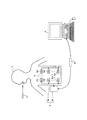
本発明の実施の形態について説明する。図1は、無線型の被検体内情報取得システムの全体構成を示す模式図である。この被検体内情報取得システムは、被検体内導入装置の一例として単眼型のカプセル型内視鏡を用いている。図1に示すように、無線型の被検体内情報取得システムは、被検体1の体内に導入され、体腔内画像を撮像して受信装置3に対して映像信号などのデータ送信を無線によって行うカプセル型内視鏡2と、カプセル型内視鏡2から無線送信された体腔内画像データを受信する受信装置3と、受信装置3が受信した映像信号に基づいて体腔内画像を表示する処理装置4と、受信装置3と処理装置4との間のデータ受け渡しを行うための携帯型記録媒体5とを備える。
Embodiments of the present invention will be described. FIG. 1 is a schematic diagram showing the overall configuration of a wireless in-vivo information acquiring system. This intra-subject information acquisition system uses a monocular capsule endoscope as an example of the intra-subject introduction apparatus. As shown in FIG. 1, the wireless in-vivo information acquiring system is introduced into the body of the
また、受信装置3は、被検体1の体外表面に貼付される複数の受信用アンテナA1〜Anを有した無線ユニット3aと、複数の受信用アンテナA1〜Anを介して受信された無線信号の処理等を行う受信本体ユニット3bとを備え、これらユニットはコネクタ等を介して着脱可能に接続される。なお、受信用アンテナA1〜Anのそれぞれは、例えば、被検体1が着用可能なジャケットに備え付けられ、被検体1は、このジャケットを着用することによって受信用アンテナA1〜Anを装着するようにしてもよい。また、この場合、受信用アンテナA1〜Anは、ジャケットに対して着脱可能なものであってもよい。
The
処理装置4は、カプセル型内視鏡2によって撮像された体腔内画像を処理して表示するためのものであり、携帯型記録媒体5によって得られるデータをもとに画像表示を行うワークステーション等の構成を有する。具体的には、処理装置4は、CRTディスプレイ、液晶ディスプレイ等によって直接画像を表示する構成としてもよいし、プリンタ等のように、他の媒体に画像を出力する構成としてもよい。
The processing device 4 is for processing and displaying a body cavity image picked up by the
携帯型記録媒体5は、コンパクトフラッシュ(登録商標)メモリ等が用いられ、受信本体ユニット3b及び処理装置4に対して着脱可能であって、両者に対する挿着時に情報の出力又は記録が可能な機能を有する。具体的には、携帯型記録媒体5は、カプセル型内視鏡2が被検体1の体腔内を移動している間は受信本体ユニット3bに挿着され、カプセル型内視鏡2から送信されるデータが携帯型記録媒体5に記録される。そして、カプセル型内視鏡2が被検体1から排出された後、つまり、被検体1の内部の撮像が終わった後には、受信本体ユニット3bから取り出されて処理装置4に挿着され、処理装置4によって記録されたデータが読み出される。受信本体ユニット3bと処理装置4との間のデータの受け渡しを携帯型記録媒体5によって行うことで、被検体1が体腔内の撮像中に自由に行動することが可能となり、また、処理装置4との間のデータの受け渡し期間の短縮にも寄与している。なお、受信本体ユニット3bと処理装置4との間のデータの受け渡しは、受信本体ユニット3bに内蔵型の他の記録装置を用い、処理装置4と有線又は無線接続するように構成してもよい。
The portable recording medium 5 uses a compact flash (registered trademark) memory or the like, is detachable from the receiving
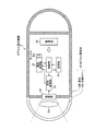
つぎに、この発明にかかる被検体内導入装置の一例であるカプセル型内視鏡2の構成について詳細に説明する。図2は、カプセル型内視鏡2の一構成例を示す模式図である。図2に示すように、カプセル型内視鏡2は、被検体1の体腔内を撮像する撮像部22を、カプセル型内視鏡2を構成する各構成部位に電力を供給する電源部26とともに、カプセル型筐体20内に配設することにより構成されている。なお、図2では、被検体1内を撮像する撮像部22を単眼で示しているが、撮像部22を複数設けた複眼であってもよい。
Next, the configuration of the
カプセル型筐体20は、撮像部22を覆う透明でドーム状の先端カバー20aと、先端カバー20aと水密状態に設けられた筐体20bとによって構成され、被検体1の口から飲み込み可能な大きさに形成されている。先端カバー20aは、筐体20bの一方の端部に取り付けられている。筐体20bは、可視光が透過しない有色材質によって形成されている。筐体20bは、カプセル型内視鏡2の各構成部位の駆動を制御し各構成部位における信号の入出力制御を行う制御部21と、体腔内部を撮像する撮像部22と、撮像部22によって撮像された画像を処理する信号処理部23と、無線通信に必要である情報を記憶する記憶部24と、外部の処理装置4に対して送信する各種信号を無線信号に変調し、またはアンテナ25aを介して受信した無線信号を復調する通信処理部25と、カプセル型内視鏡2の各構成部に対して駆動電力を供給する電源部26とを内蔵する。通信処理部25は、コイルアンテナなどによって構成され外部のアンテナとの間で無線信号を送受信するアンテナ25aを有する。
The capsule-type casing 20 includes a transparent dome-shaped tip cover 20a that covers the
撮像部22は、被検体1の体腔内の画像を撮像するためのものである。具体的には、撮像部22は、CCDまたはCMOS等の撮像素子と、この撮像素子の撮像視野を照明するLED等の発光素子と、この撮像素子に対して撮像視野からの反射光を結像するレンズ22a等の光学系とを用いて実現される。撮像部22は、筐体20bの端部に固定され、先端カバー20aを介して受光する撮像視野からの反射光を結像し、被検体1の体腔内の画像を撮像する。撮像部22における撮像素子、発光素子、レンズ22aなどの光学系などは、カプセル型内視鏡2の適用部位に対応させた光学性能で設計されたレンズや信号処理機能を有する。たとえば、カプセル型内視鏡2が小腸用である場合には、内径が狭い小腸壁を撮像するため、近点に焦点を合わせた光学性能でレンズや信号処理機能が設計されている。また、カプセル型内視鏡2が胃用である場合には、内部容積が大きい胃内を撮像するため、小腸の場合よりも大きい距離に焦点を合わせた光学性能でレンズや信号処理機能が設計されている。
The
ここで、カプセル型内視鏡2においては、被検体内画像を含む画像情報に、撮像部22における光学情報に対応した種別情報を付して送信している。カプセル型内視鏡2においては、撮像部22は、カプセル型内視鏡2の適用部位にそれぞれ対応させた光学性能で設計されており、適用部位に応じて撮像部22の各光学情報は異なる。カプセル型内視鏡2は、このカプセル型内視鏡2における撮像部22の光学情報に対応する種別情報として、このカプセル型内視鏡2の適用部位を示す情報を画像情報に付して送信している。
Here, in the
この種別情報は、予め記憶部24内に記憶されており、制御部21は、撮像部22の適用部位に対応する種別情報を記憶部24内から取得して、信号処理部23に出力する。信号処理部23は、撮像部22によって撮像された画像を処理し、この画像を含む画像信号に制御部21から出力された種別情報を付する。
The type information is stored in advance in the
具体的には、図3に示すように、信号処理部23は、撮像部22によって撮像された画像G1を走査線データ単位に処理する。そして、信号処理部23は、送信対象の画像G11における最後の走査線データの後に、カプセル型内視鏡2固有の情報を含む固有情報Daを付する。この固有情報Daは、ホワイトバランス情報およびカプセル型内視鏡2のシリアル番号などに加え、前述した種別情報をさらに含んでいる。信号処理部23は、撮像部22によって撮像された画像G2〜Gnに対しても同様に、各画像G2〜Gnの最後の走査線データの後に、ホワイトバランス情報およびシリアル番号などとともに種別情報を含んだ固有情報Daを付する。
Specifically, as illustrated in FIG. 3, the
そして、通信処理部25は、信号処理部23が生成した情報をアンテナ25aから無線送信する。すなわち、通信処理部25は、撮像部22によって撮像された画像を含む画像情報に、撮像部22における光学情報に対応した種別情報を付して外部に無線送信する。
Then, the
このように、カプセル型内視鏡2は、撮像部22が撮像した画像情報に、このカプセル型内視鏡2の撮像部22における光学情報に対応した種別情報であって、このカプセル型内視鏡2の適用部位を示す種別情報を付して送信している。カプセル型内視鏡2から送信された情報は、受信装置3によって受信され、携帯型記録媒体5に記憶される。処理装置4は、この携帯型記録媒体5内に記憶された情報を読み取ることによってカプセル型内視鏡2が撮像した画像を含む画像情報とともに種別情報を取得することができる。
As described above, the
つぎに、図4を参照して、図1に示す処理装置4について説明する。図4は、図1に示す処理装置4の概略構成を示すブロック図である。図4に示すように、処理装置4は、制御部41と、入力部42と、記憶部44と、画像処理部45と、表示部47とを備える。また、制御部41は、カプセル型内視鏡2によって撮像された画像群Pa,PbがそれぞれフォルダFa,Fb内などに格納されたデータベースDbと接続する。
Next, the processing device 4 shown in FIG. 1 will be described with reference to FIG. FIG. 4 is a block diagram showing a schematic configuration of the processing apparatus 4 shown in FIG. As illustrated in FIG. 4, the processing device 4 includes a
制御部41は、制御機能を有するCPU等を用いて構成され、入力部42、記憶部44、画像処理部45および表示部47の処理および動作を制御する。制御部41は、これらの各構成部位に入出力される情報について所定の入出力制御を行ない、かつ、この情報に対して所定の情報処理を行う。入力部42は、種々の情報を入力するためのキーボード、表示部47の表示画面上における任意の位置を指定できるマウス等を用いて構成され、検体の分析に必要な諸情報や分析動作の指示情報等を外部から取得する。
The
記憶部44は、情報を磁気的に記憶するハードディスクと、処理装置4が処理を実行する際にその処理にかかわる各種プログラムをハードディスクからロードして電気的に記憶するメモリとを用いて構成され、光学情報群Drおよび画像処理プログラム群Dpを含む諸情報を記憶する。記憶部44は、CD−ROM、DVD−ROM、PCカード等の記憶媒体に記憶された情報を読み取ることができる補助記憶装置を備えてもよい。
The
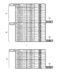
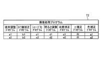
ところで、カプセル型内視鏡2の撮像部22は、カプセル型内視鏡2の適用部位に対応させた光学性能で設計されている。したがって、カプセル型内視鏡2適用部位によって、すなわちカプセル型内視鏡2の種別によって、撮像部22の各光学情報は異なったものとなる場合が多い。そこで、記憶部44は、たとえば図5に示すように、このカプセル型内視鏡2の種別にそれぞれ対応する光学情報の組み合わせを示すテーブルT1を光学情報群Drとして記憶する。テーブルT1においては、光学情報としてカプセル型内視鏡2の撮像部22における撮像倍率、ディストーション(DT)値、画素数、明るさ、像高に対する収差量、γ値および分光感度がカプセル型内視鏡2の小腸や胃などがカプセル型内視鏡2の適用部位である各種別に対応づけられた状態で示されている。なお、記憶部44は、カプセル型内視鏡2の各種別の各バージョンによってそれぞれ光学情報が異なる場合には、テーブルT1のように、各種別の各バージョンにそれぞれ対応した各光学情報を光学情報群Drとして記憶する。
By the way, the
そして、記憶部44は、画像処理プログラム群Dpとして、処理対象の画像の倍率を変更する倍率調整プログラム、処理対象の画像の鮮明度を変更するシャープ化プログラム、処理対象の画像の明るさを変更する明るさ調整プログラムなどを記憶する。具体的には、図6のテーブルT2に示すように、記憶部44は、画像処理プログラムとして、処理対象の画像を撮像した撮像部22の撮像倍率をもとに該画像の倍率を変更する倍率調整プログラム、処理対象の画像を撮像した撮像部22の画素数をもとに該画像の鮮明度を変更するシャープ化プログラム、処理対象の画像を撮像した撮像部22の明るさをもとに該画像の明るさを変更する明るさ調整プログラムを記憶する。さらに、記憶部44は、処理対象の画像を撮像した撮像部22のDT値をもとに該画像を補正するDT値補正プログラム、処理対象の画像を撮像した撮像部22の収差量をもとに該画像を補正する収差補正プログラム、処理対象の画像を撮像した撮像部22のγ値をもとに該画像を補正するγ補正プログラムおよび処理対象の画像を撮像した撮像部22の撮像素子の分光感度をもとに該画像を色補正する色補正プログラムを記憶する。なお、記憶部44は、画像処理プログラム群Dpとして、図6のテーブルT2に示すように、倍率調整プログラムa1,a2、DT値補正プログラムb1,b2、シャープ化プログラムc1,c2、明るさ調整プログラムd1,d2、収差補正プログラムe1,e2、γ補正プログラムf1,f2、および、色補正プログラムg1,g2などのように、各画像処理プログラムをそれぞれ複数記憶していてもよい。
The
画像処理部45は、処理設定部46を有する。処理設定部46は、記憶部44に記憶される光学情報の中から処理対象の画像情報に付された種別情報に対応する光学情報を取得する。そして、処理設定部46は、記憶部44に記憶される画像処理プログラムのうち取得した光学情報に応じた画像処理プログラムを、処理対象の画像情報を処理する画像処理プログラムとして設定する。画像処理部45は、処理設定部によって設定された処理対象の画像情報の光学情報に応じた画像処理プログラムを用いて、処理対象の画像を処理する。画像処理部45は、カプセル型内視鏡2の適用部位以外に対応する画像を含む画像情報を処理対象の画像情報として処理する。画像処理部45は、処理対象の画像情報の種別情報に対応する撮像倍率、画素数または明るさなどの光学情報に応じた倍率調整プログラム、シャープ化プログラム、明るさ調整プログラムなどを用いて処理対象の画像情報を処理する。画像処理部45は、カプセル型内視鏡2によって撮像されたカプセル型内視鏡2の適用部位以外に対応する画像の種別情報に対応する光学情報を取得し、さらに、この光学情報に応じた画像処理プログラムを用いて、この画像を処理することによって、そのままでは使用することができなかった適用部位以外に対応する画像を使用できるようにしている。
The
表示部47は、CRTディスプレイ、液晶ディスプレイ等によって実現され、入力部42の指示情報あるいは指示結果などを表示する。そして、表示部47は、制御部41の制御のもと、カプセル型内視鏡2によって撮像された画像および画像処理部45によって処理された画像を表示する。
The
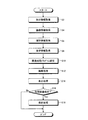
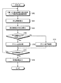
つぎに、図7を参照して、処理装置4における画像処理手順について説明する。図7は、図4に示す処理装置4における画像処理の各処理手順を示すフローチャートである。図7に示すように、まず、画像処理部45は、制御部41を介して、入力部42から入力された画像処理の実行を指示する指示情報を取得する(ステップS2)。この指示情報には、処理対象の画像情報を示す情報や処理対象の画像情報に対する処理内容等が含まれる。指示情報によって、携帯型記録媒体5によって得られた画像群のうちカプセル型内視鏡2の適用部位以外に対応する所定数の画像に対応する画像情報や、データベースDb内に格納された画像群Pa,Pbのうち、これらの画像群Pa,Pbを撮像したカプセル型内視鏡2の適用部位以外に対応する所定数の画像に対応する画像情報が、処理対象の画像情報として指示される。そして、指示情報によって、処理対象の画像情報として指示された画像に撮像されている部位が診断に使用できるようにするための画像処理が指示される。たとえば、指示情報は、カプセル型内視鏡2の適用部位以外に対応する画像として、小腸観察用のカプセル型内視鏡2によって観察された胃内部の画像を処理対象の画像として指示し、この胃内部の画像を胃内部観察用のカプセル型内視鏡2によって撮像された画像と同程度まで観察可能となるように画像処理を行うように指示する。
Next, an image processing procedure in the processing device 4 will be described with reference to FIG. FIG. 7 is a flowchart showing each processing procedure of image processing in the processing device 4 shown in FIG. As shown in FIG. 7, first, the
画像処理部45は、指示情報にしたがって、処理対象の画像情報を取得する(ステップS4)。次いで、処理設定部46は、処理対象の画像情報から、この画像情報に付された種別情報を取得する(ステップS6)。種別情報は、前述したように、各画像の最後の走査線データの後に付された固有情報Da内に含まれている。処理設定部46は、記憶部44に記憶される光学情報群Drの中からステップS6において取得した処理対象の画像情報における種別情報に対応する光学情報を取得する(ステップS8)。
The
そして、処理設定部46は、記憶部44に記憶される画像処理プログラム群Dpのうち、取得した光学情報に応じた画像処理プログラムを、この処理対象の画像情報を処理する画像処理プログラムとして設定する(ステップS10)。
Then, the
つぎに、画像処理部45は、処理設定部46が設定した画像処理プログラムを用いて、処理対象である画像情報を処理する画像処理を行ない(ステップS12)、表示部47は、画像処理部45が処理した画像を表示出力する表示処理を行う(ステップS14)。そして、制御部41は、入力部42から入力された指示情報をもとに、画像処理部45によって処理された処理画像を保存するか否かを判断する(ステップS16)。たとえば、処理装置4の操作者が、処理画像の保存を選択できる選択メニューにおいて保存を指示する選択欄をマウスで選択した場合には、処理画像の保存を指示する指示情報が入力部42から制御部41に入力される。また、処理装置4の操作者が保存を指示しない選択欄をマウスで選択した場合には、処理画像の保存を指示しない指示情報が入力部42から制御部41に入力される。
Next, the
制御部41が画像処理部45によって処理された処理画像を保存しないと判断した場合(ステップS16:No)、そのまま処理装置4は、画像処理を終了する。また、制御部41は、画像処理部45によって処理された処理画像を保存すると判断した場合(ステップS16:Yes)、指示された保存先に処理画像を保存する保存処理を行ない(ステップS18)、処理装置4における画像処理を終了する。
When the
つぎに、画像処理部45において行なわれる画像処理(ステップS12)について具体的に説明する。まず、図8を参照して、画像処理部45が、図6に示す画像処理プログラムのうち倍率調整プログラムを用いて画像処理を行う場合を例に説明する。
Next, the image processing (step S12) performed in the
画像処理部45は、倍率調整プログラムに示された処理手順にしたがって、処理設定部46が取得した種別情報のうち、処理対象である画像情報を撮像したカプセル型内視鏡2の撮像部22における画角情報・LED発光量情報を取得する(ステップS22)。
The
そして、画像処理部45は、取得した画角情報・LED発光量情報をもとにカプセル型内視鏡2における観察距離を算出する(ステップS24)。なお、画角は、カプセル型内視鏡2における撮像部22で明瞭に撮影できる範囲の角度を示すものであり、この画角をもとに観察距離を算出することができる。また、カプセル型内視鏡2において、撮像部22は、自動調光処理を行なっており、撮像対象が遠いために撮像範囲が暗い場合には、自動的にLEDの発光量を増やしており、撮像対象が近すぎるために撮像範囲が過度に明るい場合には、自動的にLEDの発光量を減らしている。このため、画像処理部45は、LED発光量情報から、処理対象である画像情報が撮像された時間におけるLED発光量をもとに、撮像対象とカプセル型内視鏡2との距離を算出することができる。
Then, the
つぎに、画像処理部45は、算出した観察距離および処理対象の画像情報における撮像物の大きさをもとに、この処理対象の画像を撮像したカプセル型内視鏡2における撮像部22の倍率を算出する(ステップS26)。
Next, the
そして、画像処理部45は、算出した倍率が、使用目的に対応する所定の倍率よりも大きいか否かを判断する(ステップS28)。画像処理部45は、たとえば、小腸観察用のカプセル型内視鏡2によって観察された胃内部の画像を、診断用に使用可能な程度にまで処理する場合には、算出した倍率と胃内部観察用のカプセル型内視鏡2における倍率とを比較する。なお、比較する倍率は、倍率調整プログラムの中に示されていてもよく、また、記憶部44内の光学情報群Drから取得されたものであってもよい。
Then, the
画像処理部45は、算出した倍率が所定の倍率よりも大きくないと判断した場合(ステップS28:No)、すなわち、算出した倍率が所定の倍率よりも小さいと判断した場合、処理対象の画像を所定の倍率に合うように拡大する拡大処理を行う(ステップS30)。画像処理部45は、たとえば、処理対象の画像が小腸観察用のカプセル型内視鏡2によって撮像された胃内部の画像である場合であって、胃内部観察用のカプセル型内視鏡2における倍率よりも小さい場合には、胃内部観察用のカプセル型内視鏡2における倍率に合わせて拡大させる。
When the
これに対し、画像処理部45は、算出した倍率が所定の倍率よりも大きいと判断した場合(ステップS28:No)、処理対象の画像を所定の倍率に合うように縮小する縮小処理を行う(ステップS32)。
On the other hand, when the
そして、画像処理部45は、拡大処理または縮小処理が終わった後に、次に処理対象である画像があるか否かを判断し(ステップS34)、次に処理対象である画像があると判断した場合には(ステップS34:Yes)、ステップS24に進み、次に処理対象である画像に対する観察距離算出処理を行う。一方、画像処理部45は、次に処理対象である画像がないと判断した場合(ステップS34:No)、処理を行なった一連の画像を制御部41に出力し(ステップS36)、画像処理を終了する。画像処理部45によって画像処理が行なわれた一連の画像は、表示部47によって表示されるほか、制御部41によってデータベースDb内などに記憶される。
Then, after the enlargement process or the reduction process is completed, the
つぎに、図9を参照して、画像処理部45が、図6に示す画像処理プログラムのうちシャープ化プログラムを用いて画像処理を行う場合を例に説明する。シャープ化プログラムに示された処理手順にしたがって、画像処理部45は、処理設定部46が取得した種別情報のうち、処理対象である画像情報を撮像したカプセル型内視鏡2の撮像部22の画素数などを示す画素数情報を取得する(ステップS42)。
Next, a case where the
そして、画像処理部45は、取得した画素数情報をもとに、処理対象の画像を撮像したカプセル型内視鏡2の撮像部22の画素数が使用目的に対応する所定の画素数よりも少ないか否かを判断する(ステップS44)。画像処理部45は、たとえば、小腸観察用のカプセル型内視鏡2によって観察された胃内部の画像を診断用に使用可能な程度にまで処理する場合には、胃内部観察用のカプセル型内視鏡2における画素数と比較する。なお、比較する画素数は、シャープ化プログラムの中に示されていてもよく、また、記憶部44内の光学情報群Drから取得されたものであってもよい。
Then, based on the acquired pixel number information, the
画像処理部45は、処理対象の画像を撮像したカプセル型内視鏡2の撮像部22の画素数が所定の画素数よりも少ないと判断した場合(ステップS44:Yes)、処理対象の画像の鮮明度を調整して輪郭をシャープにするシャープ化処理を行う(ステップS46)。画像処理部45は、処理対象の画像が小腸観察用のカプセル型内視鏡2によって撮像された胃内部の画像である場合であって、胃内部観察用のカプセル型内視鏡2における画素数よりも少ない画素数で撮像されたものである場合には、処理対象の画像の輪郭は胃内部観察用のカプセル型内視鏡2によって撮像された画像よりもぼやけているため、胃内部観察用のカプセル型内視鏡2における鮮明度に合わせてシャープにする。
When the
これに対し、画像処理部45は、処理対象の画像を撮像したカプセル型内視鏡2の撮像部22の画素数が所定の画素数よりも多いと判断した場合(ステップS44:No)、処理対象の画像の鮮明度は、診断用の使用目的に耐え得る程度に高いため、輪郭をシャープにするシャープ化処理を行なわない。すなわち、画像処理部45は、アンシャープと判断する(ステップS50)。画像処理部45は、処理対象の画像が小腸観察用のカプセル型内視鏡2によって撮像された胃内部の画像である場合であって、胃内部観察用のカプセル型内視鏡2における画素数よりも多い画素数で撮像されたものである場合には、処理対象の画像の輪郭は胃内部観察用のカプセル型内視鏡2によって撮像された画像と同等以上に鮮明であるため、胃内部観察用のカプセル型内視鏡2における鮮明度に合わせてシャープ化する必要がない。
In contrast, when the
そして、画像処理部45は、シャープ化処理またはアンシャープの判断処理が終わった後に、次に処理対象である画像があるか否かを判断し(ステップS52)、次に処理対象である画像があると判断した場合には(ステップS52:Yes)、ステップS42に進み、次に処理対象である画像に対する画素数量の判断処理を行う。一方、画像処理部45は、次に処理対象である画像がないと判断した場合(ステップS52:No)、処理を行なった一連の画像を制御部41に出力し(ステップS54)、画像処理を終了する。画像処理部45によって画像処理が行なわれた一連の画像は、表示部47によって表示されるほか、制御部41によってデータベースDb内などに記憶される。
Then, after the sharpening process or the unsharp determination process is completed, the
つぎに、図10を参照して、画像処理部45が、図6に示す画像処理プログラムのうち明るさ調整プログラムを用いて画像処理を行う場合を例に説明する。明るさ調整プログラムに示された処理手順にしたがって、画像処理部45は、処理設定部46が取得した種別情報のうち、処理対象である画像情報を撮像したカプセル型内視鏡2の撮像部22におけるF値・LED発光量・LEDの発光効率・レンズ周辺光量などの明るさに関する情報を取得する(ステップS62)。
Next, a case where the
そして、画像処理部45は、F値・LED発光量・LEDの発光効率・レンズ周辺光量をもとに処理対象の画像の中心輝度を算出する(ステップS64)。つぎに、画像処理部45は、処理対象の画像の中心に対する所定の周辺領域の明るさを算出する(ステップS66)。
Then, the
次いで、画像処理部45は、算出した中心輝度と周辺領域の明るさとを比較して、周辺領域の明るさよりも中心輝度の方が大きいか否かを判断する(ステップS68)。画像処理部45は、周辺領域の明るさよりも中心輝度の方が大きいと判断した場合(ステップS68:Yes)、画像の周辺領域が中心よりも暗いため、画像全体のゲインを上昇させるゲイン上昇処理(ステップS70)を行なって、画像全体を明るくして観察しやすいようにする。一方、画像処理部45は、周辺領域の明るさよりも中心輝度の方が小さいと判断した場合(ステップS68:No)、画像の周辺領域が中心よりも明るいため、画像全体のゲインを低下させるゲイン低下処理(ステップS72)を行なって、画像全体を暗くして観察しやすいようにする。
Next, the
そして、画像処理部45は、ゲイン上昇処理またはゲイン低下処理が終わった後に、次に処理対象である画像があるか否かを判断し(ステップS74)、次に処理対象である画像があると判断した場合には(ステップS74:Yes)、ステップS62に進み、次に処理対象である画像に対する明るさに関する情報を取得して、明るさの調整を行う。一方、画像処理部45は、次に処理対象である画像がないと判断した場合(ステップS74:No)、処理を行なった一連の画像を制御部41に出力し(ステップS76)、画像処理を終了する。画像処理部45によって画像処理が行なわれた一連の画像は、表示部47によって表示されるほか、制御部41によってデータベースDb内などに記憶される。
Then, after the gain increase process or the gain decrease process ends, the
このように、実施の形態においては、カプセル型内視鏡2は、カプセル型内視鏡2における撮像部22の光学情報に対応した種別情報を各画像に付して送信し、処理装置4は、処理対象の画像に付された種別情報をもとに、この画像を撮像した撮像部22の光学情報に応じた画像処理プログラムを用いて処理対象の画像を処理している。この結果、処理装置4においては、そのままでは使用することができなかったカプセル型内視鏡2の適用部位以外に対応する画像を診断用などに使用できるように処理できる。したがって、本実施の形態によれば、従来において無駄となっていた適用部位以外の画像を無駄にすることなく利用できるため、無駄となる画像を減らすことができる。さらに、従来においては、適用部位以外の臓器に対して新たに画像を取得するために被験者に画像取得対象の臓器に対応するカプセル型内視鏡を再度飲み込んでもらう必要がなくなるため、被験者の負担を減らすことができる。
Thus, in the embodiment, the
また、本実施の形態においては、データベースDb内の画像のうち、カプセル型内視鏡2の適用部位以外に対応する画像を処理して使用可能とすることによって、データベースDb内の画像を適切に活用できる。たとえば、処理装置4で、今回観察した被験者の小腸の画像と比較できるように、前回観察した被験者の胃内部観察用のカプセル型内視鏡2によって撮像された小腸部位の画像を処理することによって、被験者に対する履歴情報として前回の観察画像を利用することができる。
In the present embodiment, among the images in the database Db, the images corresponding to those other than the application site of the
また、本実施の形態によれば、たとえば小腸観察用のカプセル型内視鏡2によって撮像された画像のうち、小腸と、大腸との境界部分の画像に対して大腸観察用のカプセル型内視鏡2の光学情報に合わせた画像処理を施すことによって、小腸よりも内径が大きく観察対象までの距離が小腸の場合よりも遠い大腸方向を撮像した画像についても、さらに適切に診断に適用することができる。
Further, according to the present embodiment, for example, among the images captured by the
また、本実施の形態においては、小腸観察用のカプセル型内視鏡2によって観察された胃内部の画像を処理する場合を例に説明したが、もちろんこれに限らず、小腸観察用のカプセル型内視鏡2によって観察された大腸内部の画像を大腸観察用のカプセル型内視鏡2の光学情報に合わせて処理するなどのように、処理装置4は、カプセル型内視鏡2の適用部位以外に対応する画像を使用可能にできる画像処理プログラムをそれぞれ選択して、処理対象の画像を処理すればよい。この結果、本実施の形態によれば、一つのカプセル型内視鏡2で撮像した消化管画像のほとんどを診断可能となる状態とすることができる。
In the present embodiment, the case of processing an image of the stomach observed by the
なお、本実施の形態においては、テーブルT1に示すように、カプセル型内視鏡2の各種別の各バージョンによってそれぞれ光学情報が異なる場合には、種別情報とともにバージョン情報を使用すればよい。この場合、カプセル型内視鏡2は、各画像情報に撮像部22の種別情報および該カプセル型内視鏡2のバージョン情報を付して送信する。処理装置4においては、処理設定部46は、処理対象の画像情報に付された種別情報に加えバージョン情報を取得して、たとえばテーブルT1を参照することによって、取得した種別情報およびバージョン情報に対応する光学情報を取得する。そして、処理設定部46は、取得した光学情報に応じた画像処理プログラムを画像処理プログラム群Dpから選択し、処理対象の画像情報を処理する画像プログラムとして設定する。このように、本実施の形態においては、各バージョンごとに光学情報が異なる場合には、種別情報とともにバージョン情報を使用して、画像処理を行なってもよい。
In the present embodiment, as shown in the table T1, when the optical information is different for each version of the
さらに、撮像部22の光学性能が異なるごとに異なるバージョン情報が付与される場合、すなわち、カプセル型内視鏡2の種別によらず撮像部22の光学情報が異なれば異なるバージョン情報が付与される場合、バージョン情報のみで光学情報が管理されているため、カプセル型内視鏡2は、新たに種別情報を付する必要がなく、バージョン情報のみを画像情報に付して送信すればよい。処理装置4において、記憶部44は、カプセル型内視鏡2のバージョン情報にそれぞれ対応する光学情報の組み合わせを光学情報群Drとして記憶する。そして、処理設定部46は、処理対象の画像情報に付されたバージョン情報を取得して、取得したバージョン情報に対応する光学情報を光学情報群Drから取得する。そして、処理設定部46は、取得した光学情報に応じた画像処理プログラムを画像処理プログラム群Dpから選択し、処理対象の画像情報を処理する画像プログラムとして設定する。このように、本実施の形態においては、バージョン情報のみで光学情報を管理できる場合には、バージョン情報のみで処理対象の画像処理を管理してもよい。
Furthermore, when different version information is given every time the optical performance of the
また、本実施の形態においては、処理装置4においてデータベースDb内の画像または携帯型記録媒体5を介して取得した画像を処理した場合について説明したが、もちろんこれに限らない。たとえば、図1に示す受信装置3内においてカプセル型内視鏡2から順次送信される画像をほぼリアルタイムで処理して受信装置3内の記憶部または携帯型記録媒体5内に記憶させてもよい。
In the present embodiment, the case has been described in which the image in the database Db or the image acquired via the portable recording medium 5 is processed in the processing device 4, but the present invention is not limited to this. For example, the images sequentially transmitted from the
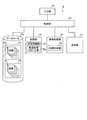
この場合、図11に示すように、受信装置3は、無線ユニット3aにおける受信用アンテナA1〜Anを介して受信された無線信号の処理等を行う受信本体ユニット3b内において、光学情報群Drおよび画像処理プログラム群Dpを記憶し、これらの光学情報群Drおよび画像処理プログラム群Dpを用いて、処理対象であるカプセル型内視鏡2の適用部位以外に対応する画像を使用可能な程度となるように画像処理を行う。
In this case, as shown in FIG. 11, the receiving
つぎに、受信本体ユニット3bについて詳細に説明する。受信本体ユニット3bは、図11に示すように、受信部31と変換部32と同期信号検出部33と画像処理部34と記憶部36とを備える。受信部31は、無線信号の受信の際に使用するアンテナAを切り替え、切り替えたアンテナAを介して受信された無線信号に対して復調、アナログ/デジタル変換等の受信処理を行ない、信号Saを出力する。変換部32は、受信部31から出力された信号Saを画像処理部34が処理可能である信号形式の信号Siに変換する。変換部32は、同期信号検出部33による同期信号出力タイミングに合わせて信号Siを出力する。同期信号検出部33は、信号Saの中から各種同期信号を検出し、検出した同期信号に関する同期信号情報Sdを画像処理部に出力する。
Next, the receiving
そして、画像処理部34は、変換部32から出力された信号Siに対して所定の処理を行ない1フレームの画像に対応する画像データSfを出力する。また、記憶部36は、受信装置3における画像処理に必要である情報とともに光学情報群Drおよび画像処理プログラム群Dpを記憶する。
The
図11に示すように、画像処理部34は、処理設定部46を有する。処理設定部46は、記憶部36に記憶される光学情報の中から処理対象の画像情報に付された種別情報に対応する光学情報を取得し、記憶部44に記憶される画像処理プログラムのうち取得した光学情報に応じた画像処理プログラムを、処理対象の画像情報を処理する画像処理プログラムとして設定する。画像処理部34は、処理設定部によって設定された処理対象の画像情報の光学情報に応じた画像処理プログラムを用いて、処理対象の画像をほぼリアルタイムで処理し、そのままでは使用することができなかったカプセル型内視鏡2の適用部位以外に対応する画像を使用できるようにしている。
As illustrated in FIG. 11, the
1 被検体
2 カプセル型内視鏡
3 受信装置
3a 無線ユニット
3b 受信本体ユニット
4 処理装置
5 携帯型記録媒体
20 カプセル型筐体
20a 先端カバー
20b 筐体
21 制御部
22 撮像部
22a レンズ
23 信号処理部
24 記憶部
25 通信処理部
25a アンテナ
26 電源部
31 受信部
32 変換部
33 同期信号検出部
34 画像処理部
36 記憶部
41 制御部
42 入力部
44 記憶部
45 画像処理部
46 処理設定部
47 表示部
DESCRIPTION OF
Claims (7)
前記被検体内導入装置は、
前記被検体内の画像を撮像する撮像手段と、
前記撮像手段によって撮像された画像を含む前記画像情報に、前記撮像手段における光学情報に対応した種別情報を付して送信する送信手段と、
を備え、
前記処理装置は、
各種別情報に対応する各光学情報の組み合わせと、各光学情報に対応した画像処理プログラムとを記憶する記憶手段と、
前記記憶手段に記憶される前記光学情報の中から処理対象の前記画像情報に付された前記種別情報に対応する前記光学情報を取得し、前記記憶手段に記憶される画像処理プログラムのうち前記取得した光学情報に応じた前記画像処理プログラムを用いて前記処理対象の画像情報を処理する画像処理手段と、
を備えたことを特徴とする被検体内画像取得システム。 An in-subject introduction apparatus that wirelessly transmits image information including an image in the subject that is introduced and imaged inside the subject, and a process that processes the image information transmitted from the in-subject introduction apparatus In-subject image acquisition system comprising an apparatus,
The in-subject introduction device comprises:
Imaging means for imaging an image in the subject;
Transmitting means for attaching the type information corresponding to the optical information in the imaging means to the image information including the image captured by the imaging means;
With
The processor is
Storage means for storing a combination of optical information corresponding to each type information and an image processing program corresponding to each optical information;
The optical information corresponding to the type information attached to the image information to be processed is acquired from the optical information stored in the storage unit, and the acquisition is performed among the image processing programs stored in the storage unit. Image processing means for processing the image information to be processed using the image processing program corresponding to the optical information,
An in-vivo image acquisition system comprising:
前記画像処理手段は、前記被検体内導入装置の適用部位以外に対応する画像情報を前記処理対象の画像情報として処理することを特徴とする請求項1に記載の被検体内画像取得システム。 The type information indicates an application site of the intra-subject introduction device,
The in-vivo image acquisition system according to claim 1, wherein the image processing unit processes image information corresponding to a portion other than an application site of the in-subject introduction apparatus as image information to be processed.
前記画像処理プログラムは、前記処理対象の画像の倍率を変更する倍率調整プログラム、前記処理対象の画像の鮮明度を変更する鮮明度調整プログラム、または、前記処理対象の画像の明るさを変更する明るさ調整プログラムであり、
前記画像処理手段は、前記処理対象の画像情報に付される種別情報に対応する前記撮像倍率、前記画素数または前記明るさをもとに前記倍率調整プログラム、前記鮮明度調整プログラムまたは前記明るさ調整プログラムを用いて前記処理対象の画像情報を処理することを特徴とする請求項1または2に記載の被検体内画像取得システム。 The optical information is information indicating an imaging magnification, the number of pixels, or brightness of the imaging unit,
The image processing program is a magnification adjustment program that changes the magnification of the image to be processed, a sharpness adjustment program that changes the sharpness of the image to be processed, or a brightness that changes the brightness of the image to be processed. Adjustment program,
The image processing unit is configured to use the magnification adjustment program, the sharpness adjustment program, or the brightness based on the imaging magnification, the number of pixels, or the brightness corresponding to the type information attached to the image information to be processed. The in-vivo image acquisition system according to claim 1, wherein the image information to be processed is processed using an adjustment program.
前記被検体内導入装置から送信された前記画像情報と前記種別情報とを受信する受信ステップと、
前記光学情報と前記光学情報に応じた画像処理プログラムと前記種別情報とを記録する記録手段から前記受信した種別情報に対応する前記光学情報に応じた前記画像処理プログラムを用いて前記処理対象の画像情報を処理する画像処理ステップと、
を含むことを特徴とする被検体内画像処理方法。 From an in-subject introduction device that is introduced into a subject and wirelessly transmits the image information including the image in the subject imaged by the imaging means to the outside with the type information corresponding to the optical information of the imaging means. In an in-vivo image processing method executed by a processing device that processes the transmitted image information ,
A receiving step of receiving and said type information and the image information transmitted from the body insertable device,
The image to be processed using the image processing program corresponding to the optical information corresponding to the type information received from the recording means for recording the optical information, the image processing program corresponding to the optical information, and the type information An image processing step for processing the information;
A method for processing an in-vivo image, comprising:
前記画像処理ステップは、前記被検体内導入装置の適用部位以外に対応する画像情報を前記処理対象の画像情報として処理することを特徴とする請求項4に記載の被検体内画像処理方法。 The type information indicates an application site of the intra-subject introduction device,
5. The in-subject image processing method according to claim 4, wherein the image processing step processes image information corresponding to a portion other than an application site of the in-subject introduction apparatus as image information to be processed.
前記画像処理プログラムは、前記処理対象の画像の倍率を変更する倍率調整プログラム、前記処理対象の画像の鮮明度を変更する鮮明度調整プログラム、または、前記処理対象の画像の明るさを変更する明るさ調整プログラムであり、
前記画像処理ステップは、前記処理対象の画像情報に付される種別情報に対応する前記撮像倍率、前記画素数または前記明るさをもとに前記倍率調整プログラム、前記鮮明度調整プログラムまたは前記明るさ調整プログラムを用いて前記処理対象の画像情報を処理することを特徴とする請求項4または5に記載の被検体内画像処理方法。 The optical information is information indicating an imaging magnification, the number of pixels, or brightness of the imaging unit,
The image processing program is a magnification adjustment program that changes the magnification of the image to be processed, a sharpness adjustment program that changes the sharpness of the image to be processed, or a brightness that changes the brightness of the image to be processed. Adjustment program,
The image processing step includes the magnification adjustment program, the sharpness adjustment program, or the brightness based on the imaging magnification, the number of pixels, or the brightness corresponding to the type information attached to the image information to be processed. The in-vivo image processing method according to claim 4, wherein the image information to be processed is processed using an adjustment program.
前記被検体内の画像を撮像する撮像手段と、
前記撮像手段によって撮像された画像を含む前記画像情報に、前記撮像手段における撮像倍率、画素数または明るさを示す情報を含む光学情報に対応した種別情報であって、当該被検体内導入装置の適用部位を示す種別情報を付して該種別情報に対応する前記光学情報に基づいて処理を行う前記処理装置に送信する送信手段と、
を備えたことを特徴とする被検体内導入装置。 In an in-subject introduction apparatus that wirelessly transmits image information including an image in the subject introduced and imaged into the subject to a processing apparatus that processes the image information.
Imaging means for imaging an image in the subject;
Type information corresponding to optical information including information indicating the imaging magnification, the number of pixels, or the brightness in the imaging unit in the image information including the image captured by the imaging unit, Transmitting means for attaching to the type information indicating the application site and transmitting to the processing device for performing processing based on the optical information corresponding to the type information;
An intra-subject introduction apparatus characterized by comprising:
Priority Applications (5)
| Application Number | Priority Date | Filing Date | Title |
|---|---|---|---|
| JP2007226973A JP5259141B2 (en) | 2007-08-31 | 2007-08-31 | In-subject image acquisition system, in-subject image processing method, and in-subject introduction device |
| EP08828127.4A EP2191767B1 (en) | 2007-08-31 | 2008-08-26 | In-examinee image acquisition system and in-examinee image processing method |
| PCT/JP2008/065210 WO2009028508A1 (en) | 2007-08-31 | 2008-08-26 | In-examinee image acquisition system, in-examinee image processing method, and in-examinee introduction device |
| CN2008801044989A CN101790344B (en) | 2007-08-31 | 2008-08-26 | In-examinee image acquisition system, in-examinee image processing method, and in-examinee introduction device |
| US12/199,679 US8300090B2 (en) | 2007-08-31 | 2008-08-27 | In-vivo image acquiring system, in-vivo image processing method, and body-insertable apparatus |
Applications Claiming Priority (1)
| Application Number | Priority Date | Filing Date | Title |
|---|---|---|---|
| JP2007226973A JP5259141B2 (en) | 2007-08-31 | 2007-08-31 | In-subject image acquisition system, in-subject image processing method, and in-subject introduction device |
Publications (2)
| Publication Number | Publication Date |
|---|---|
| JP2009056160A JP2009056160A (en) | 2009-03-19 |
| JP5259141B2 true JP5259141B2 (en) | 2013-08-07 |
Family
ID=40387227
Family Applications (1)
| Application Number | Title | Priority Date | Filing Date |
|---|---|---|---|
| JP2007226973A Expired - Fee Related JP5259141B2 (en) | 2007-08-31 | 2007-08-31 | In-subject image acquisition system, in-subject image processing method, and in-subject introduction device |
Country Status (5)
| Country | Link |
|---|---|
| US (1) | US8300090B2 (en) |
| EP (1) | EP2191767B1 (en) |
| JP (1) | JP5259141B2 (en) |
| CN (1) | CN101790344B (en) |
| WO (1) | WO2009028508A1 (en) |
Families Citing this family (7)
| Publication number | Priority date | Publication date | Assignee | Title |
|---|---|---|---|---|
| JP5015363B2 (en) * | 2010-09-24 | 2012-08-29 | オリンパスメディカルシステムズ株式会社 | Image display device and capsule endoscope system |
| WO2012042966A1 (en) * | 2010-09-28 | 2012-04-05 | オリンパスメディカルシステムズ株式会社 | Image display device, image display method, and image display program |
| JP2012249956A (en) * | 2011-06-06 | 2012-12-20 | Toshiba Corp | Capsule endoscope image processing apparatus and capsule endoscope system |
| CN103513413B (en) * | 2012-06-18 | 2017-05-24 | 奥林巴斯株式会社 | Endoscope device and endoscope image recording destination folder change method |
| JP6581923B2 (en) * | 2016-03-03 | 2019-09-25 | 富士フイルム株式会社 | Image processing apparatus, operating method thereof and operating program |
| WO2018020558A1 (en) * | 2016-07-25 | 2018-02-01 | オリンパス株式会社 | Image processing device, image processing method, and program |
| CN119606285A (en) * | 2018-02-09 | 2025-03-14 | 捷锐士阿希迈公司(以奥林巴斯美国外科技术名义) | Medical laser equipment, endoscope controllers and medical systems |
Family Cites Families (26)
| Publication number | Priority date | Publication date | Assignee | Title |
|---|---|---|---|---|
| JP3958526B2 (en) * | 2001-02-28 | 2007-08-15 | ペンタックス株式会社 | Observation site display system for electronic endoscope apparatus |
| IL143260A (en) | 2001-05-20 | 2006-09-05 | Given Imaging Ltd | Array system and method for locating an in vivo signal source |
| JP4744026B2 (en) * | 2001-07-30 | 2011-08-10 | オリンパス株式会社 | Capsule endoscope and capsule endoscope system |
| JP3810381B2 (en) * | 2003-04-25 | 2006-08-16 | オリンパス株式会社 | Image display device, image display method, and image display program |
| US7623904B2 (en) * | 2003-08-06 | 2009-11-24 | Olympus Corporation | Medical apparatus, medical apparatus guide system, capsule type medical apparatus, and capsule type medical apparatus guide apparatus |
| US20050029437A1 (en) * | 2003-08-08 | 2005-02-10 | Akira Hasegawa | Capsule optical sensor |
| JP3993550B2 (en) * | 2003-09-30 | 2007-10-17 | オリンパス株式会社 | Gastrointestinal inspection device |
| US20050075537A1 (en) * | 2003-10-06 | 2005-04-07 | Eastman Kodak Company | Method and system for real-time automatic abnormality detection for in vivo images |
| JP4009581B2 (en) * | 2003-11-18 | 2007-11-14 | オリンパス株式会社 | Capsule medical system |
| JP4445799B2 (en) * | 2004-05-24 | 2010-04-07 | オリンパス株式会社 | Intra-subject introduction device and medical device |
| JP2006075331A (en) * | 2004-09-09 | 2006-03-23 | Sanyo Electric Co Ltd | Imaging apparatus for endoscope |
| JP2006115965A (en) | 2004-10-20 | 2006-05-11 | Fujinon Corp | Electronic endoscope apparatus |
| US7627189B2 (en) * | 2004-10-20 | 2009-12-01 | Fujinon Corporation | Sharpness adjustment method and program and electronic endoscope apparatus |
| JP4615963B2 (en) * | 2004-10-29 | 2011-01-19 | オリンパス株式会社 | Capsule endoscope device |
| JP4709579B2 (en) * | 2005-04-26 | 2011-06-22 | オリンパスメディカルシステムズ株式会社 | Capsule endoscope |
| JP4602828B2 (en) * | 2005-04-26 | 2010-12-22 | オリンパスメディカルシステムズ株式会社 | In-subject information acquisition system |
| JP4472585B2 (en) * | 2005-06-14 | 2010-06-02 | オリンパス株式会社 | Transmission device and in-subject information acquisition system |
| JP2007082664A (en) * | 2005-09-21 | 2007-04-05 | Fujifilm Corp | Capsule endoscope |
| US7567692B2 (en) * | 2005-09-30 | 2009-07-28 | Given Imaging Ltd. | System and method for detecting content in-vivo |
| JP4855759B2 (en) * | 2005-10-19 | 2012-01-18 | オリンパス株式会社 | Receiving apparatus and in-subject information acquisition system using the same |
| CN101325909B (en) * | 2005-12-07 | 2012-07-25 | 皇家飞利浦电子股份有限公司 | Electronic gastrointestinal screening |
| JP4847161B2 (en) | 2006-03-02 | 2011-12-28 | キヤノン株式会社 | Image transmitting apparatus and imaging apparatus |
| JP2007319442A (en) * | 2006-06-01 | 2007-12-13 | Fujifilm Corp | Capsule endoscope system and image processing apparatus |
| JP4932588B2 (en) * | 2007-05-08 | 2012-05-16 | オリンパス株式会社 | Image processing apparatus and image processing program |
| JP5467754B2 (en) * | 2008-07-08 | 2014-04-09 | Hoya株式会社 | Signal processing apparatus for electronic endoscope and electronic endoscope apparatus |
| JP2010187756A (en) * | 2009-02-16 | 2010-09-02 | Olympus Corp | Image processing apparatus, image processing method, and image processing program |
-
2007
- 2007-08-31 JP JP2007226973A patent/JP5259141B2/en not_active Expired - Fee Related
-
2008
- 2008-08-26 WO PCT/JP2008/065210 patent/WO2009028508A1/en not_active Ceased
- 2008-08-26 CN CN2008801044989A patent/CN101790344B/en not_active Expired - Fee Related
- 2008-08-26 EP EP08828127.4A patent/EP2191767B1/en not_active Not-in-force
- 2008-08-27 US US12/199,679 patent/US8300090B2/en active Active
Also Published As
| Publication number | Publication date |
|---|---|
| EP2191767A1 (en) | 2010-06-02 |
| US20090058996A1 (en) | 2009-03-05 |
| EP2191767B1 (en) | 2017-05-10 |
| CN101790344A (en) | 2010-07-28 |
| JP2009056160A (en) | 2009-03-19 |
| CN101790344B (en) | 2012-07-18 |
| US8300090B2 (en) | 2012-10-30 |
| WO2009028508A1 (en) | 2009-03-05 |
| EP2191767A4 (en) | 2014-10-01 |
Similar Documents
| Publication | Publication Date | Title |
|---|---|---|
| JP5259141B2 (en) | In-subject image acquisition system, in-subject image processing method, and in-subject introduction device | |
| US8164659B2 (en) | Image pickup apparatus, image display apparatus, and image display system | |
| JP5622350B2 (en) | Intra-subject introduction apparatus and intra-subject information acquisition system | |
| EP1946695A1 (en) | In vivo image acquisition device, receive device, and in vivo information acquisition system | |
| CN101784223B (en) | In vivo image acquisition device and in vivo image acquisition system | |
| US20190082936A1 (en) | Image processing apparatus | |
| JPWO2012033200A1 (en) | Imaging device | |
| CN101790343B (en) | In vivo information acquisition system | |
| WO2012063623A1 (en) | Image display apparatus and capsule endoscopy system | |
| JP5096115B2 (en) | In-subject information acquisition system and in-subject introduction device | |
| US10462440B2 (en) | Image processing apparatus | |
| US7630754B2 (en) | Intra-subject device and related medical device | |
| WO2021176708A1 (en) | Antenna system, capsule endoscope system, and operating method of antenna system | |
| US11259691B2 (en) | Body-insertable apparatus, transmission method, and non-transitory computer readable medium | |
| US20200373955A1 (en) | Receiving device and receiving method | |
| JP4656825B2 (en) | In-subject introduction apparatus and wireless in-subject information acquisition system | |
| WO2012042966A1 (en) | Image display device, image display method, and image display program | |
| US20230143451A1 (en) | Method and Apparatus of Image Adjustment for Gastrointestinal Tract Images | |
| JP5896877B2 (en) | Light control device | |
| US20180242013A1 (en) | Motion determining apparatus, body-insertable apparatus, method of determining motion, and computer readable recording medium | |
| JP2001292961A (en) | Endoscope apparatus and wireless video camera for endoscope |
Legal Events
| Date | Code | Title | Description |
|---|---|---|---|
| A621 | Written request for application examination |
Free format text: JAPANESE INTERMEDIATE CODE: A621 Effective date: 20100604 |
|
| A131 | Notification of reasons for refusal |
Free format text: JAPANESE INTERMEDIATE CODE: A131 Effective date: 20111206 |
|
| A521 | Request for written amendment filed |
Free format text: JAPANESE INTERMEDIATE CODE: A523 Effective date: 20120201 |
|
| A131 | Notification of reasons for refusal |
Free format text: JAPANESE INTERMEDIATE CODE: A131 Effective date: 20120807 |
|
| A521 | Request for written amendment filed |
Free format text: JAPANESE INTERMEDIATE CODE: A523 Effective date: 20121004 |
|
| TRDD | Decision of grant or rejection written | ||
| A01 | Written decision to grant a patent or to grant a registration (utility model) |
Free format text: JAPANESE INTERMEDIATE CODE: A01 Effective date: 20130402 |
|
| A61 | First payment of annual fees (during grant procedure) |
Free format text: JAPANESE INTERMEDIATE CODE: A61 Effective date: 20130424 |
|
| FPAY | Renewal fee payment (event date is renewal date of database) |
Free format text: PAYMENT UNTIL: 20160502 Year of fee payment: 3 |
|
| R151 | Written notification of patent or utility model registration |
Ref document number: 5259141 Country of ref document: JP Free format text: JAPANESE INTERMEDIATE CODE: R151 |
|
| S111 | Request for change of ownership or part of ownership |
Free format text: JAPANESE INTERMEDIATE CODE: R313111 |
|
| R350 | Written notification of registration of transfer |
Free format text: JAPANESE INTERMEDIATE CODE: R350 |
|
| S531 | Written request for registration of change of domicile |
Free format text: JAPANESE INTERMEDIATE CODE: R313531 |
|
| R350 | Written notification of registration of transfer |
Free format text: JAPANESE INTERMEDIATE CODE: R350 |
|
| R250 | Receipt of annual fees |
Free format text: JAPANESE INTERMEDIATE CODE: R250 |
|
| R250 | Receipt of annual fees |
Free format text: JAPANESE INTERMEDIATE CODE: R250 |
|
| R250 | Receipt of annual fees |
Free format text: JAPANESE INTERMEDIATE CODE: R250 |
|
| R250 | Receipt of annual fees |
Free format text: JAPANESE INTERMEDIATE CODE: R250 |
|
| LAPS | Cancellation because of no payment of annual fees |