ES2888224T3 - Human antibodies against PD-1 - Google Patents
Human antibodies against PD-1 Download PDFInfo
- Publication number
- ES2888224T3 ES2888224T3 ES15703187T ES15703187T ES2888224T3 ES 2888224 T3 ES2888224 T3 ES 2888224T3 ES 15703187 T ES15703187 T ES 15703187T ES 15703187 T ES15703187 T ES 15703187T ES 2888224 T3 ES2888224 T3 ES 2888224T3
- Authority
- ES
- Spain
- Prior art keywords
- antibody
- antibodies
- binding
- antigen
- amino acid
- Prior art date
- Legal status (The legal status is an assumption and is not a legal conclusion. Google has not performed a legal analysis and makes no representation as to the accuracy of the status listed.)
- Active
Links
Classifications
-
- A—HUMAN NECESSITIES
- A61—MEDICAL OR VETERINARY SCIENCE; HYGIENE
- A61N—ELECTROTHERAPY; MAGNETOTHERAPY; RADIATION THERAPY; ULTRASOUND THERAPY
- A61N5/00—Radiation therapy
- A61N5/10—X-ray therapy; Gamma-ray therapy; Particle-irradiation therapy
-
- A—HUMAN NECESSITIES
- A61—MEDICAL OR VETERINARY SCIENCE; HYGIENE
- A61K—PREPARATIONS FOR MEDICAL, DENTAL OR TOILETRY PURPOSES
- A61K39/00—Medicinal preparations containing antigens or antibodies
- A61K39/395—Antibodies; Immunoglobulins; Immune serum, e.g. antilymphocytic serum
- A61K39/39533—Antibodies; Immunoglobulins; Immune serum, e.g. antilymphocytic serum against materials from animals
- A61K39/3955—Antibodies; Immunoglobulins; Immune serum, e.g. antilymphocytic serum against materials from animals against proteinaceous materials, e.g. enzymes, hormones, lymphokines
-
- A—HUMAN NECESSITIES
- A61—MEDICAL OR VETERINARY SCIENCE; HYGIENE
- A61K—PREPARATIONS FOR MEDICAL, DENTAL OR TOILETRY PURPOSES
- A61K45/00—Medicinal preparations containing active ingredients not provided for in groups A61K31/00 - A61K41/00
- A61K45/06—Mixtures of active ingredients without chemical characterisation, e.g. antiphlogistics and cardiaca
-
- A—HUMAN NECESSITIES
- A61—MEDICAL OR VETERINARY SCIENCE; HYGIENE
- A61P—SPECIFIC THERAPEUTIC ACTIVITY OF CHEMICAL COMPOUNDS OR MEDICINAL PREPARATIONS
- A61P1/00—Drugs for disorders of the alimentary tract or the digestive system
- A61P1/04—Drugs for disorders of the alimentary tract or the digestive system for ulcers, gastritis or reflux esophagitis, e.g. antacids, inhibitors of acid secretion, mucosal protectants
-
- A—HUMAN NECESSITIES
- A61—MEDICAL OR VETERINARY SCIENCE; HYGIENE
- A61P—SPECIFIC THERAPEUTIC ACTIVITY OF CHEMICAL COMPOUNDS OR MEDICINAL PREPARATIONS
- A61P1/00—Drugs for disorders of the alimentary tract or the digestive system
- A61P1/14—Prodigestives, e.g. acids, enzymes, appetite stimulants, antidyspeptics, tonics, antiflatulents
-
- A—HUMAN NECESSITIES
- A61—MEDICAL OR VETERINARY SCIENCE; HYGIENE
- A61P—SPECIFIC THERAPEUTIC ACTIVITY OF CHEMICAL COMPOUNDS OR MEDICINAL PREPARATIONS
- A61P1/00—Drugs for disorders of the alimentary tract or the digestive system
- A61P1/16—Drugs for disorders of the alimentary tract or the digestive system for liver or gallbladder disorders, e.g. hepatoprotective agents, cholagogues, litholytics
-
- A—HUMAN NECESSITIES
- A61—MEDICAL OR VETERINARY SCIENCE; HYGIENE
- A61P—SPECIFIC THERAPEUTIC ACTIVITY OF CHEMICAL COMPOUNDS OR MEDICINAL PREPARATIONS
- A61P1/00—Drugs for disorders of the alimentary tract or the digestive system
- A61P1/18—Drugs for disorders of the alimentary tract or the digestive system for pancreatic disorders, e.g. pancreatic enzymes
-
- A—HUMAN NECESSITIES
- A61—MEDICAL OR VETERINARY SCIENCE; HYGIENE
- A61P—SPECIFIC THERAPEUTIC ACTIVITY OF CHEMICAL COMPOUNDS OR MEDICINAL PREPARATIONS
- A61P17/00—Drugs for dermatological disorders
-
- A—HUMAN NECESSITIES
- A61—MEDICAL OR VETERINARY SCIENCE; HYGIENE
- A61P—SPECIFIC THERAPEUTIC ACTIVITY OF CHEMICAL COMPOUNDS OR MEDICINAL PREPARATIONS
- A61P17/00—Drugs for dermatological disorders
- A61P17/04—Antipruritics
-
- A—HUMAN NECESSITIES
- A61—MEDICAL OR VETERINARY SCIENCE; HYGIENE
- A61P—SPECIFIC THERAPEUTIC ACTIVITY OF CHEMICAL COMPOUNDS OR MEDICINAL PREPARATIONS
- A61P17/00—Drugs for dermatological disorders
- A61P17/06—Antipsoriatics
-
- A—HUMAN NECESSITIES
- A61—MEDICAL OR VETERINARY SCIENCE; HYGIENE
- A61P—SPECIFIC THERAPEUTIC ACTIVITY OF CHEMICAL COMPOUNDS OR MEDICINAL PREPARATIONS
- A61P17/00—Drugs for dermatological disorders
- A61P17/14—Drugs for dermatological disorders for baldness or alopecia
-
- A—HUMAN NECESSITIES
- A61—MEDICAL OR VETERINARY SCIENCE; HYGIENE
- A61P—SPECIFIC THERAPEUTIC ACTIVITY OF CHEMICAL COMPOUNDS OR MEDICINAL PREPARATIONS
- A61P19/00—Drugs for skeletal disorders
- A61P19/02—Drugs for skeletal disorders for joint disorders, e.g. arthritis, arthrosis
-
- A—HUMAN NECESSITIES
- A61—MEDICAL OR VETERINARY SCIENCE; HYGIENE
- A61P—SPECIFIC THERAPEUTIC ACTIVITY OF CHEMICAL COMPOUNDS OR MEDICINAL PREPARATIONS
- A61P21/00—Drugs for disorders of the muscular or neuromuscular system
- A61P21/04—Drugs for disorders of the muscular or neuromuscular system for myasthenia gravis
-
- A—HUMAN NECESSITIES
- A61—MEDICAL OR VETERINARY SCIENCE; HYGIENE
- A61P—SPECIFIC THERAPEUTIC ACTIVITY OF CHEMICAL COMPOUNDS OR MEDICINAL PREPARATIONS
- A61P25/00—Drugs for disorders of the nervous system
-
- A—HUMAN NECESSITIES
- A61—MEDICAL OR VETERINARY SCIENCE; HYGIENE
- A61P—SPECIFIC THERAPEUTIC ACTIVITY OF CHEMICAL COMPOUNDS OR MEDICINAL PREPARATIONS
- A61P29/00—Non-central analgesic, antipyretic or antiinflammatory agents, e.g. antirheumatic agents; Non-steroidal antiinflammatory drugs [NSAID]
-
- A—HUMAN NECESSITIES
- A61—MEDICAL OR VETERINARY SCIENCE; HYGIENE
- A61P—SPECIFIC THERAPEUTIC ACTIVITY OF CHEMICAL COMPOUNDS OR MEDICINAL PREPARATIONS
- A61P3/00—Drugs for disorders of the metabolism
- A61P3/08—Drugs for disorders of the metabolism for glucose homeostasis
- A61P3/10—Drugs for disorders of the metabolism for glucose homeostasis for hyperglycaemia, e.g. antidiabetics
-
- A—HUMAN NECESSITIES
- A61—MEDICAL OR VETERINARY SCIENCE; HYGIENE
- A61P—SPECIFIC THERAPEUTIC ACTIVITY OF CHEMICAL COMPOUNDS OR MEDICINAL PREPARATIONS
- A61P31/00—Antiinfectives, i.e. antibiotics, antiseptics, chemotherapeutics
- A61P31/12—Antivirals
-
- A—HUMAN NECESSITIES
- A61—MEDICAL OR VETERINARY SCIENCE; HYGIENE
- A61P—SPECIFIC THERAPEUTIC ACTIVITY OF CHEMICAL COMPOUNDS OR MEDICINAL PREPARATIONS
- A61P31/00—Antiinfectives, i.e. antibiotics, antiseptics, chemotherapeutics
- A61P31/12—Antivirals
- A61P31/14—Antivirals for RNA viruses
-
- A—HUMAN NECESSITIES
- A61—MEDICAL OR VETERINARY SCIENCE; HYGIENE
- A61P—SPECIFIC THERAPEUTIC ACTIVITY OF CHEMICAL COMPOUNDS OR MEDICINAL PREPARATIONS
- A61P31/00—Antiinfectives, i.e. antibiotics, antiseptics, chemotherapeutics
- A61P31/12—Antivirals
- A61P31/14—Antivirals for RNA viruses
- A61P31/18—Antivirals for RNA viruses for HIV
-
- A—HUMAN NECESSITIES
- A61—MEDICAL OR VETERINARY SCIENCE; HYGIENE
- A61P—SPECIFIC THERAPEUTIC ACTIVITY OF CHEMICAL COMPOUNDS OR MEDICINAL PREPARATIONS
- A61P31/00—Antiinfectives, i.e. antibiotics, antiseptics, chemotherapeutics
- A61P31/12—Antivirals
- A61P31/20—Antivirals for DNA viruses
-
- A—HUMAN NECESSITIES
- A61—MEDICAL OR VETERINARY SCIENCE; HYGIENE
- A61P—SPECIFIC THERAPEUTIC ACTIVITY OF CHEMICAL COMPOUNDS OR MEDICINAL PREPARATIONS
- A61P35/00—Antineoplastic agents
-
- A—HUMAN NECESSITIES
- A61—MEDICAL OR VETERINARY SCIENCE; HYGIENE
- A61P—SPECIFIC THERAPEUTIC ACTIVITY OF CHEMICAL COMPOUNDS OR MEDICINAL PREPARATIONS
- A61P37/00—Drugs for immunological or allergic disorders
- A61P37/02—Immunomodulators
-
- A—HUMAN NECESSITIES
- A61—MEDICAL OR VETERINARY SCIENCE; HYGIENE
- A61P—SPECIFIC THERAPEUTIC ACTIVITY OF CHEMICAL COMPOUNDS OR MEDICINAL PREPARATIONS
- A61P37/00—Drugs for immunological or allergic disorders
- A61P37/02—Immunomodulators
- A61P37/04—Immunostimulants
-
- A—HUMAN NECESSITIES
- A61—MEDICAL OR VETERINARY SCIENCE; HYGIENE
- A61P—SPECIFIC THERAPEUTIC ACTIVITY OF CHEMICAL COMPOUNDS OR MEDICINAL PREPARATIONS
- A61P37/00—Drugs for immunological or allergic disorders
- A61P37/02—Immunomodulators
- A61P37/06—Immunosuppressants, e.g. drugs for graft rejection
-
- A—HUMAN NECESSITIES
- A61—MEDICAL OR VETERINARY SCIENCE; HYGIENE
- A61P—SPECIFIC THERAPEUTIC ACTIVITY OF CHEMICAL COMPOUNDS OR MEDICINAL PREPARATIONS
- A61P37/00—Drugs for immunological or allergic disorders
- A61P37/08—Antiallergic agents
-
- A—HUMAN NECESSITIES
- A61—MEDICAL OR VETERINARY SCIENCE; HYGIENE
- A61P—SPECIFIC THERAPEUTIC ACTIVITY OF CHEMICAL COMPOUNDS OR MEDICINAL PREPARATIONS
- A61P7/00—Drugs for disorders of the blood or the extracellular fluid
- A61P7/04—Antihaemorrhagics; Procoagulants; Haemostatic agents; Antifibrinolytic agents
-
- A—HUMAN NECESSITIES
- A61—MEDICAL OR VETERINARY SCIENCE; HYGIENE
- A61P—SPECIFIC THERAPEUTIC ACTIVITY OF CHEMICAL COMPOUNDS OR MEDICINAL PREPARATIONS
- A61P7/00—Drugs for disorders of the blood or the extracellular fluid
- A61P7/06—Antianaemics
-
- C—CHEMISTRY; METALLURGY
- C07—ORGANIC CHEMISTRY
- C07K—PEPTIDES
- C07K16/00—Immunoglobulins [IGs], e.g. monoclonal or polyclonal antibodies
- C07K16/18—Immunoglobulins [IGs], e.g. monoclonal or polyclonal antibodies against material from animals or humans
- C07K16/28—Immunoglobulins [IGs], e.g. monoclonal or polyclonal antibodies against material from animals or humans against receptors, cell surface antigens or cell surface determinants
- C07K16/2803—Immunoglobulins [IGs], e.g. monoclonal or polyclonal antibodies against material from animals or humans against receptors, cell surface antigens or cell surface determinants against the immunoglobulin superfamily
- C07K16/2818—Immunoglobulins [IGs], e.g. monoclonal or polyclonal antibodies against material from animals or humans against receptors, cell surface antigens or cell surface determinants against the immunoglobulin superfamily against CD28 or CD152
-
- C—CHEMISTRY; METALLURGY
- C07—ORGANIC CHEMISTRY
- C07K—PEPTIDES
- C07K16/00—Immunoglobulins [IGs], e.g. monoclonal or polyclonal antibodies
- C07K16/18—Immunoglobulins [IGs], e.g. monoclonal or polyclonal antibodies against material from animals or humans
- C07K16/28—Immunoglobulins [IGs], e.g. monoclonal or polyclonal antibodies against material from animals or humans against receptors, cell surface antigens or cell surface determinants
- C07K16/2803—Immunoglobulins [IGs], e.g. monoclonal or polyclonal antibodies against material from animals or humans against receptors, cell surface antigens or cell surface determinants against the immunoglobulin superfamily
- C07K16/2827—Immunoglobulins [IGs], e.g. monoclonal or polyclonal antibodies against material from animals or humans against receptors, cell surface antigens or cell surface determinants against the immunoglobulin superfamily against B7 molecules, e.g. CD80, CD86
-
- A—HUMAN NECESSITIES
- A61—MEDICAL OR VETERINARY SCIENCE; HYGIENE
- A61K—PREPARATIONS FOR MEDICAL, DENTAL OR TOILETRY PURPOSES
- A61K39/00—Medicinal preparations containing antigens or antibodies
- A61K2039/505—Medicinal preparations containing antigens or antibodies comprising antibodies
-
- A—HUMAN NECESSITIES
- A61—MEDICAL OR VETERINARY SCIENCE; HYGIENE
- A61K—PREPARATIONS FOR MEDICAL, DENTAL OR TOILETRY PURPOSES
- A61K39/00—Medicinal preparations containing antigens or antibodies
- A61K2039/545—Medicinal preparations containing antigens or antibodies characterised by the dose, timing or administration schedule
-
- A—HUMAN NECESSITIES
- A61—MEDICAL OR VETERINARY SCIENCE; HYGIENE
- A61K—PREPARATIONS FOR MEDICAL, DENTAL OR TOILETRY PURPOSES
- A61K2300/00—Mixtures or combinations of active ingredients, wherein at least one active ingredient is fully defined in groups A61K31/00 - A61K41/00
-
- C—CHEMISTRY; METALLURGY
- C07—ORGANIC CHEMISTRY
- C07K—PEPTIDES
- C07K2317/00—Immunoglobulins specific features
- C07K2317/20—Immunoglobulins specific features characterized by taxonomic origin
- C07K2317/21—Immunoglobulins specific features characterized by taxonomic origin from primates, e.g. man
-
- C—CHEMISTRY; METALLURGY
- C07—ORGANIC CHEMISTRY
- C07K—PEPTIDES
- C07K2317/00—Immunoglobulins specific features
- C07K2317/30—Immunoglobulins specific features characterized by aspects of specificity or valency
- C07K2317/31—Immunoglobulins specific features characterized by aspects of specificity or valency multispecific
-
- C—CHEMISTRY; METALLURGY
- C07—ORGANIC CHEMISTRY
- C07K—PEPTIDES
- C07K2317/00—Immunoglobulins specific features
- C07K2317/50—Immunoglobulins specific features characterized by immunoglobulin fragments
- C07K2317/56—Immunoglobulins specific features characterized by immunoglobulin fragments variable (Fv) region, i.e. VH and/or VL
- C07K2317/565—Complementarity determining region [CDR]
-
- C—CHEMISTRY; METALLURGY
- C07—ORGANIC CHEMISTRY
- C07K—PEPTIDES
- C07K2317/00—Immunoglobulins specific features
- C07K2317/70—Immunoglobulins specific features characterized by effect upon binding to a cell or to an antigen
- C07K2317/76—Antagonist effect on antigen, e.g. neutralization or inhibition of binding
-
- C—CHEMISTRY; METALLURGY
- C07—ORGANIC CHEMISTRY
- C07K—PEPTIDES
- C07K2317/00—Immunoglobulins specific features
- C07K2317/90—Immunoglobulins specific features characterized by (pharmaco)kinetic aspects or by stability of the immunoglobulin
- C07K2317/92—Affinity (KD), association rate (Ka), dissociation rate (Kd) or EC50 value
Abstract
Un anticuerpo aislado o fragmento de unión a antígeno del mismo que se une específicamente a la proteína de muerte programada-1 (PD-1) humana, en donde el anticuerpo aislado o fragmento de unión a antígeno del mismo comprende una región variable de cadena pesada (HCVR) de SEQ ID NO: 162 y una región variable de cadena ligera (LCVR) de SEQ ID NO: 170.An isolated antibody or antigen-binding fragment thereof that specifically binds to human programmed death protein-1 (PD-1), wherein the isolated antibody or antigen-binding fragment thereof comprises a heavy chain variable region (HCVR) of SEQ ID NO: 162 and a light chain variable region (LCVR) of SEQ ID NO: 170.
Description
DESCRIPCIÓNDESCRIPTION
Anticuerpos humanos contra PD-1Human antibodies against PD-1
Campo de la invenciónfield of invention
La presente invención se refiere a anticuerpos humanos y fragmentos de unión a antígeno de anticuerpos humanos que se unen específicamente al receptor inmunomodulador proteína de muerte programada 1 (PD-1), y métodos terapéuticos y diagnósticos para usar esos anticuerpos.The present invention relates to human antibodies and antigen-binding fragments of human antibodies that specifically bind to the immunomodulatory receptor programmed death protein 1 (PD-1), and therapeutic and diagnostic methods for using those antibodies.
Exposición de la técnica relacionadaRelated Art Statement
La muerte programada-1 (PD-1) (también llamada CD279) es un receptor de proteína de 288 aminoácidos expresado en linfocitos T y linfocitos B activados, linfocitos citolíticos naturales y monocitos. PD-1 es un miembro de la familia CD28/CTLA-4 (antígeno de linfocito T citotóxico)/ICOS (coestimulador inducible) de receptores coinhibidores de linfocitos T (Chen et al. 2013, Nat. Rev. Immunol. 13: 227-242). La función principal de PD-1 es atenuar la respuesta inmunitaria (Riley 2009, Immunol. Rev. 229: 114-125). PD-1 tiene dos ligandos, el ligando de PD 1 (PD-L1) y Pd -L2. PD-L1 (CD274, B7H1) se expresa ampliamente en tejidos linfoides y no linfoides, tales como linfocitos T CD4 y CD8, células del linaje de los macrófagos, tejidos periféricos, así como en células tumorales, células infectadas por virus y células de tejido autoinmunitario. PD-L2 (CD273, B7-DC) tiene una expresión más restringida que PD-L1, expresándose en células dendríticas activadas y macrófagos (Dong et al. 1999, Nature Med.). PD-L1 se expresa en la mayoría de los cánceres humanos, incluyendo melanoma, glioma, cáncer de pulmón no microcítico, carcinoma de células escamosas de cabeza y cuello, leucemia, cáncer pancreático, carcinoma de células renales y carcinoma hepatocelular y puede ser inducible en casi todos los tipos de cáncer (Zou and Chen 2008, Nat. Rev. Immunol. 8: 467 77). La unión de PD-1 a sus ligandos da como resultado una disminución de la proliferación de linfocitos T y la secreción de citocinas, lo que compromete las respuestas inmunitarias humorales y celulares en enfermedades tales como el cáncer, infecciones víricas y enfermedades autoinmunitarias. El bloqueo de la unión de PD-1 a la inmunosupresión inversa se ha estudiado en inmunoterapia autoinmunitaria, vírica y tumoral (Ribas 2012, NEJM 366: 2517-2519; Watanabe et al. 2012, Clin. Dev. Immunol. Volume 2012, ID Artículo: 269756; Wang et al. 2013, J. Viral Hep. 20: 27-39).Programmed death-1 (PD-1) (also called CD279) is a 288-amino acid protein receptor expressed on activated T cells and B cells, natural killer cells, and monocytes. PD-1 is a member of the CD28/CTLA-4 (cytotoxic T-lymphocyte antigen)/ICOS (inducible co-stimulatory) family of T-lymphocyte co-inhibitory receptors (Chen et al. 2013, Nat. Rev. Immunol. 13: 227- 242). The main function of PD-1 is to attenuate the immune response (Riley 2009, Immunol. Rev. 229: 114-125). PD-1 has two ligands, PD ligand 1 (PD-L1) and Pd-L2. PD-L1 (CD274, B7H1) is widely expressed in lymphoid and non-lymphoid tissues, such as CD4 and CD8 T lymphocytes, cells of the macrophage lineage, peripheral tissues, as well as in tumor cells, virus-infected cells, and tissue cells. autoimmune PD-L2 (CD273, B7-DC) has a more restricted expression than PD-L1, being expressed on activated dendritic cells and macrophages (Dong et al. 1999, Nature Med.). PD-L1 is expressed in most human cancers, including melanoma, glioma, non-small cell lung cancer, squamous cell carcinoma of the head and neck, leukemia, pancreatic cancer, renal cell carcinoma, and hepatocellular carcinoma, and can be inducible in almost all types of cancer (Zou and Chen 2008, Nat. Rev. Immunol. 8: 467-77). Binding of PD-1 to its ligands results in decreased T cell proliferation and cytokine secretion, compromising humoral and cellular immune responses in diseases such as cancer, viral infections, and autoimmune diseases. Blocking PD-1 binding to reverse immunosuppression has been studied in autoimmune, viral, and tumor immunotherapy (Ribas 2012, NEJM 366: 2517-2519; Watanabe et al. 2012, Clin. Dev. Immunol. Volume 2012, ID Article: 269756; Wang et al. 2013, J. Viral Hep. 20: 27-39).
Las moléculas coestimuladoras y coinhibidoras de linfocitos T (denominadas colectivamente moléculas de coseñalización) desempeñan un papel crucial en la regulación de la activación de los linfocitos T, diferenciación de subconjuntos, la función efectora y la supervivencia (Chen et al. 2013, Nature Rev. Immunol. 13: 227-242). Tras el reconocimiento de los complejos péptido-MHC afines en las células presentadoras de antígeno por el receptor de linfocitos T, los receptores de coseñalización se localizan conjuntamente con los receptores de linfocitos T en la sinapsis inmunitaria, donde se sinergizan con la señalización de TCR para promover o inhibir la activación y función de los linfocitos T (Flies et al. 2011, Yale J. Biol. Med. 84: 409-421). La respuesta inmunitaria definitiva está regulada por un equilibrio entre las señales coestimuladoras y coinhibidoras ("puntos de control inmunitario") (Pardoll 2012, Nature 12: 252-264). PD-1 funciona como uno de esos "puntos de control inmunitario" para mediar la tolerancia de los linfocitos T periféricos y para evitar la autoinmunidad. PD-1 se une a PD-L1 o PD-L2 e inhibe la activación de los linfocitos T. Las infecciones virales crónicas y los tumores aprovechan la capacidad de PD1 para inhibir la activación de los linfocitos T para evadir la respuesta inmunitaria. En las infecciones víricas crónicas, PD-1 se expresa en gran medida en los linfocitos T específicos del virus y estos linfocitos T se "agotan" con la pérdida de las funciones efectoras y la capacidad proliferativa (Freeman 2008, p Na S 105: 10275-10276). PD-L1 se expresa en una amplia variedad de tumores y los estudios en modelos animales han demostrado que PD-L1 en tumores inhibe la activación de linfocitos T y la lisis de células tumorales y puede conducir a una mayor muerte de linfocitos T específicos de tumores. El PD-1: El sistema PD-L1 también juega un papel importante en el desarrollo de linfocitos T reguladores (Treg) inducidos y en el mantenimiento de la función Treg (Francisco et al. 2010, Immunol. Rev. 236: 219-242).T-cell costimulatory and co-inhibitory molecules (collectively referred to as co-signaling molecules) play a crucial role in regulating T-cell activation, subset differentiation, effector function, and survival (Chen et al. 2013, Nature Rev. Immunol. 13: 227-242). Following recognition of cognate peptide-MHC complexes on antigen-presenting cells by the T-cell receptor, co-signaling receptors co-localize with T-cell receptors at the immune synapse, where they synergize with TCR signaling to promote or inhibit T cell activation and function (Flies et al. 2011, Yale J. Biol. Med. 84:409-421). The definitive immune response is regulated by a balance between co-stimulatory and co-inhibitory signals ("immune checkpoints") (Pardoll 2012, Nature 12: 252-264). PD-1 functions as one such "immune checkpoint" to mediate tolerance of peripheral T cells and to prevent autoimmunity. PD-1 binds to PD-L1 or PD-L2 and inhibits T cell activation. Chronic viral infections and tumors take advantage of PD1's ability to inhibit T cell activation to evade the immune response. In chronic viral infections, PD-1 is highly expressed on virus-specific T cells, and these T cells become "depleted" with loss of effector functions and proliferative capacity (Freeman 2008, p Na S 105: 10275 -10276). PD-L1 is expressed in a wide variety of tumors, and studies in animal models have shown that PD-L1 in tumors inhibits T cell activation and tumor cell lysis and may lead to increased tumor-specific T cell death. . The PD-1: The PD-L1 system also plays an important role in the development of induced regulatory T cells (Tregs) and in the maintenance of Treg function (Francisco et al. 2010, Immunol. Rev. 236: 219-242 ).
Dado que PD-1 juega un papel importante en la autoinmunidad, la inmunidad tumoral y la inmunidad infecciosa, es una diana ideal para la inmunoterapia. Se ha estudiado el bloqueo de PD-1 con antagonistas, incluyendo anticuerpos monoclonales, en tratamientos de cáncer e infecciones víricas crónicas (Sheridan 2012, Nature Biotechnology 30: 729 730).Since PD-1 plays an important role in autoimmunity, tumor immunity, and infectious immunity, it is an ideal target for immunotherapy. Blockade of PD-1 with antagonists, including monoclonal antibodies, has been studied in cancer treatments and chronic viral infections (Sheridan 2012, Nature Biotechnology 30:729-730).
Los anticuerpos monoclonales contra PD-1 son conocidos en la técnica y se han descrito, por ejemplo, en las patentes de los Estados Unidos número 8008449, 8168757, 20110008369, 20130017199, 20130022595 y en los documentos WO2006121168, WO20091154335, WO2012145493, WO2013014668, WO2009101611, EP2262837 y EP2504028.Monoclonal antibodies against PD-1 are known in the art and have been described, for example, in U.S. Pat. No. 8008449, 8168757, 20110008369, 20130017199, 2013002259568, WO20091154335, WO201214668 , EP2262837 and EP2504028.
Breve sumario de la invenciónBrief summary of the invention
La presente invención proporciona anticuerpos y fragmentos de unión a antígeno de los mismos como se define en las reivindicaciones que se unen a PD-1. Los anticuerpos de la presente invención son útiles, entre otros, para dirigirse a linfocitos T que expresan PD-1, y para modular la actividad de PD-1. En determinadas realizaciones, los anticuerpos de la invención son útiles para inhibir o neutralizar la actividad de PD-1 y/o para estimular la activación de linfocitos T, por ejemplo, en circunstancias en las que la destrucción mediada por linfocitos T es beneficiosa o deseable. En aspectos alternativos de la divulgación, los anticuerpos potencian la unión y/o la actividad de PD-1 y pueden usarse para inhibir la activación de linfocitos T. Los anticuerpos anti-PD-1 de la invención, o porciones de unión a antígeno de los mismos, pueden incluirse como parte de una molécula de unión a antígeno multiespecífica, por ejemplo, para modular la respuesta inmunitaria y/o para dirigir los anticuerpos a un tipo celular específico, tal como una célula tumoral, una célula de tejido autoinmunitaria o una célula infectada por virus. Los anticuerpos son útiles en el tratamiento de una enfermedad o trastorno tal como cáncer, infecciones víricas y enfermedades autoinmunitarias. The present invention provides antibodies and antigen-binding fragments thereof as defined in the claims that bind to PD-1. The antibodies of the present invention are useful, inter alia, for targeting T cells that express PD-1, and for modulating PD-1 activity. In certain embodiments, the antibodies of the invention are useful to inhibit or neutralize PD-1 activity and/or to stimulate T cell activation, for example, in circumstances where T cell mediated killing is beneficial or desirable. . In In alternative aspects of the disclosure, the antibodies enhance PD-1 binding and/or activity and can be used to inhibit T cell activation. Anti-PD-1 antibodies of the invention, or antigen-binding portions thereof themselves, may be included as part of a multispecific antigen-binding molecule, for example, to modulate the immune response and/or to target the antibodies to a specific cell type, such as a tumor cell, autoimmune tissue cell, or immunogenic cell. virus infected. The antibodies are useful in the treatment of a disease or disorder such as cancer, viral infections, and autoimmune diseases.
Los anticuerpos de la invención pueden ser de longitud completa (por ejemplo, un anticuerpo IgG1 o IgG4) o pueden comprender únicamente una porción de unión a antígeno (por ejemplo, un Fab, F(ab')2 o fragmento scFv), y pueden modificarse para afectar a la funcionalidad, por ejemplo, para eliminar las funciones efectoras residuales (Reddy et al., 2000, J. Immunol. 164:1925-1933). En determinadas realizaciones, los anticuerpos pueden ser biespecíficos.Antibodies of the invention may be full-length (eg, an IgG1 or IgG4 antibody) or may comprise only an antigen-binding portion (eg, a Fab, F(ab')2, or scFv fragment), and may be modified to affect functionality, eg, to remove residual effector functions (Reddy et al., 2000, J. Immunol. 164:1925-1933). In certain embodiments, the antibodies can be bispecific.
En un primer aspecto, la presente invención proporciona anticuerpos monoclonales recombinantes aislados o fragmentos de unión a antígeno de los mismos como se define en las reivindicaciones que se unen específicamente a PD-1. En determinadas realizaciones, los anticuerpos son totalmente humanos. Los anticuerpos anti-PD-1 ilustrativos de la presente divulgación se enumeran en las Tablas 1 - 3 del presente documento. La Tabla 1 presenta los identificadores de secuencias de aminoácidos de las regiones variables de cadena pesada (HCVR), regiones variables de cadena ligera (LCVR), regiones determinantes de la complementariedad de cadena pesada (HCDR1, HCDR2 y HCDR3) y regiones determinantes de la complementariedad de cadena ligera (LCDR1, LCDR2 y LCDR3) de los anticuerpos anti-PD-1 ilustrativos. La Tabla 2 presenta los identificadores de secuencia de ácido nucleico de las HCVR, LCVR, HCDR1, HCDR2, HCDR3, LCDR1, LCDR2 y LCDR3 de los anticuerpos anti-PD-1 ilustrativos. La Tabla 3 muestra los identificadores de secuencia de aminoácidos de las secuencias de cadena pesada y cadena ligera de anticuerpos anti-PD-1 ilustrativos.In a first aspect, the present invention provides isolated recombinant monoclonal antibodies or antigen-binding fragments thereof as defined in the claims that specifically bind to PD-1. In certain embodiments, the antibodies are fully human. Illustrative anti-PD-1 antibodies of the present disclosure are listed in Tables 1-3 herein. Table 1 presents the amino acid sequence identifiers of the heavy chain variable regions (HCVRs), light chain variable regions (LCVRs), heavy chain complementarity determining regions (HCDR1, HCDR2, and HCDR3), and HCVRs. light chain complementarity (LCDR1, LCDR2, and LCDR3) of illustrative anti-PD-1 antibodies. Table 2 presents the HCVR, LCVR, HCDR1, HCDR2, HCDR3, LCDR1, LCDR2, and LCDR3 nucleic acid sequence identifiers of illustrative anti-PD-1 antibodies. Table 3 shows the amino acid sequence identifiers of the heavy chain and light chain sequences of illustrative anti-PD-1 antibodies.
La presente divulgación proporciona anticuerpos o fragmentos de unión a antígeno de los mismos, que comprenden una HCVR que comprende una secuencia de aminoácidos seleccionada de cualquiera de las secuencias de aminoácidos de HCVR enumeradas en la Tabla 1, o una secuencia sustancialmente similar de la misma que tiene al menos un 90 %, al menos un 95 %, al menos un 98 % o al menos un 99 % de identidad de secuencia con la misma. The present disclosure provides antibodies or antigen-binding fragments thereof, comprising an HCVR comprising an amino acid sequence selected from any of the HCVR amino acid sequences listed in Table 1, or a substantially similar sequence thereof that has at least 90%, at least 95%, at least 98%, or at least 99% sequence identity thereto.
La presente divulgación también proporciona anticuerpos o fragmentos de unión a antígeno de los mismos, que comprenden una LCVR que comprende una secuencia de aminoácidos seleccionada de cualquiera de las secuencias de aminoácidos de LCVR enumeradas en la Tabla 1, o una secuencia sustancialmente similar de la misma que tiene al menos un 90 %, al menos un 95 %, al menos un 98 % o al menos un 99 % de identidad de secuencia con la misma. The present disclosure also provides antibodies or antigen-binding fragments thereof, comprising an LCVR comprising an amino acid sequence selected from any of the LCVR amino acid sequences listed in Table 1, or a substantially similar sequence thereof. having at least 90%, at least 95%, at least 98%, or at least 99% sequence identity therewith.
La presente divulgación también proporciona anticuerpos o fragmentos de unión a antígeno de los mismos, que comprenden un par de secuencias de aminoácidos HCVR y LCVR (HCVR/LCVR) que comprende cualquiera de las secuencias de aminoácidos de HCVR enumeradas en la Tabla 1 junto con cualquiera de las secuencias de aminoácidos de LCVR enumeradas en la Tabla 1. De acuerdo con determinados aspectos, la presente divulgación proporciona anticuerpos o fragmentos de unión a antígeno de los mismos, que comprenden un par de secuencias de aminoácidos HCVR/LCVR contenidas dentro de cualquiera de los anticuerpos anti-PD-1 ilustrativos enumerados en la Tabla 1. En determinados aspectos, el par de secuencias de aminoácidos HCVR/LCVR se selecciona del grupo que consiste en SEQ ID NO: 2/10, 18/26, 34/42, 50/58, 66/74, 82/90, 98/106, 114/122, 130/138, 146/154, 162/170, 178/186, 194/202, 210/202, 218/202, 226/202, 234/202, 242/202, 250/202, 258/202, 266/202, 274/202, 282/202, 290/202, 298/186, 306/186 y 314/186. En determinados aspectos, el par de secuencias de aminoácidos HCVR/LCVR se selecciona de uno de SEQ ID NO: 130/138 (por ejemplo, H2M7795N), 162/170 (por ejemplo, H2M7798N), 234/202 (por ejemplo, H4xH9048P), o 314/186 (por ejemplo, H4xH9008P). En la invención, el anticuerpo o fragmento de unión a antígeno del mismo comprende un par de secuencias de aminoácidos HCVR/LCVR de las SEQ ID NO: 162/170. The present disclosure also provides antibodies or antigen-binding fragments thereof, comprising an HCVR and LCVR (HCVR/LCVR) amino acid sequence pair comprising any of the HCVR amino acid sequences listed in Table 1 together with any of the LCVR amino acid sequences listed in Table 1. In accordance with certain aspects, the present disclosure provides antibodies or antigen-binding fragments thereof, comprising a pair of HCVR/LCVR amino acid sequences contained within any one of illustrative anti-PD-1 antibodies listed in Table 1. In certain aspects, the HCVR/LCVR amino acid sequence pair is selected from the group consisting of SEQ ID NOs: 2/10, 18/26, 34/42, 50/58, 66/74, 82/90, 98/106, 114/122, 130/138, 146/154, 162/170, 178/186, 194/202, 210/202, 218/202, 226/ 202, 234/202, 242/202, 250/202, 258/202, 266/202, 274/202, 282/202, 290/202, 298/186, 306/186 and 314 /186. In certain aspects, the HCVR/LCVR amino acid sequence pair is selected from one of SEQ ID NOs: 130/138 (eg H2M7795N), 162/170 (eg H2M7798N), 234/202 (eg H4xH9048P ), or 314/186 (for example, H4xH9008P). In the invention, the antibody or antigen-binding fragment thereof comprises a pair of HCVR/LCVR amino acid sequences of SEQ ID NO: 162/170.
La presente divulgación también proporciona anticuerpos o fragmentos de unión a antígeno de los mismos, que comprenden una CDR1 de cadena pesada (HCDR1) que comprende una secuencia de aminoácidos seleccionada de cualquiera de las secuencias de aminoácidos de HCDR1 enumeradas en la Tabla 1 o una secuencia sustancialmente similar de la misma que tiene al menos un 90 %, al menos un 95 %, al menos un 98 % o al menos un 99 % de identidad de secuencia.The present disclosure also provides antibodies or antigen-binding fragments thereof, comprising a heavy chain CDR1 (HCDR1) comprising an amino acid sequence selected from any of the HCDR1 amino acid sequences listed in Table 1 or a sequence substantially similar to the same having at least 90%, at least 95%, at least 98%, or at least 99% sequence identity.
La presente divulgación también proporciona anticuerpos o fragmentos de unión a antígeno de los mismos, que comprenden una CDR2 de cadena pesada (HCDR2) que comprende una secuencia de aminoácidos seleccionada de cualquiera de las secuencias de aminoácidos de HCDR2 enumeradas en la Tabla 1 o una secuencia sustancialmente similar de la misma que tiene al menos un 90 %, al menos un 95 %, al menos un 98 % o al menos un 99 % de identidad de secuencia.The present disclosure also provides antibodies or antigen-binding fragments thereof, comprising a heavy chain CDR2 (HCDR2) comprising an amino acid sequence selected from any of the HCDR2 amino acid sequences listed in Table 1 or a sequence substantially similar to the same having at least 90%, at least 95%, at least 98%, or at least 99% sequence identity.
La presente divulgación también proporciona anticuerpos o fragmentos de unión a antígeno de los mismos, que comprenden una CDR3 de cadena pesada (HCDR3) que comprende una secuencia de aminoácidos seleccionada de cualquiera de las secuencias de aminoácidos de HCDR3 enumeradas en la Tabla 1 o una secuencia sustancialmente similar de la misma que tiene al menos un 90 %, al menos un 95 %, al menos un 98 % o al menos un 99 % de identidad de secuencia. The present disclosure also provides antibodies or antigen-binding fragments thereof, comprising a heavy chain CDR3 (HCDR3) comprising an amino acid sequence selected from any of the HCDR3 amino acid sequences listed in Table 1 or a sequence substantially similar to the same having at least 90%, at least 95%, at least 98%, or at least 99% sequence identity.
La presente divulgación también proporciona anticuerpos o fragmentos de unión a antígeno de los mismos, que comprenden una CDR1 de cadena ligera (LCDR1) que comprende una secuencia de aminoácidos seleccionada de cualquiera de las secuencias de aminoácidos de LCDR1 enumeradas en la Tabla 1 o una secuencia sustancialmente similar de la misma que tiene al menos un 90 %, al menos un 95 %, al menos un 98 % o al menos un 99 % de identidad de secuencia.The present disclosure also provides antibodies or antigen-binding fragments thereof, comprising a light chain CDR1 (LCDR1) comprising an amino acid sequence selected from any of the LCDR1 amino acid sequences listed in Table 1 or a sequence substantially similar to the same having at least 90%, at least 95%, at least 98%, or at least 99% sequence identity.
La presente divulgación también proporciona anticuerpos o fragmentos de unión a antígeno de los mismos, que comprenden una CDR2 de cadena ligera (LCDR2) que comprende una secuencia de aminoácidos seleccionada de cualquiera de las secuencias de aminoácidos de LCDR2 enumeradas en la Tabla 1 o una secuencia sustancialmente similar de la misma que tiene al menos un 90 %, al menos un 95 %, al menos un 98 % o al menos un 99 % de identidad de secuencia.The present disclosure also provides antibodies or antigen-binding fragments thereof, comprising a light chain CDR2 (LCDR2) comprising an amino acid sequence selected from any of the LCDR2 amino acid sequences listed in Table 1 or a sequence substantially similar to the same having at least 90%, at least 95%, at least 98%, or at least 99% sequence identity.
La presente divulgación también proporciona anticuerpos o fragmentos de unión a antígeno de los mismos, que comprenden una CDR3 de cadena ligera (LCDR3) que comprende una secuencia de aminoácidos seleccionada de cualquiera de las secuencias de aminoácidos de LCDR3 enumeradas en la Tabla 1 o una secuencia sustancialmente similar de la misma que tiene al menos un 90 %, al menos un 95 %, al menos un 98 % o al menos un 99 % de identidad de secuencia.The present disclosure also provides antibodies or antigen-binding fragments thereof, comprising a light chain CDR3 (LCDR3) comprising an amino acid sequence selected from any of the LCDR3 amino acid sequences listed in Table 1 or a sequence substantially similar to the same having at least 90%, at least 95%, at least 98%, or at least 99% sequence identity.
La presente divulgación también proporciona anticuerpos o fragmentos de unión a antígeno de los mismos, que comprenden un par de secuencias de aminoácidos HCDR3 y LCDR3 (HCDR3/LCDR3) que comprende cualquiera de las secuencias de aminoácidos de HCDR3 enumeradas en la Tabla 1 junto con cualquiera de las secuencias de aminoácidos de LCDR3 enumeradas en la Tabla 1. De acuerdo con determinados aspectos, la presente divulgación proporciona anticuerpos o fragmentos de unión a antígeno de los mismos, que comprenden un par de secuencias de aminoácidos HCDR3/LCDR3 contenidas dentro de cualquiera de los anticuerpos anti-PD-1 ilustrativos enumerados en la Tabla 1. En determinados aspectos, el par de secuencias de aminoácidos HCDR3/LCDR3 se selecciona del grupo que consiste en las SEQ ID NO: 136/144 (por ejemplo, H2M7795N), 168/176 (por ejemplo, H2M7798N), 240/208 (por ejemplo, H4xH9048P), y 320/192 (por ejemplo, H4xH9008P).The present disclosure also provides antibodies or antigen-binding fragments thereof, comprising an HCDR3 and LCDR3 amino acid sequence pair (HCDR3/LCDR3) comprising any of the HCDR3 amino acid sequences listed in Table 1 together with any of the LCDR3 amino acid sequences listed in Table 1. In accordance with certain aspects, the present disclosure provides antibodies or antigen-binding fragments thereof, comprising a pair of HCDR3/LCDR3 amino acid sequences contained within any of illustrative anti-PD-1 antibodies listed in Table 1. In certain aspects, the HCDR3/LCDR3 amino acid sequence pair is selected from the group consisting of SEQ ID NOs: 136/144 (eg, H2M7795N), 168 /176 (for example, H2M7798N), 240/208 (for example, H4xH9048P), and 320/192 (for example, H4xH9008P).
La presente divulgación proporciona anticuerpos o fragmentos de unión a antígeno de los mismos, que comprenden una cadena pesada que comprende una secuencia de aminoácidos seleccionada de cualquiera de las secuencias de aminoácidos de HC enumeradas en la Tabla 3 o una secuencia sustancialmente similar de la misma que tiene al menos un 90 %, al menos un 95 %, al menos un 98 % o al menos un 99 % de identidad de secuencia con la misma. The present disclosure provides antibodies or antigen-binding fragments thereof, comprising a heavy chain comprising an amino acid sequence selected from any of the HC amino acid sequences listed in Table 3 or a substantially similar sequence thereof that has at least 90%, at least 95%, at least 98%, or at least 99% sequence identity thereto.
La presente divulgación también proporciona anticuerpos o fragmentos de unión a antígeno de los mismos, que comprenden una cadena ligera que comprende una secuencia de aminoácidos seleccionada de cualquiera de las secuencias de aminoácidos de LC enumeradas en la Tabla 3 o una secuencia sustancialmente similar de la misma que tiene al menos un 90 %, al menos un 95 %, al menos un 98 % o al menos un 99 % de identidad de secuencia con la misma.The present disclosure also provides antibodies or antigen-binding fragments thereof, comprising a light chain comprising an amino acid sequence selected from any of the LC amino acid sequences listed in Table 3 or a substantially similar sequence thereof. having at least 90%, at least 95%, at least 98%, or at least 99% sequence identity therewith.
La presente divulgación también proporciona anticuerpos o fragmentos de unión a antígeno de los mismos, que comprenden un par de secuencias de aminoácidos HC y LC (HC/LC) que comprende cualquiera de las secuencias de aminoácidos de HC enumeradas en la Tabla 3 junto con cualquiera de las secuencias de aminoácidos de LC enumeradas en la Tabla 3. De acuerdo con determinados aspectos, la presente divulgación proporciona anticuerpos o fragmentos de unión a antígeno de los mismos, que comprenden un par de secuencias de aminoácidos HC/LC contenidas dentro de cualquiera de los anticuerpos anti-PD-1 ilustrativos enumerados en la Tabla 3. En determinados aspectos, el par de secuencias de aminoácidos HC/LC se selecciona del grupo que consiste en SEQ ID NO: 330/331, 332/333, 334/335, y 336/337. En algunas realizaciones, el anticuerpo o fragmento de unión a antígeno del mismo comprende un par de secuencias de aminoácidos HC/LC de las SEQ ID NO: 330/331.The present disclosure also provides antibodies or antigen-binding fragments thereof, comprising an HC and LC (HC/LC) amino acid sequence pair comprising any of the HC amino acid sequences listed in Table 3 together with any of the LC amino acid sequences listed in Table 3. In accordance with certain aspects, the present disclosure provides antibodies or antigen-binding fragments thereof, comprising an HC/LC amino acid sequence pair contained within any one of illustrative anti-PD-1 antibodies listed in Table 3. In certain aspects, the HC/LC amino acid sequence pair is selected from the group consisting of SEQ ID NOs: 330/331, 332/333, 334/335, and 336/337. In some embodiments, the antibody or antigen-binding fragment thereof comprises an HC/LC amino acid sequence pair of SEQ ID NO: 330/331.
La presente divulgación también proporciona anticuerpos o fragmentos de unión a antígeno de los mismos, que comprenden un conjunto de seis CDR (es decir, HCDR1-HCDR2-HCDR3-LCDR1-LCDR2-LCDR3) contenidas en cualquiera de los anticuerpos anti-PD-1 ilustrativos enumerados en la Tabla 1. En determinados aspectos, el par de secuencias de aminoácidos HCDR1-HCDR2-HCDR3-LCDR1-LCDR2-LCDR3 se selecciona del grupo que consiste en las SEQ ID NO: 132-134-136-140-142-144 (por ejemplo, H2M7795N); 164-166-168-172-174-176 (por ejemplo, H2M7798N); 236-238-240-204-206-208 (por ejemplo, H4xH9048P); y 316-318-320-188-190-192 (por ejemplo, H4xH9008P).The present disclosure also provides antibodies or antigen-binding fragments thereof, comprising a set of six CDRs (ie, HCDR1-HCDR2-HCDR3-LCDR1-LCDR2-LCDR3) contained in any of the anti-PD-1 antibodies examples listed in Table 1. In certain aspects, the amino acid sequence pair HCDR1-HCDR2-HCDR3-LCDR1-LCDR2-LCDR3 is selected from the group consisting of SEQ ID NOs: 132-134-136-140-142- 144 (eg, H2M7795N); 164-166-168-172-174-176 (eg H2M7798N); 236-238-240-204-206-208 (eg H4xH9048P); and 316-318-320-188-190-192 (eg H4xH9008P).
En un aspecto relacionado, la presente divulgación proporciona anticuerpos o fragmentos de unión a antígeno de los mismos, que comprenden un conjunto de seis CDR (es decir, HCDR1-HCDR2-HCDR3-LCDR1-LCDR2-LCDR3) contenidas dentro de un par de secuencias de aminoácidos HCVR/LCVR como se define mediante cualquiera de los anticuerpos anti-PD-1 ilustrativos enumerados en la Tabla 1. Por ejemplo, la presente divulgación incluye anticuerpos o fragmentos de unión a antígeno de los mismos, que comprenden el conjunto de secuencias de aminoácidos HCDR1-HCDR2-HCDR3-LCDR1-LCDR2-LCDR3 contenidas dentro de un par de secuencias de aminoácidos HCVR/LCVR seleccionado del grupo que consiste en las SEQ ID NO: 130/138 (por ejemplo, H2M7795N); 162/170 (por ejemplo, H2M7798N); 234/202 (por ejemplo, H4xH9048P); y 314/186 (por ejemplo, H4xH9008P). Los métodos y técnicas para identificar las CDR dentro de las secuencias de aminoácidos de HCVR y LCVR son bien conocidos en la materia y pueden utilizarse para identificar las CDR dentro de las secuencias de aminoácidos de HCVR y/o LCVR específicas que se divulgan en el presente documento. Las convenciones ilustrativas que pueden utilizarse para identificar los límites de las CDR incluyen, por ejemplo, la definición de Kabat, la definición de Chothia y la definición de AbM. En términos generales, la definición de Kabat se basa en la variabilidad de secuencia, la definición de Chothia se basa en la ubicación de las regiones de bucles estructurales, y la definición de AbM es un término medio entre las estrategias de Kabat y Chothia. Véanse, por ejemplo, Kabat, "Sequences of Proteins of Immunological Interest", National Institutes of Health, Bethesda, Md. (1991); Al-Lazikani et al., J. Mol. Biol. 273:927-948 (1997); y Martin et al., Proc. Natl. Acad. Sci. USA 86:9268-9272 (1989). También hay disponibles bases de datos públicas para identificar secuencias de CDR dentro de un anticuerpo.In a related aspect, the present disclosure provides antibodies or antigen-binding fragments thereof, comprising a set of six CDRs (i.e., HCDR1-HCDR2-HCDR3-LCDR1-LCDR2-LCDR3) contained within a sequence pair. of HCVR/LCVR amino acids as defined by any of the illustrative anti-PD-1 antibodies listed in Table 1. For example, the present disclosure includes antibodies or antigen-binding fragments thereof, comprising the set of sequences of HCDR1-HCDR2-HCDR3-LCDR1-LCDR2-LCDR3 amino acids contained within an HCVR/LCVR amino acid sequence pair selected from the group consisting of SEQ ID NOs: 130/138 (eg, H2M7795N); 162/170 (eg H2M7798N); 234/202 (for example, H4xH9048P); and 314/186 (eg H4xH9008P). Methods and techniques for identifying CDRs within the amino acid sequences of HCVR and LCVR are well known in the art and can be used to identify CDRs within the specific HCVR and/or LCVR amino acid sequences disclosed herein. Illustrative conventions that can be used to identify CDR boundaries include, for example, the Kabat definition, the Chothia definition, and the AbM definition. Broadly speaking, the Kabat definition is based on sequence variability, the Chothia definition is based on the location of structural loop regions, and the AbM definition is a compromise between the Kabat and Chothia strategies. See, eg, Kabat, "Sequences of Proteins of Immunological Interest," National Institutes of Health, Bethesda, Md. (1991); Al-Lazikani et al., J. Mol. Biol. 273:927-948 (1997); and Martin et al., Proc. Natl. Acad. Sci. USA 86:9268-9272 (1989). Public databases are also available to identify CDR sequences within an antibody.
La presente invención incluye anticuerpos anti-PD-1 que tienen un patrón de glucosilación modificado. En algunas realizaciones, puede ser útil la modificación para eliminar sitios de glucosilación indeseables, o un anticuerpo que carezca de una fracción de fucosa presente en la cadena de oligosacárido, por ejemplo, para aumentar la función de citotoxicidad celular dependiente de anticuerpos (ADCC) (véase, Shield et al., (2002) JBC 277:26733). En otras solicitudes, puede hacerse modificación de la galactosilación para modificar la citotoxicidad dependiente del complemento (CDC).The present invention includes anti-PD-1 antibodies that have a modified glycosylation pattern. In some embodiments, modification to remove undesirable glycosylation sites, or an antibody lacking a fucose moiety present in the oligosaccharide chain, may be useful, for example, to enhance antibody-dependent cellular cytotoxicity (ADCC) function ( see, Shield et al., (2002) JBC 277:26733). In other applications, modification of galactosylation can be made to modify complement dependent cytotoxicity (CDC).
La presente divulgación también proporciona anticuerpos y fragmentos de unión a antígeno de los mismos que compiten por la unión específica a PD-1 con un anticuerpo o fragmento de unión a antígeno del mismo que comprende las CDR de una HCVR y las CDR de una LCVR, en donde la HCVR y la LCVR tienen cada una una secuencia de aminoácidos seleccionada de las secuencias de HCVR y LCVR enumeradas en la Tabla 1.The present disclosure also provides antibodies and antigen-binding fragments thereof that compete for specific binding to PD-1 with an antibody or antigen-binding fragment thereof comprising HCVR CDRs and LCVR CDRs, wherein the HCVR and LCVR each have an amino acid sequence selected from the HCVR and LCVR sequences listed in Table 1.
La presente divulgación también proporciona anticuerpos aislados y fragmentos de unión a antígeno de los mismos que bloquean la unión de PD-1 a PD-L1 o PD-L2. En algunos aspectos, el anticuerpo o fragmento de unión a antígeno del mismo que bloquea la unión de PD-1 a PD-L1 puede unirse al mismo epítopo en PD-1 que PD-L1 o puede unirse a un epítopo diferente en PD-1 que PD-L1.The present disclosure also provides isolated antibodies and antigen-binding fragments thereof that block the binding of PD-1 to PD-L1 or PD-L2. In some aspects, the antibody or antigen-binding fragment thereof that blocks PD-1 from binding to PD-L1 may bind to the same epitope on PD-1 as PD-L1 or may bind to a different epitope on PD-1. than PD-L1.
En aspectos alternativos, la presente divulgación proporciona anticuerpos y fragmentos de unión a antígeno de los mismos que estimulan la unión de PD-1 a PD-L1. En determinados aspectos, la presente divulgación proporciona anticuerpos aislados, o fragmentos de unión a antígeno de los mismos que se unen a PD-1, en donde los anticuerpos o fragmentos de unión a antígeno de los mismos potencian la unión de PD-1 a PD-L1. En algunos aspectos, los anticuerpos aislados o fragmentos de unión a antígeno de los mismos comprenden las CDR de una HCVR, en donde cada HCVR tiene una secuencia de aminoácidos seleccionada del grupo que consiste en las SEQ ID NO: 2, 98 y 250; y las CDR de una LCVR, en donde cada LCVR tiene una secuencia de aminoácidos seleccionada del grupo que consiste en las SEQ ID NO: 10, 106 y 202. En algunos aspectos, los anticuerpos aislados o fragmentos de unión a antígeno de los mismos comprenden un par de secuencias de aminoácidos de HCVR/LCVR seleccionadas del grupo que consiste en las SEQ ID NO: 2/10 (por ejemplo, H1M7789N), 98/106 (por ejemplo, H2M7791N), y 250/202 (por ejemplo, H4H9068P2).In alternative aspects, the present disclosure provides antibodies and antigen-binding fragments thereof that stimulate the binding of PD-1 to PD-L1. In certain aspects, the present disclosure provides isolated antibodies, or antigen-binding fragments thereof that bind to PD-1, wherein the antibodies or antigen-binding fragments thereof enhance the binding of PD-1 to PD -L1. In some aspects, isolated antibodies or antigen-binding fragments thereof comprise the CDRs of an HCVR, wherein each HCVR has an amino acid sequence selected from the group consisting of SEQ ID NOs: 2, 98 and 250; and the CDRs of an LCVR, wherein each LCVR has an amino acid sequence selected from the group consisting of SEQ ID NOs: 10, 106, and 202. In some aspects, isolated antibodies or antigen-binding fragments thereof comprise an HCVR/LCVR amino acid sequence pair selected from the group consisting of SEQ ID NOs: 2/10 (eg H1M7789N), 98/106 (eg H2M7791N), and 250/202 (eg H4H9068P2 ).
La presente divulgación también proporciona anticuerpos y fragmentos de unión a antígeno de los mismos que se unen específicamente a PD-1 del ser humano o de otras especies. En determinados aspectos, los anticuerpos pueden unirse a PD-1 humano y/o a PD-1 de mono cinomolgo.The present disclosure also provides antibodies and antigen-binding fragments thereof that specifically bind to PD-1 from human or other species. In certain aspects, the antibodies can bind to human PD-1 and/or cynomolgus monkey PD-1.
La presente divulgación también proporciona anticuerpos y fragmentos de unión a antígeno de los mismos que tienen competencia cruzada por la unión a PD-1 con un anticuerpo de referencia o fragmento de unión a antígeno del mismo que comprende las CDR de una HCVR y las CDR de una LCVR, en donde la HCVR y la LCVR tienen cada una una secuencia de aminoácidos seleccionada de las secuencias de HCVR y LCVR enumeradas en la Tabla 1.The present disclosure also provides antibodies and antigen-binding fragments thereof that are cross-competitive for binding to PD-1 with a reference antibody or antigen-binding fragment thereof comprising the CDRs of an HCVR and the CDRs of an HCVR. an LCVR, wherein the HCVR and LCVR each have an amino acid sequence selected from the HCVR and LCVR sequences listed in Table 1.
En una realización, la divulgación proporciona un anticuerpo aislado o un fragmento de unión a antígeno que tiene una 0 más de las siguientes características: (a) bloquea la unión de PD-1 a PD-L1 o a PD-L2; (b) se une específicamente a PD-1 humano y/o a PD-1 de mono cinomolgo; (c) bloquea la regulación negativa de los linfocitos T inducida por PD-1 y rescata la señalización de los linfocitos T; (d) suprime el crecimiento tumoral y aumenta la supervivencia en sujetos con cáncer de colon; (e) inhibe la proliferación de linfocitos T en un ensayo de reacción de linfocitos mixtos (MLR); y (f) aumenta la secreción de IL-2 y/o interferón-gamma en un ensayo MLR.In one embodiment, the disclosure provides an isolated antibody or antigen-binding fragment that has one or more of the following characteristics: (a) blocks PD-1 binding to PD-L1 or PD-L2; (b) specifically binds to human PD-1 and/or cynomolgus monkey PD-1; (c) blocks PD-1-induced downregulation of T cells and rescues T cell signaling; (d) suppresses tumor growth and increases survival in subjects with colon cancer; (e) inhibits T cell proliferation in a mixed lymphocyte reaction (MLR) assay; and (f) increases IL-2 and/or interferon-gamma secretion in an MLR assay.
En algunos aspectos, el anticuerpo o fragmento de unión a antígeno del mismo puede unirse específicamente a PD-1 de una manera agonista, es decir, puede potenciar o estimular la unión y/o actividad de PD-1; en otros aspectos, el anticuerpo puede unirse específicamente a PD-1 de una manera antagonista, es decir, puede bloquear la unión de PD-1 a su ligando.In some aspects, the antibody or antigen-binding fragment thereof may specifically bind PD-1 in an agonistic manner, ie, may enhance or stimulate PD-1 binding and/or activity; In other aspects, the antibody may specifically bind PD-1 in an antagonistic manner, ie, it may block the binding of PD-1 to its ligand.
En determinadas realizaciones, los anticuerpos o fragmentos de unión a antígeno de la presente invención son biespecíficos y comprenden una primera especificidad de unión a PD-1 y una segunda especificidad de unión a un segundo epítopo diana. El segundo epítopo diana puede ser otro epítopo en PD-1 o en una proteína diferente. En determinadas realizaciones, el epítopo diana puede estar en una célula diferente incluyendo un linfocito T diferente, un linfocito B, una célula tumoral, una célula de tejido autoinmunitaria o una célula infectada por virus. In certain embodiments, the antibodies or antigen-binding fragments of the present invention are bispecific and comprise a first binding specificity for PD-1 and a second binding specificity for a second target epitope. The second target epitope may be another epitope on PD-1 or on a different protein. In certain embodiments, the target epitope may be on a different cell including a different T cell, B cell, tumor cell, autoimmune tissue cell, or virus-infected cell.
En un segundo aspecto, la presente invención proporciona moléculas de ácido nucleico que codifican anticuerpos anti-PD-1 o porciones de los mismos de la invención. La presente divulgación proporciona moléculas de ácido nucleico que codifican cualquiera de las secuencias de aminoácidos de HCVR enumeradas en la Tabla 1; en determinados aspectos, la molécula de ácido nucleico comprende una secuencia de polinucleótidos seleccionada de cualquiera de las secuencias de ácido nucleico de HCVR enumeradas en la Tabla 2, o una secuencia sustancialmente similar a la misma que tiene al menos un 90 %, al menos un 95 %, al menos un 98 % o al menos un 99 % de identidad de secuencia con la misma.In a second aspect, the present invention provides nucleic acid molecules encoding anti-PD-1 antibodies or portions thereof of the invention. The present disclosure provides nucleic acid molecules encoding any of the HCVR amino acid sequences listed in Table 1; In certain aspects, the nucleic acid molecule comprises a polynucleotide sequence selected from any of the HCVR nucleic acid sequences listed in Table 2, or a sequence substantially similar thereto that is at least 90%, at least a 95%, at least 98%, or at least 99% sequence identity thereto.
La presente divulgación también proporciona moléculas de ácido nucleico que codifican cualquiera de las secuencias de aminoácidos de LCVR enumeradas en la Tabla 1; en determinados aspectos, la molécula de ácido nucleico comprende una secuencia de polinucleótidos seleccionada de cualquiera de las secuencias de ácido nucleico de LCVR enumeradas en la Tabla 2, o una secuencia sustancialmente similar a la misma que tiene al menos un 90 %, al menos un 95 %, al menos un 98 % o al menos un 99 % de identidad de secuencia con la misma.The present disclosure also provides nucleic acid molecules encoding any of the LCVR amino acid sequences listed in Table 1; In certain aspects, the nucleic acid molecule comprises a polynucleotide sequence selected from any of the LCVR nucleic acid sequences listed in Table 2, or a sequence substantially similar thereto that is at least 90%, at least a 95%, at least 98%, or at least 99% sequence identity thereto.
La presente divulgación también proporciona moléculas de ácido nucleico que codifican cualquiera de las secuencias de aminoácidos de HCDR1 enumeradas en la Tabla 1; en determinados aspectos, la molécula de ácido nucleico comprende una secuencia de polinucleótidos seleccionada de cualquiera de las secuencias de ácido nucleico de HCDR1 enumeradas en la Tabla 2, o una secuencia sustancialmente similar a la misma que tiene al menos un 90 %, al menos un 95 %, al menos un 98 % o al menos un 99 % de identidad de secuencia con la misma.The present disclosure also provides nucleic acid molecules encoding any of the HCDR1 amino acid sequences listed in Table 1; In certain aspects, the nucleic acid molecule comprises a polynucleotide sequence selected from any of the HCDR1 nucleic acid sequences listed in Table 2, or a sequence substantially similar thereto that is at least 90%, at least a 95%, at least 98%, or at least 99% sequence identity thereto.
La presente divulgación también proporciona moléculas de ácido nucleico que codifican cualquiera de las secuencias de aminoácidos de HCDR2 enumeradas en la Tabla 1; en determinados aspectos, la molécula de ácido nucleico comprende una secuencia de polinucleótidos seleccionada de cualquiera de las secuencias de ácido nucleico de HCDR2 enumeradas en la Tabla 2, o una secuencia sustancialmente similar a la misma que tiene al menos un 90 %, al menos un 95 %, al menos un 98 % o al menos un 99 % de identidad de secuencia con la misma.The present disclosure also provides nucleic acid molecules encoding any of the HCDR2 amino acid sequences listed in Table 1; In certain aspects, the nucleic acid molecule comprises a polynucleotide sequence selected from any of the HCDR2 nucleic acid sequences listed in Table 2, or a sequence substantially similar thereto that is at least 90%, at least a 95%, at least 98%, or at least 99% sequence identity thereto.
La presente divulgación también proporciona moléculas de ácido nucleico que codifican cualquiera de las secuencias de aminoácidos de HCDR3 enumeradas en la Tabla 1; en determinados aspectos, la molécula de ácido nucleico comprende una secuencia de polinucleótidos seleccionada de cualquiera de las secuencias de ácido nucleico de HCDR3 enumeradas en la Tabla 2, o una secuencia sustancialmente similar a la misma que tiene al menos un 90 %, al menos un 95 %, al menos un 98 % o al menos un 99 % de identidad de secuencia con la misma.The present disclosure also provides nucleic acid molecules encoding any of the HCDR3 amino acid sequences listed in Table 1; In certain aspects, the nucleic acid molecule comprises a polynucleotide sequence selected from any of the HCDR3 nucleic acid sequences listed in Table 2, or a sequence substantially similar thereto that is at least 90%, at least a 95%, at least 98%, or at least 99% sequence identity thereto.
La presente divulgación también proporciona moléculas de ácido nucleico que codifican cualquiera de las secuencias de aminoácidos de LCDR1 enumeradas en la Tabla 1; en determinados aspectos, la molécula de ácido nucleico comprende una secuencia de polinucleótidos seleccionada de cualquiera de las secuencias de ácido nucleico de LCDR1 enumeradas en la Tabla 2, o una secuencia sustancialmente similar a la misma que tiene al menos un 90 %, al menos un 95 %, al menos un 98 % o al menos un 99 % de identidad de secuencia con la misma.The present disclosure also provides nucleic acid molecules encoding any of the LCDR1 amino acid sequences listed in Table 1; In certain aspects, the nucleic acid molecule comprises a polynucleotide sequence selected from any of the LCDR1 nucleic acid sequences listed in Table 2, or a sequence substantially similar thereto that is at least 90%, at least a 95%, at least 98%, or at least 99% sequence identity thereto.
La presente divulgación también proporciona moléculas de ácido nucleico que codifican cualquiera de las secuencias de aminoácidos de LCDR2 enumeradas en la Tabla 1; en determinados aspectos, la molécula de ácido nucleico comprende una secuencia de polinucleótidos seleccionada de cualquiera de las secuencias de ácido nucleico de LCDR2 enumeradas en la Tabla 2, o una secuencia sustancialmente similar a la misma que tiene al menos un 90 %, al menos un 95 %, al menos un 98 % o al menos un 99 % de identidad de secuencia con la misma.The present disclosure also provides nucleic acid molecules encoding any of the LCDR2 amino acid sequences listed in Table 1; In certain aspects, the nucleic acid molecule comprises a polynucleotide sequence selected from any of the LCDR2 nucleic acid sequences listed in Table 2, or a sequence substantially similar thereto that is at least 90%, at least a 95%, at least 98%, or at least 99% sequence identity thereto.
La presente divulgación también proporciona moléculas de ácido nucleico que codifican cualquiera de las secuencias de aminoácidos de LCDR3 enumeradas en la Tabla 1; en determinados aspectos, la molécula de ácido nucleico comprende una secuencia de polinucleótidos seleccionada de cualquiera de las secuencias de ácido nucleico de LCDR3 enumeradas en la Tabla 2, o una secuencia sustancialmente similar a la misma que tiene al menos un 90 %, al menos un 95 %, al menos un 98 % o al menos un 99 % de identidad de secuencia con la misma.The present disclosure also provides nucleic acid molecules encoding any of the LCDR3 amino acid sequences listed in Table 1; In certain aspects, the nucleic acid molecule comprises a polynucleotide sequence selected from any of the LCDR3 nucleic acid sequences listed in Table 2, or a sequence substantially similar thereto that is at least 90%, at least a 95%, at least 98%, or at least 99% sequence identity thereto.
La presente divulgación también proporciona moléculas de ácido nucleico que codifican una HCVR, en donde la HCVR comprende un conjunto de tres CDR (es decir, HCDR1-HCDR2-HCDR3), en donde el conjunto de secuencias de aminoácidos de HCDR1-HCDR2-HCDR3 es como se define por cualquiera de los anticuerpos anti-PD-1 ilustrativos enumerados en la Tabla 1.The present disclosure also provides nucleic acid molecules encoding an HCVR, wherein the HCVR comprises a set of three CDRs (i.e., HCDR1-HCDR2-HCDR3), wherein the set of amino acid sequences of HCDR1-HCDR2-HCDR3 is as defined by any of the illustrative anti-PD-1 antibodies listed in Table 1.
La presente divulgación también proporciona moléculas de ácido nucleico que codifican una LCVR, en donde la LCVR comprende un conjunto de tres CDR (es decir, LCDR1-LCDR2-LCDR3), en donde el conjunto de secuencias de aminoácidos de LCDR1-LCDR2-LCDR3 es como se define por cualquiera de los anticuerpos anti-PD-1 ilustrativos enumerados en la Tabla 1.The present disclosure also provides nucleic acid molecules encoding an LCVR, wherein the LCVR comprises a set of three CDRs (i.e., LCDR1-LCDR2-LCDR3), wherein the set of amino acid sequences of LCDR1-LCDR2-LCDR3 is as defined by any of the illustrative anti-PD-1 antibodies listed in Table 1.
La presente divulgación también proporciona moléculas de ácido nucleico que codifican tanto una HCVR como una LCVR, en donde la HCVR comprende una secuencia de aminoácidos de cualquiera de las secuencias de aminoácidos de HCVR enumeradas en la Tabla 1, y en donde la LCVR comprende una secuencia de aminoácidos de cualquiera de las secuencias de aminoácidos de LCVR enumeradas en la Tabla 1. En determinados aspectos, la molécula de ácido nucleico comprende una secuencia de polinucleótidos seleccionada de cualquiera de las secuencias de ácidos nucleicos de HCVR enumeradas en la Tabla 2, o una secuencia sustancialmente similar de la misma que tiene al menos un 90 %, al menos un 95 %, al menos un 98 % o al menos un 99 % de identidad de secuencia con la misma, y una secuencia de polinucleótidos seleccionada de cualquiera de las secuencias de ácido nucleico de LCVR enumeradas en la Tabla 2, o una secuencia sustancialmente similar a la misma que tiene al menos un 90 %, al menos un 95 %, al menos un 98 % o al menos un 99 % de identidad de secuencia con la misma. En ciertos casos de acuerdo con este aspecto de la divulgación, la molécula de ácido nucleico codifica una HCVR y una LCVR, en donde la HCVR y la LCVR proceden las dos del mismo anticuerpo anti-PD-1 enumerado en la Tabla 1.The present disclosure also provides nucleic acid molecules encoding both an HCVR and an LCVR, wherein the HCVR comprises an amino acid sequence of any of the HCVR amino acid sequences listed in Table 1, and wherein the LCVR comprises a sequence amino acid sequence of any of the LCVR amino acid sequences listed in Table 1. In certain aspects, the nucleic acid molecule comprises a polynucleotide sequence selected from any of the HCVR nucleic acid sequences listed in Table 2, or a substantially similar sequence of the same one that has at at least 90%, at least 95%, at least 98 % , or at least 99 % sequence identity thereto, and a polynucleotide sequence selected from any of the LCVR nucleic acid sequences listed in Table 2, or a substantially similar sequence thereto that has at least 90%, at least 95%, at least 98%, or at least 99% sequence identity thereto. In certain instances according to this aspect of the disclosure, the nucleic acid molecule encodes an HCVR and an LCVR, where the HCVR and LCVR are both derived from the same anti-PD-1 antibody listed in Table 1.
La presente divulgación proporciona moléculas de ácido nucleico que codifican cualquiera de las secuencias de aminoácidos de cadena pesada enumeradas en la Tabla 3. La presente divulgación también proporciona moléculas de ácido nucleico que codifican cualquiera de las secuencias de aminoácidos de cadena ligera enumeradas en la Tabla 3.The present disclosure provides nucleic acid molecules encoding any of the heavy chain amino acid sequences listed in Table 3. The present disclosure also provides nucleic acid molecules encoding any of the light chain amino acid sequences listed in Table 3. .
La presente divulgación también proporciona moléculas de ácido nucleico que codifican tanto una cadena pesada (HC) como una cadena ligera (LC), en donde la HC comprende una secuencia de aminoácidos de cualquiera de las secuencias de aminoácidos de HC enumeradas en la Tabla 3, y en donde la LC comprende una secuencia de aminoácidos de cualquiera de las secuencias de aminoácidos de LC enumeradas en la Tabla 3.The present disclosure also provides nucleic acid molecules encoding both a heavy chain (HC) and a light chain (LC), wherein the HC comprises an amino acid sequence of any of the HC amino acid sequences listed in Table 3, and wherein the LC comprises an amino acid sequence of any of the LC amino acid sequences listed in Table 3.
En un aspecto relacionado, la presente invención proporciona vectores de expresión recombinante que comprenden moléculas de la invención. La presente divulgación incluye vectores de expresión recombinantes que comprenden cualquiera de las moléculas de ácido nucleico mencionadas anteriormente, es decir, moléculas de ácido nucleico que codifican cualquiera de las secuencias de HCVR, LCVR y/o CDR como se exponen en la Tabla 1. La presente divulgación también proporciona vectores de expresión recombinantes capaces de expresar un polipéptido que comprende una cadena pesada o ligera de un anticuerpo anti-PD-1. Por ejemplo, la presente divulgación incluye vectores de expresión recombinantes que comprenden cualquiera de las moléculas de ácido nucleico mencionadas anteriormente, es decir, moléculas de ácido nucleico que codifican cualquiera de las secuencias de cadena pesada o cadena ligera como se exponen en la Tabla 3. Dentro del alcance de la presente invención también se incluyen células hospedadoras en donde se han introducido dichos vectores de la invención, así como métodos para producir los anticuerpos o porciones de los mismos de la invención mediante el cultivo de las células hospedadoras de la invención en condiciones que permiten la producción de los anticuerpos o fragmentos de anticuerpo, y la recuperación de los anticuerpos y fragmentos de anticuerpo así producidos.In a related aspect, the present invention provides recombinant expression vectors comprising molecules of the invention. The present disclosure includes recombinant expression vectors comprising any of the aforementioned nucleic acid molecules, that is, nucleic acid molecules encoding any of the HCVR, LCVR and/or CDR sequences as set forth in Table 1. The present disclosure also provides recombinant expression vectors capable of expressing a polypeptide comprising a heavy or light chain of an anti-PD-1 antibody. For example, the present disclosure includes recombinant expression vectors comprising any of the aforementioned nucleic acid molecules, i.e., nucleic acid molecules encoding any of the heavy chain or light chain sequences as set forth in Table 3. The scope of the present invention also includes host cells into which said vectors of the invention have been introduced, as well as methods for producing the antibodies or portions thereof of the invention by culturing the host cells of the invention under conditions which allow the production of the antibodies or antibody fragments, and the recovery of the antibodies and antibody fragments thus produced.
En un aspecto adicional, la presente divulgación proporciona moléculas de unión a antígeno multiespecíficas y fragmentos de unión a antígeno de las mismas que comprenden una primera especificidad de unión a antígeno que se une específicamente a PD-1 y una segunda especificidad de unión a antígeno que se une específicamente a un antígeno seleccionado del grupo que consiste en un antígeno específico de una célula tumoral, un antígeno específico de un tejido autoinmunitario, un antígeno específico de células infectadas, un coinhibidor de linfocitos T, un receptor de linfocitos T, un receptor Fc, PD-L1 y PD-1. En determinados aspectos, la primera especificidad de unión a antígeno puede comprender tres CDR derivadas de una HCVR con una secuencia de aminoácidos seleccionada de las secuencias de HCVR de la Tabla 1 y tres CDR derivadas de una LCVR con una secuencia de aminoácidos seleccionada de las secuencias de LCVR de la Tabla 1. En un aspecto, la primera especificidad de unión a antígeno puede comprender el dominio extracelular de PD-L1. La segunda especificidad de unión a antígeno puede dirigirse a un antígeno en la misma célula que PD-1 o en una célula diferente del mismo tipo de tejido o de un tipo de tejido diferente. Por ejemplo, la molécula multiespecífica de unión a antígeno puede unirse a un linfocito T en donde la primera especificidad de unión a antígeno puede unirse específicamente a PD-1 y la segunda especificidad de unión a antígeno puede unirse a un receptor de un linfocito T en el linfocito T. Como alternativa, en otro aspecto, la primera especificidad de unión a antígeno puede unirse específicamente a PD-1 en un linfocito T y la segunda especificidad de unión a antígeno puede dirigirse a un antígeno/receptor en un linfocito B o un macrófago o célula presentadora de antígeno. En determinados aspectos, la segunda especificidad de unión a antígeno puede estar dirigida a un antígeno asociado con un tejido autoinmunitario. En un aspecto, la primera especificidad de unión a antígeno puede comprender un dominio extracelular de PD-L1 y la segunda especificidad de unión a antígeno puede unirse a otro epítopo en PD-1. En determinados aspectos, la primera especificidad de unión a antígeno se une a PD-1 con una afinidad menor, por ejemplo, con una Kd de más de 10'7 M, más de 10'6 M, más de 10'5 M, o más de 10'4 M.In a further aspect, the present disclosure provides multispecific antigen-binding molecules and antigen-binding fragments thereof comprising a first antigen-binding specificity that binds specifically to PD-1 and a second antigen-binding specificity that binds PD-1 specifically. binds specifically to an antigen selected from the group consisting of tumor cell specific antigen, autoimmune tissue specific antigen, infected cell specific antigen, T cell co-inhibitor, T cell receptor, Fc receptor , PD-L1 and PD-1. In certain aspects, the first antigen-binding specificity may comprise three HCVR-derived CDRs with an amino acid sequence selected from the HCVR sequences of Table 1 and three LCVR-derived CDRs with an amino acid sequence selected from the sequences of LCVR of Table 1. In one aspect, the first antigen binding specificity may comprise the extracellular domain of PD-L1. The second antigen-binding specificity can be directed to an antigen on the same cell as PD-1 or on a different cell of the same tissue type or a different tissue type. For example, the multispecific antigen-binding molecule can bind to a T cell where the first antigen-binding specificity can bind specifically to PD-1 and the second antigen-binding specificity can bind to a receptor on a T cell on the T cell. Alternatively, in another aspect, the first antigen-binding specificity may specifically bind to PD-1 on a T cell and the second antigen-binding specificity may be directed to an antigen/receptor on a B cell or a macrophage or antigen presenting cell. In certain aspects, the second antigen binding specificity may be directed to an antigen associated with an autoimmune tissue. In one aspect, the first antigen-binding specificity may comprise an extracellular domain of PD-L1 and the second antigen-binding specificity may bind to another epitope on PD-1. In certain aspects, the first antigen-binding specificity binds PD-1 with a lower affinity, eg, with a K d of greater than 10.7 M, greater than 10.6 M, greater than 10.5 M , or more than 10.4 M.
En un tercer aspecto, la invención proporciona una composición farmacéutica que comprende un anticuerpo humano recombinante o fragmento del mismo de la invención que se une específicamente a PD-1 y un vehículo farmacéuticamente aceptable. En un aspecto relacionado, la invención presenta una composición que es una combinación de un anticuerpo anti-PD-1 de la invención y un segundo agente terapéutico. En una realización, el segundo agente terapéutico es cualquier agente que se combine de forma ventajosa con un anticuerpo anti-PD-1. Los agentes ilustrativos que pueden combinarse de forma ventajosa con un anticuerpo anti-PD-1 incluyen, sin limitación, otros agentes que se unen y/o modulan la señalización de PD-1 (incluyendo otros anticuerpos o fragmentos de unión a antígeno de los mismos, etc.) y/o agentes que no se unen directamente a PD-1 pero que, sin embargo, modulan la activación de las células inmunitarias. En cualquier parte del presente documento se divulgan terapias de combinación y coformulaciones adicionales que implican los anticuerpos anti-PD-1 de la presente invención.In a third aspect, the invention provides a pharmaceutical composition comprising a recombinant human antibody or fragment thereof of the invention that specifically binds to PD-1 and a pharmaceutically acceptable carrier. In a related aspect, the invention features a composition that is a combination of an anti-PD-1 antibody of the invention and a second therapeutic agent. In one embodiment, the second therapeutic agent is any agent that is advantageously combined with an anti-PD-1 antibody. Illustrative agents that may be advantageously combined with an anti-PD-1 antibody include, without limitation, other agents that bind to and/or modulate PD-1 signaling (including other antibodies or antigen-binding fragments thereof). , etc.) and/or agents that do not bind directly to PD-1 but nevertheless modulate immune cell activation. Additional combination therapies and co-formulations involving the anti-PD-1 antibodies of the present invention are disclosed elsewhere.
En un aspecto adicional, la divulgación proporciona métodos para modular la respuesta inmunitaria en un sujeto, comprendiendo el método administrar una cantidad terapéuticamente eficaz de un anticuerpo anti-PD-1 o fragmento de unión a antígeno del mismo de la invención al sujeto que lo necesita. En determinados aspectos, la divulgación proporciona métodos para potenciar la respuesta inmunitaria en un sujeto, comprendiendo los métodos administrar al sujeto una cantidad eficaz de un anticuerpo o fragmento del mismo de la invención que se une a PD-1 y bloquea la unión de PD-1 a PD-L1. En un aspecto, la divulgación proporciona un método para estimular o potenciar la estimulación de los linfocitos T en un sujeto. En un aspecto, la divulgación proporciona métodos para inhibir un linfocito T regulador (Treg) en un sujeto, comprendiendo los métodos administrar una cantidad terapéuticamente eficaz de un anticuerpo bloqueante o fragmento de unión a antígeno del mismo de la invención al sujeto que lo necesita. En determinados aspectos, el sujeto que lo necesita puede padecer una enfermedad o trastorno tal como cáncer o infección vírica. En aspectos alternativos, la divulgación proporciona métodos para inhibir o suprimir la activación de linfocitos T en un sujeto, comprendiendo los métodos administrar una cantidad terapéuticamente eficaz de un anticuerpo activante o fragmento del mismo de la divulgación al sujeto que lo necesita. En un aspecto, el sujeto puede padecer una enfermedad autoinmunitaria o trastorno.In a further aspect, the disclosure provides methods for modulating the immune response in a subject, the method comprising administering a therapeutically effective amount of an anti-PD-1 antibody or fragment. binding to antigen of the same of the invention to the subject that needs it. In certain aspects, the disclosure provides methods for enhancing the immune response in a subject, the methods comprising administering to the subject an effective amount of an antibody or fragment thereof of the invention that binds to PD-1 and blocks PD-1 binding. 1 to PD-L1. In one aspect, the disclosure provides a method of stimulating or enhancing stimulation of T cells in a subject. In one aspect, the disclosure provides methods for inhibiting a regulatory T cell (Treg) in a subject, the methods comprising administering a therapeutically effective amount of a blocking antibody or antigen-binding fragment thereof of the invention to the subject in need thereof. In certain aspects, the subject in need may be suffering from a disease or disorder such as cancer or viral infection. In alternative aspects, the disclosure provides methods for inhibiting or suppressing T cell activation in a subject, the methods comprising administering a therapeutically effective amount of an activating antibody or fragment thereof of the disclosure to the subject in need thereof. In one aspect, the subject may be suffering from an autoimmune disease or disorder.
En otro aspecto, la divulgación proporciona métodos terapéuticos para el tratamiento de una enfermedad o trastorno tal como cáncer, enfermedad autoinmunitaria o infección vírica en un sujeto usando un anticuerpo anti-PD-1 o porción de unión a antígeno de un anticuerpo de la divulgación, en donde los métodos terapéuticos comprenden administrar una cantidad terapéuticamente eficaz de una composición farmacéutica que comprende un anticuerpo o fragmento de un anticuerpo de la divulgación a un sujeto que lo necesita. El trastorno tratado es cualquier enfermedad o afección que se mejora, alivia, inhibe o previene mediante la estimulación o inhibición de la actividad o señalización de PD-1. En determinados aspectos, el anticuerpo o fragmento de unión a antígeno del mismo de la invención se administra en combinación con un segundo agente terapéutico al sujeto que lo necesita. El segundo agente terapéutico puede seleccionarse del grupo que consiste en un anticuerpo contra otro coinhibidor de linfocitos T, un anticuerpo contra un antígeno de célula tumoral, un anticuerpo contra un receptor de linfocitos T, un anticuerpo contra un receptor de Fc, un anticuerpo contra un epítopo de una célula infectada por virus, un anticuerpo contra un antígeno de tejido autoinmunitario, un anticuerpo contra PD-L1, un agente citotóxico, un fármaco contra el cáncer, un fármaco antivírico, un fármaco antiinflamatorio (por ejemplo, corticoesteroides), agente quimioterapéutico, radioterapia, un inmunosupresor y cualquier otro fármaco o terapia conocido en la técnica. En determinados aspectos, el segundo agente terapéutico puede ser un agente que ayude a contrarrestar o reducir cualquier posible efecto o efectos secundarios asociados con un anticuerpo o fragmento de unión a antígeno del mismo de la invención, si ocurriera dicho efecto o efectos.In another aspect, the disclosure provides therapeutic methods for treating a disease or disorder such as cancer, autoimmune disease, or viral infection in a subject using an anti-PD-1 antibody or antigen-binding portion of an antibody of the disclosure, wherein the therapeutic methods comprise administering a therapeutically effective amount of a pharmaceutical composition comprising an antibody or antibody fragment of the disclosure to a subject in need thereof. The disorder treated is any disease or condition that is ameliorated, alleviated, inhibited, or prevented by stimulation or inhibition of PD-1 activity or signaling. In certain aspects, the antibody or antigen-binding fragment thereof of the invention is administered in combination with a second therapeutic agent to the subject in need thereof. The second therapeutic agent may be selected from the group consisting of an antibody against another T cell co-inhibitor, an antibody against a tumor cell antigen, an antibody against a T cell receptor, an antibody against an Fc receptor, an antibody against a epitope of a virus-infected cell, an antibody against an autoimmune tissue antigen, an antibody against PD-L1, a cytotoxic agent, an anticancer drug, an antiviral drug, an anti-inflammatory drug (eg, corticosteroids), chemotherapeutic agent , radiation therapy, an immunosuppressant, and any other drug or therapy known in the art. In certain aspects, the second therapeutic agent may be an agent that helps to counteract or reduce any potential side effect(s) associated with an antibody or antigen-binding fragment thereof of the invention, should such effect(s) occur.
En determinadas realizaciones, la presente divulgación proporciona métodos para suprimir el crecimiento tumoral. En determinados aspectos, la presente divulgación proporciona métodos para aumentar la supervivencia de los pacientes con cáncer. Los ejemplos de cáncer incluyen, aunque no de forma limitativa, cáncer primario y/o recurrente, incluyendo cáncer de cerebro (por ejemplo, glioblastoma multiforme), cáncer de pulmón (por ejemplo, cáncer de pulmón no microcítico), carcinoma de células escamosas de cabeza y cuello, carcinoma de células renales, melanoma, mieloma múltiple, cáncer de próstata y cáncer de colon. Los métodos comprenden administrar una composición farmacéutica que comprende una cantidad terapéuticamente eficaz de un anticuerpo anti-PD-1 de la presente invención en combinación con un segundo agente terapéutico seleccionado del grupo que consiste en un antagonista del factor de crecimiento endotelial vascular (VEGF) (por ejemplo, aflibercept, bevacizumab), un inhibidor de la angiopoyetina-2 (Ang2) (por ejemplo, un anticuerpo anti-Ang2 tal como nesvacumab), un inhibidor del gen de activación de linfocitos 3 (LAG-3), un inhibidor del antígeno de linfocitos T citotóxicos 4 (CTLA-4) (por ejemplo, ipilimumab), un agente quimioterapéutico y radioterapia. En otra parte del presente documento se describen ejemplos adicionales de terapias/agentes terapéuticos adicionales que se pueden usar en combinación con un anticuerpo anti-PD-1 de la invención para usar en el tratamiento del cáncer.In certain embodiments, the present disclosure provides methods for suppressing tumor growth. In certain aspects, the present disclosure provides methods for increasing the survival of cancer patients. Examples of cancer include, but are not limited to, primary and/or recurrent cancer, including brain cancer (eg, glioblastoma multiforme), lung cancer (eg, non-small cell lung cancer), squamous cell carcinoma of the head and neck, renal cell carcinoma, melanoma, multiple myeloma, prostate cancer, and colon cancer. The methods comprise administering a pharmaceutical composition comprising a therapeutically effective amount of an anti-PD-1 antibody of the present invention in combination with a second therapeutic agent selected from the group consisting of a vascular endothelial growth factor (VEGF) antagonist ( aflibercept, bevacizumab), an angiopoietin-2 (Ang2) inhibitor (for example, an anti-Ang2 antibody such as nesvacumab), a lymphocyte activating gene 3 (LAG-3) inhibitor, an inhibitor of cytotoxic T-lymphocyte antigen 4 (CTLA-4) (eg, ipilimumab), a chemotherapy agent, and radiation therapy. Additional examples of additional therapies/therapeutic agents that can be used in combination with an anti-PD-1 antibody of the invention for use in the treatment of cancer are described elsewhere herein.
El anticuerpo o fragmento del mismo se puede administrar por vía subcutánea, intravenosa, intradérmica, intraperitoneal, oral, intramuscular o intracraneal. El anticuerpo o fragmento del mismo se puede administrar a una dosis de aproximadamente 0,1 mg/kg de peso corporal a aproximadamente 100 mg/kg de peso corporal del sujeto. The antibody or fragment thereof can be administered subcutaneously, intravenously, intradermally, intraperitoneally, orally, intramuscularly, or intracranially. The antibody or fragment thereof can be administered at a dose of from about 0.1 mg/kg of body weight to about 100 mg/kg of body weight of the subject.
La presente divulgación también incluye el uso de un anticuerpo anti-PD-1 o fragmento de unión a antígeno del mismo de la invención en la fabricación de un medicamento para el tratamiento de una enfermedad o trastorno que se beneficiaría del bloqueo o mejora de la unión y/o señalización de PD-1.The present disclosure also includes the use of an anti-PD-1 antibody or antigen-binding fragment thereof of the invention in the manufacture of a medicament for the treatment of a disease or disorder that would benefit from blocking or enhancing binding. and/or PD-1 signaling.
Otras realizaciones resultarán evidentes tras la revisión de la descripción detallada adjunta.Other embodiments will become apparent upon review of the accompanying detailed description.
Breve descripción de las figurasBrief description of the figures
La Figura 1 es un esquema del bioensayo de PD-1 basado en luciferasa descrito en el Ejemplo 8 del presente documento. Panel A: Células Jurkat inactivas; Panel B: Las células Jurkat se activan mediante la agrupación del receptor de linfocitos T (TCR) a través del anticuerpo biespecífico CD3xCD20; Panel C: La activación de PD-1 atenúa la respuesta en células Jurkat activadas; Panel D: El bloqueo de PD-1 rescata la respuesta en células Jurkat activadas.Figure 1 is a schematic of the luciferase-based PD-1 bioassay described in Example 8 herein. Panel A: quiescent Jurkat cells; Panel B: Jurkat cells are activated by T cell receptor (TCR) targeting via the bispecific CD3xCD20 antibody; Panel C: Activation of PD-1 attenuates the response in activated Jurkat cells; Panel D: Blockade of PD-1 rescues the response in activated Jurkat cells.
La Figura 2 ilustra el crecimiento tumoral y los resultados de supervivencia para ratones implantados con linfocitos tumorales de colon-26 en el día 0 y tratados con las combinaciones indicadas de moléculas mediante inyección en los días 3, 6, 10, 13 y 19 ("modelo de tumor de tratamiento temprano"). El gráfico muestra el volumen tumoral (en mm3) para los diferentes grupos experimentales en varios puntos temporales después de la implantación. Las flechas hacia arriba a lo largo del eje X indican el momento de las inyecciones de tratamiento. "mlgG2a" es control de isotipo de IgG2; "Fc" es el control de Fc humano; "Trampa de VEGf" es aflibercept; "anti-PD-1" es anti-PD-1 de ratón clon RPMI-14; "anti-PD-L1" es un anticuerpo monoclonal anti-PD-L1 como se describe en otra parte del presente documento.Figure 2 illustrates tumor growth and survival outcomes for mice implanted with colon-26 tumor lymphocytes at day 0 and treated with the indicated combinations of molecules by injection at days 3, 6, 10, 13, and 19 (" early treatment tumor model"). The graph shows the tumor volume (in mm3) for the different experimental groups at various time points after implantation. Up arrows along the X-axis indicate the time of treatment injections. "mlgG2a" is IgG2 isotype control; "Fc" is the human Fc control; "VEGf trap" is aflibercept; "anti-PD-1" is mouse anti-PD-1 clone RPMI-14; "anti-PD-L1" is an anti-PD-L1 monoclonal antibody as described elsewhere herein.
La Figura 3 ilustra el crecimiento tumoral y los resultados de supervivencia para ratones implantados con linfocitos tumorales de colon-26 en el día 0 y tratados con las combinaciones indicadas de moléculas mediante inyección en los días 3, 6, 10, 13 y 19 ("modelo de tumor de tratamiento temprano"). El gráfico muestra el volumen tumoral (en mm3) de ratones individuales en cada grupo experimental en el día 28 después de la implantación. "mlgG2a" es control de isotipo de IgG2; "Fc" es el control de Fc humano; "Trampa de VEGF" es aflibercept; "anti-PD-1" es anti-PD-1 de ratón clon RPMI-14; "anti-PD-L1" es un anticuerpo monoclonal anti-PD-L1 como se describe en otra parte del presente documento.Figure 3 illustrates tumor growth and survival outcomes for mice implanted with colon-26 tumor lymphocytes at day 0 and treated with the indicated combinations of molecules by injection at days 3, 6, 10, 13, and 19 (" early treatment tumor model"). The graph shows the tumor volume (in mm3) of individual mice in each experimental group at day 28 after implantation. "mlgG2a" is IgG2 isotype control; "Fc" is the human Fc control; "VEGF trap" is aflibercept; "anti-PD-1" is mouse anti-PD-1 clone RPMI-14; "anti-PD-L1" is an anti-PD-L1 monoclonal antibody as described elsewhere herein.
Descripción detalladaDetailed description
Antes de describir los presentes métodos, se ha de entender que la presente invención no se limita a los métodos ni a las condiciones experimentales descritos en particular, ya que dichos métodos y condiciones pueden variar. También debe entenderse que la terminología usada en el presente documento únicamente tiene el fin de describir realizaciones particulares, y no se pretende que sea limitante, ya que el alcance de la presente invención estará limitado únicamente por las reivindicaciones adjuntas.Before describing the present methods, it is to be understood that the present invention is not limited to the particular methods and experimental conditions described, as such methods and conditions may vary. It is also to be understood that the terminology used herein is for the purpose of describing particular embodiments only, and is not intended to be limiting, as the scope of the present invention will be limited only by the appended claims.
A menos que se definan de otra manera, todos los términos técnicos y científicos usados en el presente documento tienen el mismo significado que el entendido comúnmente por un experto habitual en la materia a la cual pertenece la presente invención. Aunque puede usarse cualquier método y material similar o equivalente a los que se describen en el presente documento en la práctica o ensayo de la presente invención, a continuación, se describen métodos y materiales preferidos.Unless otherwise defined, all technical and scientific terms used herein have the same meaning as is commonly understood by one of ordinary skill in the art to which the present invention pertains. Although any methods and materials similar or equivalent to those described herein may be used in the practice or testing of the present invention, preferred methods and materials are described below.
El término "PD-1" se refiere a la proteína de muerte programada-1, un coinhibidor de linfocitos T, también conocido como CD279. La secuencia de aminoácidos de PD-1 de longitud completa se proporciona en GenBank con número de registro NP_005009.2 y también se hace referencia a ella en el presente documento como SEQ ID NO: 327. El término "PD-1" también incluye variantes de la proteína PD-1 que tienen la secuencia de aminoácidos de las SEQ ID NO: 321, 322, 323, o 324. El término "PD-1" incluye PD-1 recombinante o un fragmento de la misma. El término también abarca PD-1 o un fragmento de la misma acoplado a, por ejemplo, una etiqueta de histidina, Fc de ratón o humano, o una secuencia de señal tal como ROR1. Por ejemplo, el término incluye secuencias ilustradas por las SEQ ID NO: 323 o 324, que comprenden un Fc de ratón (mlgG2a) o Fc humano (hlgG1) en el extremo C-terminal, acoplado a los restos de aminoácidos 25-170 de PD-1 de longitud completa con un cambio C93S. Las variantes de proteínas, como se ilustra en la SEQ ID NO: 321, comprenden una etiqueta de histidina en el extremo C-terminal, acoplada a los restos de aminoácidos 25-170 de PD-1 de longitud completa. A menos que se especifique que proviene de una especie no humana, el término "PD-1" significa PD-1 humana.The term "PD-1" refers to programmed death protein-1, a co-inhibitor of T cells, also known as CD279. The full-length PD-1 amino acid sequence is provided in GenBank under accession number NP_005009.2 and is also referred to herein as SEQ ID NO: 327. The term "PD-1" also includes variants. of PD-1 protein having the amino acid sequence of SEQ ID NO: 321, 322, 323, or 324. The term "PD-1" includes recombinant PD-1 or a fragment thereof. The term also encompasses PD-1 or a fragment thereof coupled to, for example, a histidine tag, mouse or human Fc, or a signal sequence such as ROR1. For example, the term includes sequences illustrated by SEQ ID NO: 323 or 324, which comprise a mouse Fc (mlgG2a) or human Fc (hlgG1) at the C-terminus, coupled to amino acid residues 25-170 of Full length PD-1 with a C93S derailleur. Protein variants, as illustrated in SEQ ID NO: 321, comprise a C-terminal histidine tag, coupled to amino acid residues 25-170 of full-length PD-1. Unless specified as being from a non-human species, the term "PD-1" means human PD-1.
PD-1 es un miembro de la familia de coinhibidores de linfocitos T CD28/CTLA-4/ICOS. PD-1 es una proteína de 288 aminoácidos con un dominio N-terminal extracelular que es similar a IgV, un dominio transmembrana y un dominio intracelular que contiene un motivo inhibidor inmunorreceptor basado en tirosina (ITIM) y un motivo intercambiable inmunorreceptor basado en tirosina (ITSM) (Chattopadhyay et al. 2009, Immunol. Rev.). El receptor de PD-1 tiene dos ligandos, el ligando de PD 1 (PD-L1) y PD-L2. PD-1 is a member of the CD28/CTLA-4/ICOS T cell co-inhibitor family. PD-1 is a 288 amino acid protein with an extracellular N-terminal domain that is IgV-like, a transmembrane domain, and an intracellular domain that contains an immunoreceptor tyrosine-based inhibitory motif (ITIM) and an immunoreceptor tyrosine-based interchangeable motif ( ITSM) (Chattopadhyay et al. 2009, Immunol. Rev.). The PD-1 receptor has two ligands, PD 1 ligand (PD-L1) and PD-L 2 .
El término "PD-L1" se refiere al ligando del receptor de PD-1 también conocido como CD274 y B7H1. La secuencia de aminoácidos de PD-L1 de longitud completa se proporciona en GenBank con número de registro NP_054862.1 y también se hace referencia a ella en el presente documento como SEQ ID NO: 328. El término también abarca PD-L1 o un fragmento del mismo acoplado a, por ejemplo, una etiqueta de histidina, Fc de ratón o humano, o una secuencia de señal tal como ROR1. Por ejemplo, el término incluye secuencias ilustradas por las SEQ ID NO: 325 o 326, que comprenden un Fc de ratón (mlgG2a) o Fc humano (hlgG1) en el extremo C-terminal, acoplado a los restos de aminoácidos 19-239 de PD-L1 de longitud completa. PD-L1 es una proteína de 290 aminoácidos con un dominio extracelular similar a IgV, un dominio transmembrana y un dominio intracelular altamente conservado de aproximadamente 30 aminoácidos. PD-L1 se expresa constitutivamente en muchas células, tales como las células presentadoras de antígeno (por ejemplo, células dendríticas, macrófagos y linfocitos B) y en células hematopoyéticas y no hematopoyéticas (por ejemplo, células endoteliales vasculares, islotes pancreáticos y sitios de privilegio inmunitario). PD-L1 también se expresa en una amplia variedad de tumores, células infectadas por virus y tejido autoinmunitario, y es un componente del medio inmunosupresor (Ribas 2012, NEJM 366: 2517-2519).The term "PD-L1" refers to the PD-1 receptor ligand also known as CD274 and B7H1. The full-length PD-L1 amino acid sequence is provided in GenBank under accession number NP_054862.1 and is also referred to herein as SEQ ID NO: 328. The term also encompasses PD-L1 or a fragment thereof coupled to, for example, a histidine tag, mouse or human Fc, or a signal sequence such as ROR1. For example, the term includes sequences illustrated by SEQ ID NO: 325 or 326, which comprise a mouse Fc (mlgG2a) or human Fc (hlgG1) at the C-terminus, coupled to amino acid residues 19-239 of PD-L1 full length. PD-L1 is a 290 amino acid protein with an extracellular IgV-like domain, a transmembrane domain, and a highly conserved intracellular domain of approximately 30 amino acids. PD-L1 is constitutively expressed on many cells, such as antigen-presenting cells (eg, dendritic cells, macrophages, and B lymphocytes) and on hematopoietic and nonhematopoietic cells (eg, vascular endothelial cells, pancreatic islets, and sites of privilege). immune). PD-L1 is also expressed in a wide variety of tumors, virus-infected cells, and autoimmune tissue, and is a component of the immunosuppressive milieu (Ribas 2012, NEJM 366: 2517-2519).
Como se utiliza en el presente documento, el término "coinhibidor de linfocitos T" se refiere a un ligando y/o receptor que modula la respuesta inmunitaria a través de la activación o supresión de linfocitos T. La expresión "coinhibidor de linfocitos T", también conocido como molécula de coseñalización de linfocitos T, incluye, pero sin limitación, proteína del gen de activación de linfocitos 3 (LAG-3, también conocida como CD223), antígeno de linfocitos T citotóxicos 4 (CTLA-4), atenuador de linfocitos B y T (BTLA), CD-28, 2B4, LY108, inmunoglobulina de linfocitos T y mucina 3 (TIM3), inmunorreceptor de linfocitos T con inmunoglobulina e ITIM (TIGIT; también conocido como VSIG9), receptor tipo inmunoglobulina asociado a leucocitos 1 (LAIR1; también conocido como CD305), coestimulador de linfocitos T inducible (ICOS; también conocido como CD278), dominio V de Ig supresor de la activación de linfocitos T (VISTA) y CD160.As used herein, the term "T cell co-inhibitor" refers to a ligand and/or receptor that modulates the immune response through the activation or suppression of T cells. The term "T cell co-inhibitor", also known as T cell co-signaling molecule, includes but is not limited to lymphocyte activating gene protein 3 (LAG-3, also known as CD223), cytotoxic T cell antigen 4 (CTLA-4), lymphocyte attenuator B and T (BTLA), CD-28, 2B4, LY108, T-cell immunoglobulin and mucin 3 (TIM3), T-cell immunoreceptor with immunoglobulin and ITIM (TIGIT; also known as VSIG9), receptor-like leukocyte-associated immunoglobulin 1 (LAIR1; also known as CD305), inducible T cell costimulator (ICOS; also known as CD278), Ig domain V suppressor of T cell activation (VISTA), and CD160.
Como se utiliza en el presente documento, la expresión "receptor de Fc" se refiere a la proteína receptora de superficie que se encuentra en las células inmunitarias, incluidos los linfocitos B, linfocitos citolíticos naturales, macrófagos, basófilos, neutrófilos y mastocitos, que tiene una especificidad de unión para la región Fc de un anticuerpo. La expresión "receptor Fc" incluye, pero sin limitación, un receptor Fcy [por ejemplo, FcyRI (CD64), FcyRIIA (CD32), FcyRIIB (CD32), FcyRIIIA (CD16a) y FcyRIIIB (CD16b)], receptor Fca (por ejemplo, FcaRl o CD89) y receptor Fc£ [por ejemplo, FceRI, y FceRII (Cd 23)].As used herein, the term "Fc receptor" refers to the surface receptor protein found on immune cells, including B cells, natural killer cells, macrophages, basophils, neutrophils, and mast cells, that has a binding specificity for the Fc region of an antibody. The term "Fc receptor" includes, but is not limited to, an Fcy receptor [eg, FcyRI (CD64), FcyRIIA (CD32), FcyRIIB (CD32), FcyRIIIA (CD16a), and FcyRIIIB (CD16b)], Fca receptor (eg, , FcaRl, or CD89) and Fc£ receptor [eg, FceRI, and FceRII (Cd 23)].
El término "anticuerpo", como se utiliza en el presente documento, pretende hacer referencia a moléculas de inmunoglobulina compuestas por cuatro cadenas polipeptídicas, dos cadenas pesadas (H) y dos cadenas ligeras (L) interconectadas por enlaces disulfuro (es decir, "moléculas de anticuerpo completo"), así como multímeros de las mismas (por ejemplo, IgM) o fragmentos de unión a antígeno de las mismas. Cada cadena pesada está compuesta por una región variable de la cadena pesada ("HCVR" o "Vh") y una región constante de la cadena pesada (comprendida por los dominios Ch1, Ch2 y Ch3). Cada cadena ligera está compuesta por una región variable de la cadena ligera ("LCVR o "Vl") y una región constante de la cadena ligera (Cl). Las regiones Vh y Vl pueden subdividirse además en regiones de hipervariabilidad, denominadas regiones determinantes de la complementariedad (CDR), intercaladas con regiones que están más conservadas, denominadas regiones marco (FR). Cada Vh y Vl está compuesta por tres CDR y cuatro FR, dispuestas desde el extremo amino hasta el extremo carboxilo en el siguiente orden: FR1, CDR1, FR2, Cd R2, FR3, Cd R3, FR4. En determinados aspectos de la divulgación, las FR del anticuerpo (o fragmento de unión a antígeno del mismo) pueden ser idénticas a las secuencias de la línea germinal humana, o pueden modificarse natural o artificialmente. Una secuencia consenso de aminoácidos puede definirse basándose en un análisis paralelo de dos o más CDR.The term "antibody" as used herein is intended to refer to immunoglobulin molecules composed of four polypeptide chains, two heavy (H) chains and two light (L) chains interconnected by disulfide bonds (i.e., "antibody molecules"). of whole antibody"), as well as multimers thereof (eg, IgM) or antigen-binding fragments thereof. Each heavy chain is composed of a heavy chain variable region ("HCVR" or " Vh ") and a heavy chain constant region (comprised of the Ch1, Ch2 , and Ch3 domains ) . Each light chain is composed of a light chain variable region (" LCVR " or "Vl") and a light chain constant region ( C1 ). The Vh and Vl regions can be further subdivided into regions of hypervariability, called complementarity determining regions (CDRs), interspersed with regions that are more conserved, called framework regions (FRs).Each V h and V l is composed of three CDRs and four FRs, arranged from the amino terminus to the carboxyl terminus in the following order: FR1, CDR1, FR2, Cd R2, FR3, Cd R3, FR4 In certain aspects of the disclosure, the FRs of the antibody (or antigen-binding fragment thereof) may be identical to the sequences of the line human germ cell, or may be naturally or artificially modified A consensus amino acid sequence may be defined based on parallel analysis of two or more CDRs.
También es posible la sustitución de uno o más restos de CDR o la omisión de una o más CDR. Se han descrito anticuerpos en la bibliografía científica en los que se puede prescindir de una o dos CDR para la unión. Padlan et al. (1995 FASEB J. 9:133-139) analizaron las regiones de contacto entre los anticuerpos y sus antígenos, basándose en estructuras cristalinas publicadas, y concluyeron que solo aproximadamente de un quinto a un tercio de los restos de CDR realmente entran en contacto con el antígeno. Padlan también descubrió muchos anticuerpos en los que una o dos CDR no tenían aminoácidos en contacto con un antígeno (véase también, Vajdos et al. 2002 J Mol Biol 320:415-428).Substitution of one or more CDR residues or omission of one or more CDRs is also possible. Antibodies have been described in the scientific literature in which one or two CDRs may be dispensed with for binding. Padlan et al. (1995 FASEB J. 9:133-139) analyzed the contact regions between antibodies and their antigens, based on published crystal structures, and concluded that only about one-fifth to one-third of the CDR residues actually come into contact with the antigen. Padlan also discovered many antibodies in which one or two CDRs did not have amino acids in contact with an antigen (see also, Vajdos et al. 2002 J Mol Biol 320:415-428).
Los restos de CDR que no entran en contacto con el antígeno se pueden identificar basándose en estudios anteriores (por ejemplo, los restos H60-H65 en CDRH2 a menudo no son necesarios), de regiones de CDR de Kabat que se encuentran fuera de CDR de Chothia, por modelización molecular y/o empíricamente. Si se omite una CDR o uno o más restos de la misma, generalmente se sustituye con un aminoácido que ocupa la posición correspondiente en otra secuencia de anticuerpo humano o un consenso de dichas secuencias. Las posiciones para la sustitución dentro de CDR y los aminoácidos a sustituir también pueden seleccionarse empíricamente. Las sustituciones empíricas pueden ser sustituciones conservadoras o no conservadoras.CDR residues that do not come into contact with antigen can be identified based on previous studies (for example, residues H60-H65 in CDRH2 are often not needed), from regions of Kabat CDRs that lie outside CDRs of Chothia, by molecular modeling and/or empirically. If a CDR or one or more residues thereof is omitted, it is generally substituted with an amino acid occupying the corresponding position in another human antibody sequence or a consensus of such sequences. Positions for substitution within CDRs and amino acids to be substituted can also be selected empirically. Empirical substitutions may be conservative or non-conservative substitutions.
Los anticuerpos monoclonales anti-PD-1 totalmente humanos divulgados en el presente documento pueden comprender una o más sustituciones, inserciones y/o deleciones de aminoácidos en las regiones marco y/o CDR de los dominios variables de la cadena pesada y ligera en comparación con las secuencias de la línea germinal correspondientes. Dichas mutaciones pueden determinarse fácilmente comparando las secuencias de aminoácidos que se divulgan en el presente documento con secuencias de la línea germinal disponibles en, por ejemplo, bases de datos de secuencias de anticuerpos públicas. La presente divulgación incluye anticuerpos y fragmentos de unión a antígeno de los mismos, que se obtienen a partir de cualquiera de las secuencias de aminoácidos que se divulgan en el presente documento, en donde uno o más aminoácidos dentro de una o más regiones marco y/o CDR están mutados en el resto o restos correspondientes de la secuencia de la línea germinal de la que deriva el anticuerpo, o en el resto o restos correspondientes de otra secuencia de la línea germinal humana, o en una sustitución de aminoácidos conservadora del resto o restos de la línea germinal correspondientes (dichos cambios de secuencia se denominan en el presente documento colectivamente "mutaciones de la línea germinal"). Un experto habitual en la materia, partiendo de las secuencias de la región variable de cadena pesada y ligera que se divulgan en el presente documento, puede producir fácilmente numerosos anticuerpos y fragmentos de unión a antígeno que comprenden una o más mutaciones de la línea germinal individuales o combinaciones de las mismas. En determinados aspectos, todos los restos de la región marco y/o CDR dentro de los dominios Vh y/o Vl mutan de vuelta a los restos encontrados en la secuencia de la línea germinal original de la que se obtuvo el anticuerpo. En otros aspectos, únicamente determinados restos mutan de vuelta a la secuencia de la línea germinal original, por ejemplo, únicamente los restos mutados encontrados dentro de los primeros 8 aminoácidos de FR1 o dentro de los últimos 8 aminoácidos de FR4, o únicamente los restos mutados encontrados dentro de CDR1, CDR2 o CDR3. En otros aspectos, uno o más del resto o restos marco y/o CDR están mutados en el resto o restos correspondientes de una secuencia de línea germinal diferente (es decir, una secuencia de línea germinal que es diferente de la secuencia de línea germinal de la cual se obtuvo originalmente el anticuerpo). Asimismo, los anticuerpos de la presente divulgación pueden contener cualquier combinación de dos o más mutaciones de la línea germinal dentro de las regiones marco y/o CDR, por ejemplo, en donde ciertos restos individuales están mutados en el resto correspondiente de una secuencia de la línea germinal particular mientras que otros restos determinados que difieren de la secuencia de la línea germinal original se mantienen o están mutados en el resto correspondiente de una secuencia de la línea germinal diferente. Una vez obtenidos, los anticuerpos y fragmentos de unión a antígeno que contienen una o más mutaciones de la línea germinal pueden ensayarse fácilmente con respecto a una o más propiedades deseadas tales como, especificidad de unión mejorada, mayor afinidad de unión, propiedades biológicas antagonista o agonista mejoradas o potenciadas (según sea el caso), menor inmunogenia, etc. Los anticuerpos y fragmentos de unión a antígeno obtenidos de esta manera general están incluidos en la presente divulgación.The fully human anti-PD-1 monoclonal antibodies disclosed herein may comprise one or more amino acid substitutions, insertions and/or deletions in the framework and/or CDR regions of the heavy and light chain variable domains as compared to the corresponding germline sequences. Such mutations can be readily determined by comparing the amino acid sequences disclosed herein with germline sequences available in, for example, public antibody sequence databases. The present disclosure includes antibodies and antigen-binding fragments thereof, which are derived from any of the amino acid sequences disclosed herein, wherein one or more amino acids within one or more framework regions and/or or CDR are mutated to the corresponding residue(s) of the germline sequence from which the antibody is derived, or to the corresponding residue(s) of another human germline sequence, or to a conservative amino acid substitution of the residue or corresponding germline residues (such sequence changes are collectively referred to herein as "germline mutations"). One of ordinary skill in the art, starting from the heavy and light chain variable region sequences disclosed herein, can readily produce numerous antibodies and antigen-binding fragments comprising one or more individual germline mutations. or combinations thereof. In certain aspects, all framework and/or CDR residues within the Vh and/or Vl domains are mutated back to residues found in the original germline sequence from which the antibody was derived. In other aspects, only certain residues mutate back to the original germline sequence, for example, only mutated residues found within the first 8 amino acids of FR1 or within the last 8 amino acids of FR4, or only mutated residues found within CDR1, CDR2 or CDR3. In other aspects, one or more of the framework residue(s) and/or CDR are mutated into the corresponding residue(s) of a different germline sequence (i.e., a germline sequence that is different from the germline sequence of which the antibody was originally obtained). Likewise, the antibodies of the present disclosure may contain any combination of two or more germline mutations within the framework and/or CDR regions, for example , in wherein certain individual residues are mutated to the corresponding residue of a particular germline sequence while certain other residues that differ from the original germline sequence remain or are mutated to the corresponding residue of a different germline sequence . Once obtained, antibodies and antigen-binding fragments containing one or more germline mutations can be readily tested for one or more desired properties such as, improved binding specificity, increased binding affinity, antagonistic biological properties or enhanced or potentiated agonist (as the case may be), lower immunogenicity, etc. Antibodies and antigen-binding fragments obtained in this general manner are included in the present disclosure.
La presente divulgación también incluye anticuerpos monoclonales anti-PD-1 totalmente humanos que comprenden variantes de cualquiera de las secuencias de aminoácidos de HCVR, LCVR, y/o CDR divulgadas en el presente documento que tienen una o más sustituciones conservadoras. Por ejemplo, la presente divulgación incluye anticuerpos anti-PD-1 que tienen secuencias de aminoácidos de HCVR, LCVR, y/o CDR con, por ejemplo, 10 o menos, 8 o menos, 6 o menos, 4 o menos, etc., sustituciones conservadoras de aminoácidos respecto a cualquiera de las secuencias de aminoácidos de HCVR, LCVR, y/o CDR divulgadas en el presente documento.The present disclosure also includes fully human anti-PD-1 monoclonal antibodies comprising variants of any of the HCVR, LCVR, and/or CDR amino acid sequences disclosed herein that have one or more conservative substitutions. For example, the present disclosure includes anti-PD-1 antibodies having HCVR, LCVR, and/or CDR amino acid sequences with, eg, 10 or less, 8 or less, 6 or less, 4 or less, etc. , conservative amino acid substitutions relative to any of the HCVR, LCVR, and/or CDR amino acid sequences disclosed herein.
La expresión "anticuerpo humano", como se utiliza en el presente documento, pretende incluir anticuerpos que tienen regiones variables y constantes derivadas de secuencias de inmunoglobulina de la línea germinal humana. Los mAb humanos de la divulgación pueden incluir restos de aminoácidos no codificados por las secuencias de inmunoglobulinas de la línea germinal humana (por ejemplo, mutaciones introducidas por mutagénesis aleatoria y específicas de sitio in vitro o mediante mutación somática in vivo), por ejemplo, en las c Dr y en particular CDR3. Sin embargo, la expresión "anticuerpo humano", como se utiliza en el presente documento, no pretende incluir mAb en los cuales las secuencias CDR derivadas de la línea germinal de otra especie de mamífero (por ejemplo, ratón), se han injertado en secuencias FR humanas. La expresión incluye anticuerpos producidos de forma recombinante en un mamífero no humano o en células de un mamífero no humano. La expresión no pretende incluir anticuerpos aislados de o generados en un sujeto humano.The term "human antibody", as used herein, is intended to include antibodies having variable and constant regions derived from human germline immunoglobulin sequences. The human mAbs of the disclosure may include amino acid residues not encoded by human germline immunoglobulin sequences (eg, mutations introduced by random and site-specific mutagenesis in vitro or by somatic mutation in vivo), eg, in the cDr and in particular CDR3. However, the term "human antibody", as used herein, is not intended to include mAbs in which CDR sequences derived from the germ line of another mammalian species (eg, mouse), have been grafted onto DNA sequences. human RF. The term includes antibodies produced recombinantly in a non-human mammal or in cells of a non-human mammal. The term is not intended to include antibodies isolated from or generated in a human subject.
El término "recombinante", como se utiliza en el presente documento, se refiere a anticuerpos o fragmentos de unión a antígeno de los mismos de la invención creados, expresados, aislados u obtenidos mediante tecnologías o métodos conocidos en la técnica como tecnología de ADN recombinante que incluye, por ejemplo, corte y empalme de ADN y expresión transgénica. La expresión se refiere a anticuerpos expresados en un mamífero no humano (incluyendo mamíferos no humanos transgénicos, por ejemplo, ratones transgénicos) o en un sistema de expresión celular (por ejemplo, células CHO) o aislados de una biblioteca de anticuerpos humanos combinatorios recombinantes.The term "recombinant", as used herein, refers to antibodies or antigen-binding fragments thereof of the invention created, expressed, isolated, or obtained by technologies or methods known in the art such as recombinant DNA technology. including, for example, DNA splicing and transgene expression. The term refers to antibodies expressed in a non-human mammal (including transgenic non-human mammals, eg, transgenic mice) or in a cellular expression system (eg, CHO cells) or isolated from a recombinant combinatorial human antibody library.
La expresión "moléculas de unión a antígeno multiespecíficas", como se usa en el presente documento, se refiere a moléculas de unión a antígeno biespecíficas, triespecíficas o multiespecíficas y a fragmentos de unión a antígeno de las mismas. Las moléculas de unión a antígeno multiespecíficas pueden ser específicas de diferentes epítopos de un polipéptido diana o pueden contener dominios de unión a antígeno específicos de los epítopos de más de un polipéptido diana. Una molécula de unión a antígeno multiespecífica puede ser un solo polipéptido multifuncional o puede ser un complejo multimérico de dos o más polipéptidos que se asocian covalente o no covalentemente entre sí. La expresión "moléculas de unión a antígeno multiespecíficas" incluye anticuerpos de la presente invención que pueden unirse o coexpresarse con otra molécula funcional, por ejemplo, otro péptido o proteína. Por ejemplo, un anticuerpo o fragmento del mismo puede unirse de forma funcional (por ejemplo, por acoplamiento químico, fusión genética, asociación no covalente o de otro modo) a una o más entidades moleculares distintas, tales como una proteína o fragmento de la misma para producir una molécula de unión a antígeno biespecífica o multiespecífica con una segunda especificidad de unión. De acuerdo con la presente invención, la expresión "moléculas de unión a antígeno multiespecíficas" incluye también anticuerpos biespecíficos, triespecíficos o multiespecíficos o fragmentos de unión a antígeno de los mismos. En determinadas realizaciones, un anticuerpo de la presente invención se une funcionalmente a otro anticuerpo o fragmento de unión a antígeno del mismo para producir un anticuerpo biespecífico con una segunda especificidad de unión. Los anticuerpos biespecíficos y multiespecíficos de la presente invención se describen en otra parte del presente documento.The term "multispecific antigen-binding molecules," as used herein, refers to bispecific, trispecific, or multispecific antigen-binding molecules and antigen-binding fragments thereof. Multispecific antigen-binding molecules may be specific for different epitopes of a target polypeptide or may contain antigen-binding domains specific for epitopes of more than one target polypeptide. A multispecific antigen-binding molecule may be a single multifunctional polypeptide or may be a multimeric complex of two or more polypeptides that are covalently or non-covalently associated with each other. The term "multispecific antigen-binding molecules" includes antibodies of the present invention that are capable of binding or co-expressing with another functional molecule, eg, another peptide or protein. For example, an antibody or fragment thereof may be functionally linked (eg, by chemical coupling, genetic fusion, non-covalent association, or otherwise) to one or more other molecular entities, such as a protein or fragment thereof. to produce a bispecific or multispecific antigen-binding molecule with a second binding specificity. In accordance with the present invention, the term "multispecific antigen-binding molecules" also includes bispecific, trispecific, or multispecific antibodies or antigen-binding fragments thereof. In certain embodiments, an antibody of the present invention is operatively linked to another antibody or antigen-binding fragment thereof to produce a bispecific antibody with a second binding specificity. Bispecific and multispecific antibodies of the present invention are described elsewhere herein.
La expresión "se une específicamente", o "se une específicamente a", o similares, significa que un anticuerpo o fragmento de unión a antígeno del mismo forma un complejo con un antígeno que es relativamente estable en condiciones fisiológicas. La unión específica puede caracterizarse por una constante de disociación en el equilibrio de al menos aproximadamente 1 x 10'8 M o menos (por ejemplo, una Kd menor indica una unión más estrecha). Los métodos para determinar si dos moléculas se unen específicamente son bien conocidos en la técnica e incluyen, por ejemplo, diálisis en equilibrio, resonancia de plasmón superficial y similares. Como se describe en el presente documento, los anticuerpos se han identificado mediante resonancia de plasmón superficial, por ejemplo, BIACORE™, que se une específicamente a PD-1. Además, los anticuerpos multiespecíficos, que se unen a un dominio en PD-1 y uno o más antígenos adicionales o uno biespecífico que se une a dos regiones diferentes de PD-1 se consideran, no obstante, anticuerpos que se "unen específicamente", como se usan en el presente documento.The term "specifically binds", or "specifically binds to", or the like, means that an antibody or antigen-binding fragment thereof forms a complex with an antigen that is relatively stable under physiological conditions. Specific binding can be characterized by an equilibrium dissociation constant of at least about 1 x 10.8 M or less (eg, a lower K d indicates tighter binding). Methods for determining whether two molecules specifically bind are well known in the art and include, for example, equilibrium dialysis, surface plasmon resonance, and the like. As described herein, antibodies have been identified by surface plasmon resonance, eg, BIACORE™, that specifically bind to PD-1. In addition, multispecific antibodies, which bind to one domain on PD-1 and one or more additional antigens or one bispecific that binds to two different regions of PD-1 are nonetheless considered "specifically binding" antibodies. as used herein.
La expresión anticuerpo "de alta afinidad" se refiere a aquellos mAb que tienen una afinidad de unión a PD-1, expresada como Kd, de al menos 10'7 M; preferentemente 10'8 M; más preferentemente de 10'9 M, incluso más preferentemente de 10'10 M, incluso más preferentemente de 10'11 M, según se mide mediante resonancia de plasmón superficial, por ejemplo, BIACORE™ o ELISA de afinidad en solución.The term "high-affinity" antibody refers to those mAbs that have a binding affinity for PD-1, expressed as K d , of at least 10.7 M; preferably 10.8 M; more preferably 10.9 M, even more preferably 10.10 M, even more preferably 10.11 M, as measured by plasmon resonance surface, eg, BIACORE™ or solution affinity ELISA.
Por la expresión "disociación lenta", "Koff' o "kd" se entiende un anticuerpo que se disocia de PD-1, con una constante de velocidad de 1 x 10⁻³ s-1 o menos, preferentemente 1 x 10⁻⁴ s-1 o menos, tal como se determina mediante resonancia de plasmón superficial, por ejemplo, BIACORE™.By the expression "slow dissociation", "Koff' or "kd" is meant an antibody that dissociates from PD-1, with a rate constant of 1 x 10⁻³ s-1 or less, preferably 1 x 10⁻⁴ s-1 or less, as determined by surface plasmon resonance, eg, BIACORE™.
Las expresiones "porción de unión a antígeno" de un anticuerpo, "fragmento de unión a antígeno" de un anticuerpo y similares, como se utiliza en el presente documento, incluyen cualquier polipéptido o glucoproteína de origen natural, obtenible enzimáticamente, sintético o modificado por ingeniería genética que se une específicamente a un antígeno para formar un complejo. Las expresiones "fragmento de unión a antígeno" de un anticuerpo o "fragmento de anticuerpo", como se utiliza en el presente documento, se refieren a uno o más fragmentos de un anticuerpo que conservan la capacidad de unirse a PD-1.The terms "antigen-binding portion" of an antibody, "antigen-binding fragment" of an antibody, and the like, as used herein, include any naturally occurring, enzymatically obtainable, synthetic, or machine-modified polypeptide or glycoprotein. genetic engineering that specifically binds to an antigen to form a complex. The terms "antigen-binding fragment" of an antibody or "antibody fragment" as used herein refer to one or more fragments of an antibody that retain the ability to bind PD-1.
En realizaciones específicas, un anticuerpo o fragmentos de anticuerpo de la invención pueden estar conjugados con una fracción tal como un ligando o una fracción terapéutica ("inmunoconjugado"), tal como un antibiótico, un segundo anticuerpo anti-PD-1, o un anticuerpo contra otro antígeno, tal como un antígeno específico de tumor, un antígeno de tejido autoinmunitario, un antígeno de célula infectada por virus, un receptor Fc, un receptor de linfocitos T o un coinhibidor de linfocitos T, o una inmunotoxina, o cualquier otra fracción terapéutica útil para tratar una enfermedad o afección que incluye cáncer, enfermedad autoinmunitaria o infección vírica crónica.In specific embodiments, an antibody or antibody fragments of the invention may be conjugated to a moiety such as a ligand or a therapeutic moiety ("immunoconjugate"), such as an antibiotic, a second anti-PD-1 antibody, or an antibody. against another antigen, such as a tumor-specific antigen, an autoimmune tissue antigen, a virus-infected cell antigen, an Fc receptor, a T-cell receptor, or a T-cell co-inhibitor, or an immunotoxin, or any other moiety therapy useful for treating a disease or condition including cancer, autoimmune disease, or chronic viral infection.
Un "anticuerpo aislado", como se utiliza en el presente documento, pretende hacer referencia a un anticuerpo que está sustancialmente libre de otros anticuerpos (Abs) que tienen diferentes especificidades antigénicas (por ejemplo, un anticuerpo aislado que se une específicamente a PD-1 o un fragmento del mismo, está sustancialmente libre de Abs que se unen específicamente a antígenos diferentes a PD-1.An "isolated antibody", as used herein, is intended to refer to an antibody that is substantially free of other antibodies (Abs) that have different antigenic specificities (for example, an isolated antibody that specifically binds PD-1 or a fragment thereof, is substantially free of Abs that specifically bind antigens other than PD-1.
Un "anticuerpo de bloqueo" o un "anticuerpo neutralizante", como se usa en el presente documento (o un "anticuerpo que neutraliza la actividad de PD-1" o "anticuerpo antagonista"), pretende hacer referencia a un anticuerpo cuya unión a PD-1 da como resultado la inhibición de al menos una actividad biológica de PD-1. Los anticuerpos de la invención previenen o bloquean la unión de PD-1 a PD-L1.A "blocking antibody" or a "neutralizing antibody", as used herein (or an "antibody that neutralizes PD-1 activity" or "antagonist antibody"), is intended to refer to an antibody whose binding to PD-1 results in inhibition of at least one biological activity of PD-1. The antibodies of the invention prevent or block the binding of PD-1 to PD-L1.
Un "anticuerpo activador" o un "anticuerpo potenciador", como se usa en el presente documento (o un "anticuerpo agonista"), pretende hacer referencia a un anticuerpo cuya unión a PD-1 da como resultado el aumento o la estimulación de al menos una actividad biológica de PD-1. Por ejemplo, un anticuerpo de la divulgación puede aumentar la unión de PD-1 a PD-L1.An "activating antibody" or an "enhancing antibody", as used herein (or an "agonist antibody"), is intended to refer to an antibody whose binding to PD-1 results in the increase or stimulation of al least one biological activity of PD-1. For example, an antibody of the disclosure can increase PD-1 binding to PD-L1.
La expresión "resonancia de plasmón superficial", como se utiliza en el presente documento, se refiere a un fenómeno óptico que permite el análisis de interacciones biomoleculares en tiempo real mediante la detección de alteraciones en concentraciones de proteína dentro de una matriz biodetectora, por ejemplo usando el sistema BIACORE™ (Pharmacia Biosensor a B, Uppsala, Suecia y Piscataway, N.J.).The term "surface plasmon resonance", as used herein, refers to an optical phenomenon that allows the analysis of biomolecular interactions in real time by detecting alterations in protein concentrations within a biosensor array, for example using the BIACORE™ system (Pharmacia Biosensor a B, Uppsala, Sweden and Piscataway, NJ).
El término "Kd", como se utiliza en el presente documento, pretende hacer referencia a la constante de disociación en equilibrio de una interacción particular de anticuerpo-antígeno.The term "K d ", as used herein, is intended to refer to the equilibrium dissociation constant of a particular antibody-antigen interaction.
El término "epítopo" se refiere a un determinante antigénico que interactúa con un sitio de unión a antígeno específico en la región variable de una molécula de anticuerpo conocida como parátopo. Un único antígeno puede tener más de un epítopo. Por tanto, diferentes anticuerpos pueden unirse a diferentes áreas en un antígeno o puede tener diferentes efectos biológicos. El término "epítopo" se refiere también a un lugar sobre un antígeno al que responden linfocitos B y/o T. También se refiere a una región de un antígeno a la que se une un anticuerpo. Los epítopos pueden definirse como estructurales o funcionales. Los epítopos funcionales son generalmente un subconjunto de los epítopos estructurales y tienen aquellos restos que contribuyen directamente a la afinidad de la interacción. Los epítopos también pueden ser conformacionales, es decir, compuestos de aminoácidos no lineales. En determinados casos, los epítopos pueden incluir determinantes que son agrupaciones superficiales químicamente activas de moléculas tales como aminoácidos, cadenas laterales de azúcares, grupos fosforilo o grupos sulfonilo y, en determinados casos, pueden tener características estructurales tridimensionales específicas y/o características de carga específicas. The term "epitope" refers to an antigenic determinant that interacts with a specific antigen-binding site on the variable region of an antibody molecule known as a paratope. A single antigen may have more than one epitope. Therefore, different antibodies may bind to different areas on an antigen or may have different biological effects. The term "epitope" also refers to a site on an antigen to which B and/or T lymphocytes respond. It also refers to a region of an antigen to which an antibody binds. Epitopes can be defined as structural or functional. Functional epitopes are generally a subset of structural epitopes and have those residues that directly contribute to the affinity of the interaction. Epitopes can also be conformational, ie non-linear amino acid compounds. In certain cases, epitopes may include determinants that are chemically active surface groupings of molecules such as amino acids, sugar side chains, phosphoryl groups, or sulfonyl groups, and, in certain cases, may have specific three-dimensional structural characteristics and/or specific charge characteristics. .
La expresión "identidad sustancial" o "sustancialmente idéntico", cuando hace referencia a un ácido nucleico o fragmento del mismo, indica que, cuando se alinean de forma óptima con inserciones o eliminaciones de nucleótidos adecuadas con otro ácido nucleico (o su cadena complementaria), existe identidad de secuencia de nucleótidos en al menos aproximadamente un 90 % y, más preferentemente, al menos aproximadamente un 95 %, 96 %, 97 %, 98 % o 99 % de las bases nucleotídicas, según se mide mediante cualquier algoritmo bien conocido de identidad de secuencia, tales como FASTA, BLAST o GAP, como se analiza a continuación. Una molécula de ácido nucleico que tiene identidad sustancial con una molécula de ácido nucleico de referencia puede, en determinados casos, codificar un polipéptido que tiene la misma secuencia de aminoácidos o una sustancialmente similar al polipéptido codificado por la molécula de ácido nucleico de referencia.The term "substantial identity" or "substantially identical", when referring to a nucleic acid or fragment thereof, indicates that, when optimally aligned with suitable nucleotide insertions or deletions with another nucleic acid (or its complementary strand) , nucleotide sequence identity exists in at least about 90%, and more preferably at least about 95%, 96%, 97%, 98%, or 99% of the nucleotide bases, as measured by any well-known algorithm of sequence identity, such as FASTA, BLAST or GAP, as discussed below. A nucleic acid molecule having substantial identity to a reference nucleic acid molecule may, in certain instances, encode a polypeptide having the same or substantially similar amino acid sequence as the polypeptide encoded by the reference nucleic acid molecule.
Aplicada a polipéptidos, la expresión "similitud sustancial" o "sustancialmente similar" significa que dos secuencias peptídicas, cuando se alinean de forma óptima, tal como mediante los programas GAP o BESTFIT usando ponderaciones de hueco predeterminadas, comparten al menos un 90 % de identidad de secuencia, incluso más preferentemente al menos el 95 %, 98 % o 99 % de identidad de secuencia. Preferentemente, las posiciones de los restos, que no son idénticas, difieren en sustituciones de aminoácidos conservadoras. Una "sustitución de aminoácido conservadora" es una en donde un resto de aminoácido está sustituido por otro resto de aminoácido que tiene una cadena lateral (grupo R) con propiedades químicas similares (por ejemplo, carga o hidrofobia). En general, una sustitución de aminoácidos conservadora no cambiará sustancialmente las propiedades funcionales de una proteína. En los casos donde dos o más secuencias de aminoácidos difieren entre sí por sustituciones conservadoras, el porcentaje o grado de similitud puede ajustarse a la alza para corregir la naturaleza conservadora de la sustitución. Los medios para hacer este ajuste son bien conocidos por los expertos en la materia. Véase, por ejemplo, Pearson (1994) Methods Mol. Biol. 24: 307-331. Los ejemplos de grupos de aminoácidos que tienen cadenas laterales con propiedades químicas similares incluyen 1) cadenas laterales alifáticas: glicina, alanina, valina, leucina e isoleucina; 2) cadenas laterales alifáticas-hidroxilo: serina y treonina; 3) cadenas laterales que contienen amida: asparagina y glutamina; 4) cadenas laterales aromáticas: fenilalanina, tirosina y triptófano; 5) cadenas laterales básicas: lisina, arginina e histidina; 6) cadenas laterales ácidas: aspartato y glutamato, y 7) cadenas laterales que contienen azufre: cisteína y metionina. Son grupos de sustitución conservadora de aminoácidos preferidos: valina-leucina-isoleucina, fenilalanina-tirosina, lisina-arginina, alanina-valina, glutamato-aspartato y asparagina-glutamina. Como alternativa, un remplazo conservador es cualquier cambio que tenga un valor positivo en la matriz de probabilidad logarítmica PAM250 divulgada en Gonnet et al. (1992) Science 256: 1443 45. Un remplazo "moderadamente conservador" es cualquier cambio que tiene un valor no negativo en la matriz de probabilidad logarítmica PAM250.Applied to polypeptides, the term "substantial similarity" or "substantially similar" means that two sequences peptides, when optimally aligned, such as by the GAP or BESTFIT programs using predetermined gap weights, share at least 90% sequence identity, even more preferably at least 95%, 98%, or 99% identity of sequence. Preferably, residue positions, which are not identical, differ by conservative amino acid substitutions. A "conservative amino acid substitution" is one in which one amino acid residue is substituted for another amino acid residue having a side chain (R group) with similar chemical properties (eg, charge or hydrophobicity). In general, a conservative amino acid substitution will not substantially change the functional properties of a protein. In cases where two or more amino acid sequences differ from one another by conservative substitutions, the percentage or degree of similarity may be adjusted upward to correct for the conservative nature of the substitution. Means for making this adjustment are well known to those skilled in the art. See, eg, Pearson (1994) Methods Mol. Biol. 24: 307-331. Examples of amino acid groups having side chains with similar chemical properties include 1) aliphatic side chains: glycine, alanine, valine, leucine, and isoleucine; 2) aliphatic-hydroxyl side chains: serine and threonine; 3) amide-containing side chains: asparagine and glutamine; 4) aromatic side chains: phenylalanine, tyrosine and tryptophan; 5) basic side chains: lysine, arginine and histidine; 6) acidic side chains: aspartate and glutamate, and 7) sulfur-containing side chains: cysteine and methionine. Preferred conservative amino acid substitution groups are: valine-leucine-isoleucine, phenylalanine-tyrosine, lysine-arginine, alanine-valine, glutamate-aspartate, and asparagine-glutamine. Alternatively, a conservative replacement is any change that has a positive value in the PAM250 log-likelihood matrix disclosed in Gonnet et al. (1992) Science 256: 1443 45. A "moderately conservative" replacement is any change that has a non-negative value in the PAM250 log-likelihood matrix.
La similitud de secuencia para polipéptidos se mide normalmente usando un programa informático de análisis de secuencia. El programa informático de análisis de proteínas empareja secuencias similares usando medidas de similitud asignadas a diversas sustituciones, eliminaciones y otras modificaciones, incluyendo sustituciones de aminoácidos conservadoras. Por ejemplo, el programa informático GCG contiene programas tales como GAP y BESTFIT que pueden usarse con parámetros por defecto para determinar la homología de secuencia o la identidad de secuencia entre polipéptidos muy relacionados, tales como polipéptidos homólogos de diferentes especies de organismos o entre una proteína de tipo silvestre y una muteína de la misma. Véase, por ejemplo, GCG Versión 6.1. Las secuencias polipeptídicas también pueden compararse usando FASTA con parámetros por defecto o recomendados; un programa en GCG Versión 6.1. FASTA (por ejemplo, FASTA2 y FASTA3) proporciona alineaciones y porcentajes de identidad de secuencia de las regiones del mejor solapamiento entre las secuencias de consulta y de búsqueda (Pearson (2000) supra). Otro algoritmo preferido cuando se compara una secuencia de la invención con una base de datos que contiene una gran cantidad de secuencias de diferentes organismos es el programa informático BLAST, especialmente BLASTP o TBLASTN, usando parámetros por defecto. Véanse, por ejemplo, Altschul et al. (1990) J. Mol. Biol. 215: 403-410 y (1997) Nucleic Acids Res. 25:3389-3402.Sequence similarity for polypeptides is typically measured using sequence analysis software. Protein analysis software matches similar sequences using measures of similarity assigned to various substitutions, deletions, and other modifications, including conservative amino acid substitutions. For example, the GCG computer program contains programs such as GAP and BESTFIT that can be used with default parameters to determine sequence homology or sequence identity between closely related polypeptides, such as homologous polypeptides from different species of organisms or between a protein. wild-type and a mutein thereof. See, for example, GCG Version 6.1. Polypeptide sequences can also be compared using FASTA with default or recommended parameters; a program in GCG Version 6.1. FASTA (eg, FASTA2 and FASTA3) provides alignments and percent sequence identity of the regions of best overlap between query and search sequences (Pearson (2000) supra). Another preferred algorithm when comparing a sequence of the invention to a database containing a large number of sequences from different organisms is the BLAST software, especially BLASTP or TBLASTN, using default parameters. See, for example, Altschul et al. (1990) J. Mol. Biol. 215:403-410 and (1997) Nucleic Acids Res. 25:3389-3402.
Por la expresión "cantidad terapéuticamente eficaz" se entiende una cantidad que produce el efecto deseado para el que se administra. La cantidad exacta dependerá del propósito del tratamiento, y un experto en la materia podrá determinarla usando técnicas conocidas (véase, por ejemplo, Lloyd (1999) The Art, Science and Technology of Pharmaceutical Compounding).By the term "therapeutically effective amount" is meant an amount that produces the desired effect for which it is administered. The exact amount will depend on the purpose of the treatment, and can be determined by one skilled in the art using known techniques (see, eg, Lloyd (1999) The Art, Science and Technology of Pharmaceutical Compounding).
Como se utiliza en el presente documento, el término "sujeto" se refiere a un animal, preferentemente un mamífero, que necesita mejora, prevención y/o tratamiento de una enfermedad o trastorno tal como una infección vírica, cáncer o enfermedad autoinmunitaria.As used herein, the term "subject" refers to an animal, preferably a mammal, in need of amelioration, prevention, and/or treatment of a disease or disorder such as a viral infection, cancer, or autoimmune disease.
Como se utiliza en el presente documento, "fármaco contra el cáncer" significa cualquier agente útil para tratar el cáncer que incluye, aunque no de forma limitativa, citotoxinas y agentes tales como antimetabolitos, agentes alquilantes, antraciclinas, antibióticos, agentes antimitóticos, procarbazina, hidroxiurea, asparaginasa, corticoesteroides, mitotano (O,P'-(DDD)), biológicos (por ejemplo, anticuerpos e interferones) y agentes radioactivos. Como se utiliza en el presente documento, "una citotoxina o agente citotóxico", también se refiere a un agente quimioterapéutico y significa cualquier agente que es perjudicial para las células. Los ejemplos incluyen Taxol® (paclitaxel), temozolamida, citocalasina B, gramicidina D, bromuro de etidio, emetina, cisplatino, mitomicina, etopósido, tenopósido, vincristina, vinblastina, colchicina, doxorrubicina, daunorrubicina, dihidroxiantracindiona, mitoxantrona, mitramicina, actinomicina D, 1-deshidrotestosterona, glucocorticoides, procaína, tetracaína, lidocaína, propanolol y puromicina y análogos u homólogos de los mismos.As used herein, "anticancer drug" means any agent useful in treating cancer including, but not limited to, cytotoxins and agents such as antimetabolites, alkylating agents, anthracyclines, antibiotics, antimitotic agents, procarbazine, hydroxyurea, asparaginase, corticosteroids, mitotane (O,P'-(DDD)), biologics (eg, antibodies and interferons), and radioactive agents. As used herein, "a cytotoxin or cytotoxic agent" also refers to a chemotherapeutic agent and means any agent that is detrimental to cells. Examples include Taxol® (paclitaxel), Temozolamide, Cytochalasin B, Gramicidin D, Ethidium Bromide, Emetine, Cisplatin, Mitomycin, Etoposide, Tenoposide, Vincristine, Vinblastine, Colchicine, Doxorubicin, Daunorubicin, Dihydroxyanthracyndione, Mitoxantrone, Mithramycin, Actinomycin D, 1-dehydrotestosterone, glucocorticoids, procaine, tetracaine, lidocaine, propranolol and puromycin and analogs or homologs thereof.
Como se utiliza en el presente documento, la expresión "fármaco antivírico" se refiere a cualquier fármaco o terapia usado para tratar, prevenir o mejorar una infección vírica en un sujeto hospedador. La expresión "fármaco antivírico" incluye, pero sin limitación, zidovudina, lamivudina, abacavir, ribavirina, lopinavir, efavirenz, cobicistat, tenofovir, rilpivirina, analgésicos y corticoesteroides. En el contexto de la presente invención, las infecciones crónicas incluyen infecciones a largo plazo o crónicas causadas por virus incluyendo, aunque no de forma limitativa, virus de la inmunodeficiencia humana (VIH), virus de la hepatitis B (VHB), virus de la hepatitis C (VHC), virus del papiloma humano (VPH), virus de la coriomeningitis linfocítica (VCML) y virus de la inmunodeficiencia de los simios (VIS). As used herein, the term "antiviral drug" refers to any drug or therapy used to treat, prevent, or ameliorate a viral infection in a host subject. The term "antiviral drug" includes, but is not limited to, zidovudine, lamivudine, abacavir, ribavirin, lopinavir, efavirenz, cobicistat, tenofovir, rilpivirine, analgesics, and corticosteroids. In the context of the present invention, chronic infections include long-term or chronic infections caused by viruses including, but not limited to, human immunodeficiency virus (HIV), hepatitis B virus (HBV), hepatitis C (HCV), human papillomavirus (HPV), lymphocytic choriomeningitis virus (LCMV), and simian immunodeficiency virus (SIV).
Los anticuerpos y fragmentos de unión a antígeno de la presente invención se unen específicamente a PD-1 y modulan la interacción de PD-1 con PD-L1. Los anticuerpos anti-PD-1 de la divulgación pueden unirse a PD-1 con alta o baja afinidad. Los anticuerpos de la presente invención son anticuerpos bloqueantes, en donde los anticuerpos se unen a PD-1 y bloquean la interacción de PD-1 con PD-L1. Los anticuerpos bloqueantes de la invención bloquean la unión de PD-1 a PD-L1 y estimulan o mejoran la activación de los linfocitos T. En algunas realizaciones, los anticuerpos bloqueantes pueden ser útiles para estimular o potenciar la respuesta inmunitaria y/o para tratar a un sujeto que padece cáncer o una infección vírica crónica. Los anticuerpos, cuando se administran a un sujeto que los necesita, pueden reducir la infección crónica por un virus tal como el VIH, VCML o VHB en el sujeto. Pueden usarse para inhibir el crecimiento de células tumorales en un sujeto. Pueden usarse solos o como terapia complementaria con otras fracciones terapéuticas o modalidades conocidas en la técnica para tratar el cáncer o una infección vírica.The antibodies and antigen-binding fragments of the present invention specifically bind to PD-1 and modulate the interaction of PD-1 with PD-L1. The anti-PD-1 antibodies of the disclosure can bind PD-1 with high or low affinity. The antibodies of the present invention are blocking antibodies, wherein the antibodies bind to PD-1 and block the interaction of PD-1 with PD-L1. The blocking antibodies of the invention block the binding of PD-1 to PD-L1 and stimulate or enhance T cell activation. In some embodiments, the blocking antibodies may be useful to stimulate or enhance the immune response and/or to treat to a subject suffering from cancer or a chronic viral infection. The antibodies, when administered to a subject in need of them, can reduce chronic infection by a virus such as HIV, LCMV or HBV in the subject. They can be used to inhibit the growth of tumor cells in a subject. They can be used alone or as adjunctive therapy with other therapeutic moieties or modalities known in the art to treat cancer or viral infection.
En otros aspectos, los anticuerpos de la presente divulgación pueden ser anticuerpos activadores, en donde los anticuerpos pueden unirse a PD-1 y potenciar la interacción de PD-1 y PD-L1. En algunos aspectos, los anticuerpos activadores pueden potenciar la unión de PD-1 a PD-L1 y/o inhibir o suprimir la activación de linfocitos T. Los anticuerpos activadores de la presente divulgación pueden ser útiles para inhibir la respuesta inmunitaria en un sujeto y/o para tratar una enfermedad autoinmunitaria.In other aspects, the antibodies of the present disclosure can be activating antibodies, wherein the antibodies can bind to PD-1 and enhance the interaction of PD-1 and PD-L1. In some aspects, the activating antibodies can enhance the binding of PD-1 to PD-L1 and/or inhibit or suppress T cell activation. The activating antibodies of the present disclosure can be useful for inhibiting the immune response in a subject and /or to treat an autoimmune disease.
En determinadas realizaciones, los anticuerpos anti-PD-1 pueden ser moléculas de unión a antígeno multiespecíficas, en donde estas comprenden una primera especificidad de unión a PD-1 de la invención y una segunda especificidad de unión a un antígeno seleccionado del grupo que consiste en otro coinhibidor de linfocitos T, un antígeno de tejido autoinmunitario, receptor de linfocitos T, receptor Fc, receptor de linfocitos T, PD-L1, y un epítopo diferente de PD-1. In certain embodiments, the anti-PD-1 antibodies may be multispecific antigen-binding molecules, wherein these comprise a first binding specificity for PD-1 of the invention and a second binding specificity for an antigen selected from the group consisting on another T-cell co-inhibitor, an autoimmune tissue antigen, T-cell receptor, Fc receptor, T-cell receptor, PD-L1, and an epitope other than PD-1.
En determinados aspectos, los anticuerpos de la divulgación se obtienen de ratones inmunizados con un inmunógeno primario, tal como un PD-1 de longitud completa [Véase número de registro de GenBank NP_005009.2 (SEQ ID NO: 327)] o con una forma recombinante de PD-1 o fragmentos de PD-1 humanos modificados (SEQ ID NO: 321, 323 o 324) o con fragmentos de PD-1 de mono cinomolgo modificados (SEQ ID NO: 322), seguido de inmunización con un inmunógeno secundario o con un fragmento inmunogénicamente activo de PD-1.In certain aspects, the antibodies of the disclosure are obtained from mice immunized with a primary immunogen, such as a full-length PD-1 [See GenBank accession number NP_005009.2 (SEQ ID NO: 327)] or with a form recombinant human PD-1 or modified human PD-1 fragments (SEQ ID NO: 321, 323 or 324) or with modified cynomolgus monkey PD-1 fragments (SEQ ID NO: 322), followed by immunization with a secondary immunogen or with an immunogenically active fragment of PD-1.
El inmunógeno puede ser un fragmento biológicamente activo y/o inmunogénico de PD-1 o ADN que codifica el fragmento activo del mismo. El fragmento puede derivarse del dominio N-terminal o C-terminal de PD-1. En determinados aspectos de la divulgación, el inmunógeno es un fragmento de PD-1 que abarca los restos de aminoácidos 25-170 de la SEQ ID NO: 327 con un cambio C93S.The immunogen may be a biologically active and/or immunogenic fragment of PD-1 or DNA encoding the active fragment thereof. The fragment can be derived from the N-terminal or C-terminal domain of PD-1. In certain aspects of the disclosure, the immunogen is a fragment of PD-1 spanning amino acid residues 25-170 of SEQ ID NO: 327 with a C93S change.
Los péptidos pueden modificarse para incluir la adición o sustitución de ciertos restos para marcar o con fines de conjugación con moléculas vehículo, tales como, KLH. Por ejemplo, puede añadirse una cisteína en el extremo N terminal o C terminal de un péptido, o puede añadirse una secuencia enlazadora para preparar el péptido para conjugarlo con, por ejemplo, KLH para inmunización.Peptides can be modified to include the addition or substitution of certain moieties for labeling or conjugation purposes with carrier molecules, such as, KLH. For example, a cysteine may be added to the N-terminus or C-terminus of a peptide, or a linker sequence may be added to prepare the peptide for conjugation with, eg, KLH for immunization.
La secuencia de aminoácidos de longitud completa de PD-1 humano de longitud completa se muestra como la SEQ ID NO: 327.The full length amino acid sequence of full length human PD-1 is shown as SEQ ID NO: 327.
En determinados aspectos, los anticuerpos que se unen específicamente a PD-1 se pueden preparar usando fragmentos de las regiones antes mencionadas, o péptidos que se extienden más allá de las regiones designadas en aproximadamente 5 a aproximadamente 20 restos de aminoácidos de cualquiera, o ambos, el extremo N o C terminal de las regiones descritas en el presente documento. En determinados aspectos, se puede usar cualquier combinación de las regiones mencionadas anteriormente o fragmentos de las mismas en la preparación de anticuerpos específicos de PD-1. En determinados aspectos, se puede usar una cualquiera o más de las regiones de PD-1 indicadas anteriormente, o fragmentos de las mismas, para preparar anticuerpos monoespecíficos, biespecíficos o multiespecíficos.In certain aspects, antibodies that specifically bind to PD-1 can be prepared using fragments of the aforementioned regions, or peptides that extend beyond the designated regions by about 5 to about 20 amino acid residues of either, or both. , the N- or C-terminus of the regions described herein. In certain aspects, any combination of the aforementioned regions or fragments thereof can be used in the preparation of PD-1-specific antibodies. In certain aspects, any one or more of the regions of PD-1 noted above, or fragments thereof, can be used to prepare monospecific, bispecific, or multispecific antibodies.
Los anticuerpos anti-PD-1 de la presente invención pueden unirse y neutralizar la actividad de PD-1, según lo determinado en ensayos in vitro o in vivo. La capacidad de los anticuerpos de la invención para unirse y neutralizar la actividad de PD-1 puede medirse usando cualquier método estándar conocido por los expertos en la materia, incluyendo ensayos de unión o ensayos de actividad, como se describe en el presente documento.The anti-PD-1 antibodies of the present invention can bind to and neutralize the activity of PD-1, as determined by in vitro or in vivo assays. The ability of the antibodies of the invention to bind to and neutralize PD-1 activity can be measured using any standard method known to those of skill in the art, including binding assays or activity assays, as described herein.
Configuraciones no limitantes, ilustrativas de ensayos in vitro para medir la actividad de unión se ilustran en los Ejemplos del presente documento. En el Ejemplo 3, las afinidades de unión y las constantes cinéticas de los anticuerpos anti-PD-1 humanos para PD-1 humano y PD-1 de mono cinomolgo se determinaron mediante resonancia de plasmón superficial y las mediciones se realizaron en un instrumento Biacore 4000 o T200. En los Ejemplos 4 y 5, se usaron ensayos de bloqueo para determinar la capacidad de los anticuerpos anti-PD-1 para bloquear la capacidad de unión de PD-1 a PD-L1 in vitro. En el Ejemplo 6, se utilizaron ensayos de bloqueo para determinar la competencia cruzada entre anticuerpos anti-PD-1. El ejemplo 7 describe la unión de los anticuerpos a células que sobreexpresan PD-1. En el Ejemplo 8, se usó un ensayo de luciferasa para determinar la capacidad de los anticuerpos anti-PD-1 para antagonizar la señalización de PD-1/PD-L1 en linfocitos T.Illustrative, non-limiting configurations of in vitro assays to measure binding activity are illustrated in the Examples herein. In Example 3, the binding affinities and kinetic constants of anti-human PD-1 antibodies to human PD-1 and cynomolgus monkey PD-1 were determined by surface plasmon resonance and measurements were made on a Biacore instrument. 4000 or T200. In Examples 4 and 5, blocking assays were used to determine the ability of anti-PD-1 antibodies to block the ability of PD-1 to bind to PD-L1 in vitro. In Example 6, blocking assays were used to determine cross-competition between anti-PD-1 antibodies. Example 7 describes the binding of antibodies to cells that overexpress PD-1. In Example 8, a luciferase assay was used to determine the ability of anti-PD-1 antibodies to antagonize PD-1/PD-L1 signaling in T cells.
Los anticuerpos de la presente invención pueden potenciar o estimular la activación de los linfocitos T in vitro y en un sujeto con cáncer o en un sujeto infectado con un virus tal como el VCML. En determinadas realizaciones, los anticuerpos de la presente invención se utilizan en combinación con un segundo agente activo terapéutico, tal como un anticuerpo contra un segundo coinhibidor de linfocitos T, para potenciar la respuesta inmunitaria e inhibir el crecimiento tumoral en un sujeto.The antibodies of the present invention can enhance or stimulate T cell activation in vitro and in a subject with cancer or in a subject infected with a virus such as LCMV. In certain embodiments, the antibodies of the present invention are used in combination with a second active therapeutic agent, such as an antibody against a second T-cell co-inhibitor, to enhance the immune response and inhibit tumor growth in a subject.
Los anticuerpos específicos de PD-1 pueden no contener marcadores o fracciones adicionales, o pueden contener un marcador o fracción N-terminal o C-terminal. En una realización, el marcador o fracción es biotina. En un ensayo de unión, la ubicación de un marcador (si lo hay) puede determinar la orientación del péptido con respecto a la superficie sobre la que se une el péptido. Por ejemplo, si una superficie está recubierta con avidina, un péptido que contiene una biotina N-terminal se orientará de manera que la porción C-terminal del péptido estará distal a la superficie. En una realización, el marcador puede ser un radionúclido, un tinte fluorescente o un marcador detectable por RM. En determinados aspectos, dichos anticuerpos marcados pueden usarse en ensayos de diagnóstico que incluyen ensayos de formación de imágenes.PD-1-specific antibodies may contain no additional markers or moieties, or may contain an N-terminal or C-terminal marker or moiety. In one embodiment, the label or moiety is biotin. In a binding assay, the location of a marker (if any) can determine the orientation of the peptide with respect to the surface to which the peptide is bound. For example, if a surface is coated with avidin, a peptide containing an N-terminal biotin will orient so that the C-terminal portion of the peptide will be distal to the surface. In one embodiment, the label can be a radionuclide, a fluorescent dye, or an MR detectable label. In certain aspects, such labeled antibodies can be used in diagnostic assays including imaging assays.
Fragmentos de anticuerpos de unión a antígenoAntigen-binding antibody fragments
A menos que se indique específicamente de otra manera, el término "anticuerpo", como se utiliza en el presente documento, debe entenderse que abarca moléculas de anticuerpos que comprenden dos cadenas pesadas de inmunoglobulina y dos cadenas ligeras de inmunoglobulina (es decir, "moléculas de anticuerpo completo") así como sus fragmentos de unión a antígeno. Las expresiones "porción de unión a antígeno" de un anticuerpo, "fragmento de unión a antígeno" de un anticuerpo y similares, como se utiliza en el presente documento, incluyen cualquier polipéptido o glucoproteína de origen natural, obtenible enzimáticamente, sintético o modificado por ingeniería genética que se une específicamente a un antígeno para formar un complejo. Las expresiones "fragmento de unión a antígeno" de un anticuerpo o "fragmento de anticuerpo", como se utiliza en el presente documento, se refieren a uno o más fragmentos de un anticuerpo que conservan la capacidad de unirse específicamente a PD-1. Un fragmento de anticuerpo puede incluir un fragmento Fab, un fragmento F(ab')2, un fragmento Fv, un fragmento dAb, un fragmento que contiene una CDR, o una CDR aislada. En determinadas realizaciones, la expresión "fragmento de unión a antígeno" se refiere a un fragmento de polipéptido de una molécula de unión a antígeno multiespecífica. En dichos aspectos de la divulgación, la expresión "fragmento de unión a antígeno" incluye, por ejemplo, un dominio extracelular de PD-L1 que se une específicamente a PD-1. Pueden obtenerse fragmentos de unión a antígeno de un anticuerpo, por ejemplo, a partir de moléculas de anticuerpo completo, usando cualquier técnica convencional adecuada tal como digestión proteolítica o técnicas de ingeniería genética recombinante que implican la manipulación y expresión de ADN que codifica dominios variables, y opcionalmente) constantes, de anticuerpo. Dicho a Dn se conoce y/o se puede adquirir fácilmente de, por ejemplo, fuentes comerciales, bibliotecas de ADN (incluyendo, por ejemplo, fagotecas de anticuerpos), o puede sintetizarse. El ADN puede secuenciarse y manipularse químicamente o usando técnicas de biología molecular, por ejemplo, para disponer uno o más dominios variables y/o constantes en una configuración adecuada, o para introducir codones, crear restos de cisteína, modificar, añadir o suprimir aminoácidos, etc.Unless specifically indicated otherwise, the term "antibody" as used herein is to be understood to encompass antibody molecules comprising two immunoglobulin heavy chains and two immunoglobulin light chains (i.e., "antibody molecules"). of whole antibody") as well as its antigen-binding fragments. The terms "antigen-binding portion" of an antibody, "antigen-binding fragment" of an antibody, and the like, as used herein, include any naturally occurring, enzymatically obtainable, synthetic, or machine-modified polypeptide or glycoprotein. genetic engineering that specifically binds to an antigen to form a complex. The terms "antigen-binding fragment" of an antibody or "antibody fragment" as used herein refer to one or more fragments of an antibody that retain the ability to specifically bind PD-1. An antibody fragment may include a Fab fragment, an F(ab')2 fragment, an Fv fragment, a dAb fragment, a CDR-containing fragment, or an isolated CDR. In certain embodiments, the term "antigen-binding fragment" refers to a polypeptide fragment of a multispecific antigen-binding molecule. In such aspects of the disclosure, the term "antigen-binding fragment" includes, for example, an extracellular domain of PD-L1 that specifically binds PD-1. Antigen-binding fragments of an antibody can be obtained, for example , from whole antibody molecules, using any suitable conventional technique such as proteolytic digestion or recombinant genetic engineering techniques involving the manipulation and expression of DNA encoding variable domains, and optionally) constants, of antibody. Said a Dn is known and/or can be readily acquired from, for example, commercial sources, DNA libraries (including, for example, antibody phage libraries), or can be synthesized. DNA can be sequenced and manipulated chemically or using molecular biology techniques, for example, to arrange one or more variable and/or constant domains in a suitable configuration, or to introduce codons, create cysteine residues, modify, add or delete amino acids, etc.
Los ejemplos no limitantes de fragmentos de unión a antígeno incluyen: (i) fragmentos Fab; (ii) fragmentos F(ab')2; (iii) fragmentos Fd; (iv) fragmentos Fv; (v) moléculas Fv monocatenarias (scFv); (vi) fragmentos dAb; y (vii) unidades de reconocimiento mínimo que consisten en los restos de aminoácidos que imitan la región hipervariable de un anticuerpo (por ejemplo, una región determinante de complementariedad (CDR) aislada, tal como un péptido CDR3) o un péptido FR3-CDR3-FR4 restringido. Otras moléculas modificadas por ingeniería genética, tales como anticuerpos específicos de dominio, anticuerpos de dominio único, anticuerpos de dominio suprimido, anticuerpos quiméricos, anticuerpos injertados con CDR, diacuerpos, triacuerpos, tetracuerpos, minicuerpos, nanocuerpos (por ejemplo, nanocuerpos monovalentes, nanocuerpos bivalentes, etc.), agentes inmunofarmacéuticos modulares pequeños (SMIP) y dominios IgNAR variables de tiburón, también se incluyen dentro de la expresión "fragmento de unión a antígeno", como se usan en el presente documento.Non-limiting examples of antigen binding fragments include: (i) Fab fragments; (ii) F(ab')2 fragments; (iii) Fd fragments; (iv) Fv fragments; (v) single-chain Fv (scFv) molecules; (vi) dAb fragments; and (vii) minimal recognition units consisting of amino acid residues that mimic the hypervariable region of an antibody (eg, an isolated complementarity determining region (CDR), such as a CDR3 peptide) or a FR3-CDR3-peptide. FR4 restricted. Other genetically engineered molecules, such as domain-specific antibodies, single-domain antibodies, domain-deleted antibodies, chimeric antibodies, CDR-grafted antibodies, diabodies, triabodies, tetrabodies, minibodies, nanobodies (e.g., monovalent nanobodies, bivalent nanobodies , etc.), small modular immunopharmaceuticals (SMIPs) and shark IgNAR variable domains, are also included within the term "antigen-binding fragment", as used herein.
Un fragmento de unión a antígeno de un anticuerpo normalmente comprenderá al menos un dominio variable. El dominio variable puede ser de cualquier tamaño o composición de aminoácidos y generalmente comprenderá al menos una CDR, que está adyacente o en marco con una o más secuencias marco. En fragmentos de unión a antígeno que tienen un dominio Vh asociado a un dominio Vl, los dominios Vh y Vl pueden situarse uno con respecto al otro en cualquier disposición adecuada. Por ejemplo, la región variable puede ser dimérica y contener los dímeros Vh - Vh, Vh - Vl o Vl - Vl. Como alternativa, el fragmento de unión a antígeno de un anticuerpo puede contener un dominio Vh o Vl monomérico.An antigen-binding fragment of an antibody will typically comprise at least one variable domain. The variable domain can be of any size or amino acid composition and will generally comprise at least one CDR, which is adjacent to or in frame with one or more framework sequences. In antigen-binding fragments having a V h domain associated with a V l domain, the V h and V l domains may be positioned relative to one another in any suitable arrangement. For example, the variable region may be dimeric and contain the dimers Vh - Vh , Vh - Vl , or Vl - Vl . Alternatively, the antigen-binding fragment of an antibody may contain a monomeric Vh or Vl domain.
En determinados aspectos, un fragmento de unión a antígeno de un anticuerpo puede contener al menos un dominio variable unido covalentemente con al menos un dominio constante. Configuraciones no limitantes, ilustrativas, de dominios variables y constantes que pueden encontrarse dentro de un fragmento de unión a antígeno de un anticuerpo de la presente divulgación incluyen: (i) Vh -Ch1; (ii) Vh -Ch2; (iii) Vh -Ch3; (iv) Vh -Ch1-Ch2; (v) Vh -Ch1-Ch2-Ch3; (vi) Vh -Ch2-Ch3; (vii) Vh -Cl; (viii) Vl -Ch1; (ix) Vl -Ch2; (x) Vl -Ch3; (xi) Vl -Ch1-Ch2; (xii) Vl -Ch1-Ch2-Ch3; (xiii) Vl -Ch2-Ch3; y (xiv) Vl -Cl. En cualquier configuración de dominios variables y constantes, incluyendo cualquiera de las configuraciones de ejemplo enumeradas anteriormente, los dominios variables y constantes pueden estar unidos directamente entre sí o pueden estar unidos mediante una región bisagra o enlazadora completa o parcial. Una región bisagra puede consistir en al menos 2 (por ejemplo, 5, 10, 15, 20, 40, 60 o más) aminoácidos, lo que da como resultado una unión flexible o semiflexible entre dominios variables y/o constantes adyacentes en una única molécula polipeptídica. Además, un fragmento de unión a antígeno de un anticuerpo de la presente divulgación puede comprender un homo-dímero o hetero-dímero (u otro multímero) de cualquiera de las configuraciones de dominio variable y constante enumeradas anteriormente en asociación no covalente entre sí y/o con uno o más dominios Vh o Vl monoméricos (p.ej., mediante enlace(s) disulfuro).In certain aspects, an antigen-binding fragment of an antibody may contain at least one variable domain covalently linked to at least one constant domain. Illustrative, non-limiting configurations of variable and constant domains that may be found within an antigen-binding fragment of an antibody of the present disclosure include: (i) V h -C h 1; (ii) V h -Ch2; (iii) V h -C h 3 ; (iv) V h -C h 1-C h 2; (v) V h -C h 1-C h 2-C h 3; (vi) V h -C h 2 -C h 3; (vii) V h -C l ; (viii) V l -C h 1; (ix) V l -C h 2 ; (x) V1- Ch3 ; (xi) V 1 -C h 1-C h 2; (xii) V l -C h 1-C h 2-C h 3; (xiii) V 1 -C h 2 -C h 3; and (xiv) V l -C l . In any configuration of variable and constant domains, including any of the exemplary configurations listed above, the variable and constant domains may be linked directly to one another or may be linked via a complete or partial linker or hinge region. A hinge region may consist of at least 2 (eg, 5, 10, 15, 20, 40, 60, or more) amino acids, resulting in a flexible or semi-flexible linkage between adjacent variable and/or constant domains in a single polypeptide molecule. Furthermore, an antigen-binding fragment of an antibody of the present disclosure can comprise a homo-dimer or hetero-dimer (or other multimer) of any of the variable and constant domain configurations listed above in non-covalent association with each other and/or with one or more monomeric Vh or Vl domains (e.g. ., via disulfide bond(s).
Como ocurre con las moléculas de anticuerpo completas, los fragmentos de unión a antígeno pueden ser monoespecíficos o multiespecíficos (por ejemplo, biespecíficos). Un fragmento de unión a antígeno multiespecífico de un anticuerpo normalmente comprenderá al menos dos dominios variables diferentes, en donde cada dominio variable puede unirse específicamente con un antígeno distinto o con un epítopo diferente en el mismo antígeno. Cualquier formato de anticuerpo multiespecífico, incluyendo los formatos de anticuerpo biespecífico de ejemplo que se divulgan en el presente documento, puede adaptarse para su uso en el contexto de un fragmento de unión a antígeno de un anticuerpo de la presente invención usando técnicas rutinarias disponibles en la técnica.As with complete antibody molecules, antigen-binding fragments can be monospecific or multispecific (eg, bispecific). A multispecific antigen-binding fragment of an antibody will typically comprise at least two different variable domains, where each variable domain can bind specifically to a different antigen or to a different epitope on the same antigen. Any multispecific antibody format, including the exemplary bispecific antibody formats disclosed herein, can be adapted for use in the context of an antigen-binding fragment of an antibody of the present invention using routine techniques available in the art. technique.
Preparación de anticuerpos humanosPreparation of human antibodies
Los métodos para generar anticuerpos humanos en ratones transgénicos se conocen en la técnica. Cualquiera de dichos métodos conocidos puede usarse en el contexto de la presente divulgación para preparar anticuerpos humanos que se unan específicamente a PD-1.Methods for generating human antibodies in transgenic mice are known in the art. Any such known method can be used in the context of the present disclosure to prepare human antibodies that specifically bind to PD-1.
Puede usarse un inmunógeno que comprenda uno cualquiera de los siguientes para generar anticuerpos contra PD-1. En determinados aspectos, los anticuerpos de la divulgación se obtienen de ratones inmunizados con un PD-1 nativo, de longitud completa (véase el número de registro GenBank NP_005009.2) (SEQ ID NO: 327), o con un péptido PD-1 recombinante. Como alternativa, PD-1 o un fragmento del mismo puede producirse usando técnicas bioquímicas estándar y modificarse (SEQ ID NO: 321 - 324) y usarse como inmunógeno.An immunogen comprising any one of the following can be used to generate antibodies against PD-1. In certain aspects, the antibodies of the disclosure are obtained from mice immunized with a native, full-length PD-1 (see GenBank accession number NP_005009.2) (SEQ ID NO: 327), or with a PD-1 peptide. recombinant. Alternatively, PD-1 or a fragment thereof can be produced using standard biochemical techniques and modified (SEQ ID NO: 321-324) and used as an immunogen.
En determinados aspectos, el inmunógeno puede ser un péptido del extremo N terminal o C terminal de PD-1. En un aspecto, el inmunógeno es el dominio extracelular o el dominio de PD-1 similar a IgV. En determinados aspectos de la divulgación, el inmunógeno es un fragmento de PD-1 que va desde aproximadamente los restos de aminoácidos 25-170 de la SEQ ID NO: 327 con un cambio C93S.In certain aspects, the immunogen can be a peptide from the N-terminus or C-terminus of PD-1. In one aspect, the immunogen is the extracellular domain or IgV-like domain of PD-1. In certain aspects of the disclosure, the immunogen is a fragment of PD-1 ranging from about amino acid residues 25-170 of SEQ ID NO: 327 with a C93S change.
En algunos aspectos, el inmunógeno puede ser un péptido PD-1 recombinante expresado en E. coli o en cualquier otra célula eucariota o de mamífero tales como células de ovario de hámster chino (CHO).In some aspects, the immunogen can be a recombinant PD-1 peptide expressed in E. coli or any other mammalian or eukaryotic cell such as Chinese hamster ovary (CHO) cells.
En determinados aspectos, los anticuerpos que se unen específicamente a PD-1 se pueden preparar usando fragmentos de las regiones antes mencionadas, o péptidos que se extienden más allá de las regiones designadas en aproximadamente 5 a aproximadamente 20 restos de aminoácidos de cualquiera, o ambos, el extremo N o C terminal de las regiones descritas en el presente documento. En determinados aspectos, se puede usar cualquier combinación de las regiones mencionadas anteriormente o fragmentos de las mismas en la preparación de anticuerpos específicos de PD-1.In certain aspects, antibodies that specifically bind to PD-1 can be prepared using fragments of the aforementioned regions, or peptides that extend beyond the designated regions by about 5 to about 20 amino acid residues of either, or both. , the N- or C-terminus of the regions described herein. In certain aspects, any combination of the aforementioned regions or fragments thereof can be used in the preparation of PD-1-specific antibodies.
Usando la tecnología VELOCIMMUNE® (véase, por ejemplo, el documento US 6.596.541, Regeneron Pharmaceuticals, VELOCIMMUNE®) o cualquier otro método conocido para generar anticuerpos monoclonales, inicialmente se aíslan anticuerpos quiméricos de alta afinidad contra PD-1 que tienen una región variable humana y una región constante de ratón. La tecnología VELOCIMMUNE® implica la generación de un ratón transgénico que tiene un genoma que comprende regiones variables de la cadena pesada y ligera humanas unidas operativamente a loci endógenos de regiones constantes de ratón de manera que el ratón produce un anticuerpo que comprende una región variable humana y una región constante de ratón en respuesta a la estimulación antigénica. El ADN que codifica las regiones variables de las cadenas pesada y ligera del anticuerpo se aísla y se une operativamente al ADN que codifica las regiones constantes de la cadena pesada y ligera humana. A continuación, el ADN se expresa en una célula capaz de expresar el anticuerpo totalmente humano.Using VELOCIMMUNE® technology (see, for example, US 6,596,541, Regeneron Pharmaceuticals, VELOCIMMUNE®) or any other known method for generating monoclonal antibodies, high-affinity chimeric antibodies against PD-1 having a region human variable and a mouse constant region. The VELOCIMMUNE® technology involves the generation of a transgenic mouse having a genome comprising human heavy and light chain variable regions operatively linked to endogenous mouse constant region loci such that the mouse produces an antibody comprising a human variable region and a mouse constant region in response to antigenic stimulation. The DNA encoding the heavy and light chain variable regions of the antibody is isolated and operatively linked to the DNA encoding the human heavy and light chain constant regions. The DNA is then expressed in a cell capable of expressing the fully human antibody.
BioequivalentesBioequivalents
Los anticuerpos anti-PD-1 y fragmentos de anticuerpo de la presente divulgación abarcan proteínas que tienen secuencias de aminoácidos que varían respecto a las de los anticuerpos descritos, pero que conservan la capacidad de unión a PD-1. Dichos anticuerpos variantes y fragmentos de anticuerpo comprenden una o más adiciones, eliminaciones o sustituciones de aminoácidos cuando se comparan con la secuencia precursora, pero muestran actividad biológica que es esencialmente equivalente a la de los anticuerpos descritos. De forma análoga, las secuencias de ADN que codifican el anticuerpo de la presente divulgación abarcan secuencias que comprenden una o más adiciones, eliminaciones o sustituciones de nucleótidos en comparación con la secuencia divulgada, pero que codifican un anticuerpo o fragmento de anticuerpo que es esencialmente bioequivalente a un anticuerpo o fragmento de anticuerpo de la invención.The anti-PD-1 antibodies and antibody fragments of the present disclosure encompass proteins that have amino acid sequences that vary from those of the disclosed antibodies, but retain the ability to bind PD-1. Such variant antibodies and antibody fragments comprise one or more amino acid additions, deletions, or substitutions when compared to the parent sequence, but exhibit biological activity that is essentially equivalent to that of the disclosed antibodies. Similarly, the antibody-encoding DNA sequences of the present disclosure encompass sequences that comprise one or more nucleotide additions, deletions, or substitutions compared to the disclosed sequence, but encode an antibody or antibody fragment that is essentially bioequivalent. to an antibody or antibody fragment of the invention.
Dos proteínas de unión a antígeno, o anticuerpos, se consideran bioequivalentes si, por ejemplo, son equivalentes farmacéuticos o alternativas farmacéuticas cuya velocidad y grado de absorción no muestran una diferencia significativa cuando se administran a la misma dosis molar en condiciones experimentales similares, bien en una sola dosis o en múltiples dosis. Algunos anticuerpos se considerarán equivalentes o alternativas farmacéuticas si son equivalentes en el grado de su absorción pero no en su velocidad de absorción y aún pueden considerarse bioequivalentes porque dichas diferencias en la velocidad de absorción son intencionadas y se reflejan en el marcaje, no son esenciales para obtener las concentraciones eficaces del fármaco en el organismo en, por ejemplo, el uso crónico, y se consideran médicamente insignificantes para el producto farmacológico particular estudiado.Two antigen-binding proteins, or antibodies, are considered bioequivalent if, for example, they are pharmaceutical equivalents or pharmaceutical alternatives whose rate and extent of absorption do not show a significant difference when administered at the same molar dose under similar experimental conditions, either in single dose or multiple doses. Some antibodies will be considered pharmaceutical equivalents or alternatives if they are equivalent in the extent of their absorption but not in their rate of absorption and can still be considered bioequivalent because such differences in rate of absorption are intentional and are reflected in the labeling, they are not essential to obtain effective concentrations of the drug in the organism in question. eg, chronic use, and are considered medically insignificant for the particular drug product studied.
En un aspecto, dos proteínas de unión a antígeno son bioequivalentes si no hay diferencias clínicamente importantes en su seguridad, pureza, o potencia.In one aspect, two antigen-binding proteins are bioequivalent if there are no clinically important differences in their safety, purity, or potency.
En un aspecto, dos proteínas de unión a antígeno son bioequivalentes si un paciente puede cambiarse una o más veces entre el producto de referencia y el producto biológico sin un aumento esperado en el riesgo de efectos adversos, incluyendo un cambio clínicamente significativo en la inmunogenia, o eficacia disminuida, en comparación con la terapia continuada sin dicho cambio.In one aspect, two antigen-binding proteins are bioequivalent if a patient can be switched one or more times between the reference product and the biological product without an expected increase in the risk of adverse effects, including a clinically significant change in immunogenicity, or decreased efficacy, compared to continued therapy without such a change.
En un aspecto, dos proteínas de unión a antígeno son bioequivalentes si ambas actúan mediante un mecanismo o mecanismos comunes de acción para la condición o condiciones de uso, en la medida en que dichos mecanismos sean conocidos.In one aspect, two antigen-binding proteins are bioequivalent if they both act by a common mechanism(s) of action for the condition(s) of use, to the extent such mechanisms are known.
La bioequivalencia puede demostrarse por métodos in vivo y/o in vitro. Las medidas de bioequivalencia incluyen, por ejemplo, (a) un ensayo in vivo en seres humanos u otros mamíferos, en el que se mide la concentración del anticuerpo o sus metabolitos en sangre, plasma, suero u otro líquido biológico como una función del tiempo; (b) un ensayo in vitro que se ha correlacionado con y es razonablemente predictivo de datos de biodisponibilidad in vivo en seres humanos; (c) un ensayo in vivo en seres humanos u otros mamíferos en el que se mide el efecto farmacológico agudo adecuado del anticuerpo (o su diana) como una función del tiempo; y (d) en un ensayo clínico bien controlado que establece la seguridad, eficacia o biodisponibilidad o bioequivalencia de un anticuerpo.Bioequivalence can be demonstrated by in vivo and/or in vitro methods. Bioequivalence measures include, for example, (a) an in vivo assay in humans or other mammals, in which the concentration of the antibody or its metabolites in blood, plasma, serum, or other biological fluid is measured as a function of time ; (b) an in vitro assay that has been correlated with and is reasonably predictive of in vivo bioavailability data in humans; (c) an in vivo assay in humans or other mammals in which the appropriate acute pharmacological effect of the antibody (or its target) is measured as a function of time; and (d) in a well-controlled clinical trial that establishes the safety, efficacy, or bioavailability or bioequivalence of an antibody.
Las variantes bioequivalentes de los anticuerpos de la invención pueden construirse, por ejemplo, generando diversas sustituciones de restos o secuencias o eliminando secuencias o restos terminales o internos no necesarios para la actividad biológica. Por ejemplo, pueden eliminarse restos de cisteína no esenciales para la actividad biológica, o remplazarse con otros aminoácidos, para evitar la formación de puentes disulfuro intramoleculares innecesarios o incorrectos tras la renaturalización. En otros contextos, los anticuerpos bioequivalentes pueden incluir variantes de anticuerpo que comprenden cambios de aminoácidos, los cuales modifican las características de glucosilación de los anticuerpos, por ejemplo, mutaciones que eliminan o suprimen la glucosilación.Bioequivalent variants of the antibodies of the invention can be constructed, for example, by making various substitutions of residues or sequences or by removing terminal or internal sequences or residues not necessary for biological activity. For example, cysteine residues not essential for biological activity may be removed, or replaced with other amino acids, to avoid unnecessary or incorrect intramolecular disulfide bond formation upon renaturation. In other contexts, bioequivalent antibodies can include antibody variants that comprise amino acid changes, which modify the glycosylation characteristics of the antibodies, eg, mutations that eliminate or abolish glycosylation.
Anticuerpos anti-PD-1 que comprenden variantes de FcAnti-PD-1 antibodies comprising Fc variants
De acuerdo con determinadas realizaciones de la presente invención, se proporcionan anticuerpos anti-PD-1 que comprenden un dominio Fc que comprende una o más mutaciones que mejoran o reducen la unión del anticuerpo al receptor FcRn, por ejemplo, a pH ácido en comparación con pH neutro. Por ejemplo, la presente invención incluye anticuerpos anti-PD-1 que comprenden una mutación en la región Ch2 o Ch3 del dominio Fc, en los que la mutación o mutaciones aumentan la afinidad del dominio Fc por FcRn en un entorno ácido (por ejemplo, en un endosoma en el que el pH varía de aproximadamente 5,5 a aproximadamente 6,0). Dichas mutaciones pueden dar como resultado un aumento de la semivida en suero del anticuerpo cuando se administra a un animal. Los ejemplos no limitantes de dichas modificaciones de Fc incluyen, por ejemplo, una modificación en la posición 250 (por ejemplo, E o Q); 250 y 428 (por ejemplo, L o F); 252 (por ejemplo, L/Y/F/W o T), 254 (por ejemplo, S o T) y 256 (por ejemplo, S/R/Q/E/D o T); o una modificación en la posición 428 y/o 433 (por ejemplo, H/L/R/S/P/Q o K) y/o 434 (por ejemplo, A, W, H, F o Y [N434A, N434W, N434H, N434F o N434Y]); o una modificación en la posición 250 y/o 428; o una modificación en la posición 307 o 308 (por ejemplo, 308F, V308F) y 434. En una realización, la modificación comprende una modificación 428L (por ejemplo, M428L) y 434S (por ejemplo, N434S); una modificación 428L, 2591 (por ejemplo, V2591) y 308F (por ejemplo, V308F); una modificación 433K (por ejemplo, H433K) y en 434 (por ejemplo, 434Y); una modificación en 252, 254, y 256 (por ejemplo, 252Y, 254T y 256E); una modificación 250Q y 428L (por ejemplo,T250Q y M428L); y una modificación en 307 y/o 308 (por ejemplo, 308F o 308P). En otra realización más, la modificación comprende una modificación 265A (por ejemplo, D265A) y/o una modificación 297A (por ejemplo, N297A).In accordance with certain embodiments of the present invention, anti-PD-1 antibodies are provided that comprise an Fc domain that comprises one or more mutations that enhance or reduce binding of the antibody to the FcRn receptor, eg, at acidic pH compared to neutral pH. For example, the present invention includes anti-PD-1 antibodies that comprise a mutation in the C h 2 or C h 3 region of the Fc domain, wherein the mutation(s) increase the affinity of the Fc domain for FcRn in an acidic environment. (eg, in an endosome where the pH ranges from about 5.5 to about 6.0). Such mutations may result in an increase in the serum half-life of the antibody when administered to an animal. Non-limiting examples of such Fc modifications include, for example, a modification at position 250 (eg, E or Q); 250 and 428 (for example, L or F); 252 (for example, L/Y/F/W or T), 254 (for example, S or T), and 256 (for example, S/R/Q/E/D or T); or a modification at position 428 and/or 433 (eg H/L/R/S/P/Q or K) and/or 434 (eg A, W, H, F or Y [N434A, N434W , N434H, N434F or N434Y]); or a modification at position 250 and/or 428; or a modification at position 307 or 308 (eg, 308F, V308F) and 434. In one embodiment, the modification comprises a modification 428L (eg, M428L) and 434S (eg, N434S); a 428L, 2591 (eg V2591) and 308F (eg V308F) modification; a modification at 433K (eg H433K) and at 434 (eg 434Y); a modification at 252, 254, and 256 (eg, 252Y, 254T, and 256E); a modification 250Q and 428L (for example, T250Q and M428L); and a modification at 307 and/or 308 (eg, 308F or 308P). In yet another embodiment, the modification comprises a 265A modification (eg, D265A) and/or a 297A modification (eg, N297A).
Por ejemplo, la presente invención incluye anticuerpos anti-PD-1 que comprenden un dominio Fc que comprende uno o más pares o grupos de mutaciones seleccionadas del grupo que consiste en: 250Q y 248L (por ejemplo, T250Q y M248L); 252Y, 254T y 256E (por ejemplo, M252Y, S254T y T256E); 428L y 434S (por ejemplo, M428L y N434S); 257I y 3111 (por ejemplo, P257I y Q311I); 257I y 434H (por ejemplo, P257I y N434H); 376V y 434H (por ejemplo, D376V y N434H); 307A, 380A y 434A (por ejemplo, T307A, E380A y N434A); y 433K y 434F (por ejemplo, H433K y N434F). En una realización, la presente invención incluye anticuerpos anti-PD-1 que comprenden un dominio Fc que comprende una mutación S108P en la región bisagra de IgG4 para promover la estabilización del dímero. Todas las posibles combinaciones de las mutaciones del dominio Fc anteriores, y otras mutaciones dentro de los dominios variables del anticuerpo divulgados en el presente documento, se contemplan.For example, the present invention includes anti-PD-1 antibodies that comprise an Fc domain that comprises one or more pairs or groups of mutations selected from the group consisting of: 250Q and 248L (eg, T250Q and M248L); 252Y, 254T, and 256E (for example, M252Y, S254T, and T256E); 428L and 434S (for example, M428L and N434S); 257I and 3111 (eg, P257I and Q311I); 257I and 434H (eg, P257I and N434H); 376V and 434H (eg D376V and N434H); 307A, 380A, and 434A (for example, T307A, E380A, and N434A); and 433K and 434F (eg, H433K and N434F). In one embodiment, the present invention includes anti-PD-1 antibodies comprising an Fc domain comprising an S108P mutation in the hinge region of IgG4 to promote dimer stabilization. All possible combinations of the above Fc domain mutations, and other mutations within the antibody variable domains disclosed herein, are contemplated.
La presente invención también incluye anticuerpos anti-PD-1 que comprenden una región constante de cadena pesada (Ch) quimérica, en donde la región Ch quimérica comprende segmentos procedentes de las regiones Ch de más de un isotipo de inmunoglobulina. Por ejemplo, los anticuerpos de la invención pueden comprender una región Ch quimérica que comprende parte o todo el dominio Ch2 derivado de una molécula de IgG1 humana, IgG2 humana o IgG4 humana, en combinación con parte o la totalidad de un dominio Ch3 derivado de una molécula de IgG1 humana, IgG2 humana o IgG4 humana. De acuerdo con determinadas realizaciones, los anticuerpos de la invención comprenden una región Ch quimérica que tiene una región bisagra quimérica. Por ejemplo, una bisagra quimérica puede comprender una secuencia de aminoácidos de "bisagra superior" (restos de aminoácidos de las posiciones 216 a 227 de acuerdo con la numeración EU) derivada de una región bisagra de una IgG1 humana, una IgG2 humana o una IgG4 humana, en combinación con una secuencia "bisagra inferior" (restos de aminoácidos de las posiciones 228 a 236 de acuerdo con la numeración EU) derivada de una región bisagra de una IgG1 humana, una IgG2 humana o una IgG4 humana. De acuerdo con determinadas realizaciones, la región bisagra quimérica comprende restos de aminoácidos procedentes de una bisagra superior de IgG1 humana o IgG4 humana y restos de aminoácidos procedentes de una bisagra inferior de IgG2 humana. Un anticuerpo que comprende una región Ch quimérica como se describe en el presente documento puede, en determinadas realizaciones, presentar funciones efectoras de Fc modificadas sin afectar negativamente a las propiedades terapéuticas o farmacocinéticas del anticuerpo. (Véase, por ejemplo, USSN. 14/170.166, presentada el 31 de enero de 2014).The present invention also includes anti-PD-1 antibodies comprising a chimeric heavy chain (C h ) constant region, wherein the chimeric C h region comprises segments derived from the C h regions of more than one immunoglobulin isotype. For example, the antibodies of the invention may comprise a C h region chimeric comprising part or all of the C h 2 domain derived from a human IgG1, human IgG2, or human IgG4 molecule, in combination with part or all of a C h 3 domain derived from a human IgG1, human IgG2, or IgG4 molecule human. According to certain embodiments, the antibodies of the invention comprise a chimeric C h region having a chimeric hinge region. For example, a chimeric hinge may comprise a "top hinge" amino acid sequence (amino acid residues from positions 216 to 227 according to EU numbering) derived from a hinge region of a human IgG1, a human IgG2 or an IgG4. human, in combination with a "lower hinge" sequence (amino acid residues from positions 228 to 236 according to EU numbering) derived from a hinge region of a human IgG1, a human IgG2 or a human IgG4. According to certain embodiments, the chimeric hinge region comprises amino acid residues from an upper hinge of human IgG1 or human IgG4 and amino acid residues from a lower hinge of human IgG2. An antibody comprising a chimeric C h region as described herein may, in certain embodiments, display modified Fc effector functions without adversely affecting the therapeutic or pharmacokinetic properties of the antibody. (See, eg, USSN. 14/170,166, filed Jan. 31, 2014).
Características biológicas de los anticuerposBiological characteristics of antibodies
En general, los anticuerpos de la presente divulgación funcionan uniéndose a PD-1. La presente invención incluye anticuerpos anti-PD-1 y fragmentos de unión a antígeno de los mismos que unen moléculas de PD-1 monomérico o dimérico solubles con alta afinidad. Por ejemplo, la presente divulgación incluye anticuerpos y fragmentos de unión a antígeno de anticuerpos que se unen a PD-1 monomérico (por ejemplo, a 25 °C o a 37 °C) con una Kd de menos de aproximadamente 50 nM según se mide por resonancia de plasmón superficial, por ejemplo, usando el formato de ensayo como se define en el Ejemplo 3 del presente documento. En determinados aspectos, los anticuerpos o fragmentos de unión a antígeno de los mismos se unen a PD-1 monomérico con una Kd de menos de aproximadamente 40 nM, menos de aproximadamente 30 nM, menos de aproximadamente 20 nM, menos de aproximadamente 10 nM o menos de aproximadamente 5 nM, menos de aproximadamente 2 nM o menos de aproximadamente 1 nM, según se mide mediante resonancia de plasmón superficial, por ejemplo, usando el formato de ensayo como se define en el Ejemplo 3 en el presente documento, o un ensayo sustancialmente similar.In general, the antibodies of the present disclosure function by binding to PD-1. The present invention includes anti-PD-1 antibodies and antigen-binding fragments thereof that bind soluble monomeric or dimeric PD-1 molecules with high affinity. For example, the present disclosure includes antibodies and antigen-binding fragments of antibodies that bind to monomeric PD-1 (eg, at 25°C or 37°C) with a K d of less than about 50 nM as measured by surface plasmon resonance, eg, using the assay format as defined in Example 3 herein. In certain aspects, the antibodies or antigen-binding fragments thereof bind to monomeric PD-1 with a K d of less than about 40 nM, less than about 30 nM, less than about 20 nM, less than about 10 nM. or less than about 5 nM, less than about 2 nM, or less than about 1 nM, as measured by surface plasmon resonance, for example, using the assay format as defined in Example 3 herein, or a substantially similar test.
La presente divulgación incluye también anticuerpos y fragmentos de unión a antígeno de los mismos que se unen a PD-1 dimérico (por ejemplo, a 25 °C o a 37 °C) con una Kd de menos de aproximadamente 400 pM según se mide por resonancia de plasmón superficial, por ejemplo, usando el formato de ensayo como se define en el Ejemplo 3 del presente documento. En determinados aspectos, los anticuerpos o fragmentos de unión a antígeno de los mismos se unen a PD-1 dimérico con una Kd de menos de aproximadamente 300 pM, menos de aproximadamente 250 pM, menos de aproximadamente 200 pM, menos de aproximadamente 100 pM, o menos de aproximadamente 50 pM, según se mide mediante resonancia de plasmón superficial, por ejemplo, usando el formato de ensayo como se define en el Ejemplo 3 en el presente documento, o un ensayo sustancialmente similar.The present disclosure also includes antibodies and antigen-binding fragments thereof that bind to dimeric PD-1 (eg, at 25°C or 37°C) with a K d of less than about 400 pM as measured by surface plasmon resonance, eg, using the assay format as defined in Example 3 herein. In certain aspects, antibodies or antigen-binding fragments thereof bind dimeric PD-1 with a K d of less than about 300 pM, less than about 250 pM, less than about 200 pM, less than about 100 pM. , or less than about 50 pM, as measured by surface plasmon resonance, eg, using the assay format as defined in Example 3 herein, or a substantially similar assay.
La presente divulgación incluye también anticuerpos y fragmentos de unión a antígeno de los mismos que se unen a PD-1 de mono cinomolgo (Macaca fascicularis) (por ejemplo, a 25 °C o a 37 °C) con una Kd de menos de aproximadamente 35 nM según se mide por resonancia de plasmón superficial, por ejemplo, usando el formato de ensayo como se define en el Ejemplo 3 del presente documento. En determinados aspectos, los anticuerpos o fragmentos de unión a antígeno de los mismos se unen a PD-1 de mono cinomolgo con una Kd de menos de aproximadamente 30 nM, menos de aproximadamente 20 nM, menos de aproximadamente 15 nM, menos de aproximadamente 10 nM o menos de aproximadamente 5 nM, según se mide mediante resonancia de plasmón superficial, por ejemplo, usando el formato de ensayo como se define en el Ejemplo 3 en el presente documento, o un ensayo sustancialmente similar.The present disclosure also includes antibodies and antigen-binding fragments thereof that bind to cynomolgus monkey ( Macaca fascicularis) PD-1 (eg, at 25°C or 37°C) with a K d of less than about 35 nM as measured by surface plasmon resonance, eg, using the assay format as defined in Example 3 herein. In certain aspects, the antibodies or antigen-binding fragments thereof bind to cynomolgus monkey PD-1 with a K d of less than about 30 nM, less than about 20 nM, less than about 15 nM, less than about 10 nM or less than about 5 nM, as measured by surface plasmon resonance, eg , using the assay format as defined in Example 3 herein, or a substantially similar assay.
La presente divulgación también incluye anticuerpos y fragmentos de unión a antígeno de los mismos que se unen a PD-1 con una semivida disociativa (t1^ ) superior a aproximadamente 1,1 minutos, medida por resonancia de plasmón superficial a 25 °C o 37 °C, por ejemplo, usando un formato de ensayo como se define en el ejemplo 3 en el presente documento, o un ensayo sustancialmente similar. En determinados aspectos, los anticuerpos o fragmentos de unión a antígeno de la presente divulgación se unen a PD-1 con una t1^ superior a aproximadamente 5 minutos, superior a aproximadamente 10 minutos, superior a aproximadamente 30 minutos, superior a aproximadamente 50 minutos, superior a aproximadamente 60 minutos, superior a aproximadamente 70 minutos, superior a aproximadamente 80 minutos, superior a aproximadamente 90 minutos, superior a aproximadamente 100 minutos, superior a aproximadamente 200 minutos, superior a aproximadamente 300 minutos, superior a aproximadamente 400 minutos, superior a aproximadamente 500 minutos, superior a aproximadamente 600 minutos, superior a aproximadamente 700 minutos, superior a aproximadamente 800 minutos, superior a aproximadamente 900 minutos, superior a aproximadamente 1000 minutos o superior a aproximadamente 1200 minutos, medido por resonancia de plasmón superficial a 25 °C o 37 °C, por ejemplo, usando un formato de ensayo como se define en el Ejemplo 3 en el presente documento (p.ej., formato de captura de mAb o captura de antígeno), o un ensayo sustancialmente similar.The present disclosure also includes antibodies and antigen-binding fragments thereof that bind PD-1 with a dissociative half-life (t1^ ) greater than about 1.1 minutes, as measured by surface plasmon resonance at 25°C or 37°C. °C, for example , using an assay format as defined in Example 3 herein, or a substantially similar assay. In certain aspects, the antibodies or antigen-binding fragments of the present disclosure bind to PD-1 with a t1^ greater than about 5 minutes, greater than about 10 minutes, greater than about 30 minutes, greater than about 50 minutes, more than about 60 minutes, more than about 70 minutes, more than about 80 minutes, more than about 90 minutes, more than about 100 minutes, more than about 200 minutes, more than about 300 minutes, more than about 400 minutes, more than greater than approximately 500 minutes, greater than approximately 600 minutes, greater than approximately 700 minutes, greater than approximately 800 minutes, greater than approximately 900 minutes, greater than approximately 1,000 minutes, or greater than approximately 1,200 minutes, as measured by surface plasmon resonance at 25°C or 37 °C, for example , using an assay format as defined e in Example 3 herein (eg, mAb capture or antigen capture format), or a substantially similar assay.
La presente divulgación también incluye anticuerpos o fragmentos de unión a antígeno de los mismos que bloquean la unión de PD-1 a PD-L1 con una CI50 de menos de aproximadamente 3 nM determinado usando un ensayo de inmunoensayo basado en ELISA, por ejemplo, como se muestra en el Ejemplo 4, o un ensayo sustancialmente similar. The present disclosure also includes antibodies or antigen-binding fragments thereof that block the binding of PD-1 to PD-L1 with an IC50 of less than about 3 nM as determined using an ELISA-based immunoassay assay, for example, as shown in Example 4, or a substantially similar assay.
La presente divulgación también incluye anticuerpos y fragmentos de unión a antígeno de los mismos que se unen a PD-1 y mejoran la unión de PD-1 a p D-L1.The present disclosure also includes antibodies and antigen-binding fragments thereof that bind to PD-1 and enhance the binding of PD-1 to pD-L1.
En algunos aspectos, los anticuerpos de la presente divulgación pueden unirse al dominio de PD-1 o a un fragmento del dominio. En algunos aspectos, los anticuerpos de la presente divulgación pueden unirse a más de un dominio (anticuerpos de reactividad cruzada). En determinados aspectos, los anticuerpos de la presente divulgación pueden unirse a un epítopo ubicado en el dominio extracelular que comprende los restos de aminoácidos 21-171 de PD-1 (SEQ ID NO: 327). En un aspecto, los anticuerpos pueden unirse a un epítopo que comprende uno o más aminoácidos seleccionados del grupo que consiste en los restos de aminoácidos 1-146 de la SEQ ID NO: 321 - 324.In some aspects, the antibodies of the present disclosure can bind to the PD-1 domain or a fragment of the domain. In some aspects, the antibodies of the present disclosure can bind to more than one domain (cross-reactive antibodies). In certain aspects, the antibodies of the present disclosure can bind to an epitope located in the extracellular domain comprising amino acid residues 21-171 of PD-1 (SEQ ID NO: 327). In one aspect, the antibodies may bind to an epitope comprising one or more amino acids selected from the group consisting of amino acid residues 1-146 of SEQ ID NO: 321-324.
En determinados aspectos, los anticuerpos de la presente divulgación pueden funcionar bloqueando o inhibiendo la actividad de unión de PD-L1 asociada con PD-1 al unirse a cualquier otra región o fragmento de la proteína de longitud completa, cuya secuencia de aminoácidos que se muestra en la SEQ ID NO: 327. En determinados aspectos, los anticuerpos pueden atenuar o modular la interacción entre PD-1 y PD-L1.In certain aspects, the antibodies of the present disclosure may function to block or inhibit PD-1-associated PD-L1 binding activity by binding to any other region or fragment of the full-length protein, the amino acid sequence of which is shown in SEQ ID NO: 327. In certain aspects, the antibodies can attenuate or modulate the interaction between PD-1 and PD-L1.
En determinadas realizaciones, los anticuerpos de la presente invención pueden ser anticuerpos biespecíficos. Los anticuerpos biespecíficos de la invención pueden unirse a un epítopo en un dominio y también pueden unirse a un segundo epítopo en un dominio de PD-1 diferente. En determinadas realizaciones, los anticuerpos biespecíficos de la invención pueden unirse a dos epítopos diferentes en el mismo dominio. En una realización, la molécula de unión a antígeno multiespecífica comprende una primera especificidad de unión en la que la primera especificidad de unión comprende el dominio extracelular o fragmento del mismo de PD-L1; y una segunda especificidad de unión a otro epítopo de PD-1.In certain embodiments, the antibodies of the present invention may be bispecific antibodies. Bispecific antibodies of the invention can bind to one epitope on one domain and can also bind to a second epitope on a different PD-1 domain. In certain embodiments, the bispecific antibodies of the invention can bind to two different epitopes on the same domain. In one embodiment, the multispecific antigen-binding molecule comprises a first binding specificity, wherein the first binding specificity comprises the extracellular domain or fragment thereof of PD-L1; and a second binding specificity to another epitope of PD-1.
En un aspecto, la divulgación proporciona un anticuerpo monoclonal totalmente humano aislado o un fragmento de unión a antígeno del mismo que se une a PD-1, en donde el anticuerpo o fragmento del mismo presenta una o más de las siguientes características: (i) comprende una HCVR que tiene una secuencia de aminoácidos seleccionada del grupo que consiste en las SEQ ID NO: 2, 18, 34, 50, 66, 82, 98, 114, 130, 146, 162, 178, 194, 210, 218, 226, 234, 242, 250, 258, 266, 274, 282, 290, 298, 306 y 314, o una secuencia sustancialmente similar a la misma que tiene al menos un 90 %, al menos un 95 %, al menos un 98 % o al menos un 99 % de identidad de secuencia; (ii) comprende una LCVR que tiene una secuencia de aminoácidos seleccionada del grupo que consiste en la SEQ ID NO: 10, 26, 42, 58, 74, 90, 106, 122, 138, 154, 170, 186, y 202, o una secuencia sustancialmente similar de la misma que tiene al menos un 90 %, al menos un 95 %, al menos un 98 % o al menos un 99 % de identidad de secuencia; (iii) comprende un dominio HCDR3 que tiene una secuencia de aminoácidos seleccionada del grupo que consiste en la SEQ ID NO: 8, 24, 40, 56, 72, 88, 104, 120, 136, 152, 168, 184, 200, 216, 224, 232, 240, 248, 256, 264, 272, 280, 288, 296, 304, 312 y 320, o una secuencia sustancialmente similar a la misma que tiene al menos un 90 %, al menos un 95 %, al menos un 98 % o al menos un 99 % de identidad de secuencia; y un dominio LCDR3 que tiene una secuencia de aminoácidos seleccionada del grupo que consiste en la SEQ ID NO: 16, 32, 48, 64, 80, 96, 112, 128, 144, 160, 176, 192, y 208, o una secuencia sustancialmente similar de la misma que tiene al menos un 90 %, al menos un 95 %, al menos un 98 % o al menos un 99 % de identidad de secuencia; (iv) comprende un dominio HCDR1 que tiene una secuencia de aminoácidos seleccionada del grupo que consiste en la SEQ ID NO: 4, 20, 36, 52, 68, 84, 100, 116, 132, 148, 164, 180, 196, 212, 220, 228, 236, 244, 252, 260, 268, 276, 284, 292, 300, 308 y 316, o una secuencia sustancialmente similar a la misma que tiene al menos un 90 %, al menos un 95 %, al menos un 98 % o al menos un 99 % de identidad de secuencia; un dominio HCDR2 que tiene una secuencia de aminoácidos seleccionada del grupo que consiste en la SEQ ID NO: 6, 22, 38, 54, 70, 86, 102, 118, 134, 150, 166, 182, 198, 214, 222, 230, 238, 246, 254, 262, 270, 278, 286, 294, 302, 310 y 318, o una secuencia sustancialmente similar a la misma que tiene al menos un 90 %, al menos un 95 %, al menos un 98 % o al menos un 99 % de identidad de secuencia; un dominio LCDR1 que tiene una secuencia de aminoácidos seleccionada del grupo que consiste en la SEQ ID NO: 12, 28, 44, 60, 76, 92, 108, 124, 140, 156, 172, 188, y 204, o una secuencia sustancialmente similar de la misma que tiene al menos un 90 %, al menos un 95 %, al menos un 98 % o al menos un 99 % de identidad de secuencia; y un dominio LCDR2 que tiene una secuencia de aminoácidos seleccionada del grupo que consiste en la SEQ ID NO: 14, 30, 46, 62, 78, 94, 110, 126, 142, 158, 174, 190, y 206, o una secuencia sustancialmente similar de la misma que tiene al menos un 90 %, al menos un 95 %, al menos un 98 % o al menos un 99 % de identidad de secuencia; (v) es una molécula de unión a antígeno multiespecífica que comprende una primera especificidad de unión a PD-1 y una segunda especificidad de unión a un antígeno seleccionado del grupo que consiste en PD-1, un antígeno específico de tumores, un antígeno específico de tejido autoinmunitario, un antígeno de célula infectada por virus, un coinhibidor de linfocitos T diferente, receptor de linfocitos T y un receptor Fc; (vi) se une a PD-1 humano con una Kd de aproximadamente 28 pM a aproximadamente 1,5 pM; (vii) se une a PD-1 de mono cinomolgo con una Kd de aproximadamente 3 nM a aproximadamente 7,5 pM; (viii) bloquea o potencia la unión de PD-1 a PD-L1 con una CI50 < aproximadamente 3,3 nM; (ix) bloquea la regulación negativa de linfocitos T inducida por PD-1 y/o rescata la señalización de linfocitos T en un ensayo de indicador de luciferasa de linfocitos T/APC; (x) estimula la proliferación y actividad de linfocitos T en un ensayo de reacción de linfocitos mixtos (MLR); (xi) induce la producción de IL-2 y/o IFNy en un ensayo MLR; y (xii) suprime el crecimiento tumoral y aumenta la supervivencia en sujetos con cáncer.In one aspect, the disclosure provides an isolated fully human monoclonal antibody or antigen-binding fragment thereof that binds to PD-1, wherein the antibody or fragment thereof exhibits one or more of the following characteristics: (i) comprises an HCVR having an amino acid sequence selected from the group consisting of SEQ ID NOs: 2, 18, 34, 50, 66, 82, 98, 114, 130, 146, 162, 178, 194, 210, 218, 226, 234, 242, 250, 258, 266, 274, 282, 290, 298, 306 and 314, or substantially similar sequence thereto having at least 90%, at least 95%, at least 98 % or at least 99% sequence identity; (ii) comprises an LCVR having an amino acid sequence selected from the group consisting of SEQ ID NO: 10, 26, 42, 58, 74, 90, 106, 122, 138, 154, 170, 186, and 202, or a substantially similar sequence thereof that has at least 90%, at least 95%, at least 98%, or at least 99% sequence identity; (iii) comprises an HCDR3 domain having an amino acid sequence selected from the group consisting of SEQ ID NO: 8, 24, 40, 56, 72, 88, 104, 120, 136, 152, 168, 184, 200, 216, 224, 232, 240, 248, 256, 264, 272, 280, 288, 296, 304, 312 and 320, or substantially similar sequence thereto having at least 90%, at least 95%, at least 98% or at least 99% sequence identity; and an LCDR3 domain having an amino acid sequence selected from the group consisting of SEQ ID NO: 16, 32, 48, 64, 80, 96, 112, 128, 144, 160, 176, 192, and 208, or a substantially similar sequence thereof having at least 90%, at least 95%, at least 98%, or at least 99% sequence identity; (iv) comprises a HCDR1 domain having an amino acid sequence selected from the group consisting of SEQ ID NO: 4, 20, 36, 52, 68, 84, 100, 116, 132, 148, 164, 180, 196, 212, 220, 228, 236, 244, 252, 260, 268, 276, 284, 292, 300, 308 and 316, or substantially similar sequence thereto having at least 90%, at least 95%, at least 98% or at least 99% sequence identity; an HCDR2 domain having an amino acid sequence selected from the group consisting of SEQ ID NO: 6, 22, 38, 54, 70, 86, 102, 118, 134, 150, 166, 182, 198, 214, 222, 230, 238, 246, 254, 262, 270, 278, 286, 294, 302, 310 and 318, or substantially similar sequence thereto having at least 90%, at least 95%, at least 98 % or at least 99% sequence identity; an LCDR1 domain having an amino acid sequence selected from the group consisting of SEQ ID NO: 12, 28, 44, 60, 76, 92, 108, 124, 140, 156, 172, 188, and 204, or a sequence substantially similar to the same having at least 90%, at least 95%, at least 98%, or at least 99% sequence identity; and an LCDR2 domain having an amino acid sequence selected from the group consisting of SEQ ID NO: 14, 30, 46, 62, 78, 94, 110, 126, 142, 158, 174, 190, and 206, or a substantially similar sequence thereof having at least 90%, at least 95%, at least 98%, or at least 99% sequence identity; (v) is a multispecific antigen-binding molecule comprising a first binding specificity for PD-1 and a second binding specificity for an antigen selected from the group consisting of PD-1, a tumor-specific antigen, a specific antigen from autoimmune tissue, a virus-infected cell antigen, a different T-cell co-inhibitor, T-cell receptor, and an Fc receptor; (vi) binds human PD-1 with a K d from about 28 pM to about 1.5 pM; (vii) binds to cynomolgus monkey PD-1 with a K d from about 3 nM to about 7.5 pM; (viii) blocks or enhances PD-1 binding to PD-L1 with an IC50 < about 3.3 nM; (ix) blocks PD-1-induced downregulation of T cells and/or rescues T cell signaling in an APC/T cell luciferase reporter assay; (x) stimulates T cell proliferation and activity in a mixed lymphocyte reaction (MLR) assay; (xi) induces IL-2 and/or IFNγ production in an MLR assay; and (xii) suppresses tumor growth and increases survival in cancer subjects.
En un aspecto, la divulgación proporciona un anticuerpo monoclonal totalmente humano aislado o un fragmento de unión a antígeno del mismo que bloquea la unión de PD-1 a PD-L1, en donde el anticuerpo o fragmento del mismo presenta una o más de las siguientes características: (i) comprende una HCVR que tiene una secuencia de aminoácidos seleccionada del grupo que consiste en la SEQ ID NO: 130, 162, 234 y 314, o una secuencia sustancialmente similar de la misma que tiene al menos un 90 %, al menos un 95 %, al menos un 98 % o al menos un 99 % de identidad de secuencia; (ii) comprende una LCVR que tiene una secuencia de aminoácidos seleccionada del grupo que consiste en la SEQ ID NO: 138, 170, 186 y 202, o una secuencia sustancialmente similar de la misma que tiene al menos un 90 %, al menos un 95 %, al menos un 98 % o al menos un 99 % de identidad de secuencia; (iii) comprende un dominio HCDR3 que tiene una secuencia de aminoácidos seleccionada del grupo que consiste en la SEQ ID NO: 136, 168, 240, y 320, o una secuencia sustancialmente similar de la misma que tiene al menos un 90 %, al menos un 95 %, al menos un 98 % o al menos un 99 % de identidad de secuencia; y un dominio LCDR3 que tiene una secuencia de aminoácidos seleccionada del grupo que consiste en la SEQ ID NO: 144, 176, 192, y 208, o una secuencia sustancialmente similar de la misma que tiene al menos un 90 %, al menos un 95 %, al menos un 98 % o al menos un 99 % de identidad de secuencia; (iv) comprende un dominio HCDR1 que tiene una secuencia de aminoácidos seleccionada del grupo que consiste en la SEQ ID NO: 132, 164, 236, y 316, o una secuencia sustancialmente similar de la misma que tiene al menos un 90 %, al menos un 95 %, al menos un 98 % o al menos un 99 % de identidad de secuencia; y un dominio HCDR2 que tiene una secuencia de aminoácidos seleccionada del grupo que consiste en la SEQ ID NO: 134, 166, 238, y 318, o una secuencia sustancialmente similar de la misma que tiene al menos un 90 %, al menos un 95 %, al menos un 98 % o al menos un 99 % de identidad de secuencia; y un dominio LCDR1 que tiene una secuencia de aminoácidos seleccionada del grupo que consiste en la SEQ ID NO: 140, 172, 188, y 204, o una secuencia sustancialmente similar de la misma que tiene al menos un 90 %, al menos un 95 %, al menos un 98 % o al menos un 99 % de identidad de secuencia; y un dominio LCDR2 que tiene una secuencia de aminoácidos seleccionada del grupo que consiste en la SEQ ID NO: 142, 174, 190, y 206, o una secuencia sustancialmente similar de la misma que tiene al menos un 90 %, al menos un 95 %, al menos un 98 % o al menos un 99 % de identidad de secuencia; (v) es una molécula de unión a antígeno multiespecífica que comprende una primera especificidad de unión a PD-1 y una segunda especificidad de unión a un antígeno seleccionado del grupo que consiste en un epítopo diferente de PD-1, un antígeno específico de tumores, un antígeno específico de tejido autoinmunitario, un antígeno de célula infectada por virus, un coinhibidor de linfocitos T diferente, receptor de linfocitos T y un receptor Fc; (vi) unión a PD-1 humano con una Kd á de 10-9M; (vi) unión a PD-1 de mono cinomolgo con una Kd á de 10-8M; (viii) bloquea la unión de PD-1 a PD-L1 con una CI50 á 10'10M; (ix) bloquea la regulación negativa de linfocitos T inducida por PD-1 y/o rescata la señalización de linfocitos T en un ensayo de indicador de luciferasa de linfocitos T/APC; (x) estimula la proliferación y actividad de linfocitos T en un ensayo de reacción de linfocitos mixtos (MLR); (xi) induce la producción de IL-2 y/o IFNy en un ensayo MLR; y (xii) suprime el crecimiento tumoral y aumenta la supervivencia en sujetos con cáncer.In one aspect, the disclosure provides an isolated fully human monoclonal antibody or antigen-binding fragment thereof that blocks the binding of PD-1 to PD-L1, wherein the antibody or fragment thereof has one or more of the following: features: (i) comprises an HCVR having a sequence of amino acids selected from the group consisting of SEQ ID NO: 130, 162, 234 and 314, or a substantially similar sequence thereof having at least 90%, at least 95%, at least 98% or at least 99% sequence identity; (ii) comprises an LCVR having an amino acid sequence selected from the group consisting of SEQ ID NO: 138, 170, 186 and 202, or a substantially similar sequence thereof having at least 90%, at least one 95%, at least 98%, or at least 99% sequence identity; (iii) comprises an HCDR3 domain having an amino acid sequence selected from the group consisting of SEQ ID NO: 136, 168, 240, and 320, or a substantially similar sequence thereof having at least 90%, at at least 95%, at least 98%, or at least 99% sequence identity; and an LCDR3 domain having an amino acid sequence selected from the group consisting of SEQ ID NO: 144, 176, 192, and 208, or a substantially similar sequence thereof having at least 90%, at least 95% %, at least 98%, or at least 99% sequence identity; (iv) comprises an HCDR1 domain having an amino acid sequence selected from the group consisting of SEQ ID NO: 132, 164, 236, and 316, or a substantially similar sequence thereof having at least 90%, at at least 95%, at least 98%, or at least 99% sequence identity; and an HCDR2 domain having an amino acid sequence selected from the group consisting of SEQ ID NO: 134, 166, 238, and 318, or a substantially similar sequence thereof having at least 90%, at least 95% %, at least 98%, or at least 99% sequence identity; and an LCDR1 domain having an amino acid sequence selected from the group consisting of SEQ ID NO: 140, 172, 188, and 204, or a substantially similar sequence thereof having at least 90%, at least 95% %, at least 98%, or at least 99% sequence identity; and an LCDR2 domain having an amino acid sequence selected from the group consisting of SEQ ID NO: 142, 174, 190, and 206, or a substantially similar sequence thereof having at least 90%, at least 95% %, at least 98%, or at least 99% sequence identity; (v) is a multispecific antigen-binding molecule comprising a first binding specificity for PD-1 and a second binding specificity for an antigen selected from the group consisting of a different epitope of PD-1, a tumor-specific antigen , an autoimmune tissue-specific antigen, a virus-infected cell antigen, a different T-cell co-inhibitor, T-cell receptor, and an Fc receptor; (vi) binding to human PD-1 with a Kd of 10-9M; (vi) binding to cynomolgus monkey PD-1 with a Kd of 10-8M; (viii) blocks the binding of PD-1 to PD-L1 with an IC50 of 10.10M; (ix) blocks PD-1-induced downregulation of T cells and/or rescues T cell signaling in an APC/T cell luciferase reporter assay; (x) stimulates T cell proliferation and activity in a mixed lymphocyte reaction (MLR) assay; (xi) induces IL-2 and/or IFNγ production in an MLR assay; and (xii) suppresses tumor growth and increases survival in cancer subjects.
Los anticuerpos de la presente divulgación pueden poseer una o más de las características biológicas mencionadas anteriormente, o cualquier combinación de las mismas. Otras características biológicas de los anticuerpos de la presente invención serán evidentes para un experto en la materia a partir de una revisión de la presente divulgación que incluye los Ejemplos de trabajo del presente documento.The antibodies of the present disclosure may possess one or more of the aforementioned biological characteristics, or any combination thereof. Other biological characteristics of the antibodies of the present invention will be apparent to one of ordinary skill in the art from a review of the present disclosure including the Working Examples herein.
Selectividad de especie y reactividad cruzada de especieSpecies Selectivity and Species Cross Reactivity
De acuerdo con determinados aspectos de la divulgación, los anticuerpos anti-PD-1 se unen a PD-1 humano, pero no a PD-1 de otras especies. Como alternativa, los anticuerpos anti-PD-1 de la divulgación, en determinados aspectos, se unen a PD-1 humano y a PD-1 de una o más especies no humanas. Por ejemplo, los anticuerpos anti-PD-1 de la divulgación pueden unirse a PD-1 humano y pueden unirse o no, según sea el caso, a uno o más de PD-1 de ratón, rata, cobaya, hámster, jerbo, cerdo, gato, perro, conejo, cabra, oveja, vaca, caballo, camello, cinomolgo, tití, mono Rhesus o chimpancé. En determinados aspectos, los anticuerpos anti-PD-1 de la divulgación pueden unirse a PD-1 humano y de mono cinomolgo con las mismas afinidades o con diferentes afinidades, pero no se unen a PD-1 de rata y de ratón.In accordance with certain aspects of the disclosure, anti-PD-1 antibodies bind to human PD-1, but not to PD-1 from other species. Alternatively, the anti-PD-1 antibodies of the disclosure, in certain aspects, bind to human PD-1 and PD-1 from one or more non-human species. For example, the anti-PD-1 antibodies of the disclosure may bind to human PD-1 and may or may not bind, as the case may be, to one or more of PD-1 from mouse, rat, guinea pig, hamster, gerbil, pig, cat, dog, rabbit, goat, sheep, cow, horse, camel, cynomolgus, marmoset, rhesus monkey or chimpanzee. In certain aspects, the anti-PD-1 antibodies of the disclosure can bind human and cynomolgus PD-1 with the same or different affinities, but do not bind to rat and mouse PD-1.
Mapeo de epítopos y tecnologías relacionadasEpitope mapping and related technologies
La presente divulgación incluye anticuerpos anti-PD-1 que interactúan con uno o más aminoácidos que se encuentran dentro de uno o más dominios de la molécula de PD-1, incluidos, por ejemplo, dominio extracelular (similar a IgV), un dominio transmembrana y un dominio intracelular que contiene el motivo inhibidor inmunorreceptor basado en tirosina (ITIM) y un motivo intercambiable inmunorreceptor basado en tirosina (ITSM). El epítopo al que se unen los anticuerpos puede consistir en una sola secuencia contigua de 3 o más (por ejemplo, 3, 4, 5, 6, 7, 8, 9, 10, 11, 12, 13, 14, 15, 16, 17, 18, 19, 20 o más) aminoácidos localizados dentro de cualquiera de los dominios de la molécula de PD-1 anteriormente mencionados (por ejemplo, un epítopo lineal en un dominio). Como alternativa, el epítopo puede consistir en una pluralidad de aminoácidos (o secuencias de aminoácidos) no contiguos dentro de uno o ambos de los dominios anteriormente mencionados de la molécula de PD-1 (por ejemplo, un epítopo conformacional).The present disclosure includes anti-PD-1 antibodies that interact with one or more amino acids found within one or more domains of the PD-1 molecule, including, for example, extracellular (IgV-like) domain, transmembrane domain and an intracellular domain containing the immunoreceptor tyrosine-based inhibitory motif (ITIM) and an immunoreceptor tyrosine-based switchable motif (ITSM). The epitope to which the antibodies bind may consist of a single contiguous sequence of 3 or more (for example, 3, 4, 5, 6, 7, 8, 9, 10, 11, 12, 13, 14, 15, 16 , 17, 18, 19, 20 or more) amino acids located within any of the aforementioned domains of the PD-1 molecule (eg, a linear epitope within a domain). Alternatively, the epitope may consist of a plurality of non-contiguous amino acids (or amino acid sequences) within one or both of the aforementioned domains of the PD-1 molecule (eg, a conformational epitope).
Pueden usarse diversas técnicas conocidas por los expertos en la materia para determinar si un anticuerpo "interactúa con uno o más aminoácidos" dentro de un polipéptido o proteína. Las técnicas de ejemplo incluyen, por ejemplo, ensayos de retrobloqueo rutinario, tales como los descritos en Antibodies, Harlow y Lane (Cold Spring Harbor Press, Cold Spring Harbor, NY). Otros métodos incluyen análisis mutacionales de barrido de alanina, análisis de hibridación de péptidos (Reineke (2004) Methods Mol. Biol. 248: 443-63), análisis de escisión de péptidos, estudios cristalográficos y análisis por RMN. Además, pueden emplearse métodos tales como escisión de epítopos, extracción de epítopos y modificación química de antígenos (Tomer (2000) Prot. Sci. 9: 487-496). Otro método que puede usarse para identificar los aminoácidos dentro de un polipéptido con el que interactúa un anticuerpo es el intercambio de hidrógeno/deuterio detectado por espectrometría de masas. En términos generales, el método de intercambio hidrógeno/deuterio implica el marcaje con deuterio de la proteína de interés, seguido de la unión del anticuerpo a la proteína marcada con deuterio. A continuación, el complejo proteína/anticuerpo se transfiere al agua y los protones intercambiables dentro de los aminoácidos que están protegidos por el complejo del anticuerpo experimentan un intercambio inverso de deuterio a hidrógeno a una velocidad más lenta que los protones intercambiables dentro de los aminoácidos que no forman parte de la interfaz. Como resultado, los aminoácidos que forman parte de la interfaz proteína/anticuerpo pueden retener deuterio y, por lo tanto, presentan una masa relativamente mayor en comparación con los aminoácidos no incluidos en la interfaz. Después de la disociación del anticuerpo, la proteína diana se somete a escisión por proteasa y análisis de espectrometría de masas, revelando de esta manera los restos marcados con deuterio que corresponden a los aminoácidos específicos con los que interactúa el anticuerpo. Véanse, por ejemplo, Ehring (1999) Analytical Biochemistry 267: 252-259; Engen y Smith (2001) Anal. Chem. 73: 256A-265A.Various techniques known to those of skill in the art can be used to determine whether an antibody "interacts with one or more amino acids" within a polypeptide or protein. Exemplary techniques include, for example, routine retroblock assays, such as those described in Antibodies, Harlow and Lane (Cold Spring Harbor Press, Cold Spring Harbor, NY). Other methods include alanine scanning mutational analysis, peptide hybridization analysis (Reineke (2004) Methods Mol. Biol. 248:443-63), peptide cleavage analysis, crystallographic studies, and NMR analysis. In addition, methods such as epitope excision, epitope extraction and chemical modification of antigens (Tomer (2000) Prot. Sci. 9: 487-496). Another method that can be used to identify the amino acids within a polypeptide with which an antibody interacts is hydrogen/deuterium exchange detected by mass spectrometry. In general terms, the hydrogen/deuterium exchange method involves deuterium labeling of the protein of interest, followed by binding of the antibody to the deuterium-labeled protein. The protein/antibody complex is then transferred to water, and the exchangeable protons within the amino acids that are protected by the antibody complex undergo reverse deuterium-to-hydrogen exchange at a slower rate than the exchangeable protons within the amino acids that are protected by the antibody complex. they are not part of the interface. As a result, amino acids that are part of the protein/antibody interface can retain deuterium and thus have a relatively higher mass compared to amino acids not included in the interface. Following dissociation from the antibody, the target protein is subjected to protease cleavage and mass spectrometry analysis, thereby revealing deuterium-labeled moieties corresponding to the specific amino acids with which the antibody interacts. See, eg, Ehring (1999) Analytical Biochemistry 267: 252-259; Engen and Smith (2001) Anal. Chem. 73: 256A-265A.
El término "epítopo"se refiere a un lugar sobre un antígeno al que responden linfocitos B y/o T. Los epítopos de linfocitos B pueden formarse tanto a partir de aminoácidos contiguos como a partir de aminoácidos no contiguos yuxtapuestos mediante el plegamiento terciario de una proteína. Los epítopos formados a partir de aminoácidos contiguos normalmente se mantienen al tratarlos con disolventes desnaturalizantes, mientras que los epítopos formados por plegamiento terciario normalmente se pierden al tratarlos con disolventes desnaturalizantes. Un epítopo normalmente incluye al menos 3, y más habitualmente, al menos 5 u 8-10 aminoácidos en una conformación espacial única.The term "epitope" refers to a site on an antigen to which B and/or T cells respond. B cell epitopes can be formed from either contiguous amino acids or from juxtaposed noncontiguous amino acids by tertiary folding of a protein. Epitopes formed from contiguous amino acids are usually maintained by treatment with denaturing solvents, while epitopes formed by tertiary folding are usually lost by treatment with denaturing solvents. An epitope typically includes at least 3, and more usually at least 5 or 8-10 amino acids in a single spatial conformation.
El perfilado asistido por modificación (MAP), también conocido como perfilado de anticuerpos basado en la estructura del antígeno (ASAP) es un método que categoriza grandes números de anticuerpos monoclonales (mAb) dirigidos contra el mismo antígeno de acuerdo con las similitudes del perfil de unión de cada anticuerpo a superficies de antígenos modificadas química o enzimáticamente (Véase el documento US 2004/0101920). Cada categoría puede reflejar un epítopo único diferente o parcialmente superpuesto con el epítopo representado por otra categoría. Esta tecnología permite el filtrado rápido de anticuerpos genéticamente idénticos, de modo que la caracterización puede centrarse en anticuerpos genéticamente distintos. Cuando se aplica al cribado de hibridomas, MAP puede facilitar la identificación de clones de hibridomas raros que producen mAb que tienen las características deseadas. MAP se puede usar para clasificar los anticuerpos de la invención en grupos de anticuerpos que se unen a diferentes epítopos. Modification-assisted profiling (MAP), also known as antigen structure-based antibody profiling (ASAP), is a method that categorizes large numbers of monoclonal antibodies (mAbs) directed against the same antigen according to antibody profile similarities. binding of each antibody to chemically or enzymatically modified antigen surfaces (See US 2004/0101920 ). Each category may reflect a different or partially overlapping unique epitope with the epitope represented by another category. This technology enables rapid screening of genetically identical antibodies so that characterization can focus on genetically distinct antibodies. When applied to hybridoma screening, MAP can facilitate the identification of rare hybridoma clones that produce mAbs having the desired characteristics. MAP can be used to classify the antibodies of the invention into groups of antibodies that bind to different epitopes.
En determinados aspectos, los anticuerpos anti-PD-1 o fragmentos de unión a antígeno de los mismos se unen a un epítopo dentro de una o más de las regiones ilustradas en la PD-1, ya sea en forma natural, como se ilustra en la SEQ ID NO: 327, o producida de forma recombinante, como se ilustra en las SEQ ID NO: 321-324, o en un fragmento del mismo. En algunos aspectos, los anticuerpos de la divulgación se unen a una región extracelular que comprende uno 0 más aminoácidos seleccionados del grupo que consiste en los restos de aminoácidos 21-171 de PD-1. En algunos aspectos, los anticuerpos de la divulgación se unen a una región extracelular que comprende uno o más aminoácidos seleccionados del grupo que consiste en los restos de aminoácidos 1-146 de PD-1 del mono cinomolgo, como se ilustra por la SEQ ID NO: 322.In certain aspects, anti-PD-1 antibodies or antigen-binding fragments thereof bind to an epitope within one or more of the illustrated regions on PD-1, either naturally, as illustrated in SEQ ID NO: 327, or recombinantly produced, as illustrated in SEQ ID NO: 321-324, or a fragment thereof. In some aspects, the antibodies of the disclosure bind to an extracellular region comprising one or more amino acids selected from the group consisting of amino acid residues 21-171 of PD-1. In some aspects, the antibodies of the disclosure bind to an extracellular region comprising one or more amino acids selected from the group consisting of amino acid residues 1-146 of cynomolgus monkey PD-1, as illustrated by SEQ ID NO : 322.
En determinados aspectos, los anticuerpos de la divulgación, como se muestra en la Tabla 1, interactúan con al menos una secuencia de aminoácidos seleccionada del grupo que consiste en restos de aminoácidos que varían desde aproximadamente la posición 21 hasta aproximadamente la posición 136 de la SEQ ID NO: 327; o restos de aminoácidos que varían desde aproximadamente la posición 136 hasta aproximadamente la posición 171 de la SEQ ID NO: 327. Estas regiones están parcialmente ilustradas en las SEQ ID No : 321 - 324.In certain aspects, the antibodies of the disclosure, as shown in Table 1, interact with at least one amino acid sequence selected from the group consisting of amino acid residues ranging from about position 21 to about position 136 of SEQ ID NO: 327; or amino acid residues ranging from about position 136 to about position 171 of SEQ ID NO: 327. These regions are partially illustrated in SEQ ID NOs: 321-324.
La presente divulgación incluye anticuerpos anti-PD-1 que se unen al mismo epítopo, o una porción del epítopo, como cualquiera de los anticuerpos ilustrativos específicos descritos en el presente documento en la Tabla 1, o un anticuerpo que tiene las secuencias CDR de cualquiera de los anticuerpos ilustrativos descritos en la Tabla 1. De forma análoga, la presente divulgación también incluye anticuerpos anti-PD-1 que compiten por unirse a PD-1 o un fragmento de PD-1 con cualquiera de los anticuerpos ilustrativos específicos descritos en el presente documento en la Tabla 1, o un anticuerpo que tiene las secuencias CDR de cualquiera de los anticuerpos ilustrativos descritos en la Tabla 1. Por ejemplo, la presente divulgación incluye anticuerpos anti-PD-1 que compiten de forma cruzada para unirse a PD-1 con uno o más anticuerpos como se define en el Ejemplo 6 en el presente documento (por ejemplo, H2aM7788N, H4xH8992P, H4xH8999P, H1M7799N, H2aM7780N, H1M7800N, H2aM7794N, H2aM7798N, H4xH9145P2, H4H9057P2, H4xH9120P2, H4xH9128P2, H4H9019P, H4xH9119P2, H4xH9135P2, H4xH9034P, H2aM7790N, H4xH9035P, H4xH9037P, H4xH9045P y H2aM7795N).The present disclosure includes anti-PD-1 antibodies that bind to the same epitope, or a portion of the epitope, as any of the specific illustrative antibodies described herein in Table 1, or an antibody that has the CDR sequences of any of the illustrative antibodies described in Table 1. Similarly, the present disclosure also includes anti-PD-1 antibodies that compete for binding to PD-1 or a PD-1 fragment with any of the specific illustrative antibodies described in Table 1. presented herein in Table 1, or an antibody having the CDR sequences of any of the illustrative antibodies described in Table 1. For example, the present disclosure includes anti-PD-1 antibodies that cross-compete for binding to PD-1. 1 with one or more antibodies as defined in Example 6 herein (eg, H2aM7788N, H4xH8992P, H4xH8999P, H1M7799N, H2aM7780N, H1M7800N, H2aM7794N, H2aM7798N, H4xH91 45P2, H4H9057P2, H4xH9120P2, H4xH9128P2, H4H9019P, H4xH9119P2, H4xH9135P2, H4xH9034P, H2aM7790N, H4xH9035P, H4xH9037P, H4xH9045P and H2aM7795N).
Puede determinarse fácilmente si un anticuerpo se une al mismo epítopo que, o compite por la unión con, un anticuerpo anti-PD-1 de referencia usando métodos rutinarios conocidos en la técnica. Por ejemplo, para determinar si un anticuerpo de ensayo se une al mismo epítopo que un anticuerpo anti-PD-1 de referencia de la invención, se permite que el anticuerpo de referencia se una a una proteína o péptido PD-1 en condiciones de saturación. A continuación, se evalúa la capacidad de un anticuerpo de ensayo de unirse a la molécula de PD-1. Si el anticuerpo de ensayo puede unirse a PD-1 después de la unión por saturación con el anticuerpo anti-PD-1 de referencia, puede concluirse que el anticuerpo de ensayo se une a un epítopo diferente del anticuerpo anti-PD-1 de referencia. Por otro lado, si el anticuerpo de ensayo no puede unirse a la proteína de PD-1 después de la unión por saturación con el anticuerpo anti-PD-1 de referencia, entonces el anticuerpo de ensayo puede unirse al mismo epítopo al que se une el anticuerpo anti-PD-1 de referencia de la invención.Whether an antibody binds to the same epitope as, or competes for binding with, a reference anti-PD-1 antibody can be readily determined using routine methods known in the art. For example, to determine whether a test antibody binds to the same epitope as a reference anti-PD-1 antibody of the invention, the reference antibody is allowed to bind to a PD-1 protein or peptide under saturating conditions. . Next, the ability of a test antibody to bind to the PD-1 molecule is evaluated. If the test antibody can bind to PD-1 after saturation binding with the reference anti-PD-1 antibody, it can be concluded that the test antibody binds a different epitope from the reference anti-PD-1 antibody. . On the other hand, if the test antibody cannot bind to the PD-1 protein after saturation binding with the reference anti-PD-1 antibody, then the test antibody can bind to the same epitope that the anti-PD-1 antibody binds to. 1 reference of the invention.
Para determinar si un anticuerpo compite por la unión con un anticuerpo anti-PD-1 de referencia, se realiza la metodología de unión descrita anteriormente en dos orientaciones: En una primera orientación, se permite que el anticuerpo de referencia se una a una proteína PD-1 en condiciones de saturación seguido por evaluación de la unión del anticuerpo de ensayo a la molécula de PD-1. En una segunda orientación, se permite que el anticuerpo de ensayo se una a una molécula PD-1 en condiciones de saturación seguido por evaluación de la unión del anticuerpo de referencia a la molécula de PD-1. Si, en ambas orientaciones, únicamente el primer anticuerpo (de saturación) puede unirse a la molécula de PD-1, entonces se concluye que el anticuerpo de ensayo y el anticuerpo de referencia compiten por la unión a PD-1. Como apreciará un experto en la materia, un anticuerpo que compite por la unión con un anticuerpo de referencia puede que no se una necesariamente al epítopo idéntico al anticuerpo de referencia, sino que puede bloquear estéricamente la unión del anticuerpo de referencia mediante la unión a un epítopo solapante o adyacente. To determine whether an antibody competes for binding with a reference anti-PD-1 antibody, the binding methodology described above is performed in two orientations: In a first orientation, the reference antibody is allowed to bind to a PD protein -1 under saturating conditions followed by evaluation of the binding of the test antibody to the PD-1 molecule. In a second orientation, the test antibody is allowed to bind to a PD-1 molecule under saturating conditions followed by evaluation of the binding of the reference antibody to the PD-1 molecule. If, in both orientations, only the first (saturating) antibody can bind to the PD-1 molecule, then it is concluded that the test antibody and the reference antibody compete for binding to PD-1. As one of skill in the art will appreciate, an antibody that competes for binding with a reference antibody may not necessarily bind to the identical epitope as the reference antibody, but may sterically block binding of the reference antibody by binding to a overlapping or adjacent epitope.
Dos anticuerpos se unen al mismo epítopo o epítopos solapantes si cada uno inhibe (bloquea) competitivamente la unión del otro al antígeno. Es decir, un exceso en un factor de 1, 5, 10, 20 o 100 de un anticuerpo inhibe la unión del otro en al menos un 50 %, pero preferentemente en un 75 %, 90 % o incluso un 99 %, según se mide en un ensayo de unión competitiva (véase, por ejemplo, Junghans et al., Cancer Res. 1990 50:1495-1502). Como alternativa, dos anticuerpos tienen el mismo epítopo si esencialmente todas las mutaciones de aminoácido en el antígeno que reducen o eliminan la unión de un anticuerpo reducen o eliminan la unión del otro. Dos anticuerpos tienen epítopos solapantes si algunas mutaciones de aminoácido que reducen o eliminan la unión de un anticuerpo reducen o eliminan la unión del otro.Two antibodies bind to the same epitope or overlapping epitopes if each competitively inhibits (blocks) the other's binding to antigen. That is, a 1-, 5-, 10-, 20-, or 100-fold excess of one antibody inhibits the binding of the other by at least 50%, but preferably 75%, 90%, or even 99%, as determined. measured in a competitive binding assay (see, eg, Junghans et al., Cancer Res. 1990 50:1495-1502). Alternatively, two antibodies have the same epitope if essentially all amino acid mutations in the antigen that reduce or eliminate the binding of one antibody reduce or eliminate the binding of the other. Two antibodies have overlapping epitopes if any amino acid mutations that reduce or eliminate the binding of one antibody reduce or eliminate the binding of the other.
A continuación, puede realizarse experimentación de rutina adicional (por ejemplo, mutación de péptidos y análisis de unión) para confirmar si la ausencia observada de unión del anticuerpo de ensayo se debe, de hecho, a la unión al mismo epítopo que el anticuerpo de referencia o si el bloqueo estérico (u otro fenómeno) es responsable de la ausencia de la unión observada. Los experimentos de este tipo pueden realizarse usando ELISA, RIA, resonancia del plasmón superficial, citometría de flujo o cualquier otro ensayo cuantitativo o cualitativo de unión de anticuerpos disponible en la técnica.Further routine experimentation (eg, peptide mutation and binding analysis) can then be performed to confirm whether the observed lack of binding of the test antibody is, in fact, due to binding to the same epitope as the reference antibody. or whether steric blocking (or another phenomenon) is responsible for the absence of the observed binding. Experiments of this type can be performed using ELISA, RIA, surface plasmon resonance, flow cytometry, or any other quantitative or qualitative antibody binding assay available in the art.
Inmunoconjugadosimmunoconjugates
La invención abarca un anticuerpo monoclonal anti-PD-1 humano de la invención conjugado con una fracción terapéutica ("inmunoconjugado"), tal como una citotoxina o un agente quimioterapéutico para tratar el cáncer. Como se utiliza en el presente documento, el término "inmunoconjugado" se refiere a un anticuerpo que está unido química o biológicamente a una citotoxina, un agente radiactivo, una citocina, un interferón, una diana o fracción indicadora, una enzima, una toxina, un péptido o proteína o un agente terapéutico. El anticuerpo se puede unir a la citotoxina, agente radiactivo, citocina, interferón, diana o fracción indicadora, enzima, toxina, péptido o agente terapéutico en cualquier lugar a lo largo de la molécula siempre que sea capaz de unirse a su diana. Los ejemplos de inmunoconjugados incluyen conjugados anticuerpo-fármaco y proteínas de fusión anticuerpo-toxina. En una realización, el agente puede ser un segundo anticuerpo diferente contra PD-1. En determinadas realizaciones, el anticuerpo puede conjugarse con un agente específico de una célula tumoral o una célula infectada por virus. El tipo de fracción terapéutica que se puede conjugar con el anticuerpo anti-PD-1 tendrá en cuenta la afección a tratar y el efecto terapéutico deseado a lograr. En la técnica se conocen ejemplos de agentes adecuados para formar inmunoconjugados; véase, por ejemplo, el documento WO 05/103081.The invention encompasses an anti-human PD-1 monoclonal antibody of the invention conjugated to a therapeutic moiety ("immunoconjugate"), such as a cytotoxin or a chemotherapeutic agent for treating cancer. As used herein, the term "immunoconjugate" refers to an antibody that is chemically or biologically linked to a cytotoxin, a radioactive agent, a cytokine, an interferon, a target or indicator moiety, an enzyme, a toxin, a peptide or protein or a therapeutic agent. The antibody can bind the cytotoxin, radioactive agent, cytokine, interferon, target or indicator moiety, enzyme, toxin, peptide, or therapeutic agent anywhere along the molecule as long as it is capable of binding its target. Examples of immunoconjugates include antibody-drug conjugates and antibody-toxin fusion proteins. In one embodiment, the agent can be a different second antibody against PD-1. In certain embodiments, the antibody can be conjugated to an agent specific for a tumor cell or a virus-infected cell. The type of therapeutic moiety that can be conjugated to the anti-PD-1 antibody will take into account the condition to be treated and the desired therapeutic effect to be achieved. Examples of agents suitable for forming immunoconjugates are known in the art; see, for example, WO 05/103081.
Anticuerpos multiespecíficosMultispecific antibodies
Los anticuerpos de la presente invención pueden ser monoespecíficos, biespecíficos, o multiespecíficos. Los anticuerpos multiespecíficos pueden ser específicos para diferentes epítopos de un polipéptido diana o pueden contener dominios de unión a antígeno específicos para más de un polipéptido diana. Véanse, por ejemplo, Tutt et al., 1991, J. Immunol. 147:60-69; Kufer et al., 2004, Trends Biotechnol. 22:238-244.The antibodies of the present invention can be monospecific, bispecific, or multispecific. Multispecific antibodies may be specific for different epitopes on a target polypeptide or may contain antigen-binding domains specific for more than one target polypeptide. See, for example, Tutt et al., 1991, J. Immunol. 147:60-69; Kufer et al., 2004, Trends Biotechnol. 22:238-244.
En un aspecto, la presente divulgación incluye moléculas de unión a antígeno multiespecíficas o fragmentos de unión a antígeno de las mismas en las que una especificidad de una inmunoglobulina es específica del dominio extracelular de PD-1, o un fragmento del mismo, y la otra especificidad de la inmunoglobulina es específica para unirse fuera del dominio extracelular de PD-1, o una segunda diana terapéutica, o está conjugada con un resto terapéutico. En determinados aspectos, la primera especificidad de unión a antígeno puede comprender PD-L1 o PD-L2, o un fragmento del mismo. En determinados aspectos de la divulgación, una especificidad de una inmunoglobulina es específica de un epítopo que comprende los restos de aminoácidos 21-171 de PD-1 (SEQ ID NO: 327) o un fragmento del mismo, y la otra especificidad de la inmunoglobulina es específica de un segundo antígeno diana. El segundo antígeno diana puede estar en la misma célula que PD-1 o en una célula diferente. En un aspecto, la segunda célula diana está en una célula inmunitaria distinta de un linfocito T, tal como un linfocito B, célula presentadora de antígenos, monocito, macrófago o célula dendrítica. En algún aspecto, el segundo antígeno diana puede estar presente en una célula tumoral o una célula de tejido autoinmunitario o en una célula infectada por virus. In one aspect, the present disclosure includes multispecific antigen-binding molecules or antigen-binding fragments thereof in which one specificity of an immunoglobulin is specific to the extracellular domain of PD-1, or a fragment thereof, and the other Immunoglobulin specificity is specific for binding outside the extracellular domain of PD-1, or a second therapeutic target, or is conjugated to a therapeutic moiety. In certain aspects, the first antigen binding specificity may comprise PD-L1 or PD-L2, or a fragment thereof. In certain aspects of the disclosure, one specificity of an immunoglobulin is specific for an epitope comprising amino acid residues 21-171 of PD-1 (SEQ ID NO: 327) or a fragment thereof, and the other specificity of the immunoglobulin it is specific for a second target antigen. The second target antigen may be on the same cell as PD-1 or on a different cell. In one aspect, the second target cell is an immune cell other than a T cell, such as a B cell, antigen presenting cell, monocyte, macrophage, or dendritic cell. In some aspect, the second target antigen may be present on a tumor cell or autoimmune tissue cell or on a virus-infected cell.
En otro aspecto, la divulgación proporciona moléculas de unión a antígeno multiespecíficas o fragmentos de unión a antígeno de las mismas que comprenden una primera especificidad de unión a antígeno que se une a PD-1 y una segunda especificidad de unión a antígeno que se une a un receptor de linfocitos T, un receptor de linfocitos B o un receptor Fc. En un aspecto relacionado, la divulgación proporciona moléculas de unión a antígeno multiespecíficas o fragmentos de unión a antígeno de las mismas que comprenden una primera especificidad de unión a antígeno que se une a PD-1 y una segunda especificidad de unión a antígeno que se une a un coinhibidor de linfocitos T diferente, tal como LAG-3, CTLA-4, BTLA, CD-28, 2B4, LY108, TIGIT, TIM3, LAIR1, ICOS y CD160.In another aspect, the disclosure provides multispecific antigen-binding molecules or antigen-binding fragments thereof comprising a first antigen-binding specificity that binds to PD-1 and a second antigen-binding specificity that binds to a T cell receptor, a B cell receptor or an Fc receptor. In a related aspect, the disclosure provides multispecific antigen-binding molecules or antigen-binding fragments thereof comprising a first antigen-binding specificity that binds to PD-1 and a second antigen-binding specificity that binds to PD-1. to a different T cell co-inhibitor, such as LAG-3, CTLA-4, BTLA, CD-28, 2B4, LY108, TIGIT, TIM3, LAIR1, ICOS, and CD160.
En otro aspecto, la divulgación proporciona moléculas de unión a antígeno multiespecíficas o fragmentos de unión a antígeno de las mismas que comprenden una primera especificidad de unión a antígeno que se une a PD-1 y una segunda especificidad de unión a antígeno que se une a un antígeno específico de tejido autoinmunitario. En determinados aspectos, los anticuerpos pueden ser anticuerpos activantes o agonistas.In another aspect, the disclosure provides multispecific antigen-binding molecules or antigen-binding fragments thereof comprising a first antigen-binding specificity that binds to PD-1 and a second antigen-binding specificity that binds to an autoimmune tissue-specific antigen. In certain aspects, the antibodies can be activating or agonistic antibodies.
Cualquiera de las moléculas de unión a antígeno multiespecíficas de la invención, o variantes de las mismas, puede construirse usando técnicas de biología molecular estándar (por ejemplo, tecnología de ADN recombinante y expresión de proteínas), como sabrá un experto habitual en la materia.Any of the multispecific antigen-binding molecules of the invention, or variants thereof, can be constructed using standard molecular biology techniques (eg, recombinant DNA technology and protein expression), as will be known to one of ordinary skill in the art.
En algunas realizaciones, los anticuerpos específicos de PD-1 se generan en un formato biespecífico (un "biespecífico") en el que las regiones variables que se unen a dominios distintos de PD-1 se unen para conferir especificidad de dominio doble dentro de una sola molécula de unión. Los biespecíficos adecuadamente diseñados pueden mejorar la eficacia inhibidora general de PD-1 al aumentar tanto la especificidad como la avidez de unión. Las regiones variables con especificidad para dominios individuales, (por ejemplo, segmentos del dominio N-terminal), o que pueden unirse a diferentes regiones dentro de un dominio, se emparejan en un armazón estructural que permite que cada región se una simultáneamente a los epítopos separados, o a diferentes regiones dentro de un dominio. En un ejemplo de un biespecífico, las regiones variables de la cadena pesada (Vh) de un ligante con especificidad para un dominio se recombinan con regiones variables de la cadena ligera (Vl) a partir de una serie de ligantes con especificidad para un segundo dominio para identificar parejas Vl no afines que se pueden emparejar con una Vh original sin alterar la especificidad original para esa Vh. De esta forma, un único segmento Vl (por ejemplo, Vl1) se puede combinar con dos dominios Vh diferentes (por ejemplo, Vh1 y Vh2) para generar un biespecífico comprendido por dos "brazos" de unión " (Vh1-Vl1 y Vh2-Vl1). El uso de un solo segmento de Vl reduce la complejidad del sistema y, por lo tanto, simplifica y aumenta la eficiencia en los procesos de clonación, expresión y purificación utilizados para generar el biespecífico (véase, por ejemplo, USSN13/022759 y US2010/0331527).In some embodiments, PD-1-specific antibodies are generated in a bispecific format (a "bispecific") in which variable regions that bind to domains other than PD-1 are linked to confer dual domain specificity within a single domain. single binding molecule. Properly designed bispecifics can improve the overall inhibitory efficacy of PD-1 by increasing both specificity and binding avidity. Variable regions with specificity for individual domains (eg, N-terminal domain segments), or that can bind to different regions within a domain, are paired into a structural framework that allows each region to bind epitopes simultaneously. separate, or to different regions within a domain. In one example of a bispecific, heavy chain variable regions (V h ) from a domain-specific binder recombine with light chain variable regions (V l ) from a series of domain-specific binders. second domain to identify unrelated V l pairs that can be paired with an original V h without altering the original specificity for that V h . In this way, a single V l segment (for example, V l 1) can combine with two different V h domains (for example, V h 1 and V h 2) to generate a bispecific comprised of two binding "arms"" (V h 1-V l 1 and V h 2-V l 1). The use of a single segment of V l reduces the complexity of the system and, therefore, simplifies and increases the efficiency in the cloning processes, expression and purification used to generate the bispecific (see, eg, USSN13/022759 and US2010/0331527).
Como alternativa, los anticuerpos que se unen a más de un dominio y una segunda diana, tales como, aunque no de forma limitativa, por ejemplo, un segundo anticuerpo anti-PD-1 diferente, pueden prepararse en un formato biespecífico usando técnicas descritas en el presente documento, u otras técnicas conocidas por los expertos en la materia. Las regiones variables de anticuerpos que se unen a regiones distintas pueden unirse junto con regiones variables que se unen a sitios relevantes en, por ejemplo, el dominio extracelular de PD-1, para conferir especificidad de antígeno doble dentro de una única molécula de unión. Los biespecíficos adecuadamente diseñados de esta naturaleza cumplen una doble función. Las regiones variables con especificidad para el dominio extracelular se combinan con una región variable con especificidad del exterior del dominio extracelular y se emparejan en un armazón estructural que permite que cada región variable se una a los antígenos separados.Alternatively, antibodies that bind to more than one domain and a second target, such as, but not limited to, for example, a different second anti-PD-1 antibody, can be prepared in a bispecific format using techniques described in herein, or other techniques known to those of skill in the art. Antibody variable regions that bind to distinct regions can be linked together with variable regions that bind to relevant sites in, for example, the extracellular domain of PD-1, to confer dual antigen specificity within a single binding molecule. Properly designed bispecifics of this nature serve a dual role. The variable regions with specificity for the extracellular domain are combined with a variable region with specificity for the exterior of the extracellular domain and are paired in a structural framework that allows each variable region to bind to the separate antigens.
Un formato de anticuerpo biespecífico de ejemplo que puede usarse en el contexto de la presente invención implica el uso de un primer dominio CH3 de inmunoglobulina (Ig) y un segundo dominio CH3 de Ig, en donde el primer y el segundo dominio Ch3 de Ig difieren entre sí en al menos un aminoácido, y en donde al menos una diferencia de aminoácido reduce la unión del anticuerpo biespecífico a la proteína A en comparación con un anticuerpo biespecífico que carece de la diferencia de aminoácido. En una realización, el primer dominio Ch3 de Ig está unido a proteína A y el segundo dominio Ch3 de Ig contiene una mutación que reduce o anula la unión a proteína A tal como una modificación H95R (por numeración de exones IMGT; H435R por numeración EU). El segundo Ch3 puede comprender además una modificación Y96F (por IMGT; Y436F por EU). Otras modificaciones adicionales que pueden encontrarse dentro del segundo Ch3 incluyen: D16E, L18M, N44S, K52N, V57M y V82I (por IMGT; D356E, L358M, N384S, K392N, V397M y V422I por EU) en el caso de anticuerpos IgG1; N44S, K52N y V82I (IMGT; N384S, K392N y V422I por EU) en el caso de anticuerpos IgG2; y Q15R, N44S, K52N, V57M, R69K, E79Q y V82I (por IMGT; Q355R, N384S, K392N, V397M, R409K, E419Q y V422I por EU) en el caso de anticuerpos IgG4. Dentro del alcance de la presente invención se contemplan variaciones en el formato de anticuerpo biespecífico descrito anteriormente.An exemplary bispecific antibody format that can be used in the context of the present invention involves the use of a first immunoglobulin (Ig) CH3 domain and a second Ig CH3 domain, wherein the first and second CH3 domains of Ig differ from each other by at least one amino acid, and wherein at least one amino acid difference reduces binding of the bispecific antibody to protein A compared to a bispecific antibody lacking the amino acid difference. In one embodiment, the first Ig C h 3 domain is bound to protein A and the second Ig C h 3 domain contains a mutation that reduces or abolishes protein A binding such as an H95R modification (by IMGT exon numbering; H435R for EU numbering). The second C h 3 may further comprise a Y96F modification (for IMGT; Y436F for EU). Additional modifications that may be found within the second C h 3 include: D16E, L18M, N44S, K52N, V57M, and V82I (for IMGT; D356E, L358M, N384S, K392N, V397M, and V422I for EU) in the case of IgG1 antibodies; N44S, K52N and V82I (IMGT; N384S, K392N and V422I by EU) in the case of IgG2 antibodies; and Q15R, N44S, K52N, V57M, R69K, E79Q and V82I (by IMGT; Q355R, N384S, K392N, V397M, R409K, E419Q and V422I by EU) in the case of IgG4 antibodies. Variations on the bispecific antibody format described above are contemplated within the scope of the present invention.
Otros formatos biespecíficos ilustrativos que pueden usarse en el contexto de la presente invención incluyen, sin limitación, por ejemplo, formatos biespecíficos basados en scFv o de diacuerpo, fusiones IgG-scFv, dominio variable doble (DVD)-lg, cuadroma, "botón en ojal", cadena ligera común (por ejemplo, cadena ligera común con "botón en ojal", etc.), CrossMab, CrossFab, (SEED)cuerpo, cremallera de leucina, Duocuerpo, lgG1/lgG2, Fab de acción doble (DAF)-IgG y formatos biespecíficos de Mab2 (véase, e.g., Klein et al. 2012, mAbs 4:6, 1-11, y las referencias citadas en el mismo, para una revisión de los formatos anteriores). También se pueden construir anticuerpos biespecíficos usando conjugación de péptido/ácido nucleico, por ejemplo, en los que se usan aminoácidos no naturales con reactividad química ortogonal para generar conjugados de anticuerpo-oligonucleótido específicos de sitio que después se autoensamblan en complejos multiméricos con composición, valencia y geometría definidas. (Véase, por ejemplo, Kazane et al., J. Am. Chem. Soc. [Publicación electrónica: 4 de diciembre de 2012]).Other illustrative bispecific formats that may be used in the context of the present invention include, without limitation, for example, scFv-based or diabody-based bispecific formats, IgG-scFv fusions, double variable domain (DVD)-lg, quadroma, "button on buttonhole", common light chain (e.g., "buttonhole" common light chain, etc.), CrossMab, CrossFab, (SEED)body, leucine zipper, Duobody, IgG1/lgG2, Double-acting Fab (DAF) -IgG and Mab2 bispecific formats (see, eg, Klein et al. 2012, mAbs 4:6, 1-11, and references cited therein, for a review of earlier formats). Bispecific antibodies can also be constructed using peptide/nucleic acid conjugation, for example, in which unnatural amino acids with orthogonal chemical reactivity are used to generate site-specific antibody-oligonucleotide conjugates that are then they self-assemble into multimeric complexes with defined composition, valence, and geometry. (See, eg, Kazane et al., J. Am. Chem. Soc. [Epub: 2012 Dec 4]).
Administración terapéutica y formulacionesTherapeutic administration and formulations
La invención proporciona composiciones terapéuticas que comprenden los anticuerpos anti-PD-1 o fragmentos de unión a antígeno de los mismos de la presente invención. Las composiciones terapéuticas de acuerdo con la invención se administrarán con vehículos, excipientes y otros agentes adecuados que se incorporan en las formulaciones para proporcionar una mejor transferencia, administración, tolerancia y similares. Puede encontrarse una multitud de formulaciones adecuadas en el formulario conocido para todos los químicos farmacéuticos: Remington's Pharmaceutical Sciences, Mack Publishing Company, Easton, PA. Estas formulaciones incluyen, por ejemplo, polvos, pastas, pomadas, gelatinas, ceras, aceites, lípidos, vesículas que contienen lípidos (catiónicos o aniónicos) (tales como LIPOFECTIN™), conjugados de ADN, pastas de absorción anhidras, emulsiones de aceite en agua y agua en aceite, emulsiones de Carbowax (polietilenglicoles de diversos pesos moleculares), geles semisólidos y mezclas semisólidas que contienen Carbowax. Véase también Powell et al. "Compendium of excipients for parenteral formulations" PDA (1998) J Pharm Sci Technol 52:238-311.The invention provides therapeutic compositions comprising the anti-PD-1 antibodies or antigen-binding fragments thereof of the present invention. Therapeutic compositions according to the invention will be administered with suitable carriers, excipients, and other agents that are incorporated into the formulations to provide improved transfer, administration, tolerance, and the like. A multitude of suitable formulations can be found in the form known to all pharmaceutical chemists: Remington's Pharmaceutical Sciences, Mack Publishing Company, Easton, PA. These formulations include, for example, powders, pastes, ointments, gelatins, waxes, oils, lipids, lipid-containing vesicles (cationic or anionic) (such as LIPOFECTIN™), DNA conjugates, anhydrous absorption pastes, oil-in- water and water in oil, Carbowax emulsions (polyethylene glycols of various molecular weights), semi-solid gels and semi-solid mixtures containing Carbowax. See also Powell et al. "Compendium of excipients for parenteral formulations" PDA (1998) J Pharm Sci Technol 52:238-311.
La dosis de anticuerpo puede variar dependiendo de la edad y el tamaño de un sujeto a recibir la administración, la enfermedad diana, afecciones, vía de administración y similares. Cuando un anticuerpo de la presente invención se usa para tratar una enfermedad o trastorno en un paciente adulto, o para prevenir dicha enfermedad, es ventajoso administrar el anticuerpo de la presente invención, normalmente en una única dosis de aproximadamente 0,1 a aproximadamente 60 mg/kg de peso corporal, más preferentemente de aproximadamente 5 a aproximadamente 60, de aproximadamente 10 a aproximadamente 50, o de aproximadamente 20 a aproximadamente 50 mg/kg de peso corporal. Dependiendo de la intensidad de la afección, pueden ajustarse la frecuencia y la duración del tratamiento. En determinados aspectos, el anticuerpo o fragmento de unión a antígeno del mismo de la invención puede administrarse como una dosis inicial de al menos aproximadamente 0,1 mg a aproximadamente 800 mg, aproximadamente 1 a aproximadamente 500 mg, aproximadamente 5 a aproximadamente 300 mg, o aproximadamente 10 a aproximadamente 200 mg, hasta aproximadamente 100 mg, o hasta aproximadamente 50 mg. En determinados aspectos, la dosis inicial puede ir seguida de la administración de una segunda o una pluralidad de dosis posteriores del anticuerpo o fragmento de unión a antígeno del mismo en una cantidad que puede ser aproximadamente la misma o menor que la de la dosis inicial, en donde las dosis posteriores están separadas por al menos 1 día a 3 días; al menos una semana, al menos 2 semanas; al menos 3 semanas; al menos 4 semanas; al menos 5 semanas; al menos 6 semanas; al menos 7 semanas; al menos 8 semanas; al menos 9 semanas; al menos 10 semanas; al menos 12 semanas; o al menos 14 semanas.The antibody dose may vary depending on the age and size of a subject to be administered, the target disease, conditions, route of administration, and the like. When an antibody of the present invention is used to treat a disease or disorder in an adult patient, or to prevent said disease, it is advantageous to administer the antibody of the present invention, typically in a single dose of from about 0.1 to about 60 mg. /kg of body weight, more preferably from about 5 to about 60, from about 10 to about 50, or from about 20 to about 50 mg/kg of body weight. Depending on the intensity of the condition, the frequency and duration of treatment can be adjusted. In certain aspects, the antibody or antigen-binding fragment thereof of the invention may be administered as an initial dose of at least about 0.1 mg to about 800 mg, about 1 to about 500 mg, about 5 to about 300 mg, or about 10 to about 200 mg, up to about 100 mg, or up to about 50 mg. In certain aspects, the initial dose may be followed by the administration of a second or a plurality of subsequent doses of the antibody or antigen-binding fragment thereof in an amount that may be about the same as or less than the initial dose, wherein subsequent doses are separated by at least 1 day to 3 days; at least a week, at least 2 weeks; at least 3 weeks; at least 4 weeks; at least 5 weeks; at least 6 weeks; at least 7 weeks; at least 8 weeks; at least 9 weeks; at least 10 weeks; at least 12 weeks; or at least 14 weeks.
Se conocen diversos sistemas de administración y pueden usarse para administrar la composición farmacéutica de la invención, por ejemplo, encapsulación en liposomas, micropartículas, microcápsulas, células recombinantes capaces de expresar los virus mutantes, endocitosis mediada por receptor (véase, por ejemplo, Wu et al. (1987) J. Biol. Chem.Various delivery systems are known and can be used to deliver the pharmaceutical composition of the invention, for example, encapsulation in liposomes, microparticles, microcapsules, recombinant cells capable of expressing the mutant viruses, receptor-mediated endocytosis (see, eg, Wu et al (1987) J. Biol. Chem.
262:4429-4432). Los métodos de introducción incluyen, aunque no de forma limitativa, las vías intradérmica, transdérmica, intramuscular, intraperitoneal, intravenosa, subcutánea, intranasal, epidural y oral. La composición puede administrarse por cualquier vía conveniente, por ejemplo, por infusión o inyección en bolo, por absorción a través de revestimientos epiteliales o mucocutáneos (por ejemplo, mucosa oral, mucosa rectal e intestinal, etc.) y puede administrarse junto con otros agentes biológicamente activos. La administración puede ser sistémica o local. La composición farmacéutica también puede administrarse en una vesícula, en particular un liposoma (véase, por ejemplo, Langer (1990) Science 249:1527-1533).262:4429-4432). Methods of introduction include, but are not limited to, intradermal, transdermal, intramuscular, intraperitoneal, intravenous, subcutaneous, intranasal, epidural, and oral routes. The composition may be administered by any convenient route, for example, by bolus injection or infusion, by absorption through epithelial or mucocutaneous linings (eg, oral mucosa, rectal and intestinal mucosa, etc.), and may be administered in conjunction with other agents. biologically active. Administration can be systemic or local. The pharmaceutical composition can also be administered in a vesicle, in particular a liposome (see, eg, Langer (1990) Science 249:1527-1533).
El uso de nanopartículas para administrar los anticuerpos de la presente invención también se contempla en el presente documento. Las nanopartículas conjugadas con anticuerpo pueden usarse tanto para aplicaciones terapéuticas como de diagnóstico. Las nanopartículas conjugadas con anticuerpo y los métodos de preparación y uso se describen en detalle por Arruebo, M., et al. 2009 ("Antibody-conjugated nanoparticles for biomedical applications" en J. Nanomat. Volume 2009, Artículo ID 439389, 24 páginas), doi: 10.1155/2009/439389). Las nanopartículas se pueden desarrollar y conjugar con anticuerpos contenidos en composiciones farmacéuticas para dirigirse a células tumorales diana o células de tejido autoinmunitario o células infectadas por virus. También se han descrito nanopartículas para la administración de fármacos en, por ejemplo, los documentos EP 8257740 o EP 8246995.The use of nanoparticles to deliver the antibodies of the present invention is also contemplated herein. Antibody-conjugated nanoparticles can be used for both therapeutic and diagnostic applications. Antibody-conjugated nanoparticles and methods of preparation and use are described in detail by Arruebo, M., et al. 2009 ("Antibody-conjugated nanoparticles for biomedical applications" in J. Nanomat. Volume 2009, Article ID 439389, 24 pages), doi: 10.1155/2009/439389). Nanoparticles can be developed and conjugated with antibodies contained in pharmaceutical compositions to target tumor cells or autoimmune tissue cells or cells infected by viruses. Nanoparticles for drug delivery have also been described in, for example, EP 8257740 or EP 8246995.
En determinadas situaciones, la composición farmacéutica puede suministrarse en un sistema de liberación controlada. En una realización, se puede usar una bomba. En otra realización, pueden usarse materiales poliméricos. En otra realización más, puede ubicarse un sistema de liberación controlada en las proximidades de la diana de la composición, siendo necesaria, por lo tanto, solo una fracción de la dosis sistémica.In certain situations, the pharmaceutical composition may be delivered in a controlled release system. In one embodiment, a pump may be used. In another embodiment, polymeric materials may be used. In yet another embodiment, a controlled release system may be located in close proximity to the target of the composition, thus requiring only a fraction of the systemic dose.
Las preparaciones inyectables pueden incluir formas farmacéuticas para inyecciones intravenosas, subcutáneas, intracutáneas, intracraneales, intraperitoneales e intramusculares, infusiones por goteo, etc. Estas preparaciones inyectables pueden prepararse por métodos conocidos públicamente. Por ejemplo, las preparaciones inyectables pueden prepararse, por ejemplo, disolviendo, suspendiendo o emulsionando el anticuerpo o su sal descrita anteriormente en un medio acuoso estéril o un medio oleoso utilizado convencionalmente para inyecciones. Como medio acuoso para inyecciones, existen, por ejemplo, solución salina fisiológica, una solución isotónica que contiene glucosa y otros auxiliares, etc., que pueden usarse en combinación con un agente solubilizante apropiado tal como un alcohol (por ejemplo, etanol), un polialcohol (por ejemplo, propilenglicol, polietilenglicol), un tensioactivo no iónico [por ejemplo, polisorbato 80, HCO-50 (aducto de polioxietileno (50 mol) de aceite de ricino hidrogenado)], etc. Como medio oleoso, se emplean, por ejemplo, aceite de sésamo, aceite de soja, etc., que pueden usarse en combinación con un agente solubilizante tal como benzoato de bencilo, alcohol bencílico, etc. La inyección preparada de este modo se carga preferentemente en una ampolla adecuada.Injectable preparations may include dosage forms for intravenous, subcutaneous, intracutaneous, intracranial, intraperitoneal and intramuscular injections, drip infusions, etc. These injection preparations can be prepared by publicly known methods. For example, injectable preparations can be prepared by, for example , dissolving, suspending or emulsifying the antibody or its salt described above in a sterile aqueous medium or an oily medium conventionally used for injections. As the aqueous medium for injection, there are, for example, physiological saline, an isotonic solution containing glucose and other auxiliaries, etc., which may be used in combination with an appropriate solubilizing agent such as an alcohol (eg, ethanol), a polyalcohol (eg, propylene glycol, polyethylene glycol), a non-ionic surfactant [eg, polysorbate 80 , HCO-50 (polyoxyethylene adduct (50 mol) of hydrogenated castor oil)], etc. As the oily medium, for example, sesame oil, soybean oil, etc., which may be used in combination with a solubilizing agent such as benzyl benzoate, benzyl alcohol, etc., are used. The injection prepared in this way is preferably filled into a suitable ampoule.
Una composición farmacéutica de la presente invención puede administrarse por vía subcutánea o por vía intravenosa con una aguja y una jeringa convencionales. Además, con respecto a la administración subcutánea, un dispositivo inyector de pluma tiene fácilmente aplicaciones en la administración de una composición farmacéutica de la presente invención. Dicho dispositivo inyector de pluma puede ser reutilizable o desechable. Un dispositivo inyector de pluma reutilizable utiliza generalmente un cartucho reemplazable que contiene una composición farmacéutica. Una vez que se ha administrado toda la composición farmacéutica dentro del cartucho y que el cartucho está vacío, el cartucho vacío puede desecharse fácilmente y remplazarse con un nuevo cartucho que contiene la composición farmacéutica. El dispositivo inyector de pluma puede entonces reutilizarse. En un dispositivo inyector de pluma desechable, no hay cartucho reemplazable. Más bien, el dispositivo inyector de pluma desechable viene precargado con la composición farmacéutica mantenida en un depósito dentro del dispositivo. Una vez que el depósito se vacía de la composición farmacéutica, el dispositivo completo se desecha.A pharmaceutical composition of the present invention can be administered subcutaneously or intravenously with a conventional needle and syringe. Furthermore, with respect to subcutaneous administration, a pen injector device readily has applications in the administration of a pharmaceutical composition of the present invention. Said pen injector device may be reusable or disposable. A reusable pen injector device generally uses a replaceable cartridge containing a pharmaceutical composition. Once all of the pharmaceutical composition has been dispensed into the cartridge and the cartridge is empty, the empty cartridge can be easily disposed of and replaced with a new cartridge containing the pharmaceutical composition. The pen injector device can then be reused. In a disposable pen injector device, there is no replaceable cartridge. Rather, the disposable pen injector device is pre-filled with the pharmaceutical composition held in a reservoir within the device. Once the reservoir is emptied of the pharmaceutical composition, the entire device is discarded.
Numerosos dispositivos inyectores de pluma y autoinyectores reutilizables tienen aplicaciones en la administración subcutánea de una composición farmacéutica de la presente invención. Los ejemplos incluyen, aunque ciertamente sin limitación, AUTOPEN™ (Owen Mumford, Inc., Woodstock, RU), pluma DiSETRONIC™ (Disetronic Medical Systems, Bergdorf, Suiza), pluma HUMALOG MIX 75/25™, pluma HUMALOG™, pluma HUMALIN 70/30™ (Eli Lilly y Co., Indianápolis, IN), NOVOPEN™1 I, II y III (Novo Nordisk, Copenhague, Dinamarca), NOVOPEN JUNIOR™ (Novo Nordisk, Copenhague, Dinamarca), pluma BD™ (Becton Dickinson, Franklin Lakes, NJ), OPTIPEN™, OPTIPEN PRO™, OpT iPEN STARLET™, y OpTI-CLIK™ (Sanofi-Aventis, Frankfurt, Alemania), por nombrar solo algunos. Los ejemplos de dispositivos inyectores de pluma desechables que tienen aplicaciones en la administración subcutánea de una composición farmacéutica de la presente invención incluyen, aunque ciertamente sin limitación, la pluma SOLOSTAR™ (Sanofi-Aventis), la FLEXPEN™ (Novo Nordisk) y la KWIKPEN™ (Eli Lilly), el autoinyector SURECLICK ™ (Amgen, Thousand Oaks, CA), el PENLET ™ (Haselmeier, Stuttgart, Alemania), el EPIPEN (Dey, L.P.) y la pluma HUMIRA ™ (Abbott Labs, Abbott Park, IL), por nombrar solo algunos.Numerous reusable pen injector and autoinjector devices have applications in the subcutaneous administration of a pharmaceutical composition of the present invention. Examples include, but certainly are not limited to, AUTOPEN™ (Owen Mumford, Inc., Woodstock, UK), DiSETRONIC™ Pen (Disetronic Medical Systems, Bergdorf, Switzerland), HUMALOG MIX 75/25™ Pen, HUMALOG™ Pen, HUMALIN Pen 70/30™ (Eli Lilly and Co., Indianapolis, IN), NOVOPEN™1 I, II and III (Novo Nordisk, Copenhagen, Denmark), NOVOPEN JUNIOR™ (Novo Nordisk, Copenhagen, Denmark), BD™ Pen (Becton Dickinson, Franklin Lakes, NJ), OPTIPEN™, OPTIPEN PRO™, OpT iPEN STARLET™, and OpTI-CLIK™ (Sanofi-Aventis, Frankfurt, Germany), to name just a few. Examples of disposable pen injector devices that have applications in the subcutaneous administration of a pharmaceutical composition of the present invention include, but are certainly not limited to, the SOLOSTAR™ pen (Sanofi-Aventis), the FLEXPEN™ (Novo Nordisk), and the KWIKPEN. ™ (Eli Lilly), the SURECLICK ™ autoinjector (Amgen, Thousand Oaks, CA), the PENLET ™ (Haselmeier, Stuttgart, Germany), the EPIPEN (Dey, LP), and the HUMIRA ™ pen (Abbott Labs, Abbott Park, IL ), to name just a few.
Ventajosamente, las composiciones farmacéuticas para su uso oral o parenteral descritas anteriormente se preparan en formas farmacéuticas en una dosis unitaria adecuada para ajustarse a una dosis de los principios activos. Dichas formas farmacéuticas en una dosis unitaria incluyen, por ejemplo, comprimidos, pastillas, cápsulas, inyecciones (ampollas), supositorios, etc. La cantidad del anticuerpo contenida generalmente es de aproximadamente 5 a aproximadamente 500 mg por forma farmacéutica en una dosis unitaria; especialmente en forma de inyección, se prefiere que el anticuerpo esté contenido en aproximadamente 5 a aproximadamente 100 mg y en aproximadamente 10 a aproximadamente 250 mg para las otras formas farmacéuticas.Advantageously, the pharmaceutical compositions for oral or parenteral use described above are prepared in pharmaceutical forms in a unit dose suitable for adjusting to a dose of the active ingredients. Such unit dose dosage forms include, for example, tablets, pills, capsules, injections (ampoules), suppositories, etc. The amount of the antibody contained is generally from about 5 to about 500 mg per dosage form in a unit dose; especially in injection form, it is preferred that the antibody is contained in about 5 to about 100 mg and about 10 to about 250 mg for the other dosage forms.
Usos terapéuticos de los anticuerposTherapeutic uses of antibodies
Los anticuerpos de la invención son útiles, entre otros, para el tratamiento, prevención y/o mejora de cualquier enfermedad o trastorno asociado o mediado por la expresión, señalización o actividad de PD-1, o que se puede tratar bloqueando la interacción entre PD-1 y un ligando de PD-1 (por ejemplo, PD-L1 o PD-L2) o inhibiendo de otro modo la actividad y/o señalización de PD-1. Por ejemplo, la presente divulgación proporciona métodos para el tratamiento del cáncer (inhibición del crecimiento tumoral), infecciones víricas crónicas y/o enfermedad autoinmunitaria mediante la administración de un anticuerpo anti-PD-1 (o composición farmacéutica que comprende un anticuerpo anti-PD-1) como se describe en el presente documento a un paciente que necesita dicho tratamiento. Los anticuerpos de la presente divulgación son útiles en el tratamiento, prevención y/o mejora de una enfermedad o trastorno o afección tal como cáncer, enfermedad autoinmunitaria o una infección vírica y/o para mejorar al menos un síntoma asociado con dicha enfermedad, trastorno o afección. En el contexto de los métodos de tratamiento descritos en el presente documento, el anticuerpo anti-PD-1 se puede administrar como monoterapia (es decir, como único agente terapéutico) o en combinación con uno o más agentes terapéuticos adicionales (ejemplos de los cuales se describen en otra parte del presente documento).The antibodies of the invention are useful, among others, for the treatment, prevention and/or improvement of any disease or disorder associated or mediated by the expression, signaling or activity of PD-1, or that can be treated by blocking the interaction between PD -1 and a PD-1 ligand (eg, PD-L1 or PD-L2) or by otherwise inhibiting PD-1 activity and/or signaling. For example, the present disclosure provides methods for the treatment of cancer (inhibition of tumor growth), chronic viral infections and/or autoimmune disease by administering an anti-PD-1 antibody (or pharmaceutical composition comprising an anti-PD-1 antibody -1) as described herein to a patient in need of such treatment. The antibodies of the present disclosure are useful in the treatment, prevention and/or amelioration of a disease or disorder or condition such as cancer, autoimmune disease or a viral infection and/or to ameliorate at least one symptom associated with said disease, disorder or condition. condition. In the context of the methods of treatment described herein, the anti-PD-1 antibody can be administered as monotherapy (i.e., as the sole therapeutic agent) or in combination with one or more additional therapeutic agents (examples of which described elsewhere in this document).
En algunos aspectos de la divulgación, los anticuerpos descritos en el presente documento son útiles para tratar sujetos que padecen cáncer primario o recurrente, incluyendo, aunque no de forma limitativa, carcinoma de células renales, cáncer colorrectal, cáncer de pulmón no microcítico, cáncer de cerebro (por ejemplo, glioblastoma multiforme), carcinoma de células escamosas de cabeza y cuello, cáncer gástrico, cáncer de próstata, cáncer de ovario, cáncer de riñón, cáncer de mama, mieloma múltiple y melanoma.In some aspects of the disclosure, the antibodies described herein are useful for treating subjects suffering from primary or recurrent cancer, including, but not limited to, renal cell carcinoma, colorectal cancer, non-small cell lung cancer, brain (eg, glioblastoma multiforme), squamous cell carcinoma of the head and neck, gastric cancer, prostate cancer, ovarian cancer, kidney cancer, breast cancer, multiple myeloma, and melanoma.
Los anticuerpos pueden usarse para tratar los síntomas del cáncer en etapa temprana o tardía. En un aspecto, se puede usar un anticuerpo o fragmento del mismo de la invención para tratar el cáncer metastásico. Los anticuerpos son útiles para reducir o inhibir o evitar el crecimiento tumoral tanto de tumores sólidos como de cánceres sanguíneos. En determinados aspectos, el tratamiento con un anticuerpo o fragmento de unión a antígeno del mismo de la invención conduce a más del 50 % de regresión, más del 60 % de regresión, más del 70 % de regresión, más del 80 % de regresión o más del 90 % de regresión de un tumor en un sujeto. En determinados aspectos, los anticuerpos pueden usarse para prevenir la recaída de un tumor. En determinados aspectos, los anticuerpos son útiles para prolongar la supervivencia general en un sujeto con cáncer. En algunos aspectos, los anticuerpos son útiles para reducir la toxicidad debida a la quimioterapia o la radioterapia a la vez que mantienen la supervivencia a largo plazo en un paciente que padece cáncer.The antibodies can be used to treat the symptoms of early or late stage cancer. In one aspect, an antibody or fragment thereof of the invention can be used to treat metastatic cancer. The antibodies are useful for reducing or inhibiting or preventing tumor growth of both solid tumors and blood cancers. In certain aspects, treatment with an antibody or antigen-binding fragment thereof of the invention leads to greater than 50 % regression, greater than 60 % regression, greater than 70 % regression, greater than 80 % regression, or greater than 90% regression of a tumor in a subject. In certain aspects, the antibodies can be used to prevent relapse of a tumor. In certain aspects, the antibodies are useful for prolonging overall survival in a subject with cancer. In some aspects, the antibodies are useful in reducing toxicity due to chemotherapy or radiation therapy while maintaining long-term survival in a patient suffering from cancer.
En determinados aspectos, los anticuerpos de la invención son útiles para tratar sujetos que padecen una infección vírica crónica. En algunos aspectos, los anticuerpos de la invención son útiles para disminuir los títulos víricos en el hospedador y/o rescatar los linfocitos T agotados. En determinados aspectos, se puede usar un anticuerpo o fragmento del mismo de la invención para tratar la infección vírica crónica por el virus de la coriomeningitis linfocítica (VCML). En algunos aspectos, un anticuerpo o fragmento de unión a antígeno del mismo de la invención puede administrarse a una dosis terapéutica a un paciente con una infección por el virus de la inmunodeficiencia humana (VIH) o el virus del papiloma humano (VPH) o el virus de la hepatitis B/C (VHB/VHC). En un aspecto relacionado, un anticuerpo o fragmento de unión a antígeno del mismo de la invención puede usarse para tratar una infección por el virus de la inmunodeficiencia de simios (VIS) en un sujeto simio tal como el mono cinomolgo.In certain aspects, the antibodies of the invention are useful for treating subjects suffering from chronic viral infection. In some aspects, the antibodies of the invention are useful for lowering viral titers in the host and/or rescuing depleted T cells. In certain aspects, an antibody or fragment thereof of the invention can be used to treat chronic lymphocytic choriomeningitis virus (LCMV) viral infection. In some aspects, an antibody or antigen-binding fragment thereof of the invention may be administered at a therapeutic dose to a patient with a human immunodeficiency virus (HIV) or human papillomavirus (HPV) infection or the hepatitis B/C virus (HBV/HCV). In a related aspect, an antibody or antigen-binding fragment thereof of the invention can be used to treat a simian immunodeficiency virus (SIV) infection in a simian subject such as the cynomolgus monkey.
En determinados aspectos, se puede administrar un anticuerpo bloqueante de la presente invención en una cantidad terapéuticamente eficaz a un sujeto que padece un cáncer o una infección vírica.In certain aspects, a blocking antibody of the present invention may be administered in a therapeutically effective amount to a subject suffering from cancer or viral infection.
En determinados aspectos, los anticuerpos de la divulgación son útiles para tratar una enfermedad autoinmunitaria, incluyendo pero sin limitación, alopecia areata, hepatitis autoinmunitaria, enfermedad celíaca, enfermedad de Graves, síndrome de Guillain-Barré, enfermedad de Hashimoto, anemia hemolítica, enfermedad inflamatoria intestinal, miopatías inflamatorias, esclerosis múltiple, cirrosis biliar primaria, psoriasis, artritis reumatoide, esclerodermia, síndrome de Sjogren, lupus eritematoso sistémico, vitíligo, pancreatitis autoinmunitaria, urticaria autoinmunitaria, púrpura trombocitopénica autoinmunitaria, enfermedad de Crohn, diabetes tipo I, fascitis eosinofílica, enterogastritis eosinofílica, síndrome de Goodpasture, miastenia grave, artritis psoriásica, fiebre reumática, colitis ulcerosa, vasculitis y granulomatosis de Wegener. En determinados aspectos, se puede usar un anticuerpo activador de la divulgación para tratar a un sujeto que padece una enfermedad autoinmunitaria.In certain aspects, the antibodies of the disclosure are useful for treating an autoimmune disease, including but not limited to, alopecia areata, autoimmune hepatitis, celiac disease, Graves' disease, Guillain-Barré syndrome, Hashimoto's disease, hemolytic anemia, inflammatory disease intestinal, inflammatory myopathies, multiple sclerosis, primary biliary cirrhosis, psoriasis, rheumatoid arthritis, scleroderma, Sjogren's syndrome, systemic lupus erythematosus, vitiligo, autoimmune pancreatitis, autoimmune urticaria, autoimmune thrombocytopenic purpura, Crohn's disease, type I diabetes, eosinophilic fasciitis, eosinophilic enterogastritis, Goodpasture syndrome, myasthenia gravis, psoriatic arthritis, rheumatic fever, ulcerative colitis, vasculitis, and Wegener's granulomatosis. In certain aspects, an activating antibody of the disclosure can be used to treat a subject suffering from an autoimmune disease.
Se pueden administrar uno o más anticuerpos de la presente invención para aliviar o prevenir o disminuir la gravedad de uno o más de los síntomas o afecciones de la enfermedad o trastorno.One or more antibodies of the present invention may be administered to alleviate or prevent or lessen the severity of one or more of the symptoms or conditions of the disease or disorder.
También se contempla en el presente documento utilizar uno o más anticuerpos de la presente divulgación de forma profiláctica en pacientes con riesgo de desarrollar una enfermedad o trastorno tal como cáncer, enfermedad autoinmunitaria e infección vírica crónica.It is also contemplated herein to use one or more antibodies of the present disclosure prophylactically in patients at risk of developing a disease or disorder such as cancer, autoimmune disease, and chronic viral infection.
En un aspecto adicional de la divulgación, los presentes anticuerpos se utilizan para la preparación de una composición farmacéutica para el tratamiento de pacientes que padecen cáncer, enfermedad autoinmunitaria o infección vírica. En otro aspecto de la divulgación, los presentes anticuerpos se usan como terapia adjunta con cualquier otro agente o cualquier otra terapia conocida por los expertos en la técnica útil para tratar el cáncer, enfermedad autoinmunitaria o infección vírica.In a further aspect of the disclosure, the present antibodies are used for the preparation of a pharmaceutical composition for the treatment of patients suffering from cancer, autoimmune disease or viral infection. In another aspect of the disclosure, the present antibodies are used as adjunctive therapy with any other agent or therapy known to those of skill in the art to be useful in treating cancer, autoimmune disease, or viral infection.
Terapias y formulaciones de combinaciónCombination therapies and formulations
Las terapias de combinación pueden incluir un anticuerpo anti-PD-1 de la invención y cualquier agente terapéutico adicional que pueda combinarse de manera ventajosa con un anticuerpo de la invención, o con un fragmento biológicamente activo de un anticuerpo de la invención.Combination therapies may include an anti-PD-1 antibody of the invention and any additional therapeutic agents that may be advantageously combined with an antibody of the invention, or with a biologically active fragment of an antibody of the invention.
Los anticuerpos de la presente invención pueden combinarse sinérgicamente con uno o más fármacos contra el cáncer o terapia usada para tratar el cáncer, incluyendo, por ejemplo, carcinoma de células renales, cáncer colorrectal, glioblastoma multiforme, carcinoma de células escamosas de cabeza y cuello, cáncer de pulmón no microcítico, cáncer de colon, cáncer de ovario, adenocarcinoma, cáncer de próstata, glioma, y melanoma. En el presente documento se contempla usar anticuerpos anti-PD-1 de la invención en combinación con terapias inmunoestimuladoras y/o inmunosupresoras para inhibir el crecimiento tumoral y/o mejorar la supervivencia de pacientes con cáncer. Las terapias inmunoestimuladoras incluyen terapias inmunoestimuladoras directas para aumentar la actividad de las células inmunitarias "liberando el freno" sobre las células inmunitarias suprimidas o "pisando el acelerador" para activar una respuesta inmunitaria. Los ejemplos incluyen el direccionamiento a otros receptores de puntos de control, vacunación y adyuvantes. Las modalidades de inmunosupresión pueden aumentar la antigenicidad del tumor al promover la muerte celular inmunogénica, la inflamación o tener otros efectos indirectos que promueven una respuesta inmunitaria antitumoral. Los ejemplos incluyen radiación, quimioterapia, agentes anti-angiogénicos y cirugía.The antibodies of the present invention may be synergistically combined with one or more anticancer drugs or therapy used to treat cancer, including, for example, renal cell carcinoma, colorectal cancer, glioblastoma multiforme, squamous cell carcinoma of the head and neck, non-small cell lung cancer, colon cancer, ovarian cancer, adenocarcinoma, prostate cancer, glioma, and melanoma. It is contemplated herein to use anti-PD-1 antibodies of the invention in combination with immunostimulatory and/or immunosuppressive therapies to inhibit tumor growth and/or improve survival of cancer patients. Immunostimulatory therapies include direct immunostimulatory therapies to increase immune cell activity by "releasing the brake" on suppressed immune cells or "stepping on the gas" to activate an immune response. Examples include targeting other recipients of checkpoints, vaccination, and adjuvants. Immunosuppressive modalities may increase tumor antigenicity by promoting immunogenic cell death, inflammation, or have other indirect effects that promote an antitumor immune response. Examples include radiation, chemotherapy, anti-angiogenic agents, and surgery.
En diversas realizaciones, se pueden usar uno o más anticuerpos de la presente invención en combinación con un anticuerpo contra PD-L1, un segundo anticuerpo contra PD-1 (por ejemplo, nivolumab), un inhibidor de LAG-3, un inhibidor de CTLA-4 (por ejemplo, ipilimumab), un inhibidor de TlM3, un inhibidor de BTLA, un inhibidor de TIGIT, un inhibidor de CD47, un antagonista de otro coinhibidor o ligando de linfocitos T (por ejemplo, un anticuerpo contra CD28, 2B4, LY108, LAIR1, ICOS, CD160 o VISTA), un inhibidor de la indolamina-2,3-dioxigenasa (IDO), un antagonista del factor de crecimiento endotelial vascular (VEGF) [por ejemplo, una "trampa de VEGF" tal como aflibercept u otra proteína de fusión inhibidora de VEGF como se establece en el documento US 7.087.411 o un anticuerpo anti-VEGF o fragmento de unión a antígeno del mismo (por ejemplo, bevacizumab, o ranibizumab) o un inhibidor de cinasa de molécula pequeña del receptor de VEGF (por ejemplo, sunitinib, sorafenib o pazopanib)], un inhibidor de Ang2 (por ejemplo, nesvacumab), un inhibidor del factor de crecimiento transformante beta (TGFp), un inhibidor del receptor del factor de crecimiento epidérmico (EGFR) (por ejemplo, erlotinib, cetuximab), un agonista de un receptor coestimulador (por ejemplo, un agonista de la proteína relacionada con TNFR inducida por glucocorticoides), un anticuerpo contra un antígeno específico de tumores (por ejemplo, CA9, CA125, antígeno 3 asociado a melanoma (MAGE3), antígeno carcinoembrionario (CEA), vimentina, tumor-M2-PK, antígeno específico de la próstata (AEP), mucina-1, MART-1, y CA19-9), una vacuna (por ejemplo, el bacilo de Calmette-Guerin, una vacuna contra el cáncer), un adyuvante para aumentar la presentación de antígenos (por ejemplo, factor estimulante de colonias de granulocitos-macrófagos), un anticuerpo biespecífico (por ejemplo, un anticuerpo biespecífico CD3xCD20, un anticuerpo biespecífico PSMAxCD3), una citotoxina, un agente quimioterapéutico (por ejemplo, dacarbazina, temozolomida, ciclofosfamida, docetaxel, doxorrubicina, daunorrubicina, cisplatino, carboplatino, gemcitabina, metotrexato, mitoxantrona, oxaliplatino, paclitaxel y vincristina), ciclofosfamida, radioterapia, un inhibidor de IL-6R (por ejemplo, sarilumab), un inhibidor de lL-4R (por ejemplo, dupilumab), un inhibidor de la IL-10, una citocina tal como IL-2, IL-7, IL-21 y IL-15, un conjugado anticuerpofármaco (ADC) (por ejemplo, el ADC anti-CD19-DM4 y el ADC anti-DS6-DM4), corticoesteroides y fármacos antiinflamatorios no esteroideos), un suplemento dietético, tal como los antioxidantes o cualquier cuidado paliativo para tratar el cáncer. En determinadas realizaciones, los anticuerpos anti-PD-1 de la presente invención pueden usarse en combinación con vacunas contra el cáncer, incluidas vacunas de células dendríticas, virus oncolíticos, vacunas contra células tumorales, etc. para aumentar la respuesta antitumoral. Ejemplos de vacunas contra el cáncer que se pueden usar en combinación con anticuerpos anti-pD-1 de la presente invención incluyen la vacuna MAGE3 para el melanoma y el cáncer de vejiga, la vacuna MUC1 para el cáncer de mama, EGFRv3 (por ejemplo, rindopepimut) para el cáncer de cerebro (incluido el glioblastoma multiforme) o ALVAC-CEA (para los cánceres CEA+).In various embodiments, one or more antibodies of the present invention may be used in combination with an antibody against PD-L1, a second antibody against PD-1 (eg, nivolumab), a LAG-3 inhibitor, a CTLA inhibitor -4 (eg, ipilimumab), a TlM3 inhibitor, a BTLA inhibitor, a TIGIT inhibitor, a CD47 inhibitor, an antagonist of another co-inhibitor or T cell ligand (eg, an antibody against CD28, 2B4, LY108, LAIR1, ICOS, CD160, or VISTA), an indoleamine-2,3-dioxygenase (IDO) inhibitor, a vascular endothelial growth factor (VEGF) antagonist [e.g., a "VEGF trap "such as aflibercept or another VEGF inhibitory fusion protein as set forth in US 7,087,411 or an anti-VEGF antibody or antigen-binding fragment thereof (for example, bevacizumab, or ranibizumab) or a kinase inhibitor VEGF receptor small-molecule inhibitor (such as sunitinib, sorafenib, or pazopanib)], an Ang2 inhibitor (such as nesvacumab), a transforming growth factor beta (TGFp) inhibitor, a growth factor receptor inhibitor (EGFR) (eg, erlotinib, cetuximab), a costimulatory receptor agonist (eg, a glucocorticoid-induced TNFR-related protein agonist), an antibody against a tumor-specific antigen (eg, CA9, CA125, antigen 3 associated with melanoma (MAGE3), carcinoembryonic antigen (CEA), vimentin, tumor-M2-PK, prostate-specific antigen (PSA), mucin-1, MART-1, and CA19-9), a vaccine (for example, bacillus Calmette-Guerin, a cancer vaccine), an adjuvant to increase antigen presentation (for example, granulocyte-macrophage colony-stimulating factor), a bispecific antibody (for example, a CD3xCD20 bispecific antibody, a PSMAxCD3 bispecific antibody ), a cytotoxin, a chemotherapeutic agent (for example, dacarbazine, temozolomide, cyclophosphamide, docetaxel, doxorubicin, daunorubicin, cisplatin, carboplatin, gemcitabine, methotrexate, mitoxantrone, oxaliplatin, paclitaxel, and vincristine), cyclophosphamide, radiation therapy, an IL- 6R (eg, sarilumab), an IL-4R inhibitor (eg, dupilumab), an IL-10 inhibitor, a cytokine such as IL-2, IL-7, IL-21, and IL-15, an antibody-drug conjugate (ADC) (for example, anti-CD19-D ADC M4 and the anti-DS6-DM4 ADC), corticosteroids and nonsteroidal anti-inflammatory drugs), a dietary supplement, such as antioxidants, or any palliative care to treat cancer. In certain embodiments, the anti-PD-1 antibodies of the present invention can be used in combination with cancer vaccines, including dendritic cell vaccines, oncolytic viruses, tumor cell vaccines, etc. to increase the antitumor response. Examples of cancer vaccines that can be used in combination with anti-pD-1 antibodies of the present invention include the MAGE3 vaccine for melanoma and bladder cancer, the MUC1 vaccine for breast cancer, EGFRv3 (eg, rindopepimut) for brain cancer (including glioblastoma multiforme) or ALVAC-CEA (for CEA+ cancers).
En determinados aspectos, los anticuerpos anti-PD-1 de la invención pueden administrarse en combinación con radioterapia en métodos para generar respuestas antitumorales duraderas a largo plazo y/o mejorar la supervivencia de pacientes con cáncer. En algunos aspectos, los anticuerpos anti-PD-1 de la invención pueden administrarse antes de, concomitantemente o después de administrar radioterapia a un paciente con cáncer. Por ejemplo, la radioterapia se puede administrar en una o más dosis a las lesiones tumorales seguido de la administración de una o más dosis de anticuerpos anti-PD-1 de la invención. En algunos aspectos, la radioterapia se puede administrar localmente a una lesión tumoral para potenciar la inmunogenicidad local del tumor de un paciente (radiación adyuvante) y/o para destruir las células tumorales (radiación ablativa) seguida de la administración sistémica de un anticuerpo anti-PD-1. de la invención. Por ejemplo, se puede administrar radiación intracraneal a un paciente con cáncer de cerebro (por ejemplo, glioblastoma multiforme) en combinación con la administración sistémica de un anticuerpo anti-PD-1 de la invención. En determinados aspectos, los anticuerpos anti-PD-1 de la invención pueden administrarse en combinación con radioterapia y un agente quimioterapéutico (por ejemplo, temozolomida) o un antagonista de VEGF (por ejemplo, aflibercept).In certain aspects, the anti-PD-1 antibodies of the invention can be administered in combination with radiation therapy in methods to generate long-term durable anti-tumor responses and/or improve survival of cancer patients. In some aspects, the anti-PD-1 antibodies of the invention can be administered prior to, concomitantly with, or after administering radiation therapy to a cancer patient. For example, radiation therapy can be administered in one or more doses to tumor lesions followed by administration of one or more doses of anti-PD-1 antibodies of the invention. In some aspects, radiation therapy may be administered locally to a tumor lesion to enhance the local immunogenicity of a patient's tumor (adjuvant radiation) and/or to kill tumor cells (ablative radiation) followed by systemic administration of an anti-cancer antibody. PD-1. of the invention. For example, intracranial radiation can be administered to a patient with brain cancer (eg, glioblastoma multiforme) in combination with systemic administration of an anti-PD-1 antibody of the invention. In certain aspects, the anti-PD-1 antibodies of the invention can be administered in combination with radiation therapy and a chemotherapeutic agent (eg, temozolomide) or a VEGF antagonist (eg, aflibercept).
En determinados aspectos, los anticuerpos anti-PD-1 de la invención se pueden administrar en combinación con uno o más fármacos antivíricos para tratar la infección vírica crónica causada por el VCML, VIH, VPH, VHB o VHC. Ejemplos de fármacos antivíricos incluyen, aunque no de forma limitativa, zidovudina, lamivudina, abacavir, ribavirina, lopinavir, efavirenz, cobicistat, tenofovir, rilpivirina y corticosteroides. En algunos aspectos, los anticuerpos anti-PD-1 de la invención pueden administrarse en combinación con un inhibidor de LAG3, un inhibidor de CTLA-4 o cualquier antagonista de otro coinhibidor de linfocitos T para tratar una infección vírica crónica.In certain aspects, the anti-PD-1 antibodies of the invention can be administered in combination with one or more antiviral drugs to treat chronic viral infection caused by LCMV, HIV, HPV, HBV, or HCV. Examples of antiviral drugs include, but are not limited to, zidovudine, lamivudine, abacavir, ribavirin, lopinavir, efavirenz, cobicistat, tenofovir, rilpivirine, and corticosteroids. In some aspects, the anti-PD-1 antibodies of the invention can be administered in combination with a LAG3 inhibitor, a CTLA-4 inhibitor, or any other T cell co-inhibitor antagonist to treat a chronic viral infection.
En determinados aspectos, los anticuerpos anti-PD-1 de la divulgación pueden combinarse con un anticuerpo contra un receptor Fc en células inmunitarias para el tratamiento de una enfermedad autoinmunitaria. En un aspecto, un anticuerpo o fragmento del mismo de la divulgación se administra en combinación con un anticuerpo o proteína de unión a antígeno dirigido a un antígeno específico de tejido autoinmunitario. En determinados aspectos, un anticuerpo o fragmento de unión a antígeno del mismo de la divulgación se administra en combinación con un anticuerpo o proteína de unión a antígeno dirigido a un receptor de linfocitos T o un receptor de linfocitos B, incluyendo pero sin limitación, Fca (por ejemplo, CD89), Fcy (por ejemplo, CD64, CD32, CD16a y CD16b), CD19, etc. Los anticuerpos o fragmentos de los mismos de la divulgación pueden usarse en combinación con cualquier fármaco o terapia conocida en la técnica (por ejemplo, corticosteroides y otros inmunosupresores) para tratar una enfermedad o trastorno autoinmunitario que incluye, pero sin limitación, alopecia areata, hepatitis autoinmunitaria, enfermedad celíaca, enfermedad de Graves, síndrome de Guillain-Barré, enfermedad de Hashimoto, anemia hemolítica, enfermedad inflamatoria intestinal, miopatías inflamatorias, esclerosis múltiple, cirrosis biliar primaria, psoriasis, artritis reumatoide, esclerodermia, síndrome de Sjogren, lupus eritematoso sistémico, vitíligo, pancreatitis autoinmunitaria, urticaria autoinmunitaria, púrpura trombocitopénica autoinmunitaria, enfermedad de Crohn, diabetes tipo I, fascitis eosinofílica, enterogastritis eosinofílica, síndrome de Goodpasture, miastenia grave, artritis psoriásica, fiebre reumática, colitis ulcerosa, vasculitis y granulomatosis de Wegener.In certain aspects, the anti-PD-1 antibodies of the disclosure can be combined with an antibody against an Fc receptor on immune cells for the treatment of an autoimmune disease. In one aspect, an antibody or fragment thereof of the disclosure is administered in combination with an antibody or antigen-binding protein directed to an autoimmune tissue-specific antigen. In certain aspects, an antibody or antigen-binding fragment thereof of the disclosure is administered in combination with an antibody or antigen-binding protein directed at a T cell receptor or a B cell receptor, including but not limited to, Fca (eg CD89), Fcy (eg CD64, CD32, CD16a and CD16b), CD19, etc. The antibodies or fragments thereof of the disclosure may be used in combination with any drug or therapy known in the art (eg, corticosteroids and other immunosuppressants) to treat an autoimmune disease or disorder including, but not limited to, alopecia areata, hepatitis autoimmune, celiac disease, Graves' disease, Guillain-Barré syndrome, Hashimoto's disease, hemolytic anemia, inflammatory bowel disease, inflammatory myopathies, multiple sclerosis, primary biliary cirrhosis, psoriasis, rheumatoid arthritis, scleroderma, Sjogren's syndrome, systemic lupus erythematosus , vitiligo, autoimmune pancreatitis, autoimmune urticaria, autoimmune thrombocytopenic purpura, Crohn's disease, type I diabetes, eosinophilic fasciitis, eosinophilic enterogastritis, Goodpasture's syndrome, myasthenia gravis, psoriatic arthritis, rheumatic fever, ulcerative colitis, vasculitis, and Wegener's granulomatosis.
El agente o agentes/componente o componentes terapéuticamente activos adicionales pueden administrarse antes de, simultáneamente con o después de la administración del anticuerpo anti-PD-1 de la presente invención. Para los fines de la presente divulgación, dichos regímenes de administración se consideran la administración de un anticuerpo anti-PD-1 "en combinación con" un segundo componente terapéuticamente activo.Additional therapeutically active agent(s)/component(s) may be administered prior to, simultaneously with, or after administration of the anti-PD-1 antibody of the present invention. For the For purposes of this disclosure, such administration regimens are considered to be the administration of an anti-PD-1 antibody "in combination with" a second therapeutically active component.
El componente o componentes terapéuticamente activos adicionales se pueden administrar a un sujeto antes de la administración de un anticuerpo anti-PD-1 de la presente invención. Por ejemplo, se puede considerar que un primer componente se administra "antes" de un segundo componente si el primer componente se administra 1 semana antes,Additional therapeutically active component(s) may be administered to a subject prior to administration of an anti-PD-1 antibody of the present invention. For example, a first component may be considered to be given "before" a second component if the first component is given 1 week before,
72 horas antes, 60 horas antes, 48 horas antes, 36 horas antes, 24 horas antes, 12 horas antes, 6 horas antes, 5 horas antes, 4 horas antes, 3 horas antes, 2 horas antes, 1 hora antes, 30 minutos antes, 15 minutos antes, 10 minutos antes, 5 minutos antes, o menos de 1 minuto antes de la administración del segundo componente. En otros aspectos, el componente o componentes terapéuticamente activos adicionales se pueden administrar a un sujeto después de la administración de un anticuerpo anti-PD-1 de la presente invención. Por ejemplo, se puede considerar que un primer componente se administra "después" de un segundo componente si el primer componente se administra 1 minuto después, 5 minutos después, 10 minutos después, 15 minutos después, 30 minutos después, 1 hora después, 2 horas después, 3 horas después, 4 horas después, 5 horas después, 6 horas después, 12 horas después, 24 horas después,72 hours before, 60 hours before, 48 hours before, 36 hours before, 24 hours before, 12 hours before, 6 hours before, 5 hours before, 4 hours before, 3 hours before, 2 hours before, 1 hour before, 30 minutes before, 15 minutes before, 10 minutes before, 5 minutes before, or less than 1 minute before administration of the second component. In other aspects, the additional therapeutically active component(s) may be administered to a subject following administration of an anti-PD-1 antibody of the present invention. For example, a first component may be considered to be administered "after" a second component if the first component is administered 1 minute later, 5 minutes later, 10 minutes later, 15 minutes later, 30 minutes later, 1 hour later, 2 hours later, 3 hours later, 4 hours later, 5 hours later, 6 hours later, 12 hours later, 24 hours later,
36 horas después, 48 horas después, 60 horas después, 72 horas después de la administración del segundo componente. En otros aspectos más, el componente o componentes terapéuticamente activos adicionales se pueden administrar a un sujeto concurrente con la administración de un anticuerpo anti-PD-1 de la presente invención. La administración "concurrente", para los fines de la presente invención, incluye, por ejemplo, la administración de un anticuerpo anti-PD-1 y un componente terapéuticamente activo adicional a un sujeto en una forma de dosificación única (por ejemplo, coformulado), o en formas de dosificación separadas administradas al sujeto en aproximadamente36 hours after, 48 hours after, 60 hours after, 72 hours after administration of the second component. In still other aspects, the additional therapeutically active component(s) may be administered to a subject concurrent with administration of an anti-PD-1 antibody of the present invention. "Concurrent" administration, for purposes of the present invention, includes, for example, administration of an anti-PD-1 antibody and an additional therapeutically active component to a subject in a single dosage form (eg, co-formulated). , or in separate dosage forms administered to the subject in approximately
30 minutos o menos entre sí. Si se administran en formas de dosificación separadas, cada forma de dosificación puede administrarse por la misma vía (por ejemplo, tanto el anticuerpo anti- PD-1 como el componente terapéuticamente activo adicional se pueden administrar por vía intravenosa, subcutánea, etc.); como alternativa, cada forma de dosificación puede administrarse por una vía diferente (por ejemplo, el anticuerpo anti-PD-1 se puede administrar por vía intravenosa, y el componente terapéuticamente activo se puede administrar por vía subcutánea). En cualquier caso, la administración de los componentes en una única forma de dosificación, en formas de dosificación separadas por la misma vía, o en formas de dosificación separadas por vías diferentes, se consideran "administración concurrente", para los fines de la presente divulgación. Para los fines de la presente divulgación, la administración de un anticuerpo anti-PD-1 "antes de", "concurrente con", o "después" (como se definen esos términos en el presente documento anteriormente) de la administración de un componente terapéuticamente activo adicional se considera la administración de un anticuerpo anti-PD-1 "en combinación con" un componente terapéuticamente activo adicional).30 minutes or less from each other. If administered in separate dosage forms, each dosage form may be administered by the same route (eg, both the anti-PD-1 antibody and the additional therapeutically active component may be administered intravenously, subcutaneously, etc.); alternatively, each dosage form can be administered by a different route (eg, the anti-PD-1 antibody can be administered intravenously, and the therapeutically active component can be administered subcutaneously). In any event, administration of the components in a single dosage form, in separate dosage forms by the same route, or in separate dosage forms by different routes, is considered "concurrent administration" for purposes of this disclosure. . For purposes of this disclosure, administration of an anti-PD-1 antibody "prior to," "concurrent with," or "after" (as those terms are defined herein above) administration of a component additional therapeutically active ingredient is considered the administration of an anti-PD-1 antibody "in combination with" an additional therapeutically active ingredient).
La presente invención incluye composiciones farmacéuticas en las que un anticuerpo anti-PD-1 de la presente invención se formula conjuntamente con uno o más del componente o los componentes terapéuticamente activos adicionales como se describe en otra parte del presente documento utilizando una variedad de combinaciones de dosificación.The present invention includes pharmaceutical compositions in which an anti-PD-1 antibody of the present invention is formulated together with one or more of the additional therapeutically active component(s) as described elsewhere herein using a variety of combinations of dosage.
En aspectos ilustrativos en donde se administra un anticuerpo anti-PD-1 de la invención en combinación con un antagonista de VEGF (por ejemplo, una trampa de VEGF tal como aflibercept), incluida la administración de coformulaciones que comprenden un anticuerpo anti-PD-1 y un antagonista de VEGF, los componentes individuales pueden administrarse a un sujeto y/o coformularse usando una variedad de combinaciones de dosificación. Por ejemplo, el anticuerpo anti-PD-1 puede administrarse a un sujeto y/o estar contenido en una coformulación en una cantidad seleccionada del grupo que consiste en 0,01 mg, 0,02 mg, 0,03 mg, 0,04 mg, 0,05 mg, 0,1 mg, 0,2 mg, 0,3 mg, 0,4 mg, 0,5 mg, 0,6 mg, 0,7 mg, 0,8 mg, 0,9 mg, 1,0 mg, 1,5 mg, 2,0 mg, 2,5 mg, 3,0 mg, 3,5 mg, 4,0 mg, 4,5 5,0 mg, 6,0 mg, 7,0 mg, 8,0 mg, 9,0 mg, y 10,0 mg; y el antagonista de VEGF (por ejemplo, una trampa de VEGF tal como aflibercept) puede administrarse al sujeto y/o estar contenida en una coformulación en una cantidad seleccionada del grupo que consiste en 0,1 mg, 0,2 mg, 0,3 mg, 0,4 mg, 0,5 mg, 0,6 mg, 0,7 mg, 0,8 mg, 0,9 mg, 1,0 mg, 1,1 mg, 1,2 mg, 1,3 mg, 1,4 mg, 1,5 mg, 1,6 mg, 1,7 mg, 1,8 mg, 1,9 mg, 2,0 mg, 2,1 mg, 2,2 mg, 2,3 mg, 2,4 2,5 mg, 2,6 mg, 2,7 mg, 2,8 mg, 2,9 mg y 3,0 mg. Las combinaciones/coformulaciones se pueden administrar a un sujeto de acuerdo con cualquiera de los regímenes de administración divulgados en otra parte del presente documento, incluyendo, por ejemplo, dos veces a la semana, una vez cada semana, una vez cada 2 semanas, una vez cada 3 semanas, una vez al mes, una vez cada 2 meses, una vez cada 3 meses, una vez cada 4 meses, una vez cada 5 meses, una vez cada 6 meses, etc.In illustrative aspects where an anti-PD-1 antibody of the invention is administered in combination with a VEGF antagonist (eg, a VEGF trap such as aflibercept), including the administration of co-formulations comprising an anti-PD-1 antibody 1 and a VEGF antagonist, the individual components can be administered to a subject and/or co-formulated using a variety of dosage combinations. For example, the anti-PD-1 antibody may be administered to a subject and/or contained in a coformulation in an amount selected from the group consisting of 0.01 mg, 0.02 mg, 0.03 mg, 0.04 mg, 0.05mg, 0.1mg, 0.2mg, 0.3mg, 0.4mg, 0.5mg, 0.6mg, 0.7mg, 0.8mg, 0.9 mg, 1.0mg, 1.5mg, 2.0mg, 2.5mg, 3.0mg, 3.5mg, 4.0mg, 4.5 5.0mg, 6.0mg, 7.0mg, 8.0mg, 9.0mg, and 10.0mg; and the VEGF antagonist (eg, a VEGF trap such as aflibercept) may be administered to the subject and/or contained in a co-formulation in an amount selected from the group consisting of 0.1 mg, 0.2 mg, 0, 3mg, 0.4mg, 0.5mg, 0.6mg, 0.7mg, 0.8mg, 0.9mg, 1.0mg, 1.1mg, 1.2mg, 1, 3mg, 1.4mg, 1.5mg, 1.6mg, 1.7mg, 1.8mg, 1.9mg, 2.0mg, 2.1mg, 2.2mg, 2, 3 mg, 2.4 2.5 mg, 2.6 mg, 2.7 mg, 2.8 mg, 2.9 mg and 3.0 mg. The combinations/co-formulations can be administered to a subject according to any of the administration regimens disclosed elsewhere herein, including, for example, twice a week, once a week, once every 2 weeks, once a week. once every 3 weeks, once a month, once every 2 months, once every 3 months, once every 4 months, once every 5 months, once every 6 months, etc.
Regímenes de administraciónAdministration regimens
De acuerdo con ciertos aspectos de la presente divulgación, se pueden administrar múltiples dosis de un anticuerpo anti-PD-1 (o una composición farmacéutica que comprende una combinación de un anticuerpo anti-PD-1 y cualquiera de los agentes terapéuticamente activos adicionales mencionados en el presente documento) a un sujeto durante un transcurso de tiempo definido. Los métodos de acuerdo con este aspecto de la divulgación comprenden administrar secuencialmente a un sujeto múltiples dosis de un anticuerpo anti-PD-1 de la invención. Como se utiliza en el presente documento, "administrar secuencialmente" significa que cada dosis de anticuerpo anti-PD-1 se administra al sujeto en un diferente punto temporal, por ejemplo, en días diferentes separados por un intervalo predeterminado (por ejemplo, horas, días, semanas o meses). La presente divulgación incluye métodos que comprenden administrar secuencialmente al paciente una dosis inicial única de un anticuerpo anti-PD-1, seguida por una o más dosis secundarias del anticuerpo anti-PD-1, y opcionalmente seguidas por una o más dosis terciarias del anticuerpo antiIn accordance with certain aspects of the present disclosure, multiple doses of an anti-PD-1 antibody (or a pharmaceutical composition comprising a combination of an anti-PD-1 antibody and any of the additional therapeutically active agents mentioned in herein) to a subject over a defined period of time. Methods according to this aspect of the disclosure comprise sequentially administering to a subject multiple doses of an anti-PD-1 antibody of the invention. As used herein, "administer sequentially" means that each dose of anti-PD-1 antibody is administered to the subject at a different time point, eg, on different days separated by a predetermined interval (eg, hours, days, weeks or months). The present disclosure includes methods comprising sequentially administering to the patient a single initial dose of an anti-PD-1 antibody, followed by one or more secondary doses of the anti-PD-1 antibody, and optionally followed by one or more tertiary doses of the antibody. anti
PD-1. El anticuerpo anti-PD-1 se puede administrar a una dosis entre 0,1 mg/kg y 100 mg/kg.PD-1. The anti-PD-1 antibody can be administered at a dose between 0.1 mg/kg and 100 mg/kg.
Las expresiones "dosis inicial", "dosis secundarias", y "dosis terciarias", se refieren a la secuencia temporal de administración del anticuerpo anti-PD-1 de la invención. Por tanto, la "dosis inicial" es la dosis que se administra al inicio del régimen de tratamiento (también mencionada como la "dosis basal"); las "dosis secundarias" son las dosis que se administran después de la dosis inicial; y las "dosis terciarias" son las dosis que se administran después de las dosis secundarias. Las dosis inicial, secundarias y terciarias pueden contener todas la misma cantidad de anticuerpo anti-PD-1, pero generalmente pueden diferir entre sí en términos de frecuencia de administración. En determinadas realizaciones, sin embargo, la cantidad de anticuerpo anti-PD-1 contenida en las dosis inicial, secundarias y/o terciarias varían entre sí (p.ej., ajustadas hacia arriba o hacia abajo según corresponda) durante el transcurso del tratamiento. En determinados aspectos, dos o más (por ejemplo, 2, 3, 4 o 5) dosis se administran al comienzo del régimen de tratamiento como "dosis de carga" seguidas de dosis posteriores que se administran con menos frecuencia (por ejemplo, "dosis de mantenimiento").The terms "initial dose", "secondary doses", and "tertiary doses" refer to the temporal sequence of administration of the anti-PD-1 antibody of the invention. Therefore, the "starting dose" is the dose that is administered at the beginning of the treatment regimen (also referred to as the "basal dose"); "secondary doses" are doses given after the initial dose; and "tertiary doses" are doses given after secondary doses. The initial, secondary, and tertiary doses may all contain the same amount of anti-PD-1 antibody, but may generally differ from each other in terms of frequency of administration. In certain embodiments, however, the amount of anti-PD-1 antibody contained in the initial, secondary, and/or tertiary doses varies from one another (eg, adjusted up or down as appropriate) over the course of treatment. . In certain aspects, two or more (eg, 2, 3, 4, or 5) doses are administered at the beginning of the treatment regimen as "loading doses" followed by subsequent doses that are administered less frequently (eg, "loading doses"). of maintenance").
En ciertos aspectos ilustrativos de la presente divulgación, cada dosis secundaria y/o terciaria se administra de 1 a 26 (por ejemplo, 1, 1 / , 2, 2 / , 3, 3 / , 4, 41/ 2, 5, 51/ 2, 6, 61/ 2, 7, 71/ 2, 8, 81/ 2, 9, 91/ 2, 10, 101/ 2, 11, 11 /, 12, 121/ 2, 13, 131/ 2, 14, 141/ 2, 15, 15 /, 16, 16 /, 17, 17 /, 18, 18 /, 19, 19 /, 20, 20 /, 21, 21 / , 22, 22 /, 23, 23 /, 24, 24 /, 25, 25 /, 26, 26 / o más) semanas después de la dosis inmediatamente anterior. La expresión "la dosis inmediatamente anterior", como se utiliza en el presente documento, significa, en una secuencia de administraciones múltiples, la dosis de anticuerpo anti-PD-1 que se administra a un paciente antes de la administración de la siguiente dosis en la secuencia sin dosis intermedias.In certain illustrative aspects of the present disclosure, each secondary and/or tertiary dose is administered from 1 to 26 (eg, 1, 1 / , 2, 2 / , 3, 3 / , 4, 41/ 2, 5, 51 / 2, 6, 61/ 2, 7, 71/ 2, 8, 81/ 2, 9, 91/ 2, 10, 101/ 2, 11, 11 /, 12, 121/ 2, 13, 131/ 2, 14, 141/ 2, 15, 15 /, 16, 16 /, 17, 17 /, 18, 18 /, 19, 19 /, 20, 20 /, 21, 21 / , 22, 22 /, 23, 23 / , 24, 24 /, 25, 25 /, 26, 26 / or more) weeks after the immediately preceding dose. The term "immediately preceding dose" as used herein means, in a sequence of multiple administrations, the dose of anti-PD-1 antibody that is administered to a patient prior to the administration of the next dose in the sequence without intermediate doses.
Los métodos de acuerdo con este aspecto de la divulgación pueden comprender administrar a un paciente cualquier cantidad de dosis secundarias y/o terciarias de un anticuerpo anti-PD-1. Por ejemplo, en determinados aspectos, solo se administra una dosis secundaria única al paciente. En otros aspectos, dos o más (por ejemplo, 2, 3, 4, 5, 6, 7, 8 o más) dosis secundarias se administran al paciente. De forma análoga, en determinados aspectos, solo se administra una dosis terciaria única al paciente. En otros aspectos, dos o más (por ejemplo, 2, 3, 4, 5, 6, 7, 8 o más) dosis terciarias se administran al paciente.Methods according to this aspect of the disclosure may comprise administering to a patient any number of secondary and/or tertiary doses of an anti-PD-1 antibody. For example, in certain aspects, only a single secondary dose is administered to the patient. In other aspects, two or more (eg, 2, 3, 4, 5, 6, 7, 8 or more) secondary doses are administered to the patient. Similarly, in certain aspects, only a single tertiary dose is administered to the patient. In other aspects, two or more (eg, 2, 3, 4, 5, 6, 7, 8 or more) tertiary doses are administered to the patient.
En aspectos que implican múltiples dosis secundarias, cada dosis secundaria puede administrarse a la misma frecuencia que las otras dosis secundarias. Por ejemplo, cada dosis secundaria puede administrarse al paciente de 1 a 2 semanas o de 1 a 2 meses después de la dosis inmediatamente anterior. De forma similar, en aspectos que implican múltiples dosis terciarias, cada dosis terciaria puede administrarse a la misma frecuencia que las otras dosis terciarias. Por ejemplo, cada dosis terciaria puede administrarse al paciente de 2 a 12 semanas después de la dosis inmediatamente anterior. En determinados aspectos de la divulgación, la frecuencia con la que se administran las dosis secundarias y/o terciarias a un paciente puede variar en el transcurso del régimen de tratamiento. La frecuencia de la administración también puede ajustarla un médico durante el transcurso del tratamiento, dependiendo de las necesidades del paciente individual después de la exploración clínica.In aspects involving multiple secondary doses, each secondary dose may be administered at the same frequency as the other secondary doses. For example, each secondary dose may be administered to the patient 1 to 2 weeks or 1 to 2 months after the immediately preceding dose. Similarly, in aspects involving multiple tertiary doses, each tertiary dose may be administered at the same frequency as the other tertiary doses. For example, each tertiary dose may be administered to the patient 2 to 12 weeks after the immediately preceding dose. In certain aspects of the disclosure, the frequency with which secondary and/or tertiary doses are administered to a patient may vary over the course of the treatment regimen. The frequency of administration can also be adjusted by a physician during the course of treatment, depending on the needs of the individual patient after clinical examination.
La presente divulgación incluye la administración de regímenes en los que se administran de 2 a 6 dosis de carga a un paciente en una primera frecuencia (por ejemplo, una vez a la semana, una vez cada dos semanas, una vez cada tres semanas, una vez al mes, una vez cada dos meses, etc.), seguido de la administración de dos o más dosis de mantenimiento al paciente de forma menos frecuente. Por ejemplo, de acuerdo con este aspecto de la divulgación, si las dosis de carga se administran con una frecuencia de, por ejemplo, una vez al mes (por ejemplo, dos, tres, cuatro o más dosis de carga administradas una vez al mes), entonces las dosis de mantenimiento pueden administrarse al paciente una vez cada cinco semanas, una vez cada seis semanas, una vez cada siete semanas, una vez cada ocho semanas, una vez cada diez semanas, una vez cada doce semanas, etc.).The present disclosure includes the administration of regimens in which 2 to 6 loading doses are administered to a patient at a first frequency (eg, once a week, once every two weeks, once every three weeks, once a week). once a month, once every two months, etc.), followed by administration of two or more maintenance doses to the patient on a less frequent basis. For example, according to this aspect of the disclosure, if loading doses are administered at a frequency of, for example, once a month (for example, two, three, four or more loading doses administered once a month ), then maintenance doses can be given to the patient once every five weeks, once every six weeks, once every seven weeks, once every eight weeks, once every ten weeks, once every twelve weeks, etc.) .
Usos de diagnóstico de los anticuerposDiagnostic Uses of Antibodies
Los anticuerpos anti-PD-1 de la presente invención pueden usarse para detectar y/o medir PD-1 en una muestra, por ejemplo, con fines de diagnóstico. Algunos aspectos contemplan el uso de uno o más anticuerpos de la presente divulgación en ensayos para detectar una enfermedad o trastorno, tal como cáncer, enfermedad autoinmunitaria o infección vírica crónica. Los ensayos de diagnóstico ilustrativos para PD-1 pueden comprender, por ejemplo, poner en contacto una muestra, obtenida de un paciente, con un anticuerpo anti-PD-1 de la invención, en donde el anticuerpo anti-PD-1 está marcado con un marcador detectable o molécula indicadora o se usa como un ligando de captura para aislar selectivamente PD-1 de las muestras del paciente. Como alternativa, puede usarse un anticuerpo anti-PD-1 no marcado en aplicaciones de diagnóstico en combinación con un anticuerpo secundario que está marcado de forma detectable por sí mismo. El marcador detectable o molécula indicadora puede ser un radioisótopo, tal como 3H, 14C, 32P, 35S o 125I; una fracción fluorescente o quimioluminiscente tal como isotiocianato de fluoresceína o rodamina; o una enzima tal como una fosfatasa alcalina, p-galactosidasa, peroxidasa de rábano picante o luciferasa. Los ensayos ilustrativos específicos que pueden usarse para detectar o medir PD-1 en una muestra incluyen el ensayo inmunoabsorbente ligado a enzimas (ELISA), radioinmunoensayo (RIA) y clasificación de células activadas por fluorescencia (FACS).The anti-PD-1 antibodies of the present invention can be used to detect and/or measure PD-1 in a sample, for example, for diagnostic purposes. Some aspects contemplate the use of one or more antibodies of the present disclosure in assays to detect a disease or disorder, such as cancer, autoimmune disease, or chronic viral infection. Illustrative diagnostic assays for PD-1 may comprise, for example, contacting a sample, obtained from a patient, with an anti-PD-1 antibody of the invention, wherein the anti-PD-1 antibody is labeled with a detectable marker or reporter molecule or used as a capture ligand to selectively isolate PD-1 from patient samples. Alternatively, an unlabeled anti-PD-1 antibody can be used in diagnostic applications in combination with a secondary antibody that is itself detectably labeled. The detectable label or reporter molecule can be a radioisotope, such as 3H, 14C, 32P, 35S, or 125I; a fluorescent or chemiluminescent moiety such as fluorescein or rhodamine isothiocyanate; or an enzyme such as an alkaline phosphatase, p-galactosidase, horseradish peroxidase or luciferase. Specific illustrative assays that can be used to detect or measure PD-1 in a sample include enzyme-linked immunosorbent assay (ELISA), radioimmunoassay (RIA), and fluorescence-activated cell sorting (FACS).
Las m ue s tra s que pueden usa rse en e n sa yo s de d ia g n ó s tico de PD-1 de a cu e rd o con la p re sen te d ivu lg a c ió n inc luyen cualquier muestra de tejido o fluido obtenible de un paciente, que contiene cantidades detectables de proteína PD-1, o fragmentos de la misma, en condiciones normales o patológicas. Generalmente, se medirán los niveles de PD-1 en una muestra particular obtenida de un paciente sano (por ejemplo, un paciente no afectado por cáncer o por una enfermedad autoinmunitaria) para establecer un nivel basal, o estándar, nivel de PD-1. Este nivel basal de PD-1 a continuación puede compararse con los niveles de PD-1 medidos en muestras obtenidas de individuos de los que se sospecha que tienen una afección relacionada con el cáncer, o síntomas asociados con dicha afección.Samples that may be used in PD-1 diagnostic assays in accordance with this disclosure include any tissue or fluid sample obtainable from a patient, which contains detectable amounts of PD-1 protein, or fragments thereof, under normal or pathological conditions. Generally, PD-1 levels will be measured in a particular sample obtained from a healthy patient (eg, a patient unaffected by cancer or autoimmune disease) to establish a baseline, or standard, PD-1 level. This baseline PD-1 level can then be compared to PD-1 levels measured in samples obtained from individuals suspected of having a cancer-related condition, or symptoms associated with such a condition.
Los anticuerpos específicos de PD-1 pueden no contener marcadores o fracciones adicionales, o pueden contener un marcador o fracción N-terminal o C-terminal. En una realización, el marcador o fracción es biotina. En un ensayo de unión, la ubicación de un marcador (si lo hay) puede determinar la orientación del péptido con respecto a la superficie sobre la que se une el péptido. Por ejemplo, si una superficie está recubierta con avidina, un péptido que contiene una biotina N-terminal se orientará de manera que la porción C-terminal del péptido estará distal a la superficie.PD-1-specific antibodies may contain no additional markers or moieties, or may contain an N-terminal or C-terminal marker or moiety. In one embodiment, the label or moiety is biotin. In a binding assay, the location of a marker (if any) can determine the orientation of the peptide with respect to the surface to which the peptide is bound. For example, if a surface is coated with avidin, a peptide containing an N-terminal biotin will orient so that the C-terminal portion of the peptide will be distal to the surface.
Los aspectos de la divulgación se refieren al uso de los anticuerpos divulgados como marcadores para predecir el pronóstico de cáncer o un trastorno autoinmunitario en pacientes. Los anticuerpos de la presente invención pueden usarse en ensayos de diagnóstico para evaluar el pronóstico de la enfermedad de cáncer en un paciente y predecir la supervivencia.Aspects of the disclosure relate to the use of the disclosed antibodies as markers for predicting the prognosis of cancer or an autoimmune disorder in patients. The antibodies of the present invention can be used in diagnostic assays to assess the prognosis of cancer disease in a patient and predict survival.
Ejemplosexamples
Los siguientes ejemplos se presentan para proporcionar a los expertos en la materia una divulgación y descripción completa de cómo preparar y usar los métodos y las composiciones de la invención, y no se pretende que limiten el alcance de lo que los inventores consideran su invención. Se han realizado esfuerzos para garantizar la exactitud con respecto a los números utilizados (por ejemplo, cantidades, temperatura, etc.) pero deben tenerse en cuenta algunos errores y desviaciones experimentales. A menos que se indique lo contrario, las partes son partes en peso, el peso molecular es peso molecular promedio, la temperatura está en grados centígrados, la temperatura ambiente es de aproximadamente 25 °C y la presión es o está cerca de la atmosférica.The following examples are presented to provide those skilled in the art with a complete disclosure and description of how to prepare and use the methods and compositions of the invention, and are not intended to limit the scope of what the inventors believe to be their invention. Efforts have been made to ensure accuracy with respect to the numbers used (eg amounts, temperature, etc.) but some experimental errors and deviations should be accounted for. Unless otherwise indicated, parts are parts by weight, molecular weight is weight average molecular weight, temperature is in degrees Centigrade, ambient temperature is about 25°C, and pressure is at or near atmospheric.
Ejemplo 1: Generación de anticuerpos humanos contra PD-1Example 1: Generation of Human Antibodies Against PD-1
Se generaron anticuerpos humanos contra PD-1 usando un fragmento de PD-1 que varía entre aproximadamente los aminoácidos 25-170 del n.° de registro GenBank NP_005009.2 (SEQ ID NO: 327) con un cambio de C93S. El inmunógeno se administró directamente, con un adyuvante para estimular la respuesta inmunitaria, a un ratón VELOCIMMUNE® que comprende ADN que codifica las regiones variables de cadena pesada y cadena ligera kappa de inmunoglobulina humana. La respuesta inmunitaria de anticuerpos se controló mediante un inmunoensayo específico de PD-1. Cuando se consiguió una respuesta inmunitaria deseada, se recogieron los esplenocitos y se fusionaron con células de mieloma de ratón para conservar su viabilidad y formar estirpes celulares de hibridoma. Las líneas celulares de hibridoma se cribaron y seleccionaron para identificar líneas celulares que producían anticuerpos específicos de PD-1. Utilizando esta técnica y el inmunógeno descrito anteriormente, se obtuvieron varios anticuerpos quiméricos anti-PD-1 (es decir, anticuerpos que poseen dominios variables humanos y dominios constantes de ratón); los anticuerpos de ejemplo generados de esta manera se designaron como H1M7789N, H1M7799N, H1M7800N, H2M7780N, H2M7788N, H2M7790N, H2M7791N, H2M7794N, H2M7795N, H2M7796N y H2M7798N.Human antibodies against PD-1 were generated using a PD-1 fragment ranging from approximately amino acids 25-170 from GenBank Accession No. NP_005009.2 (SEQ ID NO: 327) with a C93S change. The immunogen was administered directly, with an adjuvant to stimulate the immune response, to a VELOCIMMUNE® mouse comprising DNA encoding the heavy chain and kappa light chain variable regions of human immunoglobulin. The antibody immune response was monitored by a PD-1 specific immunoassay. When a desired immune response was achieved, splenocytes were harvested and fused with mouse myeloma cells to preserve their viability and form hybridoma cell lines. Hybridoma cell lines were screened and selected to identify cell lines that produced PD-1-specific antibodies. Using this technique and the immunogen described above, several chimeric anti-PD-1 antibodies (ie, antibodies possessing human variable domains and mouse constant domains) were obtained; exemplary antibodies generated in this manner were designated H1M7789N, H1M7799N, H1M7800N, H2M7780N, H2M7788N, H2M7790N, H2M7791N, H2M7794N, H2M7795N, H2M7796N, and H2M7798N.
También se aislaron anticuerpos anti-PD-1 directamente de linfocitos B positivos a antígeno sin fusión a células de mieloma, tal como se describe en el documento U.S. 2007/0280945A1, incorporado específicamente en el presente documento por referencia en su totalidad. Usando este método, se obtuvieron varios anticuerpos anti-PD-1 completamente humanos (es decir, anticuerpos que poseen dominios variables humanos y dominios constantes humanos); los anticuerpos de ejemplo generados de esta manera se denominaron del siguiente modo: H4H9019P, H4xH9034P2, H4xH9035P2, H4xH9037P2, H4xH9045P2, H4xH9048P2, H4H9057P2, H4H9068P2, H4xH9119P2, H4xH9120P2, H4xH9128P2, H4xH9135P2, H4xH9145P2, H4xH8992P, H4xH8999P y H4xH9008P.Anti-PD-1 antibodies were also isolated directly from antigen-positive B cells without fusion to myeloma cells, as described in U.S. Ser. 2007/0280945A1, specifically incorporated herein by reference in its entirety. Using this method, several fully human anti-PD-1 antibodies (ie, antibodies possessing human variable domains and human constant domains) were obtained; Exemplary antibodies thus generated were designated in the following way: H4H9019P, H4xH9034P2, H4xH9035P2, H4xH9037P2, H4xH9045P2, H4xH9048P2, H4H9057P2, H4H9068P2, H4xH9119P2, H4xH9120P2, H4xH9128P2, H4xH9135P2, H4xH9145P2, H4xH8992P, H4xH8999P and H4xH9008P.
Determinadas propiedades biológicas de los anticuerpos de ejemplo generados de acuerdo con los métodos de este Ejemplo se describen en detalle en los Ejemplos que se exponen a continuación.Certain biological properties of the exemplary antibodies generated according to the methods of this Example are described in detail in the Examples set forth below.
Ejemplo 2: Secuencias de aminoácidos y nucleótidos de la región variable de la cadena pesada y ligera Example 2: Amino Acid and Nucleotide Sequences of the Heavy and Light Chain Variable Region
La Tabla 1 muestra los identificadores de la secuencia de aminoácidos de las regiones variables de la cadena pesada y ligera y las CDR de los anticuerpos anti-PD-1 seleccionados de la invención. Los identificadores de secuencia de ácido nucleico correspondientes se exponen en la Tabla 2. Table 1 shows the amino acid sequence identifiers of the heavy and light chain variable regions and CDRs of selected anti-PD-1 antibodies of the invention. The corresponding nucleic acid sequence identifiers are set forth in Table 2.
Tabla 1: Identificadores de secuencia de aminoácidosTable 1: Amino Acid Sequence Identifiers

Tabla 2: Identificadores de secuencia de ácido nucleicoTable 2: Nucleic Acid Sequence Identifiers

continuacióncontinuation

Los anticuerpos se denominan normalmente en el presente documento de acuerdo con la siguiente nomenclatura: prefijo Fc (por ejemplo, "H4xH", "H1M", "H2M", etc.), seguido de un identificador numérico (por ejemplo, "7789", "7799", etc., como se muestra en la Tabla 1), seguido de un sufijo "P", "P2", "N", o "B". Por tanto, de acuerdo con esta nomenclatura, un anticuerpo puede denominarse en el presente documento, por ejemplo, "H1H7789N", "H1M7799N", "H2M7780N", etc. Los prefijos H4xH, H1M, H2M y H2aM en las denominaciones de anticuerpo usadas en el presente documento indican el isotipo de la región Fc particular del anticuerpo. Por ejemplo, un anticuerpo "H4xH" tiene un Fc de IgG4 humana con 2 o más cambios de aminoácidos como se divulga en el documento US20100331527, un anticuerpo "H1M" tiene un Fc de IgG1 de ratón, y un anticuerpo "H2M" tiene un Fc de IgG2 de ratón (isotipo a o b) (todas las regiones variables son completamente humanas como se indica por la primera "H" en la designación del anticuerpo). Como apreciará un experto en la materia, un anticuerpo que tiene un isotipo de Fc particular se puede convertir en un anticuerpo con un isotipo de Fc diferente (por ejemplo, un anticuerpo con una Fc de IgG1 de ratón se puede convertir en un anticuerpo con una IgG4 humana, etc.), pero en cualquier caso, los dominios variables (incluyendo las CDR), que se indican mediante los identificadores numéricos que se muestran en la Tabla 1, seguirán siendo los mismos, y se espera que las propiedades de unión sean idénticas o sustancialmente similares independientemente de la naturaleza del dominio Fc.Antibodies are typically referred to herein according to the following nomenclature: Fc prefix (eg, "H4xH", "H1M", "H2M", etc.), followed by a numerical identifier (eg, "7789" , "7799", etc., as shown in Table 1), followed by a suffix "P", "P2", "N", or "B". Thus, according to this nomenclature, an antibody may be referred to herein as, for example, "H1H7789N", "H1M7799N", "H2M7780N", etc. The prefixes H4xH, H1M, H2M and H2aM in the antibody designations used herein indicate the isotype of the particular Fc region of the antibody. For example, an "H4xH" antibody has a human IgG4 Fc with 2 or more amino acid changes as disclosed in US20100331527, an "H1M" antibody has a mouse IgG1 Fc, and an "H2M" antibody has a Mouse IgG2 Fc (isotype a or b) (all variable regions are fully human as indicated by the first "H" in the antibody designation). As one skilled in the art will appreciate, an antibody having a particular Fc isotype can be converted to an antibody with a different Fc isotype (for example, an antibody with a mouse IgG1 Fc can be converted to an antibody with a different Fc). human IgG4, etc.), but in any case, the variable domains (including CDRs), which are indicated by the numerical identifiers shown in Table 1, will remain the same, and the binding properties are expected to be identical or substantially similar regardless of the nature of the Fc domain.
En determinadas realizaciones, los anticuerpos seleccionados con un Fc de IgG1 de ratón se convirtieron en anticuerpos con Fc de IgG4 humana. En una realización, el dominio Fc de IgG4 comprende una mutación de serina a prolina en la región bisagra (S108P) para promover la estabilización del dímero. La Tabla 3 muestra los identificadores de secuencia de aminoácidos de las secuencias de la cadena pesada y la cadena ligera de anticuerpos anti-PD-1 con Fc de IgG4 seleccionados. In certain embodiments, selected antibodies with a mouse IgG1 Fc were converted to antibodies with a human IgG4 Fc. In one embodiment, the IgG4 Fc domain comprises a serine to proline mutation in the hinge region (S108P) to promote dimer stabilization. Table 3 shows the amino acid sequence identifiers of the heavy chain and light chain sequences of selected IgG4 Fc anti-PD-1 antibodies.
Tabla 3Table 3

Cada secuencia de cadena pesada en la Tabla 3 comprendía una región variable (Vh o HCVR; que comprende HCDR1, HCDR2 y HCDR3) y una región constante (que comprende los dominios Ch 1, Ch2 y Ch3). Cada secuencia de cadena ligera en la Tabla 3 comprendía una región variable (Vl o LCVR; que comprende LCDR1, LCDR2 y LCDR3) y una región constante (Cl). SEQ ID NO: 330 que comprendía una HCVR que comprende los aminoácidos 1 - 117 y una región constante que comprende los aminoácidos 118 - 444. SEQ ID NO: 331 que comprendía una LCVR que comprende los aminoácidos 1 - 107 y una región constante que comprende los aminoácidos 108 - 214. SEQ ID NO: 332 que comprendía una HCVR que comprende los aminoácidos 1 - 122 y una región constante que comprende los aminoácidos 123 - 449. SEQ ID NO: 333 que comprendía una LCVR que comprende los aminoácidos 1 - 107 y una región constante que comprende los aminoácidos 108 - 214. SEQ ID NO: 334 que comprendía una HCVR que comprende los aminoácidos 1 - 119 y una región constante que comprende los aminoácidos 120 - 446. SEQ ID NO: 335 que comprendía una LCVR que comprende los aminoácidos 1 - 108 y una región constante que comprende los aminoácidos 109 - 215. SEQ ID NO: 336 que comprendía una HCVR que comprende los aminoácidos 1 - 121 y una región constante que comprende los aminoácidos 122 - 448. SEQ ID NO: 337 que comprendía una LCVR que comprende los aminoácidos 1 - 108 y una región constante que comprende los aminoácidos 109 - 215.Each heavy chain sequence in Table 3 comprised a variable region (V h or HCVR; comprising HCDR1, HCDR2 and HCDR3) and a constant region (comprising the C h 1, C h 2 and C h 3 domains). Each light chain sequence in Table 3 comprised a variable region (V 1 or LCVR; comprising LCDR1, LCDR2 and LCDR3) and a constant region (C 1 ). SEQ ID NO: 330 comprising an HCVR comprising amino acids 1-117 and a constant region comprising amino acids 118-444. SEQ ID NO: 331 comprising an LCVR comprising amino acids 1-107 and a constant region comprising amino acids 108-214. SEQ ID NO: 332 comprising an HCVR comprising amino acids 1-122 and a constant region comprising amino acids 123-449. SEQ ID NO: 333 comprising an LCVR comprising amino acids 1-107 and a constant region comprising amino acids 108-214. SEQ ID NO: 334 comprising an HCVR comprising amino acids 1-119 and a constant region comprising amino acids 120-446. SEQ ID NO: 335 comprising an LCVR comprising comprises amino acids 1-108 and a constant region comprising amino acids 109-215. SEQ ID NO: 336 comprising an HCVR comprising amino acids 1-121 and a constant region comprising amino acids 122-448. SEQ ID NO: 33 7 comprising an LCVR comprising amino acids 1-108 and a constant region comprising amino acids 109-215.
Ejemplo 3: Unión de anticuerpos a PD-1 determinada mediante resonancia de plasmón superficialExample 3: Antibody Binding to PD-1 Determined by Surface Plasmon Resonance
Las constantes de velocidad de asociación de unión y tasa de disociación (ka y kd, respectivamente), las constantes de disociación y las semividas de disociación en el equilibrio (Kd y t1^ , respectivamente) para la unión del antígeno a anticuerpos anti-PD1 purificados se determinaron usando un ensayo de biosensor de resonancia de plasmón superficial en tiempo real en un instrumento Biacore 4000 o Biacore T200. La superficie del sensor Biacore se derivatizó con un anticuerpo policlonal anti-ratón de conejo (GE, N.° BR-1008-38) o con un anticuerpo monoclonal anti-Fc humano de ratón (GE, N.° BR-1008-39) para capturar aproximadamente 100-900 RU de anticuerpos monoclonales anti-PD-1, expresados con un Fc de ratón o un Fc humano, respectivamente. Los reactivos PD-1 ensayados para la unión a los anticuerpos anti-PD-1 incluyeron PD-1 humano recombinante expresado con una etiqueta myc-mychexahistidina C-terminal (hPD-1-MMH; SEQ ID NO: 321), PD-1 de mono cinomolgo recombinante expresado con una etiqueta myc-myc-hexahistidina C-terminal (MfPD-1-MMH; SEQ ID NO: 322), dímero de PD-1 humano recombinante expresado con una etiqueta Fc de IgG2a de ratón C-terminal (hPD-1-mFc; SEQ ID NO: 323) o con un Fc de IgG1 humana C-terminal (hPD1-hFc; SEQ ID NO: 324), y PD-1 de mono con mFc (SEQ ID n O: 329). Se inyectaron diferentes concentraciones de reactivos PD-1 que variaban de 200 nM a 3,7 nM sobre la superficie capturada del anticuerpo monoclonal anti-PD-1 a un caudal de 30 pl/min en Biacore 4000 o de 50 pl/min en Biacore T200. La unión de los reactivos PD-1 a los anticuerpos monoclonales capturados se controló durante 3 a 5 minutos mientras que su disociación de los anticuerpos se controló durante 7 a 10 minutos en tampón de análisis HBST (HEPES 0,01 M pH 7,4, NaCI 0,15 M, EDTA 3 mM, tensioactivo P20 al 0,05 % v/v. Los experimentos se realizaron a 25 °C y 37 °C. Las constantes de velocidad de asociación cinética (ka) y disociación cinética (kd) se determinaron procesando y ajustando los datos a un modelo de unión 1:1 usando el software de ajuste de curvas Scrubber 2.0c. Las constantes en equilibrio de disociación de unión (KD) y las semividas disociativas (ty2) se calcularon a continuación a partir de las constantes de velocidad cinética como: Kd (M) = kd / ka y t!^ (min) = [In2/(60*kd)]. Los parámetros de la cinética de unión para diferentes anticuerpos monoclonales anti-PD-1 que se unen a diferentes reactivos PD-1 a 25 °C y 37 °C se tabulan en las Tablas 4-11.Binding association rate constants and dissociation rate ( k a and k d , respectively), dissociation constants, and equilibrium dissociation half-lives ( K d and t1^ , respectively) for antigen binding to antibodies Purified anti-PD1 were determined using a real-time surface plasmon resonance biosensor assay on a Biacore 4000 or Biacore T200 instrument. The Biacore sensor surface was derivatized with either a rabbit anti-mouse polyclonal antibody (GE, No. BR-1008-38) or a mouse anti-human Fc monoclonal antibody (GE, No. BR-1008-39). ) to capture approximately 100-900 RU of anti-PD-1 monoclonal antibodies, expressed with a mouse Fc or a human Fc, respectively. PD-1 reagents tested for binding to anti-PD-1 antibodies included recombinant human PD-1 expressed with a C-terminal myc-mychexahistidine tag (hPD-1-MMH; SEQ ID NO: 321), PD-1 expressed recombinant cynomolgus monkey with a C-terminal myc-myc-hexahistidine tag (MfPD-1-MMH; SEQ ID NO: 322), expressed recombinant human PD-1 dimer with a C-terminal mouse IgG2a Fc tag ( hPD-1-mFc; SEQ ID NO: 323) or with a C-terminal human IgG1 Fc (hPD1-hFc; SEQ ID NO: 324), and monkey PD-1 with mFc (SEQ ID NO: 329) . Different concentrations of PD-1 reagents ranging from 200 nM to 3.7 nM were injected onto the captured surface of the anti-PD-1 monoclonal antibody at a flow rate of 30 μl/min in Biacore 4000 or 50 μl/min in Biacore T200. The binding of PD-1 reagents to the captured monoclonal antibodies was monitored for 3 to 5 minutes while their dissociation from the antibodies was monitored for 7 to 10 minutes in HBST assay buffer (0.01 M HEPES pH 7.4, 0.15 M NaCl, 3 mM EDTA, 0.05% v/v P20 surfactant Experiments were performed at 25 °C and 37 °C The rate constants for kinetic association ( k a ) and kinetic dissociation ( kd ) were determined by processing and fitting the data to a 1:1 binding model using Scrubber 2.0c curve fitting software.Equilibrium binding dissociation constants ( KD) and dissociative half-lives (ty2) were then calculated from from kinetic rate constants as: Kd (M) = k d / k a yt!^ (min) = [In2/(60*kd)] Binding kinetic parameters for different anti-PD monoclonal antibodies -1 that bind to different PD-1 reagents at 25°C and 37°C are tabulated in Tables 4-11.
Tabla 4: Parámetros de la cinética de unión de anticuerpos monoclonales anti-PD-1 que se unen a PD-1-MMH humano a 25 °C.Table 4: Binding kinetics parameters of anti-PD-1 monoclonal antibodies binding to human PD-1-MMH at 25 °C.

continuacióncontinuation

Tabla 5: Parámetros de la cinética de unión de anticuerpos monoclonales anti-PD-1 que se unen a PD-1-MMH humano a 37 °C.Table 5: Binding kinetics parameters of anti-PD-1 monoclonal antibodies binding to human PD-1-MMH at 37 °C.

continuacióncontinuation

Tabla 6: Parámetros de la cinética de unión de anticuerpos monoclonales anti-PD-1 que se unen al dímero de PD-1Table 6: Parameters of the binding kinetics of anti-PD-1 monoclonal antibodies that bind to the PD-1 dimer
- - - - °- - - - °

continuacióncontinuation
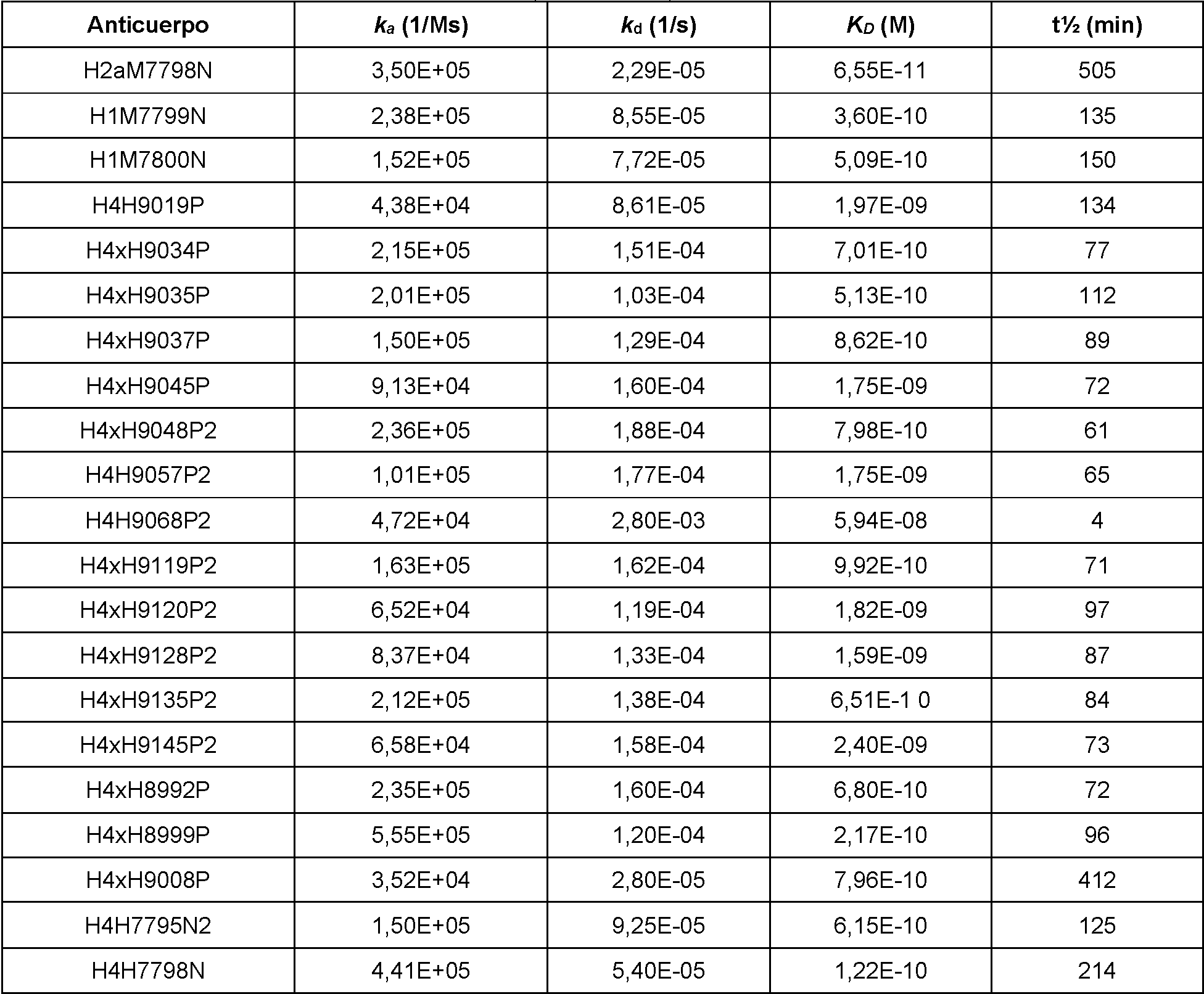
Tabla 7: Parámetros de la cinética de unión de anticuerpos monoclonales anti-PD-1 que se unen al dímero de PD-1 h m n PD-1-mF h m n PD-1-hF h m n 7 ° .Table 7: Binding kinetics parameters of anti-PD-1 monoclonal antibodies binding to PD-1 dimer h m n PD-1-mF h m n PD-1-hF h m n 7°.

continuacióncontinuation

Tabla 8: Parámetros de la cinética de unión de anticuerpos monoclonales anti-PD-1 que se unen a MfPD-1-MMH aTable 8: Binding kinetics parameters of anti-PD-1 monoclonal antibodies binding to MfPD-1-MMH a
25 °C.25°C
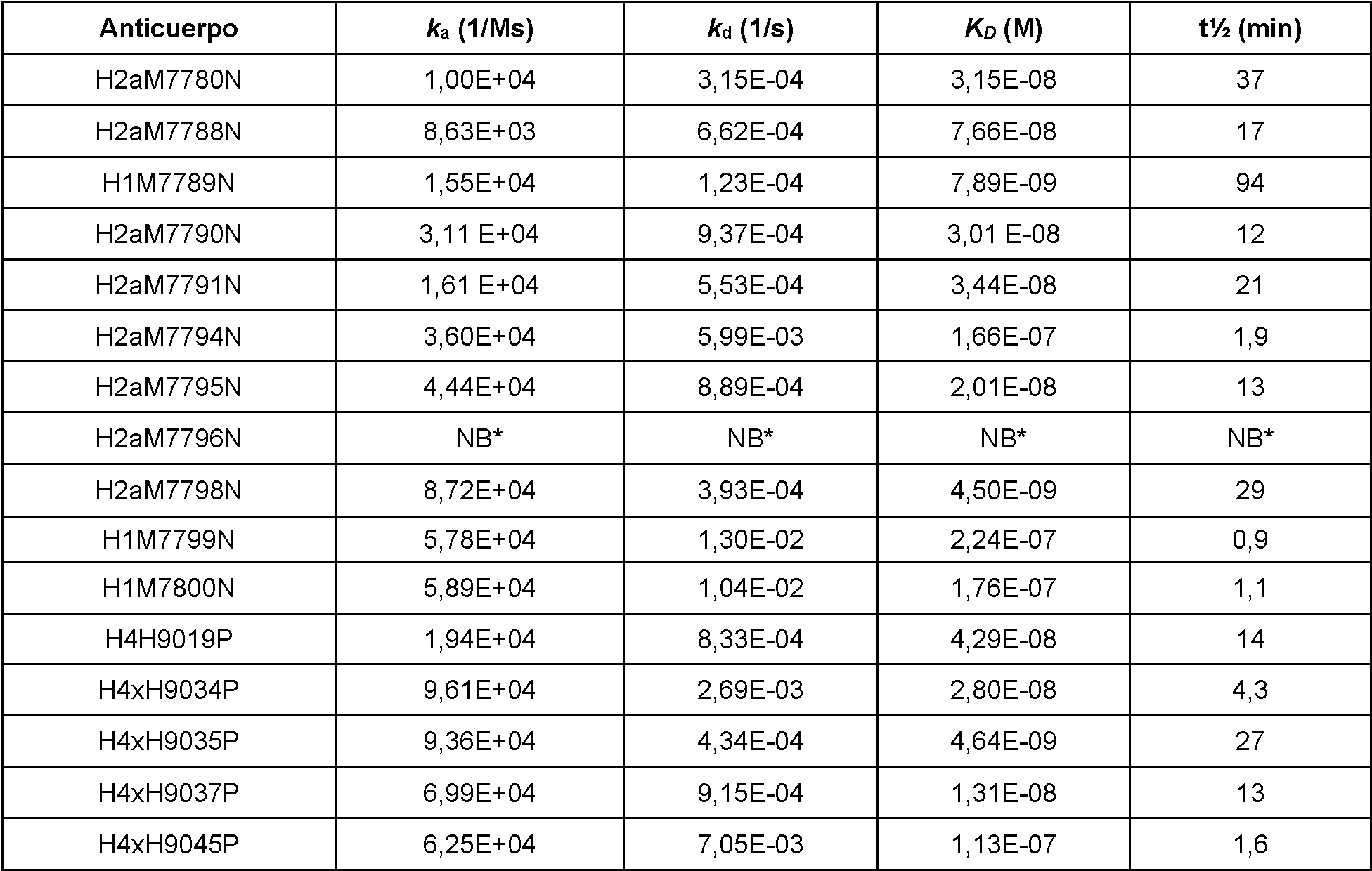
continuacióncontinuation

Tabla 9: Parámetros de la cinética de unión de anticuerpos monoclonales anti-PD-1 que se unen a MfPD-1-MMH aTable 9: Binding Kinetic Parameters of Anti-PD-1 Monoclonal Antibodies Binding to MfPD-1-MMH a
37 °C37°C

continuacióncontinuation

Tabla 10: Parámetros de la cinética de unión de anticuerpos monoclonales anti-PD-1 que se unen al dímero de PD-1 de mono PD-1-mFc de mono a 25 °CTable 10: Binding kinetics parameters of anti-PD-1 monoclonal antibodies binding to monkey PD-1 dimer Monkey PD-1-mFc at 25 °C

Tabla 11: Parámetros de la cinética de unión de anticuerpos monoclonales anti-PD-1 que se unen al dímero de PD-1Table 11: Parameters of the binding kinetics of anti-PD-1 monoclonal antibodies that bind to the PD-1 dimer
- - °- - °

Como se muestra en la Tabla 4, a 25 °C, 28 de los 29 anticuerpos anti-PD-1 de la invención se unieron a hPD-1-MMH con valores KD que van de 2,1 nM a 291 nM. Un anticuerpo, H4H9068P2, no demostró ninguna unión medible a hPD-1-MMH a 25 °C. Como se muestra en la Tabla 5, a 37 °C, 26 de los 29 anticuerpos anti-PD-1 de la invención se unieron a hPD-1-MMH con valores KD que van de 3,79 nM a 1,51 pM. Tres anticuerpos de la invención no demostraron ninguna unión concluyente a hPD-1-MMH a 37 °C. Como se muestra en la Tabla 6, a 25 °C, los 29 anticuerpos anti-PD-1 de la invención se unieron a las proteínas del dímero de hPD-1 con valores KD que van de 65,5 pM a 59,4 nM. Como se muestra en la Tabla 7, a 37 °C, los 27 anticuerpos anti-PD-1 de la invención se unieron a las proteínas del dímero de hPD-1 con valores KD que van de 3,09 pM a 551 nM. Como se muestra en la Tabla 8, a 25 °C, 27 de los 29 anticuerpos anti-PD-1 de la invención se unieron a MfPD-1-MMH con valores KD que van de 3,09 nM a 551 nM. Dos anticuerpos de la invención no demostraron ninguna unión concluyente a MfPD-1-MMH a 25 °C. Como se muestra en la Tabla 9, a 37 °C, 25 de los 29 anticuerpos anti-PD-1 de la invención se unieron a MfPD-1-MMH con valores KDque van de 7,00 nM a 7,54 pM. Cuatro anticuerpos de la invención no demostraron ninguna unión concluyente a MfPD-1-MMH a 37 °C. Como se muestra en la Tabla 10, a 25 °C, 18 de los anticuerpos anti-PD-1 ensayados de la invención se unieron al dímero de MfPD-1 con valores KDque van de 137 pM a 54,2 nM. Como se muestra en la Tabla 11, a 37 °C, 18 de los anticuerpos anti-PD-1 ensayados de la invención se unieron al dímero de MfPD-1 con valores KDque van de 49 pM a 86,3 nM.As shown in Table 4, at 25°C, 28 of 29 anti-PD-1 antibodies of the invention bound hPD-1-MMH with KD values ranging from 2.1 nM to 291 nM. One antibody, H4H9068P2, did not demonstrate any measurable binding to hPD-1-MMH at 25°C. As shown in Table 5, at 37°C, 26 of 29 anti-PD-1 antibodies of the invention bound hPD-1-MMH with KD values ranging from 3.79 nM to 1.51 pM. Three antibodies of the invention did not demonstrate any conclusive binding to hPD-1-MMH at 37°C. As shown in Table 6, at 25 °C, the 29 anti-PD-1 antibodies of the invention bound to hPD-1 dimer proteins with KD values ranging from 65.5 pM to 59.4 nM. . As shown in Table 7, at 37°C, the 27 anti-PD-1 antibodies of the invention bound to hPD-1 dimer proteins with KD values ranging from 3.09 pM to 551 nM. As shown in Table 8, at 25°C, 27 of 29 anti-PD-1 antibodies of the invention bound MfPD-1-MMH with KD values ranging from 3.09 nM to 551 nM. Two antibodies of the invention did not demonstrate any conclusive binding to MfPD-1-MMH at 25°C. As shown in Table 9, at 37°C, 25 of 29 anti-PD-1 antibodies of the invention bound MfPD-1-MMH with KD values ranging from 7.00 nM to 7.54 pM. Four antibodies of the invention did not demonstrate any conclusive binding to MfPD-1-MMH at 37°C. As shown in Table 10, at 25°C, 18 of the tested anti-PD-1 antibodies of the invention bound to the MfPD-1 dimer with KD values ranging from 137 pM to 54.2 nM. As shown in Table 11, at 37°C, 18 of the tested anti-PD-1 antibodies of the invention bound to the MfPD-1 dimer with KD values ranging from 49 pM to 86.3 nM.
Ejemplo 4: Bloqueo de la unión de PD-1 a PD-L1 determinado por ELISAExample 4: Blockade of PD-1 binding to PD-L1 determined by ELISA
La capacidad de los anticuerpos anti-PD-1 para bloquear la unión de PD-1 humano a su ligando, el receptor de PD-L1, se midió utilizando tres formatos de ELISA sándwich de competición. Las proteínas PD-L1 humanas dimérica, compuestas por una porción del dominio extracelular de PD-L1 humano expresado con una etiqueta Fc humana C-terminal (hPD-L1-hFc; SEQ ID: 325) o una etiqueta Fc de ratón C-terminal (hPD-L1-mFc; Se Q ID: 326), o PD-L2 humana dimérica, compuesta por una región extracelular de PD-L2 humana producida con una etiqueta Fc humano C-terminal (hPD-L2-hFc; R&D Systems, N.° 1224- PL) se recubrieron por separado a una concentración de 2 pg/ml en PBS en una placa de microtitulación de 96 pocillos durante la noche a 4 °C. Los sitios de unión inespecíficos se bloquearon posteriormente usando una solución al 0,5 % (p/v) de BSA en PBS. En un primer formato de competición, se añadió una concentración constante de 1,5 nM de una proteína PD-1 humana dimérica, compuesta por un dominio extracelular de PD-1 humana expresada con una etiqueta Fc humano C-terminal (hPD-1-mFc; s Eq ID: 323) a diluciones seriadas de anticuerpos anti-PD-1 o anticuerpos de control de isotipo de modo que las concentraciones finales de anticuerpos variaran de 0 a 200 nM. En un segundo formato de competición, se añadió una concentración constante de 200 pM de proteína PD-1 humana biotinilada dimérica, compuesta por el dominio extracelular de PD-1 humana que se expresó con una etiqueta Fc humana C-terminal (biot-hPD-1-hFc; SEQ ID: 323) se añadió de forma similar a diluciones en serie de anticuerpos anti-PD-1 o un control de isotipo a concentraciones finales de anticuerpo que van de 0 a 50 nM. En un tercer formato de competición, se añadió de forma similar una concentración constante de 100 pM de proteína dimérica hPD-1-mFc a diluciones en serie de anticuerpos anti-PD-1 o un control de isotipo a concentraciones finales de anticuerpo que varían de 0 a 100 nM. Estos complejos anticuerpo-proteína se incubaron a continuación durante 1 hora a temperatura ambiente (TA). Los complejos anticuerpo-proteína con 1,5 nM constante de hPD-1-mFc se transfirieron a placas de microvaloración recubiertas con hPD-L1-hFc, los complejos anticuerpoproteína con 200 pM constante de biot-hPD-1-hFc se transfirieron a placas recubiertas con hPD-L1-mFc, y los complejos anticuerpo-proteína con 100 pM constante de hPD-1-mFc se transfirieron a placas de microvaloración recubiertas con hPD-L2-hFc. Después de incubar durante 1 hora a TA, se lavaron los pocillos y se detectó el hPD-1-mFc unido a la placa con un anticuerpo policlonal anti-mFc conjugado con peroxidasa de rábano picante (HRP) (Jackson ImmunoResearch Inc., N.° 115-035-164 ), y se detectó biot-hPD-1-hFc unido a la placa con estreptavidina conjugada con HRP (Thermo Scientific, N.° N200). Las muestras se desarrollaron con una solución TMB (BD Biosciences, N.° 51-2606KC y N.° 51-2607KC) para producir una reacción colorimétrica y a continuación el desarrollo del color se estabilizó mediante la adición de ácido sulfúrico 1 M antes de medir la absorbancia a 450 nm en un lector de placas Victor X5. El análisis de los datos se realizó usando un modelo de respuesta a dosis sigmoideo dentro del programa informático Prism™ (GraphPad). El valor de CI50 calculado, definido como la concentración de anticuerpo requerida para reducir el 50 % de la unión de PD-1 humano a PD-L1 o PD-L2 humano, se usó como indicador de la potencia de bloqueo. El porcentaje de bloqueo máximo se calculó como una medida de la capacidad de los anticuerpos para bloquear completamente la unión de PD-1 humano a PD-L1 o PD-L2 humano en la placa determinado a partir de la curva de dosis. Este porcentaje de bloqueo máximo se calculó restando del 100 % la proporción de la reducción en la señal observada en presencia de la concentración más alta probada para cada anticuerpo respecto a la diferencia entre la señal observada para una muestra de PD-1 humano que no contiene anticuerpo anti-PD-1 (0 % de bloqueo) y la señal de fondo del anticuerpo secundario conjugado con HRP solo (100 % de bloqueo).The ability of anti-PD-1 antibodies to block the binding of human PD-1 to its ligand, the PD-L1 receptor, was measured using three competitive sandwich ELISA formats. Dimeric human PD-L1 proteins, composed of a portion of the extracellular domain of human PD-L1 expressed with a C-terminal human Fc tag (hPD-L1-hFc; SEQ ID: 325) or a C-terminal mouse Fc tag (hPD-L1-mFc; Se Q ID: 326), or dimeric human PD-L2, composed of an extracellular region of human PD-L2 produced with a C-terminal human Fc tag (hPD-L2-hFc; R&D Systems, #1224-PL) were coated separately at a concentration of 2 pg/mL in PBS in a 96-well microtiter plate overnight at 4°C. The nonspecific binding sites subsequently blocked using a 0.5 % (w/v) solution of BSA in PBS. In a first competition format, a constant concentration of 1.5 nM of a dimeric human PD-1 protein, composed of an extracellular domain of expressed human PD-1 with a C-terminal human Fc tag (hPD-1- mFc;s Eq ID: 323) to serial dilutions of anti-PD-1 antibodies or isotype control antibodies such that final antibody concentrations ranged from 0 to 200 nM. In a second competition format, a constant concentration of 200 pM of dimeric biotinylated human PD-1 protein, composed of the extracellular domain of human PD-1 that was expressed with a C-terminal human Fc tag (biot-hPD- 1-hFc; SEQ ID: 323) was similarly added to serial dilutions of anti-PD-1 antibodies or an isotype control at final antibody concentrations ranging from 0 to 50 nM. In a third competition format, a constant concentration of 100 pM hPD-1-mFc dimeric protein was similarly added to serial dilutions of anti-PD-1 antibodies or an isotype control at final antibody concentrations ranging from 0 to 100nM. These antibody-protein complexes were then incubated for 1 hour at room temperature (RT). Antibody-protein complexes with constant 1.5 nM hPD-1-mFc were transferred to hPD-L1-hFc coated microtiter plates, antibody-protein complexes with constant 200 pM biot-hPD-1-hFc were transferred to hPD-L1-hFc coated microtiter plates. hPD-L1-mFc coated, and antibody-protein complexes with constant 100 pM hPD-1-mFc were transferred to hPD-L2-hFc coated microtiter plates. After incubation for 1 hour at RT, the wells were washed and hPD-1-mFc bound to the plate was detected with a horseradish peroxidase (HRP)-conjugated polyclonal anti-mFc antibody (Jackson ImmunoResearch Inc., N. 115-035-164), and plate-bound biot-hPD-1-hFc was detected with HRP-conjugated streptavidin (Thermo Scientific, #N200). Samples were developed with TMB solution (BD Biosciences, #51-2606KC and #51-2607KC) to produce a colorimetric reaction and then color development was stabilized by addition of 1M sulfuric acid prior to measurement. absorbance at 450 nm on a Victor X5 plate reader. Data analysis was performed using a sigmoid dose response model within Prism™ software (GraphPad). The calculated IC50 value, defined as the concentration of antibody required to reduce 50% of human PD-1 binding to human PD-L1 or PD-L2, was used as an indicator of blocking potency. Percent maximal blocking was calculated as a measure of the ability of the antibodies to completely block binding of human PD-1 to human PD-L1 or PD-L2 in the plaque determined from the dose curve. This percent maximum blocking was calculated by subtracting from 100% the ratio of the reduction in signal observed in the presence of the highest concentration tested for each antibody to the difference between the signal observed for a human PD-1 sample containing no anti-PD-1 antibody (0% blocking) and background signal from HRP-conjugated secondary antibody alone (100% blocking).
El porcentaje máximo de bloqueo y los valores de CI50 calculados para los anticuerpos que bloquean más del 35 % de la señal de unión a hPD-1 se muestran en las Tablas 12 - 14. Los anticuerpos que mostraron una disminución en la señal de unión de hPD-1 del 35 % o menos se definieron como no bloqueantes. Los anticuerpos que mostraron un aumento del 35 % o más en la señal de unión de PD-1 humano se caracterizaron como no bloqueantes/potenciadores. La base teórica del ensayo, definida como la concentración mínima de anticuerpo teóricamente necesaria para ocupar el 50 % de los sitios de unión de PD-1 humano en el ensayo, es 0,75 nM para el formato que usa 1,5 nM constante de hPD-1-mFc, 100 pM para el formato que usa 200 pM constante de biot-hPD-1-hFc y 50 pM para el formato que usa 100 pM constante de hPD-1-mFc, lo que indica que los valores de CI50 inferiores calculados no representan unión al sitio de unión al anticuerpo. Por esta razón, los anticuerpos con valores de CI50 calculados de menos de 0,75 nM en el ensayo con hPD-1-mFc constante y recubrimiento de hPD-L1, de menos de 100 pM en el ensayo con biot-hPD-1-hFc constante y recubrimiento de hPD-L1, y menos de 50 pM en el ensayo con hPD-1-mFc constantes y recubrimiento de hPD-L2 se indican en las Tablas 12 - 14 como <7,5E-10M, <1,0E-10M y <5,0E-11M, respectivamente.The maximum percent blocking and calculated IC50 values for antibodies that block greater than 35% of the hPD-1 binding signal are shown in Tables 12-14. Antibodies that showed a decrease in hPD-1 binding signal hPD-1 of 35% or less was defined as non-blocking. Antibodies showing a 35% or greater increase in human PD-1 binding signal were characterized as non-blocking/enhancing. The theoretical basis of the assay, defined as the minimum concentration of antibody theoretically required to occupy 50% of the human PD-1 binding sites in the assay, is 0.75 nM for the format using 1.5 nM constant. hPD-1-mFc, 100 pM for the format using a constant 200 pM of biot-hPD-1-hFc and 50 pM for the format using a constant 100 pM of hPD-1-mFc, indicating that the IC50 values lower calculated do not represent binding to the antibody binding site. For this reason, antibodies with calculated IC50 values of less than 0.75 nM in the hPD-1-mFc constant assay and hPD-L1 coating, of less than 100 pM in the biot-hPD-1-mFc assay. constant hFc and hPD-L1 overlay, and less than 50 pM in the assay with constant hPD-1-mFc and hPD-L2 overlay are reported in Tables 12 - 14 as <7.5E-10M, <1.0E -10M and <5.0E-11M, respectively.
Tabla 12: ELISA de blo ueo de la unión de PD-1 humano a PD-L1 humano mediante anticuer os anti-PD-1Table 12: ELISA blocking binding of human PD-1 to human PD-L1 by anti-PD-1 antibodies

continuacióncontinuation

Tabla 13: ELISA de bloqueo de la unión de PD-1 humano biotinilado a PD-L1 humano mediante anticuerpos anti-Table 13: ELISA for blocking the binding of biotinylated human PD-1 to human PD-L1 by anti-
PD-1PD-1

continuacióncontinuation

Tabla 14: ELISA de blo ueo de la unión de PD-1 humano a PD-L2 humano mediante anticuer os anti-PD-1 Table 14: ELISA blocking binding of human PD-1 to human PD-L2 by anti-PD-1 antibodies

o m o se ind ica en la T a b la 12, en el p rim e r fo rm a to de ensayo , 23 de los 27 a n ticu e rp o s an ti-P D -1 b loq uea ron 1,5 nM de hPD-1-mFc de la unión a hPD-L1-hFc con valores de CI50 que van de 190 pM a 3,3 nM con el porcentaje de bloqueo máximo que va del 67 % al 100 %. Un anticuerpo, H2aM7796N, se identificó como no bloqueante. Se identificaron tres anticuerpos anti-PD-1 (H4H9068P2, H1M7789N y H2aM7791N) como no bloqueantes/potenciadores.As indicated in T able 12, in the first assay format, 23 of the 27 anti-P D -1 an tibo rs b lo cked 1.5 nM of hPD-1-mFc from binding to hPD-L1-hFc with IC50 values ranging from 190 pM to 3.3 nM with the maximum percent block ranging from 67% to 100%. One antibody, H2aM7796N, was identified as non-blocking. Three anti-PD-1 antibodies (H4H9068P2, H1M7789N and H2aM7791N) were identified as non-blocking/enhancing.
Como se muestra en la Tabla 13, en el segundo formato de ensayo, 23 de los 27 anticuerpos anti-PD-1 bloquearon 200 pM de biothPD-1-hFc de la unión a hPD-L1-mFc con valores de CI50 que van de 59 pM a 1,3 nM con el porcentaje de bloqueo máximo que va del 60 % al 101 %. Un anticuerpo, H1M7789N, se identificó como no bloqueante. Se identificaron tres anticuerpos anti-PD-1 (H4H9057P2, H4H9068P2 y H2aM7791N) como no bloqueantes/potenciadores.As shown in Table 13, in the second assay format, 23 of 27 anti-PD-1 antibodies blocked 200 pM biothPD-1-hFc from binding to hPD-L1-mFc with IC50 values ranging from 59 pM to 1.3 nM with the maximum percentage block ranging from 60% to 101%. One antibody, H1M7789N, was identified as non-blocking. Three anti-PD-1 antibodies (H4H9057P2, H4H9068P2, and H2aM7791N) were identified as non-blocking/enhancing.
En el tercer formato de ensayo como se muestra en la Tabla 14, se probaron cuatro anticuerpos anti-PD-1 de la invención y un control de isotipo. Los 4 anticuerpos anti-PD-1 de la invención bloquearon 100 pM (concentración fija) de hPD-1-mFc de la unión a hPD-L2-hFc que recubre la placa con valores de CI50 que varían de 0,13 nM a 1,3 nM y con un porcentaje de bloqueo máximo que varía del 94 % al 100 %.In the third assay format as shown in Table 14, four anti-PD-1 antibodies of the invention and one isotype control were tested. The 4 anti-PD-1 antibodies of the invention blocked 100 pM (fixed concentration) of hPD-1-mFc from binding to hPD-L2-hFc coating the plate with IC50 values ranging from 0.13 nM to 1 .3 nM and with a maximum blocking percentage ranging from 94% to 100%.
Ejemplo 5: Bloqueo de la unión de PD-1 a PD-L1 determinado mediante un ensayo de biosensor y resonancia de plasmón superficialExample 5: Blockade of PD-1 Binding to PD-L1 Determined by Biosensor Assay and Surface Plasmon Resonance
Se estudió la inhibición de la unión de PD-1 humano a PD-L1 humano por diferentes anticuerpos monoclonales anti-PD-1, ya sea utilizando un ensayo de interferometría de biocapa en tiempo real en un instrumento biosensor Octet Red96 (Fortebio Inc.) o utilizando un ensayo de biosensor de resonancia de plasmón superificial en tiempo real en un instrumento Biacore 3000.Inhibition of the binding of human PD-1 to human PD-L1 by different anti-PD-1 monoclonal antibodies was studied using either a real-time biolayer interferometry assay on an Octet Red96 biosensor instrument (Fortebio Inc.) or using a real-time surface plasmon resonance biosensor assay on a Biacore 3000 instrument.
Se realizaron estudios de inhibición de anticuerpos monoclonales anti-PD-1 expresados con un Fc de ratón en un instrumento Octet Red 96. En primer lugar, se incubaron 100 nM de un PD-1 humano recombinante expresado con una etiqueta Fc de IgG2a de ratón C-terminal (hPD-1-mFc; SEQ ID NO: 323) con 500 nM de cada anticuerpo monoclonal anti-PD-1 durante al menos 1 hora antes de realizar el ensayo de inhibición. Se capturaron alrededor de 0,8 nm a 1,2 nm de PD-L1 humano recombinante expresado con una etiqueta Fc de IgG1 humana C-terminal (hPD-L1-hFc; SEQ ID NO: 325) usando un biosensor Octet de captura de Fc de anti-IgG humana. Los biosensores Octet recubiertos con hPD-L1-hFc se sumergieron a continuación en pocillos que contenían la mezcla de hPD-1-mFc y diferentes anticuerpos monoclonales anti-PD-1. Todo el experimento se realizó a 25 °C en tampón Octet HBST (0,01 M HEPES pH 7,4, NaCl 0,15 M, EDTA 3 mM, Tensioactivo P20 al 0,05 % v/v, 0,1 mg/ml de BSA) con la placa en agitación a una velocidad de 1000 rpm. Los biosensores se lavaron en tampón Octet HBST entre cada etapa del experimento. Las respuestas de unión en tiempo real se controlaron durante todo el transcurso del experimento y se registró la respuesta de unión al final de cada etapa. La unión de hPD-1-mFc al hPD-L1-hFc capturado se comparó en presencia y ausencia de diferentes anticuerpos monoclonales anti-PD-1 y se usó para determinar el comportamiento de bloqueo de los anticuerpos ensayados como se muestra en la Tabla 15.Inhibition studies of anti-PD-1 monoclonal antibodies expressed with a mouse Fc were performed on an Octet Red 96 instrument. First, 100 nM of a recombinant human PD-1 expressed with a mouse IgG2a Fc tag was incubated. C-terminal (hPD-1-mFc; SEQ ID NO: 323) with 500 nM of each anti-PD-1 monoclonal antibody for at least 1 hour before performing the inhibition assay. About 0.8 nm to 1.2 nm of recombinant human PD-L1 expressed with a C-terminal human IgG1 Fc tag (hPD-L1-hFc; SEQ ID NO: 325) was captured using an Octet capture biosensor. Anti-human IgG Fc. The hPD-L1-hFc coated Octet biosensors were then immersed in wells containing the mixture of hPD-1-mFc and different anti-PD-1 monoclonal antibodies. The entire experiment was performed at 25 °C in Octet HBST buffer (0.01 M HEPES pH 7.4, 0.15 M NaCl, 3 mM EDTA, 0.05% v/v Surfactant P20, 0.1 mg/ ml of BSA) with the plate shaken at a speed of 1000 rpm. Biosensors were washed in Octet HBST buffer between each stage of the experiment. Real-time binding responses were monitored throughout the experiment and the binding response was recorded at the end of each stage. The binding of hPD-1-mFc to captured hPD-L1-hFc was compared in the presence and absence of different anti-PD-1 monoclonal antibodies and used to determine the blocking behavior of the antibodies tested as shown in Table 15. .
Tabla 15: Inhibición de la unión de PD-L1 humano a PD-1 mediante anticuerpos monoclonales anti-PD-1 ex resados con Fc de ratón medido en un instrumento Octet Red 96Table 15: Inhibition of Human PD-L1 Binding to PD-1 by Mouse Fc Raised Anti-PD-1 Monoclonal Antibodies Measured on an Octet Red 96 Instrument

C o m o se m ue s tra en la T a b la 15, 9 de los 11 a n ticu e rp o s an ti-P D -1 e n sa ya d o s en el in s tru m e n to O c te t Red 96 demostraron un fuerte bloqueo de hPD-1-mFc de la unión a hPD-L1-hFc desde el 86 % hasta el bloqueo completo de la unión. Un anticuerpo anti-PD-1 (H1M7789N) ensayado mostró un bloqueo más débil de la unión de hPD-1-mFc a hPD-L1-hFc con un bloqueo del 29 %. Un anticuerpo (H2aM7791N) ensayado demostró la capacidad de mejorar la unión de hPD-1-mFc a hPD-L1-hFc.A s shown in T ab le 15, 9 of the 11 anti-P D -1 an ti bodies tested on the O c te t Red 96 instrument demonstrated a strong blockade of hPD-1-mFc from binding to hPD-L1-hFc from 86 % to complete blockade of binding. An anti-PD-1 antibody (H1M7789N) tested showed weaker blockade of hPD-1-mFc binding to hPD-L1-hFc with a 29% block. One antibody (H2aM7791N) tested demonstrated the ability to enhance hPD-1-mFc binding to hPD-L1-hFc.
A continuación, se realizaron estudios de inhibición de anticuerpos monoclonales anti-PD-1 expresados con un Fc humano en un instrumento Biacore 3000. En primer lugar, se incubaron 100 nM de un PD-1 humano recombinante expresado con una etiqueta Fc de IgG1 humana C-terminal (hPD-1-hFc; SEQ ID: 324) con 500 nM de cada anticuerpo monoclonal anti-PD-1 durante al menos 2 horas antes de realizar el ensayo de inhibición. En primer lugar, se derivatizó una superficie de sensor CM5 Biacore con anticuerpo policlonal anti-ratón de conejo (Catálogo GE N.° BR-1008-38) usando química estándar EDC-NHS. Se capturaron alrededor de 730 RU de PD-L1 humano recombinante expresado con una etiqueta Fc de IgG2a de ratón C-terminal (hPD-L1.mFc; SEQ ID: 326) seguido a continuación de la inyección de 100 nM de hPD-1.hFc en presencia y ausencia de diferentes anticuerpos monoclonales anti-PD-1 a un caudal de 25 pl/min durante 3 minutos. Todo el experimento se realizó a 25 °C en tampón de análisis que comprendía HEPES 0,01 M pH 7,4, NaCl 0,15 M, EDTA 3 mM, tensioactivo Tween-20 al 0,05 % v/v (tampón de análisis HBS-ET). Las respuestas de unión en tiempo real se controlaron durante todo el transcurso del experimento y se registró la respuesta de unión al final de cada etapa. La unión de hPD-1-hFc al hPD-L1-mFc capturado se comparó en presencia y ausencia de diferentes anticuerpos monoclonales anti-PD-1 y se usó para determinar el comportamiento de bloqueo de los anticuerpos ensayados como se muestra en la Tabla 16.Inhibition studies of anti-PD-1 monoclonal antibodies expressed with a human Fc were then performed on a Biacore 3000 instrument. First, 100 nM of a recombinant human PD-1 expressed with a human IgG1 Fc tag was incubated. C-terminal (hPD-1-hFc; SEQ ID: 324) with 500 nM of each anti-PD-1 monoclonal antibody for at least 2 hours before performing the inhibition assay. First, a CM5 Biacore sensor surface was derivatized with polyclonal rabbit anti-mouse antibody (GE Catalog No. BR-1008-38) using standard EDC-NHS chemistry. About 730 RU of expressed recombinant human PD-L1 were captured with a C-terminal mouse IgG2a Fc tag (hPD-L1.mFc; SEQ ID: 326) followed by injection of 100 nM hPD-1. hFc in the presence and absence of different anti-PD-1 monoclonal antibodies at a flow rate of 25 pl/min for 3 minutes. The entire experiment was performed at 25 °C in assay buffer comprising 0.01 M HEPES pH 7.4, 0.15 M NaCl, 3 mM EDTA, 0.05% v/v Tween-20 surfactant HBS-ET analysis). Real-time binding responses were monitored throughout the experiment and the binding response was recorded at the end of each stage. The binding of hPD-1-hFc to captured hPD-L1-mFc was compared in the presence and absence of different anti-PD-1 monoclonal antibodies and used to determine the blocking behavior of the tested antibodies as shown in Table 16. .
Tabla 16: Inhibición de la unión de PD-L1 humano a PD-1 mediante anticuerpos monoclonales anti-PD-1 ex resados con Fc humano medido en un instrumento Biacore 3000Table 16: Inhibition of Human PD-L1 Binding to PD-1 by Human Fc Raised Anti-PD-1 Monoclonal Antibodies Measured on a Biacore 3000 Instrument

Como se muestra en la Tabla 16, 18 de los 20 anticuerpos anti-PD-1 de la invención ensayados en el instrumento Biacore 3000 demostraron un fuerte bloqueo de la unión de hPD-1-hFc a hPD-L1-mFc con un bloqueo que varía de 96% a 100%. Un anticuerpo demostró la capacidad de mejorar la unión de hPD-1-hFc a hPD-L1-mFc. En este estudio, uno de los anticuerpos ensayados de la invención (H4H9057P2) demostró una unión de fondo no específica a la superficie de captura del Fc anti-ratón.As shown in Table 16, 18 of the 20 anti-PD-1 antibodies of the invention tested on the Biacore 3000 instrument demonstrated strong blockade of hPD-1-hFc binding to hPD-L1-mFc with a block that varies from 96% to 100%. One antibody demonstrated the ability to enhance the binding of hPD-1-hFc to hPD-L1-mFc. In this studio, one of the tested antibodies of the invention (H4H9057P2) demonstrated non-specific background binding to the anti-mouse Fc capture surface.
Ejemplo 6: Competencia cruzada en Octet entre anticuerpos anti-PD-1Example 6: Octet Cross Competition Between Anti-PD-1 Antibodies
La competencia de unión entre anticuerpos monoclonales anti-PD-1 se determinó usando un ensayo de interferometría de biocapa sin marcaje en tiempo real en un biosensor Octet RED384 (Pall ForteBio Corp.). Todo el experimento se realizó a 25 °C en tampón HEPES 0,01 M pH 7,4, NaCl 0,15 M, EDTA 3 mM, Tensioactivo Tween-20 al 0,05 % v/v, 0,1 mg/ml de BSA (tampón Octet HBS-ET) con la placa en agitación a una velocidad de 1000 rpm. Para evaluar si 2 anticuerpos podían competir entre sí para unirse a sus respectivos epítopos en un PD-1 humano expresado de manera recombinante con una etiqueta myc-myc-hexahistidina C-terminal (hPD-1-MMH; SEQ ID: 321), se capturaron alrededor de 0,1 nM de hPD-1-MMH primero en puntas de biosensor Octet recubiertas con anticuerpo anti-Penta-His (Pall ForteBio Corp., N.° 18-5079) sumergiendo las puntas durante 5 minutos en pocillos que contenían una solución de 50 |jg/ml de hPD-1-MMH. Las puntas del biosensor capturadas con antígeno se saturaron a continuación con el primer anticuerpo monoclonal anti-PD-1 (posteriormente denominado mAb-1) sumergiéndolo en pocillos que contenían 50 jg/m l de solución de mAb-1 durante 5 minutos. Las puntas del biosensor se sumergieron a continuación en pocillos que contenían una solución de 50 jg/m l de un segundo anticuerpo monoclonal anti-PD-1 (posteriormente denominado mAb-2). Las puntas del biosensor se lavaron en tampón Octet HBS-ET entre cada etapa del experimento. La respuesta de unión en tiempo real se controló durante todo el transcurso del experimento y se registró la respuesta de unión al final de cada etapa. Se comparó la respuesta de la unión de mAb-2 a hPD-1-MMH formando un complejo previo con mAb-1 y se determinó el comportamiento competitivo/no competitivo de diferentes anticuerpos monoclonales anti-PD-1. Los resultados se resumen en la Tabla 17 (* No se enumeran los mAb2 de autocompetencia).Binding competition between anti-PD-1 monoclonal antibodies was determined using a real-time label-free biolayer interferometry assay on an Octet RED384 biosensor (Pall ForteBio Corp.). The entire experiment was performed at 25 °C in 0.01 M HEPES buffer pH 7.4, 0.15 M NaCl, 3 mM EDTA, 0.05% v/v Tween-20 Surfactant, 0.1 mg/ml of BSA (Octet HBS-ET buffer) with the plate shaking at a speed of 1000 rpm. To assess whether 2 antibodies could compete with each other for binding to their respective epitopes on a recombinantly expressed human PD-1 with a C-terminal myc-myc-hexahistidine tag (hPD-1-MMH; SEQ ID: 321), captured about 0.1 nM hPD-1-MMH first on anti-Penta-His antibody-coated Octet biosensor tips (Pall ForteBio Corp., #18-5079) by immersing the tips for 5 min in wells containing a 50 µg/ml solution of hPD-1-MMH. Antigen-captured biosensor tips were then saturated with the first anti-PD-1 monoclonal antibody (subsequently designated mAb-1) by immersion in wells containing 50 μg/μl of mAb-1 solution for 5 minutes. The biosensor tips were then immersed in wells containing a 50 μg/μl solution of a second anti-PD-1 monoclonal antibody (subsequently designated mAb-2). Biosensor tips were washed in Octet HBS-ET buffer between each stage of the experiment. The real-time binding response was monitored throughout the experiment and the binding response at the end of each step was recorded. The binding response of mAb-2 to hPD-1-MMH by forming a pre-complex with mAb-1 was compared and the competitive/non-competitive behavior of different anti-PD-1 monoclonal antibodies was determined. The results are summarized in Table 17 (*Self-competing mAb2 not listed).
T l 17: m n i r z nr r n i r ni-PD-1 l i nT l 17: m n i r z nr r n i r ni-PD-1 l i n

(continuación)(continuation)

(continuación)(continuation)

Se determinó una segunda competencia de unión entre un panel de anticuerpos monoclonales anti-PD-1 seleccionados usando un ensayo de interferometría de biocapa sin marcaje en tiempo real en un biosensor Octet HTX (Pall ForteBio Corp.). Todo el experimento se realizó a 25 °C en tampón HEPES 0,01 M pH 7,4, NaCl 0,15 M, EDTA 3 mM, Tensioactivo Tween-20 al 0,05 % v/v, 0,1 mg/ml de BSA (tampón Octet HBS-ET) con la placa en agitación a una velocidad de 1000 rpm. Para evaluar si 2 anticuerpos podían competir entre sí para unirse a sus respectivos epítopos en el hPD-1-MMH, se capturaron alrededor de 0,25 nM de hPD-1-MMH primero en puntas de biosensor Octet recubiertas con anticuerpo anti-Penta-His (ForteBio Inc., N.° 18-5079) sumergiendo las puntas durante 150 segundos en pocillos que contenían una solución de 10 pg/ml de hPD-1-MMH. Las puntas del biosensor capturadas con antígeno se saturaron a continuación con un primer anticuerpo monoclonal anti-PD-1 (posteriormente denominado mAb-1) sumergiéndolo en pocillos que contenían 100 pg/ml de solución de mAb-1 durante 5 minutos. Las puntas del biosensor se sumergieron continuación en pocillos que contenían una solución de 100 pg/ml de un segundo anticuerpo monoclonal anti-PD-1 (posteriormente denominado mAb-2) durante 4 minutos. Todos los biosensores se lavaron en tampón Octet HBS-ET entre cada etapa del experimento. La respuesta de unión en tiempo real se controló durante todo el transcurso del experimento y se registró la respuesta de unión al final de cada etapa como se muestra en la Figura 2. Se comparó la respuesta de la unión de mAb-2 a hPD-1-MMH formando un complejo previo con mAb-1 y se determinó el comportamiento competitivo/no competitivo de diferentes anticuerpos monoclonales anti-PD-1. Los resultados se resumen en la Tabla 18 (*No se enumeran los mAb2 de autocompetencia).A second binding competition between a panel of selected anti-PD-1 monoclonal antibodies was determined using a real-time label-free biolayer interferometry assay on an Octet HTX biosensor (Pall ForteBio Corp.). The entire experiment was performed at 25 °C in 0.01 M HEPES buffer pH 7.4, 0.15 M NaCl, 3 mM EDTA, 0.05% v/v Tween-20 Surfactant, 0.1 mg/ml of BSA (Octet HBS-ET buffer) with the plate shaking at a speed of 1000 rpm. To assess whether 2 antibodies could compete with each other for binding to their respective epitopes on hPD-1-MMH, about 0.25 nM of hPD-1-MMH was first captured on anti-Penta-antibody coated Octet biosensor tips. His (ForteBio Inc., #18-5079) by dipping the tips for 150 seconds into wells containing a 10 pg/ml solution of hPD-1-MMH. Antigen-captured biosensor tips were then saturated with a first anti-PD-1 monoclonal antibody (subsequently designated mAb-1) by immersion in wells containing 100 pg/ml mAb-1 solution for 5 minutes. The biosensor tips were then immersed in wells containing a 100 pg/ml solution of a second anti-PD-1 monoclonal antibody (later designated mAb-2) for 4 minutes. All biosensors were washed in Octet HBS-ET buffer between each stage of the experiment. The real-time binding response was monitored throughout the experiment and the binding response at the end of each step was recorded as shown in Figure 2. The binding response of mAb-2 to hPD-1 was compared -MMH forming a previous complex with mAb-1 and the competitive/non-competitive behavior of different anti-PD-1 monoclonal antibodies was determined. The results are summarized in Table 18 (*Self-competing mAb2 not listed).
T l 1 : m n i r z nr r n i r ni-PD-1 l i nT l 1 : m n i r z nr r n i r ni-PD-1 l i n

En las condiciones experimentales divulgadas en este ejemplo, H4H7795N2 mostraba competencia cruzada con H4H7798N; H4H7798N mostraba competencia cruzada con H4H7795N2 y H4H9008P; H4H9008P mostraba competencia cruzada con H4H7798N y H4H9068P2; H4H9068P2 mostraba competencia cruzada con H4H9008P y H4H9048P2.Under the experimental conditions disclosed in this example, H4H7795N2 showed cross-competition with H4H7798N; H4H7798N showed cross-competition with H4H7795N2 and H4H9008P; H4H9008P showed cross-competition with H4H7798N and H4H9068P2; H4H9068P2 showed cross competition with H4H9008P and H4H9048P2.
Ejemplo 7: Unión de anticuerpos a células que sobreexpresan PD-1Example 7: Binding of Antibodies to Cells Overexpressing PD-1
La un ión de a n ticu e rp o s an ti-P D -1 a una línea c e lu la r de riñón e m b rio n a rio hu m ano (H E K 293 ; A T C C , N .° C R L -1573 ) transfectada de manera estable con PD-1 humano de longitud completa (aminoácidos 1 a 289 con número de registro NP_005009.2) (HEK293/hPD-1) se determinó mediante FACS.Binding of Anti-PD-1 Antibodies to a Human Embryonic Kidney Cell Line (HEK 293; ATCC, No. CRL-1573) stably transfected with full-length human PD-1 (amino acids 1 to 289 with accession number NP_005009.2) (HEK293/hPD-1) was determined by FACS.
Para el ensayo, las células adherentes se separaron usando tripsina o tampón de disociación sin enzimas y se bloquearon con medio completo. Las células se centrifugaron y se resuspendieron a una concentración de 2,5-6 x1oA6 células/ml en PBS frío que contiene FBS al 2 %. A continuación, se incubaron células HEK293 parentales y HEK293/hPD-1 durante 15-30 min en hielo con 100 nM de cada anticuerpo anti-PD-1. Los anticuerpos no unidos se eliminaron lavando con D-PBS que contenía FBS al 2 %, y las células se incubaron posteriormente con un F(ab')2 secundario conjugado con aloficocianina que reconocía Fc humano (Jackson ImmunoResearch, N.° 109-136-170) o Fc de ratón (Jackson ImmunoResearch, #115-136-146) durante 15-30 minutos en hielo. Las células se lavaron con D-PBS que contenía FBS al 2 % para eliminar el F(ab')2 secundario no unido y las mediciones de fluorescencia se adquirieron utilizando un citómetro de flujo HyperCyte (IntelliCyt, Inc.) o un citómetro de flujo Accuri (BD Biosciences). Los datos se analizaron utilizando el software FlowJo (Tree Star).For the assay, adherent cells were detached using trypsin or enzyme-free dissociation buffer and blocked with complete medium. Cells were centrifuged and resuspended at a concentration of 2.5-6 x1oA6 cells/ml in cold PBS containing 2% FBS. Parental HEK293 and HEK293/hPD-1 cells were then incubated for 15-30 min on ice with 100 nM of each anti-PD-1 antibody. Unbound antibodies were removed by washing with D-PBS containing 2% FBS, and cells were subsequently incubated with an allophycocyanin-conjugated secondary F(ab')2 recognizing human Fc (Jackson ImmunoResearch, #109-136 -170) or mouse Fc (Jackson ImmunoResearch, #115-136-146) for 15-30 min on ice. Cells were washed with D-PBS containing 2% FBS to remove unbound secondary F(ab')2 and fluorescence measurements were acquired using a HyperCyte flow cytometer (IntelliCyt, Inc.) or a flow cytometer. Accuri (BD Biosciences). Data was analyzed using FlowJo software (Tree Star).
Tabla 19: Unión or FACS de anticuer os anti-PD-1 a células HEK293/hPD-1 células HEK293 arentalesTable 19: Binding or FACS of anti-PD-1 antibodies to HEK293/hPD-1 cells HEK293 arental cells

C o m o se m ue s tra en la T a b la 19, 25 de los 27 a n ticu e rp o s an ti-P D -1 de la inven c ión m ostra ro n una fu e rte un ión a las células HEK293/hPD-1 en comparación con la unión a la línea HEK293 parental. Dos anticuerpos de la invención (H2aM7795N y H4H9068P2) se unieron más débilmente a las células que expresan PD-1 humano en comparación con los otros anticuerpos probados.As shown in T able 19, 25 of the 27 anti-P D -1 antibodies of the invention showed strong binding to the HEK293/hPD-1 cells compared to binding to the parental HEK293 line. Two antibodies of the invention (H2aM7795N and H4H9068P2) bound more weakly to cells expressing human PD-1 compared to the other antibodies tested.
Para caracterizar adicionalmente los anticuerpos anti-PD1 de la invención, la unión dependiente de la dosis a una línea celular de riñón embrionario humano (HEK293; ATCC, N.° CRL-1573) transfectada de manera estable con PD-1 humano de longitud completa (aminoácidos 1 a 289 con número de registro NP_005009.2) (HEK293/hPD-1) se determinó mediante FACS.To further characterize the anti-PD1 antibodies of the invention, dose-dependent binding to a human embryonic kidney cell line (HEK293; ATCC, No. CRL-1573) stably transfected with full-length human PD-1 (amino acids 1 to 289 with accession number NP_005009.2) (HEK293/hPD-1) was determined by FACS.
Para el ensayo, las células adherentes se separaron usando tripsina y se bloquearon con medio completo. Las células se centrifugaron y se resuspendieron a una concentración de 6 x10A6 células/ml en tampón de tinción (FBS al 1 % FBS en PBS). Para determinar la CE50 y la Emax de los anticuerpos anti-PD1, se incubaron 90 pl de suspensión celular durante 30 minutos en hielo con una dilución en serie de anticuerpos anti-PD-1 y los controles se diluyeron hasta una concentración final que oscilaba de 5 pM a 100 nM (no se incluyó ninguna muestra de mAb como control negativo) en tampón de tinción. A continuación, las células se centrifugaron y los sedimentos se lavaron una vez con tampón de tinción para eliminar los anticuerpos no unidos. Posteriormente, las células se incubaron durante 30 minutos en hielo con un F(ab')2 secundario conjugado con aloficocianina que reconoce Fc humano (Jackson ImmunoResearch, N.° 109-136-170) o Fc de ratón (Jackson ImmunoResearch, N.° 115-136-071). Las células se centrifugaron y los sedimentos se lavaron una vez con tampón de tinción para eliminar el F(ab')2 secundario no unido y a continuación se fijaron durante la noche con una dilución 1:1 de Cytofix (BD Biosciences, N.° 554655) y tampón de tinción. El día siguiente, las células se centrifugaron y los sedimentos se lavaron una vez con tampón de tinción, se resuspendieron y se filtraron. Las medidas de fluorescencia se adquirieron en un citómetro Hypercyt® y se analizaron en ForeCyt™ (intelliCyt; Albuquerque, NM) para determinar las intensidades medias de fluorescencia (MFI). Los valores de CE50 se calcularon a partir de una ecuación logística de cuatro parámetros sobre una curva de respuesta de 11 puntos utilizando GraphPad Prism. La Emax para cada anticuerpo se definió como la unión a la dosis de anticuerpo más alta (100 nM) ensayada.For the assay, adherent cells were detached using trypsin and blocked with complete medium. Cells were centrifuged and resuspended at a concentration of 6 x 10A6 cells/ml in staining buffer (FBS 1% FBS in PBS). To determine the EC 50 and Emax of anti-PD1 antibodies, 90 μl of cell suspension were incubated for 30 minutes on ice with a serial dilution of anti-PD-1 antibodies and controls were diluted to a final concentration ranging 5 pM to 100 nM (no mAb sample included as negative control) in staining buffer. Cells were then centrifuged and pellets washed once with staining buffer to remove unbound antibodies. Cells were then incubated for 30 min on ice with an allophycocyanin-conjugated secondary F(ab')2 recognizing human Fc (Jackson ImmunoResearch, #109-136-170) or mouse Fc (Jackson ImmunoResearch, N. 115-136-071). Cells were centrifuged and pellets washed once with staining buffer to remove unbound secondary F(ab')2 and then fixed overnight with a 1:1 dilution of Cytofix (BD Biosciences, #554655 ) and staining buffer. The next day, the cells were centrifuged and the pellets washed once with staining buffer, resuspended and filtered. Fluorescence measurements were acquired on a Hypercyt® cytometer and analyzed on ForeCyt™ (intelliCyt; Albuquerque, NM) to determine mean fluorescence intensities (MFI). EC 50 values were calculated from a four-parameter logistic equation on an 11-point response curve using GraphPad Prism. The Emax for each antibody was defined as the binding at the highest dose of antibody (100 nM) tested.
Tabla 20: FACS de endiente^ de dosis de la unión de anticuer os anti-PD-1 a células HEK293/hPD-1Table 20: FACS of Anti-PD-1 Antibody Binding Dose^ to HEK293/hPD-1 Cells

continuacióncontinuation

Tabla 21: FACS de endiente de dosis de la unión de anticuer os anti-PD-1 a células HEK293/hPD-1Table 21: Dose Slope FACS of Anti-PD-1 Antibody Binding to HEK293/hPD-1 Cells

Como se muestra en la Tabla 20, 25 de 28 anticuerpos anti-PD1 de la invención mostraron unión dependiente de la dosis a células HEK293/hPD-1 con valores de CE50 que van de 33,18 pM a 2,59 nM y valores de Emax que varían de 37.789 a 11,368 MFI. Tres anticuerpos anti-PD1 de la invención no demostraron una unión fuerte a las células HEK293/hPD-1 y, por lo tanto, no se pudo determinar un valor de CE50. Ninguno de los controles de isotipo demostró unión medible en este ensayo.As shown in Table 20, 25 of 28 anti-PD1 antibodies of the invention showed dose-dependent binding to HEK293/hPD-1 cells with EC 50 values ranging from 33.18 pM to 2.59 nM and values of Emax ranging from 37,789 to 11,368 MFI. Three anti-PD1 antibodies of the invention did not demonstrate strong binding to HEK293/hPD-1 cells and therefore an EC 50 value could not be determined. None of the isotype controls demonstrated measurable binding in this assay.
Como se muestra en la Tabla 21,3 de 6 anticuerpos anti-PD1 de la invención mostraron unión dependiente de la dosis a células HEK293/hPD-1 con valores de CE50 que van de 509 pM a 4,81 nM y valores de Emax que van de 39.774 a 14.111 MFI. Tres anticuerpos de la invención ensayados se unieron a células HEK293/hPD-1, pero no alcanzaron una meseta. Por consiguiente, no se pudieron determinar sus valores de CE50 y sus valores de CE50 se consideran no concluyentes. Ninguno de los controles de isotipo demostró unión medible en este ensayo.As shown in Table 21, 3 of 6 anti-PD1 antibodies of the invention showed dose-dependent binding to HEK293/hPD-1 cells with EC 50 values ranging from 509 pM to 4.81 nM and Emax values ranging from 39,774 to 14,111 MFI. Three antibodies of the invention tested bound to HEK293/hPD-1 cells, but did not reach a plateau. Therefore, their EC 50 values could not be determined and their EC 50 values are considered inconclusive. None of the isotype controls demonstrated measurable binding in this assay.
Ejemplo 8: Bloqueo de la regulación negativa de linfocitos T inducida por PD-1 en un ensayo de indicador de luciferasa en linfocitos/APCExample 8: Blocking PD-1-Induced Downregulation of T Cells in a Lymphocyte/APC Luciferase Reporter Assay
La activación de los linfocitos T se logra estimulando los receptores de los linfocitos T (TcR) que reconocen péptidos específicos presentados por las proteínas del complejo principal de histocompatibilidad de clase I o II en las células presentadoras de antígeno (APC). Los TcR activados a su vez inician una cascada de eventos de señalización que pueden ser controlados por genes indicadores impulsados por factores de transcripción como la proteína activadora 1 (AP-1), el factor nuclear de linfocitos T activados (NFAT) o el factor nuclear potenciador de la cadena ligera kappa de linfocitos B activados (NFKb). La respuesta de los linfocitos T se modula mediante el acoplamiento de correceptores expresados de forma constitutiva o inducible en los linfocitos T. Uno de esos receptores es el PD-1, un regulador negativo de la actividad de los linfocitos T. PD-1 interactúa con su ligando, PD-L1, que se expresa en las células diana, incluidas las APC o las células cancerosas, y actúa para enviar señales inhibitorias al reclutar fosfatasas en el signalosoma de TcR, lo que da como resultado la supresión de la señalización positiva.Activation of T cells is achieved by stimulating T cell receptors (TcRs) that recognize specific peptides presented by major histocompatibility complex class I or II proteins on antigen presenting cells (APCs). Activated TcRs in turn initiate a cascade of signaling events that can be controlled by reporter genes driven by transcription factors such as activator protein 1 (AP-1), nuclear factor of activated T cells (NFAT), or nuclear factor kappa light chain enhancer of activated B lymphocytes (NFKb). The T cell response is modulated by coupling to co-receptors constitutively or inducibly expressed on T cells. One such receptor is PD-1, a negative regulator of T cell activity. PD-1 interacts with its ligand, PD-L1, which is expressed on target cells, including APCs or cancer cells, and acts to send inhibitory signals by recruiting phosphatases to the TcR signalosome, resulting in suppression of positive signaling.
La capacidad de los anticuerpos anti-PD-1 para antagonizar la señalización mediada por PD-1/PD-L1 a través del receptor PD-1 en líneas de linfocitos T humanos se evaluó usando un ensayo basado en células in vitro mostrado en la Figura 1. El bioensayo se desarrolló para medir la señalización de linfocitos T inducida por la interacción entre APC y linfocitos T utilizando un cultivo mixto derivado de dos líneas de células de mamíferos: células Jurkat (una línea de linfocitos T inmortalizada) y células Raji (una línea de linfocitos B). Para el primer componente del bioensayo, las células Jurkat Clón E6-1 (ATCC, N.° TIB-152) se transdujeron con Cignal Lenti AP-1 Luc Reporter (Qiagen -Sabiosciences, N.° CLS-011L) según las instrucciones del fabricante. El lentivirus codifica el gen de luciferasa de luciérnaga bajo el control de un promotor mínimo de CMV, repeticiones en tándem del elemento de respuesta transcripcional inducible por TPA (TRE) y un gen de resistencia a puromicina. La línea celular Jurkat diseñada fue posteriormente transducida con una quimera de PD-1 que comprende el dominio extracelular de PD-1 humano (aminoácidos de 1 a 170 de PD1 humano; número de registro NP_005009.2) y los dominios transmembrana y citoplásmico de CD300a humano (aminoácidos de 181 a 299 de CD300a humano; número de registro NP_009192.2). La línea celular estable resultante (Jurkat/AP1-Luc/hPD1-hCD300a) se seleccionó y mantuvo en RPMI/FBS al 10 %/penicilina/estreptomicina/glutamina suplementada con 500 pg/ml de G418 1 pg/ml de puromicina.The ability of anti-PD-1 antibodies to antagonize PD-1/PD-L1-mediated signaling through the PD-1 receptor in human T cell lines was assessed using an in vitro cell-based assay shown in Figure 1. The bioassay was developed to measure T cell signaling induced by the interaction between APCs and T cells using a mixed culture derived from two mammalian cell lines: Jurkat cells (an immortalized T cell line) and Raji cells (an immortalized T cell line). B cell line). For the first component of the bioassay, Jurkat Clone E6-1 cells (ATCC, No. TIB-152) were transduced with Cignal Lenti AP-1 Luc Reporter (Qiagen-Sabiosciences, No. CLS-011L) according to the instructions in the maker. The lentivirus encodes the firefly luciferase gene under the control of a CMV minimal promoter, TPA-inducible transcriptional response element (TRE) tandem repeats, and a puromycin resistance gene. The engineered Jurkat cell line was subsequently transduced with a PD-1 chimera comprising the extracellular domain of human PD-1 (amino acids 1 to 170 of human PD1; accession number NP_005009.2) and the transmembrane and cytoplasmic domains of CD300a. human (human CD300a amino acids 181 to 299; accession number NP_009192.2). The resulting stable cell line (Jurkat/AP1-Luc/hPD1-hCD300a) was selected and maintained in RPMI/10% FBS/penicillin/streptomycin/glutamine supplemented with 500 pg/ml G418 1 pg/ml puromycin.
Para el segundo componente del bioensayo, se transdujeron células Raji (ATCC, N.° CCL-86) con el gen PD-L1 humano (aminoácidos 1-290 del número de registro NP_054862.1) que se había clonado en un sistema de vector lentiviral (pLEX) (Thermo Scientific Biosystems, N.° OHS4735). Las células Raji, positivas para PD-L1 (Raji/hPD-L1) se aislaron mediante FACS usando un anticuerpo PD-L1 y se mantuvieron en Iscove/FBS al 10 %/penicilina/estreptomicina/glutamina suplementada con 1 pg/ml de puromicina.For the second component of the bioassay, Raji cells (ATCC, No. CCL-86) were transduced with the human PD-L1 gene (amino acids 1-290 of accession number NP_054862.1) that had been cloned into a vector system lentiviral (pLEX) (Thermo Scientific Biosystems, #OHS4735). Raji cells, positive for PD-L1 (Raji/hPD-L1) were isolated by FACS using a PD-L1 antibody and maintained in Iscove/10% FBS/penicillin/streptomycin/glutamine supplemented with 1 pg/mL puromycin. .
Para simular la interacción APC/linfocito T, se utilizó un anticuerpo biespecífico compuesto por un brazo Fab que se une a CD3 en linfocitos T y el otro brazo Fab que se une a CD20 en células Raji (anticuerpo biespecífico CD3xCD20; por ejemplo, como se divulga en el documento US20140088295). La presencia de la molécula biespecífica en el ensayo da como resultado la activación de los linfocitos T y APC al unir las subunidades CD3 de los linfocitos T con CD20 expresado endógenamente en las células Raji. Se ha demostrado que la ligación de CD3 con anticuerpos anti-CD3 conduce a la activación de los linfocitos T. En este bioensayo, los anticuerpos que bloquean la interacción PD1/PD-L1 rescatan la actividad de los linfocitos T desactivando la señalización inhibidora y, posteriormente, conduciendo a un aumento de la activación de AP1-Luc.To simulate APC/T cell interaction, a bispecific antibody composed of one Fab arm that binds to CD3 on T cells and the other Fab arm that binds to CD20 on Raji cells (CD3xCD20 bispecific antibody; for example, as shown) was used. disclosed in document US20140088295). The presence of the bispecific molecule in the assay results in activation of T cells and APCs by binding the CD3 subunits of T cells to CD20 endogenously expressed on Raji cells. Binding of CD3 with anti-CD3 antibodies has been shown to lead to T cell activation. In this bioassay, antibodies that block the PD1/PD-L1 interaction rescue T cell activity by turning off inhibitory signaling and, subsequently, leading to increased activation of AP1-Luc.
En el bioensayo basado en luciferasa, se usó RPMI1640 suplementado con FBS al 10 % y penicilina/estreptomicina/glutamina como medio de ensayo para preparar suspensiones celulares y diluciones de anticuerpos para llevar a cabo el cribado de anticuerpos monoclonales (mAb) anti-PD1. El día del cribado, se determinaron los valores de CE50 de los mAb anti-PD1, en presencia de una concentración fija de anticuerpo biespecífico CD3xCD20 (30 pM), así como la CE50 del anticuerpo biespecífico solo. En el siguiente orden, se añadieron células y reactivos a placas de fondo plano y blanco de 96 pocillos. Para las determinaciones de la CE50 de los mAb anti-PD1, primero se preparó una concentración fija de anticuerpo biespecífico CD3xCD20 (30 pM final) y se añadió a los pocillos de la placa de microvaloración. A continuación, se añadieron diluciones en serie de 12 puntos de mAb anti-PD1 y controles (concentraciones finales en el intervalo de 1,7 pM a 100 nM; más pocillos con medio de ensayo solo). Para la determinación del anticuerpo biespecífico (solo) CE50 , se añadió el anticuerpo biespecífico, a concentraciones finales que varían de 0,17 pM a 10 nM (más pocillos con medio de ensayo solo), a los pocillos de la placa de microvaloración. Posteriormente, se preparó una suspensión celular de 2,5 x 10A6/ml de células Raji/hPD-L1 y se añadieron 20 pl por pocillo (número final de células (pocillo 5 x 10A4 células). Las placas se dejaron a temperatura ambiente (15-20 minutos), mientras se preparaba una suspensión de 2,5 x 10A6/ml de Jurkat/AP1-Luc/hPD1(ecto)-hCD300a (TM-Cyto). Se añadieron 20 pl de la suspensión Jurkat (número final de células/pocillo 5x10A4 células) por pocillo. Las placas que contenían el cocultivo se incubaron durante 5 a 6 horas a 37 °C/5 % de CO2. Las muestras se analizaron por duplicado y a continuación se detectó la actividad luciferasa después de la adición de reactivo ONE-Glo™ (Promega, # E6051) y se midieron las unidades de luz relativa (RLU) en un luminómetro Victor.In the luciferase-based bioassay, RPMI1640 supplemented with 10% FBS and penicillin/streptomycin/glutamine was used as assay medium to prepare cell suspensions and antibody dilutions to screen for anti-PD1 monoclonal antibodies (mAbs). On the day of screening, the EC50 values of the anti-PD1 mAbs were determined, in the presence of a fixed concentration of CD3xCD20 bispecific antibody (30 pM), as well as the EC50 of the bispecific antibody alone. In the following order, cells and reagents were added to 96-well white flat bottom plates. For EC50 determinations of anti-PD1 mAbs, a fixed concentration of CD3xCD20 bispecific antibody (30 pM final) was first prepared and added to the wells of the microtiter plate. Serial 12-point dilutions of anti-PD1 mAb and controls (final concentrations ranging from 1.7 pM to 100 nM; plus wells with assay medium alone) were then added. For determination of bispecific antibody (alone) CE 50 , bispecific antibody, at final concentrations ranging from 0.17 pM to 10 nM (plus wells with assay medium alone), was added to the wells of the microtiter plate. Subsequently, a cell suspension of 2.5 x 10A6/ml of Raji/hPD-L1 cells was prepared and 20 μl per well (final number of cells (well 5 x 10A4 cells) were added). The plates were left at room temperature ( 15-20 minutes), while preparing a 2.5 x 10A6/ml suspension of Jurkat/AP1-Luc/hPD1(ecto)-hCD300a (TM-Cyto) 20 μl of the Jurkat suspension (final number of cells/well 5x10A4 cells) per well Plates containing the co-culture were incubated for 5-6 hours at 37°C/5% CO 2 Samples were assayed in duplicate and luciferase activity was then detected after addition of ONE-Glo™ reagent (Promega, # E6051) and relative light units (RLU) were measured on a Victor luminometer.
Los valores de RLU para cada anticuerpo seleccionado se normalizaron estableciendo las condiciones de ensayo con concentración fija (30 pM) del anticuerpo biespecífico CD3/CD20, pero sin anticuerpo anti-PD-1 hasta el 100 %. Esta condición corresponde a la respuesta máxima de AP1-Luc provocada por la molécula biespecífica en presencia de la señal inhibidora de PD-1/PD-L1. Tras la adición del anticuerpo anti-PD-1, se suprime la señal inhibidora y la estimulación aumentada se muestra aquí como Emax, el porcentaje de aumento de la señal en presencia de la dosis de anticuerpo más alta probada (100 nM). Para comparar la potencia de los anticuerpos anti-PD1 ensayados, se determinó la concentración de anticuerpo a la que el valor de RLU normalizado alcanzó el 150 % de activación a partir de una ecuación logística de cuatro parámetros sobre una curva de respuesta de 12 puntos utilizando GraphPad Prism. Los resultados se resumen en la Tabla 22 y la Tabla 23, respectivamente. RLU values for each selected antibody were normalized by setting assay conditions with fixed concentration (30 pM) of CD3/CD20 bispecific antibody, but no anti-PD-1 antibody to 100%. This condition corresponds to the maximum AP1-Luc response elicited by the bispecific molecule in the presence of the PD-1/PD-L1 inhibitory signal. Upon addition of anti-PD-1 antibody, the inhibitory signal is abolished and the increased stimulation is shown here as Emax, the percent increase in signal in the presence of the highest dose of antibody tested (100 nM). To compare the potency of the anti-PD1 antibodies tested, the antibody concentration at which the normalized RLU value reached 150% activation was determined from a four-parameter logistic equation on a 12-point response curve using GraphPad Prism. The results are summarized in Table 22 and Table 23, respectively.
Tabla 22: Anticuerpo anti-PD1 que bloquea la inhibición dependiente de PD-1/PD-L1 de la señalización de AP1-Luc en el Ex erimento 1Table 22: Anti-PD1 Antibody Blocking PD-1/PD-L1-Dependent Inhibition of AP1-Luc Signaling in Ex erment 1
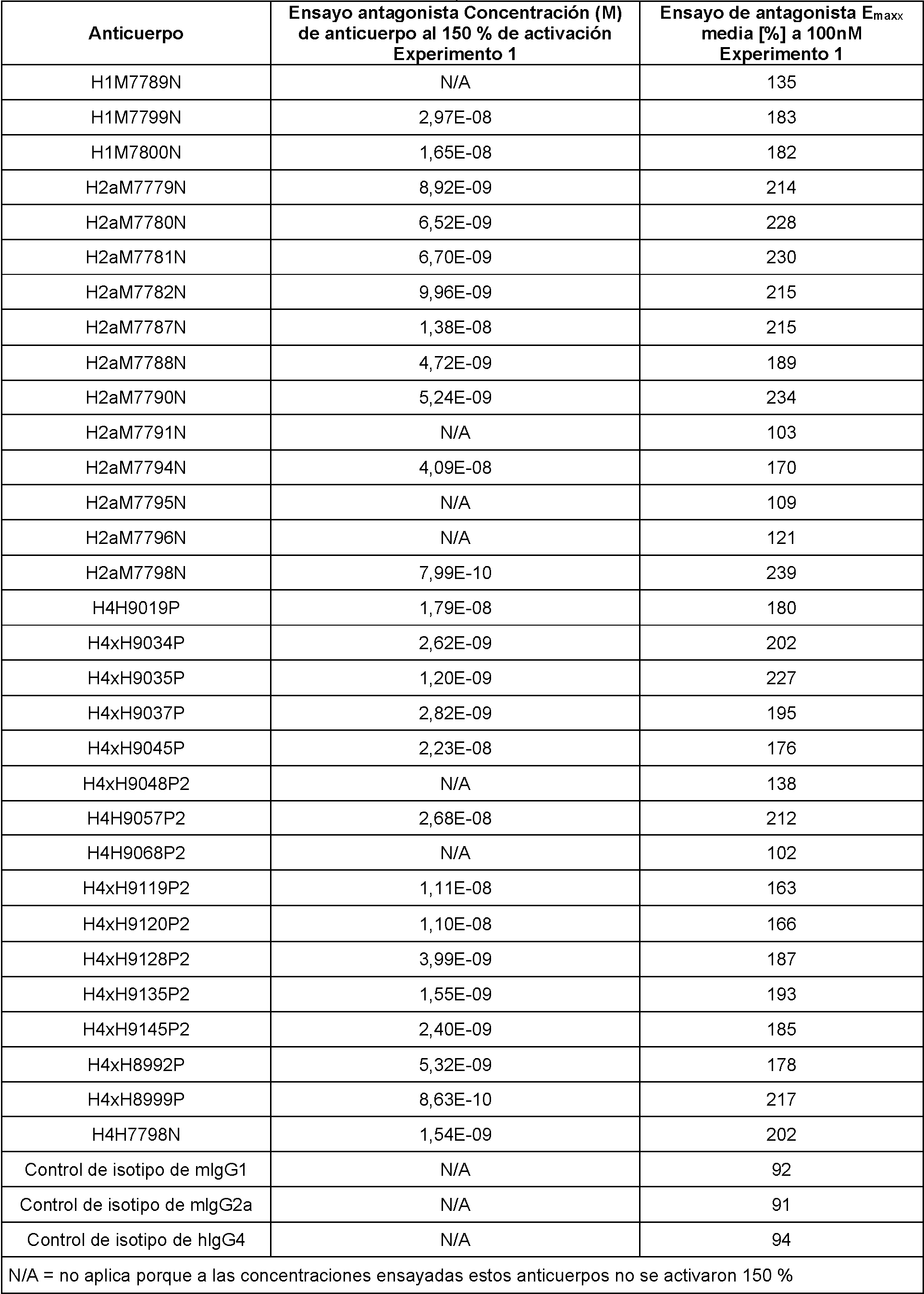
Tabla 23: Anticuerpo anti-PD1 que bloquea la inhibición dependiente de PD-1/PD-L1 de la señalización de AP1-Luc en el Ex erimento 2Table 23: Anti-PD1 Antibody Blocking PD-1/PD-L1-Dependent Inhibition of AP1-Luc Signaling in Ex eriment 2

Como se muestra en la Tabla 22, 25 de los 31 anticuerpos anti-PD-1 de la invención ensayados bloquearon la inhibición de PD-1/PD-L1 con valores de Emax que van de 239 a 163. Seis de los 31 anticuerpos anti-PD-1 de la invención no demostraron un bloqueo sustancial de la interacción PD1/PD-L1 cuando se ensayaron en este ensayo. As shown in Table 22, 25 of the 31 anti-PD-1 antibodies of the invention tested blocked PD-1/PD-L1 inhibition with Emax values ranging from 239 to 163. Six of the 31 anti -PD-1 of the invention did not demonstrate substantial blockade of the PD1/PD-L1 interaction when tested in this assay.
Como se muestra en la Tabla 23, 2 de los 4 anticuerpos anti-PD-1 de la invención ensayados bloquearon la inhibición de PD-1/PD-L1 con valores de Emax de 150 y 343 %, respectivamente. Dos de los 4 anticuerpos anti-PD-1 de la invención no demostraron un bloqueo sustancial de la interacción PD1/PD-L1 cuando se ensayaron en este ensayo. As shown in Table 23, 2 of the 4 anti-PD-1 antibodies of the invention tested blocked PD-1/PD-L1 inhibition with Emax values of 150 and 343%, respectively. Two of the 4 anti-PD-1 antibodies of the invention did not demonstrate substantial blockade of the PD1/PD-L1 interaction when tested in this assay.
Ejemplo 9: Eficacia in vivo de los anticuerpos anti-PD-1Example 9: In vivo efficacy of anti-PD-1 antibodies
Para determinar el efecto de un número selecto de anticuerpos anti-PD-1 de la invención en un modelo in vivo relevante, se llevaron a cabo tres estudios de crecimiento tumoral MC38.ova, que implican la inyección subcutánea de células tumorales y que comenzaron en días diferentes, en ratones que eran homocigotos para la expresión del dominio extracelular de PD-1 humano en lugar del dominio extracelular de PD-1 de ratón (ratones PD-1 Humln) en un 75 % de C57/BI6/25 % de 129 cepas de fondo.To determine the effect of a select number of anti-PD-1 antibodies of the invention in a relevant in vivo model, three MC38.ova tumor growth studies were carried out, involving subcutaneous injection of tumor cells and beginning in different days, in mice that were homozygous for expression of the human PD-1 extracellular domain rather than the mouse PD-1 extracellular domain (PD-1 Humln mice) by 75% C57/BI6/25% 129 background strains.
Para los estudios, los ratones se dividieron uniformemente según el peso corporal en 5 grupos de tratamiento o control para el Estudio 1 (5 ratones por grupo), 8 grupos de tratamiento o control para el Estudio 2 (5 ratones por grupo) y 5 grupos de tratamiento o control para el Estudio 3 (7 ratones por grupo). En el día 0, los ratones fueron anestesiados por inhalación de isoflurano y a continuación recibieron una inyección por vía subcutánea en el flanco derecho de 5 x 10⁵ células MC38.ova en suspensión de 100 pl de DMEM en el Estudio 1 o 1 x 10⁶ células MC38.ova en suspensión de 100 pl de DMEM en el Estudio 2 y el Estudio 3. Para el estudio 1, se inyectaron por vía intraperitoneal los grupos de tratamiento con 200 pg de uno de los tres anticuerpos anti-PD-1 de la invención, o un anticuerpo control de isotipo con especificidad irrelevante los días 3, 7, 10, 14 y 17 del experimento, mientras que un grupo de ratones se dejó sin tratar. Para el estudio 2, los grupos de tratamiento se inyectaron por vía intraperitoneal con uno de los tres anticuerpos anti-PD-1 de la invención a 10 mg/kg o 5 mg/kg por dosis, un anticuerpo de la invención (H4H7795N2) a 10 mg/kg por dosis, o un anticuerpo de control de isotipo con especificidad irrelevante a 10 mg/kg los días 3, 7, 10, 14 y 17 del experimento. Para el estudio 3, los grupos de tratamiento se inyectaron por vía intraperitoneal con uno de los dos anticuerpos anti-PD-1 de la invención a 5 mg/kg o 2,5 mg/kg por/dosis, o un anticuerpo de control de isotipo con especificidad irrelevante a 5 mg/kg los días 3, 7, 10, 14 y 17 del experimento. El protocolo experimental de dosificación y tratamiento para grupos de ratones se muestra en la Tabla 24.For the studies, mice were evenly divided according to body weight into 5 treatment or control groups for Study 1 (5 mice per group), 8 treatment or control groups for Study 2 (5 mice per group), and 5 treatment or control groups for Study 2 (5 mice per group). treatment or control for Study 3 (7 mice per group). On day 0, mice were anesthetized by isoflurane inhalation and then received a subcutaneous injection in the right flank of 5 x 10⁵ MC38.ova cells in suspension in 100 pl DMEM in Study 1 or 1 x 10⁶ MC38 cells. .ova in 100 pl suspension of DMEM in Study 2 and Study 3. For Study 1, treatment groups were injected intraperitoneally with 200 pg of one of the three anti-PD-1 antibodies of the invention, or an isotype control antibody with irrelevant specificity on days 3, 7, 10, 14 and 17 of the experiment, while one group of mice was left untreated. For Study 2, treatment groups were injected intraperitoneally with one of three anti-PD-1 antibodies of the invention at 10 mg/kg or 5 mg/kg per dose, one antibody of the invention (H4H7795N2) at 10 mg/kg per dose, or an isotype control antibody with irrelevant specificity at 10 mg/kg on days 3, 7, 10, 14 and 17 of the experiment. For study 3, the treatment groups were injected intraperitoneally with one of the two anti-PD-1 antibodies of the invention at 5 mg/kg or 2.5 mg/kg per/dose, or a control antibody of isotype with irrelevant specificity at 5 mg/kg on days 3, 7, 10, 14 and 17 of the experiment. The experimental dosing and treatment protocol for groups of mice is shown in Table 24.

continuacióncontinuation

Para los estudios, se registraron los volúmenes tumorales promedio determinados por medidas de calibre y el porcentaje de supervivencia en el día 14 o 17 y el día 23 o 24 de cada experimento para cada grupo de tratamiento. Además, también se evaluó el número de ratones sin tumor al final del estudio (día 42 para el estudio 1 y día 31 para el estudio 2 y el estudio 3). Los resultados, expresados como la media del volumen tumoral (mm3) (± DE), el porcentaje de supervivencia y el número de ratones sin tumor se muestran en la Tabla 23 para el Estudio 1, la Tabla 3 para el Estudio 2 y la Tabla 4 para el Estudio 3.For the studies, mean tumor volumes as determined by caliper measurements and percent survival at day 14 or 17 and day 23 or 24 of each experiment were recorded for each treatment group. In addition, the number of tumor-free mice at the end of the study (day 42 for study 1 and day 31 for study 2 and study 3) was also evaluated. The results, expressed as mean tumor volume (mm3) (±SD), percent survival, and number of tumor-free mice are shown in Table 23 for Study 1, Table 3 for Study 2, and Table 4 for Study 3.
Tabla 25: Media del volumen tumoral, porcentaje de supervivencia y número de ratones sin tumor en cada grupo de tratamiento del tumor in vivo Estudio 1Table 25: Mean tumor volume, percent survival, and number of tumor-free mice in each tumor treatment group in vivo Study 1

Como se muestra en la Tabla 25 para el Estudio 1, los ratones tratados con un anticuerpo de la invención, H4H7798N no desarrollaron ningún tumor detectable durante el transcurso del estudio. Los ratones tratados con H4H9008P exhibieron un volumen tumoral reducido sostenido en comparación con los controles en los días 17 y 24 del estudio con 3 de 5 ratones o 4 de 5 ratones sin tumores al final del experimento, respectivamente. En cambio, el tratamiento con uno de los anticuerpos anti-PD1, H4H7795N2, no demostró una eficacia significativa en la reducción del volumen tumoral en este estudio en comparación con los controles. En el día 23 del estudio, 1 de cada 5 ratones murió en el grupo H4H7795N2, y 2 de cada 5 ratones murieron en el grupo de tratamiento de control de isotipo. En el grupo sin tratamiento y en el grupo de control de isotipo, algunos ratones mostraron una regresión espontánea de los tumores (1 de cada 5 ratones y 2 de cada 5 ratones, respectivamente). As shown in Table 25 for Study 1, mice treated with an antibody of the invention, H4H7798N, did not develop any detectable tumors over the course of the study. H4H9008P-treated mice exhibited sustained reduced tumor volume compared to controls on days 17 and 24 of the study with 3 of 5 mice or 4 of 5 tumor-free mice at the end of the experiment, respectively. In contrast, treatment with one of the anti-PD1 antibodies, H4H7795N2, did not demonstrate significant efficacy in reducing tumor volume in this study compared to controls. On day 23 of the study, 1 out of 5 mice died in the H4H7795N2 group, and 2 out of 5 mice died in the isotype control treatment group. In the untreated group and the isotype control group, some mice showed spontaneous regression of tumors (1/5 mice and 2/5 mice, respectively).

Como se muestra en la Tabla 26 para el Estudio 2, los ratones tratados con un anticuerpo de la invención, H4H7798N a 10 mg/kg no desarrollaron ningún tumor detectable durante el transcurso del estudio. Los grupos de ratones tratados con 10 mg/kg de H4H9008P o H4H9048P2 mostraron un volumen tumoral sustancialmente reducido en comparación con los controles en los días 17 y 24 del estudio. Cuatro de cada 5 ratones en cada grupo tratados con 10 mg/kg de H4H9008P o H4H9048P2 no tenían tumores el día 31, mientras que en el grupo de tratamiento de control de isotipo solo 1 de cada 5 animales no tenía tumores como resultado de la regresión espontánea del tumor. Un anticuerpo ensayado a 10 mg/kg, H4H7795N2, demostró un volumen tumoral sustancialmente reducido en comparación con los controles en los días 17 y 24 del estudio, pero este anticuerpo fue el anticuerpo anti-PD1 menos eficaz y solo 2 de cada 5 ratones sobrevivieron al final del experimento.As shown in Table 26 for Study 2, mice treated with an antibody of the invention, H4H7798N at 10 mg/kg, did not develop any detectable tumors over the course of the study. Groups of mice treated with 10 mg/kg H4H9008P or H4H9048P2 showed substantially reduced tumor volume compared to controls on days 17 and 24 of the study. Four out of 5 mice in each group treated with 10 mg/kg H4H9008P or H4H9048P2 were tumor-free at day 31, whereas only 1 out of 5 animals in the isotype control treatment group were tumor-free as a result of regression. spontaneous tumor. One antibody tested at 10 mg/kg, H4H7795N2, demonstrated substantially reduced tumor volume compared to controls on study days 17 and 24, but this antibody was the least effective anti-PD1 antibody and only 2 out of 5 mice survived. at the end of the experiment.
Se observó una respuesta dependiente de la dosis en la supresión tumoral a las dosis ensayadas (5 mg/kg y 10 mg/kg) en los grupos tratados con H4H7798N, H4H9008P y H4H9048P2. La terapia con H4H7798N o H4H9008P a 5 mg/kg fue menos eficaz, con 4 de 5 ratones sin tumor al final del experimento el día 21, mientras que 5 de 5 ratones permanecieron sin tumores en ambos grupos de dosis de 10 mg/kg de H4H7798N y H4H9008P.A dose-dependent response in tumor suppression was observed at the doses tested (5 mg/kg and 10 mg/kg) in the H4H7798N, H4H9008P and H4H9048P2 treated groups. Therapy with H4H7798N or H4H9008P at 5 mg/kg was less effective, with 4 of 5 mice tumor-free at the end of the experiment on day 21, while 5 of 5 mice remained tumor-free in both 10 mg/kg dose groups. H4H7798N and H4H9008P.
La prueba de Dunnett en comparaciones múltiples ANOVA bidireccional reveló que las diferencias en el crecimiento tumoral entre el grupo tratado con anticuerpo de control de isotipo con 10 mg/kg como referencia y los grupos tratados con 10 mg/kg con H4H7798N, H4H9008P o H4H9048P2 fueron estadísticamente significativas con valor de p <0,005. Las diferencias en el crecimiento tumoral entre el grupo tratado con anticuerpo de control de isotipo con 10 mg/kg como referencia y los grupos tratados con 5 mg/kg con H4H7798N, H4H9008P o H4H9048P2 también fueron estadísticamente significativas con un valor de p < 0,05.Dunnett's test for multiple comparisons Two-way ANOVA revealed that the differences in tumor growth between the group treated with 10 mg/kg isotype control antibody as reference and the groups treated with 10 mg/kg with H4H7798N, H4H9008P, or H4H9048P2 were statistically significant with p value <0.005. Differences in tumor growth between the group treated with isotype control antibody at 10 mg/kg as reference and the groups treated at 5 mg/kg with H4H7798N, H4H9008P, or H4H9048P2 were also statistically significant at p < 0, 05.
Tabla 27: Media del volumen tumoral, porcentaje de supervivencia y número de ratones sin tumor en cada grupo de tratamiento del tumor in vivo Estudio 3Table 27: Mean tumor volume, percent survival, and number of tumor-free mice in each tumor treatment group in vivo Study 3

Como se muestra en la Tabla 27 para el Estudio 3, 6 de cada 7 ratones tratados con un anticuerpo de la invención, H4H7798N, u otro anticuerpo de la invención, H4H9008P, con 5 mg/kg no tenían tumor al final del experimento, mientras que en el grupo de control de isotipo no había ningún animal sin tumor. Un ratón portador de tumores en el grupo de control de IgG4 murió el día 17 posterior a la implantación. Solo 4 de los 7 ratones tratados con H4H9008P a una dosis de 2,5 mg/kg permanecían sin tumores al final del experimento. La diferencia en los volúmenes tumorales el día 21 entre los anticuerpos anti-PD-1 ensayados y un grupo de control de isotipo fue estadísticamente significativa según lo determinado por ANOVA unidireccional con la prueba posterior de comparación múltiple de Dunnett con p < 0,01. Los cuatro anticuerpos anti-PD-1 fueron igualmente más eficaces con la dosis de 5 mg/kg que con la dosis de 2,5 mg/kg.As shown in Table 27 for Study 3, 6 out of 7 mice treated with one antibody of the invention, H4H7798N, or another antibody of the invention, H4H9008P, at 5 mg/kg had no tumor at the end of the experiment, while that in the isotype control group there were no animals without tumor. One tumor-bearing mouse in the IgG4 control group died on day 17 post-implantation. Only 4 of 7 mice treated with H4H9008P at a dose of 2.5 mg/kg remained tumor-free at the end of the experiment. The difference in tumor volumes on day 21 between tested anti-PD-1 antibodies and an isotype control group was statistically significant as determined by one-way ANOVA with Dunnett's multiple comparison post-test with p < 0.01. All four anti-PD-1 antibodies were equally more effective at the 5 mg/kg dose than at the 2.5 mg/kg dose.
Ejemplo 10: Efectos antitumorales de una combinación de un anticuerpo anti-PD-1 y un antagonista de VEGF en un modelo de tum or de tratamiento temprano en ratónExample 10: Antitumor Effects of a Combination of an Anti-PD-1 Antibody and a VEGF Antagonist in an Early Treatment Mouse Tumor Model
Se desarrolló un modelo de tumor de tratamiento temprano para probar la eficacia de una combinación de un anticuerpo anti-PD-1 y un antagonista de VEGF. En este modelo, la terapia de combinación se administra poco después de la implantación del tumor. El experimento también usó un anticuerpo anti-PD-L1 solo y en combinación con el antagonista de VEGF. El anticuerpo anti-PD-1 usado en este experimento fue el clon "RPMI-14" anti-PD-1 de ratón con IgG2b de rata (Bio X Cell, West Lebanon, NH). El antagonista de VEGF usado en este experimento fue aflibercept (una molécula quimérica basada en el receptor de VEGF, también conocida como "trampa de VEGF" o "VEGFR1R2-FcAC1(a)", cuya descripción completa se proporciona en otra parte del presente documento). El anticuerpo anti-PD-L1 utilizado en este experimento fue un anticuerpo monoclonal anti-PD-L1 con secuencias Vh/Vl de anticuerpo "YW243.55S70" de acuerdo con el documento US20100203056A1 (Genentech, Inc.), con IgG2a de ratón y que mostró reactividad cruzada con PD-L1 de ratón.An early treatment tumor model was developed to test the efficacy of a combination of an anti-PD-1 antibody and a VEGF antagonist. In this model, the combination therapy is administered shortly after tumor implantation. The experiment also used an anti-PD-L1 antibody alone and in combination with the VEGF antagonist. The anti-PD-1 antibody used in this experiment was the mouse anti-PD-1 clone "RPMI-14" with rat IgG2b (Bio X Cell, West Lebanon, NH). The VEGF antagonist used in this experiment was aflibercept (a chimeric molecule based on the VEGF receptor, also known as "VEGF trap" or "VEGFR1R2-FcAC1(a)", the full description of which is provided elsewhere in this document. ). The anti-PD-L1 antibody used in this experiment was an anti-PD-L1 monoclonal antibody with V h /V l sequences of "YW243.55S70" antibody according to US20100203056A1 (Genentech, Inc.), with IgG2a of mouse and showed cross-reactivity with mouse PD-L1.
P ara este m ode lo e xp e rim e n ta l, se im p lan ta ron po r v ía su b cu tá n e a 1,0 x 10⁶ de cé lu la s tu m o ra le s de co lo n -26 en ratones BALB/c el día 0. Comenzando el Día 3, antes del establecimiento de tumores medibles, los ratones se trataron con una de las terapias mono o de combinación, o combinación de control, como se expone en la Tabla 28.For this e xp e rim en ta l m ode l, 1.0 x 10⁶ of co lo n -26 tumor cells were implanted subcutaneously in BALB/c mice on day 0. Starting on Day 3, prior to the establishment of measurable tumors, mice were treated with one of the mono or combination therapies, or control combination, as set forth in Table 28.
Tabla 28: Dosificación ex erimental ru os de tratamientoTable 28: Experimental dosage ru os of treatment

Las diversas terapias se administraron en cinco puntos temporales diferentes durante un período de dos semanas (es decir, inyecciones en el Día 3, Día 6, Día 10, Día 13 y Día 19).The various therapies were administered at five different time points over a two-week period (ie, injections on Day 3, Day 6, Day 10, Day 13, and Day 19).
Los animales de cada grupo de terapia se evaluaron en términos de incidencia tumoral, volumen tumoral, mediana del tiempo de supervivencia y número de animales sin tumor el Día 50. La extensión del crecimiento tumoral se resume en la Figura 2 (curvas de crecimiento tumoral) y la Figura 3 (volumen tumoral en el día 28). Los resultados también se resumen en la Tabla 29.Animals in each therapy group were evaluated in terms of tumor incidence, tumor volume, median survival time, and number of tumor-free animals at Day 50. The extent of tumor growth is summarized in Figure 2 (tumor growth curves). and Figure 3 (tumor volume at day 28). The results are also summarized in Table 29.
T l 2 : R n in m r n r r mi nT l 2 : R n in m r n r r mi n

El crecimiento tumoral se redujo sustancialmente en los animales tratados con la combinación de trampa de VEGF anticuerpo anti-PD-1 en comparación con los regímenes de tratamiento que implican cualquier agente terapéutico solo (véanse las Figuras 2 y 3). Asimismo, la supervivencia aumentó sustancialmente en el grupo de anticuerpo trampa de VEGF anti-PD-1, sobreviviendo el 70 % de los animales hasta al menos el día 50 después de la implantación del tumor. Por el contrario, para los grupos de anti-PD-1 y trampa de VEGF en monoterapia, la supervivencia hasta el día 50 fue sólo del 40 % y 30 % respectivamente (véase la Figura 3 y la Tabla 29).Tumor growth was substantially reduced in animals treated with the anti-PD-1 antibody VEGF trap combination compared to treatment regimens involving either therapeutic agent alone (see Figures 2 and 3). Likewise, survival was substantially increased in the anti-PD-1 VEGF trap antibody group, with 70% of animals surviving until at least day 50 after tumor implantation. In contrast, for the anti-PD-1 and VEGF trap monotherapy groups, survival to day 50 was only 40% and 30%, respectively (see Figure 3 and Table 29).
Ejemplo 11: Estudio de ensayo clín ico de dosificación repetida con anticuerpo anti-PD-1 como terapia única y en combinación con otras terapias contra el cáncer en pacientes con neoplasias malignas avanzadas Example 11: Repeated Dosing Clinical Trial Study With Anti-PD-1 Antibody As Single Therapy And In Combination With Other Anticancer Therapies In Patients With Advanced Malignancies
Este es un estudio de aumento de la dosis del anticuerpo anti-PD-1, solo o en combinación con radioterapia, ciclofosfamida o ambos en pacientes con neoplasias malignas avanzadas. El anticuerpo anti-PD-1 ilustrativo ("mAb") usado en este Ejemplo comprende HCVR de SEQ ID NO: 162 y LCVR de SEQ ID NO: 170.This is a dose escalation study of anti-PD-1 antibody, alone or in combination with radiation therapy, cyclophosphamide, or both in patients with advanced malignancies. Illustrative anti-PD-1 antibody ("mAb") used in this Example comprises HCVR of SEQ ID NO: 162 and LCVR of SEQ ID NO: 170.
Objetivos del estudioStudy Objectives
El objetivo principal del estudio es caracterizar la seguridad, tolerabilidad, TLD del mAb administrado por vía intravenosa como monoterapia o en combinación con radiación dirigida (con la intención de que esto sirva como una terapia inmunoestimuladora, en lugar de básicamen una terapia ablativa del tumor), ciclofosfamida en dosis bajas (una terapia que se ha demostrado que inhibe las respuestas de los linfocitos T reguladores), o ambas en pacientes con neoplasias malignas avanzadas.The main objective of the study is to characterize the safety, tolerability, TLD of the mAb administered intravenously as monotherapy or in combination with targeted radiation (with the intention of this serving as an immunostimulatory therapy, rather than primarily a tumor ablative therapy). , low-dose cyclophosphamide (a therapy that has been shown to inhibit regulatory T cell responses), or both in patients with advanced malignancies.
Los objetivos secundarios del estudio son: (1) determinar una dosis de fase 2 recomendada (RP2D) de mAb como monoterapia y en combinación con otras terapias contra el cáncer (radiación dirigida, ciclofosfamida en dosis bajas o ambas); (2) describir la actividad antitumoral preliminar de mAb, solo y con cada pareja(s) de combinación; (3) caracterizar la FC de mAb como monoterapia y en combinación con otras terapias contra el cáncer (radiación dirigida, ciclofosfamida en dosis bajas o ambas); y (4) evaluar la inmunogenicidad de mAb. The secondary objectives of the study are: (1) to determine a recommended phase 2 dose (RP2D) of mAb as monotherapy and in combination with other anticancer therapies (targeted radiation, low-dose cyclophosphamide, or both); (2) describe preliminary antitumor activity of mAb, alone and with each combination partner(s); (3) characterize the PK of mAbs as monotherapy and in combination with other anticancer therapies (targeted radiation, low-dose cyclophosphamide, or both); and (4) evaluating mAb immunogenicity.
Diseño del estudioStudy design
El estudio de seguridad se evaluará en cohortes de aumento de la dosis 3 3 estándar separadas (en monoterapia), combinación con radioterapia, combinación con ciclofosfamida, y combinación con radioterapia más ciclofosfamida). La elección de la terapia combinada con radiación, ciclofosfamida o ambas se basará en la evaluación del investigador de la mejor opción de terapia para un paciente individual en consulta con el patrocinador. Para ser incluido en una cohorte de radioterapia, un paciente debe tener una lesión que pueda irradiarse de manera segura y para la cual la radiación en las dosis paliativas limitadas contempladas, se consideraría médicamente apropiada, y al menos otra lesión adecuada para la evaluación de la respuesta. Se permitirá la inclusión de un paciente solo si hay un hueco disponible en la cohorte para el tratamiento elegido.The safety study will be evaluated in separate standard 3 3 dose escalation (in monotherapy), combination with radiotherapy, combination with cyclophosphamide, and combination with radiotherapy plus cyclophosphamide cohorts). The choice of combined therapy with radiation, cyclophosphamide, or both will be based on the investigator's evaluation of the best therapy option for an individual patient in consultation with the sponsor. To be included in a radiation therapy cohort, a patient must have one lesion that can be safely irradiated and for which radiation at the contemplated limited palliative doses would be considered medically appropriate, and at least one other lesion suitable for evaluation of answer. Inclusion of a patient will be allowed only if there is an available slot in the cohort for the chosen treatment.
Los pacientes se someterán a procedimientos de detección para determinar la elegibilidad dentro de los 28 días anteriores a la administración inicial del mAb. Después de la inclusión de los pacientes en una cohorte de monoterapia con mAb, la inclusión de cohortes posteriores se determinará por la aparición de TLD en cohortes anteriores (es decir, sin TLD en una cohorte de 3 pacientes, o no más de 1 TLD en una cohorte ampliada de 6 pacientes), y la disponibilidad de huecos para pacientes. Los niveles de dosis de monoterapia planificados son 1, 3 o 10 mg/kg administrados por vía intravenosa cada 14 días (2 semanas).Patients will undergo screening procedures to determine eligibility within 28 days prior to initial mAb administration. Following inclusion of patients in a mAb monotherapy cohort, inclusion of subsequent cohorts will be determined by the occurrence of DLTs in prior cohorts (i.e., no DLTs in a cohort of 3 patients, or no more than 1 DLTs in an expanded cohort of 6 patients), and the availability of patient slots. Planned monotherapy dose levels are 1, 3, or 10 mg/kg administered intravenously every 14 days (2 weeks).
Una vez que uno o ambos períodos de observación de la TLD de la cohorte de monoterapia de 1 mg/kg o 3 mg/kg de mAb en monoterapia se completan sin una TLD en una cohorte de 3 pacientes o con no más de 1 TLD en una cohorte ampliada de 6 pacientes, los pacientes se pueden incluir en una cohorte que combina ciclofosfamida o radioterapia con mAb a ese nivel de dosis de monoterapia. Los pacientes pueden incluirse en una cohorte de combinación de mAb ciclofosfamida/radioterapia una vez que se completen los períodos de observación de TLD tanto para la cohorte para ese nivel de dosis de mAb ciclofosfamida como para la cohorte para ese nivel de dosis de mAb el mismo régimen de radioterapia sin TLD en una cohorte de 3 pacientes, o no más de 1 TLD en una cohorte ampliada de 6 pacientes.Once one or both of the 1 mg/kg monotherapy or 3 mg/kg mAb monotherapy cohort TLD observation periods are completed with no TLD in a cohort of 3 patients or with no more than 1 TLD in an expanded cohort of 6 patients, patients can be included in a cohort combining cyclophosphamide or mAb radiotherapy at that monotherapy dose level. Patients may be included in a combination cyclophosphamide mAb/radiation therapy cohort once the TLD observation periods are completed for both the cohort for that cyclophosphamide mAb dose level and the cohort for that same mAb dose level. radiotherapy regimen without DLT in a cohort of 3 patients, or no more than 1 DLT in an expanded cohort of 6 patients.
Una vez que se completa el período de observación de TLD de la cohorte de 3 mg/kg de mAb en monoterapia sin TLD en una cohorte de 3 pacientes, o no más de 1 TLD en una cohorte ampliada de 6 pacientes, también se puede incluir una cohorte de 10 mg/kg de mAb en monoterapia.Once the DLT observation period of the 3 mg/kg mAb monotherapy cohort without DLT is completed in a cohort of 3 patients, or no more than 1 DLT in an expanded cohort of 6 patients, it can also be included. a cohort of 10 mg/kg mAb monotherapy.
Las cohortes de mAb de 3 mg/kg y 10 mg/kg en monoterapia se incluirán solo después de que el número requerido de pacientes en la cohorte de dosis de monoterapia previa (es decir, 1 mg/kg y 3 mg/kg, respectivamente) hayan superado el período de observación de TLD de 28 días sin que se demuestre una dosis máxima tolerada (DMT) para ese nivel de dosis. Una cohorte de tratamiento de combinación de 1 mg/kg de mAb se incluirá solo después de completar el período de observación de TLD para la cohorte de 1 mg/kg en monoterapia. Las cohortes de combinación que reciben 3 mg/kg de mAb se incluirán solo cuando el número requerido de pacientes en las respectivas cohortes de combinación de 1 mg/kg de mAb haya superado el período de observación de TLD sin demostrar una DMT. Las cohortes de combinación triple que combinan mAb con ciclofosfamida y un régimen de radiación se incluirán solo cuando el número requerido de pacientes en las dos cohortes de combinación doble correspondientes a ese nivel de dosificación haya superado el período de observación de TLD sin que se demuestre una d Mt .The 3 mg/kg and 10 mg/kg monotherapy mAb cohorts will be included only after the required number of patients in the prior monotherapy dose cohort (i.e., 1 mg/kg and 3 mg/kg, respectively). ) have exceeded the TLD observation period of 28 days without a demonstrated maximum tolerated dose (MTD) for that dose level. A 1 mg/kg mAb combination treatment cohort will be included only after completion of the TLD observation period for the 1 mg/kg monotherapy cohort. Combination cohorts receiving 3 mg/kg mAb will be included only when the required number of patients in the respective 1 mg/kg mAb combination cohorts have passed the TLD observation period without demonstrating an MTD. Triple combination cohorts combining mAb with cyclophosphamide and a radiation regimen will be included only when the required number of patients in the corresponding two dual combination cohorts at that dosage level have passed the TLD observation period without a demonstrated d Mt .
La Tabla 30 resume las cohortes de aumento de la dosis en las cuales se incluirán los pacientes.Table 30 summarizes the dose escalation cohorts into which patients will be enrolled.
Tabla 30: Posibles cohortes de aumento de la dosisTable 30: Potential Dose Escalation Cohorts

con tin u a c ió ncontinuation

U na T LD se de fine com o cu a lq u ie ra de lo s igu ien te : una to x ic id a d no h e m a to ló g ica (p o r e jem p lo , uve ítis o c u a lq u ie r o A A ri) o una to x ic id a d h e m a to ló g ica (p o r e jem p lo , neu tropen ia , tro m b o c ito p e n ia , n e u tro p e n ia febril).A TLD IS DEFINED AS ANY OF THE FOLLOWING: A NON-HEMATOLOGICAL TOXICITY (FOR EXAMPLE, UVEITIS OR ANY AARI) or an adhema to logical to x ic id (for example, neutropenia, thrombocytopenia, febrile neutropenia).
La dos is m áx im a to le ra d a (D M T ) se de fine com o la dos is m ás a lta a la que m enos de un te rc io de una coho rte am p liad a de 6 p a c ien tes e xp e rim e n ta una T LD d u ra n te el p rim e r c ic lo de tra ta m ie n to . P o r tan to , la D M T se de fine co m o el n ive l de dos is in m e d ia ta m e n te po r de b a jo de l n ive l en el que se de tie ne la d o s ifica c ió n de b id o a la apa ric ió n de 2 o m ás T LD en una coh o rte a m p liad a de 6 pac ien tes. Si el a u m en to de la d os is no se de tie ne de b id o a la apa ric ió n de TLD, se co n s id e ra rá que no se ha d e te rm in a do la DM T. Es pos ib le que en este es tu d io no se de fina una DM T, ya sea para un g rupo de m on o te rap ia o para g ru p o s de co m b in a c ió n ind iv idua les . A d ic io n a lm e n te , es pos ib le que las D M T de los m A b puedan d ife rir en tre la m on o te rap ia y cad a rég im en de tra ta m ie n to com b inado .The maximum tolerated dose (MTD) is defined as the highest dose at which less than one-third of an expanded cohort of 6 patients e xp e rim en ta a T LD d u r ing the f rst treatm en t . Therefore, the DMT is defined as the level of doses immediately below the level at which dosing of bid stops. or the appearance of 2 or more T LDs in an expanded cohort of 6 patients. If the increase in the dose is not stopped due to the appearance of TLDs, it will be considered that the DM T has not been determined. It is possible that in this study a DM T is not defined, either for a m on o th e rap ia group or for ind iv idual co mb in a tio n groups. A d d itio na lm en t , it is pos sible that the DMT of the m A b may di fer between the m on o t rap ia and each com b ined treatm ent reg im ent .
Duración del estudioStudy duration
Los pac ie n te s rec ib irán hasta 48 se m a n a s de tra ta m ie n to , d e sp ué s de las cua les habrá un pe ríodo de se g u im ie n to de 24 sem anas. Un pac ie n te rec ib irá tra ta m ie n to hasta que se co m p le te el pe ríodo de tra ta m ie n to de 48 sem anas, o hasta la p rog res ión de la en fe rm e da d , to x ic id a d inacep tab le , re tirada de l c o n se n tim ie n to o cu m p lim ie n to de o tro crite rio de re tirada de l es tud io . D e spués de un m ín im o de 24 se m a n a s de tra ta m ie n to , los p a c ie n te s con resp u e s ta s co m p le ta s (R C ) co n firm a d a s pueden o p ta r p o r in te rru m p ir el tra ta m ie n to y c o n tin u a r con to d a s las e va lu a c io n e s re leva n te s del es tud io (p o r e jem p lo , e va lu a c io n e s de e ficac ia ). D esp u é s de un m ín im o de 24 se m a n a s de tra ta m ie n to , los p a c ien tes con e va lu a c io n e s de la ca rga tu m o ra l de e n fe rm e da d es ta b le (E E ) o respues ta pa rc ia l (R P ) que no han ca m b ia d o du ra n te 3 e va lu a c io n e s tu m o ra le s su ce s iva s ta m b ié n pueden o p ta r po r in te rru m p ir el tra ta m ie n to y c o n tin u a r con to d as las e va lu a c io n e s re leva n te s del es tu d io (p o r e jem p lo , e v a lu a c io n e s de la e ficac ia ).Patients will receive up to 48 weeks of treatment, after which there will be a 24-week follow-up period. A patient will receive treatment until the completion of the 48-week treatment period, or until disease progression d , unacceptable to x ic ity , withdrawal from lcon sen tim e or fulfilment of oth er withdrawal criterion from th e study . After a minimum of 24 weeks of treatment, patients with confirmed complete responses (CR) may opt for in te interrupt treatment and continue with all relevant assessments of the study (for example, e icacy assessments). After a minimum of 24 weeks of treatment, patients with assessments of tumor burden of disease ta b le (SE ) or partial response (PR ) that have not changed during 3 successive tumor evaluations can also op ta r for in ter rupting the treatmen t and contin uing with all rel e lev a n t e va lu atio n s of the study (for example, e va lu atio n e ficacy ).
Población del estudioStudy Population
La p o b lac ión ob je tivo de este es tu d io co m p re n d e p a c ie n te s con n e o p las ias m a lig na s a va n za d a s que no son ca n d id a to s para la te ra p ia es tánda r, que no desean so m e te rse a una te ra p ia e s tá n d a r o para q u ie n es no se esp e ra que una te ra p ia d isp o n ib le p ro p o rc io n e un b e ne fic io c lín ico ; y p a c ie n te s con n e o p las ias que son in cu ra b le s y no han resp o n d id o o han m os tra d o p ro g re s ió n de l tu m o r a p e sa r de la te ra p ia es tándar.The target population of this study comprises patients with advanced malignant neoplasms who are not candidates for standard therapy. r, who do not wish to undergo a standard therapy for whom it is not expected that an avail able therapy will provide a benefit clinical; and patients with neoplasms that are incurable and have not responded or have shown tumor progression despite standard therapy.
C rite rio s de inc lus ión : Un pac ie n te d ebe cu m p lir los s ig u ie n tes c rite rio s para se r e le g ib le para su inc lus ión en el es tud io : (1 ) p rog res ión d e m o s tra d a de un tu m o r só lido sin una opc ión te ra p é u tica de re fe re n c ia a lte rn a tiva d ispon ib le ; (2 ) al m enos 1 les ión para la e va lu a c ió n de la respues ta . Los p a c ie n te s a s ig n a d o s a rad io te ra p ia requ ie ren al m enos una les ión a d ic io n a l que pueda irrad ia rse de fo rm a se g u ra sin a fe c ta r a las les io n e s de re fe re n c ia y para la cua l la rad iac ión en las d os is p a lia tivas lim ita d as co n tem p la d a s se co n s id e ra ría m é d ica m e n te a p rop iada ; (3 ) E s tado fu n c io n a l del E aste rn C o o p e ra tive O n co lo g y G roup (E C O G ) < 1; (4) m ás de 18 años; (5) fu n c ió n hepática : a. b ilirru b in a to ta l < 1,5x el lím ite su p e rio r de la no rm a lid a d (LS N ; si ex is ten m e tá s ta s is hepá tica s < 3x LSN), b. tra n s a m in a s a s < 3x LSN (o < 5 ,0 x LSN, si ex is ten m e tá s ta s is hepá ticas), c. fo s fa ta sa a lca lin a (A L P ) < 2 ,5x LSN (o 5 ,0x LSN, si hay m e tás tas is hepáticas); (6) fun c ió n renal: c rea tin in a sé rica < 1 ,5x LSN; (7) recu e n to de n e u tró filo s (A N C ) > 1,5 x 10⁹/l, c. recuen to de p la q u e ta s > 75 x 109/l; (8) ca p a c id ad para p ro p o rc io n a r el c o n se n tim ie n to in fo rm a d o firm ado ; y (9) ca p a c id ad y vo lu n tad de cu m p lir con las v is ita s p rog ra m a d a s , los p lanes de tra ta m ie n to , las p rue b a s de lab o ra to rio y o tros p ro ce d im ie n tos re lac ion a d o s con el es tud io .Inc lusion C rite rio s : A PATIENT MUST MEET THE FOLLOWING C RITERIA TO BE RELIGIBLED FOR INCLUSION IN THE STUDY : (1 ) demonstrated p rog res sion of a solid tu mor without an a lte rn a tive referral therapeutic option avail able ; (2) at least 1 injury for response e va lu a tio n. P a tio n s ig nated to r adio th ara p y require at least one additional lesion that can be safely irradiated without a ecting the lesions of re fe re nce and for which r adiation in the lim ita ted d o is pa lia tive co n tem p la ds con s id e ra d m ed ically ap rop ia ted ; (3 ) E astern Co o p e ra tive O n co lo g y G roup (E C O G ) < 1; (4) over 18 years of age; (5) hepatic function: a. to ta l bilirubin < 1.5x the upper limit of no rm a lity (ULN; if there are liver metastases < 3x ULN), b. trans amin a s < 3x ULN (or < 5.0 x ULN, if there are liver metastases), c. alkaline phosphate (ALP) < 2.5x ULN (or 5.0x ULN, if there are liver metastases); (6) kidney function: serum creatinine < 1.5x ULN; (7) neutrophil count (ANC) > 1.5 x 10⁹/l, c. plate count > 75 x 109/l; (8) CAPACITY TO PROVIDE SIGNATURED INFORMED CONSENT; and (9) ability and willingness to comply with scheduled visits, treatment plans, lab o ratory tests, and oth ers P ro ce d me n ts re lated to the study.
C rite rio s de exc lus ión : Un pac ien te que cu m p la c u a lq u ie ra de los s ig u ie n te s c rite rio s se rá e xc lu id o de l es tud io : (1) E v id e n c ia a c tua l o rec ien te (en los ú ltim os 5 años) de e n fe rm e da d a u to in m u n ita ria s ig n ifica tiva que requ irió tra ta m ie n to con tra ta m ie n to s in m u n o su p re so re s s is tém icos , que pueden su g e rir r iesgo de A A ri; (2 ) T ra ta m ie n to p rev io con un agen te que b lo q u ea la v ía P D -1 /P D -L1 ; (3) T ra ta m ie n to p rev io con o tros a g e n te s in m u n o m o d u la d o re s en m enos de 4 se m a n a s o 4 se m iv idas , lo que sea m ayor, a n tes de la p rim e ra d os is de m Ab; (4) Los e je m p lo s de ag e n te s in m u n o m o d u la d o re s inc luyen b lo q u e a d o re s de C T LA -4 , 4 -1 B B (C D 137), O X -40 , va c u n a s te ra p é u tica s o tra ta m ie n to s con c itoc inas; (5 ) M e tá s ta s is ce re b ra le s no tra ta d a s que pueden co n s id e ra rse activas. Los pac ie n te s con m e tá s ta s is ce re b ra le s p re v ia m e n te tra ta d a s pueden p a rtic ip a r s ie m pre que estén e s ta b les (es decir, s in e v id e n c ia de p rog res ión p o r im á ge n e s du ra n te al m enos 4 se m a n a s a n tes de la p rim e ra d os is de l tra ta m ie n to de l es tud io , y cu a lq u ie r s ín tom a n e u ro ló g ico haya vu e lto a los va lo re s in ic ia le s ) y no haya e v id e n c ia de m e tá s ta s is ce re b ra le s nuevas o en aum en to ; (6) D osis de co rtico s te ro id e s in m u n o su p re so re s (> 10 m g de p re d n iso n a al d ía o e q u iva le n te ) en las 4 sem a n a s a n te rio re s a la p rim e ra d os is de m Ab; (7 ) T ro m b o s is v e n o sa p ro funda , em b o lia p u lm o n a r ( in c lu id a e m b o lia p u lm o n a r a s in to m á tica id e n tifica da en las im á ge n e s) u o tro e p iso d io tro m b o e m b ó lic o en los 6 m eses a n te rio re s a la p rim era dos is de m Ab; (8 ) In fecc ión ac tiva que requ ie re te ra p ia , inc lu id a la in fecc ión co n o c id a p o r el v iru s de la in m u n o d e fic ie n c ia hu m a n a o la in fecc ión ac tiva po r el v iru s de la hep a titis B o la hep a titis C; (9 ) A n te c e d e n te s de ne u m o n itis en los ú ltim os 5 años; (10 ) C u a lq u ie r tra ta m ie n to de in ve s tig a c ió n o a n titum o ra l en los 30 d ías a n te rio re s a la a d m in is tra c ió n in ic ia l de m Ab; (11 ) A n te c e d e n te s de re a cc io n e s a lé rg ica s d o cu m e n ta d a s o re a cc io n e s de h ip e rse n s ib ilid a d a guda a trib u id a s al tra ta m ie n to con te ra p ia s con a n ticu e rp o s en genera l, o a ag e n te s e sp e c ífica m e n te u tiliza d o s en el es tud io ; 12) A le rg ia co n o c id a a la d o x ic ic lin a o te tra c ic lin a (p re ca u c ió n de b id o a la p re senc ia de co m p o n e n te s tra z a en m Ab); (13 ) Lac tan c ia m aterna ; (14 ) P rueba de e m b a ra zo en su e ro pos itiva ; (15 ) A n te ce d e n te s en los ú ltim os 5 años de una n e o p las ia m a ligna invas iva d is tin ta a la tra ta d a en este estud io , con la exce p c ió n de ca rc ino m a de piel de cé lu la s e sca m o sa s o basa l re se ca d o /e x tirp a d o o ca rc ino m a in situ del cue llo u te rino u o tros tu m o re s loca les c o n s id e ra d o s cu ra d o s con tra ta m ie n to loca l; (16 ) P ro b le m a s p s iq u iá trico s ag u do s o c rón ico s que, según la eva lu a c ió n de l inves tig a do r, hacen que el pac ie n te no sea e le g ib le para pa rtic ipa r; y (17 ) A c tiv id a d sexua l co n tin u a d a en h o m bres o m u je res en edad fé rtil que no están d isp u e s to s a u tiliza r m é to d o s a n tico n ce p tivo s a d e cua d o s d u ra n te el es tud io .Exc lusion C rite rio s : A PATIENT WHO MEET ANY OF THE FOLLOWING S C rite rio s W ill be exc lu d from th e study : (1) Recent current evidence (within the last 5 years) of significant autoimmune disease that required treatment Immunosuppressant s ys tem ic s , which may sug gest risk of AA ri ; (2) P re v ior treatm ent w ith an a g e n t that b locks the PD-1/PD-L1 pathway; (3) P re v ior treatm ent w ith oth er im m unomodulator a g e n ts in less than 4 w eeks or 4 w eeks, whichever is longer, b efore the p rim era doses of mAb; (4) Examples of immunomodulatory agents include CT LA -4 , 4 -1 BB (CD 137), OX -40 , vaccines tera p e u tics o r treat m e n ts w ith cytokines; (5) Untreated brain metastases that can be considered active. P ATIENTS WITH PRE VIALY TREATED BRAIN METASTASIS MAY PARTICIPATE PROVIDED THEY ARE STABLE (i.e., WITHOUT EV ID ENC P rog res s io n by im a g e ns D u r at least 4 w eeks before the f rst d o s of th e study , an d any ie rs in tom ane ro l o g ical ha v e b e d to ini tial values ) and there is no ev id enc e of new or increas ing brain meta s ta s is to; (6) Immunosuppressant corticoste roid doses (> 10 mg of p re dnisone per day or equivalent) in the 4 w eeks first dose of mAb; (7) Deep venous thrombosis, pulmonary embolism (incl. in toma tic pulmonary embolism id en tified in ima g e s) or oth er episode io tro mboembólic o in the 6 m ess prece rio re sa the first dose of m Ab; (8 ) Active infection requiring therapy, including known human immunodeficiency virus infection or active infection r hep a titis B or hep a titis C v irus; (9) History of pneumonitis in the last 5 years; (10 ) A ny non-an tit o ra l in ve s tig atio n treatmen t in the 30 days before re sa the initial administration of mAb; (11 ) HISTORY OF DOCUMENTED L erg ica l re a c tio ns or acute h i p e r n s e n s i b ili d a t r e a c tio ns with antibody therapies in general, or with specific agents used in the study; 12) Known a l erg y to dox ic ic lin a or te tra cy lin a (caution due to the presence of trace components in m Ab); (13) M aternal lactation; (14) Positive pregnancy test; (15) History in the last 5 years of an invasive malignant neoplasm other than the one treated in this study, with the exception of cancer ma of skin of cé lu la se scamosa so base l re se ca do / ex tirp adoo car rc ino ma in situ del cervix u te rino uo other local tu mo re cons id e ra two cu ra two local contracting; (16 ) Acute sochronic psychiatric problems that, according to the inves tigator's assessment, make the patient ineligible le para partic ipa r; and (17 ) Continued sexual activity in men or women of childbearing age who are unwilling to use n cep tive meds when study .
Tratamientos del estudioStudy Treatments
El m A b se su m in is tra rá co m o un líqu ido en v ia le s e s té rile s de un so lo uso. C ada v ia l co n ten d rá un v o lu m e n su fic ie n te pa ra e x tra e r 10 ml de m A b a una co n ce n tra c ió n de 25 m g/m l. Las in s tru cc io n e s so b re la p repa ra c ió n de la d os is se p rop o rc io n an en los m an u a le s de re fe re n c ia de l es tud io . El m A b se a d m in is tra rá de fo rm a a m b u la to ria com o una in fus ión in tra ve n o sa de 30 m inutos. La d os is de cada pac ie n te d e p e n d e rá del peso co rp o ra l ind iv idua l. La dos is de m A b debe a ju s ta rse en cada c ic lo para ca m b io s en el peso co rp o ra l de >10% . El m A b se a d m in is tra rá so lo y en co m b in a c ió n con rad ia c ió n yo c ic lo fos fam ida .The m A b will be supplied as a liquid in sterile, single-use vials. Each vial will contain a sufficient volume to extract 10 mL of mAb at a concentration of 25 mg/mL. In s tru ct io n s on d ose p repa ration are pro vided in the s tudy re fe re n c e manu als. The m A b will be administered on an outpatient basis as a 30-minute intravenous infusion. The dose for each patient will depend on the individual body weight. The dose of m A b should be adjusted each cycle for changes in body weight of >10%. The m A b will be ad m in is tra ra singly and in co m b in a tio n w ith r ia t io n and c iclo fos famide .
M on o te rap iaM on o te rap ia
El m A b se a d m in is tra rá de fo rm a a m b u la to ria m ed ia n te in fus ión in tra ve n o sa du ra n te 30 m in u to s cada 14 d ías du ran te 48 se m a n a s (es decir, los d ías 1, 15 ± 3, 29 ± 3, y 43 ± 3 de un c ic lo de 56 d ías). Los reg ím e n es de m on o te rap ia p la n ifica d os que se a s ig n a rá n pueden inc lu ir: (i) in fus ión IV de 1 m g/kg d u ra n te 30 m in u to s cada 14 d ías du ra n te 48 sem anas; (ii) in fus ión de 3 m g/kg du ra n te 30 m inu tos cada 14 d ías du ra n te 48 sem anas; ( iii) in fus ión de 10 m g/kg d u ra n te 30 m inu tos cada 14 d ías du ra n te 48 sem anas; y (iv ) in fus ión de 0 ,3 m g/kg d u ra n te 30 m inu tos cada 14 d ías d u ra n te 48 se m a n a s (si se de te rm in a que la D M T es in fe rio r a 1 m g/kg).The m A b will be administered on an ambulatory basis by means of intra venous infusion for 30 minutes every 14 days for 48 weeks (ie, days 1, 15 ± 3, 29 ± 3, and 43 ± 3 of a 56-day cycle). Planned m on o th e rap y regimens to be assigned may include: (i) IV infusion of 1 mg/kg over 30 m in u every 14 days for 48 weeks; (ii) infusion of 3 mg/kg over 30 minutes every 14 days for 48 weeks; (iii) infusion of 10 mg/kg over 30 minutes every 14 days for 48 weeks; and (iv) 0.3 mg/kg infusion over 30 minutes every 14 days for 48 weeks (if MTD determined to be less than 1 mg/kg).
T e ra p ia de co m b in a c ió nCombination therapy
La rad io te ra p ia co n co m ita n te y la c ic lo fo s fa m id a se a d m in is tra rá n m ed ia n te rece ta m éd ica y su uso, dosis , m od ifica c io n e s de la dosis , reducc iones , o retrasos, a s í com o cu a lq u ie r A A po ten c ia l resu ltan te de su uso, se ras trea rán ju n to con el de l m Ab.Concomitant radiotherapy and c iclo phosfam ide will be administered by medical prescription and its use, dose, and modifications. Dose reductions, reductions, or delays, as well as any potential AAs resulting from their use, will be tracked along with that of lm Ab.
Coadministración de mAb y radiación: El m A b se a d m in is tra rá m ed ia n te in fus ión IV d u ra n te 30 m in u to s cada 14 d ías du ra n te 48 se m a n a s en co m b in a c ió n con rad io te ra p ia desde el d ía 8 hasta el d ía 12. Los reg ím e n es de co m b in a c ió n de m A b y ra d io te ra p ia p la n ifica d o s pueden inclu ir:Coadministration of mAb and radiation: The mAb will be administered by IV infusion over 30 minutes every 14 days for 48 weeks in combination. m A b y r io t ra p ia p la n ed co mb inatio n regimens may include go:
• 1 m g/kg de in fus ión de m A b du ra n te 30 m in u to s cada 14 d ías du ra n te 48 se m a n a s m ás rad io te ra p ia 30 G y (6 G y x 5 ve ce s /se m a n a ; a d m in is tra d a 1 se m a n a d e sp ué s de la p rim e ra d os is de m Ab, p re fe re n te m e n te en d ías co n se cu tivo s )• 1 mg/kg infusion of m A b for 30 min u tes every 14 days for 48 w eeks plus radiotherapy 30 G y (6 G yx 5 ve ce s /w eek ; adm in is tra d 1 w eek a fter th e first d ose of MAb, p re fe re n tly on con secu tive d a ys )
• 1 m g/kg de in fus ión de m A b du ra n te 30 m inu tos cada 14 d ías du ra n te 48 se m a n a s m ás rad io te ra p ia 27 G y (9 G y x 3 ve ce s /sem a n a ; a d m in is tra d a 1 se m a n a d e sp ué s de la p rim e ra d os is de m Ab, p re fe re n te m e n te en d ías no co n se cu tivo s )• 1 mg/kg infusion of m A b for 30 minutes every 14 days for 48 weeks plus radiotherapy 27 G y (9 G yx 3 times /w eek ; administered 1 w eek a fter the first d ose of mAb, p re fe re n tly on non-con se cu tive d a ys )
• 3 m g/kg de in fus ión de m A b du ra n te 30 m inu tos cada 14 d ías du ra n te 48 se m a n a s m ás rad io te ra p ia 30 G y (6 G y x 5 ve ce s /se m a n a ; a d m in is tra d a 1 se m a n a d e sp ué s de la p rim e ra d os is de m Ab, p re fe re n te m e n te en d ías co n se cu tivo s )• 3 mg/kg infusion of m A b du ra n 30 m inutes every 14 d a ys for 48 w eeks plus radiotherapy 30 G y (6 G yx 5 times /w eek ; administered 1 w eek a fter th e first d ose of mAb, p re fe re n tly on con se cutive d a ys )
• 3 m g/kg de in fus ión de m A b du ra n te 30 m in u to s cada 14 d ías d u ra n te 48 se m a n a s m ás rad io te ra p ia 27 G y (9 G y x 3 ve ce s /sem a n a ; a d m in is tra d a 1 se m a n a d e sp ué s de la p rim e ra d os is de m Ab, p re fe re n te m e n te en d ías no co n se cu tivo s )• 3 mg/kg infusion of m A b for 30 min u tes every 14 days for 48 w eeks plus radiotherapy 27 G y (9 G yx 3 ve ce s /w eek ; administered 1 w eek a fter th e rst d ose of MAb, p re fe re n tly on non-con secu tive d a ys )
Los p a c ie n te s rec ib irán 30 G y a d m in is tra d o s com o 5 fra cc io n e s de 6 G y a d m in is tra d o s d ia ria m e n te co m e n za n d o 1 se m a n a d e sp ué s de la p rim e ra dos is de m Ab, o 27 G y a d m in is tra d o s com o 3 fra cc io n e s de 9 G y a d m in is tra d o s en d ías a lte rn o s co m e n za n d o 1 se m a n a d e sp ué s de la p rim e ra d os is de m Ab. La les ión se le cc io n a d a para la rad iac ión debe se r una les ión que pueda irra d ia rse de m ane ra se g u ra con irra d ia c ió n foca l sin a fe c ta r a la les ión o les iones de re fe renc ia , y para las cua les la rad iac ión en las dos is p a lia tiva s lim ita d as c o n te m p la d a s se co n s id e ra ría m é d ica m e n te ap rop iada . La d os is ob je tivo pa ra un pac ie n te se basa rá en la as ig n a c ió n de co h o rte s y debe a ju s ta rse a los requ is itos no rm a le s de l te jido , de a cu e rd o con la p rác tica e s tá n d a r de o n co lo g ía rad io te rá p ica . El tra ta m ie n to con el rég im en de dos ifica c ió n e sp e c ifica do po r el p ro to co lo es tá pe rm itid o so lo si se cum p le n los c r ite rio s de te jid o norm a l. Si los c rite rio s de te jid o no rm a l no se pueden cu m p lir en n in g u no de los reg ím e n es de rad io te ra p ia e sp e c ifica d o s en el p ro toco lo , el pac ien te no es e leg ib le pa ra su inc lus ión en una co h o rte de tra ta m ie n to co m b in a d o de rad iac ión en este estud io . Coadministración de mAb y ciclofosfam ida: El m A b se a d m in is tra rá m ed ia n te pe rfus ión in tra ve n o sa d u ra n te 30 m in u to s cada 14 d ías (2 se m a n a s) du ra n te 48 se m a n a s en co m b in a c ió n con c ic lo fo s fa m id a 200 m g /m 2 cada 14 d ías du ra n te 4 dosis. C ada una de las 4 d os is de c ic lo fo s fa m id a se a d m in is tra rá 1 d ía an tes de cada una de las p rim e ras 4 dos is de m A b (d ía s -1, 14, 28 y 42 de l p rim e r c ic lo de 56 d ías).Patients will receive 30 G yadm in is tra d as 5 fractions of 6 G yadm in is tra d da ily beginning 1 w eek after the first doses of m Ab, or 27 G yadm in is tra dos as 3 frac tio ns of 9 G yadm in is tra dos on ALTERNATE D AYS BEGINNING 1 w eek a fter p first dose of m Ab. The lesion selected for radiation must be a lesion that can be safely irradiated with focal irradiation without affecting the lesion o le sions of re fe rence , and for which r adiation in the two is lim ita ted pa lia tive con tem lated s consid e ra d m ed ically ap rop Iada. The target dose for a patient will be based on cohort assignment and should be adjusted to the no rm a l requirements of the tissue, ACCORDING TO THE STANDARD PRACTICE OF RADIATION THERAPY ONCOLOGY. Treatment with the dosing regimen specified by the protocol is permissible only if the criteria for jid you or norm to him. If the criteria for normal tissue cannot be met in any of the radio therapy regimens specified in the protocol, the PATIENT IS NOT ELIGIBLE FOR INCLUSION IN A COMBINED RADIATION TREATMENT CO HORT IN THIS STUDY . Co-administration of mAb and cyclophospham ide: The m A b will be administered by i n tra ve no sa infusion for 30 min u tes every 14 days (2 w eeks) du for 48 weeks in combination with a 200 mg/m 2 cycle of phosfamid every 14 days for 4 doses. Each of the 4 d oses of the pho s fam ide cycle will be administered 1 day before each of the first 4 two is of m A b (days -1, 14, 28 and 42 of the first 56-day cycle).
A u n q u e la c ic lo fo s fa m id a se ha u tiliza d o con éx ito al m ism o tie m p o que o tros fá rm acos , según se ha descrito , la tasa de m e ta b o lism o y la a c tiv idad le u co p én ica de la c ic lo fo s fa m id a a u m e n ta n con la a d m in is tra c ió n c rón ica de a ltas dos is de fe n ob a rb ita l. El tra ta m ie n to con c ic lo fo s fa m id a p ro vo ca una inh ib ic ión m arcada y p e rs is te n te de la a c tiv idad de la co lines te ra sa , lo que po ten c ia el e fec to de l c lo ru ro de su cc in ilco lin a . El rég im en p la n ifica d o de co m b in a c ió n de m A b y c ic lo fo s fa m id a que se a s ig n a rá es:Although c iclo pho s f m ida h a s been suc cessfully used at the same time as other drugs , as described , the me tabolism rate and the activity leu copenic of the c iclo phos fa m id a ncrease with the adm in is tra tio n chron ic of high doses of fe n ob a rb ita l. The treatment with c iclo fo s fa m id a p ro vo ca a marked and pers is te n inhibition of the activity of the cholinestera sa , which po ten c ia the effect of lc lo ru ro of its cc in ilco lin a . The p lan n i fied r e g im e of m A b y c icle pho s f m ida tion that will be assigned is:
• C ic lo fo s fa m id a 200 m g /m 2 cada 14 d ías (d ía s -1, 14, 28 y 42 de l p rim e r c ic lo de 56 d ías) para un to ta l de 4 dos is m ás• C ycle pho sfam id a 200 mg /m 2 every 14 days (d a ys -1, 14, 28 and 42 of the first 56-day cycle) for a total of 4 two Is more
• 3 m g/kg de in fus ión de m A b du ra n te 30 m inu tos cada 14 d ías d u ra n te 48 se m a n a s (dos is de m on o te rap ia a d m in is tra d a de 3 m g/kg < DM T; si 3 m g/kg > DM T, la d os is se rá 1 m g/kg).• 3 mg/kg infusion of m A b over 30 min u tes every 14 days for 48 w eeks (3 mg adm in istrated m onotherapy dose /kg < DM T; if 3 mg/kg > DM T, the dose will be 1 mg/kg).
Coadministración de mAb, radiación y ciclofosfam ida: El rég im en p la n ifica d o de co m b in a c ió n de m Ab, rad iac ión y c ic lo fo s fa m id a incluye:Coadministration of mAb, radiation, and cyclophosphamide: The planned regimen of combination of mAb, radiation, and cyclophosphamide includes:
• C ic lo fo s fa m id a 200 m g /m 2 cada 14 d ías (d ía s -1, 14, 28 y 42 de l p rim e r c ic lo de 56 d ías) para un to ta l de 4 dos is m ás• C ycle pho sfam id a 200 mg /m 2 every 14 days (d a ys -1, 14, 28 and 42 of the first 56-day cycle) for a total of 4 two Is more
• 27 G y de ra d io te ra p ia (9 G y x 3 ve ce s /sem a n a ; a d m in is tra d a 1 se m a n a d e sp ué s de la p rim e ra d os is de m Ab, p re fe re n te m e n te en d ías no c o n se cu tivo s ) O• 27 Gy of radiotherapy (9 Gyx 3 times/w eek ; adm in is tra d 1 w eek a fter th e first mAb d ose, p re fe re n te ly on non-con se cu tive d a ys ) OR
• 30 G y de rad io te ra p ia (6 G y x 5 ve ce s /se m a n a ; a d m in is tra d a 1 se m a n a desp ué s de la p rim e ra dos is de m Ab, p re fe re n te m e n te en d ías c o n se cu tivo s ) m ás• 30 Gy of radiotherapy (6 Gyx 5 times/w eek ; administered 1 w eek after the first dose of mAb, p re fe re n te mente on con se cutive d a ys ) m ore
• 3 m g/kg de in fus ión de m A b du ra n te 30 m inu tos cada 14 d ías d u ra n te 48 se m a n a s (dos is de m on o te rap ia a d m in is tra d a de 3 m g/kg < DM T; si 3 m g/kg > DM T, la d os is se rá 1 m g/kg )• 3 mg/kg infusion of m A b over 30 min u tes every 14 days for 48 w eeks (3 mg adm in istrated m onotherapy dose /kg < DM T; if 3 mg/kg > DM T, the dose will be 1 mg/kg )
Variables del estudioStudy variables
V a ria b le s p rinc ipa les : Las va ria b le s p rinc ipa le s de se g u rid a d inc luyen la in c id e n c ia de TLD , la in c id e n c ia y la g rave d a d de los a co n te c im ie n to s a d ve rso s su rg id o s du ra n te el tra ta m ie n to (A A S T ) y los resu lta d o s de la b o ra to rio a n o rm a le s du ra n te las 48 se m a n a s de tra ta m ie n to .Sp r inc ipa l V a ria b le s : Sp r inc ipa l v a ria b le s for securit y inc ude the in c id enc e of TLDs , the in c id enc e and the sever ity of the a SAD EVENTS EMERGED DU RING TREATMENT (AAST ) AND ABNORMAL LABORATORY RESULTS DU RING the 48 weeks of treatment.
V a ria b le s s e c u n d a r ia s : Las va ria b le s s e cu n d a ria s c lave inc luyen las s igu ien tes :S e c u n d a r y V a ria b le s : Key S e c u n d a ry V a ria b le s in clude the following :
• C o n ce n tra c ió n sé rica y fa rm a co c in é tica (FC ) de m Ab• Serum and pharmacokinetic concentration (FC) of m Ab
• A c tiv id a d e s a n titu m o ra le s e va lu a d a s según los c rite rio s a d e cu a d o s pa ra la ind icac ión :• A n ti mor a l A c tiv ity A ssessed a ccording to the ap ppropiate criteria for the indication:
o C rite rio s de e va lu a c ió n de la resp u e s ta en tu m o re s só lidos (R E C IS T ), m ed id o s po r to m o g ra fía co m p u ta riza da (TC ) o re so n a n c ia m ag n é tica (R M )o C riteria for evaluating the response in solid tumors (REC IST ), M EASURED BY COMPUTERIZED TOMOGRAPHY (CT ) or re so m ag n e tic nanc e (RM )
o T am b ié n deben u sa rse o tros c rite rio s de eva lu a c ió n pa ra tu m o re s e sp e c ífico s en los que las m ed ic ion e s R E C IS T no son el es tándar.o Other assessment criteria should also be used for specific tumors in which R E CIST M ed itio ns are not the standard.
o C rite rio s de resp u e s ta re lac ion a d o s con el s is te m a in m un ita rio (C R ri) a p lica d o s a las m e d ic ion e s R E C IS T . En to d os los casos, los C R ri se rán la h e rra m ien ta que rige para d e te rm in a r la p rog res ión de la en fe rm e da d (PE), DE, RC, RP. Los da tos e s tá n d a r de R E C IS T ta m b ié n se recop ila rán con fin e s in fo rm a tivos .o I n m un it a ry s ys te m (C R ri) R e s e n t c rite rio s a p p p lica d ed to R E C IST M e d itio n s. In all cases, the C R ri will be the governing tool to determine the prog res sion of the disease (PE), DE, CR, RP. R E C IS T STANDARD DATA WILL ALSO BE COLLECTED FOR INFORMATION PURPOSES.
• A n tic u e rp o s an ti-m A b• A n ti e rp o s an ti-m A b
Procedimientos del estudioStudy procedures
En la se le cc ió n se rea liza rán los s ig u ie n tes p ro ce d im ie n tos con el fin de d e te rm in a r la e leg ib ilid a d para el es tu d io o ca ra c te r iz a r la pob lac ión de re fe renc ia : (i) p -H C G en su e ro (el resu lta d o d ebe s e r < 72 horas an tes de la p rim e ra dosis); (ii) R e co g id a de m a te ria l tu m o ra l a rch ivado : D e spués de que un pac ie n te haya d ado su c o n se n tim ie n to in fo rm ado , se le ped irá al pac ie n te que rea lice las g e s tio ne s n e ce sa ria s para p ro p o rc io n a r las m ue s tra s tu m o ra le s recog idas p re v ia m e n te d ispon ib les ; (iii) RM ce reb ra l: se req u ie re una reso n a n c ia m ag n é tica ce re b ra l en la se lecc ió n si no se rea lizó en los 60 d ías an te rio res ; y (iv ) rad io g ra fía de tó rax: se req u ie re una rad io g ra fía de tó ra x en la se le cc ió n si no se rea lizó en los 60 d ías an te rio res .The following proce d ures will be carried out in the se le ctio n in order to determine the eligibility for the study or face c terize the reference population: (i) p-HCG in serum (the result must be < 72 hours before the first dose); (ii) C ollec tion of archived tu mor ma te rial : A fter a patient has given you his or her informed consent, he or she will be asked to It will go to the patient who performs the necessary procedures to provide the previously available tumor samples; (iii) Brain MRI: Brain MRI is required at screening if not done in the prior 60 days; and (iv) chest x-ray: a chest x-ray is required at screening if not done in the prior 60 days.
P ro ce d im ie n to s de la e ficac ia : S e rea liza rá una to m o g ra fía c o m p u ta r iza d a o reso n a n c ia m ag n é tica para la eva lu a c ió n de l tu m o r en la v is ita de se lecc ió n (de n tro de los 28 d ías a n te rio re s a la in fus ión ) y du ra n te cada c ic lo (a p ro x im a d a m e n te cada 8 se m a n a s) el d ía 56 ± 3, y cu a n do se so sp e che la p rog res ión de la en fe rm edad . A d ic io n a lm e n te , para los p a c ie n te s que no han p ro g re sa do en el es tud io , se rea liza rá una eva lu a c ió n del tu m o r para las v is ita s de se g u im ie n to 3, 5 y 7. U na ve z que se haya d e c id id o u tiliza r una T C o una RM, las e v a lu a c io n e s pos te rio re s se rea liza rán u tiliza n d o la m ism a m oda lidad .P ro ce d ie n ts of e icacy : A computa rized to m ag ra phy will be performed for the evaluation of the tu mor in the screening visit (within the 28 days prior to the infusion) and during each cycle (approx im ately every 8 weeks) the day 56 ± 3, and when disease progression is suspected. In addition , for patients who have not progressed in the study , a tumor assessment will be performed for visits of follow-up 3, 5 and 7. Once it has been decided to use a CT or an MRI, the subsequent evaluations will be carried out using the same fashion.
La e va lu a c ió n de la resp u e s ta tu m o ra l se rea liza rá de a cu e rd o con los c rite rio s de resp u e s ta re lac ion a d o s con el s is te m a inm un ita rio (C R ri; N ish ino 2013). Las e va lu a c io n e s de a cu e rd o con los C rite rio s de eva lu a c ió n de la respues ta en tu m o re s só lid o s (R E C IS T ) ve rs ión 1.1 (E ise n h a u e r 2009 ) ta m b ié n se rea liza rán co m o e xp lo ra c ió n de apoyo; sin embargo, la determinación principal de la progresión de la enfermedad para un paciente individual se realizará de acuerdo con los CRri. Las lesiones medibles seleccionadas como lesiones objetivo para las evaluaciones RECIST también se incluirán como lesiones de referencia para las evaluaciones de los CRri.The evaluation of the tumor response will be carried out in accordance with the response criteria related to the immune system (CRi; Nishino 2013). E va lu a tio ns a ccording to the C riteria for Assessing Response in Solid Tumors (REC I ST ) v ersion 1.1 (E ise nhauer 2009) will also be carried out as support moe xp lo ration; without However, the primary determination of disease progression for an individual patient will be made according to the CRri. Measurable lesions selected as target lesions for RECIST assessments will also be included as reference lesions for CRri assessments.
Procedimientos de seguridad: Se recogerán las constantes vitales, incluyendo la temperatura, presión arterial en reposo, pulso y respiración. Cuando se programe en la misma visita que otros procedimientos, las constantes vitales deben medirse antes de las evaluaciones de laboratorio clínico, FC o recogida de muestras exploratorias. Durante el ciclo 1, las constantes vitales se registrarán los días de tratamiento antes del tratamiento, al final de la infusión, cada 30 minutos durante las primeras 4 horas posteriores a la infusión y a las 6 y 8 horas posteriores a la administración del fármaco del estudio. En ciclos posteriores, las constantes vitales de los días de tratamiento se evaluarán y documentarán antes de la infusión, cada 30 minutos durante las primeras 2 horas y luego cada hora hasta las 4 horas posteriores a la administración del fármaco del estudio.Safety Procedures: Vital signs will be collected, including temperature, resting blood pressure, pulse, and respiration. When scheduled at the same visit as other procedures, vital signs should be measured prior to clinical laboratory evaluations, PK, or exploratory sample collection. During Cycle 1, vital signs will be recorded on treatment days before treatment, at the end of the infusion, every 30 minutes for the first 4 hours post-infusion, and at 6 and 8 hours after study drug administration . In subsequent cycles, vital signs on treatment days will be assessed and documented prior to infusion, every 30 minutes for the first 2 hours, and then hourly for up to 4 hours after study drug administration.
Se realizará un examen físico completo o limitado en las visitas. La exploración física completa incluirá un examen de la piel, cabeza, ojos, nariz, garganta, cuello, articulaciones, pulmones, corazón, pulso, abdomen (incluyendo hígado y bazo), ganglios linfáticos y extremidades, así como una breve exploración neurológica. La exploración física limitada incluirá pulmones, corazón, abdomen y piel.A complete or limited physical exam will be performed at visits. A complete physical examination will include an examination of the skin, head, eyes, nose, throat, neck, joints, lungs, heart, pulse, abdomen (including liver and spleen), lymph nodes, and extremities, as well as a brief neurological examination. The limited physical examination will include the lungs, heart, abdomen, and skin.
Se realizará un ECG estándar de 12 derivaciones. Cualquier hallazgo de ECG que el investigador considere un cambio clínicamente significativo (empeoramiento) en comparación con el valor inicial se considerará un AA, se registrará y se controlará.A standard 12-lead ECG will be performed. Any ECG finding that the investigator considers to be a clinically significant change (worsening) from baseline will be considered an AE, recorded, and monitored.
Los ensayos de seguridad inmunitaria consisten en el factor reumatoide (RF), hormona estimulante tiroidea (TSH), proteína C reactiva (PCR) y el título y patrón de anticuerpos antinucleares (ANA). Si, durante el transcurso del estudio, se observa un aumento de 4 veces o más con respecto al valor inicial en RF o ANA o niveles anormales de TSH o CRP, también se pueden realizar las siguientes pruebas: anticuerpos anti-DNA, anticuerpos anti-antígeno A del síndrome de Sjogren (SSA) (Ro), anticuerpos anti-antígeno B del síndrome de Sjogren (SSB) (La), anticuerpos antitiroglobulina, anticuerpos anti-LKM, anticuerpos antifosfolipídicos, anticuerpos anti-células de los islotes, anticuerpos anti-citoplasma de neutrófilos, C3, C4, CH50. El tiempo de tromboplastina parcial activada (aPTT) y el índice internacional normalizado (INR) serán analizados por el laboratorio local del centro.Immune safety assays consist of rheumatoid factor (RF), thyroid-stimulating hormone (TSH), C-reactive protein (CRP), and antinuclear antibody (ANA) titer and pattern. If, during the course of the study, a 4-fold or greater increase from baseline in RF or ANA or abnormal TSH or CRP levels are observed, the following tests may also be performed: anti-DNA antibodies, anti- Sjogren's syndrome antigen A (SSA) (Ro), anti-Sjogren's syndrome antigen B (SSB) antibodies (La), anti-thyroglobulin antibodies, anti-LKM antibodies, anti-phospholipid antibodies, anti-islet cell antibodies, anti- -Neutrophil cytoplasm, C3, C4, CH50. The activated partial thromboplastin time (aPTT) and the international normalized ratio (INR) will be analyzed by the local laboratory of the center.
SeguridadSecurity
Un acontecimiento adverso (AA) es cualquier acontecimiento médico no deseado en un paciente al que se le administra un fármaco del estudio que puede tener o no una relación causal con el fármaco del estudio. Por consiguiente, un AA es cualquier signo desfavorable e involuntario (incluido un hallazgo anormal de laboratorio), síntoma o enfermedad asociado que se asocia temporalmente con el uso de un fármaco del estudio, independientemente de si se considera relacionado o no con el fármaco del estudio. Un AA también incluye cualquier empeoramiento (es decir, cualquier cambio clínicamente significativo en la frecuencia y/o intensidad) de una afección preexistente que está temporalmente asociado con el uso del fármaco del estudio. La progresión de la neoplasia maligna subyacente no se considerará un AA si es claramente coherente con el patrón de progresión típico del cáncer subyacente (incluido el curso temporal, los órganos afectados, etc.). Los síntomas clínicos de progresión se pueden notificar como AA si no se puede determinar que el síntoma se debe exclusivamente a la progresión de la neoplasia maligna subyacente o no se ajusta al patrón de progresión esperado para la enfermedad en estudio.An adverse event (AE) is any undesirable medical event in a patient taking a study drug that may or may not have a causal relationship to the study drug. Therefore, an AE is any untoward and unintentional sign (including an abnormal laboratory finding), associated symptom, or disease that is temporally associated with the use of a study drug, regardless of whether or not it is considered to be related to the study drug. . An AE also includes any worsening (ie, any clinically significant change in frequency and/or intensity) of a pre-existing condition that is temporally associated with study drug use. Progression of the underlying malignancy will not be considered an AE if it is clearly consistent with the typical pattern of progression of the underlying cancer (including time course, organs affected, etc.). Clinical symptoms of progression may be reported as AA if it cannot be determined that the symptom is due exclusively to progression of the underlying malignancy or does not fit the pattern of progression expected for the disease under study.
Un acontecimiento adverso grave (AAG) es cualquier acontecimiento médico adverso que a cualquier dosis tiene como resultado la muerte, es potencialmente mortal, requiere hospitalización o prolongación de la hospitalización existente, tiene como resultado una discapacidad/incapacidad persistente o significativa (interrupción sustancial de la capacidad de uno mismo para llevar a cabo las funciones de la vida normal), es una anomalía congénita/defecto congénito. A serious adverse event (SAE) is any adverse medical event that at any dose results in death, is life-threatening, requires hospitalization or prolongation of existing hospitalization, results in persistent or significant disability/incapacity (substantial interruption of ability of oneself to carry out the functions of normal life), is a congenital anomaly/birth defect.
Se registrará la información del paciente sobre todos los AA y AAG.Patient information on all AAs and SAAs will be recorded.
Plan estadísticostatistical plan
El aumento de la dosis del estudio se basa en un diseño tradicional 3 3 con 3 a 6 pacientes asignados por nivel de dosis. El número exacto de pacientes incluidos en el estudio dependerá del número de TLD definidas por el protocolo observadas y de la necesidad de ampliar los niveles de dosis definidos actualmente o abrir cohortes adicionales a niveles de dosis más bajos. Después de que se haya producido la inclusión inicial requerida en la siguiente cohorte de aumento de la dosis, la inclusión en cada una de las cohortes previas anteriores por debajo de la DMT para ese tratamiento se ampliará (si no se amplió previamente durante el aumento de la dosis) hasta un total de 6 pacientes. The study dose escalation is based on a traditional 3 3 design with 3 to 6 patients assigned per dose level. The exact number of patients included in the study will depend on the number of protocol-defined DLTs observed and the need to expand currently defined dose levels or open additional cohorts at lower dose levels. After the required initial enrollment in the next dose escalation cohort has occurred, enrollment in each of the previous prior cohorts below the MTD for that treatment will be expanded (if not previously expanded during the dose escalation). dose) up to a total of 6 patients.
Los datos se resumirán utilizando únicamente estadísticos descriptivos. En general, los datos se resumirán por niveles de dosis y combinaciones. Los resúmenes y análisis de seguridad se realizarán en el conjunto de análisis de seguridad (CAS). El análisis principal de seguridad se basará en AA surgidos durante el tratamiento (AAST). Data will be summarized using only descriptive statistics. In general, the data will be summarized by dose levels and combinations. Security summaries and analyzes will be performed on the security analysis suite (CAS). The main safety analysis will be based on treatment-emergent AEs (TEAs).
Claims (12)
Applications Claiming Priority (3)
| Application Number | Priority Date | Filing Date | Title |
|---|---|---|---|
| US201461930576P | 2014-01-23 | 2014-01-23 | |
| US201462014181P | 2014-06-19 | 2014-06-19 | |
| PCT/US2015/012589 WO2015112800A1 (en) | 2014-01-23 | 2015-01-23 | Human antibodies to pd-1 |
Publications (1)
| Publication Number | Publication Date |
|---|---|
| ES2888224T3 true ES2888224T3 (en) | 2022-01-03 |
Family
ID=52462456
Family Applications (1)
| Application Number | Title | Priority Date | Filing Date |
|---|---|---|---|
| ES15703187T Active ES2888224T3 (en) | 2014-01-23 | 2015-01-23 | Human antibodies against PD-1 |
Country Status (30)
| Country | Link |
|---|---|
| US (3) | US9987500B2 (en) |
| EP (2) | EP3097119B1 (en) |
| JP (4) | JP6425730B2 (en) |
| KR (2) | KR20210152583A (en) |
| CN (2) | CN113248614A (en) |
| AU (1) | AU2015209233B2 (en) |
| BR (2) | BR112016016699A2 (en) |
| CA (2) | CA3207270A1 (en) |
| CL (1) | CL2016001871A1 (en) |
| CY (1) | CY1124747T1 (en) |
| DK (1) | DK3097119T3 (en) |
| EA (1) | EA034770B8 (en) |
| ES (1) | ES2888224T3 (en) |
| HR (1) | HRP20211794T1 (en) |
| HU (1) | HUE056332T2 (en) |
| IL (2) | IL246818B (en) |
| LT (1) | LT3097119T (en) |
| MX (3) | MX2016009554A (en) |
| MY (1) | MY176475A (en) |
| NZ (1) | NZ722342A (en) |
| PH (1) | PH12016501330A1 (en) |
| PL (1) | PL3097119T3 (en) |
| PT (1) | PT3097119T (en) |
| RS (1) | RS62507B1 (en) |
| SG (1) | SG11201605482SA (en) |
| SI (1) | SI3097119T1 (en) |
| TW (1) | TWI681969B (en) |
| UA (1) | UA122666C2 (en) |
| UY (1) | UY35964A (en) |
| WO (1) | WO2015112800A1 (en) |
Families Citing this family (496)
| Publication number | Priority date | Publication date | Assignee | Title |
|---|---|---|---|---|
| SI2376535T1 (en) | 2008-12-09 | 2017-07-31 | F. Hoffmann-La Roche Ag | Anti-pd-l1 antibodies and their use to enhance t-cell function |
| ES2671748T3 (en) | 2011-07-21 | 2018-06-08 | Tolero Pharmaceuticals, Inc. | Heterocyclic protein kinase inhibitors |
| JOP20200236A1 (en) | 2012-09-21 | 2017-06-16 | Regeneron Pharma | Anti-cd3 antibodies, bispecific antigen-binding molecules that bind cd3 and cd20, and uses thereof |
| TWI682941B (en) | 2013-02-01 | 2020-01-21 | 美商再生元醫藥公司 | Antibodies comprising chimeric constant domains |
| AR095196A1 (en) | 2013-03-15 | 2015-09-30 | Regeneron Pharma | SERUM FREE CELL CULTIVATION MEDIA |
| RS61400B1 (en) | 2013-05-02 | 2021-02-26 | Anaptysbio Inc | Antibodies directed against programmed death-1 (pd-1) |
| AR097306A1 (en) | 2013-08-20 | 2016-03-02 | Merck Sharp & Dohme | MODULATION OF TUMOR IMMUNITY |
| EP3049442A4 (en) | 2013-09-26 | 2017-06-28 | Costim Pharmaceuticals Inc. | Methods for treating hematologic cancers |
| RS59480B1 (en) | 2013-12-12 | 2019-12-31 | Shanghai hengrui pharmaceutical co ltd | Pd-1 antibody, antigen-binding fragment thereof, and medical application thereof |
| TWI681969B (en) | 2014-01-23 | 2020-01-11 | 美商再生元醫藥公司 | Human antibodies to pd-1 |
| TWI680138B (en) | 2014-01-23 | 2019-12-21 | 美商再生元醫藥公司 | Human antibodies to pd-l1 |
| JOP20200094A1 (en) | 2014-01-24 | 2017-06-16 | Dana Farber Cancer Inst Inc | Antibody molecules to pd-1 and uses thereof |
| JOP20200096A1 (en) | 2014-01-31 | 2017-06-16 | Children’S Medical Center Corp | Antibody molecules to tim-3 and uses thereof |
| GB201403775D0 (en) | 2014-03-04 | 2014-04-16 | Kymab Ltd | Antibodies, uses & methods |
| US10618963B2 (en) | 2014-03-12 | 2020-04-14 | Yeda Research And Development Co. Ltd | Reducing systemic regulatory T cell levels or activity for treatment of disease and injury of the CNS |
| WO2017042633A2 (en) * | 2015-09-10 | 2017-03-16 | Yeda Research And Development Co. Ltd | Reducing systemic regulatory t cell levels or activity for treatment of disease and injury of the cns |
| US9394365B1 (en) | 2014-03-12 | 2016-07-19 | Yeda Research And Development Co., Ltd | Reducing systemic regulatory T cell levels or activity for treatment of alzheimer's disease |
| US10519237B2 (en) | 2014-03-12 | 2019-12-31 | Yeda Research And Development Co. Ltd | Reducing systemic regulatory T cell levels or activity for treatment of disease and injury of the CNS |
| JP6903432B2 (en) | 2014-03-12 | 2021-07-14 | イエダ リサーチ アンド ディベロップメント カンパニー リミテッド | Decreasing the level or activity of systemic regulatory T cells to treat CNS diseases and injuries |
| TWI701042B (en) | 2014-03-19 | 2020-08-11 | 美商再生元醫藥公司 | Methods and antibody compositions for tumor treatment |
| TWI693232B (en) | 2014-06-26 | 2020-05-11 | 美商宏觀基因股份有限公司 | Covalently bonded diabodies having immunoreactivity with pd-1 and lag-3, and methods of use thereof |
| PT3177644T (en) | 2014-08-05 | 2021-01-13 | MabQuest SA | Immunological reagents binding to pd-1 |
| US9982052B2 (en) | 2014-08-05 | 2018-05-29 | MabQuest, SA | Immunological reagents |
| US9993551B2 (en) | 2014-09-13 | 2018-06-12 | Novartis Ag | Combination therapies of EGFR inhibitors |
| WO2016081490A1 (en) | 2014-11-17 | 2016-05-26 | Regeneron Pharmaceuticals, Inc. | Methods for tumor treatment using cd3xcd20 bispecific antibody |
| PT3220927T (en) * | 2014-11-20 | 2022-02-07 | Promega Corp | Systems and methods for assessing modulators of immune checkpoints |
| TWI595006B (en) | 2014-12-09 | 2017-08-11 | 禮納特神經系統科學公司 | Anti-pd-1 antibodies and methods of use thereof |
| US11933786B2 (en) | 2015-03-30 | 2024-03-19 | Stcube, Inc. | Antibodies specific to glycosylated PD-L1 and methods of use thereof |
| CN114380909A (en) | 2015-03-30 | 2022-04-22 | 斯特库比股份有限公司 | Antibodies specific for glycosylated PD-L1 and methods of use thereof |
| AU2016242866B2 (en) | 2015-03-30 | 2021-06-03 | Regeneron Pharmaceuticals, Inc. | Heavy chain constant regions with reduced binding to FC gamma receptors |
| WO2016161410A2 (en) | 2015-04-03 | 2016-10-06 | Xoma Technology Ltd. | Treatment of cancer using inhibitors of tgf-beta and pd-1 |
| CN107995913B (en) | 2015-05-18 | 2022-02-11 | T细胞受体治疗公司 | Compositions and methods for reprogramming TCRs using fusion proteins |
| TWI773646B (en) | 2015-06-08 | 2022-08-11 | 美商宏觀基因股份有限公司 | Lag-3-binding molecules and methods of use thereof |
| TW201709929A (en) | 2015-06-12 | 2017-03-16 | 宏觀基因股份有限公司 | Combination therapy for the treatment of cancer |
| KR20180040138A (en) | 2015-07-13 | 2018-04-19 | 싸이톰스 테라퓨틱스, 인크. | Anti-PD-1 antibodies, activatable anti-PD-1 antibodies, and methods of using them |
| EP3322448A4 (en) | 2015-07-16 | 2019-03-06 | Bioxcel Therapeutics, Inc. | A novel approach for treatment of cancer using immunomodulation |
| KR20180034588A (en) | 2015-07-30 | 2018-04-04 | 마크로제닉스, 인크. | PD-1-binding molecules and methods for their use |
| TWI797060B (en) | 2015-08-04 | 2023-04-01 | 美商再生元醫藥公司 | Taurine supplemented cell culture medium and methods of use |
| US20190010231A1 (en) | 2015-08-07 | 2019-01-10 | Pieris Pharmaceuticals Gmbh | Novel fusion polypeptide specific for lag-3 and pd-1 |
| CA2993276A1 (en) | 2015-08-11 | 2017-02-16 | Yong Zheng | Novel anti-pd-1 antibodies |
| AR105654A1 (en) * | 2015-08-24 | 2017-10-25 | Lilly Co Eli | ANTIBODIES PD-L1 (LINKING 1 OF PROGRAMMED CELL DEATH) |
| EA201890630A1 (en) | 2015-09-01 | 2018-10-31 | Эйдженус Инк. | ANTIBODIES AGAINST PD-1 AND METHODS OF THEIR APPLICATION |
| IL258036B (en) | 2015-09-23 | 2022-09-01 | Regeneron Pharma | Optimized anti-cd3 bispecific antibodies and uses thereof |
| AU2016328279A1 (en) * | 2015-09-24 | 2018-05-10 | The University Of North Carolina At Chapel Hill | Methods and compositions for reducing metastases |
| EP3356416B1 (en) * | 2015-09-29 | 2021-03-24 | Shanghai Zhangjiang Biotechnology Co., Ltd | Pd-1 antibodies and uses thereof |
| EA201890790A1 (en) * | 2015-09-29 | 2018-10-31 | Селджин Корпорейшн | CONNECTING PD-1 PROTEINS AND METHODS OF THEIR APPLICATION |
| HUE059788T2 (en) * | 2015-10-02 | 2022-12-28 | Symphogen As | Anti-pd-1 antibodies and compositions |
| KR102146319B1 (en) | 2015-10-02 | 2020-08-25 | 에프. 호프만-라 로슈 아게 | Bispecific antibodies specific for PD1 and TIM3 |
| WO2017062619A2 (en) | 2015-10-08 | 2017-04-13 | Macrogenics, Inc. | Combination therapy for the treatment of cancer |
| TWI756187B (en) | 2015-10-09 | 2022-03-01 | 美商再生元醫藥公司 | Anti-lag3 antibodies and uses thereof |
| BR112018008904A2 (en) * | 2015-11-03 | 2018-11-27 | Janssen Biotech Inc | antibodies specifically binding to tim-3 and their uses |
| CN106699889A (en) * | 2015-11-18 | 2017-05-24 | 礼进生物医药科技(上海)有限公司 | PD-1 resisting antibody and treatment application thereof |
| IL300122A (en) | 2015-11-18 | 2023-03-01 | Merck Sharp ַ& Dohme Llc | PD1 and/or LAG3 Binders |
| KR20180085793A (en) * | 2015-12-02 | 2018-07-27 | 주식회사 에스티큐브 | Antibodies specific for glycosylated PD-1 and methods for their use |
| CR20180318A (en) | 2015-12-14 | 2018-09-19 | Macrogenics Inc | BISPECIFIC MOLECULES THAT HAVE IMMUNORREACTIVITY WITH PD-1 AND CTLA-4, AND METHODS OF USE OF THE SAME |
| US10392442B2 (en) | 2015-12-17 | 2019-08-27 | Bristol-Myers Squibb Company | Use of anti-PD-1 antibody in combination with anti-CD27 antibody in cancer treatment |
| ES2954153T3 (en) * | 2015-12-22 | 2023-11-20 | Regeneron Pharma | Combination of anti-PD-1 antibodies and anti-CD20/anti-CD3 bispecific antibodies to treat cancer |
| CN105669864B (en) * | 2015-12-23 | 2018-10-16 | 杭州尚健生物技术有限公司 | Anti-human 1 antibody of programmed death receptor and its preparation method and application |
| AU2017210224B2 (en) * | 2016-01-21 | 2024-02-08 | The Board Of Trustees Of The Leland Stanford Junior University | Treatment of cancer with combinations of immunoregulatory agents |
| US11214617B2 (en) | 2016-01-22 | 2022-01-04 | MabQuest SA | Immunological reagents |
| AU2017208819B2 (en) * | 2016-01-22 | 2023-10-19 | MabQuest SA | PD1 specific antibodies |
| CN109073635A (en) * | 2016-01-25 | 2018-12-21 | 豪夫迈·罗氏有限公司 | Method for measuring T cell dependence bispecific antibody |
| RU2018131123A (en) | 2016-02-17 | 2020-03-17 | Новартис Аг | ANTIBODIES TO TGF-BETA2 |
| WO2017152088A1 (en) | 2016-03-04 | 2017-09-08 | JN Biosciences, LLC | Antibodies to tigit |
| WO2017165683A1 (en) * | 2016-03-23 | 2017-09-28 | Novartis Ag | Cell secreted minibodies and uses thereof |
| WO2017165681A1 (en) * | 2016-03-24 | 2017-09-28 | Gensun Biopharma Inc. | Trispecific inhibitors for cancer treatment |
| US11542332B2 (en) | 2016-03-26 | 2023-01-03 | Bioatla, Inc. | Anti-CTLA4 antibodies, antibody fragments, their immunoconjugates and uses thereof |
| CN109563153B (en) | 2016-03-28 | 2022-09-02 | 加利福尼亚大学董事会 | anti-RYK antibodies and methods of using the same |
| EP3943508B1 (en) | 2016-03-29 | 2024-01-10 | Board Of Regents, The University Of Texas System | Dual function antibodies specific to glycosylated pd-l1 and methods of use thereof |
| EP3452084A4 (en) * | 2016-05-02 | 2020-01-01 | Tetragenetics, Inc. | Anti-kv1.3 antibodies, and methods of production and use thereof |
| TWI822521B (en) * | 2016-05-13 | 2023-11-11 | 美商再生元醫藥公司 | Methods of treating skin cancer by administering a pd-1 inhibitor |
| PT3463457T (en) | 2016-06-02 | 2023-09-07 | Bristol Myers Squibb Co | Pd-1 blockade with nivolumab in refractory hodgkin's lymphoma |
| EP3464368B1 (en) | 2016-06-02 | 2023-06-28 | Bristol-Myers Squibb Company | Use of an anti-pd-1 antibody in combination with an anti-cd30 antibody in lymphoma treatment |
| EP3464369A1 (en) | 2016-06-03 | 2019-04-10 | Bristol-Myers Squibb Company | Anti-pd-1 antibody for use in a method of treating a tumor |
| EP3463454A1 (en) | 2016-06-03 | 2019-04-10 | Bristol-Myers Squibb Company | Anti-pd-1 antibody for use in a method of treatment of recurrent small cell lung cancer |
| US11332529B2 (en) | 2016-06-03 | 2022-05-17 | Bristol-Myers Squibb Company | Methods of treating colorectal cancer |
| WO2017214182A1 (en) * | 2016-06-07 | 2017-12-14 | The United States Of America. As Represented By The Secretary, Department Of Health & Human Services | Fully human antibody targeting pdi for cancer immunotherapy |
| SG10201913326UA (en) * | 2016-06-07 | 2020-02-27 | Macrogenics Inc | Combination therapy |
| AU2017278193B9 (en) | 2016-06-10 | 2024-02-01 | Regeneron Pharmaceuticals, Inc. | Anti-GITR antibodies and uses thereof |
| JP2019517549A (en) * | 2016-06-10 | 2019-06-24 | ノバルティス アーゲー | Therapeutic use of C-RAF inhibitors |
| JP7185530B2 (en) | 2016-06-13 | 2022-12-07 | トルク セラピューティクス, インコーポレイテッド | Methods and compositions for promoting immune cell function |
| US9567399B1 (en) | 2016-06-20 | 2017-02-14 | Kymab Limited | Antibodies and immunocytokines |
| TWI784957B (en) | 2016-06-20 | 2022-12-01 | 英商克馬伯有限公司 | Immunocytokines |
| GB2569463A (en) * | 2016-07-18 | 2019-06-19 | Harvard College | Human lymphoid tissue-on-chip |
| JP2019528251A (en) | 2016-07-20 | 2019-10-10 | エスティーキューブ,インコーポレイテッド | Methods of treating and treating cancer using a combination of antibodies that bind to glycosylated PD-L1 |
| NL2017267B1 (en) | 2016-07-29 | 2018-02-01 | Aduro Biotech Holdings Europe B V | Anti-pd-1 antibodies |
| CA3032498A1 (en) | 2016-08-02 | 2018-02-08 | TCR2 Therapeutics Inc. | Compositions and methods for tcr reprogramming using fusion proteins |
| WO2018026248A1 (en) * | 2016-08-05 | 2018-02-08 | 주식회사 와이바이오로직스 | Novel antibody against programmed cell death protein (pd-1), and use thereof |
| RU2725950C1 (en) * | 2016-08-05 | 2020-07-07 | И-Байолоджикс Инк. | Programmed cell death (pd-1) protein-1 antibodies and use thereof |
| US11858996B2 (en) | 2016-08-09 | 2024-01-02 | Kymab Limited | Anti-ICOS antibodies |
| WO2018039131A1 (en) * | 2016-08-22 | 2018-03-01 | Protiva Biotherapeutics, Inc. | Anti-pd-1 antibodies, or fragments thereof, for treating hepatitis b |
| WO2018035710A1 (en) | 2016-08-23 | 2018-03-01 | Akeso Biopharma, Inc. | Anti-ctla4 antibodies |
| CN106977602B (en) * | 2016-08-23 | 2018-09-25 | 中山康方生物医药有限公司 | A kind of anti-PD1 monoclonal antibodies, its medical composition and its use |
| CN106967172B (en) | 2016-08-23 | 2019-01-08 | 康方药业有限公司 | The anti-PD-1 bifunctional antibody of anti-CTLA 4-, its medical composition and its use |
| EP3504328A1 (en) | 2016-08-24 | 2019-07-03 | Regeneron Pharmaceuticals, Inc. | Host cell protein modification |
| US10766958B2 (en) | 2016-09-19 | 2020-09-08 | Celgene Corporation | Methods of treating vitiligo using PD-1 binding antibodies |
| WO2018053405A1 (en) | 2016-09-19 | 2018-03-22 | Celgene Corporation | Methods of treating immune disorders using pd-1 binding proteins |
| AU2017336867B2 (en) | 2016-09-29 | 2024-03-14 | Beijing Hanmi Pharmaceutical Co., Ltd. | Heterodimeric immunoglobulin constructs and preparation methods thereof |
| IL302917A (en) | 2016-10-07 | 2023-07-01 | Tcr2 Therapeutics Inc | Compositions and methods for t-cell receptors reprogramming using fusion proteins |
| US20190315865A1 (en) | 2016-10-28 | 2019-10-17 | Bristol-Myers Squibb Company | Methods of treating urothelial carcinoma using an anti-pd-1 antibody |
| CA3041684C (en) | 2016-11-01 | 2023-09-26 | Anaptysbio, Inc. | Antibodies directed against programmed death- 1 (pd-1) |
| AU2017355401A1 (en) | 2016-11-02 | 2019-05-02 | Jounce Therapeutics, Inc. | Antibodies to PD-1 and uses thereof |
| PE20191131A1 (en) | 2016-11-03 | 2019-09-02 | Bristol Myers Squibb Co | ACTIVABLE CYTOTOXIC T LYMPHOCYTE 4 ANTI ANTIGEN (CTLA-4) ANTIBODIES AND THEIR USES |
| US11779604B2 (en) | 2016-11-03 | 2023-10-10 | Kymab Limited | Antibodies, combinations comprising antibodies, biomarkers, uses and methods |
| WO2018089293A2 (en) | 2016-11-08 | 2018-05-17 | Qilu Puget Sound Biotherapeutics Corporation | Anti-pd1 and anti-ctla4 antibodies |
| EP3533804A4 (en) | 2016-11-18 | 2020-06-17 | Beijing Hanmi Pharmaceutical Co., Ltd. | Anti-pd-1/anti-her2 natural antibody structure-like bispecific antibody of heterodimeric form and preparation thereof |
| US11279694B2 (en) | 2016-11-18 | 2022-03-22 | Sumitomo Dainippon Pharma Oncology, Inc. | Alvocidib prodrugs and their use as protein kinase inhibitors |
| US11359018B2 (en) | 2016-11-18 | 2022-06-14 | Symphogen A/S | Anti-PD-1 antibodies and compositions |
| EP3544996A2 (en) | 2016-11-22 | 2019-10-02 | TCR2 Therapeutics Inc. | Compositions and methods for tcr reprogramming using fusion proteins |
| WO2018098352A2 (en) | 2016-11-22 | 2018-05-31 | Jun Oishi | Targeting kras induced immune checkpoint expression |
| MX2019006448A (en) * | 2016-12-01 | 2020-02-05 | Regeneron Pharma | Radiolabeled anti-pd-l1 antibodies for immuno-pet imaging. |
| AU2017371498B2 (en) | 2016-12-09 | 2020-10-01 | Regeneron Pharmaceuticals, Inc. | Systems and methods for sequencing T cell receptors and uses thereof |
| CN106674209B (en) * | 2016-12-23 | 2020-03-06 | 深圳先进技术研究院 | Programmed death receptor 1 gene inhibitor and preparation method and application thereof |
| JP7122758B2 (en) * | 2016-12-23 | 2022-08-22 | アールイーエムディー バイオセラピューティクス,インコーポレイテッド | Immunotherapy using antibodies that bind programmed death-1 (PD-1) |
| CN108203464B (en) * | 2016-12-25 | 2021-07-20 | 南京金斯瑞生物科技有限公司 | High-affinity, high-specificity and high-functionality anti-human PD-1 antibody with multiple antigen recognition epitopes |
| CN113480530A (en) | 2016-12-26 | 2021-10-08 | 阿里根公司 | Aromatic hydrocarbon receptor modulators |
| EP3565839A4 (en) | 2017-01-05 | 2021-04-21 | Gensun Biopharma Inc. | Checkpoint regulator antagonists |
| EP4219563A3 (en) | 2017-01-09 | 2023-10-04 | Tesaro, Inc. | Methods of treating cancer with anti-pd-1 antibodies |
| WO2018134279A1 (en) | 2017-01-18 | 2018-07-26 | Pieris Pharmaceuticals Gmbh | Novel fusion polypeptides specific for lag-3 and pd-1 |
| EP3570870A1 (en) | 2017-01-20 | 2019-11-27 | Novartis AG | Combination therapy for the treatment of cancer |
| BR112019013238A2 (en) | 2017-01-20 | 2020-02-11 | Tayu Huaxia Biotech Medical Group Co., Ltd. | ANTI-PD-1 ANTIBODIES AND USES OF THE SAME |
| AR110904A1 (en) * | 2017-01-20 | 2019-05-15 | Sanofi Sa | ANTI-TGF-b ANTIBODIES AND ITS USE |
| AR110755A1 (en) | 2017-01-20 | 2019-05-02 | Genzyme Corp | BONE DIRECTED ANTIBODIES |
| TW202313678A (en) * | 2017-01-20 | 2023-04-01 | 法商賽諾菲公司 | Anti-tgf-beta antibodies and their use |
| WO2018134681A1 (en) * | 2017-01-20 | 2018-07-26 | Sanofi | Anti-tgf-beta antibodies and their use |
| JP7167041B2 (en) | 2017-02-10 | 2022-11-08 | リジェネロン・ファーマシューティカルズ・インコーポレイテッド | Radiolabeled anti-LAG3 antibody for immunoPET imaging |
| JP7161481B2 (en) | 2017-02-10 | 2022-10-26 | ノバルティス アーゲー | 1-(4-amino-5-bromo-6-(1H-pyrazol-1-yl)pyrimidin-2-yl)-1H-pyrazol-4-ol and its use in treating cancer |
| TWI674261B (en) | 2017-02-17 | 2019-10-11 | 美商英能腫瘤免疫股份有限公司 | Nlrp3 modulators |
| CN110536905B (en) * | 2017-02-21 | 2023-10-27 | 瑞泽恩制药公司 | anti-PD-1 antibodies for the treatment of lung cancer |
| US11459394B2 (en) | 2017-02-24 | 2022-10-04 | Macrogenics, Inc. | Bispecific binding molecules that are capable of binding CD137 and tumor antigens, and uses thereof |
| UY37621A (en) | 2017-02-28 | 2018-09-28 | Sanofi Sa | THERAPEUTIC ARN THAT CODIFIES CYTOKINS |
| WO2018162944A1 (en) * | 2017-03-04 | 2018-09-13 | Shenzhen Runshin Bioscience | Recombinant antibodies to programmed death 1 (pd-1) and uses therefor |
| WO2018170288A1 (en) | 2017-03-15 | 2018-09-20 | Pandion Therapeutics, Inc. | Targeted immunotolerance |
| SG11201907948TA (en) * | 2017-03-29 | 2019-09-27 | Celgene Corp | Formulations comprising pd-1 binding proteins and methods of making thereof |
| MA50056A (en) | 2017-03-31 | 2020-02-05 | Bristol Myers Squibb Co | TUMOR TREATMENT PROCESSES |
| AU2018243104A1 (en) | 2017-04-01 | 2019-10-17 | Beijing Hanmi Pharm. Co., Ltd. | Anti-PD-L1/anti-PD-1 natural antibody structure-like heterodimeric bispecific antibody and preparation thereof |
| LT3606946T (en) * | 2017-04-03 | 2022-10-25 | F. Hoffmann-La Roche Ag | Immunoconjugates of an anti-pd-1 antibody with a mutant il-2 or with il-15 |
| CN110914300A (en) | 2017-04-03 | 2020-03-24 | 安康乐济股份有限公司 | Methods of treating cancer using PS-targeted antibodies and immunooncology agents |
| KR102346336B1 (en) | 2017-04-05 | 2022-01-04 | 에프. 호프만-라 로슈 아게 | Bispecific antibodies that specifically bind to PD1 and LAG3 |
| JP2020513009A (en) | 2017-04-05 | 2020-04-30 | シムフォゲン・アクティーゼルスカブSymphogen A/S | Combination therapy targeting PD-1, TIM-3, and LAG-3 |
| US11603407B2 (en) * | 2017-04-06 | 2023-03-14 | Regeneron Pharmaceuticals, Inc. | Stable antibody formulation |
| TWI788340B (en) | 2017-04-07 | 2023-01-01 | 美商必治妥美雅史谷比公司 | Anti-icos agonist antibodies and uses thereof |
| AR111651A1 (en) | 2017-04-28 | 2019-08-07 | Novartis Ag | CONJUGATES OF ANTIBODIES THAT INCLUDE TOLL TYPE RECEIVER AGONISTS AND COMBINATION THERAPIES |
| AR111760A1 (en) | 2017-05-19 | 2019-08-14 | Novartis Ag | COMPOUNDS AND COMPOSITIONS FOR THE TREATMENT OF SOLID TUMORS THROUGH INTRATUMORAL ADMINISTRATION |
| JP2020520985A (en) * | 2017-05-22 | 2020-07-16 | オンコイミューン, インコーポレイテッド | Methods of using soluble CD24 for the treatment of immune related adverse events in cancer therapy |
| BR112019024127A2 (en) | 2017-05-24 | 2020-06-23 | Pandion Therapeutics, Inc. | TARGETED IMMUNOTOLERANCE |
| KR20200010500A (en) | 2017-05-30 | 2020-01-30 | 브리스톨-마이어스 스큅 컴퍼니 | A composition comprising a combination of anti-LAG-3 antibodies, PD-1 pathway inhibitors, and immunotherapy agents |
| CA3065304A1 (en) | 2017-05-30 | 2018-12-06 | Bristol-Myers Squibb Company | Compositions comprising an anti-lag-3 antibody or an anti-lag-3 antibody and an anti-pd-1 or anti-pd-l1 antibody |
| JP2020522691A (en) | 2017-05-30 | 2020-07-30 | ブリストル−マイヤーズ スクイブ カンパニーBristol−Myers Squibb Company | Treatment of LAG-3-positive tumors |
| JOP20190279A1 (en) | 2017-05-31 | 2019-11-28 | Novartis Ag | Crystalline forms of 5-bromo-2,6-di(1 h-pyrazol-1-yl)pyrimidin-4-amine and new salts |
| KR20200014363A (en) | 2017-06-01 | 2020-02-10 | 브리스톨-마이어스 스큅 컴퍼니 | How to Treat Tumors Using Anti-PD-1 Antibodies |
| EP3634995A4 (en) | 2017-06-05 | 2021-06-09 | Janssen Biotech, Inc. | Antibodies that specifically bind pd-1 and methods of use |
| WO2018229715A1 (en) | 2017-06-16 | 2018-12-20 | Novartis Ag | Compositions comprising anti-cd32b antibodies and methods of use thereof |
| GB201709808D0 (en) | 2017-06-20 | 2017-08-02 | Kymab Ltd | Antibodies |
| JP7433910B2 (en) | 2017-06-22 | 2024-02-20 | ノバルティス アーゲー | Antibody molecules against CD73 and uses thereof |
| WO2018235056A1 (en) | 2017-06-22 | 2018-12-27 | Novartis Ag | Il-1beta binding antibodies for use in treating cancer |
| WO2018234879A1 (en) | 2017-06-22 | 2018-12-27 | Novartis Ag | Il-1beta binding antibodies for use in treating cancer |
| WO2018237173A1 (en) | 2017-06-22 | 2018-12-27 | Novartis Ag | Antibody molecules to cd73 and uses thereof |
| JP2020529862A (en) * | 2017-06-25 | 2020-10-15 | システィミューン, インク.Systimmune, Inc. | Anti-PD-1 antibody and its preparation and usage |
| WO2019006007A1 (en) | 2017-06-27 | 2019-01-03 | Novartis Ag | Dosage regimens for anti-tim-3 antibodies and uses thereof |
| EP3421494A1 (en) | 2017-06-29 | 2019-01-02 | Sanofi | Use of isatuximab in combination with an anti-pd-1 antibody |
| KR20210084695A (en) | 2017-07-06 | 2021-07-07 | 리제너론 파마슈티칼스 인코포레이티드 | Cell culture process for making a glycoprotein |
| KR20200028434A (en) | 2017-07-14 | 2020-03-16 | 인네이트 튜머 이뮤니티, 인코포레이티드 | NLRP3 modulator |
| JP2020527572A (en) | 2017-07-20 | 2020-09-10 | ノバルティス アーゲー | Anti-LAG-3 antibody dosage regimen and its use |
| CN110997013B (en) * | 2017-07-24 | 2022-09-20 | 瑞泽恩制药公司 | anti-CD 8antibodies and uses thereof |
| TWI799432B (en) | 2017-07-27 | 2023-04-21 | 美商再生元醫藥公司 | Anti-ctla-4 antibodies and uses thereof |
| JP7186764B2 (en) | 2017-07-28 | 2022-12-09 | ブリストル-マイヤーズ スクイブ カンパニー | Cyclic dinucleotides as anticancer agents |
| WO2019027847A1 (en) * | 2017-07-29 | 2019-02-07 | University Of Southern California | Synthetic extracellular vesicles for novel therapies |
| CR20200099A (en) | 2017-08-03 | 2020-07-24 | Amgen Inc | Interleukin-21 muteins and methods of treatment |
| CN111032672A (en) | 2017-08-31 | 2020-04-17 | 百时美施贵宝公司 | Cyclic dinucleotides as anticancer agents |
| CN111051328B (en) | 2017-08-31 | 2023-11-03 | 百时美施贵宝公司 | Cyclic dinucleotides as anticancer agents |
| EP3676278B1 (en) | 2017-08-31 | 2023-04-12 | Bristol-Myers Squibb Company | Cyclic dinucleotides as anticancer agents |
| EP3679070A1 (en) * | 2017-09-07 | 2020-07-15 | Augusta University Research Institute, Inc. | Antibodies to programmed cell death protein 1 |
| WO2019055579A1 (en) | 2017-09-12 | 2019-03-21 | Tolero Pharmaceuticals, Inc. | Treatment regimen for cancers that are insensitive to bcl-2 inhibitors using the mcl-1 inhibitor alvocidib |
| KR20200086371A (en) | 2017-09-20 | 2020-07-16 | 리제너론 파마슈티칼스 인코포레이티드 | The immunotherapy treatment about the patient having the tumor with high passenger gene mutation burden |
| WO2019074887A1 (en) | 2017-10-10 | 2019-04-18 | Bristol-Myers Squibb Company | Cyclic dinucleotides as anticancer agents |
| CN111247169A (en) | 2017-10-15 | 2020-06-05 | 百时美施贵宝公司 | Method for treating tumors |
| WO2019079261A1 (en) | 2017-10-16 | 2019-04-25 | Bristol-Myers Squibb Company | Cyclic dinucleotides as anticancer agents |
| WO2019081983A1 (en) | 2017-10-25 | 2019-05-02 | Novartis Ag | Antibodies targeting cd32b and methods of use thereof |
| AU2018361743A1 (en) * | 2017-11-03 | 2020-04-09 | Novartis Ag | Anti-CD40 antibodies for use in treatment of Sjogren's syndrome |
| JP2021502066A (en) | 2017-11-06 | 2021-01-28 | ジェネンテック, インコーポレイテッド | Cancer diagnosis and therapy |
| EP3706778A1 (en) | 2017-11-06 | 2020-09-16 | Bristol-Myers Squibb Company | Methods of treating a tumor |
| AU2018363880B2 (en) * | 2017-11-08 | 2022-04-07 | Epiaxis Therapeutics Pty Ltd | Immunogenic compositions and uses therefor |
| JP2021503478A (en) | 2017-11-16 | 2021-02-12 | ノバルティス アーゲー | Combination treatment |
| US11459322B2 (en) | 2017-11-20 | 2022-10-04 | Ariagen, Inc. | Indole compounds and their use |
| KR20200096253A (en) | 2017-11-30 | 2020-08-11 | 노파르티스 아게 | BCMA-targeting chimeric antigen receptor, and uses thereof |
| US10946068B2 (en) | 2017-12-06 | 2021-03-16 | Pandion Operations, Inc. | IL-2 muteins and uses thereof |
| US10174091B1 (en) | 2017-12-06 | 2019-01-08 | Pandion Therapeutics, Inc. | IL-2 muteins |
| CN109912577B (en) * | 2017-12-12 | 2021-10-22 | 深圳先进技术研究院 | Compound for inhibiting tumor related to EB virus and preparation method and application thereof |
| EP3728314A1 (en) | 2017-12-19 | 2020-10-28 | Kymab Limited | Bispecific antibody for icos and pd-l1 |
| EP3727629A1 (en) | 2017-12-22 | 2020-10-28 | Regeneron Pharmaceuticals, Inc. | System and method for characterizing drug product impurities |
| CN112218651A (en) | 2018-01-08 | 2021-01-12 | 诺华公司 | Immunopotentiating RNA for combination with chimeric antigen receptor therapy |
| CR20200330A (en) | 2018-01-12 | 2020-12-23 | Amgen Inc | Anti-pd-1 antibodies and methods of treatment |
| EP3740506A1 (en) | 2018-01-16 | 2020-11-25 | Bristol-Myers Squibb Company | Methods of treating cancer with antibodies against tim3 |
| CA3096287A1 (en) | 2018-01-22 | 2019-07-25 | Pascal Biosciences Inc. | Cannabinoids and derivatives for promoting immunogenicity of tumor and infected cells |
| EA202091751A1 (en) | 2018-01-22 | 2020-11-06 | Бристол-Маерс Сквибб Компани | COMPOSITIONS AND METHODS FOR TREATMENT OF CANCER |
| US20210038659A1 (en) | 2018-01-31 | 2021-02-11 | Novartis Ag | Combination therapy using a chimeric antigen receptor |
| CN111655722A (en) | 2018-01-31 | 2020-09-11 | 瑞泽恩制药公司 | System and method for characterizing pharmaceutical product impurities |
| TW202311746A (en) | 2018-02-02 | 2023-03-16 | 美商再生元醫藥公司 | System and method for characterizing protein dimerization |
| KR102171766B1 (en) * | 2018-02-02 | 2020-10-29 | 주식회사 뉴라클제네틱스 | PD-1 Variants with Enhanced PD-L1 Binding Affinity |
| WO2019153200A1 (en) | 2018-02-08 | 2019-08-15 | 北京韩美药品有限公司 | Anti-pd-1/anti-her2 natural antibody structure-like bispecific antibody in heterodimeric form and preparation thereof |
| US10519187B2 (en) | 2018-02-13 | 2019-12-31 | Bristol-Myers Squibb Company | Cyclic dinucleotides as anticancer agents |
| WO2019160956A1 (en) | 2018-02-13 | 2019-08-22 | Novartis Ag | Chimeric antigen receptor therapy in combination with il-15r and il15 |
| GB201802573D0 (en) * | 2018-02-16 | 2018-04-04 | Crescendo Biologics Ltd | Therapeutic molecules that bind to LAG3 |
| BR112020013426A2 (en) | 2018-02-28 | 2020-12-01 | Regeneron Pharmaceuticals, Inc. | methods for identifying a virus in a sample and for detecting viral nucleic acids in a cell culture sample |
| CA3085467A1 (en) | 2018-02-28 | 2019-09-06 | Ap Biosciences, Inc. | Bifunctional proteins combining checkpoint blockade for targeted therapy |
| JP7250808B2 (en) | 2018-03-08 | 2023-04-03 | ブリストル-マイヤーズ スクイブ カンパニー | Cyclic dinucleotides as anticancer agents |
| CN111868085A (en) | 2018-03-19 | 2020-10-30 | 瑞泽恩制药公司 | Microchip capillary electrophoresis measuring method and reagent |
| KR20200135447A (en) * | 2018-03-22 | 2020-12-02 | 카이레스 아게 | Antagonistic PD-1, PD-L1 and LAG-3 binding proteins |
| TW202003565A (en) | 2018-03-23 | 2020-01-16 | 美商必治妥美雅史谷比公司 | Antibodies against MICA and/or MICB and uses thereof |
| AU2019237257A1 (en) * | 2018-03-23 | 2020-10-15 | ISR Immune System Regulation Holding AB (publ) | Combinations of macrolide compounds and immune checkpoint inhibitors |
| CN108546297B (en) * | 2018-03-29 | 2019-08-02 | 中国人民解放军军事科学院军事医学研究院 | For the monoclonal antibody and its application of PD-1 |
| CN111971306A (en) | 2018-03-30 | 2020-11-20 | 百时美施贵宝公司 | Method for treating tumors |
| CN112292399A (en) | 2018-04-04 | 2021-01-29 | 百时美施贵宝公司 | anti-CD27 antibodies and uses thereof |
| US20210147547A1 (en) | 2018-04-13 | 2021-05-20 | Novartis Ag | Dosage Regimens For Anti-Pd-L1 Antibodies And Uses Thereof |
| CN109336975B (en) * | 2018-04-18 | 2022-05-13 | 中国科学院微生物研究所 | PD-1-targeted tumor inhibitory antibody and application thereof |
| PE20210160A1 (en) | 2018-04-25 | 2021-01-26 | Innate Tumor Immunity Inc | NLRP3 MODULATORS |
| CN110404066B (en) * | 2018-04-28 | 2022-06-17 | 齐鲁制药有限公司 | Monoclonal antibody preparation for resisting human PD-1, combined medicament and application thereof |
| CN108840932B (en) * | 2018-04-28 | 2022-03-29 | 中国科学院微生物研究所 | PD-1 specific antibody and anti-tumor application thereof |
| BR112020022145A2 (en) | 2018-05-04 | 2021-01-26 | Merck Patent Gmbh | combined inhibition of pd-1 / pd-l1, tgfbeta and dna-pk for the treatment of cancer |
| TW202016125A (en) | 2018-05-10 | 2020-05-01 | 美商再生元醫藥公司 | Systems and methods for quantifying and modifying protein viscosity |
| BR112020023195A2 (en) | 2018-05-14 | 2021-09-28 | Immunocore Limited | BIFUNCTIONAL BINDING POLYPEPTIDES, PHARMACEUTICAL COMPOSITION, NUCLEIC ACID, EXPRESSION VECTOR, HOST CELL, METHOD TO PREPARE THE BIFUNCTIONAL BINDING POLYPEPTIDE, AND METHOD TO TREAT AN AUTOIMMUNE DISORDER |
| EP3794039A4 (en) * | 2018-05-17 | 2022-05-04 | Nanjing Leads Biolabs Co., Ltd. | Antibody binding pd-1 and use thereof |
| TW202015726A (en) | 2018-05-30 | 2020-05-01 | 瑞士商諾華公司 | Entpd2 antibodies, combination therapies, and methods of using the antibodies and combination therapies |
| US20210214459A1 (en) | 2018-05-31 | 2021-07-15 | Novartis Ag | Antibody molecules to cd73 and uses thereof |
| WO2019232319A1 (en) | 2018-05-31 | 2019-12-05 | Peloton Therapeutics, Inc. | Compositions and methods for inhibiting cd73 |
| AU2019277029C1 (en) | 2018-06-01 | 2024-01-04 | Novartis Ag | Binding molecules against BCMA and uses thereof |
| US11548947B2 (en) | 2018-06-21 | 2023-01-10 | Regeneron Pharmaceuticals, Inc. | Bispecific anti-PSMA X anti-CD28 antibodies and uses thereof |
| TW202005985A (en) * | 2018-06-21 | 2020-02-01 | 美商再生元醫藥公司 | Methods for treating cancer with bispecific anti-CD3xMUC16 antibodies and anti-PD-1 antibodies |
| WO2019246557A1 (en) | 2018-06-23 | 2019-12-26 | Genentech, Inc. | Methods of treating lung cancer with a pd-1 axis binding antagonist, a platinum agent, and a topoisomerase ii inhibitor |
| US11795206B2 (en) * | 2018-06-24 | 2023-10-24 | Yunbiao Lu | Specific bifunctional BY-001 (active composition of homomultimer of chimeric protein pd-L1 / fc-gamma1) down regulates the activation of human immune cells and the use thereof |
| WO2020006511A1 (en) | 2018-06-29 | 2020-01-02 | Gensun Biopharma, Inc. | Trispecific antagonists |
| CA3105750A1 (en) * | 2018-07-09 | 2020-01-16 | Precigen, Inc. | Fusion constructs and methods of using thereof |
| RS64759B1 (en) | 2018-07-10 | 2023-11-30 | Novartis Ag | 3-(5-hydroxy-1-oxoisoindolin-2-yl)piperidine-2,6-dione derivatives and their use in the treatment of ikaros family zinc finger 2 (ikzf2)-dependent diseases |
| AR116109A1 (en) | 2018-07-10 | 2021-03-31 | Novartis Ag | DERIVATIVES OF 3- (5-AMINO-1-OXOISOINDOLIN-2-IL) PIPERIDINE-2,6-DIONA AND USES OF THE SAME |
| CA3104147A1 (en) | 2018-07-18 | 2020-01-23 | Genentech, Inc. | Methods of treating lung cancer with a pd-1 axis binding antagonist, an antimetabolite, and a platinum agent |
| EP3823991A4 (en) * | 2018-07-19 | 2022-08-03 | Tayu Huaxia Biotech Medical Group Co., Ltd. | Anti-pd-1 antibodies, dosages and uses thereof |
| WO2020021465A1 (en) | 2018-07-25 | 2020-01-30 | Advanced Accelerator Applications (Italy) S.R.L. | Method of treatment of neuroendocrine tumors |
| WO2020021061A1 (en) | 2018-07-26 | 2020-01-30 | Pieris Pharmaceuticals Gmbh | Humanized anti-pd-1 antibodies and uses thereof |
| BR112021000511A2 (en) | 2018-07-26 | 2021-04-06 | Bristol-Myers Squibb Company | LAG-3 COMBINATION THERAPY FOR CANCER TREATMENT |
| CN113038989A (en) | 2018-08-16 | 2021-06-25 | 先天肿瘤免疫公司 | Imidazo [4,5-C ] quinoline-derived NLRP3 modulators |
| WO2020037092A1 (en) | 2018-08-16 | 2020-02-20 | Innate Tumor Immunity, Inc. | Imidazo[4,5-c]quinoline derived nlrp3-modulators |
| JP2021534180A (en) | 2018-08-16 | 2021-12-09 | イネイト・テューマー・イミュニティ・インコーポレイテッドInnate Tumor Immunity, Inc. | Substituted 4-amino-1H-imidazole [4,5-c] quinoline compound and improved method for its production |
| WO2020041655A1 (en) | 2018-08-24 | 2020-02-27 | Sanofi | Therapeutic rna for solid tumor cancers |
| AU2019328632A1 (en) | 2018-08-27 | 2021-03-25 | Pieris Pharmaceuticals Gmbh | Combination therapies comprising CD137/HER2 bispecific agents and PD-1 axis inhibitors and uses thereof |
| CN112218877A (en) | 2018-08-27 | 2021-01-12 | 瑞泽恩制药公司 | Application of Raman spectroscopy in downstream purification |
| CN112601963A (en) | 2018-08-30 | 2021-04-02 | 瑞泽恩制药公司 | Method for characterizing protein complexes |
| EP3844189A1 (en) | 2018-08-31 | 2021-07-07 | Regeneron Pharmaceuticals, Inc. | Dosing strategy that mitigates cytokine release syndrome for cd3/c20 bispecific antibodies |
| WO2020044252A1 (en) | 2018-08-31 | 2020-03-05 | Novartis Ag | Dosage regimes for anti-m-csf antibodies and uses thereof |
| JP2021535169A (en) | 2018-09-03 | 2021-12-16 | エフ・ホフマン−ラ・ロシュ・アクチェンゲゼルシャフト | Carboxamide and sulfonamide derivatives useful as TEAD modulators |
| WO2020049534A1 (en) | 2018-09-07 | 2020-03-12 | Novartis Ag | Sting agonist and combination therapy thereof for the treatment of cancer |
| WO2020076799A1 (en) | 2018-10-09 | 2020-04-16 | Bristol-Myers Squibb Company | Anti-mertk antibodies for treating cancer |
| EP3867409A1 (en) | 2018-10-16 | 2021-08-25 | Novartis AG | Tumor mutation burden alone or in combination with immune markers as biomarkers for predicting response to targeted therapy |
| AU2019361124A1 (en) | 2018-10-19 | 2021-06-03 | Bristol-Myers Squibb Company | Combination therapy for melanoma |
| JP2022505647A (en) | 2018-10-23 | 2022-01-14 | ブリストル-マイヤーズ スクイブ カンパニー | How to treat a tumor |
| EP3873532A1 (en) | 2018-10-31 | 2021-09-08 | Novartis AG | Dc-sign antibody drug conjugates |
| MX2021005594A (en) * | 2018-11-13 | 2021-10-22 | Compass Therapeutics Llc | Multispecific binding constructs against checkpoint molecules and uses thereof. |
| US20210388091A1 (en) | 2018-11-14 | 2021-12-16 | Regeneron Pharmaceuticals, Inc. | Intralesional administration of pd-1 inhibitors for treating skin cancer |
| PE20211284A1 (en) | 2018-11-16 | 2021-07-19 | Bristol Myers Squibb Co | ANTI-NKG2A ANTIBODIES AND USES OF THEM |
| EP3883605A1 (en) | 2018-11-19 | 2021-09-29 | Ariagen, Inc. | Methods of treating cancer |
| EP3883610A4 (en) | 2018-11-20 | 2022-11-02 | Cornell University | Macrocyclic complexes of radionuclides and their use in radiotherapy of cancer |
| KR20210098504A (en) | 2018-12-04 | 2021-08-10 | 브리스톨-마이어스 스큅 컴퍼니 | Analytical Methods Using In-Sample Calibration Curves by Monitoring Multiple Isotope Responses |
| MX2021006544A (en) | 2018-12-04 | 2021-07-07 | Sumitomo Pharma Oncology Inc | Cdk9 inhibitors and polymorphs thereof for use as agents for treatment of cancer. |
| MX2021006831A (en) | 2018-12-11 | 2021-07-02 | Theravance Biopharma R&D Ip Llc | Alk5 inhibitors. |
| MA54540A (en) | 2018-12-19 | 2021-10-27 | Regeneron Pharma | ANTI-CD28 X ANTI-CD22 BISPECIFIC ANTIBODIES AND THEIR USES |
| CA3123420A1 (en) | 2018-12-19 | 2020-06-25 | Regeneron Pharmaceuticals, Inc. | Bispecific anti-muc16 x anti-cd28 antibodies and uses thereof |
| WO2020132091A2 (en) * | 2018-12-19 | 2020-06-25 | Humabs Biomed Sa | Antibodies that neutralize hepatitis b virus and uses thereof |
| AU2019402189B2 (en) | 2018-12-20 | 2023-04-13 | Novartis Ag | Dosing regimen and pharmaceutical combination comprising 3-(1-oxoisoindolin-2-yl)piperidine-2,6-dione derivatives |
| KR20210108422A (en) | 2018-12-21 | 2021-09-02 | 노파르티스 아게 | Uses of IL-1β Binding Antibodies |
| US20220025036A1 (en) | 2018-12-21 | 2022-01-27 | Novartis Ag | Use of il-1beta binding antibodies |
| KR20210107730A (en) | 2018-12-21 | 2021-09-01 | 노파르티스 아게 | Use of IL-1 beta antibodies in the treatment or prevention of myelodysplastic syndrome |
| US20220054524A1 (en) | 2018-12-21 | 2022-02-24 | Onxeo | New conjugated nucleic acid molecules and their uses |
| WO2020128637A1 (en) | 2018-12-21 | 2020-06-25 | Novartis Ag | Use of il-1 binding antibodies in the treatment of a msi-h cancer |
| JP7340021B2 (en) | 2018-12-23 | 2023-09-06 | エフ. ホフマン-ラ ロシュ アーゲー | Tumor classification based on predicted tumor mutational burden |
| CN113544146A (en) * | 2018-12-27 | 2021-10-22 | 吉加根公司 | anti-PD-1 binding proteins and methods of use thereof |
| CN113286786A (en) | 2019-01-14 | 2021-08-20 | 先天肿瘤免疫公司 | NLRP3 modulators |
| JP7373571B2 (en) | 2019-01-14 | 2023-11-02 | イネイト・テューマー・イミュニティ・インコーポレイテッド | Substituted quinazolines as NLRP3 modulators for use in cancer therapy |
| JP2022517111A (en) | 2019-01-14 | 2022-03-04 | イネイト・テューマー・イミュニティ・インコーポレイテッド | Heterocyclic NLRP3 modulator for use in the treatment of cancer |
| AU2020208193A1 (en) | 2019-01-14 | 2021-07-29 | BioNTech SE | Methods of treating cancer with a PD-1 axis binding antagonist and an RNA vaccine |
| KR20210114983A (en) | 2019-01-14 | 2021-09-24 | 인네이트 튜머 이뮤니티, 인코포레이티드 | NLRP3 modulators |
| CA3116732A1 (en) | 2019-01-16 | 2020-07-23 | Regeneron Pharmaceuticals, Inc. | Methods for identifying free thiols in proteins |
| BR112021013965A2 (en) | 2019-01-21 | 2021-09-21 | Sanofi | THERAPEUTIC RNA AND ANTI-PD1 ANTIBODIES FOR ADVANCED STAGE SOLID TUMOR CANCER |
| JP2022523100A (en) | 2019-02-01 | 2022-04-21 | グラクソスミスクライン、インテレクチュアル、プロパティー、ディベロップメント、リミテッド | Concomitant treatment of cancer including verantamab mafodotin and anti-OX40 antibody, and its use and method. |
| MX2021009562A (en) | 2019-02-12 | 2021-09-08 | Novartis Ag | Pharmaceutical combination comprising tno155 and a pd-1 inhibitor. |
| EP3924351A4 (en) | 2019-02-12 | 2022-12-21 | Sumitomo Pharma Oncology, Inc. | Formulations comprising heterocyclic protein kinase inhibitors |
| EA202192019A1 (en) | 2019-02-15 | 2021-11-02 | Новартис Аг | DERIVATIVES OF 3- (1-OXO-5- (PIPERIDIN-4-YL) ISOINDOLIN-2-YL) PIPERIDINE-2,6-DIONE AND ROUTES OF THEIR APPLICATION |
| AU2020222346B2 (en) | 2019-02-15 | 2021-12-09 | Novartis Ag | Substituted 3-(1-oxoisoindolin-2-yl)piperidine-2,6-dione derivatives and uses thereof |
| WO2020176699A1 (en) | 2019-02-28 | 2020-09-03 | Regeneron Pharmaceuticals, Inc. | Administration of pd-1 inhibitors for treating skin cancer |
| AU2020231343A1 (en) | 2019-03-06 | 2021-10-21 | Regeneron Pharmaceuticals, Inc. | IL-4/IL-13 pathway inhibitors for enhanced efficacy in treating cancer |
| WO2020191326A1 (en) | 2019-03-20 | 2020-09-24 | Sumitomo Dainippon Pharma Oncology, Inc. | Treatment of acute myeloid leukemia (aml) with venetoclax failure |
| AU2020245437A1 (en) | 2019-03-22 | 2021-09-30 | Sumitomo Pharma Oncology, Inc. | Compositions comprising PKM2 modulators and methods of treatment using the same |
| US11912767B2 (en) | 2019-03-22 | 2024-02-27 | Regeneron Pharmaceuticals, Inc. | EGFR × CD28 multispecific antibodies |
| KR20210146349A (en) | 2019-03-28 | 2021-12-03 | 브리스톨-마이어스 스큅 컴퍼니 | how to treat a tumor |
| US20220041733A1 (en) | 2019-03-28 | 2022-02-10 | Bristol-Myers Squibb Company | Methods of treating tumor |
| WO2020208612A1 (en) | 2019-04-12 | 2020-10-15 | Vascular Biogenics Ltd. | Methods of anti-tumor therapy |
| MX2021012543A (en) | 2019-04-15 | 2021-12-10 | Ariagen Inc | Chiral indole compounds and their use. |
| CN114364703A (en) | 2019-04-19 | 2022-04-15 | 豪夫迈·罗氏有限公司 | Anti-merk antibodies and methods of use thereof |
| EP3725370A1 (en) | 2019-04-19 | 2020-10-21 | ImmunoBrain Checkpoint, Inc. | Modified anti-pd-l1 antibodies and methods and uses for treating a neurodegenerative disease |
| JP2022532490A (en) | 2019-05-13 | 2022-07-15 | リジェネロン・ファーマシューティカルズ・インコーポレイテッド | Combination of PD-1 and LAG-3 inhibitors to enhance efficacy in the treatment of cancer |
| EP3969908A1 (en) | 2019-05-13 | 2022-03-23 | Regeneron Pharmaceuticals, Inc. | Improved competitive ligand binding assays |
| WO2020231766A1 (en) | 2019-05-13 | 2020-11-19 | Bristol-Myers Squibb Company | AGONISTS OF ROR GAMMAt |
| US20230242478A1 (en) | 2019-05-13 | 2023-08-03 | Bristol-Myers Squibb Company | AGONISTS OF ROR GAMMAt |
| KR20220035333A (en) | 2019-05-20 | 2022-03-22 | 팬디온 오퍼레이션스, 인코포레이티드 | MADCAM Targeted Immune Tolerance |
| EP3976090A1 (en) | 2019-05-24 | 2022-04-06 | Pfizer Inc. | Combination therapies using cdk inhibitors |
| WO2020243568A1 (en) | 2019-05-30 | 2020-12-03 | Bristol-Myers Squibb Company | Methods of identifying a subject suitable for an immuno-oncology (i-o) therapy |
| US20220363760A1 (en) | 2019-05-30 | 2022-11-17 | Bristol-Myers Squibb Company | Multi-tumor gene signature for suitability to immuno-oncology therapy |
| KR20220016157A (en) | 2019-05-30 | 2022-02-08 | 브리스톨-마이어스 스큅 컴퍼니 | Cell localization signatures and combination therapies |
| WO2020252471A1 (en) * | 2019-06-14 | 2020-12-17 | Dana-Farber Cancer Institute, Inc. | Antibodies against pd-1 and methods of use thereof |
| CN114630675A (en) | 2019-06-18 | 2022-06-14 | 爱尔兰詹森科学公司 | Combination of Hepatitis B Virus (HBV) vaccine and anti-PD-1 or anti-PD-L1 antibody |
| WO2020255009A2 (en) | 2019-06-18 | 2020-12-24 | Janssen Sciences Ireland Unlimited Company | Combination of hepatitis b virus (hbv) vaccines and anti-pd-1 antibody |
| US10851157B2 (en) | 2019-07-01 | 2020-12-01 | Gensun Biopharma, Inc. | Antagonists targeting the TGF-β pathway |
| WO2021003417A1 (en) | 2019-07-03 | 2021-01-07 | Sumitomo Dainippon Pharma Oncology, Inc. | Tyrosine kinase non-receptor 1 (tnk1) inhibitors and uses thereof |
| KR20220030956A (en) | 2019-07-05 | 2022-03-11 | 오노 야꾸힝 고교 가부시키가이샤 | Hematological cancer treatment by PD-1/CD3 bispecific protein |
| EP3997124A4 (en) * | 2019-07-09 | 2024-01-03 | Nat Institute For Biotechnology In The Negev Ltd | Antibodies with reduced immunogenicity |
| AU2020322474A1 (en) * | 2019-08-01 | 2022-03-10 | Vaccinex,Inc. | Combined inhibition of semaphorin-4D and TGFβ and compositions therefor |
| WO2021024020A1 (en) | 2019-08-06 | 2021-02-11 | Astellas Pharma Inc. | Combination therapy involving antibodies against claudin 18.2 and immune checkpoint inhibitors for treatment of cancer |
| WO2021026179A1 (en) | 2019-08-06 | 2021-02-11 | Bristol-Myers Squibb Company | AGONISTS OF ROR GAMMAt |
| WO2021025140A1 (en) | 2019-08-08 | 2021-02-11 | 小野薬品工業株式会社 | Dual-specific protein |
| US11629190B2 (en) | 2019-08-15 | 2023-04-18 | Oregon State University | Canine antibody therapeutic for treating cancer |
| TW202124446A (en) | 2019-09-18 | 2021-07-01 | 瑞士商諾華公司 | Combination therapies with entpd2 antibodies |
| JP2022548881A (en) | 2019-09-18 | 2022-11-22 | ノバルティス アーゲー | ENTPD2 Antibodies, Combination Therapy and Methods of Using Antibodies and Combination Therapy |
| WO2021055994A1 (en) | 2019-09-22 | 2021-03-25 | Bristol-Myers Squibb Company | Quantitative spatial profiling for lag-3 antagonist therapy |
| MX2022003438A (en) | 2019-09-24 | 2022-04-19 | Regeneron Pharma | Systems and methods for chromatography use and regeneration. |
| MX2022003357A (en) | 2019-09-25 | 2022-05-03 | Seagen Inc | Combination anti-cd30 adc, anti-pd-1 and chemotherapeutic for treatment of hematopoietic cancers. |
| JP2022549337A (en) | 2019-09-25 | 2022-11-24 | ブリストル-マイヤーズ スクイブ カンパニー | Composite biomarkers for cancer therapy |
| MX2022004769A (en) | 2019-10-21 | 2022-05-16 | Novartis Ag | Tim-3 inhibitors and uses thereof. |
| CN114786679A (en) | 2019-10-21 | 2022-07-22 | 诺华股份有限公司 | Combination therapy with Vernetork and TIM-3 inhibitors |
| US20210128710A1 (en) * | 2019-11-04 | 2021-05-06 | Inovio Pharmaceuticals, Inc. | Combination therapy to treat brain cancer |
| EP4055392A1 (en) | 2019-11-05 | 2022-09-14 | Bristol-Myers Squibb Company | M-protein assays and uses thereof |
| WO2021092221A1 (en) | 2019-11-06 | 2021-05-14 | Bristol-Myers Squibb Company | Methods of identifying a subject with a tumor suitable for a checkpoint inhibitor therapy |
| WO2021092220A1 (en) | 2019-11-06 | 2021-05-14 | Bristol-Myers Squibb Company | Methods of identifying a subject with a tumor suitable for a checkpoint inhibitor therapy |
| CA3151629A1 (en) | 2019-11-07 | 2021-05-14 | Laura E. BENJAMIN | Classification of tumor microenvironments |
| JP2022553851A (en) | 2019-11-08 | 2022-12-26 | ブリストル-マイヤーズ スクイブ カンパニー | LAG-3 antagonists for the treatment of melanoma |
| CA3155989A1 (en) | 2019-11-13 | 2021-05-20 | Jason Robert ZBIEG | Therapeutic compounds and methods of use |
| US20220395553A1 (en) | 2019-11-14 | 2022-12-15 | Cohbar, Inc. | Cxcr4 antagonist peptides |
| US20230000864A1 (en) | 2019-11-22 | 2023-01-05 | Sumitomo Pharma Oncology, Inc. | Solid dose pharmaceutical composition |
| US11590116B2 (en) | 2019-11-22 | 2023-02-28 | Theravance Biopharma R&D Ip, Llc | Substituted pyridines and methods of use |
| KR20220104797A (en) | 2019-11-25 | 2022-07-26 | 리제너론 파마슈티칼스 인코포레이티드 | Sustained release formulation using non-aqueous emulsion |
| MX2022006891A (en) | 2019-12-09 | 2022-09-09 | Seagen Inc | Combination therapy with liv1-adc and pd-1 antagonist. |
| US20230089255A1 (en) | 2019-12-19 | 2023-03-23 | Bristol-Myers Squibb Company | Combinations of dgk inhibitors and checkpoint antagonists |
| WO2021123996A1 (en) | 2019-12-20 | 2021-06-24 | Novartis Ag | Uses of anti-tgf-beta antibodies and checkpoint inhibitors for the treatment of proliferative diseases |
| EP4079763A4 (en) * | 2019-12-20 | 2023-10-11 | Guangdong Feipeng Pharmaceutical Co., Ltd | Anti-human programmed death -1 (pd-1) monoclonal antibody |
| US20230348458A1 (en) | 2020-01-10 | 2023-11-02 | Innate Tumor Immunity, Inc. | Nlrp3 modulators |
| CN114980902A (en) | 2020-01-17 | 2022-08-30 | 诺华股份有限公司 | Combination comprising a TIM-3 inhibitor and a hypomethylated drug for the treatment of myelodysplastic syndrome or chronic myelomonocytic leukemia |
| KR102544013B1 (en) | 2020-01-21 | 2023-06-19 | 리제너론 파마슈티칼스 인코포레이티드 | Deglycosylation method for electrophoresis of glycosylated proteins |
| KR20210095781A (en) | 2020-01-24 | 2021-08-03 | 주식회사 에이프릴바이오 | A multi-specific antibody comprising a fusion construct consisting of a Fab and a bioactive effector moiety |
| WO2021152548A1 (en) | 2020-01-30 | 2021-08-05 | Benitah Salvador Aznar | Combination therapy for treatment of cancer and cancer metastasis |
| TW202142257A (en) | 2020-01-31 | 2021-11-16 | 美商建南德克公司 | Methods of inducing neoepitope-specific t cells with a pd-1 axis binding antagonist and an rna vaccine |
| CN115362167A (en) | 2020-02-06 | 2022-11-18 | 百时美施贵宝公司 | IL-10 and uses thereof |
| CN113244385A (en) * | 2020-02-07 | 2021-08-13 | 上海君实生物医药科技股份有限公司 | Use of anti-PD-1 antibodies in the treatment of malignant tumors |
| KR20220148846A (en) | 2020-02-28 | 2022-11-07 | 노파르티스 아게 | Triple Pharmaceutical Combination Comprising Dabrafenib, ERK Inhibitor, and RAF Inhibitor |
| AU2021230575A1 (en) | 2020-03-06 | 2022-10-20 | Celgene Quanticel Research, Inc. | Combination of an LSD-1 inhibitor and nivolumab for use in treating SCLC or sqNSCLC |
| BR112022017156A2 (en) | 2020-03-06 | 2022-11-08 | Regeneron Pharma | ISOLATED ANTIBODY OR BINDING FRAGMENT, ISOLATED POLYNUCLEOTIDE MOLECULE, VECTOR, ISOLATED HOST CELL, PHARMACEUTICAL COMPOSITION, METHOD OF TREATMENT OF CANCER, AND, USE OF AN ANTIBODY |
| IL295979A (en) | 2020-03-06 | 2022-10-01 | Ona Therapeutics S L | Anti-cd36 antibodies and their use to treat cancer |
| AU2021244200A1 (en) | 2020-03-23 | 2022-11-24 | Bristol-Myers Squibb Company | Anti-CCR8 antibodies for treating cancer |
| AU2021242305A1 (en) * | 2020-03-26 | 2022-10-20 | Cureimmune Therapeutics Inc. | Anti-PD-1 antibodies and methods of use |
| CN115443269A (en) | 2020-03-31 | 2022-12-06 | 施万生物制药研发Ip有限责任公司 | Substituted pyrimidines and methods of use |
| EP4138819A1 (en) | 2020-04-21 | 2023-03-01 | Novartis AG | Dosing regimen for treating a disease modulated by csf-1r |
| KR20230004682A (en) | 2020-04-22 | 2023-01-06 | 머크 샤프 앤드 돔 엘엘씨 | Human interleukin-2 conjugates biased against the interleukin-2 receptor beta gamma c dimer and conjugated to non-peptide water soluble polymers |
| CN115666724A (en) | 2020-05-26 | 2023-01-31 | 瑞泽恩制药公司 | Method for treating cervical cancer by administering the PD-1 inhibitor antibody, cimirapril mab |
| US20230203157A1 (en) * | 2020-05-27 | 2023-06-29 | Dana-Farber Cancer Institute, Inc. | Bispecific molecules for selectively modulating t cells |
| MX2022015157A (en) | 2020-06-02 | 2023-01-16 | Arcus Biosciences Inc | Antibodies to tigit. |
| WO2021253041A1 (en) | 2020-06-10 | 2021-12-16 | Theravance Biopharma R&D Ip, Llc | Naphthyridine derivatives useful as alk5 inhibitors |
| TW202214857A (en) | 2020-06-19 | 2022-04-16 | 法商昂席歐公司 | New conjugated nucleic acid molecules and their uses |
| WO2021260528A1 (en) | 2020-06-23 | 2021-12-30 | Novartis Ag | Dosing regimen comprising 3-(1-oxoisoindolin-2-yl)piperidine-2,6-dione derivatives |
| KR20230033647A (en) | 2020-06-30 | 2023-03-08 | 멘두스 비.브이. | Use of leukemia-derived cells in ovarian cancer vaccines |
| CA3182579A1 (en) | 2020-07-07 | 2022-01-13 | Ugur Sahin | Therapeutic rna for hpv-positive cancer |
| WO2022009157A1 (en) | 2020-07-10 | 2022-01-13 | Novartis Ag | Lhc165 and spartalizumab combinations for treating solid tumors |
| US11787775B2 (en) | 2020-07-24 | 2023-10-17 | Genentech, Inc. | Therapeutic compounds and methods of use |
| CN116134027A (en) | 2020-08-03 | 2023-05-16 | 诺华股份有限公司 | Heteroaryl-substituted 3- (1-oxo-isoindolin-2-yl) piperidine-2, 6-dione derivatives and uses thereof |
| KR20230056761A (en) | 2020-08-26 | 2023-04-27 | 리제너론 파마슈티칼스 인코포레이티드 | Methods of treating cancer by administering a PD-1 inhibitor |
| BR112023003427A2 (en) | 2020-08-28 | 2023-03-21 | Bristol Myers Squibb Co | LAG-3 ANTAGONIST THERAPY FOR HEPATOCELLULAR CARCINOMA |
| AU2021334361A1 (en) | 2020-08-31 | 2023-05-11 | Bristol-Myers Squibb Company | Cell localization signature and immunotherapy |
| EP4204578A1 (en) | 2020-08-31 | 2023-07-05 | Regeneron Pharmaceuticals, Inc. | Asparagine feed strategies to improve cell culture performance and mitigate asparagine sequence variants |
| WO2022043558A1 (en) | 2020-08-31 | 2022-03-03 | Advanced Accelerator Applications International Sa | Method of treating psma-expressing cancers |
| WO2022043557A1 (en) | 2020-08-31 | 2022-03-03 | Advanced Accelerator Applications International Sa | Method of treating psma-expressing cancers |
| WO2022049526A1 (en) | 2020-09-02 | 2022-03-10 | Pharmabcine Inc. | Combination therapy of a pd-1 antagonist and an antagonist for vegfr-2 for treating patients with cancer |
| IL300975A (en) | 2020-09-03 | 2023-04-01 | Regeneron Pharma | Methods of treating cancer pain by administering a pd-1 inhibitor |
| EP4210734A1 (en) | 2020-09-14 | 2023-07-19 | Boehringer Ingelheim International GmbH | Heterologous prime boost vaccine |
| IL301037A (en) | 2020-09-18 | 2023-05-01 | Regeneron Pharma | Antigen-binding molecules that bind cd38 and/or cd28, and uses thereof |
| CN116472290A (en) | 2020-09-24 | 2023-07-21 | 默沙东有限责任公司 | Stable formulations of programmed death receptor 1 (PD-1) antibodies and hyaluronidase variants and fragments thereof and methods of use thereof |
| EP4222171A1 (en) | 2020-10-02 | 2023-08-09 | Regeneron Pharmaceuticals, Inc. | Combination of antibodies for treating cancer with reduced cytokine release syndrome |
| CN116406369A (en) | 2020-10-05 | 2023-07-07 | 百时美施贵宝公司 | Method for concentrating proteins |
| US20230364127A1 (en) | 2020-10-06 | 2023-11-16 | Codiak Biosciences, Inc. | Extracellular vesicle-aso constructs targeting stat6 |
| TW202233671A (en) | 2020-10-20 | 2022-09-01 | 美商建南德克公司 | Peg-conjugated anti-mertk antibodies and methods of use |
| EP4232019A1 (en) | 2020-10-23 | 2023-08-30 | Bristol-Myers Squibb Company | Lag-3 antagonist therapy for lung cancer |
| MX2023004847A (en) | 2020-10-28 | 2023-07-11 | Ikena Oncology Inc | Combination of an ahr inhibitor with a pdx inhibitor or doxorubicine. |
| WO2022093981A1 (en) | 2020-10-28 | 2022-05-05 | Genentech, Inc. | Combination therapy comprising ptpn22 inhibitors and pd-l1 binding antagonists |
| JP2023548064A (en) | 2020-11-04 | 2023-11-15 | ジェネンテック, インコーポレイテッド | Administration for treatment with anti-CD20/anti-CD3 bispecific antibody and anti-CD79B antibody drug conjugate |
| EP4240766A2 (en) | 2020-11-04 | 2023-09-13 | Genentech, Inc. | Subcutaneous dosing of anti-cd20/anti-cd3 bispecific antibodies |
| US20220162329A1 (en) | 2020-11-04 | 2022-05-26 | Genentech, Inc. | Dosing for treatment with anti-cd20/anti-cd3 bispecific antibodies |
| WO2022097060A1 (en) | 2020-11-06 | 2022-05-12 | Novartis Ag | Cd19 binding molecules and uses thereof |
| WO2022104043A1 (en) * | 2020-11-13 | 2022-05-19 | Ludwig Institute For Cancer Research Ltd | Pd-1 decoy variants for immunotherapy |
| KR20230113280A (en) | 2020-11-25 | 2023-07-28 | 리제너론 파마슈티칼스 인코포레이티드 | Sustained release formulation using non-aqueous membrane emulsification method |
| WO2022118197A1 (en) | 2020-12-02 | 2022-06-09 | Pfizer Inc. | Time to resolution of axitinib-related adverse events |
| MX2023006488A (en) | 2020-12-02 | 2023-06-20 | Genentech Inc | Methods and compositions for neoadjuvant and adjuvant urothelial carcinoma therapy. |
| WO2022120179A1 (en) | 2020-12-03 | 2022-06-09 | Bristol-Myers Squibb Company | Multi-tumor gene signatures and uses thereof |
| TW202237119A (en) | 2020-12-10 | 2022-10-01 | 美商住友製藥腫瘤公司 | Alk-5 inhibitors and uses thereof |
| US20240082394A1 (en) * | 2020-12-15 | 2024-03-14 | Bicara Therapeutics Inc. | Combination therapy for the treatment of cancer |
| KR20230121854A (en) | 2020-12-17 | 2023-08-21 | 리제너론 파아마슈티컬스, 인크. | Fabrication of protein-encapsulated microgels |
| WO2022135667A1 (en) | 2020-12-21 | 2022-06-30 | BioNTech SE | Therapeutic rna for treating cancer |
| WO2022135666A1 (en) | 2020-12-21 | 2022-06-30 | BioNTech SE | Treatment schedule for cytokine proteins |
| TW202245808A (en) | 2020-12-21 | 2022-12-01 | 德商拜恩迪克公司 | Therapeutic rna for treating cancer |
| JP2024501029A (en) | 2020-12-28 | 2024-01-10 | ブリストル-マイヤーズ スクイブ カンパニー | Subcutaneous administration of PD1/PD-L1 antibodies |
| AU2021411486A1 (en) | 2020-12-28 | 2023-06-29 | Bristol-Myers Squibb Company | Antibody compositions and methods of use thereof |
| TW202233827A (en) | 2021-01-20 | 2022-09-01 | 美商再生元醫藥公司 | Methods of improving protein titer in cell culture |
| CA3203705A1 (en) | 2021-01-22 | 2022-07-28 | Erik Hans MANTING | Methods of tumor vaccination |
| JP2024505049A (en) | 2021-01-29 | 2024-02-02 | ノバルティス アーゲー | Administration modes for anti-CD73 and anti-ENTPD2 antibodies and their uses |
| US20240109899A1 (en) | 2021-02-04 | 2024-04-04 | Bristol-Myers Squibb Company | Benzofuran compounds as sting agonists |
| KR20230141869A (en) | 2021-02-11 | 2023-10-10 | 리제너론 파마슈티칼스 인코포레이티드 | Method for treating cancer by administration of PD-1 inhibitor as neoadjuvant therapy |
| AU2022230987A1 (en) | 2021-03-03 | 2023-08-31 | Regeneron Pharmaceuticals, Inc. | Systems and methods for quantifying and modifying protein viscosity |
| US20220305100A1 (en) | 2021-03-12 | 2022-09-29 | Dcprime B.V. | Methods of vaccination and use of cd47 blockade |
| CN117321418A (en) | 2021-03-18 | 2023-12-29 | 诺华股份有限公司 | Cancer biomarkers and methods of use thereof |
| WO2022204672A1 (en) | 2021-03-23 | 2022-09-29 | Regeneron Pharmaceuticals, Inc. | Methods of treating cancer in immunosuppressed or immunocompromised patients by administering a pd-1 inhibitor |
| EP4314348A1 (en) | 2021-03-25 | 2024-02-07 | Oncxerna Therapeutics, Inc. | Targeted therapies in cancer |
| TW202304506A (en) | 2021-03-25 | 2023-02-01 | 日商安斯泰來製藥公司 | Combination therapy involving antibodies against claudin 18.2 for treatment of cancer |
| BR112023018665A2 (en) | 2021-03-26 | 2023-10-03 | Regeneron Pharma | METHODS AND SYSTEMS FOR DEVELOPING MIXING PROTOCOLS |
| WO2022212400A1 (en) | 2021-03-29 | 2022-10-06 | Juno Therapeutics, Inc. | Methods for dosing and treatment with a combination of a checkpoint inhibitor therapy and a car t cell therapy |
| EP4314068A1 (en) | 2021-04-02 | 2024-02-07 | The Regents Of The University Of California | Antibodies against cleaved cdcp1 and uses thereof |
| TW202304979A (en) | 2021-04-07 | 2023-02-01 | 瑞士商諾華公司 | USES OF ANTI-TGFβ ANTIBODIES AND OTHER THERAPEUTIC AGENTS FOR THE TREATMENT OF PROLIFERATIVE DISEASES |
| TW202309022A (en) | 2021-04-13 | 2023-03-01 | 美商努法倫特公司 | Amino-substituted heterocycles for treating cancers with egfr mutations |
| JP2024516230A (en) | 2021-04-30 | 2024-04-12 | ジェネンテック, インコーポレイテッド | Therapeutic and diagnostic methods and compositions for cancer |
| EP4330282A1 (en) | 2021-04-30 | 2024-03-06 | F. Hoffmann-La Roche AG | Dosing for combination treatment with anti-cd20/anti-cd3 bispecific antibody and anti-cd79b antibody drug conjugate |
| AR125874A1 (en) | 2021-05-18 | 2023-08-23 | Novartis Ag | COMBINATION THERAPIES |
| CN117412767A (en) | 2021-05-25 | 2024-01-16 | 雪绒花免疫公司 | C-X-C motif chemokine receptor 6 (CXCR 6) binding molecules and methods of use thereof |
| WO2022251359A1 (en) | 2021-05-26 | 2022-12-01 | Theravance Biopharma R&D Ip, Llc | Bicyclic inhibitors of alk5 and methods of use |
| EP4348234A1 (en) | 2021-06-01 | 2024-04-10 | Regeneron Pharmaceuticals, Inc. | Micropchip capillary electrophoresis assays and reagents |
| WO2022256538A1 (en) | 2021-06-03 | 2022-12-08 | Synthorx, Inc. | Head and neck cancer combination therapy comprising an il-2 conjugate and cetuximab |
| GB202107994D0 (en) | 2021-06-04 | 2021-07-21 | Kymab Ltd | Treatment of cancer |
| TW202317623A (en) * | 2021-06-14 | 2023-05-01 | 美商再生元醫藥公司 | Il2-based therapeutics and methods of use thereof |
| IL309310A (en) | 2021-06-18 | 2024-02-01 | Genzyme Corp | Anti-tgf-beta antibody formulations and their use |
| BR112023026966A2 (en) | 2021-07-02 | 2024-03-12 | Hoffmann La Roche | METHODS FOR TREATING AN INDIVIDUAL WITH MELANOMA, FOR ACHIEVING A CLINICAL RESPONSE, FOR TREATING AN INDIVIDUAL WITH NON-HODGKIN LYMPHOMA, FOR TREATING A POPULATION OF INDIVIDUALS WITH NON-HODGKIN LYMPHOMA, AND FOR TREATING AN INDIVIDUAL WITH METASTATIC COLORECTAL CANCER |
| AU2022312698A1 (en) | 2021-07-13 | 2024-01-25 | BioNTech SE | Multispecific binding agents against cd40 and cd137 in combination therapy for cancer |
| AU2022314735A1 (en) | 2021-07-19 | 2024-02-22 | Regeneron Pharmaceuticals, Inc. | Combination of checkpoint inhibitors and an oncolytic virus for treating cancer |
| AU2022314734A1 (en) | 2021-07-19 | 2024-02-08 | Regeneron Pharmaceuticals, Inc. | Il12 receptor agonists and methods of use thereof |
| WO2023010094A2 (en) | 2021-07-28 | 2023-02-02 | Genentech, Inc. | Methods and compositions for treating cancer |
| AU2022317820A1 (en) | 2021-07-28 | 2023-12-14 | F. Hoffmann-La Roche Ag | Methods and compositions for treating cancer |
| WO2023007472A1 (en) | 2021-07-30 | 2023-02-02 | ONA Therapeutics S.L. | Anti-cd36 antibodies and their use to treat cancer |
| WO2023022965A2 (en) | 2021-08-16 | 2023-02-23 | Regeneron Pharmaceuticals, Inc. | Novel il27 receptor agonists and methods of use thereof |
| WO2023039457A1 (en) | 2021-09-08 | 2023-03-16 | Regeneron Pharmaceuticals, Inc. | A high-throughput and mass-spectrometry-based method for quantitating antibodies and other fc-containing proteins |
| CA3232463A1 (en) | 2021-09-20 | 2023-03-23 | Philip Mellors | Methods of controlling antibody heterogeneity |
| TW202321308A (en) | 2021-09-30 | 2023-06-01 | 美商建南德克公司 | Methods for treatment of hematologic cancers using anti-tigit antibodies, anti-cd38 antibodies, and pd-1 axis binding antagonists |
| WO2023051926A1 (en) | 2021-09-30 | 2023-04-06 | BioNTech SE | Treatment involving non-immunogenic rna for antigen vaccination and pd-1 axis binding antagonists |
| TW202327595A (en) | 2021-10-05 | 2023-07-16 | 美商輝瑞大藥廠 | Combinations of azalactam compounds for the treatment of cancer |
| WO2023057534A1 (en) | 2021-10-06 | 2023-04-13 | Genmab A/S | Multispecific binding agents against pd-l1 and cd137 in combination |
| AU2022359898A1 (en) | 2021-10-07 | 2024-03-21 | Regeneron Pharmaceuticals, Inc. | Ph meter calibration and correction |
| WO2023059800A2 (en) | 2021-10-07 | 2023-04-13 | Regeneron Pharmaceuticals, Inc. | Systems and methods of ph modeling and control |
| TW202333802A (en) | 2021-10-11 | 2023-09-01 | 德商拜恩迪克公司 | Therapeutic rna for lung cancer |
| TW202330104A (en) | 2021-10-26 | 2023-08-01 | 美商再生元醫藥公司 | Systems and methods for generating laboratory water and distributing laboratory water at different temperatures |
| WO2023077090A1 (en) | 2021-10-29 | 2023-05-04 | Bristol-Myers Squibb Company | Lag-3 antagonist therapy for hematological cancer |
| WO2023079428A1 (en) | 2021-11-03 | 2023-05-11 | Pfizer Inc. | Combination therapies using tlr7/8 agonist |
| WO2023080900A1 (en) | 2021-11-05 | 2023-05-11 | Genentech, Inc. | Methods and compositions for classifying and treating kidney cancer |
| WO2023083439A1 (en) | 2021-11-09 | 2023-05-19 | BioNTech SE | Tlr7 agonist and combinations for cancer treatment |
| TW202319073A (en) | 2021-11-12 | 2023-05-16 | 瑞士商諾華公司 | Combination therapy for treating lung cancer |
| WO2023097195A1 (en) | 2021-11-24 | 2023-06-01 | Genentech, Inc. | Therapeutic indazole compounds and methods of use in the treatment of cancer |
| US20230203062A1 (en) | 2021-11-24 | 2023-06-29 | Genentech, Inc. | Therapeutic compounds and methods of use |
| CN116333118B (en) * | 2021-12-16 | 2024-04-19 | 徕特康(苏州)生物制药有限公司 | Anti-EGF receptor antibody, preparation method and application thereof |
| WO2023122573A1 (en) | 2021-12-20 | 2023-06-29 | Synthorx, Inc. | Head and neck cancer combination therapy comprising an il-2 conjugate and pembrolizumab |
| WO2023133280A1 (en) | 2022-01-07 | 2023-07-13 | Regeneron Pharmaceuticals, Inc. | Methods of treating recurrent ovarian cancer with bispecific anti-muc16 x anti-cd3 antibodies alone or in combination with anti-pd-1 antibodies |
| WO2023147371A1 (en) | 2022-01-26 | 2023-08-03 | Bristol-Myers Squibb Company | Combination therapy for hepatocellular carcinoma |
| WO2023154799A1 (en) | 2022-02-14 | 2023-08-17 | The United States Of America, As Represented By The Secretary, Department Of Health And Human Services | Combination immunotherapy for treating cancer |
| WO2023159102A1 (en) | 2022-02-17 | 2023-08-24 | Regeneron Pharmaceuticals, Inc. | Combinations of checkpoint inhibitors and oncolytic virus for treating cancer |
| WO2023164638A1 (en) | 2022-02-25 | 2023-08-31 | Bristol-Myers Squibb Company | Combination therapy for colorectal carcinoma |
| WO2023168404A1 (en) | 2022-03-04 | 2023-09-07 | Bristol-Myers Squibb Company | Methods of treating a tumor |
| WO2023170606A1 (en) | 2022-03-08 | 2023-09-14 | Alentis Therapeutics Ag | Use of anti-claudin-1 antibodies to increase t cell availability |
| WO2023177772A1 (en) | 2022-03-17 | 2023-09-21 | Regeneron Pharmaceuticals, Inc. | Methods of treating recurrent epithelioid sarcoma with bispecific anti-muc16 x anti-cd3 antibodies alone or in combination with anti-pd-1 antibodies |
| WO2023178329A1 (en) | 2022-03-18 | 2023-09-21 | Bristol-Myers Squibb Company | Methods of isolating polypeptides |
| WO2023177836A1 (en) | 2022-03-18 | 2023-09-21 | Regeneron Pharmaceuticals, Inc. | Methods and systems for analyzing polypeptide variants |
| WO2023191816A1 (en) | 2022-04-01 | 2023-10-05 | Genentech, Inc. | Dosing for treatment with anti-fcrh5/anti-cd3 bispecific antibodies |
| CN116925223A (en) * | 2022-04-02 | 2023-10-24 | 普米斯生物技术(珠海)有限公司 | anti-PD-1 monoclonal antibody, derivative and application thereof |
| WO2023196987A1 (en) | 2022-04-07 | 2023-10-12 | Bristol-Myers Squibb Company | Methods of treating tumor |
| WO2023196964A1 (en) | 2022-04-08 | 2023-10-12 | Bristol-Myers Squibb Company | Machine learning identification, classification, and quantification of tertiary lymphoid structures |
| WO2023214325A1 (en) | 2022-05-05 | 2023-11-09 | Novartis Ag | Pyrazolopyrimidine derivatives and uses thereof as tet2 inhibitors |
| WO2023220647A1 (en) | 2022-05-11 | 2023-11-16 | Regeneron Pharmaceuticals, Inc. | Multispecific binding molecule proproteins and uses thereof |
| WO2023219613A1 (en) | 2022-05-11 | 2023-11-16 | Genentech, Inc. | Dosing for treatment with anti-fcrh5/anti-cd3 bispecific antibodies |
| WO2023218046A1 (en) | 2022-05-12 | 2023-11-16 | Genmab A/S | Binding agents capable of binding to cd27 in combination therapy |
| WO2023224912A1 (en) | 2022-05-16 | 2023-11-23 | Regeneron Pharmaceuticals, Inc. | Methods of treating metastatic castration-resistant prostate cancer with bispecific anti-psma x anti-cd3 antibodies alone or in combination with anti-pd-1 antibodies |
| WO2023225098A1 (en) | 2022-05-18 | 2023-11-23 | Regeneron Pharmaceuticals, Inc. | Multispecific antigen binding molecules that bind cd38 and 4-1bb, and uses thereof |
| WO2023230554A1 (en) | 2022-05-25 | 2023-11-30 | Pfizer Inc. | Combination of a braf inhibitor, an egfr inhibitor, and a pd-1 antagonist for the treatment of braf v600e-mutant, msi-h/dmmr colorectal cancer |
| US20230382969A1 (en) | 2022-05-27 | 2023-11-30 | Regeneron Pharmaceuticals, Inc. | Interleukin-2 proproteins and uses thereof |
| WO2023235847A1 (en) | 2022-06-02 | 2023-12-07 | Bristol-Myers Squibb Company | Antibody compositions and methods of use thereof |
| US20230391844A1 (en) | 2022-06-04 | 2023-12-07 | Regeneron Pharmaceuticals, Inc. | Interleukin-2 proproteins and uses thereof |
| WO2023240058A2 (en) | 2022-06-07 | 2023-12-14 | Genentech, Inc. | Prognostic and therapeutic methods for cancer |
| WO2024015897A1 (en) | 2022-07-13 | 2024-01-18 | Genentech, Inc. | Dosing for treatment with anti-fcrh5/anti-cd3 bispecific antibodies |
| WO2024020432A1 (en) | 2022-07-19 | 2024-01-25 | Genentech, Inc. | Dosing for treatment with anti-fcrh5/anti-cd3 bispecific antibodies |
| EP4310197A1 (en) | 2022-07-21 | 2024-01-24 | Fundación para la Investigación Biomédica del Hospital Universitario Puerta de Hierro Majadahonda | Method for identifying lung cancer patients for a combination treatment of immuno- and chemotherapy |
| WO2024018069A1 (en) | 2022-07-22 | 2024-01-25 | Philogen S.P.A | Anti-cd28 antibodies |
| WO2024023740A1 (en) | 2022-07-27 | 2024-02-01 | Astrazeneca Ab | Combinations of recombinant virus expressing interleukin-12 with pd-1/pd-l1 inhibitors |
| US20240043560A1 (en) | 2022-08-02 | 2024-02-08 | Regeneron Pharmaceuticals, Inc. | Methods of Treating Metastatic Castration-Resistant Prostate Cancer with Bispecific Anti-PSMA x Anti-CD28 Antibodies in Combination with Anti-PD-1 Antibodies |
| US20240067691A1 (en) | 2022-08-18 | 2024-02-29 | Regeneron Pharmaceuticals, Inc. | Interferon receptor agonists and uses thereof |
| WO2024040247A1 (en) | 2022-08-18 | 2024-02-22 | Regeneron Pharmaceuticals, Inc. | Interferon proproteins and uses thereof |
| WO2024049949A1 (en) | 2022-09-01 | 2024-03-07 | Genentech, Inc. | Therapeutic and diagnostic methods for bladder cancer |
| WO2024069009A1 (en) | 2022-09-30 | 2024-04-04 | Alentis Therapeutics Ag | Treatment of drug-resistant hepatocellular carcinoma |
| WO2024076926A1 (en) | 2022-10-03 | 2024-04-11 | Regeneron Pharmaceuticals, Inc. | Methods of treating cancer with bispecific egfr x cd28 antibodies alone or in combination with anti-pd-1 antibodies |
| WO2024077095A1 (en) | 2022-10-05 | 2024-04-11 | Genentech, Inc. | Methods and compositions for classifying and treating bladder cancer |
| WO2024077166A1 (en) | 2022-10-05 | 2024-04-11 | Genentech, Inc. | Methods and compositions for classifying and treating lung cancer |
Family Cites Families (92)
| Publication number | Priority date | Publication date | Assignee | Title |
|---|---|---|---|---|
| CA2143491C (en) | 1994-03-01 | 2011-02-22 | Yasumasa Ishida | A novel peptide related to human programmed cell death and dna encoding it |
| GB9809951D0 (en) | 1998-05-08 | 1998-07-08 | Univ Cambridge Tech | Binding molecules |
| US7087411B2 (en) | 1999-06-08 | 2006-08-08 | Regeneron Pharmaceuticals, Inc. | Fusion protein capable of binding VEGF |
| US6808710B1 (en) | 1999-08-23 | 2004-10-26 | Genetics Institute, Inc. | Downmodulating an immune response with multivalent antibodies to PD-1 |
| CA2383424C (en) | 1999-08-23 | 2011-02-15 | Gordon Freeman | Novel b7-4 molecules and uses therefor |
| PT1234031T (en) | 1999-11-30 | 2017-06-26 | Mayo Foundation | B7-h1, a novel immunoregulatory molecule |
| WO2002000730A2 (en) | 2000-06-28 | 2002-01-03 | Genetics Institute, Llc. | Pd-l2 molecules: novel pd-1 ligands and uses therefor |
| US6596541B2 (en) | 2000-10-31 | 2003-07-22 | Regeneron Pharmaceuticals, Inc. | Methods of modifying eukaryotic cells |
| AR036993A1 (en) | 2001-04-02 | 2004-10-20 | Wyeth Corp | USE OF AGENTS THAT MODULATE THE INTERACTION BETWEEN PD-1 AND ITS LINKS IN THE SUBMODULATION OF IMMUNOLOGICAL ANSWERS |
| AU2002258941A1 (en) | 2001-04-20 | 2002-11-05 | Mayo Foundation For Medical Education And Research | Methods of enhancing cell responsiveness |
| WO2003042402A2 (en) | 2001-11-13 | 2003-05-22 | Dana-Farber Cancer Institute, Inc. | Agents that modulate immune cell activation and methods of use thereof |
| FI2206517T3 (en) | 2002-07-03 | 2023-10-19 | Ono Pharmaceutical Co | Immunopotentiating compositions comprising anti-PD-L1 antibodies |
| JP2004104681A (en) | 2002-09-12 | 2004-04-02 | Renesas Technology Corp | Input buffer circuit |
| US20040101920A1 (en) | 2002-11-01 | 2004-05-27 | Czeslaw Radziejewski | Modification assisted profiling (MAP) methodology |
| JP4511943B2 (en) | 2002-12-23 | 2010-07-28 | ワイス エルエルシー | Antibody against PD-1 and use thereof |
| ES2729974T3 (en) | 2003-01-23 | 2019-11-07 | Ono Pharmaceutical Co | Specific antibody of human PD-1 and CD3 |
| EP2053062A1 (en) | 2004-03-24 | 2009-04-29 | Xencor, Inc. | Immunoglobin variants outside the Fc region |
| CA2885854C (en) | 2004-04-13 | 2017-02-21 | F. Hoffmann-La Roche Ag | Anti-p-selectin antibodies |
| JP5848861B2 (en) | 2004-04-20 | 2016-01-27 | ジェンマブ エー/エスGenmab A/S | Human monoclonal antibody against CD20 |
| US8257740B1 (en) | 2011-08-15 | 2012-09-04 | Gp Medical, Inc. | Pharmaceutical composition of nanoparticles |
| LT2439273T (en) | 2005-05-09 | 2019-05-10 | Ono Pharmaceutical Co., Ltd. | Human monoclonal antibodies to programmed death 1(PD-1) and methods for treating cancer using anti-PD-1 antibodies alone or in combination with other immunotherapeutics |
| US8246995B2 (en) | 2005-05-10 | 2012-08-21 | The Board Of Trustees Of The Leland Stanford Junior University | Hydrophobic nanotubes and nanoparticles as transporters for the delivery of drugs into cells |
| CA2611814A1 (en) | 2005-06-20 | 2007-01-04 | Medarex, Inc. | Cd19 antibodies and their uses |
| CN105330741B (en) | 2005-07-01 | 2023-01-31 | E.R.施贵宝&圣斯有限责任公司 | Human monoclonal antibodies to programmed death ligand 1 (PD-L1) |
| CN102875681A (en) | 2005-07-08 | 2013-01-16 | 拜奥根Idec马萨诸塞公司 | Anti-alpha v beta 6 antibodies and uses thereof |
| EP1820513A1 (en) | 2006-02-15 | 2007-08-22 | Trion Pharma Gmbh | Destruction of tumor cells expressing low to medium levels of tumor associated target antigens by trifunctional bispecific antibodies |
| US8216996B2 (en) | 2006-03-03 | 2012-07-10 | Ono Pharmaceutical Co., Ltd. | Multimer of extracellular domain of cell surface functional molecule |
| MX2008014804A (en) | 2006-06-02 | 2009-01-27 | Regeneron Pharma | High affinity antibodies to human il-6 receptor. |
| DK2170959T3 (en) | 2007-06-18 | 2014-01-13 | Merck Sharp & Dohme | ANTIBODIES AGAINST HUMAN PROGRAMMED DEATH RECEPTOR PD-1 |
| US20090028857A1 (en) | 2007-07-23 | 2009-01-29 | Cell Genesys, Inc. | Pd-1 antibodies in combination with a cytokine-secreting cell and methods of use thereof |
| US9243052B2 (en) | 2007-08-17 | 2016-01-26 | Daniel Olive | Method for treating and diagnosing hematologic malignancies |
| AU2007358569B2 (en) | 2007-09-07 | 2014-09-04 | Ablynx N.V. | Binding molecules with multiple binding sites, compositions comprising the same and uses thereof |
| BRPI0907718A2 (en) | 2008-02-11 | 2017-06-13 | Curetech Ltd | method for treating a tumor, method for improving tolerability to at least one chemotherapeutic agent, method for increasing survival of an individual having a tumor, method for reducing or preventing tumor recurrence, use of a humanized monoclonal antibody or fragment and antibody thereof humanized monoclonal or fragment thereof |
| EP2262837A4 (en) | 2008-03-12 | 2011-04-06 | Merck Sharp & Dohme | Pd-1 binding proteins |
| KR101001360B1 (en) | 2008-06-16 | 2010-12-14 | (주)기가레인 | printed circuit board electrically connected to the ground of electronic device |
| NZ591130A (en) | 2008-08-25 | 2012-09-28 | Amplimmune Inc | Compositions comprising a PD-1 antagonists and cyclophosphamide and methods of use thereof |
| WO2010029434A1 (en) | 2008-09-12 | 2010-03-18 | Isis Innovation Limited | Pd-1 specific antibodies and uses thereof |
| AU2009290544B2 (en) | 2008-09-12 | 2015-07-16 | Oxford University Innovation Limited | PD-1 specific antibodies and uses thereof |
| ES2592216T3 (en) | 2008-09-26 | 2016-11-28 | Dana-Farber Cancer Institute, Inc. | Human anti-PD-1, PD-L1 and PD-L2 antibodies and their uses |
| KR101050829B1 (en) | 2008-10-02 | 2011-07-20 | 서울대학교산학협력단 | Anticancer agents comprising an anti-PD-1 antibody or an anti-PD-L1 antibody |
| SI2376535T1 (en) | 2008-12-09 | 2017-07-31 | F. Hoffmann-La Roche Ag | Anti-pd-l1 antibodies and their use to enhance t-cell function |
| JP5844159B2 (en) | 2009-02-09 | 2016-01-13 | ユニヴェルシテ デクス−マルセイユUniversite D’Aix−Marseille | PD-1 antibody and PD-L1 antibody and use thereof |
| AU2010265933B2 (en) | 2009-06-26 | 2015-05-14 | Regeneron Pharmaceuticals, Inc. | Readily isolated bispecific antibodies with native immunoglobulin format |
| ES2681214T3 (en) | 2009-09-30 | 2018-09-12 | Memorial Sloan-Kettering Cancer Center | Combination immunotherapy for cancer treatment |
| JP2013512251A (en) | 2009-11-24 | 2013-04-11 | アンプリミューン、インコーポレーテッド | Simultaneous inhibition of PD-L1 / PD-L2 |
| NZ599405A (en) | 2009-11-24 | 2014-09-26 | Medimmune Ltd | Targeted binding agents against b7-h1 |
| EP3095871B1 (en) | 2010-02-08 | 2019-04-10 | Regeneron Pharmaceuticals, Inc. | Common light chain mouse |
| TW201134488A (en) * | 2010-03-11 | 2011-10-16 | Ucb Pharma Sa | PD-1 antibodies |
| JP5920929B2 (en) | 2010-03-11 | 2016-05-18 | ユセベ ファルマ ソシエテ アノニム | PD-1 antibody |
| JP2013532153A (en) | 2010-06-18 | 2013-08-15 | ザ ブリガム アンド ウィメンズ ホスピタル インコーポレイテッド | Bispecific antibodies against TIM-3 and PD-1 for immunotherapy against chronic immune disease |
| EP2910572B1 (en) | 2010-11-11 | 2017-09-06 | Versitech Limited | Soluble pd-1 variants, fusion constructs, and uses thereof |
| LT2699264T (en) | 2011-04-20 | 2018-07-10 | Medimmune, Llc | Antibodies and other molecules that bind b7-h1 and pd-1 |
| US8686119B2 (en) | 2011-07-24 | 2014-04-01 | Curetech Ltd. | Variants of humanized immunomodulatory monoclonal antibodies |
| TW201840336A (en) | 2011-08-01 | 2018-11-16 | 美商建南德克公司 | Methods of treating cancer using pd-1 axis binding antagonists and mek inhibitors |
| KR101981873B1 (en) | 2011-11-28 | 2019-05-23 | 메르크 파텐트 게엠베하 | Anti-pd-l1 antibodies and uses thereof |
| GB201120527D0 (en) | 2011-11-29 | 2012-01-11 | Ucl Business Plc | Method |
| CA2899433A1 (en) | 2012-01-27 | 2013-08-01 | Gliknik Inc. | Fusion proteins comprising igg2 hinge domains |
| CN107827979A (en) * | 2012-03-16 | 2018-03-23 | 瑞泽恩制药公司 | The engineered light chain antibody of histidine and the non-human animal through genetic modification for generating the antibody |
| AU2013256010B2 (en) | 2012-05-04 | 2018-01-04 | Dana-Farber Cancer Institute, Inc. | Affinity matured anti-CCR4 humanized monoclonal antibodies and methods of use |
| WO2013169693A1 (en) | 2012-05-09 | 2013-11-14 | Bristol-Myers Squibb Company | Methods of treating cancer using an il-21 polypeptide and an anti-pd-1 antibody |
| US20130303250A1 (en) | 2012-05-11 | 2013-11-14 | Ryan Moore | Method of Playing a Card Game |
| US9856320B2 (en) | 2012-05-15 | 2018-01-02 | Bristol-Myers Squibb Company | Cancer immunotherapy by disrupting PD-1/PD-L1 signaling |
| EP3556776A1 (en) | 2012-05-31 | 2019-10-23 | F. Hoffmann-La Roche AG | Methods of treating cancer using pd-1 axis binding antagonists and vegf antagonists |
| JOP20200236A1 (en) | 2012-09-21 | 2017-06-16 | Regeneron Pharma | Anti-cd3 antibodies, bispecific antigen-binding molecules that bind cd3 and cd20, and uses thereof |
| EA038920B1 (en) | 2012-10-02 | 2021-11-10 | Бристол-Майерс Сквибб Компани | Combination of anti-kir antibodies and anti-pd-1 antibodies to treat cancer |
| WO2014066834A1 (en) | 2012-10-26 | 2014-05-01 | The University Of Chicago | Synergistic combination of immunologic inhibitors for the treatment of cancer |
| TWI682941B (en) | 2013-02-01 | 2020-01-21 | 美商再生元醫藥公司 | Antibodies comprising chimeric constant domains |
| CN109045289A (en) | 2013-02-22 | 2018-12-21 | 库瑞瓦格股份公司 | Vaccine inoculation and the combination for inhibiting PD-1 approach |
| PL2970473T3 (en) | 2013-03-14 | 2018-01-31 | Bristol Myers Squibb Co | Combination of dr5 agonist and anti-pd-1 antagonist and methods of use |
| EP2972373B1 (en) | 2013-03-15 | 2019-10-09 | F.Hoffmann-La Roche Ag | Biomarkers and methods of treating pd-1 and pd-l1 related conditions |
| RS61400B1 (en) | 2013-05-02 | 2021-02-26 | Anaptysbio Inc | Antibodies directed against programmed death-1 (pd-1) |
| WO2014194293A1 (en) | 2013-05-30 | 2014-12-04 | Amplimmune, Inc. | Improved methods for the selection of patients for pd-1 or b7-h4 targeted therapies, and combination therapies thereof |
| WO2014209804A1 (en) | 2013-06-24 | 2014-12-31 | Biomed Valley Discoveries, Inc. | Bispecific antibodies |
| KR20160030936A (en) | 2013-07-16 | 2016-03-21 | 제넨테크, 인크. | Methods of treating cancer using pd-1 axis binding antagonists and tigit inhibitors |
| JP2016531907A (en) | 2013-08-02 | 2016-10-13 | アデュロ・バイオテック・ホールディングス・ヨーロッパ・ベスローテン・フエンノートシャップAduro Biotech Holdings, Europe B.V. | Combination of CD27 agonist and immune checkpoint inhibition for immune stimulation |
| JP6586087B2 (en) | 2013-08-20 | 2019-10-02 | メルク・シャープ・アンド・ドーム・コーポレーションMerck Sharp & Dohme Corp. | Cancer treatment with a combination of a PD-1 antagonist and dinacribib |
| AR097306A1 (en) | 2013-08-20 | 2016-03-02 | Merck Sharp & Dohme | MODULATION OF TUMOR IMMUNITY |
| EP3178849B1 (en) | 2013-09-20 | 2019-03-20 | Bristol-Myers Squibb Company | Combination of anti-lag-3 antibodies and anti-pd-1 antibodies to treat tumors |
| EP3049442A4 (en) | 2013-09-26 | 2017-06-28 | Costim Pharmaceuticals Inc. | Methods for treating hematologic cancers |
| SG11201602283UA (en) | 2013-09-27 | 2016-04-28 | Genentech Inc | Anti-pdl1 antibody formulations |
| DK3083689T3 (en) | 2013-12-17 | 2020-08-03 | Genentech Inc | Anti-CD3 antibodies and methods of use |
| TWI680138B (en) | 2014-01-23 | 2019-12-21 | 美商再生元醫藥公司 | Human antibodies to pd-l1 |
| TWI681969B (en) * | 2014-01-23 | 2020-01-11 | 美商再生元醫藥公司 | Human antibodies to pd-1 |
| JOP20200094A1 (en) | 2014-01-24 | 2017-06-16 | Dana Farber Cancer Inst Inc | Antibody molecules to pd-1 and uses thereof |
| TWI701042B (en) | 2014-03-19 | 2020-08-11 | 美商再生元醫藥公司 | Methods and antibody compositions for tumor treatment |
| US10092645B2 (en) | 2014-06-17 | 2018-10-09 | Medimmune Limited | Methods of treatment with antagonists against PD-1 and PD-L1 in combination with radiation therapy |
| TWI693232B (en) | 2014-06-26 | 2020-05-11 | 美商宏觀基因股份有限公司 | Covalently bonded diabodies having immunoreactivity with pd-1 and lag-3, and methods of use thereof |
| CU20170052A7 (en) | 2014-10-14 | 2017-11-07 | Dana Farber Cancer Inst Inc | ANTIBODY MOLECULES THAT JOIN PD-L1 |
| SG11201707383PA (en) | 2015-03-13 | 2017-10-30 | Cytomx Therapeutics Inc | Anti-pdl1 antibodies, activatable anti-pdl1 antibodies, and methods of use thereof |
| KR20180040138A (en) * | 2015-07-13 | 2018-04-19 | 싸이톰스 테라퓨틱스, 인크. | Anti-PD-1 antibodies, activatable anti-PD-1 antibodies, and methods of using them |
| ES2954153T3 (en) | 2015-12-22 | 2023-11-20 | Regeneron Pharma | Combination of anti-PD-1 antibodies and anti-CD20/anti-CD3 bispecific antibodies to treat cancer |
| US11603407B2 (en) * | 2017-04-06 | 2023-03-14 | Regeneron Pharmaceuticals, Inc. | Stable antibody formulation |
-
2015
- 2015-01-21 TW TW104101891A patent/TWI681969B/en active
- 2015-01-23 CA CA3207270A patent/CA3207270A1/en active Pending
- 2015-01-23 SI SI201531692T patent/SI3097119T1/en unknown
- 2015-01-23 WO PCT/US2015/012589 patent/WO2015112800A1/en active Application Filing
- 2015-01-23 NZ NZ722342A patent/NZ722342A/en unknown
- 2015-01-23 KR KR1020217039773A patent/KR20210152583A/en not_active Application Discontinuation
- 2015-01-23 CN CN202110538997.6A patent/CN113248614A/en active Pending
- 2015-01-23 RS RS20211336A patent/RS62507B1/en unknown
- 2015-01-23 LT LTEPPCT/US2015/012589T patent/LT3097119T/en unknown
- 2015-01-23 AU AU2015209233A patent/AU2015209233B2/en active Active
- 2015-01-23 DK DK15703187.3T patent/DK3097119T3/en active
- 2015-01-23 PL PL15703187T patent/PL3097119T3/en unknown
- 2015-01-23 HR HRP20211794TT patent/HRP20211794T1/en unknown
- 2015-01-23 EP EP15703187.3A patent/EP3097119B1/en active Active
- 2015-01-23 BR BR112016016699A patent/BR112016016699A2/en not_active Application Discontinuation
- 2015-01-23 UA UAA201608946A patent/UA122666C2/en unknown
- 2015-01-23 UY UY0001035964A patent/UY35964A/en not_active Application Discontinuation
- 2015-01-23 PT PT157031873T patent/PT3097119T/en unknown
- 2015-01-23 CN CN201580005698.9A patent/CN106068275B/en active Active
- 2015-01-23 EP EP21185778.4A patent/EP3967710A1/en active Pending
- 2015-01-23 EA EA201691482A patent/EA034770B8/en unknown
- 2015-01-23 JP JP2016548008A patent/JP6425730B2/en active Active
- 2015-01-23 SG SG11201605482SA patent/SG11201605482SA/en unknown
- 2015-01-23 KR KR1020167022944A patent/KR102337042B1/en active IP Right Grant
- 2015-01-23 HU HUE15703187A patent/HUE056332T2/en unknown
- 2015-01-23 US US14/603,776 patent/US9987500B2/en active Active
- 2015-01-23 MY MYPI2016702408A patent/MY176475A/en unknown
- 2015-01-23 ES ES15703187T patent/ES2888224T3/en active Active
- 2015-01-23 MX MX2016009554A patent/MX2016009554A/en unknown
- 2015-01-23 BR BR122022010183-6A patent/BR122022010183B1/en active IP Right Grant
- 2015-01-23 CA CA2936075A patent/CA2936075C/en active Active
-
2016
- 2016-07-04 PH PH12016501330A patent/PH12016501330A1/en unknown
- 2016-07-18 IL IL246818A patent/IL246818B/en active IP Right Grant
- 2016-07-22 CL CL2016001871A patent/CL2016001871A1/en unknown
- 2016-07-22 MX MX2021003436A patent/MX2021003436A/en unknown
- 2016-07-22 MX MX2021009852A patent/MX2021009852A/en unknown
-
2018
- 2018-02-26 US US15/905,685 patent/US10737113B2/en active Active
- 2018-10-22 JP JP2018198310A patent/JP6711883B2/en active Active
-
2019
- 2019-07-02 IL IL267798A patent/IL267798B/en active IP Right Grant
-
2020
- 2020-05-26 JP JP2020091219A patent/JP7174009B2/en active Active
- 2020-07-02 US US16/919,296 patent/US20200330794A1/en active Pending
-
2021
- 2021-11-24 CY CY20211101017T patent/CY1124747T1/en unknown
-
2022
- 2022-08-03 JP JP2022123661A patent/JP2022160558A/en active Pending
Also Published As
Similar Documents
| Publication | Publication Date | Title |
|---|---|---|
| ES2888224T3 (en) | Human antibodies against PD-1 | |
| US20210380702A1 (en) | Human antibodies to pd-l1 | |
| US11692032B2 (en) | Anti-LAG3 antibodies and uses thereof | |
| US10844137B2 (en) | Anti-CTLA-4 antibodies and uses thereof | |
| EA042000B1 (en) | ANTI-CTLA-4 ANTIBODIES AND THEIR USE | |
| EA039718B1 (en) | Anti-lag3 antibodies and uses thereof |