WO2022104043A1 - Pd-1 decoy variants for immunotherapy - Google Patents
Pd-1 decoy variants for immunotherapy Download PDFInfo
- Publication number
- WO2022104043A1 WO2022104043A1 PCT/US2021/059119 US2021059119W WO2022104043A1 WO 2022104043 A1 WO2022104043 A1 WO 2022104043A1 US 2021059119 W US2021059119 W US 2021059119W WO 2022104043 A1 WO2022104043 A1 WO 2022104043A1
- Authority
- WO
- WIPO (PCT)
- Prior art keywords
- variant
- decoy
- cell
- cells
- polypeptide
- Prior art date
Links
- 238000009169 immunotherapy Methods 0.000 title abstract description 6
- 206010028980 Neoplasm Diseases 0.000 claims abstract description 107
- 210000004698 lymphocyte Anatomy 0.000 claims abstract description 95
- 239000000203 mixture Substances 0.000 claims abstract description 76
- 238000000034 method Methods 0.000 claims abstract description 64
- 230000001413 cellular effect Effects 0.000 claims abstract description 12
- 102100040678 Programmed cell death protein 1 Human genes 0.000 claims description 192
- 210000004027 cell Anatomy 0.000 claims description 187
- 101710089372 Programmed cell death protein 1 Proteins 0.000 claims description 165
- 239000012634 fragment Substances 0.000 claims description 141
- 108090000765 processed proteins & peptides Proteins 0.000 claims description 83
- 108700019146 Transgenes Proteins 0.000 claims description 82
- 102000004196 processed proteins & peptides Human genes 0.000 claims description 74
- 229920001184 polypeptide Polymers 0.000 claims description 71
- 108010002350 Interleukin-2 Proteins 0.000 claims description 70
- 102100032937 CD40 ligand Human genes 0.000 claims description 58
- 108010029697 CD40 Ligand Proteins 0.000 claims description 56
- 238000006467 substitution reaction Methods 0.000 claims description 55
- 108010067003 Interleukin-33 Proteins 0.000 claims description 47
- 102000017761 Interleukin-33 Human genes 0.000 claims description 47
- 125000003275 alpha amino acid group Chemical group 0.000 claims description 41
- 239000013598 vector Substances 0.000 claims description 41
- 108091008874 T cell receptors Proteins 0.000 claims description 34
- 239000003814 drug Substances 0.000 claims description 34
- 102000040430 polynucleotide Human genes 0.000 claims description 31
- 108091033319 polynucleotide Proteins 0.000 claims description 31
- 239000002157 polynucleotide Substances 0.000 claims description 31
- 102000016266 T-Cell Antigen Receptors Human genes 0.000 claims description 30
- 201000011510 cancer Diseases 0.000 claims description 28
- 239000008194 pharmaceutical composition Substances 0.000 claims description 26
- 230000004927 fusion Effects 0.000 claims description 24
- 229940124597 therapeutic agent Drugs 0.000 claims description 24
- 108010019670 Chimeric Antigen Receptors Proteins 0.000 claims description 22
- 102100022005 B-lymphocyte antigen CD20 Human genes 0.000 claims description 18
- 101000897405 Homo sapiens B-lymphocyte antigen CD20 Proteins 0.000 claims description 18
- 238000002560 therapeutic procedure Methods 0.000 claims description 15
- 239000002246 antineoplastic agent Substances 0.000 claims description 10
- 230000008030 elimination Effects 0.000 claims description 10
- 238000003379 elimination reaction Methods 0.000 claims description 10
- 239000002773 nucleotide Substances 0.000 claims description 10
- 125000003729 nucleotide group Chemical group 0.000 claims description 10
- 101100462457 African swine fever virus (strain Badajoz 1971 Vero-adapted) Ba71V-049 gene Proteins 0.000 claims description 9
- 101001012157 Homo sapiens Receptor tyrosine-protein kinase erbB-2 Proteins 0.000 claims description 9
- 102100030086 Receptor tyrosine-protein kinase erbB-2 Human genes 0.000 claims description 9
- 102200063469 rs869312823 Human genes 0.000 claims description 9
- 206010039491 Sarcoma Diseases 0.000 claims description 8
- 239000003937 drug carrier Substances 0.000 claims description 8
- 102000052116 epidermal growth factor receptor activity proteins Human genes 0.000 claims description 8
- 108700015053 epidermal growth factor receptor activity proteins Proteins 0.000 claims description 8
- 108060003951 Immunoglobulin Proteins 0.000 claims description 7
- 239000004176 azorubin Substances 0.000 claims description 7
- 102000018358 immunoglobulin Human genes 0.000 claims description 7
- 201000001441 melanoma Diseases 0.000 claims description 7
- YOHYSYJDKVYCJI-UHFFFAOYSA-N n-[3-[[6-[3-(trifluoromethyl)anilino]pyrimidin-4-yl]amino]phenyl]cyclopropanecarboxamide Chemical compound FC(F)(F)C1=CC=CC(NC=2N=CN=C(NC=3C=C(NC(=O)C4CC4)C=CC=3)C=2)=C1 YOHYSYJDKVYCJI-UHFFFAOYSA-N 0.000 claims description 7
- 206010005003 Bladder cancer Diseases 0.000 claims description 5
- 206010006187 Breast cancer Diseases 0.000 claims description 5
- 208000026310 Breast neoplasm Diseases 0.000 claims description 5
- 101001057156 Homo sapiens Melanoma-associated antigen C2 Proteins 0.000 claims description 5
- 101000880770 Homo sapiens Protein SSX2 Proteins 0.000 claims description 5
- 102000000440 Melanoma-associated antigen Human genes 0.000 claims description 5
- 108050008953 Melanoma-associated antigen Proteins 0.000 claims description 5
- 102100027252 Melanoma-associated antigen C2 Human genes 0.000 claims description 5
- 206010033128 Ovarian cancer Diseases 0.000 claims description 5
- 206010061535 Ovarian neoplasm Diseases 0.000 claims description 5
- 102100037686 Protein SSX2 Human genes 0.000 claims description 5
- 230000001093 anti-cancer Effects 0.000 claims description 5
- 238000012737 microarray-based gene expression Methods 0.000 claims description 5
- 238000012243 multiplex automated genomic engineering Methods 0.000 claims description 5
- SSOORFWOBGFTHL-OTEJMHTDSA-N (4S)-5-[[(2S)-1-[[(2S)-1-[[(2S)-1-[[(2S)-1-[[(2S)-1-[[(2S)-1-[[(2S)-1-[[(2S)-6-amino-1-[[(2S)-1-[[(2S)-1-[[(2S)-1-[[(2S)-1-[[2-[(2S)-2-[[(2S)-1-[[(2S)-1-[[(2S)-1-[[(2S)-1-[[(2S)-1-[[(2S)-1-[[(2S)-6-amino-1-[[(2S)-1-[[(2S)-1-[[(2S,3S)-1-[[(2S)-1-[[(2S)-1-[[(2S)-6-amino-1-[[(2S)-1-[[(2S)-1-[[(2S)-1-[[(2S)-1-[[(2S)-1-[[(2S)-5-amino-1-[[(2S)-1-[[(2S)-1-[[(2S)-6-amino-1-[[(2S)-6-amino-1-[[(2S)-1-[[(2S)-1-[[(2S)-5-amino-1-[[(2S)-5-carbamimidamido-1-[[(2S)-5-carbamimidamido-1-[[(1S)-4-carbamimidamido-1-carboxybutyl]amino]-1-oxopentan-2-yl]amino]-1-oxopentan-2-yl]amino]-1,5-dioxopentan-2-yl]amino]-5-carbamimidamido-1-oxopentan-2-yl]amino]-5-carbamimidamido-1-oxopentan-2-yl]amino]-1-oxohexan-2-yl]amino]-1-oxohexan-2-yl]amino]-5-carbamimidamido-1-oxopentan-2-yl]amino]-4-methyl-1-oxopentan-2-yl]amino]-1,5-dioxopentan-2-yl]amino]-4-methyl-1-oxopentan-2-yl]amino]-3-hydroxy-1-oxopropan-2-yl]amino]-3-hydroxy-1-oxopropan-2-yl]amino]-3-hydroxy-1-oxopropan-2-yl]amino]-1-oxopropan-2-yl]amino]-1-oxohexan-2-yl]amino]-3-hydroxy-1-oxopropan-2-yl]amino]-1-oxo-3-phenylpropan-2-yl]amino]-3-methyl-1-oxopentan-2-yl]amino]-3-methyl-1-oxobutan-2-yl]amino]-5-carbamimidamido-1-oxopentan-2-yl]amino]-1-oxohexan-2-yl]amino]-3-methyl-1-oxobutan-2-yl]amino]-5-carbamimidamido-1-oxopentan-2-yl]amino]-3-methyl-1-oxobutan-2-yl]amino]-4-methyl-1-oxopentan-2-yl]amino]-1-oxopropan-2-yl]amino]-5-carbamimidamido-1-oxopentan-2-yl]carbamoyl]pyrrolidin-1-yl]-2-oxoethyl]amino]-3-(1H-indol-3-yl)-1-oxopropan-2-yl]amino]-4-methyl-1-oxopentan-2-yl]amino]-1-oxo-3-phenylpropan-2-yl]amino]-5-carbamimidamido-1-oxopentan-2-yl]amino]-1-oxohexan-2-yl]amino]-3-methyl-1-oxobutan-2-yl]amino]-5-carbamimidamido-1-oxopentan-2-yl]amino]-4-methyl-1-oxopentan-2-yl]amino]-1-oxo-3-phenylpropan-2-yl]amino]-3-(1H-imidazol-4-yl)-1-oxopropan-2-yl]amino]-3-methyl-1-oxobutan-2-yl]amino]-4-methyl-1-oxopentan-2-yl]amino]-4-[[(2S)-2-[[(2S)-2-[[(2S)-2,6-diaminohexanoyl]amino]-3-methylbutanoyl]amino]propanoyl]amino]-5-oxopentanoic acid Chemical compound CC[C@H](C)[C@H](NC(=O)[C@@H](NC(=O)[C@H](CCCNC(N)=N)NC(=O)[C@H](CCCCN)NC(=O)[C@@H](NC(=O)[C@H](CCCNC(N)=N)NC(=O)[C@@H](NC(=O)[C@H](CC(C)C)NC(=O)[C@H](C)NC(=O)[C@H](CCCNC(N)=N)NC(=O)[C@@H]1CCCN1C(=O)CNC(=O)[C@H](Cc1c[nH]c2ccccc12)NC(=O)[C@H](CC(C)C)NC(=O)[C@H](Cc1ccccc1)NC(=O)[C@H](CCCNC(N)=N)NC(=O)[C@H](CCCCN)NC(=O)[C@@H](NC(=O)[C@H](CCCNC(N)=N)NC(=O)[C@H](CC(C)C)NC(=O)[C@H](Cc1ccccc1)NC(=O)[C@H](Cc1c[nH]cn1)NC(=O)[C@@H](NC(=O)[C@H](CC(C)C)NC(=O)[C@H](CCC(O)=O)NC(=O)[C@H](C)NC(=O)[C@@H](NC(=O)[C@@H](N)CCCCN)C(C)C)C(C)C)C(C)C)C(C)C)C(C)C)C(C)C)C(=O)N[C@@H](Cc1ccccc1)C(=O)N[C@@H](CO)C(=O)N[C@@H](CCCCN)C(=O)N[C@@H](C)C(=O)N[C@@H](CO)C(=O)N[C@@H](CO)C(=O)N[C@@H](CO)C(=O)N[C@@H](CC(C)C)C(=O)N[C@@H](CCC(N)=O)C(=O)N[C@@H](CC(C)C)C(=O)N[C@@H](CCCNC(N)=N)C(=O)N[C@@H](CCCCN)C(=O)N[C@@H](CCCCN)C(=O)N[C@@H](CCCNC(N)=N)C(=O)N[C@@H](CCCNC(N)=N)C(=O)N[C@@H](CCC(N)=O)C(=O)N[C@@H](CCCNC(N)=N)C(=O)N[C@@H](CCCNC(N)=N)C(=O)N[C@@H](CCCNC(N)=N)C(O)=O SSOORFWOBGFTHL-OTEJMHTDSA-N 0.000 claims description 4
- 208000008839 Kidney Neoplasms Diseases 0.000 claims description 4
- 206010058467 Lung neoplasm malignant Diseases 0.000 claims description 4
- 206010060862 Prostate cancer Diseases 0.000 claims description 4
- 208000000236 Prostatic Neoplasms Diseases 0.000 claims description 4
- 206010038389 Renal cancer Diseases 0.000 claims description 4
- 208000007097 Urinary Bladder Neoplasms Diseases 0.000 claims description 4
- 201000010982 kidney cancer Diseases 0.000 claims description 4
- 201000005202 lung cancer Diseases 0.000 claims description 4
- 208000020816 lung neoplasm Diseases 0.000 claims description 4
- 230000009257 reactivity Effects 0.000 claims description 4
- 201000005112 urinary bladder cancer Diseases 0.000 claims description 4
- 208000014829 head and neck neoplasm Diseases 0.000 claims description 3
- 230000028993 immune response Effects 0.000 abstract description 17
- 230000001404 mediated effect Effects 0.000 abstract description 9
- 210000001744 T-lymphocyte Anatomy 0.000 description 127
- 102000000588 Interleukin-2 Human genes 0.000 description 65
- 108090000623 proteins and genes Proteins 0.000 description 57
- 208000037265 diseases, disorders, signs and symptoms Diseases 0.000 description 33
- 102000004169 proteins and genes Human genes 0.000 description 33
- 235000018102 proteins Nutrition 0.000 description 32
- 101000611936 Homo sapiens Programmed cell death protein 1 Proteins 0.000 description 30
- 230000014509 gene expression Effects 0.000 description 28
- 102000004127 Cytokines Human genes 0.000 description 27
- 108090000695 Cytokines Proteins 0.000 description 27
- 101000834898 Homo sapiens Alpha-synuclein Proteins 0.000 description 27
- 101000652359 Homo sapiens Spermatogenesis-associated protein 2 Proteins 0.000 description 27
- 210000003171 tumor-infiltrating lymphocyte Anatomy 0.000 description 27
- 102000005962 receptors Human genes 0.000 description 26
- 108020003175 receptors Proteins 0.000 description 26
- 150000002632 lipids Chemical class 0.000 description 25
- 150000007523 nucleic acids Chemical class 0.000 description 24
- 238000010361 transduction Methods 0.000 description 24
- 201000010099 disease Diseases 0.000 description 22
- 102000039446 nucleic acids Human genes 0.000 description 22
- 108020004707 nucleic acids Proteins 0.000 description 22
- 210000001519 tissue Anatomy 0.000 description 22
- 102100034922 T-cell surface glycoprotein CD8 alpha chain Human genes 0.000 description 20
- 239000003795 chemical substances by application Substances 0.000 description 20
- 230000001177 retroviral effect Effects 0.000 description 20
- 230000026683 transduction Effects 0.000 description 19
- -1 TGF-B Proteins 0.000 description 18
- 238000011282 treatment Methods 0.000 description 18
- 108091033409 CRISPR Proteins 0.000 description 17
- 238000002965 ELISA Methods 0.000 description 17
- LOKCTEFSRHRXRJ-UHFFFAOYSA-I dipotassium trisodium dihydrogen phosphate hydrogen phosphate dichloride Chemical compound P(=O)(O)(O)[O-].[K+].P(=O)(O)([O-])[O-].[Na+].[Na+].[Cl-].[K+].[Cl-].[Na+] LOKCTEFSRHRXRJ-UHFFFAOYSA-I 0.000 description 17
- 239000002953 phosphate buffered saline Substances 0.000 description 17
- 230000000694 effects Effects 0.000 description 16
- UCSJYZPVAKXKNQ-HZYVHMACSA-N streptomycin Chemical compound CN[C@H]1[C@H](O)[C@@H](O)[C@H](CO)O[C@H]1O[C@@H]1[C@](C=O)(O)[C@H](C)O[C@H]1O[C@@H]1[C@@H](NC(N)=N)[C@H](O)[C@@H](NC(N)=N)[C@H](O)[C@H]1O UCSJYZPVAKXKNQ-HZYVHMACSA-N 0.000 description 16
- IAZDPXIOMUYVGZ-UHFFFAOYSA-N Dimethylsulphoxide Chemical compound CS(C)=O IAZDPXIOMUYVGZ-UHFFFAOYSA-N 0.000 description 15
- 239000000427 antigen Substances 0.000 description 15
- 108091007433 antigens Proteins 0.000 description 15
- 102000036639 antigens Human genes 0.000 description 15
- 150000001875 compounds Chemical class 0.000 description 15
- 239000003102 growth factor Substances 0.000 description 15
- 239000002609 medium Substances 0.000 description 15
- 230000004048 modification Effects 0.000 description 15
- 238000012986 modification Methods 0.000 description 15
- 230000004044 response Effects 0.000 description 15
- 239000000523 sample Substances 0.000 description 15
- 241001430294 unidentified retrovirus Species 0.000 description 15
- 102000008096 B7-H1 Antigen Human genes 0.000 description 14
- 238000010354 CRISPR gene editing Methods 0.000 description 14
- 239000003446 ligand Substances 0.000 description 14
- 238000004519 manufacturing process Methods 0.000 description 14
- 239000006228 supernatant Substances 0.000 description 14
- 108010074708 B7-H1 Antigen Proteins 0.000 description 13
- 241001529936 Murinae Species 0.000 description 13
- 239000006146 Roswell Park Memorial Institute medium Substances 0.000 description 13
- 235000001014 amino acid Nutrition 0.000 description 13
- 239000011324 bead Substances 0.000 description 13
- 210000004881 tumor cell Anatomy 0.000 description 13
- 230000004913 activation Effects 0.000 description 12
- 239000012091 fetal bovine serum Substances 0.000 description 12
- 239000002502 liposome Substances 0.000 description 12
- 238000000329 molecular dynamics simulation Methods 0.000 description 12
- 239000000243 solution Substances 0.000 description 12
- PEDCQBHIVMGVHV-UHFFFAOYSA-N Glycerine Chemical compound OCC(O)CO PEDCQBHIVMGVHV-UHFFFAOYSA-N 0.000 description 11
- 229930182555 Penicillin Natural products 0.000 description 11
- JGSARLDLIJGVTE-MBNYWOFBSA-N Penicillin G Chemical compound N([C@H]1[C@H]2SC([C@@H](N2C1=O)C(O)=O)(C)C)C(=O)CC1=CC=CC=C1 JGSARLDLIJGVTE-MBNYWOFBSA-N 0.000 description 11
- 238000011467 adoptive cell therapy Methods 0.000 description 11
- 229940024606 amino acid Drugs 0.000 description 11
- 150000001413 amino acids Chemical class 0.000 description 11
- 238000003556 assay Methods 0.000 description 11
- 230000007423 decrease Effects 0.000 description 11
- 208000035475 disorder Diseases 0.000 description 11
- 230000006870 function Effects 0.000 description 11
- 239000000463 material Substances 0.000 description 11
- 229940049954 penicillin Drugs 0.000 description 11
- 210000003819 peripheral blood mononuclear cell Anatomy 0.000 description 11
- 239000013612 plasmid Substances 0.000 description 11
- 238000012360 testing method Methods 0.000 description 11
- 102100034458 Hepatitis A virus cellular receptor 2 Human genes 0.000 description 10
- 108090000723 Insulin-Like Growth Factor I Proteins 0.000 description 10
- 241000699670 Mus sp. Species 0.000 description 10
- 238000007792 addition Methods 0.000 description 10
- 210000004369 blood Anatomy 0.000 description 10
- 239000008280 blood Substances 0.000 description 10
- 230000003612 virological effect Effects 0.000 description 10
- 241000700605 Viruses Species 0.000 description 9
- 239000000872 buffer Substances 0.000 description 9
- 229940079593 drug Drugs 0.000 description 9
- 238000000684 flow cytometry Methods 0.000 description 9
- 238000000338 in vitro Methods 0.000 description 9
- 230000001225 therapeutic effect Effects 0.000 description 9
- 238000012546 transfer Methods 0.000 description 9
- 108091032973 (ribonucleotides)n+m Proteins 0.000 description 8
- FAPWRFPIFSIZLT-UHFFFAOYSA-M Sodium chloride Chemical compound [Na+].[Cl-] FAPWRFPIFSIZLT-UHFFFAOYSA-M 0.000 description 8
- 102000013275 Somatomedins Human genes 0.000 description 8
- 102100040247 Tumor necrosis factor Human genes 0.000 description 8
- 108010019530 Vascular Endothelial Growth Factors Proteins 0.000 description 8
- 229960005395 cetuximab Drugs 0.000 description 8
- 230000008859 change Effects 0.000 description 8
- 238000010790 dilution Methods 0.000 description 8
- 239000012895 dilution Substances 0.000 description 8
- 239000012737 fresh medium Substances 0.000 description 8
- 238000001727 in vivo Methods 0.000 description 8
- 230000002401 inhibitory effect Effects 0.000 description 8
- 230000035772 mutation Effects 0.000 description 8
- 239000002245 particle Substances 0.000 description 8
- 230000028327 secretion Effects 0.000 description 8
- 229960005322 streptomycin Drugs 0.000 description 8
- XLYOFNOQVPJJNP-UHFFFAOYSA-N water Substances O XLYOFNOQVPJJNP-UHFFFAOYSA-N 0.000 description 8
- 108091003079 Bovine Serum Albumin Proteins 0.000 description 7
- 101150013553 CD40 gene Proteins 0.000 description 7
- 102100039498 Cytotoxic T-lymphocyte protein 4 Human genes 0.000 description 7
- 108020004414 DNA Proteins 0.000 description 7
- 101710083479 Hepatitis A virus cellular receptor 2 homolog Proteins 0.000 description 7
- 108010050904 Interferons Proteins 0.000 description 7
- 102000014150 Interferons Human genes 0.000 description 7
- 108010061593 Member 14 Tumor Necrosis Factor Receptors Proteins 0.000 description 7
- 241000699666 Mus <mouse, genus> Species 0.000 description 7
- 229940126547 T-cell immunoglobulin mucin-3 Drugs 0.000 description 7
- 102100028785 Tumor necrosis factor receptor superfamily member 14 Human genes 0.000 description 7
- 102100040245 Tumor necrosis factor receptor superfamily member 5 Human genes 0.000 description 7
- 102000005789 Vascular Endothelial Growth Factors Human genes 0.000 description 7
- 239000006143 cell culture medium Substances 0.000 description 7
- 238000001514 detection method Methods 0.000 description 7
- 230000018109 developmental process Effects 0.000 description 7
- 238000001943 fluorescence-activated cell sorting Methods 0.000 description 7
- 210000002865 immune cell Anatomy 0.000 description 7
- 210000000987 immune system Anatomy 0.000 description 7
- 230000001506 immunosuppresive effect Effects 0.000 description 7
- 230000001965 increasing effect Effects 0.000 description 7
- 229940047124 interferons Drugs 0.000 description 7
- 238000004806 packaging method and process Methods 0.000 description 7
- 108010056030 retronectin Proteins 0.000 description 7
- 238000010186 staining Methods 0.000 description 7
- 208000024891 symptom Diseases 0.000 description 7
- IJGRMHOSHXDMSA-UHFFFAOYSA-N Atomic nitrogen Chemical compound N#N IJGRMHOSHXDMSA-UHFFFAOYSA-N 0.000 description 6
- 201000009030 Carcinoma Diseases 0.000 description 6
- HEDRZPFGACZZDS-UHFFFAOYSA-N Chloroform Chemical compound ClC(Cl)Cl HEDRZPFGACZZDS-UHFFFAOYSA-N 0.000 description 6
- VYZAMTAEIAYCRO-UHFFFAOYSA-N Chromium Chemical compound [Cr] VYZAMTAEIAYCRO-UHFFFAOYSA-N 0.000 description 6
- 238000008157 ELISA kit Methods 0.000 description 6
- 102100027723 Endogenous retrovirus group K member 6 Rec protein Human genes 0.000 description 6
- 101710091045 Envelope protein Proteins 0.000 description 6
- 102100022057 Hepatocyte nuclear factor 1-alpha Human genes 0.000 description 6
- 101000914514 Homo sapiens T-cell-specific surface glycoprotein CD28 Proteins 0.000 description 6
- 102100040019 Interferon alpha-1/13 Human genes 0.000 description 6
- 241000713666 Lentivirus Species 0.000 description 6
- 241001465754 Metazoa Species 0.000 description 6
- OKKJLVBELUTLKV-UHFFFAOYSA-N Methanol Chemical compound OC OKKJLVBELUTLKV-UHFFFAOYSA-N 0.000 description 6
- DNIAPMSPPWPWGF-UHFFFAOYSA-N Propylene glycol Chemical compound CC(O)CO DNIAPMSPPWPWGF-UHFFFAOYSA-N 0.000 description 6
- 101710188315 Protein X Proteins 0.000 description 6
- 102100027213 T-cell-specific surface glycoprotein CD28 Human genes 0.000 description 6
- 238000002617 apheresis Methods 0.000 description 6
- 238000013459 approach Methods 0.000 description 6
- 210000003719 b-lymphocyte Anatomy 0.000 description 6
- 230000001580 bacterial effect Effects 0.000 description 6
- 239000003153 chemical reaction reagent Substances 0.000 description 6
- 229910052804 chromium Inorganic materials 0.000 description 6
- 239000011651 chromium Substances 0.000 description 6
- 230000016396 cytokine production Effects 0.000 description 6
- 238000011161 development Methods 0.000 description 6
- 239000003085 diluting agent Substances 0.000 description 6
- 238000009826 distribution Methods 0.000 description 6
- 239000001963 growth medium Substances 0.000 description 6
- 239000007788 liquid Substances 0.000 description 6
- 239000003607 modifier Substances 0.000 description 6
- 210000000822 natural killer cell Anatomy 0.000 description 6
- 239000000546 pharmaceutical excipient Substances 0.000 description 6
- 239000000126 substance Substances 0.000 description 6
- 230000004083 survival effect Effects 0.000 description 6
- 231100000419 toxicity Toxicity 0.000 description 6
- 230000001988 toxicity Effects 0.000 description 6
- 238000001890 transfection Methods 0.000 description 6
- 238000011740 C57BL/6 mouse Methods 0.000 description 5
- PMATZTZNYRCHOR-CGLBZJNRSA-N Cyclosporin A Chemical compound CC[C@@H]1NC(=O)[C@H]([C@H](O)[C@H](C)C\C=C\C)N(C)C(=O)[C@H](C(C)C)N(C)C(=O)[C@H](CC(C)C)N(C)C(=O)[C@H](CC(C)C)N(C)C(=O)[C@@H](C)NC(=O)[C@H](C)NC(=O)[C@H](CC(C)C)N(C)C(=O)[C@H](C(C)C)NC(=O)[C@H](CC(C)C)N(C)C(=O)CN(C)C1=O PMATZTZNYRCHOR-CGLBZJNRSA-N 0.000 description 5
- 108010036949 Cyclosporine Proteins 0.000 description 5
- LFQSCWFLJHTTHZ-UHFFFAOYSA-N Ethanol Chemical compound CCO LFQSCWFLJHTTHZ-UHFFFAOYSA-N 0.000 description 5
- WQZGKKKJIJFFOK-GASJEMHNSA-N Glucose Chemical compound OC[C@H]1OC(O)[C@H](O)[C@@H](O)[C@@H]1O WQZGKKKJIJFFOK-GASJEMHNSA-N 0.000 description 5
- 101001045751 Homo sapiens Hepatocyte nuclear factor 1-alpha Proteins 0.000 description 5
- 101001117317 Homo sapiens Programmed cell death 1 ligand 1 Proteins 0.000 description 5
- FBOZXECLQNJBKD-ZDUSSCGKSA-N L-methotrexate Chemical compound C=1N=C2N=C(N)N=C(N)C2=NC=1CN(C)C1=CC=C(C(=O)N[C@@H](CCC(O)=O)C(O)=O)C=C1 FBOZXECLQNJBKD-ZDUSSCGKSA-N 0.000 description 5
- QJJXYPPXXYFBGM-LFZNUXCKSA-N Tacrolimus Chemical compound C1C[C@@H](O)[C@H](OC)C[C@@H]1\C=C(/C)[C@@H]1[C@H](C)[C@@H](O)CC(=O)[C@H](CC=C)/C=C(C)/C[C@H](C)C[C@H](OC)[C@H]([C@H](C[C@H]2C)OC)O[C@@]2(O)C(=O)C(=O)N2CCCC[C@H]2C(=O)O1 QJJXYPPXXYFBGM-LFZNUXCKSA-N 0.000 description 5
- 102100031988 Tumor necrosis factor ligand superfamily member 6 Human genes 0.000 description 5
- 210000000612 antigen-presenting cell Anatomy 0.000 description 5
- 230000008901 benefit Effects 0.000 description 5
- 230000037396 body weight Effects 0.000 description 5
- 230000010261 cell growth Effects 0.000 description 5
- 229960001265 ciclosporin Drugs 0.000 description 5
- 229960000390 fludarabine Drugs 0.000 description 5
- GIUYCYHIANZCFB-FJFJXFQQSA-N fludarabine phosphate Chemical compound C1=NC=2C(N)=NC(F)=NC=2N1[C@@H]1O[C@H](COP(O)(O)=O)[C@@H](O)[C@@H]1O GIUYCYHIANZCFB-FJFJXFQQSA-N 0.000 description 5
- 230000008014 freezing Effects 0.000 description 5
- 238000007710 freezing Methods 0.000 description 5
- 230000012010 growth Effects 0.000 description 5
- 229940088597 hormone Drugs 0.000 description 5
- 239000005556 hormone Substances 0.000 description 5
- 238000011534 incubation Methods 0.000 description 5
- 238000001802 infusion Methods 0.000 description 5
- 230000003834 intracellular effect Effects 0.000 description 5
- 208000032839 leukemia Diseases 0.000 description 5
- 230000036210 malignancy Effects 0.000 description 5
- 108020004999 messenger RNA Proteins 0.000 description 5
- 229960000485 methotrexate Drugs 0.000 description 5
- HPNSFSBZBAHARI-UHFFFAOYSA-N micophenolic acid Natural products OC1=C(CC=C(C)CCC(O)=O)C(OC)=C(C)C2=C1C(=O)OC2 HPNSFSBZBAHARI-UHFFFAOYSA-N 0.000 description 5
- HPNSFSBZBAHARI-RUDMXATFSA-N mycophenolic acid Chemical compound OC1=C(C\C=C(/C)CCC(O)=O)C(OC)=C(C)C2=C1C(=O)OC2 HPNSFSBZBAHARI-RUDMXATFSA-N 0.000 description 5
- 239000013642 negative control Substances 0.000 description 5
- 210000000056 organ Anatomy 0.000 description 5
- 230000008569 process Effects 0.000 description 5
- 239000000047 product Substances 0.000 description 5
- 230000009467 reduction Effects 0.000 description 5
- 238000011160 research Methods 0.000 description 5
- 230000011664 signaling Effects 0.000 description 5
- 239000002904 solvent Substances 0.000 description 5
- QJJXYPPXXYFBGM-SHYZHZOCSA-N tacrolimus Natural products CO[C@H]1C[C@H](CC[C@@H]1O)C=C(C)[C@H]2OC(=O)[C@H]3CCCCN3C(=O)C(=O)[C@@]4(O)O[C@@H]([C@H](C[C@H]4C)OC)[C@@H](C[C@H](C)CC(=C[C@@H](CC=C)C(=O)C[C@H](O)[C@H]2C)C)OC QJJXYPPXXYFBGM-SHYZHZOCSA-N 0.000 description 5
- 230000009258 tissue cross reactivity Effects 0.000 description 5
- GPRLSGONYQIRFK-MNYXATJNSA-N triton Chemical compound [3H+] GPRLSGONYQIRFK-MNYXATJNSA-N 0.000 description 5
- 238000005406 washing Methods 0.000 description 5
- YBJHBAHKTGYVGT-ZKWXMUAHSA-N (+)-Biotin Chemical compound N1C(=O)N[C@@H]2[C@H](CCCCC(=O)O)SC[C@@H]21 YBJHBAHKTGYVGT-ZKWXMUAHSA-N 0.000 description 4
- QTBSBXVTEAMEQO-UHFFFAOYSA-N Acetic acid Chemical compound CC(O)=O QTBSBXVTEAMEQO-UHFFFAOYSA-N 0.000 description 4
- 102100024222 B-lymphocyte antigen CD19 Human genes 0.000 description 4
- 108010021064 CTLA-4 Antigen Proteins 0.000 description 4
- 229940045513 CTLA4 antagonist Drugs 0.000 description 4
- 102100025570 Cancer/testis antigen 1 Human genes 0.000 description 4
- 102000019034 Chemokines Human genes 0.000 description 4
- 108010012236 Chemokines Proteins 0.000 description 4
- CMSMOCZEIVJLDB-UHFFFAOYSA-N Cyclophosphamide Chemical compound ClCCN(CCCl)P1(=O)NCCCO1 CMSMOCZEIVJLDB-UHFFFAOYSA-N 0.000 description 4
- 102000004190 Enzymes Human genes 0.000 description 4
- 108090000790 Enzymes Proteins 0.000 description 4
- 102000018233 Fibroblast Growth Factor Human genes 0.000 description 4
- 108050007372 Fibroblast Growth Factor Proteins 0.000 description 4
- 239000012571 GlutaMAX medium Substances 0.000 description 4
- DHMQDGOQFOQNFH-UHFFFAOYSA-N Glycine Chemical compound NCC(O)=O DHMQDGOQFOQNFH-UHFFFAOYSA-N 0.000 description 4
- 102000001398 Granzyme Human genes 0.000 description 4
- 108060005986 Granzyme Proteins 0.000 description 4
- 101000980825 Homo sapiens B-lymphocyte antigen CD19 Proteins 0.000 description 4
- 101000856237 Homo sapiens Cancer/testis antigen 1 Proteins 0.000 description 4
- 101000831007 Homo sapiens T-cell immunoreceptor with Ig and ITIM domains Proteins 0.000 description 4
- 101000597785 Homo sapiens Tumor necrosis factor receptor superfamily member 6B Proteins 0.000 description 4
- 101000851370 Homo sapiens Tumor necrosis factor receptor superfamily member 9 Proteins 0.000 description 4
- 108091006905 Human Serum Albumin Proteins 0.000 description 4
- 102000008100 Human Serum Albumin Human genes 0.000 description 4
- 102000015696 Interleukins Human genes 0.000 description 4
- 108010063738 Interleukins Proteins 0.000 description 4
- 102000017578 LAG3 Human genes 0.000 description 4
- 102100033237 Pro-epidermal growth factor Human genes 0.000 description 4
- 102000004022 Protein-Tyrosine Kinases Human genes 0.000 description 4
- 108090000412 Protein-Tyrosine Kinases Proteins 0.000 description 4
- 102100024834 T-cell immunoreceptor with Ig and ITIM domains Human genes 0.000 description 4
- 108060008682 Tumor Necrosis Factor Proteins 0.000 description 4
- 102100035284 Tumor necrosis factor receptor superfamily member 6B Human genes 0.000 description 4
- 102100036856 Tumor necrosis factor receptor superfamily member 9 Human genes 0.000 description 4
- 239000002253 acid Substances 0.000 description 4
- 230000009471 action Effects 0.000 description 4
- 230000003213 activating effect Effects 0.000 description 4
- 238000004458 analytical method Methods 0.000 description 4
- 230000033115 angiogenesis Effects 0.000 description 4
- 230000006907 apoptotic process Effects 0.000 description 4
- WQZGKKKJIJFFOK-VFUOTHLCSA-N beta-D-glucose Chemical compound OC[C@H]1O[C@@H](O)[C@H](O)[C@@H](O)[C@@H]1O WQZGKKKJIJFFOK-VFUOTHLCSA-N 0.000 description 4
- 230000004071 biological effect Effects 0.000 description 4
- 230000000903 blocking effect Effects 0.000 description 4
- 229940098773 bovine serum albumin Drugs 0.000 description 4
- 229940112129 campath Drugs 0.000 description 4
- 239000000969 carrier Substances 0.000 description 4
- 239000006285 cell suspension Substances 0.000 description 4
- 238000005119 centrifugation Methods 0.000 description 4
- 239000011248 coating agent Substances 0.000 description 4
- 238000000576 coating method Methods 0.000 description 4
- 229960004397 cyclophosphamide Drugs 0.000 description 4
- 229930182912 cyclosporin Natural products 0.000 description 4
- 229940127089 cytotoxic agent Drugs 0.000 description 4
- 230000003247 decreasing effect Effects 0.000 description 4
- 238000012217 deletion Methods 0.000 description 4
- 230000037430 deletion Effects 0.000 description 4
- 238000010586 diagram Methods 0.000 description 4
- 238000005516 engineering process Methods 0.000 description 4
- 108020001507 fusion proteins Proteins 0.000 description 4
- 102000037865 fusion proteins Human genes 0.000 description 4
- 235000011187 glycerol Nutrition 0.000 description 4
- 230000002519 immonomodulatory effect Effects 0.000 description 4
- 239000002955 immunomodulating agent Substances 0.000 description 4
- 208000015181 infectious disease Diseases 0.000 description 4
- 239000003112 inhibitor Substances 0.000 description 4
- 229940047122 interleukins Drugs 0.000 description 4
- 238000007912 intraperitoneal administration Methods 0.000 description 4
- 238000001990 intravenous administration Methods 0.000 description 4
- 230000000670 limiting effect Effects 0.000 description 4
- 239000011159 matrix material Substances 0.000 description 4
- 239000000693 micelle Substances 0.000 description 4
- 210000002381 plasma Anatomy 0.000 description 4
- 238000001959 radiotherapy Methods 0.000 description 4
- 230000001105 regulatory effect Effects 0.000 description 4
- 238000012552 review Methods 0.000 description 4
- 150000003839 salts Chemical class 0.000 description 4
- 210000002966 serum Anatomy 0.000 description 4
- 230000019491 signal transduction Effects 0.000 description 4
- 239000011780 sodium chloride Substances 0.000 description 4
- 210000000130 stem cell Anatomy 0.000 description 4
- WYWHKKSPHMUBEB-UHFFFAOYSA-N tioguanine Chemical compound N1C(N)=NC(=S)C2=C1N=CN2 WYWHKKSPHMUBEB-UHFFFAOYSA-N 0.000 description 4
- 239000013603 viral vector Substances 0.000 description 4
- 230000029663 wound healing Effects 0.000 description 4
- WEEMDRWIKYCTQM-UHFFFAOYSA-N 2,6-dimethoxybenzenecarbothioamide Chemical compound COC1=CC=CC(OC)=C1C(N)=S WEEMDRWIKYCTQM-UHFFFAOYSA-N 0.000 description 3
- 241000894006 Bacteria Species 0.000 description 3
- 108091032955 Bacterial small RNA Proteins 0.000 description 3
- 102000007350 Bone Morphogenetic Proteins Human genes 0.000 description 3
- 108010007726 Bone Morphogenetic Proteins Proteins 0.000 description 3
- 102100032912 CD44 antigen Human genes 0.000 description 3
- 101100463133 Caenorhabditis elegans pdl-1 gene Proteins 0.000 description 3
- GAGWJHPBXLXJQN-UORFTKCHSA-N Capecitabine Chemical compound C1=C(F)C(NC(=O)OCCCCC)=NC(=O)N1[C@H]1[C@H](O)[C@H](O)[C@@H](C)O1 GAGWJHPBXLXJQN-UORFTKCHSA-N 0.000 description 3
- 108010078791 Carrier Proteins Proteins 0.000 description 3
- 102000053642 Catalytic RNA Human genes 0.000 description 3
- 108090000994 Catalytic RNA Proteins 0.000 description 3
- GHASVSINZRGABV-UHFFFAOYSA-N Fluorouracil Chemical compound FC1=CNC(=O)NC1=O GHASVSINZRGABV-UHFFFAOYSA-N 0.000 description 3
- 102000014015 Growth Differentiation Factors Human genes 0.000 description 3
- 108010050777 Growth Differentiation Factors Proteins 0.000 description 3
- 241000282412 Homo Species 0.000 description 3
- 101000868273 Homo sapiens CD44 antigen Proteins 0.000 description 3
- 101000889276 Homo sapiens Cytotoxic T-lymphocyte protein 4 Proteins 0.000 description 3
- 101001068133 Homo sapiens Hepatitis A virus cellular receptor 2 Proteins 0.000 description 3
- 101001043807 Homo sapiens Interleukin-7 Proteins 0.000 description 3
- 101000971533 Homo sapiens Killer cell lectin-like receptor subfamily G member 1 Proteins 0.000 description 3
- 101001109501 Homo sapiens NKG2-D type II integral membrane protein Proteins 0.000 description 3
- 101000851018 Homo sapiens Vascular endothelial growth factor receptor 1 Proteins 0.000 description 3
- 102000004157 Hydrolases Human genes 0.000 description 3
- 108090000604 Hydrolases Proteins 0.000 description 3
- 102000003812 Interleukin-15 Human genes 0.000 description 3
- 108090000172 Interleukin-15 Proteins 0.000 description 3
- 102000004310 Ion Channels Human genes 0.000 description 3
- 102000004195 Isomerases Human genes 0.000 description 3
- 108090000769 Isomerases Proteins 0.000 description 3
- 102100021457 Killer cell lectin-like receptor subfamily G member 1 Human genes 0.000 description 3
- AYFVYJQAPQTCCC-GBXIJSLDSA-N L-threonine Chemical compound C[C@@H](O)[C@H](N)C(O)=O AYFVYJQAPQTCCC-GBXIJSLDSA-N 0.000 description 3
- 206010025323 Lymphomas Diseases 0.000 description 3
- 206010027476 Metastases Diseases 0.000 description 3
- 108010006519 Molecular Chaperones Proteins 0.000 description 3
- 102000005431 Molecular Chaperones Human genes 0.000 description 3
- 102100022680 NKG2-D type II integral membrane protein Human genes 0.000 description 3
- 102000004316 Oxidoreductases Human genes 0.000 description 3
- 108090000854 Oxidoreductases Proteins 0.000 description 3
- 102000001253 Protein Kinase Human genes 0.000 description 3
- 108700012920 TNF Proteins 0.000 description 3
- AYFVYJQAPQTCCC-UHFFFAOYSA-N Threonine Natural products CC(O)C(N)C(O)=O AYFVYJQAPQTCCC-UHFFFAOYSA-N 0.000 description 3
- 239000004473 Threonine Substances 0.000 description 3
- 102000004357 Transferases Human genes 0.000 description 3
- 108090000992 Transferases Proteins 0.000 description 3
- 102000004887 Transforming Growth Factor beta Human genes 0.000 description 3
- 108090001012 Transforming Growth Factor beta Proteins 0.000 description 3
- 238000002679 ablation Methods 0.000 description 3
- 125000000539 amino acid group Chemical group 0.000 description 3
- 239000003242 anti bacterial agent Substances 0.000 description 3
- 230000033228 biological regulation Effects 0.000 description 3
- 230000015572 biosynthetic process Effects 0.000 description 3
- 210000001772 blood platelet Anatomy 0.000 description 3
- 229940112869 bone morphogenetic protein Drugs 0.000 description 3
- 238000004422 calculation algorithm Methods 0.000 description 3
- 230000024245 cell differentiation Effects 0.000 description 3
- 230000004663 cell proliferation Effects 0.000 description 3
- 230000005754 cellular signaling Effects 0.000 description 3
- 238000002512 chemotherapy Methods 0.000 description 3
- 238000011198 co-culture assay Methods 0.000 description 3
- 210000004443 dendritic cell Anatomy 0.000 description 3
- 239000008121 dextrose Substances 0.000 description 3
- 239000006185 dispersion Substances 0.000 description 3
- 239000012636 effector Substances 0.000 description 3
- 238000004520 electroporation Methods 0.000 description 3
- 230000002124 endocrine Effects 0.000 description 3
- 230000001973 epigenetic effect Effects 0.000 description 3
- 235000019441 ethanol Nutrition 0.000 description 3
- 230000002349 favourable effect Effects 0.000 description 3
- 229960002949 fluorouracil Drugs 0.000 description 3
- 238000009472 formulation Methods 0.000 description 3
- 238000012239 gene modification Methods 0.000 description 3
- 230000005017 genetic modification Effects 0.000 description 3
- 235000013617 genetically modified food Nutrition 0.000 description 3
- 102000052622 human IL7 Human genes 0.000 description 3
- 230000005746 immune checkpoint blockade Effects 0.000 description 3
- 230000001900 immune effect Effects 0.000 description 3
- 230000001939 inductive effect Effects 0.000 description 3
- 239000004615 ingredient Substances 0.000 description 3
- 238000002347 injection Methods 0.000 description 3
- 239000007924 injection Substances 0.000 description 3
- 230000003993 interaction Effects 0.000 description 3
- 238000010212 intracellular staining Methods 0.000 description 3
- 210000003734 kidney Anatomy 0.000 description 3
- FVVLHONNBARESJ-NTOWJWGLSA-H magnesium;potassium;trisodium;(2r,3s,4r,5r)-2,3,4,5,6-pentahydroxyhexanoate;acetate;tetrachloride;nonahydrate Chemical compound O.O.O.O.O.O.O.O.O.[Na+].[Na+].[Na+].[Mg+2].[Cl-].[Cl-].[Cl-].[Cl-].[K+].CC([O-])=O.OC[C@@H](O)[C@@H](O)[C@H](O)[C@@H](O)C([O-])=O FVVLHONNBARESJ-NTOWJWGLSA-H 0.000 description 3
- 230000003211 malignant effect Effects 0.000 description 3
- 210000004962 mammalian cell Anatomy 0.000 description 3
- GLVAUDGFNGKCSF-UHFFFAOYSA-N mercaptopurine Chemical compound S=C1NC=NC2=C1NC=N2 GLVAUDGFNGKCSF-UHFFFAOYSA-N 0.000 description 3
- 230000009401 metastasis Effects 0.000 description 3
- 230000002297 mitogenic effect Effects 0.000 description 3
- KKZJGLLVHKMTCM-UHFFFAOYSA-N mitoxantrone Chemical compound O=C1C2=C(O)C=CC(O)=C2C(=O)C2=C1C(NCCNCCO)=CC=C2NCCNCCO KKZJGLLVHKMTCM-UHFFFAOYSA-N 0.000 description 3
- 210000001616 monocyte Anatomy 0.000 description 3
- 229960000951 mycophenolic acid Drugs 0.000 description 3
- 229910052757 nitrogen Inorganic materials 0.000 description 3
- 108091027963 non-coding RNA Proteins 0.000 description 3
- 102000042567 non-coding RNA Human genes 0.000 description 3
- 231100000252 nontoxic Toxicity 0.000 description 3
- 230000003000 nontoxic effect Effects 0.000 description 3
- 230000036961 partial effect Effects 0.000 description 3
- 244000052769 pathogen Species 0.000 description 3
- 230000002688 persistence Effects 0.000 description 3
- 229920001223 polyethylene glycol Polymers 0.000 description 3
- 230000035755 proliferation Effects 0.000 description 3
- 108060006633 protein kinase Proteins 0.000 description 3
- 238000000746 purification Methods 0.000 description 3
- 230000005855 radiation Effects 0.000 description 3
- ZAHRKKWIAAJSAO-UHFFFAOYSA-N rapamycin Natural products COCC(O)C(=C/C(C)C(=O)CC(OC(=O)C1CCCCN1C(=O)C(=O)C2(O)OC(CC(OC)C(=CC=CC=CC(C)CC(C)C(=O)C)C)CCC2C)C(C)CC3CCC(O)C(C3)OC)C ZAHRKKWIAAJSAO-UHFFFAOYSA-N 0.000 description 3
- 108700015048 receptor decoy activity proteins Proteins 0.000 description 3
- 230000002829 reductive effect Effects 0.000 description 3
- 108091092562 ribozyme Proteins 0.000 description 3
- 229960004641 rituximab Drugs 0.000 description 3
- 229960002930 sirolimus Drugs 0.000 description 3
- QFJCIRLUMZQUOT-HPLJOQBZSA-N sirolimus Chemical compound C1C[C@@H](O)[C@H](OC)C[C@@H]1C[C@@H](C)[C@H]1OC(=O)[C@@H]2CCCCN2C(=O)C(=O)[C@](O)(O2)[C@H](C)CC[C@H]2C[C@H](OC)/C(C)=C/C=C/C=C/[C@@H](C)C[C@@H](C)C(=O)[C@H](OC)[C@H](O)/C(C)=C/[C@@H](C)C(=O)C1 QFJCIRLUMZQUOT-HPLJOQBZSA-N 0.000 description 3
- 239000003381 stabilizer Substances 0.000 description 3
- 238000007619 statistical method Methods 0.000 description 3
- 238000003860 storage Methods 0.000 description 3
- 229960002385 streptomycin sulfate Drugs 0.000 description 3
- 238000007920 subcutaneous administration Methods 0.000 description 3
- 230000002103 transcriptional effect Effects 0.000 description 3
- 102000003390 tumor necrosis factor Human genes 0.000 description 3
- 238000005199 ultracentrifugation Methods 0.000 description 3
- 239000003981 vehicle Substances 0.000 description 3
- UBWXUGDQUBIEIZ-UHFFFAOYSA-N (13-methyl-3-oxo-2,6,7,8,9,10,11,12,14,15,16,17-dodecahydro-1h-cyclopenta[a]phenanthren-17-yl) 3-phenylpropanoate Chemical compound CC12CCC(C3CCC(=O)C=C3CC3)C3C1CCC2OC(=O)CCC1=CC=CC=C1 UBWXUGDQUBIEIZ-UHFFFAOYSA-N 0.000 description 2
- MTCFGRXMJLQNBG-REOHCLBHSA-N (2S)-2-Amino-3-hydroxypropansäure Chemical compound OC[C@H](N)C(O)=O MTCFGRXMJLQNBG-REOHCLBHSA-N 0.000 description 2
- STQGQHZAVUOBTE-UHFFFAOYSA-N 7-Cyan-hept-2t-en-4,6-diinsaeure Natural products C1=2C(O)=C3C(=O)C=4C(OC)=CC=CC=4C(=O)C3=C(O)C=2CC(O)(C(C)=O)CC1OC1CC(N)C(O)C(C)O1 STQGQHZAVUOBTE-UHFFFAOYSA-N 0.000 description 2
- 108010059616 Activins Proteins 0.000 description 2
- 102000005606 Activins Human genes 0.000 description 2
- 208000031261 Acute myeloid leukaemia Diseases 0.000 description 2
- 229920001817 Agar Polymers 0.000 description 2
- CJLHTKGWEUGORV-UHFFFAOYSA-N Artemin Chemical compound C1CC2(C)C(O)CCC(=C)C2(O)C2C1C(C)C(=O)O2 CJLHTKGWEUGORV-UHFFFAOYSA-N 0.000 description 2
- 102100027207 CD27 antigen Human genes 0.000 description 2
- OYPRJOBELJOOCE-UHFFFAOYSA-N Calcium Chemical compound [Ca] OYPRJOBELJOOCE-UHFFFAOYSA-N 0.000 description 2
- CURLTUGMZLYLDI-UHFFFAOYSA-N Carbon dioxide Chemical compound O=C=O CURLTUGMZLYLDI-UHFFFAOYSA-N 0.000 description 2
- 208000005623 Carcinogenesis Diseases 0.000 description 2
- 108091007741 Chimeric antigen receptor T cells Proteins 0.000 description 2
- 206010009944 Colon cancer Diseases 0.000 description 2
- 229930105110 Cyclosporin A Natural products 0.000 description 2
- UHDGCWIWMRVCDJ-CCXZUQQUSA-N Cytarabine Chemical compound O=C1N=C(N)C=CN1[C@H]1[C@@H](O)[C@H](O)[C@@H](CO)O1 UHDGCWIWMRVCDJ-CCXZUQQUSA-N 0.000 description 2
- 108010092160 Dactinomycin Proteins 0.000 description 2
- 241000702421 Dependoparvovirus Species 0.000 description 2
- AOJJSUZBOXZQNB-TZSSRYMLSA-N Doxorubicin Chemical compound O([C@H]1C[C@@](O)(CC=2C(O)=C3C(=O)C=4C=CC=C(C=4C(=O)C3=C(O)C=21)OC)C(=O)CO)[C@H]1C[C@H](N)[C@H](O)[C@H](C)O1 AOJJSUZBOXZQNB-TZSSRYMLSA-N 0.000 description 2
- 108010039471 Fas Ligand Protein Proteins 0.000 description 2
- 241000233866 Fungi Species 0.000 description 2
- 241001663880 Gammaretrovirus Species 0.000 description 2
- 206010018338 Glioma Diseases 0.000 description 2
- 239000004471 Glycine Substances 0.000 description 2
- 102000003886 Glycoproteins Human genes 0.000 description 2
- 108090000288 Glycoproteins Proteins 0.000 description 2
- 108010051696 Growth Hormone Proteins 0.000 description 2
- 101000914511 Homo sapiens CD27 antigen Proteins 0.000 description 2
- 101001018097 Homo sapiens L-selectin Proteins 0.000 description 2
- 101001137987 Homo sapiens Lymphocyte activation gene 3 protein Proteins 0.000 description 2
- 101000595923 Homo sapiens Placenta growth factor Proteins 0.000 description 2
- 101000863873 Homo sapiens Tyrosine-protein phosphatase non-receptor type substrate 1 Proteins 0.000 description 2
- 101000666896 Homo sapiens V-type immunoglobulin domain-containing suppressor of T-cell activation Proteins 0.000 description 2
- 101150106931 IFNG gene Proteins 0.000 description 2
- 229940123309 Immune checkpoint modulator Drugs 0.000 description 2
- 102000008394 Immunoglobulin Fragments Human genes 0.000 description 2
- 108010021625 Immunoglobulin Fragments Proteins 0.000 description 2
- 206010061218 Inflammation Diseases 0.000 description 2
- 102000004218 Insulin-Like Growth Factor I Human genes 0.000 description 2
- 102100037852 Insulin-like growth factor I Human genes 0.000 description 2
- 108010002352 Interleukin-1 Proteins 0.000 description 2
- 108010065805 Interleukin-12 Proteins 0.000 description 2
- 102000013462 Interleukin-12 Human genes 0.000 description 2
- 238000012313 Kruskal-Wallis test Methods 0.000 description 2
- HNDVDQJCIGZPNO-YFKPBYRVSA-N L-histidine Chemical compound OC(=O)[C@@H](N)CC1=CN=CN1 HNDVDQJCIGZPNO-YFKPBYRVSA-N 0.000 description 2
- AGPKZVBTJJNPAG-WHFBIAKZSA-N L-isoleucine Chemical compound CC[C@H](C)[C@H](N)C(O)=O AGPKZVBTJJNPAG-WHFBIAKZSA-N 0.000 description 2
- COLNVLDHVKWLRT-QMMMGPOBSA-N L-phenylalanine Chemical compound OC(=O)[C@@H](N)CC1=CC=CC=C1 COLNVLDHVKWLRT-QMMMGPOBSA-N 0.000 description 2
- 102100033467 L-selectin Human genes 0.000 description 2
- QIVBCDIJIAJPQS-VIFPVBQESA-N L-tryptophane Chemical compound C1=CC=C2C(C[C@H](N)C(O)=O)=CNC2=C1 QIVBCDIJIAJPQS-VIFPVBQESA-N 0.000 description 2
- OUYCCCASQSFEME-QMMMGPOBSA-N L-tyrosine Chemical compound OC(=O)[C@@H](N)CC1=CC=C(O)C=C1 OUYCCCASQSFEME-QMMMGPOBSA-N 0.000 description 2
- KZSNJWFQEVHDMF-BYPYZUCNSA-N L-valine Chemical compound CC(C)[C@H](N)C(O)=O KZSNJWFQEVHDMF-BYPYZUCNSA-N 0.000 description 2
- 101150030213 Lag3 gene Proteins 0.000 description 2
- 239000000232 Lipid Bilayer Substances 0.000 description 2
- 241000124008 Mammalia Species 0.000 description 2
- 101100176487 Mus musculus Gzmc gene Proteins 0.000 description 2
- 101000998136 Mus musculus Interleukin-33 Proteins 0.000 description 2
- 101100370002 Mus musculus Tnfsf14 gene Proteins 0.000 description 2
- 201000003793 Myelodysplastic syndrome Diseases 0.000 description 2
- 208000033776 Myeloid Acute Leukemia Diseases 0.000 description 2
- NWIBSHFKIJFRCO-WUDYKRTCSA-N Mytomycin Chemical compound C1N2C(C(C(C)=C(N)C3=O)=O)=C3[C@@H](COC(N)=O)[C@@]2(OC)[C@@H]2[C@H]1N2 NWIBSHFKIJFRCO-WUDYKRTCSA-N 0.000 description 2
- 101150038994 PDGFRA gene Proteins 0.000 description 2
- 102000007079 Peptide Fragments Human genes 0.000 description 2
- 108010033276 Peptide Fragments Proteins 0.000 description 2
- 208000037581 Persistent Infection Diseases 0.000 description 2
- 102100035194 Placenta growth factor Human genes 0.000 description 2
- 102100040681 Platelet-derived growth factor C Human genes 0.000 description 2
- 239000004698 Polyethylene Substances 0.000 description 2
- 239000002202 Polyethylene glycol Substances 0.000 description 2
- 102100024216 Programmed cell death 1 ligand 1 Human genes 0.000 description 2
- 108010076504 Protein Sorting Signals Proteins 0.000 description 2
- 239000012980 RPMI-1640 medium Substances 0.000 description 2
- MTCFGRXMJLQNBG-UHFFFAOYSA-N Serine Natural products OCC(N)C(O)=O MTCFGRXMJLQNBG-UHFFFAOYSA-N 0.000 description 2
- 102100038803 Somatotropin Human genes 0.000 description 2
- 229920002472 Starch Polymers 0.000 description 2
- 108010090804 Streptavidin Proteins 0.000 description 2
- 238000000692 Student's t-test Methods 0.000 description 2
- CZMRCDWAGMRECN-UGDNZRGBSA-N Sucrose Chemical compound O[C@H]1[C@H](O)[C@@H](CO)O[C@@]1(CO)O[C@@H]1[C@H](O)[C@@H](O)[C@H](O)[C@@H](CO)O1 CZMRCDWAGMRECN-UGDNZRGBSA-N 0.000 description 2
- 229930006000 Sucrose Natural products 0.000 description 2
- NKANXQFJJICGDU-QPLCGJKRSA-N Tamoxifen Chemical compound C=1C=CC=CC=1C(/CC)=C(C=1C=CC(OCCN(C)C)=CC=1)/C1=CC=CC=C1 NKANXQFJJICGDU-QPLCGJKRSA-N 0.000 description 2
- FOCVUCIESVLUNU-UHFFFAOYSA-N Thiotepa Chemical compound C1CN1P(N1CC1)(=S)N1CC1 FOCVUCIESVLUNU-UHFFFAOYSA-N 0.000 description 2
- 108091028113 Trans-activating crRNA Proteins 0.000 description 2
- 108010073062 Transcription Activator-Like Effectors Proteins 0.000 description 2
- 102000040945 Transcription factor Human genes 0.000 description 2
- 108091023040 Transcription factor Proteins 0.000 description 2
- 102100023935 Transmembrane glycoprotein NMB Human genes 0.000 description 2
- QIVBCDIJIAJPQS-UHFFFAOYSA-N Tryptophan Natural products C1=CC=C2C(CC(N)C(O)=O)=CNC2=C1 QIVBCDIJIAJPQS-UHFFFAOYSA-N 0.000 description 2
- 102000012883 Tumor Necrosis Factor Ligand Superfamily Member 14 Human genes 0.000 description 2
- 108050002568 Tumor necrosis factor ligand superfamily member 6 Proteins 0.000 description 2
- 102100029948 Tyrosine-protein phosphatase non-receptor type substrate 1 Human genes 0.000 description 2
- XSQUKJJJFZCRTK-UHFFFAOYSA-N Urea Chemical compound NC(N)=O XSQUKJJJFZCRTK-UHFFFAOYSA-N 0.000 description 2
- 102100038282 V-type immunoglobulin domain-containing suppressor of T-cell activation Human genes 0.000 description 2
- KZSNJWFQEVHDMF-UHFFFAOYSA-N Valine Natural products CC(C)C(N)C(O)=O KZSNJWFQEVHDMF-UHFFFAOYSA-N 0.000 description 2
- 102100033178 Vascular endothelial growth factor receptor 1 Human genes 0.000 description 2
- 230000002159 abnormal effect Effects 0.000 description 2
- 150000007513 acids Chemical class 0.000 description 2
- RJURFGZVJUQBHK-UHFFFAOYSA-N actinomycin D Natural products CC1OC(=O)C(C(C)C)N(C)C(=O)CN(C)C(=O)C2CCCN2C(=O)C(C(C)C)NC(=O)C1NC(=O)C1=C(N)C(=O)C(C)=C2OC(C(C)=CC=C3C(=O)NC4C(=O)NC(C(N5CCCC5C(=O)N(C)CC(=O)N(C)C(C(C)C)C(=O)OC4C)=O)C(C)C)=C3N=C21 RJURFGZVJUQBHK-UHFFFAOYSA-N 0.000 description 2
- 239000013543 active substance Substances 0.000 description 2
- 239000000488 activin Substances 0.000 description 2
- 230000033289 adaptive immune response Effects 0.000 description 2
- 239000008272 agar Substances 0.000 description 2
- 230000019552 anatomical structure morphogenesis Effects 0.000 description 2
- 230000003042 antagnostic effect Effects 0.000 description 2
- 239000005557 antagonist Substances 0.000 description 2
- 230000000259 anti-tumor effect Effects 0.000 description 2
- 229940088710 antibiotic agent Drugs 0.000 description 2
- 239000012736 aqueous medium Substances 0.000 description 2
- 239000007864 aqueous solution Substances 0.000 description 2
- LMEKQMALGUDUQG-UHFFFAOYSA-N azathioprine Chemical compound CN1C=NC([N+]([O-])=O)=C1SC1=NC=NC2=C1NC=N2 LMEKQMALGUDUQG-UHFFFAOYSA-N 0.000 description 2
- 229960002170 azathioprine Drugs 0.000 description 2
- 230000009286 beneficial effect Effects 0.000 description 2
- 239000012620 biological material Substances 0.000 description 2
- 238000001574 biopsy Methods 0.000 description 2
- 229960002685 biotin Drugs 0.000 description 2
- 235000020958 biotin Nutrition 0.000 description 2
- 239000011616 biotin Substances 0.000 description 2
- 210000001185 bone marrow Anatomy 0.000 description 2
- 239000011575 calcium Substances 0.000 description 2
- 229910052791 calcium Inorganic materials 0.000 description 2
- 238000004364 calculation method Methods 0.000 description 2
- 230000036952 cancer formation Effects 0.000 description 2
- 235000011089 carbon dioxide Nutrition 0.000 description 2
- 231100000504 carcinogenesis Toxicity 0.000 description 2
- 238000004113 cell culture Methods 0.000 description 2
- 230000009087 cell motility Effects 0.000 description 2
- 238000006243 chemical reaction Methods 0.000 description 2
- 229960004630 chlorambucil Drugs 0.000 description 2
- JCKYGMPEJWAADB-UHFFFAOYSA-N chlorambucil Chemical compound OC(=O)CCCC1=CC=C(N(CCCl)CCCl)C=C1 JCKYGMPEJWAADB-UHFFFAOYSA-N 0.000 description 2
- HVYWMOMLDIMFJA-DPAQBDIFSA-N cholesterol Chemical compound C1C=C2C[C@@H](O)CC[C@]2(C)[C@@H]2[C@@H]1[C@@H]1CC[C@H]([C@H](C)CCCC(C)C)[C@@]1(C)CC2 HVYWMOMLDIMFJA-DPAQBDIFSA-N 0.000 description 2
- 238000010367 cloning Methods 0.000 description 2
- 238000003501 co-culture Methods 0.000 description 2
- 238000012937 correction Methods 0.000 description 2
- 239000012228 culture supernatant Substances 0.000 description 2
- STQGQHZAVUOBTE-VGBVRHCVSA-N daunorubicin Chemical compound O([C@H]1C[C@@](O)(CC=2C(O)=C3C(=O)C=4C=CC=C(C=4C(=O)C3=C(O)C=21)OC)C(C)=O)[C@H]1C[C@H](N)[C@H](O)[C@H](C)O1 STQGQHZAVUOBTE-VGBVRHCVSA-N 0.000 description 2
- 230000003831 deregulation Effects 0.000 description 2
- 229940119744 dextran 40 Drugs 0.000 description 2
- RNPXCFINMKSQPQ-UHFFFAOYSA-N dicetyl hydrogen phosphate Chemical compound CCCCCCCCCCCCCCCCOP(O)(=O)OCCCCCCCCCCCCCCCC RNPXCFINMKSQPQ-UHFFFAOYSA-N 0.000 description 2
- 238000010494 dissociation reaction Methods 0.000 description 2
- 230000005593 dissociations Effects 0.000 description 2
- 230000013020 embryo development Effects 0.000 description 2
- 210000002889 endothelial cell Anatomy 0.000 description 2
- 230000002708 enhancing effect Effects 0.000 description 2
- 210000003743 erythrocyte Anatomy 0.000 description 2
- MMXKVMNBHPAILY-UHFFFAOYSA-N ethyl laurate Chemical compound CCCCCCCCCCCC(=O)OCC MMXKVMNBHPAILY-UHFFFAOYSA-N 0.000 description 2
- 239000013613 expression plasmid Substances 0.000 description 2
- 239000013604 expression vector Substances 0.000 description 2
- 239000012894 fetal calf serum Substances 0.000 description 2
- 229960000556 fingolimod Drugs 0.000 description 2
- KKGQTZUTZRNORY-UHFFFAOYSA-N fingolimod Chemical compound CCCCCCCCC1=CC=C(CCC(N)(CO)CO)C=C1 KKGQTZUTZRNORY-UHFFFAOYSA-N 0.000 description 2
- 239000012530 fluid Substances 0.000 description 2
- IJJVMEJXYNJXOJ-UHFFFAOYSA-N fluquinconazole Chemical compound C=1C=C(Cl)C=C(Cl)C=1N1C(=O)C2=CC(F)=CC=C2N=C1N1C=NC=N1 IJJVMEJXYNJXOJ-UHFFFAOYSA-N 0.000 description 2
- OVBPIULPVIDEAO-LBPRGKRZSA-N folic acid Chemical compound C=1N=C2NC(N)=NC(=O)C2=NC=1CNC1=CC=C(C(=O)N[C@@H](CCC(O)=O)C(O)=O)C=C1 OVBPIULPVIDEAO-LBPRGKRZSA-N 0.000 description 2
- 238000002825 functional assay Methods 0.000 description 2
- CHPZKNULDCNCBW-UHFFFAOYSA-N gallium nitrate Chemical compound [Ga+3].[O-][N+]([O-])=O.[O-][N+]([O-])=O.[O-][N+]([O-])=O CHPZKNULDCNCBW-UHFFFAOYSA-N 0.000 description 2
- 229960005277 gemcitabine Drugs 0.000 description 2
- SDUQYLNIPVEERB-QPPQHZFASA-N gemcitabine Chemical compound O=C1N=C(N)C=CN1[C@H]1C(F)(F)[C@H](O)[C@@H](CO)O1 SDUQYLNIPVEERB-QPPQHZFASA-N 0.000 description 2
- 238000011194 good manufacturing practice Methods 0.000 description 2
- 210000003714 granulocyte Anatomy 0.000 description 2
- 239000000122 growth hormone Substances 0.000 description 2
- 230000003394 haemopoietic effect Effects 0.000 description 2
- 238000003306 harvesting Methods 0.000 description 2
- 201000005787 hematologic cancer Diseases 0.000 description 2
- 206010073071 hepatocellular carcinoma Diseases 0.000 description 2
- 229940022353 herceptin Drugs 0.000 description 2
- HNDVDQJCIGZPNO-UHFFFAOYSA-N histidine Natural products OC(=O)C(N)CC1=CN=CN1 HNDVDQJCIGZPNO-UHFFFAOYSA-N 0.000 description 2
- 102000048776 human CD274 Human genes 0.000 description 2
- 102000048362 human PDCD1 Human genes 0.000 description 2
- 206010020718 hyperplasia Diseases 0.000 description 2
- 229960001101 ifosfamide Drugs 0.000 description 2
- HOMGKSMUEGBAAB-UHFFFAOYSA-N ifosfamide Chemical compound ClCCNP1(=O)OCCCN1CCCl HOMGKSMUEGBAAB-UHFFFAOYSA-N 0.000 description 2
- 230000036039 immunity Effects 0.000 description 2
- 230000002163 immunogen Effects 0.000 description 2
- 239000003018 immunosuppressive agent Substances 0.000 description 2
- 229940125721 immunosuppressive agent Drugs 0.000 description 2
- 230000004054 inflammatory process Effects 0.000 description 2
- 230000015788 innate immune response Effects 0.000 description 2
- NOESYZHRGYRDHS-UHFFFAOYSA-N insulin Chemical compound N1C(=O)C(NC(=O)C(CCC(N)=O)NC(=O)C(CCC(O)=O)NC(=O)C(C(C)C)NC(=O)C(NC(=O)CN)C(C)CC)CSSCC(C(NC(CO)C(=O)NC(CC(C)C)C(=O)NC(CC=2C=CC(O)=CC=2)C(=O)NC(CCC(N)=O)C(=O)NC(CC(C)C)C(=O)NC(CCC(O)=O)C(=O)NC(CC(N)=O)C(=O)NC(CC=2C=CC(O)=CC=2)C(=O)NC(CSSCC(NC(=O)C(C(C)C)NC(=O)C(CC(C)C)NC(=O)C(CC=2C=CC(O)=CC=2)NC(=O)C(CC(C)C)NC(=O)C(C)NC(=O)C(CCC(O)=O)NC(=O)C(C(C)C)NC(=O)C(CC(C)C)NC(=O)C(CC=2NC=NC=2)NC(=O)C(CO)NC(=O)CNC2=O)C(=O)NCC(=O)NC(CCC(O)=O)C(=O)NC(CCCNC(N)=N)C(=O)NCC(=O)NC(CC=3C=CC=CC=3)C(=O)NC(CC=3C=CC=CC=3)C(=O)NC(CC=3C=CC(O)=CC=3)C(=O)NC(C(C)O)C(=O)N3C(CCC3)C(=O)NC(CCCCN)C(=O)NC(C)C(O)=O)C(=O)NC(CC(N)=O)C(O)=O)=O)NC(=O)C(C(C)CC)NC(=O)C(CO)NC(=O)C(C(C)O)NC(=O)C1CSSCC2NC(=O)C(CC(C)C)NC(=O)C(NC(=O)C(CCC(N)=O)NC(=O)C(CC(N)=O)NC(=O)C(NC(=O)C(N)CC=1C=CC=CC=1)C(C)C)CC1=CN=CN1 NOESYZHRGYRDHS-UHFFFAOYSA-N 0.000 description 2
- 229940117681 interleukin-12 Drugs 0.000 description 2
- 238000007918 intramuscular administration Methods 0.000 description 2
- UWKQSNNFCGGAFS-XIFFEERXSA-N irinotecan Chemical compound C1=C2C(CC)=C3CN(C(C4=C([C@@](C(=O)OC4)(O)CC)C=4)=O)C=4C3=NC2=CC=C1OC(=O)N(CC1)CCC1N1CCCCC1 UWKQSNNFCGGAFS-XIFFEERXSA-N 0.000 description 2
- 238000002955 isolation Methods 0.000 description 2
- AGPKZVBTJJNPAG-UHFFFAOYSA-N isoleucine Natural products CCC(C)C(N)C(O)=O AGPKZVBTJJNPAG-UHFFFAOYSA-N 0.000 description 2
- 229960000310 isoleucine Drugs 0.000 description 2
- FZWBNHMXJMCXLU-BLAUPYHCSA-N isomaltotriose Chemical compound O[C@@H]1[C@@H](O)[C@H](O)[C@@H](CO)O[C@@H]1OC[C@@H]1[C@@H](O)[C@H](O)[C@@H](O)[C@@H](OC[C@@H](O)[C@@H](O)[C@H](O)[C@@H](O)C=O)O1 FZWBNHMXJMCXLU-BLAUPYHCSA-N 0.000 description 2
- 238000002372 labelling Methods 0.000 description 2
- 210000000265 leukocyte Anatomy 0.000 description 2
- 210000002751 lymph Anatomy 0.000 description 2
- 230000001926 lymphatic effect Effects 0.000 description 2
- 229920002521 macromolecule Polymers 0.000 description 2
- 238000012423 maintenance Methods 0.000 description 2
- 238000005259 measurement Methods 0.000 description 2
- 208000023356 medullary thyroid gland carcinoma Diseases 0.000 description 2
- 229960001428 mercaptopurine Drugs 0.000 description 2
- 244000005700 microbiome Species 0.000 description 2
- 230000002438 mitochondrial effect Effects 0.000 description 2
- 229960001156 mitoxantrone Drugs 0.000 description 2
- 229940014456 mycophenolate Drugs 0.000 description 2
- QZGIWPZCWHMVQL-UIYAJPBUSA-N neocarzinostatin chromophore Chemical compound O1[C@H](C)[C@H](O)[C@H](O)[C@@H](NC)[C@H]1O[C@@H]1C/2=C/C#C[C@H]3O[C@@]3([C@@H]3OC(=O)OC3)C#CC\2=C[C@H]1OC(=O)C1=C(O)C=CC2=C(C)C=C(OC)C=C12 QZGIWPZCWHMVQL-UIYAJPBUSA-N 0.000 description 2
- 210000005170 neoplastic cell Anatomy 0.000 description 2
- 210000000440 neutrophil Anatomy 0.000 description 2
- 238000001543 one-way ANOVA Methods 0.000 description 2
- 230000001575 pathological effect Effects 0.000 description 2
- 239000008188 pellet Substances 0.000 description 2
- 210000005259 peripheral blood Anatomy 0.000 description 2
- 239000011886 peripheral blood Substances 0.000 description 2
- 210000005105 peripheral blood lymphocyte Anatomy 0.000 description 2
- 230000008823 permeabilization Effects 0.000 description 2
- 239000012071 phase Substances 0.000 description 2
- COLNVLDHVKWLRT-UHFFFAOYSA-N phenylalanine Natural products OC(=O)C(N)CC1=CC=CC=C1 COLNVLDHVKWLRT-UHFFFAOYSA-N 0.000 description 2
- 150000003904 phospholipids Chemical class 0.000 description 2
- 108010017843 platelet-derived growth factor A Proteins 0.000 description 2
- 108010017992 platelet-derived growth factor C Proteins 0.000 description 2
- BASFCYQUMIYNBI-UHFFFAOYSA-N platinum Chemical compound [Pt] BASFCYQUMIYNBI-UHFFFAOYSA-N 0.000 description 2
- 229920005862 polyol Polymers 0.000 description 2
- 150000003077 polyols Chemical class 0.000 description 2
- 239000013641 positive control Substances 0.000 description 2
- 230000003389 potentiating effect Effects 0.000 description 2
- 238000002360 preparation method Methods 0.000 description 2
- 239000003755 preservative agent Substances 0.000 description 2
- 230000002335 preservative effect Effects 0.000 description 2
- 238000012545 processing Methods 0.000 description 2
- 230000000770 proinflammatory effect Effects 0.000 description 2
- RXWNCPJZOCPEPQ-NVWDDTSBSA-N puromycin Chemical compound C1=CC(OC)=CC=C1C[C@H](N)C(=O)N[C@H]1[C@@H](O)[C@H](N2C3=NC=NC(=C3N=C2)N(C)C)O[C@@H]1CO RXWNCPJZOCPEPQ-NVWDDTSBSA-N 0.000 description 2
- GZUITABIAKMVPG-UHFFFAOYSA-N raloxifene Chemical compound C1=CC(O)=CC=C1C1=C(C(=O)C=2C=CC(OCCN3CCCCC3)=CC=2)C2=CC=C(O)C=C2S1 GZUITABIAKMVPG-UHFFFAOYSA-N 0.000 description 2
- 229960004622 raloxifene Drugs 0.000 description 2
- 108010091666 romidepsin Proteins 0.000 description 2
- 229960003452 romidepsin Drugs 0.000 description 2
- OHRURASPPZQGQM-GCCNXGTGSA-N romidepsin Chemical compound O1C(=O)[C@H](C(C)C)NC(=O)C(=C/C)/NC(=O)[C@H]2CSSCC\C=C\[C@@H]1CC(=O)N[C@H](C(C)C)C(=O)N2 OHRURASPPZQGQM-GCCNXGTGSA-N 0.000 description 2
- 238000010187 selection method Methods 0.000 description 2
- 235000019698 starch Nutrition 0.000 description 2
- 150000003431 steroids Chemical class 0.000 description 2
- 230000004936 stimulating effect Effects 0.000 description 2
- 230000000638 stimulation Effects 0.000 description 2
- 239000011550 stock solution Substances 0.000 description 2
- PVYJZLYGTZKPJE-UHFFFAOYSA-N streptonigrin Chemical compound C=1C=C2C(=O)C(OC)=C(N)C(=O)C2=NC=1C(C=1N)=NC(C(O)=O)=C(C)C=1C1=CC=C(OC)C(OC)=C1O PVYJZLYGTZKPJE-UHFFFAOYSA-N 0.000 description 2
- 239000005720 sucrose Substances 0.000 description 2
- 239000004094 surface-active agent Substances 0.000 description 2
- 239000000375 suspending agent Substances 0.000 description 2
- 239000000725 suspension Substances 0.000 description 2
- 230000009885 systemic effect Effects 0.000 description 2
- ZRKFYGHZFMAOKI-QMGMOQQFSA-N tgfbeta Chemical compound C([C@H](NC(=O)[C@H](C(C)C)NC(=O)CNC(=O)[C@H](CCC(O)=O)NC(=O)[C@H](CCCNC(N)=N)NC(=O)[C@H](CC(N)=O)NC(=O)[C@H](CC(C)C)NC(=O)[C@H]([C@@H](C)O)NC(=O)[C@H](CCC(O)=O)NC(=O)[C@H]([C@@H](C)O)NC(=O)[C@H](CC(C)C)NC(=O)CNC(=O)[C@H](C)NC(=O)[C@H](CO)NC(=O)[C@H](CCC(N)=O)NC(=O)[C@@H](NC(=O)[C@H](C)NC(=O)[C@H](C)NC(=O)[C@@H](NC(=O)[C@H](CC(C)C)NC(=O)[C@@H](N)CCSC)C(C)C)[C@@H](C)CC)C(=O)N[C@@H]([C@@H](C)O)C(=O)N[C@@H](C(C)C)C(=O)N[C@@H](CC=1C=CC=CC=1)C(=O)N[C@@H](C)C(=O)N1[C@@H](CCC1)C(=O)N[C@@H]([C@@H](C)O)C(=O)N[C@@H](CC(N)=O)C(=O)N[C@@H](CCC(O)=O)C(=O)N[C@@H](C)C(=O)N[C@@H](CC=1C=CC=CC=1)C(=O)N[C@@H](CCCNC(N)=N)C(=O)N[C@@H](C)C(=O)N[C@@H](CC(C)C)C(=O)N1[C@@H](CCC1)C(=O)N1[C@@H](CCC1)C(=O)N[C@@H](CCCNC(N)=N)C(=O)N[C@@H](CCC(O)=O)C(=O)N[C@@H](CCCNC(N)=N)C(=O)N[C@@H](CO)C(=O)N[C@@H](CCCNC(N)=N)C(=O)N[C@@H](CC(C)C)C(=O)N[C@@H](CC(C)C)C(O)=O)C1=CC=C(O)C=C1 ZRKFYGHZFMAOKI-QMGMOQQFSA-N 0.000 description 2
- 239000002562 thickening agent Substances 0.000 description 2
- 229960001196 thiotepa Drugs 0.000 description 2
- 229960003087 tioguanine Drugs 0.000 description 2
- 238000004448 titration Methods 0.000 description 2
- 108091007466 transmembrane glycoproteins Proteins 0.000 description 2
- 238000002054 transplantation Methods 0.000 description 2
- 230000013819 transposition, DNA-mediated Effects 0.000 description 2
- 229960001612 trastuzumab emtansine Drugs 0.000 description 2
- 238000011277 treatment modality Methods 0.000 description 2
- OUYCCCASQSFEME-UHFFFAOYSA-N tyrosine Natural products OC(=O)C(N)CC1=CC=C(O)C=C1 OUYCCCASQSFEME-UHFFFAOYSA-N 0.000 description 2
- 241001529453 unidentified herpesvirus Species 0.000 description 2
- 239000004474 valine Substances 0.000 description 2
- 229940053867 xeloda Drugs 0.000 description 2
- NNJPGOLRFBJNIW-HNNXBMFYSA-N (-)-demecolcine Chemical compound C1=C(OC)C(=O)C=C2[C@@H](NC)CCC3=CC(OC)=C(OC)C(OC)=C3C2=C1 NNJPGOLRFBJNIW-HNNXBMFYSA-N 0.000 description 1
- FLWWDYNPWOSLEO-HQVZTVAUSA-N (2s)-2-[[4-[1-(2-amino-4-oxo-1h-pteridin-6-yl)ethyl-methylamino]benzoyl]amino]pentanedioic acid Chemical compound C=1N=C2NC(N)=NC(=O)C2=NC=1C(C)N(C)C1=CC=C(C(=O)N[C@@H](CCC(O)=O)C(O)=O)C=C1 FLWWDYNPWOSLEO-HQVZTVAUSA-N 0.000 description 1
- YKFCISHFRZHKHY-NGQGLHOPSA-N (2s)-2-amino-3-(3,4-dihydroxyphenyl)-2-methylpropanoic acid;trihydrate Chemical compound O.O.O.OC(=O)[C@](N)(C)CC1=CC=C(O)C(O)=C1.OC(=O)[C@](N)(C)CC1=CC=C(O)C(O)=C1 YKFCISHFRZHKHY-NGQGLHOPSA-N 0.000 description 1
- CGMTUJFWROPELF-YPAAEMCBSA-N (3E,5S)-5-[(2S)-butan-2-yl]-3-(1-hydroxyethylidene)pyrrolidine-2,4-dione Chemical compound CC[C@H](C)[C@@H]1NC(=O)\C(=C(/C)O)C1=O CGMTUJFWROPELF-YPAAEMCBSA-N 0.000 description 1
- TVIRNGFXQVMMGB-OFWIHYRESA-N (3s,6r,10r,13e,16s)-16-[(2r,3r,4s)-4-chloro-3-hydroxy-4-phenylbutan-2-yl]-10-[(3-chloro-4-methoxyphenyl)methyl]-6-methyl-3-(2-methylpropyl)-1,4-dioxa-8,11-diazacyclohexadec-13-ene-2,5,9,12-tetrone Chemical compound C1=C(Cl)C(OC)=CC=C1C[C@@H]1C(=O)NC[C@@H](C)C(=O)O[C@@H](CC(C)C)C(=O)O[C@H]([C@H](C)[C@@H](O)[C@@H](Cl)C=2C=CC=CC=2)C/C=C/C(=O)N1 TVIRNGFXQVMMGB-OFWIHYRESA-N 0.000 description 1
- XRBSKUSTLXISAB-XVVDYKMHSA-N (5r,6r,7r,8r)-8-hydroxy-7-(hydroxymethyl)-5-(3,4,5-trimethoxyphenyl)-5,6,7,8-tetrahydrobenzo[f][1,3]benzodioxole-6-carboxylic acid Chemical compound COC1=C(OC)C(OC)=CC([C@@H]2C3=CC=4OCOC=4C=C3[C@H](O)[C@@H](CO)[C@@H]2C(O)=O)=C1 XRBSKUSTLXISAB-XVVDYKMHSA-N 0.000 description 1
- XRBSKUSTLXISAB-UHFFFAOYSA-N (7R,7'R,8R,8'R)-form-Podophyllic acid Natural products COC1=C(OC)C(OC)=CC(C2C3=CC=4OCOC=4C=C3C(O)C(CO)C2C(O)=O)=C1 XRBSKUSTLXISAB-UHFFFAOYSA-N 0.000 description 1
- AESVUZLWRXEGEX-DKCAWCKPSA-N (7S,9R)-7-[(2S,4R,5R,6R)-4-amino-5-hydroxy-6-methyloxan-2-yl]oxy-6,9,11-trihydroxy-9-(2-hydroxyacetyl)-4-methoxy-8,10-dihydro-7H-tetracene-5,12-dione iron(3+) Chemical compound [Fe+3].COc1cccc2C(=O)c3c(O)c4C[C@@](O)(C[C@H](O[C@@H]5C[C@@H](N)[C@@H](O)[C@@H](C)O5)c4c(O)c3C(=O)c12)C(=O)CO AESVUZLWRXEGEX-DKCAWCKPSA-N 0.000 description 1
- JXVAMODRWBNUSF-KZQKBALLSA-N (7s,9r,10r)-7-[(2r,4s,5s,6s)-5-[[(2s,4as,5as,7s,9s,9ar,10ar)-2,9-dimethyl-3-oxo-4,4a,5a,6,7,9,9a,10a-octahydrodipyrano[4,2-a:4',3'-e][1,4]dioxin-7-yl]oxy]-4-(dimethylamino)-6-methyloxan-2-yl]oxy-10-[(2s,4s,5s,6s)-4-(dimethylamino)-5-hydroxy-6-methyloxan-2 Chemical compound O([C@@H]1C2=C(O)C=3C(=O)C4=CC=CC(O)=C4C(=O)C=3C(O)=C2[C@@H](O[C@@H]2O[C@@H](C)[C@@H](O[C@@H]3O[C@@H](C)[C@H]4O[C@@H]5O[C@@H](C)C(=O)C[C@@H]5O[C@H]4C3)[C@H](C2)N(C)C)C[C@]1(O)CC)[C@H]1C[C@H](N(C)C)[C@H](O)[C@H](C)O1 JXVAMODRWBNUSF-KZQKBALLSA-N 0.000 description 1
- INAUWOVKEZHHDM-PEDBPRJASA-N (7s,9s)-6,9,11-trihydroxy-9-(2-hydroxyacetyl)-7-[(2r,4s,5s,6s)-5-hydroxy-6-methyl-4-morpholin-4-yloxan-2-yl]oxy-4-methoxy-8,10-dihydro-7h-tetracene-5,12-dione;hydrochloride Chemical compound Cl.N1([C@H]2C[C@@H](O[C@@H](C)[C@H]2O)O[C@H]2C[C@@](O)(CC=3C(O)=C4C(=O)C=5C=CC=C(C=5C(=O)C4=C(O)C=32)OC)C(=O)CO)CCOCC1 INAUWOVKEZHHDM-PEDBPRJASA-N 0.000 description 1
- RCFNNLSZHVHCEK-IMHLAKCZSA-N (7s,9s)-7-(4-amino-6-methyloxan-2-yl)oxy-6,9,11-trihydroxy-9-(2-hydroxyacetyl)-4-methoxy-8,10-dihydro-7h-tetracene-5,12-dione;hydrochloride Chemical compound [Cl-].O([C@H]1C[C@@](O)(CC=2C(O)=C3C(=O)C=4C=CC=C(C=4C(=O)C3=C(O)C=21)OC)C(=O)CO)C1CC([NH3+])CC(C)O1 RCFNNLSZHVHCEK-IMHLAKCZSA-N 0.000 description 1
- NOPNWHSMQOXAEI-PUCKCBAPSA-N (7s,9s)-7-[(2r,4s,5s,6s)-4-(2,3-dihydropyrrol-1-yl)-5-hydroxy-6-methyloxan-2-yl]oxy-6,9,11-trihydroxy-9-(2-hydroxyacetyl)-4-methoxy-8,10-dihydro-7h-tetracene-5,12-dione Chemical compound N1([C@H]2C[C@@H](O[C@@H](C)[C@H]2O)O[C@H]2C[C@@](O)(CC=3C(O)=C4C(=O)C=5C=CC=C(C=5C(=O)C4=C(O)C=32)OC)C(=O)CO)CCC=C1 NOPNWHSMQOXAEI-PUCKCBAPSA-N 0.000 description 1
- FPVKHBSQESCIEP-UHFFFAOYSA-N (8S)-3-(2-deoxy-beta-D-erythro-pentofuranosyl)-3,6,7,8-tetrahydroimidazo[4,5-d][1,3]diazepin-8-ol Natural products C1C(O)C(CO)OC1N1C(NC=NCC2O)=C2N=C1 FPVKHBSQESCIEP-UHFFFAOYSA-N 0.000 description 1
- IEXUMDBQLIVNHZ-YOUGDJEHSA-N (8s,11r,13r,14s,17s)-11-[4-(dimethylamino)phenyl]-17-hydroxy-17-(3-hydroxypropyl)-13-methyl-1,2,6,7,8,11,12,14,15,16-decahydrocyclopenta[a]phenanthren-3-one Chemical compound C1=CC(N(C)C)=CC=C1[C@@H]1C2=C3CCC(=O)C=C3CC[C@H]2[C@H](CC[C@]2(O)CCCO)[C@@]2(C)C1 IEXUMDBQLIVNHZ-YOUGDJEHSA-N 0.000 description 1
- FDKXTQMXEQVLRF-ZHACJKMWSA-N (E)-dacarbazine Chemical compound CN(C)\N=N\c1[nH]cnc1C(N)=O FDKXTQMXEQVLRF-ZHACJKMWSA-N 0.000 description 1
- LKJPYSCBVHEWIU-KRWDZBQOSA-N (R)-bicalutamide Chemical compound C([C@@](O)(C)C(=O)NC=1C=C(C(C#N)=CC=1)C(F)(F)F)S(=O)(=O)C1=CC=C(F)C=C1 LKJPYSCBVHEWIU-KRWDZBQOSA-N 0.000 description 1
- AGNGYMCLFWQVGX-AGFFZDDWSA-N (e)-1-[(2s)-2-amino-2-carboxyethoxy]-2-diazonioethenolate Chemical compound OC(=O)[C@@H](N)CO\C([O-])=C\[N+]#N AGNGYMCLFWQVGX-AGFFZDDWSA-N 0.000 description 1
- FONKWHRXTPJODV-DNQXCXABSA-N 1,3-bis[2-[(8s)-8-(chloromethyl)-4-hydroxy-1-methyl-7,8-dihydro-3h-pyrrolo[3,2-e]indole-6-carbonyl]-1h-indol-5-yl]urea Chemical compound C1([C@H](CCl)CN2C(=O)C=3NC4=CC=C(C=C4C=3)NC(=O)NC=3C=C4C=C(NC4=CC=3)C(=O)N3C4=CC(O)=C5NC=C(C5=C4[C@H](CCl)C3)C)=C2C=C(O)C2=C1C(C)=CN2 FONKWHRXTPJODV-DNQXCXABSA-N 0.000 description 1
- IIZPXYDJLKNOIY-JXPKJXOSSA-N 1-palmitoyl-2-arachidonoyl-sn-glycero-3-phosphocholine Chemical compound CCCCCCCCCCCCCCCC(=O)OC[C@H](COP([O-])(=O)OCC[N+](C)(C)C)OC(=O)CCC\C=C/C\C=C/C\C=C/C\C=C/CCCCC IIZPXYDJLKNOIY-JXPKJXOSSA-N 0.000 description 1
- BTOTXLJHDSNXMW-POYBYMJQSA-N 2,3-dideoxyuridine Chemical compound O1[C@H](CO)CC[C@@H]1N1C(=O)NC(=O)C=C1 BTOTXLJHDSNXMW-POYBYMJQSA-N 0.000 description 1
- BOMZMNZEXMAQQW-UHFFFAOYSA-N 2,5,11-trimethyl-6h-pyrido[4,3-b]carbazol-2-ium-9-ol;acetate Chemical compound CC([O-])=O.C[N+]1=CC=C2C(C)=C(NC=3C4=CC(O)=CC=3)C4=C(C)C2=C1 BOMZMNZEXMAQQW-UHFFFAOYSA-N 0.000 description 1
- JKMHFZQWWAIEOD-UHFFFAOYSA-N 2-[4-(2-hydroxyethyl)piperazin-1-yl]ethanesulfonic acid Chemical compound OCC[NH+]1CCN(CCS([O-])(=O)=O)CC1 JKMHFZQWWAIEOD-UHFFFAOYSA-N 0.000 description 1
- QCXJFISCRQIYID-IAEPZHFASA-N 2-amino-1-n-[(3s,6s,7r,10s,16s)-3-[(2s)-butan-2-yl]-7,11,14-trimethyl-2,5,9,12,15-pentaoxo-10-propan-2-yl-8-oxa-1,4,11,14-tetrazabicyclo[14.3.0]nonadecan-6-yl]-4,6-dimethyl-3-oxo-9-n-[(3s,6s,7r,10s,16s)-7,11,14-trimethyl-2,5,9,12,15-pentaoxo-3,10-di(propa Chemical compound C[C@H]1OC(=O)[C@H](C(C)C)N(C)C(=O)CN(C)C(=O)[C@@H]2CCCN2C(=O)[C@H](C(C)C)NC(=O)[C@H]1NC(=O)C1=C(N=C2C(C(=O)N[C@@H]3C(=O)N[C@H](C(N4CCC[C@H]4C(=O)N(C)CC(=O)N(C)[C@@H](C(C)C)C(=O)O[C@@H]3C)=O)[C@@H](C)CC)=C(N)C(=O)C(C)=C2O2)C2=C(C)C=C1 QCXJFISCRQIYID-IAEPZHFASA-N 0.000 description 1
- VNBAOSVONFJBKP-UHFFFAOYSA-N 2-chloro-n,n-bis(2-chloroethyl)propan-1-amine;hydrochloride Chemical compound Cl.CC(Cl)CN(CCCl)CCCl VNBAOSVONFJBKP-UHFFFAOYSA-N 0.000 description 1
- YIMDLWDNDGKDTJ-QLKYHASDSA-N 3'-deamino-3'-(3-cyanomorpholin-4-yl)doxorubicin Chemical compound N1([C@H]2C[C@@H](O[C@@H](C)[C@H]2O)O[C@H]2C[C@@](O)(CC=3C(O)=C4C(=O)C=5C=CC=C(C=5C(=O)C4=C(O)C=32)OC)C(=O)CO)CCOCC1C#N YIMDLWDNDGKDTJ-QLKYHASDSA-N 0.000 description 1
- NDMPLJNOPCLANR-UHFFFAOYSA-N 3,4-dihydroxy-15-(4-hydroxy-18-methoxycarbonyl-5,18-seco-ibogamin-18-yl)-16-methoxy-1-methyl-6,7-didehydro-aspidospermidine-3-carboxylic acid methyl ester Natural products C1C(CC)(O)CC(CC2(C(=O)OC)C=3C(=CC4=C(C56C(C(C(O)C7(CC)C=CCN(C67)CC5)(O)C(=O)OC)N4C)C=3)OC)CN1CCC1=C2NC2=CC=CC=C12 NDMPLJNOPCLANR-UHFFFAOYSA-N 0.000 description 1
- PWMYMKOUNYTVQN-UHFFFAOYSA-N 3-(8,8-diethyl-2-aza-8-germaspiro[4.5]decan-2-yl)-n,n-dimethylpropan-1-amine Chemical compound C1C[Ge](CC)(CC)CCC11CN(CCCN(C)C)CC1 PWMYMKOUNYTVQN-UHFFFAOYSA-N 0.000 description 1
- FWBHETKCLVMNFS-UHFFFAOYSA-N 4',6-Diamino-2-phenylindol Chemical compound C1=CC(C(=N)N)=CC=C1C1=CC2=CC=C(C(N)=N)C=C2N1 FWBHETKCLVMNFS-UHFFFAOYSA-N 0.000 description 1
- AOJJSUZBOXZQNB-VTZDEGQISA-N 4'-epidoxorubicin Chemical compound O([C@H]1C[C@@](O)(CC=2C(O)=C3C(=O)C=4C=CC=C(C=4C(=O)C3=C(O)C=21)OC)C(=O)CO)[C@H]1C[C@H](N)[C@@H](O)[C@H](C)O1 AOJJSUZBOXZQNB-VTZDEGQISA-N 0.000 description 1
- 108010082808 4-1BB Ligand Proteins 0.000 description 1
- DODQJNMQWMSYGS-QPLCGJKRSA-N 4-[(z)-1-[4-[2-(dimethylamino)ethoxy]phenyl]-1-phenylbut-1-en-2-yl]phenol Chemical compound C=1C=C(O)C=CC=1C(/CC)=C(C=1C=CC(OCCN(C)C)=CC=1)/C1=CC=CC=C1 DODQJNMQWMSYGS-QPLCGJKRSA-N 0.000 description 1
- TVZGACDUOSZQKY-LBPRGKRZSA-N 4-aminofolic acid Chemical compound C1=NC2=NC(N)=NC(N)=C2N=C1CNC1=CC=C(C(=O)N[C@@H](CCC(O)=O)C(O)=O)C=C1 TVZGACDUOSZQKY-LBPRGKRZSA-N 0.000 description 1
- UZOVYGYOLBIAJR-UHFFFAOYSA-N 4-isocyanato-4'-methyldiphenylmethane Chemical compound C1=CC(C)=CC=C1CC1=CC=C(N=C=O)C=C1 UZOVYGYOLBIAJR-UHFFFAOYSA-N 0.000 description 1
- IDPUKCWIGUEADI-UHFFFAOYSA-N 5-[bis(2-chloroethyl)amino]uracil Chemical compound ClCCN(CCCl)C1=CNC(=O)NC1=O IDPUKCWIGUEADI-UHFFFAOYSA-N 0.000 description 1
- NMUSYJAQQFHJEW-KVTDHHQDSA-N 5-azacytidine Chemical compound O=C1N=C(N)N=CN1[C@H]1[C@H](O)[C@H](O)[C@@H](CO)O1 NMUSYJAQQFHJEW-KVTDHHQDSA-N 0.000 description 1
- WYXSYVWAUAUWLD-SHUUEZRQSA-N 6-azauridine Chemical compound O[C@@H]1[C@H](O)[C@@H](CO)O[C@H]1N1C(=O)NC(=O)C=N1 WYXSYVWAUAUWLD-SHUUEZRQSA-N 0.000 description 1
- YCWQAMGASJSUIP-YFKPBYRVSA-N 6-diazo-5-oxo-L-norleucine Chemical compound OC(=O)[C@@H](N)CCC(=O)C=[N+]=[N-] YCWQAMGASJSUIP-YFKPBYRVSA-N 0.000 description 1
- 229960005538 6-diazo-5-oxo-L-norleucine Drugs 0.000 description 1
- 108010013238 70-kDa Ribosomal Protein S6 Kinases Proteins 0.000 description 1
- ZGXJTSGNIOSYLO-UHFFFAOYSA-N 88755TAZ87 Chemical compound NCC(=O)CCC(O)=O ZGXJTSGNIOSYLO-UHFFFAOYSA-N 0.000 description 1
- HDZZVAMISRMYHH-UHFFFAOYSA-N 9beta-Ribofuranosyl-7-deazaadenin Natural products C1=CC=2C(N)=NC=NC=2N1C1OC(CO)C(O)C1O HDZZVAMISRMYHH-UHFFFAOYSA-N 0.000 description 1
- 239000013607 AAV vector Substances 0.000 description 1
- 206010000830 Acute leukaemia Diseases 0.000 description 1
- 208000024893 Acute lymphoblastic leukemia Diseases 0.000 description 1
- 208000014697 Acute lymphocytic leukaemia Diseases 0.000 description 1
- GUBGYTABKSRVRQ-XLOQQCSPSA-N Alpha-Lactose Chemical compound O[C@@H]1[C@@H](O)[C@@H](O)[C@@H](CO)O[C@H]1O[C@@H]1[C@@H](CO)O[C@H](O)[C@H](O)[C@H]1O GUBGYTABKSRVRQ-XLOQQCSPSA-N 0.000 description 1
- CEIZFXOZIQNICU-UHFFFAOYSA-N Alternaria alternata Crofton-weed toxin Natural products CCC(C)C1NC(=O)C(C(C)=O)=C1O CEIZFXOZIQNICU-UHFFFAOYSA-N 0.000 description 1
- 102100038778 Amphiregulin Human genes 0.000 description 1
- 108010033760 Amphiregulin Proteins 0.000 description 1
- 102220477380 Anaphase-promoting complex subunit CDC26_C145S_mutation Human genes 0.000 description 1
- 244000303258 Annona diversifolia Species 0.000 description 1
- 235000002198 Annona diversifolia Nutrition 0.000 description 1
- 239000004475 Arginine Substances 0.000 description 1
- 102100029361 Aromatase Human genes 0.000 description 1
- 108010078554 Aromatase Proteins 0.000 description 1
- 102100026376 Artemin Human genes 0.000 description 1
- 101710205806 Artemin Proteins 0.000 description 1
- 206010003445 Ascites Diseases 0.000 description 1
- DCXYFEDJOCDNAF-UHFFFAOYSA-N Asparagine Natural products OC(=O)C(N)CC(N)=O DCXYFEDJOCDNAF-UHFFFAOYSA-N 0.000 description 1
- 241000416162 Astragalus gummifer Species 0.000 description 1
- 206010003571 Astrocytoma Diseases 0.000 description 1
- 102100022717 Atypical chemokine receptor 1 Human genes 0.000 description 1
- NOWKCMXCCJGMRR-UHFFFAOYSA-N Aziridine Chemical class C1CN1 NOWKCMXCCJGMRR-UHFFFAOYSA-N 0.000 description 1
- 102100029822 B- and T-lymphocyte attenuator Human genes 0.000 description 1
- 208000010839 B-cell chronic lymphocytic leukemia Diseases 0.000 description 1
- 230000003844 B-cell-activation Effects 0.000 description 1
- 208000032791 BCR-ABL1 positive chronic myelogenous leukemia Diseases 0.000 description 1
- 101000840545 Bacillus thuringiensis L-isoleucine-4-hydroxylase Proteins 0.000 description 1
- 206010004146 Basal cell carcinoma Diseases 0.000 description 1
- 108010081589 Becaplermin Proteins 0.000 description 1
- VGGGPCQERPFHOB-MCIONIFRSA-N Bestatin Chemical compound CC(C)C[C@H](C(O)=O)NC(=O)[C@@H](O)[C@H](N)CC1=CC=CC=C1 VGGGPCQERPFHOB-MCIONIFRSA-N 0.000 description 1
- 101800001382 Betacellulin Proteins 0.000 description 1
- 206010004593 Bile duct cancer Diseases 0.000 description 1
- 108010006654 Bleomycin Proteins 0.000 description 1
- 102000003928 Bone morphogenetic protein 15 Human genes 0.000 description 1
- 108090000349 Bone morphogenetic protein 15 Proteins 0.000 description 1
- 241000283690 Bos taurus Species 0.000 description 1
- 206010006143 Brain stem glioma Diseases 0.000 description 1
- MBABCNBNDNGODA-LTGLSHGVSA-N Bullatacin Natural products O=C1C(C[C@H](O)CCCCCCCCCC[C@@H](O)[C@@H]2O[C@@H]([C@@H]3O[C@H]([C@@H](O)CCCCCCCCCC)CC3)CC2)=C[C@H](C)O1 MBABCNBNDNGODA-LTGLSHGVSA-N 0.000 description 1
- KGGVWMAPBXIMEM-ZRTAFWODSA-N Bullatacinone Chemical compound O1[C@@H]([C@@H](O)CCCCCCCCCC)CC[C@@H]1[C@@H]1O[C@@H]([C@H](O)CCCCCCCCCC[C@H]2OC(=O)[C@H](CC(C)=O)C2)CC1 KGGVWMAPBXIMEM-ZRTAFWODSA-N 0.000 description 1
- KGGVWMAPBXIMEM-JQFCFGFHSA-N Bullatacinone Natural products O=C(C[C@H]1C(=O)O[C@H](CCCCCCCCCC[C@H](O)[C@@H]2O[C@@H]([C@@H]3O[C@@H]([C@@H](O)CCCCCCCCCC)CC3)CC2)C1)C KGGVWMAPBXIMEM-JQFCFGFHSA-N 0.000 description 1
- COVZYZSDYWQREU-UHFFFAOYSA-N Busulfan Chemical compound CS(=O)(=O)OCCCCOS(C)(=O)=O COVZYZSDYWQREU-UHFFFAOYSA-N 0.000 description 1
- 102100038078 CD276 antigen Human genes 0.000 description 1
- 101710185679 CD276 antigen Proteins 0.000 description 1
- 102100025221 CD70 antigen Human genes 0.000 description 1
- 206010006895 Cachexia Diseases 0.000 description 1
- 241000282836 Camelus dromedarius Species 0.000 description 1
- KLWPJMFMVPTNCC-UHFFFAOYSA-N Camptothecin Natural products CCC1(O)C(=O)OCC2=C1C=C3C4Nc5ccccc5C=C4CN3C2=O KLWPJMFMVPTNCC-UHFFFAOYSA-N 0.000 description 1
- GAGWJHPBXLXJQN-UHFFFAOYSA-N Capecitabine Natural products C1=C(F)C(NC(=O)OCCCCC)=NC(=O)N1C1C(O)C(O)C(C)O1 GAGWJHPBXLXJQN-UHFFFAOYSA-N 0.000 description 1
- 241000283707 Capra Species 0.000 description 1
- 108090000565 Capsid Proteins Proteins 0.000 description 1
- SHHKQEUPHAENFK-UHFFFAOYSA-N Carboquone Chemical compound O=C1C(C)=C(N2CC2)C(=O)C(C(COC(N)=O)OC)=C1N1CC1 SHHKQEUPHAENFK-UHFFFAOYSA-N 0.000 description 1
- AOCCBINRVIKJHY-UHFFFAOYSA-N Carmofur Chemical compound CCCCCCNC(=O)N1C=C(F)C(=O)NC1=O AOCCBINRVIKJHY-UHFFFAOYSA-N 0.000 description 1
- DLGOEMSEDOSKAD-UHFFFAOYSA-N Carmustine Chemical compound ClCCNC(=O)N(N=O)CCCl DLGOEMSEDOSKAD-UHFFFAOYSA-N 0.000 description 1
- 241000700199 Cavia porcellus Species 0.000 description 1
- 101150087313 Cd8a gene Proteins 0.000 description 1
- 241000282693 Cercopithecidae Species 0.000 description 1
- 102100023321 Ceruloplasmin Human genes 0.000 description 1
- 206010008342 Cervix carcinoma Diseases 0.000 description 1
- 241001432959 Chernes Species 0.000 description 1
- JWBOIMRXGHLCPP-UHFFFAOYSA-N Chloditan Chemical compound C=1C=CC=C(Cl)C=1C(C(Cl)Cl)C1=CC=C(Cl)C=C1 JWBOIMRXGHLCPP-UHFFFAOYSA-N 0.000 description 1
- XCDXSSFOJZZGQC-UHFFFAOYSA-N Chlornaphazine Chemical compound C1=CC=CC2=CC(N(CCCl)CCCl)=CC=C21 XCDXSSFOJZZGQC-UHFFFAOYSA-N 0.000 description 1
- MKQWTWSXVILIKJ-LXGUWJNJSA-N Chlorozotocin Chemical compound OC[C@@H](O)[C@@H](O)[C@H](O)[C@H](C=O)NC(=O)N(N=O)CCCl MKQWTWSXVILIKJ-LXGUWJNJSA-N 0.000 description 1
- 208000005243 Chondrosarcoma Diseases 0.000 description 1
- 208000006332 Choriocarcinoma Diseases 0.000 description 1
- VYZAMTAEIAYCRO-BJUDXGSMSA-N Chromium-51 Chemical compound [51Cr] VYZAMTAEIAYCRO-BJUDXGSMSA-N 0.000 description 1
- 208000010833 Chronic myeloid leukaemia Diseases 0.000 description 1
- 108091026890 Coding region Proteins 0.000 description 1
- 208000009798 Craniopharyngioma Diseases 0.000 description 1
- 241000699800 Cricetinae Species 0.000 description 1
- 229930188224 Cryptophycin Natural products 0.000 description 1
- FBPFZTCFMRRESA-FSIIMWSLSA-N D-Glucitol Natural products OC[C@H](O)[C@H](O)[C@@H](O)[C@H](O)CO FBPFZTCFMRRESA-FSIIMWSLSA-N 0.000 description 1
- FBPFZTCFMRRESA-KVTDHHQDSA-N D-Mannitol Chemical compound OC[C@@H](O)[C@@H](O)[C@H](O)[C@H](O)CO FBPFZTCFMRRESA-KVTDHHQDSA-N 0.000 description 1
- FBPFZTCFMRRESA-JGWLITMVSA-N D-glucitol Chemical compound OC[C@H](O)[C@@H](O)[C@H](O)[C@H](O)CO FBPFZTCFMRRESA-JGWLITMVSA-N 0.000 description 1
- WEAHRLBPCANXCN-UHFFFAOYSA-N Daunomycin Natural products CCC1(O)CC(OC2CC(N)C(O)C(C)O2)c3cc4C(=O)c5c(OC)cccc5C(=O)c4c(O)c3C1 WEAHRLBPCANXCN-UHFFFAOYSA-N 0.000 description 1
- NNJPGOLRFBJNIW-UHFFFAOYSA-N Demecolcine Natural products C1=C(OC)C(=O)C=C2C(NC)CCC3=CC(OC)=C(OC)C(OC)=C3C2=C1 NNJPGOLRFBJNIW-UHFFFAOYSA-N 0.000 description 1
- 108010002156 Depsipeptides Proteins 0.000 description 1
- AUGQEEXBDZWUJY-ZLJUKNTDSA-N Diacetoxyscirpenol Chemical compound C([C@]12[C@]3(C)[C@H](OC(C)=O)[C@@H](O)[C@H]1O[C@@H]1C=C(C)CC[C@@]13COC(=O)C)O2 AUGQEEXBDZWUJY-ZLJUKNTDSA-N 0.000 description 1
- AUGQEEXBDZWUJY-UHFFFAOYSA-N Diacetoxyscirpenol Natural products CC(=O)OCC12CCC(C)=CC1OC1C(O)C(OC(C)=O)C2(C)C11CO1 AUGQEEXBDZWUJY-UHFFFAOYSA-N 0.000 description 1
- RWSOTUBLDIXVET-UHFFFAOYSA-N Dihydrogen sulfide Chemical compound S RWSOTUBLDIXVET-UHFFFAOYSA-N 0.000 description 1
- GZDFHIJNHHMENY-UHFFFAOYSA-N Dimethyl dicarbonate Chemical compound COC(=O)OC(=O)OC GZDFHIJNHHMENY-UHFFFAOYSA-N 0.000 description 1
- 239000006144 Dulbecco’s modified Eagle's medium Substances 0.000 description 1
- 229930193152 Dynemicin Natural products 0.000 description 1
- 101150029707 ERBB2 gene Proteins 0.000 description 1
- 102100025137 Early activation antigen CD69 Human genes 0.000 description 1
- LVGKNOAMLMIIKO-UHFFFAOYSA-N Elaidinsaeure-aethylester Natural products CCCCCCCCC=CCCCCCCCC(=O)OCC LVGKNOAMLMIIKO-UHFFFAOYSA-N 0.000 description 1
- 241000196324 Embryophyta Species 0.000 description 1
- AFMYMMXSQGUCBK-UHFFFAOYSA-N Endynamicin A Natural products C1#CC=CC#CC2NC(C=3C(=O)C4=C(O)C=CC(O)=C4C(=O)C=3C(O)=C3)=C3C34OC32C(C)C(C(O)=O)=C(OC)C41 AFMYMMXSQGUCBK-UHFFFAOYSA-N 0.000 description 1
- SAMRUMKYXPVKPA-VFKOLLTISA-N Enocitabine Chemical compound O=C1N=C(NC(=O)CCCCCCCCCCCCCCCCCCCCC)C=CN1[C@H]1[C@@H](O)[C@H](O)[C@@H](CO)O1 SAMRUMKYXPVKPA-VFKOLLTISA-N 0.000 description 1
- 206010014967 Ependymoma Diseases 0.000 description 1
- HTIJFSOGRVMCQR-UHFFFAOYSA-N Epirubicin Natural products COc1cccc2C(=O)c3c(O)c4CC(O)(CC(OC5CC(N)C(=O)C(C)O5)c4c(O)c3C(=O)c12)C(=O)CO HTIJFSOGRVMCQR-UHFFFAOYSA-N 0.000 description 1
- OBMLHUPNRURLOK-XGRAFVIBSA-N Epitiostanol Chemical compound C1[C@@H]2S[C@@H]2C[C@]2(C)[C@H]3CC[C@](C)([C@H](CC4)O)[C@@H]4[C@@H]3CC[C@H]21 OBMLHUPNRURLOK-XGRAFVIBSA-N 0.000 description 1
- 241000283074 Equus asinus Species 0.000 description 1
- 241000283073 Equus caballus Species 0.000 description 1
- 208000031637 Erythroblastic Acute Leukemia Diseases 0.000 description 1
- 208000036566 Erythroleukaemia Diseases 0.000 description 1
- 241000588724 Escherichia coli Species 0.000 description 1
- 229930189413 Esperamicin Natural products 0.000 description 1
- 239000001856 Ethyl cellulose Substances 0.000 description 1
- ZZSNKZQZMQGXPY-UHFFFAOYSA-N Ethyl cellulose Chemical compound CCOCC1OC(OC)C(OCC)C(OCC)C1OC1C(O)C(O)C(OC)C(CO)O1 ZZSNKZQZMQGXPY-UHFFFAOYSA-N 0.000 description 1
- JOYRKODLDBILNP-UHFFFAOYSA-N Ethyl urethane Chemical compound CCOC(N)=O JOYRKODLDBILNP-UHFFFAOYSA-N 0.000 description 1
- 208000006168 Ewing Sarcoma Diseases 0.000 description 1
- 208000001382 Experimental Melanoma Diseases 0.000 description 1
- 108010037362 Extracellular Matrix Proteins Proteins 0.000 description 1
- 102000010834 Extracellular Matrix Proteins Human genes 0.000 description 1
- 108010087819 Fc receptors Proteins 0.000 description 1
- 102000009109 Fc receptors Human genes 0.000 description 1
- 241000282326 Felis catus Species 0.000 description 1
- 201000008808 Fibrosarcoma Diseases 0.000 description 1
- 102100027581 Forkhead box protein P3 Human genes 0.000 description 1
- 108010010803 Gelatin Proteins 0.000 description 1
- 229940123611 Genome editing Drugs 0.000 description 1
- 208000000527 Germinoma Diseases 0.000 description 1
- 102000034615 Glial cell line-derived neurotrophic factor Human genes 0.000 description 1
- 108091010837 Glial cell line-derived neurotrophic factor Proteins 0.000 description 1
- 208000032612 Glial tumor Diseases 0.000 description 1
- WHUUTDBJXJRKMK-UHFFFAOYSA-N Glutamic acid Natural products OC(=O)C(N)CCC(O)=O WHUUTDBJXJRKMK-UHFFFAOYSA-N 0.000 description 1
- BLCLNMBMMGCOAS-URPVMXJPSA-N Goserelin Chemical compound C([C@@H](C(=O)N[C@H](COC(C)(C)C)C(=O)N[C@@H](CC(C)C)C(=O)N[C@@H](CCCN=C(N)N)C(=O)N1[C@@H](CCC1)C(=O)NNC(N)=O)NC(=O)[C@H](CO)NC(=O)[C@H](CC=1C2=CC=CC=C2NC=1)NC(=O)[C@H](CC=1NC=NC=1)NC(=O)[C@H]1NC(=O)CC1)C1=CC=C(O)C=C1 BLCLNMBMMGCOAS-URPVMXJPSA-N 0.000 description 1
- 108010069236 Goserelin Proteins 0.000 description 1
- 108010017213 Granulocyte-Macrophage Colony-Stimulating Factor Proteins 0.000 description 1
- 102100039620 Granulocyte-macrophage colony-stimulating factor Human genes 0.000 description 1
- 101150047444 H2-Aa gene Proteins 0.000 description 1
- 102000006354 HLA-DR Antigens Human genes 0.000 description 1
- 108010058597 HLA-DR Antigens Proteins 0.000 description 1
- 229940121710 HMGCoA reductase inhibitor Drugs 0.000 description 1
- 102100031573 Hematopoietic progenitor cell antigen CD34 Human genes 0.000 description 1
- 102000008055 Heparan Sulfate Proteoglycans Human genes 0.000 description 1
- 229920002971 Heparan sulfate Polymers 0.000 description 1
- 102100034459 Hepatitis A virus cellular receptor 1 Human genes 0.000 description 1
- 108010020382 Hepatocyte Nuclear Factor 1-alpha Proteins 0.000 description 1
- 102100022623 Hepatocyte growth factor receptor Human genes 0.000 description 1
- 208000017604 Hodgkin disease Diseases 0.000 description 1
- 208000010747 Hodgkins lymphoma Diseases 0.000 description 1
- 101000678879 Homo sapiens Atypical chemokine receptor 1 Proteins 0.000 description 1
- 101000864344 Homo sapiens B- and T-lymphocyte attenuator Proteins 0.000 description 1
- 101000868215 Homo sapiens CD40 ligand Proteins 0.000 description 1
- 101000934356 Homo sapiens CD70 antigen Proteins 0.000 description 1
- 101000934374 Homo sapiens Early activation antigen CD69 Proteins 0.000 description 1
- 101000851181 Homo sapiens Epidermal growth factor receptor Proteins 0.000 description 1
- 101000861452 Homo sapiens Forkhead box protein P3 Proteins 0.000 description 1
- 101000777663 Homo sapiens Hematopoietic progenitor cell antigen CD34 Proteins 0.000 description 1
- 101001068136 Homo sapiens Hepatitis A virus cellular receptor 1 Proteins 0.000 description 1
- 101000972946 Homo sapiens Hepatocyte growth factor receptor Proteins 0.000 description 1
- 101001037256 Homo sapiens Indoleamine 2,3-dioxygenase 1 Proteins 0.000 description 1
- 101001057504 Homo sapiens Interferon-stimulated gene 20 kDa protein Proteins 0.000 description 1
- 101001055157 Homo sapiens Interleukin-15 Proteins 0.000 description 1
- 101001002657 Homo sapiens Interleukin-2 Proteins 0.000 description 1
- 101001055144 Homo sapiens Interleukin-2 receptor subunit alpha Proteins 0.000 description 1
- 101000868279 Homo sapiens Leukocyte surface antigen CD47 Proteins 0.000 description 1
- 101001138062 Homo sapiens Leukocyte-associated immunoglobulin-like receptor 1 Proteins 0.000 description 1
- 101000917826 Homo sapiens Low affinity immunoglobulin gamma Fc region receptor II-a Proteins 0.000 description 1
- 101000917824 Homo sapiens Low affinity immunoglobulin gamma Fc region receptor II-b Proteins 0.000 description 1
- 101000917858 Homo sapiens Low affinity immunoglobulin gamma Fc region receptor III-A Proteins 0.000 description 1
- 101000917839 Homo sapiens Low affinity immunoglobulin gamma Fc region receptor III-B Proteins 0.000 description 1
- 101000611888 Homo sapiens Platelet-derived growth factor C Proteins 0.000 description 1
- 101000611892 Homo sapiens Platelet-derived growth factor D Proteins 0.000 description 1
- 101000831286 Homo sapiens Protein timeless homolog Proteins 0.000 description 1
- 101000738771 Homo sapiens Receptor-type tyrosine-protein phosphatase C Proteins 0.000 description 1
- 101000752245 Homo sapiens Rho guanine nucleotide exchange factor 5 Proteins 0.000 description 1
- 101000946843 Homo sapiens T-cell surface glycoprotein CD8 alpha chain Proteins 0.000 description 1
- 101100369992 Homo sapiens TNFSF10 gene Proteins 0.000 description 1
- 101000764263 Homo sapiens Tumor necrosis factor ligand superfamily member 4 Proteins 0.000 description 1
- 101000638161 Homo sapiens Tumor necrosis factor ligand superfamily member 6 Proteins 0.000 description 1
- 101000851007 Homo sapiens Vascular endothelial growth factor receptor 2 Proteins 0.000 description 1
- 229920001612 Hydroxyethyl starch Polymers 0.000 description 1
- VSNHCAURESNICA-UHFFFAOYSA-N Hydroxyurea Chemical compound NC(=O)NO VSNHCAURESNICA-UHFFFAOYSA-N 0.000 description 1
- 206010020880 Hypertrophy Diseases 0.000 description 1
- 102000039996 IL-1 family Human genes 0.000 description 1
- 108091069196 IL-1 family Proteins 0.000 description 1
- MPBVHIBUJCELCL-UHFFFAOYSA-N Ibandronate Chemical compound CCCCCN(C)CCC(O)(P(O)(O)=O)P(O)(O)=O MPBVHIBUJCELCL-UHFFFAOYSA-N 0.000 description 1
- XDXDZDZNSLXDNA-TZNDIEGXSA-N Idarubicin Chemical compound C1[C@H](N)[C@H](O)[C@H](C)O[C@H]1O[C@@H]1C2=C(O)C(C(=O)C3=CC=CC=C3C3=O)=C3C(O)=C2C[C@@](O)(C(C)=O)C1 XDXDZDZNSLXDNA-TZNDIEGXSA-N 0.000 description 1
- XDXDZDZNSLXDNA-UHFFFAOYSA-N Idarubicin Natural products C1C(N)C(O)C(C)OC1OC1C2=C(O)C(C(=O)C3=CC=CC=C3C3=O)=C3C(O)=C2CC(O)(C(C)=O)C1 XDXDZDZNSLXDNA-UHFFFAOYSA-N 0.000 description 1
- 229940076838 Immune checkpoint inhibitor Drugs 0.000 description 1
- 206010061598 Immunodeficiency Diseases 0.000 description 1
- 102000001706 Immunoglobulin Fab Fragments Human genes 0.000 description 1
- 108010054477 Immunoglobulin Fab Fragments Proteins 0.000 description 1
- 206010062016 Immunosuppression Diseases 0.000 description 1
- 102100040061 Indoleamine 2,3-dioxygenase 1 Human genes 0.000 description 1
- 108010004250 Inhibins Proteins 0.000 description 1
- 102000002746 Inhibins Human genes 0.000 description 1
- 102000037984 Inhibitory immune checkpoint proteins Human genes 0.000 description 1
- 108091008026 Inhibitory immune checkpoint proteins Proteins 0.000 description 1
- 102000004877 Insulin Human genes 0.000 description 1
- 108090001061 Insulin Proteins 0.000 description 1
- 102100026720 Interferon beta Human genes 0.000 description 1
- 108090000467 Interferon-beta Proteins 0.000 description 1
- 102000000589 Interleukin-1 Human genes 0.000 description 1
- 102100026878 Interleukin-2 receptor subunit alpha Human genes 0.000 description 1
- 108090001005 Interleukin-6 Proteins 0.000 description 1
- 108010002586 Interleukin-7 Proteins 0.000 description 1
- 102000000704 Interleukin-7 Human genes 0.000 description 1
- 206010069755 K-ras gene mutation Diseases 0.000 description 1
- XUJNEKJLAYXESH-REOHCLBHSA-N L-Cysteine Chemical compound SC[C@H](N)C(O)=O XUJNEKJLAYXESH-REOHCLBHSA-N 0.000 description 1
- ONIBWKKTOPOVIA-BYPYZUCNSA-N L-Proline Chemical compound OC(=O)[C@@H]1CCCN1 ONIBWKKTOPOVIA-BYPYZUCNSA-N 0.000 description 1
- QNAYBMKLOCPYGJ-REOHCLBHSA-N L-alanine Chemical compound C[C@H](N)C(O)=O QNAYBMKLOCPYGJ-REOHCLBHSA-N 0.000 description 1
- ODKSFYDXXFIFQN-BYPYZUCNSA-P L-argininium(2+) Chemical compound NC(=[NH2+])NCCC[C@H]([NH3+])C(O)=O ODKSFYDXXFIFQN-BYPYZUCNSA-P 0.000 description 1
- DCXYFEDJOCDNAF-REOHCLBHSA-N L-asparagine Chemical compound OC(=O)[C@@H](N)CC(N)=O DCXYFEDJOCDNAF-REOHCLBHSA-N 0.000 description 1
- CKLJMWTZIZZHCS-REOHCLBHSA-N L-aspartic acid Chemical compound OC(=O)[C@@H](N)CC(O)=O CKLJMWTZIZZHCS-REOHCLBHSA-N 0.000 description 1
- WHUUTDBJXJRKMK-VKHMYHEASA-N L-glutamic acid Chemical compound OC(=O)[C@@H](N)CCC(O)=O WHUUTDBJXJRKMK-VKHMYHEASA-N 0.000 description 1
- ZDXPYRJPNDTMRX-VKHMYHEASA-N L-glutamine Chemical compound OC(=O)[C@@H](N)CCC(N)=O ZDXPYRJPNDTMRX-VKHMYHEASA-N 0.000 description 1
- ROHFNLRQFUQHCH-YFKPBYRVSA-N L-leucine Chemical compound CC(C)C[C@H](N)C(O)=O ROHFNLRQFUQHCH-YFKPBYRVSA-N 0.000 description 1
- KDXKERNSBIXSRK-YFKPBYRVSA-N L-lysine Chemical compound NCCCC[C@H](N)C(O)=O KDXKERNSBIXSRK-YFKPBYRVSA-N 0.000 description 1
- FFEARJCKVFRZRR-BYPYZUCNSA-N L-methionine Chemical compound CSCC[C@H](N)C(O)=O FFEARJCKVFRZRR-BYPYZUCNSA-N 0.000 description 1
- JLERVPBPJHKRBJ-UHFFFAOYSA-N LY 117018 Chemical compound C1=CC(O)=CC=C1C1=C(C(=O)C=2C=CC(OCCN3CCCC3)=CC=2)C2=CC=C(O)C=C2S1 JLERVPBPJHKRBJ-UHFFFAOYSA-N 0.000 description 1
- GUBGYTABKSRVRQ-QKKXKWKRSA-N Lactose Natural products OC[C@H]1O[C@@H](O[C@H]2[C@H](O)[C@@H](O)C(O)O[C@@H]2CO)[C@H](O)[C@@H](O)[C@H]1O GUBGYTABKSRVRQ-QKKXKWKRSA-N 0.000 description 1
- 208000018142 Leiomyosarcoma Diseases 0.000 description 1
- 229920001491 Lentinan Polymers 0.000 description 1
- ROHFNLRQFUQHCH-UHFFFAOYSA-N Leucine Natural products CC(C)CC(N)C(O)=O ROHFNLRQFUQHCH-UHFFFAOYSA-N 0.000 description 1
- 206010024305 Leukaemia monocytic Diseases 0.000 description 1
- 102100032913 Leukocyte surface antigen CD47 Human genes 0.000 description 1
- 102100020943 Leukocyte-associated immunoglobulin-like receptor 1 Human genes 0.000 description 1
- 108010000817 Leuprolide Proteins 0.000 description 1
- 102220566643 Lipoprotein lipase_E82A_mutation Human genes 0.000 description 1
- GQYIWUVLTXOXAJ-UHFFFAOYSA-N Lomustine Chemical compound ClCCN(N=O)C(=O)NC1CCCCC1 GQYIWUVLTXOXAJ-UHFFFAOYSA-N 0.000 description 1
- 102100029204 Low affinity immunoglobulin gamma Fc region receptor II-a Human genes 0.000 description 1
- 102100029185 Low affinity immunoglobulin gamma Fc region receptor III-B Human genes 0.000 description 1
- 208000031422 Lymphocytic Chronic B-Cell Leukemia Diseases 0.000 description 1
- 102000004083 Lymphotoxin-alpha Human genes 0.000 description 1
- 108090000542 Lymphotoxin-alpha Proteins 0.000 description 1
- KDXKERNSBIXSRK-UHFFFAOYSA-N Lysine Natural products NCCCCC(N)C(O)=O KDXKERNSBIXSRK-UHFFFAOYSA-N 0.000 description 1
- 239000004472 Lysine Substances 0.000 description 1
- 108010059255 MAGE-A10 antigen Proteins 0.000 description 1
- 241000282567 Macaca fascicularis Species 0.000 description 1
- FYYHWMGAXLPEAU-UHFFFAOYSA-N Magnesium Chemical compound [Mg] FYYHWMGAXLPEAU-UHFFFAOYSA-N 0.000 description 1
- 229930195725 Mannitol Natural products 0.000 description 1
- VJRAUFKOOPNFIQ-UHFFFAOYSA-N Marcellomycin Natural products C12=C(O)C=3C(=O)C4=C(O)C=CC(O)=C4C(=O)C=3C=C2C(C(=O)OC)C(CC)(O)CC1OC(OC1C)CC(N(C)C)C1OC(OC1C)CC(O)C1OC1CC(O)C(O)C(C)O1 VJRAUFKOOPNFIQ-UHFFFAOYSA-N 0.000 description 1
- 229930126263 Maytansine Natural products 0.000 description 1
- 208000007054 Medullary Carcinoma Diseases 0.000 description 1
- 208000037196 Medullary thyroid carcinoma Diseases 0.000 description 1
- 208000000172 Medulloblastoma Diseases 0.000 description 1
- IVDYZAAPOLNZKG-KWHRADDSSA-N Mepitiostane Chemical compound O([C@@H]1[C@]2(CC[C@@H]3[C@@]4(C)C[C@H]5S[C@H]5C[C@@H]4CC[C@H]3[C@@H]2CC1)C)C1(OC)CCCC1 IVDYZAAPOLNZKG-KWHRADDSSA-N 0.000 description 1
- 206010027406 Mesothelioma Diseases 0.000 description 1
- VFKZTMPDYBFSTM-KVTDHHQDSA-N Mitobronitol Chemical compound BrC[C@@H](O)[C@@H](O)[C@H](O)[C@H](O)CBr VFKZTMPDYBFSTM-KVTDHHQDSA-N 0.000 description 1
- 229930192392 Mitomycin Natural products 0.000 description 1
- 241000713869 Moloney murine leukemia virus Species 0.000 description 1
- 208000034578 Multiple myelomas Diseases 0.000 description 1
- 101000868216 Mus musculus CD40 ligand Proteins 0.000 description 1
- 101100059514 Mus musculus Cd40lg gene Proteins 0.000 description 1
- 101100519207 Mus musculus Pdcd1 gene Proteins 0.000 description 1
- 101100407308 Mus musculus Pdcd1lg2 gene Proteins 0.000 description 1
- 101001117316 Mus musculus Programmed cell death 1 ligand 1 Proteins 0.000 description 1
- 241000204031 Mycoplasma Species 0.000 description 1
- 208000033761 Myelogenous Chronic BCR-ABL Positive Leukemia Diseases 0.000 description 1
- OVBPIULPVIDEAO-UHFFFAOYSA-N N-Pteroyl-L-glutaminsaeure Natural products C=1N=C2NC(N)=NC(=O)C2=NC=1CNC1=CC=C(C(=O)NC(CCC(O)=O)C(O)=O)C=C1 OVBPIULPVIDEAO-UHFFFAOYSA-N 0.000 description 1
- GXCLVBGFBYZDAG-UHFFFAOYSA-N N-[2-(1H-indol-3-yl)ethyl]-N-methylprop-2-en-1-amine Chemical compound CN(CCC1=CNC2=C1C=CC=C2)CC=C GXCLVBGFBYZDAG-UHFFFAOYSA-N 0.000 description 1
- ZDZOTLJHXYCWBA-VCVYQWHSSA-N N-debenzoyl-N-(tert-butoxycarbonyl)-10-deacetyltaxol Chemical compound O([C@H]1[C@H]2[C@@](C([C@H](O)C3=C(C)[C@@H](OC(=O)[C@H](O)[C@@H](NC(=O)OC(C)(C)C)C=4C=CC=CC=4)C[C@]1(O)C3(C)C)=O)(C)[C@@H](O)C[C@H]1OC[C@]12OC(=O)C)C(=O)C1=CC=CC=C1 ZDZOTLJHXYCWBA-VCVYQWHSSA-N 0.000 description 1
- 230000006051 NK cell activation Effects 0.000 description 1
- 108010025020 Nerve Growth Factor Proteins 0.000 description 1
- 102000007072 Nerve Growth Factors Human genes 0.000 description 1
- 102000014413 Neuregulin Human genes 0.000 description 1
- 108050003475 Neuregulin Proteins 0.000 description 1
- 206010029260 Neuroblastoma Diseases 0.000 description 1
- 241001028048 Nicola Species 0.000 description 1
- SYNHCENRCUAUNM-UHFFFAOYSA-N Nitrogen mustard N-oxide hydrochloride Chemical compound Cl.ClCC[N+]([O-])(C)CCCl SYNHCENRCUAUNM-UHFFFAOYSA-N 0.000 description 1
- KGTDRFCXGRULNK-UHFFFAOYSA-N Nogalamycin Natural products COC1C(OC)(C)C(OC)C(C)OC1OC1C2=C(O)C(C(=O)C3=C(O)C=C4C5(C)OC(C(C(C5O)N(C)C)O)OC4=C3C3=O)=C3C=C2C(C(=O)OC)C(C)(O)C1 KGTDRFCXGRULNK-UHFFFAOYSA-N 0.000 description 1
- 208000015914 Non-Hodgkin lymphomas Diseases 0.000 description 1
- 238000000636 Northern blotting Methods 0.000 description 1
- 108091028043 Nucleic acid sequence Proteins 0.000 description 1
- 201000010133 Oligodendroglioma Diseases 0.000 description 1
- 108091034117 Oligonucleotide Proteins 0.000 description 1
- 229930187135 Olivomycin Natural products 0.000 description 1
- 108700026244 Open Reading Frames Proteins 0.000 description 1
- 241000283973 Oryctolagus cuniculus Species 0.000 description 1
- 108010058846 Ovalbumin Proteins 0.000 description 1
- 240000007019 Oxalis corniculata Species 0.000 description 1
- 108091008606 PDGF receptors Proteins 0.000 description 1
- 229930012538 Paclitaxel Natural products 0.000 description 1
- 241000282577 Pan troglodytes Species 0.000 description 1
- VREZDOWOLGNDPW-ALTGWBOUSA-N Pancratistatin Chemical compound C1=C2[C@H]3[C@@H](O)[C@H](O)[C@@H](O)[C@@H](O)[C@@H]3NC(=O)C2=C(O)C2=C1OCO2 VREZDOWOLGNDPW-ALTGWBOUSA-N 0.000 description 1
- VREZDOWOLGNDPW-MYVCAWNPSA-N Pancratistatin Natural products O=C1N[C@H]2[C@H](O)[C@H](O)[C@H](O)[C@H](O)[C@@H]2c2c1c(O)c1OCOc1c2 VREZDOWOLGNDPW-MYVCAWNPSA-N 0.000 description 1
- 206010061902 Pancreatic neoplasm Diseases 0.000 description 1
- 206010033701 Papillary thyroid cancer Diseases 0.000 description 1
- 235000019483 Peanut oil Nutrition 0.000 description 1
- 241001494479 Pecora Species 0.000 description 1
- 108010057150 Peplomycin Proteins 0.000 description 1
- 108010071384 Peptide T Proteins 0.000 description 1
- 241000009328 Perro Species 0.000 description 1
- 241000709664 Picornaviridae Species 0.000 description 1
- 208000007641 Pinealoma Diseases 0.000 description 1
- KMSKQZKKOZQFFG-HSUXVGOQSA-N Pirarubicin Chemical compound O([C@H]1[C@@H](N)C[C@@H](O[C@H]1C)O[C@H]1C[C@@](O)(CC=2C(O)=C3C(=O)C=4C=CC=C(C=4C(=O)C3=C(O)C=21)OC)C(=O)CO)[C@H]1CCCCO1 KMSKQZKKOZQFFG-HSUXVGOQSA-N 0.000 description 1
- 102000011653 Platelet-Derived Growth Factor Receptors Human genes 0.000 description 1
- 102100040682 Platelet-derived growth factor D Human genes 0.000 description 1
- 102100040990 Platelet-derived growth factor subunit B Human genes 0.000 description 1
- 208000002151 Pleural effusion Diseases 0.000 description 1
- 208000006664 Precursor Cell Lymphoblastic Leukemia-Lymphoma Diseases 0.000 description 1
- HFVNWDWLWUCIHC-GUPDPFMOSA-N Prednimustine Chemical compound O=C([C@@]1(O)CC[C@H]2[C@H]3[C@@H]([C@]4(C=CC(=O)C=C4CC3)C)[C@@H](O)C[C@@]21C)COC(=O)CCCC1=CC=C(N(CCCl)CCCl)C=C1 HFVNWDWLWUCIHC-GUPDPFMOSA-N 0.000 description 1
- 102100029837 Probetacellulin Human genes 0.000 description 1
- 108700030875 Programmed Cell Death 1 Ligand 2 Proteins 0.000 description 1
- 102100024213 Programmed cell death 1 ligand 2 Human genes 0.000 description 1
- ONIBWKKTOPOVIA-UHFFFAOYSA-N Proline Natural products OC(=O)C1CCCN1 ONIBWKKTOPOVIA-UHFFFAOYSA-N 0.000 description 1
- 108010019674 Proto-Oncogene Proteins c-sis Proteins 0.000 description 1
- LCTONWCANYUPML-UHFFFAOYSA-M Pyruvate Chemical compound CC(=O)C([O-])=O LCTONWCANYUPML-UHFFFAOYSA-M 0.000 description 1
- 238000003559 RNA-seq method Methods 0.000 description 1
- AHHFEZNOXOZZQA-ZEBDFXRSSA-N Ranimustine Chemical compound CO[C@H]1O[C@H](CNC(=O)N(CCCl)N=O)[C@@H](O)[C@H](O)[C@H]1O AHHFEZNOXOZZQA-ZEBDFXRSSA-N 0.000 description 1
- 241000700159 Rattus Species 0.000 description 1
- 230000010799 Receptor Interactions Effects 0.000 description 1
- 102100037422 Receptor-type tyrosine-protein phosphatase C Human genes 0.000 description 1
- 108020004511 Recombinant DNA Proteins 0.000 description 1
- 108010008281 Recombinant Fusion Proteins Proteins 0.000 description 1
- 102000007056 Recombinant Fusion Proteins Human genes 0.000 description 1
- 102000018120 Recombinases Human genes 0.000 description 1
- 108010091086 Recombinases Proteins 0.000 description 1
- 208000006265 Renal cell carcinoma Diseases 0.000 description 1
- 201000000582 Retinoblastoma Diseases 0.000 description 1
- OWPCHSCAPHNHAV-UHFFFAOYSA-N Rhizoxin Natural products C1C(O)C2(C)OC2C=CC(C)C(OC(=O)C2)CC2CC2OC2C(=O)OC1C(C)C(OC)C(C)=CC=CC(C)=CC1=COC(C)=N1 OWPCHSCAPHNHAV-UHFFFAOYSA-N 0.000 description 1
- 102000002278 Ribosomal Proteins Human genes 0.000 description 1
- 108010000605 Ribosomal Proteins Proteins 0.000 description 1
- NSFWWJIQIKBZMJ-YKNYLIOZSA-N Roridin A Chemical compound C([C@]12[C@]3(C)[C@H]4C[C@H]1O[C@@H]1C=C(C)CC[C@@]13COC(=O)[C@@H](O)[C@H](C)CCO[C@H](\C=C\C=C/C(=O)O4)[C@H](O)C)O2 NSFWWJIQIKBZMJ-YKNYLIOZSA-N 0.000 description 1
- 101150036449 SIRPA gene Proteins 0.000 description 1
- 101001037255 Saccharomyces cerevisiae (strain ATCC 204508 / S288c) Indoleamine 2,3-dioxygenase Proteins 0.000 description 1
- 235000019485 Safflower oil Nutrition 0.000 description 1
- 201000010208 Seminoma Diseases 0.000 description 1
- 101710105463 Snake venom vascular endothelial growth factor toxin Proteins 0.000 description 1
- 238000002105 Southern blotting Methods 0.000 description 1
- 235000019892 Stellar Nutrition 0.000 description 1
- 241000193996 Streptococcus pyogenes Species 0.000 description 1
- 241000282898 Sus scrofa Species 0.000 description 1
- 108090000054 Syndecan-2 Proteins 0.000 description 1
- 230000020385 T cell costimulation Effects 0.000 description 1
- 230000024932 T cell mediated immunity Effects 0.000 description 1
- BXFOFFBJRFZBQZ-QYWOHJEZSA-N T-2 toxin Chemical compound C([C@@]12[C@]3(C)[C@H](OC(C)=O)[C@@H](O)[C@H]1O[C@H]1[C@]3(COC(C)=O)C[C@@H](C(=C1)C)OC(=O)CC(C)C)O2 BXFOFFBJRFZBQZ-QYWOHJEZSA-N 0.000 description 1
- 102000046283 TNF-Related Apoptosis-Inducing Ligand Human genes 0.000 description 1
- 108700012411 TNFSF10 Proteins 0.000 description 1
- CGMTUJFWROPELF-UHFFFAOYSA-N Tenuazonic acid Natural products CCC(C)C1NC(=O)C(=C(C)/O)C1=O CGMTUJFWROPELF-UHFFFAOYSA-N 0.000 description 1
- 208000024313 Testicular Neoplasms Diseases 0.000 description 1
- 102100028788 Thymocyte selection-associated high mobility group box protein TOX Human genes 0.000 description 1
- 102220481257 Thymocyte selection-associated high mobility group box protein TOX_R58A_mutation Human genes 0.000 description 1
- IWEQQRMGNVVKQW-OQKDUQJOSA-N Toremifene citrate Chemical compound OC(=O)CC(O)(C(O)=O)CC(O)=O.C1=CC(OCCN(C)C)=CC=C1C(\C=1C=CC=CC=1)=C(\CCCl)C1=CC=CC=C1 IWEQQRMGNVVKQW-OQKDUQJOSA-N 0.000 description 1
- 229920001615 Tragacanth Polymers 0.000 description 1
- UMILHIMHKXVDGH-UHFFFAOYSA-N Triethylene glycol diglycidyl ether Chemical compound C1OC1COCCOCCOCCOCC1CO1 UMILHIMHKXVDGH-UHFFFAOYSA-N 0.000 description 1
- 239000007983 Tris buffer Substances 0.000 description 1
- 102000004142 Trypsin Human genes 0.000 description 1
- 108090000631 Trypsin Proteins 0.000 description 1
- 238000010162 Tukey test Methods 0.000 description 1
- 108010065158 Tumor Necrosis Factor Ligand Superfamily Member 14 Proteins 0.000 description 1
- 102100026890 Tumor necrosis factor ligand superfamily member 4 Human genes 0.000 description 1
- 102100032101 Tumor necrosis factor ligand superfamily member 9 Human genes 0.000 description 1
- 102220474582 Ubiquitin-conjugating enzyme E2 D1_F62A_mutation Human genes 0.000 description 1
- 208000006105 Uterine Cervical Neoplasms Diseases 0.000 description 1
- 108010079206 V-Set Domain-Containing T-Cell Activation Inhibitor 1 Proteins 0.000 description 1
- 102100038929 V-set domain-containing T-cell activation inhibitor 1 Human genes 0.000 description 1
- 241000700618 Vaccinia virus Species 0.000 description 1
- 108010073929 Vascular Endothelial Growth Factor A Proteins 0.000 description 1
- 108010073925 Vascular Endothelial Growth Factor B Proteins 0.000 description 1
- 108010073923 Vascular Endothelial Growth Factor C Proteins 0.000 description 1
- 108010073919 Vascular Endothelial Growth Factor D Proteins 0.000 description 1
- 102100039037 Vascular endothelial growth factor A Human genes 0.000 description 1
- 102100038217 Vascular endothelial growth factor B Human genes 0.000 description 1
- 102100038232 Vascular endothelial growth factor C Human genes 0.000 description 1
- 102100038234 Vascular endothelial growth factor D Human genes 0.000 description 1
- 102100033177 Vascular endothelial growth factor receptor 2 Human genes 0.000 description 1
- 241000251539 Vertebrata <Metazoa> Species 0.000 description 1
- 208000014070 Vestibular schwannoma Diseases 0.000 description 1
- JXLYSJRDGCGARV-WWYNWVTFSA-N Vinblastine Natural products O=C(O[C@H]1[C@](O)(C(=O)OC)[C@@H]2N(C)c3c(cc(c(OC)c3)[C@]3(C(=O)OC)c4[nH]c5c(c4CCN4C[C@](O)(CC)C[C@H](C3)C4)cccc5)[C@@]32[C@H]2[C@@]1(CC)C=CCN2CC3)C JXLYSJRDGCGARV-WWYNWVTFSA-N 0.000 description 1
- 108700005077 Viral Genes Proteins 0.000 description 1
- 108010067390 Viral Proteins Proteins 0.000 description 1
- 102220507099 WD repeat domain phosphoinositide-interacting protein 3_Y65A_mutation Human genes 0.000 description 1
- 208000033559 Waldenström macroglobulinemia Diseases 0.000 description 1
- 208000008383 Wilms tumor Diseases 0.000 description 1
- HCHKCACWOHOZIP-UHFFFAOYSA-N Zinc Chemical compound [Zn] HCHKCACWOHOZIP-UHFFFAOYSA-N 0.000 description 1
- ZYVSOIYQKUDENJ-ASUJBHBQSA-N [(2R,3R,4R,6R)-6-[[(6S,7S)-6-[(2S,4R,5R,6R)-4-[(2R,4R,5R,6R)-4-[(2S,4S,5S,6S)-5-acetyloxy-4-hydroxy-4,6-dimethyloxan-2-yl]oxy-5-hydroxy-6-methyloxan-2-yl]oxy-5-hydroxy-6-methyloxan-2-yl]oxy-7-[(3S,4R)-3,4-dihydroxy-1-methoxy-2-oxopentyl]-4,10-dihydroxy-3-methyl-5-oxo-7,8-dihydro-6H-anthracen-2-yl]oxy]-4-[(2R,4R,5R,6R)-4-hydroxy-5-methoxy-6-methyloxan-2-yl]oxy-2-methyloxan-3-yl] acetate Chemical class COC([C@@H]1Cc2cc3cc(O[C@@H]4C[C@@H](O[C@@H]5C[C@@H](O)[C@@H](OC)[C@@H](C)O5)[C@H](OC(C)=O)[C@@H](C)O4)c(C)c(O)c3c(O)c2C(=O)[C@H]1O[C@H]1C[C@@H](O[C@@H]2C[C@@H](O[C@H]3C[C@](C)(O)[C@@H](OC(C)=O)[C@H](C)O3)[C@H](O)[C@@H](C)O2)[C@H](O)[C@@H](C)O1)C(=O)[C@@H](O)[C@@H](C)O ZYVSOIYQKUDENJ-ASUJBHBQSA-N 0.000 description 1
- SPJCRMJCFSJKDE-ZWBUGVOYSA-N [(3s,8s,9s,10r,13r,14s,17r)-10,13-dimethyl-17-[(2r)-6-methylheptan-2-yl]-2,3,4,7,8,9,11,12,14,15,16,17-dodecahydro-1h-cyclopenta[a]phenanthren-3-yl] 2-[4-[bis(2-chloroethyl)amino]phenyl]acetate Chemical compound O([C@@H]1CC2=CC[C@H]3[C@@H]4CC[C@@H]([C@]4(CC[C@@H]3[C@@]2(C)CC1)C)[C@H](C)CCCC(C)C)C(=O)CC1=CC=C(N(CCCl)CCCl)C=C1 SPJCRMJCFSJKDE-ZWBUGVOYSA-N 0.000 description 1
- IFJUINDAXYAPTO-UUBSBJJBSA-N [(8r,9s,13s,14s,17s)-17-[2-[4-[4-[bis(2-chloroethyl)amino]phenyl]butanoyloxy]acetyl]oxy-13-methyl-6,7,8,9,11,12,14,15,16,17-decahydrocyclopenta[a]phenanthren-3-yl] benzoate Chemical compound C([C@@H]1[C@@H](C2=CC=3)CC[C@]4([C@H]1CC[C@@H]4OC(=O)COC(=O)CCCC=1C=CC(=CC=1)N(CCCl)CCCl)C)CC2=CC=3OC(=O)C1=CC=CC=C1 IFJUINDAXYAPTO-UUBSBJJBSA-N 0.000 description 1
- ATBOMIWRCZXYSZ-XZBBILGWSA-N [1-[2,3-dihydroxypropoxy(hydroxy)phosphoryl]oxy-3-hexadecanoyloxypropan-2-yl] (9e,12e)-octadeca-9,12-dienoate Chemical compound CCCCCCCCCCCCCCCC(=O)OCC(COP(O)(=O)OCC(O)CO)OC(=O)CCCCCCC\C=C\C\C=C\CCCCC ATBOMIWRCZXYSZ-XZBBILGWSA-N 0.000 description 1
- IHGLINDYFMDHJG-UHFFFAOYSA-N [2-(4-methoxyphenyl)-3,4-dihydronaphthalen-1-yl]-[4-(2-pyrrolidin-1-ylethoxy)phenyl]methanone Chemical compound C1=CC(OC)=CC=C1C(CCC1=CC=CC=C11)=C1C(=O)C(C=C1)=CC=C1OCCN1CCCC1 IHGLINDYFMDHJG-UHFFFAOYSA-N 0.000 description 1
- XZSRRNFBEIOBDA-CFNBKWCHSA-N [2-[(2s,4s)-4-[(2r,4s,5s,6s)-4-amino-5-hydroxy-6-methyloxan-2-yl]oxy-2,5,12-trihydroxy-7-methoxy-6,11-dioxo-3,4-dihydro-1h-tetracen-2-yl]-2-oxoethyl] 2,2-diethoxyacetate Chemical compound O([C@H]1C[C@](CC2=C(O)C=3C(=O)C4=CC=CC(OC)=C4C(=O)C=3C(O)=C21)(O)C(=O)COC(=O)C(OCC)OCC)[C@H]1C[C@H](N)[C@H](O)[C@H](C)O1 XZSRRNFBEIOBDA-CFNBKWCHSA-N 0.000 description 1
- 238000010317 ablation therapy Methods 0.000 description 1
- 239000003070 absorption delaying agent Substances 0.000 description 1
- ZOZKYEHVNDEUCO-XUTVFYLZSA-N aceglatone Chemical compound O1C(=O)[C@H](OC(C)=O)[C@@H]2OC(=O)[C@@H](OC(=O)C)[C@@H]21 ZOZKYEHVNDEUCO-XUTVFYLZSA-N 0.000 description 1
- 229950002684 aceglatone Drugs 0.000 description 1
- DPXJVFZANSGRMM-UHFFFAOYSA-N acetic acid;2,3,4,5,6-pentahydroxyhexanal;sodium Chemical compound [Na].CC(O)=O.OCC(O)C(O)C(O)C(O)C=O DPXJVFZANSGRMM-UHFFFAOYSA-N 0.000 description 1
- 230000002378 acidificating effect Effects 0.000 description 1
- 208000004064 acoustic neuroma Diseases 0.000 description 1
- 208000017733 acquired polycythemia vera Diseases 0.000 description 1
- 229930183665 actinomycin Natural products 0.000 description 1
- RJURFGZVJUQBHK-IIXSONLDSA-N actinomycin D Chemical compound C[C@H]1OC(=O)[C@H](C(C)C)N(C)C(=O)CN(C)C(=O)[C@@H]2CCCN2C(=O)[C@@H](C(C)C)NC(=O)[C@H]1NC(=O)C1=C(N)C(=O)C(C)=C2OC(C(C)=CC=C3C(=O)N[C@@H]4C(=O)N[C@@H](C(N5CCC[C@H]5C(=O)N(C)CC(=O)N(C)[C@@H](C(C)C)C(=O)O[C@@H]4C)=O)C(C)C)=C3N=C21 RJURFGZVJUQBHK-IIXSONLDSA-N 0.000 description 1
- 108010023082 activin A Proteins 0.000 description 1
- 208000021841 acute erythroid leukemia Diseases 0.000 description 1
- 210000005006 adaptive immune system Anatomy 0.000 description 1
- 101150063416 add gene Proteins 0.000 description 1
- 208000009956 adenocarcinoma Diseases 0.000 description 1
- 239000002671 adjuvant Substances 0.000 description 1
- 229950004955 adozelesin Drugs 0.000 description 1
- BYRVKDUQDLJUBX-JJCDCTGGSA-N adozelesin Chemical compound C1=CC=C2OC(C(=O)NC=3C=C4C=C(NC4=CC=3)C(=O)N3C[C@H]4C[C@]44C5=C(C(C=C43)=O)NC=C5C)=CC2=C1 BYRVKDUQDLJUBX-JJCDCTGGSA-N 0.000 description 1
- 210000004504 adult stem cell Anatomy 0.000 description 1
- 230000001270 agonistic effect Effects 0.000 description 1
- 235000004279 alanine Nutrition 0.000 description 1
- 150000001298 alcohols Chemical class 0.000 description 1
- 150000001299 aldehydes Chemical class 0.000 description 1
- 239000000783 alginic acid Substances 0.000 description 1
- 235000010443 alginic acid Nutrition 0.000 description 1
- 229920000615 alginic acid Polymers 0.000 description 1
- 229960001126 alginic acid Drugs 0.000 description 1
- 150000004781 alginic acids Chemical class 0.000 description 1
- 150000001338 aliphatic hydrocarbons Chemical class 0.000 description 1
- 229940045714 alkyl sulfonate alkylating agent Drugs 0.000 description 1
- 150000008052 alkyl sulfonates Chemical class 0.000 description 1
- 229940100198 alkylating agent Drugs 0.000 description 1
- 239000002168 alkylating agent Substances 0.000 description 1
- SHGAZHPCJJPHSC-YCNIQYBTSA-N all-trans-retinoic acid Chemical compound OC(=O)\C=C(/C)\C=C\C=C(/C)\C=C\C1=C(C)CCCC1(C)C SHGAZHPCJJPHSC-YCNIQYBTSA-N 0.000 description 1
- 230000000735 allogeneic effect Effects 0.000 description 1
- AWUCVROLDVIAJX-UHFFFAOYSA-N alpha-glycerophosphate Natural products OCC(O)COP(O)(O)=O AWUCVROLDVIAJX-UHFFFAOYSA-N 0.000 description 1
- 230000004075 alteration Effects 0.000 description 1
- 229960000473 altretamine Drugs 0.000 description 1
- WNROFYMDJYEPJX-UHFFFAOYSA-K aluminium hydroxide Chemical compound [OH-].[OH-].[OH-].[Al+3] WNROFYMDJYEPJX-UHFFFAOYSA-K 0.000 description 1
- 150000001412 amines Chemical class 0.000 description 1
- 150000001414 amino alcohols Chemical class 0.000 description 1
- 229960003437 aminoglutethimide Drugs 0.000 description 1
- ROBVIMPUHSLWNV-UHFFFAOYSA-N aminoglutethimide Chemical compound C=1C=C(N)C=CC=1C1(CC)CCC(=O)NC1=O ROBVIMPUHSLWNV-UHFFFAOYSA-N 0.000 description 1
- 229960002749 aminolevulinic acid Drugs 0.000 description 1
- 229960003896 aminopterin Drugs 0.000 description 1
- 210000004381 amniotic fluid Anatomy 0.000 description 1
- 229960000723 ampicillin Drugs 0.000 description 1
- AVKUERGKIZMTKX-NJBDSQKTSA-N ampicillin Chemical compound C1([C@@H](N)C(=O)N[C@H]2[C@H]3SC([C@@H](N3C2=O)C(O)=O)(C)C)=CC=CC=C1 AVKUERGKIZMTKX-NJBDSQKTSA-N 0.000 description 1
- 229960001220 amsacrine Drugs 0.000 description 1
- XCPGHVQEEXUHNC-UHFFFAOYSA-N amsacrine Chemical compound COC1=CC(NS(C)(=O)=O)=CC=C1NC1=C(C=CC=C2)C2=NC2=CC=CC=C12 XCPGHVQEEXUHNC-UHFFFAOYSA-N 0.000 description 1
- 238000000540 analysis of variance Methods 0.000 description 1
- 239000012491 analyte Substances 0.000 description 1
- BBDAGFIXKZCXAH-CCXZUQQUSA-N ancitabine Chemical compound N=C1C=CN2[C@@H]3O[C@H](CO)[C@@H](O)[C@@H]3OC2=N1 BBDAGFIXKZCXAH-CCXZUQQUSA-N 0.000 description 1
- 229950000242 ancitabine Drugs 0.000 description 1
- 239000003098 androgen Substances 0.000 description 1
- 229940030486 androgens Drugs 0.000 description 1
- 238000010171 animal model Methods 0.000 description 1
- 230000002280 anti-androgenic effect Effects 0.000 description 1
- 230000000844 anti-bacterial effect Effects 0.000 description 1
- 229940046836 anti-estrogen Drugs 0.000 description 1
- 230000001833 anti-estrogenic effect Effects 0.000 description 1
- 230000000340 anti-metabolite Effects 0.000 description 1
- 239000000051 antiandrogen Substances 0.000 description 1
- 229940030495 antiandrogen sex hormone and modulator of the genital system Drugs 0.000 description 1
- 238000009175 antibody therapy Methods 0.000 description 1
- 210000000628 antibody-producing cell Anatomy 0.000 description 1
- 229940121375 antifungal agent Drugs 0.000 description 1
- 239000003429 antifungal agent Substances 0.000 description 1
- 230000000890 antigenic effect Effects 0.000 description 1
- 239000013059 antihormonal agent Substances 0.000 description 1
- 229940100197 antimetabolite Drugs 0.000 description 1
- 239000002256 antimetabolite Substances 0.000 description 1
- 229940045687 antimetabolites folic acid analogs Drugs 0.000 description 1
- 239000003963 antioxidant agent Substances 0.000 description 1
- 230000003078 antioxidant effect Effects 0.000 description 1
- 230000005975 antitumor immune response Effects 0.000 description 1
- 239000003443 antiviral agent Substances 0.000 description 1
- 230000005775 apoptotic pathway Effects 0.000 description 1
- 150000008209 arabinosides Chemical class 0.000 description 1
- ODKSFYDXXFIFQN-UHFFFAOYSA-N arginine Natural products OC(=O)C(N)CCCNC(N)=N ODKSFYDXXFIFQN-UHFFFAOYSA-N 0.000 description 1
- 239000000823 artificial membrane Substances 0.000 description 1
- 125000003118 aryl group Chemical group 0.000 description 1
- 235000009582 asparagine Nutrition 0.000 description 1
- 229960001230 asparagine Drugs 0.000 description 1
- 235000003704 aspartic acid Nutrition 0.000 description 1
- 235000006533 astragalus Nutrition 0.000 description 1
- 210000001130 astrocyte Anatomy 0.000 description 1
- 229960003852 atezolizumab Drugs 0.000 description 1
- 230000003305 autocrine Effects 0.000 description 1
- 230000035578 autophosphorylation Effects 0.000 description 1
- 229950002916 avelumab Drugs 0.000 description 1
- 229960002756 azacitidine Drugs 0.000 description 1
- VSRXQHXAPYXROS-UHFFFAOYSA-N azanide;cyclobutane-1,1-dicarboxylic acid;platinum(2+) Chemical compound [NH2-].[NH2-].[Pt+2].OC(=O)C1(C(O)=O)CCC1 VSRXQHXAPYXROS-UHFFFAOYSA-N 0.000 description 1
- 229950011321 azaserine Drugs 0.000 description 1
- 150000001540 azides Chemical class 0.000 description 1
- 150000001541 aziridines Chemical class 0.000 description 1
- 230000003385 bacteriostatic effect Effects 0.000 description 1
- 210000003651 basophil Anatomy 0.000 description 1
- OQFSQFPPLPISGP-UHFFFAOYSA-N beta-carboxyaspartic acid Natural products OC(=O)C(N)C(C(O)=O)C(O)=O OQFSQFPPLPISGP-UHFFFAOYSA-N 0.000 description 1
- 229960000997 bicalutamide Drugs 0.000 description 1
- 239000003012 bilayer membrane Substances 0.000 description 1
- 201000007180 bile duct carcinoma Diseases 0.000 description 1
- 239000011230 binding agent Substances 0.000 description 1
- 238000010256 biochemical assay Methods 0.000 description 1
- 230000003115 biocidal effect Effects 0.000 description 1
- 239000003124 biologic agent Substances 0.000 description 1
- 230000008827 biological function Effects 0.000 description 1
- 230000031018 biological processes and functions Effects 0.000 description 1
- 230000008512 biological response Effects 0.000 description 1
- 239000012472 biological sample Substances 0.000 description 1
- 229950008548 bisantrene Drugs 0.000 description 1
- 229950006844 bizelesin Drugs 0.000 description 1
- 201000001531 bladder carcinoma Diseases 0.000 description 1
- 201000000053 blastoma Diseases 0.000 description 1
- OYVAGSVQBOHSSS-UAPAGMARSA-O bleomycin A2 Chemical class N([C@H](C(=O)N[C@H](C)[C@@H](O)[C@H](C)C(=O)N[C@@H]([C@H](O)C)C(=O)NCCC=1SC=C(N=1)C=1SC=C(N=1)C(=O)NCCC[S+](C)C)[C@@H](O[C@H]1[C@H]([C@@H](O)[C@H](O)[C@H](CO)O1)O[C@@H]1[C@H]([C@@H](OC(N)=O)[C@H](O)[C@@H](CO)O1)O)C=1N=CNC=1)C(=O)C1=NC([C@H](CC(N)=O)NC[C@H](N)C(N)=O)=NC(N)=C1C OYVAGSVQBOHSSS-UAPAGMARSA-O 0.000 description 1
- 229960003008 blinatumomab Drugs 0.000 description 1
- 210000000601 blood cell Anatomy 0.000 description 1
- 210000004204 blood vessel Anatomy 0.000 description 1
- 210000000988 bone and bone Anatomy 0.000 description 1
- 210000002798 bone marrow cell Anatomy 0.000 description 1
- 238000010322 bone marrow transplantation Methods 0.000 description 1
- 210000004556 brain Anatomy 0.000 description 1
- KQNZDYYTLMIZCT-KQPMLPITSA-N brefeldin A Chemical compound O[C@@H]1\C=C\C(=O)O[C@@H](C)CCC\C=C\[C@@H]2C[C@H](O)C[C@H]21 KQNZDYYTLMIZCT-KQPMLPITSA-N 0.000 description 1
- JUMGSHROWPPKFX-UHFFFAOYSA-N brefeldin-A Natural products CC1CCCC=CC2(C)CC(O)CC2(C)C(O)C=CC(=O)O1 JUMGSHROWPPKFX-UHFFFAOYSA-N 0.000 description 1
- 208000003362 bronchogenic carcinoma Diseases 0.000 description 1
- 229960005520 bryostatin Drugs 0.000 description 1
- MJQUEDHRCUIRLF-TVIXENOKSA-N bryostatin 1 Chemical compound C([C@@H]1CC(/[C@@H]([C@@](C(C)(C)/C=C/2)(O)O1)OC(=O)/C=C/C=C/CCC)=C\C(=O)OC)[C@H]([C@@H](C)O)OC(=O)C[C@H](O)C[C@@H](O1)C[C@H](OC(C)=O)C(C)(C)[C@]1(O)C[C@@H]1C\C(=C\C(=O)OC)C[C@H]\2O1 MJQUEDHRCUIRLF-TVIXENOKSA-N 0.000 description 1
- MUIWQCKLQMOUAT-AKUNNTHJSA-N bryostatin 20 Natural products COC(=O)C=C1C[C@@]2(C)C[C@]3(O)O[C@](C)(C[C@@H](O)CC(=O)O[C@](C)(C[C@@]4(C)O[C@](O)(CC5=CC(=O)O[C@]45C)C(C)(C)C=C[C@@](C)(C1)O2)[C@@H](C)O)C[C@H](OC(=O)C(C)(C)C)C3(C)C MUIWQCKLQMOUAT-AKUNNTHJSA-N 0.000 description 1
- 239000006172 buffering agent Substances 0.000 description 1
- MBABCNBNDNGODA-LUVUIASKSA-N bullatacin Chemical compound O1[C@@H]([C@@H](O)CCCCCCCCCC)CC[C@@H]1[C@@H]1O[C@@H]([C@H](O)CCCCCCCCCC[C@@H](O)CC=2C(O[C@@H](C)C=2)=O)CC1 MBABCNBNDNGODA-LUVUIASKSA-N 0.000 description 1
- 229960002092 busulfan Drugs 0.000 description 1
- 210000004899 c-terminal region Anatomy 0.000 description 1
- 108700002839 cactinomycin Proteins 0.000 description 1
- 229950009908 cactinomycin Drugs 0.000 description 1
- 108010044481 calcineurin phosphatase Proteins 0.000 description 1
- 239000001506 calcium phosphate Substances 0.000 description 1
- 229910000389 calcium phosphate Inorganic materials 0.000 description 1
- 235000011010 calcium phosphates Nutrition 0.000 description 1
- HXCHCVDVKSCDHU-LULTVBGHSA-N calicheamicin Chemical compound C1[C@H](OC)[C@@H](NCC)CO[C@H]1O[C@H]1[C@H](O[C@@H]2C\3=C(NC(=O)OC)C(=O)C[C@](C/3=C/CSSSC)(O)C#C\C=C/C#C2)O[C@H](C)[C@@H](NO[C@@H]2O[C@H](C)[C@@H](SC(=O)C=3C(=C(OC)C(O[C@H]4[C@@H]([C@H](OC)[C@@H](O)[C@H](C)O4)O)=C(I)C=3C)OC)[C@@H](O)C2)[C@@H]1O HXCHCVDVKSCDHU-LULTVBGHSA-N 0.000 description 1
- 229930195731 calicheamicin Natural products 0.000 description 1
- IVFYLRMMHVYGJH-PVPPCFLZSA-N calusterone Chemical compound C1C[C@]2(C)[C@](O)(C)CC[C@H]2[C@@H]2[C@@H](C)CC3=CC(=O)CC[C@]3(C)[C@H]21 IVFYLRMMHVYGJH-PVPPCFLZSA-N 0.000 description 1
- 229950009823 calusterone Drugs 0.000 description 1
- 229940127093 camptothecin Drugs 0.000 description 1
- VSJKWCGYPAHWDS-FQEVSTJZSA-N camptothecin Chemical compound C1=CC=C2C=C(CN3C4=CC5=C(C3=O)COC(=O)[C@]5(O)CC)C4=NC2=C1 VSJKWCGYPAHWDS-FQEVSTJZSA-N 0.000 description 1
- 229960004117 capecitabine Drugs 0.000 description 1
- 239000004202 carbamide Substances 0.000 description 1
- 229960004562 carboplatin Drugs 0.000 description 1
- 229960002115 carboquone Drugs 0.000 description 1
- 239000001768 carboxy methyl cellulose Substances 0.000 description 1
- 229960003261 carmofur Drugs 0.000 description 1
- 229960005243 carmustine Drugs 0.000 description 1
- 210000003321 cartilage cell Anatomy 0.000 description 1
- 229950007509 carzelesin Drugs 0.000 description 1
- BBZDXMBRAFTCAA-AREMUKBSSA-N carzelesin Chemical compound C1=2NC=C(C)C=2C([C@H](CCl)CN2C(=O)C=3NC4=CC=C(C=C4C=3)NC(=O)C3=CC4=CC=C(C=C4O3)N(CC)CC)=C2C=C1OC(=O)NC1=CC=CC=C1 BBZDXMBRAFTCAA-AREMUKBSSA-N 0.000 description 1
- 108010047060 carzinophilin Proteins 0.000 description 1
- 150000001768 cations Chemical class 0.000 description 1
- 108700021031 cdc Genes Proteins 0.000 description 1
- 230000020411 cell activation Effects 0.000 description 1
- 230000005779 cell damage Effects 0.000 description 1
- 210000000170 cell membrane Anatomy 0.000 description 1
- 230000032341 cell morphogenesis Effects 0.000 description 1
- 230000005859 cell recognition Effects 0.000 description 1
- 238000009172 cell transfer therapy Methods 0.000 description 1
- 230000007969 cellular immunity Effects 0.000 description 1
- 239000001913 cellulose Substances 0.000 description 1
- 229920002678 cellulose Polymers 0.000 description 1
- 229920002301 cellulose acetate Polymers 0.000 description 1
- 229940121420 cemiplimab Drugs 0.000 description 1
- 208000025997 central nervous system neoplasm Diseases 0.000 description 1
- 229940083181 centrally acting adntiadrenergic agent methyldopa Drugs 0.000 description 1
- 210000001175 cerebrospinal fluid Anatomy 0.000 description 1
- 201000010881 cervical cancer Diseases 0.000 description 1
- 238000012512 characterization method Methods 0.000 description 1
- 230000003399 chemotactic effect Effects 0.000 description 1
- 239000012829 chemotherapy agent Substances 0.000 description 1
- 208000011654 childhood malignant neoplasm Diseases 0.000 description 1
- 208000012191 childhood neoplasm Diseases 0.000 description 1
- 229960005091 chloramphenicol Drugs 0.000 description 1
- 229950008249 chlornaphazine Drugs 0.000 description 1
- 229960001480 chlorozotocin Drugs 0.000 description 1
- 235000012000 cholesterol Nutrition 0.000 description 1
- 230000001684 chronic effect Effects 0.000 description 1
- 208000024207 chronic leukemia Diseases 0.000 description 1
- 208000032852 chronic lymphocytic leukemia Diseases 0.000 description 1
- 210000003040 circulating cell Anatomy 0.000 description 1
- DQLATGHUWYMOKM-UHFFFAOYSA-L cisplatin Chemical compound N[Pt](N)(Cl)Cl DQLATGHUWYMOKM-UHFFFAOYSA-L 0.000 description 1
- 229960004316 cisplatin Drugs 0.000 description 1
- 238000011260 co-administration Methods 0.000 description 1
- 230000004186 co-expression Effects 0.000 description 1
- 229940110456 cocoa butter Drugs 0.000 description 1
- 235000019868 cocoa butter Nutrition 0.000 description 1
- 238000011963 cognitive enhancement therapy Methods 0.000 description 1
- 238000001246 colloidal dispersion Methods 0.000 description 1
- 239000000084 colloidal system Substances 0.000 description 1
- 208000029742 colonic neoplasm Diseases 0.000 description 1
- 239000003086 colorant Substances 0.000 description 1
- 238000002648 combination therapy Methods 0.000 description 1
- 238000004891 communication Methods 0.000 description 1
- 230000000052 comparative effect Effects 0.000 description 1
- 230000002860 competitive effect Effects 0.000 description 1
- 230000000295 complement effect Effects 0.000 description 1
- 230000001143 conditioned effect Effects 0.000 description 1
- 238000012790 confirmation Methods 0.000 description 1
- 238000011109 contamination Methods 0.000 description 1
- 235000005687 corn oil Nutrition 0.000 description 1
- 239000002285 corn oil Substances 0.000 description 1
- 235000012343 cottonseed oil Nutrition 0.000 description 1
- 239000002385 cottonseed oil Substances 0.000 description 1
- 239000002577 cryoprotective agent Substances 0.000 description 1
- 108010089438 cryptophycin 1 Proteins 0.000 description 1
- PSNOPSMXOBPNNV-VVCTWANISA-N cryptophycin 1 Chemical compound C1=C(Cl)C(OC)=CC=C1C[C@@H]1C(=O)NC[C@@H](C)C(=O)O[C@@H](CC(C)C)C(=O)O[C@H]([C@H](C)[C@@H]2[C@H](O2)C=2C=CC=CC=2)C/C=C/C(=O)N1 PSNOPSMXOBPNNV-VVCTWANISA-N 0.000 description 1
- 108010090203 cryptophycin 8 Proteins 0.000 description 1
- PSNOPSMXOBPNNV-UHFFFAOYSA-N cryptophycin-327 Natural products C1=C(Cl)C(OC)=CC=C1CC1C(=O)NCC(C)C(=O)OC(CC(C)C)C(=O)OC(C(C)C2C(O2)C=2C=CC=CC=2)CC=CC(=O)N1 PSNOPSMXOBPNNV-UHFFFAOYSA-N 0.000 description 1
- 238000012258 culturing Methods 0.000 description 1
- 208000031513 cyst Diseases 0.000 description 1
- 235000018417 cysteine Nutrition 0.000 description 1
- XUJNEKJLAYXESH-UHFFFAOYSA-N cysteine Natural products SCC(N)C(O)=O XUJNEKJLAYXESH-UHFFFAOYSA-N 0.000 description 1
- 229960000684 cytarabine Drugs 0.000 description 1
- 230000009089 cytolysis Effects 0.000 description 1
- 210000000805 cytoplasm Anatomy 0.000 description 1
- 230000001086 cytosolic effect Effects 0.000 description 1
- 231100000433 cytotoxic Toxicity 0.000 description 1
- 210000001151 cytotoxic T lymphocyte Anatomy 0.000 description 1
- 230000001472 cytotoxic effect Effects 0.000 description 1
- 231100000135 cytotoxicity Toxicity 0.000 description 1
- 230000003013 cytotoxicity Effects 0.000 description 1
- 239000002619 cytotoxin Substances 0.000 description 1
- 229960003901 dacarbazine Drugs 0.000 description 1
- 229960000640 dactinomycin Drugs 0.000 description 1
- 229960002204 daratumumab Drugs 0.000 description 1
- 229960000975 daunorubicin Drugs 0.000 description 1
- 230000002950 deficient Effects 0.000 description 1
- 230000006735 deficit Effects 0.000 description 1
- 230000002939 deleterious effect Effects 0.000 description 1
- 229960005052 demecolcine Drugs 0.000 description 1
- 238000000432 density-gradient centrifugation Methods 0.000 description 1
- 230000001419 dependent effect Effects 0.000 description 1
- 230000000779 depleting effect Effects 0.000 description 1
- 230000002074 deregulated effect Effects 0.000 description 1
- 238000013461 design Methods 0.000 description 1
- 230000023077 detection of light stimulus Effects 0.000 description 1
- 229950003913 detorubicin Drugs 0.000 description 1
- 238000003745 diagnosis Methods 0.000 description 1
- 238000000502 dialysis Methods 0.000 description 1
- 239000012973 diazabicyclooctane Substances 0.000 description 1
- WVYXNIXAMZOZFK-UHFFFAOYSA-N diaziquone Chemical compound O=C1C(NC(=O)OCC)=C(N2CC2)C(=O)C(NC(=O)OCC)=C1N1CC1 WVYXNIXAMZOZFK-UHFFFAOYSA-N 0.000 description 1
- 229950002389 diaziquone Drugs 0.000 description 1
- 229940093541 dicetylphosphate Drugs 0.000 description 1
- 235000014113 dietary fatty acids Nutrition 0.000 description 1
- 230000004069 differentiation Effects 0.000 description 1
- 238000006471 dimerization reaction Methods 0.000 description 1
- BPHQZTVXXXJVHI-UHFFFAOYSA-N dimyristoyl phosphatidylglycerol Chemical compound CCCCCCCCCCCCCC(=O)OCC(COP(O)(=O)OCC(O)CO)OC(=O)CCCCCCCCCCCCC BPHQZTVXXXJVHI-UHFFFAOYSA-N 0.000 description 1
- BFMYDTVEBKDAKJ-UHFFFAOYSA-L disodium;(2',7'-dibromo-3',6'-dioxido-3-oxospiro[2-benzofuran-1,9'-xanthene]-4'-yl)mercury;hydrate Chemical compound O.[Na+].[Na+].O1C(=O)C2=CC=CC=C2C21C1=CC(Br)=C([O-])C([Hg])=C1OC1=C2C=C(Br)C([O-])=C1 BFMYDTVEBKDAKJ-UHFFFAOYSA-L 0.000 description 1
- 239000002270 dispersing agent Substances 0.000 description 1
- 239000002612 dispersion medium Substances 0.000 description 1
- 238000004821 distillation Methods 0.000 description 1
- 125000002228 disulfide group Chemical group 0.000 description 1
- VSJKWCGYPAHWDS-UHFFFAOYSA-N dl-camptothecin Natural products C1=CC=C2C=C(CN3C4=CC5=C(C3=O)COC(=O)C5(O)CC)C4=NC2=C1 VSJKWCGYPAHWDS-UHFFFAOYSA-N 0.000 description 1
- 239000003534 dna topoisomerase inhibitor Substances 0.000 description 1
- AMRJKAQTDDKMCE-UHFFFAOYSA-N dolastatin Chemical compound CC(C)C(N(C)C)C(=O)NC(C(C)C)C(=O)N(C)C(C(C)C)C(OC)CC(=O)N1CCCC1C(OC)C(C)C(=O)NC(C=1SC=CN=1)CC1=CC=CC=C1 AMRJKAQTDDKMCE-UHFFFAOYSA-N 0.000 description 1
- 229930188854 dolastatin Natural products 0.000 description 1
- 230000007783 downstream signaling Effects 0.000 description 1
- ZWAOHEXOSAUJHY-ZIYNGMLESA-N doxifluridine Chemical compound O[C@@H]1[C@H](O)[C@@H](C)O[C@H]1N1C(=O)NC(=O)C(F)=C1 ZWAOHEXOSAUJHY-ZIYNGMLESA-N 0.000 description 1
- 229950005454 doxifluridine Drugs 0.000 description 1
- 229960004679 doxorubicin Drugs 0.000 description 1
- NOTIQUSPUUHHEH-UXOVVSIBSA-N dromostanolone propionate Chemical compound C([C@@H]1CC2)C(=O)[C@H](C)C[C@]1(C)[C@@H]1[C@@H]2[C@@H]2CC[C@H](OC(=O)CC)[C@@]2(C)CC1 NOTIQUSPUUHHEH-UXOVVSIBSA-N 0.000 description 1
- 229950004683 drostanolone propionate Drugs 0.000 description 1
- 238000009510 drug design Methods 0.000 description 1
- 229960005501 duocarmycin Drugs 0.000 description 1
- VQNATVDKACXKTF-XELLLNAOSA-N duocarmycin Chemical compound COC1=C(OC)C(OC)=C2NC(C(=O)N3C4=CC(=O)C5=C([C@@]64C[C@@H]6C3)C=C(N5)C(=O)OC)=CC2=C1 VQNATVDKACXKTF-XELLLNAOSA-N 0.000 description 1
- 229930184221 duocarmycin Natural products 0.000 description 1
- 229950009791 durvalumab Drugs 0.000 description 1
- 239000000975 dye Substances 0.000 description 1
- AFMYMMXSQGUCBK-AKMKHHNQSA-N dynemicin a Chemical compound C1#C\C=C/C#C[C@@H]2NC(C=3C(=O)C4=C(O)C=CC(O)=C4C(=O)C=3C(O)=C3)=C3[C@@]34O[C@]32[C@@H](C)C(C(O)=O)=C(OC)[C@H]41 AFMYMMXSQGUCBK-AKMKHHNQSA-N 0.000 description 1
- 229960000284 efalizumab Drugs 0.000 description 1
- VLCYCQAOQCDTCN-UHFFFAOYSA-N eflornithine Chemical compound NCCCC(N)(C(F)F)C(O)=O VLCYCQAOQCDTCN-UHFFFAOYSA-N 0.000 description 1
- XOPYFXBZMVTEJF-PDACKIITSA-N eleutherobin Chemical compound C(/[C@H]1[C@H](C(=CC[C@@H]1C(C)C)C)C[C@@H]([C@@]1(C)O[C@@]2(C=C1)OC)OC(=O)\C=C\C=1N=CN(C)C=1)=C2\CO[C@@H]1OC[C@@H](O)[C@@H](O)[C@@H]1OC(C)=O XOPYFXBZMVTEJF-PDACKIITSA-N 0.000 description 1
- XOPYFXBZMVTEJF-UHFFFAOYSA-N eleutherobin Natural products C1=CC2(OC)OC1(C)C(OC(=O)C=CC=1N=CN(C)C=1)CC(C(=CCC1C(C)C)C)C1C=C2COC1OCC(O)C(O)C1OC(C)=O XOPYFXBZMVTEJF-UHFFFAOYSA-N 0.000 description 1
- 229950000549 elliptinium acetate Drugs 0.000 description 1
- 229960004137 elotuzumab Drugs 0.000 description 1
- 201000008184 embryoma Diseases 0.000 description 1
- 230000017793 embryonic organ development Effects 0.000 description 1
- 210000001671 embryonic stem cell Anatomy 0.000 description 1
- 239000003995 emulsifying agent Substances 0.000 description 1
- 239000000839 emulsion Substances 0.000 description 1
- 230000003511 endothelial effect Effects 0.000 description 1
- 229950011487 enocitabine Drugs 0.000 description 1
- 230000007515 enzymatic degradation Effects 0.000 description 1
- 210000003979 eosinophil Anatomy 0.000 description 1
- 229960001904 epirubicin Drugs 0.000 description 1
- 229950002973 epitiostanol Drugs 0.000 description 1
- 229930013356 epothilone Natural products 0.000 description 1
- 150000003883 epothilone derivatives Chemical class 0.000 description 1
- 229940082789 erbitux Drugs 0.000 description 1
- ITSGNOIFAJAQHJ-BMFNZSJVSA-N esorubicin Chemical compound O([C@H]1C[C@@](O)(CC=2C(O)=C3C(=O)C=4C=CC=C(C=4C(=O)C3=C(O)C=21)OC)C(=O)CO)[C@H]1C[C@H](N)C[C@H](C)O1 ITSGNOIFAJAQHJ-BMFNZSJVSA-N 0.000 description 1
- 229950002017 esorubicin Drugs 0.000 description 1
- LJQQFQHBKUKHIS-WJHRIEJJSA-N esperamicin Chemical compound O1CC(NC(C)C)C(OC)CC1OC1C(O)C(NOC2OC(C)C(SC)C(O)C2)C(C)OC1OC1C(\C2=C/CSSSC)=C(NC(=O)OC)C(=O)C(OC3OC(C)C(O)C(OC(=O)C=4C(=CC(OC)=C(OC)C=4)NC(=O)C(=C)OC)C3)C2(O)C#C\C=C/C#C1 LJQQFQHBKUKHIS-WJHRIEJJSA-N 0.000 description 1
- 239000003797 essential amino acid Substances 0.000 description 1
- 235000020776 essential amino acid Nutrition 0.000 description 1
- 150000002148 esters Chemical class 0.000 description 1
- 229960001842 estramustine Drugs 0.000 description 1
- FRPJXPJMRWBBIH-RBRWEJTLSA-N estramustine Chemical compound ClCCN(CCCl)C(=O)OC1=CC=C2[C@H]3CC[C@](C)([C@H](CC4)O)[C@@H]4[C@@H]3CCC2=C1 FRPJXPJMRWBBIH-RBRWEJTLSA-N 0.000 description 1
- 239000000328 estrogen antagonist Substances 0.000 description 1
- QSRLNKCNOLVZIR-KRWDZBQOSA-N ethyl (2s)-2-[[2-[4-[bis(2-chloroethyl)amino]phenyl]acetyl]amino]-4-methylsulfanylbutanoate Chemical compound CCOC(=O)[C@H](CCSC)NC(=O)CC1=CC=C(N(CCCl)CCCl)C=C1 QSRLNKCNOLVZIR-KRWDZBQOSA-N 0.000 description 1
- 235000019325 ethyl cellulose Nutrition 0.000 description 1
- 229920001249 ethyl cellulose Polymers 0.000 description 1
- LVGKNOAMLMIIKO-QXMHVHEDSA-N ethyl oleate Chemical compound CCCCCCCC\C=C/CCCCCCCC(=O)OCC LVGKNOAMLMIIKO-QXMHVHEDSA-N 0.000 description 1
- 229940093471 ethyl oleate Drugs 0.000 description 1
- 229960005237 etoglucid Drugs 0.000 description 1
- VJJPUSNTGOMMGY-MRVIYFEKSA-N etoposide Chemical compound COC1=C(O)C(OC)=CC([C@@H]2C3=CC=4OCOC=4C=C3[C@@H](O[C@H]3[C@@H]([C@@H](O)[C@@H]4O[C@H](C)OC[C@H]4O3)O)[C@@H]3[C@@H]2C(OC3)=O)=C1 VJJPUSNTGOMMGY-MRVIYFEKSA-N 0.000 description 1
- 210000003527 eukaryotic cell Anatomy 0.000 description 1
- 238000011156 evaluation Methods 0.000 description 1
- 230000007717 exclusion Effects 0.000 description 1
- 238000002474 experimental method Methods 0.000 description 1
- 238000010195 expression analysis Methods 0.000 description 1
- 210000002744 extracellular matrix Anatomy 0.000 description 1
- 238000000605 extraction Methods 0.000 description 1
- 230000001815 facial effect Effects 0.000 description 1
- 229940043168 fareston Drugs 0.000 description 1
- 239000000194 fatty acid Substances 0.000 description 1
- 229930195729 fatty acid Natural products 0.000 description 1
- 150000004665 fatty acids Chemical class 0.000 description 1
- 210000004700 fetal blood Anatomy 0.000 description 1
- 239000000945 filler Substances 0.000 description 1
- 238000001914 filtration Methods 0.000 description 1
- 239000000796 flavoring agent Substances 0.000 description 1
- 229960000961 floxuridine Drugs 0.000 description 1
- ODKNJVUHOIMIIZ-RRKCRQDMSA-N floxuridine Chemical compound C1[C@H](O)[C@@H](CO)O[C@H]1N1C(=O)NC(=O)C(F)=C1 ODKNJVUHOIMIIZ-RRKCRQDMSA-N 0.000 description 1
- MKXKFYHWDHIYRV-UHFFFAOYSA-N flutamide Chemical compound CC(C)C(=O)NC1=CC=C([N+]([O-])=O)C(C(F)(F)F)=C1 MKXKFYHWDHIYRV-UHFFFAOYSA-N 0.000 description 1
- 229960002074 flutamide Drugs 0.000 description 1
- 229960000304 folic acid Drugs 0.000 description 1
- 235000019152 folic acid Nutrition 0.000 description 1
- 239000011724 folic acid Substances 0.000 description 1
- 150000002224 folic acids Chemical class 0.000 description 1
- 235000013355 food flavoring agent Nutrition 0.000 description 1
- 235000003599 food sweetener Nutrition 0.000 description 1
- 229960004783 fotemustine Drugs 0.000 description 1
- YAKWPXVTIGTRJH-UHFFFAOYSA-N fotemustine Chemical compound CCOP(=O)(OCC)C(C)NC(=O)N(CCCl)N=O YAKWPXVTIGTRJH-UHFFFAOYSA-N 0.000 description 1
- 239000012595 freezing medium Substances 0.000 description 1
- 229940044658 gallium nitrate Drugs 0.000 description 1
- 239000008273 gelatin Substances 0.000 description 1
- 229920000159 gelatin Polymers 0.000 description 1
- 235000019322 gelatine Nutrition 0.000 description 1
- 235000011852 gelatine desserts Nutrition 0.000 description 1
- 239000003349 gelling agent Substances 0.000 description 1
- 238000003197 gene knockdown Methods 0.000 description 1
- 238000010199 gene set enrichment analysis Methods 0.000 description 1
- 238000001415 gene therapy Methods 0.000 description 1
- 230000004077 genetic alteration Effects 0.000 description 1
- 231100000118 genetic alteration Toxicity 0.000 description 1
- 238000010353 genetic engineering Methods 0.000 description 1
- 238000010362 genome editing Methods 0.000 description 1
- 201000003115 germ cell cancer Diseases 0.000 description 1
- 230000002518 glial effect Effects 0.000 description 1
- 208000005017 glioblastoma Diseases 0.000 description 1
- 239000008103 glucose Substances 0.000 description 1
- 235000013922 glutamic acid Nutrition 0.000 description 1
- 239000004220 glutamic acid Substances 0.000 description 1
- ZDXPYRJPNDTMRX-UHFFFAOYSA-N glutamine Natural products OC(=O)C(N)CCC(N)=O ZDXPYRJPNDTMRX-UHFFFAOYSA-N 0.000 description 1
- 150000002334 glycols Chemical class 0.000 description 1
- 229930182470 glycoside Natural products 0.000 description 1
- 229960002913 goserelin Drugs 0.000 description 1
- 239000003979 granulating agent Substances 0.000 description 1
- 210000003780 hair follicle Anatomy 0.000 description 1
- 201000009277 hairy cell leukemia Diseases 0.000 description 1
- 208000025750 heavy chain disease Diseases 0.000 description 1
- 201000002222 hemangioblastoma Diseases 0.000 description 1
- 208000013210 hematogenous Diseases 0.000 description 1
- 230000002489 hematologic effect Effects 0.000 description 1
- 210000003958 hematopoietic stem cell Anatomy 0.000 description 1
- 210000000777 hematopoietic system Anatomy 0.000 description 1
- 230000011132 hemopoiesis Effects 0.000 description 1
- 102000022382 heparin binding proteins Human genes 0.000 description 1
- 108091012216 heparin binding proteins Proteins 0.000 description 1
- 231100000844 hepatocellular carcinoma Toxicity 0.000 description 1
- 210000003494 hepatocyte Anatomy 0.000 description 1
- 229940064366 hespan Drugs 0.000 description 1
- 239000000833 heterodimer Substances 0.000 description 1
- UUVWYPNAQBNQJQ-UHFFFAOYSA-N hexamethylmelamine Chemical compound CN(C)C1=NC(N(C)C)=NC(N(C)C)=N1 UUVWYPNAQBNQJQ-UHFFFAOYSA-N 0.000 description 1
- 210000003630 histaminocyte Anatomy 0.000 description 1
- 230000013632 homeostatic process Effects 0.000 description 1
- 102000045108 human EGFR Human genes 0.000 description 1
- 102000056003 human IL15 Human genes 0.000 description 1
- 239000003906 humectant Substances 0.000 description 1
- 230000028996 humoral immune response Effects 0.000 description 1
- 230000004727 humoral immunity Effects 0.000 description 1
- 210000004408 hybridoma Anatomy 0.000 description 1
- 230000000887 hydrating effect Effects 0.000 description 1
- 229960001330 hydroxycarbamide Drugs 0.000 description 1
- 229940015872 ibandronate Drugs 0.000 description 1
- 229960000908 idarubicin Drugs 0.000 description 1
- 230000003832 immune regulation Effects 0.000 description 1
- 239000012274 immune-checkpoint protein inhibitor Substances 0.000 description 1
- 238000010820 immunofluorescence microscopy Methods 0.000 description 1
- 238000003125 immunofluorescent labeling Methods 0.000 description 1
- 238000013388 immunohistochemistry analysis Methods 0.000 description 1
- 230000004957 immunoregulator effect Effects 0.000 description 1
- 229960001438 immunostimulant agent Drugs 0.000 description 1
- 239000003022 immunostimulating agent Substances 0.000 description 1
- 230000003308 immunostimulating effect Effects 0.000 description 1
- DBIGHPPNXATHOF-UHFFFAOYSA-N improsulfan Chemical compound CS(=O)(=O)OCCCNCCCOS(C)(=O)=O DBIGHPPNXATHOF-UHFFFAOYSA-N 0.000 description 1
- 229950008097 improsulfan Drugs 0.000 description 1
- 238000000099 in vitro assay Methods 0.000 description 1
- 230000002779 inactivation Effects 0.000 description 1
- 210000003000 inclusion body Anatomy 0.000 description 1
- 210000004263 induced pluripotent stem cell Anatomy 0.000 description 1
- 230000006698 induction Effects 0.000 description 1
- 230000008595 infiltration Effects 0.000 description 1
- 238000001764 infiltration Methods 0.000 description 1
- 208000027866 inflammatory disease Diseases 0.000 description 1
- 230000002757 inflammatory effect Effects 0.000 description 1
- 230000028709 inflammatory response Effects 0.000 description 1
- 239000000893 inhibin Substances 0.000 description 1
- 108010067471 inhibin A Proteins 0.000 description 1
- 108010067479 inhibin B Proteins 0.000 description 1
- 230000000977 initiatory effect Effects 0.000 description 1
- 230000000266 injurious effect Effects 0.000 description 1
- 210000005007 innate immune system Anatomy 0.000 description 1
- 238000011081 inoculation Methods 0.000 description 1
- 238000003780 insertion Methods 0.000 description 1
- 230000037431 insertion Effects 0.000 description 1
- 229940125396 insulin Drugs 0.000 description 1
- 102000028416 insulin-like growth factor binding Human genes 0.000 description 1
- 108091022911 insulin-like growth factor binding Proteins 0.000 description 1
- 102000006495 integrins Human genes 0.000 description 1
- 108010044426 integrins Proteins 0.000 description 1
- 230000002452 interceptive effect Effects 0.000 description 1
- 230000019734 interleukin-12 production Effects 0.000 description 1
- 230000000968 intestinal effect Effects 0.000 description 1
- 108091008582 intracellular receptors Proteins 0.000 description 1
- 102000027411 intracellular receptors Human genes 0.000 description 1
- 230000031146 intracellular signal transduction Effects 0.000 description 1
- 230000004068 intracellular signaling Effects 0.000 description 1
- 238000007913 intrathecal administration Methods 0.000 description 1
- 230000002601 intratumoral effect Effects 0.000 description 1
- 230000009545 invasion Effects 0.000 description 1
- 229960005386 ipilimumab Drugs 0.000 description 1
- 229960004768 irinotecan Drugs 0.000 description 1
- BPHPUYQFMNQIOC-NXRLNHOXSA-N isopropyl beta-D-thiogalactopyranoside Chemical compound CC(C)S[C@@H]1O[C@H](CO)[C@H](O)[C@H](O)[C@H]1O BPHPUYQFMNQIOC-NXRLNHOXSA-N 0.000 description 1
- 238000005304 joining Methods 0.000 description 1
- 210000002510 keratinocyte Anatomy 0.000 description 1
- 230000002147 killing effect Effects 0.000 description 1
- 239000008101 lactose Substances 0.000 description 1
- 239000000787 lecithin Substances 0.000 description 1
- 235000010445 lecithin Nutrition 0.000 description 1
- 229940067606 lecithin Drugs 0.000 description 1
- 229940115286 lentinan Drugs 0.000 description 1
- 230000021633 leukocyte mediated immunity Effects 0.000 description 1
- GFIJNRVAKGFPGQ-LIJARHBVSA-N leuprolide Chemical compound CCNC(=O)[C@@H]1CCCN1C(=O)[C@H](CCCNC(N)=N)NC(=O)[C@H](CC(C)C)NC(=O)[C@@H](CC(C)C)NC(=O)[C@@H](NC(=O)[C@H](CO)NC(=O)[C@H](CC=1C2=CC=CC=C2NC=1)NC(=O)[C@H](CC=1N=CNC=1)NC(=O)[C@H]1NC(=O)CC1)CC1=CC=C(O)C=C1 GFIJNRVAKGFPGQ-LIJARHBVSA-N 0.000 description 1
- 229960004338 leuprorelin Drugs 0.000 description 1
- 108020001756 ligand binding domains Proteins 0.000 description 1
- 238000012417 linear regression Methods 0.000 description 1
- 238000001638 lipofection Methods 0.000 description 1
- 206010024627 liposarcoma Diseases 0.000 description 1
- 210000004185 liver Anatomy 0.000 description 1
- 238000011068 loading method Methods 0.000 description 1
- 230000004807 localization Effects 0.000 description 1
- 238000007477 logistic regression Methods 0.000 description 1
- 229960002247 lomustine Drugs 0.000 description 1
- 230000007774 longterm Effects 0.000 description 1
- 229960003538 lonidamine Drugs 0.000 description 1
- WDRYRZXSPDWGEB-UHFFFAOYSA-N lonidamine Chemical compound C12=CC=CC=C2C(C(=O)O)=NN1CC1=CC=C(Cl)C=C1Cl WDRYRZXSPDWGEB-UHFFFAOYSA-N 0.000 description 1
- 239000000314 lubricant Substances 0.000 description 1
- 210000004072 lung Anatomy 0.000 description 1
- 210000001165 lymph node Anatomy 0.000 description 1
- 210000004324 lymphatic system Anatomy 0.000 description 1
- 208000019420 lymphoid neoplasm Diseases 0.000 description 1
- 239000006166 lysate Substances 0.000 description 1
- 210000002540 macrophage Anatomy 0.000 description 1
- 239000011777 magnesium Substances 0.000 description 1
- 229910052749 magnesium Inorganic materials 0.000 description 1
- VTHJTEIRLNZDEV-UHFFFAOYSA-L magnesium dihydroxide Chemical compound [OH-].[OH-].[Mg+2] VTHJTEIRLNZDEV-UHFFFAOYSA-L 0.000 description 1
- 239000000347 magnesium hydroxide Substances 0.000 description 1
- 229910001862 magnesium hydroxide Inorganic materials 0.000 description 1
- 208000015486 malignant pancreatic neoplasm Diseases 0.000 description 1
- 235000010355 mannitol Nutrition 0.000 description 1
- 239000000594 mannitol Substances 0.000 description 1
- MQXVYODZCMMZEM-ZYUZMQFOSA-N mannomustine Chemical compound ClCCNC[C@@H](O)[C@@H](O)[C@H](O)[C@H](O)CNCCCl MQXVYODZCMMZEM-ZYUZMQFOSA-N 0.000 description 1
- 229950008612 mannomustine Drugs 0.000 description 1
- WKPWGQKGSOKKOO-RSFHAFMBSA-N maytansine Chemical compound CO[C@@H]([C@@]1(O)C[C@](OC(=O)N1)([C@H]([C@@H]1O[C@@]1(C)[C@@H](OC(=O)[C@H](C)N(C)C(C)=O)CC(=O)N1C)C)[H])\C=C\C=C(C)\CC2=CC(OC)=C(Cl)C1=C2 WKPWGQKGSOKKOO-RSFHAFMBSA-N 0.000 description 1
- 230000010534 mechanism of action Effects 0.000 description 1
- 229960004961 mechlorethamine Drugs 0.000 description 1
- HAWPXGHAZFHHAD-UHFFFAOYSA-N mechlorethamine Chemical compound ClCCN(C)CCCl HAWPXGHAZFHHAD-UHFFFAOYSA-N 0.000 description 1
- 229960001924 melphalan Drugs 0.000 description 1
- SGDBTWWWUNNDEQ-LBPRGKRZSA-N melphalan Chemical compound OC(=O)[C@@H](N)CC1=CC=C(N(CCCl)CCCl)C=C1 SGDBTWWWUNNDEQ-LBPRGKRZSA-N 0.000 description 1
- 239000012528 membrane Substances 0.000 description 1
- 206010027191 meningioma Diseases 0.000 description 1
- 229950009246 mepitiostane Drugs 0.000 description 1
- 230000004060 metabolic process Effects 0.000 description 1
- 229930182817 methionine Natural products 0.000 description 1
- VJRAUFKOOPNFIQ-TVEKBUMESA-N methyl (1r,2r,4s)-4-[(2r,4s,5s,6s)-5-[(2s,4s,5s,6s)-5-[(2s,4s,5s,6s)-4,5-dihydroxy-6-methyloxan-2-yl]oxy-4-hydroxy-6-methyloxan-2-yl]oxy-4-(dimethylamino)-6-methyloxan-2-yl]oxy-2-ethyl-2,5,7,10-tetrahydroxy-6,11-dioxo-3,4-dihydro-1h-tetracene-1-carboxylat Chemical compound O([C@H]1[C@@H](O)C[C@@H](O[C@H]1C)O[C@H]1[C@H](C[C@@H](O[C@H]1C)O[C@H]1C[C@]([C@@H](C2=CC=3C(=O)C4=C(O)C=CC(O)=C4C(=O)C=3C(O)=C21)C(=O)OC)(O)CC)N(C)C)[C@H]1C[C@H](O)[C@H](O)[C@H](C)O1 VJRAUFKOOPNFIQ-TVEKBUMESA-N 0.000 description 1
- 239000011325 microbead Substances 0.000 description 1
- 238000000520 microinjection Methods 0.000 description 1
- 239000004005 microsphere Substances 0.000 description 1
- 230000005012 migration Effects 0.000 description 1
- 238000013508 migration Methods 0.000 description 1
- 229960005485 mitobronitol Drugs 0.000 description 1
- 229960003539 mitoguazone Drugs 0.000 description 1
- MXWHMTNPTTVWDM-NXOFHUPFSA-N mitoguazone Chemical compound NC(N)=N\N=C(/C)\C=N\N=C(N)N MXWHMTNPTTVWDM-NXOFHUPFSA-N 0.000 description 1
- VFKZTMPDYBFSTM-GUCUJZIJSA-N mitolactol Chemical compound BrC[C@H](O)[C@@H](O)[C@@H](O)[C@H](O)CBr VFKZTMPDYBFSTM-GUCUJZIJSA-N 0.000 description 1
- 229950010913 mitolactol Drugs 0.000 description 1
- 229960004857 mitomycin Drugs 0.000 description 1
- 229960000350 mitotane Drugs 0.000 description 1
- 201000004058 mixed glioma Diseases 0.000 description 1
- 238000002156 mixing Methods 0.000 description 1
- 238000010369 molecular cloning Methods 0.000 description 1
- 238000000302 molecular modelling Methods 0.000 description 1
- 238000012544 monitoring process Methods 0.000 description 1
- 201000006894 monocytic leukemia Diseases 0.000 description 1
- FOYWNSCCNCUEPU-UHFFFAOYSA-N mopidamol Chemical compound C12=NC(N(CCO)CCO)=NC=C2N=C(N(CCO)CCO)N=C1N1CCCCC1 FOYWNSCCNCUEPU-UHFFFAOYSA-N 0.000 description 1
- 229950010718 mopidamol Drugs 0.000 description 1
- 230000000877 morphologic effect Effects 0.000 description 1
- 210000003205 muscle Anatomy 0.000 description 1
- 238000002703 mutagenesis Methods 0.000 description 1
- 231100000350 mutagenesis Toxicity 0.000 description 1
- 208000001611 myxosarcoma Diseases 0.000 description 1
- NJSMWLQOCQIOPE-OCHFTUDZSA-N n-[(e)-[10-[(e)-(4,5-dihydro-1h-imidazol-2-ylhydrazinylidene)methyl]anthracen-9-yl]methylideneamino]-4,5-dihydro-1h-imidazol-2-amine Chemical compound N1CCN=C1N\N=C\C(C1=CC=CC=C11)=C(C=CC=C2)C2=C1\C=N\NC1=NCCN1 NJSMWLQOCQIOPE-OCHFTUDZSA-N 0.000 description 1
- 239000002088 nanocapsule Substances 0.000 description 1
- 229960005027 natalizumab Drugs 0.000 description 1
- 229940086322 navelbine Drugs 0.000 description 1
- 230000032405 negative regulation of neuron apoptotic process Effects 0.000 description 1
- 208000025189 neoplasm of testis Diseases 0.000 description 1
- 201000008026 nephroblastoma Diseases 0.000 description 1
- 208000007538 neurilemmoma Diseases 0.000 description 1
- 239000003900 neurotrophic factor Substances 0.000 description 1
- XWXYUMMDTVBTOU-UHFFFAOYSA-N nilutamide Chemical compound O=C1C(C)(C)NC(=O)N1C1=CC=C([N+]([O-])=O)C(C(F)(F)F)=C1 XWXYUMMDTVBTOU-UHFFFAOYSA-N 0.000 description 1
- 229960002653 nilutamide Drugs 0.000 description 1
- 229960001420 nimustine Drugs 0.000 description 1
- VFEDRRNHLBGPNN-UHFFFAOYSA-N nimustine Chemical compound CC1=NC=C(CNC(=O)N(CCCl)N=O)C(N)=N1 VFEDRRNHLBGPNN-UHFFFAOYSA-N 0.000 description 1
- YMVWGSQGCWCDGW-UHFFFAOYSA-N nitracrine Chemical compound C1=CC([N+]([O-])=O)=C2C(NCCCN(C)C)=C(C=CC=C3)C3=NC2=C1 YMVWGSQGCWCDGW-UHFFFAOYSA-N 0.000 description 1
- 229950008607 nitracrine Drugs 0.000 description 1
- 229960003301 nivolumab Drugs 0.000 description 1
- KGTDRFCXGRULNK-JYOBTZKQSA-N nogalamycin Chemical compound CO[C@@H]1[C@@](OC)(C)[C@@H](OC)[C@H](C)O[C@H]1O[C@@H]1C2=C(O)C(C(=O)C3=C(O)C=C4[C@@]5(C)O[C@H]([C@H]([C@@H]([C@H]5O)N(C)C)O)OC4=C3C3=O)=C3C=C2[C@@H](C(=O)OC)[C@@](C)(O)C1 KGTDRFCXGRULNK-JYOBTZKQSA-N 0.000 description 1
- 229950009266 nogalamycin Drugs 0.000 description 1
- 238000001422 normality test Methods 0.000 description 1
- 229960003347 obinutuzumab Drugs 0.000 description 1
- 229960002450 ofatumumab Drugs 0.000 description 1
- 239000003921 oil Substances 0.000 description 1
- 235000019198 oils Nutrition 0.000 description 1
- 210000004248 oligodendroglia Anatomy 0.000 description 1
- 239000004006 olive oil Substances 0.000 description 1
- 235000008390 olive oil Nutrition 0.000 description 1
- CZDBNBLGZNWKMC-MWQNXGTOSA-N olivomycin Chemical class O([C@@H]1C[C@@H](O[C@H](C)[C@@H]1O)OC=1C=C2C=C3C[C@H]([C@@H](C(=O)C3=C(O)C2=C(O)C=1)O[C@H]1O[C@@H](C)[C@H](O)[C@@H](OC2O[C@@H](C)[C@H](O)[C@@H](O)C2)C1)[C@H](OC)C(=O)[C@@H](O)[C@@H](C)O)[C@H]1C[C@H](O)[C@H](OC)[C@H](C)O1 CZDBNBLGZNWKMC-MWQNXGTOSA-N 0.000 description 1
- 229950011093 onapristone Drugs 0.000 description 1
- 230000033667 organ regeneration Effects 0.000 description 1
- 230000008520 organization Effects 0.000 description 1
- 210000004409 osteocyte Anatomy 0.000 description 1
- 201000008968 osteosarcoma Diseases 0.000 description 1
- 229940092253 ovalbumin Drugs 0.000 description 1
- 229960001592 paclitaxel Drugs 0.000 description 1
- VREZDOWOLGNDPW-UHFFFAOYSA-N pancratistatine Natural products C1=C2C3C(O)C(O)C(O)C(O)C3NC(=O)C2=C(O)C2=C1OCO2 VREZDOWOLGNDPW-UHFFFAOYSA-N 0.000 description 1
- 201000002528 pancreatic cancer Diseases 0.000 description 1
- 208000008443 pancreatic carcinoma Diseases 0.000 description 1
- 208000004019 papillary adenocarcinoma Diseases 0.000 description 1
- 230000003076 paracrine Effects 0.000 description 1
- 244000045947 parasite Species 0.000 description 1
- 230000008506 pathogenesis Effects 0.000 description 1
- 230000001717 pathogenic effect Effects 0.000 description 1
- 239000013610 patient sample Substances 0.000 description 1
- 239000000312 peanut oil Substances 0.000 description 1
- 229960002621 pembrolizumab Drugs 0.000 description 1
- 229960002340 pentostatin Drugs 0.000 description 1
- FPVKHBSQESCIEP-JQCXWYLXSA-N pentostatin Chemical compound C1[C@H](O)[C@@H](CO)O[C@H]1N1C(N=CNC[C@H]2O)=C2N=C1 FPVKHBSQESCIEP-JQCXWYLXSA-N 0.000 description 1
- QIMGFXOHTOXMQP-GFAGFCTOSA-N peplomycin Chemical compound N([C@H](C(=O)N[C@H](C)[C@@H](O)[C@H](C)C(=O)N[C@@H]([C@H](O)C)C(=O)NCCC=1SC=C(N=1)C=1SC=C(N=1)C(=O)NCCCN[C@@H](C)C=1C=CC=CC=1)[C@@H](O[C@H]1[C@H]([C@@H](O)[C@H](O)[C@H](CO)O1)O[C@@H]1[C@H]([C@@H](OC(N)=O)[C@H](O)[C@@H](CO)O1)O)C=1NC=NC=1)C(=O)C1=NC([C@H](CC(N)=O)NC[C@H](N)C(N)=O)=NC(N)=C1C QIMGFXOHTOXMQP-GFAGFCTOSA-N 0.000 description 1
- 229950003180 peplomycin Drugs 0.000 description 1
- 239000002304 perfume Substances 0.000 description 1
- 208000028591 pheochromocytoma Diseases 0.000 description 1
- 239000008363 phosphate buffer Substances 0.000 description 1
- WTJKGGKOPKCXLL-RRHRGVEJSA-N phosphatidylcholine Chemical compound CCCCCCCCCCCCCCCC(=O)OC[C@H](COP([O-])(=O)OCC[N+](C)(C)C)OC(=O)CCCCCCCC=CCCCCCCCC WTJKGGKOPKCXLL-RRHRGVEJSA-N 0.000 description 1
- 238000000053 physical method Methods 0.000 description 1
- 230000001766 physiological effect Effects 0.000 description 1
- 230000035790 physiological processes and functions Effects 0.000 description 1
- 239000002504 physiological saline solution Substances 0.000 description 1
- 208000024724 pineal body neoplasm Diseases 0.000 description 1
- 201000004123 pineal gland cancer Diseases 0.000 description 1
- 229960000952 pipobroman Drugs 0.000 description 1
- NJBFOOCLYDNZJN-UHFFFAOYSA-N pipobroman Chemical compound BrCCC(=O)N1CCN(C(=O)CCBr)CC1 NJBFOOCLYDNZJN-UHFFFAOYSA-N 0.000 description 1
- NUKCGLDCWQXYOQ-UHFFFAOYSA-N piposulfan Chemical compound CS(=O)(=O)OCCC(=O)N1CCN(C(=O)CCOS(C)(=O)=O)CC1 NUKCGLDCWQXYOQ-UHFFFAOYSA-N 0.000 description 1
- 229950001100 piposulfan Drugs 0.000 description 1
- 229960001221 pirarubicin Drugs 0.000 description 1
- 229940081858 plasmalyte a Drugs 0.000 description 1
- 239000004014 plasticizer Substances 0.000 description 1
- 108010000685 platelet-derived growth factor AB Proteins 0.000 description 1
- 229910052697 platinum Inorganic materials 0.000 description 1
- 150000003057 platinum Chemical class 0.000 description 1
- 208000037244 polycythemia vera Diseases 0.000 description 1
- 239000008389 polyethoxylated castor oil Substances 0.000 description 1
- 239000004848 polyfunctional curative Substances 0.000 description 1
- 230000008092 positive effect Effects 0.000 description 1
- 229920001592 potato starch Polymers 0.000 description 1
- 239000000843 powder Substances 0.000 description 1
- 238000001556 precipitation Methods 0.000 description 1
- 229960004694 prednimustine Drugs 0.000 description 1
- 238000003825 pressing Methods 0.000 description 1
- 230000002265 prevention Effects 0.000 description 1
- 208000016800 primary central nervous system lymphoma Diseases 0.000 description 1
- CPTBDICYNRMXFX-UHFFFAOYSA-N procarbazine Chemical compound CNNCC1=CC=C(C(=O)NC(C)C)C=C1 CPTBDICYNRMXFX-UHFFFAOYSA-N 0.000 description 1
- 229960000624 procarbazine Drugs 0.000 description 1
- 230000001737 promoting effect Effects 0.000 description 1
- 230000000069 prophylactic effect Effects 0.000 description 1
- 230000017854 proteolysis Effects 0.000 description 1
- WOLQREOUPKZMEX-UHFFFAOYSA-N pteroyltriglutamic acid Chemical compound C=1N=C2NC(N)=NC(=O)C2=NC=1CNC1=CC=C(C(=O)NC(CCC(=O)NC(CCC(=O)NC(CCC(O)=O)C(O)=O)C(O)=O)C(O)=O)C=C1 WOLQREOUPKZMEX-UHFFFAOYSA-N 0.000 description 1
- 150000003212 purines Chemical class 0.000 description 1
- 229950010131 puromycin Drugs 0.000 description 1
- 150000003230 pyrimidines Chemical class 0.000 description 1
- 239000013608 rAAV vector Substances 0.000 description 1
- 229960002185 ranimustine Drugs 0.000 description 1
- BMKDZUISNHGIBY-UHFFFAOYSA-N razoxane Chemical compound C1C(=O)NC(=O)CN1C(C)CN1CC(=O)NC(=O)C1 BMKDZUISNHGIBY-UHFFFAOYSA-N 0.000 description 1
- 229960000460 razoxane Drugs 0.000 description 1
- 102000027426 receptor tyrosine kinases Human genes 0.000 description 1
- 108091008598 receptor tyrosine kinases Proteins 0.000 description 1
- 230000010837 receptor-mediated endocytosis Effects 0.000 description 1
- 239000013074 reference sample Substances 0.000 description 1
- 210000003289 regulatory T cell Anatomy 0.000 description 1
- 230000010076 replication Effects 0.000 description 1
- 230000001850 reproductive effect Effects 0.000 description 1
- 230000022120 response to tumor cell Effects 0.000 description 1
- 230000000284 resting effect Effects 0.000 description 1
- 230000000717 retained effect Effects 0.000 description 1
- 229930002330 retinoic acid Natural products 0.000 description 1
- 238000003757 reverse transcription PCR Methods 0.000 description 1
- 201000009410 rhabdomyosarcoma Diseases 0.000 description 1
- OWPCHSCAPHNHAV-LMONGJCWSA-N rhizoxin Chemical compound C/C([C@H](OC)[C@@H](C)[C@@H]1C[C@H](O)[C@]2(C)O[C@@H]2/C=C/[C@@H](C)[C@]2([H])OC(=O)C[C@@](C2)(C[C@@H]2O[C@H]2C(=O)O1)[H])=C\C=C\C(\C)=C\C1=COC(C)=N1 OWPCHSCAPHNHAV-LMONGJCWSA-N 0.000 description 1
- 210000003705 ribosome Anatomy 0.000 description 1
- 229950004892 rodorubicin Drugs 0.000 description 1
- MBABCNBNDNGODA-WPZDJQSSSA-N rolliniastatin 1 Natural products O1[C@@H]([C@@H](O)CCCCCCCCCC)CC[C@H]1[C@H]1O[C@@H]([C@H](O)CCCCCCCCCC[C@@H](O)CC=2C(O[C@@H](C)C=2)=O)CC1 MBABCNBNDNGODA-WPZDJQSSSA-N 0.000 description 1
- IMUQLZLGWJSVMV-UOBFQKKOSA-N roridin A Natural products CC(O)C1OCCC(C)C(O)C(=O)OCC2CC(=CC3OC4CC(OC(=O)C=C/C=C/1)C(C)(C23)C45CO5)C IMUQLZLGWJSVMV-UOBFQKKOSA-N 0.000 description 1
- 102200088234 rs786200925 Human genes 0.000 description 1
- 102220115703 rs886039780 Human genes 0.000 description 1
- VHXNKPBCCMUMSW-FQEVSTJZSA-N rubitecan Chemical compound C1=CC([N+]([O-])=O)=C2C=C(CN3C4=CC5=C(C3=O)COC(=O)[C@]5(O)CC)C4=NC2=C1 VHXNKPBCCMUMSW-FQEVSTJZSA-N 0.000 description 1
- 235000005713 safflower oil Nutrition 0.000 description 1
- 239000003813 safflower oil Substances 0.000 description 1
- 229930182947 sarcodictyin Natural products 0.000 description 1
- 208000001076 sarcopenia Diseases 0.000 description 1
- 206010039667 schwannoma Diseases 0.000 description 1
- 201000008407 sebaceous adenocarcinoma Diseases 0.000 description 1
- 230000009758 senescence Effects 0.000 description 1
- 238000000926 separation method Methods 0.000 description 1
- 230000009919 sequestration Effects 0.000 description 1
- 239000008159 sesame oil Substances 0.000 description 1
- 235000011803 sesame oil Nutrition 0.000 description 1
- 108091006024 signal transducing proteins Proteins 0.000 description 1
- 102000034285 signal transducing proteins Human genes 0.000 description 1
- 238000004088 simulation Methods 0.000 description 1
- 239000002356 single layer Substances 0.000 description 1
- 238000002741 site-directed mutagenesis Methods 0.000 description 1
- 235000019812 sodium carboxymethyl cellulose Nutrition 0.000 description 1
- 229920001027 sodium carboxymethylcellulose Polymers 0.000 description 1
- YEENEYXBHNNNGV-XEHWZWQGSA-M sodium;3-acetamido-5-[acetyl(methyl)amino]-2,4,6-triiodobenzoate;(2r,3r,4s,5s,6r)-2-[(2r,3s,4s,5r)-3,4-dihydroxy-2,5-bis(hydroxymethyl)oxolan-2-yl]oxy-6-(hydroxymethyl)oxane-3,4,5-triol Chemical compound [Na+].CC(=O)N(C)C1=C(I)C(NC(C)=O)=C(I)C(C([O-])=O)=C1I.O[C@H]1[C@H](O)[C@@H](CO)O[C@]1(CO)O[C@@H]1[C@H](O)[C@@H](O)[C@H](O)[C@@H](CO)O1 YEENEYXBHNNNGV-XEHWZWQGSA-M 0.000 description 1
- 239000007787 solid Substances 0.000 description 1
- 238000005063 solubilization Methods 0.000 description 1
- 230000007928 solubilization Effects 0.000 description 1
- 238000000527 sonication Methods 0.000 description 1
- 239000000600 sorbitol Substances 0.000 description 1
- 235000010356 sorbitol Nutrition 0.000 description 1
- 239000003549 soybean oil Substances 0.000 description 1
- 235000012424 soybean oil Nutrition 0.000 description 1
- 125000006850 spacer group Chemical group 0.000 description 1
- 230000021595 spermatogenesis Effects 0.000 description 1
- 238000009987 spinning Methods 0.000 description 1
- 229950006315 spirogermanium Drugs 0.000 description 1
- 210000000952 spleen Anatomy 0.000 description 1
- 230000003393 splenic effect Effects 0.000 description 1
- ICXJVZHDZFXYQC-UHFFFAOYSA-N spongistatin 1 Natural products OC1C(O2)(O)CC(O)C(C)C2CCCC=CC(O2)CC(O)CC2(O2)CC(OC)CC2CC(=O)C(C)C(OC(C)=O)C(C)C(=C)CC(O2)CC(C)(O)CC2(O2)CC(OC(C)=O)CC2CC(=O)OC2C(O)C(CC(=C)CC(O)C=CC(Cl)=C)OC1C2C ICXJVZHDZFXYQC-UHFFFAOYSA-N 0.000 description 1
- 206010041823 squamous cell carcinoma Diseases 0.000 description 1
- 238000010561 standard procedure Methods 0.000 description 1
- 238000011272 standard treatment Methods 0.000 description 1
- 239000008107 starch Substances 0.000 description 1
- 229960001052 streptozocin Drugs 0.000 description 1
- ZSJLQEPLLKMAKR-GKHCUFPYSA-N streptozocin Chemical compound O=NN(C)C(=O)N[C@H]1[C@@H](O)O[C@H](CO)[C@@H](O)[C@@H]1O ZSJLQEPLLKMAKR-GKHCUFPYSA-N 0.000 description 1
- 235000000346 sugar Nutrition 0.000 description 1
- 150000008163 sugars Chemical class 0.000 description 1
- 239000000829 suppository Substances 0.000 description 1
- 238000001356 surgical procedure Methods 0.000 description 1
- 201000010965 sweat gland carcinoma Diseases 0.000 description 1
- 239000003765 sweetening agent Substances 0.000 description 1
- 210000001258 synovial membrane Anatomy 0.000 description 1
- 230000002194 synthesizing effect Effects 0.000 description 1
- 238000012353 t test Methods 0.000 description 1
- 239000000454 talc Substances 0.000 description 1
- 229910052623 talc Inorganic materials 0.000 description 1
- 229960001603 tamoxifen Drugs 0.000 description 1
- 230000008685 targeting Effects 0.000 description 1
- RCINICONZNJXQF-MZXODVADSA-N taxol Chemical compound O([C@@H]1[C@@]2(C[C@@H](C(C)=C(C2(C)C)[C@H](C([C@]2(C)[C@@H](O)C[C@H]3OC[C@]3([C@H]21)OC(C)=O)=O)OC(=O)C)OC(=O)[C@H](O)[C@@H](NC(=O)C=1C=CC=CC=1)C=1C=CC=CC=1)O)C(=O)C1=CC=CC=C1 RCINICONZNJXQF-MZXODVADSA-N 0.000 description 1
- RCINICONZNJXQF-XAZOAEDWSA-N taxol® Chemical compound O([C@@H]1[C@@]2(CC(C(C)=C(C2(C)C)[C@H](C([C@]2(C)[C@@H](O)C[C@H]3OC[C@]3(C21)OC(C)=O)=O)OC(=O)C)OC(=O)[C@H](O)[C@@H](NC(=O)C=1C=CC=CC=1)C=1C=CC=CC=1)O)C(=O)C1=CC=CC=C1 RCINICONZNJXQF-XAZOAEDWSA-N 0.000 description 1
- 229940063683 taxotere Drugs 0.000 description 1
- NRUKOCRGYNPUPR-QBPJDGROSA-N teniposide Chemical compound COC1=C(O)C(OC)=CC([C@@H]2C3=CC=4OCOC=4C=C3[C@@H](O[C@H]3[C@@H]([C@@H](O)[C@@H]4O[C@@H](OC[C@H]4O3)C=3SC=CC=3)O)[C@@H]3[C@@H]2C(OC3)=O)=C1 NRUKOCRGYNPUPR-QBPJDGROSA-N 0.000 description 1
- 229960001278 teniposide Drugs 0.000 description 1
- 201000003120 testicular cancer Diseases 0.000 description 1
- 229960005353 testolactone Drugs 0.000 description 1
- BPEWUONYVDABNZ-DZBHQSCQSA-N testolactone Chemical compound O=C1C=C[C@]2(C)[C@H]3CC[C@](C)(OC(=O)CC4)[C@@H]4[C@@H]3CCC2=C1 BPEWUONYVDABNZ-DZBHQSCQSA-N 0.000 description 1
- 238000010257 thawing Methods 0.000 description 1
- 238000011285 therapeutic regimen Methods 0.000 description 1
- 210000001541 thymus gland Anatomy 0.000 description 1
- 208000013818 thyroid gland medullary carcinoma Diseases 0.000 description 1
- 208000030045 thyroid gland papillary carcinoma Diseases 0.000 description 1
- YFTWHEBLORWGNI-UHFFFAOYSA-N tiamiprine Chemical compound CN1C=NC([N+]([O-])=O)=C1SC1=NC(N)=NC2=C1NC=N2 YFTWHEBLORWGNI-UHFFFAOYSA-N 0.000 description 1
- 229950011457 tiamiprine Drugs 0.000 description 1
- 230000030968 tissue homeostasis Effects 0.000 description 1
- 229940044693 topoisomerase inhibitor Drugs 0.000 description 1
- 229960000303 topotecan Drugs 0.000 description 1
- UCFGDBYHRUNTLO-QHCPKHFHSA-N topotecan Chemical compound C1=C(O)C(CN(C)C)=C2C=C(CN3C4=CC5=C(C3=O)COC(=O)[C@]5(O)CC)C4=NC2=C1 UCFGDBYHRUNTLO-QHCPKHFHSA-N 0.000 description 1
- XFCLJVABOIYOMF-QPLCGJKRSA-N toremifene Chemical compound C1=CC(OCCN(C)C)=CC=C1C(\C=1C=CC=CC=1)=C(\CCCl)C1=CC=CC=C1 XFCLJVABOIYOMF-QPLCGJKRSA-N 0.000 description 1
- 229960005026 toremifene Drugs 0.000 description 1
- 239000000196 tragacanth Substances 0.000 description 1
- 235000010487 tragacanth Nutrition 0.000 description 1
- 229940116362 tragacanth Drugs 0.000 description 1
- 238000013518 transcription Methods 0.000 description 1
- 230000035897 transcription Effects 0.000 description 1
- 230000009261 transgenic effect Effects 0.000 description 1
- 230000010474 transient expression Effects 0.000 description 1
- 238000013519 translation Methods 0.000 description 1
- 102000035160 transmembrane proteins Human genes 0.000 description 1
- 108091005703 transmembrane proteins Proteins 0.000 description 1
- 230000032258 transport Effects 0.000 description 1
- 238000011269 treatment regimen Methods 0.000 description 1
- 229950001353 tretamine Drugs 0.000 description 1
- IUCJMVBFZDHPDX-UHFFFAOYSA-N tretamine Chemical compound C1CN1C1=NC(N2CC2)=NC(N2CC2)=N1 IUCJMVBFZDHPDX-UHFFFAOYSA-N 0.000 description 1
- 229960001727 tretinoin Drugs 0.000 description 1
- PXSOHRWMIRDKMP-UHFFFAOYSA-N triaziquone Chemical compound O=C1C(N2CC2)=C(N2CC2)C(=O)C=C1N1CC1 PXSOHRWMIRDKMP-UHFFFAOYSA-N 0.000 description 1
- 229960004560 triaziquone Drugs 0.000 description 1
- QORWJWZARLRLPR-UHFFFAOYSA-H tricalcium bis(phosphate) Chemical compound [Ca+2].[Ca+2].[Ca+2].[O-]P([O-])([O-])=O.[O-]P([O-])([O-])=O QORWJWZARLRLPR-UHFFFAOYSA-H 0.000 description 1
- 229930013292 trichothecene Natural products 0.000 description 1
- 150000003327 trichothecene derivatives Chemical class 0.000 description 1
- IMNIMPAHZVJRPE-UHFFFAOYSA-N triethylenediamine Chemical compound C1CN2CCN1CC2 IMNIMPAHZVJRPE-UHFFFAOYSA-N 0.000 description 1
- 229960001670 trilostane Drugs 0.000 description 1
- KVJXBPDAXMEYOA-CXANFOAXSA-N trilostane Chemical compound OC1=C(C#N)C[C@]2(C)[C@H]3CC[C@](C)([C@H](CC4)O)[C@@H]4[C@@H]3CC[C@@]32O[C@@H]31 KVJXBPDAXMEYOA-CXANFOAXSA-N 0.000 description 1
- NOYPYLRCIDNJJB-UHFFFAOYSA-N trimetrexate Chemical compound COC1=C(OC)C(OC)=CC(NCC=2C(=C3C(N)=NC(N)=NC3=CC=2)C)=C1 NOYPYLRCIDNJJB-UHFFFAOYSA-N 0.000 description 1
- 229960001099 trimetrexate Drugs 0.000 description 1
- 229950000212 trioxifene Drugs 0.000 description 1
- LENZDBCJOHFCAS-UHFFFAOYSA-N tris Chemical compound OCC(N)(CO)CO LENZDBCJOHFCAS-UHFFFAOYSA-N 0.000 description 1
- 229960000875 trofosfamide Drugs 0.000 description 1
- UMKFEPPTGMDVMI-UHFFFAOYSA-N trofosfamide Chemical compound ClCCN(CCCl)P1(=O)OCCCN1CCCl UMKFEPPTGMDVMI-UHFFFAOYSA-N 0.000 description 1
- 239000012588 trypsin Substances 0.000 description 1
- HDZZVAMISRMYHH-LITAXDCLSA-N tubercidin Chemical compound C1=CC=2C(N)=NC=NC=2N1[C@@H]1O[C@@H](CO)[C@H](O)[C@H]1O HDZZVAMISRMYHH-LITAXDCLSA-N 0.000 description 1
- 230000005748 tumor development Effects 0.000 description 1
- 230000004614 tumor growth Effects 0.000 description 1
- 229950009811 ubenimex Drugs 0.000 description 1
- 229960001055 uracil mustard Drugs 0.000 description 1
- 208000010570 urinary bladder carcinoma Diseases 0.000 description 1
- 210000002700 urine Anatomy 0.000 description 1
- 239000012808 vapor phase Substances 0.000 description 1
- 230000008728 vascular permeability Effects 0.000 description 1
- 229960003048 vinblastine Drugs 0.000 description 1
- JXLYSJRDGCGARV-XQKSVPLYSA-N vincaleukoblastine Chemical compound C([C@@H](C[C@]1(C(=O)OC)C=2C(=CC3=C([C@]45[C@H]([C@@]([C@H](OC(C)=O)[C@]6(CC)C=CCN([C@H]56)CC4)(O)C(=O)OC)N3C)C=2)OC)C[C@@](C2)(O)CC)N2CCC2=C1NC1=CC=CC=C21 JXLYSJRDGCGARV-XQKSVPLYSA-N 0.000 description 1
- OGWKCGZFUXNPDA-XQKSVPLYSA-N vincristine Chemical compound C([N@]1C[C@@H](C[C@]2(C(=O)OC)C=3C(=CC4=C([C@]56[C@H]([C@@]([C@H](OC(C)=O)[C@]7(CC)C=CCN([C@H]67)CC5)(O)C(=O)OC)N4C=O)C=3)OC)C[C@@](C1)(O)CC)CC1=C2NC2=CC=CC=C12 OGWKCGZFUXNPDA-XQKSVPLYSA-N 0.000 description 1
- 229960004528 vincristine Drugs 0.000 description 1
- OGWKCGZFUXNPDA-UHFFFAOYSA-N vincristine Natural products C1C(CC)(O)CC(CC2(C(=O)OC)C=3C(=CC4=C(C56C(C(C(OC(C)=O)C7(CC)C=CCN(C67)CC5)(O)C(=O)OC)N4C=O)C=3)OC)CN1CCC1=C2NC2=CC=CC=C12 OGWKCGZFUXNPDA-UHFFFAOYSA-N 0.000 description 1
- UGGWPQSBPIFKDZ-KOTLKJBCSA-N vindesine Chemical compound C([C@@H](C[C@]1(C(=O)OC)C=2C(=CC3=C([C@]45[C@H]([C@@]([C@H](O)[C@]6(CC)C=CCN([C@H]56)CC4)(O)C(N)=O)N3C)C=2)OC)C[C@@](C2)(O)CC)N2CCC2=C1N=C1[C]2C=CC=C1 UGGWPQSBPIFKDZ-KOTLKJBCSA-N 0.000 description 1
- 229960004355 vindesine Drugs 0.000 description 1
- GBABOYUKABKIAF-IELIFDKJSA-N vinorelbine Chemical compound C1N(CC=2C3=CC=CC=C3NC=22)CC(CC)=C[C@H]1C[C@]2(C(=O)OC)C1=CC([C@]23[C@H]([C@@]([C@H](OC(C)=O)[C@]4(CC)C=CCN([C@H]34)CC2)(O)C(=O)OC)N2C)=C2C=C1OC GBABOYUKABKIAF-IELIFDKJSA-N 0.000 description 1
- 229960002066 vinorelbine Drugs 0.000 description 1
- CILBMBUYJCWATM-PYGJLNRPSA-N vinorelbine ditartrate Chemical compound OC(=O)[C@H](O)[C@@H](O)C(O)=O.OC(=O)[C@H](O)[C@@H](O)C(O)=O.C1N(CC=2C3=CC=CC=C3NC=22)CC(CC)=C[C@H]1C[C@]2(C(=O)OC)C1=CC([C@]23[C@H]([C@@]([C@H](OC(C)=O)[C@]4(CC)C=CCN([C@H]34)CC2)(O)C(=O)OC)N2C)=C2C=C1OC CILBMBUYJCWATM-PYGJLNRPSA-N 0.000 description 1
- 230000009385 viral infection Effects 0.000 description 1
- 230000000007 visual effect Effects 0.000 description 1
- 238000011179 visual inspection Methods 0.000 description 1
- 239000001993 wax Substances 0.000 description 1
- 230000003442 weekly effect Effects 0.000 description 1
- 238000001262 western blot Methods 0.000 description 1
- 239000000080 wetting agent Substances 0.000 description 1
- 210000000636 white adipocyte Anatomy 0.000 description 1
- 239000011701 zinc Substances 0.000 description 1
- 229910052725 zinc Inorganic materials 0.000 description 1
- 229950009268 zinostatin Drugs 0.000 description 1
- FBTUMDXHSRTGRV-ALTNURHMSA-N zorubicin Chemical compound O([C@H]1C[C@@](O)(CC=2C(O)=C3C(=O)C=4C=CC=C(C=4C(=O)C3=C(O)C=21)OC)C(\C)=N\NC(=O)C=1C=CC=CC=1)[C@H]1C[C@H](N)[C@H](O)[C@H](C)O1 FBTUMDXHSRTGRV-ALTNURHMSA-N 0.000 description 1
- 229960000641 zorubicin Drugs 0.000 description 1
Classifications
-
- A—HUMAN NECESSITIES
- A61—MEDICAL OR VETERINARY SCIENCE; HYGIENE
- A61K—PREPARATIONS FOR MEDICAL, DENTAL OR TOILETRY PURPOSES
- A61K35/00—Medicinal preparations containing materials or reaction products thereof with undetermined constitution
- A61K35/12—Materials from mammals; Compositions comprising non-specified tissues or cells; Compositions comprising non-embryonic stem cells; Genetically modified cells
- A61K35/14—Blood; Artificial blood
- A61K35/17—Lymphocytes; B-cells; T-cells; Natural killer cells; Interferon-activated or cytokine-activated lymphocytes
-
- C—CHEMISTRY; METALLURGY
- C07—ORGANIC CHEMISTRY
- C07K—PEPTIDES
- C07K14/00—Peptides having more than 20 amino acids; Gastrins; Somatostatins; Melanotropins; Derivatives thereof
- C07K14/435—Peptides having more than 20 amino acids; Gastrins; Somatostatins; Melanotropins; Derivatives thereof from animals; from humans
- C07K14/46—Peptides having more than 20 amino acids; Gastrins; Somatostatins; Melanotropins; Derivatives thereof from animals; from humans from vertebrates
- C07K14/47—Peptides having more than 20 amino acids; Gastrins; Somatostatins; Melanotropins; Derivatives thereof from animals; from humans from vertebrates from mammals
- C07K14/4701—Peptides having more than 20 amino acids; Gastrins; Somatostatins; Melanotropins; Derivatives thereof from animals; from humans from vertebrates from mammals not used
- C07K14/4747—Apoptosis related proteins
-
- A—HUMAN NECESSITIES
- A61—MEDICAL OR VETERINARY SCIENCE; HYGIENE
- A61K—PREPARATIONS FOR MEDICAL, DENTAL OR TOILETRY PURPOSES
- A61K45/00—Medicinal preparations containing active ingredients not provided for in groups A61K31/00 - A61K41/00
- A61K45/06—Mixtures of active ingredients without chemical characterisation, e.g. antiphlogistics and cardiaca
-
- A—HUMAN NECESSITIES
- A61—MEDICAL OR VETERINARY SCIENCE; HYGIENE
- A61P—SPECIFIC THERAPEUTIC ACTIVITY OF CHEMICAL COMPOUNDS OR MEDICINAL PREPARATIONS
- A61P35/00—Antineoplastic agents
-
- A—HUMAN NECESSITIES
- A61—MEDICAL OR VETERINARY SCIENCE; HYGIENE
- A61K—PREPARATIONS FOR MEDICAL, DENTAL OR TOILETRY PURPOSES
- A61K38/00—Medicinal preparations containing peptides
-
- C—CHEMISTRY; METALLURGY
- C07—ORGANIC CHEMISTRY
- C07K—PEPTIDES
- C07K2319/00—Fusion polypeptide
-
- C—CHEMISTRY; METALLURGY
- C07—ORGANIC CHEMISTRY
- C07K—PEPTIDES
- C07K2319/00—Fusion polypeptide
- C07K2319/30—Non-immunoglobulin-derived peptide or protein having an immunoglobulin constant or Fc region, or a fragment thereof, attached thereto
Definitions
- the present invention relates generally to PD-1 decoy variants, compositions, and methods for immunotherapy.
- Adoptive cell transfer or adoptive cell therapy represents a promising therapeutic approach for the treatment of cancer patients.
- it faces two major obstacles: the shortterm survival of the transferred cells in the cancer patients and the hostile immunosuppressive tumor microenvironment.
- IL-2 interleukin 2
- IL-2 is a potent immunostimulant; therefore it boosts the immune response and increases the survival of the transferred cells.
- this approach was unsuccessful due to the toxicities associated with IL-2.
- US Patent 7,381,405 describes methods for preparing IL-2-transduced lymphocytes for ACT that secrete IL-2. This approach is based on the hypothesis that the lymphocytes will secrete their own growth factor (e.g, IL-2) and thus depend less on other exogenous factors for survival in vivo.
- IL-2-transduced lymphocytes were not more effective than non-transduced lymphocytes in treating cancer (Heemskerk et al., Human Gene Therapy, 2008).
- TRUCKS International Publication WO 2017/108805
- Armored CARs US Patent 10,124,023
- TRUCKS International Publication WO 2017/108805
- IL-12 interleukin- 12
- CD40L recombinant interleukin- 12
- these strategies have disadvantages.
- TRUCKS high transgenic IL-12 production limited T cell expansion and increased apoptosis, showing limited therapeutic efficacy.
- the clinical application of armored CAR T cells has been limited to liquid tumors so far.
- the solid tumors and their microenvironment have given a series of challenges for the success of ACT therapy. These challenges include efficient trafficking and infiltration of the tumor, as well as overcoming tumor-mediated immunosuppression. Despite numerous efforts, the state-of-the-art ACT therapies do not provide functional persistence within the immunosuppressive solid tumor microenvironment for long-term efficacy.
- this disclosure addresses the need mentioned above in a number of aspects.
- this disclosure provides a programmed cell death 1 (PD-1) decoy polypeptide.
- the PD-1 decoy polypeptide comprises an amino acid sequence of a PD-1 variant, wherein the amino acid sequence has at least 80% identity to SEQ ID NO: 6 and comprises substitutions at least at positions T76, K78, and Q133 of SEQ ID NO: 6.
- the substitutions comprise additional substitutions at least at one of positions L122 and A132.
- the substitutions comprise: (i) T76D or a conservative substitution of D76; or T76V or a conservative substitution V76; (ii) K78R or a conservative substitution of R78; or (iii) Q133K or a conservative substitution of K133.
- the additional substitutions comprise at least one of: (i) L122E or a conservative substitution of E122; L122F or a conservative substitution of F 122; or L122I or a conservative substitution of 1122; and (ii) A132W or a conservative substitution of W132; A132L or a conservative substitution of L132; or A132Y or a conservative substitution of Y132.
- the amino acid sequence comprises the substitutions at the positions at T76, K78, E122, A132, and Q133.
- the substitutions comprise: (a) T76D, K78R, L122F, and Q133K; or (b) T76V, K78R, L122F, A132W, and Q133K.
- the PD-1 decoy polypeptide further comprises a fusion partner.
- the fusion partner comprises a fragment of a human immunoglobulin polypeptide sequence.
- the fragment comprises: (a) a CH3 domain; and (b) part or whole of an Fc region.
- the fragment comprises a human IgG Fc sequence (e.g., IgG4Fc).
- the PD-1 decoy polypeptide further comprises a cellular elimination tag (CET).
- the CET comprises a truncated EGFR (tEGFR) or a variant thereof, a truncated HER2 (tHER2) or a variant thereof, a CD20 or a variant thereof, or a CD 19 or a variant thereof.
- the fusion partner is linked (directly or indirectly via a linker) to the C -terminus of the amino acid sequence of the PD-1 variant, and the CET is linked to the C-terminus of the fusion partner.
- the PD-1 decoy polypeptide comprises an amino acid sequence selected fromSEQ ID NOs: 9-65, 90, 92, 95-96, and 99-100.
- the tEGFR comprises an amino acid sequence having at least 80% identity to SEQ ID NO: 88 or the amino acid sequence of SEQ ID NO: 88;
- the HER2 comprises an amino acid sequence having at least 80% identity to SEQ ID NO: 93 or the amino acid sequence of SEQ ID NO: 93;
- the CD20 comprises an amino acid sequence having at least 80% identity to SEQ ID NO: 98 or the amino acid sequence of SEQ ID NO: 98.
- the PD-1 decoy polypeptide comprises a detectable label.
- polynucleotide comprising a polynucleotide sequence that encodes the PD-1 decoy polypeptide of any one of the preceding claims.
- the polynucleotide comprises: (a) a nucleotide sequence encoding an alpha polypeptide and a beta polypeptide of a T cell receptor (TCR); or (b) a nucleotide sequence encoding a chimeric antigen receptor (CAR).
- a vector comprising the polynucleotide as described above.
- the disclosure also provides a cell comprising the described polynucleotide.
- the cell further comprises a second polynucleotide sequence encoding at least one of an IL-2 variant, LIGHT or a variant thereof, IL-33 or a variant thereof, and CD40L or a variant thereof.
- the polynucleotide and the second polynucleotide are on the same vector.
- the cell is a lymphocyte.
- the lymphocyte expresses: (a) the PD-1 decoy or the variant thereof and tEGFR or the variant thereof; (b) the PD- 1 decoy or the variant thereof and the IL-2 variant; (c) the PD-1 decoy or the variant thereof and the LIGHT or the variant thereof; (d) the PD-1 decoy or the variant thereof and the IL-33 or the variant thereof; (e) the PD-1 decoy or the variant thereof and the CD40L or the variant thereof; (f) the PD-1 decoy or the variant thereof, the IL-2 variant, and the IL-33 or the variant thereof; (g) the PD-1 decoy or the variant thereof, the tEGFR or the variant thereof, and the IL -2 variant; (h) the PD-1 decoy or the variant thereof, the tEGFR or the variant thereof, and the LIGHT or the variant thereof; (i) the PD-1 decoy or the variant thereof;
- the lymphocyte is autologous. In some embodiments, the lymphocyte is a tumor-infiltrating lymphocyte (TIL). In some embodiments, the lymphocyte expresses a chimeric antigen receptor (CAR) or a recombinant T cell receptor (TCR). In some embodiments, the recombinant T cell receptor (TCR) shows reactivity against NY-ESO1, MAGE- Al, MAGE-A3, MAGE A-10, MAGE-C2, SSX2, MAGE-A12, or a combination thereof.
- TIL tumor-infiltrating lymphocyte
- CAR chimeric antigen receptor
- TCR recombinant T cell receptor
- the recombinant T cell receptor (TCR) shows reactivity against NY-ESO1, MAGE- Al, MAGE-A3, MAGE A-10, MAGE-C2, SSX2, MAGE-A12, or a combination thereof.
- this disclosure additionally provides a composition
- a composition comprising: (i) a polypeptide; (ii) a polynucleotide as described above; (iii) a vector; or (iv) the cell, as described above.
- a pharmaceutical composition comprising an effective amount of a composition described above and a pharmaceutically acceptable carrier.
- the pharmaceutical composition further comprises a second therapeutic agent.
- this disclosure additionally provides a kit comprising an effective amount of a composition or a pharmaceutical composition, as described above.
- this disclosure further provides a method of treating a cancer/tumor or chronic infection in a subject. The method comprises administering to a subject in need thereof a therapeutically effective amount of a composition or a pharmaceutical composition, as described above.
- the cancer is selected from melanoma, sarcoma, ovarian cancer, prostate cancer, lung cancer, bladder cancer, MSI-high tumors, head and neck tumors, kidney cancer, and breast cancer.
- the method further comprises administering to the subject a second therapeutic agent or therapy.
- the second therapeutic agent comprises an anti-cancer or anti-tumor agent.
- the composition or the pharmaceutical composition is administered to the subject before, after, or concurrently with the second therapeutic agent or therapy.
- FIGS. 1A, IB, 1C, and ID are a set of diagrams showing characterization of PD-1 decoy variants and hCD8+ T cells transduced with the PD-1 decoy variants and tEGFR.
- FIG. 1A shows titration ELISA of soluble monomeric PD1 decoy variants (bacterial production) against plates coated with human PDL1 protein. Bound PD1 decoy molecules were detected with anti-His tag antibody. The PD- 1 decoy variant 4XMUT M70 binds 10-fold better and the variant 6XDM about 7.5-fold better than the WT PD1 decoy to PD-L1.
- FIG. 1A shows titration ELISA of soluble monomeric PD1 decoy variants (bacterial production) against plates coated with human PDL1 protein. Bound PD1 decoy molecules were detected with anti-His tag antibody. The PD- 1 decoy variant 4XMUT M70 binds 10-fold better and the variant 6
- FIG. 1C shows IFNy production by NY-TCR (I53F) engineered CD8 + T cells co-expression PD-1 decoy (variants) and tEGFR.
- the engineered T cells were co-cultured at a 1 : 1 ratio with different PD-L1+ target tumor cells (100,000 of each cell type) for 48 hours.
- NA8 and HLA/A2+ NY-ESO-1-, SAOS2, and A375 are HLA/A2+ NY-ESO-1+.
- FIG. ID shows the results of an ADCC assay of human T cells transduced to express the PD1 decoy (4XMUT_M70E) and tEGFR.
- CD8 T cells engineered with PD1 decoy tEGFR retrovirus were labeled with chromium.
- the engineered T cells were co-cultured with anti-EGFR Ab and cocultured with different ratios of PBMCs from the same donor.
- the negative control is NT (nontransduced) T cells Killing evaluated at 4 hours.
- HC1 HC1.
- FIGS. 2A, 2B, 2C, and 2D are a set of diagrams showing the results of an antibodydependent cytotoxicity (ADCC) assay wherein T cells were engineered to express tEGFR.
- FIG. 2A shows that tEGFR engineered CD8 + T cells were loaded with chromium and cultured for 4-5 hours with PBMCs at different ratios along with decreasing concentrations of the anti-EGFR Ab Cetuximab.
- tCD30 engineered T cells were used in the assay along with the maximum concentration of Cetuximab (lOOug/ml). Released chromium is used as a measure of lysed T cells.
- FIGS. 2D shows the results of an ADCC assay for CD20 engineered CD8 + T cells.
- FIGS. 3A, 3B, and 3C are a set of diagrams showing the representative constructs carrying transgenes used to transduce lymphocytes.
- the representative constructs carry a PD-1 decoy and a tEGFR (FIG. 3A), a CD40L variant (FIG. 3B), and an IL-2 variant (also referred to as IL-2 V or mutIL2) (FIG. 3C), respectively.
- FIG. 4 is a diagram showing the residue numbering of WT PD-1 decoy (SEQ ID NO: 6) and other PD-1 decoy variants described in this disclosure.
- the present disclosure relates to novel programmed cell death 1 (PD-1) decoy variants, compositions, and methods to confer and/or increase immune responses mediated by cellular immunotherapy, such as by adoptively transferring tumor-specific genetically-modified subsets of lymphocytes.
- the disclosure further provides compositions comprising genetically-modified lymphocytes that express at least two transgene(s) having the ability to modulate the immune system and the innate and adaptive immune response.
- this disclosure provides a PD-1 decoy polypeptide.
- the PD-1 decoy polypeptide comprises an amino acid sequence of a PD-1 variant, wherein the amino acid sequence has at least 80% identity to SEQ ID NO: 6 and comprises substitutions at least at positions T76, K78, and Q133 of SEQ ID NO: 6.
- the substitutions comprise additional substitutions at least at one of positions L122 and A132.
- the residue numbering is shown in FIG. 4.
- the substitutions comprise: (i) T76D or a conservative substitution of D76; or T76V or a conservative substitution V76; (ii) K78R or a conservative substitution of R78; or (iii) Q133K or a conservative substitution of K133.
- the additional substitutions comprise at least one of: (i) L122E or a conservative substitution of E122; L122F or a conservative substitution of F 122; or L122I or a conservative substitution of 1122; and (ii) A132W or a conservative substitution of W132; A132L or a conservative substitution of L132; or A132Y or a conservative substitution of Y132.
- the amino acid sequence comprises the substitutions at the positions at T76, K78, E122, A132, and Q133.
- the substitutions comprise: (a) T76D, K78R, L122F, and Q133K; or (b) T76V, K78R, L122F, A132W, and Q133K.
- the PD-1 decoy polypeptide further comprises a fusion partner.
- the fusion partner comprises a fragment of a human immunoglobulin polypeptide sequence.
- the fragment comprises: (a) a CH3 domain; and (b) part or whole of an Fc region.
- the fragment comprises a human IgG Fc sequence (e.g, IgG4Fc).
- the PD-1 decoy polypeptide further comprises a cellular elimination tag (CET).
- the CET comprises a truncated EGFR (tEGFR) or a variant thereof, a truncated HER2 (tHER2) or a variant thereof, a CD20 or a variant thereof, or a CD 19 or a variant thereof.
- the fusion partner is linked (directly or indirectly via a linker) to the C -terminus of the amino acid sequence of the PD-1 variant, and the CET is linked to the C-terminus of the fusion partner.
- the PD-1 decoy polypeptide comprises an amino acid sequence selected from SEQ ID NOs: 9-65, 90, 92, 95-96, and 99-100.
- the tEGFR comprises an amino acid sequence having at least 80% (e.g., 80%, 85%, 90%, 95%, 96%, 97%, 98%, 99%) identity to SEQ ID NO: 88 or the amino acid sequence of SEQ ID NO: 88;
- the HER2 comprises an amino acid sequence having at least 80% (e.g, 80%, 85%, 90%, 95%, 96%, 97%, 98%, 99%) identity to SEQ ID NO: 93 or the amino acid sequence of SEQ ID NO: 93;
- the CD20 comprises an amino acid sequence having at least 80% (e.g., 80%, 85%, 90%, 95%, 96%, 97%, 98%, 99%) identity to SEQ ID NO: 98 or the amino acid sequence of SEQ ID NO: 98.
- the PD-1 decoy polypeptide comprises a detectable label.
- polynucleotide comprising a polynucleotide sequence that encodes the PD-1 decoy polypeptide of any one of the preceding claims.
- the polynucleotide comprises: (a) a nucleotide sequence encoding an alpha polypeptide and a beta polypeptide of a T cell receptor (TCR); or (b) a nucleotide sequence encoding a chimeric antigen receptor (CAR).
- a vector comprising the polynucleotide as described above.
- the disclosure also provides a cell comprising the described polynucleotide.
- the cell further comprises a second polynucleotide sequence encoding at least one of an IL-2 variant, LIGHT or a variant thereof, IL-33 or a variant thereof, and CD40L or a variant thereof.
- the polynucleotide and the second polynucleotide are on the same vector.
- variant refers to a first molecule that is related to a second molecule (also termed a “parent” molecule).
- the variant molecule can be derived from, isolated from, based on, or homologous to the parent molecule.
- a “functional variant” of a protein as used herein refers to a variant of such protein that retains at least partially the activity of that protein. Functional variants may include mutants (which may be insertion, deletion, or replacement mutants), including polymorphs, etc. Also included within functional variants are fusion products of such protein with another, usually unrelated, nucleic acid, protein, polypeptide, or peptide. Functional variants may be naturally occurring or may be man-made.
- a variant of a transgene may include one or more conservative modifications.
- the transgene variant with one or more conservative modifications may retain the desired functional properties, which can be tested using the functional assays known in the art.
- conservative sequence modifications refers to amino acid modifications that do not significantly affect or alter the binding characteristics of the protein containing the amino acid sequence. Such conservative modifications include amino acid substitutions, additions, and deletions. Modifications can be introduced by standard techniques known in the art, such as site-directed mutagenesis and PCR-mediated mutagenesis. Conservative amino acid substitutions are ones in which the amino acid residue is replaced with an amino acid residue having a similar side chain. Families of amino acid residues having similar side chains have been defined in the art.
- amino acids with basic side chains e.g., lysine, arginine, histidine
- acidic side chains e.g., aspartic acid, glutamic acid
- uncharged polar side chains e.g., glycine, asparagine, glutamine, serine, threonine, tyrosine, cysteine, tryptophan
- nonpolar side chains e.g., alanine, valine, leucine, isoleucine, proline, phenylalanine, methionine
- beta-branched side chains e.g, threonine, valine, isoleucine
- aromatic side chains e.g., tyrosine, phenylalanine, tryptophan, histidine
- the Cas protein with one or more conservative modifications may retain the desired functional properties, which can be tested using the functional assays known in the art.
- the percent homology between two amino acid sequences is equivalent to the percent identity between the two sequences.
- the comparison of sequences and determination of percent identity between two sequences can be accomplished using a mathematical algorithm, as described in the non-limiting examples below.
- the percent identity between two amino acid sequences can be determined using the algorithm of E. Meyers and W. Miller (Comput. Appl. Biosci., 4:11-17 (1988)) which has been incorporated into the ALIGN program (version 2.0), using a PAM120 weight residue table, a gap length penalty of 12 and a gap penalty of 4.
- the percent identity between two amino acid sequences can be determined using the Needleman and Wunsch (J. Mol. Biol.
- homolog refers to a high degree of sequence identity between two polypeptides, or to a high degree of similarity between the three-dimensional structure or to a high degree of similarity between the active site and the mechanism of action.
- a homolog has a greater than 60% sequence identity, and more preferably greater than 75% sequence identity, and still more preferably greater than 90% sequence identity, with a reference sequence.
- substantially identity as applied to polypeptides, means that two peptide sequences, when optimally aligned, such as by the programs GAP or BESTFIT using default gap weights, share at least 75% sequence identity.
- a peptide or polypeptide “fragment” as used herein refers to a less than full-length peptide, polypeptide or protein.
- a peptide or polypeptide fragment can have at least about 3, at least about 4, at least about 5, at least about 10, at least about 20, at least about 30, at least about 40 amino acids in length, or single unit lengths thereof.
- fragment may be 6, 7, 8, 9, 10, 11, 12, 13, 14, 15, 16, 17, or more amino acids in length.
- peptide fragments can be less than about 500 amino acids, less than about 400 amino acids, less than about 300 amino acids or less than about 250 amino acids in length.
- variants, mutants, and homologs with significant identity to the transgene may have sequences with at least about 70%, about 71%, about 72%, about 73%, about 74%, about 75%, about 76%, about 77%, about 78%, about 79%, about 80%, about 81%, about 82%, about 83%, about 84%, about 85%, about 86%, about 87%, about 88%, about 89%, about 90%, about 91%, about 92%, about 93%, about 94%, about 95%, about 96%, about 97%, about 98%, or about 99% sequence identity with the sequences of transgenes described herein.
- the variant of the transgene as described is a fusion polypeptide comprising a transgene sequence fused (e.g., N- or C-terminally fused) to a fusion partner.
- the fusion partner comprises a fragment of a human immunoglobulin polypeptide sequence (e.g., a CH3 domain; or part or whole of an Fc region, such as IgG4Fc).
- PD-1 or a variant/fragment thereof, IL -2 or a variant/fragment thereof, IL-33 or a variant/fragment thereof, CD40L or a variant/fragment thereof, or LIGHT a variant/fragment thereof can be N- or C-terminally fused or linked, directly or indirectly via a linker, to a fusion partner, such as an IgG4Fc or a variant/fragment thereof.
- fusion polypeptide or “fusion protein” means a protein created by joining two or more polypeptide sequences together.
- the fusion polypeptides encompassed in this invention include translation products of a chimeric gene construct that joins the nucleic acid sequences encoding a first polypeptide with the nucleic acid sequence encoding a second polypeptide to form a single open reading frame.
- a “fusion polypeptide” or “fusion protein” is a recombinant protein of two or more proteins which are joined by a peptide bond or via several peptides.
- the fusion protein may also comprise a peptide linker between the two domains.
- the cell is a lymphocyte.
- the lymphocyte expresses: (a) the PD-1 decoy or the variant thereof and tEGFR or the variant thereof; (b) the PD- 1 decoy or the variant thereof and the IL-2 variant; (c) the PD-1 decoy or the variant thereof and the LIGHT or the variant thereof; (d) the PD-1 decoy or the variant thereof and the IL-33 or the variant thereof; (e) the PD-1 decoy or the variant thereof and the CD40L or the variant thereof; (f) the PD-1 decoy or the variant thereof, the IL-2 variant, and the IL-33 or the variant thereof; (g) the PD-1 decoy or the variant thereof, the tEGFR or the variant thereof, and the IL -2 variant; (h) the PD-1 decoy or the variant thereof, the tEGFR or the variant thereof, and the LIGHT or the variant thereof; (i) the PD-1 decoy or the variant thereof;
- the lymphocyte is autologous. In some embodiments, the lymphocyte is a tumor-infiltrating lymphocyte (TIL). In some embodiments, the lymphocyte expresses a chimeric antigen receptor (CAR) or a recombinant T cell receptor (TCR). In some embodiments, the recombinant T cell receptor (TCR) shows reactivity against NY-ESO1, MAGE- Al, MAGE-A3, MAGE A-10, MAGE-C2, SSX2, MAGE-A12, or a combination thereof.
- TIL tumor-infiltrating lymphocyte
- CAR chimeric antigen receptor
- TCR recombinant T cell receptor
- the recombinant T cell receptor (TCR) shows reactivity against NY-ESO1, MAGE- Al, MAGE-A3, MAGE A-10, MAGE-C2, SSX2, MAGE-A12, or a combination thereof.
- this disclosure additionally provides a composition
- a composition comprising: (i) a polypeptide; (ii) a polynucleotide as described above; (iii) a vector; or (iv) the cell, as described above.
- a pharmaceutical composition comprising an effective amount of a composition described above and a pharmaceutically acceptable carrier.
- the pharmaceutical composition further comprises a second therapeutic agent.
- this disclosure additionally provides a kit comprising an effective amount of a composition or a pharmaceutical composition, as described above.
- lymphocytes are peripheral blood lymphocytes (PBLs).
- lymphocytes are tumor-infiltrating lymphocytes (TILs).
- Lymphocytes may include T cells, B cells, NK cells, macrophages, neutrophils, dendritic cells, mast cells, eosinophils, and basophils.
- lymphocytes are derived from CD34 hematopoietic stem cells, embryonic stem cells, or induced pluripotent stem cells. Lymphocytes can be autologous, allogeneic, syngeneic, or xenogeneic. In some embodiments, lymphocytes are autologous. In some embodiments, lymphocytes are human lymphocytes.
- the lymphocytes can be tumor-infiltrating lymphocytes (TILs).
- TILs tumor-infiltrating lymphocytes
- the lymphocytes may express a chimeric antigen receptor (CAR).
- the lymphocytes may express a recombinant T cell receptor (TCR).
- the CAR or TCR may bind to a cancer antigen.
- the CAR or TCR may show reactivity against NY-ESO1, MAGE-A1, MAGE-A3, MAGE A-10, MAGE-C2, SSX2, MAGE-A12, or a combination thereof.
- the transgene encodes a molecule selected from a soluble receptor, a decoy, a dominant negative, a microenvironment modulator, an enzyme, an oxidoreductase, a transferase, a hydrolases, a lysase, an isomerase, a translocase, a kinase, a transporter, a modifier, a molecular chaperone, an ion channel, an antibody, a cytokine, a chemokine, a hormone, a DNA, a ribozyme, a biosensor, an epigenetic modifier, a transcriptional factor, a coding RNA, a noncoding RNA, a small-RNA, a long-RNA, an IRES element, or an exosomal-shuttle RNA.
- the transgene encodes at least two molecules selected from a soluble receptor, a decoy, a dominant negative, a microenvironment modulator, an enzyme, an oxidoreductase, a transferase, a hydrolase, a lysase, an isomerase, a translocase, a kinase, a transporter, a modifier, a molecular chaperone, an ion channel, an antibody, a cytokine, a chemokine, a hormone, a DNA, a ribozyme, a biosensor, an epigenetic modifier, a transcriptional factor, a coding RNA, a non-coding RNA, a small-RNA, a long-RNA, an IRES element, an exosomal-shuttle RNA, or any combination thereof.
- the two or more molecules encoded by the transgene are linked by a self-cleaving peptide sequence.
- the transgene expression is regulated by a constitutively activated promoter.
- the transgene expression is regulated by an inducible promoter.
- the transgene expression is induced by the activation status of the lymphocyte.
- the transgene is introduced to the lymphocytes via integration-competent gamma-retroviruses or lentivirus, DNA transposition, etc.
- the transgenes are selected from antibodies, antibody fragments, receptors, decoys, checkpoint blockade modulators, cytokines, chemokines, hormones, cellular elimination tags, and combinations thereof.
- the antibodies or antibody fragments can be VEGF, TGF-B, 4-1BB, CD28, CD27, NKG2D, PD1, PDL1, or CTLA4 antibodies.
- the antibody is a PD1 antibody.
- the decoy can be PD1, CTLA4, LAG3, VEGFR1, TIM3, TIGIT, or SIRP alpha decoy.
- the decoy is aPDl decoy, such as aPD-l.IgG4 decoy.
- the cytokine is selected from LIGHT or a variant/fragment thereof, IL-33 or a variant/fragment thereof, IL-2 or a variant/fragment thereof, IL- 15 or a variant/fragment thereof, IL- 12 or a variant/fragment thereof, and CD40L or a variant/fragment thereof.
- the cytokine is a mutant cytokine.
- the cellular elimination tag is selected from truncated tEGFR, Her2, CD20, and CD19.
- the transgenes comprise two or more of a PD-1 decoy or a variant/fragment thereof, an IL-2 variant/fragment, LIGHT or a variant/fragment thereof, IL-33 or a variant/fragment thereof, and CD40L or a variant/fragment thereof.
- the transgenes further comprise a tEGFR or a variant/fragment thereof, a truncated HER2 (tHER2) or a variant/fragment thereof, CD20 or a variant/fragment thereof, or CD 19 or a variant/fragment thereof.
- the PD-1 decoy or a variant/fragment thereof is harbored on the same vector as a cellular elimination tag (CET), such as tEGFR or the variant/fragment thereof, tHER2 or a variant/fragment thereof, CD20 or a variant/fragment thereof, and CD 19 or a variant/fragment thereof.
- CCT cellular elimination tag
- the at least two transgenes comprise: (a) the PD-1 decoy or the variant/fragment thereof and tEGFR or the variant/fragment thereof; (b) the PD-1 decoy or the variant/fragment thereof and the IL-2 variant/fragment; (c) the PD-1 decoy variant thereof and the LIGHT or the variant/fragment thereof; (d) the PD-1 decoy variant thereof and the IL-33 or the variant/fragment thereof; (e) the PD-1 decoy variant thereof and the CD40L or the variant/fragment thereof; (f) the PD-1 decoy variant thereof, the IL-2 variant/fragment, and the IL- 33 or the variant/fragment thereof; (g) the PD-1 decoy variant thereof, the tEGFR or the variant/fragment thereof, and the IL-2 variant/fragment; (h) the PD-1 decoy variant thereof, the tEGFR or the variant/fragment thereof, and the LIGHT or the variant/fragment;
- the composition comprises at least two subsets of lymphocytes.
- the composition may include two, three, four, five or more genetically-modified subsets of lymphocytes.
- Each subset of genetically-modified lymphocytes may express at least one transgene.
- each subset of genetically-modified lymphocytes may express two, three, four, five or more transgenes.
- the composition comprises two genetically-modified subsets of lymphocytes, in which each subset expresses at least one transgene. In some embodiments, the composition comprises two genetically-modified subsets of lymphocytes, wherein each subset expresses two transgenes. In some embodiments, the composition comprises three genetically- modified subsets of lymphocytes, wherein each subset expresses at least one transgene. In some embodiments, the composition comprises four genetically-modified subsets of lymphocytes, wherein each subset expresses at least one transgene. In some embodiments, the composition comprises five or more genetically-modified subsets of lymphocytes, wherein each subset expresses at least one transgene.
- the composition comprises at least two genetically-modified subsets of lymphocytes, wherein each subset expresses at least two transgenes and wherein each subset shares one transgene. In some embodiments, the composition comprises at least two genetically- modified subsets of lymphocytes, wherein each subset expresses at least two transgenes and wherein each subset expresses different transgenes.
- the plurality of lymphocytes may include: (i) a first subset expressing at least two transgenes; and (ii) a second subset expressing at least two transgenes, wherein at least one of the transgenes of the first subset is different from the transgenes of the second subset or wherein at least one of the transgenes of the first subset is in common with the transgenes of the second subset.
- the composition of lymphocytes may express three transgenes after combining the first subset and the second subset.
- the first subset expresses at least a PD-1 decoy or a variant/fragment thereof and an IL-2 variant/fragment and the second subset expresses at least a PD-1 decoy or a variant/fragment thereof and LIGHT or a variant/fragment thereof;
- the first subset expresses at least a PD-1 decoy or a variant/fragment thereof and an IL-2 variant/fragment and the second subset expresses at least a PD-1 decoy or a variant/fragment thereof and IL-33 or a variant/fragment thereof;
- the first subset expresses at least a PD-1 decoy or a variant/fragment thereof and an IL-2 variant/fragment and the second subset expresses at least a PD-1 decoy or a variant/fragment thereof and CD40L or a variant/fragment thereof;
- the first subset expresses at least a PD-1 decoy or a variant/fragment thereof and CD40L
- the first subset or the second subset further expresses a tEGFR or a variant thereof, a truncated HER2 (tHER2) or a variant thereof, CD20 or a variant thereof, or CD 19 or a variant thereof.
- tHER2 truncated HER2
- the first subset expresses at least the PD-1 decoy or the variant thereof, tEGFR or the variant/fragment thereof and an IL-2 variant/fragment and the second subset expresses at least the PD-1 decoy or the variant thereof, tEGFR or the variant/fragment thereof and LIGHT or the variant/fragment thereof;
- the first subset expresses at least the PD-1 decoy or the variant thereof, tEGFR or the variant/fragment thereof and an IL-2 variant/fragment and the second subset expresses at least the PD-1 decoy or the variant thereof, tEGFR or the variant/fragment thereof and IL-33 or the variant/fragment thereof;
- the first subset expresses at least the PD-1 decoy or the variant thereof, tEGFR or the variant/fragment thereof and an IL-2 variant/fragment and the second subset expresses at least the PD-1 decoy or the variant thereof, tEG
- Immunosuppressive polypeptides known to suppress or decrease an immune response via their binding include CD47, PD-1, CTLA-4, and their corresponding ligands, including SIRPalpha, PD-L1, PD-L2, B7-1, and B7-2. Such polypeptides are present in the tumor microenvironment and inhibit immune responses to neoplastic cells. In various embodiments, inhibiting, blocking, or antagonizing the interaction of immunosuppressive polypeptides and/or their ligands via a transgene enhances the immune response of the immunoresponsive cell.
- a transgene can function as a gene knock-down for inhibitory/checkpoint molecules, including, but not limited to, PD-1, CTLA-4, LAG-3, TIGIT, VISTA, TIM-3, and CBL-B.
- Co-stimulatory polypeptides known to stimulate or increase an immune response via their binding include CD28, OX-40, 4- IBB, CD27, and NKG2D and their corresponding ligands, including B7-1, B7-2, OX-40L, 4-1BBL, CD70, and NKG2D ligands.
- Such polypeptides are present in the tumor microenvironment and activate immune responses to neoplastic cells.
- promoting, stimulating, or agonizing pro-inflammatory polypeptides and/or their ligands via a transgene enhances the immune response of the immunoresponsive cell.
- transgenes are cytokines or growth factors.
- growth factors and “cytokines” mean signaling molecules that control cell activities in an autocrine, paracrine or endocrine manner. They exert their biological functions by binding to specific receptors and activating associated downstream signaling pathways, which, in turn, regulate gene transcription in the nucleus and ultimately stimulate a biological response (Nicola N. Oxford; New York: Oxford University Press; 1994). Growth factors and cytokines affect a wide variety of physiological processes such as cell proliferation, differentiation, apoptosis, immunological or hematopoietic response, morphogenesis, angiogenesis, metabolism, wound healing, and maintaining tissue homeostasis in adult organisms.
- cytokines were thought to be biological moieties that have a positive effect on cell growth and proliferation, while cytokines were typically considered to have an immunological or hematopoietic response.
- cytokines and “growth factors” can have similar functions, and therefore, these terms are herein used interchangeably.
- the TGF-beta superfamily includes the TGF-beta proteins, Bone Morphogenetic Proteins (BMPs), Growth Differentiation Factors (GDFs), Glial-derived Neurotrophic Factors (GDNFs), Activins, Inhibins, Nodal, Lefty, and Mulllerian Inhibiting Substance (MIS).
- BMPs Bone Morphogenetic Proteins
- GDFs Growth Differentiation Factors
- GDNFs Glial-derived Neurotrophic Factors
- Activins Inhibins
- Nodal, Lefty, and Mulllerian Inhibiting Substance MIS.
- the TGF-beta superfamily members are multifunctional regulators of various biological processes such as morphogenesis, embryonic development, adult stem cell differentiation, immune regulation, wound healing, inflammation, and cancer.
- BMP-like family (BMPs (i.e., BMP1-10, BMP-15), GDFs (i.e., GDF1-15), AMH
- GDNFs Family GDNF, Artemin, Neuturin, and Persephone
- TGF-p-like Family TGF-ps (i.e., TGF-p-1, TGF-p-2, TGF-p-3), Activins (i.e., Activin A/AB/B, Inhibin A/B), Nodal
- EGFs Epidermal Growth Factors
- the EGF family members include EGF, TGF-a, Neuregulins, Amphiregulin, Betacellulin, and others.
- the members of the EGF family are best known fortheir ability to stimulate cell proliferation, differentiation, and survival. Deregulation of the members of this family and their receptors is closely associated with tumorigenesis (Herbst RS. International loumal of Radiation Oncology, Biology, Physics 2004, 59(2 Suppl) :21-26).
- Platelet-Derived Growth Factors are potent mitogenic and chemotactic proteins.
- PDGFs are secreted as disulfide- linked homodimers or heterodimers that include PDGF-AA, PDGF-BB, PDGF-CC, PDGF-DD, and PDGF-AB.
- PDGFRa and PDGFRP are two known PDGF receptors with intrinsic tyrosine kinase activity.
- PDGFRa Ligand binding promotes receptor dimerization, autophosphorylation, and the consequent activation of multiple downstream intracellular signaling cascades.
- Signaling via PDGFRa is essential for the development of the facial skeleton, hair follicles, spermatogenesis oligodendrocytes and astrocytes, as well as for the development of the lung and intestinal villi while signaling via PDGFR0 is crucial for the development of blood vessels, kidneys and white adipocytes (Heldin CH. Cell Commun Signal 2013, 11 :97).
- Fibroblast Growth Factors (FGFs) Family In humans, twenty -two members of the FGF family have been identified, all of which are heparin-binding proteins. High-affinity interactions with cell-surface-associated heparan sulfate proteoglycans are essential for FGF signal transduction as mediated by receptor tyrosine kinases (Ornitz DM, Itoh N. Genome Biology 2001, 2(3): REVIEWS3005). FGFs are pluripotent proteins that are primarily mitogenic but also have regulatory, morphological, and endocrine effects. FGFs are involved in embryonic developmental processes (Heldin CH: Targeting the PDGF signaling pathway in tumor treatment.
- IGFs Insulin-like Growth Factors
- the Insulin-like Growth Factors (IGFs) are proteins with high sequence similarity to Insulin.
- the IGF receptor is a disulfide-linked heterotetrameric transmembrane protein with a cytoplasmic tyrosine kinase domain.
- IGFLR disulfide-linked heterotetrameric transmembrane protein with a cytoplasmic tyrosine kinase domain.
- IGFLR cytoplasmic tyrosine kinase domain
- IGFLR cytoplasmic tyrosine kinase domain
- IGFLR cytoplasmic tyrosine kinase domain
- IGFLR cytoplasmic tyrosine kinase domain
- IGFLR cytoplasmic tyrosine kinase domain
- IGFLR cytoplasmic tyrosine kinase domain
- IGFLR cytoplasmic
- IGF-1 insulin growth factor-1
- hypertrophy increase in cell size
- hyperplasia increase in cell number
- IGFs can also induce neuron survival, protect cartilage cells, and activate osteocytes (Brahmkhatri VP, et al. BioMed research international 2015, 2015:538019).
- VEGFs Vascular Endothelial Growth Factors
- VEGFs are homodimeric, glycoprotein growth factors that are specific to endothelial cells (Ferrara N, Gerber HP, LeCouter. Nature Medicine 2003, 9(6):669-676). They regulate angiogenesis and vascular permeability, especially during embryogenesis, skeleton growth, and reproductive functions. They also play important roles in hematopoiesis.
- VEGFs signal mainly through tyrosine kinases VEGFR1 and VEGFR2 and stimulate cell survival, proliferation, migration, and/or adhesion (Ferrara N. Endocrine Reviews
- VEGFs have been associated with tumors, intraocular neovascular disorders, and other diseases (Ferrara N, et al. Nature Medicine 2003, 9(6): 669-676).
- VEGF gene family include VEGF/VEGF-A, VEGF-B, VEGF-C, VEGF-D, VEGF-E, VEGF-F, and Placental Growth Factor (P1GF) (Holmes DI, Zachary I. Genome Biology
- HGFs Hepatocyte Growth Factors
- mesenchymal cells acts as a multi-functional cytokine on cells that are mainly of epithelial and endothelial origin. It regulates cell growth, cell motility, and morphogenesis by activating a tyrosine kinase signaling cascade via HGFR (Okada M, et al. Pediatric Research 2004, 56(3) :336-344).
- HGF has been shown to have a major role in embryonic organ development, adult organ regeneration, and wound healing. Furthermore, its ability to stimulate mitogenesis, cell motility, and matrix invasion gives it a central role in angiogenesis and tumorigenesis (Sharma NS, et al. FASEB 2010, 24(7) :2364-2374).
- Tumor necrosis factors Cytokines that were known to be involved in tumor cell apoptosis were initially classified as Tumor Necrosis Factors (or under the TNF family). All TNF family members share a trimeric, conserved C-terminal domain called the ‘TNF homology domain’ or THD. responsible for receptor binding, THD shares a -20-30% sequence identity amongst family members. Although most ligands are synthesized as membrane-bound proteins, soluble forms can be generated by limited proteolysis (Bodmer IL, et al. Trends in Biochemical Sciences 2002, 27(1): 19-26). The first two members of the family to be identified were TNFa and TNFp.
- TNF superfamily ligands have been identified along with 32 TNF superfamily receptors. While many TNF superfamily members promote or inhibit apoptosis, they also regulate critical functions of both the innate and adaptive immune system, including natural killer cell activation, T-cell co-stimulation, and B-cell homeostasis and activation (Croft M. Nature Reviews Immunology 2009, 9(4):271-285).
- LIGHT homologous to lymphotoxin, exhibits inducible expression, and competes with HSV glycoprotein D for herpes virus entry mediator, a receptor expressed by T lymphocytes
- TNF ligand superfamily is a type II transmembrane glycoprotein of the TNF ligand superfamily.
- LIGHT is expressed on immature DCs and activated T cells and binds to 3 distinct receptors, herpes virus entry mediator (HVEM), lymphotoxin-P receptor (LTpR), and decoy receptor 3/TR6. Upon binding to HVEM, LIGHT costimulates T cells and accelerates proliferation and cytokine production.
- HVEM herpes virus entry mediator
- LIGHT costimulates T cells and accelerates proliferation and cytokine production.
- CD154 also called CD40 ligand or CD40L. It is a protein that is primarily expressed on activated T cells and is a member of the INF superfamily of molecules. It binds to CD40 on antigen-presenting cells, which leads to many effects depending on the target cell type.
- Fas ligand Fas ligand (FasL or CD95L or CD178). Fas ligand/receptor interactions play an important role in the regulation of the immune system and the progression of cancer.
- Interleukins are a large group of immunomodulatory proteins that regulate growth, differentiation, and activation of cells in the immune or hematopoietic systems during the immune response. Based on distinguishing structural features, the known ILs can be divided into four major groups that include; the IL 1 -like cytokines, the class I helical cytokines (IL4-like, y-chain, and IL-6/12-like), the class II helical cytokines (IL-10-like and IL-28-like), and the IL-17-Iike cytokines (Table 1).
- Interferons are a group of signaling proteins that are made and released by host cells in response to the presence of pathogens such as viruses, bacteria, parasites, or tumor cells. Interferons also have immunoregulatory functions; they inhibit B-cell activation, enhance T- cell activity, and increase the cellular-destruction capability of natural killer cells. More than twenty distinct IFN genes and proteins have been identified in animals, including humans. They are typically divided into two classes: Type I IFN and Type II IFN. Type IFNs are also known as viral IFNs and include IFN-a, IFN-P, and IFN-co. Type II IFN is also known as immune IFN (IFN-y).
- the viral IFNs are induced by virus infection, whereas type II IFN is induced by mitogenic or antigenic stimuli. Most types of virally infected cells are capable of synthesizing Type I IFN in cell culture. By contrast, IFN-y is synthesized only by certain cells of the immune system, including natural killer cells, CD4 Thl cells, and CD8 cytotoxic suppressor cells (Samuel CE. Clinical Microbiology Reviews 2001, 14(4):778-809, table of contents).
- a transgene is a decoy receptor.
- a “decoy receptor” means a receptor that is able to recognize and bind specific growth factors or cytokines efficiently, but is not structurally able to signal or activate the intended receptor complex. It acts as an inhibitor, binding a ligand and keeping it from binding to its regular receptor.
- a transgene is a soluble decoy.
- a “soluble decoy” means a polypeptide that is expressed and secreted from a cell and that binds to a specific receptor on a different cell, therefore, inhibiting the binding of its native ligand to such receptor.
- Non-limiting examples of soluble decoys are PDl-decoy, CTLA-4 decoy, LAG3-decoy, VEGFR1 decoy, TIM3 decoy, TIGIT decoy, and SIRPalpha decoy.
- PD-1 decoys are expressed and secreted by lymphoid cells, and such PD-1 decoys inhibit binding of native PD-1 on T-cells to PDL-1 on antigen-presenting cells (APCs) by occupying the binding site of PD-L1 on APCs thus inhibiting immunosuppressive signaling of T-cells and therefore enhancing the immune response of the T-cells.
- APCs antigen-presenting cells
- PD-1 decoy is a strong negative regulator of T lymphocytes in the tumor microenvironment.
- T cells were generated expressing a dominant-negative deletion mutant of PD-1 (a non-limiting example of a PD-1 decoy) via retroviral transduction.
- This PD-1 decoy increased IFN-y secretion of antigen-specific T cells in response to tumor cells expressing the cognate antigen.
- soluble fragments of the PD-1 ectodomain (a non-limiting example of a PD-1 decoy) that have higher binding affinity to PDL-1 are administered as competitive antagonists of PDL-1.
- Non-limiting examples of soluble PD-1 ectodomain variants are disclosed in Maute e/ iz/.
- a PD-1 decoy molecule comprising the ectodomain of PD1 fused to the Fc region of human IgG4 (PD-1 ,IgG4) can be used for enhanced tumor control in vivo.
- PD-1 ,IgG4 can be expressed and secreted by TILs.
- the PD-1 decoy, as described in this disclosure, can also be generated by computationalbased rational design to develop binding and/or solubility enhanced variants of the ectodomain of PD-1.
- single and multiple amino acid replacements predicted to increase the binding affinity of PD-1 for PD-L1 are evaluated in a recombinant soluble protein produced in a bacterial expression system.
- the variants can be evaluated by direct titration ELISA for binding to plate- captured PD-L1.
- Variants of interest can then be cloned into retroviral vectors for evaluation of secretion by T cells.
- PD-1 decoys that demonstrate poor solubility during bacterial production are discarded because typically poor solubility corresponds to no or low production by T cells.
- PD-1 decoys produced by engineered human T cells may also comprise an Fc portion (e.g., IgG4Fc) to increase avidity and stability of the protein.
- Fc portion e.g., IgG4Fc
- PDl-Fc decoys produced by primary human T cells can be evaluated in ELISA.
- a co-culture assay was established in which primary human T cells co-engineered to express the A2/NY-ESO-1 T cell receptor (TCR) to allow tumor cell recognition (by lentivirus transduction) as well as the PDl-Fc decoy and the cell-surface tEGFR (encoded in a bicistronic retroviral vector).
- TCR A2/NY-ESO-1 T cell receptor
- co-transduced T cells or control T cells comprising TCR only or PD1 decoy only
- target tumor cells that are PDLl pos .
- IFNy levels present in the co-culture supernatant were evaluated to determine the best PD-1 decoy variant (i.e., the higher the IFNy level, the better the PD1 decoy at blocking PD-L1 on the target tumor cell surface).
- 4XMUT_M70 and 6XDM are among the PDl- Fc decoy variants showing high binding affinity to PD-L1 and high solubility (FIGS. 1A-D).
- the transgenes such as PDl-Fc decoy, can be expressed constitutively from a bicistronic retroviral vector also encoding a CET, such as tEGFR, tHER2, CD20, or CD19 (FIGS. 2A-D).
- a CET such as tEGFR, tHER2, CD20, or CD19
- the purpose of the CET is four-fold. First of all, it can be used as a means of evaluating transduction efficiency and second for enriching the engineered cells (on anti-EGFR coated beads) if necessary. Third, it can be used as a means of tracking the engineered T cells in a patient post-engraftment (via FACS from drawn blood samples or tumor biopsies).
- a truncated human EGFR polypeptide (huEGFRt) that is devoid of extracellular N- terminal ligand binding domains and intracellular receptor tyrosine kinase activity but retains the native amino acid sequence, type I transmembrane cell surface localization, and a conformationally intact binding epitope for pharmaceutical -grade anti-EGFR monoclonal antibody, cetuximab (Erbitux) is described in Want et al. (Wang X, et al. Blood. 2011 Aug 4; 118(5): 1255-63. Epub 2011 Jun 7).
- CETs ADCC may include tHER2 (with Herceptin or Kadcyla), CD20 (with Rituximab), and CD19.
- CD20 as a CET is described in Griffioen et al. (Griffioen M, et al. Haematologica. 2009 Sep;94(9): 1316-20).
- CD19 as a CET is described inBudde etal. (Budde, etal. Blood 2013; 122 (21): 1660) and Annesley etal. (Annesley et al., Blood 2019; 134 (Supplement ⁇ ): 223).
- LIGHT is a type II transmembrane glycoprotein of the TNF ligand superfamily (Mauri et al. Immunity 1998 Jan; 8(1):21 -30). It is expressed on immature dendritic cells and activated T cells (Tamada K et al. J Immunol. 2000 Apr 15; 164(8)4105-10) and binds to 3 distinct receptors, herpes virus entry mediator (HVEM), lymphotoxin-P receptor (LT[3R), and decoy receptor 3/TR6. Upon binding to HVEM, LIGHT costimulates T cells and accelerates proliferation and cytokine production (Tamada et al. Nat Med. 2000 Mar; 6(3):283-9). In one embodiment, LIGHT protein can be engineered to express and secreted from TILs.
- IL-33 Cytokines are central mediators between cells in the inflammatory tumor microenvironment, in which Interleukin-33 (IL-33) is considered as an alarmin released after cellular damage. IL-33 was discovered as a member of the IL-1 family of cytokines.
- the IL-1 gene family contains 11 members (IL-la, IL-lp, IL-IRA, IL-18, IL-36RA, IL-36a, IL-37, IL-36p, IL- 36y, IL-38, IL-33), which induces a complex network of pro-inflammatory cytokines and regulates and initiates inflammatory responses, via expressing integrins on leukocytes and endothelial cells (Interleukin- 1 in the pathogenesis and treatment of inflammatory diseases. (Dinarello CA., Blood. 2011 Apr 7; 117(14)3720-32). The process of tumor development can trigger anti-tumor immune responses.
- the type 1 immune response is a critical component of cell-mediated immunity, which includes tumor-induced IFN-y-producing Thl cells, cytotoxic T lymphocytes, NK T cells, and y6 T cells, to limit tumor growth and metastasis (Galon let al. Science. 2006 Sep 29; 313(5795): 1960- 4.). Since inflammation is another important component in malignancies, IL-33 can play roles in improving cancerous surveillance and immunity against tumors. In one embodiment of the present invention, IL-33 can be engineered to express and secreted from TILs.
- IL-2 Interleukin-2
- IL-2 Interleukin-2
- IL-2 was one of the first cytokines discovered to be molecularly characterized. It was primarily shown to support the growth and expansion of T and NK cells. IL- 2 was approved for clinical use in 1992, but the precise description of the biology of its receptor is still under study.
- Systemic high dose (HD) IL-2 treatment produces durable responses in melanoma and renal cancer carcinoma patients, but only in a relatively small fraction of patients. Moreover, systemic HD IL-2 treatments induce significant toxicities, further limiting its clinical relevance.
- IL-2 promotes the activation and expansion of T cells and NK cells in vitro.
- IL-2 or its functional variants can be engineered to express and secreted from TILs. Such TILs can further be engineered to secrete additional transgenes.
- CD40L ks> immune co-stimulatory molecules, CD40 and its ligand CD40L can complement each other.
- Previous studies have shown that CD40 and CD40L play pivotal roles in humoral and cellular immunity, and the expression of CD40 and CD40L are closely related to the occurrence and development of various diseases (Elgueta etal. Immunol Rev 2009; 229: 152-172).
- CD40 was found to be highly expressed in bladder cancer, breast cancer, ovarian cancer, and other tumors (Hussain et al. Br J Cancer 2011; 88:586).
- CD40L as the primary ligand of CD40, is mainly expressed on the surface of activated CD4+ T cells. When CD40 binds CD40L, CD40L can activate T lymphocytes and the Fas-mediated apoptotic pathway in tumor cells.
- the above-described genetically-modified lymphocytes can be incorporated into pharmaceutical compositions suitable for administration.
- the pharmaceutical compositions generally comprise substantially isolated/purified lymphocytes and a pharmaceutically acceptable carrier in a form suitable for administration to a subject.
- Pharmaceutically-acceptable carriers are determined in part by the particular composition being administered, as well as by the particular method used to administer the composition.
- the pharmaceutical compositions are generally formulated in full compliance with all Good Manufacturing Practice (GMP) regulations of the U.S. Food and Drug Administration.
- GMP Good Manufacturing Practice
- compositions, carriers, diluents, and reagents are used interchangeably and include materials are capable of administration to or upon a subject without the production of undesirable physiological effects to the degree that would prohibit administration of the composition.
- pharmaceutically-acceptable excipient includes an excipient that is useful in preparing a pharmaceutical composition that is generally safe, non-toxic, and desirable, and includes excipients that are acceptable for veterinary use as well as for human pharmaceutical use.
- Such carriers or diluents include, but are not limited to, water, saline, Ringer's solutions, dextrose solution, and 5% human serum albumin.
- a second therapeutic agent such as an anticancer or anti-tumor, can also be incorporated into pharmaceutical compositions.
- compositions suitable for injectable use include sterile aqueous solutions (where water-soluble) or dispersions and sterile powders for the extemporaneous preparation of sterile injectable solutions or dispersion.
- suitable carriers include physiological saline, bacteriostatic water, Cremophor ELTM (BASF, Parsippany, N.J.) or phosphate-buffered saline (PBS).
- the composition must be sterile and should be fluid to the extent that easy syringeability exists. It must be stable under the conditions of manufacture and storage and must be preserved against the contaminating action of microorganisms such as bacteria and fungi.
- the carrier can be a solvent or dispersion medium containing, e.g., water, ethanol, polyol (e.g, glycerol, propylene glycol, and liquid polyethylene glycol, and the like), and suitable mixtures thereof.
- the proper fluidity can be maintained, e.g., by the use of a coating such as lecithin, by the maintenance of the required particle size in the case of dispersion, and by the use of surfactants.
- the composition includes the genetically-modified lymphocytes as described above and optionally a cryo-protectant (e.g, glycerol, DMSO, PEG).
- a cryo-protectant e.g, glycerol, DMSO, PEG.
- kits can include (a) a container that contains the composition and optionally (b) informational material.
- the informational material can be descriptive, instructional, marketing or other material that relates to the methods described herein and/or the use of the agents for therapeutic benefit.
- kits may include instruction for the manufacturing, for the therapeutic regimen to be used, and periods of administration.
- the kit includes also includes an additional therapeutic agent (e.g., a checkpoint modulator).
- the kit may comprise one or more containers, each with a different reagent.
- the kit includes a first container that contains the composition and a second container for the additional therapeutic agent.
- the containers can include a unit dosage of the pharmaceutical composition.
- the kit can include other ingredients, such as a solvent or buffer, an adjuvant, a stabilizer, or a preservative.
- the kit optionally includes a device suitable for administration of the composition, e.g., a syringe or other suitable delivery device.
- the device can be provided pre-loaded with one or both of the agents or can be empty, but suitable for loading.
- this disclosure further provides a method of preparing the abovedescribed composition.
- the method comprises: (a) providing a plurality of lymphocytes; (b) introducing to the plurality of lymphocytes a nucleic acid molecule encoding at least two transgenes to obtain a plurality of genetically-modified lymphocytes; and (c) expanding the plurality of genetically-modified in a cell culture medium.
- the method may include: (a) providing a plurality of lymphocytes; (b) introducing to the plurality of lymphocytes two or more nucleic acid molecules, each of the two or more nucleic acid molecules encoding at least one transgene, thereby obtaining a plurality of genetically -modified lymphocytes; and (c) expanding the plurality of genetically-modified in a cell culture medium.
- the transgenes comprise two or more of a PD-1 decoy, an IL-2 variant/fragment, LIGHT or a variant/fragment thereof, IL-33 or a variant/fragment thereof, and CD40L or a variant/fragment thereof.
- the transgenes further comprise a truncated EGFR (tEGFR) or a variant/fragment thereof.
- tEGFR truncated EGFR
- the PD-1 decoy or the variant thereof and the tEGFR or the variant/fragment thereof or the truncated HER2 (tHER2) or a variant/fragment thereof, CD20 or a variant/fragment thereof, or CD 19 or a variant/fragment thereof
- tEGFR truncated EGFR
- the at least two transgenes comprise: (a) the PD-1 decoy or the variant thereof and tEGFR or the variant thereof; (b) the PD-1 decoy or the variant thereof and the IL-2 variant; (c) the PD-1 decoy or the variant thereof and the LIGHT or the variant thereof; (d) the PD-1 decoy or the variant thereof and the IL-33 or the variant thereof; (e) the PD-1 decoy or the variant thereof and the CD40L or the variant thereof; (f) the PD-1 decoy or the variant thereof, the IL-2 variant, and the IL-33 or the variant thereof; (g) the PD-1 decoy or the variant thereof, the tEGFR or the variant thereof, and the IL-2 variant; (h) the PD-1 decoy or the variant thereof, the tEGFR or the variant thereof, and the LIGHT or the variant thereof; (i) the PD-1 decoy or the variant thereof, the tEGFR or the variant thereof, the tEG
- the method may include: (a) introducing to a first plurality of lymphocytes a first nucleic acid molecule encoding at least two transgenes to obtain a first plurality of genetically-engineered lymphocytes; and (b) introducing to a second plurality of lymphocytes a second nucleic acid molecule encoding at least two transgenes to obtain a second plurality of genetically-engineered lymphocytes.
- the method further comprises combining the first plurality of genetically-engineered lymphocytes with the first plurality of genetically-engineered lymphocytes at a predetermined ratio between about 1 : 1 and about 1 : 100 (c.g, 1: 1, 1 :2, 1:5, 1 :10, 1 :20, 1 :30, 1 :40, 1:50, 1:60, 1:70, 1 :80, 1 :90, 1 :100).
- the method includes: a) introducing transgenes in different lymphocytes subsets, wherein each subset expresses at least one transgene, and b) combining at least two subsets of lymphocytes.
- each subset expresses at least two transgenes according to the embodiments described above.
- the composition of lymphocytes expresses at least three different transgenes.
- methods to obtain a composition of tumor-specific genetically- modified subsets of lymphocytes described above can be performed in vitro or ex vivo.
- Methods in more particular form may be as disclosed in PCT/EP2018/080343, the content of which is hereby incorporated by reference in its entirety.
- the method may additionally include expanding the first plurality of lymphocytes in a cell culture medium following the step of introducing the first nucleic acid or expanding the second plurality of lymphocytes in a cell culture medium following the step of introducing the second nucleic acid.
- the term “culturing” or “expanding” refers to maintaining or cultivating cells under conditions in which they can proliferate and avoid senescence.
- cells may be cultured in media optionally containing one or more growth factors, i.e., a growth factor cocktail.
- the cell culture medium is a defined cell culture medium.
- the cell culture medium may include neoantigen peptides. Stable cell lines may be established to allow for the continued propagation of cells. a. Lymphocytes
- Lymphocytes Prior to the expansion and genetic modification of the lymphocytes described herein, a source of lymphocytes from a subject is obtained. Lymphocytes can be obtained from several sources, including peripheral blood mononuclear cells, bone marrow, lymph node tissue, umbilical cord blood, thymus tissue, tissue from an infection site, ascites, pleural effusion, splenic tissue, and tumors. As described herein, any number of lymphocyte lines available in the art can be used. Lymphocytes can be obtained from a unit of blood collected from a subject using any number of techniques known to the person skilled in the art, such as the FicollTM separation. Circulating blood cells of an individual are obtained by apheresis.
- the apheresis product typically contains lymphocytes, including T lymphocytes, monocytes, granulocytes, B lymphocytes, other nucleated white blood cells, red blood cells, and platelets.
- the cells harvested by apheresis can be washed to remove the plasma fraction and place the cells in a suitable buffer or medium for the subsequent processing steps.
- the cells may be washed with phosphate-buffered saline (PBS).
- the wash solution may lack calcium and may lack magnesium or may lack many, if not all, divalent cations.
- a washing step can be achieved by methods known to those skilled in the art, such as using a semiautomatic continuous flow centrifuge (e.g, the Cobe 2991 cell processor, the Baxter CytoMate , or elHaemonetics Cell Saver 5) according to the manufacturer's instructions.
- a semiautomatic continuous flow centrifuge e.g, the Cobe 2991 cell processor, the Baxter CytoMate , or elHaemonetics Cell Saver 5
- the cells can be resuspended in a variety of biocompatible buffers, such as, for example, Ca2+ free, PBS free Mg2+, PlasmaLyte A, or other saline solution with or without buffer.
- the undesirable components of the apheresis sample can be removed and the cells resuspended directly in a culture medium.
- lymphocytes may be isolated from peripheral blood by lysis of red blood cells and depletion of monocytes, for example, by centrifugation through a PERCOLLTM gradient or by countercurrent centrifugal elutriation. If needed, specific subpopulation lymphocytes, such as T lymphocytes (i.e., Cd3 +, CD28 +, CD4 +, CD8 +, CD45RA + or CD45RO + T lymphocytes) can be further isolated by positive or negative selection techniques.
- T lymphocytes i.e., Cd3 +, CD28 +, CD4 +, CD8 +, CD45RA + or CD45RO + T lymphocytes
- T lymphocytes may be isolated by incubation with conjugated anti-CD3 / anti-CD28 beads (z.e., 3x28), such as DYNABEADS® M-450 CD3 / CD28 T, for a sufficient period of time (z.c., 30 minutes to 24 hours) for positive selection of the desired T lymphocytes.
- conjugated anti-CD3 / anti-CD28 beads such as DYNABEADS® M-450 CD3 / CD28 T
- a sufficient period of time z.c., 30 minutes to 24 hours
- Longer incubation times can be used to isolate T lymphocytes in any situation where there are few T lymphocytes compared to other cell types, such as isolating tumor-infiltrating lymphocytes (TILs) from tumor tissue or from immunocompromised individuals.
- TILs tumor-infiltrating lymphocytes
- the person skilled in the art will recognize that multiple rounds of selection may also be used. It may be desirable to perform the selection procedure and use the “unselected
- Enrichment of a population of lymphocytes (e.g. , T lymphocytes) by negative selection can be performed with a combination of antibodies directed to unique surface markers for the negatively selected cells.
- One method is the sorting and/or selection of cells by negative magnetic immune adherence or flow cytometry using a cocktail of monoclonal antibodies directed to cell surface markers present in the negatively selected cells.
- a monoclonal antibody typically includes antibodies against CD 14, CD20, CDl lb, CD16, HLA-DR, and CD8.
- the regulatory T lymphocytes are depleted by anti-C25 conjugate beads or other similar selection method.
- Lymphocytes for stimulation can also be frozen after a washing step.
- freezing and the following thawing step provide a more uniform product by eliminating granulocytes and, to some extent, monocytes in the cell population.
- the cells can be suspended in a freezing solution.
- one method involves the use of PBS containing 20% DMSO and 8% human serum albumin, or culture medium containing 10% dextran 40 and 5% dextrose human albumin and 7.5% DMSO or 31.25% Plasmalyte A, 31.25% dextrose 5%, 0.45% NaCl, 10% dextran 40 and 5% of dextrose, 20% serum of human albumin and 7.5% of DMSO or other suitable cell freezing medium containing, for example, Hespan and PlasmaLyte A.
- the cells may then be frozen at -80°C at a rate of 1°C per minute and stored in the vapor phase of a liquid nitrogen storage tank.
- cryopreserved cells may be thawed and washed as described herein and allowed to stand for one hour at room temperature before activation using the methods of the present invention.
- lymphocytes can be expanded, frozen, and used later.
- samples may be collected from a patient shortly after the diagnosis of a particular disease as described herein, but before any treatment.
- the cells may be isolated from a blood sample or an apheresis of a subj ect before any number of relevant treatment modalities, including but not limited to treatment with agents such as natalizumab, efalizumab, antiviral agents, chemotherapy, radiation, immunosuppressive agents such as cyclosporine, azathioprine, methotrexate, mycophenolate and FK506, antibodies or other immunoablatories such as CAMPATH, anti-CD3 antibodies, cytoxane, fludarabine, cyclosporin, FK506, rapamycin, mycophenolic acid, steroids, FR901228, and irradiation.
- agents such as natalizumab, efalizumab, antiviral agents, chemotherapy, radiation, immunosuppressive agents such as cyclosporine, azathioprine, methotrexate, mycophenolate and FK506, antibodies or other immunoablatories such as CAMPATH, anti-CD3
- the cells may be isolated from a patient and frozen for later use together with (e.g., before, simultaneously or after) bone marrow or stem cell transplant, therapy with T lymphocyte ablation using chemotherapeutic agents such as fludarabine, radiotherapy external beam (XRT), cyclophosphamide, or antibodies such as OKT3 or CAMPATH.
- chemotherapeutic agents such as fludarabine, radiotherapy external beam (XRT), cyclophosphamide, or antibodies such as OKT3 or CAMPATH.
- the cells may be isolated before and can be frozen for later use in the treatment after therapy with ablation of B lymphocytes, such as agents that react with CD20, for example, Rituxan.
- lymphocytes can be activated and expanded generally using methods such as those described, for example, in U.S. Patents 6,352,694; 6,534,055; 6,905,680; 6,692,964; 5,858,358; 6,887,466; 6,905,681; 7,144,575; 7,067,318; 7,172,869; 7,232,566; 7,175,843; 5,883,223; 6,905,874; 6,797,514; 6,867,041; and the publication of US patent application. No. 20060121005.
- Vectors e.g., Vectors
- Transgenes can be introduced into lymphoid cells using various methods. These methods include, but are not limited to, transduction of cells using integration-competent gammaretroviruses or lentivirus, and DNA transposition.
- a wide variety of vectors can be used for the expression of the transgenes. The ability of certain viruses to infect cells or enter cells via receptor-mediated endocytosis, and to integrate into a host cell genome and express viral genes stably and efficiently have made them attractive candidates for the transfer of foreign nucleic acids into cells. Accordingly, in certain embodiments, a viral vector is used to introduce a nucleotide sequence encoding one or more transgenes or fragment thereof into a host cell for expression.
- the viral vector may comprise a nucleotide sequence encoding one or more transgenes or fragment thereof operably linked to one or more control sequences, for example, a promoter.
- the viral vector may not contain a control sequence and will instead rely on a control sequence within the host cell to drive expression of the transgenes or fragment thereof.
- Non-limiting examples of viral vectors that may be used to deliver a nucleic acid include adenoviral vectors, AAV vectors, and retroviral vectors.
- an adeno-associated virus can be used to introduce a nucleotide sequence encoding one or more transgenes or fragment thereof into a host cell for expression.
- AAV systems have been described previously and are generally well known in the art (Kelleher and Vos, Biotechniques, 17(6): 1110-7, 1994; Cotten etal., Proc Natl Acad Sci USA, 89(13):6094- 6098, 1992; Curiel, Nat Immun, 13(2-3): 141-64, 1994; Muzyczka, Curr Top Microbiol Immunol, 158:97-129, 1992). Details concerning the generation and use of rAAV vectors are described, for example, in U.S. Pat. Nos. 5,139,941 and 4,797,368, each incorporated herein by reference in its entirety for all purposes.
- a retroviral expression vector can be used to introduce a nucleotide sequence encoding one or more transgenes or fragment thereof into a host cell for expression.
- vectors for eukaryotic expression in mammalian cells include AD5, pSVL, pCMV, pRc/RSV, pcDNA3, pBPV, etc., and vectors derived from viral systems such as vaccinia virus, adeno-associated viruses, herpes viruses, retroviruses, etc., using promoters such as CMV, SV40, EF-1, UbC, RSV, ADV, BPV, and P-actin.
- Combinations of retroviruses and an appropriate packaging line may also find use, where the capsid proteins will be functional for infecting the target cells.
- the cells and viruses will be incubated for at least about 24 hours in the culture medium. The cells are then allowed to grow in the culture medium for short intervals in some applications, e.g, 24-73 hours, or for at least two weeks, and may be allowed to grow for five weeks or more, before analysis.
- Commonly used retroviral vectors are “defective,” i.e., unable to produce viral proteins required for productive infection. Replication of the vector requires growth in the packaging cell line.
- the host cell specificity of the retrovirus is determined by the envelope protein, env (pl20).
- the envelope protein is provided by the packaging cell line.
- Envelope proteins are of at least three types, ecotropic, amphotropic, and xenotropic.
- Retroviruses packaged with ecotropic envelope protein e.g., MMLV, are capable of infecting most murine and rat cell types.
- Ecotropic packaging cell lines include BOSC23.
- Amphotropic packaging cell lines include PA12 and PA317.
- Retroviruses packaged with xenotropic envelope protein, e.g., AKR env are capable of infecting most mammalian cell types, except murine cells.
- the vectors may include genes that must later be removed, e.g., using a recombinase system such as Cre/Lox, or the cells that express them destroyed, e.g., by including genes that allow selective toxicity such as herpesvirus TK, BCL-xs, etc.
- Suitable inducible promoters are activated in a desired target cell type, either the transfected cell or progeny thereof.
- Non-limiting examples of the vectors useful for the present invention include retroviral vector SFG.MCS, and helper plasmids RD114, Peg-Pam3 (Arber et al. J Clin Invest 2015 Jan 2; 125(1): 157-168), lentiviral vector pRRL, and helper plasmids R8.74 and pMD2G (e.g., Addgene Plasmid #12259).
- the Sleeping Beauty transposon system can be used (Deniger et al. 2016 Mol Ther. Jun;24(6): 1078-1089).
- transgenes can be introduced into cells via deforming a cell as it passes through a small opening, disrupting the cell membrane and allowing material to be inserted into the cell, for example, electroporation (Xiaojun et al. 2017 Protein Cell, 8(7): 514-526), or the Cell Squeeze® method.
- electroporation Xiaojun et al. 2017 Protein Cell, 8(7): 514-526
- Cell Squeeze® method Cell Squeeze® method.
- electroporation methods of an RNA encoding a transgene allow for transient expression of such transgene in cells which can limit toxicity and other undesirable effects of engineered cells (Barrett et al. 2011 Hum Gene Ther. Dec; 22 (12): 1575-1586).
- genome-editing techniques such as CRISPR/Cas9 systems, designer zinc fingers, transcription activator-like effectors (TALEs), or homing meganucleases are available to induce expression of the transgenes in an immune cell.
- TALEs transcription activator-like effectors
- CRISPR/Cas9 system refers collectively to transcripts and other elements involved in the expression of or directing the activity of CRISPR-associated (“Cas”) genes, including sequences encoding a Cas gene, a tracr (trans-activating CRISPR) sequence (e.g, tracrRNA or an active partial tracrRNA), a tracr-mate sequence (encompassing a “direct repeat” and a tracrRNA-processed partial direct repeat in the context of an endogenous CRISPR system), a guide sequence (also referred to as a “spacer” in the context of an endogenous CRISPR system), or other sequences and transcripts from a CRISPR locus.
- a tracr trans-activating CRISPR
- tracr-mate sequence encompassing a “direct repeat” and a tracrRNA-processed partial direct repeat in the context of an endogenous CRISPR system
- guide sequence also referred to as a “spacer” in the context of an endogenous
- One or more elements of a CRISPR system may be derived from a type I, type II, or type III CRISPR system.
- one or more elements of a CRISPR system may be derived from a particular organism comprising an endogenous CRISPR system, such as Streptococcus pyogenes.
- a CRISPR system is characterized by elements that promote the formation of a CRISPR complex at the site of a target sequence (also referred to as a protospacer in the context of an endogenous CRISPR system).
- the genetic modification is introduced by transfecting the lymphocyte cell with a vector (e.g, lentiviral vector) encoding one or more transgenes or a functional fragment thereof and CA9 or a functional fragment thereof.
- a vector e.g, lentiviral vector
- one or more transgenes or a functional fragment thereof and CA9 or a functional fragment thereof can be introduced into the immune cell using one, two, or more vectors.
- Physical methods for introducing a polynucleotide into a host cell include calcium phosphate precipitation, lipofection, particle bombardment, microinjection, electroporation, and the like. Methods for producing cells comprising exogenous vectors and/or nucleic acids are well known in the art. See, for example, Sambrook et al. (2001, Molecular Cloning: A Laboratory Manual, Cold Spring Harbor Laboratory, New York).
- Chemical means for introducing a polynucleotide into a host cell include colloidal dispersion systems, such as macromolecule complexes, nanocapsules, microspheres, beads, and lipid-based systems including oil-in-water emulsions, micelles, mixed micelles, and liposomes.
- An exemplary colloidal system for use as an in vitro and in vivo release vehicle is a liposome (e.g., an artificial membrane vesicle).
- an exemplary delivery vehicle is a liposome.
- lipid formulations is contemplated for the introduction of the nucleic acids into a host cell (in vitro, ex vivo, or in vivo).
- the nucleic acid may be associated with a lipid.
- the nucleic acid associated with a lipid may be encapsulated in the aqueous interior of a liposome, interspersed within the lipid bilayer of a liposome, bound to a liposome via a binding molecule that is associated with both the liposome and the oligonucleotide, entrapped in a liposome, in a complex with a liposome, dispersed in a solution containing a lipid, mixed with a lipid, combined with a lipid, contained as a suspension in a lipid, content or in a complex with a micelle, or associated otherwise with a lipid.
- compositions associated with lipids, lipids/DNA or lipids/expression vector are not limited to any particular structure in solution. For example, they can be present in a bilayer structure, as micelles, or with a “collapsed” structure. They can also be simply interspersed in a solution, possibly forming aggregates that are not uniform in size or shape.
- Lipids are fatty substances that can be natural or synthetic lipids.
- lipids include fatty droplets that occur naturally in the cytoplasm as well as the class of compounds containing long- chain aliphatic hydrocarbons and their derivatives, such as fatty acids, alcohols, amines, amino alcohols, and aldehydes.
- Lipids suitable for use can be obtained from commercial sources.
- DMPC dimyristyl phosphatidylcholine
- DCP Dicetylphosphate
- Cholesterol Cholesterol
- DMPG dimyristyl phosphatidylglycerol
- Lipid stock solutions in chloroform or chloroform/methanol can be stored at about -20°C. Chloroform is used as the sole solvent since it evaporates more easily than methanol.
- Liposome is a generic term that encompasses a variety of unique and multilamellar lipid vehicles formed by the generation of bilayers or closed lipid aggregates.
- Liposomes can be characterized as having vesicular structures with a bilayer membrane of phospholipids and an internal aqueous medium.
- Multilamellar liposomes have multiple layers of lipids separated by an aqueous medium. They form spontaneously when phospholipids are suspended in an excess of aqueous solution. The lipid components undergo self-rearrangement before the formation of closed structures and trap dissolved water and solutes between the lipid bilayers (Ghosh et al., 1991 Glycobiology 5: 505- 10).
- compositions that have different structures in solution than the normal vesicular structure are also included.
- lipids can assume a micellar structure or simply exist as nonuniform aggregates of lipid molecules.
- Lipofectamine-nucleic acid complexes are also contemplated. Regardless of the method used to introduce exogenous nucleic acids into a host cell, the presence of the recombinant DNA sequence in the host cell can be confirmed by a series of tests.
- Such assays include, for example, “molecular biology” assays well known to those skilled in the art, such as Southern and Northern blot, RT-PCR and PCR; biochemical assays, such as the detection of the presence or absence of a particular peptide, for example, by immunological means (ELISA and Western blot) or by assays described herein to identify agents that are within the scope of the invention.
- “molecular biology” assays well known to those skilled in the art, such as Southern and Northern blot, RT-PCR and PCR
- biochemical assays such as the detection of the presence or absence of a particular peptide, for example, by immunological means (ELISA and Western blot) or by assays described herein to identify agents that are within the scope of the invention.
- This disclosure further provides a method of treating cancer/tumor or chronic infection.
- the method comprises administering a therapeutically effective amount of a composition or a pharmaceutical composition, as described above, to a subject in need thereof.
- the terms “subject” and “patient” are used interchangeably irrespective of whether the subject has or is currently undergoing any form of treatment.
- the terms “subject” and “subjects” may refer to any vertebrate, including, but not limited to, a mammal (e.g., cow, pig, camel, llama, horse, goat, rabbit, sheep, hamsters, guinea pig, cat, dog, rat, and mouse, a non-human primate (for example, a monkey, such as a cynomolgus monkey, chimpanzee, etc.) and a human).
- the subject may be a human or a non-human.
- the mammal is a human.
- the subject is a human. In some embodiments, the subject has a cancer. In some embodiments, the subject is immune-depleted.
- cancer As used to describe the present invention, “cancer,” “tumor,” and “malignancy” all relate equivalently to hyperplasia of a tissue or organ. If the tissue is a part of the lymphatic or immune system, malignant cells may include non-solid tumors of circulating cells. Malignancies of other tissues or organs may produce solid tumors.
- the methods of the present invention may be used in the treatment of lymphatic cells, circulating immune cells, and solid tumors.
- Cancers that can be treated include tumors that are not vascularized or are not substantially vascularized, as well as vascularized tumors. Cancers may comprise non-solid tumors (such as hematologic tumors, e.g, leukemias and lymphomas) or may comprise solid tumors.
- the types of cancers to be treated with the compositions of the present invention include, but are not limited to, carcinoma, blastoma and sarcoma, and certain leukemias or malignant lymphoid tumors, benign and malignant tumors and malignancies, e.g., sarcomas, carcinomas, and melanomas. Also included are adult tumors/cancers and pediatric tumors/cancers.
- Hematologic cancers are cancers of the blood or bone marrow.
- leukemias include leukemias, including acute leukemias (such as acute lymphocytic leukemia, acute myelocytic leukemia, acute myelogenous leukemia, promyelocytic, myelomonocytic, monocytic, and erythroleukemia), chronic leukemias (such as chronic myelocytic (granulocytic) leukemia, chronic myelogenous leukemia, and chronic lymphocytic leukemia), polycythemia vera, lymphoma, Hodgkin's disease, non-Hodgkin’s lymphoma (indolent and high-grade forms), myeloma Multiple, Waldenstrom's macroglobulinemia, heavy chain disease, myelodysplastic syndrome, hairy cell leukemia, and myelodysplasia.
- acute leukemias such as acute lymphocytic leukemia
- Solid tumors are abnormal masses of tissue that usually do not contain cysts or liquid areas. Solid tumors can be benign or malignant. The different types of solid tumors are named for the type of cells that form them (such as sarcomas, carcinomas, and lymphomas).
- solid tumors such as sarcomas and carcinomas
- solid tumors include fibrosarcoma, myxosarcoma, liposarcoma, chondrosarcoma, osteosarcoma and other sarcomas, synovium, mesothelioma, Ewing tumor, leiomyosarcoma, rhabdomyosarcoma, colon carcinoma, lymphoid malignancy, pancreatic cancer, breast cancer , lung cancer, ovarian cancer, prostate cancer, hepatocellular carcinoma, squamous cell carcinoma, basal cell carcinoma, adenocarcinoma, carcinoma of the sweat gland, medullary thyroid carcinoma, papillary thyroid carcinoma, sebaceous gland carcinoma of pheochromocytomas, carcinoma papillary, papillary adenocarcinomas, medullary carcinoma, bronchogenic carcinoma, renal cell carcinoma, hepatoma, bile duct carcinoma, choriocarcinoma, Wilms tumor,
- the cancer is selected from melanoma, sarcoma, ovarian cancer, prostate cancer, lung cancer, bladder cancer, MSI-high tumors, head and neck tumors, kidney cancer, and breast cancer.
- the pharmaceutical compositions, as described, can be administered in a manner appropriate to the disease to be treated (or prevented).
- the amount and frequency of administration will be determined by factors such as the condition of the patient, and the type and severity of the patient's disease, although appropriate dosages can be determined by clinical trials.
- an immunologically effective amount When “an immunologically effective amount,” “an effective antitumor quantity,” “an effective tumor-inhibiting amount” or “therapeutic amount” is indicated, the precise amount of the compositions of the present invention to be administered can be determined by a physician having account for individual differences in age, weight, tumor size, extent of infection or metastasis, and patient's condition (subject). It can generally be stated that a pharmaceutical composition comprising the lymphocytes described herein can be administered at a dose of 10 4 to 10 9 cells/kg body weight, e.g., 10 5 to 10 6 cells/kg body weight, including all values integers within these intervals. The lymphocyte compositions can also be administered several times at these dosages.
- the cells can be administered using infusion techniques that are commonly known in immunotherapy see, for example, Rosenberg et al., New Eng. J. of Med. 319: 1676, 1988).
- the optimal dose and treatment regimen for a particular patient can be readily determined by one skilled in the art of medicine by monitoring the patient for signs of the disease and adjusting the treatment accordingly.
- compositions can be carried out in any convenient way, including infusion or injection (i.e., intravenous, intrathecal, intramuscular, intraluminal, intratracheal, intraperitoneal, or subcutaneous), transdermal administration, or other methods known in the art. Administration can be once every two weeks, once a week, or more often, but the frequency may be decreased during a maintenance phase of the disease or disorder. In some embodiments, the composition is administered by intravenous infusion.
- the cells activated and expanded using the methods described herein, or other methods known in the art wherein the lymphocytes are expanded to therapeutic levels are administered to a patient together with e.g., before, simultaneously or after) any number of relevant treatment modalities.
- the lymphocytes can be used in combination with chemotherapy, radiation, immunosuppressive agents, such as cyclosporin, azathioprine, methotrexate, mycophenolate, and FK506, antibodies, or other immunoablating agents such as CAMPATH, anti-cancer antibodies.
- CD3 or other antibody therapies cytoxine, fludarabine, cyclosporine, FK506, rapamycin, mycophenolic acid, steroids, FR901228, cytokines, and irradiation.
- compositions of the present invention can also be administered to a patient together with (e.g., before, simultaneously or after) bone marrow transplantation, therapy with T lymphocyte ablation using chemotherapy agents such as fludarabine, radiation therapy external beam (XRT), cyclophosphamide, or antibodies such as OKT3 or CAMPATH.
- chemotherapy agents such as fludarabine, radiation therapy external beam (XRT), cyclophosphamide, or antibodies such as OKT3 or CAMPATH.
- the compositions can be administered after ablative therapy of B lymphocytes, such as agents that react with CD20, for example, Rituxan.
- subjects may undergo standard treatment with high-dose chemotherapy, followed by transplantation of peripheral blood stem cells.
- the subjects receive an infusion of the expanded lymphocytes, or the expanded lymphocytes are administered before or after surgery.
- the method may further include administering to the subject a second therapeutic agent or therapy.
- the second therapeutic agent is an anti-cancer or anti-tumor agent.
- the composition is administered to the subject before, after, or concurrently with the second therapeutic agent or therapy, including chemotherapeutic agents and immunotherapeutic agents.
- the method further comprises administering a therapeutically effective amount of an immune checkpoint modulator.
- an immune checkpoint modulator may include PD1, PDL1, CTLA4, TIM3, LAG3, and TRAIL.
- the checkpoint modulators may be administered simultaneously, separately, or concurrently with the composition of the present invention.
- chemotherapeutic agent is a chemical compound useful in the treatment of cancer.
- examples of chemotherapeutic agents include alkylating agents such as thiotepa and cyclophosphamide (CYTOXANTM); alkyl sulfonates such as busulfan, improsulfan and piposulfan; aziridines such as benzodopa, carboquone, methyldopa, and uredopa; ethylenimines and methylamelamines including altretamine, triethylenemelamine, trietylenephosphoramide, triethylenethiophosphaoramide and trimethylolomelamine; acetogenins (especially bullatacin and bullatacinone); a camptothecin (including the synthetic analogue topotecan); bryostatin; cally statin; CC-1065 (including its adozelesin, carzelesin and bizelesin synthetic analogues); cryptophycins
- calicheamicin see, e.g, Agnew Chem. Inti. Ed. Engl. 33: 183-186 (1994); dynemicin, including dynemicin A; an esperamicin; as well as neocarzinostatin chromophore and related chromoprotein enediyne antibiotic chromomophores), aclacinomysins, actinomycin, authramycin, azaserine, bleomycins, cactinomycin, carabicin, caminomycin, carzinophilin, chromomycins, dactinomycin, daunorubicin, detorubicin, 6-diazo-5-oxo-L-norleucine, doxorubicin (including morpholinodoxorubicin, cyanomorpholino-doxorubicin, 2-pyrrolino-doxorubicin and deoxydoxorubicin), epirubicin,
- paclitaxel TAXOL®, Bristol-Myers Squibb Oncology, Princeton, N.J.
- doxetaxel TAXOTERE®, Rhone-Poulenc Rorer, Antony, France
- chlorambucil gemcitabine
- 6-thioguanine mercaptopurine
- methotrexate platinum analogs such as cisplatin and carboplatin; vinblastine; platinum; etoposide (VP-16); ifosfamide; mitomycin C; mitoxantrone; vincristine; vinorelbine; navelbine; novantrone; teniposide; daunomycin; aminopterin; xeloda; ibandronate; CPT-11; topoisomerase inhibitor RFS 2000; difluoromethylornithine (DMFO); retinoic acid; capecitabine; and pharmaceutically acceptable salts, acids or derivatives of any of the above.
- DMFO diflu
- anti-hormonal agents that act to regulate or inhibit hormone action on tumors
- anti-estrogens including, for example, tamoxifen, raloxifene, aromatase inhibiting 4(5)-imidazoles, 4-hydroxytamoxifen, trioxifene, keoxifene, LY117018, onapristone, and toremifene (Fareston); and anti-androgens such as flutamide, nilutamide, bicalutamide, leuprolide, xeloda, gemcitabine, KRAS mutation covalent inhibitors and goserelin; and pharmaceutically acceptable salts, acids or derivatives of any of the above. Additional examples include irinotecan, oxaliplatinum, and other standard colon cancer regimens.
- an “immunotherapeutic agent” may include a biological agent useful in the treatment of cancer.
- the immunotherapeutic agent may include an immune checkpoint inhibitor (e.g, an inhibitor of PD-1, PD-L1, TIM-3, LAG-3, VISTA, DKG-a, B7-H3, B7-H4, TIGIT, CTLA-4, BTLA, CD 160, TIM1, IDO, LAIR1, IL- 12, or combinations thereof).
- an immune checkpoint inhibitor e.g, an inhibitor of PD-1, PD-L1, TIM-3, LAG-3, VISTA, DKG-a, B7-H3, B7-H4, TIGIT, CTLA-4, BTLA, CD 160, TIM1, IDO, LAIR1, IL- 12, or combinations thereof.
- immunotherapeutic agents include atezolizumab, avelumab, blinatumomab, daratumumab, cemiplimab, durvalumab, elotuzumab, laherparepvec, ipilimumab, nivolumab, obinutuzumab, ofatumumab, pembrolizumab, cetuximab, and talimogene.
- expression refers to the process by which a polynucleotide is transcribed from a DNA template (such as into an mRNA or other RNA transcript) and/or the process by which a transcribed mRNA is subsequently translated into peptides, polypeptides, or proteins.
- Transcripts and encoded polypeptides may be collectively referred to as “gene product.” If the polynucleotide is derived from genomic DNA, expression may include splicing of the mRNA in a eukaryotic cell.
- the term "recombinant” refers to a cell, microorganism, nucleic acid molecule or vector that has been modified by the introduction of an exogenous nucleic acid molecule or has controlled expression of an endogenous nucleic acid molecule or gene. , Deregulated or altered to be constitutively altered, such alterations or modifications can be introduced by genetic engineering. Genetic alteration includes, for example, modification by introducing a nucleic acid molecule encoding one or more proteins or enzymes (which may include an expression control element such as a promoter), or addition, deletion, substitution of another nucleic acid molecule. , Or other functional disruption of, or functional addition to, the genetic material of the cell. Exemplary modifications include modifications in the coding region of a heterologous or homologous polypeptide derived from the reference or parent molecule or a functional fragment thereof.
- transgene or “therapeutic transgene,” it is meant a molecule selected from a soluble receptor, a decoy, a decoy receptor, a dominant negative, a microenvironment modulator, an enzyme, an oxidoreductase, a transferase, a hydrolases, a lysases, an isomerase, a translocase, a kinase, a transporter, a modifier, a molecular chaperone, an ion channel, an antibody, a cytokine, a growth factor, a chemokine, a hormone, a DNA, a ribozyme, a biosensor, an epigenetic modifier, a transcriptional factor, a coding RNA, a non-coding RNA, a small-RNA, a long-RNA, an IRES element, or an exosomal-shuttle RNA.
- the term “functional variant” as used herein refers to a modified transgene having substantial or significant sequence identity or similarity to a wild type transgene, such functional variant retaining the biological activity of the wild type transgene of which it is a variant. In some embodiments, functional variants of transgenes are used.
- antigen recognizing receptor refers to a receptor that is capable of activating an immune cell (e.g., a T-cell) in response to antigen binding.
- exemplary antigen recognizing receptors may be native or genetically engineered TCRs, or genetically engineered TCR-like mAbs (Hoydahl etal. Antibodies 2019 8:32) or CARs in which a tumor antigen-binding domain is fused to an intracellular signaling domain capable of activating an immune cell (e.g., a T-cell).
- T-cell clones expressing native TCRs against specific cancer antigens have been previously disclosed (Traversari et al., J Exp Med, 1992 176: 1453-7; Ottaviani et al., Cancer Immunol Immunother, 2005 54:1214-20; Chaux et al., J Immunol, 1999 163:2928-36; Luiten and van der Bruggen, Tissue Antigens, 2000 55: 149-52; van der Bruggen etal., Eur J Immunol, 1994 24:3038-43; Huang et al, I Immunol, 1999 162:6849-54; Ma et al., Int J Cancer, 2004 109:698- 702; Ebert et al., Cancer Res, 2009 69: 1046-54; Ayyoub et al.
- TCRs can be sequenced and genetically engineered into TILs for use in adoptive cell therapy.
- TCRs that recognize MAGE-A1 antigen, MAGE -A3 antigen, MAGE A-10 antigen, MAGE-C2 antigen, NY-ESO-1 antigen, SSX2 antigen, and MAGE-A12 antigen can be genetically engineered into TILs for use in adoptive cell therapy.
- genetically engineered TILs with TCRs are further engineered to secrete transgenes.
- CARs are used.
- CARs are further engineered to secrete transgenes.
- the term “antibody” means not only intact antibody molecules, but also fragments of antibody molecules that retain immunogen-binding ability. Such fragments are also well known in the art and are regularly employed both in vitro and in vivo. Accordingly, as used herein, the term “antibody” means not only intact immunoglobulin molecules but also the well- known active fragments f(ab')2, and fab. F(ab')2, and fab fragments that lack the Fe fragment of intact antibody, clear more rapidly from the circulation and may have less non-specific tissue binding of an intact antibody (Wahl etal., J. Nucl. Med. 24:316-325 (1983).
- the antibodies of the invention comprise whole native antibodies, bispecific antibodies; chimeric antibodies; fab, fab', single-chain v region fragments (scFv), fusion polypeptides, and unconventional antibodies.
- single-chain variable fragment is a fusion protein of the variable regions of the heavy (VH) and light chains (VL) of an immunoglobulin covalently linked to form a VH::VL heterodimer.
- the heavy (VH) and light chains (VL) are either joined directly or joined by a peptide-encoding linker (e.g., 10, 15, 20, 25 amino acids), which connects the n-terminus of the VH with the C-terminus of the VL, or the C-terminus of the VH with the N- terminus of the VL.
- the linker is usually rich in glycine for flexibility, as well as serine or threonine for solubility.
- Single-chain Fv polypeptide antibodies can be expressed from a nucleic acid including VH- and VL-encoding sequences as described by Huston, et al. (Proc. Nat. Acad. Sci., 85:5879-5883, 1988). See, also, US. Pat. Nos. 5,091,513, 5,132,405 and 4,956,778; and US patent publication nos. 20050196754 and 20050196754.
- Antagonistic scFvs having inhibitory activity have been described (see, e.g, Zhao etal., Hybridoma (Larchmont) 200827 (6) :455 -51; Peter etal., J cachexia sarcopenia muscle 2012 Aug. 12; Shieh et al., J Immunol 2009 183(4):2277-85; Giomarelli et al., Thromb Haemost 2007 97(6):955-63; Fife etal., J Clin Invst 2006 116(8):2252-61; Brocks et al., Immunotechnology 1997 3(3):173-84; Moosmayer et al., Ther Immunol 1995 2(10:31-40).
- Treating” or “treatment” as used herein refers to administration of a compound or agent to a subject who has a disorder with the purpose to cure, alleviate, relieve, remedy, delay the onset of, prevent, or ameliorate the disorder, the symptom of a disorder, the disease state secondary to the disorder, or the predisposition toward the disorder.
- eliciting or “enhancing” in the context of an immune response refers to triggering or increasing an immune response, such as an increase in the ability of immune cells to target and/or kill cancer cells or to target and/or kill pathogens and pathogen-infected cells (e.g., EBV-positive cancer cells).
- immune response refers to any type of immune response, including, but not limited to, innate immune responses (e.g., activation of Toll receptor signaling cascade), cell-mediated immune responses (e.g., responses mediated by T cells (e.g., antigenspecific T cells) and non-specific cells of the immune system) and humoral immune responses (e.g., responses mediated by B cells (e.g., via generation and secretion of antibodies into the plasma, lymph, and/or tissue fluids).
- innate immune responses e.g., activation of Toll receptor signaling cascade
- cell-mediated immune responses e.g., responses mediated by T cells (e.g., antigenspecific T cells) and non-specific cells of the immune system)
- humoral immune responses e.g., responses mediated by B cells (e.g., via generation and secretion of antibodies into the plasma, lymph, and/or tissue fluids).
- immune response is meant to encompass all aspects of the capability of a subject's immune system to respond to antigens and/or immunogens (e.g., both the initial response to an immunogen (e.g., a pathogen) as well as acquired (e.g., memory) responses that are a result of an adaptive immune response).
- an immunogen e.g., a pathogen
- acquired e.g., memory
- in vitro refers to events that occur in an artificial environment, e.g., in a test tube or reaction vessel, in cell culture, etc., rather than within a multi-cellular organism.
- in vivo refers to events that occur within a multi-cellular organism, such as a non-human animal.
- disease as used herein is intended to be generally synonymous and is used interchangeably with, the terms “disorder” and “condition” (as in medical condition), in that all reflect an abnormal condition of the human or animal body or of one of its parts that impairs normal functioning, is typically manifested by distinguishing signs and symptoms, and causes the human or animal to have a reduced duration or quality of life.
- “decrease,” “reduced,” “reduction,” “decrease,” or “inhibit” are all used herein generally to mean a decrease by a statistically significant amount.
- “reduced,” “reduction” or “decrease” or “inhibit” means a decrease by at least 10% as compared to a reference level, for example, a decrease by at least about 20%, or at least about 30%, or at least about 40%, or at least about 50%, or at least about 60%, or at least about 70%, or at least about 80%, or at least about 90% or up to and including a 100% decrease (e.g. absent level as compared to a reference sample), or any decrease between 10-100% as compared to a reference level.
- the term “modulate” is meant to refer to any change in biological state, i.e., increasing, decreasing, and the like.
- the terms “increased,” “increase” or “enhance” or “activate” are all used herein to generally mean an increase by a statically significant amount; for the avoidance of any doubt, the terms “increased,” “increase” or “enhance” or “activate” means an increase of at least 10% as compared to a reference level, for example an increase of at least about 20%, or at least about 30%, or at least about 40%, or at least about 50%, or at least about 60%, or at least about 70%, or at least about 80%, or at least about 90% or up to and including a 100% increase or any increase between 10-100% as compared to a reference level, or at least about a 2-fold, or at least about a 3 -fold, or at least about a 4-fold, or at least about a 5-fold or at least about a 10-fold increase, or any increase between 2-fold and 10-fold
- an effective amount is defined as an amount sufficient to achieve or at least partially achieve a desired effect.
- a “therapeutically effective amount” or “therapeutically effective dosage” of a drug or therapeutic agent is any amount of the drug that, when used alone or in combination with another therapeutic agent, promotes disease regression evidenced by a decrease in severity of disease symptoms, an increase in frequency and duration of disease symptom-free periods, or a prevention of impairment or disability due to the disease affliction.
- a “prophylactically effective amount” or a “prophylactically effective dosage” of a drug is an amount of the drug that, when administered alone or in combination with another therapeutic agent to a subject at risk of developing a disease or of suffering a recurrence of disease, inhibits the development or recurrence of the disease.
- the ability of a therapeutic or prophylactic agent to promote disease regression or inhibit the development or recurrence of the disease can be evaluated using a variety of methods known to the skilled practitioner, such as in human subjects during clinical trials, in animal model systems predictive of efficacy in humans, or by assaying the activity of the agent in in vitro assays.
- a dose which is expressed as [g, mg, or other unit]/kg (or g, mg etc.) usually refers to [g, mg, or other unit] “per kg (or g, mg etc.) body weight,” even if the term “body weight” is not explicitly mentioned.
- agent is used herein to denote a chemical compound, a mixture of chemical compounds, a biological macromolecule (such as a nucleic acid, an antibody, a protein or portion thereof, e.g., a peptide), or an extract made from biological materials such as bacteria, plants, fungi, or animal (particularly mammalian) cells or tissues.
- a biological macromolecule such as a nucleic acid, an antibody, a protein or portion thereof, e.g., a peptide
- an extract made from biological materials such as bacteria, plants, fungi, or animal (particularly mammalian) cells or tissues.
- the activity of such agents may render it suitable as a “therapeutic agent,” which is a biologically, physiologically, or pharmacologically active substance (or substances) that acts locally or systemically in a subject.
- therapeutic agent refers to a molecule or compound that confers some beneficial effect upon administration to a subject.
- the beneficial effect includes enablement of diagnostic determinations; amelioration of a disease, symptom, disorder, or pathological condition; reducing or preventing the onset of a disease, symptom, disorder or condition; and generally counteracting a disease, symptom, disorder or pathological condition.
- Combination therapy is meant to encompass administration of two or more therapeutic agents in a coordinated fashion, and includes, but is not limited to, concurrent dosing.
- combination therapy encompasses both co-administration (e.g., administration of a co-formulation or simultaneous administration of separate therapeutic compositions) and serial or sequential administration, provided that administration of one therapeutic agent is conditioned in some way on administration of another therapeutic agent.
- one therapeutic agent may be administered only after a different therapeutic agent has been administered and allowed to act for a prescribed period of time. See, e.g., Kohrt e/ rzZ. (2011) Blood 117:2423.
- sample can be a sample of, serum, urine plasma, amniotic fluid, cerebrospinal fluid, cells (e.g., antibody-producing cells) or tissue.
- cells e.g., antibody-producing cells
- tissue e.g., tissue
- sample can be used directly as obtained from a patient or can be pre-treated, such as by filtration, distillation, extraction, concentration, centrifugation, inactivation of interfering components, addition of reagents, and the like, to modify the character of the sample in some manner as discussed herein or otherwise as is known in the art.
- sample and biological sample as used herein generally refer to a biological material being tested for and/or suspected of containing an analyte of interest such as antibodies.
- the sample may be any tissue sample from the subject.
- the sample may comprise protein from the subject.
- inhibitor and “antagonize,” as used herein, mean to reduce a molecule, a reaction, an interaction, a gene, an mRNA, and/or a protein’s expression, stability, function or activity by a measurable amount or to prevent entirely.
- Inhibitors are compounds that, e.g., bind to, partially or totally block stimulation, decrease, prevent, delay activation, inactivate, desensitize, or down-regulate a protein, a gene, and mRNA stability, expression, function and activity, e.g., antagonists.
- Parenteral administration of a composition includes, e.g., subcutaneous (s.c.), intravenous (i.v.), intramuscular (i.m.), or intrastemal injection, or infusion techniques.
- the term “pharmaceutical composition” refers to a mixture of at least one compound useful within the invention with other chemical components, such as carriers, stabilizers, diluents, dispersing agents, suspending agents, thickening agents, and/or excipients.
- the pharmaceutical composition facilitates administration of the compound to an organism.
- the term “pharmaceutically acceptable” refers to a material, such as a carrier or diluent, which does not abrogate the biological activity or properties of the composition, and is relatively non-toxic, i.e., the material may be administered to an individual without causing undesirable biological effects or interacting in a deleterious manner with any of the components of the composition in which it is contained.
- pharmaceutically acceptable carrier includes a pharmaceutically acceptable salt, pharmaceutically acceptable material, composition or carrier, such as a liquid or solid filler, diluent, excipient, solvent or encapsulating material, involved in carrying or transporting a compound(s) of the present invention within or to the subj ect such that it may perform its intended function. Typically, such compounds are carried or transported from one organ, or portion of the body, to another organ, or portion of the body. Each salt or carrier must be “acceptable” in the sense of being compatible with the other ingredients of the formulation, and not injurious to the subject.
- materials that may serve as pharmaceutically acceptable carriers include: sugars, such as lactose, glucose and sucrose; starches, such as com starch and potato starch; cellulose, and its derivatives, such as sodium carboxymethyl cellulose, ethyl cellulose and cellulose acetate; powdered tragacanth; malt; gelatin; talc; excipients, such as cocoa butter and suppository waxes; oils, such as peanut oil, cottonseed oil, safflower oil, sesame oil, olive oil, corn oil and soybean oil; glycols, such as propylene glycol; polyols, such as glycerin, sorbitol, mannitol and polyethylene glycol; esters, such as ethyl oleate and ethyl laurate; agar; buffering agents, such as magnesium hydroxide and aluminum hydroxide; alginic acid; pyrogen-free water; isotonic saline; Ringer
- “pharmaceutically acceptable carrier” also includes any and all coatings, antibacterial and antifungal agents, and absorption delaying agents, and the like that are compatible with the activity of the compound, and are physiologically acceptable to the subject. Supplementary active compounds may also be incorporated into the compositions.
- the term “approximately” or “about” refers to a range of values that fall within 25%, 20%, 19%, 18%, 17%, 16%, 15%, 14%, 13%, 12%, 11%, 10%, 9%, 8%, 7%, 6%, 5%, 4%, 3%, 2%, 1%, or less in either direction (greater than or less than) of the stated reference value unless otherwise stated or otherwise evident from the context (except where such number would exceed 100% of a possible value).
- the term “about” is intended to include values, e.g., weight percents, proximate to the recited range that are equivalent in terms of the functionality of the individual ingredient, the composition, or the embodiment.
- each when used in reference to a collection of items, is intended to identify an individual item in the collection but does not necessarily refer to every item in the collection. Exceptions can occur if explicit disclosure or context clearly dictates otherwise.
- mice aged 6 weeks were purchased from Harlan (Harlan, Netherlands) and housed at the animal facility at the University of Lausanne (UNIL, Epalinges, Switzerland) in compliance with guidelines.
- C57BL/6 OT-1 CD45.1+ and C57BL/6 CD8a-/- mice are described in Hogquist KA et al. (Hogquist KA et al. Cell 76(1): 17-27 PubMed: 8287475MGI: E92867) and Fung-Leung WP e/ al. (Fung-Leung WP etal. Cell 65(3):443-9 PubMed: 1673361MGI: L68956). All in vivo experiments were conducted in accordance and with approval from the Service of Consumer and Veterinary Affairs (SCAV) of the Canton of Vaud, Switzerland.
- SCAV Service of Consumer and Veterinary Affairs
- B16-OVA ovalbumin
- FCS fetal calf serum
- the Phoenix Eco retroviral ecotropic packaging cell line derived from immortalized normal human embryonic kidney (HEK) cells, was maintained in RPMI 1640-Glutamax media supplemented with 10% heat-inactivated FBS, 100 U/ml penicillin, and 100 pg/ml streptomycin sulfate.
- HEK 293T cells Human embryonic kidney (HEK) 293T cells were purchased from the ATCC (CRL-3216) and cultured in RPMI 1640 Glutamax medium (Invitrogen), 10% FBS (heat-inactivated for 30 min at 56C; Gibco), 1% Penicillin/Streptomycin (ThermoFisher Scientific). HEK 293T cells were used to produce retroviral and lentiviral particles.
- the HLA-A2.1 pos /NY-ESO pos melanoma cell lines Me275 and A375, and the HLA-A2.1 pos /NY-ESO lieg cell line NA8 (obtained from the UNIL Department of Oncology) were cultured in IMDM supplemented with 10% FBS and 1% Penicillin/Streptomycin.
- the retroviral vector pMSGVl (murine stem cell virus (MSCV)-based splice-gag vector) comprising the MSCV long terminal repeat (LTR) was used as the backbone for all the constructs.
- Expression cassettes typically encoded the signal peptide of a murine IgG Kappa Chain region V- III MOPC 321 (e.g, Uniprot ID: P01650) followed by the N-terminal ectodomain of murine PD- 1 (e.
- the second part followed the T2A sequence and was composed by the signal peptide of murine IFN-beta (e.g., Uniprot ID:P01575.1) followed by a gene-string encoding one of the following molecules: murine IL-33 (e.g., Uniprot ID :Q8BVZ5.1, residues S109-I266 ), murine LIGHT (e.g., Uniprot ID: Q9QYH9.1, residues D72-V239), murine CD40L (e.g., Uniprot ID:P27548, residues Ml 12-L260) and no alpha mutant IL-2 (e.g., Uniprot ID:P60568.1, residues A21-T153, mutations: R58A, F62A, Y65A, E82A, and C145S).
- murine IL-33 e.g., Uniprot ID :Q8BVZ5.1, residues S109-I266
- murine LIGHT e.g.
- codon-optimized gene strings encoding the PD1 decoy and truncated EGFR, separated by the picoma virus-derived 2A sequence, as well as the CD40 ligand decoy, and IL-2 variant were ordered from GeneArt (ThermoFisher Scientific) and cloned into the retroviral vector pSFG (for constitutive expression) or pSFG-SIN (self-inactivating) for activation based gene-expression under NF AT promoter.
- the vectors were amplified in Stellar competent cells (E. coli HST08, #636763, Takara) and purified with plasmid mini/maxi-prep kit (Genomed) upon sequence confirmation (Microsynth AG).
- a gene string encoding the HLA/A2:NY-ESO-1 peptide T cell receptor (TCR) comprising TCRa23 and TCR013.1 was ordered from GeneArt (ThermoFisher Scientific). The TCRa and TCRp chains were codon-optimized and separated by the picorna virus-derived 2A sequence.
- the gene string was incorporated into the lentiviral vector pRRL, in which most of the U3 region of the 3' long terminal repeat was deleted, resulting in a self-inactivating 3' long terminal repeat (SIN).
- HEK 293T cells were seeded in T150 flasks with RPMI complete medium (RPMI 1640 Glutamax medium (Invitrogen) 10% FBS (Gibco), 1% Penicillin/Streptomycin). Approximately 24 hours later (at 70-80% confluency), and the cells were transfected with 7 pg pVSV-G (VSV glycoprotein expression plasmid), 18 pg of pg R874 (Rev and Gag/Pol expression plasmid), and 15 pg of pRRL transgene plasmid using a mix of 107pl of Turbofect and 2 ml of Optimem media (51985026, Invitrogen).
- RPMI complete medium RPMI 1640 Glutamax medium (Invitrogen) 10% FBS (Gibco), 1% Penicillin/Streptomycin.
- 7 pg pVSV-G VSV glycoprotein expression plasmid
- 18 pg of pg R874 Rev and Ga
- the DNA mixture was added on top of the cells, and the volume was adjusted up to a total of 30 ml. After 24 hours, the medium was refreshed, and the viral supernatant was harvested at 48 hours post-transfection. The viral particles were concentrated by ultracentrifugation and resuspended in 400 pl of RPMI complete media. Aliquots of virus of 100-200 pl per Eppendorf tube were prepared and stored at -80C.
- 10xl0 6 HEK 293 T cells were seeded in 17 ml RPMI, 10% fetal bovine serum (FBS, Gibco), 1% Penicillin/Streptomycin (ThermoFisher Scientific) in a T150 flask overnight at 37 degrees. The following day (at 85-95% confluency of 293T cells), a mix of 120 pl turbofect (LifeTechnol ogies) and 3 ml OptiMem per transfection (per T150 flask) was prepared and then combined with the retroviral plasmids: 22 pg PamPeg, 7 pg RDF-RD114, 18 pg SFG or SFG-SIN encoding the gene of interest.
- FBS fetal bovine serum
- Penicillin/Streptomycin ThermoFisher Scientific
- the medium was gently removed from the 293T cells, and the retroviral plasmid mix was pipetted onto the 293T cells. After resting 5 minutes, an additional 16 ml medium was gently added. Cells were incubated at 37°C overnight. The next day, the medium was refreshed, and the day following (at 48 hours), the virus was harvested from the filtered supernatant by ultracentrifugation (2 hours at 24000x g). Fresh medium was added to the 293T cells for a second harvest of virus at 72 hours. Aliquots of virus on both days of 100-200 pl per Eppendorf tube were prepared and stored at -80C.
- OT-1 cells were isolated from single-cell suspensions of dissociated spleens from CD45.1+ congenic OT-1 C57BL/6 mice aged 6-10 weeks using the Pan T cell Isolation Kit II for the mouse (Miltenyi Biotec cat# 130-095-130) and cultured in RPMI 1640- Glutamax media supplemented with 10% heat-inactivated FBS, 100 U/ml penicillin, 100 pg/ml streptomycin sulfate, ImM Pyruvate, 50 pM BME, and lOmM non-essential amino acids (T-cell medium).
- the cultures were maintained at a cell density of 0.5-1x10⁶ cells/ml, replenished with fresh T-cell media every other day until day 15 (media was supplemented with 10 lU/ml of human no alpha mutant IL-2 alone until day 3 and then together with 10 ng/ml of hIL-7/IL-15).
- the cell expression of the molecules was assessed by intracellular flow-cytometric analysis, and their presence in the supernatant was assessed by ELISA.
- engineered OT-1 T cells were adjusted according to the transduction efficiency of the PD-l.IgG4 decoy prior to cell transfer. Recombinant human IL-7 and human IL-15 were obtained from Miltenyi Biotec.
- Isolated naive OT-1 T cells were plated at lxl0 6 /ml in 24-well plates in T-cell medium and stimulated with aCD-3/aCD-28 Ab-coated beads (Invitrogen) and 10 lU/ml human no alpha mutant IL-2. Twenty-four hours post-activation, T cells were transduced for the first time with retrovirus at a multiplicity of infection (MOI) of 10.
- MOI multiplicity of infection
- This transduction was performed in non- tissue culture grade 24-well plates (Becton Dickinson Labware) pre-coated overnight at 4°C with 20 mg/ml of recombinant retronectin (RetroNectin; Takara), washed, blocked with 2% bovine serum albumin (BSA) in PBS for 30-minutes at RT, and then given a final wash. Following addition of the retrovirus (250 pl), the plates were centrifuged at 2000xg for 1.5-hours at 32 °C. 125 pl of supernatant was aspirated, and IxlO 6 of activated T cells were transferred to each coated well. The plates were centrifuged for 10 min at 1200 rpm and incubated overnight at 37°C, 5% of CO2.
- the second transduction was done at 48-hours post activation following the protocol explained above. On day 7, the cell expression of the molecules was assessed by intracellular flowcytometric analysis, and their presence in the supernatant by ELISA. Finally, engineered OT-1 T cells were adjusted based on the PD-l.IgG4 decoy expression prior to cell transfer.
- the cultures were maintained at a cell density of 0.5-lxl0 6 cells/ml and replenished with fresh T-cell media every other day until day 15 following an in vitro expansion protocol optimized to generate CD44+CD62L+TCF1+ central memory CD8 T cells.
- T cell media was supplemented with 10 lU/ml of human no alpha mutant IL-2 alone until day 3 and then together with 10 ng/ml of hIL-7/IL-l 5 until the end of the culture.
- Recombinant human IL-7 and human IL- 15 were obtained from Miltenyi Biotec.
- PBMCs Peripheral blood mononuclear cells
- Lymphoprep StemCell Technologies
- CD8 + or CD4 + T cells were negatively isolated using CD8 or CD4 magnetic Microbeads (Miltenyi), following the manufacturer’s protocol.
- Isolated CD8 + and CD4 + T cells were stimulated with anti-CD3/CD28 beads (Invitrogen) at a 2: 1 Beads: T cell ratio in the presence of human IL-2 (GlaxoSmithKline).
- Lentiviral transduction of T cells was performed 24 hours post-activation by direct addition of the viral particles in the culture medium (MOI 20) and enhanced by concurrent addition of Lentiboost (Sirion Biotech). Retroviral transduction of T cells was performed 48h post-activation. T cells were transferred to retronectin-coated plates previously spinoculated with retroviral particles at 2000xg for 1.5 hours. T cells were removed from retronectin-coated plates the next day.
- the antiCD3/antiCD28 beads were removed 5 days postactivation, and the T cells were maintained thereafter in RPMI 1640-Glutamax (Thermo Fisher) supplemented with 10% heat-inactivated FBS (Gibco), 1% Penicillin/ Streptomycin, 10 ng/ml human IL-7 (Miltenyi), and 10 ng/ml IL-15 (Miltenyi) at 0.5-lxl0 6 T cells/ml.
- human T cells were purified and bead-activated (Per 48-well: 0.5xl0 6 T cells + IxlO 6 antiCD3/antiCD28 beads + 50IU/ml IL-2) for 18-22 hours prior to the addition of concentrated lentivirus (100 pl), and optionally also 1 pl Lentiboost (Sirion Biotech) to enhance transduction efficiency.
- concentrated lentivirus 100 pl
- Lentiboost 1 pl Lentiboost
- the transduced T cells were transferred to retronectin-coated plates previously spinoculated with retroviral particles at 2000xg for 1.5 hours.
- the T cells were transferred to a tissue culture plate.
- TILs tumor-infiltrating lymphocytes
- TILs were previously expanded from dissociated patient tumor fragments.
- 0.5xl0 6 TILs were stimulated in 48-well plates in 500 pl RPMI, 10% FBS plus 25 pl GMP-grade TransAct (1:20, Miltenyi Biotech) and 6000 lU/ml IL-2.
- a non-tissue culture plate was coated with retronectin (Takara Bio, dilute Img/ml 50 times, 250 pl per 48well) overnight at 4C. The next day, the retronectin was removed and blocked with 500 pl of RPMI, 10% FBS (Gibco), 1% Penicillin/Streptomycin for 30 minutes at 37°C.
- the medium was removed, and 50- 100 pl of concentrated retrovirus was added in 50 pl medium, followed by spinning for 1 hour at 2000g at 25°C. Then the supernatant was removed, the TILs were added and spun for 10 minutes at 1000g at 25C. Incubate overnight at 37°C and then transfer to 48-well tissue culture plates with fresh medium. On day 5, the TILs were transferred to larger well plates and supplemented with fresh medium (RPMI, 10% FBS (Gibco), 1% Penicillin/Streptomycin, 6000IU/ml IL-2). Transduction efficiency was evaluated on day 7-10. From day 5 onwards, fresh medium was provided every 2-3 days. Flow cytometric analysis
- OT-1 T cells were incubated with 50 pl of Live/Dead Fixable aqua dead for 30 minutes in PBS at room temperature, washed and then incubated again with 50 pl of FCR blocking reagent (clone 2.4G2 BD Pharmingen) for 30 minutes at 4°C. Cells were washed again and incubated at 4°C for another 30 minutes with surface markers directed Abs against CD3 (145-2C11, Invitrogen), CD8a (53-6.7, BioLegend), and CD45.1 (A20, BioLegend).
- FCR blocking reagent clone 2.4G2 BD Pharmingen
- the following antibodies were used: anti-human hIgG4-Fc (Abeam, clone: HP6025) for detecting the PD-l.IgG4 decoy and anti-mouse IL-33 (eBioscience, clone: 396118).
- anti-human hIgG4-Fc Abeam, clone: HP6025
- anti-mouse IL-33 eBioscience, clone: 396118.
- gene-engineered OT-1 cells were washed twice and fixed/permeabilized using the FoxP3 transcription factor staining buffer set (Invitrogen) according to the manufacturer’s recommendations. For the detection of each molecule, the cells were further washed and incubated for 30 minutes with respective antibodies at room temperature.
- FACS buffer FACS buffer to be acquired with a BD flow cytometer LSRII cytometer and analyzed using FlowJo software vl 1 (Tree Star Inc.).
- cytokine production or PD1 decoy or CD40L decoy production via FACS, 50,000 live T cells per well were activated with the combination of plate- coated anti-CD3 (5 pg/ml) and soluble anti-CD28 (2 pg/ml) antibody for 7 hours in round-bottom 96-well plates (or with anti-CD3/anti-CD28 beads).
- Golgi stop was added (BD Biosciences) at a dilution of 1 :400 to the wells 1.5 hours after the initiation of the assay.
- a standard fixation/permeabilization kit (BD Biosciences) was used according to manufacturer’s instructions to fix and permeabilize the T cells before assessing their transduction efficiency or their capacity to produce the molecule of interest.
- An anti-Fc antibody was used for detection of the decoys.
- Antibodies specific for the cytokine of interest (IL-2, IFN-y) were used.
- OT-1 T cells were seeded in 1 ml of serum-free RPMI media for 72 hours. Then SN was harvested and tested for each molecule.
- PD1.IgG4 a modified-ELISA with the following setup was used. Plates were coated with anti-mouse PD1 Ab (R&D, AF1021, 2 pg/ml), incubated with SN and PD1.IgG4 was detected with anti-hIgG4-HRP Ab (Abeam, ab99817, dilution 1 : 1000).
- IL-2 V a modified-ELISA with the following setup was used. Plates were coated with anti-human IL-2 Ab (R&D, AF-202-NA, 3 pg/ml), incubated with supernatant, and IL-2 V was detected by biotinylated polyclonal anti-Human IL-2 Ab (Invitrogen, 13-7028-81, dilution 1:500) followed by streptavidin-HRP (BioLegend, dilution 1 : 1000). SN from OT-1 T cell transduced for expressing either the fusion molecule TIM-3. IgG4 or IL-2 V were used as negative controls.
- mouse LIGHT/TNFSF14 DuoSet ELISA developed by R&D (DY1794-05)
- LEGEND MAXTM Mouse IL-33 ELISA Kit developed by BioLegend (436407)
- Mouse CD40Ligand/TNFSF5 ELISA Kit developed by Novus Biological (NBP 1-92662).
- B16-OVA tumor cells were harvested with 0.05% trypsin, washed, and resuspended in PBS for injection.
- IxlO 5 tumor cells were injected subcutaneously in the right flank of C57BL/6 mice, aged 7 weeks. On day 11 (average tumor volume 100-200 mm 3 ), mice were regrouped in order to have comparative average tumor volumes between experimental arms, with n > 5 mice/group. On day 12 and 15 mice were treated with i.v transfer of 5xl0 6 gene-engineered CD44+ CD62L+ TCF1+ OT-1 T cells or control non-transduced OT-1.
- mice were monitored three times/week, and tumor length (L; greatest longitudinal measurement) and width (W; greatest transverse measurement) measured by caliper by an independent investigator in a blinded manner.
- IxlO 6 gene-engineered OT-1 T cells were seeded in a 24-well plate in 1 ml of serum-free RPMI media for 72 hours. SN was then harvested and tested for each molecule by ELISA.
- PD1.IgG4 homemade-ELISA coating Ab: anti-mouse PD-1 (R&D, AF1021, 2 pg/ml), Detection Ab: anti-hIgG4-HRP (Abeam, ab99817, dilution 1 :1000).
- IL-2 V home-made ELISA coating Ab: anti -human IL-2 (R&D, AF-202-NA, 3 pg/ml), Secondary Ab biotinylated polyclonal anti-Human IL-2 (Invitrogen, 13 -7028-81, dilution 1 :500 ), streptavidin-HRP (BioLegend, dilution 1: 1000 ). SN from OT-1 T cell transduced for expressing either the fusion molecule TIM-3. IgG4 or IL-2 V were used as negative controls. For detection of IL-33, a commercial LEGEND MAXTM Mouse IL-33 ELISA Kit developed by BioLegend (436407) was used.
- PBMCs were defrosted, placed at a concentration of lxl0 6 /ml, and added in a 6-well plate at 3 ml per well in the presence of 10 ng/ml GM-CSF.
- 0.5xl0 6 EGFR + T cells were loaded with 50 pCi Chromium-51, re-suspended, and put in a 37°C water-bath for approximately 1 hour.
- Cetuximab anti- EGFR antibody
- PBMCs effector cells
- the PBMCs were harvested at 1.2xl0 6 cells/ml in RPMI, 10% FBS (Gibco), 1% Penicillin/Streptomycin. 1 in 3 dilutions of the effector PBMCs (1.2xl0 6 , 0.4xl0 6 , 1.33xl0 6 , and 0.42x10 6 PBMC/ml) were prepared, and 50 pl was added to the wells containing tEGFR + T cells plus anti-EGFR antibody, different ratios of effector :target cells (30: 1, 10 : 1, 3 : 1, 1 : 1, in triplicate) were set up.
- TCR-T cells co-engineered to express the PD1 decoy plus truncated EGFR were prepared at a concentration of 1x10 6 TCR + T cells/ml, and tumor cells were prepared at IxlO 6 cells/ml. 100 pl each of the T cells and the tumor cells were combined in 96- well round-bottom plates. The plates were spun for 1 minute at 1500 rpm and incubated at 37°C for 48-72 hours. Evaluate IFN-y levels in the supernatant by ELISA (Invitrogen) according to the manufacturer’s recommendation.
- IxlO 6 primary UTD and co-transduced T cells were co-cultured with IxlO 6 target cells per well in 96-well round bottom plates, in duplicate, in a final volume of 200 pL complete RPMI media. The plates were spun for 1 minute at 1500 rpm and incubated at 37C. After 24-hours, the co-culture supernatants were harvested and tested for the presence of PDl-Fc fusion decoy molecules by capture on plate-bound anti-PDl antibody or plate-bound human PD-L1 protein. The bound PDl- Fc decoy molecules were detected by anti-IgG-Fc Ab. The same conditions were used to evaluate CD40L decoy secreted in the supernatant except that a commercial ELISA kit is used (Invitrogen).
- the PD1 decoy genes comprising a 6xHis tag at the C-terminus for purification purposes were cloned into vector pet21b and used to transform chemocompetent Rosetta pRAREII bacterial cells (Sigma- Aldrich). Colonies isolated from LB Ampicillin 150mg/ml-Chloramphenicol 34mg/mL (LB AC) 1.5% Agar plates were used to inoculate 20mL warmed LB AC medium overnight, which was subsequently transferred to IL warmed LBAC medium. Induction was performed with 0.5mM IPTG 3h00 at 37° prior to centrifugation of the bottle.
- mice were injected i.p. every three days with 250 pg/dose of a-mouse PD-L1 (BioXcell, 10F.962) and a-mouse TIM-3 (BioXcell, RMT3-23).
- FTY720 10 mg/ml in DMSO
- FTY720 1 mg/ml in water
- 100 pg of the drug was administrated i.p. every three days beginning 2 days before therapy. Both depletions and sequestration (FTY720) of immune cells were confirmed by flow cytometry of PBMC.
- Tumors were excised 5 and 12 days after the first adoptive cell transfer and dissociated into a single-cell suspension by combining mechanical dissociation with enzymatic degradation of the extracellular matrix using the commercial Tumor Dissociation kit for mouse (Miltenyi Biotec, 130-096-730).
- 2.5xl0 6 live cells were seeded in 96-well plates and incubated with 50 pl of Live/Dead Fixable aqua dead for 30 minutes in PBS at room temperature, then Fc receptors were blocked by incubation for 30 min. at 4°C with 50 pl of purified anti-CD16/CD32 mAb (clone 2.4G2 BD Pharmingen). Cells were then stained for 30 min.
- CD45.1 (clone A20, BioLegend); CD3 (clone 145- 2C11, Invitrogen), CD4 (clone GK1.5, BioLegend); CD8 (clone 53.6.7, BioLegend), FOXP3 (clone FJK-16S, Invitrogen), NK1.1 (clone PK136, BioLegend), CD44 (clone IM7, BioLegend), PD-1 (clone 29F.1A12, BioLegend), LY6C (clone HK1.4, BioLegend), Granzyme C (clone SFC1D8, BioLegend), TCF1 (clone C63D9, Cell Signaling Technology), anti-rabbit IgG (H+L), F(ab')2 Fragment AF488 or PE conjugated (Cell Signaling Technology), Granzyme B (clone GB11, Novul Biological), CD69 (clone H1.2F3, Bio
- Fluorescence minus one (FMO) controls were stained in parallel using the panel of antibodies with sequential omission of one antibody. FMO staining was performed as a control for the following antibodies: TCF1, Ki67, 4-1BB, Granzyme B, TNFa, IFNg, PD-1, and TIM-3. Isotype control was used for Granzyme C staining (clone HTK888, BioLegend). Precision Count BeadsTM (BioLegend) were used to obtain absolute counts of cells during acquisition on the flow cytometer.
- single tumor cells suspension (2.5xl0 6 live cells) were in vitro re-stimulated in 24-well plates with 1 pg/ml well-coated anti-mouse CD3 (clone 17A2, Invitrogen) and 2 pg/ml of soluble anti-mouse CD28 (clone 37.51, Invitrogen) for 4h in the presence of Brefeldin A (5 pg/ml).
- Cells were surface stained before fixation and permeabilization as described above, which was followed by intracellular staining.
- tumor tissues were isolated and were fixed in 1% PFA in PBS overnight, infiltrated with 30% sucrose the next day (overnight), and then embedded and frozen in OCT compound.
- Cryostat sections were collected on Superfrost Plus slides (Fisher Scientific), air-dried, and preincubated with a blocking solution containing BSA, normal mouse serum, normal donkey serum (Sigma), and 0.1% triton. Then they were labeled overnight at 4°C with primary antibodies diluted in PBS with 0.1% triton. After washing with PBS with 0.1% triton, the secondary reagents were diluted in PBS with 0.1% triton and applied for 45 minutes at RT.
- DAPI Sigma
- DABCO homemade
- Images were acquired with a Zeiss Axiolmager Z1 microscope and an AxioCam MRC5 camera. Images were treated using Fiji (NIH) or Adobe Photoshop. Exposure and image processing were identical for mouse groups, which were directly compared.
- Antibodies (ab) used for the CD8/CD45.1/CD105 labeling 1° ab: Rat-a-mouse CD8a (clone 53-6.7) , Rabbit-a-mouse CD105 (clone MJ7/18), Mouse-a-mouse CD45.1 Biotin (clone A20.1). 2° reagent: Donkey-a-Rat Alexa 488 (Invitrogen, # A21208), Donkey-a-Rabbit Cy 3 (Jackson ImmunoResearch # 711-165-152) , Streptavidin APC (Biolegend, #405207).
- Antibodies (ab) used for the CD8-CD45.1-TCF1 labeling 1° ab: Rat-a-mouse CD8a (53- 6.7), Rabbit-a-mouse TCF-1 (Cellsignalling, clone C63D9), #2203), Mouse-a-mouse CD45.1 Biotin (clone A20.1). 2° ab: Donkey-a-Rat Alexa 488 (Invitrogen, # A21208), Donkey-a-Rabbit Cy3 (Jackson ImmunoResearch # 711-165-152.), Streptavidin APC (Biolegend, #405207).
- HVG highly variable genes
- Seurat 3.1.1 vst method with default parameters (Stuart et al., Cell, vol. 177, issue 7, pl888- 1902. e21, June 13, 2019).
- Standardized HVG was used for a first step of dimensionality reduction using PCA, and a second set using UMAP (as implemented in Seurat v3.1.1) on the first 10 principal components (with other parameters by default).
- Clustering was performed using the shared nearest neighbor method of Seurat with parameters using FindNeighbors with default parameters and FindClusters with resolution ⁇ .2.
- TILPRED https://github.com/carmonalab/TILPRED; Santiago J. Carmona, et al., Oncolmmunology, 9: 1(2020)
- TILPRED https://github.com/carmonalab/TILPRED; Santiago J. Carmona, et al., Oncolmmunology, 9: 1(2020)
- Statistical analysis of tumor control was performed using the change (%) of tumor volume relative to day 17 after tumor inoculation. The best response (smallest tumor volume) observed for each animal after at least 12 days post-l st ACT was taken for the calculation.
- the Objective Response rate and Clinical Benefit rate by treatment group were calculated over the total number of mice per group, as (1) Objective Response includes Complete Response (CR; 100% reduction in tumor volume) and Partial Response (PR; ⁇ -30% tumor change); and (2) Clinical Benefit includes CR, PR and Stable Disease (-30% ⁇ tumor change ⁇ +20%).
- Predicted probabilities of the variables “Objective response” and “Clinical benefit” were calculated using exact logistic regression. The values of tumor change as a continuous variable were further analyzed using linear regression. P-values lower than 0.05 were considered as statistically significant.
- the binding free energy change for each amino acid replacement was calculated using the MM-GBSA approach as the difference between its contributions for the mutated complex, G n TM ut , and that for the wild type complex, G bln s d .
- GB-MV2 generalized Bom model was used to simulate implicitly the effect of the solvent.
- a J bmd and AG ⁇ ” values were calculated from stochastic boundary conditions MD simulations of the wild type and the corresponding mutated PD1/PDL1 complexes, respectively.
- 5 MD simulations, each 500 ps in production length, were performed for each of the wild type residues mentioned above.
- the modified side chain was introduced virtually in the homology model.
- several rotamers were taken into account during this phase for the mutated residue, and introduced into the initial model, creating this way several new models (one per rotamer).
- 5 MD simulations, 500 ps in length were performed for each rotamer of each mutant.
- each MD simulation was performed after hydrating the above-described homology model (wild-type or virtually mutated) using a 25 A radius water sphere centered on the CD atom of the above-mentioned residues. Binding free energy terms were calculated as an average of over 100 frames regularly extracted from the 500 ps production trajectory of each MD simulation. The binding free energy difference for a given mutation, AAG /)/m/ , was then obtained by summing the values for the mutated residue and all the first-shell residues in contact with it.
- the final value of AAG')A was selected as the most favorable value (z.e., the most negative) of all AAG ⁇ A values calculated over all MD simulations relative to that mutation (z.e., from the 5 MD simulation of the wild-type system and from the 5 MD simulations of each of the retained rotamers of the mutated system).
- SIN retroviral vectors were constructed encoding a trimeric CD40L decoy as well as a variant of IL -2 that does not engage CD25. These molecules are expressed under NF AT and hence only produced in an activated T cell, which should only take place in the tumor microenvironment. It was shown in preclinical models that the IL-2 variant promotes a less differentiated phenotype and supports in vivo engraftment (i.e., persistence of the T cells). In the preclinical studies, it was also shown that CD40L promotes tumor control. It can act on antigen-presenting cells such as dendritic cells to activate them and thereby provide better T-cell support. Hence, CD40L decoy is a tumor microenvironment re-programmer.
- T cells were first engineered with PD1 decoy -tEGFR and then combined, either by cotransduction or by mixing different engineered T cell populations, with the CD40L decoy and IL2 V .
- the tEGFR (or referred to as Cellular Elimination Tag (CET)) can be used as a means of evaluating transduction efficiency and for enriching the engineered cells (on anti-EGFR coated beads) if necessary. It can be used as a means of tracking the engineered T cells in a patient post- engraftment (via FACS from drawn blood samples or tumor biopsies). In addition, it can be used as an elimination tag via ADCC in the event of toxicity in a patient with Cetuximab.
- CCT Cellular Elimination Tag
Abstract
The present disclosure relates to novel PD-1 decoy variants, compositions, and methods to confer and/or increase immune responses mediated by cellular immunotherapy, such as by adoptively transferring tumor-specific genetically-modified subsets of lymphocytes.
Description
PD-1 DECOY VARIANTS FOR IMMUNOTHERAPY
CROSS-REFERENCE TO RELATED APPLICATIONS
This application claims priority under 35 U.S.C. §119(e) to U.S. Provisional Patent Application No. 63/113,651, filed November 13, 2020. The foregoing application is incorporated by reference herein in its entirety.
FIELD OF THE INVENTION
The present invention relates generally to PD-1 decoy variants, compositions, and methods for immunotherapy.
BACKGROUND OF THE INVENTION
Adoptive cell transfer or adoptive cell therapy (ACT) represents a promising therapeutic approach for the treatment of cancer patients. However, it faces two major obstacles: the shortterm survival of the transferred cells in the cancer patients and the hostile immunosuppressive tumor microenvironment.
To overcome these limitations, several options have been proposed. For example, some trials tested the administration of interleukin 2 (IL-2) concurrently with the ACT. IL-2 is a potent immunostimulant; therefore it boosts the immune response and increases the survival of the transferred cells. However, this approach was unsuccessful due to the toxicities associated with IL-2. US Patent 7,381,405 describes methods for preparing IL-2-transduced lymphocytes for ACT that secrete IL-2. This approach is based on the hypothesis that the lymphocytes will secrete their own growth factor (e.g, IL-2) and thus depend less on other exogenous factors for survival in vivo. Despite the successful results in in vitro settings, clinical trials determined that this approach was ineffective. IL-2-transduced lymphocytes were not more effective than non-transduced lymphocytes in treating cancer (Heemskerk et al., Human Gene Therapy, 2008).
The advent of chimeric antigen receptor (CAR) T cells has provided a useful tool to improve ACT. TRUCKS (International Publication WO 2017/108805) and Armored CARs (US Patent 10,124,023) are representative examples of CAR T cells that have been further engineered for the secretion of a recombinant interleukin- 12 (IL-12) and CD40L, respectively. However, these strategies have disadvantages. For example, in TRUCKS, high transgenic IL-12 production limited
T cell expansion and increased apoptosis, showing limited therapeutic efficacy. Also, the clinical application of armored CAR T cells has been limited to liquid tumors so far.
The solid tumors and their microenvironment have given a series of challenges for the success of ACT therapy. These challenges include efficient trafficking and infiltration of the tumor, as well as overcoming tumor-mediated immunosuppression. Despite numerous efforts, the state-of-the-art ACT therapies do not provide functional persistence within the immunosuppressive solid tumor microenvironment for long-term efficacy.
Therefore, there is a pressing need for identifying novel ACT therapies that provide cells with functional persistence and/or that can change the cytokine milieu to overcome the immunosuppressive tumor microenvironment.
SUMMARY OF THE INVENTION
This disclosure addresses the need mentioned above in a number of aspects. In one aspect, this disclosure provides a programmed cell death 1 (PD-1) decoy polypeptide. The PD-1 decoy polypeptide comprises an amino acid sequence of a PD-1 variant, wherein the amino acid sequence has at least 80% identity to SEQ ID NO: 6 and comprises substitutions at least at positions T76, K78, and Q133 of SEQ ID NO: 6. In some embodiments, the substitutions comprise additional substitutions at least at one of positions L122 and A132.
In some embodiments, the substitutions comprise: (i) T76D or a conservative substitution of D76; or T76V or a conservative substitution V76; (ii) K78R or a conservative substitution of R78; or (iii) Q133K or a conservative substitution of K133.
In some embodiments, the additional substitutions comprise at least one of: (i) L122E or a conservative substitution of E122; L122F or a conservative substitution of F 122; or L122I or a conservative substitution of 1122; and (ii) A132W or a conservative substitution of W132; A132L or a conservative substitution of L132; or A132Y or a conservative substitution of Y132.
In some embodiments, the amino acid sequence comprises the substitutions at the positions at T76, K78, E122, A132, and Q133. In some embodiments, the substitutions comprise: (a) T76D, K78R, L122F, and Q133K; or (b) T76V, K78R, L122F, A132W, and Q133K.
In some embodiments, the PD-1 decoy polypeptide further comprises a fusion partner. In some embodiments, the fusion partner comprises a fragment of a human immunoglobulin
polypeptide sequence. In some embodiments, the fragment comprises: (a) a CH3 domain; and (b) part or whole of an Fc region. In some embodiments, the fragment comprises a human IgG Fc sequence (e.g., IgG4Fc).
In some embodiments, the PD-1 decoy polypeptide further comprises a cellular elimination tag (CET). In some embodiments, the CET comprises a truncated EGFR (tEGFR) or a variant thereof, a truncated HER2 (tHER2) or a variant thereof, a CD20 or a variant thereof, or a CD 19 or a variant thereof.
In some embodiments, the fusion partner is linked (directly or indirectly via a linker) to the C -terminus of the amino acid sequence of the PD-1 variant, and the CET is linked to the C-terminus of the fusion partner.
In some embodiments, the PD-1 decoy polypeptide comprises an amino acid sequence selected fromSEQ ID NOs: 9-65, 90, 92, 95-96, and 99-100.
In some embodiments, the tEGFR comprises an amino acid sequence having at least 80% identity to SEQ ID NO: 88 or the amino acid sequence of SEQ ID NO: 88; the HER2 comprises an amino acid sequence having at least 80% identity to SEQ ID NO: 93 or the amino acid sequence of SEQ ID NO: 93; and the CD20 comprises an amino acid sequence having at least 80% identity to SEQ ID NO: 98 or the amino acid sequence of SEQ ID NO: 98.
In some embodiments, the PD-1 decoy polypeptide comprises a detectable label.
Also within the scope of this disclosure is a polynucleotide comprising a polynucleotide sequence that encodes the PD-1 decoy polypeptide of any one of the preceding claims. In some embodiments, the polynucleotide comprises: (a) a nucleotide sequence encoding an alpha polypeptide and a beta polypeptide of a T cell receptor (TCR); or (b) a nucleotide sequence encoding a chimeric antigen receptor (CAR). Also provided is a vector comprising the polynucleotide as described above.
In another aspect, the disclosure also provides a cell comprising the described polynucleotide. In some embodiments, the cell further comprises a second polynucleotide sequence encoding at least one of an IL-2 variant, LIGHT or a variant thereof, IL-33 or a variant thereof, and CD40L or a variant thereof.
In some embodiments, the polynucleotide and the second polynucleotide are on the same vector.
In some embodiments, the cell is a lymphocyte. In some embodiments, the lymphocyte expresses: (a) the PD-1 decoy or the variant thereof and tEGFR or the variant thereof; (b) the PD- 1 decoy or the variant thereof and the IL-2 variant; (c) the PD-1 decoy or the variant thereof and the LIGHT or the variant thereof; (d) the PD-1 decoy or the variant thereof and the IL-33 or the variant thereof; (e) the PD-1 decoy or the variant thereof and the CD40L or the variant thereof; (f) the PD-1 decoy or the variant thereof, the IL-2 variant, and the IL-33 or the variant thereof; (g) the PD-1 decoy or the variant thereof, the tEGFR or the variant thereof, and the IL -2 variant; (h) the PD-1 decoy or the variant thereof, the tEGFR or the variant thereof, and the LIGHT or the variant thereof; (i) the PD-1 decoy or the variant thereof, the tEGFR or the variant thereof, and the IL-33 or the variant thereof; (j) the PD-1 decoy or the variant thereof, the tEGFR or the variant thereof, and the CD40L or the variant thereof; (k) the PD- 1 decoy or the variant thereof, the tEGFR or the variant thereof, the IL-2 variant, and the IL-33 or the variant thereof; (1) the PD-1 decoy or the variant thereof, the tEGFR or the variant thereof, the IL-2 variant, and the CD40L or the variant thereof; or (m) the PD-1 decoy or the variant thereof, the tEGFR or the variant thereof, the IL-33 variant and the CD40L or the variant thereof.
In some embodiments, the lymphocyte is autologous. In some embodiments, the lymphocyte is a tumor-infiltrating lymphocyte (TIL). In some embodiments, the lymphocyte expresses a chimeric antigen receptor (CAR) or a recombinant T cell receptor (TCR). In some embodiments, the recombinant T cell receptor (TCR) shows reactivity against NY-ESO1, MAGE- Al, MAGE-A3, MAGE A-10, MAGE-C2, SSX2, MAGE-A12, or a combination thereof.
In another aspect, this disclosure additionally provides a composition comprising: (i) a polypeptide; (ii) a polynucleotide as described above; (iii) a vector; or (iv) the cell, as described above. Also within the scope of this disclosure is a pharmaceutical composition comprising an effective amount of a composition described above and a pharmaceutically acceptable carrier. In some embodiments, the pharmaceutical composition further comprises a second therapeutic agent.
In another aspect, this disclosure additionally provides a kit comprising an effective amount of a composition or a pharmaceutical composition, as described above.
In another aspect, this disclosure further provides a method of treating a cancer/tumor or chronic infection in a subject. The method comprises administering to a subject in need thereof a therapeutically effective amount of a composition or a pharmaceutical composition, as described above.
In some embodiments, the cancer is selected from melanoma, sarcoma, ovarian cancer, prostate cancer, lung cancer, bladder cancer, MSI-high tumors, head and neck tumors, kidney cancer, and breast cancer.
In some embodiments, the method further comprises administering to the subject a second therapeutic agent or therapy. In some embodiments, the second therapeutic agent comprises an anti-cancer or anti-tumor agent. In some embodiments, the composition or the pharmaceutical composition is administered to the subject before, after, or concurrently with the second therapeutic agent or therapy.
The foregoing summary is not intended to define every aspect of the disclosure, and additional aspects are described in other sections, such as the following detailed description. The entire document is intended to be related as a unified disclosure, and it should be understood that all combinations of features described herein are contemplated, even if the combination of features are not found together in the same sentence, or paragraph, or section of this document. Other features and advantages of the invention will become apparent from the following detailed description. It should be understood, however, that the detailed description and the specific examples, while indicating specific embodiments of the disclosure, are given by way of illustration only, because various changes and modifications within the spirit and scope of the disclosure will become apparent to those skilled in the art from this detailed description.
BRIEF DESCRIPTION OF THE DRAWINGS
FIGS. 1A, IB, 1C, and ID are a set of diagrams showing characterization of PD-1 decoy variants and hCD8+ T cells transduced with the PD-1 decoy variants and tEGFR. FIG. 1A shows titration ELISA of soluble monomeric PD1 decoy variants (bacterial production) against plates coated with human PDL1 protein. Bound PD1 decoy molecules were detected with anti-His tag antibody. The PD- 1 decoy variant 4XMUT M70 binds 10-fold better and the variant 6XDM about 7.5-fold better than the WT PD1 decoy to PD-L1. FIG. IB shows detection of tEGFR and intracellular PD1 decoy of retrovirally transduced CD8+ T cells. FIG. 1C shows IFNy production
by NY-TCR (I53F) engineered CD8+ T cells co-expression PD-1 decoy (variants) and tEGFR. The engineered T cells were co-cultured at a 1 : 1 ratio with different PD-L1+ target tumor cells (100,000 of each cell type) for 48 hours. NA8 and HLA/A2+ NY-ESO-1-, SAOS2, and A375 are HLA/A2+ NY-ESO-1+. The supernatants were collected after 48 hours, diluted 1 in 25, and evaluated for the presence of IFNy using a commercial ELISA kit (Thermo). Shown are data for a representative T-cell donor. In all assays, the variants do better than the WT PD-1 decoy. FIG. ID shows the results of an ADCC assay of human T cells transduced to express the PD1 decoy (4XMUT_M70E) and tEGFR. CD8 T cells engineered with PD1 decoy tEGFR retrovirus were labeled with chromium. The engineered T cells were co-cultured with anti-EGFR Ab and cocultured with different ratios of PBMCs from the same donor. The negative control is NT (nontransduced) T cells Killing evaluated at 4 hours. As a positive control, T cells were treated with HC1.
FIGS. 2A, 2B, 2C, and 2D are a set of diagrams showing the results of an antibodydependent cytotoxicity (ADCC) assay wherein T cells were engineered to express tEGFR. FIG. 2A shows that tEGFR engineered CD8+ T cells were loaded with chromium and cultured for 4-5 hours with PBMCs at different ratios along with decreasing concentrations of the anti-EGFR Ab Cetuximab. As a negative control, tCD30 engineered T cells were used in the assay along with the maximum concentration of Cetuximab (lOOug/ml). Released chromium is used as a measure of lysed T cells. FIGS. 2B and 2C show the results of an ADCC assay for tHER2 engineered CD8+ T cells (left: Herceptin (FIG. 2B); right: Kadcyla (FIG. 2C)). FIGS. 2D shows the results of an ADCC assay for CD20 engineered CD8+ T cells.
FIGS. 3A, 3B, and 3C are a set of diagrams showing the representative constructs carrying transgenes used to transduce lymphocytes. The representative constructs carry a PD-1 decoy and a tEGFR (FIG. 3A), a CD40L variant (FIG. 3B), and an IL-2 variant (also referred to as IL-2V or mutIL2) (FIG. 3C), respectively.
FIG. 4 is a diagram showing the residue numbering of WT PD-1 decoy (SEQ ID NO: 6) and other PD-1 decoy variants described in this disclosure.
DETAILED DESCRIPTION OF THE INVENTION
The present disclosure relates to novel programmed cell death 1 (PD-1) decoy variants, compositions, and methods to confer and/or increase immune responses mediated by cellular
immunotherapy, such as by adoptively transferring tumor-specific genetically-modified subsets of lymphocytes. The disclosure further provides compositions comprising genetically-modified lymphocytes that express at least two transgene(s) having the ability to modulate the immune system and the innate and adaptive immune response.
A. PD-1 DECOY VARIANTS AND USE THEREOF
In one aspect, this disclosure provides a PD-1 decoy polypeptide. The PD-1 decoy polypeptide comprises an amino acid sequence of a PD-1 variant, wherein the amino acid sequence has at least 80% identity to SEQ ID NO: 6 and comprises substitutions at least at positions T76, K78, and Q133 of SEQ ID NO: 6. In some embodiments, the substitutions comprise additional substitutions at least at one of positions L122 and A132. The residue numbering is shown in FIG. 4.
In some embodiments, the substitutions comprise: (i) T76D or a conservative substitution of D76; or T76V or a conservative substitution V76; (ii) K78R or a conservative substitution of R78; or (iii) Q133K or a conservative substitution of K133.
In some embodiments, the additional substitutions comprise at least one of: (i) L122E or a conservative substitution of E122; L122F or a conservative substitution of F 122; or L122I or a conservative substitution of 1122; and (ii) A132W or a conservative substitution of W132; A132L or a conservative substitution of L132; or A132Y or a conservative substitution of Y132.
In some embodiments, the amino acid sequence comprises the substitutions at the positions at T76, K78, E122, A132, and Q133. In some embodiments, the substitutions comprise: (a) T76D, K78R, L122F, and Q133K; or (b) T76V, K78R, L122F, A132W, and Q133K.
In some embodiments, the PD-1 decoy polypeptide further comprises a fusion partner. In some embodiments, the fusion partner comprises a fragment of a human immunoglobulin polypeptide sequence. In some embodiments, the fragment comprises: (a) a CH3 domain; and (b) part or whole of an Fc region. In some embodiments, the fragment comprises a human IgG Fc sequence (e.g, IgG4Fc).
In some embodiments, the PD-1 decoy polypeptide further comprises a cellular elimination tag (CET). In some embodiments, the CET comprises a truncated EGFR (tEGFR) or a variant
thereof, a truncated HER2 (tHER2) or a variant thereof, a CD20 or a variant thereof, or a CD 19 or a variant thereof.
In some embodiments, the fusion partner is linked (directly or indirectly via a linker) to the C -terminus of the amino acid sequence of the PD-1 variant, and the CET is linked to the C-terminus of the fusion partner.
In some embodiments, the PD-1 decoy polypeptide comprises an amino acid sequence selected from SEQ ID NOs: 9-65, 90, 92, 95-96, and 99-100.
In some embodiments, the tEGFR comprises an amino acid sequence having at least 80% (e.g., 80%, 85%, 90%, 95%, 96%, 97%, 98%, 99%) identity to SEQ ID NO: 88 or the amino acid sequence of SEQ ID NO: 88; the HER2 comprises an amino acid sequence having at least 80% (e.g, 80%, 85%, 90%, 95%, 96%, 97%, 98%, 99%) identity to SEQ ID NO: 93 or the amino acid sequence of SEQ ID NO: 93; and the CD20 comprises an amino acid sequence having at least 80% (e.g., 80%, 85%, 90%, 95%, 96%, 97%, 98%, 99%) identity to SEQ ID NO: 98 or the amino acid sequence of SEQ ID NO: 98.
In some embodiments, the PD-1 decoy polypeptide comprises a detectable label.
Also within the scope of this disclosure is a polynucleotide comprising a polynucleotide sequence that encodes the PD-1 decoy polypeptide of any one of the preceding claims. In some embodiments, the polynucleotide comprises: (a) a nucleotide sequence encoding an alpha polypeptide and a beta polypeptide of a T cell receptor (TCR); or (b) a nucleotide sequence encoding a chimeric antigen receptor (CAR). Also provided is a vector comprising the polynucleotide as described above.
In another aspect, the disclosure also provides a cell comprising the described polynucleotide. In some embodiments, the cell further comprises a second polynucleotide sequence encoding at least one of an IL-2 variant, LIGHT or a variant thereof, IL-33 or a variant thereof, and CD40L or a variant thereof.
In some embodiments, the polynucleotide and the second polynucleotide are on the same vector.
As used herein, the term “variant” refers to a first molecule that is related to a second molecule (also termed a “parent” molecule). The variant molecule can be derived from, isolated
from, based on, or homologous to the parent molecule. A “functional variant” of a protein as used herein refers to a variant of such protein that retains at least partially the activity of that protein. Functional variants may include mutants (which may be insertion, deletion, or replacement mutants), including polymorphs, etc. Also included within functional variants are fusion products of such protein with another, usually unrelated, nucleic acid, protein, polypeptide, or peptide. Functional variants may be naturally occurring or may be man-made.
In some embodiments, a variant of a transgene may include one or more conservative modifications. The transgene variant with one or more conservative modifications may retain the desired functional properties, which can be tested using the functional assays known in the art.
As used herein, the term “conservative sequence modifications” refers to amino acid modifications that do not significantly affect or alter the binding characteristics of the protein containing the amino acid sequence. Such conservative modifications include amino acid substitutions, additions, and deletions. Modifications can be introduced by standard techniques known in the art, such as site-directed mutagenesis and PCR-mediated mutagenesis. Conservative amino acid substitutions are ones in which the amino acid residue is replaced with an amino acid residue having a similar side chain. Families of amino acid residues having similar side chains have been defined in the art. These families include: amino acids with basic side chains (e.g., lysine, arginine, histidine); acidic side chains (e.g., aspartic acid, glutamic acid); uncharged polar side chains (e.g, glycine, asparagine, glutamine, serine, threonine, tyrosine, cysteine, tryptophan); nonpolar side chains (e.g., alanine, valine, leucine, isoleucine, proline, phenylalanine, methionine); beta-branched side chains (e.g, threonine, valine, isoleucine); and aromatic side chains (e.g., tyrosine, phenylalanine, tryptophan, histidine) includes one or more conservative modifications. The Cas protein with one or more conservative modifications may retain the desired functional properties, which can be tested using the functional assays known in the art.
As used herein, the percent homology between two amino acid sequences is equivalent to the percent identity between the two sequences. The percent identity between the two sequences is a function of the number of identical positions shared by the sequences (i.e., % homology = # of identical positions/total # of positions x 100), taking into account the number of gaps, and the length of each gap, which need to be introduced for optimal alignment of the two sequences. The
comparison of sequences and determination of percent identity between two sequences can be accomplished using a mathematical algorithm, as described in the non-limiting examples below.
The percent identity between two amino acid sequences can be determined using the algorithm of E. Meyers and W. Miller (Comput. Appl. Biosci., 4:11-17 (1988)) which has been incorporated into the ALIGN program (version 2.0), using a PAM120 weight residue table, a gap length penalty of 12 and a gap penalty of 4. In addition, the percent identity between two amino acid sequences can be determined using the Needleman and Wunsch (J. Mol. Biol. 48:444-453 (1970)) algorithm, which has been incorporated into the GAP program in the GCG software package (available at www.gcg.com), using either a Blossum62 matrix or a PAM250 matrix, and a gap weight of 16, 14, 12, 10, 8, 6, or 4 and a length weight of 1, 2, 3, 4, 5, or 6.
The term “homolog” or “homologous,” when used in reference to a polypeptide, refers to a high degree of sequence identity between two polypeptides, or to a high degree of similarity between the three-dimensional structure or to a high degree of similarity between the active site and the mechanism of action. In some embodiments, a homolog has a greater than 60% sequence identity, and more preferably greater than 75% sequence identity, and still more preferably greater than 90% sequence identity, with a reference sequence. The term “substantial identity,” as applied to polypeptides, means that two peptide sequences, when optimally aligned, such as by the programs GAP or BESTFIT using default gap weights, share at least 75% sequence identity.
A peptide or polypeptide “fragment” as used herein refers to a less than full-length peptide, polypeptide or protein. For example, a peptide or polypeptide fragment can have at least about 3, at least about 4, at least about 5, at least about 10, at least about 20, at least about 30, at least about 40 amino acids in length, or single unit lengths thereof. For example, fragment may be 6, 7, 8, 9, 10, 11, 12, 13, 14, 15, 16, 17, or more amino acids in length. There is no upper limit to the size of a peptide fragment. However, in some embodiments, peptide fragments can be less than about 500 amino acids, less than about 400 amino acids, less than about 300 amino acids or less than about 250 amino acids in length.
Also within the scope of this disclosure are the variants, mutants, and homologs with significant identity to the transgene. For example, such variants and homologs may have sequences with at least about 70%, about 71%, about 72%, about 73%, about 74%, about 75%, about 76%, about 77%, about 78%, about 79%, about 80%, about 81%, about 82%, about 83%, about 84%,
about 85%, about 86%, about 87%, about 88%, about 89%, about 90%, about 91%, about 92%, about 93%, about 94%, about 95%, about 96%, about 97%, about 98%, or about 99% sequence identity with the sequences of transgenes described herein.
In some embodiments, the variant of the transgene as described is a fusion polypeptide comprising a transgene sequence fused (e.g., N- or C-terminally fused) to a fusion partner. In some embodiments, the fusion partner comprises a fragment of a human immunoglobulin polypeptide sequence (e.g., a CH3 domain; or part or whole of an Fc region, such as IgG4Fc). For example, PD-1 or a variant/fragment thereof, IL -2 or a variant/fragment thereof, IL-33 or a variant/fragment thereof, CD40L or a variant/fragment thereof, or LIGHT a variant/fragment thereof can be N- or C-terminally fused or linked, directly or indirectly via a linker, to a fusion partner, such as an IgG4Fc or a variant/fragment thereof.
The term “fusion polypeptide” or “fusion protein” means a protein created by joining two or more polypeptide sequences together. The fusion polypeptides encompassed in this invention include translation products of a chimeric gene construct that joins the nucleic acid sequences encoding a first polypeptide with the nucleic acid sequence encoding a second polypeptide to form a single open reading frame. In other words, a “fusion polypeptide” or “fusion protein” is a recombinant protein of two or more proteins which are joined by a peptide bond or via several peptides. The fusion protein may also comprise a peptide linker between the two domains.
In some embodiments, the cell is a lymphocyte. In some embodiments, the lymphocyte expresses: (a) the PD-1 decoy or the variant thereof and tEGFR or the variant thereof; (b) the PD- 1 decoy or the variant thereof and the IL-2 variant; (c) the PD-1 decoy or the variant thereof and the LIGHT or the variant thereof; (d) the PD-1 decoy or the variant thereof and the IL-33 or the variant thereof; (e) the PD-1 decoy or the variant thereof and the CD40L or the variant thereof; (f) the PD-1 decoy or the variant thereof, the IL-2 variant, and the IL-33 or the variant thereof; (g) the PD-1 decoy or the variant thereof, the tEGFR or the variant thereof, and the IL -2 variant; (h) the PD-1 decoy or the variant thereof, the tEGFR or the variant thereof, and the LIGHT or the variant thereof; (i) the PD-1 decoy or the variant thereof, the tEGFR or the variant thereof, and the IL-33 or the variant thereof; (j) the PD-1 decoy or the variant thereof, the tEGFR or the variant thereof, and the CD40L or the variant thereof; (k) the PD- 1 decoy or the variant thereof, the tEGFR or the variant thereof, the IL-2 variant, and the IL-33 or the variant thereof; (1) the PD-1 decoy or
the variant thereof, the tEGFR or the variant thereof, the IL-2 variant, and the CD40L or the variant thereof; or (m) the PD-1 decoy or the variant thereof, the tEGFR or the variant thereof, the IL-33 variant and the CD40L or the variant thereof.
In some embodiments, the lymphocyte is autologous. In some embodiments, the lymphocyte is a tumor-infiltrating lymphocyte (TIL). In some embodiments, the lymphocyte expresses a chimeric antigen receptor (CAR) or a recombinant T cell receptor (TCR). In some embodiments, the recombinant T cell receptor (TCR) shows reactivity against NY-ESO1, MAGE- Al, MAGE-A3, MAGE A-10, MAGE-C2, SSX2, MAGE-A12, or a combination thereof.
In another aspect, this disclosure additionally provides a composition comprising: (i) a polypeptide; (ii) a polynucleotide as described above; (iii) a vector; or (iv) the cell, as described above. Also within the scope of this disclosure is a pharmaceutical composition comprising an effective amount of a composition described above and a pharmaceutically acceptable carrier. In some embodiments, the pharmaceutical composition further comprises a second therapeutic agent.
In another aspect, this disclosure additionally provides a kit comprising an effective amount of a composition or a pharmaceutical composition, as described above.
In some embodiments, lymphocytes are peripheral blood lymphocytes (PBLs). In some embodiments, lymphocytes are tumor-infiltrating lymphocytes (TILs). Lymphocytes may include T cells, B cells, NK cells, macrophages, neutrophils, dendritic cells, mast cells, eosinophils, and basophils. In some embodiments, lymphocytes are derived from CD34 hematopoietic stem cells, embryonic stem cells, or induced pluripotent stem cells. Lymphocytes can be autologous, allogeneic, syngeneic, or xenogeneic. In some embodiments, lymphocytes are autologous. In some embodiments, lymphocytes are human lymphocytes.
In some embodiments, the lymphocytes can be tumor-infiltrating lymphocytes (TILs). In some embodiments, the lymphocytes may express a chimeric antigen receptor (CAR). In some embodiments, the lymphocytes may express a recombinant T cell receptor (TCR). The CAR or TCR may bind to a cancer antigen. In some embodiments, the CAR or TCR may show reactivity against NY-ESO1, MAGE-A1, MAGE-A3, MAGE A-10, MAGE-C2, SSX2, MAGE-A12, or a combination thereof.
In some embodiments, the transgene encodes a molecule selected from a soluble receptor, a decoy, a dominant negative, a microenvironment modulator, an enzyme, an oxidoreductase, a transferase, a hydrolases, a lysase, an isomerase, a translocase, a kinase, a transporter, a modifier, a molecular chaperone, an ion channel, an antibody, a cytokine, a chemokine, a hormone, a DNA, a ribozyme, a biosensor, an epigenetic modifier, a transcriptional factor, a coding RNA, a noncoding RNA, a small-RNA, a long-RNA, an IRES element, or an exosomal-shuttle RNA.
In some embodiments, the transgene encodes at least two molecules selected from a soluble receptor, a decoy, a dominant negative, a microenvironment modulator, an enzyme, an oxidoreductase, a transferase, a hydrolase, a lysase, an isomerase, a translocase, a kinase, a transporter, a modifier, a molecular chaperone, an ion channel, an antibody, a cytokine, a chemokine, a hormone, a DNA, a ribozyme, a biosensor, an epigenetic modifier, a transcriptional factor, a coding RNA, a non-coding RNA, a small-RNA, a long-RNA, an IRES element, an exosomal-shuttle RNA, or any combination thereof.
In some embodiments, the two or more molecules encoded by the transgene are linked by a self-cleaving peptide sequence. In some embodiments, the transgene expression is regulated by a constitutively activated promoter. In some embodiments, the transgene expression is regulated by an inducible promoter. In some embodiments, the transgene expression is induced by the activation status of the lymphocyte. In some embodiments, the transgene is introduced to the lymphocytes via integration-competent gamma-retroviruses or lentivirus, DNA transposition, etc.
In some embodiments, the transgenes are selected from antibodies, antibody fragments, receptors, decoys, checkpoint blockade modulators, cytokines, chemokines, hormones, cellular elimination tags, and combinations thereof.
In some embodiments, the antibodies or antibody fragments can be VEGF, TGF-B, 4-1BB, CD28, CD27, NKG2D, PD1, PDL1, or CTLA4 antibodies. In some embodiments, the antibody is a PD1 antibody. In some embodiments, the decoy can be PD1, CTLA4, LAG3, VEGFR1, TIM3, TIGIT, or SIRP alpha decoy. In some embodiments, the decoy is aPDl decoy, such as aPD-l.IgG4 decoy.
In some embodiments, the cytokine is selected from LIGHT or a variant/fragment thereof, IL-33 or a variant/fragment thereof, IL-2 or a variant/fragment thereof, IL- 15 or a variant/fragment
thereof, IL- 12 or a variant/fragment thereof, and CD40L or a variant/fragment thereof. In some embodiments, the cytokine is a mutant cytokine.
In some embodiments, the cellular elimination tag is selected from truncated tEGFR, Her2, CD20, and CD19.
In some embodiments, the transgenes comprise two or more of a PD-1 decoy or a variant/fragment thereof, an IL-2 variant/fragment, LIGHT or a variant/fragment thereof, IL-33 or a variant/fragment thereof, and CD40L or a variant/fragment thereof. In some embodiments, the transgenes further comprise a tEGFR or a variant/fragment thereof, a truncated HER2 (tHER2) or a variant/fragment thereof, CD20 or a variant/fragment thereof, or CD 19 or a variant/fragment thereof.
In some embodiments, the PD-1 decoy or a variant/fragment thereof is harbored on the same vector as a cellular elimination tag (CET), such as tEGFR or the variant/fragment thereof, tHER2 or a variant/fragment thereof, CD20 or a variant/fragment thereof, and CD 19 or a variant/fragment thereof.
In some embodiments, the at least two transgenes comprise: (a) the PD-1 decoy or the variant/fragment thereof and tEGFR or the variant/fragment thereof; (b) the PD-1 decoy or the variant/fragment thereof and the IL-2 variant/fragment; (c) the PD-1 decoy variant thereof and the LIGHT or the variant/fragment thereof; (d) the PD-1 decoy variant thereof and the IL-33 or the variant/fragment thereof; (e) the PD-1 decoy variant thereof and the CD40L or the variant/fragment thereof; (f) the PD-1 decoy variant thereof, the IL-2 variant/fragment, and the IL- 33 or the variant/fragment thereof; (g) the PD-1 decoy variant thereof, the tEGFR or the variant/fragment thereof, and the IL-2 variant/fragment; (h) the PD-1 decoy variant thereof, the tEGFR or the variant/fragment thereof, and the LIGHT or the variant/fragment thereof; (i) the PD- 1 decoy variant thereof, the tEGFR or the variant/fragment thereof, and the IL-33 or the variant/fragment thereof; (j) the PD-1 decoy variant thereof, the tEGFR or the variant/fragment thereof, and the CD40L or the variant/fragment thereof; (k) the PD-1 decoy variant thereof, the tEGFR or the variant/fragment thereof, the IL-2 variant/fragment, and the IL-33 or the variant/fragment thereof; (1) the PD-1 decoy variant thereof, the tEGFR or the variant/fragment thereof, the IL-2 variant/fragment, and the CD40L or the variant/fragment thereof; or (m) the PD-
1 decoy variant thereof, the tEGFR or the variant/fragment thereof, the IL-33 variant/fragment and the CD40L or the variant/fragment thereof.
In some embodiments, the composition comprises at least two subsets of lymphocytes. For example, the composition may include two, three, four, five or more genetically-modified subsets of lymphocytes. Each subset of genetically-modified lymphocytes may express at least one transgene. For example, each subset of genetically-modified lymphocytes may express two, three, four, five or more transgenes.
In some embodiments, the composition comprises two genetically-modified subsets of lymphocytes, in which each subset expresses at least one transgene. In some embodiments, the composition comprises two genetically-modified subsets of lymphocytes, wherein each subset expresses two transgenes. In some embodiments, the composition comprises three genetically- modified subsets of lymphocytes, wherein each subset expresses at least one transgene. In some embodiments, the composition comprises four genetically-modified subsets of lymphocytes, wherein each subset expresses at least one transgene. In some embodiments, the composition comprises five or more genetically-modified subsets of lymphocytes, wherein each subset expresses at least one transgene.
In some embodiments, the composition comprises at least two genetically-modified subsets of lymphocytes, wherein each subset expresses at least two transgenes and wherein each subset shares one transgene. In some embodiments, the composition comprises at least two genetically- modified subsets of lymphocytes, wherein each subset expresses at least two transgenes and wherein each subset expresses different transgenes.
In some embodiments, the plurality of lymphocytes may include: (i) a first subset expressing at least two transgenes; and (ii) a second subset expressing at least two transgenes, wherein at least one of the transgenes of the first subset is different from the transgenes of the second subset or wherein at least one of the transgenes of the first subset is in common with the transgenes of the second subset. In some embodiments, the composition of lymphocytes may express three transgenes after combining the first subset and the second subset.
In some embodiments, (i) the first subset expresses at least a PD-1 decoy or a variant/fragment thereof and an IL-2 variant/fragment and the second subset expresses at least a PD-1 decoy or a variant/fragment thereof and LIGHT or a variant/fragment thereof; (ii) the first
subset expresses at least a PD-1 decoy or a variant/fragment thereof and an IL-2 variant/fragment and the second subset expresses at least a PD-1 decoy or a variant/fragment thereof and IL-33 or a variant/fragment thereof; (iii) the first subset expresses at least a PD-1 decoy or a variant/fragment thereof and an IL-2 variant/fragment and the second subset expresses at least a PD-1 decoy or a variant/fragment thereof and CD40L or a variant/fragment thereof; (iv) the first subset expresses at least a PD-1 decoy or a variant/fragment thereof and LIGHT or a variant/fragment thereof and the second subset expresses at least a PD-1 decoy or a variant/fragment thereof and IL-33 or a variant/fragment thereof; or (v) the first subset expresses at least a PD-1 decoy or a variant/fragment thereof and LIGHT or a variant/fragment thereof and the second subset expresses at least a PD-1 decoy or a variant/fragment thereof and CD40L or a variant/fragment thereof; or (vi) the first subset expresses at least a PD-1 decoy or a variant/fragment thereof and IL-33 or a variant/fragment thereof and the second subset expresses at least a PD-1 decoy or a variant/fragment thereof and CD40L or a variant/fragment thereof.
In some embodiments, the first subset or the second subset further expresses a tEGFR or a variant thereof, a truncated HER2 (tHER2) or a variant thereof, CD20 or a variant thereof, or CD 19 or a variant thereof.
In some embodiments, (i) the first subset expresses at least the PD-1 decoy or the variant thereof, tEGFR or the variant/fragment thereof and an IL-2 variant/fragment and the second subset expresses at least the PD-1 decoy or the variant thereof, tEGFR or the variant/fragment thereof and LIGHT or the variant/fragment thereof; (ii) the first subset expresses at least the PD-1 decoy or the variant thereof, tEGFR or the variant/fragment thereof and an IL-2 variant/fragment and the second subset expresses at least the PD-1 decoy or the variant thereof, tEGFR or the variant/fragment thereof and IL-33 or the variant/fragment thereof; (iii) the first subset expresses at least the PD-1 decoy or the variant thereof, tEGFR or the variant/fragment thereof and an IL-2 variant/fragment and the second subset expresses at least the PD-1 decoy or the variant thereof, tEGFR or the variant/fragment thereof and CD40L or the variant/fragment thereof; (iv) the first subset expresses at least the PD-1 decoy or the variant thereof, tEGFR or the variant/fragment thereof and LIGHT or the variant/fragment thereof and the second subset expresses at least the PD-1 decoy or the variant thereof, tEGFR or the variant/fragment thereof and IL-33 or the variant/fragment thereof; (v) the first subset expresses at least the PD-1 decoy or the variant thereof, tEGFR or the variant/fragment thereof and LIGHT or the variant/fragment thereof and the
second subset expresses at least the PD-1 decoy or the variant thereof, tEGFR or the variant/fragment thereof and CD40L or the variant/fragment thereof; or (vi) the first subset expresses at least the PD-1 decoy or the variant thereof, tEGFR or the variant/fragment thereof and IL-33 or the variant/fragment thereof and the second subset expresses at least the PD-1 decoy or the variant thereof, tEGFR or the variant/fragment thereof and CD40L or the variant/fragment thereof.
Immunosuppressive polypeptides known to suppress or decrease an immune response via their binding include CD47, PD-1, CTLA-4, and their corresponding ligands, including SIRPalpha, PD-L1, PD-L2, B7-1, and B7-2. Such polypeptides are present in the tumor microenvironment and inhibit immune responses to neoplastic cells. In various embodiments, inhibiting, blocking, or antagonizing the interaction of immunosuppressive polypeptides and/or their ligands via a transgene enhances the immune response of the immunoresponsive cell. In one aspect, a transgene can function as a gene knock-down for inhibitory/checkpoint molecules, including, but not limited to, PD-1, CTLA-4, LAG-3, TIGIT, VISTA, TIM-3, and CBL-B.
Co-stimulatory polypeptides known to stimulate or increase an immune response via their binding include CD28, OX-40, 4- IBB, CD27, and NKG2D and their corresponding ligands, including B7-1, B7-2, OX-40L, 4-1BBL, CD70, and NKG2D ligands. Such polypeptides are present in the tumor microenvironment and activate immune responses to neoplastic cells. In various embodiments, promoting, stimulating, or agonizing pro-inflammatory polypeptides and/or their ligands via a transgene enhances the immune response of the immunoresponsive cell.
In some embodiments, transgenes are cytokines or growth factors. The terms “growth factors” and “cytokines” mean signaling molecules that control cell activities in an autocrine, paracrine or endocrine manner. They exert their biological functions by binding to specific receptors and activating associated downstream signaling pathways, which, in turn, regulate gene transcription in the nucleus and ultimately stimulate a biological response (Nicola N. Oxford; New York: Oxford University Press; 1994). Growth factors and cytokines affect a wide variety of physiological processes such as cell proliferation, differentiation, apoptosis, immunological or hematopoietic response, morphogenesis, angiogenesis, metabolism, wound healing, and maintaining tissue homeostasis in adult organisms. Historically, growth factors were thought to be biological moieties that have a positive effect on cell growth and proliferation, while cytokines
were typically considered to have an immunological or hematopoietic response. However, as different lines of research have converged, it has been found that “cytokines” and “growth factors” can have similar functions, and therefore, these terms are herein used interchangeably.
TGF-fi Superfamily. The TGF-beta superfamily includes the TGF-beta proteins, Bone Morphogenetic Proteins (BMPs), Growth Differentiation Factors (GDFs), Glial-derived Neurotrophic Factors (GDNFs), Activins, Inhibins, Nodal, Lefty, and Mulllerian Inhibiting Substance (MIS). The TGF-beta superfamily members are multifunctional regulators of various biological processes such as morphogenesis, embryonic development, adult stem cell differentiation, immune regulation, wound healing, inflammation, and cancer.
(1) BMP-like family: (BMPs (i.e., BMP1-10, BMP-15), GDFs (i.e., GDF1-15), AMH
(2) GDNFs Family: GDNF, Artemin, Neuturin, and Persephone
(3) TGF-p-like Family: TGF-ps (i.e., TGF-p-1, TGF-p-2, TGF-p-3), Activins (i.e., Activin A/AB/B, Inhibin A/B), Nodal
Epidermal Growth (EGFs) Factors: The EGF family members include EGF, TGF-a, Neuregulins, Amphiregulin, Betacellulin, and others. The members of the EGF family are best known fortheir ability to stimulate cell proliferation, differentiation, and survival. Deregulation of the members of this family and their receptors is closely associated with tumorigenesis (Herbst RS. International loumal of Radiation Oncology, Biology, Physics 2004, 59(2 Suppl) :21-26).
Platelet-Derived Growth Factors (PDGFs): Platelet-derived growth factors (PDGFs) are potent mitogenic and chemotactic proteins. There are currently four known PDGF proteins encoded by four genes (PDGF A, PDGFB, PDGFC, and PDGFD). PDGFs are secreted as disulfide- linked homodimers or heterodimers that include PDGF-AA, PDGF-BB, PDGF-CC, PDGF-DD, and PDGF-AB. There are two known PDGF receptors with intrinsic tyrosine kinase activity; PDGFRa and PDGFRP, both of which can form heterodimers and homodimers. Ligand binding promotes receptor dimerization, autophosphorylation, and the consequent activation of multiple downstream intracellular signaling cascades. Signaling via PDGFRa is essential for the development of the facial skeleton, hair follicles, spermatogenesis oligodendrocytes and astrocytes, as well as for the development of the lung and intestinal villi while signaling via
PDGFR0 is crucial for the development of blood vessels, kidneys and white adipocytes (Heldin CH. Cell Commun Signal 2013, 11 :97).
Fibroblast Growth Factors (FGFs) Family: In humans, twenty -two members of the FGF family have been identified, all of which are heparin-binding proteins. High-affinity interactions with cell-surface-associated heparan sulfate proteoglycans are essential for FGF signal transduction as mediated by receptor tyrosine kinases (Ornitz DM, Itoh N. Genome Biology 2001, 2(3): REVIEWS3005). FGFs are pluripotent proteins that are primarily mitogenic but also have regulatory, morphological, and endocrine effects. FGFs are involved in embryonic developmental processes (Heldin CH: Targeting the PDGF signaling pathway in tumor treatment. Cell Commun Signal 2013, 11 :97), mature tissues/systems angiogenesis (Kim BS, et al. Biochemical and biophysical research communications 2014, 450(4): 1333-1338), keratinocyte organization (Tsuboi R, etal. The Journal of Investigative Dermatology 1993, 101 ( 1 ) :49-53) and wound healing processes (Lee JG, Kay EP. Investigative Ophthalmology & Visual Science 2006, 47(4): 1376- 1386).
Insulin-like Growth Factors (IGFs): The Insulin-like Growth Factors (IGFs) are proteins with high sequence similarity to Insulin. The IGF receptor is a disulfide-linked heterotetrameric transmembrane protein with a cytoplasmic tyrosine kinase domain. There are two types of IGF receptors, IGFLR and IGFII-R. The availability of IGFs can be regulated by IGF Binding Proteins 1-6 (Griffeth RJ, et al. Basic and clinical andrology 2014, 24: 12). The primary action of IGFs is on cell growth. Indeed, most of the actions of pituitary growth hormone are mediated by IGFs, primarily IGF-1. Growth hormone stimulates many tissues, particularly the liver, to synthesize and secrete IGF-1, which in turn stimulates both hypertrophy (increase in cell size) and hyperplasia (increase in cell number) in most tissues, including bone. IGFs can also induce neuron survival, protect cartilage cells, and activate osteocytes (Brahmkhatri VP, et al. BioMed research international 2015, 2015:538019).
Vascular Endothelial Growth Factors (VEGFs): VEGFs are homodimeric, glycoprotein growth factors that are specific to endothelial cells (Ferrara N, Gerber HP, LeCouter. Nature Medicine 2003, 9(6):669-676). They regulate angiogenesis and vascular permeability, especially during embryogenesis, skeleton growth, and reproductive functions. They also play important roles in hematopoiesis. VEGFs signal mainly through tyrosine kinases VEGFR1 and VEGFR2 and
stimulate cell survival, proliferation, migration, and/or adhesion (Ferrara N. Endocrine Reviews
2004, 25(4):581-611). Deregulation of VEGFs has been associated with tumors, intraocular neovascular disorders, and other diseases (Ferrara N, et al. Nature Medicine 2003, 9(6): 669-676). Members of the VEGF gene family include VEGF/VEGF-A, VEGF-B, VEGF-C, VEGF-D, VEGF-E, VEGF-F, and Placental Growth Factor (P1GF) (Holmes DI, Zachary I. Genome Biology
2005, 6(2):209).
Hepatocyte Growth Factors (HGFs): HGF is secreted by mesenchymal cells and acts as a multi-functional cytokine on cells that are mainly of epithelial and endothelial origin. It regulates cell growth, cell motility, and morphogenesis by activating a tyrosine kinase signaling cascade via HGFR (Okada M, et al. Pediatric Research 2004, 56(3) :336-344). HGF has been shown to have a major role in embryonic organ development, adult organ regeneration, and wound healing. Furthermore, its ability to stimulate mitogenesis, cell motility, and matrix invasion gives it a central role in angiogenesis and tumorigenesis (Sharma NS, et al. FASEB 2010, 24(7) :2364-2374).
Tumor necrosis factors (TNFs): Cytokines that were known to be involved in tumor cell apoptosis were initially classified as Tumor Necrosis Factors (or under the TNF family). All TNF family members share a trimeric, conserved C-terminal domain called the ‘TNF homology domain’ or THD. Responsible for receptor binding, THD shares a -20-30% sequence identity amongst family members. Although most ligands are synthesized as membrane-bound proteins, soluble forms can be generated by limited proteolysis (Bodmer IL, et al. Trends in Biochemical Sciences 2002, 27(1): 19-26). The first two members of the family to be identified were TNFa and TNFp. To date, 19 TNF superfamily ligands have been identified along with 32 TNF superfamily receptors. While many TNF superfamily members promote or inhibit apoptosis, they also regulate critical functions of both the innate and adaptive immune system, including natural killer cell activation, T-cell co-stimulation, and B-cell homeostasis and activation (Croft M. Nature Reviews Immunology 2009, 9(4):271-285). LIGHT (homologous to lymphotoxin, exhibits inducible expression, and competes with HSV glycoprotein D for herpes virus entry mediator, a receptor expressed by T lymphocytes) is a type II transmembrane glycoprotein of the TNF ligand superfamily. LIGHT is expressed on immature DCs and activated T cells and binds to 3 distinct receptors, herpes virus entry mediator (HVEM), lymphotoxin-P receptor (LTpR), and decoy receptor 3/TR6. Upon binding to HVEM, LIGHT costimulates T cells and accelerates proliferation
and cytokine production. Another example is CD154, also called CD40 ligand or CD40L. It is a protein that is primarily expressed on activated T cells and is a member of the INF superfamily of molecules. It binds to CD40 on antigen-presenting cells, which leads to many effects depending on the target cell type. Yet another example is Fas ligand (FasL or CD95L or CD178). Fas ligand/receptor interactions play an important role in the regulation of the immune system and the progression of cancer.
Interleukins (ILs): Interleukins are a large group of immunomodulatory proteins that regulate growth, differentiation, and activation of cells in the immune or hematopoietic systems during the immune response. Based on distinguishing structural features, the known ILs can be divided into four major groups that include; the IL 1 -like cytokines, the class I helical cytokines (IL4-like, y-chain, and IL-6/12-like), the class II helical cytokines (IL-10-like and IL-28-like), and the IL-17-Iike cytokines (Table 1).

Interferons (IFNs): IFNs are a group of signaling proteins that are made and released by host cells in response to the presence of pathogens such as viruses, bacteria, parasites, or tumor cells. Interferons also have immunoregulatory functions; they inhibit B-cell activation, enhance T- cell activity, and increase the cellular-destruction capability of natural killer cells. More than twenty distinct IFN genes and proteins have been identified in animals, including humans. They are typically divided into two classes: Type I IFN and Type II IFN. Type I IFNs are also known
as viral IFNs and include IFN-a, IFN-P, and IFN-co. Type II IFN is also known as immune IFN (IFN-y). The viral IFNs are induced by virus infection, whereas type II IFN is induced by mitogenic or antigenic stimuli. Most types of virally infected cells are capable of synthesizing Type I IFN in cell culture. By contrast, IFN-y is synthesized only by certain cells of the immune system, including natural killer cells, CD4 Thl cells, and CD8 cytotoxic suppressor cells (Samuel CE. Clinical Microbiology Reviews 2001, 14(4):778-809, table of contents).
In some embodiments, a transgene is a decoy receptor. A “decoy receptor” means a receptor that is able to recognize and bind specific growth factors or cytokines efficiently, but is not structurally able to signal or activate the intended receptor complex. It acts as an inhibitor, binding a ligand and keeping it from binding to its regular receptor.
In some embodiments, a transgene is a soluble decoy. A “soluble decoy” means a polypeptide that is expressed and secreted from a cell and that binds to a specific receptor on a different cell, therefore, inhibiting the binding of its native ligand to such receptor. Non-limiting examples of soluble decoys are PDl-decoy, CTLA-4 decoy, LAG3-decoy, VEGFR1 decoy, TIM3 decoy, TIGIT decoy, and SIRPalpha decoy. In one embodiment, PD-1 decoys are expressed and secreted by lymphoid cells, and such PD-1 decoys inhibit binding of native PD-1 on T-cells to PDL-1 on antigen-presenting cells (APCs) by occupying the binding site of PD-L1 on APCs thus inhibiting immunosuppressive signaling of T-cells and therefore enhancing the immune response of the T-cells.
PD-1 decoy: PD-1 is a strong negative regulator of T lymphocytes in the tumor microenvironment. In one embodiment, T cells were generated expressing a dominant-negative deletion mutant of PD-1 (a non-limiting example of a PD-1 decoy) via retroviral transduction. This PD-1 decoy increased IFN-y secretion of antigen-specific T cells in response to tumor cells expressing the cognate antigen. In another embodiment, soluble fragments of the PD-1 ectodomain (a non-limiting example of a PD-1 decoy) that have higher binding affinity to PDL-1 are administered as competitive antagonists of PDL-1. Non-limiting examples of soluble PD-1 ectodomain variants are disclosed in Maute e/ iz/. PNAS 2015 Nov 24; 112(47): E6506-E6514. In yet another embodiment, a PD-1 decoy molecule comprising the ectodomain of PD1 fused to the Fc region of human IgG4 (PD-1 ,IgG4) can be used for enhanced tumor control in vivo. In another embodiment, such a PD-1 decoy can be expressed and secreted by TILs.
The PD-1 decoy, as described in this disclosure, can also be generated by computationalbased rational design to develop binding and/or solubility enhanced variants of the ectodomain of PD-1. For example, single and multiple amino acid replacements predicted to increase the binding affinity of PD-1 for PD-L1 are evaluated in a recombinant soluble protein produced in a bacterial expression system. The variants can be evaluated by direct titration ELISA for binding to plate- captured PD-L1. Variants of interest can then be cloned into retroviral vectors for evaluation of secretion by T cells. PD-1 decoys that demonstrate poor solubility during bacterial production are discarded because typically poor solubility corresponds to no or low production by T cells.
PD-1 decoys produced by engineered human T cells may also comprise an Fc portion (e.g., IgG4Fc) to increase avidity and stability of the protein. PDl-Fc decoys produced by primary human T cells can be evaluated in ELISA. To evaluate functionality, a co-culture assay was established in which primary human T cells co-engineered to express the A2/NY-ESO-1 T cell receptor (TCR) to allow tumor cell recognition (by lentivirus transduction) as well as the PDl-Fc decoy and the cell-surface tEGFR (encoded in a bicistronic retroviral vector). These co-transduced T cells (or control T cells comprising TCR only or PD1 decoy only) were co-cultured with target tumor cells that are PDLlpos. IFNy levels present in the co-culture supernatant were evaluated to determine the best PD-1 decoy variant (i.e., the higher the IFNy level, the better the PD1 decoy at blocking PD-L1 on the target tumor cell surface). 4XMUT_M70 and 6XDM are among the PDl- Fc decoy variants showing high binding affinity to PD-L1 and high solubility (FIGS. 1A-D).
Cellular Elimination Tag (CET): The transgenes, such as PDl-Fc decoy, can be expressed constitutively from a bicistronic retroviral vector also encoding a CET, such as tEGFR, tHER2, CD20, or CD19 (FIGS. 2A-D). The purpose of the CET is four-fold. First of all, it can be used as a means of evaluating transduction efficiency and second for enriching the engineered cells (on anti-EGFR coated beads) if necessary. Third, it can be used as a means of tracking the engineered T cells in a patient post-engraftment (via FACS from drawn blood samples or tumor biopsies). And finally, it can be used as an elimination tag via ADCC in the event of toxicity in a patient with Cetuximab. A truncated human EGFR polypeptide (huEGFRt) that is devoid of extracellular N- terminal ligand binding domains and intracellular receptor tyrosine kinase activity but retains the native amino acid sequence, type I transmembrane cell surface localization, and a conformationally intact binding epitope for pharmaceutical -grade anti-EGFR monoclonal antibody, cetuximab (Erbitux) is described in Want et al. (Wang X, et al. Blood. 2011 Aug
4; 118(5): 1255-63. Epub 2011 Jun 7). Other examples of CETs ADCC may include tHER2 (with Herceptin or Kadcyla), CD20 (with Rituximab), and CD19. CD20 as a CET is described in Griffioen et al. (Griffioen M, et al. Haematologica. 2009 Sep;94(9): 1316-20). CD19 as a CET is described inBudde etal. (Budde, etal. Blood 2013; 122 (21): 1660) and Annesley etal. (Annesley et al., Blood 2019; 134 (Supplement^ ): 223).
LIGHT: LIGHT is a type II transmembrane glycoprotein of the TNF ligand superfamily (Mauri et al. Immunity 1998 Jan; 8(1):21 -30). It is expressed on immature dendritic cells and activated T cells (Tamada K et al. J Immunol. 2000 Apr 15; 164(8)4105-10) and binds to 3 distinct receptors, herpes virus entry mediator (HVEM), lymphotoxin-P receptor (LT[3R), and decoy receptor 3/TR6. Upon binding to HVEM, LIGHT costimulates T cells and accelerates proliferation and cytokine production (Tamada et al. Nat Med. 2000 Mar; 6(3):283-9). In one embodiment, LIGHT protein can be engineered to express and secreted from TILs.
IL-33: Cytokines are central mediators between cells in the inflammatory tumor microenvironment, in which Interleukin-33 (IL-33) is considered as an alarmin released after cellular damage. IL-33 was discovered as a member of the IL-1 family of cytokines. The IL-1 gene family contains 11 members (IL-la, IL-lp, IL-IRA, IL-18, IL-36RA, IL-36a, IL-37, IL-36p, IL- 36y, IL-38, IL-33), which induces a complex network of pro-inflammatory cytokines and regulates and initiates inflammatory responses, via expressing integrins on leukocytes and endothelial cells (Interleukin- 1 in the pathogenesis and treatment of inflammatory diseases. (Dinarello CA., Blood. 2011 Apr 7; 117(14)3720-32). The process of tumor development can trigger anti-tumor immune responses. The type 1 immune response is a critical component of cell-mediated immunity, which includes tumor-induced IFN-y-producing Thl cells, cytotoxic T lymphocytes, NK T cells, and y6 T cells, to limit tumor growth and metastasis (Galon let al. Science. 2006 Sep 29; 313(5795): 1960- 4.). Since inflammation is another important component in malignancies, IL-33 can play roles in improving cancerous surveillance and immunity against tumors. In one embodiment of the present invention, IL-33 can be engineered to express and secreted from TILs.
IL-2: Interleukin-2 (IL-2) was one of the first cytokines discovered to be molecularly characterized. It was primarily shown to support the growth and expansion of T and NK cells. IL- 2 was approved for clinical use in 1992, but the precise description of the biology of its receptor is still under study. Systemic high dose (HD) IL-2 treatment produces durable responses in
melanoma and renal cancer carcinoma patients, but only in a relatively small fraction of patients. Moreover, systemic HD IL-2 treatments induce significant toxicities, further limiting its clinical relevance. IL-2 promotes the activation and expansion of T cells and NK cells in vitro. In one embodiment, IL-2 or its functional variants can be engineered to express and secreted from TILs. Such TILs can further be engineered to secrete additional transgenes.
CD40L: ks> immune co-stimulatory molecules, CD40 and its ligand CD40L can complement each other. Previous studies have shown that CD40 and CD40L play pivotal roles in humoral and cellular immunity, and the expression of CD40 and CD40L are closely related to the occurrence and development of various diseases (Elgueta etal. Immunol Rev 2009; 229: 152-172). CD40 was found to be highly expressed in bladder cancer, breast cancer, ovarian cancer, and other tumors (Hussain et al. Br J Cancer 2011; 88:586). CD40L, as the primary ligand of CD40, is mainly expressed on the surface of activated CD4+ T cells. When CD40 binds CD40L, CD40L can activate T lymphocytes and the Fas-mediated apoptotic pathway in tumor cells.
In another aspect, the above-described genetically-modified lymphocytes can be incorporated into pharmaceutical compositions suitable for administration. The pharmaceutical compositions generally comprise substantially isolated/purified lymphocytes and a pharmaceutically acceptable carrier in a form suitable for administration to a subject. Pharmaceutically-acceptable carriers are determined in part by the particular composition being administered, as well as by the particular method used to administer the composition. The pharmaceutical compositions are generally formulated in full compliance with all Good Manufacturing Practice (GMP) regulations of the U.S. Food and Drug Administration.
The terms “pharmaceutically acceptable,” “physiologically tolerable,” as referred to compositions, carriers, diluents, and reagents, are used interchangeably and include materials are capable of administration to or upon a subject without the production of undesirable physiological effects to the degree that would prohibit administration of the composition. For example, “pharmaceutically-acceptable excipient” includes an excipient that is useful in preparing a pharmaceutical composition that is generally safe, non-toxic, and desirable, and includes excipients that are acceptable for veterinary use as well as for human pharmaceutical use.
Examples of such carriers or diluents include, but are not limited to, water, saline, Ringer's solutions, dextrose solution, and 5% human serum albumin. The use of such media and compounds
for pharmaceutically active substances is well known in the art. Except insofar as any conventional media or compound is incompatible with the disclosed composition, use thereof in the compositions is contemplated. In some embodiments, a second therapeutic agent, such as an anticancer or anti-tumor, can also be incorporated into pharmaceutical compositions.
Pharmaceutical compositions suitable for injectable use include sterile aqueous solutions (where water-soluble) or dispersions and sterile powders for the extemporaneous preparation of sterile injectable solutions or dispersion. For intravenous administration, suitable carriers include physiological saline, bacteriostatic water, Cremophor EL™ (BASF, Parsippany, N.J.) or phosphate-buffered saline (PBS). In all cases, the composition must be sterile and should be fluid to the extent that easy syringeability exists. It must be stable under the conditions of manufacture and storage and must be preserved against the contaminating action of microorganisms such as bacteria and fungi. The carrier can be a solvent or dispersion medium containing, e.g., water, ethanol, polyol (e.g, glycerol, propylene glycol, and liquid polyethylene glycol, and the like), and suitable mixtures thereof. The proper fluidity can be maintained, e.g., by the use of a coating such as lecithin, by the maintenance of the required particle size in the case of dispersion, and by the use of surfactants.
In some embodiments, the composition includes the genetically-modified lymphocytes as described above and optionally a cryo-protectant (e.g, glycerol, DMSO, PEG).
The composition or the pharmaceutical composition described herein can be provided in a kit. In one embodiment, the kit includes (a) a container that contains the composition and optionally (b) informational material. The informational material can be descriptive, instructional, marketing or other material that relates to the methods described herein and/or the use of the agents for therapeutic benefit. For example, kits may include instruction for the manufacturing, for the therapeutic regimen to be used, and periods of administration. In an embodiment, the kit includes also includes an additional therapeutic agent (e.g., a checkpoint modulator). The kit may comprise one or more containers, each with a different reagent. For example, the kit includes a first container that contains the composition and a second container for the additional therapeutic agent.
The containers can include a unit dosage of the pharmaceutical composition. In addition to the composition, the kit can include other ingredients, such as a solvent or buffer, an adjuvant, a stabilizer, or a preservative.
The kit optionally includes a device suitable for administration of the composition, e.g., a syringe or other suitable delivery device. The device can be provided pre-loaded with one or both of the agents or can be empty, but suitable for loading.
B. METHODS FOR PREPARING THE COMPOSITIONS
In another aspect, this disclosure further provides a method of preparing the abovedescribed composition. The method comprises: (a) providing a plurality of lymphocytes; (b) introducing to the plurality of lymphocytes a nucleic acid molecule encoding at least two transgenes to obtain a plurality of genetically-modified lymphocytes; and (c) expanding the plurality of genetically-modified in a cell culture medium.
In some embodiments, the method may include: (a) providing a plurality of lymphocytes; (b) introducing to the plurality of lymphocytes two or more nucleic acid molecules, each of the two or more nucleic acid molecules encoding at least one transgene, thereby obtaining a plurality of genetically -modified lymphocytes; and (c) expanding the plurality of genetically-modified in a cell culture medium.
In some embodiments, the transgenes comprise two or more of a PD-1 decoy, an IL-2 variant/fragment, LIGHT or a variant/fragment thereof, IL-33 or a variant/fragment thereof, and CD40L or a variant/fragment thereof. In some embodiments, the transgenes further comprise a truncated EGFR (tEGFR) or a variant/fragment thereof. In some embodiments, the PD-1 decoy or the variant thereof and the tEGFR or the variant/fragment thereof (or the truncated HER2 (tHER2) or a variant/fragment thereof, CD20 or a variant/fragment thereof, or CD 19 or a variant/fragment thereof) are harbored on the same vector.
In some embodiments, the at least two transgenes comprise: (a) the PD-1 decoy or the variant thereof and tEGFR or the variant thereof; (b) the PD-1 decoy or the variant thereof and the IL-2 variant; (c) the PD-1 decoy or the variant thereof and the LIGHT or the variant thereof; (d) the PD-1 decoy or the variant thereof and the IL-33 or the variant thereof; (e) the PD-1 decoy or the variant thereof and the CD40L or the variant thereof; (f) the PD-1 decoy or the variant thereof, the IL-2 variant, and the IL-33 or the variant thereof; (g) the PD-1 decoy or the variant thereof, the tEGFR or the variant thereof, and the IL-2 variant; (h) the PD-1 decoy or the variant thereof, the tEGFR or the variant thereof, and the LIGHT or the variant thereof; (i) the PD-1 decoy or the variant thereof, the tEGFR or the variant thereof, and the IL-33 or the variant thereof; (j) the PD-
1 decoy or the variant thereof, the tEGFR or the variant thereof, and the CD40L or the variant thereof; (k) the PD-1 decoy or the variant thereof, the tEGFR or the variant thereof, the IL-2 variant, and the IL-33 or the variant thereof; (1) the PD-1 decoy or the variant thereof, the tEGFR or the variant thereof, the IL-2 variant, and the CD40L or the variant thereof; or (m) the PD-1 decoy or the variant thereof, the tEGFR or the variant thereof, the IL-33 variant and the CD40L or the variant thereof.
In some embodiments, the method may include: (a) introducing to a first plurality of lymphocytes a first nucleic acid molecule encoding at least two transgenes to obtain a first plurality of genetically-engineered lymphocytes; and (b) introducing to a second plurality of lymphocytes a second nucleic acid molecule encoding at least two transgenes to obtain a second plurality of genetically-engineered lymphocytes. In some embodiments, the method further comprises combining the first plurality of genetically-engineered lymphocytes with the first plurality of genetically-engineered lymphocytes at a predetermined ratio between about 1 : 1 and about 1 : 100 (c.g, 1: 1, 1 :2, 1:5, 1 :10, 1 :20, 1 :30, 1 :40, 1:50, 1:60, 1:70, 1 :80, 1 :90, 1 :100).
In some embodiments, the method includes: a) introducing transgenes in different lymphocytes subsets, wherein each subset expresses at least one transgene, and b) combining at least two subsets of lymphocytes. In some embodiments, each subset expresses at least two transgenes according to the embodiments described above. In some embodiments, the composition of lymphocytes expresses at least three different transgenes.
In some embodiments, methods to obtain a composition of tumor-specific genetically- modified subsets of lymphocytes described above can be performed in vitro or ex vivo. Methods in more particular form may be as disclosed in PCT/EP2018/080343, the content of which is hereby incorporated by reference in its entirety.
In some embodiments, the method may additionally include expanding the first plurality of lymphocytes in a cell culture medium following the step of introducing the first nucleic acid or expanding the second plurality of lymphocytes in a cell culture medium following the step of introducing the second nucleic acid.
The term “culturing” or “expanding” refers to maintaining or cultivating cells under conditions in which they can proliferate and avoid senescence. For example, cells may be cultured in media optionally containing one or more growth factors, i.e., a growth factor cocktail. In some
embodiments, the cell culture medium is a defined cell culture medium. The cell culture medium may include neoantigen peptides. Stable cell lines may be established to allow for the continued propagation of cells. a. Lymphocytes
Prior to the expansion and genetic modification of the lymphocytes described herein, a source of lymphocytes from a subject is obtained. Lymphocytes can be obtained from several sources, including peripheral blood mononuclear cells, bone marrow, lymph node tissue, umbilical cord blood, thymus tissue, tissue from an infection site, ascites, pleural effusion, splenic tissue, and tumors. As described herein, any number of lymphocyte lines available in the art can be used. Lymphocytes can be obtained from a unit of blood collected from a subject using any number of techniques known to the person skilled in the art, such as the Ficoll™ separation. Circulating blood cells of an individual are obtained by apheresis. The apheresis product typically contains lymphocytes, including T lymphocytes, monocytes, granulocytes, B lymphocytes, other nucleated white blood cells, red blood cells, and platelets. The cells harvested by apheresis can be washed to remove the plasma fraction and place the cells in a suitable buffer or medium for the subsequent processing steps. The cells may be washed with phosphate-buffered saline (PBS). Alternatively, the wash solution may lack calcium and may lack magnesium or may lack many, if not all, divalent cations. As those of ordinary skill in the art would readily appreciate, a washing step can be achieved by methods known to those skilled in the art, such as using a semiautomatic continuous flow centrifuge (e.g, the Cobe 2991 cell processor, the Baxter CytoMate , or elHaemonetics Cell Saver 5) according to the manufacturer's instructions. After washing, the cells can be resuspended in a variety of biocompatible buffers, such as, for example, Ca2+ free, PBS free Mg2+, PlasmaLyte A, or other saline solution with or without buffer. Alternatively, the undesirable components of the apheresis sample can be removed and the cells resuspended directly in a culture medium.
As described herein, lymphocytes may be isolated from peripheral blood by lysis of red blood cells and depletion of monocytes, for example, by centrifugation through a PERCOLL™ gradient or by countercurrent centrifugal elutriation. If needed, specific subpopulation lymphocytes, such as T lymphocytes (i.e., Cd3 +, CD28 +, CD4 +, CD8 +, CD45RA + or CD45RO + T lymphocytes) can be further isolated by positive or negative selection techniques. For example, T lymphocytes may be isolated by incubation with conjugated anti-CD3 / anti-CD28 beads (z.e.,
3x28), such as DYNABEADS® M-450 CD3 / CD28 T, for a sufficient period of time (z.c., 30 minutes to 24 hours) for positive selection of the desired T lymphocytes. For the isolation of T lymphocytes from patients with leukemia, the use of longer incubation times, such as 24 hours, can increase cellular performance. Longer incubation times can be used to isolate T lymphocytes in any situation where there are few T lymphocytes compared to other cell types, such as isolating tumor-infiltrating lymphocytes (TILs) from tumor tissue or from immunocompromised individuals. The person skilled in the art will recognize that multiple rounds of selection may also be used. It may be desirable to perform the selection procedure and use the “unselected” cells in the activation and expansion process. “Unselected” cells can also undergo new rounds of selection.
Enrichment of a population of lymphocytes (e.g. , T lymphocytes) by negative selection can be performed with a combination of antibodies directed to unique surface markers for the negatively selected cells. One method is the sorting and/or selection of cells by negative magnetic immune adherence or flow cytometry using a cocktail of monoclonal antibodies directed to cell surface markers present in the negatively selected cells. For example, to enrich CD4+ cells by negative selection, a monoclonal antibody typically includes antibodies against CD 14, CD20, CDl lb, CD16, HLA-DR, and CD8. Alternatively, the regulatory T lymphocytes are depleted by anti-C25 conjugate beads or other similar selection method.
Lymphocytes for stimulation can also be frozen after a washing step. Wishing not to be bound by theory, freezing and the following thawing step provide a more uniform product by eliminating granulocytes and, to some extent, monocytes in the cell population. After the washing step that removes the plasma and platelets, the cells can be suspended in a freezing solution. Although many solutions and freezing parameters are known in the art and will be useful in this context, one method involves the use of PBS containing 20% DMSO and 8% human serum albumin, or culture medium containing 10% dextran 40 and 5% dextrose human albumin and 7.5% DMSO or 31.25% Plasmalyte A, 31.25% dextrose 5%, 0.45% NaCl, 10% dextran 40 and 5% of dextrose, 20% serum of human albumin and 7.5% of DMSO or other suitable cell freezing medium containing, for example, Hespan and PlasmaLyte A. The cells may then be frozen at -80°C at a rate of 1°C per minute and stored in the vapor phase of a liquid nitrogen storage tank. Other methods of controlled freezing can be used, as well as uncontrolled freezing immediately at -20°C or in liquid nitrogen.
The cryopreserved cells may be thawed and washed as described herein and allowed to stand for one hour at room temperature before activation using the methods of the present invention. As described herein, lymphocytes can be expanded, frozen, and used later. As described herein, samples may be collected from a patient shortly after the diagnosis of a particular disease as described herein, but before any treatment. The cells may be isolated from a blood sample or an apheresis of a subj ect before any number of relevant treatment modalities, including but not limited to treatment with agents such as natalizumab, efalizumab, antiviral agents, chemotherapy, radiation, immunosuppressive agents such as cyclosporine, azathioprine, methotrexate, mycophenolate and FK506, antibodies or other immunoablatories such as CAMPATH, anti-CD3 antibodies, cytoxane, fludarabine, cyclosporin, FK506, rapamycin, mycophenolic acid, steroids, FR901228, and irradiation. These drugs inhibit calcium-dependent calcineurin phosphatase (e.g, ciclosporin and FK506) or inhibit p70S6 kinase that is important for signaling induced by the growth factor (rapamycin) (Liu et al., Cell 66: 807-815, 1991; Henderson et al., Immun 73: 316- 321, 1991, Bierer et al., Curr. Opin. Immun., 5: 763-773, 1993). The cells may be isolated from a patient and frozen for later use together with (e.g., before, simultaneously or after) bone marrow or stem cell transplant, therapy with T lymphocyte ablation using chemotherapeutic agents such as fludarabine, radiotherapy external beam (XRT), cyclophosphamide, or antibodies such as OKT3 or CAMPATH. As described herein, the cells may be isolated before and can be frozen for later use in the treatment after therapy with ablation of B lymphocytes, such as agents that react with CD20, for example, Rituxan.
Either before or after the genetic modification of lymphocytes (e.g., T lymphocytes) to express a desirable transgene, lymphocytes can be activated and expanded generally using methods such as those described, for example, in U.S. Patents 6,352,694; 6,534,055; 6,905,680; 6,692,964; 5,858,358; 6,887,466; 6,905,681; 7,144,575; 7,067,318; 7,172,869; 7,232,566; 7,175,843; 5,883,223; 6,905,874; 6,797,514; 6,867,041; and the publication of US patent application. No. 20060121005. b. Vectors
Transgenes can be introduced into lymphoid cells using various methods. These methods include, but are not limited to, transduction of cells using integration-competent gammaretroviruses or lentivirus, and DNA transposition.
A wide variety of vectors can be used for the expression of the transgenes. The ability of certain viruses to infect cells or enter cells via receptor-mediated endocytosis, and to integrate into a host cell genome and express viral genes stably and efficiently have made them attractive candidates for the transfer of foreign nucleic acids into cells. Accordingly, in certain embodiments, a viral vector is used to introduce a nucleotide sequence encoding one or more transgenes or fragment thereof into a host cell for expression. The viral vector may comprise a nucleotide sequence encoding one or more transgenes or fragment thereof operably linked to one or more control sequences, for example, a promoter. Alternatively, the viral vector may not contain a control sequence and will instead rely on a control sequence within the host cell to drive expression of the transgenes or fragment thereof. Non-limiting examples of viral vectors that may be used to deliver a nucleic acid include adenoviral vectors, AAV vectors, and retroviral vectors.
For example, an adeno-associated virus (AAV) can be used to introduce a nucleotide sequence encoding one or more transgenes or fragment thereof into a host cell for expression. AAV systems have been described previously and are generally well known in the art (Kelleher and Vos, Biotechniques, 17(6): 1110-7, 1994; Cotten etal., Proc Natl Acad Sci USA, 89(13):6094- 6098, 1992; Curiel, Nat Immun, 13(2-3): 141-64, 1994; Muzyczka, Curr Top Microbiol Immunol, 158:97-129, 1992). Details concerning the generation and use of rAAV vectors are described, for example, in U.S. Pat. Nos. 5,139,941 and 4,797,368, each incorporated herein by reference in its entirety for all purposes.
In some embodiments, a retroviral expression vector can be used to introduce a nucleotide sequence encoding one or more transgenes or fragment thereof into a host cell for expression. These systems have been described previously and are generally well known in the art (Nicolas and Rubinstein, In, Rodriguez and Denhardt, eds., Stoneham: Butterworth, pp. 494-513, 1988; Temin, In: Gene Transfer, Kucherlapati (ed.), New York: Plenum Press, pp. 149-188, 1986). Examples of vectors for eukaryotic expression in mammalian cells include AD5, pSVL, pCMV, pRc/RSV, pcDNA3, pBPV, etc., and vectors derived from viral systems such as vaccinia virus, adeno-associated viruses, herpes viruses, retroviruses, etc., using promoters such as CMV, SV40, EF-1, UbC, RSV, ADV, BPV, and P-actin.
Combinations of retroviruses and an appropriate packaging line may also find use, where the capsid proteins will be functional for infecting the target cells. Usually, the cells and viruses
will be incubated for at least about 24 hours in the culture medium. The cells are then allowed to grow in the culture medium for short intervals in some applications, e.g, 24-73 hours, or for at least two weeks, and may be allowed to grow for five weeks or more, before analysis. Commonly used retroviral vectors are “defective,” i.e., unable to produce viral proteins required for productive infection. Replication of the vector requires growth in the packaging cell line. The host cell specificity of the retrovirus is determined by the envelope protein, env (pl20). The envelope protein is provided by the packaging cell line. Envelope proteins are of at least three types, ecotropic, amphotropic, and xenotropic. Retroviruses packaged with ecotropic envelope protein, e.g., MMLV, are capable of infecting most murine and rat cell types. Ecotropic packaging cell lines include BOSC23. Retroviruses bearing amphotropic envelope protein, e.g., 4070A, are capable of infecting most mammalian cell types, including human, dog, and mouse. Amphotropic packaging cell lines include PA12 and PA317. Retroviruses packaged with xenotropic envelope protein, e.g., AKR env, are capable of infecting most mammalian cell types, except murine cells. The vectors may include genes that must later be removed, e.g., using a recombinase system such as Cre/Lox, or the cells that express them destroyed, e.g., by including genes that allow selective toxicity such as herpesvirus TK, BCL-xs, etc. Suitable inducible promoters are activated in a desired target cell type, either the transfected cell or progeny thereof.
Non-limiting examples of the vectors useful for the present invention include retroviral vector SFG.MCS, and helper plasmids RD114, Peg-Pam3 (Arber et al. J Clin Invest 2015 Jan 2; 125(1): 157-168), lentiviral vector pRRL, and helper plasmids R8.74 and pMD2G (e.g., Addgene Plasmid #12259). In some embodiments, the Sleeping Beauty transposon system can be used (Deniger et al. 2016 Mol Ther. Jun;24(6): 1078-1089). In some embodiments, transgenes can be introduced into cells via deforming a cell as it passes through a small opening, disrupting the cell membrane and allowing material to be inserted into the cell, for example, electroporation (Xiaojun et al. 2017 Protein Cell, 8(7): 514-526), or the Cell Squeeze® method. Such electroporation methods of an RNA encoding a transgene allow for transient expression of such transgene in cells which can limit toxicity and other undesirable effects of engineered cells (Barrett et al. 2011 Hum Gene Ther. Dec; 22 (12): 1575-1586).
In some embodiments, genome-editing techniques, such as CRISPR/Cas9 systems, designer zinc fingers, transcription activator-like effectors (TALEs), or homing meganucleases are available to induce expression of the transgenes in an immune cell. In general, “CRISPR/Cas9
system” refers collectively to transcripts and other elements involved in the expression of or directing the activity of CRISPR-associated (“Cas”) genes, including sequences encoding a Cas gene, a tracr (trans-activating CRISPR) sequence (e.g, tracrRNA or an active partial tracrRNA), a tracr-mate sequence (encompassing a “direct repeat” and a tracrRNA-processed partial direct repeat in the context of an endogenous CRISPR system), a guide sequence (also referred to as a “spacer” in the context of an endogenous CRISPR system), or other sequences and transcripts from a CRISPR locus. One or more elements of a CRISPR system may be derived from a type I, type II, or type III CRISPR system. Alternatively, one or more elements of a CRISPR system may be derived from a particular organism comprising an endogenous CRISPR system, such as Streptococcus pyogenes. In general, a CRISPR system is characterized by elements that promote the formation of a CRISPR complex at the site of a target sequence (also referred to as a protospacer in the context of an endogenous CRISPR system).
In some embodiments, the genetic modification is introduced by transfecting the lymphocyte cell with a vector (e.g, lentiviral vector) encoding one or more transgenes or a functional fragment thereof and CA9 or a functional fragment thereof. In some embodiments, one or more transgenes or a functional fragment thereof and CA9 or a functional fragment thereof can be introduced into the immune cell using one, two, or more vectors.
Physical methods for introducing a polynucleotide into a host cell include calcium phosphate precipitation, lipofection, particle bombardment, microinjection, electroporation, and the like. Methods for producing cells comprising exogenous vectors and/or nucleic acids are well known in the art. See, for example, Sambrook et al. (2001, Molecular Cloning: A Laboratory Manual, Cold Spring Harbor Laboratory, New York).
Chemical means for introducing a polynucleotide into a host cell include colloidal dispersion systems, such as macromolecule complexes, nanocapsules, microspheres, beads, and lipid-based systems including oil-in-water emulsions, micelles, mixed micelles, and liposomes. An exemplary colloidal system for use as an in vitro and in vivo release vehicle is a liposome (e.g., an artificial membrane vesicle).
In the case where a non-viral delivery system is used, an exemplary delivery vehicle is a liposome. The use of lipid formulations is contemplated for the introduction of the nucleic acids into a host cell (in vitro, ex vivo, or in vivo). In another aspect, the nucleic acid may be associated
with a lipid. The nucleic acid associated with a lipid may be encapsulated in the aqueous interior of a liposome, interspersed within the lipid bilayer of a liposome, bound to a liposome via a binding molecule that is associated with both the liposome and the oligonucleotide, entrapped in a liposome, in a complex with a liposome, dispersed in a solution containing a lipid, mixed with a lipid, combined with a lipid, contained as a suspension in a lipid, content or in a complex with a micelle, or associated otherwise with a lipid. The compositions associated with lipids, lipids/DNA or lipids/expression vector are not limited to any particular structure in solution. For example, they can be present in a bilayer structure, as micelles, or with a “collapsed” structure. They can also be simply interspersed in a solution, possibly forming aggregates that are not uniform in size or shape. Lipids are fatty substances that can be natural or synthetic lipids. For example, lipids include fatty droplets that occur naturally in the cytoplasm as well as the class of compounds containing long- chain aliphatic hydrocarbons and their derivatives, such as fatty acids, alcohols, amines, amino alcohols, and aldehydes.
Lipids suitable for use can be obtained from commercial sources. For example, dimyristyl phosphatidylcholine (“DMPC”) can be obtained from Sigma, St. Louis, MO; Dicetylphosphate (“DCP”) can be obtained from K & K Laboratories (Plainview, NY); Cholesterol (“Choi”) can be obtained from Calbiochem-Behring; dimyristyl phosphatidylglycerol (“DMPG”) and other lipids can be obtained from Avanti Polar Lipids, Inc. (Birmingham, AL). Lipid stock solutions in chloroform or chloroform/methanol can be stored at about -20°C. Chloroform is used as the sole solvent since it evaporates more easily than methanol. “Liposome” is a generic term that encompasses a variety of unique and multilamellar lipid vehicles formed by the generation of bilayers or closed lipid aggregates. Liposomes can be characterized as having vesicular structures with a bilayer membrane of phospholipids and an internal aqueous medium. Multilamellar liposomes have multiple layers of lipids separated by an aqueous medium. They form spontaneously when phospholipids are suspended in an excess of aqueous solution. The lipid components undergo self-rearrangement before the formation of closed structures and trap dissolved water and solutes between the lipid bilayers (Ghosh et al., 1991 Glycobiology 5: 505- 10). However, compositions that have different structures in solution than the normal vesicular structure are also included. For example, lipids can assume a micellar structure or simply exist as nonuniform aggregates of lipid molecules. Lipofectamine-nucleic acid complexes are also contemplated.
Regardless of the method used to introduce exogenous nucleic acids into a host cell, the presence of the recombinant DNA sequence in the host cell can be confirmed by a series of tests. Such assays include, for example, “molecular biology” assays well known to those skilled in the art, such as Southern and Northern blot, RT-PCR and PCR; biochemical assays, such as the detection of the presence or absence of a particular peptide, for example, by immunological means (ELISA and Western blot) or by assays described herein to identify agents that are within the scope of the invention.
C. METHODS OF TREATMENT
This disclosure further provides a method of treating cancer/tumor or chronic infection. The method comprises administering a therapeutically effective amount of a composition or a pharmaceutical composition, as described above, to a subject in need thereof.
As used herein, the terms “subject” and “patient” are used interchangeably irrespective of whether the subject has or is currently undergoing any form of treatment. As used herein, the terms “subject” and “subjects” may refer to any vertebrate, including, but not limited to, a mammal (e.g., cow, pig, camel, llama, horse, goat, rabbit, sheep, hamsters, guinea pig, cat, dog, rat, and mouse, a non-human primate (for example, a monkey, such as a cynomolgus monkey, chimpanzee, etc.) and a human). The subject may be a human or a non-human. In more exemplary aspects, the mammal is a human.
In some embodiments, the subject is a human. In some embodiments, the subject has a cancer. In some embodiments, the subject is immune-depleted.
As used to describe the present invention, “cancer,” “tumor,” and “malignancy” all relate equivalently to hyperplasia of a tissue or organ. If the tissue is a part of the lymphatic or immune system, malignant cells may include non-solid tumors of circulating cells. Malignancies of other tissues or organs may produce solid tumors. The methods of the present invention may be used in the treatment of lymphatic cells, circulating immune cells, and solid tumors.
Cancers that can be treated include tumors that are not vascularized or are not substantially vascularized, as well as vascularized tumors. Cancers may comprise non-solid tumors (such as hematologic tumors, e.g, leukemias and lymphomas) or may comprise solid tumors. The types of cancers to be treated with the compositions of the present invention include, but are not limited to,
carcinoma, blastoma and sarcoma, and certain leukemias or malignant lymphoid tumors, benign and malignant tumors and malignancies, e.g., sarcomas, carcinomas, and melanomas. Also included are adult tumors/cancers and pediatric tumors/cancers.
Hematologic cancers are cancers of the blood or bone marrow. Examples of hematologic (or hematogenous) cancers include leukemias, including acute leukemias (such as acute lymphocytic leukemia, acute myelocytic leukemia, acute myelogenous leukemia, promyelocytic, myelomonocytic, monocytic, and erythroleukemia), chronic leukemias (such as chronic myelocytic (granulocytic) leukemia, chronic myelogenous leukemia, and chronic lymphocytic leukemia), polycythemia vera, lymphoma, Hodgkin's disease, non-Hodgkin’s lymphoma (indolent and high-grade forms), myeloma Multiple, Waldenstrom's macroglobulinemia, heavy chain disease, myelodysplastic syndrome, hairy cell leukemia, and myelodysplasia.
Solid tumors are abnormal masses of tissue that usually do not contain cysts or liquid areas. Solid tumors can be benign or malignant. The different types of solid tumors are named for the type of cells that form them (such as sarcomas, carcinomas, and lymphomas). Examples of solid tumors, such as sarcomas and carcinomas, include fibrosarcoma, myxosarcoma, liposarcoma, chondrosarcoma, osteosarcoma and other sarcomas, synovium, mesothelioma, Ewing tumor, leiomyosarcoma, rhabdomyosarcoma, colon carcinoma, lymphoid malignancy, pancreatic cancer, breast cancer , lung cancer, ovarian cancer, prostate cancer, hepatocellular carcinoma, squamous cell carcinoma, basal cell carcinoma, adenocarcinoma, carcinoma of the sweat gland, medullary thyroid carcinoma, papillary thyroid carcinoma, sebaceous gland carcinoma of pheochromocytomas, carcinoma papillary, papillary adenocarcinomas, medullary carcinoma, bronchogenic carcinoma, renal cell carcinoma, hepatoma, bile duct carcinoma, choriocarcinoma, Wilms tumor, cervical cancer, testicular tumor, seminoma, bladder carcinoma, melanoma, and CNS tumors (such as glioma) (such as brainstem glioma and mixed gliomas), glioblastoma (also astrocytoma, CNS lymphoma, germinoma, medulloblastoma, Schwannoma craniopharyngioma, ependymoma, pinealoma, hemangioblastoma, acoustic neuroma, oligodendroglioma, meningioma, neuroblastoma, retinoblastoma, and brain metastasis).
In some embodiments, the cancer is selected from melanoma, sarcoma, ovarian cancer, prostate cancer, lung cancer, bladder cancer, MSI-high tumors, head and neck tumors, kidney cancer, and breast cancer.
The pharmaceutical compositions, as described, can be administered in a manner appropriate to the disease to be treated (or prevented). The amount and frequency of administration will be determined by factors such as the condition of the patient, and the type and severity of the patient's disease, although appropriate dosages can be determined by clinical trials.
When “an immunologically effective amount,” “an effective antitumor quantity,” “an effective tumor-inhibiting amount” or “therapeutic amount” is indicated, the precise amount of the compositions of the present invention to be administered can be determined by a physician having account for individual differences in age, weight, tumor size, extent of infection or metastasis, and patient's condition (subject). It can generally be stated that a pharmaceutical composition comprising the lymphocytes described herein can be administered at a dose of 104 to 109 cells/kg body weight, e.g., 105 to 106 cells/kg body weight, including all values integers within these intervals. The lymphocyte compositions can also be administered several times at these dosages. The cells can be administered using infusion techniques that are commonly known in immunotherapy see, for example, Rosenberg et al., New Eng. J. of Med. 319: 1676, 1988). The optimal dose and treatment regimen for a particular patient can be readily determined by one skilled in the art of medicine by monitoring the patient for signs of the disease and adjusting the treatment accordingly.
The administration of the present compositions can be carried out in any convenient way, including infusion or injection (i.e., intravenous, intrathecal, intramuscular, intraluminal, intratracheal, intraperitoneal, or subcutaneous), transdermal administration, or other methods known in the art. Administration can be once every two weeks, once a week, or more often, but the frequency may be decreased during a maintenance phase of the disease or disorder. In some embodiments, the composition is administered by intravenous infusion.
In certain cases, the cells activated and expanded using the methods described herein, or other methods known in the art wherein the lymphocytes are expanded to therapeutic levels, are administered to a patient together with e.g., before, simultaneously or after) any number of relevant treatment modalities. Also described herein, the lymphocytes can be used in combination with chemotherapy, radiation, immunosuppressive agents, such as cyclosporin, azathioprine, methotrexate, mycophenolate, and FK506, antibodies, or other immunoablating agents such as CAMPATH, anti-cancer antibodies. CD3 or other antibody therapies, cytoxine, fludarabine,
cyclosporine, FK506, rapamycin, mycophenolic acid, steroids, FR901228, cytokines, and irradiation.
The compositions of the present invention can also be administered to a patient together with (e.g., before, simultaneously or after) bone marrow transplantation, therapy with T lymphocyte ablation using chemotherapy agents such as fludarabine, radiation therapy external beam (XRT), cyclophosphamide, or antibodies such as OKT3 or CAMPATH. Also described herein, the compositions can be administered after ablative therapy of B lymphocytes, such as agents that react with CD20, for example, Rituxan. For example, subjects may undergo standard treatment with high-dose chemotherapy, followed by transplantation of peripheral blood stem cells. In certain cases, after transplantation, the subjects receive an infusion of the expanded lymphocytes, or the expanded lymphocytes are administered before or after surgery.
In some embodiments, the method may further include administering to the subject a second therapeutic agent or therapy. The second therapeutic agent is an anti-cancer or anti-tumor agent. In some embodiments, the composition is administered to the subject before, after, or concurrently with the second therapeutic agent or therapy, including chemotherapeutic agents and immunotherapeutic agents.
In some embodiments, the method further comprises administering a therapeutically effective amount of an immune checkpoint modulator. Examples of the immune checkpoint modulator may include PD1, PDL1, CTLA4, TIM3, LAG3, and TRAIL. The checkpoint modulators may be administered simultaneously, separately, or concurrently with the composition of the present invention.
A “chemotherapeutic agent” is a chemical compound useful in the treatment of cancer. Examples of chemotherapeutic agents include alkylating agents such as thiotepa and cyclophosphamide (CYTOXANTM); alkyl sulfonates such as busulfan, improsulfan and piposulfan; aziridines such as benzodopa, carboquone, methyldopa, and uredopa; ethylenimines and methylamelamines including altretamine, triethylenemelamine, trietylenephosphoramide, triethylenethiophosphaoramide and trimethylolomelamine; acetogenins (especially bullatacin and bullatacinone); a camptothecin (including the synthetic analogue topotecan); bryostatin; cally statin; CC-1065 (including its adozelesin, carzelesin and bizelesin synthetic analogues); cryptophycins (particularly cryptophycin 1 and cryptophycin 8); dolastatin; duocarmycin
(including the synthetic analogues, KW-2189 and CBI-TMI); eleutherobin; pancrati statin; a sarcodictyin; spongistatin; nitrogen mustards such as chlorambucil, chlornaphazine, cholophosphamide, estramustine, ifosfamide, mechlorethamine, mechlorethamine oxide hydrochloride, melphalan, novembichin, phenesterine, prednimustine, trofosfamide, uracil mustard; nitrosureas such as carmustine, chlorozotocin, fotemustine, lomustine, nimustine, ranimustine; antibiotics such as the enediyne antibiotics (e.g. calicheamicin, see, e.g, Agnew Chem. Inti. Ed. Engl. 33: 183-186 (1994); dynemicin, including dynemicin A; an esperamicin; as well as neocarzinostatin chromophore and related chromoprotein enediyne antibiotic chromomophores), aclacinomysins, actinomycin, authramycin, azaserine, bleomycins, cactinomycin, carabicin, caminomycin, carzinophilin, chromomycins, dactinomycin, daunorubicin, detorubicin, 6-diazo-5-oxo-L-norleucine, doxorubicin (including morpholinodoxorubicin, cyanomorpholino-doxorubicin, 2-pyrrolino-doxorubicin and deoxydoxorubicin), epirubicin, esorubicin, idarubicin, marcellomycin, mitomycins, mycophenolic acid, nogalamycin, olivomycins, peplomycin, potfiromycin, puromycin, quelamycin, rodorubicin, streptonigrin, streptozocin, tubercidin, ubenimex, zinostatin, zorubicin; anti-metabolites such as methotrexate and 5 -fluorouracil (5-FU); folic acid analogues such as denopterin, methotrexate, pteropterin, trimetrexate; purine analogs such as fludarabine, 6-mercaptopurine, thiamiprine, thioguanine; pyrimidine analogs such as ancitabine, azacitidine, 6-azauridine, carmofur, cytarabine, dideoxyuridine, doxifluridine, enocitabine, floxuridine, 5-FU; androgens such as calusterone, dromostanolone propionate, epitiostanol, mepitiostane, testolactone; anti-adrenals such as aminoglutethimide, mitotane, trilostane; folic acid replenisher such as frolinic acid; aceglatone; aldophosphamide glycoside; aminolevulinic acid; amsacrine; bestrabucil; bisantrene; edatraxate; defofamine; demecolcine; diaziquone; elformithine; elliptinium acetate; an epothilone; etoglucid; gallium nitrate; hydroxyurea; lentinan; lonidamine; maytansinoids such as maytansine and ansamitocins; mitoguazone; mitoxantrone; mopidamol; nitracrine; pentostatin; phenamet; pirarubicin; podophyllinic acid; 2-ethylhydrazide; procarbazine; PSK®.; razoxane; rhizoxin; sizofuran; spirogermanium; tenuazonic acid; triaziquone; 2, 2 2” -tri chlorotri ethylamine; trichothecenes (especially T-2 toxin, verracurin A, roridin A and anguidine); urethan; vindesine; dacarbazine; mannomustine; mitobronitol; mitolactol; pipobroman; gacytosine; arabinoside (“Ara-C”); cyclophosphamide; thiotepa; taxoids, e.g. paclitaxel (TAXOL®, Bristol-Myers Squibb Oncology, Princeton, N.J.) and doxetaxel (TAXOTERE®, Rhone-Poulenc Rorer, Antony,
France); chlorambucil; gemcitabine; 6-thioguanine; mercaptopurine; methotrexate; platinum analogs such as cisplatin and carboplatin; vinblastine; platinum; etoposide (VP-16); ifosfamide; mitomycin C; mitoxantrone; vincristine; vinorelbine; navelbine; novantrone; teniposide; daunomycin; aminopterin; xeloda; ibandronate; CPT-11; topoisomerase inhibitor RFS 2000; difluoromethylornithine (DMFO); retinoic acid; capecitabine; and pharmaceutically acceptable salts, acids or derivatives of any of the above. Also included in this definition are anti-hormonal agents that act to regulate or inhibit hormone action on tumors such as anti-estrogens, including, for example, tamoxifen, raloxifene, aromatase inhibiting 4(5)-imidazoles, 4-hydroxytamoxifen, trioxifene, keoxifene, LY117018, onapristone, and toremifene (Fareston); and anti-androgens such as flutamide, nilutamide, bicalutamide, leuprolide, xeloda, gemcitabine, KRAS mutation covalent inhibitors and goserelin; and pharmaceutically acceptable salts, acids or derivatives of any of the above. Additional examples include irinotecan, oxaliplatinum, and other standard colon cancer regimens.
An “immunotherapeutic agent” may include a biological agent useful in the treatment of cancer. In some embodiments, the immunotherapeutic agent may include an immune checkpoint inhibitor (e.g, an inhibitor of PD-1, PD-L1, TIM-3, LAG-3, VISTA, DKG-a, B7-H3, B7-H4, TIGIT, CTLA-4, BTLA, CD 160, TIM1, IDO, LAIR1, IL- 12, or combinations thereof). Examples of immunotherapeutic agents include atezolizumab, avelumab, blinatumomab, daratumumab, cemiplimab, durvalumab, elotuzumab, laherparepvec, ipilimumab, nivolumab, obinutuzumab, ofatumumab, pembrolizumab, cetuximab, and talimogene.
D. DEFINITIONS
To aid in understanding the detailed description of the compositions and methods according to the disclosure, a few express definitions are provided to facilitate an unambiguous disclosure of the various aspects of the disclosure. Unless otherwise defined, all technical and scientific terms used herein have the same meaning as commonly understood by one of ordinary skill in the art to which this disclosure belongs.
Unless defined otherwise, all technical and scientific terms used herein have the meaning commonly understood by a person skilled in the art to which this invention belongs. The following references provide one of skill with a general definition of many of the terms used in this invention: Singleton et al., Dictionary of Microbiology and Molecular Biology (2nd ed. 1994); The
Cambridge Dictionary of Science and Technology (Walker ed., 1988); The Glossary of Genetics, 5th Ed., R. Rieger et al. (eds.), Springer Verlag (1991); and Hale & Marham, The Harper Collins Dictionary of Biology (1991). As used herein, the following terms have the meanings ascribed to them below, unless specified otherwise.
As used herein, “expression” refers to the process by which a polynucleotide is transcribed from a DNA template (such as into an mRNA or other RNA transcript) and/or the process by which a transcribed mRNA is subsequently translated into peptides, polypeptides, or proteins. Transcripts and encoded polypeptides may be collectively referred to as “gene product.” If the polynucleotide is derived from genomic DNA, expression may include splicing of the mRNA in a eukaryotic cell.
As used herein, the term "recombinant" refers to a cell, microorganism, nucleic acid molecule or vector that has been modified by the introduction of an exogenous nucleic acid molecule or has controlled expression of an endogenous nucleic acid molecule or gene. , Deregulated or altered to be constitutively altered, such alterations or modifications can be introduced by genetic engineering. Genetic alteration includes, for example, modification by introducing a nucleic acid molecule encoding one or more proteins or enzymes (which may include an expression control element such as a promoter), or addition, deletion, substitution of another nucleic acid molecule. , Or other functional disruption of, or functional addition to, the genetic material of the cell. Exemplary modifications include modifications in the coding region of a heterologous or homologous polypeptide derived from the reference or parent molecule or a functional fragment thereof.
By “transgene” or “therapeutic transgene,” it is meant a molecule selected from a soluble receptor, a decoy, a decoy receptor, a dominant negative, a microenvironment modulator, an enzyme, an oxidoreductase, a transferase, a hydrolases, a lysases, an isomerase, a translocase, a kinase, a transporter, a modifier, a molecular chaperone, an ion channel, an antibody, a cytokine, a growth factor, a chemokine, a hormone, a DNA, a ribozyme, a biosensor, an epigenetic modifier, a transcriptional factor, a coding RNA, a non-coding RNA, a small-RNA, a long-RNA, an IRES element, or an exosomal-shuttle RNA.
The term “functional variant” as used herein refers to a modified transgene having substantial or significant sequence identity or similarity to a wild type transgene, such functional
variant retaining the biological activity of the wild type transgene of which it is a variant. In some embodiments, functional variants of transgenes are used.
The term “antigen recognizing receptor,” as used herein, refers to a receptor that is capable of activating an immune cell (e.g., a T-cell) in response to antigen binding. Exemplary antigen recognizing receptors may be native or genetically engineered TCRs, or genetically engineered TCR-like mAbs (Hoydahl etal. Antibodies 2019 8:32) or CARs in which a tumor antigen-binding domain is fused to an intracellular signaling domain capable of activating an immune cell (e.g., a T-cell). T-cell clones expressing native TCRs against specific cancer antigens have been previously disclosed (Traversari et al., J Exp Med, 1992 176: 1453-7; Ottaviani et al., Cancer Immunol Immunother, 2005 54:1214-20; Chaux et al., J Immunol, 1999 163:2928-36; Luiten and van der Bruggen, Tissue Antigens, 2000 55: 149-52; van der Bruggen etal., Eur J Immunol, 1994 24:3038-43; Huang et al, I Immunol, 1999 162:6849-54; Ma et al., Int J Cancer, 2004 109:698- 702; Ebert et al., Cancer Res, 2009 69: 1046-54; Ayyoub et al. J Immunol 2002 168:1717-22; Chaux et al., European Journal of Immunology, 2001 31 :1910-16; Wang et al., Cancer Immunol Immunother, 2007 56:807-18; Schultz et al., Cancer Research, 2000 60:6272-75; Cesson et al., Cancer Immunol Immunother, 2010 60:23-25; Zhang et al., Journal of Immunology, 2003 171 :219-25; Gnjatic et al., PNAS, 2003 100:8862-67; Chen et al., PNAS, 2004). In one embodiment, such TCRs can be sequenced and genetically engineered into TILs for use in adoptive cell therapy. In certain aspects, TCRs that recognize MAGE-A1 antigen, MAGE -A3 antigen, MAGE A-10 antigen, MAGE-C2 antigen, NY-ESO-1 antigen, SSX2 antigen, and MAGE-A12 antigen can be genetically engineered into TILs for use in adoptive cell therapy. In yet other embodiments, genetically engineered TILs with TCRs are further engineered to secrete transgenes. In yet other embodiments, CARs are used. In other embodiments, CARs are further engineered to secrete transgenes.
As used herein, the term “antibody” means not only intact antibody molecules, but also fragments of antibody molecules that retain immunogen-binding ability. Such fragments are also well known in the art and are regularly employed both in vitro and in vivo. Accordingly, as used herein, the term “antibody” means not only intact immunoglobulin molecules but also the well- known active fragments f(ab')2, and fab. F(ab')2, and fab fragments that lack the Fe fragment of intact antibody, clear more rapidly from the circulation and may have less non-specific tissue binding of an intact antibody (Wahl etal., J. Nucl. Med. 24:316-325 (1983). The antibodies of the
invention comprise whole native antibodies, bispecific antibodies; chimeric antibodies; fab, fab', single-chain v region fragments (scFv), fusion polypeptides, and unconventional antibodies.
As used herein, the term “single-chain variable fragment” or “scFv” is a fusion protein of the variable regions of the heavy (VH) and light chains (VL) of an immunoglobulin covalently linked to form a VH::VL heterodimer. The heavy (VH) and light chains (VL) are either joined directly or joined by a peptide-encoding linker (e.g., 10, 15, 20, 25 amino acids), which connects the n-terminus of the VH with the C-terminus of the VL, or the C-terminus of the VH with the N- terminus of the VL. The linker is usually rich in glycine for flexibility, as well as serine or threonine for solubility. Despite removal of the constant regions and the introduction of a linker, scFv proteins retain the specificity of the original immunoglobulin. Single-chain Fv polypeptide antibodies can be expressed from a nucleic acid including VH- and VL-encoding sequences as described by Huston, et al. (Proc. Nat. Acad. Sci., 85:5879-5883, 1988). See, also, US. Pat. Nos. 5,091,513, 5,132,405 and 4,956,778; and US patent publication nos. 20050196754 and 20050196754. Antagonistic scFvs having inhibitory activity have been described (see, e.g, Zhao etal., Hybridoma (Larchmont) 200827 (6) :455 -51; Peter etal., J cachexia sarcopenia muscle 2012 Aug. 12; Shieh et al., J Immunol 2009 183(4):2277-85; Giomarelli et al., Thromb Haemost 2007 97(6):955-63; Fife etal., J Clin Invst 2006 116(8):2252-61; Brocks et al., Immunotechnology 1997 3(3):173-84; Moosmayer et al., Ther Immunol 1995 2(10:31-40). Agonistic scFvs having stimulatory activity have been described (see, e.g., Peter et al, J Bioi Chern 2003 25278(38):36740-7; Xie et al., Nat Biotech 1997 15(8):768-71; Ledbetter etal., Crit Rev Immunol 1997 17(5-6):427-55; Ho et al., Biochim Biophys Acta 2003 1638(3) :257-66).
“Treating” or “treatment” as used herein refers to administration of a compound or agent to a subject who has a disorder with the purpose to cure, alleviate, relieve, remedy, delay the onset of, prevent, or ameliorate the disorder, the symptom of a disorder, the disease state secondary to the disorder, or the predisposition toward the disorder.
The term “eliciting” or “enhancing” in the context of an immune response refers to triggering or increasing an immune response, such as an increase in the ability of immune cells to target and/or kill cancer cells or to target and/or kill pathogens and pathogen-infected cells (e.g., EBV-positive cancer cells).
The term “immune response,” as used herein, refers to any type of immune response, including, but not limited to, innate immune responses (e.g., activation of Toll receptor signaling cascade), cell-mediated immune responses (e.g., responses mediated by T cells (e.g., antigenspecific T cells) and non-specific cells of the immune system) and humoral immune responses (e.g., responses mediated by B cells (e.g., via generation and secretion of antibodies into the plasma, lymph, and/or tissue fluids). The term “immune response” is meant to encompass all aspects of the capability of a subject's immune system to respond to antigens and/or immunogens (e.g., both the initial response to an immunogen (e.g., a pathogen) as well as acquired (e.g., memory) responses that are a result of an adaptive immune response).
As used herein, the term “in vitro ' refers to events that occur in an artificial environment, e.g., in a test tube or reaction vessel, in cell culture, etc., rather than within a multi-cellular organism.
As used herein, the term “in vivo" refers to events that occur within a multi-cellular organism, such as a non-human animal.
The term “disease” as used herein is intended to be generally synonymous and is used interchangeably with, the terms “disorder” and “condition” (as in medical condition), in that all reflect an abnormal condition of the human or animal body or of one of its parts that impairs normal functioning, is typically manifested by distinguishing signs and symptoms, and causes the human or animal to have a reduced duration or quality of life.
The terms “decrease,” “reduced,” “reduction,” “decrease,” or “inhibit” are all used herein generally to mean a decrease by a statistically significant amount. However, for avoidance of doubt, “reduced,” “reduction” or “decrease” or “inhibit” means a decrease by at least 10% as compared to a reference level, for example, a decrease by at least about 20%, or at least about 30%, or at least about 40%, or at least about 50%, or at least about 60%, or at least about 70%, or at least about 80%, or at least about 90% or up to and including a 100% decrease (e.g. absent level as compared to a reference sample), or any decrease between 10-100% as compared to a reference level.
As used herein, the term “modulate” is meant to refer to any change in biological state, i.e., increasing, decreasing, and the like.
The terms “increased,” “increase” or “enhance” or “activate” are all used herein to generally mean an increase by a statically significant amount; for the avoidance of any doubt, the terms “increased,” “increase” or “enhance” or “activate” means an increase of at least 10% as compared to a reference level, for example an increase of at least about 20%, or at least about 30%, or at least about 40%, or at least about 50%, or at least about 60%, or at least about 70%, or at least about 80%, or at least about 90% or up to and including a 100% increase or any increase between 10-100% as compared to a reference level, or at least about a 2-fold, or at least about a 3 -fold, or at least about a 4-fold, or at least about a 5-fold or at least about a 10-fold increase, or any increase between 2-fold and 10-fold or greater as compared to a reference level.
The term “effective amount,” “effective dose,” or “effective dosage” is defined as an amount sufficient to achieve or at least partially achieve a desired effect. A “therapeutically effective amount” or “therapeutically effective dosage” of a drug or therapeutic agent is any amount of the drug that, when used alone or in combination with another therapeutic agent, promotes disease regression evidenced by a decrease in severity of disease symptoms, an increase in frequency and duration of disease symptom-free periods, or a prevention of impairment or disability due to the disease affliction. A “prophylactically effective amount” or a “prophylactically effective dosage” of a drug is an amount of the drug that, when administered alone or in combination with another therapeutic agent to a subject at risk of developing a disease or of suffering a recurrence of disease, inhibits the development or recurrence of the disease. The ability of a therapeutic or prophylactic agent to promote disease regression or inhibit the development or recurrence of the disease can be evaluated using a variety of methods known to the skilled practitioner, such as in human subjects during clinical trials, in animal model systems predictive of efficacy in humans, or by assaying the activity of the agent in in vitro assays.
Doses are often expressed in relation to bodyweight. Thus, a dose which is expressed as [g, mg, or other unit]/kg (or g, mg etc.) usually refers to [g, mg, or other unit] “per kg (or g, mg etc.) body weight,” even if the term “body weight” is not explicitly mentioned.
The term “agent” is used herein to denote a chemical compound, a mixture of chemical compounds, a biological macromolecule (such as a nucleic acid, an antibody, a protein or portion thereof, e.g., a peptide), or an extract made from biological materials such as bacteria, plants, fungi, or animal (particularly mammalian) cells or tissues. The activity of such agents may render it
suitable as a “therapeutic agent,” which is a biologically, physiologically, or pharmacologically active substance (or substances) that acts locally or systemically in a subject.
The terms “therapeutic agent,” “therapeutic capable agent,” or “treatment agent” are used interchangeably and refer to a molecule or compound that confers some beneficial effect upon administration to a subject. The beneficial effect includes enablement of diagnostic determinations; amelioration of a disease, symptom, disorder, or pathological condition; reducing or preventing the onset of a disease, symptom, disorder or condition; and generally counteracting a disease, symptom, disorder or pathological condition.
“Combination” therapy, as used herein, unless otherwise clear from the context, is meant to encompass administration of two or more therapeutic agents in a coordinated fashion, and includes, but is not limited to, concurrent dosing. Specifically, combination therapy encompasses both co-administration (e.g., administration of a co-formulation or simultaneous administration of separate therapeutic compositions) and serial or sequential administration, provided that administration of one therapeutic agent is conditioned in some way on administration of another therapeutic agent. For example, one therapeutic agent may be administered only after a different therapeutic agent has been administered and allowed to act for a prescribed period of time. See, e.g., Kohrt e/ rzZ. (2011) Blood 117:2423.
“Sample,” “test sample,” and “patient sample” may be used interchangeably herein. The sample can be a sample of, serum, urine plasma, amniotic fluid, cerebrospinal fluid, cells (e.g., antibody-producing cells) or tissue. Such a sample can be used directly as obtained from a patient or can be pre-treated, such as by filtration, distillation, extraction, concentration, centrifugation, inactivation of interfering components, addition of reagents, and the like, to modify the character of the sample in some manner as discussed herein or otherwise as is known in the art. The terms “sample” and “biological sample” as used herein generally refer to a biological material being tested for and/or suspected of containing an analyte of interest such as antibodies. The sample may be any tissue sample from the subject. The sample may comprise protein from the subject.
The terms “inhibit” and “antagonize,” as used herein, mean to reduce a molecule, a reaction, an interaction, a gene, an mRNA, and/or a protein’s expression, stability, function or activity by a measurable amount or to prevent entirely. Inhibitors are compounds that, e.g., bind to, partially or totally block stimulation, decrease, prevent, delay activation, inactivate, desensitize,
or down-regulate a protein, a gene, and mRNA stability, expression, function and activity, e.g., antagonists.
“Parenteral” administration of a composition includes, e.g., subcutaneous (s.c.), intravenous (i.v.), intramuscular (i.m.), or intrastemal injection, or infusion techniques.
As used herein, the term “pharmaceutical composition” refers to a mixture of at least one compound useful within the invention with other chemical components, such as carriers, stabilizers, diluents, dispersing agents, suspending agents, thickening agents, and/or excipients. The pharmaceutical composition facilitates administration of the compound to an organism.
As used herein, the term “pharmaceutically acceptable” refers to a material, such as a carrier or diluent, which does not abrogate the biological activity or properties of the composition, and is relatively non-toxic, i.e., the material may be administered to an individual without causing undesirable biological effects or interacting in a deleterious manner with any of the components of the composition in which it is contained.
The term “pharmaceutically acceptable carrier” includes a pharmaceutically acceptable salt, pharmaceutically acceptable material, composition or carrier, such as a liquid or solid filler, diluent, excipient, solvent or encapsulating material, involved in carrying or transporting a compound(s) of the present invention within or to the subj ect such that it may perform its intended function. Typically, such compounds are carried or transported from one organ, or portion of the body, to another organ, or portion of the body. Each salt or carrier must be “acceptable” in the sense of being compatible with the other ingredients of the formulation, and not injurious to the subject. Some examples of materials that may serve as pharmaceutically acceptable carriers include: sugars, such as lactose, glucose and sucrose; starches, such as com starch and potato starch; cellulose, and its derivatives, such as sodium carboxymethyl cellulose, ethyl cellulose and cellulose acetate; powdered tragacanth; malt; gelatin; talc; excipients, such as cocoa butter and suppository waxes; oils, such as peanut oil, cottonseed oil, safflower oil, sesame oil, olive oil, corn oil and soybean oil; glycols, such as propylene glycol; polyols, such as glycerin, sorbitol, mannitol and polyethylene glycol; esters, such as ethyl oleate and ethyl laurate; agar; buffering agents, such as magnesium hydroxide and aluminum hydroxide; alginic acid; pyrogen-free water; isotonic saline; Ringer’s solution; ethyl alcohol; phosphate buffer solutions; diluent; granulating agent; lubricant; binder; disintegrating agent; wetting agent; emulsifier; coloring agent; release agent;
coating agent; sweetening agent; flavoring agent; perfuming agent; preservative; antioxidant; plasticizer; gelling agent; thickener; hardener; setting agent; suspending agent; surfactant; humectant; carrier; stabilizer; and other non-toxic compatible substances employed in pharmaceutical formulations, or any combination thereof. As used herein, “pharmaceutically acceptable carrier” also includes any and all coatings, antibacterial and antifungal agents, and absorption delaying agents, and the like that are compatible with the activity of the compound, and are physiologically acceptable to the subject. Supplementary active compounds may also be incorporated into the compositions.
It is noted here that, as used in this specification and the appended claims, the singular forms “a,” “an,” and “the” include plural reference unless the context clearly dictates otherwise.
The terms “including,” “comprising,” “containing,” or “having” and variations thereof are meant to encompass the items listed thereafter and equivalents thereof as well as additional subject matter unless otherwise noted.
The phrases “in one embodiment,” “in various embodiments,” “in some embodiments,” and the like are used repeatedly. Such phrases do not necessarily refer to the same embodiment, but they may unless the context dictates otherwise.
The terms “and/or”
 means any one of the items, any combination of the items, or all of the items with which this term is associated.
The word “substantially” does not exclude “completely,” e.g., a composition which is “substantially free” from Y may be completely free from Y. Where necessary, the word “substantially” may be omitted from the definition of the invention.
As used herein, the term “approximately” or “about,” as applied to one or more values of interest, refers to a value that is similar to a stated reference value. In some embodiments, the term “approximately” or “about” refers to a range of values that fall within 25%, 20%, 19%, 18%, 17%, 16%, 15%, 14%, 13%, 12%, 11%, 10%, 9%, 8%, 7%, 6%, 5%, 4%, 3%, 2%, 1%, or less in either direction (greater than or less than) of the stated reference value unless otherwise stated or otherwise evident from the context (except where such number would exceed 100% of a possible value). Unless indicated otherwise herein, the term “about” is intended to include values, e.g.,
weight percents, proximate to the recited range that are equivalent in terms of the functionality of the individual ingredient, the composition, or the embodiment.
It is to be understood that wherever values and ranges are provided herein, all values and ranges encompassed by these values and ranges, are meant to be encompassed within the scope of the present invention. Moreover, all values that fall within these ranges, as well as the upper or lower limits of a range of values, are also contemplated by the present application.
As used herein, the term “each,” when used in reference to a collection of items, is intended to identify an individual item in the collection but does not necessarily refer to every item in the collection. Exceptions can occur if explicit disclosure or context clearly dictates otherwise.
The use of any and all examples, or exemplary language (e.g., “such as”) provided herein, is intended merely to better illuminate the invention and does not pose a limitation on the scope of the invention unless otherwise claimed. No language in the specification should be construed as indicating any non-claimed element as essential to the practice of the invention.
All methods described herein are performed in any suitable order unless otherwise indicated herein or otherwise clearly contradicted by context. In regard to any of the methods provided, the steps of the method may occur simultaneously or sequentially. When the steps of the method occur sequentially, the steps may occur in any order, unless noted otherwise.
In cases in which a method comprises a combination of steps, each and every combination or sub-combination of the steps is encompassed within the scope of the disclosure, unless otherwise noted herein.
Each publication, patent application, patent, and other reference cited herein is incorporated by reference in its entirety to the extent that it is not inconsistent with the present disclosure. Publications disclosed herein are provided solely for their disclosure prior to the filing date of the present invention. Nothing herein is to be construed as an admission that the present invention is not entitled to antedate such publication by virtue of prior invention. Further, the dates of publication provided may be different from the actual publication dates, which may need to be independently confirmed.
It is understood that the examples and embodiments described herein are for illustrative purposes only and that various modifications or changes in light thereof will be suggested to persons skilled in the art and are to be included within the spirit and purview of this application and scope of the appended claims.
E. EXAMPLES
EXAMPLE 1
This example describes the materials and methods used in the subsequent EXAMPLES.
Mice and cell lines
Female C57BL/6 mice aged 6 weeks were purchased from Harlan (Harlan, Netherlands) and housed at the animal facility at the University of Lausanne (UNIL, Epalinges, Switzerland) in compliance with guidelines. C57BL/6 OT-1 CD45.1+ and C57BL/6 CD8a-/- mice are described in Hogquist KA et al. (Hogquist KA et al. Cell 76(1): 17-27 PubMed: 8287475MGI: E92867) and Fung-Leung WP e/ al. (Fung-Leung WP etal. Cell 65(3):443-9 PubMed: 1673361MGI: L68956). All in vivo experiments were conducted in accordance and with approval from the Service of Consumer and Veterinary Affairs (SCAV) of the Canton of Vaud, Switzerland.
The B16 melanoma cell line expressing ovalbumin (B16-OVA) was previously generated by retroviral transduction of the B16.F10 cell line purchased from ATCC and was grown as a monolayer in DMEM supplemented with 10% fetal calf serum (FCS), 100 U/ml of penicillin, and 100 pg/ml streptomycin sulfate. Cells were passaged twice weekly to maintain them under exponential growth conditions and were routinely tested for mycoplasma contamination. The Phoenix Eco retroviral ecotropic packaging cell line, derived from immortalized normal human embryonic kidney (HEK) cells, was maintained in RPMI 1640-Glutamax media supplemented with 10% heat-inactivated FBS, 100 U/ml penicillin, and 100 pg/ml streptomycin sulfate.
Human embryonic kidney (HEK) 293T cells were purchased from the ATCC (CRL-3216) and cultured in RPMI 1640 Glutamax medium (Invitrogen), 10% FBS (heat-inactivated for 30 min at 56C; Gibco), 1% Penicillin/Streptomycin (ThermoFisher Scientific). HEK 293T cells were used to produce retroviral and lentiviral particles. The HLA-A2.1pos/NY-ESOpos melanoma cell lines Me275 and A375, and the HLA-A2.1pos/NY-ESOlieg cell line NA8 (obtained from the UNIL
Department of Oncology) were cultured in IMDM supplemented with 10% FBS and 1% Penicillin/Streptomycin.
Design of Bi-Cistr onic Expression Cassettes
The retroviral vector pMSGVl (murine stem cell virus (MSCV)-based splice-gag vector) comprising the MSCV long terminal repeat (LTR) was used as the backbone for all the constructs. Expression cassettes typically encoded the signal peptide of a murine IgG Kappa Chain region V- III MOPC 321 (e.g, Uniprot ID: P01650) followed by the N-terminal ectodomain of murine PD- 1 (e. , Uniprot ID: Q02242 residues S21-Q167, C83S) fused to human IgG4_Fc (e.g, Uniprot ID: P01861.1, residues P104-K327) referred here as PD-l.IgG4 decoy. The restriction sites Agel and EcoRI flanked this first part at the 5’ and 3’ ends, respectively. The second part followed the T2A sequence and was composed by the signal peptide of murine IFN-beta (e.g., Uniprot ID:P01575.1) followed by a gene-string encoding one of the following molecules: murine IL-33 (e.g., Uniprot ID :Q8BVZ5.1, residues S109-I266 ), murine LIGHT (e.g., Uniprot ID: Q9QYH9.1, residues D72-V239), murine CD40L (e.g., Uniprot ID:P27548, residues Ml 12-L260) and no alpha mutant IL-2 (e.g., Uniprot ID:P60568.1, residues A21-T153, mutations: R58A, F62A, Y65A, E82A, and C145S). The restriction sites Mlul and Sall flanked this second part at the 5’ and 3’ ends, respectively. Consequently, after respective cloning, the following constructs were obtained: PD-l.IgG4_T2A_IL-2v, PD-l.IgG4_T2A_IL-33, PD-l.IgG4_T2 A LIGHT, and PD- LIgG4_T2A_CD40L. All genes-strings were murine codon-optimized and synthesized by GeneArt AG, and all constructs were fully sequenced by Microsynth AG after cloning in the MSGV vector.
As shown in FIGS. 3A-C, codon-optimized gene strings encoding the PD1 decoy and truncated EGFR, separated by the picoma virus-derived 2A sequence, as well as the CD40 ligand decoy, and IL-2 variant, were ordered from GeneArt (ThermoFisher Scientific) and cloned into the retroviral vector pSFG (for constitutive expression) or pSFG-SIN (self-inactivating) for activation based gene-expression under NF AT promoter. The vectors were amplified in Stellar competent cells (E. coli HST08, #636763, Takara) and purified with plasmid mini/maxi-prep kit (Genomed) upon sequence confirmation (Microsynth AG).
A gene string encoding the HLA/A2:NY-ESO-1 peptide T cell receptor (TCR) comprising TCRa23 and TCR013.1 was ordered from GeneArt (ThermoFisher Scientific). The TCRa and
TCRp chains were codon-optimized and separated by the picorna virus-derived 2A sequence. The gene string was incorporated into the lentiviral vector pRRL, in which most of the U3 region of the 3' long terminal repeat was deleted, resulting in a self-inactivating 3' long terminal repeat (SIN).
Lentivirus Production
10X106 HEK 293T cells were seeded in T150 flasks with RPMI complete medium (RPMI 1640 Glutamax medium (Invitrogen) 10% FBS (Gibco), 1% Penicillin/Streptomycin). Approximately 24 hours later (at 70-80% confluency), and the cells were transfected with 7 pg pVSV-G (VSV glycoprotein expression plasmid), 18 pg of pg R874 (Rev and Gag/Pol expression plasmid), and 15 pg of pRRL transgene plasmid using a mix of 107pl of Turbofect and 2 ml of Optimem media (51985026, Invitrogen). After 30 minutes of incubation at room temperature, the DNA mixture was added on top of the cells, and the volume was adjusted up to a total of 30 ml. After 24 hours, the medium was refreshed, and the viral supernatant was harvested at 48 hours post-transfection. The viral particles were concentrated by ultracentrifugation and resuspended in 400 pl of RPMI complete media. Aliquots of virus of 100-200 pl per Eppendorf tube were prepared and stored at -80C.
Retrovirus Production
Phoenix Eco cells were seeded at 1 x 10⁷ per T-150 tissue culture flask in 25 ml culture medium 24 hours prior to transfection with 14.4 pg of pCL-Eco Retrovirus Packaging Vector and 21.4 pg of pMSGV transfer plasmid using Turbofect (Thermo Fisher Scientific). All plasmids were purified using JETSTAR 2.0 Plasmid Maxiprep Kit (Genomed). For the transfection mixture, a 3:1 ratio of turbofect:plasmid was prepared in 2 ml of Optimem and incubated for 30 minutes at RT. Medium was then removed from T-150 flasks bearing 80-90% confluent Phoenix Eco cells, and the transfection mixture was applied and incubated for 1 minute, followed by addition of 25 ml fresh medium. The viral supernatant was harvested 48 hours post-transfection, followed by addition of 25 ml of fresh media. A second harvest was performed again 24 hours later. The viral particles in both SN were concentrated by ultracentrifugation for 2-hours at 24,000g at 4°C with a Beckman JS-24 rotor (Beckman Coulter) and suspended in 0.5 ml murine T-cell medium, then viral titer was determined. Finally, the retrovirus was aliquoted, frozen on dry ice, and stored at - 80°C.
In another example, 10xl06 HEK 293 T cells were seeded in 17 ml RPMI, 10% fetal bovine serum (FBS, Gibco), 1% Penicillin/Streptomycin (ThermoFisher Scientific) in a T150 flask overnight at 37 degrees. The following day (at 85-95% confluency of 293T cells), a mix of 120 pl turbofect (LifeTechnol ogies) and 3 ml OptiMem per transfection (per T150 flask) was prepared and then combined with the retroviral plasmids: 22 pg PamPeg, 7 pg RDF-RD114, 18 pg SFG or SFG-SIN encoding the gene of interest. The medium was gently removed from the 293T cells, and the retroviral plasmid mix was pipetted onto the 293T cells. After resting 5 minutes, an additional 16 ml medium was gently added. Cells were incubated at 37°C overnight. The next day, the medium was refreshed, and the day following (at 48 hours), the virus was harvested from the filtered supernatant by ultracentrifugation (2 hours at 24000x g). Fresh medium was added to the 293T cells for a second harvest of virus at 72 hours. Aliquots of virus on both days of 100-200 pl per Eppendorf tube were prepared and stored at -80C.
Murine T-cell transduction
Primary murine OT-1 cells were isolated from single-cell suspensions of dissociated spleens from CD45.1+ congenic OT-1 C57BL/6 mice aged 6-10 weeks using the Pan T cell Isolation Kit II for the mouse (Miltenyi Biotec cat# 130-095-130) and cultured in RPMI 1640- Glutamax media supplemented with 10% heat-inactivated FBS, 100 U/ml penicillin, 100 pg/ml streptomycin sulfate, ImM Pyruvate, 50 pM BME, and lOmM non-essential amino acids (T-cell medium).
The cultures were maintained at a cell density of 0.5-1x10⁶ cells/ml, replenished with fresh T-cell media every other day until day 15 (media was supplemented with 10 lU/ml of human no alpha mutant IL-2 alone until day 3 and then together with 10 ng/ml of hIL-7/IL-15). On day 7, the cell expression of the molecules was assessed by intracellular flow-cytometric analysis, and their presence in the supernatant was assessed by ELISA. Finally, engineered OT-1 T cells were adjusted according to the transduction efficiency of the PD-l.IgG4 decoy prior to cell transfer. Recombinant human IL-7 and human IL-15 were obtained from Miltenyi Biotec.
Isolated naive OT-1 T cells were plated at lxl06/ml in 24-well plates in T-cell medium and stimulated with aCD-3/aCD-28 Ab-coated beads (Invitrogen) and 10 lU/ml human no alpha mutant IL-2. Twenty-four hours post-activation, T cells were transduced for the first time with retrovirus at a multiplicity of infection (MOI) of 10. This transduction was performed in non-
tissue culture grade 24-well plates (Becton Dickinson Labware) pre-coated overnight at 4°C with 20 mg/ml of recombinant retronectin (RetroNectin; Takara), washed, blocked with 2% bovine serum albumin (BSA) in PBS for 30-minutes at RT, and then given a final wash. Following addition of the retrovirus (250 pl), the plates were centrifuged at 2000xg for 1.5-hours at 32 °C. 125 pl of supernatant was aspirated, and IxlO6 of activated T cells were transferred to each coated well. The plates were centrifuged for 10 min at 1200 rpm and incubated overnight at 37°C, 5% of CO2. The second transduction was done at 48-hours post activation following the protocol explained above. On day 7, the cell expression of the molecules was assessed by intracellular flowcytometric analysis, and their presence in the supernatant by ELISA. Finally, engineered OT-1 T cells were adjusted based on the PD-l.IgG4 decoy expression prior to cell transfer.
The cultures were maintained at a cell density of 0.5-lxl06 cells/ml and replenished with fresh T-cell media every other day until day 15 following an in vitro expansion protocol optimized to generate CD44+CD62L+TCF1+ central memory CD8 T cells. T cell media was supplemented with 10 lU/ml of human no alpha mutant IL-2 alone until day 3 and then together with 10 ng/ml of hIL-7/IL-l 5 until the end of the culture. Recombinant human IL-7 and human IL- 15 were obtained from Miltenyi Biotec.
Human T-cell purification and activation
Healthy donor apheresis and buffy coats were purchased from the Transfusion Interregionale CRS SA, Epalinges, Switzerland, with written consent under an approved University Institutional Review Board protocol. Peripheral blood mononuclear cells (PBMCs) were prepared using Lymphoprep (StemCell Technologies) density gradient centrifugation, and CD8+ or CD4+ T cells were negatively isolated using CD8 or CD4 magnetic Microbeads (Miltenyi), following the manufacturer’s protocol. Isolated CD8+ and CD4+ T cells were stimulated with anti-CD3/CD28 beads (Invitrogen) at a 2: 1 Beads: T cell ratio in the presence of human IL-2 (GlaxoSmithKline).
Human T-cell lentiviral and retroviral transduction
Lentiviral transduction of T cells was performed 24 hours post-activation by direct addition of the viral particles in the culture medium (MOI 20) and enhanced by concurrent addition of Lentiboost (Sirion Biotech). Retroviral transduction of T cells was performed 48h post-activation. T cells were transferred to retronectin-coated plates previously spinoculated with retroviral
particles at 2000xg for 1.5 hours. T cells were removed from retronectin-coated plates the next day. The antiCD3/antiCD28 beads (Thermo Fisher Scientific) were removed 5 days postactivation, and the T cells were maintained thereafter in RPMI 1640-Glutamax (Thermo Fisher) supplemented with 10% heat-inactivated FBS (Gibco), 1% Penicillin/ Streptomycin, 10 ng/ml human IL-7 (Miltenyi), and 10 ng/ml IL-15 (Miltenyi) at 0.5-lxl06 T cells/ml.
Human T-cell co-transduction with lentivirus and retrovirus
For co-transduction, human T cells were purified and bead-activated (Per 48-well: 0.5xl06 T cells + IxlO6 antiCD3/antiCD28 beads + 50IU/ml IL-2) for 18-22 hours prior to the addition of concentrated lentivirus (100 pl), and optionally also 1 pl Lentiboost (Sirion Biotech) to enhance transduction efficiency. The next day, the transduced T cells were transferred to retronectin-coated plates previously spinoculated with retroviral particles at 2000xg for 1.5 hours. The following day, the T cells were transferred to a tissue culture plate. On day five the beads were removed, and the T cells were transferred to larger wells and provided fresh medium supplemented with 10 ng/ml IL-15 and 10 ng/ml IL-7. The provision of fresh medium plus cyotokines was performed every 2- 3 days. From day 7-10, co-transduction efficiency can be determined by flow cytometry.
Retrovirus transduction of tumor-infiltrating lymphocytes (TILs)
Defrosted TILs were previously expanded from dissociated patient tumor fragments. 0.5xl06 TILs were stimulated in 48-well plates in 500 pl RPMI, 10% FBS plus 25 pl GMP-grade TransAct (1:20, Miltenyi Biotech) and 6000 lU/ml IL-2. A non-tissue culture plate was coated with retronectin (Takara Bio, dilute Img/ml 50 times, 250 pl per 48well) overnight at 4C. The next day, the retronectin was removed and blocked with 500 pl of RPMI, 10% FBS (Gibco), 1% Penicillin/Streptomycin for 30 minutes at 37°C. Subsequently, the medium was removed, and 50- 100 pl of concentrated retrovirus was added in 50 pl medium, followed by spinning for 1 hour at 2000g at 25°C. Then the supernatant was removed, the TILs were added and spun for 10 minutes at 1000g at 25C. Incubate overnight at 37°C and then transfer to 48-well tissue culture plates with fresh medium. On day 5, the TILs were transferred to larger well plates and supplemented with fresh medium (RPMI, 10% FBS (Gibco), 1% Penicillin/Streptomycin, 6000IU/ml IL-2). Transduction efficiency was evaluated on day 7-10. From day 5 onwards, fresh medium was provided every 2-3 days.
Flow cytometric analysis
All FACS data were acquired at an LCRII flow cytometer (BD) and analyzed using FlowJo software. The fixable aqua dead dyes L34965 or L34975 (Invitrogen) were used as per manufacturer’s instructions for dead cell exclusion. The following antibodies were used for T cell staining: anti-Vbl3.1 :PE (IM2292, BD Bioscience), anti-IFN'Y:PeCy7 (502527, Biolegend). Tetramer (A2/NY-ESO-1157.165; produced in-house) staining was used to evaluated TCR transduction efficiency.
Flow Cytometric analysis for evaluating the expression of immunomodulatory factors by gene-engineered T cells
One-week post-transduction gene engineered OT-1 T cells were incubated with 50 pl of Live/Dead Fixable aqua dead for 30 minutes in PBS at room temperature, washed and then incubated again with 50 pl of FCR blocking reagent (clone 2.4G2 BD Pharmingen) for 30 minutes at 4°C. Cells were washed again and incubated at 4°C for another 30 minutes with surface markers directed Abs against CD3 (145-2C11, Invitrogen), CD8a (53-6.7, BioLegend), and CD45.1 (A20, BioLegend). For intracellular staining, the following antibodies were used: anti-human hIgG4-Fc (Abeam, clone: HP6025) for detecting the PD-l.IgG4 decoy and anti-mouse IL-33 (eBioscience, clone: 396118). After surface staining, gene-engineered OT-1 cells were washed twice and fixed/permeabilized using the FoxP3 transcription factor staining buffer set (Invitrogen) according to the manufacturer’s recommendations. For the detection of each molecule, the cells were further washed and incubated for 30 minutes with respective antibodies at room temperature. Cells were washed and resuspended in PBS supplemented with 2% BSA and 0.01% azide (FACS buffer) FACS buffer to be acquired with a BD flow cytometer LSRII cytometer and analyzed using FlowJo software vl 1 (Tree Star Inc.).
Flow cytometric analysis to evaluate intracellular cytokine or PDl-Fc decoy or CD40L decoy production
In order to assess intracellular cytokine production or PD1 decoy or CD40L decoy production via FACS, 50,000 live T cells per well were activated with the combination of plate- coated anti-CD3 (5 pg/ml) and soluble anti-CD28 (2 pg/ml) antibody for 7 hours in round-bottom 96-well plates (or with anti-CD3/anti-CD28 beads). To prevent protein secretion, Golgi stop was added (BD Biosciences) at a dilution of 1 :400 to the wells 1.5 hours after the initiation of the assay.
A standard fixation/permeabilization kit (BD Biosciences) was used according to manufacturer’s instructions to fix and permeabilize the T cells before assessing their transduction efficiency or their capacity to produce the molecule of interest. An anti-Fc antibody was used for detection of the decoys. Antibodies specific for the cytokine of interest (IL-2, IFN-y) were used.
ELISA far evaluating the secretion of immunomodulatory factors by gene-engineered T cells
One-week post activation and transduction, 106 genetically engineered OT-1 T cells were seeded in 1 ml of serum-free RPMI media for 72 hours. Then SN was harvested and tested for each molecule. For PD1.IgG4, a modified-ELISA with the following setup was used. Plates were coated with anti-mouse PD1 Ab (R&D, AF1021, 2 pg/ml), incubated with SN and PD1.IgG4 was detected with anti-hIgG4-HRP Ab (Abeam, ab99817, dilution 1 : 1000).
For IL-2V, a modified-ELISA with the following setup was used. Plates were coated with anti-human IL-2 Ab (R&D, AF-202-NA, 3 pg/ml), incubated with supernatant, and IL-2V was detected by biotinylated polyclonal anti-Human IL-2 Ab (Invitrogen, 13-7028-81, dilution 1:500) followed by streptavidin-HRP (BioLegend, dilution 1 : 1000). SN from OT-1 T cell transduced for expressing either the fusion molecule TIM-3. IgG4 or IL-2V were used as negative controls. For detection of LIGHT, IL-33, and CD40L, three commercial ELISA kits were used: mouse LIGHT/TNFSF14 DuoSet ELISA developed by R&D (DY1794-05), LEGEND MAXTM Mouse IL-33 ELISA Kit developed by BioLegend (436407), Mouse CD40Ligand/TNFSF5 ELISA Kit developed by Novus Biological (NBP 1-92662).
Adoptive cell transfer in tumor-bearing mice
B16-OVA tumor cells were harvested with 0.05% trypsin, washed, and resuspended in PBS for injection. IxlO5 tumor cells were injected subcutaneously in the right flank of C57BL/6 mice, aged 7 weeks. On day 11 (average tumor volume 100-200 mm3), mice were regrouped in order to have comparative average tumor volumes between experimental arms, with n > 5 mice/group. On day 12 and 15 mice were treated with i.v transfer of 5xl06 gene-engineered CD44+ CD62L+ TCF1+ OT-1 T cells or control non-transduced OT-1. Mice were monitored three times/week, and tumor length (L; greatest longitudinal measurement) and width (W; greatest transverse measurement) measured by caliper by an independent investigator in a blinded manner. Tumor volumes (V) were calculated using the formula: V = (L x W2)/2. The average tumor
volumes/group were plotted ± SD. Mice were sacrificed once tumors reached 1000mm3, or, according to regulation, if they became distressed or moribund.
ELISA far evaluating the secretion of immunomodulatory factors by gene-engineered T cells.
One-week post-transduction IxlO6 gene-engineered OT-1 T cells were seeded in a 24-well plate in 1 ml of serum-free RPMI media for 72 hours. SN was then harvested and tested for each molecule by ELISA. PD1.IgG4 homemade-ELISA: coating Ab: anti-mouse PD-1 (R&D, AF1021, 2 pg/ml), Detection Ab: anti-hIgG4-HRP (Abeam, ab99817, dilution 1 :1000). IL-2V home-made ELISA: coating Ab: anti -human IL-2 (R&D, AF-202-NA, 3 pg/ml), Secondary Ab biotinylated polyclonal anti-Human IL-2 (Invitrogen, 13 -7028-81, dilution 1 :500 ), streptavidin-HRP (BioLegend, dilution 1: 1000 ). SN from OT-1 T cell transduced for expressing either the fusion molecule TIM-3. IgG4 or IL-2V were used as negative controls. For detection of IL-33, a commercial LEGEND MAXTM Mouse IL-33 ELISA Kit developed by BioLegend (436407) was used.
ADCC measured by chromium release assay
Autologous PBMCs were defrosted, placed at a concentration of lxl06/ml, and added in a 6-well plate at 3 ml per well in the presence of 10 ng/ml GM-CSF. The next day, 0.5xl06 EGFR+ T cells were loaded with 50 pCi Chromium-51, re-suspended, and put in a 37°C water-bath for approximately 1 hour. The T cells were then washed twice and suspended at a concentration of 400,000 cells/ml, and 50 pl of the T cells (=2,000 cells) per well was transferred. Cetuximab (anti- EGFR antibody) at 300 pg/ml or 30 pg/ml was prepared. 50 pl of Cetuximab was added to the T cells and incubated for 30 minutes at 37°C. The PBMCs (effector cells) were harvested at 1.2xl06 cells/ml in RPMI, 10% FBS (Gibco), 1% Penicillin/Streptomycin. 1 in 3 dilutions of the effector PBMCs (1.2xl06, 0.4xl06, 1.33xl06, and 0.42x10⁶PBMC/ml) were prepared, and 50 pl was added to the wells containing tEGFR+ T cells plus anti-EGFR antibody, different ratios of effector :target cells (30: 1, 10 : 1, 3 : 1, 1 : 1, in triplicate) were set up. As a positive control, lx TritonX was added to the T cells (=maximum chromium release). All negative controls (medium only, T cells no PBMCs, T cells plus PBMCs but no antibody, etc.) were set up. The plates were spun at 1500 rpm and placed at 37°C for 4-5 hours. 50 pl of supernatant was transferred to lumaplate wells and allowed to dry overnight. The following day, chromium levels were evaluated with the topCounter.
Co-Culture assay and ELISA to measure cytokine production
TCR-T cells co-engineered to express the PD1 decoy plus truncated EGFR (and all control T cell conditions) were prepared at a concentration of 1x10⁶ TCR+ T cells/ml, and tumor cells were prepared at IxlO6 cells/ml. 100 pl each of the T cells and the tumor cells were combined in 96- well round-bottom plates. The plates were spun for 1 minute at 1500 rpm and incubated at 37°C for 48-72 hours. Evaluate IFN-y levels in the supernatant by ELISA (Invitrogen) according to the manufacturer’s recommendation.
Co-Culture assay and ELISA for evaluating secretion ofPDl and CD40L decoys
IxlO6 primary UTD and co-transduced T cells (engineered to express the NY TCR and secrete the PD1 decoy plus tEGFR) were co-cultured with IxlO6 target cells per well in 96-well round bottom plates, in duplicate, in a final volume of 200 pL complete RPMI media. The plates were spun for 1 minute at 1500 rpm and incubated at 37C. After 24-hours, the co-culture supernatants were harvested and tested for the presence of PDl-Fc fusion decoy molecules by capture on plate-bound anti-PDl antibody or plate-bound human PD-L1 protein. The bound PDl- Fc decoy molecules were detected by anti-IgG-Fc Ab. The same conditions were used to evaluate CD40L decoy secreted in the supernatant except that a commercial ELISA kit is used (Invitrogen).
Bacterial production and purification of soluble PD1 decoy proteins
The PD1 decoy genes comprising a 6xHis tag at the C-terminus for purification purposes were cloned into vector pet21b and used to transform chemocompetent Rosetta pRAREII bacterial cells (Sigma- Aldrich). Colonies isolated from LB Ampicillin 150mg/ml-Chloramphenicol 34mg/mL (LB AC) 1.5% Agar plates were used to inoculate 20mL warmed LB AC medium overnight, which was subsequently transferred to IL warmed LBAC medium. Induction was performed with 0.5mM IPTG 3h00 at 37° prior to centrifugation of the bottle. The bacterial pellets were resuspended in 35 mL final of TH 50mM- NaCl IM add DDM 0.02%-PMSF 200mM- EDTA ImM- DTT ImM (TH lM=Tris-Hepes IM Mix Tris Base & Hepes free acid equimolar IM, pH 8) and lysed on ice by sonication. Following centrifugation (discard supernatant), the resuspension was repeated. The pellet was finally suspended in 50mM TH8.0 NaCl IM Urea 8M-200mM PMSF-0.02% DDM. After solubilization of the inclusion bodies (30 minutes), 100 mM CuSCU was added to the lysate, which was then incubated at room temperature for 30 minutes to allow
disulfide bridge formation. 1/100V of 100% acetic acid was then added to shift pH to 4.5. The sample was then centrifuged, and dialysis was performed on the supernatant. The proteins were then purified on a Ni-NTA column, washed with at least 20 column volumes with TRIS500, and eluted directly with TRIS500-Imidazole500mM. The sample was subsequently concentrated (Amicon, 3 kDa cut-off), centrifuged, and purified on a Superdex75 or 200 Column. The sample was finally concentrated to lOmg/mL with 10% glycerol, aliquots prepared, and flash-frozen for storage at - 80° C.
Immune subsets depletions, checkpoint blockade, andFTY720 treatment.
Specific cellular subsets were depleted by administering 250 pg/dose of depleting antibody i.p. every three days beginning 1 day before therapy: CD4 T cells with a-mouse CD4 (clone GK1.5, BioXCell), NK cells with a-mouse NK.1.1 (clone PK136, BioXCell), neutrophils with a- mouse Ly6G (clone 1A8, BioXCell). For checkpoint blockade, mice were injected i.p. every three days with 250 pg/dose of a-mouse PD-L1 (BioXcell, 10F.962) and a-mouse TIM-3 (BioXcell, RMT3-23). To block emigration of lymphocytes from secondary lymph organs, a stock solution of FTY720 (10 mg/ml in DMSO) obtained from SIGMA was prepared and then diluted to 1 mg/ml in water before administration. Finally, 100 pg of the drug was administrated i.p. every three days beginning 2 days before therapy. Both depletions and sequestration (FTY720) of immune cells were confirmed by flow cytometry of PBMC.
Preparation of single tumor-cell suspensions, antibodies for flow cytometry and ex vivo restimulation for cytokine production.
Tumors were excised 5 and 12 days after the first adoptive cell transfer and dissociated into a single-cell suspension by combining mechanical dissociation with enzymatic degradation of the extracellular matrix using the commercial Tumor Dissociation kit for mouse (Miltenyi Biotec, 130-096-730). Following the single-cell suspension, 2.5xl06 live cells were seeded in 96-well plates and incubated with 50 pl of Live/Dead Fixable aqua dead for 30 minutes in PBS at room temperature, then Fc receptors were blocked by incubation for 30 min. at 4°C with 50 pl of purified anti-CD16/CD32 mAb (clone 2.4G2 BD Pharmingen). Cells were then stained for 30 min. at 4°C with the fluorochrome-conjugated mAbs of interest in 50 pl of FACS Buffer. Subsequently, the cells were washed twice and fixed/permeabilized using the FoxP3 transcription factor staining buffer set (Invitrogen) for intracellular staining. Analysis of stained cells was performed using an
LSRII cytometer and FlowJo software.
The following antibodies were used: CD45.1 (clone A20, BioLegend); CD3 (clone 145- 2C11, Invitrogen), CD4 (clone GK1.5, BioLegend); CD8 (clone 53.6.7, BioLegend), FOXP3 (clone FJK-16S, Invitrogen), NK1.1 (clone PK136, BioLegend), CD44 (clone IM7, BioLegend), PD-1 (clone 29F.1A12, BioLegend), LY6C (clone HK1.4, BioLegend), Granzyme C (clone SFC1D8, BioLegend), TCF1 (clone C63D9, Cell Signaling Technology), anti-rabbit IgG (H+L), F(ab')2 Fragment AF488 or PE conjugated (Cell Signaling Technology), Granzyme B (clone GB11, Novul Biological), CD69 (clone H1.2F3, BioLegend), TIM-3 (clone RMT3-23, BioLegend), CD137/4-1BB (clone 17B5, Invitrogen), KLRG1 (clone 2F1/KLRG1, BioLegend), KI67 (clone 16A8, BioLegend), IFNg (clone XM61.L, Invitrogen), TNFa (clone MP6-XT22, BioLegend), TOX (clone TXRX10, Invitrogen), CD45 (clone 30-F11, BioLegend),
Fluorescence minus one (FMO) controls were stained in parallel using the panel of antibodies with sequential omission of one antibody. FMO staining was performed as a control for the following antibodies: TCF1, Ki67, 4-1BB, Granzyme B, TNFa, IFNg, PD-1, and TIM-3. Isotype control was used for Granzyme C staining (clone HTK888, BioLegend). Precision Count Beads™ (BioLegend) were used to obtain absolute counts of cells during acquisition on the flow cytometer.
For the detection of cytokine production, single tumor cells suspension (2.5xl06 live cells) were in vitro re-stimulated in 24-well plates with 1 pg/ml well-coated anti-mouse CD3 (clone 17A2, Invitrogen) and 2 pg/ml of soluble anti-mouse CD28 (clone 37.51, Invitrogen) for 4h in the presence of Brefeldin A (5 pg/ml). Cells were surface stained before fixation and permeabilization as described above, which was followed by intracellular staining.
Immunofluorescence labeling and microscopy
For immunohistochemistry analysis, tumor tissues were isolated and were fixed in 1% PFA in PBS overnight, infiltrated with 30% sucrose the next day (overnight), and then embedded and frozen in OCT compound. Cryostat sections were collected on Superfrost Plus slides (Fisher Scientific), air-dried, and preincubated with a blocking solution containing BSA, normal mouse serum, normal donkey serum (Sigma), and 0.1% triton. Then they were labeled overnight at 4°C with primary antibodies diluted in PBS with 0.1% triton. After washing with PBS with 0.1% triton, the secondary reagents were diluted in PBS with 0.1% triton and applied for 45 minutes at
RT. Finally, after additional washes with PBS and 0.1% triton, DAPI (Sigma) was used to stain the nuclei, followed by a PBS wash and mounting in DABCO (homemade). Images were acquired with a Zeiss Axiolmager Z1 microscope and an AxioCam MRC5 camera. Images were treated using Fiji (NIH) or Adobe Photoshop. Exposure and image processing were identical for mouse groups, which were directly compared.
Antibodies (ab) used for the CD8/CD45.1/CD105 labeling: 1° ab: Rat-a-mouse CD8a (clone 53-6.7) , Rabbit-a-mouse CD105 (clone MJ7/18), Mouse-a-mouse CD45.1 Biotin (clone A20.1). 2° reagent: Donkey-a-Rat Alexa 488 (Invitrogen, # A21208), Donkey-a-Rabbit Cy 3 (Jackson ImmunoResearch # 711-165-152) , Streptavidin APC (Biolegend, #405207).
Antibodies (ab) used for the CD8-CD45.1-TCF1 labeling: 1° ab: Rat-a-mouse CD8a (53- 6.7), Rabbit-a-mouse TCF-1 (Cellsignalling, clone C63D9), #2203), Mouse-a-mouse CD45.1 Biotin (clone A20.1). 2° ab: Donkey-a-Rat Alexa 488 (Invitrogen, # A21208), Donkey-a-Rabbit Cy3 (Jackson ImmunoResearch # 711-165-152.), Streptavidin APC (Biolegend, #405207).
Single-cell RNA-seq analysis.
Aggregated UMI counts matrix generated by CellRanger was fdtered in order to select high-quality CD8 TIL transcriptomes. First, cells having 500 to 5000 detected genes, 2000 to 30000 UMI counts, mitochondrial content below 5%, and % ribosomal protein content below 50 % were kept. Next, CD8 T cells were filtered as those expressing Cd2, Cd8a and CD8bl (>= 1 UMI) but not Cd4 (0 UMI). Cells expressing Cdl4, Csflr, Cdl9, Spil, Foxp3, H2-Aa, and H2- Abl were further removed, and 1788 high-quality CD8 TIL transcriptomes were obtained.
For dimensionality reduction, highly variable genes (HVG) were first identified using Seurat 3.1.1 vst method with default parameters (Stuart et al., Cell, vol. 177, issue 7, pl888- 1902. e21, June 13, 2019). Next, mitochondrial, ribosomal protein-coding genes and cell cycle genes (those bearing Gene Ontology term G0:0007049) were removed from the set of HVG, and the remaining HVG (1649) was scaled to have mean=0 and variance=l. Standardized HVG was used for a first step of dimensionality reduction using PCA, and a second set using UMAP (as implemented in Seurat v3.1.1) on the first 10 principal components (with other parameters by default). Clustering was performed using the shared nearest neighbor method of Seurat with parameters using FindNeighbors with default parameters and FindClusters with resolution^.2. For supervised classification of CD8 TIL states, TILPRED
(https://github.com/carmonalab/TILPRED; Santiago J. Carmona, et al., Oncolmmunology, 9: 1(2020)) was used with default parameters. Differentially expressed genes between clusters were identified using FindAllMarkers and MAST vl.10 (Finak, G., et al. Genome Biol 16, 278 (2015) with parameters min.pct=0.25 and logfc.threshold=0.25. For comparison of Gzmc cluster and conventional exhausted cluster, the origin ‘exhausted’ cluster was sub-clustered by increasing the ‘resolution’ parameter to 0.3. Differential expression analysis between the refined exhaustion cluster and Gzmc cluster was assessed using FindAllMarkers and MAST vl.10 with parameters min.pct=0.1 and logfc.threshold=0.25. Gene set enrichment analysis of these clusters vs. TOX-KO signature (Scott, A.C., et al. Nature 571, 270-274 (2019).) was calculated using GSEA function from clusterProfiler package v3.12 (Guangchuang Yu, et al., OMICS: A Journal of Integrative Biology. May 2012.284-287) with default parameters and using the top 200 differentially expressed cluster genes with p-value < 0.01 ordered by decreasing fold-change.
Statistical analysis
Normal Distribution of data was evaluated using the Shapiro-Will normality test. A two- tailed Student’s t-test was used to compare two groups (if normal distribution and homoscedasticity), or a t-test with Welch’s correction (if normal distribution but not homoscedasticity), and if data were not normally distributed, the non-parametric Mann-Whitney test was used. For comparing more than two groups, a similar strategy was followed. A Kruskal Wallis Test was used if normal distribution was absent. One-way ANOVA test was used if normal distribution and homoscedasticity, or a Brown-Forsythe and Welch ANOVA test was used in case of normal distribution but not homoscedasticity. Correction for multiple comparisons was done using a Dunn’s Test (for Kruskal Wallis Test), a Dunnet Test (for one-way ANOVA test), and a Tukey test (for Brown-Forsythe’s test). Survival Analysis was done using a log-rank Mantel-Cox model. The Pearson correlation test was used to calculate the correlation between the number of TCF1+ OT-1 intratumoral CD8 T cells and the total number of tumor-infiltrated OT-1. All these statistical analyses were done with GraphPad Prism 8.0, *p<0.05, **p<0.01, ***p<0.001 ****p<0.0001
Statistical analysis of tumor control was performed using the change (%) of tumor volume relative to day 17 after tumor inoculation. The best response (smallest tumor volume) observed for each animal after at least 12 days post-lst ACT was taken for the calculation. The Objective
Response rate and Clinical Benefit rate by treatment group were calculated over the total number of mice per group, as (1) Objective Response includes Complete Response (CR; 100% reduction in tumor volume) and Partial Response (PR; <-30% tumor change); and (2) Clinical Benefit includes CR, PR and Stable Disease (-30%<tumor change<+20%).
Predicted probabilities of the variables “Objective response” and “Clinical benefit” were calculated using exact logistic regression. The values of tumor change as a continuous variable were further analyzed using linear regression. P-values lower than 0.05 were considered as statistically significant.
EXAMPLE 2
Molecular Modeling of PD1/PD-L1
At the time the modeling was performed, no experimental 3D structure was available for the human PD1/PD-L1 complex. Consequently, a structural model was created of the human complex by homology modeling, with the MODELLER program [PMID: 25199792], using the experimental structure of the human PD1 in complex with the mouse PD-L1 (PDB ID 3BIK [PMID: 18287011]), based on the alignment of the human and mouse PD1 sequences. 500 models were generated and ranked according to the MODELLER objective function. The top-ranked model was used to perform the Molecular Dynamics (MD) simulations and binding free energy calculations described below.
Visual inspection of the interface between PD1 and PD-L1 at the atomic level revealed that the 24 following PD1 residues could be potentially subjected to mutation in order to increase its affinity for PD-L1 : Val64, Asn66, Tyr68, Met70, Ser73, Asn74, Gln75, Thr76, Asp77, Lys78, Ala81, Pro89, Gly90, Cysl23, Leul22, Glyl24, Ilel26, Leul28, Prol30, Lysl31, Alal32, Glnl33, He 134, Glul36. Since several mutations could be envisioned for some of these residues, a total of 55 sequence mutations were tested virtually using a computer-aided approach
The binding free energy change for each amino acid replacement, was calculated
 using the MM-GBSA approach as the difference between its contributions for the mutated complex, G n™ut, and that for the wild type complex, Gbln s d.
 The
The
GB-MV2 generalized Bom model was used to simulate implicitly the effect of the solvent.
a Jbmd and AG^ ” values were calculated from stochastic boundary conditions MD simulations of the wild type and the corresponding mutated PD1/PDL1 complexes, respectively. 5 MD simulations, each 500 ps in production length, were performed for each of the wild type residues mentioned above. For the mutated proteins, the modified side chain was introduced virtually in the homology model. With the exception of mutations to Ala, several rotamers were taken into account during this phase for the mutated residue, and introduced into the initial model, creating this way several new models (one per rotamer). Again, 5 MD simulations, 500 ps in length, were performed for each rotamer of each mutant. In total, 120 MD simulations were performed on the wild-type system, and 1165 MD simulations were performed for the mutated systems. Each MD simulation was performed after hydrating the above-described homology model (wild-type or virtually mutated) using a 25 A radius water sphere centered on the CD atom of the above-mentioned residues. Binding free energy terms were calculated as an average of over 100 frames regularly extracted from the 500 ps production trajectory of each MD simulation. The binding free energy difference for a given mutation, AAG/)/m/, was then obtained by summing the values for the mutated residue and all the first-shell residues in contact with it. The final
 value of AAG')A was selected as the most favorable value (z.e., the most negative) of all AAG^A values calculated over all MD simulations relative to that mutation (z.e., from the 5 MD simulation of the wild-type system and from the 5 MD simulations of each of the retained rotamers of the mutated system).
Table 1. Calculated binding free energy change, AA ,’"/, for the virtual sequence modifications to PD1 decoy in kcal/mol. Mutations are ranked, from the most favorable ones to the binding (z.e., the most negative AAG A values) to the least favorable ones.


EXAMPLE 3
CD40L Decoy and IL-2 variant and combination GEEP therapy
SIN retroviral vectors were constructed encoding a trimeric CD40L decoy as well as a variant of IL -2 that does not engage CD25. These molecules are expressed under NF AT and hence only produced in an activated T cell, which should only take place in the tumor microenvironment. It was shown in preclinical models that the IL-2 variant promotes a less differentiated phenotype and supports in vivo engraftment (i.e., persistence of the T cells). In the preclinical studies, it was also shown that CD40L promotes tumor control. It can act on antigen-presenting cells such as dendritic cells to activate them and thereby provide better T-cell support. Hence, CD40L decoy is a tumor microenvironment re-programmer.
T cells were first engineered with PD1 decoy -tEGFR and then combined, either by cotransduction or by mixing different engineered T cell populations, with the CD40L decoy and IL2V. The tEGFR (or referred to as Cellular Elimination Tag (CET)) can be used as a means of evaluating transduction efficiency and for enriching the engineered cells (on anti-EGFR coated beads) if necessary. It can be used as a means of tracking the engineered T cells in a patient post- engraftment (via FACS from drawn blood samples or tumor biopsies). In addition, it can be used as an elimination tag via ADCC in the event of toxicity in a patient with Cetuximab.
Table 2. Representative sequences of example transgenes


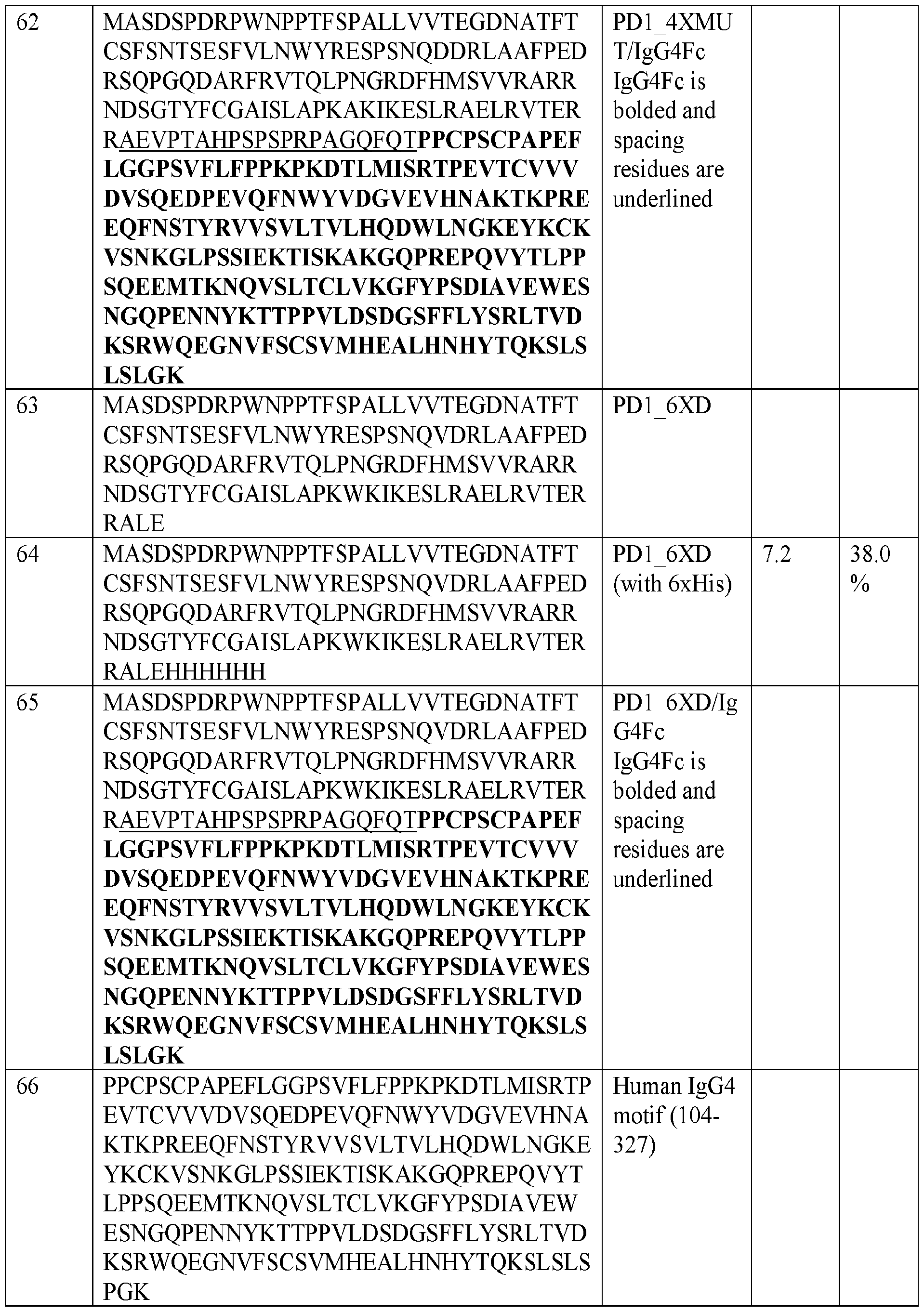
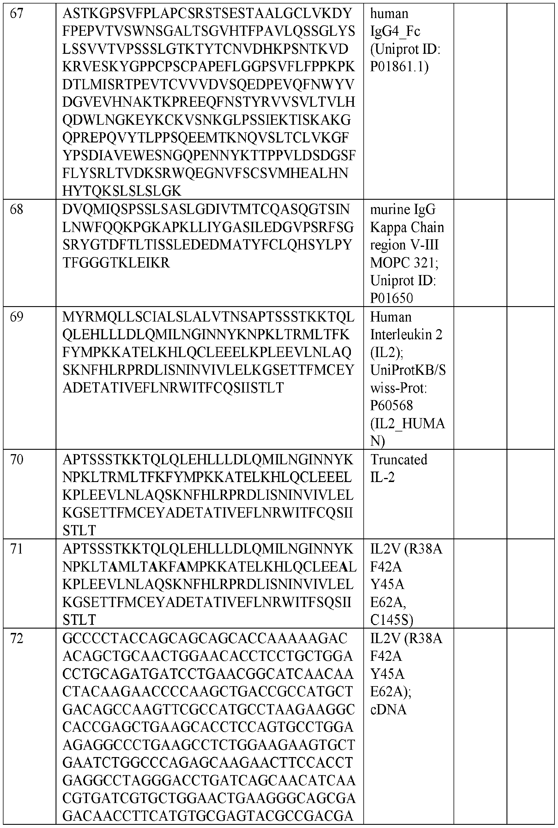


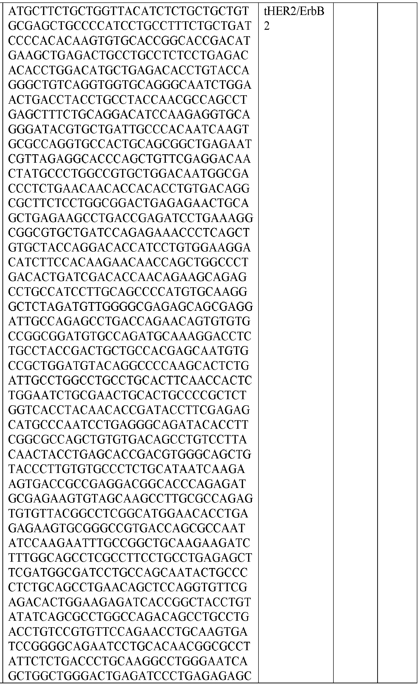


Claims
1. A programmed cell death 1 (PD-1) decoy polypeptide, comprising an amino acid sequence of a PD-1 variant, wherein the amino acid sequence has at least 80% identity to SEQ ID NO: 6 and comprises substitutions at least at positions T76, K78, and Q 133 of SEQ ID NO: 6.
2. The PD-1 decoy polypeptide of claim 1, wherein the substitutions comprise additional substitutions at least at one of positions L122 and A132.
3. The PD-1 decoy polypeptide of any one of the preceding claims, wherein the substitutions comprise:
(i) T76D or a conservative substitution of D76; or T76V or a conservative substitution V76;
(ii) K78R or a conservative substitution of R78; or
(iii) Q133K or a conservative substitution of K133.
4. The PD-1 decoy polypeptide of any one of the preceding claims, wherein the additional substitutions comprise at least one of:
(iv) L122E or a conservative substitution of E122; L122F or a conservative substitution of F122; or L122I or a conservative substitution of 1122; and
(v) A132W or a conservative substitution of W 132; A132L or a conservative substitution of LI 32; or A132Y or a conservative substitution of Y132.
5. The PD-1 decoy polypeptide of any one of the preceding claims, wherein the amino acid sequence comprises the substitutions at the positions at T76, K78, E122, A132, and Q133.
6. The PD-1 decoy polypeptide of any one of the preceding claims, wherein the amino acid sequence comprises the substitutions at the positions at T76, K78, E122, and Q133.
94
7. The PD-1 decoy polypeptide of any one of claims 1 to 5, wherein the substitutions comprise:
(a) T76D, K78R, L122F, and Q133K; or
(b) T76V, K78R, L122F, A132W, and Q133K.
8. The PD-1 decoy polypeptide of any one of the preceding claims, further comprising a fusion partner.
9. The PD-1 decoy polypeptide of claim 8, wherein the fusion partner comprises a fragment of a human immunoglobulin polypeptide sequence.
10. The PD-1 decoy polypeptide of claim 9, wherein the fragment comprises: (a) a CH3 domain; and (b) part or whole of an Fc region.
11. The PD-1 decoy polypeptide of claim 9, wherein the fragment comprises a human IgG Fc sequence.
12. The PD-1 decoy polypeptide of any one of the preceding claims, further comprising a cellular elimination tag (CET).
13. The PD-1 decoy polypeptide of claim 12, wherein the CET comprises a truncated EGFR (tEGFR) or a variant thereof, a truncated HER2 (tHER2) or a variant thereof, a CD20 or a variant thereof, or a CD 19 or a variant thereof.
14. The PD-1 decoy polypeptide of claim 13, wherein the fusion partner is linked to the C -terminus of the amino acid sequence of the PD-1 variant and the CET is linked to the C- terminus of the fusion partner.
15. The PD-1 decoy polypeptide of claim 1, comprising an amino acid sequence selected from SEQ ID NOs: 9-65, 90, 92, 95-96, and 99-100.
95
16. The PD-1 decoy polypeptide of claim 13, wherein: the tEGFR comprises an amino acid sequence having at least 80% identity to SEQ ID NO: 88 or comprises the amino acid sequence of SEQ ID NO: 88; the HER2 comprises an amino acid sequence having at least 80% identity to SEQ ID NO: 93 or comprises the amino acid sequence of SEQ ID NO: 93; and the CD20 comprises an amino acid sequence having at least 80% identity to SEQ ID NO: 98 or comprises the amino acid sequence of SEQ ID NO: 98.
17. The PD-1 decoy polypeptide of any one of the preceding claims, comprising a detectable label.
18. A polynucleotide comprising a polynucleotide sequence that encodes the PD-1 decoy polypeptide of any one of the preceding claims.
19. The polynucleotide of claim 18, further comprising: (a) a nucleotide sequence encoding an alpha polypeptide and a beta polypeptide of a T cell receptor (TCR); or (b) a nucleotide sequence encoding a chimeric antigen receptor (CAR).
20. A vector comprising the polynucleotide of any one of claims 18 to 19.
21. A cell comprising the polynucleotide of claim 18 or 19 or the vector of claim 20.
22. The cell of any one of claims 21 to 23, wherein the cell further comprises a second polynucleotide sequence encoding at least one transgene.
23. The cell of claim 22, wherein the transgene is selected from an IL-2 variant, LIGHT or a variant thereof, IL-33 or a variant thereof, and CD40L or a variant thereof.
24. The cell of claim 22, wherein the polynucleotide and the second polynucleotide are on the same vector.
25. The cell of any one of claims 21 to 24, wherein the cell is a lymphocyte.
96
26. The cell of claim 25, wherein the lymphocyte expresses:
(a) the PD-1 decoy or the variant thereof and tEGFR or the variant thereof;
(b) the PD-1 decoy or the variant thereof and the IL-2 variant;
(c) the PD-1 decoy or the variant thereof and the LIGHT or the variant thereof;
(d) the PD-1 decoy or the variant thereof and the IL-33 or the variant thereof;
(e) the PD-1 decoy or the variant thereof and the CD40L or the variant thereof;
(f) the PD-1 decoy or the variant thereof, the IL-2 variant, and the IL-33 or the variant thereof;
(g) the PD-1 decoy or the variant thereof, the tEGFR or the variant thereof, and the IL- 2 variant;
(h) the PD-1 decoy or the variant thereof, the tEGFR or the variant thereof, and the LIGHT or the variant thereof;
(i) the PD-1 decoy or the variant thereof, the tEGFR or the variant thereof, and the IL- 33 or the variant thereof;
(j) the PD-1 decoy or the variant thereof, the tEGFR or the variant thereof, and the CD40L or the variant thereof;
(k) the PD-1 decoy or the variant thereof, the tEGFR or the variant thereof, the IL-2 variant, and the IL-33 or the variant thereof;
(l) the PD-1 decoy or the variant thereof, the tEGFR or the variant thereof, the IL-2 variant, and the CD40L or the variant thereof; or
(m) the PD-1 decoy or the variant thereof, the tEGFR or the variant thereof, the IL-33 variant, and the CD40L or the variant thereof.
27. The cell of any one of claims 25 to 26, wherein the lymphocyte is autologous.
28. The cell of any one of claims 25 to 27, wherein the lymphocyte is a tumor-infdtrating lymphocyte (TIL).
29. The cell of any one of claims 25 to 28, wherein the lymphocyte expresses a chimeric antigen receptor (CAR) or a recombinant T cell receptor (TCR).
97
30. The cell of claim 29, wherein the recombinant T cell receptor (TCR) shows reactivity against NY-ESO1, MAGE-A1, MAGE-A3, MAGE A-10, MAGE-C2, SSX2, MAGE-A12, or a combination thereof.
31. A composition comprising: (i) the polypeptide of any one of claims 1 to 17; (ii) the polynucleotide of any one of claims 18 to 19; (iii) the vector of claim 20; or (iv) the cell of claims 21 to 30.
32. A pharmaceutical composition comprising an effective amount of the composition of claim 31 and a pharmaceutically acceptable carrier.
33. The pharmaceutical composition of claim 32, further comprising a second therapeutic agent.
34. A kit comprising an effective amount of the composition of claim 31 or the pharmaceutical composition of claim 32 or 33.
35. A method of treating a cancer or tumor in a subject, comprising administering to a subject in need thereof a therapeutically effective amount of the composition of claim 31 or the pharmaceutical composition of claim 32 or 33.
36. The method of claim 35, the cancer is selected from melanoma, sarcoma, ovarian cancer, prostate cancer, lung cancer, bladder cancer, MSI-high tumors, head and neck tumors, kidney cancer, and breast cancer.
37. The method of claim 35 or 36, further comprising administering to the subject a second therapeutic agent or therapy.
38. The method of claim 37, wherein the second therapeutic agent comprises an anti-cancer or anti-tumor agent.
98
Priority Applications (2)
| Application Number | Priority Date | Filing Date | Title |
|---|---|---|---|
| EP21892863.8A EP4243852A1 (en) | 2020-11-13 | 2021-11-12 | Pd-1 decoy variants for immunotherapy |
| US18/252,379 US20230414660A1 (en) | 2020-11-13 | 2021-11-12 | Pd-1 decoy variants for immunotherapy |
Applications Claiming Priority (2)
| Application Number | Priority Date | Filing Date | Title |
|---|---|---|---|
| US202063113651P | 2020-11-13 | 2020-11-13 | |
| US63/113,651 | 2020-11-13 |
Publications (1)
| Publication Number | Publication Date |
|---|---|
| WO2022104043A1 true WO2022104043A1 (en) | 2022-05-19 |
Family
ID=81601718
Family Applications (1)
| Application Number | Title | Priority Date | Filing Date |
|---|---|---|---|
| PCT/US2021/059119 WO2022104043A1 (en) | 2020-11-13 | 2021-11-12 | Pd-1 decoy variants for immunotherapy |
Country Status (3)
| Country | Link |
|---|---|
| US (1) | US20230414660A1 (en) |
| EP (1) | EP4243852A1 (en) |
| WO (1) | WO2022104043A1 (en) |
Cited By (2)
| Publication number | Priority date | Publication date | Assignee | Title |
|---|---|---|---|---|
| WO2022235832A1 (en) * | 2021-05-06 | 2022-11-10 | Ludwig Institute For Cancer Research Ltd | Compositions and methods for immunotherapy |
| EP4058035A4 (en) * | 2019-11-14 | 2023-12-27 | Ludwig Institute for Cancer Research Ltd | Compositions and methods for immunotherapy |
Citations (4)
| Publication number | Priority date | Publication date | Assignee | Title |
|---|---|---|---|---|
| US20160052990A1 (en) * | 2014-08-08 | 2016-02-25 | The Board Of Trustees Of The Leland Stanford Junior University | High affinity pd-1 agents and methods of use |
| US20180185668A1 (en) * | 2014-01-23 | 2018-07-05 | Regeneron Pharmaceuticals, Inc. | Human antibodies to pd-1 |
| WO2019241758A1 (en) * | 2018-06-15 | 2019-12-19 | Alpine Immune Sciences, Inc. | Pd-1 variant immunomodulatory proteins and uses thereof |
| US20190381184A1 (en) * | 2015-09-24 | 2019-12-19 | The University Of North Carolina At Chapel Hill | Methods and Compositions for Reducing Metastases |
-
2021
- 2021-11-12 US US18/252,379 patent/US20230414660A1/en active Pending
- 2021-11-12 WO PCT/US2021/059119 patent/WO2022104043A1/en unknown
- 2021-11-12 EP EP21892863.8A patent/EP4243852A1/en active Pending
Patent Citations (4)
| Publication number | Priority date | Publication date | Assignee | Title |
|---|---|---|---|---|
| US20180185668A1 (en) * | 2014-01-23 | 2018-07-05 | Regeneron Pharmaceuticals, Inc. | Human antibodies to pd-1 |
| US20160052990A1 (en) * | 2014-08-08 | 2016-02-25 | The Board Of Trustees Of The Leland Stanford Junior University | High affinity pd-1 agents and methods of use |
| US20190381184A1 (en) * | 2015-09-24 | 2019-12-19 | The University Of North Carolina At Chapel Hill | Methods and Compositions for Reducing Metastases |
| WO2019241758A1 (en) * | 2018-06-15 | 2019-12-19 | Alpine Immune Sciences, Inc. | Pd-1 variant immunomodulatory proteins and uses thereof |
Non-Patent Citations (1)
| Title |
|---|
| SHRESTHA ET AL.: "Computational Redesign of PD-1 Interface for PD-L1 Ligand Selectivity", STRUCTURE, vol. 27, no. 5, 7 May 2019 (2019-05-07), pages 829 - 836, XP055701602, DOI: 10.1016/j.str.2019.03.006 * |
Cited By (2)
| Publication number | Priority date | Publication date | Assignee | Title |
|---|---|---|---|---|
| EP4058035A4 (en) * | 2019-11-14 | 2023-12-27 | Ludwig Institute for Cancer Research Ltd | Compositions and methods for immunotherapy |
| WO2022235832A1 (en) * | 2021-05-06 | 2022-11-10 | Ludwig Institute For Cancer Research Ltd | Compositions and methods for immunotherapy |
Also Published As
| Publication number | Publication date |
|---|---|
| US20230414660A1 (en) | 2023-12-28 |
| EP4243852A1 (en) | 2023-09-20 |
Similar Documents
| Publication | Publication Date | Title |
|---|---|---|
| JP7157839B2 (en) | Combination of immunotherapy and cytokine control therapy for cancer treatment | |
| TWI792123B (en) | Chimeric receptors and methods of use thereof | |
| US11723962B2 (en) | Cell-based neoantigen vaccines and uses thereof | |
| CA3067602A1 (en) | Mouse model for assessing toxicities associated with immunotherapies | |
| JP2023154073A (en) | Methods of administering chimeric antigen receptor immunotherapy | |
| US20230414660A1 (en) | Pd-1 decoy variants for immunotherapy | |
| WO2020069508A1 (en) | Immunoresponsive cells expressing dominant negative fas and uses thereof | |
| CN110785488A (en) | Gene therapy | |
| JP2017533707A (en) | Compositions and methods for stimulating and expanding T cells | |
| US20220387555A1 (en) | Compositions and methods for immunotherapy | |
| EP3833682A1 (en) | Suicide module compositions and methods | |
| KR20220122639A (en) | Method for preparing a tumor-reactive T cell composition using a modulator | |
| CN115916961A (en) | T cells | |
| US10538568B2 (en) | Methods and compositions of a follicle stimulating hormone receptor immunoreceptor | |
| JP7057975B2 (en) | Chimeric antigen receptor gene-modified lymphocytes with cell-killing effect | |
| WO2020112815A1 (en) | Anti-lmp2 tcr-t cell therapy for the treatment of ebv-associated cancers | |
| JP2021536247A (en) | Neoantigen targeting DNA vaccines for combination therapy | |
| WO2023060214A1 (en) | Methods and compositions for enhancing activity of t cells with modified b cells | |
| KR20240024047A (en) | Cancer treatment method using NK cells and CD38 targeting antibodies | |
| JP2024509917A (en) | Method for selective stimulation of T cells in solid tumors using orthogonal IL-2 delivery by oncolytic viruses | |
| WO2022235832A1 (en) | Compositions and methods for immunotherapy | |
| US11913023B2 (en) | Modified B cells and methods of use thereof | |
| US20230042446A1 (en) | Adoptive cell therapy with zbtb20 suppression | |
| WO2021108648A2 (en) | Chimeric receptors to cea and methods of use thereof | |
| WO2023076523A1 (en) | Chimeric adaptor polypeptides |
Legal Events
| Date | Code | Title | Description |
|---|---|---|---|
| 121 | Ep: the epo has been informed by wipo that ep was designated in this application |
Ref document number: 21892863 Country of ref document: EP Kind code of ref document: A1 |
|
| NENP | Non-entry into the national phase |
Ref country code: DE |
|
| ENP | Entry into the national phase |
Ref document number: 2021892863 Country of ref document: EP Effective date: 20230613 |