EP1820513A1 - Destruction of tumor cells expressing low to medium levels of tumor associated target antigens by trifunctional bispecific antibodies - Google Patents
Destruction of tumor cells expressing low to medium levels of tumor associated target antigens by trifunctional bispecific antibodies Download PDFInfo
- Publication number
- EP1820513A1 EP1820513A1 EP06003057A EP06003057A EP1820513A1 EP 1820513 A1 EP1820513 A1 EP 1820513A1 EP 06003057 A EP06003057 A EP 06003057A EP 06003057 A EP06003057 A EP 06003057A EP 1820513 A1 EP1820513 A1 EP 1820513A1
- Authority
- EP
- European Patent Office
- Prior art keywords
- tumor
- antibody
- cells
- human
- her2
- Prior art date
- Legal status (The legal status is an assumption and is not a legal conclusion. Google has not performed a legal analysis and makes no representation as to the accuracy of the status listed.)
- Withdrawn
Links
Images


Classifications
-
- C—CHEMISTRY; METALLURGY
- C07—ORGANIC CHEMISTRY
- C07K—PEPTIDES
- C07K16/00—Immunoglobulins [IGs], e.g. monoclonal or polyclonal antibodies
- C07K16/18—Immunoglobulins [IGs], e.g. monoclonal or polyclonal antibodies against material from animals or humans
- C07K16/28—Immunoglobulins [IGs], e.g. monoclonal or polyclonal antibodies against material from animals or humans against receptors, cell surface antigens or cell surface determinants
- C07K16/2887—Immunoglobulins [IGs], e.g. monoclonal or polyclonal antibodies against material from animals or humans against receptors, cell surface antigens or cell surface determinants against CD20
-
- A—HUMAN NECESSITIES
- A61—MEDICAL OR VETERINARY SCIENCE; HYGIENE
- A61P—SPECIFIC THERAPEUTIC ACTIVITY OF CHEMICAL COMPOUNDS OR MEDICINAL PREPARATIONS
- A61P35/00—Antineoplastic agents
-
- A—HUMAN NECESSITIES
- A61—MEDICAL OR VETERINARY SCIENCE; HYGIENE
- A61P—SPECIFIC THERAPEUTIC ACTIVITY OF CHEMICAL COMPOUNDS OR MEDICINAL PREPARATIONS
- A61P35/00—Antineoplastic agents
- A61P35/04—Antineoplastic agents specific for metastasis
-
- C—CHEMISTRY; METALLURGY
- C07—ORGANIC CHEMISTRY
- C07K—PEPTIDES
- C07K16/00—Immunoglobulins [IGs], e.g. monoclonal or polyclonal antibodies
- C07K16/18—Immunoglobulins [IGs], e.g. monoclonal or polyclonal antibodies against material from animals or humans
- C07K16/28—Immunoglobulins [IGs], e.g. monoclonal or polyclonal antibodies against material from animals or humans against receptors, cell surface antigens or cell surface determinants
- C07K16/2803—Immunoglobulins [IGs], e.g. monoclonal or polyclonal antibodies against material from animals or humans against receptors, cell surface antigens or cell surface determinants against the immunoglobulin superfamily
- C07K16/2809—Immunoglobulins [IGs], e.g. monoclonal or polyclonal antibodies against material from animals or humans against receptors, cell surface antigens or cell surface determinants against the immunoglobulin superfamily against the T-cell receptor (TcR)-CD3 complex
-
- C—CHEMISTRY; METALLURGY
- C07—ORGANIC CHEMISTRY
- C07K—PEPTIDES
- C07K16/00—Immunoglobulins [IGs], e.g. monoclonal or polyclonal antibodies
- C07K16/18—Immunoglobulins [IGs], e.g. monoclonal or polyclonal antibodies against material from animals or humans
- C07K16/28—Immunoglobulins [IGs], e.g. monoclonal or polyclonal antibodies against material from animals or humans against receptors, cell surface antigens or cell surface determinants
- C07K16/30—Immunoglobulins [IGs], e.g. monoclonal or polyclonal antibodies against material from animals or humans against receptors, cell surface antigens or cell surface determinants from tumour cells
- C07K16/3076—Immunoglobulins [IGs], e.g. monoclonal or polyclonal antibodies against material from animals or humans against receptors, cell surface antigens or cell surface determinants from tumour cells against structure-related tumour-associated moieties
- C07K16/3084—Immunoglobulins [IGs], e.g. monoclonal or polyclonal antibodies against material from animals or humans against receptors, cell surface antigens or cell surface determinants from tumour cells against structure-related tumour-associated moieties against tumour-associated gangliosides
-
- C—CHEMISTRY; METALLURGY
- C07—ORGANIC CHEMISTRY
- C07K—PEPTIDES
- C07K16/00—Immunoglobulins [IGs], e.g. monoclonal or polyclonal antibodies
- C07K16/18—Immunoglobulins [IGs], e.g. monoclonal or polyclonal antibodies against material from animals or humans
- C07K16/32—Immunoglobulins [IGs], e.g. monoclonal or polyclonal antibodies against material from animals or humans against translation products of oncogenes
-
- A—HUMAN NECESSITIES
- A61—MEDICAL OR VETERINARY SCIENCE; HYGIENE
- A61K—PREPARATIONS FOR MEDICAL, DENTAL OR TOILETRY PURPOSES
- A61K39/00—Medicinal preparations containing antigens or antibodies
- A61K2039/505—Medicinal preparations containing antigens or antibodies comprising antibodies
-
- A—HUMAN NECESSITIES
- A61—MEDICAL OR VETERINARY SCIENCE; HYGIENE
- A61K—PREPARATIONS FOR MEDICAL, DENTAL OR TOILETRY PURPOSES
- A61K39/00—Medicinal preparations containing antigens or antibodies
- A61K2039/55—Medicinal preparations containing antigens or antibodies characterised by the host/recipient, e.g. newborn with maternal antibodies
-
- C—CHEMISTRY; METALLURGY
- C07—ORGANIC CHEMISTRY
- C07K—PEPTIDES
- C07K2317/00—Immunoglobulins specific features
- C07K2317/20—Immunoglobulins specific features characterized by taxonomic origin
- C07K2317/24—Immunoglobulins specific features characterized by taxonomic origin containing regions, domains or residues from different species, e.g. chimeric, humanized or veneered
-
- C—CHEMISTRY; METALLURGY
- C07—ORGANIC CHEMISTRY
- C07K—PEPTIDES
- C07K2317/00—Immunoglobulins specific features
- C07K2317/30—Immunoglobulins specific features characterized by aspects of specificity or valency
- C07K2317/31—Immunoglobulins specific features characterized by aspects of specificity or valency multispecific
-
- C—CHEMISTRY; METALLURGY
- C07—ORGANIC CHEMISTRY
- C07K—PEPTIDES
- C07K2317/00—Immunoglobulins specific features
- C07K2317/70—Immunoglobulins specific features characterized by effect upon binding to a cell or to an antigen
- C07K2317/73—Inducing cell death, e.g. apoptosis, necrosis or inhibition of cell proliferation
Definitions
- the present invention refers to the use of trifunctional bispecific antibodies for the preparation of a pharmaceutical composition for the prophylaxis and treatment of tumor diseases.
- a hitherto unsolved problem in the treatment of tumors by monospecific monoclonal antibodies during an immune therapy is the insufficient efficiency of tumor cell destruction with low to medium expression levels of tumor target antigens.
- One particular example is the treatment of breast cancer with a low expression level of the target antigen Her2/neu.
- Metastatic breast cancer is an almost always fatal disease.
- the median survival time from first manifestation of metastases ranges from 17 to 20 months.
- a number of endocrine, cytotoxic and biological agents have demonstrated palliative efficacy but there is no consensual standard of care, and treatment often causes substantial adverse effects.
- Epidermal growth factor (EGF) family member Her2/neu is overexpressed in tumor specimens of approximately 25-30% of breast cancer patients and attributed to more aggressive tumor growth and a worse prognosis.
- EGF receptors includes four members EGFR (ERBB1), Her2/neu (ERBB2), ERBB3 and ERBB4.
- EGFR and HER-2/neu have been intensely pursued as therapeutic targets.
- Antibodies targeting the extra cellular domain of EGFR and Her2/neu as well as small molecular compounds inhibiting intracellular receptor signalling are already in clinical use and have demonstrated clinical efficacy. However, their anti-tumor effects are often not as strong as predicted from pre-clinical studies and combination with chemotherapy is preferable.
- Herceptin ® is a well accepted monoclonal antibody widely used for the treatment of breast cancer; however, Herceptin ® can be used only for patients with an expression level of the target antigen Her2/neu of at least 2+ (as verified by the so-called HercepTest and a positive FISH-analysis) or 3+ on their tumor cells.
- Herceptin ® can be used only for patients with an expression level of the target antigen Her2/neu of at least 2+ (as verified by the so-called HercepTest and a positive FISH-analysis) or 3+ on their tumor cells.
- Several studies have shown that a relatively high expression density of the target antigen Her2/neu appears to be necessary in order to obtain a statistically significant survival.
- Pre-clinical in vitro experiments have also shown that an efficient destruction of tumor cells by Herceptin is visible only when tumor cells express a high level of the target antigen Her2/neu.
- Her2/neu are also valid for other tumor-associated antigens like e.g. CD20, EpCAM, G250, GD3, GD2, proteoglycans, MHC II, EGF-R and CEA.
- tumor-associated antigens like e.g. CD20, EpCAM, G250, GD3, GD2, proteoglycans, MHC II, EGF-R and CEA.
- bispecific antibodies are tools for immunological treatment of e.g. malignant cells by redirecting effector cells against tumor cells.
- the bsAbs described to date normally redirect and activate only a single class of effector cells, i.e., either T-cells, NK-cells, Fc ⁇ RI + , or Fc ⁇ RI + cells, thereby limiting their efficacy.
- trifunctional bispecific antibodies having the following properties:
- the present inventors could surprisingly show that trifunctional bispecific antibodies already described in the art can be efficiently used for targeting particularly selected tumor associated target antigens on a tumor cell which are expressed on said tumor cell to an only low to medium level.
- Particularly preferred are Her2/neu and CD20 tumor associated antigens as target antigens.
- the tumor associated antigens are expressed on the tumor cells preferably in amount of at least 10,000, 20,000, 50,000 or 80,000 tumor associated antigens/tumor cell and at a maximum of 120,000, 110,000 or 100,000 tumor associated antigens/tumor cell.
- tumor-associated target antigens selected by the present inventors are permanently and stably expressed on the tumor cell with low to medium expression levels only. While other type of inducible antigens like heatshock proteins and MIC molecules MIC A and MIC B, which are both under the control of heatshock promoter elements, are expressed after induction with increasing levels during lifetime, the expression rate of tumor-associated antigens like Her2/neu and CD20 is generally constant and stable
- EpCAM is typically associated with adeno carcinomas, Her2/neu with mamma carcinomas but also with colon, lung, gastric, pancreas and ovarian cancer, CD20 with B cell lymphomas, G250 with renal carcinomas, proteoglycans, GD3 and GD2 with melanomas, MHC II with B cell lymphomas and EGF-R and CEA with epithelial tumors.
- trifunctional bispecific antibodies used in the present invention are known per se. Reference is made for instance to US-6,551,592 the content of which is fully incorporated herein by reference. The same is true for DE-A-196 49 223 and DE-A-197 10 495 , which are also included herein by reference together with their corresponding US patent documents.
- the antibodies for use in the invention may be administered orally in any acceptable dosage form such as capsules, tablets, aqueous suspensions, solution or the like.
- the antibodies and derivatives thereof may also be administered parenterally. That is via the following routes of administration: subcutaneous, intravenous, intraperitoneal, intramuscular, intra-articular, intra-synovial, intrastemal, intranasal, topically, intrathecal, intrahepatic, intralesional, and intracranial injection or infusion techniques.
- the antibodies will be provided as an intravenous injection or infusion.
- the antibodies of the invention may be administered alone or with a pharmaceutically acceptable carrier, including acceptable adjuvants, vehicles, and excipients.
- the effective dosage will depend on a variety of factors and it is well within the purview of a skilled physician to adjust the dosage for a given patient according to various parameters such as body weight, the goal of treatment, the highest tolerated dose, the specific formulation used, the route of administration, the response of the patient and the like.
- the trAbs employed according to the present invention are preferably administered in an amount of 5 - 1000 ⁇ g, further preferred 10 - 300 ⁇ g, 10 - 100 ⁇ g or 10 - 50 ⁇ g, each per infusion.
- the optimal amounts may be determined by the skilled artisan by means of experimentation.Further preferred, the trifunctional antibody is used in an amount of 0.05-15 ⁇ g/kg, further preferred 0.5-5 ⁇ g/kg and 0.5-2 ⁇ g/kg body weight.
- said trifunctional antibody is selected to be an anti-CD3 X anti-tumor-associated antigen antibody and/or anti-CD4 X anti-tumor-associated antigen antibody and/or anti-CD5 X anti-tumor-associated antigen antibody and/or anti-CD6 X anti-tumor-associated antigen antibody and/or anti-CD8 anti-tumor-associated antigen antibody and/or anti-CD2 X anti-tumor-associated antigen antibody and/or anti-CD28 X anti-tumor-associated antigen antibody and/or anti-CD44 X anti-tumor-associated antigen antibody, wherein particularly preferred an anti-CD3 x anti-tumor associated antigen antibody is used.
- the anti-tumor associated antigen is a Her2/neu or CD20 antigen.
- the Fc-receptor positive cells are activated by binding of the Fc-portion of the trAb to the Fc-receptor positive cell., Thereby, the expression of cytokines and/or co-stimulatory antigens is initiated or increased. Then, at least a second activation signal required for physiological activation of the T cell is transferred to said T cell by said co-stimulatory antigens and/or cytokines. This activation is indicated by up-regulation of activation markers, the killing of tumor cells and by the proliferation of T cells.
- Activation of the Fc receptor-positive cell by the trAb is dependent on the subclass or the subclass combination of the antibody heavy chain fragments, respectively.
- trAbs of the mouse-IgG2a/rat-IgG2b subclass combination are able to bind to, and simultaneously activate, Fc receptor-positive cells leading to an up-regulation or new formation (expression), respectively, of co-stimulatory antigens such as CD40, CD80, or CD86 on the surface of these cells, while bispecific antibodies of the mouse-IgG1/rat-IgG2b subclass combination are able to bind to Fc receptor-positive cells ((1) Haagen et al., J.
- mouse-IgG2a/ratIgG2b isotype combination in the Fc-region of the trAb is particularly preferred.
- the trAb simultaneously binds to and activates the T cell with one binding arm (e.g. anti-CD3), co-stimulatory signals from the Fc receptor-positive cell bound to the Fc portion of the trAb may be transferred to the T cell. I.e., only the combination of T cell activation via one binding arm of the trAb and simultaneous transfer of co-stimulatory signals from the Fc receptor-positive cell to the T cell leads to an efficient T cell activation.
- one binding arm e.g. anti-CD3
- tumor-specific CD4- Another important aspect in the induction of an antitumor immunity is the possible phagocytosis, processing and presentation of tumor components by the accessory cells (monocytes/macrophages, or dendritic cells) which have been targeted by the trAb.
- accessory cells monocytes/macrophages, or dendritic cells
- tumor-specific CD4 cells play an important role in the induction of a humoral immune response in the context of T/B cell cooperation.
- Trifunctional bispecific antibodies are able to bind to the T cell receptor complex of the T cell with one binding arm and to said tumor-associated antigens on the tumor cell with the second binding arm. Thereby, they activate T cells which destroy the tumor cells by releasing cytokines or by apoptosis-mediating mechanisms. Moreover, there seems to be the possibility that in the frame of activation by trifunctional antibodies T cells which recognize tumor-specific antigens via their receptor may be re-activated, whereby leading to a long-lasting antitumor immunity.
- the intact Fc portion of the trifunctional bispecific antibody mediating the binding to accessory cells such as monocytes/macrophages and dendritic cells and causing them to develop cytotoxicity themselves and/or concomitantly transfer important co-stimulatory signals to the T cell.
- accessory cells such as monocytes/macrophages and dendritic cells
- cytotoxicity themselves and/or concomitantly transfer important co-stimulatory signals to the T cell.
- tumor-specific T cells By redirecting possibly anergized tumor-specific T cells to tumor cells by means of trAbs and simultaneous co-stimulation of such T cells by accessory cells binding to the Fc portion of the trAb the anergic state of tumor-specific cytotoxic T cells (CTLs) could be abolished.
- CTLs tumor-specific cytotoxic T cells
- the antibodies employed according to the present invention are capable of reactivating tumor-specific T cells being in a state of anergy. Furthermore, they are capable of inducing tumor-reactive complement-binding antibodies and therefore to induce a humoral immune response.
- Binding to the T cell preferably takes place via CD3, CD2, CD4, CD5, CD6, CD8, CD28, and/or CD44.
- the Fc receptor-positive cells have at least one Fc ⁇ receptor I or III.
- Antibodies which may be employed according to the present invention are able to bind to monocytes, macrophages, dendritic cells, "natural killer” cells (NK cells) and/or activated neutrophils being Fc ⁇ receptor I and/or III-positive cells.
- the antibodies which may be employed according to the invention lead to the initiation or increase of the expression of CD40, CD80, CD86, ICAM-1, and/or LFA-3 being co-stimulatory antigens, or/and secretion of cytokines by the Fc receptor-positive cell.
- the cytokines are IL-1, IL-2, IL-4, IL-6, IL-8, IL-12, INF- ⁇ and/or TNF- ⁇ .
- binding to the T cell takes place via the T cell receptor complex of the T cell.
- the trifunctional bispecific antibody is a heterologous intact rat/mouse bispecific antibody.
- T cells are activated and redirected against the tumor cells.
- Preferred useful heterologous trifunctional bispecific antibodies are selected from one or more of the following combinations of isotypes:
- the antibodies useful according to the present invention are monoclonal, chimeric, recombinant, synthetic, semi-synthetic, or chemically modified intact antibodies having for example Fv, Fab, scFv, or F(ab) 2 fragments.
- antibodies or derivatives or fragments thereof of human origin are used, or those modified to be suitable for the use in humans (so-called "humanized antibodies”) (see for example Shalaby et al., J. Exp. Med. 175 (1992), 217 ; Mocikat et al., Transplantation 57 (1994), 405 ).
- the preparation of the different types of antibodies and antibody fragments mentioned above is well-known to the skilled artisan.
- the preparation of monoclonal antibodies, preferably of mammalian origin, e.g. of human, rat, mouse, rabbit, or goat, can be performed using conventional methods as those described for example in Köhler und Milstein ( Nature 256 (1975), 495 ), in Harlow and Lane (Antibodies, A Laboratory Manual (1988), Cold Spring Harbor ) or in Galfré (Meth. Enzymol. 73 (1981), 3 ) or in DE 195 31 346 .
- bispecific antibodies can be performed on the one hand using recombinant DNA technology but on the other hand also by the so-called hybrid hybridoma fusion technique (see for example Milstein et al., Nature 305 (1983), 537 ).
- This technique includes the fusion of hybridoma cell lines each producing antibodies with one of the desired specificities and identifying and isolating recombinant cell lines producing antibodies with both specificities.
- Trifunctional bispecific antibodies falling under the present invention belong to the prior art, and references describing such methods of preparation are incorporated herein by reference in their entirety.
- Trifunctional bispecific antibodies are composed of two antibody semi-molecules (each having a H and a L immunoglobulin chain) each representing a specificity, and having a Fc portion like normal antibodies which performs the well-known effector functions. They are preferably prepared using the quadroma technology. This method of preparation is exemplified in DE-A-44 19 399 . For complete disclosure this document is incorporated by reference in its entirety also with respect to a definition of bispecific antibodies. It should be understood that also other methods of preparation are useful if they lead to the trifunctional bispecific antibodies according to the above definition which are required according to the present invention.
- the binding of the trAb to Fc ⁇ -RI shows two essential advantages with regard to an optimal anti-tumor effectivity:
- Trifunctional antibodies as used in the present invention are able to target and simultaneously activate different types of immune cells by their anti-T cell binding arm and their Fc-portion. Typical examples of these immune cells are T lymphocytes and accessory cells with Fc ⁇ -receptor types I (CD64) and III (CD16). While the first binding arm binds to the target antigen on the tumor cell like Her2/neu or CD20, the second binding arm binds to e.g. CD3 on T lymphocytes (figure 4).
- Methods on how to quantify the expression level of a tumor antigen are known in the art. E.g. immunocytochemical methods like e.g. ELISA tests, flow cytometry tissue staining methods and cytospin analysis.
- a typical example for a quantitative determination method for the expression of tumor associated target antigens on a tumor cell is an immunohistochemical method like the HercepTest ® developed by DAKO, Glostrup, Denmark allowing the quantitative determination of the expression level of Her2/neu on tumor tissues.
- Her2/neu comprising four steps 0, 1+, 2+, 3+ referring to an approximate number of expressed target antigens on the surface of a tumor cell.
- cells with an expression of less than 20,000 Her2/neu molecules on the target cell surface are classified as negative; cells with an expression of more than about 20,000 and up to about 100,000-110,000 molecules are classified as 1+, cells with an expression of up to about 500,000 molecules as 2+, and cells with an expression of between about 2.000,000 to 10.000,000 molecules are classified as 3+.
- trifunctional bispecific antibodies directed against Her2/neu can be used for the treatment of patients having tumor cells classified as Her2/neu 1+ in accordance with the HercepTest ® .
- the example illustrates the present invention with respect to the trifunctional antibody available under the trademark Rexomun® (INN-name ertumaxomab) and the tumor associated antigen Her2/neu targeted by Rexomun®.
- Rexomun® INN-name ertumaxomab
- Her2/neu targeted by Rexomun® This example provides convincing evidence that the invention can be used with respect to all tumor associated antigens as specified in claim 1 and throughout the specification. All these tumor associated antigens may be present on the surface of the tumor cell in such a low number so that they can hardly be treated by conventionally used antibody-based immune therapy.
- Rexomun ® (produced by Trion Pharma, Kunststoff) is a trifunctional antibody which targets with one binding arm the tumor associated antigen Her2/neu and binds with the other arm the CD3 molecule on T cells. Furthermore Rexomun ® binds Fc ⁇ RI and Fc ⁇ RIII positive cells (e.g. macrophages, NK cells, dendritic cells) with its potent Fc region consisting of mouse IgG2a and rat IgG2b isotypes. Monoclonal antibody 2502A (Trion Pharma, Kunststoff) is specific for Her2/neu and is one of the parental antibodies which are included in Rexomun ® .
- Fc ⁇ RI and Fc ⁇ RIII positive cells e.g. macrophages, NK cells, dendritic cells
- Monoclonal antibody 2502A (Trion Pharma, Kunststoff) is specific for Her2/neu and is one of the parental antibodies which are included in Rexomun ® .
- 26II6 (Trion Pharma, Kunststoff) is specific for CD3 and is the other parental antibody which is included in Rexomun ® .
- 520C9 (ATCC HB-8696) is another monoclonal antibody recognizing the Her2/neu receptor.
- Trastuzumab is a humanized anti Her2 monoclonal antibody developed from the murine anti Her2/neu antibody Mab 4D5 (7). Table 1 shows the characteristics of the used antibodies and their corresponding detection antibodies.
- SKBR3 (ATCC H TB-30) is a breast cancer cell line with a strong overexpression of Her2/neu (HercepTest ® score +3) detected by IHC (4).
- HCT-8 (ATCC CCL-244) is a colon carcinoma cell line and Lox is a melanoma cell line (table 2).
- Her2/neu status of HCT-8 cells was assessed by IHC using the HercepTestTM kit (DAKO, Glostrup, Denmark) and was performed by the Institute of Pathology, LMU Kunststoff according to the manufacturer's manual.
- FACS analysis was performed with 5x10 5 target cells (SKBR3, HCT-8 or Lox). Cells were incubated with 100 ng monoclonal antibodies or 100 ng of the trifunctional antibody Rexomun ® for 45 min at 4°C. Samples were then washed with phosphate buffered saline (PBS) without Mg 2+ and Ca 2+ (PAN Biotec, Aidenbach) and incubated with the corresponding detection antibodies (Dianova, Hamburg) (Table 1) for 45 min at 4°C. After another washing step with PBS, cells were resuspended in PBS containing 0.01 ⁇ g/ml ethidiumbromide (Sigma, Kunststoff).
- Amount of the antibody binding was analysed by mean fluorescence intensity in an overlay histogram.
- a sample with the anti human detection antibody alone or a sample incubated with 26II6 (anti CD3) and its corresponding detection antibody (isotype control) served as negative controls. All FACS analysis was carried out on a FACSCalibur (Becton Dickinson, Heidelberg) and was analysed with Cell Quest Pro or WinMDI 2.8.
- Antibody-meditated tumor elimination was investigated with a long-term clonogenic assay with unstimulated effector cells. Therefore, 10 6 PBMC were spiked with 5% target cells (SKBR3, HCT-8 or Lox) and seeded in 24 well plates (Greiner). Antibodies were added in different concentrations and combinations which are shown in Table 3 . Samples of target cells alone and samples consisting of a mixture of target and effector cells without addition of any antibodies served as controls. Samples were incubated at 37°C / 5% CO 2 and were fed every 3 days with RPMI 1640, L- Gutamine and 10% FCS.
- SKBR3 and HCT-8 cells were stained with 1.5 ⁇ g/slide anti Cytokeratine 8, 18, 19 antibody (A45B-B3, mouse IgG1, Micromet, Kunststoff), Lox cells were stained with 1,5 ⁇ g/slide anti EGFR antibody (mouse IgG2a, kindly provided by Dr. Htun, Kunststoff).
- the anti-mouse IgG 1 Alexa Fluor 477 (FITC) antibody (Molecular probes, Eugene) for cytokeratine staining or the anti-mouse IgG2a Alexa Fluor 477 (FITC) antibody (Molecular probes, Eugene) for EGFR staining were used at 1.5 ⁇ g/slide as detection antibodies. Cytospin slides were analysed in a Micrometastasis detection system (MDS, Applied Imaging) with a computerized image analysis counting FITC labelled cells.
- MDS Micrometastasis detection system
- Colon carcinoma cell line HCT-8 shows a +1 Her2/neu expression
- Her2/neu expression was investigated on different cell lines, in order to find a cell line that has a +1 expression of Her2/neu.
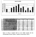
- FIG. 1 A-F shows the binding profiles of the used antibodies on SKBR3, HCT-8 or Lox cells. As controls served flow cytometry samples with the different detection antibodies only ( Figure 1A), isotype control was 26II6 (anti CD3) with its corresponding detection antibody ( Figure 1B).
- the melanoma cell line Lox - as expected - showed no expression for the Her2/neu receptor ( Figure 1 C-F), whereas SKBR3 cells were stained heavily against Her2/neu with the monoclonal antibodies 520C9 (Figure 1C), 2502A ( Figure 1D) and Trastuzumab (Figure 1E).
- Rexomun ® stained SKBR3 cells to a lower extent, probably because of its one arm binding nature (Figure 1F).
- HCT-8 cells compared to SKBR3 cells have a significantly weaker expression of the Her2/neu receptor with all used Her2 antibodies ( Figure 1 C-F), justifying further evaluation of these cells.
- Her2/neu expression of HCT-8 cells was in addition analysed with the DAKO HercepTest ® .
- the test was performed by the Institute of Pathology (TU Munich).
- the colon carcinoma cell line HCT-8 showed a +1 expression of Her2/neu in the performed DAKO test, confirming the results of the FACS analysis.
- Table 2 shows a summary of used cell lines, their Her2/neu score and the methods applied for determination of Her2 expression.
- Rexomun ® is able to kill Her2/neu +3 and Her2/neu +1 cell lines in vitro
- Rexomun ® 's ability to simultaneously recruit and activate different types of immune cells to the tumor site leaded to the hypothesis, that Rexomun ® might also be able to eliminate Her2/neu (1+) low expressing tumor cells.
- cytotoxicity assay (clonogenic assay) was established.
- PBMC of 3 healthy donors were co-incubated either with the Her2/neu +3 expressing cell line SKBR-3, the Her2/neu-negative Lox cell line or the Her2/neu +1 expressing HCT-8 cell line for 8 days.
- Different antibody concentrations of Trastuzumab (100 ⁇ g/ml - 1 ⁇ g /ml) or Rexomun ® (100 ng/ml - 1 ng/ml) were added as shown in Table 3.
- Tumor cell killing was evaluated after 8 days by harvesting the cells, transferring them on cytospin slides and staining the cells with anti CK 8, 18, 19 antibody (SKBR-3 and HCT-8) or anti EGFR-antibody (Lox) and the corresponding Alexa-Fluor FITC antibodies.
- the stained cytospin slides were then evaluated by a computerized image analysis using the SLIDE SCAN programm on the Micrometastasis Detection System (MDS, Applied Imaging, UK).
- the trifunctional antibody Rexomun ® (anti Her2/neu x anti CD3) was compared to the humanized anti Her2/neu monoclonal antibody Trastuzumab ® in its ability to inhibit tumor cell growth of a low Her2/neu expressing tumor cell line HCT-8 (HercepTest score: +1).
- Her2/neu expression on the cell lines SKBR3 (+ 3 Hercep score), HCT-8 (+1 Hercep score) and Lox (Her2/neu negative) were measured by FACS analysis, whereas HCT-8 cells were also tested via HercepTest ® .
- FACS analysis with various antibodies against Her2/neu showed strong Her2 expression on SKBR3 cells, a weaker expression on HCT-8 cells and no Her2/neu expression on Lox cells. These results correspond with the +1 HercepTest ® score of the HCT-8 cells.
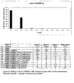
- Tumor cell growth of the Her2 +3 cell line SKBR3 was inhibited as expected by addition of 100 ⁇ g, 50 ⁇ g, 10 ⁇ g and 1 ⁇ g Trastuzumab ® ( Figure 2a, samples A4-A8). Furthermore, Rexomun ® was also able to inhibit tumor growth by addition of only 100 ng, 50 ng, 10 ng or Ing. As shown in samples A9-A12 ( Figure 2a) tumor cells growth was inhibited more efficient than with Trastuzumab ® .
- Tumor cell growth of the Her2/neu negative cell line Lox was neither inhibited by addition of various amounts of Trastuzumab ® nor Rexomun ® indicating the specificity of the tumor cell elimination driven by the presence of the Her2/neu receptor ( Figure 2c, sample C4-C12).
- the growth of the colon carconima cell line HCT-8 (Her2/neu score +1) was not inhibited by various amounts (100 ⁇ g - 1 ⁇ g) of Trastuzumab ® ( Figure 2b, sample B4-B8) which corresponds to previous findings with the Her2/neu +1 mamma carcinoma cell line MCF-7 (5). Since MCF-7cells (ATCC HTB-22) show growth inhibition to TNF- ⁇ these cells could not be used in the clonogenic assays, as rexomun ® like the trifunctional antibody removab ® (anti EpCAM x anti CD3) triggers TNF- ⁇ secretion, because of its activation properties (8).
- Rexomun® however, was able to destroy the +1 Her2/neu cell line HCT-8 in concentrations as low as 100 ng, 50 ng and 10 ng / well ( Figure 2b sample B9-B11). 1 ng of Rexomun ® did not succeed to inhibit tumor cell growth of HCT-8 cells ( Figure 2b , sample B12).
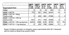
- Figure 3 shows the original MDS data plots for clonogenic assays of donor 2 using HCT-8 cells as target.
- Rexomun ® is a therapeutic option not only for mamma carcinoma patients having +3 or +2 overexpression/gene amplification, but also for patients with low Her2/neu expression levels.
Abstract
Description
- The present invention refers to the use of trifunctional bispecific antibodies for the preparation of a pharmaceutical composition for the prophylaxis and treatment of tumor diseases.
- A hitherto unsolved problem in the treatment of tumors by monospecific monoclonal antibodies during an immune therapy is the insufficient efficiency of tumor cell destruction with low to medium expression levels of tumor target antigens. One particular example is the treatment of breast cancer with a low expression level of the target antigen Her2/neu.
- Metastatic breast cancer is an almost always fatal disease. The median survival time from first manifestation of metastases ranges from 17 to 20 months. A number of endocrine, cytotoxic and biological agents have demonstrated palliative efficacy but there is no consensual standard of care, and treatment often causes substantial adverse effects.
- Epidermal growth factor (EGF) family member Her2/neu is overexpressed in tumor specimens of approximately 25-30% of breast cancer patients and attributed to more aggressive tumor growth and a worse prognosis.
- The family of EGF receptors includes four members EGFR (ERBB1), Her2/neu (ERBB2), ERBB3 and ERBB4. EGFR and HER-2/neu have been intensely pursued as therapeutic targets. Antibodies targeting the extra cellular domain of EGFR and Her2/neu as well as small molecular compounds inhibiting intracellular receptor signalling are already in clinical use and have demonstrated clinical efficacy. However, their anti-tumor effects are often not as strong as predicted from pre-clinical studies and combination with chemotherapy is preferable.
- Herceptin® is a well accepted monoclonal antibody widely used for the treatment of breast cancer; however, Herceptin® can be used only for patients with an expression level of the target antigen Her2/neu of at least 2+ (as verified by the so-called HercepTest and a positive FISH-analysis) or 3+ on their tumor cells. Several studies have shown that a relatively high expression density of the target antigen Her2/neu appears to be necessary in order to obtain a statistically significant survival. Pre-clinical in vitro experiments have also shown that an efficient destruction of tumor cells by Herceptin is visible only when tumor cells express a high level of the target antigen Her2/neu.
- These limitations are responsible that only about 25-30% of the patients with metastatic breast cancer which express high levels of Her2/neu on their tumors (scored as 2+ or 3+ with the Dako HercepTest) can be efficiently treated with Herceptin.
- In a study of Perez and co-workers, 35% of breast cancer patients were scored as
Her2 1+, 14% as 2+, and 13% as 3+ evaluated with immunohistochemistry (IHC)(Perez EA et al.: Her2 testing in patients with breast cancer: poor correlation between weak positivity by immunohistochemistry and gene amplification by fluorescence in situ hybridization. Mayo Clin Proc 2002; 77: 148-154) - The limitations explained above for Her2/neu are also valid for other tumor-associated antigens like e.g.
CD20, EpCAM, G250, GD3, GD2, proteoglycans, MHC II, EGF-R and CEA. - Therefore, there is a need for improvement of antitumor activity of e.g. antibodies which can also kill Her2/neu low expressing tumor cells (scored e.g. as 1+ with the Dako HercepTest) Enhancing immunological effector functions of antibodies reflects one approach to improve the efficacy of antibody-based cancer therapy.
- Thus, bispecific antibodies (bsAb) are tools for immunological treatment of e.g. malignant cells by redirecting effector cells against tumor cells. However, the bsAbs described to date normally redirect and activate only a single class of effector cells, i.e., either T-cells, NK-cells, FcγRI+, or FcαRI+ cells, thereby limiting their efficacy.
- Since successful immune responses against neoplastic cells in vivo depend on the cooperation of different classes of immune cells, the above described problem is solved by the present invention using trifunctional bispecific antibodies having the following properties:
- (a) binding to a T cell;
- (b) binding to at least one tumor associated antigen on a tumor cell, said antigen being selected from the group consisting of Her2/neu, CD20, EpCAM, G250, GD3, GD2, proteoglycans, MHC II, EGF-R and CEA;
- (c) binding via their Fc-portion to Fcγ-receptor type I and/or III positive cells;
- Preferred embodiments of the invention are indicated in the subclaims as well as in the following description and the accompanying experimental data, figures and tables.
- The present inventors could surprisingly show that trifunctional bispecific antibodies already described in the art can be efficiently used for targeting particularly selected tumor associated target antigens on a tumor cell which are expressed on said tumor cell to an only low to medium level. Particularly preferred are Her2/neu and CD20 tumor associated antigens as target antigens. The tumor associated antigens are expressed on the tumor cells preferably in amount of at least 10,000, 20,000, 50,000 or 80,000 tumor associated antigens/tumor cell and at a maximum of 120,000, 110,000 or 100,000 tumor associated antigens/tumor cell.
- It is quite important to note that the tumor-associated target antigens selected by the present inventors are permanently and stably expressed on the tumor cell with low to medium expression levels only. While other type of inducible antigens like heatshock proteins and MIC molecules MIC A and MIC B, which are both under the control of heatshock promoter elements, are expressed after induction with increasing levels during lifetime, the expression rate of tumor-associated antigens like Her2/neu and CD20 is generally constant and stable
- The tumor-associated antigens selected by the present inventors are associated with or specific for particular tumors. EpCAM is typically associated with adeno carcinomas, Her2/neu with mamma carcinomas but also with colon, lung, gastric, pancreas and ovarian cancer, CD20 with B cell lymphomas, G250 with renal carcinomas, proteoglycans, GD3 and GD2 with melanomas, MHC II with B cell lymphomas and EGF-R and CEA with epithelial tumors.
- The trifunctional bispecific antibodies (shortly trAb) used in the present invention are known per se. Reference is made for instance to
US-6,551,592 the content of which is fully incorporated herein by reference. The same is true forDE-A-196 49 223 andDE-A-197 10 495 , which are also included herein by reference together with their corresponding US patent documents. - The antibodies for use in the invention may be administered orally in any acceptable dosage form such as capsules, tablets, aqueous suspensions, solution or the like. The antibodies and derivatives thereof may also be administered parenterally. That is via the following routes of administration: subcutaneous, intravenous, intraperitoneal, intramuscular, intra-articular, intra-synovial, intrastemal, intranasal, topically, intrathecal, intrahepatic, intralesional, and intracranial injection or infusion techniques. Generally, the antibodies will be provided as an intravenous injection or infusion.
- The antibodies of the invention may be administered alone or with a pharmaceutically acceptable carrier, including acceptable adjuvants, vehicles, and excipients.
- The effective dosage will depend on a variety of factors and it is well within the purview of a skilled physician to adjust the dosage for a given patient according to various parameters such as body weight, the goal of treatment, the highest tolerated dose, the specific formulation used, the route of administration, the response of the patient and the like.
- The trAbs employed according to the present invention are preferably administered in an amount of 5 - 1000 µg, further preferred 10 - 300 µg, 10 - 100 µg or 10 - 50 µg, each per infusion. The optimal amounts may be determined by the skilled artisan by means of experimentation.Further preferred, the trifunctional antibody is used in an amount of 0.05-15µg/kg, further preferred 0.5-5µg/kg and 0.5-2µg/kg body weight.
- Preferably, said trifunctional antibody is selected to be an anti-CD3 X anti-tumor-associated antigen antibody and/or anti-CD4 X anti-tumor-associated antigen antibody and/or anti-CD5 X anti-tumor-associated antigen antibody and/or anti-CD6 X anti-tumor-associated antigen antibody and/or anti-CD8 anti-tumor-associated antigen antibody and/or anti-CD2 X anti-tumor-associated antigen antibody and/or anti-CD28 X anti-tumor-associated antigen antibody and/or anti-CD44 X anti-tumor-associated antigen antibody, wherein particularly preferred an anti-CD3 x anti-tumor associated antigen antibody is used. Particularly preferred as the anti-tumor associated antigen is a Her2/neu or CD20 antigen.
- By using the trifunctional antibodies of the present invention, the Fc-receptor positive cells are activated by binding of the Fc-portion of the trAb to the Fc-receptor positive cell., Thereby, the expression of cytokines and/or co-stimulatory antigens is initiated or increased. Then, at least a second activation signal required for physiological activation of the T cell is transferred to said T cell by said co-stimulatory antigens and/or cytokines. This activation is indicated by up-regulation of activation markers, the killing of tumor cells and by the proliferation of T cells.
- Activation of the Fc receptor-positive cell by the trAb is dependent on the subclass or the subclass combination of the antibody heavy chain fragments, respectively. As demonstrated by in vitro experiments, for example, trAbs of the mouse-IgG2a/rat-IgG2b subclass combination are able to bind to, and simultaneously activate, Fc receptor-positive cells leading to an up-regulation or new formation (expression), respectively, of co-stimulatory antigens such as CD40, CD80, or CD86 on the surface of these cells, while bispecific antibodies of the mouse-IgG1/rat-IgG2b subclass combination are able to bind to Fc receptor-positive cells ((1) Haagen et al., J. Immunology, 1995, 154: 1852-1860) but obviously are unable to activate these cells to a comparable extent ((2) Gast et al., Cancer Immunol. Immunother., 1995, 40: 390). Hence, mouse-IgG2a/ratIgG2b isotype combination in the Fc-region of the trAb is particularly preferred.
- While the trAb simultaneously binds to and activates the T cell with one binding arm (e.g. anti-CD3), co-stimulatory signals from the Fc receptor-positive cell bound to the Fc portion of the trAb may be transferred to the T cell. I.e., only the combination of T cell activation via one binding arm of the trAb and simultaneous transfer of co-stimulatory signals from the Fc receptor-positive cell to the T cell leads to an efficient T cell activation.
- Another important aspect in the induction of an antitumor immunity is the possible phagocytosis, processing and presentation of tumor components by the accessory cells (monocytes/macrophages, or dendritic cells) which have been targeted by the trAb. By this classical mechanism of antigen presentation both tumor-specific CD4- as well as CD8-positive cells may be generated. Moreover, tumor-specific CD4 cells play an important role in the induction of a humoral immune response in the context of T/B cell cooperation.
- Trifunctional bispecific antibodies are able to bind to the T cell receptor complex of the T cell with one binding arm and to said tumor-associated antigens on the tumor cell with the second binding arm. Thereby, they activate T cells which destroy the tumor cells by releasing cytokines or by apoptosis-mediating mechanisms. Moreover, there seems to be the possibility that in the frame of activation by trifunctional antibodies T cells which recognize tumor-specific antigens via their receptor may be re-activated, whereby leading to a long-lasting antitumor immunity. Of particular importance in this respect is the intact Fc portion of the trifunctional bispecific antibody mediating the binding to accessory cells such as monocytes/macrophages and dendritic cells and causing them to develop cytotoxicity themselves and/or concomitantly transfer important co-stimulatory signals to the T cell. Obviously, in this manner a T cell response may be induced against tumor-specific peptides which have been unknown up to now.
- By redirecting possibly anergized tumor-specific T cells to tumor cells by means of trAbs and simultaneous co-stimulation of such T cells by accessory cells binding to the Fc portion of the trAb the anergic state of tumor-specific cytotoxic T cells (CTLs) could be abolished. I.e., a preexisting T cell tolerance existing in the patient against the tumor may be abolished by means of trAbs and, thus, a long-lasting anti-tumor immunity may be induced besides the direct destruction of the tumor cell.
- Preferably, the antibodies employed according to the present invention are capable of reactivating tumor-specific T cells being in a state of anergy. Furthermore, they are capable of inducing tumor-reactive complement-binding antibodies and therefore to induce a humoral immune response.
- Binding to the T cell preferably takes place via CD3, CD2, CD4, CD5, CD6, CD8, CD28, and/or CD44. The Fc receptor-positive cells have at least one Fcγ receptor I or III.
- Antibodies which may be employed according to the present invention are able to bind to monocytes, macrophages, dendritic cells, "natural killer" cells (NK cells) and/or activated neutrophils being Fcγ receptor I and/or III-positive cells.
- The antibodies which may be employed according to the invention lead to the initiation or increase of the expression of CD40, CD80, CD86, ICAM-1, and/or LFA-3 being co-stimulatory antigens, or/and secretion of cytokines by the Fc receptor-positive cell.
Preferably, the cytokines are IL-1, IL-2, IL-4, IL-6, IL-8, IL-12, INF-γ and/or TNF-α.
Preferably, binding to the T cell takes place via the T cell receptor complex of the T cell. - Preferably, the trifunctional bispecific antibody is a heterologous intact rat/mouse bispecific antibody.
- By means, of the trAbs which may be used according to the present invention, T cells are activated and redirected against the tumor cells. Preferred useful heterologous trifunctional bispecific antibodies are selected from one or more of the following combinations of isotypes:
- rat-IgG2b/mouse-IgG2a,
- rat-IgG2b/mouse-IgG2b,
- rat-IgG2b/mouse-IgG3;
- rat-IgG2b/human-
IgG 1, - rat-IgG2b/human-IgG2,
- rat-IgG2b/human-IgG3 [oriental allotype G3m(st) = binding to protein A],
- rat-IgG2b/human-IgG4;
- rat-IgG2b/rat-IgG2c;
- mouse-IgG2a/human-IgG3[caucasian allotypes G3m(b+g) = no binding to protein A, in the following indicated as *]
- mouse-IgG2a/mouse-[VH-CH1,VL-CL]-human-IgG1-[hinge]-human-IgG3*-[CH2-CH3]
- mouse-IgG2a/rat-[VH-CH1,VL-CL]-human-IgG1-[hinge]-human-IgG3*-[CH2-CH3]
- mouse-IgG2a/human-[VH-CH1,VL-CL]-human-IgG1-[hinge]-human-IgG3*-[CH2-CH3]
- mouse-[VH-
CH 1,VL-CL]-human-IgG 1/rat-[VH-CH1 ,VL-CL]-human-IgG 1-[hinge]-human-IgG3*-[CH2-CH3] - mouse-[VH-CH1,VL-CL]-human-IgG4/rat-[VH-CH1,VL-CL]-human-IgG4-[hinge]-human-IgG4[N-terminal region of CH2]-human- IgG3*[C-terminal region of CH2: > aa position 251]-human- IgG3*[CH3]
- rat-IgG2b/mouse-[VH-CH1,VL-CL]-human-IgG1-[hinge-CH2-CH3]
- rat-IgG2b/mouse-[VH-CH1,VL-CL]-human-IgG2-[hinge-CH2-CH3]
- rat-IgG2b/mouse-[VH-CH1,VL-CL]-human-IgG3-[hinge-CH2-CH3, oriental allotype]
- rat-IgG2b/mouse-[VH-CH1,VL-CL]-human-IgG4-[hinge-CH2-CH3]
- human-IgG1/human-[VH-CH1,VL-CL]-human-IgG1-[hinge]-human-IgG3*-[CH2-CH3]
- human-IgG1/rat-[VH-CH1,VL-CL]-human-IgG1-[hinge]-human-IgG4[N-terminal region of CH2]-human-IgG3*[C-terminal region of CH2 : > aa position 251]-human-IgG3*[CH3]
- human-
IgG 1/mouse-[VH-CH1,VL-CL]-human-IgG 1-[hinge]-human-IgG4[N-terminal region of CH2]-human-IgG3*[C-terminal region of CH2 : > aa position 251]-human-IgG3*[CH3] - human-IgG1/rat-[VH-CH1,VL-CL]-human-IgG1-[hinge]-human-IgG2[N-terminal region of CH2]-human-IgG3*[C-terminal region of CH2 : > aa position 251]-human-IgG3*[CH3]
- human-IgG1/mouse-[VH-CH1,VL-CL]-human-IgG1-[hinge]-human-IgG2[N-terminal region of CH2]-human-IgG3*[C-terminal region of CH2 : > aa position 251]-human-IgG3*[CH3]
- human-IgG1/rat-[VH-CH1,VL-CL]-human-IgG1-[hinge]-human-IgG3*-[CH2-CH3]
- human-IgG1/mouse-[VH-CH1,VL-CL]-human-IgG1-[hinge]-human-IgG3*-[CH2-CH3]
- human-IgG2/human-[VH-CH1,VL-CL]-human-IgG2-[hinge]-human-IgG3*-[CH2-CH3]
- human-IgG4/human-[VH-CH1,VL-CL]-human-IgG4-[hinge]-human-IgG3*-[CH2-CH3]
- human-IgG4/human-[VH-CH1,VL-CL]-human-IgG4-[hinge]-human-IgG4[N-terminal region of CH2]-human-IgG3 *[C-terminal region of CH2 : > aa position 251]-human-IgG3*[CH3]
- mouse-IgG2b/rat-[VH-CH1,VL-CL]-human-IgG1-[hinge]-human-IgG3*-[CH2-CH3]
- mouse-IgG2b/human-[VH-CH1,VL-CL]-human-IgG1-[hinge]-human-IgG3*-[CH2-CH3]
- mouse-IgG2b/mouse-[VH-CH1,VL-CL]-human-IgG1-[hinge]-human-IgG3*-[CH2-CH3]
- mouse-[VH-CH1,VL-CL]-human-IgG4/rat-[VH-CH1,VL-CL]-human-IgG4-[hinge]-human-IgG4-[CH2]-human-IgG3*-[CH3]
- human-IgG1/rat-[VH-CH1,VL-CL]-human-IgG1-[hinge]-human-IgG4-[CH2]-human-IgG3*-[CH3]
- human-IgG1/mouse-[VH-CH1,VL-CL]-human-IgG1-[hinge]-human-IgG4-[CH2]-human-IgG3*-[CH3]
- human-IgG4/human-[VH-CH1,VL-CL]-human-IgG4-[hinge]-human-IgG4-[CH2]-human-IgG3*-[CH3]
- Preferably, the antibodies useful according to the present invention are monoclonal, chimeric, recombinant, synthetic, semi-synthetic, or chemically modified intact antibodies having for example Fv, Fab, scFv, or F(ab)2 fragments.
- Preferably, antibodies or derivatives or fragments thereof of human origin are used, or those modified to be suitable for the use in humans (so-called "humanized antibodies") (see for example Shalaby et al., J. Exp. Med. 175 (1992), 217; Mocikat et al., Transplantation 57 (1994), 405).
- The preparation of the different types of antibodies and antibody fragments mentioned above is well-known to the skilled artisan. The preparation of monoclonal antibodies, preferably of mammalian origin, e.g. of human, rat, mouse, rabbit, or goat, can be performed using conventional methods as those described for example in Köhler und Milstein (Nature 256 (1975), 495), in Harlow and Lane (Antibodies, A Laboratory Manual (1988), Cold Spring Harbor) or in Galfré (Meth. Enzymol. 73 (1981), 3) or in
DE 195 31 346 . - It is further possible to prepare the antibodies described by means of recombinant DNA technology according to techniques known to the skilled artisan (see Kurucz et al., J. Immunol. 154 (1995), 4576; Hollinger et al., Proc. Natl. Acad. Sci. USA 90 (1993), 6444).
- The preparation of antibodies having two different specificities, so-called bispecific antibodies, can be performed on the one hand using recombinant DNA technology but on the other hand also by the so-called hybrid hybridoma fusion technique (see for example Milstein et al., Nature 305 (1983), 537). This technique includes the fusion of hybridoma cell lines each producing antibodies with one of the desired specificities and identifying and isolating recombinant cell lines producing antibodies with both specificities.
- The problem underlying the present invention is solved using the trifunctional bispecific antibodies as defined in
claim 1. In the following, the preparation of antibodies showing two specificities is described in more detail. Trifunctional bispecific antibodies falling under the present invention belong to the prior art, and references describing such methods of preparation are incorporated herein by reference in their entirety. - Trifunctional bispecific antibodies are composed of two antibody semi-molecules (each having a H and a L immunoglobulin chain) each representing a specificity, and having a Fc portion like normal antibodies which performs the well-known effector functions. They are preferably prepared using the quadroma technology. This method of preparation is exemplified in
DE-A-44 19 399 . For complete disclosure this document is incorporated by reference in its entirety also with respect to a definition of bispecific antibodies. It should be understood that also other methods of preparation are useful if they lead to the trifunctional bispecific antibodies according to the above definition which are required according to the present invention. - The binding of the trAb to Fcγ-RI shows two essential advantages with regard to an optimal anti-tumor effectivity:
- (1) Fcγ-RI-positive cells have the ability to eliminate tumor cells by ADCC and, thus, are able to contribute synergistically to the anti-tumor effect of the cytotoxic T cells directed to the tumor cell by the trAbs.
- (2) FcγRI-positive cells (such as monocytes/macrophages/dendritic cells) are able to provide important co-stimulatory signals similar to antigen presentation to the T cell and, thereby, preventing T cell anergy. Furthermore, even T cells having a T cell receptor which recognizes tumor-specific peptides (presented via MHC antigens on the tumor cell) can be stimulated as a desired by-product due to the trAb-mediated interaction of the T cell with accessory cell and tumor cell. In this constellation, the co-stimuli necessary for correct activation of the T cell would be provided by the accessory cell (such as the monocyte). Thus, besides the direct T cell receptor-independent trAb-mediated tumor destruction the antibody of the present invention should also be able to activate and generate tumor-specific T cells which after degradation of the trAb continue to patrol in the patient. This means, that similar to gene-therapeutical approaches (e.g. by incorporation of co-stimulatory antigens such as B-7 into the tumor cell) the tumor tolerance in the patient may be abolished by trifunctional bispecific antibodies.
- The following experimental data show that trAbs as described herein can be surprisingly highly efficiently used for the destruction of tumor cells with only a low to medium expression level of the target antigens, particularly preferred of Her2/neu and CD20 target antigens. The reason for the high efficiency is to be found in the mode of action of said trifunctional antibodies as described before. The mode of action is significantly different from monospecific antibodies. Trifunctional antibodies as used in the present invention are able to target and simultaneously activate different types of immune cells by their anti-T cell binding arm and their Fc-portion. Typical examples of these immune cells are T lymphocytes and accessory cells with Fcγ-receptor types I (CD64) and III (CD16). While the first binding arm binds to the target antigen on the tumor cell like Her2/neu or CD20, the second binding arm binds to e.g. CD3 on T lymphocytes (figure 4).
- Methods on how to quantify the expression level of a tumor antigen are known in the art. E.g. immunocytochemical methods like e.g. ELISA tests, flow cytometry tissue staining methods and cytospin analysis. A typical example for a quantitative determination method for the expression of tumor associated target antigens on a tumor cell is an immunohistochemical method like the HercepTest® developed by DAKO, Glostrup, Denmark allowing the quantitative determination of the expression level of Her2/neu on tumor tissues. Reference is made to the HercepTest® as described by DAKO for the DAKO Cytomation Autostainer Code no. K5207, first edition, to be identified by the publication no. 111 781-001 and K5207/EFG/AOS/13.10.04 which is fully incorporated herein by reference. An evaluation system is provided for Her2/neu comprising four
steps neu 1+ in accordance with the HercepTest®. - In the following example and with reference to the enclosed figures and tables, the invention is described in more detail. The example illustrates the present invention with respect to the trifunctional antibody available under the trademark Rexomun® (INN-name ertumaxomab) and the tumor associated antigen Her2/neu targeted by Rexomun®. This example provides convincing evidence that the invention can be used with respect to all tumor associated antigens as specified in
claim 1 and throughout the specification. All these tumor associated antigens may be present on the surface of the tumor cell in such a low number so that they can hardly be treated by conventionally used antibody-based immune therapy. - Rexomun® (produced by Trion Pharma, Munich) is a trifunctional antibody which targets with one binding arm the tumor associated antigen Her2/neu and binds with the other arm the CD3 molecule on T cells. Furthermore Rexomun® binds FcγRI and FcγRIII positive cells (e.g. macrophages, NK cells, dendritic cells) with its potent Fc region consisting of mouse IgG2a and rat IgG2b isotypes.
Monoclonal antibody 2502A (Trion Pharma, Munich) is specific for Her2/neu and is one of the parental antibodies which are included in Rexomun®. 26II6 (Trion Pharma, Munich) is specific for CD3 and is the other parental antibody which is included in Rexomun®. 520C9 (ATCC HB-8696) is another monoclonal antibody recognizing the Her2/neu receptor. Trastuzumab is a humanized anti Her2 monoclonal antibody developed from the murine anti Her2/neu antibody Mab 4D5 (7). Table 1 shows the characteristics of the used antibodies and their corresponding detection antibodies. - SKBR3 (ATCC H TB-30) is a breast cancer cell line with a strong overexpression of Her2/neu (HercepTest® score +3) detected by IHC (4). HCT-8 (ATCC CCL-244) is a colon carcinoma cell line and Lox is a melanoma cell line (table 2).
- Her2/neu status of HCT-8 cells was assessed by IHC using the HercepTest™ kit (DAKO, Glostrup, Denmark) and was performed by the Institute of Pathology, LMU Munich according to the manufacturer's manual.
- FACS analysis was performed with 5x10⁵ target cells (SKBR3, HCT-8 or Lox). Cells were incubated with 100 ng monoclonal antibodies or 100 ng of the trifunctional antibody Rexomun® for 45 min at 4°C. Samples were then washed with phosphate buffered saline (PBS) without Mg2+ and Ca2+ (PAN Biotec, Aidenbach) and incubated with the corresponding detection antibodies (Dianova, Hamburg) (Table 1) for 45 min at 4°C. After another washing step with PBS, cells were resuspended in PBS containing 0.01 µg/ml ethidiumbromide (Sigma, Munich). Amount of the antibody binding was analysed by mean fluorescence intensity in an overlay histogram. A sample with the anti human detection antibody alone or a sample incubated with 26II6 (anti CD3) and its corresponding detection antibody (isotype control) served as negative controls. All FACS analysis was carried out on a FACSCalibur (Becton Dickinson, Heidelberg) and was analysed with Cell Quest Pro or WinMDI 2.8.
- Antibody-meditated tumor elimination was investigated with a long-term clonogenic assay with unstimulated effector cells. Therefore, 106 PBMC were spiked with 5% target cells (SKBR3, HCT-8 or Lox) and seeded in 24 well plates (Greiner). Antibodies were added in different concentrations and combinations which are shown in Table 3. Samples of target cells alone and samples consisting of a mixture of target and effector cells without addition of any antibodies served as controls. Samples were incubated at 37°C / 5% CO2 and were fed every 3 days with RPMI 1640, L- Gutamine and 10% FCS. After 8 days of incubation, cells were harvested from the 24 well plates , washed with PBS w/o Mg2+/Ca2+ and counted in a Neubauer Cell Chamber with dead cell exclusion via trypan blue. Living cells were then spinned down on cytospin slides (Menzel) with a concentration of 2,5 x 10e5 cells per slide. Cytospins samples were dried overnight, followed by immunocytochemistry. Experiments were repeated 3 times with PBMC from 3 different donors.
- Unspecific binding properties on the cytospin slides were saturated with 10% human sera. SKBR3 and HCT-8 cells were stained with 1.5 µg/slide
anti Cytokeratine anti-mouse IgG 1 Alexa Fluor 477 (FITC) antibody (Molecular probes, Eugene) for cytokeratine staining or the anti-mouse IgG2a Alexa Fluor 477 (FITC) antibody (Molecular probes, Eugene) for EGFR staining were used at 1.5 µg/slide as detection antibodies. Cytospin slides were analysed in a Micrometastasis detection system (MDS, Applied Imaging) with a computerized image analysis counting FITC labelled cells. - The level of Her2/neu expression was investigated on different cell lines, in order to find a cell line that has a +1 expression of Her2/neu.
- Therefore, FACS analysis with the monoclonal Her2/
neu antibodies 2502A, 520C9, Trastuzumab and the trifunctional antibody Rexomun® was performed. The monoclonal anti CD3 antibody 26II6 served as mock control. Figure 1 A-F shows the binding profiles of the used antibodies on SKBR3, HCT-8 or Lox cells. As controls served flow cytometry samples with the different detection antibodies only (Figure 1A), isotype control was 26II6 (anti CD3) with its corresponding detection antibody (Figure 1B). - The melanoma cell line Lox - as expected - showed no expression for the Her2/neu receptor (Figure 1 C-F), whereas SKBR3 cells were stained heavily against Her2/neu with the monoclonal antibodies 520C9 (Figure 1C), 2502A (Figure 1D) and Trastuzumab (Figure 1E). Rexomun® stained SKBR3 cells to a lower extent, probably because of its one arm binding nature (Figure 1F). HCT-8 cells compared to SKBR3 cells have a significantly weaker expression of the Her2/neu receptor with all used Her2 antibodies (Figure 1 C-F), justifying further evaluation of these cells. Her2/neu expression of HCT-8 cells was in addition analysed with the DAKO HercepTest® . The test was performed by the Institute of Pathology (TU Munich). The colon carcinoma cell line HCT-8 showed a +1 expression of Her2/neu in the performed DAKO test, confirming the results of the FACS analysis. Table 2 shows a summary of used cell lines, their Her2/neu score and the methods applied for determination of Her2 expression.
- Rexomun®'s ability to simultaneously recruit and activate different types of immune cells to the tumor site leaded to the hypothesis, that Rexomun® might also be able to eliminate Her2/neu (1+) low expressing tumor cells.
- Therefore, a long-term cytotoxicity assay (clonogenic assay) was established. PBMC of 3 healthy donors were co-incubated either with the Her2/neu +3 expressing cell line SKBR-3, the Her2/neu-negative Lox cell line or the Her2/neu +1 expressing HCT-8 cell line for 8 days. Different antibody concentrations of Trastuzumab (100 µg/ml - 1µg /ml) or Rexomun® (100 ng/ml - 1 ng/ml) were added as shown in Table 3. Tumor cell killing was evaluated after 8 days by harvesting the cells, transferring them on cytospin slides and staining the cells with
anti CK - In this study the trifunctional antibody Rexomun® (anti Her2/neu x anti CD3) was compared to the humanized anti Her2/neu monoclonal antibody Trastuzumab® in its ability to inhibit tumor cell growth of a low Her2/neu expressing tumor cell line HCT-8 (HercepTest score: +1).
- Her2/neu expression on the cell lines SKBR3 (+ 3 Hercep score), HCT-8 (+1 Hercep score) and Lox (Her2/neu negative) were measured by FACS analysis, whereas HCT-8 cells were also tested via HercepTest®. FACS analysis with various antibodies against Her2/neu showed strong Her2 expression on SKBR3 cells, a weaker expression on HCT-8 cells and no Her2/neu expression on Lox cells. These results correspond with the +1 HercepTest® score of the HCT-8 cells. Mean fluorescence values of the trifunctional antibody Rexomun® were slightly lower than the values of the monoclonal antibodies because of the monovalent binding of Rexomun® compared to the bivalent binding of the monoclonal antibodies (Figure 1: A-F).
- To test the ability of Rexomun® and Trastuzumab® to inhibit growth of the low Her2/neu expressing cell line HCT-8, clonogenic assays were performed. After eight days of culture cells were harvested from the culture dishes, were spinned on cytospin slides and stained for tumor cells. All control samples (12,500 tumor cells / 24 well plate) without added antibodies (Figure 2a-c, sample A1, B1, C1) showed tumor cell growth for the cell lines SKBR3, HCT-8 and Lox after cytospin staining in the MDS. The slide with 250 000 PBMC without spiked tumor cells or added antibodies showed - as expected - no tumor cells at all (Figure 2a-c, sample A2, B2, C2). Tumor growth was also clearly visible in the allogeneic controls where PBMC were mixed either with 5% of SKBR3, HCT-8 or Lox cells without addition of any antibody (Figure 2a-c, sample A3, B3, C3).
- Tumor cell growth of the Her2 +3 cell line SKBR3 was inhibited as expected by addition of 100 µg, 50 µg, 10 µg and 1µg Trastuzumab® (Figure 2a, samples A4-A8). Furthermore, Rexomun® was also able to inhibit tumor growth by addition of only 100 ng, 50 ng, 10 ng or Ing. As shown in samples A9-A12 (Figure 2a) tumor cells growth was inhibited more efficient than with Trastuzumab®.
- Tumor cell growth of the Her2/neu negative cell line Lox was neither inhibited by addition of various amounts of Trastuzumab® nor Rexomun® indicating the specificity of the tumor cell elimination driven by the presence of the Her2/neu receptor (Figure 2c, sample C4-C12).
- Remarkably, the growth of the colon carconima cell line HCT-8 (Her2/neu score +1) was not inhibited by various amounts (100 µg - 1µg) of Trastuzumab® (Figure 2b, sample B4-B8) which corresponds to previous findings with the Her2/neu +1 mamma carcinoma cell line MCF-7 (5). Since MCF-7cells (ATCC HTB-22) show growth inhibition to TNF-α these cells could not be used in the clonogenic assays, as rexomun® like the trifunctional antibody removab® (anti EpCAM x anti CD3) triggers TNF-α secretion, because of its activation properties (8). Rexomun® however, was able to destroy the +1 Her2/neu cell line HCT-8 in concentrations as low as 100 ng, 50 ng and 10 ng / well (Figure 2b sample B9-B11). 1 ng of Rexomun® did not succeed to inhibit tumor cell growth of HCT-8 cells (Figure 2b, sample B12). Figure 3 shows the original MDS data plots for clonogenic assays of
donor 2 using HCT-8 cells as target. - In conclusion, Rexomun® is a therapeutic option not only for mamma carcinoma patients having +3 or +2 overexpression/gene amplification, but also for patients with low Her2/neu expression levels.
-
- 1. Slamon DJ, Clark GM, Wong SG, Levin WJ, Ullrich A, McGuire WL. Human breast cancer: correlation of relapse and survival with amplification of the HER-2/neu oncogene. Science 1987;235(4785):177-82.
- 2. Slamon DJ, Godolphin W, Jones LA, Holt JA, Wong SG, Keith DE et al. Studies of the HER-2/neu proto-oncogene in human breast and ovarian cancer. Science 1989;244(4905):707-12.
- 3. Hynes NE, Stem DF. The biology of erbB-2/neu/HER-2 and its role in cancer. Biochim.Biophys.Acta 1994; 1198(2-3): 165-84.
- 4. Lewis GD, Figari I, Fendly B, Wong WL, Carter P, Gorman C et al. Differential responses of human tumor cell lines to anti-p185HER2 monoclonal antibodies. Cancer Immunol.Immunother. 1993;37(4):255-63.
- 5. Sarup JC, Johnson RM, King KL, Fendly BM, Lipari MT, Napier MA et al. Characterization of an anti-p185HER2 monoclonal antibody that stimulates receptor function and inhibits tumor cell growth. Growth Regul. 1991; 1(2):72-82.
- 6. Paik S, Bryant J, Tan-Chiu E, Romond E, Hiller W, Park K et al. Real-world performance of HER2 testing--ational Surgical Adjuvant Breast and Bowel Project experience. J.Natl.Cancer Inst. 2002;94(11):852-4.
- 7. Hudziak RM, Lewis GD, Winget M, Fendly BM, Shepard HM, Ullrich A. p185HER2 monoclonal antibody has antiproliferative effects in vitro and sensitizes human breast tumor cells to tumor necrosis factor. Mol.Cell Biol. 1989;9(3):1165-72.
- 8. Zeidler R, Mayer A, Gires O, Schmitt B, Mack B, Lindhofer H et al. TNFalpha contributes to the antitumor activity of a bispecific, trifunctional antibody. Anticancer Res. 2001;21(5):3499-503.
Claims (11)
- Use of trifunctional bispecific antibodies having the following properties:(a) binding to a T cell;(b) binding to at least one tumor associated antigen on a tumor cell, said antigen being selected from the group consisting of Her2/neu, CD20, EpCAM, G250, GD3, proteoglycans, GD2, MHC II, EGF-R and CEA;(c) binding via their Fc-portion to Fcγ-receptor type I and/or III positive cells;for the preparation of a pharmaceutical composition for the prophylaxis and treatment of tumor diseases wherein said tumor associated antigen is expressed on said tumor cell in an amount of about 5,000 - 150,000 tumor associated antigens/tumor cell.
- Use according to claim 1, wherein said tumor associated antigen is Her2/neu or CD20.
- Use according to claim 1 or 2, wherein said tumor antigens are expressed on the tumor cells in amount of at least about 10,000, 20,000, 50,000 or 80,000 tumor antigens/tumor cell and at a maximum of about 120,000, 110,000 or 100,000 tumor antigens/tumor cell.
- Use according to one or more of the preceding claims, wherein said trifunctional antibody is selected to be an anti-CD3 X anti-tumor-associated antigen antibody and/or anti-CD4 X anti-tumor-associated antigen antibody and/or anti-CD5 X anti-tumor-associated antigen antibody and/or anti-CD6 X anti-tumor-associated antigen antibody and/or anti-CD8 anti-tumor-associated antigen antibody and/or anti-CD2 X anti-tumor-associated antigen antibody and/or anti-CD28 X anti-tumor-associated antigen antibody and/or anti-CD44 X anti-tumor-associated antigen antibody.
- Use according to one or more of the preceding claims, wherein said trifunctional antibody is selected from a heterologous bispecific antibody, preferable a heterologous rat/mouse bispecific antibody.
- Use according to one or more of the preceding claims, wherein said trifunctional antibody is selected from at least one member of the following group of isotype combinations in its Fc-region:rat-IgG2b/mouse-IgG2a,rat-IgG2b/mouse-IgG2b,rat-IgG2b/human-IgG1,rat-IgG2b/human-IgG2.
- Use according to one or more of the preceding claims, wherein said trifunctional antibody is binding via its Fc-portion to Fc-receptor positive cells with an Fcγ-type I and/or III receptor.
- Use according to one or more of the preceding claims, wherein said trifunctional antibodies are administered in an amount of 0.05-15 µg/kg body weight.
- Use according to one or more or of the preceding claims wherein said trifunctional antibody is an anti-Her2/neu x anti-CD3 antibody binding to Fcγ-type I/III-receptors, preferably with the isotype combination rat-IgG2b/mouse-IgG2a.
- Use according to one or more of the preceding claims wherein said Her2/neu antigen is expressed in an amount of about 5,000-110,000, preferably about 20,000-100,000 on breast tumor cells.
- Use according to one or more of the preceding claims wherein said tumor disease is a breast cancer classified with a Her2/neu value of 1+ in accordance with the Dako HercepTest®.
Priority Applications (13)
| Application Number | Priority Date | Filing Date | Title |
|---|---|---|---|
| EP06003057A EP1820513A1 (en) | 2006-02-15 | 2006-02-15 | Destruction of tumor cells expressing low to medium levels of tumor associated target antigens by trifunctional bispecific antibodies |
| EP10157479A EP2196219A1 (en) | 2006-02-15 | 2007-02-15 | Destruction of tumor cells expressing low to medium levels of tumor associated target antigens by trifunctional bispecific antibodies |
| JP2008554780A JP2009526823A (en) | 2006-02-15 | 2007-02-15 | Trifunctional bispecific antibody destruction of tumor cells expressing low to moderate levels of tumor-associated target antigen |
| AU2007216472A AU2007216472B2 (en) | 2006-02-15 | 2007-02-15 | Destruction of tumor cells expressing low to medium levels of tumor associated target antigens by trifunctional bispecific antibodies |
| EP11154648.7A EP2335729B1 (en) | 2006-02-15 | 2007-02-15 | Destruction of tumor cells expressing low to medium levels of tumor associated target antigens by trifunctional bispecific antibodies |
| CN2007800127778A CN101420978B (en) | 2006-02-15 | 2007-02-15 | Destruction of tumor cells expressing low to medium levels of tumor associated target antigens by trifunctional bispecific antibodies |
| CN201310011196XA CN103083665A (en) | 2006-02-15 | 2007-02-15 | Destruction of tumor cells by trifunctional bispecific antibodies with low to medium expression levels of tumor-associated target antigens |
| PCT/EP2007/051483 WO2007093630A1 (en) | 2006-02-15 | 2007-02-15 | Destruction of tumor cells expressing low to medium levels of tumor associated target antigens by trifunctional bispecific antibodies |
| EP07704607A EP1986687A1 (en) | 2006-02-15 | 2007-02-15 | Destruction of tumor cells expressing low to medium levels of tumor associated target antigens by trifunctional bispecific antibodies |
| US12/224,010 US9017676B2 (en) | 2006-02-15 | 2007-02-15 | Destruction of tumor cells by trifunctional bispecific antibodies with low to medium expression levels of tumor-associated target antigens |
| CA2642606A CA2642606C (en) | 2006-02-15 | 2007-02-15 | Destruction of tumor cells expressing low to medium levels of tumor associated target antigens by trifunctional bispecific antibodies |
| CN2013100120579A CN103100084A (en) | 2006-02-15 | 2007-02-15 | Destruction of tumor cells expressing low to medium levels of tumor associated target antigens by trifunctional bispecific antibodies |
| JP2012286212A JP2013079274A (en) | 2006-02-15 | 2012-12-27 | Destruction of tumor cells expressing low to medium levels of tumor associated target antigens by trifunctional bispecific antibodies |
Applications Claiming Priority (1)
| Application Number | Priority Date | Filing Date | Title |
|---|---|---|---|
| EP06003057A EP1820513A1 (en) | 2006-02-15 | 2006-02-15 | Destruction of tumor cells expressing low to medium levels of tumor associated target antigens by trifunctional bispecific antibodies |
Publications (1)
| Publication Number | Publication Date |
|---|---|
| EP1820513A1 true EP1820513A1 (en) | 2007-08-22 |
Family
ID=36659718
Family Applications (4)
| Application Number | Title | Priority Date | Filing Date |
|---|---|---|---|
| EP06003057A Withdrawn EP1820513A1 (en) | 2006-02-15 | 2006-02-15 | Destruction of tumor cells expressing low to medium levels of tumor associated target antigens by trifunctional bispecific antibodies |
| EP10157479A Ceased EP2196219A1 (en) | 2006-02-15 | 2007-02-15 | Destruction of tumor cells expressing low to medium levels of tumor associated target antigens by trifunctional bispecific antibodies |
| EP07704607A Withdrawn EP1986687A1 (en) | 2006-02-15 | 2007-02-15 | Destruction of tumor cells expressing low to medium levels of tumor associated target antigens by trifunctional bispecific antibodies |
| EP11154648.7A Not-in-force EP2335729B1 (en) | 2006-02-15 | 2007-02-15 | Destruction of tumor cells expressing low to medium levels of tumor associated target antigens by trifunctional bispecific antibodies |
Family Applications After (3)
| Application Number | Title | Priority Date | Filing Date |
|---|---|---|---|
| EP10157479A Ceased EP2196219A1 (en) | 2006-02-15 | 2007-02-15 | Destruction of tumor cells expressing low to medium levels of tumor associated target antigens by trifunctional bispecific antibodies |
| EP07704607A Withdrawn EP1986687A1 (en) | 2006-02-15 | 2007-02-15 | Destruction of tumor cells expressing low to medium levels of tumor associated target antigens by trifunctional bispecific antibodies |
| EP11154648.7A Not-in-force EP2335729B1 (en) | 2006-02-15 | 2007-02-15 | Destruction of tumor cells expressing low to medium levels of tumor associated target antigens by trifunctional bispecific antibodies |
Country Status (7)
| Country | Link |
|---|---|
| US (1) | US9017676B2 (en) |
| EP (4) | EP1820513A1 (en) |
| JP (2) | JP2009526823A (en) |
| CN (3) | CN103083665A (en) |
| AU (1) | AU2007216472B2 (en) |
| CA (1) | CA2642606C (en) |
| WO (1) | WO2007093630A1 (en) |
Cited By (3)
| Publication number | Priority date | Publication date | Assignee | Title |
|---|---|---|---|---|
| WO2009106096A1 (en) * | 2008-02-27 | 2009-09-03 | Fresenius Biotech Gmbh | Treatment of resistant tumors with trifunctional antibodies |
| EP3148580A4 (en) * | 2014-05-29 | 2018-02-21 | MacroGenics, Inc. | Tri-specific binding molecules that specifically bind to multiple cancer antigens and methods of use thereof |
| WO2018099539A1 (en) * | 2016-11-29 | 2018-06-07 | Horst Lindhofer | Combination of t-cell redirecting multifunctional antibodies with immune checkpoint modulators and uses thereof |
Families Citing this family (98)
| Publication number | Priority date | Publication date | Assignee | Title |
|---|---|---|---|---|
| WO2006114115A1 (en) | 2005-04-26 | 2006-11-02 | Trion Pharma Gmbh | Combination of antibodies and glucocorticoids for treating cancer |
| EP1820513A1 (en) | 2006-02-15 | 2007-08-22 | Trion Pharma Gmbh | Destruction of tumor cells expressing low to medium levels of tumor associated target antigens by trifunctional bispecific antibodies |
| ES2654040T3 (en) | 2006-03-31 | 2018-02-12 | Chugai Seiyaku Kabushiki Kaisha | Antibody modification method for the purification of bispecific antibodies |
| EP2033657A1 (en) * | 2007-09-04 | 2009-03-11 | Trion Pharma Gmbh | Intraoperative trifunctional antibody application for prophylatic intraperitonal tumour cell dissemination |
| US8454960B2 (en) | 2008-01-03 | 2013-06-04 | The Scripps Research Institute | Multispecific antibody targeting and multivalency through modular recognition domains |
| AU2008346734A1 (en) | 2008-01-03 | 2009-07-16 | The Scripps Research Institute | Antibody targeting through a modular recognition domain |
| US8557243B2 (en) | 2008-01-03 | 2013-10-15 | The Scripps Research Institute | EFGR antibodies comprising modular recognition domains |
| US8557242B2 (en) | 2008-01-03 | 2013-10-15 | The Scripps Research Institute | ERBB2 antibodies comprising modular recognition domains |
| US8574577B2 (en) | 2008-01-03 | 2013-11-05 | The Scripps Research Institute | VEGF antibodies comprising modular recognition domains |
| US9493578B2 (en) | 2009-09-02 | 2016-11-15 | Xencor, Inc. | Compositions and methods for simultaneous bivalent and monovalent co-engagement of antigens |
| KR101820987B1 (en) | 2010-04-15 | 2018-01-22 | 프로제닉스 파머슈티컬스, 인코포레이티드 | Antibodies for the treatment of clostridium difficile-associated infection and disease |
| WO2012009705A1 (en) | 2010-07-15 | 2012-01-19 | Zyngenia, Inc. | Ang-2 binding complexes and uses thereof |
| JP5953303B2 (en) | 2010-07-29 | 2016-07-20 | ゼンコア インコーポレイテッド | Antibodies with modified isoelectric points |
| AR084053A1 (en) | 2010-11-30 | 2013-04-17 | Chugai Pharmaceutical Co Ltd | THERAPEUTIC AGENT THAT INDUCES CYTOTOXICITY |
| EP2714738B1 (en) | 2011-05-24 | 2018-10-10 | Zyngenia, Inc. | Multivalent and monovalent multispecific complexes and their uses |
| US10851178B2 (en) | 2011-10-10 | 2020-12-01 | Xencor, Inc. | Heterodimeric human IgG1 polypeptides with isoelectric point modifications |
| CN109134658B (en) | 2011-10-31 | 2022-10-14 | 中外制药株式会社 | Antigen binding molecules that control association of heavy and light chains |
| JP2015528003A (en) * | 2012-07-13 | 2015-09-24 | ザイムワークス,インコーポレイテッド | Bispecific asymmetric heterodimer comprising anti-CD3 construct |
| US9382329B2 (en) | 2012-08-14 | 2016-07-05 | Ibc Pharmaceuticals, Inc. | Disease therapy by inducing immune response to Trop-2 expressing cells |
| JOP20200236A1 (en) * | 2012-09-21 | 2017-06-16 | Regeneron Pharma | Anti-cd3 antibodies, bispecific antigen-binding molecules that bind cd3 and cd20, and uses thereof |
| JP6392770B2 (en) | 2012-12-03 | 2018-09-19 | ノビミューン エスアー | Anti-CD47 antibody and method of use thereof |
| US10131710B2 (en) | 2013-01-14 | 2018-11-20 | Xencor, Inc. | Optimized antibody variable regions |
| AU2014205086B2 (en) | 2013-01-14 | 2019-04-18 | Xencor, Inc. | Novel heterodimeric proteins |
| US10487155B2 (en) | 2013-01-14 | 2019-11-26 | Xencor, Inc. | Heterodimeric proteins |
| US10968276B2 (en) | 2013-03-12 | 2021-04-06 | Xencor, Inc. | Optimized anti-CD3 variable regions |
| US9701759B2 (en) | 2013-01-14 | 2017-07-11 | Xencor, Inc. | Heterodimeric proteins |
| US11053316B2 (en) | 2013-01-14 | 2021-07-06 | Xencor, Inc. | Optimized antibody variable regions |
| US9605084B2 (en) | 2013-03-15 | 2017-03-28 | Xencor, Inc. | Heterodimeric proteins |
| EP2945969A1 (en) | 2013-01-15 | 2015-11-25 | Xencor, Inc. | Rapid clearance of antigen complexes using novel antibodies |
| EA201890895A1 (en) | 2013-03-15 | 2019-02-28 | Зинджения, Инк. | MULTIVALENT AND MONOVALENT MULTIS-SPECIFIC COMPLEXES AND THEIR APPLICATION |
| EP3421495A3 (en) | 2013-03-15 | 2019-05-15 | Xencor, Inc. | Modulation of t cells with bispecific antibodies and fc fusions |
| US10519242B2 (en) | 2013-03-15 | 2019-12-31 | Xencor, Inc. | Targeting regulatory T cells with heterodimeric proteins |
| US10106624B2 (en) | 2013-03-15 | 2018-10-23 | Xencor, Inc. | Heterodimeric proteins |
| US10858417B2 (en) | 2013-03-15 | 2020-12-08 | Xencor, Inc. | Heterodimeric proteins |
| EP2840091A1 (en) * | 2013-08-23 | 2015-02-25 | MacroGenics, Inc. | Bi-specific diabodies that are capable of binding gpA33 and CD3 and uses thereof |
| EP2839842A1 (en) * | 2013-08-23 | 2015-02-25 | MacroGenics, Inc. | Bi-specific monovalent diabodies that are capable of binding CD123 and CD3 and uses thereof |
| MX2016006572A (en) | 2013-11-27 | 2016-12-09 | Zymeworks Inc | Bispecific antigen-binding constructs targeting her2. |
| DK3083689T3 (en) | 2013-12-17 | 2020-08-03 | Genentech Inc | Anti-CD3 antibodies and methods of use |
| TWI681969B (en) | 2014-01-23 | 2020-01-11 | 美商再生元醫藥公司 | Human antibodies to pd-1 |
| TWI680138B (en) | 2014-01-23 | 2019-12-21 | 美商再生元醫藥公司 | Human antibodies to pd-l1 |
| KR20150097304A (en) | 2014-02-18 | 2015-08-26 | 삼성전자주식회사 | Anti-EGFR/Anti-HER2 Bispecific Antibodies With Anti-EGFR DARPins |
| TWI701042B (en) | 2014-03-19 | 2020-08-11 | 美商再生元醫藥公司 | Methods and antibody compositions for tumor treatment |
| EP3954713A3 (en) | 2014-03-28 | 2022-03-30 | Xencor, Inc. | Bispecific antibodies that bind to cd38 and cd3 |
| ES2900898T3 (en) | 2014-04-07 | 2022-03-18 | Chugai Pharmaceutical Co Ltd | Bispecific immunoactivating antibodies |
| BR112016026299A2 (en) * | 2014-05-13 | 2018-02-20 | Chugai Seiyaku Kabushiki Kaisha | The T-lymph cell redirection antigen joint molecule to the cell which has an immunosuppressive function |
| WO2016019969A1 (en) | 2014-08-08 | 2016-02-11 | Ludwig-Maximilians-Universität München | Subcutaneously administered bispecific antibodies for use in the treatment of cancer |
| MA40764A (en) | 2014-09-26 | 2017-08-01 | Chugai Pharmaceutical Co Ltd | THERAPEUTIC AGENT INDUCING CYTOTOXICITY |
| WO2016081490A1 (en) | 2014-11-17 | 2016-05-26 | Regeneron Pharmaceuticals, Inc. | Methods for tumor treatment using cd3xcd20 bispecific antibody |
| BR112017011166A2 (en) | 2014-11-26 | 2018-02-27 | Xencor, Inc. | heterodimeric antibodies that bind to cd3 and cd38 |
| US10259887B2 (en) | 2014-11-26 | 2019-04-16 | Xencor, Inc. | Heterodimeric antibodies that bind CD3 and tumor antigens |
| CA2967426A1 (en) | 2014-11-26 | 2016-06-02 | Xencor, Inc. | Heterodimeric antibodies that bind cd3 and tumor antigens |
| WO2016105450A2 (en) | 2014-12-22 | 2016-06-30 | Xencor, Inc. | Trispecific antibodies |
| US9932599B2 (en) | 2015-03-02 | 2018-04-03 | Synthetic Genomics, Inc. | Regulatory elements from labyrinthulomycetes microorganisms |
| US10227411B2 (en) | 2015-03-05 | 2019-03-12 | Xencor, Inc. | Modulation of T cells with bispecific antibodies and FC fusions |
| TWI731861B (en) | 2015-06-16 | 2021-07-01 | 美商建南德克公司 | HUMANIZED AND AFFINITY MATURED ANTIBODIES TO FcRH5 AND METHODS OF USE |
| CN104892765B (en) * | 2015-06-29 | 2018-04-10 | 深圳市国创纳米抗体技术有限公司 | A kind of bispecific antibody of AntiCD3 McAb antigen and Her 2 antigen |
| US11147886B2 (en) | 2015-07-15 | 2021-10-19 | Zymeworks Inc. | Drug-conjugated bi-specific antigen-binding constructs |
| WO2017055385A1 (en) * | 2015-10-02 | 2017-04-06 | F. Hoffmann-La Roche Ag | Anti-cd3xgd2 bispecific t cell activating antigen binding molecules |
| EP3378487B1 (en) | 2015-11-18 | 2022-03-16 | Chugai Seiyaku Kabushiki Kaisha | Combination therapy using t cell redirection antigen binding molecule against cell having immunosuppressing function |
| JP6925278B2 (en) | 2015-11-18 | 2021-08-25 | 中外製薬株式会社 | Method of enhancing humoral immune response |
| EP3387013B1 (en) | 2015-12-07 | 2022-06-08 | Xencor, Inc. | Heterodimeric antibodies that bind cd3 and psma |
| WO2017115773A1 (en) | 2015-12-28 | 2017-07-06 | 中外製薬株式会社 | Method for promoting efficiency of purification of fc region-containing polypeptide |
| TW202342540A (en) | 2016-03-14 | 2023-11-01 | 日商中外製藥股份有限公司 | Cell injury inducing therapeutic drug for use in cancer therapy |
| US10584347B2 (en) | 2016-03-16 | 2020-03-10 | Conagen Inc. | Production of proteins in labyrinthulomycetes |
| TWI822521B (en) | 2016-05-13 | 2023-11-11 | 美商再生元醫藥公司 | Methods of treating skin cancer by administering a pd-1 inhibitor |
| MX2018015592A (en) | 2016-06-14 | 2019-04-24 | Xencor Inc | Bispecific checkpoint inhibitor antibodies. |
| CA3029328A1 (en) | 2016-06-28 | 2018-01-04 | Xencor, Inc. | Heterodimeric antibodies that bind somatostatin receptor 2 |
| US10793632B2 (en) | 2016-08-30 | 2020-10-06 | Xencor, Inc. | Bispecific immunomodulatory antibodies that bind costimulatory and checkpoint receptors |
| CN107915776B (en) * | 2016-10-11 | 2021-05-14 | 康梦实 | Bispecific antibody FITC (FITC) x CD3 as well as preparation method and application thereof |
| AU2017342559B2 (en) | 2016-10-14 | 2022-03-24 | Xencor, Inc. | Bispecific heterodimeric fusion proteins containing IL-15/IL-15Ralpha Fc-fusion proteins and PD-1 antibody fragments |
| US10633454B2 (en) | 2016-11-01 | 2020-04-28 | Conagen Inc. | Expression of modified glycoproteins and glycopeptides |
| AU2017361081A1 (en) | 2016-11-15 | 2019-05-23 | Genentech, Inc. | Dosing for treatment with anti-CD20/anti-CD3 bispecific antibodies |
| EP3579848A4 (en) | 2017-02-08 | 2021-03-03 | Dragonfly Therapeutics, Inc. | Multi-specific binding proteins for activation of natural killer cells and therapeutic uses thereof to treat cancer |
| WO2018152530A1 (en) * | 2017-02-20 | 2018-08-23 | Adimab, Llc | Proteins binding gd2, nkg2d and cd16 |
| AU2018220736A1 (en) | 2017-02-20 | 2019-09-05 | Dragonfly Therapeutics, Inc. | Proteins binding HER2, NKG2D and CD16 |
| US11603407B2 (en) | 2017-04-06 | 2023-03-14 | Regeneron Pharmaceuticals, Inc. | Stable antibody formulation |
| JP2020529832A (en) | 2017-06-30 | 2020-10-15 | ゼンコア インコーポレイテッド | Targeted heterodimer Fc fusion protein containing IL-15 / IL-15Rα and antigen binding domain |
| EP3706793A1 (en) | 2017-11-08 | 2020-09-16 | Xencor, Inc. | Bispecific and monospecific antibodies using novel anti-pd-1 sequences |
| US10981992B2 (en) | 2017-11-08 | 2021-04-20 | Xencor, Inc. | Bispecific immunomodulatory antibodies that bind costimulatory and checkpoint receptors |
| WO2019125732A1 (en) | 2017-12-19 | 2019-06-27 | Xencor, Inc. | Engineered il-2 fc fusion proteins |
| CN112368012A (en) | 2018-02-08 | 2021-02-12 | 蜻蜓疗法股份有限公司 | Antibody variable domains targeting NKG2D receptor |
| AU2019218959A1 (en) | 2018-02-08 | 2020-09-03 | Genentech, Inc. | Bispecific antigen-binding molecules and methods of use |
| AU2019247415A1 (en) | 2018-04-04 | 2020-10-22 | Xencor, Inc. | Heterodimeric antibodies that bind fibroblast activation protein |
| CA3097593A1 (en) | 2018-04-18 | 2019-10-24 | Xencor, Inc. | Pd-1 targeted heterodimeric fusion proteins containing il-15/il-15ra fc-fusion proteins and pd-1 antigen binding domains and uses thereof |
| AU2019256529A1 (en) | 2018-04-18 | 2020-11-26 | Xencor, Inc. | TIM-3 targeted heterodimeric fusion proteins containing IL-15/IL-15Ra Fc-fusion proteins and TIM-3 antigen binding domains |
| EP3569617A1 (en) * | 2018-05-18 | 2019-11-20 | Trion Research GmbH | Pharmaceutical preparation for use in treating epstein- barr virus positive patients with reactivation phenomenon- associated diseases |
| EP3844189A1 (en) | 2018-08-31 | 2021-07-07 | Regeneron Pharmaceuticals, Inc. | Dosing strategy that mitigates cytokine release syndrome for cd3/c20 bispecific antibodies |
| JP2022503959A (en) | 2018-10-03 | 2022-01-12 | ゼンコア インコーポレイテッド | IL-12 heterodimer FC-fusion protein |
| EP3883610A4 (en) | 2018-11-20 | 2022-11-02 | Cornell University | Macrocyclic complexes of radionuclides and their use in radiotherapy of cancer |
| WO2020180726A1 (en) | 2019-03-01 | 2020-09-10 | Xencor, Inc. | Heterodimeric antibodies that bind enpp3 and cd3 |
| PE20221511A1 (en) | 2019-12-13 | 2022-10-04 | Genentech Inc | ANTI-LY6G6D ANTIBODIES AND METHODS OF USE |
| WO2021231976A1 (en) | 2020-05-14 | 2021-11-18 | Xencor, Inc. | Heterodimeric antibodies that bind prostate specific membrane antigen (psma) and cd3 |
| AU2021329378A1 (en) | 2020-08-19 | 2023-03-23 | Xencor, Inc. | Anti-CD28 compositions |
| EP3991796A1 (en) * | 2020-10-27 | 2022-05-04 | Lindis Blood Care GmbH | An ex vivo method for removal of tumor cells from intra-operatively salvaged blood |
| EP4305067A1 (en) | 2021-03-09 | 2024-01-17 | Xencor, Inc. | Heterodimeric antibodies that bind cd3 and cldn6 |
| WO2022192586A1 (en) | 2021-03-10 | 2022-09-15 | Xencor, Inc. | Heterodimeric antibodies that bind cd3 and gpc3 |
| CN114316060B (en) * | 2021-12-15 | 2023-06-13 | 北京市肿瘤防治研究所 | Bispecific antibody against human CD19 and CD206, and preparation method and application thereof |
| CN115141805B (en) * | 2022-08-22 | 2023-10-27 | 中山大学孙逸仙纪念医院 | CIC cell simulating human tumor microenvironment, preparation method and sorting method |
Citations (2)
| Publication number | Priority date | Publication date | Assignee | Title |
|---|---|---|---|---|
| DE19649223A1 (en) * | 1996-09-03 | 1998-03-05 | Gsf Forschungszentrum Umwelt | Destruction of contaminating tumor cells in stem cell transplants with bispecific antibodies |
| DE19859110A1 (en) * | 1998-09-25 | 2000-04-13 | Horst Lindhofer | Bispecific and trispecific antibodies that react specifically with inducible surface antigens as operational targets |
Family Cites Families (12)
| Publication number | Priority date | Publication date | Assignee | Title |
|---|---|---|---|---|
| DE4419399C1 (en) | 1994-06-03 | 1995-03-09 | Gsf Forschungszentrum Umwelt | Process for the preparation of heterologous bispecific antibodies |
| DE19531346A1 (en) | 1995-08-25 | 1997-02-27 | Gsf Forschungszentrum Umwelt | Medicines for immunotherapy, containing antibodies that specifically recognize the MHCII antigen of a patient to be treated |
| ES2176574T3 (en) | 1996-09-03 | 2002-12-01 | Gsf Forschungszentrum Umwelt | USE OF BI AND TRIESPECIFIC ANTIBODIES FOR INDUCTION OF TUMOR IMMUNITY. |
| DE19710497C2 (en) | 1996-09-03 | 1998-07-09 | Gsf Forschungszentrum Umwelt | Use bi- and trispecific antibodies to induce tumor immunity |
| DK0826695T3 (en) | 1996-09-03 | 2002-04-15 | Gsf Forschungszentrum Umwelt | Destruction of contaminating tumor cells in stem cell grafts with bispecific antibodies |
| DE19725586C2 (en) | 1997-06-17 | 1999-06-24 | Gsf Forschungszentrum Umwelt | Process for the preparation of cell preparations for immunization by means of heterologous intact bispecific and / or trispecific antibodies |
| EP1115427B1 (en) | 1998-09-25 | 2003-12-03 | Horst Lindhofer | Time-staggered utilization of tumor cells in combination with intact antibodies for immunization |
| WO2000018806A1 (en) | 1998-09-25 | 2000-04-06 | Horst Lindhofer | Bispecific and trispecific antibodies which specifically react with inducible surface antigens as operational target structures |
| AU5286999A (en) * | 1999-07-23 | 2001-02-13 | Glaxo Group Limited | Combination of an anti-ep-cam antibody with a chemotherapeutic agent |
| DE10043437A1 (en) | 2000-09-04 | 2002-03-28 | Horst Lindhofer | Use of trifunctional bispecific and trispecific antibodies for the treatment of malignant ascites |
| US8066989B2 (en) | 2004-11-30 | 2011-11-29 | Trion Pharma Gmbh | Method of treating tumor growth and metastasis by using trifunctional antibodies to reduce the risk for GvHD in allogeneic antitumor cell therapy |
| EP1820513A1 (en) | 2006-02-15 | 2007-08-22 | Trion Pharma Gmbh | Destruction of tumor cells expressing low to medium levels of tumor associated target antigens by trifunctional bispecific antibodies |
-
2006
- 2006-02-15 EP EP06003057A patent/EP1820513A1/en not_active Withdrawn
-
2007
- 2007-02-15 US US12/224,010 patent/US9017676B2/en active Active
- 2007-02-15 CN CN201310011196XA patent/CN103083665A/en active Pending
- 2007-02-15 CA CA2642606A patent/CA2642606C/en active Active
- 2007-02-15 AU AU2007216472A patent/AU2007216472B2/en active Active
- 2007-02-15 WO PCT/EP2007/051483 patent/WO2007093630A1/en active Application Filing
- 2007-02-15 CN CN2007800127778A patent/CN101420978B/en active Active
- 2007-02-15 EP EP10157479A patent/EP2196219A1/en not_active Ceased
- 2007-02-15 CN CN2013100120579A patent/CN103100084A/en active Pending
- 2007-02-15 JP JP2008554780A patent/JP2009526823A/en active Pending
- 2007-02-15 EP EP07704607A patent/EP1986687A1/en not_active Withdrawn
- 2007-02-15 EP EP11154648.7A patent/EP2335729B1/en not_active Not-in-force
-
2012
- 2012-12-27 JP JP2012286212A patent/JP2013079274A/en active Pending
Patent Citations (2)
| Publication number | Priority date | Publication date | Assignee | Title |
|---|---|---|---|---|
| DE19649223A1 (en) * | 1996-09-03 | 1998-03-05 | Gsf Forschungszentrum Umwelt | Destruction of contaminating tumor cells in stem cell transplants with bispecific antibodies |
| DE19859110A1 (en) * | 1998-09-25 | 2000-04-13 | Horst Lindhofer | Bispecific and trispecific antibodies that react specifically with inducible surface antigens as operational targets |
Non-Patent Citations (3)
| Title |
|---|
| HEISS MARKUS M ET AL: "Immunotherapy of malignant ascites with trifunctional antibodies", INTERNATIONAL JOURNAL OF CANCER, NEW YORK, NY, US, vol. 117, no. 3, November 2005 (2005-11-01), pages 435 - 443, XP002367084, ISSN: 0020-7136 * |
| KIEWE, P. ET AL.: "Phase I trial of the trifunctional anti-HER2 x anti-CD3 antibody ertumaxomab in metastatic breast cancer", JOURNAL OF CLINICAL ONCOLOGY, vol. 23, no. 16S, 1 June 2005 (2005-06-01), XP002391284 * |
| RIESENBERG R ET AL: "Lysis of prostate carcinoma cells by trifunctional bispecific antibodies (alpha EpCAM x alpha CD3).", THE JOURNAL OF HISTOCHEMISTRY AND CYTOCHEMISTRY : OFFICIAL JOURNAL OF THE HISTOCHEMISTRY SOCIETY. JUL 2001, vol. 49, no. 7, July 2001 (2001-07-01), pages 911 - 917, XP002391490, ISSN: 0022-1554 * |
Cited By (10)
| Publication number | Priority date | Publication date | Assignee | Title |
|---|---|---|---|---|
| WO2009106096A1 (en) * | 2008-02-27 | 2009-09-03 | Fresenius Biotech Gmbh | Treatment of resistant tumors with trifunctional antibodies |
| WO2009106321A1 (en) * | 2008-02-27 | 2009-09-03 | Fresenius Biotech Gmbh | Treatment of resistant tumors with trifunctional antibodies |
| EP3148580A4 (en) * | 2014-05-29 | 2018-02-21 | MacroGenics, Inc. | Tri-specific binding molecules that specifically bind to multiple cancer antigens and methods of use thereof |
| US10633440B2 (en) | 2014-05-29 | 2020-04-28 | Macrogenics, Inc. | Multi-chain polypeptide-containing tri-specific binding molecules that specifically bind to multiple cancer antigens |
| US10647768B2 (en) | 2014-05-29 | 2020-05-12 | Macrogenics, Inc. | Multi-chain polypeptide-containing tri-specific binding molecules |
| EP3954703A3 (en) * | 2014-05-29 | 2022-05-18 | MacroGenics, Inc. | Tri-specific binding molecules and methods of use thereof |
| US11697684B2 (en) | 2014-05-29 | 2023-07-11 | Macrogenics, Inc. | Tri-specific binding molecules that specifically bind to multiple cancer antigens |
| US11820818B2 (en) | 2014-05-29 | 2023-11-21 | Macrogenics, Inc. | Multi-chain polypeptide-containing tri-specific binding molecules |
| WO2018099539A1 (en) * | 2016-11-29 | 2018-06-07 | Horst Lindhofer | Combination of t-cell redirecting multifunctional antibodies with immune checkpoint modulators and uses thereof |
| WO2018099978A1 (en) * | 2016-11-29 | 2018-06-07 | Horst Lindhofer | Combination of t-cell redirecting multifunctional antibodies with immune checkpoint modulators and uses thereof |
Also Published As
| Publication number | Publication date |
|---|---|
| CN101420978B (en) | 2013-08-14 |
| AU2007216472B2 (en) | 2013-01-17 |
| US9017676B2 (en) | 2015-04-28 |
| AU2007216472A1 (en) | 2007-08-23 |
| CA2642606A1 (en) | 2007-08-23 |
| EP2335729B1 (en) | 2017-08-23 |
| JP2013079274A (en) | 2013-05-02 |
| EP2196219A1 (en) | 2010-06-16 |
| JP2009526823A (en) | 2009-07-23 |
| US20100178298A1 (en) | 2010-07-15 |
| EP2335729A3 (en) | 2011-12-14 |
| EP1986687A1 (en) | 2008-11-05 |
| WO2007093630A1 (en) | 2007-08-23 |
| EP2335729A2 (en) | 2011-06-22 |
| CN101420978A (en) | 2009-04-29 |
| CA2642606C (en) | 2019-04-16 |
| CN103100084A (en) | 2013-05-15 |
| CN103083665A (en) | 2013-05-08 |
Similar Documents
| Publication | Publication Date | Title |
|---|---|---|
| EP1820513A1 (en) | Destruction of tumor cells expressing low to medium levels of tumor associated target antigens by trifunctional bispecific antibodies | |
| JP6629187B2 (en) | Immune activating antigen binding molecule | |
| AU2014351308B2 (en) | Compositions comprising anti-CEACAM1 and anti-PD antibodies for cancer therapy | |
| JP2024050684A (en) | Fc-optimized anti-CD25 for tumor-specific cell depletion | |
| JP2021522801A (en) | Antibodies specific for humannectin 4 | |
| JP2019513725A (en) | Anti-CD25 FC.GAMMA. Receptor Bispecific Antibodies for Depletion of Tumor-Specific Cells | |
| CN107427583B (en) | Combination of Taquinomod or a pharmaceutically acceptable salt thereof with a PD-1 and/or PD-L1 inhibitor for use as a medicament | |
| CN110312525A (en) | The combination and application thereof of T cell redirection multipurpose antibody and immunologic test point regulator | |
| US20090130125A1 (en) | Combination of tumor-associated surface protein antigens and tumor-associated sugars in the treatment and diagnosis of cancer | |
| JP2022536815A (en) | Antibodies against ILT2 and uses thereof | |
| WO2017154868A1 (en) | Anti-tmem-180 antibody, anticancer agent, and test method for cancer | |
| JP6159011B2 (en) | Hybridoma clones and monoclonal antibodies against CD9 | |
| WO2015075710A1 (en) | Compositions comprising anti-ceacam1 antibodies, lymphocyte activating agents and activated lymphocytes for cancer therapy | |
| RU2761638C1 (en) | Antibodies against the programmed death ligand (pd-l1) and application thereof | |
| JP2022537703A (en) | Antibodies and directions for use | |
| WO2023227115A1 (en) | A method of treating solid tumor | |
| WO2021121215A1 (en) | Optimized anti-cd3 arm in the generation of t-cell bispecific antibodies for immunotherapy | |
| WO2021088846A1 (en) | Fgfr4/pd-1 combination treatments | |
| JP2016005449A (en) | Hybridoma clone and monoclonal antibody to cd9 | |
| BR112016010557B1 (en) | USE OF A MONOCLONAL ANTIBODY TO HUMAN CEACAM1, OR AN ANTIGEN-BINDING FRAGMENT THEREOF AND A MONOCLONAL ANTIBODY TO HUMAN PD-1, OR AN ANTIGEN-BINDING FRAGMENT THEREOF AND KIT |
Legal Events
| Date | Code | Title | Description |
|---|---|---|---|
| PUAI | Public reference made under article 153(3) epc to a published international application that has entered the european phase |
Free format text: ORIGINAL CODE: 0009012 |
|
| STAA | Information on the status of an ep patent application or granted ep patent |
Free format text: STATUS: THE APPLICATION HAS BEEN PUBLISHED |
|
| AK | Designated contracting states |
Kind code of ref document: A1 Designated state(s): AT BE BG CH CY CZ DE DK EE ES FI FR GB GR HU IE IS IT LI LT LU LV MC NL PL PT RO SE SI SK TR |
|
| AX | Request for extension of the european patent |
Extension state: AL BA HR MK YU |
|
| AKX | Designation fees paid | ||
| STAA | Information on the status of an ep patent application or granted ep patent |
Free format text: STATUS: THE APPLICATION IS DEEMED TO BE WITHDRAWN |
|
| 18D | Application deemed to be withdrawn |
Effective date: 20080223 |
|
| REG | Reference to a national code |
Ref country code: DE Ref legal event code: 8566 |