EP4491716A2 - Verfahren zur isolierung und verwendung einer descemet-membran und zusammensetzungen mit isolierter descemet-membran - Google Patents
Verfahren zur isolierung und verwendung einer descemet-membran und zusammensetzungen mit isolierter descemet-membran Download PDFInfo
- Publication number
- EP4491716A2 EP4491716A2 EP24195485.8A EP24195485A EP4491716A2 EP 4491716 A2 EP4491716 A2 EP 4491716A2 EP 24195485 A EP24195485 A EP 24195485A EP 4491716 A2 EP4491716 A2 EP 4491716A2
- Authority
- EP
- European Patent Office
- Prior art keywords
- membrane
- descemet
- isolated
- cornea
- limbal
- Prior art date
- Legal status (The legal status is an assumption and is not a legal conclusion. Google has not performed a legal analysis and makes no representation as to the accuracy of the status listed.)
- Pending
Links
Images


Classifications
-
- A—HUMAN NECESSITIES
- A61—MEDICAL OR VETERINARY SCIENCE; HYGIENE
- A61L—METHODS OR APPARATUS FOR STERILISING MATERIALS OR OBJECTS IN GENERAL; DISINFECTION, STERILISATION OR DEODORISATION OF AIR; CHEMICAL ASPECTS OF BANDAGES, DRESSINGS, ABSORBENT PADS OR SURGICAL ARTICLES; MATERIALS FOR BANDAGES, DRESSINGS, ABSORBENT PADS OR SURGICAL ARTICLES
- A61L27/00—Materials for grafts or prostheses or for coating grafts or prostheses
- A61L27/36—Materials for grafts or prostheses or for coating grafts or prostheses containing ingredients of undetermined constitution or reaction products thereof, e.g. transplant tissue, natural bone, extracellular matrix
- A61L27/3641—Materials for grafts or prostheses or for coating grafts or prostheses containing ingredients of undetermined constitution or reaction products thereof, e.g. transplant tissue, natural bone, extracellular matrix characterised by the site of application in the body
- A61L27/3675—Nerve tissue, e.g. brain, spinal cord, nerves, dura mater
-
- C—CHEMISTRY; METALLURGY
- C12—BIOCHEMISTRY; BEER; SPIRITS; WINE; VINEGAR; MICROBIOLOGY; ENZYMOLOGY; MUTATION OR GENETIC ENGINEERING
- C12N—MICROORGANISMS OR ENZYMES; COMPOSITIONS THEREOF; PROPAGATING, PRESERVING, OR MAINTAINING MICROORGANISMS; MUTATION OR GENETIC ENGINEERING; CULTURE MEDIA
- C12N5/00—Undifferentiated human, animal or plant cells, e.g. cell lines; Tissues; Cultivation or maintenance thereof; Culture media therefor
- C12N5/06—Animal cells or tissues; Human cells or tissues
- C12N5/0602—Vertebrate cells
- C12N5/0618—Cells of the nervous system
- C12N5/0621—Eye cells, e.g. cornea, iris pigmented cells
-
- A—HUMAN NECESSITIES
- A61—MEDICAL OR VETERINARY SCIENCE; HYGIENE
- A61F—FILTERS IMPLANTABLE INTO BLOOD VESSELS; PROSTHESES; DEVICES PROVIDING PATENCY TO, OR PREVENTING COLLAPSING OF, TUBULAR STRUCTURES OF THE BODY, e.g. STENTS; ORTHOPAEDIC, NURSING OR CONTRACEPTIVE DEVICES; FOMENTATION; TREATMENT OR PROTECTION OF EYES OR EARS; BANDAGES, DRESSINGS OR ABSORBENT PADS; FIRST-AID KITS
- A61F2/00—Filters implantable into blood vessels; Prostheses, i.e. artificial substitutes or replacements for parts of the body; Appliances for connecting them with the body; Devices providing patency to, or preventing collapsing of, tubular structures of the body, e.g. stents
- A61F2/02—Prostheses implantable into the body
- A61F2/14—Eye parts, e.g. lenses or corneal implants; Artificial eyes
- A61F2/142—Cornea, e.g. artificial corneae, keratoprostheses or corneal implants for repair of defective corneal tissue
-
- A—HUMAN NECESSITIES
- A61—MEDICAL OR VETERINARY SCIENCE; HYGIENE
- A61K—PREPARATIONS FOR MEDICAL, DENTAL OR TOILETRY PURPOSES
- A61K35/00—Medicinal preparations containing materials or reaction products thereof with undetermined constitution
- A61K35/12—Materials from mammals; Compositions comprising non-specified tissues or cells; Compositions comprising non-embryonic stem cells; Genetically modified cells
- A61K35/30—Nerves; Brain; Eyes; Corneal cells; Cerebrospinal fluid; Neuronal stem cells; Neuronal precursor cells; Glial cells; Oligodendrocytes; Schwann cells; Astroglia; Astrocytes; Choroid plexus; Spinal cord tissue
-
- A—HUMAN NECESSITIES
- A61—MEDICAL OR VETERINARY SCIENCE; HYGIENE
- A61L—METHODS OR APPARATUS FOR STERILISING MATERIALS OR OBJECTS IN GENERAL; DISINFECTION, STERILISATION OR DEODORISATION OF AIR; CHEMICAL ASPECTS OF BANDAGES, DRESSINGS, ABSORBENT PADS OR SURGICAL ARTICLES; MATERIALS FOR BANDAGES, DRESSINGS, ABSORBENT PADS OR SURGICAL ARTICLES
- A61L27/00—Materials for grafts or prostheses or for coating grafts or prostheses
- A61L27/36—Materials for grafts or prostheses or for coating grafts or prostheses containing ingredients of undetermined constitution or reaction products thereof, e.g. transplant tissue, natural bone, extracellular matrix
- A61L27/3604—Materials for grafts or prostheses or for coating grafts or prostheses containing ingredients of undetermined constitution or reaction products thereof, e.g. transplant tissue, natural bone, extracellular matrix characterised by the human or animal origin of the biological material, e.g. hair, fascia, fish scales, silk, shellac, pericardium, pleura, renal tissue, amniotic membrane, parenchymal tissue, fetal tissue, muscle tissue, fat tissue, enamel
-
- A—HUMAN NECESSITIES
- A61—MEDICAL OR VETERINARY SCIENCE; HYGIENE
- A61L—METHODS OR APPARATUS FOR STERILISING MATERIALS OR OBJECTS IN GENERAL; DISINFECTION, STERILISATION OR DEODORISATION OF AIR; CHEMICAL ASPECTS OF BANDAGES, DRESSINGS, ABSORBENT PADS OR SURGICAL ARTICLES; MATERIALS FOR BANDAGES, DRESSINGS, ABSORBENT PADS OR SURGICAL ARTICLES
- A61L27/00—Materials for grafts or prostheses or for coating grafts or prostheses
- A61L27/36—Materials for grafts or prostheses or for coating grafts or prostheses containing ingredients of undetermined constitution or reaction products thereof, e.g. transplant tissue, natural bone, extracellular matrix
- A61L27/3641—Materials for grafts or prostheses or for coating grafts or prostheses containing ingredients of undetermined constitution or reaction products thereof, e.g. transplant tissue, natural bone, extracellular matrix characterised by the site of application in the body
- A61L27/3666—Epithelial tissues other than skin
-
- A—HUMAN NECESSITIES
- A61—MEDICAL OR VETERINARY SCIENCE; HYGIENE
- A61L—METHODS OR APPARATUS FOR STERILISING MATERIALS OR OBJECTS IN GENERAL; DISINFECTION, STERILISATION OR DEODORISATION OF AIR; CHEMICAL ASPECTS OF BANDAGES, DRESSINGS, ABSORBENT PADS OR SURGICAL ARTICLES; MATERIALS FOR BANDAGES, DRESSINGS, ABSORBENT PADS OR SURGICAL ARTICLES
- A61L27/00—Materials for grafts or prostheses or for coating grafts or prostheses
- A61L27/36—Materials for grafts or prostheses or for coating grafts or prostheses containing ingredients of undetermined constitution or reaction products thereof, e.g. transplant tissue, natural bone, extracellular matrix
- A61L27/3683—Materials for grafts or prostheses or for coating grafts or prostheses containing ingredients of undetermined constitution or reaction products thereof, e.g. transplant tissue, natural bone, extracellular matrix subjected to a specific treatment prior to implantation, e.g. decellularising, demineralising, grinding, cellular disruption/non-collagenous protein removal, anti-calcification, crosslinking, supercritical fluid extraction, enzyme treatment
- A61L27/3691—Materials for grafts or prostheses or for coating grafts or prostheses containing ingredients of undetermined constitution or reaction products thereof, e.g. transplant tissue, natural bone, extracellular matrix subjected to a specific treatment prior to implantation, e.g. decellularising, demineralising, grinding, cellular disruption/non-collagenous protein removal, anti-calcification, crosslinking, supercritical fluid extraction, enzyme treatment characterised by physical conditions of the treatment, e.g. applying a compressive force to the composition, pressure cycles, ultrasonic/sonication or microwave treatment, lyophilisation
-
- A—HUMAN NECESSITIES
- A61—MEDICAL OR VETERINARY SCIENCE; HYGIENE
- A61L—METHODS OR APPARATUS FOR STERILISING MATERIALS OR OBJECTS IN GENERAL; DISINFECTION, STERILISATION OR DEODORISATION OF AIR; CHEMICAL ASPECTS OF BANDAGES, DRESSINGS, ABSORBENT PADS OR SURGICAL ARTICLES; MATERIALS FOR BANDAGES, DRESSINGS, ABSORBENT PADS OR SURGICAL ARTICLES
- A61L27/00—Materials for grafts or prostheses or for coating grafts or prostheses
- A61L27/36—Materials for grafts or prostheses or for coating grafts or prostheses containing ingredients of undetermined constitution or reaction products thereof, e.g. transplant tissue, natural bone, extracellular matrix
- A61L27/38—Materials for grafts or prostheses or for coating grafts or prostheses containing ingredients of undetermined constitution or reaction products thereof, e.g. transplant tissue, natural bone, extracellular matrix containing added animal cells
- A61L27/3804—Materials for grafts or prostheses or for coating grafts or prostheses containing ingredients of undetermined constitution or reaction products thereof, e.g. transplant tissue, natural bone, extracellular matrix containing added animal cells characterised by specific cells or progenitors thereof, e.g. fibroblasts, connective tissue cells, kidney cells
- A61L27/3834—Cells able to produce different cell types, e.g. hematopoietic stem cells, mesenchymal stem cells, marrow stromal cells, embryonic stem cells
-
- A—HUMAN NECESSITIES
- A61—MEDICAL OR VETERINARY SCIENCE; HYGIENE
- A61P—SPECIFIC THERAPEUTIC ACTIVITY OF CHEMICAL COMPOUNDS OR MEDICINAL PREPARATIONS
- A61P27/00—Drugs for disorders of the senses
- A61P27/02—Ophthalmic agents
-
- C—CHEMISTRY; METALLURGY
- C12—BIOCHEMISTRY; BEER; SPIRITS; WINE; VINEGAR; MICROBIOLOGY; ENZYMOLOGY; MUTATION OR GENETIC ENGINEERING
- C12N—MICROORGANISMS OR ENZYMES; COMPOSITIONS THEREOF; PROPAGATING, PRESERVING, OR MAINTAINING MICROORGANISMS; MUTATION OR GENETIC ENGINEERING; CULTURE MEDIA
- C12N5/00—Undifferentiated human, animal or plant cells, e.g. cell lines; Tissues; Cultivation or maintenance thereof; Culture media therefor
- C12N5/06—Animal cells or tissues; Human cells or tissues
- C12N5/0602—Vertebrate cells
- C12N5/0618—Cells of the nervous system
- C12N5/0623—Stem cells
-
- A—HUMAN NECESSITIES
- A61—MEDICAL OR VETERINARY SCIENCE; HYGIENE
- A61L—METHODS OR APPARATUS FOR STERILISING MATERIALS OR OBJECTS IN GENERAL; DISINFECTION, STERILISATION OR DEODORISATION OF AIR; CHEMICAL ASPECTS OF BANDAGES, DRESSINGS, ABSORBENT PADS OR SURGICAL ARTICLES; MATERIALS FOR BANDAGES, DRESSINGS, ABSORBENT PADS OR SURGICAL ARTICLES
- A61L2430/00—Materials or treatment for tissue regeneration
- A61L2430/16—Materials or treatment for tissue regeneration for reconstruction of eye parts, e.g. intraocular lens, cornea
-
- C—CHEMISTRY; METALLURGY
- C12—BIOCHEMISTRY; BEER; SPIRITS; WINE; VINEGAR; MICROBIOLOGY; ENZYMOLOGY; MUTATION OR GENETIC ENGINEERING
- C12N—MICROORGANISMS OR ENZYMES; COMPOSITIONS THEREOF; PROPAGATING, PRESERVING, OR MAINTAINING MICROORGANISMS; MUTATION OR GENETIC ENGINEERING; CULTURE MEDIA
- C12N2533/00—Supports or coatings for cell culture, characterised by material
- C12N2533/90—Substrates of biological origin, e.g. extracellular matrix, decellularised tissue
- C12N2533/92—Amnion; Decellularised dermis or mucosa
Definitions
- Limbal stem cell deficiency is a major cause of corneal blindness in the United States due to the limited treatment options available and poor long-term prognosis.
- Limbal stem cells are a population of pluripotent cells on the ocular surface that sustain and regenerate the vital, transparent epithelium of the cornea throughout life. In addition to being transparent, corneal epithelium is crucial for maintaining the avascularity of the cornea, protecting the cornea from infection, and maintaining a healthy tear film over the cornea.
- Loss of limbal stem cells due to chemical or thermal burns iatrogenic trauma including, for example, overuse of contact lenses, chronic use of glaucoma drops, or ocular surgery; or an autoimmune disease including, for example, Steven Johnson Syndrome or ocular cicatricial pemphigoid, results in an inability to regenerate normal corneal epithelium on the ocular surface. This inability results in devastating pain and blindness due to corneal erosions, scarring, melting, and conjunctivalization.
- This disclosure describes methods of preparing a decellularized Descemet's membrane and an isolated Descemet's membrane, methods of using an isolated Descemet's membrane, and tissues prepared using an isolated Descemet's membrane.
- This disclosure further describes a composition that includes an isolated Descemet's membrane.
- the tissues and methods described herein may be used to treat a limbal stem cell deficiency or as an ocular surface bandage.
- this disclosure provides a method that includes removing endothelium from a Descemet's membrane of a cornea to provide a decellularized Descemet's membrane and separating the decellularized Descemet's membrane from the stroma of the cornea to obtain an isolated Descemet's membrane.
- this disclosure provides a composition that includes an isolated Descemet's membrane, wherein the Descemet's membrane has been decellularized and separated from the corneal stroma.
- the steps may be conducted in any feasible order. And, as appropriate, any combination of two or more steps may be conducted simultaneously.
- This disclosure describes methods of preparing a decellularized Descemet's membrane and an isolated Descemet's membrane, methods of using an isolated Descemet's membrane, and tissues prepared using an isolated Descemet's membrane.
- This disclosure further describes a composition that includes an isolated Descemet's membrane.
- the tissues and methods described herein may be used to treat a limbal stem cell deficiency or as an ocular surface bandage.
- KLAL keratolimbal allografts
- SLET simple limbal epithelial transplants
- CLET cultured limbal transplants
- KLAL the entire limbus and adjacent cornea and conjunctiva (corneolimbal ring) are excised from a donor cornea and the entire chunk of mixed tissues is transplanted on the recipient eye.
- This procedure results in a high rate of rejection because so many mixed tissues and antigens (including resident antigen-presenting dendritic cells in the limbus) are transplanted in addition to the limbal stem cells.
- Systemic immunosuppressive drugs must be administered to the patient to prevent rejection.
- two donor corneas are usually required to have enough donor corneolimbal tissue to cover the damaged limbus of a single recipient eye.
- the procedure is also time-consuming and difficult for the surgeon because the dissection needs to be done in the operating room at the time of surgery (so that the graft is fresh). It can further be challenging to dissect out the donor limbus without injuring the donor stem cells or taking too much of the adjacent cornea and conjunctiva.
- a KLAL graft (corneolimbal ring) is harvested from a donor eye, but instead of being transplanted directly, the graft is cut into multiple small fragments (limbal explants). These explants are then scattered over the diseased cornea on a bed of amniotic membrane. Over time, limbal stem cells grow out from the fragments and resurface the cornea. The fragments are then removed after the cornea is covered with cells.
- SLET does not require as much donor tissue and less extraneous tissue, aside from the limbal stem cells, is ultimately transplanted because the limbal fragments are removed. Nevertheless, results are still variable and suboptimal.
- the limbal fragments often dislodge before there is limbal stem cell outgrowth, resulting in a failed surgery. Also, significant trauma to the donor tissue may occur in preparing the explants, resulting in a significant portion of the fragments demonstrating no outgrowth of limbal stem cells. Prior to transplantation, there is also no way to objectively test the viability of the transplanted stem cells. Finally, the long-term survival of limbal stem cells on the cornea, outside of a limbal niche microenvironment, is limited. Without a limbal niche microenvironment, stem cells have difficulty maintaining their stem cell phenotype and often lose the ability to proliferate indefinitely.
- CLET One method of performing CLET is described in U.S. Patent No. 7,347,875 .
- this method limbal stem cells are removed from the intended recipient or a donor by taking small biopsies (explants) from the limbus of a healthy eye. The explants are then grown in culture on a human amniotic membrane. After a sheet of stem cells has been grown, the entire sheet, including the amniotic membrane, is transplanted onto a recipient eye.
- a pure population of stem cells is transplanted, so the risk of immune rejection is presumed to be lower than with KLAL.
- CLET a large number of stem cells may be transplanted from a small piece of donor limbus. Also, the viability of the stem cells in culture can be confirmed before transplantation surgery.
- CLET the bulk of the graft preparation work is done in the lab, outside of the operating room, saving surgeons time and money.
- amniotic membrane as a substrate for the cell cultures requires a lot of resources; most surgeons do not have access to amniotic membrane-based CLET grafts. Moreover, amniotic membrane is expensive and difficult to store (requiring storage at -80°C with a limited shelf life). Fifth, amniotic membrane is neither transparent nor perfectly stable on the ocular surface.
- amniotic membrane is neither transparent nor completely stable on the ocular surface, there are two potential outcomes that occur after an amniotic membrane-based limbal stem cell graft is transplanted on the eye, and neither is ideal.
- One potential outcome is that the amniotic membrane persists and becomes incorporated on the cornea long-term. The clarity of the patient's cornea is compromised by the semi-opaque amniotic membrane, and vision can remain poor despite successful transplantation of viable limbal stem cells.
- the alternative outcome is that the amniotic membrane dissolves before becoming fully incorporated into the cornea. In this case, the cornea can be clear, but the transplanted stem cells lose the substrate that helps to sustain their viability and, potentially, their stem cell phenotype.
- a limbal niche microenvironment Without a limbal niche microenvironment, it is very difficult for stem cells to maintain their stem cell phenotype. Stem cells growing directly on the cornea itself, outside a normal limbal niche microenvironment or limbal niche-like environment, will eventually differentiate into mature corneal epithelial cells and lose their ability to proliferate indefinitely. This differentiation results in long-term failure of the limbal stem cell transplants as terminally differentiated epithelial cells cannot sustain the ocular surface throughout the patient's lifetime. Consequently, long-term success (for example, beyond five years) remains limited with current amniotic membrane-based limbal stem cell grafts.
- this disclosure describes a modified CLET that uses Descemet's membrane as a substrate for culturing limbal stem cells instead of amniotic membrane.
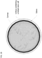
- Descemet's membrane is the multilayered basement membrane of the corneal endothelium (see FIG. 1 ).
- Descemet's membrane is routinely isolated, along with the endothelial cells, from donor corneas and transplanted intraocularly in patients to replaced damaged Descemet's membrane and/or corneal endothelial cells as reported by Park, et al., Ophthalmology. 2015; 122:2432-2442 ( see also U.S. Patent No. 8,889,415 ). Transplantation of Descemet's membrane onto the external ocular surface of the eye is not known to have been previously described.
- Descemet's membrane unexpectedly provides multiple advantages as a substrate for supporting proliferation and long-term survival of limbal stem cells, both in vivo on the ocular surface and ex vivo in culture.
- the similarity of Descemet's membrane to the basement membrane of the limbal niche microenvironment, biochemically, is an obscure characteristic of Descemet's membrane that has not previously been exploited to promote the growth of limbal stem cells and to inhibit further differentiation into non-amplifying corneal epithelial cells.
- the methods of this disclosure exploit this characteristic of Descemet's membrane, and this disclosure describes, in some embodiments, a method of using Descemet's membrane as a substrate for culturing limbal stem cells.
- this disclosure describes a method of removing endothelium from a Descemet's membrane of a cornea to provide a decellularized Descemet's membrane. In a further aspect, this disclosure describes separating a decellularized Descemet's membrane from the stroma of the cornea to obtain an isolated Descemet's membrane.
- the cornea may be a donor cornea including, for example, a cadaveric cornea. In some embodiments, the cornea may be a human cornea. In some embodiments, the cornea may be a porcine cornea.
- corneal endothelium may be separated from Descemet's membrane to form a decellularized Descemet's membrane using mechanical, enzymatic, and/or chemical decellularization. In some embodiments, the separation of the corneal endothelium from Descemet's membrane to form a decellularized Descemet's membrane leaves the corneal epithelium intact.
- a decellularized Descemet's membrane may be separated from the stroma of a cornea to obtain an isolated Descemet's membrane by manually peeling the Descemet's membrane from the stroma of the cornea.
- the corneal epithelium may preferably be removed with the stroma of the cornea.
- a decellularized Descemet's membrane may be separated from the stroma of a cornea to obtain an isolated Descemet's membrane by injecting air or fluid or both into the cornea.
- the fluid may include any suitable fluid.
- the fluid may include, for example, saline, corneal storage solution, or any buffered solution.
- the residual cornea e.g., including the corneal epithelium and corneal stroma
- the residual cornea that has been separated from Descemet's membrane may be excised to expose the isolated Descemet's membrane.
- separating the decellularized Descemet's membrane from the stroma of the cornea and removing that stroma includes exposing the isolated Descemet's membrane to a limbal stem cell found in the corneolimbal ring of the cornea.
- the method may further include making a partial-thickness incision in the corneolimbal ring. Such an incision may promote the outgrowth of limbal stem cells from the corneolimbal ring.
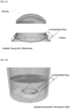
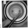
- Drawings of one exemplary method of separating the decellularized Descemet's membrane from the stroma of the cornea and exposing the isolated Descemet's membrane to a limbal stem cell is shown in FIG. 2A - FIG. 2C .
- the isolated Descemet's membrane may be excised completely from the rest of the cornea (including the corneolimbal ring) including, for example, by trephinating the Descemet's membrane.
- this disclosure describes a method of storing an isolated Descemet's membrane.
- the isolated Descemet's membrane may be preserved without regard to cell viability.
- the isolated Descemet's membrane may be preserved by a means that maintains cell viability.
- the isolated Descemet's membrane may be frozen, lyophilized, and/or cryopreserved.
- the isolated Descemet's membrane may be sterilized before or after preservation or both before and after preservation.
- the isolated Descemet's membrane may be sterilized by any suitable means including, for example, by gamma irradiation, chemical disinfectant, antibiotic treatment, ethylene oxide gas treatment, or supercritical CO 2 exposure.
- this disclosure describes methods of using an isolated Descemet's membrane.
- this disclosure describes using an isolated Descemet's membrane as a cell culture substrate.
- this disclosure describes using an isolated Descemet's membrane as a cell culture substrate to support proliferation of a limbal stem cell.
- the limbal stem cell may adhere to the isolated Descemet's membrane.
- an isolated Descemet's membrane may be attached to a cell culture surface including, for example, tissue culture plastic.
- a drawing of one exemplary method of attaching Descemet's membrane to a cell culture surface is shown in FIG. 2D .
- the Descemet's membrane may be reversibly attached to the cell culture surface.
- the isolated Descemet's membrane when an isolated Descemet's membrane is attached to a cell culture surface, the isolated Descemet's membrane will preferably have been completely excised or manually peeled from the rest of the cornea (including the corneolimbal ring).
- a limbal explant may be cultured in the presence of the isolated Descemet's membrane.
- the limbal explant may be from the same donor (for example, from the same cadaveric cornea) or different donor (for example, from a living donor's cornea) as the isolated Descemet's membrane.
- the limbal explant includes a part of the corneolimbal ring. In some embodiments, the limbal explant includes an entire corneolimbal ring. In some embodiments, the limbal explant may be cultured under conditions that allow outgrowth of a limbal stem cell from the limbal explant onto the isolated Descemet's membrane, resulting in an isolated Descemet's membrane with limbal stem cells on its surface.
- separating the decellularized Descemet's membrane from the stroma of the cornea to obtain an isolated Descemet's membrane exposes the isolated Descemet's membrane to limbal stem cells found in the corneolimbal ring.
- the isolated Descemet's membrane may be exposed to limbal stem cells found in the corneolimbal ring by removing the cornea stroma.
- the corneolimbal ring remains attached to the isolated Descemet's membrane.
- an isolated Descemet's membrane may be exposed to limbal stem cells obtained from another source or obtained from the same donor source but separated from the corneolimbal ring.
- a limbal explant culture may be treated to release limbal stem cells (into a cell suspension) which may be seeded onto an isolated Descemet's membrane.
- a limbal explant culture and/or a limbal tissue ring may be digested with one or more of trypsin ( Sharifi, et al., Biocell. 2010;34:53-55 ), dispase ( Zhang, et al., Curr Eye Res.
- the limbal stem cells may be seeded onto an isolated Descemet's membrane without any further manipulation (see FIG. 11B ).
- the limbal stem cells may be sorted (for example, using flow cytometry) to select cells with a specific limbal stem cell marker or markers prior to being seeded onto an isolated Descemet's membrane. Exemplary methods of preparing limbal stem cells for culture on Descemet's membrane are described in Example 8A and 8B.
- Limbal explant cultures and/or limbal stem cells may be maintained in any suitable growth media.
- suitable growth media may include, for example, a growth medium containing human autologous serum, fetal bovine serum, human platelet lysates, and a growth medium containing serum-free medium with bovine pituitary extracts, growth supplement with recombinant components, or chemically defined supplements.
- An isolated Descemet's membrane may be cultured under conditions that allow for growth of limbal stem cells (e.g., from a limbal explant and/or from a corneolimbal ring) onto the isolated Descemet's membrane.
- a drawing of one exemplary method of culturing Descemet's membrane to achieve limbal cell outgrowth onto the Descemet's membrane is shown in FIG. 2E .
- the method may include making a partial-thickness incision in the corneolimbal ring to promote the outgrowth of limbal stem cells.
- the method may include placing the isolated Descemet's membrane in a cell culture media.
- the cell culture media may include any suitable cell culture media including, for example, an epithelial cell growth media or other media suitable for corneal organ culture.
- An exemplary media suitable for corneal organ culture is CorneaMax ® (Eurobio, Les Ulis, France).
- the cell culture media may be serum free.
- cell culture media may include pituitary extract.
- the cell culture media may include one or more of a complex culture media supplemented with fetal bovine serum, a complex culture media supplemented with human serum, a complex culture media supplemented with platelet lysate serum, or a chemically-defined keratinocyte growth media.
- the cell culture media may preferably promote limbal stem cell growth and/or maintain limbal stem cell pluripotency.
- the method may include incubating the isolated Descemet's membrane in the cell culture media under typical cell culture conditions including, for example, at a temperature in a range of 32°C to 38°C and/or at 5% CO 2 .
- the isolated Descemet's membrane is incubated with a limbal explant and/or a limbal stem cell.
- this disclosure describes methods of transplanting an isolated Descemet's membrane to an ocular surface of a patient in need thereof.
- the transplanted Descemet's membrane may be decellularized. In some embodiments, the transplanted Descemet's membrane may include limbal stem cells. A drawing of one exemplary method of transplanting a Descemet's membrane including limbal stem cells is shown in FIG. 2F . In some embodiments, the transplanted Descemet's membrane may include an isolated Descemet's membrane prepared by any of the methods described herein.
- an isolated Descemet's membrane including limbal stem cells produced using the methods described herein may be transplanted to a patient exhibiting a partial limbal stem cell deficiency, a total limbal stem cell deficiency, a persistent epithelial defect, an epithelial erosion, a corneal ulcer, a corneal melt, and/or an ocular surface disease.
- Limbal stem cell deficiency may be caused by, for example, burns (chemical, thermal, or industrial); excessive use of certain medications; iatrogenic trauma, including, for example, overuse of contact lenses, chronic use of glaucoma drops, or ocular surgery; an autoimmune disease including, for example, Steven Johnson Syndrome or ocular cicatricial pemphigoid; or graft-versus-host disease (GVHD); etc.
- burns chemical, thermal, or industrial
- iatrogenic trauma including, for example, overuse of contact lenses, chronic use of glaucoma drops, or ocular surgery
- an autoimmune disease including, for example, Steven Johnson Syndrome or ocular cicatricial pemphigoid
- GVHD graft-versus-host disease
- transplanting an isolated Descemet's membrane to an ocular surface of a patient, as described herein may have certain advantages over existing treatments for limbal stem cell deficiency.
- transplanting an isolated Descemet's membrane to an ocular surface of a patient, as described herein e.g., including a limbal stem cell
- a CLET-like procedure performed using an isolated Descemet's membrane is less expensive than an amniotic-membrane based method (because it takes advantage of the free Descemet's membrane that comes with every donor cornea and limbus), and it also makes it possible to produce CLET-like grafts in an eye bank setting, improving surgeon access to CLET technology.
- Descemet's membrane along with limbal stem cells on the ocular surface provides a novel, viable technique for establishing a stable niche-like microenvironment on the cornea, and thus may provide a robust and promising technique for long-term reconstruction of the ocular surface. Additionally, intact Descemet's membrane is optically clear ( see FIG. 5 ), resistant to collagenase digestion ( see FIG. 7 ), minimally immunogenic when transplanted intraocularly, and freely available with every donor cornea from which stems cells are harvested.
- the isolated Descemet's membrane may be used as an ocular bandage including, for example, in patients with an ocular surface trauma, a recurrent erosion, a corneal melt, or a sterile corneal ulcer.
- Such patients may include any patient with a disease that compromises the ocular surface including, for example, patients with diabetes, patients with glaucoma, patients with injuries resulting from chronic contact lens wear, patients with complications of LASIK surgery, patients with neurotrophic concerns, patients with severe tear deficiency, and/or patients with exposure keratopathy.
- an isolated Descemet's membrane may be useful as a reconstructive therapy in cases of minor ocular surface burns or trauma.
- isolated Descemet's membrane when placed as a resurfacing treatment, may establish a stable bandage layer on the ocular surface of the cornea that promotes regrowth of the patients' own epithelium in situ, while protecting the corneal stroma from enzymatic degradation by inflammatory mediators in the tear film due to its natural resistance to collagenase.
- Isolated Descemet's membrane can remain stable over long periods of time but is still transparent and visually unobstructive. Since the isolated Descemet's membrane is decellularized to remove endothelial cells, the isolated Descemet's membrane has no cells for the body to reject making the risk of rejection minimal.
- Isolated Descemet's membrane may be adhered to the recipient cornea by any suitable means.
- the Descemet's membrane may be adhered to the recipient cornea by air drying.
- the Descemet's membrane may be adhered to the recipient cornea using one or more of a fibrin sealant, colloidal silica nanoparticles, and light-initiated rose bengal collagen cross-linking.
- this disclosure describes a composition that includes an isolated Descemet's membrane, wherein the Descemet's membrane has been decellularized and separated from the corneal stroma.
- the composition further includes a limbal stem cell.
- the limbal stem cell and the isolated Descemet's membrane are from the same cornea.
- the limbal stem cell may, in some embodiments, be in contact with (for example, present on the surface of) the isolated Descemet's membrane.
- the composition includes isolated Descemet's membrane attached to a corneolimbal ring.
- the composition includes a limbal stem cell, the limbal stem cell may be present in the corneolimbal ring and/or on the surface of the isolated Descemet's membrane that has been bared to the corneolimbal ring.
- the isolated Descemet's membrane may act as a substrate for the limbal stem cell.
- the isolated Descemet's membrane may have been sterilized or preserved or both.
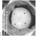
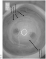
- This Example describes a method for the isolation of a Descemet's membrane from the stroma of a cornea, to form an isolated Descemet's membrane, as shown in one embodiment in FIG. 2 & FIG. 3B - FIG. 3C .
- the endothelium was removed from Descemet's membrane by mechanical debridement to form a decellularized Descemet's membrane.
- the decellularized Descemet's membrane was isolated from the stroma of a cornea by manually injecting cornea storage solution with pressure not exceeding 600 mmHg into the posterior stroma, near Descemet's membrane.
- a super sharp blade was used to make an incision through the stroma.
- Scissors were used to perform a continuous circumferential cut to excise the residual cornea while leaving the corneolimbal ring attached to isolated Descemet's membrane.
- the Descemet's membrane was then excised from the corneolimbal ring using a trephine. In some instances, the Descemet's membrane was decontaminated by exposure to a 5% povidone iodide solution.
- This Example describes a method for the separation of a Descemet's membrane from the stroma of a cornea, and an exemplary method for using the isolated Descemet's membrane as a cell culture substrate for culturing a limbal stem cell, as shown in one embodiment in FIG. 2 .
- Isolated Descemet's membrane was prepared as described in Example 1. The residual cornea was excised, exposing the isolated Descemet's membrane to the corneolimbal ring, as shown in one embodiment in FIG. 3D .
- the isolated Descemet's membrane ( FIG. 3E ) was placed in complex culture media supplemented with fetal bovine serum and cultured at 37°C, 5% CO 2 for up to 4 weeks. Cells grew out from the corneolimbal ring and grew over the isolated Descemet's membrane ( FIG. 3F ).
- the cellular morphology of the cells growing on the isolated Descemet's membrane at 1 week was consistent with limbal stem cells rather than fibroblasts.
- the resulting cultures were stained using the methods described by Suri et al. Curr Eye Res. 2016; 41:1266-1273 .
- the cells growing on the isolated Descemet's membrane express ⁇ Np63 ⁇ and ABCG2, stem cell biomarkers, after 1 week in culture.
- the isolated Descemet's membrane was stained with 0.06% Trypan blue for 1 minute before transplantation to assist in visualization.
- Descemet's membrane was adhered to the recipient cornea by air drying.
- the Descemet's membrane is transparent.
- an amniotic membrane, transplanted onto a cornea is semi-opaque as described by Connon, et al., Br J Ophthalmol. 2010;94:1057-1061 .
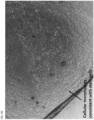
- This Example shows Descemet's membrane is resistant to enzymatic degradation.
- a donor cornea see FIG. 7A
- the residual stroma bed was then treated for 45 minutes with collagenase A.
- collagenase treatment the stroma was completely digested, but the Descemet's membrane remained intact ( FIG. 7C ).
- Isolated Descemet's membrane prepared as described in Example 1, was allowed to dry onto a treated polystyrene tissue culture surface. Corneolimbal ring was minced into 1 mm 2 limbal explant fragments and adhered on the tissue culture plastic surface near Descemet's membrane. Complex culture media supplemented with fetal bovine serum was added. The limbal explants and Descemet's membrane were then cultured at 37°C and 5% CO 2 . Culture media was changed every 48 hours. Limbal stem cells proliferated out from the limbal explant fragments over the tissue culture plastic and onto the Descemet's membrane.
- Results are shown in FIG. 8 .
- PAS positive immunostaining Descemet's membrane
- corneal epithelium pancytokeratin positive immunostaining
- Results are shown in FIG. 9 . These result support the ability of Descemet's membranes to act as an ocular surface bandage to promote corneal epithelial healing.
- Descemet's membranes excised from their corneolimbal rings were isolated as described in Example 1, sterilized using antibiotic treatment, frozen at -80°C for 1 month, and then thawed. After freezing and thawing, the Descemet's membranes maintained their transparency and propensity to scroll. An exemplary sample, stained with Trypan blue (to assist with visibility), is shown in FIG. 10 .
- This example describes an exemplary method for isolating limbal stem cells from a corneolimbal ring for culture on Descemet's membrane.
- a corneolimbal ring was treated with collagenase to release the basal limbal stem cells, as per previously published protocols ( Chen, et al., Tissue Eng Part C Methods. 2011;17:537-548 ), and resuspended in serum-free medium with bovine pituitary extracts, growth supplement with recombinant components, or chemically defined supplements. After resuspension of the limbal stem cells, the cells were immunostained with limbal stem cell markers ( ⁇ Np63 ⁇ and ABCG2) and counterstained for nuclei (DAPI), as described in FIG. 4F . Results are shown in Figure 11A .
- These enzymatically isolated limbal stem cells may be further enriched by flow cytometry and seeded onto Descemet's membrane.
- the cultures can be maintained in (a) growth medium containing human autologous serum, fetal bovine serum, human platelet lysates, or (b) serum-free medium with bovine pituitary extracts, growth supplement with recombinant components or chemically defined supplements.
- This example describes an exemplary method for isolating limbal stem cells from a corneolimbal ring for culture on Descemet's membrane.
- Limbal explants were prepared by trephining out the central corneal button, then cutting the limbal ring into 12 to 16 small limbal explant fragments. Limbal explant fragments were cultured on tissue culture plastic using serum-free medium with bovine pituitary extracts, growth supplement with recombinant components, or chemically defined supplements. The explants were cultured for 7 to 10 days at 37°C and 5% CO 2 , until limbal stem cell outgrowth from the limbal explants was near confluence on the tissue culture plastic. The limbal stem cells were then trypsinized into cell suspension (see Sharifi, et al., Biocell. 2010;34:53-55 ).
- Descemet's membrane was then seeded from suspension onto Descemet's membrane at approximately 20% confluency and allowed to grow for up to 4 to 7 days (resulting in a total period of culture of 14 days).
- An exemplary image of the resulting Descemet's membrane is shown in FIG. 11B .
- Limbal stem cells cultured on tissue culture plastic and Descemet's membrane using different culture conditions were compared for expression of limbal stem cell markers to demonstrate the efficacy of using Descemet's membrane as a culture substrate for limbal stem cells.
- In the first culture condition limbal explants were cultured on tissue culture plastic (TCP) using fetal bovine serum (FBS)-supplemented media.
- FBS fetal bovine serum
- Descemet's membrane was isolated from the corneoscleral ring as described in Example 1. Limbal explants were then cultured on tissue the isolated Descemet's membrane using fetal bovine serum (FBS)-supplemented media.
- Descemet's membrane was isolated from the corneoscleral ring as described in Example 2, where the limbal stem cells in the intact limbus were exposed to Descemet's membrane and cultured directly onto Descemet's membrane from the intact limbus.
- Cultures were then evaluated for outgrowth from limbal explant fragments using inverted light microscopy. One sample for each culture condition that grew to confluence was further characterized with immunohistochemistry. Cultures were immunostained for putative limbal stem cell (LSC) markers: ⁇ Np63 ⁇ (nuclear) and ABCG2 (membrane-associated). Cells were then manually counted by two independent reviewers and compared for ⁇ Np63 ⁇ and ABCG2 against DAPI nuclear counterstain.
- LSC limbal stem cell
- limbal stem cells cultured from limbal explants demonstrated greater sternness, as measured by co-localized expression of putative limbal stem cell markers (ABCG2, ⁇ Np63 ⁇ ), when cultured on Descemet's membrane (DM) compared to tissue culture plastic (TCP). Furthermore, limbal stem cells cultured from intact limbus demonstrated greater sternness, as measured by co-localized expression of putative limbal stem cell markers (ABCG2, ⁇ Np63 ⁇ ), in comparison to limbal stem cells cultured from limbal explants.
- putative limbal stem cell markers ABCG2, ⁇ Np63 ⁇
- Limbal stem cells cultured on human amniotic membrane and Descemet's membrane were compared for expression of limbal stem cell markers to demonstrate the efficacy of using Descemet's membrane as a culture substrate for limbal stem cells.
- Limbal stem cells were trypsinized from limbal explant cultures and suspended in serum free media with bovine pituitary extracts, growth supplement with recombinant components, or chemically defined supplements, as described in Example 8B.
- Limbal stem cells in suspension were then seeded on human amniotic membrane (HAM) or Descemet's membrane (DM) and maintained in culture at 37°C and 5% CO 2 for 4 to 7 days.
- LSC limbal stem cell
- limbal stem cells cultured from cell suspension demonstrated greater sternness, as measured by expression of putative limbal stem cell markers (ABCG2, ⁇ Np63 ⁇ ), when cultured on Descemet's membrane compared to human amniotic membrane (HAM).
Landscapes
- Health & Medical Sciences (AREA)
- Life Sciences & Earth Sciences (AREA)
- Engineering & Computer Science (AREA)
- Biomedical Technology (AREA)
- Chemical & Material Sciences (AREA)
- General Health & Medical Sciences (AREA)
- Zoology (AREA)
- Veterinary Medicine (AREA)
- Public Health (AREA)
- Animal Behavior & Ethology (AREA)
- Transplantation (AREA)
- Medicinal Chemistry (AREA)
- Epidemiology (AREA)
- Oral & Maxillofacial Surgery (AREA)
- Chemical Kinetics & Catalysis (AREA)
- Botany (AREA)
- Dermatology (AREA)
- Cell Biology (AREA)
- Bioinformatics & Cheminformatics (AREA)
- Biotechnology (AREA)
- Organic Chemistry (AREA)
- Wood Science & Technology (AREA)
- Genetics & Genomics (AREA)
- Developmental Biology & Embryology (AREA)
- Neurosurgery (AREA)
- Neurology (AREA)
- Molecular Biology (AREA)
- Ophthalmology & Optometry (AREA)
- Urology & Nephrology (AREA)
- General Engineering & Computer Science (AREA)
- Biochemistry (AREA)
- Microbiology (AREA)
- Vascular Medicine (AREA)
- Pharmacology & Pharmacy (AREA)
- Hematology (AREA)
- Orthopedic Medicine & Surgery (AREA)
- General Chemical & Material Sciences (AREA)
- Nuclear Medicine, Radiotherapy & Molecular Imaging (AREA)
- Immunology (AREA)
- Virology (AREA)
Applications Claiming Priority (3)
| Application Number | Priority Date | Filing Date | Title |
|---|---|---|---|
| US201862747715P | 2018-10-19 | 2018-10-19 | |
| PCT/US2019/056938 WO2020081934A1 (en) | 2018-10-19 | 2019-10-18 | Methods of isolating and using descemet's membrane and compositions including isolated descemet's membrane |
| EP19809212.4A EP3867357B1 (de) | 2018-10-19 | 2019-10-18 | Verfahren zur isolierung und verwendung der descemet-membran und zusammensetzungen mit isolierter descemet-membran |
Related Parent Applications (2)
| Application Number | Title | Priority Date | Filing Date |
|---|---|---|---|
| EP19809212.4A Division EP3867357B1 (de) | 2018-10-19 | 2019-10-18 | Verfahren zur isolierung und verwendung der descemet-membran und zusammensetzungen mit isolierter descemet-membran |
| EP19809212.4A Division-Into EP3867357B1 (de) | 2018-10-19 | 2019-10-18 | Verfahren zur isolierung und verwendung der descemet-membran und zusammensetzungen mit isolierter descemet-membran |
Publications (2)
| Publication Number | Publication Date |
|---|---|
| EP4491716A2 true EP4491716A2 (de) | 2025-01-15 |
| EP4491716A3 EP4491716A3 (de) | 2025-04-02 |
Family
ID=68655628
Family Applications (2)
| Application Number | Title | Priority Date | Filing Date |
|---|---|---|---|
| EP24195485.8A Pending EP4491716A3 (de) | 2018-10-19 | 2019-10-18 | Verfahren zur isolierung und verwendung einer descemet-membran und zusammensetzungen mit isolierter descemet-membran |
| EP19809212.4A Active EP3867357B1 (de) | 2018-10-19 | 2019-10-18 | Verfahren zur isolierung und verwendung der descemet-membran und zusammensetzungen mit isolierter descemet-membran |
Family Applications After (1)
| Application Number | Title | Priority Date | Filing Date |
|---|---|---|---|
| EP19809212.4A Active EP3867357B1 (de) | 2018-10-19 | 2019-10-18 | Verfahren zur isolierung und verwendung der descemet-membran und zusammensetzungen mit isolierter descemet-membran |
Country Status (4)
| Country | Link |
|---|---|
| US (2) | US12383655B2 (de) |
| EP (2) | EP4491716A3 (de) |
| ES (1) | ES2990204T3 (de) |
| WO (1) | WO2020081934A1 (de) |
Families Citing this family (3)
| Publication number | Priority date | Publication date | Assignee | Title |
|---|---|---|---|---|
| ES2990204T3 (es) | 2018-10-19 | 2024-11-29 | Univ Minnesota | Procedimientos de aislamiento y utilización de la membrana de Descemet y composiciones que incluyen membrana de Descemet aislada |
| US12257365B2 (en) | 2019-04-26 | 2025-03-25 | Regents Of The University Of Minnesota | Incised Descemet's membrane and methods of making and using |
| KR102417981B1 (ko) * | 2020-05-21 | 2022-07-05 | 충북대학교 산학협력단 | 각막내피세포 이식용 인공 지지체, 이의 제조방법 및 이를 이용한 초박형 데스메막박리 각막내피층판 이식편 |
Citations (2)
| Publication number | Priority date | Publication date | Assignee | Title |
|---|---|---|---|---|
| US7347875B2 (en) | 2002-12-02 | 2008-03-25 | Gi Dynamics, Inc. | Methods of treatment using a bariatric sleeve |
| US8889415B2 (en) | 2006-04-28 | 2014-11-18 | Ray Jui-Fang Tsai | Method for expansion of human corneal endothelial cells |
Family Cites Families (23)
| Publication number | Priority date | Publication date | Assignee | Title |
|---|---|---|---|---|
| FR2648702A1 (fr) | 1989-06-23 | 1990-12-28 | Hanna Khalil | Lentille pour epikeratophakie et keratotome notamment destine a la realisation d'une incision de reception d'une telle lentille |
| US5964748A (en) | 1995-10-20 | 1999-10-12 | Peyman; Gholam A. | Intrastromal corneal modification |
| IL120005A (en) * | 1997-01-14 | 2000-08-31 | Ramot University Authority Of | Pharmaceutical compositions for the treatment of the eye |
| AU2878200A (en) | 1999-02-11 | 2000-08-29 | Schepens Eye Research Institute, Inc., The | Growth medium for human corneal endothelial cells |
| US7347876B2 (en) * | 2001-04-25 | 2008-03-25 | Ray Jui-Fang Tsai | Method for expansion of epithelial stem cells |
| ITRM20010476A1 (it) * | 2001-08-03 | 2003-02-03 | Idi Irccs | Lamine di epitelio corneale umano ricostruite e metodo per il loro ottenimento. |
| CN1635115A (zh) | 2004-05-27 | 2005-07-06 | 天津医科大学眼科中心 | 人成纤维细胞滋养的人角膜缘干细胞组织工程产品及制备 |
| JP5552690B2 (ja) | 2006-09-13 | 2014-07-16 | ディーエスエム アイピー アセッツ ビー.ブイ. | 被覆された医療装置 |
| EP2764846A1 (de) | 2007-02-08 | 2014-08-13 | Kaneka Corporation | Injektor für Augen |
| US20110166650A1 (en) | 2008-09-04 | 2011-07-07 | Massimo Busin | New technique for preparing, storing and transplanting endothelial grafts |
| US20100256651A1 (en) | 2009-04-07 | 2010-10-07 | Dharmendra Jani | Intraocular Lens Injector with Hydrophilic Coating |
| US8287890B2 (en) | 2009-12-15 | 2012-10-16 | C.R. Bard, Inc. | Hydrophilic coating |
| US20140155871A1 (en) | 2012-05-10 | 2014-06-05 | James Stuart Cumming | Method for preparing corneal donor tissue for refractive eye surgery utilizing the femtosecond laser |
| US20140171956A1 (en) | 2012-12-17 | 2014-06-19 | Abbott Medical Optics Inc. | Detecting coatings on intraocular lens insertion devices and methods of manufacturing the devices |
| CN107979992B (zh) | 2015-06-08 | 2020-05-22 | 科尔尼特视觉有限公司 | 人工角膜及其用途 |
| US11510931B2 (en) * | 2016-09-28 | 2022-11-29 | Medicon Pharmaceuticals, Inc. | Compositions and methods for treating ophthalmic conditions |
| US10481051B2 (en) | 2016-10-13 | 2019-11-19 | Lions VisionGift | Corneal tissue sample assemblies and related methods of use |
| CN106955372B (zh) * | 2017-04-12 | 2020-08-14 | 山东省眼科研究所 | 一种组织工程角膜内皮的构建方法 |
| ES2990204T3 (es) | 2018-10-19 | 2024-11-29 | Univ Minnesota | Procedimientos de aislamiento y utilización de la membrana de Descemet y composiciones que incluyen membrana de Descemet aislada |
| EP3698758A1 (de) | 2019-02-20 | 2020-08-26 | Leibniz-Institut für Polymerforschung Dresden e.V. | Vorrichtung und anordnungen für ausgerichteten transport, mikroskopische untersuchung und ausgerichtetes ausstossen eines gewebetransplantats oder -implantats |
| US12257365B2 (en) | 2019-04-26 | 2025-03-25 | Regents Of The University Of Minnesota | Incised Descemet's membrane and methods of making and using |
| US12185699B2 (en) | 2019-11-20 | 2025-01-07 | Egg-Chick Automated Technologies | Assembly for transporting and separating living poultry birds |
| WO2022241252A1 (en) | 2021-05-13 | 2022-11-17 | Regents Of The University Of Minnesota | Descemet membrane endothelial keratoplasty (dmek) assemblies and injectors with friction-reducing coatings |
-
2019
- 2019-10-18 ES ES19809212T patent/ES2990204T3/es active Active
- 2019-10-18 WO PCT/US2019/056938 patent/WO2020081934A1/en not_active Ceased
- 2019-10-18 EP EP24195485.8A patent/EP4491716A3/de active Pending
- 2019-10-18 US US17/286,537 patent/US12383655B2/en active Active
- 2019-10-18 EP EP19809212.4A patent/EP3867357B1/de active Active
-
2025
- 2025-07-24 US US19/278,964 patent/US20250345484A1/en active Pending
Patent Citations (2)
| Publication number | Priority date | Publication date | Assignee | Title |
|---|---|---|---|---|
| US7347875B2 (en) | 2002-12-02 | 2008-03-25 | Gi Dynamics, Inc. | Methods of treatment using a bariatric sleeve |
| US8889415B2 (en) | 2006-04-28 | 2014-11-18 | Ray Jui-Fang Tsai | Method for expansion of human corneal endothelial cells |
Non-Patent Citations (5)
| Title |
|---|
| CHEN ET AL., TISSUE ENG PART C METHODS, vol. 17, 2011, pages 537 - 548 |
| CONNON ET AL., BR J OPHTHALMOL, vol. 94, 2010, pages 1057 - 1061 |
| PARK ET AL., OPHTHALMOLOGY, vol. 122, 2015, pages 2432 - 2442 |
| SHARIFI ET AL., BIOCELL, vol. 34, 2010, pages 53 - 55 |
| ZHANG ET AL., CURR EYE RES, vol. 41, 2016, pages 1266 - 1273 |
Also Published As
| Publication number | Publication date |
|---|---|
| EP3867357A1 (de) | 2021-08-25 |
| US12383655B2 (en) | 2025-08-12 |
| WO2020081934A1 (en) | 2020-04-23 |
| US20250345484A1 (en) | 2025-11-13 |
| EP4491716A3 (de) | 2025-04-02 |
| ES2990204T3 (es) | 2024-11-29 |
| EP3867357B1 (de) | 2024-09-25 |
| US20210379246A1 (en) | 2021-12-09 |
| EP3867357C0 (de) | 2024-09-25 |
Similar Documents
| Publication | Publication Date | Title |
|---|---|---|
| US20250345484A1 (en) | Methods of isolating and using descemet's membrane and compositions including isolated descemet's membrane | |
| Nishida et al. | Functional bioengineered corneal epithelial sheet grafts from corneal stem cells expanded ex vivo on a temperature-responsive cell culture surface | |
| Peh et al. | Human corneal endothelial cell expansion for corneal endothelium transplantation: an overview | |
| EP1604695A1 (de) | Aus amnios stammendes medizinisches material und verfahren zu seiner herstellung | |
| JP5255846B2 (ja) | 生体内で細胞増殖可能な角膜内皮製剤 | |
| KR100940765B1 (ko) | 각막 상피형 시트, 및 그의 제조 방법 | |
| KR20070015519A (ko) | 생체 조직 시트, 그것의 제조 방법, 및 그것을 이용한 이식방법 | |
| He et al. | Growing human corneal epithelium on collagen shield and subsequent transfer to denuded cornea in vitro | |
| US7611895B2 (en) | Method for growth of human conjunctival tissue equivalents for research, clinical ocular surface transplantation and tissue engineering | |
| KR20070037451A (ko) | 각막 상피 시트 및 그 제조 방법 | |
| Amano et al. | Properties of corneas reconstructed with cultured human corneal endothelial cells and human corneal stroma | |
| US20070280993A1 (en) | Corneal Epithelial Sheet, Method Of constructing The Same, And Transplantation Method Using The Sheet | |
| RU2475218C1 (ru) | Способ выделения и органо-типической консервации аллогенного лимбального трансплантата | |
| Sharma et al. | Limbal stem cell transplants and amniotic membrane grafts in ocular surface disease: current perspectives. | |
| Suresh et al. | Standardization of human corneal endothelial cell isolation and the use of denuded human amniotic membrane as a scaffold for human corneal endothelial cells | |
| Javadi et al. | Early results of autologous cultivated limbal stem cell transplantation in total limbal stem cell deficiency | |
| Sangwan | Cultivated Limbal Stem Cell Transplantation—The Surgical Technique | |
| Suresh et al. | Standardization of Human Corneal Endothelial Cell Isolation and the Use of | |
| Jastaneiah | Corneal Limbal Stem Cells: II. Ocular Surface Reconstruction with Limbal Stem Cell Transplantation | |
| GUPTA et al. | Stem Cells in Ophthalmology | |
| Güell et al. | Limbal Stem Cell Culture |
Legal Events
| Date | Code | Title | Description |
|---|---|---|---|
| PUAI | Public reference made under article 153(3) epc to a published international application that has entered the european phase |
Free format text: ORIGINAL CODE: 0009012 |
|
| STAA | Information on the status of an ep patent application or granted ep patent |
Free format text: STATUS: THE APPLICATION HAS BEEN PUBLISHED |
|
| AC | Divisional application: reference to earlier application |
Ref document number: 3867357 Country of ref document: EP Kind code of ref document: P |
|
| AK | Designated contracting states |
Kind code of ref document: A2 Designated state(s): AL AT BE BG CH CY CZ DE DK EE ES FI FR GB GR HR HU IE IS IT LI LT LU LV MC MK MT NL NO PL PT RO RS SE SI SK SM TR |
|
| REG | Reference to a national code |
Ref country code: DE Ref legal event code: R079 Free format text: PREVIOUS MAIN CLASS: C12N0005079700 Ipc: C12N0005079000 |
|
| PUAL | Search report despatched |
Free format text: ORIGINAL CODE: 0009013 |
|
| AK | Designated contracting states |
Kind code of ref document: A3 Designated state(s): AL AT BE BG CH CY CZ DE DK EE ES FI FR GB GR HR HU IE IS IT LI LT LU LV MC MK MT NL NO PL PT RO RS SE SI SK SM TR |
|
| RIC1 | Information provided on ipc code assigned before grant |
Ipc: A61P 27/02 20060101ALI20250226BHEP Ipc: A61L 27/36 20060101ALI20250226BHEP Ipc: A61L 27/38 20060101ALI20250226BHEP Ipc: A61F 2/14 20060101ALI20250226BHEP Ipc: A61K 35/30 20150101ALI20250226BHEP Ipc: C12N 5/0797 20100101ALI20250226BHEP Ipc: A61K 35/12 20150101ALI20250226BHEP Ipc: C12N 5/079 20100101AFI20250226BHEP |
|
| STAA | Information on the status of an ep patent application or granted ep patent |
Free format text: STATUS: REQUEST FOR EXAMINATION WAS MADE |
|
| 17P | Request for examination filed |
Effective date: 20251002 |
|
| STAA | Information on the status of an ep patent application or granted ep patent |
Free format text: STATUS: EXAMINATION IS IN PROGRESS |
|
| 17Q | First examination report despatched |
Effective date: 20260129 |