EP4425178A2 - Verfahren zur erfassung von zellfreier methylierter dna und verwendungen davon - Google Patents
Verfahren zur erfassung von zellfreier methylierter dna und verwendungen davon Download PDFInfo
- Publication number
- EP4425178A2 EP4425178A2 EP24176389.5A EP24176389A EP4425178A2 EP 4425178 A2 EP4425178 A2 EP 4425178A2 EP 24176389 A EP24176389 A EP 24176389A EP 4425178 A2 EP4425178 A2 EP 4425178A2
- Authority
- EP
- European Patent Office
- Prior art keywords
- dna
- methylated
- filler
- cfdna
- cell
- Prior art date
- Legal status (The legal status is an assumption and is not a legal conclusion. Google has not performed a legal analysis and makes no representation as to the accuracy of the status listed.)
- Pending
Links
Images














Classifications
-
- C—CHEMISTRY; METALLURGY
- C12—BIOCHEMISTRY; BEER; SPIRITS; WINE; VINEGAR; MICROBIOLOGY; ENZYMOLOGY; MUTATION OR GENETIC ENGINEERING
- C12N—MICROORGANISMS OR ENZYMES; COMPOSITIONS THEREOF; PROPAGATING, PRESERVING, OR MAINTAINING MICROORGANISMS; MUTATION OR GENETIC ENGINEERING; CULTURE MEDIA
- C12N15/00—Mutation or genetic engineering; DNA or RNA concerning genetic engineering, vectors, e.g. plasmids, or their isolation, preparation or purification; Use of hosts therefor
- C12N15/09—Recombinant DNA-technology
- C12N15/10—Processes for the isolation, preparation or purification of DNA or RNA
- C12N15/1003—Extracting or separating nucleic acids from biological samples, e.g. pure separation or isolation methods; Conditions, buffers or apparatuses therefor
-
- C—CHEMISTRY; METALLURGY
- C12—BIOCHEMISTRY; BEER; SPIRITS; WINE; VINEGAR; MICROBIOLOGY; ENZYMOLOGY; MUTATION OR GENETIC ENGINEERING
- C12N—MICROORGANISMS OR ENZYMES; COMPOSITIONS THEREOF; PROPAGATING, PRESERVING, OR MAINTAINING MICROORGANISMS; MUTATION OR GENETIC ENGINEERING; CULTURE MEDIA
- C12N15/00—Mutation or genetic engineering; DNA or RNA concerning genetic engineering, vectors, e.g. plasmids, or their isolation, preparation or purification; Use of hosts therefor
- C12N15/09—Recombinant DNA-technology
- C12N15/10—Processes for the isolation, preparation or purification of DNA or RNA
-
- C—CHEMISTRY; METALLURGY
- C12—BIOCHEMISTRY; BEER; SPIRITS; WINE; VINEGAR; MICROBIOLOGY; ENZYMOLOGY; MUTATION OR GENETIC ENGINEERING
- C12N—MICROORGANISMS OR ENZYMES; COMPOSITIONS THEREOF; PROPAGATING, PRESERVING, OR MAINTAINING MICROORGANISMS; MUTATION OR GENETIC ENGINEERING; CULTURE MEDIA
- C12N15/00—Mutation or genetic engineering; DNA or RNA concerning genetic engineering, vectors, e.g. plasmids, or their isolation, preparation or purification; Use of hosts therefor
- C12N15/09—Recombinant DNA-technology
- C12N15/10—Processes for the isolation, preparation or purification of DNA or RNA
- C12N15/1034—Isolating an individual clone by screening libraries
- C12N15/1093—General methods of preparing gene libraries, not provided for in other subgroups
-
- C—CHEMISTRY; METALLURGY
- C12—BIOCHEMISTRY; BEER; SPIRITS; WINE; VINEGAR; MICROBIOLOGY; ENZYMOLOGY; MUTATION OR GENETIC ENGINEERING
- C12Q—MEASURING OR TESTING PROCESSES INVOLVING ENZYMES, NUCLEIC ACIDS OR MICROORGANISMS; COMPOSITIONS OR TEST PAPERS THEREFOR; PROCESSES OF PREPARING SUCH COMPOSITIONS; CONDITION-RESPONSIVE CONTROL IN MICROBIOLOGICAL OR ENZYMOLOGICAL PROCESSES
- C12Q1/00—Measuring or testing processes involving enzymes, nucleic acids or microorganisms; Compositions therefor; Processes of preparing such compositions
- C12Q1/68—Measuring or testing processes involving enzymes, nucleic acids or microorganisms; Compositions therefor; Processes of preparing such compositions involving nucleic acids
- C12Q1/6804—Nucleic acid analysis using immunogens
-
- C—CHEMISTRY; METALLURGY
- C12—BIOCHEMISTRY; BEER; SPIRITS; WINE; VINEGAR; MICROBIOLOGY; ENZYMOLOGY; MUTATION OR GENETIC ENGINEERING
- C12Q—MEASURING OR TESTING PROCESSES INVOLVING ENZYMES, NUCLEIC ACIDS OR MICROORGANISMS; COMPOSITIONS OR TEST PAPERS THEREFOR; PROCESSES OF PREPARING SUCH COMPOSITIONS; CONDITION-RESPONSIVE CONTROL IN MICROBIOLOGICAL OR ENZYMOLOGICAL PROCESSES
- C12Q1/00—Measuring or testing processes involving enzymes, nucleic acids or microorganisms; Compositions therefor; Processes of preparing such compositions
- C12Q1/68—Measuring or testing processes involving enzymes, nucleic acids or microorganisms; Compositions therefor; Processes of preparing such compositions involving nucleic acids
- C12Q1/6806—Preparing nucleic acids for analysis, e.g. for polymerase chain reaction [PCR] assay
-
- G—PHYSICS
- G01—MEASURING; TESTING
- G01N—INVESTIGATING OR ANALYSING MATERIALS BY DETERMINING THEIR CHEMICAL OR PHYSICAL PROPERTIES
- G01N33/00—Investigating or analysing materials by specific methods not covered by groups G01N1/00 - G01N31/00
- G01N33/48—Biological material, e.g. blood, urine; Haemocytometers
- G01N33/50—Chemical analysis of biological material, e.g. blood, urine; Testing involving biospecific ligand binding methods; Immunological testing
- G01N33/53—Immunoassay; Biospecific binding assay; Materials therefor
- G01N33/5308—Immunoassay; Biospecific binding assay; Materials therefor for analytes not provided for elsewhere, e.g. nucleic acids, uric acid, worms, mites
-
- C—CHEMISTRY; METALLURGY
- C12—BIOCHEMISTRY; BEER; SPIRITS; WINE; VINEGAR; MICROBIOLOGY; ENZYMOLOGY; MUTATION OR GENETIC ENGINEERING
- C12Q—MEASURING OR TESTING PROCESSES INVOLVING ENZYMES, NUCLEIC ACIDS OR MICROORGANISMS; COMPOSITIONS OR TEST PAPERS THEREFOR; PROCESSES OF PREPARING SUCH COMPOSITIONS; CONDITION-RESPONSIVE CONTROL IN MICROBIOLOGICAL OR ENZYMOLOGICAL PROCESSES
- C12Q1/00—Measuring or testing processes involving enzymes, nucleic acids or microorganisms; Compositions therefor; Processes of preparing such compositions
- C12Q1/68—Measuring or testing processes involving enzymes, nucleic acids or microorganisms; Compositions therefor; Processes of preparing such compositions involving nucleic acids
- C12Q1/6876—Nucleic acid products used in the analysis of nucleic acids, e.g. primers or probes
- C12Q1/6883—Nucleic acid products used in the analysis of nucleic acids, e.g. primers or probes for diseases caused by alterations of genetic material
- C12Q1/6886—Nucleic acid products used in the analysis of nucleic acids, e.g. primers or probes for diseases caused by alterations of genetic material for cancer
-
- C—CHEMISTRY; METALLURGY
- C12—BIOCHEMISTRY; BEER; SPIRITS; WINE; VINEGAR; MICROBIOLOGY; ENZYMOLOGY; MUTATION OR GENETIC ENGINEERING
- C12Q—MEASURING OR TESTING PROCESSES INVOLVING ENZYMES, NUCLEIC ACIDS OR MICROORGANISMS; COMPOSITIONS OR TEST PAPERS THEREFOR; PROCESSES OF PREPARING SUCH COMPOSITIONS; CONDITION-RESPONSIVE CONTROL IN MICROBIOLOGICAL OR ENZYMOLOGICAL PROCESSES
- C12Q2522/00—Reaction characterised by the use of non-enzymatic proteins
- C12Q2522/10—Nucleic acid binding proteins
- C12Q2522/101—Single or double stranded nucleic acid binding proteins
-
- C—CHEMISTRY; METALLURGY
- C12—BIOCHEMISTRY; BEER; SPIRITS; WINE; VINEGAR; MICROBIOLOGY; ENZYMOLOGY; MUTATION OR GENETIC ENGINEERING
- C12Q—MEASURING OR TESTING PROCESSES INVOLVING ENZYMES, NUCLEIC ACIDS OR MICROORGANISMS; COMPOSITIONS OR TEST PAPERS THEREFOR; PROCESSES OF PREPARING SUCH COMPOSITIONS; CONDITION-RESPONSIVE CONTROL IN MICROBIOLOGICAL OR ENZYMOLOGICAL PROCESSES
- C12Q2525/00—Reactions involving modified oligonucleotides, nucleic acids, or nucleotides
- C12Q2525/10—Modifications characterised by
- C12Q2525/186—Modifications characterised by incorporating a non-extendable or blocking moiety
-
- C—CHEMISTRY; METALLURGY
- C12—BIOCHEMISTRY; BEER; SPIRITS; WINE; VINEGAR; MICROBIOLOGY; ENZYMOLOGY; MUTATION OR GENETIC ENGINEERING
- C12Q—MEASURING OR TESTING PROCESSES INVOLVING ENZYMES, NUCLEIC ACIDS OR MICROORGANISMS; COMPOSITIONS OR TEST PAPERS THEREFOR; PROCESSES OF PREPARING SUCH COMPOSITIONS; CONDITION-RESPONSIVE CONTROL IN MICROBIOLOGICAL OR ENZYMOLOGICAL PROCESSES
- C12Q2537/00—Reactions characterised by the reaction format or use of a specific feature
- C12Q2537/10—Reactions characterised by the reaction format or use of a specific feature the purpose or use of
- C12Q2537/164—Methylation detection other then bisulfite or methylation sensitive restriction endonucleases
-
- C—CHEMISTRY; METALLURGY
- C12—BIOCHEMISTRY; BEER; SPIRITS; WINE; VINEGAR; MICROBIOLOGY; ENZYMOLOGY; MUTATION OR GENETIC ENGINEERING
- C12Q—MEASURING OR TESTING PROCESSES INVOLVING ENZYMES, NUCLEIC ACIDS OR MICROORGANISMS; COMPOSITIONS OR TEST PAPERS THEREFOR; PROCESSES OF PREPARING SUCH COMPOSITIONS; CONDITION-RESPONSIVE CONTROL IN MICROBIOLOGICAL OR ENZYMOLOGICAL PROCESSES
- C12Q2600/00—Oligonucleotides characterized by their use
- C12Q2600/154—Methylation markers
-
- C—CHEMISTRY; METALLURGY
- C40—COMBINATORIAL TECHNOLOGY
- C40B—COMBINATORIAL CHEMISTRY; LIBRARIES, e.g. CHEMICAL LIBRARIES
- C40B30/00—Methods of screening libraries
- C40B30/04—Methods of screening libraries by measuring the ability to specifically bind a target molecule, e.g. antibody-antigen binding, receptor-ligand binding
Definitions
- the invention relates to the field of cell free DNA and, more specifically, to methods and uses of capturing cell-free methylated DNA.
- DNA methylation is a covalent modification of DNA and a stable gene regulatory mechanism that plays an important role in the chromatin architecture.
- DNA methylation primarily occurs at cytosine residues in CpG dinucleotides. Unlike other dinucleotides, CpGs are not evenly distributed across the genome but are instead concentrated in short CpG-rich DNA regions called CpG islands.
- DNA methylation can lead to gene repression by two main mechanisms: 1) recruiting methyl-binding domain proteins, which can in turn recruit histone deacetylases (HDACs) and 2) blocking the access to binding sites of transcription factors (TFs), such as c-MYC 1 .
- HDACs histone deacetylases
- DNA methylation patterns have been used to stratify cancer patients into clinically relevant subgroups with prognostic value in glioblastoma 3 , ependymomas 4 , colorectal 5 , breast 8,7 , among many other cancer types.
- DNA methylation is a good biomarker that can be used to represent tumor characteristics and phenotypic states and therefore, has high potential for personalized medicine.
- DNA methylation mapping and for biomarker discovery are suitable for DNA methylation mapping and for biomarker discovery including fresh and FFPE tumor tissue, blood cells, urine, saliva, stool, among others 8 .
- cfDNA circulating cell-free DNA
- Use of DNA methylation mapping of cfDNA as a biomarker could have a significant impact, as it could allow for the identification of the tissue-of-origin and stratify cancer patients in a minimally invasive fashion.
- cfDNA as a biomarker in situations where genomic distinctions do not exist, such as monitoring immune response, neurodegenerative diseases or myocardial infarction, where the epigenetic aberration can be detected in the cfDNA.
- genome-wide DNA methylation mapping of cfDNA could overcome a critical sensitivity problem in detecting circulating tumor DNA (ctDNA) in patients with early-stage cancer with no radiographic evidence of disease.
- Existing ctDNA detection methods are based on sequencing mutations and have limited sensitivity in part due to the limited number of recurrent mutations available to distinguish between tumor and normal circulating cfDNA 13,14 .
- genome-wide DNA methylation mapping leverages large numbers of epigenetic alterations that may be used to distinguish circulating tumor DNA (ctDNA) from normal circulating cell-free DNA (cfDNA). For example, some tumor types, such as ependymomas, can have extensive DNA methylation aberrations without any significant recurrent somatic mutations 4 .
- pan-cancer data from The Cancer Genome Atlas shows large numbers of DMRs between tumor and normal tissues across virtually all tumor types 15 . Therefore, these findings highlighted that an assay that successfully recovered cancer-specific DNA methylation alterations from ctDNA could serve as a very sensitive tool to detect, classify, and monitor malignant disease with low sequencing-associated costs.
- mapping DNA methylation in cfDNA is extremely challenging due to the low amount of DNA available and to the fact that cfDNA is fragmented to less than 200 bp in length 16 . This makes it impossible to perform traditional MeDIP-seq, which needs at least 50-100 ng of DNA 17 or RRBS (Reduced Representation Bisulfite Sequencing), which needs non-fragmented DNA 18 .
- Another issue to mapping DNA methylation in cfDNA is the low abundance of the DNA of interest within the normal cfDNA 19 . This makes it impractical to perform WGBS, as the cost of sequencing with enough depth to capture the low abundance DNA is prohibitive.
- WGBS Reduced Representation Bisulfite Sequencing
- a method of capturing cell-free methylated DNA from a sample having less than 100 ng of cell-free DNA comprising the steps of: subjecting the sample to library preparation to permit subsequent sequencing of the cell-free methylated DNA; adding a first amount of filler DNA to the sample, wherein at least a portion of the filler DNA is methylated; denaturing the sample; and capturing cell-free methylated DNA using a binder selective for methylated polynucleotides.
- cfMeDIP-seq cell-free Methylated DNA Immunoprecipitation and high-throughput sequencing
- the cfMeDIP-seq method described here was developed through the modification of an existing low input MeDIP-seq protocol 17 that is robust down to 100 ng of input DNA.
- the majority of plasma samples yield much less than 100 ng of DNA.
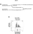
- exogenous ⁇ DNA (filler DNA) was added to the adapter-ligated cfDNA library in order to artificially inflate the amount of starting DNA to 100 ng ( Figure 2 ).
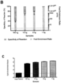
- the filler DNA consisted of amplicons similar in size to an adapter-ligated cfDNA library and was composed of unmethylated and in vitro methylated DNA at different methylation levels ( Figure 9 and Figure 10 ).
- the addition of this filler DNA also serves a practical use, as different patients will yield different amounts of cfDNA, allowing for the normalization of input DNA amount to 100 ng. This ensures that the downstream protocol remains exactly the same for all samples regardless of the amount of available cfDNA.
- a method of capturing cell-free methylated DNA from a sample having less than 100 ng of cell-free DNA comprising the steps of:
- this method further comprises the step of amplifying and subsequently sequencing the captured cell-free methylated DNA.
- NGS next-generation sequencing
- Illumina Solexa
- Roche 454 sequencing Ion torrent
- Proton / PGM sequencing SOLiD sequencing.
- NGS allow for the sequencing of DNA and RNA much more quickly and cheaply than the previously used Sanger sequencing.
- said sequencing is optimized for short read sequencing.
- Cell-free methylated DNA is DNA that is circulating freely in the blood stream, and are methylated at various known regions of the DNA. Samples, for example, plasma samples can be taken to analyze cell-free methylated DNA.
- library preparation includes list end-repair, A-tailing, adapter ligation, or any other preparation performed on the cell free DNA to permit subsequent sequencing of DNA.
- fill DNA can be noncoding DNA or it can consist of amplicons.
- DNA samples may be denatured, for example, using sufficient heat.
- samples have less than 50 ng of cell-free DNA.
- the first amount of filler DNA comprises about 10%, 20%, 30%, 40%, 50%, 60%, 70%, 80%, 90%, or 100% methylated filler DNA. In preferred embodiments, the first amount of filler DNA comprises about 50% methylated filler DNA.
- the first amount of filler DNA is from 20 ng to 100 ng. In preferred embodiments, 30 ng to 100 ng of filler DNA. In more preferred embodiments 50 ng to 100 ng of filler DNA.
- the filler DNA is 50 bp to 800 bp long. In preferred embodiments, 100 bp to 600 bp long; and in more preferred embodiments 200 bp to 600 bp long.
- the filler DNA is double stranded.
- the filler DNA can be junk DNA.
- the filler DNA may also be endogenous or exogenous DNA.
- the filler DNA is non-human DNA, and in preferred embodiments, ⁇ DNA.
- ⁇ DNA refers to Enterobacteria phage ⁇ DNA.
- the filler DNA has no alignment to human DNA.
- the binder is a protein comprising a Methyl-CpG-binding domain.
- MBD2 protein a protein comprising a Methyl-CpG-binding domain.
- MBD2 protein a protein comprising a Methyl-CpG-binding domain.
- MBD2 protein a protein comprising a Methyl-CpG-binding domain.
- MBD2 protein a protein comprising a Methyl-CpG-binding domain.
- MBD2 protein Methyl-CpG-binding domain
- Human proteins MECP2, MBD1, MBD2, MBD3, and MBD4 comprise a family of nuclear proteins related by the presence in each of a methyl-CpG-binding domain (MBD). Each of these proteins, with the exception of MBD3, is capable of binding specifically to methylated DNA.
- the binder is an antibody and capturing cell-free methylated DNA comprises immunoprecipitating the cell-free methylated DNA using the antibody.
- immunoprecipitation refers a technique of precipitating an antigen (such as polypeptides and nucleotides) out of solution using an antibody that specifically binds to that particular antigen. This process can be used to isolate and concentrate a particular protein or DNA from a sample and requires that the antibody be coupled to a solid substrate at some point in the procedure.
- the solid substrate includes for examples beads, such as magnetic beads. Other types of beads and solid substrates are known in the art.
- One exemplary antibody is 5-MeC antibody.
- the method described herein further comprises the step of adding a second amount of control DNA to the sample after step (b).
- Another exemplary antibody is or 5-hydroxymethyl cytosine antibody.
- the method described herein further comprises the step of adding a second amount of control DNA to the sample after step (b) for confirming the capture of cell-free methylated DNA.
- the use further comprising the use of described herein for identifying tissue of origin of the cancer cells within the cell-free DNA within the sample.
- Pancreatic adenocarcinoma (PDAC) patient samples were obtained from the University Health Network BioBank; healthy controls were recruited through the Family Medicine Centre at Mount Sinai Hospital (MSH) in Toronto, Canada. All samples collected with patient consent, were obtained with institutional approval from the Research Ethics Board, from University Health Network and Mount Sinai Hospital in Toronto, Canada.
- LCM Laser capture microdissection
- Microdissected tumor cells were collected by gravity into the caps of sterile, RNAse-free microcentrifuge tubes. Approximately 150,000-200,000 tumor cells were collected for DNA sample and stored at -80 °C until further processing. LCM typically took 1-2 days per case to collect sufficient amounts of purified tumor cells. Qiagen Cell Lysis Buffer was used to extract genomic DNA. Matched normal, histologically reviewed reference tissue was collected for each patient from frozen duodenal or gastric mucosa by scraping unstained frozen sections on glass slides into the appropriate DNA extraction buffer.
- EDTA and ACD plasma samples were obtained from the BioBank and from the Family Medicine Centre at Mount Sinai Hospital (MSH) in Toronto, Canada. All samples were either stored at -80°C or in vapour phase liquid nitrogen until use.
- Cell-free DNA was extracted from 0.5-3.5 ml of plasma using the QIAamp Circulating Nucleic Acid Kit (Qiagen). The extracted DNA was quantified through Qubit prior to use.
- Human colorectal tumor tissue obtained with patient consent from the University Health Network Biobank as approved by the Research Ethics Board at University Health Network, was digested to single cells using collagenase A. Single cells were subcutaneously injected into 4-6 week old NOD/SCID male mouse. Mice were euthanized by CO2 inhalation prior to blood collection by cardiac puncture and stored in EDTA tubes. From the collected blood samples, the plasma was isolated and stored at -80C. Cell-free DNA was extracted from 0.3-0.7 ml of plasma using the QIAamp Circulating Nucleic Acid Kit (Qiagen). All animal work was carried out in compliance with the ethical regulations approved by the Animal Care Committee at University Health Network.
- the optimal number of cycles to amplify each purified library was determined through the use of qPCR, after which the samples were amplified using the KAPA HiFi Uracil+ Mastermix (Kapa Biosystems) and purified with AMPure beads (Beckman Coulter). The final libraries were submitted for BioAnalyzer analysis prior to sequencing at the UHN Princess Margaret Genomic Centre in an Illumina HiSeq 2000.
- Enterobacteria phage ⁇ DNA was amplified using the primers indicated in Table 1, generating 6 different PCR amplicons products.
- the PCR reaction was carried out using KAPA HiFi Hotstart ReadyMix with the following condition: activation of enzyme at 95°C for 3 min, 30 cycles of 98°C for 20 sec, 60°C for 15 sec, 72°C for 30 sec and a final extension at 72°C for 1 min.
- the PCR amplicons were purified with QIAQuick PCR purification kit (Qiagen) and ran on a gel to verify size and amplification.
- Amplicons for 1CpG, 5CpG, 10CpG, 15CpG and 20CpGL were methylated using CpG Methyltransferase ( M . Sss I) (ThermoFischer Scientific) and purified with the QIAQuick PCR purification kit. Methylation of the PCR amplicons was tested using restriction enzyme HpyCH4 IV (New England Biolabs Canada) and ran on a gel to ensure its methylation.
- the DNA concentration of the unmethylated (20CpGS) and methylated (1CpG, 5CpG, 10CpG, 15CpG, 20CpGL) amplicons was measured using picogreen prior to pooling with 50% of methylated and 50% unmethylated ⁇ PCR product.
- FIG. 2 A schematic representation of the cfMeDIP-seq protocol is shown in Figure 2 .
- the DNA samples were subjected to library preparation using the Kapa Hyper Prep Kit (Kapa Biosystems). The manufacturer protocol was followed with some modifications. Briefly, the DNA of interest was added to 0.2 mL PCR tube and subjected to end-repair and A-Tailing. Adapter ligation was followed using NEBNext adapter (from the NEBNext Multiplex Oligos for Illumina kit, New England Biolabs) at a final concentration of 0.181 ⁇ M, incubated at 20°C for 20 mins and purified with AMPure XP beads. The eluted library was digested using the USER enzyme (New England Biolabs Canada) followed by purification with Qiagen MinElute PCR Purification Kit prior to MeDIP.
- NEBNext adapter from the NEBNext Multiplex Oligos for Illumina kit, New England Biolabs
- the prepared libraries were combined with the pooled methylated/unmethylated ⁇ PCR product to a final DNA amount of 100 ng and subjected to MeDIP using the protocol from Taiwo et al. 2012 17 with some modifications.
- MeDIP the Diagenode MagMeDIP kit (Cat# C02010021) was used following the manufacturer's protocol with some modifications. After the addition of 0.3 ng of the control methylated and 0.3ng of the control unmethylated A .
- thaliana DNA to complete the total amount of DNA [cfDNA + Filler + Controls] to 100ng
- the samples were heated to 95°C for 10 mins, then immediately placed into an ice water bath for 10 mins.
- Each sample was partitioned into two 0.2 mL PCR tubes: one for the 10% input control and the other one for the sample to be subjected to immunoprecipitation.
- the included 5-mC monoclonal antibody 33D3 (Cat#C15200081) from the MagMeDIP kit was diluted 1:15 prior to generating the diluted antibody mix and added to the sample.
- Washed magnetic beads (following manufacturer instructions) were also added prior to incubation at 4°C for 17 hours.
- the samples were purified using the Diagenode iPure Kit and eluted in 50 ⁇ l of Buffer C.
- the success of the reaction (QC1) was validated through qPCR to detect the presence of the spiked-in A . thaliana DNA, ensuring a % recovery of unmethylated spiked-in DNA ⁇ 1% and the % specificity of the reaction >99% (as calculated by 1-[recovery of spiked-in unmethylated control DNA over recovery of spiked-in methylated control DNA]), prior to proceeding to the next step.
- the optimal number of cycles to amplify each library was determined through the use of qPCR, after which the samples were amplified using the KAPA HiFi Hotstart Mastermix and the NEBNext multiplex oligos added to a final concentration of 0.3 ⁇ M.
- the PCR settings used to amplify the libraries were as follows: activation at 95°C for 3 min, followed by predetermined cycles of 98°C for 20 sec, 65°C for 15 sec and 72°C for 30 sec and a final extension of 72°C for 1 min.
- the amplified libraries were purified using MinElute PCR purification column and then gel size selected with 3% Nusieve GTG agarose gel to remove any adapter dimers.
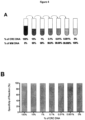
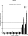
- cfMeDIP-seq was performed using different % of methylated to unmethylated lambda DNA in the filler component of the protocol as follows: Using filler (lambda) DNA to increase final amount of DNA to 100ng prior to immunoprecipitation, with the starting amounts of cell-free DNA ranging from 10ng, 5ng and 1ng % Methylated Filler % Unmethylated Filler 100 0 85 15 70 30 50 50 50 30 70 15 85 0 100 No Filler DNA used - as is only 10ng, 5ng or 1ng of starting DNA prior to immunoprecipitation
- the filler DNA (lambda DNA) used to increase the final amounts prior to immunoprecipitation to 100ng should preferably have some artificially methylated DNA in its composition (from 100%-15%) in order to have the minimal recovery unmethylated DNA ( Figure 9 ), while still getting a good yield in terms of recovery of methylated DNA ( Figure 10 ).
- the filler DNA in the samples where we have 100% unmethylated filler DNA or no filler DNA present, although there is really higher recovery of methylated DNA, we also have high % recovery of unmethylated DNA. This shows that the additional methylated DNA in the filler DNA helps to occupy the excess antibody present in the reaction, minimizing the amount of unspecific binding to unmethylated DNA found in the sample.
- this filler DNA helps normalize the different starting amounts and allow for different cell-free DNA samples to be processed the same way (i.e use same amount of antibody), while still recovering good methylation data from it.
- DNA sequencing libraries were constructed from 83 ng of fragmented DNA using the KAPA Hyper Prep Kit (Kapa Biosystems, Wilmington, MA) utilizing NEXTflex-96 DNA Barcode adapters (Bio Scientific, Austin, TX) adapters.
- KAPA Hyper Prep Kit Kapa Biosystems, Wilmington, MA
- NEXTflex-96 DNA Barcode adapters Bio Scientific, Austin, TX
- the barcoded libraries were pooled and then applied the custom hybrid capture library following manufacturer's instructions (IDT xGEN Lockdown protocol version 2.1). These fragments were sequenced to >10,000X read coverage using an Illumina HiSeq 2000 instrument. Resulting reads were aligned using bwa-mem and mutations detected using
- DMRs Differentially Methylated Regions
- PC Pancreatic Cancer
- Healthy Donors were calculated using the MEDIPS R package 25 .
- BAM alignment to human genome hg19 files were used to create MEDIPS R objects.
- DMRs were calculated by comparing the RPKMs from the two sets of samples using t-tests. The raw p-values from the t-tests were adjusted using the Benjamini-Hochberg procedure.
- DMCs were obtained using the criteria of Benjamini-Hochberg adjusted p-value ⁇ 0.01 and Delta Beta > 0.25, and 134,021 DMCs were found to be Hyper in Pancreatic Cancer compared to PBMCs.
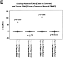
- Permutation analysis was carried out to compare the frequency of expected versus the observed overlap between the DMRs identified in the plasma (with circulating cfDNA subjected to our cfMeDIP-seq protocol) and the cancer-specific DMCs identified in the primary tumor tissue (with RRBS).
- RNA-Seq data was obtained from the GTEx database in the form of median RPKMs by tissue for all human genes (obtained from file GTEx_Analysis_v6p_RNA-seq_RNA-SeQCv1.1.8_gene_median_rpkm.gct.gz under https://gtexportal.org/home/datasets).
- TFs of interest were matched to their gene names, and heatmaps ( Figure 8A , 8C ) were constructed with the median RPKMs of each TF scaled across all tissues.
- the distance function "manhattan” and clustering function "average” were used for both row-wise and column-wise clustering.
- the cfMeDIP-seq method described here was developed through the modification of an existing low input MeDIP-seq protocol 17 that is robust down to 100 ng of input DNA.
- exogenous ⁇ DNA filler DNA
- the filler DNA consisted of amplicons similar in size to an adapter-ligated cfDNA library and was composed of unmethylated and in vitro methylated DNA at different CpG densities. The addition of this filler DNA also serves a practical use, as different patients will yield different amounts of cfDNA, allowing for the normalization of input DNA amount to 100 ng. This ensures that the downstream protocol remains exactly the same for all samples regardless of the amount of available cfDNA.
- cfMeDIP-seg displays high-sensitivity for detection of tumor-derived ctDNA
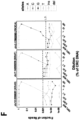
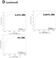
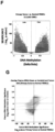
- Cancer DNA is frequently hypermethylated at CpG-rich regions 1 . Since cfMeDIP-seq specifically targets methylated CpG-rich sequences, we hypothesized that ctDNA would be preferentially enriched during the immunoprecipitation procedure. To test this, we generated patient-derived xenografts (PDXs) from two colorectal cancer patients and collected the mouse plasma. Tumor-derived human cfDNA was present at less than 1% frequency within the total cfDNA pool in the input samples and at 2-fold greater abundance following immunoprecipitation ( Figure 1G ). These results suggest that through biased sequencing of ctDNA, the cfMeDIP procedure could further increase ctDNA detection sensitivity.
- PDXs patient-derived xenografts
- Methylome analysis of plasma cfDNA distinguishes early stage pancreatic adenocarcinoma patients from healthy donors
- Plasma cfDNA methylomes permit inference of tumor-associated active transcription factor networks
- TFs preferentially expressed in the pancreas including RBPJL, PTF1a, Onecut1 (HNF6), and NR5A2 ( Figure 8C-D ).
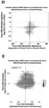
- the TF motifs identified as hypomethylated footprints in the pancreatic adenocarcinoma cases were also frequently overexpressed in pancreatic adenocarcinoma patients from TCGA ( Figure 8E ).
- c-MYC and HIF1a (Squamous subtype drivers), NR5A2, MAFA, RBPJL, and NEUROD1 (ADEX drivers) and finally FOXA2 and HNF4A (pancreatic progenitor subtype).
- methylome analysis of circulating cfDNA can be used to infer active transcriptional networks within the tumor based on the differentially methylated TF footprints and potentially identify systemic shifts in immune cell populations between healthy donors and cancer patients.
- cfMeDIP-seq relies on epigenetic, rather than genomic information, it could potentially be used to non-invasively monitor tissue damage in a broad set of non-malignant diseases. For instance, it could be used to monitor immune response to an infection or after cancer immunotherapy; it could be used to monitor heart DNA in the circulation after myocardial infarction or brain DNA during early stages of neurodegenerative diseases.
- pancreatic cancer patients can be stratified into four subgroups driven by several mechanisms 24 ; squamous, pancreatic progenitor, immunogenic and aberrantly differentiated endocrine exocrine (ADEX).
- squamous, pancreatic progenitor, immunogenic and aberrantly differentiated endocrine exocrine (ADEX) In the circulating cfDNA methylome of pancreatic cancer patients, we were able to identify the hypomethylated footprints from TFs that drive these subtypes.
- MYC and HIF1 alpha Hypoxia-inducible factor 1-alpha
- HNF4A and FOXA2 two pathways enriched in the squamous subtype 24 .
- HNF4A and FOXA2 two pathways enriched in the progenitor subtype 24 .
- NR5A2, RBPJL, and MAFA three TFs enriched in the ADEX subtype 24 . This suggests that cfMeDIP-seq could also be used as a biomarker to stratify cancer patients with a minimally invasive approach.
- PCR primers used to generate Enterobacteria phage ⁇ PCR product from Taiwo et al., 2012 Name Forward Primer Reverse Primer 1CpG GAGGTGATAAAATTAACTGC GGCTCTACCATATCTCCTA 5CpG CATGTCCAGAGCTCATTC GTTTAAAATCACTAGGCGA 10CpG CTGACCATTTCCATCATTC GTAACTAAACAGGAGCCG 15CpG ATGTATCCATTGAGCATTGCC CACGAATCAGCGGTAAAGGT 20CpGL GAGATATGGTAGAGCCGCAGA TTTCAGCAGCTACAGTCAGAATTT 20CpGS CGATGGGTTAATTCGCTCGTTGTGG GCACAAGGGAAAGAGCACTG
Landscapes
- Chemical & Material Sciences (AREA)
- Health & Medical Sciences (AREA)
- Life Sciences & Earth Sciences (AREA)
- Engineering & Computer Science (AREA)
- Organic Chemistry (AREA)
- Genetics & Genomics (AREA)
- Wood Science & Technology (AREA)
- Zoology (AREA)
- Biotechnology (AREA)
- Molecular Biology (AREA)
- Biomedical Technology (AREA)
- Immunology (AREA)
- Bioinformatics & Cheminformatics (AREA)
- General Engineering & Computer Science (AREA)
- Analytical Chemistry (AREA)
- General Health & Medical Sciences (AREA)
- Microbiology (AREA)
- Biochemistry (AREA)
- Physics & Mathematics (AREA)
- Proteomics, Peptides & Aminoacids (AREA)
- Biophysics (AREA)
- Pathology (AREA)
- Crystallography & Structural Chemistry (AREA)
- Plant Pathology (AREA)
- Hematology (AREA)
- Urology & Nephrology (AREA)
- Chemical Kinetics & Catalysis (AREA)
- Cell Biology (AREA)
- General Physics & Mathematics (AREA)
- Tropical Medicine & Parasitology (AREA)
- Food Science & Technology (AREA)
- Medicinal Chemistry (AREA)
- Bioinformatics & Computational Biology (AREA)
- Hospice & Palliative Care (AREA)
- Oncology (AREA)
- Measuring Or Testing Involving Enzymes Or Micro-Organisms (AREA)
- Peptides Or Proteins (AREA)
Applications Claiming Priority (3)
| Application Number | Priority Date | Filing Date | Title |
|---|---|---|---|
| US201662331070P | 2016-05-03 | 2016-05-03 | |
| PCT/CA2017/000108 WO2017190215A1 (en) | 2016-05-03 | 2017-05-03 | Methods of capturing cell-free methylated dna and uses of same |
| EP17792306.7A EP3452615A4 (de) | 2016-05-03 | 2017-05-03 | Verfahren zur erfassung von zellfreier methylierter dna und verwendungen davon |
Related Parent Applications (1)
| Application Number | Title | Priority Date | Filing Date |
|---|---|---|---|
| EP17792306.7A Division EP3452615A4 (de) | 2016-05-03 | 2017-05-03 | Verfahren zur erfassung von zellfreier methylierter dna und verwendungen davon |
Publications (2)
| Publication Number | Publication Date |
|---|---|
| EP4425178A2 true EP4425178A2 (de) | 2024-09-04 |
| EP4425178A3 EP4425178A3 (de) | 2024-12-18 |
Family
ID=60202577
Family Applications (2)
| Application Number | Title | Priority Date | Filing Date |
|---|---|---|---|
| EP24176389.5A Pending EP4425178A3 (de) | 2016-05-03 | 2017-05-03 | Verfahren zur erfassung von zellfreier methylierter dna und verwendungen davon |
| EP17792306.7A Withdrawn EP3452615A4 (de) | 2016-05-03 | 2017-05-03 | Verfahren zur erfassung von zellfreier methylierter dna und verwendungen davon |
Family Applications After (1)
| Application Number | Title | Priority Date | Filing Date |
|---|---|---|---|
| EP17792306.7A Withdrawn EP3452615A4 (de) | 2016-05-03 | 2017-05-03 | Verfahren zur erfassung von zellfreier methylierter dna und verwendungen davon |
Country Status (7)
| Country | Link |
|---|---|
| US (5) | US11078475B2 (de) |
| EP (2) | EP4425178A3 (de) |
| JP (3) | JP6979554B2 (de) |
| KR (3) | KR102522067B1 (de) |
| CN (1) | CN109415763A (de) |
| BR (1) | BR112018072536A2 (de) |
| WO (1) | WO2017190215A1 (de) |
Families Citing this family (31)
| Publication number | Priority date | Publication date | Assignee | Title |
|---|---|---|---|---|
| US9115386B2 (en) | 2008-09-26 | 2015-08-25 | Children's Medical Center Corporation | Selective oxidation of 5-methylcytosine by TET-family proteins |
| KR102522067B1 (ko) * | 2016-05-03 | 2023-04-13 | 유니버시티 헬스 네트워크 | 무-세포 메틸화된 dna의 포획 방법 및 이의 이용 |
| MX2019007444A (es) | 2016-12-22 | 2019-08-16 | Guardant Health Inc | Metodos y sistemas para analisis de moleculas de acido nucleico. |
| CA3063405A1 (en) * | 2017-05-12 | 2018-11-15 | President And Fellows Of Harvard College | Universal early cancer diagnostics |
| US12227737B2 (en) * | 2017-07-12 | 2025-02-18 | University Health Network | Cancer detection and classification using methylome analysis |
| BR112020000681A2 (pt) * | 2017-07-12 | 2020-07-14 | University Health Network | detecção e classificação de cancro utilizando análise de metilome |
| US20210156863A1 (en) * | 2017-11-03 | 2021-05-27 | University Health Network | Cancer detection, classification, prognostication, therapy prediction and therapy monitoring using methylome analysis |
| CN108796057A (zh) * | 2018-06-29 | 2018-11-13 | 上海交通大学 | 一种少量样品全基因组dna甲基化的检测方法及试剂盒 |
| CN108796039B (zh) * | 2018-06-29 | 2021-10-08 | 上海交通大学 | 一种用于dna甲基化检测的试剂盒与方法及应用 |
| CN109023537B (zh) * | 2018-09-04 | 2021-10-08 | 上海交通大学 | 一种微量dna样品高通量测序文库的构建技术 |
| CN109097439A (zh) * | 2018-09-04 | 2018-12-28 | 上海交通大学 | 一种检测少量样品全基因组dna甲基化的方法 |
| WO2020160414A1 (en) * | 2019-01-31 | 2020-08-06 | Guardant Health, Inc. | Compositions and methods for isolating cell-free dna |
| CN109825559A (zh) * | 2019-03-11 | 2019-05-31 | 上海交通大学 | 一种血浆cfDNA全局性甲基化检测方法及应用 |
| US11891653B2 (en) | 2019-09-30 | 2024-02-06 | Guardant Health, Inc. | Compositions and methods for analyzing cell-free DNA in methylation partitioning assays |
| CN115087744A (zh) * | 2019-11-06 | 2022-09-20 | 大学健康网络 | 用于无细胞MeDIP测序的合成加标对照及其使用方法 |
| EP4617375A3 (de) * | 2019-11-26 | 2025-10-29 | Guardant Health, Inc. | Verfahren, zusammensetzungen und systeme zur verbesserung der bindung methylierter polynukleotide |
| CN111154846A (zh) * | 2020-01-13 | 2020-05-15 | 四川大学华西医院 | 一种甲基化核酸的检测方法 |
| EP4715052A2 (de) * | 2020-05-18 | 2026-03-25 | Myriad Women's Health, Inc. | Verfahren zur anreicherung und zum screening von nukleinsäureproben |
| US12592321B2 (en) | 2020-06-19 | 2026-03-31 | University Health Network | Cancer detection and classification using methylome analysis |
| KR102332540B1 (ko) | 2020-07-02 | 2021-11-29 | 의료법인 성광의료재단 | 다운증후군 특이적인 후성학적 마커를 이용한 다운증후군 진단 방법 |
| KR102332539B1 (ko) | 2020-07-02 | 2021-11-29 | 의료법인 성광의료재단 | 다운증후군 진단용 후성학적 바이오마커 조성물 및 이의 용도 |
| EP4585697A3 (de) * | 2020-07-30 | 2025-08-06 | Guardant Health, Inc. | Verfahren zur isolierung von zellfreier dna |
| AU2021345359A1 (en) | 2020-09-21 | 2023-05-11 | Enumera Molecular, Inc. | Compositions and methods for isolation of cell-free dna |
| EP4222278A1 (de) * | 2020-09-30 | 2023-08-09 | Guardant Health, Inc. | Zusammensetzungen und verfahren zur analyse von dna unter verwendung von partitionierung und einer methylierungsabhängigen nuklease |
| JP2023547620A (ja) | 2020-10-23 | 2023-11-13 | ガーダント ヘルス, インコーポレイテッド | 分配および塩基変換を使用してdnaを解析するための組成物および方法 |
| JP2024525190A (ja) * | 2021-06-18 | 2024-07-10 | シングレラ ジェノミクス (チャンスー) リミテッド | 腫瘍評価のための物質及び方法 |
| JP2024523401A (ja) * | 2021-06-21 | 2024-06-28 | ガーダント ヘルス, インコーポレイテッド | コピー数情報に基づく組織起源分析のための方法および組成物 |
| CN113621691A (zh) * | 2021-09-27 | 2021-11-09 | 广东省妇幼保健院 | 基于一代测序技术直接检测KLF1基因启动子区CpG甲基化的方法和试剂盒 |
| CN114045342A (zh) * | 2021-12-01 | 2022-02-15 | 大连晶泰生物技术有限公司 | 一种游离DNA(cfDNA)甲基化突变的检测方法及试剂盒 |
| WO2023107709A1 (en) * | 2021-12-10 | 2023-06-15 | Adela, Inc. | Methods and systems for generating sequencing libraries |
| WO2023192635A2 (en) * | 2022-04-01 | 2023-10-05 | Twist Bioscience Corporation | Libraries for methylation analysis |
Family Cites Families (13)
| Publication number | Priority date | Publication date | Assignee | Title |
|---|---|---|---|---|
| US6818762B2 (en) * | 2001-05-25 | 2004-11-16 | Maine Molecular Quality Controls, Inc. | Compositions and methods relating to nucleic acid reference standards |
| AU2002363062B2 (en) * | 2001-10-09 | 2007-03-22 | Nanosphere, Inc. | Nanoparticles having oligonucleotides attached thereto and uses therefor |
| CN101802223A (zh) * | 2007-08-15 | 2010-08-11 | 香港大学 | 用于高通量亚硫酸氢盐dna-测序的方法和组合物及其用途 |
| CN103443338B (zh) * | 2011-02-02 | 2017-09-22 | 华盛顿大学商业化中心 | 大规模平行邻接作图 |
| US8895249B2 (en) * | 2012-06-15 | 2014-11-25 | Illumina, Inc. | Kinetic exclusion amplification of nucleic acid libraries |
| US9732390B2 (en) * | 2012-09-20 | 2017-08-15 | The Chinese University Of Hong Kong | Non-invasive determination of methylome of fetus or tumor from plasma |
| SI4056712T1 (sl) * | 2012-09-20 | 2024-10-30 | The Chinese University Of Hong Kong | Neinvazivno določanje metiloma tumorja iz plazme |
| US10706957B2 (en) * | 2012-09-20 | 2020-07-07 | The Chinese University Of Hong Kong | Non-invasive determination of methylome of tumor from plasma |
| JP6681841B2 (ja) | 2014-05-09 | 2020-04-15 | ライフコーデックス アーゲー | 特別な細胞タイプに由来するdnaの検出とそれに関連する方法 |
| TWI813141B (zh) * | 2014-07-18 | 2023-08-21 | 香港中文大學 | Dna混合物中之組織甲基化模式分析 |
| KR102522067B1 (ko) * | 2016-05-03 | 2023-04-13 | 유니버시티 헬스 네트워크 | 무-세포 메틸화된 dna의 포획 방법 및 이의 이용 |
| BR112020000681A2 (pt) * | 2017-07-12 | 2020-07-14 | University Health Network | detecção e classificação de cancro utilizando análise de metilome |
| US20210156863A1 (en) * | 2017-11-03 | 2021-05-27 | University Health Network | Cancer detection, classification, prognostication, therapy prediction and therapy monitoring using methylome analysis |
-
2017
- 2017-05-03 KR KR1020217023549A patent/KR102522067B1/ko active Active
- 2017-05-03 EP EP24176389.5A patent/EP4425178A3/de active Pending
- 2017-05-03 CN CN201780032740.5A patent/CN109415763A/zh active Pending
- 2017-05-03 WO PCT/CA2017/000108 patent/WO2017190215A1/en not_active Ceased
- 2017-05-03 KR KR1020237012338A patent/KR20230054746A/ko not_active Ceased
- 2017-05-03 US US16/098,620 patent/US11078475B2/en active Active
- 2017-05-03 KR KR1020187034995A patent/KR102359767B1/ko active Active
- 2017-05-03 BR BR112018072536-6A patent/BR112018072536A2/pt not_active Application Discontinuation
- 2017-05-03 JP JP2018557336A patent/JP6979554B2/ja active Active
- 2017-05-03 EP EP17792306.7A patent/EP3452615A4/de not_active Withdrawn
-
2021
- 2021-06-21 US US17/353,756 patent/US20220119796A1/en active Pending
- 2021-06-22 JP JP2021103482A patent/JP7434215B2/ja active Active
- 2021-11-04 US US17/519,350 patent/US11560558B2/en active Active
-
2022
- 2022-11-28 US US18/059,370 patent/US20230287384A1/en active Pending
-
2024
- 2024-02-07 JP JP2024017072A patent/JP2024054242A/ja active Pending
-
2025
- 2025-06-18 US US19/241,820 patent/US20250313825A1/en active Pending
Non-Patent Citations (30)
| Title |
|---|
| AKALIN, A. ET AL.: "methylKit: a comprehensive R package for the analysis of genome-wide DNA methylation profiles.", GENOME BIOL, vol. 13, 2012, pages 87 |
| ARAVANIS, A. M.LEE, M.KLAUSNER, R. D: "Next-Generation Sequencing of Circulating Tumor DNA for Early Cancer Detection.", CELL, vol. 168, 2017, pages 571 - 574, XP029935395, DOI: 10.1016/j.cell.2017.01.030 |
| BAILEY, P. ET AL.: "Genomic analyses identify molecular subtypes of pancreatic cancer.", NATURE, 2016, pages 16965 |
| CHIU, R. W. ET AL.: "Noninvasive prenatal diagnosis of fetal chromosomal aneuploidy by massively parallel genomic sequencing of DNA in maternal plasma.", PROC NATL ACAD SCI U, vol. 5, no. 105, 2008, pages 20458 - 20463 |
| CONSORTIUM, G. T.: "Human genomics. The Genotype-Tissue Expression (GTEx) pilot analysis: multitissue gene regulation in humans.", SCIENCE, vol. 348, 2015, pages 648 - 660 |
| DIAZ, L. A., JR.BARDELLI, A.: "Liquid biopsies: genotyping circulating tumor DNA", J CLIN ONCOL, vol. 32, 2014, pages 579 - 586, XP055449655, DOI: 10.1200/JCO.2012.45.2011 |
| FAN, H. CBLUMENFELD, Y. J.CHITKARA, U.HUDGINS, L.QUAKE, S. R.: "Noninvasive diagnosis of fetal aneuploidy by shotgun sequencing DNA from maternal blood.", PROC NATL ACAD SCI U S A, vol. 105, 2008, pages 16266 - 16271, XP093117217, DOI: 10.1073/pnas.0808319105 |
| FANG, F. ET AL.: "Breast cancer methylomes establish an epigenomic foundation for metastasis.", SCI TRANSL MED, vol. 3, 2011, pages 75 - 25, XP055045310, DOI: 10.1126/scitranslmed.3001875 |
| FLEISCHHACKER, M.SCHMIDT, B: "Biochim Biophys Acta", vol. 1775, 2007, article "Circulating nucleic acids (CNAs) and cancer-a survey.", pages: 181 - 232 |
| GU, H. ET AL.: "Preparation of reduced representation bisulfite sequencing libraries for genome-scale DNA methylation profiling.", NAT PROTOC, vol. 6, 2011, pages 468 - 481, XP055151116, DOI: 10.1038/nprot.2010.190 |
| HEINZ, S. ET AL.: "Simple combinations of lineage-determining transcription factors prirne cis-regulatory elements required for macrophage and B cell identities.", MOF CELL, vol. 38, 2010, pages 576 - 589 |
| HINOUE, T. ET AL.: "Genome-scale analysis of aberrant DNA methylation in colorectal cancer.", GENOME RES, vol. 22, 2012, pages 271 - 282, XP055821428, DOI: 10.1101/gr.117523.110 |
| HOADLEY, K. A. ET AL.: "Multiplatform analysis of 12 cancer types reveals molecular classification within and across tissues of origin.", CELL, vol. 158, 2014, pages 929 - 944, XP055934214, DOI: 10.1016/j.cell.2014.06.049 |
| HU, S. ET AL.: "DNA methylation presents distinct binding sites for human transcription factors.", ELIFE, vol. 2, 2013, pages e00726 |
| HUNG, E. C., CHIU, R. W. & LO, Y. M.: "Detection of circulating fetal nucleic acids: a review of methods and applications", J CLIN PATHOL, vol. 62, 2009, pages 308 - 313, XP009132411 |
| KRUEGER, F.ANDREWS, S. R: "Bismark: a flexible aligner and methylation caller for Bisulfite-Seq applications.", BIOINFORMATICS, vol. 27, 2011, pages 1571 - 1572, XP093055863, DOI: 10.1093/bioinformatics/btr167 |
| LIENHARD, M., GRIMM, C., MARKET, M., HERWIG, R. & CHAVEZ, L.: "MEDIPS:genome-wide differential coverage analysis of sequencing data derived from DNA enrichment experiments", BIOINFORMATICS, vol. 30, 2014, pages 284 - 286, XP055821362, DOI: 10.1093/bioinformatics/btt650 |
| LUI, Y. Y. ET AL.: "Predominant hematopoietic origin of cell-free DNA in plasma and serum after sex-mismatched bone marrow transplantation.", CLIN CHEM, vol. 48, 2002, pages 421 - 427, XP055443838 |
| MACK, S. C. ET AL.: "Epigenomic alterations define lethal CIMP-positive ependymomas of infancy.", NATURE, vol. 506, 2014, pages 445 - 450, XP055210650, DOI: 10.1038/nature13108 |
| MIKESKA, T.CRAIG, J. M.: "DNA methylation biomarkers: cancer and beyond.", GENES (BASEL, vol. 5, 2014, pages 821 - 864, XP093103969, DOI: 10.3390/genes5030821 |
| NEWMAN, A. M. ET AL.: "An ultrasensitive method for quantitating circulating tumor DNA with broad patient coverage.", NAT MED, vol. 20, 2014, pages 548 - 554, XP055580741, DOI: 10.1038/nm.3519 |
| SHARMA, SKELLY, T. KJONES, P. A., EPIGENETICS IN CANCER. CARCINOGENESIS, vol. 31, pages 27 - 36 |
| SNYDER, M. W.KIRCHER, M.HILL, A. J.DAZA, R. M.SHENDURE, J.: "Cell-free DNA Comprises an In Vivo Nucleosome Footprint that Informs Its Tissues-Of-Origin.", CELL, vol. 164, 2016, pages 57 - 68 |
| SNYDER, T. MKHUSH, K. K.VALANTINE, H. A.QUAKE, S. R.: "Universal noninvasive detection of solid organ transplant rejection.", PROC MALL ACAD SCI U SA, vol. 108, 2011, pages 6229 - 6234, XP055524568, DOI: 10.1073/pnas.1013924108 |
| STIRZAKER, C. ET AL.: "Methylome sequencing in triple-negative breast cancer reveals distinct methylation clusters with prognostic value.", NAT COMMUN, vol. 6, 2015, pages 5899, XP055418632, DOI: 10.1038/ncomms6899 |
| STURM, D. ET AL.: "Hotspot mutations in H3F3A and IDH1 define distinct epigenetic and biological subgroups of glioblastoma", CANCER CELL, vol. 22, 2012, pages 425 - 437, XP055069630, DOI: 10.1016/j.ccr.2012.08.024 |
| SU, A. I. ET AL.: "A gene atlas of the mouse and human protein-encoding transcriptomes", PROC NATF ACAD SCI U S A, vol. 101, 2004, pages 6062 - 6067, XP055263129, DOI: 10.1073/pnas.0400782101 |
| TAIWO, O. ET AL.: "Methylome analysis using MeDIP-seq with low DNA concentrations.", NAT PROTOC, vol. 7, 2012, pages 617 - 636, XP055581354, DOI: 10.1038/nprot.2012.012 |
| VARLEY, K. E. ET AL.: "Dynamic DNA methylation across diverse human cell lines and tissues", GENOME RES, vol. 23, 2013, pages 555 - 567 |
| WU, C.JIN, X.TSUENG, G.AFRASIABI, C.SU, A. Γ.: "BioGPS: building your own mash-up of gene annotations and expression profiles.", NUCLEIC ACIDS RES, vol. 44, 2016, pages 0313 - 316 |
Also Published As
| Publication number | Publication date |
|---|---|
| US11560558B2 (en) | 2023-01-24 |
| KR102522067B1 (ko) | 2023-04-13 |
| US20220119796A1 (en) | 2022-04-21 |
| US20220073902A1 (en) | 2022-03-10 |
| BR112018072536A2 (pt) | 2019-03-26 |
| US20230287384A1 (en) | 2023-09-14 |
| JP7434215B2 (ja) | 2024-02-20 |
| JP2021168657A (ja) | 2021-10-28 |
| KR20210095962A (ko) | 2021-08-03 |
| JP2019521315A (ja) | 2019-07-25 |
| EP3452615A1 (de) | 2019-03-13 |
| CA3022606A1 (en) | 2017-11-09 |
| EP3452615A4 (de) | 2019-12-04 |
| US20250313825A1 (en) | 2025-10-09 |
| CN109415763A (zh) | 2019-03-01 |
| KR20190003987A (ko) | 2019-01-10 |
| US20190144848A1 (en) | 2019-05-16 |
| JP2024054242A (ja) | 2024-04-16 |
| WO2017190215A1 (en) | 2017-11-09 |
| US11078475B2 (en) | 2021-08-03 |
| KR102359767B1 (ko) | 2022-02-08 |
| KR20230054746A (ko) | 2023-04-25 |
| JP6979554B2 (ja) | 2021-12-15 |
| EP4425178A3 (de) | 2024-12-18 |
Similar Documents
| Publication | Publication Date | Title |
|---|---|---|
| US11560558B2 (en) | Methods of capturing cell-free methylated DNA and uses of same | |
| US20220251665A1 (en) | Cancer detection and classification using methylome analysis | |
| JP7256748B2 (ja) | エラーが訂正された核酸配列決定への適用を伴う標的化核酸配列濃縮のための方法 | |
| US20210156863A1 (en) | Cancer detection, classification, prognostication, therapy prediction and therapy monitoring using methylome analysis | |
| CN113661249A (zh) | 用于分离无细胞dna的组合物和方法 | |
| Kwon et al. | Genome-wide analysis of DNA methylation and the gene expression change in lung cancer | |
| EP3757229A1 (de) | Verfahren zur herstellung zellfreier nukleinsäuremoleküle durch in-situ-amplifizierung | |
| CN114616343A (zh) | 用于在甲基化分区测定中分析无细胞dna的组合物和方法 | |
| US12227737B2 (en) | Cancer detection and classification using methylome analysis | |
| US20250297320A1 (en) | Methylation signatures in cell-free dna for tumor classification and early detection | |
| HK40116163A (en) | Methods of capturing cell-free methylated dna and uses of same | |
| CA3022606C (en) | Methods of capturing cell-free methylated dna and uses of same | |
| Wei et al. | Genomics studies in hepatocellular carcinoma via next-generation sequencing | |
| HK40112829A (zh) | 使用甲基化组分析进行癌症检测和分类 |
Legal Events
| Date | Code | Title | Description |
|---|---|---|---|
| PUAI | Public reference made under article 153(3) epc to a published international application that has entered the european phase |
Free format text: ORIGINAL CODE: 0009012 |
|
| STAA | Information on the status of an ep patent application or granted ep patent |
Free format text: STATUS: THE APPLICATION HAS BEEN PUBLISHED |
|
| AC | Divisional application: reference to earlier application |
Ref document number: 3452615 Country of ref document: EP Kind code of ref document: P |
|
| AK | Designated contracting states |
Kind code of ref document: A2 Designated state(s): AL AT BE BG CH CY CZ DE DK EE ES FI FR GB GR HR HU IE IS IT LI LT LU LV MC MK MT NL NO PL PT RO RS SE SI SK SM TR |
|
| REG | Reference to a national code |
Ref country code: DE Ref legal event code: R079 Free format text: PREVIOUS MAIN CLASS: G01N0033530000 Ipc: C12Q0001680000 |
|
| PUAL | Search report despatched |
Free format text: ORIGINAL CODE: 0009013 |
|
| AK | Designated contracting states |
Kind code of ref document: A3 Designated state(s): AL AT BE BG CH CY CZ DE DK EE ES FI FR GB GR HR HU IE IS IT LI LT LU LV MC MK MT NL NO PL PT RO RS SE SI SK SM TR |
|
| RIC1 | Information provided on ipc code assigned before grant |
Ipc: G01N 33/53 20060101ALI20241114BHEP Ipc: C12Q 1/6886 20180101ALI20241114BHEP Ipc: C12Q 1/6806 20180101ALI20241114BHEP Ipc: C12Q 1/6804 20180101ALI20241114BHEP Ipc: C40B 30/04 20060101ALI20241114BHEP Ipc: C12N 15/10 20060101ALI20241114BHEP Ipc: C12Q 1/68 20180101AFI20241114BHEP |
|
| REG | Reference to a national code |
Ref country code: HK Ref legal event code: DE Ref document number: 40116163 Country of ref document: HK |
|
| STAA | Information on the status of an ep patent application or granted ep patent |
Free format text: STATUS: REQUEST FOR EXAMINATION WAS MADE |
|
| 17P | Request for examination filed |
Effective date: 20250617 |