EP4421186A2 - Methods of determining a surgical margin and methods of use thereof - Google Patents
Methods of determining a surgical margin and methods of use thereof Download PDFInfo
- Publication number
- EP4421186A2 EP4421186A2 EP24171813.9A EP24171813A EP4421186A2 EP 4421186 A2 EP4421186 A2 EP 4421186A2 EP 24171813 A EP24171813 A EP 24171813A EP 4421186 A2 EP4421186 A2 EP 4421186A2
- Authority
- EP
- European Patent Office
- Prior art keywords
- analyte
- tissue
- capture
- location
- tissue sample
- Prior art date
- Legal status (The legal status is an assumption and is not a legal conclusion. Google has not performed a legal analysis and makes no representation as to the accuracy of the status listed.)
- Granted
Links
Images


Classifications
-
- C—CHEMISTRY; METALLURGY
- C12—BIOCHEMISTRY; BEER; SPIRITS; WINE; VINEGAR; MICROBIOLOGY; ENZYMOLOGY; MUTATION OR GENETIC ENGINEERING
- C12N—MICROORGANISMS OR ENZYMES; COMPOSITIONS THEREOF; PROPAGATING, PRESERVING, OR MAINTAINING MICROORGANISMS; MUTATION OR GENETIC ENGINEERING; CULTURE MEDIA
- C12N15/00—Mutation or genetic engineering; DNA or RNA concerning genetic engineering, vectors, e.g. plasmids, or their isolation, preparation or purification; Use of hosts therefor
- C12N15/09—Recombinant DNA-technology
- C12N15/10—Processes for the isolation, preparation or purification of DNA or RNA
- C12N15/1034—Isolating an individual clone by screening libraries
- C12N15/1065—Preparation or screening of tagged libraries, e.g. tagged microorganisms by STM-mutagenesis, tagged polynucleotides, gene tags
-
- C—CHEMISTRY; METALLURGY
- C12—BIOCHEMISTRY; BEER; SPIRITS; WINE; VINEGAR; MICROBIOLOGY; ENZYMOLOGY; MUTATION OR GENETIC ENGINEERING
- C12Q—MEASURING OR TESTING PROCESSES INVOLVING ENZYMES, NUCLEIC ACIDS OR MICROORGANISMS; COMPOSITIONS OR TEST PAPERS THEREFOR; PROCESSES OF PREPARING SUCH COMPOSITIONS; CONDITION-RESPONSIVE CONTROL IN MICROBIOLOGICAL OR ENZYMOLOGICAL PROCESSES
- C12Q1/00—Measuring or testing processes involving enzymes, nucleic acids or microorganisms; Compositions therefor; Processes of preparing such compositions
- C12Q1/68—Measuring or testing processes involving enzymes, nucleic acids or microorganisms; Compositions therefor; Processes of preparing such compositions involving nucleic acids
- C12Q1/6813—Hybridisation assays
- C12Q1/6841—In situ hybridisation
-
- C—CHEMISTRY; METALLURGY
- C12—BIOCHEMISTRY; BEER; SPIRITS; WINE; VINEGAR; MICROBIOLOGY; ENZYMOLOGY; MUTATION OR GENETIC ENGINEERING
- C12Q—MEASURING OR TESTING PROCESSES INVOLVING ENZYMES, NUCLEIC ACIDS OR MICROORGANISMS; COMPOSITIONS OR TEST PAPERS THEREFOR; PROCESSES OF PREPARING SUCH COMPOSITIONS; CONDITION-RESPONSIVE CONTROL IN MICROBIOLOGICAL OR ENZYMOLOGICAL PROCESSES
- C12Q1/00—Measuring or testing processes involving enzymes, nucleic acids or microorganisms; Compositions therefor; Processes of preparing such compositions
- C12Q1/68—Measuring or testing processes involving enzymes, nucleic acids or microorganisms; Compositions therefor; Processes of preparing such compositions involving nucleic acids
- C12Q1/6869—Methods for sequencing
- C12Q1/6874—Methods for sequencing involving nucleic acid arrays, e.g. sequencing by hybridisation
Definitions
- Cells within a tissue of a subject have differences in cell morphology and/or function due to varied analyte levels (e.g., gene and/or protein expression) within the different cells.
- the specific position of a cell within a tissue e.g., the cell's position relative to neighboring cells or the cell's position relative to the tissue microenvironment
- a capture probe of the plurality of capture probes comprises (i) a capture domain that specifically binds to an analyte of the tissue sample and (ii) a spatial barcode; (b) determining (i) all or a part of a nucleic acid sequence corresponding to the analyte specifically bound to the capture domain or a complement thereof, and (ii) all or a part of a nucleic acid sequence corresponding to the spatial barcode or a complement thereof, and using the determined nucleic acid sequences of (i) and (ii) to identify the presence of the analyte at a location in the tissue sample; (c) comparing the presence of the analyte at the location in the tissue sample to presence of the analyte at different location(
- Also provided herein are methods of determining size and site of a tissue to be resected from a subject comprising: (a) contacting a tissue sample obtained from the subject to an array comprising a plurality of capture probes, wherein a capture probe of the plurality of capture probes comprises (i) a capture domain that binds specifically to an analyte of the tissue sample and (ii) a spatial barcode; (b) determining (i) all or a part of a nucleic acid sequence corresponding to the analyte specifically bound to the capture domain or a complement thereof, and (ii) all or a part of a nucleic acid sequence corresponding to the spatial barcode or a complement thereof, and using the determined nucleic acid sequences of (i) and (ii) to identify the presence of the analyte at a location in the tissue sample; (c) comparing the presence of the analyte at the location in the tissue sample to presence of the analyte at a different location
- Also provided herein are methods of reducing the risk of re-excision of a tissue comprising: (a) contacting a tissue sample to an array comprising a plurality of capture probes, wherein a capture probe of the plurality of capture probes comprises a (i) capture domain that specifically binds to an analyte of the tissue sample and (ii) a spatial barcode; (b) determining (i) all or a part of a nucleic acid sequence corresponding to the analyte specifically bound to the capture domain or a complement thereof, and (ii) all or a part of a nucleic acid sequence corresponding to the spatial barcode or a complement thereof, and using the determined nucleic acid sequences of (i) and (ii) to identify the presence of the analyte at a location in the tissue sample; (c) comparing the presence of the analyte at the location in the tissue sample to presence of the analyte at different location(s) in the tissue sample, and determining
- the array comprises a slide. In some embodiments, the array is a bead array.
- step (b) comprises sequencing (i) all or a part of the nucleic acid sequence corresponding to the analyte specifically bound to the capture domain or a complement thereof, and (ii) all or a part of the nucleic acid sequence corresponding to the spatial barcode or a complement thereof.
- the sequencing is high throughput sequencing.
- step (b) comprises extending a 3' end of the capture probe using the specifically bound analyte as a template to generate an extended capture probe.
- step (b) further comprises generating a single-stranded nucleic acid comprising a nucleic acid sequence that is complementary to all or a part of the extended capture probe.
- Also provided herein are methods of determining size and site of a tissue to be resected from a subject comprising: (a) contacting a tissue sample obtained from the subject to a plurality of analyte capture agents, wherein an analyte capture agent of the plurality of analyte capture agents comprises an analyte binding moiety barcode, an analyte capture sequence, and an analyte binding moiety that binds specifically to an analyte; (b) disposing the tissue sample onto an array, wherein the array comprises a plurality of capture probes, wherein a capture probe of the plurality of capture probes comprises a spatial barcode and a capture domain that binds specifically to the analyte capture sequence; (c) determining (i) all or a part of a nucleic acid sequence corresponding to the analyte binding moiety barcode or a complement thereof, and (ii) all or a part of a nucleic acid sequence corresponding to the
- step (c) comprises sequencing (i) all or a part of the nucleic acid sequence corresponding to the analyte binding moiety barcode or a complement thereof, and (ii) all or a part of the nucleic acid sequence corresponding to the spatial barcode or a complement thereof.
- the sequencing is high throughput sequencing.
- step (c) comprises extending a 3' end of the capture probe using the specifically bound analyte capture agent as a template to generate an extended capture probe.
- step (c) further comprises generating a single-stranded nucleic acid comprising a sequence that is complementary to all or a part of the extended capture probe.
- the array comprises a slide. In some embodiments, the array comprises a slide having the plurality of capture probes. In some embodiments, the array is a bead array.
- the tissue to be resected is a tumor. In some embodiments, the tissue to be resected is infected tissue, necrotic tissue, or diseased tissue. In some embodiments, the resected tissue is a tumor. In some embodiments, the resected tissue is infected tissue, necrotic tissue, or diseased tissue.
- the analyte is RNA. In some embodiments, the RNA is mRNA. In some embodiments, the analyte is DNA. In some embodiments, the DNA is genomic DNA.
- the subject is suspected of or diagnosed as having a cancer.
- the cancer is breast cancer.
- the subject is suspected of or diagnosed as having ductal carcinoma in situ.
- the analyte is a protein.
- the protein is an intracellular protein.
- the protein is an extracellular protein.
- the analyte binding moiety is an antibody or an antigen-binding antibody fragment.
- This application is based on the discovery of a method of analyzing spatial expression profiles of analytes in tissue sections and its applications on determining surgical margins and methods of treating patients in need thereof.
- Spatial analysis methodologies and compositions described herein can provide a vast amount of analyte and/or expression data for a variety of analytes within a biological sample at high spatial resolution, while retaining native spatial context.
- Spatial analysis methods and compositions can include, e.g., the use of a capture probe including a spatial barcode (e.g., a nucleic acid sequence that provides information as to the location or position of an analyte within a cell or a tissue sample (e.g., mammalian cell or a mammalian tissue sample) and a capture domain that is capable of binding to an analyte (e.g., a protein and/or a nucleic acid) produced by and/or present in a cell.
- a spatial barcode e.g., a nucleic acid sequence that provides information as to the location or position of an analyte within a cell or a tissue sample
- a capture domain that is capable of binding to an analyte (
- Spatial analysis methods and compositions can also include the use of a capture probe having a capture domain that captures an intermediate agent for indirect detection of an analyte.
- the intermediate agent can include a nucleic acid sequence (e.g., a barcode) associated with the intermediate agent. Detection of the intermediate agent is therefore indicative of the analyte in the cell or tissue sample.
- Non-limiting aspects of spatial analysis methodologies and compositions are described in U.S. Patent Nos. 10,774,374 , 10,724,078 , 10,480,022 , 10,059,990 , 10,041,949 , 10,002,316 , 9,879,313 , 9,783,841 , 9,727,810 , 9,593,365 , 8,951,726 , 8,604,182 , 7,709,198 , U.S. Patent Application Publication Nos.
- a "barcode” is a label, or identifier, that conveys or is capable of conveying information (e.g., information about an analyte in a sample, a bead, and/or a capture probe).
- a barcode can be part of an analyte, or independent of an analyte.
- a barcode can be attached to an analyte.
- a particular barcode can be unique relative to other barcodes.
- an "analyte” can include any biological substance, structure, moiety, or component to be analyzed.
- target can similarly refer to an analyte of interest.
- Analytes can be broadly classified into one of two groups: nucleic acid analytes, and non-nucleic acid analytes.
- non-nucleic acid analytes include, but are not limited to, lipids, carbohydrates, peptides, proteins, glycoproteins (N-linked or O-linked), lipoproteins, phosphoproteins, specific phosphorylated or acetylated variants of proteins, amidation variants of proteins, hydroxylation variants of proteins, methylation variants of proteins, ubiquitylation variants of proteins, sulfation variants of proteins, viral proteins (e.g., viral capsid, viral envelope, viral coat, viral accessory, viral glycoproteins, viral spike, etc.), extracellular and intracellular proteins, antibodies, and antigen binding fragments.
- viral proteins e.g., viral capsid, viral envelope, viral coat, viral accessory, viral glycoproteins, viral spike, etc.
- the analyte(s) can be localized to subcellular location(s), including, for example, organelles, e.g., mitochondria, Golgi apparatus, endoplasmic reticulum, chloroplasts, endocytic vesicles, exocytic vesicles, vacuoles, lysosomes, etc.
- organelles e.g., mitochondria, Golgi apparatus, endoplasmic reticulum, chloroplasts, endocytic vesicles, exocytic vesicles, vacuoles, lysosomes, etc.
- analyte(s) can be peptides or proteins, including without limitation antibodies and enzymes. Additional examples of analytes can be found in Section (I)(c) of WO 2020/176788 and/or U.S. Patent Application Publication No. 2020/0277663 .
- an analyte can be detected indirectly, such as through detection of an intermediate agent, for example, a ligation product or an analyte capture agent (e.g., an oligonucleotide-conjugated antibody), such as those described herein.
- an intermediate agent for example, a ligation product or an analyte capture agent (e.g., an oligonucleotide-conjugated antibody), such as those described herein.
- a “biological sample” is typically obtained from the subject for analysis using any of a variety of techniques including, but not limited to, biopsy, surgery, and laser capture microscopy (LCM), and generally includes cells and/or other biological material from the subject.
- a biological sample can be a tissue section.
- a biological sample can be a fixed and/or stained biological sample (e.g., a fixed and/or stained tissue section).
- stains include histological stains (e.g., hematoxylin and/or eosin) and immunological stains (e.g., fluorescent stains).
- a biological sample e.g., a fixed and/or stained biological sample
- Biological samples are also described in Section (I)(d) of WO 2020/176788 and/or U.S. Patent Application Publication No. 2020/0277663 .
- a biological sample is permeabilized with one or more permeabilization reagents.
- permeabilization of a biological sample can facilitate analyte capture.
- Exemplary permeabilization agents and conditions are described in Section (I)(d)(ii)(13) or the Exemplary Embodiments Section of WO 2020/176788 and/or U.S. Patent Application Publication No. 2020/0277663 .
- Array-based spatial analysis methods involve the transfer of one or more analytes from a biological sample to an array of features on a substrate, where each feature is associated with a unique spatial location on the array. Subsequent analysis of the transferred analytes includes determining the identity of the analytes and the spatial location of the analytes within the biological sample. The spatial location of an analyte within the biological sample is determined based on the feature to which the analyte is bound (e.g., directly or indirectly) on the array, and the feature's relative spatial location within the array.
- a “capture probe” refers to any molecule capable of capturing (directly or indirectly) and/or labelling an analyte (e.g., an analyte of interest) in a biological sample.
- the capture probe is a nucleic acid or a polypeptide.
- the capture probe includes a barcode (e.g., a spatial barcode and/or a unique molecular identifier (UMI)) and a capture domain).
- UMI unique molecular identifier
- a capture probe can include a cleavage domain and/or a functional domain (e.g., a primer-binding site, such as for next-generation sequencing (NGS)).
- NGS next-generation sequencing
- Section (II)(b) e.g., subsections (i)-(vi)) of WO 2020/176788 and/or U.S. Patent Application Publication No. 2020/0277663 .
- Generation of capture probes can be achieved by any appropriate method, including those described in Section (II)(d)(ii) of WO 2020/176788 and/or U.S. Patent Application Publication No. 2020/0277663 .
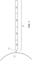
- FIG. 1 is a schematic diagram showing an exemplary capture probe, as described herein.
- the capture probe 102 is optionally coupled to a feature 101 by a cleavage domain 103, such as a disulfide linker.
- the capture probe can include a functional sequence 104 that are useful for subsequent processing.
- the functional sequence 104 can include all or a part of sequencer specific flow cell attachment sequence (e.g., a P5 or P7 sequence), all or a part of a sequencing primer sequence, (e.g., a R1 primer binding site, a R2 primer binding site), or combinations thereof.
- the capture probe can also include a spatial barcode 105.
- the capture probe can also include a unique molecular identifier (UMI) sequence 106. While FIG.
- UMI unique molecular identifier
- the capture probe can also include a capture domain 107 to facilitate capture of a target analyte.
- the capture domain can have a sequence complementary to a sequence of a nucleic acid analyte.
- the capture domain can have a sequence complementary to a connected probe described herein.
- the capture domain can have a sequence complementary to a capture handle sequence present in an analyte capture agent.
- the capture domain can have a sequence complementary to a splint oligonucleotide.
- Such splint oligonucleotide in addition to having a sequence complementary to a capture domain of a capture probe, can have a sequence complementary to a sequence of a nucleic acid analyte, a portion of a connected probe described herein, a capture handle sequence described herein, and/or a methylated adaptor described herein.
- the functional sequences can generally be selected for compatibility with any of a variety of different sequencing systems, e.g., Ion Torrent Proton or PGM, Illumina sequencing instruments, PacBio, Oxford Nanopore, etc., and the requirements thereof.
- functional sequences can be selected for compatibility with non-commercialized sequencing systems. Examples of such sequencing systems and techniques, for which suitable functional sequences can be used, include (but are not limited to) Ion Torrent Proton or PGM sequencing, Illumina sequencing, PacBio SMRT sequencing, and Oxford Nanopore sequencing.
- functional sequences can be selected for compatibility with other sequencing systems, including non-commercialized sequencing systems.
- the spatial barcode 105 and functional sequences 104 is common to all of the probes attached to a given feature.
- the UMI sequence 106 of a capture probe attached to a given feature is different from the UMI sequence of a different capture probe attached to the given feature.
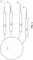
- FIG. 2 is a schematic illustrating a cleavable capture probe, wherein the cleaved capture probe can enter into a non-permeabilized cell and bind to analytes within the sample.
- the capture probe 201 contains a cleavage domain 202, a cell penetrating peptide 203, a reporter molecule 204, and a disulfide bond (-S-S-).
- 205 represents all other parts of a capture probe, for example a spatial barcode and a capture domain.
- Cleavable capture probe are further described in WO 2020/176788 and/or U.S. Patent Application Publication No. 2020/0277663 , each of which is incorporated by reference in its entirety.
- the one or more spatial barcode sequences of the multiple capture probes can include sequences that are the same for all capture probes coupled to the feature, and/or sequences that are different across all capture probes coupled to the feature.
- FIG. 3 is a schematic diagram of an exemplary multiplexed spatially-barcoded feature.
- the feature 301 can be coupled to spatially-barcoded capture probes, wherein the spatially-barcoded probes of a particular feature can possess the same spatial barcode, but have different capture domains designed to associate the spatial barcode of the feature with more than one target analyte.
- a feature may be coupled to four different types of spatially-barcoded capture probes, each type of spatially-barcoded capture probe possessing the spatial barcode 302.
- One type of capture probe associated with the feature includes the spatial barcode 302 in combination with a poly(T) capture domain 303, designed to capture mRNA target analytes.
- a second type of capture probe associated with the feature includes the spatial barcode 302 in combination with a random N-mer capture domain 304 for gDNA analysis.
- a third type of capture probe associated with the feature includes the spatial barcode 302 in combination with a capture domain complementary to a capture handle sequence of an analyte capture agent of interest 305.
- a fourth type of capture probe associated with the feature includes the spatial barcode 302 in combination with a capture domain that can specifically bind a nucleic acid molecule 306 that can function in a CRISPR assay (e.g., CRISPR/Cas9). While only four different capture probe-barcoded constructs are shown in FIG.
- capture-probe barcoded constructs can be tailored for analyses of any given analyte associated with a nucleic acid and capable of binding with such a construct.
- the schemes shown in FIG. 3 can also be used for concurrent analysis of other analytes disclosed herein, including, but not limited to: (a) mRNA, a lineage tracing construct, cell surface or intracellular proteins and metabolites, and gDNA; (b) mRNA, accessible chromatin (e.g., ATAC-seq, DNase-seq, and/or MNase-seq) cell surface or intracellular proteins and metabolites, and a perturbation agent (e.g., a CRISPR crRNA/sgRNA, TALEN, zinc finger nuclease, and/or antisense oligonucleotide as described herein); (c) mRNA, cell surface or intracellular proteins and/or metabolites, a barcoded labelling agent (e.g., the MHC multimers
- a perturbation agent can be a small molecule, an antibody, a drug, an aptamer, a miRNA, a physical environmental (e.g., temperature change), or any other known perturbation agents. See, e.g., Section (II)(b) (e.g., subsections (i)-(vi)) of WO 2020/176788 and/or U.S. Patent Application Publication No. 2020/0277663 . Generation of capture probes can be achieved by any appropriate method, including those described in Section (II)(d)(ii) of WO 2020/176788 and/or U.S. Patent Application Publication No. 2020/0277663 .
- Capture probes attached to a single array feature can include identical (or common) spatial barcode sequences, different spatial barcode sequences, or a combination of both. Capture probes attached to a feature can include multiple sets of capture probes. Capture probes of a given set can include identical spatial barcode sequences. The identical spatial barcode sequences can be different from spatial barcode sequences of capture probes of another set.
- the plurality of capture probes can include spatial barcode sequences (e.g., nucleic acid barcode sequences) that are associated with specific locations on a spatial array.
- a first plurality of capture probes can be associated with a first region, based on a spatial barcode sequence common to the capture probes within the first region
- a second plurality of capture probes can be associated with a second region, based on a spatial barcode sequence common to the capture probes within the second region.
- the second region may or may not be associated with the first region.
- Additional pluralities of capture probes can be associated with spatial barcode sequences common to the capture probes within other regions.
- the spatial barcode sequences can be the same across a plurality of capture probe molecules.
- multiple different spatial barcodes are incorporated into a single arrayed capture probe.
- a mixed but known set of spatial barcode sequences can provide a stronger address or attribution of the spatial barcodes to a given spot or location, by providing duplicate or independent confirmation of the identity of the location.
- the multiple spatial barcodes represent increasing specificity of the location of the particular array point.
- more than one analyte type e.g., nucleic acids and proteins
- a biological sample can be detected (e.g., simultaneously or sequentially) using any appropriate multiplexing technique, such as those described in Section (IV) of WO 2020/176788 and/or U.S. Patent Application Publication No. 2020/0277663 .
- an analyte capture agent refers to an agent that interacts with an analyte (e.g., an analyte in a biological sample) and with a capture probe (e.g., a capture probe attached to a substrate or a feature) to identify the analyte.
- the analyte capture agent includes: (i) an analyte binding moiety (e.g., that binds to an analyte), for example, an antibody or antigen-binding fragment thereof; (ii) analyte binding moiety barcode; and (iii) an analyte capture sequence.
- an analyte binding moiety barcode refers to a barcode that is associated with or otherwise identifies the analyte binding moiety.
- analyte capture sequence refers to a region or moiety configured to hybridize to, bind to, couple to, or otherwise interact with a capture domain of a capture probe.
- an analyte binding moiety barcode (or portion thereof) may be able to be removed (e.g., cleaved) from the analyte capture agent. Additional description of analyte capture agents can be found in Section (II)(b)(ix) of WO 2020/176788 and/or Section (II)(b)(viii) U.S. Patent Application Publication No. 2020/0277663 .
- a spatial barcode with one or more neighboring cells, such that the spatial barcode identifies the one or more cells, and/or contents of the one or more cells, as associated with a particular spatial location.
- One method is to promote analytes or analyte proxies (e.g., intermediate agents) out of a cell and towards a spatially-barcoded array (e.g., including spatially-barcoded capture probes).
- Another method is to cleave spatially-barcoded capture probes from an array and promote the spatially-barcoded capture probes towards and/or into or onto the biological sample.
- capture probes may be configured to prime, replicate, and consequently yield optionally barcoded extension products from a template (e.g., a DNA or RNA template, such as an analyte or an intermediate agent (e.g., a ligation product or an analyte capture agent), or a portion thereof), or derivatives thereof (see, e.g., Section (II)(b)(vii) of WO 2020/176788 and/or U.S. Patent Application Publication No. 2020/0277663 regarding extended capture probes).
- a template e.g., a DNA or RNA template, such as an analyte or an intermediate agent (e.g., a ligation product or an analyte capture agent), or a portion thereof), or derivatives thereof (see, e.g., Section (II)(b)(vii) of WO 2020/176788 and/or U.S. Patent Application Publication No. 2020/0277663 regarding extended capture probes).
- capture probes may be configured to form ligation products with a template (e.g., a DNA or RNA template, such as an analyte or an intermediate agent, or portion thereof), thereby creating ligations products that serve as proxies for a template.
- a template e.g., a DNA or RNA template, such as an analyte or an intermediate agent, or portion thereof
- an "extended capture probe” refers to a capture probe having additional nucleotides added to the terminus (e.g., 3' or 5' end) of the capture probe thereby extending the overall length of the capture probe.
- an "extended 3' end” indicates additional nucleotides were added to the most 3' nucleotide of the capture probe to extend the length of the capture probe, for example, by polymerization reactions used to extend nucleic acid molecules including templated polymerization catalyzed by a polymerase (e.g., a DNA polymerase or a reverse transcriptase).
- a polymerase e.g., a DNA polymerase or a reverse transcriptase
- extending the capture probe includes adding to a 3' end of a capture probe a nucleic acid sequence that is complementary to a nucleic acid sequence of an analyte or intermediate agent specifically bound to the capture domain of the capture probe.
- the capture probe is extended using reverse transcription.
- the capture probe is extended using one or more DNA polymerases. The extended capture probes include the sequence of the capture probe and the sequence of the spatial barcode of the capture probe.
- extended capture probes are amplified (e.g., in bulk solution or on the array) to yield quantities that are sufficient for downstream analysis, e.g., via DNA sequencing.
- extended capture probes e.g., DNA molecules
- act as templates for an amplification reaction e.g., a polymerase chain reaction.
- Section (II)(a) of WO 2020/176788 and/or U.S. Patent Application Publication No. 2020/0277663 Analysis of captured analytes (and/or intermediate agents or portions thereof), for example, including sample removal, extension of capture probes, sequencing (e.g., of a cleaved extended capture probe and/or a cDNA molecule complementary to an extended capture probe), sequencing on the array (e.g., using, for example, in situ hybridization or in situ ligation approaches), temporal analysis, and/or proximity capture, is described in Section (II)(g) of WO 2020/176788 and/or U.S. Patent Application Publication No. 2020/0277663 . Some quality control measures are described in Section (II)(h) of WO 2020/176788 and/or U.S. Patent Application Publication No. 2020/0277663 .
- Spatial information can provide information of biological and/or medical importance.
- the methods and compositions described herein can allow for: identification of one or more biomarkers (e.g., diagnostic, prognostic, and/or for determination of efficacy of a treatment) of a disease or disorder; identification of a candidate drug target for treatment of a disease or disorder; identification (e.g., diagnosis) of a subject as having a disease or disorder; identification of stage and/or prognosis of a disease or disorder in a subject; identification of a subject as having an increased likelihood of developing a disease or disorder; monitoring of progression of a disease or disorder in a subject; determination of efficacy of a treatment of a disease or disorder in a subject; identification of a patient subpopulation for which a treatment is effective for a disease or disorder; modification of a treatment of a subject with a disease or disorder; selection of a subject for participation in a clinical trial; and/or selection of a treatment for a subject with a disease or disorder.
- Spatial information can provide information of biological importance.
- the methods and compositions described herein can allow for: identification of transcriptome and/or proteome expression profiles (e.g., in healthy and/or diseased tissue); identification of multiple analyte types in close proximity (e.g., nearest neighbor analysis); determination of up- and/or down-regulated genes and/or proteins in diseased tissue; characterization of tumor microenvironments; characterization of tumor immune responses; characterization of cells types and their co-localization in tissue; and identification of genetic variants within tissues (e.g., based on gene and/or protein expression profiles associated with specific disease or disorder biomarkers).
- a substrate functions as a support for direct or indirect attachment of capture probes to features of the array.
- a "feature” is an entity that acts as a support or repository for various molecular entities used in spatial analysis.
- some or all of the features in an array are functionalized for analyte capture.
- Exemplary substrates are described in Section (II)(c) of WO 2020/176788 and/or U.S. Patent Application Publication No. 2020/0277663 .
- Exemplary features and geometric attributes of an array can be found in Sections (II)(d)(i), (II)(d)(iii), and (II)(d)(iv) of WO 2020/176788 and/or U.S. Patent Application Publication No. 2020/0277663 .
- analytes and/or intermediate agents can be captured when contacting a biological sample with a substrate including capture probes (e.g., a substrate with capture probes embedded, spotted, printed, fabricated on the substrate, or a substrate with features (e.g., beads, wells) comprising capture probes).
- capture probes e.g., a substrate with capture probes embedded, spotted, printed, fabricated on the substrate, or a substrate with features (e.g., beads, wells) comprising capture probes.
- contact contacted
- contacting a biological sample with a substrate refers to any contact (e.g., direct or indirect) such that capture probes can interact (e.g., bind covalently or non-covalently (e.g., hybridize)) with analytes from the biological sample.
- Capture can be achieved actively (e.g., using electrophoresis) or passively (e.g., using diffusion). Analyte capture is further described in Section (II)(e) of WO 2020/176788 and/or U.S. Patent Application Publication No. 2020/0277663 .
- spatial analysis can be performed by attaching and/or introducing a molecule (e.g., a peptide, a lipid, or a nucleic acid molecule) having a barcode (e.g., a spatial barcode) to a biological sample (e.g., to a cell in a biological sample).
- a plurality of molecules e.g., a plurality of nucleic acid molecules
- a plurality of barcodes e.g., a plurality of spatial barcodes
- a biological sample e.g., to a plurality of cells in a biological sample for use in spatial analysis.
- the biological sample after attaching and/or introducing a molecule having a barcode to a biological sample, the biological sample can be physically separated (e.g., dissociated) into single cells or cell groups for analysis.
- Some such methods of spatial analysis are described in Section (III) of WO 2020/176788 and/or U.S. Patent Application Publication No. 2020/0277663 .
- spatial analysis can be performed by detecting multiple oligonucleotides that hybridize to an analyte.
- spatial analysis can be performed using RNA-templated ligation (RTL).
- RTL RNA-templated ligation
- Methods of RTL have been described previously. See, e.g., Credle et al., Nucleic Acids Res. 2017 Aug 21;45(14):e128 .
- RTL includes hybridization of two oligonucleotides to adjacent sequences on an analyte (e.g., an RNA molecule, such as an mRNA molecule).
- the oligonucleotides are DNA molecules.
- one of the oligonucleotides includes at least two ribonucleic acid bases at the 3' end and/or the other oligonucleotide includes a phosphorylated nucleotide at the 5' end.
- one of the two oligonucleotides includes a capture domain (e.g., a poly(A) sequence, a non-homopolymeric sequence).
- a ligase e.g., SplintR ligase
- the two oligonucleotides hybridize to sequences that are not adjacent to one another.
- hybridization of the two oligonucleotides creates a gap between the hybridized oligonucleotides.
- a polymerase e.g., a DNA polymerase
- the ligation product is released from the analyte.
- the ligation product is released using an endonuclease (e.g., RNAse H).
- the released ligation product can then be captured by capture probes (e.g., instead of direct capture of an analyte) on an array, optionally amplified, and sequenced, thus determining the location and optionally the abundance of the analyte in the biological sample.
- capture probes e.g., instead of direct capture of an analyte
- sequence information for a spatial barcode associated with an analyte is obtained, and the sequence information can be used to provide information about the spatial distribution of the analyte in the biological sample.
- Various methods can be used to obtain the spatial information.
- specific capture probes and the analytes they capture are associated with specific locations in an array of features on a substrate.
- specific spatial barcodes can be associated with specific array locations prior to array fabrication, and the sequences of the spatial barcodes can be stored (e.g., in a database) along with specific array location information, so that each spatial barcode uniquely maps to a particular array location.
- specific spatial barcodes can be deposited at predetermined locations in an array of features during fabrication such that at each location, only one type of spatial barcode is present so that spatial barcodes are uniquely associated with a single feature of the array.
- the arrays can be decoded using any of the methods described herein so that spatial barcodes are uniquely associated with array feature locations, and this mapping can be stored as described above.
- each array feature location represents a position relative to a coordinate reference point (e.g., an array location, a fiducial marker) for the array. Accordingly, each feature location has an "address" or location in the coordinate space of the array.
- Some exemplary spatial analysis workflows are described in the Exemplary Embodiments section of WO 2020/176788 and/or U.S. Patent Application Publication No. 2020/0277663 . See, for example, the Exemplary embodiment starting with "In some non-limiting examples of the workflows described herein, the sample can be immersed... " of WO 2020/176788 and/or U.S. Patent Application Publication No. 2020/0277663 . See also, e.g., the Visium Spatial Gene Expression Reagent Kits User Guide (e.g., Rev C, dated June 2020 ), and/or the Visium Spatial Tissue Optimization Reagent Kits User Guide (e.g., Rev C, dated July 2020 ).
- spatial analysis can be performed using dedicated hardware and/or software, such as any of the systems described in Sections (II)(e)(ii) and/or (V) of WO 2020/176788 and/or U.S. Patent Application Publication No. 2020/0277663 , or any of one or more of the devices or methods described in Sections Control Slide for Imaging, Methods of Using Control Slides and Substrates for, Systems of Using Control Slides and Substrates for Imaging, and/or Sample and Array Alignment Devices and Methods, Informational labels of WO 2020/123320 .
- Suitable systems for performing spatial analysis can include components such as a chamber (e.g., a flow cell or sealable, fluid-tight chamber) for containing a biological sample.
- the biological sample can be mounted for example, in a biological sample holder.
- One or more fluid chambers can be connected to the chamber and/or the sample holder via fluid conduits, and fluids can be delivered into the chamber and/or sample holder via fluidic pumps, vacuum sources, or other devices coupled to the fluid conduits that create a pressure gradient to drive fluid flow.
- One or more valves can also be connected to fluid conduits to regulate the flow of reagents from reservoirs to the chamber and/or sample holder.
- the systems can optionally include a control unit that includes one or more electronic processors, an input interface, an output interface (such as a display), and a storage unit (e.g., a solid state storage medium such as, but not limited to, a magnetic, optical, or other solid state, persistent, writeable and/or re-writeable storage medium).
- the control unit can optionally be connected to one or more remote devices via a network.
- the control unit (and components thereof) can generally perform any of the steps and functions described herein. Where the system is connected to a remote device, the remote device (or devices) can perform any of the steps or features described herein.
- the systems can optionally include one or more detectors (e.g., CCD, CMOS) used to capture images.
- the systems can also optionally include one or more light sources (e.g., LED-based, diode-based, lasers) for illuminating a sample, a substrate with features, analytes from a biological sample captured on a substrate, and various control and calibration media.
- one or more light sources e.g., LED-based, diode-based, lasers
- the systems can optionally include software instructions encoded and/or implemented in one or more of tangible storage media and hardware components such as application specific integrated circuits.
- the software instructions when executed by a control unit (and in particular, an electronic processor) or an integrated circuit, can cause the control unit, integrated circuit, or other component executing the software instructions to perform any of the method steps or functions described herein.
- the systems described herein can detect (e.g., register an image) the biological sample on the array.
- Exemplary methods to detect the biological sample on an array are described in PCT Application No. 2020/061064 and/or U.S. Patent Application Serial No. 16/951,854 .
- the biological sample Prior to transferring analytes from the biological sample to the array of features on the substrate, the biological sample can be aligned with the array. Alignment of a biological sample and an array of features including capture probes can facilitate spatial analysis, which can be used to detect differences in analyte presence and/or level within different positions in the biological sample, for example, to generate a three-dimensional map of the analyte presence and/or level. Exemplary methods to generate a two- and/or three-dimensional map of the analyte presence and/or level are described in PCT Application No. 2020/053655 and spatial analysis methods are generally described in WO 2020/061108 and/or U.S. Patent Application Serial No. 16/951,864 .
- a map of analyte presence and/or level can be aligned to an image of a biological sample using one or more fiducial markers, e.g., objects placed in the field of view of an imaging system which appear in the image produced, as described in the Substrate Attributes Section, Control Slide for Imaging Section of WO 2020/123320 , PCT Application No. 2020/061066 , and/or U.S. Patent Application Serial No. 16/951,843 .
- fiducial markers e.g., objects placed in the field of view of an imaging system which appear in the image produced, as described in the Substrate Attributes Section, Control Slide for Imaging Section of WO 2020/123320 , PCT Application No. 2020/061066 , and/or U.S. Patent Application Serial No. 16/951,843 .
- Fiducial markers can be used as a point of reference or measurement scale for alignment (e.g., to align a sample and an array, to align two substrates, to determine a location of a sample or array on a substrate relative to a fiducial marker) and/or for quantitative measurements of sizes and/or distances.
- Cancer and disease diagnosis and treatment plans are oftentimes guided by three diagnostic tools: blood work, imaging, and/or biopsies.
- surgery is the first line of treatment when cancerous and diseased tissues are identified, especially in the early stages of the disease.
- clinicians face difficult decisions on the size and site of the resection of the diseased tissue.
- Conclusive diagnosis and staging are oftentimes obtained after surgical resection has been completed. Unfortunately, sometimes the entire abnormal tissue is not completely removed, leaving tissue margins that contain diseased cells that can proliferate in the body and cause continued cancer and disease.
- the methods described here can help support a clinician's decision on the type of surgical intervention to provide a subject with a potential cancer or diseased tissue.
- information from practicing the described methods can help determine the size and site of tissue resection by more completely identifying abnormal tissue margins, thereby reducing the risk of re-excision and/or reducing the risk of future recurrence of the cancerous or diseased tissue in the subject.
- determining the size and site of a tissue to be resected from a subject that include: (a) contacting a tissue sample obtained from the subject to an array comprising a plurality of capture probes, where a capture probe of the plurality of capture probes comprises (i) a capture domain that binds specifically to an analyte of the tissue sample and (ii) a spatial barcode; (b) determining (i) all or a part of a nucleic acid sequence corresponding to the analyte specifically bound to the capture domain or a complement thereof, and (ii) all or a part of a nucleic acid sequence corresponding to the spatial barcode or a complement thereof, and using the determined nucleic acid sequences of (i) and (ii) to identify the presence of the analyte at a location in the tissue sample; (c) comparing the presence of the analyte at the location in the tissue sample to presence of the analyte at different location(s) in the
- the analyte is a DNA or RNA. In some embodiments, the analyte is a messenger RNA (mRNA) molecule. In some embodiments, the analyte is a genomic DNA. In some embodiments, the analyte comprises a full-length sequence of a biomarker described herein. In some embodiments, the analyte comprises a fragment of the sequence of a biomarker described herein. In some embodiments of any of the methods described herein, each of the plurality of capture probes comprises (i) a capture domain that binds specifically to an analyte of the tissue section and (ii) a spatial barcode. The capture probe can be any capture probe described herein.
- the capture domain of the capture probe comprises a sequence that is substantially complementary (e.g., at least 80%, at least 85%, at least 90%, at least 95%, at least 99%, or 100% complementary) to a portion of the sequence of the analyte of the tissue sample.
- the capture domain can have a total of about 10 nucleotides to about 125 nucleotides (or any of the subranges of this range described herein).
- the sequence that is substantially complementary to a portion of the sequence of the analyte can be a random sequence.
- the sequence that is substantially complementary to a portion of the sequence of the analyte can include a poly(T) oligonucleotide sequence (e.g., at least 5 contiguous Ts, at least 10 continguous Ts, or at least 15 contiguous Ts).
- a poly(T) oligonucleotide sequence e.g., at least 5 contiguous Ts, at least 10 continguous Ts, or at least 15 contiguous Ts.
- step (b) comprises sequencing (i) all or a part of the nucleic acid sequence corresponding to the analyte specifically bound to the capture domain or a complement thereof, and (ii) all or a part of the nucleic acid sequence corresponding to the spatial barcode or a complement thereof.
- the sequencing is high throughput sequencing, sequencing by synthesis, sequencing by hybridization, sequencing by ligation or any of the other methods for sequencing described herein or known in the art.
- sequencing can involve one or more of nucleic acid amplification, the ligation or addition of one or more sequencing adaptors, cleavage of the capture probe from the array, extension of the capture probe using the bound cDNA as a template, and generating a single-stranded nucleic acid comprising a sequence that is complementary to all or a part of the extended capture probe.
- Non-limiting methods for determining the sequence of (i) all or a part of the sequence of the target nucleic acid, or a complement thereof, or (ii) all or a part of the nucleic acid sequence corresponding to the binding moiety barcode, or a complement thereof, are described herein or are known in the art.
- Also provided herein are methods of determining the size and site of a tissue to be resected from a subject that include: (a) contacting a tissue sample obtained from the subject to a plurality of analyte capture agents, where an analyte capture agent of the plurality of analyte capture agents comprises an analyte binding moiety barcode, an analyte capture sequence, and an analyte binding moiety that binds specifically to an analyte; (b) disposing the tissue sample onto an array, where the array comprises a plurality of capture probes, where a capture probe of the plurality of capture probes comprises a spatial barcode and a capture domain that binds specifically to the analyte capture sequence; (c) determining (i) all or a part of the nucleic acid sequence corresponding to the analyte binding moiety barcode or a complement thereof, and (ii) all or a part of the nucleic acid sequence corresponding to the binding moiety barcode or
- the analyte is a protein. In some embodiments, the analyte is a full-length protein. In some embodiments, the analyte is a fragment of a protein. In some embodiments, the analyte is a byproduct of a protein. In some embodiments, the protein is any of the exemplary cancer biomarkers described herein.
- each of the plurality of analyte capture agents comprises an analyte binding moiety barcode, an analyte capture sequence, and an analyte binding moiety that binds specifically to an analyte.
- the analyte binding moiety is an antibody or an antigen-binding antibody fragment (e.g., a Fab). Any other suitable protein binding moiety known in the art can also be used as an analyte binding moiety.
- the analyte binding moiety barcode can be any barcode described herein.
- the analyte capture sequence can be any analyte capture sequence described herein.
- each of the plurality of capture probes comprises a spatial barcode and a capture domain that binds specifically to the analyte capture sequence.
- the capture probe can be any capture probe described herein.
- the capture domain of the capture probe comprises a sequence that is substantially complementary (e.g., at least 80%, at least 85%, at least 90%, at least 95%, at least 99%, or 100% complementary) to a portion of the analyte capture sequence.
- the capture domain can have a total of about 10 nucleotides to about 125 nucleotides (or any of the subranges of this range described herein).
- the sequence that is substantially complementary to a portion of the analyte capture sequence can be a random sequence. In some embodiments, the sequence that is substantially complementary to a portion of the analyte capture sequence can include a poly(T) oligonucleotide sequence (e.g., at least 5 contiguous Ts, at least 10 continguous Ts, or at least 15 contiguous Ts).
- the determining of the sequence is by sequencing.
- the sequencing is high throughput sequencing, sequencing by hybridization, or any of the other methods for sequencing described herein or known in the art.
- sequencing can involve one or more of nucleic acid amplification, the ligation or addition of one or more sequencing adaptors, cleavage of the capture probe from the array, extension of the capture probe using the bound analyte capture sequence as a template, and generating a single-stranded nucleic acid comprising a sequence that is complementary to all or a part of the extended capture probe.
- Non-limiting methods for determining the sequence of (i) all or a part of the nucleic acid sequence corresponding to the analyte binding moiety barcode, or a complement thereof, or (ii) all or a part of the nucleic acid sequence corresponding to the spatial barcode, or a complement thereof, are described herein or are known in the art.
- the tissue to be resected is a tumor (e.g., a malignant or a benign tumor).
- the tumor is a solid tumor.
- the subject is suspected of having a cancer.
- the subject has been previously diagnosed or identified as having a cancer (e.g., any of the exemplary cancers described herein).
- Non-limiting examples of cancers referred to in any one the methods described herein include: sarcomas, carcinomas, adrenocortical carcinoma, AIDS-related cancers, anal cancer, appendix cancer, astrocytomas, atypical teratoid/rhabdoid tumor, basal cell carcinoma, bladder cancer, brain stem glioma, brain tumors (including brain stem glioma, central nervous system atypical teratoid/rhabdoid tumor, central nervous system embryonal tumors, astrocytomas, craniopharyngioma, ependymoblastoma, ependymoma, medulloblastoma, medulloepithelioma, pineal parenchymal tumors of intermediate differentiation, supratentorial primitive neuroectodermal tumors, and pineoblastoma), breast cancer, bronchial tumors, cancer of unknown primary site, carcinoid tumor, carcinoma of unknown primary site, central
- the tissue to be resected can include a tumor (e.g., a malignant tumor) of any of the types of cancer described herein.
- a tumor e.g., a malignant tumor
- the analyte is a tumor biomarker.
- the analyte is a tumor antigen.
- Exemplary tumor antigens include, but are not limited to, melanoma-associated antigen (MAGE) series of antigens (e.g., MAGE-C1 (cancer/testis antigen CT7), MAGE-B1 antigen (MAGE-XP antigen, DAM10), MAGE-B2 antigen (DAM6), MAGE-2 antigen, MAGE-4a antigen, and MAGE-4b antigen), tyrosinase, glycoprotein 100 (gp100), disialoganglioside GD-2, disialoganglioside O-acetylated GD-3, ganglioside GM-2, epidermal growth factor receptor (EGFR), vascular endothelial growth factor receptor (VEGFR), mutant B-Raf antigen associated with melanoma and colon cancer, human
- MAGE melanoma-associated antigen
- the tissue to be resected is an infected tissue, a necrotic tissue, or a diseased tissue.
- the analyte can be associated with an infection, necrosis, inflammation, or disease. Non-limiting examples of such analytes are known in the art.
- the tissue to be resected is infected by a bacterium. In some embodiments, the tissue to be resected is infected by a virus. In some embodiments, the tissue to be resected is infected by a fungus. In some embodiments, the tissue to be resected is infected by a parasite or protozoa.
- the tissue to be resected is infected by a bacterium, e.g., a Bordetella pertussis, a Brucella abortis, a Escherichia coli, a Salmonella species, e.g., a Salmonella typhi, a Streptococci, a Vibrio ( V cholera, V.
- a bacterium e.g., a Bordetella pertussis, a Brucella abortis, a Escherichia coli, a Salmonella species, e.g., a Salmonella typhi, a Streptococci, a Vibrio ( V cholera, V.
- a Shigella a Pseudomonas, a Brucella species, a Klebsiella, a Mycobacteria species (a tuberculosis, an avium, a BCG, a leprosy), a Pneumococci, a Staphylococci, a Enterobacter species, a Clostridium tetani, a Bacillus anthracis, a Streptococcus pneumoniae, a Meningococcus A, B, C, Y, W, or W-135, a Helicobacter pylori, a Rochalimaea henselae, a Pasteurella ( P.
- haemolytica P. multocida
- Chlamydia C. trachomatis, C. psittaci
- a Treponema pallidum a Haemophilus species, e.g., a Haemophilus influenza type b, a mycoplasma species, a Borrelia burgdorferi, a Legionella pneumophila, a Clostridium botulinum, a Corynebacterium diptheriae, a Yersinia entercolitica, a Ehrlichia, a Anaplasma, or a Coxiella burnetii bacterium.
- the tissue to be resected is infected by a parasite or protozoa, e.g., those causing malaria ( Plasmodium falciparum, P. vivax, or P. malariae ), a schistosome, a trypanosome, leishmania, a filarial nematode, Trichomonas vaginalis, a sarcocystis, a Taenia species ( T. saginata or T. solium ), Toxoplasma gondi, Trichinella spiralis, or an Eimeria species.
- a parasite or protozoa e.g., those causing malaria ( Plasmodium falciparum, P. vivax, or P. malariae ), a schistosome, a trypanosome, leishmania, a filarial nematode, Trichomonas vaginalis, a sarcocystis, a Taenia species ( T
- the tissue to be resected is infected by a fungus, e.g., Cryptococcus neoformans, Candida albicans, Aspergillus fumigatus, Coccidioides immitis, or Coccidioides posadasii.
- a fungus e.g., Cryptococcus neoformans, Candida albicans, Aspergillus fumigatus, Coccidioides immitis, or Coccidioides posadasii.
- the tissue to be resected is infected by a virus, e.g., a rotavirus, an aphthovirus (the agent for foot and mouth disease), an Ebola virus, a Hanta virus, a parainfluenza, a herpes virus species (e.g., herpes simplex virus, Epstein-Barr virus, chicken pox virus, pseudorabies, or cytomegalovirus), a rabies virus, a polio virus, a Hepatitis A, B, C or E, distemper, a Venezuelan equine encephalomyelitis virus, a feline leukemia virus, a reovirus, a respiratory syncytial virus, a Lassa fever virus, a polyoma virus, a canine parvovirus, a papilloma virus, a flavivirus, a tick home encephalitis virus, a paramyxovirus (the agent for Rinder), a her
- the methods provided herein comprise comparing the presence of the analyte at the location in the tissue sample to presence of the analyte at different location(s) in the tissue sample, and determining the size and site of a tissue to be resected from the subject based on the comparison.
- the different location(s) in the tissue sample are reference location(s).
- the reference location(s) in the tissue sample are locations of healthy tissue.
- the reference location(s) in the tissue sample are locations of non-cancerous tissue.
- the reference location(s) in the tissue sample are locations of non-tumor tissue.
- the reference location(s) in the tissue sample are locations with no abnormalities such as tumor, cancer, necrosis, inflammation, infection, or disease.
- the presence of the analyte at the location in the tissue sample is significantly different from the presence of the analyte at the different location(s) in the tissue. In some embodiments, the presence of the analyte at the location in the tissue sample is significantly greater than the presence of the analyte at the different location(s) in the tissue sample. In some embodiments, the presence of the analyte at the location in the tissue sample is significantly less than the presence of the analyte at the different location(s) in the tissue sample.
- the location at the tissue sample is determined to be resected if the presence of the analyte at the location in the tissue sample is significantly different from the presence of the analyte at the different location(s). In some embodiments, the location at the tissue sample is determined to be resected if the presence of the analyte at the location in the tissue sample is significantly greater than the presence of the analyte at the different location(s). In some embodiments, the location at the tissue sample is determined to be resected if the presence of the analyte at the location in the tissue sample is significantly less than the presence of the analyte at the different location(s).
- the presence of the analyte at the location in the tissue sample is about 0.1-fold to about 100-fold (e.g., about 0.1-fold to about 90-fold, about 0.1-fold to about 80-fold, about 0.1-fold to about 70-fold, about 0.1-fold to about 60-fold, about 0.1-fold to about 50-fold, about 0.1-fold to about 40-fold, about 0.1-fold to about 30-fold, about 0.1-fold to about 20-fold, about 0.1-fold to about 15-fold, about 0.1-fold to about 10-fold, about 0.1-fold to about 8-fold, about 0.1-fold to about 6-fold, about 0.1-fold to about 5-fold, about 0.1-fold to about 4-fold, about 0.1-fold to about 3-fold, about 0.1-fold to about 2-fold, about 0.1-fold to about 1.5-fold, about 0.1-fold to about 1-fold, about 0.1-fold to about 0.8-fold, about 0.1-fold to about 0.6-fold, about 0.1-fold to about
- the presence of the analyte at the location in the tissue sample is about 1% to about 99% (e.g., about 1% to about 95%, about 1% to about 90%, about 1% to about 85%, about 1% to about 80%, about 1% to about 75%, about 1% to about 70%, about 1% to about 65%, about 1% to about 60%, about 1% to about 55%, about 1% to about 50%, about 1% to about 45%, about 1% to about 40%, about 1% to about 35%, about 1% to about 30%, about 1% to about 25%, about 1% to about 20%, about 1% to about 15%, about 1% to about 10%, about 1% to about 5%, about 5% to about 99%, about 5% to about 95%, about 5% to about 90%, about 5% to about 85%, about 5% to about 80%, about 5% to about 75%, about 5% to about 70%, about 5% to about 65%, about 5% to about 60%, about 5% to about 55%, about
- the presence of certain biomarkers associated with a cancer and/or disease e.g., breast cancer biomarkers in ductal carcinoma
- a cancer and/or disease e.g., breast cancer biomarkers in ductal carcinoma
- the location in the tissue sample is considered "clear.” If the presence of certain biomarkers associated with a cancer and/or disease are above a threshold value for those biomarkers, the location in the tissue sample is considered within the margin of tissue to be resected.
- the method further comprises comparing presence of one or more additional analyte(s) at the location in the tissue sample with the presence of the one or more additional analyte(s) at the different location(s) in the tissue sample.
- the presence of a total of about 1 to about 20,000 e.g., about 1 to about 18,000, about 1 to about 16,000, about 1 to about 14,000, about 1 to about 12,000, about 1 to about 10,000, about 1 to about 9,000, about 1 to about 8,000, about 1 to about 7,000, about 1 to about 6,000, about 1 to about 5,000, about 1 to about 4,500, about 1 to about 4,000, about 1 to about 3,500, about 1 to about 3,000, about 1 to about 2,500, about 1 to about 2,000, about 1 to about 1,500, about 1 to about 1,000, about 1 to about 800, about 1 to about 600, about 1 to about 500, about 1 to about 400, about 1 to about 300, about 1 to about 200, about 1 to about 100, about 1 to about 1 to about
- mutant cells are identified according to the presence of the one or more analyte(s) at the location in the tissue sample. In some embodiments, a mutant cell is identified according to the presence of one or more biomarkers described herein. In some embodiments, a mutant cell is identified according to the presence of one or more cell-surface biomarkers, e.g., a cell-surface receptor, at the location in the tissue sample.
- a cell within a location at the tissue sample is identified as a mutant cell if the presence of the one or more analyte(s) at the location in the tissue sample are significantly different from the presence of the analyte(s) at the different location(s). In some embodiments, a cell within a location at the tissue sample is identified as a mutant cell if the presence of the one or more analyte(s) at the location in the tissue sample are significantly greater than the presence of the analyte(s) at the different location(s).
- the mutant cell(s) within the location at the tissue sample is determined to be resected if the presence of the analyte(s) at the location in the tissue sample are significantly less than the presence of the analyte at the different location(s).
- a location at the tissue sample comprises about 1 to about 100,000 (e.g., about 1 to about 90,000, about 1 to about 80,000, about 1 to about 70,000, about 1 to about 60,000, about 1 to about 50,000, about 1 to about 40,000, about 1 to about 30,000, about 1 to about 20,000, about 1 to about 10,000, about 1 to about 9,000, about 1 to about 8,000, about 1 to about 7,000, about 1 to about 6,000, about 1 to about 5,000, about 1 to about 4,000, about 1 to about 3,000, about 1 to about 2,000, about 1 to about 1,000, about 1 to about 900, about 1 to about 800, about 1 to about 700, about 1 to about 600, about 1 to about 500, about 1 to about 400, about 1 to about 300, about 1 to about 200, about 1 to about 100, about 1 to about 90, about 1 to about 80, about 1 to about 70, about 1 to about 60, about 1 to about 50, about 1 to about 40, about 1 to about 30, about 1 to about 20, about 1 to about 10,
- the tissue sample can be obtained from any suitable tissue or organ from the subject (e.g., breast tissue, muscle tissue, gland tissue, fat or adipose tissue, nerve tissue, joint tissue, ligament tissue, tendon tissue, mouth tissue, tongue tissue, salivary gland tissue, parotid gland tissue, submandibular gland tissue, sublingual gland tissue, pharynx tissue, esophageal tissue, stomach tissue, small intestine tissue, duodenum tissue, jejunum tissue, ileum tissue, large intestine tissue, liver tissue, gallbladder tissue, mesentery tissue, pancreas tissue, anal canal tissue, anus tissue, nasal cavity tissue, pharynx tissue, larynx tissue, trachea tissue, bronchi tissue, lung tissue, diaphragm tissue, kidney tissue, ureter tissue, bladder tissue, urethra tissue, ovarian tissue, fallopian tube tissue, uterus tissue, vagina tissue, vulva tissue
- the spatial barcode of the capture probe can be any spatial barcode described herein.
- the array can be any of the types of arrays described herein.
- the array comprises a slide.
- the capture probe is attached to the slide (e.g., by its 5' end).
- the array is a bead array. In some embodiments, a 5' end of the capture probe is attached to a bead of the bead array.
- the method comprises extending a 3' end of the capture probe using the specifically bound analyte or analyte binding agent barcode as a template to generate an extended capture probe.
- additional methods are used in combination with the methods described herein to determine the site and size of the tissue to be resected in a subject.
- medical imaging modalities such as computed tomography (CT) and magnetic resonance imaging (MRI) are used in combination with the methods described herein.
- MRI magnetic resonance imaging
- PET position emission tomography
- an initial scanning of a cancer patient and/or imaging of a tissue sample from a cancer patient can be performed using, e.g., MRI, CT, and/or PET prior to the methods described herein, and a preliminary assessment of a surgical margin can be performed.
- the initial information can provide guidance on, e.g., where to obtain the tissue sample for use in the methods described herein, the size of the tissue sample, and/or the number of tissue samples needed.
- a follow-up scanning and/or imaging can be performed using e.g., MRI, CT, and/or PET after the methods described herein are performed.
- the follow-up scanning and /or imaging provide information on, e.g., the clearance of the cancerous and/or diseased tissue, and whether there are residual cancerous and/or diseased tissue. Any other suitable methods known in the art can also be used in combination with the methods described herein.
- Some embodiments of any of the methods described herein can further include obtaining the tissue sample from the subject (e.g., obtain a biopsy from the subject).
- At least a portion of the tissue to be resected includes cancer cell(s), pre-cancerous cell(s), necrotic cell(s), infected cell(s), and/or diseased tissue. In some embodiments, at least 80%, at least 85%, at least 90%, at least 95%, at least 99%, or 100% of the tissue to be resected includes one or more of cancer cell(s), pre-cancerous cell(s), necrotic cell(s), infected cell(s), and disease tissue.
- Also provided herein are methods of treating a subject in need thereof that include: (a) contacting a tissue sample obtained from the subject to an array comprising a plurality of capture probes, wherein a capture probe of the plurality of capture probes comprises (i) a capture domain that specifically binds to an analyte of the tissue sample and (ii) a spatial barcode; (b) determining (i) all or a part of a nucleic acid sequence corresponding to the analyte specifically bound to the capture domain or a complement thereof, and (ii) all or a part of a nucleic acid sequence corresponding to the spatial barcode or a complement thereof, and using the determined nucleic acid sequences of (i) and (ii) to identify the presence of the analyte at a location in the tissue sample; (c) comparing the presence of the analyte at the location in the tissue sample to presence of the analyte at a different location in the tissue sample, and determining the surgical margin based on
- a method of treating a subject comprising: resecting tissue from the subject using a surgical margin previously determined using a method comprising the steps of: (a) contacting a tissue sample obtained from the subject to an array comprising a plurality of capture probes, wherein a capture probe of the plurality of capture probes comprises (i) a capture domain that specifically binds to an analyte of the tissue sample and (ii) a spatial barcode; (b) determining (i) all or a part of a nucleic acid sequence corresponding to the analyte specifically bound to the capture domain or a complement thereof, and (ii) all or a part of a nucleic acid sequence corresponding to the spatial barcode or a complement thereof, and using the determined nucleic acid sequences of (i) and (ii) to identify the presence of the analyte at a location in the tissue sample; (c) comparing the presence of the analyte at the location in the tissue sample to the presence of the analyl tissue
- the data obtained can provide the clinician with information on the accurate location of the cancerous or diseased tissue, therefore provide the accurate surgical margin of the tissue to be resected.
- the clinician is able to achieve, e.g., more complete resection, thereby treating the subject.
- the analyte is a DNA or RNA. In some embodiments, the analyte is a messenger RNA (mRNA) molecule. In some embodiments, the analyte is a genomic DNA. In some embodiments, the analyte comprises a full-length sequence of a biomarker described herein. In some embodiments, the analyte comprises a fragment of the sequence of a biomarker described herein. In some embodiments of any of the methods described herein, each of the plurality of capture probes comprises (i) a capture domain that binds specifically to an analyte of the tissue sample and (ii) a spatial barcode. The capture probe can be any capture probe described herein.
- the capture domain of the capture probe comprises a sequence that is substantially complementary (e.g., at least 80%, at least 85%, at least 90%, at least 95%, at least 99%, or 100% complementary) to a portion of the sequence of the analyte of the tissue sample.
- the capture domain can have a total of about 10 nucleotides to about 125 nucleotides (or any of the subranges of this range described herein).
- the sequence that is substantially complementary to a portion of the sequence of the analyte can be a random sequence.
- the sequence that is substantially complementary to a portion of the sequence of the analyte can include a poly(T) oligonucleotide sequence (e.g., at least 5 contiguous Ts, at least 10 continguous Ts, or at least 15 contiguous Ts).
- a poly(T) oligonucleotide sequence e.g., at least 5 contiguous Ts, at least 10 continguous Ts, or at least 15 contiguous Ts.
- step (b) comprises sequencing (i) all or a part of the nucleic acid sequence corresponding to the analyte specifically bound to the capture domain or a complement thereof, and (ii) all or a part of the nucleic acid sequence corresponding to the spatial barcode or a complement thereof.
- the sequencing is high throughput sequencing, sequencing by hybridization, or any of the other methods for sequencing described herein or known in the art.
- sequencing can involve one or more of nucleic acid amplification, the ligation or addition of one or more sequencing adaptors, cleavage of the capture probe from the array, extension of the capture probe using the bound cDNA as a template, and generating a single-stranded nucleic acid comprising a sequence that is complementary to all or a part of the extended capture probe.
- Non-limiting methods for determining the sequence of (i) all or a part of the sequence of the target nucleic acid, or a complement thereof, or (ii) all or a part of the nucleic acid sequence corresponding to the spatial barcode, or a complement thereof, are described herein or are known in the art.
- Also provided herein are methods of treating a subject that include: (a) contacting a tissue sample obtained from the subject to a plurality of analyte capture agents, wherein an analyte capture agent of the plurality of analyte capture agents comprises an analyte binding moiety barcode, an analyte capture sequence, and an analyte binding moiety that binds specifically to an analyte; (b) disposing the tissue sample onto an array, wherein the array comprises a plurality of capture probes, wherein a capture probe of the plurality of capture probes comprises a spatial barcode and a capture domain that binds specifically to the analyte capture sequence; (c) determining (i) all or a part of a nucleic acid sequence corresponding to the analyte binding moiety barcode or a complement thereof, and (ii) all or a part of a nucleic acid sequence corresponding to the spatial barcode or a complement thereof, and using the determined nucleic acid
- Also provided herein are methods of treating a subject comprising: resecting tissue from the subject using a surgical margin previously determined using a method comprising the steps of: (a) contacting a tissue sample obtained from the subject to a plurality of analyte capture agents, wherein an analyte capture agent of the plurality of analyte capture agents comprises an analyte binding moiety barcode, an analyte capture sequence, and an analyte binding moiety that binds specifically to an analyte; (b) disposing the tissue sample onto an array, wherein the array comprises a plurality of capture probes, wherein a capture probe of the plurality of capture probes comprises a spatial barcode and a capture domain that binds specifically to the analyte capture sequence; (c) determining (i) all or a part of a nucleic acid sequence corresponding to the analyte binding moiety barcode or a complement thereof, and (ii) all or a part of
- the analyte is a protein. In some embodiments, the analyte is a full-length protein. In some embodiments, the analyte is a fragment of a protein. In some embodiments, the analyte is a byproduct of a protein. In some embodiments, the protein is any of the exemplary cancer biomarkers described herein.
- each of the plurality of analyte capture agents comprises an analyte binding moiety barcode, an analyte capture sequence, and an analyte binding moiety that binds specifically to an analyte.
- the analyte binding moiety is an antibody or an antigen-binding antibody fragment (e.g., a Fab). Any other suitable protein binding moiety known in the art can also be used as an analyte binding moiety.
- the analyte binding moiety barcode can be any barcode described herein.
- the analyte capture sequence can be any analyte capture sequence described herein.
- each of the plurality of capture probes comprises a spatial barcode and a capture domain that binds specifically to the analyte capture sequence.
- the capture probe can be any capture probe described herein.
- the capture domain of the capture probe comprises a sequence that is substantially complementary (e.g., at least 80%, at least 85%, at least 90%, at least 95%, at least 99%, or 100% complementary) to a portion of the analyte capture sequence.
- the capture domain can have a total of about 10 nucleotides to about 125 nucleotides (or any of the subranges of this range described herein).
- the sequence that is substantially complementary to a portion of the analyte capture sequence can be a random sequence. In some embodiments, the sequence that is substantially complementary to a portion of the analyte capture sequence can include a poly(T) oligonucleotide sequence (e.g., at least 5 contiguous Ts, at least 10 continguous Ts, or at least 15 contiguous Ts).
- the determining of the sequence is by sequencing.
- the sequencing is high throughput sequencing, sequencing by hybridization, or any of the other methods for sequencing described herein or known in the art.
- sequencing can involve one or more of nucleic acid amplification, the ligation or addition of one or more sequencing adaptors, cleavage of the capture probe from the array, extension of the capture probe using the bound analyte capture sequence as a template, and generating a single-stranded nucleic acid comprising a sequence that is complementary to all or a part of the extended capture probe.
- Non-limiting methods for determining the sequence of (i) all or a part of the nucleic acid sequence corresponding to the analyte binding moiety barcode, or a complement thereof, or (ii) all or a part of the nucleic acid sequence corresponding to the spatial barcode, or a complement thereof, are described herein or are known in the art.
- the resected tissue is or comprises a tumor (e.g., a malignant or a benign tumor).
- the tumor is a solid tumor.
- the subject is suspected of having a cancer.
- the subject has been previously diagnosed or identified as having a cancer (e.g., any of the exemplary cancers described herein).
- the resected tissue can include a tumor, (e.g., a malignant tumor) of any of the types of cancer describes herein.
- a tumor e.g., a malignant tumor
- the analyte is a tumor biomarker. In some embodiments, the analyte is a tumor antigen. Exemplary tumor antigens include, but are not limited to, any of the exemplary tumor antigens described herein.
- the resected tissue is or comprises an infected tissue, a necrotic tissue, or a diseased tissue.
- the analyte can be associated with an infection, necrosis, inflammation, or disease. Non-limiting examples of such analytes are known in the art.
- the resected tissue is infected by a bacterium (e.g., any of the exemplary bacteria described herein), a parasite or protozoa (e.g., any of the exemplary parasites or protozoa described herein), a fungus (e.g., any of the exemplary fungi described herein), or a virus (e.g., any of the exemplary viruses described herein).
- a bacterium e.g., any of the exemplary bacteria described herein
- a parasite or protozoa e.g., any of the exemplary parasites or protozoa described herein
- a fungus e.g., any of the exemplary fungi described herein
- a virus e.g., any of the exemplary viruses described herein.
- the methods provided herein comprise comparing the presence of the analyte at the location in the tissue sample to the presence of the analyte at different location(s) in the tissue sample, and determining the size and site of a tissue to be resected from the subject based on the comparison.
- the different location(s) in the tissue sample are reference location(s).
- the reference location(s) in the tissue sample are locations of healthy tissue.
- the reference location(s) in the tissue sample are locations of non-cancerous tissue.
- the reference location(s) in the tissue sample are locations of non-tumor tissue.
- the reference location(s) in the tissue sample are locations with no abnormalities such as tumor, cancer, necrosis, inflammation, infection, or disease.
- the presence of the analyte at the location in the tissue sample is significantly different from the presence of the analyte at the different location(s) in the tissue. In some embodiments, the presence of the analyte at the location in the tissue sample is significantly greater than the presence of the analyte at the different location(s) in the tissue sample. In some embodiments, the presence of the analyte at the location in the tissue sample is significantly less than the presence of the analyte at the different location(s) in the tissue sample.
- the location at the tissue sample is determined to be resected if the presence of the analyte at the location in the tissue sample is significantly different from the presence of the analyte at the different location(s). In some embodiments, the location at the tissue sample is determined to be resected if the presence of the analyte at the location in the tissue sample is significantly greater than the presence of the analyte at the different location(s). In some embodiments, the location at the tissue sample is determined to be resected if the presence of the analyte at the location in the tissue sample is significantly less than the presence of the analyte at the different location(s).
- the presence of the analyte at the location in the tissue sample is about 0.1-fold to about 100-fold (e.g., or any of the subranges of this range described herein) greater than the presence of the analyte at the different location(s).
- the presence of the analyte at the location in the tissue sample is about 1% to about 99% (e.g., or any of the subranges of this range described herein) decreased than the presence of the analyte at the different location(s).
- the presence of certain biomarkers associated with a cancer and/or disease e.g., breast cancer biomarkers in ductal carcinoma
- a cancer and/or disease e.g., breast cancer biomarkers in ductal carcinoma
- the location in the tissue sample is considered "clear.” If the presence of certain biomarkers associated with a cancer and/or disease are above a threshold value for those biomarkers, the location in the tissue sample is considered within the margin of tissue to be resected.
- the method further comprises comparing the presence of one or more additional analyte(s) at the location in the tissue sample with the presence of the one or more additional analyte(s) at the different location(s) in the tissue sample.
- the presence of a total of about 1 to about 20,000 (e.g., or any of the subranges of this range described herein) analyte(s) at the location are compared to the presence of the analyte(s) at the different location(s).
- mutant cells are identified according to the presence of the one or more analyte(s) at the location in the tissue sample. In some embodiments, a mutant cell is identified according to the presence of one or more biomarkers decribed herein. In some embodiments, a mutant cell is identified according to the presence of one or more cell-surface biomarkers, e.g., a cell-sruface receptor, at the location in the tissue sample.
- a cell within a location at the tissue sample is identified as a mutant cell if the presence of the one or more analyte(s) at the location in the tissue sample are significantly different from the presence of the analyte(s) at the different location(s). In some embodiments, a cell within a location at the tissue sample is identified as a mutant cell if the presence of the one or more analyte(s) at the location in the tissue sample are significantly greater than the presence of the analyte(s) at the different location(s).
- the mutant cell(s) within the location at the tissue sample is determined to be resected if the presence of the analyte(s) at the location in the tissue sample are significantly less than the presence of the analyte at the different location(s).
- a location at the tissue sample comprises about 1 to about 100,000 cells.
- the spatial barcode of the capture probe can be any spatial barcode described herein.
- the array can be any of the types of arrays described herein.
- the array comprises a slide.
- the capture probe is attached to the slide (e.g., by its 5' end).
- the array is a bead array. In some embodiments, a 5' end of the capture probe is attached to a bead of the bead array.
- the method comprises extending a 3' end of the capture probe using the specifically bound analyte or analyte binding agent barcode as a template to generate an extended capture probe.
- additional methods are used in combination with the methods described herein to determine the site and size of the tissue to be resected in a subject.
- medical imaging modalities such as computed tomography (CT) and magnetic resonance imaging (MRI) are used in combination with the methods described herein.
- MRI magnetic resonance imaging
- PET position emission tomography
- an initial scanning of a cancer patient and/or imaging of a tissue sample from a cancer patient can be performed using, e.g., MRI, CT, and/or PET prior to the methods described herein, and a preliminary assessment of a surgical margin can be performed.
- the initial information can provide guidance on, e.g., where to obtain the tissue sample for use in the methods described herein, the size of the tissue sample, and/or the number of tissue samples needed.
- a follow-up scanning and/or imaging can be performed using e.g., MRI, CT, and/or PET after the methods described herein are performed.
- the follow-up scanning and /or imaging provide information on, e.g., the clearance of the cancerous and/or diseased tissue, and wether there are residual cancerous and/or diseased tissue. Any other suitable methods known in the art can also be used in combination with the methods described herein.
- Some embodiments of any of the methods described herein can further include obtaining the tissue sample from the subject (e.g., obtain a biopsy sample from the subject).
- At least a portion of the resected tissue includes cancer cell(s), pre-cancerous cell(s), necrotic cell(s), infected cell(s), and/or diseased tissue. In some embodiments, at least 80%, at least 85%, at least 90%, at least 95%, at least 99%, or 100% of the resected tissue includes one or more of cancer cell(s), pre-cancerous cell(s), necrotic cell(s), infected cell(s), and disease tissue.
- the tissue resection is considered successful when less than 20%, less than 15%, less than 10%, less than 5%, or zero cancer cell(s), pre-cancerous cell(s), necrotic cell(s), infected cell(s), and/or diseased tissue are detected post-resection as compared to the identified cancer cell(s), pre-cancerous cell(s), necrotic cell(s), infected cell(s), and/or diseased tissue prior to the resection.
- the treatment is considered successful when less than 20%, less than 15%, less than 10%, less than 5%, or zero cancer cell(s), pre-cancerous cell(s), necrotic cell(s), infected cell(s), and/or diseased tissue are detected post-resection as compared to the identified cancer cell(s), pre-cancerous cell(s), necrotic cell(s), infected cell(s), and/or diseased tissue prior to the resection.
- the surgical margin can be the margin between the location(s) of one or more of cancer cell(s), pre-cancerous cell(s), necrotic cell(s), infected cell(s), and disease tissue, and the location(s) of healthy or normal tissue, in a subject.
- identifying a surgical margin of a tissue to be resected in a subject comprising: (a) contacting a tissue sample obtained from the subject to an array comprising a plurality of capture probes, wherein a capture probe of the plurality of capture probes comprises (i) a capture domain that specifically binds to an analyte of the tissue sample and (ii) a spatial barcode; (b) determining (i) all or a part of the nucleic acid sequence corresponding to the analyte specifically bound to the capture domain or a complement thereof, and (ii) all or a part of the nucleic acid sequence corresponding to the spatial barcode or a complement thereof, and using the determined nucleic acid sequences of (i) and (ii) to identify the presence of the analyte at a location in the tissue sample; (c) comparing the presence of the analyte at the location in the tissue sample to the presence of the analyte at different location(s
- the analyte is a DNA or RNA. In some embodiments, the analyte is a messenger RNA (mRNA) molecule. In some embodiments, the analyte is a genomic DNA. In some embodiments, the analyte comprises a full-length sequence of a biomarker described herein. In some embodiments, the analyte comprises a fragment of the sequence of a biomarker described herein. In some embodiments of any of the methods described herein, each of the plurality of capture probes comprises (i) a capture domain that binds specifically to an analyte of the tissue sample and (ii) a spatial barcode. The capture probe can be any capture probe described herein.
- the capture domain of the capture probe comprises a sequence that is substantially complementary (e.g., at least 80%, at least 85%, at least 90%, at least 95%, at least 99%, or 100% complementary) to a portion of the sequence of the analyte of the tissue sample.
- the capture domain can have a total of about 10 nucleotides to about 125 nucleotides (or any of the subranges of this range described herein).
- the sequence that is substantially complementary to a portion of the sequence of the analyte can be a random sequence.
- the sequence that is substantially complementary to a portion of the sequence of the analyte can include a poly(T) oligonucleotide sequence (e.g., at least 5 contiguous Ts, at least 10 continguous Ts, or at least 15 contiguous Ts).
- a poly(T) oligonucleotide sequence e.g., at least 5 contiguous Ts, at least 10 continguous Ts, or at least 15 contiguous Ts.
- step (b) comprises sequencing (i) all or a part of the nucleic acid sequence corresponding to the analyte specifically bound to the capture domain or a complement thereof, and (ii) all or a part of the nucleic acid sequence corresponding to the spatial barcode or a complement thereof.
- the sequencing is high throughput sequencing, sequencing by hybridization, or any of the other methods for sequencing described herein or known in the art.
- sequencing can involve one or more of nucleic acid amplification, the ligation or addition of one or more sequencing adaptors, cleavage of the capture probe from the array, extension of the capture probe using the bound cDNA as a template, and generating a single-stranded nucleic acid comprising a sequence that is complementary to all or a part of the extended capture probe.
- Non-limiting methods for determining the sequence of (i) all or a part of the sequence of the target nucleic acid, or a complement thereof, or (ii) all or a part of the nucleic acid sequence corresponding to the spatial barcode, or a complement thereof, are described herein or are known in the art.
- identifying a surgical margin of a tissue to be resected in a subject that include: (a) contacting a tissue sample obtained from the subject to a plurality of analyte capture agents, where an analyte capture agent of the plurality of analyte capture agents comprises an analyte binding moiety barcode, an analyte capture sequence, and an analyte binding moiety that binds specifically to an analyte; (b) disposing the tissue sample onto an array, wherein the array comprises a plurality of capture probes, where a capture probe of the plurality of capture probes comprises a spatial barcode and a capture domain that binds specifically to the analyte capture sequence; (c) determining (i) all or a part of the nucleic acid sequence corresponding to the analyte binding moiety barcode or a complement thereof, and (ii) all or a part of the nucleic acid sequence corresponding to the spatial barcode or a
- the analyte is a protein. In some embodiments, the analyte is a full-length protein. In some embodiments, the analyte is a fragment of a protein. In some embodiments, the analyte is a byproduct of a protein. In some embodiments, the protein is any of the exemplary cancer biomarkers described herein.
- each of the plurality of analyte capture agents comprises an analyte binding moiety barcode, an analyte capture sequence, and an analyte binding moiety that binds specifically to an analyte.
- the analyte binding moiety is an antibody or an antigen-binding antibody fragment (e.g., a Fab). Any other suitable protein binding moiety known in the art can also be used as an analyte binding moiety.
- the analyte binding moiety barcode can be any barcode described herein.
- the analyte capture sequence can be any analyte capture sequence described herein.
- each of the plurality of capture probes comprises a spatial barcode and a capture domain that binds specifically to the analyte capture sequence.
- the capture probe can be any capture probe described herein.
- the capture domain of the capture probe comprises a sequence that is substantially complementary (e.g., at least 80%, at least 85%, at least 90%, at least 95%, at least 99%, or 100% complementary) to a portion of the analyte capture sequence.
- the capture domain can have a total of about 10 nucleotides to about 125 nucleotides (or any of the subranges of this range described herein).
- the sequence that is substantially complementary to a portion of the analyte capture sequence can be a random sequence. In some embodiments, the sequence that is substantially complementary to a portion of the analyte capture sequence can include a poly(T) oligonucleotide sequence (e.g., at least 5 contiguous Ts, at least 10 continguous Ts, or at least 15 contiguous Ts).
- the determining of the sequence is by sequencing.
- the sequencing is high throughput sequencing, sequencing by hybridization, sequencing by ligation or any of the other methods for sequencing described herein or known in the art.
- sequencing can involve one or more of nucleic acid amplification, the ligation or addition of one or more sequencing adaptors, cleavage of the capture probe from the array, extension of the capture probe using the bound analyte capture sequence as a template, and generating a single-stranded nucleic acid comprising a sequence that is complementary to all or a part of the extended capture probe.
- Non-limiting methods for determining the sequence of (i) all or a part of the nucleic acid sequence corresponding to the analyte binding moiety barcode, or a complement thereof, or (ii) all or a part of the nucleic acid sequence corresponding to the spatial barcode, or a complement thereof, are described herein or are known in the art.
- the tissue to be resected is or comprises a tumor (e.g., a malignant or a benign tumor).
- the tumor is a solid tumor.
- the subject is suspected of having a cancer.
- the subject has been previously diagnosed or identified as having a cancer (e.g., any of the exemplary cancers described herein).
- the tissue to be resected can include a tumor (e.g., a malignant tumor) of any of the types of cancer describes herein.
- a tumor e.g., a malignant tumor
- the analyte is a tumor biomarker. In some embodiments, the analyte is a tumor antigen. Exemplary tumor antigens include, but are not limited to, any of the exemplary tumor antigens described herein.
- the tissue to be resected is or comprises an infected tissue, a necrotic tissue, or a diseased tissue.
- the analyte can be associated with an infection, necrosis, inflammation, or disease. Non-limiting examples of such analytes are known in the art.
- the tissue to be resected is infected by a bacterium (e.g., any of the exemplary bacteria described herein), a parasite or protozoa (e.g., any of the exemplary parasites or protozoa described herein), a fungus (e.g., any of the exemplary fungi described herein), or a virus (e.g., any of the exemplary viruses described herein).
- a bacterium e.g., any of the exemplary bacteria described herein
- a parasite or protozoa e.g., any of the exemplary parasites or protozoa described herein
- a fungus e.g., any of the exemplary fungi described herein
- a virus e.g., any of the exemplary viruses described herein.
- the methods provided herein comprise comparing the presence of the analyte at the location in the tissue sample to the presence of the analyte at different location(s) in the tissue sample, and determining the size and site of a tissue to be resected from the subject based on the comparison.
- the different location(s) in the tissue sample are reference location(s).
- the reference location(s) in the tissue sample are locations of healthy tissue.
- the reference location(s) in the tissue sample are locations of non-cancerous tissue.
- the reference location(s) in the tissue sample are locations of non-tumor tissue.
- the reference location(s) in the tissue sample are locations with no abnormalities such as tumor, cancer, necrosis, inflammation, infection, or disease.
- the presence of the analyte at the location in the tissue sample is significantly different from the presence of the analyte at the different location(s) in the tissue. In some embodiments, the presence of the analyte at the location in the tissue sample is significantly greater than the presence of the analyte at the different location(s) in the tissue sample. In some embodiments, the presence of the analyte at the location in the tissue sample is significantly less than the presence of the analyte at the different location(s) in the tissue sample.
- the location at the tissue sample is determined to be resected if the presence of the analyte at the location in the tissue sample is significantly different from the presence of the analyte at the different location(s). In some embodiments, the location at the tissue sample is determined to be resected if the presence of the analyte at the location in the tissue sample is significantly greater than the presence of the analyte at the different location(s). In some embodiments, the location at the tissue sample is determined to be resected if the presence of the analyte at the location in the tissue sample is significantly less than the presence of the analyte at the different location(s).
- the presence of the analyte at the location in the tissue sample is about 0.1-fold to about 100-fold (e.g., or any of the subranges of this range described herein) greater than the presence of the analyte at the different location(s).
- the presence of the analyte at the location in the tissue sample is about 1% to about 99% (e.g., or any of the subranges of this range described herein) decreased than the presence of the analyte at the different location(s).
- the presence of certain biomarkers associated with a cancer and/or disease e.g., breast cancer biomarkers in ductal carcinoma
- a cancer and/or disease e.g., breast cancer biomarkers in ductal carcinoma
- the location in the tissue sample is considered "clear.” If the presence of certain biomarkers associated with a cancer and/or disease are above a threshold value for those biomarkers, the location in the tissue sample is considered within the margin of tissue to be resected.
- the method further comprises comparing presence of one or more additional analyte(s) at the location in the tissue sample with the presence of the one or more additional analyte(s) at the different location(s) in the tissue sample.
- the presence of a total of about 1 to about 20,000 (e.g., or any of the subranges of this range described herein) analyte(s) at the location are compared to the presence of the analyte(s) at the different location(s).
- mutant cells are identified according to the presence of the one or more analyte(s) at the location in the tissue sample. In some embodiments, a mutant cell is identified according to the presence of one or more biomarkers decribed herein. In some embodiments, a mutant cell is identified according to the presence of one or more cell-surface biomarkers, e.g., a cell-sruface receptor, at the location in the tissue sample.
- a cell within a location at the tissue sample is identified as a mutant cell if the presence of the one or more analyte(s) at the location in the tissue sample are significantly different from the presence of the analyte(s) at the different location(s). In some embodiments, a cell within a location at the tissue sample is identified as a mutant cell if the presence of the one or more analyte(s) at the location in the tissue sample are significantly greater than the presence of the analyte(s) at the different location(s).
- the mutant cell(s) within the location at the tissue sample is determined to be resected if the presence of the analyte(s) at the location in the tissue sample are significantly less than the presence of the analyte at the different location(s).
- a location at the tissue sample comprises about 1 to about 100,000 cells.
- the spatial barcode of the capture probe can be any spatial barcode described herein.
- the array can be any of the types of arrays described herein.
- the array comprises a slide.
- the capture probe is attached to the slide (e.g., by its 5' end).
- the array is a bead array. In some embodiments, a 5' end of the capture probe is attached to a bead of the bead array.
- the method comprises extending a 3' end of the capture probe using the specifically bound analyte or analyte binding agent barcode as a template to generate an extended capture probe.
- additional methods are used in combination with the methods described herein to determine the site and size of the tissue to be resected in a subject.
- medical imaging modalities such as computed tomography (CT) and magnetic resonance imaging (MRI) are used in combination with the methods described herein.
- MRI magnetic resonance imaging
- PET position emission tomography
- an initial scanning of a cancer patient and/or imaging of a tissue sample from a cancer patient can be performed using, e.g., MRI, CT, and/or PET prior to the methods described herein, and a preliminary assessment of a surgical margin can be performed.
- the initial information can provide guidance on, e.g., where to obtain the tissue sample for use in the methods described herein, the size of the tissue sample, and/or the number of tissue samples needed.
- a follow-up scanning and/or imaging can be performed using e.g., MRI, CT, and/or PET after the methods described herein are performed.
- the follow-up scanning and /or imaging provide information on, e.g., the clearance of the cancerous and/or diseased tissue, and wether there are residual cancerous and/or diseased tissue. Any other suitable methods known in the art can also be used in combination with the methods described herein.
- Some embodiments of any of the methods described herein can further include obtaining the tissue sample from the subject (e.g., obtain a biopsy sample from the subject).
- At least a portion of the tissue to be resected includes cancer cell(s), pre-cancerous cell(s), necrotic cell(s), infected cell(s), and/or diseased tissue. In some embodiments, at least 80%, at least 85%, at least 90%, at least 95%, at least 99%, or 100% of the tissue to be resected includes one or more of cancer cell(s), pre-cancerous cell(s), necrotic cell(s), infected cell(s), and disease tissue.
- the tissue resection is considered successful when less than 20%, less than 15%, less than 10%, less than 5%, or zero cancer cell(s), pre-cancerous cell(s), necrotic cell(s), infected cell(s), and/or diseased tissue are detected post-resection as compared to the identified cancer cell(s), pre-cancerous cell(s), necrotic cell(s), infected cell(s), and/or diseased tissue prior to the resection.
- the surgical margin can be the margin between the location(s) of one or more of cancer cell(s), pre-cancerous cell(s), necrotic cell(s), infected cell(s), and disease tissue, and the location(s) of healthy or normal tissue, in a subject.
- Also provided herein are methods of reducing the risk of re-excision of a tissue from a subject that include: (a) contacting a tissue sample obtained from the subject to an array comprising a plurality of capture probes, wherein a capture probe of the plurality of capture probes comprises a (i) capture domain that specifically binds to an analyte of the tissue sample and (ii) a spatial barcode; (b) determining (i) all or a part of a nucleic acid sequence corresponding to the analyte specifically bound to the capture domain or a complement thereof, and (ii) all or a part of a nucleic acid sequence corresponding to the spatial barcode or a complement thereof, and using the determined nucleic acid sequences of (i) and (ii) to identify the presence of the analyte at a location in the tissue sample; (c) comparing the presence of the analyte at the location in the tissue sample to the presence of the analyte at different location(s) in
- Also provided herein are methods of reducing the risk of re-excision of a tissue from a subject comprising: resecting tissue from the subject using a surgical margin previously determined using a method comprising the steps of: (a) contacting a tissue sample obtained from the subject to an array comprising a plurality of capture probes, wherein a capture probe of the plurality of capture probes comprises a (i) capture domain that specifically binds to an analyte of the tissue sample and (ii) a spatial barcode; (b) determining (i) all or a part of a nucleic acid sequence corresponding to the analyte specifically bound to the capture domain or a complement thereof, and (ii) all or a part of a nucleic acid sequence corresponding to the spatial barcode or a complement thereof, and using the determined nucleic acid sequences of (i) and (ii) to identify the presence of the analyte at a location in the tissue sample; (c) comparing the presence of the ana
- the data obtained can provide the clinician with guidance on where to resect the tissue margins thereby reducing the probability that an additional resection might be needed; the clinician will be more confident in capturing tissue margins for a more complete resection.
- the analyte is a DNA or RNA.
- the analyte is a messenger RNA (mRNA) molecule.
- the analyte is a genomic DNA.
- the analyte comprises a full-length sequence of a biomarker described herein.
- the analyte comprises a fragment of the sequence of a biomarker described herein.
- each of the plurality of capture probes comprises (i) a capture domain that binds specifically to an analyte of the tissue sample and (ii) a spatial barcode.
- the capture probe can be any capture probe described herein.
- the capture domain of the capture probe comprises a sequence that is substantially complementary (e.g., at least 80%, at least 85%, at least 90%, at least 95%, at least 99%, or 100% complementary) to a portion of the sequence of the analyte of the tissue sample.
- the capture domain can have a total of about 10 nucleotides to about 125 nucleotides (or any of the subranges of this range described herein).
- the sequence that is substantially complementary to a portion of the sequence of the analyte can be a random sequence.
- the sequence that is substantially complementary to a portion of the sequence of the analyte can include a poly(T) oligonucleotide sequence (e.g., at least 5 contiguous Ts, at least 10 continguous Ts, or at least 15 contiguous Ts).
- step (b) comprises sequencing (i) all or a part of the nucleic acid sequence corresponding to the analyte specifically bound to the capture domain or a complement thereof, and (ii) all or a part of the nucleic acid sequence corresponding to the spatial barcode or a complement thereof.
- the sequencing is high throughput sequencing, sequencing by hybridization, or any of the other methods for sequencing described herein or known in the art.
- sequencing can involve one or more of nucleic acid amplification, the ligation or addition of one or more sequencing adaptors, cleavage of the capture probe from the array, extension of the capture probe using the bound cDNA as a template, and generating a single-stranded nucleic acid comprising a sequence that is complementary to all or a part of the extended capture probe.
- Non-limiting methods for determining the sequence of (i) all or a part of the sequence of the target nucleic acid, or a complement thereof, or (ii) all or a part of the nucleic acid sequence corresponding to the spatial barcode, or a complement thereof, are described herein or are known in the art.
- Also provided herein are methods of reducing the risk of re-excision of a tissue from a subject that include: (a) contacting a tissue sample obtained from the subject to a plurality of analyte capture agents, wherein an analyte capture agent of the plurality of analyte capture agents comprises an analyte binding moiety barcode, an analyte capture sequence, and an analyte binding moiety that binds specifically to an analyte; (b) disposing the tissue sample onto an array, wherein the array comprises a plurality of capture probes, wherein a capture probe of the plurality of capture probes comprises a spatial barcode and a capture domain that binds specifically to the analyte capture sequence; (c) determining (i) all or a part of a nucleic acid sequence corresponding to the analyte binding moiety barcode or a complement thereof, and (ii) all or a part of a nucleic acid sequence corresponding to the spatial barcode
- Also provided herein are methods of reducing the risk of re-excision of a tissue from a subject comprising: resecting tissue from the subject using a surgical margin previously determined using a method comprising the steps of: (a) contacting a tissue sample obtained from the subject to a plurality of analyte capture agents, wherein an analyte capture agent of the plurality of analyte capture agents comprises an analyte binding moiety barcode, an analyte capture sequence, and an analyte binding moiety that binds specifically to an analyte; (b) disposing the tissue sample onto an array, wherein the array comprises a plurality of capture probes, wherein a capture probe of the plurality of capture probes comprises a spatial barcode and a capture domain that binds specifically to the analyte capture sequence; (c) determining (i) all or a part of a nucleic acid sequence corresponding to the analyte binding moiety barcode or a complement
- the analyte is a full-length protein. In some embodiments, the analyte is a fragment of a protein. In some embodiments, the analyte is a byproduct of a protein. In some embodiments, the protein is any of the exemplary cancer biomarkers described herein.
- each of the plurality of analyte capture agents comprises an analyte binding moiety barcode, an analyte capture sequence, and an analyte binding moiety that binds specifically to an analyte.
- the analyte binding moiety is an antibody or an antigen-binding antibody fragment (e.g., a Fab). Any other suitable protein binding moiety known in the art can also be used as an analyte binding moiety.
- the analyte binding moiety barcode can be any barcode described herein.
- the analyte capture sequence can be any analyte capture sequence described herein.
- each of the plurality of capture probes comprises a spatial barcode and a capture domain that binds specifically to the analyte capture sequence.
- the capture probe can be any capture probe described herein.
- the capture domain of the capture probe comprises a sequence that is substantially complementary (e.g., at least 80%, at least 85%, at least 90%, at least 95%, at least 99%, or 100% complementary) to a portion of the analyte capture sequence.
- the capture domain can have a total of about 10 nucleotides to about 125 nucleotides (or any of the subranges of this range described herein).
- the sequence that is substantially complementary to a portion of the analyte capture sequence can be a random sequence. In some embodiments, the sequence that is substantially complementary to a portion of the analyte capture sequence can include a poly(T) oligonucleotide sequence (e.g., at least 5 contiguous Ts, at least 10 continguous Ts, or at least 15 contiguous Ts).
- the determining of the sequence is by sequencing.
- the sequencing is high throughput sequencing, sequencing by hybridization, or any of the other methods for sequencing described herein or known in the art.
- sequencing can involve one or more of nucleic acid amplification, the ligation or addition of one or more sequencing adaptors, cleavage of the capture probe from the array, extension of the capture probe using the bound analyte capture sequence as a template, and generating a single-stranded nucleic acid comprising a sequence that is complementary to all or a part of the extended capture probe.
- Non-limiting methods for determining the sequence of (i) all or a part of the nucleic acid sequence corresponding to the analyte binding moiety barcode, or a complement thereof, or (ii) all or a part of the nucleic acid sequence corresponding to the spatial barcode, or a complement thereof, are described herein or are known in the art.
- the resected tissue is or comprises a tumor (e.g., a malignant or a benign tumor).
- the tumor is a solid tumor.
- the subject is suspected of having a cancer.
- the subject has been previously diagnosed or identified as having a cancer (e.g., any of the exemplary cancers described herein).
- the resected tissue can include a tumor (e.g., a malignant tumor) of any of the types of cancer describes herein.
- a tumor e.g., a malignant tumor
- the analyte is a tumor biomarker. In some embodiments, the analyte is a tumor antigen. Exemplary tumor antigens include, but are not limited to, any of the exemplary tumor antigens described herein.
- the resected tissue is or comprises an infected tissue, a necrotic tissue, or a diseased tissue.
- the analyte can be associated with an infection, necrosis, inflammation, or disease. Non-limiting examples of such analytes are known in the art.
- the resected tissue is infected by a bacterium (e.g., any of the exemplary bacteria described herein), a parasite or protozoa (e.g., any of the exemplary parasites or protozoa described herein), a fungus (e.g., any of the exemplary fungi described herein), or a virus (e.g., any of the exemplary viruses described herein).
- a bacterium e.g., any of the exemplary bacteria described herein
- a parasite or protozoa e.g., any of the exemplary parasites or protozoa described herein
- a fungus e.g., any of the exemplary fungi described herein
- a virus e.g., any of the exemplary viruses described herein.
- the methods provided herein comprise comparing the presence of the analyte at the location in the tissue sample to presence of the analyte at different location(s) in the tissue sample, and determining the size and site of a tissue to be resected from the subject based on the comparison.
- the different location(s) in the tissue sample are reference location(s).
- the reference location(s) in the tissue sample are locations of healthy tissue.
- the reference location(s) in the tissue sample are locations of non-cancerous tissue.
- the reference location(s) in the tissue sample are locations of non-tumor tissue.
- the reference location(s) in the tissue sample are locations with no abnormalities such as tumor, cancer, necrosis, inflammation, infection, or disease.
- the presence of the analyte at the location in the tissue sample is significantly different from the presence of the analyte at the different location(s) in the tissue. In some embodiments, the presence of the analyte at the location in the tissue sample is significantly greater than the presence of the analyte at the different location(s) in the tissue sample. In some embodiments, the presence of the analyte at the location in the tissue sample is significantly less than the presence of the analyte at the different location(s) in the tissue sample.
- the location at the tissue sample is determined to be resected if the presence of the analyte at the location in the tissue sample is significantly different from the presence of the analyte at the different location(s). In some embodiments, the location at the tissue sample is determined to be resected if the presence of the analyte at the location in the tissue sample is significantly greater than the presence of the analyte at the different location(s). In some embodiments, the location at the tissue sample is determined to be resected if the presence of the analyte at the location in the tissue sample is significantly less than the presence of the analyte at the different location(s).
- the presence of the analyte at the location in the tissue sample is about 0.1-fold to about 100-fold (e.g., or any of the subranges of this range described herein) greater than the presence of the analyte at the different location(s).
- the presence of the analyte at the location in the tissue sample is about 1% to about 99% (e.g., or any of the subranges of this range described herein) decreased than the presence of the analyte at the different location(s).
- the presence of certain biomarkers associated with a cancer and/or disease e.g., breast cancer biomarkers in ductal carcinoma
- a cancer and/or disease e.g., breast cancer biomarkers in ductal carcinoma
- the location in the tissue sample is considered "clear.” If the presence of certain biomarkers associated with a cancer and/or disease are above a threshold value for those biomarkers, the location in the tissue sample is considered within the margin of tissue to be resected.
- the method further comprises comparing the presence of one or more additional analyte(s) at the location in the tissue sample with the presence of the one or more additional analyte(s) at the different location(s) in the tissue sample.
- the presence of a total of about 1 to about 20,000 (e.g., or any of the subranges of this range described herein) analyte(s) at the location are compared to the presence of the analyte(s) at the different location(s).
- mutant cells are identified according to the presence of the one or more analyte(s) at the location in the tissue sample. In some embodiments, a mutant cell is identified according to the presence of one or more biomarkers decribed herein. In some embodiments, a mutant cell is identified according to the presence of one or more cell-surface biomarkers, e.g., a cell-sruface receptor, at the location in the tissue sample.
- a cell within a location at the tissue sample is identified as a mutant cell if the presence of the one or more analyte(s) at the location in the tissue sample are significantly different from the presence of the analyte(s) at the different location(s). In some embodiments, a cell within a location at the tissue sample is identified as a mutant cell if the presence of the one or more analyte(s) at the location in the tissue sample are significantly greater than the presence of the analyte(s) at the different location(s).
- the mutant cell(s) within the location at the tissue sample is determined to be resected if the presence of the analyte(s) at the location in the tissue sample are significantly less than the presence of the analyte at the different location(s).
- a location at the tissue sample comprises about 1 to about 100,000 cells.
- the spatial barcode of the capture probe can be any spatial barcode described herein.
- the array can be any of the types of arrays described herein.
- the array comprises a slide.
- the capture probe is attached to the slide (e.g., by its 5' end).
- the array is a bead array. In some embodiments, a 5' end of the capture probe is attached to a bead of the bead array.
- the method comprises extending a 3' end of the capture probe using the specifically bound analyte or analyte binding agent barcode as a template to generate an extended capture probe.
- additional methods are used in combination with the methods described herein to determine the site and size of the tissue to be resected in a subject.
- medical imaging modalities such as computed tomography (CT) and magnetic resonance imaging (MRI) are used in combination with the methods described herein.
- MRI magnetic resonance imaging
- PET position emission tomography
- an initial scanning of a cancer patient and/or imaging of a tissue sample from a cancer patient can be performed using, e.g., MRI, CT, and/or PET prior to the methods described herein, and a preliminary assessment of a surgical margin can be performed.
- the initial information can provide guidance on, e.g., where to obtain the tissue sample for use in the methods described herein, the size of the tissue sample, and/or the number of tissue samples needed.
- a follow-up scanning and/or imaging can be performed using e.g., MRI, CT, and/or PET after the methods described herein are performed.
- the follow-up scanning and /or imaging provide information on, e.g., the clearance of the cancerous and/or diseased tissue, and wether there are residual cancerous and/or diseased tissue. Any other suitable methods known in the art can also be used in combination with the methods described herein.
- Some embodiments of any of the methods described herein can further include obtaining the tissue sample from the subject (e.g., obtain a biopsy sample from the subject).
- At least a portion of the resected tissue includes cancer cell(s), pre-cancerous cell(s), necrotic cell(s), infected cell(s), and/or diseased tissue. In some embodiments, at least 80%, at least 85%, at least 90%, at least 95%, at least 99%, or 100% of the resected tissue includes one or more of cancer cell(s), pre-cancerous cell(s), necrotic cell(s), infected cell(s), and disease tissue.
- the surgical margin can be the margin between the location(s) of one or more of cancer cell(s), pre-cancerous cell(s), necrotic cell(s), infected cell(s), and disease tissue, and the location(s) of healthy or normal tissue, in a subject.
- the methods described herein are more accurate in determining the surgical margin of a tissue to be resected than the traditional methods, such as medical imaging or scanning methods.
- a re-excision includes additional tissue excisions during the initial surgical procedure to obtain the tissue sample. In some embodiments, a re-excision includes additional tissue excisions during a future procedure.
- the methods described herein results in a reduction (e.g., at least a 5% reduction, at least a 10% reduction, at least a 15% reduction, at least a 20% reduction, at least a 25% reduction, at least a 30% reduction, at least a 35% reduction, at least a 40% reduction, at least a 45% reduction, at least a 50% reduction, at least a 55% reduction, at least a 60% reduction, at least a 65% reduction, at least a 70% reduction, at least a 75% reduction, at least a 80% reduction, at least a 85% reduction, at least a 90% reduction, at least a 95% reduction, or at least a 99% reduction, or about a 1% to about a 5% reduction, about a 5% to about a 10% reduction, about a 10% to about a 99% reduction, about a 10% to about a 90% reduction, about a 10% to about a 80% reduction, about a 10% to about a 70% reduction, about a 10% to about a 60% reduction,
- FIG. 4 A non-limiting example of a method for identifying a surgical margin of a tissue to be resected is depicted in FIG. 4 .
- a biopsy tissue is excised from a subject and imaged for potential tumor tissue cells or other disease related cells. After imaging, the tissue is sectioned and one or more sections of tissue are subjected to the methods described herein for spatial determination and location of cells of interest in a tissue. For example, gene expression along the margins of a tissue section indicative of cancer or a disease state and/or the location of receptors along the margins of a tissue section indicative of cancer or a disease state is determined via a spatial array. Additionally, known mutations associated with a cancer or disease state can be spatially identified along the margins of a tissue section.
- Gene expression analysis and/or receptor presence and/or mutational state of cells within the tissue margins can be used to determine whether a surgeon has sufficiently resected the tissue. For example, the presence of one or more genes, receptors and/or mutations indicative of a cancer or disease state in a spatially analyzed tissue margin section would indicate that the resection of the cancerous or diseased tissue was not complete as such a more expanded resection might be necessary. When the tissue section margins are absent of those biomarkers that were used to indicate a cancer or disease state, then a resection could be considered successful for that location.
- a capture probe of the plurality of capture probes comprises a (i) capture domain that specifically binds to an analyte of the tissue sample and (ii) a spatial barcode; (b) determining (i) all or a part of a nucleic acid sequence corresponding to the analyte specifically bound to the capture domain or a complement thereof, and (ii) all or a part of a nucleic acid sequence corresponding to the spatial barcode or a complement thereof, and using the determined nucleic acid sequences of (i) and (ii) to identify the presence of the analyte at a location in the tissue sample; (c) comparing the presence of the analyte at the location in the tissue sample to presence of the analyte at different location(s) in
- the methods described herein allow a clinician to identify tissue tumor margins with confidence such that tissue resection will increase the likelihood that the complete tumor or tissue abnormality has been removed from the subject, thereby reducing the rate of recurrence of the abnormal tissue or tumor in the subject.
- the analyte is a DNA or RNA. In some embodiments, the analyte is a messenger RNA (mRNA) molecule. In some embodiments, the analyte is a genomic DNA. In some embodiments, the analyte comprises a full-length sequence of a biomarker described herein. In some embodiments, the analyte comprises a fragment of the sequence of a biomarker described herein. In some embodiments of any of the methods described herein, each of the plurality of capture probes comprises (i) a capture domain that binds specifically to an analyte of the tissue sample and (ii) a spatial barcode. The capture probe can be any capture probe described herein.
- the capture domain of the capture probe comprises a sequence that is substantially complementary (e.g., at least 80%, at least 85%, at least 90%, at least 95%, at least 99%, or 100% complementary) to a portion of the sequence of the analyte of the tissue sample.
- the capture domain can have a total of about 10 nucleotides to about 125 nucleotides (or any of the subranges of this range described herein).
- the sequence that is substantially complementary to a portion of the sequence of the analyte can be a random sequence.
- the sequence that is substantially complementary to a portion of the sequence of the analyte can include a poly(T) oligonucleotide sequence (e.g., at least 5 contiguous Ts, at least 10 continguous Ts, or at least 15 contiguous Ts).
- a poly(T) oligonucleotide sequence e.g., at least 5 contiguous Ts, at least 10 continguous Ts, or at least 15 contiguous Ts.
- step (b) comprises sequencing (i) all or a part of the nucleic acid sequence corresponding to the analyte specifically bound to the capture domain or a complement thereof, and (ii) all or a part of the nucleic acid sequence corresponding to the spatial barcode or a complement thereof.
- the sequencing is high throughput sequencing, sequencing by hybridization, or any of the other methods for sequencing described herein or known in the art.
- sequencing can involve one or more of nucleic acid amplification, the ligation or addition of one or more sequencing adaptors, cleavage of the capture probe from the array, extension of the capture probe using the bound cDNA as a template, and generating a single-stranded nucleic acid comprising a sequence that is complementary to all or a part of the extended capture probe.
- Non-limiting methods for determining the sequence of (i) all or a part of the sequence of the target nucleic acid, or a complement thereof, or (ii) all or a part of the nucleic acid sequence corresponding to the spatial barcode, or a complement thereof, are described herein or are known in the art.
- Also provided herein are methods of reducing the rate of recurrence of a tissue abnormality in a subject that include: (a) contacting a tissue sample obtained from the subject to a plurality of analyte capture agents, wherein an analyte capture agent of the plurality of analyte capture agents comprises an analyte binding moiety barcode, an analyte capture sequence, and an analyte binding moiety that binds specifically to an analyte; (b) disposing the tissue sample onto an array, wherein the array comprises a plurality of capture probes, wherein a capture probe of the plurality of capture probes comprises a spatial barcode and a capture domain that binds specifically to the analyte capture sequence; (c) determining (i) all or a part of a nucleic acid sequence corresponding to the analyte binding moiety barcode or a complement thereof, and (ii) all or a part of a nucleic acid sequence corresponding to the spatial
- the analyte is a protein. In some embodiments, the analyte is a full-length protein. In some embodiments, the analyte is a fragment of a protein. In some embodiments, the analyte is a byproduct of a protein. In some embodiments, the protein is any of the exemplary cancer biomarkers described herein.
- each of the plurality of analyte capture agents comprises an analyte binding moiety barcode, an analyte capture sequence, and an analyte binding moiety that binds specifically to an analyte.
- the analyte binding moiety is an antibody or an antigen-binding antibody fragment (e.g., a Fab). Any other suitable protein binding moiety known in the art can also be used as an analyte binding moiety.
- the analyte binding moiety barcode can be any barcode described herein.
- the analyte capture sequence can be any analyte capture sequence described herein.
- each of the plurality of capture probes comprises a spatial barcode and a capture domain that binds specifically to the analyte capture sequence.
- the capture probe can be any capture probe described herein.
- the capture domain of the capture probe comprises a sequence that is substantially complementary (e.g., at least 80%, at least 85%, at least 90%, at least 95%, at least 99%, or 100% complementary) to a portion of the analyte capture sequence.
- the capture domain can have a total of about 10 nucleotides to about 125 nucleotides (or any of the subranges of this range described herein).
- the sequence that is substantially complementary to a portion of the analyte capture sequence can be a random sequence. In some embodiments, the sequence that is substantially complementary to a portion of the analyte capture sequence can include a poly(T) oligonucleotide sequence (e.g., at least 5 contiguous Ts, at least 10 continguous Ts, or at least 15 contiguous Ts).
- the determining of the sequence is by sequencing.
- the sequencing is high throughput sequencing, sequencing by hybridization, or any of the other methods for sequencing described herein or known in the art.
- sequencing can involve one or more of nucleic acid amplification, the ligation or addition of one or more sequencing adaptors, cleavage of the capture probe from the array, extension of the capture probe using the bound analyte capture sequence as a template, and generating a single-stranded nucleic acid comprising a sequence that is complementary to all or a part of the extended capture probe.
- Non-limiting methods for determining the sequence of (i) all or a part of the nucleic acid sequence corresponding to the analyte binding moiety barcode, or a complement thereof, or (ii) all or a part of the nucleic acid sequence corresponding to the spatial barcode, or a complement thereof, are described herein or are known in the art.
- an abnormality in the resected tissue is or comprises a tumor (e.g., a malignant or a benign tumor).
- the tumor is a solid tumor.
- the subject is suspected of having a cancer.
- the subject has been previously diagnosed or identified as having a cancer (e.g., any of the exemplary cancers described herein).
- the resected tissue can include a tumor, e.g., a tumor (e.g., a malignant tumor) of any of the types of cancer describes herein.
- a tumor e.g., a tumor (e.g., a malignant tumor) of any of the types of cancer describes herein.
- the analyte is a tumor biomarker. In some embodiments, the analyte is a tumor antigen. Exemplary tumor antigens include, but are not limited to, any of the exemplary tumor antigens described herein.
- the resected tissue is or comprises an infected tissue, a necrotic tissue, or a diseased tissue.
- the analyte can be associated with an infection, necrosis, inflammation, or disease. Non-limiting examples of such analytes are known in the art.
- the resected tissue is infected by a bacterium (e.g., any of the exemplary bacteria described herein), a parasite or protozoa (e.g., any of the exemplary parasites or protozoa described herein), a fungus (e.g., any of the exemplary fungi described herein), or a virus (e.g., any of the exemplary viruses described herein).
- a bacterium e.g., any of the exemplary bacteria described herein
- a parasite or protozoa e.g., any of the exemplary parasites or protozoa described herein
- a fungus e.g., any of the exemplary fungi described herein
- a virus e.g., any of the exemplary viruses described herein.
- the methods provided herein comprise comparing the presence of the analyte at the location in the tissue sample to presence of the analyte at different location(s) in the tissue sample, and determining the size and site of a tissue to be resected from the subject based on the comparison.
- the different location(s) in the tissue sample are reference location(s).
- the reference location(s) in the tissue sample are locations of healthy tissue.
- the reference location(s) in the tissue sample are locations of non-cancerous tissue.
- the reference location(s) in the tissue sample are locations of non-tumor tissue.
- the reference location(s) in the tissue sample are locations with no abnormalities such as tumor, cancer, necrosis, inflammation, infection, or disease.
- the presence of the analyte at the location in the tissue sample is significantly different from the presence of the analyte at the different location(s) in the tissue. In some embodiments, the presence of the analyte at the location in the tissue sample is significantly greater than the presence of the analyte at the different location(s) in the tissue sample. In some embodiments, the presence of the analyte at the location in the tissue sample is significantly less than the presence of the analyte at the different location(s) in the tissue sample.
- the location at the tissue sample is determined to be resected if the presence of the analyte at the location in the tissue sample is significantly different from the presence of the analyte at the different location(s). In some embodiments, the location at the tissue sample is determined to be resected if the presence of the analyte at the location in the tissue sample is significantly greater than the presence of the analyte at the different location(s). In some embodiments, the location at the tissue sample is determined to be resected if the presence of the analyte at the location in the tissue sample is significantly less than the presence of the analyte at the different location(s).
- the presence of the analyte at the location in the tissue sample is about 0.1-fold to about 100-fold (e.g., or any of the subranges of this range described herein) greater than the presence of the analyte at the different location(s).
- the presence of the analyte at the location in the tissue sample is about 1% to about 99% (e.g., or any of the subranges of this range described herein) decreased than the presence of the analyte at the different location(s).
- the presence of certain biomarkers associated with a cancer and/or disease e.g., breast cancer biomarkers in ductal carcinoma
- a cancer and/or disease e.g., breast cancer biomarkers in ductal carcinoma
- the location in the tissue sample is considered "clear.” If the presence of certain biomarkers associated with a cancer and/or disease are above a threshold value for those biomarkers, the location in the tissue sample is considered within the margin of tissue to be resected.
- the method further comprises comparing presence of one or more additional analyte(s) at the location in the tissue sample with the presence of the one or more additional analyte(s) at the different location(s) in the tissue sample.
- the presence of a total of about 1 to about 20,000 (e.g., or any of the subranges of this range described herein) analyte(s) at the location are compared to the presence of the analyte(s) at the different location(s).
- mutant cells are identified according to the presence of the one or more analyte(s) at the location in the tissue sample. In some embodiments, a mutant cell is identified according to the presence of one or more biomarkers decribed herein. In some embodiments, a mutant cell is identified according to the presence of one or more cell-surface biomarkers, e.g., a cell-sruface receptor, at the location in the tissue sample.
- a cell within a location at the tissue sample is identified as a mutant cell if the presence of the one or more analyte(s) at the location in the tissue sample are significantly different from the presence of the analyte(s) at the different location(s). In some embodiments, a cell within a location at the tissue sample is identified as a mutant cell if the presence of the one or more analyte(s) at the location in the tissue sample are significantly greater than the presence of the analyte(s) at the different location(s).
- the mutant cell(s) within the location at the tissue sample is determined to be resected if the presence of the analyte(s) at the location in the tissue sample are significantly less than the presence of the analyte at the different location(s).
- a location at the tissue sample comprises about 1 to about 100,000 cells.
- the spatial barcode of the capture probe can be any spatial barcode described herein.
- the array can be any of the types of arrays described herein.
- the array comprises a slide.
- the capture probe is attached to the slide (e.g., by its 5' end).
- the array is a bead array. In some embodiments, a 5' end of the capture probe is attached to a bead of the bead array.
- the method comprises extending a 3' end of the capture probe using the specifically bound analyte or analyte binding agent barcode as a template to generate an extended capture probe.
- additional methods are used in combination with the methods described herein to determine the site and size of the tissue to be resected in a subject.
- medical imaging modalities such as computed tomography (CT) and magnetic resonance imaging (MRI) are used in combination with the methods described herein.
- MRI magnetic resonance imaging
- PET position emission tomography
- an initial scanning of a cancer patient and/or imaging of a tissue sample from a cancer patient can be performed using, e.g., MRI, CT, and/or PET prior to the methods described herein, and a preliminary assessment of a surgical margin can be performed.
- the initial information can provide guidance on, e.g., where to obtain the tissue sample for use in the methods described herein, the size of the tissue sample, and/or the number of tissue samples needed.
- a follow-up scanning and/or imaging can be performed using e.g., MRI, CT, and/or PET after the methods described herein are performed.
- the follow-up scanning and /or imaging provide information on, e.g., the clearance of the cancerous and/or diseased tissue, and wether there are residual cancerous and/or diseased tissue. Any other suitable methods known in the art can also be used in combination with the methods described herein.
- Some embodiments of any of the methods described herein can further include obtaining the tissue sample from the subject (e.g., obtain a biopsy sample from the subject).
- At least a portion of the resected tissue includes cancer cell(s), pre-cancerous cell(s), necrotic cell(s), infected cell(s), and/or diseased tissue. In some embodiments, at least 80%, at least 85%, at least 90%, at least 95%, at least 99%, or 100% of the resected tissue includes one or more of cancer cell(s), pre-cancerous cell(s), necrotic cell(s), infected cell(s), and disease tissue.
- the surgical margin can be the margin between the location(s) of one or more of cancer cell(s), pre-cancerous cell(s), necrotic cell(s), infected cell(s), and disease tissue, and the location(s) of healthy or normal tissue, in a subject.
- the methods described herein results in a reduction (e.g., at least a 5% reduction, at least a 10% reduction, at least a 15% reduction, at least a 20% reduction, at least a 25% reduction, at least a 30% reduction, at least a 35% reduction, at least a 40% reduction, at least a 45% reduction, at least a 50% reduction, at least a 55% reduction, at least a 60% reduction, at least a 65% reduction, at least a 70% reduction, at least a 75% reduction, at least a 80% reduction, at least a 85% reduction, at least a 90% reduction, at least a 95% reduction, or at least a 99% reduction, or about a 1% to about a 99% reduction (e.g., or any of the subranges of this range described herein) in the rate of recurrence of a tissue abnormality (e.g., any of the cancers described herein) in the subject (e.g., as a compared to a similar subject that
- Also provided herein are methods of treating a subject that include: (a) contacting a tissue sample obtained from the subject to an array comprising a plurality of capture probes, wherein a capture probe of the plurality of capture probes comprises a (i) capture domain that specifically binds to an analyte of the tissue sample and (ii) a spatial barcode; (b) determining (i) all or a part of a nucleic acid sequence corresponding to the analyte specifically bound to the capture domain or a complement thereof, and (ii) all or a part of a nucleic acid sequence corresponding to the spatial barcode or a complement thereof, and using the determined nucleic acid sequences of (i) and (ii) to identify the presence of the analyte at a location in the tissue sample; (c) comparing the presence of the analyte at the location in the tissue sample to presence of the analyte at different location(s) in the tissue sample, and determining the surgical margin based on the comparison
- Also provided herein are methods of treating a subject the method comprising: excising a nerve or blood vessel that is within a surgical margin in the subject, or avoiding excision of a nerve or blood vessel outside of the surgical margin in the subject, wherein the surgical margin was previously determined using a method comprising the steps of: (a) contacting a tissue sample obtained from the subject to an array comprising a plurality of capture probes, wherein a capture probe of the plurality of capture probes comprises a (i) capture domain that specifically binds to an analyte of the tissue sample and (ii) a spatial barcode; (b) determining (i) all or a part of a nucleic acid sequence corresponding to the analyte specifically bound to the capture domain or a complement thereof, and (ii) all or a part of a nucleic acid sequence corresponding to the spatial barcode or a complement thereof, and using the determined nucleic acid sequences of (i) and (ii) to identify the presence of the analyte at
- the data obtained can provide the clinician with information of the accurate location of the cancerous or diseased tissue, therefore provide the accurate surgical margin of the tissue to be resected.
- the clinician is able to achieve, e.g., more complete and accurate excision, thereby treating the subject in need thereof.
- the analyte is a DNA or RNA. In some embodiments, the analyte is a messenger RNA (mRNA) molecule. In some embodiments, the analyte is a genomic DNA. In some embodiments, the analyte comprises a full-length sequence of a biomarker described herein. In some embodiments, the analyte comprises a fragment of the sequence of a biomarker described herein. In some embodiments of any of the methods described herein, each of the plurality of capture probes comprises (i) a capture domain that binds specifically to an analyte of the tissue sample and (ii) a spatial barcode. The capture probe can be any capture probe described herein.
- the capture domain of the capture probe comprises a sequence that is substantially complementary (e.g., at least 80%, at least 85%, at least 90%, at least 95%, at least 99%, or 100% complementary) to a portion of the sequence of the analyte of the tissue sample.
- the capture domain can have a total of about 10 nucleotides to about 125 nucleotides (or any of the subranges of this range described herein).
- the sequence that is substantially complementary to a portion of the sequence of the analyte can be a random sequence.
- the sequence that is substantially complementary to a portion of the sequence of the analyte can include a poly(T) oligonucleotide sequence (e.g., at least 5 contiguous Ts, at least 10 continguous Ts, or at least 15 contiguous Ts).
- a poly(T) oligonucleotide sequence e.g., at least 5 contiguous Ts, at least 10 continguous Ts, or at least 15 contiguous Ts.
- step (b) comprises sequencing (i) all or a part of the nucleic acid sequence corresponding to the analyte specifically bound to the capture domain or a complement thereof, and (ii) all or a part of the nucleic acid sequence corresponding to the spatial barcode or a complement thereof.
- the sequencing is high throughput sequencing, sequencing by hybridization, or any of the other methods for sequencing described herein or known in the art.
- sequencing can involve one or more of nucleic acid amplification, the ligation or addition of one or more sequencing adaptors, cleavage of the capture probe from the array, extension of the capture probe using the bound cDNA as a template, and generating a single-stranded nucleic acid comprising a sequence that is complementary to all or a part of the extended capture probe.
- Non-limiting methods for determining the sequence of (i) all or a part of the sequence of the target nucleic acid, or a complement thereof, or (ii) all or a part of the nucleic acid sequence corresponding to the spatial barcode, or a complement thereof, are described herein or are known in the art.
- Also provided herein are methods of treating a subject that include: (a) contacting a tissue sample obtained from the subject to a plurality of analyte capture agents, wherein an analyte capture agent of the plurality of analyte capture agents comprises an analyte binding moiety barcode, an analyte capture sequence, and an analyte binding moiety that binds specifically to an analyte; (b) disposing the tissue sample onto an array, wherein the array comprises a plurality of capture probes, wherein a capture probe of the plurality of capture probes comprises a spatial barcode and a capture domain that binds specifically to the analyte capture sequence; (c) determining (i) all or a part of a nucleic acid sequence corresponding to the analyte binding moiety barcode or a complement thereof, and (ii) all or a part of a nucleic acid sequence corresponding to the spatial barcode or a complement thereof, and using the determined nucleic acid
- Also provided herein are methods of treating a subject comprising: excising a nerve or blood vessel that is within a surgical margin in the subject, or avoiding excision of a nerve or blood vessel outside of the surgical margin in the subject, wherein the surgical margin was previously determined using a method comprising the steps of: (a) contacting a tissue sample obtained from the subject to a plurality of analyte capture agents, wherein an analyte capture agent of the plurality of analyte capture agents comprises an analyte binding moiety barcode, an analyte capture sequence, and an analyte binding moiety that binds specifically to an analyte; (b) disposing the tissue sample onto an array, wherein the array comprises a plurality of capture probes, wherein a capture probe of the plurality of capture probes comprises a spatial barcode and a capture domain that binds specifically to the analyte capture sequence; (c) determining (i) all or a part of a nucleic acid
- the analyte is a protein. In some embodiments, the analyte is a full-length protein. In some embodiments, the analyte is a fragment of a protein. In some embodiments, the analyte is a byproduct of a protein. In some embodiments, the protein is any of the exemplary cancer biomarkers described herein.
- each of the plurality of analyte capture agents comprises an analyte binding moiety barcode, an analyte capture sequence, and an analyte binding moiety that binds specifically to an analyte.
- the analyte binding moiety is an antibody or an antigen-binding antibody fragment (e.g., a Fab). Any other suitable protein binding moiety known in the art can also be used as an analyte binding moiety.
- the analyte binding moiety barcode can be any barcode described herein.
- the analyte capture sequence can be any analyte capture sequence described herein.
- each of the plurality of capture probes comprises a spatial barcode and a capture domain that binds specifically to the analyte capture sequence.
- the capture probe can be any capture probe described herein.
- the capture domain of the capture probe comprises a sequence that is substantially complementary (e.g., at least 80%, at least 85%, at least 90%, at least 95%, at least 99%, or 100% complementary) to a portion of the analyte capture sequence.
- the capture domain can have a total of about 10 nucleotides to about 125 nucleotides (or any of the subranges of this range described herein).
- the sequence that is substantially complementary to a portion of the analyte capture sequence can be a random sequence. In some embodiments, the sequence that is substantially complementary to a portion of the analyte capture sequence can include a poly(T) oligonucleotide sequence (e.g., at least 5 contiguous Ts, at least 10 continguous Ts, or at least 15 contiguous Ts).
- the determining of the sequence is by sequencing.
- the sequencing is high throughput sequencing, sequencing by hybridization, or any of the other methods for sequencing described herein or known in the art.
- sequencing can involve one or more of nucleic acid amplification, the ligation or addition of one or more sequencing adaptors, cleavage of the capture probe from the array, extension of the capture probe using the bound analyte capture sequence as a template, and generating a single-stranded nucleic acid comprising a sequence that is complementary to all or a part of the extended capture probe.
- Non-limiting methods for determining the sequence of (i) all or a part of the nucleic acid sequence corresponding to the analyte binding moiety barcode, or a complement thereof, or (ii) all or a part of the nucleic acid sequence corresponding to the spatial barcode, or a complement thereof, are described herein or are known in the art.
- the tissue to be resected is or comprises a tumor (e.g., a malignant or a benign tumor).
- the tumor is a solid tumor.
- the subject is suspected of having a cancer.
- the subject has been previously diagnosed or identified as having a cancer (e.g., any of the exemplary cancers described herein).
- the tissue to be resected can include a tumor (e.g., a malignant tumor) of any of the types of cancer describes herein.
- a tumor e.g., a malignant tumor
- the analyte is a tumor biomarker. In some embodiments, the analyte is a tumor antigen. Exemplary tumor antigens include, but are not limited to, any of the exemplary tumor antigens described herein.
- the tissue to be resected is or comprises an infected tissue, a necrotic tissue, or a diseased tissue.
- the analyte can be associated with an infection, necrosis, inflammation, or disease. Non-limiting examples of such analytes are known in the art.
- the tissue to be resected is infected by a bacterium (e.g., any of the exemplary bacteria described herein), a parasite or protozoa (e.g., any of the exemplary parasites or protozoa described herein), a fungus (e.g., any of the exemplary fungi described herein), or a virus (e.g., any of the exemplary viruses described herein).
- a bacterium e.g., any of the exemplary bacteria described herein
- a parasite or protozoa e.g., any of the exemplary parasites or protozoa described herein
- a fungus e.g., any of the exemplary fungi described herein
- a virus e.g., any of the exemplary viruses described herein.
- the methods provided herein comprise comparing the presence of the analyte at the location in the tissue sample to presence of the analyte at different location(s) in the tissue sample, and determining the size and site of a tissue to be resected from the subject based on the comparison.
- the different location(s) in the tissue sample are reference location(s).
- the reference location(s) in the tissue sample are locations of healthy tissue.
- the reference location(s) in the tissue sample are locations of non-cancerous tissue.
- the reference location(s) in the tissue sample are locations of non-tumor tissue.
- the reference location(s) in the tissue sample are locations with no abnormalities such as tumor, cancer, necrosis, inflammation, infection, or disease.
- the presence of the analyte at the location in the tissue sample is significantly different from the presence of the analyte at the different location(s) in the tissue. In some embodiments, the presence of the analyte at the location in the tissue sample is significantly greater than the presence of the analyte at the different location(s) in the tissue sample. In some embodiments, the presence of the analyte at the location in the tissue sample is significantly less than the presence of the analyte at the different location(s) in the tissue sample.
- the location at the tissue sample is determined to be resected if the presence of the analyte at the location in the tissue sample is significantly different from the presence of the analyte at the different location(s). In some embodiments, the location at the tissue sample is determined to be resected if the presence of the analyte at the location in the tissue sample is significantly greater than the presence of the analyte at the different location(s). In some embodiments, the location at the tissue sample is determined to be resected if the presence of the analyte at the location in the tissue sample is significantly less than the presence of the analyte at the different location(s).
- the presence of the analyte at the location in the tissue sample is about 0.1-fold to about 100-fold (e.g., or any of the subranges of this range described herein) greater than the presence of the analyte at the different location(s).
- the presence of the analyte at the location in the tissue sample is about 1% to about 99% (e.g., or any of the subranges of this range described herein) decreased than the presence of the analyte at the different location(s).
- the presence of certain biomarkers associated with a cancer and/or disease at a location in a tissue sample are evaluated. If the presence of certain biomarkers associated with a cancer and/or disease are below a threshold value for those biomarkers, the location in the tissue sample is considered "clear.” If the presence of certain biomarkers associated with a cancer and/or disease are above a threshold value for those biomarkers, the location in the tissue sample is considered within the margin of nerve and/or blood vessel to be resected.
- the method further comprises comparing presence of one or more additional analyte(s) at the location in the tissue sample with the presence of the one or more additional analyte(s) at the different location(s) in the tissue sample.
- the presence of a total of about 1 to about 20,000 (e.g., or any of the subranges of this range described herein) analyte(s) at the location are compared to the presence of the analyte(s) at the different location(s).
- mutant cells are identified according to the presence of the one or more analyte(s) at the location in the tissue sample. In some embodiments, a mutant cell is identified according to the presence of one or more biomarkers decribed herein. In some embodiments, a mutant cell is identified according to the presence of one or more cell-surface biomarkers, e.g., a cell-sruface receptor, at the location in the tissue sample.
- a cell within a location at the tissue sample is identified as a mutant cell if the presence of the one or more analyte(s) at the location in the tissue sample are significantly different from the presence of the analyte(s) at the different location(s). In some embodiments, a cell within a location at the tissue sample is identified as a mutant cell if the presence of the one or more analyte(s) at the location in the tissue sample are significantly greater than the presence of the analyte(s) at the different location(s).
- the mutant cell(s) within the location at the tissue sample is determined to be resected if the presence of the analyte(s) at the location in the tissue sample are significantly less than the presence of the analyte at the different location(s).
- a location at the tissue sample comprises about 1 to about 100,000 cells.
- the spatial barcode of the capture probe can be any spatial barcode described herein.
- the array can be any of the types of arrays described herein.
- the array comprises a slide.
- the capture probe is attached to the slide (e.g., by its 5' end).
- the array is a bead array. In some embodiments, a 5' end of the capture probe is attached to a bead of the bead array.
- the method comprises extending a 3' end of the capture probe using the specifically bound analyte or analyte binding agent barcode as a template to generate an extended capture probe.
- additional methods are used in combination with the methods described herein to determine the site and size of the tissue to be resected in a subject.
- medical imaging modalities such as computed tomography (CT) and magnetic resonance imaging (MRI) are used in combination with the methods described herein.
- MRI magnetic resonance imaging
- PET position emission tomography
- an initial scanning of a cancer patient and/or imaging of a tissue sample from a cancer patient can be performed using, e.g., MRI, CT, and/or PET prior to the methods described herein, and a preliminary assessment of a surgical margin can be performed.
- the initial information can provide guidance on, e.g., where to obtain the tissue sample for use in the methods described herein, the size of the tissue sample, and/or the number of tissue samples needed.
- a follow-up scanning and/or imaging can be performed using e.g., MRI, CT, and/or PET after the methods described herein are performed.
- the follow-up scanning and /or imaging provide information on, e.g., the clearance of the cancerous and/or diseased tissue, and wether there are residual cancerous and/or diseased tissue. Any other suitable methods known in the art can also be used in combination with the methods described herein.
- Some embodiments of any of the methods described herein can further include obtaining the tissue sample from the subject (e.g., obtain a biopsy sample from the subject).
- At least a portion of the tissue to be resected includes cancer cell(s), pre-cancerous cell(s), necrotic cell(s), infected cell(s), and/or diseased tissue. In some embodiments, at least 80%, at least 85%, at least 90%, at least 95%, at least 99%, or 100% of the tissue to be resected includes one or more of cancer cell(s), pre-cancerous cell(s), necrotic cell(s), infected cell(s), and disease tissue.
- the surgical margin can be the margin between the location(s) of one or more of cancer cell(s), pre-cancerous cell(s), necrotic cell(s), infected cell(s), and disease tissue, and the location(s) of healthy or normal tissue, in a subject.
- the methods described herein comprise excising a nerve and/or blood vessel that is within the determined surgical margin in the subject. In some embodiments, at least 80%, at least 85%, at least 90%, at least 95%, or at least 99% of the nerve and/or blood vessel that is within the determined surgical margin are excised.
- the excision of a nerve and/or blood vessel is considered successful when less than 20%, less than 15%, less than 10%, less than 5%, or zero nerve and/or blood vessel that is within the determined surgical margin is detected post-excision as compared to the identified nerve and/or blood vessel that is within the determined surgical margin prior to the resection.
- the treatment is considered successful when less than 20%, less than 15%, less than 10%, less than 5%, or zero nerve and/or blood vessel that is within the determined surgical margin is detected post-excision as compared to the identified nerve and/or blood vessel that is within the determined surgical margin prior to the resection.
- the methods described herein comprise avoiding excision of a nerve and/or blood vessel outside of the determined surgical margin in the subject. In some embodiments, no more than 20%, no more than 15%, no more than 10%, no more than 8%, no more than 6%, no more than 4%, no more than 2%, or no more than 1% of the nerve and/or blood vessel outside of the determined surgical margin is excised.
- presence refers to the existence and/or level(s) of any object(s) (e.g., an analyte) being measured, quantified, and/or compared in any of the methods described herein.
- Embodiment 1 A method of determining size and site of a tissue to be resected from a subject, the method comprising:
- Embodiment 2 The method of Embodiment 1, wherein the tissue to be resected is a tumor.
- Embodiment 3 The method of Embodiment 1, wherein the tissue to be resected is infected tissue, necrotic tissue, or diseased tissue.
- Embodiment 4 The method of Embodiment 1 or 2, wherein the subject is suspected of or diagnosed as having a cancer.
- Embodiment 5 The method of Embodiment 4, wherein the cancer is breast cancer.
- Embodiment 6 The method of Embodiment 1, wherein the subject is suspected of or diagnosed as having ductal carcinoma in situ.
- Embodiment 7 The method of any one of Embodiments 1-6, wherein the analyte is RNA.
- Embodiment 8 The method of Embodiment 7, wherein the RNA is mRNA.
- Embodiment 9 The method of any one of Embodiments 1-6, wherein the analyte is DNA.
- Embodiment 10 The method of Embodiment 9, wherein the DNA is genomic DNA.
- Embodiment 11 The method of any one of Embodiments 1-10, wherein the array comprises a slide having the plurality of capture probes.
- Embodiment 12 The method of any one of Embodiments 1-10, wherein the array is a bead array.
- Embodiment 13 The method of any one of Embodiments 1-12, wherein step (b) comprises sequencing (i) all or a part of the nucleic acid sequence corresponding to the analyte specifically bound to the capture domain or a complement thereof, and (ii) all or a part of the nucleic acid sequence corresponding to the spatial barcode or a complement thereof.
- Embodiment 14 The method of Embodiment 13, wherein the sequencing is high throughput sequencing.
- Embodiment 15 The method of any one of Embodiments 1-12, wherein step (b) comprises extending a 3' end of the capture probe using the specifically bound analyte as a template to generate an extended capture probe.
- Embodiment 16 The method of Embodiment 15, wherein step (b) further comprises generating a single-stranded nucleic acid comprising a nucleic acid sequence that is complementary to all or a part of the extended capture probe.
- Embodiment 17 A method of determining size and site of a tissue to be resected from a subject, the method comprising:
- Embodiment 18 The method of Embodiment 17, wherein the tissue to be resected is a tumor.
- Embodiment 19 The method of Embodiment 17, wherein the tissue to be resected is infected tissue, necrotic tissue, or diseased tissue.
- Embodiment 20 The method of Embodiment 17 or 18, wherein the subject is suspected of or diagnosed as having a cancer.
- Embodiment 21 The method of Embodiment 20, wherein the cancer is breast cancer.
- Embodiment 22 The method of Embodiment 17, wherein the subject is suspected of or diagnosed as having ductal carcinoma in situ.
- Embodiment 23 The method of any one of Embodiments 17-22, wherein the analyte is a protein.
- Embodiment 24 The method of Embodiment 23, wherein the analyte is an intracellular protein.
- Embodiment 25 The method of Embodiment 23, wherein the analyte is an extracellular protein.
- Embodiment 26 The method of any one of Embodiments 23-25, wherein the analyte binding moiety is an antibody or an antigen-binding antibody fragment.
- Embodiment 27 The method of any one of Embodiments 17-26, wherein the array comprises a slide having the plurality of capture probes.
- Embodiment 28 The method of any one of Embodiments 17-26, wherein the array is a bead array.
- Embodiment 29 The method of any one of Embodiments 17-28, wherein step (c) comprises sequencing (i) all or a part of the nucleic acid sequence corresponding to the analyte binding moiety barcode or a complement thereof, and (ii) all or a part of the nucleic acid sequence corresponding to the spatial barcode or a complement thereof.
- Embodiment 30 The method of Embodiment 29, wherein the sequencing is high throughput sequencing.
- Embodiment 31 The method of any one of Embodiments 17-30, wherein step (c) comprises extending a 3' end of the capture probe using the specifically bound analyte capture agent as a template to generate an extended capture probe.
- Embodiment 32 The method of Embodiment 31, wherein step (c) further comprises generating a single-stranded nucleic acid comprising a sequence that is complementary to all or a part of the extended capture probe.
- Embodiment 33 A method of treating a subject in need thereof, the method comprising: resecting tissue from the subject using a surgical margin previously determined using a method comprising the steps of:
- Embodiment 34 The method of Embodiment 33, wherein the resected tissue is a tumor.
- Embodiment 35 The method of Embodiment 33, wherein the resected tissue is infected tissue, necrotic tissue, or diseased tissue.
- Embodiment 36 The method of Embodiment 33 or 34, wherein the subject is suspected of or diagnosed as having a cancer.
- Embodiment 37 The method of Embodiment 36, wherein the cancer is breast cancer.
- Embodiment 38 The method of Embodiment 33, wherein the subject is suspected of or diagnosed as having ductal carcinoma in situ.
- Embodiment 39 The method of any one of Embodiments 33-38, wherein the analyte is RNA.
- Embodiment 40 The method of Embodiment 39, wherein the RNA is mRNA.
- Embodiment 41 The method of any one of Embodiments 33-38, wherein the analyte is DNA.
- Embodiment 42 The method of Embodiment 41, wherein the DNA is genomic DNA.
- Embodiment 43 The method of any one of Embodiments 33-42, wherein the array comprises a slide having the plurality of capture probes.
- Embodiment 44 The method of any one of Embodiments 33-42, wherein the array is a bead array.
- Embodiment 45 The method of any one of Embodiments 33-44, wherein step (b) comprises sequencing (i) all or a part of the nucleic acid sequence corresponding to the analyte specifically bound to the capture domain or a complement thereof, and (ii) all or a part of the nucleic acid sequence corresponding to the spatial barcode or a complement thereof.
- Embodiment 46 The method of Embodiment 45, wherein the sequencing is high throughput sequencing.
- Embodiment 47 The method of any one of Embodiments 33-46, wherein step (b) comprises extending a 3' end of the capture probe using the specifically bound analyte as a template to generate an extended capture probe.
- Embodiment 48 The method of Embodiment 47, wherein step (b) further comprises generating a single-stranded nucleic acid comprising a sequence that is complementary to all or a part of the extended capture probe.
- Embodiment 49 A method of treating a subject in need thereof, the method comprising: resecting tissue from the subject using a surgical margin previously determined using a method comprising the steps of:
- Embodiment 50 The method of Embodiment 49, wherein the resected tissue is a tumor.
- Embodiment 51 The method of Embodiment 49, wherein the resected tissue is infected tissue, necrotic tissue, or diseased tissue.
- Embodiment 52 The method of Embodiment 49 or 50, wherein the subject is suspected of or diagnosed as having a cancer.
- Embodiment 53 The method of Embodiment 52, wherein the cancer is breast cancer.
- Embodiment 54 The method of Embodiment 49, wherein the subject is suspected of or diagnosed as having ductal carcinoma in situ.
- Embodiment 55 The method of any one of Embodiments 49-54, wherein the analyte is a protein.
- Embodiment 56 The method of Embodiment 55, wherein the protein is an intracellular protein.
- Embodiment 57 The method of Embodiment 55, wherein the protein is an extracellular protein.
- Embodiment 58 The method of any one of Embodiments 55-57, wherein the analyte binding moiety is an antibody or an antigen-binding antibody fragment.
- Embodiment 59 The method of any one of Embodiments 49-58, wherein the array comprises a slide having the plurality of capture probes.
- Embodiment 60 The method of any one of Embodiments 49-58, wherein the array is a bead array.
- Embodiment 61 The method of any one of Embodiments 49-60, wherein step (c) comprises sequencing (i) all or a part of the nucleic acid sequence corresponding to the analyte binding moiety barcode or a complement thereof, and (ii) all or a part of the nucleic acid sequence corresponding to the spatial barcode or a complement thereof.
- Embodiment 62 The method of Embodiment 61, wherein the sequencing is high throughput sequencing.
- Embodiment 63 The method of any one of Embodiments 49-62, wherein step (c) comprises extending a 3' end of the capture probe using the specifically bound analyte capture agent as a template to generate an extended capture probe.
- Embodiment 64 The method of Embodiment 63, wherein step (c) further comprises generating a single-stranded nucleic acid comprising a sequence that is complementary to all or a part of the extended capture probe.
- Embodiment 65 A method of identifying a surgical margin of a tissue to be resected in a subject, the method comprising:
- Embodiment 66 The method of Embodiment 65, wherein the tissue to be resected is a tumor.
- Embodiment 67 The method of Embodiment 65, wherein the tissue to be resected is infected tissue, necrotic tissue, or diseased tissue.
- Embodiment 68 The method of Embodiment 65 or 66, wherein the subject is suspected of or diagnosed as having a cancer.
- Embodiment 69 The method of Embodiment 68, wherein the cancer is breast cancer.
- Embodiment 70 The method of Embodiment 65, wherein the subject is suspected of or diagnosed as having ductal carcinoma in situ.
- Embodiment 71 The method of any one of Embodiments 65-70, wherein the analyte is RNA.
- Embodiment 72 The method of Embodiment 71, wherein the RNA is mRNA.
- Embodiment 73 The method of any one of Embodiments 65-70, wherein the analyte is DNA.
- Embodiment 74 The method of Embodiment 73, wherein the DNA is genomic DNA.
- Embodiment 75 The method of any one of Embodiments 65-74, wherein the array comprises a slide.
- Embodiment 76 The method of any one of Embodiments 65-74, wherein the array is a bead array.
- Embodiment 77 The method of any one of Embodiments 65-76, wherein step (b) comprises sequencing (i) all or a part of the nucleic acid sequence corresponding to the analyte specifically bound to the capture domain or a complement thereof, and (ii) all or a part of the nucleic acid sequence corresponding to the spatial barcode or a complement thereof.
- Embodiment 78 The method of Embodiment 77, wherein the sequencing is high throughput sequencing.
- Embodiment 79 The method of any one of Embodiments 65-78, wherein step (b) comprises extending a 3' end of the capture probe using the specifically bound analyte as a template to generate an extended capture probe.
- Embodiment 80 The method of Embodiment 79, wherein step (b) further comprises generating a single-stranded nucleic acid comprising a sequence that is complementary to all or a part of the extended capture probe.
- Embodiment 81 A method of identifying a surgical margin of a tissue to be resected in a subject, the method comprising:
- Embodiment 82 The method of Embodiment 81, wherein the tissue to be resected is a tumor.
- Embodiment 83 The method of Embodiment 81, wherein the tissue to be resected is infected tissue, necrotic tissue, or diseased tissue.
- Embodiment 84 The method of Embodiment 81 or 82, wherein the subject is suspected of or diagnosed as having a cancer.
- Embodiment 85 The method of Embodiment 84, wherein the cancer is breast cancer.
- Embodiment 86 The method of Embodiment 81, wherein the subject is suspected of or diagnosed as having ductal carcinoma in situ.
- Embodiment 87 The method of any one of Embodiments 81-86, wherein the analyte is a protein.
- Embodiment 88 The method of Embodiment 87, wherein the protein is an intracellular protein.
- Embodiment 89 The method of Embodiment 87, wherein the protein is an extracellular protein.
- Embodiment 90 The method of any one of Embodiments 87-89, wherein the analyte binding moiety is an antibody or an antigen-binding antibody fragment.
- Embodiment 91 The method of any one of Embodiments 81-90, wherein the array comprises a slide.
- Embodiment 92 The method of any one of Embodiments 81-90, wherein the array is a bead array.
- Embodiment 93 The method of any one of Embodiments 81-92, wherein step (c) comprises sequencing (i) all or a part of the nucleic acid sequence corresponding to the analyte binding moiety barcode or a complement thereof, and (ii) all or a part of the nucleic acid sequence corresponding to the spatial barcode or a complement thereof.
- Embodiment 94 The method of Embodiment 93, wherein the sequencing is high throughput sequencing.
- Embodiment 95 The method of any one of Embodiments 81-94, wherein step (c) comprises extending a 3' end of the capture probe using the specifically bound analyte capture agent as a template to generate an extended capture probe.
- Embodiment 96 The method of Embodiment 95, wherein step (c) further comprises generating a single-stranded nucleic acid comprising a sequence that is complementary to all or a part of the extended capture probe.
- Embodiment 97 A method of reducing the risk of re-excision of a tissue from a subject, the method comprising: resecting tissue from the subject using a surgical margin previously determined using a method comprising the steps of:
- Embodiment 98 The method of Embodiment 97, wherein the resected tissue is a tumor.
- Embodiment 99 The method of Embodiment 97, wherein the resected tissue is infected tissue, necrotic tissue, or diseased tissue.
- Embodiment 100 The method of Embodiment 97 or 98, wherein the subject is suspected of or diagnosed as having a cancer.
- Embodiment 101 The method of Embodiment 100, wherein the cancer is breast cancer.
- Embodiment 102 The method of Embodiment 97, wherein the subject is suspected of or diagnosed as having ductal carcinoma in situ.
- Embodiment 103 The method of any one of Embodiments 97-102, wherein the analyte is RNA.
- Embodiment 104 The method of Embodiment 103, wherein the RNA is mRNA.
- Embodiment 105 The method of any one of Embodiments 97-102, wherein the analyte is DNA.
- Embodiment 106 The method of Embodiment 105, wherein the DNA is genomic DNA.
- Embodiment 107 The method of any one of Embodiments 97-106, wherein the array comprises a slide having the plurality of capture probes.
- Embodiment 108 The method of any one of Embodiments 97-106, wherein the array is a bead array.
- Embodiment 109 The method of any one of Embodiments 97-108, wherein step (b) comprises sequencing (i) all or a part of the nucleic acid sequence corresponding to the analyte specifically bound to the capture domain or a complement thereof, and (ii) all or a part of the nucleic acid sequence corresponding to the spatial barcode or a complement thereof.
- Embodiment 110 The method of Embodiment 109, wherein the sequencing is high throughput sequencing.
- Embodiment 111 The method of any one of Embodiments 97-110, wherein step (b) comprises extending a 3' end of the capture probe using the specifically bound analyte as a template to generate an extended capture probe.
- Embodiment 112 The method of Embodiment 111, wherein step (b) further comprises generating a single-stranded nucleic acid comprising a sequence that is complementary to all or a part of the extended capture probe.
- Embodiment 113 A method of reducing the risk of re-excision of a tissue from a subject, the method comprising: resecting tissue from the subject using a surgical margin previously determined using a method comprising the steps of:
- Embodiment 114 The method of Embodiment 113, wherein the resected tissue is a tumor.
- Embodiment 115 The method of Embodiment 113, wherein the resected tissue is infected tissue, necrotic tissue, or diseased tissue.
- Embodiment 116 The method of Embodiment 113 or 114, wherein the subject is suspected of or diagnosed as having a cancer.
- Embodiment 117 The method of Embodiment 116, wherein the cancer is breast cancer.
- Embodiment 118 The method of Embodiment 113, wherein the subject is suspected of or diagnosed as having ductal carcinoma in situ.
- Embodiment 119 The method of any one of Embodiments 113-118, wherein the analyte is a protein.
- Embodiment 120 The method of Embodiment 119, wherein the protein is intracellular.
- Embodiment 121 The method of Embodiment 119, wherein the protein is extracellular.
- Embodiment 122 The method of any one of Embodiments 119-121, wherein the analyte binding moiety is an antibody or an antigen-binding antibody fragment.
- Embodiment 123 The method of any one of Embodiments 113-122, wherein the array comprises a slide having the plurality of capture probes.
- Embodiment 124 The method of any one of Embodiments 113-122, wherein the array is a bead array.
- Embodiment 125 The method of any one of Embodiments 113-124, wherein step (c) comprises sequencing (i) all or a part of the nucleic acid sequence corresponding to the analyte binding moiety barcode or a complement thereof, and (ii) all or a part of the nucleic acid sequence corresponding to the spatial barcode or a complement thereof.
- Embodiment 126 The method of Embodiment 125, wherein the sequencing is high throughput sequencing.
- Embodiment 127 The method of any one of Embodiments 113-126, wherein step (c) comprises extending a 3' end of the capture probe using the specifically bound analyte capture agent as a template to generate an extended capture probe.
- Embodiment 128 The method of Embodiment 127, wherein step (c) further comprises generating a single-stranded nucleic acid comprising a sequence that is complementary to all or a part of the extended capture probe.
- Embodiment 129 A method of reducing the rate of recurrence of a tissue abnormality in a subject, the method comprising: resecting tissue from the subject using a surgical margin previously determined using a method comprising the steps of:
- Embodiment 130 The method of Embodiment 129, wherein the resected tissue is a tumor.
- Embodiment 131 The method of Embodiment 129, wherein the resected tissue is infected tissue, necrotic tissue, or diseased tissue.
- Embodiment 132 The method of Embodiment 129 or 130, wherein the subject is suspected of or diagnosed as having a cancer.
- Embodiment 133 The method of Embodiment 132, wherein the cancer is breast cancer.
- Embodiment 134 The method of Embodiment 129, wherein the subject is suspected of or diagnosed as having ductal carcinoma in situ.
- Embodiment 135. The method of any one of Embodiments 129-134, wherein the analyte is RNA.
- Embodiment 136 The method of Embodiment 135, wherein the RNA is mRNA.
- Embodiment 137 The method of any one of Embodiments 129-134, wherein the analyte is DNA.
- Embodiment 138 The method of Embodiment 137, wherein the DNA is genomic DNA.
- Embodiment 139 The method of any one of Embodiments 129-138, wherein the array comprises a slide having the plurality of capture probes.
- Embodiment 140 The method of any one of Embodiments 129-138, wherein the array is a bead array.
- Embodiment 141 The method of any one of Embodiments 129-140, wherein step (b) comprises sequencing (i) all or a part of the nucleic acid sequence corresponding to the analyte specifically bound to the capture domain or a complement thereof, and (ii) all or a part of the nucleic acid sequence corresponding to the spatial barcode or a complement thereof.
- Embodiment 142 The method of Embodiment 141, wherein the sequencing is high throughput sequencing.
- Embodiment 143 The method of any one of Embodiments 129-142, wherein step (b) comprises extending a 3' end of the capture probe using the specifically bound analyte as a template to generate an extended capture probe.
- Embodiment 144 The method of Embodiment 143, wherein step (b) further comprises generating a single-stranded nucleic acid comprising a sequence that is complementary to all or a part of the extended capture probe.
- Embodiment 145 A method of reducing the rate of recurrence of a tissue abnormality in a subject, the method comprising: resecting tissue from the subject using a surgical margin previously determined using a method comprising the steps of:
- Embodiment 146 The method of Embodiment 145, wherein the resected tissue is a tumor.
- Embodiment 147 The method of Embodiment 145, wherein the resected tissue is infected tissue, necrotic tissue, or diseased tissue.
- Embodiment 148 The method of Embodiment 145 or 146, wherein the subject is suspected of or diagnosed as having a cancer.
- Embodiment 149 The method of Embodiment 148, wherein the cancer is breast cancer.
- Embodiment 150 The method of Embodiment 145, wherein the subject is suspected of or diagnosed as having ductal carcinoma in situ.
- Embodiment 151 The method of any one of Embodiments 145-150, wherein the analyte is a protein.
- Embodiment 152 The method of Embodiment 151, wherein the protein is an intracellular protein.
- Embodiment 153 The method of Embodiment 151, wherein the protein is an extracellular protein.
- Embodiment 154 The method of any one of Embodiments 151-153, wherein the analyte binding moiety is an antibody or an antigen-binding antibody fragment.
- Embodiment 155 The method of any one of Embodiments 145-154, wherein the array comprises a slide having the plurality of capture probes.
- Embodiment 156 The method of any one of Embodiments 145-154, wherein the array is a bead array.
- Embodiment 157 The method of any one of Embodiments 145-156, wherein step (c) comprises sequencing (i) all or a part of the nucleic acid sequence corresponding to the analyte binding moiety barcode or a complement thereof, and (ii) all or a part of the nucleic acid sequence corresponding to the spatial barcode or a complement thereof.
- Embodiment 158 The method of Embodiment 157, wherein the sequencing is high throughput sequencing.
- Embodiment 159 The method of any one of Embodiments 145-158, wherein step (c) comprises extending a 3' end of the capture probe using the specifically bound analyte capture agent as a template to generate an extended capture probe.
- Embodiment 160 The method of Embodiment 159, wherein step (c) further comprises generating a single-stranded nucleic acid comprising a sequence that is complementary to all or a part of the extended capture probe.
- Embodiment 161 A method of treating a subject, the method comprising: excising a nerve or blood vessel that is within a surgical margin in the subject, or avoiding excision of a nerve or blood vessel outside of the surgical margin in the subject, wherein the surgical margin was previously determined using a method comprising the steps of:
- Embodiment 162 The method of Embodiment 161, wherein the subject is suspected of or diagnosed as having a cancer.
- Embodiment 163 The method of Embodiment 162, wherein the cancer is breast cancer.
- Embodiment 164 The method of Embodiment 161, wherein the subject is suspected of or diagnosed as having ductal carcinoma in situ.
- Embodiment 165 The method of any one of Embodiments 161-164, wherein the analyte is RNA.
- Embodiment 166 The method of Embodiment 165, wherein the RNA is mRNA.
- Embodiment 167 The method of any one of Embodiments 161-164, wherein the analyte is DNA.
- Embodiment 168 The method of Embodiment 167, wherein the DNA is genomic DNA.
- Embodiment 169 The method of any one of Embodiments 161-168, wherein the array comprises a slide having the plurality of capture probes.
- Embodiment 170 The method of any one of Embodiments 161-169, wherein the array is a bead array.
- Embodiment 171 The method of any one of Embodiments 161-170, wherein step (b) comprises sequencing (i) all or a part of the nucleic acid sequence corresponding to the analyte specifically bound to the capture domain or a complement thereof, and (ii) all or a part of the nucleic acid sequence corresponding to the spatial barcode or a complement thereof.
- Embodiment 172 The method of Embodiment 171, wherein the sequencing is high throughput sequencing.
- Embodiment 173 The method of any one of Embodiments 161-172, wherein step (b) comprises extending a 3' end of the capture probe using the specifically bound analyte as a template to generate an extended capture probe.
- Embodiment 174 The method of Embodiment 173, wherein step (b) further comprises generating a single-stranded nucleic acid comprising a sequence that is complementary to all or a part of the extended capture probe.
- Embodiment 175. The method of any one of Embodiments 161-174, wherein the method excising a nerve or blood vessel that is within the surgical margin in the subject.
- Embodiment 176 The method of any one of Embodiments 161-174, wherein the method comprises avoiding excision of a nerve or blood vessel outside of the surgical margin in the subject.
- Embodiment 177 A method of treating a subject, the method comprising: excising a nerve or blood vessel that is within a surgical margin in the subject, or avoiding excision of a nerve or blood vessel outside of the surgical margin in the subject, wherein the surgical margin was previously determined using a method comprising the steps of:
- Embodiment 178 The method of Embodiment 177, wherein the subject is suspected of or diagnosed as having a cancer.
- Embodiment 179 The method of Embodiment 178, wherein the cancer is breast cancer.
- Embodiment 180 The method of Embodiment 177, wherein the subject is suspected of or diagnosed as having ductal carcinoma in situ.
- Embodiment 181 The method of any one of Embodiments 177-180, wherein the analyte is a protein.
- Embodiment 182 The method of Embodiment 181, wherein the protein is an intracellular protein.
- Embodiment 183 The method of Embodiment 181, wherein the protein is an extracellular protein.
- Embodiment 184 The method of any one of Embodiments 181-183, wherein the analyte binding moiety is an antibody or an antigen-binding antibody fragment.
- Embodiment 185 The method of any one of Embodiments 177-184, wherein the array comprises a slide having the plurality of capture probes.
- Embodiment 186 The method of any one of Embodiments 177-184, wherein the array is a bead array.
- Embodiment 187 The method of any one of Embodiments 177-186, wherein step (c) comprises sequencing (i) all or a part of the nucleic acid sequence corresponding to the analyte binding moiety barcode or a complement thereof, and (ii) all or a part of the nucleic acid sequence corresponding to the spatial barcode or a complement thereof.
- Embodiment 188 The method of Embodiment 187, wherein the sequencing is high throughput sequencing.
- Embodiment 189 The method of any one of Embodiments 177-188, wherein step (c) comprises extending a 3' end of the capture probe using the specifically bound analyte capture agent as a template to generate an extended capture probe.
- Embodiment 190 The method of Embodiment 189, wherein step (c) further comprises generating a single-stranded nucleic acid comprising a sequence that is complementary to all or a part of the extended capture probe.
- Embodiment 191 The method of any one of Embodiments 177-190, wherein the method comprises excising a nerve or blood vessel that is within the surgical margin in the subj ect.
- Embodiment 192 The method of any one of Embodiments 177-190, wherein the method comprises avoiding excision of a nerve or blood vessel outside of the surgical margin in the subject.
Landscapes
- Chemical & Material Sciences (AREA)
- Life Sciences & Earth Sciences (AREA)
- Organic Chemistry (AREA)
- Health & Medical Sciences (AREA)
- Engineering & Computer Science (AREA)
- Wood Science & Technology (AREA)
- Zoology (AREA)
- Proteomics, Peptides & Aminoacids (AREA)
- Genetics & Genomics (AREA)
- General Engineering & Computer Science (AREA)
- Bioinformatics & Cheminformatics (AREA)
- Biotechnology (AREA)
- Molecular Biology (AREA)
- General Health & Medical Sciences (AREA)
- Microbiology (AREA)
- Biochemistry (AREA)
- Biophysics (AREA)
- Physics & Mathematics (AREA)
- Immunology (AREA)
- Analytical Chemistry (AREA)
- Biomedical Technology (AREA)
- Bioinformatics & Computational Biology (AREA)
- Crystallography & Structural Chemistry (AREA)
- Plant Pathology (AREA)
- Measuring Or Testing Involving Enzymes Or Micro-Organisms (AREA)
- Chemical Kinetics & Catalysis (AREA)
- Surgical Instruments (AREA)
Abstract
Description
- This application claims priority to
U.S. Provisional Patent Application No. 63/036,195, filed June 8, 2020 - Cells within a tissue of a subject have differences in cell morphology and/or function due to varied analyte levels (e.g., gene and/or protein expression) within the different cells. The specific position of a cell within a tissue (e.g., the cell's position relative to neighboring cells or the cell's position relative to the tissue microenvironment) can affect, e.g., the cell's morphology, differentiation, fate, viability, proliferation, behavior, and signaling and crosstalk with other cells in the tissue.
- Spatial heterogeneity has been previously studied using techniques that only provide data for a small handful of analytes in the context of an intact tissue or a portion of a tissue, or provide a lot of analyte data for single cells, but fail to provide information regarding the position of the single cell in a parent biological sample (e.g., tissue sample).
- Provided herein are method of identifying a surgical margin of a tissue to be resected in a subject, the method comprising: (a) contacting a tissue sample obtained from the subject to an array comprising a plurality of capture probes, wherein a capture probe of the plurality of capture probes comprises (i) a capture domain that specifically binds to an analyte of the tissue sample and (ii) a spatial barcode; (b) determining (i) all or a part of a nucleic acid sequence corresponding to the analyte specifically bound to the capture domain or a complement thereof, and (ii) all or a part of a nucleic acid sequence corresponding to the spatial barcode or a complement thereof, and using the determined nucleic acid sequences of (i) and (ii) to identify the presence of the analyte at a location in the tissue sample; (c) comparing the presence of the analyte at the location in the tissue sample to presence of the analyte at different location(s) in the tissue sample, and determining the surgical margin of the tissue to be resected from the subject based on the comparison.
- Also provided herein are methods of determining size and site of a tissue to be resected from a subject, the method comprising: (a) contacting a tissue sample obtained from the subject to an array comprising a plurality of capture probes, wherein a capture probe of the plurality of capture probes comprises (i) a capture domain that binds specifically to an analyte of the tissue sample and (ii) a spatial barcode; (b) determining (i) all or a part of a nucleic acid sequence corresponding to the analyte specifically bound to the capture domain or a complement thereof, and (ii) all or a part of a nucleic acid sequence corresponding to the spatial barcode or a complement thereof, and using the determined nucleic acid sequences of (i) and (ii) to identify the presence of the analyte at a location in the tissue sample; (c) comparing the presence of the analyte at the location in the tissue sample to presence of the analyte at a different location in the tissue sample, and determining the size and site of the tissue to be resected from the subject based on the comparison.
- Also provided herein are methods of reducing the risk of re-excision of a tissue, the method comprising: (a) contacting a tissue sample to an array comprising a plurality of capture probes, wherein a capture probe of the plurality of capture probes comprises a (i) capture domain that specifically binds to an analyte of the tissue sample and (ii) a spatial barcode; (b) determining (i) all or a part of a nucleic acid sequence corresponding to the analyte specifically bound to the capture domain or a complement thereof, and (ii) all or a part of a nucleic acid sequence corresponding to the spatial barcode or a complement thereof, and using the determined nucleic acid sequences of (i) and (ii) to identify the presence of the analyte at a location in the tissue sample; (c) comparing the presence of the analyte at the location in the tissue sample to presence of the analyte at different location(s) in the tissue sample, and determining the surgical margin based on the comparison.
- Also provided herein are methods of reducing the rate of recurrence of a tissue abnormality in a subject, the method comprising: resecting tissue from the subject using a surgical margin previously determined using a method comprising the steps of: (a) contacting a tissue sample obtained from the subject to an array comprising a plurality of capture probes, wherein a capture probe of the plurality of capture probes comprises a (i) capture domain that specifically binds to an analyte of the tissue sample and (ii) a spatial barcode; (b) determining (i) all or a part of a nucleic acid sequence corresponding to the analyte specifically bound to the capture domain or a complement thereof, and (ii) all or a part of a nucleic acid sequence corresponding to the spatial barcode or a complement thereof, and using the determined nucleic acid sequences of (i) and (ii) to identify the presence of the analyte at a location in the tissue sample; (c) comparing the presence of the analyte at the location in the tissue sample to presence of the analyte at different location(s) in the tissue sample, and determining the surgical margin based on the comparison.
- In some embodiments, the array comprises a slide. In some embodiments, the array is a bead array.
- In some embodiments, step (b) comprises sequencing (i) all or a part of the nucleic acid sequence corresponding to the analyte specifically bound to the capture domain or a complement thereof, and (ii) all or a part of the nucleic acid sequence corresponding to the spatial barcode or a complement thereof. In some embodiments, the sequencing is high throughput sequencing. In some embodiments, step (b) comprises extending a 3' end of the capture probe using the specifically bound analyte as a template to generate an extended capture probe. In some embodiments, step (b) further comprises generating a single-stranded nucleic acid comprising a nucleic acid sequence that is complementary to all or a part of the extended capture probe.
- Also provided herein are methods of identifying a surgical margin of a tissue to be resected, the method comprising: (a) contacting a tissue sample to a plurality of analyte capture agents, wherein an analyte capture agent of the plurality of analyte capture agents comprises an analyte binding moiety barcode, an analyte capture sequence, and an analyte binding moiety that binds specifically to an analyte; (b) disposing the tissue sample onto an array, wherein the array comprises a plurality of capture probes, wherein a capture probe of the plurality of capture probes comprises a spatial barcode and a capture domain that binds specifically to the analyte capture sequence; (c) determining (i) all or a part of a nucleic acid sequence corresponding to the analyte binding moiety barcode or a complement thereof, and (ii) all or a part of a nucleic acid sequence corresponding to the spatial barcode or a complement thereof, and using the determined nucleic acid sequences of (i) and (ii) to identify the presence of the analyte at a location in the tissue sample; (d) comparing the presence of the analyte at the location in the tissue sample to presence of the analyte at different location(s) in the tissue sample, and determining the surgical margin of the tissue to be resected based on the comparison.
- Also provided herein are methods of determining size and site of a tissue to be resected from a subject, the method comprising: (a) contacting a tissue sample obtained from the subject to a plurality of analyte capture agents, wherein an analyte capture agent of the plurality of analyte capture agents comprises an analyte binding moiety barcode, an analyte capture sequence, and an analyte binding moiety that binds specifically to an analyte; (b) disposing the tissue sample onto an array, wherein the array comprises a plurality of capture probes, wherein a capture probe of the plurality of capture probes comprises a spatial barcode and a capture domain that binds specifically to the analyte capture sequence; (c) determining (i) all or a part of a nucleic acid sequence corresponding to the analyte binding moiety barcode or a complement thereof, and (ii) all or a part of a nucleic acid sequence corresponding to the spatial barcode or a complement thereof, and using the determined nucleic acid sequences of (i) and (ii) to identify the presence of the analyte at a location in the tissue sample; (d) comparing the presence of the analyte at the location in the tissue sample to the presence of the analyte at different location(s) in the tissue sample, and determining the size and site of the tissue to be resected from the subject based on the comparison.
- Also provided herein are methods of reducing the rate of recurrence of a tissue abnormality in a subject, the method comprising: resecting tissue from the subject using a surgical margin previously determined using a method comprising the steps of: (a) contacting a tissue sample obtained from the subject to a plurality of analyte capture agents, wherein an analyte capture agent of the plurality of analyte capture agents comprises an analyte binding moiety barcode, an analyte capture sequence, and an analyte binding moiety that binds specifically to an analyte; (b) disposing the tissue sample onto an array, wherein the array comprises a plurality of capture probes, wherein a capture probe of the plurality of capture probes comprises a spatial barcode and a capture domain that binds specifically to the analyte capture sequence; (c) determining (i) all or a part of a nucleic acid sequence corresponding to the analyte binding moiety barcode or a complement thereof, and (ii) all or a part of a nucleic acid sequence corresponding to the spatial barcode or a complement thereof, and using the determined nucleic acid sequences of (i) and (ii) to identify the presence of the analyte at a location in the tissue sample; (d) comparing the presence of the analyte at the location in the tissue sample to presence of the analyte at different location(s) in the tissue sample, and determining the surgical margin based on the comparison.
- In some embodiments, step (c) comprises sequencing (i) all or a part of the nucleic acid sequence corresponding to the analyte binding moiety barcode or a complement thereof, and (ii) all or a part of the nucleic acid sequence corresponding to the spatial barcode or a complement thereof. In some embodiments, the sequencing is high throughput sequencing. In some embodiments, step (c) comprises extending a 3' end of the capture probe using the specifically bound analyte capture agent as a template to generate an extended capture probe. In some embodiments, step (c) further comprises generating a single-stranded nucleic acid comprising a sequence that is complementary to all or a part of the extended capture probe.
- In some embodiments, the array comprises a slide. In some embodiments, the array comprises a slide having the plurality of capture probes. In some embodiments, the array is a bead array.
- In some embodiments, the tissue to be resected is a tumor. In some embodiments, the tissue to be resected is infected tissue, necrotic tissue, or diseased tissue. In some embodiments, the resected tissue is a tumor. In some embodiments, the resected tissue is infected tissue, necrotic tissue, or diseased tissue.
- In some embodiments, the analyte is RNA. In some embodiments, the RNA is mRNA. In some embodiments, the analyte is DNA. In some embodiments, the DNA is genomic DNA.
- In some embodiments, the subject is suspected of or diagnosed as having a cancer. In some embodiments, the cancer is breast cancer. In some embodiments, the subject is suspected of or diagnosed as having ductal carcinoma in situ. In some embodiments, the analyte is a protein. In some embodiments, the protein is an intracellular protein. In some embodiments, the protein is an extracellular protein. In some embodiments, the analyte binding moiety is an antibody or an antigen-binding antibody fragment.
- Various embodiments of the features of this disclosure are described herein. However, it should be understood that such embodiments are provided merely by way of example, and numerous variations, changes, and substitutions can occur to those skilled in the art without departing from the scope of this disclosure. It should also be understood that various alternatives to the specific embodiments described herein are also within the scope of this disclosure.
- The following drawings illustrate certain embodiments of the features and advantages of this disclosure. These embodiments are not intended to limit the scope of the appended claims in any manner. Like reference symbols in the drawings indicate like elements.
-
FIG. 1 is a schematic diagram showing an example of a barcoded capture probe, as described herein. -
FIG. 2 is a schematic illustrating a cleavable capture probe, wherein the cleaved capture probe can enter into a non-permeabilized cell and bind to target analytes within the sample. -
FIG. 3 is a schematic diagram of an exemplary multiplexed spatially-barcoded feature. -
FIG. 4 is a schematic showing an exemplary method of determining a surgical margin (e.g., a tumor margin) of a tissue to be resected from a subject. - This application is based on the discovery of a method of analyzing spatial expression profiles of analytes in tissue sections and its applications on determining surgical margins and methods of treating patients in need thereof.
- Spatial analysis methodologies and compositions described herein can provide a vast amount of analyte and/or expression data for a variety of analytes within a biological sample at high spatial resolution, while retaining native spatial context. Spatial analysis methods and compositions can include, e.g., the use of a capture probe including a spatial barcode (e.g., a nucleic acid sequence that provides information as to the location or position of an analyte within a cell or a tissue sample (e.g., mammalian cell or a mammalian tissue sample) and a capture domain that is capable of binding to an analyte (e.g., a protein and/or a nucleic acid) produced by and/or present in a cell. Spatial analysis methods and compositions can also include the use of a capture probe having a capture domain that captures an intermediate agent for indirect detection of an analyte. For example, the intermediate agent can include a nucleic acid sequence (e.g., a barcode) associated with the intermediate agent. Detection of the intermediate agent is therefore indicative of the analyte in the cell or tissue sample.
- Non-limiting aspects of spatial analysis methodologies and compositions are described in
U.S. Patent Nos. 10,774,374 10,724,078 10,480,022 10,059,990 10,041,949 10,002,316 9,879,313 9,783,841 9,727,810 9,593,365 8,951,726 ,8,604,182 ,7,709,198 ,U.S. Patent Application Publication Nos. 2020/239946 ,2020/080136 ,2020/0277663 ,2020/024641 ,2019/330617 ,2019/264268 ,2020/256867 ,2020/224244 ,2019/194709 ,2019/161796 ,2019/085383 ,2019/055594 ,2018/216161 ,2018/051322 ,2018/0245142 ,2017/241911 ,2017/089811 ,2017/067096 ,2017/029875 ,2017/0016053 ,2016/108458 ,2015/000854 ,2013/171621 ,WO 2018/091676 ,WO 2020/176788 , Rodriques et al., Science 363(6434):1463-1467, 2019; Lee et al., Nat. Protoc. 10(3):442-458, 2015; Trejo et al., PLoS ONE 14(2):e0212031, 2019; Chen et al., Science 348(6233):aaa6090, 2015; Gao et al., BMC Biol. 15:50, 2017; and Gupta et al., Nature Biotechnol. 36:1197-1202, 2018; the Visium Spatial Gene Expression Reagent Kits User Guide (e.g., Rev C, dated June 2020), and/or the Visium Spatial Tissue Optimization Reagent Kits User Guide (e.g., Rev C, dated July 2020), both of which are available at the 10× Genomics Support Documentation website, and can be used herein in any combination. Further non-limiting aspects of spatial analysis methodologies and compositions are described herein. - Some general terminology that may be used in this disclosure can be found in Section (I)(b) of
WO 2020/176788 and/orU.S. Patent Application Publication No. 2020/0277663 . Typically, a "barcode" is a label, or identifier, that conveys or is capable of conveying information (e.g., information about an analyte in a sample, a bead, and/or a capture probe). A barcode can be part of an analyte, or independent of an analyte. A barcode can be attached to an analyte. A particular barcode can be unique relative to other barcodes. For the purpose of this disclosure, an "analyte" can include any biological substance, structure, moiety, or component to be analyzed. The term "target" can similarly refer to an analyte of interest. - Analytes can be broadly classified into one of two groups: nucleic acid analytes, and non-nucleic acid analytes. Examples of non-nucleic acid analytes include, but are not limited to, lipids, carbohydrates, peptides, proteins, glycoproteins (N-linked or O-linked), lipoproteins, phosphoproteins, specific phosphorylated or acetylated variants of proteins, amidation variants of proteins, hydroxylation variants of proteins, methylation variants of proteins, ubiquitylation variants of proteins, sulfation variants of proteins, viral proteins (e.g., viral capsid, viral envelope, viral coat, viral accessory, viral glycoproteins, viral spike, etc.), extracellular and intracellular proteins, antibodies, and antigen binding fragments. In some embodiments, the analyte(s) can be localized to subcellular location(s), including, for example, organelles, e.g., mitochondria, Golgi apparatus, endoplasmic reticulum, chloroplasts, endocytic vesicles, exocytic vesicles, vacuoles, lysosomes, etc. In some embodiments, analyte(s) can be peptides or proteins, including without limitation antibodies and enzymes. Additional examples of analytes can be found in Section (I)(c) of
WO 2020/176788 and/orU.S. Patent Application Publication No. 2020/0277663 . In some embodiments, an analyte can be detected indirectly, such as through detection of an intermediate agent, for example, a ligation product or an analyte capture agent (e.g., an oligonucleotide-conjugated antibody), such as those described herein. - A "biological sample" is typically obtained from the subject for analysis using any of a variety of techniques including, but not limited to, biopsy, surgery, and laser capture microscopy (LCM), and generally includes cells and/or other biological material from the subject. In some embodiments, a biological sample can be a tissue section. In some embodiments, a biological sample can be a fixed and/or stained biological sample (e.g., a fixed and/or stained tissue section). Non-limiting examples of stains include histological stains (e.g., hematoxylin and/or eosin) and immunological stains (e.g., fluorescent stains). In some embodiments, a biological sample (e.g., a fixed and/or stained biological sample) can be imaged. Biological samples are also described in Section (I)(d) of
WO 2020/176788 and/orU.S. Patent Application Publication No. 2020/0277663 . - In some embodiments, a biological sample is permeabilized with one or more permeabilization reagents. For example, permeabilization of a biological sample can facilitate analyte capture. Exemplary permeabilization agents and conditions are described in Section (I)(d)(ii)(13) or the Exemplary Embodiments Section of
WO 2020/176788 and/orU.S. Patent Application Publication No. 2020/0277663 . - Array-based spatial analysis methods involve the transfer of one or more analytes from a biological sample to an array of features on a substrate, where each feature is associated with a unique spatial location on the array. Subsequent analysis of the transferred analytes includes determining the identity of the analytes and the spatial location of the analytes within the biological sample. The spatial location of an analyte within the biological sample is determined based on the feature to which the analyte is bound (e.g., directly or indirectly) on the array, and the feature's relative spatial location within the array.
- A "capture probe" refers to any molecule capable of capturing (directly or indirectly) and/or labelling an analyte (e.g., an analyte of interest) in a biological sample. In some embodiments, the capture probe is a nucleic acid or a polypeptide. In some embodiments, the capture probe includes a barcode (e.g., a spatial barcode and/or a unique molecular identifier (UMI)) and a capture domain). In some embodiments, a capture probe can include a cleavage domain and/or a functional domain (e.g., a primer-binding site, such as for next-generation sequencing (NGS)). See, e.g., Section (II)(b) (e.g., subsections (i)-(vi)) of
WO 2020/176788 and/orU.S. Patent Application Publication No. 2020/0277663 . Generation of capture probes can be achieved by any appropriate method, including those described in Section (II)(d)(ii) ofWO 2020/176788 and/orU.S. Patent Application Publication No. 2020/0277663 . -
FIG. 1 is a schematic diagram showing an exemplary capture probe, as described herein. As shown, thecapture probe 102 is optionally coupled to afeature 101 by acleavage domain 103, such as a disulfide linker. The capture probe can include afunctional sequence 104 that are useful for subsequent processing. Thefunctional sequence 104 can include all or a part of sequencer specific flow cell attachment sequence (e.g., a P5 or P7 sequence), all or a part of a sequencing primer sequence, (e.g., a R1 primer binding site, a R2 primer binding site), or combinations thereof. The capture probe can also include aspatial barcode 105. The capture probe can also include a unique molecular identifier (UMI)sequence 106. WhileFIG. 1 shows thespatial barcode 105 as being located upstream (5') ofUMI sequence 106, it is to be understood that capture probes whereinUMI sequence 106 is located upstream (5') of thespatial barcode 105 is also suitable for use in any of the methods described herein. The capture probe can also include acapture domain 107 to facilitate capture of a target analyte. The capture domain can have a sequence complementary to a sequence of a nucleic acid analyte. The capture domain can have a sequence complementary to a connected probe described herein. The capture domain can have a sequence complementary to a capture handle sequence present in an analyte capture agent. The capture domain can have a sequence complementary to a splint oligonucleotide. Such splint oligonucleotide, in addition to having a sequence complementary to a capture domain of a capture probe, can have a sequence complementary to a sequence of a nucleic acid analyte, a portion of a connected probe described herein, a capture handle sequence described herein, and/or a methylated adaptor described herein. - The functional sequences can generally be selected for compatibility with any of a variety of different sequencing systems, e.g., Ion Torrent Proton or PGM, Illumina sequencing instruments, PacBio, Oxford Nanopore, etc., and the requirements thereof. In some embodiments, functional sequences can be selected for compatibility with non-commercialized sequencing systems. Examples of such sequencing systems and techniques, for which suitable functional sequences can be used, include (but are not limited to) Ion Torrent Proton or PGM sequencing, Illumina sequencing, PacBio SMRT sequencing, and Oxford Nanopore sequencing. Further, in some embodiments, functional sequences can be selected for compatibility with other sequencing systems, including non-commercialized sequencing systems.
- In some embodiments, the
spatial barcode 105 andfunctional sequences 104 is common to all of the probes attached to a given feature. In some embodiments, theUMI sequence 106 of a capture probe attached to a given feature is different from the UMI sequence of a different capture probe attached to the given feature. -
FIG. 2 is a schematic illustrating a cleavable capture probe, wherein the cleaved capture probe can enter into a non-permeabilized cell and bind to analytes within the sample. Thecapture probe 201 contains acleavage domain 202, acell penetrating peptide 203, areporter molecule 204, and a disulfide bond (-S-S-). 205 represents all other parts of a capture probe, for example a spatial barcode and a capture domain. Cleavable capture probe are further described inWO 2020/176788 and/orU.S. Patent Application Publication No. 2020/0277663 , each of which is incorporated by reference in its entirety. - For multiple capture probes that are attached to a common array feature, the one or more spatial barcode sequences of the multiple capture probes can include sequences that are the same for all capture probes coupled to the feature, and/or sequences that are different across all capture probes coupled to the feature.
-
FIG. 3 is a schematic diagram of an exemplary multiplexed spatially-barcoded feature. InFIG. 3 , thefeature 301 can be coupled to spatially-barcoded capture probes, wherein the spatially-barcoded probes of a particular feature can possess the same spatial barcode, but have different capture domains designed to associate the spatial barcode of the feature with more than one target analyte. For example, a feature may be coupled to four different types of spatially-barcoded capture probes, each type of spatially-barcoded capture probe possessing thespatial barcode 302. One type of capture probe associated with the feature includes thespatial barcode 302 in combination with a poly(T)capture domain 303, designed to capture mRNA target analytes. A second type of capture probe associated with the feature includes thespatial barcode 302 in combination with a random N-mer capture domain 304 for gDNA analysis. A third type of capture probe associated with the feature includes thespatial barcode 302 in combination with a capture domain complementary to a capture handle sequence of an analyte capture agent ofinterest 305. A fourth type of capture probe associated with the feature includes thespatial barcode 302 in combination with a capture domain that can specifically bind anucleic acid molecule 306 that can function in a CRISPR assay (e.g., CRISPR/Cas9). While only four different capture probe-barcoded constructs are shown inFIG. 3 , capture-probe barcoded constructs can be tailored for analyses of any given analyte associated with a nucleic acid and capable of binding with such a construct. For example, the schemes shown inFIG. 3 can also be used for concurrent analysis of other analytes disclosed herein, including, but not limited to: (a) mRNA, a lineage tracing construct, cell surface or intracellular proteins and metabolites, and gDNA; (b) mRNA, accessible chromatin (e.g., ATAC-seq, DNase-seq, and/or MNase-seq) cell surface or intracellular proteins and metabolites, and a perturbation agent (e.g., a CRISPR crRNA/sgRNA, TALEN, zinc finger nuclease, and/or antisense oligonucleotide as described herein); (c) mRNA, cell surface or intracellular proteins and/or metabolites, a barcoded labelling agent (e.g., the MHC multimers described herein), and a V(D)J sequence of an immune cell receptor (e.g., T-cell receptor). In some embodiments, a perturbation agent can be a small molecule, an antibody, a drug, an aptamer, a miRNA, a physical environmental (e.g., temperature change), or any other known perturbation agents. See, e.g., Section (II)(b) (e.g., subsections (i)-(vi)) ofWO 2020/176788 and/orU.S. Patent Application Publication No. 2020/0277663 . Generation of capture probes can be achieved by any appropriate method, including those described in Section (II)(d)(ii) ofWO 2020/176788 and/orU.S. Patent Application Publication No. 2020/0277663 . - Capture probes attached to a single array feature can include identical (or common) spatial barcode sequences, different spatial barcode sequences, or a combination of both. Capture probes attached to a feature can include multiple sets of capture probes. Capture probes of a given set can include identical spatial barcode sequences. The identical spatial barcode sequences can be different from spatial barcode sequences of capture probes of another set.
- The plurality of capture probes can include spatial barcode sequences (e.g., nucleic acid barcode sequences) that are associated with specific locations on a spatial array. For example, a first plurality of capture probes can be associated with a first region, based on a spatial barcode sequence common to the capture probes within the first region, and a second plurality of capture probes can be associated with a second region, based on a spatial barcode sequence common to the capture probes within the second region. The second region may or may not be associated with the first region. Additional pluralities of capture probes can be associated with spatial barcode sequences common to the capture probes within other regions. In some embodiments, the spatial barcode sequences can be the same across a plurality of capture probe molecules.
- In some embodiments, multiple different spatial barcodes are incorporated into a single arrayed capture probe. For example, a mixed but known set of spatial barcode sequences can provide a stronger address or attribution of the spatial barcodes to a given spot or location, by providing duplicate or independent confirmation of the identity of the location. In some embodiments, the multiple spatial barcodes represent increasing specificity of the location of the particular array point.
- In some embodiments, more than one analyte type (e.g., nucleic acids and proteins) from a biological sample can be detected (e.g., simultaneously or sequentially) using any appropriate multiplexing technique, such as those described in Section (IV) of
WO 2020/176788 and/orU.S. Patent Application Publication No. 2020/0277663 . - In some embodiments, detection of one or more analytes (e.g., protein analytes) can be performed using one or more analyte capture agents. As used herein, an "analyte capture agent" refers to an agent that interacts with an analyte (e.g., an analyte in a biological sample) and with a capture probe (e.g., a capture probe attached to a substrate or a feature) to identify the analyte. In some embodiments, the analyte capture agent includes: (i) an analyte binding moiety (e.g., that binds to an analyte), for example, an antibody or antigen-binding fragment thereof; (ii) analyte binding moiety barcode; and (iii) an analyte capture sequence. As used herein, the term "analyte binding moiety barcode" refers to a barcode that is associated with or otherwise identifies the analyte binding moiety. As used herein, the term "analyte capture sequence" refers to a region or moiety configured to hybridize to, bind to, couple to, or otherwise interact with a capture domain of a capture probe. In some cases, an analyte binding moiety barcode (or portion thereof) may be able to be removed (e.g., cleaved) from the analyte capture agent. Additional description of analyte capture agents can be found in Section (II)(b)(ix) of
WO 2020/176788 and/or Section (II)(b)(viii)U.S. Patent Application Publication No. 2020/0277663 . - There are at least two methods to associate a spatial barcode with one or more neighboring cells, such that the spatial barcode identifies the one or more cells, and/or contents of the one or more cells, as associated with a particular spatial location. One method is to promote analytes or analyte proxies (e.g., intermediate agents) out of a cell and towards a spatially-barcoded array (e.g., including spatially-barcoded capture probes). Another method is to cleave spatially-barcoded capture probes from an array and promote the spatially-barcoded capture probes towards and/or into or onto the biological sample.
- In some cases, capture probes may be configured to prime, replicate, and consequently yield optionally barcoded extension products from a template (e.g., a DNA or RNA template, such as an analyte or an intermediate agent (e.g., a ligation product or an analyte capture agent), or a portion thereof), or derivatives thereof (see, e.g., Section (II)(b)(vii) of
WO 2020/176788 and/orU.S. Patent Application Publication No. 2020/0277663 regarding extended capture probes). In some cases, capture probes may be configured to form ligation products with a template (e.g., a DNA or RNA template, such as an analyte or an intermediate agent, or portion thereof), thereby creating ligations products that serve as proxies for a template. - As used herein, an "extended capture probe" refers to a capture probe having additional nucleotides added to the terminus (e.g., 3' or 5' end) of the capture probe thereby extending the overall length of the capture probe. For example, an "extended 3' end" indicates additional nucleotides were added to the most 3' nucleotide of the capture probe to extend the length of the capture probe, for example, by polymerization reactions used to extend nucleic acid molecules including templated polymerization catalyzed by a polymerase (e.g., a DNA polymerase or a reverse transcriptase). In some embodiments, extending the capture probe includes adding to a 3' end of a capture probe a nucleic acid sequence that is complementary to a nucleic acid sequence of an analyte or intermediate agent specifically bound to the capture domain of the capture probe. In some embodiments, the capture probe is extended using reverse transcription. In some embodiments, the capture probe is extended using one or more DNA polymerases. The extended capture probes include the sequence of the capture probe and the sequence of the spatial barcode of the capture probe.
- In some embodiments, extended capture probes are amplified (e.g., in bulk solution or on the array) to yield quantities that are sufficient for downstream analysis, e.g., via DNA sequencing. In some embodiments, extended capture probes (e.g., DNA molecules) act as templates for an amplification reaction (e.g., a polymerase chain reaction).
- Additional variants of spatial analysis methods, including in some embodiments, an imaging step, are described in Section (II)(a) of
WO 2020/176788 and/orU.S. Patent Application Publication No. 2020/0277663 . Analysis of captured analytes (and/or intermediate agents or portions thereof), for example, including sample removal, extension of capture probes, sequencing (e.g., of a cleaved extended capture probe and/or a cDNA molecule complementary to an extended capture probe), sequencing on the array (e.g., using, for example, in situ hybridization or in situ ligation approaches), temporal analysis, and/or proximity capture, is described in Section (II)(g) ofWO 2020/176788 and/orU.S. Patent Application Publication No. 2020/0277663 . Some quality control measures are described in Section (II)(h) ofWO 2020/176788 and/orU.S. Patent Application Publication No. 2020/0277663 . - Spatial information can provide information of biological and/or medical importance. For example, the methods and compositions described herein can allow for: identification of one or more biomarkers (e.g., diagnostic, prognostic, and/or for determination of efficacy of a treatment) of a disease or disorder; identification of a candidate drug target for treatment of a disease or disorder; identification (e.g., diagnosis) of a subject as having a disease or disorder; identification of stage and/or prognosis of a disease or disorder in a subject; identification of a subject as having an increased likelihood of developing a disease or disorder; monitoring of progression of a disease or disorder in a subject; determination of efficacy of a treatment of a disease or disorder in a subject; identification of a patient subpopulation for which a treatment is effective for a disease or disorder; modification of a treatment of a subject with a disease or disorder; selection of a subject for participation in a clinical trial; and/or selection of a treatment for a subject with a disease or disorder.
- Spatial information can provide information of biological importance. For example, the methods and compositions described herein can allow for: identification of transcriptome and/or proteome expression profiles (e.g., in healthy and/or diseased tissue); identification of multiple analyte types in close proximity (e.g., nearest neighbor analysis); determination of up- and/or down-regulated genes and/or proteins in diseased tissue; characterization of tumor microenvironments; characterization of tumor immune responses; characterization of cells types and their co-localization in tissue; and identification of genetic variants within tissues (e.g., based on gene and/or protein expression profiles associated with specific disease or disorder biomarkers).
- Typically, for spatial array-based methods, a substrate functions as a support for direct or indirect attachment of capture probes to features of the array. A "feature" is an entity that acts as a support or repository for various molecular entities used in spatial analysis. In some embodiments, some or all of the features in an array are functionalized for analyte capture. Exemplary substrates are described in Section (II)(c) of
WO 2020/176788 and/orU.S. Patent Application Publication No. 2020/0277663 . Exemplary features and geometric attributes of an array can be found in Sections (II)(d)(i), (II)(d)(iii), and (II)(d)(iv) ofWO 2020/176788 and/orU.S. Patent Application Publication No. 2020/0277663 . - Generally, analytes and/or intermediate agents (or portions thereof) can be captured when contacting a biological sample with a substrate including capture probes (e.g., a substrate with capture probes embedded, spotted, printed, fabricated on the substrate, or a substrate with features (e.g., beads, wells) comprising capture probes). As used herein, "contact," "contacted," and/or "contacting," a biological sample with a substrate refers to any contact (e.g., direct or indirect) such that capture probes can interact (e.g., bind covalently or non-covalently (e.g., hybridize)) with analytes from the biological sample. Capture can be achieved actively (e.g., using electrophoresis) or passively (e.g., using diffusion). Analyte capture is further described in Section (II)(e) of
WO 2020/176788 and/orU.S. Patent Application Publication No. 2020/0277663 . - In some cases, spatial analysis can be performed by attaching and/or introducing a molecule (e.g., a peptide, a lipid, or a nucleic acid molecule) having a barcode (e.g., a spatial barcode) to a biological sample (e.g., to a cell in a biological sample). In some embodiments, a plurality of molecules (e.g., a plurality of nucleic acid molecules) having a plurality of barcodes (e.g., a plurality of spatial barcodes) are introduced to a biological sample (e.g., to a plurality of cells in a biological sample) for use in spatial analysis. In some embodiments, after attaching and/or introducing a molecule having a barcode to a biological sample, the biological sample can be physically separated (e.g., dissociated) into single cells or cell groups for analysis. Some such methods of spatial analysis are described in Section (III) of
WO 2020/176788 and/orU.S. Patent Application Publication No. 2020/0277663 . - In some cases, spatial analysis can be performed by detecting multiple oligonucleotides that hybridize to an analyte. In some instances, for example, spatial analysis can be performed using RNA-templated ligation (RTL). Methods of RTL have been described previously. See, e.g., Credle et al., Nucleic Acids Res. 2017 Aug 21;45(14):e128. Typically, RTL includes hybridization of two oligonucleotides to adjacent sequences on an analyte (e.g., an RNA molecule, such as an mRNA molecule). In some instances, the oligonucleotides are DNA molecules. In some instances, one of the oligonucleotides includes at least two ribonucleic acid bases at the 3' end and/or the other oligonucleotide includes a phosphorylated nucleotide at the 5' end. In some instances, one of the two oligonucleotides includes a capture domain (e.g., a poly(A) sequence, a non-homopolymeric sequence). After hybridization to the analyte, a ligase (e.g., SplintR ligase) ligates the two oligonucleotides together, creating a ligation product. In some instances, the two oligonucleotides hybridize to sequences that are not adjacent to one another. For example, hybridization of the two oligonucleotides creates a gap between the hybridized oligonucleotides. In some instances, a polymerase (e.g., a DNA polymerase) can extend one of the oligonucleotides prior to ligation. After ligation, the ligation product is released from the analyte. In some instances, the ligation product is released using an endonuclease (e.g., RNAse H). The released ligation product can then be captured by capture probes (e.g., instead of direct capture of an analyte) on an array, optionally amplified, and sequenced, thus determining the location and optionally the abundance of the analyte in the biological sample.
- During analysis of spatial information, sequence information for a spatial barcode associated with an analyte is obtained, and the sequence information can be used to provide information about the spatial distribution of the analyte in the biological sample. Various methods can be used to obtain the spatial information. In some embodiments, specific capture probes and the analytes they capture are associated with specific locations in an array of features on a substrate. For example, specific spatial barcodes can be associated with specific array locations prior to array fabrication, and the sequences of the spatial barcodes can be stored (e.g., in a database) along with specific array location information, so that each spatial barcode uniquely maps to a particular array location.
- Alternatively, specific spatial barcodes can be deposited at predetermined locations in an array of features during fabrication such that at each location, only one type of spatial barcode is present so that spatial barcodes are uniquely associated with a single feature of the array. Where necessary, the arrays can be decoded using any of the methods described herein so that spatial barcodes are uniquely associated with array feature locations, and this mapping can be stored as described above.
- When sequence information is obtained for capture probes and/or analytes during analysis of spatial information, the locations of the capture probes and/or analytes can be determined by referring to the stored information that uniquely associates each spatial barcode with an array feature location. In this manner, specific capture probes and captured analytes are associated with specific locations in the array of features. Each array feature location represents a position relative to a coordinate reference point (e.g., an array location, a fiducial marker) for the array. Accordingly, each feature location has an "address" or location in the coordinate space of the array.
- Some exemplary spatial analysis workflows are described in the Exemplary Embodiments section of
WO 2020/176788 and/orU.S. Patent Application Publication No. 2020/0277663 . See, for example, the Exemplary embodiment starting with "In some non-limiting examples of the workflows described herein, the sample can be immersed... " ofWO 2020/176788 and/orU.S. Patent Application Publication No. 2020/0277663 . See also, e.g., the Visium Spatial Gene Expression Reagent Kits User Guide (e.g., Rev C, dated June 2020), and/or the Visium Spatial Tissue Optimization Reagent Kits User Guide (e.g., Rev C, dated July 2020). - In some embodiments, spatial analysis can be performed using dedicated hardware and/or software, such as any of the systems described in Sections (II)(e)(ii) and/or (V) of
WO 2020/176788 and/orU.S. Patent Application Publication No. 2020/0277663 , or any of one or more of the devices or methods described in Sections Control Slide for Imaging, Methods of Using Control Slides and Substrates for, Systems of Using Control Slides and Substrates for Imaging, and/or Sample and Array Alignment Devices and Methods, Informational labels ofWO 2020/123320 . - Suitable systems for performing spatial analysis can include components such as a chamber (e.g., a flow cell or sealable, fluid-tight chamber) for containing a biological sample. The biological sample can be mounted for example, in a biological sample holder. One or more fluid chambers can be connected to the chamber and/or the sample holder via fluid conduits, and fluids can be delivered into the chamber and/or sample holder via fluidic pumps, vacuum sources, or other devices coupled to the fluid conduits that create a pressure gradient to drive fluid flow. One or more valves can also be connected to fluid conduits to regulate the flow of reagents from reservoirs to the chamber and/or sample holder.
- The systems can optionally include a control unit that includes one or more electronic processors, an input interface, an output interface (such as a display), and a storage unit (e.g., a solid state storage medium such as, but not limited to, a magnetic, optical, or other solid state, persistent, writeable and/or re-writeable storage medium). The control unit can optionally be connected to one or more remote devices via a network. The control unit (and components thereof) can generally perform any of the steps and functions described herein. Where the system is connected to a remote device, the remote device (or devices) can perform any of the steps or features described herein. The systems can optionally include one or more detectors (e.g., CCD, CMOS) used to capture images. The systems can also optionally include one or more light sources (e.g., LED-based, diode-based, lasers) for illuminating a sample, a substrate with features, analytes from a biological sample captured on a substrate, and various control and calibration media.
- The systems can optionally include software instructions encoded and/or implemented in one or more of tangible storage media and hardware components such as application specific integrated circuits. The software instructions, when executed by a control unit (and in particular, an electronic processor) or an integrated circuit, can cause the control unit, integrated circuit, or other component executing the software instructions to perform any of the method steps or functions described herein.
- In some cases, the systems described herein can detect (e.g., register an image) the biological sample on the array. Exemplary methods to detect the biological sample on an array are described in
PCT Application No. 2020/061064 and/orU.S. Patent Application Serial No. 16/951,854 . - Prior to transferring analytes from the biological sample to the array of features on the substrate, the biological sample can be aligned with the array. Alignment of a biological sample and an array of features including capture probes can facilitate spatial analysis, which can be used to detect differences in analyte presence and/or level within different positions in the biological sample, for example, to generate a three-dimensional map of the analyte presence and/or level. Exemplary methods to generate a two- and/or three-dimensional map of the analyte presence and/or level are described in
PCT Application No. 2020/053655 and spatial analysis methods are generally described inWO 2020/061108 and/orU.S. Patent Application Serial No. 16/951,864 . - In some cases, a map of analyte presence and/or level can be aligned to an image of a biological sample using one or more fiducial markers, e.g., objects placed in the field of view of an imaging system which appear in the image produced, as described in the Substrate Attributes Section, Control Slide for Imaging Section of
WO 2020/123320 ,PCT Application No. 2020/061066 , and/orU.S. Patent Application Serial No. 16/951,843 . Fiducial markers can be used as a point of reference or measurement scale for alignment (e.g., to align a sample and an array, to align two substrates, to determine a location of a sample or array on a substrate relative to a fiducial marker) and/or for quantitative measurements of sizes and/or distances. - Cancer and disease diagnosis and treatment plans are oftentimes guided by three diagnostic tools: blood work, imaging, and/or biopsies. Oftentimes, surgery is the first line of treatment when cancerous and diseased tissues are identified, especially in the early stages of the disease. When surgery is indicated, clinicians face difficult decisions on the size and site of the resection of the diseased tissue. Conclusive diagnosis and staging are oftentimes obtained after surgical resection has been completed. Unfortunately, sometimes the entire abnormal tissue is not completely removed, leaving tissue margins that contain diseased cells that can proliferate in the body and cause continued cancer and disease.
- The methods described here can help support a clinician's decision on the type of surgical intervention to provide a subject with a potential cancer or diseased tissue. For example, information from practicing the described methods can help determine the size and site of tissue resection by more completely identifying abnormal tissue margins, thereby reducing the risk of re-excision and/or reducing the risk of future recurrence of the cancerous or diseased tissue in the subject.
- Provided herein are methods of determining the size and site of a tissue to be resected from a subject that include: (a) contacting a tissue sample obtained from the subject to an array comprising a plurality of capture probes, where a capture probe of the plurality of capture probes comprises (i) a capture domain that binds specifically to an analyte of the tissue sample and (ii) a spatial barcode; (b) determining (i) all or a part of a nucleic acid sequence corresponding to the analyte specifically bound to the capture domain or a complement thereof, and (ii) all or a part of a nucleic acid sequence corresponding to the spatial barcode or a complement thereof, and using the determined nucleic acid sequences of (i) and (ii) to identify the presence of the analyte at a location in the tissue sample; (c) comparing the presence of the analyte at the location in the tissue sample to presence of the analyte at different location(s) in the tissue sample, and determining the size and site of a tissue to be resected from the subject based on the comparison. In some embodiments, the analyte is a DNA or RNA. In some embodiments, the analyte is a messenger RNA (mRNA) molecule. In some embodiments, the analyte is a genomic DNA. In some embodiments, the analyte comprises a full-length sequence of a biomarker described herein. In some embodiments, the analyte comprises a fragment of the sequence of a biomarker described herein. In some embodiments of any of the methods described herein, each of the plurality of capture probes comprises (i) a capture domain that binds specifically to an analyte of the tissue section and (ii) a spatial barcode. The capture probe can be any capture probe described herein. In some embodiments, the capture domain of the capture probe comprises a sequence that is substantially complementary (e.g., at least 80%, at least 85%, at least 90%, at least 95%, at least 99%, or 100% complementary) to a portion of the sequence of the analyte of the tissue sample. In some embodiments, the capture domain can have a total of about 10 nucleotides to about 125 nucleotides (or any of the subranges of this range described herein). In some embodiments, the sequence that is substantially complementary to a portion of the sequence of the analyte can be a random sequence. In some embodiments, the sequence that is substantially complementary to a portion of the sequence of the analyte can include a poly(T) oligonucleotide sequence (e.g., at least 5 contiguous Ts, at least 10 continguous Ts, or at least 15 contiguous Ts).
- In some embodiments of any of the methods described herein, step (b) comprises sequencing (i) all or a part of the nucleic acid sequence corresponding to the analyte specifically bound to the capture domain or a complement thereof, and (ii) all or a part of the nucleic acid sequence corresponding to the spatial barcode or a complement thereof. In some embodiments, the sequencing is high throughput sequencing, sequencing by synthesis, sequencing by hybridization, sequencing by ligation or any of the other methods for sequencing described herein or known in the art. For example, sequencing can involve one or more of nucleic acid amplification, the ligation or addition of one or more sequencing adaptors, cleavage of the capture probe from the array, extension of the capture probe using the bound cDNA as a template, and generating a single-stranded nucleic acid comprising a sequence that is complementary to all or a part of the extended capture probe. Non-limiting methods for determining the sequence of (i) all or a part of the sequence of the target nucleic acid, or a complement thereof, or (ii) all or a part of the nucleic acid sequence corresponding to the binding moiety barcode, or a complement thereof, are described herein or are known in the art.
- Also provided herein are methods of determining the size and site of a tissue to be resected from a subject that include: (a) contacting a tissue sample obtained from the subject to a plurality of analyte capture agents, where an analyte capture agent of the plurality of analyte capture agents comprises an analyte binding moiety barcode, an analyte capture sequence, and an analyte binding moiety that binds specifically to an analyte; (b) disposing the tissue sample onto an array, where the array comprises a plurality of capture probes, where a capture probe of the plurality of capture probes comprises a spatial barcode and a capture domain that binds specifically to the analyte capture sequence; (c) determining (i) all or a part of the nucleic acid sequence corresponding to the analyte binding moiety barcode or a complement thereof, and (ii) all or a part of the nucleic acid sequence corresponding to the binding moiety barcode or a complement thereof, and using the determined nucleic acid sequences of (i) and (ii) to identify a presence of the analyte at a location in the tissue sample; (d) comparing the presence of the analyte at the location in the tissue sample to presence of the analyte at different location(s) in the tissue sample, and determining the size and site of a tissue to be resected from the subject based on the comparison. In some embodiments, the analyte is a protein. In some embodiments, the analyte is a full-length protein. In some embodiments, the analyte is a fragment of a protein. In some embodiments, the analyte is a byproduct of a protein. In some embodiments, the protein is any of the exemplary cancer biomarkers described herein.
- In some embodiments of any of the methods described herein, each of the plurality of analyte capture agents comprises an analyte binding moiety barcode, an analyte capture sequence, and an analyte binding moiety that binds specifically to an analyte. In some embodiments, the analyte binding moiety is an antibody or an antigen-binding antibody fragment (e.g., a Fab). Any other suitable protein binding moiety known in the art can also be used as an analyte binding moiety. In some embodiments, the analyte binding moiety barcode can be any barcode described herein. In some embodiments, the analyte capture sequence can be any analyte capture sequence described herein. In some embodiments of any of the methods described herein, each of the plurality of capture probes comprises a spatial barcode and a capture domain that binds specifically to the analyte capture sequence. The capture probe can be any capture probe described herein. In some embodiments, the capture domain of the capture probe comprises a sequence that is substantially complementary (e.g., at least 80%, at least 85%, at least 90%, at least 95%, at least 99%, or 100% complementary) to a portion of the analyte capture sequence. In some embodiments, the capture domain can have a total of about 10 nucleotides to about 125 nucleotides (or any of the subranges of this range described herein). In some embodiments, the sequence that is substantially complementary to a portion of the analyte capture sequence can be a random sequence. In some embodiments, the sequence that is substantially complementary to a portion of the analyte capture sequence can include a poly(T) oligonucleotide sequence (e.g., at least 5 contiguous Ts, at least 10 continguous Ts, or at least 15 contiguous Ts).
- In some embodiments, the determining of the sequence is by sequencing. In some embodiments, the sequencing is high throughput sequencing, sequencing by hybridization, or any of the other methods for sequencing described herein or known in the art. For example, sequencing can involve one or more of nucleic acid amplification, the ligation or addition of one or more sequencing adaptors, cleavage of the capture probe from the array, extension of the capture probe using the bound analyte capture sequence as a template, and generating a single-stranded nucleic acid comprising a sequence that is complementary to all or a part of the extended capture probe. Non-limiting methods for determining the sequence of (i) all or a part of the nucleic acid sequence corresponding to the analyte binding moiety barcode, or a complement thereof, or (ii) all or a part of the nucleic acid sequence corresponding to the spatial barcode, or a complement thereof, are described herein or are known in the art.
- In some embodiments, the tissue to be resected is a tumor (e.g., a malignant or a benign tumor). In some embodiments, the tumor is a solid tumor. In some embodiments, the subject is suspected of having a cancer. In some embodiments, the subject has been previously diagnosed or identified as having a cancer (e.g., any of the exemplary cancers described herein).
- Non-limiting examples of cancers referred to in any one the methods described herein include: sarcomas, carcinomas, adrenocortical carcinoma, AIDS-related cancers, anal cancer, appendix cancer, astrocytomas, atypical teratoid/rhabdoid tumor, basal cell carcinoma, bladder cancer, brain stem glioma, brain tumors (including brain stem glioma, central nervous system atypical teratoid/rhabdoid tumor, central nervous system embryonal tumors, astrocytomas, craniopharyngioma, ependymoblastoma, ependymoma, medulloblastoma, medulloepithelioma, pineal parenchymal tumors of intermediate differentiation, supratentorial primitive neuroectodermal tumors, and pineoblastoma), breast cancer, bronchial tumors, cancer of unknown primary site, carcinoid tumor, carcinoma of unknown primary site, central nervous system atypical teratoid/rhabdoid tumor, central nervous system embryonal tumors, cervical cancer, childhood cancers, chordoma, colon cancer, colorectal cancer, craniopharyngioma, endocrine pancreas islet cell tumors, endometrial cancer, ependymoblastoma, ependymoma, esophageal cancer, esthesioneuroblastoma, Ewing sarcoma, extracranial germ cell tumor, extragonadal germ cell tumor, extrahepatic bile duct cancer, gallbladder cancer, gastric (stomach) cancer, gastrointestinal carcinoid tumor, gastrointestinal stromal cell tumor, gastrointestinal stromal tumor (GIST), gestational trophoblastic tumor, glioma, head and neck cancer, heart cancer, hypopharyngeal cancer, intraocular melanoma, islet cell tumors, Kaposi's sarcoma, kidney cancer, Langerhans cell histiocytosis, laryngeal cancer, lip cancer, liver cancer, lung cancer, malignant fibrous histiocytoma bone cancer, medulloblastoma, medulloepithelioma, melanoma, Merkel cell carcinoma, Merkel cell skin carcinoma, mesothelioma, metastatic squamous neck cancer with occult primary, mouth cancer, multiple endocrine neoplasia syndromes, multiple myeloma, multiple myelomalplasma cell neoplasm, mycosis fungoides, myelodysplastic syndromes, myeloproliferative neoplasms, nasal cavity cancer, nasopharyngeal cancer, neuroblastoma, non-melanoma skin cancer, non-small cell lung cancer, oral cancer, oral cavity cancer, oropharyngeal cancer, osteosarcoma, other brain and spinal cord tumors, ovarian cancer, ovarian epithelial cancer, ovarian germ cell tumor, ovarian low malignant potential tumor, pancreatic cancer, papillomatosis, paranasal sinus cancer, parathyroid cancer, pelvic cancer, penile cancer, pharyngeal cancer, pineal parenchymal tumors of intermediate differentiation, pineoblastoma, pituitary tumor, pleuropulmonary blastoma, primary hepatocellular liver cancer, prostate cancer, rectal cancer, renal cancer, renal cell (kidney) cancer, renal cell cancer, respiratory tract cancer, retinoblastoma, rhabdomyosarcoma, salivary gland cancer, Sezary syndrome, small cell lung cancer, small intestine cancer, soft tissue sarcoma, squamous cell carcinoma, squamous neck cancer, stomach (gastric) cancer, supratentorial primitive neuroectodermal tumors, testicular cancer, throat cancer, thymic carcinoma, thymoma, thyroid cancer, transitional cell cancer, transitional cell cancer of the renal pelvis and ureter, trophoblastic tumor, ureter cancer, urethral cancer, uterine cancer, uterine sarcoma, vaginal cancer, vulvar cancer, Waldenstrom macroglobulinemia, and Wilm's tumor.
- In some embodiments, the tissue to be resected can include a tumor (e.g., a malignant tumor) of any of the types of cancer described herein.
- In some embodiments of any one of the methods described herein, the analyte is a tumor biomarker. In some embodiments, the analyte is a tumor antigen. Exemplary tumor antigens include, but are not limited to, melanoma-associated antigen (MAGE) series of antigens (e.g., MAGE-C1 (cancer/testis antigen CT7), MAGE-B1 antigen (MAGE-XP antigen, DAM10), MAGE-B2 antigen (DAM6), MAGE-2 antigen, MAGE-4a antigen, and MAGE-4b antigen), tyrosinase, glycoprotein 100 (gp100), disialoganglioside GD-2, disialoganglioside O-acetylated GD-3, ganglioside GM-2, epidermal growth factor receptor (EGFR), vascular endothelial growth factor receptor (VEGFR), mutant B-Raf antigen associated with melanoma and colon cancer, human epidermal growth factor receptor-2 (HER-2/neu) antigen, melanoma-associated antigen recognized by T cells (MART-1) (e.g., MART-1 26-35 peptide or MART-1 27-35 peptide), protein kinase C-binding protein, reverse transcriptase protein, A-kinase-anchoring protein (AKAP protein), vaccinia-related kinase Serine/Threonine Kinase 1(VRK1), fucosyltransferase (T6-7), zinc finger protein 258 (T11-6), p53-binding protein (T1-52), T5-15 (KIAA1735), T5-13 (Sos1), T11-5 (hypothetical protein MGC4170), T11-9 (hypothetical protein AF225417), T11-3 (trap ankyrin repeat), T7-1 (KIAA1288), a mutant or wild type ras peptide, Homo sapiens telomerase ferment (hTRT), cytokeratin-19 (CYFRA21-1), squamous cell carcinoma antigen 1 (SCCA-1), protein T4-A, squamous cell carcinoma antigen 2 (SCCA-2), ovarian carcinoma antigen CA125 (1A1-3B) (KIAA0049), cell surface-associated MUCIN 1 (e.g., tumor-associated MUCIN, carcinoma-associated MUCIN, polymorphic epithelial MUCIN peanut-reactive urinary MUCIN, polymorphic epithelial mucin (PEM), PEMT, episialin, tumor-associated epithelial membrane antigen, epithelial membrane antigen (EMA), H23 antigen (H23AG), PUM, and breast carcinoma-associated antigen DF3), CTCL tumor antigen se1-1, CTCL tumor antigen se14-3, CTCL tumor antigen se20-4, CTCL tumor antigen se20-9, CTCL tumor antigen se33-1, CTCL tumor antigen se37-2, CTCL tumor antigen se57-1, CTCL tumor antigen se89-1, prostate-specific membrane antigen, 5T4 oncofetal trophoblast glycoprotein, Orf73 Kaposi's sarcoma-associated herpesvirus, colon cancer antigen NY-CO-45, lung cancer antigen NY-LU-12 variant A, cancer associated surface antigen, adenocarcinoma antigen ART1, paraneoplastic associated brain-testis-cancer antigen (onconeuronal antigen MA2; paraneoplastic neuronal antigen), neuro-oncological ventral antigen 2 (NOVA2), hepatocellular carcinoma antigen gene 520, tumor-associated antigen CO-029, tumor-associated antigen MAGE-X2, synovial sarcoma antigen, X breakpoint 2, squamous cell carcinoma antigen recognized by T cell, serologically defined colon cancer antigen 1, serologically defined breast cancer antigen NY-BR-15, serologically defined breast cancer antigen NY-BR-16, chromogranin A, parathyroid secretory protein 1, pancreatic cancer-associated antigen (DUPAN-2), carbohydrate antigen CA 19-9, carbohydrate antigen CA 72-4, carbohydrate antigen CA 195, and carcinoembryonic antigen (CEA).
- In some embodiments, the tissue to be resected is an infected tissue, a necrotic tissue, or a diseased tissue. In some embodiments, the analyte can be associated with an infection, necrosis, inflammation, or disease. Non-limiting examples of such analytes are known in the art.
- In some embodiments, the tissue to be resected is infected by a bacterium. In some embodiments, the tissue to be resected is infected by a virus. In some embodiments, the tissue to be resected is infected by a fungus. In some embodiments, the tissue to be resected is infected by a parasite or protozoa.
- In some embodiments, the tissue to be resected is infected by a bacterium, e.g., a Bordetella pertussis, a Brucella abortis, a Escherichia coli, a Salmonella species, e.g., a Salmonella typhi, a Streptococci, a Vibrio (V cholera, V. parahaemolyticus), a Shigella, a Pseudomonas, a Brucella species, a Klebsiella, a Mycobacteria species (a tuberculosis, an avium, a BCG, a leprosy), a Pneumococci, a Staphylococci, a Enterobacter species, a Clostridium tetani, a Bacillus anthracis, a Streptococcus pneumoniae, a Meningococcus A, B, C, Y, W, or W-135, a Helicobacter pylori, a Rochalimaea henselae, a Pasteurella (P. haemolytica, P. multocida), a Chlamydia (C. trachomatis, C. psittaci), a Treponema pallidum, a Haemophilus species, e.g., a Haemophilus influenza type b, a mycoplasma species, a Borrelia burgdorferi, a Legionella pneumophila, a Clostridium botulinum, a Corynebacterium diptheriae, a Yersinia entercolitica, a Ehrlichia, a Anaplasma, or a Coxiella burnetii bacterium.
- In some embodiments, the tissue to be resected is infected by a parasite or protozoa, e.g., those causing malaria (Plasmodium falciparum, P. vivax, or P. malariae), a schistosome, a trypanosome, leishmania, a filarial nematode, Trichomonas vaginalis, a sarcocystis, a Taenia species (T. saginata or T. solium), Toxoplasma gondi, Trichinella spiralis, or an Eimeria species.
- In some embodiments, the tissue to be resected is infected by a fungus, e.g., Cryptococcus neoformans, Candida albicans, Aspergillus fumigatus, Coccidioides immitis, or Coccidioides posadasii.
- In some embodiments, the tissue to be resected is infected by a virus, e.g., a rotavirus, an aphthovirus (the agent for foot and mouth disease), an Ebola virus, a Hanta virus, a parainfluenza, a herpes virus species (e.g., herpes simplex virus, Epstein-Barr virus, chicken pox virus, pseudorabies, or cytomegalovirus), a rabies virus, a polio virus, a Hepatitis A, B, C or E, distemper, a Venezuelan equine encephalomyelitis virus, a feline leukemia virus, a reovirus, a respiratory syncytial virus, a Lassa fever virus, a polyoma virus, a canine parvovirus, a papilloma virus, a flavivirus, a tick home encephalitis virus, a paramyxovirus (the agent for Rinderpest), a rhinovirus, an enterovirus, a Mengo virus, a paramyxovirus (mumps, measles, or respiratory syncytial virus), an avian infectious bronchitis virus, HTLV 1, HIV-1 or -2, or influenza virus A, B, or C, a lymphocytic choriomeningitis virus, a parvovirus, an adenovirus, a togavirus, a bovine respiratory syncytial virus, a coronavirus, or a Japanese Encephalitis virus.
- In some embodiments, the methods provided herein comprise comparing the presence of the analyte at the location in the tissue sample to presence of the analyte at different location(s) in the tissue sample, and determining the size and site of a tissue to be resected from the subject based on the comparison. In some embodiments, the different location(s) in the tissue sample are reference location(s). In some embodiments, the reference location(s) in the tissue sample are locations of healthy tissue. In some embodiments, the reference location(s) in the tissue sample are locations of non-cancerous tissue. In some embodiments, the reference location(s) in the tissue sample are locations of non-tumor tissue. In some embodiments, the reference location(s) in the tissue sample are locations with no abnormalities such as tumor, cancer, necrosis, inflammation, infection, or disease.
- In some embodiments, the presence of the analyte at the location in the tissue sample is significantly different from the presence of the analyte at the different location(s) in the tissue. In some embodiments, the presence of the analyte at the location in the tissue sample is significantly greater than the presence of the analyte at the different location(s) in the tissue sample. In some embodiments, the presence of the analyte at the location in the tissue sample is significantly less than the presence of the analyte at the different location(s) in the tissue sample.
- In some embodiments, the location at the tissue sample is determined to be resected if the presence of the analyte at the location in the tissue sample is significantly different from the presence of the analyte at the different location(s). In some embodiments, the location at the tissue sample is determined to be resected if the presence of the analyte at the location in the tissue sample is significantly greater than the presence of the analyte at the different location(s). In some embodiments, the location at the tissue sample is determined to be resected if the presence of the analyte at the location in the tissue sample is significantly less than the presence of the analyte at the different location(s).
- In some embodiments, the presence of the analyte at the location in the tissue sample is about 0.1-fold to about 100-fold (e.g., about 0.1-fold to about 90-fold, about 0.1-fold to about 80-fold, about 0.1-fold to about 70-fold, about 0.1-fold to about 60-fold, about 0.1-fold to about 50-fold, about 0.1-fold to about 40-fold, about 0.1-fold to about 30-fold, about 0.1-fold to about 20-fold, about 0.1-fold to about 15-fold, about 0.1-fold to about 10-fold, about 0.1-fold to about 8-fold, about 0.1-fold to about 6-fold, about 0.1-fold to about 5-fold, about 0.1-fold to about 4-fold, about 0.1-fold to about 3-fold, about 0.1-fold to about 2-fold, about 0.1-fold to about 1.5-fold, about 0.1-fold to about 1-fold, about 0.1-fold to about 0.8-fold, about 0.1-fold to about 0.6-fold, about 0.1-fold to about 0.4-fold, about 0.1-fold to about 0.2-fold, about 1-fold to about 100-fold, about 1-fold to about 90-fold, about 1-fold to about 80-fold, about 1-fold to about 70-fold, about 1-fold to about 60-fold, about 1-fold to about 50-fold, about 1-fold to about 40-fold, about 1-fold to about 30-fold, about 1-fold to about 20-fold, about 1-fold to about 15-fold, about 1-fold to about 10-fold, about 1-fold to about 8-fold, about 1-fold to about 6-fold, about 1-fold to about 5-fold, about 1-fold to about 4-fold, about 1-fold to about 3-fold, about 1-fold to about 2-fold, about 1-fold to about 1.5-fold, about 5-fold to about 100-fold, about 5-fold to about 90-fold, about 5-fold to about 80-fold, about 5-fold to about 70-fold, about 5-fold to about 60-fold, about 5-fold to about 50-fold, about 5-fold to about 40-fold, about 5-fold to about 30-fold, about 5-fold to about 20-fold, about 5-fold to about 15-fold, about 5-fold to about 10-fold, about 5-fold to about 8-fold, about 5-fold to about 6-fold, about 10-fold to about 100-fold, about 10-fold to about 90-fold, about 10-fold to about 80-fold, about 10-fold to about 70-fold, about 10-fold to about 60-fold, about 10-fold to about 50-fold, about 10-fold to about 40-fold, about 10-fold to about 30-fold, about 10-fold to about 20-fold, about 10-fold to about 15-fold, about 15-fold to about 100-fold, about 15-fold to about 90-fold, about 15-fold to about 80-fold, about 15-fold to about 70-fold, about 15-fold to about 60-fold, about 15-fold to about 50-fold, about 15-fold to about 40-fold, about 15-fold to about 30-fold, about 15-fold to about 20-fold, about 20-fold to about 100-fold, about 20-fold to about 90-fold, about 20-fold to about 80-fold, about 20-fold to about 70-fold, about 20-fold to about 60-fold, about 20-fold to about 50-fold, about 20-fold to about 40-fold, about 20-fold to about 30-fold, about 30-fold to about 40-fold, about 40-fold to about 100-fold, about 40-fold to about 90-fold, about 40-fold to about 80-fold, about 40-fold to about 70-fold, about 40-fold to about 60-fold, about 40-fold to about 50-fold, about 50-fold to about 100-fold, about 50-fold to about 90-fold, about 50-fold to about 80-fold, about 50-fold to about 70-fold, about 50-fold to about 60-fold, about 60-fold to about 100-fold, about 60-fold to about 90-fold, about 60-fold to about 80-fold, about 60-fold to about 70-fold, about 70-fold to about 100-fold, about 70-fold to about 90-fold, about 70-fold to about 80-fold, about 80-fold to about 100-fold, about 80-fold to about 90-fold, or about 90-fold to about 100-fold) greater than the presence of the analyte at the different location(s).
- In some embodiments, the presence of the analyte at the location in the tissue sample is about 1% to about 99% (e.g., about 1% to about 95%, about 1% to about 90%, about 1% to about 85%, about 1% to about 80%, about 1% to about 75%, about 1% to about 70%, about 1% to about 65%, about 1% to about 60%, about 1% to about 55%, about 1% to about 50%, about 1% to about 45%, about 1% to about 40%, about 1% to about 35%, about 1% to about 30%, about 1% to about 25%, about 1% to about 20%, about 1% to about 15%, about 1% to about 10%, about 1% to about 5%, about 5% to about 99%, about 5% to about 95%, about 5% to about 90%, about 5% to about 85%, about 5% to about 80%, about 5% to about 75%, about 5% to about 70%, about 5% to about 65%, about 5% to about 60%, about 5% to about 55%, about 5% to about 50%, about 5% to about 45%, about 5% to about 40%, about 5% to about 35%, about 5% to about 30%, about 5% to about 25%, about 5% to about 20%, about 5% to about 15%, about 5% to about 10%, about 10% to about 99%, about 10% to about 95%, about 10% to about 90%, about 10% to about 85%, about 10% to about 80%, about 10% to about 75%, about 10% to about 70%, about 10% to about 65%, about 10% to about 60%, about 10% to about 55%, about 10% to about 50%, about 10% to about 45%, about 10% to about 40%, about 10% to about 35%, about 10% to about 30%, about 10% to about 25%, about 10% to about 20%, about 10% to about 15%, about 20% to about 99%, about 20% to about 95%, about 20% to about 90%, about 20% to about 85%, about 20% to about 80%, about 20% to about 75%, about 20% to about 70%, about 20% to about 65%, about 20% to about 60%, about 20% to about 55%, about 20% to about 50%, about 20% to about 45%, about 20% to about 40%, about 20% to about 35%, about 20% to about 30%, about 20% to about 25%, about 30% to about 99%, about 30% to about 95%, about 30% to about 90%, about 30% to about 85%, about 30% to about 80%, about 30% to about 75%, about 30% to about 70%, about 30% to about 65%, about 30% to about 60%, about 30% to about 55%, about 30% to about 50%, about 30% to about 45%, about 30% to about 40%, about 30% to about 35%, about 40% to about 99%, about 40% to about 95%, about 40% to about 90%, about 40% to about 85%, about 40% to about 80%, about 40% to about 75%, about 40% to about 70%, about 40% to about 65%, about 40% to about 60%, about 40% to about 55%, about 40% to about 50%, about 40% to about 45%, about 50% to about 99%, about 50% to about 95%, about 50% to about 90%, about 50% to about 85%, about 50% to about 80%, about 50% to about 75%, about 50% to about 70%, about 50% to about 65%, about 50% to about 60%, about 50% to about 55%, about 60% to about 99%, about 60% to about 95%, about 60% to about 90%, about 60% to about 85%, about 60% to about 80%, about 60% to about 75%, about 60% to about 70%, about 60% to about 65%, about 70% to about 99%, about 70% to about 95%, about 70% to about 90%, about 70% to about 85%, about 70% to about 80%, about 70% to about 75%, about 80% to about 99%, about 80% to about 95%, about 80% to about 90%, about 80% to about 85%, about 90% to about 99%, about 90% to about 95%, or about 95% to about 99%) decreased than the presence of the analyte at the different location(s).
- In some embodiments, the presence of certain biomarkers associated with a cancer and/or disease (e.g., breast cancer biomarkers in ductal carcinoma) at a location in a tissue sample are evaluated. If the presence of certain biomarkers associated with a cancer and/or disease are below a threshold value for those biomarkers, the location in the tissue sample is considered "clear." If the presence of certain biomarkers associated with a cancer and/or disease are above a threshold value for those biomarkers, the location in the tissue sample is considered within the margin of tissue to be resected.
- In some embodiments, the method further comprises comparing presence of one or more additional analyte(s) at the location in the tissue sample with the presence of the one or more additional analyte(s) at the different location(s) in the tissue sample. In some embodiments, the presence of a total of about 1 to about 20,000 (e.g., about 1 to about 18,000, about 1 to about 16,000, about 1 to about 14,000, about 1 to about 12,000, about 1 to about 10,000, about 1 to about 9,000, about 1 to about 8,000, about 1 to about 7,000, about 1 to about 6,000, about 1 to about 5,000, about 1 to about 4,500, about 1 to about 4,000, about 1 to about 3,500, about 1 to about 3,000, about 1 to about 2,500, about 1 to about 2,000, about 1 to about 1,500, about 1 to about 1,000, about 1 to about 800, about 1 to about 600, about 1 to about 500, about 1 to about 400, about 1 to about 300, about 1 to about 200, about 1 to about 100, about 1 to about 50, about 1 to about 20, about 1 to about 10, about 1 to about 5, about 50 to about 20,000, about 50 to about 18,000, about 50 to about 16,000, about 50 to about 14,000, about 50 to about 12,000, about 50 to about 10,000, about 50 to about 9,000, about 50 to about 8,000, about 50 to about 7,000, about 50 to about 6,000, about 50 to about 5,000, about 50 to about 4,500, about 50 to about 4,000, about 50 to about 3,500, about 50 to about 3,000, about 50 to about 2,500, about 50 to about 2,000, about 50 to about 1,500, about 50 to about 1,000, about 50 to about 800, about 50 to about 600, about 50 to about 500, about 50 to about 400, about 50 to about 300, about 50 to about 200, about 50 to about 100, about 100 to about 20,000, about 100 to about 18,000, about 100 to about 16,000, about 100 to about 14,000, about 100 to about 12,000, about 100 to about 10,000, about 100 to about 9,000, about 100 to about 8,000, about 100 to about 7,000, about 100 to about 6,000, about 100 to about 5,000, about 100 to about 4,500, about 100 to about 4,000, about 100 to about 3,500, about 100 to about 3,000, about 100 to about 2,500, about 100 to about 2,000, about 100 to about 1,500, about 100 to about 1,000, about 100 to about 800, about 100 to about 600, about 100 to about 500, about 100 to about 400, about 100 to about 300, about 100 to about 200, about 500 to about 20,000, about 500 to about 18,000, about 500 to about 16,000, about 500 to about 14,000, about 500 to about 12,000, about 500 to about 10,000, about 500 to about 9,000, about 500 to about 8,000, about 500 to about 7,000, about 500 to about 6,000, about 500 to about 5,000, about 500 to about 4,500, about 500 to about 4,000, about 500 to about 3,500, about 500 to about 3,000, about 500 to about 2,500, about 500 to about 2,000, about 500 to about 1,500, about 500 to about 1,000, about 500 to about 800, about 500 to about 600, about 1,000 to about 20,000, about 1,000 to about 18,000, about 1,000 to about 16,000, about 1,000 to about 14,000, about 1,000 to about 12,000, about 1,000 to about 10,000, about 1,000 to about 9,000, about 1,000 to about 8,000, about 1,000 to about 7,000, about 1,000 to about 6,000, about 1,000 to about 5,000, about 1,000 to about 4,500, about 1,000 to about 4,000, about 1,000 to about 3,500, about 1,000 to about 3,000, about 1,000 to about 2,500, about 1,000 to about 2,000, about 1,000 to about 1,500, about 1,500 to about 20,000, about 2,000 to about 20,000, about 2,000 to about 18,000, about 2,000 to about 16,000, about 2,000 to about 14,000, about 2,000 to about 12,000, about 2,000 to about 10,000, about 2,000 to about 9,000, about 2,000 to about 8,000, about 2,000 to about 7,000, about 2,000 to about 6,000, about 2,000 to about 5,000, about 2,000 to about 4,500, about 2,000 to about 4,000, about 2,000 to about 3,500, about 2,000 to about 3,000, about 2,000 to about 2,500, about 5,000 to about 20,000, about 5,000 to about 18,000, about 5,000 to about 16,000, about 5,000 to about 14,000, about 5,000 to about 12,000, about 5,000 to about 10,000, about 5,000 to about 9,000, about 5,000 to about 8,000, about 5,000 to about 7,000, about 5,000 to about 6,000, about 10,000 to about 20,000, about 10,000 to about 18,000, about 10,000 to about 16,000, about 10,000 to about 14,000, about 10,000 to about 12,000, about 12,000 to about 20,000, about 12,000 to about 18,000, about 12,000 to about 16,000, about 12,000 to about 14,000, about 14,000 to about 20,000, about 14,000 to about 18,000, about 14,000 to about 16,000, about 16,000 to about 20,000, about 16,000 to about 18,000, or about 18,000 to about 20,000) analyte(s) at the location are compared to the presence of the analyte(s) at the different location(s).
- In some embodiments of any one of the methods described herein, mutant cells are identified according to the presence of the one or more analyte(s) at the location in the tissue sample. In some embodiments, a mutant cell is identified according to the presence of one or more biomarkers described herein. In some embodiments, a mutant cell is identified according to the presence of one or more cell-surface biomarkers, e.g., a cell-surface receptor, at the location in the tissue sample.
- In some embodiments, a cell within a location at the tissue sample is identified as a mutant cell if the presence of the one or more analyte(s) at the location in the tissue sample are significantly different from the presence of the analyte(s) at the different location(s). In some embodiments, a cell within a location at the tissue sample is identified as a mutant cell if the presence of the one or more analyte(s) at the location in the tissue sample are significantly greater than the presence of the analyte(s) at the different location(s). In some embodiments, the mutant cell(s) within the location at the tissue sample is determined to be resected if the presence of the analyte(s) at the location in the tissue sample are significantly less than the presence of the analyte at the different location(s).
- In some embodiments, a location at the tissue sample comprises about 1 to about 100,000 (e.g., about 1 to about 90,000, about 1 to about 80,000, about 1 to about 70,000, about 1 to about 60,000, about 1 to about 50,000, about 1 to about 40,000, about 1 to about 30,000, about 1 to about 20,000, about 1 to about 10,000, about 1 to about 9,000, about 1 to about 8,000, about 1 to about 7,000, about 1 to about 6,000, about 1 to about 5,000, about 1 to about 4,000, about 1 to about 3,000, about 1 to about 2,000, about 1 to about 1,000, about 1 to about 900, about 1 to about 800, about 1 to about 700, about 1 to about 600, about 1 to about 500, about 1 to about 400, about 1 to about 300, about 1 to about 200, about 1 to about 100, about 1 to about 90, about 1 to about 80, about 1 to about 70, about 1 to about 60, about 1 to about 50, about 1 to about 40, about 1 to about 30, about 1 to about 20, about 1 to about 10, about 100 to about 100,000, about 100 to about 90,000, about 100 to about 80,000, about 100 to about 70,000, about 100 to about 60,000, about 100 to about 50,000, about 100 to about 40,000, about 100 to about 30,000, about 100 to about 20,000, about 100 to about 10,000, about 100 to about 9,000, about 100 to about 8,000, about 100 to about 7,000, about 100 to about 6,000, about 100 to about 5,000, about 100 to about 4,000, about 100 to about 3,000, about 100 to about 2,000, about 100 to about 1,000, about 100 to about 900, about 100 to about 800, about 100 to about 700, about 100 to about 600, about 100 to about 500, about 100 to about 400, about 100 to about 300, about 100 to about 200, about 1,000 to about 100,000, about 1,000 to about 90,000, about 1,000 to about 80,000, about 1,000 to about 70,000, about 1,000 to about 60,000, about 1,000 to about 50,000, about 1000 to about 40,000, about 1,000 to about 30,000, about 1,000 to about 20,000, about 1,000 to about 10,000, about 1000 to about 9,000, about 1,000 to about 8,000, about 1,000 to about 7,000, about 1000 to about 6,000, about 1,000 to about 5,000, about 1,000 to about 4,000, about 1000 to about 3,000, about 1,000 to about 2,000, 10,000 to about 100,000, about 10,000 to about 90,000, about 10,000 to about 80,000, about 10,000 to about 70,000, about 10,000 to about 60,000, about 10000 to about 50,000, about 10,000 to about 40,000, about 10,000 to about 30,000, about 10,000 to about 20,000) cells.
- In some embodiments of any one of the methods described herein, the tissue sample can be obtained from any suitable tissue or organ from the subject (e.g., breast tissue, muscle tissue, gland tissue, fat or adipose tissue, nerve tissue, joint tissue, ligament tissue, tendon tissue, mouth tissue, tongue tissue, salivary gland tissue, parotid gland tissue, submandibular gland tissue, sublingual gland tissue, pharynx tissue, esophageal tissue, stomach tissue, small intestine tissue, duodenum tissue, jejunum tissue, ileum tissue, large intestine tissue, liver tissue, gallbladder tissue, mesentery tissue, pancreas tissue, anal canal tissue, anus tissue, nasal cavity tissue, pharynx tissue, larynx tissue, trachea tissue, bronchi tissue, lung tissue, diaphragm tissue, kidney tissue, ureter tissue, bladder tissue, urethra tissue, ovarian tissue, fallopian tube tissue, uterus tissue, vagina tissue, vulva tissue, clitoris tissue, testes tissue, epididymis tissue, vas deferens tissue, seminal vesicles tissue, prostate tissue, bulbourethral gland tissue, external reproductive organ tissue, penis tissue, scrotum tissue, brain tissue, pituitary gland tissue, pineal gland tissue, thyroid gland tissue, parathyroid gland tissue, adrenal gland tissue, pancreas tissue, heart tissue, patent foramen ovale tissue, artery tissue, vein tissue, capillary tissue, lymphatic vessel tissue, lymph node tissue, bone tissue, thymus tissue, spleen tissue, gut-associated lymphoid tissue, tonsil tissue, interstitium tissue, cerebrum tissue, cerebral hemisphere tissue, diencephalon tissue, brainstem tissue, midbrain tissue, pons tissue, medulla oblongata tissue, cerebellum tissue, spinal cord tissue, ventricular system tissue, choroid plexus tissue, nerve tissue, cranial nerve tissue, spinal nerve tissue, Anglia tissue, enteric nervous system tissue, eye tissue, a cornea tissue, iris tissue, ciliary body tissue, lens tissue, retina tissue, ear tissue, outer ear tissue, earlobe tissue, eardrum tissue, middle ear tissue, ossicles tissue, inner ear tissue, cochlea tissue, vestibule tissue, semicircular canal tissue, olfactory epithelium tissue, tongue tissue, taste bud tissue, mammary gland tissue, skin tissue, subcutaneous tissue, and milk duct tissue). In some embodiments, the tissue sample can include or be proximal in the body of the subject to a nerve, a blood vessel, and/or a lymph vessel.
- The spatial barcode of the capture probe can be any spatial barcode described herein.
- In some embodiments of any of the methods described herein, the array can be any of the types of arrays described herein. For example, the array comprises a slide. In some embodiments, the capture probe is attached to the slide (e.g., by its 5' end).
- In some embodiments of any of the methods described herein, the array is a bead array. In some embodiments, a 5' end of the capture probe is attached to a bead of the bead array.
- In some embodiments of any of the methods described herein, the method comprises extending a 3' end of the capture probe using the specifically bound analyte or analyte binding agent barcode as a template to generate an extended capture probe.
- In some embodiments, additional methods are used in combination with the methods described herein to determine the site and size of the tissue to be resected in a subject. In some embodiments, medical imaging modalities such as computed tomography (CT) and magnetic resonance imaging (MRI) are used in combination with the methods described herein. In some embodiments, a position emission tomography (PET) is used in combination with the methods described herein. For example, an initial scanning of a cancer patient and/or imaging of a tissue sample from a cancer patient can be performed using, e.g., MRI, CT, and/or PET prior to the methods described herein, and a preliminary assessment of a surgical margin can be performed. The initial information can provide guidance on, e.g., where to obtain the tissue sample for use in the methods described herein, the size of the tissue sample, and/or the number of tissue samples needed. In another example, a follow-up scanning and/or imaging can be performed using e.g., MRI, CT, and/or PET after the methods described herein are performed. The follow-up scanning and /or imaging provide information on, e.g., the clearance of the cancerous and/or diseased tissue, and whether there are residual cancerous and/or diseased tissue. Any other suitable methods known in the art can also be used in combination with the methods described herein.
- Some embodiments of any of the methods described herein can further include obtaining the tissue sample from the subject (e.g., obtain a biopsy from the subject).
- In some embodiments of any of the methods described herein, at least a portion of the tissue to be resected includes cancer cell(s), pre-cancerous cell(s), necrotic cell(s), infected cell(s), and/or diseased tissue. In some embodiments, at least 80%, at least 85%, at least 90%, at least 95%, at least 99%, or 100% of the tissue to be resected includes one or more of cancer cell(s), pre-cancerous cell(s), necrotic cell(s), infected cell(s), and disease tissue.
- Also provided herein are methods of treating a subject in need thereof that include: (a) contacting a tissue sample obtained from the subject to an array comprising a plurality of capture probes, wherein a capture probe of the plurality of capture probes comprises (i) a capture domain that specifically binds to an analyte of the tissue sample and (ii) a spatial barcode; (b) determining (i) all or a part of a nucleic acid sequence corresponding to the analyte specifically bound to the capture domain or a complement thereof, and (ii) all or a part of a nucleic acid sequence corresponding to the spatial barcode or a complement thereof, and using the determined nucleic acid sequences of (i) and (ii) to identify the presence of the analyte at a location in the tissue sample; (c) comparing the presence of the analyte at the location in the tissue sample to presence of the analyte at a different location in the tissue sample, and determining the surgical margin based on the comparison; and (d) resecting tissue from the subject using the surgical margin determined in step (c).
- A method of treating a subject, the method comprising: resecting tissue from the subject using a surgical margin previously determined using a method comprising the steps of: (a) contacting a tissue sample obtained from the subject to an array comprising a plurality of capture probes, wherein a capture probe of the plurality of capture probes comprises (i) a capture domain that specifically binds to an analyte of the tissue sample and (ii) a spatial barcode; (b) determining (i) all or a part of a nucleic acid sequence corresponding to the analyte specifically bound to the capture domain or a complement thereof, and (ii) all or a part of a nucleic acid sequence corresponding to the spatial barcode or a complement thereof, and using the determined nucleic acid sequences of (i) and (ii) to identify the presence of the analyte at a location in the tissue sample; (c) comparing the presence of the analyte at the location in the tissue sample to the presence of the analyte at a different location in the tissue sample, and determining the surgical margin based on the comparison.
- For example, when a clinician practices the method, the data obtained can provide the clinician with information on the accurate location of the cancerous or diseased tissue, therefore provide the accurate surgical margin of the tissue to be resected. Using the information of the accurate surgical margin provided by the method described herein, the clinician is able to achieve, e.g., more complete resection, thereby treating the subject.
- In some embodiments, the analyte is a DNA or RNA. In some embodiments, the analyte is a messenger RNA (mRNA) molecule. In some embodiments, the analyte is a genomic DNA. In some embodiments, the analyte comprises a full-length sequence of a biomarker described herein. In some embodiments, the analyte comprises a fragment of the sequence of a biomarker described herein. In some embodiments of any of the methods described herein, each of the plurality of capture probes comprises (i) a capture domain that binds specifically to an analyte of the tissue sample and (ii) a spatial barcode. The capture probe can be any capture probe described herein. In some embodiments, the capture domain of the capture probe comprises a sequence that is substantially complementary (e.g., at least 80%, at least 85%, at least 90%, at least 95%, at least 99%, or 100% complementary) to a portion of the sequence of the analyte of the tissue sample. In some embodiments, the capture domain can have a total of about 10 nucleotides to about 125 nucleotides (or any of the subranges of this range described herein). In some embodiments, the sequence that is substantially complementary to a portion of the sequence of the analyte can be a random sequence. In some embodiments, the sequence that is substantially complementary to a portion of the sequence of the analyte can include a poly(T) oligonucleotide sequence (e.g., at least 5 contiguous Ts, at least 10 continguous Ts, or at least 15 contiguous Ts).
- In some embodiments of any of the methods described herein, step (b) comprises sequencing (i) all or a part of the nucleic acid sequence corresponding to the analyte specifically bound to the capture domain or a complement thereof, and (ii) all or a part of the nucleic acid sequence corresponding to the spatial barcode or a complement thereof. In some embodiments, the sequencing is high throughput sequencing, sequencing by hybridization, or any of the other methods for sequencing described herein or known in the art. For example, sequencing can involve one or more of nucleic acid amplification, the ligation or addition of one or more sequencing adaptors, cleavage of the capture probe from the array, extension of the capture probe using the bound cDNA as a template, and generating a single-stranded nucleic acid comprising a sequence that is complementary to all or a part of the extended capture probe. Non-limiting methods for determining the sequence of (i) all or a part of the sequence of the target nucleic acid, or a complement thereof, or (ii) all or a part of the nucleic acid sequence corresponding to the spatial barcode, or a complement thereof, are described herein or are known in the art.
- Also provided herein are methods of treating a subject that include: (a) contacting a tissue sample obtained from the subject to a plurality of analyte capture agents, wherein an analyte capture agent of the plurality of analyte capture agents comprises an analyte binding moiety barcode, an analyte capture sequence, and an analyte binding moiety that binds specifically to an analyte; (b) disposing the tissue sample onto an array, wherein the array comprises a plurality of capture probes, wherein a capture probe of the plurality of capture probes comprises a spatial barcode and a capture domain that binds specifically to the analyte capture sequence; (c) determining (i) all or a part of a nucleic acid sequence corresponding to the analyte binding moiety barcode or a complement thereof, and (ii) all or a part of a nucleic acid sequence corresponding to the spatial barcode or a complement thereof, and using the determined nucleic acid sequences of (i) and (ii) to identify the presence of the analyte at a location in the tissue sample; (d) comparing the presence of the analyte at the location in the tissue sample to presence of the analyte at different location(s) in the tissue sample, and determining the surgical margin based on the comparison; and (e) resecting tissue from the subject using the surgical margin determined in step (d).
- Also provided herein are methods of treating a subject, the method comprising: resecting tissue from the subject using a surgical margin previously determined using a method comprising the steps of: (a) contacting a tissue sample obtained from the subject to a plurality of analyte capture agents, wherein an analyte capture agent of the plurality of analyte capture agents comprises an analyte binding moiety barcode, an analyte capture sequence, and an analyte binding moiety that binds specifically to an analyte; (b) disposing the tissue sample onto an array, wherein the array comprises a plurality of capture probes, wherein a capture probe of the plurality of capture probes comprises a spatial barcode and a capture domain that binds specifically to the analyte capture sequence; (c) determining (i) all or a part of a nucleic acid sequence corresponding to the analyte binding moiety barcode or a complement thereof, and (ii) all or a part of a nucleic acid sequence corresponding to the spatial barcode or a complement thereof, and using the determined nucleic acid sequences of (i) and (ii) to identify the presence of the analyte at a location in the tissue sample; (d) comparing the presence of the analyte at the location in the tissue sample to presence of the analyte at different location(s) in the tissue sample, and determining the surgical margin based on the comparison.
- In some embodiments, the analyte is a protein. In some embodiments, the analyte is a full-length protein. In some embodiments, the analyte is a fragment of a protein. In some embodiments, the analyte is a byproduct of a protein. In some embodiments, the protein is any of the exemplary cancer biomarkers described herein.
- In some embodiments of any of the methods described herein, each of the plurality of analyte capture agents comprises an analyte binding moiety barcode, an analyte capture sequence, and an analyte binding moiety that binds specifically to an analyte. In some embodiments, the analyte binding moiety is an antibody or an antigen-binding antibody fragment (e.g., a Fab). Any other suitable protein binding moiety known in the art can also be used as an analyte binding moiety. In some embodiments, the analyte binding moiety barcode can be any barcode described herein. In some embodiments, the analyte capture sequence can be any analyte capture sequence described herein. In some embodiments of any of the methods described herein, each of the plurality of capture probes comprises a spatial barcode and a capture domain that binds specifically to the analyte capture sequence. The capture probe can be any capture probe described herein. In some embodiments, the capture domain of the capture probe comprises a sequence that is substantially complementary (e.g., at least 80%, at least 85%, at least 90%, at least 95%, at least 99%, or 100% complementary) to a portion of the analyte capture sequence. In some embodiments, the capture domain can have a total of about 10 nucleotides to about 125 nucleotides (or any of the subranges of this range described herein). In some embodiments, the sequence that is substantially complementary to a portion of the analyte capture sequence can be a random sequence. In some embodiments, the sequence that is substantially complementary to a portion of the analyte capture sequence can include a poly(T) oligonucleotide sequence (e.g., at least 5 contiguous Ts, at least 10 continguous Ts, or at least 15 contiguous Ts).
- In some embodiments, the determining of the sequence is by sequencing. In some embodiments, the sequencing is high throughput sequencing, sequencing by hybridization, or any of the other methods for sequencing described herein or known in the art. For example, sequencing can involve one or more of nucleic acid amplification, the ligation or addition of one or more sequencing adaptors, cleavage of the capture probe from the array, extension of the capture probe using the bound analyte capture sequence as a template, and generating a single-stranded nucleic acid comprising a sequence that is complementary to all or a part of the extended capture probe. Non-limiting methods for determining the sequence of (i) all or a part of the nucleic acid sequence corresponding to the analyte binding moiety barcode, or a complement thereof, or (ii) all or a part of the nucleic acid sequence corresponding to the spatial barcode, or a complement thereof, are described herein or are known in the art.
- In some embodiments, the resected tissue is or comprises a tumor (e.g., a malignant or a benign tumor). In some embodiments, the tumor is a solid tumor. In some embodiments, the subject is suspected of having a cancer. In some embodiments, the subject has been previously diagnosed or identified as having a cancer (e.g., any of the exemplary cancers described herein).
- In some embodiments, the resected tissue can include a tumor, (e.g., a malignant tumor) of any of the types of cancer describes herein.
- In some embodiments, the analyte is a tumor biomarker. In some embodiments, the analyte is a tumor antigen. Exemplary tumor antigens include, but are not limited to, any of the exemplary tumor antigens described herein.
- In some embodiments, the resected tissue is or comprises an infected tissue, a necrotic tissue, or a diseased tissue. In some embodiments, the analyte can be associated with an infection, necrosis, inflammation, or disease. Non-limiting examples of such analytes are known in the art.
- In some embodiments, the resected tissue is infected by a bacterium (e.g., any of the exemplary bacteria described herein), a parasite or protozoa (e.g., any of the exemplary parasites or protozoa described herein), a fungus (e.g., any of the exemplary fungi described herein), or a virus (e.g., any of the exemplary viruses described herein).
- In some embodiments, the methods provided herein comprise comparing the presence of the analyte at the location in the tissue sample to the presence of the analyte at different location(s) in the tissue sample, and determining the size and site of a tissue to be resected from the subject based on the comparison. In some embodiments, the different location(s) in the tissue sample are reference location(s). In some embodiments, the reference location(s) in the tissue sample are locations of healthy tissue. In some embodiments, the reference location(s) in the tissue sample are locations of non-cancerous tissue. In some embodiments, the reference location(s) in the tissue sample are locations of non-tumor tissue. In some embodiments, the reference location(s) in the tissue sample are locations with no abnormalities such as tumor, cancer, necrosis, inflammation, infection, or disease.
- In some embodiments, the presence of the analyte at the location in the tissue sample is significantly different from the presence of the analyte at the different location(s) in the tissue. In some embodiments, the presence of the analyte at the location in the tissue sample is significantly greater than the presence of the analyte at the different location(s) in the tissue sample. In some embodiments, the presence of the analyte at the location in the tissue sample is significantly less than the presence of the analyte at the different location(s) in the tissue sample.
- In some embodiments, the location at the tissue sample is determined to be resected if the presence of the analyte at the location in the tissue sample is significantly different from the presence of the analyte at the different location(s). In some embodiments, the location at the tissue sample is determined to be resected if the presence of the analyte at the location in the tissue sample is significantly greater than the presence of the analyte at the different location(s). In some embodiments, the location at the tissue sample is determined to be resected if the presence of the analyte at the location in the tissue sample is significantly less than the presence of the analyte at the different location(s).
- In some embodiments, the presence of the analyte at the location in the tissue sample is about 0.1-fold to about 100-fold (e.g., or any of the subranges of this range described herein) greater than the presence of the analyte at the different location(s).
- In some embodiments, the presence of the analyte at the location in the tissue sample is about 1% to about 99% (e.g., or any of the subranges of this range described herein) decreased than the presence of the analyte at the different location(s).
- In some embodiments, the presence of certain biomarkers associated with a cancer and/or disease (e.g., breast cancer biomarkers in ductal carcinoma) at a location in a tissue sample are evaluated. If the presence of certain biomarkers associated with a cancer and/or disease are below a threshold value for those biomarkers, the location in the tissue sample is considered "clear." If the presence of certain biomarkers associated with a cancer and/or disease are above a threshold value for those biomarkers, the location in the tissue sample is considered within the margin of tissue to be resected.
- In some embodiments, the method further comprises comparing the presence of one or more additional analyte(s) at the location in the tissue sample with the presence of the one or more additional analyte(s) at the different location(s) in the tissue sample. In some embodiments, the presence of a total of about 1 to about 20,000 (e.g., or any of the subranges of this range described herein) analyte(s) at the location are compared to the presence of the analyte(s) at the different location(s).
- In some embodiments of any one of the methods described herein, mutant cells are identified according to the presence of the one or more analyte(s) at the location in the tissue sample. In some embodiments, a mutant cell is identified according to the presence of one or more biomarkers decribed herein. In some embodiments, a mutant cell is identified according to the presence of one or more cell-surface biomarkers, e.g., a cell-sruface receptor, at the location in the tissue sample.
- In some embodiments, a cell within a location at the tissue sample is identified as a mutant cell if the presence of the one or more analyte(s) at the location in the tissue sample are significantly different from the presence of the analyte(s) at the different location(s). In some embodiments, a cell within a location at the tissue sample is identified as a mutant cell if the presence of the one or more analyte(s) at the location in the tissue sample are significantly greater than the presence of the analyte(s) at the different location(s). In some embodiments, the mutant cell(s) within the location at the tissue sample is determined to be resected if the presence of the analyte(s) at the location in the tissue sample are significantly less than the presence of the analyte at the different location(s).
- In some embodiments, a location at the tissue sample comprises about 1 to about 100,000 cells.
- The spatial barcode of the capture probe can be any spatial barcode described herein.
- In some embodiments of any of the methods described herein, the array can be any of the types of arrays described herein. For example, the array comprises a slide. In some embodiments, the capture probe is attached to the slide (e.g., by its 5' end).
- In some embodiments of any of the methods described herein, the array is a bead array. In some embodiments, a 5' end of the capture probe is attached to a bead of the bead array.
- In some embodiments of any of the methods described herein, the method comprises extending a 3' end of the capture probe using the specifically bound analyte or analyte binding agent barcode as a template to generate an extended capture probe.
- In some embodiments, additional methods are used in combination with the methods described herein to determine the site and size of the tissue to be resected in a subject. In some embodiments, medical imaging modalities such as computed tomography (CT) and magnetic resonance imaging (MRI) are used in combination with the methods described herein. In some embodiments, a position emission tomography (PET) is used in combination with the methods described herein. For example, an initial scanning of a cancer patient and/or imaging of a tissue sample from a cancer patient can be performed using, e.g., MRI, CT, and/or PET prior to the methods described herein, and a preliminary assessment of a surgical margin can be performed. The initial information can provide guidance on, e.g., where to obtain the tissue sample for use in the methods described herein, the size of the tissue sample, and/or the number of tissue samples needed. In another example, a follow-up scanning and/or imaging can be performed using e.g., MRI, CT, and/or PET after the methods described herein are performed. The follow-up scanning and /or imaging provide information on, e.g., the clearance of the cancerous and/or diseased tissue, and wether there are residual cancerous and/or diseased tissue. Any other suitable methods known in the art can also be used in combination with the methods described herein.
- Some embodiments of any of the methods described herein can further include obtaining the tissue sample from the subject (e.g., obtain a biopsy sample from the subject).
- In some embodiments of any of the methods described herein, at least a portion of the resected tissue includes cancer cell(s), pre-cancerous cell(s), necrotic cell(s), infected cell(s), and/or diseased tissue. In some embodiments, at least 80%, at least 85%, at least 90%, at least 95%, at least 99%, or 100% of the resected tissue includes one or more of cancer cell(s), pre-cancerous cell(s), necrotic cell(s), infected cell(s), and disease tissue.
- In some embodiments, the tissue resection is considered successful when less than 20%, less than 15%, less than 10%, less than 5%, or zero cancer cell(s), pre-cancerous cell(s), necrotic cell(s), infected cell(s), and/or diseased tissue are detected post-resection as compared to the identified cancer cell(s), pre-cancerous cell(s), necrotic cell(s), infected cell(s), and/or diseased tissue prior to the resection.
- In some embodiments, the treatment is considered successful when less than 20%, less than 15%, less than 10%, less than 5%, or zero cancer cell(s), pre-cancerous cell(s), necrotic cell(s), infected cell(s), and/or diseased tissue are detected post-resection as compared to the identified cancer cell(s), pre-cancerous cell(s), necrotic cell(s), infected cell(s), and/or diseased tissue prior to the resection.
- In some embodiments of any of the methods described herein, the surgical margin can be the margin between the location(s) of one or more of cancer cell(s), pre-cancerous cell(s), necrotic cell(s), infected cell(s), and disease tissue, and the location(s) of healthy or normal tissue, in a subject.
- Provided herein are methods of identifying a surgical margin of a tissue to be resected in a subject, the method comprising: (a) contacting a tissue sample obtained from the subject to an array comprising a plurality of capture probes, wherein a capture probe of the plurality of capture probes comprises (i) a capture domain that specifically binds to an analyte of the tissue sample and (ii) a spatial barcode; (b) determining (i) all or a part of the nucleic acid sequence corresponding to the analyte specifically bound to the capture domain or a complement thereof, and (ii) all or a part of the nucleic acid sequence corresponding to the spatial barcode or a complement thereof, and using the determined nucleic acid sequences of (i) and (ii) to identify the presence of the analyte at a location in the tissue sample; (c) comparing the presence of the analyte at the location in the tissue sample to the presence of the analyte at different location(s) in the tissue sample, and determining a surgical margin of a tissue to be resected from the subject based on the comparison. In some embodiments, the analyte is a DNA or RNA. In some embodiments, the analyte is a messenger RNA (mRNA) molecule. In some embodiments, the analyte is a genomic DNA. In some embodiments, the analyte comprises a full-length sequence of a biomarker described herein. In some embodiments, the analyte comprises a fragment of the sequence of a biomarker described herein. In some embodiments of any of the methods described herein, each of the plurality of capture probes comprises (i) a capture domain that binds specifically to an analyte of the tissue sample and (ii) a spatial barcode. The capture probe can be any capture probe described herein. In some embodiments, the capture domain of the capture probe comprises a sequence that is substantially complementary (e.g., at least 80%, at least 85%, at least 90%, at least 95%, at least 99%, or 100% complementary) to a portion of the sequence of the analyte of the tissue sample. In some embodiments, the capture domain can have a total of about 10 nucleotides to about 125 nucleotides (or any of the subranges of this range described herein). In some embodiments, the sequence that is substantially complementary to a portion of the sequence of the analyte can be a random sequence. In some embodiments, the sequence that is substantially complementary to a portion of the sequence of the analyte can include a poly(T) oligonucleotide sequence (e.g., at least 5 contiguous Ts, at least 10 continguous Ts, or at least 15 contiguous Ts).
- In some embodiments of any of the methods described herein, step (b) comprises sequencing (i) all or a part of the nucleic acid sequence corresponding to the analyte specifically bound to the capture domain or a complement thereof, and (ii) all or a part of the nucleic acid sequence corresponding to the spatial barcode or a complement thereof. In some embodiments, the sequencing is high throughput sequencing, sequencing by hybridization, or any of the other methods for sequencing described herein or known in the art. For example, sequencing can involve one or more of nucleic acid amplification, the ligation or addition of one or more sequencing adaptors, cleavage of the capture probe from the array, extension of the capture probe using the bound cDNA as a template, and generating a single-stranded nucleic acid comprising a sequence that is complementary to all or a part of the extended capture probe. Non-limiting methods for determining the sequence of (i) all or a part of the sequence of the target nucleic acid, or a complement thereof, or (ii) all or a part of the nucleic acid sequence corresponding to the spatial barcode, or a complement thereof, are described herein or are known in the art.
- Provided herein are methods of identifying a surgical margin of a tissue to be resected in a subject that include: (a) contacting a tissue sample obtained from the subject to a plurality of analyte capture agents, where an analyte capture agent of the plurality of analyte capture agents comprises an analyte binding moiety barcode, an analyte capture sequence, and an analyte binding moiety that binds specifically to an analyte; (b) disposing the tissue sample onto an array, wherein the array comprises a plurality of capture probes, where a capture probe of the plurality of capture probes comprises a spatial barcode and a capture domain that binds specifically to the analyte capture sequence; (c) determining (i) all or a part of the nucleic acid sequence corresponding to the analyte binding moiety barcode or a complement thereof, and (ii) all or a part of the nucleic acid sequence corresponding to the spatial barcode or a complement thereof, and using the determined nucleic acid sequences of (i) and (ii) to identify the presence of the analyte at a location in the tissue sample; (d) comparing the the presence of the analyte at the location in the tissue sample to presence of the analyte at different location(s) in the tissue sample, and determining a surgical margin of a tissue to be resected from the subject based on the comparison. In some embodiments, the analyte is a protein. In some embodiments, the analyte is a full-length protein. In some embodiments, the analyte is a fragment of a protein. In some embodiments, the analyte is a byproduct of a protein. In some embodiments, the protein is any of the exemplary cancer biomarkers described herein.
- In some embodiments of any of the methods described herein, each of the plurality of analyte capture agents comprises an analyte binding moiety barcode, an analyte capture sequence, and an analyte binding moiety that binds specifically to an analyte. In some embodiments, the analyte binding moiety is an antibody or an antigen-binding antibody fragment (e.g., a Fab). Any other suitable protein binding moiety known in the art can also be used as an analyte binding moiety. In some embodiments, the analyte binding moiety barcode can be any barcode described herein. In some embodiments, the analyte capture sequence can be any analyte capture sequence described herein. In some embodiments of any of the methods described herein, each of the plurality of capture probes comprises a spatial barcode and a capture domain that binds specifically to the analyte capture sequence. The capture probe can be any capture probe described herein. In some embodiments, the capture domain of the capture probe comprises a sequence that is substantially complementary (e.g., at least 80%, at least 85%, at least 90%, at least 95%, at least 99%, or 100% complementary) to a portion of the analyte capture sequence. In some embodiments, the capture domain can have a total of about 10 nucleotides to about 125 nucleotides (or any of the subranges of this range described herein). In some embodiments, the sequence that is substantially complementary to a portion of the analyte capture sequence can be a random sequence. In some embodiments, the sequence that is substantially complementary to a portion of the analyte capture sequence can include a poly(T) oligonucleotide sequence (e.g., at least 5 contiguous Ts, at least 10 continguous Ts, or at least 15 contiguous Ts).
- In some embodiments, the determining of the sequence is by sequencing. In some embodiments, the sequencing is high throughput sequencing, sequencing by hybridization, sequencing by ligation or any of the other methods for sequencing described herein or known in the art. For example, sequencing can involve one or more of nucleic acid amplification, the ligation or addition of one or more sequencing adaptors, cleavage of the capture probe from the array, extension of the capture probe using the bound analyte capture sequence as a template, and generating a single-stranded nucleic acid comprising a sequence that is complementary to all or a part of the extended capture probe. Non-limiting methods for determining the sequence of (i) all or a part of the nucleic acid sequence corresponding to the analyte binding moiety barcode, or a complement thereof, or (ii) all or a part of the nucleic acid sequence corresponding to the spatial barcode, or a complement thereof, are described herein or are known in the art.
- In some embodiments, the tissue to be resected is or comprises a tumor (e.g., a malignant or a benign tumor). In some embodiments, the tumor is a solid tumor. In some embodiments, the subject is suspected of having a cancer. In some embodiments, the subject has been previously diagnosed or identified as having a cancer (e.g., any of the exemplary cancers described herein).
- In some embodiments, the tissue to be resected can include a tumor (e.g., a malignant tumor) of any of the types of cancer describes herein.
- In some embodiments, the analyte is a tumor biomarker. In some embodiments, the analyte is a tumor antigen. Exemplary tumor antigens include, but are not limited to, any of the exemplary tumor antigens described herein.
- In some embodiments, the tissue to be resected is or comprises an infected tissue, a necrotic tissue, or a diseased tissue. In some embodiments, the analyte can be associated with an infection, necrosis, inflammation, or disease. Non-limiting examples of such analytes are known in the art.
- In some embodiments, the tissue to be resected is infected by a bacterium (e.g., any of the exemplary bacteria described herein), a parasite or protozoa (e.g., any of the exemplary parasites or protozoa described herein), a fungus (e.g., any of the exemplary fungi described herein), or a virus (e.g., any of the exemplary viruses described herein).
- In some embodiments, the methods provided herein comprise comparing the presence of the analyte at the location in the tissue sample to the presence of the analyte at different location(s) in the tissue sample, and determining the size and site of a tissue to be resected from the subject based on the comparison. In some embodiments, the different location(s) in the tissue sample are reference location(s). In some embodiments, the reference location(s) in the tissue sample are locations of healthy tissue. In some embodiments, the reference location(s) in the tissue sample are locations of non-cancerous tissue. In some embodiments, the reference location(s) in the tissue sample are locations of non-tumor tissue. In some embodiments, the reference location(s) in the tissue sample are locations with no abnormalities such as tumor, cancer, necrosis, inflammation, infection, or disease.
- In some embodiments, the presence of the analyte at the location in the tissue sample is significantly different from the presence of the analyte at the different location(s) in the tissue. In some embodiments, the presence of the analyte at the location in the tissue sample is significantly greater than the presence of the analyte at the different location(s) in the tissue sample. In some embodiments, the presence of the analyte at the location in the tissue sample is significantly less than the presence of the analyte at the different location(s) in the tissue sample.
- In some embodiments, the location at the tissue sample is determined to be resected if the presence of the analyte at the location in the tissue sample is significantly different from the presence of the analyte at the different location(s). In some embodiments, the location at the tissue sample is determined to be resected if the presence of the analyte at the location in the tissue sample is significantly greater than the presence of the analyte at the different location(s). In some embodiments, the location at the tissue sample is determined to be resected if the presence of the analyte at the location in the tissue sample is significantly less than the presence of the analyte at the different location(s).
- In some embodiments, the presence of the analyte at the location in the tissue sample is about 0.1-fold to about 100-fold (e.g., or any of the subranges of this range described herein) greater than the presence of the analyte at the different location(s).
- In some embodiments, the presence of the analyte at the location in the tissue sample is about 1% to about 99% (e.g., or any of the subranges of this range described herein) decreased than the presence of the analyte at the different location(s).
- In some embodiments, the presence of certain biomarkers associated with a cancer and/or disease (e.g., breast cancer biomarkers in ductal carcinoma) at a location in a tissue sample are evaluated. If the presence of certain biomarkers associated with a cancer and/or disease are below a threshold value for those biomarkers, the location in the tissue sample is considered "clear." If the presence of certain biomarkers associated with a cancer and/or disease are above a threshold value for those biomarkers, the location in the tissue sample is considered within the margin of tissue to be resected.
- In some embodiments, the method further comprises comparing presence of one or more additional analyte(s) at the location in the tissue sample with the presence of the one or more additional analyte(s) at the different location(s) in the tissue sample. In some embodiments, the presence of a total of about 1 to about 20,000 (e.g., or any of the subranges of this range described herein) analyte(s) at the location are compared to the presence of the analyte(s) at the different location(s).
- In some embodiments of any one of the methods described herein, mutant cells are identified according to the presence of the one or more analyte(s) at the location in the tissue sample. In some embodiments, a mutant cell is identified according to the presence of one or more biomarkers decribed herein. In some embodiments, a mutant cell is identified according to the presence of one or more cell-surface biomarkers, e.g., a cell-sruface receptor, at the location in the tissue sample.
- In some embodiments, a cell within a location at the tissue sample is identified as a mutant cell if the presence of the one or more analyte(s) at the location in the tissue sample are significantly different from the presence of the analyte(s) at the different location(s). In some embodiments, a cell within a location at the tissue sample is identified as a mutant cell if the presence of the one or more analyte(s) at the location in the tissue sample are significantly greater than the presence of the analyte(s) at the different location(s). In some embodiments, the mutant cell(s) within the location at the tissue sample is determined to be resected if the presence of the analyte(s) at the location in the tissue sample are significantly less than the presence of the analyte at the different location(s).
- In some embodiments, a location at the tissue sample comprises about 1 to about 100,000 cells.
- The spatial barcode of the capture probe can be any spatial barcode described herein.
- In some embodiments of any of the methods described herein, the array can be any of the types of arrays described herein. For example, the array comprises a slide. In some embodiments, the capture probe is attached to the slide (e.g., by its 5' end).
- In some embodiments of any of the methods described herein, the array is a bead array. In some embodiments, a 5' end of the capture probe is attached to a bead of the bead array.
- In some embodiments of any of the methods described herein, the method comprises extending a 3' end of the capture probe using the specifically bound analyte or analyte binding agent barcode as a template to generate an extended capture probe.
- In some embodiments, additional methods are used in combination with the methods described herein to determine the site and size of the tissue to be resected in a subject. In some embodiments, medical imaging modalities such as computed tomography (CT) and magnetic resonance imaging (MRI) are used in combination with the methods described herein. In some embodiments, a position emission tomography (PET) is used in combination with the methods described herein. For example, an initial scanning of a cancer patient and/or imaging of a tissue sample from a cancer patient can be performed using, e.g., MRI, CT, and/or PET prior to the methods described herein, and a preliminary assessment of a surgical margin can be performed. The initial information can provide guidance on, e.g., where to obtain the tissue sample for use in the methods described herein, the size of the tissue sample, and/or the number of tissue samples needed. In another example, a follow-up scanning and/or imaging can be performed using e.g., MRI, CT, and/or PET after the methods described herein are performed. The follow-up scanning and /or imaging provide information on, e.g., the clearance of the cancerous and/or diseased tissue, and wether there are residual cancerous and/or diseased tissue. Any other suitable methods known in the art can also be used in combination with the methods described herein.
- Some embodiments of any of the methods described herein can further include obtaining the tissue sample from the subject (e.g., obtain a biopsy sample from the subject).
- In some embodiments of any of the methods described herein, at least a portion of the tissue to be resected includes cancer cell(s), pre-cancerous cell(s), necrotic cell(s), infected cell(s), and/or diseased tissue. In some embodiments, at least 80%, at least 85%, at least 90%, at least 95%, at least 99%, or 100% of the tissue to be resected includes one or more of cancer cell(s), pre-cancerous cell(s), necrotic cell(s), infected cell(s), and disease tissue.
- In some embodiments, the tissue resection is considered successful when less than 20%, less than 15%, less than 10%, less than 5%, or zero cancer cell(s), pre-cancerous cell(s), necrotic cell(s), infected cell(s), and/or diseased tissue are detected post-resection as compared to the identified cancer cell(s), pre-cancerous cell(s), necrotic cell(s), infected cell(s), and/or diseased tissue prior to the resection.
- In some embodiments of any of the methods described herein, the surgical margin can be the margin between the location(s) of one or more of cancer cell(s), pre-cancerous cell(s), necrotic cell(s), infected cell(s), and disease tissue, and the location(s) of healthy or normal tissue, in a subject.
- Also provided herein are methods of reducing the risk of re-excision of a tissue from a subject that include: (a) contacting a tissue sample obtained from the subject to an array comprising a plurality of capture probes, wherein a capture probe of the plurality of capture probes comprises a (i) capture domain that specifically binds to an analyte of the tissue sample and (ii) a spatial barcode; (b) determining (i) all or a part of a nucleic acid sequence corresponding to the analyte specifically bound to the capture domain or a complement thereof, and (ii) all or a part of a nucleic acid sequence corresponding to the spatial barcode or a complement thereof, and using the determined nucleic acid sequences of (i) and (ii) to identify the presence of the analyte at a location in the tissue sample; (c) comparing the presence of the analyte at the location in the tissue sample to the presence of the analyte at different location(s) in the tissue sample, and determining the surgical margin based on the comparison and (d) resecting tissue from the subject using the surgical margin determined in step (c), wherein the method results in a reduction in the risk of future re-excision of the tissue in the subject.
- Also provided herein are methods of reducing the risk of re-excision of a tissue from a subject, the method comprising: resecting tissue from the subject using a surgical margin previously determined using a method comprising the steps of: (a) contacting a tissue sample obtained from the subject to an array comprising a plurality of capture probes, wherein a capture probe of the plurality of capture probes comprises a (i) capture domain that specifically binds to an analyte of the tissue sample and (ii) a spatial barcode; (b) determining (i) all or a part of a nucleic acid sequence corresponding to the analyte specifically bound to the capture domain or a complement thereof, and (ii) all or a part of a nucleic acid sequence corresponding to the spatial barcode or a complement thereof, and using the determined nucleic acid sequences of (i) and (ii) to identify the presence of the analyte at a location in the tissue sample; (c) comparing the presence of the analyte at the location in the tissue sample to presence of the analyte at different location(s) in the tissue sample, and determining the surgical margin based on the comparison.
- For example, when a clinician practices the method, the data obtained can provide the clinician with guidance on where to resect the tissue margins thereby reducing the probability that an additional resection might be needed; the clinician will be more confident in capturing tissue margins for a more complete resection. In some embodiments, the analyte is a DNA or RNA. In some embodiments, the analyte is a messenger RNA (mRNA) molecule. In some embodiments, the analyte is a genomic DNA. In some embodiments, the analyte comprises a full-length sequence of a biomarker described herein. In some embodiments, the analyte comprises a fragment of the sequence of a biomarker described herein. In some embodiments of any of the methods described herein, each of the plurality of capture probes comprises (i) a capture domain that binds specifically to an analyte of the tissue sample and (ii) a spatial barcode. The capture probe can be any capture probe described herein. In some embodiments, the capture domain of the capture probe comprises a sequence that is substantially complementary (e.g., at least 80%, at least 85%, at least 90%, at least 95%, at least 99%, or 100% complementary) to a portion of the sequence of the analyte of the tissue sample. In some embodiments, the capture domain can have a total of about 10 nucleotides to about 125 nucleotides (or any of the subranges of this range described herein). In some embodiments, the sequence that is substantially complementary to a portion of the sequence of the analyte can be a random sequence. In some embodiments, the sequence that is substantially complementary to a portion of the sequence of the analyte can include a poly(T) oligonucleotide sequence (e.g., at least 5 contiguous Ts, at least 10 continguous Ts, or at least 15 contiguous Ts).
- In some embodiments of any of the methods described herein, step (b) comprises sequencing (i) all or a part of the nucleic acid sequence corresponding to the analyte specifically bound to the capture domain or a complement thereof, and (ii) all or a part of the nucleic acid sequence corresponding to the spatial barcode or a complement thereof. In some embodiments, the sequencing is high throughput sequencing, sequencing by hybridization, or any of the other methods for sequencing described herein or known in the art. For example, sequencing can involve one or more of nucleic acid amplification, the ligation or addition of one or more sequencing adaptors, cleavage of the capture probe from the array, extension of the capture probe using the bound cDNA as a template, and generating a single-stranded nucleic acid comprising a sequence that is complementary to all or a part of the extended capture probe. Non-limiting methods for determining the sequence of (i) all or a part of the sequence of the target nucleic acid, or a complement thereof, or (ii) all or a part of the nucleic acid sequence corresponding to the spatial barcode, or a complement thereof, are described herein or are known in the art.
- Also provided herein are methods of reducing the risk of re-excision of a tissue from a subject that include: (a) contacting a tissue sample obtained from the subject to a plurality of analyte capture agents, wherein an analyte capture agent of the plurality of analyte capture agents comprises an analyte binding moiety barcode, an analyte capture sequence, and an analyte binding moiety that binds specifically to an analyte; (b) disposing the tissue sample onto an array, wherein the array comprises a plurality of capture probes, wherein a capture probe of the plurality of capture probes comprises a spatial barcode and a capture domain that binds specifically to the analyte capture sequence; (c) determining (i) all or a part of a nucleic acid sequence corresponding to the analyte binding moiety barcode or a complement thereof, and (ii) all or a part of a nucleic acid sequence corresponding to the spatial barcode or a complement thereof, and using the determined nucleic acid sequences of (i) and (ii) to identify the presence of the analyte at a location in the tissue sample; (d) comparing the presence of the analyte at the location in the tissue sample to presence of the analyte at different location(s) in the tissue sample, and determining the surgical margin based on the comparison; and (e) resecting tissue from the subject using the surgical margin determined in step (d), where the method results in a reduction in the risk of future re-excision of the tissue in the subject. In some embodiments, the analyte is a protein.
- Also provided herein are methods of reducing the risk of re-excision of a tissue from a subject, the method comprising: resecting tissue from the subject using a surgical margin previously determined using a method comprising the steps of: (a) contacting a tissue sample obtained from the subject to a plurality of analyte capture agents, wherein an analyte capture agent of the plurality of analyte capture agents comprises an analyte binding moiety barcode, an analyte capture sequence, and an analyte binding moiety that binds specifically to an analyte; (b) disposing the tissue sample onto an array, wherein the array comprises a plurality of capture probes, wherein a capture probe of the plurality of capture probes comprises a spatial barcode and a capture domain that binds specifically to the analyte capture sequence; (c) determining (i) all or a part of a nucleic acid sequence corresponding to the analyte binding moiety barcode or a complement thereof, and (ii) all or a part of a nucleic acid sequence corresponding to the spatial barcode or a complement thereof, and using the determined nucleic acid sequences of (i) and (ii) to identify the presence of the analyte at a location in the tissue sample; (d) comparing the presence of the analyte at the location in the tissue sample to the presence of the analyte at different location(s) in the tissue sample, and determining the surgical margin based on the comparison.
- In some embodiments, the analyte is a full-length protein. In some embodiments, the analyte is a fragment of a protein. In some embodiments, the analyte is a byproduct of a protein. In some embodiments, the protein is any of the exemplary cancer biomarkers described herein.
- In some embodiments of any of the methods described herein, each of the plurality of analyte capture agents comprises an analyte binding moiety barcode, an analyte capture sequence, and an analyte binding moiety that binds specifically to an analyte. In some embodiments, the analyte binding moiety is an antibody or an antigen-binding antibody fragment (e.g., a Fab). Any other suitable protein binding moiety known in the art can also be used as an analyte binding moiety. In some embodiments, the analyte binding moiety barcode can be any barcode described herein. In some embodiments, the analyte capture sequence can be any analyte capture sequence described herein. In some embodiments of any of the methods described herein, each of the plurality of capture probes comprises a spatial barcode and a capture domain that binds specifically to the analyte capture sequence. The capture probe can be any capture probe described herein. In some embodiments, the capture domain of the capture probe comprises a sequence that is substantially complementary (e.g., at least 80%, at least 85%, at least 90%, at least 95%, at least 99%, or 100% complementary) to a portion of the analyte capture sequence. In some embodiments, the capture domain can have a total of about 10 nucleotides to about 125 nucleotides (or any of the subranges of this range described herein). In some embodiments, the sequence that is substantially complementary to a portion of the analyte capture sequence can be a random sequence. In some embodiments, the sequence that is substantially complementary to a portion of the analyte capture sequence can include a poly(T) oligonucleotide sequence (e.g., at least 5 contiguous Ts, at least 10 continguous Ts, or at least 15 contiguous Ts).
- In some embodiments, the determining of the sequence is by sequencing. In some embodiments, the sequencing is high throughput sequencing, sequencing by hybridization, or any of the other methods for sequencing described herein or known in the art. For example, sequencing can involve one or more of nucleic acid amplification, the ligation or addition of one or more sequencing adaptors, cleavage of the capture probe from the array, extension of the capture probe using the bound analyte capture sequence as a template, and generating a single-stranded nucleic acid comprising a sequence that is complementary to all or a part of the extended capture probe. Non-limiting methods for determining the sequence of (i) all or a part of the nucleic acid sequence corresponding to the analyte binding moiety barcode, or a complement thereof, or (ii) all or a part of the nucleic acid sequence corresponding to the spatial barcode, or a complement thereof, are described herein or are known in the art.
- In some embodiments, the resected tissue is or comprises a tumor (e.g., a malignant or a benign tumor). In some embodiments, the tumor is a solid tumor. In some embodiments, the subject is suspected of having a cancer. In some embodiments, the subject has been previously diagnosed or identified as having a cancer (e.g., any of the exemplary cancers described herein).
- In some embodiments, the resected tissue can include a tumor (e.g., a malignant tumor) of any of the types of cancer describes herein.
- In some embodiments, the analyte is a tumor biomarker. In some embodiments, the analyte is a tumor antigen. Exemplary tumor antigens include, but are not limited to, any of the exemplary tumor antigens described herein.
- In some embodiments, the resected tissue is or comprises an infected tissue, a necrotic tissue, or a diseased tissue. In some embodiments, the analyte can be associated with an infection, necrosis, inflammation, or disease. Non-limiting examples of such analytes are known in the art.
- In some embodiments, the resected tissue is infected by a bacterium (e.g., any of the exemplary bacteria described herein), a parasite or protozoa (e.g., any of the exemplary parasites or protozoa described herein), a fungus (e.g., any of the exemplary fungi described herein), or a virus (e.g., any of the exemplary viruses described herein).
- In some embodiments, the methods provided herein comprise comparing the presence of the analyte at the location in the tissue sample to presence of the analyte at different location(s) in the tissue sample, and determining the size and site of a tissue to be resected from the subject based on the comparison. In some embodiments, the different location(s) in the tissue sample are reference location(s). In some embodiments, the reference location(s) in the tissue sample are locations of healthy tissue. In some embodiments, the reference location(s) in the tissue sample are locations of non-cancerous tissue. In some embodiments, the reference location(s) in the tissue sample are locations of non-tumor tissue. In some embodiments, the reference location(s) in the tissue sample are locations with no abnormalities such as tumor, cancer, necrosis, inflammation, infection, or disease.
- In some embodiments, the presence of the analyte at the location in the tissue sample is significantly different from the presence of the analyte at the different location(s) in the tissue. In some embodiments, the presence of the analyte at the location in the tissue sample is significantly greater than the presence of the analyte at the different location(s) in the tissue sample. In some embodiments, the presence of the analyte at the location in the tissue sample is significantly less than the presence of the analyte at the different location(s) in the tissue sample.
- In some embodiments, the location at the tissue sample is determined to be resected if the presence of the analyte at the location in the tissue sample is significantly different from the presence of the analyte at the different location(s). In some embodiments, the location at the tissue sample is determined to be resected if the presence of the analyte at the location in the tissue sample is significantly greater than the presence of the analyte at the different location(s). In some embodiments, the location at the tissue sample is determined to be resected if the presence of the analyte at the location in the tissue sample is significantly less than the presence of the analyte at the different location(s).
- In some embodiments, the presence of the analyte at the location in the tissue sample is about 0.1-fold to about 100-fold (e.g., or any of the subranges of this range described herein) greater than the presence of the analyte at the different location(s).
- In some embodiments, the presence of the analyte at the location in the tissue sample is about 1% to about 99% (e.g., or any of the subranges of this range described herein) decreased than the presence of the analyte at the different location(s).
- In some embodiments, the presence of certain biomarkers associated with a cancer and/or disease (e.g., breast cancer biomarkers in ductal carcinoma) at a location in a tissue sample are evaluated. If the presence of certain biomarkers associated with a cancer and/or disease are below a threshold value for those biomarkers, the location in the tissue sample is considered "clear." If the presence of certain biomarkers associated with a cancer and/or disease are above a threshold value for those biomarkers, the location in the tissue sample is considered within the margin of tissue to be resected.
- In some embodiments, the method further comprises comparing the presence of one or more additional analyte(s) at the location in the tissue sample with the presence of the one or more additional analyte(s) at the different location(s) in the tissue sample. In some embodiments, the presence of a total of about 1 to about 20,000 (e.g., or any of the subranges of this range described herein) analyte(s) at the location are compared to the presence of the analyte(s) at the different location(s).
- In some embodiments of any one of the methods described herein, mutant cells are identified according to the presence of the one or more analyte(s) at the location in the tissue sample. In some embodiments, a mutant cell is identified according to the presence of one or more biomarkers decribed herein. In some embodiments, a mutant cell is identified according to the presence of one or more cell-surface biomarkers, e.g., a cell-sruface receptor, at the location in the tissue sample.
- In some embodiments, a cell within a location at the tissue sample is identified as a mutant cell if the presence of the one or more analyte(s) at the location in the tissue sample are significantly different from the presence of the analyte(s) at the different location(s). In some embodiments, a cell within a location at the tissue sample is identified as a mutant cell if the presence of the one or more analyte(s) at the location in the tissue sample are significantly greater than the presence of the analyte(s) at the different location(s). In some embodiments, the mutant cell(s) within the location at the tissue sample is determined to be resected if the presence of the analyte(s) at the location in the tissue sample are significantly less than the presence of the analyte at the different location(s).
- In some embodiments, a location at the tissue sample comprises about 1 to about 100,000 cells.
- The spatial barcode of the capture probe can be any spatial barcode described herein.
- In some embodiments of any of the methods described herein, the array can be any of the types of arrays described herein. For example, the array comprises a slide. In some embodiments, the capture probe is attached to the slide (e.g., by its 5' end).
- In some embodiments of any of the methods described herein, the array is a bead array. In some embodiments, a 5' end of the capture probe is attached to a bead of the bead array.
- In some embodiments of any of the methods described herein, the method comprises extending a 3' end of the capture probe using the specifically bound analyte or analyte binding agent barcode as a template to generate an extended capture probe.
- In some embodiments, additional methods are used in combination with the methods described herein to determine the site and size of the tissue to be resected in a subject. In some embodiments, medical imaging modalities such as computed tomography (CT) and magnetic resonance imaging (MRI) are used in combination with the methods described herein. In some embodiments, a position emission tomography (PET) is used in combination with the methods described herein. For example, an initial scanning of a cancer patient and/or imaging of a tissue sample from a cancer patient can be performed using, e.g., MRI, CT, and/or PET prior to the methods described herein, and a preliminary assessment of a surgical margin can be performed. The initial information can provide guidance on, e.g., where to obtain the tissue sample for use in the methods described herein, the size of the tissue sample, and/or the number of tissue samples needed. In another example, a follow-up scanning and/or imaging can be performed using e.g., MRI, CT, and/or PET after the methods described herein are performed. The follow-up scanning and /or imaging provide information on, e.g., the clearance of the cancerous and/or diseased tissue, and wether there are residual cancerous and/or diseased tissue. Any other suitable methods known in the art can also be used in combination with the methods described herein.
- Some embodiments of any of the methods described herein can further include obtaining the tissue sample from the subject (e.g., obtain a biopsy sample from the subject).
- In some embodiments of any of the methods described herein, at least a portion of the resected tissue includes cancer cell(s), pre-cancerous cell(s), necrotic cell(s), infected cell(s), and/or diseased tissue. In some embodiments, at least 80%, at least 85%, at least 90%, at least 95%, at least 99%, or 100% of the resected tissue includes one or more of cancer cell(s), pre-cancerous cell(s), necrotic cell(s), infected cell(s), and disease tissue.
- In some embodiments of any of the methods described herein, the surgical margin can be the margin between the location(s) of one or more of cancer cell(s), pre-cancerous cell(s), necrotic cell(s), infected cell(s), and disease tissue, and the location(s) of healthy or normal tissue, in a subject.
- In some embodiments, the methods described herein are more accurate in determining the surgical margin of a tissue to be resected than the traditional methods, such as medical imaging or scanning methods.
- In some embodiments, a re-excision includes additional tissue excisions during the initial surgical procedure to obtain the tissue sample. In some embodiments, a re-excision includes additional tissue excisions during a future procedure.
- In some embodiments, the methods described herein results in a reduction (e.g., at least a 5% reduction, at least a 10% reduction, at least a 15% reduction, at least a 20% reduction, at least a 25% reduction, at least a 30% reduction, at least a 35% reduction, at least a 40% reduction, at least a 45% reduction, at least a 50% reduction, at least a 55% reduction, at least a 60% reduction, at least a 65% reduction, at least a 70% reduction, at least a 75% reduction, at least a 80% reduction, at least a 85% reduction, at least a 90% reduction, at least a 95% reduction, or at least a 99% reduction, or about a 1% to about a 5% reduction, about a 5% to about a 10% reduction, about a 10% to about a 99% reduction, about a 10% to about a 90% reduction, about a 10% to about a 80% reduction, about a 10% to about a 70% reduction, about a 10% to about a 60% reduction, about a 10% to about a 50% reduction, about a 10% to about a 40% reduction, about a 10% to about a 30% reduction, about a 10% to about a 20% reduction, about a 10% to about a 15% reduction, about a 20% to about a 99% reduction, about a 20% to about a 90% reduction, about a 20% to about a 80% reduction, about a 20% to about a 70% reduction, about a 20% to about a 60% reduction, about a 20% to about a 50% reduction, about a 20% to about a 40% reduction, about a 20% to about a 30% reduction, about a 30% to about a 99% reduction, about a 30% to about a 90% reduction, about a 30% to about a 80% reduction, about a 30% to about a 70% reduction, about a 30% to about a 60% reduction, about a 30% to about a 50% reduction, about a 30% to about a 40% reduction, about a 40% to about a 99% reduction, about a 40% to about a 90% reduction, about a 40% to about a 80% reduction, about a 40% to about a 70% reduction, about a 40% to about a 60% reduction, about a 40% to about a 50% reduction, about a 50% to about a 99% reduction, about a 50% to about a 90% reduction, about a 50% to about a 50% to about a 80% reduction, about a 50% to about a 70% reduction, about a 50% to about a 65% reduction, about a 50% to about a 60% reduction, about a 50% to about a 55% reduction, about a 60% to about a 99% reduction, about a 60% to about a 90% reduction, about a 60% to about a 80% reduction, about a 60% to about a 75% reduction, about a 60% to about a 70% reduction, about a 60% to about a 65% reduction, about a 70% to about a 99% reduction, about a 70% to about a 95% reduction, about a 70% to about a 90% reduction, about a 70% to about a 85% reduction, about a 70% to about a 80% reduction, about a 70% to about a 75% reduction, about a 80% to about a 99% reduction, about a 80% to about a 95% reduction, about a 80% to about a 90% reduction, about a 80% to about a 85% reduction, about a 90% to about a 99% reduction, about a 90% to about a 95% reduction, or about a 95% to about a 99% reduction) in the risk of future re-excision of the tissue in the subject (e.g., as a compared to a similar subject that has undergone resection based on an imaging method or by the physician's visual assessment during resection).
- A non-limiting example of a method for identifying a surgical margin of a tissue to be resected is depicted in
FIG. 4 . Briefly, a biopsy tissue is excised from a subject and imaged for potential tumor tissue cells or other disease related cells. After imaging, the tissue is sectioned and one or more sections of tissue are subjected to the methods described herein for spatial determination and location of cells of interest in a tissue. For example, gene expression along the margins of a tissue section indicative of cancer or a disease state and/or the location of receptors along the margins of a tissue section indicative of cancer or a disease state is determined via a spatial array. Additionally, known mutations associated with a cancer or disease state can be spatially identified along the margins of a tissue section. Gene expression analysis and/or receptor presence and/or mutational state of cells within the tissue margins can be used to determine whether a surgeon has sufficiently resected the tissue. For example, the presence of one or more genes, receptors and/or mutations indicative of a cancer or disease state in a spatially analyzed tissue margin section would indicate that the resection of the cancerous or diseased tissue was not complete as such a more expanded resection might be necessary. When the tissue section margins are absent of those biomarkers that were used to indicate a cancer or disease state, then a resection could be considered successful for that location. - Provided herein are methods of reducing the rate of recurrence of a tissue abnormality in a subject that include: (a) contacting a tissue sample obtained from the subject to an array comprising a plurality of capture probes, wherein a capture probe of the plurality of capture probes comprises a (i) capture domain that specifically binds to an analyte of the tissue sample and (ii) a spatial barcode; (b) determining (i) all or a part of a nucleic acid sequence corresponding to the analyte specifically bound to the capture domain or a complement thereof, and (ii) all or a part of a nucleic acid sequence corresponding to the spatial barcode or a complement thereof, and using the determined nucleic acid sequences of (i) and (ii) to identify the presence of the analyte at a location in the tissue sample; (c) comparing the presence of the analyte at the location in the tissue sample to presence of the analyte at different location(s) in the tissue sample, and determining the surgical margin based on the comparison; (d) resecting tissue from the subject using the surgical margin determined in step (c), wherein the method results in a reduction in the rate of recurrence of a tissue abnormality in the subject.
- Also provided herein are methods of reducing the rate of recurrence of a tissue abnormality in a subject, the method comprising: resecting tissue from the subject using a surgical margin previously determined using a method comprising the steps of: (a) contacting a tissue sample obtained from the subject to an array comprising a plurality of capture probes, wherein a capture probe of the plurality of capture probes comprises a (i) capture domain that specifically binds to an analyte of the tissue sample and (ii) a spatial barcode; (b) determining (i) all or a part of a nucleic acid sequence corresponding to the analyte specifically bound to the capture domain or a complement thereof, and (ii) all or a part of a nucleic acid sequence corresponding to the spatial barcode or a complement thereof, and using the determined nucleic acid sequences of (i) and (ii) to identify the presence of the analyte at a location in the tissue sample; (c) comparing the presence of the analyte at the location in the tissue sample to the presence of the analyte at different location(s) in the tissue sample, and determining the surgical margin based on the comparison.
- For example, the methods described herein allow a clinician to identify tissue tumor margins with confidence such that tissue resection will increase the likelihood that the complete tumor or tissue abnormality has been removed from the subject, thereby reducing the rate of recurrence of the abnormal tissue or tumor in the subject.
- In some embodiments, the analyte is a DNA or RNA. In some embodiments, the analyte is a messenger RNA (mRNA) molecule. In some embodiments, the analyte is a genomic DNA. In some embodiments, the analyte comprises a full-length sequence of a biomarker described herein. In some embodiments, the analyte comprises a fragment of the sequence of a biomarker described herein. In some embodiments of any of the methods described herein, each of the plurality of capture probes comprises (i) a capture domain that binds specifically to an analyte of the tissue sample and (ii) a spatial barcode. The capture probe can be any capture probe described herein. In some embodiments, the capture domain of the capture probe comprises a sequence that is substantially complementary (e.g., at least 80%, at least 85%, at least 90%, at least 95%, at least 99%, or 100% complementary) to a portion of the sequence of the analyte of the tissue sample. In some embodiments, the capture domain can have a total of about 10 nucleotides to about 125 nucleotides (or any of the subranges of this range described herein). In some embodiments, the sequence that is substantially complementary to a portion of the sequence of the analyte can be a random sequence. In some embodiments, the sequence that is substantially complementary to a portion of the sequence of the analyte can include a poly(T) oligonucleotide sequence (e.g., at least 5 contiguous Ts, at least 10 continguous Ts, or at least 15 contiguous Ts).
- In some embodiments of any of the methods described herein, step (b) comprises sequencing (i) all or a part of the nucleic acid sequence corresponding to the analyte specifically bound to the capture domain or a complement thereof, and (ii) all or a part of the nucleic acid sequence corresponding to the spatial barcode or a complement thereof. In some embodiments, the sequencing is high throughput sequencing, sequencing by hybridization, or any of the other methods for sequencing described herein or known in the art. For example, sequencing can involve one or more of nucleic acid amplification, the ligation or addition of one or more sequencing adaptors, cleavage of the capture probe from the array, extension of the capture probe using the bound cDNA as a template, and generating a single-stranded nucleic acid comprising a sequence that is complementary to all or a part of the extended capture probe. Non-limiting methods for determining the sequence of (i) all or a part of the sequence of the target nucleic acid, or a complement thereof, or (ii) all or a part of the nucleic acid sequence corresponding to the spatial barcode, or a complement thereof, are described herein or are known in the art.
- Also provided herein are methods of reducing the rate of recurrence of a tissue abnormality in a subject that include: (a) contacting a tissue sample obtained from the subject to a plurality of analyte capture agents, wherein an analyte capture agent of the plurality of analyte capture agents comprises an analyte binding moiety barcode, an analyte capture sequence, and an analyte binding moiety that binds specifically to an analyte; (b) disposing the tissue sample onto an array, wherein the array comprises a plurality of capture probes, wherein a capture probe of the plurality of capture probes comprises a spatial barcode and a capture domain that binds specifically to the analyte capture sequence; (c) determining (i) all or a part of a nucleic acid sequence corresponding to the analyte binding moiety barcode or a complement thereof, and (ii) all or a part of a nucleic acid sequence corresponding to the spatial barcode or a complement thereof, and using the determined nucleic acid sequences of (i) and (ii) to identify the presence of the analyte at a location in the tissue sample; (d) comparing the presence of the analyte at the location in the tissue sample to presence of the analyte at different location(s) in the tissue sample, and determining the surgical margin based on the comparison; and (e) resecting tissue from the subject using the surgical margin determined in step (d), where the method results in a reduction in the rate of recurrence of a tissue abnormality in the subject.
- Also provided herein are methods of reducing the rate of recurrence of a tissue abnormality in a subject, the method comprising: resecting tissue from the subject using a surgical margin previously determined using a method comprising the steps of: (a) contacting a tissue sample obtained from the subject to a plurality of analyte capture agents, wherein an analyte capture agent of the plurality of analyte capture agents comprises an analyte binding moiety barcode, an analyte capture sequence, and an analyte binding moiety that binds specifically to an analyte; (b) disposing the tissue sample onto an array, wherein the array comprises a plurality of capture probes, wherein a capture probe of the plurality of capture probes comprises a spatial barcode and a capture domain that binds specifically to the analyte capture sequence; (c) determining (i) all or a part of a nucleic acid sequence corresponding to the analyte binding moiety barcode or a complement thereof, and (ii) all or a part of a nucleic acid sequence corresponding to the spatial barcode or a complement thereof, and using the determined nucleic acid sequences of (i) and (ii) to identify the presence of the analyte at a location in the tissue sample; (d) comparing the presence of the analyte at the location in the tissue sample to presence of the analyte at different location(s) in the tissue sample, and determining the surgical margin based on the comparison.
- In some embodiments, the analyte is a protein. In some embodiments, the analyte is a full-length protein. In some embodiments, the analyte is a fragment of a protein. In some embodiments, the analyte is a byproduct of a protein. In some embodiments, the protein is any of the exemplary cancer biomarkers described herein.
- In some embodiments of any of the methods described herein, each of the plurality of analyte capture agents comprises an analyte binding moiety barcode, an analyte capture sequence, and an analyte binding moiety that binds specifically to an analyte. In some embodiments, the analyte binding moiety is an antibody or an antigen-binding antibody fragment (e.g., a Fab). Any other suitable protein binding moiety known in the art can also be used as an analyte binding moiety. In some embodiments, the analyte binding moiety barcode can be any barcode described herein. In some embodiments, the analyte capture sequence can be any analyte capture sequence described herein. In some embodiments of any of the methods described herein, each of the plurality of capture probes comprises a spatial barcode and a capture domain that binds specifically to the analyte capture sequence. The capture probe can be any capture probe described herein. In some embodiments, the capture domain of the capture probe comprises a sequence that is substantially complementary (e.g., at least 80%, at least 85%, at least 90%, at least 95%, at least 99%, or 100% complementary) to a portion of the analyte capture sequence. In some embodiments, the capture domain can have a total of about 10 nucleotides to about 125 nucleotides (or any of the subranges of this range described herein). In some embodiments, the sequence that is substantially complementary to a portion of the analyte capture sequence can be a random sequence. In some embodiments, the sequence that is substantially complementary to a portion of the analyte capture sequence can include a poly(T) oligonucleotide sequence (e.g., at least 5 contiguous Ts, at least 10 continguous Ts, or at least 15 contiguous Ts).
- In some embodiments, the determining of the sequence is by sequencing. In some embodiments, the sequencing is high throughput sequencing, sequencing by hybridization, or any of the other methods for sequencing described herein or known in the art. For example, sequencing can involve one or more of nucleic acid amplification, the ligation or addition of one or more sequencing adaptors, cleavage of the capture probe from the array, extension of the capture probe using the bound analyte capture sequence as a template, and generating a single-stranded nucleic acid comprising a sequence that is complementary to all or a part of the extended capture probe. Non-limiting methods for determining the sequence of (i) all or a part of the nucleic acid sequence corresponding to the analyte binding moiety barcode, or a complement thereof, or (ii) all or a part of the nucleic acid sequence corresponding to the spatial barcode, or a complement thereof, are described herein or are known in the art.
- In some embodiments, an abnormality in the resected tissue is or comprises a tumor (e.g., a malignant or a benign tumor). In some embodiments, the tumor is a solid tumor. In some embodiments, the subject is suspected of having a cancer. In some embodiments, the subject has been previously diagnosed or identified as having a cancer (e.g., any of the exemplary cancers described herein).
- In some embodiments, the resected tissue can include a tumor, e.g., a tumor (e.g., a malignant tumor) of any of the types of cancer describes herein.
- In some embodiments, the analyte is a tumor biomarker. In some embodiments, the analyte is a tumor antigen. Exemplary tumor antigens include, but are not limited to, any of the exemplary tumor antigens described herein.
- In some embodiments, the resected tissue is or comprises an infected tissue, a necrotic tissue, or a diseased tissue. In some embodiments, the analyte can be associated with an infection, necrosis, inflammation, or disease. Non-limiting examples of such analytes are known in the art.
- In some embodiments, the resected tissue is infected by a bacterium (e.g., any of the exemplary bacteria described herein), a parasite or protozoa (e.g., any of the exemplary parasites or protozoa described herein), a fungus (e.g., any of the exemplary fungi described herein), or a virus (e.g., any of the exemplary viruses described herein).
- In some embodiments, the methods provided herein comprise comparing the presence of the analyte at the location in the tissue sample to presence of the analyte at different location(s) in the tissue sample, and determining the size and site of a tissue to be resected from the subject based on the comparison. In some embodiments, the different location(s) in the tissue sample are reference location(s). In some embodiments, the reference location(s) in the tissue sample are locations of healthy tissue. In some embodiments, the reference location(s) in the tissue sample are locations of non-cancerous tissue. In some embodiments, the reference location(s) in the tissue sample are locations of non-tumor tissue. In some embodiments, the reference location(s) in the tissue sample are locations with no abnormalities such as tumor, cancer, necrosis, inflammation, infection, or disease.
- In some embodiments, the presence of the analyte at the location in the tissue sample is significantly different from the presence of the analyte at the different location(s) in the tissue. In some embodiments, the presence of the analyte at the location in the tissue sample is significantly greater than the presence of the analyte at the different location(s) in the tissue sample. In some embodiments, the presence of the analyte at the location in the tissue sample is significantly less than the presence of the analyte at the different location(s) in the tissue sample.
- In some embodiments, the location at the tissue sample is determined to be resected if the presence of the analyte at the location in the tissue sample is significantly different from the presence of the analyte at the different location(s). In some embodiments, the location at the tissue sample is determined to be resected if the presence of the analyte at the location in the tissue sample is significantly greater than the presence of the analyte at the different location(s). In some embodiments, the location at the tissue sample is determined to be resected if the presence of the analyte at the location in the tissue sample is significantly less than the presence of the analyte at the different location(s).
- In some embodiments, the presence of the analyte at the location in the tissue sample is about 0.1-fold to about 100-fold (e.g., or any of the subranges of this range described herein) greater than the presence of the analyte at the different location(s).
- In some embodiments, the presence of the analyte at the location in the tissue sample is about 1% to about 99% (e.g., or any of the subranges of this range described herein) decreased than the presence of the analyte at the different location(s).
- In some embodiments, the presence of certain biomarkers associated with a cancer and/or disease (e.g., breast cancer biomarkers in ductal carcinoma) at a location in a tissue sample are evaluated. If the presence of certain biomarkers associated with a cancer and/or disease are below a threshold value for those biomarkers, the location in the tissue sample is considered "clear." If the presence of certain biomarkers associated with a cancer and/or disease are above a threshold value for those biomarkers, the location in the tissue sample is considered within the margin of tissue to be resected.
- In some embodiments, the method further comprises comparing presence of one or more additional analyte(s) at the location in the tissue sample with the presence of the one or more additional analyte(s) at the different location(s) in the tissue sample. In some embodiments, the presence of a total of about 1 to about 20,000 (e.g., or any of the subranges of this range described herein) analyte(s) at the location are compared to the presence of the analyte(s) at the different location(s).
- In some embodiments of any one of the methods described herein, mutant cells are identified according to the presence of the one or more analyte(s) at the location in the tissue sample. In some embodiments, a mutant cell is identified according to the presence of one or more biomarkers decribed herein. In some embodiments, a mutant cell is identified according to the presence of one or more cell-surface biomarkers, e.g., a cell-sruface receptor, at the location in the tissue sample.
- In some embodiments, a cell within a location at the tissue sample is identified as a mutant cell if the presence of the one or more analyte(s) at the location in the tissue sample are significantly different from the presence of the analyte(s) at the different location(s). In some embodiments, a cell within a location at the tissue sample is identified as a mutant cell if the presence of the one or more analyte(s) at the location in the tissue sample are significantly greater than the presence of the analyte(s) at the different location(s). In some embodiments, the mutant cell(s) within the location at the tissue sample is determined to be resected if the presence of the analyte(s) at the location in the tissue sample are significantly less than the presence of the analyte at the different location(s).
- In some embodiments, a location at the tissue sample comprises about 1 to about 100,000 cells.
- The spatial barcode of the capture probe can be any spatial barcode described herein.
- In some embodiments of any of the methods described herein, the array can be any of the types of arrays described herein. For example, the array comprises a slide. In some embodiments, the capture probe is attached to the slide (e.g., by its 5' end).
- In some embodiments of any of the methods described herein, the array is a bead array. In some embodiments, a 5' end of the capture probe is attached to a bead of the bead array.
- In some embodiments of any of the methods described herein, the method comprises extending a 3' end of the capture probe using the specifically bound analyte or analyte binding agent barcode as a template to generate an extended capture probe.
- In some embodiments, additional methods are used in combination with the methods described herein to determine the site and size of the tissue to be resected in a subject. In some embodiments, medical imaging modalities such as computed tomography (CT) and magnetic resonance imaging (MRI) are used in combination with the methods described herein. In some embodiments, a position emission tomography (PET) is used in combination with the methods described herein. For example, an initial scanning of a cancer patient and/or imaging of a tissue sample from a cancer patient can be performed using, e.g., MRI, CT, and/or PET prior to the methods described herein, and a preliminary assessment of a surgical margin can be performed. The initial information can provide guidance on, e.g., where to obtain the tissue sample for use in the methods described herein, the size of the tissue sample, and/or the number of tissue samples needed. In another example, a follow-up scanning and/or imaging can be performed using e.g., MRI, CT, and/or PET after the methods described herein are performed. The follow-up scanning and /or imaging provide information on, e.g., the clearance of the cancerous and/or diseased tissue, and wether there are residual cancerous and/or diseased tissue. Any other suitable methods known in the art can also be used in combination with the methods described herein.
- Some embodiments of any of the methods described herein can further include obtaining the tissue sample from the subject (e.g., obtain a biopsy sample from the subject).
- In some embodiments of any of the methods described herein, at least a portion of the resected tissue includes cancer cell(s), pre-cancerous cell(s), necrotic cell(s), infected cell(s), and/or diseased tissue. In some embodiments, at least 80%, at least 85%, at least 90%, at least 95%, at least 99%, or 100% of the resected tissue includes one or more of cancer cell(s), pre-cancerous cell(s), necrotic cell(s), infected cell(s), and disease tissue.
- In some embodiments of any of the methods described herein, the surgical margin can be the margin between the location(s) of one or more of cancer cell(s), pre-cancerous cell(s), necrotic cell(s), infected cell(s), and disease tissue, and the location(s) of healthy or normal tissue, in a subject.
- In some embodiments, the methods described herein results in a reduction (e.g., at least a 5% reduction, at least a 10% reduction, at least a 15% reduction, at least a 20% reduction, at least a 25% reduction, at least a 30% reduction, at least a 35% reduction, at least a 40% reduction, at least a 45% reduction, at least a 50% reduction, at least a 55% reduction, at least a 60% reduction, at least a 65% reduction, at least a 70% reduction, at least a 75% reduction, at least a 80% reduction, at least a 85% reduction, at least a 90% reduction, at least a 95% reduction, or at least a 99% reduction, or about a 1% to about a 99% reduction (e.g., or any of the subranges of this range described herein) in the rate of recurrence of a tissue abnormality (e.g., any of the cancers described herein) in the subject (e.g., as a compared to a similar subject that has undergone resection based on an imaging method or by the physician's visual assessment during resection).
- Also provided herein are methods of treating a subject that include: (a) contacting a tissue sample obtained from the subject to an array comprising a plurality of capture probes, wherein a capture probe of the plurality of capture probes comprises a (i) capture domain that specifically binds to an analyte of the tissue sample and (ii) a spatial barcode; (b) determining (i) all or a part of a nucleic acid sequence corresponding to the analyte specifically bound to the capture domain or a complement thereof, and (ii) all or a part of a nucleic acid sequence corresponding to the spatial barcode or a complement thereof, and using the determined nucleic acid sequences of (i) and (ii) to identify the presence of the analyte at a location in the tissue sample; (c) comparing the presence of the analyte at the location in the tissue sample to presence of the analyte at different location(s) in the tissue sample, and determining the surgical margin based on the comparison; and (d) excising a nerve or blood vessel that is within the determined surgical margin in the subject, or avoiding excision of a nerve or blood vessel outside of the determined surgical margin in the subject.
- Also provided herein are methods of treating a subject, the method comprising: excising a nerve or blood vessel that is within a surgical margin in the subject, or avoiding excision of a nerve or blood vessel outside of the surgical margin in the subject, wherein the surgical margin was previously determined using a method comprising the steps of: (a) contacting a tissue sample obtained from the subject to an array comprising a plurality of capture probes, wherein a capture probe of the plurality of capture probes comprises a (i) capture domain that specifically binds to an analyte of the tissue sample and (ii) a spatial barcode; (b) determining (i) all or a part of a nucleic acid sequence corresponding to the analyte specifically bound to the capture domain or a complement thereof, and (ii) all or a part of a nucleic acid sequence corresponding to the spatial barcode or a complement thereof, and using the determined nucleic acid sequences of (i) and (ii) to identify the presence of the analyte at a location in the tissue sample; (c) comparing the presence of the analyte at the location in the tissue sample to presence of the analyte at different location(s) in the tissue sample, and determining the surgical margin based on the comparison.
- For example, when a clinician practices the method, the data obtained can provide the clinician with information of the accurate location of the cancerous or diseased tissue, therefore provide the accurate surgical margin of the tissue to be resected. Using the information of the accurate surgical margin provided by the method described herein, the clinician is able to achieve, e.g., more complete and accurate excision, thereby treating the subject in need thereof.
- In some embodiments, the analyte is a DNA or RNA. In some embodiments, the analyte is a messenger RNA (mRNA) molecule. In some embodiments, the analyte is a genomic DNA. In some embodiments, the analyte comprises a full-length sequence of a biomarker described herein. In some embodiments, the analyte comprises a fragment of the sequence of a biomarker described herein. In some embodiments of any of the methods described herein, each of the plurality of capture probes comprises (i) a capture domain that binds specifically to an analyte of the tissue sample and (ii) a spatial barcode. The capture probe can be any capture probe described herein. In some embodiments, the capture domain of the capture probe comprises a sequence that is substantially complementary (e.g., at least 80%, at least 85%, at least 90%, at least 95%, at least 99%, or 100% complementary) to a portion of the sequence of the analyte of the tissue sample. In some embodiments, the capture domain can have a total of about 10 nucleotides to about 125 nucleotides (or any of the subranges of this range described herein). In some embodiments, the sequence that is substantially complementary to a portion of the sequence of the analyte can be a random sequence. In some embodiments, the sequence that is substantially complementary to a portion of the sequence of the analyte can include a poly(T) oligonucleotide sequence (e.g., at least 5 contiguous Ts, at least 10 continguous Ts, or at least 15 contiguous Ts).
- In some embodiments of any of the methods described herein, step (b) comprises sequencing (i) all or a part of the nucleic acid sequence corresponding to the analyte specifically bound to the capture domain or a complement thereof, and (ii) all or a part of the nucleic acid sequence corresponding to the spatial barcode or a complement thereof. In some embodiments, the sequencing is high throughput sequencing, sequencing by hybridization, or any of the other methods for sequencing described herein or known in the art. For example, sequencing can involve one or more of nucleic acid amplification, the ligation or addition of one or more sequencing adaptors, cleavage of the capture probe from the array, extension of the capture probe using the bound cDNA as a template, and generating a single-stranded nucleic acid comprising a sequence that is complementary to all or a part of the extended capture probe. Non-limiting methods for determining the sequence of (i) all or a part of the sequence of the target nucleic acid, or a complement thereof, or (ii) all or a part of the nucleic acid sequence corresponding to the spatial barcode, or a complement thereof, are described herein or are known in the art.
- Also provided herein are methods of treating a subject that include: (a) contacting a tissue sample obtained from the subject to a plurality of analyte capture agents, wherein an analyte capture agent of the plurality of analyte capture agents comprises an analyte binding moiety barcode, an analyte capture sequence, and an analyte binding moiety that binds specifically to an analyte; (b) disposing the tissue sample onto an array, wherein the array comprises a plurality of capture probes, wherein a capture probe of the plurality of capture probes comprises a spatial barcode and a capture domain that binds specifically to the analyte capture sequence; (c) determining (i) all or a part of a nucleic acid sequence corresponding to the analyte binding moiety barcode or a complement thereof, and (ii) all or a part of a nucleic acid sequence corresponding to the spatial barcode or a complement thereof, and using the determined nucleic acid sequences of (i) and (ii) to identify the presence of the analyte at a location in the tissue sample; (d) comparing presence of the analyte at the location in the tissue sample to presence of the analyte at different location(s) in the tissue sample, and determining the surgical margin based on the comparison; and (e) excising a nerve or blood vessel that is within the determined surgical margin in the subject, or avoiding excision of a nerve or blood vessel outside of the determined surgical margin in the subject.
- Also provided herein are methods of treating a subject, the method comprising: excising a nerve or blood vessel that is within a surgical margin in the subject, or avoiding excision of a nerve or blood vessel outside of the surgical margin in the subject, wherein the surgical margin was previously determined using a method comprising the steps of: (a) contacting a tissue sample obtained from the subject to a plurality of analyte capture agents, wherein an analyte capture agent of the plurality of analyte capture agents comprises an analyte binding moiety barcode, an analyte capture sequence, and an analyte binding moiety that binds specifically to an analyte; (b) disposing the tissue sample onto an array, wherein the array comprises a plurality of capture probes, wherein a capture probe of the plurality of capture probes comprises a spatial barcode and a capture domain that binds specifically to the analyte capture sequence; (c) determining (i) all or a part of a nucleic acid sequence corresponding to the analyte binding moiety barcode or a complement thereof, and (ii) all or a part of a nucleic acid sequence corresponding to the spatial barcode or a complement thereof, and using the determined nucleic acid sequences of (i) and (ii) to identify the presence of the analyte at a location in the tissue sample; (d) comparing presence of the analyte at the location in the tissue sample to presence of the analyte at different location(s) in the tissue sample, and determining the surgical margin based on the comparison.
- In some embodiments, the analyte is a protein. In some embodiments, the analyte is a full-length protein. In some embodiments, the analyte is a fragment of a protein. In some embodiments, the analyte is a byproduct of a protein. In some embodiments, the protein is any of the exemplary cancer biomarkers described herein.
- In some embodiments of any of the methods described herein, each of the plurality of analyte capture agents comprises an analyte binding moiety barcode, an analyte capture sequence, and an analyte binding moiety that binds specifically to an analyte. In some embodiments, the analyte binding moiety is an antibody or an antigen-binding antibody fragment (e.g., a Fab). Any other suitable protein binding moiety known in the art can also be used as an analyte binding moiety. In some embodiments, the analyte binding moiety barcode can be any barcode described herein. In some embodiments, the analyte capture sequence can be any analyte capture sequence described herein. In some embodiments of any of the methods described herein, each of the plurality of capture probes comprises a spatial barcode and a capture domain that binds specifically to the analyte capture sequence. The capture probe can be any capture probe described herein. In some embodiments, the capture domain of the capture probe comprises a sequence that is substantially complementary (e.g., at least 80%, at least 85%, at least 90%, at least 95%, at least 99%, or 100% complementary) to a portion of the analyte capture sequence. In some embodiments, the capture domain can have a total of about 10 nucleotides to about 125 nucleotides (or any of the subranges of this range described herein). In some embodiments, the sequence that is substantially complementary to a portion of the analyte capture sequence can be a random sequence. In some embodiments, the sequence that is substantially complementary to a portion of the analyte capture sequence can include a poly(T) oligonucleotide sequence (e.g., at least 5 contiguous Ts, at least 10 continguous Ts, or at least 15 contiguous Ts).
- In some embodiments, the determining of the sequence is by sequencing. In some embodiments, the sequencing is high throughput sequencing, sequencing by hybridization, or any of the other methods for sequencing described herein or known in the art. For example, sequencing can involve one or more of nucleic acid amplification, the ligation or addition of one or more sequencing adaptors, cleavage of the capture probe from the array, extension of the capture probe using the bound analyte capture sequence as a template, and generating a single-stranded nucleic acid comprising a sequence that is complementary to all or a part of the extended capture probe. Non-limiting methods for determining the sequence of (i) all or a part of the nucleic acid sequence corresponding to the analyte binding moiety barcode, or a complement thereof, or (ii) all or a part of the nucleic acid sequence corresponding to the spatial barcode, or a complement thereof, are described herein or are known in the art.
- In some embodiments, the tissue to be resected is or comprises a tumor (e.g., a malignant or a benign tumor). In some embodiments, the tumor is a solid tumor. In some embodiments, the subject is suspected of having a cancer. In some embodiments, the subject has been previously diagnosed or identified as having a cancer (e.g., any of the exemplary cancers described herein).
- In some embodiments, the tissue to be resected can include a tumor (e.g., a malignant tumor) of any of the types of cancer describes herein.
- In some embodiments, the analyte is a tumor biomarker. In some embodiments, the analyte is a tumor antigen. Exemplary tumor antigens include, but are not limited to, any of the exemplary tumor antigens described herein.
- In some embodiments, the tissue to be resected is or comprises an infected tissue, a necrotic tissue, or a diseased tissue. In some embodiments, the analyte can be associated with an infection, necrosis, inflammation, or disease. Non-limiting examples of such analytes are known in the art.
- In some embodiments, the tissue to be resected is infected by a bacterium (e.g., any of the exemplary bacteria described herein), a parasite or protozoa (e.g., any of the exemplary parasites or protozoa described herein), a fungus (e.g., any of the exemplary fungi described herein), or a virus (e.g., any of the exemplary viruses described herein).
- In some embodiments, the methods provided herein comprise comparing the presence of the analyte at the location in the tissue sample to presence of the analyte at different location(s) in the tissue sample, and determining the size and site of a tissue to be resected from the subject based on the comparison. In some embodiments, the different location(s) in the tissue sample are reference location(s). In some embodiments, the reference location(s) in the tissue sample are locations of healthy tissue. In some embodiments, the reference location(s) in the tissue sample are locations of non-cancerous tissue. In some embodiments, the reference location(s) in the tissue sample are locations of non-tumor tissue. In some embodiments, the reference location(s) in the tissue sample are locations with no abnormalities such as tumor, cancer, necrosis, inflammation, infection, or disease.
- In some embodiments, the presence of the analyte at the location in the tissue sample is significantly different from the presence of the analyte at the different location(s) in the tissue. In some embodiments, the presence of the analyte at the location in the tissue sample is significantly greater than the presence of the analyte at the different location(s) in the tissue sample. In some embodiments, the presence of the analyte at the location in the tissue sample is significantly less than the presence of the analyte at the different location(s) in the tissue sample.
- In some embodiments, the location at the tissue sample is determined to be resected if the presence of the analyte at the location in the tissue sample is significantly different from the presence of the analyte at the different location(s). In some embodiments, the location at the tissue sample is determined to be resected if the presence of the analyte at the location in the tissue sample is significantly greater than the presence of the analyte at the different location(s). In some embodiments, the location at the tissue sample is determined to be resected if the presence of the analyte at the location in the tissue sample is significantly less than the presence of the analyte at the different location(s).
- In some embodiments, the presence of the analyte at the location in the tissue sample is about 0.1-fold to about 100-fold (e.g., or any of the subranges of this range described herein) greater than the presence of the analyte at the different location(s).
- In some embodiments, the presence of the analyte at the location in the tissue sample is about 1% to about 99% (e.g., or any of the subranges of this range described herein) decreased than the presence of the analyte at the different location(s).
- In some embodiments, the presence of certain biomarkers associated with a cancer and/or disease (e.g., breast cancer biomarkers in ductal carcinoma) at a location in a tissue sample are evaluated. If the presence of certain biomarkers associated with a cancer and/or disease are below a threshold value for those biomarkers, the location in the tissue sample is considered "clear." If the presence of certain biomarkers associated with a cancer and/or disease are above a threshold value for those biomarkers, the location in the tissue sample is considered within the margin of nerve and/or blood vessel to be resected.
- In some embodiments, the method further comprises comparing presence of one or more additional analyte(s) at the location in the tissue sample with the presence of the one or more additional analyte(s) at the different location(s) in the tissue sample. In some embodiments, the presence of a total of about 1 to about 20,000 (e.g., or any of the subranges of this range described herein) analyte(s) at the location are compared to the presence of the analyte(s) at the different location(s).
- In some embodiments of any one of the methods described herein, mutant cells are identified according to the presence of the one or more analyte(s) at the location in the tissue sample. In some embodiments, a mutant cell is identified according to the presence of one or more biomarkers decribed herein. In some embodiments, a mutant cell is identified according to the presence of one or more cell-surface biomarkers, e.g., a cell-sruface receptor, at the location in the tissue sample.
- In some embodiments, a cell within a location at the tissue sample is identified as a mutant cell if the presence of the one or more analyte(s) at the location in the tissue sample are significantly different from the presence of the analyte(s) at the different location(s). In some embodiments, a cell within a location at the tissue sample is identified as a mutant cell if the presence of the one or more analyte(s) at the location in the tissue sample are significantly greater than the presence of the analyte(s) at the different location(s). In some embodiments, the mutant cell(s) within the location at the tissue sample is determined to be resected if the presence of the analyte(s) at the location in the tissue sample are significantly less than the presence of the analyte at the different location(s).
- In some embodiments, a location at the tissue sample comprises about 1 to about 100,000 cells.
- The spatial barcode of the capture probe can be any spatial barcode described herein.
- In some embodiments of any of the methods described herein, the array can be any of the types of arrays described herein. For example, the array comprises a slide. In some embodiments, the capture probe is attached to the slide (e.g., by its 5' end).
- In some embodiments of any of the methods described herein, the array is a bead array. In some embodiments, a 5' end of the capture probe is attached to a bead of the bead array.
- In some embodiments of any of the methods described herein, the method comprises extending a 3' end of the capture probe using the specifically bound analyte or analyte binding agent barcode as a template to generate an extended capture probe.
- In some embodiments, additional methods are used in combination with the methods described herein to determine the site and size of the tissue to be resected in a subject. In some embodiments, medical imaging modalities such as computed tomography (CT) and magnetic resonance imaging (MRI) are used in combination with the methods described herein. In some embodiments, a position emission tomography (PET) is used in combination with the methods described herein. For example, an initial scanning of a cancer patient and/or imaging of a tissue sample from a cancer patient can be performed using, e.g., MRI, CT, and/or PET prior to the methods described herein, and a preliminary assessment of a surgical margin can be performed. The initial information can provide guidance on, e.g., where to obtain the tissue sample for use in the methods described herein, the size of the tissue sample, and/or the number of tissue samples needed. In another example, a follow-up scanning and/or imaging can be performed using e.g., MRI, CT, and/or PET after the methods described herein are performed. The follow-up scanning and /or imaging provide information on, e.g., the clearance of the cancerous and/or diseased tissue, and wether there are residual cancerous and/or diseased tissue. Any other suitable methods known in the art can also be used in combination with the methods described herein.
- Some embodiments of any of the methods described herein can further include obtaining the tissue sample from the subject (e.g., obtain a biopsy sample from the subject).
- In some embodiments of any of the methods described herein, at least a portion of the tissue to be resected includes cancer cell(s), pre-cancerous cell(s), necrotic cell(s), infected cell(s), and/or diseased tissue. In some embodiments, at least 80%, at least 85%, at least 90%, at least 95%, at least 99%, or 100% of the tissue to be resected includes one or more of cancer cell(s), pre-cancerous cell(s), necrotic cell(s), infected cell(s), and disease tissue.
- In some embodiments of any of the methods described herein, the surgical margin can be the margin between the location(s) of one or more of cancer cell(s), pre-cancerous cell(s), necrotic cell(s), infected cell(s), and disease tissue, and the location(s) of healthy or normal tissue, in a subject.
- In some embodiments, the methods described herein comprise excising a nerve and/or blood vessel that is within the determined surgical margin in the subject. In some embodiments, at least 80%, at least 85%, at least 90%, at least 95%, or at least 99% of the nerve and/or blood vessel that is within the determined surgical margin are excised.
- In some embodiments, the excision of a nerve and/or blood vessel is considered successful when less than 20%, less than 15%, less than 10%, less than 5%, or zero nerve and/or blood vessel that is within the determined surgical margin is detected post-excision as compared to the identified nerve and/or blood vessel that is within the determined surgical margin prior to the resection.
- In some embodiments, the treatment is considered successful when less than 20%, less than 15%, less than 10%, less than 5%, or zero nerve and/or blood vessel that is within the determined surgical margin is detected post-excision as compared to the identified nerve and/or blood vessel that is within the determined surgical margin prior to the resection.
- In some embodiments, the methods described herein comprise avoiding excision of a nerve and/or blood vessel outside of the determined surgical margin in the subject. In some embodiments, no more than 20%, no more than 15%, no more than 10%, no more than 8%, no more than 6%, no more than 4%, no more than 2%, or no more than 1% of the nerve and/or blood vessel outside of the determined surgical margin is excised.
- The term "presence" as used herein refers to the existence and/or level(s) of any object(s) (e.g., an analyte) being measured, quantified, and/or compared in any of the methods described herein.
- Embodiment 1. A method of determining size and site of a tissue to be resected from a subject, the method comprising:
- (a) contacting a tissue sample obtained from the subject to an array comprising a plurality of capture probes, wherein a capture probe of the plurality of capture probes comprises (i) a capture domain that binds specifically to an analyte of the tissue sample and (ii) a spatial barcode;
- (b) determining (i) all or a part of a nucleic acid sequence corresponding to the analyte specifically bound to the capture domain or a complement thereof, and (ii) all or a part of a nucleic acid sequence corresponding to the spatial barcode or a complement thereof, and using the determined nucleic acid sequences of (i) and (ii) to identify the presence of the analyte at a location in the tissue sample;
- (c) comparing the presence of the analyte at the location in the tissue sample to presence of the analyte at a different location in the tissue sample, and determining the size and site of the tissue to be resected from the subject based on the comparison.
- Embodiment 2. The method of Embodiment 1, wherein the tissue to be resected is a tumor.
- Embodiment 3. The method of Embodiment 1, wherein the tissue to be resected is infected tissue, necrotic tissue, or diseased tissue.
-
Embodiment 4. The method of Embodiment 1 or 2, wherein the subject is suspected of or diagnosed as having a cancer. - Embodiment 5. The method of
Embodiment 4, wherein the cancer is breast cancer. - Embodiment 6. The method of Embodiment 1, wherein the subject is suspected of or diagnosed as having ductal carcinoma in situ.
- Embodiment 7. The method of any one of Embodiments 1-6, wherein the analyte is RNA.
- Embodiment 8. The method of Embodiment 7, wherein the RNA is mRNA.
- Embodiment 9. The method of any one of Embodiments 1-6, wherein the analyte is DNA.
- Embodiment 10. The method of Embodiment 9, wherein the DNA is genomic DNA.
- Embodiment 11. The method of any one of Embodiments 1-10, wherein the array comprises a slide having the plurality of capture probes.
- Embodiment 12. The method of any one of Embodiments 1-10, wherein the array is a bead array.
- Embodiment 13. The method of any one of Embodiments 1-12, wherein step (b) comprises sequencing (i) all or a part of the nucleic acid sequence corresponding to the analyte specifically bound to the capture domain or a complement thereof, and (ii) all or a part of the nucleic acid sequence corresponding to the spatial barcode or a complement thereof.
- Embodiment 14. The method of Embodiment 13, wherein the sequencing is high throughput sequencing.
- Embodiment 15. The method of any one of Embodiments 1-12, wherein step (b) comprises extending a 3' end of the capture probe using the specifically bound analyte as a template to generate an extended capture probe.
- Embodiment 16. The method of Embodiment 15, wherein step (b) further comprises generating a single-stranded nucleic acid comprising a nucleic acid sequence that is complementary to all or a part of the extended capture probe.
- Embodiment 17. A method of determining size and site of a tissue to be resected from a subject, the method comprising:
- (a) contacting a tissue sample obtained from the subject to a plurality of analyte capture agents, wherein an analyte capture agent of the plurality of analyte capture agents comprises an analyte binding moiety barcode, an analyte capture sequence, and an analyte binding moiety that binds specifically to an analyte;
- (b) disposing the tissue sample onto an array, wherein the array comprises a plurality of capture probes, wherein a capture probe of the plurality of capture probes comprises a spatial barcode and a capture domain that binds specifically to the analyte capture sequence;
- (c) determining (i) all or a part of a nucleic acid sequence corresponding to the analyte binding moiety barcode or a complement thereof, and (ii) all or a part of a nucleic acid sequence corresponding to the spatial barcode or a complement thereof, and using the determined nucleic acid sequences of (i) and (ii) to identify the presence of the analyte at a location in the tissue sample;
- (d) comparing the presence of the analyte at the location in the tissue sample to the presence of the analyte at different location(s) in the tissue sample, and determining the size and site of the tissue to be resected from the subject based on the comparison.
- Embodiment 18. The method of Embodiment 17, wherein the tissue to be resected is a tumor.
- Embodiment 19. The method of Embodiment 17, wherein the tissue to be resected is infected tissue, necrotic tissue, or diseased tissue.
- Embodiment 20. The method of Embodiment 17 or 18, wherein the subject is suspected of or diagnosed as having a cancer.
- Embodiment 21. The method of Embodiment 20, wherein the cancer is breast cancer.
- Embodiment 22. The method of Embodiment 17, wherein the subject is suspected of or diagnosed as having ductal carcinoma in situ.
- Embodiment 23. The method of any one of Embodiments 17-22, wherein the analyte is a protein.
- Embodiment 24. The method of Embodiment 23, wherein the analyte is an intracellular protein.
- Embodiment 25. The method of Embodiment 23, wherein the analyte is an extracellular protein.
- Embodiment 26. The method of any one of Embodiments 23-25, wherein the analyte binding moiety is an antibody or an antigen-binding antibody fragment.
- Embodiment 27. The method of any one of Embodiments 17-26, wherein the array comprises a slide having the plurality of capture probes.
- Embodiment 28. The method of any one of Embodiments 17-26, wherein the array is a bead array.
- Embodiment 29. The method of any one of Embodiments 17-28, wherein step (c) comprises sequencing (i) all or a part of the nucleic acid sequence corresponding to the analyte binding moiety barcode or a complement thereof, and (ii) all or a part of the nucleic acid sequence corresponding to the spatial barcode or a complement thereof.
- Embodiment 30. The method of Embodiment 29, wherein the sequencing is high throughput sequencing.
- Embodiment 31. The method of any one of Embodiments 17-30, wherein step (c) comprises extending a 3' end of the capture probe using the specifically bound analyte capture agent as a template to generate an extended capture probe.
- Embodiment 32. The method of Embodiment 31, wherein step (c) further comprises generating a single-stranded nucleic acid comprising a sequence that is complementary to all or a part of the extended capture probe.
- Embodiment 33. A method of treating a subject in need thereof, the method comprising:
resecting tissue from the subject using a surgical margin previously determined using a method comprising the steps of: - (a) contacting a tissue sample obtained from the subject to an array comprising a plurality of capture probes, wherein a capture probe of the plurality of capture probes comprises (i) a capture domain that specifically binds to an analyte of the tissue sample and (ii) a spatial barcode;
- (b) determining (i) all or a part of a nucleic acid sequence corresponding to the analyte specifically bound to the capture domain or a complement thereof, and (ii) all or a part of a nucleic acid sequence corresponding to the spatial barcode or a complement thereof, and using the determined nucleic acid sequences of (i) and (ii) to identify the presence of the analyte at a location in the tissue sample;
- (c) comparing the presence of the analyte at the location in the tissue sample to presence of the analyte at a different location in the tissue sample, and determining the surgical margin based on the comparison.
- Embodiment 34. The method of Embodiment 33, wherein the resected tissue is a tumor.
- Embodiment 35. The method of Embodiment 33, wherein the resected tissue is infected tissue, necrotic tissue, or diseased tissue.
- Embodiment 36. The method of Embodiment 33 or 34, wherein the subject is suspected of or diagnosed as having a cancer.
- Embodiment 37. The method of Embodiment 36, wherein the cancer is breast cancer.
- Embodiment 38. The method of Embodiment 33, wherein the subject is suspected of or diagnosed as having ductal carcinoma in situ.
- Embodiment 39. The method of any one of Embodiments 33-38, wherein the analyte is RNA.
- Embodiment 40. The method of Embodiment 39, wherein the RNA is mRNA.
- Embodiment 41. The method of any one of Embodiments 33-38, wherein the analyte is DNA.
- Embodiment 42. The method of Embodiment 41, wherein the DNA is genomic DNA.
- Embodiment 43. The method of any one of Embodiments 33-42, wherein the array comprises a slide having the plurality of capture probes.
- Embodiment 44. The method of any one of Embodiments 33-42, wherein the array is a bead array.
- Embodiment 45. The method of any one of Embodiments 33-44, wherein step (b) comprises sequencing (i) all or a part of the nucleic acid sequence corresponding to the analyte specifically bound to the capture domain or a complement thereof, and (ii) all or a part of the nucleic acid sequence corresponding to the spatial barcode or a complement thereof.
- Embodiment 46. The method of Embodiment 45, wherein the sequencing is high throughput sequencing.
- Embodiment 47. The method of any one of Embodiments 33-46, wherein step (b) comprises extending a 3' end of the capture probe using the specifically bound analyte as a template to generate an extended capture probe.
- Embodiment 48. The method of Embodiment 47, wherein step (b) further comprises generating a single-stranded nucleic acid comprising a sequence that is complementary to all or a part of the extended capture probe.
- Embodiment 49. A method of treating a subject in need thereof, the method comprising:
resecting tissue from the subject using a surgical margin previously determined using a method comprising the steps of: - (a) contacting a tissue sample obtained from the subject to a plurality of analyte capture agents, wherein an analyte capture agent of the plurality of analyte capture agents comprises an analyte binding moiety barcode, an analyte capture sequence, and an analyte binding moiety that binds specifically to an analyte;
- (b) disposing the tissue sample onto an array, wherein the array comprises a plurality of capture probes, wherein a capture probe of the plurality of capture probes comprises a spatial barcode and a capture domain that binds specifically to the analyte capture sequence;
- (c) determining (i) all or a part of a nucleic acid sequence corresponding to the analyte binding moiety barcode or a complement thereof, and (ii) all or a part of a nucleic acid sequence corresponding to the spatial barcode or a complement thereof, and using the determined nucleic acid sequences of (i) and (ii) to identify the presence of the analyte at a location in the tissue sample;
- (d) comparing the presence of the analyte at the location in the tissue sample to presence of the analyte at different location(s) in the tissue sample, and determining the surgical margin based on the comparison.
- Embodiment 50. The method of Embodiment 49, wherein the resected tissue is a tumor.
- Embodiment 51. The method of Embodiment 49, wherein the resected tissue is infected tissue, necrotic tissue, or diseased tissue.
- Embodiment 52. The method of Embodiment 49 or 50, wherein the subject is suspected of or diagnosed as having a cancer.
- Embodiment 53. The method of Embodiment 52, wherein the cancer is breast cancer.
- Embodiment 54. The method of Embodiment 49, wherein the subject is suspected of or diagnosed as having ductal carcinoma in situ.
- Embodiment 55. The method of any one of Embodiments 49-54, wherein the analyte is a protein.
- Embodiment 56. The method of Embodiment 55, wherein the protein is an intracellular protein.
- Embodiment 57. The method of Embodiment 55, wherein the protein is an extracellular protein.
- Embodiment 58. The method of any one of Embodiments 55-57, wherein the analyte binding moiety is an antibody or an antigen-binding antibody fragment.
- Embodiment 59. The method of any one of Embodiments 49-58, wherein the array comprises a slide having the plurality of capture probes.
- Embodiment 60. The method of any one of Embodiments 49-58, wherein the array is a bead array.
- Embodiment 61. The method of any one of Embodiments 49-60, wherein step (c) comprises sequencing (i) all or a part of the nucleic acid sequence corresponding to the analyte binding moiety barcode or a complement thereof, and (ii) all or a part of the nucleic acid sequence corresponding to the spatial barcode or a complement thereof.
- Embodiment 62. The method of Embodiment 61, wherein the sequencing is high throughput sequencing.
- Embodiment 63. The method of any one of Embodiments 49-62, wherein step (c) comprises extending a 3' end of the capture probe using the specifically bound analyte capture agent as a template to generate an extended capture probe.
- Embodiment 64. The method of Embodiment 63, wherein step (c) further comprises generating a single-stranded nucleic acid comprising a sequence that is complementary to all or a part of the extended capture probe.
- Embodiment 65. A method of identifying a surgical margin of a tissue to be resected in a subject, the method comprising:
- (a) contacting a tissue sample obtained from the subject to an array comprising a plurality of capture probes, wherein a capture probe of the plurality of capture probes comprises (i) a capture domain that specifically binds to an analyte of the tissue sample and (ii) a spatial barcode;
- (b) determining (i) all or a part of a nucleic acid sequence corresponding to the analyte specifically bound to the capture domain or a complement thereof, and (ii) all or a part of a nucleic acid sequence corresponding to the spatial barcode or a complement thereof, and using the determined nucleic acid sequences of (i) and (ii) to identify the presence of the analyte at a location in the tissue sample;
- (c) comparing the presence of the analyte at the location in the tissue sample to presence of the analyte at different location(s) in the tissue sample, and determining the surgical margin of the tissue to be resected from the subject based on the comparison.
- Embodiment 66. The method of Embodiment 65, wherein the tissue to be resected is a tumor.
- Embodiment 67. The method of Embodiment 65, wherein the tissue to be resected is infected tissue, necrotic tissue, or diseased tissue.
- Embodiment 68. The method of Embodiment 65 or 66, wherein the subject is suspected of or diagnosed as having a cancer.
- Embodiment 69. The method of Embodiment 68, wherein the cancer is breast cancer.
- Embodiment 70. The method of Embodiment 65, wherein the subject is suspected of or diagnosed as having ductal carcinoma in situ.
- Embodiment 71. The method of any one of Embodiments 65-70, wherein the analyte is RNA.
- Embodiment 72. The method of Embodiment 71, wherein the RNA is mRNA.
- Embodiment 73. The method of any one of Embodiments 65-70, wherein the analyte is DNA.
- Embodiment 74. The method of Embodiment 73, wherein the DNA is genomic DNA.
- Embodiment 75. The method of any one of Embodiments 65-74, wherein the array comprises a slide.
- Embodiment 76. The method of any one of Embodiments 65-74, wherein the array is a bead array.
- Embodiment 77. The method of any one of Embodiments 65-76, wherein step (b) comprises sequencing (i) all or a part of the nucleic acid sequence corresponding to the analyte specifically bound to the capture domain or a complement thereof, and (ii) all or a part of the nucleic acid sequence corresponding to the spatial barcode or a complement thereof.
- Embodiment 78. The method of Embodiment 77, wherein the sequencing is high throughput sequencing.
- Embodiment 79. The method of any one of Embodiments 65-78, wherein step (b) comprises extending a 3' end of the capture probe using the specifically bound analyte as a template to generate an extended capture probe.
- Embodiment 80. The method of Embodiment 79, wherein step (b) further comprises generating a single-stranded nucleic acid comprising a sequence that is complementary to all or a part of the extended capture probe.
- Embodiment 81. A method of identifying a surgical margin of a tissue to be resected in a subject, the method comprising:
- (a) contacting a tissue sample obtained from the subject to a plurality of analyte capture agents, wherein an analyte capture agent of the plurality of analyte capture agents comprises an analyte binding moiety barcode, an analyte capture sequence, and an analyte binding moiety that binds specifically to an analyte;
- (b) disposing the tissue sample onto an array, wherein the array comprises a plurality of capture probes, wherein a capture probe of the plurality of capture probes comprises a spatial barcode and a capture domain that binds specifically to the analyte capture sequence;
- (c) determining (i) all or a part of a nucleic acid sequence corresponding to the analyte binding moiety barcode or a complement thereof, and (ii) all or a part of a nucleic acid sequence corresponding to the spatial barcode or a complement thereof, and using the determined nucleic acid sequences of (i) and (ii) to identify the presence of the analyte at a location in the tissue sample;
- (d) comparing the presence of the analyte at the location in the tissue sample to presence of the analyte at different location(s) in the tissue sample, and determining the surgical margin of the tissue to be resected from the subject based on the comparison.
- Embodiment 82. The method of Embodiment 81, wherein the tissue to be resected is a tumor.
- Embodiment 83. The method of Embodiment 81, wherein the tissue to be resected is infected tissue, necrotic tissue, or diseased tissue.
- Embodiment 84. The method of Embodiment 81 or 82, wherein the subject is suspected of or diagnosed as having a cancer.
- Embodiment 85. The method of Embodiment 84, wherein the cancer is breast cancer.
- Embodiment 86. The method of Embodiment 81, wherein the subject is suspected of or diagnosed as having ductal carcinoma in situ.
- Embodiment 87. The method of any one of Embodiments 81-86, wherein the analyte is a protein.
- Embodiment 88. The method of Embodiment 87, wherein the protein is an intracellular protein.
- Embodiment 89. The method of Embodiment 87, wherein the protein is an extracellular protein.
- Embodiment 90. The method of any one of Embodiments 87-89, wherein the analyte binding moiety is an antibody or an antigen-binding antibody fragment.
- Embodiment 91. The method of any one of Embodiments 81-90, wherein the array comprises a slide.
- Embodiment 92. The method of any one of Embodiments 81-90, wherein the array is a bead array.
- Embodiment 93. The method of any one of Embodiments 81-92, wherein step (c) comprises sequencing (i) all or a part of the nucleic acid sequence corresponding to the analyte binding moiety barcode or a complement thereof, and (ii) all or a part of the nucleic acid sequence corresponding to the spatial barcode or a complement thereof.
- Embodiment 94. The method of Embodiment 93, wherein the sequencing is high throughput sequencing.
- Embodiment 95. The method of any one of Embodiments 81-94, wherein step (c) comprises extending a 3' end of the capture probe using the specifically bound analyte capture agent as a template to generate an extended capture probe.
- Embodiment 96. The method of Embodiment 95, wherein step (c) further comprises generating a single-stranded nucleic acid comprising a sequence that is complementary to all or a part of the extended capture probe.
- Embodiment 97. A method of reducing the risk of re-excision of a tissue from a subject, the method comprising:
resecting tissue from the subject using a surgical margin previously determined using a method comprising the steps of: - (a) contacting a tissue sample obtained from the subject to an array comprising a plurality of capture probes, wherein a capture probe of the plurality of capture probes comprises a (i) capture domain that specifically binds to an analyte of the tissue sample and (ii) a spatial barcode;
- (b) determining (i) all or a part of a nucleic acid sequence corresponding to the analyte specifically bound to the capture domain or a complement thereof, and (ii) all or a part of a nucleic acid sequence corresponding to the spatial barcode or a complement thereof, and using the determined nucleic acid sequences of (i) and (ii) to identify the presence of the analyte at a location in the tissue sample;
- (c) comparing the presence of the analyte at the location in the tissue sample to presence of the analyte at different location(s) in the tissue sample, and determining the surgical margin based on the comparison.
- Embodiment 98. The method of Embodiment 97, wherein the resected tissue is a tumor.
- Embodiment 99. The method of Embodiment 97, wherein the resected tissue is infected tissue, necrotic tissue, or diseased tissue.
- Embodiment 100. The method of Embodiment 97 or 98, wherein the subject is suspected of or diagnosed as having a cancer.
-
Embodiment 101. The method of Embodiment 100, wherein the cancer is breast cancer. -
Embodiment 102. The method of Embodiment 97, wherein the subject is suspected of or diagnosed as having ductal carcinoma in situ. -
Embodiment 103. The method of any one of Embodiments 97-102, wherein the analyte is RNA. -
Embodiment 104. The method ofEmbodiment 103, wherein the RNA is mRNA. -
Embodiment 105. The method of any one of Embodiments 97-102, wherein the analyte is DNA. -
Embodiment 106. The method ofEmbodiment 105, wherein the DNA is genomic DNA. -
Embodiment 107. The method of any one of Embodiments 97-106, wherein the array comprises a slide having the plurality of capture probes. - Embodiment 108. The method of any one of Embodiments 97-106, wherein the array is a bead array.
- Embodiment 109. The method of any one of Embodiments 97-108, wherein step (b) comprises sequencing (i) all or a part of the nucleic acid sequence corresponding to the analyte specifically bound to the capture domain or a complement thereof, and (ii) all or a part of the nucleic acid sequence corresponding to the spatial barcode or a complement thereof.
- Embodiment 110. The method of Embodiment 109, wherein the sequencing is high throughput sequencing.
- Embodiment 111. The method of any one of Embodiments 97-110, wherein step (b) comprises extending a 3' end of the capture probe using the specifically bound analyte as a template to generate an extended capture probe.
- Embodiment 112. The method of Embodiment 111, wherein step (b) further comprises generating a single-stranded nucleic acid comprising a sequence that is complementary to all or a part of the extended capture probe.
- Embodiment 113. A method of reducing the risk of re-excision of a tissue from a subject, the method comprising:
resecting tissue from the subject using a surgical margin previously determined using a method comprising the steps of: - (a) contacting a tissue sample obtained from the subject to a plurality of analyte capture agents, wherein an analyte capture agent of the plurality of analyte capture agents comprises an analyte binding moiety barcode, an analyte capture sequence, and an analyte binding moiety that binds specifically to an analyte;
- (b) disposing the tissue sample onto an array, wherein the array comprises a plurality of capture probes, wherein a capture probe of the plurality of capture probes comprises a spatial barcode and a capture domain that binds specifically to the analyte capture sequence;
- (c) determining (i) all or a part of a nucleic acid sequence corresponding to the analyte binding moiety barcode or a complement thereof, and (ii) all or a part of a nucleic acid sequence corresponding to the spatial barcode or a complement thereof, and using the determined nucleic acid sequences of (i) and (ii) to identify the presence of the analyte at a location in the tissue sample;
- (d) comparing the presence of the analyte at the location in the tissue sample to presence of the analyte at different location(s) in the tissue sample, and determining the surgical margin based on the comparison.
- Embodiment 114. The method of Embodiment 113, wherein the resected tissue is a tumor.
- Embodiment 115. The method of Embodiment 113, wherein the resected tissue is infected tissue, necrotic tissue, or diseased tissue.
- Embodiment 116. The method of Embodiment 113 or 114, wherein the subject is suspected of or diagnosed as having a cancer.
- Embodiment 117. The method of Embodiment 116, wherein the cancer is breast cancer.
- Embodiment 118. The method of Embodiment 113, wherein the subject is suspected of or diagnosed as having ductal carcinoma in situ.
- Embodiment 119. The method of any one of Embodiments 113-118, wherein the analyte is a protein.
- Embodiment 120. The method of Embodiment 119, wherein the protein is intracellular.
- Embodiment 121. The method of Embodiment 119, wherein the protein is extracellular.
- Embodiment 122. The method of any one of Embodiments 119-121, wherein the analyte binding moiety is an antibody or an antigen-binding antibody fragment.
- Embodiment 123. The method of any one of Embodiments 113-122, wherein the array comprises a slide having the plurality of capture probes.
- Embodiment 124. The method of any one of Embodiments 113-122, wherein the array is a bead array.
- Embodiment 125. The method of any one of Embodiments 113-124, wherein step (c) comprises sequencing (i) all or a part of the nucleic acid sequence corresponding to the analyte binding moiety barcode or a complement thereof, and (ii) all or a part of the nucleic acid sequence corresponding to the spatial barcode or a complement thereof.
- Embodiment 126. The method of Embodiment 125, wherein the sequencing is high throughput sequencing.
- Embodiment 127. The method of any one of Embodiments 113-126, wherein step (c) comprises extending a 3' end of the capture probe using the specifically bound analyte capture agent as a template to generate an extended capture probe.
- Embodiment 128. The method of Embodiment 127, wherein step (c) further comprises generating a single-stranded nucleic acid comprising a sequence that is complementary to all or a part of the extended capture probe.
- Embodiment 129. A method of reducing the rate of recurrence of a tissue abnormality in a subject, the method comprising:
resecting tissue from the subject using a surgical margin previously determined using a method comprising the steps of: - (a) contacting a tissue sample obtained from the subject to an array comprising a plurality of capture probes, wherein a capture probe of the plurality of capture probes comprises a (i) capture domain that specifically binds to an analyte of the tissue sample and (ii) a spatial barcode;
- (b) determining (i) all or a part of a nucleic acid sequence corresponding to the analyte specifically bound to the capture domain or a complement thereof, and (ii) all or a part of a nucleic acid sequence corresponding to the spatial barcode or a complement thereof, and using the determined nucleic acid sequences of (i) and (ii) to identify the presence of the analyte at a location in the tissue sample;
- (c) comparing the presence of the analyte at the location in the tissue sample to presence of the analyte at different location(s) in the tissue sample, and determining the surgical margin based on the comparison.
- Embodiment 130. The method of Embodiment 129, wherein the resected tissue is a tumor.
- Embodiment 131. The method of Embodiment 129, wherein the resected tissue is infected tissue, necrotic tissue, or diseased tissue.
- Embodiment 132. The method of Embodiment 129 or 130, wherein the subject is suspected of or diagnosed as having a cancer.
- Embodiment 133. The method of Embodiment 132, wherein the cancer is breast cancer.
- Embodiment 134. The method of Embodiment 129, wherein the subject is suspected of or diagnosed as having ductal carcinoma in situ.
- Embodiment 135. The method of any one of Embodiments 129-134, wherein the analyte is RNA.
- Embodiment 136. The method of Embodiment 135, wherein the RNA is mRNA.
- Embodiment 137. The method of any one of Embodiments 129-134, wherein the analyte is DNA.
- Embodiment 138. The method of Embodiment 137, wherein the DNA is genomic DNA.
- Embodiment 139. The method of any one of Embodiments 129-138, wherein the array comprises a slide having the plurality of capture probes.
- Embodiment 140. The method of any one of Embodiments 129-138, wherein the array is a bead array.
- Embodiment 141. The method of any one of Embodiments 129-140, wherein step (b) comprises sequencing (i) all or a part of the nucleic acid sequence corresponding to the analyte specifically bound to the capture domain or a complement thereof, and (ii) all or a part of the nucleic acid sequence corresponding to the spatial barcode or a complement thereof.
- Embodiment 142. The method of Embodiment 141, wherein the sequencing is high throughput sequencing.
- Embodiment 143. The method of any one of Embodiments 129-142, wherein step (b) comprises extending a 3' end of the capture probe using the specifically bound analyte as a template to generate an extended capture probe.
- Embodiment 144. The method of Embodiment 143, wherein step (b) further comprises generating a single-stranded nucleic acid comprising a sequence that is complementary to all or a part of the extended capture probe.
- Embodiment 145. A method of reducing the rate of recurrence of a tissue abnormality in a subject, the method comprising:
resecting tissue from the subject using a surgical margin previously determined using a method comprising the steps of: - (a) contacting a tissue sample obtained from the subject to a plurality of analyte capture agents, wherein an analyte capture agent of the plurality of analyte capture agents comprises an analyte binding moiety barcode, an analyte capture sequence, and an analyte binding moiety that binds specifically to an analyte;
- (b) disposing the tissue sample onto an array, wherein the array comprises a plurality of capture probes, wherein a capture probe of the plurality of capture probes comprises a spatial barcode and a capture domain that binds specifically to the analyte capture sequence;
- (c) determining (i) all or a part of a nucleic acid sequence corresponding to the analyte binding moiety barcode or a complement thereof, and (ii) all or a part of a nucleic acid sequence corresponding to the spatial barcode or a complement thereof, and using the determined nucleic acid sequences of (i) and (ii) to identify the presence of the analyte at a location in the tissue sample;
- (d) comparing the presence of the analyte at the location in the tissue sample to presence of the analyte at different location(s) in the tissue sample, and determining the surgical margin based on the comparison.
- Embodiment 146. The method of Embodiment 145, wherein the resected tissue is a tumor.
- Embodiment 147. The method of Embodiment 145, wherein the resected tissue is infected tissue, necrotic tissue, or diseased tissue.
- Embodiment 148. The method of Embodiment 145 or 146, wherein the subject is suspected of or diagnosed as having a cancer.
- Embodiment 149. The method of Embodiment 148, wherein the cancer is breast cancer.
- Embodiment 150. The method of Embodiment 145, wherein the subject is suspected of or diagnosed as having ductal carcinoma in situ.
- Embodiment 151. The method of any one of Embodiments 145-150, wherein the analyte is a protein.
- Embodiment 152. The method of Embodiment 151, wherein the protein is an intracellular protein.
- Embodiment 153. The method of Embodiment 151, wherein the protein is an extracellular protein.
- Embodiment 154. The method of any one of Embodiments 151-153, wherein the analyte binding moiety is an antibody or an antigen-binding antibody fragment.
- Embodiment 155. The method of any one of Embodiments 145-154, wherein the array comprises a slide having the plurality of capture probes.
- Embodiment 156. The method of any one of Embodiments 145-154, wherein the array is a bead array.
- Embodiment 157. The method of any one of Embodiments 145-156, wherein step (c) comprises sequencing (i) all or a part of the nucleic acid sequence corresponding to the analyte binding moiety barcode or a complement thereof, and (ii) all or a part of the nucleic acid sequence corresponding to the spatial barcode or a complement thereof.
- Embodiment 158. The method of Embodiment 157, wherein the sequencing is high throughput sequencing.
- Embodiment 159. The method of any one of Embodiments 145-158, wherein step (c) comprises extending a 3' end of the capture probe using the specifically bound analyte capture agent as a template to generate an extended capture probe.
- Embodiment 160. The method of Embodiment 159, wherein step (c) further comprises generating a single-stranded nucleic acid comprising a sequence that is complementary to all or a part of the extended capture probe.
- Embodiment 161. A method of treating a subject, the method comprising:
excising a nerve or blood vessel that is within a surgical margin in the subject, or avoiding excision of a nerve or blood vessel outside of the surgical margin in the subject, wherein the surgical margin was previously determined using a method comprising the steps of: - (a) contacting a tissue sample obtained from the subject to an array comprising a plurality of capture probes, wherein a capture probe of the plurality of capture probes comprises a (i) capture domain that specifically binds to an analyte of the tissue sample and (ii) a spatial barcode;
- (b) determining (i) all or a part of a nucleic acid sequence corresponding to the analyte specifically bound to the capture domain or a complement thereof, and (ii) all or a part of a nucleic acid sequence corresponding to the spatial barcode or a complement thereof, and using the determined nucleic acid sequences of (i) and (ii) to identify the presence of the analyte at a location in the tissue sample;
- (c) comparing the presence of the analyte at the location in the tissue sample to presence of the analyte at different location(s) in the tissue sample, and determining the surgical margin based on the comparison.
- Embodiment 162. The method of Embodiment 161, wherein the subject is suspected of or diagnosed as having a cancer.
- Embodiment 163. The method of Embodiment 162, wherein the cancer is breast cancer.
- Embodiment 164. The method of Embodiment 161, wherein the subject is suspected of or diagnosed as having ductal carcinoma in situ.
- Embodiment 165. The method of any one of Embodiments 161-164, wherein the analyte is RNA.
- Embodiment 166. The method of Embodiment 165, wherein the RNA is mRNA.
- Embodiment 167. The method of any one of Embodiments 161-164, wherein the analyte is DNA.
- Embodiment 168. The method of Embodiment 167, wherein the DNA is genomic DNA.
- Embodiment 169. The method of any one of Embodiments 161-168, wherein the array comprises a slide having the plurality of capture probes.
- Embodiment 170. The method of any one of Embodiments 161-169, wherein the array is a bead array.
- Embodiment 171. The method of any one of Embodiments 161-170, wherein step (b) comprises sequencing (i) all or a part of the nucleic acid sequence corresponding to the analyte specifically bound to the capture domain or a complement thereof, and (ii) all or a part of the nucleic acid sequence corresponding to the spatial barcode or a complement thereof.
- Embodiment 172. The method of Embodiment 171, wherein the sequencing is high throughput sequencing.
- Embodiment 173. The method of any one of Embodiments 161-172, wherein step (b) comprises extending a 3' end of the capture probe using the specifically bound analyte as a template to generate an extended capture probe.
- Embodiment 174. The method of Embodiment 173, wherein step (b) further comprises generating a single-stranded nucleic acid comprising a sequence that is complementary to all or a part of the extended capture probe.
- Embodiment 175. The method of any one of Embodiments 161-174, wherein the method excising a nerve or blood vessel that is within the surgical margin in the subject.
- Embodiment 176. The method of any one of Embodiments 161-174, wherein the method comprises avoiding excision of a nerve or blood vessel outside of the surgical margin in the subject.
- Embodiment 177. A method of treating a subject, the method comprising:
excising a nerve or blood vessel that is within a surgical margin in the subject, or avoiding excision of a nerve or blood vessel outside of the surgical margin in the subject, wherein the surgical margin was previously determined using a method comprising the steps of: - (a) contacting a tissue sample obtained from the subject to a plurality of analyte capture agents, wherein an analyte capture agent of the plurality of analyte capture agents comprises an analyte binding moiety barcode, an analyte capture sequence, and an analyte binding moiety that binds specifically to an analyte;
- (b) disposing the tissue sample onto an array, wherein the array comprises a plurality of capture probes, wherein a capture probe of the plurality of capture probes comprises a spatial barcode and a capture domain that binds specifically to the analyte capture sequence;
- (c) determining (i) all or a part of a nucleic acid sequence corresponding to the analyte binding moiety barcode or a complement thereof, and (ii) all or a part of a nucleic acid sequence corresponding to the spatial barcode or a complement thereof, and using the determined nucleic acid sequences of (i) and (ii) to identify the presence of the analyte at a location in the tissue sample;
- (d) comparing presence of the analyte at the location in the tissue sample to presence of the analyte at different location(s) in the tissue sample, and determining the surgical margin based on the comparison.
- Embodiment 178. The method of Embodiment 177, wherein the subject is suspected of or diagnosed as having a cancer.
- Embodiment 179. The method of Embodiment 178, wherein the cancer is breast cancer.
- Embodiment 180. The method of Embodiment 177, wherein the subject is suspected of or diagnosed as having ductal carcinoma in situ.
- Embodiment 181. The method of any one of Embodiments 177-180, wherein the analyte is a protein.
- Embodiment 182. The method of Embodiment 181, wherein the protein is an intracellular protein.
- Embodiment 183. The method of Embodiment 181, wherein the protein is an extracellular protein.
- Embodiment 184. The method of any one of Embodiments 181-183, wherein the analyte binding moiety is an antibody or an antigen-binding antibody fragment.
- Embodiment 185. The method of any one of Embodiments 177-184, wherein the array comprises a slide having the plurality of capture probes.
- Embodiment 186. The method of any one of Embodiments 177-184, wherein the array is a bead array.
- Embodiment 187. The method of any one of Embodiments 177-186, wherein step (c) comprises sequencing (i) all or a part of the nucleic acid sequence corresponding to the analyte binding moiety barcode or a complement thereof, and (ii) all or a part of the nucleic acid sequence corresponding to the spatial barcode or a complement thereof.
- Embodiment 188. The method of Embodiment 187, wherein the sequencing is high throughput sequencing.
- Embodiment 189. The method of any one of Embodiments 177-188, wherein step (c) comprises extending a 3' end of the capture probe using the specifically bound analyte capture agent as a template to generate an extended capture probe.
- Embodiment 190. The method of Embodiment 189, wherein step (c) further comprises generating a single-stranded nucleic acid comprising a sequence that is complementary to all or a part of the extended capture probe.
- Embodiment 191. The method of any one of Embodiments 177-190, wherein the method comprises excising a nerve or blood vessel that is within the surgical margin in the subj ect.
- Embodiment 192. The method of any one of Embodiments 177-190, wherein the method comprises avoiding excision of a nerve or blood vessel outside of the surgical margin in the subject.
- Various preferred features and embodiments of the present invention will now be described with reference to the following numbered paragraphs.
- 1. A method of identifying a surgical margin of a tissue to be resected in a subject, the method comprising:
- (a) contacting a tissue sample obtained from the subject to an array comprising a plurality of capture probes, wherein a capture probe of the plurality of capture probes comprises (i) a capture domain that specifically binds to an analyte of the tissue sample and (ii) a spatial barcode;
- (b) determining (i) all or a part of a nucleic acid sequence corresponding to the analyte specifically bound to the capture domain or a complement thereof, and (ii) all or a part of a nucleic acid sequence corresponding to the spatial barcode or a complement thereof, and using the determined nucleic acid sequences of (i) and (ii) to identify the presence of the analyte at a location in the tissue sample;
- (c) comparing the presence of the analyte at the location in the tissue sample to presence of the analyte at different location(s) in the tissue sample, and determining the surgical margin of the tissue to be resected from the subject based on the comparison.
- 2. The method of paragraph 1, wherein the array comprises a slide.
- 3. The method of paragraph 1, wherein the array is a bead array.
- 4. The method of any one of paragraphs 1-3, wherein step (b) comprises sequencing (i) all or a part of the nucleic acid sequence corresponding to the analyte specifically bound to the capture domain or a complement thereof, and (ii) all or a part of the nucleic acid sequence corresponding to the spatial barcode or a complement thereof.
- 5. The method of
paragraph 4, wherein the sequencing is high throughput sequencing. - 6. The method of any one of paragraphs 1-5, wherein step (b) comprises extending a 3' end of the capture probe using the specifically bound analyte as a template to generate an extended capture probe.
- 7. The method of paragraph 6, wherein step (b) further comprises generating a single-stranded nucleic acid comprising a sequence that is complementary to all or a part of the extended capture probe.
- 8. A method of identifying a surgical margin of a tissue to be resected, the method comprising:
- (a) contacting a tissue sample to a plurality of analyte capture agents, wherein an analyte capture agent of the plurality of analyte capture agents comprises an analyte binding moiety barcode, an analyte capture sequence, and an analyte binding moiety that binds specifically to an analyte;
- (b) disposing the tissue sample onto an array, wherein the array comprises a plurality of capture probes, wherein a capture probe of the plurality of capture probes comprises a spatial barcode and a capture domain that binds specifically to the analyte capture sequence;
- (c) determining (i) all or a part of a nucleic acid sequence corresponding to the analyte binding moiety barcode or a complement thereof, and (ii) all or a part of a nucleic acid sequence corresponding to the spatial barcode or a complement thereof, and using the determined nucleic acid sequences of (i) and (ii) to identify the presence of the analyte at a location in the tissue sample;
- (d) comparing the presence of the analyte at the location in the tissue sample to presence of the analyte at different location(s) in the tissue sample, and determining the surgical margin of the tissue to be resected based on the comparison.
- 9. The method of paragraph 8, wherein the array comprises a slide.
- 10. The method of paragraph 8, wherein the array is a bead array.
- 11. The method of any one of paragraphs 8-10, wherein step (c) comprises sequencing (i) all or a part of the nucleic acid sequence corresponding to the analyte binding moiety barcode or a complement thereof, and (ii) all or a part of the nucleic acid sequence corresponding to the spatial barcode or a complement thereof.
- 12. The method of paragraph 11, wherein the sequencing is high throughput sequencing.
- 13. The method of any one of paragraphs 8-12, wherein step (c) comprises extending a 3' end of the capture probe using the specifically bound analyte capture agent as a template to generate an extended capture probe.
- 14. The method of paragraph 13, wherein step (c) further comprises generating a single-stranded nucleic acid comprising a sequence that is complementary to all or a part of the extended capture probe.
- 15. A method of determining size and site of a tissue to be resected from a subject, the method comprising:
- (a) contacting a tissue sample obtained from the subject to an array comprising a plurality of capture probes, wherein a capture probe of the plurality of capture probes comprises (i) a capture domain that binds specifically to an analyte of the tissue sample and (ii) a spatial barcode;
- (b) determining (i) all or a part of a nucleic acid sequence corresponding to the analyte specifically bound to the capture domain or a complement thereof, and (ii) all or a part of a nucleic acid sequence corresponding to the spatial barcode or a complement thereof, and using the determined nucleic acid sequences of (i) and (ii) to identify the presence of the analyte at a location in the tissue sample;
- (c) comparing the presence of the analyte at the location in the tissue sample to presence of the analyte at a different location in the tissue sample, and determining the size and site of the tissue to be resected from the subject based on the comparison.
- 16. The method of paragraph 15, wherein step (b) comprises sequencing (i) all or a part of the nucleic acid sequence corresponding to the analyte specifically bound to the capture domain or a complement thereof, and (ii) all or a part of the nucleic acid sequence corresponding to the spatial barcode or a complement thereof.
- 17. The method of paragraph 16, wherein the sequencing is high throughput sequencing.
- 18. The method of any one of paragraphs 15-17, wherein step (b) comprises extending a 3' end of the capture probe using the specifically bound analyte as a template to generate an extended capture probe.
- 19. The method of paragraph 18, wherein step (b) further comprises generating a single-stranded nucleic acid comprising a nucleic acid sequence that is complementary to all or a part of the extended capture probe.
- 20. A method of determining size and site of a tissue to be resected from a subject, the method comprising:
- (a) contacting a tissue sample obtained from the subject to a plurality of analyte capture agents, wherein an analyte capture agent of the plurality of analyte capture agents comprises an analyte binding moiety barcode, an analyte capture sequence, and an analyte binding moiety that binds specifically to an analyte;
- (b) disposing the tissue sample onto an array, wherein the array comprises a plurality of capture probes, wherein a capture probe of the plurality of capture probes comprises a spatial barcode and a capture domain that binds specifically to the analyte capture sequence;
- (c) determining (i) all or a part of a nucleic acid sequence corresponding to the analyte binding moiety barcode or a complement thereof, and (ii) all or a part of a nucleic acid sequence corresponding to the spatial barcode or a complement thereof, and using the determined nucleic acid sequences of (i) and (ii) to identify the presence of the analyte at a location in the tissue sample;
- (d) comparing the presence of the analyte at the location in the tissue sample to the presence of the analyte at different location(s) in the tissue sample, and determining the size and site of the tissue to be resected from the subject based on the comparison.
- 21. The method of paragraph 20, wherein step (c) comprises sequencing (i) all or a part of the nucleic acid sequence corresponding to the analyte binding moiety barcode or a complement thereof, and (ii) all or a part of the nucleic acid sequence corresponding to the spatial barcode or a complement thereof.
- 22. The method of paragraph 21, wherein the sequencing is high throughput sequencing.
- 23. The method of any one of paragraphs 20-22, wherein step (c) comprises extending a 3' end of the capture probe using the specifically bound analyte capture agent as a template to generate an extended capture probe.
- 24. The method of paragraph 23, wherein step (c) further comprises generating a single-stranded nucleic acid comprising a sequence that is complementary to all or a part of the extended capture probe.
- 25. The method of any one of paragraphs 1, 8, 15, and 20, wherein the tissue to be resected is a tumor.
- 26. The method of any one of paragraphs 1, 8, 15, and 20, wherein the tissue to be resected is infected tissue, necrotic tissue, or diseased tissue.
- 27. A method of reducing the risk of re-excision of a tissue, the method comprising:
- (a) contacting a tissue sample to an array comprising a plurality of capture probes, wherein a capture probe of the plurality of capture probes comprises a (i) capture domain that specifically binds to an analyte of the tissue sample and (ii) a spatial barcode;
- (b) determining (i) all or a part of a nucleic acid sequence corresponding to the analyte specifically bound to the capture domain or a complement thereof, and (ii) all or a part of a nucleic acid sequence corresponding to the spatial barcode or a complement thereof, and using the determined nucleic acid sequences of (i) and (ii) to identify the presence of the analyte at a location in the tissue sample;
- (c) comparing the presence of the analyte at the location in the tissue sample to presence of the analyte at different location(s) in the tissue sample, and determining the surgical margin based on the comparison.
- 28. The method of paragraph 27, wherein step (b) comprises sequencing (i) all or a part of the nucleic acid sequence corresponding to the analyte specifically bound to the capture domain or a complement thereof, and (ii) all or a part of the nucleic acid sequence corresponding to the spatial barcode or a complement thereof.
- 29. The method of paragraph 28, wherein the sequencing is high throughput sequencing.
- 30. The method of any one of paragraphs 27-29, wherein step (b) comprises extending a 3' end of the capture probe using the specifically bound analyte as a template to generate an extended capture probe.
- 31. The method of paragraph 30, wherein step (b) further comprises generating a single-stranded nucleic acid comprising a sequence that is complementary to all or a part of the extended capture probe.
- 32. A method of reducing the rate of recurrence of a tissue abnormality in a subject, the method comprising:
resecting tissue from the subject using a surgical margin previously determined using a method comprising the steps of:- (a) contacting a tissue sample obtained from the subject to an array comprising a plurality of capture probes, wherein a capture probe of the plurality of capture probes comprises a (i) capture domain that specifically binds to an analyte of the tissue sample and (ii) a spatial barcode;
- (b) determining (i) all or a part of a nucleic acid sequence corresponding to the analyte specifically bound to the capture domain or a complement thereof, and (ii) all or a part of a nucleic acid sequence corresponding to the spatial barcode or a complement thereof, and using the determined nucleic acid sequences of (i) and (ii) to identify the presence of the analyte at a location in the tissue sample;
- (c) comparing the presence of the analyte at the location in the tissue sample to presence of the analyte at different location(s) in the tissue sample, and determining the surgical margin based on the comparison.
- 33. The method of paragraph 32, wherein step (b) comprises sequencing (i) all or a part of the nucleic acid sequence corresponding to the analyte specifically bound to the capture domain or a complement thereof, and (ii) all or a part of the nucleic acid sequence corresponding to the spatial barcode or a complement thereof.
- 34. The method of paragraph 33, wherein the sequencing is high throughput sequencing.
- 35. The method of any one of paragraphs 32-34, wherein step (b) comprises extending a 3' end of the capture probe using the specifically bound analyte as a template to generate an extended capture probe.
- 36. The method of paragraph 35, wherein step (b) further comprises generating a single-stranded nucleic acid comprising a sequence that is complementary to all or a part of the extended capture probe.
- 37. A method of reducing the rate of recurrence of a tissue abnormality in a subject, the method comprising:
resecting tissue from the subject using a surgical margin previously determined using a method comprising the steps of:- (a) contacting a tissue sample obtained from the subject to a plurality of analyte capture agents, wherein an analyte capture agent of the plurality of analyte capture agents comprises an analyte binding moiety barcode, an analyte capture sequence, and an analyte binding moiety that binds specifically to an analyte;
- (b) disposing the tissue sample onto an array, wherein the array comprises a plurality of capture probes, wherein a capture probe of the plurality of capture probes comprises a spatial barcode and a capture domain that binds specifically to the analyte capture sequence;
- (c) determining (i) all or a part of a nucleic acid sequence corresponding to the analyte binding moiety barcode or a complement thereof, and (ii) all or a part of a nucleic acid sequence corresponding to the spatial barcode or a complement thereof, and using the determined nucleic acid sequences of (i) and (ii) to identify the presence of the analyte at a location in the tissue sample;
- (d) comparing the presence of the analyte at the location in the tissue sample to presence of the analyte at different location(s) in the tissue sample, and determining the surgical margin based on the comparison.
- 38. The method of any one of paragraphs 27, 32, and 37, wherein the resected tissue is a tumor.
- 39. The method of any one of paragraphs 27, 32, and 37, wherein the resected tissue is infected tissue, necrotic tissue, or diseased tissue.
- 40. The method of paragraph 37, wherein step (c) comprises sequencing (i) all or a part of the nucleic acid sequence corresponding to the analyte binding moiety barcode or a complement thereof, and (ii) all or a part of the nucleic acid sequence corresponding to the spatial barcode or a complement thereof.
- 41. The method of paragraph 40, wherein the sequencing is high throughput sequencing.
- 42. The method of any one of paragraphs 37, 40, and 41, wherein step (c) comprises extending a 3' end of the capture probe using the specifically bound analyte capture agent as a template to generate an extended capture probe.
- 43. The method of paragraph 42, wherein step (c) further comprises generating a single-stranded nucleic acid comprising a sequence that is complementary to all or a part of the extended capture probe.
- 44. The method of any one of paragraphs 1, 15, 27, and 32, wherein the analyte is RNA.
- 45. The method of paragraph 44, wherein the RNA is mRNA.
- 46. The method of any one of paragraphs 1, 15, 27, and 32, wherein the analyte is DNA.
- 47. The method of paragraph 46, wherein the DNA is genomic DNA.
- 48. The method of any one of paragraphs 1, 8, 15, 20, 27, 32, and 37, wherein the subject is suspected of or diagnosed as having a cancer.
- 49. The method of paragraph 48, wherein the cancer is breast cancer.
- 50. The method of any one of paragraphs 1, 8, 15, 20, 27, 32, 37, wherein the subject is suspected of or diagnosed as having ductal carcinoma in situ.
- 51. The method of any one of paragraphs 8, 20, and 37, wherein the analyte is a protein.
- 52. The method of paragraph 51, wherein the protein is an intracellular protein.
- 53. The method of paragraph 51, wherein the protein is an extracellular protein.
- 54. The method of any one of paragraphs 51-53, wherein the analyte binding moiety is an antibody or an antigen-binding antibody fragment.
- 55. The method of any one of paragraphs 15, 20, 27, 32, and 37, wherein the array comprises a slide having the plurality of capture probes.
- 56. The method of any one of paragraphs 15, 20, 27, 32, and 37, wherein the array is a bead array.
Claims (15)
- A method of determining a surgical margin of a tissue to be resected in a subject, the method comprising:(a) contacting a tissue section obtained from the subject to a first substrate;(b) aligning the first substrate with a second substrate comprising an array, such that at least a portion of the tissue section is aligned with at least a portion of the array, wherein the array comprises a plurality of capture probes affixed to the substrate, wherein a capture probe of the plurality of capture probes comprises (i) a capture domain that hybridizes to an analyte of the tissue section and (ii) a spatial barcode;(c) permeabilizing the tissue section and hybridizing the analyte to the capture domain;(d) determining (i) all or a part of a sequence corresponding to the analyte hybridized to the capture domain or a complement thereof, and (ii) the spatial barcode sequence or a complement thereof, and using the determined sequences of (i) and (ii) to identify the analyte at a location in the tissue section; and(e) comparing the analyte at the location in the tissue section to the analyte at one or more different locations in the tissue section, and determining the surgical margin of the tissue to be resected from the subject based on the comparing.
- The method of claim 1, wherein the analyte is ribonucleic acid (RNA) or deoxyribonucleic acid (DNA).
- A method of determining a surgical margin of a tissue to be resected in a subject, the method comprising:(a) contacting a tissue section on a first substrate obtained from the subject to a plurality of analyte capture agents, wherein an analyte capture agent of the plurality of analyte capture agents comprises:an analyte binding moiety that binds specifically to an analyte,an analyte binding moiety barcode, andan analyte capture sequence;(b) aligning the first substrate with a second substrate comprising an array, such that at least a portion of the tissue section is aligned with at least a portion of the array, wherein the array comprises a plurality of capture probes affixed to the substrate, wherein a capture probe of the plurality of capture probes comprises (i) a capture domain that hybridizes to the analyte capture sequence and (ii) a spatial barcode;(c) permeabilizing the tissue section and hybridizing the analyte capture sequence to the capture domain;(d) determining (i) all or a part of a sequence corresponding to the analyte hybridized to the capture domain or a complement thereof, and (ii) the spatial barcode sequence or a complement thereof, and using the determined sequences of (i) and (ii) to identify the analyte at a location in the tissue section; and(e) comparing the analyte at the location in the tissue section to the analyte at one or more different locations in the tissue section, and determining the surgical margin of the tissue to be resected from the subject based on the comparing.
- The method of claim 3, wherein the analyte is a protein.
- The method of any one of claims 1-4, wherein the analyte at the location in the tissue section is increased compared to the analyte at one or more different locations in the tissue section.
- The method of any one of claims 1-5, wherein the analyte at the location in the tissue section is decreased compared to the analyte at the one or more different locations in the tissue section.
- The method of any one of claims 1-6, wherein the one or more different locations in the tissue section are reference locations, optionally wherein the reference locations in the tissue section are locations of non-cancerous tissue and/or non-tumor tissue.
- The method of any one of claims 1-7, wherein the tissue to be resected is a tumor, optionally wherein the tissue to be resected is an infected tissue, a necrotic tissue, or a diseased tissue, optionally wherein the tissue to be resected is infected by a bacterium, a virus, a fungus, a parasite, and/or protozoa.
- The method of any one of claims 1-8, wherein the capture probe further comprises one or more functional domains, a unique molecular identifier, a cleavage domain, or combinations thereof.
- The method of any one of claims 1-9, wherein comparing the analyte at the location in the tissue section to the analyte at the one or more different locations in the tissue section further comprises determining size and site of the tissue to be resected from the subject.
- The method of any one of claims 1-10, wherein the method results in a reduced rate of re-excision of the tissue, and/or wherein the method results in a reduced rate of recurrence of abnormality of the tissue in the subject.
- The method of any one of claims 1-11, wherein the subject is suspected of or diagnosed as having a cancer.
- The method of any one of claims 1-12, wherein the tissue section was previously stained by hematoxylin and eosin, immunohistochemistry, and/or immunofluorescence.
- The method of any one of claims 1-13, further comprising identifying the location of a second analyte in the tissue section, the method further comprising:hybridizing the second analyte to the capture domain; anddetermining (iii) all or a part of a sequence of the second analyte hybridized to the capture domain or a complement thereof, and (iv) the spatial barcode sequence or a complement thereof, and using the determined sequences of (iii) and (iv) to identify the location of the second analyte in the tissue section.
- The method of claim 14, wherein when the first analyte is a protein then the second analyte is RNA or when the first analyte is RNA then the second analyte is a protein.
Applications Claiming Priority (3)
| Application Number | Priority Date | Filing Date | Title |
|---|---|---|---|
| US202063036195P | 2020-06-08 | 2020-06-08 | |
| EP21743315.0A EP4162074B1 (en) | 2020-06-08 | 2021-06-08 | Methods of determining a surgical margin and methods of use thereof |
| PCT/US2021/036415 WO2021252499A1 (en) | 2020-06-08 | 2021-06-08 | Methods of determining a surgical margin and methods of use thereof |
Related Parent Applications (1)
| Application Number | Title | Priority Date | Filing Date |
|---|---|---|---|
| EP21743315.0A Division EP4162074B1 (en) | 2020-06-08 | 2021-06-08 | Methods of determining a surgical margin and methods of use thereof |
Publications (4)
| Publication Number | Publication Date |
|---|---|
| EP4421186A2 true EP4421186A2 (en) | 2024-08-28 |
| EP4421186A3 EP4421186A3 (en) | 2024-09-18 |
| EP4421186C0 EP4421186C0 (en) | 2025-08-13 |
| EP4421186B1 EP4421186B1 (en) | 2025-08-13 |
Family
ID=76971988
Family Applications (2)
| Application Number | Title | Priority Date | Filing Date |
|---|---|---|---|
| EP24171813.9A Active EP4421186B1 (en) | 2020-06-08 | 2021-06-08 | Methods of determining a surgical margin and methods of use thereof |
| EP21743315.0A Active EP4162074B1 (en) | 2020-06-08 | 2021-06-08 | Methods of determining a surgical margin and methods of use thereof |
Family Applications After (1)
| Application Number | Title | Priority Date | Filing Date |
|---|---|---|---|
| EP21743315.0A Active EP4162074B1 (en) | 2020-06-08 | 2021-06-08 | Methods of determining a surgical margin and methods of use thereof |
Country Status (4)
| Country | Link |
|---|---|
| US (5) | US11407992B2 (en) |
| EP (2) | EP4421186B1 (en) |
| ES (1) | ES2981265T3 (en) |
| WO (1) | WO2021252499A1 (en) |
Families Citing this family (69)
| Publication number | Priority date | Publication date | Assignee | Title |
|---|---|---|---|---|
| US20190300945A1 (en) | 2010-04-05 | 2019-10-03 | Prognosys Biosciences, Inc. | Spatially Encoded Biological Assays |
| US10787701B2 (en) | 2010-04-05 | 2020-09-29 | Prognosys Biosciences, Inc. | Spatially encoded biological assays |
| GB201106254D0 (en) | 2011-04-13 | 2011-05-25 | Frisen Jonas | Method and product |
| US9593365B2 (en) | 2012-10-17 | 2017-03-14 | Spatial Transcriptions Ab | Methods and product for optimising localised or spatial detection of gene expression in a tissue sample |
| DK3013983T3 (en) | 2013-06-25 | 2023-03-06 | Prognosys Biosciences Inc | SPATIALLY ENCODED BIOLOGICAL ASSAYS USING A MICROFLUIDIC DEVICE |
| CN120425026A (en) | 2015-04-10 | 2025-08-05 | 十程基因技术瑞典公司 | Spatial differentiation of biological samples and multiplex nucleic acid analysis |
| US11519033B2 (en) | 2018-08-28 | 2022-12-06 | 10X Genomics, Inc. | Method for transposase-mediated spatial tagging and analyzing genomic DNA in a biological sample |
| US20220290217A1 (en) | 2018-12-10 | 2022-09-15 | 10X Genomics, Inc. | Resolving spatial arrays using deconvolution |
| US12529094B2 (en) | 2018-12-10 | 2026-01-20 | 10X Genomics, Inc. | Imaging system hardware |
| US11926867B2 (en) | 2019-01-06 | 2024-03-12 | 10X Genomics, Inc. | Generating capture probes for spatial analysis |
| US11649485B2 (en) | 2019-01-06 | 2023-05-16 | 10X Genomics, Inc. | Generating capture probes for spatial analysis |
| EP3976820A1 (en) | 2019-05-30 | 2022-04-06 | 10X Genomics, Inc. | Methods of detecting spatial heterogeneity of a biological sample |
| WO2021092433A2 (en) | 2019-11-08 | 2021-05-14 | 10X Genomics, Inc. | Enhancing specificity of analyte binding |
| EP4055185A1 (en) | 2019-11-08 | 2022-09-14 | 10X Genomics, Inc. | Spatially-tagged analyte capture agents for analyte multiplexing |
| EP4081777B1 (en) | 2019-12-23 | 2026-02-18 | 10X Genomics, Inc. | Reversible fixing reagents and methods of use thereof |
| EP4081656A1 (en) | 2019-12-23 | 2022-11-02 | 10X Genomics, Inc. | Compositions and methods for using fixed biological samples in partition-based assays |
| CN121022991A (en) | 2019-12-23 | 2025-11-28 | 10X基因组学有限公司 | Spatial analysis using RNA templated ligation |
| US12365942B2 (en) | 2020-01-13 | 2025-07-22 | 10X Genomics, Inc. | Methods of decreasing background on a spatial array |
| US12405264B2 (en) | 2020-01-17 | 2025-09-02 | 10X Genomics, Inc. | Electrophoretic system and method for analyte capture |
| US11732299B2 (en) | 2020-01-21 | 2023-08-22 | 10X Genomics, Inc. | Spatial assays with perturbed cells |
| US11702693B2 (en) | 2020-01-21 | 2023-07-18 | 10X Genomics, Inc. | Methods for printing cells and generating arrays of barcoded cells |
| US20210230681A1 (en) | 2020-01-24 | 2021-07-29 | 10X Genomics, Inc. | Methods for spatial analysis using proximity ligation |
| US11821035B1 (en) | 2020-01-29 | 2023-11-21 | 10X Genomics, Inc. | Compositions and methods of making gene expression libraries |
| US12076701B2 (en) | 2020-01-31 | 2024-09-03 | 10X Genomics, Inc. | Capturing oligonucleotides in spatial transcriptomics |
| US11898205B2 (en) | 2020-02-03 | 2024-02-13 | 10X Genomics, Inc. | Increasing capture efficiency of spatial assays |
| US12110541B2 (en) | 2020-02-03 | 2024-10-08 | 10X Genomics, Inc. | Methods for preparing high-resolution spatial arrays |
| US11732300B2 (en) | 2020-02-05 | 2023-08-22 | 10X Genomics, Inc. | Increasing efficiency of spatial analysis in a biological sample |
| WO2021158925A1 (en) | 2020-02-07 | 2021-08-12 | 10X Genomics, Inc. | Quantitative and automated permeabilization performance evaluation for spatial transcriptomics |
| US11835462B2 (en) | 2020-02-11 | 2023-12-05 | 10X Genomics, Inc. | Methods and compositions for partitioning a biological sample |
| US12281357B1 (en) | 2020-02-14 | 2025-04-22 | 10X Genomics, Inc. | In situ spatial barcoding |
| US12399123B1 (en) | 2020-02-14 | 2025-08-26 | 10X Genomics, Inc. | Spatial targeting of analytes |
| US11891654B2 (en) | 2020-02-24 | 2024-02-06 | 10X Genomics, Inc. | Methods of making gene expression libraries |
| US11926863B1 (en) | 2020-02-27 | 2024-03-12 | 10X Genomics, Inc. | Solid state single cell method for analyzing fixed biological cells |
| US11768175B1 (en) | 2020-03-04 | 2023-09-26 | 10X Genomics, Inc. | Electrophoretic methods for spatial analysis |
| WO2021216708A1 (en) | 2020-04-22 | 2021-10-28 | 10X Genomics, Inc. | Methods for spatial analysis using targeted rna depletion |
| CN116134308B (en) | 2020-05-19 | 2025-05-27 | 10X基因组学有限公司 | Electrophoresis cartridge and instrument |
| EP4414459B1 (en) | 2020-05-22 | 2025-09-03 | 10X Genomics, Inc. | Simultaneous spatio-temporal measurement of gene expression and cellular activity |
| WO2021237087A1 (en) | 2020-05-22 | 2021-11-25 | 10X Genomics, Inc. | Spatial analysis to detect sequence variants |
| WO2021242834A1 (en) | 2020-05-26 | 2021-12-02 | 10X Genomics, Inc. | Method for resetting an array |
| EP4158054B1 (en) | 2020-06-02 | 2025-04-16 | 10X Genomics, Inc. | Spatial transcriptomics for antigen-receptors |
| WO2021247543A2 (en) | 2020-06-02 | 2021-12-09 | 10X Genomics, Inc. | Nucleic acid library methods |
| US12265079B1 (en) | 2020-06-02 | 2025-04-01 | 10X Genomics, Inc. | Systems and methods for detecting analytes from captured single biological particles |
| US12031177B1 (en) | 2020-06-04 | 2024-07-09 | 10X Genomics, Inc. | Methods of enhancing spatial resolution of transcripts |
| WO2021252499A1 (en) | 2020-06-08 | 2021-12-16 | 10X Genomics, Inc. | Methods of determining a surgical margin and methods of use thereof |
| EP4446430A3 (en) | 2020-06-10 | 2024-12-18 | 10X Genomics, Inc. | Methods for determining a location of an analyte in a biological sample |
| WO2021252747A1 (en) | 2020-06-10 | 2021-12-16 | 1Ox Genomics, Inc. | Fluid delivery methods |
| US12435363B1 (en) | 2020-06-10 | 2025-10-07 | 10X Genomics, Inc. | Materials and methods for spatial transcriptomics |
| WO2021263111A1 (en) | 2020-06-25 | 2021-12-30 | 10X Genomics, Inc. | Spatial analysis of dna methylation |
| US11981960B1 (en) | 2020-07-06 | 2024-05-14 | 10X Genomics, Inc. | Spatial analysis utilizing degradable hydrogels |
| US11761038B1 (en) | 2020-07-06 | 2023-09-19 | 10X Genomics, Inc. | Methods for identifying a location of an RNA in a biological sample |
| US12209280B1 (en) | 2020-07-06 | 2025-01-28 | 10X Genomics, Inc. | Methods of identifying abundance and location of an analyte in a biological sample using second strand synthesis |
| US12553898B1 (en) | 2020-08-10 | 2026-02-17 | 10X Genomics, Inc. | Fluorescent hybridization of antibody-oligonucleotide for multiplexing and signal amplification |
| US11981958B1 (en) | 2020-08-20 | 2024-05-14 | 10X Genomics, Inc. | Methods for spatial analysis using DNA capture |
| WO2022061150A2 (en) | 2020-09-18 | 2022-03-24 | 10X Geonomics, Inc. | Sample handling apparatus and image registration methods |
| US11926822B1 (en) | 2020-09-23 | 2024-03-12 | 10X Genomics, Inc. | Three-dimensional spatial analysis |
| US11827935B1 (en) | 2020-11-19 | 2023-11-28 | 10X Genomics, Inc. | Methods for spatial analysis using rolling circle amplification and detection probes |
| EP4121555B1 (en) | 2020-12-21 | 2026-02-25 | 10X Genomics, Inc. | Methods for capturing probes and/or barcodes |
| EP4421491A3 (en) | 2021-02-19 | 2024-11-27 | 10X Genomics, Inc. | Method of using a modular assay support device |
| AU2022238446A1 (en) | 2021-03-18 | 2023-09-07 | 10X Genomics, Inc. | Multiplex capture of gene and protein expression from a biological sample |
| EP4428246B1 (en) | 2021-04-14 | 2025-12-24 | 10X Genomics, Inc. | Methods of measuring mislocalization of an analyte |
| EP4320271B1 (en) | 2021-05-06 | 2025-03-19 | 10X Genomics, Inc. | Methods for increasing resolution of spatial analysis |
| WO2022256503A1 (en) | 2021-06-03 | 2022-12-08 | 10X Genomics, Inc. | Methods, compositions, kits, and systems for enhancing analyte capture for spatial analysis |
| US12553805B2 (en) | 2021-08-02 | 2026-02-17 | 10X Genomics, Inc. | Methods of preserving a biological sample |
| EP4196605B1 (en) | 2021-09-01 | 2024-12-04 | 10X Genomics, Inc. | Methods for blocking a capture probe on a spatial array |
| EP4423664A4 (en) | 2021-10-25 | 2025-08-27 | Singular Genomics Systems Inc | MANIPULATION AND DETECTION OF BIOLOGICAL SAMPLES |
| WO2023086880A1 (en) | 2021-11-10 | 2023-05-19 | 10X Genomics, Inc. | Methods, compositions, and kits for determining the location of an analyte in a biological sample |
| WO2023102118A2 (en) | 2021-12-01 | 2023-06-08 | 10X Genomics, Inc. | Methods, compositions, and systems for improved in situ detection of analytes and spatial analysis |
| EP4441711A1 (en) | 2021-12-20 | 2024-10-09 | 10X Genomics, Inc. | Self-test for pathology/histology slide imaging device |
| WO2024102809A1 (en) | 2022-11-09 | 2024-05-16 | 10X Genomics, Inc. | Methods, compositions, and kits for determining the location of multiple analytes in a biological sample |
Citations (39)
| Publication number | Priority date | Publication date | Assignee | Title |
|---|---|---|---|---|
| US7709198B2 (en) | 2005-06-20 | 2010-05-04 | Advanced Cell Diagnostics, Inc. | Multiplex detection of nucleic acids |
| US20130171621A1 (en) | 2010-01-29 | 2013-07-04 | Advanced Cell Diagnostics Inc. | Methods of in situ detection of nucleic acids |
| US20150000854A1 (en) | 2013-06-27 | 2015-01-01 | The Procter & Gamble Company | Sheet products bearing designs that vary among successive sheets, and apparatus and methods for producing the same |
| US20160108458A1 (en) | 2014-10-06 | 2016-04-21 | The Board Of Trustees Of The Leland Stanford Junior University | Multiplexed detection and quantification of nucleic acids in single-cells |
| US20170016053A1 (en) | 2015-07-17 | 2017-01-19 | Nanostring Technologies, Inc. | Simultaneous quantification of gene expression in a user-defined region of a cross-sectioned tissue |
| US20170029875A1 (en) | 2014-04-18 | 2017-02-02 | William Marsh Rice University | Competitive compositions of nucleic acid molecules for enrichment of rare-allele-bearing species |
| US20170067096A1 (en) | 2015-08-07 | 2017-03-09 | Massachusetts Institute Of Technology | Nanoscale Imaging of Proteins and Nucleic Acids via Expansion Microscopy |
| US9593365B2 (en) | 2012-10-17 | 2017-03-14 | Spatial Transcriptions Ab | Methods and product for optimising localised or spatial detection of gene expression in a tissue sample |
| US20170089811A1 (en) | 2015-08-07 | 2017-03-30 | Massachusetts Institute Of Technology | Protein Retention Expansion Microscopy |
| US9727810B2 (en) | 2015-02-27 | 2017-08-08 | Cellular Research, Inc. | Spatially addressable molecular barcoding |
| US20170241911A1 (en) | 2016-02-22 | 2017-08-24 | Miltenyi Biotec Gmbh | Automated analysis tool for biological specimens |
| US9783841B2 (en) | 2012-10-04 | 2017-10-10 | The Board Of Trustees Of The Leland Stanford Junior University | Detection of target nucleic acids in a cellular sample |
| US9879313B2 (en) | 2013-06-25 | 2018-01-30 | Prognosys Biosciences, Inc. | Methods and systems for determining spatial patterns of biological targets in a sample |
| US20180051322A1 (en) | 2013-03-12 | 2018-02-22 | President And Fellows Of Harvard College | Method for Generating A Three-Dimensional Nucleic Acid Containing Matrix |
| WO2018091676A1 (en) | 2016-11-17 | 2018-05-24 | Spatial Transcriptomics Ab | Method for spatial tagging and analysing nucleic acids in a biological specimen |
| US20180216161A1 (en) | 2017-01-23 | 2018-08-02 | Massachusetts Institute Of Technology | Multiplexed Signal Amplified FISH via Splinted Ligation Amplification and Sequencing |
| US10041949B2 (en) | 2013-09-13 | 2018-08-07 | The Board Of Trustees Of The Leland Stanford Junior University | Multiplexed imaging of tissues using mass tags and secondary ion mass spectrometry |
| US10059990B2 (en) | 2015-04-14 | 2018-08-28 | Massachusetts Institute Of Technology | In situ nucleic acid sequencing of expanded biological samples |
| US20180245142A1 (en) | 2015-07-27 | 2018-08-30 | Illumina, Inc. | Spatial mapping of nucleic acid sequence information |
| US20190055594A1 (en) | 2016-02-26 | 2019-02-21 | The Board Of Trustee Of The Leland Stanford Junior University | Multiplexed single molecule rna visualization with a two-probe proximity ligation system |
| US20190085383A1 (en) | 2014-07-11 | 2019-03-21 | President And Fellows Of Harvard College | Methods for High-Throughput Labelling and Detection of Biological Features In Situ Using Microscopy |
| US20190161796A1 (en) | 2016-06-21 | 2019-05-30 | Cartana Ab | Nucleic acid sequencing |
| US20190194709A1 (en) | 2016-08-31 | 2019-06-27 | President And Fellows Of Harvard College | Methods of Combining the Detection of Biomolecules Into a Single Assay Using Fluorescent In Situ Sequencing |
| US20190264268A1 (en) | 2011-04-13 | 2019-08-29 | Spatial Transcriptions Ab | Methods of Detecting Analytes |
| US20190330617A1 (en) | 2016-08-31 | 2019-10-31 | President And Fellows Of Harvard College | Methods of Generating Libraries of Nucleic Acid Sequences for Detection via Fluorescent in Situ Sequ |
| US10480022B2 (en) | 2010-04-05 | 2019-11-19 | Prognosys Biosciences, Inc. | Spatially encoded biological assays |
| US20200024641A1 (en) | 2016-07-27 | 2020-01-23 | The Board Of Trustees Of The Leland Stanford Junior University | Highly-multiplexed fluorescent imaging |
| US20200080136A1 (en) | 2016-09-22 | 2020-03-12 | William Marsh Rice University | Molecular hybridization probes for complex sequence capture and analysis |
| WO2020053655A1 (en) | 2018-09-13 | 2020-03-19 | Zenith Epigenetics Ltd. | Combination therapy for the treatment of triple-negative breast cancer |
| WO2020061064A1 (en) | 2018-09-17 | 2020-03-26 | Piggy Llc | Systems, methods, and computer programs for providing users maximum benefit in electronic commerce |
| WO2020061066A1 (en) | 2018-09-17 | 2020-03-26 | Computer World Services Corp. dba LabSavvy | Systems and methods for automated reporting and education for laboratory test results |
| WO2020061108A1 (en) | 2018-09-17 | 2020-03-26 | Schneider Electric Systems Usa, Inc. | Industrial system event detection and corresponding response |
| WO2020123320A2 (en) | 2018-12-10 | 2020-06-18 | 10X Genomics, Inc. | Imaging system hardware |
| US20200224244A1 (en) | 2017-10-06 | 2020-07-16 | Cartana Ab | Rna templated ligation |
| US10724078B2 (en) | 2015-04-14 | 2020-07-28 | Koninklijke Philips N.V. | Spatial mapping of molecular profiles of biological tissue samples |
| US20200239946A1 (en) | 2017-10-11 | 2020-07-30 | Expansion Technologies | Multiplexed in situ hybridization of tissue sections for spatially resolved transcriptomics with expansion microscopy |
| US20200256867A1 (en) | 2016-12-09 | 2020-08-13 | Ultivue, Inc. | Methods for Multiplex Imaging Using Labeled Nucleic Acid Imaging Agents |
| WO2020176788A1 (en) | 2019-02-28 | 2020-09-03 | 10X Genomics, Inc. | Profiling of biological analytes with spatially barcoded oligonucleotide arrays |
| US10774374B2 (en) | 2015-04-10 | 2020-09-15 | Spatial Transcriptomics AB and Illumina, Inc. | Spatially distinguished, multiplex nucleic acid analysis of biological specimens |
Family Cites Families (655)
| Publication number | Priority date | Publication date | Assignee | Title |
|---|---|---|---|---|
| US4683195A (en) | 1986-01-30 | 1987-07-28 | Cetus Corporation | Process for amplifying, detecting, and/or-cloning nucleic acid sequences |
| US4683202A (en) | 1985-03-28 | 1987-07-28 | Cetus Corporation | Process for amplifying nucleic acid sequences |
| US4965188A (en) | 1986-08-22 | 1990-10-23 | Cetus Corporation | Process for amplifying, detecting, and/or cloning nucleic acid sequences using a thermostable enzyme |
| US4883867A (en) | 1985-11-01 | 1989-11-28 | Becton, Dickinson And Company | Detection of reticulocytes, RNA or DNA |
| US4800159A (en) | 1986-02-07 | 1989-01-24 | Cetus Corporation | Process for amplifying, detecting, and/or cloning nucleic acid sequences |
| US5525464A (en) | 1987-04-01 | 1996-06-11 | Hyseq, Inc. | Method of sequencing by hybridization of oligonucleotide probes |
| GB8810400D0 (en) | 1988-05-03 | 1988-06-08 | Southern E | Analysing polynucleotide sequences |
| US4988617A (en) | 1988-03-25 | 1991-01-29 | California Institute Of Technology | Method of detecting a nucleotide change in nucleic acids |
| US5130238A (en) | 1988-06-24 | 1992-07-14 | Cangene Corporation | Enhanced nucleic acid amplification process |
| US5512439A (en) | 1988-11-21 | 1996-04-30 | Dynal As | Oligonucleotide-linked magnetic particles and uses thereof |
| US5002882A (en) | 1989-04-27 | 1991-03-26 | New England Biolabs, Inc. | Method for producing the XmaI restriction endonuclease and methylase |
| RU2145635C1 (en) | 1989-05-18 | 2000-02-20 | Чирон Корпорейшн | Oligomer (variants), method of detection of hcv sequence (variants), set for detection, method of blood preparing |
| EP0450060A1 (en) | 1989-10-26 | 1991-10-09 | Sri International | Dna sequencing |
| US5494810A (en) | 1990-05-03 | 1996-02-27 | Cornell Research Foundation, Inc. | Thermostable ligase-mediated DNA amplifications system for the detection of genetic disease |
| US5559032A (en) | 1990-06-29 | 1996-09-24 | Pomeroy; Patrick C. | Method and apparatus for post-transfer assaying of material on solid support |
| US5455166A (en) | 1991-01-31 | 1995-10-03 | Becton, Dickinson And Company | Strand displacement amplification |
| WO1993004199A2 (en) | 1991-08-20 | 1993-03-04 | Scientific Generics Limited | Methods of detecting or quantitating nucleic acids and of producing labelled immobilised nucleic acids |
| US5474796A (en) | 1991-09-04 | 1995-12-12 | Protogene Laboratories, Inc. | Method and apparatus for conducting an array of chemical reactions on a support surface |
| US6872816B1 (en) | 1996-01-24 | 2005-03-29 | Third Wave Technologies, Inc. | Nucleic acid detection kits |
| US6759226B1 (en) | 2000-05-24 | 2004-07-06 | Third Wave Technologies, Inc. | Enzymes for the detection of specific nucleic acid sequences |
| CA2119126C (en) | 1991-09-16 | 1996-09-03 | Molecular Probes, Inc. | Dimers of unsymmetrical cyanine dyes |
| US5321130A (en) | 1992-02-10 | 1994-06-14 | Molecular Probes, Inc. | Unsymmetrical cyanine dyes with a cationic side chain |
| US5308751A (en) | 1992-03-23 | 1994-05-03 | General Atomics | Method for sequencing double-stranded DNA |
| US5503980A (en) | 1992-11-06 | 1996-04-02 | Trustees Of Boston University | Positional sequencing by hybridization |
| US5410030A (en) | 1993-04-05 | 1995-04-25 | Molecular Probes, Inc. | Dimers of unsymmetrical cyanine dyes containing pyridinium moieties |
| US5658751A (en) | 1993-04-13 | 1997-08-19 | Molecular Probes, Inc. | Substituted unsymmetrical cyanine dyes with selected permeability |
| US5436134A (en) | 1993-04-13 | 1995-07-25 | Molecular Probes, Inc. | Cyclic-substituted unsymmetrical cyanine dyes |
| US5837832A (en) | 1993-06-25 | 1998-11-17 | Affymetrix, Inc. | Arrays of nucleic acid probes on biological chips |
| US6401267B1 (en) | 1993-09-27 | 2002-06-11 | Radoje Drmanac | Methods and compositions for efficient nucleic acid sequencing |
| SE9400522D0 (en) | 1994-02-16 | 1994-02-16 | Ulf Landegren | Method and reagent for detecting specific nucleotide sequences |
| US5512462A (en) | 1994-02-25 | 1996-04-30 | Hoffmann-La Roche Inc. | Methods and reagents for the polymerase chain reaction amplification of long DNA sequences |
| US5677170A (en) | 1994-03-02 | 1997-10-14 | The Johns Hopkins University | In vitro transposition of artificial transposons |
| JPH09510351A (en) | 1994-03-16 | 1997-10-21 | ジェン−プローブ・インコーポレイテッド | Isothermal strand displacement nucleic acid amplification method |
| US6015880A (en) | 1994-03-16 | 2000-01-18 | California Institute Of Technology | Method and substrate for performing multiple sequential reactions on a matrix |
| US5552278A (en) | 1994-04-04 | 1996-09-03 | Spectragen, Inc. | DNA sequencing by stepwise ligation and cleavage |
| US5807522A (en) | 1994-06-17 | 1998-09-15 | The Board Of Trustees Of The Leland Stanford Junior University | Methods for fabricating microarrays of biological samples |
| US5641658A (en) | 1994-08-03 | 1997-06-24 | Mosaic Technologies, Inc. | Method for performing amplification of nucleic acid with two primers bound to a single solid support |
| CA2195562A1 (en) | 1994-08-19 | 1996-02-29 | Pe Corporation (Ny) | Coupled amplification and ligation method |
| US5736351A (en) | 1995-01-09 | 1998-04-07 | New Horizons Diagnostics Corporation | Method for detection of contaminants |
| US5750341A (en) | 1995-04-17 | 1998-05-12 | Lynx Therapeutics, Inc. | DNA sequencing by parallel oligonucleotide extensions |
| US5648245A (en) | 1995-05-09 | 1997-07-15 | Carnegie Institution Of Washington | Method for constructing an oligonucleotide concatamer library by rolling circle replication |
| SE504798C2 (en) | 1995-06-16 | 1997-04-28 | Ulf Landegren | Immunoassay and test kits with two reagents that can be cross-linked if adsorbed to the analyte |
| EP0861321B1 (en) | 1995-10-13 | 2006-05-31 | President And Fellows Of Harvard College | Phosphopantetheinyl transferases and uses thereof |
| US5716825A (en) | 1995-11-01 | 1998-02-10 | Hewlett Packard Company | Integrated nucleic acid analysis system for MALDI-TOF MS |
| US5763175A (en) | 1995-11-17 | 1998-06-09 | Lynx Therapeutics, Inc. | Simultaneous sequencing of tagged polynucleotides |
| US5854033A (en) | 1995-11-21 | 1998-12-29 | Yale University | Rolling circle replication reporter systems |
| US6300063B1 (en) | 1995-11-29 | 2001-10-09 | Affymetrix, Inc. | Polymorphism detection |
| EP0880598A4 (en) | 1996-01-23 | 2005-02-23 | Affymetrix Inc | Nucleic acid analysis techniques |
| US6875572B2 (en) | 1996-01-24 | 2005-04-05 | Third Wave Technologies, Inc. | Nucleic acid detection assays |
| US6913881B1 (en) | 1996-01-24 | 2005-07-05 | Third Wave Technologies, Inc. | Methods and compositions for detecting target sequences |
| US5985557A (en) | 1996-01-24 | 1999-11-16 | Third Wave Technologies, Inc. | Invasive cleavage of nucleic acids |
| US6852487B1 (en) | 1996-02-09 | 2005-02-08 | Cornell Research Foundation, Inc. | Detection of nucleic acid sequence differences using the ligase detection reaction with addressable arrays |
| EP2332957B1 (en) | 1996-02-09 | 2015-04-08 | Cornell Research Foundation, Inc. | Detection of nucleic and acid sequence differences using the ligase detection reaction with addressable arrays |
| US6013440A (en) | 1996-03-11 | 2000-01-11 | Affymetrix, Inc. | Nucleic acid affinity columns |
| CA2255774C (en) | 1996-05-29 | 2008-03-18 | Cornell Research Foundation, Inc. | Detection of nucleic acid sequence differences using coupled ligase detection and polymerase chain reactions |
| US20050003431A1 (en) | 1996-08-16 | 2005-01-06 | Wucherpfennig Kai W. | Monovalent, multivalent, and multimeric MHC binding domain fusion proteins and conjugates, and uses therefor |
| US5965443A (en) | 1996-09-09 | 1999-10-12 | Wisconsin Alumni Research Foundation | System for in vitro transposition |
| US5925545A (en) | 1996-09-09 | 1999-07-20 | Wisconsin Alumni Research Foundation | System for in vitro transposition |
| GB9620209D0 (en) | 1996-09-27 | 1996-11-13 | Cemu Bioteknik Ab | Method of sequencing DNA |
| US6083761A (en) | 1996-12-02 | 2000-07-04 | Glaxo Wellcome Inc. | Method and apparatus for transferring and combining reagents |
| US6060240A (en) | 1996-12-13 | 2000-05-09 | Arcaris, Inc. | Methods for measuring relative amounts of nucleic acids in a complex mixture and retrieval of specific sequences therefrom |
| US5837466A (en) | 1996-12-16 | 1998-11-17 | Vysis, Inc. | Devices and methods for detecting nucleic acid analytes in samples |
| GB9626815D0 (en) | 1996-12-23 | 1997-02-12 | Cemu Bioteknik Ab | Method of sequencing DNA |
| WO1998030575A1 (en) | 1997-01-08 | 1998-07-16 | Proligo Llc | Bioconjugation of macromolecules |
| US6309824B1 (en) | 1997-01-16 | 2001-10-30 | Hyseq, Inc. | Methods for analyzing a target nucleic acid using immobilized heterogeneous mixtures of oligonucleotide probes |
| US6327410B1 (en) | 1997-03-14 | 2001-12-04 | The Trustees Of Tufts College | Target analyte sensors utilizing Microspheres |
| US6023540A (en) | 1997-03-14 | 2000-02-08 | Trustees Of Tufts College | Fiber optic sensor with encoded microspheres |
| CA2283651A1 (en) | 1997-03-21 | 1998-10-01 | Greg Firth | Extraction and utilisation of vntr alleles |
| EP1591541B1 (en) | 1997-04-01 | 2012-02-15 | Illumina Cambridge Limited | Method of nucleic acid sequencing |
| EP1498494A3 (en) | 1997-04-01 | 2007-06-20 | Solexa Ltd. | Method of nucleic acid sequencing |
| US6143496A (en) | 1997-04-17 | 2000-11-07 | Cytonix Corporation | Method of sampling, amplifying and quantifying segment of nucleic acid, polymerase chain reaction assembly having nanoliter-sized sample chambers, and method of filling assembly |
| WO1999005295A1 (en) | 1997-07-25 | 1999-02-04 | Thomas Jefferson University | Composition and method for targeted integration into cells |
| AU8908198A (en) | 1997-08-15 | 1999-03-08 | Hyseq, Inc. | Methods and compositions for detection or quantification of nucleic acid species |
| CA2305449A1 (en) | 1997-10-10 | 1999-04-22 | President & Fellows Of Harvard College | Replica amplification of nucleic acid arrays |
| US6054274A (en) | 1997-11-12 | 2000-04-25 | Hewlett-Packard Company | Method of amplifying the signal of target nucleic acid sequence analyte |
| US6242246B1 (en) | 1997-12-15 | 2001-06-05 | Somalogic, Inc. | Nucleic acid ligand diagnostic Biochip |
| AU2003200718B2 (en) | 1997-12-15 | 2006-10-19 | Somalogic, Inc. | Nucleic acid ligand diagnostic biochip |
| US6844158B1 (en) | 1997-12-22 | 2005-01-18 | Hitachi Chemical Co., Ltd. | Direct RT-PCR on oligonucleotide-immobilized PCR microplates |
| US6699710B1 (en) | 1998-02-25 | 2004-03-02 | The United States Of America As Represented By The Department Of Health And Human Services | Tumor tissue microarrays for rapid molecular profiling |
| US6358475B1 (en) | 1998-05-27 | 2002-03-19 | Becton, Dickinson And Company | Device for preparing thin liquid for microscopic analysis |
| US7060431B2 (en) | 1998-06-24 | 2006-06-13 | Illumina, Inc. | Method of making and decoding of array sensors with microspheres |
| EP1088103A2 (en) | 1998-06-26 | 2001-04-04 | Visible Genetics Inc. | Method for sequencing nucleic acids with reduced errors |
| US20040106110A1 (en) | 1998-07-30 | 2004-06-03 | Solexa, Ltd. | Preparation of polynucleotide arrays |
| US20030022207A1 (en) | 1998-10-16 | 2003-01-30 | Solexa, Ltd. | Arrayed polynucleotides and their use in genome analysis |
| US6787308B2 (en) | 1998-07-30 | 2004-09-07 | Solexa Ltd. | Arrayed biomolecules and their use in sequencing |
| EP1109938B1 (en) | 1998-09-18 | 2002-02-27 | Micromet AG | Dna amplification of a single cell |
| US6159736A (en) | 1998-09-23 | 2000-12-12 | Wisconsin Alumni Research Foundation | Method for making insertional mutations using a Tn5 synaptic complex |
| AR021833A1 (en) | 1998-09-30 | 2002-08-07 | Applied Research Systems | METHODS OF AMPLIFICATION AND SEQUENCING OF NUCLEIC ACID |
| US6573043B1 (en) | 1998-10-07 | 2003-06-03 | Genentech, Inc. | Tissue analysis and kits therefor |
| WO2000024940A1 (en) | 1998-10-28 | 2000-05-04 | Vysis, Inc. | Cellular arrays and methods of detecting and using genetic disorder markers |
| JP4638043B2 (en) | 1998-12-14 | 2011-02-23 | パシフィック バイオサイエンシーズ オブ カリフォルニア, インコーポレイテッド | Systems and methods for sequencing single molecule nucleic acids by polymerase synthesis |
| US6830884B1 (en) | 1998-12-15 | 2004-12-14 | Molecular Staging Inc. | Method of amplification |
| AU2496900A (en) | 1999-01-06 | 2000-07-24 | Hyseq, Inc. | Enhanced sequencing by hybridization using pools of probes |
| GB9901475D0 (en) | 1999-01-22 | 1999-03-17 | Pyrosequencing Ab | A method of DNA sequencing |
| DE69913092T2 (en) | 1999-01-27 | 2004-09-09 | Commissariat à l'Energie Atomique | Microassay for serial analysis of gene expression and applications thereof |
| US20020150909A1 (en) | 1999-02-09 | 2002-10-17 | Stuelpnagel John R. | Automated information processing in randomly ordered arrays |
| US6153389A (en) | 1999-02-22 | 2000-11-28 | Haarer; Brian K. | DNA additives as a mechanism for unambiguously marking biological samples |
| CA2370976C (en) | 1999-04-20 | 2009-10-20 | Illumina, Inc. | Detection of nucleic acid reactions on bead arrays |
| US6355431B1 (en) | 1999-04-20 | 2002-03-12 | Illumina, Inc. | Detection of nucleic acid amplification reactions using bead arrays |
| US20060275782A1 (en) | 1999-04-20 | 2006-12-07 | Illumina, Inc. | Detection of nucleic acid reactions on bead arrays |
| US20050191698A1 (en) | 1999-04-20 | 2005-09-01 | Illumina, Inc. | Nucleic acid sequencing using microsphere arrays |
| US6673620B1 (en) | 1999-04-20 | 2004-01-06 | Cytologix Corporation | Fluid exchange in a chamber on a microscope slide |
| EP1171636A2 (en) | 1999-04-22 | 2002-01-16 | Albert Einstein College Of Medicine Of Yeshiva University | Assay of gene expression patterns by multi-fluor fish |
| US7276336B1 (en) | 1999-07-22 | 2007-10-02 | Agilent Technologies, Inc. | Methods of fabricating an addressable array of biopolymer probes |
| US20010055764A1 (en) | 1999-05-07 | 2001-12-27 | Empedocles Stephen A. | Microarray methods utilizing semiconductor nanocrystals |
| EP2360270B1 (en) | 1999-05-20 | 2016-11-09 | Illumina, Inc. | Combinatorial decoding of random nucleic acid arrays |
| US6544732B1 (en) | 1999-05-20 | 2003-04-08 | Illumina, Inc. | Encoding and decoding of array sensors utilizing nanocrystals |
| CA2379132C (en) | 1999-07-14 | 2011-05-31 | Packard Bioscience Company | Method for multiplexed analysis of a plurality of target nucleic acid sequences in a sample |
| EP1218743B1 (en) | 1999-07-26 | 2006-05-31 | THE GOVERNMENT OF THE UNITED STATES OF AMERICA, as represented by THE SECRETARY, DEPARTMENT OF HEALTH AND HUMAN SERVICES | Layered device with capture regions for cellular analysis |
| CN1367840A (en) | 1999-08-02 | 2002-09-04 | 威斯康星校友研究基金会 | Mutant TN5 transposase enzymes and method for their use |
| JP2003527087A (en) | 1999-08-13 | 2003-09-16 | イェール・ユニバーシティ | Binary coded array tags |
| ATE542916T1 (en) | 1999-08-18 | 2012-02-15 | Illumina Inc | METHODS FOR GENERATING OLIGONUCLEOTIDE SOLUTIONS |
| EP1212599A2 (en) | 1999-08-30 | 2002-06-12 | Illumina, Inc. | Methods for improving signal detection from an array |
| CA2384838C (en) | 1999-09-13 | 2006-07-18 | Nugen Technologies, Inc. | Methods and compositions for linear isothermal amplification of polynucleotide sequences |
| US6274320B1 (en) | 1999-09-16 | 2001-08-14 | Curagen Corporation | Method of sequencing a nucleic acid |
| DE60031997D1 (en) | 1999-09-17 | 2007-01-04 | Whitehead Biomedical Inst | UMKEHRTRANSFEKTIONSVERFAHREN |
| WO2001023610A2 (en) | 1999-09-29 | 2001-04-05 | Solexa Ltd. | Polynucleotide sequencing |
| US6291180B1 (en) | 1999-09-29 | 2001-09-18 | American Registry Of Pathology | Ultrasound-mediated high-speed biological reaction and tissue processing |
| WO2001025188A1 (en) | 1999-10-04 | 2001-04-12 | University Of Medicine And Dentistry Of New Jersey | Novel carbamates and ureas |
| WO2001027327A2 (en) | 1999-10-08 | 2001-04-19 | Protogene Laboratories, Inc. | Method and apparatus for performing large numbers of reactions using array assembly |
| CA2394358A1 (en) | 1999-12-13 | 2001-06-14 | The Government Of The United States Of America, As Represented By The Se Cretary, Department Of Health & Human Services, The National Institutes | High-throughput tissue microarray technology and applications |
| US6248535B1 (en) | 1999-12-20 | 2001-06-19 | University Of Southern California | Method for isolation of RNA from formalin-fixed paraffin-embedded tissue specimens |
| GB0002389D0 (en) | 2000-02-02 | 2000-03-22 | Solexa Ltd | Molecular arrays |
| US8076063B2 (en) | 2000-02-07 | 2011-12-13 | Illumina, Inc. | Multiplexed methylation detection methods |
| US7582420B2 (en) | 2001-07-12 | 2009-09-01 | Illumina, Inc. | Multiplex nucleic acid reactions |
| AU3806701A (en) | 2000-02-07 | 2001-08-14 | Illumina Inc | Nucleic acid detection methods using universal priming |
| AU2001238068A1 (en) | 2000-02-07 | 2001-08-14 | Illumina, Inc. | Nucleic acid detection methods using universal priming |
| US7955794B2 (en) | 2000-09-21 | 2011-06-07 | Illumina, Inc. | Multiplex nucleic acid reactions |
| US7361488B2 (en) | 2000-02-07 | 2008-04-22 | Illumina, Inc. | Nucleic acid detection methods using universal priming |
| US7611869B2 (en) | 2000-02-07 | 2009-11-03 | Illumina, Inc. | Multiplexed methylation detection methods |
| US20010026919A1 (en) | 2000-02-08 | 2001-10-04 | Alex Chenchik | Nucleic acid assays employing universal arrays |
| US6770441B2 (en) | 2000-02-10 | 2004-08-03 | Illumina, Inc. | Array compositions and methods of making same |
| AU3839101A (en) | 2000-02-15 | 2001-08-27 | Lynx Therapeutics, Inc. | Data analysis and display system for ligation-based dna sequencing |
| JP2001284267A (en) | 2000-04-03 | 2001-10-12 | Canon Inc | Exhaust processing method, plasma processing method and plasma processing apparatus |
| AU2001224349A1 (en) | 2000-04-10 | 2001-10-23 | The Scripps Research Institute | Proteomic analysis |
| US6368801B1 (en) | 2000-04-12 | 2002-04-09 | Molecular Staging, Inc. | Detection and amplification of RNA using target-mediated ligation of DNA by RNA ligase |
| US6291187B1 (en) | 2000-05-12 | 2001-09-18 | Molecular Staging, Inc. | Poly-primed amplification of nucleic acid sequences |
| US6828098B2 (en) | 2000-05-20 | 2004-12-07 | The Regents Of The University Of Michigan | Method of producing a DNA library using positional amplification based on the use of adaptors and nick translation |
| US6511809B2 (en) | 2000-06-13 | 2003-01-28 | E. I. Du Pont De Nemours And Company | Method for the detection of an analyte by means of a nucleic acid reporter |
| US7439016B1 (en) | 2000-06-15 | 2008-10-21 | Digene Corporation | Detection of nucleic acids by type-specific hybrid capture method |
| US6323009B1 (en) | 2000-06-28 | 2001-11-27 | Molecular Staging, Inc. | Multiply-primed amplification of nucleic acid sequences |
| ATE377093T1 (en) | 2000-07-07 | 2007-11-15 | Visigen Biotechnologies Inc | REAL-TIME SEQUENCE DETERMINATION |
| GB0018120D0 (en) | 2000-07-24 | 2000-09-13 | Fermentas Ab | Nuclease |
| US7674752B2 (en) | 2000-08-15 | 2010-03-09 | Discema Limited | Functional protein arrays |
| WO2002027029A2 (en) | 2000-09-27 | 2002-04-04 | Lynx Therapeutics, Inc. | Method for determining relative abundance of nucleic acid sequences |
| WO2002061129A2 (en) | 2000-11-15 | 2002-08-08 | Minerva Biotechnologies Corporation | Oligonucleotide identifiers |
| WO2002040874A1 (en) | 2000-11-16 | 2002-05-23 | California Institute Of Technology | Apparatus and methods for conducting assays and high throughput screening |
| US7211414B2 (en) | 2000-12-01 | 2007-05-01 | Visigen Biotechnologies, Inc. | Enzymatic nucleic acid synthesis: compositions and methods for altering monomer incorporation fidelity |
| AR031640A1 (en) | 2000-12-08 | 2003-09-24 | Applied Research Systems | ISOTHERMAL AMPLIFICATION OF NUCLEIC ACIDS IN A SOLID SUPPORT |
| US20030215936A1 (en) | 2000-12-13 | 2003-11-20 | Olli Kallioniemi | High-throughput tissue microarray technology and applications |
| US20030017451A1 (en) | 2000-12-21 | 2003-01-23 | Hui Wang | Methods for detecting transcripts |
| US7135296B2 (en) | 2000-12-28 | 2006-11-14 | Mds Inc. | Elemental analysis of tagged biologically active materials |
| JP4061043B2 (en) | 2000-12-28 | 2008-03-12 | 株式会社ポストゲノム研究所 | Method for producing peptide etc. by in vitro transcription / translation system |
| ATE538380T1 (en) | 2001-01-23 | 2012-01-15 | Harvard College | NUCLEIC ACID PROGRAMMABLE PROTEIN ARRAYS |
| JP4422963B2 (en) | 2001-01-25 | 2010-03-03 | ルミネックス モレキュラー ダイアグノスティックス, インク. | Non-cross-hybridized polynucleotide families, products for use as labels and labeled complements, and methods of use thereof |
| KR20020063359A (en) | 2001-01-27 | 2002-08-03 | 일렉트론 바이오 (주) | nucleic hybridization assay method and device using a cleavage technique responsive to the specific sequences of the complementary double strand of nucleic acids or oligonucleotides |
| US6573051B2 (en) | 2001-03-09 | 2003-06-03 | Molecular Staging, Inc. | Open circle probes with intramolecular stem structures |
| WO2002072892A1 (en) | 2001-03-12 | 2002-09-19 | California Institute Of Technology | Methods and apparatus for analyzing polynucleotide sequences by asynchronous base extension |
| AU785425B2 (en) | 2001-03-30 | 2007-05-17 | Genetic Technologies Limited | Methods of genomic analysis |
| AU2002322457A1 (en) | 2001-06-28 | 2003-03-03 | Illumina, Inc. | Multiplex decoding of array sensors with microspheres |
| US7473767B2 (en) | 2001-07-03 | 2009-01-06 | The Institute For Systems Biology | Methods for detection and quantification of analytes in complex mixtures |
| US20040241688A1 (en) | 2001-07-19 | 2004-12-02 | Cuneyt Bukusoglu | Human tissue specific drug screening procedure |
| US20040091857A1 (en) | 2001-07-20 | 2004-05-13 | Nallur Girish N. | Gene expression profiling |
| GB0118031D0 (en) | 2001-07-24 | 2001-09-19 | Oxford Glycosciences Uk Ltd | Self assembled protein nucleic acid complexes and self assembled protein arrays |
| US7297778B2 (en) | 2001-07-25 | 2007-11-20 | Affymetrix, Inc. | Complexity management of genomic DNA |
| US20060188875A1 (en) | 2001-09-18 | 2006-08-24 | Perlegen Sciences, Inc. | Human genomic polymorphisms |
| US20040019005A1 (en) | 2001-10-01 | 2004-01-29 | Jeffrey Van Ness | Methods for parallel measurement of genetic variations |
| AU2002360272A1 (en) | 2001-10-10 | 2003-04-22 | Superarray Bioscience Corporation | Detecting targets by unique identifier nucleotide tags |
| US6942972B2 (en) | 2001-10-24 | 2005-09-13 | Beckman Coulter, Inc. | Efficient synthesis of protein-oligonucleotide conjugates |
| EP1451365A4 (en) | 2001-11-13 | 2006-09-13 | Rubicon Genomics Inc | Dna amplification and sequencing using dna molecules generated by random fragmentation |
| GB0127564D0 (en) | 2001-11-16 | 2002-01-09 | Medical Res Council | Emulsion compositions |
| US7057026B2 (en) | 2001-12-04 | 2006-06-06 | Solexa Limited | Labelled nucleotides |
| US7098041B2 (en) | 2001-12-11 | 2006-08-29 | Kimberly-Clark Worldwide, Inc. | Methods to view and analyze the results from diffraction-based diagnostics |
| WO2003069333A1 (en) | 2002-02-14 | 2003-08-21 | Illumina, Inc. | Automated information processing in randomly ordered arrays |
| US20040002090A1 (en) | 2002-03-05 | 2004-01-01 | Pascal Mayer | Methods for detecting genome-wide sequence variations associated with a phenotype |
| US20030170637A1 (en) | 2002-03-06 | 2003-09-11 | Pirrung Michael C. | Method of analyzing mRNA splice variants |
| US7166441B2 (en) | 2002-03-12 | 2007-01-23 | Perseptive Biosystems Inc. | Method and apparatus for the identification and quantification of biomolecules |
| EP2258401B1 (en) | 2002-05-06 | 2014-07-09 | Endocyte, Inc. | Folate-receptor targeted imaging agents |
| US20030235854A1 (en) | 2002-05-09 | 2003-12-25 | Chan Eugene Y. | Methods for analyzing a nucleic acid |
| US20050202433A1 (en) | 2002-06-03 | 2005-09-15 | Van Beuningen Marinus Gerardus J. | Novel high density arrays and methods for analyte analysis |
| US7108976B2 (en) | 2002-06-17 | 2006-09-19 | Affymetrix, Inc. | Complexity management of genomic DNA by locus specific amplification |
| US20050019776A1 (en) | 2002-06-28 | 2005-01-27 | Callow Matthew James | Universal selective genome amplification and universal genotyping system |
| US20050118616A1 (en) | 2002-08-16 | 2005-06-02 | Kawashima Tadashi R. | Amplification of target nucleotide sequence without polymerase chain reaction |
| US7205128B2 (en) | 2002-08-16 | 2007-04-17 | Agilent Technologies, Inc. | Method for synthesis of the second strand of cDNA |
| EP2607369B1 (en) | 2002-08-23 | 2015-09-23 | Illumina Cambridge Limited | Modified nucleotides for polynucleotide sequencing |
| US7595883B1 (en) | 2002-09-16 | 2009-09-29 | The Board Of Trustees Of The Leland Stanford Junior University | Biological analysis arrangement and approach therefor |
| US7662594B2 (en) | 2002-09-20 | 2010-02-16 | New England Biolabs, Inc. | Helicase-dependent amplification of RNA |
| DE60324810D1 (en) | 2002-09-20 | 2009-01-02 | New England Biolabs Inc | HELICASE-DEPENDENT AMPLIFICATION OF NUCLEAR SURES |
| CA2500283A1 (en) | 2002-09-25 | 2004-04-08 | California Institute Of Technology | Microfluidic large scale integration |
| US20040259105A1 (en) | 2002-10-03 | 2004-12-23 | Jian-Bing Fan | Multiplex nucleic acid analysis using archived or fixed samples |
| US20040067492A1 (en) | 2002-10-04 | 2004-04-08 | Allan Peng | Reverse transcription on microarrays |
| MXPA05006158A (en) | 2002-12-12 | 2005-08-26 | Chiron Corp | Identification of oligonucleotides for the capture, detection and quantitation of west nile virus. |
| CA2513889A1 (en) | 2003-01-29 | 2004-08-19 | 454 Corporation | Double ended sequencing |
| GB0302058D0 (en) | 2003-01-29 | 2003-02-26 | Univ Cranfield | Replication of nucleic acid arrays |
| CA2897376A1 (en) | 2003-02-26 | 2004-09-10 | Radoje T. Drmanac | Modular system and probes for dna analysis |
| US7718403B2 (en) | 2003-03-07 | 2010-05-18 | Rubicon Genomics, Inc. | Amplification and analysis of whole genome and whole transcriptome libraries generated by a DNA polymerization process |
| PT1601450E (en) | 2003-03-10 | 2013-09-06 | Expression Pathology Inc | Liquid tissue preparation from histopatologically processed biological samples, tissues and cells |
| FR2852317B1 (en) | 2003-03-13 | 2006-08-04 | PROBE BIOPUCES AND METHODS OF USE | |
| CN1300333C (en) | 2003-04-17 | 2007-02-14 | 中国人民解放军军事医学科学院放射与辐射医学研究所 | Preparation of gene chip for digagnosingantrax baiuus and its application |
| WO2004093645A2 (en) | 2003-04-17 | 2004-11-04 | Wisconsin Alumni Research Foundation | Tn5 transposase mutants and the use thereof |
| AU2004242249A1 (en) | 2003-05-23 | 2004-12-02 | Epfl-Ecole Polytechnique Federale De Lausanne | Methods for protein labeling based on acyl carrier protein |
| US20050026188A1 (en) | 2003-05-30 | 2005-02-03 | Van Kessel Andrew G. | Methods of identifying, characterizing and comparing organism communities |
| WO2005047543A2 (en) | 2003-06-10 | 2005-05-26 | Applera Corporation | Ligation assay |
| US20060216775A1 (en) | 2003-06-17 | 2006-09-28 | The Regents Of The University Of Califoenia | Compositions and methods for analysis and manipulation of enzymes in biosynthetic proteomes |
| EP1636337A4 (en) | 2003-06-20 | 2007-07-04 | Illumina Inc | Methods and compositions for whole genome amplification and genotyping |
| US20070128656A1 (en) | 2003-06-26 | 2007-06-07 | University Of South Florida | Direct Fluorescent Label Incorporation Via 1st Strand cDNA Synthesis |
| US7601492B2 (en) | 2003-07-03 | 2009-10-13 | The Regents Of The University Of California | Genome mapping of functional DNA elements and cellular proteins |
| CA2531105C (en) | 2003-07-05 | 2015-03-17 | The Johns Hopkins University | Method and compositions for detection and enumeration of genetic variations |
| US7919238B2 (en) | 2003-08-08 | 2011-04-05 | Albert J. Wong | Method for rapid identification of alternative splicing |
| US20050037362A1 (en) | 2003-08-11 | 2005-02-17 | Eppendorf Array Technologies, S.A. | Detection and quantification of siRNA on microarrays |
| EP1664345B1 (en) | 2003-09-02 | 2015-04-01 | Keygene N.V. | Ola-based methods for the detection of target nucleic acid sequences |
| WO2005074417A2 (en) | 2003-09-03 | 2005-08-18 | Salk Institute For Biological Studies | Multiple antigen detection assays and reagents |
| ATE461292T1 (en) | 2003-09-10 | 2010-04-15 | Althea Technologies Inc | CREATION OF EXPRESSION PROFILES USING MICROARRAYS |
| GB0321306D0 (en) | 2003-09-11 | 2003-10-15 | Solexa Ltd | Modified polymerases for improved incorporation of nucleotide analogues |
| US20050266417A1 (en) | 2003-09-12 | 2005-12-01 | Francis Barany | Methods for identifying target nucleic acid molecules |
| WO2005026387A1 (en) | 2003-09-18 | 2005-03-24 | Nuevolution A/S | A method for obtaining structural information concerning an encoded molecule and method for selecting compounds |
| US20050064435A1 (en) | 2003-09-24 | 2005-03-24 | Xing Su | Programmable molecular barcodes |
| EP1676292A4 (en) | 2003-10-10 | 2009-01-07 | Protein Discovery Inc | METHODS AND DEVICES FOR CONCENTRATING AND PURIFYING ANALYTES FOR CHEMICAL ANALYSIS INCLUDING MATRIX-ASSISTED LASER IMPACT DESORPTION-IONIZATION MASS SPECTROMETRY (MS) (MALDI) |
| CA2545006C (en) | 2003-12-12 | 2013-09-17 | Saint Louis University | Biosensors for detecting macromolecules and other analytes |
| US20050136414A1 (en) | 2003-12-23 | 2005-06-23 | Kevin Gunderson | Methods and compositions for making locus-specific arrays |
| US20050147976A1 (en) | 2003-12-29 | 2005-07-07 | Xing Su | Methods for determining nucleotide sequence information |
| US7569392B2 (en) | 2004-01-08 | 2009-08-04 | Vanderbilt University | Multiplex spatial profiling of gene expression |
| CA2552858A1 (en) | 2004-01-23 | 2005-08-04 | Lingvitae As | Improving polynucleotide ligation reactions |
| FR2866493B1 (en) | 2004-02-16 | 2010-08-20 | Commissariat Energie Atomique | DEVICE FOR CONTROLLING THE DISPLACEMENT OF A DROP BETWEEN TWO OR MORE SOLID SUBSTRATES |
| US7378242B2 (en) | 2004-03-18 | 2008-05-27 | Atom Sciences, Inc. | DNA sequence detection by limited primer extension |
| KR100624420B1 (en) | 2004-04-10 | 2006-09-19 | 삼성전자주식회사 | A microarray in which information about the microarray is stored in the form of spots, a method of manufacturing the same, and a method of using the same. |
| US20050260653A1 (en) | 2004-04-14 | 2005-11-24 | Joshua Labaer | Nucleic-acid programmable protein arrays |
| DE102004022263A1 (en) | 2004-05-06 | 2005-12-15 | Clondiag Chip Technologies Gmbh | Apparatus and method for detecting molecular interactions |
| US7618780B2 (en) | 2004-05-20 | 2009-11-17 | Trillion Genomics Limited | Use of mass labelled probes to detect target nucleic acids using mass spectrometry |
| US7608434B2 (en) | 2004-08-04 | 2009-10-27 | Wisconsin Alumni Research Foundation | Mutated Tn5 transposase proteins and the use thereof |
| WO2006073504A2 (en) | 2004-08-04 | 2006-07-13 | President And Fellows Of Harvard College | Wobble sequencing |
| US7776547B2 (en) | 2004-08-26 | 2010-08-17 | Intel Corporation | Cellular analysis using Raman surface scanning |
| US20060228758A1 (en) | 2004-09-13 | 2006-10-12 | Xencor, Inc. | Analysis of MHC-peptide binding interactions |
| CN100396790C (en) | 2004-09-17 | 2008-06-25 | 北京大学 | Solution identification, surface addressing protein chip and its preparation and detection method |
| US7527970B2 (en) | 2004-10-15 | 2009-05-05 | The United States Of America As Represented By The Department Of Health And Human Services | Method of identifying active chromatin domains |
| CA2857878A1 (en) | 2004-11-12 | 2006-12-28 | Asuragen, Inc. | Methods and compositions involving mirna and mirna inhibitor molecules |
| US7183119B2 (en) | 2004-11-15 | 2007-02-27 | Eastman Kodak Company | Method for sensitive detection of multiple biological analytes |
| EP1828381B1 (en) | 2004-11-22 | 2009-01-07 | Peter Birk Rasmussen | Template directed split and mix systhesis of small molecule libraries |
| ITPD20040301A1 (en) | 2004-11-26 | 2005-02-26 | Dimensional Srl P | METHOD AND APPARATUS FOR THE SIMULTANEOUS SEPARATION OF BIOLOGICAL MOLECULES BY BIDIMENSIONAL ELECTROPHORESIS |
| WO2006081222A2 (en) | 2005-01-25 | 2006-08-03 | Compass Genetics, Llc. | Isothermal dna amplification |
| US7458661B2 (en) | 2005-01-25 | 2008-12-02 | The Regents Of The University Of California | Method and apparatus for promoting the complete transfer of liquid drops from a nozzle |
| EP2272983A1 (en) | 2005-02-01 | 2011-01-12 | AB Advanced Genetic Analysis Corporation | Reagents, methods and libraries for bead-based sequencing |
| US7393665B2 (en) | 2005-02-10 | 2008-07-01 | Population Genetics Technologies Ltd | Methods and compositions for tagging and identifying polynucleotides |
| US7407757B2 (en) | 2005-02-10 | 2008-08-05 | Population Genetics Technologies | Genetic analysis by sequence-specific sorting |
| US7727721B2 (en) | 2005-03-08 | 2010-06-01 | California Institute Of Technology | Hybridization chain reaction amplification for in situ imaging |
| GB0504774D0 (en) | 2005-03-08 | 2005-04-13 | Lingvitae As | Method |
| US7601498B2 (en) | 2005-03-17 | 2009-10-13 | Biotium, Inc. | Methods of using dyes in association with nucleic acid staining or detection and associated technology |
| US7776567B2 (en) | 2005-03-17 | 2010-08-17 | Biotium, Inc. | Dimeric and trimeric nucleic acid dyes, and associated systems and methods |
| US7303880B2 (en) | 2005-03-18 | 2007-12-04 | Wisconsin Alumni Research Foundation | Microdissection-based methods for determining genomic features of single chromosomes |
| ES2313143T3 (en) | 2005-04-06 | 2009-03-01 | Maurice Stroun | METHOD FOR THE CANCER DIAGNOSIS THROUGH CIRCULATING DNA AND RNA DETECTION. |
| US8623628B2 (en) | 2005-05-10 | 2014-01-07 | Illumina, Inc. | Polymerases |
| WO2006124771A2 (en) | 2005-05-12 | 2006-11-23 | Panomics, Inc. | Multiplex branched-chain dna assays |
| JP5208730B2 (en) | 2005-05-18 | 2013-06-12 | ノバルティス アーゲー | Methods for the diagnosis and treatment of proliferative disorders mediated by CD40 signaling |
| US20060263789A1 (en) | 2005-05-19 | 2006-11-23 | Robert Kincaid | Unique identifiers for indicating properties associated with entities to which they are attached, and methods for using |
| CN100526453C (en) | 2005-05-20 | 2009-08-12 | 麦克奥迪实业集团有限公司 | Cell collection method after laser microdissection |
| WO2006128010A2 (en) | 2005-05-26 | 2006-11-30 | The Trustees Of Boston University | Quantification of nucleic acids and proteins using oligonucleotide mass tags |
| CA2615323A1 (en) | 2005-06-06 | 2007-12-21 | 454 Life Sciences Corporation | Paired end sequencing |
| AU2006259565B2 (en) | 2005-06-15 | 2011-01-06 | Complete Genomics, Inc. | Single molecule arrays for genetic and chemical analysis |
| US20070087360A1 (en) | 2005-06-20 | 2007-04-19 | Boyd Victoria L | Methods and compositions for detecting nucleotides |
| US7873193B2 (en) | 2005-06-21 | 2011-01-18 | Carl Zeiss Microimaging Gmbh | Serial section analysis for computer-controlled microscopic imaging |
| CN105039313B (en) | 2005-06-23 | 2018-10-23 | 科因股份有限公司 | For the high throughput identification of polymorphism and the strategy of detection |
| US20070026430A1 (en) | 2005-06-30 | 2007-02-01 | Applera Corporation | Proximity probing of target proteins comprising restriction and/or extension |
| US7883848B2 (en) | 2005-07-08 | 2011-02-08 | Olink Ab | Regulation analysis by cis reactivity, RACR |
| JP4822753B2 (en) | 2005-07-11 | 2011-11-24 | 一般社団法人オンチップ・セロミクス・コンソーシアム | Cell component sorting chip, cell component analysis system, and cell component analysis method using them |
| US20070020640A1 (en) | 2005-07-21 | 2007-01-25 | Mccloskey Megan L | Molecular encoding of nucleic acid templates for PCR and other forms of sequence analysis |
| JP2007074967A (en) | 2005-09-13 | 2007-03-29 | Canon Inc | Identifier probe and nucleic acid amplification method using the same |
| US7405281B2 (en) | 2005-09-29 | 2008-07-29 | Pacific Biosciences Of California, Inc. | Fluorescent nucleotide analogs and uses therefor |
| CA2910861C (en) | 2005-09-29 | 2018-08-07 | Michael Josephus Theresia Van Eijk | High throughput screening of mutagenized populations |
| WO2007041689A2 (en) | 2005-10-04 | 2007-04-12 | President And Fellows Of Harvard College | Methods of site-specific labeling of molecules and molecules produced thereby |
| WO2007133831A2 (en) | 2006-02-24 | 2007-11-22 | Callida Genomics, Inc. | High throughput genome sequencing on dna arrays |
| GB0522310D0 (en) | 2005-11-01 | 2005-12-07 | Solexa Ltd | Methods of preparing libraries of template polynucleotides |
| US20070116612A1 (en) | 2005-11-02 | 2007-05-24 | Biopath Automation, L.L.C. | Prefix tissue cassette |
| WO2007058898A2 (en) | 2005-11-10 | 2007-05-24 | Panomics, Inc. | Detection of nucleic acids through amplification of surrogate nucleic acids |
| WO2007120208A2 (en) | 2005-11-14 | 2007-10-25 | President And Fellows Of Harvard College | Nanogrid rolling circle dna sequencing |
| US20120021930A1 (en) | 2005-11-22 | 2012-01-26 | Stichting Dienst Landbouwkundig Onderzoek | Multiplex Nucleic Acid Detection |
| US20080311598A1 (en) | 2005-11-25 | 2008-12-18 | Koninklijke Philips Electronics, N.V. | Magnetic Biosensor For Determination of Enzymic Activity |
| WO2007100392A2 (en) | 2005-11-30 | 2007-09-07 | Biotium, Inc. | Enzyme substrate comprising a functional dye and associated technology and methods |
| US20070161029A1 (en) | 2005-12-05 | 2007-07-12 | Panomics, Inc. | High throughput profiling of methylation status of promoter regions of genes |
| US7803751B2 (en) | 2005-12-09 | 2010-09-28 | Illumina, Inc. | Compositions and methods for detecting phosphomonoester |
| DE102005060738A1 (en) | 2005-12-16 | 2007-06-21 | Qiagen Gmbh | Method for extraction of biomolecules from fixed tissues |
| US20070178503A1 (en) | 2005-12-19 | 2007-08-02 | Feng Jiang | In-situ genomic DNA chip for detection of cancer |
| ES2394633T3 (en) | 2005-12-22 | 2013-02-04 | Keygene N.V. | Improved strategies for developing transcript profiles using high performance sequencing technologies |
| JP5452021B2 (en) | 2005-12-22 | 2014-03-26 | キージーン ナムローゼ フェンノートシャップ | High-throughput AFLP polymorphism detection method |
| JP5700911B2 (en) | 2005-12-23 | 2015-04-15 | ナノストリング テクノロジーズ,インコーポレーテッド | Composition comprising oriented and immobilized macromolecules and method for producing the same |
| ATE525482T1 (en) | 2005-12-23 | 2011-10-15 | Nanostring Technologies Inc | NANOREPORTERS AND METHOD FOR THE PRODUCTION AND USE THEREOF |
| ATE483804T1 (en) | 2006-01-04 | 2010-10-15 | Si Lok | METHOD FOR ASSIGNING NUCLEIC ACIDS AND FOR IDENTIFYING FINE STRUCTURED VARIATIONS IN NUCLEIC ACIDS AND TOOLS THEREFOR |
| WO2007087312A2 (en) | 2006-01-23 | 2007-08-02 | Population Genetics Technologies Ltd. | Molecular counting |
| WO2007087339A2 (en) | 2006-01-24 | 2007-08-02 | Perkinelmer Las, Inc. | Multiplexed analyte quantitation by two-dimensional planar electrochromatography |
| WO2007092538A2 (en) | 2006-02-07 | 2007-08-16 | President And Fellows Of Harvard College | Methods for making nucleotide probes for sequencing and synthesis |
| SG170028A1 (en) | 2006-02-24 | 2011-04-29 | Callida Genomics Inc | High throughput genome sequencing on dna arrays |
| DK1991698T3 (en) | 2006-03-01 | 2014-03-10 | Keygene Nv | "High-throughput" -sekvensbaseret detection of SNPs using ligeringsassays |
| EP2021503A1 (en) | 2006-03-17 | 2009-02-11 | Solexa Ltd. | Isothermal methods for creating clonal single molecule arrays |
| GB0605584D0 (en) | 2006-03-20 | 2006-04-26 | Olink Ab | Method for analyte detection using proximity probes |
| US20090253581A1 (en) | 2006-04-04 | 2009-10-08 | Keygene N.V. | High Throughput Detection of Molecular Markers Based on AFLP and High Throughput Sequencing |
| US8383338B2 (en) | 2006-04-24 | 2013-02-26 | Roche Nimblegen, Inc. | Methods and systems for uniform enrichment of genomic regions |
| US20070254305A1 (en) | 2006-04-28 | 2007-11-01 | Nsabp Foundation, Inc. | Methods of whole genome or microarray expression profiling using nucleic acids prepared from formalin fixed paraffin embedded tissue |
| EP2677039B8 (en) | 2006-05-10 | 2022-10-05 | DxTerity Diagnostics Incorporated | Detection of nucleic acid targets using chemically reactive oligonucleotide probes |
| CA2653095C (en) | 2006-05-22 | 2013-07-16 | Nanostring Technologies, Inc. | Systems and methods for analyzing nanoreporters |
| US20080132429A1 (en) | 2006-05-23 | 2008-06-05 | Uchicago Argonne | Biological microarrays with enhanced signal yield |
| US8362242B2 (en) | 2006-06-30 | 2013-01-29 | Dh Technologies Development Pte. Ltd. | Analyte determination utilizing mass tagging reagents comprising a non-encoded detectable label |
| US7595150B2 (en) | 2006-06-30 | 2009-09-29 | Searete Llc | Method of applying an elongated molecule to a surface |
| AT503862B1 (en) | 2006-07-05 | 2010-11-15 | Arc Austrian Res Centers Gmbh | PATHOGENIC IDENTIFICATION DUE TO A 16S OR 18S RRNA MICROARRAY |
| WO2008097257A2 (en) | 2006-07-10 | 2008-08-14 | California Institute Of Technology | Method for selectively anchoring large numbers of nanoscale structures |
| WO2008007511A1 (en) | 2006-07-10 | 2008-01-17 | Hitachi High-Technologies Corporation | Liquid transfer device |
| EP1878502A1 (en) | 2006-07-14 | 2008-01-16 | Roche Diagnostics GmbH | Instrument for heating and cooling |
| CN101522915A (en) | 2006-08-02 | 2009-09-02 | 加州理工学院 | Methods and systems for detecting and/or sorting targets |
| WO2008045158A1 (en) | 2006-10-10 | 2008-04-17 | Illumina, Inc. | Compositions and methods for representational selection of nucleic acids fro complex mixtures using hybridization |
| US8343746B2 (en) | 2006-10-23 | 2013-01-01 | Pacific Biosciences Of California, Inc. | Polymerase enzymes and reagents for enhanced nucleic acid sequencing |
| US20080108804A1 (en) | 2006-11-02 | 2008-05-08 | Kabushiki Kaisha Dnaform | Method for modifying RNAS and preparing DNAS from RNAS |
| WO2008069906A2 (en) | 2006-11-14 | 2008-06-12 | The Regents Of The University Of California | Digital expression of gene analysis |
| US9201063B2 (en) | 2006-11-16 | 2015-12-01 | General Electric Company | Sequential analysis of biological samples |
| EP2639578B1 (en) | 2006-12-14 | 2016-09-14 | Life Technologies Corporation | Apparatus for measuring analytes using large scale fet arrays |
| US8349167B2 (en) | 2006-12-14 | 2013-01-08 | Life Technologies Corporation | Methods and apparatus for detecting molecular interactions using FET arrays |
| US8262900B2 (en) | 2006-12-14 | 2012-09-11 | Life Technologies Corporation | Methods and apparatus for measuring analytes using large scale FET arrays |
| CN101221182A (en) | 2007-01-08 | 2008-07-16 | 山东司马特生物芯片有限公司 | Novel method for blood serum tumor series diagnosis by fluorescent protein chip |
| GB0701999D0 (en) | 2007-02-02 | 2007-03-14 | Advanced Biotech Ltd | Multi-well improved plate |
| WO2008093098A2 (en) | 2007-02-02 | 2008-08-07 | Illumina Cambridge Limited | Methods for indexing samples and sequencing multiple nucleotide templates |
| WO2008098100A2 (en) | 2007-02-07 | 2008-08-14 | Perscitus Biosciences, Llc | Detection of molecule proximity |
| WO2008128111A1 (en) | 2007-04-13 | 2008-10-23 | Sequenom, Inc. | Comparative sequence analysis processes and systems |
| WO2008150432A1 (en) | 2007-06-01 | 2008-12-11 | 454 Life Sciences Corporation | System and meth0d for identification of individual samples from a multiplex mixture |
| WO2008151127A1 (en) | 2007-06-04 | 2008-12-11 | President And Fellows Of Harvard College | Compounds and methods for chemical ligation |
| JP5282088B2 (en) | 2007-06-21 | 2013-09-04 | ジェン−プロウブ インコーポレイテッド | Equipment and containers used for processing execution |
| EP2171097A2 (en) | 2007-06-29 | 2010-04-07 | Population Genetics Technologies LTD. | Methods and compositions for isolating nucleic acid sequence variants |
| US12180549B2 (en) | 2007-07-23 | 2024-12-31 | The Chinese University Of Hong Kong | Diagnosing fetal chromosomal aneuploidy using genomic sequencing |
| JP2009036694A (en) | 2007-08-03 | 2009-02-19 | Tokyo Medical & Dental Univ | Method for analyzing intracellular biological material with spatial distribution |
| US20090093378A1 (en) | 2007-08-29 | 2009-04-09 | Helen Bignell | Method for sequencing a polynucleotide template |
| US9388457B2 (en) | 2007-09-14 | 2016-07-12 | Affymetrix, Inc. | Locus specific amplification using array probes |
| EP2201143B2 (en) | 2007-09-21 | 2016-08-24 | Katholieke Universiteit Leuven | Tools and methods for genetic tests using next generation sequencing |
| EP2053132A1 (en) | 2007-10-23 | 2009-04-29 | Roche Diagnostics GmbH | Enrichment and sequence analysis of geomic regions |
| US8518640B2 (en) | 2007-10-29 | 2013-08-27 | Complete Genomics, Inc. | Nucleic acid sequencing and process |
| US8592150B2 (en) | 2007-12-05 | 2013-11-26 | Complete Genomics, Inc. | Methods and compositions for long fragment read sequencing |
| WO2009091934A1 (en) | 2008-01-17 | 2009-07-23 | Sequenom, Inc. | Single molecule nucleic acid sequence analysis processes and compositions |
| KR20090081260A (en) | 2008-01-23 | 2009-07-28 | 삼성전자주식회사 | Micro array hybridization detection method |
| DE102008023438B4 (en) | 2008-05-14 | 2011-06-30 | Bruker Daltonik GmbH, 28359 | Method for analyzing tissue sections |
| US8093064B2 (en) | 2008-05-15 | 2012-01-10 | The Regents Of The University Of California | Method for using magnetic particles in droplet microfluidics |
| DE102008025656B4 (en) | 2008-05-28 | 2016-07-28 | Genxpro Gmbh | Method for the quantitative analysis of nucleic acids, markers therefor and their use |
| US20100120097A1 (en) | 2008-05-30 | 2010-05-13 | Board Of Regents, The University Of Texas System | Methods and compositions for nucleic acid sequencing |
| GB0811574D0 (en) | 2008-06-24 | 2008-07-30 | Trillion Genomics Ltd | Characterising planar samples by mass spectrometry |
| US20100035249A1 (en) | 2008-08-05 | 2010-02-11 | Kabushiki Kaisha Dnaform | Rna sequencing and analysis using solid support |
| ES2684317T3 (en) | 2008-08-14 | 2018-10-02 | Nanostring Technologies, Inc | Stable nanoinformers |
| EP2326732A4 (en) | 2008-08-26 | 2012-11-14 | Fluidigm Corp | ASSAY METHODS FOR ENHANCED RATE OF SAMPLES AND / OR TARGETS |
| US8586310B2 (en) | 2008-09-05 | 2013-11-19 | Washington University | Method for multiplexed nucleic acid patch polymerase chain reaction |
| US8383345B2 (en) | 2008-09-12 | 2013-02-26 | University Of Washington | Sequence tag directed subassembly of short sequencing reads into long sequencing reads |
| US20100076185A1 (en) | 2008-09-22 | 2010-03-25 | Nils Adey | Selective Processing of Biological Material on a Microarray Substrate |
| US9080211B2 (en) | 2008-10-24 | 2015-07-14 | Epicentre Technologies Corporation | Transposon end compositions and methods for modifying nucleic acids |
| EP2356259A4 (en) | 2008-10-30 | 2012-11-14 | Sequenom Inc | PRODUCTS AND METHODS FOR IDENTIFICATION OF MULTIPLEXED NUCLEIC ACID |
| US9394567B2 (en) | 2008-11-07 | 2016-07-19 | Adaptive Biotechnologies Corporation | Detection and quantification of sample contamination in immune repertoire analysis |
| EP2364368B1 (en) | 2008-11-07 | 2014-01-15 | Sequenta, Inc. | Methods of monitoring conditions by sequence analysis |
| US8748103B2 (en) | 2008-11-07 | 2014-06-10 | Sequenta, Inc. | Monitoring health and disease status using clonotype profiles |
| EP2370803A2 (en) | 2008-12-03 | 2011-10-05 | The United States Government As Represented By The Department Of Veterans Affairs | Pressure-assisted molecular recovery (pamr) of biomolecules, pressure-assisted antigen retrieval (paar), and pressure-assisted tissue histology (path) |
| WO2010081114A2 (en) | 2009-01-12 | 2010-07-15 | 20/20 Genesystems, Inc. | Oligonucleotide-coated affinity membranes and uses thereof |
| US8790873B2 (en) | 2009-01-16 | 2014-07-29 | Affymetrix, Inc. | DNA ligation on RNA template |
| KR101059565B1 (en) | 2009-02-11 | 2011-08-26 | 어플라이드 프레시젼, 인코포레이티드 | Microarrays with bright reference point labels and methods of collecting optical data therefrom |
| US8481698B2 (en) | 2009-03-19 | 2013-07-09 | The President And Fellows Of Harvard College | Parallel proximity ligation event analysis |
| DK2414547T3 (en) | 2009-04-02 | 2014-06-16 | Fluidigm Corp | Multiprimer amplification method for bar coding of target nucleic acids |
| CN104404134B (en) | 2009-04-03 | 2017-05-10 | 莱弗斯基因股份有限公司 | Multiplex nucleic acid detection methods and systems |
| WO2010127186A1 (en) | 2009-04-30 | 2010-11-04 | Prognosys Biosciences, Inc. | Nucleic acid constructs and methods of use |
| EP2425240A4 (en) | 2009-04-30 | 2012-12-12 | Good Start Genetics Inc | Methods and compositions for evaluating genetic markers |
| JP5829606B2 (en) | 2009-06-29 | 2015-12-09 | カリフォルニア・インスティテュート・オブ・テクノロジーCalifornia Institute Oftechnology | Isolation of unknown rearranged T cell receptors from single cells |
| GB0912909D0 (en) | 2009-07-23 | 2009-08-26 | Olink Genomics Ab | Probes for specific analysis of nucleic acids |
| WO2011014811A1 (en) | 2009-07-31 | 2011-02-03 | Ibis Biosciences, Inc. | Capture primers and capture sequence linked solid supports for molecular diagnostic tests |
| WO2011044574A1 (en) | 2009-10-09 | 2011-04-14 | Invisible Sentinel | Device for detection of antigens and uses thereof |
| WO2011047087A2 (en) | 2009-10-13 | 2011-04-21 | Nanostring Technologies, Inc. | Protein detection via nanoreporters |
| US9005891B2 (en) | 2009-11-10 | 2015-04-14 | Genomic Health, Inc. | Methods for depleting RNA from nucleic acid samples |
| KR101358549B1 (en) | 2009-11-13 | 2014-02-05 | 벤타나 메디컬 시스템즈, 인코포레이티드 | Thin film processing apparatuses for adjustable volume accommodation |
| WO2011062933A2 (en) | 2009-11-18 | 2011-05-26 | Raybiotech, Inc. | Array-based proximity ligation association assays |
| CN102639716A (en) | 2009-12-04 | 2012-08-15 | 株式会社日立制作所 | Gene expression analysis method using two-dimensional cDNA library |
| CN102648295B (en) | 2009-12-07 | 2017-08-08 | 伊鲁米那股份有限公司 | Multiple Sample Indexing for Multiplex Genotyping |
| US8835358B2 (en) | 2009-12-15 | 2014-09-16 | Cellular Research, Inc. | Digital counting of individual molecules by stochastic attachment of diverse labels |
| SG10201408300PA (en) | 2009-12-15 | 2015-01-29 | Agency Science Tech & Res | Processing of amplified dna fragments for sequencing |
| EP2354242A1 (en) | 2010-02-03 | 2011-08-10 | Epiontis GmbH | Assay for determining the type and/or status of a cell based on the epigenetic pattern and the chromatin structure |
| US9714446B2 (en) | 2010-02-11 | 2017-07-25 | Nanostring Technologies, Inc. | Compositions and methods for the detection of small RNAs |
| EP2536818B1 (en) | 2010-02-18 | 2018-10-03 | Bima Limited | Immobilised-bead immunomultiplex assay |
| US20110245101A1 (en) | 2010-04-05 | 2011-10-06 | Prognosys Biosciences, Inc. | Co-localization affinity assays |
| US20190300945A1 (en) | 2010-04-05 | 2019-10-03 | Prognosys Biosciences, Inc. | Spatially Encoded Biological Assays |
| US10787701B2 (en) | 2010-04-05 | 2020-09-29 | Prognosys Biosciences, Inc. | Spatially encoded biological assays |
| WO2011143556A1 (en) | 2010-05-13 | 2011-11-17 | Gen9, Inc. | Methods for nucleotide sequencing and high fidelity polynucleotide synthesis |
| US8828688B2 (en) | 2010-05-27 | 2014-09-09 | Affymetrix, Inc. | Multiplex amplification methods |
| ES2960184T3 (en) | 2010-06-09 | 2024-03-01 | Keygene Nv | Combinatorial Sequence Barcoding for High-Throughput Screening |
| WO2012003478A2 (en) | 2010-07-02 | 2012-01-05 | Ventana Medical Systems, Inc. | Detecting targets using mass tags and mass spectrometry |
| US11203786B2 (en) | 2010-08-06 | 2021-12-21 | Ariosa Diagnostics, Inc. | Detection of target nucleic acids using hybridization |
| EP2619327B1 (en) | 2010-09-21 | 2014-10-22 | Population Genetics Technologies LTD. | Increasing confidence of allele calls with molecular counting |
| WO2012049316A1 (en) | 2010-10-15 | 2012-04-19 | Olink Ab | Dynamic range methods |
| EP2633080B1 (en) | 2010-10-29 | 2018-12-05 | President and Fellows of Harvard College | Method of detecting targets using fluorescently labelled nucleic acid nanotube probes |
| WO2012071428A2 (en) | 2010-11-22 | 2012-05-31 | Solulink, Inc. | Methods and/or use of oligonucleotide conjugates for assays and detections |
| US20140121118A1 (en) | 2010-11-23 | 2014-05-01 | Opx Biotechnologies, Inc. | Methods, systems and compositions regarding multiplex construction protein amino-acid substitutions and identification of sequence-activity relationships, to provide gene replacement such as with tagged mutant genes, such as via efficient homologous recombination |
| KR20140024270A (en) | 2010-12-30 | 2014-02-28 | 파운데이션 메디신 인코포레이티드 | Optimization of multigene analysis of tumor samples |
| US8951781B2 (en) | 2011-01-10 | 2015-02-10 | Illumina, Inc. | Systems, methods, and apparatuses to image a sample for biological or chemical analysis |
| BR112013018924A2 (en) | 2011-01-25 | 2018-05-08 | Almac Diagnostics Limited | Colon cancer gene expression signatures and methods of use |
| EP2675916B1 (en) | 2011-02-15 | 2016-08-24 | Leica Biosystems Newcastle Limited | METHOD FOR LOCALIZED IN SITU DETECTION OF mRNA |
| EP2689028B1 (en) | 2011-03-23 | 2017-08-30 | Pacific Biosciences Of California, Inc. | Isolation of polymerase-nucleic acid complexes and loading onto substrates |
| WO2012129363A2 (en) | 2011-03-24 | 2012-09-27 | President And Fellows Of Harvard College | Single cell nucleic acid detection and analysis |
| EP2694709B1 (en) | 2011-04-08 | 2016-09-14 | Prognosys Biosciences, Inc. | Peptide constructs and assay systems |
| US8946389B2 (en) | 2011-04-25 | 2015-02-03 | University Of Washington | Compositions and methods for multiplex biomarker profiling |
| AU2012251027B2 (en) | 2011-05-04 | 2015-03-26 | Htg Molecular Diagnostics, Inc. | Quantitative nuclease protection Assay (qNPA) and sequencing (qNPS) improvements |
| SG194722A1 (en) | 2011-05-09 | 2013-12-30 | Fluidigm Corp | Probe based nucleic acid detection |
| CN106434871B (en) | 2011-05-17 | 2020-08-18 | 德克斯特里蒂诊断公司 | Methods and compositions for detecting target nucleic acids |
| CN110564819A (en) | 2011-05-19 | 2019-12-13 | 基纳生物技术有限公司 | Products and methods for multiplex nucleic acid identification |
| US9005935B2 (en) | 2011-05-23 | 2015-04-14 | Agilent Technologies, Inc. | Methods and compositions for DNA fragmentation and tagging by transposases |
| GB201108678D0 (en) | 2011-05-24 | 2011-07-06 | Olink Ab | Multiplexed proximity ligation assay |
| CN103518132B (en) | 2011-06-06 | 2015-11-25 | 拜奥卡蒂斯股份有限公司 | Selective lysis of cells by ionic surfactants |
| CN103732761A (en) | 2011-08-03 | 2014-04-16 | 伯乐实验室公司 | Filtering small nucleic acids using permeabilized cells |
| WO2013033271A2 (en) | 2011-08-29 | 2013-03-07 | Derren Barken | Method to augment immune system in response to disease or injury |
| US10385475B2 (en) | 2011-09-12 | 2019-08-20 | Adaptive Biotechnologies Corp. | Random array sequencing of low-complexity libraries |
| EP3623481B1 (en) | 2011-09-23 | 2021-08-25 | Illumina, Inc. | Compositions for nucleic acid sequencing |
| EP3604555B1 (en) | 2011-10-14 | 2024-12-25 | President and Fellows of Harvard College | Sequencing by structure assembly |
| US8987174B2 (en) | 2011-10-28 | 2015-03-24 | Prognosys Biosciences, Inc. | Methods for manufacturing molecular arrays |
| DK2788499T3 (en) | 2011-12-09 | 2016-03-21 | Illumina Inc | Enhanced root for polymer tags |
| WO2013090390A2 (en) | 2011-12-13 | 2013-06-20 | Sequenta, Inc. | Method of measuring immune activation |
| EP4491741B1 (en) | 2011-12-22 | 2025-10-15 | President And Fellows Of Harvard College | Compositions and methods for analyte detection |
| WO2013096838A2 (en) | 2011-12-22 | 2013-06-27 | Ibis Biosciences, Inc. | Systems and methods for isolating nucleic acids |
| JP2015508655A (en) | 2012-02-14 | 2015-03-23 | コーネル ユニバーシティー | A method for the relative quantification of nucleic acid sequences, expression, or copy changes using a combined nuclease reaction, ligation reaction, and polymerase reaction |
| HUE053360T2 (en) | 2012-02-17 | 2021-06-28 | Hutchinson Fred Cancer Res | Compositions and methods for accurate identification of mutations |
| NO2694769T3 (en) | 2012-03-06 | 2018-03-03 | ||
| EP2825675B1 (en) | 2012-03-13 | 2017-12-27 | Patel, Abhijit Ajit | Measurement of nucleic acid variants using highly-multiplexed error-suppressed deep sequencing |
| ES2828661T3 (en) | 2012-03-20 | 2021-05-27 | Univ Washington Through Its Center For Commercialization | Methods to Reduce the Error Rate of Parallel Massive DNA Sequencing Using Double-stranded Consensus Sequence Sequencing |
| US10053729B2 (en) | 2012-03-26 | 2018-08-21 | The Johns Hopkins University | Rapid aneuploidy detection |
| EP2647426A1 (en) | 2012-04-03 | 2013-10-09 | Max-Planck-Gesellschaft zur Förderung der Wissenschaften e.V. | Replication of distributed nucleic acid molecules with preservation of their relative distribution through hybridization-based binding |
| JP2015512655A (en) | 2012-04-13 | 2015-04-30 | シーケンタ インコーポレイテッド | Detection and quantification of sample contamination in immune repertoire analysis |
| WO2013158936A1 (en) | 2012-04-20 | 2013-10-24 | Sequenta, Inc | Monitoring immunoglobulin heavy chain evolution in b-cell acute lymphoblastic leukemia |
| US9914967B2 (en) | 2012-06-05 | 2018-03-13 | President And Fellows Of Harvard College | Spatial sequencing of nucleic acids using DNA origami probes |
| US8895249B2 (en) | 2012-06-15 | 2014-11-25 | Illumina, Inc. | Kinetic exclusion amplification of nucleic acid libraries |
| WO2014015187A1 (en) | 2012-07-18 | 2014-01-23 | Biological Dynamics, Inc. | Manipulation of microparticles in low field dielectrophoretic regions |
| JP5997278B2 (en) | 2012-07-30 | 2016-09-28 | 株式会社日立製作所 | Two-dimensional cDNA library device with tag sequence, gene expression analysis method and gene expression analysis apparatus using the same |
| US10323279B2 (en) | 2012-08-14 | 2019-06-18 | 10X Genomics, Inc. | Methods and systems for processing polynucleotides |
| US10273541B2 (en) | 2012-08-14 | 2019-04-30 | 10X Genomics, Inc. | Methods and systems for processing polynucleotides |
| EP2898090B1 (en) | 2012-09-18 | 2017-11-08 | Qiagen GmbH | Method and kit for preparing a target rna depleted sample |
| US9732390B2 (en) | 2012-09-20 | 2017-08-15 | The Chinese University Of Hong Kong | Non-invasive determination of methylome of fetus or tumor from plasma |
| US20160002731A1 (en) | 2012-10-01 | 2016-01-07 | Adaptive Biotechnologies Corporation | Immunocompetence assessment by adaptive immune receptor diversity and clonality characterization |
| EP2920324B1 (en) | 2012-11-14 | 2017-12-27 | Olink Bioscience AB | Localised rca-based amplification method |
| EP3561072A1 (en) | 2012-12-10 | 2019-10-30 | Resolution Bioscience, Inc. | Methods for targeted genomic analysis |
| KR102190198B1 (en) | 2013-02-08 | 2020-12-14 | 10엑스 제노믹스, 인크. | Polynucleotide barcode generation |
| WO2014130576A1 (en) | 2013-02-19 | 2014-08-28 | Biodot, Inc. | Automated fish analysis of tissue and cell samples using an isolating barrier for precise dispensing of probe and other reagents on regions of interest |
| US9512422B2 (en) | 2013-02-26 | 2016-12-06 | Illumina, Inc. | Gel patterned surfaces |
| US20160023208A1 (en) | 2013-03-13 | 2016-01-28 | Illumina, Inc. | Multilayer fluidic devices and methods for their fabrication |
| US9273349B2 (en) | 2013-03-14 | 2016-03-01 | Affymetrix, Inc. | Detection of nucleic acids |
| WO2014152397A2 (en) | 2013-03-14 | 2014-09-25 | The Broad Institute, Inc. | Selective purification of rna and rna-bound molecular complexes |
| US9330295B2 (en) | 2013-03-15 | 2016-05-03 | Brown University | Spatial sequencing/gene expression camera |
| US10656149B2 (en) | 2013-03-15 | 2020-05-19 | The Trustees Of Princeton University | Analyte detection enhancement by targeted immobilization, surface amplification, and pixelated reading and analysis |
| EP3611262B1 (en) | 2013-03-15 | 2020-11-11 | Lineage Biosciences, Inc. | Methods of sequencing the immune repertoire |
| WO2014149629A1 (en) | 2013-03-15 | 2014-09-25 | Htg Molecular Diagnostics, Inc. | Subtyping lung cancers |
| CN113337604A (en) | 2013-03-15 | 2021-09-03 | 莱兰斯坦福初级大学评议会 | Identification and use of circulating nucleic acid tumor markers |
| WO2014176435A2 (en) | 2013-04-25 | 2014-10-30 | Bergo Vladislav B | Microarray compositions and methods of their use |
| US10510435B2 (en) | 2013-04-30 | 2019-12-17 | California Institute Of Technology | Error correction of multiplex imaging analysis by sequential hybridization |
| EP4617376A3 (en) | 2013-05-23 | 2025-12-10 | The Board of Trustees of the Leland Stanford Junior University | Transposition into native chromatin for personal epigenomics |
| JP6600302B2 (en) | 2013-06-12 | 2019-10-30 | ザ ジェネラル ホスピタル コーポレイション | Methods, kits, and systems for multiplex detection of target molecules and uses thereof |
| US9567645B2 (en) | 2013-08-28 | 2017-02-14 | Cellular Research, Inc. | Massively parallel single cell analysis |
| JP2017504307A (en) | 2013-10-07 | 2017-02-09 | セルラー リサーチ, インコーポレイテッド | Method and system for digitally counting features on an array |
| EP3066114B1 (en) | 2013-11-07 | 2019-11-13 | Agilent Technologies, Inc. | Plurality of transposase adapters for dna manipulations |
| US9834814B2 (en) | 2013-11-22 | 2017-12-05 | Agilent Technologies, Inc. | Spatial molecular barcoding of in situ nucleic acids |
| GB2520765A (en) | 2013-12-02 | 2015-06-03 | Vanadis Diagnostics Ab | Multiplex detection of nucleic acids |
| GB201401885D0 (en) | 2014-02-04 | 2014-03-19 | Olink Ab | Proximity assay with detection based on hybridisation chain reaction (HCR) |
| US10858698B2 (en) | 2014-03-25 | 2020-12-08 | President And Fellows Of Harvard College | Barcoded protein array for multiplex single-molecule interaction profiling |
| CA2943624A1 (en) | 2014-04-10 | 2015-10-15 | 10X Genomics, Inc. | Fluidic devices, systems, and methods for encapsulating and partitioning reagents, and applications of same |
| WO2015154334A1 (en) | 2014-04-11 | 2015-10-15 | 珠海美路得企业发展有限公司 | Biochip detection system |
| CA3262557A1 (en) | 2014-04-15 | 2026-03-02 | Illumina, Inc. | Modified transposases for improved insertion sequence bias and increased dna input tolerance |
| WO2015188839A2 (en) | 2014-06-13 | 2015-12-17 | Immudex Aps | General detection and isolation of specific cells by binding of labeled molecules |
| US9909167B2 (en) | 2014-06-23 | 2018-03-06 | The Board Of Trustees Of The Leland Stanford Junior University | On-slide staining by primer extension |
| WO2016003814A1 (en) | 2014-06-30 | 2016-01-07 | Illumina, Inc. | Methods and compositions using one-sided transposition |
| CN107075545A (en) | 2014-07-30 | 2017-08-18 | 哈佛学院院长及董事 | Probe library construction |
| US20180320226A1 (en) | 2014-08-19 | 2018-11-08 | President And Fellows Of Harvard College | RNA-Guided Systems For Probing And Mapping Of Nucleic Acids |
| US11091810B2 (en) | 2015-01-27 | 2021-08-17 | BioSpyder Technologies, Inc. | Focal gene expression profiling of stained FFPE tissues with spatial correlation to morphology |
| US9938566B2 (en) | 2014-09-08 | 2018-04-10 | BioSpyder Technologies, Inc. | Profiling expression at transcriptome scale |
| US9856521B2 (en) | 2015-01-27 | 2018-01-02 | BioSpyder Technologies, Inc. | Ligation assays in liquid phase |
| US9957550B2 (en) | 2014-09-08 | 2018-05-01 | BioSpyder Technologies, Inc. | Attenuators |
| WO2016044313A1 (en) | 2014-09-16 | 2016-03-24 | The Board Of Trustees Of The Leland Stanford Junior University | Methods and compositions for the removal of aldehyde adducts and crosslinks from biomolecules |
| IL251274B2 (en) | 2014-09-26 | 2026-01-01 | Ontera Inc | Target sequence detection by nanopore sensing of synthetic probes |
| WO2016069939A1 (en) | 2014-10-29 | 2016-05-06 | 10X Genomics, Inc. | Methods and compositions for targeted nucleic acid sequencing |
| WO2016077763A1 (en) | 2014-11-13 | 2016-05-19 | The Board Of Trustees Of The University Of Illinois | Bio-engineered hyper-functional "super" helicases |
| US20160194701A1 (en) | 2014-11-21 | 2016-07-07 | Nanostring Technologies, Inc. | Enzyme- and amplification-free sequencing |
| WO2016100974A1 (en) | 2014-12-19 | 2016-06-23 | The Broad Institute Inc. | Unbiased identification of double-strand breaks and genomic rearrangement by genome-wide insert capture sequencing |
| US10066259B2 (en) | 2015-01-06 | 2018-09-04 | Good Start Genetics, Inc. | Screening for structural variants |
| US10208982B2 (en) | 2015-01-23 | 2019-02-19 | Mestek, Inc. | Airfoil blade and method of assembly |
| US20160257993A1 (en) | 2015-02-27 | 2016-09-08 | Cellular Research, Inc. | Methods and compositions for labeling targets |
| EP3835431B1 (en) | 2015-03-30 | 2022-11-02 | Becton, Dickinson and Company | Methods for combinatorial barcoding |
| JP2018518655A (en) | 2015-04-03 | 2018-07-12 | アボット・ラボラトリーズAbbott Laboratories | Sample analysis device and method |
| US20180095067A1 (en) | 2015-04-03 | 2018-04-05 | Abbott Laboratories | Devices and methods for sample analysis |
| US11408890B2 (en) | 2015-04-14 | 2022-08-09 | Massachusetts Institute Of Technology | Iterative expansion microscopy |
| CN107636169A (en) | 2015-04-17 | 2018-01-26 | 生捷科技控股公司 | The method that profile space analysis is carried out to biomolecule |
| SG11201708579WA (en) | 2015-04-21 | 2017-11-29 | Gen Automation Lab Tech Inc | High resolution systems, kits, apparatus, and methods for high throughput microbiology applications |
| EP4368992A3 (en) | 2015-07-17 | 2024-08-07 | Bruker Spatial Biology, Inc. | Simultaneous quantification of a plurality of proteins in a user-defined region of a cross-sectioned tissue |
| ES2959190T3 (en) | 2015-07-24 | 2024-02-21 | Univ Johns Hopkins | RNA compositions and analysis methods |
| US11118216B2 (en) | 2015-09-08 | 2021-09-14 | Affymetrix, Inc. | Nucleic acid analysis by joining barcoded polynucleotide probes |
| WO2017044614A1 (en) | 2015-09-11 | 2017-03-16 | The Regents Of The University Of California | Isoelectric focusing arrays and methods of use thereof |
| MY194887A (en) | 2015-09-14 | 2022-12-21 | Essenlix Corp | Device and System for Analyzing a Sample, Particularly Blood, as Well as Methods of Using The Same |
| US11111487B2 (en) | 2015-10-28 | 2021-09-07 | Silicon Valley Scientific, Inc. | Method and apparatus for encoding cellular spatial position information |
| SG10202108763UA (en) | 2015-12-04 | 2021-09-29 | 10X Genomics Inc | Methods and compositions for nucleic acid analysis |
| US10281369B2 (en) | 2015-12-23 | 2019-05-07 | VOR, Inc. | Dual-image based bioimaging devices and techniques |
| US10049770B2 (en) | 2015-12-30 | 2018-08-14 | Case Western Reserve University | Prediction of recurrence of non-small cell lung cancer |
| WO2017139501A1 (en) | 2016-02-10 | 2017-08-17 | The Board Of Trustees Of The Leland Stanford Junior University | Rna fixation and detection in clarity-based hydrogel tissue |
| US10633648B2 (en) | 2016-02-12 | 2020-04-28 | University Of Washington | Combinatorial photo-controlled spatial sequencing and labeling |
| US20210207131A1 (en) | 2016-02-18 | 2021-07-08 | President And Fellows Of Harvard College | Multiplex Alteration of Cells Using a Pooled Nucleic Acid Library and Analysis Thereof |
| WO2017156336A1 (en) | 2016-03-10 | 2017-09-14 | The Board Of Trustees Of The Leland Stanford Junior University | Transposase-mediated imaging of the accessible genome |
| WO2017161251A1 (en) | 2016-03-17 | 2017-09-21 | President And Fellows Of Harvard College | Methods for detecting and identifying genomic nucleic acids |
| US12060412B2 (en) | 2016-03-21 | 2024-08-13 | The Broad Institute, Inc. | Methods for determining spatial and temporal gene expression dynamics in single cells |
| US11149312B2 (en) | 2016-04-15 | 2021-10-19 | University Health Network | Hybrid-capture sequencing for determining immune cell clonality |
| US20190135774A1 (en) | 2016-04-21 | 2019-05-09 | Cell Data Sciences, Inc. | Biomolecule processing from fixed biological samples |
| AU2017257624B2 (en) | 2016-04-25 | 2023-05-25 | President And Fellows Of Harvard College | Hybridization chain reaction methods for in situ molecular detection |
| EP4299803A3 (en) | 2016-05-02 | 2024-03-27 | Encodia, Inc. | Macromolecule analysis employing nucleic acid encoding |
| US20170327876A1 (en) | 2016-05-16 | 2017-11-16 | Nanostring Technologies, Inc. | Methods for detecting target nucleic acids in a sample |
| US10894990B2 (en) | 2016-05-17 | 2021-01-19 | Shoreline Biome, Llc | High throughput method for identification and sequencing of unknown microbial and eukaryotic genomes from complex mixtures |
| US10495554B2 (en) | 2016-05-25 | 2019-12-03 | The Board Of Trustees Of The Leland Stanford Junior University | Method and system for imaging and analysis of a biological specimen |
| WO2018009463A2 (en) | 2016-07-05 | 2018-01-11 | California Institute Of Technology | Fractional initiator hybridization chain reaction |
| WO2018023068A1 (en) | 2016-07-29 | 2018-02-01 | New England Biolabs, Inc. | Methods and compositions for preventing concatemerization during template- switching |
| US12421540B2 (en) | 2016-08-01 | 2025-09-23 | California Institute Of Technology | Sequential probing of molecular targets based on pseudo-color barcodes with embedded error correction mechanism |
| EP3491151B1 (en) | 2016-08-01 | 2026-03-11 | California Institute of Technology | Sequential probing of molecular targets based on pseudo-color barcodes with embedded error correction mechanism |
| WO2018044939A1 (en) | 2016-08-30 | 2018-03-08 | California Institute Of Technology | Immunohistochemistry via hybridization chain reaction |
| EP3519612B1 (en) | 2016-10-01 | 2022-04-06 | Berkeley Lights, Inc. | Dna barcode compositions and methods of in situ identification in a microfluidic device |
| JP2018062131A (en) | 2016-10-13 | 2018-04-19 | コニカミノルタ株式会社 | Optical writing apparatus and image forming apparatus |
| WO2018075436A1 (en) | 2016-10-17 | 2018-04-26 | West Jason A A | High resolution spatial genomic analysis of tissues and cell aggregates |
| CN110191959B (en) | 2016-11-02 | 2024-04-09 | 阿谢尔德克斯有限责任公司 | Nucleic acid sample preparation method for immune repertoire sequencing |
| KR102476709B1 (en) | 2016-11-21 | 2022-12-09 | 나노스트링 테크놀로지스, 인크. | Chemical compositions and methods of using same |
| US10656144B2 (en) | 2016-12-02 | 2020-05-19 | The Charlotte Mecklenburg Hospital Authority | Immune profiling and minimal residue disease following stem cell transplantation in multiple myeloma |
| US10815525B2 (en) | 2016-12-22 | 2020-10-27 | 10X Genomics, Inc. | Methods and systems for processing polynucleotides |
| US20190177800A1 (en) | 2017-12-08 | 2019-06-13 | 10X Genomics, Inc. | Methods and compositions for labeling cells |
| US10550429B2 (en) | 2016-12-22 | 2020-02-04 | 10X Genomics, Inc. | Methods and systems for processing polynucleotides |
| US10011872B1 (en) | 2016-12-22 | 2018-07-03 | 10X Genomics, Inc. | Methods and systems for processing polynucleotides |
| US11492661B2 (en) | 2017-01-10 | 2022-11-08 | President And Fellows Of Harvard College | Multiplexed signal amplification |
| EP4310183B1 (en) | 2017-01-30 | 2025-07-09 | 10X Genomics, Inc. | Methods and systems for droplet-based single cell barcoding |
| GB201701691D0 (en) | 2017-02-01 | 2017-03-15 | Illumina Inc | System and method with reflective fiducials |
| CN111465882B (en) | 2017-02-08 | 2023-05-26 | Essenlix公司 | Optics, devices and systems for assays |
| EP4375376A3 (en) | 2017-04-26 | 2024-08-14 | 10X Genomics, Inc. | Mmlv reverse transcriptase variants |
| EP3625358A4 (en) | 2017-05-17 | 2021-02-24 | Microbio Pty Ltd | BIOMARKERS AND USES OF THE LATEST |
| EP3427829A1 (en) | 2017-07-12 | 2019-01-16 | Lunaphore Technologies SA | Methods of in situ antigen retrieval of a biological sample & imaging thereof |
| WO2019023214A1 (en) | 2017-07-25 | 2019-01-31 | Massachusetts Institute Of Technology | In situ atac sequencing |
| US20210032693A1 (en) | 2017-08-10 | 2021-02-04 | Rootpath Genomics, Inc. | Improved Method to Analyze Nucleic Acid Contents from Multiple Biological Particles |
| US10357771B2 (en) | 2017-08-22 | 2019-07-23 | 10X Genomics, Inc. | Method of producing emulsions |
| US10590244B2 (en) | 2017-10-04 | 2020-03-17 | 10X Genomics, Inc. | Compositions, methods, and systems for bead formation using improved polymers |
| US10837047B2 (en) | 2017-10-04 | 2020-11-17 | 10X Genomics, Inc. | Compositions, methods, and systems for bead formation using improved polymers |
| CN111051523B (en) | 2017-11-15 | 2024-03-19 | 10X基因组学有限公司 | Functionalized gel beads |
| EP3717645A4 (en) | 2017-11-29 | 2021-09-01 | XGenomes Corp. | NUCLEIC ACIDS SEQUENCING BY LEAKAGE |
| US11767557B2 (en) | 2017-12-07 | 2023-09-26 | Massachusetts Institute Of Technology | Single cell analyses |
| CA3083224A1 (en) | 2017-12-08 | 2019-06-13 | California Institute Of Technology | Multiplex labeling of molecules by sequential hybridization barcoding with rapid switching and rehybridzation of probes |
| US20210095341A1 (en) | 2017-12-22 | 2021-04-01 | The University Of Chicago | Multiplex 5mc marker barcode counting for methylation detection in cell free dna |
| US11156606B2 (en) | 2018-01-11 | 2021-10-26 | Essenlix Corporation | Homogeneous assay (II) |
| EP3737774B1 (en) | 2018-01-12 | 2024-12-25 | Claret Bioscience, LLC | Method for analyzing nucleic acid |
| SG11202007686VA (en) | 2018-02-12 | 2020-09-29 | 10X Genomics Inc | Methods characterizing multiple analytes from individual cells or cell populations |
| KR102879375B1 (en) | 2018-02-12 | 2025-10-30 | 브루커 스페이셜 바이올로지, 인크. | Biomolecular probes and methods for detecting gene and protein expression |
| EP3755812A1 (en) | 2018-02-22 | 2020-12-30 | 10X Genomics, Inc. | Ligation mediated analysis of nucleic acids |
| WO2019195166A1 (en) | 2018-04-06 | 2019-10-10 | 10X Genomics, Inc. | Systems and methods for quality control in single cell processing |
| WO2019213254A1 (en) | 2018-05-02 | 2019-11-07 | The General Hospital Corporation | High-resolution spatial macromolecule abundance assessment |
| EP3788171B1 (en) | 2018-05-03 | 2023-04-05 | Becton, Dickinson and Company | High throughput multiomics sample analysis |
| US20190360043A1 (en) | 2018-05-23 | 2019-11-28 | Pacific Biosciences Of California, Inc. | Enrichment of dna comprising target sequence of interest |
| WO2019241290A1 (en) | 2018-06-12 | 2019-12-19 | Accuragen Holdings Limited | Methods and compositions for forming ligation products |
| CN108949924B (en) | 2018-06-27 | 2021-10-08 | 中国科学院宁波工业技术研究院慈溪生物医学工程研究所 | Fluorescence detection kit and fluorescence detection method for gene deletion mutation |
| EP3836967A4 (en) | 2018-07-30 | 2022-06-15 | ReadCoor, LLC | METHODS AND SYSTEMS FOR SAMPLE PROCESSING OR ANALYSIS |
| KR101981301B1 (en) | 2018-08-10 | 2019-05-22 | 대신아이브(주) | fire suspension airplane |
| EP4603594A3 (en) | 2018-08-28 | 2025-11-12 | 10X Genomics, Inc. | Increasing spatial array resolution |
| US20210324457A1 (en) | 2018-08-28 | 2021-10-21 | Eswar Prasad Ramachandran Iyer | Methods for Generating Spatially Barcoded Arrays |
| US11519033B2 (en) | 2018-08-28 | 2022-12-06 | 10X Genomics, Inc. | Method for transposase-mediated spatial tagging and analyzing genomic DNA in a biological sample |
| EP3844308A1 (en) | 2018-08-28 | 2021-07-07 | 10X Genomics, Inc. | Resolving spatial arrays |
| CA3054046A1 (en) | 2018-09-04 | 2020-03-04 | The Governing Council Of The University Of Toronto | Patterned optoelectronic tweezers |
| US20220042090A1 (en) | 2018-09-14 | 2022-02-10 | Cold Spring Harbor Laboratory | PROGRAMMABLE RNA-TEMPLATED SEQUENCING BY LIGATION (rSBL) |
| EP3864173B1 (en) | 2018-10-10 | 2025-08-27 | 10x Genomics, Inc. | Surface capture of target nucleic acid molecules |
| US11466310B2 (en) | 2018-10-19 | 2022-10-11 | Akoya Biosciences, Inc. | Detection of co-occurring receptor-coding nucleic acid segments |
| GB201818742D0 (en) | 2018-11-16 | 2019-01-02 | Cartana Ab | Method for detection of RNA |
| MX2021003771A (en) | 2018-11-30 | 2021-05-27 | Illumina Inc | Analysis of multiple analytes using a single assay. |
| CN113227395A (en) | 2018-12-04 | 2021-08-06 | 豪夫迈·罗氏有限公司 | Spatially directed quantum barcoding of cellular targets |
| US20220049293A1 (en) | 2018-12-10 | 2022-02-17 | 10X Genomics, Inc. | Methods for determining a location of a biological analyte in a biological sample |
| US20210189475A1 (en) | 2018-12-10 | 2021-06-24 | 10X Genomics, Inc. | Imaging system hardware |
| DE102018132378A1 (en) | 2018-12-17 | 2020-06-18 | Hamm Ag | Tillage machine |
| US10633644B1 (en) | 2018-12-20 | 2020-04-28 | New England Biolabs, Inc. | Proteinases with improved properties |
| CN118291584A (en) | 2018-12-21 | 2024-07-05 | Illumina公司 | Nuclease-based RNA depletion |
| US12228481B2 (en) | 2018-12-31 | 2025-02-18 | Expansion Technologies | Expansion microscopy compatible anchorable HandE staining for histopathology |
| US11649485B2 (en) | 2019-01-06 | 2023-05-16 | 10X Genomics, Inc. | Generating capture probes for spatial analysis |
| US20220267844A1 (en) | 2019-11-27 | 2022-08-25 | 10X Genomics, Inc. | Methods for determining a location of a biological analyte in a biological sample |
| WO2020160044A1 (en) | 2019-01-28 | 2020-08-06 | The Broad Institute, Inc. | In-situ spatial transcriptomics |
| WO2020167862A1 (en) | 2019-02-12 | 2020-08-20 | 10X Genomics, Inc. | Systems and methods for transfer of reagents between droplets |
| EP3930900A1 (en) | 2019-02-28 | 2022-01-05 | 10X Genomics, Inc. | Devices, systems, and methods for increasing droplet formation efficiency |
| US20220145361A1 (en) | 2019-03-15 | 2022-05-12 | 10X Genomics, Inc. | Methods for using spatial arrays for single cell sequencing |
| CN114127309A (en) | 2019-03-15 | 2022-03-01 | 10X基因组学有限公司 | Method for single cell sequencing using spatial arrays |
| WO2020198071A1 (en) | 2019-03-22 | 2020-10-01 | 10X Genomics, Inc. | Three-dimensional spatial analysis |
| US20220017951A1 (en) | 2019-03-22 | 2022-01-20 | 10X Genomics, Inc. | Three-dimensional spatial analysis |
| US20220205035A1 (en) | 2019-04-05 | 2022-06-30 | Board Of Regents, The University Of Texas System | Methods and applications for cell barcoding |
| WO2020219901A1 (en) | 2019-04-26 | 2020-10-29 | 10X Genomics, Inc. | Imaging support devices |
| US20200370095A1 (en) | 2019-05-24 | 2020-11-26 | Takara Bio Usa, Inc. | Spatial Analysis |
| EP3976820A1 (en) | 2019-05-30 | 2022-04-06 | 10X Genomics, Inc. | Methods of detecting spatial heterogeneity of a biological sample |
| SG11202111878RA (en) | 2019-05-31 | 2021-11-29 | 10X Genomics Inc | Method of detecting target nucleic acid molecules |
| EP3754028A1 (en) | 2019-06-18 | 2020-12-23 | Apollo Life Sciences GmbH | Method of signal encoding of analytes in a sample |
| US20220251632A1 (en) | 2019-07-23 | 2022-08-11 | University Of Washington | Method for spatially barcoding cells in tissue slices |
| CN114787348B (en) | 2019-09-30 | 2025-09-19 | 耶鲁大学 | Deterministic barcodes for spatial histology sequencing |
| CN117036248A (en) | 2019-10-01 | 2023-11-10 | 10X基因组学有限公司 | Systems and methods for identifying morphological patterns in tissue samples |
| US20210140982A1 (en) | 2019-10-18 | 2021-05-13 | 10X Genomics, Inc. | Identification of spatial biomarkers of brain disorders and methods of using the same |
| US12157124B2 (en) | 2019-11-06 | 2024-12-03 | 10X Genomics, Inc. | Imaging system hardware |
| EP4055185A1 (en) | 2019-11-08 | 2022-09-14 | 10X Genomics, Inc. | Spatially-tagged analyte capture agents for analyte multiplexing |
| WO2021092433A2 (en) | 2019-11-08 | 2021-05-14 | 10X Genomics, Inc. | Enhancing specificity of analyte binding |
| ES3024383T3 (en) | 2019-11-13 | 2025-06-04 | 10X Genomics Inc | Generating capture probes for spatial analysis |
| CA3158603A1 (en) | 2019-11-18 | 2021-05-27 | Neil Ira WEISENFELD | Systems and methods for tissue classification |
| US20210155982A1 (en) | 2019-11-21 | 2021-05-27 | 10X Genomics, Inc. | Pipeline for spatial analysis of analytes |
| WO2021102005A1 (en) | 2019-11-22 | 2021-05-27 | 10X Genomics, Inc. | Systems and methods for spatial analysis of analytes using fiducial alignment |
| US20210199660A1 (en) | 2019-11-22 | 2021-07-01 | 10X Genomics, Inc. | Biomarkers of breast cancer |
| EP4073241A2 (en) | 2019-12-11 | 2022-10-19 | 10X Genomics, Inc. | Reverse transcriptase variants |
| EP4081656A1 (en) | 2019-12-23 | 2022-11-02 | 10X Genomics, Inc. | Compositions and methods for using fixed biological samples in partition-based assays |
| EP4081777B1 (en) | 2019-12-23 | 2026-02-18 | 10X Genomics, Inc. | Reversible fixing reagents and methods of use thereof |
| CN121022991A (en) | 2019-12-23 | 2025-11-28 | 10X基因组学有限公司 | Spatial analysis using RNA templated ligation |
| US20210198741A1 (en) | 2019-12-30 | 2021-07-01 | 10X Genomics, Inc. | Identification of spatial biomarkers of heart disorders and methods of using the same |
| CN115715329A (en) | 2020-01-10 | 2023-02-24 | 10X基因组学有限公司 | Method for determining the position of a target nucleic acid in a biological sample |
| US20220348992A1 (en) | 2020-01-10 | 2022-11-03 | 10X Genomics, Inc. | Methods for determining a location of a target nucleic acid in a biological sample |
| US12365942B2 (en) | 2020-01-13 | 2025-07-22 | 10X Genomics, Inc. | Methods of decreasing background on a spatial array |
| US12405264B2 (en) | 2020-01-17 | 2025-09-02 | 10X Genomics, Inc. | Electrophoretic system and method for analyte capture |
| US11702693B2 (en) | 2020-01-21 | 2023-07-18 | 10X Genomics, Inc. | Methods for printing cells and generating arrays of barcoded cells |
| US11732299B2 (en) | 2020-01-21 | 2023-08-22 | 10X Genomics, Inc. | Spatial assays with perturbed cells |
| US20210222253A1 (en) | 2020-01-21 | 2021-07-22 | 10X Genomics, Inc. | Identification of biomarkers of glioblastoma and methods of using the same |
| US20210230681A1 (en) | 2020-01-24 | 2021-07-29 | 10X Genomics, Inc. | Methods for spatial analysis using proximity ligation |
| US12076701B2 (en) | 2020-01-31 | 2024-09-03 | 10X Genomics, Inc. | Capturing oligonucleotides in spatial transcriptomics |
| US11898205B2 (en) | 2020-02-03 | 2024-02-13 | 10X Genomics, Inc. | Increasing capture efficiency of spatial assays |
| US12110541B2 (en) | 2020-02-03 | 2024-10-08 | 10X Genomics, Inc. | Methods for preparing high-resolution spatial arrays |
| US11732300B2 (en) | 2020-02-05 | 2023-08-22 | 10X Genomics, Inc. | Increasing efficiency of spatial analysis in a biological sample |
| WO2021158925A1 (en) | 2020-02-07 | 2021-08-12 | 10X Genomics, Inc. | Quantitative and automated permeabilization performance evaluation for spatial transcriptomics |
| US11835462B2 (en) | 2020-02-11 | 2023-12-05 | 10X Genomics, Inc. | Methods and compositions for partitioning a biological sample |
| US20230081381A1 (en) | 2020-02-20 | 2023-03-16 | 10X Genomics, Inc. | METHODS TO COMBINE FIRST AND SECOND STRAND cDNA SYNTHESIS FOR SPATIAL ANALYSIS |
| AU2021224760A1 (en) | 2020-02-21 | 2022-09-15 | 10X Genomics, Inc. | Capturing genetic targets using a hybridization approach |
| EP4484571A3 (en) | 2020-02-21 | 2025-03-19 | 10X Genomics, Inc. | Methods and compositions for integrated in situ spatial assay |
| US11891654B2 (en) | 2020-02-24 | 2024-02-06 | 10X Genomics, Inc. | Methods of making gene expression libraries |
| WO2021216708A1 (en) | 2020-04-22 | 2021-10-28 | 10X Genomics, Inc. | Methods for spatial analysis using targeted rna depletion |
| US20230265491A1 (en) | 2020-05-04 | 2023-08-24 | 10X Genomics, Inc. | Spatial transcriptomic transfer modes |
| CN116134308B (en) | 2020-05-19 | 2025-05-27 | 10X基因组学有限公司 | Electrophoresis cartridge and instrument |
| EP4414459B1 (en) | 2020-05-22 | 2025-09-03 | 10X Genomics, Inc. | Simultaneous spatio-temporal measurement of gene expression and cellular activity |
| WO2021237087A1 (en) | 2020-05-22 | 2021-11-25 | 10X Genomics, Inc. | Spatial analysis to detect sequence variants |
| WO2021237056A1 (en) | 2020-05-22 | 2021-11-25 | 10X Genomics, Inc. | Rna integrity analysis in a biological sample |
| WO2021242834A1 (en) | 2020-05-26 | 2021-12-02 | 10X Genomics, Inc. | Method for resetting an array |
| WO2021247543A2 (en) | 2020-06-02 | 2021-12-09 | 10X Genomics, Inc. | Nucleic acid library methods |
| EP4158054B1 (en) | 2020-06-02 | 2025-04-16 | 10X Genomics, Inc. | Spatial transcriptomics for antigen-receptors |
| WO2021252499A1 (en) | 2020-06-08 | 2021-12-16 | 10X Genomics, Inc. | Methods of determining a surgical margin and methods of use thereof |
| EP4446430A3 (en) | 2020-06-10 | 2024-12-18 | 10X Genomics, Inc. | Methods for determining a location of an analyte in a biological sample |
| WO2021252747A1 (en) | 2020-06-10 | 2021-12-16 | 1Ox Genomics, Inc. | Fluid delivery methods |
| US20230279474A1 (en) | 2020-06-10 | 2023-09-07 | 10X Genomics, Inc. | Methods for spatial analysis using blocker oligonucleotides |
| WO2021263111A1 (en) | 2020-06-25 | 2021-12-30 | 10X Genomics, Inc. | Spatial analysis of dna methylation |
| CN116323968A (en) | 2020-07-31 | 2023-06-23 | 10X基因组学有限公司 | Decrosslinking compounds for spatial analysis and methods of use |
| CN116547388A (en) | 2020-09-15 | 2023-08-04 | 10X基因组学有限公司 | Method for releasing extended capture probes from a substrate and use thereof |
| EP4214333A1 (en) | 2020-09-16 | 2023-07-26 | 10X Genomics, Inc. | Methods of determining the location of an analyte in a biological sample using a plurality of wells |
| AU2021343507A1 (en) | 2020-09-18 | 2023-04-06 | 10X Genomics, Inc. | Sample handling apparatus and fluid delivery methods |
| WO2022087273A1 (en) | 2020-10-22 | 2022-04-28 | 10X Genomics, Inc. | Methods for spatial analysis using rolling circle amplification |
| EP4222283A1 (en) | 2020-11-06 | 2023-08-09 | 10X Genomics, Inc. | Compositions and methods for binding an analyte to a capture probe |
| WO2022098810A1 (en) | 2020-11-06 | 2022-05-12 | 10X Genomics, Inc. | Assay support devices |
| AU2021385065A1 (en) | 2020-11-18 | 2023-06-15 | 10X Genomics, Inc. | Methods and compositions for analyzing immune infiltration in cancer stroma to predict clinical outcome |
| EP4121555B1 (en) | 2020-12-21 | 2026-02-25 | 10X Genomics, Inc. | Methods for capturing probes and/or barcodes |
| WO2022147296A1 (en) | 2020-12-30 | 2022-07-07 | 10X Genomics, Inc. | Cleavage of capture probes for spatial analysis |
| WO2022147005A1 (en) | 2020-12-30 | 2022-07-07 | 10X Genomics, Inc. | Methods for analyte capture determination |
| EP4450641A3 (en) | 2021-01-29 | 2024-11-13 | 10x Genomics, Inc. | Method for transposase mediated spatial tagging and analyzing genomic dna in a biological sample |
| EP4421491A3 (en) | 2021-02-19 | 2024-11-27 | 10X Genomics, Inc. | Method of using a modular assay support device |
| AU2022238446A1 (en) | 2021-03-18 | 2023-09-07 | 10X Genomics, Inc. | Multiplex capture of gene and protein expression from a biological sample |
| EP4428246B1 (en) | 2021-04-14 | 2025-12-24 | 10X Genomics, Inc. | Methods of measuring mislocalization of an analyte |
| US20220333192A1 (en) | 2021-04-20 | 2022-10-20 | 10X Genomics, Inc. | Methods and devices for spatial assessment of rna quality |
| US20240218432A1 (en) | 2021-04-20 | 2024-07-04 | 10X Genomics, Inc. | Methods for assessing sample quality prior to spatial analysis using templated ligation |
| EP4320271B1 (en) | 2021-05-06 | 2025-03-19 | 10X Genomics, Inc. | Methods for increasing resolution of spatial analysis |
| WO2022256503A1 (en) | 2021-06-03 | 2022-12-08 | 10X Genomics, Inc. | Methods, compositions, kits, and systems for enhancing analyte capture for spatial analysis |
| WO2022271820A1 (en) | 2021-06-22 | 2022-12-29 | 10X Genomics, Inc. | Spatial detection of sars-cov-2 using templated ligation |
| US20240263218A1 (en) | 2021-07-13 | 2024-08-08 | 10X Genomics, Inc. | Methods for spatial analysis using targeted probe silencing |
| US20230014008A1 (en) | 2021-07-13 | 2023-01-19 | 10X Genomics, Inc. | Methods for improving spatial performance |
| US20230034216A1 (en) | 2021-07-28 | 2023-02-02 | 10X Genomics, Inc. | Multiplexed spatial capture of analytes |
| US12553805B2 (en) | 2021-08-02 | 2026-02-17 | 10X Genomics, Inc. | Methods of preserving a biological sample |
| US20230042817A1 (en) | 2021-08-04 | 2023-02-09 | 10X Genomics, Inc. | Analyte capture from an embedded biological sample |
| WO2023018799A1 (en) | 2021-08-12 | 2023-02-16 | 10X Genomics, Inc. | Methods, compositions and systems for identifying antigen-binding molecules |
| EP4196605B1 (en) | 2021-09-01 | 2024-12-04 | 10X Genomics, Inc. | Methods for blocking a capture probe on a spatial array |
-
2021
- 2021-06-08 WO PCT/US2021/036415 patent/WO2021252499A1/en not_active Ceased
- 2021-06-08 ES ES21743315T patent/ES2981265T3/en active Active
- 2021-06-08 EP EP24171813.9A patent/EP4421186B1/en active Active
- 2021-06-08 EP EP21743315.0A patent/EP4162074B1/en active Active
- 2021-12-13 US US17/549,175 patent/US11407992B2/en active Active
-
2022
- 2022-07-14 US US17/864,785 patent/US11492612B1/en active Active
- 2022-11-04 US US17/981,082 patent/US11624063B2/en active Active
-
2023
- 2023-03-10 US US18/181,943 patent/US11781130B2/en active Active
- 2023-06-14 US US18/334,775 patent/US20230323340A1/en active Pending
Patent Citations (43)
| Publication number | Priority date | Publication date | Assignee | Title |
|---|---|---|---|---|
| US8604182B2 (en) | 2005-06-20 | 2013-12-10 | Advanced Cell Diagnostics, Inc. | Multiplex detection of nucleic acids |
| US8951726B2 (en) | 2005-06-20 | 2015-02-10 | Advanced Cell Diagnostics, Inc. | Multiplex detection of nucleic acids |
| US7709198B2 (en) | 2005-06-20 | 2010-05-04 | Advanced Cell Diagnostics, Inc. | Multiplex detection of nucleic acids |
| US20130171621A1 (en) | 2010-01-29 | 2013-07-04 | Advanced Cell Diagnostics Inc. | Methods of in situ detection of nucleic acids |
| US10480022B2 (en) | 2010-04-05 | 2019-11-19 | Prognosys Biosciences, Inc. | Spatially encoded biological assays |
| US20190264268A1 (en) | 2011-04-13 | 2019-08-29 | Spatial Transcriptions Ab | Methods of Detecting Analytes |
| US9783841B2 (en) | 2012-10-04 | 2017-10-10 | The Board Of Trustees Of The Leland Stanford Junior University | Detection of target nucleic acids in a cellular sample |
| US9593365B2 (en) | 2012-10-17 | 2017-03-14 | Spatial Transcriptions Ab | Methods and product for optimising localised or spatial detection of gene expression in a tissue sample |
| US20180051322A1 (en) | 2013-03-12 | 2018-02-22 | President And Fellows Of Harvard College | Method for Generating A Three-Dimensional Nucleic Acid Containing Matrix |
| US9879313B2 (en) | 2013-06-25 | 2018-01-30 | Prognosys Biosciences, Inc. | Methods and systems for determining spatial patterns of biological targets in a sample |
| US20150000854A1 (en) | 2013-06-27 | 2015-01-01 | The Procter & Gamble Company | Sheet products bearing designs that vary among successive sheets, and apparatus and methods for producing the same |
| US10041949B2 (en) | 2013-09-13 | 2018-08-07 | The Board Of Trustees Of The Leland Stanford Junior University | Multiplexed imaging of tissues using mass tags and secondary ion mass spectrometry |
| US20170029875A1 (en) | 2014-04-18 | 2017-02-02 | William Marsh Rice University | Competitive compositions of nucleic acid molecules for enrichment of rare-allele-bearing species |
| US20190085383A1 (en) | 2014-07-11 | 2019-03-21 | President And Fellows Of Harvard College | Methods for High-Throughput Labelling and Detection of Biological Features In Situ Using Microscopy |
| US20160108458A1 (en) | 2014-10-06 | 2016-04-21 | The Board Of Trustees Of The Leland Stanford Junior University | Multiplexed detection and quantification of nucleic acids in single-cells |
| US10002316B2 (en) | 2015-02-27 | 2018-06-19 | Cellular Research, Inc. | Spatially addressable molecular barcoding |
| US9727810B2 (en) | 2015-02-27 | 2017-08-08 | Cellular Research, Inc. | Spatially addressable molecular barcoding |
| US10774374B2 (en) | 2015-04-10 | 2020-09-15 | Spatial Transcriptomics AB and Illumina, Inc. | Spatially distinguished, multiplex nucleic acid analysis of biological specimens |
| US10724078B2 (en) | 2015-04-14 | 2020-07-28 | Koninklijke Philips N.V. | Spatial mapping of molecular profiles of biological tissue samples |
| US10059990B2 (en) | 2015-04-14 | 2018-08-28 | Massachusetts Institute Of Technology | In situ nucleic acid sequencing of expanded biological samples |
| US20170016053A1 (en) | 2015-07-17 | 2017-01-19 | Nanostring Technologies, Inc. | Simultaneous quantification of gene expression in a user-defined region of a cross-sectioned tissue |
| US20180245142A1 (en) | 2015-07-27 | 2018-08-30 | Illumina, Inc. | Spatial mapping of nucleic acid sequence information |
| US20170089811A1 (en) | 2015-08-07 | 2017-03-30 | Massachusetts Institute Of Technology | Protein Retention Expansion Microscopy |
| US20170067096A1 (en) | 2015-08-07 | 2017-03-09 | Massachusetts Institute Of Technology | Nanoscale Imaging of Proteins and Nucleic Acids via Expansion Microscopy |
| US20170241911A1 (en) | 2016-02-22 | 2017-08-24 | Miltenyi Biotec Gmbh | Automated analysis tool for biological specimens |
| US20190055594A1 (en) | 2016-02-26 | 2019-02-21 | The Board Of Trustee Of The Leland Stanford Junior University | Multiplexed single molecule rna visualization with a two-probe proximity ligation system |
| US20190161796A1 (en) | 2016-06-21 | 2019-05-30 | Cartana Ab | Nucleic acid sequencing |
| US20200024641A1 (en) | 2016-07-27 | 2020-01-23 | The Board Of Trustees Of The Leland Stanford Junior University | Highly-multiplexed fluorescent imaging |
| US20190194709A1 (en) | 2016-08-31 | 2019-06-27 | President And Fellows Of Harvard College | Methods of Combining the Detection of Biomolecules Into a Single Assay Using Fluorescent In Situ Sequencing |
| US20190330617A1 (en) | 2016-08-31 | 2019-10-31 | President And Fellows Of Harvard College | Methods of Generating Libraries of Nucleic Acid Sequences for Detection via Fluorescent in Situ Sequ |
| US20200080136A1 (en) | 2016-09-22 | 2020-03-12 | William Marsh Rice University | Molecular hybridization probes for complex sequence capture and analysis |
| WO2018091676A1 (en) | 2016-11-17 | 2018-05-24 | Spatial Transcriptomics Ab | Method for spatial tagging and analysing nucleic acids in a biological specimen |
| US20200256867A1 (en) | 2016-12-09 | 2020-08-13 | Ultivue, Inc. | Methods for Multiplex Imaging Using Labeled Nucleic Acid Imaging Agents |
| US20180216161A1 (en) | 2017-01-23 | 2018-08-02 | Massachusetts Institute Of Technology | Multiplexed Signal Amplified FISH via Splinted Ligation Amplification and Sequencing |
| US20200224244A1 (en) | 2017-10-06 | 2020-07-16 | Cartana Ab | Rna templated ligation |
| US20200239946A1 (en) | 2017-10-11 | 2020-07-30 | Expansion Technologies | Multiplexed in situ hybridization of tissue sections for spatially resolved transcriptomics with expansion microscopy |
| WO2020053655A1 (en) | 2018-09-13 | 2020-03-19 | Zenith Epigenetics Ltd. | Combination therapy for the treatment of triple-negative breast cancer |
| WO2020061108A1 (en) | 2018-09-17 | 2020-03-26 | Schneider Electric Systems Usa, Inc. | Industrial system event detection and corresponding response |
| WO2020061066A1 (en) | 2018-09-17 | 2020-03-26 | Computer World Services Corp. dba LabSavvy | Systems and methods for automated reporting and education for laboratory test results |
| WO2020061064A1 (en) | 2018-09-17 | 2020-03-26 | Piggy Llc | Systems, methods, and computer programs for providing users maximum benefit in electronic commerce |
| WO2020123320A2 (en) | 2018-12-10 | 2020-06-18 | 10X Genomics, Inc. | Imaging system hardware |
| US20200277663A1 (en) | 2018-12-10 | 2020-09-03 | 10X Genomics, Inc. | Methods for determining a location of a biological analyte in a biological sample |
| WO2020176788A1 (en) | 2019-02-28 | 2020-09-03 | 10X Genomics, Inc. | Profiling of biological analytes with spatially barcoded oligonucleotide arrays |
Non-Patent Citations (9)
| Title |
|---|
| CHEN ET AL., SCIENCE, vol. 348, no. 6233, 2015, pages 6090 |
| CREDLE ET AL., NUCLEIC ACIDS RES., vol. 45, no. 14, 21 August 2017 (2017-08-21), pages e128 |
| GAO ET AL., BMC BIOL., vol. 15, 2017, pages 50 |
| GUPTA ET AL., NATURE BIOTECHNOL., vol. 36, 2018, pages 1197 - 1202 |
| LEE ET AL., NAT. PROTOC., vol. 10, no. 3, 2015, pages 442 - 458 |
| REV C: "the Visium Spatial Gene Expression Reagent Kits User Guide", June 2020 |
| REV C: "the Visium Spatial Tissue Optimization Reagent Kits User Guide", July 2020 |
| RODRIQUES ET AL., SCIENCE, vol. 363, no. 6434, 2019, pages 1463 - 1467 |
| TREJO ET AL., PLOS ONE, vol. 14, no. 2, 2019, pages e0212031 |
Also Published As
| Publication number | Publication date |
|---|---|
| US11624063B2 (en) | 2023-04-11 |
| EP4421186A3 (en) | 2024-09-18 |
| US11407992B2 (en) | 2022-08-09 |
| EP4421186C0 (en) | 2025-08-13 |
| EP4162074A1 (en) | 2023-04-12 |
| US20220098576A1 (en) | 2022-03-31 |
| WO2021252499A1 (en) | 2021-12-16 |
| EP4162074C0 (en) | 2024-04-24 |
| US20230227811A1 (en) | 2023-07-20 |
| ES2981265T3 (en) | 2024-10-08 |
| US11492612B1 (en) | 2022-11-08 |
| US20230056549A1 (en) | 2023-02-23 |
| US11781130B2 (en) | 2023-10-10 |
| US20220348905A1 (en) | 2022-11-03 |
| EP4421186B1 (en) | 2025-08-13 |
| EP4162074B1 (en) | 2024-04-24 |
| US20230323340A1 (en) | 2023-10-12 |
Similar Documents
| Publication | Publication Date | Title |
|---|---|---|
| US11781130B2 (en) | Methods of determining a surgical margin and methods of use thereof | |
| US11661626B2 (en) | Spatial analysis of DNA methylation | |
| EP4482979B1 (en) | Methods, compositions, and kits for determining the location of multiple analytes in a biological sample | |
| US12275988B2 (en) | Methods, compositions, and kits for determining the location of an analyte in a biological sample | |
| EP4370675B1 (en) | Methods, compositions and systems for identifying antigen-binding molecules | |
| US20250250632A1 (en) | Molecular barcode readers for analyte detection | |
| US11952627B2 (en) | Methods for identifying a location of an RNA in a biological sample | |
| ES3008686T3 (en) | Multiplex capture of gene and protein expression from a biological sample | |
| US20240360494A1 (en) | Methods of capturing target analytes | |
| US12461110B2 (en) | Methods for treating breast cancer and for identifying breast cancer antigens | |
| US20260117286A1 (en) | Spatial analysis of rna methylation | |
| US20260132449A1 (en) | Methods, compositions, and kits for determining the location of multiple analytes in a biological sample |
Legal Events
| Date | Code | Title | Description |
|---|---|---|---|
| PUAI | Public reference made under article 153(3) epc to a published international application that has entered the european phase |
Free format text: ORIGINAL CODE: 0009012 |
|
| STAA | Information on the status of an ep patent application or granted ep patent |
Free format text: STATUS: THE APPLICATION HAS BEEN PUBLISHED |
|
| PUAL | Search report despatched |
Free format text: ORIGINAL CODE: 0009013 |
|
| AC | Divisional application: reference to earlier application |
Ref document number: 4162074 Country of ref document: EP Kind code of ref document: P |
|
| AK | Designated contracting states |
Kind code of ref document: A2 Designated state(s): AL AT BE BG CH CY CZ DE DK EE ES FI FR GB GR HR HU IE IS IT LI LT LU LV MC MK MT NL NO PL PT RO RS SE SI SK SM TR |
|
| AK | Designated contracting states |
Kind code of ref document: A3 Designated state(s): AL AT BE BG CH CY CZ DE DK EE ES FI FR GB GR HR HU IE IS IT LI LT LU LV MC MK MT NL NO PL PT RO RS SE SI SK SM TR |
|
| RIC1 | Information provided on ipc code assigned before grant |
Ipc: C12Q 1/6841 20180101AFI20240809BHEP |
|
| STAA | Information on the status of an ep patent application or granted ep patent |
Free format text: STATUS: REQUEST FOR EXAMINATION WAS MADE |
|
| 17P | Request for examination filed |
Effective date: 20241113 |
|
| RBV | Designated contracting states (corrected) |
Designated state(s): AL AT BE BG CH CY CZ DE DK EE ES FI FR GB GR HR HU IE IS IT LI LT LU LV MC MK MT NL NO PL PT RO RS SE SI SK SM TR |
|
| GRAP | Despatch of communication of intention to grant a patent |
Free format text: ORIGINAL CODE: EPIDOSNIGR1 |
|
| STAA | Information on the status of an ep patent application or granted ep patent |
Free format text: STATUS: GRANT OF PATENT IS INTENDED |
|
| INTG | Intention to grant announced |
Effective date: 20250328 |
|
| GRAS | Grant fee paid |
Free format text: ORIGINAL CODE: EPIDOSNIGR3 |
|
| GRAA | (expected) grant |
Free format text: ORIGINAL CODE: 0009210 |
|
| STAA | Information on the status of an ep patent application or granted ep patent |
Free format text: STATUS: THE PATENT HAS BEEN GRANTED |
|
| AC | Divisional application: reference to earlier application |
Ref document number: 4162074 Country of ref document: EP Kind code of ref document: P |
|
| AK | Designated contracting states |
Kind code of ref document: B1 Designated state(s): AL AT BE BG CH CY CZ DE DK EE ES FI FR GB GR HR HU IE IS IT LI LT LU LV MC MK MT NL NO PL PT RO RS SE SI SK SM TR |
|
| REG | Reference to a national code |
Ref country code: GB Ref legal event code: FG4D |
|
| REG | Reference to a national code |
Ref country code: CH Ref legal event code: EP |
|
| REG | Reference to a national code |
Ref country code: DE Ref legal event code: R096 Ref document number: 602021036429 Country of ref document: DE |
|
| REG | Reference to a national code |
Ref country code: IE Ref legal event code: FG4D |
|
| U01 | Request for unitary effect filed |
Effective date: 20250909 |
|
| U07 | Unitary effect registered |
Designated state(s): AT BE BG DE DK EE FI FR IT LT LU LV MT NL PT RO SE SI Effective date: 20250924 |
|
| PG25 | Lapsed in a contracting state [announced via postgrant information from national office to epo] |
Ref country code: IS Free format text: LAPSE BECAUSE OF FAILURE TO SUBMIT A TRANSLATION OF THE DESCRIPTION OR TO PAY THE FEE WITHIN THE PRESCRIBED TIME-LIMIT Effective date: 20251213 |
|
| PG25 | Lapsed in a contracting state [announced via postgrant information from national office to epo] |
Ref country code: NO Free format text: LAPSE BECAUSE OF FAILURE TO SUBMIT A TRANSLATION OF THE DESCRIPTION OR TO PAY THE FEE WITHIN THE PRESCRIBED TIME-LIMIT Effective date: 20251113 |
|
| PG25 | Lapsed in a contracting state [announced via postgrant information from national office to epo] |
Ref country code: HR Free format text: LAPSE BECAUSE OF FAILURE TO SUBMIT A TRANSLATION OF THE DESCRIPTION OR TO PAY THE FEE WITHIN THE PRESCRIBED TIME-LIMIT Effective date: 20250813 |
|
| PG25 | Lapsed in a contracting state [announced via postgrant information from national office to epo] |
Ref country code: GR Free format text: LAPSE BECAUSE OF FAILURE TO SUBMIT A TRANSLATION OF THE DESCRIPTION OR TO PAY THE FEE WITHIN THE PRESCRIBED TIME-LIMIT Effective date: 20251114 |
|
| PG25 | Lapsed in a contracting state [announced via postgrant information from national office to epo] |
Ref country code: PL Free format text: LAPSE BECAUSE OF FAILURE TO SUBMIT A TRANSLATION OF THE DESCRIPTION OR TO PAY THE FEE WITHIN THE PRESCRIBED TIME-LIMIT Effective date: 20250813 |
|
| PG25 | Lapsed in a contracting state [announced via postgrant information from national office to epo] |
Ref country code: RS Free format text: LAPSE BECAUSE OF FAILURE TO SUBMIT A TRANSLATION OF THE DESCRIPTION OR TO PAY THE FEE WITHIN THE PRESCRIBED TIME-LIMIT Effective date: 20251113 |
|
| PG25 | Lapsed in a contracting state [announced via postgrant information from national office to epo] |
Ref country code: ES Free format text: LAPSE BECAUSE OF FAILURE TO SUBMIT A TRANSLATION OF THE DESCRIPTION OR TO PAY THE FEE WITHIN THE PRESCRIBED TIME-LIMIT Effective date: 20250813 |
|
| PG25 | Lapsed in a contracting state [announced via postgrant information from national office to epo] |
Ref country code: SM Free format text: LAPSE BECAUSE OF FAILURE TO SUBMIT A TRANSLATION OF THE DESCRIPTION OR TO PAY THE FEE WITHIN THE PRESCRIBED TIME-LIMIT Effective date: 20250813 |
|
| PG25 | Lapsed in a contracting state [announced via postgrant information from national office to epo] |
Ref country code: CZ Free format text: LAPSE BECAUSE OF FAILURE TO SUBMIT A TRANSLATION OF THE DESCRIPTION OR TO PAY THE FEE WITHIN THE PRESCRIBED TIME-LIMIT Effective date: 20250813 |
|
| PG25 | Lapsed in a contracting state [announced via postgrant information from national office to epo] |
Ref country code: SK Free format text: LAPSE BECAUSE OF FAILURE TO SUBMIT A TRANSLATION OF THE DESCRIPTION OR TO PAY THE FEE WITHIN THE PRESCRIBED TIME-LIMIT Effective date: 20250813 |