EP4414384A2 - Rekombinantes protein - Google Patents
Rekombinantes protein Download PDFInfo
- Publication number
- EP4414384A2 EP4414384A2 EP24178801.7A EP24178801A EP4414384A2 EP 4414384 A2 EP4414384 A2 EP 4414384A2 EP 24178801 A EP24178801 A EP 24178801A EP 4414384 A2 EP4414384 A2 EP 4414384A2
- Authority
- EP
- European Patent Office
- Prior art keywords
- protein
- zvar2
- recombinant protein
- immunoglobulin
- seq
- Prior art date
- Legal status (The legal status is an assumption and is not a legal conclusion. Google has not performed a legal analysis and makes no representation as to the accuracy of the status listed.)
- Pending
Links
Images







Classifications
-
- C—CHEMISTRY; METALLURGY
- C12—BIOCHEMISTRY; BEER; SPIRITS; WINE; VINEGAR; MICROBIOLOGY; ENZYMOLOGY; MUTATION OR GENETIC ENGINEERING
- C12N—MICROORGANISMS OR ENZYMES; COMPOSITIONS THEREOF; PROPAGATING, PRESERVING, OR MAINTAINING MICROORGANISMS; MUTATION OR GENETIC ENGINEERING; CULTURE MEDIA
- C12N15/00—Mutation or genetic engineering; DNA or RNA concerning genetic engineering, vectors, e.g. plasmids, or their isolation, preparation or purification; Use of hosts therefor
- C12N15/09—Recombinant DNA-technology
- C12N15/11—DNA or RNA fragments; Modified forms thereof; Non-coding nucleic acids having a biological activity
- C12N15/62—DNA sequences coding for fusion proteins
-
- C—CHEMISTRY; METALLURGY
- C07—ORGANIC CHEMISTRY
- C07K—PEPTIDES
- C07K1/00—General methods for the preparation of peptides, i.e. processes for the organic chemical preparation of peptides or proteins of any length
- C07K1/14—Extraction; Separation; Purification
- C07K1/16—Extraction; Separation; Purification by chromatography
- C07K1/22—Affinity chromatography or related techniques based upon selective absorption processes
-
- C—CHEMISTRY; METALLURGY
- C07—ORGANIC CHEMISTRY
- C07K—PEPTIDES
- C07K14/00—Peptides having more than 20 amino acids; Gastrins; Somatostatins; Melanotropins; Derivatives thereof
- C07K14/195—Peptides having more than 20 amino acids; Gastrins; Somatostatins; Melanotropins; Derivatives thereof from bacteria
- C07K14/305—Peptides having more than 20 amino acids; Gastrins; Somatostatins; Melanotropins; Derivatives thereof from bacteria from Micrococcaceae (F)
- C07K14/31—Peptides having more than 20 amino acids; Gastrins; Somatostatins; Melanotropins; Derivatives thereof from bacteria from Micrococcaceae (F) from Staphylococcus (G)
-
- C—CHEMISTRY; METALLURGY
- C07—ORGANIC CHEMISTRY
- C07K—PEPTIDES
- C07K16/00—Immunoglobulins [IG], e.g. monoclonal or polyclonal antibodies
-
- C—CHEMISTRY; METALLURGY
- C07—ORGANIC CHEMISTRY
- C07K—PEPTIDES
- C07K7/00—Peptides having 5 to 20 amino acids in a fully defined sequence; Derivatives thereof
- C07K7/04—Linear peptides containing only normal peptide links
- C07K7/08—Linear peptides containing only normal peptide links having 12 to 20 amino acids
-
- C—CHEMISTRY; METALLURGY
- C07—ORGANIC CHEMISTRY
- C07K—PEPTIDES
- C07K2319/00—Fusion polypeptide
-
- C—CHEMISTRY; METALLURGY
- C07—ORGANIC CHEMISTRY
- C07K—PEPTIDES
- C07K2319/00—Fusion polypeptide
- C07K2319/01—Fusion polypeptide containing a localisation/targetting motif
- C07K2319/02—Fusion polypeptide containing a localisation/targetting motif containing a signal sequence
-
- C—CHEMISTRY; METALLURGY
- C07—ORGANIC CHEMISTRY
- C07K—PEPTIDES
- C07K2319/00—Fusion polypeptide
- C07K2319/01—Fusion polypeptide containing a localisation/targetting motif
- C07K2319/034—Fusion polypeptide containing a localisation/targetting motif containing a motif for targeting to the periplasmic space of Gram negative bacteria as a soluble protein, i.e. signal sequence should be cleaved
-
- C—CHEMISTRY; METALLURGY
- C07—ORGANIC CHEMISTRY
- C07K—PEPTIDES
- C07K2319/00—Fusion polypeptide
- C07K2319/50—Fusion polypeptide containing protease site
Definitions
- the present invention relates to recombinant proteins, and more particularly to recombinant proteins expressed in gram-negative bacteria such as Escherichia coli ( E. coli ) .
- the invention also relates to nucleic acids, vectors and gram-negative bacteria for expression of recombinant proteins as well as to separation matrices with covalently linked recombinant protein ligands and to methods of separating immunoglobulins on such matrices.
- E. coli E. coli
- secretion generally means transport of the produced protein over the inner membrane separating the cytoplasm and the periplasm. By secretion to the periplasm, the protein many times also leaks out to the extracellular medium ( Mergulh ⁇ o et al., Biotech Adv 23, 177-202, 2005 ). Secretion has many advantages compared to cytoplasmic expression such as facilitating correct protein folding, correct N-terminal processing, simplification of down-stream processing and prevention of aggregation into inclusion bodies. However far from all proteins have been successfully expressed in soluble form in periplasm.
- immunoglobulin binders based on the native or mutated Fc-binding domains B or C of Staphylococcus aureus Protein A ( L Abrahmsén et al. EMBO J 4(13B), 3901-3906, 1985 ).
- immunoglobulin binders are commonly used as ligands in affinity chromatography separation of monoclonal antibodies, a major category of modern pharmaceuticals.
- Protein expression is depending on a promoter sequence that starts the transcription of the messenger ribonucleotide acid (mRNA) followed by a ribosome-binding site (RBS) that attracts the translation machinery followed by a signal peptide sequence that is facilitating the transport of the protein to the periplasm.
- mRNA messenger ribonucleotide acid
- RBS ribosome-binding site
- the mature protein is often cloned after the signal peptide and the mature protein is cleaved off from the signal peptide by a signal peptidase when passing the membrane.
- an issue when cloning constructs after a signal peptide is that the restriction enzymes often needs a specific sequence to cleave the DNA, and this leaves a cloning scar after the signal peptide sequence.
- One aspect of the invention is to provide a functional protein which is easily expressed and secreted in gram-negative bacteria like E. coli. This is achieved with a recombinant protein comprising a functional polypeptide and, linked to the N-terminus of said functional polypeptide, an N-terminal spacer having a length such that the distance between a signal peptide cleaving site and an N-terminus proximal structural unit of the functional polypeptide is 14-24 amino acid residues.
- One advantage is that the expression level is improved by the introduction of the N-terminal spacer.
- a further advantage is that the selectivity of the signal peptide cleavage is improved.
- a second aspect of the invention is to provide a nucleic acid molecule encoding for the recombinant protein. This is achieved with a nucleic acid molecule comprising the following elements in the 5' to 3' direction, said elements being operatively linked:
- a third aspect of the invention is to provide a cloning vector which expresses and secretes the recombinant protein of any preceding claim into the bacterial periplasm of a gram-negative cell. This is achieved with a cloning vector comprising the above nucleic acid molecule.
- a fourth aspect of the invention is to provide a gram-negative bacterium transformed by the cloning vector.
- a fifth aspect of the invention is to provide a method of expressing and secreting the recombinant protein in a gram-negative bacterium. This is achieved by a method comprising the steps of providing the gram-negative bacterium and culturing the gram-negative bacterium.
- a sixth aspect of the invention is to provide a separation matrix comprising the recombinant protein covalently linked to a support.
- a seventh aspect of the invention is to provide a method of separating an immunoglobulin, comprising the steps of:
- antibody and “immunoglobulin” are used interchangeably herein, and are understood to include also fragments of antibodies, fusion proteins comprising antibodies or antibody fragments and conjugates comprising antibodies or antibody fragments.
- an "Fc-binding polypeptide” and “Fc-binding protein” mean a polypeptide or protein respectively, capable of binding to the crystallisable part (Fc) of an antibody and includes e.g. Staphylococcus aureus Protein A and Streptococcus Protein G, or any fragment or fusion protein thereof that has maintained said binding property.
- Fab-binding polypeptide and "Fab-binding protein” mean a polypeptide or protein respectively, capable of binding to the antigen-binding part (Fab) of an antibody and includes e.g. Peptostreptococcus magnus Protein L, Streptococcus Protein G, native Staphylococcus aureus Protein A or any fragment or fusion protein thereof that has maintained said binding property.
- Fab antigen-binding part
- linker herein means an element linking two polypeptide units, monomers or domains to each other in a multimer.
- % identity with respect to comparisons of amino acid sequences is determined by standard alignment algorithms such as, for example, Basic Local Alignment Tool (BLAST TM ) described in Altschul et al. (1990) J. Mol. Biol., 215: 403-410 .
- BLAST TM Basic Local Alignment Tool
- the algorithm "blastp (protein-protein BLAST)” is used for alignment of a query sequence with a subject sequence and determining i.a. the % identity.
- DsbA E. coli, Thiol:disulfide interchange protein, UniProt P0AEG4.
- OmpA E. coli, Outer membrane protein A, UniProt P0A910.
- PrA Staphylococcus aureus protein A, UniProt P38507.
- GIII Gene 3 from Bacteriophage M13, UniProt P69168.
- signal peptide herein means a short (usually 16-30 amino acids long) peptide present at the N-terminus of the majority of newly synthesized proteins that are destined towards the secretory pathway. It may also be referred to as signal sequence, targeting signal, localization signal, localization sequence, transit peptide, leader sequence or leader peptide.
- the signal peptide is normally cleaved off from the protein by a signal peptidase enzyme.
- signal peptide cleavage site herein means a dipeptide between which the signal peptidase cleaves the signal peptide from the mature protein. In most (but not all) cases the dipeptide is Ala-Ala.
- the signal peptide cleavage site can be calculated with algorithms such as SignalP 4.1, available on-line at http://www.cbs.dtu.dk/services/SignalP/ (Center for Biological Sequence Analysis, Technical University of Denmark).
- heterologous expression herein means the expression of a gene or part of a gene in a host organism, which does not naturally have this gene or gene fragment. "Secreted” refers to across the inner membrane of gram negative bacteria, such as E.coli.
- cytoplasm cytoplasmic
- cytoplasmic expression herein means protein expression within the cytoplasm.
- the periplasm is a concentrated gel-like matrix in the space between the inner cytoplasmic membrane and the bacterial outer membrane called the periplasmic space in gram-negative bacteria.
- promoter herein means a region of DNA that initiates transcription (writing to mRNA) of a particular gene. Promoters are normally located near the transcription start sites of genes, on the same strand and upstream on the DNA (towards the 5' region of the sense strand).
- a promoter can be inducible, meaning that the expression of genes operably linked to the promoter can be turned on by the presence of an inducer substance. Alternatively, the promoter may be constitutive, i.e. that it is not regulated by any inducer substance.
- RBS ribosome-binding site, or ribosomal binding site. This is a sequence of nucleotides upstream of the start codon of an mRNA transcript that is responsible for the recruitment of a ribosome during the initiation of protein translation.
- RhaBAD means the E . coli rhamnose operon promoter (also called rhamnose promoter) of genes RhaB, RhaA and RhaD. This is a promoter widely used in molecular biology.
- T5 Bacteriophage T5 promoter for E . coli RNA polymerase, with an embedded lac operator.
- An operator is a segment of DNA to which a transcription factor binds to regulate gene expression by repressing it.
- pD861-SR refers to a plasmid for E . coli protein expression with rhamnose promoter (RhaBAD) and a strong ribosome-binding site (SR).
- pJ401 refers to a plasmid for E . coli protein expression, with bacteriophage T5 promoter and dual embedded lac operator mirrored on each side of the promoter.
- OptEc means optimized for E.coli expression, i.e. codon triplets are chosen to fit E.coli translation machinery.
- FspI means a DNA restriction enzyme from F ischerella sp ecies (ATCC 29114). It cleaves blunt at the sequence TGCGCA.
- KpnI means a DNA restriction enzyme from Klebsiella pneumoniae OK8 (ATCC 49790). It cleaves with overhang at the sequence GGTACC.
- SRP signal recognition particle pathway
- Sec secretion or Type II secretory pathway, a system which is responsible for the secretion of proteins through the cell membrane.
- E. coli K12-017 herein means an E. coli expression strain, as described by Olsson M.O. and Isaksson L.A. in Molec. Gen. Genet. 169, 251-257 (1979 ).
- polypeptide herein means a polypeptide with a technically useful property. Examples of such properties are: a) highly specific binding to a target species (for use as affinity binders, in particular as ligands for affinity chromatography), b) therapeutic properties (for use as a medicament), c) enzymatic properties (for use as biocatalysts) and d) signal-emitting properties (for use as reporter proteins, such as fluorescent reporter proteins).
- the terms "comprises,” “comprising,” “containing,” “having” and the like can have the meaning ascribed to them in U.S. Patent law and can mean “includes,” “including,” and the like; “consisting essentially of or “consists essentially” likewise has the meaning ascribed in U.S. Patent law and the term is open-ended, allowing for the presence of more than that which is recited so long as basic or novel characteristics of that which is recited is not changed by the presence of more than that which is recited, but excludes prior art embodiments.
- the present invention discloses a recombinant protein comprising a functional polypeptide and, linked to the N-terminus of the functional polypeptide, an N-terminal spacer having a length such that the number of amino acid residues between a signal peptide cleaving site and an N-terminus proximal structural unit of the functional polypeptide is 14-24.
- the functional polypeptide can be an immunoglobulin-binding polypeptide.
- a polypeptide may e.g. comprise one or more immunoglobulin-binding domains derived from a bacterial protein selected from the group consisting of Staphylococcus aureus Protein A, Peptostreptococcus magnus Protein L and Streptococcus Protein G, such as from the group consisting of Staphylococcus aureus Protein A and Peptostreptococcus magnus Protein L.
- the immunoglobulin-binding domain(s) can e.g.
- the immunoglobulin-containing domain(s) can be defined by, or have at least 80%, such as at least 90 or 95% sequence identity with, an amino acid sequence selected from the group consisting of SEQ ID NO: 1-11.
- SEQ ID NO. 1-7 Staphylococcus aureus Protein A domains E, D, A, B and C, Protein Z and Zvar
- SEQ ID NO. 8-11 are specified below.
- SEQ ID NO. 1-11 can all be characterized as Fc-binding domains derived from Staphylococcus aureus Protein A. Such domains may further be alkali-stabilized by mutations of the native domains, as has been done in Zvar and Zvar2. Further examples of such alkali-stabilized domains can be SEQ ID NO. 48-93 (listed under Example 6) and other examples are given e.g.
- the alkali-stabilized Fc-binding domains may have at least 80%, such as at least 90% or at least 95%, sequence identity to an amino acid sequence selected from the group consisting of SEQ ID NO. 7-11, 48-64 and 74-93.
- the immunoglobulin-binding polypeptide can suitably be a multimer of immunoglobulin-binding domains, e.g. Fc-binding domains, as discussed above.
- the multimer can e.g. be a dimer, trimer, tetramer, pentamer, hexamer or a heptamer, such as a dimer, tetramer or hexamer.
- the multimer may comprise at least four domains.
- the domains can be directly linked to each other (e.g. in the case of SEQ ID NO.
- linkers typically comprising 1-25 (such as 3-20) amino acid residues (e.g. in the case of SEQ ID NO 9-11).
- linkers include APKADNKFNKE, APKVDAKFDKE, APK, APKVDA, AKFDKE, APKVFDKE, APAKFDKE, VDAKFDKE, APKKFDKE, APKYEDGKQYTVDAKFDKE and APKYEDGVDAKFDKE.
- Some specific examples of multimers include SEQ ID NO. 12 (tetramer), SEQ ID NO. 13 (hexamer) and SEQ ID NO. 65-73 (dimers).
- the recombinant protein may comprise a coupling moiety at or proximal to the C-terminus or the N-terminus, such as at the C-terminus.
- This coupling moiety can be used for specific coupling to a support as discussed below and may comprise a cysteine, allowing for coupling by thioether bonds.
- the coupling moiety may comprise one or more lysines, such as a cluster of 2-8 lysines.
- the functional or immunoglobulin-binding polypeptide may have secondary and tertiary structures and may suitably comprise one or more structural units, as exemplified by alpha helices, beta sheets and/or beta barrels.
- the polypeptide may comprise a plurality of alpha helices, such as at least three alpha helices.
- the Fc-binding domains derived from Staphylococcus aureus Protein A e.g. SEQ ID NO. 1-11, 48-64 and 74-93 each comprise three alpha helices, so the number of alpha helices in a multimer as discussed above may be three times the number of domains in the multimer.
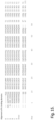
- the first alpha helix in the domains as exemplified by SEQ ID NO. 1-11, 48-64 and 74-93 starts at position 9 (using the position nomenclature of Fig. 15 ), which in the case of SEQ ID NO 1-7 and 9-10 is a glutamine and in SEQ ID NO. 8, 11, 48-64 and 74-93 is an alanine.
- the N-terminal spacer suitably has a length such that the number of amino acid residues between the signal peptide cleaving site and an N-terminus proximal structural unit of the functional or immunoglobulin-binding polypeptide is 14-24.
- this structural unit can suitably be an alpha helix (or alternatively a beta sheet or beta barrel).
- the number of amino acid residues between the N-terminus of the functional or immunoglobulin-binding polypeptide and the N-terminus proximal structural unit can vary, e.g. between 0 (SEQ ID NO. 9-11) and 11 (SEQ ID NO. 2), and accordingly, the length of the N-terminal spacer may vary, e.g.
- the N-terminal spacer may e.g. have a length of 8-12 amino acid residues and if the N-terminal spacer is linked to an Fc-binding domain having at least 90% identity to SEQ ID NO. 9-11, the N-terminal spacer may e.g. have a length of 16-20 amino acid residues.
- the N-terminal spacer may e.g.
- the N-terminal spacer may e.g. consist of amino acid residues selected from the group consisting of alanine, aspartic acid, glutamine, glutamic acid, glycine, histidine, lysine, phenylalanine, serine, threonine, tryptophan, tyrosine and valine.
- the N-terminal spacer does not comprise any clusters of arginines or lysines, and/or if it comprises at most two amino acid residues selected from the group consisting of lysine and arginine.
- the two N-terminal amino acid residues of the N-terminal spacer may be AQ (alanine followed by glutamine).
- the N-terminal spacer may comprise, comprise essentially or have, an amino acid sequence having at least 80% sequence identity to, or being defined by, an amino acid sequence selected from the group consisting of SEQ ID NO. 16-18, 29-30, 33-40, 43-45 and 47.
- the N-terminal spacer may further comprise, comprise essentially or have an amino acid sequence having at least 80% sequence identity to, or being defined by, an amino acid sequence selected from the group consisting of AQYEDGKQYTADNKFNKE, AQYEDGKQYTVDAKFDKE, AQKDQTWYTGVDAKFDKE, AQHDEAQQEAVDAKFDKE, AQGGGSGGGSVDAKFDKE, AQYEDGKQYGTVDAKFDKE, AQYEDGKQGTVDAKFDKE, AQYEDGKQYTTLEKGTVDAKFDKE, AQYEDGKQYTTLEKPVAGGTVDAKFDKE, AQYEDGKQYTVDAKFDKE, AQYEDGKQYTETVDAKFDKE, AQYEDGKQYTDTVDAKFDKE, AQYEDGKQYTATVDAKFDKE, AQYEDGKQYEDTVDAKFDKE,
- an N-terminal spacer comprising, comprising essentially or having, an amino acid sequence having at least 80% sequence identity to, or being defined by, an amino acid sequence selected from the group consisting of SEQ ID NO. 35, 37, 38, 40, 43, 44 and 47.
- the N-terminal spacer may also for alkali stability comprise, comprise essentially or have an amino acid sequence having at least 80% sequence identity to, or being defined by, an amino acid sequence selected from the group consisting of AQYEDGKQYTVDAKFDKE, AQYEDGKQYTETVDAKFDKE, AQYEDGKQYTDTVDAKFDKE, AQYEDGKQYEDTVDAKFDKE, AQHHHHHHHHGTVDAKFDKE, AQHHHHHHGTVDAKFDKE and AQHDEAQQEAGTVDAKFDKE.
- the latter sequences are particularly advantageous in combination with immunoglobulin-binding polypeptides derived from SEQ ID NO. 9-11.
- the alkali stability of the recombinant protein can be assessed by coupling it to an SPR chip, e.g. to Biacore CM5 sensor chip as described in the examples of WO2016079033 , using e.g. NHS- or maleimide coupling chemistries, and measuring the immunoglobulin-binding capacity of the chip, typically using polyclonal human IgG, before and after incubation in alkaline solutions at a specified temperature, e.g. 22 +/- 2 °C.
- the incubation can e.g. be performed in 0.5 M NaOH for a number of 10 min cycles, such as 100, 200 or 300 cycles.
- the IgG capacity of the matrix after 100 10 min incubation cycles in 0.5 M NaOH at 22 +/- 2 °C can be at least 55, such as at least 60, at least 80 or at least 90% of the IgG capacity before the incubation.
- the remaining IgG capacity after 100 cycles for a particular mutant measured as above can be compared with the remaining IgG capacity for a parental recombinant protein.
- the remaining IgG capacity for the mutant may be at least 105%, such as at least 110%, at least 125%, at least 150% or at least 200% of the parental recombinant protein.
- the present invention discloses a nucleic acid molecule encoding for the recombinant protein as disclosed above.
- the nucleic acid molecule comprises the following operatively linked elements in the 5' to 3' direction:
- the nucleic acid molecule may further comprise a ribosome-binding site (RBS) and an origin of replication. It can suitably also comprise an antibiotic resistance marker.
- RBS ribosome-binding site
- the present invention discloses a cloning vector, e.g. a plasmid, which expresses and secretes the recombinant protein as disclosed above into the bacterial periplasm of a gram-negative cell, e.g. E. coli.
- the cloning vector comprises the nucleic acid molecule as discussed above.
- the present invention discloses a gram-negative bacterium transformed by the cloning vector disclosed above.
- the bacterium may e.g. be identified as Escherichia coli, in particular E. coli of the K12 strain, such as E. coli K12-017.
- Other examples of gram-negative bacteria include e.g. the genus Pseudomonas, such as e.g. Pseudomonas fluorescens.
- the transformation may be achieved e.g. by the heat shock method, although other methods such as electroporation are also possible.
- the present invention discloses a method of expressing and secreting the recombinant protein discussed above in a gram-negative bacterium. This method comprises the steps of:
- the method may further comprise a step of inducing recombinant protein expression in the gram-negative bacterium.
- the present invention discloses a separation matrix comprising the recombinant protein disclosed above, covalently linked to a support.
- the expressed recombinant protein should be purified to an appropriate extent before being immobilized to a support.
- purification methods are well known in the field, and the immobilization of protein-based ligands to supports is easily carried out using standard methods. Suitable methods and supports will be discussed below in more detail.
- the alkali stability of the matrix can be assessed by measuring the immunoglobulin-binding capacity, typically using polyclonal human IgG, before and after incubation in alkaline solutions at a specified temperature, e.g. 22 +/- 2 °C.
- the incubation can e.g. be performed in 0.5 M NaOH for a number of 15 min cycles, such as 100, 200 or 300 cycles, corresponding to a total incubation time of 25, 50 or 75 h.
- the IgG capacity of the matrix after 96-100 15 min incubation cycles or a total incubation time of 24 or 25 h in 0.5 M NaOH at 22 +/- 2 °C can be at least 80, such as at least 85, at least 90 or at least 95% of the IgG capacity before the incubation.
- the solid support of the matrix according to the invention can be of any suitable well-known kind.
- a conventional affinity separation matrix is often of organic nature and based on polymers that expose a hydrophilic surface to the aqueous media used, i.e. expose hydroxy (-OH), carboxy (-COOH), carboxamido (-CONH 2 , possibly in N- substituted forms), amino (-NH 2 , possibly in substituted form), oligo- or polyethylenoxy groups on their external and, if present, also on internal surfaces.
- the solid support can suitably be porous.
- the porosity can be expressed as a Kav or Kd value (the fraction of the pore volume available to a probe molecule of a particular size) measured by inverse size exclusion chromatography, e.g. according to the methods described in Gel Filtration Principles and Methods, Pharmacia LKB Biotechnology 1991, pp 6-13 .
- Kd and Kav values always lie within the range 0-1.
- the Kav value can advantageously be 0.6 - 0.95, e.g. 0.7 - 0.90 or 0.6 - 0.8, as measured with dextran of Mw 110 kDa as a probe molecule.
- the support has a large fraction of pores able to accommodate both the recombinant protein of the invention and immunoglobulins binding to the recombinant protein and to provide mass transport of the immunoglobulins to and from the binding sites.
- the recombinant protein may be attached to the support via conventional coupling techniques utilising e.g. thiol, amino and/or carboxy groups present in the ligand. Bisepoxides, epichlorohydrin, CNBr, N-hydroxysuccinimide (NHS) etc are well-known coupling reagents. Between the support and the recombinant protein, a molecule known as a spacer can be introduced, which improves the availability of the recombinant protein and facilitates the chemical coupling of the recombinant protein to the support. Depending on the nature of the recombinant protein and the coupling conditions, the coupling may be a multipoint coupling (e.g.
- the recombinant protein may be attached to the support by non-covalent bonding, such as physical adsorption or biospecific adsorption.
- the matrix comprises 5 - 25, such as 5-20 mg/ml, 5 - 15 mg/ml, 5-11 mg/ml or 6 - 11 mg/ml of the recombinant protein coupled to the support.
- the amount of coupled protein can be controlled by the concentration of protein used in the coupling process, by the activation and coupling conditions used and/or by the pore structure of the support used.
- the absolute binding capacity of the matrix increases with the amount of coupled protein, at least up to a point where the pores become significantly constricted by the coupled protein.
- the relative binding capacity per mg coupled protein will decrease at high coupling levels, resulting in a cost-benefit optimum within the ranges specified above.
- the protein is coupled to the support via thioether bonds.
- Methods for performing such coupling are well-known in this field and easily performed by the skilled person in this field using standard techniques and equipment.
- Thioether bonds are flexible and stable and generally suited for use in affinity chromatography.
- the mobility of the coupled recombinant protein is enhanced which provides improved binding capacity and binding kinetics.
- the recombinant protein is coupled via a C-terminal cysteine provided on the protein as described above. This allows for efficient coupling of the cysteine thiol to electrophilic groups, e.g.
- the recombinant protein may be covalently linked to the support via one or more amide bonds. This can be achieved e.g. through reaction between one or more lysines in the protein and one or more activated carboxyl groups on the support. The activation can e.g. be made by the commonly known N-hydroxysuccinimide (NHS) reagent.
- the protein is linked via one or more secondary amine links. Such links can be formed from lysines in the protein and either hydroxyl groups on the support, which have been activated using e.g.
- the aldehydes can e.g. be formed from vicinal diols on the support through periodate oxidation.
- the support comprises a polyhydroxy polymer, such as a polysaccharide.
- polysaccharides include e.g. dextran, starch, cellulose, pullulan, agar, agarose etc.
- Polysaccharides are inherently hydrophilic with low degrees of nonspecific interactions, they provide a high content of reactive (activatable) hydroxyl groups and they are generally stable towards alkaline cleaning solutions used in bioprocessing.
- the support comprises agar or agarose.
- the supports used in the present invention can easily be prepared according to standard methods, such as inverse suspension gelation ( S Hjertén: Biochim Biophys Acta 79(2), 393-398 (1964 ).
- the base matrices are commercially available products, such as crosslinked agarose beads sold under the name of SEPHAROSE TM FF (GE Healthcare).
- the support has been adapted to increase its rigidity using the methods described in US6602990 or US7396467 , which are hereby incorporated by reference in their entirety, and hence renders the matrix more suitable for high flow rates.
- the support such as a polysaccharide or agarose support
- Crosslinker reagents producing such crosslinks can be e.g. epihalohydrins like epichlorohydrin, diepoxides like butanediol diglycidyl ether, allylating reagents like allyl halides or allyl glycidyl ether.
- Crosslinking is beneficial for the rigidity of the support and improves the chemical stability. Hydroxyalkyl ether crosslinks are alkali stable and do not cause significant nonspecific adsorption.
- the solid support is based on synthetic polymers, such as polyvinyl alcohol, polyhydroxyalkyl acrylates, polyhydroxyalkyl methacrylates, polyacrylamides, polymethacrylamides etc.
- hydrophobic polymers such as matrices based on divinyl and monovinyl-substituted benzenes
- the surface of the matrix is often hydrophilised to expose hydrophilic groups as defined above to a surrounding aqueous liquid.
- Such polymers are easily produced according to standard methods, see e.g. " Styrene based polymer supports developed by suspension polymerization" (R Arshady: Chimica e L'Industria 70(9), 70-75 (1988 )).
- a commercially available product such as SOURCE TM (GE Healthcare) is used.
- the solid support according to the invention comprises a support of inorganic nature, e.g. silica, zirconium oxide etc.
- the solid support is in another form such as a surface, a chip, capillaries, or a filter (e.g. a membrane or a depth filter matrix).
- a filter e.g. a membrane or a depth filter matrix
- the present invention discloses a method of separating an immunoglobulin, comprising the steps of:
- Example 2 Two different signal peptides were tested together with four different N-terminal spacers to find the best cleavage and protein expression.
- This experiment (Example 1) was performed with Zvar2 monomer using the RhaBAD promoter system.
- Example 2 the most promising signal peptide and N-terminal start was cloned in to Zvar2 hexamer. This construct was tested in fed-batch fermentations with and without the N-terminal start sequence. This study was performed using the T5 promoter expression system.
- Example 3 all the native domains of Staphylococcus aureus protein A, Zvar monomer (Zvar 1 ), Zvar2 monomer (Zvar2 1 ) and Zvar tetramer (Zvar 4 ) were tested with the chosen signal peptide and N-terminal start to see if the start sequence also had an effect on closely related domains. Also Example 3 was performed with the T5 expression system. Example 4 was performed with a set of different N-terminal spacers, Example 5 was a scale-up of fermentations using a couple of selected spacers and Example 6 was an investigation using the AQYEDGKQYTGT N-terminal spacer in combination with different mutants of an immunoglobulin-binding protein.
- dsDNA Double stranded dideoxy ribonucleic acid
- AA amino acid sequence
- pJ401 Double stranded dideoxy ribonucleic acid
- Table 2 The dsDNA was inserted into expression vector pD861-SR or pJ401 with a signal peptide either from DsbA or OmpA (Table 2).
- Table 1 Plasmids ordered from ATUM.
- All plasmids were transformed into E.coli K12-017 using chemically competent cells.
- a short spacer sequence of 8 AA was inserted between the FspI and KpnI cleave sites.
- the 8 AA sequences were taken from the first AA in the mature protein of OmpA (AQKDQTWYTGGT, SEQ ID NO 16), DsbA (AQYEDGKQYTGT, SEQ ID NO 17), SpA (AQHDEAQQEAGT, SEQ ID NO 18) or the flexible linker structure from M13 phage GIII AA 236-243 (AQGGGSGGGSGT, SEQ ID NO 19), with modifications in OmpA to fit the restriction site and in OmpA and SpA to mutate asparagines to glutamines ( Fig. 1 ).
- a range of different sequences were cloned into plasmid pGE0002 (Table 1).
- Plasmids were cleaved with restriction enzymes FspI and KpnI (New England Biolabs (NEB), MA, USA), 6 ⁇ l NEB buffer 2.1 was mixed with 6 ⁇ g plasmid and 2 ⁇ l FspI in total 58 ⁇ l. The solution was incubated in 37°C for 1 hour before KpnI was added. The incubation was continued 2 h, followed by addition of 1 ⁇ l Calf intestine phosphatase (CIP) followed by 30 min further incubation. Excised bases were removed from the digested plasmids using QIAquick PCR Purification kit (Qiagen, Hilden, Germany).
- Oligonucleotides were ordered from Integrated DNA technologies (IDT, IA, USA). All oligonucleotides were modified with a five prime phosphate group by manufacturer. Two complementary oligonucleotide pairs were mixed in ligation buffer and heated to 95°C, 4 min, followed by cooling down to room temperature. The hybridized fragment was ligated into the FspI and KpnI cleaved plasmids by use of T4 DNA ligase (NEB, MA, USA). A complete list of oligonucleotides used is found in Table 3. Table 3. List of oligonucleotides used for addition of N-terminal start sequence.
- Ligated plasmids were transformed into chemically competent E. coli K12-017 cells. The cells were thawed on ice for 30 min, 100 ⁇ l competent cells were added to 10 ⁇ l ligation reaction. Cells were incubated on ice for 20 min followed by heat shock at 42°C for 1 min, followed by incubation on ice 5 min. Followed by addition of 900 ⁇ l SOC-medium (NEB, MA, USA). Transformation reaction was incubated in 37°C in a rotary shake incubator for 60 min, 100 ⁇ l of each reaction was spread on Luria agar plates containing appropriate selective antibiotics. Positive clones were screened with Polymerase chain reaction (PCR), and clones with correct insert were selected.
- PCR Polymerase chain reaction
- Escherichia coli strain K12-017 transformed with the recombinant plasmids pGE0138-145 were cultured in 30°C for 4 h in 100 ml Terrific Broth (TB) medium (containing 12g tryptone, 24 g yeast extract, 5 g glycerol (85%), 2,31 g KH 2 PO 4 , 12,54 g K 2 HPO 4 and 50 mg of kanamycin sulphate per liter). Cultures were induced when OD 600 nm reached 1-2, using 4 mM L-rhamnose (Sigma Aldrich, MO, USA). Cultures were further incubated in 30°C 17-20 h.
- TB Terrific Broth
- Culture solution was subjected to low-speed centrifugation (at 4,000 rpm) for 20 min in a swing out rotor to collect wet cell pellet.
- the bacterial cell pellets were suspended in 20 ml of a 25 mM phosphate buffer solution (pH 7.4), and the cells were lysed by heat treatment at 85°C for 10 min in a heat block.
- a high-speed centrifugation at 13 000 rpm
- the supernatants were filtered through 0.2 ⁇ m syringe filter to remove any residual particles before it was applied to an IgG Sepharose 6FF (GE Healthcare Bio-Sciences, Uppsala, Sweden) affinity chromatography column.
- the column was equilibrated with loading buffer (25 mM phosphate pH7, 250 mM NaCl) followed by loading of sample. After loading, the column was washed with 5 column volumes (CV) of loading buffer and one CV of low salt wash buffer (50 mM acetate pH6), followed by elution with 50 mM acetic acid, pH2.8. Absorbance at 237 nm was measured in-line using an ⁇ KTA explorer 100 chromatography system (GE Healthcare Bio-Sciences, Uppsala, Sweden) and peak integration of the eluted peak was performed in the system software (Unicorn 5.1).
- loading buffer 25 mM phosphate pH7, 250 mM NaCl
- low salt wash buffer 50 mM acetate pH6
- Absorbance at 237 nm was measured in-line using an ⁇ KTA explorer 100 chromatography system (GE Healthcare Bio-Sciences, Uppsala, Sweden) and peak integration of the eluted peak was performed in the
- DsbA utilizes the signal recognition particle (SRP) pathway and OmpA the Sec pathway.
- SRP signal recognition particle
- OmpA the Sec pathway.
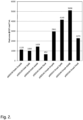
- the first N-terminal AA after the cleave site of the signal peptide had a significant impact on the expression levels.
- the DsbA start AA had the highest expression level together with both signal peptides. The lowest expression level was seen for the Flex8AA start sequence using both signal peptides.
- Fermentation samples were heat treated at 85°C for 5 min in 1.5 ml tubes.
- high speed centrifugation at 13500 rpm, in benchtop centrifuge
- Supernatants were filtered through 0.2 ⁇ m syringe filter to remove any residual particles before they were applied to an IgG Sepharose 6FF HiTrap TM column (GE Healthcare Bio-Sciences, Uppsala Sweden).
- the column was equilibrated with loading buffer (Phosphate buffered Saline (Medicago, Uppsala, Sweden)) followed by loading of 50 ⁇ l sample.
- the column was washed with 5 CV of loading buffer, followed by elution with 200 mM phosphate buffer, pH2.9. Absorbance at 237 nm was measured in-line and peak integration of the eluted peak was performed in the chromatography system software. Protein concentration of the samples were determined by a standard curve using a purified protein of known concentration.
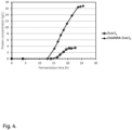
- Plasmid pGE0002 containing Zvar2 hexamer (Zvar2 6 ) was cleaved with FspI and KpnI restriction enzymes. The N-terminal start from DsbA was ligated into the cleaved vector, and the resulting plasmid was denoted pGE0180. These two vectors were transformed into E.coli K12-017 and the resulting constructs were expressed in fed-batch fermentations to see differences in protein expression and signal peptide cleavage patterns. Protein concentration analysis using standard curve showed that the protein expression increased from 3.5 g/L to 16.8 g/L, i.e. a more than 4 times increase in protein expression ( Fig. 4 ).
- the amount of protein was estimated by absorbance measurements at 280 nm using specific absorbance coefficients, calculated based on Pace et al. Protein Science 4, 2411-2423, (1995 ) and Beer Lamberts law. Sample concentration was back-calculated using total protein mass in elution pool and volume of injected sample. The concentration was confirmed by amino acid analysis. The homogeneity and the molecular weights were analysed using mass spectrometry on a Waters Q-Tof (PA, USA).
- the N-terminal inserts were each designed to investigate the effect of the insert sequence on one or more of the following parameters: protein yield, signal peptide cleavage and alkaline stability.
- the designs can be divided into four separate categories as follows:
- category 1 was mainly to investigate the effect of the insert length on signal peptide cleavage and protein yield, and did not explicitly address the suspected truncation site at alkaline conditions.
- Categories 2 and 3 were mostly aimed at addressing the alkaline stability issue, although they also provide some more data for the other two issues.
- Category 4 was a more open category, providing a wider set of sequences to provide data in all three areas of interest.
- nucleotide sequences were codon optimized for E. coli, but with some degeneracy added in the more repetitive sequences. Inserts belonging to category 1 and 4 were designed to be complementary to the digestion sites of FspI in the 5' end, and KpnI in the 3' end. Since the glycine-threonine truncation site was a part of the KpnI restriction site, inserts belonging to categories 2 and 3 were designed to instead be complementary to the adjacent AccI restriction site in the 3' end, while retaining the 5' complementarity to FspI.
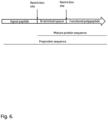
- the amino acid sequences of all inserts are shown in table 6. A schematic illustration of the general construct is shown in Fig 6 . Table 6. All inserts used in the experiments.
- Residues resulting from the restriction site "scar" are marked in italics.
- Category Construct name Insert amino acid sequence SEQ ID NO Original construct without DsbA insert pGE0002 AQGT 28 Original construct with 8 aa DsbA insert pGE0180 AQ YEDGKQYT GT 17 1 DsbA7 AQ YEDGKQY GT 29 DsbA6 AQ YEDGKQ GT 30 DsbA5 AQ YEDGK GT 31 DsbA4 AQ YEDG GT 32 DsbA12 AQ YEDGKQYTTLEK GT 33 DsbA16 AQ YEDGKQYTTLEKPVAG GT 34 2 DsbA8_noG T AQ YEDGKQYT 35 DsbA6_noG T AQ YEDGKQ 36 3 DsbA8_ET AQ YEDGKQYTET 37 DsbA8_DT AQ
- constructs were assembled by ligating different hybridized oligonucleotides into the plasmid of either pGE0002 or pGE0180, digested with the relevant restriction enzymes to remove the original N-terminal insert. After ligation, the plasmids were transformed into E. coli K12-017 cells. Positive colonies were screened using colony PCR and electrophoresis. Two or three clones from each construct were chosen and sent to GATC biotech (Cologne, Germany) for sequence verification.
- FIG. 7 shows a summary of the results from the quantification of protein yields in the different shake flask cultures done with IgG Sepharose 6FF.
- the left bar is the protein concentration after linear correction using a 5 g/L and a 10 g/L standard solution as reference.
- the right bar is this concentration divided by the OD 600 nm at the end of the cultivation. This value is meant to give an idea of whether a low protein concentration is due to an actual low rate of protein production per cell, or simply due to a low culture density.
- concentrations are measured after pelleting the cells and re-suspending them in Phosphate buffered saline (Medicago, Uppsala, Sweden), and as such are not directly comparable to concentrations from samples taken from e.g. the multifermenter.
- the protein solutions for each construct yielded from the shake flask cultivation were purified using IgG Sepharose 6FF (GE Healthcare, Uppsala, Sweden).
- the purified protein samples were investigated for signal peptide cleavage using LC/MS analysis (Waters, PA, USA).
- a protein with a correctly cleaved signal peptide only has one main mass (example Total Ion Count (TIC) shown in Figure 8 a)
- TIC Total Ion Count
- Figure 8 b secondary peaks
- the side peaks further to the left and right of the main peaks are likely due to leakage from the cytoplasm during the heat treatment-step when extracting the protein from the cell culture.
- the peaks C1 to C5, N1 and N2 are likely different partially digested versions of the protein from the cytoplasm.
- the small peak to the right of the main peak is protein with uncleaved signal peptide, which would be reasonable to see if leakage from the cytoplasm had occurred.
- the protein solutions of constructs with correct signal peptide cleavage were further purified using a Capto Q ImpRes anion exchange column (GE Healthcare, Uppsala, Sweden). Purified samples were subjected to treatment with 1 M sodium hydroxide (NaOH) for 0, 4 and 24 hours. Vivaspin columns (GE Healthcare, Uppsala, Sweden) were used to separate the main protein from any small peptides that might have been truncated. Both the peptide samples and the main proteins were investigated using LC/MS (Waters, PA, USA).
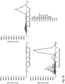
- Peptides appeared in the mass spectra with +2 charge, that is, at a mass over charge (m/z) -value corresponding to half their molecular weight. In the cases investigated here, the peptides all had a molecular weight between about 800 Da and 1600 Da. As such, the relevant cluster of peaks appeared between 400 and 800 m/z, as can be seen for pGE0180 to the left in Fig 9 .
- the MassLynx search function was used on the TIC to extract the peaks of different peptide masses as extracted ion chromatograms (XIC). The XIC peaks were integrated and the integrated areas were then used to compare the amounts of different peptides in the samples.
- Fig 10 shows a summary of the results from the initial alkaline stability study done with unwashed Vivaspin columns. Despite quite a bit of overlapping background noise, clear patterns are visible after 24 hours of incubation.
- Fig 11 shows a different view of the same data for the most promising constructs (including pGE0180 as a reference), while Fig 12 gives a breakdown of the most prominently cleaved constructs.
- the sequences around the cleavage site are included to give an idea of what residues might be vulnerable. Notably, for the low peak areas (around 5000 area units), background noise has a significant influence.
- the fed-batch cultures of constructs DsbA8_noGT and DsbA8_DT were heat treated to extract periplasmic protein, and centrifuged and diafiltered to remove cellular debris.
- the filtrate was conditioned to remove some contaminants via precipitation, and prepare the solution for purification with Capto S ImpAct (GE Healthcare, Uppsala, Sweden) using a pH gradient.
- Capto S ImpAct GE Healthcare, Uppsala, Sweden
- the elute was reduced to remove dimers, and desalted to remove the DTT and get the correct buffer for the anion exchange purification, where a salt gradient was used.
- the final purified solution was concentrated via membrane ultrafiltration to reach the concentration needed for immobilization at the desired ligand densities.
- the end product was analysed with SEC for purity and LC/MS for molecular weight, with satisfactory results.
- the purified proteins (>90% purity) were coupled to highly cross-linked agarose beads, aiming for ligand densities similar to those in a previous experiment where Zvar2 was immobilized on a base matrix of similar properties and analyzed for dynamic binding capacity.
- the coupled gels were packed in Tricorn 5/100 columns (GE Healthcare, Uppsala, Sweden). The packing of the gel bed was evaluated through the asymmetry of an acetone peak. The packed columns were used to measure the dynamic binding capacity of the gels using IgG. One measurement per construct was made. The results are summarized in table 7.
- Fig 14 shows a comparison between measurements done in this instance, and those done in the aforementioned previous experiment. Table 7. Asymmetry and dynamic binding capacity values for the different protein-coupled gels.
- Zvar2 DsbA8_noGT DsbA8_DT Qb10 (mg IgG/mL gel) 73.7 74.2 71.6 Qb80 (mg IgG/mL gel) 93.6 92.2 90.7
- the N-terminal spacer AQYEDGKQYTGT and a C-terminal cysteine was introduced in a number of monomeric and dimeric further mutants of Zvar2 (as listed below), using the OmpA signal peptide.
- the structure of the mature protein was thus: AQYEDGKQYTGT-IgG-binding polypeptide-C.
- Plasmids were transformed into chemically competent E.coli K12-017 cells. The cells were thawed on ice for 30 min, 50 ⁇ l competent cells were added to 20 ng plasmids. Cells were incubated on ice for 20 min followed by heat shock at 42°C for 1 min, followed by incubation on ice 5 min and addition of 400 ⁇ l SOC-medium (NEB, MA, USA). Transformation reaction was incubated in 37°C in a rotary shake incubator for 60 min, 200 ⁇ l of each reaction was spread on Luria agar plates containing appropriate selective antibiotics.
- E. coli strain K12-017 transformed with the recombinant plasmids were cultured in 37°C in 100 ml Terrific Broth (TB) medium (containing 12g tryptone, 24 g yeast extract, 5 g glycerol (85%), 2,31 g KH 2 PO 4 , 12,54 g K 2 HPO 4 and 50 mg of kanamycin sulphate per liter). Cultures were induced when OD 600 nm reached 1, using 1 mM IPTG (Sigma Aldrich, MO, USA). Cultures were further incubated in 30°C 17-20 h.
- TB Terrific Broth
- Culture solution was subjected to low-speed centrifugation (at 4,000 rpm) for 20 min in a swing out rotor to collect wet cell pellet.
- the bacterial cell pellets were suspended in 10 ml of a 25 mM phosphate buffer solution (pH 7.4), and the cells were lysed by heat treatment at 85°C for 15 min in a heat block.
- a high-speed centrifugation at 10 000 x g
- supernatants were filtered through 0.2 ⁇ m syringe filter to remove any residual particles before it was applied to an XK 16-6 IgG Sepharose 6FF (GE Healthcare Bio-Sciences, Uppsala, Sweden).
- the column was equilibrated with loading buffer (25 mM phosphate pH7, 250 mM NaCl) followed by loading of sample. After loading the column was washed with 5 column volumes (CV) of loading buffer and one CV of low salt wash buffer (50 mM acetate pH6), followed by elution with 50 mM acetic acid, pH2.8. Absorbance at 237 nm was measured in-line using an ⁇ KTA explorer 100 (GE Healthcare Bio-Sciences, Uppsala, Sweden) and peak integration of the eluted peak was performed in the system software (Unicorn 5.1).
- loading buffer 25 mM phosphate pH7, 250 mM NaCl
- low salt wash buffer 50 mM acetate pH6
- Absorbance at 237 nm was measured in-line using an ⁇ KTA explorer 100 (GE Healthcare Bio-Sciences, Uppsala, Sweden) and peak integration of the eluted peak was performed in the system software (Unicorn 5.1).
- the IgG-binding fraction was eluted and collected in a pool and the volumes and protein concentrations of the pools were noted - see table 8.
- the amount of IgG-binding protein expressed may in several cases be higher than the amount recovered, due to loss of protein secreted into the culture medium and/or to overloading of the IgG column.
- Table 8. Expression of IgG-binding proteins with N-terminal spacer AQYEDGKQYTGT and with a C-terminal cysteine.
- IgG-binding protein SEQ ID NO Protein conc.
Landscapes
- Chemical & Material Sciences (AREA)
- Health & Medical Sciences (AREA)
- Genetics & Genomics (AREA)
- Organic Chemistry (AREA)
- Life Sciences & Earth Sciences (AREA)
- Molecular Biology (AREA)
- Engineering & Computer Science (AREA)
- Biochemistry (AREA)
- General Health & Medical Sciences (AREA)
- Biophysics (AREA)
- Biomedical Technology (AREA)
- Medicinal Chemistry (AREA)
- Proteomics, Peptides & Aminoacids (AREA)
- Bioinformatics & Cheminformatics (AREA)
- Zoology (AREA)
- Wood Science & Technology (AREA)
- Biotechnology (AREA)
- General Engineering & Computer Science (AREA)
- Analytical Chemistry (AREA)
- Plant Pathology (AREA)
- Microbiology (AREA)
- Physics & Mathematics (AREA)
- Gastroenterology & Hepatology (AREA)
- Immunology (AREA)
- Peptides Or Proteins (AREA)
- Micro-Organisms Or Cultivation Processes Thereof (AREA)
Applications Claiming Priority (3)
| Application Number | Priority Date | Filing Date | Title |
|---|---|---|---|
| GBGB1708277.7A GB201708277D0 (en) | 2017-05-24 | 2017-05-24 | A Recombinant protein |
| EP18726972.5A EP3630793B1 (de) | 2017-05-24 | 2018-05-23 | Rekombinantes protein |
| PCT/EP2018/063435 WO2018215503A1 (en) | 2017-05-24 | 2018-05-23 | A recombinant protein |
Related Parent Applications (2)
| Application Number | Title | Priority Date | Filing Date |
|---|---|---|---|
| EP18726972.5A Division EP3630793B1 (de) | 2017-05-24 | 2018-05-23 | Rekombinantes protein |
| EP18726972.5A Division-Into EP3630793B1 (de) | 2017-05-24 | 2018-05-23 | Rekombinantes protein |
Publications (2)
| Publication Number | Publication Date |
|---|---|
| EP4414384A2 true EP4414384A2 (de) | 2024-08-14 |
| EP4414384A3 EP4414384A3 (de) | 2024-12-11 |
Family
ID=59220774
Family Applications (2)
| Application Number | Title | Priority Date | Filing Date |
|---|---|---|---|
| EP24178801.7A Pending EP4414384A3 (de) | 2017-05-24 | 2018-05-23 | Rekombinantes protein |
| EP18726972.5A Active EP3630793B1 (de) | 2017-05-24 | 2018-05-23 | Rekombinantes protein |
Family Applications After (1)
| Application Number | Title | Priority Date | Filing Date |
|---|---|---|---|
| EP18726972.5A Active EP3630793B1 (de) | 2017-05-24 | 2018-05-23 | Rekombinantes protein |
Country Status (8)
| Country | Link |
|---|---|
| US (2) | US20200318120A1 (de) |
| EP (2) | EP4414384A3 (de) |
| JP (3) | JP7490190B2 (de) |
| CN (2) | CN117756884A (de) |
| DK (1) | DK3630793T3 (de) |
| ES (1) | ES2986499T3 (de) |
| GB (1) | GB201708277D0 (de) |
| WO (1) | WO2018215503A1 (de) |
Families Citing this family (2)
| Publication number | Priority date | Publication date | Assignee | Title |
|---|---|---|---|---|
| WO2020221781A1 (en) * | 2019-04-29 | 2020-11-05 | Cytiva Bioprocess R&D Ab | Method for separation of antibodies or antibody fragments being devoid of an fc region capable of binding to protein a |
| GB202018588D0 (en) * | 2020-11-26 | 2021-01-13 | Cytiva Bioprocess R & D Ab | Separation matrix |
Citations (19)
| Publication number | Priority date | Publication date | Assignee | Title |
|---|---|---|---|---|
| US6602990B1 (en) | 1996-04-11 | 2003-08-05 | Amersham Biosciences Ab | Process for the production of a porous cross-linked polysaccharide gel and its use as a gel filtration media and in chromatography |
| US20050143566A1 (en) | 2002-03-25 | 2005-06-30 | Sophia Hober | Mutated immunoglobulin-binding protein |
| JP2006304633A (ja) | 2005-04-26 | 2006-11-09 | Apro Life Science Institute Inc | イムノグロブリン結合タンパク質 |
| US7396467B2 (en) | 2004-09-22 | 2008-07-08 | Ge Healthcare Bio-Sciences Ab | Method of preparing a chromatography matrix |
| US20100221844A1 (en) | 2008-12-24 | 2010-09-02 | Millipore Corporation | Caustic stable chromatography ligands |
| US20120208234A1 (en) | 2009-03-24 | 2012-08-16 | Kaneka Corporation | Protein having affinity for immunoglobulin, and immunoglobulin-binding affinity ligand |
| US8329860B2 (en) | 2006-09-29 | 2012-12-11 | Ge Healthcare Bio-Sciences Ab | Chromatography ligand comprising domain C from Staphylococcus aureus protein A for antibody isolation |
| US20130096276A1 (en) | 2010-03-24 | 2013-04-18 | Kaneka Corporation | Protein capable of binding specifically to immunoglobulin, and immunoglobulin-binding affinity ligand |
| US20130274451A1 (en) | 2010-12-20 | 2013-10-17 | Tomas Bjorkman | Affinity chromatography matrix |
| US20140031522A1 (en) | 2010-12-21 | 2014-01-30 | Jsr Corporation | Novel alkali-resistant variants of protein a and their use in affinity chromatography |
| US8674073B2 (en) | 2006-02-21 | 2014-03-18 | Protenova Co., Ltd. | Immunoglobulin affinity ligand |
| US20140107315A1 (en) | 2011-03-25 | 2014-04-17 | Shinichi Yoshida | Protein for affinity-separation matrix |
| US8754196B2 (en) | 2011-06-08 | 2014-06-17 | Emd Millipore Corporation | Chromatography matrices including novel Staphylococcus aureus protein A based ligands |
| WO2014146350A1 (zh) | 2013-03-18 | 2014-09-25 | 南京金斯瑞生物科技有限公司 | 一类突变的具有高耐碱特性的蛋白a及其应用 |
| US9040661B2 (en) | 2010-12-21 | 2015-05-26 | Jsr Corporation | Support for affinity chromatography and method for isolating immunoglobulin |
| WO2016079034A1 (en) | 2014-11-17 | 2016-05-26 | Ge Healthcare Bioprocess R&D Ab | Mutated immunoglobulin-binding polypeptides |
| US20160159855A1 (en) | 2013-07-10 | 2016-06-09 | Ge Healthcare Bio-Sciences Ab | Mutated immunoglobulin-binding polypeptides |
| US20160168209A1 (en) | 2012-09-21 | 2016-06-16 | Kaneka Corporation | Protein ligand for affinity isolation matrix |
| WO2016152946A1 (ja) | 2015-03-26 | 2016-09-29 | Jsr株式会社 | イムノグロブリン結合タンパク質およびそれを用いたアフィニティー担体 |
Family Cites Families (6)
| Publication number | Priority date | Publication date | Assignee | Title |
|---|---|---|---|---|
| US5563046A (en) * | 1993-08-02 | 1996-10-08 | Celtrix Pharmaceuticals, Inc. | Fusion polypeptides and proteins |
| JP5099881B2 (ja) * | 2007-01-27 | 2012-12-19 | 独立行政法人科学技術振興機構 | 高効率無細胞蛋白質合成系 |
| WO2009086132A2 (en) * | 2007-12-20 | 2009-07-09 | University Of Southern California | Design of spacers to increase the expression of recombinant fusion proteins |
| JP6283674B2 (ja) * | 2012-09-18 | 2018-02-21 | グラクソスミスクライン バイオロジカルズ ソシエテ アノニム | 外膜小胞 |
| JP6425327B2 (ja) * | 2013-12-12 | 2018-11-21 | 国立大学法人東京農工大学 | タンパク質−磁性粒子複合体及びその製造方法 |
| AU2015229035B2 (en) * | 2014-03-14 | 2021-08-05 | Genentech, Inc. | Methods and compositions for secretion of heterologous polypeptides |
-
2017
- 2017-05-24 GB GBGB1708277.7A patent/GB201708277D0/en not_active Ceased
-
2018
- 2018-05-23 WO PCT/EP2018/063435 patent/WO2018215503A1/en not_active Ceased
- 2018-05-23 US US16/615,552 patent/US20200318120A1/en active Pending
- 2018-05-23 CN CN202311512241.XA patent/CN117756884A/zh active Pending
- 2018-05-23 ES ES18726972T patent/ES2986499T3/es active Active
- 2018-05-23 CN CN201880033927.1A patent/CN110831956B/zh active Active
- 2018-05-23 DK DK18726972.5T patent/DK3630793T3/da active
- 2018-05-23 EP EP24178801.7A patent/EP4414384A3/de active Pending
- 2018-05-23 EP EP18726972.5A patent/EP3630793B1/de active Active
- 2018-05-23 JP JP2019564995A patent/JP7490190B2/ja active Active
-
2022
- 2022-11-11 JP JP2022181157A patent/JP2023026757A/ja active Pending
-
2024
- 2024-10-10 JP JP2024177862A patent/JP2025015536A/ja active Pending
-
2025
- 2025-05-23 US US19/217,889 patent/US20260022387A1/en active Pending
Patent Citations (23)
| Publication number | Priority date | Publication date | Assignee | Title |
|---|---|---|---|---|
| US6602990B1 (en) | 1996-04-11 | 2003-08-05 | Amersham Biosciences Ab | Process for the production of a porous cross-linked polysaccharide gel and its use as a gel filtration media and in chromatography |
| US20050143566A1 (en) | 2002-03-25 | 2005-06-30 | Sophia Hober | Mutated immunoglobulin-binding protein |
| US7396467B2 (en) | 2004-09-22 | 2008-07-08 | Ge Healthcare Bio-Sciences Ab | Method of preparing a chromatography matrix |
| JP2006304633A (ja) | 2005-04-26 | 2006-11-09 | Apro Life Science Institute Inc | イムノグロブリン結合タンパク質 |
| US8674073B2 (en) | 2006-02-21 | 2014-03-18 | Protenova Co., Ltd. | Immunoglobulin affinity ligand |
| US8329860B2 (en) | 2006-09-29 | 2012-12-11 | Ge Healthcare Bio-Sciences Ab | Chromatography ligand comprising domain C from Staphylococcus aureus protein A for antibody isolation |
| US20100221844A1 (en) | 2008-12-24 | 2010-09-02 | Millipore Corporation | Caustic stable chromatography ligands |
| US20120208234A1 (en) | 2009-03-24 | 2012-08-16 | Kaneka Corporation | Protein having affinity for immunoglobulin, and immunoglobulin-binding affinity ligand |
| US9403883B2 (en) | 2009-03-24 | 2016-08-02 | Kaneka Corporation | Protein having affinity for immunoglobulin, and immunoglobulin-binding affinity ligand |
| US20130096276A1 (en) | 2010-03-24 | 2013-04-18 | Kaneka Corporation | Protein capable of binding specifically to immunoglobulin, and immunoglobulin-binding affinity ligand |
| US20130274451A1 (en) | 2010-12-20 | 2013-10-17 | Tomas Bjorkman | Affinity chromatography matrix |
| US9040661B2 (en) | 2010-12-21 | 2015-05-26 | Jsr Corporation | Support for affinity chromatography and method for isolating immunoglobulin |
| US20140031522A1 (en) | 2010-12-21 | 2014-01-30 | Jsr Corporation | Novel alkali-resistant variants of protein a and their use in affinity chromatography |
| US9051375B2 (en) | 2010-12-21 | 2015-06-09 | The University Of Western Ontario | Alkali-resistant variants of protein A and their use in affinity chromatography |
| US20140107315A1 (en) | 2011-03-25 | 2014-04-17 | Shinichi Yoshida | Protein for affinity-separation matrix |
| US8754196B2 (en) | 2011-06-08 | 2014-06-17 | Emd Millipore Corporation | Chromatography matrices including novel Staphylococcus aureus protein A based ligands |
| US20160168209A1 (en) | 2012-09-21 | 2016-06-16 | Kaneka Corporation | Protein ligand for affinity isolation matrix |
| WO2014146350A1 (zh) | 2013-03-18 | 2014-09-25 | 南京金斯瑞生物科技有限公司 | 一类突变的具有高耐碱特性的蛋白a及其应用 |
| US20160237124A1 (en) | 2013-03-18 | 2016-08-18 | Hong Qian | Kind of mutated proteins a with high alkali resistance feature and application thereof |
| US20160159855A1 (en) | 2013-07-10 | 2016-06-09 | Ge Healthcare Bio-Sciences Ab | Mutated immunoglobulin-binding polypeptides |
| WO2016079034A1 (en) | 2014-11-17 | 2016-05-26 | Ge Healthcare Bioprocess R&D Ab | Mutated immunoglobulin-binding polypeptides |
| WO2016079033A1 (en) | 2014-11-17 | 2016-05-26 | Ge Healthcare Bioprocess R&D Ab | Mutated immunoglobulin-binding polypeptides |
| WO2016152946A1 (ja) | 2015-03-26 | 2016-09-29 | Jsr株式会社 | イムノグロブリン結合タンパク質およびそれを用いたアフィニティー担体 |
Non-Patent Citations (7)
| Title |
|---|
| ALTSCHUL ET AL., J. MOL. BIOL., vol. 215, 1990, pages 403 - 410 |
| L ABRAHMSÉN ET AL., EMBO J, vol. 4, no. 13B, 1985, pages 3901 - 3906 |
| MERGULHAO ET AL., BIOTECH ADV, vol. 23, 2005, pages 177 - 202 |
| OLSSON M.O.ISAKSSON L.A., MOLEC. GEN. GENET., vol. 169, 1979, pages 251 - 257 |
| PACE ET AL., PROTEIN SCIENCE, vol. 4, 1995, pages 2411 - 2423 |
| R ARSHADY, CHIMICA E L'INDUSTRIA, vol. 70, no. 9, 1988, pages 70 - 75 |
| S HJERTÉN, BIOCHIM BIOPHYS ACTA, vol. 79, no. 2, 1964, pages 393 - 398 |
Also Published As
| Publication number | Publication date |
|---|---|
| CN117756884A (zh) | 2024-03-26 |
| DK3630793T3 (da) | 2024-07-22 |
| WO2018215503A1 (en) | 2018-11-29 |
| JP2023026757A (ja) | 2023-02-28 |
| JP7490190B2 (ja) | 2024-05-27 |
| EP4414384A3 (de) | 2024-12-11 |
| ES2986499T3 (es) | 2024-11-11 |
| CN110831956A (zh) | 2020-02-21 |
| JP2020520670A (ja) | 2020-07-16 |
| EP3630793A1 (de) | 2020-04-08 |
| US20200318120A1 (en) | 2020-10-08 |
| GB201708277D0 (en) | 2017-07-05 |
| CN110831956B (zh) | 2024-02-06 |
| JP2025015536A (ja) | 2025-01-30 |
| EP3630793B1 (de) | 2024-07-10 |
| US20260022387A1 (en) | 2026-01-22 |
Similar Documents
| Publication | Publication Date | Title |
|---|---|---|
| US10464972B2 (en) | Multimer of mutant protein A and methods of using same | |
| US20260022387A1 (en) | Recombinant Protein | |
| EP3233891B1 (de) | Modifizierte kappa-leichtketten-bindende polypeptide | |
| JPWO2010110288A1 (ja) | 免疫グロブリンに親和性を有するタンパク質および免疫グロブリン結合性アフィニティーリガンド | |
| JP2017070289A (ja) | アフィニティー分離マトリックス用タンパク質 | |
| CN111315887B (zh) | 免疫球蛋白结合性蛋白质 | |
| WO2014046278A1 (ja) | アフィニティー分離マトリックス用タンパク質リガンド | |
| JP6805831B2 (ja) | 免疫グロブリンに親和性を有するタンパク質、およびそれを用いたアフィニティ分離剤、液体クロマトグラフィー用カラム | |
| EP3842536A1 (de) | Immunglobulin-bindendes protein und affinitätsträger damit | |
| CN109721645A (zh) | 一种基因突变的蛋白a及其应用 | |
| CN104356238A (zh) | 一种重组蛋白a亲和配基的定点固定化方法 | |
| CN104945488B (zh) | 一种具有免疫球蛋白结合能力的多肽 | |
| CN120795094A (zh) | 一种用于纯化抗体的高亲和力蛋白及其重组基因 | |
| CN116323652A (zh) | 从内含肽复合物释放靶标的方法 | |
| Jennings | Chromatographic Analysis of Recombinant Lysostaphin Expressed in Escherichia coli |
Legal Events
| Date | Code | Title | Description |
|---|---|---|---|
| PUAI | Public reference made under article 153(3) epc to a published international application that has entered the european phase |
Free format text: ORIGINAL CODE: 0009012 |
|
| STAA | Information on the status of an ep patent application or granted ep patent |
Free format text: STATUS: REQUEST FOR EXAMINATION WAS MADE |
|
| 17P | Request for examination filed |
Effective date: 20240529 |
|
| AC | Divisional application: reference to earlier application |
Ref document number: 3630793 Country of ref document: EP Kind code of ref document: P |
|
| AK | Designated contracting states |
Kind code of ref document: A2 Designated state(s): AL AT BE BG CH CY CZ DE DK EE ES FI FR GB GR HR HU IE IS IT LI LT LU LV MC MK MT NL NO PL PT RO RS SE SI SK SM TR |
|
| REG | Reference to a national code |
Ref country code: DE Ref legal event code: R079 Free format text: PREVIOUS MAIN CLASS: C07K0016000000 Ipc: C07K0007080000 |
|
| PUAL | Search report despatched |
Free format text: ORIGINAL CODE: 0009013 |
|
| AK | Designated contracting states |
Kind code of ref document: A3 Designated state(s): AL AT BE BG CH CY CZ DE DK EE ES FI FR GB GR HR HU IE IS IT LI LT LU LV MC MK MT NL NO PL PT RO RS SE SI SK SM TR |
|
| RIC1 | Information provided on ipc code assigned before grant |
Ipc: C07K 14/33 20060101ALN20241105BHEP Ipc: C07K 14/315 20060101ALN20241105BHEP Ipc: C07K 17/00 20060101ALN20241105BHEP Ipc: C07K 1/16 20060101ALN20241105BHEP Ipc: C07K 14/31 20060101ALN20241105BHEP Ipc: C07K 16/00 20060101ALI20241105BHEP Ipc: C07K 1/22 20060101ALI20241105BHEP Ipc: C12N 15/62 20060101ALI20241105BHEP Ipc: C07K 7/08 20060101AFI20241105BHEP |
|
| STAA | Information on the status of an ep patent application or granted ep patent |
Free format text: STATUS: EXAMINATION IS IN PROGRESS |