EP4406555A2 - Antibodies to pmel17 and conjugates thereof - Google Patents
Antibodies to pmel17 and conjugates thereof Download PDFInfo
- Publication number
- EP4406555A2 EP4406555A2 EP24177473.6A EP24177473A EP4406555A2 EP 4406555 A2 EP4406555 A2 EP 4406555A2 EP 24177473 A EP24177473 A EP 24177473A EP 4406555 A2 EP4406555 A2 EP 4406555A2
- Authority
- EP
- European Patent Office
- Prior art keywords
- seq
- antibody
- heavy chain
- light chain
- pmel17
- Prior art date
- Legal status (The legal status is an assumption and is not a legal conclusion. Google has not performed a legal analysis and makes no representation as to the accuracy of the status listed.)
- Pending
Links
Images







Classifications
-
- C—CHEMISTRY; METALLURGY
- C07—ORGANIC CHEMISTRY
- C07K—PEPTIDES
- C07K16/00—Immunoglobulins [IG], e.g. monoclonal or polyclonal antibodies
- C07K16/18—Immunoglobulins [IG], e.g. monoclonal or polyclonal antibodies against material from animals or humans
- C07K16/28—Immunoglobulins [IG], e.g. monoclonal or polyclonal antibodies against material from animals or humans against receptors, cell surface antigens or cell surface determinants
- C07K16/30—Immunoglobulins [IG], e.g. monoclonal or polyclonal antibodies against material from animals or humans against receptors, cell surface antigens or cell surface determinants from tumour cells
- C07K16/3053—Skin, nerves, brain
-
- A—HUMAN NECESSITIES
- A61—MEDICAL OR VETERINARY SCIENCE; HYGIENE
- A61K—PREPARATIONS FOR MEDICAL, DENTAL OR TOILETRY PURPOSES
- A61K31/00—Medicinal preparations containing organic active ingredients
- A61K31/33—Heterocyclic compounds
- A61K31/395—Heterocyclic compounds having nitrogen as a ring hetero atom, e.g. guanethidine or rifamycins
- A61K31/435—Heterocyclic compounds having nitrogen as a ring hetero atom, e.g. guanethidine or rifamycins having six-membered rings with one nitrogen as the only ring hetero atom
- A61K31/47—Quinolines; Isoquinolines
- A61K31/4738—Quinolines; Isoquinolines ortho- or peri-condensed with heterocyclic ring systems
- A61K31/4745—Quinolines; Isoquinolines ortho- or peri-condensed with heterocyclic ring systems condensed with ring systems having nitrogen as a ring hetero atom, e.g. phenantrolines
-
- A—HUMAN NECESSITIES
- A61—MEDICAL OR VETERINARY SCIENCE; HYGIENE
- A61K—PREPARATIONS FOR MEDICAL, DENTAL OR TOILETRY PURPOSES
- A61K39/00—Medicinal preparations containing antigens or antibodies
- A61K39/395—Antibodies; Immunoglobulins; Immune serum, e.g. antilymphocytic serum
- A61K39/39533—Antibodies; Immunoglobulins; Immune serum, e.g. antilymphocytic serum against materials from animals
- A61K39/39558—Antibodies; Immunoglobulins; Immune serum, e.g. antilymphocytic serum against materials from animals against tumor tissues, cells, antigens
-
- A—HUMAN NECESSITIES
- A61—MEDICAL OR VETERINARY SCIENCE; HYGIENE
- A61K—PREPARATIONS FOR MEDICAL, DENTAL OR TOILETRY PURPOSES
- A61K45/00—Medicinal preparations containing active ingredients not provided for in groups A61K31/00 - A61K41/00
- A61K45/06—Mixtures of active ingredients without chemical characterisation, e.g. antiphlogistics and cardiaca
-
- A—HUMAN NECESSITIES
- A61—MEDICAL OR VETERINARY SCIENCE; HYGIENE
- A61K—PREPARATIONS FOR MEDICAL, DENTAL OR TOILETRY PURPOSES
- A61K47/00—Medicinal preparations characterised by the non-active ingredients used, e.g. carriers or inert additives; Targeting or modifying agents chemically bound to the active ingredient
- A61K47/50—Medicinal preparations characterised by the non-active ingredients used, e.g. carriers or inert additives; Targeting or modifying agents chemically bound to the active ingredient the non-active ingredient being chemically bound to the active ingredient, e.g. polymer-drug conjugates
- A61K47/51—Medicinal preparations characterised by the non-active ingredients used, e.g. carriers or inert additives; Targeting or modifying agents chemically bound to the active ingredient the non-active ingredient being chemically bound to the active ingredient, e.g. polymer-drug conjugates the non-active ingredient being a modifying agent
- A61K47/68—Medicinal preparations characterised by the non-active ingredients used, e.g. carriers or inert additives; Targeting or modifying agents chemically bound to the active ingredient the non-active ingredient being chemically bound to the active ingredient, e.g. polymer-drug conjugates the non-active ingredient being a modifying agent the modifying agent being an antibody, an immunoglobulin or a fragment thereof, e.g. an Fc-fragment
- A61K47/6801—Drug-antibody or immunoglobulin conjugates defined by the pharmacologically or therapeutically active agent
- A61K47/6803—Drugs conjugated to an antibody or immunoglobulin, e.g. cisplatin-antibody conjugates
-
- A—HUMAN NECESSITIES
- A61—MEDICAL OR VETERINARY SCIENCE; HYGIENE
- A61K—PREPARATIONS FOR MEDICAL, DENTAL OR TOILETRY PURPOSES
- A61K47/00—Medicinal preparations characterised by the non-active ingredients used, e.g. carriers or inert additives; Targeting or modifying agents chemically bound to the active ingredient
- A61K47/50—Medicinal preparations characterised by the non-active ingredients used, e.g. carriers or inert additives; Targeting or modifying agents chemically bound to the active ingredient the non-active ingredient being chemically bound to the active ingredient, e.g. polymer-drug conjugates
- A61K47/51—Medicinal preparations characterised by the non-active ingredients used, e.g. carriers or inert additives; Targeting or modifying agents chemically bound to the active ingredient the non-active ingredient being chemically bound to the active ingredient, e.g. polymer-drug conjugates the non-active ingredient being a modifying agent
- A61K47/68—Medicinal preparations characterised by the non-active ingredients used, e.g. carriers or inert additives; Targeting or modifying agents chemically bound to the active ingredient the non-active ingredient being chemically bound to the active ingredient, e.g. polymer-drug conjugates the non-active ingredient being a modifying agent the modifying agent being an antibody, an immunoglobulin or a fragment thereof, e.g. an Fc-fragment
- A61K47/6835—Medicinal preparations characterised by the non-active ingredients used, e.g. carriers or inert additives; Targeting or modifying agents chemically bound to the active ingredient the non-active ingredient being chemically bound to the active ingredient, e.g. polymer-drug conjugates the non-active ingredient being a modifying agent the modifying agent being an antibody, an immunoglobulin or a fragment thereof, e.g. an Fc-fragment the modifying agent being an antibody or an immunoglobulin bearing at least one antigen-binding site
- A61K47/6851—Medicinal preparations characterised by the non-active ingredients used, e.g. carriers or inert additives; Targeting or modifying agents chemically bound to the active ingredient the non-active ingredient being chemically bound to the active ingredient, e.g. polymer-drug conjugates the non-active ingredient being a modifying agent the modifying agent being an antibody, an immunoglobulin or a fragment thereof, e.g. an Fc-fragment the modifying agent being an antibody or an immunoglobulin bearing at least one antigen-binding site the antibody targeting a determinant of a tumour cell
- A61K47/6865—Medicinal preparations characterised by the non-active ingredients used, e.g. carriers or inert additives; Targeting or modifying agents chemically bound to the active ingredient the non-active ingredient being chemically bound to the active ingredient, e.g. polymer-drug conjugates the non-active ingredient being a modifying agent the modifying agent being an antibody, an immunoglobulin or a fragment thereof, e.g. an Fc-fragment the modifying agent being an antibody or an immunoglobulin bearing at least one antigen-binding site the antibody targeting a determinant of a tumour cell the tumour determinant being from skin, nerves or brain cancer cell
-
- A—HUMAN NECESSITIES
- A61—MEDICAL OR VETERINARY SCIENCE; HYGIENE
- A61P—SPECIFIC THERAPEUTIC ACTIVITY OF CHEMICAL COMPOUNDS OR MEDICINAL PREPARATIONS
- A61P35/00—Antineoplastic agents
-
- C—CHEMISTRY; METALLURGY
- C07—ORGANIC CHEMISTRY
- C07K—PEPTIDES
- C07K1/00—General methods for the preparation of peptides, i.e. processes for the organic chemical preparation of peptides or proteins of any length
- C07K1/107—General methods for the preparation of peptides, i.e. processes for the organic chemical preparation of peptides or proteins of any length by chemical modification of precursor peptides
- C07K1/113—General methods for the preparation of peptides, i.e. processes for the organic chemical preparation of peptides or proteins of any length by chemical modification of precursor peptides without change of the primary structure
- C07K1/1133—General methods for the preparation of peptides, i.e. processes for the organic chemical preparation of peptides or proteins of any length by chemical modification of precursor peptides without change of the primary structure by redox-reactions involving cystein/cystin side chains
-
- C—CHEMISTRY; METALLURGY
- C07—ORGANIC CHEMISTRY
- C07K—PEPTIDES
- C07K16/00—Immunoglobulins [IG], e.g. monoclonal or polyclonal antibodies
-
- C—CHEMISTRY; METALLURGY
- C07—ORGANIC CHEMISTRY
- C07K—PEPTIDES
- C07K16/00—Immunoglobulins [IG], e.g. monoclonal or polyclonal antibodies
- C07K16/18—Immunoglobulins [IG], e.g. monoclonal or polyclonal antibodies against material from animals or humans
-
- G—PHYSICS
- G01—MEASURING; TESTING
- G01N—INVESTIGATING OR ANALYSING MATERIALS BY DETERMINING THEIR CHEMICAL OR PHYSICAL PROPERTIES
- G01N33/00—Investigating or analysing materials by specific methods not covered by groups G01N1/00 - G01N31/00
- G01N33/48—Biological material, e.g. blood, urine; Haemocytometers
- G01N33/50—Chemical analysis of biological material, e.g. blood, urine; Testing involving biospecific ligand binding methods; Immunological testing
- G01N33/53—Immunoassay; Biospecific binding assay; Materials therefor
- G01N33/575—Immunoassay; Biospecific binding assay; Materials therefor for cancer
-
- G—PHYSICS
- G01—MEASURING; TESTING
- G01N—INVESTIGATING OR ANALYSING MATERIALS BY DETERMINING THEIR CHEMICAL OR PHYSICAL PROPERTIES
- G01N33/00—Investigating or analysing materials by specific methods not covered by groups G01N1/00 - G01N31/00
- G01N33/48—Biological material, e.g. blood, urine; Haemocytometers
- G01N33/50—Chemical analysis of biological material, e.g. blood, urine; Testing involving biospecific ligand binding methods; Immunological testing
- G01N33/53—Immunoassay; Biospecific binding assay; Materials therefor
- G01N33/575—Immunoassay; Biospecific binding assay; Materials therefor for cancer
- G01N33/5751—Immunoassay; Biospecific binding assay; Materials therefor for cancer of the skin, e.g. melanoma
-
- G—PHYSICS
- G01—MEASURING; TESTING
- G01N—INVESTIGATING OR ANALYSING MATERIALS BY DETERMINING THEIR CHEMICAL OR PHYSICAL PROPERTIES
- G01N33/00—Investigating or analysing materials by specific methods not covered by groups G01N1/00 - G01N31/00
- G01N33/48—Biological material, e.g. blood, urine; Haemocytometers
- G01N33/50—Chemical analysis of biological material, e.g. blood, urine; Testing involving biospecific ligand binding methods; Immunological testing
- G01N33/53—Immunoassay; Biospecific binding assay; Materials therefor
- G01N33/575—Immunoassay; Biospecific binding assay; Materials therefor for cancer
- G01N33/57525—Immunoassay; Biospecific binding assay; Materials therefor for cancer of the liver or pancreas
-
- G—PHYSICS
- G01—MEASURING; TESTING
- G01N—INVESTIGATING OR ANALYSING MATERIALS BY DETERMINING THEIR CHEMICAL OR PHYSICAL PROPERTIES
- G01N33/00—Investigating or analysing materials by specific methods not covered by groups G01N1/00 - G01N31/00
- G01N33/48—Biological material, e.g. blood, urine; Haemocytometers
- G01N33/50—Chemical analysis of biological material, e.g. blood, urine; Testing involving biospecific ligand binding methods; Immunological testing
- G01N33/53—Immunoassay; Biospecific binding assay; Materials therefor
- G01N33/575—Immunoassay; Biospecific binding assay; Materials therefor for cancer
- G01N33/5758—Immunoassay; Biospecific binding assay; Materials therefor for cancer involving compounds serving as markers for tumours, cancers or neoplasias, e.g. cellular determinants, receptors, heat shock/stress proteins, A-protein, oligosaccharides or metabolites
-
- G—PHYSICS
- G01—MEASURING; TESTING
- G01N—INVESTIGATING OR ANALYSING MATERIALS BY DETERMINING THEIR CHEMICAL OR PHYSICAL PROPERTIES
- G01N33/00—Investigating or analysing materials by specific methods not covered by groups G01N1/00 - G01N31/00
- G01N33/48—Biological material, e.g. blood, urine; Haemocytometers
- G01N33/50—Chemical analysis of biological material, e.g. blood, urine; Testing involving biospecific ligand binding methods; Immunological testing
- G01N33/53—Immunoassay; Biospecific binding assay; Materials therefor
- G01N33/577—Immunoassay; Biospecific binding assay; Materials therefor involving monoclonal antibodies binding reaction mechanisms characterised by the use of monoclonal antibodies
-
- A—HUMAN NECESSITIES
- A61—MEDICAL OR VETERINARY SCIENCE; HYGIENE
- A61K—PREPARATIONS FOR MEDICAL, DENTAL OR TOILETRY PURPOSES
- A61K39/00—Medicinal preparations containing antigens or antibodies
- A61K2039/505—Medicinal preparations containing antigens or antibodies comprising antibodies
-
- C—CHEMISTRY; METALLURGY
- C07—ORGANIC CHEMISTRY
- C07K—PEPTIDES
- C07K2317/00—Immunoglobulins specific features
- C07K2317/50—Immunoglobulins specific features characterized by immunoglobulin fragments
- C07K2317/56—Immunoglobulins specific features characterized by immunoglobulin fragments variable (Fv) region, i.e. VH and/or VL
-
- C—CHEMISTRY; METALLURGY
- C07—ORGANIC CHEMISTRY
- C07K—PEPTIDES
- C07K2317/00—Immunoglobulins specific features
- C07K2317/50—Immunoglobulins specific features characterized by immunoglobulin fragments
- C07K2317/56—Immunoglobulins specific features characterized by immunoglobulin fragments variable (Fv) region, i.e. VH and/or VL
- C07K2317/565—Complementarity determining region [CDR]
Definitions
- the present invention generally relates to anti-PMEL17 antibodies, or fragments thereof, conjugates thereof, including GNAQ/GNA1 1 inhibitor conjugates thereof, and their uses for the treatment or prevention of cancer.
- PMEL17 (also referred to as gp100 and SILV) is a single-pass Type I transmembrane protein produced by melanocytes and involved in melanin synthesis. Along its maturation, PMEL17 is transiently expressed at the cell surface before trafficked to melanosomes where PMEL17 is degraded into various domains that multimerizes to form fibrillar sheets. Such pattern then serves as a support for trapping melanin.
- the melanosomes PMEL17 expression is regulated by MITF, a lineage oncogene, and was found to be up-regulated in a variety of primary and metastatic subcutaneous and uveal melanomas.
- the transient cell surface expression and subsequent internalization of PMEL17 makes it a suitable target for developing antibody drug conjugate (ADC) for the treatment of melanoma.
- ADC antibody drug conjugate
- PMEL17 is heavily processed by pro-protein convertases.
- the protein is cleaved between V467/K468 forming two subdomains, M ⁇ at the N-term and M ⁇ at the C-term, supposedly maintained via a disulphide bridge.
- PMEL17 molecules are transiently expressed at the cell surface.
- Most PMEL17 is then redirected to a melanocyte for further maturation while some PMEL17 appears to be shed.
- PMEL17 is degraded into various domains which reorganize and form fibrillar sheets where melanin polymerizes ( Valencia JC, et al.
- PMEL17 constitutes a potential therapeutic target for the treatment of melanoma.
- PMEL17 is a direct transcriptional target of MITF, a lineage oncogene in melanoma, as observed by mRNA expression studies ( Du J, et al. MLANA/MART1 and SILV/PMEL17/GP100 are transcriptionally regulated by MITF in melanocytes and melanoma. Am J Pathol. 2003 Jul;163(1):333-43 ).
- PMEL17 expression is restricted to the melanocyte lineage which includes skin melanocytes, hair bulb melanocytes, retinal pigment epithelium, pigmented cilliary epithelium, and possibly the choroid melanocytes in the retina.
- PMEL17 is also highly expressed in melanocyte lineage tumors such as subcutaneous and uveal melanoma. In contrast, mRNA studies have demonstrated that PMEL17 expression is limited on other tumor types and normal tissues ( Wagner SN, Wagner C, Schultewolter T, Goos M. Analysis of Pmel17/gp 100 expression in primary human tissue specimens: implications for melanoma immuno-and gene-therapy. Cancer Immunol Immunother. 1997 Jun;44(4):239-47 ). Besides, ADC and ImmTAC compounds targeting PMEL17 have previously been described to specifically induce killing of melanoma in vivo and in vitro and are currently evaluated in clinical trials ( Chen Y, et al.
- the melanosomal protein PMEL17 as a target for antibody drug conjugate therapy in melanoma. J Biol Chem. 2012 Jul 13;287(29):24082-91. doi:10.1074/jbc.M112.361485. Epub 2012 May 21 ).
- GNAQ and GNA11 genes encode for the alpha subunit of the heterotrimeric G proteins Gq/11, which are almost ubiquitously expressed and act as binary molecular switches that cycle between active guanosine triphosphate (GTP)-bound and inactive guanosine diphosphate (GDP)-bound states.
- GTP-bound Gaq and G ⁇ 11 activate ⁇ -isoforms of phospholipase C, which triggers a number of signal transduction pathways through the generation of second messengers IP3 and DAG. Signaling termination is triggered by GTP hydrolysis mediated by intrinsic GTPase activity of these G ⁇ proteins.
- Gq and G11 have been shown to be involved in a vast array of physiological functions including platelet activation, myocardial hypertrophy, and smooth muscle tone.
- Oncogenic mutations in either GNAQ or GNA11 occur in up to 90% of cases of uveal melanoma (UM) and in -2-3% of cutaneous melanoma. Approximately 95% of these mutations affect codons 209 (Q209) in the Ras-like domain, resulting in complete or partial loss of GTPase activity and thereby locking GNAQ/11 into its active state.
- Q209 GNAQ/11 are dominant acting oncogenes that transform melanocytes by triggering the activation of multiple pathways including PKC/MAPK, Rho/Rac, ⁇ -catenin, and YAP.
- GNAQ neurocutaneous disorder characterized by capillary malformation (port-wine stains), and choroidal and leptomeningeal vascular malformations.
- SWS Sturge-Weber syndrome
- GNAQ and GNA11 constitutes potential therapeutic targets for the treatment of uveal and cutaneous melanoma.
- ADCs Antibody drug conjugates
- ADCs have been used for the local delivery of cytotoxic agents in the treatment of cancer (see, e.g., Lambert, Curr. Opinion In Pharmacology 5:543-549, 2005 ).
- ADCs allow targeted delivery of the drug moiety where maximum efficacy with minimal toxicity may be achieved.
- ADCs include an antibody selected for its ability to bind to a cell targeted for therapeutic intervention, linked to a drug selected for its cytostatic or cytotoxic activity. Binding of the antibody to the targeted cell thereby delivers the drug to the site where its therapeutic effect is needed.
- the present application discloses an antibody or antigen binding fragment thereof that binds PMEL17 comprising:
- An antibody or antigen binding fragment thereof that binds PMEL17 of the present application may also comprise:
- the antibody or antigen binding fragment thereof that binds PMEL17 comprises:
- the antibody or antigen binding fragment thereof as described herein may comprise one or more cysteine substitutions.
- the antibody or antigen binding fragment thereof comprises one or more cysteine substitutions selected from S152C, S375C, or both S152C and S375C of the heavy chain of the antibody or antigen binding fragment thereof, wherein the position is numbered according to the EU system.
- An antibody as disclosed herein can be a monoclonal antibody.
- the Antibody Drug Conjugate of the invention is a conjugate of Formula (C): Ab-(L A -(D) n ) y (C) wherein:
- L A is a cleavable linker comprising one or more linker components selected from a self-immolative spacer, a phosphate group, a carbonate group and a bivalent peptide linker.
- the Antibody Drug Conjugate of Formula (C) is a conjugate of Formula (C-1): wherein:
- the present application also discloses pharmaceutical compositions comprising the antibodies, or antigen binding fragments thereof, disclosed herein and a pharmaceutically acceptable carrier.
- the present application also discloses pharmaceutical compositions comprising the antibody drug conjugates as disclosed herein and a pharmaceutically acceptable carrier.
- the present application also discloses methods of treating or preventing cancer in a patient in need thereof, comprising administering to said patient the antibody drug conjugates or the pharmaceutical compositions disclosed herein, wherein the cancer expresses PMEL17, contains a mutation of the GNAQ or GNA11 gene, or the cancer expresses PMEL17 and contains a mutation of GNAQ, GNA11, or both.
- the antibody drug conjugate or pharmaceutical composition are administered to the patient in combination with one or more additional therapeutic compounds.
- the one or more additional therapeutic compounds is selected from a standard of care chemotherapeutic, an MDM2 inhibitor, an MRC2 inhibitor, a PKC inhibitor, a MAPK inhibitor, a costimulatory molecule, or a checkpoint inhibitor.
- the costimulatory molecule is selected from an agonist of OX40, CD2, CD27, CDS, ICAM-1, LFA-1 (CD11a/CD18), ICOS (CD278), 4-1BB (CD137), GITR, CD30, CD40, BAFFR, HVEM, CD7, LIGHT, NKG2C, SLAMF7, NKp80, CD160, B7-H3, STING, or CD83 ligand.
- the checkpoint inhibitor is selected from an inhibitor of PD-1, PD-L1, PD-L2, CTLA4, TIM3, LAG3, VISTA, BTLA, TIGIT, LAIR1, CD160, 2B4 and/or TGFR beta.
- the present application also discloses the antibody drug conjugates or the pharmaceutical compositions disclosed herein, for use as a medicament.
- the antibody drug conjugates or the pharmaceutical compositions disclosed herein are for use in the treatment or prevention of a PMEL17 expressing cancer or a cancer that contains a mutation of the GNAQ or GNA11 gene in a patient in need thereof.
- the application discloses use of the antibodies or antigen binding fragments thereof, the antibody drug conjugates, or the pharmaceutical composition as disclosed herein, to treat or prevent a PMEL17 expressing cancer in a patient in need thereof.
- the application discloses use of the antibodies or antigen binding fragments thereof, the antibody drug conjugates, or the pharmaceutical compositions as disclosed herein, to treat or prevent a PMEL17 expressing cancer or a cancer that contains a mutation of the GNAQ or GNA11 gene in a patient in need thereof.
- the application discloses use of the antibodies or antigen binding fragments thereof, the antibody drug conjugates, or the pharmaceutical compositions as disclosed herein, in the manufacture of a medicament.
- the cancer expresses PMEL17 or contains a mutation of the GNAQ or GNA11 gene. In one embodiment, the cancer is uveal melanoma, subcutaneous melanoma, hepatocellular carcinoma, or a metastatic cancer thereof.
- nucleic acids that encodes the antibodies or antigen binding fragments as disclosed herein.
- the nucleic acid comprises the nucleotide sequence of SEQ ID NOs: 13, 24, 28, 32, 45, 56, 67, 78, 91, 102, 115, 122, 135, 146, 152, 162, 168, 174, 187, 193, 199, 205, 218, 224, 230, 236, 242, 251, 257, or 26.
- vectors comprising the nucleic acids, and host cells comprising the vectors or nucleic acids.
- This application also discloses a process for producing the antibodies or antigen binding fragments disclosed herein comprising cultivating the host cell and recovering the antibody from cell culture.
- the process of recovering the antibody from cell culture comprises the steps of:
- the present application also discloses a process for producing an anti-PMEL17 antibody drug conjugate comprising:
- the present application also discloses a diagnostic reagent comprising an antibody or antigen binding fragment thereof as disclosed herein.
- the antibody or antigen binding fragment thereof is labeled with a radiolabel, a fluorophore, a chromophore, an imaging agent, or a metal ion.
- alkyl refers to a monovalent saturated hydrocarbon chain having the specified number of carbon atoms.
- C 1 -C 6 alkyl refers to an alkyl group having from 1 to 6 carbon atoms.
- Alkyl groups may be straight or branched. Representative branched alkyl groups have one, two, or three branches. Examples of alkyl groups include, but are not limited to, methyl, ethyl, propyl (n-propyl and isopropyl), butyl (n-butyl, isobutyl, sec-butyl, and t-butyl), pentyl (n-pentyl, isopentyl, and neopentyl), and hexyl.
- “Cleavable” as used herein refers to a linking group or linker component that connects two moieties by covalent connections, but breaks down to sever the covalent connection between the moieties under physiologically relevant conditions, typically a cleavable linking group is severed in vivo more rapidly in an intracellular environment than when outside a cell, causing release of the payload to preferentially occur inside a targeted cell. Cleavage may be enzymatic or non-enzymatic, but generally releases a payload from an antibody without degrading the antibody. Cleavage may leave some portion of a linking group or linker component attached to the payload, or it may release the payload without any residue of the linking group.
- Non-cleavable refers to a linking group or linker component that is not especially susceptible to breaking down under physiological conditions, e.g., it is at least as stable as the antibody or antigen binding fragment portion of the conjugate.
- Such linking groups are sometimes referred to as 'stable', meaning they are sufficiently resistant to degradation to keep the payload connected to antibody or antigen binding fragment until the antibody or antigen binding fragment is itself at least partially degraded, i.e., the degradation of the antibody or antigen binding fragment precedes cleavage of the linking group in vivo.
- Degradation of the antibody portion of an ADC having a stable or non-cleavable linking group may leave some or all of the linking group, e.g., one or more amino acid groups from an antibody, attached to the payload or drug moiety that is delivered in vivo.
- antibody refers to a polypeptide of the immunoglobulin family that is capable of binding a corresponding antigen non-covalently, reversibly, and in a specific manner.
- a naturally occurring IgG antibody is a tetramer comprising at least two heavy (H) chains and two light (L) chains inter-connected by disulfide bonds.
- Each heavy chain is comprised of a heavy chain variable region (abbreviated herein as VH) and a heavy chain constant region.
- the heavy chain constant region is comprised of three domains, CH1, CH2 and CH3.
- Each light chain is comprised of a light chain variable region (abbreviated herein as VL) and a light chain constant region.
- the light chain constant region is comprised of one domain, CL.
- the VH and VL regions can be further subdivided into regions of hypervariability, termed complementarity determining regions (CDR), interspersed with regions that are more conserved, termed framework regions (FR).
- CDR complementarity determining regions
- FR framework regions
- Each VH and VL is composed of three CDRs and four FRs arranged from amino-terminus to carboxy-terminus in the following order: FR1, CDR1, FR2, CDR2, FR3, CDR3, and FR4.
- the variable regions of the heavy and light chains contain a binding domain that interacts with an antigen.
- the constant regions of the antibodies may mediate the binding of the immunoglobulin to host tissues or factors, including various cells of the immune system (e.g., effector cells) and the first component (Clq) of the classical complement system.
- antibody includes, but is not limited to, monoclonal antibodies, human antibodies, humanized antibodies, chimeric antibodies, and anti-idiotypic (anti-ld) antibodies (including, e.g., anti-Id antibodies to antibodies of the invention).
- the antibodies can be of any isotype/class (e.g., IgG, IgE, IgM, IgD, IgA and IgY), or subclass (e.g., IgG1, IgG2, IgG3, IgG4, lgA1 and IgA2).
- CDRs Complementarity-determining domains
- VL and VH Complementary-determining regions
- the CDRs are the target protein-binding site of the antibody chains that harbors specificity for such target protein.
- CDR1-3 There are three CDRs (CDR1-3, numbered sequentially from the N-terminus) in each human VL or VH, constituting about 15-20% of the variable domains.
- the CDRs are structurally complementary to the epitope of the target protein and are thus directly responsible for the binding specificity.
- the remaining stretches of the VL or VH, the so-called framework regions exhibit less variation in amino acid sequence ( Kuby, Immunology, 4th ed., Chapter 4. W.H. Freeman & Co., New York, 2000 ).
- the positions of the CDRs and framework regions can be determined using various well known definitions in the art, e.g., Kabat, Chothia, international ImMunoGeneTics database (IMGT) (on the worldwide web at www.imgt.org/ ), and AbM (see, e.g., Johnson et al., Nucleic Acids Res., 29:205-206 (2001 ); Chothia and Lesk, J. Mol. Biol., 196:901-917 (1987 ); Chothia et al., Nature, 342:877-883 (1989 ); Chothia et al., J. Mol.
- variable domains of both the light (VL) and heavy (VH) chain portions determine antigen recognition and specificity.
- the constant domains of the light chain (CL) and the heavy chain (CH1, CH2 or CH3) confer important biological properties such as secretion, transplacental mobility, Fc receptor binding, complement binding, and the like.
- the numbering of the constant region domains increases as they become more distal from the antigen binding site or amino-terminus of the antibody.
- the N-terminus is a variable region and at the C-terminus is a constant region; the CH3 and CL domains actually comprise the carboxy-terminal domains of the heavy and light chain, respectively.
- antigen binding fragment refers to one or more portions of an antibody that retain the ability to specifically interact with (e.g., by binding, steric hindrance, stabilizing/destabilizing, spatial distribution) an epitope of an antigen.
- binding fragments include, but are not limited to, single-chain Fvs (scFv), camelid antibodies, disulfide-linked Fvs (sdFv), Fab fragments, F(ab') fragments, a monovalent fragment consisting of the VL, VH, CL and CH1 domains; a F(ab)2 fragment, a bivalent fragment comprising two Fab fragments linked by a disulfide bridge at the hinge region; a Fd fragment consisting of the VH and CH1 domains; a Fv fragment consisting of the VL and VH domains of a single arm of an antibody; a dAb fragment ( Ward et al., Nature 341:544-546, 1989 ), which consists of a VH domain; and an isolated complementarity determining region (CDR), or other epitope-binding fragments of an antibody.
- scFv single-chain Fvs
- sdFv camelid antibodies
- sdFv disulfide-linked
- the two domains of the Fv fragment, VL and VH are coded for by separate genes, they can be joined, using recombinant methods, by a synthetic linker that enables them to be made as a single protein chain in which the VL and VH regions pair to form monovalent molecules (known as single chain Fv ("scFv"); see, e.g., Bird et al., Science 242:423-426, 1988 ; and Huston et al., Proc. Natl. Acad. Sci. 85:5879-5883, 1988 ).
- Such single chain antibodies are also intended to be encompassed within the term "antigen binding fragment.”
- antigen binding fragments are obtained using conventional techniques known to those of skill in the art, and the fragments are screened for utility in the same manner as are intact antibodies.
- Antigen binding fragments can also be incorporated into single domain antibodies, maxibodies, minibodies, single domain antibodies, intrabodies, diabodies, triabodies, tetrabodies, v-NAR and bis-scFv (see, e.g., Hollinger and Hudson, Nature Biotechnology 23:1126-1136, 2005 ).
- Antigen binding fragments can be grafted into scaffolds based on polypeptides such as fibronectin type III (Fn3) (see U.S. Pat. No. 6,703,199 , which describes fibronectin polypeptide monobodies).
- Fn3 fibronectin type III
- Antigen binding fragments can be incorporated into single chain molecules comprising a pair of tandem Fv segments (VH-CH1-VH-CH1) which, together with complementary light chain polypeptides, form a pair of antigen binding regions ( Zapata et al., Protein Eng. 8:1057-1062, 1995 ; and U.S. Pat. No. 5,641,870 ).
- monoclonal antibody or “monoclonal antibody composition” as used herein refers to polypeptides, including antibodies and antigen binding fragments that have substantially identical amino acid sequence or are derived from the same genetic source. This term also includes preparations of antibody molecules of single molecular composition. A monoclonal antibody composition displays a single binding specificity and affinity for a particular epitope.
- human antibody includes antibodies having variable regions in which both the framework and CDR regions are derived from sequences of human origin. Furthermore, if the antibody contains a constant region, the constant region also is derived from such human sequences, e.g., human germline sequences, or mutated versions of human germline sequences or antibody containing consensus framework sequences derived from human framework sequences analysis, for example, as described in Knappik et al., J. Mol. Biol. 296:57-86, 2000 ). Also included are antibodies derived from human sequences wherein one or more CDRs has been mutated for affinity maturation or for manufacturing/payload conjugation purposes. See Kilpatrick et al., "Rapid development of affinity matured monoclonal antibodies using RIMMS," Hybridoma. 1997 Aug;16(4):381-9 .
- the human antibodies of the invention may include amino acid residues not encoded by human sequences (e.g., mutations introduced by random or site-specific mutagenesis in vitro or by somatic mutation in vivo, or a conservative substitution to promote stability or manufacturing).
- epitopes refers to an antibody or antigen binding fragment thereof that finds and interacts (e.g., binds) with its epitope, whether that epitope is linear or conformational.
- epitope refers to a site on an antigen to which an antibody or antigen binding fragment of the invention specifically binds.
- Epitopes can be formed both from contiguous amino acids or noncontiguous amino acids juxtaposed by tertiary folding of a protein. Epitopes formed from contiguous amino acids are typically retained on exposure to denaturing solvents, whereas epitopes formed by tertiary folding are typically lost on treatment with denaturing solvents.
- An epitope typically includes at least 3, 4, 5, 6, 7, 8, 9, 10, 11, 12, 13, 14 or 15 amino acids in a unique spatial conformation.
- Methods of determining spatial conformation of epitopes include techniques in the art, for example, x-ray crystallography and 2-dimensional nuclear magnetic resonance (see, e.g., Epitope Mapping Protocols in Methods in Molecular Biology, Vol. 66, G. E. Morris, Ed. (1996 )).
- affinity refers to the strength of interaction between antibody and antigen at single antigenic sites. Within each antigenic site, the variable region of the antibody “arm” interacts through weak non-covalent forces with antigen at numerous sites; the more interactions, the stronger the affinity.
- isolated antibody refers to an antibody that is substantially free of other antibodies having different antigenic specificities.
- An isolated antibody that specifically binds to one antigen may, however, have cross-reactivity to other antigens.
- an isolated antibody may be substantially free of other cellular material and/or chemicals.
- corresponding human germline sequence refers to the nucleic acid sequence encoding a human variable region amino acid sequence or subsequence that shares the highest determined amino acid sequence identity with a reference variable region amino acid sequence or subsequence in comparison to all other all other known variable region amino acid sequences encoded by human germline immunoglobulin variable region sequences.
- the corresponding human germline sequence can also refer to the human variable region amino acid sequence or subsequence with the highest amino acid sequence identity with a reference variable region amino acid sequence or subsequence in comparison to all other evaluated variable region amino acid sequences.
- the corresponding human germline sequence can be framework regions only, complementarity determining regions only, framework and complementarity determining regions, a variable segment (as defined above), or other combinations of sequences or subsequences that comprise a variable region. Sequence identity can be determined using the methods described herein, for example, aligning two sequences using BLAST, ALIGN, or another alignment algorithm known in the art.
- the corresponding human germline nucleic acid or amino acid sequence can have at least about 90%, 91, 92%, 93%, 94%, 95%, 96%, 97%, 98%, 99%, or 100% sequence identity with the reference variable region nucleic acid or amino acid sequence.
- Corresponding human germline sequences can be determined, for example, through the publicly available international ImMunoGeneTics database (IMGT) (on the worldwide web at www.imgt.org/) and V-base (on the worldwide web at vbase.mrc-cpe.cam.ac.uk).
- IMGT international ImMunoGeneTics database
- V-base on the worldwide web at vbase.mrc-cpe.cam.ac.uk.
- an antigen e.g., a protein
- an antibody, antibody fragment, or antibody-derived binding agent refers to a binding reaction that is determinative of the presence of the antigen in a heterogeneous population of proteins and other biologics, e.g., in a biological sample, e.g., a blood, serum, plasma or tissue sample.
- a biological sample e.g., a blood, serum, plasma or tissue sample.
- the antibodies or binding agents with a particular binding specificity bind to a particular antigen at least two times the background and do not substantially bind in a significant amount to other antigens present in the sample.
- the antibody or binding agent with a particular binding specificity binds to a particular antigen at least ten (10) times the background and does not substantially bind in a significant amount to other antigens present in the sample.
- Specific binding to an antibody or binding agent under such conditions may require the antibody or agent to have been selected for its specificity for a particular protein. As desired or appropriate, this selection may be achieved by subtracting out antibodies that cross-react with molecules from other species ( e.g., mouse or rat) or other subtypes. Alternatively, in some embodiments, antibodies or antibody fragments are selected that cross-react with certain desired molecules.
- immunoassay formats may be used to select antibodies specifically immunoreactive with a particular protein.
- solid-phase ELISA immunoassays are routinely used to select antibodies specifically immunoreactive with a protein (see, e.g., Harlow & Lane, Using Antibodies, A Laboratory Manual (1998 ), for a description of immunoassay formats and conditions that can be used to determine specific immunoreactivity).
- a specific or selective binding reaction will produce a signal at least twice over the background signal and more typically at least 10 to 100 times over the background.
- Equilibrium dissociation constant refers to the dissociation rate constant (kd, time-1) divided by the association rate constant (ka, time-1, M-1). Equilibrium dissociation constants can be measured using any known method in the art.
- the antibodies of the present invention generally will have an equilibrium dissociation constant of less than about 10 -7 or 10 -8 M, for example, less than about 10 -9 M or 10 -10 M, in some embodiments, less than about 10 -11 M, 10 -12 M or 10 -13 M.
- bioavailability refers to the systemic availability (i.e., blood/plasma levels) of a given amount of drug administered to a patient. Bioavailability is an absolute term that indicates measurement of both the time (rate) and total amount (extent) of drug that reaches the general circulation from an administered dosage form.
- the phrase “consisting essentially of” refers to the genera or species of active pharmaceutical agents included in a method or composition, as well as any excipients inactive for the intended purpose of the methods or compositions. In some embodiments, the phrase “consisting essentially of” expressly excludes the inclusion of one or more additional active agents other than an antibody drug conjugate of the invention. In some embodiments, the phrase “consisting essentially of” expressly excludes the inclusion of one or more additional active agents other than an antibody drug conjugate of the invention and a second co-administered agent.
- amino acid refers to naturally occurring, synthetic, and unnatural amino acids, as well as amino acid analogs and amino acid mimetics that function in a manner similar to the naturally occurring amino acids.
- Naturally occurring amino acids are those encoded by the genetic code, as well as those amino acids that are later modified, e.g., hydroxyproline, ⁇ -carboxyglutamate, and O-phosphoserine.
- Amino acid analogs refer to compounds that have the same basic chemical structure as a naturally occurring amino acid, i.e., an ⁇ -carbon that is bound to a hydrogen, a carboxyl group, an amino group, and an R group, e.g., homoserine, norleucine, methionine sulfoxide, methionine methyl sulfonium. Such analogs have modified R groups ( e.g., norleucine) or modified peptide backbones, but retain the same basic chemical structure as a naturally occurring amino acid.
- Amino acid mimetics refers to chemical compounds that have a structure that is different from the general chemical structure of an amino acid, but that functions in a manner similar to a naturally occurring amino acid.
- conservatively modified variants refers to those nucleic acids which encode identical or essentially identical amino acid sequences, or where the nucleic acid does not encode an amino acid sequence, to essentially identical sequences. Because of the degeneracy of the genetic code, a large number of functionally identical nucleic acids encode any given protein. For instance, the codons GCA, GCC, GCG and GCU all encode the amino acid alanine. Thus, at every position where an alanine is specified by a codon, the codon can be altered to any of the corresponding codons described without altering the encoded polypeptide.
- nucleic acid variations are "silent variations," which are one species of conservatively modified variations. Every nucleic acid sequence herein which encodes a polypeptide also describes every possible silent variation of the nucleic acid.
- each codon in a nucleic acid except AUG, which is ordinarily the only codon for methionine, and TGG, which is ordinarily the only codon for tryptophan
- TGG which is ordinarily the only codon for tryptophan
- conservatively modified variants include individual substitutions, deletions or additions to a polypeptide sequence which result in the substitution of an amino acid with a chemically similar amino acid. Conservative substitution tables providing functionally similar amino acids are well known in the art. Such conservatively modified variants are in addition to and do not exclude polymorphic variants, interspecies homologs, and alleles of the invention.
- the following eight groups contain amino acids that are conservative substitutions for one another: 1) Alanine (A), Glycine (G); 2) Aspartic acid (D), Glutamic acid (E); 3) Asparagine (N), Glutamine (Q); 4) Arginine (R), Lysine (K); 5) Isoleucine (I), Leucine (L), Methionine (M), Valine (V); 6) Phenylalanine (F), Tyrosine (Y), Tryptophan (W); 7) Serine (S), Threonine (T); and 8) Cysteine (C), Methionine (M) ( see, e.g., Creighton, Proteins (1984 )).
- the term "conservative sequence modifications" are used to refer to amino acid modifications that do not significantly affect or alter the binding characteristics of the antibody containing the amino acid sequence.
- optimized refers to a nucleotide sequence that has been altered to encode an amino acid sequence using codons that are preferred in the production cell or organism, generally a eukaryotic cell, for example, a yeast cell, a Pichia cell, a fungal cell, a Trichoderma cell, a Chinese Hamster Ovary cell (CHO) or a human cell.
- the optimized nucleotide sequence is engineered to retain completely or as much as possible the amino acid sequence originally encoded by the starting nucleotide sequence, which is also known as the "parental" sequence.
- percent identical in the context of two or more nucleic acids or polypeptide sequences, refers to the extent to which two or more sequences or subsequences that are the same. Two sequences are “identical” if they have the same sequence of amino acids or nucleotides over the region being compared.
- Two sequences are "substantially identical” if two sequences have a specified percentage of amino acid residues or nucleotides that are the same (i.e., 60% identity, optionally 65%, 70%, 75%, 80%, 85%, 90%, 95%, or 99% identity over a specified region, or, when not specified, over the entire sequence), when compared and aligned for maximum correspondence over a comparison window, or designated region as measured using one of the following sequence comparison algorithms or by manual alignment and visual inspection.
- the identity exists over a region that is at least about 30 nucleotides (or 10 amino acids) in length, or more preferably over a region that is 100 to 500 or 1000 or more nucleotides (or 20, 50, 200 or more amino acids) in length.
- sequence comparison typically one sequence acts as a reference sequence, to which test sequences are compared.
- test and reference sequences are entered into a computer, subsequence coordinates are designated, if necessary, and sequence algorithm program parameters are designated. Default program parameters can be used, or alternative parameters can be designated.
- sequence comparison algorithm then calculates the percent sequence identities for the test sequences relative to the reference sequence, based on the program parameters.
- a “comparison window”, as used herein, includes reference to a segment of any one of the number of contiguous positions selected from the group consisting of from 20 to 600, usually about 50 to about 200, more usually about 100 to about 150 in which a sequence may be compared to a reference sequence of the same number of contiguous positions after the two sequences are optimally aligned.
- Methods of alignment of sequences for comparison are well known in the art. Optimal alignment of sequences for comparison can be conducted, e.g., by the local homology algorithm of Smith and Waterman, Adv. Appl. Math. 2:482c (1970 ), by the homology alignment algorithm of Needleman and Wunsch, J. Mol. Biol.
- BLAST and BLAST 2.0 algorithms Two examples of algorithms that are suitable for determining percent sequence identity and sequence similarity are the BLAST and BLAST 2.0 algorithms, which are described in Altschul et al., Nuc. Acids Res. 25:3389-3402, 1977 ; and Altschul et al., J. Mol. Biol. 215:403-410, 1990 , respectively.
- Software for performing BLAST analyses is publicly available through the National Center for Biotechnology Information. This algorithm involves first identifying high scoring sequence pairs (HSPs) by identifying short words of length W in the query sequence, which either match or satisfy some positive-valued threshold score T when aligned with a word of the same length in a database sequence. T is referred to as the neighborhood word score threshold (Altschul et al., supra ) .
- HSPs high scoring sequence pairs
- initial neighborhood word hits act as seeds for initiating searches to find longer HSPs containing them.
- the word hits are extended in both directions along each sequence for as far as the cumulative alignment score can be increased. Cumulative scores are calculated using, for nucleotide sequences, the parameters M (reward score for a pair of matching residues; always > 0) and N (penalty score for mismatching residues; always ⁇ 0). For amino acid sequences, a scoring matrix is used to calculate the cumulative score. Extension of the word hits in each direction are halted when: the cumulative alignment score falls off by the quantity X from its maximum achieved value; the cumulative score goes to zero or below, due to the accumulation of one or more negative-scoring residue alignments; or the end of either sequence is reached.
- the BLAST algorithm parameters W, T, and X determine the sensitivity and speed of the alignment.
- the BLAST algorithm also performs a statistical analysis of the similarity between two sequences (see, e.g., Karlin and Altschul, Proc. Natl. Acad. Sci. USA 90:5873-5787, 1993 ).
- One measure of similarity provided by the BLAST algorithm is the smallest sum probability (P(N)), which provides an indication of the probability by which a match between two nucleotide or amino acid sequences would occur by chance.
- P(N) the smallest sum probability
- a nucleic acid is considered similar to a reference sequence if the smallest sum probability in a comparison of the test nucleic acid to the reference nucleic acid is less than about 0.2, more preferably less than about 0.01, and most preferably less than about 0.001.
- the percent identity between two amino acid sequences can also be determined using the algorithm of E. Meyers and W. Miller, Comput. Appl. Biosci. 4:11-17 (1988 ) which has been incorporated into the ALIGN program (version 2.0), using a PAM120 weight residue table, a gap length penalty of 12 and a gap penalty of 4.
- the percent identity between two amino acid sequences can be determined using the Needleman and Wunsch, J. Mol. Biol.
- nucleic acid sequences or polypeptides are substantially identical is that the polypeptide encoded by the first nucleic acid is immunologically cross reactive with the antibodies raised against the polypeptide encoded by the second nucleic acid, as described below.
- a polypeptide is typically substantially identical to a second polypeptide, for example, where the two peptides differ only by conservative substitutions.
- Another indication that two nucleic acid sequences are substantially identical is that the two molecules or their complements hybridize to each other under stringent conditions, as described below.
- Yet another indication that two nucleic acid sequences are substantially identical is that the same primers can be used to amplify the sequence.
- nucleic acid is used herein interchangeably with the term “polynucleotide” and refers to deoxyribonucleotides or ribonucleotides and polymers thereof in either single- or double-stranded form.
- the term encompasses nucleic acids containing known nucleotide analogs or modified backbone residues or linkages, which are synthetic, naturally occurring, and non-naturally occurring, which have similar binding properties as the reference nucleic acid, and which are metabolized in a manner similar to the reference nucleotides.
- Examples of such analogs include, without limitation, phosphorothioates, phosphoramidates, methyl phosphonates, chiral-methyl phosphonates, 2-O-methyl ribonucleotides, peptide-nucleic acids (PNAs).
- nucleic acid sequence also implicitly encompasses conservatively modified variants thereof (e.g., degenerate codon substitutions) and complementary sequences, as well as the sequence explicitly indicated.
- degenerate codon substitutions may be achieved by generating sequences in which the third position of one or more selected (or all) codons is substituted with mixed-base and/or deoxyinosine residues ( Batzer et al., (1991) Nucleic Acid Res. 19:5081 ; Ohtsuka et al., (1985) J. Biol. Chem. 260:2605-2608 ; and Rossolini et al., (1994) Mol. Cell. Probes 8:91-98 ).
- operably linked in the context of nucleic acids refers to a functional relationship between two or more polynucleotide (e.g., DNA) segments. Typically, it refers to the functional relationship of a transcriptional regulatory sequence to a transcribed sequence.
- a promoter or enhancer sequence is operably linked to a coding sequence if it stimulates or modulates the transcription of the coding sequence in an appropriate host cell or other expression system.
- promoter transcriptional regulatory sequences that are operably linked to a transcribed sequence are physically contiguous to the transcribed sequence, i.e., they are cis-acting.
- some transcriptional regulatory sequences, such as enhancers need not be physically contiguous or located in close proximity to the coding sequences whose transcription they enhance.
- polypeptide and "protein” are used interchangeably herein to refer to a polymer of amino acid residues.
- the terms apply to amino acid polymers in which one or more amino acid residue is an artificial chemical mimetic of a corresponding naturally occurring amino acid, as well as to naturally occurring amino acid polymers and non-naturally occurring amino acid polymer. Unless otherwise indicated, a particular polypeptide sequence also implicitly encompasses conservatively modified variants thereof.
- antibody drug conjugate refers to the linkage of an antibody or an antigen binding fragment thereof with another agent, such as a chemotherapeutic agent, a toxin, an immunotherapeutic agent, an imaging probe, and the like.
- the linkage can be covalent bonds, or non-covalent interactions such as through electrostatic forces.
- Various linkers known in the art, can be employed in order to form the antibody drug conjugate.
- the antibody drug conjugate can be provided in the form of a fusion protein that may be expressed from a polynucleotide encoding the immunoconjugate.
- fusion protein refers to proteins created through the joining of two or more genes or gene fragments which originally coded for separate proteins (including peptides and polypeptides). Translation of the fusion gene results in a single protein with functional properties derived from each of the original proteins.
- subject includes human and non-human animals.
- Non-human animals include all vertebrates, e.g., mammals and non-mammals, such as non-human primates, sheep, dog, cow, chickens, amphibians, and reptiles. Except when noted, the terms “patient” or “subject” are used herein interchangeably.
- cytotoxin refers to any agent that is detrimental to the growth and proliferation of cells and may act to reduce, inhibit, or destroy a cell or malignancy.
- anti-cancer agent refers to any agent that can be used to treat or prevent a cell proliferative disorder such as cancer, including but not limited to, cytotoxic agents, chemotherapeutic agents, radiotherapy and radiotherapeutic agents, targeted anti-cancer agents, and immunotherapeutic agents.
- drug moiety refers to a chemical moiety that is conjugated to an antibody or antigen binding fragment of the invention, and can include any therapeutic or diagnostic agent, for example, an anti-cancer, anti-inflammatory, anti-infective (e.g., anti-fungal, antibacterial, anti-parasitic, anti-viral), or an anesthetic agent.
- the drug moiety can be an anti-cancer agent, such as a cytotoxin.
- a drug moiety is a target inhibitor compound.
- a payload can be a biophysical probe, a fluorophore, a spin label, an infrared probe, an affinity probe, a chelator, a spectroscopic probe, a radioactive probe, a lipid molecule, a polyethylene glycol, a polymer, a spin label, DNA, RNA, a protein, a peptide, a surface, an antibody, an antibody fragment, a nanoparticle, a quantum dot, a liposome, a PLGA particle, a saccharide or a polysaccharide.
- the drug moiety or payload is a GNAQ inhibitor, a GNA11 inhibitor or an inhibitor of GNAQ and GNA11 (GNAQ/GNA11 inhibitor).
- a GNAQ/11 inhibitor is a molecule that inhibits GNAQ/11-mediated production of IP3 and/or exhibits a dose-response antiproliferative effect in cells dependent on GNAQ/11 signaling (i.e., GNAQ/11 mutant uveal melanoma cells).
- a GNAQ/11 inhibitor is a compound that stabilizes GNAQ/11 in the inactive GDP-bound state and prevents GDP release, or binds to the active GTP-bound state and prevents GNAQ/11 interaction with downstream effectors.
- a GNAQ/11 inhibitor functions by inhibiting a mutant GNAQ and/or GNA11, such as one comprising a Q209UP mutation.
- Methods for attaching such drug moieties to a linker compatible with the targeting moiety are given in the present disclosure, along with the methods known in the art. See, e.g., Singh et al., (2009) Therapeutic Antibodies: Methods and Protocols, vol. 525, 445-457 .
- GNAQ (Guanine nucleotide-binding protein G(q) subunit alpha, also known as CMC1, G-ALPHA-q, GAQ, SWS, and G protein subunit alpha q) and GNA11 (Guanine nucleotide-binding protein subunit alpha-11, also known as FBH, FBH2, FHH2, GNA-11, HHC2, HYPOC2, and G protein subunit alpha 11) are closely related GTPases that constitute ⁇ subunits of heterotrimeric G proteins acting downstream of G protein-coupled receptors (GPCRs).
- GPCRs G protein-coupled receptors
- the ⁇ subunits act as a switch for activation of G proteins by exchanging the guanosine diphosphate (GDP) for guanosine triphosphate (GTP), leading to the activation of distinct downstream effectors.
- GDP guanosine diphosphate
- GTP guanosine triphosphate
- the activation is terminated by the intrinsic GTPase activity, as GTP is hydrolyzed to GDP ( Van Raamsdonk et al., 2010, N Engl J Med.; 363(23):2191-9 ).
- Gq protein cascade occurs via phospholipase C- ⁇ (PLC- ⁇ ), which hydrolyses phospholipid phosphatidylinositol 4,5-biphosphate to release two potent second messengers: D-myo-inositol 1,4,5-triphosphate (IP3) and diacylglycerol (DAG).
- PLC- ⁇ phospholipase C- ⁇
- IP3 D-myo-inositol 1,4,5-triphosphate
- DAG diacylglycerol
- DAG activates protein kinase C (PKC), leading to a cascade of phosphorylation of RAF, MEK, and ERK, which translocates to the nucleus to regulate cell proliferation and survival ( Krantz et al., 2017, Clin Ophthalmol.; 11:279-289 ).
- PKC protein kinase C
- nucleic acid and amino acid sequences of human GNAQ have been published in GenBank with the following Accession Nos.:
- Tumor refers to neoplastic cell growth and proliferation, whether malignant or benign, and all pre-cancerous and cancerous cells and tissues.
- anti-tumor activity means a reduction in the rate of tumor cell proliferation, viability, or metastatic activity.
- anti-tumor activity can be shown by a decline in growth rate of abnormal cells that arises during therapy or tumor size stability or reduction, or longer survival due to therapy as compared to control without therapy.
- Such activity can be assessed using accepted in vitro or in vivo tumor models, including but not limited to xenograft models, allograft models, MMTV models, and other known models known in the art to investigate anti-tumor activity.
- malignancy refers to a non-benign tumor or a cancer.
- cancer includes a malignancy characterized by deregulated or uncontrolled cell growth.
- Exemplary cancers include: carcinomas, sarcomas, leukemias, and lymphomas.
- cancer includes primary malignant tumors (e.g., those whose cells have not migrated to sites in the subject's body other than the site of the original tumor) and secondary malignant tumors (e.g., those arising from metastasis, the migration of tumor cells to secondary sites that are different from the site of the original tumor).
- primary malignant tumors e.g., those whose cells have not migrated to sites in the subject's body other than the site of the original tumor
- secondary malignant tumors e.g., those arising from metastasis, the migration of tumor cells to secondary sites that are different from the site of the original tumor.
- PMEL17 (also referred to as premelanosome protein (PMEL), D12S53E, ME20, ME20-M, ME20M, P1, P100, gp100, SI, SIL, and silver locus protein homolog (SILV)) refers to a single-pass Type I transmembrane protein produced by melanocytes and involved in melanin synthesis.
- PMEL17 The nucleic acid and amino acid sequence of human PMEL17 have been published in GenBank with the following Accession Nos.: NP_008859, NP_001307050, NP_001307051, NP_001186982, NP_001186983 (amino acid sequences), and NM_006928, NM_001200053, NM_001200054, NM_001320121, NM_001320122 (nucleotide sequences).
- PMEL17 is used to refer collectively to all naturally occurring isoforms of PMEL17 protein, or a variant thereof.
- variant refers to a polypeptide that has a substantially identical amino acid sequence to a reference polypeptide, or is encoded by a substantially identical nucleotide sequence, and is capable of having one or more activities of the reference polypeptide.
- a variant can have about 85%, 90%, 91%, 92%, 93%, 94%, 95%, 96%, 97%, 98%, 99% or higher sequence identity to a reference polypeptide, while retain one or more activities of the reference polypeptide.
- the terms “treat,” “treating,” or “treatment” of any disease or disorder refers in one embodiment, to ameliorating the disease or disorder (i.e., slowing or arresting or reducing the development of the disease or at least one of the clinical symptoms thereof).
- “treat,” “treating,” or “treatment” refers to alleviating or ameliorating at least one physical parameter including those which may not be discernible by the patient.
- “treat,” “treating,” or “treatment” refers to modulating the disease or disorder, either physically, (e.g., stabilization of a discernible symptom), physiologically, (e.g., stabilization of a physical parameter), or both.
- the term “prevent”, “preventing” or “prevention” of any disease or disorder refers to the prophylactic treatment of the disease or disorder; or delaying the onset or progression of the disease or disorder
- terapéuticaally acceptable amount or "therapeutically effective dose” interchangeably refers to an amount sufficient to effect the desired result (i.e., a reduction in tumor size, inhibition of tumor growth, prevention of metastasis, inhibition or prevention of viral, bacterial, fungal or parasitic infection).
- a therapeutically acceptable amount does not induce or cause undesirable side effects.
- a therapeutically acceptable amount induces or causes side effects but only those that are acceptable by the healthcare providers in view of a patient's condition.
- a therapeutically acceptable amount can be determined by first administering a low dose, and then incrementally increasing that dose until the desired effect is achieved.
- a "prophylactically effective dosage,” and a “therapeutically effective dosage,” of the molecules of the invention can prevent the onset of, or result in a decrease in severity of, respectively, disease symptoms, including symptoms associated with cancer.
- co-administer refers to the presence of two active agents in the blood of an individual. Active agents that are co-administered can be concurrently or sequentially delivered.
- the present invention provides antibodies, antibody fragments (e.g., antigen binding fragments), and drug conjugates thereof, i.e., antibody drug conjugates or ADCs, that bind to PMEL17.
- the present invention provides antibodies and antibody fragments (e.g., antigen binding fragments) that bind to PMEL17, and internalize upon such binding.
- the antibodies and antibody fragments (e.g., antigen binding fragments) of the present invention can be used for producing antibody drug conjugates.
- the present invention provides antibody drug conjugates that have desirable pharmacokinetic characteristics and other desirable attributes, and thus can be used for treating or preventing a cancer expressing PMEL17.
- the present invention further provides pharmaceutical compositions comprising the antibody drug conjugates of the invention, and methods of making and using such pharmaceutical compositions for the treatment or prevention of cancer.
- the Drug moiety (D) of the Antibody Drug Conjugate of the invention is a GNAQ inhibitor, a GNA11 inhibitor, or an inhibitor of GNAQ and GNA11 (GNAQ/GNA11 inhibitor).
- the Drug moiety (D) of the Antibody Drug Conjugate of the invention is a GNAQ inhibitor.
- the Drug moiety (D) of the Antibody Drug Conjugate of the invention is a GNA11 inhibitor.
- the Drug moiety (D) of the Antibody Drug Conjugate of the invention is an inhibitor of GNAQ and GNA11 (GNAQ/GNA11 inhibitor).
- the Drug moiety of the Antibody Drug Conjugate of the invention is a compound having the structure of Formula (A): wherein R 0 is methyl or ethyl, R 1 is methyl or i-propyl, and R 2 is methyl or ethyl.
- Drug moiety of the Antibody Drug Conjugate of the invention is compound (A1) having the following structure:
- Drug moiety of the Antibody Drug Conjugate of the invention is compound (A2) having the following structure:
- Drug moiety of the Antibody Drug Conjugate of the invention is compound (A3) having the following structure: Table 1 gives the inhibitory activity of compounds (A1), (A2) and (A3) obtained using the assay described in Example 5.
- Table 1 Compound GI 50 (nM) 92.1GNAQ Q209L A1 0.467 A2 22.1 A3 9.3
- the Linker-Drug moiety, ((L A -(D) n ))), of the Antibody Drug Conjugate of the invention comprises one or more Drug moieties covalently attached to a linker (L A ), wherein the one or more Drug moieties are each independently selected from a GNAQ inhibitor, a GNA11 inhibitor or an inhibitor of GNAQ and GNA11 (GNAQ/GNA11 inhibitor).
- the Linker-Drug moiety, ((L A -(D) n ))), of the Antibody Drug Conjugate of the invention comprises one or more Drug moieties covalently attached to a linker (L A ), wherein the one or more Drug moieties are each independently selected from a GNAQ inhibitor.
- the Linker-Drug moiety, ((L A -(D) n ))), of the Antibody Drug Conjugate of the invention comprises one or more Drug moieties attached to a linker (L A ), wherein the one or more Drug moieties are each independently selected from a GNA11 inhibitor.
- the Linker-Drug moiety, ((L A -(D) n ))), of the Antibody Drug Conjugate of the invention comprises one or more Drug moieties covalently attached to a linker (L A ), wherein the one or more Drug moieties are each independently selected from an inhibitor of GNAQ and GNA11 (GNAQ/GNA11 inhibitor).
- the Linker-Drug moiety, ((L A -(D) n ))), of the Antibody Drug Conjugate of the invention comprises one or more Drug moieties covalently attached to a linker (L A ), wherein the linker (L A ) is a cleavable linker and the one or more Drug moieties are each independently selected from a GNAQ inhibitor, a GNA11 inhibitor or an inhibitor of GNAQ and GNA11 (GNAQ/GNA11 inhibitor).
- the Linker-Drug moiety, ((L A -(D) n ))), of the Antibody Drug Conjugate of the invention comprises one or more Drug moieties covalently attached to a linker (L A ), wherein the linker (L A ) is a cleavable linker and the one or more Drug moieties are each independently selected from a GNAQ inhibitor.
- the Linker-Drug moiety, ((L A -(D) n ))), of the Antibody Drug Conjugate of the invention comprises one or more Drug moieties covalently attached to a linker (L A ), wherein the linker (L A ) is a cleavable linker and the one or more Drug moieties are each independently selected from a GNA11 inhibitor.
- the Linker-Drug moiety, ((L A -(D) n ))), of the Antibody Drug Conjugate of the invention comprises one or more Drug moieties covalently attached to a linker (L A ), wherein the linker (L A ) is a cleavable linker and the one or more Drug moieties are each independently selected from an inhibitor of GNAQ and GNA11 (GNAQ/GNA11 inhibitor).
- the Linker-Drug moiety, ((L A -(D) n ))), of the Antibody Drug Conjugate of the invention comprises one or more Drug moieties covalently attached to a linker (L A ), wherein the linker (L A ) is a non-cleavable linker and the one or more Drug moieties are each independently selected from a GNAQ inhibitor, a GNA11 inhibitor or an inhibitor of GNAQ and GNA11 (GNAQ/GNA11 inhibitor).
- the Linker-Drug moiety, ((L A -(D) n ))), of the Antibody Drug Conjugate of the invention comprises one or more Drug moieties covalently attached to a linker (L A ), wherein the linker (L A ) is a non-cleavable linker and the one or more Drug moieties are each independently selected from a GNAQ inhibitor.
- the Linker-Drug moiety, ((L A -(D) n ))), of the Antibody Drug Conjugate of the invention comprises one or more Drug moieties covalently attached to a linker (L A ), wherein the linker (L A ) is a non-cleavable linker and the one or more Drug moieties are each independently selected from a GNA11 inhibitor.
- the Linker-Drug moiety, ((L A -(D) n ))), of the Antibody Drug Conjugate of the invention comprises one or more Drug moieties covalently attached to a linker (L A ), wherein the linker (L A ) is a non-cleavable linker and the one or more Drug moieties are each independently selected from an inhibitor of GNAQ and GNA11 (GNAQ/GNA11 inhibitor).
- the linker (L A ) of the Linker-Drug moiety, ((L A -(D) n ))), of the Antibody Drug Conjugate of the invention has the following formula: wherein:
- the Linker-Drug moiety, ((L A -(D) n ))), of the Antibody Drug Conjugate of the invention has the following formula: wherein:
- Linker-Drug of the invention is a compound having the structure of Formula (B), or stereoisomers or pharmaceutically acceptable salts thereof, R 8 -L B -(D) n (B) wherein:
- Linker-Drug of the invention having the structure of Formula (B), or stereoisomers or pharmaceutically acceptable salts thereof, wherein
- Linker-Drug of the invention is a compound having the structure of Formula (B-1), or stereoisomers or pharmaceutically acceptable salts thereof, wherein:
- Linker-Drug compounds of the invention are provided in the following listing of additional, enumerated embodiments. It will be recognized that features specified in each embodiment may be combined with other specified features to provide further embodiments of the present invention.
- the Antibody Drug Conjugate of the invention is a conjugate of Formula (C): Ab-(L A -(D) n ) y (C) wherein:
- the Antibody Drug Conjugate of the invention having the structure of Formula (C), wherein:
- the Antibody Drug Conjugate of Formula (C) is a conjugate of Formula (C-1): wherein:
- one or more Linker-Drug moiety (L B -(D) n ) can be covalently attached to the antibody or antigen binding fragment thereof, Ab, thereby covalently attaching one or more drug moieties, D, to the antibody or antigen binding fragment thereof, Ab, through linker, L A .
- L A is any chemical moiety that is capable of linking the antibody or antigen binding fragment thereof, Ab, to one or more drug moieties, D.
- the conjugates of Formula (C), wherein one or more drug moieties, D, are covalently linked to an antibody or antigen binding fragment thereof, Ab can be formed using a bifunctional or multifunctional linker reagent having one or more reactive functional groups that are the same or different.
- One of the reactive functional groups of the bifunctional or multifunctional linker reagent is used to react with a group on the antibody or antigen binding fragment thereof, Ab, by way of example, a thiol or an amine (e.g. a cysteine, an N-terminus or amino acid side chain such as lysine) to form a covalent linkage with one end of the linker L A .
- a thiol or an amine e.g. a cysteine, an N-terminus or amino acid side chain such as lysine
- Such reactive functional groups of the bifunctional or multifunctional linker reagent include, but are not limited to, a maleimide, a thiol and an NHS ester.
- the other reactive functional group or groups of the bifunctional or multifunctional linker reagent are used to covalently attached one or more drug moieties, D, to linker L A .
- L A is a cleavable linker. In another aspect, L A is a non-cleavable linker. In some aspects, L A is an acid-labile linker, photo-labile linker, peptidase cleavable linker, esterase cleavable linker, glycosidase cleavable linker, phosphodiesterase cleavable linker, a disulfide bond reducible linker, a hydrophilic linker, or a dicarboxylic acid based linker.
- L A is a cleavable linker comprising one or more linker components selected from a self-immolative spacer, a phosphate group, a carbonate group and a bivalent peptide linker.
- L A is a cleavable linker comprising one or more linker components selected from a self-immolative spacer, a phosphate group, a carbonate group, a bivalent peptide linker and a bivalent coupling group.
- L A is a cleavable linker comprising one or more linker components selected from a self-immolative spacer, a phosphate group and a bivalent peptide linker.
- L A is a cleavable linker comprising one or more linker components selected from a self-immolative spacer, a phosphate group, a bivalent peptide linker and a bivalent coupling group.
- linker (L A ) has the following formula: wherein:
- linker (L A ) has the following formula: wherein:
- linker (L A ) has the following formula: wherein:
- the drug to antibody ratio has an exact integer value for a specific conjugate molecule (e.g., the product of n and y in Formula (C), it is understood that the value will often be an average value when used to describe a sample containing many molecules, due to some degree of heterogeneity, typically associated with the conjugation step.
- the average loading for a sample of a conjugate is referred to herein as the drug to antibody ratio, or "DAR.”
- the DAR is between about 1 and about 5, and typically is about 1, 2, 3, or 4.
- at least 50% of a sample by weight is compound having the average DAR plus or minus 2, and preferably at least 50% of the sample is a conjugate that contains the average DAR plus or minus 1.
- a DAR of 'about y' means the measured value for DAR is within 20% of the product of n and y in Formula (I). In some aspects, a DAR of 'about n' means the measured value for DAR is within 20% of n in Formula (II).
- the average molar ratio of the drug to the antibody in the conjugates of Formula (C) (i.e., average value of the product of n and y, also known as drug to antibody ratio (DAR)) is about 1 to about 10, about 1 to about 6 ( e.g., 0.9, 1.0, 1.1, 1.2, 1.3, 1.4, 1.5, 1.6, 1.7, 1.8, 1.9, 2.0, 2.1, 2.2, 2.3, 2.4, 2.5, 2.6, 2.7, 2.8, 2.9, 3.0, 3.1, 3.2, 3.3, 3.4, 3.5, 3.6, 3.7, 3.8, 3.9, 4.0, 4.1, 4.2, 4.3, 4.4, 4.5, 4.6, 4.7, 4.8, 4.9, 5.0, 5.1, 5.2, 5.3, 5.4, 5.5, 5.6, 5.7, 5.8, 5.9, 6.0), about 1 to about 5, about 1.5 to about 4.5, or about 2 to about 4.
- DAR drug to antibody ratio
- the conjugate has substantially high purity and has one or more of the following features: (a) greater than about 90% (e.g., greater than or equal to about 91%, 92%, 93%, 94%, 95%, 96%, 97%, 98%, 99%, or 100%), preferably greater than about 95%, of conjugate species are monomeric, (b) unconjugated linker level in the conjugate preparation is less than about 10% ( e.g., less than or equal to about 9%, 8%, 7%, 6%, 5%, 4%, 3%, 2%, 1%, or 0%) (relative to total linker), (c) less than 10% of conjugate species are crosslinked ( e.g., less than or equal to about 9%, 8%, 7%, 6%, 5%, 4%, 3%, 2%, 1%, or 0%), (d) free drug (ADP-induced platelet aggregation inhibitor, e.g ., a GNAQ inhibitor,
- Antibody Drug Conjugates of the invention are provided in the following listing of additional, enumerated embodiments. It will be recognized that features specified in each embodiment may be combined with other specified features to provide further embodiments of the present invention.
- Embodiment 47 The conjugate of Formula (C) or Formula (C-1), wherein D is a GNAQ inhibitor.
- Embodiment 48 The conjugate of Formula (C) or Formula (C-1), wherein D is a GNA11 inhibitor.
- Embodiment 49 The conjugate of Formula (C) or Formula (C-1), wherein D is an inhibitor of GNAQ and GNA11.
- Embodiment 50 The conjugate of Formula (C) or Formula (C-1), wherein D is wherein R° is methyl or ethyl, R 1 is methyl or isopropyl, R 2 is methyl or ethyl, and the *** indicates the point of attachment to L A or Y 1 .
- Embodiment 51 The conjugate of Formula (C) or Formula (C-1), wherein D is where the *** indicates the point of attachment to L A or Y 1 .
- Embodiment 52 The conjugate of Formula (C) or Formula (C-1), wherein D is where the *** indicates the point of attachment to L A or Y 1 .
- Embodiment 53 The conjugate of Formula (C) or Formula (C-1), wherein D is where the *** indicates the point of attachment to L A or Y 1 .
- Embodiment 54 The conjugate of Formula (C) or Formula (C-1), having the structure of Formula (C-2): wherein:
- Embodiment 55 The conjugate of Formula (C) or Formula (C-1), having the structure of Formula (C-2a): wherein:
- Embodiment 56 The conjugate of Formula (C) or Formula (C-1), having the structure of Formula (C-2b): wherein:
- Embodiment 57 The conjugate of Formula (C) or Formula (C-1), having the structure of Formula (C-2c): wherein:
- Embodiment 58 The conjugate of Formula (C) or Formula (C-1), having the structure of Formula (C-3): wherein:
- Embodiment 59 The conjugate of Formula (C) or Formula (C-1), having the structure of Formula (C-3a): wherein:
- Embodiment 60 The conjugate of Formula (C) or Formula (C-1), having the structure of Formula (C-3b): wherein:
- Embodiment 61 The conjugate of Formula (C) or Formula (C-1), having the structure of Formula (C-3c): wherein:
- Embodiment 62 The conjugate of Formula (C-2) of Embodiment 54 or the conjugate of Formula (C-3) of Embodiment 58: wherein:
- Embodiment 63 The conjugate of any one of Embodiments 54 to 62, wherein X 2 is a self-immolative spacer selected from and where the * of X 2 indicates the point of attachment to L 1 and the ** of X 2 indicates the point of attachment to Y 1 or the point of attachment to the group or the point of attachment to group.
- Embodiment 64 The conjugate of any one of Embodiments 54 to 63, wherein X 2 is where the * of X 2 indicates the point of attachment to L 1 and the ** of X 2 indicates the point of attachment to Y 1 or the point of attachment to the group or the point of attachment to group.
- Embodiment 65 The conjugate of any one of Embodiments 54 to 64, wherein L 1 is a bivalent peptide linker comprising 2 to 4 amino acid residues.
- Embodiment 66 The conjugate of any one of Embodiments 54 to 65, wherein L 1 is a is a bivalent peptide linker comprising an amino acid residue selected from valine, citrulline, lysine, isoleucine, phenylalanine, methionine, asparagine, proline, alanine, leucine, tryptophan, and tyrosine.
- Embodiment 67 The conjugate of any one of Embodiments 54-64, wherein L 1 is a bivalent peptide linker comprising at least one valine (Val) or citrulline (Cit) residue.
- Embodiment 68 The conjugate of any one of Embodiments 54 to 64, wherein L 1 is a bivalent dipeptide linker selected from ValCit, PheLys, ValAla and ValLys.
- Embodiment 69 The conjugate of any one of Embodiments 54 to 64, wherein L 1 is a bivalent dipeptide linker selected from (LeuCit), where the * of L 1 indicates the attachment point to L 2 and the ** of L 1 indicates the attachment point to X 2 .
- L 1 is a bivalent dipeptide linker selected from (LeuCit), where the * of L 1 indicates the attachment point to L 2 and the ** of L 1 indicates the attachment point to X 2 .
- Embodiment 70 The conjugate of any one of Embodiments 54 to 64, wherein L 1 is ValCit.
- Embodiment 71 The conjugate of any one of Embodiments 54 to 64, wherein L 1 is where the * of L 1 indicates the attachment point to L 2 and the ** of L 1 indicates the attachment point to X 2 .
- Embodiment 72 The conjugate of any one of Embodiments 54 to 71, wherein L 2 is a linker.
- Embodiment 73 The conjugate of any one of Embodiments 54 to 71, wherein L 2 is a linker selected from:
- Embodiment 75 The conjugate of any one of Embodiments 54 to 71, wherein L 2 is where the * of L 2 indicates the point of attachment to L 1 and the ** of L 2 indicates the point of attachment to X 1 .
- Embodiment 76 The conjugate of any one of Embodiments 54 to 75,
- Embodiment 77 The conjugate of any one of Embodiments 54 to 75, wherein X 1 is is a bivalent coupling group selected from where the * of X 1 indicates the point of attachment to L 2 and the ** of X 1 indicates the point of attachment to Ab.
- Embodiment 78 The conjugate of any one of Embodiments 54 to 75, wherein X 1 is is a bivalent coupling group selected from and where the * of X 1 indicates the point of attachment to L 2 and the ** of X 1 indicates the point of attachment to Ab.
- Embodiment 79 The conjugate of Formula (C-2) of Embodiment 54 wherein:
- Embodiment 80 The conjugate of Formula (C-3) of Embodiment 58 wherein:
- Embodiment 81 The conjugate of Formula (C), Formula (C-1) or Formula (C-2) having the structure: wherein:
- Embodiment 82 The conjugate of Formula (C), Formula (C-1) or Formula (C-2) having the structure: wherein:
- Embodiment 83 The conjugate of Formula (C), Formula (C-1) or Formula (C-2) having the structure: wherein:
- Embodiment 84 The conjugate of Formula (C), Formula (C-1) or Formula (C-2) having the structure: wherein:
- Embodiment 85 The conjugate of Formula (C), Formula (C-1) or Formula (C-2) having the structure: wherein:
- Embodiment 86 The conjugate of Formula (C), Formula (C-1) or Formula (C-2) having the structure: wherein:
- Embodiment 87 The conjugate of Formula (C), Formula (C-1) or Formula (C-3) having the structure: wherein:
- Embodiment 88 The conjugate of Formula (C), Formula (C-1) or Formula (C-3) having the structure: wherein:
- Embodiment 89 The conjugate of Formula (C), Formula (C-1) or Formula (C-3) having the structure: wherein:
- Embodiment 90 The conjugate of Formula (C), Formula (C-1) or Formula (C-3) having the structure: wherein:
- Embodiment 91 The conjugate of Formula (C), Formula (C-1) or Formula (C-3) having the structure: wherein:
- Embodiment 92 The conjugate of Formula (C), Formula (C-1) or Formula (C-3) having the structure: wherein:
- the antibodies, antibody fragments (e.g., antigen binding fragments) or functional equivalents of the present invention may be conjugated to a drug moiety that modifies a given biological response.
- Drug moieties are not to be construed as limited to classical chemical therapeutic agents.
- the drug moiety may be a protein, peptide, or polypeptide possessing a desired biological activity.
- Such proteins may include, for example, a toxin such as abrin, ricin A, pseudomonas exotoxin, cholera toxin, or diphtheria toxin, a protein such as tumor necrosis factor, ⁇ -interferon, ⁇ -interferon, nerve growth factor, platelet derived growth factor, tissue plasminogen activator, a cytokine, an apoptotic agent, an anti-angiogenic agent, or, a biological response modifier such as, for example, a lymphokine.
- a toxin such as abrin, ricin A, pseudomonas exotoxin, cholera toxin, or diphtheria toxin
- a protein such as tumor necrosis factor, ⁇ -interferon, ⁇ -interferon, nerve growth factor, platelet derived growth factor, tissue plasminogen activator, a cytokine, an apoptotic agent, an anti-angiogenic agent, or, a biological response
- the antibodies, antibody fragments (e.g., antigen binding fragments) or functional equivalents of the present invention are conjugated to a drug moiety, such as a cytotoxin, a drug (e.g., an immunosuppressant) or a radiotoxin.
- a drug moiety such as a cytotoxin, a drug (e.g., an immunosuppressant) or a radiotoxin.
- cytotoxins include but are not limited to, taxanes ( see, e.g., International (PCT) Patent Application Nos.
- DNA-alkylating agents e.g., CC-1065 analogs
- anthracyclines e.g., tubulysin analogs, duocarmycin analogs, auristatin E, auristatin F, maytansinoids, pyrrolobenzodiazipines (PBDs), and cytotoxic agents comprising a reactive polyethylene glycol moiety
- PBDs pyrrolobenzodiazipines
- cytotoxic agents comprising a reactive polyethylene glycol moiety
- WO 01/49698 taxon, cytochalasin B, gramicidin D, ethidium bromide, emetine, mitomycin, etoposide, tenoposide, vincristine, vinblastine, t. colchicin, doxorubicin, daunorubicin, dihydroxy anthracin dione, mitoxantrone, mithramycin, actinomycin D, 1-dehydrotestosterone, glucocorticoids, procaine, tetracaine, lidocaine, propranolol, and puromycin and analogs or homologs thereof.
- Therapeutic agents also include, for example, anti-metabolites (e.g., methotrexate, 6-mercaptopurine, 6-thioguanine, cytarabine, 5-fluorouracil decarbazine), ablating agents (e.g., methotrexate, 6-mercaptopurine, 6-thioguanine, cytarabine, 5-fluorouracil decarbazine), ablating agents (e.g.
- anti-metabolites e.g., methotrexate, 6-mercaptopurine, 6-thioguanine, cytarabine, 5-fluorouracil decarbazine
- ablating agents e.g.
- DDP cis-dichlorodiamine platinum
- anthracyclines e.g., daunorubicin (formerly daunomycin) and doxorubicin
- antibiotics e.g.,
- cytotoxins that can be conjugated to the antibodies, antibody fragments (antigen binding fragments) or functional equivalents of the invention include duocarmycins, calicheamicins, maytansines and auristatins, and derivatives thereof.
- cytotoxins, linkers and methods for conjugating therapeutic agents to antibodies are known in the art, see, e.g., Saito et al., (2003) Adv. Drug Deliv. Rev. 55:199-215 ; Trail et al., (2003) Cancer Immunol. Immunother. 52:328-337 ; Payne, (2003) Cancer Cell 3:207-212 ; Allen, (2002) Nat. Rev. Cancer 2:750-763 ; Pastan and Kreitman, (2002) Curr. Opin. Investig. Drugs 3:1089-1091 ; Senter and Springer, (2001) Adv. Drug Deliv. Rev. 53:247-264 .
- the antibodies, antibody fragments (e.g., antigen binding fragments) or functional equivalents of the present invention can also be conjugated to a radioactive isotope to generate cytotoxic radiopharmaceuticals, referred to as radioimmunoconjugates.
- radioactive isotopes that can be conjugated to antibodies for use diagnostically or therapeutically include, but are not limited to, iodine-131, indium-111, yttrium-90, and lutetium-177. Methods for preparing radioimmunoconjugates are established in the art.
- radioimmunoconjugates are commercially available, including Zevalin TM (DEC Pharmaceuticals) and Bexxar TM (Corixa Pharmaceuticals), and similar methods can be used to prepare radioimmunoconjugates using the antibodies of the invention.
- the macrocyclic chelator is 1,4,7,10-tetraazacyclododecane-N,N',N",N′′′-tetraacetic acid (DOTA) which can be attached to the antibody via a linker molecule.
- linker molecules are commonly known in the art and described in Denardo et al., (1998) Clin Cancer Res. 4(10):2483-90 ; Peterson et al., (1999) Bioconjug. Chem. 10(4):553-7 ; and Zimmerman et al., (1999) Nucl. Med. Biol. 26(8):943-50 , each incorporated by reference in their entireties.
- the antibodies, antibody fragments (e.g., antigen binding fragments) or functional equivalents of the present invention can also conjugated to a heterologous protein or polypeptide (or fragment thereof, preferably to a polypeptide of at least 10, at least 20, at least 30, at least 40, at least 50, at least 60, at least 70, at least 80, at least 90 or at least 100 amino acids) to generate fusion proteins.
- a heterologous protein or polypeptide or fragment thereof, preferably to a polypeptide of at least 10, at least 20, at least 30, at least 40, at least 50, at least 60, at least 70, at least 80, at least 90 or at least 100 amino acids
- the invention provides fusion proteins comprising an antibody fragment (e.g., antigen binding fragment) described herein (e.g., a Fab fragment, Fd fragment, Fv fragment, F(ab) 2 fragment, a VH domain, a VH CDR, a VL domain or a VL CDR) and a heterologous protein, polypeptide, or peptide.
- an antibody fragment e.g., antigen binding fragment
- an antibody fragment e.g., antigen binding fragment
- Fd fragment e.g., Fd fragment, Fv fragment, F(ab) 2 fragment
- a VH domain e.g., a VH CDR, a VL domain or a VL CDR
- heterologous protein e.g., polypeptide, or peptide.
- DNA shuffling may be employed to alter the activities of antibodies of the invention or fragments thereof (e.g., antibodies or fragments thereof with higher affinities and lower dissociation rates). See, generally, U.S. Patent Nos. 5,605,793 , 5,811,238 , 5,830,721 , 5,834,252 , and 5,837,458 ; Patten et al., (1997) Curr. Opinion Biotechnol.
- Antibodies or fragments thereof, or the encoded antibodies or fragments thereof may be altered by being subjected to random mutagenesis by error-prone PCR, random nucleotide insertion or other methods prior to recombination.
- a polynucleotide encoding an antibody or fragment thereof that specifically binds to an antigen may be recombined with one or more components, motifs, sections, parts, domains, fragments, etc. of one or more heterologous molecules.
- the antibodies, antibody fragments (e.g., antigen binding fragments) or functional equivalents of the present invention can be conjugated to marker sequences, such as a peptide, to facilitate purification.
- the marker amino acid sequence is a hexa-histidine peptide (SEQ ID NO: 267), such as the tag provided in a pQE vector (QIAGEN, Inc., 9259 Eton Avenue, Chatsworth, CA, 91311), among others, many of which are commercially available.
- pQE vector QIAGEN, Inc., 9259 Eton Avenue, Chatsworth, CA, 91311
- hexa-histidine provides for convenient purification of the fusion protein.
- Other peptide tags useful for purification include, but are not limited to, the hemagglutinin ("HA") tag, which corresponds to an epitope derived from the influenza hemagglutinin protein ( Wilson et al., (1984) Cell 37:767 ), and the "FLAG” tag ( A. Einhauer et al., J. Biochem. Biophys. Methods 49: 455-465, 2001 ).
- HA hemagglutinin
- antibodies or antigen binding fragments can also be conjugated to tumor-penetrating peptides in order to enhance their efficacy.
- the antibodies, antibody fragments (e.g., antigen binding fragments) or functional equivalents of the present invention are conjugated to a diagnostic or detectable agent.
- a diagnostic or detectable agent e.g., an antibody that binds to antibodies, antibody fragments or functional equivalents of the present invention.
- Such immunoconjugates can be useful for monitoring or prognosing the onset, development, progression and/or severity of a disease or disorder as part of a clinical testing procedure, such as determining the efficacy of a particular therapy.
- Such diagnosis and detection can be accomplished by coupling the antibody to detectable substances including, but not limited to, various enzymes, such as, but not limited to, horseradish peroxidase, alkaline phosphatase, beta-galactosidase, or acetylcholinesterase; prosthetic groups, such as, but not limited to, streptavidin/biotin and avidin/biotin; fluorescent materials, such as, but not limited to, Alexa Fluor 350, Alexa Fluor 405, Alexa Fluor 430, Alexa Fluor 488, Alexa Fluor 500, Alexa Fluor 514, Alexa Fluor 532, Alexa Fluor 546, Alexa Fluor 555, Alexa Fluor 568, Alexa Fluor 594, Alexa Fluor 610, Alexa Fluor 633, Alexa Fluor 647, Alexa Fluor 660, Alexa Fluor 680, Alexa Fluor 700, Alexa Fluor 750, umbelliferone, fluorescein, fluorescein isothio
- the antibodies, antibody fragments (e.g., antigen binding fragments) or functional equivalents of the invention may also be attached to solid supports, which are particularly useful for immunoassays or purification of the target antigen.
- solid supports include, but are not limited to, glass, cellulose, polyacrylamide, nylon, polystyrene, polyvinyl chloride or polypropylene.
- RG 1 is a reactive group on the antibody or antigen binding fragment thereof, Ab, by way of example only a thiol, amine or ketone, which reacts with a compatible reactive group, R 8 , attached to the linker-drug compound thereby covalently linking antibody or antigen binding fragment thereof, Ab, to one or more linker-drug moieties.
- Nonlimiting examples of such reactions of RG, and R 8 groups are a maleimide (R 8 ) reacting with a thiol (RG 1 ) to give a succinimide ring, or a hydroxylamine (R 8 ) reacting with a ketone (RG 1 ) to give an oxime.
- D is a GNAQ inhibitor, a GNA11 inhibitor, or an inhibitor of GNAQ and GNA11 (GNAQ/GNA11 inhibitor)
- L a is a linker further comprising a bivalent coupling group formed when RG, and R 8 react, n is 1, 2, 3 or 4, and y is 1, 2, 3 or 4.
- RG 1 is a reactive group on the antibody or antigen binding fragment thereof, Ab, by way of example only a thiol, amine or ketone, which reacts with a compatible reactive group, R 8 , attached to the linker-drug moiety thereby covalently linking antibody or antigen binding fragment thereof, Ab, to one or more linker-drug moieties.
- Nonlimiting examples of such reactions of RG, and R 8 groups are a maleimide (R 8 ) reacting with a thiol (RG 1 ) to give a succinimide ring, or a hydroxylamine (R 8 ) reacting with a ketone (RG 1 ) to give an oxime.
- D is a GNAQ inhibitor, a GNA11 inhibitor, or an inhibitor of GNAQ and GNA11 (GNAQ/GNA11 inhibitor)
- X 1 is a bivalent coupling group formed when RG, and R 8 react (e.g. a succinimide ring or an oxime)
- Y is a phosphate group
- X 2 is a self-immolative spacer
- L 1 is a bivalent peptide linker
- L 2 is a bond or a linker
- y is 1, 2, 3 or 4.
- RG 1 is a reactive group on the antibody or antigen binding fragment thereof, Ab, by way of example only a thiol, amine or ketone, which reacts with a compatible reactive group, R 8 , attached to the linker-drug moiety thereby covalently linking antibody or antigen binding fragment thereof, Ab, to one or more linker-drug moieties.
- Nonlimiting examples of such reactions of RG, and R 8 groups are a maleimide (R 8 ) reacting with a thiol (RG 1 ) to give a succinimide ring, or a hydroxylamine (R 8 ) reacting with a ketone (RG 1 ) to give an oxime.
- R 0 is methyl or ethyl
- R 1 is methyl or isopropyl
- R 2 is methyl or ethyl
- X 1 is a bivalent coupling group formed when RG, and R 8 react (e.g. a succinimide ring or an oxime)
- X 2 is a self-immolative spacer
- L 1 is a bivalent peptide linker
- L 2 is a bond or a linker
- y is 1, 2, 3 or 4.
- RG 1 is a reactive group on the antibody or antigen binding fragment thereof, Ab, by way of example only a thiol, amine or ketone, which reacts with a compatible reactive group, R 8 , attached to the linker-drug moiety thereby covalently linking antibody or antigen binding fragment thereof, Ab, to one or more linker-drug moieties.
- Nonlimiting examples of such reactions of RG, and R 8 groups are a maleimide (R 8 ) reacting with a thiol (RG 1 ) to give a succinimide ring, or a hydroxylamine (R 8 ) reacting with a ketone (RG 1 ) to give an oxime.
- R 0 is methyl or ethyl
- R 1 is methyl or isopropyl
- R 2 is methyl or ethyl
- X 1 is a bivalent coupling group formed when RG, and R 8 react (e.g. a succinimide ring or an oxime)
- X 2 is a self-immolative spacer
- L 1 is a bivalent peptide linker
- L 2 is a bond or a linker
- y is 1, 2, 3 or 4.
- Conjugates of the invention can be prepared using cysteine residues engineered into an antibody by, for example, site-directed mutagenesis.
- site-specific conjugates are homogenous and have improved properties ( Junutula JR, Raab H, Clark S, Bhakta S, Leipold DD, Weir S, Chen Y, Simpson M, Tsai SP, Dennis MS, Lu Y, Meng YG, Ng C, Yang J, Lee CC, Duenas E, Gorrell J, Katta V, Kim A, McDorman K, Flagella K, Venook R, Ross S, Spencer SD, Lee Wong W, Lowman HB, Vandlen R, Sliwkowski MX, Scheller RH, Polakis P, Mallet W. (2008) Nature Biotechnology 26:925-932 .)
- engineered cysteines in antibodies expressed in mammalian cells are modified by adducts (disulfides) such as glutathione (GSH) and/or cysteine during their biosynthesis (Chen et al. 2009)
- the engineered cysteine residues in the product as initially expressed are unreactive to thiol reactive reagents such as maleimido or bromo-or iodoacetamide groups.
- glutathione or cysteine adducts need to be removed by reducing these disulfide adducts, which generally entails also reducing native disulfides in the expressed protein.
- Deprotection of adducted engineered cysteines can be accomplished by first exposing antibody to a reducing agent, e.g., dithiothreitol (DTT), TCEP, or reduced cysteine, followed by a procedure that allows for re-oxidation of all native disulfide bonds of an antibody to restore and/or stabilize the functional antibody structure.
- a reducing agent e.g., dithiothreitol (DTT), TCEP, or reduced cysteine
- the following is a method to reduce and re-oxidize antibodies with engineered cysteine residues for preparation of antibody drug conjugates: Freshly prepared DTT is added to purified Cys mutant antibodies to a final concentration of 10 mM. After incubation with DTT at room temperature for 1 hour, mixture is dialyzed at 4°C against PBS for three days with daily buffer exchange to remove DTT and re-oxidize native disulfide bonds of the antibody.
- An alternative method is to remove reducing reagents through a desalting column such as Sephadex G-25, equilibrated with PBS. Once protein is fully reduced, 1 mM oxidized ascorbate (dehydro-ascorbic acid) is optionally added to desalted samples and re-oxidation incubations are carried out for 20-24 hours.
- deprotection of engineered Cys residues is accomplished by adding fully reduced cysteine at 20 mM concentration to antibodies bound to protein A-Sepharose resin. Reduction of the Cys adducts is achieved by incubation for approximately 30-60 minutes at room temperature, then reductant is rapidly removed by washing resin with 50 beds of PBS. Re-oxidation of the reduced antibody is achieved by incubating washed slurry at room temperature with or without addition of 50-2000 nM CuCl 2 as an accelerant. With the exception of use of copper sulfate, examples herein use each of the protocols described herein with similar results.
- Reoxidation restores intra-chain disulfides, while dialysis, desalting or protein A chromatography removes reducing agent as well as cysteines and glutathiones initially connected to engineered cysteine(s) of the antibody.
- HPLC reverse phase chromatography is typically used to monitor the reoxidation process: Antibodies are loaded onto a PLRP-S column (4000 ⁇ , 50 mm x 2.1 mm, Agilent) heated to 80° C and eluted using a linear gradient of 30-45% CH3CN in water containing 0.1% TFA at 1.5 mL/min. and peak detection at 215, 254, and 280 nm.
- the antibody is conjugated to a linker-drug compound of , by way of example, compounds of Formula (B), Formula (B-1), Formula (B-2) or Formula (B-3) (see schemes 1-4).
- a compound of Formula (B), Formula (B-1), Formula (B-2) or Formula (B-3) is added to re-oxidized Cys mutant antibody at 5-10 molar equivalents relative to antibody in PBS buffer (pH 7.2). Incubations are carried out for 1-2 hours.
- the conjugation process is monitored by reverse-phase HPLC, which is able to separate conjugated antibodies from non-conjugated ones.
- Conjugation reaction mixtures are analyzed on a PRLP-S column (4000 ⁇ , 50 mm x 2.1 mm, Agilent) heated to 80°C and elution of the column are carried out by a linear gradient of 30-60% acetonitrile in water containing 0.1% TFA at a flow rate of 1.5 ml/min. Elution of proteins from the column is monitored at 280 nm, 254 nm and 215 nm.
- the resin is washed with 10 column volumes PBS and the resin is then resuspended in equal volume PBS and an 8x excess of a compound of Formula (B), Formula (B-1), Formula (B-2) or Formula (B-3) (in DMSO) is added and incubated at room temperature for 2 hours.
- the resin is then washed with 50 column volumes of PBS and the resulting antibody drug conjugate is eluted from the Protein A resin, neutralized with 1/10 volume 1 M Tris pH 9.0 and buffer exchanged into appropriate buffer to perform preparative size exclusion chromatography (if needed).
- Immunoconjugates are also characterized in terms of average loading of a drug moiety to antibody binding moiety, generally referred to as drug-to-antibody ratio (DAR).
- DAR drug-to-antibody ratio
- the DAR value is extrapolated, for example, from LC-MS data for reduced and deglycosylated samples.
- LC/MS allows quantitation of the average number of molecules of payload (drug moiety) attached to an antibody in an ADC.
- HPLC separates an antibody into light and heavy chains, and also separates heavy chain (HC) and light chain (LC) according to the number of Linker-Payload groups per chain.
- Mass spectral data enables identification of the component species in the mixture, e.g., LC, LC+1, LC+2, HC, HC+1, HC+2, etc.
- the average DAR can be calculated for an ADC.
- the DAR for a given immunoconjugate sample represents the average number of drug (payload) molecules attached to a tetrameric antibody containing two light chains and two heavy chains.
- antibodies of the invention include those where the amino acids or nucleic acids encoding the amino acids have been mutated, yet have at least 60, 70, 80, 90 or 95 percent identity to the sequences described in Table 2. In some embodiments, 1, 2, 3, 4 or 5 amino acids have been mutated in the variable regions when compared with the variable regions depicted in the sequence described in Table 2, while retaining substantially the same therapeutic activity as the antibodies listed in Table 2.
- each of these antibodies can bind to PMEL17, the VH, VL, full length light chain, and full length heavy chain sequences (amino acid sequences and the nucleotide sequences encoding the amino acid sequences) can be "mixed and matched" to create other PMEL17-binding antibodies of the invention.
- Such "mixed and matched" PMEL17-binding antibodies can be tested using the binding assays known in the art (e.g., ELISAs, and other assays described in the Example section). When these chains are mixed and matched, a VH sequence from a particular VH/VL pairing should be replaced with a structurally similar VH sequence.
- a full length heavy chain sequence from a particular full length heavy chain / full length light chain pairing should be replaced with a structurally similar full length heavy chain sequence.
- a VL sequence from a particular VH/VL pairing should be replaced with a structurally similar VL sequence.
- a full length light chain sequence from a particular full length heavy chain / full length light chain pairing should be replaced with a structurally similar full length light chain sequence.
- the invention provides an isolated monoclonal antibody or antigen binding region thereof having: a heavy chain variable region comprising an amino acid sequence selected from the group consisting of SEQ ID NOs: 10, 42, 64, 88, 112, 132, 149, 165, 184, 196, 215, 227, 239 or 254; and a light chain variable region comprising an amino acid sequence selected from the group consisting of SEQ ID NOs: 21, 25, 29, 53, 75, 99, 119, 143, 159, 171, 190, 202, 221, 233, 248 or 260; wherein the antibody specifically binds to PMEL17.
- the invention provides (i) an isolated monoclonal antibody having: a full length heavy chain comprising an amino acid sequence that has been optimized for expression in the cell of a mammalian expression system selected from the group consisting of SEQ ID NOs: 12, 44, 66, 90, 114, 134, 151, 167, 186, 198, 217, 229, 241 or 256; and a full length light chain comprising an amino acid sequence that has been optimized for expression in the cell of a mammalian selected from the group consisting of SEQ ID NOs: 23, 27, 31, 55, 77, 101, 121, 145, 161, 173, 192, 204, 223, 235, 250 or 262; or (ii) a functional protein comprising an antigen binding portion thereof.
- the present invention provides PMEL17-binding antibodies that comprise the heavy chain and light chain CDR1s, CDR2s and CDR3s as described in Table 2, or combinations thereof.
- the amino acid sequences of the VH CDR1s of the antibodies are shown, for example, in SEQ ID NOs: 1, 4, 5, 7, 33, 36, 37, 39, 57, 60, 79, 82, 83, 85, 103, 106, 107, 109, 123, 126, 127, 129, 175, 178, 179, 181, 206, 209, 210, and 212.
- the amino acid sequences of the VH CDR2s of the antibodies are shown, for example, in SEQ ID NOs: 2, 6, 8, 34, 38, 40, 58, 61, 62, 80, 84, 86, 104, 108, 110, 124, 128, 130, 176, 180, 182, 207, 211, and 213.
- the amino acid sequences of the VH CDR3s of the antibodies are shown, for example, in SEQ ID NOs: 3, 9, 35, 41, 59, 63, 81, 87, 105, 111, 125, 131, 147, 148, 163, 164, 177, 183, 194, 195, 208, 214, 225, 226, 237, 238, 252, and 253.
- the amino acid sequences of the VL CDR1s of the antibodies are shown, for example, in SEQ ID NOs: 14, 17, 20, 46, 49, 52, 68, 71, 74, 92, 95, 98, 116, 136, 139, 142, 153, 156, 158, 243, 245, and 247.
- the amino acid sequences of the VL CDR2s of the antibodies are shown, for example, in SEQ ID Nos: 15, 18, 47, 50, 69, 72, 93, 96, 137, 140, and 154.
- amino acid sequences of the VL CDR3s of the antibodies are shown, for example, in SEQ ID NOs: 16, 19, 48, 51, 70, 73, 94, 97, 117, 118, 138, 141, 155, 157, 169, 170188, 189, 200, 201, 219, 220, 231, 232, 244, 246, 258, and 259.
- VH CDR1, CDR2 and CDR3 sequences and VL CDR1, CDR2 and CDR3 sequences can be "mixed and matched" (i.e. , CDRs from different antibodies can be mixed and matched.
- Such "mixed and matched" PMEL17-binding antibodies can be tested using the binding assays known in the art and those described in the Examples ( e.g., ELISAs).
- VH CDR sequences When VH CDR sequences are mixed and matched, the CDR1, CDR2 and/or CDR3 sequence from a particular VH sequence should be replaced with a structurally similar CDR sequence(s).
- VL CDR sequences when VL CDR sequences are mixed and matched, the CDR1, CDR2 and/or CDR3 sequence from a particular VL sequence should be replaced with a structurally similar CDR sequence(s). It will be readily apparent to the ordinarily skilled artisan that novel VH and VL sequences can be created by substituting one or more VH and/or VL CDR region sequences with structurally similar sequences from the CDR sequences shown herein for monoclonal antibodies of the present invention.
- the present invention provides an isolated monoclonal antibody or antigen binding region thereof comprising a heavy chain CDR1 comprising an amino acid sequence selected from the group consisting of SEQ ID NOs: 1, 4, 5, 7, 33, 36, 37, 39, 57, 60, 79, 82, 83, 85, 103, 106, 107, 109, 123, 126, 127, 129, 175, 178, 179, 181, 206, 209, 210, and 212; a heavy chain CDR2 comprising an amino acid sequence selected from the group consisting of SEQ ID NOs: 2, 6, 8, 34, 38, 40, 58, 61, 62, 80, 84, 86, 104, 108, 110, 124, 128, 130, 176, 180, 182, 207, 211, and 213; a heavy chain CDR3 comprising an amino acid sequence selected from the group consisting of SEQ ID NOs: 3, 9, 35, 41, 59, 63, 81, 87
- an antibody or antibody fragment that specifically binds to PMEL17 comprises CDR sequences selected from:
- an antibody or antibody fragment that specifically binds to PMEL17 comprises CDR sequences selected from:
- an antibody or antibody fragment that specifically binds to PMEL17 comprises CDR sequences selected from:
- an antibody or antibody fragment that specifically binds to PMEL17 comprises CDR sequences selected from:
- an antibody or antibody fragment that specifically binds to PMEL17 comprises CDR sequences selected from:
- an antibody or antibody fragment that specifically binds to PMEL17 comprises CDR sequences selected from:
- an antibody or antibody fragment that specifically binds to PMEL17 comprises CDR sequences selected from:
- an antibody or antibody fragment that specifically binds to PMEL17 comprises CDR sequences selected from:
- an antibody or antibody fragment that specifically binds to PMEL17 comprises CDR sequences selection from:
- an antibody or antibody fragment that specifically binds to PMEL17 comprises CDR sequences selected from:
- an antibody or antibody fragment that specifically binds to PMEL17 comprises CDR sequences selected from:
- an antibody or antibody fragment that specifically binds to PMEL17 comprises CDR sequences selected from:
- an antibody or antibody fragment that specifically binds to PMEL17 comprises CDR sequences selected from:
- an antibody or antibody fragment that specifically binds to PMEL17 comprises CDR sequences selected from:
- an antibody or antibody fragment that specifically binds to PMEL17 comprises a heavy chain variable region (VH) comprising the amino acid sequence of SEQ ID NO:10, and a light chain variable region (VL) comprising the amino acid sequence of SEQ ID NO:21.
- VH heavy chain variable region
- VL light chain variable region
- an antibody or antibody fragment that specifically binds to PMEL17 comprises a heavy chain variable region (VH) comprising the amino acid sequence of SEQ ID NO:10, and a light chain variable region (VL) comprising the amino acid sequence of SEQ ID NO:25.
- VH heavy chain variable region
- VL light chain variable region
- an antibody or antibody fragment that specifically binds to PMEL17 comprises a heavy chain variable region (VH) comprising the amino acid sequence of SEQ ID NO:10, and a light chain variable region (VL) comprising the amino acid sequence of SEQ ID NO:29.
- VH heavy chain variable region
- VL light chain variable region
- an antibody or antibody fragment that specifically binds to PMEL17 comprises a heavy chain variable region (VH) comprising the amino acid sequence of SEQ ID NO:42, and a light chain variable region (VL) comprising the amino acid sequence of SEQ ID NO:53.
- VH heavy chain variable region
- VL light chain variable region
- an antibody or antibody fragment that specifically binds to PMEL17 comprises a heavy chain variable region (VH) comprising the amino acid sequence of SEQ ID NO:64, and a light chain variable region (VL) comprising the amino acid sequence of SEQ ID NO:75.
- VH heavy chain variable region
- VL light chain variable region
- an antibody or antibody fragment that specifically binds to PMEL17 comprises a heavy chain variable region (VH) comprising the amino acid sequence of SEQ ID NO:88, and a light chain variable region (VL) comprising the amino acid sequence of SEQ ID NO:99.
- VH heavy chain variable region
- VL light chain variable region
- an antibody or antibody fragment that specifically binds to PMEL17 comprises a heavy chain variable region (VH) comprising the amino acid sequence of SEQ ID NO:112, and a light chain variable region (VL) comprising the amino acid sequence of SEQ ID NO:119.
- VH heavy chain variable region
- VL light chain variable region
- an antibody or antibody fragment that specifically binds to PMEL17 comprises a heavy chain variable region (VH) comprising the amino acid sequence of SEQ ID NO:132, and a light chain variable region (VL) comprising the amino acid sequence of SEQ ID NO:143.
- VH heavy chain variable region
- VL light chain variable region
- an antibody or antibody fragment that specifically binds to PMEL17 comprises a heavy chain variable region (VH) comprising the amino acid sequence of SEQ ID NO:149, and a light chain variable region (VL) comprising the amino acid sequence of SEQ ID NO:159.
- VH heavy chain variable region
- VL light chain variable region
- an antibody or antibody fragment that specifically binds to PMEL17 comprises a heavy chain variable region (VH) comprising the amino acid sequence of SEQ ID NO:165, and a light chain variable region (VL) comprising the amino acid sequence of SEQ ID NO:171.
- VH heavy chain variable region
- VL light chain variable region
- an antibody or antibody fragment that specifically binds to PMEL17 comprises a heavy chain variable region (VH) comprising the amino acid sequence of SEQ ID NO:184, and a light chain variable region (VL) comprising the amino acid sequence of SEQ ID NO:190.
- VH heavy chain variable region
- VL light chain variable region
- an antibody or antibody fragment that specifically binds to PMEL17 comprises a heavy chain variable region (VH) comprising the amino acid sequence of SEQ ID NO:196, and a light chain variable region (VL) comprising the amino acid sequence of SEQ ID NO:202.
- VH heavy chain variable region
- VL light chain variable region
- an antibody or antibody fragment that specifically binds to PMEL17 comprises a heavy chain variable region (VH) comprising the amino acid sequence of SEQ ID NO:215, and a light chain variable region (VL) comprising the amino acid sequence of SEQ ID NO:221.
- VH heavy chain variable region
- VL light chain variable region
- an antibody or antibody fragment that specifically binds to PMEL17 comprises a heavy chain variable region (VH) comprising the amino acid sequence of SEQ ID NO:227, and a light chain variable region (VL) comprising the amino acid sequence of SEQ ID NO:233.
- VH heavy chain variable region
- VL light chain variable region
- an antibody or antibody fragment that specifically binds to PMEL17 comprises a heavy chain variable region (VH) comprising the amino acid sequence of SEQ ID NO:239, and a light chain variable region (VL) comprising the amino acid sequence of SEQ ID NO:248.
- VH heavy chain variable region
- VL light chain variable region
- an antibody or antibody fragment that specifically binds to PMEL17 comprises a heavy chain variable region (VH) comprising the amino acid sequence of SEQ ID NO:254, and a light chain variable region (VL) comprising the amino acid sequence of SEQ ID NO:260.
- VH heavy chain variable region
- VL light chain variable region
- an antibody or antibody fragment that specifically binds to PMEL17 comprises a heavy chain comprising the amino acid sequence of SEQ ID NO:12, and a light chain comprising the amino acid sequence of SEQ ID NO:23.
- an antibody or antibody fragment that specifically binds to PMEL17 comprises a heavy chain comprising the amino acid sequence of SEQ ID NO:12, and a light chain comprising the amino acid sequence of SEQ ID NO:27.
- an antibody or antibody fragment that specifically binds to PMEL17 comprises a heavy chain comprising the amino acid sequence of SEQ ID NO:12, and a light chain comprising the amino acid sequence of SEQ ID NO:31.
- an antibody or antibody fragment that specifically binds to PMEL17 comprises a heavy chain comprising the amino acid sequence of SEQ ID NO:44, and a light chain comprising the amino acid sequence of SEQ ID NO:55.
- an antibody or antibody fragment that specifically binds to PMEL17 comprises a heavy chain comprising the amino acid sequence of SEQ ID NO:66, and a light chain comprising the amino acid sequence of SEQ ID NO:77.
- an antibody or antibody fragment that specifically binds to PMEL17 comprises a heavy chain comprising the amino acid sequence of SEQ ID NO:90, and a light chain comprising the amino acid sequence of SEQ ID NO:101.
- an antibody or antibody fragment that specifically binds to PMEL17 comprises a heavy chain comprising the amino acid sequence of SEQ ID NO:114, and a light chain comprising the amino acid sequence of SEQ ID NO:121.
- an antibody or antibody fragment that specifically binds to PMEL17 comprises a heavy chain comprising the amino acid sequence of SEQ ID NO:134, and a light chain comprising the amino acid sequence of SEQ ID NO:145.
- an antibody or antibody fragment that specifically binds to PMEL17 comprises a heavy chain comprising the amino acid sequence of SEQ ID NO:151, and a light chain comprising the amino acid sequence of SEQ ID NO:161.
- an antibody or antibody fragment that specifically binds to PMEL17 comprises a heavy chain comprising the amino acid sequence of SEQ ID NO:167, and a light chain comprising the amino acid sequence of SEQ ID NO:173.
- an antibody or antibody fragment that specifically binds to PMEL17 comprises a heavy chain comprising the amino acid sequence of SEQ ID NO:186, and a light chain comprising the amino acid sequence of SEQ ID NO:192.
- an antibody or antibody fragment that specifically binds to PMEL17 comprises a heavy chain comprising the amino acid sequence of SEQ ID NO:198, and a light chain comprising the amino acid sequence of SEQ ID NO:204.
- an antibody or antibody fragment that specifically binds to PMEL17 comprises a heavy chain comprising the amino acid sequence of SEQ ID NO:217, and a light chain comprising the amino acid sequence of SEQ ID NO:223.
- an antibody or antibody fragment that specifically binds to PMEL17 comprises a heavy chain comprising the amino acid sequence of SEQ ID NO:229, and a light chain comprising the amino acid sequence of SEQ ID NO:235.
- an antibody or antibody fragment that specifically binds to PMEL17 comprises a heavy chain comprising the amino acid sequence of SEQ ID NO:241, and a light chain comprising the amino acid sequence of SEQ ID NO:250.
- an antibody or antibody fragment that specifically binds to PMEL17 comprises a heavy chain comprising the amino acid sequence of SEQ ID NO:256, and a light chain comprising the amino acid sequence of SEQ ID NO:262.
- an antibody that specifically binds to PMEL17 is an antibody or antibody fragment (e.g., antigen binding fragment) that is described in Table 2.
- the present invention also provides antibodies and antibody fragments (e.g., antigen binding fragments) that specifically bind to the same epitope as the anti-PMEL17 antibodies described in Table 2, or cross compete with the antibodies described in Table 2. Additional antibodies and antibody fragments (e.g., antigen binding fragments) can therefore be identified based on their ability to cross-compete (e.g., to competitively inhibit the binding of, in a statistically significant manner) with other antibodies of the invention in PMEL17 binding assays, for example, via BIACORE or assays known to persons skilled in the art for measuring binding.
- antibodies and antibody fragments e.g., antigen binding fragments
- test antibody to inhibit the binding of antibodies and antibody fragments (e.g., antigen binding fragments) of the present invention to a PMEL17 (e.g., human PMEL17) demonstrates that the test antibody can compete with that antibody or antibody fragment (e.g., antigen binding fragments) for binding to PMEL17; such an antibody may, according to nonlimiting theory, bind to the same or a related (e.g., a structurally similar or spatially proximal or overlapping) epitope on the PMEL17 protein as the antibody or antibody fragment ( e.g., antigen binding fragments) with which it competes.
- a related e.g., a structurally similar or spatially proximal or overlapping
- the antibodies that bind to the same epitope on PMEL17 as the antibodies or antibody fragments (e.g., antigen binding fragments) described in Table 2 are human or humanized monoclonal antibodies. Such human or humanized monoclonal antibodies can be prepared and isolated as described herein.
- the immunoconjugates of the invention may comprise modified antibodies or antigen binding fragments thereof that further comprise modifications to framework residues within VH and/or VL, e.g. to improve the properties of the antibody.
- the framework modifications are made to decrease the immunogenicity of the antibody.
- one approach is to "back-mutate" one or more framework residues to the corresponding germline sequence.
- an antibody that has undergone somatic mutation may contain framework residues that differ from the germline sequence from which the antibody is derived. Such residues can be identified by comparing the antibody framework sequences to the germline sequences from which the antibody is derived.
- somatic mutations can be "back-mutated” to the germline sequence by, for example, site-directed mutagenesis.
- Such "back-mutated” antibodies are also intended to be encompassed by the invention.
- Another type of framework modification involves mutating one or more residues within the framework region, or even within one or more CDR regions, to remove T-cell epitopes to thereby reduce the potential immunogenicity of the antibody. This approach is also referred to as "deimmunization" and is described in further detail in U.S. Patent Publication No. 20030153043 by Carr et al.
- antibodies of the invention may be engineered to include modifications within the Fc region, typically to alter one or more functional properties of the antibody, such as serum half-life, complement fixation, Fc receptor binding, and/or antigen-dependent cellular cytotoxicity (ADCC).
- an antibody of the invention may be chemically modified (e.g., one or more chemical moieties can be attached to the antibody) or be modified to alter its glycosylation, again to alter one or more functional properties of the antibody.
- the hinge region of CH1 is modified such that the number of cysteine residues in the hinge region is altered, e.g., increased or decreased.
- This approach is described further in U.S. Patent No. 5,677,425 by Bodmer et al.
- the number of cysteine residues in the hinge region of CH1 is altered to, for example, facilitate assembly of the light and heavy chains or to increase or decrease the stability of the antibody.
- the antibody or antibody fragment disclosed herein include modified or engineered amino acid residues, e.g., one or more cysteine residues, as sites for conjugation to a drug moiety ( Junutula JR, et al., Nat Biotechnol 2008, 26:925-932 ).
- the invention provides a modified antibody or antibody fragment comprising a substitution of one or more amino acids with cysteine at the positions described herein. Sites for cysteine substitution are in the constant regions of the antibody or antibody fragment and are thus applicable to a variety of antibody or antibody fragment, and the sites are selected to provide stable and homogeneous conjugates.
- a modified antibody or fragment can have one, two or more cysteine substitutions, and these substitutions can be used in combination with other modification and conjugation methods as described herein.
- Methods for inserting cysteine at specific locations of an antibody are known in the art, see, e.g., Lyons et al., (1990) Protein Eng., 3:703-708 , WO 2011/005481 , WO2014/124316 , WO 2015/138615 .
- a modified antibody comprises a substitution of one or more amino acids with cysteine on its constant region selected from positions 117, 119, 121, 124, 139, 152, 153, 155, 157, 164, 169, 171, 174, 189, 191, 195, 197, 205, 207, 246, 258, 269, 274, 286, 288, 290, 292, 293, 320, 322, 326, 333, 334, 335, 337, 344, 355, 360, 375, 382, 390, 392, 398, 400 and 422 of a heavy chain of the antibody, and wherein the positions are numbered according to the EU system.
- a modified antibody or antibody fragment comprises a substitution of one or more amino acids with cysteine on its constant region selected from positions 107, 108, 109, 114, 129, 142, 143, 145, 152, 154, 156, 159, 161, 165, 168, 169, 170, 182, 183, 197, 199, and 203 of a light chain of the antibody or antibody fragment, wherein the positions are numbered according to the EU system, and wherein the light chain is a human kappa light chain.
- a modified antibody or antibody fragment thereof comprises a combination of substitution of two or more amino acids with cysteine on its constant regions wherein the combinations comprise substitutions at positions 375 of an antibody heavy chain, position 152 of an antibody heavy chain, position 360 of an antibody heavy chain, or position 107 of an antibody light chain and wherein the positions are numbered according to the EU system.
- a modified antibody or antibody fragment thereof comprises a substitution of one amino acid with cysteine on its constant regions wherein the substitution is position 375 of an antibody heavy chain, position 152 of an antibody heavy chain, position 360 of an antibody heavy chain, position 107 of an antibody light chain, position 165 of an antibody light chain or position 159 of an antibody light chain and wherein the positions are numbered according to the EU system, and wherein the light chain is a kappa chain.
- a modified antibody or antibody fragment thereof comprises a combination of substitution of two amino acids with cysteine on its constant regions wherein the combinations comprise substitutions at positions 375 of an antibody heavy chain and position 152 of an antibody heavy chain, wherein the positions are numbered according to the EU system.
- a modified antibody or antibody fragment thereof comprises a substitution of one amino acid with cysteine at position 360 of an antibody heavy chain, wherein the positions are numbered according to the EU system.
- a modified antibody or antibody fragment thereof comprises a substitution of one amino acid with cysteine at position 107 of an antibody light chain and wherein the positions are numbered according to the EU system, and wherein the light chain is a kappa chain.
- the antibodies or antibody fragments can be modified to incorporate Pcl or pyrrolysine ( W.
- the Fc hinge region of an antibody is mutated to decrease the biological half-life of the antibody. More specifically, one or more amino acid mutations are introduced into the CH2-CH3 domain interface region of the Fc-hinge fragment such that the antibody has impaired Staphylococcyl protein A (SpA) binding relative to native Fc-hinge domain SpA binding.
- SpA Staphylococcyl protein A
- the Fc region is altered by replacing at least one amino acid residue with a different amino acid residue to alter the effector functions of the antibody.
- one or more amino acids can be replaced with a different amino acid residue such that the antibody has an altered affinity for an effector ligand but retains the antigen-binding ability of the parent antibody.
- the effector ligand to which affinity is altered can be, for example, an Fc receptor or the C1 component of complement. This approach is described in, e.g., U.S. Patent Nos. 5,624,821 and 5,648,260, both by Winter et al.
- one or more amino acids selected from amino acid residues can be replaced with a different amino acid residue such that the antibody has altered C1q binding and/or reduced or abolished complement dependent cytotoxicity (CDC).
- CDC complement dependent cytotoxicity
- one or more amino acid residues are altered to thereby alter the ability of the antibody to fix complement.
- This approach is described in, e.g., the PCT Publication WO 94/29351 by Bodmer et al.
- Allotypic amino acid residues include, but are not limited to, constant region of a heavy chain of the IgG1, IgG2, and IgG3 subclasses as well as constant region of a light chain of the kappa isotype as described by Jefferis et al., MAbs. 1:332-338 (2009 ).
- Antibody fusion protein complexes containing such mutations mediate reduced or no antibody-dependent cellular cytotoxicity (ADCC) or complement-dependent cytotoxicity (CDC).
- ADCC antibody-dependent cellular cytotoxicity
- CDC complement-dependent cytotoxicity
- amino acid residues L234 and L235 of the IgG1 constant region are substituted to A234 and A235.
- amino acid residue N267 of the IgG1 constant region is substituted to A267.
- amino acid residues D265 and P329 of the IgG1 constant region are substituted to A265 and A329.
- Other antibody Fc silencing mutations may also be used.
- one or more amino acid residues are altered to thereby alter the ability of the antibody to fix complement. This approach is described in, e.g., the PCT Publication WO 94/29351 by Bodmer et al.
- one or more amino acids of an antibody or antigen binding fragment thereof of the present invention are replaced by one or more allotypic amino acid residues. Allotypic amino acid residues also include, but are not limited to, the constant region of the heavy chain of the IgG1, IgG2, and IgG3 subclasses as well as the constant region of the light chain of the kappa isotype as described by Jefferis et al., MAbs. 1:332-338 (2009 ).
- the glycosylation of an antibody is modified.
- an aglycosylated antibody can be made (i.e., the antibody lacks glycosylation).
- Glycosylation can be altered to, for example, increase the affinity of the antibody for "antigen.”
- Such carbohydrate modifications can be accomplished by, for example, altering one or more sites of glycosylation within the antibody sequence.
- one or more amino acid substitutions can be made that result in elimination of one or more variable region framework glycosylation sites to thereby eliminate glycosylation at that site.
- Such aglycosylation may increase the affinity of the antibody for antigen.
- Such an approach is described in, e.g., U.S. Patent Nos. 5,714,350 and 6,350,861 by Co et al.
- the antibody is modified to increase its biological half-life.
- Various approaches are possible. For example, one or more of the following mutations can be introduced: T252L, T254S, T256F, as described in U.S. Patent No. 6,277,375 to Ward .
- the antibody can be altered within the CH1 or CL region to contain a salvage receptor binding epitope taken from two loops of a CH2 domain of an Fc region of an IgG, as described in U.S. Patent Nos. 5,869,046 and 6,121,022 by Presta et al.
- Anti-PMEL17 antibodies and antibody fragments (e.g., antigen binding fragments) thereof can be produced by any means known in the art, including but not limited to, recombinant expression, chemical synthesis, and enzymatic digestion of antibody tetramers, whereas full-length monoclonal antibodies can be obtained by, e .g., hybridoma or recombinant production.
- Recombinant expression can be from any appropriate host cells known in the art, for example, mammalian host cells, bacterial host cells, yeast host cells, insect host cells, etc.
- the invention further provides polynucleotides encoding the antibodies described herein, e.g., polynucleotides encoding heavy or light chain variable regions or segments comprising the complementarity determining regions as described herein.
- the polynucleotide encoding the heavy chain variable regions has at least 85%, 89%, 90%, 91%, 92%, 93%, 94%, 95%, 96%, 97%, 98%, 99%, or 100% nucleic acid sequence identity with a polynucleotide selected from the group consisting of SEQ ID NOs: 11, 43, 65, 89, 113, 133, 150, 166, 185, 197, 216, 228, 240, and 255.
- the polynucleotide encoding the light chain variable regions has at least 85%, 89%, 90%, 91%, 92%, 93%, 94%, 95%, 96%, 97%, 98%, 99%, or 100% nucleic acid sequence identity with a polynucleotide selected from the group consisting of SEQ ID NOs: 22, 26, 30, 54, 76, 100, 120, 144, 160, 172, 191, 203, 222, 234, 249, and 261.
- the polynucleotide encoding the heavy chain has at least 85%, 89%, 90%, 91%, 92%, 93%, 94%, 95%, 96%, 97%, 98%, 99%, or 100% nucleic acid sequence identity with a polynucleotide of SEQ ID NO: 13, 45, 67, 91, 115, 135, 152, 168, 187, 199, 218, 230, 242, and 257.
- the polynucleotide encoding the light chain has at least 85%, 89%, 90%, 91%, 92%, 93%, 94%, 95%, 96%, 97%, 98%, 99%, or 100% nucleic acid sequence identity with a polynucleotide of SEQ ID NO: 24, 28, 32, 56, 78, 102, 122, 146, 162, 174, 193, 205, 224, 236, 251, and 263.
- the polynucleotides of the invention can encode only the variable region sequence of an anti-PMEL17 antibody. They can also encode both a variable region and a constant region of the antibody. Some of the polynucleotide sequences encode a polypeptide that comprises variable regions of both the heavy chain and the light chain of one of the exemplified mouse anti-PMEL17 antibody. Some other polynucleotides encode two polypeptide segments that respectively are substantially identical to the variable regions of the heavy chain and the light chain of one of the mouse antibodies.
- the polynucleotide sequences can be produced by de novo solid-phase DNA synthesis or by PCR mutagenesis of an existing sequence (e.g., sequences as described in the Examples below) encoding an anti-PMEL17 antibody or its binding fragment.
- Direct chemical synthesis of nucleic acids can be accomplished by methods known in the art, such as the phosphotriester method of Narang et al., Meth. Enzymol. 68:90, 1979 ; the phosphodiester method of Brown et al., Meth. Enzymol. 68:109, 1979 ; the diethylphosphoramidite method of Beaucage et al., Tetra. Lett., 22:1859, 1981 ; and the solid support method of U.S.
- Patent No. 4,458,066 Introducing mutations to a polynucleotide sequence by PCR can be performed as described in, e.g., PCR Technology: Principles and Applications for DNA Amplification, H.A. Erlich (Ed.), Freeman Press, NY, NY, 1992 ; PCR Protocols: A Guide to Methods and Applications, Innis et al. (Ed.), Academic Press, San Diego, CA, 1990 ; Mattila et al., Nucleic Acids Res. 19:967, 1991 ; and Eckert et al., PCR Methods and Applications 1:17, 1991 .
- Nonviral vectors and systems include plasmids, episomal vectors, typically with an expression cassette for expressing a protein or RNA, and human artificial chromosomes ( see , e.g., Harrington et al., Nat Genet 15:345, 1997 ).
- nonviral vectors useful for expression of the anti-PMEL17 polynucleotides and polypeptides in mammalian (e.g., human) cells include pThioHis A, B & C, pcDNA TM 3.1/His, pEBVHis A, B & C (Invitrogen, San Diego, CA), MPSV vectors, and numerous other vectors known in the art for expressing other proteins.
- Useful viral vectors include vectors based on retroviruses, adenoviruses, adenoassociated viruses, herpes viruses, vectors based on SV40, papilloma virus, HBP Epstein Barr virus, vaccinia virus vectors and Semliki Forest virus (SFV). See Brent et al., supra; Smith, Annu. Rev. Microbiol. 49:807, 1995 ; and Rosenfeld et al., Cell 68:143, 1992 .
- expression vector depends on the intended host cells in which the vector is to be expressed.
- the expression vectors contain a promoter and other regulatory sequences (e.g., enhancers) that are operably linked to the polynucleotides encoding an anti-PMEL17 antibody chain or fragment.
- an inducible promoter is employed to prevent expression of inserted sequences except under inducing conditions.
- Inducible promoters include, e.g., arabinose, lacZ, metallothionein promoter or a heat shock promoter. Cultures of transformed organisms can be expanded under noninducing conditions without biasing the population for coding sequences whose expression products are better tolerated by the host cells.
- promoters In addition to promoters, other regulatory elements may also be required or desired for efficient expression of an anti-PMEL17 antibody chain or fragment. These elements typically include an ATG initiation codon and adjacent ribosome binding site or other sequences. In addition, the efficiency of expression may be enhanced by the inclusion of enhancers appropriate to the cell system in use (see, e.g., Scharf et al., Results Probl. Cell Differ. 20:125, 1994 ; and Bittner et al., Meth. Enzymol., 153:516, 1987 ). For example, the SV40 enhancer or CMV enhancer may be used to increase expression in mammalian host cells.
- the expression vectors may also provide a secretion signal sequence position to form a fusion protein with polypeptides encoded by inserted anti-PMEL17 antibody sequences. More often, the inserted anti-PMEL17 antibody sequences are linked to a signal sequences before inclusion in the vector.
- Vectors to be used to receive sequences encoding anti-PMEL17 antibody light and heavy chain variable domains sometimes also encode constant regions or parts thereof. Such vectors allow expression of the variable regions as fusion proteins with the constant regions thereby leading to production of intact antibodies or fragments thereof. Typically, such constant regions are human.
- the host cells for harboring and expressing the anti-PMEL17 antibody chains can be either prokaryotic or eukaryotic.
- E. coli is one prokaryotic host useful for cloning and expressing the polynucleotides of the present invention.
- Other microbial hosts suitable for use include bacilli, such as Bacillus subtilis, and other enterobacteriaceae, such as Salmonella, Serratia, and various Pseudomonas species.
- bacilli such as Bacillus subtilis
- enterobacteriaceae such as Salmonella, Serratia, and various Pseudomonas species.
- any number of a variety of well-known promoters will be present, such as the lactose promoter system, a tryptophan (trp) promoter system, a beta-lactamase promoter system, or a promoter system from phage lambda.
- the promoters typically control expression, optionally with an operator sequence, and have ribosome binding site sequences and the like, for initiating and completing transcription and translation.
- Other microbes, such as yeast can also be employed to express anti-PMEL17 polypeptides of the invention. Insect cells in combination with baculovirus vectors can also be used.
- mammalian host cells are used to express and produce the anti-PMEL17 polypeptides of the present invention.
- they can be either a hybridoma cell line expressing endogenous immunoglobulin genes (e.g., the myeloma hybridoma clones as described in the Examples) or a mammalian cell line harboring an exogenous expression vector (e.g., the SP2/0 myeloma cells exemplified below). These include any normal mortal or normal or abnormal immortal animal or human cell.
- a number of suitable host cell lines capable of secreting intact immunoglobulins have been developed, including the CHO cell lines, various Cos cell lines, HeLa cells, myeloma cell lines, transformed B-cells and hybridomas.
- the use of mammalian tissue cell culture to express polypeptides is discussed generally in, e.g., Winnacker, From Genes to Clones, VCH Publishers, N.Y., N.Y., 1987 .
- Expression vectors for mammalian host cells can include expression control sequences, such as an origin of replication, a promoter, and an enhancer ( see, e.g., Queen et al., Immunol. Rev.
- expression vectors usually contain promoters derived from mammalian genes or from mammalian viruses. Suitable promoters may be constitutive, cell type-specific, stage-specific, and/or modulatable or regulatable.
- Useful promoters include, but are not limited to, the metallothionein promoter, the constitutive adenovirus major late promoter, the dexamethasone-inducible MMTV promoter, the SV40 promoter, the MRP pollll promoter, the constitutive MPSV promoter, the tetracycline-inducible CMV promoter (such as the human immediate-early CMV promoter), the constitutive CMV promoter, and promoter-enhancer combinations known in the art.
- Methods for introducing expression vectors containing the polynucleotide sequences of interest vary depending on the type of cellular host. For example, calcium chloride transfection is commonly utilized for prokaryotic cells, whereas calcium phosphate treatment or electroporation may be used for other cellular hosts ( see generally Sambrook et al., Molecular Cloning: A Laboratory Manual, 4th ed .).
- Other methods include, e.g., electroporation, calcium phosphate treatment, liposome-mediated transformation, injection and microinjection, ballistic methods, virosomes, immunoliposomes, polycation:nucleic acid conjugates, naked DNA, artificial virions, fusion to the herpes virus structural protein VP22 ( Elliot and O'Hare, Cell 88:223, 1997 ), agent-enhanced uptake of DNA, and ex vivo transduction. For long-term, high-yield production of recombinant proteins, stable expression will often be desired.
- cell lines which stably express anti-PMEL17 antibody chains or binding fragments can be prepared using expression vectors of the invention which contain viral origins of replication or endogenous expression elements and a selectable marker gene. Following introduction of the vector, cells may be allowed to grow for 1-2 days in an enriched media before they are switched to selective media.
- the purpose of the selectable marker is to confer resistance to selection, and its presence allows growth of cells which successfully express the introduced sequences in selective media.
- Resistant, stably transfected cells can be proliferated using tissue culture techniques appropriate to the cell type.
- the antibodies, antibody fragments (e.g., antigen binding fragments), and antibody drug conjugates of the invention are useful in a variety of applications including, but not limited to, treatment or prevention of cancer, such as solid cancers or heme malignancies.
- the antibodies, antibody fragments (e.g., antigen binding fragments), and antibody drug conjugates of the invention are useful for inhibiting tumor growth, inducing differentiation, reducing tumor volume, and/or reducing the tumorigenicity of a tumor.
- the methods of use can be in vitro, ex vivo, or in vivo methods.
- the antibodies, antibody fragments (e.g., antigen binding fragments), and antibody drug conjugates of the invention are useful for detecting the presence of PMEL17 in a biological sample.
- detecting encompasses quantitative or qualitative detection.
- a biological sample comprises a cell or tissue.
- such tissues include normal and/or cancerous tissues that express PMEL17 at higher levels relative to other tissues.
- the invention provides a method of detecting the presence of PMEL17 in a biological sample.
- the method comprises contacting the biological sample with an anti-PMEL17 antibody under conditions permissive for binding of the antibody to the antigen, and detecting whether a complex is formed between the antibody and the antigen.
- the invention provides a method of diagnosing a disorder associated with increased expression of PMEL17.
- the method comprises contacting a test cell with an anti-PMEL17 antibody; determining the level of expression (either quantitatively or qualitatively) of PMEL17 on the test cell by detecting binding of the anti-PMEL17 antibody to the PMEL17 antigen; and comparing the level of expression of PMEL17 in the test cell with the level of expression of PMEL17 on a control cell (e.g., a normal cell of the same tissue origin as the test cell or a cell that expresses PMEL17 at levels comparable to such a normal cell), wherein a higher level of expression of PMEL17 on the test cell as compared to the control cell indicates the presence of a disorder associated with increased expression of PMEL17.
- a control cell e.g., a normal cell of the same tissue origin as the test cell or a cell that expresses PMEL17 at levels comparable to such a normal cell
- the test cell is obtained from an individual suspected of having a disorder associated with increased expression of PMEL17.
- the disorder is a cell proliferative disorder, such as a cancer or a tumor.
- the method comprises measuring the copy number of the PMEL17 gene in a test cell.
- a method of diagnosis or detection comprises detecting binding of an anti-PMEL17 antibody to PMEL17 expressed on the surface of a cell or in a membrane preparation obtained from a cell expressing PMEL17 on its surface.
- An exemplary assay for detecting binding of an anti-PMEL17 antibody to PMEL17 expressed on the surface of a cell is a "FACS" assay.
- Certain other methods can be used to detect binding of anti-PMEL17 antibodies to PMEL17.
- Such methods include, but are not limited to, antigen-binding assays that are well known in the art, such as Western blots, radioimmunoassays, ELISA (enzyme linked immunosorbent assay), "sandwich” immunoassays, immunoprecipitation assays, fluorescent immunoassays, protein A immunoassays, and immunohistochemistry (IHC).
- antigen-binding assays that are well known in the art, such as Western blots, radioimmunoassays, ELISA (enzyme linked immunosorbent assay), "sandwich” immunoassays, immunoprecipitation assays, fluorescent immunoassays, protein A immunoassays, and immunohistochemistry (IHC).
- anti-PMEL17 antibodies are labeled.
- Labels include, but are not limited to, labels or moieties that are detected directly (such as fluorescent, chromophoric, electron-dense, chemiluminescent, and radioactive labels), as well as moieties, such as enzymes or ligands, that are detected indirectly, e.g., through an enzymatic reaction or molecular interaction.
- anti-PMEL17 antibodies are immobilized on an insoluble matrix. Immobilization entails separating the anti-PMEL17 antibody from any PMEL17 protein that remains free in solution. This conventionally is accomplished by either insolubilizing the anti-PMEL17 antibody before the assay procedure, as by adsorption to a water-insoluble matrix or surface ( Bennich et al, U.S. Patent No. 3,720,760 ), or by covalent coupling (for example, using glutaraldehyde cross-linking), or by insolubilizing the anti-PMEL17 antibody after formation of a complex between the anti-PMEL17 antibody and PMEL17 protein, e.g., by immunoprecipitation.
- any of the above embodiments of diagnosis or detection can be carried out using an immunoconjugate of the invention in place of or in addition to an anti-PMEL17 antibody.
- the invention provides a method of treating or preventing a disease comprising administering the antibodies, antibody fragments (e.g., antigen binding fragments), or antibody drug conjugates of the invention to a patient.
- the invention also provides use of the antibodies, antibody fragments ( e.g., antigen binding fragments), or antibody drug conjugates of the invention to treat or prevent disease in a patient.
- the invention provides antibodies, antibody fragments ( e.g., antigen binding fragments), or antibody drug conjugates of the invention for use in the treatment or prevention of disease in a patient.
- the invention provides use of the antibodies, antibody fragments (e.g., antigen binding fragments), or antibody drug conjugates of the invention in the manufacture of a medicament for treatment or prevention of disease in a patient.
- the disease treated with the antibodies, antibody fragments (e.g., antigen binding fragments), and antibody drug conjugates of the invention is a cancer.
- the cancer is characterized by PMEL17 expressing cells to which the antibodies, antibody fragments ( e.g., antigen binding fragments), and antibody drug conjugates of the invention binds.
- the cancer is characterized by an increase in expression of PMEL17 relative to a healthy patient.
- the expression of PMEL17 may be measured by an increase in PMEL17 RNA.
- the cancer is characterized by an increase in DNA copy number of PMEL17. Other methods of measuring or determining levels of PMEL17 expression are known to persons skilled in the art.
- the cancer is characterized by a mutation, e.g., an activating mutation affecting Q209 or R183, in the GNAQ and/or the GNA11 gene.
- diseases which can be treated and/or prevented include, but are not limited to, melanoma, uveal melanoma, hepatocellular carcinoma, and a metastatic cancer thereof.
- the present invention provides for methods of treating or preventing cancer comprising administering a therapeutically effective amount of the antibodies, antibody fragments (e.g., antigen binding fragments), or antibody drug conjugates of the invention.
- the cancer is a solid cancer such as melanoma, uveal melanoma, uveal melanoma, hepatocellular carcinoma, or a metastatic cancer thereof.
- the subject is a human.
- the cancer is a resistant cancer and/or relapsed cancer.
- the invention provides for methods of inhibiting tumor growth comprising administering to a subject a therapeutically effective amount of the antibodies, antibody fragments (e.g., antigen binding fragments), or antibody drug conjugates of the invention.
- the tumor is of a solid cancer such as melanoma, uveal melanoma, hepatocellular carcinoma, or a metastatic cancer thereof.
- the subject is a human.
- the subject has a tumor or has had a tumor removed.
- the tumor expresses the PMEL17 to which the anti-PMEL17 antibody binds. In certain embodiments, the tumor overexpresses the human PMEL17. In certain embodiments, the tumor has an increase copy number of the PMEL17 gene. In certain embodiments, the tumor is characterized by a mutation, e.g., an activating mutation affecting Q209 or R183, in the GNAQ and/or the GNA11 gene.
- the present invention also provides for methods of selecting patients for treatment with antibodies, antibody fragments (e.g., antigen binding fragments), or antibody drug conjugates of the invention comprising administering a therapeutically effective amount of said antibodies, antibody fragments ( e.g., antigen binding fragments), or antibody drug conjugates.
- the methods comprise selecting a patient by measuring for expression of PMEL17.
- the methods comprise selecting a patient by identifying a mutation, e.g., an activating mutation affecting Q209 or R183, in the GNAQ or the GNA11 gene.
- the methods comprise measuring the level of PMEL17 expression in the patient as well as detecting for the GNAQ and/or GNA11 gene.
- the appropriate dosage of the antibodies, antibody fragments (e.g., antigen binding fragments), or antibody drug conjugates of the present invention depends on various factors, such as the type of disease to be treated, the severity and course of the disease, the responsiveness of the disease, previous therapy, patient's clinical history, and so on.
- the antibody or agent can be administered one time or over a series of treatments lasting from several days to several months, or until a cure is effected or a diminution of the disease state is achieved ( e.g., reduction in tumor size).
- Optimal dosing schedules can be calculated from measurements of drug accumulation in the body of the patient and will vary depending on the relative potency of an individual antibody, antibody fragment (e.g., antigen binding fragment), or antibody drug conjugates.
- the treating physician can estimate repetition rates for dosing based on measured residence times and concentrations of the drug in bodily fluids or tissues.
- an antibody, antibody fragment (e.g., antigen binding fragment), or antibody drug conjugate of the present invention is combined with other therapeutic treatments, such as surgery and radiation therapy, therapeutic agents, such as other anti-cancer agents, anti-allergic agents, anti-nausea agents (or anti-emetics), pain relievers, cytoprotective agents, and combinations thereof.
- therapeutic agents such as other anti-cancer agents, anti-allergic agents, anti-nausea agents (or anti-emetics), pain relievers, cytoprotective agents, and combinations thereof.
- an antibody, antibody fragment (e.g., antigen binding fragment), or antibody drug conjugate of the present invention is combined in a pharmaceutical combination formulation, or dosing regimen as combination therapy, with a second compound having anti-cancer properties.
- the second compound of the pharmaceutical combination formulation or dosing regimen can have complementary activities to the antibody or immunoconjugate of the combination such that they do not adversely affect each other.
- an antibody, antibody fragment (e.g., antigen binding fragment), or antibody drug conjugate of the present invention can be administered in combination with, but not limited to, a chemotherapeutic agent, immunomodulatory agents, a tyrosine kinase inhibitor, a GNAQ/GNA11 downstream signaling pathway inhibitor, IAP inhibitors, Bcl2 inhibitors, Mcl1 inhibitors, and other GNAQ/GNA11 inhibitors.
- a chemotherapeutic agent e.g., immunomodulatory agents, a tyrosine kinase inhibitor, a GNAQ/GNA11 downstream signaling pathway inhibitor, IAP inhibitors, Bcl2 inhibitors, Mcl1 inhibitors, and other GNAQ/GNA11 inhibitors.
- pharmaceutical combination refers to either a fixed combination in one dosage unit form, or non-fixed combination or a kit of parts for the combined administration where two or more therapeutic agents may be administered independently at the same time or separately within time intervals, especially where these time intervals allow that the combination partners show a cooperative, e.g., synergistic effect.
- composition therapy refers to the administration of two or more therapeutic agents to treat or prevent a therapeutic condition or disorder described in the present disclosure.
- administration encompasses co-administration of these therapeutic agents in a substantially simultaneous manner, such as in a single capsule having a fixed ratio of active ingredients.
- administration encompasses co-administration in multiple, or in separate containers ( e.g., capsules, powders, and liquids) for each active ingredient. Powders and/or liquids may be reconstituted or diluted to a desired dose prior to administration.
- administration also encompasses use of each type of therapeutic agent in a sequential manner, either at approximately the same time or at different times. In either case, the treatment regimen will provide beneficial effects of the drug combination in treating or preventing the conditions or disorders described herein.
- the combination therapy can provide "synergy” and prove “synergistic", i.e., the effect achieved when the active ingredients used together is greater than the sum of the effects that results from using the compounds separately.
- a synergistic effect can be attained when the active ingredients are: (1) co-formulated and administered or delivered simultaneously in a combined, unit dosage formulation; (2) delivered by alternation or in parallel as separate formulations; or (3) by some other regimen.
- a synergistic effect can be attained when the compounds are administered or delivered sequentially, e.g., by different injections in separate syringes.
- an effective dosage of each active ingredient is administered sequentially, i.e., serially
- effective dosages of two or more active ingredients are administered together.
- Chemotherapeutic agents considered for use in combination therapies include anastrozole (Arimidex ® ), bicalutamide (Casodex ® ), bleomycin sulfate (Blenoxane ® ), busulfan (Myleran ® ), busulfan injection (Busulfex ® ), capecitabine (Xeloda ® ), N4-pentoxycarbonyl-5-deoxy-5-fluorocytidine, carboplatin (Paraplatin ® ), carmustine (BiCNU ® ), chlorambucil (Leukeran ® ), cisplatin (Platinol ® ), cladribine (Leustatin ® ), cyclophosphamide (Cytoxan ® or Neosar ® ), cytarabine, cytosine arabinoside (Cytosar-U ® ), cytarabine liposome injection (De
- the present invention provides a method of treating or preventing cancer by administering to a subject in need thereof an antibody drug conjugate of the present invention in combination with one or more MDM2 inhibitors, PKC inhibitors, PRC2 inhibitors, MAPK inhibitors, GPCR inhibitors, tyrosine kinase inhibitors, including but not limited to, BTK inhibitors, EGFR inhibitors, Her2 inhibitors, Her3 inhibitors, IGFR inhibitors, and Met inhibitors.
- MDM2 inhibitors PKC inhibitors, PRC2 inhibitors, MAPK inhibitors, GPCR inhibitors, tyrosine kinase inhibitors, including but not limited to, BTK inhibitors, EGFR inhibitors, Her2 inhibitors, Her3 inhibitors, IGFR inhibitors, and Met inhibitors.
- MDM2 inhibitors include but are not limited to, RG7112 (RO5045337); RG7388 (RO5503781, Idasanutlin); MI-77301 (SAR405838); MK-8242 (SCH-900242); AMG232; CGM097; DS3032b; HDM201; and ALRN-6924.
- PKC inhibitors include but are not limited to, Balanol; Riluzole; Staurosporin; Enzastaurin; ⁇ V1-1 (KAI-9803 or Delcasertib); ⁇ V1-2 (KAI-1678); Aprinocarsen; Midostaurin (PKC412); UCN-01 (7-hydroxy-staurosporin); Rottierin (5, 7, dihydroxy-2,2-dimethyl-6-(2,4,6-trihydroxy-3-methyl-5-acetlybenzyl)-8-cinnamoyl-1,2-chromene); and Bryostatin 1.
- PRC2 inhibitors include but are not limited to, EI1; EPZ011989; EPZ005687; Tetramethylpiperidinyl Benzamides; UNC1999; and GSK126.
- MAPK inhibitors include but are not limited to, Vemurafenib (Zelboraf);, dabrafenib (Tafinlar); encorafenib (Braftovi); trametinib (Mekinist); cobimetinib (Cotellic); binimetinib (Mektovi); and ulixertinib.
- tyrosine kinase inhibitors include but are not limited to, Ibrutinib (PCI-32765); Erlotinib hydrochloride (Tarceva ® ); Linifanib (N-[4-(3-amino-1H-indazol-4-yl)phenyl]-N'-(2-fluoro-5-methylphenyl)urea, also known as ABT 869, available from Genentech); Sunitinib malate (Sutent ® ); Bosutinib (4-[(2,4-dichloro-5-methoxyphenyl)amino]-6-methoxy-7-[3-(4-methylpiperazin-1-yl)propoxy]quinoline-3-carbonitrile, also known as SKI-606, and described in US Patent No.
- Epidermal growth factor receptor (EGFR) inhibitors include but are not limited to, Erlotinib hydrochloride (Tarceva ® ), Gefitinib (Iressa ® ); N-[4-[(3-Chloro-4-fluorophenyl)amino]-7-[[(3"S")-tetrahydro-3-furanyl]oxy]-6-quinazolinyl]-4(dimethylamino)-2-butenamide, Tovok ® ); Vandetanib (Caprelsa ® ); Lapatinib (Tykerb ® ); (3R,4R)-4-Amino-1-((4-((3-methoxyphenyl)amino)pyrrolo[2,1-f][1,2,4]triazin-5-yl)methyl)piperidin-3-ol (BMS690514); Canertinib dihydrochloride (CI-1033); 6-[4-[(4-Eth
- EGFR antibodies include but are not limited to, Cetuximab (Erbitux ® ); Panitumumab (Vectibix ® ); Matuzumab (EMD-72000); ; Nimotuzumab (hR3); Zalutumumab; TheraCIM h-R3; MDX0447 ( CAS 339151-96-1 ); and ch806 (mAb-806, CAS 946414-09-1 ).
- Human Epidermal Growth Factor Receptor 2 (Her2 receptor) (also known as Neu, ErbB-2, CD340, or p185) inhibitors include but are not limited to, Trastuzumab (Herceptin ® ); Pertuzumab (Omnitarg ® ); trastuzumab emtansine (Kadcyla ® ); Neratinib (HKI-272, (2E)-N-[4-[[3-chloro-4-[(pyridin-2-yl)methoxy]phenyl]amino]-3-cyano-7-ethoxyquinolin-6-yl]-4-(dimethylamino)but-2-enamide, and described PCT Publication No.
- Her3 inhibitors include but are not limited to, LJM716, MM-121, AMG-888, RG7116, REGN-1400, AV-203, MP-RM-1, MM-111, and MEHD-7945A.
- MET inhibitors include but are not limited to, Cabozantinib (XL184, CAS 849217-68-1 ); Foretinib (GSK1363089, formerly XL880, CAS 849217-64-7 ); Tivantinib (ARQ197, CAS 1000873-98-2 ); 1-(2-Hydroxy-2-methylpropyl)-N-(5-(7-methoxyquinolin-4-yloxy)pyridin-2-yl)-5-methyl-3-oxo-2-phenyl-2,3-dihydro-1H-pyrazole-4-carboxamide (AMG 458); Cryzotinib (Xalkori ® , PF-02341066); (3Z)-5-(2,3-Dihydro-1H-indol-1-ylsulfonyl)-3-( ⁇ 3,5-dimethyl-4-[(4-methylpiperazin-1-yl)carbonyl]-1
- IGF1R inhibitors include but are not limited to, BMS-754807, XL-228, OSI-906, GSK0904529A, A-928605, AXL1717, KW-2450, MK0646, AMG479, IMCA12, MEDI-573, and BI836845. See e.g., Yee, JNCI, 104; 975 (2012 ) for review.
- the present invention provides a method of treating or preventing cancer by administering to a subject in need thereof an antibody drug conjugate of the present invention in combination with one or more GNAQ/GNA11 downstream signaling pathway inhibitors, including but not limited to, ⁇ -arrestin inhibitors, GRK inhibitors, MAPK inhibitors, PI3K inhibitors, JAK inhibitors, etc.
- GNAQ/GNA11 downstream signaling pathway inhibitors including but not limited to, ⁇ -arrestin inhibitors, GRK inhibitors, MAPK inhibitors, PI3K inhibitors, JAK inhibitors, etc.
- phosphoinositide 3-kinase (PI3K) inhibitors include but are not limited to, Idelalisib (Zydelig, GS-1101, Cal-101), 4-[2-(1H-lndazol-4-yl)-6-[[4-(methylsulfonyl)piperazin-1-yl]methyl]thieno[3,2-d]pyrimidin-4-yl]morpholine (also known as GDC 0941 and described in PCT Publication Nos.
- Idelalisib Zydelig, GS-1101, Cal-101
- 4-[2-(1H-lndazol-4-yl)-6-[[4-(methylsulfonyl)piperazin-1-yl]methyl]thieno[3,2-d]pyrimidin-4-yl]morpholine also known as GDC 0941 and described in PCT Publication Nos.
- the present invention provides a method of treating or preventing cancer by administering to a subject in need thereof an antibody drug conjugate of the present invention in combination with one or more pro-apoptosis, including but not limited to, IAP inhibitors, BcI2 inhibitors, MCI1 inhibitors, Trail agents, Chk inhibitors.
- an antibody drug conjugate of the present invention in combination with one or more pro-apoptosis, including but not limited to, IAP inhibitors, BcI2 inhibitors, MCI1 inhibitors, Trail agents, Chk inhibitors.
- IAP inhibitors include but are not limited to, LCL161, GDC-0917, AEG-35156, AT406, and TL32711.
- Other examples of IAP inhibitors include but are not limited to those disclosed in WO04/005284 , WO 04/007529 , WO05/097791 , WO 05/069894 , WO 05/069888 , WO 05/094818 , US2006/0014700 , US2006/0025347 , WO 06/069063 , WO 06/010118 , WO 06/017295 , and WO08/134679 , all of which are incorporated herein by reference.
- BCL-2 inhibitors include but are not limited to, Venetoclax (also known as GDC-0199, ABT-199, RG7601); 4-[4-[[2-(4-Chlorophenyl)-5,5-dimethyl-1-cyclohexen-1-yl]methyl]-1-piperazinyl]-N-[[4-[[(1R)-3-(4-morpholinyl)-1-[(phenylthio)methyl]propyl]amino]-3-[(trifluoromethyl)sulfonyl]phenyl]sulfonyl]benzamide (also known as ABT-263 and described in PCT Publication No.
- Venetoclax also known as GDC-0199, ABT-199, RG7601
- Proapoptotic receptor agonists including DR4 (TRAILR1) and DR5 (TRAILR2), including but are not limited to, Dulanermin (AMG-951, RhApo2L/TRAIL); Mapatumumab (HRS-ETR1, CAS 658052-09-6 ); Lexatumumab (HGS-ETR2, CAS 845816-02-6 ); Apomab (Apomab ® ); Conatumumab (AMG655, CAS 896731-82-1 ); and Tigatuzumab (CS1008, CAS 946415-34-5 , available from Daiichi Sankyo).
- PARAs Proapoptotic receptor agonists
- DR4 DR4
- DR5 DR5
- Dulanermin AMG-951, RhApo2L/TRAIL
- Mapatumumab HRS-ETR1, CAS 658052-09-6
- Lexatumumab HS-ETR2,
- Checkpoint Kinase (CHK) inhibitors include but are not limited to, 7-Hydroxystaurosporine (UCN-01); 6-Bromo-3-(1-methyl-1 H -pyrazol-4-yl)-5-(3 R )-3-piperidinyl-pyrazolo[1,5-a]pyrimidin-7-amine (SCH900776, CAS 891494-63-6 ); 5-(3-Fluorophenyl)-3-ureidothiophene-2-carboxylic acid N-[(S)-piperidin-3-yl]amide (AZD7762, CAS 860352-01-8 ); 4-[((3S)-1-Azabicyclo[2.2.2]oct-3-yl)amino]-3-(1H-benzimidazol-2-yl)-6-chloroquinolin-2(1H)-one (CHIR 124, CAS 405168-58-3 ); 7-Aminodactinomycin (7-
- the present invention provides a method of treating or preventing cancer by administering to a subject in need thereof an antibody drug conjugate of the present invention in combination with one or more immunomodulators (e.g., one or more of: an activator of a costimulatory molecule or an inhibitor of an immune checkpoint molecule).
- one or more immunomodulators e.g., one or more of: an activator of a costimulatory molecule or an inhibitor of an immune checkpoint molecule.
- the immunomodulator is an activator of a costimulatory molecule.
- the agonist of the costimulatory molecule is chosen from an agonist (e.g., an agonistic antibody or antigen-binding fragment thereof, or a soluble fusion) of OX40, CD2, CD27, CDS, ICAM-1, LFA-1 (CD11a/CD18), ICOS (CD278), 4-1BB (CD137), GITR, CD30, CD40, BAFFR, HVEM, CD7, LIGHT, NKG2C, SLAMF7, NKp80, CD160, B7-H3, STING, or CD83 ligand.
- the immunomodulator is an inhibitor of an immune checkpoint molecule.
- the immunomodulator is an inhibitor of PD-1, PD-L1, PD-L2, CTLA4, TIM3, LAG3, VISTA, BTLA, TIGIT, LAIR1, CD160, 2B4 and/or TGFR beta.
- the inhibitor of an immune checkpoint molecule inhibits PD-1, PD-L1, LAG-3, TIM-3 or CTLA4, or any combination thereof.
- the term "inhibition" or “inhibitor” includes a reduction in a certain parameter, e.g., an activity, of a given molecule, e.g., an immune checkpoint inhibitor. For example, inhibition of an activity, e.g., a PD-1 or PD-L1 activity, of at least 5%, 10%, 20%, 30%, 40%, 50% or more is included by this term. Thus, inhibition need not be 100%.
- Inhibition of an inhibitory molecule can be performed at the DNA, RNA or protein level.
- an inhibitory nucleic acid e.g., a dsRNA, siRNA or shRNA
- a dsRNA, siRNA or shRNA can be used to inhibit expression of an inhibitory molecule.
- the inhibitor of an inhibitory signal is a polypeptide e.g., a soluble ligand (e.g., PD-1-Ig or CTLA-4 Ig), or an antibody or antigen-binding fragment thereof, that binds to the inhibitory molecule; e.g., an antibody or fragment thereof (also referred to herein as "an antibody molecule") that binds to PD-1, PD-L1, PD-L2, CTLA4, TIM3, LAG3, VISTA, BTLA, TIGIT, LAIR1, CD160, 2B4 and/or TGFR beta, or a combination thereof.
- a polypeptide e.g., a soluble ligand (e.g., PD-1-Ig or CTLA-4 Ig), or an antibody or antigen-binding fragment thereof, that binds to the inhibitory molecule; e.g., an antibody or fragment thereof (also referred to herein as "an antibody molecule") that binds to PD-1, PD
- the antibody molecule is a full antibody or fragment thereof (e.g., a Fab, F(ab') 2 , Fv, or a single chain Fv fragment (scFv)).
- the antibody molecule has a heavy chain constant region (Fc) chosen from, e.g., the heavy chain constant regions of IgG1, IgG2, IgG3, IgG4, IgM, IgA1, IgA2, IgD, and IgE; particularly, chosen from, e.g., the heavy chain constant regions of IgG1, IgG2, IgG3, and IgG4, more particularly, the heavy chain constant region of IgG1 or IgG4 ( e.g., human IgG1 or lgG4).
- Fc heavy chain constant region
- the heavy chain constant region is human IgG1 or human IgG4.
- the constant region is altered, e.g., mutated, to modify the properties of the antibody molecule (e.g., to increase or decrease one or more of: Fc receptor binding, antibody glycosylation, the number of cysteine residues, effector cell function, or complement function).
- the antibody molecule is in the form of a bispecific or multispecific antibody molecule.
- the bispecific antibody molecule has a first binding specificity to PD-1 or PD-L1 and a second binding specificity, e.g., a second binding specificity to TIM-3, LAG-3, or PD-L2.
- the bispecific antibody molecule binds to PD-1 or PD-L1 and TIM-3.
- the bispecific antibody molecule binds to PD-1 or PD-L1 and LAG-3.
- the bispecific antibody molecule binds to PD-1 and PD-L1.
- the bispecific antibody molecule binds to PD-1 and PD-L2.
- the bispecific antibody molecule binds to TIM-3 and LAG-3.
- Any combination of the aforesaid molecules can be made in a multispecific antibody molecule, e.g., a trispecific antibody that includes a first binding specificity to PD-1 or PD-1, and a second and third binding specificities to two or more of: TIM-3, LAG-3, or PD-L2.
- the immunomodulator is an inhibitor of PD-1, e.g., human PD-1.
- the immunomodulator is an inhibitor of PD-L1, e.g., human PD-L1.
- the inhibitor of PD-1 or PD-L1 is an antibody molecule to PD-1 or PD-L1.
- the PD-1 or PD-L1 inhibitor can be administered alone, or in combination with other immunomodulators, e.g., in combination with an inhibitor of LAG-3, TIM-3 or CTLA4.
- the inhibitor of PD-1 or PD-L1, e.g., the anti-PD-1 or PD-L1 antibody molecule is administered in combination with a LAG-3 inhibitor, e.g., an anti-LAG-3 antibody molecule.
- the inhibitor of PD-1 or PD-L1, e.g., the anti-PD-1 or PD-L1 antibody molecule is administered in combination with a TIM-3 inhibitor, e.g., an anti-TIM-3 antibody molecule.
- the inhibitor of PD-1 or PD-L1, e.g., the anti-PD-1 antibody molecule is administered in combination with a LAG-3 inhibitor, e.g., an anti-LAG-3 antibody molecule, and a TIM-3 inhibitor, e.g., an anti-TIM-3 antibody molecule.
- a LAG-3 inhibitor e.g., an anti-LAG-3 antibody molecule
- a TIM-3 inhibitor e.g., an anti-TIM-3 antibody molecule.
- Other combinations of immunomodulators with a PD-1 inhibitor e.g., one or more of PD-L2, CTLA4, TIM3, LAG3, VISTA, BTLA, TIGIT, LAIR1, CD160, 2B4 and/or TGFR
- Any of the antibody molecules known in the art or disclosed herein can be used in the aforesaid combinations of inhibitors of checkpoint molecule.
- the PD-1 inhibitor is an anti-PD-1 antibody chosen from Nivolumab, Pembrolizumab or Pidilizumab.
- the anti-PD-1 antibody is Nivolumab.
- Alternative names for Nivolumab include MDX- 1106, MDX-1106-04, ONO-4538, or BMS-936558.
- the anti-PD- 1 antibody is Nivolumab ( CAS Registry Number: 946414-94-4 ).
- Nivolumab is a fully human IgG4 monoclonal antibody which specifically blocks PD1.
- Nivolumab (clone 5C4) and other human monoclonal antibodies that specifically bind to PD1 are disclosed in US Pat No. 8,008,449 and PCT Publication No. WO2006/121168 .
- the anti-PD-1 antibody is Pembrolizumab.
- Pembrolizumab (Trade name KEYTRUDA formerly Lambrolizumab,-also known as Merck 3745, MK-3475 or SCH-900475) is a humanized IgG4 monoclonal antibody that binds to PD1.
- Pembrolizumab is disclosed, e.g., in Hamid, O. et al. (2013) New England Journal of Medicine 369 (2): 134-44 , PCT Publication No. WO2009/114335 , and US Patent No. 8,354,509 .
- the anti-PD-1 antibody is Pidilizumab.
- Pidilizumab CT-011; Cure Tech
- CT-011 Cure Tech
- IgG1k monoclonal antibody that binds to PD1.
- Pidilizumab and other humanized anti-PD-1 monoclonal antibodies are disclosed in PCT Publication No. WO2009/101611 .
- Other anti-PD1 antibodies are disclosed in US Patent No. 8,609,089 , US Publication No. 2010028330 , and/or US Publication No. 20120114649 .
- Other anti-PD1 antibodies include AMP 514 (Amplimmune).
- the PD-1 inhibitor is PDR001, also known as spartalizumab, or any other anti-PD-1 antibody disclosed in WO2015/112900 .
- the PD-1 inhibitor is an immunoadhesin (e.g. , an immunoadhesin comprising an extracellular or PD-1 binding portion of PD-LI or PD-L2 fused to a constant region (e.g., an Fc region of an immunoglobulin sequence).
- the PD-1 inhibitor is AMP-224.
- the PD-LI inhibitor is anti-PD-LI antibody.
- the anti-PD-LI inhibitor is chosen from YW243.55.S70, MPDL3280A, MEDI-4736, or MDX-1105MSB-0010718C (also referred to as A09-246-2) disclosed in, e.g., WO 2013/0179174 , and having a sequence disclosed herein (or a sequence substantially identical or similar thereto, e.g., a sequence at least 85%, 90%, 95% identical or higher to the sequence specified).
- the PD-L1 inhibitor is MDX-1105.
- MDX-1105 also known as BMS-936559, is an anti-PD-LI antibody described in PCT Publication No. WO2007/005874 .
- the PD-L1 inhibitor is YW243.55.S70.
- the YW243.55.S70 antibody is an anti-PD-LI described in PCT Publication No. WO 2010/077634 (heavy and light chain variable region sequences shown in SEQ ID Nos. 20 and 21, respectively).
- the PD-L1 inhibitor is MDPL3280A (Genentech / Roche).
- MDPL3280A is a human Fc optimized IgG1 monoclonal antibody that binds to PD-L1.
- MDPL3280A and other human monoclonal antibodies to PD-L1 are disclosed in U.S. Patent No.: 7,943,743 and U.S Publication No.: 20120039906 .
- the PD-L2 inhibitor is AMP-224.
- AMP-224 is a PD-L2 Fc fusion soluble receptor that blocks the interaction between PD1 and B7-H1 (B7-DCIg; Amplimmune; e.g., disclosed in PCT Publication Nos. WO2010/027827 and WO2011/066342 ).
- the LAG-3 inhibitor is an anti-LAG-3 antibody molecule. In one embodiment, the LAG-3 inhibitor is BMS-986016. In one embodiment, the LAG-3 inhibitor is LAG525 or any anti-LAG3 antibody disclosed in WO2015/138920 .
- the TIM-3 inhibitor is an anti-TIM3 antibody molecule. In one embodiment, the TIM-3 inhibitor is MBG453 or any anti-TIM3 antibody disclosed in WO2015/117002 .
- compositions including immunoconjugates are mixed with a pharmaceutically acceptable carrier or excipient.
- the compositions can additionally contain one or more other therapeutic agents that are suitable for treating or preventing a PMEL17 expressing cancer (including, but not limited to subcutaneous melanoma, uveal melanoma, hepatocellular carcinoma, and a metastatic cancer thereof).
- Formulations of therapeutic and diagnostic agents can be prepared by mixing with physiologically acceptable carriers, excipients, or stabilizers in the form of, e.g., lyophilized powders, slurries, aqueous solutions, lotions, or suspensions (see, e.g., Hardman et al., Goodman and Gilman's The Pharmacological Basis of Therapeutics, McGraw-Hill, New York, N.Y., 2001 ; Gennaro, Remington: The Science and Practice of Pharmacy, Lippincott, Williams, and Wilkins, New York, N.Y., 2000 ; Avis, et al.
- an administration regimen for a therapeutic depends on several factors, including the serum or tissue turnover rate of the entity, the level of symptoms, the immunogenicity of the entity, and the accessibility of the target cells in the biological matrix.
- an administration regimen maximizes the amount of therapeutic delivered to the patient consistent with an acceptable level of side effects.
- the amount of biologic delivered depends in part on the particular entity and the severity of the condition being treated. Guidance in selecting appropriate doses of antibodies, cytokines, and small molecules are available ( see, e.g., Wawrzynczak, Antibody Therapy, Bios Scientific Pub.
- Determination of the appropriate dose is made by the clinician, e.g., using parameters or factors known or suspected in the art to affect treatment or prevention or predicted to affect treatment or prevention. Generally, the dose begins with an amount somewhat less than the optimum dose and it is increased by small increments thereafter until the desired or optimum effect is achieved relative to any negative side effects.
- Important diagnostic measures include those of symptoms of, e.g., the inflammation or level of inflammatory cytokines produced.
- Actual dosage levels of the active ingredients in the pharmaceutical compositions of the present invention may be varied so as to obtain an amount of the active ingredient which is effective to achieve the desired therapeutic response for a particular patient, composition, and mode of administration, without being toxic to the patient.
- the selected dosage level will depend upon a variety of pharmacokinetic factors including the activity of the particular compositions of the present invention employed, or the ester, salt or amide thereof, the route of administration, the time of administration, the rate of excretion of the particular compound being employed, the duration of the treatment, other drugs, compounds and/or materials used in combination with the particular compositions employed, the age, sex, weight, condition, general health and prior medical history of the patient being treated, and like factors known in the medical arts.
- compositions comprising antibodies or fragments thereof of the invention can be provided by continuous infusion, or by doses at intervals of, e.g., one day, one week, or 1-7 times per week, once every other week, once every three weeks, once every four weeks, once every five weeks, once every six weeks, once every seven weeks, or once very eight weeks.
- Doses may be provided intravenously, subcutaneously, topically, orally, nasally, rectally, intramuscular, intracerebrally, or by inhalation.
- a specific dose protocol is one involving the maximal dose or dose frequency that avoids significant undesirable side effects.
- the dosage administered to a patient may be 0.0001 mg/kg to 100 mg/kg of the patient's body weight.
- the dosage may be between 0.0001 mg/kg and 30 mg/kg, 0.0001 mg/kg and 20 mg/kg, 0.0001 mg/kg and 10 mg/kg, 0.0001 mg/kg and 5 mg/kg, 0.0001 and 2 mg/kg, 0.0001 and 1 mg/kg, 0.0001 mg/kg and 0.75 mg/kg, 0.0001 mg/kg and 0.5 mg/kg, 0.0001 mg/kg to 0.25 mg/kg, 0.0001 to 0.15 mg/kg, 0.0001 to 0.10 mg/kg, 0.001 to 0.5 mg/kg, 0.01 to 0.25 mg/kg or 0.01 to 0.10 mg/kg of the patient's body weight.
- the dosage of the antibodies or fragments thereof of the invention may be calculated using the patient's weight in kilograms (kg) multiplied by the dose to be administered in mg/kg.
- Doses of the immunoconjugates the invention may be repeated and the administrations may be separated by less than 1 day, at least 1 day, 2 days, 3 days, 5 days, 10 days, 15 days, 30 days, 45 days, 2 months, 75 days, 3 months, 4 months, 5 months, or at least 6 months.
- the immunoconjugates of the invention may be given twice weekly, once weekly, once every two weeks, once every three weeks, once every four weeks, or less frequently.
- doses of the immunoconjugates of the invention are repeated every 2 weeks.
- An effective amount for a particular patient may vary depending on factors such as the condition being treated, the overall health of the patient, the method, route and dose of administration and the severity of side effects (see, e.g., Maynard et al., A Handbook of SOPs for Good Clinical Practice, Interpharm Press, Boca Raton, Fla., 1996 ; Dent, Good Laboratory and Good Clinical Practice, Urch Publ., London, UK, 2001 ).
- the route of administration may be by, e.g., topical or cutaneous application, injection or infusion by subcutaneous, intravenous, intraperitoneal, intracerebral, intramuscular, intraocular, intraarterial, intracerebrospinal, intralesional administration, or by sustained release systems or an implant (see, e.g., Sidman et al., Biopolymers 22:547-556, 1983 ; Langer et al., J. Biomed. Mater. Res. 15:167-277, 1981 ; Langer, Chem. Tech. 12:98-105, 1982 ; Epstein et al., Proc. Natl. Acad. Sci.
- composition may also include a solubilizing agent or a local anesthetic such as lidocaine to ease pain at the site of the injection, or both.
- pulmonary administration can also be employed, e.g., by use of an inhaler or nebulizer, and formulation with an aerosolizing agent. See, e.g., U.S. Pat. Nos.
- a composition of the present invention may also be administered via one or more routes of administration using one or more of a variety of methods known in the art.
- routes of administration include intravenous, intramuscular, intradermal, intraperitoneal, subcutaneous, spinal or other parenteral routes of administration, for example by injection or infusion.
- Parenteral administration may represent modes of administration other than enteral and topical administration, usually by injection, and includes, without limitation, intravenous, intramuscular, intraarterial, intrathecal, intracapsular, intraorbital, intracardiac, intradermal, intraperitoneal, transtracheal, subcutaneous, subcuticular, intraarticular, subcapsular, subarachnoid, intraspinal, epidural and intrasternal injection and infusion.
- a composition of the invention can be administered via a non-parenteral route, such as a topical, epidermal or mucosal route of administration, for example, intranasally, orally, vaginally, rectally, sublingually or topically.
- the immunoconjugates of the invention is administered by infusion.
- the immunoconjugates of the invention is administered subcutaneously.
- a pump may be used to achieve controlled or sustained release (see Langer, supra; Sefton, CRC Crit. Ref Biomed. Eng. 14:20, 1987 ; Buchwald et al., Surgery 88:507, 1980 ; Saudek et al., N. Engl. J. Med. 321:574, 1989 ).
- Polymeric materials can be used to achieve controlled or sustained release of the therapies of the invention (see , e.g., Medical Applications of Controlled Release, Langer and Wise (eds.), CRC Pres., Boca Raton, Fla., 1974 ; Controlled Drug Bioavailability, Drug Product Design and Performance, Smolen and Ball (eds.), Wiley, New York, 1984 ; Ranger and Peppas, J. Macromol. Sci. Rev. Macromol. Chem. 23:61, 1983 ; see also Levy et al., Science 228:190, 1985 ; During et al., Ann. Neurol. 25:351, 1989 ; Howard et al., J. Neurosurg.
- polymers used in sustained release formulations include, but are not limited to, poly(2-hydroxy ethyl methacrylate), poly(methyl methacrylate), poly(acrylic acid), poly(ethylene-co-vinyl acetate), poly(methacrylic acid), polyglycolides (PLG), polyanhydrides, poly(N-vinyl pyrrolidone), poly(vinyl alcohol), polyacrylamide, poly(ethylene glycol), polylactides (PLA), poly(lactide-co-glycolides) (PLGA), and polyorthoesters.
- the polymer used in a sustained release formulation is inert, free of leachable impurities, stable on storage, sterile, and biodegradable.
- a controlled or sustained release system can be placed in proximity of the prophylactic or therapeutic target, thus requiring only a fraction of the systemic dose (see, e.g ., Goodson, in Medical Applications of Controlled Release, supra, vol. 2, pp. 115-138, 1984 ).
- Controlled release systems are discussed in the review by Langer, Science 249:1527-1533, 1990 ). Any technique known to one of skill in the art can be used to produce sustained release formulations comprising one or more immunoconjugates of the invention. See, e.g., U.S. Pat. No. 4,526,938 , PCT publication WO 91/05548 , PCT publication WO 96/20698 , Ning et al., Radiotherapy & Oncology 39:179-189, 1996 ; Song et al., PDA Journal of Pharmaceutical Science & Technology 50:372-397, 1995 ; Cleek et al., Pro. Int'l. Symp. Control. Rel. Bioact. Mater. 24:853-854, 1997 ; and Lam et al., Proc. Int'l. Symp. Control Rel. Bioact. Mater. 24:759-760, 1997 , each of which is incorporated herein by reference in their entirety.
- the immunoconjugates of the invention are administered topically, they can be formulated in the form of an ointment, cream, transdermal patch, lotion, gel, spray, aerosol, solution, emulsion, or other form well-known to one of skill in the art. See, e.g., Remington's Pharmaceutical Sciences and Introduction to Pharmaceutical Dosage Forms, 19th ed., Mack Pub. Co., Easton, Pa. (1995 ).
- viscous to semi-solid or solid forms comprising a carrier or one or more excipients compatible with topical application and having a dynamic viscosity, in some instances, greater than water are typically employed.
- Suitable formulations include, without limitation, solutions, suspensions, emulsions, creams, ointments, powders, liniments, salves, and the like, which are, if desired, sterilized or mixed with auxiliary agents (e.g., preservatives, stabilizers, wetting agents, buffers, or salts) for influencing various properties, such as, for example, osmotic pressure.
- auxiliary agents e.g., preservatives, stabilizers, wetting agents, buffers, or salts
- Other suitable topical dosage forms include sprayable aerosol preparations wherein the active ingredient, in some instances, in combination with a solid or liquid inert carrier, is packaged in a mixture with a pressurized volatile (e.g., a gaseous propellant, such as freon) or in a squeeze bottle.
- a pressurized volatile e.g., a gaseous propellant, such as freon
- humectants can also be added to
- compositions comprising the immunoconjugates are administered intranasally, it can be formulated in an aerosol form, spray, mist or in the form of drops.
- prophylactic or therapeutic agents for use according to the present invention can be conveniently delivered in the form of an aerosol spray presentation from pressurized packs or a nebuliser, with the use of a suitable propellant (e.g., dichlorodifluoromethane, trichlorofluoromethane, dichlorotetrafluoroethane, carbon dioxide or other suitable gas).
- a suitable propellant e.g., dichlorodifluoromethane, trichlorofluoromethane, dichlorotetrafluoroethane, carbon dioxide or other suitable gas.
- the dosage unit may be determined by providing a valve to deliver a metered amount.
- Capsules and cartridges for use in an inhaler or insufflator may be formulated containing a powder mix of the compound and a suitable powder base such as lactose or starch.
- a second therapeutic agent e.g., a cytokine, steroid, chemotherapeutic agent, antibiotic, or radiation
- a second therapeutic agent e.g., a cytokine, steroid, chemotherapeutic agent, antibiotic, or radiation
- An effective amount of therapeutic may decrease the symptoms by at least 10%; by at least 20%; at least about 30%; at least 40%, or at least 50%.
- Additional therapies which can be administered in combination with the immunoconjugates of the invention may be administered less than 5 minutes apart, less than 30 minutes apart, 1 hour apart, at about 1 hour apart, at about 1 to about 2 hours apart, at about 2 hours to about 3 hours apart, at about 3 hours to about 4 hours apart, at about 4 hours to about 5 hours apart, at about 5 hours to about 6 hours apart, at about 6 hours to about 7 hours apart, at about 7 hours to about 8 hours apart, at about 8 hours to about 9 hours apart, at about 9 hours to about 10 hours apart, at about 10 hours to about 11 hours apart, at about 11 hours to about 12 hours apart, at about 12 hours to 18 hours apart, 18 hours to 24 hours apart, 24 hours to 36 hours apart, 36 hours to 48 hours apart, 48 hours to 52 hours apart, 52 hours to 60 hours apart, 60 hours to 72 hours apart, 72 hours to 84 hours apart, 84 hours to 96 hours apart, or 96 hours to 120 hours apart from the immunoconjugates of the invention.
- the immunoconjugates of the invention can be formulated to ensure proper distribution in vivo.
- the blood-brain barrier (BBB) excludes many highly hydrophilic compounds.
- the therapeutic compounds of the invention cross the BBB (if desired)
- they can be formulated, for example, in liposomes.
- liposomes For methods of manufacturing liposomes, see, e.g., U.S. Pat. Nos. 4,522,811 ; 5,374,548 ; and 5,399,331 .
- the liposomes may comprise one or more moieties which are selectively transported into specific cells or organs, thus enhance targeted drug delivery ( see, e.g., Ranade, (1989) J. Clin. Pharmacol. 29:685 ).
- Exemplary targeting moieties include folate or biotin (see, e.g., U.S. Pat. No. 5,416,016 to Low et al .); mannosides ( Umezawa et al., (1988) Biochem. Biophys. Res. Commun. 153:1038 ); antibodies ( Bloeman et al., (1995) FEBS Lett. 357:140 ; Owais et al., (1995) Antimicrob. Agents Chemother. 39:180 ); surfactant protein A receptor ( Briscoe et al., (1995) Am. J. Physiol. 1233:134 ); p 120 ( Schreier et al., (1994) J. Biol. Chem.
- the invention provides protocols for the administration of pharmaceutical composition comprising immunoconjugates of the invention alone or in combination with other therapies to a subject in need thereof.
- the therapies e.g., prophylactic or therapeutic agents
- the therapy e.g., prophylactic or therapeutic agents
- the combination therapies of the present invention can also be cyclically administered.
- Cycling therapy involves the administration of a first therapy (e.g., a first prophylactic or therapeutic agent) for a period of time, followed by the administration of a second therapy (e.g., a second prophylactic or therapeutic agent) for a period of time and repeating this sequential administration, i.e., the cycle, in order to reduce the development of resistance to one of the therapies (e.g., agents) to avoid or reduce the side effects of one of the therapies (e.g ., agents), and/or to improve, the efficacy of the therapies.
- a first therapy e.g., a first prophylactic or therapeutic agent
- a second therapy e.g., a second prophylactic or therapeutic agent
- the therapies e.g., prophylactic or therapeutic agents
- the therapies can be administered to a subject concurrently.
- each therapy may be administered to a subject at the same time or sequentially in any order at different points in time; however, if not administered at the same time, they should be administered sufficiently close in time so as to provide the desired therapeutic or prophylactic effect.
- Each therapy can be administered to a subject separately, in any appropriate form and by any suitable route.
- the therapies are administered to a subject less than 5 minutes apart, less than 15 minutes apart, less than 30 minutes apart, less than 1 hour apart, at about 1 hour apart, at about 1 hour to about 2 hours apart, at about 2 hours to about 3 hours apart, at about 3 hours to about 4 hours apart, at about 4 hours to about 5 hours apart, at about 5 hours to about 6 hours apart, at about 6 hours to about 7 hours apart, at about 7 hours to about 8 hours apart, at about 8 hours to about 9 hours apart, at about 9 hours to about 10 hours apart, at about 10 hours to about 11 hours apart, at about 11 hours to about 12 hours apart, 24 hours apart, 48 hours apart, 72 hours apart, or 1 week apart.
- two or more therapies are administered to a within the same patient visit.
- the prophylactic or therapeutic agents of the combination therapies can be administered to a subject in the same pharmaceutical composition. Alternatively, the prophylactic or therapeutic agents of the combination therapies can be administered concurrently to a subject in separate pharmaceutical compositions. The prophylactic or therapeutic agents may be administered to a subject by the same or different routes of administration. The prophylactic or therapeutic agents of the combination therapies can be administered to a subject in the same pharmaceutical composition. Alternatively, the prophylactic or therapeutic agents of the combination therapies can be administered concurrently to a subject in separate pharmaceutical compositions. The prophylactic or therapeutic agents may be administered to a subject by the same or different routes of administration.
- Example 1-1 Synthesis of (R)-1-((3S,6S,9S,12S,18R,21S,22R)-21-acetamido-18-benzyl-22-isopropyl-3-((R)-1-methoxyethyl)-4,9,10,12,16-pentamethyl-15-methylene-2,5,8,11,14,17,20-heptaoxo-1,19-dioxa-4,7,10,13,16-pentaazacyclodocosan-6-yl)-2-methylpropyl (2S,3R)-3-(((4-((S)-2-((S)-2-(3-(2-(2,5-dioxo-2,5-dihydro-1H-pyrrol-1-yl)ethoxy)propanamido)-3-methylbutanamido)-5-ureidopentanamido)benzyl)oxy)(hydroxy)phosphoryl)oxy)-4-methyl-2-pro
- Step 1 Synthesis of (R)-1-((3S,6S,9S,12S,18R,21S,22R)-21-acetamido-18-benzyl-22-isopropyl-3-((R)-1-methoxyethyl)-4,9,10,12,16-pentamethyl-15-methylene-2,5,8,11,14,17,20-heptaoxo-1,19-dioxa-4,7,10,13,16-pentaazacyclodocosan-6-yl)-2-methylpropyl (2S,3R)-3-((hydroxyhydrophosphoryl)oxy)-4-methyl-2-propionamidopentanoate (1-1):
- Imidazole (102 mg, 1.49 mmol, 15 equiv. ) was dissolved in acetonitrile (ACN) (1.4 mL) and cooled in an ice-bath (crashing of ImH is observed and the mixture was raised from the ice-bath to solubilize ImH) while still cold. Then, phosphorus trichloride (1.0 M in ACN) (499 ⁇ l, 0.499 mmol, 5 equiv.) was added dropwise (which results in a white suspension) and the mixture was agitiated for 10 min.
- ACN acetonitrile
- the yellowish-orange heterogenous mixture was warmed to room temperature and agitiated for a total of 60 minutes.
- the mixture was treated with water (0.2 mL) and the material purified by reverse-phase flash chromatography (0-100% ACN/Water, 40 gram C18 column, neutral mobile phase) and the product fractions collected and lyophillized to afford the H-phosphonate (1-1) as a yellowish-white amorphous powder.
- LCMS: MH+ 1066.3, 0.78 min (Acquity UPLC BEH C18 1.7um Column, 2-98% 2min run with Water/MeCN +0.1% NH 4 OH, basic method).
- Step 2 Synthesis of (R)-1-((3S,6S,9S,12S,18R,21S,22R)-21-acetamido-18-benzyl-22-isopropyl-3-((R)-1-methoxyethyl)-4,9,10,12,16-pentamethyl-15-methylene-2,5,8,11,14,17,20-heptaoxo-1,19-dioxa-4,7,10,13,16-pentaazacyclodocosan-6-yl)-2-methylpropyl (2S,3R)-3-((((4-((S)-2-((S)-2-(3-(2-(2,5-dioxo-2,5-dihydro-1H-pyrrol-1-yl)ethoxy)propanamido)-3-methylbutanamido)-5-ureidopentanamido)benzyl)oxy)(hydroxy)phosphoryl)oxy)-4-methyl-2-pro
- pivaloyl chloride (0.058 mL, 0.469 mmol, 5 equiv.) was added dropwise to give a faint yellow solution.
- the mixture was agitiated at room temperature for 10 minutes and then 1.0 equiv. additoinal pivaloyl chloride was added.
- a freshly prepared solution of iodine (47.6 mg, 0.188 mmol, 2.0 equiv.) in pyridine-water (14:1, 750 uL) was added to give a dark-brown clear solution.
- Example 1-2 Synthesis of (R)-1-((3S,6S,9S,12S,18R,21S,22R)-21-acetamido-18-benzyl-3-((R)-1-methoxyethyl)-4,9,10,12,16,22-hexamethyl-15-methylene-2,5,8,11,14,17,20-heptaoxo-1,19-dioxa-4,7,10,13,16-pentaazacyclodocosan-6-yl)-2-methylpropyl (2S,3R)-2-acetamido-3-((((4-((S)-2-((S)-2-(3-(2-(2,5-dioxo-2,5-dihydro-1H-pyrrol-1-yl)ethoxy)propanamido)-3-methylbutanamido)-5-ureidopentanamido)benzyl)oxy)(hydroxy)phosphoryl)oxy)-4-methyl
- Step 1 Synthesis of (R)-1-((3S,6S,9S,12S,18R,21S,22R)-21-acetamido-18-benzyl-3-((R)-1-methoxyethyl)-4,9, 1 0, 12, 16,22-hexamethyl-15-methylene-2,5,8, 11,14, 17,20-heptaoxo-1, 19-dioxa-4,7,10,13,16-pentaazacyclodocosan-6-yl)-2-methylpropyl (2S,3R)-2-acetamido-3-((hydroxyhydrophosphoryl)oxy)-4-methylpentanoate (1-2):
- Imidazole (85 mg, 1.25 mmol, 15 equiv.) was dissolved in acetonitrile (ACN) (2.5 mL) and cooled in an ice-bath (crashing of ImH is observed and the mixture was raised from the ice-bath to solubilize ImH) while still cold. Then, phosphorus trichloride (36.4 ⁇ l dissolved in 0.5 mL MeCN, 0.417 mmol, 5 equiv.) was added dropwise (which results in a white suspension) and the mixture was agitiated for 10 min.
- ACN acetonitrile
- the yellowish-orange heterogenous mixture was warmed to room temperature and agitiated for a total of 30 minutes.
- the mixture was treated with water (1 mL) and the material purified by reverse-phase flash chromatography (0-100% ACN/Water, 40 gram C18 column, neutral mobile phase) and the product fractions collected and lyophillized to afford the H-phosphonate (1-2) as a yellowish-white amorphous powder.
- LCMS: MH+ 1024.3, 0.78 min (Acquity UPLC BEH C18 1.7um Column, 2-98% 2min run with Water/MeCN +0.1% NH 4 OH, basic method).
- Step 2 Synthesis of (R)-1-((3S,6S,9S,12S,18R,21S,22R)-21-acetamido-18-benzyl-3-((R)-1-methoxyethyl)-4,9,10,12,16,22-hexamethyl-15-methylene-2,5,8,11,14,17,20-heptaoxo-1,19-dioxa-4,7,10,13,16-pentaazacyclodocosan-6-yl)-2-methylpropyl (2S,3R)-2-acetamido-3-(((4-((S)-2-((S)-2-(3-(2-(2,5-dioxo-2,5-dihydro-1H-pyrrol-1-yl)ethoxy)propanamido)-3-methylbutanamido)-5-ureidopentanamido)benzyl)oxy)(hydroxy)phosphoryl)oxy)-4-methylpent
- pivaloyl chloride (0.042 mL, 0.342 mmol, 7 equiv.) was added dropwise to give a faint yellow solution.
- the mixture was agitiated at room temperature for 30 minutes.
- a freshly prepared solution of iodine (49.6 mg, 0.195 mmol, 4 equiv.) in pyridine-water (20:1, 500 uL) was added to give a dark-brown clear solution.
- Example 1-3 Synthesis of (R)-1-((3S,6S,9S,12S,18R,21S,22R)-21-acetamido-18-benzyl-3-((R)-1-methoxyethyl)-4,9,10,12,16,22-hexamethyl-15-methylene-2,5,8,11,14,17,20-heptaoxo-1,19-dioxa-4,7,10,13,16-pentaazacyclodocosan-6-yl)-2-methylpropyl (2S,3R)-3-((((4-((S)-2-((S)-2-(3-(2-(2,5-dioxo-2,5-dihydro-1H-pyrrol-1-yl)ethoxy)propanamido)-3-methylbutanamido)-5-ureidopentanamido)benzyl)oxy)(hydroxy)phosphoryl)oxy)-4-methyl-2-propionamidopen
- Compound (B3) can be obtained using procedures similar to the methods described in Example 1-1, except in step 1 where compound (A3) (from Example 3-3) is used in place of compound (A1).
- Example 1-5 Synthesis of (R)-1-((3S,6S,9S,12S,18R,21S,22R)-21-acetamido-18-benzyl-3-((R)-1-methoxyethyl)-4,9,10,12,16,22-hexamethyl-15-methylene-2,5,8,11,14,17,20-heptaoxo-1,19-dioxa-4,7,10,13,16-pentaazacyclodocosan-6-yl)-2-methylpropyl (2S,3R)-2-acetamido-3-((((4-((S)-2-((S)-2-(3-(2,5-dioxo-2,5-dihydro-1H-pyrrol-1-yl)propanamido)-3-methylbutanamido)-5-ureidopentanamido)benzyl)oxy)(hydroxy)phosphoryl)oxy)-4-methylpentanoate (
- Compound (B5) can be obtained using procedures similar to the methods described in Example 1-1, except in step 1 where compound (A2) (from Example 3-2) was used in place of compound (A1), and in step 2 where (S)-2-((S)-2-(3-(2,5-dioxo-2,5-dihydro-1H-pyrrol-1-yl)propanamido)-3-methylbutanamido)-N-(4-(hydroxymethyl)phenyl)-5-ureidopentanamide ( CAS# 1949793-46-7 ) is used in place of (S)-2-((S)-2-(3-(2-(2,5-dioxo-2,5-dihydro-1H-pyrrol-1-yl)ethoxy)propanamido)-3-methylbutanamido)-N-(4-(hydroxymethyl)phenyl)-5-ureidopentanamide ( CAS# is 2055041-37-5 ).
- Example 1-6 Synthesis of (R)-1-((3S,6S,9S,12S,18R,21S,22R)-21-acetamido-18-benzyl-3-((R)-1-methoxyethyl)-4,9,10,12,16,22-hexamethyl-15-methylene-2,5,8,11,14,17,20-heptaoxo-1,19-dioxa-4,7,10,13,16-pentaazacyclodocosan-6-yl)-2-methylpropyl (2S,3R)-3-((((4-((S)-2-((S)-2-(3-(2,5-dioxo-2,5-dihydro-1H-pyrrol-1-yl)propanamido)-3-methylbutanamido)-5-ureidopentanamido)benzyl)oxy)(hydroxy)phosphoryl)oxy)-4-methyl-2-propionamidopentanoate (B
- Compound (B6) can be obtained using procedures similar to the methods described in Example 1-1, except in step 1 where compound (A3) (from Example 3-3) was used in place of compound (A1), and in step 2 where (S)-2-((S)-2-(3-(2,5-dioxo-2,5-dihydro-1H-pyrrol-1-yl)propanamido)-3-methylbutanamido)-N-(4-(hydroxymethyl)phenyl)-5-ureidopentanamide ( CAS# 1949793-46-7 ) is used in place of (S)-2-((S)-2-(3-(2-(2,5-dioxo-2,5-dihydro-1H-pyrrol-1-yl)ethoxy)propanamido)-3-methylbutanamido)-N-(4-(hydroxymethyl)phenyl)-5-ureidopentanamide ( CAS# is 2055041-37-5 ).
- Example 1-7 Synthesis of (R)-1-((3S,6S,9S,12S,18R,21S,22R)-21-acetamido-18-benzyl-22-isopropyl-3-((R)-1-methoxyethyl)-4,9,10,12,16-pentamethyl-15-methylene-2,5,8,11,14,17,20-heptaoxo-1,19-dioxa-4,7,10,13,16-pentaazacyclodocosan-6-yl)-2-methylpropyl (2S,3R)-3-((((4-((S)-2-((S)-2-(3-(2-(2,5-dioxo-2,5-dihydro-1H-pyrrol-1-yl)ethoxy)propanamido)-3-methylbutanamido)-5-ureidopentanamido)benzyl)oxy)carbonyl)oxy)-4-methyl-2-propion
- Compound (B7) may be obtained by reacting chloroformate (1-3) with (S)-2-((S)-2-(3-(2-(2,5-dioxo-2,5-dihydro-1H-pyrrol-1-yl)ethoxy)propanamido)-3-methylbutanamido)-N-(4-(hydroxymethyl)phenyl)-5-ureidopentanamide ( CAS# is 2055041-37-5 ).
- Chloroformate (1-3) may be obtained by reacting Compound (A1) with phosgene.
- Example 1-8 Synthesis of (R)-1-((3S,6S,9S,12S,18R,21S,22R)-21-acetamido-18-benzyl-3-((R)-1-methoxyethyl)-4,9,10,12,16,22-hexamethyl-15-methylene-2,5,8,11,14,17,20-heptaoxo-1,19-dioxa-4,7,10,13,16-pentaazacyclodocosan-6-yl)-2-methylpropyl (2S,3R)-2-acetamido-3-((((4-((S)-2-((S)-2-(3-(2-(2,5-dioxo-2,5-dihydro-1H-pyrrol-1-yl)ethoxy)propanamido)-3-methylbutanamido)-5-ureidopentanamido)benzyl)oxy)carbonyl)oxy)-4-methylpent
- Compound (B8) may be obtained using the method described in Example 1-7, except Compound (A2) is used in place of Compound (A1).
- Example 1-9 Synthesis of (R)-1-((3S,6S,9S,12S,18R,21S,22R)-21-acetamido-18-benzyl-3-((R)-1-methoxyethyl)-4,9,10,12,16,22-hexamethyl-15-methylene-2,5,8,11,14,17,20-heptaoxo-1,19-dioxa-4,7,10,13,16-pentaazacyclodocosan-6-yl)-2-methylpropyl (2S,3R)-3-((((4-((S)-2-((S)-2-(3-(2-(2,5-dioxo-2,5-dihydro-1H-pyrrol-1-yl)ethoxy)propanamido)-3-methylbutanamido)-5-ureidopentanamido)benzyl)oxy)carbonyl)oxy)-4-methyl-2-propionamidopentan
- Compound (B9) may be obtained using the method described in Example 1-7, except Compound (A3) is used in place of Compound (A1).
- Example 1-10 Synthesis of (R)-1-((3S,6S,9S,12S,18R,21S,22R)-21-acetamido-18-benzyl-22-isopropyl-3-((R)-1-methoxyethyl)-4,9,10,12,16-pentamethyl-15-methylene-2,5,8,11,14,17,20-heptaoxo-1,19-dioxa-4,7,10,13,16-pentaazacyclodocosan-6-yl)-2-methylpropyl (2S,3R)-3-((((4-((S)-2-((S)-2-(3-(2,5-dioxo-2,5-dihydro-1H-pyrrol-1-yl)propanamido)-3-methylbutanamido)-5-ureidopentanamido)benzyl)oxy)carbonyl)oxy)-4-methyl-2-propionamidopentano
- Compound (B10) may be obtained using the method described in Example 1-7, except (S)-2-((S)-2-(3-(2,5-dioxo-2,5-dihydro-1H-pyrrol-1-yl)propanamido)-3-methylbutanamido)-N-(4-(hydroxymethyl)phenyl)-5-ureidopentanamide ( CAS# 1949793-46-7 ) is used in place of (S)-2-((S)-2-(3-(2-(2,5-dioxo-2,5-dihydro-1H-pyrrol-1-yl)ethoxy)propanamido)-3-methylbutanamido)-N-(4-(hydroxymethyl)phenyl)-5-ureidopentanamide ( CAS# is 2055041-37-5 ).
- Compound (B11) may be obtained using the method described in Example 1-10, except Compound (A2) is used in place of Compound (A1).
- Example 1-12 Synthesis of (R)-1-((3S,6S,9S,12S,18R,21S,22R)-21-acetamido-18-benzyl-3-((R)-1-methoxyethyl)-4,9,10,12,16,22-hexamethyl-15-methylene-2,5,8,11,14,17,20-heptaoxo-1,19-dioxa-4,7,10,13,16-pentaazacyclodocosan-6-yl)-2-methylpropyl (2S,3R)-3-(((4-((S)-2-((S)-2-(3-(2,5-dioxo-2,5-dihydro-1H-pyrrol-1-yl)propanamido)-3-methylbutanamido)-5-ureidopentanamido)benzyl)oxy)carbonyl)oxy)-4-methyl-2-propionamidopentanoate (B12)
- Compound (B12) may be obtained using the method described in Example 1-10, except Compound (A3) is used in place of Compound (A1).
- Example 2-1 Preparation of cell lines expressing PMEL17
- Engineered stable PMEL17-expressing cell lines were generated and cultured under appropriate selection conditions to produce stable PMEL17-expressing cell lines.
- Example 2-2 Whole cell panning against PMEL17
- the phagemid libraries are based on the HuCAL PLATINUM ® (Knappik et al., 2000) and Ylanthia concepts (Tiller et al., 2013) and employ the CysDisplayTM technology for displaying the Fab on the phage surface (Lohning, 2001).
- the phage-cell complexes were washed three times in PBS/5% FCS. Elution of specifically bound phage from target cells was performed by 10 min acidic elution with 0.1 M glycine-HCl/0.5 M NaCl, pH 2.2. After centrifugation, the supernatant (eluate) was neutralized by adding 2 M unbuffered Tris. For removal of phage binding to cell surface molecules other than the target antigen, post-adsorption was performed three times with 0.5 - 1.0 ⁇ 10 7 adsorption cells each. The final supernatant was used for infection of 14 ml E. coli TG1 culture grown to an OD600 of 0.6 - 0.8.
- the culture was incubated for 45 min in a water bath at 37°C for phage infection.
- the bacterial pellets were resuspended in 2xYT medium, plated on LB/Cam agar plates and incubated o/n at 30°C. Colonies were scraped off the plates and were used for phage rescue and phage amplification. Amplified phage were used for the next panning round.
- the second and third round of the whole cell panning was performed according to the protocol of the first round.
- the Fab encoding inserts of the selected HuCAL PLATINUM ® phage were subcloned from pMORPH ® 30 display vector into pMORPH ® x11_FH expression vector. Subcloning was performed by triple digest via EcoRl, Xbal and Bmtl. After transformation of E. coli TG1-F- single clone expression and preparation of periplasmic extracts containing HuCAL ® -Fab fragments were performed as described previously (Rauchenberger et al., 2003).
- the Fab encoding inserts of the selected Ylanthia ® phage were subcloned from pYPdis10 display vector into pYBex10_Fab_FH expression vector. Subcloning was performed by triple digest via Xbal, EcoRl-HF and Pstl-HF. After transformation of E. coli TG1-F- single clone expression and preparation of periplasmic extracts containing Ylanthia ® -Fab fragments were performed as described previously (Rauchenberger et al., 2003).
- Fab fragments encoded by pMORPH ® x11_Fab_FH and pYBex10_Fab_FH in E. coli TG1 F - cells was carried out in shake flask cultures using 500 ml of 2xYT medium supplemented with 0.1% glucose and 34 ⁇ g/ml chloramphenicol. Cultures were shaken at 30°C until the OD 600 reached a value of 0.5. Fab expression was induced by addition IPTG (isopropyl- ⁇ -D-thiogalactopyranoside) at a final concentration of 0.75 mM and further cultivation for 20 h at 30°C. Cells were harvested and disrupted using lysozyme.
- IPTG isopropyl- ⁇ -D-thiogalactopyranoside
- Fab fragments were isolated via IMAC (Bio-Rad, Germany) and eluted using imidazole. Buffer exchange to 1x Dulbecco's PBS (pH 7.2) was performed using PD10 columns (GE Healthcare, Germany). Samples were sterile filtered (0.2 ⁇ m). Protein concentrations were determined by UV-spectrophotometry. The purity of the samples was analyzed in denaturing, reducing 15% SDS-PAGE. The homogeneity of Fab preparations was determined in native state by size exclusion chromatography (HP-SEC) with calibration standards.
- HP-SEC size exclusion chromatography
- FACS screening purified Fabs were titrated on a variety of PMEL17 expressing and PMEL17 non expressing cell lines (for control).
- Cells were harvested using Accutase, adjusted to ⁇ 4 ⁇ 10 6 cells/ml into FACS buffer (PBS, 3%FCS and 0,02% Na-Azide) and kept on ice to avoid internalization.
- FACS buffer PBS, 3%FCS and 0,02% Na-Azide
- 15 ⁇ l of cell suspension/well were transferred into 384 well V-bottom plates (Greiner, Cat# 781280) and incubated with 15 ⁇ l of Fab at different concentrations (most commonly from 200 to 3,5 ⁇ 10 -3 nm ) for 1 hour at 4°C, gently shaking. Following incubation, cells were washed three times with FACS buffer.
- the antigen was immuno-captured from cell lysate and used as coating material for ELISA based screenings.
- PMEL17 expressing and PMEL17 non expressing cells (used as negative control) were centrifugated (250g, 5min) and resuspended in lysis buffer (MSD Tris Lysis Buffer, Meso Scale Discovery, R60TX-2) containing protease inhibitors (Complete EDTA free Protease Inhibitor Cocktail, Roche, 11 873 580 001) at 1 ⁇ 10 7 to 1 ⁇ 10 8 cells/mL and incubated 30 min at4°C.
- Lysate was spinned down at 13000rpm for 5 minutes to discard cell debris and the supernatant was aliquotted and stored at -80°C. Preliminary testing demonstrated that the lysate could be freezed and thawed 3 times without major loss of signal in ELISA.
- Fab bacterial expression vectors pMORPH ® x11_FH and pYBex10_Fab_FH were converted into the IgG mammalian expression vector pMORPH ® 4 and pYMex10 (for HuCAL and Ylanthia clones, respectively).
- pMORPHx11_FH plasmid DNA was first amplified by PCR using a biotinylated primer specific to the phoA leader region and a non biotinylated primer specific to the bacterial CL domain.
- the amplified product was captured on streptavidin beads, digested with BsiWI or Hpal (for VLkappa or VLlambda clones, respectively), washed and then digested again with Mfel. This procedure resulted in the release of the purified vector backbone into the supernatant, now lacking the bacterial constant light chain region (CL) and the phoA heavy chain leader.
- a kappa or lambda specific mammalian pIN expression cassette was then cloned into the vector backbone carrying the mammalian CL, polyA site, CMV promotor and mammalian heavy chain leader sequence.
- the newly generated Fab insert was amplified again using a biotinylated primer specific to the CH1 region and a non-biotinylated primer binding within the bacterial ompA leader.
- the PCR product was captured on streptavidin beads, digested with EcoRV, washed and digested with Blpl resulting in the release of the purified insert into the supernatant. Inserts were finally cloned into the Fab_Cys acceptor vector for expression in mammalian cells.
- pYBex10_Fab_FH plasmid DNA was first amplified by PCR using a biotinylated primer specific to the phoA leader region and a non biotinylated primer specific to the bacterial CL domain.
- the amplified product was captured on streptavidin beads, digested with Nhel, washed and then digested again with Kpnl. This procedure resulted in the release of the purified vector backbone into the supernatant, now lacking the bacterial constant light chain region (CL) and the phoA heavy chain leader.
- a kappa or lambda specific mammalian pIN expression cassette was then cloned into the vector backbone carrying the mammalian CL, polyA site, CMV promotor and mammalian heavy chain leader sequence.
- the newly generated Fab insert was amplified again using a biotinylated primer specific to the CH1 region and a non-biotinylated primer binding within the bacterial ompA leader.
- the PCR product was captured on streptavidin beads, digested with Xhol, washed and digested with Ndel resulting in the release of the purified insert into the supernatant. Inserts were finally cloned into the Fab_Cys acceptor vector for expression in mammalian cells.
- Eukaryotic HEK293 c18 cells ATCC #CRL-10852 were used in a 96-well expression system for the generation of conditioned cell culture supernatants containing full-length IgG. Eukaryotic HEK293 c18 cells were seeded in a 96-well flat-bottom plate to a density of ⁇ 4 ⁇ 10 4 cells/50 ⁇ l/well the day before and transfected with equal amounts of Ig expression vector DNA.
- the culture supernatants were transferred to a 96-well U-bottom plate and cleared by centrifugation.
- the resulting Ig supernatants were tested by an anti-Fd capture ELISA for calculation of Ig concentration in reference to known standards and stored at -20°C for later use in specificity and/or functional screening assays.
- DNA of clones of interest were subjected to sequencing with primer CMV_HC for (CTC TAG CGC CAC CAT GAA ACA (SEQ ID NO: 264)) for VH domain and IgG_const_for (AGC CCA GCA ACA CCA AGG (SEQ ID NO: 265)) for Fc domain followed by light chain sequencing with T7 promoter primer (TAA TAC GAC TCA CTA TAG GG (SEQ ID NO: 266)) to obtain complete IgG sequence information.
- FACS screening was performed in the 384-well plate format using the HTFC screening platform from IntelliCyt.
- Cells were harvested using Accutase, adjusted to ⁇ 4 ⁇ 10 6 cells/ml into FACS buffer (PBS, 3%FCS and 0,02% Na-Azide) and kept on ice to avoid internalization.
- FACS buffer PBS, 3%FCS and 0,02% Na-Azide
- 15 ⁇ l of cell suspension/well were transferred into 384 well V-bottom plates (Greiner, Cat# 781280) and incubated with 15 ⁇ l of IgG containing supernatant (or diluted purified control antibodies) for 1 hour at 4°C, gently shaking. Following incubation, cells were washed three times with FACS buffer.
- Example 2-6 Production of human IgG and human Fab_Cys
- Eukaryotic HKB11 cells were transfected with pMORPH ® 4 or pYMex10 expression vector DNA encoding both heavy and light chains of IgGs.
- Cell culture supernatant was harvested on day 3 or 7 post transfection.
- the cell culture supernatant was subjected to standard Protein A affinity chromatography (MabSelect SURE, GE Healthcare) and Capture Select IgG-CH1 (BAC) for human IgG and human Fab_Cys, respectively. If not stated otherwise, buffer exchange was performed to 1x Dulbcecco's PBS (pH 7.2, Invitrogen) and samples were sterile filtered (0.2 ⁇ m pore size).
- IgG Purity of IgG was analyzed under denaturing, reducing and nonreducing conditions using a Labchip System (GXII, Perkin Elmer, USA) or on SDS-PAGE. Protein concentrations were determined by UV-spectrophotometry and HP-SEC was performed to analyze IgG preparations in native state.
- HuCAL PLATINUM ® phage were blocked with PBS/5% FBS.
- 1.0 ⁇ 10 7 target cells expressing antigen PMEL17 and 1.5-3.0 ⁇ 10 7 adsorption cells without expression of antigen PMEL17 per phage pool were resuspended in 1 ml PBS/5% FCS for blocking on ice.
- phage were incubated with 0.5 ⁇ 10 7 adsorption cells without expression of antigen PMEL17 per phage pool for 30 minutes on ice.
- the cells were pelleted by centrifugation and the supernatant was added to a fresh sample of with 0.5 ⁇ 10 7 adsorption cells without expression of antigen PMEL17.
- the same incubation conditions were applied and pre-absorption was again applied to a fresh sample of 0.5 ⁇ 10 7 adsorption cells without expression of antigen PMEL17 for a total of three pre-absorption rounds.
- the supernatant was added to 1.0 ⁇ 10 7 target cells expressing antigen PMEL17 and incubated on ice for 2 hours with occasional mixing.
- the phage-cell complexes were washed three times in PBS/5% FCS.
- Elution of specifically bound phage from target cells was performed by 10 minute acidic elution with 0.1 M glycine-HCl/0.5 M NaCl, pH 2.5. After centrifugation, the supernatant (eluate) was neutralized by adding 2 M unbuffered Tris and this eluate was used to infect 14 ml E. coli TG1 F' culture grown to an OD 600 of 0.6 - 0.8. The culture was incubated for 20 minutes at 37°C and then an addition 25 minutes at 37°C with shaking at 200 rpm for phage infection. The bacterial pellets were resuspended in 2xYT medium, plated on LB/Chloroamphenicol agar plates and incubated overnight at 30°C.
- Example 2-8 Apparent affinities of anti-PMEL17 antibodies
- Purified IgGs were titrated on a variety of PMEL17 expressing and PMEL17 non expressing cell lines (for control) to determine EC50 values.
- Cells were harvested using Accutase, adjusted to ⁇ 4x10⁶ cells/ml into FACS buffer (PBS, 3%FCS and 0,02% Na-Azide) and kept on ice to avoid internalization.
- 15 ⁇ l of cell suspension/well were transferred into 384 well V-bottom plates (Greiner, Cat# 781280) and incubated with 15 ⁇ l of IgGs at different concentrations (most commonly from 200 to 3,5x10⁻³ nM) for 1 hour at 4°C, gently shaking. Following incubation, cells were washed three times with FACS buffer.
- the cross-specificity of the IgGs was evaluated in does response FACS on G-361 (human melanoma), HKB11-cyno-PMEL17, HKB11-rat PMEL17 and B16-F10 (mouse PMEL17 expressing melanoma) (Table 3).
- Testing on G-361 was performed using a detection antibody labelled to PE.
- an Alexa-Fluor labelled detection antibody had to be used for testing the candidates on the other cell lines so as to reach detectable signals.
- IgGs were classified into several epitope groups according to their cross-specificity profiles.
- the various cross-reactive profiles of the IgGs nevertheless suggest that their target at least 4 epitopes (human/cyno/rat/mouse, human/cyno/rat, human/cyno/mouse and human/cyno).
- IgGs were tested in dose response for their ability to internalize and induce the killing of various PMEL17 expressing cells in both piggyback ADC assay and after direct conjugation.
- the Fab-ZAP goat anti-hu-mAb-saporin-coupled; ATS Biotechnology, Cat# IT-51
- ATS Biotechnology Cat# IT-51
- Cells were harvested using Accutase and adjusted to 5 ⁇ 10 4 cells/ml. 50 ⁇ l of the cell suspension were transferred per well into a 96-well flat clear bottom white plate (Corning, 3610) and incubated overnight at 37°C, 5% CO2.
- antibody candidates were incubated at different concentrations (most commonly from 44 to 7 ⁇ 10 -3 nM) with the Fab-ZAP compound at 8 nM for 30 minutes at 37°C. 50 ⁇ l per well of the IgG - Fab-ZAP complexes were then added to the target cells.
- Final concentration of IgGs were 22 to 3,5 ⁇ 10 -3 nM and Fab-ZAP 4 nM. Plates were incubated for 72 h at 37°C and 5% CO2. The amount of viable cells was evaluated using CellTiter-Glo (Promega #G7571) and the luminescence detected with the Tecan Infinite 500. Viability was then normalized to the cells + IgG only control.
- IgGs could efficiently kill G-361 melanoma (>80% maximum killing) and showed limited killing on the non PMEL17 expressing 293T cells ( ⁇ 30% maximum killing). G-361 killing was also achieved with a range of EC50s which may be affinity or epitope related. Cross-specificity could also be confirmed for most of the IgGs reactive to human/cyno/rat/mouse, human/cyno/rat and human/cyno/mouse, i.e. IgGs specific to mouse PMEL17 could also kill B16-F10 (mouse melanoma).
- Example 3-1 Isolation process of (R)-1-((3S,6S,9S,12S,18R,21S,22R)-21-acetamido-18-benzyl-22-isopropyl-3-((R)-1-methoxyethyl)-4,9,10,12,16-pentamethyl-15-methylene-2,5,8,11,14,17,20-heptaoxo-1,19-dioxa-4,7,10,13,16-pentaazacyclodocosan-6-yl)-2-methylpropyl (2S,3R)-3-hydroxy-4-methyl-2-propionamidopentanoate (A1) from dried leaves of Ardisia crenata
- Step 1 Extraction: 25 kg dried leaves of Ardisia crenata were milled into fine powder and extracted with 500 L methanol. After filtration the extract was evaporated to dryness. The residue was dissolved in 100 L ethyl acetate and extracted five times with 100 L deionized water (sodium chloride was added in order to improve phase separation. The organic layer was evaporated to dryness. The residue was dissolved in 50 L acetonitrile/water (9/1) and extracted with 50 L heptane. The acetonitrile/water layer was evaporated until only water was left. This was then extracted with 50 L ethyl acetate. The ethyl acetate layer was evaporated to dryness yielding 109 g of crude extract.
- Step 2 Defatting: The crude extract was dissolved in 25 L acetonitrile/water (9/1) and extracted three times with 25 L heptane. The acetonitrile/water layer was evaporated until only water was left. This was then extracted with 25 L ethyl acetate. The ethyl acetate layer was evaporated to dryness yielding 40 g of crude extract.
- Step 3 Flash chromatography: The crude extract was dissolved in acetone/methanol (1/1), adsorbed onto 100 g Isolute (diatomaceous earth) and evaporated to dryness. Flash chromatography on silica gel with a ternary solvent system with cyclohexane (eluent A), ethyl acetate (eluent B) and methanol (eluent C) was performed (experimental details: column: RediSep Rf 120 g; flow rate: 85 mL/min; gradient: 0 min: 75 % A, 25 % B, 0 % C; 3 min: 75 % A, 25 % B, 0 % C; 10 min: 0 % A, 100 % B, 0 % C; 17.5 min: 0% A, 90% B 10 % C; 25 min: 0 % A, 50 % B, 50 % C; 30 min: 0 % A, 50 % B, 50 % C; the different segments
- Step 4 Size-exclusion chromatography (SEC): The 25 g fraction was dissolved in 400 mL methanol and further fractionated by SEC on a column (length 25 cm, diameter 12.5 cm) packed with Sephadex LH20 and methanol as eluent. Fractions of 200 mL each were collected and the collected fractions were analyzed by UPLC-UV-MS for presence of Compound (A1). Fractions containing Compound (A1) were pooled and evaporated to dryness resulting in an enriched fraction of 6.2 g.
- SEC Size-exclusion chromatography
- Step 5 1st preparative HPLC: The enriched fraction of 6.2 g was dissolved in 12 mL methanol/dimethylsulfoxide (1/1) and further fractionated by preparative HPLC (experimental details: column: Sunfire C18, 30 ⁇ 150mm, 5 ⁇ m particle size; eluent A: deionized water with 0.1 % formic acid, eluent B: methanol with 0.1 % formic acid; flow rate 60 mL/min; gradient: 0 min: 35 % A, 65 % B; 0.5 min: 35 % A, 65 % B; 17 min: 15 % A, 85 % B; 15.0 min: 0 % A, 100 % B; 19 min: 0 % A, 100 % B; 19.1 min: 35 % A, 65 % B; 20 min: 35 % A, 65 % B; the different segments of the gradient are connected by linear changes over time).
- Fractionation was triggered by mass spectrometry. 15 runs with 410 mg enriched fraction each were performed. Fractions containing Compound (A1) were pooled and evaporated to dryness resulting in a semi-pure fraction of 488 mg with an estimated content of 70 % Compound (A1).
- Step 6 2 nd preparative HPLC: The semi-pure fraction of 488 mg was dissolved in 4 mL methanol and further fractionated by preparative HPLC (experimental details: column: X-Select PFP, 19 ⁇ 250mm, 5 ⁇ m particle size; eluent A: deionized water with 0.1 % formic acid, eluent B: acetonitrile with 0.1 % formic acid; flow rate 30 mL/min; gradient: 0 min: 45 % A, 55 % B; 0.5 min: 45 % A, 55 % B; 24 min: 25 % A, 75 % B; 24.0 min: 0 % A, 100 % B; 27.5 min: 0 % A, 100 % B; 27.6 min: 45 % A, 55 % B; 30.5 min: 45 % A, 55 % B; the different segments of the gradient are connected by linear changes over time).
- Fractionation was triggered by mass spectrometry. Five runs witch 98 mg semi-pure fraction each were performed. Fractions containing Compound (A1) were pooled and evaporated to dryness resulting in 287 mg Compound (A1) with a purity > 95 %.
- Example 3-2 Generation process for (R)-1-((3S,6S,9S,12S,18R,21S,22R)-21-acetamido-18-benzyl-3-((R)-1-methoxyethyl)-4,9,10,12,16,22-hexamethyl-15-methylene-2,5,8,11,14,17,20-heptaoxo-1,19-dioxa-4,7,10,13,16-pentaazacyclodocosan-6-yl)-2-methylpropyl (2S,3R)-2-acetamido-3-hydroxy-4-methylpentanoate (A2)
- Compound (A2) was obtained using the methods described in [ M. Taniguchi et al., Tetrahedron 59 (2003) 4533-4538 ]. Isolation was performed using the methods described for Compound (A1) in Example 3-1 The material was then characterized by UPLC-UV-HRMS, 1D-NMR- and 2D-NMR-experiments. Compound (A2): Retention time: 4.20 min, molecular formula [M+H]+: C46H70N7O15+, calculated monoisotopic mass [M+H]+: 960.4924 Da, observed mass: 960.4914 Da.
- Example 3-3 Generation process for (R)-1-((3S,6S,9S,12S,18R,21S,22R)-21-acetamido-18-benzyl-3-((R)-1-methoxyethyl)-4,9,10,12,16,22-hexamethyl-15-methylene-2,5,8,11,14,17,20-heptaoxo-1,19-dioxa-4,7,10,13,16-pentaazacyclodocosan-6-yl)-2-methylpropyl (2S,3R)-3-hydroxy-4-methyl-2-propionamidopentanoate (A3)
- Compound (A3) was obtained using the methods described in [ M. Taniguchi et al. Tetrahedron 59 (2003) 4533-4538 ]. Isolation was performed using the methods described for Compound (A1) in Example 3-1. The material was then characterized by UPLC-UV-HRMS, 1D-NMR- and 2D-NMR-experiments. Compound (A3): Retention time: 4.42 min, molecular formula [M+H]+: C47H72N7O15+, calculated monoisotopic mass [M+H]+: 974.5081 Da, observed mass: 974.5098 Da.
- Example 4 Process for the production of anti-PMEL17 antibody drug conjugates
- Antibody was incubated with RMP Protein A resin (GE) at a ratio of 10 mg Ab to 1 ml resin in PBS for 15 minutes with mixing in an appropriately sized disposable column. Cysteine HCl was added to a final concentration of 20 mM and incubated with agitation for 30 min at room temperature to allow the reactive cysteines to be deblocked. The resin was quickly washed with 50 column volumes PBS on a vacuum manifold. The resin was then resuspeneded in an equal volume PBS containing 250 nM CuCl 2 . Reformation of antibody interchain disulfides was monitored by taking time points.
- RMP Protein A resin GE
- the resin was washed with 10 column volumes PBS and the resin was resuspended in an equal volume PBS and 8 equivalents of linker-payload (20 mM) in DMSO was added and then incubated at room temperature for 2 hours. The resin was then washed with 50 column volumes PBS.
- the ADC was eluted from the protein A resin with Antibody elution buffer and neutralized with 1/10 volume 1 M Tris pH 9.0.
- the ADC was then buffer exchanged into PBS or other suitable buffer and preparative size exclusion chromatography to remove aggregates was performed (S200 Increase; GE), if needed.
- the following analyses were performed - analytical SEC to determine percent monomer, mass spectroscopy to determine DAR, LAL test to determine endotoxin load and protein concentration was determined by A280 utilizing extinction coefficient and molecular weight of antibody.
- Example 5 In vitro anti-uveal melanoma activity of GNAQ/11 inhibitors Compound (A1) and Compound (A2)
- Compound (A2) and Compound (A1) displayed potent target-dependent anti-UM activity ( Fig. 1 ).
- Table 4. Growth inhibitory activity of Compound (A1) and Compound (A2) in UM cells Cell line Mutation Compound (A1) Compound (A2) GI 50 nM GI 50 nM 92.1 GNAQ Q209L 0.18 7.7 MIAPACA WT > 10 ⁇ M > 10 ⁇ M
- Example 6 Compound (A1) and Compound (A2) induce apoptosis in uveal melanoma cells
- Example 7 Analysis of GNAQ/11 inhibition by Compound (A1) and Compound (A2) in uveal melanoma cells
- FIG. 3A shows IP1 levels (nM) in 92.1 cells treated with DMSO (control), Compound (A2) or Compound (A1).
- Fig. 3B shows correlation between IP1 levels and relative proliferation in Compound (A1)-treated 92.1 cells.
- Fig. 3C shows immunoblots of 92.1 cells treated with Compound (A1) and Compound (A2) to determine the effect on ERK signaling.
- the plasma stability of Compound (A1) was investigated in mouse, rat, monkey and human plasma and compared to buffer. Both disappearance of Compound (A1) ( Fig. 4A ) as well as appearance of the ring-opened form of Compound (A1) having the structure of or ( Fig. 4B ) were monitored over 24h. The transformation seem to be slightly faster in human plasma when compared to the other species. With the exception of the rat, adding the % remaining Compound (A1) and % formed Compound (A8) shows stoichiometry over 24h, indicating that no other transformation seem to play a significant role ( Fig. 4C ).
- the PK of Compound (A1) after intravenous dosing in mouse was characterized by a very high clearance and moderate to high volume of distribution. A slightly over-proportional increase of exposure with dose was observed in the range of 0.2-0.8 mg/kg ( Fig. 4D ). Table 5 summarizing all PK values is shown below. Table 5.
- PK properties of Compound (A1) Dose (mg/kg) 0.2 0.4 0.8 Cmax (nM) 31 83 240 Cmax/D (nM/(mg/kg)) 156 208 300 AUCinf (nM*h) 16 47 137 AUCinf/D (nM*h/(mg/kg)) 82 118 171 C0 (nM) 43 108 380 C0/D (nM/(mg/kg)) 217 270 475 CL [mL/min/kg] 204 141 97 Vss [L/kg] 8.6 4.9 4.6
- Example 10 In vitro anti-uveal melanoma activity of anti-PMEL17-(B1) ADCs
- NT- or PMEL17-shRNA transduced 92.1 cells were seeded at low cell density in 96-well plates and treated with increasing concentrations of 3207-(B1) (Isotype control), G4-(B1), and G1-(B1) in the presence or absence of doxycycline as indicated. Following drug treatment for 96 or 120 hours, cell viability and proliferation were determined using a resazurin-based viability assay. Briefly, cells were incubated with a resazurin-based solution and color change was detected by absorbance with a spectrophotometer and used as a readout of cell viability.
- Anti-PMEL17-(B1) ADCs inhibit the proliferation of uveal melanoma cells in a PMEL17- and dose-dependent manner.
- Example 11 anti-PMEL17-(B1) ADCs induce apoptosis in uveal melanoma cells
- 92.1 cells were seeded in 96-well plates (5000 cells per well) and treated with increasing concentrations of Compound (A1), 3207-(B1) (Isotype control), G4-(B1), and G1-(B1) as indicated. Following drug treatment for 96, cells were subjected to fluorescence-activated flow cytometry using an Annexin V antibody conjugated to a fluorescent dye. Data presented as mean of 3 independent replicates. Both G4-(B1) and G1-(B1) induced apoptosis in 92.1 cells in a dose-dependent manner ( Fig. 7 ).
- Example 12 In vitro anti-uveal melanoma activity of anti-PMEL17-(B2) ADCs and anti-PMEL17 mAbs
- 92.1 uveal melanoma cells were seeded at low cell density in 96-well plates and treated with increasing concentrations of 3207-(B2) (Isotype control), G4-(B2), and G1-(B2), and anti-PMEL17-B1 and -G4 mAbs as indicated. Following drug treatment for 96 or 120 hours, cell viability and proliferation were determined using a resazurin-based viability assay. Data presented as mean of 3 independent replicates and relative to PBS-treated cells (control) ( Fig. 8 ). Anti-PMEL17-(B2) ADCs and anti-PMEL17 mAbs exhibit minimal anti-proliferative effect in uveal melanoma cells.
- Example 13 Analysis of GNAQ/11 inhibition by anti-PMEL17-(B1) and anti-PMEL17-(B2) ADCs in uveal melanoma cells
- IP1 levels 92.1 uveal melanoma were treated with DMSO, Compound (A1) (8 nM) and increasing concentrations of Isotype-(B1), anti-PMEL17-(B1) and anti-PMEL17-(B2) ADCs as indicated. Following drug treatment for 2 days, cells were processed for determination of IP1 levels using TR-FRET (time-resolved fluorescent resonance energy transfer). IP1 levels (nM) are presented as mean of 3 independent replicates. As indicated by reduced levels of IP1, both G1-(B1) and G1-(B2) inhibit mutant GNAQ and GNA11 in a dose-dependent manner ( Fig. 9 ).
- Example 14 Binding analysis of anti-PMEL17 antibodies to intact platelets and uveal melanoma cells
- the uveal melanoma cell line 92.1 and platelets isolated from Human Platelet Rich Plasma (PRP from 3 human donors) were subjected to fluorescence-activated flow cytometry using anti-PMEL17 antibodies G1 (black line) and G4 (grey line), and anti-human IgG secondary antibody conjugated to a fluorescent dye, or the conjugated secondary antibody only (light grey line) ( Fig. 10 ).
- human platelets do not show cell surface expression of PMEL17.
- Example 15 Impact of Compound (A1) and anti-PMEL17-(B1) ADCs on human platelet aggregation
- Human Platelet Rich Plasma PRP was incubated with ADC 17A9-(B1), an anti-PMEL Ab conjugated to B1,or Compound (A1) (0.1, 1, and 150nM) for 4, 16 or 24h. Platelet aggregation was then measured following treatment with Thrombin Receptor Activating Peptide 6 (TRAP6) at 32uM. While Compound (A1) induced platelet aggregation, anti-PMEL17-(B1) did not induce platelet aggregation up to 24 hours ( Fig. 11 ).
- Example 16 anti-PMEL17-(B1) ADCs inhibit tumor growth in mouse model
- mice bearing 92.1-luciferase subcutaneous xenografts were treated with a single i.v. injection of 3207-(B1), G1-(B1) or vehicle at the indicated doses.
- Treatment single dose i.v. injection
- Initial tumor volume at day 0 was approximately 200-250 mm 3 .
- G1-(B1) inhibited tumor growth in a dose-dependent manner ( Fig. 12A ).
- tumour tissue from 92.1-luciferase subcutaneous xenografts treated with vehicle, 3207-(B1) (7.5 mg/kg body weight), or G1-(B1) (7.5 mg/kg body weight) was performed. Tumors were fixed in 10% (vol/vol) neutral buffered formalin for 24 h at room temperature, then rinsed in PBS, processed for dehydration, cleared, and embedded in paraffin. 3 ⁇ m sections were prepared and processed for hematoxylin and eosin (H&E) staining and for immunohistochemistry.
- H&E hematoxylin and eosin
- Immunohistochemical staining was performed on formalin-fixed, paraffin-embedded tissue sections using a Bond-RX (Leica) fully automated system for anti-hlgG (ThermoFischer) and anti-gp100 (PMEL17) (clone EP4863; Abcam) staining, and a Discovery XT (Ventana Medical System) fully automated system for anti-Ki67 (clone Sp6; NeoMarkers), anti-pERK1/2 (clone D13.14.4E; Cell Signaling), anti-MITF (Sigma) and anti-cleaved Caspase-3 (Cell Signaling) staining.
- G1-(B1) treatment resulted in GNAQ signaling inhibition and inhibition of tumor cell proliferation as indicated by reduced levels of pERK and Ki67, respectively ( Fig. 12C ).
- G1-(B1) induced cell apoptosis compared to vehicle- and 3207-(B1)-treated mice, which correlated with tumor cell accumulation of G1-(B1) ADC as detected by IgG staining ( Fig. 12C ). No changes were observed in MITF and PMEL17 levels following G1-(A1) treatment ( Fig. 12C ).
- mice Female NOD-Scid mice were intravenously injected with 92.1-luciferase cells (approximately 2 million cells per animal) and bioluminescence was measured twice a week to evaluate tumor formation. After 45 days all injected mice developed liver and lung metastases as detected by bioluminescence signal (BLI) ( Figs. 13A and 13B ). Tumor bearing mice were treated with a single i.v. injection of G1-(B1) (20 mg/kg body weight) and bioluminescence was monitored over time as a readout of tumor progression. Individual pictures from each mice are presented at day 45 following i.v. injection of 92.1-luciferase cells (just before the initiation of treatment) and 12 days post treatment ( Figs.
- Initial BLI for liver metastasis at day 0 was approximately 2.8 *10 9 p/sec/cm 2 .
- Lung tumors (bioluminescence signal) in Fig. 13B are indicated by a black arrow.
- G1-(B1) induced regression of liver and lung metastases following a single i.v. dose of 20 mg/kg.
- Example 18 PK properties of G1-(B1) ADCs.
- G1-(B1) showed a slightly over-proportional increase of exposure with dose between 7.5 and 30 mg/kg in nude mice ( Fig. 14A ). Clearance, volume of distribution and half-life are in the typical range for ADCs. Table 8 summarizes some PK values.
- Example 19 In vitro stability of anti-PMEL17-GNAQ/11i ADCs in buffer, mouse, rat, and human plasma and in vivo stability of anti-PMEL17-GNAQ/11i ADCs in mouse
- Anti-PMEL17G1_E152C_S375C_(B2) was spiked at 100 ⁇ g/mL into respective matrix and incubated at 37°C. Samples were collected at 0, 1, 2, and 4 h by flash freezing at -70 °C. Aliquots of each sample were immuno-precipitated using Capture Select FcXL beads and were incubated with buffer containing papain to release the payload from the bead captured ADC. Released payload was then analyzed by HPLC MS to determine relative levels of Compound (A2)-PO 4 (i.e. from see Example 1.2), and ring opened Compound (A2)-PO 4 having the structure of Compound (A5)-PO 4 (i.e.
- Anti-PMEL17G1_E152C_S375C_(B1) (G1-(B1)), Anti-PMEL17G1_E152C_(B1) (G1-E152C-DAR2-(B1)), Anti-PMEL17G1_S375C_(B1) (G1-S375C-DAR2-(B1)), and Fc-silent Anti-PMEL17G1_E152C_S375C_(B1) (Fc-silent G1-(B1)) were spiked at 100 ⁇ g/mL into respective matrix and incubated at 37 °C. Samples were collected at 0, 2, 4, and 6 h by flash freezing at -70 °C.
- Anti-PMEL17G1_E152C_S375C_(B1) (G1-(B1)), Anti-PMEL17G1_E152C_(B1) (G1-E152C-DAR2-(B1)), Anti-PMEL17G1_S375C_(B1) (G1-S375C-DAR2-(B1)), and Fc-silent Anti-PMEL17G1_E152C S375C_(B1) (Fc-silent G1- (B1)) were each intravenously injected in nude mice at 20 mg/kg (body weight). Nine mice were dosed with each of the ADC constructs described above.
- ADC construct Three animals per group (ADC construct) were terminally bled to collect serum samples for analysis at 24 h, day 7, and day 14 post dose. Aliquots of each sample were immuno-precipitated using Capture Select FcXL beads and were incubated with buffer containing papain to release the payload from the bead captured ADC. Released payloads were then analyzed by HPLC MS to determine relative levels of Compound (A1)-PO 4 , and ring opened Compound (A1)-PO 4 having the structure of (where ring opened compounds of Compound (A1)-PO 4 - are inactive payload).
- Example 20 In vivo efficacy of G1-E152C-DAR2-(B1), G1-S375C-DAR2-(B1), Fc-silent G1-(B1) in a xenograft model of uveal melanoma
- mice bearing 92.1-luciferase subcutaneous xenografts were treated with 3207-E152C_S375C-DAR4-(B1), G1-E152C_S375C-DAR4-(B1), G1-E152C-DAR2-(B1), G1-S375C-DAR2-(B1), Fc-silent 3207-(B1), and Fc-silent G1-DAR4 at 7.5 mg/kg.
- Treatments single dose i.v. injection
- Initial tumor volume at day 0 was approximately 300-325 mm 3 .
- G1-E152C-DAR2-(B1), Fc-silent G1-DAR4, and G1-E152C_S375C-DAR4-(B1) exhibit comparable antitumor activity in the 92.1 xenograft model of uveal melanoma, while G1-S375C-DAR2-(B1) and non-targeted 3207-ADCs have no effect on tumor growth.
- Example 21 In vitro activity of G1-3J-DAR4-(B1), G1-3R-DAR4-(B1), G1-DAR4-(B1), G1-3J-DAR2 (E152C)-(B1), Fc Silent G1-3J -DAR2 (E152C)-(B1), 3207-DAR2 (E152C)-(B1), and G1-DAR2 (E152C)-(B1)
- Fig. 17A 92.1 (left panel) and MP41 (right panel) cells were seeded at low cell density in 96-well plates and treated with increasing concentrations of G1-3J-DAR4-(B1), G1-3R-DAR4-(B1), G1-DAR4-(B1) as indicated. Following drug treatment for 96 or 120 hours, cell viability and proliferation were determined using a resazurin-based viability assay. Briefly, cells were incubated with a resazurin-based solution and color change was detected by absorbance with a spectrophotometer and used as a readout of cell viability. Data presented as mean of 3 independent replicates and relative to PBS-treated cells (control).
- GI50 (is the concentration for 50% of maximal inhibition of cell proliferation) value for each ADC and cell line is depicted in Table 9.
- Anti-PMEL17-(B1) ADCs inhibit the proliferation of uveal melanoma cells in a PMEL17- and dose-dependent manner.
- Table 9. Growth inhibitory activity of G1-3J-DAR4-(B1), G1-3R-DAR4-(B1), G1-DAR4-(B1) in UM cells Cell line G1-3J-DAR4-(B1) G1-3R-DAR4-(B1) G1-DAR4-(B1) GI 50 nM GI 50 nM 92.1 0.850 0.811 0.722 MP41 0.582 0.654 0.563
- Fig. 17B depicts results of a cell proliferation assay in 92.1 cell line with G1-3J-DAR2 (E152C)-(B1), Fc Silent G1-3J-DAR2 (E152C)-(B1), 3207-DAR2 (E152C)-(B1), and G1-DAR2 (E152C)-(B1) as described in Fig. 17A .
- GI 50 is depicted in Table 10. Table 10.
- G1-3J-DAR2 (E152C)-(B1), Fc Silent G1-3J -DAR2 (E152C)-(B1), 3207-DAR2 (E152C)-(B1), and G1-DAR2 (E152C)-(B1) in UM cells Cell line G1-3J-DAR2 (E152C)-(B1)
- Fc Silent G1-3J -DAR2 (E152C)-(B1) 3207-DAR2 (E152C)-(B1) G1-DAR2 (E152C)-(B1) GI 50 nM GI 50 nM GI 50 nM 92.1 0.309 0.278 >150 0.365
- mice bearing 92.1-luciferase subcutaneous xenografts were treated with a single i.v. injection of 3207-(B1), G1-3J(E152C), G1-3J-DAR2 (E152C)-(B1), Fc Silent G1-3J-DAR2 (E152C)-(B1), G1-DAR2 (E152C)-(B1) or vehicle at the indicated doses.
- Treatment single dose i.v. injection
- Initial tumor volume at day 0 was approximately 200-250 mm3.
- G1-3J-DAR2 (E152C)-(B1) and Fc Silent G1-3J-DAR2 (E152C)-(B1) inhibited tumor growth in a dose-dependent manner ( Fig. 18 ).
- Example 23 Immunohistochemical analysis of tumor biopsies from metastatic uveal melanoma patients
- Tumours were fixed in 10% (vol/vol) neutral buffered formalin for 24 h at room temperature, then rinsed in PBS, processed for dehydration, cleared, and embedded in paraffin. 3 ⁇ m sections were prepared and processed for immunohistochemistry. Immunohistochemical staining was performed on formalin-fixed, paraffin-embedded tissue sections using a Bond-RX (Leica) fully automated system for anti-gp100 (PMEL17) (mouse monoclonal HMB45) staining. Metastatic uveal melanoma samples exhibit high and relatively homogenous expression of PMEL17 ( Fig. 19A ). Quantification of PMEL17 expression levels and proportion of PMEL17 positive cells in metastatic uveal melanoma patient samples ( Fig. 19B, Fig. 19C ).
- SPR Surface Plasmon Resonance
- Molecules are immobilized on a sensor surface over which a sample solution flows.
- the interaction between the immobilized molecule and the flowing sample causes a refractive index change.
- the refractive index change is an altering of the angle at which reduced intensity polarized light is reflected from the supporting glass plane.
- This angle change is caused by binding or dissociation of molecules from the sensor surface and is proportional to the mass of bound material and is recorded by the instrument in a sensorgram.
- the sensorgram shows increasing response as molecules interact, the response remains constant if the interaction reaches equilibrium, and the response decreases as the sample is replaced by buffer and the interaction partners dissociate. From the association and dissociation event responses, an association rate (ka), a dissociation rate (kd) and an overall affinity (KD) are determined. For epitope binning, KDs are not determined.
- G1 LC 3J and 17A9 antibodies were directly immobilized onto a CM5 chip surface via Amine Coupling to achieve approximately 2000RU respectively on Fc2 and Fc3.
- Flow cell 1 did not have an antibody immobilized onto it and served as the reference Fc.
- the immobilized antibodies on Fc2 and Fc3 are the base antibodies in the experiment
- the third and final step has the G1 LC 3J antibody alone or G1 LC 3J antibody plus 17A9 antibody flowing over Fc2 or the 17A9 antibody alone or 17A9 antibody plus G1 LC 3J antibody flowing over Fc3 ( FIG. 20A ).
- the antibodies flowed over at 500nM total concentration over both flow cells for 120 seconds at 30ul/min for association followed by a dissociation phase of 120 seconds at 30ul/min with running buffer.
- Regeneration was performed at the end of each cycle on all flow cells.
- the regeneration buffer was Glycine 2.0 which flowed at 30 ⁇ l/min for 30 seconds.
Landscapes
- Health & Medical Sciences (AREA)
- Chemical & Material Sciences (AREA)
- Life Sciences & Earth Sciences (AREA)
- Engineering & Computer Science (AREA)
- Immunology (AREA)
- Medicinal Chemistry (AREA)
- General Health & Medical Sciences (AREA)
- Organic Chemistry (AREA)
- Bioinformatics & Cheminformatics (AREA)
- Molecular Biology (AREA)
- Biomedical Technology (AREA)
- Animal Behavior & Ethology (AREA)
- Pharmacology & Pharmacy (AREA)
- Public Health (AREA)
- Veterinary Medicine (AREA)
- Epidemiology (AREA)
- Biochemistry (AREA)
- Cell Biology (AREA)
- Hematology (AREA)
- Urology & Nephrology (AREA)
- Biophysics (AREA)
- Proteomics, Peptides & Aminoacids (AREA)
- Genetics & Genomics (AREA)
- Chemical Kinetics & Catalysis (AREA)
- Neurology (AREA)
- Microbiology (AREA)
- Analytical Chemistry (AREA)
- General Chemical & Material Sciences (AREA)
- Food Science & Technology (AREA)
- Physics & Mathematics (AREA)
- Biotechnology (AREA)
- Pathology (AREA)
- General Physics & Mathematics (AREA)
- Nuclear Medicine, Radiotherapy & Molecular Imaging (AREA)
- Oncology (AREA)
- Mycology (AREA)
- Neurosurgery (AREA)
- Peptides Or Proteins (AREA)
- Medicines Containing Antibodies Or Antigens For Use As Internal Diagnostic Agents (AREA)
- Medicinal Preparation (AREA)
Abstract
Description
- The instant application contains a Sequence Listing which has been submitted electronically in ASCII format and is hereby incorporated by reference in its entirety. Said ASCII copy, created on December 6, 2019, is named PAT058359-WO-PCT_SL.txt and is 285,253 bytes in size
- The present invention generally relates to anti-PMEL17 antibodies, or fragments thereof, conjugates thereof, including GNAQ/
GNA1 1 inhibitor conjugates thereof, and their uses for the treatment or prevention of cancer. - PMEL17 (also referred to as gp100 and SILV) is a single-pass Type I transmembrane protein produced by melanocytes and involved in melanin synthesis. Along its maturation, PMEL17 is transiently expressed at the cell surface before trafficked to melanosomes where PMEL17 is degraded into various domains that multimerizes to form fibrillar sheets. Such pattern then serves as a support for trapping melanin. The melanosomes PMEL17 expression is regulated by MITF, a lineage oncogene, and was found to be up-regulated in a variety of primary and metastatic subcutaneous and uveal melanomas. The transient cell surface expression and subsequent internalization of PMEL17 makes it a suitable target for developing antibody drug conjugate (ADC) for the treatment of melanoma.
- Along its maturation, PMEL17 is heavily processed by pro-protein convertases. The protein is cleaved between V467/K468 forming two subdomains, Mα at the N-term and Mβ at the C-term, supposedly maintained via a disulphide bridge. After leaving the golgi apparatus, some PMEL17 molecules are transiently expressed at the cell surface. Most PMEL17 is then redirected to a melanocyte for further maturation while some PMEL17 appears to be shed. Following additional enzymatic cleavages, PMEL17 is degraded into various domains which reorganize and form fibrillar sheets where melanin polymerizes (Valencia JC, et al. Sorting of Pmel17 to melanosomes through the plasma membrane by AP1 and AP2: evidence for the polarized nature of melanocytes. J Cell Sci. 2006 Mar 15;119(Pt 6):1080-91; Theos AC, et al. The Silver locus product Pmel17/gp100/Silv/ME20: controversial in name and in function. Pigment Cell Res. 2005 Oct;18(5):322-36).
- PMEL17 constitutes a potential therapeutic target for the treatment of melanoma. PMEL17 is a direct transcriptional target of MITF, a lineage oncogene in melanoma, as observed by mRNA expression studies (Du J, et al. MLANA/MART1 and SILV/PMEL17/GP100 are transcriptionally regulated by MITF in melanocytes and melanoma. Am J Pathol. 2003 Jul;163(1):333-43). PMEL17 expression is restricted to the melanocyte lineage which includes skin melanocytes, hair bulb melanocytes, retinal pigment epithelium, pigmented cilliary epithelium, and possibly the choroid melanocytes in the retina. PMEL17 is also highly expressed in melanocyte lineage tumors such as subcutaneous and uveal melanoma. In contrast, mRNA studies have demonstrated that PMEL17 expression is limited on other tumor types and normal tissues (Wagner SN, Wagner C, Schultewolter T, Goos M. Analysis of Pmel17/). Besides, ADC and ImmTAC compounds targeting PMEL17 have previously been described to specifically induce killing of melanoma in vivo and in vitro and are currently evaluated in clinical trials (Chen Y, et al. The melanosomal protein PMEL17 as a target for antibody drug conjugate therapy in melanoma. J Biol Chem. 2012 Jul 13;287(29):24082-91. doi:10.1074/jbc.M112.361485. Epub 2012 May 21).
- GNAQ and GNA11 genes encode for the alpha subunit of the heterotrimeric G proteins Gq/11, which are almost ubiquitously expressed and act as binary molecular switches that cycle between active guanosine triphosphate (GTP)-bound and inactive guanosine diphosphate (GDP)-bound states. GTP-bound Gaq and Gα11 activate β-isoforms of phospholipase C, which triggers a number of signal transduction pathways through the generation of second messengers IP3 and DAG. Signaling termination is triggered by GTP hydrolysis mediated by intrinsic GTPase activity of these Gα proteins. Gq and G11 have been shown to be involved in a vast array of physiological functions including platelet activation, myocardial hypertrophy, and smooth muscle tone.
- Oncogenic mutations in either GNAQ or GNA11 occur in up to 90% of cases of uveal melanoma (UM) and in -2-3% of cutaneous melanoma. Approximately 95% of these mutations affect codons 209 (Q209) in the Ras-like domain, resulting in complete or partial loss of GTPase activity and thereby locking GNAQ/11 into its active state. Q209 GNAQ/11 are dominant acting oncogenes that transform melanocytes by triggering the activation of multiple pathways including PKC/MAPK, Rho/Rac, β-catenin, and YAP. Although the PKC/MAPK pathway has been shown as one contributing factor to GNAQ-mediated oncogenesis, multiple lines of evidence suggest that mutant GNAQ/11 govern additional pathways that are also likely to play a role in UM tumorigenesis (i.e. YAP, β-catenin). Interestingly, another somatic activating mutation in GNAQ (R183Q) was recently described to be the cause of the Sturge-Weber syndrome (SWS), a neurocutaneous disorder characterized by capillary malformation (port-wine stains), and choroidal and leptomeningeal vascular malformations. Thus, GNAQ and GNA11 constitutes potential therapeutic targets for the treatment of uveal and cutaneous melanoma.
- Antibody drug conjugates ("ADCs") have been used for the local delivery of cytotoxic agents in the treatment of cancer (see, e.g., Lambert, Curr. Opinion In Pharmacology 5:543-549, 2005). ADCs allow targeted delivery of the drug moiety where maximum efficacy with minimal toxicity may be achieved. ADCs include an antibody selected for its ability to bind to a cell targeted for therapeutic intervention, linked to a drug selected for its cytostatic or cytotoxic activity. Binding of the antibody to the targeted cell thereby delivers the drug to the site where its therapeutic effect is needed.
- Many antibodies that recognize and selectively bind to targeted cells, e.g., cancer cells, have been disclosed for use in ADCs. In spite of the extensive work on ADCs, antibody binding to a particular target of interest is not sufficient to predict success in ADC applications. Examples of factors that can effect therapeutic effectiveness of ADCs (besides target-intrinsic features) include various aspects that need customized fine-tuning, such as the optimal antibody affinity as a balance between target-mediated disposition (TMDD) and efficacy-driving exposure, evaluation of Fc-mediated functions (antibody-dependent cell-mediated cytotoxicity, ADCC), method of conjugation (site-specific or not), the ratio of the drug/payload molecules that conjugate to each antibody ("DAR" or "drug antibody ratio"), the cleavability or stability of the linker, stability of the ADC, and the tendency of an ADC to aggregate.
- There remains a need for antibodies, attachment methods, and cytotoxic payloads with improved properties for use as effective ADC therapeutic compositions and methods.
- In one embodiment, the present application discloses an antibody or antigen binding fragment thereof that binds PMEL17 comprising:
- a. a heavy chain variable region that comprises a heavy chain CDR1 (Complementarity Determining Region 1) of SEQ ID NO:1, 4, 5 or 7, a heavy chain CDR2 (Complementarity Determining Region 2) of SEQ ID NO:2, 6 or 8, and a heavy chain CDR3 (Complementarity Determining Region 3) of SEQ ID NO:3 or 9; and a light chain variable region that comprises a light chain CDR1 (Complementarity Determining Region 1) of SEQ ID NO:14, 17 or 20, a light chain CDR2 (Complementarity Determining Region 2) of SEQ ID NO:15 or 18, and a light chain CDR3 (Complementarity Determining Region 3) of SEQ ID NO:16 or 19;
- b. a heavy chain variable region that comprises a heavy chain CDR1 of SEQ ID NO:33, 36, 37 or 39, a heavy chain CDR2 of SEQ ID NO:34, 38 or 40; a heavy chain CDR3 of SEQ ID NO:35 or 41; a light chain CDR1 of SEQ ID NO:46, 49 or 52; a light chain CDR2 of SEQ ID NO:47 or 50; and a light chain CDR3 of SEQ ID NO:48 or 51;
- c. a heavy chain variable region that comprises a heavy chain CDR1 of SEQ ID NO:5, 7, 57 or 60, a heavy chain CDR2 of SEQ ID NO:58, 61 or 62; a heavy chain CDR3 of SEQ ID NO:59 or 63; a light chain CDR1 of SEQ ID NO:68, 71 or 74; a light chain CDR2 of SEQ ID NO:69 or 72; and a light chain CDR3 of SEQ ID NO:70 or 73;
- d. a heavy chain variable region that comprises a heavy chain CDR1 of SEQ ID NO:79, 82, 83 or 85, a heavy chain CDR2 of SEQ ID NO:80, 84 or 86; a heavy chain CDR3 of SEQ ID NO:81 or 87; a light chain CDR1 of SEQ ID NO:92, 95 or 98; a light chain CDR2 of SEQ ID NO:93 or 96; and a light chain CDR3 of SEQ ID NO:94 or 97;
- e. a heavy chain variable region that comprises a heavy chain CDR1 of SEQ ID NO: 103, 106, 107 or 109, a heavy chain CDR2 of SEQ ID NO: 104, 108 or 110; a heavy chain CDR3 of SEQ ID NO:105 or 111; a light chain CDR1 of SEQ ID NO:49, 52 or 116; a light chain CDR2 of SEQ ID NO:47 or 50; and a light chain CDR3 of SEQ ID NO:117 or 118;
- f. a heavy chain variable region that comprises a heavy chain CDR1 of SEQ ID NO:123, 126, 127 or 129, a heavy chain CDR2 of SEQ ID NO:124, 128 or 130; a heavy chain CDR3 of SEQ ID NO:125 or 131; a light chain CDR1 of SEQ ID NO:136, 139 or 142; a light chain CDR2 of SEQ ID NO:137 or 140; and a light chain CDR3 of SEQ ID NO:138 or 141;
- g. a heavy chain variable region that comprises a heavy chain CDR1 of SEQ ID NO:123, 126, 127 or 129, a heavy chain CDR2 of SEQ ID NO:124, 128 or 130; a heavy chain CDR3 of SEQ ID NO:147 or 148; a light chain CDR1 of SEQ ID NO:153, 156 or 158; a light chain CDR2 of SEQ ID NO:50 or 154; and a light chain CDR3 of SEQ ID NO:155 or 157;
- h. a heavy chain variable region that comprises a heavy chain CDR1 of SEQ ID NO: 103, 106, 107 or 109, a heavy chain CDR2 of SEQ ID NO:104, 108 or 110; a heavy chain CDR3 of SEQ ID NO:163 or 164; a light chain CDR1 of SEQ ID NO:49, 52 or 116; a light chain CDR2 of SEQ ID NO:47 or 50; and a light chain CDR3 of SEQ ID NO:169 or 170;
- i. a heavy chain variable region that comprises a heavy chain CDR1 of SEQ ID NO:175, 178, 179 or 181, a heavy chain CDR2 of SEQ ID NO:176, 180 or 182; a heavy chain CDR3 of SEQ ID NO:177 or 183; a light chain CDR1 of SEQ ID NO:49, 52 or 116; a light chain CDR2 of SEQ ID NO:47 or 50; and a light chain CDR3 of SEQ ID NO:188 or 189;
- j. a heavy chain variable region that comprises a heavy chain CDR1 of SEQ ID NO: 103, 106, 107 or 109, a heavy chain CDR2 of SEQ ID NO: 104, 108 or 110; a heavy chain CDR3 of SEQ ID NO:194 or 195; a light chain CDR1 of SEQ ID NO: 49, 52 or 116; a light chain CDR2 of SEQ ID NO: 47 or 50; and a light chain CDR3 of SEQ ID NO:200 or 201;
- k. a heavy chain variable region that comprises a heavy chain CDR1 of SEQ ID NO:206, 209, 210 or 212, a heavy chain CDR2 of SEQ ID NO:207, 211 or 213; a heavy chain CDR3 of SEQ ID NO:208 or 214; a light chain CDR1 of SEQ ID NO:153, 156 or 158; a light chain CDR2 of SEQ ID NO:50 or 154; and a light chain CDR3 of SEQ ID NO:219 or 220;
- l. a heavy chain variable region that comprises a heavy chain CDR1 of SEQ ID NO: 206, 209, 210 or 212, a heavy chain CDR2 of SEQ ID NO: 207, 211 or 213; a heavy chain CDR3 of SEQ ID NO:225 or 226; a light chain CDR1 of SEQ ID NO:136, 139 or 142; a light chain CDR2 of SEQ ID NO:137 or 140; and a light chain CDR3 of SEQ ID NO:231 or 232;
- m. a heavy chain variable region that comprises a heavy chain variable region that comprises an HCDR1 of SEQ ID NO: 206, 209, 210 or 212, an HCDR2 of SEQ ID NO: 207, 211 or 213, and an HCDR3 of SEQ ID NO:237 or 238; and a light chain variable region that comprises an LCDR1 of SEQ ID NO:243, 245 or 247, an LCDR2 of SEQ ID NO:47 or 50, and an LCDR3 of SEQ ID NO:244 or 246;
- n. a heavy chain variable region that comprises a heavy chain variable region that comprises an HCDR1 of SEQ ID NO: 206, 209, 210 or 212, an HCDR2 of SEQ ID NO: 207, 211 or 213, and an HCDR3 of SEQ ID NO:252 or 253; and a light chain variable region that comprises an LCDR1 of SEQ ID NO:153, 156 or 158, an LCDR2 of SEQ ID NO:50 or 154, and an LCDR3 of SEQ ID NO:258 or 259;
- o. a heavy chain CDR1 of SEQ ID NO:1, a heavy chain CDR2 of SEQ ID NO:2, a heavy chain CDR3 of SEQ ID NO:3, a light chain CDR1 of SEQ ID NO:14, a light chain CDR2 of SEQ ID NO:15, and a light chain CDR3 of SEQ ID NO:16;
- p. a heavy chain CDR1 of SEQ ID NO: 4, a heavy chain CDR2 of SEQ ID NO:2, a heavy chain CDR3 of SEQ ID NO:3, a light chain CDR1 of SEQ ID NO:14, a light chain CDR2 of SEQ ID NO:15, and a light chain CDR3 of SEQ ID NO:16;
- q. a heavy chain CDR1 of SEQ ID NO:5, a heavy chain CDR2 of SEQ ID NO:6, a heavy chain CDR3 of SEQ ID NO:3, a light chain CDR1 of SEQ ID NO:17, a light chain CDR2 of SEQ ID NO: 18, and a light chain CDR3 of SEQ ID NO: 19;
- r. a heavy chain CDR1 of SEQ ID NO:7, a heavy chain CDR2 of SEQ ID NO:8, a heavy chain CDR3 of SEQ ID NO:9, a light chain CDR1 of SEQ ID NO:20, a light chain CDR2 of SEQ ID NO:18, and a light chain CDR3 of SEQ ID NO:16;
- s. a heavy chain CDR1 of SEQ ID NO:33, a heavy chain CDR2 of SEQ ID NO:34, a heavy chain CDR3 of SEQ ID NO:35, a light chain CDR1 of SEQ ID NO:46, a light chain CDR2 of SEQ ID NO:47, and a light chain CDR3 of SEQ ID NO:48;
- t. a heavy chain CDR1 of SEQ ID NO:36, a heavy chain CDR2 of SEQ ID NO:34, a heavy chain CDR3 of SEQ ID NO:35, a light chain CDR1 of SEQ ID NO:46, a light chain CDR2 of SEQ ID NO:47, and a light chain CDR3 of SEQ ID NO:48;
- u. a heavy chain CDR1 of SEQ ID NO:37, a heavy chain CDR2 of SEQ ID NO:38, a heavy chain CDR3 of SEQ ID NO:35, a light chain CDR1 of SEQ ID NO:49, a light chain CDR2 of SEQ ID NO:50, and a light chain CDR3 of SEQ ID NO:51;
- v. a heavy chain CDR1 of SEQ ID NO: 39, a heavy chain CDR2 of SEQ ID NO:40, a heavy chain CDR3 of SEQ ID NO:41, a light chain CDR1 of SEQ ID NO:52, a light chain CDR2 of SEQ ID NO:50, and a light chain CDR3 of SEQ ID NO:48;
- w. a heavy chain CDR1 of SEQ ID NO:57, a heavy chain CDR2 of SEQ ID NO:58, a heavy chain CDR3 of SEQ ID NO:59, a light chain CDR1 of SEQ ID NO:68, a light chain CDR2 of SEQ ID NO:69, and a light chain CDR3 of SEQ ID NO:70;
- x. a heavy chain CDR1 of SEQ ID NO:60, a heavy chain CDR2 of SEQ ID NO:58, a heavy chain CDR3 of SEQ ID NO:59, a light chain CDR1 of SEQ ID NO:68, a light chain CDR2 of SEQ ID NO:69, and a light chain CDR3 of SEQ ID NO:70;
- y. a heavy chain CDR1 of SEQ ID NO:5, a heavy chain CDR2 of SEQ ID NO:61, a heavy chain CDR3 of SEQ ID NO:59, a light chain CDR1 of SEQ ID NO:71, a light chain CDR2 of SEQ ID NO:72, and a light chain CDR3 of SEQ ID NO:73;
- z. a heavy chain CDR1 of SEQ ID NO:7, a heavy chain CDR2 of SEQ ID NO:62, a heavy chain CDR3 of SEQ ID NO:63, a light chain CDR1 of SEQ ID NO:74, a light chain CDR2 of SEQ ID NO:72, and a light chain CDR3 of SEQ ID NO:70;
- aa. a heavy chain CDR1 of SEQ ID NO:79, , a heavy chain CDR2 of SEQ ID NO:80, a heavy chain CDR3 of SEQ ID NO:81, a light chain CDR1 of SEQ ID NO:92, a light chain CDR2 of SEQ ID NO:93, and a light chain CDR3 of SEQ ID NO:94;
- bb. a heavy chain CDR1 of SEQ ID NO:82, a heavy chain CDR2 of SEQ ID NO:80, a heavy chain CDR3 of SEQ ID NO:81, a light chain CDR1 of SEQ ID NO:92, a light chain CDR2 of SEQ ID NO:93, and a light chain CDR3 of SEQ ID NO:94;
- cc. a heavy chain CDR1 of SEQ ID NO:83, a heavy chain CDR2 of SEQ ID NO:84, a heavy chain CDR3 of SEQ ID NO:81, a light chain CDR1 of SEQ ID NO:95, a light chain CDR2 of SEQ ID NO:96, and a light chain CDR3 of SEQ ID NO: 97;
- dd. a heavy chain CDR1 of SEQ ID NO: 85, a heavy chain CDR2 of SEQ ID NO:86, a heavy chain CDR3 of SEQ ID NO:87, a light chain CDR1 of SEQ ID NO:98, a light chain CDR2 of SEQ ID NO:96, and a light chain CDR3 of SEQ ID NO:94;
- ee. a heavy chain CDR1 of SEQ ID NO:103, a heavy chain CDR2 of SEQ ID NO:104, a heavy chain CDR3 of SEQ ID NO:105, a light chain CDR1 of SEQ ID NO: 116; a light chain CDR2 of SEQ ID NO:47; and a light chain CDR3 of SEQ ID NO:117;
- ff. a heavy chain CDR1 of SEQ ID NO:106, a heavy chain CDR2 of SEQ ID NO:104, a heavy chain CDR3 of SEQ ID NO:105, a light chain CDR1 of SEQ ID NO: 116, a light chain CDR2 of SEQ ID NO:47, and a light chain CDR3 of SEQ ID NO:117;
- gg. a heavy chain CDR1 of SEQ ID NO:107, a heavy chain CDR2 of SEQ ID NO:108, a heavy chain CDR3 of SEQ ID NO:105, a light chain CDR1 of SEQ ID NO:49, a light chain CDR2 of SEQ ID NO:50, and a light chain CDR3 of SEQ ID NO:118;
- hh. a heavy chain CDR1 of SEQ ID NO:109, a heavy chain CDR2 of SEQ ID NO:110, a heavy chain CDR3 of SEQ ID NO:111, a light chain CDR1 of SEQ ID NO:52 a light chain CDR2 of SEQ ID NO:50, and a light chain CDR3 of SEQ ID NO:117;
- ii. a heavy chain CDR1 of SEQ ID NO:123, a heavy chain CDR2 of SEQ ID NO:124, a heavy chain CDR3 of SEQ ID NO:125, a light chain CDR1 of SEQ ID NO:136, a light chain CDR2 of SEQ ID NO:137,and a light chain CDR3 of SEQ ID NO:138;
- jj. a heavy chain CDR1 of SEQ ID NO:126, a heavy chain CDR2 of SEQ ID NO:124, a heavy chain CDR3 of SEQ ID NO:125, a light chain CDR1 of SEQ ID NO:136, a light chain CDR2 of SEQ ID NO:137, and a light chain CDR3 of SEQ ID NO:138;
- kk. a heavy chain CDR1 of SEQ ID NO:127, a heavy chain CDR2 of SEQ ID NO:128, a heavy chain CDR3 of SEQ ID NO:125, a light chain CDR1 of SEQ ID NO:139, a light chain CDR2 of SEQ ID NO:140, and a light chain CDR3 of SEQ ID NO: 141;
- II. a heavy chain CDR1 of SEQ ID NO: 129, a heavy chain CDR2 of SEQ ID NO:130, a heavy chain CDR3 of SEQ ID NO:131, a light chain CDR1 of SEQ ID NO:142, a light chain CDR2 of SEQ ID NO:140, and a light chain CDR3 of SEQ ID NO:138;
- mm. a heavy chain CDR1 of SEQ ID NO:123, a heavy chain CDR2 of SEQ ID NO:124, a heavy chain CDR3 of SEQ ID NO:147, a light chain CDR1 of SEQ ID NO:153, a light chain CDR2 of SEQ ID NO:154, and a light chain CDR3 of SEQ ID NO:155;
- nn. a heavy chain CDR1 of SEQ ID NO:126, a heavy chain CDR2 of SEQ ID NO:124, a heavy chain CDR3 of SEQ ID NO:147, a light chain CDR1 of SEQ ID NO:153, a light chain CDR2 of SEQ ID NO: 154, and a light chain CDR3 of SEQ ID NO:155;
- oo. a heavy chain CDR1 of SEQ ID NO:127, a heavy chain CDR2 of SEQ ID NO:128, a heavy chain CDR3 of SEQ ID NO:147, a light chain CDR1 of SEQ ID NO:156, a light chain CDR2 of SEQ ID NO:50, and a light chain CDR3 of SEQ ID NO:157;
- pp. a heavy chain CDR1 of SEQ ID NO: 129, a heavy chain CDR2 of SEQ ID NO:130, a heavy chain CDR3 of SEQ ID NO:148, a light chain CDR1 of SEQ ID NO:158, a light chain CDR2 of SEQ ID NO:50, and a light chain CDR3 of SEQ ID NO:155;
- qq. a heavy chain CDR1 of SEQ ID NO:103, a heavy chain CDR2 of SEQ ID NO:104, a heavy chain CDR3 of SEQ ID NO:163, a light chain CDR1 of SEQ ID NO: 116, a light chain CDR2 of SEQ ID NO:47, and a light chain CDR3 of SEQ ID NO:169;
- rr. a heavy chain CDR1 of SEQ ID NO:106, a heavy chain CDR2 of SEQ ID NO:104, a heavy chain CDR3 of SEQ ID NO:163, a light chain CDR1 of SEQ ID NO:116, a light chain CDR2 of SEQ ID NO:47, and a light chain CDR3 of SEQ ID NO:169;
- ss. a heavy chain CDR1 of SEQ ID NO:107, a heavy chain CDR2 of SEQ ID NO:108, a heavy chain CDR3 of SEQ ID NO:163, a light chain CDR1 of SEQ ID NO:49, a light chain CDR2 of SEQ ID NO:50, and a light chain CDR3 of SEQ ID NO:170;
- tt. a heavy chain CDR1 of SEQ ID NO: 109, a heavy chain CDR2 of SEQ ID NO:110, a heavy chain CDR3 of SEQ ID NO:164, a light chain CDR1 of SEQ ID NO:52, a light chain CDR2 of SEQ ID NO:50, and a light chain CDR3 of SEQ ID NO:169;
- uu. a heavy chain CDR1 of SEQ ID NO:175, a heavy chain CDR2 of SEQ ID NO:176, a heavy chain CDR3 of SEQ ID NO:177, a light chain CDR1 of SEQ ID NO:116, a light chain CDR2 of SEQ ID NO:47, and a light chain CDR3 of SEQ ID NO:188;
- vv. a heavy chain CDR1 of SEQ ID NO:178, a heavy chain CDR2 of SEQ ID NO:176, a heavy chain CDR3 of SEQ ID NO:177, a light chain CDR1 of SEQ ID NO:116, a light chain CDR2 of SEQ ID NO:47, and a light chain CDR3 of SEQ ID NO:188;
- ww. a heavy chain CDR1 of SEQ ID NO:179, a heavy chain CDR2 of SEQ ID NO:180, a heavy chain CDR3 of SEQ ID NO:177, a light chain CDR1 of SEQ ID NO:49, a light chain CDR2 of SEQ ID NO:50, and a light chain CDR3 of SEQ ID NO:189;
- xx. a heavy chain CDR1 of SEQ ID NO: 181, a heavy chain CDR2 of SEQ ID NO:182; a heavy chain CDR3 of SEQ ID NO:183, a light chain CDR1 of SEQ ID NO:52, a light chain CDR2 of SEQ ID NO:50, and a light chain CDR3 of SEQ ID NO:188;
- yy. a heavy chain CDR1 of SEQ ID NO: 103, a heavy chain CDR2 of SEQ ID NO: 104, a heavy chain CDR3 of SEQ ID NO:194, a light chain CDR1 of SEQ ID NO: 116, a light chain CDR2 of SEQ ID NO: 47, and a light chain CDR3 of SEQ ID NO:200;
- zz. a heavy chain CDR1 of SEQ ID NO: 106, a heavy chain CDR2 of SEQ ID NO: 104, a heavy chain CDR3 of SEQ ID NO:194, a light chain CDR1 of SEQ ID NO: 116, a light chain CDR2 of SEQ ID NO: 47, and a light chain CDR3 of SEQ ID NO:200;
- aaa. a heavy chain CDR1 of SEQ ID NO: 107, a heavy chain CDR2 of SEQ ID NO: 108, a heavy chain CDR3 of SEQ ID NO:194, a light chain CDR1 of SEQ ID NO: 49, a light chain CDR2 of SEQ ID NO: 50, and a light chain CDR3 of SEQ ID NO: 201;
- bbb. a heavy chain CDR1 of SEQ ID NO: 109, a heavy chain CDR2 of SEQ ID NO: 110, a heavy chain CDR3 of SEQ ID NO:195, a light chain CDR1 of SEQ ID NO: 52, a light chain CDR2 of SEQ ID NO: 50, and a light chain CDR3 of SEQ ID NO:200;
- ccc. a heavy chain CDR1 of SEQ ID NO:206, a heavy chain CDR2 of SEQ ID NO:207, a heavy chain CDR3 of SEQ ID NO:208, a light chain CDR1 of SEQ ID NO:153, a light chain CDR2 of SEQ ID NO:154, and a light chain CDR3 of SEQ ID NO:219;
- ddd. a heavy chain CDR1 of SEQ ID NO:209, a heavy chain CDR2 of SEQ ID NO:207, a heavy chain CDR3 of SEQ ID NO:208, a light chain CDR1 of SEQ ID NO:153, a light chain CDR2 of SEQ ID NO: 154, and a light chain CDR3 of SEQ ID NO:219;
- eee. a heavy chain CDR1 of SEQ ID NO:210, a heavy chain CDR2 of SEQ ID NO:211, a heavy chain CDR3 of SEQ ID NO:208, a light chain CDR1 of SEQ ID NO:156, a light chain CDR2 of SEQ ID NO:50, and a light chain CDR3 of SEQ ID NO:220;
- fff. a heavy chain CDR1 of SEQ ID NO: 212, a heavy chain CDR2 of SEQ ID NO:213, a heavy chain CDR3 of SEQ ID NO:214, a light chain CDR1 of SEQ ID NO:158, a light chain CDR2 of SEQ ID NO:50, and a light chain CDR3 of SEQ ID NO:219;
- ggg. a heavy chain CDR1 of SEQ ID NO: 206, a heavy chain CDR2 of SEQ ID NO: 207, a heavy chain CDR3 of SEQ ID NO:225, a light chain CDR1 of SEQ ID NO:136, a light chain CDR2 of SEQ ID NO:137,and a light chain CDR3 of SEQ ID NO:231;
- hhh. a heavy chain CDR1 of SEQ ID NO: 209, a heavy chain CDR2 of SEQ ID NO: 207, a heavy chain CDR3 of SEQ ID NO:225, a light chain CDR1 of SEQ ID NO:136, a light chain CDR2 of SEQ ID NO:137, and a light chain CDR3 of SEQ ID NO:231;
- iii. a heavy chain CDR1 of SEQ ID NO: 210, a heavy chain CDR2 of SEQ ID NO: 211, a heavy chain CDR3 of SEQ ID NO:225, a light chain CDR1 of SEQ ID NO:139, a light chain CDR2 of SEQ ID NO:140, and a light chain CDR3 of SEQ ID NO: 232;
- jjj. a heavy chain CDR1 of SEQ ID NO: 212, a heavy chain CDR2 of SEQ ID NO: 213, a heavy chain CDR3 of SEQ ID NO: 226, a light chain CDR1 of SEQ ID NO:142; a light chain CDR2 of SEQ ID NO: 140; and a light chain CDR3 of SEQ ID NO:231;
- kkk. a heavy chain variable region that comprises an HCDR1 of SEQ ID NO: 206, an HCDR2 of SEQ ID NO: 207, and an HCDR3 of SEQ ID NO:237,and a light chain variable region that comprises an LCDR1 of SEQ ID NO:243, an LCDR2 of SEQ ID NO:47, and an LCDR3 of SEQ ID NO:244;
- III. a heavy chain variable region that comprises an HCDR1 of SEQ ID NO: 209, an HCDR2 of SEQ ID NO: 207, and an HCDR3 of SEQ ID NO:237, and a light chain variable region that comprises an LCDR1 of SEQ ID NO:243, an LCDR2 of SEQ ID NO:47, and an LCDR3 of SEQ ID NO:244;
- mmm. a heavy chain variable region that comprises an HCDR1 of SEQ ID NO: 210, an HCDR2 of SEQ ID NO: 211, and an HCDR3 of SEQ ID NO:237, and a light chain variable region that comprises an LCDR1 of SEQ ID NO:245, an LCDR2 of SEQ ID NO:50, and an LCDR3 of SEQ ID NO:246;
- nnn. a heavy chain variable region that comprises an HCDR1 of SEQ ID NO: 212, an HCDR2 of SEQ ID NO: 213, and an HCDR3 of SEQ ID NO:238; and a light chain variable region that comprises an LCDR1 of SEQ ID NO:247, an LCDR2 of SEQ ID NO: 50, and an LCDR3 of SEQ ID NO:244;
- ooo. a heavy chain variable region that comprises an HCDR1 of SEQ ID NO: 206, an HCDR2 of SEQ ID NO: 207, and an HCDR3 of SEQ ID NO:252,and a light chain variable region that comprises an LCDR1 of SEQ ID NO:153, an LCDR2 of SEQ ID NO: 154, and an LCDR3 of SEQ ID NO:258;
- ppp. a heavy chain variable region that comprises an HCDR1 of SEQ ID NO: 209, an HCDR2 of SEQ ID NO: 207, and an HCDR3 of SEQ ID NO:252, and a light chain variable region that comprises an LCDR1 of SEQ ID NO:153, an LCDR2 of SEQ ID NO:154, and an LCDR3 of SEQ ID NO:258;
- qqq. a heavy chain variable region that comprises an HCDR1 of SEQ ID NO: 210, an HCDR2 of SEQ ID NO: 211, and an HCDR3 of SEQ ID NO:252, and a light chain variable region that comprises an LCDR1 of SEQ ID NO:156, an LCDR2 of SEQ ID NO:50, and an LCDR3 of SEQ ID NO:259; or
- rrr. a heavy chain variable region that comprises an HCDR1 of SEQ ID NO: 212, an HCDR2 of SEQ ID NO: 213, and an HCDR3 of SEQ ID NO: 253; and a light chain variable region that comprises an LCDR1 of SEQ ID NO:158, an LCDR2 of SEQ ID NO:50, and an LCDR3 of SEQ ID NO:258.
- An antibody or antigen binding fragment thereof that binds PMEL17 of the present application may also comprise:
- a. A heavy chain variable region (VH) comprising the amino acid sequence of SEQ ID NO:10, and a light chain variable region (VL) comprising the amino acid sequence of SEQ ID NO:21;
- b. A heavy chain variable region (VH) comprising the amino acid sequence of SEQ ID NO:10, and a light chain variable region (VL) comprising the amino acid sequence of SEQ ID NO:25;
- c. A heavy chain variable region (VH) comprising the amino acid sequence of SEQ ID NO:10, and a light chain variable region (VL) comprising the amino acid sequence of SEQ ID NO:29;
- d. A heavy chain variable region (VH) comprising the amino acid sequence of SEQ ID NO:42, and a light chain variable region (VL) comprising the amino acid sequence of SEQ ID NO:53;
- e. A heavy chain variable region (VH) comprising the amino acid sequence of SEQ ID NO:64, and a light chain variable region (VL) comprising the amino acid sequence of SEQ ID NO:75;
- f. A heavy chain variable region (VH) comprising the amino acid sequence of SEQ ID NO:88, and a light chain variable region (VL) comprising the amino acid sequence of SEQ ID NO:99;
- g. A heavy chain variable region (VH) comprising the amino acid sequence of SEQ ID NO:112, and a light chain variable region (VL) comprising the amino acid sequence of SEQ ID NO:119;
- h. A heavy chain variable region (VH) comprising the amino acid sequence of SEQ ID NO:132, and a light chain variable region (VL) comprising the amino acid sequence of SEQ ID NO:143;
- i. A heavy chain variable region (VH) comprising the amino acid sequence of SEQ ID NO:149, and a light chain variable region (VL) comprising the amino acid sequence of SEQ ID NO:159;
- j. A heavy chain variable region (VH) comprising the amino acid sequence of SEQ ID NO:165, and a light chain variable region (VL) comprising the amino acid sequence of SEQ ID NO:171;
- k. A heavy chain variable region (VH) comprising the amino acid sequence of SEQ ID NO:184, and a light chain variable region (VL) comprising the amino acid sequence of SEQ ID NO:190;
- l. A heavy chain variable region (VH) comprising the amino acid sequence of SEQ ID NO:196, and a light chain variable region (VL) comprising the amino acid sequence of SEQ ID NO:202;
- m. A heavy chain variable region (VH) comprising the amino acid sequence of SEQ ID NO:215, and a light chain variable region (VL) comprising the amino acid sequence of SEQ ID NO:221;
- n. A heavy chain variable region (VH) comprising the amino acid sequence of SEQ ID NO:227, and a light chain variable region (VL) comprising the amino acid sequence of SEQ ID NO:233;
- o. A heavy chain variable region (VH) comprising the amino acid sequence of SEQ ID NO:239, and a light chain variable region (VL) comprising the amino acid sequence of SEQ ID NO:248; or
- p. A heavy chain variable region (VH) comprising the amino acid sequence of SEQ ID NO:254, and a light chain variable region (VL) comprising the amino acid sequence of SEQ ID NO:260.
- In another embodiment, the antibody or antigen binding fragment thereof that binds PMEL17 comprises:
- a. A heavy chain comprising the amino acid sequence of SEQ ID NO:12, and a light chain comprising the amino acid sequence of SEQ ID NO:23;
- b. A heavy chain comprising the amino acid sequence of SEQ ID NO:12, and a light chain comprising the amino acid sequence of SEQ ID NO:27;
- c. A heavy chain comprising the amino acid sequence of SEQ ID NO:12, and a light chain comprising the amino acid sequence of SEQ ID NO:31;
- d. A heavy chain comprising the amino acid sequence of SEQ ID NO:44, and a light chain comprising the amino acid sequence of SEQ ID NO:55;
- e. A heavy chain comprising the amino acid sequence of SEQ ID NO:66, and a light chain comprising the amino acid sequence of SEQ ID NO:77;
- f. A heavy chain comprising the amino acid sequence of SEQ ID NO:90, and a light chain comprising the amino acid sequence of SEQ ID NO:101;
- g. A heavy chain comprising the amino acid sequence of SEQ ID NO:114, and a light chain comprising the amino acid sequence of SEQ ID NO:121.;
- h. A heavy chain comprising the amino acid sequence of SEQ ID NO:134, and a light chain comprising the amino acid sequence of SEQ ID NO:145;
- i. A heavy chain comprising the amino acid sequence of SEQ ID NO:151, and a light chain comprising the amino acid sequence of SEQ ID NO:161;
- j. A heavy chain comprising the amino acid sequence of SEQ ID NO:167, and a light chain comprising the amino acid sequence of SEQ ID NO:173;
- k. A heavy chain comprising the amino acid sequence of SEQ ID NO:186, and a light chain comprising the amino acid sequence of SEQ ID NO:192;
- l. A heavy chain comprising the amino acid sequence of SEQ ID NO:198, and a light chain comprising the amino acid sequence of SEQ ID NO:204;
- m. A heavy chain comprising the amino acid sequence of SEQ ID NO:217, and a light chain comprising the amino acid sequence of SEQ ID NO:223;
- n. A heavy chain comprising the amino acid sequence of SEQ ID NO:229, and a light chain comprising the amino acid sequence of SEQ ID NO:235;
- o. A heavy chain comprising the amino acid sequence of SEQ ID NO:241, and a light chain comprising the amino acid sequence of SEQ ID NO:250; or
- p. A heavy chain comprising the amino acid sequence of SEQ ID NO:256, and a light chain comprising the amino acid sequence of SEQ ID NO:262.
- The antibody or antigen binding fragment thereof as described herein may comprise one or more cysteine substitutions. In one embodiment, the antibody or antigen binding fragment thereof comprises one or more cysteine substitutions selected from S152C, S375C, or both S152C and S375C of the heavy chain of the antibody or antigen binding fragment thereof, wherein the position is numbered according to the EU system. An antibody as disclosed herein can be a monoclonal antibody.
- In one aspect, the Antibody Drug Conjugate of the invention is a conjugate of Formula (C):
Ab-(LA-(D)n)y (C)
wherein: - D is a GNAQ inhibitor, a GNA11 inhibitor or an inhibitor of GNAQ and GNA11;
- Ab is an antibody or antigen binding fragment thereof that binds to human PMEL17 protein;
- LA is a linker;
- n is 1, 2, 3 or 4, and
- y is 1, 2, 3 or 4,
- In another aspect of the Antibody Drug Conjugates of Formula (C), LA is a cleavable linker comprising one or more linker components selected from a self-immolative spacer, a phosphate group, a carbonate group and a bivalent peptide linker.
- In another aspect, the Antibody Drug Conjugate of Formula (C) is a conjugate of Formula (C-1):

- D is a GNAQ inhibitor, a GNA11 inhibitor or an inhibitor of GNAQ and GNA11;
- Ab is an antibody or antigen binding fragment thereof that binds to human PMEL17 protein;
- X1 is a bivalent coupling group;
- X2 is a self-immolative spacer;
- Y, is


- L1 is a bivalent peptide linker;
- L2 is a bond or a linker, and
- y is 1, 2, 3 or 4.
- The present application also discloses pharmaceutical compositions comprising the antibodies, or antigen binding fragments thereof, disclosed herein and a pharmaceutically acceptable carrier. The present application also discloses pharmaceutical compositions comprising the antibody drug conjugates as disclosed herein and a pharmaceutically acceptable carrier.
- The present application also discloses methods of treating or preventing cancer in a patient in need thereof, comprising administering to said patient the antibody drug conjugates or the pharmaceutical compositions disclosed herein, wherein the cancer expresses PMEL17, contains a mutation of the GNAQ or GNA11 gene, or the cancer expresses PMEL17 and contains a mutation of GNAQ, GNA11, or both.
- In some embodiments of the methods of treatment or preventing cancer, the antibody drug conjugate or pharmaceutical composition are administered to the patient in combination with one or more additional therapeutic compounds. In one embodiment, the one or more additional therapeutic compounds is selected from a standard of care chemotherapeutic, an MDM2 inhibitor, an MRC2 inhibitor, a PKC inhibitor, a MAPK inhibitor, a costimulatory molecule, or a checkpoint inhibitor. In one embodiment, the costimulatory molecule is selected from an agonist of OX40, CD2, CD27, CDS, ICAM-1, LFA-1 (CD11a/CD18), ICOS (CD278), 4-1BB (CD137), GITR, CD30, CD40, BAFFR, HVEM, CD7, LIGHT, NKG2C, SLAMF7, NKp80, CD160, B7-H3, STING, or CD83 ligand. In another embodiment, the checkpoint inhibitor is selected from an inhibitor of PD-1, PD-L1, PD-L2, CTLA4, TIM3, LAG3, VISTA, BTLA, TIGIT, LAIR1, CD160, 2B4 and/or TGFR beta.
- The present application also discloses the antibody drug conjugates or the pharmaceutical compositions disclosed herein, for use as a medicament. In one embodiment, the antibody drug conjugates or the pharmaceutical compositions disclosed herein, are for use in the treatment or prevention of a PMEL17 expressing cancer or a cancer that contains a mutation of the GNAQ or GNA11 gene in a patient in need thereof.
- In one embodiment, the application discloses use of the antibodies or antigen binding fragments thereof, the antibody drug conjugates, or the pharmaceutical composition as disclosed herein, to treat or prevent a PMEL17 expressing cancer in a patient in need thereof.
- In one embodiment, the application discloses use of the antibodies or antigen binding fragments thereof, the antibody drug conjugates, or the pharmaceutical compositions as disclosed herein, to treat or prevent a PMEL17 expressing cancer or a cancer that contains a mutation of the GNAQ or GNA11 gene in a patient in need thereof. In one embodiment, the application discloses use of the antibodies or antigen binding fragments thereof, the antibody drug conjugates, or the pharmaceutical compositions as disclosed herein, in the manufacture of a medicament.
- In one embodiment, the cancer expresses PMEL17 or contains a mutation of the GNAQ or GNA11 gene. In one embodiment, the cancer is uveal melanoma, subcutaneous melanoma, hepatocellular carcinoma, or a metastatic cancer thereof.
- The present application also discloses nucleic acids that encodes the antibodies or antigen binding fragments as disclosed herein. In one embodiment, the nucleic acid comprises the nucleotide sequence of SEQ ID NOs: 13, 24, 28, 32, 45, 56, 67, 78, 91, 102, 115, 122, 135, 146, 152, 162, 168, 174, 187, 193, 199, 205, 218, 224, 230, 236, 242, 251, 257, or 26. This application also discloses vectors comprising the nucleic acids, and host cells comprising the vectors or nucleic acids. This application also discloses a process for producing the antibodies or antigen binding fragments disclosed herein comprising cultivating the host cell and recovering the antibody from cell culture. In one embodiment, the process of recovering the antibody from cell culture comprises the steps of:
- a) removing cells and filtering the culture;
- b) purifying the culture by affinity chromatography ;
- c) inactivating any viruses in the culture by adjusting the pH to 3.4-3.6, then readjusting the pH to 5.8-6.2 and filtering the culture;
- d) purifying the culture by cation exchange chromatography and performing on-column reduction of the culture;
- e) performing anion exchange chromatography on the culture;
- f) removing viruses by nanofiltration;
- g) filtering the culture containing the antibody; and
- h) obtaining purified antibody.
- The present application also discloses a process for producing an anti-PMEL17 antibody drug conjugate comprising:
- (a) pre-forming a linker-drug moiety of the following Formula (B):
R8-LB-(D)n (B)
wherein:- D is a GNAQ inhibitor, a GNA11 inhibitor or an inhibitor of GNAQ and GNA11;
- R8 is a reactive group;
- LB is a cleavable or non-cleavable linker, and
- n is 1, 2, 3 or 4;
- (b) conjugating said linker-drug moiety to the antibody recovered from the cell culture using the process for producing an antibody or antigen binding fragment disclosed herein to produce an antibody drug conjugate; and
- (c) purifying the antibody drug conjugate.
- The present application also discloses a diagnostic reagent comprising an antibody or antigen binding fragment thereof as disclosed herein. In some embodiments, the antibody or antigen binding fragment thereof is labeled with a radiolabel, a fluorophore, a chromophore, an imaging agent, or a metal ion.
-
-
FIGs. 1A-1B show exemplary data on in vitro anti-UM activity of GNAQ/11 inhibitors Compound (A1) and Compound (A2). -
FIG. 2 shows exemplary data on activity of GNAQ/11 inhibitors Compound (A1) and Compound (A2) to induce apoptosis in uveal melanoma cells. -
FIGs. 3A-3C show exemplary data on GNAQ/11 inhibition by Compound (A1) and Compound (A2). Compound (A1) and Compound (A2) reduced IP1 levels (FIG. 3A ) and relative proliferation (FIG. 3B ) in 92.1 cells. Immunoblots of 92.1 cells treated with Compound (A1) and Compound (A2) showed reduced ERK signaling (FIG. 3C ). -
FIGs. 4A-4D show exemplary data on metabolic stability and PK properties of Compound (A1). Both disappearance of Compound (A1) (FIG. 4A ) as well as appearance of the ring-opened form Compound (A8) (FIG. 4B ) was monitored over 24h. With the exception of the rat, adding the % remaining Compound (A1) and % formed Compound (A8) shows stoichiometry over 24h (FIG. 4C ). The PK of Compound (A1) after intravenous dosing in mouse is characterized by a very high clearance and moderate to high volume of distribution (FIG. 4D ). -
FIGs. 5A-5D show exemplary data on metabolic stability and PK properties of Compound (A1) and Compound (A2). In vitro stability of Compound (A2) was tested in plasma and blood from different species (Fig. 5A ). Compound (A2) showed good chemical stability in three different systems (Fig. 5B ). PK of Compound (A2) in female balb/c mice showed high clearance and a short elimination half-life (Fig. 5C ). Compound (A1) and Compound (A2) were stable in buffer at pH 5.6 and in lysosomes over 4h (Fig. 5D ). -
FIGs. 6A-6B show exemplary data on in vitro anti-uveal melanoma activity of anti-PMEL17-(B1) ADCs. Data presented as mean of 3 independent replicates and relative to PBS-treated cells (control). -
FIG. 7 shows exemplary data on anti-PMEL17-(B1) ADCs inducing apoptosis in uveal melanoma cells. Data presented as mean of 3 independent replicates. -
FIGs. 8A-8B show exemplary data on in vitro anti-uveal melanoma activity of anti-PMEL17-(B2) ADCs and anti-PMEL17 mAbs. Data presented as mean of 3 independent replicates and relative to PBS-treated cells (control). -
FIG. 9 shows exemplary data on GNAQ/11 inhibition by anti-PMEL17-(B1) and anti-PMEL17-(B2) ADCs in uveal melanoma cells. IP1 levels (nM) are presented as mean of 3 independent replicates. -
FIGs. 10A-10D show exemplary data on binding activity of anti-PMEL17 antibodies to intact platelets and uveal melanoma cells. -
FIGs. 11A-11C show exemplary data on impact of Compound (A1) and anti-PMEL17-(B1) ADCs on human platelet aggregation. -
FIGs. 12A-12E show exemplary data on in vivo anti-tumor activity of anti-PMEL17-(B1) ADCs. G1-(B1) inhibited tumor growth in a dose-dependent manner (Fig. 12A ). Values are mean ± SEM; sample size, (n=5-12 mice per group). Initial tumor volume atday 0 was approximately 200-250 mm3. No body weight loss was observed for up to 14 days after treatment (Fig. 12B ). Values are mean ± SEM; sample size, (n=4 mice per group). G1-(B1) treatment resulted in GNAQ signaling inhibition and inhibition of tumor cell proliferation as indicated by reduced levels of pERK and Ki67, respectively (Fig. 12C ). In addition, G1-(B1) induced cell apoptosis compared to vehicle- and isotype control 3207-(B1)-treated mice, which correlated with tumor cell accumulation of G1-(B1) ADC as detected by IgG staining (Fig. 12C ). No changes were observed in MITF and PMEL17 levels following GNAQ inhibition (Fig. 12C ). No platelet aggregation inhibition was observed in G1-(B1) treated mice for up to 7 days (Figs. 12D & E ). -
FIGs. 13A-13C show exemplary data on effect of G1-(B1) ADC on a liver and lung metastasis mouse model of uveal melanoma. Individual pictures from each mice are presented atday 45 following i.v. injection of 92.1-luciferase cells (just before the initiation of treatment) and 12 days post treatment (Figs. 13A and 13B ); sample size, (n=6 mice per group). Initial BLI for liver metastasis atday 0 was approximately 2.8 *109 p/sec/cm2. Lung tumors (bioluminescence signal) inFig. 13B are indicated by a black arrow. Corresponding body weight modulation (% vs day 15) was assessed 2-3 times per week prior and post treatment with G1-(B1) 20 mg/kg (grey circles). Values inFig. 13C are mean ± SEM; sample size, (n=5-6 mice per group). Initial body weight atday 15 was approximately 21 g. -
FIGs. 14A-14E show exemplary data on PK properties of G1-(B1) ADCs. The pharmacokinetic profile (total IgG levels) of G1-(B1) showed a slightly over-proportional increase of exposure with dose between 7.5 and 30 mg/kg in nude mice (Fig. 14A ). In tumor bearing mice, free payload concentrations were measured after dosing either target binding G1-(B1) or isotype control 3207-(B1). A clear (>4-fold) increase in tumor delivery of Compound (A1) payload could be observed using the targeted ADC (Fig. 14B ). The conversion of Compound (A1) (open circles) into its ring-opened form Compound (A8) (filled circles) while being conjugated to the antibody was shown in vivo in mice (Fig. 14C ). The exposures in an in vivo efficacy study, comparing two different DAR2 formats with the DAR4 format of G1-(B1) and with the DAR4 Fc-silent format, showed lowest clearance for the DAR2 (E152C) and the DAR4 Fc-silent ADCs, whereas the DAR2 (S375C) exposure decreases faster (Fig. 14D). Fig. 14E shows the concentration of 3207 (isotype control antibody)-(B1) DAR4 (E152C, S375C) and 3207 (isotype control antibody)-(B1) DAR4 Fc-silent conjugates over time. -
FIGs. 15A-15C show exemplary data on in vitro stability of anti-PMEL17-GNAQ/11i ADCs in buffer, mouse, rat, and human plasma and in vivo stability of anti-PMEL17-GNAQ/11i ADCs in mouse. -
FIGs. 16A-16B show exemplary data on in vivo efficacy of G1-E152C-DAR2-(B1), G1-S375C-DAR2-(B1), Fc-silent G1-(B1) in a xenograft model of uveal melanoma. Values represent mean ± SEM; sample size, (n=5-6 mice per group). Initial tumor volume atday 0 was approximately 300-325 mm3. -
FIGs. 17A-17B show exemplary data on in vitro anti-uveal melanoma activity of anti-PMEL17-(B1) ADCs. Data presented as mean of 3 independent replicates and relative to PBS-treated cells (control). -
FIG. 18 shows exemplary data show exemplary data on in vivo anti-tumor activity of anti-PMEL17-(B1) ADCs. -
FIG. 19A-19B shows exemplary data on an immunohistochemical analysis of tumor biopsies from metastatic uveal melanoma patients. -
FIGs. 20A-20C shows exemplary sensorgram data to assess epitope binning of anti-PMEL antibodies.FIG. 20A illustrates the binding steps.FIG. 20B shows the sensorgram when antibody G1 3J LC is immobilized first and 17A9 is flowed over.FIG. 20C shows the sensorgram when 17A9 is immobilized first and G1 3J LC is flowed over. In both cases, binding is observed when the second antibody is flowed over, suggesting that G1 3J LC and 17A9 bind to different epitopes of human PMEL. - Unless stated otherwise, the following terms and phrases as used herein are intended to have the following meanings:
- The term "alkyl" refers to a monovalent saturated hydrocarbon chain having the specified number of carbon atoms. For example, C1-C6alkyl refers to an alkyl group having from 1 to 6 carbon atoms. Alkyl groups may be straight or branched. Representative branched alkyl groups have one, two, or three branches. Examples of alkyl groups include, but are not limited to, methyl, ethyl, propyl (n-propyl and isopropyl), butyl (n-butyl, isobutyl, sec-butyl, and t-butyl), pentyl (n-pentyl, isopentyl, and neopentyl), and hexyl.
- "Cleavable" as used herein refers to a linking group or linker component that connects two moieties by covalent connections, but breaks down to sever the covalent connection between the moieties under physiologically relevant conditions, typically a cleavable linking group is severed in vivo more rapidly in an intracellular environment than when outside a cell, causing release of the payload to preferentially occur inside a targeted cell. Cleavage may be enzymatic or non-enzymatic, but generally releases a payload from an antibody without degrading the antibody. Cleavage may leave some portion of a linking group or linker component attached to the payload, or it may release the payload without any residue of the linking group.
- "Non-cleavable" as used herein refers to a linking group or linker component that is not especially susceptible to breaking down under physiological conditions, e.g., it is at least as stable as the antibody or antigen binding fragment portion of the conjugate. Such linking groups are sometimes referred to as 'stable', meaning they are sufficiently resistant to degradation to keep the payload connected to antibody or antigen binding fragment until the antibody or antigen binding fragment is itself at least partially degraded, i.e., the degradation of the antibody or antigen binding fragment precedes cleavage of the linking group in vivo. Degradation of the antibody portion of an ADC having a stable or non-cleavable linking group may leave some or all of the linking group, e.g., one or more amino acid groups from an antibody, attached to the payload or drug moiety that is delivered in vivo.
- The term "antibody" as used herein refers to a polypeptide of the immunoglobulin family that is capable of binding a corresponding antigen non-covalently, reversibly, and in a specific manner. For example, a naturally occurring IgG antibody is a tetramer comprising at least two heavy (H) chains and two light (L) chains inter-connected by disulfide bonds. Each heavy chain is comprised of a heavy chain variable region (abbreviated herein as VH) and a heavy chain constant region. The heavy chain constant region is comprised of three domains, CH1, CH2 and CH3. Each light chain is comprised of a light chain variable region (abbreviated herein as VL) and a light chain constant region. The light chain constant region is comprised of one domain, CL. The VH and VL regions can be further subdivided into regions of hypervariability, termed complementarity determining regions (CDR), interspersed with regions that are more conserved, termed framework regions (FR). Each VH and VL is composed of three CDRs and four FRs arranged from amino-terminus to carboxy-terminus in the following order: FR1, CDR1, FR2, CDR2, FR3, CDR3, and FR4. The variable regions of the heavy and light chains contain a binding domain that interacts with an antigen. The constant regions of the antibodies may mediate the binding of the immunoglobulin to host tissues or factors, including various cells of the immune system (e.g., effector cells) and the first component (Clq) of the classical complement system.
- The term "antibody" includes, but is not limited to, monoclonal antibodies, human antibodies, humanized antibodies, chimeric antibodies, and anti-idiotypic (anti-ld) antibodies (including, e.g., anti-Id antibodies to antibodies of the invention). The antibodies can be of any isotype/class (e.g., IgG, IgE, IgM, IgD, IgA and IgY), or subclass (e.g., IgG1, IgG2, IgG3, IgG4, lgA1 and IgA2).
- "Complementarity-determining domains" or "complementary-determining regions ("CDRs") interchangeably refer to the hypervariable regions of VL and VH. The CDRs are the target protein-binding site of the antibody chains that harbors specificity for such target protein. There are three CDRs (CDR1-3, numbered sequentially from the N-terminus) in each human VL or VH, constituting about 15-20% of the variable domains. The CDRs are structurally complementary to the epitope of the target protein and are thus directly responsible for the binding specificity. The remaining stretches of the VL or VH, the so-called framework regions, exhibit less variation in amino acid sequence (Kuby, Immunology, 4th ed., ).
- The positions of the CDRs and framework regions can be determined using various well known definitions in the art, e.g., Kabat, Chothia, international ImMunoGeneTics database (IMGT) (on the worldwide web at www.imgt.org/), and AbM (see, e.g., Johnson et al., Nucleic Acids Res., 29:205-206 (2001); Chothia and Lesk, J. Mol. Biol., 196:901-917 (1987); Chothia et al., Nature, 342:877-883 (1989); Chothia et al., J. Mol. Biol., 227:799-817 (1992); Al-Lazikani et al., J.Mol.Biol., 273:927-748 (1997)). Definitions of antigen combining sites are also described in the following: Ruiz et al., Nucleic Acids Res., 28:219-221 (2000); and Lefranc, M.P., Nucleic Acids Res., 29:207-209 (2001); MacCallum et al., J. Mol. Biol., 262:732-745 (1996); and Martin et al., Proc. Natl. Acad. Sci. USA, 86:9268-9272 (1989); Martin et al., Methods Enzymol., 203:121-153 (1991); and Rees et al., In Sternberg M.J.E. (ed.), Protein Structure Prediction, Oxford University Press, Oxford, 141-172 (1996).
- Both the light and heavy chains are divided into regions of structural and functional homology. The terms "constant" and "variable" are used functionally. In this regard, it will be appreciated that the variable domains of both the light (VL) and heavy (VH) chain portions determine antigen recognition and specificity. Conversely, the constant domains of the light chain (CL) and the heavy chain (CH1, CH2 or CH3) confer important biological properties such as secretion, transplacental mobility, Fc receptor binding, complement binding, and the like. By convention, the numbering of the constant region domains increases as they become more distal from the antigen binding site or amino-terminus of the antibody. The N-terminus is a variable region and at the C-terminus is a constant region; the CH3 and CL domains actually comprise the carboxy-terminal domains of the heavy and light chain, respectively.
- The term "antigen binding fragment", as used herein, refers to one or more portions of an antibody that retain the ability to specifically interact with (e.g., by binding, steric hindrance, stabilizing/destabilizing, spatial distribution) an epitope of an antigen. Examples of binding fragments include, but are not limited to, single-chain Fvs (scFv), camelid antibodies, disulfide-linked Fvs (sdFv), Fab fragments, F(ab') fragments, a monovalent fragment consisting of the VL, VH, CL and CH1 domains; a F(ab)2 fragment, a bivalent fragment comprising two Fab fragments linked by a disulfide bridge at the hinge region; a Fd fragment consisting of the VH and CH1 domains; a Fv fragment consisting of the VL and VH domains of a single arm of an antibody; a dAb fragment (Ward et al., Nature 341:544-546, 1989), which consists of a VH domain; and an isolated complementarity determining region (CDR), or other epitope-binding fragments of an antibody.
- Furthermore, although the two domains of the Fv fragment, VL and VH, are coded for by separate genes, they can be joined, using recombinant methods, by a synthetic linker that enables them to be made as a single protein chain in which the VL and VH regions pair to form monovalent molecules (known as single chain Fv ("scFv"); see, e.g., Bird et al., Science 242:423-426, 1988; and Huston et al., Proc. Natl. Acad. Sci. 85:5879-5883, 1988). Such single chain antibodies are also intended to be encompassed within the term "antigen binding fragment." These antigen binding fragments are obtained using conventional techniques known to those of skill in the art, and the fragments are screened for utility in the same manner as are intact antibodies.
- Antigen binding fragments can also be incorporated into single domain antibodies, maxibodies, minibodies, single domain antibodies, intrabodies, diabodies, triabodies, tetrabodies, v-NAR and bis-scFv (see, e.g., Hollinger and Hudson, Nature Biotechnology 23:1126-1136, 2005). Antigen binding fragments can be grafted into scaffolds based on polypeptides such as fibronectin type III (Fn3) (see
U.S. Pat. No. 6,703,199 , which describes fibronectin polypeptide monobodies). - Antigen binding fragments can be incorporated into single chain molecules comprising a pair of tandem Fv segments (VH-CH1-VH-CH1) which, together with complementary light chain polypeptides, form a pair of antigen binding regions (Zapata et al., Protein Eng. 8:1057-1062, 1995; and
U.S. Pat. No. 5,641,870 ). - The term "monoclonal antibody" or "monoclonal antibody composition" as used herein refers to polypeptides, including antibodies and antigen binding fragments that have substantially identical amino acid sequence or are derived from the same genetic source. This term also includes preparations of antibody molecules of single molecular composition. A monoclonal antibody composition displays a single binding specificity and affinity for a particular epitope.
- The term "human antibody", as used herein, includes antibodies having variable regions in which both the framework and CDR regions are derived from sequences of human origin. Furthermore, if the antibody contains a constant region, the constant region also is derived from such human sequences, e.g., human germline sequences, or mutated versions of human germline sequences or antibody containing consensus framework sequences derived from human framework sequences analysis, for example, as described in Knappik et al., J. Mol. Biol. 296:57-86, 2000). Also included are antibodies derived from human sequences wherein one or more CDRs has been mutated for affinity maturation or for manufacturing/payload conjugation purposes. See Kilpatrick et al., "Rapid development of affinity matured monoclonal antibodies using RIMMS," Hybridoma. 1997 Aug;16(4):381-9.
- The human antibodies of the invention may include amino acid residues not encoded by human sequences (e.g., mutations introduced by random or site-specific mutagenesis in vitro or by somatic mutation in vivo, or a conservative substitution to promote stability or manufacturing).
- The term "recognize" as used herein refers to an antibody or antigen binding fragment thereof that finds and interacts (e.g., binds) with its epitope, whether that epitope is linear or conformational. The term "epitope" refers to a site on an antigen to which an antibody or antigen binding fragment of the invention specifically binds. Epitopes can be formed both from contiguous amino acids or noncontiguous amino acids juxtaposed by tertiary folding of a protein. Epitopes formed from contiguous amino acids are typically retained on exposure to denaturing solvents, whereas epitopes formed by tertiary folding are typically lost on treatment with denaturing solvents. An epitope typically includes at least 3, 4, 5, 6, 7, 8, 9, 10, 11, 12, 13, 14 or 15 amino acids in a unique spatial conformation. Methods of determining spatial conformation of epitopes include techniques in the art, for example, x-ray crystallography and 2-dimensional nuclear magnetic resonance (see, e.g., Epitope Mapping Protocols in Methods in Molecular Biology, Vol. 66, G. E. Morris, Ed. (1996)).
- The term "affinity" as used herein refers to the strength of interaction between antibody and antigen at single antigenic sites. Within each antigenic site, the variable region of the antibody "arm" interacts through weak non-covalent forces with antigen at numerous sites; the more interactions, the stronger the affinity.
- The term "isolated antibody" refers to an antibody that is substantially free of other antibodies having different antigenic specificities. An isolated antibody that specifically binds to one antigen may, however, have cross-reactivity to other antigens. Moreover, an isolated antibody may be substantially free of other cellular material and/or chemicals.
- The term "corresponding human germline sequence" refers to the nucleic acid sequence encoding a human variable region amino acid sequence or subsequence that shares the highest determined amino acid sequence identity with a reference variable region amino acid sequence or subsequence in comparison to all other all other known variable region amino acid sequences encoded by human germline immunoglobulin variable region sequences. The corresponding human germline sequence can also refer to the human variable region amino acid sequence or subsequence with the highest amino acid sequence identity with a reference variable region amino acid sequence or subsequence in comparison to all other evaluated variable region amino acid sequences. The corresponding human germline sequence can be framework regions only, complementarity determining regions only, framework and complementarity determining regions, a variable segment (as defined above), or other combinations of sequences or subsequences that comprise a variable region. Sequence identity can be determined using the methods described herein, for example, aligning two sequences using BLAST, ALIGN, or another alignment algorithm known in the art. The corresponding human germline nucleic acid or amino acid sequence can have at least about 90%, 91, 92%, 93%, 94%, 95%, 96%, 97%, 98%, 99%, or 100% sequence identity with the reference variable region nucleic acid or amino acid sequence. Corresponding human germline sequences can be determined, for example, through the publicly available international ImMunoGeneTics database (IMGT) (on the worldwide web at www.imgt.org/) and V-base (on the worldwide web at vbase.mrc-cpe.cam.ac.uk).
- The phrase "specifically binds" or "selectively binds," when used in the context of describing the interaction between an antigen (e.g., a protein) and an antibody, antibody fragment, or antibody-derived binding agent, refers to a binding reaction that is determinative of the presence of the antigen in a heterogeneous population of proteins and other biologics, e.g., in a biological sample, e.g., a blood, serum, plasma or tissue sample. Thus, under certain designated immunoassay conditions, the antibodies or binding agents with a particular binding specificity bind to a particular antigen at least two times the background and do not substantially bind in a significant amount to other antigens present in the sample. In one embodiment, under designated immunoassay conditions, the antibody or binding agent with a particular binding specificity binds to a particular antigen at least ten (10) times the background and does not substantially bind in a significant amount to other antigens present in the sample. Specific binding to an antibody or binding agent under such conditions may require the antibody or agent to have been selected for its specificity for a particular protein. As desired or appropriate, this selection may be achieved by subtracting out antibodies that cross-react with molecules from other species (e.g., mouse or rat) or other subtypes. Alternatively, in some embodiments, antibodies or antibody fragments are selected that cross-react with certain desired molecules.
- A variety of immunoassay formats may be used to select antibodies specifically immunoreactive with a particular protein. For example, solid-phase ELISA immunoassays are routinely used to select antibodies specifically immunoreactive with a protein (see, e.g., Harlow & Lane, Using Antibodies, A Laboratory Manual (1998), for a description of immunoassay formats and conditions that can be used to determine specific immunoreactivity). Typically a specific or selective binding reaction will produce a signal at least twice over the background signal and more typically at least 10 to 100 times over the background.
- The term "equilibrium dissociation constant (KD, M)" refers to the dissociation rate constant (kd, time-1) divided by the association rate constant (ka, time-1, M-1). Equilibrium dissociation constants can be measured using any known method in the art. The antibodies of the present invention generally will have an equilibrium dissociation constant of less than about 10-7 or 10-8 M, for example, less than about 10-9 M or 10-10 M, in some embodiments, less than about 10-11 M, 10-12 M or 10-13 M.
- The term "bioavailability" refers to the systemic availability (i.e., blood/plasma levels) of a given amount of drug administered to a patient. Bioavailability is an absolute term that indicates measurement of both the time (rate) and total amount (extent) of drug that reaches the general circulation from an administered dosage form.
- As used herein, the phrase "consisting essentially of" refers to the genera or species of active pharmaceutical agents included in a method or composition, as well as any excipients inactive for the intended purpose of the methods or compositions. In some embodiments, the phrase "consisting essentially of" expressly excludes the inclusion of one or more additional active agents other than an antibody drug conjugate of the invention. In some embodiments, the phrase "consisting essentially of" expressly excludes the inclusion of one or more additional active agents other than an antibody drug conjugate of the invention and a second co-administered agent.
- The term "amino acid" refers to naturally occurring, synthetic, and unnatural amino acids, as well as amino acid analogs and amino acid mimetics that function in a manner similar to the naturally occurring amino acids. Naturally occurring amino acids are those encoded by the genetic code, as well as those amino acids that are later modified, e.g., hydroxyproline, γ-carboxyglutamate, and O-phosphoserine. Amino acid analogs refer to compounds that have the same basic chemical structure as a naturally occurring amino acid, i.e., an α-carbon that is bound to a hydrogen, a carboxyl group, an amino group, and an R group, e.g., homoserine, norleucine, methionine sulfoxide, methionine methyl sulfonium. Such analogs have modified R groups (e.g., norleucine) or modified peptide backbones, but retain the same basic chemical structure as a naturally occurring amino acid. Amino acid mimetics refers to chemical compounds that have a structure that is different from the general chemical structure of an amino acid, but that functions in a manner similar to a naturally occurring amino acid.
- The term "conservatively modified variant" applies to both amino acid and nucleic acid sequences. With respect to particular nucleic acid sequences, conservatively modified variants refers to those nucleic acids which encode identical or essentially identical amino acid sequences, or where the nucleic acid does not encode an amino acid sequence, to essentially identical sequences. Because of the degeneracy of the genetic code, a large number of functionally identical nucleic acids encode any given protein. For instance, the codons GCA, GCC, GCG and GCU all encode the amino acid alanine. Thus, at every position where an alanine is specified by a codon, the codon can be altered to any of the corresponding codons described without altering the encoded polypeptide. Such nucleic acid variations are "silent variations," which are one species of conservatively modified variations. Every nucleic acid sequence herein which encodes a polypeptide also describes every possible silent variation of the nucleic acid. One of skill will recognize that each codon in a nucleic acid (except AUG, which is ordinarily the only codon for methionine, and TGG, which is ordinarily the only codon for tryptophan) can be modified to yield a functionally identical molecule. Accordingly, each silent variation of a nucleic acid that encodes a polypeptide is implicit in each described sequence.
- For polypeptide sequences, "conservatively modified variants" include individual substitutions, deletions or additions to a polypeptide sequence which result in the substitution of an amino acid with a chemically similar amino acid. Conservative substitution tables providing functionally similar amino acids are well known in the art. Such conservatively modified variants are in addition to and do not exclude polymorphic variants, interspecies homologs, and alleles of the invention. The following eight groups contain amino acids that are conservative substitutions for one another: 1) Alanine (A), Glycine (G); 2) Aspartic acid (D), Glutamic acid (E); 3) Asparagine (N), Glutamine (Q); 4) Arginine (R), Lysine (K); 5) Isoleucine (I), Leucine (L), Methionine (M), Valine (V); 6) Phenylalanine (F), Tyrosine (Y), Tryptophan (W); 7) Serine (S), Threonine (T); and 8) Cysteine (C), Methionine (M) (see, e.g., Creighton, Proteins (1984)). In some embodiments, the term "conservative sequence modifications" are used to refer to amino acid modifications that do not significantly affect or alter the binding characteristics of the antibody containing the amino acid sequence.
- The term "optimized" as used herein refers to a nucleotide sequence that has been altered to encode an amino acid sequence using codons that are preferred in the production cell or organism, generally a eukaryotic cell, for example, a yeast cell, a Pichia cell, a fungal cell, a Trichoderma cell, a Chinese Hamster Ovary cell (CHO) or a human cell. The optimized nucleotide sequence is engineered to retain completely or as much as possible the amino acid sequence originally encoded by the starting nucleotide sequence, which is also known as the "parental" sequence.
- The terms "percent identical" or "percent identity," in the context of two or more nucleic acids or polypeptide sequences, refers to the extent to which two or more sequences or subsequences that are the same. Two sequences are "identical" if they have the same sequence of amino acids or nucleotides over the region being compared. Two sequences are "substantially identical" if two sequences have a specified percentage of amino acid residues or nucleotides that are the same (i.e., 60% identity, optionally 65%, 70%, 75%, 80%, 85%, 90%, 95%, or 99% identity over a specified region, or, when not specified, over the entire sequence), when compared and aligned for maximum correspondence over a comparison window, or designated region as measured using one of the following sequence comparison algorithms or by manual alignment and visual inspection. Optionally, the identity exists over a region that is at least about 30 nucleotides (or 10 amino acids) in length, or more preferably over a region that is 100 to 500 or 1000 or more nucleotides (or 20, 50, 200 or more amino acids) in length.
- For sequence comparison, typically one sequence acts as a reference sequence, to which test sequences are compared. When using a sequence comparison algorithm, test and reference sequences are entered into a computer, subsequence coordinates are designated, if necessary, and sequence algorithm program parameters are designated. Default program parameters can be used, or alternative parameters can be designated. The sequence comparison algorithm then calculates the percent sequence identities for the test sequences relative to the reference sequence, based on the program parameters.
- A "comparison window", as used herein, includes reference to a segment of any one of the number of contiguous positions selected from the group consisting of from 20 to 600, usually about 50 to about 200, more usually about 100 to about 150 in which a sequence may be compared to a reference sequence of the same number of contiguous positions after the two sequences are optimally aligned. Methods of alignment of sequences for comparison are well known in the art. Optimal alignment of sequences for comparison can be conducted, e.g., by the local homology algorithm of Smith and Waterman, Adv. Appl. Math. 2:482c (1970), by the homology alignment algorithm of Needleman and Wunsch, J. Mol. Biol. 48:443 (1970), by the search for similarity method of Pearson and Lipman, Proc. Natl. Acad. Sci. USA 85:2444 (1988), by computerized implementations of these algorithms (GAP, BESTFIT, FASTA, and TFASTA in the Wisconsin Genetics Software Package, Genetics Computer Group, 575 Science Dr., Madison, Wl), or by manual alignment and visual inspection (see, e.g., Brent et al., Current Protocols in Molecular Biology, 2003).
- Two examples of algorithms that are suitable for determining percent sequence identity and sequence similarity are the BLAST and BLAST 2.0 algorithms, which are described in Altschul et al., Nuc. Acids Res. 25:3389-3402, 1977; and Altschul et al., J. Mol. Biol. 215:403-410, 1990, respectively. Software for performing BLAST analyses is publicly available through the National Center for Biotechnology Information. This algorithm involves first identifying high scoring sequence pairs (HSPs) by identifying short words of length W in the query sequence, which either match or satisfy some positive-valued threshold score T when aligned with a word of the same length in a database sequence. T is referred to as the neighborhood word score threshold (Altschul et al., supra). These initial neighborhood word hits act as seeds for initiating searches to find longer HSPs containing them. The word hits are extended in both directions along each sequence for as far as the cumulative alignment score can be increased. Cumulative scores are calculated using, for nucleotide sequences, the parameters M (reward score for a pair of matching residues; always > 0) and N (penalty score for mismatching residues; always < 0). For amino acid sequences, a scoring matrix is used to calculate the cumulative score. Extension of the word hits in each direction are halted when: the cumulative alignment score falls off by the quantity X from its maximum achieved value; the cumulative score goes to zero or below, due to the accumulation of one or more negative-scoring residue alignments; or the end of either sequence is reached. The BLAST algorithm parameters W, T, and X determine the sensitivity and speed of the alignment. The BLASTN program (for nucleotide sequences) uses as defaults a word length (W) of 11, an expectation (E) or 10, M=5, N=-4 and a comparison of both strands. For amino acid sequences, the BLASTP program uses as defaults a word length of 3, and expectation (E) of 10, and the BLOSUM62 scoring matrix (see Henikoff and Henikoff, (1989) Proc. Natl. Acad. Sci. USA 89:10915) alignments (B) of 50, expectation (E) of 10, M=5, N=-4, and a comparison of both strands.
- The BLAST algorithm also performs a statistical analysis of the similarity between two sequences (see, e.g., Karlin and Altschul, Proc. Natl. Acad. Sci. USA 90:5873-5787, 1993). One measure of similarity provided by the BLAST algorithm is the smallest sum probability (P(N)), which provides an indication of the probability by which a match between two nucleotide or amino acid sequences would occur by chance. For example, a nucleic acid is considered similar to a reference sequence if the smallest sum probability in a comparison of the test nucleic acid to the reference nucleic acid is less than about 0.2, more preferably less than about 0.01, and most preferably less than about 0.001.
- The percent identity between two amino acid sequences can also be determined using the algorithm of E. Meyers and W. Miller, Comput. Appl. Biosci. 4:11-17 (1988) which has been incorporated into the ALIGN program (version 2.0), using a PAM120 weight residue table, a gap length penalty of 12 and a gap penalty of 4. In addition, the percent identity between two amino acid sequences can be determined using the Needleman and Wunsch, J. Mol. Biol. 48:444-453 (1970) algorithm which has been incorporated into the GAP program in the GCG software package (available at www.gcg.com), using either a BLOSUM62 matrix or a PAM250 matrix, and a gap weight of 16, 14, 12, 10, 8, 6, or 4 and a length weight of 1, 2, 3, 4, 5, or 6.
- Other than percentage of sequence identity noted above, another indication that two nucleic acid sequences or polypeptides are substantially identical is that the polypeptide encoded by the first nucleic acid is immunologically cross reactive with the antibodies raised against the polypeptide encoded by the second nucleic acid, as described below. Thus, a polypeptide is typically substantially identical to a second polypeptide, for example, where the two peptides differ only by conservative substitutions. Another indication that two nucleic acid sequences are substantially identical is that the two molecules or their complements hybridize to each other under stringent conditions, as described below. Yet another indication that two nucleic acid sequences are substantially identical is that the same primers can be used to amplify the sequence.
- The term "nucleic acid" is used herein interchangeably with the term "polynucleotide" and refers to deoxyribonucleotides or ribonucleotides and polymers thereof in either single- or double-stranded form. The term encompasses nucleic acids containing known nucleotide analogs or modified backbone residues or linkages, which are synthetic, naturally occurring, and non-naturally occurring, which have similar binding properties as the reference nucleic acid, and which are metabolized in a manner similar to the reference nucleotides. Examples of such analogs include, without limitation, phosphorothioates, phosphoramidates, methyl phosphonates, chiral-methyl phosphonates, 2-O-methyl ribonucleotides, peptide-nucleic acids (PNAs).
- Unless otherwise indicated, a particular nucleic acid sequence also implicitly encompasses conservatively modified variants thereof (e.g., degenerate codon substitutions) and complementary sequences, as well as the sequence explicitly indicated. Specifically, as detailed below, degenerate codon substitutions may be achieved by generating sequences in which the third position of one or more selected (or all) codons is substituted with mixed-base and/or deoxyinosine residues (Batzer et al., (1991) Nucleic Acid Res. 19:5081; Ohtsuka et al., (1985) J. Biol. Chem. 260:2605-2608; and Rossolini et al., (1994) Mol. Cell. Probes 8:91-98).
- The term "operably linked" in the context of nucleic acids refers to a functional relationship between two or more polynucleotide (e.g., DNA) segments. Typically, it refers to the functional relationship of a transcriptional regulatory sequence to a transcribed sequence. For example, a promoter or enhancer sequence is operably linked to a coding sequence if it stimulates or modulates the transcription of the coding sequence in an appropriate host cell or other expression system. Generally, promoter transcriptional regulatory sequences that are operably linked to a transcribed sequence are physically contiguous to the transcribed sequence, i.e., they are cis-acting. However, some transcriptional regulatory sequences, such as enhancers, need not be physically contiguous or located in close proximity to the coding sequences whose transcription they enhance.
- The terms "polypeptide" and "protein" are used interchangeably herein to refer to a polymer of amino acid residues. The terms apply to amino acid polymers in which one or more amino acid residue is an artificial chemical mimetic of a corresponding naturally occurring amino acid, as well as to naturally occurring amino acid polymers and non-naturally occurring amino acid polymer. Unless otherwise indicated, a particular polypeptide sequence also implicitly encompasses conservatively modified variants thereof.
- The term "antibody drug conjugate" or "immunoconjugate" as used herein refers to the linkage of an antibody or an antigen binding fragment thereof with another agent, such as a chemotherapeutic agent, a toxin, an immunotherapeutic agent, an imaging probe, and the like. The linkage can be covalent bonds, or non-covalent interactions such as through electrostatic forces. Various linkers, known in the art, can be employed in order to form the antibody drug conjugate. Additionally, the antibody drug conjugate can be provided in the form of a fusion protein that may be expressed from a polynucleotide encoding the immunoconjugate. As used herein, "fusion protein" refers to proteins created through the joining of two or more genes or gene fragments which originally coded for separate proteins (including peptides and polypeptides). Translation of the fusion gene results in a single protein with functional properties derived from each of the original proteins.
- The term "subject" includes human and non-human animals. Non-human animals include all vertebrates, e.g., mammals and non-mammals, such as non-human primates, sheep, dog, cow, chickens, amphibians, and reptiles. Except when noted, the terms "patient" or "subject" are used herein interchangeably.
- The term "cytotoxin", or "cytotoxic agent" as used herein, refers to any agent that is detrimental to the growth and proliferation of cells and may act to reduce, inhibit, or destroy a cell or malignancy.
- The term "anti-cancer agent" as used herein refers to any agent that can be used to treat or prevent a cell proliferative disorder such as cancer, including but not limited to, cytotoxic agents, chemotherapeutic agents, radiotherapy and radiotherapeutic agents, targeted anti-cancer agents, and immunotherapeutic agents.
- The term "drug moiety" or "payload" as used herein refers to a chemical moiety that is conjugated to an antibody or antigen binding fragment of the invention, and can include any therapeutic or diagnostic agent, for example, an anti-cancer, anti-inflammatory, anti-infective (e.g., anti-fungal, antibacterial, anti-parasitic, anti-viral), or an anesthetic agent. For example, the drug moiety can be an anti-cancer agent, such as a cytotoxin. In certain embodiments, a drug moiety is a target inhibitor compound. In addition, a payload can be a biophysical probe, a fluorophore, a spin label, an infrared probe, an affinity probe, a chelator, a spectroscopic probe, a radioactive probe, a lipid molecule, a polyethylene glycol, a polymer, a spin label, DNA, RNA, a protein, a peptide, a surface, an antibody, an antibody fragment, a nanoparticle, a quantum dot, a liposome, a PLGA particle, a saccharide or a polysaccharide.
- In some embodiments, the drug moiety or payload is a GNAQ inhibitor, a GNA11 inhibitor or an inhibitor of GNAQ and GNA11 (GNAQ/GNA11 inhibitor). In some embodiments, a GNAQ/11 inhibitor is a molecule that inhibits GNAQ/11-mediated production of IP3 and/or exhibits a dose-response antiproliferative effect in cells dependent on GNAQ/11 signaling (i.e., GNAQ/11 mutant uveal melanoma cells). In some embodiments, a GNAQ/11 inhibitor is a compound that stabilizes GNAQ/11 in the inactive GDP-bound state and prevents GDP release, or binds to the active GTP-bound state and prevents GNAQ/11 interaction with downstream effectors. In some embodiments, a GNAQ/11 inhibitor functions by inhibiting a mutant GNAQ and/or GNA11, such as one comprising a Q209UP mutation. Methods for attaching such drug moieties to a linker compatible with the targeting moiety are given in the present disclosure, along with the methods known in the art. See, e.g., Singh et al., (2009) Therapeutic Antibodies: Methods and Protocols, vol. 525, 445-457.
- GNAQ (Guanine nucleotide-binding protein G(q) subunit alpha, also known as CMC1, G-ALPHA-q, GAQ, SWS, and G protein subunit alpha q) and GNA11 (Guanine nucleotide-binding protein subunit alpha-11, also known as FBH, FBH2, FHH2, GNA-11, HHC2, HYPOC2, and G protein subunit alpha 11) are closely related GTPases that constitute α subunits of heterotrimeric G proteins acting downstream of G protein-coupled receptors (GPCRs). The α subunits act as a switch for activation of G proteins by exchanging the guanosine diphosphate (GDP) for guanosine triphosphate (GTP), leading to the activation of distinct downstream effectors. The activation is terminated by the intrinsic GTPase activity, as GTP is hydrolyzed to GDP (Van Raamsdonk et al., 2010, N Engl J Med.; 363(23):2191-9). The classical activation of Gq protein cascade occurs via phospholipase C-β (PLC-β), which hydrolyses
phospholipid phosphatidylinositol 4,5-biphosphate to release two potent second messengers: D-myo-inositol - The nucleic acid and amino acid sequences of human GNAQ have been published in GenBank with the following Accession Nos.:
- NP_002063 (SEQ ID NO:268)
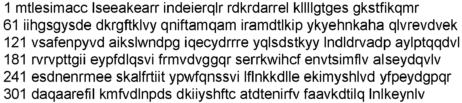
- NM_002072 (SEQ ID NO:269)


- The nucleic acid and amino acid sequence of human GNA11 have been published in GenBank with the following Accession Nos.:
- NP_002058 (SEQ ID NO:270)
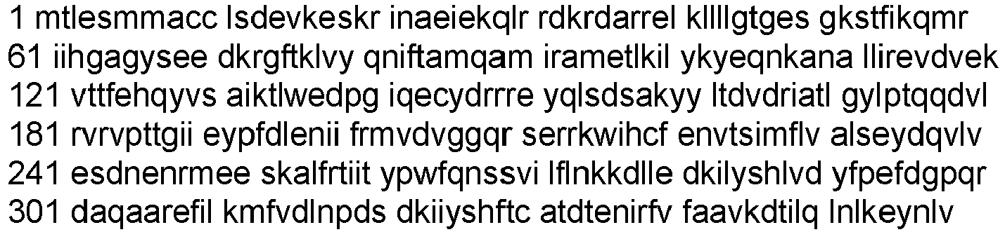
- NM_002067 (SEQ ID NO:271)


- "Tumor" refers to neoplastic cell growth and proliferation, whether malignant or benign, and all pre-cancerous and cancerous cells and tissues.
- The term "anti-tumor activity" means a reduction in the rate of tumor cell proliferation, viability, or metastatic activity. For example, anti-tumor activity can be shown by a decline in growth rate of abnormal cells that arises during therapy or tumor size stability or reduction, or longer survival due to therapy as compared to control without therapy. Such activity can be assessed using accepted in vitro or in vivo tumor models, including but not limited to xenograft models, allograft models, MMTV models, and other known models known in the art to investigate anti-tumor activity.
- The term "malignancy" refers to a non-benign tumor or a cancer. As used herein, the term "cancer" includes a malignancy characterized by deregulated or uncontrolled cell growth. Exemplary cancers include: carcinomas, sarcomas, leukemias, and lymphomas.
- The term "cancer" includes primary malignant tumors (e.g., those whose cells have not migrated to sites in the subject's body other than the site of the original tumor) and secondary malignant tumors (e.g., those arising from metastasis, the migration of tumor cells to secondary sites that are different from the site of the original tumor).
- The term "PMEL17" (also referred to as premelanosome protein (PMEL), D12S53E, ME20, ME20-M, ME20M, P1, P100, gp100, SI, SIL, and silver locus protein homolog (SILV)) refers to a single-pass Type I transmembrane protein produced by melanocytes and involved in melanin synthesis. The nucleic acid and amino acid sequence of human PMEL17 have been published in GenBank with the following Accession Nos.: NP_008859, NP_001307050, NP_001307051, NP_001186982, NP_001186983 (amino acid sequences), and NM_006928, NM_001200053, NM_001200054, NM_001320121, NM_001320122 (nucleotide sequences). As used herein, the term "PMEL17" is used to refer collectively to all naturally occurring isoforms of PMEL17 protein, or a variant thereof.
- NP_008859 (SEQ ID NO:272)

- NP_001307050 (SEQ ID NO:273)

- NP_001307051 (SEQ ID NO:274)

- NP_001186982 (SEQ ID NO:275)


- NP_001186983 (SEQ ID NO:276)

- NM_006928 (SEQ ID NO:277)


- NM_001200053 (SEQ ID NO:278)


- NM_001200054 (SEQ ID NO:279)

- NM_001320121 (SEQ ID NO:280)


- NM_001320122 (SEQ ID NO:281)


- The term "variant" refers to a polypeptide that has a substantially identical amino acid sequence to a reference polypeptide, or is encoded by a substantially identical nucleotide sequence, and is capable of having one or more activities of the reference polypeptide. For example, a variant can have about 85%, 90%, 91%, 92%, 93%, 94%, 95%, 96%, 97%, 98%, 99% or higher sequence identity to a reference polypeptide, while retain one or more activities of the reference polypeptide.
- As used herein, the terms "treat," "treating," or "treatment" of any disease or disorder refers in one embodiment, to ameliorating the disease or disorder (i.e., slowing or arresting or reducing the development of the disease or at least one of the clinical symptoms thereof). In another embodiment, "treat," "treating," or "treatment" refers to alleviating or ameliorating at least one physical parameter including those which may not be discernible by the patient. In yet another embodiment, "treat," "treating," or "treatment" refers to modulating the disease or disorder, either physically, (e.g., stabilization of a discernible symptom), physiologically, (e.g., stabilization of a physical parameter), or both.
- As used herein, the term "prevent", "preventing" or "prevention" of any disease or disorder refers to the prophylactic treatment of the disease or disorder; or delaying the onset or progression of the disease or disorder
- The term "therapeutically acceptable amount" or "therapeutically effective dose" interchangeably refers to an amount sufficient to effect the desired result (i.e., a reduction in tumor size, inhibition of tumor growth, prevention of metastasis, inhibition or prevention of viral, bacterial, fungal or parasitic infection). In some embodiments, a therapeutically acceptable amount does not induce or cause undesirable side effects. In some embodiments, a therapeutically acceptable amount induces or causes side effects but only those that are acceptable by the healthcare providers in view of a patient's condition. A therapeutically acceptable amount can be determined by first administering a low dose, and then incrementally increasing that dose until the desired effect is achieved. A "prophylactically effective dosage," and a "therapeutically effective dosage," of the molecules of the invention can prevent the onset of, or result in a decrease in severity of, respectively, disease symptoms, including symptoms associated with cancer.
- The term "co-administer" refers to the presence of two active agents in the blood of an individual. Active agents that are co-administered can be concurrently or sequentially delivered.
- The present invention provides antibodies, antibody fragments (e.g., antigen binding fragments), and drug conjugates thereof, i.e., antibody drug conjugates or ADCs, that bind to PMEL17. In particular, the present invention provides antibodies and antibody fragments (e.g., antigen binding fragments) that bind to PMEL17, and internalize upon such binding. The antibodies and antibody fragments (e.g., antigen binding fragments) of the present invention can be used for producing antibody drug conjugates. Furthermore, the present invention provides antibody drug conjugates that have desirable pharmacokinetic characteristics and other desirable attributes, and thus can be used for treating or preventing a cancer expressing PMEL17. The present invention further provides pharmaceutical compositions comprising the antibody drug conjugates of the invention, and methods of making and using such pharmaceutical compositions for the treatment or prevention of cancer.
- In one aspect, the Drug moiety (D) of the Antibody Drug Conjugate of the invention is a GNAQ inhibitor, a GNA11 inhibitor, or an inhibitor of GNAQ and GNA11 (GNAQ/GNA11 inhibitor).
- In another aspect, the Drug moiety (D) of the Antibody Drug Conjugate of the invention is a GNAQ inhibitor.
- In another aspect, the Drug moiety (D) of the Antibody Drug Conjugate of the invention is a GNA11 inhibitor.
- In another aspect, the Drug moiety (D) of the Antibody Drug Conjugate of the invention is an inhibitor of GNAQ and GNA11 (GNAQ/GNA11 inhibitor).
- In another aspect, the Drug moiety of the Antibody Drug Conjugate of the invention is a compound having the structure of Formula (A):

- In another aspect, Drug moiety of the Antibody Drug Conjugate of the invention is compound (A1) having the following structure:

- In another aspect, Drug moiety of the Antibody Drug Conjugate of the invention is compound (A2) having the following structure:

- In another aspect, Drug moiety of the Antibody Drug Conjugate of the invention is compound (A3) having the following structure:

Table 1 Compound GI50 (nM) 92.1GNAQ Q209L A1 0.467 A2 22.1 A3 9.3 - In a second aspect, the Linker-Drug moiety, ((LA-(D)n))), of the Antibody Drug Conjugate of the invention comprises one or more Drug moieties covalently attached to a linker (LA), wherein the one or more Drug moieties are each independently selected from a GNAQ inhibitor, a GNA11 inhibitor or an inhibitor of GNAQ and GNA11 (GNAQ/GNA11 inhibitor).
- In another aspect, the Linker-Drug moiety, ((LA-(D)n))), of the Antibody Drug Conjugate of the invention comprises one or more Drug moieties covalently attached to a linker (LA), wherein the one or more Drug moieties are each independently selected from a GNAQ inhibitor.
- In another aspect, the Linker-Drug moiety, ((LA-(D)n))), of the Antibody Drug Conjugate of the invention comprises one or more Drug moieties attached to a linker (LA), wherein the one or more Drug moieties are each independently selected from a GNA11 inhibitor.
- In another aspect, the Linker-Drug moiety, ((LA-(D)n))), of the Antibody Drug Conjugate of the invention comprises one or more Drug moieties covalently attached to a linker (LA), wherein the one or more Drug moieties are each independently selected from an inhibitor of GNAQ and GNA11 (GNAQ/GNA11 inhibitor).
- In another aspect, the Linker-Drug moiety, ((LA-(D)n))), of the Antibody Drug Conjugate of the invention comprises one or more Drug moieties covalently attached to a linker (LA), wherein the linker (LA) is a cleavable linker and the one or more Drug moieties are each independently selected from a GNAQ inhibitor, a GNA11 inhibitor or an inhibitor of GNAQ and GNA11 (GNAQ/GNA11 inhibitor).
- In another aspect, the Linker-Drug moiety, ((LA-(D)n))), of the Antibody Drug Conjugate of the invention comprises one or more Drug moieties covalently attached to a linker (LA), wherein the linker (LA) is a cleavable linker and the one or more Drug moieties are each independently selected from a GNAQ inhibitor.
- In another aspect, the Linker-Drug moiety, ((LA-(D)n))), of the Antibody Drug Conjugate of the invention comprises one or more Drug moieties covalently attached to a linker (LA), wherein the linker (LA) is a cleavable linker and the one or more Drug moieties are each independently selected from a GNA11 inhibitor.
- In another aspect, the Linker-Drug moiety, ((LA-(D)n))), of the Antibody Drug Conjugate of the invention comprises one or more Drug moieties covalently attached to a linker (LA), wherein the linker (LA) is a cleavable linker and the one or more Drug moieties are each independently selected from an inhibitor of GNAQ and GNA11 (GNAQ/GNA11 inhibitor).
- In another aspect, the Linker-Drug moiety, ((LA-(D)n))), of the Antibody Drug Conjugate of the invention comprises one or more Drug moieties covalently attached to a linker (LA), wherein the linker (LA) is a non-cleavable linker and the one or more Drug moieties are each independently selected from a GNAQ inhibitor, a GNA11 inhibitor or an inhibitor of GNAQ and GNA11 (GNAQ/GNA11 inhibitor).
- In another aspect, the Linker-Drug moiety, ((LA-(D)n))), of the Antibody Drug Conjugate of the invention comprises one or more Drug moieties covalently attached to a linker (LA), wherein the linker (LA) is a non-cleavable linker and the one or more Drug moieties are each independently selected from a GNAQ inhibitor.
- In another aspect, the Linker-Drug moiety, ((LA-(D)n))), of the Antibody Drug Conjugate of the invention comprises one or more Drug moieties covalently attached to a linker (LA), wherein the linker (LA) is a non-cleavable linker and the one or more Drug moieties are each independently selected from a GNA11 inhibitor.
- In another aspect, the Linker-Drug moiety, ((LA-(D)n))), of the Antibody Drug Conjugate of the invention comprises one or more Drug moieties covalently attached to a linker (LA), wherein the linker (LA) is a non-cleavable linker and the one or more Drug moieties are each independently selected from an inhibitor of GNAQ and GNA11 (GNAQ/GNA11 inhibitor).
- In another aspect, the linker (LA) of the Linker-Drug moiety, ((LA-(D)n))), of the Antibody Drug Conjugate of the invention has the following formula:
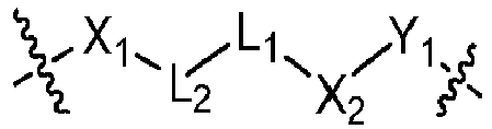
- X1 is a bivalent coupling group;
- X2 is a self-immolative spacer;
- Y1 is


- L1 is a bivalent peptide linker, and
- L2 is a bond or a linker.
- In another aspect, the Linker-Drug moiety, ((LA-(D)n))), of the Antibody Drug Conjugate of the invention has the following formula:

- D is a GNAQ inhibitor, a GNA11 inhibitor or an inhibitor of GNAQ and GNA11;
- X1 is a bivalent coupling group;
- X2 is a self-immolative spacer;
- Y1 is


- L1 is a bivalent peptide linker, and
- L2 is a bond or a linker.
- In one aspect the Linker-Drug of the invention is a compound having the structure of Formula (B), or stereoisomers or pharmaceutically acceptable salts thereof,
R8-LB-(D)n (B)
wherein: - D is a GNAQ inhibitor, a GNA11 inhibitor or an inhibitor of GNAQ and GNA11;
- R8 is a reactive group;
- LB is a cleavable linker or non-cleavable linker, and
- n is 1, 2, 3 or 4.
- In one aspect the Linker-Drug of the invention having the structure of Formula (B), or stereoisomers or pharmaceutically acceptable salts thereof, wherein
- D is a GNAQ inhibitor, a GNA11 inhibitor or an inhibitor of GNAQ and GNA11;
- R8 is a reactive group;
- LB is a cleavable linker comprising one or more linker components selected from a self-immolative spacer, a phosphate group, a carbonate group and a bivalent peptide linker, and
- n is 1, 2, 3 or 4.
- In one aspect the Linker-Drug of the invention is a compound having the structure of Formula (B-1), or stereoisomers or pharmaceutically acceptable salts thereof,

- D is a GNAQ inhibitor, a GNA11 inhibitor or an inhibitor of GNAQ and GNA11;
- R8 is a reactive group;
- X2 is a self-immolative spacer;
- Y1 is


- L1 is a bivalent peptide linker, and
- L2 is a bond or a linker.
- Certain aspects and examples of the Linker-Drug compounds of the invention are provided in the following listing of additional, enumerated embodiments. It will be recognized that features specified in each embodiment may be combined with other specified features to provide further embodiments of the present invention.
-
Embodiment 1. The compound of Formula (B) or Formula (B-1), or stereoisomers or a pharmaceutically acceptable salt thereof, wherein D is a GNAQ inhibitor. -
Embodiment 2. The compound of Formula (B) or Formula (B-1), or stereoisomers or a pharmaceutically acceptable salt thereof, wherein D is a GNA11 inhibitor. -
Embodiment 3. The compound of Formula (B) or Formula (B-1), or stereoisomers or a pharmaceutically acceptable salt thereof, wherein D is an inhibitor of GNAQ and GNA11. -
Embodiment 4. The compound of Formula (B) or Formula (B-1), or stereoisomers or a pharmaceutically acceptable salt thereof, wherein D is
-
Embodiment 5. The compound of Formula (B) or Formula (B-1), or stereoisomers or a pharmaceutically acceptable salt thereof, wherein D is
-
Embodiment 6. The compound of Formula (B) or Formula (B-1), or stereoisomers or a pharmaceutically acceptable salt thereof, wherein D is
-
Embodiment 7. The compound of Formula (B) or Formula (B-1), or stereoisomers or a pharmaceutically acceptable salt thereof, wherein D is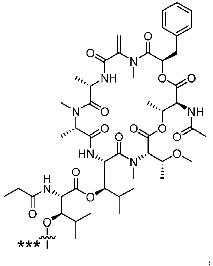
-
Embodiment 8. The compound of Formula (B) or Formula (B-1), or stereoisomers or a pharmaceutically acceptable salt thereof, having the structure of Formula (B-2), or a pharmaceutically acceptable salt thereof:
- R0 is methyl or ethyl,;
- R1 is methyl or isopropyl;
- R2 is methyl or ethyl, and
- X2, L1, L2 and R8 are as define in compounds of Formula (B-1) above.
-
Embodiment 9. The compound of Formula (B), Formula (B-1) or Formula (B-2), or stereoisomers or a pharmaceutically acceptable salt thereof, having the structure of Formula (B-2a), or a pharmaceutically acceptable salt thereof:
X2, L1, L2 and R8 are as define in compounds of Formula (B-1) above. -
Embodiment 10. The compound of Formula (B), Formula (B-1) or Formula (B-2), or stereoisomers or a pharmaceutically acceptable salt thereof, having the structure of Formula (B-2b), or a pharmaceutically acceptable salt thereof:
X2, L1, L2 and R8 are as define in compounds of Formula (B-1) above. -
Embodiment 11. The compound of Formula (B), Formula (B-1) or Formula (B-2), or stereoisomers or a pharmaceutically acceptable salt thereof, having the structure of Formula (B-2c), or a pharmaceutically acceptable salt thereof:
X2, L1, L2 and R8 are as define in compounds of Formula (B-1) above. -
Embodiment 12. The compound of Formula (B) or Formula (B-1), or stereoisomers or a pharmaceutically acceptable salt thereof, having the structure of Formula (B-3), or a pharmaceutically acceptable salt thereof:
- R0 is methyl or ethyl,;
- R1 is methyl or isopropyl;
- R2 is methyl or ethyl, and
- X2, L1, L2 and R8 are as define in compounds of Formula (B-1) above.
-
Embodiment 13. The compound of Formula (B), Formula (B-1) or Formula (B-3), or stereoisomers or a pharmaceutically acceptable salt thereof, having the structure of Formula (B-3a), or a pharmaceutically acceptable salt thereof:
X2, L1, L2 and R8 are as define in compounds of Formula (B-1) above. -
Embodiment 14. The compound of Formula (B), Formula (B-1) or Formula (B-3), or stereoisomers or a pharmaceutically acceptable salt thereof, having the structure of Formula (B-3b), or a pharmaceutically acceptable salt thereof: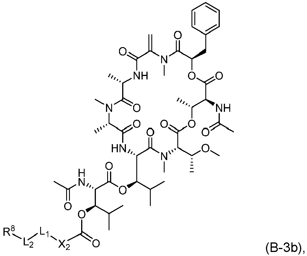
X2, L1, L2 and R8 are as define in compounds of Formula (B-1) above. -
Embodiment 15. The compound of Formula (B), Formula (B-1) or Formula (B-3), or stereoisomers or a pharmaceutically acceptable salt thereof, having the structure of Formula (B-3c), or a pharmaceutically acceptable salt thereof:
X2, L1, L2 and R8 are as define in compounds of Formula (B-1) above. -
Embodiment 16. The compound of Formula (B-2) ofEmbodiment 8, Formula (B-3) ofEmbodiment 12, or a pharmaceutically acceptable salt thereof:- wherein:
- R0 is methyl or ethyl;
- R1 is methyl or isopropyl;
- R2 is methyl or ethyl;
- X2 is a self-immolative spacer selected from


- L1 is a bivalent peptide linker comprising 2 to 4 amino acid residues;
- L2 is a linker,
- R8 is selected from

- wherein:
- NHC(=O)CH2Br, -NHC(=O)CH2I, -C(=O)NHNH2,


- each R4 is independently selected from H and C1-C6alkyl;
- each R5 is independently selected from H, C1-C6alkyl, F, Cl, and -OH;
- each R6 is independently selected from H, C1-C6alkyl, F, Cl, -NH2, -OCH3, - OCH2CH3, -N(CH3)2, -CN, -NO2 and -OH,
and - each R7 is independently selected from H, C1-6alkyl, fluoro, benzyloxy substituted with -C(=O)OH, benzyl substituted with -C(=O)OH, C1-4alkoxy substituted with - C(=O)OH and C1-4alkyl substituted with -C(=O)OH.
- Embodiment 17. The compound of any one of
Embodiments 1 to 16, wherein X2 is a self-immolative spacer selected from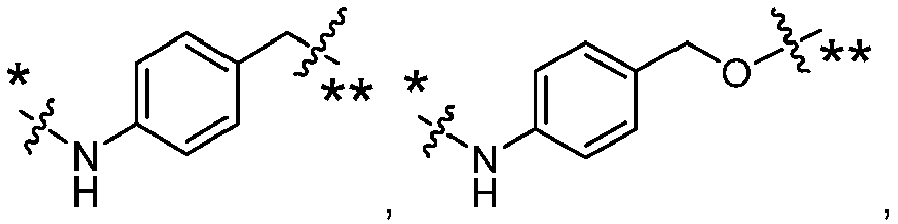


- Embodiment 18. The compound of any one of
Embodiments 1 to 17, wherein X2 is


- Embodiment 19. The compound of any one of
Embodiments 1 to 18, wherein L1 is a bivalent peptide linker comprising 2 to 4 amino acid residues. -
Embodiment 20. The compound of any one ofEmbodiments 1 to 18, wherein L1 is a is a bivalent peptide linker comprising an amino acid residue selected from valine, citrulline, lysine, isoleucine, phenylalanine, methionine, asparagine, proline, alanine, leucine, tryptophan, and tyrosine. - Embodiment 21. The compound of any one of
Embodiments 1 to 18, wherein L1 is a bivalent peptide linker comprising at least one valine (Val) or citrulline (Cit) residue. - Embodiment 22. The compound of any one of
Embodiments 1 to 18, wherein L1 is a bivalent dipeptide linker selected from ValCit, PheLys, ValAla and ValLys. - Embodiment 23. The compound of any one of
Embodiments 1 to 18, wherein L1 is a bivalent dipeptide linker selected from

-
Embodiment 24. The compound of any one ofEmbodiments 1 to 18, wherein L1 is ValCit. -
Embodiment 25. The compound of any one ofEmbodiments 1 to 18, wherein L1 is
- Embodiment 26. The compound of any one of
Embodiments 1 to 25, wherein L2 is a linker. - Embodiment 27. The compound of any one of
Embodiments 1 to 25, wherein L2 is a linker selected from:- -*C(=O)((CH2)mO)p(CH2)m**-, -*C(=O)(CH2)m**-, -*C(=O)(CH2)nNHC(=O)(CH2)m**-, - *C(=O)(CH2)mNHC(=O)((CH2)mO)p(CH2)m**-, -* ((CH2)mO)p(CH2)m**-, -* ((CH2)mO)p(CH2)m**-, -(CH2)m-, -*(CH2)mNHC(=O)(CH2)m**-, -* (CH2)mNHC(=O)(CH2)mC(=O)NH(CH2)m**-, -* ((CH2)mO)p(CH2)mNHC(=O)(CH2)m**-, -* *((CH2)mO)pCH2)mC(=O)NH(CH2)m**-, -* (CH2)mC(R3)2**-, and -* (CH2)mC(R3)2SS(CH2)mNHC(=O)(CH2)m**-, where the * of L2 indicates the attachment point to L1 and the ** of L2 indicates the point of attachment to R8;
- and wherein:
- each R3 is independently selected from H and C1-C6alkyl;
- each m is independently selected from 1, 2, 3, 4, 5, 6, 7, 8, 9 and 10, and
- each p is independently selected from 1, 2, 3, 4, 5, 6, 7, 8, 9, 10, 11, 12, 13 and 14.
- Embodiment 28. The compound of any one of
Embodiments 1 to 25, wherein L2 is -*C(=O)((CH2)mO)p(CH2)m**- or -*C(=O)(CH2)m**-, where the * of L2 indicates the point of attachment to L1 and the ** of L2 indicates the point of attachment to R8,
and wherein each m is independently selected from 1, 2, 3, 4, 5, 6, 7, 8, 9 or 10 and p is 1, 2, 3, 4, 5, 6, 7, 8, 9, 10, 11, 12, 13 or 14. -
Embodiment 29. The compound of any one ofEmbodiments 1 to 25, wherein L2 is
-
Embodiment 30. The compound of any one ofEmbodiments 1 to 29, wherein R8 is


- each R4 is independently selected from H and C1-C6alkyl;
- each R5 is independently selected from H, C1-C6alkyl, F, Cl, and -OH;
- each R6 is independently selected from H, C1-C6alkyl, F, Cl, -NH2, -OCH3, - OCH2CH3, -N(CH3)2, -CN, -NO2 and -OH, and
- each R7 is independently selected from H, C1-6alkyl, fluoro, benzyloxy substituted with -C(=O)OH, benzyl substituted with -C(=O)OH, C1-4alkoxy substituted with - C(=O)OH and C1-4alkyl substituted with -C(=O)OH.
- Embodiment 31. The compound of any one of
Embodiments 1 to 29, wherein R8 is

- Embodiment 32. The compound of any one of
Embodiments 1 to 29, wherein R8 is
- Embodiment 33. The compound of Formula (B-2) of
Embodiment 8, or a pharmaceutically acceptable salt thereof:
wherein:- R0 is methyl or ethyl;
- R1 is methyl or isopropyl;
- R2 is methyl or ethyl;
- X2 is


- L1 is

- L2 is

and - R8 is

- Embodiment 34. The compound of Formula (B-3) of
Embodiment 12, or a pharmaceutically acceptable salt thereof:
wherein:- R0 is methyl or ethyl;
- R1 is methyl or isopropyl;
- R2 is methyl or ethyl;
- X2 is


- L1 is

- L2 is

and - R8 is

-
Embodiment 35. The compound of Formula (B), Formula (B-1) or Formula (B-2), wherein the compound is
- Embodiment 36. The compound of Formula (B), Formula (B-1) or Formula (B-2), wherein the compound is

- Embodiment 37. The compound of Formula (B), Formula (B-1) or Formula (B-2), wherein the compound is

- Embodiment 38. The compound of Formula (B), Formula (B-1) or Formula (B-2), wherein the compound is

- Embodiment 39. The compound of Formula (B), Formula (B-1) or Formula (B-2), wherein the compound is

-
Embodiment 40. The compound of Formula (B), Formula (B-1) or Formula (B-2), wherein the compound is
- Embodiment 41. The compound of Formula (B), Formula (B-1) or Formula (B-3), wherein the compound is

- Embodiment 42. The compound of Formula (B), Formula (B-1) or Formula (B-3), wherein the compound is

- Embodiment 43. The compound of Formula (B), Formula (B-1) or Formula (B-3), wherein the compound is

- Embodiment 44. The compound of Formula (B), Formula (B-1) or Formula (B-3), wherein the compound is

-
Embodiment 45. The compound of Formula (B), Formula (B-1) or Formula (B-3), wherein the compound is
- Embodiment 46. The compound of Formula (B), Formula (B-1) or Formula (B-3), wherein the compound is

- In one aspect, the Antibody Drug Conjugate of the invention is a conjugate of Formula (C):
Ab-(LA-(D)n)y (C)
wherein: - D is a drug moiety;
- Ab is an antibody or antigen binding fragment thereof that binds to human PMEL17 protein;
- LA is a linker;
- n is 1, 2, 3 or 4, and
- y is 1, 2, 3 or 4,
- In one aspect, the Antibody Drug Conjugate of the invention having the structure of Formula (C), wherein:
- D is a GNAQ inhibitor, a GNA11 inhibitor or an inhibitor of GNAQ and GNA11;
- Ab is an antibody or antigen binding fragment thereof that binds to human PMEL17 protein;
- LA is a cleavable linker comprising one or more linker components selected from a self-immolative spacer, a phosphate group, a carbonate group and a bivalent peptide linker;
- n is 1, 2, 3 or 4, and
- y is 1, 2, 3 or 4,
- In one aspect, the Antibody Drug Conjugate of Formula (C) is a conjugate of Formula (C-1):

- D is a GNAQ inhibitor, a GNA11 inhibitor or an inhibitor of GNAQ and GNA11;
- Ab is an antibody or antigen binding fragment thereof that binds to human PMEL17 protein;
- X1 is a bivalent coupling group;
- X2 is a self-immolative spacer;
- Y1 is


- L1 is a bivalent peptide linker;
- L2 is a bond or a linker, and
- y is 1, 2, 3 or 4.
- In the conjugates of Formula (C), one or more Linker-Drug moiety (LB-(D)n) can be covalently attached to the antibody or antigen binding fragment thereof, Ab, thereby covalently attaching one or more drug moieties, D, to the antibody or antigen binding fragment thereof, Ab, through linker, LA. LA is any chemical moiety that is capable of linking the antibody or antigen binding fragment thereof, Ab, to one or more drug moieties, D. The conjugates of Formula (C), wherein one or more drug moieties, D, are covalently linked to an antibody or antigen binding fragment thereof, Ab, can be formed using a bifunctional or multifunctional linker reagent having one or more reactive functional groups that are the same or different. One of the reactive functional groups of the bifunctional or multifunctional linker reagent is used to react with a group on the antibody or antigen binding fragment thereof, Ab, by way of example, a thiol or an amine (e.g. a cysteine, an N-terminus or amino acid side chain such as lysine) to form a covalent linkage with one end of the linker LA. Such reactive functional groups of the bifunctional or multifunctional linker reagent include, but are not limited to, a maleimide, a thiol and an NHS ester. The other reactive functional group or groups of the bifunctional or multifunctional linker reagent are used to covalently attached one or more drug moieties, D, to linker LA.
- In one aspect, LA is a cleavable linker. In another aspect, LA is a non-cleavable linker. In some aspects, LA is an acid-labile linker, photo-labile linker, peptidase cleavable linker, esterase cleavable linker, glycosidase cleavable linker, phosphodiesterase cleavable linker, a disulfide bond reducible linker, a hydrophilic linker, or a dicarboxylic acid based linker.
- In one aspect, LA is a cleavable linker comprising one or more linker components selected from a self-immolative spacer, a phosphate group, a carbonate group and a bivalent peptide linker.
- In one aspect, LA is a cleavable linker comprising one or more linker components selected from a self-immolative spacer, a phosphate group, a carbonate group, a bivalent peptide linker and a bivalent coupling group.
- In one aspect, LA is a cleavable linker comprising one or more linker components selected from a self-immolative spacer, a phosphate group and a bivalent peptide linker.
- In one aspect, LA is a cleavable linker comprising one or more linker components selected from a self-immolative spacer, a phosphate group, a bivalent peptide linker and a bivalent coupling group.
- In another aspect, the linker (LA) has the following formula:

- X1 is a bivalent coupling group;
- X2 is a self-immolative spacer;
- Y1 is


- L1 is a bivalent peptide linker, and
- L2 is a bond or a linker.
- In another aspect, the linker (LA) has the following formula:

- X1 is a bivalent coupling group;
- X2 is a self-immolative spacer;
- Y1 is
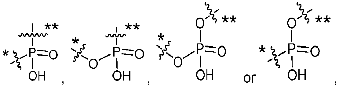
- L1 is a bivalent peptide linker, and
- L2 is a bond or a linker.
- In another aspect, the linker (LA) has the following formula:

- X1 is a bivalent coupling group;
- X2 is a self-immolative spacer;
- Y1 is

- L1 is a bivalent peptide linker, and
- L2 is a bond or a linker.
- While the drug to antibody ratio has an exact integer value for a specific conjugate molecule (e.g., the product of n and y in Formula (C), it is understood that the value will often be an average value when used to describe a sample containing many molecules, due to some degree of heterogeneity, typically associated with the conjugation step. The average loading for a sample of a conjugate is referred to herein as the drug to antibody ratio, or "DAR." In some aspects, the DAR is between about 1 and about 5, and typically is about 1, 2, 3, or 4. In some aspects, at least 50% of a sample by weight is compound having the average DAR plus or
minus 2, and preferably at least 50% of the sample is a conjugate that contains the average DAR plus orminus 1. Other aspects include conjugates wherein the DAR is about 2. In some aspects, a DAR of 'about y' means the measured value for DAR is within 20% of the product of n and y in Formula (I). In some aspects, a DAR of 'about n' means the measured value for DAR is within 20% of n in Formula (II). - In one aspect, the average molar ratio of the drug to the antibody in the conjugates of Formula (C) (i.e., average value of the product of n and y, also known as drug to antibody ratio (DAR)) is about 1 to about 10, about 1 to about 6 (e.g., 0.9, 1.0, 1.1, 1.2, 1.3, 1.4, 1.5, 1.6, 1.7, 1.8, 1.9, 2.0, 2.1, 2.2, 2.3, 2.4, 2.5, 2.6, 2.7, 2.8, 2.9, 3.0, 3.1, 3.2, 3.3, 3.4, 3.5, 3.6, 3.7, 3.8, 3.9, 4.0, 4.1, 4.2, 4.3, 4.4, 4.5, 4.6, 4.7, 4.8, 4.9, 5.0, 5.1, 5.2, 5.3, 5.4, 5.5, 5.6, 5.7, 5.8, 5.9, 6.0), about 1 to about 5, about 1.5 to about 4.5, or about 2 to about 4.
- In one aspect provided by the disclosure, the conjugate has substantially high purity and has one or more of the following features: (a) greater than about 90% (e.g., greater than or equal to about 91%, 92%, 93%, 94%, 95%, 96%, 97%, 98%, 99%, or 100%), preferably greater than about 95%, of conjugate species are monomeric, (b) unconjugated linker level in the conjugate preparation is less than about 10% (e.g., less than or equal to about 9%, 8%, 7%, 6%, 5%, 4%, 3%, 2%, 1%, or 0%) (relative to total linker), (c) less than 10% of conjugate species are crosslinked (e.g., less than or equal to about 9%, 8%, 7%, 6%, 5%, 4%, 3%, 2%, 1%, or 0%), (d) free drug (ADP-induced platelet aggregation inhibitor, e.g., a GNAQ inhibitor, a GNA11 inhibitor, or a GNAQ and a GNA11 inhibitor) level in the conjugate preparation is less than about 2% (e.g., less than or equal to about 1.5%, 1.4%, 1.3%, 1.2%, 1.1%, 1.0%, 0.9%, 0.8%, 0.7%, 0.6%, 0.5%, 0.4%, 0.3%, 0.2%, 0.1%, or 0%) (mol/mol relative to total drug).
- Certain aspects and examples of the Antibody Drug Conjugates of the invention are provided in the following listing of additional, enumerated embodiments. It will be recognized that features specified in each embodiment may be combined with other specified features to provide further embodiments of the present invention.
- Embodiment 47. The conjugate of Formula (C) or Formula (C-1), wherein D is a GNAQ inhibitor.
-
Embodiment 48. The conjugate of Formula (C) or Formula (C-1), wherein D is a GNA11 inhibitor. - Embodiment 49. The conjugate of Formula (C) or Formula (C-1), wherein D is an inhibitor of GNAQ and GNA11.
-
Embodiment 50. The conjugate of Formula (C) or Formula (C-1), wherein D is
- Embodiment 51. The conjugate of Formula (C) or Formula (C-1), wherein D is

- Embodiment 52. The conjugate of Formula (C) or Formula (C-1), wherein D is

- Embodiment 53. The conjugate of Formula (C) or Formula (C-1), wherein D is

- Embodiment 54. The conjugate of Formula (C) or Formula (C-1), having the structure of Formula (C-2):

- R0 is methyl or ethyl;
- R1 is methyl or isopropyl;
- R2 is methyl or ethyl;
- Ab is an antibody or antigen binding fragment thereof that binds to human PMEL17 protein;
- X1 is a bivalent coupling group;
- X2 is a self-immolative spacer;
- L1 is a bivalent peptide linker;
- L2 is a bond or a linker, and
- y is 1, 2, 3 or 4.
-
Embodiment 55. The conjugate of Formula (C) or Formula (C-1), having the structure of Formula (C-2a):
- Ab is an antibody or antigen binding fragment thereof that binds to human PMEL17 protein;
- X1 is a bivalent coupling group;
- X2 is a self-immolative spacer;
- L1 is a bivalent peptide linker;
- L2 is a bond or a linker, and
- y is 1, 2, 3 or 4.
- Embodiment 56. The conjugate of Formula (C) or Formula (C-1), having the structure of Formula (C-2b):

- Ab is an antibody or antigen binding fragment thereof that binds to human PMEL17 protein;
- X1 is a bivalent coupling group;
- X2 is a self-immolative spacer;
- L1 is a bivalent peptide linker;
- L2 is a bond or a linker, and
- y is 1, 2, 3 or 4.
- Embodiment 57. The conjugate of Formula (C) or Formula (C-1), having the structure of Formula (C-2c):

- Ab is an antibody or antigen binding fragment thereof that binds to human PMEL17 protein;
- X1 is a bivalent coupling group;
- X2 is a self-immolative spacer;
- L1 is a bivalent peptide linker;
- L2 is a bond or a linker, and
- y is 1, 2, 3 or 4.
- Embodiment 58. The conjugate of Formula (C) or Formula (C-1), having the structure of Formula (C-3):
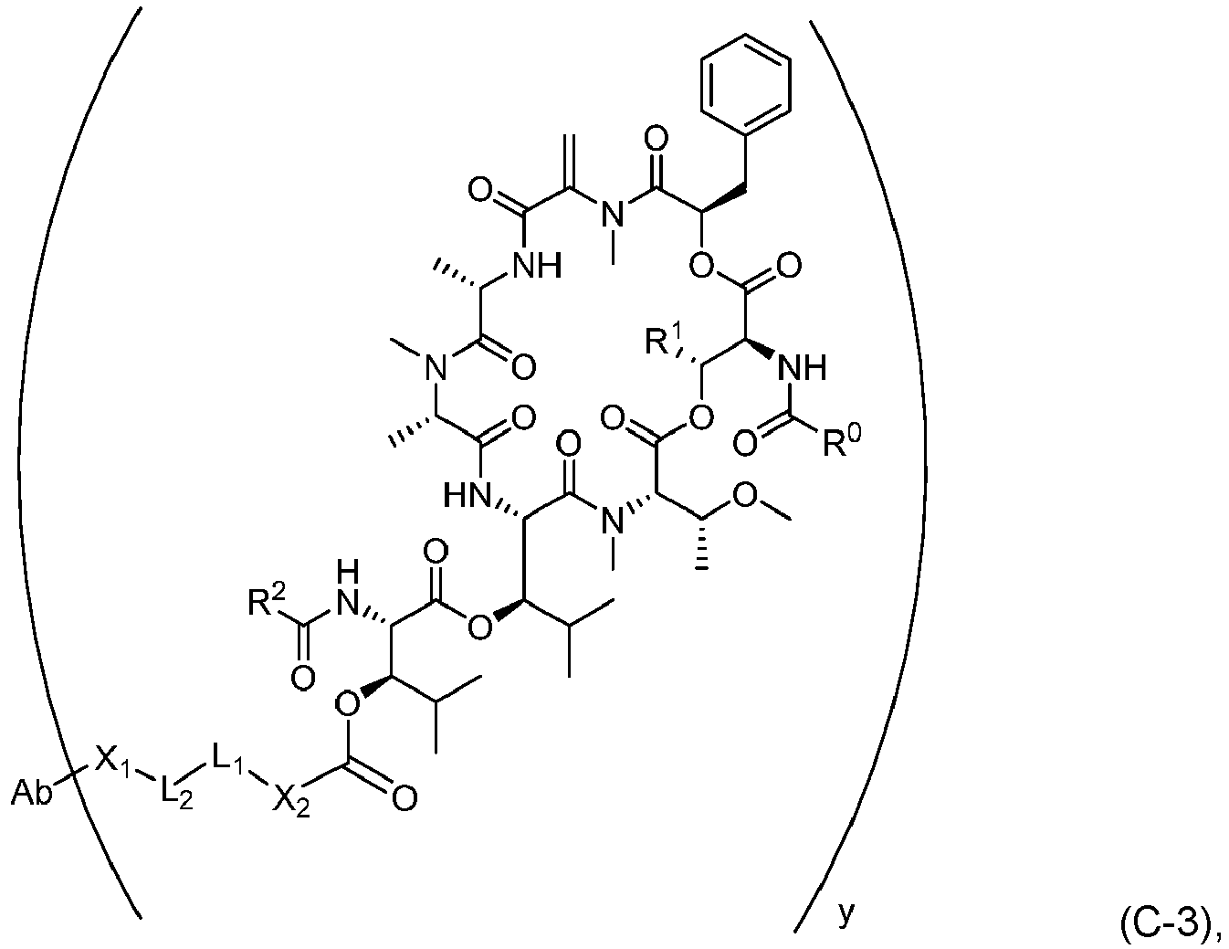
- R0 is methyl or ethyl;
- R1 is methyl or isopropyl;
- R2 is methyl or ethyl;
- Ab is an antibody or antigen binding fragment thereof that binds to human PMEL17 protein;
- X1 is a bivalent coupling group;
- X2 is a self-immolative spacer;
- L1 is a bivalent peptide linker;
- L2 is a bond or a linker, and
- y is 1, 2, 3 or 4.
- Embodiment 59. The conjugate of Formula (C) or Formula (C-1), having the structure of Formula (C-3a):

- Ab is an antibody or antigen binding fragment thereof that binds to human PMEL17 protein;
- X1 is a bivalent coupling group;
- X2 is a self-immolative spacer;
- L1 is a bivalent peptide linker;
- L2 is a bond or a linker, and
- y is 1, 2, 3 or 4.
-
Embodiment 60. The conjugate of Formula (C) or Formula (C-1), having the structure of Formula (C-3b):
- Ab is an antibody or antigen binding fragment thereof that binds to human PMEL17 protein;
- X1 is a bivalent coupling group;
- X2 is a self-immolative spacer;
- L1 is a bivalent peptide linker;
- L2 is a bond or a linker, and
- y is 1, 2, 3 or 4.
- Embodiment 61. The conjugate of Formula (C) or Formula (C-1), having the structure of Formula (C-3c):

- Ab is an antibody or antigen binding fragment thereof that binds to human PMEL17 protein;
- X1 is a bivalent coupling group;
- X2 is a self-immolative spacer;
- L1 is a bivalent peptide linker;
- L2 is a bond or a linker, and
- y is 1, 2, 3 or 4.
- Embodiment 62. The conjugate of Formula (C-2) of Embodiment 54 or the conjugate of Formula (C-3) of Embodiment 58:
wherein: - R0 is methyl or ethyl;
- R1 is methyl or isopropyl;
- R2 is methyl or ethyl;
- Ab is an antibody or antigen binding fragment thereof that binds to human PMEL17 protein;
- X2 is a self-immolative spacer selected from


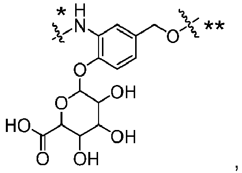


- L1 is a bivalent peptide linker comprising 2 to 4 amino acid residues;
- L2 is a linker;
- X1 is a bivalent coupling group selected from


- each R4 is independently selected from H and C1-C6alkyl;
- each R5 is independently selected from H, C1-C6alkyl, F, Cl, and -OH;
- each R6 is independently selected from H, C1-C6alkyl, F, Cl, -NH2, -OCH3, - OCH2CH3, -N(CH3)2, -CN, -NO2 and -OH,
and - each R7 is independently selected from H, C1-6alkyl, fluoro, benzyloxy substituted with -C(=O)OH, benzyl substituted with -C(=O)OH, C1-4alkoxy substituted with - C(=O)OH and C1-4alkyl substituted with -C(=O)OH,
- y is 1, 2, 3 or 4.
- Embodiment 63. The conjugate of any one of Embodiments 54 to 62, wherein X2 is a self-immolative spacer selected from
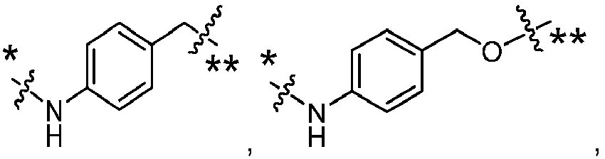
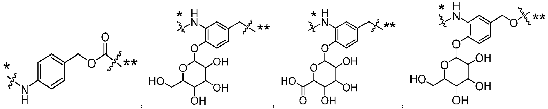


- Embodiment 64. The conjugate of any one of Embodiments 54 to 63, wherein X2 is


- Embodiment 65. The conjugate of any one of Embodiments 54 to 64, wherein L1 is a bivalent peptide linker comprising 2 to 4 amino acid residues.
- Embodiment 66. The conjugate of any one of Embodiments 54 to 65, wherein L1 is a is a bivalent peptide linker comprising an amino acid residue selected from valine, citrulline, lysine, isoleucine, phenylalanine, methionine, asparagine, proline, alanine, leucine, tryptophan, and tyrosine.
- Embodiment 67. The conjugate of any one of Embodiments 54-64, wherein L1 is a bivalent peptide linker comprising at least one valine (Val) or citrulline (Cit) residue.
-
Embodiment 68. The conjugate of any one of Embodiments 54 to 64, wherein L1 is a bivalent dipeptide linker selected from ValCit, PheLys, ValAla and ValLys. - Embodiment 69. The conjugate of any one of Embodiments 54 to 64, wherein L1 is a bivalent dipeptide linker selected from


-
Embodiment 70. The conjugate of any one of Embodiments 54 to 64, wherein L1 is ValCit. - Embodiment 71. The conjugate of any one of Embodiments 54 to 64, wherein L1 is

-
Embodiment 72. The conjugate of any one of Embodiments 54 to 71, wherein L2 is a linker. - Embodiment 73. The conjugate of any one of Embodiments 54 to 71, wherein L2 is a linker selected from:
- -*C(=O)((CH2)mO)p(CH2)m**-, -*C(=O)(CH2)m**-, -*C(=O)(CH2)nNHC(=O)(CH2)m**-, - *C(=O)(CH2)mNHC(=O)((CH2)mO)p(CH2)m**-, -* ((CH2)mO)p(CH2)m**-, -* ((CH2)mO)p(CH2)m**-, -(CH2)m-, -*(CH2)mNHC(=O)(CH2)m**-, -* (CH2)mNHC(=O)(CH2)mC(=O)NH(CH2)m**-, -* ((CH2)mO)p(CH2)mNHC(=O)(CH2)m**-, -* *((CH2)mO)pCH2)mC(=O)NH(CH2)m**-, -* (CH2)mC(R3)2**-, and -* (CH2)mC(R3)2SS(CH2)mNHC(=O)(CH2)m**-, where the * of L2 indicates the attachment point to L1 and the ** of L2 indicates the point of attachment to X1;
- and wherein:
- each R3 is independently selected from H and C1-C6alkyl;
- each m is independently selected from 1, 2, 3, 4, 5, 6, 7, 8, 9 and 10, and
- each p is independently selected from 1, 2, 3, 4, 5, 6, 7, 8, 9, 10, 11, 12, 13 and 14.
- Embodiment 74. The conjugate of any one of Embodiments 54 to 71, wherein L2 is -*C(=O)((CH2)mO)p(CH2)m**- or -*C(=O)(CH2)m**-, where the * of L2 indicates the point of attachment to L1 and the ** of L2 indicates the point of attachment to X1, and wherein each m is independently selected from 1, 2, 3, 4, 5, 6, 7, 8, 9 or 10 and p is 1, 2, 3, 4, 5, 6, 7, 8, 9, 10, 11, 12, 13 or 14.
-
Embodiment 75. The conjugate of any one of Embodiments 54 to 71, wherein L2 is
- Embodiment 76. The conjugate of any one of Embodiments 54 to 75,
- wherein X1 is is a bivalent coupling group selected from
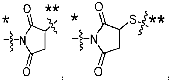

- *NHC(=O)CH2**-, -*S(=O)2CH2CH2**-, -* (CH2)2S(=O)2CH2CH2**-, -
- *NR4S(=O)2CH2CH2**-, -*NR4C(=O)CH2CH2**-, -NH-, -C(=O)-, -*NHC(=O) **-,
- -*CH2NHCH2CH2**-, -*NHCH2CH2**-, -S-,


- indicates the point of attachment to L2 and the ** of X1 indicates the point of attachment to Ab;
- and wherein:
- each R4 is independently selected from H and C1-C6alkyl;
- each R5 is independently selected from H, C1-C6alkyl, F, Cl, and -OH;
- each R6 is independently selected from H, C1-C6alkyl, F, Cl, -NH2, -OCH3, -
OCH2CH3, -N(CH3)2, -CN, -NO2 and -OH,
and - each R7 is independently selected from H, C1-6alkyl, fluoro, benzyloxy substituted with -C(=O)OH, benzyl substituted with -C(=O)OH, C1-4alkoxy substituted with - C(=O)OH and C1-4alkyl substituted with -C(=O)OH.
- Embodiment 77. The conjugate of any one of Embodiments 54 to 75, wherein X1 is is a bivalent coupling group selected from


- Embodiment 78. The conjugate of any one of Embodiments 54 to 75, wherein X1 is is a bivalent coupling group selected from

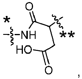
- Embodiment 79. The conjugate of Formula (C-2) of Embodiment 54 wherein:
- Ab is an antibody or antigen binding fragment thereof that binds to human PMEL17 protein;
- R0 is methyl or ethyl;
- R1 is methyl or isopropyl;
- R2 is methyl or ethyl;
- X1 is is a bivalent coupling group selected from

- X2 is

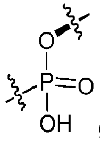
- L1 is

- L2 is

and - y is 1, 2, 3 or 4.
-
Embodiment 80. The conjugate of Formula (C-3) of Embodiment 58 wherein: - Ab is an antibody or antigen binding fragment thereof that binds to human PMEL17 protein;
- R0 is methyl or ethyl;
- R1 is methyl or isopropyl;
- R2 is methyl or ethyl;
- X1 is is a bivalent coupling group selected from

- X2 is


- L1 is


- L2 is

and - y is 1, 2, 3 or 4.
- Embodiment 81. The conjugate of Formula (C), Formula (C-1) or Formula (C-2) having the structure:

- y is 2 or 4 and
- Ab is an antibody or antigen binding fragment thereof that binds to human PMEL17 protein.
- Embodiment 82. The conjugate of Formula (C), Formula (C-1) or Formula (C-2) having the structure:

- y is 2 or 4 and
- Ab is an antibody or antigen binding fragment thereof that binds to human PMEL17 protein.
- Embodiment 83. The conjugate of Formula (C), Formula (C-1) or Formula (C-2) having the structure:

- y is 2 or 4 and
- Ab is an antibody or antigen binding fragment thereof that binds to human PMEL17 protein.
- Embodiment 84. The conjugate of Formula (C), Formula (C-1) or Formula (C-2) having the structure:
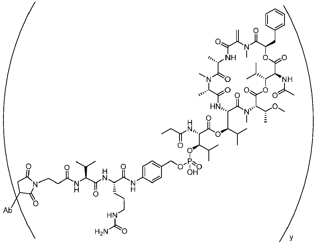
- y is 2 or 4 and
- Ab is an antibody or antigen binding fragment thereof that binds to human PMEL17 protein.
- Embodiment 85. The conjugate of Formula (C), Formula (C-1) or Formula (C-2) having the structure:

- y is 2 or 4 and
- Ab is an antibody or antigen binding fragment thereof that binds to human PMEL17 protein.
- Embodiment 86. The conjugate of Formula (C), Formula (C-1) or Formula (C-2) having the structure:

- y is 2 or 4 and
- Ab is an antibody or antigen binding fragment thereof that binds to human PMEL17 protein.
- Embodiment 87. The conjugate of Formula (C), Formula (C-1) or Formula (C-3) having the structure:

- y is 2 or 4 and
- Ab is an antibody or antigen binding fragment thereof that binds to human PMEL17 protein.
-
Embodiment 88. The conjugate of Formula (C), Formula (C-1) or Formula (C-3) having the structure:
- y is 2 or 4 and
- Ab is an antibody or antigen binding fragment thereof that binds to human PMEL17 protein.
- Embodiment 89. The conjugate of Formula (C), Formula (C-1) or Formula (C-3) having the structure:

- y is 2 or 4 and
- Ab is an antibody or antigen binding fragment thereof that binds to human PMEL17 protein.
-
Embodiment 90. The conjugate of Formula (C), Formula (C-1) or Formula (C-3) having the structure:
- y is 2 or 4 and
- Ab is an antibody or antigen binding fragment thereof that binds to human PMEL17 protein.
- Embodiment 91. The conjugate of Formula (C), Formula (C-1) or Formula (C-3) having the structure:

- y is 2 or 4 and
- Ab is an antibody or antigen binding fragment thereof that binds to human PMEL17 protein.
- Embodiment 92. The conjugate of Formula (C), Formula (C-1) or Formula (C-3) having the structure:

- y is 2 or 4 and
- Ab is an antibody or antigen binding fragment thereof that binds to human PMEL17 protein.
- Further, the antibodies, antibody fragments (e.g., antigen binding fragments) or functional equivalents of the present invention may be conjugated to a drug moiety that modifies a given biological response. Drug moieties are not to be construed as limited to classical chemical therapeutic agents. For example, the drug moiety may be a protein, peptide, or polypeptide possessing a desired biological activity. Such proteins may include, for example, a toxin such as abrin, ricin A, pseudomonas exotoxin, cholera toxin, or diphtheria toxin, a protein such as tumor necrosis factor, α-interferon, β-interferon, nerve growth factor, platelet derived growth factor, tissue plasminogen activator, a cytokine, an apoptotic agent, an anti-angiogenic agent, or, a biological response modifier such as, for example, a lymphokine.
- In one embodiment, the antibodies, antibody fragments (e.g., antigen binding fragments) or functional equivalents of the present invention are conjugated to a drug moiety, such as a cytotoxin, a drug (e.g., an immunosuppressant) or a radiotoxin. Examples of cytotoxins include but are not limited to, taxanes (see, e.g., International (PCT) Patent Application Nos.
WO 01/38318 PCT/US03/02675 ), DNA-alkylating agents (e.g., CC-1065 analogs), anthracyclines, tubulysin analogs, duocarmycin analogs, auristatin E, auristatin F, maytansinoids, pyrrolobenzodiazipines (PBDs), and cytotoxic agents comprising a reactive polyethylene glycol moiety (see, e.g., Sasse et al., J. Antibiot. (Tokyo), 53, 879-85 (2000), Suzawa et al., Bioorg. Med. Chem., 8, 2175-84 (2000), Ichimura et al., J. Antibiot. (Tokyo), 44, 1045-53 (1991), Francisco et al., Blood (2003) (electronic publication prior to print publication),U.S. Pat. Nos. 5,475,092 ,6,340,701 ,6,372,738 , and6,436,931 ,U.S. Patent Application Publication No. 2001/0036923 A1 , PendingU.S. patent application Ser. Nos. 10/024,290 and10/116,053 PCT) Patent Application No. WO 01/49698 US20090304721 ). - Other examples of cytotoxins that can be conjugated to the antibodies, antibody fragments (antigen binding fragments) or functional equivalents of the invention include duocarmycins, calicheamicins, maytansines and auristatins, and derivatives thereof.
- Various types of cytotoxins, linkers and methods for conjugating therapeutic agents to antibodies are known in the art, see, e.g., Saito et al., (2003) Adv. Drug Deliv. Rev. 55:199-215; Trail et al., (2003) Cancer Immunol. Immunother. 52:328-337; Payne, (2003) Cancer Cell 3:207-212; Allen, (2002) Nat. Rev. Cancer 2:750-763; Pastan and Kreitman, (2002) Curr. Opin. Investig. Drugs 3:1089-1091; Senter and Springer, (2001) Adv. Drug Deliv. Rev. 53:247-264.
- The antibodies, antibody fragments (e.g., antigen binding fragments) or functional equivalents of the present invention can also be conjugated to a radioactive isotope to generate cytotoxic radiopharmaceuticals, referred to as radioimmunoconjugates. Examples of radioactive isotopes that can be conjugated to antibodies for use diagnostically or therapeutically include, but are not limited to, iodine-131, indium-111, yttrium-90, and lutetium-177. Methods for preparing radioimmunoconjugates are established in the art. Examples of radioimmunoconjugates are commercially available, including Zevalin™ (DEC Pharmaceuticals) and Bexxar™ (Corixa Pharmaceuticals), and similar methods can be used to prepare radioimmunoconjugates using the antibodies of the invention. In certain embodiments, the macrocyclic chelator is 1,4,7,10-tetraazacyclododecane-N,N',N",N‴-tetraacetic acid (DOTA) which can be attached to the antibody via a linker molecule. Such linker molecules are commonly known in the art and described in Denardo et al., (1998) Clin Cancer Res. 4(10):2483-90; Peterson et al., (1999) Bioconjug. Chem. 10(4):553-7; and Zimmerman et al., (1999) Nucl. Med. Biol. 26(8):943-50, each incorporated by reference in their entireties.
- The antibodies, antibody fragments (e.g., antigen binding fragments) or functional equivalents of the present invention can also conjugated to a heterologous protein or polypeptide (or fragment thereof, preferably to a polypeptide of at least 10, at least 20, at least 30, at least 40, at least 50, at least 60, at least 70, at least 80, at least 90 or at least 100 amino acids) to generate fusion proteins. In particular, the invention provides fusion proteins comprising an antibody fragment (e.g., antigen binding fragment) described herein (e.g., a Fab fragment, Fd fragment, Fv fragment, F(ab)2 fragment, a VH domain, a VH CDR, a VL domain or a VL CDR) and a heterologous protein, polypeptide, or peptide.
- Additional fusion proteins may be generated through the techniques of gene-shuffling, motif-shuffling, exon-shuffling, and/or codon-shuffling (collectively referred to as "DNA shuffling"). DNA shuffling may be employed to alter the activities of antibodies of the invention or fragments thereof (e.g., antibodies or fragments thereof with higher affinities and lower dissociation rates). See, generally,
U.S. Patent Nos. 5,605,793 ,5,811,238 ,5,830,721 ,5,834,252 , and5,837,458 ; Patten et al., (1997) Curr. Opinion Biotechnol. 8:724-33; Harayama, (1998) Trends Biotechnol. 16(2):76-82; Hansson et al., (1999) J. Mol. Biol. 287:265-76; and Lorenzo and Blasco, (1998) Biotechniques 24(2):308- 313 (each of these patents and publications are hereby incorporated by reference in its entirety). Antibodies or fragments thereof, or the encoded antibodies or fragments thereof, may be altered by being subjected to random mutagenesis by error-prone PCR, random nucleotide insertion or other methods prior to recombination. A polynucleotide encoding an antibody or fragment thereof that specifically binds to an antigen may be recombined with one or more components, motifs, sections, parts, domains, fragments, etc. of one or more heterologous molecules. - Moreover, the antibodies, antibody fragments (e.g., antigen binding fragments) or functional equivalents of the present invention can be conjugated to marker sequences, such as a peptide, to facilitate purification. In preferred embodiments, the marker amino acid sequence is a hexa-histidine peptide (SEQ ID NO: 267), such as the tag provided in a pQE vector (QIAGEN, Inc., 9259 Eton Avenue, Chatsworth, CA, 91311), among others, many of which are commercially available. As described in Gentz et al., (1989) Proc. Natl. Acad. Sci. USA 86:821-824, for instance, hexa-histidine (SEQ ID NO: 267) provides for convenient purification of the fusion protein. Other peptide tags useful for purification include, but are not limited to, the hemagglutinin ("HA") tag, which corresponds to an epitope derived from the influenza hemagglutinin protein (Wilson et al., (1984) Cell 37:767), and the "FLAG" tag (A. Einhauer et al., J. Biochem. Biophys. Methods 49: 455-465, 2001). According to the present invention, antibodies or antigen binding fragments can also be conjugated to tumor-penetrating peptides in order to enhance their efficacy.
- In other embodiments, the antibodies, antibody fragments (e.g., antigen binding fragments) or functional equivalents of the present invention are conjugated to a diagnostic or detectable agent. Such immunoconjugates can be useful for monitoring or prognosing the onset, development, progression and/or severity of a disease or disorder as part of a clinical testing procedure, such as determining the efficacy of a particular therapy. Such diagnosis and detection can be accomplished by coupling the antibody to detectable substances including, but not limited to, various enzymes, such as, but not limited to, horseradish peroxidase, alkaline phosphatase, beta-galactosidase, or acetylcholinesterase; prosthetic groups, such as, but not limited to, streptavidin/biotin and avidin/biotin; fluorescent materials, such as, but not limited to, Alexa Fluor 350, Alexa Fluor 405, Alexa Fluor 430, Alexa Fluor 488, Alexa Fluor 500, Alexa Fluor 514, Alexa Fluor 532, Alexa Fluor 546, Alexa Fluor 555, Alexa Fluor 568, Alexa Fluor 594, Alexa Fluor 610, Alexa Fluor 633, Alexa Fluor 647, Alexa Fluor 660, Alexa Fluor 680, Alexa Fluor 700, Alexa Fluor 750, umbelliferone, fluorescein, fluorescein isothiocyanate, rhodamine, dichlorotriazinylamine fluorescein, dansyl chloride or phycoerythrin; luminescent materials, such as, but not limited to, luminol; bioluminescent materials, such as but not limited to, luciferase, luciferin, and aequorin; radioactive materials, such as, but not limited to, iodine (131I, 125I, 123I, and 121I,), carbon (14C), sulfur (35S), tritium (3H), indium (115In, 113In, 112In, and 111In,), technetium (99Tc), thallium (201Ti), gallium (68Ga, 67Ga), palladium (103Pd), molybdenum (99Mo), xenon (133Xe), fluorine (18F), 153Sm, 177Lu, 159Gd, 149Pm, 140La, 175Yb, 166Ho, 90Y, 47Sc, 186Re, 188Re, 142Pr, 105Rh, 97Ru, 68Ge, 57Co, 65Zn, 85Sr, 32P, 153Gd, 169Yb, 51Cr, 54Mn, 75Se, 64Cu, 113Sn, and 117Sn; and positron emitting metals using various positron emission tomographies, and non-radioactive paramagnetic metal ions.
- The antibodies, antibody fragments (e.g., antigen binding fragments) or functional equivalents of the invention may also be attached to solid supports, which are particularly useful for immunoassays or purification of the target antigen. Such solid supports include, but are not limited to, glass, cellulose, polyacrylamide, nylon, polystyrene, polyvinyl chloride or polypropylene.
- A general reaction scheme for the formation of conjugates of Formula (C) is shown in
Scheme 1 below: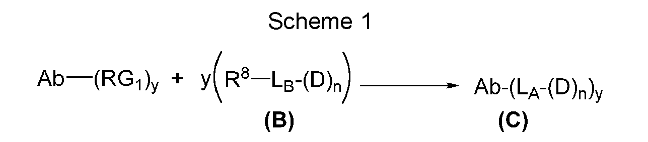
- In one embodiment, D is a GNAQ inhibitor, a GNA11 inhibitor, or an inhibitor of GNAQ and GNA11 (GNAQ/GNA11 inhibitor), La is a linker further comprising a bivalent coupling group formed when RG, and R8 react, n is 1, 2, 3 or 4, and y is 1, 2, 3 or 4.
- A general reaction scheme for the formation of conjugates of Formula (C-1) is shown in
Scheme 2 below:
- In one embodiment, D is a GNAQ inhibitor, a GNA11 inhibitor, or an inhibitor of GNAQ and GNA11 (GNAQ/GNA11 inhibitor), X1 is a bivalent coupling group formed when RG, and R8 react (e.g. a succinimide ring or an oxime), Y, is a phosphate group, X2 is a self-immolative spacer, L1 is a bivalent peptide linker, L2 is a bond or a linker, and y is 1, 2, 3 or 4.
- A general reaction scheme for the formation of conjugates of Formula (C-2) is shown in
Scheme 3 below:
- Here R0 is methyl or ethyl, R1 is methyl or isopropyl, R2 is methyl or ethyl, X1 is a bivalent coupling group formed when RG, and R8 react (e.g. a succinimide ring or an oxime), X2 is a self-immolative spacer, L1 is a bivalent peptide linker, L2 is a bond or a linker, and y is 1, 2, 3 or 4.
- A general reaction scheme for the formation of conjugates of Formula (C-2) is shown in
Scheme 4 below: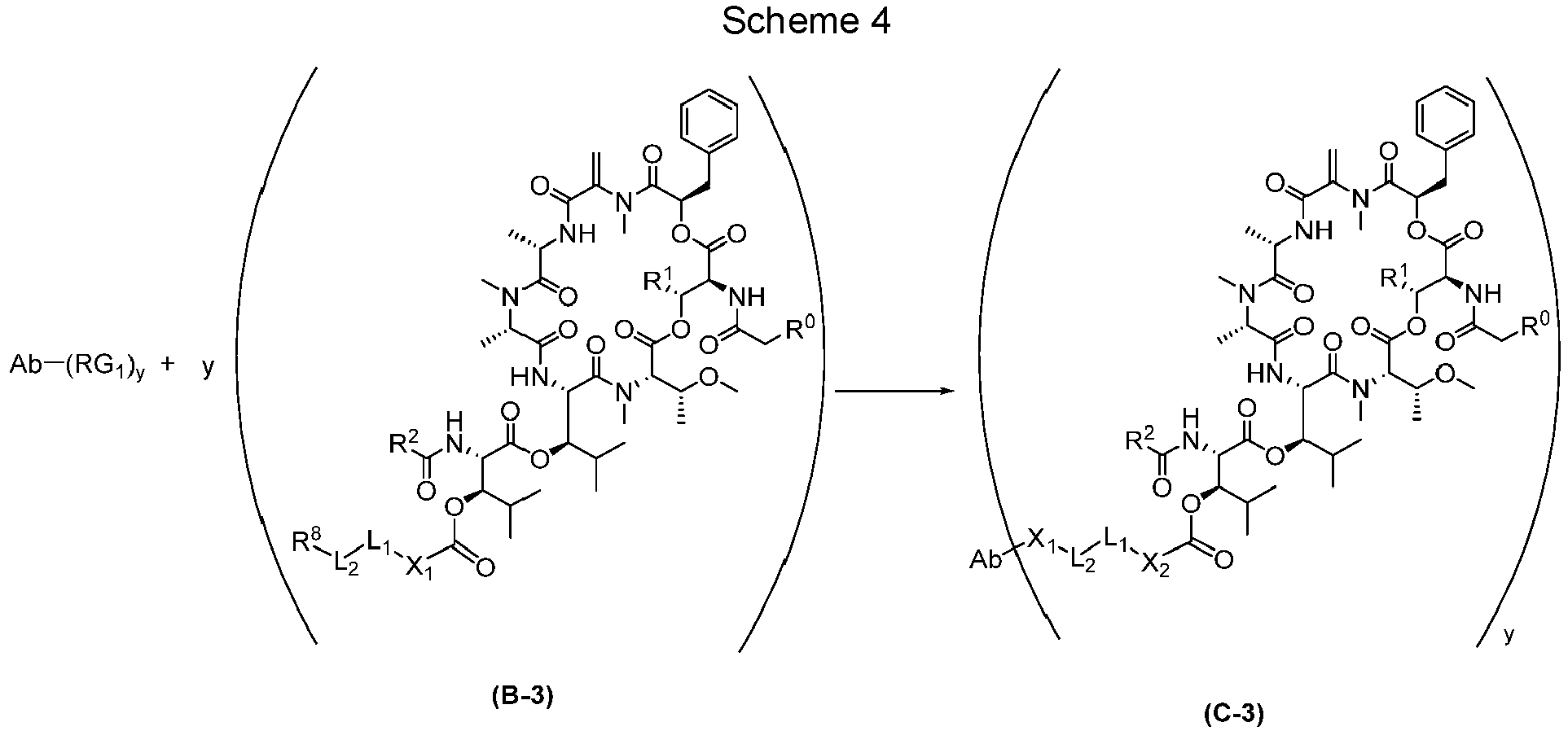
- Conjugates of the invention can be prepared using cysteine residues engineered into an antibody by, for example, site-directed mutagenesis. Such site-specific conjugates are homogenous and have improved properties (Junutula JR, Raab H, Clark S, Bhakta S, Leipold DD, Weir S, Chen Y, Simpson M, Tsai SP, Dennis MS, Lu Y, Meng YG, Ng C, Yang J, Lee CC, Duenas E, Gorrell J, Katta V, Kim A, McDorman K, Flagella K, Venook R, Ross S, Spencer SD, Lee Wong W, Lowman HB, Vandlen R, Sliwkowski MX, Scheller RH, Polakis P, Mallet W. (2008) Nature Biotechnology 26:925-932.)
- Because engineered cysteines in antibodies expressed in mammalian cells are modified by adducts (disulfides) such as glutathione (GSH) and/or cysteine during their biosynthesis (Chen et al. 2009), the engineered cysteine residues in the product as initially expressed are unreactive to thiol reactive reagents such as maleimido or bromo-or iodoacetamide groups. To conjugate payload to an engineered cysteine after expression, glutathione or cysteine adducts need to be removed by reducing these disulfide adducts, which generally entails also reducing native disulfides in the expressed protein. Deprotection of adducted engineered cysteines can be accomplished by first exposing antibody to a reducing agent, e.g., dithiothreitol (DTT), TCEP, or reduced cysteine, followed by a procedure that allows for re-oxidation of all native disulfide bonds of an antibody to restore and/or stabilize the functional antibody structure.
- Several methods can be employed to reduce and re-oxidize antibodies with engineered cysteine residues for preparation of antibody drug conjugates. Attempts to follow re-oxidation protocols previously described in the literature using high concentration of CuSO4 resulted in protein precipitation (Junutula JR, Raab H, Clark S, Bhakta S, Leipold DD, Weir S, Chen Y, Simpson M, Tsai SP, Dennis MS, Lu Y, Meng YG, Ng C, Yang J, Lee CC, Duenas E, Gorrell J, Katta V, Kim A, McDorman K, Flagella K, Venook R, Ross S, Spencer SD, Lee Wong W, Lowman HB, Vandlen R, Sliwkowski MX, Scheller RH, Polakis P, Mallet W. (2008) Nature Biotechnology 26:925). We have successfully prepared and obtained antibody drug conjugates with several different methods for reduction and antibody re-oxidation.
- The following is a method to reduce and re-oxidize antibodies with engineered cysteine residues for preparation of antibody drug conjugates: Freshly prepared DTT is added to purified Cys mutant antibodies to a final concentration of 10 mM. After incubation with DTT at room temperature for 1 hour, mixture is dialyzed at 4°C against PBS for three days with daily buffer exchange to remove DTT and re-oxidize native disulfide bonds of the antibody. An alternative method is to remove reducing reagents through a desalting column such as Sephadex G-25, equilibrated with PBS. Once protein is fully reduced, 1 mM oxidized ascorbate (dehydro-ascorbic acid) is optionally added to desalted samples and re-oxidation incubations are carried out for 20-24 hours.
- In another exemplary method, deprotection of engineered Cys residues is accomplished by adding fully reduced cysteine at 20 mM concentration to antibodies bound to protein A-Sepharose resin. Reduction of the Cys adducts is achieved by incubation for approximately 30-60 minutes at room temperature, then reductant is rapidly removed by washing resin with 50 beds of PBS. Re-oxidation of the reduced antibody is achieved by incubating washed slurry at room temperature with or without addition of 50-2000 nM CuCl2 as an accelerant. With the exception of use of copper sulfate, examples herein use each of the protocols described herein with similar results. Reoxidation restores intra-chain disulfides, while dialysis, desalting or protein A chromatography removes reducing agent as well as cysteines and glutathiones initially connected to engineered cysteine(s) of the antibody. HPLC reverse phase chromatography is typically used to monitor the reoxidation process: Antibodies are loaded onto a PLRP-S column (4000 Å, 50 mm x 2.1 mm, Agilent) heated to 80° C and eluted using a linear gradient of 30-45% CH3CN in water containing 0.1% TFA at 1.5 mL/min. and peak detection at 215, 254, and 280 nm.
- After re-oxidation, the antibody is conjugated to a linker-drug compound of , by way of example, compounds of Formula (B), Formula (B-1), Formula (B-2) or Formula (B-3) (see schemes 1-4). By way of example, a compound of Formula (B), Formula (B-1), Formula (B-2) or Formula (B-3) is added to re-oxidized Cys mutant antibody at 5-10 molar equivalents relative to antibody in PBS buffer (pH 7.2). Incubations are carried out for 1-2 hours. The conjugation process is monitored by reverse-phase HPLC, which is able to separate conjugated antibodies from non-conjugated ones. Conjugation reaction mixtures are analyzed on a PRLP-S column (4000 Å, 50 mm x 2.1 mm, Agilent) heated to 80°C and elution of the column are carried out by a linear gradient of 30-60% acetonitrile in water containing 0.1% TFA at a flow rate of 1.5 ml/min. Elution of proteins from the column is monitored at 280 nm, 254 nm and 215 nm.
- Alternatively, for antibodies bound to a Protein A resin, once the antibody is re-oxidized, the resin is washed with 10 column volumes PBS and the resin is then resuspended in equal volume PBS and an 8x excess of a compound of Formula (B), Formula (B-1), Formula (B-2) or Formula (B-3) (in DMSO) is added and incubated at room temperature for 2 hours. The resin is then washed with 50 column volumes of PBS and the resulting antibody drug conjugate is eluted from the Protein A resin, neutralized with 1/10 volume 1 M Tris pH 9.0 and buffer exchanged into appropriate buffer to perform preparative size exclusion chromatography (if needed).
- Immunoconjugates are also characterized in terms of average loading of a drug moiety to antibody binding moiety, generally referred to as drug-to-antibody ratio (DAR). The DAR value is extrapolated, for example, from LC-MS data for reduced and deglycosylated samples. LC/MS allows quantitation of the average number of molecules of payload (drug moiety) attached to an antibody in an ADC. HPLC separates an antibody into light and heavy chains, and also separates heavy chain (HC) and light chain (LC) according to the number of Linker-Payload groups per chain. Mass spectral data enables identification of the component species in the mixture, e.g., LC, LC+1, LC+2, HC, HC+1, HC+2, etc. From average loading of LC and HC chains, the average DAR can be calculated for an ADC. The DAR for a given immunoconjugate sample represents the average number of drug (payload) molecules attached to a tetrameric antibody containing two light chains and two heavy chains.
- Throughout the text of this application, should there be a discrepancy between the text of the specification and the sequence listing, the text of the specification shall prevail.
Table 2. Examples of Anti-PMEL17 Antibodies of the Present Invention G1 E152C_S375C_wt LC SEQ ID NO: 1 HCDR1 (Combined) GGTFSDYAIT SEQ ID NO: 2 HCDR2 (Combined) GIIPIFGTANYAQKFQG SEQ ID NO: 3 HCDR3 (Combined) EGGLLTDISYSRYWFAY SEQ ID NO: 4 HCDR1 (Kabat) DYAIT SEQ ID NO: 2 HCDR2 (Kabat) GIIPIFGTANYAQKFQG SEQ ID NO: 3 HCDR3 (Kabat) EGGLLTDISYSRYWFAY SEQ ID NO: 5 HCDR1 (Chothia) GGTFSDY SEQ ID NO: 6 HCDR2 (Chothia) IPIFGT SEQ ID NO: 3 HCDR3 (Chothia) EGGLLTDISYSRYWFAY SEQ ID NO: 7 HCDR1 (IMGT) GGTFSDYA SEQ ID NO: 8 HCDR2 (IMGT) IIPIFGTA SEQ ID NO: 9 HCDR3 (IMGT) AREGGLLTDISYSRYWFAY SEQ ID NO: 10 VH 
SEQ ID NO: 11 DNA VH 
SEQ ID NO: 12 Heavy Chain 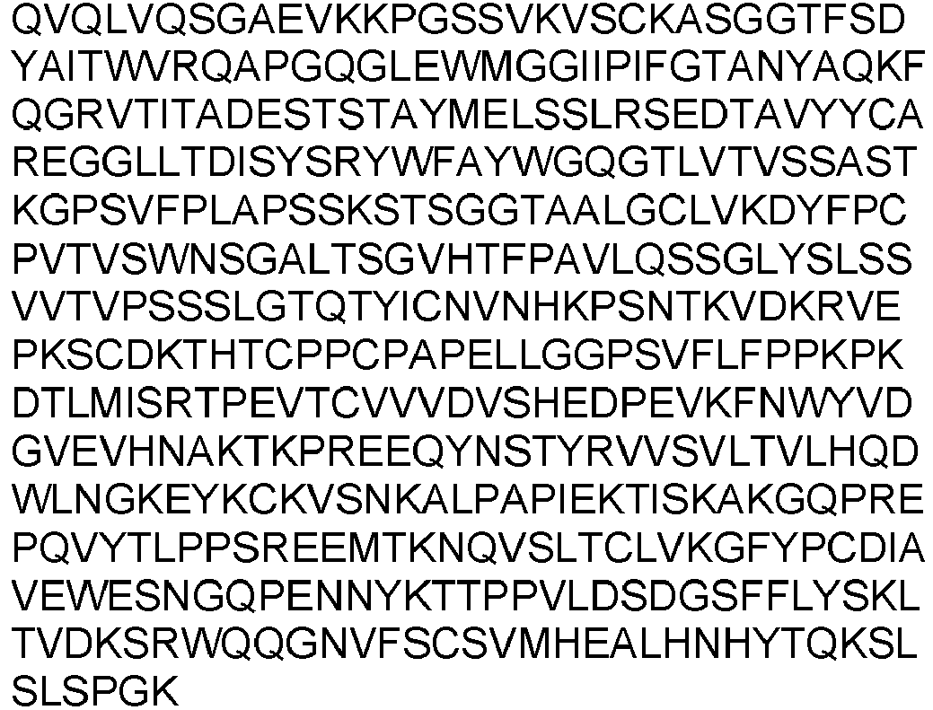
SEQ ID NO: 13 DNA Heavy Chain 

SEQ ID NO: 14 LCDR1 (Combined) SGDALRDKFVY SEQ ID NO: 15 LCDR2 (Combined) DDNNRPS SEQ ID NO: 16 LCDR3 (Combined) QSWDHSYSLVV SEQ ID NO: 14 LCDR1 (Kabat) SGDALRDKFVY SEQ ID NO: 15 LCDR2 (Kabat) DDNNRPS SEQ ID NO: 16 LCDR3 (Kabat) QSWDHSYSLVV SEQ ID NO: 17 LCDR1 (Chothia) DALRDKF SEQ ID NO: 18 LCDR2 (Chothia) DDN SEQ ID NO: 19 LCDR3 (Chothia) WDHSYSLV SEQ ID NO: 20 LCDR1 (IMGT) ALRDKF SEQ ID NO: 18 LCDR2 (IMGT) DDN SEQ ID NO: 16 LCDR3 (IMGT) QSWDHSYSLVV SEQ ID NO: 21 VL 
SEQ ID NO: 22 DNA VL 
SEQ ID NO: 23 Light Chain 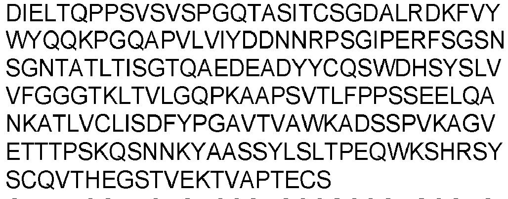
SEQ ID NO: 24 DNA Light Chain 

G1 E152C_S375C_3J LC SEQ ID NO: 1 HCDR1 (Combined) GGTFSDYAIT SEQ ID NO: 2 HCDR2 (Combined) GIIPIFGTANYAQKFQG SEQ ID NO: 3 HCDR3 (Combined) EGGLLTDISYSRYWFAY SEQ ID NO: 4 HCDR1 (Kabat) DYAIT SEQ ID NO: 2 HCDR2 (Kabat) GIIPIFGTANYAQKFQG SEQ ID NO: 3 HCDR3 (Kabat) EGGLLTDISYSRYWFAY SEQ ID NO: 5 HCDR1 (Chothia) GGTFSDY SEQ ID NO: 6 HCDR2 (Chothia) IPIFGT SEQ ID NO: 3 HCDR3(Ch othia) EGGLLTDISYSRYWFAY SEQ ID NO: 7 HCDR1 (IMGT) GGTFSDYA SEQ ID NO: 8 HCDR2 (IMGT) IIPIFGTA SEQ ID NO: 9 HCDR3 (IMGT) AREGGLLTDISYSRYWFAY SEQ ID NO: 10 VH 
SEQ ID NO: 11 DNA VH 
SEQ ID NO: 12 Heavy Chain 

SEQ ID NO: 13 DNA Heavy Chain 

SEQ ID NO: 14 LCDR1 (Combined) SGDALRDKFVY SEQ ID NO: 15 LCDR2 (Combined) DDNNRPS SEQ ID NO: 16 LCDR3 (Combined) QSWDHSYSLVV SEQ ID NO: 14 LCDR1 (Kabat) SGDALRDKFVY SEQ ID NO: 15 LCDR2 (Kabat) DDNNRPS SEQ ID NO: 16 LCDR3 (Kabat) QSWDHSYSLVV SEQ ID NO: 17 LCDR1 (Chothia) DALRDKF SEQ ID NO: 18 LCDR2 (Chothia) DDN SEQ ID NO: 19 LCDR3 (Chothia) WDHSYSLV SEQ ID NO: 20 LCDR1 (IMGT) ALRDKF SEQ ID NO: 18 LCDR2 (IMGT) DDN SEQ ID NO: 16 LCDR3 (IMGT) QSWDHSYSLVV SEQ ID NO: 25 VL 
SEQ ID NO: 26 DNA VL 
SEQ ID NO: 27 Light Chain 
SEQ ID NO: 28 DNA Light Chain 
G1 E152C_S375C_3R LC SEQ ID NO: 1 HCDR1 (Combined) GGTFSDYAIT SEQ ID NO: 2 HCDR2 (Combined) GIIPIFGTANYAQKFQG SEQ ID NO: 3 HCDR3 (Combined) EGGLLTDISYSRYWFAY SEQ ID NO: 4 HCDR1 (Kabat) DYAIT SEQ ID NO: 2 HCDR2 (Kabat) GIIPIFGTANYAQKFQG SEQ ID NO: 3 HCDR3 (Kabat) EGGLLTDISYSRYWFAY SEQ ID NO: 5 HCDR1 (Chothia) GGTFSDY SEQ ID NO: 6 HCDR2 (Chothia) IPIFGT SEQ ID NO: 3 HCDR3(Ch othia) EGGLLTDISYSRYWFAY SEQ ID NO: 7 HCDR1 (IMGT) GGTFSDYA SEQ ID NO: 8 HCDR2 (IMGT) IIPIFGTA SEQ ID NO: 9 HCDR3 (IMGT) AREGGLLTDISYSRYWFAY SEQ ID NO: 10 VH 

SEQ ID NO: 11 DNA VH 
SEQ ID NO: 12 Heavy Chain 
SEQ ID NO: 13 DNA Heavy Chain 

SEQ ID NO: 14 LCDR1 (Combined) SGDALRDKFVY SEQ ID NO: 15 LCDR2 (Combined) DDNNRPS SEQ ID NO: 16 LCDR3 (Combined) QSWDHSYSLVV SEQ ID NO: 14 LCDR1 (Kabat) SGDALRDKFVY SEQ ID NO: 15 LCDR2 (Kabat) DDNNRPS SEQ ID NO: 16 LCDR3 (Kabat) QSWDHSYSLVV SEQ ID NO: 17 LCDR1 (Chothia) DALRDKF SEQ ID NO: 18 LCDR2 (Chothia) DDN SEQ ID NO: 19 LCDR3 (Chothia) WDHSYSLV SEQ ID NO: 20 LCDR1 (IMGT) ALRDKF SEQ ID NO: 18 LCDR2 (IMGT) DDN SEQ ID NO: 16 LCDR3 (IMGT) QSWDHSYSLVV SEQ ID NO: 29 VL 

SEQ ID NO: 30 DNA VL 
SEQ ID NO: 31 Light Chain 
SEQ ID NO: 32 DNA Light Chain 
G4 E152_S375C SEQ ID NO: 33 HCDR1 (Combined) GGTFSTYAIS SEQ ID NO: 34 HCDR2 (Combined) RIIPILGIANYAQKFQG SEQ ID NO: 35 HCDR3 (Combined) EVRMIFDY SEQ ID NO: 36 HCDR1 (Kabat) TYAIS SEQ ID NO: 34 HCDR2 (Kabat) RIIPILGIANYAQKFQG SEQ ID NO: 35 HCDR3 (Kabat) EVRMIFDY SEQ ID NO: 37 HCDR1 (Chothia) GGTFSTY SEQ ID NO: 38 HCDR2 (Chothia) IPILGI SEQ ID NO: 35 HCDR3(Ch othia) EVRMIFDY SEQ ID NO: 39 HCDR1 (IMGT) GGTFSTYA SEQ ID NO: 40 HCDR2 (IMGT) IIPILGIA SEQ ID NO: 41 HCDR3 (IMGT) AREVRMIFDY SEQ ID NO: 42 VH 
SEQ ID NO: 43 DNA VH 
SEQ ID NO: 44 Heavy Chain 
SEQ ID NO: 45 DNA Heavy Chain 

SEQ ID NO: 46 LCDR1 (Combined) RASQSISSYLA SEQ ID NO: 47 LCDR2 (Combined) AASSLQS SEQ ID NO: 48 LCDR3 (Combined) QQSYDYYT SEQ ID NO: 46 LCDR1 (Kabat) RASQSISSYLA SEQ ID NO: 47 LCDR2 (Kabat) AASSLQS SEQ ID NO: 48 LCDR3 (Kabat) QQSYDYYT SEQ ID NO: 49 LCDR1 (Chothia) SQSISSY SEQ ID NO: 50 LCDR2 (Chothia) AAS SEQ ID NO: 51 LCDR3 (Chothia) SYDYY SEQ ID NO: 52 LCDR1 (IMGT) QSISSY SEQ ID NO: 50 LCDR2 (IMGT) AAS SEQ ID NO: 48 LCDR3 (IMGT) QQSYDYYT SEQ ID NO: 53 VL 
SEQ ID NO: 54 DNA VL 
SEQ ID NO: 55 Light Chain 
SEQ ID NO: 56 DNA Light Chain 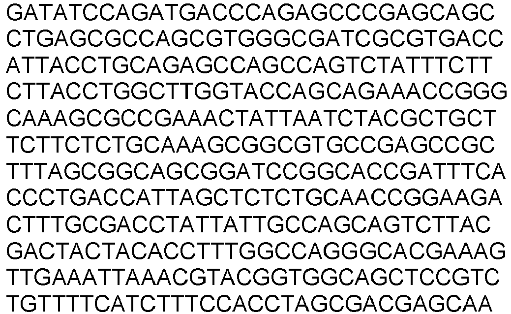

MOR024353 E152C_S375C SEQ ID NO: 57 HCDR1 (Combined) GGTFSDYAIS SEQ ID NO: 58 HCDR2 (Combined) GIIPIFGDANYAQKFQG SEQ ID NO: 59 HCDR3 (Combined) EGSSYFYMAY SEQ ID NO: 60 HCDR1 (Kabat) DYAIS SEQ ID NO: 58 HCDR2 (Kabat) GIIPIFGDANYAQKFQG SEQ ID NO: 59 HCDR3 (Kabat) EGSSYFYMAY SEQ ID NO: 5 HCDR1 (Chothia) GGTFSDY SEQ ID NO: 61 HCDR2 (Chothia) IPIFGD SEQ ID NO: 59 HCDR3(Ch othia) EGSSYFYMAY SEQ ID NO: 7 HCDR1 (IMGT) GGTFSDYA SEQ ID NO: 62 HCDR2 (IMGT) IIPIFGDA SEQ ID NO: 63 HCDR3 (IMGT) AREGSSYFYMAY SEQ ID NO: 64 VH 
SEQ ID NO: 65 DNA VH 

SEQ ID NO: 66 Heavy Chain 
SEQ ID NO: 67 DNA Heavy Chain 

SEQ ID NO: 68 LCDR1 (Combined) SGDNIGSKLAS SEQ ID NO: 69 LCDR2 (Combined) DDSNRPS SEQ ID NO: 70 LCDR3 (Combined) AATAGDRWAYV SEQ ID NO: 68 LCDR1 (Kabat) SGDNIGSKLAS SEQ ID NO: 69 LCDR2 (Kabat) DDSNRPS SEQ ID NO: 70 LCDR3 (Kabat) AATAGDRWAYV SEQ ID NO: 71 LCDR1 (Chothia) DNIGSKL SEQ ID NO: 72 LCDR2 (Chothia) DDS SEQ ID NO: 73 LCDR3 (Chothia) TAGDRWAY SEQ ID NO: 74 LCDR1 (IMGT) NIGSKL SEQ ID NO: 72 LCDR2 (IMGT) DDS SEQ ID NO: 70 LCDR3 (IMGT) AATAGDRWAYV SEQ ID NO: 75 VL 
SEQ ID NO: 76 DNA VL 
SEQ ID NO: 77 Light Chain 
SEQ ID NO: 78 DNA Light Chain 
MOR024354 E152C_S375C SEQ ID NO: 79 HCDR1 (Combined) GFTFSSFGMS SEQ ID NO: 80 HCDR2 (Combined) AISYSGSDTYYADSVKG SEQ ID NO: 81 HCDR3 (Combined) DVGVMDY SEQ ID NO: 82 HCDR1 (Kabat) SFGMS SEQ ID NO: 80 HCDR2 (Kabat) AISYSGSDTYYADSVKG SEQ ID NO: 81 HCDR3 (Kabat) DVGVMDY SEQ ID NO: 83 HCDR1 (Chothia) GFTFSSF SEQ ID NO: 84 HCDR2 (Chothia) SYSGSD SEQ ID NO: 81 HCDR3(Ch othia) DVGVMDY SEQ ID NO: 85 HCDR1 (IMGT) GFTFSSFG SEQ ID NO: 86 HCDR2 (IMGT) ISYSGSDT SEQ ID NO: 87 HCDR3 (IMGT) ARDVGVMDY SEQ ID NO: 88 VH 
SEQ ID NO: 89 DNA VH 
SEQ ID NO: 90 Heavy Chain 
SEQ ID NO: 91 DNA Heavy Chain 

SEQ ID NO: 92 LCDR1 (Combined) SGDNLGTYYAH SEQ ID NO: 93 LCDR2 (Combined) SQSHRPS SEQ ID NO: 94 LCDR3 (Combined) GAWDAPSPELV SEQ ID NO: 92 LCDR1 (Kabat) SGDNLGTYYAH SEQ ID NO: 93 LCDR2 (Kabat) SQSHRPS SEQ ID NO: 94 LCDR3 (Kabat) GAWDAPSPELV SEQ ID NO: 95 LCDR1 (Chothia) DNLGTYY SEQ ID NO: 96 LCDR2 (Chothia) SQS SEQ ID NO: 97 LCDR3 (Chothia) WDAPSPEL SEQ ID NO: 98 LCDR1 (IMGT) NLGTYY SEQ ID NO: 96 LCDR2 (IMGT) SQS SEQ ID NO: 94 LCDR3 (IMGT) GAWDAPSPELV SEQ ID NO: 99 VL 
SEQ ID NO: 100 DNA VL 
SEQ ID NO: 101 Light Chain 
SEQ ID NO: 102 DNA Light Chain 
Y010341 E152C_S375C SEQ ID NO: 103 HCDR1 (Combined) GFTFSSYAMS SEQ ID NO: 104 HCDR2 (Combined) AISGSGGSTYYADSVKG SEQ ID NO: 105 HCDR3 (Combined) AFRLYWLDV SEQ ID NO: 106 HCDR1 (Kabat) SYAMS SEQ ID NO: 104 HCDR2 (Kabat) AISGSGGSTYYADSVKG SEQ ID NO: 105 HCDR3 (Kabat) AFRLYWLDV SEQ ID NO: 107 HCDR1 (Chothia) GFTFSSY SEQ ID NO: 108 HCDR2 (Chothia) SGSGGS SEQ ID NO: 105 HCDR3(Ch othia) AFRLYWLDV SEQ ID NO: 109 HCDR1 (IMGT) GFTFSSYA SEQ ID NO: 110 HCDR2 (IMGT) ISGSGGST SEQ ID NO: 111 HCDR3 (IMGT) ARAFRLYWLDV SEQ ID NO: 112 VH 
SEQ ID NO: 113 DNA VH 
SEQ ID NO: 114 Heavy Chain 

SEQ ID NO: 115 DNA Heavy Chain 
SEQ ID NO: 116 LCDR1 (Combined) RASQSISSYLN SEQ ID NO: 47 LCDR2 (Combined) AASSLQS SEQ ID NO: 117 LCDR3 (Combined) QQVYSAPVT SEQ ID NO: 116 LCDR1 (Kabat) RASQSISSYLN SEQ ID NO: 47 LCDR2 (Kabat) AASSLQS SEQ ID NO: 117 LCDR3 (Kabat) QQVYSAPVT SEQ ID NO: 49 LCDR1 (Chothia) SQSISSY SEQ ID NO: 50 LCDR2 (Chothia) AAS SEQ ID NO: 118 LCDR3 (Chothia) VYSAPV SEQ ID NO: 52 LCDR1 (IMGT) QSISSY SEQ ID NO: 50 LCDR2 (IMGT) AAS SEQ ID NO: 117 LCDR3 (IMGT) QQVYSAPVT SEQ ID NO: 119 VL 
SEQ ID NO: 120 DNA VL 
SEQ ID NO: 121 Light Chain 
SEQ ID NO: 122 DNA Light Chain 

Y010355 E152C_S375C SEQ ID NO: 123 HCDR1 (Combined) GFTFSNYWIS SEQ ID NO: 124 HCDR2 (Combined) RIKSKTYGGTTDYAEPVKG SEQ ID NO: 125 HCDR3 (Combined) TSRRSYAFDY SEQ ID NO: 126 HCDR1 (Kabat) NYWIS SEQ ID NO: 124 HCDR2 (Kabat) RIKSKTYGGTTDYAEPVKG SEQ ID NO: 125 HCDR3 (Kabat) TSRRSYAFDY SEQ ID NO: 127 HCDR1 (Chothia) GFTFSNY SEQ ID NO: 128 HCDR2 (Chothia) KSKTYGGT SEQ ID NO: 125 HCDR3(Ch othia) TSRRSYAFDY SEQ ID NO: 129 HCDR1 (IMGT) GFTFSNYW SEQ ID NO: 130 HCDR2 (IMGT) IKSKTYGGTT SEQ ID NO: 131 HCDR3 (IMGT) ARTSRRSYAFDY SEQ ID NO: 132 VH 
SEQ ID NO: 133 DNA VH 

SEQ ID NO: 134 Heavy Chain 
SEQ ID NO: 135 DNA Heavy Chain 

SEQ ID NO: 136 LCDR1 (Combined) RASQSISSWLA SEQ ID NO: 137 LCDR2 (Combined) DASSLES SEQ ID NO: 138 LCDR3 (Combined) QQITRYPVT SEQ ID NO: 136 LCDR1 (Kabat) RASQSISSWLA SEQ ID NO: 137 LCDR2 (Kabat) DASSLES SEQ ID NO: 138 LCDR3 (Kabat) QQITRYPVT SEQ ID NO: 139 LCDR1 (Chothia) SQSISSW SEQ ID NO: 140 LCDR2 (Chothia) DAS SEQ ID NO: 141 LCDR3 (Chothia) ITRYPV SEQ ID NO: 142 LCDR1 (IMGT) QSISSW SEQ ID NO: 140 LCDR2 (IMGT) DAS SEQ ID NO: 138 LCDR3 (IMGT) QQITRYPVT SEQ ID NO: 143 VL 
SEQ ID NO: 144 DNA VL 

SEQ ID NO: 145 Light Chain 
SEQ ID NO: 146 DNA Light Chain 
Y010356 E152C_S375C SEQ ID NO: 123 HCDR1 (Combined) GFTFSNYWIS SEQ ID NO: 124 HCDR2 (Combined) RIKSKTYGGTTDYAEPVKG SEQ ID NO: 147 HCDR3 (Combined) VSGYYSHSGGFDV SEQ ID NO: 126 HCDR1 (Kabat) NYWIS SEQ ID NO: 124 HCDR2 (Kabat) RIKSKTYGGTTDYAEPVKG SEQ ID NO: 147 HCDR3 (Kabat) VSGYYSHSGGFDV SEQ ID NO: 127 HCDR1 (Chothia) GFTFSNY SEQ ID NO: 128 HCDR2 (Chothia) KSKTYGGT SEQ ID NO: 147 HCDR3(Ch othia) VSGYYSHSGGFDV SEQ ID NO: 129 HCDR1 (IMGT) GFTFSNYW SEQ ID NO: 130 HCDR2 (IMGT) IKSKTYGGTT SEQ ID NO: 148 HCDR3 (IMGT) ARVSGYYSHSGGFDV SEQ ID NO: 149 VH 
SEQ ID NO: 150 DNA VH 
SEQ ID NO: 151 Heavy Chain 
SEQ ID NO: 152 DNA Heavy Chain 

SEQ ID NO: 153 LCDR1 (Combined) RASQGISNYLA SEQ ID NO: 154 LCDR2 (Combined) AASTLQS SEQ ID NO: 155 LCDR3 (Combined) QKTWRTPGT SEQ ID NO: 153 LCDR1 (Kabat) RASQGISNYLA SEQ ID NO: 154 LCDR2 (Kabat) AASTLQS SEQ ID NO: 155 LCDR3 (Kabat) QKTWRTPGT SEQ ID NO: 156 LCDR1 (Chothia) SQGISNY SEQ ID NO: 50 LCDR2 (Chothia) AAS SEQ ID NO: 157 LCDR3 (Chothia) TWRTPG SEQ ID NO: 158 LCDR1 (IMGT) QGISNY SEQ ID NO: 50 LCDR2 (IMGT) AAS SEQ ID NO: 155 LCDR3 (IMGT) QKTWRTPGT SEQ ID NO: 159 VL 
SEQ ID NO: 160 DNA VL 
SEQ ID NO: 161 Light Chain 
SEQ ID NO: 162 DNA Light Chain 

Y010415 E152C_S375C SEQ ID NO: 103 HCDR1 (Combined) GFTFSSYAMS SEQ ID NO: 104 HCDR2 (Combined) AISGSGGSTYYADSVKG SEQ ID NO: 163 HCDR3 (Combined) SRLIAPWLDY SEQ ID NO: 106 HCDR1 (Kabat) SYAMS SEQ ID NO: 104 HCDR2 (Kabat) AISGSGGSTYYADSVKG SEQ ID NO: 163 HCDR3 (Kabat) SRLIAPWLDY SEQ ID NO: 107 HCDR1 (Chothia) GFTFSSY SEQ ID NO: 108 HCDR2 (Chothia) SGSGGS SEQ ID NO: 163 HCDR3(Ch othia) SRLIAPWLDY SEQ ID NO: 109 HCDR1 (IMGT) GFTFSSYA SEQ ID NO: 110 HCDR2 (IMGT) ISGSGGST SEQ ID NO: 164 HCDR3 (IMGT) ARSRLIAPWLDY SEQ ID NO: 165 VH 
SEQ ID NO: 166 DNA VH 
SEQ ID NO: 167 Heavy Chain 
SEQ ID NO: 168 DNA Heavy Chain 

SEQ ID NO: 116 LCDR1 (Combined) RASQSISSYLN SEQ ID NO: 47 LCDR2 (Combined) AASSLQS SEQ ID NO: 169 LCDR3 (Combined) QQVYGSPPT SEQ ID NO: 116 LCDR1 (Kabat) RASQSISSYLN SEQ ID NO: 47 LCDR2 (Kabat) AASSLQS SEQ ID NO: 169 LCDR3 (Kabat) QQVYGSPPT SEQ ID NO: 49 LCDR1 (Chothia) SQSISSY SEQ ID NO: 50 LCDR2 (Chothia) AAS SEQ ID NO: 170 LCDR3 (Chothia) VYGSPP SEQ ID NO: 52 LCDR1 (IMGT) QSISSY SEQ ID NO: 50 LCDR2 (IMGT) AAS SEQ ID NO: 169 LCDR3 (IMGT) QQVYGSPPT SEQ ID NO: 171 VL 
SEQ ID NO: 172 DNA VL 
SEQ ID NO: 173 Light Chain 

SEQ ID NO: 174 DNA Light Chain 
Y010417 E152C_S375C SEQ ID NO: 175 HCDR1 (Combined) GYSFTSYWIG SEQ ID NO: 176 HCDR2 (Combined) IIYPGDSDTRYSPSFQG SEQ ID NO: 177 HCDR3 (Combined) GSSAASGLSGDL SEQ ID NO: 178 HCDR1 (Kabat) SYWIG SEQ ID NO: 176 HCDR2 (Kabat) IIYPGDSDTRYSPSFQG SEQ ID NO: 177 HCDR3 (Kabat) GSSAASGLSGDL SEQ ID NO: 179 HCDR1 (Chothia) GYSFTSY SEQ ID NO: 180 HCDR2 (Chothia) YPGDSD SEQ ID NO: 177 HCDR3(Ch othia) GSSAASGLSGDL SEQ ID NO: 181 HCDR1 (IMGT) GYSFTSYW SEQ ID NO: 182 HCDR2 (IMGT) IYPGDSDT SEQ ID NO: 183 HCDR3 (IMGT) ARGSSAASGLSGDL SEQ ID NO: 184 VH 
SEQ ID NO: 185 DNA VH 
SEQ ID NO: 186 Heavy Chain 
SEQ ID NO: 187 DNA Heavy Chain 

SEQ ID NO: 116 LCDR1 (Combined) RASQSISSYLN SEQ ID NO: 47 LCDR2 (Combined) AASSLQS SEQ ID NO: 188 LCDR3 (Combined) QQDYYSPFT SEQ ID NO: 116 LCDR1 (Kabat) RASQSISSYLN SEQ ID NO: 47 LCDR2 (Kabat) AASSLQS SEQ ID NO: 188 LCDR3 (Kabat) QQDYYSPFT SEQ ID NO: 49 LCDR1 (Chothia) SQSISSY SEQ ID NO: 50 LCDR2 (Chothia) AAS SEQ ID NO: 189 LCDR3 (Chothia) DYYSPF SEQ ID NO: 52 LCDR1 (IMGT) QSISSY SEQ ID NO: 50 LCDR2 (IMGT) AAS SEQ ID NO: 188 LCDR3 (IMGT) QQDYYSPFT SEQ ID NO: 190 VL 
SEQ ID NO: 191 DNA VL 
SEQ ID NO: 192 Light Chain 
SEQ ID NO: 193 DNA Light Chain 
Y010429 E152C_S375C SEQ ID NO: 103 HCDR1 (Combined) GFTFSSYAMS SEQ ID NO: 104 HCDR2 (Combined) AISGSGGSTYYADSVKG SEQ ID NO: 194 HCDR3 (Combined) AYKLSWLDL SEQ ID NO: 106 HCDR1 (Kabat) SYAMS SEQ ID NO: 104 HCDR2 (Kabat) AISGSGGSTYYADSVKG SEQ ID NO: 194 HCDR3 (Kabat) AYKLSWLDL SEQ ID NO: 107 HCDR1 (Chothia) GFTFSSY SEQ ID NO: 108 HCDR2 (Chothia) SGSGGS SEQ ID NO: 194 HCDR3(Ch othia) AYKLSWLDL SEQ ID NO: 109 HCDR1 (IMGT) GFTFSSYA SEQ ID NO: 110 HCDR2 (IMGT) ISGSGGST SEQ ID NO: 195 HCDR3 (IMGT) ARAYKLSWLDL SEQ ID NO: 196 VH 
SEQ ID NO: 197 DNA VH 
SEQ ID NO: 198 Heavy Chain 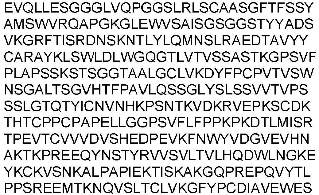

SEQ ID NO: 199 DNA Heavy Chain 
SEQ ID NO: 116 LCDR1 (Combined) RASQSISSYLN SEQ ID NO: 47 LCDR2 (Combined) AASSLQS SEQ ID NO: 200 LCDR3 (Combined) QQVWYAPVT SEQ ID NO: 116 LCDR1 (Kabat) RASQSISSYLN SEQ ID NO: 47 LCDR2 (Kabat) AASSLQS SEQ ID NO: 200 LCDR3 (Kabat) QQVWYAPVT SEQ ID NO: 49 LCDR1 (Chothia) SQSISSY SEQ ID NO: 50 LCDR2 (Chothia) AAS SEQ ID NO: 201 LCDR3 (Chothia) VWYAPV SEQ ID NO: 52 LCDR1 (IMGT) QSISSY SEQ ID NO: 50 LCDR2 (IMGT) AAS SEQ ID NO: 200 LCDR3 (IMGT) QQVWYAPVT SEQ ID NO: 202 VL 
SEQ ID NO: 203 DNA VL 
SEQ ID NO: 204 Light Chain 
SEQ ID NO: 205 DNA Light Chain 
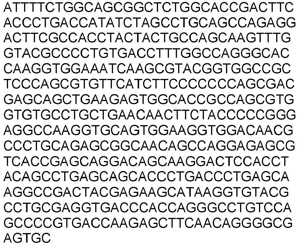
Y010900 E152C_S375C SEQ ID NO: 206 HCDR1 (Combined) GFTFSNAWMS SEQ ID NO: 207 HCDR2 (Combined) RIKSKTDAGTTDYAAPVKG SEQ ID NO: 208 HCDR3 (Combined) TIYPSAPSSSLDY SEQ ID NO: 209 HCDR1 (Kabat) NAWMS SEQ ID NO: 207 HCDR2 (Kabat) RIKSKTDAGTTDYAAPVKG SEQ ID NO: 208 HCDR3 (Kabat) TIYPSAPSSSLDY SEQ ID NO: 210 HCDR1 (Chothia) GFTFSNA SEQ ID NO: 211 HCDR2 (Chothia) KSKTDAGT SEQ ID NO: 208 HCDR3(Ch othia) TIYPSAPSSSLDY SEQ ID NO: 212 HCDR1 (IMGT) GFTFSNAW SEQ ID NO: 213 HCDR2 (IMGT) IKSKTDAGTT SEQ ID NO: 214 HCDR3 (IMGT) ARTIYPSAPSSSLDY SEQ ID NO: 215 VH 
SEQ ID NO: 216 DNA VH 

SEQ ID NO: 217 Heavy Chain 
SEQ ID NO: 218 DNA Heavy Chain 
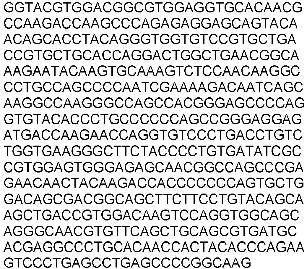
SEQ ID NO: 153 LCDR1 (Combined) RASQGISNYLA SEQ ID NO: 154 LCDR2 (Combined) AASTLQS SEQ ID NO: 219 LCDR3 (Combined) QQLIFFPLT SEQ ID NO: 153 LCDR1 (Kabat) RASQGISNYLA SEQ ID NO: 154 LCDR2 (Kabat) AASTLQS SEQ ID NO: 219 LCDR3 (Kabat) QQLIFFPLT SEQ ID NO: 156 LCDR1 (Chothia) SQGISNY SEQ ID NO: 50 LCDR2 (Chothia) AAS SEQ ID NO: 220 LCDR3 (Chothia) LIFFPL SEQ ID NO: 158 LCDR1 (IMGT) QGISNY SEQ ID NO: 50 LCDR2 (IMGT) AAS SEQ ID NO: 219 LCDR3 (IMGT) QQLIFFPLT SEQ ID NO: 221 VL 
SEQ ID NO: 222 DNA VL 

SEQ ID NO: 223 Light Chain 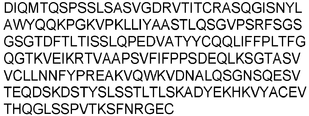
SEQ ID NO: 224 DNA Light Chain 
Y010903 E152C_S375C SEQ ID NO: 206 HCDR1 (Combined) GFTFSNAWMS SEQ ID NO: 207 HCDR2 (Combined) RIKSKTDAGTTDYAAPVKG SEQ ID NO: 225 HCDR3 (Combined) ASHRLHSLFDV SEQ ID NO: 209 HCDR1 (Kabat) NAWMS SEQ ID NO: 207 HCDR2 (Kabat) RIKSKTDAGTTDYAAPVKG SEQ ID NO: 225 HCDR3 (Kabat) ASHRLHSLFDV SEQ ID NO: 210 HCDR1 (Chothia) GFTFSNA SEQ ID NO: 211 HCDR2 (Chothia) KSKTDAGT SEQ ID NO: 225 HCDR3(Ch othia) ASHRLHSLFDV SEQ ID NO: 212 HCDR1 (IMGT) GFTFSNAW SEQ ID NO: 213 HCDR2 (IMGT) IKSKTDAGTT SEQ ID NO: 226 HCDR3 (IMGT) ARASHRLHSLFDV SEQ ID NO: 227 VH 
SEQ ID NO: 228 DNA VH 
SEQ ID NO: 229 Heavy Chain 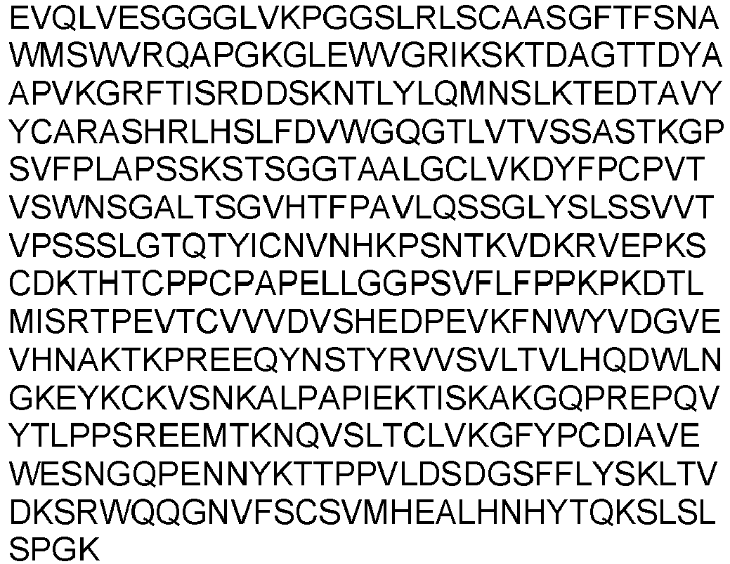
SEQ ID NO: 230 DNA Heavy Chain 

SEQ ID NO: 136 LCDR1 (Combined) RASQSISSWLA SEQ ID NO: 137 LCDR2 (Combined) DASSLES SEQ ID NO: 231 LCDR3 (Combined) QQGLFYPHT SEQ ID NO: 136 LCDR1 (Kabat) RASQSISSWLA SEQ ID NO: 137 LCDR2 (Kabat) DASSLES SEQ ID NO: 231 LCDR3 (Kabat) QQGLFYPHT SEQ ID NO: 139 LCDR1 (Chothia) SQSISSW SEQ ID NO: 140 LCDR2 (Chothia) DAS SEQ ID NO: 232 LCDR3 (Chothia) GLFYPH SEQ ID NO: 142 LCDR1 (IMGT) QSISSW SEQ ID NO: 140 LCDR2 (IMGT) DAS SEQ ID NO: 231 LCDR3 (IMGT) QQGLFYPHT SEQ ID NO: 233 VL 
SEQ ID NO: 234 DNA VL 
SEQ ID NO: 235 Light Chain 
SEQ ID NO: 236 DNA Light Chain 

Y010906 E152C_S375C SEQ ID NO: 206 HCDR1 (Combined) GFTFSNAWMS SEQ ID NO: 207 HCDR2 (Combined) RIKSKTDAGTTDYAAPVKG SEQ ID NO: 237 HCDR3 (Combined) DEYPWGWFDV SEQ ID NO: 209 HCDR1 (Kabat) NAWMS SEQ ID NO: 207 HCDR2 (Kabat) RIKSKTDAGTTDYAAPVKG SEQ ID NO: 237 HCDR3 (Kabat) DEYPWGWFDV SEQ ID NO: 210 HCDR1 (Chothia) GFTFSNA SEQ ID NO: 211 HCDR2 (Chothia) KSKTDAGT SEQ ID NO: 237 HCDR3(Ch othia) DEYPWGWFDV SEQ ID NO: 212 HCDR1 (IMGT) GFTFSNAW SEQ ID NO: 213 HCDR2 (IMGT) IKSKTDAGTT SEQ ID NO: 238 HCDR3 (IMGT) ARDEYPWGWFDV SEQ ID NO: 239 VH 
SEQ ID NO: 240 DNA VH 
SEQ ID NO: 241 Heavy Chain 

SEQ ID NO: 242 DNA Heavy Chain 

SEQ ID NO: 243 LCDR1 (Combined) RASQGISSWLA SEQ ID NO: 47 LCDR2 (Combined) AASSLQS SEQ ID NO: 244 LCDR3 (Combined) QQYIFYPLT SEQ ID NO: 243 LCDR1 (Kabat) RASQGISSWLA SEQ ID NO: 47 LCDR2 (Kabat) AASSLQS SEQ ID NO: 244 LCDR3 (Kabat) QQYIFYPLT SEQ ID NO: 245 LCDR1 (Chothia) SQGISSW SEQ ID NO: 50 LCDR2 (Chothia) AAS SEQ ID NO: 246 LCDR3 (Chothia) YIFYPL SEQ ID NO: 247 LCDR1 (IMGT) QGISSW SEQ ID NO: 50 LCDR2 (IMGT) AAS SEQ ID NO: 244 LCDR3 (IMGT) QQYIFYPLT SEQ ID NO: 248 VL 
SEQ ID NO: 249 DNA VL 
SEQ ID NO: 250 Light Chain 
SEQ ID NO: 251 DNA Light Chain 
Y010910 E152C_S375C SEQ ID NO: 206 HCDR1 (Combined) GFTFSNAWMS SEQ ID NO: 207 HCDR2 (Combined) RIKSKTDAGTTDYAAPVKG SEQ ID NO: 252 HCDR3 (Combined) VASPSAPGGFDY SEQ ID NO: 209 HCDR1 (Kabat) NAWMS SEQ ID NO: 207 HCDR2 (Kabat) RIKSKTDAGTTDYAAPVKG SEQ ID NO: 252 HCDR3 (Kabat) VASPSAPGGFDY SEQ ID NO: 210 HCDR1 (Chothia) GFTFSNA SEQ ID NO: 211 HCDR2 (Chothia) KSKTDAGT SEQ ID NO: 252 HCDR3(Ch othia) VASPSAPGGFDY SEQ ID NO: 212 HCDR1 (IMGT) GFTFSNAW SEQ ID NO: 213 HCDR2 (IMGT) IKSKTDAGTT SEQ ID NO: 253 HCDR3 (IMGT) ARVASPSAPGGFDY SEQ ID NO: 254 VH 

SEQ ID NO: 255 DNA VH 
SEQ ID NO: 256 Heavy Chain 
SEQ ID NO: 257 DNA Heavy Chain 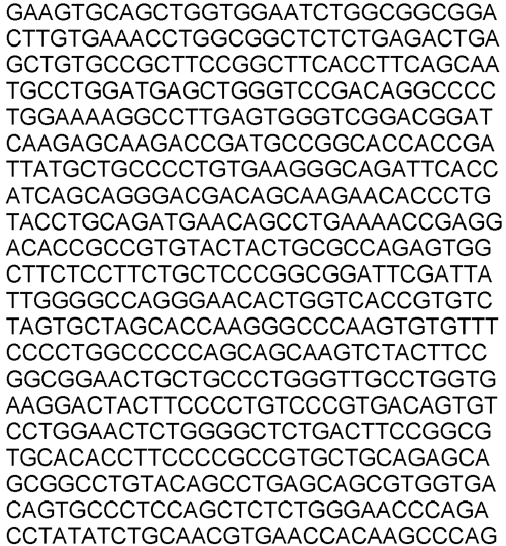
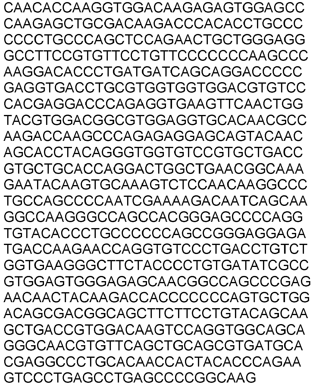
SEQ ID NO: 153 LCDR1 (Combined) RASQGISNYLA SEQ ID NO: 154 LCDR2 (Combined) AASTLQS SEQ ID NO: 258 LCDR3 (Combined) QQSLFAPFT SEQ ID NO: 153 LCDR1 (Kabat) RASQGISNYLA SEQ ID NO: 154 LCDR2 (Kabat) AASTLQS SEQ ID NO: 258 LCDR3 (Kabat) QQSLFAPFT SEQ ID NO: 156 LCDR1 (Chothia) SQGISNY SEQ ID NO: 50 LCDR2 (Chothia) AAS SEQ ID NO: 259 LCDR3 (Chothia) SLFAPF SEQ ID NO: 158 LCDR1 (IMGT) QGISNY SEQ ID NO: 50 LCDR2 (IMGT) AAS SEQ ID NO: 258 LCDR3 (IMGT) QQSLFAPFT SEQ ID NO: 260 VL 

SEQ ID NO: 261 DNA VL 
SEQ ID NO: 262 Light Chain 
SEQ ID NO: 263 DNA Light Chain 
- Other antibodies of the invention include those where the amino acids or nucleic acids encoding the amino acids have been mutated, yet have at least 60, 70, 80, 90 or 95 percent identity to the sequences described in Table 2. In some embodiments, 1, 2, 3, 4 or 5 amino acids have been mutated in the variable regions when compared with the variable regions depicted in the sequence described in Table 2, while retaining substantially the same therapeutic activity as the antibodies listed in Table 2.
- Since each of these antibodies can bind to PMEL17, the VH, VL, full length light chain, and full length heavy chain sequences (amino acid sequences and the nucleotide sequences encoding the amino acid sequences) can be "mixed and matched" to create other PMEL17-binding antibodies of the invention. Such "mixed and matched" PMEL17-binding antibodies can be tested using the binding assays known in the art (e.g., ELISAs, and other assays described in the Example section). When these chains are mixed and matched, a VH sequence from a particular VH/VL pairing should be replaced with a structurally similar VH sequence. Likewise a full length heavy chain sequence from a particular full length heavy chain / full length light chain pairing should be replaced with a structurally similar full length heavy chain sequence. Likewise, a VL sequence from a particular VH/VL pairing should be replaced with a structurally similar VL sequence. Likewise a full length light chain sequence from a particular full length heavy chain / full length light chain pairing should be replaced with a structurally similar full length light chain sequence. Accordingly, in one aspect, the invention provides an isolated monoclonal antibody or antigen binding region thereof having: a heavy chain variable region comprising an amino acid sequence selected from the group consisting of SEQ ID NOs: 10, 42, 64, 88, 112, 132, 149, 165, 184, 196, 215, 227, 239 or 254; and a light chain variable region comprising an amino acid sequence selected from the group consisting of SEQ ID NOs: 21, 25, 29, 53, 75, 99, 119, 143, 159, 171, 190, 202, 221, 233, 248 or 260; wherein the antibody specifically binds to PMEL17.
- In another aspect, the invention provides (i) an isolated monoclonal antibody having: a full length heavy chain comprising an amino acid sequence that has been optimized for expression in the cell of a mammalian expression system selected from the group consisting of SEQ ID NOs: 12, 44, 66, 90, 114, 134, 151, 167, 186, 198, 217, 229, 241 or 256; and a full length light chain comprising an amino acid sequence that has been optimized for expression in the cell of a mammalian selected from the group consisting of SEQ ID NOs: 23, 27, 31, 55, 77, 101, 121, 145, 161, 173, 192, 204, 223, 235, 250 or 262; or (ii) a functional protein comprising an antigen binding portion thereof.
- In another aspect, the present invention provides PMEL17-binding antibodies that comprise the heavy chain and light chain CDR1s, CDR2s and CDR3s as described in Table 2, or combinations thereof. The amino acid sequences of the VH CDR1s of the antibodies are shown, for example, in SEQ ID NOs: 1, 4, 5, 7, 33, 36, 37, 39, 57, 60, 79, 82, 83, 85, 103, 106, 107, 109, 123, 126, 127, 129, 175, 178, 179, 181, 206, 209, 210, and 212. The amino acid sequences of the VH CDR2s of the antibodies and are shown, for example, in SEQ ID NOs: 2, 6, 8, 34, 38, 40, 58, 61, 62, 80, 84, 86, 104, 108, 110, 124, 128, 130, 176, 180, 182, 207, 211, and 213. The amino acid sequences of the VH CDR3s of the antibodies are shown, for example, in SEQ ID NOs: 3, 9, 35, 41, 59, 63, 81, 87, 105, 111, 125, 131, 147, 148, 163, 164, 177, 183, 194, 195, 208, 214, 225, 226, 237, 238, 252, and 253. The amino acid sequences of the VL CDR1s of the antibodies are shown, for example, in SEQ ID NOs: 14, 17, 20, 46, 49, 52, 68, 71, 74, 92, 95, 98, 116, 136, 139, 142, 153, 156, 158, 243, 245, and 247. The amino acid sequences of the VL CDR2s of the antibodies are shown, for example, in SEQ ID Nos: 15, 18, 47, 50, 69, 72, 93, 96, 137, 140, and 154. The amino acid sequences of the VL CDR3s of the antibodies are shown, for example, in SEQ ID NOs: 16, 19, 48, 51, 70, 73, 94, 97, 117, 118, 138, 141, 155, 157, 169, 170188, 189, 200, 201, 219, 220, 231, 232, 244, 246, 258, and 259.
- Given that each of these antibodies can bind to PMEL17 and that antigen-binding specificity is provided primarily by the CDR1, 2 and 3 regions, the VH CDR1, CDR2 and CDR3 sequences and VL CDR1, CDR2 and CDR3 sequences can be "mixed and matched" (i.e., CDRs from different antibodies can be mixed and matched. Such "mixed and matched" PMEL17-binding antibodies can be tested using the binding assays known in the art and those described in the Examples (e.g., ELISAs). When VH CDR sequences are mixed and matched, the CDR1, CDR2 and/or CDR3 sequence from a particular VH sequence should be replaced with a structurally similar CDR sequence(s). Likewise, when VL CDR sequences are mixed and matched, the CDR1, CDR2 and/or CDR3 sequence from a particular VL sequence should be replaced with a structurally similar CDR sequence(s). It will be readily apparent to the ordinarily skilled artisan that novel VH and VL sequences can be created by substituting one or more VH and/or VL CDR region sequences with structurally similar sequences from the CDR sequences shown herein for monoclonal antibodies of the present invention.
- Accordingly, in some embodiments, the present invention provides an isolated monoclonal antibody or antigen binding region thereof comprising a heavy chain CDR1 comprising an amino acid sequence selected from the group consisting of SEQ ID NOs: 1, 4, 5, 7, 33, 36, 37, 39, 57, 60, 79, 82, 83, 85, 103, 106, 107, 109, 123, 126, 127, 129, 175, 178, 179, 181, 206, 209, 210, and 212; a heavy chain CDR2 comprising an amino acid sequence selected from the group consisting of SEQ ID NOs: 2, 6, 8, 34, 38, 40, 58, 61, 62, 80, 84, 86, 104, 108, 110, 124, 128, 130, 176, 180, 182, 207, 211, and 213; a heavy chain CDR3 comprising an amino acid sequence selected from the group consisting of SEQ ID NOs: 3, 9, 35, 41, 59, 63, 81, 87, 105, 111, 125, 131, 147, 148, 163, 164, 177, 183, 194, 195, 208, 214, 225, 226, 237, 238, 252, and 253; a light chain CDR1 comprising an amino acid sequence selected from the group consisting of SEQ ID NOs: 14, 17, 20, 46, 49, 52, 68, 71, 74, 92, 95, 98, 116, 136, 139, 142, 153, 156, 158, 243, 245, and 247; a light chain CDR2 comprising an amino acid sequence selected from the group consisting of SEQ ID NOs: 15, 18, 47, 50, 69, 72, 93, 96, 137, 140, and 154; and a light chain CDR3 comprising an amino acid sequence selected from the group consisting of SEQ ID NOs: 16, 19, 48, 51, 70, 73, 94, 97, 117, 118, 138, 141, 155, 157, 169, 170, 188, 189, 200, 201, 219, 220, 231, 232, 244, 246, 258, and 259; wherein the antibody specifically binds PMEL17.
- In a specific embodiment, an antibody or antibody fragment (e.g., antigen binding fragments) that specifically binds to PMEL17 comprises a heavy chain CDR1 of SEQ ID NO:1, 4, 5 or 7, a heavy chain CDR2 of SEQ ID NO:2, 6 or 8; a heavy chain CDR3 of SEQ ID NO:3 or 9; a light chain CDR1 of SEQ ID NO:14, 17 or 20; a light chain CDR2 of SEQ ID NO:15 or 18; and a light chain CDR3 of SEQ ID NO:16 or 19.
- In a specific embodiment, an antibody or antibody fragment (e.g., antigen binding fragments) that specifically binds to PMEL17 comprises a heavy chain CDR1 of SEQ ID NO:33, 36, 37 or 39, a heavy chain CDR2 of SEQ ID NO:34, 38 or 40; a heavy chain CDR3 of SEQ ID NO:35 or 41; a light chain CDR1 of SEQ ID NO:46, 49 or 52; a light chain CDR2 of SEQ ID NO:47 or 50; and a light chain CDR3 of SEQ ID NO:48 or 51.
- In a specific embodiment, an antibody or antibody fragment (e.g., antigen binding fragments) that specifically binds to PMEL17 comprises a heavy chain CDR1 of SEQ ID NO:5, 7, 57 or 60, a heavy chain CDR2 of SEQ ID NO:58, 61 or 62; a heavy chain CDR3 of SEQ ID NO:59 or 63; a light chain CDR1 of SEQ ID NO:68, 71 or 74; a light chain CDR2 of SEQ ID NO:69 or 72; and a light chain CDR3 of SEQ ID NO:70 or 73.
- In a specific embodiment, an antibody or antibody fragment (e.g., antigen binding fragments) that specifically binds to PMEL17 comprises a heavy chain CDR1 of SEQ ID NO:79, 82, 83 or 85, a heavy chain CDR2 of SEQ ID NO:80, 84 or 86; a heavy chain CDR3 of SEQ ID NO:81 or 87; a light chain CDR1 of SEQ ID NO:92, 95 or 98; a light chain CDR2 of SEQ ID NO:93 or 96; and a light chain CDR3 of SEQ ID NO:94 or 97.
- In a specific embodiment, an antibody or antibody fragment (e.g., antigen binding fragments) that specifically binds to PMEL17 comprises a heavy chain CDR1 of SEQ ID NO:103, 106, 107 or 109, a heavy chain CDR2 of SEQ ID NO:104, 108 or 110; a heavy chain CDR3 of SEQ ID NO:105 or 111; a light chain CDR1 of SEQ ID NO:49, 52 or 116; a light chain CDR2 of SEQ ID NO:47 or 50; and a light chain CDR3 of SEQ ID NO:117 or 118.
- In a specific embodiment, an antibody or antibody fragment (e.g., antigen binding fragments) that specifically binds to PMEL17 comprises a heavy chain CDR1 of SEQ ID NO:123, 126, 127 or 129, a heavy chain CDR2 of SEQ ID NO:124, 128 or 130; a heavy chain CDR3 of SEQ ID NO:125 or 131; a light chain CDR1 of SEQ ID NO:136, 139 or 142; a light chain CDR2 of SEQ ID NO:137 or 140; and a light chain CDR3 of SEQ ID NO:138 or 141.
- In a specific embodiment, an antibody or antibody fragment (e.g., antigen binding fragments) that specifically binds to PMEL17 comprises a heavy chain CDR1 of SEQ ID NO:123, 126, 127 or 129, a heavy chain CDR2 of SEQ ID NO:124, 128 or 130; a heavy chain CDR3 of SEQ ID NO:147 or 148; a light chain CDR1 of SEQ ID NO:153, 156 or 158; a light chain CDR2 of SEQ ID NO:50 or 154; and a light chain CDR3 of SEQ ID NO:155 or 157.
- In a specific embodiment, an antibody or antibody fragment (e.g., antigen binding fragments) that specifically binds to PMEL17 comprises a heavy chain CDR1 of SEQ ID NO:103, 106, 107 or 109, a heavy chain CDR2 of SEQ ID NO:104, 108 or 110; a heavy chain CDR3 of SEQ ID NO:163 or 164; a light chain CDR1 of SEQ ID NO:49, 52 or 116; a light chain CDR2 of SEQ ID NO:47 or 50; and a light chain CDR3 of SEQ ID NO:169 or 170.
- In a specific embodiment, an antibody or antibody fragment (e.g., antigen binding fragments) that specifically binds to PMEL17 comprises a heavy chain CDR1 of SEQ ID NO:175, 178, 179 or 181, a heavy chain CDR2 of SEQ ID NO:176, 180 or 182; a heavy chain CDR3 of SEQ ID NO:177 or 183; a light chain CDR1 of SEQ ID NO:49, 52 or 116; a light chain CDR2 of SEQ ID NO:47 or 50; and a light chain CDR3 of SEQ ID NO:188 or 189.
- In a specific embodiment, an antibody or antibody fragment (e.g., antigen binding fragments) that specifically binds to PMEL17 comprises a heavy chain CDR1 of SEQ ID NO: 103, 106, 107 or 109, a heavy chain CDR2 of SEQ ID NO: 104, 108 or 110; a heavy chain CDR3 of SEQ ID NO:194 or 195; a light chain CDR1 of SEQ ID NO: 49, 52 or 116; a light chain CDR2 of SEQ ID NO: 47 or 50; and a light chain CDR3 of SEQ ID NO:200 or 201.
- In a specific embodiment, an antibody or antibody fragment (e.g., antigen binding fragments) that specifically binds to PMEL17 comprises a heavy chain CDR1 of SEQ ID NO:206, 209, 210 or 212, a heavy chain CDR2 of SEQ ID NO:207, 211 or 213; a heavy chain CDR3 of SEQ ID NO:208 or 214; a light chain CDR1 of SEQ ID NO:153, 156 or 158; a light chain CDR2 of SEQ ID NO:50 or 154; and a light chain CDR3 of SEQ ID NO:219 or 220.
- In a specific embodiment, an antibody or antibody fragment (e.g., antigen binding fragments) that specifically binds to PMEL17 comprises a heavy chain CDR1 of SEQ ID NO: 206, 209, 210 or 212, a heavy chain CDR2 of SEQ ID NO: 207, 211 or 213; a heavy chain CDR3 of SEQ ID NO:225 or 226; a light chain CDR1 of SEQ ID NO:136, 139 or 142; a light chain CDR2 of SEQ ID NO:137 or 140; and a light chain CDR3 of SEQ ID NO:231 or 232.
- In a specific embodiment, an antibody or antibody fragment (e.g., antigen binding fragments) that specifically binds to PMEL17 comprises a heavy chain variable region that comprises an HCDR1 of SEQ ID NO: 206, 209, 210 or 212, an HCDR2 of SEQ ID NO: 207, 211 or 213, and an HCDR3 of SEQ ID NO:237 or 238; and a light chain variable region that comprises an LCDR1 of SEQ ID NO:243, 245 or 247, an LCDR2 of SEQ ID NO:47 or 50, and an LCDR3 of SEQ ID NO:244 or 246.
- In a specific embodiment, an antibody or antibody fragment (e.g., antigen binding fragments) that specifically binds to PMEL17 comprises a heavy chain variable region that comprises an HCDR1 of SEQ ID NO: 206, 209, 210 or 212, an HCDR2 of SEQ ID NO: 207, 211 or 213, and an HCDR3 of SEQ ID NO:252 or 253; and a light chain variable region that comprises an LCDR1 of SEQ ID NO:153, 156 or 158, an LCDR2 of SEQ ID NO:50 or 154, and an LCDR3 of SEQ ID NO:258 or 259.
- In a specific embodiment, an antibody or antibody fragment (e.g., antigen binding fragments) that specifically binds to PMEL17 comprises CDR sequences selected from:
- a) a heavy chain CDR1 of SEQ ID NO:1, , a heavy chain CDR2 of SEQ ID NO:2, a heavy chain CDR3 of SEQ ID NO:3, a light chain CDR1 of SEQ ID NO:14, a light chain CDR2 of SEQ ID NO:15, and a light chain CDR3 of SEQ ID NO:16;
- b) a heavy chain CDR1 of SEQ ID NO: 4, a heavy chain CDR2 of SEQ ID NO:2, a heavy chain CDR3 of SEQ ID NO:3, a light chain CDR1 of SEQ ID NO:14, a light chain CDR2 of SEQ ID NO:15, and a light chain CDR3 of SEQ ID NO:16;
- c) a heavy chain CDR1 of SEQ ID NO:5, a heavy chain CDR2 of SEQ ID NO:6, a heavy chain CDR3 of SEQ ID NO:3, a light chain CDR1 of SEQ ID NO:17, a light chain CDR2 of SEQ ID NO: 18, and a light chain CDR3 of SEQ ID NO: 19; or
- d) a heavy chain CDR1 of SEQ ID NO:7, a heavy chain CDR2 of SEQ ID NO:8, a heavy chain CDR3 of SEQ ID NO:9, a light chain CDR1 of SEQ ID NO:20, a light chain CDR2 of SEQ ID NO:18, and a light chain CDR3 of SEQ ID NO:16.
- In a specific embodiment, an antibody or antibody fragment (e.g., antigen binding fragments) that specifically binds to PMEL17 comprises CDR sequences selected from:
- a) a heavy chain CDR1 of SEQ ID NO:33, a heavy chain CDR2 of SEQ ID NO:34, a heavy chain CDR3 of SEQ ID NO:35, a light chain CDR1 of SEQ ID NO:46, a light chain CDR2 of SEQ ID NO:47, and a light chain CDR3 of SEQ ID NO:48;
- b) a heavy chain CDR1 of SEQ ID NO:36, a heavy chain CDR2 of SEQ ID NO:34, a heavy chain CDR3 of SEQ ID NO:35, a light chain CDR1 of SEQ ID NO:46, a light chain CDR2 of SEQ ID NO:47, and a light chain CDR3 of SEQ ID NO:48;
- c) a heavy chain CDR1 of SEQ ID NO:37, a heavy chain CDR2 of SEQ ID NO:38, a heavy chain CDR3 of SEQ ID NO:35, a light chain CDR1 of SEQ ID NO:49, a light chain CDR2 of SEQ ID NO:50, and a light chain CDR3 of SEQ ID NO:51; or
- d) a heavy chain CDR1 of SEQ ID NO: 39, a heavy chain CDR2 of SEQ ID NO:40, a heavy chain CDR3 of SEQ ID NO:41, a light chain CDR1 of SEQ ID NO:52, a light chain CDR2 of SEQ ID NO:50, and a light chain CDR3 of SEQ ID NO:48.
- In a specific embodiment, an antibody or antibody fragment (e.g., antigen binding fragments) that specifically binds to PMEL17 comprises CDR sequences selected from:
- a) a heavy chain CDR1 of SEQ ID NO:57, a heavy chain CDR2 of SEQ ID NO:58, a heavy chain CDR3 of SEQ ID NO:59, a light chain CDR1 of SEQ ID NO:68, a light chain CDR2 of SEQ ID NO:69, and a light chain CDR3 of SEQ ID NO:70;
- b) a heavy chain CDR1 of SEQ ID NO:60, a heavy chain CDR2 of SEQ ID NO:58, a heavy chain CDR3 of SEQ ID NO:59, a light chain CDR1 of SEQ ID NO:68, a light chain CDR2 of SEQ ID NO:69, and a light chain CDR3 of SEQ ID NO:70;
- c) a heavy chain CDR1 of SEQ ID NO:5, a heavy chain CDR2 of SEQ ID NO:61, a heavy chain CDR3 of SEQ ID NO:59, a light chain CDR1 of SEQ ID NO:71, a light chain CDR2 of SEQ ID NO:72, and a light chain CDR3 of SEQ ID NO:73; or
- d) a heavy chain CDR1 of SEQ ID NO:7, a heavy chain CDR2 of SEQ ID NO:62, a heavy chain CDR3 of SEQ ID NO:63, a light chain CDR1 of SEQ ID NO:74, a light chain CDR2 of SEQ ID NO:72, and a light chain CDR3 of SEQ ID NO:70.
- In a specific embodiment, an antibody or antibody fragment (e.g., antigen binding fragments) that specifically binds to PMEL17 comprises CDR sequences selected from:
- a) a heavy chain CDR1 of SEQ ID NO:79, , a heavy chain CDR2 of SEQ ID NO:80, a heavy chain CDR3 of SEQ ID NO:81, a light chain CDR1 of SEQ ID NO:92, a light chain CDR2 of SEQ ID NO:93, and a light chain CDR3 of SEQ ID NO:94;
- b) a heavy chain CDR1 of SEQ ID NO:82, a heavy chain CDR2 of SEQ ID NO:80, a heavy chain CDR3 of SEQ ID NO:81, a light chain CDR1 of SEQ ID NO:92, a light chain CDR2 of SEQ ID NO:93, and a light chain CDR3 of SEQ ID NO:94;
- c) a heavy chain CDR1 of SEQ ID NO:83, a heavy chain CDR2 of SEQ ID NO:84, a heavy chain CDR3 of SEQ ID NO:81, a light chain CDR1 of SEQ ID NO:95, a light chain CDR2 of SEQ ID NO:96, and a light chain CDR3 of SEQ ID NO: 97; or
- d) a heavy chain CDR1 of SEQ ID NO: 85, a heavy chain CDR2 of SEQ ID NO:86, a heavy chain CDR3 of SEQ ID NO:87, a light chain CDR1 of SEQ ID NO:98, a light chain CDR2 of SEQ ID NO:96, and a light chain CDR3 of SEQ ID NO:94.
- In a specific embodiment, an antibody or antibody fragment (e.g., antigen binding fragments) that specifically binds to PMEL17 comprises CDR sequences selected from:
- a) a heavy chain CDR1 of SEQ ID NO:103, a heavy chain CDR2 of SEQ ID NO:104, a heavy chain CDR3 of SEQ ID NO:105, a light chain CDR1 of SEQ ID NO: 116; a light chain CDR2 of SEQ ID NO:47; and a light chain CDR3 of SEQ ID NO:117;
- b) a heavy chain CDR1 of SEQ ID NO:106, a heavy chain CDR2 of SEQ ID NO:104, a heavy chain CDR3 of SEQ ID NO:105, a light chain CDR1 of SEQ ID NO: 116, a light chain CDR2 of SEQ ID NO:47, and a light chain CDR3 of SEQ ID NO:117;
- c) a heavy chain CDR1 of SEQ ID NO:107, a heavy chain CDR2 of SEQ ID NO:108, a heavy chain CDR3 of SEQ ID NO:105, a light chain CDR1 of SEQ ID NO:49, a light chain CDR2 of SEQ ID NO:50, and a light chain CDR3 of SEQ ID NO:118; or
- d) a heavy chain CDR1 of SEQ ID NO:109, a heavy chain CDR2 of SEQ ID NO:110, a heavy chain CDR3 of SEQ ID NO:111, a light chain CDR1 of SEQ ID NO:52 a light chain CDR2 of SEQ ID NO:50, and a light chain CDR3 of SEQ ID NO:117.
- In a specific embodiment, an antibody or antibody fragment (e.g., antigen binding fragments) that specifically binds to PMEL17 comprises CDR sequences selected from:
- a) a heavy chain CDR1 of SEQ ID NO:123, a heavy chain CDR2 of SEQ ID NO:124, a heavy chain CDR3 of SEQ ID NO:125, a light chain CDR1 of SEQ ID NO:136, a light chain CDR2 of SEQ ID NO:137,and a light chain CDR3 of SEQ ID NO:138;
- b) a heavy chain CDR1 of SEQ ID NO:126, a heavy chain CDR2 of SEQ ID NO:124, a heavy chain CDR3 of SEQ ID NO:125, a light chain CDR1 of SEQ ID NO:136, a light chain CDR2 of SEQ ID NO:137, and a light chain CDR3 of SEQ ID NO:138;
- c) a heavy chain CDR1 of SEQ ID NO:127, a heavy chain CDR2 of SEQ ID NO:128, a heavy chain CDR3 of SEQ ID NO:125, a light chain CDR1 of SEQ ID NO:139, a light chain CDR2 of SEQ ID NO:140, and a light chain CDR3 of SEQ ID NO: 141; or
- d) a heavy chain CDR1 of SEQ ID NO: 129, a heavy chain CDR2 of SEQ ID NO:130, a heavy chain CDR3 of SEQ ID NO:131, a light chain CDR1 of SEQ ID NO:142, a light chain CDR2 of SEQ ID NO:140, and a light chain CDR3 of SEQ ID NO:138.
- In a specific embodiment, an antibody or antibody fragment (e.g., antigen binding fragments) that specifically binds to PMEL17 comprises CDR sequences selected from:
- a) a heavy chain CDR1 of SEQ ID NO:123, a heavy chain CDR2 of SEQ ID NO:124, a heavy chain CDR3 of SEQ ID NO:147, a light chain CDR1 of SEQ ID NO:153, a light chain CDR2 of SEQ ID NO:154, and a light chain CDR3 of SEQ ID NO:155;
- b) a heavy chain CDR1 of SEQ ID NO:126, a heavy chain CDR2 of SEQ ID NO:124, a heavy chain CDR3 of SEQ ID NO:147, a light chain CDR1 of SEQ ID NO:153, a light chain CDR2 of SEQ ID NO: 154, and a light chain CDR3 of SEQ ID NO:155;
- c) a heavy chain CDR1 of SEQ ID NO:127, a heavy chain CDR2 of SEQ ID NO:128, a heavy chain CDR3 of SEQ ID NO:147, a light chain CDR1 of SEQ ID NO:156, a light chain CDR2 of SEQ ID NO:50, and a light chain CDR3 of SEQ ID NO:157; or
- d) a heavy chain CDR1 of SEQ ID NO: 129, a heavy chain CDR2 of SEQ ID NO:130, a heavy chain CDR3 of SEQ ID NO:148, a light chain CDR1 of SEQ ID NO:158, a light chain CDR2 of SEQ ID NO:50, and a light chain CDR3 of SEQ ID NO:155.
- In a specific embodiment, an antibody or antibody fragment (e.g., antigen binding fragments) that specifically binds to PMEL17 comprises CDR sequences selected from:
- a) a heavy chain CDR1 of SEQ ID NO:103, a heavy chain CDR2 of SEQ ID NO:104, a heavy chain CDR3 of SEQ ID NO:163, a light chain CDR1 of SEQ ID NO: 116, a light chain CDR2 of SEQ ID NO:47, and a light chain CDR3 of SEQ ID NO:169;
- b) a heavy chain CDR1 of SEQ ID NO:106, a heavy chain CDR2 of SEQ ID NO:104, a heavy chain CDR3 of SEQ ID NO:163, a light chain CDR1 of SEQ ID NO:116, a light chain CDR2 of SEQ ID NO:47, and a light chain CDR3 of SEQ ID NO:169;
- c) a heavy chain CDR1 of SEQ ID NO:107, a heavy chain CDR2 of SEQ ID NO:108, a heavy chain CDR3 of SEQ ID NO:163, a light chain CDR1 of SEQ ID NO:49, a light chain CDR2 of SEQ ID NO:50, and a light chain CDR3 of SEQ ID NO:170; or
- d) a heavy chain CDR1 of SEQ ID NO: 109, a heavy chain CDR2 of SEQ ID NO:110, a heavy chain CDR3 of SEQ ID NO:164, a light chain CDR1 of SEQ ID NO:52, a light chain CDR2 of SEQ ID NO:50, and a light chain CDR3 of SEQ ID NO:169.
- In a specific embodiment, an antibody or antibody fragment (e.g., antigen binding fragments) that specifically binds to PMEL17 comprises CDR sequences selection from:
- a) a heavy chain CDR1 of SEQ ID NO:175, a heavy chain CDR2 of SEQ ID NO:176, a heavy chain CDR3 of SEQ ID NO:177, a light chain CDR1 of SEQ ID NO:116, a light chain CDR2 of SEQ ID NO:47, and a light chain CDR3 of SEQ ID NO:188;
- b) a heavy chain CDR1 of SEQ ID NO:178, a heavy chain CDR2 of SEQ ID NO:176, a heavy chain CDR3 of SEQ ID NO:177, a light chain CDR1 of SEQ ID NO:116, a light chain CDR2 of SEQ ID NO:47, and a light chain CDR3 of SEQ ID NO:188;
- c) a heavy chain CDR1 of SEQ ID NO:179, a heavy chain CDR2 of SEQ ID NO:180, a heavy chain CDR3 of SEQ ID NO:177, a light chain CDR1 of SEQ ID NO:49, a light chain CDR2 of SEQ ID NO:50, and a light chain CDR3 of SEQ ID NO:189; or
- d) a heavy chain CDR1 of SEQ ID NO: 181, a heavy chain CDR2 of SEQ ID NO:182; a heavy chain CDR3 of SEQ ID NO:183, a light chain CDR1 of SEQ ID NO:52, a light chain CDR2 of SEQ ID NO:50, and a light chain CDR3 of SEQ ID NO:188.
- In a specific embodiment, an antibody or antibody fragment (e.g., antigen binding fragments) that specifically binds to PMEL17 comprises CDR sequences selected from:
- a) a heavy chain CDR1 of SEQ ID NO: 103, a heavy chain CDR2 of SEQ ID NO: 104, a heavy chain CDR3 of SEQ ID NO:194, a light chain CDR1 of SEQ ID NO: 116, a light chain CDR2 of SEQ ID NO: 47, and a light chain CDR3 of SEQ ID NO:200;
- b) a heavy chain CDR1 of SEQ ID NO: 106, a heavy chain CDR2 of SEQ ID NO: 104, a heavy chain CDR3 of SEQ ID NO:194, a light chain CDR1 of SEQ ID NO: 116, a light chain CDR2 of SEQ ID NO: 47, and a light chain CDR3 of SEQ ID NO:200;
- c) a heavy chain CDR1 of SEQ ID NO: 107, a heavy chain CDR2 of SEQ ID NO: 108, a heavy chain CDR3 of SEQ ID NO:194, a light chain CDR1 of SEQ ID NO: 49, a light chain CDR2 of SEQ ID NO: 50, and a light chain CDR3 of SEQ ID NO: 201; or
- d) a heavy chain CDR1 of SEQ ID NO: 109, a heavy chain CDR2 of SEQ ID NO: 110, a heavy chain CDR3 of SEQ ID NO:195, a light chain CDR1 of SEQ ID NO: 52, a light chain CDR2 of SEQ ID NO: 50, and a light chain CDR3 of SEQ ID NO:200.
- In a specific embodiment, an antibody or antibody fragment (e.g., antigen binding fragments) that specifically binds to PMEL17 comprises CDR sequences selected from:
- a) a heavy chain CDR1 of SEQ ID NO:206, a heavy chain CDR2 of SEQ ID NO:207, a heavy chain CDR3 of SEQ ID NO:208, a light chain CDR1 of SEQ ID NO:153, a light chain CDR2 of SEQ ID NO:154, and a light chain CDR3 of SEQ ID NO:219;
- b) a heavy chain CDR1 of SEQ ID NO:209, a heavy chain CDR2 of SEQ ID NO:207, a heavy chain CDR3 of SEQ ID NO:208, a light chain CDR1 of SEQ ID NO:153, a light chain CDR2 of SEQ ID NO: 154, and a light chain CDR3 of SEQ ID NO:219;
- c) a heavy chain CDR1 of SEQ ID NO:210, a heavy chain CDR2 of SEQ ID NO:211, a heavy chain CDR3 of SEQ ID NO:208, a light chain CDR1 of SEQ ID NO:156, a light chain CDR2 of SEQ ID NO:50, and a light chain CDR3 of SEQ ID NO:220; or
- d) a heavy chain CDR1 of SEQ ID NO: 212, a heavy chain CDR2 of SEQ ID NO:213, a heavy chain CDR3 of SEQ ID NO:214, a light chain CDR1 of SEQ ID NO:158, a light chain CDR2 of SEQ ID NO:50, and a light chain CDR3 of SEQ ID NO:219.
- In a specific embodiment, an antibody or antibody fragment (e.g., antigen binding fragments) that specifically binds to PMEL17 comprises CDR sequences selected from:
- a) a heavy chain CDR1 of SEQ ID NO: 206, a heavy chain CDR2 of SEQ ID NO: 207, a heavy chain CDR3 of SEQ ID NO:225, a light chain CDR1 of SEQ ID NO:136, a light chain CDR2 of SEQ ID NO:137,and a light chain CDR3 of SEQ ID NO:231;
- b) a heavy chain CDR1 of SEQ ID NO: 209, a heavy chain CDR2 of SEQ ID NO: 207, a heavy chain CDR3 of SEQ ID NO:225, a light chain CDR1 of SEQ ID NO:136, a light chain CDR2 of SEQ ID NO:137, and a light chain CDR3 of SEQ ID NO:231;
- c) a heavy chain CDR1 of SEQ ID NO: 210, a heavy chain CDR2 of SEQ ID NO: 211, a heavy chain CDR3 of SEQ ID NO:225, a light chain CDR1 of SEQ ID NO:139, a light chain CDR2 of SEQ ID NO:140, and a light chain CDR3 of SEQ ID NO: 232; or
- d) a heavy chain CDR1 of SEQ ID NO: 212, a heavy chain CDR2 of SEQ ID NO: 213, a heavy chain CDR3 of SEQ ID NO: 226, a light chain CDR1 of SEQ ID NO:142; a light chain CDR2 of SEQ ID NO: 140; and a light chain CDR3 of SEQ ID NO:231.
- In a specific embodiment, an antibody or antibody fragment (e.g., antigen binding fragments) that specifically binds to PMEL17 comprises CDR sequences selected from:
- a) a heavy chain variable region that comprises an HCDR1 of SEQ ID NO: 206, an HCDR2 of SEQ ID NO: 207, and an HCDR3 of SEQ ID NO:237, and a light chain variable region that comprises an LCDR1 of SEQ ID NO:243, an LCDR2 of SEQ ID NO:47, and an LCDR3 of SEQ ID NO:244;
- b) a heavy chain variable region that comprises an HCDR1 of SEQ ID NO: 209, an HCDR2 of SEQ ID NO: 207, and an HCDR3 of SEQ ID NO:237, and a light chain variable region that comprises an LCDR1 of SEQ ID NO:243, an LCDR2 of SEQ ID NO:47, and an LCDR3 of SEQ ID NO:244;
- c) a heavy chain variable region that comprises an HCDR1 of SEQ ID NO: 210, an HCDR2 of SEQ ID NO: 211, and an HCDR3 of SEQ ID NO:237, and a light chain variable region that comprises an LCDR1 of SEQ ID NO:245, an LCDR2 of SEQ ID NO:50, and an LCDR3 of SEQ ID NO:246; or
- d) a heavy chain variable region that comprises an HCDR1 of SEQ ID NO: 212, an HCDR2 of SEQ ID NO: 213, and an HCDR3 of SEQ ID NO:238; and a light chain variable region that comprises an LCDR1 of SEQ ID NO:247, an LCDR2 of SEQ ID NO: 50, and an LCDR3 of SEQ ID NO:244.
- In a specific embodiment, an antibody or antibody fragment (e.g., antigen binding fragments) that specifically binds to PMEL17 comprises CDR sequences selected from:
- a) a heavy chain variable region that comprises an HCDR1 of SEQ ID NO: 206, an HCDR2 of SEQ ID NO: 207, and an HCDR3 of SEQ ID NO:252, and a light chain variable region that comprises an LCDR1 of SEQ ID NO:153, an LCDR2 of SEQ ID NO: 154, and an LCDR3 of SEQ ID NO:258;
- b) a heavy chain variable region that comprises an HCDR1 of SEQ ID NO: 209, an HCDR2 of SEQ ID NO: 207, and an HCDR3 of SEQ ID NO:252, and a light chain variable region that comprises an LCDR1 of SEQ ID NO:153, an LCDR2 of SEQ ID NO:154, and an LCDR3 of SEQ ID NO:258;
- c) a heavy chain variable region that comprises an HCDR1 of SEQ ID NO: 210, an HCDR2 of SEQ ID NO: 211, and an HCDR3 of SEQ ID NO:252, and a light chain variable region that comprises an LCDR1 of SEQ ID NO:156, an LCDR2 of SEQ ID NO:50, and an LCDR3 of SEQ ID NO:259; or
- d) a heavy chain variable region that comprises an HCDR1 of SEQ ID NO: 212, an HCDR2 of SEQ ID NO: 213, and an HCDR3 of SEQ ID NO: 253; and a light chain variable region that comprises an LCDR1 of SEQ ID NO:158, an LCDR2 of SEQ ID NO:50, and an LCDR3 of SEQ ID NO:258.
- In a specific embodiment, an antibody or antibody fragment (e.g., antigen binding fragments) that specifically binds to PMEL17 comprises a heavy chain variable region (VH) comprising the amino acid sequence of SEQ ID NO:10, and a light chain variable region (VL) comprising the amino acid sequence of SEQ ID NO:21.
- In a specific embodiment, an antibody or antibody fragment (e.g., antigen binding fragments) that specifically binds to PMEL17 comprises a heavy chain variable region (VH) comprising the amino acid sequence of SEQ ID NO:10, and a light chain variable region (VL) comprising the amino acid sequence of SEQ ID NO:25.
- In a specific embodiment, an antibody or antibody fragment (e.g., antigen binding fragments) that specifically binds to PMEL17 comprises a heavy chain variable region (VH) comprising the amino acid sequence of SEQ ID NO:10, and a light chain variable region (VL) comprising the amino acid sequence of SEQ ID NO:29.
- In a specific embodiment, an antibody or antibody fragment (e.g., antigen binding fragments) that specifically binds to PMEL17 comprises a heavy chain variable region (VH) comprising the amino acid sequence of SEQ ID NO:42, and a light chain variable region (VL) comprising the amino acid sequence of SEQ ID NO:53.
- In a specific embodiment, an antibody or antibody fragment (e.g., antigen binding fragments) that specifically binds to PMEL17 comprises a heavy chain variable region (VH) comprising the amino acid sequence of SEQ ID NO:64, and a light chain variable region (VL) comprising the amino acid sequence of SEQ ID NO:75.
- In a specific embodiment, an antibody or antibody fragment (e.g., antigen binding fragments) that specifically binds to PMEL17 comprises a heavy chain variable region (VH) comprising the amino acid sequence of SEQ ID NO:88, and a light chain variable region (VL) comprising the amino acid sequence of SEQ ID NO:99.
- In a specific embodiment, an antibody or antibody fragment (e.g., antigen binding fragments) that specifically binds to PMEL17 comprises a heavy chain variable region (VH) comprising the amino acid sequence of SEQ ID NO:112, and a light chain variable region (VL) comprising the amino acid sequence of SEQ ID NO:119.
- In a specific embodiment, an antibody or antibody fragment (e.g., antigen binding fragments) that specifically binds to PMEL17 comprises a heavy chain variable region (VH) comprising the amino acid sequence of SEQ ID NO:132, and a light chain variable region (VL) comprising the amino acid sequence of SEQ ID NO:143.
- In a specific embodiment, an antibody or antibody fragment (e.g., antigen binding fragments) that specifically binds to PMEL17 comprises a heavy chain variable region (VH) comprising the amino acid sequence of SEQ ID NO:149, and a light chain variable region (VL) comprising the amino acid sequence of SEQ ID NO:159.
- In a specific embodiment, an antibody or antibody fragment (e.g., antigen binding fragments) that specifically binds to PMEL17 comprises a heavy chain variable region (VH) comprising the amino acid sequence of SEQ ID NO:165, and a light chain variable region (VL) comprising the amino acid sequence of SEQ ID NO:171.
- In a specific embodiment, an antibody or antibody fragment (e.g., antigen binding fragments) that specifically binds to PMEL17 comprises a heavy chain variable region (VH) comprising the amino acid sequence of SEQ ID NO:184, and a light chain variable region (VL) comprising the amino acid sequence of SEQ ID NO:190.
- In a specific embodiment, an antibody or antibody fragment (e.g., antigen binding fragments) that specifically binds to PMEL17 comprises a heavy chain variable region (VH) comprising the amino acid sequence of SEQ ID NO:196, and a light chain variable region (VL) comprising the amino acid sequence of SEQ ID NO:202.
- In a specific embodiment, an antibody or antibody fragment (e.g., antigen binding fragments) that specifically binds to PMEL17 comprises a heavy chain variable region (VH) comprising the amino acid sequence of SEQ ID NO:215, and a light chain variable region (VL) comprising the amino acid sequence of SEQ ID NO:221.
- In a specific embodiment, an antibody or antibody fragment (e.g., antigen binding fragments) that specifically binds to PMEL17 comprises a heavy chain variable region (VH) comprising the amino acid sequence of SEQ ID NO:227, and a light chain variable region (VL) comprising the amino acid sequence of SEQ ID NO:233.
- In a specific embodiment, an antibody or antibody fragment (e.g., antigen binding fragments) that specifically binds to PMEL17 comprises a heavy chain variable region (VH) comprising the amino acid sequence of SEQ ID NO:239, and a light chain variable region (VL) comprising the amino acid sequence of SEQ ID NO:248.
- In a specific embodiment, an antibody or antibody fragment (e.g., antigen binding fragments) that specifically binds to PMEL17 comprises a heavy chain variable region (VH) comprising the amino acid sequence of SEQ ID NO:254, and a light chain variable region (VL) comprising the amino acid sequence of SEQ ID NO:260.
- In a specific embodiment, an antibody or antibody fragment (e.g., antigen binding fragments) that specifically binds to PMEL17 comprises a heavy chain comprising the amino acid sequence of SEQ ID NO:12, and a light chain comprising the amino acid sequence of SEQ ID NO:23.
- In a specific embodiment, an antibody or antibody fragment (e.g., antigen binding fragments) that specifically binds to PMEL17 comprises a heavy chain comprising the amino acid sequence of SEQ ID NO:12, and a light chain comprising the amino acid sequence of SEQ ID NO:27.
- In a specific embodiment, an antibody or antibody fragment (e.g., antigen binding fragments) that specifically binds to PMEL17 comprises a heavy chain comprising the amino acid sequence of SEQ ID NO:12, and a light chain comprising the amino acid sequence of SEQ ID NO:31.
- In a specific embodiment, an antibody or antibody fragment (e.g., antigen binding fragments) that specifically binds to PMEL17 comprises a heavy chain comprising the amino acid sequence of SEQ ID NO:44, and a light chain comprising the amino acid sequence of SEQ ID NO:55.
- In a specific embodiment, an antibody or antibody fragment (e.g., antigen binding fragments) that specifically binds to PMEL17 comprises a heavy chain comprising the amino acid sequence of SEQ ID NO:66, and a light chain comprising the amino acid sequence of SEQ ID NO:77.
- In a specific embodiment, an antibody or antibody fragment (e.g., antigen binding fragments) that specifically binds to PMEL17 comprises a heavy chain comprising the amino acid sequence of SEQ ID NO:90, and a light chain comprising the amino acid sequence of SEQ ID NO:101.
- In a specific embodiment, an antibody or antibody fragment (e.g., antigen binding fragments) that specifically binds to PMEL17 comprises a heavy chain comprising the amino acid sequence of SEQ ID NO:114, and a light chain comprising the amino acid sequence of SEQ ID NO:121.
- In a specific embodiment, an antibody or antibody fragment (e.g., antigen binding fragments) that specifically binds to PMEL17 comprises a heavy chain comprising the amino acid sequence of SEQ ID NO:134, and a light chain comprising the amino acid sequence of SEQ ID NO:145.
- In a specific embodiment, an antibody or antibody fragment (e.g., antigen binding fragments) that specifically binds to PMEL17 comprises a heavy chain comprising the amino acid sequence of SEQ ID NO:151, and a light chain comprising the amino acid sequence of SEQ ID NO:161.
- In a specific embodiment, an antibody or antibody fragment (e.g., antigen binding fragments) that specifically binds to PMEL17 comprises a heavy chain comprising the amino acid sequence of SEQ ID NO:167, and a light chain comprising the amino acid sequence of SEQ ID NO:173.
- In a specific embodiment, an antibody or antibody fragment (e.g., antigen binding fragments) that specifically binds to PMEL17 comprises a heavy chain comprising the amino acid sequence of SEQ ID NO:186, and a light chain comprising the amino acid sequence of SEQ ID NO:192.
- In a specific embodiment, an antibody or antibody fragment (e.g., antigen binding fragments) that specifically binds to PMEL17 comprises a heavy chain comprising the amino acid sequence of SEQ ID NO:198, and a light chain comprising the amino acid sequence of SEQ ID NO:204.
- In a specific embodiment, an antibody or antibody fragment (e.g., antigen binding fragments) that specifically binds to PMEL17 comprises a heavy chain comprising the amino acid sequence of SEQ ID NO:217, and a light chain comprising the amino acid sequence of SEQ ID NO:223.
- In a specific embodiment, an antibody or antibody fragment (e.g., antigen binding fragments) that specifically binds to PMEL17 comprises a heavy chain comprising the amino acid sequence of SEQ ID NO:229, and a light chain comprising the amino acid sequence of SEQ ID NO:235.
- In a specific embodiment, an antibody or antibody fragment (e.g., antigen binding fragments) that specifically binds to PMEL17 comprises a heavy chain comprising the amino acid sequence of SEQ ID NO:241, and a light chain comprising the amino acid sequence of SEQ ID NO:250.
- In a specific embodiment, an antibody or antibody fragment (e.g., antigen binding fragments) that specifically binds to PMEL17 comprises a heavy chain comprising the amino acid sequence of SEQ ID NO:256, and a light chain comprising the amino acid sequence of SEQ ID NO:262.
- In certain embodiments, an antibody that specifically binds to PMEL17 is an antibody or antibody fragment (e.g., antigen binding fragment) that is described in Table 2.
- The present invention also provides antibodies and antibody fragments (e.g., antigen binding fragments) that specifically bind to the same epitope as the anti-PMEL17 antibodies described in Table 2, or cross compete with the antibodies described in Table 2. Additional antibodies and antibody fragments (e.g., antigen binding fragments) can therefore be identified based on their ability to cross-compete (e.g., to competitively inhibit the binding of, in a statistically significant manner) with other antibodies of the invention in PMEL17 binding assays, for example, via BIACORE or assays known to persons skilled in the art for measuring binding. The ability of a test antibody to inhibit the binding of antibodies and antibody fragments (e.g., antigen binding fragments) of the present invention to a PMEL17 (e.g., human PMEL17) demonstrates that the test antibody can compete with that antibody or antibody fragment (e.g., antigen binding fragments) for binding to PMEL17; such an antibody may, according to nonlimiting theory, bind to the same or a related (e.g., a structurally similar or spatially proximal or overlapping) epitope on the PMEL17 protein as the antibody or antibody fragment (e.g., antigen binding fragments) with which it competes. In certain embodiments, the antibodies that bind to the same epitope on PMEL17 as the antibodies or antibody fragments (e.g., antigen binding fragments) described in Table 2 are human or humanized monoclonal antibodies. Such human or humanized monoclonal antibodies can be prepared and isolated as described herein.
- The immunoconjugates of the invention may comprise modified antibodies or antigen binding fragments thereof that further comprise modifications to framework residues within VH and/or VL, e.g. to improve the properties of the antibody. In some embodiments, the framework modifications are made to decrease the immunogenicity of the antibody. For example, one approach is to "back-mutate" one or more framework residues to the corresponding germline sequence. More specifically, an antibody that has undergone somatic mutation may contain framework residues that differ from the germline sequence from which the antibody is derived. Such residues can be identified by comparing the antibody framework sequences to the germline sequences from which the antibody is derived. To return the framework region sequences to their germline configuration, the somatic mutations can be "back-mutated" to the germline sequence by, for example, site-directed mutagenesis. Such "back-mutated" antibodies are also intended to be encompassed by the invention.
- Another type of framework modification involves mutating one or more residues within the framework region, or even within one or more CDR regions, to remove T-cell epitopes to thereby reduce the potential immunogenicity of the antibody. This approach is also referred to as "deimmunization" and is described in further detail in
U.S. Patent Publication No. 20030153043 by Carr et al. - In addition or in the alternative to modifications made within the framework or CDR regions, antibodies of the invention may be engineered to include modifications within the Fc region, typically to alter one or more functional properties of the antibody, such as serum half-life, complement fixation, Fc receptor binding, and/or antigen-dependent cellular cytotoxicity (ADCC). Furthermore, an antibody of the invention may be chemically modified (e.g., one or more chemical moieties can be attached to the antibody) or be modified to alter its glycosylation, again to alter one or more functional properties of the antibody. Each of these embodiments is described in further detail below.
- In one embodiment, the hinge region of CH1 is modified such that the number of cysteine residues in the hinge region is altered, e.g., increased or decreased. This approach is described further in
U.S. Patent No. 5,677,425 by Bodmer et al. The number of cysteine residues in the hinge region of CH1 is altered to, for example, facilitate assembly of the light and heavy chains or to increase or decrease the stability of the antibody. - In some embodiments, the antibody or antibody fragment disclosed herein include modified or engineered amino acid residues, e.g., one or more cysteine residues, as sites for conjugation to a drug moiety (Junutula JR, et al., Nat Biotechnol 2008, 26:925-932). In one embodiment, the invention provides a modified antibody or antibody fragment comprising a substitution of one or more amino acids with cysteine at the positions described herein. Sites for cysteine substitution are in the constant regions of the antibody or antibody fragment and are thus applicable to a variety of antibody or antibody fragment, and the sites are selected to provide stable and homogeneous conjugates. A modified antibody or fragment can have one, two or more cysteine substitutions, and these substitutions can be used in combination with other modification and conjugation methods as described herein. Methods for inserting cysteine at specific locations of an antibody are known in the art, see, e.g., Lyons et al., (1990) Protein Eng., 3:703-708,
WO 2011/005481 ,WO2014/124316 ,WO 2015/138615 . In certain embodiments, a modified antibody comprises a substitution of one or more amino acids with cysteine on its constant region selected frompositions positions 107, 108, 109, 114, 129, 142, 143, 145, 152, 154, 156, 159, 161, 165, 168, 169, 170, 182, 183, 197, 199, and 203 of a light chain of the antibody or antibody fragment, wherein the positions are numbered according to the EU system, and wherein the light chain is a human kappa light chain. In certain embodiments a modified antibody or antibody fragment thereof comprises a combination of substitution of two or more amino acids with cysteine on its constant regions wherein the combinations comprise substitutions at positions 375 of an antibody heavy chain, position 152 of an antibody heavy chain, position 360 of an antibody heavy chain, or position 107 of an antibody light chain and wherein the positions are numbered according to the EU system. In certain embodiments a modified antibody or antibody fragment thereof comprises a substitution of one amino acid with cysteine on its constant regions wherein the substitution is position 375 of an antibody heavy chain, position 152 of an antibody heavy chain, position 360 of an antibody heavy chain, position 107 of an antibody light chain, position 165 of an antibody light chain or position 159 of an antibody light chain and wherein the positions are numbered according to the EU system, and wherein the light chain is a kappa chain. In particular embodiments a modified antibody or antibody fragment thereof comprises a combination of substitution of two amino acids with cysteine on its constant regions wherein the combinations comprise substitutions at positions 375 of an antibody heavy chain and position 152 of an antibody heavy chain, wherein the positions are numbered according to the EU system. In particular embodiments a modified antibody or antibody fragment thereof comprises a substitution of one amino acid with cysteine at position 360 of an antibody heavy chain, wherein the positions are numbered according to the EU system. In other particular embodiments a modified antibody or antibody fragment thereof comprises a substitution of one amino acid with cysteine at position 107 of an antibody light chain and wherein the positions are numbered according to the EU system, and wherein the light chain is a kappa chain. - In additional embodiments antibodies or antibody fragments (e.g., antigen binding fragment) useful in immunoconjugates of the invention include modified or engineered antibodies, such as an antibody modified to introduce one or more other reactive amino acid (other than cysteine), including Pcl, pyrrolysine, peptide tags (such as S6, A1 and ybbR tags), and non-natural amino acids, in place of at least one amino acid of the native sequence, thus providing a reactive site on the antibody or antigen binding fragment for conjugation to a drug moiety or a linker-drug moiety with complementary reactivity. For example, the antibodies or antibody fragments can be modified to incorporate Pcl or pyrrolysine (W. Ou, et al., (2011) PNAS 108 (26), 10437-10442;
WO2014124258 ) or unnatural amino acids (J.Y. Axup, et al., Proc Natl Acad Sci USA, 109 (2012), pp. 16101-16106; for review, see C.C. Liu and P.G. Schultz (2010) Annu Rev Biochem 79, 413-444; C.H. Kim, et al., (2013) Curr Opin Chem Biol. 17, 412-419) as sites for conjugation to a drug. Similarly, peptide tags for enzymatic conjugation methods can be introduced into an antibody (Strop P., et al., Chem Biol. 2013, 20(2):161-7; Rabuka D., Curr Opin Chem Biol. 2010 Dec;14(6):790-6; Rabuka D, et al., Nat Protoc. 2012, 7(6):1052-67). One other example is the use of 4'-phosphopantetheinyl transferases (PPTase) for the conjugation of Co-enzyme A analogs (WO2013184514 ), and (Grünewald et al., (2015) Bioconjugate Chem. 26 (12), 2554-62). Methods for conjugating such modified or engineered antibodies with payloads or linker-payload combinations are known in the art. - In another embodiment, the Fc hinge region of an antibody is mutated to decrease the biological half-life of the antibody. More specifically, one or more amino acid mutations are introduced into the CH2-CH3 domain interface region of the Fc-hinge fragment such that the antibody has impaired Staphylococcyl protein A (SpA) binding relative to native Fc-hinge domain SpA binding. This approach is described in further detail in
U.S. Patent No. 6,165,745 by Ward et al. - In yet other embodiments, the Fc region is altered by replacing at least one amino acid residue with a different amino acid residue to alter the effector functions of the antibody. For example, one or more amino acids can be replaced with a different amino acid residue such that the antibody has an altered affinity for an effector ligand but retains the antigen-binding ability of the parent antibody. The effector ligand to which affinity is altered can be, for example, an Fc receptor or the C1 component of complement. This approach is described in, e.g.,
U.S. Patent Nos. 5,624,821 and5,648,260, both by Winter et al. - In another embodiment, one or more amino acids selected from amino acid residues can be replaced with a different amino acid residue such that the antibody has altered C1q binding and/or reduced or abolished complement dependent cytotoxicity (CDC). This approach is described in, e.g.,
U.S. Patent Nos. 6,194,551 by Idusogie et al. - In another embodiment, one or more amino acid residues are altered to thereby alter the ability of the antibody to fix complement. This approach is described in, e.g., the
PCT Publication WO 94/29351 by Bodmer et al. - Antibody fusion protein complexes containing such mutations mediate reduced or no antibody-dependent cellular cytotoxicity (ADCC) or complement-dependent cytotoxicity (CDC). In some embodiments, amino acid residues L234 and L235 of the IgG1 constant region are substituted to A234 and A235. In some embodiments, amino acid residue N267 of the IgG1 constant region is substituted to A267. In some embodiments, amino acid residues D265 and P329 of the IgG1 constant region are substituted to A265 and A329. Other antibody Fc silencing mutations may also be used.
- In another embodiment, one or more amino acid residues are altered to thereby alter the ability of the antibody to fix complement. This approach is described in, e.g., the
PCT Publication WO 94/29351 by Bodmer et al. - In still another embodiment, the glycosylation of an antibody is modified. For example, an aglycosylated antibody can be made (i.e., the antibody lacks glycosylation). Glycosylation can be altered to, for example, increase the affinity of the antibody for "antigen." Such carbohydrate modifications can be accomplished by, for example, altering one or more sites of glycosylation within the antibody sequence. For example, one or more amino acid substitutions can be made that result in elimination of one or more variable region framework glycosylation sites to thereby eliminate glycosylation at that site. Such aglycosylation may increase the affinity of the antibody for antigen. Such an approach is described in, e.g.,
U.S. Patent Nos. 5,714,350 and6,350,861 by Co et al. - In another embodiment, the antibody is modified to increase its biological half-life. Various approaches are possible. For example, one or more of the following mutations can be introduced: T252L, T254S, T256F, as described in
U.S. Patent No. 6,277,375 to Ward . Alternatively, to increase the biological half-life, the antibody can be altered within the CH1 or CL region to contain a salvage receptor binding epitope taken from two loops of a CH2 domain of an Fc region of an IgG, as described inU.S. Patent Nos. 5,869,046 and6,121,022 by Presta et al. - Anti-PMEL17 antibodies and antibody fragments (e.g., antigen binding fragments) thereof can be produced by any means known in the art, including but not limited to, recombinant expression, chemical synthesis, and enzymatic digestion of antibody tetramers, whereas full-length monoclonal antibodies can be obtained by, e.g., hybridoma or recombinant production. Recombinant expression can be from any appropriate host cells known in the art, for example, mammalian host cells, bacterial host cells, yeast host cells, insect host cells, etc.
- The invention further provides polynucleotides encoding the antibodies described herein, e.g., polynucleotides encoding heavy or light chain variable regions or segments comprising the complementarity determining regions as described herein. In some embodiments, the polynucleotide encoding the heavy chain variable regions has at least 85%, 89%, 90%, 91%, 92%, 93%, 94%, 95%, 96%, 97%, 98%, 99%, or 100% nucleic acid sequence identity with a polynucleotide selected from the group consisting of SEQ ID NOs: 11, 43, 65, 89, 113, 133, 150, 166, 185, 197, 216, 228, 240, and 255. In some embodiments, the polynucleotide encoding the light chain variable regions has at least 85%, 89%, 90%, 91%, 92%, 93%, 94%, 95%, 96%, 97%, 98%, 99%, or 100% nucleic acid sequence identity with a polynucleotide selected from the group consisting of SEQ ID NOs: 22, 26, 30, 54, 76, 100, 120, 144, 160, 172, 191, 203, 222, 234, 249, and 261.
- In some embodiments, the polynucleotide encoding the heavy chain has at least 85%, 89%, 90%, 91%, 92%, 93%, 94%, 95%, 96%, 97%, 98%, 99%, or 100% nucleic acid sequence identity with a polynucleotide of SEQ ID NO: 13, 45, 67, 91, 115, 135, 152, 168, 187, 199, 218, 230, 242, and 257. In some embodiments, the polynucleotide encoding the light chain has at least 85%, 89%, 90%, 91%, 92%, 93%, 94%, 95%, 96%, 97%, 98%, 99%, or 100% nucleic acid sequence identity with a polynucleotide of SEQ ID NO: 24, 28, 32, 56, 78, 102, 122, 146, 162, 174, 193, 205, 224, 236, 251, and 263.
- The polynucleotides of the invention can encode only the variable region sequence of an anti-PMEL17 antibody. They can also encode both a variable region and a constant region of the antibody. Some of the polynucleotide sequences encode a polypeptide that comprises variable regions of both the heavy chain and the light chain of one of the exemplified mouse anti-PMEL17 antibody. Some other polynucleotides encode two polypeptide segments that respectively are substantially identical to the variable regions of the heavy chain and the light chain of one of the mouse antibodies.
- The polynucleotide sequences can be produced by de novo solid-phase DNA synthesis or by PCR mutagenesis of an existing sequence (e.g., sequences as described in the Examples below) encoding an anti-PMEL17 antibody or its binding fragment. Direct chemical synthesis of nucleic acids can be accomplished by methods known in the art, such as the phosphotriester method of Narang et al., Meth. Enzymol. 68:90, 1979; the phosphodiester method of Brown et al., Meth. Enzymol. 68:109, 1979; the diethylphosphoramidite method of Beaucage et al., Tetra. Lett., 22:1859, 1981; and the solid support method of
U.S. Patent No. 4,458,066 . Introducing mutations to a polynucleotide sequence by PCR can be performed as described in, e.g., PCR Technology: Principles and Applications for DNA Amplification, H.A. Erlich (Ed.), Freeman Press, NY, NY, 1992; PCR Protocols: A Guide to Methods and Applications, Innis et al. (Ed.), Academic Press, San Diego, CA, 1990; Mattila et al., Nucleic Acids Res. 19:967, 1991; and Eckert et al., PCR Methods and Applications 1:17, 1991. - Also provided in the invention are expression vectors and host cells for producing the anti-PMEL17 antibodies described above. Various expression vectors can be employed to express the polynucleotides encoding the anti-PMEL17 antibody chains or binding fragments. Both viral-based and nonviral expression vectors can be used to produce the antibodies in a mammalian host cell. Nonviral vectors and systems include plasmids, episomal vectors, typically with an expression cassette for expressing a protein or RNA, and human artificial chromosomes (see, e.g., Harrington et al., Nat Genet 15:345, 1997). For example, nonviral vectors useful for expression of the anti-PMEL17 polynucleotides and polypeptides in mammalian (e.g., human) cells include pThioHis A, B & C, pcDNA™3.1/His, pEBVHis A, B & C (Invitrogen, San Diego, CA), MPSV vectors, and numerous other vectors known in the art for expressing other proteins. Useful viral vectors include vectors based on retroviruses, adenoviruses, adenoassociated viruses, herpes viruses, vectors based on SV40, papilloma virus, HBP Epstein Barr virus, vaccinia virus vectors and Semliki Forest virus (SFV). See Brent et al., supra; Smith, Annu. Rev. Microbiol. 49:807, 1995; and Rosenfeld et al., Cell 68:143, 1992.
- The choice of expression vector depends on the intended host cells in which the vector is to be expressed. Typically, the expression vectors contain a promoter and other regulatory sequences (e.g., enhancers) that are operably linked to the polynucleotides encoding an anti-PMEL17 antibody chain or fragment. In some embodiments, an inducible promoter is employed to prevent expression of inserted sequences except under inducing conditions. Inducible promoters include, e.g., arabinose, lacZ, metallothionein promoter or a heat shock promoter. Cultures of transformed organisms can be expanded under noninducing conditions without biasing the population for coding sequences whose expression products are better tolerated by the host cells. In addition to promoters, other regulatory elements may also be required or desired for efficient expression of an anti-PMEL17 antibody chain or fragment. These elements typically include an ATG initiation codon and adjacent ribosome binding site or other sequences. In addition, the efficiency of expression may be enhanced by the inclusion of enhancers appropriate to the cell system in use (see, e.g., Scharf et al., Results Probl. Cell Differ. 20:125, 1994; and Bittner et al., Meth. Enzymol., 153:516, 1987). For example, the SV40 enhancer or CMV enhancer may be used to increase expression in mammalian host cells.
- The expression vectors may also provide a secretion signal sequence position to form a fusion protein with polypeptides encoded by inserted anti-PMEL17 antibody sequences. More often, the inserted anti-PMEL17 antibody sequences are linked to a signal sequences before inclusion in the vector. Vectors to be used to receive sequences encoding anti-PMEL17 antibody light and heavy chain variable domains sometimes also encode constant regions or parts thereof. Such vectors allow expression of the variable regions as fusion proteins with the constant regions thereby leading to production of intact antibodies or fragments thereof. Typically, such constant regions are human.
- The host cells for harboring and expressing the anti-PMEL17 antibody chains can be either prokaryotic or eukaryotic. E. coli is one prokaryotic host useful for cloning and expressing the polynucleotides of the present invention. Other microbial hosts suitable for use include bacilli, such as Bacillus subtilis, and other enterobacteriaceae, such as Salmonella, Serratia, and various Pseudomonas species. In these prokaryotic hosts, one can also make expression vectors, which typically contain expression control sequences compatible with the host cell (e.g., an origin of replication). In addition, any number of a variety of well-known promoters will be present, such as the lactose promoter system, a tryptophan (trp) promoter system, a beta-lactamase promoter system, or a promoter system from phage lambda. The promoters typically control expression, optionally with an operator sequence, and have ribosome binding site sequences and the like, for initiating and completing transcription and translation. Other microbes, such as yeast, can also be employed to express anti-PMEL17 polypeptides of the invention. Insect cells in combination with baculovirus vectors can also be used.
- In some preferred embodiments, mammalian host cells are used to express and produce the anti-PMEL17 polypeptides of the present invention. For example, they can be either a hybridoma cell line expressing endogenous immunoglobulin genes (e.g., the myeloma hybridoma clones as described in the Examples) or a mammalian cell line harboring an exogenous expression vector (e.g., the SP2/0 myeloma cells exemplified below). These include any normal mortal or normal or abnormal immortal animal or human cell. For example, a number of suitable host cell lines capable of secreting intact immunoglobulins have been developed, including the CHO cell lines, various Cos cell lines, HeLa cells, myeloma cell lines, transformed B-cells and hybridomas. The use of mammalian tissue cell culture to express polypeptides is discussed generally in, e.g., Winnacker, From Genes to Clones, VCH Publishers, N.Y., N.Y., 1987. Expression vectors for mammalian host cells can include expression control sequences, such as an origin of replication, a promoter, and an enhancer (see, e.g., Queen et al., Immunol. Rev. 89:49-68, 1986), and necessary processing information sites, such as ribosome binding sites, RNA splice sites, polyadenylation sites, and transcriptional terminator sequences. These expression vectors usually contain promoters derived from mammalian genes or from mammalian viruses. Suitable promoters may be constitutive, cell type-specific, stage-specific, and/or modulatable or regulatable. Useful promoters include, but are not limited to, the metallothionein promoter, the constitutive adenovirus major late promoter, the dexamethasone-inducible MMTV promoter, the SV40 promoter, the MRP pollll promoter, the constitutive MPSV promoter, the tetracycline-inducible CMV promoter (such as the human immediate-early CMV promoter), the constitutive CMV promoter, and promoter-enhancer combinations known in the art.
- Methods for introducing expression vectors containing the polynucleotide sequences of interest vary depending on the type of cellular host. For example, calcium chloride transfection is commonly utilized for prokaryotic cells, whereas calcium phosphate treatment or electroporation may be used for other cellular hosts (see generally Sambrook et al., Molecular Cloning: A Laboratory Manual, 4th ed.). Other methods include, e.g., electroporation, calcium phosphate treatment, liposome-mediated transformation, injection and microinjection, ballistic methods, virosomes, immunoliposomes, polycation:nucleic acid conjugates, naked DNA, artificial virions, fusion to the herpes virus structural protein VP22 (Elliot and O'Hare, Cell 88:223, 1997), agent-enhanced uptake of DNA, and ex vivo transduction. For long-term, high-yield production of recombinant proteins, stable expression will often be desired. For example, cell lines which stably express anti-PMEL17 antibody chains or binding fragments can be prepared using expression vectors of the invention which contain viral origins of replication or endogenous expression elements and a selectable marker gene. Following introduction of the vector, cells may be allowed to grow for 1-2 days in an enriched media before they are switched to selective media. The purpose of the selectable marker is to confer resistance to selection, and its presence allows growth of cells which successfully express the introduced sequences in selective media. Resistant, stably transfected cells can be proliferated using tissue culture techniques appropriate to the cell type.
- The antibodies, antibody fragments (e.g., antigen binding fragments), and antibody drug conjugates of the invention are useful in a variety of applications including, but not limited to, treatment or prevention of cancer, such as solid cancers or heme malignancies. In certain embodiments, the antibodies, antibody fragments (e.g., antigen binding fragments), and antibody drug conjugates of the invention are useful for inhibiting tumor growth, inducing differentiation, reducing tumor volume, and/or reducing the tumorigenicity of a tumor. The methods of use can be in vitro, ex vivo, or in vivo methods.
- In one aspect, the antibodies, antibody fragments (e.g., antigen binding fragments), and antibody drug conjugates of the invention are useful for detecting the presence of PMEL17 in a biological sample. The term "detecting" as used herein encompasses quantitative or qualitative detection. In certain embodiments, a biological sample comprises a cell or tissue. In certain embodiments, such tissues include normal and/or cancerous tissues that express PMEL17 at higher levels relative to other tissues.
- In one aspect, the invention provides a method of detecting the presence of PMEL17 in a biological sample. In certain embodiments, the method comprises contacting the biological sample with an anti-PMEL17 antibody under conditions permissive for binding of the antibody to the antigen, and detecting whether a complex is formed between the antibody and the antigen.
- In one aspect, the invention provides a method of diagnosing a disorder associated with increased expression of PMEL17. In certain embodiments, the method comprises contacting a test cell with an anti-PMEL17 antibody; determining the level of expression (either quantitatively or qualitatively) of PMEL17 on the test cell by detecting binding of the anti-PMEL17 antibody to the PMEL17 antigen; and comparing the level of expression of PMEL17 in the test cell with the level of expression of PMEL17 on a control cell (e.g., a normal cell of the same tissue origin as the test cell or a cell that expresses PMEL17 at levels comparable to such a normal cell), wherein a higher level of expression of PMEL17 on the test cell as compared to the control cell indicates the presence of a disorder associated with increased expression of PMEL17. In certain embodiments, the test cell is obtained from an individual suspected of having a disorder associated with increased expression of PMEL17. In certain embodiments, the disorder is a cell proliferative disorder, such as a cancer or a tumor. In certain embodiments, the method comprises measuring the copy number of the PMEL17 gene in a test cell.
- In certain embodiments, a method of diagnosis or detection, such as those described above, comprises detecting binding of an anti-PMEL17 antibody to PMEL17 expressed on the surface of a cell or in a membrane preparation obtained from a cell expressing PMEL17 on its surface. An exemplary assay for detecting binding of an anti-PMEL17 antibody to PMEL17 expressed on the surface of a cell is a "FACS" assay.
- Certain other methods can be used to detect binding of anti-PMEL17 antibodies to PMEL17. Such methods include, but are not limited to, antigen-binding assays that are well known in the art, such as Western blots, radioimmunoassays, ELISA (enzyme linked immunosorbent assay), "sandwich" immunoassays, immunoprecipitation assays, fluorescent immunoassays, protein A immunoassays, and immunohistochemistry (IHC).
- In certain embodiments, anti-PMEL17 antibodies are labeled. Labels include, but are not limited to, labels or moieties that are detected directly (such as fluorescent, chromophoric, electron-dense, chemiluminescent, and radioactive labels), as well as moieties, such as enzymes or ligands, that are detected indirectly, e.g., through an enzymatic reaction or molecular interaction.
- In certain embodiments, anti-PMEL17 antibodies are immobilized on an insoluble matrix. Immobilization entails separating the anti-PMEL17 antibody from any PMEL17 protein that remains free in solution. This conventionally is accomplished by either insolubilizing the anti-PMEL17 antibody before the assay procedure, as by adsorption to a water-insoluble matrix or surface (
Bennich et al, U.S. Patent No. 3,720,760 ), or by covalent coupling (for example, using glutaraldehyde cross-linking), or by insolubilizing the anti-PMEL17 antibody after formation of a complex between the anti-PMEL17 antibody and PMEL17 protein, e.g., by immunoprecipitation. - Any of the above embodiments of diagnosis or detection can be carried out using an immunoconjugate of the invention in place of or in addition to an anti-PMEL17 antibody.
- In one embodiment, the invention provides a method of treating or preventing a disease comprising administering the antibodies, antibody fragments (e.g., antigen binding fragments), or antibody drug conjugates of the invention to a patient. The invention also provides use of the antibodies, antibody fragments (e.g., antigen binding fragments), or antibody drug conjugates of the invention to treat or prevent disease in a patient. In some embodiments, the invention provides antibodies, antibody fragments (e.g., antigen binding fragments), or antibody drug conjugates of the invention for use in the treatment or prevention of disease in a patient. In further embodiments, the invention provides use of the antibodies, antibody fragments (e.g., antigen binding fragments), or antibody drug conjugates of the invention in the manufacture of a medicament for treatment or prevention of disease in a patient.
- In certain embodiments, the disease treated with the antibodies, antibody fragments (e.g., antigen binding fragments), and antibody drug conjugates of the invention is a cancer. In certain embodiments, the cancer is characterized by PMEL17 expressing cells to which the antibodies, antibody fragments (e.g., antigen binding fragments), and antibody drug conjugates of the invention binds. In certain embodiments, the cancer is characterized by an increase in expression of PMEL17 relative to a healthy patient. In some embodiments, the expression of PMEL17 may be measured by an increase in PMEL17 RNA. In other embodiments, the cancer is characterized by an increase in DNA copy number of PMEL17. Other methods of measuring or determining levels of PMEL17 expression are known to persons skilled in the art. In certain embodiments, the cancer is characterized by a mutation, e.g., an activating mutation affecting Q209 or R183, in the GNAQ and/or the GNA11 gene. Examples of diseases which can be treated and/or prevented include, but are not limited to, melanoma, uveal melanoma, hepatocellular carcinoma, and a metastatic cancer thereof.
- The present invention provides for methods of treating or preventing cancer comprising administering a therapeutically effective amount of the antibodies, antibody fragments (e.g., antigen binding fragments), or antibody drug conjugates of the invention. In certain embodiments, the cancer is a solid cancer such as melanoma, uveal melanoma, uveal melanoma, hepatocellular carcinoma, or a metastatic cancer thereof. In certain embodiments, the subject is a human. In certain embodiments, the cancer is a resistant cancer and/or relapsed cancer.
- In certain embodiments, the invention provides for methods of inhibiting tumor growth comprising administering to a subject a therapeutically effective amount of the antibodies, antibody fragments (e.g., antigen binding fragments), or antibody drug conjugates of the invention. In certain embodiments, the tumor is of a solid cancer such as melanoma, uveal melanoma, hepatocellular carcinoma, or a metastatic cancer thereof. In certain embodiments, the subject is a human. In certain embodiments, the subject has a tumor or has had a tumor removed.
- In certain embodiments, the tumor expresses the PMEL17 to which the anti-PMEL17 antibody binds. In certain embodiments, the tumor overexpresses the human PMEL17. In certain embodiments, the tumor has an increase copy number of the PMEL17 gene. In certain embodiments, the tumor is characterized by a mutation, e.g., an activating mutation affecting Q209 or R183, in the GNAQ and/or the GNA11 gene.
- The present invention also provides for methods of selecting patients for treatment with antibodies, antibody fragments (e.g., antigen binding fragments), or antibody drug conjugates of the invention comprising administering a therapeutically effective amount of said antibodies, antibody fragments (e.g., antigen binding fragments), or antibody drug conjugates. In certain aspects of the invention the methods comprise selecting a patient by measuring for expression of PMEL17. In certain aspects of the invention the methods comprise selecting a patient by identifying a mutation, e.g., an activating mutation affecting Q209 or R183, in the GNAQ or the GNA11 gene. In certain embodiments, the methods comprise measuring the level of PMEL17 expression in the patient as well as detecting for the GNAQ and/or GNA11 gene.
- For the treatment or prevention of the disease, the appropriate dosage of the antibodies, antibody fragments (e.g., antigen binding fragments), or antibody drug conjugates of the present invention depends on various factors, such as the type of disease to be treated, the severity and course of the disease, the responsiveness of the disease, previous therapy, patient's clinical history, and so on. The antibody or agent can be administered one time or over a series of treatments lasting from several days to several months, or until a cure is effected or a diminution of the disease state is achieved (e.g., reduction in tumor size). Optimal dosing schedules can be calculated from measurements of drug accumulation in the body of the patient and will vary depending on the relative potency of an individual antibody, antibody fragment (e.g., antigen binding fragment), or antibody drug conjugates. The treating physician can estimate repetition rates for dosing based on measured residence times and concentrations of the drug in bodily fluids or tissues.
- In certain instances, an antibody, antibody fragment (e.g., antigen binding fragment), or antibody drug conjugate of the present invention is combined with other therapeutic treatments, such as surgery and radiation therapy, therapeutic agents, such as other anti-cancer agents, anti-allergic agents, anti-nausea agents (or anti-emetics), pain relievers, cytoprotective agents, and combinations thereof.
- In one embodiment, an antibody, antibody fragment (e.g., antigen binding fragment), or antibody drug conjugate of the present invention is combined in a pharmaceutical combination formulation, or dosing regimen as combination therapy, with a second compound having anti-cancer properties. The second compound of the pharmaceutical combination formulation or dosing regimen can have complementary activities to the antibody or immunoconjugate of the combination such that they do not adversely affect each other. For example, an antibody, antibody fragment (e.g., antigen binding fragment), or antibody drug conjugate of the present invention can be administered in combination with, but not limited to, a chemotherapeutic agent, immunomodulatory agents, a tyrosine kinase inhibitor, a GNAQ/GNA11 downstream signaling pathway inhibitor, IAP inhibitors, Bcl2 inhibitors, Mcl1 inhibitors, and other GNAQ/GNA11 inhibitors.
- The term "pharmaceutical combination" as used herein refers to either a fixed combination in one dosage unit form, or non-fixed combination or a kit of parts for the combined administration where two or more therapeutic agents may be administered independently at the same time or separately within time intervals, especially where these time intervals allow that the combination partners show a cooperative, e.g., synergistic effect.
- The term "combination therapy" refers to the administration of two or more therapeutic agents to treat or prevent a therapeutic condition or disorder described in the present disclosure. Such administration encompasses co-administration of these therapeutic agents in a substantially simultaneous manner, such as in a single capsule having a fixed ratio of active ingredients. Alternatively, such administration encompasses co-administration in multiple, or in separate containers (e.g., capsules, powders, and liquids) for each active ingredient. Powders and/or liquids may be reconstituted or diluted to a desired dose prior to administration. In addition, such administration also encompasses use of each type of therapeutic agent in a sequential manner, either at approximately the same time or at different times. In either case, the treatment regimen will provide beneficial effects of the drug combination in treating or preventing the conditions or disorders described herein.
- The combination therapy can provide "synergy" and prove "synergistic", i.e., the effect achieved when the active ingredients used together is greater than the sum of the effects that results from using the compounds separately. A synergistic effect can be attained when the active ingredients are: (1) co-formulated and administered or delivered simultaneously in a combined, unit dosage formulation; (2) delivered by alternation or in parallel as separate formulations; or (3) by some other regimen. When delivered in alternation therapy, a synergistic effect can be attained when the compounds are administered or delivered sequentially, e.g., by different injections in separate syringes. In general, during alternation therapy, an effective dosage of each active ingredient is administered sequentially, i.e., serially, whereas in combination therapy, effective dosages of two or more active ingredients are administered together.
- General Chemotherapeutic agents considered for use in combination therapies include anastrozole (Arimidex®), bicalutamide (Casodex®), bleomycin sulfate (Blenoxane®), busulfan (Myleran®), busulfan injection (Busulfex®), capecitabine (Xeloda®), N4-pentoxycarbonyl-5-deoxy-5-fluorocytidine, carboplatin (Paraplatin®), carmustine (BiCNU®), chlorambucil (Leukeran®), cisplatin (Platinol®), cladribine (Leustatin®), cyclophosphamide (Cytoxan® or Neosar®), cytarabine, cytosine arabinoside (Cytosar-U®), cytarabine liposome injection (DepoCyt®), dacarbazine (DTIC-Dome®), dactinomycin (Actinomycin D, Cosmegan), daunorubicin hydrochloride (Cerubidine®), daunorubicin citrate liposome injection (DaunoXome®), dexamethasone, docetaxel (Taxotere®), doxorubicin hydrochloride (Adriamycin®, Rubex®), etoposide (Vepesid®), fludarabine phosphate (Fludara®), 5-fluorouracil (Adrucil®, Efudex®), flutamide (Eulexin®), tezacitibine, Gemcitabine (difluorodeoxycitidine), hydroxyurea (Hydrea®), Idarubicin (Idamycin®), ifosfamide (IFEX®), irinotecan (Camptosar®), L-asparaginase (ELSPAR®), leucovorin calcium, melphalan (Alkeran®), 6-mercaptopurine (Purinethol®), methotrexate (Folex®), mitoxantrone (Novantrone®), mylotarg, paclitaxel (Taxol®), phoenix (Yttrium90/MX-DTPA), pentostatin, polifeprosan 20 with carmustine implant (Gliadel®), tamoxifen citrate (Nolvadex®), teniposide (Vumon®), 6-thioguanine, thiotepa, tirapazamine (Tirazone®), topotecan hydrochloride for injection (Hycamptin®), vinblastine (Velban®), vincristine (Oncovin®), and vinorelbine (Navelbine®), and pemetrexed.
- In one aspect, the present invention provides a method of treating or preventing cancer by administering to a subject in need thereof an antibody drug conjugate of the present invention in combination with one or more MDM2 inhibitors, PKC inhibitors, PRC2 inhibitors, MAPK inhibitors, GPCR inhibitors, tyrosine kinase inhibitors, including but not limited to, BTK inhibitors, EGFR inhibitors, Her2 inhibitors, Her3 inhibitors, IGFR inhibitors, and Met inhibitors.
- For example, MDM2 inhibitors include but are not limited to, RG7112 (RO5045337); RG7388 (RO5503781, Idasanutlin); MI-77301 (SAR405838); MK-8242 (SCH-900242); AMG232; CGM097; DS3032b; HDM201; and ALRN-6924.
- For example, PKC inhibitors include but are not limited to, Balanol; Riluzole; Staurosporin; Enzastaurin; δV1-1 (KAI-9803 or Delcasertib); εV1-2 (KAI-1678); Aprinocarsen; Midostaurin (PKC412); UCN-01 (7-hydroxy-staurosporin); Rottierin (5, 7, dihydroxy-2,2-dimethyl-6-(2,4,6-trihydroxy-3-methyl-5-acetlybenzyl)-8-cinnamoyl-1,2-chromene); and
Bryostatin 1. - For example, PRC2 inhibitors include but are not limited to, EI1; EPZ011989; EPZ005687; Tetramethylpiperidinyl Benzamides; UNC1999; and GSK126.
- For example, MAPK inhibitors include but are not limited to, Vemurafenib (Zelboraf);, dabrafenib (Tafinlar); encorafenib (Braftovi); trametinib (Mekinist); cobimetinib (Cotellic); binimetinib (Mektovi); and ulixertinib.
- For example, tyrosine kinase inhibitors include but are not limited to, Ibrutinib (PCI-32765); Erlotinib hydrochloride (Tarceva®); Linifanib (N-[4-(3-amino-1H-indazol-4-yl)phenyl]-N'-(2-fluoro-5-methylphenyl)urea, also known as ABT 869, available from Genentech); Sunitinib malate (Sutent®); Bosutinib (4-[(2,4-dichloro-5-methoxyphenyl)amino]-6-methoxy-7-[3-(4-methylpiperazin-1-yl)propoxy]quinoline-3-carbonitrile, also known as SKI-606, and described in
US Patent No. 6,780,996 ); Dasatinib (Sprycel®); Pazopanib (Votrient®); Sorafenib (Nexavar®); Zactima (ZD6474); and Imatinib or Imatinib mesylate (Gilvec® and Gleevec®). - Epidermal growth factor receptor (EGFR) inhibitors include but are not limited to, Erlotinib hydrochloride (Tarceva®), Gefitinib (Iressa®); N-[4-[(3-Chloro-4-fluorophenyl)amino]-7-[[(3"S")-tetrahydro-3-furanyl]oxy]-6-quinazolinyl]-4(dimethylamino)-2-butenamide, Tovok®); Vandetanib (Caprelsa®); Lapatinib (Tykerb®); (3R,4R)-4-Amino-1-((4-((3-methoxyphenyl)amino)pyrrolo[2,1-f][1,2,4]triazin-5-yl)methyl)piperidin-3-ol (BMS690514); Canertinib dihydrochloride (CI-1033); 6-[4-[(4-Ethyl-1-piperazinyl)methyl]phenyl]-N-[(1R)-1-phenylethyl]- 7H-Pyrrolo[2,3-d]pyrimidin-4-amine (AEE788, CAS 497839-62-0); Mubritinib (TAK165); Pelitinib (EKB569); Afatinib (BIBW2992); Neratinib (HKI-272); N-[4-[[1-[(3-Fluorophenyl)methyl]-1H-indazol-5-yl]amino]-5-methylpyrrolo[2,1-f][1,2,4]triazin-6-yl]-carbamic acid, (3S)-3-morpholinylmethyl ester (BMS599626); N-(3,4-Dichloro-2-fluorophenyl)-6-methoxy-7-[[(3aα,5β,6aα)-octahydro-2-methylcyclopenta[c]pyrrol-5-yl]methoxy]-4-quinazolinamine (XL647, CAS 781613-23-8); and 4-[4-[[(1R)-1-Phenylethyl]amino]-7H-pyrrolo[2,3-d]pyrimidin-6-yl]-phenol (PKI166, CAS 187724-61-4).
- EGFR antibodies include but are not limited to, Cetuximab (Erbitux®); Panitumumab (Vectibix®); Matuzumab (EMD-72000); ; Nimotuzumab (hR3); Zalutumumab; TheraCIM h-R3; MDX0447 (CAS 339151-96-1); and ch806 (mAb-806, CAS 946414-09-1).
- Human Epidermal Growth Factor Receptor 2 (Her2 receptor) (also known as Neu, ErbB-2, CD340, or p185) inhibitors include but are not limited to, Trastuzumab (Herceptin®); Pertuzumab (Omnitarg®); trastuzumab emtansine (Kadcyla®); Neratinib (HKI-272, (2E)-N-[4-[[3-chloro-4-[(pyridin-2-yl)methoxy]phenyl]amino]-3-cyano-7-ethoxyquinolin-6-yl]-4-(dimethylamino)but-2-enamide, and described
PCT Publication No. WO 05/028443 - Her3 inhibitors include but are not limited to, LJM716, MM-121, AMG-888, RG7116, REGN-1400, AV-203, MP-RM-1, MM-111, and MEHD-7945A.
- MET inhibitors include but are not limited to, Cabozantinib (XL184, CAS 849217-68-1); Foretinib (GSK1363089, formerly XL880, CAS 849217-64-7); Tivantinib (ARQ197, CAS 1000873-98-2); 1-(2-Hydroxy-2-methylpropyl)-N-(5-(7-methoxyquinolin-4-yloxy)pyridin-2-yl)-5-methyl-3-oxo-2-phenyl-2,3-dihydro-1H-pyrazole-4-carboxamide (AMG 458); Cryzotinib (Xalkori®, PF-02341066); (3Z)-5-(2,3-Dihydro-1H-indol-1-ylsulfonyl)-3-({3,5-dimethyl-4-[(4-methylpiperazin-1-yl)carbonyl]-1H-pyrrol-2-yl}methylene)-1,3-dihydro-2H-indol-2-one (SU11271); (3Z)-N-(3-Chlorophenyl)-3-({3,5-dimethyl-4-[(4-methylpiperazin-1-yl)carbonyl]-1H-pyrrol-2-yl}methylene)-N-methyl-2-oxoindoline-5-sulfonamide (SU11274); (3Z)-N-(3-Chlorophenyl)-3-{[3,5-dimethyl-4-(3-morpholin-4-ylpropyl)-1H-pyrrol-2-yl]methylene}-N-methyl-2-oxoindoline-5-sulfonamide (SU11606); 6-[Difluoro[6-(1-methyl-1H-pyrazol-4-yl)-1,2,4-triazolo[4,3-b]pyridazin-3-yl]methyl]-quinoline (JNJ38877605, CAS 943540-75-8); 2-[4-[1-(Quinolin-6-ylmethyl)-1H-[1,2,3]triazolo[4,5-b]pyrazin-6-yl]-1H-pyrazol-1-yl]ethanol (PF04217903, CAS 956905-27-4); N-((2R)-1,4-Dioxan-2-ylmethyl)-N-methyl-N'-[3-(1-methyl-1H-pyrazol-4-yl)-5-oxo-5H-benzo[4,5]cyclohepta[1,2-b]pyridin-7-yl]sulfamide (MK2461, CAS 917879-39-1); 6-[[6-(1-Methyl-1H-pyrazol-4-yl)-1,2,4-triazolo[4,3-b]pyridazin-3-yl]thio]-quinoline (SGX523, CAS 1022150-57-7); and (3Z)-5-[[(2,6-Dichlorophenyl)methyl]sulfonyl]-3-[[3,5-dimethyl-4-[[(2R)-2-(1-pyrrolidinylmethyl)-1-pyrrolidinyl]carbonyl]-1H-pyrrol-2-yl]methylene]-1,3-dihydro-2H-indol-2-one (PHA665752, CAS 477575-56-7).
- IGF1R inhibitors include but are not limited to, BMS-754807, XL-228, OSI-906, GSK0904529A, A-928605, AXL1717, KW-2450, MK0646, AMG479, IMCA12, MEDI-573, and BI836845. See e.g., Yee, JNCI, 104; 975 (2012) for review.
- In another aspect, the present invention provides a method of treating or preventing cancer by administering to a subject in need thereof an antibody drug conjugate of the present invention in combination with one or more GNAQ/GNA11 downstream signaling pathway inhibitors, including but not limited to, β-arrestin inhibitors, GRK inhibitors, MAPK inhibitors, PI3K inhibitors, JAK inhibitors, etc.
- For example, phosphoinositide 3-kinase (PI3K) inhibitors include but are not limited to, Idelalisib (Zydelig, GS-1101, Cal-101), 4-[2-(1H-lndazol-4-yl)-6-[[4-(methylsulfonyl)piperazin-1-yl]methyl]thieno[3,2-d]pyrimidin-4-yl]morpholine (also known as GDC 0941 and described in
PCT Publication Nos. WO 09/036082 WO 09/055730 PCT Publication No. WO 06/122806 PCT Publication No. WO2007/084786 ); Tozasertib (VX680 or MK-0457, CAS 639089-54-6); (5Z)-5-[[4-(4-Pyridinyl)-6-quinolinyl]methylene]-2,4-thiazolidinedione (GSK1059615, CAS 958852-01-2); (1E,4S,4aR,5R,6aS,9aR)-5-(Acetyloxy)-1-[(di-2-propenylamino)methylene]-4,4a,5,6,6a,8,9,9a-octahydro-11-hydroxy-4-(methoxymethyl)-4a,6a-dimethyl-cyclopenta[5,6]naphtho[1,2-c]pyran-2,7,10(1H)-trione (PX866, CAS 502632-66-8); and 8-Phenyl-2-(morpholin-4-yl)-chromen-4-one (LY294002, CAS 154447-36-6). - In yet another aspect, the present invention provides a method of treating or preventing cancer by administering to a subject in need thereof an antibody drug conjugate of the present invention in combination with one or more pro-apoptosis, including but not limited to, IAP inhibitors, BcI2 inhibitors, MCI1 inhibitors, Trail agents, Chk inhibitors.
- For examples, IAP inhibitors include but are not limited to, LCL161, GDC-0917, AEG-35156, AT406, and TL32711. Other examples of IAP inhibitors include but are not limited to those disclosed in
WO04/005284 WO 04/007529 WO05/097791 WO 05/069894 WO 05/069888 WO 05/094818 US2006/0014700 ,US2006/0025347 ,WO 06/069063 WO 06/010118 WO 06/017295 WO08/134679 - BCL-2 inhibitors include but are not limited to, Venetoclax (also known as GDC-0199, ABT-199, RG7601); 4-[4-[[2-(4-Chlorophenyl)-5,5-dimethyl-1-cyclohexen-1-yl]methyl]-1-piperazinyl]-N-[[4-[[(1R)-3-(4-morpholinyl)-1-[(phenylthio)methyl]propyl]amino]-3-[(trifluoromethyl)sulfonyl]phenyl]sulfonyl]benzamide (also known as ABT-263 and described in
PCT Publication No. WO 09/155386 - Proapoptotic receptor agonists (PARAs) including DR4 (TRAILR1) and DR5 (TRAILR2), including but are not limited to, Dulanermin (AMG-951, RhApo2L/TRAIL); Mapatumumab (HRS-ETR1, CAS 658052-09-6); Lexatumumab (HGS-ETR2, CAS 845816-02-6); Apomab (Apomab®); Conatumumab (AMG655, CAS 896731-82-1); and Tigatuzumab (CS1008, CAS 946415-34-5, available from Daiichi Sankyo).
- Checkpoint Kinase (CHK) inhibitors include but are not limited to, 7-Hydroxystaurosporine (UCN-01); 6-Bromo-3-(1-methyl-1H-pyrazol-4-yl)-5-(3R)-3-piperidinyl-pyrazolo[1,5-a]pyrimidin-7-amine (SCH900776, CAS 891494-63-6); 5-(3-Fluorophenyl)-3-ureidothiophene-2-carboxylic acid N-[(S)-piperidin-3-yl]amide (AZD7762, CAS 860352-01-8); 4-[((3S)-1-Azabicyclo[2.2.2]oct-3-yl)amino]-3-(1H-benzimidazol-2-yl)-6-chloroquinolin-2(1H)-one (CHIR 124, CAS 405168-58-3); 7-Aminodactinomycin (7-AAD), Isogranulatimide, debromohymenialdisine; N-[5-Bromo-4-methyl-2-[(2S)-2-morpholinylmethoxy]-phenyl]-N'-(5-methyl-2-pyrazinyl)urea (LY2603618, CAS 911222-45-2); Sulforaphane (CAS 4478-93-7, 4-Methylsulfinylbutyl isothiocyanate); 9,10,11,12-Tetrahydro- 9,12-epoxy-1H-diindolo[1,2,3-fg:3',2',1'-kl]pyrrolo[3,4-i][1,6]benzodiazocine-1,3(2H)-dione (SB-218078, CAS 135897-06-2); and TAT-S216A (YGRKKRRQRRRLYRSPAMPENL (SEQ ID NO: 282)), and CBP501 ((d-B pa) sws (d- Ph e- F5) (d-C h a) rrrq rr).
- In a further embodiment, the present invention provides a method of treating or preventing cancer by administering to a subject in need thereof an antibody drug conjugate of the present invention in combination with one or more immunomodulators (e.g., one or more of: an activator of a costimulatory molecule or an inhibitor of an immune checkpoint molecule).
- In certain embodiments, the immunomodulator is an activator of a costimulatory molecule. In one embodiment, the agonist of the costimulatory molecule is chosen from an agonist (e.g., an agonistic antibody or antigen-binding fragment thereof, or a soluble fusion) of OX40, CD2, CD27, CDS, ICAM-1, LFA-1 (CD11a/CD18), ICOS (CD278), 4-1BB (CD137), GITR, CD30, CD40, BAFFR, HVEM, CD7, LIGHT, NKG2C, SLAMF7, NKp80, CD160, B7-H3, STING, or CD83 ligand.
- In certain embodiments, the immunomodulator is an inhibitor of an immune checkpoint molecule. In one embodiment, the immunomodulator is an inhibitor of PD-1, PD-L1, PD-L2, CTLA4, TIM3, LAG3, VISTA, BTLA, TIGIT, LAIR1, CD160, 2B4 and/or TGFR beta. In one embodiment, the inhibitor of an immune checkpoint molecule inhibits PD-1, PD-L1, LAG-3, TIM-3 or CTLA4, or any combination thereof. The term "inhibition" or "inhibitor" includes a reduction in a certain parameter, e.g., an activity, of a given molecule, e.g., an immune checkpoint inhibitor. For example, inhibition of an activity, e.g., a PD-1 or PD-L1 activity, of at least 5%, 10%, 20%, 30%, 40%, 50% or more is included by this term. Thus, inhibition need not be 100%.
- Inhibition of an inhibitory molecule can be performed at the DNA, RNA or protein level. In some embodiments, an inhibitory nucleic acid (e.g., a dsRNA, siRNA or shRNA), can be used to inhibit expression of an inhibitory molecule. In other embodiments, the inhibitor of an inhibitory signal is a polypeptide e.g., a soluble ligand (e.g., PD-1-Ig or CTLA-4 Ig), or an antibody or antigen-binding fragment thereof, that binds to the inhibitory molecule; e.g., an antibody or fragment thereof (also referred to herein as "an antibody molecule") that binds to PD-1, PD-L1, PD-L2, CTLA4, TIM3, LAG3, VISTA, BTLA, TIGIT, LAIR1, CD160, 2B4 and/or TGFR beta, or a combination thereof.
- In one embodiment, the antibody molecule is a full antibody or fragment thereof (e.g., a Fab, F(ab')2, Fv, or a single chain Fv fragment (scFv)). In yet other embodiments, the antibody molecule has a heavy chain constant region (Fc) chosen from, e.g., the heavy chain constant regions of IgG1, IgG2, IgG3, IgG4, IgM, IgA1, IgA2, IgD, and IgE; particularly, chosen from, e.g., the heavy chain constant regions of IgG1, IgG2, IgG3, and IgG4, more particularly, the heavy chain constant region of IgG1 or IgG4 (e.g., human IgG1 or lgG4). In one embodiment, the heavy chain constant region is human IgG1 or human IgG4. In one embodiment, the constant region is altered, e.g., mutated, to modify the properties of the antibody molecule (e.g., to increase or decrease one or more of: Fc receptor binding, antibody glycosylation, the number of cysteine residues, effector cell function, or complement function).
- In certain embodiments, the antibody molecule is in the form of a bispecific or multispecific antibody molecule. In one embodiment, the bispecific antibody molecule has a first binding specificity to PD-1 or PD-L1 and a second binding specificity, e.g., a second binding specificity to TIM-3, LAG-3, or PD-L2. In one embodiment, the bispecific antibody molecule binds to PD-1 or PD-L1 and TIM-3. In another embodiment, the bispecific antibody molecule binds to PD-1 or PD-L1 and LAG-3. In another embodiment, the bispecific antibody molecule binds to PD-1 and PD-L1. In yet another embodiment, the bispecific antibody molecule binds to PD-1 and PD-L2. In another embodiment, the bispecific antibody molecule binds to TIM-3 and LAG-3. Any combination of the aforesaid molecules can be made in a multispecific antibody molecule, e.g., a trispecific antibody that includes a first binding specificity to PD-1 or PD-1, and a second and third binding specificities to two or more of: TIM-3, LAG-3, or PD-L2.
- In certain embodiments, the immunomodulator is an inhibitor of PD-1, e.g., human PD-1. In another embodiment, the immunomodulator is an inhibitor of PD-L1, e.g., human PD-L1. In one embodiment, the inhibitor of PD-1 or PD-L1 is an antibody molecule to PD-1 or PD-L1. The PD-1 or PD-L1 inhibitor can be administered alone, or in combination with other immunomodulators, e.g., in combination with an inhibitor of LAG-3, TIM-3 or CTLA4. In an exemplary embodiment, the inhibitor of PD-1 or PD-L1, e.g., the anti-PD-1 or PD-L1 antibody molecule, is administered in combination with a LAG-3 inhibitor, e.g., an anti-LAG-3 antibody molecule. In another embodiment, the inhibitor of PD-1 or PD-L1, e.g., the anti-PD-1 or PD-L1 antibody molecule, is administered in combination with a TIM-3 inhibitor, e.g., an anti-TIM-3 antibody molecule. In yet other embodiments, the inhibitor of PD-1 or PD-L1, e.g., the anti-PD-1 antibody molecule, is administered in combination with a LAG-3 inhibitor, e.g., an anti-LAG-3 antibody molecule, and a TIM-3 inhibitor, e.g., an anti-TIM-3 antibody molecule. Other combinations of immunomodulators with a PD-1 inhibitor (e.g., one or more of PD-L2, CTLA4, TIM3, LAG3, VISTA, BTLA, TIGIT, LAIR1, CD160, 2B4 and/or TGFR) are also within the present invention. Any of the antibody molecules known in the art or disclosed herein can be used in the aforesaid combinations of inhibitors of checkpoint molecule.
- In one embodiment, the PD-1 inhibitor is an anti-PD-1 antibody chosen from Nivolumab, Pembrolizumab or Pidilizumab. In some embodiments, the anti-PD-1 antibody is Nivolumab. Alternative names for Nivolumab include MDX- 1106, MDX-1106-04, ONO-4538, or BMS-936558. In some embodiments, the anti-PD- 1 antibody is Nivolumab (CAS Registry Number: 946414-94-4). Nivolumab is a fully human IgG4 monoclonal antibody which specifically blocks PD1. Nivolumab (clone 5C4) and other human monoclonal antibodies that specifically bind to PD1 are disclosed in
US Pat No. 8,008,449 andPCT Publication No. WO2006/121168 . - In other embodiments, the anti-PD-1 antibody is Pembrolizumab. Pembrolizumab (Trade name KEYTRUDA formerly Lambrolizumab,-also known as Merck 3745, MK-3475 or SCH-900475) is a humanized IgG4 monoclonal antibody that binds to PD1. Pembrolizumab is disclosed, e.g., in Hamid, O. et al. (2013) New England Journal of Medicine 369 (2): 134-44,
PCT Publication No. WO2009/114335 , andUS Patent No. 8,354,509 . - In some embodiments, the anti-PD-1 antibody is Pidilizumab. Pidilizumab (CT-011; Cure Tech) is a humanized IgG1k monoclonal antibody that binds to PD1. Pidilizumab and other humanized anti-PD-1 monoclonal antibodies are disclosed in
PCT Publication No. WO2009/101611 . Other anti-PD1 antibodies are disclosed inUS Patent No. 8,609,089 ,US Publication No. 2010028330 , and/orUS Publication No. 20120114649 . Other anti-PD1 antibodies include AMP 514 (Amplimmune). - In some embodiments, the PD-1 inhibitor is PDR001, also known as spartalizumab, or any other anti-PD-1 antibody disclosed in
WO2015/112900 . - In some embodiments, the PD-1 inhibitor is an immunoadhesin (e.g., an immunoadhesin comprising an extracellular or PD-1 binding portion of PD-LI or PD-L2 fused to a constant region (e.g., an Fc region of an immunoglobulin sequence). In some embodiments, the PD-1 inhibitor is AMP-224.
- In some embodiments, the PD-LI inhibitor is anti-PD-LI antibody. In some embodiments, the anti-PD-LI inhibitor is chosen from YW243.55.S70, MPDL3280A, MEDI-4736, or MDX-1105MSB-0010718C (also referred to as A09-246-2) disclosed in, e.g.,
WO 2013/0179174 - In one embodiment, the PD-L1 inhibitor is MDX-1105. MDX-1105, also known as BMS-936559, is an anti-PD-LI antibody described in
PCT Publication No. WO2007/005874 . - In one embodiment, the PD-L1 inhibitor is YW243.55.S70. The YW243.55.S70 antibody is an anti-PD-LI described in
PCT Publication No. WO 2010/077634 (heavy and light chain variable region sequences shown in SEQ ID Nos. 20 and 21, respectively). - In one embodiment, the PD-L1 inhibitor is MDPL3280A (Genentech / Roche). MDPL3280A is a human Fc optimized IgG1 monoclonal antibody that binds to PD-L1. MDPL3280A and other human monoclonal antibodies to PD-L1 are disclosed in
U.S. Patent No.: 7,943,743 andU.S Publication No.: 20120039906 . - In other embodiments, the PD-L2 inhibitor is AMP-224. AMP-224 is a PD-L2 Fc fusion soluble receptor that blocks the interaction between PD1 and B7-H1 (B7-DCIg; Amplimmune; e.g., disclosed in
PCT Publication Nos. WO2010/027827 andWO2011/066342 ). - In one embodiment, the LAG-3 inhibitor is an anti-LAG-3 antibody molecule. In one embodiment, the LAG-3 inhibitor is BMS-986016. In one embodiment, the LAG-3 inhibitor is LAG525 or any anti-LAG3 antibody disclosed in
WO2015/138920 . - In one embodiment, the TIM-3 inhibitor is an anti-TIM3 antibody molecule. In one embodiment, the TIM-3 inhibitor is MBG453 or any anti-TIM3 antibody disclosed in
WO2015/117002 . - To prepare pharmaceutical or sterile compositions including immunoconjugates, the immunoconjugates of the invention are mixed with a pharmaceutically acceptable carrier or excipient. The compositions can additionally contain one or more other therapeutic agents that are suitable for treating or preventing a PMEL17 expressing cancer (including, but not limited to subcutaneous melanoma, uveal melanoma, hepatocellular carcinoma, and a metastatic cancer thereof).
- Formulations of therapeutic and diagnostic agents can be prepared by mixing with physiologically acceptable carriers, excipients, or stabilizers in the form of, e.g., lyophilized powders, slurries, aqueous solutions, lotions, or suspensions (see, e.g., Hardman et al., Goodman and Gilman's The Pharmacological Basis of Therapeutics, McGraw-Hill, New York, N.Y., 2001; Gennaro, Remington: The Science and Practice of Pharmacy, Lippincott, Williams, and Wilkins, New York, N.Y., 2000; Avis, et al. (eds.), Pharmaceutical Dosage Forms: Parenteral Medications, Marcel Dekker, NY, 1993; Lieberman, et al. (eds.), Pharmaceutical Dosage Forms: tablets, Marcel Dekker, NY, 1990; Lieberman, et al. (eds.) Pharmaceutical Dosage Forms: Disperse Systems, Marcel Dekker, NY, 1990; Weiner and Kotkoskie, Excipient Toxicity and Safety, Marcel Dekker, Inc., New York, N.Y., 2000).
- Selecting an administration regimen for a therapeutic depends on several factors, including the serum or tissue turnover rate of the entity, the level of symptoms, the immunogenicity of the entity, and the accessibility of the target cells in the biological matrix. In certain embodiments, an administration regimen maximizes the amount of therapeutic delivered to the patient consistent with an acceptable level of side effects. Accordingly, the amount of biologic delivered depends in part on the particular entity and the severity of the condition being treated. Guidance in selecting appropriate doses of antibodies, cytokines, and small molecules are available (see, e.g., Wawrzynczak, Antibody Therapy, Bios Scientific Pub. Ltd, Oxfordshire, UK, 1996; Kresina (ed.), Monoclonal Antibodies, Cytokines and Arthritis, Marcel Dekker, New York, N.Y., 1991; Bach (ed.), Monoclonal Antibodies and Peptide Therapy in Autoimmune Diseases, Marcel Dekker, New York, N.Y., 1993; Baert et al., New Engl. J. Med. 348:601-608, 2003; Milgrom et al., New Engl. J. Med. 341:1966-1973, 1999; Slamon et al., New Engl. J. Med. 344:783-792, 2001; Beniaminovitz et al., New Engl. J. Med. 342:613-619, 2000; Ghosh et al., New Engl. J. Med. 348:24-32, 2003; Lipsky et al., New Engl. J. Med. 343:1594-1602, 2000).
- Determination of the appropriate dose is made by the clinician, e.g., using parameters or factors known or suspected in the art to affect treatment or prevention or predicted to affect treatment or prevention. Generally, the dose begins with an amount somewhat less than the optimum dose and it is increased by small increments thereafter until the desired or optimum effect is achieved relative to any negative side effects. Important diagnostic measures include those of symptoms of, e.g., the inflammation or level of inflammatory cytokines produced.
- Actual dosage levels of the active ingredients in the pharmaceutical compositions of the present invention may be varied so as to obtain an amount of the active ingredient which is effective to achieve the desired therapeutic response for a particular patient, composition, and mode of administration, without being toxic to the patient. The selected dosage level will depend upon a variety of pharmacokinetic factors including the activity of the particular compositions of the present invention employed, or the ester, salt or amide thereof, the route of administration, the time of administration, the rate of excretion of the particular compound being employed, the duration of the treatment, other drugs, compounds and/or materials used in combination with the particular compositions employed, the age, sex, weight, condition, general health and prior medical history of the patient being treated, and like factors known in the medical arts.
- Compositions comprising antibodies or fragments thereof of the invention can be provided by continuous infusion, or by doses at intervals of, e.g., one day, one week, or 1-7 times per week, once every other week, once every three weeks, once every four weeks, once every five weeks, once every six weeks, once every seven weeks, or once very eight weeks. Doses may be provided intravenously, subcutaneously, topically, orally, nasally, rectally, intramuscular, intracerebrally, or by inhalation. A specific dose protocol is one involving the maximal dose or dose frequency that avoids significant undesirable side effects.
- For the immunoconjugates of the invention, the dosage administered to a patient may be 0.0001 mg/kg to 100 mg/kg of the patient's body weight. The dosage may be between 0.0001 mg/kg and 30 mg/kg, 0.0001 mg/kg and 20 mg/kg, 0.0001 mg/kg and 10 mg/kg, 0.0001 mg/kg and 5 mg/kg, 0.0001 and 2 mg/kg, 0.0001 and 1 mg/kg, 0.0001 mg/kg and 0.75 mg/kg, 0.0001 mg/kg and 0.5 mg/kg, 0.0001 mg/kg to 0.25 mg/kg, 0.0001 to 0.15 mg/kg, 0.0001 to 0.10 mg/kg, 0.001 to 0.5 mg/kg, 0.01 to 0.25 mg/kg or 0.01 to 0.10 mg/kg of the patient's body weight. The dosage of the antibodies or fragments thereof of the invention may be calculated using the patient's weight in kilograms (kg) multiplied by the dose to be administered in mg/kg.
- Doses of the immunoconjugates the invention may be repeated and the administrations may be separated by less than 1 day, at least 1 day, 2 days, 3 days, 5 days, 10 days, 15 days, 30 days, 45 days, 2 months, 75 days, 3 months, 4 months, 5 months, or at least 6 months. In some embodiments, the immunoconjugates of the invention may be given twice weekly, once weekly, once every two weeks, once every three weeks, once every four weeks, or less frequently. In a specific embodiment, doses of the immunoconjugates of the invention are repeated every 2 weeks.
- An effective amount for a particular patient may vary depending on factors such as the condition being treated, the overall health of the patient, the method, route and dose of administration and the severity of side effects (see, e.g., Maynard et al., A Handbook of SOPs for Good Clinical Practice, Interpharm Press, Boca Raton, Fla., 1996; Dent, Good Laboratory and Good Clinical Practice, Urch Publ., London, UK, 2001).
- The route of administration may be by, e.g., topical or cutaneous application, injection or infusion by subcutaneous, intravenous, intraperitoneal, intracerebral, intramuscular, intraocular, intraarterial, intracerebrospinal, intralesional administration, or by sustained release systems or an implant (see, e.g., Sidman et al., Biopolymers 22:547-556, 1983; Langer et al., J. Biomed. Mater. Res. 15:167-277, 1981; Langer, Chem. Tech. 12:98-105, 1982; Epstein et al., Proc. Natl. Acad. Sci. USA 82:3688-3692, 1985; Hwang et al., Proc. Natl. Acad. Sci. USA 77:4030-4034, 1980;
U.S. Pat. Nos. 6,350,466 and6,316,024 ). Where necessary, the composition may also include a solubilizing agent or a local anesthetic such as lidocaine to ease pain at the site of the injection, or both. In addition, pulmonary administration can also be employed, e.g., by use of an inhaler or nebulizer, and formulation with an aerosolizing agent. See, e.g.,U.S. Pat. Nos. 6,019,968 ,5,985,320 ,5,985,309 ,5,934,272 ,5,874,064 ,5,855,913 ,5,290,540 , and4,880,078 ; andPCT Publication Nos. WO 92/19244 WO 97/32572 WO 97/44013 WO 98/31346 WO 99/66903 - A composition of the present invention may also be administered via one or more routes of administration using one or more of a variety of methods known in the art. As will be appreciated by the skilled artisan, the route and/or mode of administration will vary depending upon the desired results. Selected routes of administration for the immunoconjugates of the invention include intravenous, intramuscular, intradermal, intraperitoneal, subcutaneous, spinal or other parenteral routes of administration, for example by injection or infusion. Parenteral administration may represent modes of administration other than enteral and topical administration, usually by injection, and includes, without limitation, intravenous, intramuscular, intraarterial, intrathecal, intracapsular, intraorbital, intracardiac, intradermal, intraperitoneal, transtracheal, subcutaneous, subcuticular, intraarticular, subcapsular, subarachnoid, intraspinal, epidural and intrasternal injection and infusion. Alternatively, a composition of the invention can be administered via a non-parenteral route, such as a topical, epidermal or mucosal route of administration, for example, intranasally, orally, vaginally, rectally, sublingually or topically. In one embodiment, the immunoconjugates of the invention is administered by infusion. In another embodiment, the immunoconjugates of the invention is administered subcutaneously.
- If the immunoconjugates of the invention are administered in a controlled release or sustained release system, a pump may be used to achieve controlled or sustained release (see Langer, supra; Sefton, CRC Crit. Ref Biomed. Eng. 14:20, 1987; Buchwald et al., Surgery 88:507, 1980; Saudek et al., N. Engl. J. Med. 321:574, 1989). Polymeric materials can be used to achieve controlled or sustained release of the therapies of the invention (see, e.g., Medical Applications of Controlled Release, Langer and Wise (eds.), CRC Pres., Boca Raton, Fla., 1974; Controlled Drug Bioavailability, Drug Product Design and Performance, Smolen and Ball (eds.), Wiley, New York, 1984; Ranger and Peppas, J. Macromol. Sci. Rev. Macromol. Chem. 23:61, 1983; see also Levy et al., Science 228:190, 1985; During et al., Ann. Neurol. 25:351, 1989; Howard et al., J. Neurosurg. 7 1:105, 1989;
U.S. Pat. No. 5,679,377 ;U.S. Pat. No. 5,916,597 ;U.S. Pat. No. 5,912,015 ;U.S. Pat. No. 5,989,463 ;U.S. Pat. No. 5,128,326 ;PCT Publication No. WO 99/15154 PCT Publication No. WO 99/20253 - Controlled release systems are discussed in the review by Langer, Science 249:1527-1533, 1990). Any technique known to one of skill in the art can be used to produce sustained release formulations comprising one or more immunoconjugates of the invention. See, e.g.,
U.S. Pat. No. 4,526,938 ,PCT publication WO 91/05548 PCT publication WO 96/20698 - If the immunoconjugates of the invention are administered topically, they can be formulated in the form of an ointment, cream, transdermal patch, lotion, gel, spray, aerosol, solution, emulsion, or other form well-known to one of skill in the art. See, e.g., Remington's Pharmaceutical Sciences and Introduction to Pharmaceutical Dosage Forms, 19th ed., Mack Pub. Co., Easton, Pa. (1995). For non-sprayable topical dosage forms, viscous to semi-solid or solid forms comprising a carrier or one or more excipients compatible with topical application and having a dynamic viscosity, in some instances, greater than water are typically employed. Suitable formulations include, without limitation, solutions, suspensions, emulsions, creams, ointments, powders, liniments, salves, and the like, which are, if desired, sterilized or mixed with auxiliary agents (e.g., preservatives, stabilizers, wetting agents, buffers, or salts) for influencing various properties, such as, for example, osmotic pressure. Other suitable topical dosage forms include sprayable aerosol preparations wherein the active ingredient, in some instances, in combination with a solid or liquid inert carrier, is packaged in a mixture with a pressurized volatile (e.g., a gaseous propellant, such as freon) or in a squeeze bottle. Moisturizers or humectants can also be added to pharmaceutical compositions and dosage forms if desired. Examples of such additional ingredients are well-known in the art.
- If the compositions comprising the immunoconjugates are administered intranasally, it can be formulated in an aerosol form, spray, mist or in the form of drops. In particular, prophylactic or therapeutic agents for use according to the present invention can be conveniently delivered in the form of an aerosol spray presentation from pressurized packs or a nebuliser, with the use of a suitable propellant (e.g., dichlorodifluoromethane, trichlorofluoromethane, dichlorotetrafluoroethane, carbon dioxide or other suitable gas). In the case of a pressurized aerosol the dosage unit may be determined by providing a valve to deliver a metered amount. Capsules and cartridges (composed of, e.g., gelatin) for use in an inhaler or insufflator may be formulated containing a powder mix of the compound and a suitable powder base such as lactose or starch.
- Methods for co-administration or treatment with a second therapeutic agent, e.g., a cytokine, steroid, chemotherapeutic agent, antibiotic, or radiation, are known in the art (see, e.g., Hardman et al., (eds.) (2001) Goodman and Gilman's The Pharmacological Basis of Therapeutics, 10th ed., McGraw-Hill, New York, N.Y.; Poole and Peterson (eds.) (2001) Pharmacotherapeutics for Advanced Practice:A Practical Approach, Lippincott, Williams & Wilkins, Phila., Pa.; Chabner and Longo (eds.) (2001) Cancer Chemotherapy and Biotherapy, Lippincott, Williams & Wilkins, Phila., Pa.). An effective amount of therapeutic may decrease the symptoms by at least 10%; by at least 20%; at least about 30%; at least 40%, or at least 50%.
- Additional therapies (e.g., prophylactic or therapeutic agents), which can be administered in combination with the immunoconjugates of the invention may be administered less than 5 minutes apart, less than 30 minutes apart, 1 hour apart, at about 1 hour apart, at about 1 to about 2 hours apart, at about 2 hours to about 3 hours apart, at about 3 hours to about 4 hours apart, at about 4 hours to about 5 hours apart, at about 5 hours to about 6 hours apart, at about 6 hours to about 7 hours apart, at about 7 hours to about 8 hours apart, at about 8 hours to about 9 hours apart, at about 9 hours to about 10 hours apart, at about 10 hours to about 11 hours apart, at about 11 hours to about 12 hours apart, at about 12 hours to 18 hours apart, 18 hours to 24 hours apart, 24 hours to 36 hours apart, 36 hours to 48 hours apart, 48 hours to 52 hours apart, 52 hours to 60 hours apart, 60 hours to 72 hours apart, 72 hours to 84 hours apart, 84 hours to 96 hours apart, or 96 hours to 120 hours apart from the immunoconjugates of the invention. The two or more therapies may be administered within one same patient visit.
- In certain embodiments, the immunoconjugates of the invention can be formulated to ensure proper distribution in vivo. For example, the blood-brain barrier (BBB) excludes many highly hydrophilic compounds. To ensure that the therapeutic compounds of the invention cross the BBB (if desired), they can be formulated, for example, in liposomes. For methods of manufacturing liposomes, see, e.g.,
U.S. Pat. Nos. 4,522,811 ;5,374,548 ; and5,399,331 . The liposomes may comprise one or more moieties which are selectively transported into specific cells or organs, thus enhance targeted drug delivery (see, e.g., Ranade, (1989) J. Clin. Pharmacol. 29:685). Exemplary targeting moieties include folate or biotin (see, e.g.,U.S. Pat. No. 5,416,016 to Low et al .); mannosides (Umezawa et al., (1988) Biochem. Biophys. Res. Commun. 153:1038); antibodies (Bloeman et al., (1995) FEBS Lett. 357:140; Owais et al., (1995) Antimicrob. Agents Chemother. 39:180); surfactant protein A receptor (Briscoe et al., (1995) Am. J. Physiol. 1233:134); p 120 (Schreier et al., (1994) J. Biol. Chem. 269:9090); see also K. Keinanen; M. L. Laukkanen (1994) FEBS Lett. 346:123; J. J. Killion; I. J. Fidler (1994) Immunomethods 4:273. - The invention provides protocols for the administration of pharmaceutical composition comprising immunoconjugates of the invention alone or in combination with other therapies to a subject in need thereof. The therapies (e.g., prophylactic or therapeutic agents) of the combination therapies of the present invention can be administered concomitantly or sequentially to a subject. The therapy (e.g., prophylactic or therapeutic agents) of the combination therapies of the present invention can also be cyclically administered. Cycling therapy involves the administration of a first therapy (e.g., a first prophylactic or therapeutic agent) for a period of time, followed by the administration of a second therapy (e.g., a second prophylactic or therapeutic agent) for a period of time and repeating this sequential administration, i.e., the cycle, in order to reduce the development of resistance to one of the therapies (e.g., agents) to avoid or reduce the side effects of one of the therapies (e.g., agents), and/or to improve, the efficacy of the therapies.
- The therapies (e.g., prophylactic or therapeutic agents) of the combination therapies of the invention can be administered to a subject concurrently.
- The term "concurrently" is not limited to the administration of therapies (e.g., prophylactic or therapeutic agents) at exactly the same time, but rather it is meant that a pharmaceutical composition comprising antibodies or fragments thereof the invention are administered to a subject in a sequence and within a time interval such that the antibody drug conjugates of the invention can act together with the other therapy(ies) to provide an increased benefit than if they were administered otherwise. For example, each therapy may be administered to a subject at the same time or sequentially in any order at different points in time; however, if not administered at the same time, they should be administered sufficiently close in time so as to provide the desired therapeutic or prophylactic effect. Each therapy can be administered to a subject separately, in any appropriate form and by any suitable route. In various embodiments, the therapies (e.g., prophylactic or therapeutic agents) are administered to a subject less than 5 minutes apart, less than 15 minutes apart, less than 30 minutes apart, less than 1 hour apart, at about 1 hour apart, at about 1 hour to about 2 hours apart, at about 2 hours to about 3 hours apart, at about 3 hours to about 4 hours apart, at about 4 hours to about 5 hours apart, at about 5 hours to about 6 hours apart, at about 6 hours to about 7 hours apart, at about 7 hours to about 8 hours apart, at about 8 hours to about 9 hours apart, at about 9 hours to about 10 hours apart, at about 10 hours to about 11 hours apart, at about 11 hours to about 12 hours apart, 24 hours apart, 48 hours apart, 72 hours apart, or 1 week apart. In other embodiments, two or more therapies (e.g., prophylactic or therapeutic agents) are administered to a within the same patient visit.
- The prophylactic or therapeutic agents of the combination therapies can be administered to a subject in the same pharmaceutical composition. Alternatively, the prophylactic or therapeutic agents of the combination therapies can be administered concurrently to a subject in separate pharmaceutical compositions. The prophylactic or therapeutic agents may be administered to a subject by the same or different routes of administration. The prophylactic or therapeutic agents of the combination therapies can be administered to a subject in the same pharmaceutical composition. Alternatively, the prophylactic or therapeutic agents of the combination therapies can be administered concurrently to a subject in separate pharmaceutical compositions. The prophylactic or therapeutic agents may be administered to a subject by the same or different routes of administration.
- Step 1: Synthesis of (R)-1-((3S,6S,9S,12S,18R,21S,22R)-21-acetamido-18-benzyl-22-isopropyl-3-((R)-1-methoxyethyl)-4,9,10,12,16-pentamethyl-15-methylene-2,5,8,11,14,17,20-heptaoxo-1,19-dioxa-4,7,10,13,16-pentaazacyclodocosan-6-yl)-2-methylpropyl (2S,3R)-3-((hydroxyhydrophosphoryl)oxy)-4-methyl-2-propionamidopentanoate (1-1):

- Imidazole (102 mg, 1.49 mmol, 15 equiv. ) was dissolved in acetonitrile (ACN) (1.4 mL) and cooled in an ice-bath (crashing of ImH is observed and the mixture was raised from the ice-bath to solubilize ImH) while still cold. Then, phosphorus trichloride (1.0 M in ACN) (499 µl, 0.499 mmol, 5 equiv.) was added dropwise (which results in a white suspension) and the mixture was agitiated for 10 min. Then triethylamine (250 µl, 1.796 mmol, 18 equiv.) was added and the mixture agitated for 40 min after which (R)-1-((3S,6S,9S,12S,18R,21S,22R)-21-acetamido-18-benzyl-22-isopropyl-3-((R)-1-methoxyethyl)-4,9,10,12,16-pentamethyl-15-methylene-2,5,8,11,14,17,20-heptaoxo-1,19-dioxa-4,7,10,13,16-pentaazacyclodocosan-6-yl)-2-methylpropyl (2S,3R)-3-hydroxy-4-methyl-2-propionamidopentanoate (A1) (100 mg, 0.100 mmol, 1.0 equiv., compound (A1) was obtained using the method described in Example 3-1) in ACN (1.7 mL) was added. The yellowish-orange heterogenous mixture was warmed to room temperature and agitiated for a total of 60 minutes. The mixture was treated with water (0.2 mL) and the material purified by reverse-phase flash chromatography (0-100% ACN/Water, 40 gram C18 column, neutral mobile phase) and the product fractions collected and lyophillized to afford the H-phosphonate (1-1) as a yellowish-white amorphous powder. LCMS: MH+ = 1066.3, 0.78 min (Acquity UPLC BEH C18 1.7um Column, 2-98% 2min run with Water/MeCN +0.1% NH4OH, basic method).
- Step 2: Synthesis of (R)-1-((3S,6S,9S,12S,18R,21S,22R)-21-acetamido-18-benzyl-22-isopropyl-3-((R)-1-methoxyethyl)-4,9,10,12,16-pentamethyl-15-methylene-2,5,8,11,14,17,20-heptaoxo-1,19-dioxa-4,7,10,13,16-pentaazacyclodocosan-6-yl)-2-methylpropyl (2S,3R)-3-((((4-((S)-2-((S)-2-(3-(2-(2,5-dioxo-2,5-dihydro-1H-pyrrol-1-yl)ethoxy)propanamido)-3-methylbutanamido)-5-ureidopentanamido)benzyl)oxy)(hydroxy)phosphoryl)oxy)-4-methyl-2-propionamidopentanoate (B1):

- (R)-1-((3S,6S,9S,12S,18R,21S,22R)-21-acetamido-18-benzyl-22-isopropyl-3-((R)-1-methoxyethyl)-4,9,10,12,16-pentamethyl-15-methylene-2,5,8,11,14,17,20-heptaoxo-1,19-dioxa-4,7,10,13,16-pentaazacyclodocosan-6-yl)-2-methylpropyl (2S,3R)-3-((hydroxyhydrophosphoryl)oxy)-4-methyl-2-propionamidopentanoate (1-1) (100 mg, 0.094 mmol, 1.0 equiv.) and (S)-2-((S)-2-(3-(2-(2,5-dioxo-2,5-dihydro-1H-pyrrol-1-yl)ethoxy)propanamido)-3-methylbutanamido)-N-(4-(hydroxymethyl)phenyl)-5-ureidopentanamide (108 mg, 0.188 mmol, 2.0 equiv., CAS# is 2055041-37-5) (both lyophilized powders were transferred into a 10 mL vial) and dissolved in pyridine (4 mL). Then, pivaloyl chloride (0.058 mL, 0.469 mmol, 5 equiv.) was added dropwise to give a faint yellow solution. The mixture was agitiated at room temperature for 10 minutes and then 1.0 equiv. additoinal pivaloyl chloride was added. A freshly prepared solution of iodine (47.6 mg, 0.188 mmol, 2.0 equiv.) in pyridine-water (14:1, 750 uL) was added to give a dark-brown clear solution. The mixture was agitated for 25 minutes and directly purified by reverse-phase flash chromatography (40 g C-18 column, 0%Ac/
MeCN 3 minutes, then 0-60% ACN/Water over 15 minutes, neutral method) to afford (R)-1-((3S,6S,9S,12S,18R,21S,22R)-21-acetamido-18-benzyl-22-isopropyl-3-((R)-1-methoxyethyl)-4,9,10,12,16-pentamethyl-15-methylene-2,5,8,11,14,17,20-heptaoxo-1,19-dioxa-4,7,10,13,16-pentaazacyclodocosan-6-yl)-2-methylpropyl (2S,3R)-3-((((4-((S)-2-((S)-2-(3-(2-(2,5-dioxo-2,5-dihydro-1H-pyrrol-1-yl)ethoxy)propanamido)-3-methylbutanamido)-5-ureidopentanamido)benzyl)oxy)(hydroxy)phosphoryl)oxy)-4-methyl-2-propionamidopentanoate (B-1). HRMS; MH+ = 1638.7700, 2.84 min. - Step 1: Synthesis of (R)-1-((3S,6S,9S,12S,18R,21S,22R)-21-acetamido-18-benzyl-3-((R)-1-methoxyethyl)-4,9, 1 0, 12, 16,22-hexamethyl-15-methylene-2,5,8, 11,14, 17,20-heptaoxo-1, 19-dioxa-4,7,10,13,16-pentaazacyclodocosan-6-yl)-2-methylpropyl (2S,3R)-2-acetamido-3-((hydroxyhydrophosphoryl)oxy)-4-methylpentanoate (1-2):

- Imidazole (85 mg, 1.25 mmol, 15 equiv.) was dissolved in acetonitrile (ACN) (2.5 mL) and cooled in an ice-bath (crashing of ImH is observed and the mixture was raised from the ice-bath to solubilize ImH) while still cold. Then, phosphorus trichloride (36.4 µl dissolved in 0.5 mL MeCN, 0.417 mmol, 5 equiv.) was added dropwise (which results in a white suspension) and the mixture was agitiated for 10 min. Then triethylamine (174 µl, 1.25 mmol, 15 equiv.) was added and the mixture agitated for 40 min after which (R)-1-((3S,6S,9S,12S,18R,21S,22R)-21-acetamido-18-benzyl-3-((R)-1-methoxyethyl)-4,9,10,12,16,22-hexamethyl-15-methylene-2,5,8,11,14,17,20-heptaoxo-1,19-dioxa-4,7,10,13,16-pentaazacyclodocosan-6-yl)-2-methylpropyl (2S,3R)-2-acetamido-3-hydroxy-4-methylpentanoate (A2) (80 mg, 0.084 mmol, 1.0 equiv., compound (A2) was obtained using the method described in Example 3-2) in ACN (1.7 mL) was added. The yellowish-orange heterogenous mixture was warmed to room temperature and agitiated for a total of 30 minutes. The mixture was treated with water (1 mL) and the material purified by reverse-phase flash chromatography (0-100% ACN/Water, 40 gram C18 column, neutral mobile phase) and the product fractions collected and lyophillized to afford the H-phosphonate (1-2) as a yellowish-white amorphous powder. LCMS: MH+ = 1024.3, 0.78 min (Acquity UPLC BEH C18 1.7um Column, 2-98% 2min run with Water/MeCN +0.1% NH4OH, basic method).
- Step 2: Synthesis of (R)-1-((3S,6S,9S,12S,18R,21S,22R)-21-acetamido-18-benzyl-3-((R)-1-methoxyethyl)-4,9,10,12,16,22-hexamethyl-15-methylene-2,5,8,11,14,17,20-heptaoxo-1,19-dioxa-4,7,10,13,16-pentaazacyclodocosan-6-yl)-2-methylpropyl (2S,3R)-2-acetamido-3-((((4-((S)-2-((S)-2-(3-(2-(2,5-dioxo-2,5-dihydro-1H-pyrrol-1-yl)ethoxy)propanamido)-3-methylbutanamido)-5-ureidopentanamido)benzyl)oxy)(hydroxy)phosphoryl)oxy)-4-methylpentanoate:
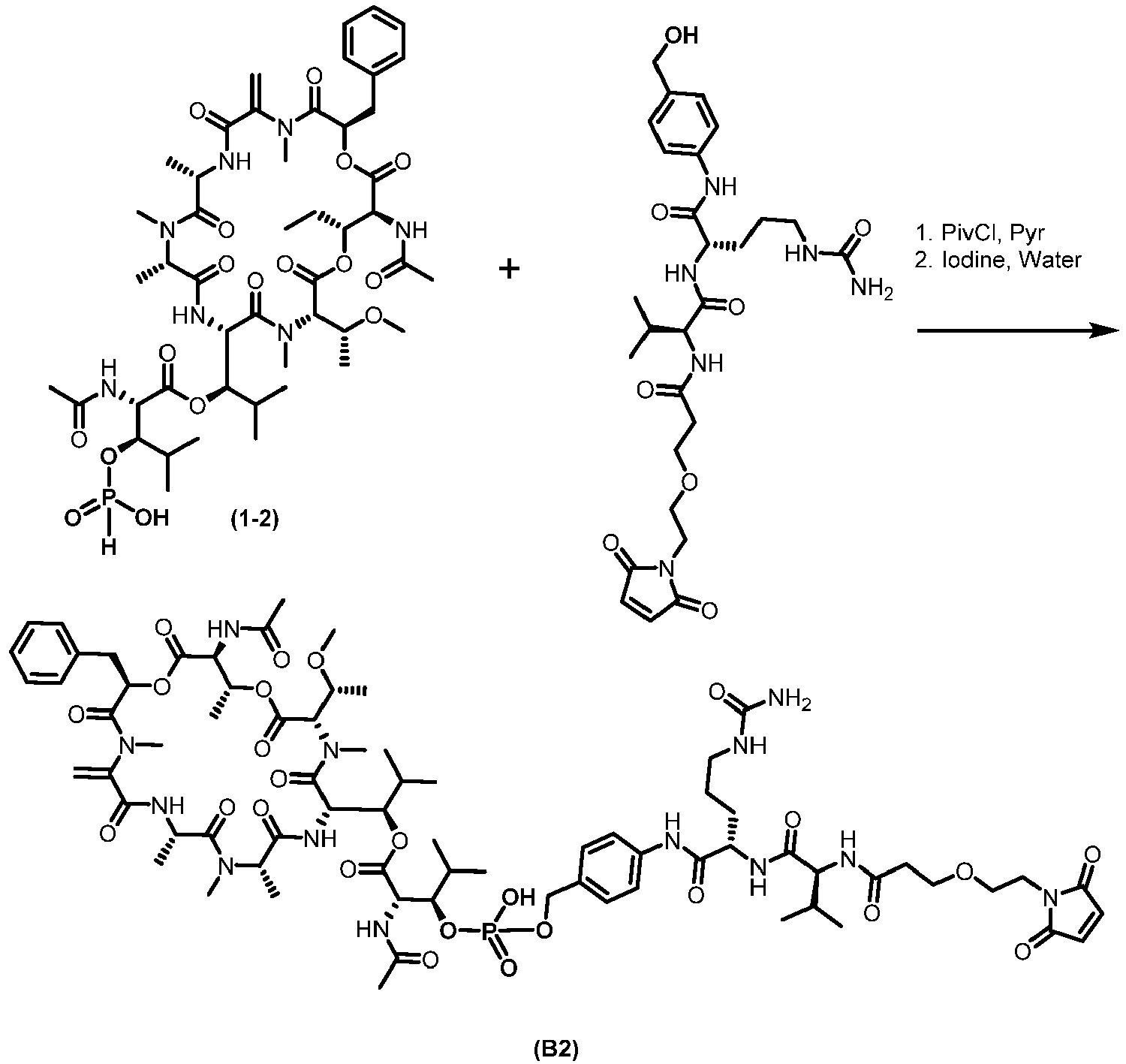
- (R)-1-((3S,6S,9S,12S,18R,21S,22R)-21-acetamido-18-benzyl-3-((R)-1-methoxyethyl)-4,9, 1 0, 12, 16,22-hexamethyl-15-methylene-2,5,8, 11,14, 17,20-heptaoxo-1, 19-dioxa-4,7,10,13,16-pentaazacyclodocosan-6-yl)-2-methylpropyl (2S,3R)-2-acetamido-3-((hydroxyhydrophosphoryl)oxy)-4-methylpentanoate (1-2) (50 mg, 0.049 mmol, 1.0 equiv.) and (S)-2-((S)-2-(3-(2-(2,5-dioxo-2,5-dihydro-1H-pyrrol-1-yl)ethoxy)propanamido)-3-methylbutanamido)-N-(4-(hydroxymethyl)phenyl)-5-ureidopentanamide (CAS# 2055041-37-5) (33.7 mg, 0.059 mmol, 1.2 equiv.) (both lyophilized powders were transferred into a 10 mL vial) and dissolved in pyridine (1 mL). Then, pivaloyl chloride (0.042 mL, 0.342 mmol, 7 equiv.) was added dropwise to give a faint yellow solution. The mixture was agitiated at room temperature for 30 minutes. A freshly prepared solution of iodine (49.6 mg, 0.195 mmol, 4 equiv.) in pyridine-water (20:1, 500 uL) was added to give a dark-brown clear solution. The mixture was agitated for 30 minutes and directly purified by reverse-phase flash chromatography (40 g C-18 column, 0%Ac/
MeCN 3 minutes, then 0-70% ACN/Water over 15 minutes, neutral method) to afford (R)-1-((3S,6S,9S,12S,18R,21S,22R)-21-acetamido-18-benzyl-22-isopropyl-3-((R)-1-methoxyethyl)-4,9,10,12,16-pentamethyl-15-methylene-2,5,8,11,14,17,20-heptaoxo-1,19-dioxa-4,7,10,13,16-pentaazacyclodocosan-6-yl)-2-methylpropyl (2S,3R)-3-((((4-((S)-2-((S)-2-(3-(2-(2,5-dioxo-2,5-dihydro-1H-pyrrol-1-yl)ethoxy)propanamido)-3-methylbutanamido)-5-ureidopentanamido)benzyl)oxy)(hydroxy)phosphoryl)oxy)-4-methyl-2-propionamidopentanoate (B2). HRMS; MH+ = 1595.7200, 2.25 min. -

- Compound (B3) can be obtained using procedures similar to the methods described in Example 1-1, except in
step 1 where compound (A3) (from Example 3-3) is used in place of compound (A1). -

- Compound (B4) was obtained using procedures similar to the methods described in Example 1-1, except in
step 2 where (S)-2-((S)-2-(3-(2,5-dioxo-2,5-dihydro-1H-pyrrol-1-yl)propanamido)-3-methylbutanamido)-N-(4-(hydroxymethyl)phenyl)-5-ureidopentanamide (CAS# 1949793-46-7) was used in place of (S)-2-((S)-2-(3-(2-(2,5-dioxo-2,5-dihydro-1H-pyrrol-1-yl)ethoxy)propanamido)-3-methylbutanamido)-N-(4-(hydroxymethyl)phenyl)-5-ureidopentanamide (CAS# is 2055041-37-5). HRMS; MH+ = 1594.5400, 2.88 min. -

- Compound (B5) can be obtained using procedures similar to the methods described in Example 1-1, except in
step 1 where compound (A2) (from Example 3-2) was used in place of compound (A1), and instep 2 where (S)-2-((S)-2-(3-(2,5-dioxo-2,5-dihydro-1H-pyrrol-1-yl)propanamido)-3-methylbutanamido)-N-(4-(hydroxymethyl)phenyl)-5-ureidopentanamide (CAS# 1949793-46-7) is used in place of (S)-2-((S)-2-(3-(2-(2,5-dioxo-2,5-dihydro-1H-pyrrol-1-yl)ethoxy)propanamido)-3-methylbutanamido)-N-(4-(hydroxymethyl)phenyl)-5-ureidopentanamide (CAS# is 2055041-37-5). -

- Compound (B6) can be obtained using procedures similar to the methods described in Example 1-1, except in
step 1 where compound (A3) (from Example 3-3) was used in place of compound (A1), and instep 2 where (S)-2-((S)-2-(3-(2,5-dioxo-2,5-dihydro-1H-pyrrol-1-yl)propanamido)-3-methylbutanamido)-N-(4-(hydroxymethyl)phenyl)-5-ureidopentanamide (CAS# 1949793-46-7) is used in place of (S)-2-((S)-2-(3-(2-(2,5-dioxo-2,5-dihydro-1H-pyrrol-1-yl)ethoxy)propanamido)-3-methylbutanamido)-N-(4-(hydroxymethyl)phenyl)-5-ureidopentanamide (CAS# is 2055041-37-5). -

- Compound (B7) may be obtained by reacting chloroformate (1-3) with (S)-2-((S)-2-(3-(2-(2,5-dioxo-2,5-dihydro-1H-pyrrol-1-yl)ethoxy)propanamido)-3-methylbutanamido)-N-(4-(hydroxymethyl)phenyl)-5-ureidopentanamide (CAS# is 2055041-37-5). Chloroformate (1-3) may be obtained by reacting Compound (A1) with phosgene.
-

- Compound (B8) may be obtained using the method described in Example 1-7, except Compound (A2) is used in place of Compound (A1).
-

- Compound (B9) may be obtained using the method described in Example 1-7, except Compound (A3) is used in place of Compound (A1).
-

- Compound (B10) may be obtained using the method described in Example 1-7, except (S)-2-((S)-2-(3-(2,5-dioxo-2,5-dihydro-1H-pyrrol-1-yl)propanamido)-3-methylbutanamido)-N-(4-(hydroxymethyl)phenyl)-5-ureidopentanamide (CAS# 1949793-46-7) is used in place of (S)-2-((S)-2-(3-(2-(2,5-dioxo-2,5-dihydro-1H-pyrrol-1-yl)ethoxy)propanamido)-3-methylbutanamido)-N-(4-(hydroxymethyl)phenyl)-5-ureidopentanamide (CAS# is 2055041-37-5).
-

- Compound (B11) may be obtained using the method described in Example 1-10, except Compound (A2) is used in place of Compound (A1).
-

- Compound (B12) may be obtained using the method described in Example 1-10, except Compound (A3) is used in place of Compound (A1).
- Full length human, cyno and rat PMEL17 genes were synthesized based on amino acid sequences from the GenBank or Uniprot databases. All synthesized DNA fragments were cloned into appropriate expression vectors.
- Engineered stable PMEL17-expressing cell lines were generated and cultured under appropriate selection conditions to produce stable PMEL17-expressing cell lines.
- The phagemid libraries are based on the HuCAL PLATINUM® (Knappik et al., 2000) and Ylanthia concepts (Tiller et al., 2013) and employ the CysDisplayTM technology for displaying the Fab on the phage surface (Lohning, 2001).
- For each panning, about 4×10¹³ HuCAL PLATINUM® or about 1×10¹⁴ Ylanthia® phage-antibodies phage-antibodies were blocked in PBS/5% FCS. In parallel, 0.5 - 1.0 × 10⁷ target cells expressing antigen PMEL17 and 0.5 - 1.0 × 10⁷ adsorption cells without expression of antigen PMEL17 per phage pool were resuspended in 1 ml PBS/5% FCS for blocking on ice. The blocked target cells were spun down, resuspended in the pre-blocked phage particles and incubated for 2 h at 4°C on a rotator. The phage-cell complexes were washed three times in PBS/5% FCS. Elution of specifically bound phage from target cells was performed by 10 min acidic elution with 0.1 M glycine-HCl/0.5 M NaCl, pH 2.2. After centrifugation, the supernatant (eluate) was neutralized by adding 2 M unbuffered Tris. For removal of phage binding to cell surface molecules other than the target antigen, post-adsorption was performed three times with 0.5 - 1.0 × 10⁷ adsorption cells each. The final supernatant was used for infection of 14 ml E. coli TG1 culture grown to an OD600 of 0.6 - 0.8. The culture was incubated for 45 min in a water bath at 37°C for phage infection. The bacterial pellets were resuspended in 2xYT medium, plated on LB/Cam agar plates and incubated o/n at 30°C. Colonies were scraped off the plates and were used for phage rescue and phage amplification. Amplified phage were used for the next panning round. The second and third round of the whole cell panning was performed according to the protocol of the first round.
- To facilitate rapid expression of soluble Fab, the Fab encoding inserts of the selected HuCAL PLATINUM® phage were subcloned from
pMORPH ®30 display vector into pMORPH®x11_FH expression vector. Subcloning was performed by triple digest via EcoRl, Xbal and Bmtl. After transformation of E. coli TG1-F- single clone expression and preparation of periplasmic extracts containing HuCAL®-Fab fragments were performed as described previously (Rauchenberger et al., 2003). - The Fab encoding inserts of the selected Ylanthia® phage were subcloned from pYPdis10 display vector into pYBex10_Fab_FH expression vector. Subcloning was performed by triple digest via Xbal, EcoRl-HF and Pstl-HF. After transformation of E. coli TG1-F- single clone expression and preparation of periplasmic extracts containing Ylanthia®-Fab fragments were performed as described previously (Rauchenberger et al., 2003).
- Expression of Fab fragments encoded by pMORPH®x11_Fab_FH and pYBex10_Fab_FH in E. coli TG1 F- cells was carried out in shake flask cultures using 500 ml of 2xYT medium supplemented with 0.1% glucose and 34 µg/ml chloramphenicol. Cultures were shaken at 30°C until the OD600 reached a value of 0.5. Fab expression was induced by addition IPTG (isopropyl-β-D-thiogalactopyranoside) at a final concentration of 0.75 mM and further cultivation for 20 h at 30°C. Cells were harvested and disrupted using lysozyme. His6-tagged (SEQ ID NO: 267) Fab fragments were isolated via IMAC (Bio-Rad, Germany) and eluted using imidazole. Buffer exchange to 1x Dulbecco's PBS (pH 7.2) was performed using PD10 columns (GE Healthcare, Germany). Samples were sterile filtered (0.2 µm). Protein concentrations were determined by UV-spectrophotometry. The purity of the samples was analyzed in denaturing, reducing 15% SDS-PAGE. The homogeneity of Fab preparations was determined in native state by size exclusion chromatography (HP-SEC) with calibration standards.
- In FACS screening, purified Fabs were titrated on a variety of PMEL17 expressing and PMEL17 non expressing cell lines (for control). Cells were harvested using Accutase, adjusted to ~ 4×10⁶ cells/ml into FACS buffer (PBS, 3%FCS and 0,02% Na-Azide) and kept on ice to avoid internalization. 15 µl of cell suspension/well were transferred into 384 well V-bottom plates (Greiner, Cat# 781280) and incubated with 15 µl of Fab at different concentrations (most commonly from 200 to 3,5×10⁻³nm ) for 1 hour at 4°C, gently shaking. Following incubation, cells were washed three times with FACS buffer. After each washing step, cells were centrifuged (250xg, 4 min, 4°C) and carefully resuspended. 15 µl of detection antibody conjugated to PE (PE-conjugated goat anti-human IgG, F(ab')2 fragment specific, 1:150 in FACS buffer; Jackson Immuno Research, #109-116-097) or Alexa Fluor (Alexa Fluor-conjugated goat anti-human IgG, F(ab')2 fragment specific, 1:150 in FACS buffer; Jackson Immuno Research, #109-606-097) were added and samples were incubated for 45 minutes to 1 hour on ice in the dark, gently shaking. After 3 washing steps, cells were resuspended in 30 µl of FACS buffer and samples were measured using the IntelliCyt HTFC device.
- In order additionally verify the specificity of the identified antibodies, the antigen was immuno-captured from cell lysate and used as coating material for ELISA based screenings. PMEL17 expressing and PMEL17 non expressing cells (used as negative control) were centrifugated (250g, 5min) and resuspended in lysis buffer (MSD Tris Lysis Buffer, Meso Scale Discovery, R60TX-2) containing protease inhibitors (Complete EDTA free Protease Inhibitor Cocktail, Roche, 11 873 580 001) at 1×10⁷ to 1×10⁸ cells/mL and incubated 30 min at4°C. Lysate was spinned down at 13000rpm for 5 minutes to discard cell debris and the supernatant was aliquotted and stored at -80°C. Preliminary testing demonstrated that the lysate could be freezed and thawed 3 times without major loss of signal in ELISA.
- So as to perform the screening ELISA anti-His IgGs were coated overnight on a maxisorb 384 wells plate (R&D systems, MAB050, 10 µg/ml, 20 µl per well). Plates were blocked with 3% BSA and 20 µl of purified Fab were transferred at different concentrations (most commonly from 400 to 0,2nM) to each well for 1 hour. The plate was washed 3 times and 20 µl PMEL17 containing cell lysate was added at a concentration of 2x10⁵ lysed cells/well for 1 hour. After 3 additional washing, the presence of PMEL17 was revealed using the tool antibody PMEL17 biotinylated (5 µg/ml, 20 µl per well) and streptavidin-ECL (Dianova, 13MSA37, 1:1500, 20 µl per well). 20 µl per well of MSD read buffer 1X (Meso Scale Discovery, R92TC-2) were added to the plate and signals were detected via the MSD Sector Imager 6000.
- Conversion of selected Fabs into IgGs was achieved by a PCR-based method in 96-well format. The Fab bacterial expression vectors pMORPH®x11_FH and pYBex10_Fab_FH were converted into the IgG mammalian
expression vector pMORPH ®4 and pYMex10 (for HuCAL and Ylanthia clones, respectively). - pMORPHx11_FH plasmid DNA was first amplified by PCR using a biotinylated primer specific to the phoA leader region and a non biotinylated primer specific to the bacterial CL domain. The amplified product was captured on streptavidin beads, digested with BsiWI or Hpal (for VLkappa or VLlambda clones, respectively), washed and then digested again with Mfel. This procedure resulted in the release of the purified vector backbone into the supernatant, now lacking the bacterial constant light chain region (CL) and the phoA heavy chain leader. A kappa or lambda specific mammalian pIN expression cassette was then cloned into the vector backbone carrying the mammalian CL, polyA site, CMV promotor and mammalian heavy chain leader sequence. In a second PCR step, the newly generated Fab insert was amplified again using a biotinylated primer specific to the CH1 region and a non-biotinylated primer binding within the bacterial ompA leader. The PCR product was captured on streptavidin beads, digested with EcoRV, washed and digested with Blpl resulting in the release of the purified insert into the supernatant. Inserts were finally cloned into the Fab_Cys acceptor vector for expression in mammalian cells.
- pYBex10_Fab_FH plasmid DNA was first amplified by PCR using a biotinylated primer specific to the phoA leader region and a non biotinylated primer specific to the bacterial CL domain. The amplified product was captured on streptavidin beads, digested with Nhel, washed and then digested again with Kpnl. This procedure resulted in the release of the purified vector backbone into the supernatant, now lacking the bacterial constant light chain region (CL) and the phoA heavy chain leader. A kappa or lambda specific mammalian pIN expression cassette was then cloned into the vector backbone carrying the mammalian CL, polyA site, CMV promotor and mammalian heavy chain leader sequence. In a second PCR step, the newly generated Fab insert was amplified again using a biotinylated primer specific to the CH1 region and a non-biotinylated primer binding within the bacterial ompA leader. The PCR product was captured on streptavidin beads, digested with Xhol, washed and digested with Ndel resulting in the release of the purified insert into the supernatant. Inserts were finally cloned into the Fab_Cys acceptor vector for expression in mammalian cells.
- After transformation of E. coli XL-1 blue cells, single clones were controlled via colony PCR and sequencing of the whole insert region.
- DNA preparations of single colonies were prepared by using an appropriate DNA preparation kit in combination with the BioRobot®8000 device. Individual DNA concentrations were determined by UV-spectrophotometry. Eukaryotic HEK293 c18 cells (ATCC #CRL-10852) were used in a 96-well expression system for the generation of conditioned cell culture supernatants containing full-length IgG. Eukaryotic HEK293 c18 cells were seeded in a 96-well flat-bottom plate to a density of ~4×10⁴cells/50µl/well the day before and transfected with equal amounts of Ig expression vector DNA. After incubation for 40-50 h at 37°C and 6% CO2 the culture supernatants were transferred to a 96-well U-bottom plate and cleared by centrifugation. The resulting Ig supernatants were tested by an anti-Fd capture ELISA for calculation of Ig concentration in reference to known standards and stored at -20°C for later use in specificity and/or functional screening assays.
- DNA of clones of interest were subjected to sequencing with primer CMV_HC for (CTC TAG CGC CAC CAT GAA ACA (SEQ ID NO: 264)) for VH domain and IgG_const_for (AGC CCA GCA ACA CCA AGG (SEQ ID NO: 265)) for Fc domain followed by light chain sequencing with T7 promoter primer (TAA TAC GAC TCA CTA TAG GG (SEQ ID NO: 266)) to obtain complete IgG sequence information.
- FACS screening was performed in the 384-well plate format using the HTFC screening platform from IntelliCyt. Cells were harvested using Accutase, adjusted to ~ 4×10⁶ cells/ml into FACS buffer (PBS, 3%FCS and 0,02% Na-Azide) and kept on ice to avoid internalization. 15 µl of cell suspension/well were transferred into 384 well V-bottom plates (Greiner, Cat# 781280) and incubated with 15 µl of IgG containing supernatant (or diluted purified control antibodies) for 1 hour at 4°C, gently shaking. Following incubation, cells were washed three times with FACS buffer. After each washing step, cells were centrifuged (250xg, 4 min, 4°C) and carefully resuspended. 15 µl of detection antibody conjugated to PE (PE-conjugated goat anti-human IgG, F(ab')2 fragment specific, 1:150 in FACS buffer; Jackson Immuno Research, #109-116-097) were added and samples were incubated for 45 minutes to 1 hour on ice in the dark, gently shaking. After 3 washing steps, cells were resuspended in 30 µl of FACS buffer and samples were measured using the IntelliCyt HTFC device.
- Eukaryotic HKB11 cells were transfected with
pMORPH ®4 or pYMex10 expression vector DNA encoding both heavy and light chains of IgGs. Cell culture supernatant was harvested onday - For the first panning round, about 4×10¹³ HuCAL PLATINUM® phage were blocked with PBS/5% FBS. In parallel, 1.0 × 10⁷ target cells expressing antigen PMEL17 and 1.5-3.0 × 10⁷ adsorption cells without expression of antigen PMEL17 per phage pool were resuspended in 1 ml PBS/5% FCS for blocking on ice. To preclear non-specific binding, phage were incubated with 0.5 × 10⁷ adsorption cells without expression of antigen PMEL17 per phage pool for 30 minutes on ice. After incubation, the cells were pelleted by centrifugation and the supernatant was added to a fresh sample of with 0.5 × 10⁷ adsorption cells without expression of antigen PMEL17. The same incubation conditions were applied and pre-absorption was again applied to a fresh sample of 0.5 × 10⁷ adsorption cells without expression of antigen PMEL17 for a total of three pre-absorption rounds. After the final pre-absorption, the supernatant was added to 1.0 × 10⁷ target cells expressing antigen PMEL17 and incubated on ice for 2 hours with occasional mixing. The phage-cell complexes were washed three times in PBS/5% FCS. Elution of specifically bound phage from target cells was performed by 10 minute acidic elution with 0.1 M glycine-HCl/0.5 M NaCl, pH 2.5. After centrifugation, the supernatant (eluate) was neutralized by adding 2 M unbuffered Tris and this eluate was used to infect 14 ml E. coli TG1 F' culture grown to an OD600 of 0.6 - 0.8. The culture was incubated for 20 minutes at 37°C and then an
addition 25 minutes at 37°C with shaking at 200 rpm for phage infection. The bacterial pellets were resuspended in 2xYT medium, plated on LB/Chloroamphenicol agar plates and incubated overnight at 30°C. Colonies were scraped off the plates and were used for phage rescue and phage amplification. Amplified phage were used for the next panning round. The second and third rounds of panning were performed similarly except that only about 1×10¹² HuCAL phage was used to reduce background and improve the effectiveness of preclearing. After the final round of panning, plasmid DNA was prepared from each phage output and the Fab-containing inserts were cloned into a bacterial expression vector. After ligation and transformation, individual colonies were picked into 2XT/chloramphenicol and cultured. Fab expression was induced by addition of 0.25 mM IPTG in cultures grown overnight in 96 well microtiter plates at 25°C. Cell pellets were lysed with 0.1% lysozyme in PBS and then blocked by the addition of BSA to a final concentration of 1%. FACS staining was performed on PMEL17-expressing and control cells. - Purified IgGs were titrated on a variety of PMEL17 expressing and PMEL17 non expressing cell lines (for control) to determine EC50 values. Cells were harvested using Accutase, adjusted to ~ 4x10⁶ cells/ml into FACS buffer (PBS, 3%FCS and 0,02% Na-Azide) and kept on ice to avoid internalization. 15 µl of cell suspension/well were transferred into 384 well V-bottom plates (Greiner, Cat# 781280) and incubated with 15 µl of IgGs at different concentrations (most commonly from 200 to 3,5x10⁻³ nM) for 1 hour at 4°C, gently shaking. Following incubation, cells were washed three times with FACS buffer. After each washing step, cells were centrifuged (250xg, 5 min, 4°C) and carefully resuspended. 15 µl of detection antibody conjugated to PE (PE-conjugated goat anti-human IgG, F(ab')2 fragment specific, 1:150 in FACS buffer; Jackson Immuno Research, #109-116-097) or Alexa Fluor (Alexa Fluor-conjugated goat anti-human IgG, F(ab')2 fragment specific, 1:150 in FACS buffer; Jackson Immuno Research, #109-606-097) were added and samples were incubated for 45 minutes to 1 hour on ice in the dark, gently shaking. After 3 washing steps, cells were resuspended in 30 µl of FACS buffer and samples were measured using the IntelliCyt HTFC device. EC50 values were calculated using the Graphpad Prism software. G-361 melanoma cell line expresses human PMEL17 at a lower level compared to the HKB11-human PMEL17 overexpressing cell line, and therefore allows a more precise ranking of the clones depending on their apparent affinity on cells. As illustrated in Table 3, a variety of EC50 ranging from ~200 pM to ~70nM was reached.
Table 3. Apparent affinities (FACS EC50 (nM)) and cross-reactivity of anti-PMEL17 antibodies. G-361 (PE) G-361 (Alexa Fluor) HKB11-cyno (Alexa Fluor) HKB11-rat (Alexa Fluor) B16-F10 (mouse) (Alexa Fluor) UACC-62 (PE) Cross spe profile (based on C14 detection ab) Y101341 0.4 2.6 1.8 1.2 Negative h/c/r/m Y010906 - 2.0 2.0 21.7 139.8 Negative h/c/r/m Y010900 - 1.3 1.2 9.3 >100 Negative h/c/r/m Y010355 0.5 3.9 0.8 0.8 Negative h/c/r/m Y010356 1.6 4.5 1.4 1.4 Negative h/c/r/m Y010429 2.5 10.7 1.5 60.2 Negative h/c/r MOR024354 - 1.7 2.1 2.4 >100 Negative h/c/r Y010415 0.3 5.4 0.4 >100 Negative h/c/r Y010903 - 3.2 2.0 5.6 927.5 Negative h/c/(m) Y010910 - 10.0 3.5 13.7 41.1 Negative h/c/(m) MOR024353 - 62.8 1.6 7.5 >100 Negative h/c Y010417 2.8 6.3 >100 >100 Negative h/c - The cross-specificity of the IgGs was evaluated in does response FACS on G-361 (human melanoma), HKB11-cyno-PMEL17, HKB11-rat PMEL17 and B16-F10 (mouse PMEL17 expressing melanoma) (Table 3). Testing on G-361 was performed using a detection antibody labelled to PE. In contrast, an Alexa-Fluor labelled detection antibody had to be used for testing the candidates on the other cell lines so as to reach detectable signals.
- IgGs were classified into several epitope groups according to their cross-specificity profiles. The various cross-reactive profiles of the IgGs nevertheless suggest that their target at least 4 epitopes (human/cyno/rat/mouse, human/cyno/rat, human/cyno/mouse and human/cyno).
- IgGs were tested in dose response for their ability to internalize and induce the killing of various PMEL17 expressing cells in both piggyback ADC assay and after direct conjugation.
- For piggyback ADC assay, the Fab-ZAP (goat anti-hu-mAb-saporin-coupled; ATS Biotechnology, Cat# IT-51) compound was used which specifically binds to human Fc and is coupled to a cytotoxin element, the saporin. Cells were harvested using Accutase and adjusted to 5×10⁴ cells/ml. 50 µl of the cell suspension were transferred per well into a 96-well flat clear bottom white plate (Corning, 3610) and incubated overnight at 37°C, 5% CO2. On the following day, antibody candidates were incubated at different concentrations (most commonly from 44 to 7 × 10⁻³ nM) with the Fab-ZAP compound at 8 nM for 30 minutes at 37°C. 50 µl per well of the IgG - Fab-ZAP complexes were then added to the target cells. For controls, wells with cells only, cells only incubated with candidate IgGs (=100% viability control) and cells only incubated with Fab-ZAP (to check for unspecific killing of the secondary reagent) were prepared. Final concentration of IgGs were 22 to 3,5 × 10⁻³ nM and Fab-
ZAP 4 nM. Plates were incubated for 72 h at 37°C and 5% CO2. The amount of viable cells was evaluated using CellTiter-Glo (Promega #G7571) and the luminescence detected with theTecan Infinite 500. Viability was then normalized to the cells + IgG only control. - Almost all IgGs could efficiently kill G-361 melanoma (>80% maximum killing) and showed limited killing on the non PMEL17 expressing 293T cells (<30% maximum killing). G-361 killing was also achieved with a range of EC50s which may be affinity or epitope related. Cross-specificity could also be confirmed for most of the IgGs reactive to human/cyno/rat/mouse, human/cyno/rat and human/cyno/mouse, i.e. IgGs specific to mouse PMEL17 could also kill B16-F10 (mouse melanoma).
- Generally, all engineering processes, were performed using PCR-based strategies. Engineering processes involved the following aspects: Germlining, Removal of PTM sites, pl engineering, and Codon optimization.After synthesis and assembly by overlap extension PCR the re-engineered VH and VL fragments were subcloned into the appropriate vector backbones for subsequent IgG expressions.
-

- Compound A1 was isolated based on methods described in Japanese Patent Publication No.
JP62283999 - Step 2: Defatting: The crude extract was dissolved in 25 L acetonitrile/water (9/1) and extracted three times with 25 L heptane. The acetonitrile/water layer was evaporated until only water was left. This was then extracted with 25 L ethyl acetate. The ethyl acetate layer was evaporated to dryness yielding 40 g of crude extract.
- Step 3: Flash chromatography: The crude extract was dissolved in acetone/methanol (1/1), adsorbed onto 100 g Isolute (diatomaceous earth) and evaporated to dryness. Flash chromatography on silica gel with a ternary solvent system with cyclohexane (eluent A), ethyl acetate (eluent B) and methanol (eluent C) was performed (experimental details: column: RediSep Rf 120 g; flow rate: 85 mL/min; gradient: 0 min: 75 % A, 25 % B, 0 % C; 3 min: 75 % A, 25 % B, 0 % C; 10 min: 0 % A, 100 % B, 0 % C; 17.5 min: 0% A, 90
% B 10 % C; 25 min: 0 % A, 50 % B, 50 % C; 30 min: 0 % A, 50 % B, 50 % C; the different segments of the gradient are connected by linear changes over time). Four runs with 10 g extract each were performed. Time based fractionation was applied and the collected fractions were analyzed by UPLC-UV-MS for presence of Compound (A1). Fractions containing Compound (A1) were pooled and evaporated to dryness resulting in a fraction of 25 g. - Step 4: Size-exclusion chromatography (SEC): The 25 g fraction was dissolved in 400 mL methanol and further fractionated by SEC on a column (
length 25 cm, diameter 12.5 cm) packed with Sephadex LH20 and methanol as eluent. Fractions of 200 mL each were collected and the collected fractions were analyzed by UPLC-UV-MS for presence of Compound (A1). Fractions containing Compound (A1) were pooled and evaporated to dryness resulting in an enriched fraction of 6.2 g. - Step 5: 1st preparative HPLC: The enriched fraction of 6.2 g was dissolved in 12 mL methanol/dimethylsulfoxide (1/1) and further fractionated by preparative HPLC (experimental details: column: Sunfire C18, 30 × 150mm, 5µm particle size; eluent A: deionized water with 0.1 % formic acid, eluent B: methanol with 0.1 % formic acid; flow
rate 60 mL/min; gradient: 0 min: 35 % A, 65 % B; 0.5 min: 35 % A, 65 % B; 17 min: 15 % A, 85 % B; 15.0 min: 0 % A, 100 % B; 19 min: 0 % A, 100 % B; 19.1 min: 35 % A, 65 % B; 20 min: 35 % A, 65 % B; the different segments of the gradient are connected by linear changes over time). Fractionation was triggered by mass spectrometry. 15 runs with 410 mg enriched fraction each were performed. Fractions containing Compound (A1) were pooled and evaporated to dryness resulting in a semi-pure fraction of 488 mg with an estimated content of 70 % Compound (A1). - Step 6: 2nd preparative HPLC: The semi-pure fraction of 488 mg was dissolved in 4 mL methanol and further fractionated by preparative HPLC (experimental details: column: X-Select PFP, 19 × 250mm, 5µm particle size; eluent A: deionized water with 0.1 % formic acid, eluent B: acetonitrile with 0.1 % formic acid; flow
rate 30 mL/min; gradient: 0 min: 45 % A, 55 % B; 0.5 min: 45 % A, 55 % B; 24 min: 25 % A, 75 % B; 24.0 min: 0 % A, 100 % B; 27.5 min: 0 % A, 100 % B; 27.6 min: 45 % A, 55 % B; 30.5 min: 45 % A, 55 % B; the different segments of the gradient are connected by linear changes over time). Fractionation was triggered by mass spectrometry. Five runs witch 98 mg semi-pure fraction each were performed. Fractions containing Compound (A1) were pooled and evaporated to dryness resulting in 287 mg Compound (A1) with a purity > 95 %. - Compound (A1): Retention time: 4.73 min, molecular formula [M+H]+: C49H76N7O15+, calculated monoisotopic mass [M+H]+: 1002.5394 Da, observed mass: 1002.5391 Da. Experimental details: column: ACQUITY UPLC BEH C18, 2.1 × 100mm, 1.7µm particle size; eluent A: deionized water with 0.1 % formic acid, eluent B: acetonitrile with 0.1 % formic acid; flow rate 0.9 mL/min; linear gradient: 0 min: 95 % A, 5 % B to 6.4 min: 0 % A, 100 % B. 1H NMR (600 MHz, Acetonitrile-d 3) δ 8.36 (d, J = 9.4 Hz, 1H), 7.50 (d, J = 10.0 Hz, 1H), 7.38 - 7.22 (m, 6H), 6.94 (d, J = 4.4 Hz, 1H), 6.77 (d, J = 9.9 Hz, 1H), 5.39 (d, J = 9.8 Hz, 1H), 5.34 (dd, J = 8.8, 3.8 Hz, 1H), 5.31 - 5.25 (m, 2H), 5.21 - 5.15 (m, 2H), 5.11 (dd, J = 9.9, 1.9 Hz, 1H), 4.90 - 4.84 (m, 1H), 4.70 (q, J = 6.9 Hz, 1H), 4.39 (dd, J = 7.7, 2.1 Hz, 1H), 4.10 (d, J = 9.8 Hz, 1H), 3.82 - 3.74 (m, 1H), 3.60 (ddd, J = 9.8, 4.4, 2.0 Hz, 1H), 3.38 (s, 3H), 3.23 (s, 3H), 3.16 - 3.11 (m, 1H), 2.94 - 2.85 (m, 1H), 2.84 (s, 3H), 2.66 (s, 3H), 2.55 - 2.43 (m, 2H), 2.16 (s, 3H), 1.96 - 1.90 (m, 1H), 1.89 - 1.74 (m, 2H), 1.37 (d, J = 7.0 Hz, 3H), 1.32 (d, J = 6.6 Hz, 3H), 1.19 (d, J = 6.5 Hz, 3H), 1.17 (d, J = 6.0 Hz, 3H), 1.12 (d, J = 7.7 Hz, 3H), 1.06 (d, J = 6.8 Hz, 3H), 0.96 (d, J = 6.6 Hz, 3H), 0.88 (d, J = 6.6 Hz, 3H), 0.85 (d, J = 7.0 Hz, 3H), 0.77 (d, J = 6.5 Hz, 3H).
-

- Compound (A2) was obtained using the methods described in [M. Taniguchi et al., Tetrahedron 59 (2003) 4533-4538]. Isolation was performed using the methods described for Compound (A1) in Example 3-1 The material was then characterized by UPLC-UV-HRMS, 1D-NMR- and 2D-NMR-experiments. Compound (A2): Retention time: 4.20 min, molecular formula [M+H]+: C46H70N7O15+, calculated monoisotopic mass [M+H]+: 960.4924 Da, observed mass: 960.4914 Da. 1H NMR (600 MHz, 1,4-Dioxane-d 8) δ 8.36 (d, J = 9.2 Hz, 1H), 7.37 (d, J = 10.0 Hz, 1H), 7.31 - 7.21 (m, 4H), 7.21 - 7.16 (m, 2H), 6.79 - 6.72 (m, 2H), 5.40 - 5.37 (m, 1H), 5.36 - 5.33 (m, 1H), 5.30 - 5.28 (m, 2H), 5.20 (dd, J = 9.0, 3.7 Hz, 1H), 5.13 (d, J = 1.9 Hz, 1H), 5.04 (dd, J = 9.9, 1.4 Hz, 1H), 4.89 - 4.82 (m, 1H), 4.73 (q, J = 6.9 Hz, 1H), 4.39 (dd, J = 8.0, 2.0 Hz, 1H), 4.04 (d, J = 9.9 Hz, 1H), 3.80 - 3.73 (m, 1H), 3.65 (ddd, J = 9.8, 4.3, 1.9 Hz, 1H), 3.37 (s, 3H), 3.21 (s, 3H), 3.11 (dd, J = 14.7, 3.7 Hz, 1H), 2.89 (dd, J = 14.7, 8.9 Hz, 1H), 2.84 (s, 3H), 2.62 (s, 3H), 2.14 (s, 3H), 2.12 (s, 3H), 1.99-1.90 (m, 1H), 1.75-1.69 (m, 1H), 1.35 (d, J = 6.8 Hz, 3H), 1.30 (d, J = 6.5 Hz, 3H), 1.18 - 1.14 (m, 6H), 1.11 (d, J = 6.5 Hz, 3H), 1.04 (d, J = 6.7 Hz, 3H), 0.86 (d, J = 6.8 Hz, 3H), 0.78 (d, J = 6.6 Hz, 4H).
-

- Compound (A3) was obtained using the methods described in [M. Taniguchi et al. Tetrahedron 59 (2003) 4533-4538]. Isolation was performed using the methods described for Compound (A1) in Example 3-1. The material was then characterized by UPLC-UV-HRMS, 1D-NMR- and 2D-NMR-experiments. Compound (A3): Retention time: 4.42 min, molecular formula [M+H]+: C47H72N7O15+, calculated monoisotopic mass [M+H]+: 974.5081 Da, observed mass: 974.5098 Da. 1H NMR (600 MHz, 1,4-Dioxane-d 8) δ 8.37 (d, J = 9.1 Hz, 1H), 7.38 (d, J = 9.9 Hz, 1H), 7.31 - 7.23 (m, 4H), 7.22 - 7.16 (m, 1H), 7.12 (d, J = 7.7 Hz, 1H), 6.78 - 6.72 (m, 2H), 5.41 - 5.33 (m, 2H), 5.32 - 5.26 (m, 2H), 5.20 (dd, J = 8.8, 3.7 Hz, 1H), 5.12 (d, J = 2.2 Hz, 1H), 5.04 (dd, J = 9.9, 1.7 Hz, 1H), 4.89 - 4.81 (m, 1H), 4.72 (q, J = 7.0 Hz, 1H), 4.40 (dd, J = 7.8, 2.1 Hz, 1H), 4.04 (d, J = 9.8 Hz, 1H), 3.81 - 3.73 (m, 1H), 3.68 - 3.64 (m, 1H), 3.38 (s, 3H), 3.21 (s, 3H), 3.11 (dd, J = 14.6, 3.7 Hz, 1H), 2.89 (dd, J = 14.7, 8.9 Hz, 1H), 2.83 (s, 3H), 2.62 (s, 3H), 2.51 - 2.41 (m, 2H), 2.14 (s, 3H), 1.99 - 1.89 (m, 1H), 1.78 - 1.68 (m, 1H), 1.34 (d, J = 6.9 Hz, 3H), 1.30 (d, J = 6.5 Hz, 3H), 1.18 - 1.14 (m, 6H), 1.13 - 1.08 (m, 6H), 1.04 (d, J = 6.7 Hz, 3H), 0.86 (d, J = 6.8 Hz, 3H), 0.78 (d, J = 6.6 Hz, 3H).
- Antibody was incubated with RMP Protein A resin (GE) at a ratio of 10 mg Ab to 1 ml resin in PBS for 15 minutes with mixing in an appropriately sized disposable column. Cysteine HCl was added to a final concentration of 20 mM and incubated with agitation for 30 min at room temperature to allow the reactive cysteines to be deblocked. The resin was quickly washed with 50 column volumes PBS on a vacuum manifold. The resin was then resuspeneded in an equal volume PBS containing 250 nM CuCl2. Reformation of antibody interchain disulfides was monitored by taking time points. At each time point, 25 µL of resin slurry was removed, 1 µL of 20 mM of Compound (B1) or Compound (B2) was added, and the tube flicked several times. The resin was spun down, supernatant removed, and then eluted with 50 µL Antibody elution buffer (Thermo). The resin was pelleted and the supernatant analyzed by reverse phase chromatography using an Agilent PLRP-S 4000A 5um, 4.6x50mm column (Buffer A is water, 0.1% TFA, Buffer B Acetonitrile, 0.1% TFA, column held at 80 C, Flowrate 1.5 ml/min).
- Once it was determined that the antibody has reformed its interchain disulfide bonds, the resin was washed with 10 column volumes PBS and the resin was resuspended in an equal volume PBS and 8 equivalents of linker-payload (20 mM) in DMSO was added and then incubated at room temperature for 2 hours. The resin was then washed with 50 column volumes PBS. The ADC was eluted from the protein A resin with Antibody elution buffer and neutralized with 1/10 volume 1 M Tris pH 9.0. The ADC was then buffer exchanged into PBS or other suitable buffer and preparative size exclusion chromatography to remove aggregates was performed (S200 Increase; GE), if needed. The following analyses were performed - analytical SEC to determine percent monomer, mass spectroscopy to determine DAR, LAL test to determine endotoxin load and protein concentration was determined by A280 utilizing extinction coefficient and molecular weight of antibody.
- 92.1 uveal melanoma and MIAPACA pancreatic ductal adenocarcinoma cells were seeded at low cell density in 96-well plates and treated with increasing concentrations of Compound (A1) or Compound (A2) as indicated. Following drug treatment for 96 or 120 hours, cell viability and proliferation were determined using a resazurin-based viability assay. Briefly, cells were incubated with a resazurin-based solution and color change was detected by absorbance with a spectrophotometer and used as a readout of cell viability. Data presented as mean of 3 independent replicates and relative to PBS-treated cells (control). Table 4 shows the
growth inhibition 50% (GI50) for Compound (A2) and Compound (A1). Compound (A2) and Compound (A1) displayed potent target-dependent anti-UM activity (Fig. 1 ).Table 4. Growth inhibitory activity of Compound (A1) and Compound (A2) in UM cells Cell line Mutation Compound (A1) Compound (A2) GI50 nM GI50 nM 92.1 GNAQQ209L 0.18 7.7 MIAPACA WT > 10 µM > 10 µM - 92.1 and MIAPACA cells were seeded in 96-well plates (5000 cells per well) and treated with increasing concentrations of Compound (A1), Compound (A2), and a fixed dose of Staurosporine (100 nM, positive control) as indicated. Following drug treatment for 96, cells were subjected to fluorescence-activated flow cytometry using an Annexin V antibody conjugated to a fluorescent dye. Data presented as mean of 3 independent replicates. Compound (A2) and Compound (A1) induced apoptosis in UM cells in GNAQ/11 dependent manner (
Fig. 2 ). - 92.1 uveal melanoma were treated with increasing concentrations of Compound (A1) or Compound (A2) as indicated. Following drug treatment overnight, cells were processed for determination of IP1 levels using TR-FRET (time-resolved fluorescent resonance energy transfer) or protein levels by western-blotting.
Fig. 3A shows IP1 levels (nM) in 92.1 cells treated with DMSO (control), Compound (A2) or Compound (A1).Fig. 3B shows correlation between IP1 levels and relative proliferation in Compound (A1)-treated 92.1 cells.Fig. 3C shows immunoblots of 92.1 cells treated with Compound (A1) and Compound (A2) to determine the effect on ERK signaling. - The plasma stability of Compound (A1) was investigated in mouse, rat, monkey and human plasma and compared to buffer. Both disappearance of Compound (A1) (
Fig. 4A ) as well as appearance of the ring-opened form of Compound (A1) having the structure of

Fig. 4B ) were monitored over 24h. The transformation seem to be slightly faster in human plasma when compared to the other species. With the exception of the rat, adding the % remaining Compound (A1) and % formed Compound (A8) shows stoichiometry over 24h, indicating that no other transformation seem to play a significant role (Fig. 4C ). - The PK of Compound (A1) after intravenous dosing in mouse was characterized by a very high clearance and moderate to high volume of distribution. A slightly over-proportional increase of exposure with dose was observed in the range of 0.2-0.8 mg/kg (
Fig. 4D ). Table 5 summarizing all PK values is shown below.Table 5. PK properties of Compound (A1) Dose (mg/kg) 0.2 0.4 0.8 Cmax (nM) 31 83 240 Cmax/D (nM/(mg/kg)) 156 208 300 AUCinf (nM*h) 16 47 137 AUCinf/D (nM*h/(mg/kg)) 82 118 171 C0 (nM) 43 108 380 C0/D (nM/(mg/kg)) 217 270 475 CL [mL/min/kg] 204 141 97 Vss [L/kg] 8.6 4.9 4.6 - In vitro stability of Compound (A2) was tested in plasma and blood from different species (
Fig. 5A ). Compound (A2) showed good chemical stability in three different systems (Fig. 5B ). PK of Compound (A2) in female balb/c mice showed high clearance and a short elimination half-life (Fig. 5C ). Table 6 summarizes some PK values. Compound (A2) showed high intrinsic clearance in liver microsomes from mouse, rat and human, but low clearance in hepatocytes, probably due to limited membrane permeability (Table 7). Incubations of Compound (A2) in hepatic S9 fraction and in plasma showed similar half-lives. Compound (A2) and Compound (A1) were stable in buffer at pH 5.6 and in lysosomes over 4h (Fig. 5D ).Table 6. PK properties of Compound (A2) in female balb/c mice (1 mg/kg iv) CL (mL·min-1·kg-1) 175 ± 29 t1/2 (h) 0.4 ± 0.01 AUC i.v. d.n. (nM h) 101 ± 16 Table 7. Stability of Compound (A2) mouse rat human Hepatic microsomes (ul/min/kg) 107 33 44 Hepatocytes (ul/min/106 cells) - 8 <4 Hepatic S9 (+/- NADPH) t1/2 (h) 1.9 / >2 >2 / >2 1.9 / >2 Plasma t1/2 (h) 2.1 4.1 2.0 - Parental and non-targeting control (NT)- or PMEL17-shRNA transduced 92.1 cells were seeded at low cell density in 96-well plates and treated with increasing concentrations of 3207-(B1) (Isotype control), G4-(B1), and G1-(B1) in the presence or absence of doxycycline as indicated. Following drug treatment for 96 or 120 hours, cell viability and proliferation were determined using a resazurin-based viability assay. Briefly, cells were incubated with a resazurin-based solution and color change was detected by absorbance with a spectrophotometer and used as a readout of cell viability. Data presented as mean of 3 independent replicates and relative to PBS-treated cells (control) (
Fig. 6 ). Anti-PMEL17-(B1) ADCs inhibit the proliferation of uveal melanoma cells in a PMEL17- and dose-dependent manner. - 92.1 cells were seeded in 96-well plates (5000 cells per well) and treated with increasing concentrations of Compound (A1), 3207-(B1) (Isotype control), G4-(B1), and G1-(B1) as indicated. Following drug treatment for 96, cells were subjected to fluorescence-activated flow cytometry using an Annexin V antibody conjugated to a fluorescent dye. Data presented as mean of 3 independent replicates. Both G4-(B1) and G1-(B1) induced apoptosis in 92.1 cells in a dose-dependent manner (
Fig. 7 ). - 92.1 uveal melanoma cells were seeded at low cell density in 96-well plates and treated with increasing concentrations of 3207-(B2) (Isotype control), G4-(B2), and G1-(B2), and anti-PMEL17-B1 and -G4 mAbs as indicated. Following drug treatment for 96 or 120 hours, cell viability and proliferation were determined using a resazurin-based viability assay. Data presented as mean of 3 independent replicates and relative to PBS-treated cells (control) (
Fig. 8 ). Anti-PMEL17-(B2) ADCs and anti-PMEL17 mAbs exhibit minimal anti-proliferative effect in uveal melanoma cells. - 92.1 uveal melanoma were treated with DMSO, Compound (A1) (8 nM) and increasing concentrations of Isotype-(B1), anti-PMEL17-(B1) and anti-PMEL17-(B2) ADCs as indicated. Following drug treatment for 2 days, cells were processed for determination of IP1 levels using TR-FRET (time-resolved fluorescent resonance energy transfer). IP1 levels (nM) are presented as mean of 3 independent replicates. As indicated by reduced levels of IP1, both G1-(B1) and G1-(B2) inhibit mutant GNAQ and GNA11 in a dose-dependent manner (
Fig. 9 ). - The uveal melanoma cell line 92.1 and platelets isolated from Human Platelet Rich Plasma (PRP from 3 human donors) were subjected to fluorescence-activated flow cytometry using anti-PMEL17 antibodies G1 (black line) and G4 (grey line), and anti-human IgG secondary antibody conjugated to a fluorescent dye, or the conjugated secondary antibody only (light grey line) (
Fig. 10 ). In contrast to 92.1 uveal melanoma cells, human platelets do not show cell surface expression of PMEL17. - Human Platelet Rich Plasma (PRP) was incubated with ADC 17A9-(B1), an anti-PMEL Ab conjugated to B1,or Compound (A1) (0.1, 1, and 150nM) for 4, 16 or 24h. Platelet aggregation was then measured following treatment with Thrombin Receptor Activating Peptide 6 (TRAP6) at 32uM. While Compound (A1) induced platelet aggregation, anti-PMEL17-(B1) did not induce platelet aggregation up to 24 hours (
Fig. 11 ). - Female nude mice bearing 92.1-luciferase subcutaneous xenografts were treated with a single i.v. injection of 3207-(B1), G1-(B1) or vehicle at the indicated doses. Treatment (single dose i.v. injection) was performed 17 days post tumor inoculation. Values are mean ± SEM; sample size, (n=5-12 mice per group). Initial tumor volume at
day 0 was approximately 200-250 mm3. G1-(B1) inhibited tumor growth in a dose-dependent manner (Fig. 12A ). - Body weight change of female naive nude mice after treatment with G1-(B1) was monitored at the indicated doses. Treatments (single dose i.v. injection) were performed at
day 0. Values are mean ± SEM; sample size, (n=4 mice per group). Initial body weight atday 0 was approximately 24 g. Body weight was measured every day for 14 days following drug treatment. No body weight loss was observed for up to 14 days after treatment (Fig. 12B ). - Immunohistochemical analysis of tumour tissue from 92.1-luciferase subcutaneous xenografts treated with vehicle, 3207-(B1) (7.5 mg/kg body weight), or G1-(B1) (7.5 mg/kg body weight) was performed. Tumors were fixed in 10% (vol/vol) neutral buffered formalin for 24 h at room temperature, then rinsed in PBS, processed for dehydration, cleared, and embedded in paraffin. 3 µm sections were prepared and processed for hematoxylin and eosin (H&E) staining and for immunohistochemistry. Immunohistochemical staining was performed on formalin-fixed, paraffin-embedded tissue sections using a Bond-RX (Leica) fully automated system for anti-hlgG (ThermoFischer) and anti-gp100 (PMEL17) (clone EP4863; Abcam) staining, and a Discovery XT (Ventana Medical System) fully automated system for anti-Ki67 (clone Sp6; NeoMarkers), anti-pERK1/2 (clone D13.14.4E; Cell Signaling), anti-MITF (Sigma) and anti-cleaved Caspase-3 (Cell Signaling) staining. Briefly, G1-(B1) treatment resulted in GNAQ signaling inhibition and inhibition of tumor cell proliferation as indicated by reduced levels of pERK and Ki67, respectively (
Fig. 12C ). In addition, G1-(B1) induced cell apoptosis compared to vehicle- and 3207-(B1)-treated mice, which correlated with tumor cell accumulation of G1-(B1) ADC as detected by IgG staining (Fig. 12C ). No changes were observed in MITF and PMEL17 levels following G1-(A1) treatment (Fig. 12C ). - Whole blood platelet aggregation of naive mice treated with vehicle, G1-(B1) (20 mg/kg body weight), or Compound (A1) (0.01, 0.03, 0.1, and 0.3 mg/kg body weight) was tested. Animals were euthanized 5 min post single i.v. dosing and blood was collected via vena cava. After 1h, whole blood aggregation was measured following platelet activation with ADP at 6.5uM (
Fig. 12D ). In contrast to Compound (A1) G1-(B1) did not have any significant effect on ADP-induced platelet aggregation in vivo. - Whole blood platelet aggregation of naive mice treated with vehicle and G1-(B1) (7.5 and 30 mg/kg body weight) at 24 h or 7 d post dosing was measured. Mice were euthanized 24 h or 7 d post single i.v. dosing and blood was collected via vena cava. After 1h, whole blood aggregation was measured following platelet activation with ADP at 6.5uM. No platelet aggregation inhibition was observed in G1-(B1) treated mice for up to 7 days (
Fig. 12E ). - Female NOD-Scid mice were intravenously injected with 92.1-luciferase cells (approximately 2 million cells per animal) and bioluminescence was measured twice a week to evaluate tumor formation. After 45 days all injected mice developed liver and lung metastases as detected by bioluminescence signal (BLI) (
Figs. 13A and 13B ). Tumor bearing mice were treated with a single i.v. injection of G1-(B1) (20 mg/kg body weight) and bioluminescence was monitored over time as a readout of tumor progression. Individual pictures from each mice are presented atday 45 following i.v. injection of 92.1-luciferase cells (just before the initiation of treatment) and 12 days post treatment (Figs. 13A and 13B ); sample size, (n=6 mice per group). Initial BLI for liver metastasis atday 0 was approximately 2.8 *109 p/sec/cm2. Lung tumors (bioluminescence signal) inFig. 13B are indicated by a black arrow. As indicated by reduced in vivo and ex vivo bioluminescence, G1-(B1) induced regression of liver and lung metastases following a single i.v. dose of 20 mg/kg. - Corresponding body weight modulation (% vs day 15) was assessed 2-3 times per week prior and post treatment with G1-(B1) 20 mg/kg (grey circles). Values are mean ± SEM; sample size, (n=5-6 mice per group). Initial body weight at
day 15 was approximately 21 g. G1-(B1) treatment resulted in body weight restoration in a liver and lung metastasis model of uveal melanoma. - The pharmacokinetic profile of G1-(B1) showed a slightly over-proportional increase of exposure with dose between 7.5 and 30 mg/kg in nude mice (
Fig. 14A ). Clearance, volume of distribution and half-life are in the typical range for ADCs. Table 8 summarizes some PK values. - In tumor bearing mice, free payload concentrations were measured after dosing either target binding G1-(B1) or isotype control 3207-(B1). A clear (>4-fold) increase in tumor delivery of Compound (A1) payload could be observed using the targeted ADC (
Fig. 14B ). The conversion of Compound (A1) (open circles) into its ring-opened form of Compound (A1) having the structure of

Fig. 14C ). The exposures in an in vivo efficacy study, comparing two different DAR2 formats with the DAR4 format of G1-(B1) and with the DAR4 Fc-silent format, showed lowest clearance for the DAR2 (E152C) and the DAR4 Fc-silent ADCs, whereas the DAR2 (S375C) exposure decreases faster (Fig. 14D ). From comparing the PK of the non-targeted 3207-ADCs (Fig. 14E ) one can deduce that the Fc-silencing has a significant effect on lowering the clearance of the DAR4 format.Table 8. PK properties of G1-(B1) ADCs Nude mice - dose (mg/kg) 7.5 30 AUClast (ug/mL*h) 6'779 ± 1'061 45'346 ± 3'785 AUClast (ug/mL*h) dose normaliz. 904 1'511 AUCinf (ug/mL*h) 9'211 ± 2'337 73'680 ± 9'867 AUC extrap (%) 25 ± 9 38 ± 3 CL (mL/h/kg) 0.9 ± 0.3 0.4 ± 0.05 Vz (L/kg) 0.21 ± 0.01 0.15 ± 0.01 app. telim (h) 176 ± 37 259 ± 25 - Anti-PMEL17G1_E152C_S375C_(B2) (G1-(B2)) was spiked at 100 µg/mL into respective matrix and incubated at 37°C. Samples were collected at 0, 1, 2, and 4 h by flash freezing at -70 °C. Aliquots of each sample were immuno-precipitated using Capture Select FcXL beads and were incubated with buffer containing papain to release the payload from the bead captured ADC. Released payload was then analyzed by HPLC MS to determine relative levels of Compound (A2)-PO4 (i.e.


Fig. 15A ). - Anti-PMEL17G1_E152C_S375C_(B1) (G1-(B1)), Anti-PMEL17G1_E152C_(B1) (G1-E152C-DAR2-(B1)), Anti-PMEL17G1_S375C_(B1) (G1-S375C-DAR2-(B1)), and Fc-silent Anti-PMEL17G1_E152C_S375C_(B1) (Fc-silent G1-(B1)) were spiked at 100 µg/mL into respective matrix and incubated at 37 °C. Samples were collected at 0, 2, 4, and 6 h by flash freezing at -70 °C. Aliquots of each sample were immuno-precipitated using Capture Select FcXL beads and were incubated with buffer containing papain to release the payload from the bead captured ADC. Released payloads were then analyzed by HPLC MS to determine relative levels of Compound (A1)-PO4,


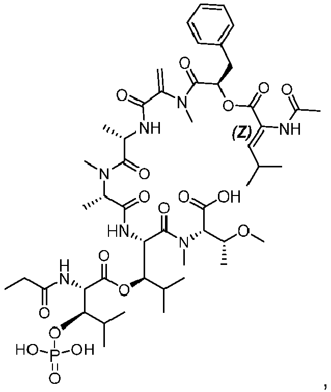

Fig. 15B ). - Anti-PMEL17G1_E152C_S375C_(B1) (G1-(B1)), Anti-PMEL17G1_E152C_(B1) (G1-E152C-DAR2-(B1)), Anti-PMEL17G1_S375C_(B1) (G1-S375C-DAR2-(B1)), and Fc-silent Anti-PMEL17G1_E152C S375C_(B1) (Fc-silent G1- (B1)) were each intravenously injected in nude mice at 20 mg/kg (body weight). Nine mice were dosed with each of the ADC constructs described above. Three animals per group (ADC construct) were terminally bled to collect serum samples for analysis at 24 h,
day 7, andday 14 post dose. Aliquots of each sample were immuno-precipitated using Capture Select FcXL beads and were incubated with buffer containing papain to release the payload from the bead captured ADC. Released payloads were then analyzed by HPLC MS to determine relative levels of Compound (A1)-PO4,


Fig. 15C ). Initial values were estimated based on in vitro experimental results. - Female nude mice bearing 92.1-luciferase subcutaneous xenografts were treated with 3207-E152C_S375C-DAR4-(B1), G1-E152C_S375C-DAR4-(B1), G1-E152C-DAR2-(B1), G1-S375C-DAR2-(B1), Fc-silent 3207-(B1), and Fc-silent G1-DAR4 at 7.5 mg/kg. Treatments (single dose i.v. injection) were performed 17 days post tumor inoculation. Values represent mean ± SEM; sample size, (n=5-6 mice per group). Initial tumor volume at
day 0 was approximately 300-325 mm3. (Fig. 16 ). G1-E152C-DAR2-(B1), Fc-silent G1-DAR4, and G1-E152C_S375C-DAR4-(B1) exhibit comparable antitumor activity in the 92.1 xenograft model of uveal melanoma, while G1-S375C-DAR2-(B1) and non-targeted 3207-ADCs have no effect on tumor growth. - In
Fig. 17A , 92.1 (left panel) and MP41 (right panel) cells were seeded at low cell density in 96-well plates and treated with increasing concentrations of G1-3J-DAR4-(B1), G1-3R-DAR4-(B1), G1-DAR4-(B1) as indicated. Following drug treatment for 96 or 120 hours, cell viability and proliferation were determined using a resazurin-based viability assay. Briefly, cells were incubated with a resazurin-based solution and color change was detected by absorbance with a spectrophotometer and used as a readout of cell viability. Data presented as mean of 3 independent replicates and relative to PBS-treated cells (control). GI50 (is the concentration for 50% of maximal inhibition of cell proliferation) value for each ADC and cell line is depicted in Table 9. Anti-PMEL17-(B1) ADCs inhibit the proliferation of uveal melanoma cells in a PMEL17- and dose-dependent manner.Table 9. Growth inhibitory activity of G1-3J-DAR4-(B1), G1-3R-DAR4-(B1), G1-DAR4-(B1) in UM cells Cell line G1-3J-DAR4-(B1) G1-3R-DAR4-(B1) G1-DAR4-(B1) GI50 nM GI50 nM GI50 nM 92.1 0.850 0.811 0.722 MP41 0.582 0.654 0.563 -
Fig. 17B depicts results of a cell proliferation assay in 92.1 cell line with G1-3J-DAR2 (E152C)-(B1), Fc Silent G1-3J-DAR2 (E152C)-(B1), 3207-DAR2 (E152C)-(B1), and G1-DAR2 (E152C)-(B1) as described inFig. 17A . GI50 is depicted in Table 10.Table 10. Growth inhibitory activity of G1-3J-DAR2 (E152C)-(B1), Fc Silent G1-3J -DAR2 (E152C)-(B1), 3207-DAR2 (E152C)-(B1), and G1-DAR2 (E152C)-(B1) in UM cells Cell line G1-3J-DAR2 (E152C)-(B1) Fc Silent G1-3J -DAR2 (E152C)-(B1) 3207-DAR2 (E152C)-(B1) G1-DAR2 (E152C)-(B1) GI50 nM GI50 nM GI50 nM GI50 nM 92.1 0.309 0.278 >150 0.365 - Female nude mice bearing 92.1-luciferase subcutaneous xenografts were treated with a single i.v. injection of 3207-(B1), G1-3J(E152C), G1-3J-DAR2 (E152C)-(B1), Fc Silent G1-3J-DAR2 (E152C)-(B1), G1-DAR2 (E152C)-(B1) or vehicle at the indicated doses. Treatment (single dose i.v. injection) was performed 14-17 days post tumor inoculation. Values are mean ± SEM; sample size, (n=5-12 mice per group). Initial tumor volume at
day 0 was approximately 200-250 mm3. G1-3J-DAR2 (E152C)-(B1) and Fc Silent G1-3J-DAR2 (E152C)-(B1) inhibited tumor growth in a dose-dependent manner (Fig. 18 ). - Tumours were fixed in 10% (vol/vol) neutral buffered formalin for 24 h at room temperature, then rinsed in PBS, processed for dehydration, cleared, and embedded in paraffin. 3 µm sections were prepared and processed for immunohistochemistry. Immunohistochemical staining was performed on formalin-fixed, paraffin-embedded tissue sections using a Bond-RX (Leica) fully automated system for anti-gp100 (PMEL17) (mouse monoclonal HMB45) staining. Metastatic uveal melanoma samples exhibit high and relatively homogenous expression of PMEL17 (
Fig. 19A ). Quantification of PMEL17 expression levels and proportion of PMEL17 positive cells in metastatic uveal melanoma patient samples (Fig. 19B, Fig. 19C ). - SPR was used to assess epitope binning for anti-PMEL antibodies on a T200 Biacore instrument (catalog #28975001, GE Healthcare Life Sciences).
- Surface Plasmon Resonance (SPR) is a technique to measure bimolecular interactions in real-time in a label free environment. Molecules are immobilized on a sensor surface over which a sample solution flows. The interaction between the immobilized molecule and the flowing sample causes a refractive index change. The refractive index change is an altering of the angle at which reduced intensity polarized light is reflected from the supporting glass plane. This angle change is caused by binding or dissociation of molecules from the sensor surface and is proportional to the mass of bound material and is recorded by the instrument in a sensorgram. The sensorgram shows increasing response as molecules interact, the response remains constant if the interaction reaches equilibrium, and the response decreases as the sample is replaced by buffer and the interaction partners dissociate. From the association and dissociation event responses, an association rate (ka), a dissociation rate (kd) and an overall affinity (KD) are determined. For epitope binning, KDs are not determined.
- In the first step, G1 LC 3J and 17A9 antibodies were directly immobilized onto a CM5 chip surface via Amine Coupling to achieve approximately 2000RU respectively on Fc2 and Fc3. Flow
cell 1 did not have an antibody immobilized onto it and served as the reference Fc. The immobilized antibodies on Fc2 and Fc3 are the base antibodies in the experiment - After immobilization, human PMEL flowed over in the second step at 20nM over all flow cells for 60 seconds at 30ul/min.
- The third and final step has the G1 LC 3J antibody alone or G1 LC 3J antibody plus 17A9 antibody flowing over Fc2 or the 17A9 antibody alone or 17A9 antibody plus G1 LC 3J antibody flowing over Fc3 (
FIG. 20A ). The antibodies flowed over at 500nM total concentration over both flow cells for 120 seconds at 30ul/min for association followed by a dissociation phase of 120 seconds at 30ul/min with running buffer. - As expected, when G1 LC 3J was the base antibody, no binging signal was observed when G13J flowed over. 100RU binding was observed when 17A9 was flowed over, suggestive it binds to a different epitope than G1 LC 3J (
FIG. 20B ). Similar trends observed when 17A9 was the base antibody. Instep 3, no binding observed with itself but binding observed when G1 LC 3J flowed over (FIG. 20C ). This indicated that G1 LC 3J and 17A9 bind to different epitopes. - Regeneration was performed at the end of each cycle on all flow cells. The regeneration buffer was Glycine 2.0 which flowed at 30µl/min for 30 seconds.
- The data was analyzed using the Biacore T200 Evaluation Software version 3.0.
- It is understood that the examples and embodiments described herein are for illustrative purposes only and that various modifications or changes in light thereof will be suggested to persons skilled in the art and are to be included within the spirit and purview of this application and the scope of the appended claims.
- The following numbered clauses, describing aspects of the invention, are part of the description.
- 1. An antibody or antigen binding fragment thereof that binds PMEL17 comprising:
- a. a heavy chain variable region that comprises a heavy chain CDR1 (Complementarity Determining Region 1) of SEQ ID NO:1, 4, 5 or 7, a heavy chain CDR2 (Complementarity Determining Region 2) of SEQ ID NO:2, 6 or 8, and a heavy chain CDR3 (Complementarity Determining Region 3) of SEQ ID NO:3 or 9; and a light chain variable region that comprises a light chain CDR1 (Complementarity Determining Region 1) of SEQ ID NO:14, 17 or 20, a light chain CDR2 (Complementarity Determining Region 2) of SEQ ID NO:15 or 18, and a light chain CDR3 (Complementarity Determining Region 3) of SEQ ID NO:16 or 19;
- b. a heavy chain variable region that comprises a heavy chain CDR1 of SEQ ID NO:33, 36, 37 or 39, a heavy chain CDR2 of SEQ ID NO:34, 38 or 40; a heavy chain CDR3 of SEQ ID NO:35 or 41; a light chain CDR1 of SEQ ID NO:46, 49 or 52; a light chain CDR2 of SEQ ID NO:47 or 50; and a light chain CDR3 of SEQ ID NO:48 or 51;
- c. a heavy chain variable region that comprises a heavy chain CDR1 of SEQ ID NO:5, 7, 57 or 60, a heavy chain CDR2 of SEQ ID NO:58, 61 or 62; a heavy chain CDR3 of SEQ ID NO:59 or 63; a light chain CDR1 of SEQ ID NO:68, 71 or 74; a light chain CDR2 of SEQ ID NO:69 or 72; and a light chain CDR3 of SEQ ID NO:70 or 73;
- d. a heavy chain variable region that comprises a heavy chain CDR1 of SEQ ID NO:79, 82, 83 or 85, a heavy chain CDR2 of SEQ ID NO:80, 84 or 86; a heavy chain CDR3 of SEQ ID NO:81 or 87; a light chain CDR1 of SEQ ID NO:92, 95 or 98; a light chain CDR2 of SEQ ID NO:93 or 96; and a light chain CDR3 of SEQ ID NO:94 or 97;
- e. a heavy chain variable region that comprises a heavy chain CDR1 of SEQ ID NO:103, 106, 107 or 109, a heavy chain CDR2 of SEQ ID NO:104, 108 or 110; a heavy chain CDR3 of SEQ ID NO:105 or 111; a light chain CDR1 of SEQ ID NO:49, 52 or 116; a light chain CDR2 of SEQ ID NO:47 or 50; and a light chain CDR3 of SEQ ID NO:117 or 118;
- f. a heavy chain variable region that comprises a heavy chain CDR1 of SEQ ID NO:123, 126, 127 or 129, a heavy chain CDR2 of SEQ ID NO:124, 128 or 130; a heavy chain CDR3 of SEQ ID NO:125 or 131; a light chain CDR1 of SEQ ID NO:136, 139 or 142; a light chain CDR2 of SEQ ID NO:137 or 140; and a light chain CDR3 of SEQ ID NO:138 or 141;
- g. a heavy chain variable region that comprises a heavy chain CDR1 of SEQ ID NO:123, 126, 127 or 129, a heavy chain CDR2 of SEQ ID NO:124, 128 or 130; a heavy chain CDR3 of SEQ ID NO:147 or 148; a light chain CDR1 of SEQ ID NO:153, 156 or 158; a light chain CDR2 of SEQ ID NO:50 or 154; and a light chain CDR3 of SEQ ID NO:155 or 157;
- h. a heavy chain variable region that comprises a heavy chain CDR1 of SEQ ID NO:103, 106, 107 or 109, a heavy chain CDR2 of SEQ ID NO:104, 108 or 110; a heavy chain CDR3 of SEQ ID NO:163 or 164; a light chain CDR1 of SEQ ID NO:49, 52 or 116; a light chain CDR2 of SEQ ID NO:47 or 50; and a light chain CDR3 of SEQ ID NO:169 or 170;
- i. a heavy chain variable region that comprises a heavy chain CDR1 of SEQ ID NO:175, 178, 179 or 181, a heavy chain CDR2 of SEQ ID NO:176, 180 or 182; a heavy chain CDR3 of SEQ ID NO:177 or 183; a light chain CDR1 of SEQ ID NO:49, 52 or 116; a light chain CDR2 of SEQ ID NO:47 or 50; and a light chain CDR3 of SEQ ID NO:188 or 189;
- j. a heavy chain variable region that comprises a heavy chain CDR1 of SEQ ID NO: 103, 106, 107 or 109, a heavy chain CDR2 of SEQ ID NO: 104, 108 or 110; a heavy chain CDR3 of SEQ ID NO:194 or 195; a light chain CDR1 of SEQ ID NO: 49, 52 or 116; a light chain CDR2 of SEQ ID NO: 47 or 50; and a light chain CDR3 of SEQ ID NO:200 or 201;
- k. a heavy chain variable region that comprises a heavy chain CDR1 of SEQ ID NO:206, 209, 210 or 212, a heavy chain CDR2 of SEQ ID NO:207, 211 or 213; a heavy chain CDR3 of SEQ ID NO:208 or 214; a light chain CDR1 of SEQ ID NO:153, 156 or 158; a light chain CDR2 of SEQ ID NO:50 or 154; and a light chain CDR3 of SEQ ID NO:219 or 220;
- l. a heavy chain variable region that comprises a heavy chain CDR1 of SEQ ID NO: 206, 209, 210 or 212, a heavy chain CDR2 of SEQ ID NO: 207, 211 or 213; a heavy chain CDR3 of SEQ ID NO:225 or 226; a light chain CDR1 of SEQ ID NO:136, 139 or 142; a light chain CDR2 of SEQ ID NO:137 or 140; and a light chain CDR3 of SEQ ID NO:231 or 232;
- m. a heavy chain variable region that comprises a heavy chain variable region that comprises an HCDR1 of SEQ ID NO: 206, 209, 210 or 212, an HCDR2 of SEQ ID NO: 207, 211 or 213, and an HCDR3 of SEQ ID NO:237 or 238; and a light chain variable region that comprises an LCDR1 of SEQ ID NO:243, 245 or 247, an LCDR2 of SEQ ID NO:47 or 50, and an LCDR3 of SEQ ID NO:244 or 246;
- n. a heavy chain variable region that comprises a heavy chain variable region that comprises an HCDR1 of SEQ ID NO: 206, 209, 210 or 212, an HCDR2 of SEQ ID NO: 207, 211 or 213, and an HCDR3 of SEQ ID NO:252 or 253; and a light chain variable region that comprises an LCDR1 of SEQ ID NO:153, 156 or 158, an LCDR2 of SEQ ID NO:50 or 154, and an LCDR3 of SEQ ID NO:258 or 259;
- o. a heavy chain CDR1 of SEQ ID NO:1, a heavy chain CDR2 of SEQ ID NO:2, a heavy chain CDR3 of SEQ ID NO:3, a light chain CDR1 of SEQ ID NO:14, a light chain CDR2 of SEQ ID NO:15, and a light chain CDR3 of SEQ ID NO:16;
- p. a heavy chain CDR1 of SEQ ID NO: 4, a heavy chain CDR2 of SEQ ID NO:2, a heavy chain CDR3 of SEQ ID NO:3, a light chain CDR1 of SEQ ID NO:14, a light chain CDR2 of SEQ ID NO:15, and a light chain CDR3 of SEQ ID NO:16;
- q. a heavy chain CDR1 of SEQ ID NO:5, a heavy chain CDR2 of SEQ ID NO:6, a heavy chain CDR3 of SEQ ID NO:3, a light chain CDR1 of SEQ ID NO:17, a light chain CDR2 of SEQ ID NO: 18, and a light chain CDR3 of SEQ ID NO: 19;
- r. a heavy chain CDR1 of SEQ ID NO:7, a heavy chain CDR2 of SEQ ID NO:8, a heavy chain CDR3 of SEQ ID NO:9, a light chain CDR1 of SEQ ID NO:20, a light chain CDR2 of SEQ ID NO:18, and a light chain CDR3 of SEQ ID NO:16;
- s. a heavy chain CDR1 of SEQ ID NO:33, a heavy chain CDR2 of SEQ ID NO:34, a heavy chain CDR3 of SEQ ID NO:35, a light chain CDR1 of SEQ ID NO:46, a light chain CDR2 of SEQ ID NO:47, and a light chain CDR3 of SEQ ID NO:48;
- t. a heavy chain CDR1 of SEQ ID NO:36, a heavy chain CDR2 of SEQ ID NO:34, a heavy chain CDR3 of SEQ ID NO:35, a light chain CDR1 of SEQ ID NO:46, a light chain CDR2 of SEQ ID NO:47, and a light chain CDR3 of SEQ ID NO:48;
- u. a heavy chain CDR1 of SEQ ID NO:37, a heavy chain CDR2 of SEQ ID NO:38, a heavy chain CDR3 of SEQ ID NO:35, a light chain CDR1 of SEQ ID NO:49, a light chain CDR2 of SEQ ID NO:50, and a light chain CDR3 of SEQ ID NO:51;
- v. a heavy chain CDR1 of SEQ ID NO: 39, a heavy chain CDR2 of SEQ ID NO:40, a heavy chain CDR3 of SEQ ID NO:41, a light chain CDR1 of SEQ ID NO:52, a light chain CDR2 of SEQ ID NO:50, and a light chain CDR3 of SEQ ID NO:48;
- w. a heavy chain CDR1 of SEQ ID NO:57, a heavy chain CDR2 of SEQ ID NO:58, a heavy chain CDR3 of SEQ ID NO:59, a light chain CDR1 of SEQ ID NO:68, a light chain CDR2 of SEQ ID NO:69, and a light chain CDR3 of SEQ ID NO:70;
- x. a heavy chain CDR1 of SEQ ID NO:60, a heavy chain CDR2 of SEQ ID NO:58, a heavy chain CDR3 of SEQ ID NO:59, a light chain CDR1 of SEQ ID NO:68, a light chain CDR2 of SEQ ID NO:69, and a light chain CDR3 of SEQ ID NO:70;
- y. a heavy chain CDR1 of SEQ ID NO:5, a heavy chain CDR2 of SEQ ID NO:61, a heavy chain CDR3 of SEQ ID NO:59, a light chain CDR1 of SEQ ID NO:71, a light chain CDR2 of SEQ ID NO:72, and a light chain CDR3 of SEQ ID NO:73;
- z. a heavy chain CDR1 of SEQ ID NO:7, a heavy chain CDR2 of SEQ ID NO:62, a heavy chain CDR3 of SEQ ID NO:63, a light chain CDR1 of SEQ ID NO:74, a light chain CDR2 of SEQ ID NO:72, and a light chain CDR3 of SEQ ID NO:70;
- aa. a heavy chain CDR1 of SEQ ID NO:79, , a heavy chain CDR2 of SEQ ID NO:80, a heavy chain CDR3 of SEQ ID NO:81, a light chain CDR1 of SEQ ID NO:92, a light chain CDR2 of SEQ ID NO:93, and a light chain CDR3 of SEQ ID NO:94;
- bb. a heavy chain CDR1 of SEQ ID NO:82, a heavy chain CDR2 of SEQ ID NO:80, a heavy chain CDR3 of SEQ ID NO:81, a light chain CDR1 of SEQ ID NO:92, a light chain CDR2 of SEQ ID NO:93, and a light chain CDR3 of SEQ ID NO:94;
- cc. a heavy chain CDR1 of SEQ ID NO:83, a heavy chain CDR2 of SEQ ID NO:84, a heavy chain CDR3 of SEQ ID NO:81, a light chain CDR1 of SEQ ID NO:95, a light chain CDR2 of SEQ ID NO:96, and a light chain CDR3 of SEQ ID NO: 97;
- dd. a heavy chain CDR1 of SEQ ID NO: 85, a heavy chain CDR2 of SEQ ID NO:86, a heavy chain CDR3 of SEQ ID NO:87, a light chain CDR1 of SEQ ID NO:98, a light chain CDR2 of SEQ ID NO:96, and a light chain CDR3 of SEQ ID NO:94;
- ee. a heavy chain CDR1 of SEQ ID NO:103, a heavy chain CDR2 of SEQ ID NO:104, a heavy chain CDR3 of SEQ ID NO:105, a light chain CDR1 of SEQ ID NO: 116; a light chain CDR2 of SEQ ID NO:47; and a light chain CDR3 of SEQ ID NO:117;
- ff. a heavy chain CDR1 of SEQ ID NO:106, a heavy chain CDR2 of SEQ ID NO:104, a heavy chain CDR3 of SEQ ID NO:105, a light chain CDR1 of SEQ ID NO: 116, a light chain CDR2 of SEQ ID NO:47, and a light chain CDR3 of SEQ ID NO:117;
- gg. a heavy chain CDR1 of SEQ ID NO:107, a heavy chain CDR2 of SEQ ID NO:108, a heavy chain CDR3 of SEQ ID NO:105, a light chain CDR1 of SEQ ID NO:49, a light chain CDR2 of SEQ ID NO:50, and a light chain CDR3 of SEQ ID NO:118;
- hh. a heavy chain CDR1 of SEQ ID NO:109, a heavy chain CDR2 of SEQ ID NO:110, a heavy chain CDR3 of SEQ ID NO:111, a light chain CDR1 of SEQ ID NO:52 a light chain CDR2 of SEQ ID NO:50, and a light chain CDR3 of SEQ ID NO:117;
- ii. a heavy chain CDR1 of SEQ ID NO:123, a heavy chain CDR2 of SEQ ID NO:124, a heavy chain CDR3 of SEQ ID NO:125, a light chain CDR1 of SEQ ID NO:136, a light chain CDR2 of SEQ ID NO:137,and a light chain CDR3 of SEQ ID NO:138;
- jj. a heavy chain CDR1 of SEQ ID NO:126, a heavy chain CDR2 of SEQ ID NO:124, a heavy chain CDR3 of SEQ ID NO:125, a light chain CDR1 of SEQ ID NO:136, a light chain CDR2 of SEQ ID NO:137, and a light chain CDR3 of SEQ ID NO:138;
- kk. a heavy chain CDR1 of SEQ ID NO:127, a heavy chain CDR2 of SEQ ID NO:128, a heavy chain CDR3 of SEQ ID NO:125, a light chain CDR1 of SEQ ID NO:139, a light chain CDR2 of SEQ ID NO:140, and a light chain CDR3 of SEQ ID NO: 141;
- II. a heavy chain CDR1 of SEQ ID NO: 129, a heavy chain CDR2 of SEQ ID NO:130, a heavy chain CDR3 of SEQ ID NO:131, a light chain CDR1 of SEQ ID NO:142, a light chain CDR2 of SEQ ID NO:140, and a light chain CDR3 of SEQ ID NO:138;
- mm. a heavy chain CDR1 of SEQ ID NO:123, a heavy chain CDR2 of SEQ ID NO:124, a heavy chain CDR3 of SEQ ID NO:147, a light chain CDR1 of SEQ ID NO:153, a light chain CDR2 of SEQ ID NO:154, and a light chain CDR3 of SEQ ID NO:155;
- nn. a heavy chain CDR1 of SEQ ID NO:126, a heavy chain CDR2 of SEQ ID NO:124, a heavy chain CDR3 of SEQ ID NO:147, a light chain CDR1 of SEQ ID NO:153, a light chain CDR2 of SEQ ID NO: 154, and a light chain CDR3 of SEQ ID NO:155;
- oo. a heavy chain CDR1 of SEQ ID NO:127, a heavy chain CDR2 of SEQ ID NO:128, a heavy chain CDR3 of SEQ ID NO:147, a light chain CDR1 of SEQ ID NO:156, a light chain CDR2 of SEQ ID NO:50, and a light chain CDR3 of SEQ ID NO:157;
- pp. a heavy chain CDR1 of SEQ ID NO: 129, a heavy chain CDR2 of SEQ ID NO:130, a heavy chain CDR3 of SEQ ID NO:148, a light chain CDR1 of SEQ ID NO:158, a light chain CDR2 of SEQ ID NO:50, and a light chain CDR3 of SEQ ID NO:155;
- qq. a heavy chain CDR1 of SEQ ID NO:103, a heavy chain CDR2 of SEQ ID NO:104, a heavy chain CDR3 of SEQ ID NO:163, a light chain CDR1 of SEQ ID NO: 116, a light chain CDR2 of SEQ ID NO:47, and a light chain CDR3 of SEQ ID NO:169;
- rr. a heavy chain CDR1 of SEQ ID NO:106, a heavy chain CDR2 of SEQ ID NO:104, a heavy chain CDR3 of SEQ ID NO:163, a light chain CDR1 of SEQ ID NO:116, a light chain CDR2 of SEQ ID NO:47, and a light chain CDR3 of SEQ ID NO:169;
- ss. a heavy chain CDR1 of SEQ ID NO:107, a heavy chain CDR2 of SEQ ID NO:108, a heavy chain CDR3 of SEQ ID NO:163, a light chain CDR1 of SEQ ID NO:49, a light chain CDR2 of SEQ ID NO:50, and a light chain CDR3 of SEQ ID NO:170;
- tt. a heavy chain CDR1 of SEQ ID NO: 109, a heavy chain CDR2 of SEQ ID NO:110, a heavy chain CDR3 of SEQ ID NO:164, a light chain CDR1 of SEQ ID NO:52, a light chain CDR2 of SEQ ID NO:50, and a light chain CDR3 of SEQ ID NO:169;
- uu. a heavy chain CDR1 of SEQ ID NO:175, a heavy chain CDR2 of SEQ ID NO:176, a heavy chain CDR3 of SEQ ID NO:177, a light chain CDR1 of SEQ ID NO:116, a light chain CDR2 of SEQ ID NO:47, and a light chain CDR3 of SEQ ID NO:188;
- vv. a heavy chain CDR1 of SEQ ID NO:178, a heavy chain CDR2 of SEQ ID NO:176, a heavy chain CDR3 of SEQ ID NO:177, a light chain CDR1 of SEQ ID NO:116, a light chain CDR2 of SEQ ID NO:47, and a light chain CDR3 of SEQ ID NO:188;
- ww. a heavy chain CDR1 of SEQ ID NO:179, a heavy chain CDR2 of SEQ ID NO:180, a heavy chain CDR3 of SEQ ID NO:177, a light chain CDR1 of SEQ ID NO:49, a light chain CDR2 of SEQ ID NO:50, and a light chain CDR3 of SEQ ID NO:189;
- xx. a heavy chain CDR1 of SEQ ID NO: 181, a heavy chain CDR2 of SEQ ID NO:182; a heavy chain CDR3 of SEQ ID NO:183, a light chain CDR1 of SEQ ID NO:52, a light chain CDR2 of SEQ ID NO:50, and a light chain CDR3 of SEQ ID NO:188;
- yy. a heavy chain CDR1 of SEQ ID NO: 103, a heavy chain CDR2 of SEQ ID NO: 104, a heavy chain CDR3 of SEQ ID NO:194, a light chain CDR1 of SEQ ID NO: 116, a light chain CDR2 of SEQ ID NO: 47, and a light chain CDR3 of SEQ ID NO:200;
- zz. a heavy chain CDR1 of SEQ ID NO: 106, a heavy chain CDR2 of SEQ ID NO: 104, a heavy chain CDR3 of SEQ ID NO:194, a light chain CDR1 of SEQ ID NO: 116, a light chain CDR2 of SEQ ID NO: 47, and a light chain CDR3 of SEQ ID NO:200;
- aaa. a heavy chain CDR1 of SEQ ID NO: 107, a heavy chain CDR2 of SEQ ID NO: 108, a heavy chain CDR3 of SEQ ID NO:194, a light chain CDR1 of SEQ ID NO: 49, a light chain CDR2 of SEQ ID NO: 50, and a light chain CDR3 of SEQ ID NO: 201;
- bbb. a heavy chain CDR1 of SEQ ID NO: 109, a heavy chain CDR2 of SEQ ID NO: 110, a heavy chain CDR3 of SEQ ID NO:195, a light chain CDR1 of SEQ ID NO: 52, a light chain CDR2 of SEQ ID NO: 50, and a light chain CDR3 of SEQ ID NO:200;
- ccc. a heavy chain CDR1 of SEQ ID NO:206, a heavy chain CDR2 of SEQ ID NO:207, a heavy chain CDR3 of SEQ ID NO:208, a light chain CDR1 of SEQ ID NO:153, a light chain CDR2 of SEQ ID NO:154, and a light chain CDR3 of SEQ ID NO:219;
- ddd. a heavy chain CDR1 of SEQ ID NO:209, a heavy chain CDR2 of SEQ ID NO:207, a heavy chain CDR3 of SEQ ID NO:208, a light chain CDR1 of SEQ ID NO:153, a light chain CDR2 of SEQ ID NO: 154, and a light chain CDR3 of SEQ ID NO:219;
- eee. a heavy chain CDR1 of SEQ ID NO:210, a heavy chain CDR2 of SEQ ID NO:211, a heavy chain CDR3 of SEQ ID NO:208, a light chain CDR1 of SEQ ID NO:156, a light chain CDR2 of SEQ ID NO:50, and a light chain CDR3 of SEQ ID NO:220;
- fff. a heavy chain CDR1 of SEQ ID NO: 212, a heavy chain CDR2 of SEQ ID NO:213, a heavy chain CDR3 of SEQ ID NO:214, a light chain CDR1 of SEQ ID NO:158, a light chain CDR2 of SEQ ID NO:50, and a light chain CDR3 of SEQ ID NO:219;
- ggg. a heavy chain CDR1 of SEQ ID NO: 206, a heavy chain CDR2 of SEQ ID NO: 207, a heavy chain CDR3 of SEQ ID NO:225, a light chain CDR1 of SEQ ID NO:136, a light chain CDR2 of SEQ ID NO:137,and a light chain CDR3 of SEQ ID NO:231;
- hhh. a heavy chain CDR1 of SEQ ID NO: 209, a heavy chain CDR2 of SEQ ID NO: 207, a heavy chain CDR3 of SEQ ID NO:225, a light chain CDR1 of SEQ ID NO:136, a light chain CDR2 of SEQ ID NO:137, and a light chain CDR3 of SEQ ID NO:231;
- iii. a heavy chain CDR1 of SEQ ID NO: 210, a heavy chain CDR2 of SEQ ID NO: 211, a heavy chain CDR3 of SEQ ID NO:225, a light chain CDR1 of SEQ ID NO:139, a light chain CDR2 of SEQ ID NO:140, and a light chain CDR3 of SEQ ID NO: 232;
- jjj. a heavy chain CDR1 of SEQ ID NO: 212, a heavy chain CDR2 of SEQ ID NO: 213, a heavy chain CDR3 of SEQ ID NO: 226, a light chain CDR1 of SEQ ID NO:142; a light chain CDR2 of SEQ ID NO: 140; and a light chain CDR3 of SEQ ID NO:231;
- kkk. a heavy chain variable region that comprises an HCDR1 of SEQ ID NO: 206, an HCDR2 of SEQ ID NO: 207, and an HCDR3 of SEQ ID NO:237,and a light chain variable region that comprises an LCDR1 of SEQ ID NO:243, an LCDR2 of SEQ ID NO:47, and an LCDR3 of SEQ ID NO:244;
- III. a heavy chain variable region that comprises an HCDR1 of SEQ ID NO: 209, an HCDR2 of SEQ ID NO: 207, and an HCDR3 of SEQ ID NO:237, and a light chain variable region that comprises an LCDR1 of SEQ ID NO:243, an LCDR2 of SEQ ID NO:47, and an LCDR3 of SEQ ID NO:244;
- mmm. a heavy chain variable region that comprises an HCDR1 of SEQ ID NO: 210, an HCDR2 of SEQ ID NO: 211, and an HCDR3 of SEQ ID NO:237, and a light chain variable region that comprises an LCDR1 of SEQ ID NO:245, an LCDR2 of SEQ ID NO:50, and an LCDR3 of SEQ ID NO:246;
- nnn. a heavy chain variable region that comprises an HCDR1 of SEQ ID NO: 212, an HCDR2 of SEQ ID NO: 213, and an HCDR3 of SEQ ID NO:238; and a light chain variable region that comprises an LCDR1 of SEQ ID NO:247, an LCDR2 of SEQ ID NO: 50, and an LCDR3 of SEQ ID NO:244;
- ooo. a heavy chain variable region that comprises an HCDR1 of SEQ ID NO: 206, an HCDR2 of SEQ ID NO: 207, and an HCDR3 of SEQ ID NO:252,and a light chain variable region that comprises an LCDR1 of SEQ ID NO:153, an LCDR2 of SEQ ID NO: 154, and an LCDR3 of SEQ ID NO:258;
- ppp. a heavy chain variable region that comprises an HCDR1 of SEQ ID NO: 209, an HCDR2 of SEQ ID NO: 207, and an HCDR3 of SEQ ID NO:252, and a light chain variable region that comprises an LCDR1 of SEQ ID NO:153, an LCDR2 of SEQ ID NO:154, and an LCDR3 of SEQ ID NO:258;
- qqq. a heavy chain variable region that comprises an HCDR1 of SEQ ID NO: 210, an HCDR2 of SEQ ID NO: 211, and an HCDR3 of SEQ ID NO:252, and a light chain variable region that comprises an LCDR1 of SEQ ID NO:156, an LCDR2 of SEQ ID NO:50, and an LCDR3 of SEQ ID NO:259; or
- rrr. a heavy chain variable region that comprises an HCDR1 of SEQ ID NO: 212, an HCDR2 of SEQ ID NO: 213, and an HCDR3 of SEQ ID NO: 253; and a light chain variable region that comprises an LCDR1 of SEQ ID NO: 158, an LCDR2 of SEQ ID NO:50, and an LCDR3 of SEQ ID NO:258.
- 2. An antibody or antigen binding fragment thereof that binds PMEL17 comprising:
- a. A heavy chain variable region (VH) comprising the amino acid sequence of SEQ ID NO:10, and a light chain variable region (VL) comprising the amino acid sequence of SEQ ID NO:21;
- b. A heavy chain variable region (VH) comprising the amino acid sequence of SEQ ID NO:10, and a light chain variable region (VL) comprising the amino acid sequence of SEQ ID NO:25;
- c. A heavy chain variable region (VH) comprising the amino acid sequence of SEQ ID NO:10, and a light chain variable region (VL) comprising the amino acid sequence of SEQ ID NO:29;
- d. A heavy chain variable region (VH) comprising the amino acid sequence of SEQ ID NO:42, and a light chain variable region (VL) comprising the amino acid sequence of SEQ ID NO:53;
- e. A heavy chain variable region (VH) comprising the amino acid sequence of SEQ ID NO:64, and a light chain variable region (VL) comprising the amino acid sequence of SEQ ID NO:75;
- f. A heavy chain variable region (VH) comprising the amino acid sequence of SEQ ID NO:88, and a light chain variable region (VL) comprising the amino acid sequence of SEQ ID NO:99;
- g. A heavy chain variable region (VH) comprising the amino acid sequence of SEQ ID NO:112, and a light chain variable region (VL) comprising the amino acid sequence of SEQ ID NO:119;
- h. A heavy chain variable region (VH) comprising the amino acid sequence of SEQ ID NO:132, and a light chain variable region (VL) comprising the amino acid sequence of SEQ ID NO:143;
- i. A heavy chain variable region (VH) comprising the amino acid sequence of SEQ ID NO:149, and a light chain variable region (VL) comprising the amino acid sequence of SEQ ID NO:159;
- j. A heavy chain variable region (VH) comprising the amino acid sequence of SEQ ID NO:165, and a light chain variable region (VL) comprising the amino acid sequence of SEQ ID NO:171;
- k. A heavy chain variable region (VH) comprising the amino acid sequence of SEQ ID NO:184, and a light chain variable region (VL) comprising the amino acid sequence of SEQ ID NO:190;
- l. A heavy chain variable region (VH) comprising the amino acid sequence of SEQ ID NO:196, and a light chain variable region (VL) comprising the amino acid sequence of SEQ ID NO:202;
- m. A heavy chain variable region (VH) comprising the amino acid sequence of SEQ ID NO:215, and a light chain variable region (VL) comprising the amino acid sequence of SEQ ID NO:221;
- n. A heavy chain variable region (VH) comprising the amino acid sequence of SEQ ID NO:227, and a light chain variable region (VL) comprising the amino acid sequence of SEQ ID NO:233;
- o. A heavy chain variable region (VH) comprising the amino acid sequence of SEQ ID NO:239, and a light chain variable region (VL) comprising the amino acid sequence of SEQ ID NO:248; or
- p. A heavy chain variable region (VH) comprising the amino acid sequence of SEQ ID NO:254, and a light chain variable region (VL) comprising the amino acid sequence of SEQ ID NO:260.
- 3. An antibody or antigen binding fragment thereof that binds PMEL17 comprising:
- a. A heavy chain comprising the amino acid sequence of SEQ ID NO:12, and a light chain comprising the amino acid sequence of SEQ ID NO:23;
- b. A heavy chain comprising the amino acid sequence of SEQ ID NO:12, and a light chain comprising the amino acid sequence of SEQ ID NO:27;
- c. A heavy chain comprising the amino acid sequence of SEQ ID NO:12, and a light chain comprising the amino acid sequence of SEQ ID NO:31;
- d. A heavy chain comprising the amino acid sequence of SEQ ID NO:44, and a light chain comprising the amino acid sequence of SEQ ID NO:55;
- e. A heavy chain comprising the amino acid sequence of SEQ ID NO:66, and a light chain comprising the amino acid sequence of SEQ ID NO:77;
- f. A heavy chain comprising the amino acid sequence of SEQ ID NO:90, and a light chain comprising the amino acid sequence of SEQ ID NO:101;
- g. A heavy chain comprising the amino acid sequence of SEQ ID NO:114, and a light chain comprising the amino acid sequence of SEQ ID NO:121.;
- h. A heavy chain comprising the amino acid sequence of SEQ ID NO:134, and a light chain comprising the amino acid sequence of SEQ ID NO:145;
- i. A heavy chain comprising the amino acid sequence of SEQ ID NO:151, and a light chain comprising the amino acid sequence of SEQ ID NO:161;
- j. A heavy chain comprising the amino acid sequence of SEQ ID NO:167, and a light chain comprising the amino acid sequence of SEQ ID NO:173;
- k. A heavy chain comprising the amino acid sequence of SEQ ID NO:186, and a light chain comprising the amino acid sequence of SEQ ID NO:192;
- l. A heavy chain comprising the amino acid sequence of SEQ ID NO:198, and a light chain comprising the amino acid sequence of SEQ ID NO:204;
- m. A heavy chain comprising the amino acid sequence of SEQ ID NO:217, and a light chain comprising the amino acid sequence of SEQ ID NO:223;
- n. A heavy chain comprising the amino acid sequence of SEQ ID NO:229, and a light chain comprising the amino acid sequence of SEQ ID NO:235;
- o. A heavy chain comprising the amino acid sequence of SEQ ID NO:241, and a light chain comprising the amino acid sequence of SEQ ID NO:250; or
- p. A heavy chain comprising the amino acid sequence of SEQ ID NO:256, and a light chain comprising the amino acid sequence of SEQ ID NO:262.
- 4. The antibody of any one of clauses 1-3, wherein the antibody or antigen binding fragment thereof comprises one or more cysteine substitutions.
- 5. The antibody of
clause 4, wherein the antibody or antigen binding fragment thereof comprises one or more cysteine substitutions selected from S152C, S375C, or both S152C and S375C of the heavy chain of the antibody or antigen binding fragment thereof, wherein the position is numbered according to the EU system. - 6. The antibody of any one of clauses 1-5, wherein said antibody is a monoclonal antibody.
- 7. An antibody drug conjugate comprising the formula (C)
Ab-(LA-(D)n)y (C)
wherein:- D is a GNAQ inhibitor, a GNA11 inhibitor or an inhibitor of GNAQ and GNA11;
- Ab is an antibody or antigen binding fragment thereof that binds to human PMEL17 protein;
- LA is a linker;
- n is 1, 2, 3 or 4, and
- 8. The antibody drug conjugate of
clause 7, wherein said n is 1. - 9. The antibody drug conjugate of any one of
clauses - 10. The antibody drug conjugate of any one of clauses 7-9, wherein said linker is a cleavable linker or a non-cleavable linker.
- 11. The antibody drug conjugate of
clause 10, wherein the linker comprises a ValCit peptide linker. - 12. The antibody drug conjugate of any one of clauses 7-11, wherein said drug moiety is an inhibitor of GNAQ and GNA11.
- 13. The antibody drug conjugate of any one of clauses 7-11, wherein the D is

- 14. The antibody drug conjugate of any one of clauses 7-11, wherein D is

- 15. The antibody drug conjugate of any one of clauses 7-14 having the following structure,

- 16. The antibody drug conjugate of any one of clauses 7-14 having the following structure,

- 17. An antibody drug conjugate having the following Formula (C-2):

- R° is methyl or ethyl;
- R1 is methyl or isopropyl;
- R2 is methyl or ethyl;
- Ab is an antibody or antigen binding fragment thereof that binds to human PMEL17 protein;
- X1 is a bivalent coupling group;
- X2 is a self-immolative spacer;
- L1 is a bivalent peptide linker;
- L2 is a bond or a linker, and
- y is 1, 2, 3 or 4.
- 18. A pharmaceutical composition comprising the antibody, or antigen binding fragment thereof, of any one of clauses 1-6 and a pharmaceutically acceptable carrier.
- 19. A pharmaceutical composition comprising the antibody drug conjugate of any one of clauses 7-17 and a pharmaceutically acceptable carrier.
- 20. A method of treating or preventing cancer in a patient in need thereof, comprising administering to said patient the antibody drug conjugate of any one of clauses7-17, or the pharmaceutical composition of any one of clauses 18 or 19, wherein the cancer expresses PMEL17, contains a mutation of the GNAQ or
GNA1 1 gene, or the cancer expresses PMEL17 and contains a mutation of GNAQ, GNA11, or both. - 21. The method of
clause 20, wherein the antibody drug conjugate or pharmaceutical composition are administered to the patient in combination with one or more additional therapeutic compounds. - 22. The method of clause 21, wherein the one or more additional therapeutic compounds is selected from a standard of care chemotherapeutic, an MDM2 inhibitor, an MRC2 inhibitor, a PKC inhibitor, a MAPK inhibitor, a costimulatory molecule, or a checkpoint inhibitor.
- 23. The method of clause 22, wherein the costimulatory molecule is selected from an agonist of OX40, CD2, CD27, CDS, ICAM-1, LFA-1 (CD11a/CD18), ICOS (CD278), 4-1BB (CD137), GITR, CD30, CD40, BAFFR, HVEM, CD7, LIGHT, NKG2C, SLAMF7, NKp80, CD160, B7-H3, STING, or CD83 ligand.
- 24. The method of clause22, wherein the checkpoint inhibitor is selected from an inhibitor of PD-1, PD-L1, PD-L2, CTLA4, TIM3, LAG3, VISTA, BTLA, TIGIT, LAIR1, CD160, 2B4 and/or TGFR beta.
- 25. The antibody drug conjugate of any one of clauses 7-17, or the pharmaceutical composition of any one of clauses 18 or 19, for use as a medicament.
- 26. The antibody drug conjugate of any one of clauses 7-17, or the pharmaceutical composition of any one of clauses 18 or 19, for use in the treatment or prevention of a PMEL17 expressing cancer or a cancer that contains a mutation of the GNAQ or GNA11 gene in a patient in need thereof.
- 27. Use of the antibody or antigen binding fragment thereof of any one of clauses 1-6, the antibody drug conjugate of any one of clauses 7-17, or the pharmaceutical composition of any one of clauses 18 or 19, to treat or prevent a PMEL17 expressing cancer or a cancer that contains a mutation of the GNAQ or GNA11 gene in a patient in need thereof.
- 28. Use of the antibody or antigen binding fragment thereof of any one of clauses 1-6, the antibody drug conjugate of any one of clauses 7-17, or the pharmaceutical composition of any one of clauses 18 or 19 in the manufacture of a medicament.
- 29. The method of any one of clauses 20-24, the antibody drug conjugate of any one of clauses 25-26, or the use of any one of clauses 27-28, wherein the cancer expresses PMEL17 or contains a mutation of the GNAQ or GNA11 gene.
- 30. The method, antibody drug conjugate, or use of clause 28, wherein the cancer is uveal melanoma, subcutaneous melanoma, hepatocellular carcinoma, or a metastatic cancer thereof.
- 31. A nucleic acid that encodes the antibody or antigen binding fragment of any one of clauses 1-6.
- 32. The nucleic acid of clause 31, wherein the nucleic acid comprises the nucleotide sequence of SEQ ID NOs: 13, 24, 28, 32, 45, 56, 67, 78, 91, 102, 115, 122, 135, 146, 152, 162, 168, 174, 187, 193, 199, 205, 218, 224, 230, 236, 242, 251, 257, or 263.
- 33. A vector comprising the nucleic acid of clause 31 or 32.
- 34. A host cell comprising the vector according to clause 33, or the nucleic acid according to clause 31 or 32.
- 35. A process for producing an antibody or antigen binding fragment comprising cultivating the host cell of clause 34 and recovering the antibody from cell culture.
- 36. The process of
clause 35 wherein recovering the antibody from cell culture comprises the steps of:- a) removing cells and filtering the culture;
- b) purifying the culture by affinity chromatography ;
- c) inactivating any viruses in the culture by adjusting the pH to 3.4-3.6, then readjusting the pH to 5.8-6.2 and filtering the culture;
- d) purifying the culture by cation exchange chromatography and performing on-column reduction of the culture;
- e) performing anion exchange chromatography on the culture;
- f) removing viruses by nanofiltration;
- g) filtering the culture containing the antibody; and
- h) obtaining purified antibody.
- 37. A process for producing an anti-PMEL17 antibody drug conjugate comprising:
- (a) pre-forming a linker-drug moiety of the following Formula (B):
R8-LB-(D)n (B)
wherein:- D is a GNAQ inhibitor, a GNA11 inhibitor or an inhibitor of GNAQ and GNA11;
- R8 is a reactive group;
- LB is a cleavable or non-cleavable linker, and
- n is 1, 2, 3 or 4;
- (b) conjugating said linker-drug moiety to the antibody recovered from the cell culture of
clause 35 to produce an antibody drug conjugate; and - (c) purifying the antibody drug conjugate.
- (a) pre-forming a linker-drug moiety of the following Formula (B):
- 38. A diagnostic reagent comprising the antibody or antigen binding fragment thereof of any one of clauses 1-6.
- 39. The diagnostic reagent of clause 38, wherein the antibody or antigen binding fragment thereof is labeled with a radiolabel, a fluorophore, a chromophore, an imaging agent, or a metal ion.
Claims (15)
- A process for preparing an antibody having one or more engineered cysteine residues, the process comprising:(a) exposing the antibody to a reducing agent in order to deprotect one or more adducted engineered cysteine residues in the antibody, wherein the reducing agent is dithiothreitol (DTT), TCEP, or reduced cysteine; and(b) subjecting the antibody to a procedure that allows re-oxidation of all native disulfide bonds of the antibody to restore and/or stabilize the functional antibody structure.
- The process of claim 1, wherein the reducing agent removes glutathione or cysteine adducts from the antibody.
- The process of claim 1 or 2, wherein the antibody in step (a) is bound to a resin, optionally wherein the resin is a protein A resin.
- The process of any one of claims 1-3, further comprising removing the reducing agent through dialysis, desalting or protein A chromatography.
- The process of claim 4, wherein the reducing agent is removed through a desalting column, e.g., Sephadex G-25, equilibrated with PBS, optionally adding 1 mM oxidized ascorbate (dehydro-ascorbic acid) to the fully reduced desalted antibody and re-oxidizing for 20-24 hours.
- The process of any one of claims 1-5, wherein re-oxidation is monitored using HPLC reverse phase chromatography, optionally wherein antibodies are loaded onto a PLRP-S column.
- The process of any one of claims 1-6, wherein the reducing agent is reduced cysteine and the process further comprises:(i) deprotecting the one or more engineered cysteine residues by adding fully reduced cysteine at a concentration of 20 mM to antibody bound to a resin, e.g., protein A-Sepharose resin, and incubating for 30-60 minutes at room temperature;(ii) removing the fully reduced cysteine in step (i) by washing the resin with PBS; and/or(iii) re-oxidizing the reduced antibody from step (ii) by incubating at room temperature, optionally with the addition of 50-2000 nM CuCh as an accelerant.
- The process of any one of claims 1-6, wherein the reducing agent is DTT and the process further comprises:(i) deprotecting the one or more engineered cysteine residues by adding DTT to the antibody at a concentration of 10 mM and incubating for 60 minutes at room temperature; and/or(ii) removing the DTT from step (i) and re-oxidizing the native disulfide bonds of the antibody by dialyzing at 4°C against PBS for three days with daily buffer exchange.
- The process of any one of claims 1-8, wherein the antibody comprises a substitution of one or more amino acids with cysteine on a constant region at positions selected from the group consisting of: 117, 119, 121, 124, 139, 152, 153, 155, 157, 164, 169, 171, 174, 189, 191, 195, 197, 205, 207, 246, 258, 269, 274, 286, 288, 290, 292, 293, 320, 322, 326, 333, 334, 335, 337, 344, 355, 360, 375, 382, 390, 392, 398, 400, and 422 of a heavy chain of the antibody, and wherein the positions are numbered according to the EU system.
- The process of any one of claims 1-9, wherein the antibody comprises a substitution of one or more amino acids with cysteine on a constant region at positions selected from the group consisting of: 107, 108, 109, 114, 129, 142, 143, 145, 152, 154, 156, 159, 161, 165, 168, 169, 170, 182, 183, 197, 199, and 203 of a light chain of the antibody, wherein the positions are numbered according to the EU system, optionally wherein the light chain is a human kappa light chain.
- The process of any one of claims 1-10, wherein the antibody comprises a substitution of one or two amino acids with cysteine on a constant region at position 152, position 375, or both positions 152 and 375, of a heavy chain of the antibody, wherein the positions are numbered according to the EU system.
- The process of any one of claims 1-11, wherein the antibody having one or more adducted engineered cysteine residues was made by expression in a mammalian cell.
- The process of any one of claims 1-9, wherein the antibody is an antigen binding fragment.
- A method for producing an antibody drug conjugate, the method comprising conjugating the antibody produced by the process of any one of claims 1-13 to a linker-drug moiety.
- The method of claim 14, wherein the antibody drug conjugate has a drug to antibody ratio (DAR) from about 1 to about 10, optionally between 1 and 5.
Applications Claiming Priority (4)
| Application Number | Priority Date | Filing Date | Title |
|---|---|---|---|
| US201862783565P | 2018-12-21 | 2018-12-21 | |
| US201962803110P | 2019-02-08 | 2019-02-08 | |
| EP19856476.7A EP3898697A2 (en) | 2018-12-21 | 2019-12-18 | Antibodies to pmel17 and conjugates thereof |
| PCT/IB2019/001333 WO2020128612A2 (en) | 2018-12-21 | 2019-12-18 | Antibodies to pmel17 and conjugates thereof |
Related Parent Applications (1)
| Application Number | Title | Priority Date | Filing Date |
|---|---|---|---|
| EP19856476.7A Division EP3898697A2 (en) | 2018-12-21 | 2019-12-18 | Antibodies to pmel17 and conjugates thereof |
Publications (2)
| Publication Number | Publication Date |
|---|---|
| EP4406555A2 true EP4406555A2 (en) | 2024-07-31 |
| EP4406555A3 EP4406555A3 (en) | 2024-11-06 |
Family
ID=69723993
Family Applications (2)
| Application Number | Title | Priority Date | Filing Date |
|---|---|---|---|
| EP19856476.7A Pending EP3898697A2 (en) | 2018-12-21 | 2019-12-18 | Antibodies to pmel17 and conjugates thereof |
| EP24177473.6A Pending EP4406555A3 (en) | 2018-12-21 | 2019-12-18 | Antibodies to pmel17 and conjugates thereof |
Family Applications Before (1)
| Application Number | Title | Priority Date | Filing Date |
|---|---|---|---|
| EP19856476.7A Pending EP3898697A2 (en) | 2018-12-21 | 2019-12-18 | Antibodies to pmel17 and conjugates thereof |
Country Status (22)
| Country | Link |
|---|---|
| US (3) | US11779649B2 (en) |
| EP (2) | EP3898697A2 (en) |
| JP (2) | JP7607564B2 (en) |
| KR (2) | KR20240052881A (en) |
| CN (3) | CN118994402A (en) |
| AU (2) | AU2019409132A1 (en) |
| BR (1) | BR112021011900A2 (en) |
| CA (2) | CA3123996A1 (en) |
| CL (1) | CL2021001607A1 (en) |
| CO (1) | CO2021007968A2 (en) |
| CR (1) | CR20210324A (en) |
| CU (1) | CU20210047A7 (en) |
| EC (1) | ECSP21044421A (en) |
| IL (1) | IL284027A (en) |
| JO (1) | JOP20210159A1 (en) |
| MX (2) | MX2021007593A (en) |
| PE (1) | PE20211296A1 (en) |
| PH (1) | PH12021551479A1 (en) |
| SG (1) | SG11202106629PA (en) |
| TW (2) | TW202039569A (en) |
| UY (1) | UY38520A (en) |
| WO (1) | WO2020128612A2 (en) |
Families Citing this family (7)
| Publication number | Priority date | Publication date | Assignee | Title |
|---|---|---|---|---|
| CA2900755C (en) * | 2013-02-08 | 2021-07-20 | Irm Llc | Specific sites for modifying antibodies to make immunoconjugates |
| CN111686085B (en) * | 2020-06-30 | 2022-09-06 | 贵州益佰女子大药厂有限责任公司 | Preparation method of throat clearing preparation |
| EP4297786A4 (en) * | 2021-02-23 | 2025-01-22 | Pandion Operations, Inc. | Pd-1 antibodies, polypeptides and uses thereof |
| WO2022221720A1 (en) | 2021-04-16 | 2022-10-20 | Novartis Ag | Antibody drug conjugates and methods for making thereof |
| JP2025181023A (en) | 2024-05-31 | 2025-12-11 | 株式会社やまびこ | electric work equipment |
| FR3164899A1 (en) * | 2024-07-24 | 2026-01-30 | Skymab Biotherapeutics | ANTIBODY-DRUG CONJUGATE AND ITS USES |
| CN120399078B (en) * | 2025-05-08 | 2026-03-06 | 上海天泽云泰生物医药股份有限公司 | CD 117-targeting nanobody and application thereof |
Citations (88)
| Publication number | Priority date | Publication date | Assignee | Title |
|---|---|---|---|---|
| US2429008A (en) | 1945-12-01 | 1947-10-14 | Diamond Chain & Mfg Company | Sprocket or gear |
| US3720760A (en) | 1968-09-06 | 1973-03-13 | Pharmacia Ab | Method for determining the presence of reagin-immunoglobulins(reagin-ig)directed against certain allergens,in aqueous samples |
| US4458066A (en) | 1980-02-29 | 1984-07-03 | University Patents, Inc. | Process for preparing polynucleotides |
| US4522811A (en) | 1982-07-08 | 1985-06-11 | Syntex (U.S.A.) Inc. | Serial injection of muramyldipeptides and liposomes enhances the anti-infective activity of muramyldipeptides |
| US4526938A (en) | 1982-04-22 | 1985-07-02 | Imperial Chemical Industries Plc | Continuous release formulations |
| JPS62283999A (en) | 1986-06-02 | 1987-12-09 | Fujisawa Pharmaceut Co Ltd | Fr-900359 substance and pharmaceutically acceptable salt thereof |
| US4880078A (en) | 1987-06-29 | 1989-11-14 | Honda Giken Kogyo Kabushiki Kaisha | Exhaust muffler |
| WO1991005548A1 (en) | 1989-10-10 | 1991-05-02 | Pitman-Moore, Inc. | Sustained release composition for macromolecular proteins |
| US5128326A (en) | 1984-12-06 | 1992-07-07 | Biomatrix, Inc. | Drug delivery systems based on hyaluronans derivatives thereof and their salts and methods of producing same |
| WO1992019244A2 (en) | 1991-05-01 | 1992-11-12 | Henry M. Jackson Foundation For The Advancement Of Military Medicine | A method for treating infectious respiratory diseases |
| US5374548A (en) | 1986-05-02 | 1994-12-20 | Genentech, Inc. | Methods and compositions for the attachment of proteins to liposomes using a glycophospholipid anchor |
| WO1994029351A2 (en) | 1993-06-16 | 1994-12-22 | Celltech Limited | Antibodies |
| US5399331A (en) | 1985-06-26 | 1995-03-21 | The Liposome Company, Inc. | Method for protein-liposome coupling |
| US5416016A (en) | 1989-04-03 | 1995-05-16 | Purdue Research Foundation | Method for enhancing transmembrane transport of exogenous molecules |
| US5475092A (en) | 1992-03-25 | 1995-12-12 | Immunogen Inc. | Cell binding agent conjugates of analogues and derivatives of CC-1065 |
| WO1996020698A2 (en) | 1995-01-05 | 1996-07-11 | The Board Of Regents Acting For And On Behalf Of The University Of Michigan | Surface-modified nanoparticles and method of making and using same |
| US5605793A (en) | 1994-02-17 | 1997-02-25 | Affymax Technologies N.V. | Methods for in vitro recombination |
| US5624821A (en) | 1987-03-18 | 1997-04-29 | Scotgen Biopharmaceuticals Incorporated | Antibodies with altered effector functions |
| US5641870A (en) | 1995-04-20 | 1997-06-24 | Genentech, Inc. | Low pH hydrophobic interaction chromatography for antibody purification |
| WO1997032572A2 (en) | 1996-03-04 | 1997-09-12 | The Penn State Research Foundation | Materials and methods for enhancing cellular internalization |
| US5677425A (en) | 1987-09-04 | 1997-10-14 | Celltech Therapeutics Limited | Recombinant antibody |
| US5679377A (en) | 1989-11-06 | 1997-10-21 | Alkermes Controlled Therapeutics, Inc. | Protein microspheres and methods of using them |
| WO1997044013A1 (en) | 1996-05-24 | 1997-11-27 | Massachusetts Institute Of Technology | Aerodynamically light particles for pulmonary drug delivery |
| US5714350A (en) | 1992-03-09 | 1998-02-03 | Protein Design Labs, Inc. | Increasing antibody affinity by altering glycosylation in the immunoglobulin variable region |
| WO1998031346A1 (en) | 1997-01-16 | 1998-07-23 | Massachusetts Institute Of Technology | Preparation of particles for inhalation |
| US5834252A (en) | 1995-04-18 | 1998-11-10 | Glaxo Group Limited | End-complementary polymerase reaction |
| US5837458A (en) | 1994-02-17 | 1998-11-17 | Maxygen, Inc. | Methods and compositions for cellular and metabolic engineering |
| US5855913A (en) | 1997-01-16 | 1999-01-05 | Massachusetts Instite Of Technology | Particles incorporating surfactants for pulmonary drug delivery |
| US5869046A (en) | 1995-04-14 | 1999-02-09 | Genentech, Inc. | Altered polypeptides with increased half-life |
| WO1999015154A1 (en) | 1997-09-24 | 1999-04-01 | Alkermes Controlled Therapeutics, Inc. | Methods for fabricating polymer-based controlled release preparations |
| WO1999020253A1 (en) | 1997-10-23 | 1999-04-29 | Bioglan Therapeutics Ab | Encapsulation method |
| US5912015A (en) | 1992-03-12 | 1999-06-15 | Alkermes Controlled Therapeutics, Inc. | Modulated release from biocompatible polymers |
| US5916597A (en) | 1995-08-31 | 1999-06-29 | Alkermes Controlled Therapeutics, Inc. | Composition and method using solid-phase particles for sustained in vivo release of a biologically active agent |
| US5934272A (en) | 1993-01-29 | 1999-08-10 | Aradigm Corporation | Device and method of creating aerosolized mist of respiratory drug |
| US5985309A (en) | 1996-05-24 | 1999-11-16 | Massachusetts Institute Of Technology | Preparation of particles for inhalation |
| WO1999066903A2 (en) | 1998-06-24 | 1999-12-29 | Advanced Inhalation Research, Inc. | Large porous particles emitted from an inhaler |
| US6019968A (en) | 1995-04-14 | 2000-02-01 | Inhale Therapeutic Systems, Inc. | Dispersible antibody compositions and methods for their preparation and use |
| US6121022A (en) | 1995-04-14 | 2000-09-19 | Genentech, Inc. | Altered polypeptides with increased half-life |
| US6165745A (en) | 1992-04-24 | 2000-12-26 | Board Of Regents, The University Of Texas System | Recombinant production of immunoglobulin-like domains in prokaryotic cells |
| US6194551B1 (en) | 1998-04-02 | 2001-02-27 | Genentech, Inc. | Polypeptide variants |
| WO2001038318A1 (en) | 1999-11-24 | 2001-05-31 | Immunogen, Inc. | Cytotoxic agents comprising taxanes and their therapeutic use |
| WO2001049698A1 (en) | 1999-12-29 | 2001-07-12 | Immunogen, Inc. | Cytotoxic agents comprising modified doxorubicins and daunorubicins and their therapeutic use |
| US6277375B1 (en) | 1997-03-03 | 2001-08-21 | Board Of Regents, The University Of Texas System | Immunoglobulin-like domains with increased half-lives |
| US6316024B1 (en) | 1996-10-11 | 2001-11-13 | Sequus Pharmaceuticals, Inc. | Therapeutic liposome composition and method of preparation |
| US6350466B1 (en) | 1994-08-05 | 2002-02-26 | Targesome, Inc. | Targeted polymerized liposome diagnostic and treatment agents |
| US20030153043A1 (en) | 1997-05-21 | 2003-08-14 | Biovation Limited | Method for the production of non-immunogenic proteins |
| WO2004005284A1 (en) | 2002-07-09 | 2004-01-15 | Astrazeneca Ab | Substituted 3-cyanoquinolines as mek inhibitors |
| WO2004007529A2 (en) | 2002-07-15 | 2004-01-22 | The Trustees Of Princeton University | Iap binding compounds |
| US6703199B1 (en) | 1997-06-12 | 2004-03-09 | Research Corporation Technologies, Inc. | Artificial antibody polypeptides |
| US6780996B2 (en) | 2002-04-30 | 2004-08-24 | Wyeth Holdings Corporation | Process for the preparation of 7-substituted-3 quinolinecarbonitriles |
| WO2005028443A2 (en) | 2003-09-15 | 2005-03-31 | Wyeth A Corporation Of The State Of Delaware, Usa | Protein tyrosine kinase enzyme inhibitors |
| WO2005069894A2 (en) | 2004-01-16 | 2005-08-04 | The Regents Of The University Of Michigan | Conformationally constrained smac mimetics and the uses thereof |
| WO2005069888A2 (en) | 2004-01-16 | 2005-08-04 | The Regents Of The University Of Michigan | Smac peptidomimetics and the uses thereof |
| WO2005094818A1 (en) | 2004-03-23 | 2005-10-13 | Genentech, Inc. | Azabicyclo-octane inhibitors of iap |
| WO2005097791A1 (en) | 2004-04-07 | 2005-10-20 | Novartis Ag | Inhibitors of iap |
| US20060014700A1 (en) | 2004-07-02 | 2006-01-19 | Genentech, Inc. | Inhibitors of IAP |
| WO2006010118A2 (en) | 2004-07-09 | 2006-01-26 | The Regents Of The University Of Michigan | Conformationally constrained smac mimetics and the uses thereof |
| US20060025347A1 (en) | 2004-07-15 | 2006-02-02 | Condon Stephen M | IAP binding compounds |
| WO2006017295A2 (en) | 2004-07-12 | 2006-02-16 | Idun Pharmaceuticals, Inc. | Tetrapeptide analogs |
| WO2006069063A1 (en) | 2004-12-20 | 2006-06-29 | Genentech, Inc. | Pyrrolidine inhibitors of iap |
| WO2006121168A1 (en) | 2005-05-09 | 2006-11-16 | Ono Pharmaceutical Co., Ltd. | Human monoclonal antibodies to programmed death 1(pd-1) and methods for treating cancer using anti-pd-1 antibodies alone or in combination with other immunotherapeutics |
| WO2006122806A2 (en) | 2005-05-20 | 2006-11-23 | Novartis Ag | 1,3-dihydro-imidazo [4,5-c] quinolin-2-ones as lipid kinase inhibitors |
| WO2007005874A2 (en) | 2005-07-01 | 2007-01-11 | Medarex, Inc. | Human monoclonal antibodies to programmed death ligand 1 (pd-l1) |
| WO2007084786A1 (en) | 2006-01-20 | 2007-07-26 | Novartis Ag | Pyrimidine derivatives used as pi-3 kinase inhibitors |
| WO2008134679A1 (en) | 2007-04-30 | 2008-11-06 | Genentech, Inc. | Inhibitors of iap |
| WO2009036082A2 (en) | 2007-09-12 | 2009-03-19 | Genentech, Inc. | Combinations of phosphoinositide 3-kinase inhibitor compounds and chemotherapeutic agents, and methods of use |
| WO2009055730A1 (en) | 2007-10-25 | 2009-04-30 | Genentech, Inc. | Process for making thienopyrimidine compounds |
| WO2009101611A1 (en) | 2008-02-11 | 2009-08-20 | Curetech Ltd. | Monoclonal antibodies for tumor treatment |
| WO2009114335A2 (en) | 2008-03-12 | 2009-09-17 | Merck & Co., Inc. | Pd-1 binding proteins |
| US20090304721A1 (en) | 2005-09-07 | 2009-12-10 | Medlmmune, Inc | Toxin conjugated eph receptor antibodies |
| WO2009155386A1 (en) | 2008-06-20 | 2009-12-23 | Abbott Laboratories | A process for the preparation of the apoptosis promoter abt-263 |
| US20100028330A1 (en) | 2002-12-23 | 2010-02-04 | Medimmune Limited | Methods of upmodulating adaptive immune response using anti-pd1 antibodies |
| WO2010027827A2 (en) | 2008-08-25 | 2010-03-11 | Amplimmune, Inc. | Targeted costimulatory polypeptides and methods of use to treat cancer |
| WO2010077634A1 (en) | 2008-12-09 | 2010-07-08 | Genentech, Inc. | Anti-pd-l1 antibodies and their use to enhance t-cell function |
| WO2011005481A1 (en) | 2009-06-22 | 2011-01-13 | Medimmune, Llc | ENGINEERED Fc REGIONS FOR SITE-SPECIFIC CONJUGATION |
| WO2011066342A2 (en) | 2009-11-24 | 2011-06-03 | Amplimmune, Inc. | Simultaneous inhibition of pd-l1/pd-l2 |
| US20120039906A1 (en) | 2009-02-09 | 2012-02-16 | INSER (Institut National de la Recherche Medicale) | PD-1 Antibodies and PD-L1 Antibodies and Uses Thereof |
| US20120114649A1 (en) | 2008-08-25 | 2012-05-10 | Amplimmune, Inc. Delaware | Compositions of pd-1 antagonists and methods of use |
| US8354509B2 (en) | 2007-06-18 | 2013-01-15 | Msd Oss B.V. | Antibodies to human programmed death receptor PD-1 |
| WO2013179174A1 (en) | 2012-05-29 | 2013-12-05 | Koninklijke Philips N.V. | Lighting arrangement |
| WO2013184514A1 (en) | 2012-06-04 | 2013-12-12 | Irm Llc | Site-specific labeling methods and molecules produced thereby |
| WO2014124316A2 (en) | 2013-02-08 | 2014-08-14 | Irm Llc | Specific sites for modifying antibodies to make immunoconjugates |
| WO2014124258A2 (en) | 2013-02-08 | 2014-08-14 | Irm Llc | Specific sites for modifying antibodies to make immunoconjugates |
| WO2015112900A1 (en) | 2014-01-24 | 2015-07-30 | Dana-Farber Cancer Institue, Inc. | Antibody molecules to pd-1 and uses thereof |
| WO2015117002A1 (en) | 2014-01-31 | 2015-08-06 | Novartis Ag | Antibody molecules to tim-3 and uses thereof |
| WO2015138920A1 (en) | 2014-03-14 | 2015-09-17 | Novartis Ag | Antibody molecules to lag-3 and uses thereof |
| WO2015138615A2 (en) | 2014-03-12 | 2015-09-17 | Irm Llc | Specific sites for modifying antibodies to make immunoconjugates |
| US11605305B2 (en) | 2012-02-20 | 2023-03-14 | Knowre Korea Inc. | Method, system, and computer-readable recording medium for providing education service based on knowledge units |
Family Cites Families (16)
| Publication number | Priority date | Publication date | Assignee | Title |
|---|---|---|---|---|
| JP2003210190A (en) | 2002-01-18 | 2003-07-29 | Yamanouchi Pharmaceut Co Ltd | Cyclic peptide compound and method for producing the same |
| US20100111856A1 (en) | 2004-09-23 | 2010-05-06 | Herman Gill | Zirconium-radiolabeled, cysteine engineered antibody conjugates |
| AU2011239431A1 (en) * | 2010-04-16 | 2012-11-29 | The Regents Of The University Of California | GNA11 and GNAQ exon 4 mutations in melanoma |
| GB201203051D0 (en) * | 2012-02-22 | 2012-04-04 | Ucb Pharma Sa | Biological products |
| GB201203071D0 (en) * | 2012-02-22 | 2012-04-04 | Ucb Pharma Sa | Biological products |
| BR112014027166A2 (en) | 2012-05-01 | 2017-06-27 | Genentech Inc | antibody, nucleic acid, host cell, method for producing an antibody, immunoconjugate, pharmaceutical formulation, treatment method, proliferation inhibiting method and detection methods. |
| EP3125943B1 (en) | 2014-04-04 | 2026-04-22 | Merck Sharp & Dohme LLC | Phosphate based linkers for intracellular delivery of drug conjugates |
| GB201506870D0 (en) * | 2015-04-22 | 2015-06-03 | Ucb Biopharma Sprl | Method |
| MA45280B1 (en) * | 2016-03-02 | 2021-08-31 | Eisai R&D Man Co Ltd | Eribulin-based antibody-drug conjugates and methods of use thereof |
| US20170315132A1 (en) | 2016-03-25 | 2017-11-02 | Genentech, Inc. | Multiplexed total antibody and antibody-conjugated drug quantification assay |
| JP7022080B2 (en) | 2016-05-27 | 2022-02-17 | ジェネンテック, インコーポレイテッド | Biochemical analytical methods for the characterization of site-specific antibody-drug conjugates |
| JOP20190187A1 (en) * | 2017-02-03 | 2019-08-01 | Novartis Ag | Anti-ccr7 antibody drug conjugates |
| AR113224A1 (en) * | 2017-04-28 | 2020-02-19 | Novartis Ag | ANTIBODY CONJUGATES INCLUDING A STING AGONIST |
| EP3639033A1 (en) | 2017-06-14 | 2020-04-22 | Servizo Galego de Saude | In vitro method for detecting tumor growth and diagnosing or prognosticating the risk of metastasis in a human subject that has been diagnosed with uveal melanoma |
| US20200282015A1 (en) | 2017-09-22 | 2020-09-10 | Washington University | Targeted pharmacological therapeutics in uveal melanoma |
| WO2022221720A1 (en) | 2021-04-16 | 2022-10-20 | Novartis Ag | Antibody drug conjugates and methods for making thereof |
-
2019
- 2019-12-18 PH PH1/2021/551479A patent/PH12021551479A1/en unknown
- 2019-12-18 AU AU2019409132A patent/AU2019409132A1/en active Pending
- 2019-12-18 CA CA3123996A patent/CA3123996A1/en active Pending
- 2019-12-18 CR CR20210324A patent/CR20210324A/en unknown
- 2019-12-18 CU CU2021000047A patent/CU20210047A7/en unknown
- 2019-12-18 EP EP19856476.7A patent/EP3898697A2/en active Pending
- 2019-12-18 PE PE2021000926A patent/PE20211296A1/en unknown
- 2019-12-18 CN CN202411101789.XA patent/CN118994402A/en active Pending
- 2019-12-18 JP JP2021535893A patent/JP7607564B2/en active Active
- 2019-12-18 EP EP24177473.6A patent/EP4406555A3/en active Pending
- 2019-12-18 KR KR1020247012014A patent/KR20240052881A/en active Pending
- 2019-12-18 BR BR112021011900-0A patent/BR112021011900A2/en unknown
- 2019-12-18 MX MX2021007593A patent/MX2021007593A/en unknown
- 2019-12-18 US US16/718,866 patent/US11779649B2/en active Active
- 2019-12-18 SG SG11202106629PA patent/SG11202106629PA/en unknown
- 2019-12-18 CN CN202410487040.7A patent/CN118420766A/en active Pending
- 2019-12-18 CN CN201980083268.7A patent/CN113195541B/en active Active
- 2019-12-18 WO PCT/IB2019/001333 patent/WO2020128612A2/en not_active Ceased
- 2019-12-18 KR KR1020217022296A patent/KR20210107731A/en active Pending
- 2019-12-18 CA CA3234463A patent/CA3234463A1/en active Pending
- 2019-12-19 UY UY0001038520A patent/UY38520A/en not_active Application Discontinuation
- 2019-12-19 TW TW108146772A patent/TW202039569A/en unknown
- 2019-12-19 TW TW113113434A patent/TW202430573A/en unknown
-
2021
- 2021-06-15 IL IL284027A patent/IL284027A/en unknown
- 2021-06-17 CL CL2021001607A patent/CL2021001607A1/en unknown
- 2021-06-17 CO CONC2021/0007968A patent/CO2021007968A2/en unknown
- 2021-06-17 JO JOP/2021/0159A patent/JOP20210159A1/en unknown
- 2021-06-18 EC ECSENADI202144421A patent/ECSP21044421A/en unknown
- 2021-06-21 MX MX2024004299A patent/MX2024004299A/en unknown
-
2023
- 2023-08-23 US US18/454,457 patent/US20240189439A1/en active Pending
-
2024
- 2024-04-09 AU AU2024202263A patent/AU2024202263A1/en active Pending
- 2024-04-09 JP JP2024062582A patent/JP2024088757A/en not_active Withdrawn
- 2024-05-23 US US18/672,871 patent/US20240299574A1/en active Pending
Patent Citations (104)
| Publication number | Priority date | Publication date | Assignee | Title |
|---|---|---|---|---|
| US2429008A (en) | 1945-12-01 | 1947-10-14 | Diamond Chain & Mfg Company | Sprocket or gear |
| US3720760A (en) | 1968-09-06 | 1973-03-13 | Pharmacia Ab | Method for determining the presence of reagin-immunoglobulins(reagin-ig)directed against certain allergens,in aqueous samples |
| US3720760B1 (en) | 1968-09-06 | 1984-02-07 | Pharmacia Ab | |
| US4458066A (en) | 1980-02-29 | 1984-07-03 | University Patents, Inc. | Process for preparing polynucleotides |
| US4526938A (en) | 1982-04-22 | 1985-07-02 | Imperial Chemical Industries Plc | Continuous release formulations |
| US4522811A (en) | 1982-07-08 | 1985-06-11 | Syntex (U.S.A.) Inc. | Serial injection of muramyldipeptides and liposomes enhances the anti-infective activity of muramyldipeptides |
| US5128326A (en) | 1984-12-06 | 1992-07-07 | Biomatrix, Inc. | Drug delivery systems based on hyaluronans derivatives thereof and their salts and methods of producing same |
| US5399331A (en) | 1985-06-26 | 1995-03-21 | The Liposome Company, Inc. | Method for protein-liposome coupling |
| US5374548A (en) | 1986-05-02 | 1994-12-20 | Genentech, Inc. | Methods and compositions for the attachment of proteins to liposomes using a glycophospholipid anchor |
| JPS62283999A (en) | 1986-06-02 | 1987-12-09 | Fujisawa Pharmaceut Co Ltd | Fr-900359 substance and pharmaceutically acceptable salt thereof |
| US5648260A (en) | 1987-03-18 | 1997-07-15 | Scotgen Biopharmaceuticals Incorporated | DNA encoding antibodies with altered effector functions |
| US5624821A (en) | 1987-03-18 | 1997-04-29 | Scotgen Biopharmaceuticals Incorporated | Antibodies with altered effector functions |
| US4880078A (en) | 1987-06-29 | 1989-11-14 | Honda Giken Kogyo Kabushiki Kaisha | Exhaust muffler |
| US5677425A (en) | 1987-09-04 | 1997-10-14 | Celltech Therapeutics Limited | Recombinant antibody |
| US5416016A (en) | 1989-04-03 | 1995-05-16 | Purdue Research Foundation | Method for enhancing transmembrane transport of exogenous molecules |
| WO1991005548A1 (en) | 1989-10-10 | 1991-05-02 | Pitman-Moore, Inc. | Sustained release composition for macromolecular proteins |
| US5679377A (en) | 1989-11-06 | 1997-10-21 | Alkermes Controlled Therapeutics, Inc. | Protein microspheres and methods of using them |
| US5290540A (en) | 1991-05-01 | 1994-03-01 | Henry M. Jackson Foundation For The Advancement Of Military Medicine | Method for treating infectious respiratory diseases |
| WO1992019244A2 (en) | 1991-05-01 | 1992-11-12 | Henry M. Jackson Foundation For The Advancement Of Military Medicine | A method for treating infectious respiratory diseases |
| US6350861B1 (en) | 1992-03-09 | 2002-02-26 | Protein Design Labs, Inc. | Antibodies with increased binding affinity |
| US5714350A (en) | 1992-03-09 | 1998-02-03 | Protein Design Labs, Inc. | Increasing antibody affinity by altering glycosylation in the immunoglobulin variable region |
| US5912015A (en) | 1992-03-12 | 1999-06-15 | Alkermes Controlled Therapeutics, Inc. | Modulated release from biocompatible polymers |
| US5475092A (en) | 1992-03-25 | 1995-12-12 | Immunogen Inc. | Cell binding agent conjugates of analogues and derivatives of CC-1065 |
| US6165745A (en) | 1992-04-24 | 2000-12-26 | Board Of Regents, The University Of Texas System | Recombinant production of immunoglobulin-like domains in prokaryotic cells |
| US5934272A (en) | 1993-01-29 | 1999-08-10 | Aradigm Corporation | Device and method of creating aerosolized mist of respiratory drug |
| WO1994029351A2 (en) | 1993-06-16 | 1994-12-22 | Celltech Limited | Antibodies |
| US5605793A (en) | 1994-02-17 | 1997-02-25 | Affymax Technologies N.V. | Methods for in vitro recombination |
| US5811238A (en) | 1994-02-17 | 1998-09-22 | Affymax Technologies N.V. | Methods for generating polynucleotides having desired characteristics by iterative selection and recombination |
| US5830721A (en) | 1994-02-17 | 1998-11-03 | Affymax Technologies N.V. | DNA mutagenesis by random fragmentation and reassembly |
| US5837458A (en) | 1994-02-17 | 1998-11-17 | Maxygen, Inc. | Methods and compositions for cellular and metabolic engineering |
| US6350466B1 (en) | 1994-08-05 | 2002-02-26 | Targesome, Inc. | Targeted polymerized liposome diagnostic and treatment agents |
| WO1996020698A2 (en) | 1995-01-05 | 1996-07-11 | The Board Of Regents Acting For And On Behalf Of The University Of Michigan | Surface-modified nanoparticles and method of making and using same |
| US5869046A (en) | 1995-04-14 | 1999-02-09 | Genentech, Inc. | Altered polypeptides with increased half-life |
| US6019968A (en) | 1995-04-14 | 2000-02-01 | Inhale Therapeutic Systems, Inc. | Dispersible antibody compositions and methods for their preparation and use |
| US6121022A (en) | 1995-04-14 | 2000-09-19 | Genentech, Inc. | Altered polypeptides with increased half-life |
| US5834252A (en) | 1995-04-18 | 1998-11-10 | Glaxo Group Limited | End-complementary polymerase reaction |
| US5641870A (en) | 1995-04-20 | 1997-06-24 | Genentech, Inc. | Low pH hydrophobic interaction chromatography for antibody purification |
| US5916597A (en) | 1995-08-31 | 1999-06-29 | Alkermes Controlled Therapeutics, Inc. | Composition and method using solid-phase particles for sustained in vivo release of a biologically active agent |
| WO1997032572A2 (en) | 1996-03-04 | 1997-09-12 | The Penn State Research Foundation | Materials and methods for enhancing cellular internalization |
| US5985320A (en) | 1996-03-04 | 1999-11-16 | The Penn State Research Foundation | Materials and methods for enhancing cellular internalization |
| US5985309A (en) | 1996-05-24 | 1999-11-16 | Massachusetts Institute Of Technology | Preparation of particles for inhalation |
| WO1997044013A1 (en) | 1996-05-24 | 1997-11-27 | Massachusetts Institute Of Technology | Aerodynamically light particles for pulmonary drug delivery |
| US5874064A (en) | 1996-05-24 | 1999-02-23 | Massachusetts Institute Of Technology | Aerodynamically light particles for pulmonary drug delivery |
| US6316024B1 (en) | 1996-10-11 | 2001-11-13 | Sequus Pharmaceuticals, Inc. | Therapeutic liposome composition and method of preparation |
| WO1998031346A1 (en) | 1997-01-16 | 1998-07-23 | Massachusetts Institute Of Technology | Preparation of particles for inhalation |
| US5855913A (en) | 1997-01-16 | 1999-01-05 | Massachusetts Instite Of Technology | Particles incorporating surfactants for pulmonary drug delivery |
| US6277375B1 (en) | 1997-03-03 | 2001-08-21 | Board Of Regents, The University Of Texas System | Immunoglobulin-like domains with increased half-lives |
| US20030153043A1 (en) | 1997-05-21 | 2003-08-14 | Biovation Limited | Method for the production of non-immunogenic proteins |
| US6703199B1 (en) | 1997-06-12 | 2004-03-09 | Research Corporation Technologies, Inc. | Artificial antibody polypeptides |
| US5989463A (en) | 1997-09-24 | 1999-11-23 | Alkermes Controlled Therapeutics, Inc. | Methods for fabricating polymer-based controlled release devices |
| WO1999015154A1 (en) | 1997-09-24 | 1999-04-01 | Alkermes Controlled Therapeutics, Inc. | Methods for fabricating polymer-based controlled release preparations |
| WO1999020253A1 (en) | 1997-10-23 | 1999-04-29 | Bioglan Therapeutics Ab | Encapsulation method |
| US6194551B1 (en) | 1998-04-02 | 2001-02-27 | Genentech, Inc. | Polypeptide variants |
| WO1999066903A2 (en) | 1998-06-24 | 1999-12-29 | Advanced Inhalation Research, Inc. | Large porous particles emitted from an inhaler |
| US6340701B1 (en) | 1999-11-24 | 2002-01-22 | Immunogen Inc | Cytotoxic agents comprising taxanes and their therapeutic use |
| WO2001038318A1 (en) | 1999-11-24 | 2001-05-31 | Immunogen, Inc. | Cytotoxic agents comprising taxanes and their therapeutic use |
| US6372738B2 (en) | 1999-11-24 | 2002-04-16 | Immunogen Inc. | Cytotoxic agents comprising taxanes and their therapeutic use |
| US6436931B1 (en) | 1999-11-24 | 2002-08-20 | Immunogen Inc. | Cytotoxic agents comprising taxanes and their therapeutic use |
| WO2001049698A1 (en) | 1999-12-29 | 2001-07-12 | Immunogen, Inc. | Cytotoxic agents comprising modified doxorubicins and daunorubicins and their therapeutic use |
| US20010036923A1 (en) | 1999-12-29 | 2001-11-01 | Chari Ravi V.J. | Cytotoxic agents comprising modified doxorubicins and daunorubicins and their therapeutic use |
| US6780996B2 (en) | 2002-04-30 | 2004-08-24 | Wyeth Holdings Corporation | Process for the preparation of 7-substituted-3 quinolinecarbonitriles |
| WO2004005284A1 (en) | 2002-07-09 | 2004-01-15 | Astrazeneca Ab | Substituted 3-cyanoquinolines as mek inhibitors |
| WO2004007529A2 (en) | 2002-07-15 | 2004-01-22 | The Trustees Of Princeton University | Iap binding compounds |
| US20100028330A1 (en) | 2002-12-23 | 2010-02-04 | Medimmune Limited | Methods of upmodulating adaptive immune response using anti-pd1 antibodies |
| WO2005028443A2 (en) | 2003-09-15 | 2005-03-31 | Wyeth A Corporation Of The State Of Delaware, Usa | Protein tyrosine kinase enzyme inhibitors |
| WO2005069894A2 (en) | 2004-01-16 | 2005-08-04 | The Regents Of The University Of Michigan | Conformationally constrained smac mimetics and the uses thereof |
| WO2005069888A2 (en) | 2004-01-16 | 2005-08-04 | The Regents Of The University Of Michigan | Smac peptidomimetics and the uses thereof |
| WO2005094818A1 (en) | 2004-03-23 | 2005-10-13 | Genentech, Inc. | Azabicyclo-octane inhibitors of iap |
| WO2005097791A1 (en) | 2004-04-07 | 2005-10-20 | Novartis Ag | Inhibitors of iap |
| US20060014700A1 (en) | 2004-07-02 | 2006-01-19 | Genentech, Inc. | Inhibitors of IAP |
| WO2006010118A2 (en) | 2004-07-09 | 2006-01-26 | The Regents Of The University Of Michigan | Conformationally constrained smac mimetics and the uses thereof |
| WO2006017295A2 (en) | 2004-07-12 | 2006-02-16 | Idun Pharmaceuticals, Inc. | Tetrapeptide analogs |
| US20060025347A1 (en) | 2004-07-15 | 2006-02-02 | Condon Stephen M | IAP binding compounds |
| WO2006069063A1 (en) | 2004-12-20 | 2006-06-29 | Genentech, Inc. | Pyrrolidine inhibitors of iap |
| US8008449B2 (en) | 2005-05-09 | 2011-08-30 | Medarex, Inc. | Human monoclonal antibodies to programmed death 1 (PD-1) and methods for treating cancer using anti-PD-1 antibodies alone or in combination with other immunotherapeutics |
| WO2006121168A1 (en) | 2005-05-09 | 2006-11-16 | Ono Pharmaceutical Co., Ltd. | Human monoclonal antibodies to programmed death 1(pd-1) and methods for treating cancer using anti-pd-1 antibodies alone or in combination with other immunotherapeutics |
| WO2006122806A2 (en) | 2005-05-20 | 2006-11-23 | Novartis Ag | 1,3-dihydro-imidazo [4,5-c] quinolin-2-ones as lipid kinase inhibitors |
| WO2007005874A2 (en) | 2005-07-01 | 2007-01-11 | Medarex, Inc. | Human monoclonal antibodies to programmed death ligand 1 (pd-l1) |
| US7943743B2 (en) | 2005-07-01 | 2011-05-17 | Medarex, Inc. | Human monoclonal antibodies to programmed death ligand 1 (PD-L1) |
| US20090304721A1 (en) | 2005-09-07 | 2009-12-10 | Medlmmune, Inc | Toxin conjugated eph receptor antibodies |
| WO2007084786A1 (en) | 2006-01-20 | 2007-07-26 | Novartis Ag | Pyrimidine derivatives used as pi-3 kinase inhibitors |
| WO2008134679A1 (en) | 2007-04-30 | 2008-11-06 | Genentech, Inc. | Inhibitors of iap |
| US8354509B2 (en) | 2007-06-18 | 2013-01-15 | Msd Oss B.V. | Antibodies to human programmed death receptor PD-1 |
| WO2009036082A2 (en) | 2007-09-12 | 2009-03-19 | Genentech, Inc. | Combinations of phosphoinositide 3-kinase inhibitor compounds and chemotherapeutic agents, and methods of use |
| WO2009055730A1 (en) | 2007-10-25 | 2009-04-30 | Genentech, Inc. | Process for making thienopyrimidine compounds |
| WO2009101611A1 (en) | 2008-02-11 | 2009-08-20 | Curetech Ltd. | Monoclonal antibodies for tumor treatment |
| WO2009114335A2 (en) | 2008-03-12 | 2009-09-17 | Merck & Co., Inc. | Pd-1 binding proteins |
| WO2009155386A1 (en) | 2008-06-20 | 2009-12-23 | Abbott Laboratories | A process for the preparation of the apoptosis promoter abt-263 |
| US8609089B2 (en) | 2008-08-25 | 2013-12-17 | Amplimmune, Inc. | Compositions of PD-1 antagonists and methods of use |
| WO2010027827A2 (en) | 2008-08-25 | 2010-03-11 | Amplimmune, Inc. | Targeted costimulatory polypeptides and methods of use to treat cancer |
| US20120114649A1 (en) | 2008-08-25 | 2012-05-10 | Amplimmune, Inc. Delaware | Compositions of pd-1 antagonists and methods of use |
| WO2010077634A1 (en) | 2008-12-09 | 2010-07-08 | Genentech, Inc. | Anti-pd-l1 antibodies and their use to enhance t-cell function |
| US20120039906A1 (en) | 2009-02-09 | 2012-02-16 | INSER (Institut National de la Recherche Medicale) | PD-1 Antibodies and PD-L1 Antibodies and Uses Thereof |
| WO2011005481A1 (en) | 2009-06-22 | 2011-01-13 | Medimmune, Llc | ENGINEERED Fc REGIONS FOR SITE-SPECIFIC CONJUGATION |
| WO2011066342A2 (en) | 2009-11-24 | 2011-06-03 | Amplimmune, Inc. | Simultaneous inhibition of pd-l1/pd-l2 |
| US11605305B2 (en) | 2012-02-20 | 2023-03-14 | Knowre Korea Inc. | Method, system, and computer-readable recording medium for providing education service based on knowledge units |
| WO2013179174A1 (en) | 2012-05-29 | 2013-12-05 | Koninklijke Philips N.V. | Lighting arrangement |
| WO2013184514A1 (en) | 2012-06-04 | 2013-12-12 | Irm Llc | Site-specific labeling methods and molecules produced thereby |
| WO2014124316A2 (en) | 2013-02-08 | 2014-08-14 | Irm Llc | Specific sites for modifying antibodies to make immunoconjugates |
| WO2014124258A2 (en) | 2013-02-08 | 2014-08-14 | Irm Llc | Specific sites for modifying antibodies to make immunoconjugates |
| WO2015112900A1 (en) | 2014-01-24 | 2015-07-30 | Dana-Farber Cancer Institue, Inc. | Antibody molecules to pd-1 and uses thereof |
| WO2015117002A1 (en) | 2014-01-31 | 2015-08-06 | Novartis Ag | Antibody molecules to tim-3 and uses thereof |
| WO2015138615A2 (en) | 2014-03-12 | 2015-09-17 | Irm Llc | Specific sites for modifying antibodies to make immunoconjugates |
| WO2015138920A1 (en) | 2014-03-14 | 2015-09-17 | Novartis Ag | Antibody molecules to lag-3 and uses thereof |
Non-Patent Citations (116)
| Title |
|---|
| "GenBank", Database accession no. NM_001320122 |
| "Medical Applications of Controlled Release", 1974, CRC PRES. |
| "Monoclonal Antibodies and Peptide Therapy in Autoimmune Diseases", 1993, MARCEL DEKKER |
| "Monoclonal Antibodies, Cytokines and Arthritis", 1991, MARCEL DEKKER |
| A. EINHAUER ET AL., J. BIOCHEM. BIOPHYS. METHODS, vol. 49, 2001, pages 455 - 465 |
| AL-LAZIKANI ET AL., J.MOL.BIOL., vol. 273, 1997, pages 927 - 748 |
| ALLEN, NAT. REV. CANCER, vol. 2, 2002, pages 750 - 763 |
| ALTSCHUL ET AL., J. MOL. BIOL., vol. 215, 1990, pages 403 - 410 |
| ALTSCHUL ET AL., NUC. ACIDS RES., vol. 25, 1977, pages 3389 - 3402 |
| BATZER ET AL., NUCLEIC ACID RES, vol. 19, 1991, pages 5081 |
| BEAUCAGE ET AL., TETRA. LETT., vol. 22, 1981, pages 1859 |
| BENIAMINOVITZ ET AL., NEW ENGL. J. MED., vol. 343, 2000, pages 1594 - 1602 |
| BIRD ET AL., SCIENCE, vol. 242, 1988, pages 423 - 426 |
| BITTNER ET AL., METH. ENZYMOL., vol. 153, 1987, pages 516 |
| BLOEMAN ET AL., FEBS LETT., vol. 357, 1995, pages 140 |
| BRENT ET AL., CURRENT PROTOCOLS IN MOLECULAR BIOLOGY, 2003 |
| BRISCOE ET AL., AM. J. PHYSIOL., vol. 1233, 1995, pages 134 |
| BUCHWALD ET AL., SURGERY, vol. 88, 1980, pages 507 |
| C.C. LIUP.G. SCHULTZ, ANNU REV BIOCHEM, vol. 79, 2010, pages 413 - 444 |
| C.H. KIM ET AL., CURR OPIN CHEM BIOL., vol. 17, 2013, pages 412 - 419 |
| CHEN Y ET AL.: "The melanosomal protein PMEL17 as a target for antibody drug conjugate therapy in melanoma", J BIOL CHEM., vol. 287, no. 29, 21 May 2012 (2012-05-21), pages 24082 - 91, XP055068112, DOI: 10.1074/jbc.M112.361485 |
| CHOTHIA ET AL., J. MOL. BIOL., vol. 227, 1992, pages 799 - 817 |
| CHOTHIA ET AL., NATURE, vol. 341, 1989, pages 544 - 546 |
| CHOTHIALESK, J. MOL. BIOL., vol. 196, 1987, pages 901 - 917 |
| CLEEK ET AL., PRO. INTL. SYMP. CONTROL. REL. BIOACT. MATER., vol. 24, 1997, pages 853 - 854 |
| CREIGHTON, PROTEINS, 1984 |
| DENARDO ET AL., CLIN CANCER RES, vol. 4, no. 10, 1998, pages 2483 - 90 |
| DENT: "Pharmacotherapeutics for Advanced Practice:A Practical Approach", 2001, LIPPINCOTT, WILLIAMS & WILKINS |
| DU J ET AL.: "MLANA/MART1 and SILV/PMEL17/GP100 are transcriptionally regulated by MITF in melanocytes and melanoma", AM J PATHOL, vol. 163, no. 1, 2003, pages 333 - 43, XP002412008 |
| DURING ET AL., ANN. NEUROL., vol. 25, 1989, pages 351 |
| E. MEYERSW. MILLER, COMPUT. APPL. BIOSCI., vol. 4, 1988, pages 11 - 17 |
| ECKERT ET AL., PCR METHODS AND APPLICATIONS, vol. 1, 1991, pages 17 |
| ELLIOTO'HARE, CELL, vol. 88, 1997, pages 223 |
| EPSTEIN ET AL., PROC. NATL. ACAD. SCI. USA, vol. 82, 1985, pages 3688 - 3692 |
| FRANCISCO ET AL., BLOOD, 2003 |
| GHOSH ET AL., NEW ENGL. J. MED., vol. 348, 2003, pages 601 - 608 |
| GOODSON, MEDICAL APPLICATIONS OF CONTROLLED RELEASE, vol. 2, 1984, pages 115 - 138 |
| GRUNEWALD ET AL., BIOCONJUGATE CHEM, vol. 26, no. 12, 2015, pages 2554 - 62 |
| HAMID, O ET AL., NEW ENGLAND JOURNAL OF MEDICINE, vol. 369, no. 946414-94-4, 2013, pages 134 - 44 |
| HANSSON ET AL., J. MOL. BIOL., vol. 287, 1999, pages 265 - 76 |
| HARAYAMA, TRENDS BIOTECHNOL, vol. 16, no. 2, 1998, pages 76 - 82 |
| HARRINGTON ET AL., NAT GENET, vol. 15, 1997, pages 345 |
| HENIKOFFHENIKOFF, PROC. NATL. ACAD. SCI. USA, vol. 86, 1989, pages 10915 - 9272 |
| HOLLINGERHUDSON, NATURE BIOTECHNOLOGY, vol. 23, 2005, pages 1126 - 1136 |
| HOWARD ET AL., J. NEUROSURG., vol. 7 1, 1989, pages 105 |
| HUSTON ET AL., PROC. NATL. ACAD. SCI., vol. 85, 1988, pages 5879 - 5883 |
| HWANG ET AL., PROC. NATL. ACAD. SCI. USA, vol. 77, 1980, pages 4030 - 4034 |
| ICHIMURA ET AL., J. ANTIBIOT. (TOKYO, vol. 44, 1991, pages 1045 - 53 |
| J. J. KILLIONI. J. FIDLER, IMMUNOMETHODS, vol. 4, 1994, pages 273 |
| J.Y. AXUP ET AL., PROC NATL ACAD SCI U S A, vol. 109, 2012, pages 16101 - 16106 |
| JEFFERIS ET AL., MABS, vol. 1, 2009, pages 332 - 338 |
| JOHNSON ET AL., NUCLEIC ACIDS RES., vol. 29, 2001, pages 207 - 209 |
| JUNUTULA JR ET AL., NAT BIOTECHNOL, vol. 26, 2008, pages 925 - 932 |
| JUNUTULA JR, RAAB H, CLARK S, BHAKTA S, LEIPOLD DD, WEIR S, CHEN Y, SIMPSON M, TSAI SP, DENNIS MS, LU Y, MENG YG, NG C, YANG J, LE, NATURE BIOTECHNOLOGY, vol. 26, 2008, pages 925 - 932 |
| K. KEINANENM. L. LAUKKANEN, FEBS LETT, vol. 346, 1994, pages 123 |
| KARLINALTSCHUL, PROC. NATL. ACAD. SCI. USA, vol. 90, 1993, pages 5873 - 5787 |
| KILPATRICK ET AL.: "Rapid development of affinity matured monoclonal antibodies using RIMMS", HYBRIDOMA, vol. 16, no. 4, August 1997 (1997-08-01), pages 381 - 9, XP055010201, DOI: 10.1089/hyb.1997.16.381 |
| KNAPPIK ET AL., J. MOL. BIOL., vol. 296, 2000, pages 57 - 86 |
| KRANTZ ET AL., CLIN OPHTHALMOL., vol. 11, 2017, pages 279 - 289 |
| LAM ET AL., PROC. INT'L. SYMP. CONTROL REL. BIOACT. MATER., vol. 24, 1997, pages 759 - 760 |
| LAMBERT, CURR. OPINION IN PHARMACOLOGY, vol. 5, 2005, pages 543 - 549 |
| LANGER ET AL., J. BIOMED. MATER. RES., vol. 15, 1981, pages 167 - 277 |
| LANGER, CHEM. TECH., vol. 12, 1982, pages 98 - 105 |
| LANGER, SCIENCE, vol. 249, 1990, pages 1527 - 1533 |
| LEVY ET AL., SCIENCE, vol. 228, 1985, pages 190 |
| LORENZOBLASCO, BIOTECHNIQUES, vol. 24, no. 2, 1998, pages 308 - 313 |
| LYONS ET AL., PROTEIN ENG., vol. 3, 1990, pages 703 - 708 |
| M. TANIGUCHI ET AL., TETRAHEDRON, vol. 59, no. 2055041-37-5, 2003, pages 4533 - 4538 |
| MACCALLUM ET AL., J. MOL. BIOL., vol. 262, 1996, pages 732 - 745 |
| MARTIN ET AL., METHODS ENZYMOL., vol. 203, 1991, pages 121 - 153 |
| MATTILA ET AL., NUCLEIC ACIDS RES, vol. 19, 1991, pages 967 |
| MILGROM ET AL., NEW ENGL. J. MED., vol. 341, 1999, pages 1966 - 1973 |
| NARANG ET AL., METH. ENZYMOL., vol. 68, 1979, pages 109 |
| NEEDLEMANWUNSCH, J. MOL. BIOL., vol. 48, 1970, pages 444 - 453 |
| NING ET AL., RADIOTHERAPY & ONCOLOGY, vol. 39, 1996, pages 179 - 189 |
| OHTSUKA ET AL., J. BIOL. CHEM., vol. 260, 1985, pages 2605 - 2608 |
| OWAIS ET AL., ANTIMICROB. AGENTS CHEMOTHER, vol. 39, 1995, pages 180 |
| PASTANKREITMAN, CURR. OPIN. INVESTIG. DRUGS, vol. 3, 2002, pages 1089 - 1091 |
| PATTEN ET AL., CURR. OPINION BIOTECHNOL., vol. 8, 1997, pages 724 - 33 |
| PAYNE, CANCER CELL, vol. 3, 2003, pages 207 - 212 |
| PEARSONLIPMAN, PROC. NATL. ACAD. SCI. USA, vol. 85, 1988, pages 2444 |
| PETERSON ET AL., BIOCONJUG. CHEM., vol. 10, no. 4, 1999, pages 553 - 7 |
| QUEEN ET AL., IMMUNOL. REV., vol. 89, 1986, pages 49 - 68 |
| RABUKA D ET AL., NAT PROTOC, vol. 7, no. 6, 2012, pages 1052 - 67 |
| RABUKA D., CURR OPIN CHEM BIOL., vol. 14, no. 6, December 2010 (2010-12-01), pages 790 - 6 |
| RANADE, J. CLIN. PHARMACOL., vol. 29, 1989, pages 685 |
| RANGERPEPPAS: "J. Macromol. Sci. Rev. Macromol. Chem.", vol. 23, 1983, pages: 61 |
| ROSENFELD ET AL., CELL, vol. 68, 1992, pages 143 |
| ROSSOLINI ET AL., MOL. CELL. PROBES, vol. 8, 1994, pages 91 - 98 |
| RUIZ ET AL., NUCLEIC ACIDS RES., vol. 28, 2000, pages 219 - 221 |
| SAITO ET AL., ADV. DRUG DELIV. REV., vol. 55, 2003, pages 199 - 215 |
| SASSE ET AL., J. ANTIBIOT. (TOKYO, vol. 53, 2000, pages 879 - 85 |
| SAUDEK ET AL., N. ENGL. J. MED., vol. 321, 1989, pages 574 |
| SCHARF ET AL., RESULTS PROBL. CELL DIFFER., vol. 20, 1994, pages 125 |
| SCHREIER ET AL., J. BIOL. CHEM., vol. 269, 1994, pages 9090 |
| SEFTON, CRC CRIT. REF BIOMED. ENG., vol. 14, 1987, pages 20 |
| SENTERSPRINGER, ADV. DRUG DELIV. REV, vol. 53, 2001, pages 247 - 264 |
| SIDMAN ET AL., BIOPOLYMERS, vol. 22, 1983, pages 547 - 556 |
| SINGH ET AL., THERAPEUTIC ANTIBODIES: METHODS AND PROTOCOLS, vol. 525, 2009, pages 445 - 457 |
| SLAMON ET AL., NEW ENGL. J. MED., vol. 344, 2001, pages 783 - 792 |
| SMITH, ANNU. REV. MICROBIOL., vol. 49, 1995, pages 807 |
| SMITHWATERMAN, ADV. APPL. MATH., vol. 2, 1970, pages 482c |
| SONG ET AL., PDA JOURNAL OF PHARMACEUTICAL SCIENCE & TECHNOLOGY, vol. 50, 1995, pages 372 - 397 |
| STROP P. ET AL., CHEM BIOL., vol. 20, no. 2, 2013, pages 161 - 7 |
| SUZAWA ET AL., BIOORG. MED. CHEM., vol. 8, 2000, pages 2175 - 84 |
| THEOS AC ET AL.: "The Silver locus product Pmel17/gp100/Silv/ME20: controversial in name and in function", PIGMENT CELL RES, vol. 18, no. 5, October 2005 (2005-10-01), pages 322 - 36 |
| TRAIL ET AL., CANCER IMMUNOL. IMMUNOTHER., vol. 52, 2003, pages 328 - 337 |
| UMEZAWA ET AL., BIOCHEM. BIOPHYS. RES. COMMUN., vol. 153, 1988, pages 1038 |
| VALENCIA JC ET AL.: "Sorting of Pmel17 to melanosomes through the plasma membrane by AP1 and AP2: evidence for the polarized nature of melanocytes", J CELL SCI, vol. 119, 15 March 2006 (2006-03-15), pages 1080 - 91 |
| VAN RAAMSDONK ET AL., N ENGL J MED., vol. 363, no. 23, 2010, pages 2191 - 9 |
| W. OU ET AL., PNAS, vol. 108, no. 26, 2011, pages 10437 - 10442 |
| WAGNER SNWAGNER CSCHULTEWOLTER TGOOS M: "Analysis of Pmell 71gp 100 expression in primary human tissue specimens: implications for melanoma immuno-and gene-therapy", CANCER IMMUNOL IMMUNOTHER, vol. 44, no. 4, June 1997 (1997-06-01), pages 239 - 47, XP002609013 |
| WILSON ET AL., CELL, vol. 37, 1984, pages 767 |
| YEE, JNCI, vol. 104, no. 1000873-98-2, 2012, pages 975 |
| ZAPATA ET AL., PROTEIN ENG, vol. 8, 1995, pages 1057 - 1062 |
| ZIMMERMAN, NUCL. MED. BIOL., vol. 26, no. 8, 1999, pages 943 - 50 |
Also Published As
Similar Documents
| Publication | Publication Date | Title |
|---|---|---|
| US11833215B2 (en) | CKIT antibody drug conjugates | |
| AU2014235474B2 (en) | Antibody drug conjugates | |
| AU2015347015B2 (en) | Antibody drug conjugates | |
| US20240299574A1 (en) | Antibodies to pmel17 and conjugates thereof | |
| TW201831510A (en) | Antibody drug conjugate | |
| CN105188763A (en) | Antibody drug conjugates and corresponding antibodies | |
| EP3180360A1 (en) | Anti-cdh6 antibody drug conjugates | |
| WO2014163714A2 (en) | Antibody drug conjugates | |
| US20230181756A1 (en) | Ccr7 antibody drug conjugates for treating cancer | |
| JP7860208B2 (en) | Antibodies against PMEL17 and their conjugates | |
| HK40112252A (en) | Antibodies to pmel17 and conjugates thereof | |
| RU2831785C2 (en) | Pmel17 antibodies and conjugates based thereon | |
| EA052318B1 (en) | ANTIBODIES TO PMEL17 AND CONJUGATES BASED ON THEM | |
| BR122024006557A2 (en) | METHODS FOR PRODUCING A REOXIDATED ANTIBODY OR ANTIGEN-BINDING FRAGMENT THEREOF AND FOR PRODUCING AN ANTIBODY DRUG CONJUGATE | |
| EA042529B1 (en) | ANTIBODY TO CCR7 AND DRUG CONJUGATES |
Legal Events
| Date | Code | Title | Description |
|---|---|---|---|
| PUAI | Public reference made under article 153(3) epc to a published international application that has entered the european phase |
Free format text: ORIGINAL CODE: 0009012 |
|
| STAA | Information on the status of an ep patent application or granted ep patent |
Free format text: STATUS: THE APPLICATION HAS BEEN PUBLISHED |
|
| AC | Divisional application: reference to earlier application |
Ref document number: 3898697 Country of ref document: EP Kind code of ref document: P |
|
| AK | Designated contracting states |
Kind code of ref document: A2 Designated state(s): AL AT BE BG CH CY CZ DE DK EE ES FI FR GB GR HR HU IE IS IT LI LT LU LV MC MK MT NL NO PL PT RO RS SE SI SK SM TR |
|
| REG | Reference to a national code |
Ref country code: DE Ref legal event code: R079 Free format text: PREVIOUS MAIN CLASS: A61K0039395000 Ipc: C07K0016000000 |
|
| PUAL | Search report despatched |
Free format text: ORIGINAL CODE: 0009013 |
|
| AK | Designated contracting states |
Kind code of ref document: A3 Designated state(s): AL AT BE BG CH CY CZ DE DK EE ES FI FR GB GR HR HU IE IS IT LI LT LU LV MC MK MT NL NO PL PT RO RS SE SI SK SM TR |
|
| RIC1 | Information provided on ipc code assigned before grant |
Ipc: C07K 1/113 20060101ALI20240927BHEP Ipc: A61K 47/68 20170101ALI20240927BHEP Ipc: C07K 16/30 20060101ALI20240927BHEP Ipc: C07K 16/00 20060101AFI20240927BHEP |
|
| RIN1 | Information on inventor provided before grant (corrected) |
Inventor name: WALDHUBER, MARKUS Inventor name: KUNZ, CHRISTIAN Inventor name: ROBLES, EUSEBIO MANCHADO Inventor name: RAUNIYAR, VIVEK Inventor name: FLEMING, TONY Inventor name: D'ALESSIO, JOSEPH ANTHONY Inventor name: BURGER, MATTHEW |
|
| REG | Reference to a national code |
Ref country code: HK Ref legal event code: DE Ref document number: 40112252 Country of ref document: HK |
|
| STAA | Information on the status of an ep patent application or granted ep patent |
Free format text: STATUS: REQUEST FOR EXAMINATION WAS MADE |
|
| 17P | Request for examination filed |
Effective date: 20250506 |