EP4339210A1 - Polythérapie utilisant un anticorps anti-cd300c - Google Patents
Polythérapie utilisant un anticorps anti-cd300c Download PDFInfo
- Publication number
- EP4339210A1 EP4339210A1 EP22807907.5A EP22807907A EP4339210A1 EP 4339210 A1 EP4339210 A1 EP 4339210A1 EP 22807907 A EP22807907 A EP 22807907A EP 4339210 A1 EP4339210 A1 EP 4339210A1
- Authority
- EP
- European Patent Office
- Prior art keywords
- seq
- cd300c
- antibody
- cancer
- amino acid
- Prior art date
- Legal status (The legal status is an assumption and is not a legal conclusion. Google has not performed a legal analysis and makes no representation as to the accuracy of the status listed.)
- Pending
Links
- 238000002648 combination therapy Methods 0.000 title abstract description 9
- 206010028980 Neoplasm Diseases 0.000 claims abstract description 298
- 201000011510 cancer Diseases 0.000 claims abstract description 216
- 238000011282 treatment Methods 0.000 claims abstract description 148
- 239000002246 antineoplastic agent Substances 0.000 claims abstract description 103
- 238000000034 method Methods 0.000 claims abstract description 82
- 239000008194 pharmaceutical composition Substances 0.000 claims abstract description 34
- 230000002265 prevention Effects 0.000 claims abstract description 31
- 239000004480 active ingredient Substances 0.000 claims abstract description 16
- 230000027455 binding Effects 0.000 claims description 181
- 108091007433 antigens Proteins 0.000 claims description 166
- 102000036639 antigens Human genes 0.000 claims description 166
- 239000000427 antigen Substances 0.000 claims description 165
- 239000012634 fragment Substances 0.000 claims description 125
- 102100029382 CMRF35-like molecule 6 Human genes 0.000 claims description 121
- 125000003275 alpha amino acid group Chemical group 0.000 claims description 113
- 101710157058 CMRF35-like molecule 6 Proteins 0.000 claims description 97
- 239000002955 immunomodulating agent Substances 0.000 claims description 92
- 206010009944 Colon cancer Diseases 0.000 claims description 61
- 239000003550 marker Substances 0.000 claims description 58
- 108090000623 proteins and genes Proteins 0.000 claims description 58
- 208000029742 colonic neoplasm Diseases 0.000 claims description 57
- 230000001965 increasing effect Effects 0.000 claims description 54
- 102000004169 proteins and genes Human genes 0.000 claims description 54
- 229940045513 CTLA4 antagonist Drugs 0.000 claims description 48
- 229940127089 cytotoxic agent Drugs 0.000 claims description 48
- -1 batalanib Chemical compound 0.000 claims description 45
- 230000001225 therapeutic effect Effects 0.000 claims description 45
- RCINICONZNJXQF-MZXODVADSA-N taxol Chemical compound O([C@@H]1[C@@]2(C[C@@H](C(C)=C(C2(C)C)[C@H](C([C@]2(C)[C@@H](O)C[C@H]3OC[C@]3([C@H]21)OC(C)=O)=O)OC(=O)C)OC(=O)[C@H](O)[C@@H](NC(=O)C=1C=CC=CC=1)C=1C=CC=CC=1)O)C(=O)C1=CC=CC=C1 RCINICONZNJXQF-MZXODVADSA-N 0.000 claims description 33
- 229960001592 paclitaxel Drugs 0.000 claims description 31
- 229930012538 Paclitaxel Natural products 0.000 claims description 30
- 230000004043 responsiveness Effects 0.000 claims description 30
- 239000003814 drug Substances 0.000 claims description 29
- 229950009791 durvalumab Drugs 0.000 claims description 29
- 229960005277 gemcitabine Drugs 0.000 claims description 29
- SDUQYLNIPVEERB-QPPQHZFASA-N gemcitabine Chemical compound O=C1N=C(N)C=CN1[C@H]1C(F)(F)[C@H](O)[C@@H](CO)O1 SDUQYLNIPVEERB-QPPQHZFASA-N 0.000 claims description 29
- 230000004083 survival effect Effects 0.000 claims description 27
- 229960002621 pembrolizumab Drugs 0.000 claims description 26
- MLDQJTXFUGDVEO-UHFFFAOYSA-N BAY-43-9006 Chemical compound C1=NC(C(=O)NC)=CC(OC=2C=CC(NC(=O)NC=3C=C(C(Cl)=CC=3)C(F)(F)F)=CC=2)=C1 MLDQJTXFUGDVEO-UHFFFAOYSA-N 0.000 claims description 25
- 239000005511 L01XE05 - Sorafenib Substances 0.000 claims description 25
- 206010058467 Lung neoplasm malignant Diseases 0.000 claims description 25
- 201000005202 lung cancer Diseases 0.000 claims description 25
- 208000020816 lung neoplasm Diseases 0.000 claims description 25
- 229960003787 sorafenib Drugs 0.000 claims description 25
- 206010006187 Breast cancer Diseases 0.000 claims description 20
- 208000026310 Breast neoplasm Diseases 0.000 claims description 20
- 101100425749 Mus musculus Tnfrsf18 gene Proteins 0.000 claims description 20
- 230000003405 preventing effect Effects 0.000 claims description 20
- 101150064607 HIF1A gene Proteins 0.000 claims description 19
- 101150056647 TNFRSF4 gene Proteins 0.000 claims description 19
- 101150030763 Vegfa gene Proteins 0.000 claims description 19
- 101150080672 Bst2 gene Proteins 0.000 claims description 18
- 101150066399 COL4A1 gene Proteins 0.000 claims description 18
- 101150004010 CXCR3 gene Proteins 0.000 claims description 18
- 101100372758 Danio rerio vegfaa gene Proteins 0.000 claims description 18
- 101150106931 IFNG gene Proteins 0.000 claims description 18
- 108010051742 Platelet-Derived Growth Factor beta Receptor Proteins 0.000 claims description 18
- 102000018967 Platelet-Derived Growth Factor beta Receptor Human genes 0.000 claims description 18
- 208000005718 Stomach Neoplasms Diseases 0.000 claims description 18
- 101150024410 Xcl1 gene Proteins 0.000 claims description 18
- 206010017758 gastric cancer Diseases 0.000 claims description 18
- 201000011549 stomach cancer Diseases 0.000 claims description 18
- 108090001005 Interleukin-6 Proteins 0.000 claims description 17
- 101150002659 CD38 gene Proteins 0.000 claims description 16
- 101100005692 Mus musculus Cd1d1 gene Proteins 0.000 claims description 16
- 101100273742 Mus musculus Cd69 gene Proteins 0.000 claims description 16
- 101100005696 Rattus norvegicus Cd1d gene Proteins 0.000 claims description 16
- 239000012472 biological sample Substances 0.000 claims description 16
- 201000001441 melanoma Diseases 0.000 claims description 16
- 229960003301 nivolumab Drugs 0.000 claims description 14
- 102100027207 CD27 antigen Human genes 0.000 claims description 13
- 102100034458 Hepatitis A virus cellular receptor 2 Human genes 0.000 claims description 13
- 101000914511 Homo sapiens CD27 antigen Proteins 0.000 claims description 13
- 101000914514 Homo sapiens T-cell-specific surface glycoprotein CD28 Proteins 0.000 claims description 13
- 102100024216 Programmed cell death 1 ligand 1 Human genes 0.000 claims description 13
- 102100027213 T-cell-specific surface glycoprotein CD28 Human genes 0.000 claims description 13
- 201000007270 liver cancer Diseases 0.000 claims description 13
- 208000014018 liver neoplasm Diseases 0.000 claims description 13
- 239000000203 mixture Substances 0.000 claims description 13
- 102100034871 C-C motif chemokine 8 Human genes 0.000 claims description 12
- 101000914484 Homo sapiens T-lymphocyte activation antigen CD80 Proteins 0.000 claims description 12
- 101150030213 Lag3 gene Proteins 0.000 claims description 12
- 101100425741 Mus musculus Tnfrsf14 gene Proteins 0.000 claims description 12
- 102100027222 T-lymphocyte activation antigen CD80 Human genes 0.000 claims description 12
- 239000000126 substance Substances 0.000 claims description 12
- 102100036301 C-C chemokine receptor type 7 Human genes 0.000 claims description 11
- 101150046249 Havcr2 gene Proteins 0.000 claims description 11
- 108090000978 Interleukin-4 Proteins 0.000 claims description 11
- 108010074708 B7-H1 Antigen Proteins 0.000 claims description 10
- 102100039498 Cytotoxic T-lymphocyte protein 4 Human genes 0.000 claims description 10
- 101000716065 Homo sapiens C-C chemokine receptor type 7 Proteins 0.000 claims description 10
- 101000946794 Homo sapiens C-C motif chemokine 8 Proteins 0.000 claims description 10
- 101710089372 Programmed cell death protein 1 Proteins 0.000 claims description 10
- 229940124597 therapeutic agent Drugs 0.000 claims description 10
- 108010021064 CTLA-4 Antigen Proteins 0.000 claims description 9
- 102000003816 Interleukin-13 Human genes 0.000 claims description 9
- 108090000176 Interleukin-13 Proteins 0.000 claims description 9
- 206010027476 Metastases Diseases 0.000 claims description 9
- 102100040678 Programmed cell death protein 1 Human genes 0.000 claims description 9
- 230000009401 metastasis Effects 0.000 claims description 9
- 101150100936 CD28 gene Proteins 0.000 claims description 8
- 101150025347 CMA1 gene Proteins 0.000 claims description 8
- 101150063370 Gzmb gene Proteins 0.000 claims description 8
- 101150050263 ICAM1 gene Proteins 0.000 claims description 8
- 101100166596 Mus musculus Cd27 gene Proteins 0.000 claims description 8
- 101150033527 TNF gene Proteins 0.000 claims description 8
- 230000009260 cross reactivity Effects 0.000 claims description 8
- 230000035755 proliferation Effects 0.000 claims description 8
- 230000002829 reductive effect Effects 0.000 claims description 8
- 101150097262 Cxcl5 gene Proteins 0.000 claims description 7
- 102000015271 Intercellular Adhesion Molecule-1 Human genes 0.000 claims description 7
- 101100381525 Mus musculus Bcl6 gene Proteins 0.000 claims description 7
- 101100005693 Mus musculus Cd1d2 gene Proteins 0.000 claims description 7
- 101100260702 Mus musculus Tinagl1 gene Proteins 0.000 claims description 7
- 101150097457 Vcam1 gene Proteins 0.000 claims description 7
- 101150088826 arg1 gene Proteins 0.000 claims description 7
- 239000003112 inhibitor Substances 0.000 claims description 7
- 239000007787 solid Substances 0.000 claims description 7
- UHDGCWIWMRVCDJ-CCXZUQQUSA-N Cytarabine Chemical compound O=C1N=C(N)C=CN1[C@H]1[C@@H](O)[C@H](O)[C@@H](CO)O1 UHDGCWIWMRVCDJ-CCXZUQQUSA-N 0.000 claims description 6
- AOJJSUZBOXZQNB-TZSSRYMLSA-N Doxorubicin Chemical compound O([C@H]1C[C@@](O)(CC=2C(O)=C3C(=O)C=4C=CC=C(C=4C(=O)C3=C(O)C=21)OC)C(=O)CO)[C@H]1C[C@H](N)[C@H](O)[C@H](C)O1 AOJJSUZBOXZQNB-TZSSRYMLSA-N 0.000 claims description 6
- 229910052739 hydrogen Inorganic materials 0.000 claims description 6
- PTOAARAWEBMLNO-KVQBGUIXSA-N Cladribine Chemical compound C1=NC=2C(N)=NC(Cl)=NC=2N1[C@H]1C[C@H](O)[C@@H](CO)O1 PTOAARAWEBMLNO-KVQBGUIXSA-N 0.000 claims description 5
- 208000008839 Kidney Neoplasms Diseases 0.000 claims description 5
- 101100494960 Mus musculus Ccl21a gene Proteins 0.000 claims description 5
- 206010061902 Pancreatic neoplasm Diseases 0.000 claims description 5
- 206010038389 Renal cancer Diseases 0.000 claims description 5
- 201000010982 kidney cancer Diseases 0.000 claims description 5
- 208000015486 malignant pancreatic neoplasm Diseases 0.000 claims description 5
- 201000002528 pancreatic cancer Diseases 0.000 claims description 5
- 208000008443 pancreatic carcinoma Diseases 0.000 claims description 5
- 208000001333 Colorectal Neoplasms Diseases 0.000 claims description 4
- 102000029749 Microtubule Human genes 0.000 claims description 4
- 108091022875 Microtubule Proteins 0.000 claims description 4
- RJURFGZVJUQBHK-UHFFFAOYSA-N actinomycin D Natural products CC1OC(=O)C(C(C)C)N(C)C(=O)CN(C)C(=O)C2CCCN2C(=O)C(C(C)C)NC(=O)C1NC(=O)C1=C(N)C(=O)C(C)=C2OC(C(C)=CC=C3C(=O)NC4C(=O)NC(C(N5CCCC5C(=O)N(C)CC(=O)N(C)C(C(C)C)C(=O)OC4C)=O)C(C)C)=C3N=C21 RJURFGZVJUQBHK-UHFFFAOYSA-N 0.000 claims description 4
- OIRDTQYFTABQOQ-KQYNXXCUSA-N adenosine Chemical compound C1=NC=2C(N)=NC=NC=2N1[C@@H]1O[C@H](CO)[C@@H](O)[C@H]1O OIRDTQYFTABQOQ-KQYNXXCUSA-N 0.000 claims description 4
- 229940121369 angiogenesis inhibitor Drugs 0.000 claims description 4
- 239000004037 angiogenesis inhibitor Substances 0.000 claims description 4
- JCKYGMPEJWAADB-UHFFFAOYSA-N chlorambucil Chemical compound OC(=O)CCCC1=CC=C(N(CCCl)CCCl)C=C1 JCKYGMPEJWAADB-UHFFFAOYSA-N 0.000 claims description 4
- 229960004630 chlorambucil Drugs 0.000 claims description 4
- ADFOJJHRTBFFOF-RBRWEJTLSA-N estramustine phosphate Chemical compound ClCCN(CCCl)C(=O)OC1=CC=C2[C@H]3CC[C@](C)([C@H](CC4)OP(O)(O)=O)[C@@H]4[C@@H]3CCC2=C1 ADFOJJHRTBFFOF-RBRWEJTLSA-N 0.000 claims description 4
- 229960004750 estramustine phosphate Drugs 0.000 claims description 4
- 210000004688 microtubule Anatomy 0.000 claims description 4
- 229940124303 multikinase inhibitor Drugs 0.000 claims description 4
- 238000004393 prognosis Methods 0.000 claims description 4
- FDKXTQMXEQVLRF-ZHACJKMWSA-N (E)-dacarbazine Chemical compound CN(C)\N=N\c1[nH]cnc1C(N)=O FDKXTQMXEQVLRF-ZHACJKMWSA-N 0.000 claims description 3
- 206010005003 Bladder cancer Diseases 0.000 claims description 3
- 206010005949 Bone cancer Diseases 0.000 claims description 3
- 208000018084 Bone neoplasm Diseases 0.000 claims description 3
- 208000003174 Brain Neoplasms Diseases 0.000 claims description 3
- 206010008342 Cervix carcinoma Diseases 0.000 claims description 3
- 239000012625 DNA intercalator Substances 0.000 claims description 3
- 206010023825 Laryngeal cancer Diseases 0.000 claims description 3
- 208000003445 Mouth Neoplasms Diseases 0.000 claims description 3
- 206010033128 Ovarian cancer Diseases 0.000 claims description 3
- 206010061535 Ovarian neoplasm Diseases 0.000 claims description 3
- 208000009565 Pharyngeal Neoplasms Diseases 0.000 claims description 3
- 206010034811 Pharyngeal cancer Diseases 0.000 claims description 3
- 206010060862 Prostate cancer Diseases 0.000 claims description 3
- 208000000236 Prostatic Neoplasms Diseases 0.000 claims description 3
- 208000015634 Rectal Neoplasms Diseases 0.000 claims description 3
- 208000000453 Skin Neoplasms Diseases 0.000 claims description 3
- 208000024770 Thyroid neoplasm Diseases 0.000 claims description 3
- 206010062129 Tongue neoplasm Diseases 0.000 claims description 3
- 208000007097 Urinary Bladder Neoplasms Diseases 0.000 claims description 3
- 208000006105 Uterine Cervical Neoplasms Diseases 0.000 claims description 3
- 208000002495 Uterine Neoplasms Diseases 0.000 claims description 3
- 201000010881 cervical cancer Diseases 0.000 claims description 3
- 229960004679 doxorubicin Drugs 0.000 claims description 3
- 201000005787 hematologic cancer Diseases 0.000 claims description 3
- 208000024200 hematopoietic and lymphoid system neoplasm Diseases 0.000 claims description 3
- 229960005386 ipilimumab Drugs 0.000 claims description 3
- 206010023841 laryngeal neoplasm Diseases 0.000 claims description 3
- 208000012987 lip and oral cavity carcinoma Diseases 0.000 claims description 3
- 206010038038 rectal cancer Diseases 0.000 claims description 3
- 201000001275 rectum cancer Diseases 0.000 claims description 3
- 230000010076 replication Effects 0.000 claims description 3
- 201000000849 skin cancer Diseases 0.000 claims description 3
- 201000002510 thyroid cancer Diseases 0.000 claims description 3
- 201000006134 tongue cancer Diseases 0.000 claims description 3
- 201000005112 urinary bladder cancer Diseases 0.000 claims description 3
- 206010046766 uterine cancer Diseases 0.000 claims description 3
- VEEGZPWAAPPXRB-BJMVGYQFSA-N (3e)-3-(1h-imidazol-5-ylmethylidene)-1h-indol-2-one Chemical compound O=C1NC2=CC=CC=C2\C1=C/C1=CN=CN1 VEEGZPWAAPPXRB-BJMVGYQFSA-N 0.000 claims description 2
- FPVKHBSQESCIEP-UHFFFAOYSA-N (8S)-3-(2-deoxy-beta-D-erythro-pentofuranosyl)-3,6,7,8-tetrahydroimidazo[4,5-d][1,3]diazepin-8-ol Natural products C1C(O)C(CO)OC1N1C(NC=NCC2O)=C2N=C1 FPVKHBSQESCIEP-UHFFFAOYSA-N 0.000 claims description 2
- SPMVMDHWKHCIDT-UHFFFAOYSA-N 1-[2-chloro-4-[(6,7-dimethoxy-4-quinolinyl)oxy]phenyl]-3-(5-methyl-3-isoxazolyl)urea Chemical compound C=12C=C(OC)C(OC)=CC2=NC=CC=1OC(C=C1Cl)=CC=C1NC(=O)NC=1C=C(C)ON=1 SPMVMDHWKHCIDT-UHFFFAOYSA-N 0.000 claims description 2
- 108010058566 130-nm albumin-bound paclitaxel Proteins 0.000 claims description 2
- AOJJSUZBOXZQNB-VTZDEGQISA-N 4'-epidoxorubicin Chemical compound O([C@H]1C[C@@](O)(CC=2C(O)=C3C(=O)C=4C=CC=C(C=4C(=O)C3=C(O)C=21)OC)C(=O)CO)[C@H]1C[C@H](N)[C@@H](O)[C@H](C)O1 AOJJSUZBOXZQNB-VTZDEGQISA-N 0.000 claims description 2
- XXJWYDDUDKYVKI-UHFFFAOYSA-N 4-[(4-fluoro-2-methyl-1H-indol-5-yl)oxy]-6-methoxy-7-[3-(1-pyrrolidinyl)propoxy]quinazoline Chemical compound COC1=CC2=C(OC=3C(=C4C=C(C)NC4=CC=3)F)N=CN=C2C=C1OCCCN1CCCC1 XXJWYDDUDKYVKI-UHFFFAOYSA-N 0.000 claims description 2
- SRSGVKWWVXWSJT-ATVHPVEESA-N 5-[(z)-(5-fluoro-2-oxo-1h-indol-3-ylidene)methyl]-2,4-dimethyl-n-(2-pyrrolidin-1-ylethyl)-1h-pyrrole-3-carboxamide Chemical compound CC=1NC(\C=C/2C3=CC(F)=CC=C3NC\2=O)=C(C)C=1C(=O)NCCN1CCCC1 SRSGVKWWVXWSJT-ATVHPVEESA-N 0.000 claims description 2
- XAUDJQYHKZQPEU-KVQBGUIXSA-N 5-aza-2'-deoxycytidine Chemical compound O=C1N=C(N)N=CN1[C@@H]1O[C@H](CO)[C@@H](O)C1 XAUDJQYHKZQPEU-KVQBGUIXSA-N 0.000 claims description 2
- STQGQHZAVUOBTE-UHFFFAOYSA-N 7-Cyan-hept-2t-en-4,6-diinsaeure Natural products C1=2C(O)=C3C(=O)C=4C(OC)=CC=CC=4C(=O)C3=C(O)C=2CC(O)(C(C)=O)CC1OC1CC(N)C(O)C(C)O1 STQGQHZAVUOBTE-UHFFFAOYSA-N 0.000 claims description 2
- 108010012934 Albumin-Bound Paclitaxel Proteins 0.000 claims description 2
- 108010006654 Bleomycin Proteins 0.000 claims description 2
- COVZYZSDYWQREU-UHFFFAOYSA-N Busulfan Chemical compound CS(=O)(=O)OCCCCOS(C)(=O)=O COVZYZSDYWQREU-UHFFFAOYSA-N 0.000 claims description 2
- GAGWJHPBXLXJQN-UORFTKCHSA-N Capecitabine Chemical compound C1=C(F)C(NC(=O)OCCCCC)=NC(=O)N1[C@H]1[C@H](O)[C@H](O)[C@@H](C)O1 GAGWJHPBXLXJQN-UORFTKCHSA-N 0.000 claims description 2
- GAGWJHPBXLXJQN-UHFFFAOYSA-N Capecitabine Natural products C1=C(F)C(NC(=O)OCCCCC)=NC(=O)N1C1C(O)C(O)C(C)O1 GAGWJHPBXLXJQN-UHFFFAOYSA-N 0.000 claims description 2
- DLGOEMSEDOSKAD-UHFFFAOYSA-N Carmustine Chemical compound ClCCNC(=O)N(N=O)CCCl DLGOEMSEDOSKAD-UHFFFAOYSA-N 0.000 claims description 2
- CMSMOCZEIVJLDB-UHFFFAOYSA-N Cyclophosphamide Chemical compound ClCCN(CCCl)P1(=O)NCCCO1 CMSMOCZEIVJLDB-UHFFFAOYSA-N 0.000 claims description 2
- 108010092160 Dactinomycin Proteins 0.000 claims description 2
- HTIJFSOGRVMCQR-UHFFFAOYSA-N Epirubicin Natural products COc1cccc2C(=O)c3c(O)c4CC(O)(CC(OC5CC(N)C(=O)C(C)O5)c4c(O)c3C(=O)c12)C(=O)CO HTIJFSOGRVMCQR-UHFFFAOYSA-N 0.000 claims description 2
- GHASVSINZRGABV-UHFFFAOYSA-N Fluorouracil Chemical compound FC1=CNC(=O)NC1=O GHASVSINZRGABV-UHFFFAOYSA-N 0.000 claims description 2
- XDXDZDZNSLXDNA-TZNDIEGXSA-N Idarubicin Chemical compound C1[C@H](N)[C@H](O)[C@H](C)O[C@H]1O[C@@H]1C2=C(O)C(C(=O)C3=CC=CC=C3C3=O)=C3C(O)=C2C[C@@](O)(C(C)=O)C1 XDXDZDZNSLXDNA-TZNDIEGXSA-N 0.000 claims description 2
- XDXDZDZNSLXDNA-UHFFFAOYSA-N Idarubicin Natural products C1C(N)C(O)C(C)OC1OC1C2=C(O)C(C(=O)C3=CC=CC=C3C3=O)=C3C(O)=C2CC(O)(C(C)=O)C1 XDXDZDZNSLXDNA-UHFFFAOYSA-N 0.000 claims description 2
- FBOZXECLQNJBKD-ZDUSSCGKSA-N L-methotrexate Chemical compound C=1N=C2N=C(N)N=C(N)C2=NC=1CN(C)C1=CC=C(C(=O)N[C@@H](CCC(O)=O)C(O)=O)C=C1 FBOZXECLQNJBKD-ZDUSSCGKSA-N 0.000 claims description 2
- 239000002147 L01XE04 - Sunitinib Substances 0.000 claims description 2
- 239000003798 L01XE11 - Pazopanib Substances 0.000 claims description 2
- 239000002118 L01XE12 - Vandetanib Substances 0.000 claims description 2
- 239000002138 L01XE21 - Regorafenib Substances 0.000 claims description 2
- 239000002139 L01XE22 - Masitinib Substances 0.000 claims description 2
- GQYIWUVLTXOXAJ-UHFFFAOYSA-N Lomustine Chemical compound ClCCN(N=O)C(=O)NC1CCCCC1 GQYIWUVLTXOXAJ-UHFFFAOYSA-N 0.000 claims description 2
- 229930192392 Mitomycin Natural products 0.000 claims description 2
- NWIBSHFKIJFRCO-WUDYKRTCSA-N Mytomycin Chemical compound C1N2C(C(C(C)=C(N)C3=O)=O)=C3[C@@H](COC(N)=O)[C@@]2(OC)[C@@H]2[C@H]1N2 NWIBSHFKIJFRCO-WUDYKRTCSA-N 0.000 claims description 2
- ZDZOTLJHXYCWBA-VCVYQWHSSA-N N-debenzoyl-N-(tert-butoxycarbonyl)-10-deacetyltaxol Chemical compound O([C@H]1[C@H]2[C@@](C([C@H](O)C3=C(C)[C@@H](OC(=O)[C@H](O)[C@@H](NC(=O)OC(C)(C)C)C=4C=CC=CC=4)C[C@]1(O)C3(C)C)=O)(C)[C@@H](O)C[C@H]1OC[C@]12OC(=O)C)C(=O)C1=CC=CC=C1 ZDZOTLJHXYCWBA-VCVYQWHSSA-N 0.000 claims description 2
- ZSJLQEPLLKMAKR-UHFFFAOYSA-N Streptozotocin Natural products O=NN(C)C(=O)NC1C(O)OC(CO)C(O)C1O ZSJLQEPLLKMAKR-UHFFFAOYSA-N 0.000 claims description 2
- BPEGJWRSRHCHSN-UHFFFAOYSA-N Temozolomide Chemical compound O=C1N(C)N=NC2=C(C(N)=O)N=CN21 BPEGJWRSRHCHSN-UHFFFAOYSA-N 0.000 claims description 2
- FOCVUCIESVLUNU-UHFFFAOYSA-N Thiotepa Chemical compound C1CN1P(N1CC1)(=S)N1CC1 FOCVUCIESVLUNU-UHFFFAOYSA-N 0.000 claims description 2
- 239000003819 Toceranib Substances 0.000 claims description 2
- JXLYSJRDGCGARV-WWYNWVTFSA-N Vinblastine Natural products O=C(O[C@H]1[C@](O)(C(=O)OC)[C@@H]2N(C)c3c(cc(c(OC)c3)[C@]3(C(=O)OC)c4[nH]c5c(c4CCN4C[C@](O)(CC)C[C@H](C3)C4)cccc5)[C@@]32[C@H]2[C@@]1(CC)C=CCN2CC3)C JXLYSJRDGCGARV-WWYNWVTFSA-N 0.000 claims description 2
- 229940028652 abraxane Drugs 0.000 claims description 2
- RJURFGZVJUQBHK-IIXSONLDSA-N actinomycin D Chemical compound C[C@H]1OC(=O)[C@H](C(C)C)N(C)C(=O)CN(C)C(=O)[C@@H]2CCCN2C(=O)[C@@H](C(C)C)NC(=O)[C@H]1NC(=O)C1=C(N)C(=O)C(C)=C2OC(C(C)=CC=C3C(=O)N[C@@H]4C(=O)N[C@@H](C(N5CCC[C@H]5C(=O)N(C)CC(=O)N(C)[C@@H](C(C)C)C(=O)O[C@@H]4C)=O)C(C)C)=C3N=C21 RJURFGZVJUQBHK-IIXSONLDSA-N 0.000 claims description 2
- 229960000473 altretamine Drugs 0.000 claims description 2
- 229960003005 axitinib Drugs 0.000 claims description 2
- RITAVMQDGBJQJZ-FMIVXFBMSA-N axitinib Chemical compound CNC(=O)C1=CC=CC=C1SC1=CC=C(C(\C=C\C=2N=CC=CC=2)=NN2)C2=C1 RITAVMQDGBJQJZ-FMIVXFBMSA-N 0.000 claims description 2
- 229960001561 bleomycin Drugs 0.000 claims description 2
- OYVAGSVQBOHSSS-UAPAGMARSA-O bleomycin A2 Chemical compound N([C@H](C(=O)N[C@H](C)[C@@H](O)[C@H](C)C(=O)N[C@@H]([C@H](O)C)C(=O)NCCC=1SC=C(N=1)C=1SC=C(N=1)C(=O)NCCC[S+](C)C)[C@@H](O[C@H]1[C@H]([C@@H](O)[C@H](O)[C@H](CO)O1)O[C@@H]1[C@H]([C@@H](OC(N)=O)[C@H](O)[C@@H](CO)O1)O)C=1N=CNC=1)C(=O)C1=NC([C@H](CC(N)=O)NC[C@H](N)C(N)=O)=NC(N)=C1C OYVAGSVQBOHSSS-UAPAGMARSA-O 0.000 claims description 2
- 229960002092 busulfan Drugs 0.000 claims description 2
- 229960004117 capecitabine Drugs 0.000 claims description 2
- 229960004562 carboplatin Drugs 0.000 claims description 2
- 190000008236 carboplatin Chemical compound 0.000 claims description 2
- 239000003183 carcinogenic agent Substances 0.000 claims description 2
- 229960005243 carmustine Drugs 0.000 claims description 2
- 229960002412 cediranib Drugs 0.000 claims description 2
- 229960004316 cisplatin Drugs 0.000 claims description 2
- DQLATGHUWYMOKM-UHFFFAOYSA-L cisplatin Chemical compound N[Pt](N)(Cl)Cl DQLATGHUWYMOKM-UHFFFAOYSA-L 0.000 claims description 2
- 229960002436 cladribine Drugs 0.000 claims description 2
- WDDPHFBMKLOVOX-AYQXTPAHSA-N clofarabine Chemical compound C1=NC=2C(N)=NC(Cl)=NC=2N1[C@@H]1O[C@H](CO)[C@@H](O)[C@@H]1F WDDPHFBMKLOVOX-AYQXTPAHSA-N 0.000 claims description 2
- 229960000928 clofarabine Drugs 0.000 claims description 2
- 229960004397 cyclophosphamide Drugs 0.000 claims description 2
- 229960000684 cytarabine Drugs 0.000 claims description 2
- 229960003901 dacarbazine Drugs 0.000 claims description 2
- 229960000640 dactinomycin Drugs 0.000 claims description 2
- 229960000975 daunorubicin Drugs 0.000 claims description 2
- STQGQHZAVUOBTE-VGBVRHCVSA-N daunorubicin Chemical compound O([C@H]1C[C@@](O)(CC=2C(O)=C3C(=O)C=4C=CC=C(C=4C(=O)C3=C(O)C=21)OC)C(C)=O)[C@H]1C[C@H](N)[C@H](O)[C@H](C)O1 STQGQHZAVUOBTE-VGBVRHCVSA-N 0.000 claims description 2
- 229960003668 docetaxel Drugs 0.000 claims description 2
- 229960001904 epirubicin Drugs 0.000 claims description 2
- 229960005420 etoposide Drugs 0.000 claims description 2
- VJJPUSNTGOMMGY-MRVIYFEKSA-N etoposide Chemical compound COC1=C(O)C(OC)=CC([C@@H]2C3=CC=4OCOC=4C=C3[C@@H](O[C@H]3[C@@H]([C@@H](O)[C@@H]4O[C@H](C)OC[C@H]4O3)O)[C@@H]3[C@@H]2C(OC3)=O)=C1 VJJPUSNTGOMMGY-MRVIYFEKSA-N 0.000 claims description 2
- 229960000390 fludarabine Drugs 0.000 claims description 2
- GIUYCYHIANZCFB-FJFJXFQQSA-N fludarabine phosphate Chemical compound C1=NC=2C(N)=NC(F)=NC=2N1[C@@H]1O[C@H](COP(O)(O)=O)[C@@H](O)[C@@H]1O GIUYCYHIANZCFB-FJFJXFQQSA-N 0.000 claims description 2
- 229960002949 fluorouracil Drugs 0.000 claims description 2
- UUVWYPNAQBNQJQ-UHFFFAOYSA-N hexamethylmelamine Chemical compound CN(C)C1=NC(N(C)C)=NC(N(C)C)=N1 UUVWYPNAQBNQJQ-UHFFFAOYSA-N 0.000 claims description 2
- 229960000908 idarubicin Drugs 0.000 claims description 2
- HOMGKSMUEGBAAB-UHFFFAOYSA-N ifosfamide Chemical compound ClCCNP1(=O)OCCCN1CCCl HOMGKSMUEGBAAB-UHFFFAOYSA-N 0.000 claims description 2
- 229960001101 ifosfamide Drugs 0.000 claims description 2
- 229960004768 irinotecan Drugs 0.000 claims description 2
- UWKQSNNFCGGAFS-XIFFEERXSA-N irinotecan Chemical compound C1=C2C(CC)=C3CN(C(C4=C([C@@](C(=O)OC4)(O)CC)C=4)=O)C=4C3=NC2=CC=C1OC(=O)N(CC1)CCC1N1CCCCC1 UWKQSNNFCGGAFS-XIFFEERXSA-N 0.000 claims description 2
- GOTYRUGSSMKFNF-UHFFFAOYSA-N lenalidomide Chemical compound C1C=2C(N)=CC=CC=2C(=O)N1C1CCC(=O)NC1=O GOTYRUGSSMKFNF-UHFFFAOYSA-N 0.000 claims description 2
- 229960003784 lenvatinib Drugs 0.000 claims description 2
- WOSKHXYHFSIKNG-UHFFFAOYSA-N lenvatinib Chemical compound C=12C=C(C(N)=O)C(OC)=CC2=NC=CC=1OC(C=C1Cl)=CC=C1NC(=O)NC1CC1 WOSKHXYHFSIKNG-UHFFFAOYSA-N 0.000 claims description 2
- 229960002247 lomustine Drugs 0.000 claims description 2
- 229960004655 masitinib Drugs 0.000 claims description 2
- WJEOLQLKVOPQFV-UHFFFAOYSA-N masitinib Chemical compound C1CN(C)CCN1CC1=CC=C(C(=O)NC=2C=C(NC=3SC=C(N=3)C=3C=NC=CC=3)C(C)=CC=2)C=C1 WJEOLQLKVOPQFV-UHFFFAOYSA-N 0.000 claims description 2
- HAWPXGHAZFHHAD-UHFFFAOYSA-N mechlorethamine Chemical compound ClCCN(C)CCCl HAWPXGHAZFHHAD-UHFFFAOYSA-N 0.000 claims description 2
- 229960004961 mechlorethamine Drugs 0.000 claims description 2
- SGDBTWWWUNNDEQ-LBPRGKRZSA-N melphalan Chemical compound OC(=O)[C@@H](N)CC1=CC=C(N(CCCl)CCCl)C=C1 SGDBTWWWUNNDEQ-LBPRGKRZSA-N 0.000 claims description 2
- 229960001924 melphalan Drugs 0.000 claims description 2
- GLVAUDGFNGKCSF-UHFFFAOYSA-N mercaptopurine Chemical compound S=C1NC=NC2=C1NC=N2 GLVAUDGFNGKCSF-UHFFFAOYSA-N 0.000 claims description 2
- 229960001428 mercaptopurine Drugs 0.000 claims description 2
- 229960000485 methotrexate Drugs 0.000 claims description 2
- 229960004857 mitomycin Drugs 0.000 claims description 2
- 229960001156 mitoxantrone Drugs 0.000 claims description 2
- KKZJGLLVHKMTCM-UHFFFAOYSA-N mitoxantrone Chemical compound O=C1C2=C(O)C=CC(O)=C2C(=O)C2=C1C(NCCNCCO)=CC=C2NCCNCCO KKZJGLLVHKMTCM-UHFFFAOYSA-N 0.000 claims description 2
- 229960004378 nintedanib Drugs 0.000 claims description 2
- XZXHXSATPCNXJR-ZIADKAODSA-N nintedanib Chemical compound O=C1NC2=CC(C(=O)OC)=CC=C2\C1=C(C=1C=CC=CC=1)\NC(C=C1)=CC=C1N(C)C(=O)CN1CCN(C)CC1 XZXHXSATPCNXJR-ZIADKAODSA-N 0.000 claims description 2
- 229960001756 oxaliplatin Drugs 0.000 claims description 2
- DWAFYCQODLXJNR-BNTLRKBRSA-L oxaliplatin Chemical compound O1C(=O)C(=O)O[Pt]11N[C@@H]2CCCC[C@H]2N1 DWAFYCQODLXJNR-BNTLRKBRSA-L 0.000 claims description 2
- 229960000639 pazopanib Drugs 0.000 claims description 2
- CUIHSIWYWATEQL-UHFFFAOYSA-N pazopanib Chemical compound C1=CC2=C(C)N(C)N=C2C=C1N(C)C(N=1)=CC=NC=1NC1=CC=C(C)C(S(N)(=O)=O)=C1 CUIHSIWYWATEQL-UHFFFAOYSA-N 0.000 claims description 2
- 229960005079 pemetrexed Drugs 0.000 claims description 2
- QOFFJEBXNKRSPX-ZDUSSCGKSA-N pemetrexed Chemical compound C1=N[C]2NC(N)=NC(=O)C2=C1CCC1=CC=C(C(=O)N[C@@H](CCC(O)=O)C(O)=O)C=C1 QOFFJEBXNKRSPX-ZDUSSCGKSA-N 0.000 claims description 2
- FPVKHBSQESCIEP-JQCXWYLXSA-N pentostatin Chemical compound C1[C@H](O)[C@@H](CO)O[C@H]1N1C(N=CNC[C@H]2O)=C2N=C1 FPVKHBSQESCIEP-JQCXWYLXSA-N 0.000 claims description 2
- 229960002340 pentostatin Drugs 0.000 claims description 2
- CPTBDICYNRMXFX-UHFFFAOYSA-N procarbazine Chemical compound CNNCC1=CC=C(C(=O)NC(C)C)C=C1 CPTBDICYNRMXFX-UHFFFAOYSA-N 0.000 claims description 2
- 229960000624 procarbazine Drugs 0.000 claims description 2
- 229960004836 regorafenib Drugs 0.000 claims description 2
- FNHKPVJBJVTLMP-UHFFFAOYSA-N regorafenib Chemical compound C1=NC(C(=O)NC)=CC(OC=2C=C(F)C(NC(=O)NC=3C=C(C(Cl)=CC=3)C(F)(F)F)=CC=2)=C1 FNHKPVJBJVTLMP-UHFFFAOYSA-N 0.000 claims description 2
- 229950003647 semaxanib Drugs 0.000 claims description 2
- WUWDLXZGHZSWQZ-WQLSENKSSA-N semaxanib Chemical compound N1C(C)=CC(C)=C1\C=C/1C2=CC=CC=C2NC\1=O WUWDLXZGHZSWQZ-WQLSENKSSA-N 0.000 claims description 2
- 229960001052 streptozocin Drugs 0.000 claims description 2
- ZSJLQEPLLKMAKR-GKHCUFPYSA-N streptozocin Chemical compound O=NN(C)C(=O)N[C@H]1[C@@H](O)O[C@H](CO)[C@@H](O)[C@@H]1O ZSJLQEPLLKMAKR-GKHCUFPYSA-N 0.000 claims description 2
- 229960001796 sunitinib Drugs 0.000 claims description 2
- WINHZLLDWRZWRT-ATVHPVEESA-N sunitinib Chemical compound CCN(CC)CCNC(=O)C1=C(C)NC(\C=C/2C3=CC(F)=CC=C3NC\2=O)=C1C WINHZLLDWRZWRT-ATVHPVEESA-N 0.000 claims description 2
- 229960004964 temozolomide Drugs 0.000 claims description 2
- 229950006410 tezacitabine Drugs 0.000 claims description 2
- GFFXZLZWLOBBLO-ASKVSEFXSA-N tezacitabine Chemical compound O=C1N=C(N)C=CN1[C@H]1C(=C/F)/[C@H](O)[C@@H](CO)O1 GFFXZLZWLOBBLO-ASKVSEFXSA-N 0.000 claims description 2
- 229960001196 thiotepa Drugs 0.000 claims description 2
- 229960000940 tivozanib Drugs 0.000 claims description 2
- 229960005048 toceranib Drugs 0.000 claims description 2
- 229960000303 topotecan Drugs 0.000 claims description 2
- UCFGDBYHRUNTLO-QHCPKHFHSA-N topotecan Chemical compound C1=C(O)C(CN(C)C)=C2C=C(CN3C4=CC5=C(C3=O)COC(=O)[C@]5(O)CC)C4=NC2=C1 UCFGDBYHRUNTLO-QHCPKHFHSA-N 0.000 claims description 2
- 229960000241 vandetanib Drugs 0.000 claims description 2
- UHTHHESEBZOYNR-UHFFFAOYSA-N vandetanib Chemical compound COC1=CC(C(/N=CN2)=N/C=3C(=CC(Br)=CC=3)F)=C2C=C1OCC1CCN(C)CC1 UHTHHESEBZOYNR-UHFFFAOYSA-N 0.000 claims description 2
- 229960003048 vinblastine Drugs 0.000 claims description 2
- JXLYSJRDGCGARV-XQKSVPLYSA-N vincaleukoblastine Chemical compound C([C@@H](C[C@]1(C(=O)OC)C=2C(=CC3=C([C@]45[C@H]([C@@]([C@H](OC(C)=O)[C@]6(CC)C=CCN([C@H]56)CC4)(O)C(=O)OC)N3C)C=2)OC)C[C@@](C2)(O)CC)N2CCC2=C1NC1=CC=CC=C21 JXLYSJRDGCGARV-XQKSVPLYSA-N 0.000 claims description 2
- 229960004528 vincristine Drugs 0.000 claims description 2
- OGWKCGZFUXNPDA-XQKSVPLYSA-N vincristine Chemical compound C([N@]1C[C@@H](C[C@]2(C(=O)OC)C=3C(=CC4=C([C@]56[C@H]([C@@]([C@H](OC(C)=O)[C@]7(CC)C=CCN([C@H]67)CC5)(O)C(=O)OC)N4C=O)C=3)OC)C[C@@](C1)(O)CC)CC1=C2NC2=CC=CC=C12 OGWKCGZFUXNPDA-XQKSVPLYSA-N 0.000 claims description 2
- OGWKCGZFUXNPDA-UHFFFAOYSA-N vincristine Natural products C1C(CC)(O)CC(CC2(C(=O)OC)C=3C(=CC4=C(C56C(C(C(OC(C)=O)C7(CC)C=CCN(C67)CC5)(O)C(=O)OC)N4C=O)C=3)OC)CN1CCC1=C2NC2=CC=CC=C12 OGWKCGZFUXNPDA-UHFFFAOYSA-N 0.000 claims description 2
- GBABOYUKABKIAF-GHYRFKGUSA-N vinorelbine Chemical compound C1N(CC=2C3=CC=CC=C3NC=22)CC(CC)=C[C@H]1C[C@]2(C(=O)OC)C1=CC([C@]23[C@H]([C@]([C@H](OC(C)=O)[C@]4(CC)C=CCN([C@H]34)CC2)(O)C(=O)OC)N2C)=C2C=C1OC GBABOYUKABKIAF-GHYRFKGUSA-N 0.000 claims description 2
- 229960002066 vinorelbine Drugs 0.000 claims description 2
- CKTSBUTUHBMZGZ-ULQXZJNLSA-N 4-amino-1-[(2r,4s,5r)-4-hydroxy-5-(hydroxymethyl)oxolan-2-yl]-5-tritiopyrimidin-2-one Chemical compound O=C1N=C(N)C([3H])=CN1[C@@H]1O[C@H](CO)[C@@H](O)C1 CKTSBUTUHBMZGZ-ULQXZJNLSA-N 0.000 claims 1
- 102100023990 60S ribosomal protein L17 Human genes 0.000 claims 1
- 229960003603 decitabine Drugs 0.000 claims 1
- 210000004027 cell Anatomy 0.000 description 269
- 210000001519 tissue Anatomy 0.000 description 62
- 235000001014 amino acid Nutrition 0.000 description 61
- 241000699670 Mus sp. Species 0.000 description 59
- 229940024606 amino acid Drugs 0.000 description 59
- 150000001413 amino acids Chemical group 0.000 description 59
- 230000000694 effects Effects 0.000 description 59
- 102000039446 nucleic acids Human genes 0.000 description 57
- 108020004707 nucleic acids Proteins 0.000 description 57
- 150000007523 nucleic acids Chemical group 0.000 description 57
- 230000002401 inhibitory effect Effects 0.000 description 53
- 235000018102 proteins Nutrition 0.000 description 51
- 230000004069 differentiation Effects 0.000 description 44
- 210000003690 classically activated macrophage Anatomy 0.000 description 43
- 238000011534 incubation Methods 0.000 description 39
- 238000001727 in vivo Methods 0.000 description 34
- 230000010261 cell growth Effects 0.000 description 32
- 241000699666 Mus <mouse, genus> Species 0.000 description 29
- 241000282412 Homo Species 0.000 description 28
- 101000990034 Homo sapiens CMRF35-like molecule 6 Proteins 0.000 description 27
- 210000001266 CD8-positive T-lymphocyte Anatomy 0.000 description 24
- 210000001744 T-lymphocyte Anatomy 0.000 description 24
- 108091003079 Bovine Serum Albumin Proteins 0.000 description 23
- 101710083129 50S ribosomal protein L10, chloroplastic Proteins 0.000 description 22
- 229940079593 drug Drugs 0.000 description 22
- 230000001093 anti-cancer Effects 0.000 description 20
- LOKCTEFSRHRXRJ-UHFFFAOYSA-I dipotassium trisodium dihydrogen phosphate hydrogen phosphate dichloride Chemical compound P(=O)(O)(O)[O-].[K+].P(=O)(O)([O-])[O-].[Na+].[Na+].[Cl-].[K+].[Cl-].[Na+] LOKCTEFSRHRXRJ-UHFFFAOYSA-I 0.000 description 19
- 239000002953 phosphate buffered saline Substances 0.000 description 19
- 230000006907 apoptotic process Effects 0.000 description 18
- 230000005907 cancer growth Effects 0.000 description 18
- 238000002474 experimental method Methods 0.000 description 18
- 239000012091 fetal bovine serum Substances 0.000 description 18
- 210000002865 immune cell Anatomy 0.000 description 18
- 210000002540 macrophage Anatomy 0.000 description 18
- 208000037265 diseases, disorders, signs and symptoms Diseases 0.000 description 17
- 210000003289 regulatory T cell Anatomy 0.000 description 17
- 201000010099 disease Diseases 0.000 description 16
- 239000000243 solution Substances 0.000 description 16
- 210000001151 cytotoxic T lymphocyte Anatomy 0.000 description 15
- 239000000047 product Substances 0.000 description 15
- 230000005764 inhibitory process Effects 0.000 description 14
- 238000004519 manufacturing process Methods 0.000 description 14
- 239000002609 medium Substances 0.000 description 14
- 210000001616 monocyte Anatomy 0.000 description 14
- 239000000872 buffer Substances 0.000 description 13
- 102000037982 Immune checkpoint proteins Human genes 0.000 description 12
- 108091008036 Immune checkpoint proteins Proteins 0.000 description 12
- 102100034922 T-cell surface glycoprotein CD8 alpha chain Human genes 0.000 description 12
- 238000002835 absorbance Methods 0.000 description 12
- 210000004981 tumor-associated macrophage Anatomy 0.000 description 12
- 238000005406 washing Methods 0.000 description 12
- 108060008682 Tumor Necrosis Factor Proteins 0.000 description 11
- 102000000852 Tumor Necrosis Factor-alpha Human genes 0.000 description 11
- 239000011259 mixed solution Substances 0.000 description 11
- 108090000765 processed proteins & peptides Proteins 0.000 description 11
- 238000006467 substitution reaction Methods 0.000 description 11
- 102000029816 Collagenase Human genes 0.000 description 10
- 108060005980 Collagenase Proteins 0.000 description 10
- 101000917858 Homo sapiens Low affinity immunoglobulin gamma Fc region receptor III-A Proteins 0.000 description 10
- 101000917839 Homo sapiens Low affinity immunoglobulin gamma Fc region receptor III-B Proteins 0.000 description 10
- 102000004889 Interleukin-6 Human genes 0.000 description 10
- 102100029185 Low affinity immunoglobulin gamma Fc region receptor III-B Human genes 0.000 description 10
- 239000004677 Nylon Substances 0.000 description 10
- 230000012292 cell migration Effects 0.000 description 10
- 239000006285 cell suspension Substances 0.000 description 10
- 230000003833 cell viability Effects 0.000 description 10
- 229960002424 collagenase Drugs 0.000 description 10
- 230000009089 cytolysis Effects 0.000 description 10
- 210000003743 erythrocyte Anatomy 0.000 description 10
- 238000010172 mouse model Methods 0.000 description 10
- 229920001778 nylon Polymers 0.000 description 10
- 230000004044 response Effects 0.000 description 10
- 102000007260 Deoxyribonuclease I Human genes 0.000 description 9
- 108010008532 Deoxyribonuclease I Proteins 0.000 description 9
- 238000002965 ELISA Methods 0.000 description 9
- 108010087230 Sincalide Proteins 0.000 description 9
- 238000003556 assay Methods 0.000 description 9
- 239000012830 cancer therapeutic Substances 0.000 description 9
- 238000010609 cell counting kit-8 assay Methods 0.000 description 9
- 230000003247 decreasing effect Effects 0.000 description 9
- 210000000987 immune system Anatomy 0.000 description 9
- 102000004196 processed proteins & peptides Human genes 0.000 description 9
- IZTQOLKUZKXIRV-YRVFCXMDSA-N sincalide Chemical compound C([C@@H](C(=O)N[C@@H](CCSC)C(=O)NCC(=O)N[C@@H](CC=1C2=CC=CC=C2NC=1)C(=O)N[C@@H](CCSC)C(=O)N[C@@H](CC(O)=O)C(=O)N[C@@H](CC=1C=CC=CC=1)C(N)=O)NC(=O)[C@@H](N)CC(O)=O)C1=CC=C(OS(O)(=O)=O)C=C1 IZTQOLKUZKXIRV-YRVFCXMDSA-N 0.000 description 9
- 239000012192 staining solution Substances 0.000 description 9
- 239000007929 subcutaneous injection Substances 0.000 description 9
- 238000010254 subcutaneous injection Methods 0.000 description 9
- 108010047041 Complementarity Determining Regions Proteins 0.000 description 8
- 102000004388 Interleukin-4 Human genes 0.000 description 8
- 108091054455 MAP kinase family Proteins 0.000 description 8
- 102000043136 MAP kinase family Human genes 0.000 description 8
- CDBYLPFSWZWCQE-UHFFFAOYSA-L Sodium Carbonate Chemical compound [Na+].[Na+].[O-]C([O-])=O CDBYLPFSWZWCQE-UHFFFAOYSA-L 0.000 description 8
- QAOWNCQODCNURD-UHFFFAOYSA-N Sulfuric acid Chemical compound OS(O)(=O)=O QAOWNCQODCNURD-UHFFFAOYSA-N 0.000 description 8
- 238000013357 binding ELISA Methods 0.000 description 8
- 238000001514 detection method Methods 0.000 description 8
- 238000011161 development Methods 0.000 description 8
- 238000001764 infiltration Methods 0.000 description 8
- 230000008595 infiltration Effects 0.000 description 8
- 229920001184 polypeptide Polymers 0.000 description 8
- 230000009467 reduction Effects 0.000 description 8
- 238000002054 transplantation Methods 0.000 description 8
- MZOFCQQQCNRIBI-VMXHOPILSA-N (3s)-4-[[(2s)-1-[[(2s)-1-[[(1s)-1-carboxy-2-hydroxyethyl]amino]-4-methyl-1-oxopentan-2-yl]amino]-5-(diaminomethylideneamino)-1-oxopentan-2-yl]amino]-3-[[2-[[(2s)-2,6-diaminohexanoyl]amino]acetyl]amino]-4-oxobutanoic acid Chemical compound OC[C@@H](C(O)=O)NC(=O)[C@H](CC(C)C)NC(=O)[C@H](CCCN=C(N)N)NC(=O)[C@H](CC(O)=O)NC(=O)CNC(=O)[C@@H](N)CCCCN MZOFCQQQCNRIBI-VMXHOPILSA-N 0.000 description 7
- 238000011725 BALB/c mouse Methods 0.000 description 7
- WSFSSNUMVMOOMR-UHFFFAOYSA-N Formaldehyde Chemical compound O=C WSFSSNUMVMOOMR-UHFFFAOYSA-N 0.000 description 7
- 230000004913 activation Effects 0.000 description 7
- 238000004458 analytical method Methods 0.000 description 7
- 230000000259 anti-tumor effect Effects 0.000 description 7
- 210000004443 dendritic cell Anatomy 0.000 description 7
- 238000002347 injection Methods 0.000 description 7
- 239000007924 injection Substances 0.000 description 7
- 229940028885 interleukin-4 Drugs 0.000 description 7
- 102000005962 receptors Human genes 0.000 description 7
- 108020003175 receptors Proteins 0.000 description 7
- 201000009030 Carcinoma Diseases 0.000 description 6
- 241000588724 Escherichia coli Species 0.000 description 6
- 229930040373 Paraformaldehyde Natural products 0.000 description 6
- 238000003975 animal breeding Methods 0.000 description 6
- 230000004071 biological effect Effects 0.000 description 6
- 230000000903 blocking effect Effects 0.000 description 6
- 230000004927 fusion Effects 0.000 description 6
- 238000011493 immune profiling Methods 0.000 description 6
- 238000003780 insertion Methods 0.000 description 6
- 230000037431 insertion Effects 0.000 description 6
- 108020004999 messenger RNA Proteins 0.000 description 6
- 230000005012 migration Effects 0.000 description 6
- 238000013508 migration Methods 0.000 description 6
- 210000000581 natural killer T-cell Anatomy 0.000 description 6
- 210000002741 palatine tonsil Anatomy 0.000 description 6
- 229920002866 paraformaldehyde Polymers 0.000 description 6
- XJMOSONTPMZWPB-UHFFFAOYSA-M propidium iodide Chemical compound [I-].[I-].C12=CC(N)=CC=C2C2=CC=C(N)C=C2[N+](CCC[N+](C)(CC)CC)=C1C1=CC=CC=C1 XJMOSONTPMZWPB-UHFFFAOYSA-M 0.000 description 6
- 238000006748 scratching Methods 0.000 description 6
- 230000002393 scratching effect Effects 0.000 description 6
- 102100022436 CMRF35-like molecule 8 Human genes 0.000 description 5
- 102000004127 Cytokines Human genes 0.000 description 5
- 108090000695 Cytokines Proteins 0.000 description 5
- 101000901669 Homo sapiens CMRF35-like molecule 8 Proteins 0.000 description 5
- 229940076838 Immune checkpoint inhibitor Drugs 0.000 description 5
- 108060003951 Immunoglobulin Proteins 0.000 description 5
- 108091008026 Inhibitory immune checkpoint proteins Proteins 0.000 description 5
- 102000037984 Inhibitory immune checkpoint proteins Human genes 0.000 description 5
- ZDXPYRJPNDTMRX-VKHMYHEASA-N L-glutamine Chemical compound OC(=O)[C@@H](N)CCC(N)=O ZDXPYRJPNDTMRX-VKHMYHEASA-N 0.000 description 5
- KDXKERNSBIXSRK-YFKPBYRVSA-N L-lysine Chemical compound NCCCC[C@H](N)C(O)=O KDXKERNSBIXSRK-YFKPBYRVSA-N 0.000 description 5
- KDXKERNSBIXSRK-UHFFFAOYSA-N Lysine Natural products NCCCCC(N)C(O)=O KDXKERNSBIXSRK-UHFFFAOYSA-N 0.000 description 5
- 210000004322 M2 macrophage Anatomy 0.000 description 5
- 241001465754 Metazoa Species 0.000 description 5
- 108010057466 NF-kappa B Proteins 0.000 description 5
- 102000003945 NF-kappa B Human genes 0.000 description 5
- 229940098773 bovine serum albumin Drugs 0.000 description 5
- 230000000875 corresponding effect Effects 0.000 description 5
- 230000012010 growth Effects 0.000 description 5
- 239000012274 immune-checkpoint protein inhibitor Substances 0.000 description 5
- 102000018358 immunoglobulin Human genes 0.000 description 5
- 210000000952 spleen Anatomy 0.000 description 5
- 230000002195 synergetic effect Effects 0.000 description 5
- 210000003171 tumor-infiltrating lymphocyte Anatomy 0.000 description 5
- IAZDPXIOMUYVGZ-UHFFFAOYSA-N Dimethylsulphoxide Chemical compound CS(C)=O IAZDPXIOMUYVGZ-UHFFFAOYSA-N 0.000 description 4
- 238000008157 ELISA kit Methods 0.000 description 4
- 102000013462 Interleukin-12 Human genes 0.000 description 4
- 108010065805 Interleukin-12 Proteins 0.000 description 4
- DCXYFEDJOCDNAF-REOHCLBHSA-N L-asparagine Chemical compound OC(=O)[C@@H](N)CC(N)=O DCXYFEDJOCDNAF-REOHCLBHSA-N 0.000 description 4
- LRQKBLKVPFOOQJ-YFKPBYRVSA-N L-norleucine Chemical compound CCCC[C@H]([NH3+])C([O-])=O LRQKBLKVPFOOQJ-YFKPBYRVSA-N 0.000 description 4
- 102100029438 Nitric oxide synthase, inducible Human genes 0.000 description 4
- 101710089543 Nitric oxide synthase, inducible Proteins 0.000 description 4
- 210000000612 antigen-presenting cell Anatomy 0.000 description 4
- 230000037396 body weight Effects 0.000 description 4
- 230000009702 cancer cell proliferation Effects 0.000 description 4
- 239000011248 coating agent Substances 0.000 description 4
- 238000000576 coating method Methods 0.000 description 4
- 230000001419 dependent effect Effects 0.000 description 4
- 239000012636 effector Substances 0.000 description 4
- 230000006054 immunological memory Effects 0.000 description 4
- 230000001976 improved effect Effects 0.000 description 4
- 238000000338 in vitro Methods 0.000 description 4
- 239000004615 ingredient Substances 0.000 description 4
- 239000003446 ligand Substances 0.000 description 4
- 230000007774 longterm Effects 0.000 description 4
- 230000007246 mechanism Effects 0.000 description 4
- 125000001360 methionine group Chemical group N[C@@H](CCSC)C(=O)* 0.000 description 4
- 238000011369 optimal treatment Methods 0.000 description 4
- 239000012188 paraffin wax Substances 0.000 description 4
- 230000011664 signaling Effects 0.000 description 4
- 235000020183 skimmed milk Nutrition 0.000 description 4
- 229940001593 sodium carbonate Drugs 0.000 description 4
- 229910000029 sodium carbonate Inorganic materials 0.000 description 4
- 235000017550 sodium carbonate Nutrition 0.000 description 4
- 238000002198 surface plasmon resonance spectroscopy Methods 0.000 description 4
- 238000012360 testing method Methods 0.000 description 4
- 238000001262 western blot Methods 0.000 description 4
- 108090000672 Annexin A5 Proteins 0.000 description 3
- 102000004121 Annexin A5 Human genes 0.000 description 3
- 101100398412 Arabidopsis thaliana ASK1 gene Proteins 0.000 description 3
- 101100110018 Arabidopsis thaliana ASK3 gene Proteins 0.000 description 3
- WVDDGKGOMKODPV-UHFFFAOYSA-N Benzyl alcohol Chemical compound OCC1=CC=CC=C1 WVDDGKGOMKODPV-UHFFFAOYSA-N 0.000 description 3
- 102100036842 C-C motif chemokine 19 Human genes 0.000 description 3
- 108010055204 Chemokine CCL8 Proteins 0.000 description 3
- 108020004414 DNA Proteins 0.000 description 3
- WQZGKKKJIJFFOK-GASJEMHNSA-N Glucose Natural products OC[C@H]1OC(O)[C@H](O)[C@@H](O)[C@@H]1O WQZGKKKJIJFFOK-GASJEMHNSA-N 0.000 description 3
- PEDCQBHIVMGVHV-UHFFFAOYSA-N Glycerine Chemical compound OCC(O)CO PEDCQBHIVMGVHV-UHFFFAOYSA-N 0.000 description 3
- DHMQDGOQFOQNFH-UHFFFAOYSA-N Glycine Chemical compound NCC(O)=O DHMQDGOQFOQNFH-UHFFFAOYSA-N 0.000 description 3
- 108010007707 Hepatitis A Virus Cellular Receptor 2 Proteins 0.000 description 3
- 101000611183 Homo sapiens Tumor necrosis factor Proteins 0.000 description 3
- 101150023483 MSR1 gene Proteins 0.000 description 3
- 241000124008 Mammalia Species 0.000 description 3
- 229910019142 PO4 Inorganic materials 0.000 description 3
- 229920001213 Polysorbate 20 Polymers 0.000 description 3
- 101710094000 Programmed cell death 1 ligand 1 Proteins 0.000 description 3
- 238000010240 RT-PCR analysis Methods 0.000 description 3
- HEMHJVSKTPXQMS-UHFFFAOYSA-M Sodium hydroxide Chemical compound [OH-].[Na+] HEMHJVSKTPXQMS-UHFFFAOYSA-M 0.000 description 3
- 229930006000 Sucrose Natural products 0.000 description 3
- CZMRCDWAGMRECN-UGDNZRGBSA-N Sucrose Chemical compound O[C@H]1[C@H](O)[C@@H](CO)O[C@@]1(CO)O[C@@H]1[C@H](O)[C@@H](O)[C@H](O)[C@@H](CO)O1 CZMRCDWAGMRECN-UGDNZRGBSA-N 0.000 description 3
- 102100039367 T-cell immunoglobulin and mucin domain-containing protein 4 Human genes 0.000 description 3
- ZMANZCXQSJIPKH-UHFFFAOYSA-N Triethylamine Chemical compound CCN(CC)CC ZMANZCXQSJIPKH-UHFFFAOYSA-N 0.000 description 3
- 108060008683 Tumor Necrosis Factor Receptor Proteins 0.000 description 3
- 108010000134 Vascular Cell Adhesion Molecule-1 Proteins 0.000 description 3
- 102100023543 Vascular cell adhesion protein 1 Human genes 0.000 description 3
- 230000002411 adverse Effects 0.000 description 3
- 230000009824 affinity maturation Effects 0.000 description 3
- 239000000611 antibody drug conjugate Substances 0.000 description 3
- 229940049595 antibody-drug conjugate Drugs 0.000 description 3
- 229960003669 carbenicillin Drugs 0.000 description 3
- 230000004663 cell proliferation Effects 0.000 description 3
- 230000007969 cellular immunity Effects 0.000 description 3
- 238000006243 chemical reaction Methods 0.000 description 3
- 238000004132 cross linking Methods 0.000 description 3
- 231100000433 cytotoxic Toxicity 0.000 description 3
- 230000001472 cytotoxic effect Effects 0.000 description 3
- 230000007423 decrease Effects 0.000 description 3
- 238000012217 deletion Methods 0.000 description 3
- 230000037430 deletion Effects 0.000 description 3
- 239000013604 expression vector Substances 0.000 description 3
- 238000000684 flow cytometry Methods 0.000 description 3
- 238000012921 fluorescence analysis Methods 0.000 description 3
- 239000008103 glucose Substances 0.000 description 3
- 102000057041 human TNF Human genes 0.000 description 3
- 230000005847 immunogenicity Effects 0.000 description 3
- 150000002632 lipids Chemical class 0.000 description 3
- 230000010534 mechanism of action Effects 0.000 description 3
- 210000003071 memory t lymphocyte Anatomy 0.000 description 3
- 238000002493 microarray Methods 0.000 description 3
- 230000004048 modification Effects 0.000 description 3
- 238000012986 modification Methods 0.000 description 3
- 230000035772 mutation Effects 0.000 description 3
- 229940127084 other anti-cancer agent Drugs 0.000 description 3
- 238000002823 phage display Methods 0.000 description 3
- NBIIXXVUZAFLBC-UHFFFAOYSA-K phosphate Chemical compound [O-]P([O-])([O-])=O NBIIXXVUZAFLBC-UHFFFAOYSA-K 0.000 description 3
- 239000010452 phosphate Substances 0.000 description 3
- 239000000256 polyoxyethylene sorbitan monolaurate Substances 0.000 description 3
- 230000000770 proinflammatory effect Effects 0.000 description 3
- 230000001737 promoting effect Effects 0.000 description 3
- ZAHRKKWIAAJSAO-UHFFFAOYSA-N rapamycin Natural products COCC(O)C(=C/C(C)C(=O)CC(OC(=O)C1CCCCN1C(=O)C(=O)C2(O)OC(CC(OC)C(=CC=CC=CC(C)CC(C)C(=O)C)C)CCC2C)C(C)CC3CCC(O)C(C3)OC)C ZAHRKKWIAAJSAO-UHFFFAOYSA-N 0.000 description 3
- 239000000523 sample Substances 0.000 description 3
- 229960002930 sirolimus Drugs 0.000 description 3
- QFJCIRLUMZQUOT-HPLJOQBZSA-N sirolimus Chemical compound C1C[C@@H](O)[C@H](OC)C[C@@H]1C[C@@H](C)[C@H]1OC(=O)[C@@H]2CCCCN2C(=O)C(=O)[C@](O)(O2)[C@H](C)CC[C@H]2C[C@H](OC)/C(C)=C/C=C/C=C/[C@@H](C)C[C@@H](C)C(=O)[C@H](OC)[C@H](O)/C(C)=C/[C@@H](C)C(=O)C1 QFJCIRLUMZQUOT-HPLJOQBZSA-N 0.000 description 3
- 238000002415 sodium dodecyl sulfate polyacrylamide gel electrophoresis Methods 0.000 description 3
- 239000005720 sucrose Substances 0.000 description 3
- 208000024891 symptom Diseases 0.000 description 3
- 102000003298 tumor necrosis factor receptor Human genes 0.000 description 3
- 239000013598 vector Substances 0.000 description 3
- 108091032973 (ribonucleotides)n+m Proteins 0.000 description 2
- HZAXFHJVJLSVMW-UHFFFAOYSA-N 2-Aminoethan-1-ol Chemical compound NCCO HZAXFHJVJLSVMW-UHFFFAOYSA-N 0.000 description 2
- CIWBSHSKHKDKBQ-JLAZNSOCSA-N Ascorbic acid Chemical compound OC[C@H](O)[C@H]1OC(=O)C(O)=C1O CIWBSHSKHKDKBQ-JLAZNSOCSA-N 0.000 description 2
- 102100021631 B-cell lymphoma 6 protein Human genes 0.000 description 2
- 108010051118 Bone Marrow Stromal Antigen 2 Proteins 0.000 description 2
- 102100037086 Bone marrow stromal antigen 2 Human genes 0.000 description 2
- 102100036846 C-C motif chemokine 21 Human genes 0.000 description 2
- 108090000342 C-Type Lectins Proteins 0.000 description 2
- 102000003930 C-Type Lectins Human genes 0.000 description 2
- 102100038078 CD276 antigen Human genes 0.000 description 2
- 101150013553 CD40 gene Proteins 0.000 description 2
- 102100032937 CD40 ligand Human genes 0.000 description 2
- 102100025221 CD70 antigen Human genes 0.000 description 2
- 108010058546 Cyclin D1 Proteins 0.000 description 2
- 102100024165 G1/S-specific cyclin-D1 Human genes 0.000 description 2
- 102000001398 Granzyme Human genes 0.000 description 2
- 108060005986 Granzyme Proteins 0.000 description 2
- 101000713106 Homo sapiens C-C motif chemokine 19 Proteins 0.000 description 2
- 101000713085 Homo sapiens C-C motif chemokine 21 Proteins 0.000 description 2
- 101000947186 Homo sapiens C-X-C motif chemokine 5 Proteins 0.000 description 2
- 101000934356 Homo sapiens CD70 antigen Proteins 0.000 description 2
- 101001046686 Homo sapiens Integrin alpha-M Proteins 0.000 description 2
- 101001057504 Homo sapiens Interferon-stimulated gene 20 kDa protein Proteins 0.000 description 2
- 101001055144 Homo sapiens Interleukin-2 receptor subunit alpha Proteins 0.000 description 2
- 101001137987 Homo sapiens Lymphocyte activation gene 3 protein Proteins 0.000 description 2
- 101001134216 Homo sapiens Macrophage scavenger receptor types I and II Proteins 0.000 description 2
- 101000891649 Homo sapiens Transcription elongation factor A protein-like 1 Proteins 0.000 description 2
- MHAJPDPJQMAIIY-UHFFFAOYSA-N Hydrogen peroxide Chemical compound OO MHAJPDPJQMAIIY-UHFFFAOYSA-N 0.000 description 2
- 108010028501 Hypoxia-Inducible Factor 1 Proteins 0.000 description 2
- 102000016878 Hypoxia-Inducible Factor 1 Human genes 0.000 description 2
- 102100022875 Hypoxia-inducible factor 1-alpha Human genes 0.000 description 2
- 108050009527 Hypoxia-inducible factor-1 alpha Proteins 0.000 description 2
- 102100034980 ICOS ligand Human genes 0.000 description 2
- 101710093458 ICOS ligand Proteins 0.000 description 2
- 108010021625 Immunoglobulin Fragments Proteins 0.000 description 2
- 102000008394 Immunoglobulin Fragments Human genes 0.000 description 2
- 102100022338 Integrin alpha-M Human genes 0.000 description 2
- 102100022297 Integrin alpha-X Human genes 0.000 description 2
- 102100037877 Intercellular adhesion molecule 1 Human genes 0.000 description 2
- 102100027268 Interferon-stimulated gene 20 kDa protein Human genes 0.000 description 2
- 102100036701 Interleukin-12 subunit beta Human genes 0.000 description 2
- 108010002350 Interleukin-2 Proteins 0.000 description 2
- 108090001007 Interleukin-8 Proteins 0.000 description 2
- 239000006142 Luria-Bertani Agar Substances 0.000 description 2
- 102100035304 Lymphotactin Human genes 0.000 description 2
- 102100034184 Macrophage scavenger receptor types I and II Human genes 0.000 description 2
- 108010031099 Mannose Receptor Proteins 0.000 description 2
- 108091007491 NSP3 Papain-like protease domains Proteins 0.000 description 2
- 206010029719 Nonspecific reaction Diseases 0.000 description 2
- 108091028043 Nucleic acid sequence Proteins 0.000 description 2
- ISWSIDIOOBJBQZ-UHFFFAOYSA-N Phenol Natural products OC1=CC=CC=C1 ISWSIDIOOBJBQZ-UHFFFAOYSA-N 0.000 description 2
- 239000002202 Polyethylene glycol Substances 0.000 description 2
- 229920002873 Polyethylenimine Polymers 0.000 description 2
- 102100024213 Programmed cell death 1 ligand 2 Human genes 0.000 description 2
- 230000006044 T cell activation Effects 0.000 description 2
- 102100036840 T-box transcription factor TBX21 Human genes 0.000 description 2
- 101710174757 T-cell immunoglobulin and mucin domain-containing protein 4 Proteins 0.000 description 2
- 210000004241 Th2 cell Anatomy 0.000 description 2
- 102000040945 Transcription factor Human genes 0.000 description 2
- 108091023040 Transcription factor Proteins 0.000 description 2
- 102000000887 Transcription factor STAT Human genes 0.000 description 2
- 108050007918 Transcription factor STAT Proteins 0.000 description 2
- 102100040245 Tumor necrosis factor receptor superfamily member 5 Human genes 0.000 description 2
- 230000004075 alteration Effects 0.000 description 2
- 125000000539 amino acid group Chemical group 0.000 description 2
- 230000010056 antibody-dependent cellular cytotoxicity Effects 0.000 description 2
- 229960003852 atezolizumab Drugs 0.000 description 2
- 229950002916 avelumab Drugs 0.000 description 2
- 210000003719 b-lymphocyte Anatomy 0.000 description 2
- 230000008499 blood brain barrier function Effects 0.000 description 2
- 210000001218 blood-brain barrier Anatomy 0.000 description 2
- FPPNZSSZRUTDAP-UWFZAAFLSA-N carbenicillin Chemical compound N([C@H]1[C@H]2SC([C@@H](N2C1=O)C(O)=O)(C)C)C(=O)C(C(O)=O)C1=CC=CC=C1 FPPNZSSZRUTDAP-UWFZAAFLSA-N 0.000 description 2
- YCIMNLLNPGFGHC-UHFFFAOYSA-N catechol Chemical compound OC1=CC=CC=C1O YCIMNLLNPGFGHC-UHFFFAOYSA-N 0.000 description 2
- 230000022131 cell cycle Effects 0.000 description 2
- 230000004709 cell invasion Effects 0.000 description 2
- 230000001413 cellular effect Effects 0.000 description 2
- 230000008859 change Effects 0.000 description 2
- 239000003153 chemical reaction reagent Substances 0.000 description 2
- 239000003795 chemical substances by application Substances 0.000 description 2
- 150000001875 compounds Chemical class 0.000 description 2
- 239000000470 constituent Substances 0.000 description 2
- MHMNJMPURVTYEJ-UHFFFAOYSA-N fluorescein-5-isothiocyanate Chemical compound O1C(=O)C2=CC(N=C=S)=CC=C2C21C1=CC=C(O)C=C1OC1=CC(O)=CC=C21 MHMNJMPURVTYEJ-UHFFFAOYSA-N 0.000 description 2
- 230000006870 function Effects 0.000 description 2
- LEQAOMBKQFMDFZ-UHFFFAOYSA-N glyoxal Chemical compound O=CC=O LEQAOMBKQFMDFZ-UHFFFAOYSA-N 0.000 description 2
- 238000007490 hematoxylin and eosin (H&E) staining Methods 0.000 description 2
- 230000001900 immune effect Effects 0.000 description 2
- 230000028993 immune response Effects 0.000 description 2
- 230000000951 immunodiffusion Effects 0.000 description 2
- 238000009169 immunotherapy Methods 0.000 description 2
- 230000006872 improvement Effects 0.000 description 2
- 230000006698 induction Effects 0.000 description 2
- 238000007912 intraperitoneal administration Methods 0.000 description 2
- 230000002147 killing effect Effects 0.000 description 2
- 210000000265 leukocyte Anatomy 0.000 description 2
- RLSSMJSEOOYNOY-UHFFFAOYSA-N m-cresol Chemical compound CC1=CC=CC(O)=C1 RLSSMJSEOOYNOY-UHFFFAOYSA-N 0.000 description 2
- 238000005259 measurement Methods 0.000 description 2
- 231100000252 nontoxic Toxicity 0.000 description 2
- 230000003000 nontoxic effect Effects 0.000 description 2
- 238000001543 one-way ANOVA Methods 0.000 description 2
- 244000052769 pathogen Species 0.000 description 2
- 230000001717 pathogenic effect Effects 0.000 description 2
- AQIXEPGDORPWBJ-UHFFFAOYSA-N pentan-3-ol Chemical compound CCC(O)CC AQIXEPGDORPWBJ-UHFFFAOYSA-N 0.000 description 2
- 230000010412 perfusion Effects 0.000 description 2
- 239000000546 pharmaceutical excipient Substances 0.000 description 2
- 230000000144 pharmacologic effect Effects 0.000 description 2
- 239000008055 phosphate buffer solution Substances 0.000 description 2
- 230000026731 phosphorylation Effects 0.000 description 2
- 238000006366 phosphorylation reaction Methods 0.000 description 2
- 230000001766 physiological effect Effects 0.000 description 2
- 239000013612 plasmid Substances 0.000 description 2
- 229920001223 polyethylene glycol Polymers 0.000 description 2
- 230000000069 prophylactic effect Effects 0.000 description 2
- QELSKZZBTMNZEB-UHFFFAOYSA-N propylparaben Chemical compound CCCOC(=O)C1=CC=C(O)C=C1 QELSKZZBTMNZEB-UHFFFAOYSA-N 0.000 description 2
- 239000012474 protein marker Substances 0.000 description 2
- GHMLBKRAJCXXBS-UHFFFAOYSA-N resorcinol Chemical compound OC1=CC=CC(O)=C1 GHMLBKRAJCXXBS-UHFFFAOYSA-N 0.000 description 2
- 230000000717 retained effect Effects 0.000 description 2
- 239000012723 sample buffer Substances 0.000 description 2
- 210000002966 serum Anatomy 0.000 description 2
- 230000007727 signaling mechanism Effects 0.000 description 2
- 239000006228 supernatant Substances 0.000 description 2
- 239000000725 suspension Substances 0.000 description 2
- 230000009885 systemic effect Effects 0.000 description 2
- 229940066453 tecentriq Drugs 0.000 description 2
- 229940055760 yervoy Drugs 0.000 description 2
- HDTRYLNUVZCQOY-UHFFFAOYSA-N α-D-glucopyranosyl-α-D-glucopyranoside Natural products OC1C(O)C(O)C(CO)OC1OC1C(O)C(O)C(O)C(CO)O1 HDTRYLNUVZCQOY-UHFFFAOYSA-N 0.000 description 1
- CKTSBUTUHBMZGZ-SHYZEUOFSA-N 2'‐deoxycytidine Chemical compound O=C1N=C(N)C=CN1[C@@H]1O[C@H](CO)[C@@H](O)C1 CKTSBUTUHBMZGZ-SHYZEUOFSA-N 0.000 description 1
- JECYNCQXXKQDJN-UHFFFAOYSA-N 2-(2-methylhexan-2-yloxymethyl)oxirane Chemical compound CCCCC(C)(C)OCC1CO1 JECYNCQXXKQDJN-UHFFFAOYSA-N 0.000 description 1
- QKNYBSVHEMOAJP-UHFFFAOYSA-N 2-amino-2-(hydroxymethyl)propane-1,3-diol;hydron;chloride Chemical compound Cl.OCC(N)(CO)CO QKNYBSVHEMOAJP-UHFFFAOYSA-N 0.000 description 1
- UAIUNKRWKOVEES-UHFFFAOYSA-N 3,3',5,5'-tetramethylbenzidine Chemical compound CC1=C(N)C(C)=CC(C=2C=C(C)C(N)=C(C)C=2)=C1 UAIUNKRWKOVEES-UHFFFAOYSA-N 0.000 description 1
- QFVHZQCOUORWEI-UHFFFAOYSA-N 4-[(4-anilino-5-sulfonaphthalen-1-yl)diazenyl]-5-hydroxynaphthalene-2,7-disulfonic acid Chemical compound C=12C(O)=CC(S(O)(=O)=O)=CC2=CC(S(O)(=O)=O)=CC=1N=NC(C1=CC=CC(=C11)S(O)(=O)=O)=CC=C1NC1=CC=CC=C1 QFVHZQCOUORWEI-UHFFFAOYSA-N 0.000 description 1
- ROMPPAWVATWIKR-UHFFFAOYSA-N 4-[3-(4-chlorophenyl)-1,2,4-oxadiazol-5-yl]butanoic acid Chemical compound O1C(CCCC(=O)O)=NC(C=2C=CC(Cl)=CC=2)=N1 ROMPPAWVATWIKR-UHFFFAOYSA-N 0.000 description 1
- 102100031585 ADP-ribosyl cyclase/cyclic ADP-ribose hydrolase 1 Human genes 0.000 description 1
- WQVFQXXBNHHPLX-ZKWXMUAHSA-N Ala-Ala-His Chemical compound C[C@H](N)C(=O)N[C@@H](C)C(=O)N[C@@H](Cc1cnc[nH]1)C(O)=O WQVFQXXBNHHPLX-ZKWXMUAHSA-N 0.000 description 1
- YYSWCHMLFJLLBJ-ZLUOBGJFSA-N Ala-Ala-Ser Chemical compound C[C@H](N)C(=O)N[C@@H](C)C(=O)N[C@@H](CO)C(O)=O YYSWCHMLFJLLBJ-ZLUOBGJFSA-N 0.000 description 1
- YYAVDNKUWLAFCV-ACZMJKKPSA-N Ala-Ser-Gln Chemical compound [H]N[C@@H](C)C(=O)N[C@@H](CO)C(=O)N[C@@H](CCC(N)=O)C(O)=O YYAVDNKUWLAFCV-ACZMJKKPSA-N 0.000 description 1
- 201000004384 Alopecia Diseases 0.000 description 1
- 102100021569 Apoptosis regulator Bcl-2 Human genes 0.000 description 1
- PTVGLOCPAVYPFG-CIUDSAMLSA-N Arg-Gln-Asp Chemical compound [H]N[C@@H](CCCNC(N)=N)C(=O)N[C@@H](CCC(N)=O)C(=O)N[C@@H](CC(O)=O)C(O)=O PTVGLOCPAVYPFG-CIUDSAMLSA-N 0.000 description 1
- 102000004452 Arginase Human genes 0.000 description 1
- 102100021723 Arginase-1 Human genes 0.000 description 1
- 101710129000 Arginase-1 Proteins 0.000 description 1
- 108700024123 Arginases Proteins 0.000 description 1
- 239000004475 Arginine Substances 0.000 description 1
- PTNFNTOBUDWHNZ-GUBZILKMSA-N Asn-Arg-Met Chemical compound [H]N[C@@H](CC(N)=O)C(=O)N[C@@H](CCCNC(N)=N)C(=O)N[C@@H](CCSC)C(O)=O PTNFNTOBUDWHNZ-GUBZILKMSA-N 0.000 description 1
- MECFLTFREHAZLH-ACZMJKKPSA-N Asn-Glu-Cys Chemical compound C(CC(=O)O)[C@@H](C(=O)N[C@@H](CS)C(=O)O)NC(=O)[C@H](CC(=O)N)N MECFLTFREHAZLH-ACZMJKKPSA-N 0.000 description 1
- KHCNTVRVAYCPQE-CIUDSAMLSA-N Asn-Lys-Asn Chemical compound [H]N[C@@H](CC(N)=O)C(=O)N[C@@H](CCCCN)C(=O)N[C@@H](CC(N)=O)C(O)=O KHCNTVRVAYCPQE-CIUDSAMLSA-N 0.000 description 1
- DCXYFEDJOCDNAF-UHFFFAOYSA-N Asparagine Natural products OC(=O)C(N)CC(N)=O DCXYFEDJOCDNAF-UHFFFAOYSA-N 0.000 description 1
- 208000023275 Autoimmune disease Diseases 0.000 description 1
- 108700024832 B-Cell CLL-Lymphoma 10 Proteins 0.000 description 1
- 108700009171 B-Cell Lymphoma 3 Proteins 0.000 description 1
- 102000052666 B-Cell Lymphoma 3 Human genes 0.000 description 1
- 102100021630 B-cell CLL/lymphoma 7 protein family member A Human genes 0.000 description 1
- 102100032481 B-cell CLL/lymphoma 9 protein Human genes 0.000 description 1
- 102100037598 B-cell lymphoma/leukemia 10 Human genes 0.000 description 1
- 102100022005 B-lymphocyte antigen CD20 Human genes 0.000 description 1
- 101150074953 BCL10 gene Proteins 0.000 description 1
- 108091012583 BCL2 Proteins 0.000 description 1
- 101150072667 Bcl3 gene Proteins 0.000 description 1
- 101150007337 Bcl6 gene Proteins 0.000 description 1
- 241000283690 Bos taurus Species 0.000 description 1
- 108050005711 C Chemokine Proteins 0.000 description 1
- 102000017483 C chemokine Human genes 0.000 description 1
- 101710149858 C-C chemokine receptor type 7 Proteins 0.000 description 1
- 101710112622 C-C motif chemokine 19 Proteins 0.000 description 1
- 102100028990 C-X-C chemokine receptor type 3 Human genes 0.000 description 1
- 102100025618 C-X-C chemokine receptor type 6 Human genes 0.000 description 1
- 101710082515 C-X-C chemokine receptor type 6 Proteins 0.000 description 1
- 102100036150 C-X-C motif chemokine 5 Human genes 0.000 description 1
- 238000011740 C57BL/6 mouse Methods 0.000 description 1
- 108010040471 CC Chemokines Proteins 0.000 description 1
- 102000001902 CC Chemokines Human genes 0.000 description 1
- 101150073824 CCL8 gene Proteins 0.000 description 1
- 101150037720 CCR7 gene Proteins 0.000 description 1
- 101150091609 CD274 gene Proteins 0.000 description 1
- 108010029697 CD40 Ligand Proteins 0.000 description 1
- 102100032912 CD44 antigen Human genes 0.000 description 1
- 101150028299 CD69 gene Proteins 0.000 description 1
- 101150088890 CD70 gene Proteins 0.000 description 1
- 108091008928 CXC chemokine receptors Proteins 0.000 description 1
- 102000054900 CXCR Receptors Human genes 0.000 description 1
- 241000282472 Canis lupus familiaris Species 0.000 description 1
- 241000283707 Capra Species 0.000 description 1
- BVKZGUZCCUSVTD-UHFFFAOYSA-L Carbonate Chemical compound [O-]C([O-])=O BVKZGUZCCUSVTD-UHFFFAOYSA-L 0.000 description 1
- 108090000397 Caspase 3 Proteins 0.000 description 1
- 102000004046 Caspase-2 Human genes 0.000 description 1
- 108090000552 Caspase-2 Proteins 0.000 description 1
- 102100029855 Caspase-3 Human genes 0.000 description 1
- 102100026548 Caspase-8 Human genes 0.000 description 1
- 108090000538 Caspase-8 Proteins 0.000 description 1
- 102100026550 Caspase-9 Human genes 0.000 description 1
- 108090000566 Caspase-9 Proteins 0.000 description 1
- 241000700198 Cavia Species 0.000 description 1
- 102000000844 Cell Surface Receptors Human genes 0.000 description 1
- 108010001857 Cell Surface Receptors Proteins 0.000 description 1
- 102000000012 Chemokine CCL8 Human genes 0.000 description 1
- 102100035294 Chemokine XC receptor 1 Human genes 0.000 description 1
- 102000009410 Chemokine receptor Human genes 0.000 description 1
- 108050000299 Chemokine receptor Proteins 0.000 description 1
- 102100024539 Chymase Human genes 0.000 description 1
- 108090000227 Chymases Proteins 0.000 description 1
- KRKNYBCHXYNGOX-UHFFFAOYSA-K Citrate Chemical compound [O-]C(=O)CC(O)(CC([O-])=O)C([O-])=O KRKNYBCHXYNGOX-UHFFFAOYSA-K 0.000 description 1
- 102100022145 Collagen alpha-1(IV) chain Human genes 0.000 description 1
- 108010058545 Cyclin D3 Proteins 0.000 description 1
- 108010024986 Cyclin-Dependent Kinase 2 Proteins 0.000 description 1
- 108010025468 Cyclin-Dependent Kinase 6 Proteins 0.000 description 1
- 102100036239 Cyclin-dependent kinase 2 Human genes 0.000 description 1
- 102100026804 Cyclin-dependent kinase 6 Human genes 0.000 description 1
- 102100033270 Cyclin-dependent kinase inhibitor 1 Human genes 0.000 description 1
- FBPFZTCFMRRESA-FSIIMWSLSA-N D-Glucitol Natural products OC[C@H](O)[C@H](O)[C@@H](O)[C@H](O)CO FBPFZTCFMRRESA-FSIIMWSLSA-N 0.000 description 1
- FBPFZTCFMRRESA-KVTDHHQDSA-N D-Mannitol Chemical compound OC[C@@H](O)[C@@H](O)[C@H](O)[C@H](O)CO FBPFZTCFMRRESA-KVTDHHQDSA-N 0.000 description 1
- FBPFZTCFMRRESA-JGWLITMVSA-N D-glucitol Chemical compound OC[C@H](O)[C@@H](O)[C@H](O)[C@H](O)CO FBPFZTCFMRRESA-JGWLITMVSA-N 0.000 description 1
- WQZGKKKJIJFFOK-QTVWNMPRSA-N D-mannopyranose Chemical compound OC[C@H]1OC(O)[C@@H](O)[C@@H](O)[C@@H]1O WQZGKKKJIJFFOK-QTVWNMPRSA-N 0.000 description 1
- 238000000018 DNA microarray Methods 0.000 description 1
- 238000001712 DNA sequencing Methods 0.000 description 1
- CKTSBUTUHBMZGZ-UHFFFAOYSA-N Deoxycytidine Natural products O=C1N=C(N)C=CN1C1OC(CO)C(O)C1 CKTSBUTUHBMZGZ-UHFFFAOYSA-N 0.000 description 1
- 239000004375 Dextrin Substances 0.000 description 1
- 229920001353 Dextrin Polymers 0.000 description 1
- 206010061818 Disease progression Diseases 0.000 description 1
- 101100396994 Drosophila melanogaster Inos gene Proteins 0.000 description 1
- KCXVZYZYPLLWCC-UHFFFAOYSA-N EDTA Chemical compound OC(=O)CN(CC(O)=O)CCN(CC(O)=O)CC(O)=O KCXVZYZYPLLWCC-UHFFFAOYSA-N 0.000 description 1
- 102100025137 Early activation antigen CD69 Human genes 0.000 description 1
- 238000011510 Elispot assay Methods 0.000 description 1
- 102000004190 Enzymes Human genes 0.000 description 1
- 108090000790 Enzymes Proteins 0.000 description 1
- 241000283086 Equidae Species 0.000 description 1
- 241000701959 Escherichia virus Lambda Species 0.000 description 1
- 208000000461 Esophageal Neoplasms Diseases 0.000 description 1
- 201000001342 Fallopian tube cancer Diseases 0.000 description 1
- 208000013452 Fallopian tube neoplasm Diseases 0.000 description 1
- 241000282326 Felis catus Species 0.000 description 1
- 102100027581 Forkhead box protein P3 Human genes 0.000 description 1
- 102000003688 G-Protein-Coupled Receptors Human genes 0.000 description 1
- 108090000045 G-Protein-Coupled Receptors Proteins 0.000 description 1
- 102100037859 G1/S-specific cyclin-D3 Human genes 0.000 description 1
- 208000022072 Gallbladder Neoplasms Diseases 0.000 description 1
- 108010010803 Gelatin Proteins 0.000 description 1
- WQWMZOIPXWSZNE-WDSKDSINSA-N Gln-Asp-Gly Chemical compound [H]N[C@@H](CCC(N)=O)C(=O)N[C@@H](CC(O)=O)C(=O)NCC(O)=O WQWMZOIPXWSZNE-WDSKDSINSA-N 0.000 description 1
- YYOBUPFZLKQUAX-FXQIFTODSA-N Glu-Asn-Glu Chemical compound [H]N[C@@H](CCC(O)=O)C(=O)N[C@@H](CC(N)=O)C(=O)N[C@@H](CCC(O)=O)C(O)=O YYOBUPFZLKQUAX-FXQIFTODSA-N 0.000 description 1
- 239000004471 Glycine Substances 0.000 description 1
- 102000003886 Glycoproteins Human genes 0.000 description 1
- 108090000288 Glycoproteins Proteins 0.000 description 1
- 101000777636 Homo sapiens ADP-ribosyl cyclase/cyclic ADP-ribose hydrolase 1 Proteins 0.000 description 1
- 101100216634 Homo sapiens ARG1 gene Proteins 0.000 description 1
- 101000971230 Homo sapiens B-cell CLL/lymphoma 7 protein family member A Proteins 0.000 description 1
- 101000798495 Homo sapiens B-cell CLL/lymphoma 9 protein Proteins 0.000 description 1
- 101000971234 Homo sapiens B-cell lymphoma 6 protein Proteins 0.000 description 1
- 101000897405 Homo sapiens B-lymphocyte antigen CD20 Proteins 0.000 description 1
- 101000740785 Homo sapiens Bone marrow stromal antigen 2 Proteins 0.000 description 1
- 101000916050 Homo sapiens C-X-C chemokine receptor type 3 Proteins 0.000 description 1
- 101100407305 Homo sapiens CD274 gene Proteins 0.000 description 1
- 101000868273 Homo sapiens CD44 antigen Proteins 0.000 description 1
- 101000804783 Homo sapiens Chemokine XC receptor 1 Proteins 0.000 description 1
- 101000901150 Homo sapiens Collagen alpha-1(IV) chain Proteins 0.000 description 1
- 101000889276 Homo sapiens Cytotoxic T-lymphocyte protein 4 Proteins 0.000 description 1
- 101000934374 Homo sapiens Early activation antigen CD69 Proteins 0.000 description 1
- 101000861452 Homo sapiens Forkhead box protein P3 Proteins 0.000 description 1
- 101000599852 Homo sapiens Intercellular adhesion molecule 1 Proteins 0.000 description 1
- 101000852992 Homo sapiens Interleukin-12 subunit beta Proteins 0.000 description 1
- 101001018097 Homo sapiens L-selectin Proteins 0.000 description 1
- 101000804764 Homo sapiens Lymphotactin Proteins 0.000 description 1
- 101000950669 Homo sapiens Mitogen-activated protein kinase 9 Proteins 0.000 description 1
- 101000596404 Homo sapiens Neuronal vesicle trafficking-associated protein 1 Proteins 0.000 description 1
- 101000713602 Homo sapiens T-box transcription factor TBX21 Proteins 0.000 description 1
- 101000669511 Homo sapiens T-cell immunoglobulin and mucin domain-containing protein 4 Proteins 0.000 description 1
- 101100260031 Homo sapiens TBX21 gene Proteins 0.000 description 1
- 101100153324 Homo sapiens TIMD4 gene Proteins 0.000 description 1
- 241000713772 Human immunodeficiency virus 1 Species 0.000 description 1
- 101150118672 ICOS gene Proteins 0.000 description 1
- 101150019209 IL13 gene Proteins 0.000 description 1
- 101150101999 IL6 gene Proteins 0.000 description 1
- 101150030333 Il12b gene Proteins 0.000 description 1
- DMSVBUWGDLYNLC-IAVJCBSLSA-N Ile-Ile-Phe Chemical compound CC[C@H](C)[C@H](N)C(=O)N[C@@H]([C@@H](C)CC)C(=O)N[C@H](C(O)=O)CC1=CC=CC=C1 DMSVBUWGDLYNLC-IAVJCBSLSA-N 0.000 description 1
- DGAQECJNVWCQMB-PUAWFVPOSA-M Ilexoside XXIX Chemical compound C[C@@H]1CC[C@@]2(CC[C@@]3(C(=CC[C@H]4[C@]3(CC[C@@H]5[C@@]4(CC[C@@H](C5(C)C)OS(=O)(=O)[O-])C)C)[C@@H]2[C@]1(C)O)C)C(=O)O[C@H]6[C@@H]([C@H]([C@@H]([C@H](O6)CO)O)O)O.[Na+] DGAQECJNVWCQMB-PUAWFVPOSA-M 0.000 description 1
- 102000053646 Inducible T-Cell Co-Stimulator Human genes 0.000 description 1
- 108700013161 Inducible T-Cell Co-Stimulator Proteins 0.000 description 1
- 108010064593 Intercellular Adhesion Molecule-1 Proteins 0.000 description 1
- 102000008070 Interferon-gamma Human genes 0.000 description 1
- 108010074328 Interferon-gamma Proteins 0.000 description 1
- 102000014150 Interferons Human genes 0.000 description 1
- 108010050904 Interferons Proteins 0.000 description 1
- 108010011429 Interleukin-12 Subunit p40 Proteins 0.000 description 1
- 102000014158 Interleukin-12 Subunit p40 Human genes 0.000 description 1
- 101710187487 Interleukin-12 subunit beta Proteins 0.000 description 1
- 102000015696 Interleukins Human genes 0.000 description 1
- 108010063738 Interleukins Proteins 0.000 description 1
- 108010055717 JNK Mitogen-Activated Protein Kinases Proteins 0.000 description 1
- 102000019145 JUN kinase activity proteins Human genes 0.000 description 1
- ODKSFYDXXFIFQN-BYPYZUCNSA-P L-argininium(2+) Chemical compound NC(=[NH2+])NCCC[C@H]([NH3+])C(O)=O ODKSFYDXXFIFQN-BYPYZUCNSA-P 0.000 description 1
- HNDVDQJCIGZPNO-YFKPBYRVSA-N L-histidine Chemical compound OC(=O)[C@@H](N)CC1=CN=CN1 HNDVDQJCIGZPNO-YFKPBYRVSA-N 0.000 description 1
- FFEARJCKVFRZRR-BYPYZUCNSA-N L-methionine Chemical compound CSCC[C@H](N)C(O)=O FFEARJCKVFRZRR-BYPYZUCNSA-N 0.000 description 1
- 102100033467 L-selectin Human genes 0.000 description 1
- 102100020862 Lymphocyte activation gene 3 protein Human genes 0.000 description 1
- 206010025323 Lymphomas Diseases 0.000 description 1
- 239000004472 Lysine Substances 0.000 description 1
- 229930195725 Mannitol Natural products 0.000 description 1
- 108010061593 Member 14 Tumor Necrosis Factor Receptors Proteins 0.000 description 1
- 102000018697 Membrane Proteins Human genes 0.000 description 1
- 108010052285 Membrane Proteins Proteins 0.000 description 1
- 102100037809 Mitogen-activated protein kinase 9 Human genes 0.000 description 1
- 101100407308 Mus musculus Pdcd1lg2 gene Proteins 0.000 description 1
- 206010067482 No adverse event Diseases 0.000 description 1
- 238000000636 Northern blotting Methods 0.000 description 1
- 206010030155 Oesophageal carcinoma Diseases 0.000 description 1
- 101150093908 PDGFRB gene Proteins 0.000 description 1
- 241001494479 Pecora Species 0.000 description 1
- WEMYTDDMDBLPMI-DKIMLUQUSA-N Phe-Ile-Lys Chemical compound CC[C@H](C)[C@@H](C(=O)N[C@@H](CCCCN)C(=O)O)NC(=O)[C@H](CC1=CC=CC=C1)N WEMYTDDMDBLPMI-DKIMLUQUSA-N 0.000 description 1
- KIQUCMUULDXTAZ-HJOGWXRNSA-N Phe-Tyr-Tyr Chemical compound N[C@@H](Cc1ccccc1)C(=O)N[C@@H](Cc1ccc(O)cc1)C(=O)N[C@@H](Cc1ccc(O)cc1)C(O)=O KIQUCMUULDXTAZ-HJOGWXRNSA-N 0.000 description 1
- 102100024616 Platelet endothelial cell adhesion molecule Human genes 0.000 description 1
- 102100026547 Platelet-derived growth factor receptor beta Human genes 0.000 description 1
- 101710164680 Platelet-derived growth factor receptor beta Proteins 0.000 description 1
- 241000288906 Primates Species 0.000 description 1
- 108700030875 Programmed Cell Death 1 Ligand 2 Proteins 0.000 description 1
- 108010090920 Proto-Oncogene Proteins c-bcl-6 Proteins 0.000 description 1
- 241000700159 Rattus Species 0.000 description 1
- 108020004511 Recombinant DNA Proteins 0.000 description 1
- 108010083644 Ribonucleases Proteins 0.000 description 1
- 102000006382 Ribonucleases Human genes 0.000 description 1
- 241000283984 Rodentia Species 0.000 description 1
- 108010044012 STAT1 Transcription Factor Proteins 0.000 description 1
- 101150094092 STAT1 gene Proteins 0.000 description 1
- 108010019992 STAT4 Transcription Factor Proteins 0.000 description 1
- QMCDMHWAKMUGJE-IHRRRGAJSA-N Ser-Phe-Val Chemical compound [H]N[C@@H](CO)C(=O)N[C@@H](CC1=CC=CC=C1)C(=O)N[C@@H](C(C)C)C(O)=O QMCDMHWAKMUGJE-IHRRRGAJSA-N 0.000 description 1
- DKGRNFUXVTYRAS-UBHSHLNASA-N Ser-Ser-Trp Chemical compound [H]N[C@@H](CO)C(=O)N[C@@H](CO)C(=O)N[C@@H](CC1=CNC2=C1C=CC=C2)C(O)=O DKGRNFUXVTYRAS-UBHSHLNASA-N 0.000 description 1
- 108010071390 Serum Albumin Proteins 0.000 description 1
- 102000007562 Serum Albumin Human genes 0.000 description 1
- 102100029904 Signal transducer and activator of transcription 1-alpha/beta Human genes 0.000 description 1
- 102000048504 Signal transducer and activator of transcription 4 Human genes 0.000 description 1
- 108010003723 Single-Domain Antibodies Proteins 0.000 description 1
- 241000282887 Suidae Species 0.000 description 1
- 108091008874 T cell receptors Proteins 0.000 description 1
- 102000016266 T-Cell Antigen Receptors Human genes 0.000 description 1
- 108010091885 T-box transcription factor TBX21 Proteins 0.000 description 1
- 101150074137 TBX21 gene Proteins 0.000 description 1
- COYHRQWNJDJCNA-NUJDXYNKSA-N Thr-Thr-Thr Chemical compound C[C@@H](O)[C@H](N)C(=O)N[C@@H]([C@@H](C)O)C(=O)N[C@@H]([C@@H](C)O)C(O)=O COYHRQWNJDJCNA-NUJDXYNKSA-N 0.000 description 1
- 101150033555 Tnfrsf18 gene Proteins 0.000 description 1
- HDTRYLNUVZCQOY-WSWWMNSNSA-N Trehalose Natural products O[C@@H]1[C@@H](O)[C@@H](O)[C@@H](CO)O[C@@H]1O[C@@H]1[C@H](O)[C@@H](O)[C@@H](O)[C@@H](CO)O1 HDTRYLNUVZCQOY-WSWWMNSNSA-N 0.000 description 1
- 239000007983 Tris buffer Substances 0.000 description 1
- 102100028785 Tumor necrosis factor receptor superfamily member 14 Human genes 0.000 description 1
- 102100022153 Tumor necrosis factor receptor superfamily member 4 Human genes 0.000 description 1
- 101710165473 Tumor necrosis factor receptor superfamily member 4 Proteins 0.000 description 1
- KHPLUFDSWGDRHD-SLFFLAALSA-N Tyr-Tyr-Pro Chemical compound C1C[C@@H](N(C1)C(=O)[C@H](CC2=CC=C(C=C2)O)NC(=O)[C@H](CC3=CC=C(C=C3)O)N)C(=O)O KHPLUFDSWGDRHD-SLFFLAALSA-N 0.000 description 1
- 108010073929 Vascular Endothelial Growth Factor A Proteins 0.000 description 1
- 101710160666 Vascular cell adhesion protein 1 Proteins 0.000 description 1
- 102100039037 Vascular endothelial growth factor A Human genes 0.000 description 1
- 206010047700 Vomiting Diseases 0.000 description 1
- 239000008351 acetate buffer Substances 0.000 description 1
- 230000002378 acidificating effect Effects 0.000 description 1
- 230000009471 action Effects 0.000 description 1
- 208000009956 adenocarcinoma Diseases 0.000 description 1
- 230000001464 adherent effect Effects 0.000 description 1
- 238000001042 affinity chromatography Methods 0.000 description 1
- 230000001270 agonistic effect Effects 0.000 description 1
- 150000001299 aldehydes Chemical class 0.000 description 1
- HDTRYLNUVZCQOY-LIZSDCNHSA-N alpha,alpha-trehalose Chemical compound O[C@@H]1[C@@H](O)[C@H](O)[C@@H](CO)O[C@@H]1O[C@@H]1[C@H](O)[C@@H](O)[C@H](O)[C@@H](CO)O1 HDTRYLNUVZCQOY-LIZSDCNHSA-N 0.000 description 1
- 230000009435 amidation Effects 0.000 description 1
- 238000007112 amidation reaction Methods 0.000 description 1
- 125000003277 amino group Chemical group 0.000 description 1
- 150000008064 anhydrides Chemical class 0.000 description 1
- 238000010171 animal model Methods 0.000 description 1
- 230000003110 anti-inflammatory effect Effects 0.000 description 1
- 230000000340 anti-metabolite Effects 0.000 description 1
- 230000000890 antigenic effect Effects 0.000 description 1
- 229940100197 antimetabolite Drugs 0.000 description 1
- 239000002256 antimetabolite Substances 0.000 description 1
- 239000003963 antioxidant agent Substances 0.000 description 1
- 235000006708 antioxidants Nutrition 0.000 description 1
- 239000007864 aqueous solution Substances 0.000 description 1
- ODKSFYDXXFIFQN-UHFFFAOYSA-N arginine Natural products OC(=O)C(N)CCCNC(N)=N ODKSFYDXXFIFQN-UHFFFAOYSA-N 0.000 description 1
- 125000003118 aryl group Chemical group 0.000 description 1
- 150000001502 aryl halides Chemical class 0.000 description 1
- 229960005070 ascorbic acid Drugs 0.000 description 1
- 235000010323 ascorbic acid Nutrition 0.000 description 1
- 239000011668 ascorbic acid Substances 0.000 description 1
- 229960001230 asparagine Drugs 0.000 description 1
- 235000009582 asparagine Nutrition 0.000 description 1
- 239000011324 bead Substances 0.000 description 1
- 230000008901 benefit Effects 0.000 description 1
- 229960000686 benzalkonium chloride Drugs 0.000 description 1
- 229960001950 benzethonium chloride Drugs 0.000 description 1
- UREZNYTWGJKWBI-UHFFFAOYSA-M benzethonium chloride Chemical compound [Cl-].C1=CC(C(C)(C)CC(C)(C)C)=CC=C1OCCOCC[N+](C)(C)CC1=CC=CC=C1 UREZNYTWGJKWBI-UHFFFAOYSA-M 0.000 description 1
- 235000019445 benzyl alcohol Nutrition 0.000 description 1
- CADWTSSKOVRVJC-UHFFFAOYSA-N benzyl(dimethyl)azanium;chloride Chemical compound [Cl-].C[NH+](C)CC1=CC=CC=C1 CADWTSSKOVRVJC-UHFFFAOYSA-N 0.000 description 1
- WQZGKKKJIJFFOK-VFUOTHLCSA-N beta-D-glucose Chemical compound OC[C@H]1O[C@@H](O)[C@H](O)[C@@H](O)[C@@H]1O WQZGKKKJIJFFOK-VFUOTHLCSA-N 0.000 description 1
- 239000013060 biological fluid Substances 0.000 description 1
- 239000000090 biomarker Substances 0.000 description 1
- 238000001574 biopsy Methods 0.000 description 1
- HUTDDBSSHVOYJR-UHFFFAOYSA-H bis[(2-oxo-1,3,2$l^{5},4$l^{2}-dioxaphosphaplumbetan-2-yl)oxy]lead Chemical compound [Pb+2].[Pb+2].[Pb+2].[O-]P([O-])([O-])=O.[O-]P([O-])([O-])=O HUTDDBSSHVOYJR-UHFFFAOYSA-H 0.000 description 1
- 210000004369 blood Anatomy 0.000 description 1
- 239000008280 blood Substances 0.000 description 1
- 210000004204 blood vessel Anatomy 0.000 description 1
- LRHPLDYGYMQRHN-UHFFFAOYSA-N butyl alcohol Substances CCCCO LRHPLDYGYMQRHN-UHFFFAOYSA-N 0.000 description 1
- 125000000484 butyl group Chemical group [H]C([*])([H])C([H])([H])C([H])([H])C([H])([H])[H] 0.000 description 1
- 230000004611 cancer cell death Effects 0.000 description 1
- 230000005880 cancer cell killing Effects 0.000 description 1
- 150000001718 carbodiimides Chemical class 0.000 description 1
- 150000001720 carbohydrates Chemical class 0.000 description 1
- 235000014633 carbohydrates Nutrition 0.000 description 1
- 239000000969 carrier Substances 0.000 description 1
- 230000020411 cell activation Effects 0.000 description 1
- 239000013592 cell lysate Substances 0.000 description 1
- 238000001516 cell proliferation assay Methods 0.000 description 1
- 230000005754 cellular signaling Effects 0.000 description 1
- 229940121420 cemiplimab Drugs 0.000 description 1
- 239000002738 chelating agent Substances 0.000 description 1
- 210000000349 chromosome Anatomy 0.000 description 1
- 238000010835 comparative analysis Methods 0.000 description 1
- 230000002860 competitive effect Effects 0.000 description 1
- 230000024203 complement activation Effects 0.000 description 1
- 230000000295 complement effect Effects 0.000 description 1
- 239000012141 concentrate Substances 0.000 description 1
- 238000010276 construction Methods 0.000 description 1
- 230000002596 correlated effect Effects 0.000 description 1
- 210000004748 cultured cell Anatomy 0.000 description 1
- 238000005520 cutting process Methods 0.000 description 1
- HPXRVTGHNJAIIH-UHFFFAOYSA-N cyclohexanol Chemical compound OC1CCCCC1 HPXRVTGHNJAIIH-UHFFFAOYSA-N 0.000 description 1
- 230000001085 cytostatic effect Effects 0.000 description 1
- 230000000593 degrading effect Effects 0.000 description 1
- 230000003111 delayed effect Effects 0.000 description 1
- 235000019425 dextrin Nutrition 0.000 description 1
- 150000002016 disaccharides Chemical class 0.000 description 1
- 230000005750 disease progression Effects 0.000 description 1
- BNIILDVGGAEEIG-UHFFFAOYSA-L disodium hydrogen phosphate Chemical compound [Na+].[Na+].OP([O-])([O-])=O BNIILDVGGAEEIG-UHFFFAOYSA-L 0.000 description 1
- 229910000397 disodium phosphate Inorganic materials 0.000 description 1
- 208000035475 disorder Diseases 0.000 description 1
- 238000010494 dissociation reaction Methods 0.000 description 1
- 230000005593 dissociations Effects 0.000 description 1
- 239000012153 distilled water Substances 0.000 description 1
- 231100000673 dose–response relationship Toxicity 0.000 description 1
- 230000002222 downregulating effect Effects 0.000 description 1
- 230000007783 downstream signaling Effects 0.000 description 1
- 239000003937 drug carrier Substances 0.000 description 1
- 230000002500 effect on skin Effects 0.000 description 1
- 238000001962 electrophoresis Methods 0.000 description 1
- 239000012149 elution buffer Substances 0.000 description 1
- 230000002124 endocrine Effects 0.000 description 1
- 210000002889 endothelial cell Anatomy 0.000 description 1
- 238000005516 engineering process Methods 0.000 description 1
- 229940088598 enzyme Drugs 0.000 description 1
- 238000003114 enzyme-linked immunosorbent spot assay Methods 0.000 description 1
- 150000002118 epoxides Chemical class 0.000 description 1
- 201000004101 esophageal cancer Diseases 0.000 description 1
- 150000002148 esters Chemical class 0.000 description 1
- 230000029142 excretion Effects 0.000 description 1
- 230000001747 exhibiting effect Effects 0.000 description 1
- 238000000605 extraction Methods 0.000 description 1
- 210000002950 fibroblast Anatomy 0.000 description 1
- 201000010175 gallbladder cancer Diseases 0.000 description 1
- 239000008273 gelatin Substances 0.000 description 1
- 229920000159 gelatin Polymers 0.000 description 1
- 235000019322 gelatine Nutrition 0.000 description 1
- 235000011852 gelatine desserts Nutrition 0.000 description 1
- 210000004907 gland Anatomy 0.000 description 1
- 239000003862 glucocorticoid Substances 0.000 description 1
- ZDXPYRJPNDTMRX-UHFFFAOYSA-N glutamine Natural products OC(=O)C(N)CCC(N)=O ZDXPYRJPNDTMRX-UHFFFAOYSA-N 0.000 description 1
- 229940015043 glyoxal Drugs 0.000 description 1
- 239000008187 granular material Substances 0.000 description 1
- 230000009422 growth inhibiting effect Effects 0.000 description 1
- 229940093915 gynecological organic acid Drugs 0.000 description 1
- 230000003676 hair loss Effects 0.000 description 1
- 208000024963 hair loss Diseases 0.000 description 1
- 230000003681 hair reduction Effects 0.000 description 1
- 201000010536 head and neck cancer Diseases 0.000 description 1
- 208000014829 head and neck neoplasm Diseases 0.000 description 1
- 230000003862 health status Effects 0.000 description 1
- 210000002443 helper t lymphocyte Anatomy 0.000 description 1
- 210000003958 hematopoietic stem cell Anatomy 0.000 description 1
- HNDVDQJCIGZPNO-UHFFFAOYSA-N histidine Natural products OC(=O)C(N)CC1=CN=CN1 HNDVDQJCIGZPNO-UHFFFAOYSA-N 0.000 description 1
- 102000058029 human CD300C Human genes 0.000 description 1
- 210000005260 human cell Anatomy 0.000 description 1
- 238000011577 humanized mouse model Methods 0.000 description 1
- 210000004408 hybridoma Anatomy 0.000 description 1
- 229920001477 hydrophilic polymer Polymers 0.000 description 1
- 230000002209 hydrophobic effect Effects 0.000 description 1
- 150000002463 imidates Chemical class 0.000 description 1
- 230000005931 immune cell recruitment Effects 0.000 description 1
- 230000000899 immune system response Effects 0.000 description 1
- 230000003053 immunization Effects 0.000 description 1
- 238000000760 immunoelectrophoresis Methods 0.000 description 1
- 229940072221 immunoglobulins Drugs 0.000 description 1
- 238000011532 immunohistochemical staining Methods 0.000 description 1
- 238000001114 immunoprecipitation Methods 0.000 description 1
- 230000002757 inflammatory effect Effects 0.000 description 1
- 229960003130 interferon gamma Drugs 0.000 description 1
- 229940047124 interferons Drugs 0.000 description 1
- 229940117681 interleukin-12 Drugs 0.000 description 1
- 238000001361 intraarterial administration Methods 0.000 description 1
- 238000000185 intracerebroventricular administration Methods 0.000 description 1
- 238000007918 intramuscular administration Methods 0.000 description 1
- 239000007928 intraperitoneal injection Substances 0.000 description 1
- 238000007913 intrathecal administration Methods 0.000 description 1
- 230000002601 intratumoral effect Effects 0.000 description 1
- 238000001990 intravenous administration Methods 0.000 description 1
- 238000007914 intraventricular administration Methods 0.000 description 1
- 239000012948 isocyanate Substances 0.000 description 1
- 150000002513 isocyanates Chemical class 0.000 description 1
- 238000006317 isomerization reaction Methods 0.000 description 1
- BPHPUYQFMNQIOC-NXRLNHOXSA-N isopropyl beta-D-thiogalactopyranoside Chemical compound CC(C)S[C@@H]1O[C@H](CO)[C@H](O)[C@H](O)[C@H]1O BPHPUYQFMNQIOC-NXRLNHOXSA-N 0.000 description 1
- 150000002540 isothiocyanates Chemical class 0.000 description 1
- 210000002510 keratinocyte Anatomy 0.000 description 1
- 210000001821 langerhans cell Anatomy 0.000 description 1
- 239000007788 liquid Substances 0.000 description 1
- 210000004185 liver Anatomy 0.000 description 1
- 244000144972 livestock Species 0.000 description 1
- 238000001325 log-rank test Methods 0.000 description 1
- 210000002751 lymph Anatomy 0.000 description 1
- 108010019677 lymphotactin Proteins 0.000 description 1
- 229940121581 magrolimab Drugs 0.000 description 1
- 239000000594 mannitol Substances 0.000 description 1
- 235000010355 mannitol Nutrition 0.000 description 1
- 238000002483 medication Methods 0.000 description 1
- 239000012528 membrane Substances 0.000 description 1
- 210000001806 memory b lymphocyte Anatomy 0.000 description 1
- 230000037353 metabolic pathway Effects 0.000 description 1
- 229910052751 metal Inorganic materials 0.000 description 1
- 239000002184 metal Substances 0.000 description 1
- 229930182817 methionine Natural products 0.000 description 1
- 125000002496 methyl group Chemical group [H]C([H])([H])* 0.000 description 1
- 235000010270 methyl p-hydroxybenzoate Nutrition 0.000 description 1
- 239000004292 methyl p-hydroxybenzoate Substances 0.000 description 1
- 229960002216 methylparaben Drugs 0.000 description 1
- 238000012544 monitoring process Methods 0.000 description 1
- 150000002772 monosaccharides Chemical class 0.000 description 1
- 238000002703 mutagenesis Methods 0.000 description 1
- 231100000350 mutagenesis Toxicity 0.000 description 1
- 239000003471 mutagenic agent Substances 0.000 description 1
- 210000000822 natural killer cell Anatomy 0.000 description 1
- 230000007524 negative regulation of DNA replication Effects 0.000 description 1
- 230000007935 neutral effect Effects 0.000 description 1
- 239000002736 nonionic surfactant Substances 0.000 description 1
- 239000002773 nucleotide Substances 0.000 description 1
- 125000003729 nucleotide group Chemical group 0.000 description 1
- 150000007524 organic acids Chemical class 0.000 description 1
- 235000005985 organic acids Nutrition 0.000 description 1
- LXCFILQKKLGQFO-UHFFFAOYSA-N p-hydroxybenzoic acid methyl ester Natural products COC(=O)C1=CC=C(O)C=C1 LXCFILQKKLGQFO-UHFFFAOYSA-N 0.000 description 1
- 238000007911 parenteral administration Methods 0.000 description 1
- 239000002245 particle Substances 0.000 description 1
- 230000001575 pathological effect Effects 0.000 description 1
- 238000010647 peptide synthesis reaction Methods 0.000 description 1
- 230000004962 physiological condition Effects 0.000 description 1
- 229920000642 polymer Polymers 0.000 description 1
- 239000001267 polyvinylpyrrolidone Substances 0.000 description 1
- 229920000036 polyvinylpyrrolidone Polymers 0.000 description 1
- 235000013855 polyvinylpyrrolidone Nutrition 0.000 description 1
- 238000010149 post-hoc-test Methods 0.000 description 1
- 230000004481 post-translational protein modification Effects 0.000 description 1
- 238000002360 preparation method Methods 0.000 description 1
- 239000003755 preservative agent Substances 0.000 description 1
- 238000012545 processing Methods 0.000 description 1
- 238000011321 prophylaxis Methods 0.000 description 1
- 235000010232 propyl p-hydroxybenzoate Nutrition 0.000 description 1
- 239000004405 propyl p-hydroxybenzoate Substances 0.000 description 1
- 229960003415 propylparaben Drugs 0.000 description 1
- 230000001681 protective effect Effects 0.000 description 1
- 238000000746 purification Methods 0.000 description 1
- 239000002516 radical scavenger Substances 0.000 description 1
- 238000003127 radioimmunoassay Methods 0.000 description 1
- 238000010188 recombinant method Methods 0.000 description 1
- 208000016691 refractory malignant neoplasm Diseases 0.000 description 1
- 230000012549 regulation of immature T cell proliferation Effects 0.000 description 1
- 230000001105 regulatory effect Effects 0.000 description 1
- 230000000284 resting effect Effects 0.000 description 1
- 230000035945 sensitivity Effects 0.000 description 1
- 230000001568 sexual effect Effects 0.000 description 1
- 210000004927 skin cell Anatomy 0.000 description 1
- 201000002314 small intestine cancer Diseases 0.000 description 1
- 150000003384 small molecules Chemical class 0.000 description 1
- 229910052708 sodium Inorganic materials 0.000 description 1
- 239000011734 sodium Substances 0.000 description 1
- AJPJDKMHJJGVTQ-UHFFFAOYSA-M sodium dihydrogen phosphate Chemical compound [Na+].OP(O)([O-])=O AJPJDKMHJJGVTQ-UHFFFAOYSA-M 0.000 description 1
- 229910000162 sodium phosphate Inorganic materials 0.000 description 1
- 239000000600 sorbitol Substances 0.000 description 1
- 230000009870 specific binding Effects 0.000 description 1
- 210000004988 splenocyte Anatomy 0.000 description 1
- 239000003381 stabilizer Substances 0.000 description 1
- 238000010186 staining Methods 0.000 description 1
- SFVFIFLLYFPGHH-UHFFFAOYSA-M stearalkonium chloride Chemical compound [Cl-].CCCCCCCCCCCCCCCCCC[N+](C)(C)CC1=CC=CC=C1 SFVFIFLLYFPGHH-UHFFFAOYSA-M 0.000 description 1
- 238000005728 strengthening Methods 0.000 description 1
- 238000007920 subcutaneous administration Methods 0.000 description 1
- 239000000758 substrate Substances 0.000 description 1
- 235000000346 sugar Nutrition 0.000 description 1
- 150000008163 sugars Chemical class 0.000 description 1
- YBBRCQOCSYXUOC-UHFFFAOYSA-N sulfuryl dichloride Chemical compound ClS(Cl)(=O)=O YBBRCQOCSYXUOC-UHFFFAOYSA-N 0.000 description 1
- 230000001629 suppression Effects 0.000 description 1
- 238000010189 synthetic method Methods 0.000 description 1
- 230000008685 targeting Effects 0.000 description 1
- 150000004579 taxol derivatives Chemical class 0.000 description 1
- 230000004797 therapeutic response Effects 0.000 description 1
- 230000000699 topical effect Effects 0.000 description 1
- 230000001988 toxicity Effects 0.000 description 1
- 231100000419 toxicity Toxicity 0.000 description 1
- 238000013518 transcription Methods 0.000 description 1
- 230000035897 transcription Effects 0.000 description 1
- 230000009261 transgenic effect Effects 0.000 description 1
- LENZDBCJOHFCAS-UHFFFAOYSA-N tris Chemical compound OCC(N)(CO)CO LENZDBCJOHFCAS-UHFFFAOYSA-N 0.000 description 1
- 239000000439 tumor marker Substances 0.000 description 1
- 238000013414 tumor xenograft model Methods 0.000 description 1
- 230000002100 tumorsuppressive effect Effects 0.000 description 1
- 229950000578 vatalanib Drugs 0.000 description 1
- YCOYDOIWSSHVCK-UHFFFAOYSA-N vatalanib Chemical compound C1=CC(Cl)=CC=C1NC(C1=CC=CC=C11)=NN=C1CC1=CC=NC=C1 YCOYDOIWSSHVCK-UHFFFAOYSA-N 0.000 description 1
- 230000008673 vomiting Effects 0.000 description 1
- XLYOFNOQVPJJNP-UHFFFAOYSA-N water Chemical group O XLYOFNOQVPJJNP-UHFFFAOYSA-N 0.000 description 1
- 230000003442 weekly effect Effects 0.000 description 1
Images


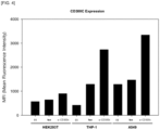

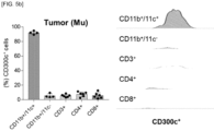
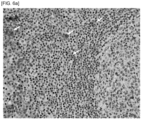


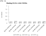


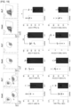
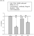
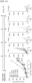
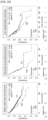


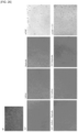


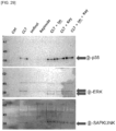


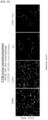


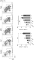

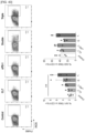

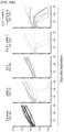


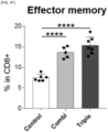


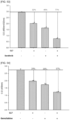
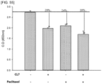
Classifications
-
- C—CHEMISTRY; METALLURGY
- C07—ORGANIC CHEMISTRY
- C07K—PEPTIDES
- C07K16/00—Immunoglobulins [IGs], e.g. monoclonal or polyclonal antibodies
- C07K16/18—Immunoglobulins [IGs], e.g. monoclonal or polyclonal antibodies against material from animals or humans
- C07K16/28—Immunoglobulins [IGs], e.g. monoclonal or polyclonal antibodies against material from animals or humans against receptors, cell surface antigens or cell surface determinants
- C07K16/2803—Immunoglobulins [IGs], e.g. monoclonal or polyclonal antibodies against material from animals or humans against receptors, cell surface antigens or cell surface determinants against the immunoglobulin superfamily
-
- A—HUMAN NECESSITIES
- A61—MEDICAL OR VETERINARY SCIENCE; HYGIENE
- A61K—PREPARATIONS FOR MEDICAL, DENTAL OR TOILETRY PURPOSES
- A61K31/00—Medicinal preparations containing organic active ingredients
- A61K31/33—Heterocyclic compounds
- A61K31/335—Heterocyclic compounds having oxygen as the only ring hetero atom, e.g. fungichromin
- A61K31/337—Heterocyclic compounds having oxygen as the only ring hetero atom, e.g. fungichromin having four-membered rings, e.g. taxol
-
- A—HUMAN NECESSITIES
- A61—MEDICAL OR VETERINARY SCIENCE; HYGIENE
- A61K—PREPARATIONS FOR MEDICAL, DENTAL OR TOILETRY PURPOSES
- A61K31/00—Medicinal preparations containing organic active ingredients
- A61K31/33—Heterocyclic compounds
- A61K31/395—Heterocyclic compounds having nitrogen as a ring hetero atom, e.g. guanethidine or rifamycins
- A61K31/435—Heterocyclic compounds having nitrogen as a ring hetero atom, e.g. guanethidine or rifamycins having six-membered rings with one nitrogen as the only ring hetero atom
- A61K31/44—Non condensed pyridines; Hydrogenated derivatives thereof
- A61K31/4412—Non condensed pyridines; Hydrogenated derivatives thereof having oxo groups directly attached to the heterocyclic ring
-
- A—HUMAN NECESSITIES
- A61—MEDICAL OR VETERINARY SCIENCE; HYGIENE
- A61K—PREPARATIONS FOR MEDICAL, DENTAL OR TOILETRY PURPOSES
- A61K31/00—Medicinal preparations containing organic active ingredients
- A61K31/70—Carbohydrates; Sugars; Derivatives thereof
- A61K31/7042—Compounds having saccharide radicals and heterocyclic rings
- A61K31/7052—Compounds having saccharide radicals and heterocyclic rings having nitrogen as a ring hetero atom, e.g. nucleosides, nucleotides
- A61K31/706—Compounds having saccharide radicals and heterocyclic rings having nitrogen as a ring hetero atom, e.g. nucleosides, nucleotides containing six-membered rings with nitrogen as a ring hetero atom
- A61K31/7064—Compounds having saccharide radicals and heterocyclic rings having nitrogen as a ring hetero atom, e.g. nucleosides, nucleotides containing six-membered rings with nitrogen as a ring hetero atom containing condensed or non-condensed pyrimidines
- A61K31/7068—Compounds having saccharide radicals and heterocyclic rings having nitrogen as a ring hetero atom, e.g. nucleosides, nucleotides containing six-membered rings with nitrogen as a ring hetero atom containing condensed or non-condensed pyrimidines having oxo groups directly attached to the pyrimidine ring, e.g. cytidine, cytidylic acid
-
- A—HUMAN NECESSITIES
- A61—MEDICAL OR VETERINARY SCIENCE; HYGIENE
- A61K—PREPARATIONS FOR MEDICAL, DENTAL OR TOILETRY PURPOSES
- A61K39/00—Medicinal preparations containing antigens or antibodies
-
- A—HUMAN NECESSITIES
- A61—MEDICAL OR VETERINARY SCIENCE; HYGIENE
- A61K—PREPARATIONS FOR MEDICAL, DENTAL OR TOILETRY PURPOSES
- A61K39/00—Medicinal preparations containing antigens or antibodies
- A61K39/395—Antibodies; Immunoglobulins; Immune serum, e.g. antilymphocytic serum
- A61K39/39533—Antibodies; Immunoglobulins; Immune serum, e.g. antilymphocytic serum against materials from animals
- A61K39/3955—Antibodies; Immunoglobulins; Immune serum, e.g. antilymphocytic serum against materials from animals against proteinaceous materials, e.g. enzymes, hormones, lymphokines
-
- A—HUMAN NECESSITIES
- A61—MEDICAL OR VETERINARY SCIENCE; HYGIENE
- A61K—PREPARATIONS FOR MEDICAL, DENTAL OR TOILETRY PURPOSES
- A61K45/00—Medicinal preparations containing active ingredients not provided for in groups A61K31/00 - A61K41/00
- A61K45/06—Mixtures of active ingredients without chemical characterisation, e.g. antiphlogistics and cardiaca
-
- A—HUMAN NECESSITIES
- A61—MEDICAL OR VETERINARY SCIENCE; HYGIENE
- A61P—SPECIFIC THERAPEUTIC ACTIVITY OF CHEMICAL COMPOUNDS OR MEDICINAL PREPARATIONS
- A61P35/00—Antineoplastic agents
-
- C—CHEMISTRY; METALLURGY
- C07—ORGANIC CHEMISTRY
- C07K—PEPTIDES
- C07K16/00—Immunoglobulins [IGs], e.g. monoclonal or polyclonal antibodies
- C07K16/18—Immunoglobulins [IGs], e.g. monoclonal or polyclonal antibodies against material from animals or humans
- C07K16/28—Immunoglobulins [IGs], e.g. monoclonal or polyclonal antibodies against material from animals or humans against receptors, cell surface antigens or cell surface determinants
- C07K16/2803—Immunoglobulins [IGs], e.g. monoclonal or polyclonal antibodies against material from animals or humans against receptors, cell surface antigens or cell surface determinants against the immunoglobulin superfamily
- C07K16/2818—Immunoglobulins [IGs], e.g. monoclonal or polyclonal antibodies against material from animals or humans against receptors, cell surface antigens or cell surface determinants against the immunoglobulin superfamily against CD28 or CD152
-
- C—CHEMISTRY; METALLURGY
- C07—ORGANIC CHEMISTRY
- C07K—PEPTIDES
- C07K16/00—Immunoglobulins [IGs], e.g. monoclonal or polyclonal antibodies
- C07K16/18—Immunoglobulins [IGs], e.g. monoclonal or polyclonal antibodies against material from animals or humans
- C07K16/28—Immunoglobulins [IGs], e.g. monoclonal or polyclonal antibodies against material from animals or humans against receptors, cell surface antigens or cell surface determinants
- C07K16/2803—Immunoglobulins [IGs], e.g. monoclonal or polyclonal antibodies against material from animals or humans against receptors, cell surface antigens or cell surface determinants against the immunoglobulin superfamily
- C07K16/2827—Immunoglobulins [IGs], e.g. monoclonal or polyclonal antibodies against material from animals or humans against receptors, cell surface antigens or cell surface determinants against the immunoglobulin superfamily against B7 molecules, e.g. CD80, CD86
-
- G—PHYSICS
- G01—MEASURING; TESTING
- G01N—INVESTIGATING OR ANALYSING MATERIALS BY DETERMINING THEIR CHEMICAL OR PHYSICAL PROPERTIES
- G01N33/00—Investigating or analysing materials by specific methods not covered by groups G01N1/00 - G01N31/00
- G01N33/48—Biological material, e.g. blood, urine; Haemocytometers
- G01N33/50—Chemical analysis of biological material, e.g. blood, urine; Testing involving biospecific ligand binding methods; Immunological testing
- G01N33/53—Immunoassay; Biospecific binding assay; Materials therefor
- G01N33/574—Immunoassay; Biospecific binding assay; Materials therefor for cancer
-
- A—HUMAN NECESSITIES
- A61—MEDICAL OR VETERINARY SCIENCE; HYGIENE
- A61K—PREPARATIONS FOR MEDICAL, DENTAL OR TOILETRY PURPOSES
- A61K39/00—Medicinal preparations containing antigens or antibodies
- A61K2039/505—Medicinal preparations containing antigens or antibodies comprising antibodies
-
- A—HUMAN NECESSITIES
- A61—MEDICAL OR VETERINARY SCIENCE; HYGIENE
- A61K—PREPARATIONS FOR MEDICAL, DENTAL OR TOILETRY PURPOSES
- A61K39/00—Medicinal preparations containing antigens or antibodies
- A61K2039/505—Medicinal preparations containing antigens or antibodies comprising antibodies
- A61K2039/507—Comprising a combination of two or more separate antibodies
-
- A—HUMAN NECESSITIES
- A61—MEDICAL OR VETERINARY SCIENCE; HYGIENE
- A61K—PREPARATIONS FOR MEDICAL, DENTAL OR TOILETRY PURPOSES
- A61K2300/00—Mixtures or combinations of active ingredients, wherein at least one active ingredient is fully defined in groups A61K31/00 - A61K41/00
-
- C—CHEMISTRY; METALLURGY
- C07—ORGANIC CHEMISTRY
- C07K—PEPTIDES
- C07K2317/00—Immunoglobulins specific features
- C07K2317/70—Immunoglobulins specific features characterized by effect upon binding to a cell or to an antigen
- C07K2317/74—Inducing cell proliferation
-
- C—CHEMISTRY; METALLURGY
- C07—ORGANIC CHEMISTRY
- C07K—PEPTIDES
- C07K2317/00—Immunoglobulins specific features
- C07K2317/90—Immunoglobulins specific features characterized by (pharmaco)kinetic aspects or by stability of the immunoglobulin
- C07K2317/92—Affinity (KD), association rate (Ka), dissociation rate (Kd) or EC50 value
-
- G—PHYSICS
- G01—MEASURING; TESTING
- G01N—INVESTIGATING OR ANALYSING MATERIALS BY DETERMINING THEIR CHEMICAL OR PHYSICAL PROPERTIES
- G01N2500/00—Screening for compounds of potential therapeutic value
-
- G—PHYSICS
- G01—MEASURING; TESTING
- G01N—INVESTIGATING OR ANALYSING MATERIALS BY DETERMINING THEIR CHEMICAL OR PHYSICAL PROPERTIES
- G01N2800/00—Detection or diagnosis of diseases
- G01N2800/52—Predicting or monitoring the response to treatment, e.g. for selection of therapy based on assay results in personalised medicine; Prognosis
Definitions
- the present disclosure relates to an anti-CD300c antibody and a combined therapy using an anti-CD300c antibody. More specifically, the present disclosure relates to a composition, a kit, and a combined therapy for the prevention or treatment of cancer, each of which includes, as active ingredients, an anti-CD300c monoclonal antibody and at least one additional anticancer agent.
- immunotherapies that activate the immune system of the human body to help fight cancer cells are receiving attention as cancer treatments.
- Previous cancer therapeutic agents have focused on killing rapidly dividing cells, which is the characteristic of cancer cells, so that there are side effects of acting on not only cancer cells but also rapidly dividing cells among normal cells.
- immunotherapeutic agents use the immune system of a cancer patient to affect cancer cells, so there are few typical side effects of existing anticancer agents.
- immunotherapeutic agents utilize the immune system of the body and thus can be applied to a variety of cancer types, unlike targeted therapeutic agents targeting specific mutations or signals of cancer.
- An immune checkpoint inhibitor such as Keytruda ® is receiving attention as a type of immunotherapeutic agent.
- CD300 antigen-like family member C (CD300c) protein which is a protein encoded by the CD300c gene in humans, exists on the surface of various cancer cells.
- the inhibition of activity or expression of the CD300c protein can activate T cells to reduce the proliferation of cancer ( Korean Patent Publication No. 10-2019-0136949 ).
- Patent Document 1 Korean Patent Publication No. 10-2019-0136949
- An aspect of the present disclosure is to solve all of the above-mentioned problems.
- Another aspect of the present disclosure is to provide a combined therapy using an anti-CD300c antibody for preventing or treating cancer.
- Still another aspect of the present disclosure is to provide a pharmaceutical composition for a combined therapy using an anti-CD300c antibody for preventing or treating cancer.
- Still another aspect of the present disclosure is to provide a kit used in a combined therapy using an anti-CD300c antibody for preventing or treating cancer.
- Still another aspect of the present disclosure is to provide a diagnostic or treatment method based on a change in marker protein according to the expression level of an anti-CD300c antibody or the administration of an anti-CD300c antibody.
- an anti-CD300c antibody or antigen-binding fragment thereof and use of the anti-CD300c antibody or antigen-binding fragment thereof for preventing or treating cancer.
- an anti-CD300c antibody or antigen-binding fragment thereof for the prevention or treatment of cancer and at least one additional anticancer agent.
- a pharmaceutical composition for the prevention or treatment of cancer the pharmaceutical composition containing, as active ingredients, an anti-CD300c antibody or antigen-binding fragment thereof and at least one additional anticancer agent
- a method for preventing or treating cancer the method including administering, to a subject in need of the prevention or treatment of cancer, an anti-CD300c (CD300 antigen-like family member C) antibody or antigen-binding fragment thereof and the at least one additional anticancer agent.
- kits for preventing or treating cancer including: a composition containing an anti-CD300c antibody or antigen-binding fragment thereof at an effective amount; and instructions directing the combined use of the antibody or antigen-binding fragment thereof and at least one additional anticancer agent.
- a method for providing information needed for the prediction of the therapeutic responsiveness of an anti-CD300c antibody or antigen-binding fragment thereof including determining the expression level of a marker for predicting the therapeutic responsiveness on the basis of a biological sample or data obtained from a subject.
- a method for providing information for the prevention or treatment of cancer including measuring the expression level of a CD300c protein on the basis of a biological sample or data obtained from a subject in need of the prevention or treatment of cancer.
- an anti-CD300c antibody according to the present disclosure can inhibit the growth, proliferation, metastasis, and the like of cancer cells effectively and synergistically compared with the use of the anti-CD300c antibody or the additional anticancer agent alone. Therefore, the combined use of an anti-CD300c antibody and an additional anticancer agent can be advantageously employed in the treatment or prevention of various types of cancers.
- antibody as used herein is used broadly and includes monoclonal antibodies (including full length antibodies) of any isotype such as IgG, IgM, IgA, IgD, and IgE, polyclonal antibodies, multispecific antibodies (for example, bispecific antibodies), antibody fusions (for example, a fusion of an antibody with a (poly)peptide or a fusion of an antibody with a compound), and antibody fragments (including antigen-binding fragments).
- the prefix "anti-" when in conjunction with an antigen, indicates that the given antibody is reactive with the given antigen.
- An antibody reactive with a specific antigen can be generated, without limitation, by synthetic and/or recombinant methods such as selection of libraries of recombinant antibodies in phage or similar vectors, or by immunizing an animal with the antigen or an antigen-encoding nucleic acid.
- a typical IgG antibody is composed of two identical heavy chains and two identical light chains that are joined by disulfide bonds. Each heavy and light chain contains a constant region and a variable region. Heavy chain variable regions (HVRs) and light chain variable regions (LVRs) contain three segments, referred to as "complementarity determining regions" ("CDRs") or "hypervariable regions", respectively, which are primarily responsible for binding an epitope of an antigen.
- CDRs complementarity determining regions
- An antibody herein may be, for example, an animal antibody, a chimeric antibody, a humanized antibody, or a human antibody.
- humanization also called reshaping or CDR-grafting
- xenogeneic sources commonly rodent
- ADCC affinity or effector function
- the term "monoclonal antibody” as used herein refers to an antibody obtained from a population of substantially homogeneous antibodies, that is, the individual antibodies including the population are identical except for possible naturally occurring mutations and/or post-translation modifications (for example, isomerizations, amidations) that may be present in small amounts. Monoclonal antibodies are highly specific, being directed against a single antigenic site. The monoclonal antibody is obtained from a substantially homogeneous population of antibodies, displays the nature of an antibody, and is not to be construed as requiring production of the antibody by any particular method.
- monoclonal antibodies to be used in accordance with the present disclosure may be made by a variety of techniques, including but not limited to the hybridoma method, recombinant DNA methods, phage-display methods, and methods utilizing transgenic animals containing all or part of the human immunoglobulin loci.
- antigen-binding fragment refers to a portion of an antibody having specific binding ability to an antigen or a polypeptide including the same.
- antibody and “antigen-binding fragment” may be used interchangeably except for a case where it is understood in the context that the "antibody” specifically excludes the “antigen-binding fragment,” and the “antibody” may be interpreted as including the “antigen-binding fragment.”
- antigen-binding fragment include, but are not limited to, Fv, Fab, Fab', Fab'-SH, F(ab')2, diabodies, triabodies, tetrabodies, cross-Fab fragments, linear antibodies, single chain antibody molecules (for example, scFv), and multispecific antibodies formed of antibody fragments and single domain antibodies.
- anticancer agent collectively refers to known medications used in conventional cancer treatment that act on various metabolic pathways of cells and exhibit cytotoxic or cytostatic effects on cancer cells.
- the anticancer agent largely includes chemotherapeutic agents, targeted therapeutic agents, and immunotherapeutic agents.
- immunotherapeutic agent refers to a medication that activates immune cells to kill cancer cells.
- chemotherapeutic agent refers to a cancer therapeutic agent also called cytotoxic anticancer agent or chemical drug anticancer agent.
- subject is used interchangeably with “patient” and may be a mammal who is in need of prevention or treatment of cancer, such as primates (for example, humans), companion animals (for example, dogs and cats), livestock (for example, cows, pigs, horses, sheep, and goats), and laboratory animals (for example, rats, mice, and guinea pigs).
- the subject is a human.
- treatment generally means obtaining a desired pharmacological and/or physiological effect.
- the effect may be therapeutic in terms of partially or completely curing a disease and/or adverse effect attributed to the disease.
- Desirable therapeutic effects include, but are not limited to, prevention of onset or recurrence of disease, alleviation of symptoms, diminishment of any direct or indirect pathological consequences of the disease, prevention of metastasis, decreasing the rate of disease progression, amelioration or slowing of the disease state, and remission or improved prognosis.
- the "treatment” may refer to medical intervention of a disease or disorder that has already developed.
- prevention relates to a prophylactic treatment, that is, to a measure or procedure, the purpose of which is to prevent, rather than to cure a disease.
- Prevention means that a desired pharmacological and/or physiological effect is obtained which is prophylactic in terms of completely or partially preventing a disease or symptom thereof.
- administration means a method of providing a substance (for example, an anti-CD300c antibody or antigen-binding fragment thereof or another anticancer agent) to a subject to achieve a prophylactic or therapeutic purpose (for example, prevention or treatment of cancer).
- a substance for example, an anti-CD300c antibody or antigen-binding fragment thereof or another anticancer agent
- biological sample encompasses a variety of sample types obtained from a subject and may be used in diagnostic or monitoring assays.
- the biological sample includes, but is not limited to, blood and other liquid samples of biological origin, and solid tissue samples such as a biopsy specimen or tissue cultures or cells derived therefrom and the progeny thereof.
- the biological sample encompasses a clinical sample, and also includes cells in culture, cell supernatants, cell lysates, serum, plasma, biological fluid, and tissue samples, in particular, tumor samples.
- biological data refers to any analytical data obtained using the biological sample.
- expression level may be determined by measuring the expression level of at least one of mRNA and protein of a corresponding marker.
- any method known in the art may be used.
- an agent for measuring the expression level of mRNA may be a pair of primers or a probe specifically binding to the corresponding marker gene
- an agent for measuring the expression level of the protein may be an antibody, substrate, ligand, or cofactor specifically binding to the corresponding marker.
- An assay method for measuring the mRNA level includes, but is not limited to, reverse transcriptase polymerase chain reaction, competitive reverse transcriptase polymerase chain reaction, real-time reverse transcriptase polymerase chain reaction, RNase protection assay, Northern blotting, or DNA chip assay.
- An assay method for measuring the protein level includes, but is not limited to, Western blotting, ELISA, radioimmunoassay, radial immunodiffusion assay, Ouchterlony immunodiffusion, rocket immunoelectrophoresis, immunohistochemical staining, immunoprecipitation assay, complement fixation assay, FACS, and protein chip assay.
- therapeutic responsiveness refers to whether an individual suffering from or suspected of having cancer responds favorably or unfavorably to treatment with a therapeutically active ingredient (such as a CD300c antibody or antigen-binding fragment thereof).
- a therapeutically active ingredient such as a CD300c antibody or antigen-binding fragment thereof.
- the therapeutic responsiveness may be assessed by changes in immune system associated with tumor treatment which are observed after administration of the CD300c antibody or antigen-binding fragment thereof.
- the anti-CD300c antibody or antigen-binding fragment thereof according to the present disclosure is an antigen-binding molecule specifically binding to a CD300c protein.
- CD300c protein is used interchangeably with “CD300c” or “CD300c antigen” and is a protein encoded by CD300c gene. It is known that the CD300c protein exhibits significant sequence identity to B7 family proteins and is expressed on the membrane of antigen presenting cells. Inhibition of expression or activity of the CD300c protein may induce activation of T cells and/or promotion of differentiation into M1 macrophages.
- anti-CD300c antibody may be used interchangeably with a polypeptide that binds to the CD300c protein.
- polypeptide is intended to mean any polymer of amino acids linked to one another by peptide bonds, irrespective of its length. That is, the polypeptide as used herein also encompasses peptides and proteins.
- the anti-CD300c antibody or antigen-binding fragment thereof is capable of specifically binding to an extracellular domain (ECD) of the CD300c protein.
- ECD extracellular domain
- the extracellular domain of CD300c may be an extracellular domain of human CD300c protein, and may include the amino acid sequence as set forth in SEQ ID NO: 402.
- the expression level of a CD300c protein exhibits a very high correlation with survival of various cancer patients. Specifically, it was identified that relative to the average CD300c expression level of cancer patients, cancer patients with a high CD300c expression level had shorter survival than cancer patients with a low CD300c expression level. This means that inhibition of expression or activity of CD300c using the anti-CD300c antibody or antigen-binding fragment thereof according to the present disclosure can result in a cancer therapeutic effect or an effect of increasing survival of cancer patients.
- the anti-CD300c antibody or antigen-binding fragment thereof according to the present disclosure can specifically bind to CD300c expressed on the surface of various cancer cells, and thus exhibit an anticancer effect.
- the binding of the anti-CD300c antibody to CD300c can activate T cells and at the same time promote differentiation into M1 macrophages to effectively inhibit the proliferation of cancer cells, which allows the anti-CD300c antibody to be effectively used as an immunotherapeutic agent for various cancers.
- the anti-CD300c antibody according to the present disclosure can exhibit a further increased therapeutic effect through combined administration with a conventional immunotherapeutic agent, and also has inter-species cross-reactivity (for example, between human and mouse antigens) that allows the antibody to be widely applied to various mammals.
- the anti-CD300c antibody of the present disclosure is able to remarkably weaken resistance of the cancer cells, thereby showing excellent efficacy in preventing cancer recurrence.
- cancer cells inhibit production of the proinflammatory cytokine IL-2 to evade the immune system.
- the anti-CD300c antibody activates the immune system by restoring production of IL-2 blocked by these cancer cells, which induces cancer cell death.
- the anti-CD300c antibody can be utilized as a more fundamental immunotherapeutic agent.
- Korean Patent Publication No. 10-2019-0136949 Korean Patent Publication No. 10-2019-0136949 , which is incorporated herein by reference in its entirety for all purposes.
- the antibody or antigen-binding fragment thereof may include:
- the heavy chain variable region may include:
- the antibody or antigen-binding fragment thereof may be any one selected from the following antibodies:
- the antibody or antigen-binding fragment thereof may include: a heavy chain variable region including CDR1 including or consisting of the amino acid sequence as set forth in SEQ ID NO: 79, CDR2 including or consisting of the amino acid sequence as set forth in SEQ ID NO: 80, CDR3 including or consisting of the amino acid sequence as set forth in SEQ ID NO: 81; and a light chain variable region including CDR1 including or consisting of the amino acid sequence as set forth in SEQ ID NO: 82, CDR2 including or consisting of the amino acid sequence as set forth in SEQ ID NO: 83, CDR3 including or consisting of the amino acid sequence as set forth in SEQ ID NO: 84;
- the antibody or antigen-binding fragment thereof may include: a heavy chain variable region including CDR1 including or consisting of the amino acid sequence as set forth in SEQ ID NO: 115, CDR2 including or consisting of the amino acid sequence as set forth in SEQ ID NO: 116, CDR3 including or consist
- the antibody or antigen-binding fragment thereof may include: a heavy chain variable region including CDR1 including or consisting of the amino acid sequence as set forth in SEQ ID NO: 211, CDR2 including or consisting of the amino acid sequence as set forth in SEQ ID NO: 212, CDR3 including or consisting of the amino acid sequence as set forth in SEQ ID NO: 213; and a light chain variable region including CDR1 including or consisting of the amino acid sequence as set forth in SEQ ID NO: 214, CDR2 including or consisting of the amino acid sequence as set forth in SEQ ID NO: 215, CDR3 including or consisting of the amino acid sequence as set forth in SEQ ID NO: 216.
- the heavy chain variable region may include an amino acid sequence selected from the group consisting of SEQ ID Nos: 303, 307, 311, 315, 319, 323, 327, 331, 335, 339, 343, 347, 351, 355, 359, 363, 367, 371, 375, 379, 383, 387, 391, 395, and 399; and the light chain variable region may include an amino acid sequence selected from the group consisting of SEQ ID Nos: 304, 308, 312, 316, 320, 324, 328, 332, 336, 340, 344, 348, 352, 356, 360, 364, 368, 372, 376, 380, 384, 388, 392, 396, and 400.
- the heavy chain variable region may include an amino acid sequence selected from the group consisting of SEQ ID Nos: 315, 327, 339, and 371; and the light chain variable region may include an amino acid sequence selected from the group consisting of SEQ ID Nos: 316, 328, 340, and 372.
- the heavy chain variable region may include: the amino acid sequence as set forth in SEQ ID No: 315 and the light chain variable region may include the amino acid sequence as set forth in of SEQ ID No: 316; the heavy chain variable region may include the amino acid sequence as set forth in SEQ ID No: 327 and the light chain variable region may include the amino acid sequence as set forth in of SEQ ID No: 328; the heavy chain variable region may include the amino acid sequence as set forth in SEQ ID No: 339 and the light chain variable region may include the amino acid sequence as set forth in of SEQ ID No: 340; or the heavy chain variable region may include the amino acid sequence as set forth in SEQ ID No: 371 and the light chain variable region may include the amino acid sequence as set forth in of SEQ ID No: 372.
- the anti-CD300c antibody or antigen-binding fragment thereof may include: a heavy chain variable region including CDR1 to CDR3 including or consisting of the amino acid sequences represent by Formulas (1) to (3), respectively; and a light chain variable region including CDR1 to CDR3 including or consisting of the amino acid sequences represented by Formulas (4) to (6), respectively (each amino acid sequence is shown in N ⁇ C direction):
- the anti-CD300c antibody or antigen-binding fragment may include sequences having 80% or more, preferably 90% or more, more preferably 95% or more, and most preferably 98% or more sequence identity to the CDR sequences or the sequences shown in Tables 4, 5, and 6.
- amino acid sequence variants of the antibodies of the present disclosure are contemplated.
- Amino acid sequence variants of the antibody may be prepared by introducing appropriate modifications into the nucleotide sequence encoding the molecules, or by peptide synthesis. Such modifications include, for example, deletions from, and/or insertions into and/or substitutions of residues within the amino acid sequence of the antibody. Any combination of deletion, insertion, and substitution can be made to arrive at the final construct, provided that the final construct possesses the desired characteristics, for example, antigen-binding.
- Sites of interest for substitutional mutagenesis include heavy chain variable regions (HVRs) and framework regions (FRs).
- amino acid side chain classes (1) to (6) Conservative substitutions are provided in Table 1 under the heading "Preferred Substitutions" and further described below in reference to amino acid side chain classes (1) to (6). Amino acid substitutions may be introduced into the molecule of interest and the products screened for a desired activity, for example, retained/improved antigen binding, decreased immunogenicity, or improved ADCC or CDC.
- substitutions Ala (A) Val; Leu; Ile Val Arg (R) Lys; Gln; Asn Lys Asn (N) Gln; His; Asp; Lys; Arg Gln Asp (D) Glu; Asn Glu Cys (C) Ser; Ala Ser Gln (Q) Asn; Glu Asn Glu (E) Asp; Gln Asp Gly (G) Ala Ala His (H) Asn; Gin; Lys; Arg Arg Ile (I) Leu; Val; Met; Ala; Phe; Norleucine Leu Leu (L) Norleucine; Ile; Val; Met; Ala; Phe Ile Lys (K) Arg; Gln; Asn Arg Met (M) Leu; Phe; Ile Ile Phe (F) Trp; Leu; Val; Ile; Ala; Tyr Tyr Pro (P) Ala Ala Ser (S) Thr Thr Thr (
- Amino acids may be grouped according to common side-chain properties:
- Non-conservative substitutions will entail exchanging a member of one of these classes for another class.
- amino acid sequence variant includes substantial variants wherein there are amino acid substitutions in one or more hypervariable region residues of a parent antigen binding molecule (for example, a humanized or human antibody).
- a parent antigen binding molecule for example, a humanized or human antibody.
- the resulting variant(s) selected for further study will have modifications (for example, improvements) in certain biological properties (for example, increased affinity, reduced immunogenicity) relative to the parent antigen binding molecule and/or will have substantially retained certain biological properties of the parent antigen binding molecule.
- An exemplary substitutional variant is an affinity matured antibody, which may be conveniently generated, for example, using phage display-based affinity maturation techniques known in the art.
- one or more HVR residues are mutated and the variant antigen binding molecules displayed on phage and screened for a particular biological activity (for example, binding affinity).
- substitutions, insertions, or deletions may occur within one or more HVRs so long as such alterations do not substantially reduce the ability of the antigen binding molecule to bind antigen.
- conservative alterations for example, conservative substitutions as provided herein
- that do not substantially reduce binding affinity may be made in HVRs.
- Amino acid sequence insertions include amino- and/or carboxyl-terminal fusions ranging in length from one residue to polypeptides containing a hundred or more residues, as well as intra-sequence insertions of single or multiple amino acid residues.
- terminal insertions include antibodies with an N-terminal methionyl residue.
- Other insertional variants of the molecule include the fusion to the N- or C-terminus to a polypeptide which increases the serum half-life of the antibody.
- other insertional variants of the molecule may include fusions to the N-terminus or C-terminus of the polypeptide to facilitate passage of the blood-brain barrier (BBB).
- BBB blood-brain barrier
- variants of the antibody or antigen-binding fragment thereof of the present disclosure which have improved affinity for the CD300c antigen.
- Such variants can be obtained by a number of affinity maturation protocols including mutating the CDRs ( Yang et al., J. Mol. Biol., 254, 392-403, 1995 ), chain shuffling ( Marks et al., Bio/Technology, 10, 779-783, 1992 ), use of mutator strains of E . coli ( Low et al., J. Mol. Biol., 250, 359-368, 1996 ), DNA shuffling ( Patten et al., Curr. Opin.
- the anti-CD300c monoclonal antibody or antigen-binding fragment thereof may have inter-species cross-reactivity. Specifically, the anti-CD300c monoclonal antibody or antigen-binding fragment thereof may exhibit cross-reactivity to both human and mouse CD300c antigens. Such cross-reactivity is identified in Experimental Examples 3.1 to 3.8.
- the anti-CD300c monoclonal antibody or antigen-binding fragment thereof may include a form of an antibody-drug conjugate, in which the anti-CD300c monoclonal antibody or antigen-binding fragment thereof is bound to another drug, or may be provided in such a form.
- antibody-drug conjugate refers to a form in which the drug and the antibody are chemically linked to each other without degrading biological activity of the antibody and the drug.
- the antibody-drug conjugate denotes a form in which the drug is bound to an amino acid residue at the N-terminus of the heavy and/or light chain of the antibody, specifically, a form in which the drug is bound to an ⁇ -amine group at the N-terminus of the heavy and/or light chain of the antibody.
- the "drug” may mean any substance having a certain biological activity for a cell (for example, a cancer cell), which is a concept including DNA, RNA, or a peptide.
- the drug may be in a form which contains a reactive group capable of reacting and crosslinking with an ⁇ -amine group, and also includes a form which contains a reactive group capable of reacting and crosslinking with an ⁇ -amine group and to which a linker is linked.
- the reactive group capable of reacting and crosslinking with the ⁇ -amine group are not particularly limited in terms of type as long as the reactive group can react and crosslink with an ⁇ -amine group at the N-terminus of a heavy chain or light chain of an antibody.
- the reactive group includes all types of groups known in the art which react with an amine group.
- the reactive group may, for example, be any one of isothiocyanate, isocyanate, acyl azide, NHS ester, sulfonyl chloride, aldehyde, glyoxal, epoxide, oxirane, carbonate, aryl halide, imidoester, carbodiimide, anhydride, and fluorophenyl ester, but is not limited thereto.
- the drug is contained regardless of the type of drug that can treat the disease targeted by the anti-CD300c antibody or antigen-binding fragment thereof according to the present disclosure, but may preferably be an anticancer agent.
- an anti-CD300c antibody or antigen-binding fragment thereof could exhibit an enhanced anticancer effect in combination with at least one other anticancer agent. Therefore, the anti-CD300c antibody or antigen-binding fragment thereof of the present disclosure can be used in the prevention or treatment of cancer in combination with at least one other anticancer agent. In an embodiment, the anti-CD300c antibody or antigen-binding fragment thereof can be used in combination with at least one other immunotherapeutic agent and/or at least one chemotherapeutic agent. In another embodiment, the anti-CD300c antibody or antigen-binding fragment thereof can be used in combination with at least one other immunotherapeutic agent and at least one chemotherapeutic agent.
- Immunotherapeutic agents have a novel mechanism by which immune cells in the body are activated to kill cancer cells, and thus are advantageous in that they can be widely used for most cancers without specific genetic mutations.
- the immunotherapeutic agents have fewer adverse effects in that they treat cancer by strengthening the patient's own immune system, and have effects of improving the patient's quality of life and significantly extending the survival.
- These immunotherapeutic agents include immune checkpoint inhibitors, and may be manufactured by known methods or commercially available products.
- immunotherapeutic agent examples include, but are not limited to, anti-PD-1, anti-PD-L1, anti-CTLA-4, anti-CD47, anti-KIR, anti-LAG3, anti-CD137, anti-OX40, anti-CD276, anti-CD27, anti-GITR, anti-TIM3, anti-41BB, anti-CD226, anti-CD40, anti-CD70, anti-ICOS, anti-CD40L, anti-BTLA, anti-TCR, and anti-TIGIT antibodies.
- examples of the immunotherapeutic agent include, but are not limited to, durvalumab (Imfinzi), atezolizumab (Tecentriq), avelumab (Bavencio), pembrolizumab (Keytruda), nivolumab (Opdivo), ⁇ CD47, cemiplimab (Libtayo), magrolimab (Hu5F9-G4), and ipilimumab (Yervoy).
- the immunotherapeutic agent may include at least one selected from the group consisting of anti-PD-1, anti-PD-L1, anti-CTLA-4, anti-CD47, anti-KIR, anti-LAG3, anti-CD137, anti-OX40, anti-CD276, anti-CD27, anti-GITR, anti-TIM3, anti-41BB, anti-CD226, anti-CD40, anti-CD70, anti-ICOS, anti-CD40L, anti-BTLA, anti-TCR, and anti-TIGIT antibodies.
- the immunotherapeutic agent may include at least one selected from the group consisting of anti-PD-1, anti-PD-L1, anti-CTLA-4, and anti-CD47 antibodies.
- the immunotherapeutic agent may include at least one selected from the group consisting of durvalumab (Imfinzi), atezolizumab (Tecentriq), pembrolizumab (Keytruda), nivolumab (Opdivo), ⁇ CD47, and ipilimumab (Yervoy).
- Chemotherapeutic agent refers to a cancer therapeutic agent also called a cytotoxic anticancer agent or a chemical drug anticancer agent. These chemotherapeutic agents have side effects, such as vomiting, hair loss, and reduction of white blood cells, by attacking not only cancer cells but also surrounding normal cells. Therefore, the chemotherapeutic agents are not used alone but used in combination with other immunotherapeutic agents showing a synergistic effect when used together. These chemotherapeutic agents include small molecule therapeutic age and may be products that are manufactured or commercially available by known methods. Examples of the chemotherapeutic agents may include microtubule assembly inhibitors, DNA intercalators or replication inhibitors, multikinase inhibitors, angiogenesis inhibitors, antimetabolites, and Taxols, but are not limited thereto.
- the chemotherapeutic agent may include at least one selected from the group consisting of a microtubule assembly inhibitor, a DNA replication inhibitor, a multikinase inhibitor, and an angiogenesis inhibitor.
- chemotherapeutic agent is shown in FIG. 2 .
- TABLE 2 Anticancer agents Mechanism of Action Doxorubicin Microtubule assembly inhibitors Paclitaxel Docetaxel Vinblastine Vincristine Vinorelbine Estramustine Phosphate (EMP) NAB-Paclitaxel (Abraxane), Doxorubicin DNA intercalators or replication inhibitors Cyclophosphamide Epirubicin 5-Fluorouracil Etoposide Ifosfam ide Gemcitabine Deoxyadenosine Cladribine Clofarabine Fludarabine Pentostatin Deoxycytidine Cytosine Arabinoside (Ara-C) 5-Aza-2'-Deoxycytidine (Decitabine) Tezacitabine Capecitabine Cytarabine Methotrexate Pemetrexed Mercaptopurine Dactinomycin Daunorubicin Mitomycin Bleomycin Idarubicin Mitoxantron
- chemotherapeutic agents are not limited to the anticancer agents shown in Table 2, and any chemotherapeutic agent may be used as long as it exerts the same or similar effect through the same or similar mechanisms of action as these chemotherapeutic agents.
- the chemotherapeutic agent may include at least one selected from the group consisting of sorafenib, gemcitabine, and paclitaxel.
- the effective amounts or effective non-toxic amounts of the anti-CD300c antibody or antigen-binding fragment thereof and the additional anti-cancer agent may be determined by routine experiments.
- the therapeutically active amount of an antibody or an anticancer agent may vary according to factors, such as the stage of a disease, severity of a disease, age, sex, medical complications, and weight of a subject, and the ability of a component to elicit a desired response in the subject, and the dosage of an anticancer agent used together.
- the dosage and dosing regimen of each of the anti-CD300c antibody or antigen-binding fragment thereof and the additional anticancer agent may be adjusted to provide an optimal therapeutic response. For example, several divided doses may be administered daily, weekly, every other week, every three weeks, every four weeks or the like, and/or the dose may be proportionally reduced as indicated by the exigencies of the therapeutic situation.
- a method for preventing or treating cancer may include inhibiting proliferation, survival, metastasis, recurrence, or anticancer agent resistance of cancer.
- Such a method includes the combined use of an anti-CD300c antibody or antigen-binding fragment thereof and at least one additional anticancer agent.
- the method includes administering a therapeutically effective amount of an anti-CD300c antibody or antigen-binding fragment thereof and a therapeutically effective amount of at least one additional anticancer agent to a subject in need thereof.
- the at least additional anticancer agent may include at least one immunotherapeutic agent, at least one chemotherapeutic agent, or at least one immunotherapeutic agent and at least one chemotherapeutic agent.
- cancer refers to a physiological condition that is typically characterized by unregulated cell growth in mammals.
- the cancer to be prevented or treated in the present disclosure may include, depending on the site of occurrence, colorectal cancer, small intestine cancer, rectal cancer, colon cancer, thyroid cancer, endocrine adenocarcinoma, oral cancer, tongue cancer, pharyngeal cancer, laryngeal cancer, esophageal cancer, cervical cancer, uterine cancer, fallopian tube cancer, ovarian cancer, brain cancer, head and neck cancer, lung cancer, lymph gland cancer, gallbladder cancer, bladder cancer, kidney cancer, liver cancer, pancreatic cancer, prostate cancer, skin cancer (or melanoma), breast cancer, stomach cancer, bone cancer, blood cancer, and the like.
- the cancer may include at least one selected from the group consisting of colorectal cancer, rectal cancer, colon cancer, thyroid cancer, oral cancer, pharyngeal cancer, laryngeal cancer, cervical cancer, brain cancer, lung cancer, ovarian cancer, bladder cancer, kidney cancer, liver cancer, pancreatic cancer, prostate cancer, skin cancer, tongue cancer, breast cancer, uterine cancer, stomach cancer, bone cancer, and blood cancer.
- the cancer may be a solid cancer.
- the anti-CD300c antibody or antigen-binding fragment thereof and the at least one additional anticancer agent may be administered simultaneously or sequentially.
- administering sequentially means that one ingredient is first administered and the other ingredient is administered immediately or at a predetermined interval after the first administration, wherein the ingredients may be administered in any order. That is, the anti-CD300c antibody or antigen-binding fragment thereof may be first administered and at least one additional anticancer agent may be administered immediately or at a predetermined interval after the first administration, or vice versa. In addition, any one of the at least one additional anticancer agent may be first administered, followed by the anti-CD300c antibody or antigen-binding fragment thereof, and then the other of the at least one additional anticancer agent.
- the anti-CD300c antibody or antigen-binding fragment thereof may be administered in combination with two or more additional anticancer agents.
- additional anticancer agents for example, an anti-PD-L1 antibody and an anti-PD-1 antibody, or an anti-PD-1 antibody and an anti-CTLA-4 antibody.
- Each of the antibody or antigen-binding fragment thereof according to the present disclosure and at least one additional anticancer agent may be administered in several ways depending on whether local or systemic treatment is desired and on the area to be treated. Methods of administering these ingredients to a subject may vary depending on the purpose of administration, the site of a disease, the condition of a subject, and the like.
- the route of administration may be oral, parenteral, inhalation, local or topical (for example, intralesional administration).
- parenteral administration may include, but is not limited to, intravenous, subcutaneous, intraperitoneal, intrapulmonary, intraarterial, intramuscular, rectal, vaginal, intraarticular, intraprostatic, intranasal, intraocular, intravesical, intrathecal, or intraventricular administration (for example, intracerebroventricular administration).
- the anti-CD300c antibody and the additional anticancer agent may be administered through the same route or mutually different routes.
- the effective amount of each of the anti-CD300c antibody or antigen-binding fragment thereof according to the present disclosure and the at least one additional cancer agent may vary depending on the age, sex, and body weight of an individual (patient). In general, administration may be performed in an amount of about 0.01 mg to about 100 mg, or about 5 mg to about 50 mg, per kg of body. The amount may be administrated once a day or several times a day in divided doses. However, the effective amount may be increased or decreased depending on route and period of administration, severity of disease, sex, body weight, age, and the like. Thus, the range of the present disclosure is not limited thereto.
- the method according to the present disclosure may include identifying the expression level of a CD300c protein from the subject in advance. Depending on the expression level, determination may be made on whether or not to administer the anti-CD300c antibody or antigen-binding fragment thereof.
- the method may further include identifying the expression level of a CD300c protein on the basis of a biological sample or data of the subject, before the administration of the anti-CD300c antibody or antigen-binding fragment thereof, before the administration of the anti-CD300c antibody or antigen-binding fragment thereof.
- the method may include determining that the subject is a suitable for the treatment using an anti-CD300c antibody or antigen-binding fragment thereof when the expression level of the CD300c protein determined on the basis of a biological sample or data of the subject is statistically significantly higher (for example, at least 10% higher) than that in a control group (for example, the expression level in normal people without cancer or the average expression level in cancer patients).
- a control group for example, the expression level in normal people without cancer or the average expression level in cancer patients.
- the control group may show the average expression level in cancer patients of the same type.
- the method according to the present disclosure may include selecting an additional immunotherapeutic agent suitable for use in combination with the anti-CD300c antibody or antigen-binding fragment thereof by measuring the change in expression level of a specific marker after the administration of the anti-CD300c antibody or antigen-binding fragment thereof to a subject.
- the method may further include determining the expression level of at least one marker selected from the following markers on the basis of a biological sample or data from the subject administered the anti-CD300c antibody or antigen-binding fragment thereof: Bst2, Cd40, Cd70, Cd86, Ccl8, Xcl1, Ccr7, Cd80, Cd206, Msr1, Arg1, Vegfa, Pdgfrb, Col4a1, Hif1a, Vcam1, Icam1, Gzma, Gzmb, Icos, Cd69, Ifng, Tnf, Cd1d1, Cd1d2, Cd38, Cxcr6, Xcr1, Tbx21, Stat1, Stat4, Cxcr3, IL-12b, IL-4, IL-6, IL-13, PD-1, PD-L1, CTLA-4, Lag3, Tim3, Ox40, Gitr, Hvem, CD27, CD28, Cma1, Timd4, Bcl6, Cxcl
- Bst2 Bone marrow stromal antigen 2 This is also known as Tetherin or CD137, and is a lipid raft-associated protein encoded by the BST2 gene.
- CD40 Cluster of differentiation 40 This is a co-stimulatory protein found in antigen-presenting cells and is required for their activation. The binding of CD154 (CD40L) on TH cells to CD40 activates antigen presenting cells and induces a variety of downstream effects.
- CD70 Cluster of differentiation 70 This is a protein encoded by the CD70 gene in humans. CD70 is a ligand for CD27.
- CD86 Cluster of differentiation 86 This is also known as B7-2, and is a protein constitutively expressed in dendritic cells, Langerhans cells, macrophages, B-cells (including memory B-cells), and in other antigen-presenting cells.
- Ccl8 Chemokine (C-C motif) ligand 8 This is also known as monocyte chemoattractant protein 2 (MCP2) and is a protein encoded by the CCL8 gene in humans.
- 6 Xcl1 X-c motif chemokine ligand 1 This is a small cytokine belonging to the C chemokine family that is also known as lymphotactin.
- Ccr7 C-C chemokine receptor type 7 This is a protein encoded by the CCR7 gene in humans. The following two ligands have been identified for this receptor: chemokine (C-C motif) ligand19 (CCL19/ELC) and (C-C motif) ligand 21 (CCL21).
- CD80 Cluster of differentiation 80 This is a B7, type 1 membrane protein in the immunoglobulin superfamily, with an extracellular immunoglobulin constant-like domain and a variable-like domain required for receptor binding. It is closely related to CD86, another B7 protein (B7-2), and often works in tandem, binding to the same receptors to prime T cells.
- CD20 6 Cluster of differentiation 206 CD206 is also known as the mannose receptor.
- Msr1 Macrophage scavenger receptor 1 This is a protein encoded by the scavenger MSR1 gene in humans. MSR1 is receptor 1 also called cluster of differentiation 204 (CD204). 11 Arg1 Arginase 1 The human ARG1 gene encodes a protein arginase. 12 Vegfa Vascular endothelial growth factor A This is a protein encoded by the VEGFA gene in humans.
- Pdgfrb Platelet-derived growth factor receptor beta This is a protein encoded by the PDGFRB gene in humans.
- 14 Col4a 1 Collagen, type IV, alpha 1 This is a protein encoded by the COL4A1 gene on chromosome in humans.
- 15 Hif1a Hypoxia-inducible factor 1-alpha This is also known as HIF-1-alpha and is a subunit of a hetero-dimeric transcription factor hypoxia-inducible factor 1 (HIF-1) encoded by the HIF1A gene.
- VCAM-1 Vascular cell adhesion protein 1
- CD106 cluster of differentiation 106
- Icam1 Intercellular adhesion molecule 1 This is also known as CD54 Adhesion (Cluster of Differentiation 54) and is a protein encoded by the ICAM1 gene in humans.
- Gzma Granzyme A This is encoded by the Gzma gene and is present in cytotoxic T lymphocyte granules.
- Gzmb Granzyme B This is encoded by the Gzmb gene and is expressed by cytotoxic T lymphocytes (CTL) and natural killer (NK) cells.
- CTL cytotoxic T lymphocytes
- NK natural killer
- T-cell co-stimulator This is an immune checkpoint protein encoded by the ICOS gene in humans.
- 21 Cd69 Cluster of Differentiation 69 This is a human transmembrane C-Type lectin protein encoded by the CD69 gene, and is an early activation marker expressed in hematopoietic stem cells, T cells, and the like.
- 22 Ifng Interferon gamma This is a dimerized soluble that is the only member of the type II class of interferons.
- Tnf Tumor necrosis factor This is encoded by the Tnf gene and is a multifunctional pro-inflammatory cytokine that belongs to the tumor necrosis factor (TNF) superfamily and is secreted primarily by macrophages.
- TNF tumor necrosis factor
- Cd1d1 CD1d1 antigen This enables T cell receptor binding activity and endogenous lipid antigen binding activity, and is involved in differentiation of natural killer T (NKT) cells and regulation of immature T cell proliferation.
- Cd1d2 CD1d2 antigen This encodes an MHC class I-like molecule involved in presentation of lipid antigens to T cells, and is involved in activation of natural killer T cells.
- Cd38 Cluster of Differentiation 38 This is a glycoprotein found on the surface of many immune cells (white blood cells), including CD4+, CD8+, B lymphocytes, and natural killer cells.
- Cxcr6 C-X-C chemokine receptor type 6 This is also called CD 186 and has been identified as an entry coreceptor used by HIV-1 and SIV to enter target cells, in conjunction with CD4.
- 28 Xcr1 X-C motif chemokine receptor 1 This is a chemokine receptor belonging to the G protein-coupled receptor superfamily.
- 29 Tbx21 T-box transcription factor TBX21 This is also called T-box transcription expressed in T cells (T-bet) and is a protein encoded by the TBX21 gene in humans.
- Stat1 Signal transducer and activator of transcription 1 This is a transcription factor encoded by the STAT1 gene in humans. It is a member of the STAT protein family.
- Stat4 Signal transducer and activator of transcription 4 This is a transcription factor belonging to the STAT protein family.
- 32 Cxcr3 C-x-c motif chemokine receptor 3 This is a G ⁇ i protein-coupled receptor in the CXC chemokine receptor family.
- 33 IL-12b Subunit beta of interleukin 12 This is also known as IL-12B, natural killer cell stimulatory factor 2, cytotoxic lymphocyte maturation factor p40, or interleukin-12 subunit p40, and is a protein encoded by the IL12B gene in humans.
- 34 IL4 Interleukin 4 This is a cytokine that induces differentiation of naive helper T cells (Th0 cells) to Th2 cells.
- IL6 Interleukin 6 This is an interleukin that acts as both a pro-inflammatory cytokine and an anti-inflammatory myokine. In humans, it is encoded by the IL6 gene.
- IL13 Interleukin 13 This is a protein encoded by the IL13 gene in humans.
- 37 PD-1 Programmed cell death protein 1 This is also known as cluster of differentiation 279 (CD279), and is a protein that has a role in regulating the immune system's response to the cells of the human body by down-regulating the immune system and promoting self-tolerance by suppressing T cell inflammatory activity.
- PD-L1 Programmed death-ligand 1 This is also known as cluster of death- differentiation 274 (CD274) or B7 homolog 1 (B7-H1), and is a protein encoded by the CD274 gene in humans.
- CTLA-4 Cytotoxic T-lymphocyte-associated protein 4 This is also known as cluster of differentiation 152 (CD152), and is a protein receptor that functions as an immune checkpoint and downregulates immune responses.
- Lag3 Lymphocyte-activation gene 3 This is a protein encoded by the LAG3 gene in humans.
- Tim3 Immunoglobulin and mucin-domain containing-3 This is also known as hepatitis A virus cellular receptor 2 (HAVCR2), and is a protein encoded by the HAVCR2 gene in humans.
- HAVCR2 hepatitis A virus cellular receptor 2
- Ox40 Cd134, tnfrsf4
- Gitr Glucocorticoid-induced tumor necrosis factor receptor This is a protein encoded by the TNFRSF18 gene in humans.
- Hvem Herpesvirus entry mediator This is a human cell surface receptor of the TNF-receptor superfamily.
- CD27 Cluster of differentiation 27 This is a member of the tumor necrosis factor receptor superfamily.
- CD28 Cluster of differentiation 28 This is one of the proteins expressed on T cells that provide co-stimulatory signals required for T cell activation and survival.
- Cma1 Chymase 1 This is an enzyme encoded by the CMA1 gene in humans.
- Timd4 T-cell immunoglobulin and mucin domain containing 4 This is also known as T-cell membrane protein 4 (TIM-4) and is a protein encoded by the TIMD4 gene in humans.
- Bcl6 B-cell lymphoma 6 protein This is a protein encoded by the BCL6 gene in humans.
- Cxcl5 C-x-c motif chemokine ligand 5 This is a protein encoded by the CXCL5 gene in humans.
- 51 Ccl21a Chemokine (C-C motif) ligand 21 This is a small cytokine belonging to the CC chemokine family.
- the method may further include selecting an additional anticancer agent on the basis of the determined expression level of the marker.
- the marker may include, but is not limited to, PD-1, PD-L1, CTLA-4, Lag3, Tim3, Icos, Ox40, Gitr, Hvem, CD27, and CD28.
- the marker may include at least one selected from the group consisting of PD-1, PD-L1, CTLA-4, Lag3, Tim3, Icos, Ox40, Gitr, Hvem, CD27, and CD28.
- the marker may include at least one selected from the group consisting of PD-1, PD-L1, CTLA-4, Lag3, and Tim3.
- the marker may include at least one selected from the group consisting of Icos, Ox40, Gitr, Hvem, CD27, and CD28.
- the changes in expression levels of these markers mean changes in tumor/immune-related markers, which affect tumor suppressive effects, observed when an anti-CD300c antibodies or antigen-binding fragment thereof of the present disclosure is administered to an individual.
- the changes in expression levels of the markers may include changes in expression pattern of protein markers or immune checkpoint protein markers related to activity of immune cells (for example, dendritic cells, macrophages, T cells, NKT cells), tumor microenvironment (TME) protein markers that affect tumor proliferation, and markers related to Th1 and Th2 responses.
- immune checkpoint protein markers related to activity of immune cells for example, dendritic cells, macrophages, T cells, NKT cells
- TAE tumor microenvironment
- an enhanced antitumor effect was exhibited when the anti-CD300c antibody or antigen-binding fragment thereof of the present disclosure was used in combination with another immune checkpoint inhibitor (at least one of durvalumab (Imfinzi) that is an anti-PD-L1 antibody, nivolumab (Opdivo) that is an anti-PD-1 antibody, pembrolizumab (Keytruda) that is an anti-PD-1 antibody, an anti-CTLA-4 antibody, and an anti-CD47 antibody ( ⁇ CD47)) which is selected by changes in expression levels of the markers in an individual observed after administration of the anti-CD300c antibody or antigen-binding fragment thereof.
- durvalumab Imfinzi
- nivolumab Opdivo
- pembrolizumab Keytruda
- ⁇ CD47 anti-CD47 antibody
- the method may further include determining the therapeutic responsiveness of the anti-CD300c antibody or antigen-binding fragment thereof on the basis of the determined expression level of the marker.
- the marker may include, but is not limited to, vegfa, pdgfrb, Col4a1, Hif1a, Bst2, CCL8, Xcl1, CCR7, CD80, Tbx21, Stat1, Stat4, Ifng, Cxcr3, IL-6, Gzma, Icos, Cd69, Cd1d1, Cd38, Cxcr6, Ox40, Gitr, CD27, and CD28.
- the marker may include at least one selected from the group consisting of vegfa, pdgfrb, Col4a1, Hif1a, Bst2, CCL8, Xcl1, CCR7, CD80, Tbx21, Stat1, Stat4, Ifng, Cxcr3, IL-6, Gzma, Icos, Cd69, Cd1d1, Cd38, and Cxcr6.
- the marker may include at least one selected from the group consisting of vegfa, pdgfrb, Col4a1, and Hif1a.
- the marker may include at least one selected from the group consisting of Bst2, CCL8, and Xcl1.
- the marker may include CCR7, CD80, or a combination thereof.
- the marker may include at least one selected from the group consisting of Tbx21, Stat1, Stat4, Ifng, Cxcr3, and IL-6.
- the method may further include determining that the therapeutic responsiveness of the anti-CD300c antibody or antigen-binding fragment thereof is good or excellent when the expression level of at least one among the markers is reduced compared with a subject not having been administered the anti-CD300c antibody or antigen-binding fragment thereof.
- the therapeutic responsiveness of the anti-CD300c antibody or antigen-binding fragment thereof is good or excellent when the expression level of at least one marker selected from the group consisting of vegfa, pdgfrb, Col4a1, Hif1a, and IL-6 is reduced compared with a subject not having been administered the anti-CD300c antibody or antigen-binding fragment thereof.
- the reduction in expression level means a statistically significant reduction.
- a reduction rate in expression level may include, but is not limited to, about 10% or higher, about 20% or higher, about 30% or higher, about 40% or higher, about 50% or higher, about 60% or higher, about 70% or higher, or about 100% or higher.
- the method may further include determining that the therapeutic responsiveness of the anti-CD300c antibody or antigen-binding fragment thereof is good or excellent when the expression level of at least one of the markers is increased compared with a subject not having been administered the anti-CD300c antibody or antigen-binding fragment thereof.
- the therapeutic responsiveness of the anti-CD300c antibody or antigen-binding fragment thereof is good or excellent when the expression level of at least one marker selected from the group consisting of Bst2, CCL8, Xcl1, CCR7, CD80, Tbx21, Stat1, Stat4, Ifng, Cxcr3, Gzma, Icos, Cd69, Cd1d1, Cd38, Cxcr6, Ox40, Gitr, Cd27, and Cd28 is increased compared with a subject administered the anti-CD300c antibody or antigen-binding fragment thereof.
- the increase in expression level means a statistically significant increase.
- An increase rate in expression level may include, but is not limited to, about 10% or higher, about 20% or higher, about 30% or higher, about 40% or higher, about 50% or higher, about 60% or higher, about 70% or higher, or about 100% or higher.
- a pharmaceutical composition for preventing or treating cancer including, as active ingredients, an anti-CD300c antibody or antigen-binding fragment thereof and at least one additional anticancer agent.
- the anti-CD300c antibody or antigen-binding fragment thereof and the at least one additional anticancer agent may be contained at a prophylactically or therapeutically effective amount in the composition.
- the additional anticancer agent may include an immunotherapeutic agent, a chemotherapeutic agent, or a combination thereof.
- the pharmaceutical composition may be administered to a subject to inhibit the proliferation, survival, metastasis, recurrence, or anticancer agent resistance of cancer.
- the anti-CD300c antibody or antigen-binding fragment thereof and the at least one additional anticancer agent may be each formulated and separately administered simultaneously or sequentially.
- the antibody or antigen-binding fragment thereof and the at least one additional anticancer agent may be contained in the same composition or separately contained in individual compositions. In another embodiment, the antibody or antigen-binding fragment thereof and the at least one additional anticancer agent may be separately contained in individual compositions.
- the antibody or antigen-binding fragment thereof and/or the additional anticancer agent may be mixed with a pharmaceutically acceptable carrier and/or excipient.
- the pharmaceutical composition may be prepared in the form of a lyophilized preparation or an aqueous solution.
- Acceptable carriers and/or excipients are non-toxic to the subject at the dosages and concentrations employed, and examples thereof may include, but are not limited to, buffers (such as phosphate, citrate, or organic acids); antioxidants (such as ascorbic acid or methionine); preservatives (such as octadecyldimethylbenzyl ammonium chloride; hexamethonium chloride; benzalkonium chloride, benzethonium chloride; phenol, butyl or benzyl alcohol; alkyl paraben, for example, methyl or propyl paraben; catechol; resorcinol; cyclohexanol; 3-pentanol; and m-cresol); low-molecular weight (less than about 10 residues) polypeptides; proteins (such as serum albumin, gelatin, or immunoglobulins); hydrophilic polymers (such as polyvinylpyrrolidone); amino acids (
- composition of the present disclosure may be formulated in a suitable form known in the art depending on the route of administration.
- prophylactically or therapeutically effective amount refers to an amount of an active ingredient in a composition which is effective for preventing or treating cancer in a subject. This amount is also sufficient for preventing or treating cancer at a reasonable benefit/risk ratio applicable to medical treatment and does not cause adverse effects.
- the level of the effective amount may be determined depending on the patient's health status, type of disease, severity of disease, activity of the drug, sensitivity to the drug, method of administration, frequency of administration, route of administration and rate of excretion, duration of treatment, drugs used in combination or coincidentally therewith, and other factors well known in the medical field. Specifically, it is important to administer a minimum amount that allows the maximum effect to be achieved with minimal or no adverse effects in consideration of all of the above factors, which can be easily determined by those skilled in the art.
- the effective amount of each active ingredient in the pharmaceutical composition of the present disclosure may vary depending on the age, sex, and body weight of an individual (patient). In general, about 0.01 mg to about 100 mg, or about 5 mg to about 50 mg, per kg of body, may be administered once a day or several times a day in divided doses. However, the scope of the present disclosure is not limited thereto since the effective amount may be increased or decreased depending on the route and duration of administration, severity of disease, sex, body weight, age, and the like.
- kits for preventing or treating cancer including the anti-CD300c antibody or antigen-binding fragment thereof according to the present disclosure as a first active ingredient and the additional anticancer agent as a second active ingredient.
- first active ingredient and the second active ingredient may be mixed and formulated and then placed in the same container.
- first active ingredient and the second active ingredient may be individually formulated and then placed in the same container or in separate containers, and may be administered simultaneously or administered sequentially at time intervals regardless of the order.
- the kit may further include instructions containing medication or administration instructions of the active ingredients.
- instruments or devices required to administer each active ingredient may be included in the kit.
- kits for preventing or treating cancer including: (i) a composition containing the anti-CD300c antibody or antigen-binding fragment thereof according to the present disclosure; and (ii) instructions directing the combined use of the antibody or antigen-binding fragment thereof and at least one additional anticancer agent.
- the composition may contain a prophylactically or therapeutically effective amount of the anti-CD300c antibody or antigen-binding fragment.
- a method for providing information needed for predicting the therapeutic responsiveness of an anti-CD300c antibody or antigen-binding fragment thereof may include determining the expression level of a marker for predicting the therapeutic responsiveness on the basis of a biological sample or data obtained from a subject.
- the marker may include at least one selected from the group consisting of Bst2, Cd40, Cd70, Cd86, Ccl8, Xcl1, Ccr7, Cd80, Cd206, Msr1, Arg1, Vegfa, Pdgfrb, Col4a1, Hif1a, Vcam1, Icam1, Gzma, Gzmb, Icos, Cd69, Ifng, Tnf, Cd1d1, Cd1d2, Cd38, Cxcr6, Xcr1, Tbx21, Stat1, Stat4, Cxcr3, IL-12b, IL-4, IL-6, IL-13, PD-1, PD-L1, CTLA-4, Lag3, Tim3, Ox40, Gitr, Hvem, CD27, CD28, Cma1, Timd4, Bcl6, Cxcl5, and Ccl21a, but is not limited thereto.
- the marker may include at least one selected from the group consisting of Bst2, Cd40, Cd70, Cd86, Ccl8, Xcl1, Ccr7, Cd80, Cd206, Msr1, Arg1, Vegfa, Pdgfrb, Col4a1, Hif1a, Vcam1, Icam1, Gzma, Gzmb, Icos, Cd69, Ifng, Tnf, Cd1d1, Cd1d2, Cd38, Cxcr6, Xcr1, Tbx21, Stat1, Stat4, Cxcr3, IL-12b, IL-4, IL-6, IL-13, Pd-1, Pd-l1, Ctla-4, Lag3, Tim3, Ox40, Gitr, Hvem, Cd27, Cd28, Cma1, Timd4, Bcl6, Cxcl5, and Ccl21a.
- the marker may include at least one selected from the group consisting of vegfa, pdgfrb, Col4a1, Hif1a, Bst2, CCL8, Xcl1, CCR7, CD80, Tbx21, Stat1, Stat4, Ifng, Cxcr3, IL-6, Gzma, Icos, Cd69, Cd1d1, Cd38, Cxcr6, Ox40, Gitr, Cd27, and Cd28.
- the method may further include determining that the therapeutic responsiveness of the anti-CD300c antibody or antigen-binding fragment thereof is good or excellent when the expression level of at least one marker among the above-described markers is reduced compared with a control group, for example, a subject not having been administered the anti-CD300c antibody or antigen-binding fragment thereof.
- the therapeutic responsiveness of the anti-CD300c antibody or antigen-binding fragment thereof may be determined to be good or excellent when the expression level of at least one marker selected from the group consisting of vegfa, pdgfrb, Col4a1, Hif1a, and IL-6 is reduced compared with a control group, for example, a subject not having been administered the anti-CD300c antibody or antigen-binding fragment thereof.
- the reduction in expression level means a statistically significant reduction
- the expression level reduction rate may include, but is not limited to, at least about 10%, at least about 20%, at least about 30%, at least about 40%, at least about 50%, at least about 60%, at least about 70%, and at least about 100%.
- the method may further include determining that the therapeutic responsiveness of the anti-CD300c antibody or antigen-binding fragment thereof is good or excellent when the expression level of at least one marker among the above-described markers is increased compared with a control group, for example, a subject not having been administered the anti-CD300c antibody or antigen-binding fragment thereof.
- the therapeutic responsiveness of the anti-CD300c antibody or antigen-binding fragment thereof may be determined to be good or excellent when the expression level of at least one marker selected from the group consisting of Bst2, CCL8, Xcl1, CCR7, CD80, Tbx21, Stat1, Stat4, Ifng, Cxcr3, Gzma, Icos, Cd69, Cd1d1, Cd38, Cxcr6, Ox40, Gitr, Cd27, and Cd28 is increased compared with a subject not having been administered the anti-CD300c antibody or antigen-binding fragment thereof.
- at least one marker selected from the group consisting of Bst2, CCL8, Xcl1, CCR7, CD80, Tbx21, Stat1, Stat4, Ifng, Cxcr3, Gzma, Icos, Cd69, Cd1d1, Cd38, Cxcr6, Ox40, Gitr, Cd27, and Cd28 is increased compared with a subject not having been administered the
- the increase in expression level means a statistically significant increase
- the expression level increase rate may include, but is not limited to, at least about 10%, at least about 20%, at least about 30%, at least about 40%, at least about 50%, at least about 60%, at least about 70%, and at least about 100%.
- kits including a substance for measuring the expression level of the marker for predicting the therapeutic responsiveness of the anti-CD300c antibody or antigen-binding fragment thereof.
- the kit may include one or more types of constituent compositions, solutions, or devices, which are suitable for analysis methods.
- the kit may be a kit for measuring the expression level of a protein marker, for example, an enzyme-linked immunosorbent assay (ELISA) kit.
- the kit may include other reagents known in the art to be necessary for the immunological detection of the antibody.
- a method for providing information for the prevention or treatment of cancer including determining the expression level of a CD300c protein on the basis of a biological sample or data obtained from a subject in need of the prevention or treatment of cancer.
- the method may further include comparing the measured expression level of the CD300c protein with that in a control group (the expression level in normal people or the average expression level in cancer patients).
- the subject may be determined to be suitable for the prevention or treatment of cancer using an anti-CD300c antibody or antigen-binding fragment thereof when the expression level of the CD300c protein is statistically significantly higher compared with the control group, for example, by 1.1-fold or more, 1.2-fold or more, 1.3-fold or more, 1.4-fold or more, or 1.5-fold or more.
- the difference in expression level is not limited thereto.
- the information for the prevention or treatment of cancer may include information about at least one of the therapeutic responsiveness of a therapeutic agent (for example, an anti-CD300c antibody or antigen-binding fragment thereof) associated with the CD300c protein, the selection of a therapeutic agent, the selection of a subject to be treated, the prognosis of a subject, and the survival period of a subject.
- a therapeutic agent for example, an anti-CD300c antibody or antigen-binding fragment thereof
- the information for the prevention or treatment of cancer may include cancer therapeutic responsiveness of the anti-CD300c antibody or antigen-binding fragment thereof, survival period of the subject, or both thereof.
- kits for providing information for the prevention or treatment of cancer including a substance for measuring the expression level of a CD300c protein by using a biological sample or data obtained from a subject in need of the prevention or treatment of cancer.
- the kit may include one or more types of constituent compositions, solutions, or devices, which are suitable for analysis methods.
- the kit may be a kit for measuring the expression level of a protein marker, for example, an enzyme-linked immunosorbent assay (ELISA) kit.
- the kit may include other reagents known in the art to be necessary for the immunological detection of the antibody.
- biopanning was performed using a lambda phage library, a kappa phage library, a VH3VL1 phage library, and an OPALTL phage library. More specifically, a CD300c antigen was added at a concentration of 5 ⁇ g/mL to each immunotube, followed by incubation for 1 hour, to allow the antigen to be adsorbed on the surface of the immunotube. Thereafter, 3% skim milk was added to suppress non-specific reactions. Then, 10 12 PFU of the antibody phage library dispersed in 3% skim milk was added to each immunotube for antigen binding.
- phages were removed by washing three times using Tris buffered saline-Tween 20 (TBST) solution, and then single-chain variable fragment (scFv) phage antibodies specifically binding to the CD300c antigen were eluted using 100 mM triethylamine solution.
- the eluted phages were neutralized using 1.0 M Tris-HCl buffer (pH 7.8), and then E . coli ER2537 was infected with the eluted phages at 37°C for 1 hour.
- the infected E . coli was applied onto LB agar medium containing carbenicillin and cultured at 37°C for 16 hours. Then, the formed E .
- coli colonies were suspended using 3 mL of a super broth (SB)-carbenicillin culture. Some of the suspension was stored at -80°C with the addition of 15% glycerol until use, and the remaining portion was re-inoculated into SB-carbenicillin-2% glucose solution and cultured at 37°C. Then, the obtained culture was centrifuged, and biopanning was again repeated three times using the supernatant containing phage particles to obtain and concentrate antigen-specific antibodies.
- SB super broth
- E. coli containing the antibody gene was applied onto LB agar medium containing carbenicillin and cultured at 37°C for 16 hours.
- the formed E . coli colonies were inoculated again into SB-carbenicillin-2% glucose solution and cultured at 37°C until the absorbance (at OD 600 nm) reached 0.5.
- IPTG was added and further cultured at 30°C for 16 hours. Thereafter, periplasmic extraction was performed. From the results, a library pool of antibodies specifically binding to the CD300c antigen was primarily obtained.
- ELISA was performed using the library pool obtained by the same method as in Example 1.1. More specifically, each of a CD300c antigen and a CD300a antigen in a coating buffer (0.1 M sodium carbonate, pH 9.0) was dispensed into an ELISA plate at a concentration of 5 ⁇ g/mL per well, followed by incubation at room temperature for 3 hours, to allow the antigen to bind to the plate.
- a coating buffer 0.1 M sodium carbonate, pH 9.0
- the unbound antigens were removed by washing three times using phosphate buffered saline-Tween 20 (PBST), and then 350 ⁇ L of PBST supplemented with 2% bovine serum albumin (BSA) was added to each well, followed by incubation at room temperature for 1 hour and then washing again using PBST. Then, 25 ⁇ g of a periplasmic extract containing scFv obtained by the same method as in Example 1.1 was added thereto, followed by incubation for 1 hour at room temperature for antigen binding. After 1 hour, the unbound scFv was removed by washing three times using PBST, and then 4 ⁇ g/mL of an antibody for detection was added, followed by incubation again at room temperature for 1 hour.
- PBST phosphate buffered saline-Tween 20
- BSA bovine serum albumin
- the unbound antibodies for detection were removed using PBST, and then HRP-conjugated anti-rabbit IgG was added, followed by incubation at room temperature for 1 hour. The unbound antibodies were removed again using PBST. Subsequently, 3,3',5,5'-tetramethylbenzidine (TMB) solution was added, followed by incubation for 10 minutes for development. Then, 2 N sulfuric acid solution was added to terminate the development reaction, and the absorbance was measured at 450 nm to identify the antibodies specifically binding to the CD300c antigen.
- TMB 3,3',5,5'-tetramethylbenzidine
- the nucleotide sequences of the anti-CD300c monoclonal antibodies selected using the same method as in Example 1.2 were identified. More specifically, for each of the selected antibody clones, plasmid DNA was extracted therefrom using a plasmid miniprep kit. Then, DNA sequencing was performed to analyze complementarity-determining region (CDR) sequences. As a result, 25 types of anti-CD300c monoclonal antibodies having different amino acid sequences were obtained. The heavy chain and light chain variable regions of these 25 types of anti-CD300c monoclonal antibodies are shown in Tables 4 and 5 below.
- FIG. 1aa (SEQ ID: 301) FIG. 1ab (SEQ ID: 302) FIG. 1ac (SEQ ID: 303) FIG. 1ad (SEQ ID: 304) CK2 Kappa FIG. 1ba (SEQ ID: 305) FIG. 1bb (SEQ ID: 306) FIG. 1bc (SEQ ID: 307) FIG. 1bd (SEQ ID: 308) CK3 Kappa FIG. 1ca (SEQ ID: 309) FIG.
- FIG. 1cb (SEQ ID: 310) FIG. 1cc (SEQ ID: 311) FIG. 1cd (SEQ ID: 312) CL4 Lambda FIG. 1da (SEQ ID: 313) FIG. 1db (SEQ ID: 314) FIG. 1dc (SEQ ID: 315) FIG. 1dd (SEQ ID: 316) CL5 Lambda FIG. 1ea (SEQ ID: 317) FIG. 1eb (SEQ ID: 318) FIG. 1ec (SEQ ID: 319) FIG. 1ed (SEQ ID: 320) CL6 VH3VL1 FIG. 1fa (SEQ ID: 321) FIG. 1fb (SEQ ID: 322) FIG. 1fc (SEQ ID: 323) FIG.
- FIG. 1fd (SEQ ID: 324) CL7 VH3VL1 FIG. 1ga (SEQ ID: 325) FIG. 1gb (SEQ ID: 326) FIG. 1gc (SEQ ID: 327) FIG. 1gd (SEQ ID: 328) CL8 VH3VL1 FIG. 1ha (SEQ ID: 329) FIG. 1hb (SEQ ID: 330) FIG. 1hc (SEQ ID: 331) FIG. 1hd (SEQ ID: 332) CL9 VH3VL1 FIG. 1ia (SEQ ID: 333) FIG. 1ib (SEQ ID: 334) FIG. 1 ic (SEQ ID: 335) FIG. 1id (SEQ ID: 336) CL10 VH3VL1 FIG.
- FIG. 1jb SEQ ID: FIG. 1jc (SEQ ID: FIG. 1jd (SEQ ID: 338) 339) 340) SK11 Kappa FIG. 1ka (SEQ ID: 341) FIG. 1kb (SEQ ID: 342) FIG. 1kc (SEQ ID: 343) FIG. 1kd (SEQ ID: 344) SK12 Kappa FIG. 1la (SEQ ID: 345) FIG. 1lb (SEQ ID: 346) FIG. 1lc (SEQ ID: 347) FIG. 1ld (SEQ ID: 348) SK13 Kappa FIG. 1ma (SEQ ID: 349) FIG. 1mb (SEQ ID: 350) FIG.
- FIG. 1mc (SEQ ID: 351) FIG. 1md (SEQ ID: 352) SK14 Kappa FIG. 1na (SEQ ID: 353) FIG. 1nb (SEQ ID: 354) FIG. 1nc (SEQ ID: 355) FIG. 1nd (SEQ ID: 356) SK15 Kappa FIG. 1oa (SEQ ID: 357) FIG. 1ob (SEQ ID: 358) FIG. 1oc (SEQ ID: 359) FIG. 1od (SEQ ID: 360) SK16 Kappa FIG. 1pa (SEQ ID: 361) FIG. 1pb (SEQ ID: 362) FIG. 1pc (SEQ ID: 363) FIG. 1pd (SEQ ID: 364) SK17 Kappa FIG.
- FIG. 1qa (SEQ ID: 365)
- FIG. 1qb (SEQ ID: 366)
- FIG. 1qc (SEQ ID: 367)
- FIG. 1qd (SEQ ID: 368)
- Table 5 Antibody name Source (phage library) Heavy chain variable region (nucleic acid) Light chain variable region (nucleic acid) Heavy chain variable region (amino acid) Light chain variable region (amino acid) SL18 Lambda
- FIG. 1ra (SEQ ID: 369)
- FIG. 1rb (SEQ ID: 370)
- FIG. 1rc (SEQ ID: 371)
- FIG. 1rd (SEQ ID: 372) CB301_H3L1_A10 VH3VL1
- FIG. 1sa (SEQ ID: 373)
- FIG. 1rc (SEQ ID: 371)
- FIG. 1rd (SEQ ID: 372)
- FIG. 1sb (SEQ ID: 374) FIG. 1sc (SEQ ID: 375) FIG. 1sd (SEQ ID: 376) CB301_H3L1_A12 VH3VL1 FIG. 1ta (SEQ ID: 377) FIG. 1tb (SEQ ID: 378) FIG. 1tc (SEQ ID: 379) FIG. 1td (SEQ ID: 380) CB301_H3L1_E6 VH3VL1 FIG. 1ua (SEQ ID: 381) FIG. 1ub (SEQ ID: 382) FIG. 1uc (SEQ ID: 383) FIG. 1ud (SEQ ID: 384) CB301_H3L1_F4 VH3VL1 FIG.
- 25 types of anti-CD300c monoclonal antibodies were identified that specifically bind, with high binding affinity, to the CD300c antigen and can be used for the prevention or treatment of cancer.
- expression vector with separated heavy and light chains capable of expressing antibodies were constructed. More specifically, the expression vectors were constructed by inserting genes into the pCIW3.3 vectors using the analyzed CDR sequences so that the vectors can express the heavy and light chains, respectively.
- the constructed expression vectors for heavy and light chains were mixed with polyethylenimine (PEI) at a mass ratio of 1:1 and transfected into 293T cells to induce antibody expression. Then, on day 8, the culture was centrifuged to remove the cells to obtain the resulting culture.
- PEI polyethylenimine
- the obtained culture was filtered, and then resuspended using a mixed solution of 0.1 M NaH 2 PO 4 and 0.1 M Na 2 HPO 4 (pH 7.0).
- the resuspended solution was purified through affinity chromatography using protein A beads (GE Healthcare), and finally eluted using an elution buffer (Thermofisher).
- various cancer cell lines such as MKN45 (human gastric cancer cell line), IM95 (human gastric cancer cell line), HT-29 (human colon cancer cell line), A549 (human lung cancer cell line), HCT116 (human colon cancer cell line), MDA-MB-231 (human breast cancer cell line), and HepG2 (human liver cancer cell line), were cultured to evaluate the expression of CD300c at the mRNA and protein levels.
- the immune cell THP-1 cells human monocyte cell line
- HEK293T normal cell line
- each cultured cell line was fixed with 4% formaldehyde and then blocked using 5% normal bovine serum albumin. Then, the cells were stained using 0.5 ⁇ g of eFluor660-labeled anti-CD300c antibody (Invitrogen). Subsequently, the fluorescently labeled cells were identified using flow cytometry (FACS).
- FACS flow cytometry
- CD300c antigen was expressed at the mRNA and protein levels in various cancer cells, such as colon cancer, lung cancer, and breast cancer.
- FACS flow cytometry
- tissue microarray was performed as follows. The tissue of a colon cancer patient was fixed with formalin and embedded into a paraffin block. Then, the paraffin block was cut into sections with a diameter of 2.0 mm and a thickness of 3 to 5 um using a microtome. Then, the sections were attached to slides in a certain direction and then dried. The cancer tissue was stained with H&E staining, and then treated with an anti-CD300c antibody (Invitrogen) at 1:500 to stain CD300c. As a result, as shown in FIG. 5a , it was identified that CD300c was expressed in the colon cancer tissue of the patient.
- an anti-CD300c antibody Invitrogen
- the single cell suspension was blocked with CD16/32 antibody (from Invitrogen), and the cells were stained with a staining solution for checking cell viability (from Invitrogen), and antibodies to the total macrophage markers F4/80 (from Abcam), CD11b (from Abcam), CD11c (from Abcam), CD3 (from Abcam), CD4 (from Thermofisher) and CD8 (from Thermofisher), and a CD300c antibody (from Sino Biological). Then, the data were read with CytoFLEX flow cytometer and analyzed with FlowJo software.
- tissue microarray was performed as follows. The normal tonsil tissue and the tissue of a colon cancer patient were fixed with formalin and embedded into a paraffin block. Subsequently, in the normal tonsil tissue and the tissue of the colon cancer patient, the locations for performing tissue microarray were determined, and then each paraffin block was cut into sections with a diameter of 2.0 mm and a thickness of 3 to 5 ⁇ m using a microtome. Then, the sections were attached to slides in a certain direction and dried. The cancer tissue was stained with H&E staining and then treated with an anti-CD300c antibody (Invitrogen) at 1:500 to stain CD300c.
- an anti-CD300c antibody Invitrogen
- CD300c was expressed in both the normal tonsil tissue ( FIG. 6a ), which is an immune tissue, and the colon cancer tissue ( FIG. 6b ) of the patient. Since a large number of immune cells such as T cells and monocytes are distributed in the tonsils, the expression of CD300c in the tonsil tissue means that CD300c is expressed in immune cells. As in Experimental Example 1.2, it was identified in this experimental example that CD300c was expressed in the tissue of the colon cancer patient; however, this experimental example is meaningful in that expression of CD300c was observed in tissues of all 4 colon cancer patients, indicating that CD300c was expressed in a large number of colon cancer tissues.
- binding ELISA was performed. Specifically, a CD300c antigen (11832-H08H, Sino Biological) or a CD300a antigen (12449-H08H, Sino Biological) in a coating buffer (0.1 M sodium carbonate, pH 9.0) was dispensed into an ELISA plate at a concentration of 8 ⁇ g/mL per well, followed by incubation at room temperature for 3 hours to allow the antigen to be bound to the plate.
- a coating buffer 0.1 M sodium carbonate, pH 9.0
- PBST phosphate buffered saline-Tween 20
- BSA bovine serum albumin
- CD300c was overexpressed in 293T cells (ATCC) and THP-1 cells (ATCC), and then the respective cells were dispensed into respective microcentrifuge tubes at 2 ⁇ 10 5 cells per tube. Then, the cells were incubated with the anti-CD300c monoclonal antibody, which was serially diluted 3-fold starting from 10 ⁇ g/mL, in a CO 2 incubator for 30 minutes, and washed twice with FACS buffer. Then, the cells were incubated with FITC-conjugated anti-human IgG (H+L), which was diluted 1:100 in FACS buffer, in a CO 2 incubator for 30 minutes, and washed twice with FACS buffer.
- H+L FITC-conjugated anti-human IgG
- FITC signals were measured with a CytoFLEX instrument manufactured by Beckman Coulter, Inc., and then the MFI values were obtained using CytExpert program. Using the obtained MFI values, sigmoidal curves were drawn by a sigmaplot program to calculate the EC50 (the effective concentration of drug that causes 50% of the maximum response). As a result, an EC50 of 2.7 nM was obtained for the 293T cells and an EC50 of 2.6 nM was obtained for the THP-1 cells.
- the anti-CD300c monoclonal antibody produced in Example 1 bound, with strong binding affinity, to CD300c overexpressed on the surface of THP-1 and 293T cells. Therefore, it was identified that the anti-CD300c monoclonal antibody bound to CD300c in an antigen-specific manner.
- CD300c antigen 250 ug/mL was diluted to a concentration of 800 ng/mL in coating buffer (0.1 M sodium carbon ate, pH 9.0), placed into a 96-well microplate at 100 ⁇ L per well, and incubated at 4°C overnight. The next day, the microplate was washed three times with 200 ⁇ L of PBST. Thereafter, 200 ⁇ L of a blocking buffer (5% skim milk) was added thereto, followed by blocking at room temperature for 1 hour.
- the anti-CD300c monoclonal antibody CL7 was diluted to 200 ⁇ g/mL in PBS, and the concentration thereof was checked by measurement using Nanodrop (product name: NanoDrop One/One, manufactured by Thermo Fisher Scientific).
- CL7 was diluted in quadruplicate with PBS starting from 10 ⁇ g/mL, and added at 100 ⁇ L each, followed by incubation at room temperature for 1 hour. After the incubation, the microplate was washed three times with 200 MI of PBST.
- a secondary antibody conjuggated anti-Fc IgG was diluted 1:10,000 in blocking buffer, and added at 100 ⁇ L, followed by incubation at room temperature for 1 hour, and then washed three times with 200 ⁇ L of PBST.
- TMB and hydrogen peroxide were mixed 1:1, and added at 100 ⁇ L per well, followed by incubation at room temperature for 7 to 9 minutes. Then, 50 ⁇ L of 1 N sulfuric acid was added to stop development, and measurement was performed at 450 nm using a microplate reader (product name: Varioskan LUX) to obtain binding affinity results.
- CD300c was diluted in 10 mM acetate buffer (pH 5.5). Then, the flow rates were all set equally to 10 mL/min, and the target RU was respectively set to 300 RU.
- the antigen was activated by a mixture of 0.2 M EDC and 0.05 M NHS, and blocked with 1 M ethanolamine to perform the immobilization so that the final RU of CD300c became 399.2 RU.
- CL7 was diluted in PBSP to concentrations of 0, 0.195, 0.39, 0.78, 1.56, 3.125, and 6.25 ⁇ g/mL, separately.
- a kinetics/affinity test was performed with an association time of 240 seconds, a dissociation time of 900 seconds, and a flow rate of 30 ⁇ L/min. Subsequently, 50 mM NaOH was allowed to flow at a rate of 30 ⁇ L/min for 30 seconds to regenerate the surface.
- the KD value was analyzed as being 5.199E-10 M, and the binding affinity of the anti-CD300c monoclonal antibody was identified as being 0.52 Nm, corresponding to a subnanomol level. This indicates that the anti-CD300c monoclonal antibody exhibited high binding affinity to the antigen.
- each of CD300c antigen, CD300a antigen, or seven B7 family protein antigens (PD-L1 [B7-H1] (Sino Biological), ICOS Ligand[B7-H2] (Sino Biological), CD276[B7-H3] (Sino Biological), B7-H4 (Sino Biological), CD80[B7-1] (Sino Biological), CD86[B7-2] (Sino Biological), CD273[PD-L2] (Sino Biological)) in a coating buffer (0.1 M sodium carbonate, pH 9.0) was coated on an ELISA plate at a concentration of 8 ⁇ g/mL per well, and then bound to the plate by incubation at 2°C to 8°C overnight.
- a coating buffer 0.1 M sodium carbonate, pH 9.0
- the unbound antigen was removed by washing with PBST three times, and then 300 mL of blocking buffer (5% skim milk in PBST) was added to each well. Subsequently, blocking was performed at room temperature for 1 hour, followed by washing again with PBST. Thereafter, CL7 was diluted in quadruplicate with PBS, incubated with the antigen at room temperature for 1 hour for antigen binding 1 hour later, and washed three times with PBST. Then, a secondary antibody (HRP-conjugated anti-Fc IgG) diluted to 4 ⁇ g/mL in blocking buffer was added thereto, followed by incubation at room temperature for 1 hour.
- HRP-conjugated anti-Fc IgG diluted to 4 ⁇ g/mL in blocking buffer was added thereto, followed by incubation at room temperature for 1 hour.
- the unbound antibody for detection was removed using PBST, and then a TMB solution was added thereto, followed by incubation for 10 minutes for development. Then, 2 N sulfuric acid solution was added to terminate the development reaction, and the absorbance was measured at 450 nm to identify the antibody specifically binding to the CD300c antigen.
- the anti-CD300c monoclonal antibody did not bind to antigens other than CD300c and showed high binding specificity only to the CD300c antigen.
- mouse macrophages (Raw264.7) were dispensed into a 96-well plate at a concentration of 1 ⁇ 10 4 cells/well, and then treated with 10 ⁇ g/mL CL7, followed by incubation. Thereafter, the production amount of TNF- ⁇ was identified by an ELISA kit (Human TNF- ⁇ Quantikine kit, R&D Systems). The results are shown in FIG. 13 .
- the anti-CD300c monoclonal antibodies also promoted the differentiation into M1 macrophages in humans. It can be therefore seen that the anti-CD300c monoclonal antibodies acted equally in mice as well as humans, and thus had cross-reactivity of promoting the differentiation into M1 macrophages.
- a colon cancer cell line (CT26) at 2 ⁇ 10 5 cells was transplanted by subcutaneous injection into 8-week-old BALB/c mice to prepare syngeneic mouse tumor models. Animal breeding and experiments were all conducted in a specific pathogen free (SPF) facility. In addition, 12 days after transplantation of the colon cancer cell line, the mice with a tumor size of 50 mm 3 to 100 mm 3 were separately administered the anti-CD300c monoclonal antibody, and for a control group, the mice were injected with the same amount of phosphate buffered saline (PBS).
- PBS phosphate buffered saline
- mice were intraperitoneally injected with 25 mg/kg of each substance twice a week for two weeks over a total of 4 times.
- the mice were sacrificed, and the tumor tissue was collected from each of six mice in the CL7 25 mg/kg administration group showing a highest antitumor effect compared with the control group.
- the tumor tissue was taken out and then incubated at 37°C for 1 hour in a mixed solution of collagenase D (20 mg/mL) and DNase I (2 mg/mL). Thereafter, the resultant product was filtered through a 70 ⁇ m cell strainer, followed by the lysis of red blood cells, and then again filtered through a nylon mesh.
- the single cell suspension was blocked with CD16/32 antibody (from Invitrogen), and the cells were stained with a staining solution for checking cell viability (from Invitrogen), and antibodies (from Abcam) to the total macrophage marker F4/80 and the M1 macrophage marker iNOS. Then, the data were read with CytoFLEX flow cytometer and analyzed with FlowJo software.
- syngeneic mouse tumor models were prepared as in Experimental Example 3.2 and administered the anti-CD300c monoclonal antibody at the same concentration.
- mice On day 25 after the injection, the mice were sacrificed, and the tumor tissue was collected from each of six mice in the CL7 25 mg/kg administration group showing a highest antitumor effect compared with the control group.
- the tumor tissue was taken out and then incubated at 37°C for 1 hour in a mixed solution of collagenase D (20 mg/mL) and DNase I (2 mg/mL). Thereafter, the resultant product was filtered through a 70 ⁇ m cell strainer, followed by the lysis of red blood cells, and then again filtered through a nylon mesh.
- the single cell suspension was blocked with CD16/32 antibody (from Invitrogen), and the cells were stained with a staining solution for checking cell viability and a CD8+ antibody (from Abcam) and a CD4+ antibody (from Abcam). Thereafter, the data were read with CytoFLEX flow cytometer and analyzed with FlowJo software.
- mice were sacrificed, and the tumor tissue was collected from each of six mice in the CL7 25 mg/kg administration group showing a highest antitumor effect compared with the control group. The tumor tissue was taken out and then incubated at 37°C for 1 hour in a mixed solution of collagenase D (20 mg/mL) and DNase I (2 mg/mL).
- the resultant product was filtered through a 70 ⁇ m cell strainer, followed by the lysis of red blood cells, and then again filtered through a nylon mesh. Subsequently, the single cell suspension was blocked with CD16/32 antibody (from Invitrogen), and the cells were stained with a staining solution for checking cell viability and AH1 tetramer antibody (from Abcam). Thereafter, the data were read with CytoFLEX flow cytometer and analyzed with FlowJo software.
- mice were sacrificed, and spleen were collected from each of six mice in the CL7 25 mg/kg administration group showing a highest antitumor effect compared with the control group. Subsequently, the results were identified by measuring IFN-g through ELISPOT assay.
- the Mouse IFN-g ELISpot kit was purchased from R&D Systems (#EL485), and IFN-g was measured according to the protocol for the kit.
- mice were sacrificed, and the tumor tissue was collected from each of six mice in the CL7 25 mg/kg administration group showing a highest antitumor effect compared with the control group. The tumor tissue was taken out and then incubated at 37°C for 1 hour in a mixed solution of collagenase D (20 mg/mL) and DNase I (2 mg/mL).
- the resultant product was filtered through a 70 ⁇ m cell strainer, followed by the lysis of red blood cells, and then again filtered through a nylon mesh. Subsequently, the single cell suspension was blocked with CD16/32 antibody (from Invitrogen), and the cells were stained with a staining solution for checking cell viability, antibodies to the Treg marker proteins CD25 (from Sino Biological) and Foxp3 (from Abcam), CD3+ antibody, and CD8+ antibody. Then, the data were read with CytoFLEX flow cytometer and analyzed with FlowJo software.
- CD8+ T cells increased compared with Treg T cells. This means that an increased number of CD8+ T cells due to the administration of the anti-CD300C monoclonal antibody further inhibited cancer growth.
- mice with a tumor size of 50 mm 3 to 100 mm 3 were administered the anti-CD300c monoclonal antibody, and for a control group, the mice were injected with the same amount of phosphate buffered saline (PBS).
- PBS phosphate buffered saline
- mice On day 25 after the injection, the mice were sacrificed, and the tumor tissue was collected from each of six mice in the CL7 25 mg/kg administration group showing a highest antitumor effect compared with the control group.
- the tumor tissue was taken out and then incubated at 37°C for 1 hour in a mixed solution of collagenase D (20 mg/mL) and DNase I (2 mg/mL). Thereafter, the resultant product was filtered through a 70 ⁇ m cell strainer, followed by the lysis of red blood cells, and then again filtered through a nylon mesh.
- the single cell suspension was blocked with CD16/32 antibody (from Invitrogen), and the cells were stained with a staining solution for checking cell viability, and CD8+ antibody and CD4+ antibody, which are CD8+ T cell markers, Foxp3 antibody and CD4+ antibody, which are Treg cell markers, or antibodies (from Abcam) to the total macrophage marker F4/80 and the M1 macrophage marker iNOS.
- CD16/32 antibody from Invitrogen
- CD8+ antibody and CD4+ antibody which are CD8+ T cell markers
- Foxp3 antibody and CD4+ antibody which are Treg cell markers, or antibodies (from Abcam)
- the anti-CD300c monoclonal antibody significantly increased activated CD8+ T cells, inhibited regulatory T cells, and repolarized tumor-associated macrophages towards M1 phenotype.
- CT26 mouse colon cancer cell line
- a colon cancer cell line (CT26) at 2 ⁇ 10 5 cells was transplanted by subcutaneous injection into 8-week-old BALB/c mice to prepare syngeneic mouse tumor models. Animal breeding and experiments were all conducted in a SPF facility. On day 11 (D11) after the transplantation of the colon cancer cell line, the mice with a tumor size of 50 mm 3 to 100 mm 3 were administered the anti-CD300c monoclonal antibody 1 mg/kg, 5 mg/kg, 10 mg/kg, or 25 mg/kg, respectively, and for a control group, the mice were administered the same amount of phosphate buffered saline (PBS). Specifically, the mice were intraperitoneally injected with each dosage twice a week for two weeks (a total of 4 times on D11, D14, D18, and D21). The tumor volume was measured for 25 days. The results are shown in FIG. 21 .
- PBS phosphate buffered saline
- the comparison of the overall survival period according to the expression level of CD300c was made by using data on patients with kidney cancer (530 cases), pancreatic cancer (177 cases), and liver cancer (370 cases) obtained from the US TCGA (The Cancer Genome Atlas) database. First, the respective cancer patients were classified according to high and low CD300c expression levels. These high and low levels are classified by comparison with the average of CD300c expression levels for each cancer type. In the kidney cancer patients, 394 cases had low CD300c expression levels, and 136 cases had high CD300c expression levels. In the pancreatic cancer patients, 57 cases had low CD300c expression levels, and 120 cases had high CD300c expression levels. In the liver cancer patients, 192 cases had low CD300c expression levels, and 178 cases had high CD300c expression levels. The overall survival period according to the CD300c expression level for each cancer patient was analyzed using the Kaplan-Meier method. Then, the survival period was compared between patients with high and low CD300c expression levels was compared by using the log-rank test.
- a colon cancer cell line (CT26) at 2 ⁇ 10 5 cells was transplanted by subcutaneous injection into 8-week-old BALB/c mice to prepare syngeneic mouse tumor models. Animal breeding and experiments were all conducted in a specific pathogen free (SPF) facility.
- SPF pathogen free
- 12 days after transplantation of the colon cancer cell line the mice with a tumor size of 50 mm 3 to 100 mm 3 were separately administered the anti-CD300c monoclonal antibody, and for a control group, the mice were injected with the same amount of phosphate buffered saline (PBS).
- PBS phosphate buffered saline
- mice were intraperitoneally injected with 25 mg/kg twice a week for two weeks over a total of 4 times. On day 25 after the injection, the mice were euthanized, and the tumor tissue was prepared. RNA was extracted and purified therefrom, and changes in dendritic cell markers, macrophage markers, tumor microenvironment (TME) markers, Th1 response markers, or Th2 response markers were identified through Nanostring Immune profiling.
- TEE tumor microenvironment
- Nanostring Immune profiling results are shown in FIG. 23 .
- FIG. 24 illustrates the results obtained by observing, compared with the control group, changes in dendritic cell markers, macrophage markers, tumor microenvironment markers, Th1 response markers, or Th2 response markers by the administration of the anti-CD300c monoclonal antibody. As shown in FIG.
- Example 2.1 On the basis of the Nanostring Immune profiling results obtained in Example 2.1, it was identified which immune checkpoint markers show a significant difference in expression compared with the control group when syngeneic mouse tumor models are administered the anti-CD300c monoclonal antibody.
- results are of considerable significance in that the results could provide useful information about which immune checkpoint-related immunotherapeutic agent needed to be selected when the anti-CD300c monoclonal antibody and an additional immunotherapeutic agent were administered in combination to obtain further enhanced anticancer efficacy.
- the anti-CD300c monoclonal antibody (CL7) produced in Example 1 was used in combination with another immunotherapeutic agent, for example, the anti-PD-L1 antibodies Imfinzi ® and Opdivo ® , and the anti-PD-1 antibody Keytruda, an anti-CD47 antibody ( ⁇ CD47), or an anti-CTLA-4 antibody, and the results were obtained.
- another immunotherapeutic agent for example, the anti-PD-L1 antibodies Imfinzi ® and Opdivo ®
- the anti-PD-1 antibody Keytruda an anti-CD47 antibody ( ⁇ CD47), or an anti-CTLA-4 antibody
- the respective immunotherapeutic agents were accessible from: Imfinzi (AstraZeneca); Opdivo and the anti-CTLA-4 antibody (Bristol Myers Squibb Company); Keytruda (Merck Sharp & Dohme); and the anti-CD47 antibody (Abcam).
- THP-1 human monocyte cell line
- THP-1 tumor necrosis factor- ⁇ (TNF- ⁇ ), IL-1b, and IL-8, which are differentiation markers of M1 macrophages, were measured using an ELISA kit (Human TNF- ⁇ Quantikine kit, R&D Systems).
- FIG. 27 it was identified that the production levels of all three differentiation markers increased when the cells were treated with the anti-CD300c monoclonal antibody alone, and especially, the production level of IL-8 significantly increased.
- FIG. 28 it was identified that the production levels of TNF- ⁇ , which is a differentiation marker of M1 macrophages, further increased when the cells were treated with the anti-CD300c monoclonal antibody in combination with at least one of Imfinzi, Opdivo, Keytruda, and Acd47, compared with when the cells were treated with the anti-CD300c monoclonal antibody alone.
- MP-1 was dispensed into a 96-well plate at 1.5 ⁇ 10 4 cells/well and pre-treated with 320 nM PMA for 6 hours.
- the cells were treated with the anti-CD300c monoclonal antibody and the immunotherapeutic agent at 10 ⁇ g/mL alone or in combination, together with interleukin-4 (IL-4) and interleukin-13 (IL-13) at 20 ng/mL, followed by incubation for 48 hours. Then, the production levels of IL-10 and IL-12, which are differentiation markers of M2 macrophages, were measured using an ELISA kit (R&D Systems).
- IL-4 interleukin-4
- IL-13 interleukin-13
- an immunotherapeutic agent such as an anti-PD-1 antibody, an anti-PD-L1 antibody, an anti-CTLA-4 antibody, and an anti-CD47 antibody
- THP-1 was dispensed into a 6-well plate at 8.8 ⁇ 10 5 cells/well and treated with 10 ⁇ g/mL of the anti-CD300c monoclonal antibody, 10 ⁇ g/mL of Imfinzi, and/or 10 ⁇ g/mL of Keytruda.
- the cells were treated with the same amount of phosphate buffer solution (PBS).
- PBS phosphate buffer solution
- Western blotting was performed to identify phosphorylated SAPK/JNK, phosphorylated ERK, phosphorylated p38 for MAPK signals, phosphorylated NF-kB for NF-kB signals, and phosphorylated IkB for IkB signals. The results are shown in FIGS. 29 to 31 .
- FIGS. 29 , 30 , and 31 illustrate the results obtained by identifying the signaling of MAPK, NF-Kb, and IkB, respectively. It was identified that the levels of phosphorylated MAPK, IkB, and NF-kB increased when THP-1 was treated with the anti-CD300c monoclonal antibody in combination with an immunotherapeutic agent, such as an anti-PD-1 antibody, an anti-PD-L1 antibody, an anti-CTLA-4 antibody, and an anti-CD47 antibody compared with when THP-1 was treated with the anti-CD300c monoclonal antibody alone.
- an immunotherapeutic agent such as an anti-PD-1 antibody, an anti-PD-L1 antibody, an anti-CTLA-4 antibody, and an anti-CD47 antibody compared with when THP-1 was treated with the anti-CD300c monoclonal antibody alone.
- apoptosis signals increase by combined treatment with the anti-CD300c monoclonal antibody CL7 and an immunotherapeutic agent, such as an anti-PD-1 antibody, an anti-PD-L1 antibody, an anti-CTLA-4 antibody, or an anti-CD47 antibody.
- an immunotherapeutic agent such as an anti-PD-1 antibody, an anti-PD-L1 antibody, an anti-CTLA-4 antibody, or an anti-CD47 antibody.
- A549 was dispensed at 8 ⁇ 10 5 cells/well into a 6-well plate, and treated with 10 ⁇ g/mL of the anti-CD300c monoclonal antibody, and 10 ⁇ g/mL of Imfinzi, Keytruda, Opdivo, or an anti-CD47 antibody alone or in combination. After the cells were incubated for 48 hours, apoptosis signals or cell cycle signals were identified by Western blotting.
- Cleaved caspase-9, caspase-3, caspase-2, and caspase-8 were identified as markers for the apoptosis signals, and cyclin D1, CDK2, p27kip1, CDK6, cyclin D3, P21 Waf1, Cip1, and the like were identified as markers for the cell cycle signals.
- the apoptosis signals increased in the combined treatment with the anti-CD300c monoclonal antibody and Imfinzi as an anti-PD-1 antibody compared with the treatment with the anti-CD300c monoclonal antibody alone; and the levels of cleaved-caspase9 and p21 increased and the level of cyclin D1 decreased in the combined treatment with the anti-CD300c monoclonal antibody and an immunotherapeutic agent, such as an anti-PD-1 antibody, an anti-PD-L1 antibody, an anti-CTLA-4 antibody, or an anti-CD47 antibody.
- an immunotherapeutic agent such as an anti-PD-1 antibody, an anti-PD-L1 antibody, an anti-CTLA-4 antibody, or an anti-CD47 antibody.
- the cancer cell growth inhibitory effects were compared using A549 (human lung cancer cell line) and MDA-MB-231 (human breast cancer cell line). Specifically, the cells were dispensed into a 96-well plate at 2 ⁇ 10 4 cells (A549) or 3 ⁇ 10 4 cells (MDA-MB-231) under 0% fetal bovine serum (FBS) conditions, and at 6 ⁇ 10 3 cells (A549) or 1 ⁇ 10 4 cells (MDA-MB-231) under 0.1% fetal bovine serum conditions.
- A549 human lung cancer cell line
- MDA-MB-231 human breast cancer cell line
- the cells were treated with the anti-CD300c monoclonal antibody and Imfinzi at 10 ⁇ g/mL alone or in combination, followed by incubation for 5 days.
- the cells were treated with the same amount of phosphate buffer solution (PBS).
- PBS phosphate buffer solution
- the cells were treated with CCK-8 (DOJINDO), and the absorbance was measured at OD 450 nm.
- the results are shown in FIG. 33 (A549) and FIG. 34 (MDA-MB-231).
- the cell growth inhibitory effect was as high as 17% in the treatment with the anti-CD300c monoclonal antibody alone and as high as 34% in the combined treatment with the anti-CD300c monoclonal antibody and Imfinzi, compared with the control group.
- the cancer cell growth inhibitory effect was as high as 19% in the treatment with the anti-CD300c monoclonal antibody alone, as high as 45% in the combined treatment with the anti-CD300c monoclonal antibody and the anti-CD47 antibody, and as high as 51% in the combined treatment with the anti-CD300c monoclonal antibody, the anti-CD47 antibody, and Imfinzi together, compared with the control group.
- the cancer cell growth inhibitory effect was as high as 19% in the treatment with the anti-CD300c monoclonal antibody alone, as high as 22% in the combined treatment with the anti-CD300c monoclonal antibody and the anti-CD47 antibody, and as high as 32% in the combined treatment with the anti-CD300c monoclonal antibody, the anti-CD47 antibody, and Imfinzi together, compared with the control group.
- a colon cancer cell line (CT26) at 2 ⁇ 10 5 cells was transplanted by subcutaneous injection into 8-week-old BALB/c mice to prepare syngeneic mouse tumor models. Animal breeding and experiments were all conducted in a SPF facility. On day 12 (D12) after transplantation of the colon cancer cell line, the mice with a tumor size of 50 mm 3 to 100 mm 3 were administered the anti-CD300c monoclonal antibody and an anti-PD-1 antibody purchased from BioXcell alone or in combination, and for a control group, the mice were administered the same amount of phosphate buffered saline (PBS).
- PBS phosphate buffered saline
- mice were intraperitoneally injected with the respective antibodies (CL7: 10 mg/kg; and anti-PD-1 antibody: 10 mg/kg) alone or in combination, twice a week for two weeks (a total of 4 times on D12, D15, D19, and D22).
- the tumor volume was measured for 25 days. The results are shown in FIG. 36 .
- FIG. 36 it was identified that although the cancer growth was inhibited compared with the control group even in the experimental group administered the anti-CD300c monoclonal antibody alone, the cancer growth was more effectively inhibited in the combined treatment with the anti-CD300c monoclonal antibody and an immunotherapeutic agent, such as an anti-PD-1 antibody compared with the treatment with the anti-CD300c monoclonal antibody alone.
- an immunotherapeutic agent such as an anti-PD-1 antibody
- TIL tumor-infiltrating lymphocytes
- TME tumor microenvironment
- mice were euthanized, and injected intravascularly with 1% paraformaldehyde (PFA) to perform perfusion, and then the cancer tissue was obtained.
- PFA paraformaldehyde
- the obtained cancer tissue was fixed using 1% PFA, and sequentially dehydrated using 10%, 20%, and 30% sucrose solutions.
- the dehydrated cancer tissue was frozen in OCT compound (optimal cutting temperature compound), and then the cancer tissue was sectioned to a thickness of 50 ⁇ m using a cryotome.
- the tissue was incubated at 37°C for 1 hour in a mixed solution of collagenase D (20 mg/mL) and DNase I (2 mg/mL). Thereafter, the resultant product was filtered through a 70 ⁇ m cell strainer, followed by the lysis of red blood cells, and then again filtered through a nylon mesh for making single cells. To suppress non-specific reactions in the single cell suspension, the cells were incubated with a CD16/32 antibody (Invitrogen) for 1 hour, and then cell viability was identified, and CD8+ T cells and CD31+ cancer blood vessel cells, which are tumor-infiltrating lymphocyte markers, were stained.
- a CD16/32 antibody Invitrogen
- CD8+ T cells increased in the experimental group administered the anti-CD300c monoclonal antibody in combination with the anti-PD-1 antibody, the anti-PD-L1 antibody, the anti-CTLA-4 antibody, or the anti-CD47 antibody, compared with the experimental group administered the anti-CD300c monoclonal antibody alone.
- the cancer tissue sections prepared by the same method as in Experimental Example 7.2 were stained with antibodies to the M1 macrophage marker iNOS and the M2 macrophage marker CD206, and analyzed by FACS.
- M1 macrophages As a result, as shown in FIG. 37 , it was identified that compared with the control group, M1 macrophages partially increased in the experimental group treated with the anti-PD-1 antibody, and M1 macrophages remarkably increased but M2 macrophages were hardly observed in the experimental group treated with the anti-CD300c monoclonal antibody. It was also identified that M1 macrophages further increased in the experimental group administered the anti-CD300c monoclonal antibody and the anti-PD-1 antibody in combination.
- mice were euthanized, and injected intravascularly with 1% paraformaldehyde (PFA) to perform perfusion, and then the cancer tissue was obtained.
- PFA paraformaldehyde
- the obtained cancer tissue was fixed using 1% PFA, and sequentially dehydrated using 10%, 20%, and 30% sucrose solutions.
- the dehydrated cancer tissue was frozen in OCT compound, and then the cancer tissue was sectioned to a thickness of 50 ⁇ m using a cryotome. Thereafter, the tissue was stained with CD8+ and Inos.
- CD8+ T cells partially increased in the experimental group treated with the anti-PD-1 antibody, but CD8+ T cells remarkably increased in the experimental group treated with the anti-CD300c monoclonal antibody. It was also identified that compared with the group administered the anti-PD-1 antibody alone, CD8+ T cells further increased in the experimental group administered the anti-CD300c monoclonal antibody and the anti-PD-1 antibody in combination. These results identified that the anti-CD300c monoclonal antibody more effectively increased the number of CD8+ T cells in the combined use of the anti-CD300c monoclonal and a conventional immunotherapeutic agent.
- spleens were obtained by the same method as in Experimental Example 7.1 from mice administered the anti-CD300c monoclonal antibody in combination with an anti-PD-1 antibody, an anti-CTLA-4 antibody, an anti-KIR antibody, an anti-LAG3 antibody, an anti-CD137 antibody, an anti-OX40 antibody, an anti-CD276 antibody, an anti-CD27 antibody, an anti-GITR antibody, an anti-TIM3 antibody, an anti-41 BB antibody, an anti-CD226 antibody, an anti-CD40 antibody, an anti-CD70 antibody, an anti-ICOS antibody, an anti-CD40L antibody, an anti-BTLA antibody, an anti-TCR antibody, an anti-TIGIT antibody, or an anti-CD47 antibody.
- the obtained spleens were FACS-stained with various markers capable of detecting T cell activity and NKT cell activity as described in Experimental Example 7.2, and checked through M
- T cells were extracted from the spleen tissue prepared by the same method as in Experimental Example 7.2, and FACS staining was performed to check the number of Treg cells, which express FOXP3, among CD3+
- Treg cells Considering the proportion of Treg cells in CD3+ cells, it was identified that the proportion of Treg cells significantly decreased in the combined administration with the anti-CD300c monoclonal antibody and each of the immunotherapeutic agents compared with the administration with each of the immunotherapeutic agents alone, indicating that the activation of T cells attacking cancer cells was induced.
- mice were injected, twice a week for 2 weeks (over a total of 4 times), with each of the anti-CD300c monoclonal antibody, the anti-PD-1 antibody, the anti-CD300c monoclonal antibody+the anti-PD-1 antibody (Combo), and the anti-CD300c monoclonal antibody+the anti-PD-1 antibody+the anti-CTLA-4 antibody (Triple), and for a control group, the mice were injected with the same amount of phosphate buffered saline (PBS). The cancer size was measured for 20 days.
- PBS phosphate buffered saline
- mouse tumor models were prepared as in Experimental Example 8.1 and injected with each test substance at the same concentration.
- Tumor tissues were collected from six mice in each group. The tumor tissue was taken out and then incubated at 37°C for 1 hour in a mixed solution of collagenase D (20 mg/mL) and DNase I (2 mg/mL). Thereafter, the resultant product was filtered through a 70 ⁇ m cell strainer, followed by the lysis of red blood cells, and then again filtered through a nylon mesh. Subsequently, the single cell suspension was blocked with CD16/32 antibody (from Invitrogen), and the cells were stained with a staining solution for checking cell viability and CD8+ antibody and CD4+ antibody. Thereafter, the data were read with CytoFLEX flow cytometer and analyzed with FlowJo software.
- group D represents the combined use of CL7 and the anti-PD-1 antibody
- group T represents the combined use of CL7, the anti-PD-1 antibody, and the anti-CTLA-4 antibody.
- mice tumor models was prepared as in Experimental Example 8.1 and injected with each test substance at the same concentration.
- the experimental groups were as follows: (i) a group administered CL7, (ii) a group administered the anti-PD-1 antibody, (iii) a group administered CL7 and the anti-PD-1 antibody (group D), and (iv) a group administered CL7, the anti-PD-1 antibody, and the anti-CTLA4 antibody (group T).
- the tumor tissue was collected from six mice in each group. The tumor tissue was taken out and then incubated at 37°C for 1 hour in a mixed solution of collagenase D (20 mg/mL) and DNase I (2 mg/mL). Thereafter, the resultant product was filtered through a 70 ⁇ m cell strainer, followed by the lysis of red blood cells, and then again filtered through a nylon mesh. Subsequently, the single cell suspension was blocked with CD16/32 antibody (from Invitrogen), and the cells were stained with a staining solution for checking cell viability, antibodies to the Treg marker proteins CD25 and Foxp3, CD3+ antibody, and CD8+ antibody. Thereafter, the data were read with CytoFLEX flow cytometer and analyzed with FlowJo software.
- group D represents the combined use of CL7 and an anti-PD-1 antibody
- group T represents the combined use of CL7, the anti-PD-1 antibody, and the anti-CTLA-4 antibody.
- mouse tumor models were prepared as in Experimental Example 8.1 and injected with each test substance at the same concentration.
- the tumor tissue was collected from six mice in each group. The tumor tissue was taken out and then incubated at 37°C for 1 hour in a mixed solution of collagenase D (20 mg/mL) and DNase I (2 mg/mL). Thereafter, the resultant product was filtered through a 70 ⁇ m cell strainer, followed by the lysis of red blood cells, and then again filtered through a nylon mesh. Subsequently, the single cell suspension was blocked with CD16/32 antibody (from Invitrogen), and the cells were stained with a staining solution for checking cell viability and antibodies to F4/80 and iNOS. Thereafter, the data were read with CytoFLEX flow cytometer and analyzed with FlowJo software.
- group D represents the combined use of CL7 and an anti-PD-1 antibody
- group T represents the combined use of CL7, the anti-PD-1 antibody, and the anti-CTLA-4 antibody.
- FIGS. 41 , 42 , and 43 have the following meanings.
- the anti-CD300c monoclonal antibody CL7 treated cancer even in the B16F10 melanoma mouse models through the same mechanism as in the CT26 colon cancer mouse models ( FIGS. 14 , 15 , and 18 ), and thus it is predictable that the combined administration of CL7 and an immunotherapeutic agent could exhibit the same effect in various carcinomas.
- a colon cancer cell line (CT26) at 2 ⁇ 10 5 cells was transplanted by subcutaneous injection into 8-week-old BALB/c mice to prepare a syngeneic mouse tumor model. Animal breeding and experiments were all conducted in a SPF facility.
- mice with a tumor size of 50 mm 3 to 100 mm 3 were administered the anti-CD300c monoclonal antibody, and an anti-PD-1 antibody and the anti-CTLA-4 antibody, each of which was purchased from BioXcell, alone or in combination, and for a control group, the mice were injected with the same amount of phosphate buffered saline (PBS). Specifically, on D11, D14 and D18, the mice were injected with each antibody alone or in combination by intraperitoneal injection (CL7: 25 mg/kg; anti-PD-1 antibody: 10 mg/kg; anti-CTLA-4 antibody: 4 mg/kg), and the tumor volume was measured.
- PBS phosphate buffered saline
- a complete remission (CR) of 50% was achieved in the combined administration (the anti-CD300c monoclonal antibody and the anti-PD-1 antibody), and a complete remission (CR) of 70% was achieved in the triple combined administration (the anti-CD300c monoclonal antibody+the anti-PD-1 antibody+the anti-CTLA-4 antibody), indicating that the combined administration resulted in an excellent anticancer effect.
- the long-term survival rate was identified on the mice tested in Experimental Example 9. As a result, as shown in FIG. 45 , it was identified that the long-term survival rate also increased in the combined treatment with the anti-CD300c monoclonal antibody and an immunotherapeutic agent, such as anti-PD-1 antibody or anti-CTLA-4 antibody, compared with the treatment with the anti-CD300c monoclonal antibody alone.
- an immunotherapeutic agent such as anti-PD-1 antibody or anti-CTLA-4 antibody
- a colon cancer cell line (CT26) at 2 ⁇ 10 5 cells was transplanted by subcutaneous injection into 8-week-old BALB/c mice to prepare syngeneic mouse tumor models, and then an experiment was conducted as described in Experimental Example 9 to obtain mice that have undergone complete remission. The thus obtained mice were transplanted again with a colon cancer cell line (CT26) at 2 ⁇ 10 5 cells, and observed for 30 days.
- mice were sacrificed and spleens were collected therefrom. Then, splenocytes were obtained therefrom and stained with antibodies (from Invitrogen) to CD44 and CD62L, which are markers related to T cell activity. Thereafter, data were read with CytoFLEX flow cytometer and analyzed with FlowJo software.
- the anti-CD300c monoclonal antibodies (CL10 and SL18) prepared in Example 1 were used in combination with other immunotherapeutic agents, for example, Imfinzi as an anti-PD-L1 antibody and Keytruda as an anti-PD-1 antibody, and the results were observed.
- immunotherapeutic agents for example, Imfinzi as an anti-PD-L1 antibody and Keytruda as an anti-PD-1 antibody
- the respective immunotherapeutic agents were accessible from the following: Imfinzi (AstraZeneca); and Keytruda (Merck Sharp & Dohme).
- THP-1 cells at 1 ⁇ 10 4 cells were dispensed into a 96-well plate, and then treated with 10 ⁇ g/mL of CL10 or SL18.
- the THP-1 cells were treated with the anti-CD300c monoclonal antibody in combination with Imfinzi and/or Keytruda at 10 ⁇ g/mL.
- TNF- ⁇ which is a differentiation marker of M1 macrophages
- FIG. 48a The results are shown in FIG. 48a (CL10 combination) and FIG. 48b (SL18 combination).
- FIGS. 48a and 48b it was identified that the expression level of TNF- ⁇ increased when the THP-1 cells were treated with CL10 or SL18 in combination with Imfinzi or Keytruda compared with when the THP-1 cells were treated with CL10 or SL18 alone. In particular, it was identified that the expression level of TNF- ⁇ was highest in the combined administration of CL10 or SL18 in combination with both the two antibodies Imfinzi and Keytruda.
- the cell growth inhibitory effects were compared using A549 (human lung cancer cell line) cells. Specifically, the cells were dispensed into a 96-well plate at 2 ⁇ 10 4 cells under 0% FBS conditions and at 6 ⁇ 10 3 cells under 0.1% FBS conditions. Then, the cells were treated with CL10 or SL18 alone or treated with CL10 or SL18 in combination with Imfinzi and/or Keytruda at a concentration of 10 ⁇ g/mL for each, followed by incubation for 5 days.
- A549 human lung cancer cell line
- the cancer cell proliferation inhibitory effect further increased when the A549 cells were treated with CL10 or SL18 in combination with Imfinzi or Keytruda compared with when the A549 cells were treated with CL10 or SL18 alone, and the cancer cell proliferation inhibitory effect was highest when the A549 cells were treated with CL10 or SL18 in combination with both the two antibodies (Imfinzi and Keytruda).
- Example 14 the other anti-CD300c monoclonal antibodies produced in Example 1, including CL10 and SL18, also exhibited efficacy through the same mechanism of action as in CL7, and the efficacy of the anti-CD300c monoclonal antibodies was increased by combined administration with an immunotherapeutic agent, as in CL7.
- sorafenib was dissolved in dimethyl sulfoxide.
- A549 cells were inoculated into a 96-well plate at 20,000 cells per well under 0% FBS medium conditions and 0.1% FBS medium conditions, and then treated with sorafenib serially diluted to a concentration ranging from as high as 180 ⁇ M to as low as 0.02 ⁇ M, followed by incubation for 5 days.
- 10 ⁇ L of CCK-8 (DOJINDO) was added to each well, followed by incubation for 4 hours, and the absorbance was measured at 450 nm. The results are shown in FIG. 50 .
- sorafenib showed a concentration-dependent cancer cell growth inhibitory effect, and the optimal treatment concentration for combined treatment of anti-CD300c and sorafenib was selected as 9 ⁇ M under 0% FBS medium conditions.
- the treatment concentration of gemcitabine was first selected. First, gemcitabine was dissolved in dimethyl sulfoxide. A549 cells were inoculated into a 96-well plate at 20,000 cells per well under 0% FBS medium conditions and 0.1% FBS medium conditions, and then treated with gemcitabine serially diluted to a concentration ranging from as high as 450 ⁇ M to as low as 0.07 ⁇ M, followed by incubation for 5 days. After the incubation, 10 ⁇ L of CCK-8 (DOJINDO) was added to each well, followed by incubation for 4 hours, and the absorbance was measured at 450 nm. The results are shown in FIG. 51 .
- gemcitabine showed a concentration-dependent cancer cell growth inhibitory effect, and the optimal treatment concentration for combined treatment of anti-CD300c and gemcitabine was selected as 1.9 ⁇ M under 0% FBS medium conditions.
- the treatment concentration of paclitaxel was first selected. First, paclitaxel was dissolved in tertiary distilled water. A549 cells were inoculated into a 96-well plate at 20,000 cells per well under 0% FBS medium conditions and 0.1% FBS medium conditions, and then treated with paclitaxel serially diluted to a concentration ranging from as high as 225 nM to as low as 0.02 nM, followed by incubation for 5 days. After the incubation, 10 ⁇ L of CCK-8 (DOJINDO) was added to each well, followed by incubation for 4 hours, and the absorbance was measured at 450 nm. The results are shown in FIG. 52 .
- paclitaxel showed a concentration-dependent cancer cell growth inhibitory effect, and the optimal treatment concentration for combined treatment of anti-CD300c and paclitaxel was selected as 5 nM under 0% FBS medium conditions.
- cell proliferation analysis was performed using A549 (human lung cancer cell line).
- A549 human lung cancer cell line.
- 20,000 cancer cells were inoculated and treated with 10 ug/mL anti-CD300c monoclonal antibody and 9 ⁇ M sorafenib, followed by incubation for 5 days. After the incubation, 10 ⁇ L of CCK-8 (DOJINDO) was added to each well, followed by incubation for 4 hours, and the absorbance was measured at 450 nm. The results are shown in FIG. 53 .
- the cancer cell growth inhibitory rate was 32% for the treatment with the anti-CD300c monoclonal antibody alone and 45% for the treatment with sorafenib alone, but the combined treatment showed a cancer cell growth inhibitory rate of 77%, higher than the previous two.
- the cancer cell growth inhibitory rate was 29% for the treatment with the anti-CD300c monoclonal antibody alone and 38% for the treatment with gemcitabine alone, but the combined treatment showed a cancer cell growth inhibitory rate of 57%, higher than the previous two.
- cell proliferation analysis was performed using A549 (human lung cancer cell line).
- the cells were treated with the anti-CD300c monoclonal antibody in combination with paclitaxel and incubated for 5 days. Thereafter, 10 ⁇ L of CCK-8 (DOJINDO) was added to each well, followed by incubation for 4 hours, and the absorbance was measured at 450 nm. The results are shown in FIG. 55 .
- the cancer cell growth inhibitory rate was 29% for the treatment with the anti-CD300c monoclonal antibody alone and 24% for the treatment with paclitaxel alone, but the combined treatment showed a cancer cell growth inhibitory rate of 39%, higher than the previous two.
- MKN45 human gastric cancer cell line
- IM95 human gastric cancer cell line
- HT-29 human colon cancer cell line
- A549 human lung cancer cell line
- HCT116 human colon cancer cell line
- MDA-MB-231 human breast cancer cell line
- HepG2 human liver cancer cell line
- the incubated cells were treated with an anti-CD300c monoclonal antibody and a chemotherapeutic agent in combination for 8 hours, and then treated with rapamycin for 48 hours.
- the degree of survival of cancer cells was identified by fluorescence analysis using Annexin V and propidium iodide (PI).
- the increase in MFI was 17% for the group treated with anti-CD300c monoclonal antibody alone and 21% for the group treated with sorafenib alone, but the combined treatment group showed 60%, indicating that the combined treatment significantly inhibited cancer cell apoptosis resistance in various cell lines with colon cancer, lung cancer, breast cancer, and the like.
- MKN45 human gastric cancer cell line
- IM95 human gastric cancer cell line
- HT-29 human colon cancer cell line
- A549 human lung cancer cell line
- HCT116 human colon cancer cell line
- MDA-MB-231 human breast cancer cell line
- HepG2 human liver cancer cell line
- the degree of survival of cancer cells was identified by fluorescence analysis using Annexin V and propidium iodide (PI).
- PI propidium iodide
- MKN45 human gastric cancer cell line
- IM95 human gastric cancer cell line
- HT-29 human colon cancer cell line
- A549 human lung cancer cell line
- HCT116 human colon cancer cell line
- MDA-MB-231 human breast cancer cell line
- HepG2 human liver cancer cell line
- the incubated cells were treated with an anti-CD300c monoclonal antibody and paclitaxel in combination for 8 hours, and then treated with rapamycin for 48 hours.
- the degree of survival of cancer cells was identified by fluorescence analysis using Annexin V and propidium iodide (PI).
- the increase in MFI was 16% for the group treated with anti-CD300c monoclonal antibody alone and 19% for the group treated with paclitaxel alone, but the combined treatment group showed 49%, indicating that the combined treatment significantly inhibited cancer cell apoptosis resistance in various cell lines with colon cancer, lung cancer, breast cancer, and the like.
- MKN45 human gastric cancer cell line
- IM95 human gastric cancer cell line
- HT-29 human colon cancer cell line
- A549 human lung cancer cell line
- HCT116 human colon cancer cell line
- MDA-MB-231 human breast cancer cell line
- HepG2 human liver cancer cell line
- the detached cells were removed by washing with medium, and then treated with an anti-CD300c monoclonal antibody for 8 hours. Thereafter, observation was performed for 24 hours. On the basis of the area of the wound formed by scratching with the pipette tip, the migration distance of cancer cells was measured, and this distance was calculated as a percentage (%) to identify the degree of migration inhibition.
- the group treated with anti-CD300c monoclonal antibody alone showed 27% and the group treated with sorafenib alone showed 32%, but the combined treatment group showed 67%, indicating that the combined treatment significantly inhibited cancer cell migration and infiltration in various cell lines with colon cancer, lung cancer, breast cancer, and the like.
- MKN45, IM95 (human gastric cancer cell line), HT-29 (human colon cancer cell line), A549 (human lung cancer cell line), HCT116 (human colon cancer cell line), MDA-MB-231 (human breast cancer cell line), and HepG2 (human liver cancer cell line) were used.
- IM95 human gastric cancer cell line
- HT-29 human colon cancer cell line
- A549 human lung cancer cell line
- HCT116 human colon cancer cell line
- MDA-MB-231 human breast cancer cell line
- HepG2 human liver cancer cell line
- the group treated with anti-CD300c monoclonal antibody alone showed 25% and the group treated with gemcitabine alone showed 28%, but the combined treatment group showed 60%, indicating that the combined treatment significantly inhibited cancer cell migration and infiltration in various cell lines with colon cancer, lung cancer, breast cancer, and the like.
- MKN45 human gastric cancer cell line
- IM95 human gastric cancer cell line
- HT-29 human colon cancer cell line
- A549 human lung cancer cell line
- HCT116 human colon cancer cell line
- MDA-MB-231 human breast cancer cell line
- HepG2 human liver cancer cell line
- the detached cells were removed by washing with medium, and then treated with an anti-CD300c monoclonal antibody for 8 hours. Thereafter, observation was performed for 24 hours. On the basis of the area of the wound formed by scratching with the pipette tip, the migration distance of cancer cells was measured, and this distance was calculated as a percentage (%) to identify the degree of migration inhibition.
- the group treated with anti-CD300c monoclonal antibody alone showed 25% and the group treated with paclitaxel alone showed 20%, but the combined treatment group showed 57%, indicating that the combined treatment significantly inhibited cancer cell migration and infiltration in various cell lines with colon cancer, lung cancer, breast cancer, and the like.
- MKN45 human gastric cancer cell line
- IM95 human gastric cancer cell line
- HT-29 human colon cancer cell line
- A549 human lung cancer cell line
- HCT116 human colon cancer cell line
- MDA-MB-231 human breast cancer cell line
- HepG2 human liver cancer cell line
- the phosphorylation of AKT and MAPKs proteins was inhibited in cancer cells treated with the anti-CD300c monoclonal antibody and each of chemotherapeutic agents compared with cancer cells treated with the anti-CD300c monoclonal antibody or each therapeutic agent alone, indicating that the combined treatment with an anti-300c monoclonal antibody and each of sorafenib, gemcitabine, and paclitaxel can inhibit the growth of cancer cells by effectively inhibiting the growth signaling mechanism of cancer cells.
- human solid tumor xenograft models were prepared by subcutaneous injection of 1 ⁇ 10 6 CT26 cells (mouse colon cancer cell line) to humanized mouse models (huCD34-NSG mice). From 3 days after the administration of cancer cells, the mice were divided into a group administered 25 mg/kg anti-CD300c monoclonal antibody alone, groups administered the chemotherapeutic agents sorafenib, gemcitabine, and paclitaxel at 10 mg/kg for each, groups administered an anti-CD300c monoclonal antibody and chemotherapeutic agents, and a non-administered group.
- the solid tumor tissue was isolated from the mice to measure the size thereof to identify the therapeutic effect of the anti-CD300c monoclonal antibody alone and the effects of the combined administration with existing chemotherapeutic agents.
- the tumor inhibition rate was 26% for the group administered the anti-CD300c monoclonal antibody alone, 22% for sorafenib, 25% for gemcitabine, and 21% for paclitaxel in the groups administered the chemotherapeutic agents alone, but the combined administration groups showed high tumor inhibition rates of 50%, 52%, and 61%, respectively, indicating excellent cancer cell killing ability by the combined treatment.
- the p-value of the corresponding results was identified to be 0.05 or less through one-way ANOVA analysis, indicating that the reduction in tumor size in each of the combined treatment groups was significant compared with those in the single treatment groups.
- the anti-CD300c monoclonal antibodies of the present disclosure exhibit advantageous effects when combined with each of the chemotherapeutic agents sorafenib, gemcitabine, and paclitaxel, and such a combined use can be applied to various individuals.
- the combined treatment with an anti-CD300c monoclonal antibody and each chemotherapeutic agent can effectively inhibit the growth, proliferation, metastasis, and the like of cancer cells, indicating that when used in combination with existing chemotherapeutic agents, anti-CD300c monoclonal antibodies have further enhanced therapeutic effects and thus can be effectively used for immunotherapy.
Applications Claiming Priority (6)
| Application Number | Priority Date | Filing Date | Title |
|---|---|---|---|
| KR20210062312 | 2021-05-13 | ||
| KR20210062313 | 2021-05-13 | ||
| KR20210062311 | 2021-05-13 | ||
| KR20210114297 | 2021-08-27 | ||
| KR20220042680 | 2022-04-06 | ||
| PCT/KR2022/006938 WO2022240260A1 (fr) | 2021-05-13 | 2022-05-13 | Polythérapie utilisant un anticorps anti-cd300c |
Publications (1)
| Publication Number | Publication Date |
|---|---|
| EP4339210A1 true EP4339210A1 (fr) | 2024-03-20 |
Family
ID=84028766
Family Applications (1)
| Application Number | Title | Priority Date | Filing Date |
|---|---|---|---|
| EP22807907.5A Pending EP4339210A1 (fr) | 2021-05-13 | 2022-05-13 | Polythérapie utilisant un anticorps anti-cd300c |
Country Status (6)
| Country | Link |
|---|---|
| EP (1) | EP4339210A1 (fr) |
| JP (1) | JP2024517986A (fr) |
| KR (2) | KR20220154639A (fr) |
| AU (1) | AU2022272266A1 (fr) |
| CA (1) | CA3218832A1 (fr) |
| WO (2) | WO2022240261A1 (fr) |
Family Cites Families (6)
| Publication number | Priority date | Publication date | Assignee | Title |
|---|---|---|---|---|
| CN102869775A (zh) * | 2009-09-30 | 2013-01-09 | 哈佛大学校长及研究员协会 | 通过调节自噬抑制基因产物调节自噬的方法 |
| US20190255107A1 (en) * | 2015-10-09 | 2019-08-22 | The Brigham And Women's Hospital, Inc. | Modulation of novel immune checkpoint targets |
| CN109690314B (zh) * | 2016-05-09 | 2022-08-02 | 法国国家卫生及研究医学协会 | 患有实体癌症的患者的分类方法 |
| JP6874147B2 (ja) | 2017-02-28 | 2021-05-19 | ジーンメディスン・カンパニー・リミテッド | 腫瘍特異的殺傷アデノウイルスおよび免疫チェックポイント阻害剤を含む抗癌用組成物 |
| KR102320280B1 (ko) | 2018-05-31 | 2021-11-29 | 주식회사 센트릭스바이오 | CD300c의 발현 억제제 또는 활성 억제제를 포함하는 암 예방 또는 치료용 약학적 조성물 |
| CN112469479A (zh) * | 2018-07-10 | 2021-03-09 | 康涅狄格大学 | 治疗癌症和自身免疫性疾病的试剂和方法 |
-
2022
- 2022-05-13 WO PCT/KR2022/006939 patent/WO2022240261A1/fr active Application Filing
- 2022-05-13 CA CA3218832A patent/CA3218832A1/fr active Pending
- 2022-05-13 WO PCT/KR2022/006938 patent/WO2022240260A1/fr active Application Filing
- 2022-05-13 KR KR1020220059222A patent/KR20220154639A/ko unknown
- 2022-05-13 KR KR1020220059213A patent/KR20220155554A/ko unknown
- 2022-05-13 EP EP22807907.5A patent/EP4339210A1/fr active Pending
- 2022-05-13 JP JP2023571118A patent/JP2024517986A/ja active Pending
- 2022-05-13 AU AU2022272266A patent/AU2022272266A1/en active Pending
Also Published As
| Publication number | Publication date |
|---|---|
| WO2022240261A1 (fr) | 2022-11-17 |
| KR20220155554A (ko) | 2022-11-23 |
| JP2024517986A (ja) | 2024-04-23 |
| CA3218832A1 (fr) | 2022-11-17 |
| AU2022272266A9 (en) | 2023-12-14 |
| AU2022272266A1 (en) | 2023-11-30 |
| KR20220154639A (ko) | 2022-11-22 |
| WO2022240260A1 (fr) | 2022-11-17 |
Similar Documents
| Publication | Publication Date | Title |
|---|---|---|
| JP6827415B2 (ja) | 疾患の処置のための併用療法 | |
| US20210364520A1 (en) | Depleting tumor-specific tregs | |
| EP3539986B1 (fr) | Traitement du cancer à l'aide d'un récepteur d'antigène chimérique anti-cd19 humanisé | |
| EP3407911B1 (fr) | Procédés et composition pharmaceutique pour le traitement du cancer | |
| EP3423482A1 (fr) | Cellules exprimant de multiples molécules de récepteur d'antigène chimère (car) et leurs utilisations | |
| WO2020198413A1 (fr) | Thérapie par lymphocytes t à récepteurs antigéniques chimériques (car) de tn-muc1 | |
| JP6649941B2 (ja) | Fstl1を利用した抗がん剤・転移抑制剤およびその併用剤 | |
| KR20180127966A (ko) | 암 치료를 위한 카드헤린-17 특이적 항체 및 세포독성 세포 | |
| CA3103629A1 (fr) | Augmentation de l'activite immunitaire par modulation de facteurs de signalisation post-cellulaires | |
| CN115697356A (zh) | 通过抑制carm1治疗癌症的方法 | |
| KR20240026507A (ko) | 타노트랜스미션을 촉진시키도록 엔지니어링된 면역 세포 및 이의 용도 | |
| AU2020373899A1 (en) | Drug for treating cancer, combination drug, drug composition, immune responsive cell, nucleic acid delivery vehicle, and product | |
| KR20210102331A (ko) | 암 및 기타 질환의 진단 및 치료를 위한 종양 촉진 암종 관련 섬유아세포의 식별 및 표적화 | |
| EP4339210A1 (fr) | Polythérapie utilisant un anticorps anti-cd300c | |
| JP2021505659A (ja) | 癌治療のためのtrpv6阻害剤および併用療法 | |
| EP4339211A1 (fr) | Anticorps monoclonal anti-cd300c et son biomarqueur pour la prévention ou le traitement du cancer | |
| US20220275103A1 (en) | Anti-cd300c monoclonal antibody and biomarker thereof for preventing or treating cancer | |
| WO2022177995A1 (fr) | Anticorps anti-lair1 pour le traitement du cancer | |
| EP4349858A1 (fr) | Récepteur antigénique chimérique se liant de manière spécifique à l'antigène cd300c ou à son récepteur | |
| US20230255978A1 (en) | Methods for treating glioblastoma | |
| US20240000932A1 (en) | Combination of a substance modulating tumor immune microenvironment and immunotherapy for the treatment of cancer | |
| CN117897402A (zh) | 使用抗cd300c抗体的组合疗法 | |
| CN117751141A (zh) | 用于预防或治疗癌症的抗cd300c单克隆抗体及其生物标志物 | |
| KR20230088768A (ko) | 췌관 샘암종 치료를 위한 병용 면역치료학적 방법 및 조성물 | |
| EP4320153A1 (fr) | Méthodes de traitement d'un lymphome anaplasique à grandes cellules |
Legal Events
| Date | Code | Title | Description |
|---|---|---|---|
| STAA | Information on the status of an ep patent application or granted ep patent |
Free format text: STATUS: THE INTERNATIONAL PUBLICATION HAS BEEN MADE |
|
| PUAI | Public reference made under article 153(3) epc to a published international application that has entered the european phase |
Free format text: ORIGINAL CODE: 0009012 |
|
| STAA | Information on the status of an ep patent application or granted ep patent |
Free format text: STATUS: REQUEST FOR EXAMINATION WAS MADE |
|
| 17P | Request for examination filed |
Effective date: 20231122 |
|
| AK | Designated contracting states |
Kind code of ref document: A1 Designated state(s): AL AT BE BG CH CY CZ DE DK EE ES FI FR GB GR HR HU IE IS IT LI LT LU LV MC MK MT NL NO PL PT RO RS SE SI SK SM TR |