EP4265716A2 - Neuartiges und effizientes verfahren zur umprogrammierung von blut in induzierte pluripotente stammzellen - Google Patents
Neuartiges und effizientes verfahren zur umprogrammierung von blut in induzierte pluripotente stammzellen Download PDFInfo
- Publication number
- EP4265716A2 EP4265716A2 EP23184643.7A EP23184643A EP4265716A2 EP 4265716 A2 EP4265716 A2 EP 4265716A2 EP 23184643 A EP23184643 A EP 23184643A EP 4265716 A2 EP4265716 A2 EP 4265716A2
- Authority
- EP
- European Patent Office
- Prior art keywords
- reprogramming
- ipscs
- cell
- cells
- ipsc
- Prior art date
- Legal status (The legal status is an assumption and is not a legal conclusion. Google has not performed a legal analysis and makes no representation as to the accuracy of the status listed.)
- Pending
Links
- 230000008672 reprogramming Effects 0.000 title claims abstract description 313
- 238000000034 method Methods 0.000 title claims abstract description 174
- 210000004263 induced pluripotent stem cell Anatomy 0.000 title claims abstract description 139
- 210000004369 blood Anatomy 0.000 title claims description 69
- 239000008280 blood Substances 0.000 title claims description 69
- 210000001744 T-lymphocyte Anatomy 0.000 claims abstract description 93
- 210000000601 blood cell Anatomy 0.000 claims abstract description 75
- 210000003719 b-lymphocyte Anatomy 0.000 claims abstract description 18
- 210000003819 peripheral blood mononuclear cell Anatomy 0.000 claims description 148
- 239000013598 vector Substances 0.000 claims description 62
- 238000004458 analytical method Methods 0.000 claims description 35
- 238000004113 cell culture Methods 0.000 claims description 30
- 208000037068 Abnormal Karyotype Diseases 0.000 claims description 29
- 238000012258 culturing Methods 0.000 claims description 29
- 210000001671 embryonic stem cell Anatomy 0.000 claims description 28
- 101150059079 EBNA1 gene Proteins 0.000 claims description 26
- 210000005259 peripheral blood Anatomy 0.000 claims description 24
- 239000011886 peripheral blood Substances 0.000 claims description 24
- 102000003974 Fibroblast growth factor 2 Human genes 0.000 claims description 23
- 108090000379 Fibroblast growth factor 2 Proteins 0.000 claims description 23
- 210000005087 mononuclear cell Anatomy 0.000 claims description 22
- 230000008569 process Effects 0.000 claims description 22
- CIWBSHSKHKDKBQ-JLAZNSOCSA-N Ascorbic acid Chemical compound OC[C@H](O)[C@H]1OC(=O)C(O)=C1O CIWBSHSKHKDKBQ-JLAZNSOCSA-N 0.000 claims description 20
- 201000010099 disease Diseases 0.000 claims description 20
- 208000037265 diseases, disorders, signs and symptoms Diseases 0.000 claims description 20
- 230000035772 mutation Effects 0.000 claims description 20
- 101100494726 Neurospora crassa (strain ATCC 24698 / 74-OR23-1A / CBS 708.71 / DSM 1257 / FGSC 987) pep-4 gene Proteins 0.000 claims description 17
- 101100257359 Caenorhabditis elegans sox-2 gene Proteins 0.000 claims description 14
- 101100257363 Mus musculus Sox2 gene Proteins 0.000 claims description 14
- 101150111214 lin-28 gene Proteins 0.000 claims description 12
- 102000010834 Extracellular Matrix Proteins Human genes 0.000 claims description 10
- 108010037362 Extracellular Matrix Proteins Proteins 0.000 claims description 10
- 108091027967 Small hairpin RNA Proteins 0.000 claims description 10
- 229960005070 ascorbic acid Drugs 0.000 claims description 10
- 238000007747 plating Methods 0.000 claims description 10
- 102000004877 Insulin Human genes 0.000 claims description 9
- 108090001061 Insulin Proteins 0.000 claims description 9
- 239000002211 L-ascorbic acid Substances 0.000 claims description 9
- 235000000069 L-ascorbic acid Nutrition 0.000 claims description 9
- 101710128836 Large T antigen Proteins 0.000 claims description 9
- 102000004338 Transferrin Human genes 0.000 claims description 9
- 108090000901 Transferrin Proteins 0.000 claims description 9
- BVTBRVFYZUCAKH-UHFFFAOYSA-L disodium selenite Chemical compound [Na+].[Na+].[O-][Se]([O-])=O BVTBRVFYZUCAKH-UHFFFAOYSA-L 0.000 claims description 9
- 229960001471 sodium selenite Drugs 0.000 claims description 9
- 239000011781 sodium selenite Substances 0.000 claims description 9
- 235000015921 sodium selenite Nutrition 0.000 claims description 9
- 239000012581 transferrin Substances 0.000 claims description 9
- UIIMBOGNXHQVGW-DEQYMQKBSA-M Sodium bicarbonate-14C Chemical compound [Na+].O[14C]([O-])=O UIIMBOGNXHQVGW-DEQYMQKBSA-M 0.000 claims description 8
- 208000015122 neurodegenerative disease Diseases 0.000 claims description 8
- 230000008685 targeting Effects 0.000 claims description 8
- 208000022559 Inflammatory bowel disease Diseases 0.000 claims description 4
- 208000027866 inflammatory disease Diseases 0.000 claims description 4
- 230000004770 neurodegeneration Effects 0.000 claims description 4
- 101100247004 Rattus norvegicus Qsox1 gene Proteins 0.000 claims description 3
- 108700021430 Kruppel-Like Factor 4 Proteins 0.000 claims 2
- 239000000203 mixture Substances 0.000 abstract description 23
- 230000008901 benefit Effects 0.000 abstract description 16
- 208000031448 Genomic Instability Diseases 0.000 abstract description 13
- 239000000758 substrate Substances 0.000 abstract description 12
- 230000001976 improved effect Effects 0.000 abstract description 7
- 239000013600 plasmid vector Substances 0.000 abstract description 4
- 238000004321 preservation Methods 0.000 abstract 1
- 210000004027 cell Anatomy 0.000 description 202
- 210000002950 fibroblast Anatomy 0.000 description 70
- 230000014509 gene expression Effects 0.000 description 49
- 239000013612 plasmid Substances 0.000 description 43
- 210000000349 chromosome Anatomy 0.000 description 40
- 238000012217 deletion Methods 0.000 description 40
- 230000037430 deletion Effects 0.000 description 40
- 230000005856 abnormality Effects 0.000 description 38
- 230000002159 abnormal effect Effects 0.000 description 35
- 230000003321 amplification Effects 0.000 description 31
- 238000003199 nucleic acid amplification method Methods 0.000 description 31
- 230000002559 cytogenic effect Effects 0.000 description 29
- 102100035423 POU domain, class 5, transcription factor 1 Human genes 0.000 description 28
- 239000000523 sample Substances 0.000 description 27
- 230000004075 alteration Effects 0.000 description 22
- 101710126211 POU domain, class 5, transcription factor 1 Proteins 0.000 description 21
- 108090000623 proteins and genes Proteins 0.000 description 21
- 210000001519 tissue Anatomy 0.000 description 20
- 230000005945 translocation Effects 0.000 description 20
- 239000000306 component Substances 0.000 description 19
- 210000000130 stem cell Anatomy 0.000 description 19
- 238000012360 testing method Methods 0.000 description 19
- 238000003556 assay Methods 0.000 description 18
- 230000002759 chromosomal effect Effects 0.000 description 18
- 108020004414 DNA Proteins 0.000 description 17
- 239000002609 medium Substances 0.000 description 16
- 108091070501 miRNA Proteins 0.000 description 15
- 208000031404 Chromosome Aberrations Diseases 0.000 description 14
- 230000004069 differentiation Effects 0.000 description 14
- 230000008707 rearrangement Effects 0.000 description 14
- 102000002260 Alkaline Phosphatase Human genes 0.000 description 13
- 108020004774 Alkaline Phosphatase Proteins 0.000 description 13
- -1 Klf-4 Proteins 0.000 description 13
- 210000001778 pluripotent stem cell Anatomy 0.000 description 13
- 210000001082 somatic cell Anatomy 0.000 description 13
- NIJJYAXOARWZEE-UHFFFAOYSA-N Valproic acid Chemical compound CCCC(C(O)=O)CCC NIJJYAXOARWZEE-UHFFFAOYSA-N 0.000 description 12
- 239000000463 material Substances 0.000 description 12
- 102100038895 Myc proto-oncogene protein Human genes 0.000 description 11
- 101710135898 Myc proto-oncogene protein Proteins 0.000 description 11
- 101710150448 Transcriptional regulator Myc Proteins 0.000 description 11
- 210000003981 ectoderm Anatomy 0.000 description 11
- 238000012546 transfer Methods 0.000 description 11
- 101000687905 Homo sapiens Transcription factor SOX-2 Proteins 0.000 description 10
- 108010085895 Laminin Proteins 0.000 description 10
- 102000007547 Laminin Human genes 0.000 description 10
- 102100024270 Transcription factor SOX-2 Human genes 0.000 description 10
- 238000005119 centrifugation Methods 0.000 description 10
- 238000012512 characterization method Methods 0.000 description 10
- 210000002919 epithelial cell Anatomy 0.000 description 10
- 210000001654 germ layer Anatomy 0.000 description 10
- 230000000306 recurrent effect Effects 0.000 description 10
- 101001139134 Homo sapiens Krueppel-like factor 4 Proteins 0.000 description 9
- 101000984042 Homo sapiens Protein lin-28 homolog A Proteins 0.000 description 9
- 102100020677 Krueppel-like factor 4 Human genes 0.000 description 9
- 102100025460 Protein lin-28 homolog A Human genes 0.000 description 9
- 230000002068 genetic effect Effects 0.000 description 9
- 230000001172 regenerating effect Effects 0.000 description 9
- 238000010186 staining Methods 0.000 description 9
- QTBSBXVTEAMEQO-UHFFFAOYSA-N Acetic acid Chemical compound CC(O)=O QTBSBXVTEAMEQO-UHFFFAOYSA-N 0.000 description 8
- 102100031573 Hematopoietic progenitor cell antigen CD34 Human genes 0.000 description 8
- 101000777663 Homo sapiens Hematopoietic progenitor cell antigen CD34 Proteins 0.000 description 8
- 101001094700 Homo sapiens POU domain, class 5, transcription factor 1 Proteins 0.000 description 8
- 108700019146 Transgenes Proteins 0.000 description 8
- 239000003814 drug Substances 0.000 description 8
- 238000005516 engineering process Methods 0.000 description 8
- 230000001965 increasing effect Effects 0.000 description 8
- 239000000126 substance Substances 0.000 description 8
- 206010068052 Mosaicism Diseases 0.000 description 7
- 241000288906 Primates Species 0.000 description 7
- 108091008874 T cell receptors Proteins 0.000 description 7
- 102000016266 T-Cell Antigen Receptors Human genes 0.000 description 7
- 208000036878 aneuploidy Diseases 0.000 description 7
- 231100001075 aneuploidy Toxicity 0.000 description 7
- 230000000052 comparative effect Effects 0.000 description 7
- LOKCTEFSRHRXRJ-UHFFFAOYSA-I dipotassium trisodium dihydrogen phosphate hydrogen phosphate dichloride Chemical compound P(=O)(O)(O)[O-].[K+].P(=O)(O)([O-])[O-].[Na+].[Na+].[Cl-].[K+].[Cl-].[Na+] LOKCTEFSRHRXRJ-UHFFFAOYSA-I 0.000 description 7
- 230000001973 epigenetic effect Effects 0.000 description 7
- 210000003743 erythrocyte Anatomy 0.000 description 7
- 230000006698 induction Effects 0.000 description 7
- 108010082117 matrigel Proteins 0.000 description 7
- 210000001616 monocyte Anatomy 0.000 description 7
- 230000004031 neuronal differentiation Effects 0.000 description 7
- 239000002953 phosphate buffered saline Substances 0.000 description 7
- 238000003908 quality control method Methods 0.000 description 7
- 238000000926 separation method Methods 0.000 description 7
- 230000001225 therapeutic effect Effects 0.000 description 7
- 108091032973 (ribonucleotides)n+m Proteins 0.000 description 6
- AQGNHMOJWBZFQQ-UHFFFAOYSA-N CT 99021 Chemical compound CC1=CNC(C=2C(=NC(NCCNC=3N=CC(=CC=3)C#N)=NC=2)C=2C(=CC(Cl)=CC=2)Cl)=N1 AQGNHMOJWBZFQQ-UHFFFAOYSA-N 0.000 description 6
- OKKJLVBELUTLKV-UHFFFAOYSA-N Methanol Chemical compound OC OKKJLVBELUTLKV-UHFFFAOYSA-N 0.000 description 6
- 206010028980 Neoplasm Diseases 0.000 description 6
- 238000011529 RT qPCR Methods 0.000 description 6
- 238000013459 approach Methods 0.000 description 6
- 238000006243 chemical reaction Methods 0.000 description 6
- 210000002242 embryoid body Anatomy 0.000 description 6
- 210000001900 endoderm Anatomy 0.000 description 6
- 210000003714 granulocyte Anatomy 0.000 description 6
- 238000009396 hybridization Methods 0.000 description 6
- 238000003365 immunocytochemistry Methods 0.000 description 6
- 238000002955 isolation Methods 0.000 description 6
- 210000003716 mesoderm Anatomy 0.000 description 6
- 239000002679 microRNA Substances 0.000 description 6
- 238000002493 microarray Methods 0.000 description 6
- 238000002360 preparation method Methods 0.000 description 6
- 238000003757 reverse transcription PCR Methods 0.000 description 6
- 210000003491 skin Anatomy 0.000 description 6
- 230000000392 somatic effect Effects 0.000 description 6
- 229960000604 valproic acid Drugs 0.000 description 6
- 206010008805 Chromosomal abnormalities Diseases 0.000 description 5
- 108091026890 Coding region Proteins 0.000 description 5
- 101000914514 Homo sapiens T-cell-specific surface glycoprotein CD28 Proteins 0.000 description 5
- 108700011259 MicroRNAs Proteins 0.000 description 5
- 102000004232 Mitogen-Activated Protein Kinase Kinases Human genes 0.000 description 5
- 108090000744 Mitogen-Activated Protein Kinase Kinases Proteins 0.000 description 5
- 241000283973 Oryctolagus cuniculus Species 0.000 description 5
- 102100027213 T-cell-specific surface glycoprotein CD28 Human genes 0.000 description 5
- 201000011510 cancer Diseases 0.000 description 5
- 230000010261 cell growth Effects 0.000 description 5
- 231100000005 chromosome aberration Toxicity 0.000 description 5
- 230000002500 effect on skin Effects 0.000 description 5
- 230000000694 effects Effects 0.000 description 5
- 239000011521 glass Substances 0.000 description 5
- 239000003112 inhibitor Substances 0.000 description 5
- 230000007774 longterm Effects 0.000 description 5
- 238000012423 maintenance Methods 0.000 description 5
- 230000001537 neural effect Effects 0.000 description 5
- 238000007390 skin biopsy Methods 0.000 description 5
- 239000000243 solution Substances 0.000 description 5
- 238000013518 transcription Methods 0.000 description 5
- 230000035897 transcription Effects 0.000 description 5
- XLYOFNOQVPJJNP-UHFFFAOYSA-N water Substances O XLYOFNOQVPJJNP-UHFFFAOYSA-N 0.000 description 5
- HIJMSZGHKQPPJS-UHFFFAOYSA-N 3-(6-methylpyridin-2-yl)-n-phenyl-4-quinolin-4-ylpyrazole-1-carbothioamide Chemical compound CC1=CC=CC(C=2C(=CN(N=2)C(=S)NC=2C=CC=CC=2)C=2C3=CC=CC=C3N=CC=2)=N1 HIJMSZGHKQPPJS-UHFFFAOYSA-N 0.000 description 4
- UPTYCYWTFGTCCG-UHFFFAOYSA-N 5-(1-piperazinylsulfonyl)isoquinoline Chemical compound C=1C=CC2=CN=CC=C2C=1S(=O)(=O)N1CCNCC1 UPTYCYWTFGTCCG-UHFFFAOYSA-N 0.000 description 4
- 108010031111 EBV-encoded nuclear antigen 1 Proteins 0.000 description 4
- 201000009051 Embryonal Carcinoma Diseases 0.000 description 4
- 102100031181 Glyceraldehyde-3-phosphate dehydrogenase Human genes 0.000 description 4
- 101000578254 Homo sapiens Homeobox protein Nkx-6.1 Proteins 0.000 description 4
- 208000037280 Trisomy Diseases 0.000 description 4
- 239000012190 activator Substances 0.000 description 4
- 210000001789 adipocyte Anatomy 0.000 description 4
- 239000000427 antigen Substances 0.000 description 4
- 108091007433 antigens Proteins 0.000 description 4
- 102000036639 antigens Human genes 0.000 description 4
- 230000015572 biosynthetic process Effects 0.000 description 4
- 210000001772 blood platelet Anatomy 0.000 description 4
- 230000030833 cell death Effects 0.000 description 4
- 230000001413 cellular effect Effects 0.000 description 4
- 238000011109 contamination Methods 0.000 description 4
- 230000032459 dedifferentiation Effects 0.000 description 4
- 238000009795 derivation Methods 0.000 description 4
- 238000009826 distribution Methods 0.000 description 4
- 230000007613 environmental effect Effects 0.000 description 4
- 239000000834 fixative Substances 0.000 description 4
- 210000004602 germ cell Anatomy 0.000 description 4
- 108020004445 glyceraldehyde-3-phosphate dehydrogenase Proteins 0.000 description 4
- 238000000338 in vitro Methods 0.000 description 4
- 230000007246 mechanism Effects 0.000 description 4
- 108091031881 miR-290 stem-loop Proteins 0.000 description 4
- AEMBWNDIEFEPTH-UHFFFAOYSA-N n-tert-butyl-n-ethylnitrous amide Chemical compound CCN(N=O)C(C)(C)C AEMBWNDIEFEPTH-UHFFFAOYSA-N 0.000 description 4
- 239000008188 pellet Substances 0.000 description 4
- 210000001626 skin fibroblast Anatomy 0.000 description 4
- 230000002269 spontaneous effect Effects 0.000 description 4
- 230000008093 supporting effect Effects 0.000 description 4
- 238000011870 unpaired t-test Methods 0.000 description 4
- 238000012070 whole genome sequencing analysis Methods 0.000 description 4
- 108700028369 Alleles Proteins 0.000 description 3
- 241000283707 Capra Species 0.000 description 3
- LFQSCWFLJHTTHZ-UHFFFAOYSA-N Ethanol Chemical compound CCO LFQSCWFLJHTTHZ-UHFFFAOYSA-N 0.000 description 3
- 108010017080 Granulocyte Colony-Stimulating Factor Proteins 0.000 description 3
- 102000004269 Granulocyte Colony-Stimulating Factor Human genes 0.000 description 3
- 102100028096 Homeobox protein Nkx-6.2 Human genes 0.000 description 3
- 101000578258 Homo sapiens Homeobox protein Nkx-6.2 Proteins 0.000 description 3
- 101000585703 Homo sapiens Protein L-Myc Proteins 0.000 description 3
- 101000713275 Homo sapiens Solute carrier family 22 member 3 Proteins 0.000 description 3
- 108010002350 Interleukin-2 Proteins 0.000 description 3
- 239000007760 Iscove's Modified Dulbecco's Medium Substances 0.000 description 3
- 241001465754 Metazoa Species 0.000 description 3
- 108010029485 Protein Isoforms Proteins 0.000 description 3
- 102000001708 Protein Isoforms Human genes 0.000 description 3
- 102100030128 Protein L-Myc Human genes 0.000 description 3
- 210000001185 bone marrow Anatomy 0.000 description 3
- 230000007910 cell fusion Effects 0.000 description 3
- 230000008668 cellular reprogramming Effects 0.000 description 3
- 238000005138 cryopreservation Methods 0.000 description 3
- 238000001514 detection method Methods 0.000 description 3
- 210000002304 esc Anatomy 0.000 description 3
- 238000011156 evaluation Methods 0.000 description 3
- 210000004700 fetal blood Anatomy 0.000 description 3
- 230000033001 locomotion Effects 0.000 description 3
- 210000004698 lymphocyte Anatomy 0.000 description 3
- 238000005259 measurement Methods 0.000 description 3
- 230000004048 modification Effects 0.000 description 3
- 238000012986 modification Methods 0.000 description 3
- DOBKQCZBPPCLEG-UHFFFAOYSA-N n-benzyl-2-(pyrimidin-4-ylamino)-1,3-thiazole-4-carboxamide Chemical compound C=1SC(NC=2N=CN=CC=2)=NC=1C(=O)NCC1=CC=CC=C1 DOBKQCZBPPCLEG-UHFFFAOYSA-N 0.000 description 3
- 210000005155 neural progenitor cell Anatomy 0.000 description 3
- 210000004940 nucleus Anatomy 0.000 description 3
- 230000037361 pathway Effects 0.000 description 3
- 230000002085 persistent effect Effects 0.000 description 3
- 239000013641 positive control Substances 0.000 description 3
- 239000000047 product Substances 0.000 description 3
- 230000035755 proliferation Effects 0.000 description 3
- 102000004169 proteins and genes Human genes 0.000 description 3
- 239000004055 small Interfering RNA Substances 0.000 description 3
- 230000009897 systematic effect Effects 0.000 description 3
- 238000002560 therapeutic procedure Methods 0.000 description 3
- 238000010361 transduction Methods 0.000 description 3
- 230000026683 transduction Effects 0.000 description 3
- 238000001890 transfection Methods 0.000 description 3
- 230000003612 virological effect Effects 0.000 description 3
- NNJPGOLRFBJNIW-HNNXBMFYSA-N (-)-demecolcine Chemical compound C1=C(OC)C(=O)C=C2[C@@H](NC)CCC3=CC(OC)=C(OC)C(OC)=C3C2=C1 NNJPGOLRFBJNIW-HNNXBMFYSA-N 0.000 description 2
- KRKNYBCHXYNGOX-UHFFFAOYSA-K Citrate Chemical compound [O-]C(=O)CC(O)(CC([O-])=O)C([O-])=O KRKNYBCHXYNGOX-UHFFFAOYSA-K 0.000 description 2
- NNJPGOLRFBJNIW-UHFFFAOYSA-N Demecolcine Natural products C1=C(OC)C(=O)C=C2C(NC)CCC3=CC(OC)=C(OC)C(OC)=C3C2=C1 NNJPGOLRFBJNIW-UHFFFAOYSA-N 0.000 description 2
- 239000006144 Dulbecco’s modified Eagle's medium Substances 0.000 description 2
- 108010010803 Gelatin Proteins 0.000 description 2
- 108010017213 Granulocyte-Macrophage Colony-Stimulating Factor Proteins 0.000 description 2
- 102000004457 Granulocyte-Macrophage Colony-Stimulating Factor Human genes 0.000 description 2
- 102000003964 Histone deacetylase Human genes 0.000 description 2
- 108090000353 Histone deacetylase Proteins 0.000 description 2
- 101000713575 Homo sapiens Tubulin beta-3 chain Proteins 0.000 description 2
- 241000701044 Human gammaherpesvirus 4 Species 0.000 description 2
- 206010021143 Hypoxia Diseases 0.000 description 2
- 108010002386 Interleukin-3 Proteins 0.000 description 2
- 108090001005 Interleukin-6 Proteins 0.000 description 2
- KFZMGEQAYNKOFK-UHFFFAOYSA-N Isopropanol Chemical compound CC(C)O KFZMGEQAYNKOFK-UHFFFAOYSA-N 0.000 description 2
- 108091092878 Microsatellite Proteins 0.000 description 2
- 229930040373 Paraformaldehyde Natural products 0.000 description 2
- 238000011530 RNeasy Mini Kit Methods 0.000 description 2
- 108700008625 Reporter Genes Proteins 0.000 description 2
- UIIMBOGNXHQVGW-UHFFFAOYSA-M Sodium bicarbonate Chemical compound [Na+].OC([O-])=O UIIMBOGNXHQVGW-UHFFFAOYSA-M 0.000 description 2
- 101150080074 TP53 gene Proteins 0.000 description 2
- 102000004887 Transforming Growth Factor beta Human genes 0.000 description 2
- 108090001012 Transforming Growth Factor beta Proteins 0.000 description 2
- 101710202239 Tubulin beta-3 chain Proteins 0.000 description 2
- 102100036790 Tubulin beta-3 chain Human genes 0.000 description 2
- 230000001594 aberrant effect Effects 0.000 description 2
- 108010076089 accutase Proteins 0.000 description 2
- 230000001464 adherent effect Effects 0.000 description 2
- 230000032683 aging Effects 0.000 description 2
- SHGAZHPCJJPHSC-YCNIQYBTSA-N all-trans-retinoic acid Chemical compound OC(=O)\C=C(/C)\C=C\C=C(/C)\C=C\C1=C(C)CCCC1(C)C SHGAZHPCJJPHSC-YCNIQYBTSA-N 0.000 description 2
- 230000009286 beneficial effect Effects 0.000 description 2
- 238000001574 biopsy Methods 0.000 description 2
- 210000002459 blastocyst Anatomy 0.000 description 2
- 238000013354 cell banking Methods 0.000 description 2
- 230000022131 cell cycle Effects 0.000 description 2
- 230000032823 cell division Effects 0.000 description 2
- 230000008859 change Effects 0.000 description 2
- 239000003795 chemical substances by application Substances 0.000 description 2
- 238000012937 correction Methods 0.000 description 2
- 210000004443 dendritic cell Anatomy 0.000 description 2
- 238000013461 design Methods 0.000 description 2
- 238000011161 development Methods 0.000 description 2
- 230000018109 developmental process Effects 0.000 description 2
- 238000007877 drug screening Methods 0.000 description 2
- 238000004520 electroporation Methods 0.000 description 2
- 230000001747 exhibiting effect Effects 0.000 description 2
- 239000013613 expression plasmid Substances 0.000 description 2
- 238000000684 flow cytometry Methods 0.000 description 2
- 230000006870 function Effects 0.000 description 2
- 239000008273 gelatin Substances 0.000 description 2
- 229920000159 gelatin Polymers 0.000 description 2
- 235000019322 gelatine Nutrition 0.000 description 2
- 235000011852 gelatine desserts Nutrition 0.000 description 2
- 238000003205 genotyping method Methods 0.000 description 2
- 230000012010 growth Effects 0.000 description 2
- 239000003102 growth factor Substances 0.000 description 2
- 108700002956 human laminin 11 Proteins 0.000 description 2
- 102000044407 human laminin 11 Human genes 0.000 description 2
- 239000000815 hypotonic solution Substances 0.000 description 2
- 230000001146 hypoxic effect Effects 0.000 description 2
- 230000003116 impacting effect Effects 0.000 description 2
- 238000001727 in vivo Methods 0.000 description 2
- 230000001939 inductive effect Effects 0.000 description 2
- 208000015181 infectious disease Diseases 0.000 description 2
- 239000004615 ingredient Substances 0.000 description 2
- 230000005764 inhibitory process Effects 0.000 description 2
- NOESYZHRGYRDHS-UHFFFAOYSA-N insulin Chemical compound N1C(=O)C(NC(=O)C(CCC(N)=O)NC(=O)C(CCC(O)=O)NC(=O)C(C(C)C)NC(=O)C(NC(=O)CN)C(C)CC)CSSCC(C(NC(CO)C(=O)NC(CC(C)C)C(=O)NC(CC=2C=CC(O)=CC=2)C(=O)NC(CCC(N)=O)C(=O)NC(CC(C)C)C(=O)NC(CCC(O)=O)C(=O)NC(CC(N)=O)C(=O)NC(CC=2C=CC(O)=CC=2)C(=O)NC(CSSCC(NC(=O)C(C(C)C)NC(=O)C(CC(C)C)NC(=O)C(CC=2C=CC(O)=CC=2)NC(=O)C(CC(C)C)NC(=O)C(C)NC(=O)C(CCC(O)=O)NC(=O)C(C(C)C)NC(=O)C(CC(C)C)NC(=O)C(CC=2NC=NC=2)NC(=O)C(CO)NC(=O)CNC2=O)C(=O)NCC(=O)NC(CCC(O)=O)C(=O)NC(CCCNC(N)=N)C(=O)NCC(=O)NC(CC=3C=CC=CC=3)C(=O)NC(CC=3C=CC=CC=3)C(=O)NC(CC=3C=CC(O)=CC=3)C(=O)NC(C(C)O)C(=O)N3C(CCC3)C(=O)NC(CCCCN)C(=O)NC(C)C(O)=O)C(=O)NC(CC(N)=O)C(O)=O)=O)NC(=O)C(C(C)CC)NC(=O)C(CO)NC(=O)C(C(C)O)NC(=O)C1CSSCC2NC(=O)C(CC(C)C)NC(=O)C(NC(=O)C(CCC(N)=O)NC(=O)C(CC(N)=O)NC(=O)C(NC(=O)C(N)CC=1C=CC=CC=1)C(C)C)CC1=CN=CN1 NOESYZHRGYRDHS-UHFFFAOYSA-N 0.000 description 2
- 210000002510 keratinocyte Anatomy 0.000 description 2
- 238000002826 magnetic-activated cell sorting Methods 0.000 description 2
- 230000014759 maintenance of location Effects 0.000 description 2
- 230000003211 malignant effect Effects 0.000 description 2
- 210000004962 mammalian cell Anatomy 0.000 description 2
- 239000011159 matrix material Substances 0.000 description 2
- 230000011987 methylation Effects 0.000 description 2
- 238000007069 methylation reaction Methods 0.000 description 2
- 238000000520 microinjection Methods 0.000 description 2
- 208000030454 monosomy Diseases 0.000 description 2
- 210000000822 natural killer cell Anatomy 0.000 description 2
- 239000013642 negative control Substances 0.000 description 2
- 210000001020 neural plate Anatomy 0.000 description 2
- 210000002569 neuron Anatomy 0.000 description 2
- 239000002773 nucleotide Substances 0.000 description 2
- 125000003729 nucleotide group Chemical group 0.000 description 2
- 229910052760 oxygen Inorganic materials 0.000 description 2
- 239000001301 oxygen Substances 0.000 description 2
- 108700025694 p53 Genes Proteins 0.000 description 2
- 229920002866 paraformaldehyde Polymers 0.000 description 2
- 210000004976 peripheral blood cell Anatomy 0.000 description 2
- 230000002093 peripheral effect Effects 0.000 description 2
- 229920003023 plastic Polymers 0.000 description 2
- 102000004196 processed proteins & peptides Human genes 0.000 description 2
- 108090000765 processed proteins & peptides Proteins 0.000 description 2
- 238000012545 processing Methods 0.000 description 2
- 230000002035 prolonged effect Effects 0.000 description 2
- 238000007388 punch biopsy Methods 0.000 description 2
- 238000000746 purification Methods 0.000 description 2
- 238000003762 quantitative reverse transcription PCR Methods 0.000 description 2
- 238000011084 recovery Methods 0.000 description 2
- 230000001105 regulatory effect Effects 0.000 description 2
- 238000011160 research Methods 0.000 description 2
- 229930002330 retinoic acid Natural products 0.000 description 2
- 230000009758 senescence Effects 0.000 description 2
- 238000012163 sequencing technique Methods 0.000 description 2
- 210000002966 serum Anatomy 0.000 description 2
- 150000003384 small molecules Chemical class 0.000 description 2
- 239000011734 sodium Substances 0.000 description 2
- 210000000603 stem cell niche Anatomy 0.000 description 2
- 230000008833 sun damage Effects 0.000 description 2
- 239000006228 supernatant Substances 0.000 description 2
- 239000013589 supplement Substances 0.000 description 2
- 230000003319 supportive effect Effects 0.000 description 2
- 230000004083 survival effect Effects 0.000 description 2
- ZRKFYGHZFMAOKI-QMGMOQQFSA-N tgfbeta Chemical compound C([C@H](NC(=O)[C@H](C(C)C)NC(=O)CNC(=O)[C@H](CCC(O)=O)NC(=O)[C@H](CCCNC(N)=N)NC(=O)[C@H](CC(N)=O)NC(=O)[C@H](CC(C)C)NC(=O)[C@H]([C@@H](C)O)NC(=O)[C@H](CCC(O)=O)NC(=O)[C@H]([C@@H](C)O)NC(=O)[C@H](CC(C)C)NC(=O)CNC(=O)[C@H](C)NC(=O)[C@H](CO)NC(=O)[C@H](CCC(N)=O)NC(=O)[C@@H](NC(=O)[C@H](C)NC(=O)[C@H](C)NC(=O)[C@@H](NC(=O)[C@H](CC(C)C)NC(=O)[C@@H](N)CCSC)C(C)C)[C@@H](C)CC)C(=O)N[C@@H]([C@@H](C)O)C(=O)N[C@@H](C(C)C)C(=O)N[C@@H](CC=1C=CC=CC=1)C(=O)N[C@@H](C)C(=O)N1[C@@H](CCC1)C(=O)N[C@@H]([C@@H](C)O)C(=O)N[C@@H](CC(N)=O)C(=O)N[C@@H](CCC(O)=O)C(=O)N[C@@H](C)C(=O)N[C@@H](CC=1C=CC=CC=1)C(=O)N[C@@H](CCCNC(N)=N)C(=O)N[C@@H](C)C(=O)N[C@@H](CC(C)C)C(=O)N1[C@@H](CCC1)C(=O)N1[C@@H](CCC1)C(=O)N[C@@H](CCCNC(N)=N)C(=O)N[C@@H](CCC(O)=O)C(=O)N[C@@H](CCCNC(N)=N)C(=O)N[C@@H](CO)C(=O)N[C@@H](CCCNC(N)=N)C(=O)N[C@@H](CC(C)C)C(=O)N[C@@H](CC(C)C)C(O)=O)C1=CC=C(O)C=C1 ZRKFYGHZFMAOKI-QMGMOQQFSA-N 0.000 description 2
- 230000002103 transcriptional effect Effects 0.000 description 2
- 241001430294 unidentified retrovirus Species 0.000 description 2
- BSDCIRGNJKZPFV-GWOFURMSSA-N (2r,3s,4r,5r)-2-(hydroxymethyl)-5-(2,5,6-trichlorobenzimidazol-1-yl)oxolane-3,4-diol Chemical compound O[C@@H]1[C@H](O)[C@@H](CO)O[C@H]1N1C2=CC(Cl)=C(Cl)C=C2N=C1Cl BSDCIRGNJKZPFV-GWOFURMSSA-N 0.000 description 1
- CDOVNWNANFFLFJ-UHFFFAOYSA-N 4-[6-[4-(1-piperazinyl)phenyl]-3-pyrazolo[1,5-a]pyrimidinyl]quinoline Chemical compound C1CNCCN1C1=CC=C(C2=CN3N=CC(=C3N=C2)C=2C3=CC=CC=C3N=CC=2)C=C1 CDOVNWNANFFLFJ-UHFFFAOYSA-N 0.000 description 1
- FWMNVWWHGCHHJJ-SKKKGAJSSA-N 4-amino-1-[(2r)-6-amino-2-[[(2r)-2-[[(2r)-2-[[(2r)-2-amino-3-phenylpropanoyl]amino]-3-phenylpropanoyl]amino]-4-methylpentanoyl]amino]hexanoyl]piperidine-4-carboxylic acid Chemical compound C([C@H](C(=O)N[C@H](CC(C)C)C(=O)N[C@H](CCCCN)C(=O)N1CCC(N)(CC1)C(O)=O)NC(=O)[C@H](N)CC=1C=CC=CC=1)C1=CC=CC=C1 FWMNVWWHGCHHJJ-SKKKGAJSSA-N 0.000 description 1
- 102100022464 5'-nucleotidase Human genes 0.000 description 1
- 102100022289 60S ribosomal protein L13a Human genes 0.000 description 1
- 206010069754 Acquired gene mutation Diseases 0.000 description 1
- 102100034135 Activin receptor type-1C Human genes 0.000 description 1
- HJCMDXDYPOUFDY-WHFBIAKZSA-N Ala-Gln Chemical compound C[C@H](N)C(=O)N[C@H](C(O)=O)CCC(N)=O HJCMDXDYPOUFDY-WHFBIAKZSA-N 0.000 description 1
- 101710145634 Antigen 1 Proteins 0.000 description 1
- 101150009389 BZLF1 gene Proteins 0.000 description 1
- 102000004219 Brain-derived neurotrophic factor Human genes 0.000 description 1
- 108090000715 Brain-derived neurotrophic factor Proteins 0.000 description 1
- 108010083123 CDX2 Transcription Factor Proteins 0.000 description 1
- 238000001353 Chip-sequencing Methods 0.000 description 1
- 108010035532 Collagen Proteins 0.000 description 1
- 102000008186 Collagen Human genes 0.000 description 1
- DWJXYEABWRJFSP-XOBRGWDASA-N DAPT Chemical compound N([C@@H](C)C(=O)N[C@H](C(=O)OC(C)(C)C)C=1C=CC=CC=1)C(=O)CC1=CC(F)=CC(F)=C1 DWJXYEABWRJFSP-XOBRGWDASA-N 0.000 description 1
- 230000007067 DNA methylation Effects 0.000 description 1
- 230000033616 DNA repair Effects 0.000 description 1
- 241000283074 Equus asinus Species 0.000 description 1
- 108700039887 Essential Genes Proteins 0.000 description 1
- 108700024394 Exon Proteins 0.000 description 1
- 102100026748 Fatty acid-binding protein, intestinal Human genes 0.000 description 1
- 229920001917 Ficoll Polymers 0.000 description 1
- 102000001267 GSK3 Human genes 0.000 description 1
- 108060006662 GSK3 Proteins 0.000 description 1
- 208000034951 Genetic Translocation Diseases 0.000 description 1
- 238000002738 Giemsa staining Methods 0.000 description 1
- 102000034615 Glial cell line-derived neurotrophic factor Human genes 0.000 description 1
- 108091010837 Glial cell line-derived neurotrophic factor Proteins 0.000 description 1
- 102000002254 Glycogen Synthase Kinase 3 Human genes 0.000 description 1
- 108010014905 Glycogen Synthase Kinase 3 Proteins 0.000 description 1
- 102000019058 Glycogen Synthase Kinase 3 beta Human genes 0.000 description 1
- 108010051975 Glycogen Synthase Kinase 3 beta Proteins 0.000 description 1
- 102100023855 Heart- and neural crest derivatives-expressed protein 1 Human genes 0.000 description 1
- 102000003693 Hedgehog Proteins Human genes 0.000 description 1
- 108090000031 Hedgehog Proteins Proteins 0.000 description 1
- 208000005176 Hepatitis C Diseases 0.000 description 1
- 102100031671 Homeobox protein CDX-2 Human genes 0.000 description 1
- 102100028707 Homeobox protein MSX-1 Human genes 0.000 description 1
- 102100028098 Homeobox protein Nkx-6.1 Human genes 0.000 description 1
- 101000678236 Homo sapiens 5'-nucleotidase Proteins 0.000 description 1
- 101000681240 Homo sapiens 60S ribosomal protein L13a Proteins 0.000 description 1
- 101000799189 Homo sapiens Activin receptor type-1B Proteins 0.000 description 1
- 101000799193 Homo sapiens Activin receptor type-1C Proteins 0.000 description 1
- 101000911337 Homo sapiens Fatty acid-binding protein, intestinal Proteins 0.000 description 1
- 101000905239 Homo sapiens Heart- and neural crest derivatives-expressed protein 1 Proteins 0.000 description 1
- 101000985653 Homo sapiens Homeobox protein MSX-1 Proteins 0.000 description 1
- 101000763322 Homo sapiens M1-specific T cell receptor beta chain Proteins 0.000 description 1
- 101000581981 Homo sapiens Neural cell adhesion molecule 1 Proteins 0.000 description 1
- 101000763321 Homo sapiens T cell receptor beta chain MC.7.G5 Proteins 0.000 description 1
- 206010061598 Immunodeficiency Diseases 0.000 description 1
- 108060003951 Immunoglobulin Proteins 0.000 description 1
- 108020004684 Internal Ribosome Entry Sites Proteins 0.000 description 1
- 101150113776 LMP1 gene Proteins 0.000 description 1
- 102100026964 M1-specific T cell receptor beta chain Human genes 0.000 description 1
- 229930192392 Mitomycin Natural products 0.000 description 1
- 241000204031 Mycoplasma Species 0.000 description 1
- NWIBSHFKIJFRCO-WUDYKRTCSA-N Mytomycin Chemical compound C1N2C(C(C(C)=C(N)C3=O)=O)=C3[C@@H](COC(N)=O)[C@@]2(OC)[C@@H]2[C@H]1N2 NWIBSHFKIJFRCO-WUDYKRTCSA-N 0.000 description 1
- 102100027347 Neural cell adhesion molecule 1 Human genes 0.000 description 1
- 108700020796 Oncogene Proteins 0.000 description 1
- 102000043276 Oncogene Human genes 0.000 description 1
- 102000001253 Protein Kinase Human genes 0.000 description 1
- 241001068263 Replication competent viruses Species 0.000 description 1
- 101150086694 SLC22A3 gene Proteins 0.000 description 1
- 101100495925 Schizosaccharomyces pombe (strain 972 / ATCC 24843) chr3 gene Proteins 0.000 description 1
- 229930006000 Sucrose Natural products 0.000 description 1
- CZMRCDWAGMRECN-UGDNZRGBSA-N Sucrose Chemical compound O[C@H]1[C@H](O)[C@@H](CO)O[C@@]1(CO)O[C@@H]1[C@H](O)[C@@H](O)[C@H](O)[C@@H](CO)O1 CZMRCDWAGMRECN-UGDNZRGBSA-N 0.000 description 1
- 108700042075 T-Cell Receptor Genes Proteins 0.000 description 1
- 108700042077 T-Cell Receptor beta Genes Proteins 0.000 description 1
- 206010043276 Teratoma Diseases 0.000 description 1
- 108091023040 Transcription factor Proteins 0.000 description 1
- 102000040945 Transcription factor Human genes 0.000 description 1
- 229920004890 Triton X-100 Polymers 0.000 description 1
- 239000013504 Triton X-100 Substances 0.000 description 1
- 108700025716 Tumor Suppressor Genes Proteins 0.000 description 1
- 102000044209 Tumor Suppressor Genes Human genes 0.000 description 1
- 241000700605 Viruses Species 0.000 description 1
- 230000004913 activation Effects 0.000 description 1
- 101150063416 add gene Proteins 0.000 description 1
- 230000002730 additional effect Effects 0.000 description 1
- 239000000654 additive Substances 0.000 description 1
- 239000011543 agarose gel Substances 0.000 description 1
- 206010064930 age-related macular degeneration Diseases 0.000 description 1
- 239000000556 agonist Substances 0.000 description 1
- 150000001413 amino acids Chemical class 0.000 description 1
- 230000006907 apoptotic process Effects 0.000 description 1
- 235000010323 ascorbic acid Nutrition 0.000 description 1
- 239000011668 ascorbic acid Substances 0.000 description 1
- QVGXLLKOCUKJST-UHFFFAOYSA-N atomic oxygen Chemical compound [O] QVGXLLKOCUKJST-UHFFFAOYSA-N 0.000 description 1
- 230000004888 barrier function Effects 0.000 description 1
- 238000003339 best practice Methods 0.000 description 1
- 239000012620 biological material Substances 0.000 description 1
- 230000033228 biological regulation Effects 0.000 description 1
- 239000000090 biomarker Substances 0.000 description 1
- 239000012503 blood component Substances 0.000 description 1
- 229940077737 brain-derived neurotrophic factor Drugs 0.000 description 1
- CJGYSWNGNKCJSB-YVLZZHOMSA-N bucladesine Chemical compound C([C@H]1O2)OP(O)(=O)O[C@H]1[C@@H](OC(=O)CCC)[C@@H]2N1C(N=CN=C2NC(=O)CCC)=C2N=C1 CJGYSWNGNKCJSB-YVLZZHOMSA-N 0.000 description 1
- JJWKPURADFRFRB-UHFFFAOYSA-N carbonyl sulfide Chemical compound O=C=S JJWKPURADFRFRB-UHFFFAOYSA-N 0.000 description 1
- 230000011712 cell development Effects 0.000 description 1
- 230000004663 cell proliferation Effects 0.000 description 1
- 239000006285 cell suspension Substances 0.000 description 1
- 238000002659 cell therapy Methods 0.000 description 1
- 230000005754 cellular signaling Effects 0.000 description 1
- 230000008711 chromosomal rearrangement Effects 0.000 description 1
- 239000011248 coating agent Substances 0.000 description 1
- 238000000576 coating method Methods 0.000 description 1
- 229920001436 collagen Polymers 0.000 description 1
- 239000002299 complementary DNA Substances 0.000 description 1
- JNGZXGGOCLZBFB-IVCQMTBJSA-N compound E Chemical compound N([C@@H](C)C(=O)N[C@@H]1C(N(C)C2=CC=CC=C2C(C=2C=CC=CC=2)=N1)=O)C(=O)CC1=CC(F)=CC(F)=C1 JNGZXGGOCLZBFB-IVCQMTBJSA-N 0.000 description 1
- 210000000805 cytoplasm Anatomy 0.000 description 1
- 231100000433 cytotoxic Toxicity 0.000 description 1
- 230000001472 cytotoxic effect Effects 0.000 description 1
- 230000007423 decrease Effects 0.000 description 1
- 230000003247 decreasing effect Effects 0.000 description 1
- 230000001934 delay Effects 0.000 description 1
- 230000000779 depleting effect Effects 0.000 description 1
- 238000010790 dilution Methods 0.000 description 1
- 239000012895 dilution Substances 0.000 description 1
- 238000010494 dissociation reaction Methods 0.000 description 1
- 230000005593 dissociations Effects 0.000 description 1
- 239000003937 drug carrier Substances 0.000 description 1
- 238000007876 drug discovery Methods 0.000 description 1
- 238000011977 dual antiplatelet therapy Methods 0.000 description 1
- 230000009977 dual effect Effects 0.000 description 1
- 230000005014 ectopic expression Effects 0.000 description 1
- 239000003623 enhancer Substances 0.000 description 1
- 210000000267 erythroid cell Anatomy 0.000 description 1
- 238000011124 ex vivo culture Methods 0.000 description 1
- 238000002474 experimental method Methods 0.000 description 1
- 239000012634 fragment Substances 0.000 description 1
- 239000012737 fresh medium Substances 0.000 description 1
- 239000012520 frozen sample Substances 0.000 description 1
- 239000000499 gel Substances 0.000 description 1
- 238000012224 gene deletion Methods 0.000 description 1
- 230000037442 genomic alteration Effects 0.000 description 1
- 210000002443 helper t lymphocyte Anatomy 0.000 description 1
- 208000002672 hepatitis B Diseases 0.000 description 1
- 230000001900 immune effect Effects 0.000 description 1
- 238000003119 immunoblot Methods 0.000 description 1
- 102000018358 immunoglobulin Human genes 0.000 description 1
- 229940072221 immunoglobulins Drugs 0.000 description 1
- 238000009169 immunotherapy Methods 0.000 description 1
- 238000002513 implantation Methods 0.000 description 1
- 238000010874 in vitro model Methods 0.000 description 1
- 238000011534 incubation Methods 0.000 description 1
- 230000000977 initiatory effect Effects 0.000 description 1
- 238000002347 injection Methods 0.000 description 1
- 239000007924 injection Substances 0.000 description 1
- 229940125396 insulin Drugs 0.000 description 1
- 230000010354 integration Effects 0.000 description 1
- 230000000968 intestinal effect Effects 0.000 description 1
- 210000000265 leukocyte Anatomy 0.000 description 1
- 210000005229 liver cell Anatomy 0.000 description 1
- 239000003589 local anesthetic agent Substances 0.000 description 1
- 229960005015 local anesthetics Drugs 0.000 description 1
- 239000012139 lysis buffer Substances 0.000 description 1
- 210000002540 macrophage Anatomy 0.000 description 1
- 208000002780 macular degeneration Diseases 0.000 description 1
- 210000001161 mammalian embryo Anatomy 0.000 description 1
- 238000004519 manufacturing process Methods 0.000 description 1
- 239000003550 marker Substances 0.000 description 1
- 230000031864 metaphase Effects 0.000 description 1
- 108091072810 miR-294 stem-loop Proteins 0.000 description 1
- 108091076076 miR-295 stem-loop Proteins 0.000 description 1
- 239000011325 microbead Substances 0.000 description 1
- 238000000386 microscopy Methods 0.000 description 1
- 229960004857 mitomycin Drugs 0.000 description 1
- 238000010369 molecular cloning Methods 0.000 description 1
- 231100000219 mutagenic Toxicity 0.000 description 1
- 230000003505 mutagenic effect Effects 0.000 description 1
- 210000000066 myeloid cell Anatomy 0.000 description 1
- NFVJNJQRWPQVOA-UHFFFAOYSA-N n-[2-chloro-5-(trifluoromethyl)phenyl]-2-[3-(4-ethyl-5-ethylsulfanyl-1,2,4-triazol-3-yl)piperidin-1-yl]acetamide Chemical compound CCN1C(SCC)=NN=C1C1CN(CC(=O)NC=2C(=CC=C(C=2)C(F)(F)F)Cl)CCC1 NFVJNJQRWPQVOA-UHFFFAOYSA-N 0.000 description 1
- 210000000440 neutrophil Anatomy 0.000 description 1
- 231100000590 oncogenic Toxicity 0.000 description 1
- 230000002246 oncogenic effect Effects 0.000 description 1
- 230000008520 organization Effects 0.000 description 1
- 210000002220 organoid Anatomy 0.000 description 1
- 230000008506 pathogenesis Effects 0.000 description 1
- 239000013610 patient sample Substances 0.000 description 1
- 239000008194 pharmaceutical composition Substances 0.000 description 1
- INAAIJLSXJJHOZ-UHFFFAOYSA-N pibenzimol Chemical compound C1CN(C)CCN1C1=CC=C(N=C(N2)C=3C=C4NC(=NC4=CC=3)C=3C=CC(O)=CC=3)C2=C1 INAAIJLSXJJHOZ-UHFFFAOYSA-N 0.000 description 1
- 102000054765 polymorphisms of proteins Human genes 0.000 description 1
- 238000007781 pre-processing Methods 0.000 description 1
- 230000002062 proliferating effect Effects 0.000 description 1
- 108060006633 protein kinase Proteins 0.000 description 1
- 238000011002 quantification Methods 0.000 description 1
- 239000002096 quantum dot Substances 0.000 description 1
- 230000006798 recombination Effects 0.000 description 1
- 238000005215 recombination Methods 0.000 description 1
- 238000007670 refining Methods 0.000 description 1
- 230000022532 regulation of transcription, DNA-dependent Effects 0.000 description 1
- 230000009711 regulatory function Effects 0.000 description 1
- 230000010076 replication Effects 0.000 description 1
- 238000007894 restriction fragment length polymorphism technique Methods 0.000 description 1
- 230000001177 retroviral effect Effects 0.000 description 1
- 230000002441 reversible effect Effects 0.000 description 1
- 239000011435 rock Substances 0.000 description 1
- 238000013341 scale-up Methods 0.000 description 1
- 230000011664 signaling Effects 0.000 description 1
- 210000004927 skin cell Anatomy 0.000 description 1
- 235000017557 sodium bicarbonate Nutrition 0.000 description 1
- 229910000030 sodium bicarbonate Inorganic materials 0.000 description 1
- MFBOGIVSZKQAPD-UHFFFAOYSA-M sodium butyrate Chemical compound [Na+].CCCC([O-])=O MFBOGIVSZKQAPD-UHFFFAOYSA-M 0.000 description 1
- 239000001509 sodium citrate Substances 0.000 description 1
- NLJMYIDDQXHKNR-UHFFFAOYSA-K sodium citrate Chemical compound O.O.[Na+].[Na+].[Na+].[O-]C(=O)CC(O)(CC([O-])=O)C([O-])=O NLJMYIDDQXHKNR-UHFFFAOYSA-K 0.000 description 1
- 230000037439 somatic mutation Effects 0.000 description 1
- 241000894007 species Species 0.000 description 1
- 238000009987 spinning Methods 0.000 description 1
- 239000007921 spray Substances 0.000 description 1
- 238000010561 standard procedure Methods 0.000 description 1
- 230000023895 stem cell maintenance Effects 0.000 description 1
- 210000002784 stomach Anatomy 0.000 description 1
- 238000003860 storage Methods 0.000 description 1
- 230000035882 stress Effects 0.000 description 1
- 239000005720 sucrose Substances 0.000 description 1
- 230000002459 sustained effect Effects 0.000 description 1
- 208000006379 syphilis Diseases 0.000 description 1
- 230000002123 temporal effect Effects 0.000 description 1
- 238000010257 thawing Methods 0.000 description 1
- 231100000027 toxicology Toxicity 0.000 description 1
- 230000001131 transforming effect Effects 0.000 description 1
- 230000001052 transient effect Effects 0.000 description 1
- 230000000472 traumatic effect Effects 0.000 description 1
- 229960001727 tretinoin Drugs 0.000 description 1
- 238000013024 troubleshooting Methods 0.000 description 1
- 230000007306 turnover Effects 0.000 description 1
- 238000007492 two-way ANOVA Methods 0.000 description 1
- 238000009281 ultraviolet germicidal irradiation Methods 0.000 description 1
- 238000011144 upstream manufacturing Methods 0.000 description 1
Images


Classifications
-
- C—CHEMISTRY; METALLURGY
- C12—BIOCHEMISTRY; BEER; SPIRITS; WINE; VINEGAR; MICROBIOLOGY; ENZYMOLOGY; MUTATION OR GENETIC ENGINEERING
- C12N—MICROORGANISMS OR ENZYMES; COMPOSITIONS THEREOF; PROPAGATING, PRESERVING, OR MAINTAINING MICROORGANISMS; MUTATION OR GENETIC ENGINEERING; CULTURE MEDIA
- C12N5/00—Undifferentiated human, animal or plant cells, e.g. cell lines; Tissues; Cultivation or maintenance thereof; Culture media therefor
- C12N5/06—Animal cells or tissues; Human cells or tissues
- C12N5/0602—Vertebrate cells
- C12N5/0696—Artificially induced pluripotent stem cells, e.g. iPS
-
- C—CHEMISTRY; METALLURGY
- C12—BIOCHEMISTRY; BEER; SPIRITS; WINE; VINEGAR; MICROBIOLOGY; ENZYMOLOGY; MUTATION OR GENETIC ENGINEERING
- C12N—MICROORGANISMS OR ENZYMES; COMPOSITIONS THEREOF; PROPAGATING, PRESERVING, OR MAINTAINING MICROORGANISMS; MUTATION OR GENETIC ENGINEERING; CULTURE MEDIA
- C12N2500/00—Specific components of cell culture medium
- C12N2500/05—Inorganic components
- C12N2500/10—Metals; Metal chelators
- C12N2500/20—Transition metals
- C12N2500/24—Iron; Fe chelators; Transferrin
- C12N2500/25—Insulin-transferrin; Insulin-transferrin-selenium
-
- C—CHEMISTRY; METALLURGY
- C12—BIOCHEMISTRY; BEER; SPIRITS; WINE; VINEGAR; MICROBIOLOGY; ENZYMOLOGY; MUTATION OR GENETIC ENGINEERING
- C12N—MICROORGANISMS OR ENZYMES; COMPOSITIONS THEREOF; PROPAGATING, PRESERVING, OR MAINTAINING MICROORGANISMS; MUTATION OR GENETIC ENGINEERING; CULTURE MEDIA
- C12N2501/00—Active agents used in cell culture processes, e.g. differentation
- C12N2501/10—Growth factors
- C12N2501/115—Basic fibroblast growth factor (bFGF, FGF-2)
-
- C—CHEMISTRY; METALLURGY
- C12—BIOCHEMISTRY; BEER; SPIRITS; WINE; VINEGAR; MICROBIOLOGY; ENZYMOLOGY; MUTATION OR GENETIC ENGINEERING
- C12N—MICROORGANISMS OR ENZYMES; COMPOSITIONS THEREOF; PROPAGATING, PRESERVING, OR MAINTAINING MICROORGANISMS; MUTATION OR GENETIC ENGINEERING; CULTURE MEDIA
- C12N2501/00—Active agents used in cell culture processes, e.g. differentation
- C12N2501/60—Transcription factors
- C12N2501/602—Sox-2
-
- C—CHEMISTRY; METALLURGY
- C12—BIOCHEMISTRY; BEER; SPIRITS; WINE; VINEGAR; MICROBIOLOGY; ENZYMOLOGY; MUTATION OR GENETIC ENGINEERING
- C12N—MICROORGANISMS OR ENZYMES; COMPOSITIONS THEREOF; PROPAGATING, PRESERVING, OR MAINTAINING MICROORGANISMS; MUTATION OR GENETIC ENGINEERING; CULTURE MEDIA
- C12N2501/00—Active agents used in cell culture processes, e.g. differentation
- C12N2501/60—Transcription factors
- C12N2501/603—Oct-3/4
-
- C—CHEMISTRY; METALLURGY
- C12—BIOCHEMISTRY; BEER; SPIRITS; WINE; VINEGAR; MICROBIOLOGY; ENZYMOLOGY; MUTATION OR GENETIC ENGINEERING
- C12N—MICROORGANISMS OR ENZYMES; COMPOSITIONS THEREOF; PROPAGATING, PRESERVING, OR MAINTAINING MICROORGANISMS; MUTATION OR GENETIC ENGINEERING; CULTURE MEDIA
- C12N2501/00—Active agents used in cell culture processes, e.g. differentation
- C12N2501/60—Transcription factors
- C12N2501/604—Klf-4
-
- C—CHEMISTRY; METALLURGY
- C12—BIOCHEMISTRY; BEER; SPIRITS; WINE; VINEGAR; MICROBIOLOGY; ENZYMOLOGY; MUTATION OR GENETIC ENGINEERING
- C12N—MICROORGANISMS OR ENZYMES; COMPOSITIONS THEREOF; PROPAGATING, PRESERVING, OR MAINTAINING MICROORGANISMS; MUTATION OR GENETIC ENGINEERING; CULTURE MEDIA
- C12N2501/00—Active agents used in cell culture processes, e.g. differentation
- C12N2501/60—Transcription factors
- C12N2501/606—Transcription factors c-Myc
-
- C—CHEMISTRY; METALLURGY
- C12—BIOCHEMISTRY; BEER; SPIRITS; WINE; VINEGAR; MICROBIOLOGY; ENZYMOLOGY; MUTATION OR GENETIC ENGINEERING
- C12N—MICROORGANISMS OR ENZYMES; COMPOSITIONS THEREOF; PROPAGATING, PRESERVING, OR MAINTAINING MICROORGANISMS; MUTATION OR GENETIC ENGINEERING; CULTURE MEDIA
- C12N2501/00—Active agents used in cell culture processes, e.g. differentation
- C12N2501/60—Transcription factors
- C12N2501/608—Lin28
-
- C—CHEMISTRY; METALLURGY
- C12—BIOCHEMISTRY; BEER; SPIRITS; WINE; VINEGAR; MICROBIOLOGY; ENZYMOLOGY; MUTATION OR GENETIC ENGINEERING
- C12N—MICROORGANISMS OR ENZYMES; COMPOSITIONS THEREOF; PROPAGATING, PRESERVING, OR MAINTAINING MICROORGANISMS; MUTATION OR GENETIC ENGINEERING; CULTURE MEDIA
- C12N2502/00—Coculture with; Conditioned medium produced by
- C12N2502/02—Coculture with; Conditioned medium produced by embryonic cells
-
- C—CHEMISTRY; METALLURGY
- C12—BIOCHEMISTRY; BEER; SPIRITS; WINE; VINEGAR; MICROBIOLOGY; ENZYMOLOGY; MUTATION OR GENETIC ENGINEERING
- C12N—MICROORGANISMS OR ENZYMES; COMPOSITIONS THEREOF; PROPAGATING, PRESERVING, OR MAINTAINING MICROORGANISMS; MUTATION OR GENETIC ENGINEERING; CULTURE MEDIA
- C12N2506/00—Differentiation of animal cells from one lineage to another; Differentiation of pluripotent cells
- C12N2506/11—Differentiation of animal cells from one lineage to another; Differentiation of pluripotent cells from blood or immune system cells
-
- C—CHEMISTRY; METALLURGY
- C12—BIOCHEMISTRY; BEER; SPIRITS; WINE; VINEGAR; MICROBIOLOGY; ENZYMOLOGY; MUTATION OR GENETIC ENGINEERING
- C12N—MICROORGANISMS OR ENZYMES; COMPOSITIONS THEREOF; PROPAGATING, PRESERVING, OR MAINTAINING MICROORGANISMS; MUTATION OR GENETIC ENGINEERING; CULTURE MEDIA
- C12N2533/00—Supports or coatings for cell culture, characterised by material
- C12N2533/50—Proteins
- C12N2533/52—Fibronectin; Laminin
Definitions
- iPSCs induced pluripotent stem cells
- Pluripotent stem cells present broad opportunities to generate therapeutic materials for use in regenerative medicine, as well as providing invaluable in vitro models for studying disease initiation and progression.
- One category of pSCs induced pluripotent stem cells (“iPSCs”), possess the hallmark stem cell properties of self-renewal (i.e., immortal) and differentiation capacity into cells derived from all three embryonic germ layers (i.e., pluripotency). These cells can be obtained through "reprogramming", which involves dedifferentiation of cells from non-embryonic sources, such as adult somatic cells. The reprogramming process obviates potential ethical concerns over embryonic source material for other types such pSCs, such as embryonic stem cells (“ESCs”), while providing a further benefit of enabling potential patient-specific immunological incompatibility.
- ESCs embryonic stem cells
- fibroblasts have been a widely used cellular source for many reprogramming experiments performed in the last decade, this source material but may not be the best choice for directed reprogramming.
- Skin biopsy to obtain fibroblasts is an invasive, non-sterile procedure requiring expansion of harvested cells before experimentation. Most importantly, skin cells harbor more mutations due to environmental insults such as UV irradiation than cells from inside the body such as blood.
- Described herein are improved techniques for establishing highly efficient, reproducible reprogramming using non-integrating episomal plasmid vectors, including generation of iPSCs from non-T cell, non-B cell component in blood samples. These described approaches allow for use of blood as a readily accessible resource for cellular reprogramming with superior properties in genomic and karyotype stability.
- Described herein is a method of generating blood cell derived induced pluripotent stem cells, comprising providing a quantity of blood cells, delivering a quantity of reprogramming factors into the blood cells, and culturing the blood cells in a reprogramming media for at least 4 days, wherein delivering the reprogramming factors, and culturing in a reprogramming media generates blood cell derived induced pluripotent stem cells.
- delivering a quantity of reprogramming factors comprises nucleofection.
- the reprogramming factors comprise one or more factors selected from the group consisting of: Oct-4, Sox-2, Klf-4, c-Myc, Lin-28, SV40 Large T Antigen ("SV40LT”), and short hairpin RNAs targeting p53 ("shRNA-p53").
- the reprogramming factors are encoded in one or more oriP/EBNA1 derived vectors.
- the one or more oriP/EBNA1 derived vectors comprise pEP4 E02S ET2K, pCXLE-hOCT3/4-shp53-F, pCXLE-hSK, pCXI,E-hUL, and pCXWB-EBNA1.
- the treated cell culture surface comprises plating of mouse embryonic feeders (MEFs).
- the treated cell culture surface comprises an extracellular matrix protein.
- the extracellular matrix protein comprises laminin.
- laminin comprises L-521.
- the reprogramming media comprises embryonic stem cell (ESC) media.
- the ESC media comprises basic fibroblast growth factor (bFGF).
- the reprogramming media comprises E7 media.
- the reprogramming media comprises E7 media comprising L-Ascorbic Acid, Transferrin, Sodium Bicarbonate, Insulin, Sodium Selenite and/or bFGF.
- E7 media comprising L-Ascorbic Acid, Transferrin, Sodium Bicarbonate, Insulin, Sodium Selenite and/or bFGF.
- culturing the blood cells in a reprogramming media is for at least 4, 5, 6, 7, 8, 9, 10, 11, 12, 13, 14, 15, or 16 days.
- culturing the blood cells in a reprogramming media is for at least 17, 18, 19, 20, 21, 22, 23, 24, 25, 26, 27, 28, 29, 30 or 31 days.
- a cell line comprising blood cell derived induced pluripotent stem cells generated by the aforementioned method.
- the blood cells are isolated from a subject possessing a disease mutation.
- the disease mutation is associated with a neurodegenerative disease, disorder and/or condition. In other embodiments, the disease mutation is associated with an inflammatory bowel disease, disorder, and/or condition.
- the blood cells are non T-cell, non B-cell mononuclear cells. In other embodiments, the blood cells are a sample drawn from a human subject. In other embodiments, the sample is whole blood. In other embodiments, the sample is peripheral blood. In other embodiments, the sample comprises an isolated component of non T-cell, non B-cell mononuclear cells. Further described herein is a blood cell derived induced pluripotent stem cell line.
- a method for generating induced pluripotent stem cells comprising providing a quantity of blood cells, delivering a quantity of reprogramming factors into the blood cells, plating the blood cells on a treated cell culture surface and culturing the blood cells in a reprogramming media for at least 4 days, wherein delivering the reprogramming factors, and culturing in a reprogramming media generates blood cell derived induced pluripotent stem cells.
- delivering a quantity of reprogramming factors comprises nucleofection
- the reprogramming factors comprise one or more factors selected from the group consisting of: Oct-4, Sox-2, Klf-4, c-Myc, Lin-28, SV40 Large T Antigen ("SV40LT”), and short hairpin RNAs targeting p53 (“shRNA-p53”) , encoded in one or more oriP/EBNA1 derived vectors.
- the one or more oriP/EBNA1 derived vectors comprise pEP4 E02S ET2K, pCXLE-hOCT3/4-shp53-F, pCXLE-hSK, pCXLE-hUL, and pCXWB-EBNA1.
- the treated cell culture surface comprises plating of mouse embryonic feeders (MEFs).
- the treated cell culture surface comprises an extracellular matrix protein.
- the extracellular matrix protein comprises laminin.
- laminin comprises L-521.
- the reprogramming media comprises embryonic stem cell (ESC) media comprising basic fibroblast growth factor (bFGF).
- the reprogramming media comprises E7 media comprising L-Ascorbic Acid, Transferrin, Sodium Bicarbonate, Insulin, Sodium Selenite and/or bFGF.
- E7 media comprising L-Ascorbic Acid, Transferrin, Sodium Bicarbonate, Insulin, Sodium Selenite and/or bFGF.
- culturing the blood cells in a reprogramming media is for at least 4, 5, 6, 7, 8, 9, 10, 11, 12, 13, 14, 15, or 16 days.
- culturing the blood cells in a reprogramming media is for at least 17, 18, 19, 20, 21, 22, 23, 24, 25, 26, 27, 28, 29, 30 or 31 days.
- the blood cells are a sample drawn from a human subject.
- the sample is whole blood.
- the sample is peripheral blood.
- the sample comprises an isolated component of non T-cell, non B-cell mononuclear cells. Further described herein is a blood cell derived induced pluripotent stem cell line.
- iPSCs patient-specific induced pluripotent stem cells
- iPSCs patient-specific induced pluripotent stem cells
- the utility of human iPSCs is contingent upon maintaining quality, genomic integrity and stability.
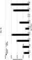
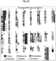
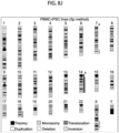
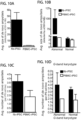
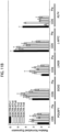
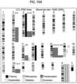
- Multiple laboratories have shown recurrent genomic aberrations in human iPSC lines upon long-term culture, with ⁇ 10-25% rate of karyotype abnormalities derived from various tissue sources.
- iPSCs maintaining genomic integrity and stability of hiPSC lines is imperative for reliable disease modeling and safe clinical applications of stem cells as a regenerative therapy.
- Aberrant cytogenetic errors that arise during reprogramming of somatic cells and/or during maintenance, expansion and prolonged culture of hiPSCs will impact the accuracy of in vitro disease modelling or, more critically, the in vivo utility of iPSCs for regenerative medicine. It is important that iPSCs in clinical use are free from cancer-associated genomic aberrations, especially given that several studies have reported chromosomal aneuploidy, translocations, duplications and deletions, and point mutations in iPSCs.
- the highly aneuploidy human embryonal carcinoma (EC) stem cells which are the malignant analogues of normal hESCs, typically contain amplified regions of the short arm of chromosome 12 and gains of chromosomes 1, 17 and X.
- CNVs sub-chromosomal copy number variations
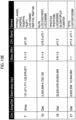
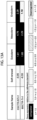
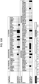
- This bio-repository currently has over 160 well-characterized individual donor human iPSC lines with multiple clones (2-6) per cell line.
- the iPSC Core has systematically monitored the genomic stability of hiPSCs derived in the Core from 450 independent iPSC cultures over multiple passages, which have been derived from fibroblast, peripheral blood mononuclear cells, immortalized lymphoblastoid cell lines and primary epithelial cells.
- fibroblasts isolated from skin biopsies.
- iPSCs derived from skin punch biopsies are more invasive, require a prolonged 2-3 week period of expansion in culture prior to reprogramming.
- peripheral blood which can be utilized as an easily accessible source of patient tissue for reprogramming.
- Peripheral blood is the most accessible adult tissue and permits access to numerous frozen samples already stored at blood banks. Additionally, many repositories have stored peripheral or lymphoblast blood with specific genotypes.
- PB peripheral blood
- white blood cells are the nucleated cells in PB at concentrations of 3.6-11 ⁇ 10 6 /ml.
- Mature T cells and primary progenitor cells in PB can be readily expanded using established methods and are among the most successfully-used sources for reprogramming.
- T cells are the most abundant cells after granulocytes in PB (20-30%) and T cells can be readily expanded with IL-2 and anti-CD3/CD28 microbeads.
- TCR T cell receptor
- the Inventors have developed a reliable protocol to efficiently reprogram blood cells (BCs) including peripheral mononuclear blood cells (PBMCs) into iPSCs (BC-iPSCs) and show that these iPSC lines are superior in terms of cytogenetic stability in comparison to their fibroblast-derived iPSC (Fib-iPSCs) lines obtained from public repositories or local clinics.
- PBMCs peripheral mononuclear blood cells
- Fib-iPSCs fibroblast-derived iPSC lines obtained from public repositories or local clinics.
- the Inventors describe methods and cytogenetic stability for derivation of BC-iPSCs from both a lymphoid T cell and a myeloid non-T cell population.
- hiPSC lines generated from PBMCs using non-integrating methods have greatly lower incidences of genomic aberrations than those generated by integrating methods.
- PBMCs should be a preferred somatic cell source for iPSC reprogramming to minimize any effects of acquired genomic aberrations and, further, should be considered as an ideal cell source for regenerative medicine.
- the alternative source of blood progenitors In contrast to mature T or B cells, the alternative source of blood progenitors contain an intact genome. In addition, they can be expanded in culture conditions that favor the proliferation of myeloid cells or erythroid cells. Blood stem/progenitor cells express surface marker CD34 and reside in the stem cell niche. However, only about 1% stem/progenitor cells enter circulation each day and as a result, only 0.01-0.1% cells in PB are CD34+ cells. This population can be enriched by magnetic-activated cell sorting (MACS) or culture of MNCs for several days can be relied upon to expand CD34+ cells to a 5-20% purity, which can be used for reprogramming without further purification.
- MCS magnetic-activated cell sorting
- nucleated peripheral blood cells include granulocytes (mostly neutrophils), monocytes, T lymphocytes, B lymphocytes and a few progenitor cells. Focusing on these constitutes of blood can be achieved by depleting red blood cells and platelet using lysis buffer followed by multiple centrifugations. Ficoll gradient centrifugation can also be utilized to deplete both red blood cells and granulocytes, leading to the enrichment of mononuclear cells (MNCs). Against this backdrop, reprogramming with exogenously expressed factors is notoriously inefficient and requires multiple cell cycles to achieve pluripotency.
- MNCs mononuclear cells
- PBMCs peripheral blood mononuclear cells
- T cells lymphocytes
- B cells lymphocytes
- NK cells monocytes
- dendritic cells dendritic cells.
- Lymphocytes are Small (5-10 ⁇ m) and Medium (10-18 ⁇ m) and constitute 70-90% of PBMCs.
- CD3+ T cells 40 -70% of PBMCs
- CD4 Helper T cells 25-60% of PBMCs
- CD8 ratio of 2:1 CD8 "Cytotoxic" compartment T cells
- the remaining compartment includes 5 - 20% B Cells (up to 15% of PBMCs) and 5 - 20% NK Cells (up to 15% of PBMCs).
- Monocytes are 16-25 ⁇ m and 10-30% of PBMCs (macrophages).
- Dendritic cells 1-2% of PBMCs
- PBMC-iPSCs peripheral blood mononuclear cell
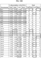
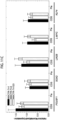
- iPSCs episomally reprogrammed iPSC lines derived from pre-expanded dermal fibroblasts (fib-iPSCs), lymphoblastoid cell lines (LCL-iPSCs) or epithelial cells (epi-iPSCs), exhibit over 10-fold greater karyotype instability (23-27%).
- the Inventors have established improved techniques for highly efficient, reproducible reprogramming using non-integrating episomal plasmid vectors, including generation of iPSCs from blood cells, including whole blood and peripheral blood, the resulting reprogrammed pluripotent cells described herein as "BC-iPSCs".
- different approaches for non-integrative reprogramming span at least categories: 1) integration-defective viral delivery, 2) episomal delivery, 3) direct RNA delivery, 4) direct protein delivery and 5) chemical induction.
- episomal vectors allows for generation of iPSCs substantially free of the vectors used in their production, as episomal or similar vectors do not encode sufficient viral genome sufficient to give rise to infection or a replication-competent virus.
- these vectors do possess a limited degree of self-replication capacity in the beginning somatic host cells. This self-replication capacity provides a degree of persistent expression understood to be beneficial in allowing the dedifferentiation process to initiate take hold in a target host cell.
- a plasmid vector satisfying these criteria includes the Epstein Barr oriP/Nuclear Antigen-1 ("EBNA1") combination, which is capable of limited self-replication and known to function in mammalian cells.
- EBNA1 Epstein Barr oriP/Nuclear Antigen-1
- binding of the EBNA1 protein to the virus replicon region oriP maintains a relatively long-term episomal presence of plasmids in mammalian cells.
- This particular feature of the oriP/EBNA1 vector makes it ideal for generation of integration-free iPSCs.
- reprogramming factor encoded in an oriP/EBNA1 vector occurs across multiple cell divisional cycles. Sufficiently high levels of reprogramming factors across several cell divisions allows for successful reprogramming even after only one infection. While sustained expression of reprogramming factors is understood to be beneficial during initial programming stages, otherwise unlimited constitutive expression would hamper subsequent stages of the reprogramming process. For example, unabated expression of reprogramming factors would interfere with subsequent growth, development, and fate specification of the host cells.
- a further benefit is the eventual removal of the reprogramming factor transgenes, as a small portion of episomes is lost per cell cycle. This is due to the asymmetric replication capacity of the host cell genome and episomal self-replication and it is estimated that approximately 0.5% of vector is lost per generation. Gradual depletion of plasmids during each cell division is inevitable following propagation leading to a population of integration-free iPSCs.
- oriP/EBNA1 The persistent, yet eventual abrogation of reprogramming factor expression on oriP/EBNA1 is highly coincident with the needs for different stages of the reprogramming process and eliminates the need for further manipulation steps for excision of the reprogramming factors, as has been attempted through use of transposons and excisable polycistronic lentiviral vector elements.
- oriP/EBNA1 has been applied by others in reprogramming studies, the reported efficiencies are extremely low (as few as 3 to 6 colonies per million cells nucleofected), which may be due, in-part, to reliance on large plasmids encoding multiple reprogramming factors (e.g., more than 12 kb), negatively impacting transfection efficiency.
- reprogramming factors that have been used include pluripotency-related genes Oct-4, Sox-2, Lin-28, Nanog, Sall4, Fbx-15 and Utf-1. These factors, traditionally are understood be normally expressed early during development and are involved in the maintenance of the pluripotent potential of a subset of cells that will constituting the inner cell mass of the pre-implantation embryo and post-implantation embryo proper. Their ectopic expression of is believed to allow the establishment of an embryonic-like transcriptional cascade that initiates and propagates an otherwise dormant endogenous core pluripotency program within a host cell.
- reprogramming determinants such as Tert, Klf-4, c-Myc, SV40 Large T Antigen (“SV40LT”) and short hairpin RNAs targeting p53 (“shRNA-p53”) have been applied.
- SV40LT Large T Antigen
- shRNA-p53 short hairpin RNAs targeting p53
- TERT and SV40LT are understood to enhance cell proliferation to promote survival during reprogramming, while others such as short hairpin targeting of p53 inhibit or eliminate reprogramming barriers, such as senescence and apoptosis mechanisms. In each case, an increase in both the speed and efficiency of reprogramming is observed.
- miRNAs are also known to influence pluripotency and reprogramming, and some miRNAs from the miR ⁇ 290 cluster have been applied in reprogramming studies. For example, the introduction of miR-291-3p, miR-294 or miR-295 into fibroblasts, along with pluripotency-related genes, has also been reported to increase reprogramming efficiency.
- somatic cell reprogramming efficiency is reportedly fourfold higher when Oct-4 and Sox2 are encoded in a single transcript on a single vector in a 1:1 ratio, in contrast to delivering the two factors on separate vectors. The latter case results in a less controlled uptake ratio of the two factors, providing a negative impact on reprogramming efficiency.
- IVS internal ribosome entry site
- a further advantage of the techniques described herein is the use of defined media conditions for the reprogramming process, including the use of ESC media and/or E7 media. While certain additives may be present to spur the reprogramming process (e.g., L-Ascorbic Acid, Transferrin, Sodium Bicarbonate, Insulin, Sodium Selenite and/or bFGF), no serum or animal components are used. In some instances, there may be further benefits in altering the chemical and/or atmospheric conditions under which reprogramming will take place.
- additives e.g., L-Ascorbic Acid, Transferrin, Sodium Bicarbonate, Insulin, Sodium Selenite and/or bFGF
- hypoxic similar to bone marrow stem-cell niches
- reprogramming under hypoxic conditions of 5% O 2 instead of the atmospheric 21% O 2 , may further provide an opportunity to increase the reprogramming efficiency.
- chemical induction techniques have been used in combination with reprogramming, particularly histone deacetylase (HDAC) inhibitor molecule, valproic acid (VPA), which has been found wide use in different reprogramming studies.
- HDAC histone deacetylase
- VPA valproic acid
- MAPK kinase (MEK)-ERK (“MEK”) inhibitor PD0325901 MAPK kinase (MEK)-ERK (“MEK”) inhibitor PD0325901, transforming growth factor beta ("TGF- ⁇ ") type I receptor ALK4, ALK5 and ALK7 inhibitor SB431542 and the glycogen synthase kinase-3 (“GSK3”) inhibitor CHIR99021 have been applied for activation of differentiation-inducing pathways (e.g. BMP signaling), coupled with the modulation of other pathways (e.g. inhibition of the MAPK kinase (MEK)-ERK pathway) in order to sustain self-renewal.
- differentiation-inducing pathways e.g. BMP signaling
- other pathways e.g. inhibition of the MAPK kinase (MEK)-ERK pathway
- Rho-associated coiled-coil-containing protein kinase (“ROCK”) inhibitors such as Y-27632 and thiazovivin (“Tzv) have been applied in order to promote survival and reduce vulnerability of pSCs to cell death, particularly upon single-cell dissociation.
- ROCK Rho-associated coiled-coil-containing protein kinase
- Tzv thiazovivin
- reprogramming factor combinations In addition to the choice of delivery vectors, reprogramming factor combinations, and conditions for reprogramming, further variations must consider the nature of the host target cell for reprogramming. To date, a wide variety of cells have served as sources for reprogramming including fibroblasts, stomach and liver cell cultures, human keratinocytes, adipose cells, and frozen human monocyte. Clearly, there is a wide and robust potential for dedifferentiation across many tissues sources. Nevertheless, it is widely understood that depending on the donor cell type, reprogramming is achieved with different efficiencies and kinetics.
- fibroblasts remain the most popular donor cell type for reprogramming studies
- other types of cells such as human primary keratinocytes transduced with Oct-4, Sox-2, Klf-4 and c-Myc have been reported to reprogram 100 times more efficiently and two-fold faster.
- some other cell types, such as cord blood cells may only require a subset of reprogramming factors, such as Oct-4 and Sox-2 for dedifferentiation to take hold, while neural progenitor cells may only require Oct-4.
- PB cells peripheral blood
- fibroblasts e.g., skin biopsy
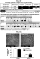
- pluripotent stem cell lines Following successful reprogramming, clonal selection allows for generation of pluripotent stem cell lines. Ideally, such cells possess requisite morphology (i.e., compact colony, high nucleus to cytoplasm ratio and prominent nucleolus), self-renewal capacity for unlimited propagation in culture (i.e., immortal), and with the capability to differentiate into all three germ layers (e.g., endoderm, mesoderm and ectoderm). Further techniques to characterize the pluripotency of a given population of cells include injection into an immunocompromised animal, such as a severe combined immunodeficient ("SCID") mouse, for formation of teratomas containing cells or tissues characteristic of all three germ layers.
- SCID severe combined immunodeficient
- PBMCs approximately, 5 ⁇ 10 6 cells per nucleofection of PBMCs were nucleofected with either plasmid mixture 4p or plasmid mixture 5p using program V-024 on the Amaxa Nucleofector 2D Device with the Amaxa Human T-cell Nucleofector ® Kit. Approximately 1 ⁇ 10 6 cells were then seeded into wells of a 6-well plate covered with mitomycin treated mouse embryonic feeder (MEF) layer or coated with 10 ⁇ g/ml Laminin-521 (L-521;BioLamina).
- MEF mouse embryonic feeder
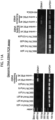
- Each episomal plasmid (Addgene) expressing 7 factors: OCT4, SOX2, KLF4, LMYC, LIN28, SV40LT and p53 shRNA (pEP4 E02S ET2K, pCXLE-hOCT3/4-shp53-F, pCXI,EhUL, pCXLE-hSK, and pCXI,E-EBNA1).
- This method has a significant advantage over viral transduction, because exogenously introduced genes do not integrate and are instead expressed episomally in a transient fashion.
- ⁇ T-cell medium X-vivo10 supplemented with 30U/ml IL-2 and 5ul/well Dynabeads Human T-activator CD3/CD28
- ⁇ MEM non T-cell medium
- FBS 10ng/ml IL-3, 10ng/ml IL-6, 10ng/ml G-CSF and 10ng/ml GM-CSF
- ReproCell Primate ESC medium containing 5 ng/ml bFGF (for MEF condition) or E7 medium (for L-521 condition) was added to the wells without aspirating the previous medium.
- the medium was gently aspirated from each well and 2ml of the appropriate fresh reprogramming media was added to each well. Medium was replaced every other day. At approximately day 18 post nucleofection, individual colonies were observed in all wells of each condition. At approximately day 25 post nucleofection, individual colonies were isolated and sub-cloned into 1 well of 12-well plate containing the appropriate substrate and medium. These nucleofected cells were plated on feeder-independent BD Matrigel TM growth factor-reduced Matrix (Corning/BD Biosciences, #354230). All cultures were maintained at 20% O2 during the reprogramming process.
- the composition of blood cell derived induced pluripotent stem cells includes cells generated by providing a quantity of blood cells, delivering a quantity of reprogramming factors into the blood cells, culturing the blood cells in a reprogramming media for at least 4 days, wherein delivering the reprogramming factors, and culturing generates the blood cells derived induced pluripotent stem cells.
- the blood cells are T-cells.
- the blood cells are non-T-cells.
- the blood cells are mononuclear cells (MNCs), including for example peripheral blood mononuclear cells (PBMCs).
- the cells are primary granulocytes, monocytes and B lymphocytes.
- the reprogramming factors are Oct-4, Sox-2, Klf-4, c-Myc, Lin-28, SV40 Large T Antigen ("SV40LT”), and short hairpin RNAs targeting p53 ("shRNA-p53").
- these reprogramming factors are encoded in a combination of vectors including pEP4 E02S ET2K, pCXLE-hOCT3/4-shp53-F, pCXLE-hSK, pCXLE-hUL and pCXWB-EBNA1.
- the reprogramming media includes PD0325901, CHIR99021, HA-100, and A-83-01.
- the culturing the blood cells in a reprogramming media is for 4-30 days.
- the blood cells are plated on a treated cell culture surface after delivering a quantity of reprogramming factors.
- treatment includes plating of feeder cells, such as mouse embryonic fibroblasts.
- treatment includes coating with extracellular matrix proteins.
- extracellular matrix proteins include laminin.
- the BC-iPSCs are capable of serial passaging as a cell line.
- the BC-iPSCs possess genomic stability.
- Genomic stability can be ascertained by various techniques known in the art. For example, G-band karyotyping can identify abnormal cells lacking genomic stability, wherein abnormal cells possess about 10% or more mosaicism, or one or more balanced translocations of greater than about 5, 6, 7, 8, 9, 10 or more Mb.
- genomic stability can be measured using comparative genomic hybridization (aCGH) microarray, comparing for example, BC-iPSCs against iPSCs from a non-blood cell source such as fibroblasts.
- aCGH comparative genomic hybridization
- Genomic stability can include copy number variants (CNVs), duplications/deletions, and unbalanced translocations.
- BC-iPSCs exhibit no more than about 3, 4, 5, 6, 7, 8, 9, 10, 11, 12, 13, 15, 16, 17, 18, 19, or 20 Mb average size of amplification and deletion.
- BC-iPSCs exhibit no more than about 20-30 Mb average size of amplification and deletion.
- BC-iPSCs exhibit no more than about 30-40 Mb average size of amplification and deletion.
- BC-iPSCs exhibit no more than about 40-50 Mb average size of amplification and deletion.
- the average number of acquired de novo amplification and deletions in BC-iPSCs is less than about 5, 4, 3, 2, or 1.
- de novo amplification and deletions in fib-iPSCs are at least two-fold greater than in PBMC-iPSCs.
- reprogramming factors can also include one or more of following: Oct-4, Sox-2, Klf-4, c-Myc, Lin-28, SV40LT, shRNA-p53, nanog, Sall4, Fbx-15, Utf-1, Tert, or a Mir-290 cluster microRNA such as miR ⁇ 291 ⁇ 3p, miR ⁇ 294 or miR ⁇ 295.
- the reprogramming factors are encoded by a vector.
- the vector can be, for example, a non-integrating episomal vector, minicircle vector, plasmid, retrovirus (integrating and non-integrating) and/or other genetic elements known to one of ordinary skill.
- the reprogramming factors are encoded by one or more oriP/EBNA1 derived vectors.
- the vector encodes one or more reprogramming factors, and combinations of vectors can be used together to deliver one or more of Oct-4, Sox-2, Klf-4, c-Myc, Lin-28, SV40LT, shRNA-p53, nanog, Sall4, Fbx-15, Utf-1, Tert, or a Mir-290 cluster microRNA such as miR ⁇ 291 ⁇ 3p, miR ⁇ 294 or miR ⁇ 295.
- oriP/EBNA1 is an episomal vector that can encode a vector combination of multiple reprogramming factors, such as pCXLE-hUL, pCXLE-hSK, pCXLE-hOCT3/4-shp53-F, pEP4 EO2S T2K and pCXWB-EBNA1.
- the reprogramming factors are delivered by techniques known in the art, such as nuclefection, transfection, transduction, electrofusion, electroporation, microinjection, cell fusion, among others.
- the reprogramming factors are provided as RNA, linear DNA, peptides or proteins, or a cellular extract of a pluripotent stem cell.
- the reprogramming media is embryonic stem cell (ESC) media. In various embodiments, the reprogramming media includes bFGF. In various embodiments, the reprogramming media is E7 media. In various embodiments, the reprogramming E7 media includes L-Ascorbic Acid, Transferrin, Sodium Bicarbonate, Insulin, Sodium Selenite and/or bFGF In different embodiments, the reprogramming media comprises at least one small chemical induction molecule. In different embodiments, the at least one small chemical induction molecule comprises PD0325901, CHIR99021, HA-100, A-83-01, valproic acid (VPA), SB431542, Y-27632 or thiazovivin ("Tzv"). In different embodiments, culturing the BCs in a reprogramming media is for at least 4, 5, 6, 7, 8, 9, 10, 11, 12, 13, 14, 15, 16, 17, 18, 19, 20, 21, 22, 23, 24, 25, 26, 27, 28, 29, or 30 days.
- the BC-iPSCs are derived from blood cells previously isolated from a subject, by for, example, drawing a blood sample from the subject.
- the blood cells are isolated from a subject possessing a disease mutation.
- subjects possessing any number of mutations, such as autosomal dominant, recessive, sex-linked, can serve as a source of blood cells to generate BC-iPSCs possessing said mutation.
- the disease mutation is associated with a neurodegenerative disease, disorder and/or condition.
- the disease mutation is associated with an inflammatory bowel disease, disorder, and/or condition.
- the BC-iPSCs possess features of pluripotent stem cells.
- pluripotent stem cells including differentiation into cells of all three germ layers (ectoderm, endoderm, mesoderm), either in vitro or in vivo when injected into an immunodeficient animal, expression of pluripotency markers such as Oct-4, Sox-2, nanog, TRA-1-60, TRA-1-81, SSEA4, high levels of alkaline phosphatase ("AP”) expression, indefinite propagation in culture, among other features recognized and appreciated by one of ordinary skill.
- pluripotency markers such as Oct-4, Sox-2, nanog, TRA-1-60, TRA-1-81, SSEA4, high levels of alkaline phosphatase (“AP”) expression, indefinite propagation in culture, among other features recognized and appreciated by one of ordinary skill.
- compositions including a quantity of blood cells derived induced pluripotent stem cells generated by the above described methods, and a pharmaceutically acceptable carrier.
- the cells are primary culture cells.
- the cells are blood cells (BCs).
- the blood cells are T-cells.
- the blood cells are non-T-cells.
- the cells are mononuclear cells (MNCs), including for example peripheral blood mononuclear cells (PBMCs).
- the cells are primary granulocytes, monocytes and B lymphocytes.
- the reprogramming factors are Oct-4, Sox-2, Klf-4, c-Myc, Lin-28, SV40 Large T Antigen ("SV40LT”), and short hairpin RNAs targeting p53 ("shRNA-p53").
- these reprogramming factors are encoded in a combination of vectors including pEP4 E02S ET2K, pCXLE-hOCT3/4-shp53-F, pCXLE-hSK, pCXLE-hUL and pCXWB-EBNA1.
- telomeres supports the reprogramming of non-T cell component of blood, including peripheral blood mononuclear cells.
- the reprogramming media is embryonic stem cell (ESC) media.
- the reprogramming media includes bFGF. In various embodiments, the reprogramming media is E7 media. In various embodiments, the reprogramming E7 media includes L-Ascorbic Acid, Transferrin, Sodium Bicarbonate, Insulin, Sodium Selenite and/or bFGF. In different embodiments, the reprogramming media comprises at least one small chemical induction molecule. In certain other embodiments, the reprogramming media includes PD0325901, CHIR99021, HA-100, and A-83-01. In other embodiments, the culturing the blood cells in a reprogramming media is for 4-30 days.
- the BC-iPSCs are capable of serial passaging as a cell line.
- the BC-iPSCs possess genomic stability.
- Genomic stability can be ascertained by various techniques known in the art. For example, G-band karyotyping can identify abnormal cells lacking genomic stability, wherein abnormal cells possess about 10% or more mosaicism, or one or more balanced translocations of greater than about 5, 6, 7, 8, 9, 10 or more Mb.
- genomic stability can be measured using comparative genomic hybridization (aCGH) microarray, comparing for example, BC-iPSCs against iPSCs from a non-blood cell source such as fibroblasts.
- aCGH comparative genomic hybridization
- Genomic stability can include copy number variants (CNVs), duplications/deletions, and unbalanced translocations.
- BC-iPSCs exhibit no more than about 3, 4, 5, 6, 7, 8, 9, 10, 11, 12, 13, 15, 16, 17, 18, 19, or 20 Mb average size of amplification and deletion.
- BC-iPSCs exhibit no more than about 20-30 Mb average size of amplification and deletion.
- BC-iPSCs exhibit no more than about 30-40 Mb average size of amplification and deletion.
- BC-iPSCs exhibit no more than about 40-50 Mb average size of amplification and deletion.
- the average number of acquired de novo amplification and deletions in BC-iPSCs is less than about 5, 4, 3, 2, or 1.
- de novo amplification and deletions in fib-iPSCs are at least two-fold greater than in PBMC-iPSCs.
- the methods produces iPSC cell lines collectively exhibiting about 20%, 15%, 10%, 5% or less abnormal karyotypes over 4-8, 9-13, 13-17, 17-21, 21-25, or 29 or more passages when serially passaged as a cell line.
- reprogramming factors can also include one or more of following: Oct-4, Sox-2, Klf-4, c-Myc, Lin-28, SV40LT, shRNA-p53, nanog, Sall4, Fbx-15, Utf-1, Tert, or a Mir-290 cluster microRNA such as miR ⁇ 291 ⁇ 3p, miR ⁇ 294 or miR ⁇ 295.
- the reprogramming factors are encoded by a vector.
- the vector can be, for example, a non-integrating episomal vector, minicircle vector, plasmid, retrovirus (integrating and non-integrating) and/or other genetic elements known to one of ordinary skill.
- the reprogramming factors are encoded by one or more oriP/EBNA1 derived vectors.
- the vector encodes one or more reprogramming factors, and combinations of vectors can be used together to deliver one or more of Oct-4, Sox-2, Klf-4, c-Myc, Lin-28, SV40LT, shRNA-p53, nanog, Sall4, Fbx-15, Utf-1, Tert, or a Mir-290 cluster microRNA such as miR ⁇ 291 ⁇ 3p, miR ⁇ 294 or miR ⁇ 295.
- oriP/EBNA1 is an episomal vector that can encode a vector combination of multiple reprogramming factors, such as pCXLE-hUL, pCXLE-hSK, pCXLE-hOCT3/4-shp53-F, pEP4 EO2S T2K and pCXWB-EBNA1.
- the reprogramming factors are delivered by techniques known in the art, such as nuclefection, transfection, transduction, electrofusion, electroporation, microinjection, cell fusion, among others.
- the reprogramming factors are provided as RNA, linear DNA, peptides or proteins, or a cellular extract of a pluripotent stem cell.
- the cells are treated with sodium butyrate prior to delivery of the reprogramming factors.
- the cells are incubated or 1, 2, 3, 4, or more days on a tissue culture surface before further culturing. This can include, for example, incubation on a Matrigel coated tissue culture surface.
- the reprogramming conditions include application of norm-oxygen conditions, such as 5% O 2 , which is less than atmospheric 21% O 2 .
- the reprogramming media is embryonic stem cell (ESC) media. In various embodiments, the reprogramming media includes bFGF. In various embodiments, the reprogramming media is E7 media. In various embodiments, the reprogramming E7 media includes L-Ascorbic Acid, Transferrin, Sodium Bicarbonate, Insulin, Sodium Selenite and/or bFGF. In different embodiments, the reprogramming media comprises at least one small chemical induction molecule. In different embodiments, the at least one small chemical induction molecule comprises PD0325901, CHIR99021, HA-100, A-83-01, valproic acid (VPA), SB431542, Y-27632 or thiazovivin ("Tzv"). In different embodiments, culturing the BCs in a reprogramming media is for at least 4, 5, 6, 7, 8, 9, 10, 11, 12, 13, 14, 15, 16, 17, 18, 19, 20, 21, 22, 23, 24, 25, 26, 27, 28, 29, or 30 days.
- Efficiency of reprogramming is readily ascertained by one of many techniques readily understood by one of ordinary skill. For example, efficiency can be described by the ratio between the number of donor cells receiving the full set of reprogramming factors and the number of reprogrammed colonies generated. Measuring the number donor cells receiving reprogramming factors can be measured directly, when a reporter gene such as GFP is included in a vector encoding a reprogramming factor. Alternatively, indirect measurement of delivery efficiency can be provided by transfecting a vector encoding a reporter gene as a proxy to gauge delivery efficiency in paired samples delivering reprogramming factor vectors.
- the number of reprogrammed colonies generated can be measured by, for example, observing the appearance of one or more embryonic stem cell-like pluripotency characteristics such as alkaline phosphatase (AP)-positive clones, colonies with endogenous expression of transcription factors Oct or Nanog, or antibody staining of surface markers such as Tra-1-60.
- efficiency can be described by the kinetics of induced pluripotent stem cell generation.
- efficiency can include producing cell lines of normal karyotype, including the method producing iPSC cell lines collectively exhibiting about 20%, 15%, 10%, 5% or less abnormal karyotypes over 4-8, 9-13, 13-17, 17-21, 21-25, or 29 or more passages when serially passaged as a cell line.
- the reprogramming method involves the following steps. Frozen peripheral blood mononuclear cells (PBMCs), or freshly isolated PBMCs can be used. Treated cell culture surfaces can be used for plating thawed or freshly isolated PBMS. This includes, for example, cell culture surfaces treated with mouse embryonic feeders (MEF) or extracellular matrix laminin L-521A. An expression plasmid mixture is provided, with the plasmid mixture encoding a combination of pluripotency factors. Plasmids are introduced to PBMCs, by using for example, nucleofection, and thereafter placed on the treated cell culture surface. After 2 days of culturing, reprogramming media is introduced, and replenished until colonies are formed.
- MEF mouse embryonic feeders
- L-521A extracellular matrix laminin L-521A
- the Inventors have established processes to isolate lymphocytes from freshly collected or commercial sources of human or mammalian peripheral blood (PB).
- PB peripheral blood
- the preferred format for the collection and shipment of such samples is the CPT tubes. This format allows the supplier to centrifuge the vacutainer(s) and separate the red blood cells from the plasma components prior to shipping.
- the protocol provides for the isolation of PBMCs from the plasma layer of the CPT tubes.
- Optional tests Prior to opening the Vacutainer, several disease state tests can be performed, including testing for HIV, Hepatitis B and C, and syphilis. The latter test results should be made available within 1-3 working days from the collection date. If there is a positive result, the sample(s) will be properly discarded, and all laboratory equipment that was used to process the samples will be decontaminated.
- Temperature Since the principle of separation depends on a density gradient, and the density of the components varies with temperature, the temperature of the system should be controlled between 18-25°C during separation.
- Centrifugation Since the principle of separation depends on the movement of formed elements in the blood through the separation media, the "RCF" should be controlled at 1500 RCF to 1800 RCF. The time of centrifugation should be a minimum of 20 minutes. (As noted in the trouble shooting section, some specimens may require up to 30 minutes for optimal separation.) Centrifugation of the BD Vacutainer ® CPT TM Tube up to 30 minutes has the effect of reducing red blood cell contamination of the mononuclear cell fraction. Centrifugation beyond 30 minutes has little additional effect. The BD Vacutainer ® CPT TM Tube may be re-centrifuged if the mononuclear "band" or layer is not disturbed.
- Blood samples should be centrifuged or separated within two hours of blood drawing. Red blood cell contamination in the separated mononuclear cell fraction increases with longer delays in sample separation. Mononuclear cell recovery decreases with increased time delay before centrifugation, falling to approximately 40% mononuclear cell recovery at 24 hours. Pour the contents (the plasma layer) off into an appropriately labeled 50 mL tube.
- Described herein is the procedure for reprogramming separated whole blood samples (peripheral blood mononuclear cells - PBMCs) for iPSC generation.
- Materials for use include: Sterile 1.5ml Eppendorf tubes; Sterile pipette tips (1000ul, 200ul, 10ul); Amaxa Nucleofector TM 2b Device (Lonza); Human T-Cell Nucleofector Kit (Lonza, Cat #VPA-1002); Prepared MEF 6-well TC Plates or L-521 Coated Plates; 0.22um SteriFlip (optional); Primate ESC Medium or E7 Medium, described below in Tables 1-4.
- Plasmids used include (1) pCXLE-hOCT3/4-shp53; (2) pCXLE-hSK; (3) pCXLE-UL; (4) pCXWB-EBNA1; (5) pEP4 E02S ET2K.
- T-Cell Clonality Assay for whole blood derived iPSC lines
- T-Cell Clonality Assay TM Gel Detection - Invivoscribe Presence (T-cell) of lack of clonal TCR beta gene rearrangements (non T-cell)
- Alkaline Phosphatase staining can be performed using the Alkaline Phosphatase Staining Kit II (Stemgent, Cat no. 00-0055) according to the manufacturer's instructions.
- BC-iPSCs or differentiated cells can be plated on glass coverslips or optical-bottom 96-well plates (Thermo, # 165305) and subsequently fixed in 4% paraformaldehyde. Intestinal organoids can be fixed in 4% paraformaldehyde, transferred to 30% sucrose, embedded in HistoPrep (Thermo Fisher Scientific) and cut into 20 ⁇ m sections. All cells are blocked in 510% goat or donkey serum with 0.1% Triton X-100 and incubated with primary antibodies either for either 3hrs at room temperature or overnight at 4°C. Cells are then rinsed and incubated in species-specific AF488 or AF594-conjugated secondary antibodies followed by Hoechst 33258 (0.5 ⁇ g/ml; Sigma) to counterstain nuclei.
- Antibodies suitable for immunocytochemistry and immunoblotting include: (as listed by antigen, dilution, catalog no., isotype, and manufacturer) SSEA4, 1:250, MAB4304, mIgG3, Millipore; TRA-1-60, 1:250, 09-0010, IgM, Stemgent; TRA-1-81, 1:250,09-0011, mIgM, Stemgent; OCT4, 1:250, 09-0023, Rabbit IgG, Stemgent; NANOG, 1:250, 09-0020, Rabbit IgG, Stemgent; SOX2,1:500, AB5603, Rabbit IgG, Millipore; TuJ1 (( ⁇ 3-tubulin), 1:1000, T8535, mIgG2b, Sigma; CDX2, 1:500, NBP1-40553, IgG, Novus; FABP2, 1:500, AF3078, IgG, R & D systems; Collagen Type 1, 1:500, 600-401-103-0.1, Rabbi
- BC-iPSCs are dissociated into a single cell suspension using Accutase (Millipore, #SCR005).
- Surface staining of IPSCs is carried out using SSEA4 (R&D Systems, FAB1435A).
- Karyotyping can also be performed as follows. Human BC-iPSCs are incubated in Colcemid (100 ng/mL; Life Technologies) for 30 minutes at 37°C and dissociated using TrypLE for 10 minutes. They can be washed in phosphate buffered saline (PBS) and incubated at 37°C in 5mL hypotonic solution (1g KCl, 1g Na Citrate in 400mL water) for 30 minutes. The cells can be centrifuged for 2.5 minutes at 1500 RPM and resuspended in fixative (methanol: acetic acid, 3:1) at room temperature for 5 minutes. This is repeated twice, and finally cells were resuspended in 500 ⁇ l of fixative solution and submitted to the Cedars-Sinai Clinical Cytogenetics Core for G-Band karyotyping.
- PBS phosphate buffered saline
- fixative methanol: acetic acid, 3:1
- RNA can be isolated using the RNeasy Mini Kit (Qiagen) and subsequently run on a Human HT-12 v4 Expression BeadChip Kit (Illumina). The raw data file (idat file) was subsequently uploaded onto the Pluritest widget online (www.pluritest.org).
- Cytogenetic Stability of PBMCs BC-iPSCs Description Numbers % abnormal Total iPSC lines 90 3.3% Total abnormal 3 *Total with non-clonal aberration 3 3.3% Table 3B. Cytogenetic Stability of PBMCs Fibroblast-iPSCs Description Numbers % abnormal Total iPSC lines 242 25.6% Total abnormal 62 *Total with non-clonal aberration 17 7.0%
- Cytogenetic Stability of PBMCs BC-iPSCs Description Numbers Total iPSC lines 7 De Novo* amp/dels 4 Average De Novo amp/dels 0.6 De Novo amp/dels per passage 0.036
- Cytogenetic Stability of PBMCs Fibroblast-iPSCs Description Numbers Total iPSC lines 6 De Novo amp/dels 19 Average De Novo amp/dels 3.2 De Novo* amp/dels per passage 0.136 * De Novo or deletions are those newly acquired amplifications or deletions during reprogramming and/or expansion of the respective iPSC lines when compared to the parent donor tissue aCGH profile
- Table 5 High Success rate of peripheral blood mononuclear cell (PBMC)-based episomal reprogramming on MEFs .
- PBMC peripheral blood mononuclear cell
- PBMCs isolated from a blood draw are reliably reprogrammed to iPSCs
- Dermal fibroblasts obtained from a skin biopsy have been the most common source of iPSC reprogramming material, due largely to their inexpensive and relatively easy usage.
- a skin punch biopsy can be between the size of 3-4 mm and is often a painful procedure that requires local anesthetics.
- blood is a more accessible source of patient tissue during hospital visits and, therefore, a preferred choice of source tissue for iPSC reprogramming by both patients and clinicians.
- Various human blood reprogramming methods have been reported with mixed efficiencies, including CD34 + cells mobilized from peripheral blood (ref).
- CD34 + cells are usually isolated from the peripheral blood or bone marrow of patients undergoing G-CSF mobilization for several days.
- isolated CD34 + progenitor cells need to be enriched and expanded in culture with complex and expensive protocols.
- the Inventors were successful in reprogramming PBMCs that were freshly isolated from a peripheral blood draw without prior cell expansion or CD34 + cell isolation using episomal reprogramming plasmids. Such an approach provides the fastest and most cost effective procedure for obtaining a patient's reprogrammed iPSCs from the time of collecting a patient bio-specimen to generating their rigorously characterized iPSCs.
- cryopreserved PBMCs from multiple individuals were reprogrammed to iPSCs with episomal plasmids expressing POU5F1, SOX2, KLF4, LIN28, L-MYC, p53 shRNA
- the Inventors observed significant variability in isolating identifiable iPSC clones even after 35-40 days, regardless of PBMC cell type, ECM substrata and media).
- the T reprogramming method was successful with only 33% of PBMC samples, while the non-T method on cryopreserved PBMCs did not result in any clonal iPSC lines (data not shown).
- the BC-iPSCs passed pluripotency quality control metrics determined by the PluriTest assay, demonstrating that the PBMC-iPSC transcription profile was analogous to well established hESCs and fib-iPSCs, but not differentiated fibroblasts and neural progenitor cells ( Fig. 8C ).
- BC-iPSCs maintained a normal G-band karyotype ( Fig. 8D ) and were confirmed to be clonal derivatives of either T cells or non-T cells from the PBMCs using the T cell receptor (TCR)- ⁇ and - ⁇ , gene rearrangement/clonality assays ( Fig. 8E ).
- BC-iPSCs The trilineage potential of BC-iPSCs was demonstrated by spontaneous embryoid body formation and by measuring germ-layer specific gene expression profile by the TaqMan Scorecard assay ( Fig. 8F ).
- the "footprintfree" status of BC-iPSCs was confirmed by demonstration of endogenous pluripotency gene expression and absence of exogenous reprogramming transgenes using RT-qPCR ( Fig. 11A ).
- the EBNA plasmid-related latency element was eventually eliminated from the established BC-iPSCs ( Fig. 11B ).
- BC-iPSCs have equivalent neuronal differentiation compared to fib-iPSCs
- iPSCs regardless of the source tissue, ultimately lose most of their gene expression and epigenetic profiles related to the original cell source.
- blood-derived iPSCs can differentiate as efficiently as fibroblast-derived iPSCs into other various cell types, possibly due to a stronger retention of epigenetic memory in blood-sourced iPSCs.
- the Inventors addressed this by directing blood or fibroblast-derived iPSCs, both of mesodermal origin, to a different germ layer such as ectoderm.
- BC-iPSCs maintain a significantly more stable karyotype compared to fib-iPSCs
- the Inventors performed karyotype analysis on 364 human iPSC cultures where source fibroblasts and PBMCs were collected from multiple laboratories or public repositories.
- the Inventors' data reveal remarkable differences in the incidence of chromosomal aberrations in fib-iPSCs and BC-iPSCs.
- Abnormal karyotypes with clonal aberrations were observed in 59 of 258 (22.9%) cultures from clonally independent human fib-iPSC lines, derived from 78 unique donors ( Fig. 6 ).
- only 4 in 106 (3.8%) cultures of clonal human PBMC-iPSC lines derived from 32 unique donors displayed low-frequency abnormalities.
- chromosomes 13 The most frequent karyotypic abnormalities in fib-iPSCs were observed in chromosomes 13 (13.2%), 1 (11.8%), 12 (11.1%), and X (10.4%) (Table 6). To a lesser extent karyotype changes were also observed in chromosomes 21, 11, and 6, in descending order of frequency. Of the aneuploidies, chromosomal gains (trisomy or duplications) were most commonly observed in 12 (21.1%), 1 (15.8%), 11 (13.2%), and 6 (10.5%) and chromosomal losses were repeatedly observed in chromosomes X (38.5%) and 21 (23.1%).
- BC-iPSCs had one line each display chromosome gains in 14 (trisomy) or 18 (duplication).
- the other two PBMC-iPSC lines had exhibited uncommon abnormalities: a translocation 46,XY,t(7;14)(q34;q11.2) and a mosaic deletion 46,X,del(X)(q22q26) in 19% of cells (Table 6).
- Fib- and BC-iPSCs karyotype distribution relates not to the donor age but rather to passage number
- the skin unlike the blood, is routinely exposed to external environmental elements like potential sun damage and the fibroblasts derived from the biopsies require a certain level of expansion prior to reprogramming, which is akin to further aging in culture.
- the Inventors posited that the donor age might be a contributing factor that impacts the cytogenetic instability of fib-iPSCs.
- the Inventors did not observe a significant trend or correlation with the age of the donor ( Fig. 9A ). Indeed, based on G band karyotype analysis, the highest percent of karyotype abnormalities (31.9%) was observed in the 21-40 age group fibroblast-iPSCs.
- Pluripotent stem cells have a highly proliferative nature. As such, the Inventors next attempted to analyze how the fib- and BC-iPSCs fared over time in cell culture and whether increasing passage number led to a greater propensity for accumulating cytogenetic aberrations. About 21.6% of fibroblast-derived iPSCs were observed to have abnormal karyotypes (> 1/20 cells with clonal aberrations) in their first G-band karyotype evaluation post-iPSC generation, which was typically between passages 4-23. In stark contrast, only 2.8% of BC-iPSCs had abnormal karyotypes in their first assessment.
- G-band karyotyping showed that a greater proportion of fib-iPSCs (27.8%) acquired abnormal karyotypes in culture upon repeat karyotyping when compared to the proportion at first karyotypes (P ⁇ 0.001, two way ANOVA with Bonferroni posttest), while the percent abnormal karyotypes in BC-iPSCs did not show any significant difference between first and repeat karyotypes ( Fig. 9B ).
- the highest rate of abnormal karyotypes first or repeat
- BC-iPSCs acquire less submicroscopic amplifications and deletions
- G-band karyotype has the advantage that it can detect balanced translocations, inversions, ⁇ 20% culture mosaicism, chromosomal position of genomic gains or losses as commonly observed in the fib-iPSC cultures ( Fig. 6 and Table 5).
- G-band karyotype is unable to detect submicroscopic genomic abnormalities ( ⁇ 5Mb)
- the Inventors decided to conduct comparative genomic hybridization (aCGH) microarray on a subset of fib-iPSC and PBMC-iPSC lines.
- aCGH While aCGH is unable to detect balanced translocations, inversions and ⁇ 20% culture mosaicism, it is a good supportive method to detect smaller size genomic gains and losses, copy number variants (CNVs), duplications/deletions, and unbalanced translocations in the iPSC lines.
- the Inventors analyzed and recorded any de novo amplifications or deletions acquired in the iPSCs upon comparison with the parental fibroblast or PBMC source bio-specimen. In this analysis only those amplifications and deletions acquired de novo in iPSCs were considered, when they were not normal CNVs upon cross referencing against the Database of Genomic Variants (DGV) that contains genomic variations observed in healthy individuals.
- DGV Genomic Variants
- the average size of the amplification and deletions detected by aCGH were significantly greater in fib-iPSC lines at 44 Mb compared to 2.1 Mb in PBMC-iPSC lines ( Fig. 10A ), the preponderance of which was due to fib-iPSC lines with an abnormal karyotype ( Fig. 10B ).
- the de novo amps/dels were on average 2.31 Mb in fib-iPSCs and 2 Mb in PBMC-iPSC lines.
- Inventors report a new and more reliable method for reprogramming of the non-T cell component of blood using episomal plasmids expressing pluripotency factors.
- Using the described reprogramming protocol one is able to consistently reprogram non-T cells with close to 100% success from non-T cell or non-B cell sources.
- Further advantages include use of a defined reprogramming media E7 and using defined clinically compatible substrate recombinant human L-521.
- Blood cell-derived iPSCs (“BC-iPSCs”) exhibited identical characteristics to fibroblastderived iPSCs ("fib-iPSCs”), retain genotype, exhibit a normal pluripotency profile, and readily differentiate into all three germ-layer cell types.
- This method for reliably deriving iPSCs from patient blood samples paves the way for rapidly generate new human iPSC lines, including patient-specific lines, thus providing an enormous bioresource for disease modeling, drug discovery, and regenerative medicine applications.
- G-Band Karyotype Human iPSCs were incubated in Colcemid (100 ng/mL; Life Technologies) for 30 minutes at 37°C and then dissociated using TrypLE for 10 minutes. They were then washed in phosphate buffered saline (PBS) and incubated at 37°C in 5mL hypotonic solution (1g KCl, 1g Na Citrate in 400mL water) for 30 minutes. The cells were centrifuged for 2.5 minutes at 1500 RPM and resuspended in fixative (methanol: acetic acid, 3:1) at room temperature for 5 minutes.
- PBS phosphate buffered saline
- fixative methanol: acetic acid, 3:1
- the bar widths for each ideogram were scaled in proportion to the number of samples examined.
- the bars displaying abnormalities found in PBMC-iPSC lines are 2.16 times the width of bars depicting abnormalities found in fib-iPSC lines to account for the fibroblast ideogram portraying the number of abnormalities found in a sample set that was 2.16 times larger than the PBMC set.
- the letters above the chromosomal translocation (navy blue bars) category are matched such that they correspond between the chromosomal locations to where the translocation abnormality occurred.
- Array CGH is a high-resolution karyotype analysis solution for the detection of unbalanced structural and numerical chromosomal alterations with high-throughput capabilities.
- High quality genomic DNA was isolated from primary dermal fibroblasts or isolated PBMCs and their reprogrammed iPSCs. Quality of genomic DNA as determined by UV spec. (NanoVue), fluorometer (Qubit) and Agarose Gel analysis. The samples were analyzed for CNVs using the Agilent 60K Standard aCGH platform suitable for human stem cell and cancer cell lines by Cell Line Genetics (Madison, WI). Comparison of iPSC lines to their parental donor cell of origin yielded de novo copy-number variation (gains/amplifications or losses/deletions) for the indicated loci.
- Genomic DNA (350ng) was harvested from all cell lines using the MasterPure DNA Purification Kit (Epicenter Biotechnologies). An embryonic stem cell line (H9) was used as a negative control. Primer sets that recognize the three framework regions in the heavy chain locus of the IgH gene were obtained from InVivoScribe Technologies (Cat no.11010010, San Diego, CA) and the PCR was carried out as per the manufacturer's protocol. Episomal plasmid related gene analysis Genomic DNA (400ng) was harvested from all cell lines and an embryonic stem cell line (H9) was used a negative control. Primers that recognize EBNA-1, along with GAPDH, which was used as a housekeeping gene, were included in this study. PCR was run for 35 cycles at 95°C for 30 sec, 60°C for 30 s, and 72°C for 30 s.
- iPSCs Neuronal differentiation from iPSCs.
- the human fibroblast- and PBMC-iPSCs were seeded in 6-well Matrigel-coated plates.
- the iPSCs were grown to near confluence under normal maintenance conditions before the start of the differentiation.
- IMDM terminal differentiation
- F12 F12 (1:1) media supplemented with NEAA (1%), B27 (2%), N2 (1%), compound E (0.1 ⁇ M; Calbiochem), DAPT (2.5 ⁇ M; Cayman), retinoic acid (0.5 ⁇ M) all-trans, SAG (0.1 ⁇ M), ascorbic acid (200 ng/ml), dibutyryl cyclic adenosine monophosphate (1 ⁇ M), brain-derived neurotrophic factor (10 ng/ml), and glial cell line-derived neurotrophic factor (10 ng/ml). Differentiating neurons were fixed at day 18 of differentiation and analyzed.
- Human iPSCs can be generated from freshly isolated non-expanded PBMCs with episomal plasmids. However, there are no descriptions for reprogramming cryopreserved PBMCs to iPSCs.
- the Inventors reprogrammed cryopreserved PBMCs to iPSCs from multiple individuals with episomal plasmids expressing POU5F1, SOX2, KLF4, LIN28, L-MYC, and TP53 shRNA (termed 4p method)
- the Inventors were either unsuccessful or observed significant variability in isolating identifiable iPSC clones even after 35-40 days.
- Each PBMC-iPSC line exhibited typical PSC characteristics, including tightly packed colonies, high cell nuclear-cytoplasmic ratio, robust alkaline phosphatase activity, and expression of multiple pluripotency antigens, which are all clear in representative reprogrammed iPSC colonies from PBMCs of two healthy volunteers (non-T cell source 03iCTR10 NTn1 and T-cell source 80iCTR-Tn1).
- the PBMC-iPSCs passed pluripotency quality control metrics determined by the PluriTest assay, demonstrating that the PBMC-iPSC transcription profile was analogous to well established hESCs and fib-iPSCs, but not differentiated fibroblasts and neural progenitor cells.
- PBMC-iPSCs maintained a normal G-band karyotype and were confirmed to be clonal derivatives of either T cells or non-T cells from the PBMCs using the T cell receptor (TCR)- ⁇ and - ⁇ , gene rearrangement/clonality assays.
- TCR T cell receptor
- the tri-lineage potential of PBMC-iPSCs was demonstrated by spontaneous embryoid body formation and by measuring germ-layer specific gene expression profile by the TaqMan Scorecard assay.
- the genomic transgene-free status of PBMC iPSCs was confirmed by, (1) demonstration that the EBNA plasmid-related latency element was eventually eliminated from the established PBMC-iPSCs and not detected in the genomic DNA; and (2) the expression of endogenous pluripotency genes and absence of any exogenous reprogramming transgenes using RT-qPCR. Further, the SV40LT factor that is a component of the 5p reprogramming cocktail was undetectable at the genomic DNA level and not expressed by the reprogrammed PBMC-iPSC lines.
- PBMC-iPSCs have Equivalent Neuronal Differentiation as Fib-iPSCs
- PBMC-iPSCs Reprogrammed with 5p Maintain a Significantly More Stable Karyotype Recurrent chromosomal abnormalities have been described previously for hESC lines.
- fibroblast, lymphoblastoid cell lines, epithelial and blood-derived iPSCs assessed were reprogrammed using similar non-integrating episomal reprogramming methods.
- the major difference between fibroblast and PBMC-reprogramming reported here was the number of reprogramming factor plasmids used; 3p31 fibroblasts and 4p or 5p for PBMCs.
- iPSC lines analyzed and reported here were maintained using feeder-free Matrigel ® /mTeSR1 TM cell culture methods and passaged using mechanical StemPro ® EZPassage TM tool.
- the Inventors first performed G-band karyotype analysis on 552 human iPSC cultures derived from source fibroblasts (260), PBMCs (206), epithelial cells (26) and lymphoblastoid cell lines (60) that were collected from multiple laboratories or public repositories.
- the Inventors' data reveal remarkably lower incidence of chromosomal aberrations in PBMC iPSCs.
- Abnormal karyotypes with clonal aberrations were observed in 61 of 260 (23.5%) cultures from clonally independent human fib-iPSC lines, derived from 77 unique donors.
- the fibroblasts had been expanded for 9-10 passages prior to reprogramming.
- iPSCs reprogrammed from other cell origins including lymphoblastoid cell lines (25%; 15 abnormal from 60) and primary epithelial cells (27%; 5 abnormal from 26) have similar high rates of karyotypes aberrations as those seen in fibroblasts.
- the most frequent karyotype abnormalities in fib-iPSCs were observed in chromosomes 13 (14.9%), 1 (11.7%), 12 (9.6%), and X (8.5%). To a lesser extent karyotype changes were also observed in chromosomes 21, 11, and 6.
- PBMC-iPSC lines from 178 iPSC cultures analyzed showed chromosome gains in 14 (trisomy) or 18 (duplication).
- the other two PBMC-iPSC lines exhibited uncommon abnormalities: a translocation 46,XY,t(7;14)(q34;q11.2) and a mosaic deletion 46,X,del(X)(q22q26) in 19% of cells.
- the skin unlike the blood, is routinely exposed to external environmental elements like potential sun damage and the fibroblasts derived from the biopsies require a certain level of expansion prior to reprogramming, which is akin to further aging in culture.
- the Inventors posited that the donor age might be a contributing factor that impacts the cytogenetic instability of fib-iPSCs.
- G27 band karyotype analysis was used to assess this possibility. There was no clear trend and correlation between the increasing age of the donor and the frequency of cytogenetic abnormalities in fib-iPSCs. However, results confirmed that PBMC-iPSCs had much greater karyotype stability over fib-iPSCs in every donor age group.
- PBMC-iPSCs were significantly more stable than fib-iPSCs for all donor groups between ages 21-90.
- 1 the Inventors next analyzed fib- and PBMC-iPSCs over time.
- About 22.4% of fibroblast-derived iPSCs were observed to have abnormal karyotypes (> 1/20 cells with clonal aberrations) in their first G-band karyotype evaluation post-iPSC generation, which was typically between passages 1-23.
- p ⁇ 0.0001 1.4% of PBMC-derived iPSCs had abnormal karyotypes in their first assessment (p ⁇ 0.0001).
- any iPSC line that was determined to have an abnormal karyotype in the first karyotype was no longer cultured or evaluated.
- 2nd and 4th repeat karyotypes of an iPSC line were only evaluated on lines that were initially identified as cytogenetically normal in their first karyotype.
- fib-iPSCs There was a trend towards greater proportion of fib-iPSCs (27.3%) acquiring abnormal karyotypes in culture upon repeat karyotyping when compared to the proportion at first karyotypes, and significantly greater than PBMC-iPSCs (p ⁇ 0.001).
- aCGH While aCGH is unable to detect balanced translocations, inversions and ⁇ 20% culture mosaicism, it is a good supportive method to detect smaller size genomic gains and losses, copy number variants (CNVs), duplications/deletions, and unbalanced translocations in the iPSC lines11,30,32,33.
- the Inventors analyzed and recorded any de novo amplifications or deletions acquired in the iPSCs upon comparison with the parental fibroblast or PBMC source bio-specimen. In this analysis, only those amplifications and deletions acquired de novo in iPSCs were considered, when they were not normal CNVs upon cross referencing against the Database of Genomic Variants that contains genomic variations observed in healthy individuals.
- iPSC lines with abnormal and normal G-band karyotypes 1 the average size of the amplification and deletions (amps/dels) detected by aCGH were significantly greater in fib-iPSC lines at 44 Mb compared to 2.1 Mb in PBMC-iPSC lines, the preponderance of which was due to fib-iPSC lines with an abnormal karyotype.
- the de novo amps/dels were on average 2.31 Mb in fib-iPSCs and 2 Mb in PBMC-iPSC lines.
- PBMC-iPSC lines PBMC-iPSC lines reprogrammed using the Inventors' method. These lines were reprogrammed on L521 substrate in defined E7 media.
- the Inventors have performed extensive analyses of multiple iPSC lines. Upon whole genome sequencing (subject of another manuscript in preparation), the Inventors did not identify any variants (SNPs or In-dels) in the iPSC lines in the exon (coding region) of the TP53 gene at the genome level.
- This common SNP is found in at least 36% of all populations assayed, with a minor allele (G) frequency of 0.403 in a reference population of Caucasian samples (the donor line iPSC line was from a Caucasian donor).
- This common variant is not implicated with any function, or annotated in typical clinical annotation databases used to assign potential impact of variants in cancer (CiVIC, ClinVar, COSMIC Cancer Consensus). There is also no evidence for this variant to act as a regulator of TP53 expression level in any healthy tissue in the Gene Tissue Expression (GTEx) dataset.
- the variant rs12940247 does not intersect with any regulatory features in the Tier 1 cell lines (including a lymphoblastoid cell line GM12878, which has been derived from PBMC).
- the Inventors have also used the Illumina Omni2.5M genotyping array to characterize these samples. In keeping with the results from whole genome sequencing there were no variants detected in the iPSC clones that were discordant from the genotype of the germline (blood) sample, and there were no copy number variants identified spanning this locus.
- Table 10 - Non-coding common variant rs12940247 is observed as a de novo event in two clones from donor CS-002.
- Isoforms of TP53 are shown from GENCODE v24 Comprehensive in black (features with corresponding entry in the Protein Data bank) or dark blue (reviewed or validated transcript from either RefSeq or SwissProt). Below gene annotation are four tracks showing transcription, active promoters (none of which are observed in the genomic interval plotted) and active enhancers (tracks 3 and 4, again, none of which are observed in the genomic interval plotted). Some poised regulatory elements are observed (represented as blue peaks showing enrichment of signal from ChIP-Seq of H3K4me1) in the gene body of TP53 in HUVEC cells. There is no evidence for the observed non-coding variant occurring de novo in two clones from donor iPSC has any regulatory function of TP53.
- Germline variant calling was performed with the Genome Analysis Toolkit (GATK) using Best Practices for germline variant calling.
- GATK Genome Analysis Toolkit
- aligned sequence is pre-processing with Picard to identify duplicate reads (those originating from a single fragment of DNA, which can be generated during sample preparation or using cluster amplification during the sequencing process) by the sequence at the 5' end of the read.
- Base quality scores are recalibrated after local genome realignment around In-dels to improve genotype calling accuracy (a process which relies heavily on base quality scores) before variants are identified.
- Called single nucleotide polymorphisms (SNPs) and In-dels are then used to recalibrate variant calls in a joint genotyping module to generate variant calls with high confidence.
- PBMC-iPSCs human iPSCs derived by a new method from non-expanded and cryopreserved PBMCs
- PBMC-iPSCs non-expanded and cryopreserved PBMCs
- PBMC-iPSCs reprogrammed with the 5p method maintain karyotype stability upon long-term culture.
- the PBMC-iPSCs showed half the number of de novo CNVs compared to the fib-iPSCs. These de novo CNVs were not shared between hiPSCs and their respective parental fibroblasts or PBMCs and were likely acquired during reprogramming or iPSC expansion. The average base pair size of chromosomal change in the CNVs in fib-iPSCs was also far greater when compared to PBMC-iPSC lines, further supporting the stability of this new reprogramming method.
- iPSCs Increased karyotype abnormalities, coding mutations, and small genomic alterations have previously been reported in iPSCs, likely reflecting the mutagenic consequences due to the reprogramming procedure itself.
- the bulk of these reported genomic abnormalities have been examined in iPSC lines generated from skin fibroblasts using integrating retroviral and lentiviral reprogramming methods. Aneuploidy rates and CNVs comparing different nonintegrating and lentiviral reprogramming methods for low-passage iPSCs have been described.
- the Progenitor Cell Biology Consortium (www.synapse.org) identified correlations in gene expression and CNVs among key developmental and oncogenic regulators as a result of donor line stability, reprogramming technology, and cell of origin.
- karyotype abnormalities and line stability were not distinguished by cell of origin and their previous cell expansion history in this study, and only small numbers of abnormal 1 fib-iPSCs were analyzed.
- iPSCs segregated somewhat based on blood and fibroblast cell of origin, they could not be directly attributed to somatic cell epigenetic memory profiles. This group also observed a trend towards higher level of CNVs in lines generated using integrating reprogramming methods.
- iPSCs derived from non-expanded PBMCs compared to iPSCs generated from expanded somatic cell types such as skin fibroblasts, LCLs, epithelial and adipose stem cells are likely to be multi-factorial.
- the bone marrow niche allows for turnover of blood more frequently (1-5 days for most cells of the PBMC lineage) than almost any other cell type of the body (BNID 101940). Therefore, a host blood may be considered far less "aged" than a resident dermal fibroblast, epithelial or adipose cell, which only renews over a period of months to years.
- fib-iPSC, LCL-iPSC or epithelial-iPSC line may be isolated in which low-level karyotype abnormalities become magnified upon significant clonal proliferation and expansion.
- This risk is avoided in the Inventors' method, as isolated PBMCs were initiated for reprogramming immediately post-isolation without any period of prior cell expansion.
- the PBMC 5p reprogramming protocol contains an additional episomal plasmid where one of the reprogramming factors SV40LT minimizes PBMC cell death and promote surface attachment, which may also enhance the stability of the PBMCs during the stressful reprogramming phase for a cell.
- SV40LT as a component of the reprogramming factor cocktail is non-transforming since the transgene does not integrate into the genome of the iPSC lines derived by episomal plasmid reprogramming, which is also supported by the Inventors' data. While it is possible that pre-expanded somatic cell types like fibroblasts may obtain greater genetic stability if reprogrammed with the 5p protocol, the Inventors feel that this single difference in reprogramming between the pre-expanded cells and the nonexpanded PMBCs does not completely account for the vast differences in karyotype stability.
- the first autologous human iPSC clinical trial for treating age-related macular degeneration patients sponsored by RIKEN institute was suspended because single nucleotide variations (SNVs) and CNVs were detected in a patient iPSC line, which were not detectable in the patient's original fibroblasts. While the CNVs were all single-gene deletions, one of the SNVs was previously identified in a cancer-related somatic mutations database6,16. This emphasizes that a stable karyotype is paramount for the success of clinical applications of human iPSCs.
- scale-up of iPSCs and derivatives for predictive toxicology and high-throughput drug screening for therapeutic discoveries requires a source of iPSCs with substantial cell stability.
- the numbers expressing quantities of ingredients, properties such as concentration, reaction conditions, and so forth, used to describe and claim certain embodiments of the invention are to be understood as being modified in some instances by the term "about.” Accordingly, in some embodiments, the numerical parameters set forth in the written description and attached claims are approximations that can vary depending upon the desired properties sought to be obtained by a particular embodiment. In some embodiments, the numerical parameters should be construed in light of the number of reported significant digits and by applying ordinary rounding techniques. Notwithstanding that the numerical ranges and parameters setting forth the broad scope of some embodiments of the invention are approximations, the numerical values set forth in the specific examples are reported as precisely as practicable. The numerical values presented in some embodiments of the invention may contain certain errors necessarily resulting from the standard deviation found in their respective testing measurements.
Landscapes
- Health & Medical Sciences (AREA)
- Life Sciences & Earth Sciences (AREA)
- Engineering & Computer Science (AREA)
- Biomedical Technology (AREA)
- Wood Science & Technology (AREA)
- Organic Chemistry (AREA)
- Chemical & Material Sciences (AREA)
- Biotechnology (AREA)
- Zoology (AREA)
- Bioinformatics & Cheminformatics (AREA)
- Genetics & Genomics (AREA)
- Microbiology (AREA)
- Cell Biology (AREA)
- Transplantation (AREA)
- Biochemistry (AREA)
- General Engineering & Computer Science (AREA)
- General Health & Medical Sciences (AREA)
- Developmental Biology & Embryology (AREA)
- Micro-Organisms Or Cultivation Processes Thereof (AREA)
- Materials For Medical Uses (AREA)
- Measuring Or Testing Involving Enzymes Or Micro-Organisms (AREA)
Applications Claiming Priority (3)
| Application Number | Priority Date | Filing Date | Title |
|---|---|---|---|
| US15/184,241 US10221395B2 (en) | 2016-06-16 | 2016-06-16 | Efficient method for reprogramming blood to induced pluripotent stem cells |
| EP17814244.4A EP3472304B1 (de) | 2016-06-16 | 2017-06-16 | Neuartiges und effizientes verfahren zur umprogrammierung von blut in induzierte pluripotente stammzellen |
| PCT/US2017/038041 WO2017219000A1 (en) | 2016-06-16 | 2017-06-16 | A novel and efficient method for reprogramming blood to induced pluripotent stem cells |
Related Parent Applications (1)
| Application Number | Title | Priority Date | Filing Date |
|---|---|---|---|
| EP17814244.4A Division EP3472304B1 (de) | 2016-06-16 | 2017-06-16 | Neuartiges und effizientes verfahren zur umprogrammierung von blut in induzierte pluripotente stammzellen |
Publications (2)
| Publication Number | Publication Date |
|---|---|
| EP4265716A2 true EP4265716A2 (de) | 2023-10-25 |
| EP4265716A3 EP4265716A3 (de) | 2024-01-24 |
Family
ID=60659283
Family Applications (2)
| Application Number | Title | Priority Date | Filing Date |
|---|---|---|---|
| EP23184643.7A Pending EP4265716A3 (de) | 2016-06-16 | 2017-06-16 | Neuartiges und effizientes verfahren zur umprogrammierung von blut in induzierte pluripotente stammzellen |
| EP17814244.4A Active EP3472304B1 (de) | 2016-06-16 | 2017-06-16 | Neuartiges und effizientes verfahren zur umprogrammierung von blut in induzierte pluripotente stammzellen |
Family Applications After (1)
| Application Number | Title | Priority Date | Filing Date |
|---|---|---|---|
| EP17814244.4A Active EP3472304B1 (de) | 2016-06-16 | 2017-06-16 | Neuartiges und effizientes verfahren zur umprogrammierung von blut in induzierte pluripotente stammzellen |
Country Status (9)
| Country | Link |
|---|---|
| US (3) | US10221395B2 (de) |
| EP (2) | EP4265716A3 (de) |
| JP (3) | JP7160690B2 (de) |
| KR (1) | KR102388643B1 (de) |
| CN (2) | CN117660311A (de) |
| AU (2) | AU2017286794B2 (de) |
| CA (1) | CA3025678A1 (de) |
| MX (1) | MX380817B (de) |
| WO (1) | WO2017219000A1 (de) |
Families Citing this family (35)
| Publication number | Priority date | Publication date | Assignee | Title |
|---|---|---|---|---|
| US9719068B2 (en) | 2010-05-06 | 2017-08-01 | Children's Hospital Medical Center | Methods and systems for converting precursor cells into intestinal tissues through directed differentiation |
| US10738323B2 (en) | 2013-07-12 | 2020-08-11 | Cedars-Sinai Medical Center | Generation of induced pluripotent stem cells from normal human mammary epithelial cells |
| SG10201801654RA (en) | 2014-05-28 | 2018-04-27 | Childrens Hospital Med Ct | Methods and systems for converting precursor cells into gastric tissues through directed differentiation |
| US20190018000A1 (en) | 2016-01-12 | 2019-01-17 | Cedars-Sinai Medical Center | A method of non destructive monitoring of biological processes in microfluidic tissue culture systems |
| EP3411470A4 (de) | 2016-02-01 | 2019-10-09 | Emulate, Inc. | Systeme und verfahren zur züchtung von darmzellen in mikrofluidischen vorrichtungen |
| US11572545B2 (en) | 2016-06-16 | 2023-02-07 | Cedars-Sinai Medical Center | Efficient method for reprogramming blood to induced pluripotent stem cells |
| US10221395B2 (en) | 2016-06-16 | 2019-03-05 | Cedars-Sinai Medical Center | Efficient method for reprogramming blood to induced pluripotent stem cells |
| EP4553082A3 (de) | 2016-11-04 | 2025-08-20 | Children's Hospital Medical Center | Leberorganoidzusammensetzungen und verfahren zur herstellung und verwendung davon |
| EP3548507A4 (de) | 2016-12-05 | 2020-07-15 | Children's Hospital Medical Center | Kolonorganoide und verfahren zur herstellung und verwendung davon |
| US11913022B2 (en) | 2017-01-25 | 2024-02-27 | Cedars-Sinai Medical Center | In vitro induction of mammary-like differentiation from human pluripotent stem cells |
| US11767513B2 (en) | 2017-03-14 | 2023-09-26 | Cedars-Sinai Medical Center | Neuromuscular junction |
| WO2018176001A2 (en) | 2017-03-24 | 2018-09-27 | Cedars-Sinai Medical Center | Methods and compositions for production of fallopian tube epithelium |
| JP7248586B2 (ja) * | 2017-04-14 | 2023-03-29 | チルドレンズ ホスピタル メディカル センター | 複数ドナー幹細胞組成物およびそれを作製する方法 |
| EP3694603B1 (de) | 2017-10-10 | 2026-04-08 | Children's Hospital Medical Center | Ösophagusgewebe- und/oder organoidzusammensetzungen und verfahren zu ihrer herstellung |
| EP3727394A4 (de) | 2017-12-21 | 2021-09-08 | Children's Hospital Medical Center | Digitalisierte menschliche organoide und verfahren zu deren verwendung |
| US12258571B2 (en) | 2018-03-16 | 2025-03-25 | Cedars-Sinai Medical Center | Methods and compositions for inducible expression of neurotrophic factors |
| EP4700387A2 (de) | 2018-03-23 | 2026-02-25 | Cedars-Sinai Medical Center | Verfahren zur verwendung von inselzellen |
| WO2019195800A1 (en) | 2018-04-06 | 2019-10-10 | Cedars-Sinai Medical Center | Novel differentiation technique to generate dopaminergic neurons from induced pluripotent stem cells |
| WO2019195798A1 (en) * | 2018-04-06 | 2019-10-10 | Cedars-Sinai Medical Center | Human pluripotent stem cell derived neurodegenerative disease models on a microfluidic chip |
| US20210147811A1 (en) * | 2018-04-09 | 2021-05-20 | Cedars-Sinai Medical Center | Methods for in vitro expansion of adult tissue stem cells |
| EP3787613B1 (de) | 2018-04-30 | 2026-03-11 | Cedars-Sinai Medical Center | Pkc-aktivierung zur verwendung bei der behandlung von sporadischer young onset parkinson-krankheit |
| EP4678727A3 (de) | 2018-07-26 | 2026-03-11 | Children's Hospital Medical Center | Hepatobiliäres bauchspeicheldrüsengewebe und verfahren zur herstellung davon |
| CA3112026A1 (en) | 2018-09-12 | 2020-03-19 | Children's Hospital Medical Center | Organoid compositions for the production of hematopoietic stem cells and derivatives thereof |
| US12600943B2 (en) | 2019-02-01 | 2026-04-14 | The University Of Hong Kong | Innervated organoid compositions and methods of making same |
| AU2020283048A1 (en) | 2019-05-31 | 2021-12-23 | Children's Hospital Medical Center | Shaped organoid compositions and methods of making same |
| US12497597B2 (en) | 2019-05-31 | 2025-12-16 | Children's Hospital Medical Center | Methods of generating and expanding hematopoietic stem cells |
| CA3158428A1 (en) * | 2019-10-22 | 2021-04-29 | Cedars-Sinai Medical Center | Cortical neural progenitor cells from ipscs |
| WO2021207673A1 (en) * | 2020-04-09 | 2021-10-14 | The Regents of the University of Colorodo, a body corporate | Compositions, methods and uses for production of hematopoietic stem cells (hscs) |
| AU2021353586A1 (en) * | 2020-10-02 | 2023-05-11 | Fate Therapeutics, Inc. | Improved reprogramming, maintenance and preservation for induced pluripotent stem cells |
| CN112322722B (zh) * | 2020-11-13 | 2021-11-12 | 上海宝藤生物医药科技股份有限公司 | 检测16p11.2微缺失的引物探针组合物、试剂盒及其应用 |
| KR20220163299A (ko) * | 2021-06-02 | 2022-12-09 | 한국생명공학연구원 | 신규한 만능성 세포 |
| CN113862221B (zh) * | 2021-10-27 | 2024-04-30 | 上海爱萨尔生物科技有限公司 | 外周血单个核细胞的培养液及培养方法 |
| CN116218786B (zh) * | 2023-03-09 | 2024-01-23 | 山东大学齐鲁医院 | 一种多重基因编辑的通用型巨噬细胞及在制备抗肿瘤药物中的应用 |
| WO2025259202A1 (en) * | 2024-06-12 | 2025-12-18 | Chiang Mai University | A method of reprogramming human somatic cells and reprogrammed cells derived thereof |
| CN120041395B (zh) * | 2025-04-27 | 2025-08-01 | 南京鸿明生物科技有限公司 | 一种pbmc的重编程诱导多能干细胞制备方法及试剂盒 |
Citations (1)
| Publication number | Priority date | Publication date | Assignee | Title |
|---|---|---|---|---|
| US5585089A (en) | 1988-12-28 | 1996-12-17 | Protein Design Labs, Inc. | Humanized immunoglobulins |
Family Cites Families (32)
| Publication number | Priority date | Publication date | Assignee | Title |
|---|---|---|---|---|
| US20090203141A1 (en) | 2003-05-15 | 2009-08-13 | Shi-Lung Lin | Generation of tumor-free embryonic stem-like pluripotent cells using inducible recombinant RNA agents |
| JP5813321B2 (ja) | 2007-03-23 | 2015-11-17 | ウィスコンシン アラムニ リサーチ ファンデーション | 体細胞の再プログラム化 |
| JP2008307007A (ja) | 2007-06-15 | 2008-12-25 | Bayer Schering Pharma Ag | 出生後のヒト組織由来未分化幹細胞から誘導したヒト多能性幹細胞 |
| JP5558097B2 (ja) * | 2007-12-10 | 2014-07-23 | 国立大学法人京都大学 | 効率的な核初期化方法 |
| US20100279404A1 (en) | 2008-05-02 | 2010-11-04 | Shinya Yamanaka | Method of nuclear reprogramming |
| EP3279314A1 (de) | 2008-06-04 | 2018-02-07 | Cellular Dynamics International, Inc. | Verfahren zur herstellung von ips-zellen unter verwendung eines nicht-viralen ansatzes |
| EP2315829B1 (de) * | 2008-08-08 | 2015-02-11 | Mayo Foundation For Medical Education And Research | Induzierte pluripotente stammzellen |
| JPWO2010027062A1 (ja) * | 2008-09-04 | 2012-02-02 | 独立行政法人理化学研究所 | B細胞由来iPS細胞およびその用途 |
| DK2356221T3 (en) | 2008-10-24 | 2019-02-18 | Wisconsin Alumni Res Found | Pluripotent stem cells obtained by non-viral reprogramming |
| JP2012518409A (ja) | 2009-02-20 | 2012-08-16 | ベントリア・バイオサイエンス | タンパク質の組み合わせを含有する細胞培養培地 |
| WO2010141801A2 (en) | 2009-06-05 | 2010-12-09 | Cellular Dynamics International, Inc. | Reprogramming t cells and hematophietic cells |
| CA2770412C (en) | 2009-08-07 | 2018-09-11 | Kyoto University | Method of efficiently establishing induced pluripotent stem cells |
| US20120301438A1 (en) * | 2009-09-14 | 2012-11-29 | The Johns Hopkins University | Reprogramming Blood Cells to Pluripotent and Multipotent Stem Cells |
| WO2011056971A2 (en) | 2009-11-04 | 2011-05-12 | Cellular Dynamics International, Inc. | Episomal reprogramming with chemicals |
| CA2798571A1 (en) | 2010-05-06 | 2011-11-10 | Stem Cell Medicine Ltd. | Stem cell bank for personalized medicine |
| US8048675B1 (en) | 2010-05-12 | 2011-11-01 | Ipierian, Inc. | Integration-free human induced pluripotent stem cells from blood |
| WO2011159684A2 (en) | 2010-06-15 | 2011-12-22 | Cellular Dynamics International, Inc. | Generation of induced pluripotent stem cells from small volumes of peripheral blood |
| CA2806858C (en) | 2010-08-04 | 2021-06-15 | Cellular Dynamics International, Inc. | Reprogramming immortalized b cells |
| AU2011308567B2 (en) | 2010-10-01 | 2015-09-03 | Fundacion Centro Nacional De Investigaciones Oncologicas, Carlos Iii | Manipulation of stem cell function by p53 isoforms |
| JP2014520551A (ja) | 2011-07-11 | 2014-08-25 | セルラー ダイナミクス インターナショナル, インコーポレイテッド | 細胞のリプログラミング方法およびゲノムの改変方法 |
| WO2013040445A1 (en) | 2011-09-15 | 2013-03-21 | Whitehead Institute For Biomedical Research | Arrays for cell-based screening and uses thereof |
| CA2849244C (en) | 2011-09-22 | 2023-10-10 | Karl Tryggvason | Cell culture substrate comprising a laminin and a cadherin |
| WO2013062140A1 (en) | 2011-10-28 | 2013-05-02 | Kyoto University | Method for efficiently inducing differentiation of pluripotent stem cells into hepatic lineage cells |
| JP5920741B2 (ja) | 2012-03-15 | 2016-05-18 | iHeart Japan株式会社 | 人工多能性幹細胞から心筋および血管系混合細胞群を製造する方法 |
| WO2013177228A1 (en) | 2012-05-22 | 2013-11-28 | Loma Linda University | Generation of integration/transgene-free stem cells |
| AU2013264708B2 (en) * | 2012-05-23 | 2019-03-07 | Kyoto University | Highly efficient method for establishing artificial pluripotent stem cell |
| DK3456831T3 (da) * | 2013-04-16 | 2021-09-06 | Regeneron Pharma | Målrettet modifikation af rottegenom |
| US10738323B2 (en) | 2013-07-12 | 2020-08-11 | Cedars-Sinai Medical Center | Generation of induced pluripotent stem cells from normal human mammary epithelial cells |
| EP3786285A1 (de) | 2014-06-05 | 2021-03-03 | Cedars-Sinai Medical Center | Neuartiges und effizientes verfahren zur umprogrammierung immortalisierter lymphoblastoider zelllinien zu induzierten pluripotenten stammzellen |
| BR112017007770A2 (pt) | 2014-10-15 | 2018-01-16 | Regeneron Pharma | cultura in vitro, população de hipscs, método para modificar um locus-alvo genômico, e, hipsc. |
| US11572545B2 (en) | 2016-06-16 | 2023-02-07 | Cedars-Sinai Medical Center | Efficient method for reprogramming blood to induced pluripotent stem cells |
| US10221395B2 (en) | 2016-06-16 | 2019-03-05 | Cedars-Sinai Medical Center | Efficient method for reprogramming blood to induced pluripotent stem cells |
-
2016
- 2016-06-16 US US15/184,241 patent/US10221395B2/en active Active
-
2017
- 2017-06-16 MX MX2018015360A patent/MX380817B/es unknown
- 2017-06-16 CN CN202311377009.XA patent/CN117660311A/zh active Pending
- 2017-06-16 CA CA3025678A patent/CA3025678A1/en active Pending
- 2017-06-16 KR KR1020197001447A patent/KR102388643B1/ko active Active
- 2017-06-16 EP EP23184643.7A patent/EP4265716A3/de active Pending
- 2017-06-16 AU AU2017286794A patent/AU2017286794B2/en active Active
- 2017-06-16 CN CN201780050279.6A patent/CN109642212B/zh active Active
- 2017-06-16 JP JP2018565660A patent/JP7160690B2/ja active Active
- 2017-06-16 WO PCT/US2017/038041 patent/WO2017219000A1/en not_active Ceased
- 2017-06-16 EP EP17814244.4A patent/EP3472304B1/de active Active
-
2019
- 2019-01-14 US US16/247,341 patent/US10745671B2/en active Active
-
2020
- 2020-07-02 US US16/919,456 patent/US11970714B2/en active Active
-
2022
- 2022-10-13 JP JP2022164730A patent/JP2022191394A/ja active Pending
-
2023
- 2023-09-29 AU AU2023237193A patent/AU2023237193B2/en active Active
-
2024
- 2024-06-12 JP JP2024095073A patent/JP2024116318A/ja active Pending
Patent Citations (1)
| Publication number | Priority date | Publication date | Assignee | Title |
|---|---|---|---|---|
| US5585089A (en) | 1988-12-28 | 1996-12-17 | Protein Design Labs, Inc. | Humanized immunoglobulins |
Non-Patent Citations (8)
| Title |
|---|
| ALLEN ET AL.: "Remington: The Science and Practice of Pharmacy", 15 September 2012, PHARMACEUTICAL PRESS |
| GREENFIELD: "March's Advanced Organic Chemistry Reactions, Mechanisms and Structure", 2013, COLD SPRING HARBOR PRESS |
| GREENSAMBROOK: "Molecular Cloning: A Laboratory Manual", 2012, COLD SPRING HARBOR LABORATORY PRESS |
| HORNYAK ET AL.: "Introduction to Nanoscience and Nanotechnology", 2008, CRC PRESS |
| KOHLERMILSTEIN: "Derivation of specific antibody-producing tissue culture and tumor lines by cell fusion", EUR. J. IMMUNOL., vol. 6, no. 7, July 1976 (1976-07-01), pages 511 - 9, XP000654957, DOI: 10.1002/eji.1830060713 |
| RIECHMANN ET AL.: "Reshaping human antibodies for therapy", NATURE, vol. 332, no. 6162, 24 March 1988 (1988-03-24), pages 323 - 7, XP002007067, DOI: 10.1038/332323a0 |
| SINGLETON: "Dictionary of DNA and Genome Technology", 28 November 2012, WILEY-BLACKWELL |
| SINGLETONSAINSBURY: "Dictionary of Microbiology and Molecular Biology", 2006, J. WILEY & SONS |
Also Published As
| Publication number | Publication date |
|---|---|
| KR102388643B1 (ko) | 2022-04-20 |
| AU2017286794A1 (en) | 2018-12-13 |
| US11970714B2 (en) | 2024-04-30 |
| AU2023237193A1 (en) | 2023-10-26 |
| WO2017219000A1 (en) | 2017-12-21 |
| US20200370023A1 (en) | 2020-11-26 |
| JP7160690B2 (ja) | 2022-10-25 |
| MX380817B (es) | 2025-03-12 |
| AU2023237193B2 (en) | 2025-12-11 |
| US20170362574A1 (en) | 2017-12-21 |
| MX2018015360A (es) | 2019-08-21 |
| US10221395B2 (en) | 2019-03-05 |
| JP2024116318A (ja) | 2024-08-27 |
| JP2019524070A (ja) | 2019-09-05 |
| EP3472304A1 (de) | 2019-04-24 |
| EP3472304A4 (de) | 2019-06-05 |
| KR20190018709A (ko) | 2019-02-25 |
| EP4265716A3 (de) | 2024-01-24 |
| CN117660311A (zh) | 2024-03-08 |
| CA3025678A1 (en) | 2017-12-21 |
| US20190136203A1 (en) | 2019-05-09 |
| AU2017286794B2 (en) | 2023-07-27 |
| US10745671B2 (en) | 2020-08-18 |
| JP2022191394A (ja) | 2022-12-27 |
| CN109642212A (zh) | 2019-04-16 |
| CN109642212B (zh) | 2023-11-03 |
| EP3472304B1 (de) | 2023-08-09 |
Similar Documents
| Publication | Publication Date | Title |
|---|---|---|
| AU2023237193B2 (en) | A novel and efficient method for reprogramming blood to induced pluripotent stem cells | |
| US20240158757A1 (en) | Novel and efficient method for reprogramming immortalized lymphoblastoid cell lines to induced pluripotent stem cells | |
| Kunitomi et al. | Improved Sendai viral system for reprogramming to naive pluripotency | |
| Trokovic et al. | Advanced feeder-free generation of induced pluripotent stem cells directly from blood cells | |
| KR102784455B1 (ko) | 재프로그래밍 벡터 | |
| US11572545B2 (en) | Efficient method for reprogramming blood to induced pluripotent stem cells | |
| Takahashi et al. | A stress-reduced passaging technique improves the viability of human pluripotent cells | |
| US20230340420A1 (en) | Novel and efficient method for reprogramming blood to induced pluripotent stem cells | |
| Hoseini et al. | Comparison of chromosomal instability of human amniocytes in primary and long-term cultures in AmnioMAX II and DMEM media: A cross-sectional study | |
| HK40095770A (en) | A novel and efficient method for reprogramming blood to induced pluripotent stem cells | |
| Aguila et al. | Haploid androgenetic development of bovine embryos reveals imbalanced WNT signaling and impaired cell fate differentiation | |
| EP3408376B1 (de) | Autosomal identische pluripotente stammzell populationen mit nicht identischen sex chromosomen zusammensetzung und deren verwendung | |
| HK40000272A (en) | A novel and efficient method for reprogramming blood to induced pluripotent stem cells | |
| HK40000272B (en) | A novel and efficient method for reprogramming blood to induced pluripotent stem cells | |
| Veerasubramanian et al. | Generation of induced pluripotent stem cells from turner syndrome (45XO) fetal cells for downstream modelling of neurological deficits associated with the syndrome | |
| Aizawa et al. | Comprehensive comparison of female germ cell development in vitro and in vivo identifies epigenetic gene regulation crucial for oocyte development and embryonic competence | |
| Reé | Establishment and characterization of a DGCR8 monoallelic human pluripotent stem cell line | |
| Okubo et al. | Selective Translation Orchestrates Key Signaling Pathways in Primed Pluripotency | |
| Single-cell analyses to decipher cell fate reprogramming | ||
| HK40129649A (zh) | 衰老相关药剂筛选及靶标分析的组合物和方法 |
Legal Events
| Date | Code | Title | Description |
|---|---|---|---|
| PUAI | Public reference made under article 153(3) epc to a published international application that has entered the european phase |
Free format text: ORIGINAL CODE: 0009012 |
|
| STAA | Information on the status of an ep patent application or granted ep patent |
Free format text: STATUS: THE APPLICATION HAS BEEN PUBLISHED |
|
| AC | Divisional application: reference to earlier application |
Ref document number: 3472304 Country of ref document: EP Kind code of ref document: P |
|
| AK | Designated contracting states |
Kind code of ref document: A2 Designated state(s): AL AT BE BG CH CY CZ DE DK EE ES FI FR GB GR HR HU IE IS IT LI LT LU LV MC MK MT NL NO PL PT RO RS SE SI SK SM TR |
|
| REG | Reference to a national code |
Ref country code: DE Ref legal event code: R079 Free format text: PREVIOUS MAIN CLASS: C12N0005100000 Ipc: C12N0005074000 |
|
| PUAL | Search report despatched |
Free format text: ORIGINAL CODE: 0009013 |
|
| AK | Designated contracting states |
Kind code of ref document: A3 Designated state(s): AL AT BE BG CH CY CZ DE DK EE ES FI FR GB GR HR HU IE IS IT LI LT LU LV MC MK MT NL NO PL PT RO RS SE SI SK SM TR |
|
| RIC1 | Information provided on ipc code assigned before grant |
Ipc: C12N 5/10 20060101ALI20231218BHEP Ipc: C12N 15/09 20060101ALI20231218BHEP Ipc: C12N 5/074 20100101AFI20231218BHEP |
|
| REG | Reference to a national code |
Ref country code: HK Ref legal event code: DE Ref document number: 40095770 Country of ref document: HK |
|
| STAA | Information on the status of an ep patent application or granted ep patent |
Free format text: STATUS: REQUEST FOR EXAMINATION WAS MADE |
|
| 17P | Request for examination filed |
Effective date: 20240722 |
|
| RBV | Designated contracting states (corrected) |
Designated state(s): AL AT BE BG CH CY CZ DE DK EE ES FI FR GB GR HR HU IE IS IT LI LT LU LV MC MK MT NL NO PL PT RO RS SE SI SK SM TR |