EP4239057A1 - Kulturmediumzusammensetzung zur herstellung oder kultivierung von keratinozyten mit calcium, epideralem wachstumsfaktor und albumin - Google Patents
Kulturmediumzusammensetzung zur herstellung oder kultivierung von keratinozyten mit calcium, epideralem wachstumsfaktor und albumin Download PDFInfo
- Publication number
- EP4239057A1 EP4239057A1 EP21898553.9A EP21898553A EP4239057A1 EP 4239057 A1 EP4239057 A1 EP 4239057A1 EP 21898553 A EP21898553 A EP 21898553A EP 4239057 A1 EP4239057 A1 EP 4239057A1
- Authority
- EP
- European Patent Office
- Prior art keywords
- medium
- keratinocytes
- culture medium
- cells
- present
- Prior art date
- Legal status (The legal status is an assumption and is not a legal conclusion. Google has not performed a legal analysis and makes no representation as to the accuracy of the status listed.)
- Pending
Links
- 210000002510 keratinocyte Anatomy 0.000 title claims abstract description 90
- 239000001963 growth medium Substances 0.000 title claims abstract description 49
- OYPRJOBELJOOCE-UHFFFAOYSA-N Calcium Chemical compound [Ca] OYPRJOBELJOOCE-UHFFFAOYSA-N 0.000 title claims abstract description 37
- 239000011575 calcium Substances 0.000 title claims abstract description 37
- 229910052791 calcium Inorganic materials 0.000 title claims abstract description 37
- 238000012258 culturing Methods 0.000 title claims abstract description 30
- 102000009027 Albumins Human genes 0.000 title claims abstract description 28
- 108010088751 Albumins Proteins 0.000 title claims abstract description 28
- 239000000203 mixture Substances 0.000 title claims abstract description 24
- 239000003102 growth factor Substances 0.000 title 1
- VBEQCZHXXJYVRD-GACYYNSASA-N uroanthelone Chemical compound C([C@@H](C(=O)N[C@H](C(=O)N[C@@H](CS)C(=O)N[C@@H](CC(N)=O)C(=O)N[C@@H](CS)C(=O)N[C@H](C(=O)N[C@@H]([C@@H](C)CC)C(=O)NCC(=O)N[C@@H](CC=1C=CC(O)=CC=1)C(=O)N[C@@H](CO)C(=O)NCC(=O)N[C@@H](CC(O)=O)C(=O)N[C@@H](CCCNC(N)=N)C(=O)N[C@@H](CS)C(=O)N[C@@H](CCC(N)=O)C(=O)N[C@@H]([C@@H](C)O)C(=O)N[C@@H](CCCNC(N)=N)C(=O)N[C@@H](CC(O)=O)C(=O)N[C@@H](CC(C)C)C(=O)N[C@@H](CCCNC(N)=N)C(=O)N[C@@H](CC=1C2=CC=CC=C2NC=1)C(=O)N[C@@H](CC=1C2=CC=CC=C2NC=1)C(=O)N[C@@H](CCC(O)=O)C(=O)N[C@@H](CC(C)C)C(=O)N[C@@H](CCCNC(N)=N)C(O)=O)C(C)C)[C@@H](C)O)NC(=O)[C@H](CO)NC(=O)[C@H](CC(O)=O)NC(=O)[C@H](CC(C)C)NC(=O)[C@H](CO)NC(=O)[C@H](CCC(O)=O)NC(=O)[C@@H](NC(=O)[C@H](CC=1NC=NC=1)NC(=O)[C@H](CCSC)NC(=O)[C@H](CS)NC(=O)[C@@H](NC(=O)CNC(=O)CNC(=O)[C@H](CC(N)=O)NC(=O)[C@H](CC(C)C)NC(=O)[C@H](CS)NC(=O)[C@H](CC=1C=CC(O)=CC=1)NC(=O)CNC(=O)[C@H](CC(O)=O)NC(=O)[C@H](CC=1C=CC(O)=CC=1)NC(=O)[C@H](CO)NC(=O)[C@H](CO)NC(=O)[C@H]1N(CCC1)C(=O)[C@H](CS)NC(=O)CNC(=O)[C@H]1N(CCC1)C(=O)[C@H](CC=1C=CC(O)=CC=1)NC(=O)[C@H](CO)NC(=O)[C@@H](N)CC(N)=O)C(C)C)[C@@H](C)CC)C1=CC=C(O)C=C1 VBEQCZHXXJYVRD-GACYYNSASA-N 0.000 claims abstract description 30
- 102000009024 Epidermal Growth Factor Human genes 0.000 claims abstract description 25
- 101800003838 Epidermal growth factor Proteins 0.000 claims abstract description 25
- 229940116977 epidermal growth factor Drugs 0.000 claims abstract description 25
- 239000002609 medium Substances 0.000 claims description 53
- 239000006144 Dulbecco’s modified Eagle's medium Substances 0.000 claims description 23
- 239000007640 basal medium Substances 0.000 claims description 18
- 210000003491 skin Anatomy 0.000 claims description 18
- 239000000654 additive Substances 0.000 claims description 16
- 230000000996 additive effect Effects 0.000 claims description 16
- 230000035755 proliferation Effects 0.000 claims description 12
- 239000006147 Glasgow's Minimal Essential Medium Substances 0.000 claims description 6
- 238000000034 method Methods 0.000 claims description 6
- 235000015097 nutrients Nutrition 0.000 claims description 6
- 239000012980 RPMI-1640 medium Substances 0.000 claims description 3
- 210000004027 cell Anatomy 0.000 abstract description 107
- 230000000694 effects Effects 0.000 abstract description 14
- 238000011161 development Methods 0.000 abstract description 4
- 239000003814 drug Substances 0.000 abstract description 4
- 229940124597 therapeutic agent Drugs 0.000 abstract description 4
- 238000011160 research Methods 0.000 abstract description 3
- 108090000623 proteins and genes Proteins 0.000 description 12
- 210000001519 tissue Anatomy 0.000 description 12
- 102000004169 proteins and genes Human genes 0.000 description 11
- 102000004190 Enzymes Human genes 0.000 description 10
- 108090000790 Enzymes Proteins 0.000 description 10
- 102000004142 Trypsin Human genes 0.000 description 10
- 108090000631 Trypsin Proteins 0.000 description 10
- 239000006228 supernatant Substances 0.000 description 10
- 239000012588 trypsin Substances 0.000 description 10
- 230000014509 gene expression Effects 0.000 description 9
- 239000013641 positive control Substances 0.000 description 6
- 239000000243 solution Substances 0.000 description 6
- 230000004069 differentiation Effects 0.000 description 5
- 239000000047 product Substances 0.000 description 5
- 238000002474 experimental method Methods 0.000 description 4
- CURLTUGMZLYLDI-UHFFFAOYSA-N Carbon dioxide Chemical compound O=C=O CURLTUGMZLYLDI-UHFFFAOYSA-N 0.000 description 3
- 230000002776 aggregation Effects 0.000 description 3
- 238000004220 aggregation Methods 0.000 description 3
- 238000004113 cell culture Methods 0.000 description 3
- 238000012790 confirmation Methods 0.000 description 3
- 210000002615 epidermis Anatomy 0.000 description 3
- 239000010931 gold Substances 0.000 description 3
- 229910052737 gold Inorganic materials 0.000 description 3
- 239000003550 marker Substances 0.000 description 3
- 238000010257 thawing Methods 0.000 description 3
- 230000035899 viability Effects 0.000 description 3
- 108091032973 (ribonucleotides)n+m Proteins 0.000 description 2
- IJGRMHOSHXDMSA-UHFFFAOYSA-N Atomic nitrogen Chemical compound N#N IJGRMHOSHXDMSA-UHFFFAOYSA-N 0.000 description 2
- 102000008186 Collagen Human genes 0.000 description 2
- 108010035532 Collagen Proteins 0.000 description 2
- 101710156796 Cornifin Proteins 0.000 description 2
- WSFSSNUMVMOOMR-UHFFFAOYSA-N Formaldehyde Chemical compound O=C WSFSSNUMVMOOMR-UHFFFAOYSA-N 0.000 description 2
- 102100040445 Keratin, type I cytoskeletal 14 Human genes 0.000 description 2
- 102100025756 Keratin, type II cytoskeletal 5 Human genes 0.000 description 2
- 108010066321 Keratin-14 Proteins 0.000 description 2
- 108010070553 Keratin-5 Proteins 0.000 description 2
- 102100031784 Loricrin Human genes 0.000 description 2
- 102000004887 Transforming Growth Factor beta Human genes 0.000 description 2
- 108090001012 Transforming Growth Factor beta Proteins 0.000 description 2
- 229910002092 carbon dioxide Inorganic materials 0.000 description 2
- 230000004663 cell proliferation Effects 0.000 description 2
- 230000003833 cell viability Effects 0.000 description 2
- 229920001436 collagen Polymers 0.000 description 2
- 239000013078 crystal Substances 0.000 description 2
- 230000018109 developmental process Effects 0.000 description 2
- 238000001962 electrophoresis Methods 0.000 description 2
- 239000000499 gel Substances 0.000 description 2
- 102000006602 glyceraldehyde-3-phosphate dehydrogenase Human genes 0.000 description 2
- 108020004445 glyceraldehyde-3-phosphate dehydrogenase Proteins 0.000 description 2
- 102000007236 involucrin Human genes 0.000 description 2
- 108010033564 involucrin Proteins 0.000 description 2
- 108010079309 loricrin Proteins 0.000 description 2
- 239000013028 medium composition Substances 0.000 description 2
- 210000004379 membrane Anatomy 0.000 description 2
- 239000012528 membrane Substances 0.000 description 2
- 108020004999 messenger RNA Proteins 0.000 description 2
- 238000001000 micrograph Methods 0.000 description 2
- 239000002244 precipitate Substances 0.000 description 2
- 230000008569 process Effects 0.000 description 2
- 238000002415 sodium dodecyl sulfate polyacrylamide gel electrophoresis Methods 0.000 description 2
- ZRKFYGHZFMAOKI-QMGMOQQFSA-N tgfbeta Chemical compound C([C@H](NC(=O)[C@H](C(C)C)NC(=O)CNC(=O)[C@H](CCC(O)=O)NC(=O)[C@H](CCCNC(N)=N)NC(=O)[C@H](CC(N)=O)NC(=O)[C@H](CC(C)C)NC(=O)[C@H]([C@@H](C)O)NC(=O)[C@H](CCC(O)=O)NC(=O)[C@H]([C@@H](C)O)NC(=O)[C@H](CC(C)C)NC(=O)CNC(=O)[C@H](C)NC(=O)[C@H](CO)NC(=O)[C@H](CCC(N)=O)NC(=O)[C@@H](NC(=O)[C@H](C)NC(=O)[C@H](C)NC(=O)[C@@H](NC(=O)[C@H](CC(C)C)NC(=O)[C@@H](N)CCSC)C(C)C)[C@@H](C)CC)C(=O)N[C@@H]([C@@H](C)O)C(=O)N[C@@H](C(C)C)C(=O)N[C@@H](CC=1C=CC=CC=1)C(=O)N[C@@H](C)C(=O)N1[C@@H](CCC1)C(=O)N[C@@H]([C@@H](C)O)C(=O)N[C@@H](CC(N)=O)C(=O)N[C@@H](CCC(O)=O)C(=O)N[C@@H](C)C(=O)N[C@@H](CC=1C=CC=CC=1)C(=O)N[C@@H](CCCNC(N)=N)C(=O)N[C@@H](C)C(=O)N[C@@H](CC(C)C)C(=O)N1[C@@H](CCC1)C(=O)N1[C@@H](CCC1)C(=O)N[C@@H](CCCNC(N)=N)C(=O)N[C@@H](CCC(O)=O)C(=O)N[C@@H](CCCNC(N)=N)C(=O)N[C@@H](CO)C(=O)N[C@@H](CCCNC(N)=N)C(=O)N[C@@H](CC(C)C)C(=O)N[C@@H](CC(C)C)C(O)=O)C1=CC=C(O)C=C1 ZRKFYGHZFMAOKI-QMGMOQQFSA-N 0.000 description 2
- 238000001262 western blot Methods 0.000 description 2
- 108020004463 18S ribosomal RNA Proteins 0.000 description 1
- 102100037084 C4b-binding protein alpha chain Human genes 0.000 description 1
- 102000000905 Cadherin Human genes 0.000 description 1
- 108050007957 Cadherin Proteins 0.000 description 1
- 238000007399 DNA isolation Methods 0.000 description 1
- 102100028314 Filaggrin Human genes 0.000 description 1
- 101710088660 Filaggrin Proteins 0.000 description 1
- 101710088172 HTH-type transcriptional regulator RipA Proteins 0.000 description 1
- 241000282412 Homo Species 0.000 description 1
- 241000581650 Ivesia Species 0.000 description 1
- 102000011782 Keratins Human genes 0.000 description 1
- 108010076876 Keratins Proteins 0.000 description 1
- 238000012356 Product development Methods 0.000 description 1
- 101710136733 Proline-rich protein Proteins 0.000 description 1
- FAPWRFPIFSIZLT-UHFFFAOYSA-M Sodium chloride Chemical compound [Na+].[Cl-] FAPWRFPIFSIZLT-UHFFFAOYSA-M 0.000 description 1
- 101710172711 Structural protein Proteins 0.000 description 1
- 239000008049 TAE buffer Substances 0.000 description 1
- 108060008539 Transglutaminase Proteins 0.000 description 1
- 229930003316 Vitamin D Natural products 0.000 description 1
- QYSXJUFSXHHAJI-XFEUOLMDSA-N Vitamin D3 Natural products C1(/[C@@H]2CC[C@@H]([C@]2(CCC1)C)[C@H](C)CCCC(C)C)=C/C=C1\C[C@@H](O)CCC1=C QYSXJUFSXHHAJI-XFEUOLMDSA-N 0.000 description 1
- HGEVZDLYZYVYHD-UHFFFAOYSA-N acetic acid;2-amino-2-(hydroxymethyl)propane-1,3-diol;2-[2-[bis(carboxymethyl)amino]ethyl-(carboxymethyl)amino]acetic acid Chemical compound CC(O)=O.OCC(N)(CO)CO.OC(=O)CN(CC(O)=O)CCN(CC(O)=O)CC(O)=O HGEVZDLYZYVYHD-UHFFFAOYSA-N 0.000 description 1
- 239000011543 agarose gel Substances 0.000 description 1
- SHGAZHPCJJPHSC-YCNIQYBTSA-N all-trans-retinoic acid Chemical compound OC(=O)\C=C(/C)\C=C\C=C(/C)\C=C\C1=C(C)CCCC1(C)C SHGAZHPCJJPHSC-YCNIQYBTSA-N 0.000 description 1
- 239000003181 biological factor Substances 0.000 description 1
- 239000000872 buffer Substances 0.000 description 1
- 229960005069 calcium Drugs 0.000 description 1
- 239000001569 carbon dioxide Substances 0.000 description 1
- 230000025084 cell cycle arrest Effects 0.000 description 1
- 230000024245 cell differentiation Effects 0.000 description 1
- 210000000170 cell membrane Anatomy 0.000 description 1
- 238000005119 centrifugation Methods 0.000 description 1
- 230000005757 colony formation Effects 0.000 description 1
- 239000002299 complementary DNA Substances 0.000 description 1
- 230000007423 decrease Effects 0.000 description 1
- 210000004207 dermis Anatomy 0.000 description 1
- 230000029087 digestion Effects 0.000 description 1
- 230000002500 effect on skin Effects 0.000 description 1
- 210000001339 epidermal cell Anatomy 0.000 description 1
- 210000005081 epithelial layer Anatomy 0.000 description 1
- PCHJSUWPFVWCPO-UHFFFAOYSA-N gold Chemical compound [Au] PCHJSUWPFVWCPO-UHFFFAOYSA-N 0.000 description 1
- 238000011534 incubation Methods 0.000 description 1
- 238000005259 measurement Methods 0.000 description 1
- 230000007246 mechanism Effects 0.000 description 1
- 230000002503 metabolic effect Effects 0.000 description 1
- 229910052757 nitrogen Inorganic materials 0.000 description 1
- 239000008188 pellet Substances 0.000 description 1
- PHEDXBVPIONUQT-RGYGYFBISA-N phorbol 13-acetate 12-myristate Chemical compound C([C@]1(O)C(=O)C(C)=C[C@H]1[C@@]1(O)[C@H](C)[C@H]2OC(=O)CCCCCCCCCCCCC)C(CO)=C[C@H]1[C@H]1[C@]2(OC(C)=O)C1(C)C PHEDXBVPIONUQT-RGYGYFBISA-N 0.000 description 1
- 239000002504 physiological saline solution Substances 0.000 description 1
- 238000011002 quantification Methods 0.000 description 1
- 238000010814 radioimmunoprecipitation assay Methods 0.000 description 1
- 108020003175 receptors Proteins 0.000 description 1
- 102000005962 receptors Human genes 0.000 description 1
- 238000011084 recovery Methods 0.000 description 1
- 230000001105 regulatory effect Effects 0.000 description 1
- 229930002330 retinoic acid Natural products 0.000 description 1
- 238000010839 reverse transcription Methods 0.000 description 1
- 239000011780 sodium chloride Substances 0.000 description 1
- 238000010186 staining Methods 0.000 description 1
- 210000000434 stratum corneum Anatomy 0.000 description 1
- 230000004083 survival effect Effects 0.000 description 1
- 238000012546 transfer Methods 0.000 description 1
- 102000003601 transglutaminase Human genes 0.000 description 1
- 229960001727 tretinoin Drugs 0.000 description 1
- 238000012795 verification Methods 0.000 description 1
- 235000019166 vitamin D Nutrition 0.000 description 1
- 239000011710 vitamin D Substances 0.000 description 1
- 150000003710 vitamin D derivatives Chemical class 0.000 description 1
- 229940046008 vitamin d Drugs 0.000 description 1
- 238000005406 washing Methods 0.000 description 1
Images




Classifications
-
- C—CHEMISTRY; METALLURGY
- C12—BIOCHEMISTRY; BEER; SPIRITS; WINE; VINEGAR; MICROBIOLOGY; ENZYMOLOGY; MUTATION OR GENETIC ENGINEERING
- C12N—MICROORGANISMS OR ENZYMES; COMPOSITIONS THEREOF; PROPAGATING, PRESERVING, OR MAINTAINING MICROORGANISMS; MUTATION OR GENETIC ENGINEERING; CULTURE MEDIA
- C12N5/00—Undifferentiated human, animal or plant cells, e.g. cell lines; Tissues; Cultivation or maintenance thereof; Culture media therefor
- C12N5/06—Animal cells or tissues; Human cells or tissues
- C12N5/0602—Vertebrate cells
- C12N5/0625—Epidermal cells, skin cells; Cells of the oral mucosa
- C12N5/0629—Keratinocytes; Whole skin
-
- C—CHEMISTRY; METALLURGY
- C12—BIOCHEMISTRY; BEER; SPIRITS; WINE; VINEGAR; MICROBIOLOGY; ENZYMOLOGY; MUTATION OR GENETIC ENGINEERING
- C12N—MICROORGANISMS OR ENZYMES; COMPOSITIONS THEREOF; PROPAGATING, PRESERVING, OR MAINTAINING MICROORGANISMS; MUTATION OR GENETIC ENGINEERING; CULTURE MEDIA
- C12N2500/00—Specific components of cell culture medium
- C12N2500/05—Inorganic components
- C12N2500/10—Metals; Metal chelators
- C12N2500/12—Light metals, i.e. alkali, alkaline earth, Be, Al, Mg
- C12N2500/14—Calcium; Ca chelators; Calcitonin
-
- C—CHEMISTRY; METALLURGY
- C12—BIOCHEMISTRY; BEER; SPIRITS; WINE; VINEGAR; MICROBIOLOGY; ENZYMOLOGY; MUTATION OR GENETIC ENGINEERING
- C12N—MICROORGANISMS OR ENZYMES; COMPOSITIONS THEREOF; PROPAGATING, PRESERVING, OR MAINTAINING MICROORGANISMS; MUTATION OR GENETIC ENGINEERING; CULTURE MEDIA
- C12N2500/00—Specific components of cell culture medium
- C12N2500/90—Serum-free medium, which may still contain naturally-sourced components
-
- C—CHEMISTRY; METALLURGY
- C12—BIOCHEMISTRY; BEER; SPIRITS; WINE; VINEGAR; MICROBIOLOGY; ENZYMOLOGY; MUTATION OR GENETIC ENGINEERING
- C12N—MICROORGANISMS OR ENZYMES; COMPOSITIONS THEREOF; PROPAGATING, PRESERVING, OR MAINTAINING MICROORGANISMS; MUTATION OR GENETIC ENGINEERING; CULTURE MEDIA
- C12N2501/00—Active agents used in cell culture processes, e.g. differentation
- C12N2501/10—Growth factors
- C12N2501/11—Epidermal growth factor [EGF]
-
- C—CHEMISTRY; METALLURGY
- C12—BIOCHEMISTRY; BEER; SPIRITS; WINE; VINEGAR; MICROBIOLOGY; ENZYMOLOGY; MUTATION OR GENETIC ENGINEERING
- C12N—MICROORGANISMS OR ENZYMES; COMPOSITIONS THEREOF; PROPAGATING, PRESERVING, OR MAINTAINING MICROORGANISMS; MUTATION OR GENETIC ENGINEERING; CULTURE MEDIA
- C12N2533/00—Supports or coatings for cell culture, characterised by material
- C12N2533/50—Proteins
- C12N2533/54—Collagen; Gelatin
Definitions
- the present invention relates to a culture medium composition for culturing keratinocytes, and more specifically, to a culture medium composition for establishing or culturing keratinocytes, comprising calcium, an epidermal growth factor and albumin.
- Keratinocytes present in the basal layer of the epidermis begin to differentiate while migrating upward after division. Such keratinocyte differentiation results in finely regulated changes in gene expression, which is accompanied by cell cycle arrest and massive changes in cell morphology.
- representative genes whose expression increases in association to differentiation include transglutaminase, involucrin, cornifin, loricrin, filaggrin, and small proline-rich protein (SPR).
- SPR small proline-rich protein
- Calcium, vitamin D, retinoic acid, 12-o-tetradecanoylphorbol-13-acetate (TPA), etc. are known as factors that affect the differentiation of keratinocytes ( Expert Rev Endocrinol Metab. 2012 Jul; 7(4): 461-472 ).
- calcium is best known as a biological factor that promotes keratinocyte differentiation.
- the calcium concentration increases as it get to upper side of the epidermis.
- structural proteins such as involucrin, cornifin, loricrin, and SPR are coupled with transglutamiase on the inside of the cell membrane to form a cornified cell envelope, which is a water-insoluble rigid structure.
- calcium is one of the essential elements in cell culture as it is involved in cell survival and metabolic activities, regulates the necessary energy and is involved in the proliferation and differentiation of cells.
- the calcium concentrations have been used variously in the range from 0.03 mM to 0.1 mM, which may be fatal in research and product development using keratinocytes.
- Epidermal growth factor was discovered by Dr. Stanley Cohen, an American biologist, and is known as a type of protein that binds to receptors on the surface of the skin and promotes the growth of new cells. Actually, in the case of epidermal growth factor, it is used in many studies and products that culture keratinocytes. This is also used in various concentrations, but according to recent literature, it is known that in the case of high concentrations of epidermal growth factor, transforming growth factor- ⁇ (TGF- ⁇ ) induces a mechanism that inhibits keratinocyte growth.
- TGF- ⁇ transforming growth factor- ⁇
- the present inventors have produced a culture medium for culturing keratinocytes, which has a high cell proliferation rate and activity while maintaining the characteristics of keratinocytes, and completed the invention.
- a culture medium additive for establishing or culturing keratinocytes comprising calcium, an epidermal growth factor, and albumin.
- a culture medium additive for establishing or culturing keratinocytes comprising calcium, an epidermal growth factor, and albumin.
- the keratinocytes may be keratinocytes derived from human skin.
- the calcium may be contained at a concentration of 0.03 to 0.1 mM.
- the epidermal growth factor may be contained at a concentration of 0.001 to 1 ng/ml.
- the albumin may be contained at a concentration of 0.001 mg/ml or more and less than 1 mg/ml.
- the culture may be proliferation.
- a culture medium composition for establishing or culturing keratinocytes comprising the culture medium additive and a basal medium.
- the basal medium may be any one selected from the group consisting of DMEM (Dulbecco's Modified Eagle's Medium); MEM (Minimal Essential Medium); BME (Basal Medium Eagle); RPMI 1640; DMEM/F-10 (Dulbecco's Modified Eagle's Medium: Nutrient Mixture F-10); DMEM/F-12 (Dulbecco's Modified Eagle's Medium: Nutrient Mixture F-12); a-MEM (a-Minimal essential Medium); G-MEM (Glasgow's Minimal Essential Medium); IMDM (Isocove's Modified Dulbecco's Medium); KnockOut DMEM (GIBCO, USA); and MCDB.
- DMEM Dulbecco's Modified Eagle's Medium
- MEM Minimum Essential Medium
- BME Base Medium Eagle
- RPMI 1640 DMEM/F-10 (Dulbecco's Modified Eagle's Medium: Nutrient Mixture F-10
- the culture medium composition may suppress the extension of population doubling time (PDT) of keratinocytes for at least 5 passages.
- PDT population doubling time
- a method of culturing keratinocytes comprising culturing keratinocytes in the culture medium composition.
- the keratinocytes when keratinocytes are cultured using the culture medium of the present invention, the keratinocytes can be effectively proliferated within a short time, thus making it possible to more quickly obtain a desired number of the cells during the development of a therapeutic agent using the keratinocytes, and the activity of the keratinocytes is improved, and thus the culture medium of the present invention can be useful in the fields of keratinocyte research and the development of therapeutic agents.
- a culture medium additive for establishing or culturing keratinocytes comprising calcium, an epidermal growth factor, and albumin.
- the present invention relates to a culture medium additive for establishing or culturing keratinocytes, which has a high cell proliferation rate and activity while maintaining the characteristics of keratinocytes.
- a culture medium additive for establishing or culturing keratinocytes, comprising calcium, an epidermal growth factor, and albumin.
- Keratinocytes which are the target cells of the present invention, refer to cells of the epidermal and oral epithelial layers that produce keratin in the process of differentiating into the stratum corneum of the skin, and accounts for 95% of skin epidermal cells.
- the origin of keratinocytes is not particularly limited, but may be preferably derived from humans.
- calcium may be contained at a concentration of 0.03 to 0.1 mM, and preferably, it may be contained at a concentration of 0.03 to 0.7mM, without being limited thereto.
- the epidermal growth factor may be contained at a concentration of 0.001 to 1 ng/ml, and preferably, it may be contained at a concentration of 0.01 to 0.1 ng/ml, without being limited thereto.
- albumin may be contained at a concentration of 0.001 mg/ml or more and less than 1 mg/ml, and preferably, it may be contained at a concentration of 0.01 to 0.1 mg/ml, without being limited thereto.
- culturing may be preferably to proliferate keratinocytes.
- the present invention provides a medium composition for establishing or culturing keratinocytes, comprising the culture medium additive and a basal medium.
- the basal medium may be a conventional medium for cell culture, and may contain all components essential for cell culture.
- the basic medium may be, for example, any one selected from the group consisting of DMEM (Dulbecco's Modified Eagle's Medium); MEM (Minimal Essential Medium); BME (Basal Medium Eagle); RPMI 1640; DMEM/F-10 (Dulbecco's Modified Eagle's Medium: Nutrient Mixture F-10); DMEM/F-12 (Dulbecco's Modified Eagle's Medium: Nutrient Mixture F-12); a-MEM (a-Minimal essential Medium); G-MEM (Glasgow's Minimal Essential Medium); IMDM (Isocove's Modified Dulbecco's Medium); KnockOut DMEM (GIBCO, USA); and MCDB, without being limited thereto.
- DMEM Dulbecco's Modified Eagle's Medium
- MEM Minimum Essential Medium
- BME Base Medium Eagle
- the present invention analyzes the effects of calcium, epidermal growth factor and albumin on the morphology and proliferation of keratinocytes through specific examples, and derived the optimal concentration effective for proliferation without changing the cell morphology (see Examples 2 and 3).
- keratinocytes are cultured using the culture medium composition according to the present invention and an existing commercially available keratinocyte culture medium, and the establishment, proliferation, characteristics, and activity of the cells are analyzed, thereby confirming excellent cell establishment and culture performance of the culture medium of the present invention.
- the present invention provides a method of culturing keratinocytes, comprising culturing keratinocytes in the culture medium composition.
- conditions other than the medium composition for culturing keratinocytes for example, conditions required for culture such as temperature, carbon dioxide concentration, humidity, culture time, culture vessel, etc. are not particularly limited, and can be appropriately selected and applied for efficient culture by those skilled in the art.
- Example 1 Thawing and culturing of human keratinocytes
- the human dermal keratinocytes used in the present invention were transferred under cryopreserved conditions to a cell storage nitrogen tank in a laboratory. After thawing the cryopreserved cells, the supernatant was removed by centrifugation. The cell precipitate was suspended in a commercially available keratinocyte culture medium (LONZA, Gold), inoculated into a T-flask, and then the cells were stabilized in a 37°C incubator while passage 1 proceeded.
- a commercially available keratinocyte culture medium LONZA, Gold
- Example 2 Comparison of morphology and proliferation ability of human keratinocytes according to the calcium concentration
- the keratinocytes cultured in Example 1 were treated with a trypsin-based enzyme (Gibco TM TrypLE TM ), and then the cells were harvested and centrifuged at 1000 rpm for 5 minutes. Then, the supernatant was discarded, and the cells were well resuspended by adding the condition-based culture medium shown in Table 1 below, and then the cell numbers were measured using NC-250 TM (Chemometec, Denmark). The cells were dispensed at 1 ⁇ 10 4 cells per well in a 6-well plate, and incubated for 4 days in a 37°C incubator, and a photograph of the cells taken on the 4th day of culture is shown in FIG. 1a .
- a trypsin-based enzyme Gibco TM TrypLE TM
- the cells were treated with a trypsin-based enzyme (Gibco TM TrypLE TM ), and the cells were harvested and centrifuged at 1000 rpm for 5 minutes. The supernatant of the centrifuged cells was discarded, the cells was well resuspended by adding the condition-based culture medium shown in Table 1 below, and then the cell numbers were measured using NC-250 TM . When measuring the cell numbers, the degree of cell aggregation was analyzed, and the results are shown in FIG. 1b .
- a trypsin-based enzyme Gibco TM TrypLE TM
- Example 3 Comparison of morphology and proliferation ability of human keratinocytes according to the component ratio of epidermal growth factor and albumin
- the present inventors attempted to investigate the effects of epidermal growth factor (EGF) and albumin on the morphology and proliferation of human keratinocytes.
- EGF epidermal growth factor
- albumin albumin
- the keratinocytes cultured in Example 1 were treated with a trypsin-based enzyme, and then the cells were harvested and centrifuged at 1000 rpm for 5 minutes. Then, the supernatant was discarded, the cells were well resuspended by adding the condition-based culture medium shown in Table 2 below, and then the cell numbers were measured using NC-250 TM .
- the cells were dispensed at 1 ⁇ 10 4 cells per well in a 6-well plate, and incubated in a 37°C incubator for 4 days, and a photograph of the cells taken on the 4th day of culture is shown in FIG. 2a .
- the cells were treated with a trypsin-based enzyme on the 4th day of culture, and then the cells were harvested and centrifuged at 1000 rpm for 5 minutes. The supernatant of the centrifuged cells was discarded, and the cells were well resuspended by adding the condition-based culture medium shown in Table 2 below, and then the cell numbers were measured using NC-250 TM . The results are shown in FIG. 2b .
- FIGS. 2a and 2b it was confirmed that in the case of epidermal growth factor, the cell morphology and yield become good at a concentration of 0.01 ⁇ 0.1ng/ml, and in the case of albumin, they become good at a concentration of 10 ⁇ 100ug/ml. Accordingly, it was found that the optimal effective concentrations of epidermal growth factor and albumin were 0.01 ⁇ 0.1ng/ml and 10 ⁇ 100ug/ml, respectively.
- Example 4 Confirmation of establishment of mouse skin tissue-derived keratinocytes in a mixed medium to which component ratios of basal medium, calcium, epidermal growth factor, and albumin were applied
- the present inventors conducted an experiment using LONZA's KGM-Gold media, which is an already used product, as a positive control group, to confirm the cell establishment performance of the mixed medium to which the component ratios of basic medium, calcium, epidermal growth factor, and albumin were applied.
- LONZA's KGM-Gold media which is an already used product, as a positive control group
- a mixed medium having a component ratio of 0.07 mM calcium, 100 ug/ml albumin, and 0.1 ng/ml epidermal growth factor was used as the basal medium.
- the mouse tail skin tissue was isolated, the isolated tail tissue was placed in saline or DMEM (Dulbecco Modified Eagle Medium) medium and transported to the laboratory.
- DMEM Denbecco Modified Eagle Medium
- the process of washing the tail tissue twice with sterile physiological saline in a clean bench, placing the washed tail tissue in a Petri dish, and peeling the skin of the tail was proceeded. More specifically, first, the middle from the bottom to the end of the tail was cut out using a blade, and the skin was cut out to a length of about 2 ⁇ 3 cm along the middle line. Then, the cut skin tissue was placed in a 100 ⁇ Petri dish, and 15 ml of PBS was added to proceed with rinsing.
- Each skin was placed in a 2ml tube and filled with cold dispaseII digestion buffer (4 mg/ml, basal medium of each medium). The tube containing the tissue was then placed on the rotator, and incubated overnight (12 ⁇ 18h) at 4°C. After incubation, the DispaseII solution and each skin tissue were transferred together to a 100 ⁇ Petri dish, and the remaining dispaseII was washed out with 15 ml of PBS. Then, the skin was grasped using forceps, and transferred to a new Petri dish with the epidermis facing down and the dermis facing up, and then the skin was carefully stretched completely, and the cells were isolated using a trypsin-based enzyme.
- the cells were filtered using a 100 ⁇ m cell strain filter, placed in a 50 ml conical tube, and centrifuged at 180 xg for 5 minutes with the 100 ⁇ m cell strain filter being fixed. The supernatant was removed by aspiration, and then the cell pellet was resuspended using cold KC culture medium, and the cell numbers were measured using NC-250 TM .
- cells were dispensed at 1 ⁇ 10 4 per well using each medium in a collagen-coated 6-well plate, and incubated in a 37°C incubator for 5 days. Pictures taken on the first and fifth days of culture are shown in FIG. 3a .
- the cells were harvested by treatment with a trypsin-based enzyme, centrifuged at 1000 rpm for 5 minutes, the supernatant was discarded, and the cells were well resuspended by adding the condition-based culture media shown in Table 2.
- the cell number, cell size and viability were analyzed using NC-250 TM and shown in FIG. 3b .
- FIGS. 3a and 3b it was found that when cultured using the mixed medium of the present invention, it shows a cell morphology similar to the already used culture medium (LONZA KGM) used as a positive control, and can obtain more cell numbers.
- LONZA KGM already used culture medium
- Example 5 Verification of the growth of keratinocytes derived from human skin tissue in a mixed medium to which component ratios of basal medium, calcium, epidermal growth factor, and albumin were applied
- the present inventors used LONZA's KGM-Gold media, which is an already used product, as a positive control group, to proceed with subculture from passage 1 to passage 5.
- the mixed medium of the present invention used in this Example was a medium having the same composition as in Example 4.
- cryopreserved cells were thawed and then centrifuged to remove the supernatant.
- the cell precipitate was suspended with each medium, and inoculated in a 25T flask coated with collagen typeIV at a concentration of 7.5 ⁇ 10 4 /flask, and then cultured under the conditions of 37°C and 5% CO 2 .
- each medium was replaced once every 2 or 3 days.
- the passage proceeded each time the density of cells proliferated to 80% or more of the culture area.
- each cell was detached from the bottom of the flask by treating with a trypsin-based enzyme, and then the cells were centrifuged.
- the supernatant was discarded, the remaining cells was well resuspended by adding the condition-based medium, and the cell numbers were measured using NC-250 TM .
- the passage after measurement was performed in the same way as the cell thawing process.
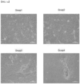
- the cells were subjected to subculture until passage 5, and a photograph taken at cells (passage 5) is shown in FIG. 4a .
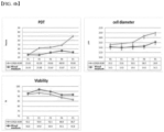
- the population doubling time (PDT), cell size and viability were analyzed and shown in FIG. 4b .
- the PDT of the cells was calculated according to the following Equation using the cell number and the culture time measured during the passage.
- the morphology of the cells showed a pebble shape, which is characteristic of keratinocytes, similar to that of the already used medium (LONZA KGM) as a positive control group.
- the PDT of the cells was more stable in the mixed medium of the present invention than in the positive control medium while the culture progresses until passage 5. It was also confirmed that cell size and viability remained stable.
- the present inventors attempted to verify the characteristics of keratinocytes cultured in the mixed medium according to the present invention using the cells cultured in Example 5.
- the expression level of the keratinocyte marker protein was confirmed through a Western blot.
- the cells cultured in Example 5 was treated with a trypsin-based enzyme, the cells were harvested, and centrifuged at 4000 rpm for 5 minutes, and proteins were isolated using RIPA solution (Biosesang, Korea). The isolated proteins were quantified using a protein quantification kit (Thermo Fisher Scientific, USA). Equal amounts of each protein were then placed in the wells of a protein gel (Invitrogen, USA), and then SDS-polyacrylamide gel electrophoresis (SDS-PAGE) was performed at 80V and 100V.
- SDS-PAGE SDS-polyacrylamide gel electrophoresis
- the protein was transferred to a membrane using a membrane transfer kit (Invitrogen, USA), and then primary and secondary antibodies were sequentially added and allowed to react. After completion of the reaction, the protein expression level was confirmed using HRP-ECL solution (Thermo Fisher Scientific, USA) and iBright (Thermo Fisher Scientific, USA), and GAPDH (glyceraldehyde-3-phosphate dehydrogenase) protein was used as a control group.
- a 1% agarose gel was made with 1X TAE buffer, PCR products corresponding to each primer were placed in each well, followed by electrophoresis at 100V. The gel after DNA isolation by electrophoresis was observed under iBright (Thermo fisher scientific, USA) to confirm the expression level, and 18s ribosomal RNA was used as a control group.
- Example 7 Confirmation of the activity of keratinocytes in the mixed medium to which component ratios of basal medium, calcium, epidermal growth factor, and albumin were applied
- the present inventors attempted to confirm the activity of keratinocytes cultured in the mixed medium according to the present invention using the cells cultured in Example 5.
- the cells cultured in Example 5 were treated with a trypsin-based enzyme, and then the cells were harvested and centrifuged at 1000 rpm for 5 minutes. The supernatant of the centrifuged cells was discarded, and the cells were well resuspended by adding a condition-based medium, and then the cell numbers were measured using NC-250 TM . Subsequently, 5 ⁇ 10 2 cells per well were seeded in a 6-well plate and incubated in a 37°C incubator for 7 days.
- the culture medium was removed, and the cells were fixed with 4% formalin solution at room temperature for 30 minutes and then stained with a concentrated solution of 0.1% crystal violet dye at room temperature for 30 minutes.
- the colonies formed of 50 or more cells were grouped as one, and the number of colonies formed per well was analyzed.
- the colony size of cells cultured in the mixed medium conditions of the present invention was generally larger and more colonies were formed as compared to cells cultured in the already used medium (LONZA KGM). This means that cell activity is further increased in the mixed medium to which the component ratios of basal medium, calcium, epidermal growth factor (EGF) and albumin are applied.
Landscapes
- Health & Medical Sciences (AREA)
- Engineering & Computer Science (AREA)
- Life Sciences & Earth Sciences (AREA)
- Biomedical Technology (AREA)
- Genetics & Genomics (AREA)
- Zoology (AREA)
- Organic Chemistry (AREA)
- Bioinformatics & Cheminformatics (AREA)
- Biotechnology (AREA)
- Chemical & Material Sciences (AREA)
- Wood Science & Technology (AREA)
- Dermatology (AREA)
- Microbiology (AREA)
- Biochemistry (AREA)
- General Engineering & Computer Science (AREA)
- General Health & Medical Sciences (AREA)
- Cell Biology (AREA)
- Micro-Organisms Or Cultivation Processes Thereof (AREA)
Applications Claiming Priority (2)
| Application Number | Priority Date | Filing Date | Title |
|---|---|---|---|
| KR1020200162867A KR102583179B1 (ko) | 2020-11-27 | 2020-11-27 | 칼슘, 상피세포성장인자 및 알부민을 포함하는 각질형성세포 수립 또는 배양용 배지 조성물 |
| PCT/KR2021/017263 WO2022114727A1 (ko) | 2020-11-27 | 2021-11-23 | 칼슘, 상피세포성장인자 및 알부민을 포함하는 각질형성세포 수립 또는 배양용 배지 조성물 |
Publications (2)
| Publication Number | Publication Date |
|---|---|
| EP4239057A1 true EP4239057A1 (de) | 2023-09-06 |
| EP4239057A4 EP4239057A4 (de) | 2024-10-30 |
Family
ID=81756209
Family Applications (1)
| Application Number | Title | Priority Date | Filing Date |
|---|---|---|---|
| EP21898553.9A Pending EP4239057A4 (de) | 2020-11-27 | 2021-11-23 | Kulturmediumzusammensetzung zur herstellung oder kultivierung von keratinozyten mit calcium, epideralem wachstumsfaktor und albumin |
Country Status (4)
| Country | Link |
|---|---|
| US (1) | US20240002790A1 (de) |
| EP (1) | EP4239057A4 (de) |
| KR (1) | KR102583179B1 (de) |
| WO (1) | WO2022114727A1 (de) |
Family Cites Families (8)
| Publication number | Priority date | Publication date | Assignee | Title |
|---|---|---|---|---|
| IL94611A (en) * | 1989-06-05 | 1994-12-29 | Organogenesis Inc | Medium for cell cultures containing insulin or growth factor similar to insulin, transferrin or iron ion, triiodothyronine or thyroxine and method of use |
| NZ325814A (en) * | 1995-12-21 | 2000-10-27 | Nestle Sa | Process for immortalizing human skin cell lines and a serum-free medium |
| EP1404809B1 (de) * | 2001-03-02 | 2013-12-11 | Stratatech Corporation | Verbesserte hautersatzstoffe und verwendungen davon |
| KR20030043313A (ko) * | 2001-11-27 | 2003-06-02 | 한스바이오메드 주식회사 | 사람 각질세포 및 성장인자가 함유된 피브린 접착제를주재로 한 피부재생촉진제 및 그의 제조방법 |
| HK1080111A1 (zh) * | 2002-03-29 | 2006-04-21 | 新加坡眼部研究所 | 为研究、临床眼晴表面移植和组织工程的应用而生长人类结膜组织等同物的方法 |
| EP1937798A4 (de) * | 2005-04-15 | 2009-05-13 | Univ Columbia | Chemisch definierte kulturmedien zur expansion und differenzierung von epidermiszellen sowie verwendungen davon für die in-vitro-anzucht von haarfollikeln |
| AU2008295441A1 (en) * | 2007-09-04 | 2009-03-12 | Queensland University Of Technology | A feeder cell-free culture medium and system |
| CN111088213B (zh) * | 2018-10-24 | 2022-04-08 | 澳门大学 | 一种诱导干细胞逐步分化形成角质细胞的方法以及角质细胞 |
-
2020
- 2020-11-27 KR KR1020200162867A patent/KR102583179B1/ko active Active
-
2021
- 2021-11-23 US US18/039,033 patent/US20240002790A1/en active Pending
- 2021-11-23 EP EP21898553.9A patent/EP4239057A4/de active Pending
- 2021-11-23 WO PCT/KR2021/017263 patent/WO2022114727A1/ko not_active Ceased
Also Published As
| Publication number | Publication date |
|---|---|
| KR20220074399A (ko) | 2022-06-03 |
| KR102583179B1 (ko) | 2023-09-26 |
| EP4239057A4 (de) | 2024-10-30 |
| WO2022114727A1 (ko) | 2022-06-02 |
| US20240002790A1 (en) | 2024-01-04 |
Similar Documents
| Publication | Publication Date | Title |
|---|---|---|
| WO2007117565A9 (en) | Standardization of processes for culturing primary cells | |
| CN105267243B (zh) | 一种消除皮肤妊娠纹的干细胞提取物 | |
| Nanba | Human keratinocyte stem cells: from cell biology to cell therapy | |
| CN112143708B (zh) | 一种脐带间充质干细胞、干细胞精华因子和在抗皮肤衰老方面的应用 | |
| DE69919531T2 (de) | Aus normalem menschlichem hautgewebe immortalizierte zellinien | |
| Korosec et al. | Isolation of papillary and reticular fibroblasts from human skin by fluorescence-activated cell sorting | |
| JP2011512146A (ja) | 上皮細胞のクローン培養のための系および方法 | |
| Shwetha et al. | Ex vivo culture of oral keratinocytes using direct explant cell culture technique | |
| EP2812689B1 (de) | Verfahren zur bewertung eines oder mehrerer wirkstoffe zur erhaltung der funktionsfähigkeit epithelialer stammzellen | |
| EP4239057A1 (de) | Kulturmediumzusammensetzung zur herstellung oder kultivierung von keratinozyten mit calcium, epideralem wachstumsfaktor und albumin | |
| EP3284817A1 (de) | Menschliche sebozyt-vorläuferzellen, menschliche sebozyte und in-vitro-verfahren zur gewinnung davon aus menschlichen induzierten pluripotenten stammzellen (hipsc) | |
| JP6563145B2 (ja) | 不死化汗腺筋上皮細胞 | |
| Wang et al. | Patterns of nestin expression in human skin | |
| CN113621556B (zh) | 一种雄激素性脱发细胞模型的构建方法 | |
| CN106754647B (zh) | 一种大鲵皮肤表皮细胞的分离、培养及鉴定方法 | |
| EP3561051A1 (de) | Zusammensetzung zur förderung der proliferation pluripotenter stammzellen und verfahren zur förderung der proliferation pluripotenter stammzellen | |
| RU2620981C2 (ru) | Способ получения культуры мезенхимных стволовых клеток человека из периваскулярного пространства вены пупочного канатика | |
| Ngo et al. | Isolation and culture of primary oral keratinocytes from the adult mouse palate | |
| CN109528620B (zh) | 一种植物干细胞活性成分及其制备方法 | |
| US20190161737A1 (en) | Process for continuous cell culture of cancer cells and cancer stem cells | |
| KR102066159B1 (ko) | 줄기세포의 층분리 배양 및 증식 방법 | |
| KR101418876B1 (ko) | 피부성체 줄기세포의 분화 조절 방법 | |
| CN121427819A (zh) | 红景天苷延缓骨髓间充质干细胞复制性衰老的新用途 | |
| CN118792236A (zh) | 一种穿刺标本类器官培养基及其培养方法 | |
| Guérin et al. | Irradiated human dermal fibroblasts are as efficient as mouse fibroblasts as a feeder layer to improve human epidermal cell culture lifespan |
Legal Events
| Date | Code | Title | Description |
|---|---|---|---|
| STAA | Information on the status of an ep patent application or granted ep patent |
Free format text: STATUS: THE INTERNATIONAL PUBLICATION HAS BEEN MADE |
|
| PUAI | Public reference made under article 153(3) epc to a published international application that has entered the european phase |
Free format text: ORIGINAL CODE: 0009012 |
|
| STAA | Information on the status of an ep patent application or granted ep patent |
Free format text: STATUS: REQUEST FOR EXAMINATION WAS MADE |
|
| 17P | Request for examination filed |
Effective date: 20230601 |
|
| AK | Designated contracting states |
Kind code of ref document: A1 Designated state(s): AL AT BE BG CH CY CZ DE DK EE ES FI FR GB GR HR HU IE IS IT LI LT LU LV MC MK MT NL NO PL PT RO RS SE SI SK SM TR |
|
| DAV | Request for validation of the european patent (deleted) | ||
| DAX | Request for extension of the european patent (deleted) | ||
| A4 | Supplementary search report drawn up and despatched |
Effective date: 20241002 |
|
| RIC1 | Information provided on ipc code assigned before grant |
Ipc: C12N 5/071 20100101AFI20240926BHEP |