EP4209784B1 - Test für digitale affinitätsverknüpfung - Google Patents
Test für digitale affinitätsverknüpfung Download PDFInfo
- Publication number
- EP4209784B1 EP4209784B1 EP22210786.4A EP22210786A EP4209784B1 EP 4209784 B1 EP4209784 B1 EP 4209784B1 EP 22210786 A EP22210786 A EP 22210786A EP 4209784 B1 EP4209784 B1 EP 4209784B1
- Authority
- EP
- European Patent Office
- Prior art keywords
- target
- affinity
- label
- affinity agent
- sample
- Prior art date
- Legal status (The legal status is an assumption and is not a legal conclusion. Google has not performed a legal analysis and makes no representation as to the accuracy of the status listed.)
- Active
Links
Images


Classifications
-
- G—PHYSICS
- G01—MEASURING; TESTING
- G01N—INVESTIGATING OR ANALYSING MATERIALS BY DETERMINING THEIR CHEMICAL OR PHYSICAL PROPERTIES
- G01N33/00—Investigating or analysing materials by specific methods not covered by groups G01N1/00 - G01N31/00
- G01N33/48—Biological material, e.g. blood, urine; Haemocytometers
- G01N33/50—Chemical analysis of biological material, e.g. blood, urine; Testing involving biospecific ligand binding methods; Immunological testing
- G01N33/53—Immunoassay; Biospecific binding assay; Materials therefor
- G01N33/543—Immunoassay; Biospecific binding assay; Materials therefor with an insoluble carrier for immobilising immunochemicals
- G01N33/54313—Immunoassay; Biospecific binding assay; Materials therefor with an insoluble carrier for immobilising immunochemicals the carrier being characterised by its particulate form
- G01N33/54326—Magnetic particles
- G01N33/54333—Modification of conditions of immunological binding reaction, e.g. use of more than one type of particle, use of chemical agents to improve binding, choice of incubation time or application of magnetic field during binding reaction
-
- G—PHYSICS
- G01—MEASURING; TESTING
- G01N—INVESTIGATING OR ANALYSING MATERIALS BY DETERMINING THEIR CHEMICAL OR PHYSICAL PROPERTIES
- G01N33/00—Investigating or analysing materials by specific methods not covered by groups G01N1/00 - G01N31/00
- G01N33/48—Biological material, e.g. blood, urine; Haemocytometers
- G01N33/50—Chemical analysis of biological material, e.g. blood, urine; Testing involving biospecific ligand binding methods; Immunological testing
- G01N33/53—Immunoassay; Biospecific binding assay; Materials therefor
- G01N33/536—Immunoassay; Biospecific binding assay; Materials therefor with immune complex formed in liquid phase
- G01N33/537—Immunoassay; Biospecific binding assay; Materials therefor with immune complex formed in liquid phase with separation of immune complex from unbound antigen or antibody
- G01N33/539—Immunoassay; Biospecific binding assay; Materials therefor with immune complex formed in liquid phase with separation of immune complex from unbound antigen or antibody involving precipitating reagent, e.g. ammonium sulfate
- G01N33/541—Double or second antibody, i.e. precipitating antibody
-
- G—PHYSICS
- G01—MEASURING; TESTING
- G01N—INVESTIGATING OR ANALYSING MATERIALS BY DETERMINING THEIR CHEMICAL OR PHYSICAL PROPERTIES
- G01N33/00—Investigating or analysing materials by specific methods not covered by groups G01N1/00 - G01N31/00
- G01N33/48—Biological material, e.g. blood, urine; Haemocytometers
- G01N33/50—Chemical analysis of biological material, e.g. blood, urine; Testing involving biospecific ligand binding methods; Immunological testing
- G01N33/53—Immunoassay; Biospecific binding assay; Materials therefor
- G01N33/543—Immunoassay; Biospecific binding assay; Materials therefor with an insoluble carrier for immobilising immunochemicals
- G01N33/54306—Solid-phase reaction mechanisms
-
- G—PHYSICS
- G01—MEASURING; TESTING
- G01N—INVESTIGATING OR ANALYSING MATERIALS BY DETERMINING THEIR CHEMICAL OR PHYSICAL PROPERTIES
- G01N33/00—Investigating or analysing materials by specific methods not covered by groups G01N1/00 - G01N31/00
- G01N33/48—Biological material, e.g. blood, urine; Haemocytometers
- G01N33/50—Chemical analysis of biological material, e.g. blood, urine; Testing involving biospecific ligand binding methods; Immunological testing
- G01N33/53—Immunoassay; Biospecific binding assay; Materials therefor
- G01N33/543—Immunoassay; Biospecific binding assay; Materials therefor with an insoluble carrier for immobilising immunochemicals
- G01N33/54366—Apparatus specially adapted for solid-phase testing
- G01N33/54386—Analytical elements
-
- G—PHYSICS
- G01—MEASURING; TESTING
- G01N—INVESTIGATING OR ANALYSING MATERIALS BY DETERMINING THEIR CHEMICAL OR PHYSICAL PROPERTIES
- G01N33/00—Investigating or analysing materials by specific methods not covered by groups G01N1/00 - G01N31/00
- G01N33/48—Biological material, e.g. blood, urine; Haemocytometers
- G01N33/50—Chemical analysis of biological material, e.g. blood, urine; Testing involving biospecific ligand binding methods; Immunological testing
- G01N33/58—Chemical analysis of biological material, e.g. blood, urine; Testing involving biospecific ligand binding methods; Immunological testing involving labelled substances
-
- G—PHYSICS
- G01—MEASURING; TESTING
- G01N—INVESTIGATING OR ANALYSING MATERIALS BY DETERMINING THEIR CHEMICAL OR PHYSICAL PROPERTIES
- G01N2458/00—Labels used in chemical analysis of biological material
- G01N2458/10—Oligonucleotides as tagging agents for labelling antibodies
Definitions
- Quantifying the amount of biomolecules in a sample from a subject can provide useful information for a number of clinical applications.
- One method for detecting and quantifying biomolecules, such as proteins, is by enzyme-linked immunosorbent assay (ELISA) or by proximity extension or ligation essays for instance described in US 2015/0132743 , and Dhillon et al., Biomolecular Detection and Quantification, pp. 2-8 (2016 ).
- ELISA enzyme-linked immunosorbent assay
- proximity extension or ligation essays for instance described in US 2015/0132743 , and Dhillon et al., Biomolecular Detection and Quantification, pp. 2-8 (2016 ).
- the limit of detection and precision of quantification with this assay are not sufficient for many needs.
- Alternative techniques such as immuno-PCR have the potential to increase the sensitivity of detection, but in practice are limited by the problem of high background signal due to non-specific binding of the antibodies used as detection agents.
- the invention in its general sense is a method of detecting a target in a sample, the method comprising contacting the sample with a first affinity agent linked to a solid support, a second affinity agent comprising a first label, and a third affinity agent comprising a second label, wherein the first label generates a first signal and the second label generates a second signal and the first and second signal are distinguishable; and wherein the first, second and third affinity agents specifically bind to the target, if present, thereby forming a target-labeled affinity agent complex; separating the target-labeled affinity agent complex from uncomplexed components in the sample based on the presence or absence of the solid support, thereby generating a separated target-labeled affinity agent complex;
- the first affinity agent is cleaved from the solid support prior to the partitioning step, thereby releasing the target-labeled affinity agent complex from the solid support.
- an amino acid tag linking the first affinity agent to the solid support is cleaved by a sequence-specific protease.
- the protease is TEV, factor Xa, or thrombin.
- a photo-cleavable linker between the solid surface and the first affinity agent is cleaved by exposing the linker to light.
- the target-labeled affinity agent complex is cross-linked prior to partitioning the separated target-labeled affinity agent complex into a plurality of partitions.
- the first label is a first nucleic acid label and the second label is a second nucleic acid label.
- the first and second nucleic acid labels are amplified following the partitioning.
- each of the first and second nucleic acid labels are detected using a DNA probe (e.g., a probe having a reporter on one end and a quencher on the other end such as a TAQMAN TM probe, a SCORPION TM probe, an ECLIPSE TM probe, a molecular beacon probe, a double-stranded probe, a dual hybridization probe, or a double-quenched probe).
- a DNA probe e.g., a probe having a reporter on one end and a quencher on the other end such as a TAQMAN TM probe, a SCORPION TM probe, an ECLIPSE TM probe, a molecular beacon probe, a double-stranded probe, a dual hybridization probe, or a double-quenched probe.
- each of the first and second nucleic acid labels are detected using an intercalating dye (e.g., EvaGreen ® dye.
- the first and second nucleic acid labels are detected using different signal levels of the same DNA probe or intercalating dye.
- the first label is a first fluorophore and the second label is a second fluorophore.
- the first label is a first enzyme
- the second label is a second enzyme
- the detecting comprises detecting products generated by the first and second enzymes.
- the first label and the second label are each linked to streptavidin
- the second and third affinity agents are biotinylated
- the second and third affinity agents are labeled with the streptavidin-linked first and second labels, respectively, prior to a first step of the method by allowing the first and second labeled streptavidins to bind to the respective biotinylated second and third affinity agents (i.e., by streptavidin-biotin interaction).
- the first affinity agent is biotinylated and the solid support is linked to streptavidin, and prior to a first step of the method the streptavidin-linked solid support is linked to the first affinity agent by allowing the streptavidin-linked solid support to bind to the biotinylated first affinity agent (i.e., by streptavidin-biotin interaction).
- the solid support or the plurality of solid supports is/are a magnetic bead(s), a non-magnetic bead(s), or a surface(s) of a reaction vessel(s).
- the non-magnetic beads are polystyrene beads or silica-based beads.
- the solid support or the plurality of solid supports are magnetic beads and the target-labeled affinity agent complex is separated from the uncomplexed components in the sample using a magnet that attracts the magnetic beads linked to the first affinity agent in the target-labeled affinity agent complex.
- the solid support or the plurality of solid supports are non-magnetic beads and the target-labeled affinity agent complex is separated from the uncomplexed components in the sample by centrifugation.
- the solid support is a surface of a reaction, and the target-labeled affinity agent complex is separated from the uncomplexed components in the sample by aspiration.
- the target comprises a protein, a protein aggregate, or a protein oligomer.
- the target is a complex of two or more interacting proteins and the second and third affinity agents each bind to one of the interacting proteins in the complex.
- the target has a repeating identical epitope and the first and second affinity agents or the first and third affinity agents recognize the same epitope.
- two or more of the first, second, and third affinity agents recognize a different epitope on the target.
- each of the first, second, and third affinity agents recognizes a different epitope on the target.
- the first, second, and third affinity agents are each selected from the group consisting of an antibody, an antibody fragment, and a nucleotide aptamer.
- the antibody is a monoclonal antibody and/or a polyclonal antibody.
- the target is a plurality of different targets
- the first affinity agent is a plurality of different first affinity agents
- the second affinity agent is a plurality of different second affinity agents comprising a plurality of first labels
- the third affinity agent is a plurality of different third affinity agents comprising a plurality of second labels and wherein a set of the first, second and third affinity agents specifically bind to the same one of the plurality of different targets.
- the method further comprises determining the number of partitions comprising the first label and the second label, thereby quantifying the target.
- the partitions are droplets.
- Described herein are methods of detecting a target in a sample.
- Digital affinity linkage assay methods have been discovered in which the concentration of a target is determined on a solid support by separating the sample into small partitions (e.g., droplets) and performing digital PCR analysis of two different DNA-labeled affinity agents (e.g. DNA-labeled antibodies).
- a linkage signal is calculated from the fraction of partitions in which the two different DNA labels are amplified and co-localized in the same partition.
- the background noise from non-specific binding of the affinity agents can be significantly reduced. Accordingly, the digital affinity linkage assay methods described herein have reduced background noise from non-specific binding of DNA-labeled affinity agents and have an unexpectedly high detection sensitivity or lower limit of detection.
- the methods described herein thus provide advantages such as the ability to detect significantly lower amounts of target in a sample as compared to assays in which targets are individually detected, and reducing or eliminating the need for diluting the concentration of affinity agents in a reaction mixture, thus avoiding the problem of diluting the affinity agents below the detection concentration.
- target refers to any agent whose presence and/or amount is to be determined.
- the target can be a mixture of several targets.
- the presence or concentration profile of the different targets is to be determined.
- the target can be any biological and/or chemical agent (e.g., a molecule, macromolecule, complex, or conjugate).
- the target is an organic or inorganic molecule.
- the target is a biological agent.
- the target is a protein, a protein aggregate, a protein oligomer, a polypeptide or a peptide.
- the protein aggregate comprises more than one protein (e.g., more than one target).
- the protein has more than one identical or non-identical subunit which may or may not be covalently bound to each other.
- the targets can be hormones, antibodies, amino acids (e.g., glutamic acid, aspartic acid) or any derivatives and/or combination thereof.
- the target is a toxin or a drug.
- the amount of the target in the sample is in micrograms. In some embodiments, the amount of the target in the sample is below 1 microgram.
- amount of the target in the sample is in nanograms. In some embodiments the amount of target in the sample is between 100 ng to 1 ng. In some embodiments, the amount of target in the sample is between 1000 pg to 1 pg. In certain embodiments, the amount of target in the sample is between 1 pg to 1 fg. In certain embodiments, the amount of target in the sample is between 1 fg to 1 ag.
- affinity agent refers to a molecule that specifically binds to a target.
- exemplary affinity agents include an antibody, an antibody fragment, a non-antibody protein scaffold, an antibody mimetic, or an aptamer.
- antibody refers to a polypeptide of the immunoglobulin family or a polypeptide comprising fragments of an immunoglobulin that is capable of noncovalently, reversibly, and in a specific manner binding to a corresponding target (or antigen).
- the term includes polyclonal or monoclonal antibodies of the isotype classes IgA, IgD, IgE, IgG, and IgM, derived from human or other mammalian cells, including natural or genetically modified forms such as humanized, human, single-chain, chimeric, synthetic, recombinant, hybrid, mutated, grafted, and in vitro generated antibodies.
- conjugates including but not limited to fusion proteins containing an immunoglobulin moiety (e.g ., chimeric or bispecific antibodies or single chain Fv's (scFv's)), and fragments, such as Fab, F(ab')2, Fv, scFv, Fd, dAb and other compositions.
- immunoglobulin moiety e.g ., chimeric or bispecific antibodies or single chain Fv's (scFv's)
- fragments such as Fab, F(ab')2, Fv, scFv, Fd, dAb and other compositions.
- An exemplary immunoglobulin (antibody) structural unit comprises a tetramer.
- Each tetramer is composed of two identical pairs of polypeptide chains, each pair having one "light” (about 25 kD) and one "heavy” chain (about 50-70 kD).
- the N-terminus of each chain defines a variable region of about 100 to 110 or more amino acids primarily responsible for antigen recognition.
- the terms variable light chain (V L ) and variable heavy chain (V H ) refer to these light and heavy chains respectively.
- the variable region contains the antigen-binding region of the antibody (or its functional equivalent) and is most critical in specificity and affinity of binding (see Paul, Fundamental Immunology (2003 ).
- Antibodies can exist as intact immunoglobulins or as any of a number of well-characterized fragments that include specific antigen-binding activity. Such fragments can be produced by digestion with various peptidases. Pepsin digests an antibody below the disulfide bonds in the hinge region to produce F(ab)' 2 , a dimer of Fab which itself is a light chain joined to V H -C H 1 by a disulfide bond. The F(ab)' 2 can be reduced under mild conditions to break the disulfide bond in the hinge region, thereby converting the F(ab)' 2 dimer into an Fab' monomer. The Fab' monomer is essentially Fab with part of the hinge region.
- antibody also includes antibody fragments either produced by the modification of whole antibodies, or those synthesized de novo using recombinant DNA methodologies (e.g., scFv) or those identified using phage display libraries (see, e.g., McCafferty et al. (1990) Nature 348:552-554 ).
- Methods for the preparation of antibodies are known in the art (see, e.g., Kohler & Milstein (1975) Nature 256:495-497 ; Kozbor et al. (1983) Immunology Today 4:72 ; Cole et al., Monoclonal Antibodies and Cancer Therapy, pp. 77-96. Alan R. Liss, Inc. 1985 )).
- variable region fragment refers to a monovalent or bi-valent variable region fragment, and can encompass only the variable regions (e.g ., V L and/or V H ), as well as longer fragments, e.g ., an Fab, Fab' or F(ab')2, which also includes C L and/or C H 1.
- Fc refers to a heavy chain monomer or dimer comprising C H 1 and C H 2 regions.
- binding typically indicates that the affinity agent (e.g., an antibody) binds a majority of the antigen in a pure population, assuming an appropriate molar ratio of affinity agent to antigen.
- an affinity agent that binds a given antigen typically binds to at least of the antigen molecules in a solution (e.g., 75%, 80%, 85%, 90%, 91%, 92%, 93%, 94%, 95%, 96%, 97%, 98%, 99%, or 100%).
- a solution e.g., 75%, 80%, 85%, 90%, 91%, 92%, 93%, 94%, 95%, 96%, 97%, 98%, 99%, or 100%.
- an affinity agent e.g., an antibody
- an affinity agent that binds to an antigen with at least 2-fold greater affinity than to non-antigen molecules, e.g., at least 4-fold, 5-fold, 6-fold, 7-fold, 8-fold, 9-fold, 10-fold, 20-fold, 25-fold, 50-fold, 100-fold, 10 3 -fold, 10 4 -fold, 10 5 -fold, 10 6 -fold, 10 7 -fold, 10 8 -fold, 10 9 -fold, 10 10 -fold, 10 11 -fold, 10 12 -fold, 10 13 -fold, 10 14 -fold, or 10 15 -fold greater affinity.
- an affinity agent that specifically binds a particular antigen will typically bind the antigen with at least a 2-fold greater affinity than to a non-antigen molecule.
- nucleic acid means a compound comprising a chain of nucleotide monomers.
- a nucleic acid can be single-stranded or double-stranded (i.e., base-paired with another nucleic acid), among others.
- the chain of a nucleic acid can be composed of any suitable number of monomers, such as at least about ten or one hundred, among others.
- the length of a nucleic acid chain corresponds to its source, with synthetic nucleic acids (e.g., nucleic acid reagents such as primers and probes) typically being shorter and biologically produced nucleic acids (e.g., nucleic acid analytes) typically being longer.
- a nucleic acid can have a natural or artificial structure, or a combination thereof.
- Nucleic acids with a natural structure namely, deoxyribonucleic acid (DNA) and ribonucleic acid (RNA), have a backbone of alternating pentose sugar groups and phosphate groups. Each pentose group is linked to a nucleobase (e.g., a purine (such as adenine (A) or guanine (T)) or a pyrimidine (such as cytosine (C), thymine (T), or uracil (U))).
- a nucleobase e.g., a purine (such as adenine (A) or guanine (T)) or a pyrimidine (such as cytosine (C), thymine (T), or uracil (U)).
- Nucleic acids with an artificial structure are analogs of natural nucleic acids and can, for example, be created by changes to the pentose and/or phosphate groups of the natural backbone.
- Exemplary artificial nucleic acids include glycol nucleic acids (GNA), peptide nucleic acids (PNA), locked nucleic acid (LNA), threose nucleic acids (TNA), and the like.
- nucleic acids having an artificial structure are analogs of natural nucleic acids and can, for example, be created by changes to the nucleobase.
- Exemplary artificial or non-naturally occurring nucleobases include, but are not limited to halogenated nucleobases (5-FU), hypoxanthine, xanthine, 7-methylguanine, inosine, xanthosine, 7-methyguanosine, 5,6-dihydrouracil, 5-methycytosine, 5-hydroxymethylcytosine, dihydrouridine, and 5-methylcytidine.
- halogenated nucleobases 5-FU
- hypoxanthine xanthine
- xanthine 7-methylguanine
- inosine xanthosine
- 7-methyguanosine 7-methyguanosine
- 5,6-dihydrouracil 5-methycytosine, 5-hydroxymethylcytosine, dihydrouridine
- 5-methylcytidine 5-methylcytidine
- the sequence of a nucleic acid is defined by the order in which nucleobases are arranged along the backbone (typically read from the 5' to 3' end). This sequence generally determines the ability of the nucleic acid to bind specifically to a partner chain (or to form an intramolecular duplex) by hydrogen bonding. In particular, adenine pairs with thymine (or uracil) and guanine pairs with cytosine. A nucleic acid that can bind to another nucleic acid in an antiparallel fashion by forming a consecutive string of adenine-thymine and guanine-cytosine base pairs with the other nucleic acid is termed "complementary.”
- label refers to a composition detectable by spectroscopic, photochemical, biochemical, immunochemical, chemical, or other physical means.
- useful labels include fluorescent dyes (fluorophores), fluorescent quenchers, luminescent agents, electron-dense reagents, enzymes (e.g., as commonly used in an ELISA), biotin, digoxigenin, 32 P and other isotopes, haptens, proteins, nucleic acids, or other substances which can be made detectable, e.g, by incorporating a label into an oligonucleotide, peptide, or antibody specifically reactive with a target molecule.
- a detectable label can also include a combination of a reporter and a quencher.
- a molecule that is "linked" to a label is one that is bound, either covalently, through a linker or a chemical bond, or noncovalently, through ionic, van der Waals, electrostatic, or hydrogen bonds to a label such that the presence of the molecule can be detected by detecting the presence of the label bound to the molecule.
- reporter refers to a substance or a portion thereof which is capable of exhibiting a detectable signal, which signal can be suppressed by a quencher.
- the detectable signal of the reporter is, e.g., fluorescence in the detectable range; thus, a reporter can also be a label.
- quencher refers to a substance which is capable of suppressing, reducing, inhibiting, etc., the detectable signal produced by the reporter.
- quenching refers to a process whereby, when a reporter and a quencher are in close proximity, and the reporter is excited by an energy source, a substantial portion of the energy of the excited state non-radiatively transfers to the quencher where it either dissipates nonradiatively or is emitted at a different emission wavelength than that of the reporter (e.g., by fluorescence resonance energy transfer or FRET).
- FRET fluorescence resonance energy transfer
- the reporter can be selected from fluorescent organic dyes modified with a suitable linking group for attachment to the oligonucleotide, such as to the 3' or 5' terminus.
- the quencher can also be selected from organic dyes, which may or may not be fluorescent, depending on the embodiment of the invention. Generally, whether the quencher is fluorescent or simply releases the transferred energy from the reporter by non-radiative decay, the absorption band of the quencher should at least substantially overlap the fluorescent emission band of the reporter to optimize the quenching.
- Non-fluorescent quenchers or dark quenchers typically function by absorbing energy from excited reporters, but do not release the energy radiatively.
- reporter-quencher pairs for particular probes can be undertaken in accordance with known techniques. Fluorescent and dark quenchers and their relevant optical properties from which exemplary reporter-quencher pairs can be selected are listed and described, for example, in R. W. Sabnis, HANDBOOK OF FLUORESCENT DYES AND PROBES, John Wiley and Sons, New Jersey, 2015 .
- Reporter-quencher pairs can be selected from xanthene dyes including fluoresceins and rhodamine dyes. Many suitable forms of these compounds are available commercially with substituents on the phenyl groups, which can be used as the site for bonding or as the bonding functionality for attachment to an oligonucleotide.
- Another group of fluorescent compounds for use as reporters are the naphthylamines, having an amino group in the alpha or beta position. Included among such naphthylamino compounds are 1-dimethylaminonaphthy1-5 sulfonate, 1-anilino-8-naphthalene sulfonate and 2-p-touidiny1-6-naphthalene sulfonate.
- dyes include 3-phenyl-7-isocyanatocoumarin; acridines such as 9-isothiocyanatoacridine; N-(p-(2-benzoxazolyl)phenyl)maleimide; benzoxadiazoles; stilbenes; and pyrenes.
- Suitable examples of quenchers can be selected from 6-carboxy-tetramethylrhodamine, 4-(4-dimethylaminophenylazo) benzoic acid (DABYL), tetramethylrhodamine (TAMRA), BHQ-OTM, BHQ-1 TM, BHQ-2TM, and BHQ-3TM, each of which are available from Biosearch Technologies, Inc. of Novato, Calif., Qy7TM QSY-9TM, QSY-21 TM and QSY-35TM, each of which are available from Molecular Probes, Inc, and ZEN TM and TAO TM Double-Quenched Probes from Integrated DNA Technologies.
- DABYL 4-(4-dimethylaminophenylazo) benzoic acid
- TAMRA tetramethylrhodamine
- BHQ-OTM BHQ-1 TM
- BHQ-2TM tetramethylrhodamine
- BHQ-3TM each of which are available from Biosearch Technologies, Inc.
- Suitable examples of reporters can be selected from dyes such as SYBR green, 5-carboxyfluorescein (5-FAMTm available from Applied Biosystems of Foster City, Calif.), 6-carboxyfluorescein (6-FAM), tetrachloro-6-carboxyfluorescein (TET), 2,7-dimethoxy-4,5-dichloro-6-carboxyfluorescein, hexachloro-6-carboxyfluorescein (HEX), 6-carboxy-2',4,7,7'-tetrachlorofluorescein (6-TETTm available from Applied Biosystems), carboxy-X-rhodamine (ROX), 6-carboxy-4',5'-dichloro-2',7'-dimethoxyfluorescein (6-JOETM available from Applied Biosystems), VICTM dye products available from Molecular Probes, Inc., NEDTM dye products available from Applied Biosystems, Cal Fluor dye products (such as, e.g.
- Partitioning refers to separating an aqueous solution having one or more of a sample and reactant into a plurality of portions, or “partitions.”
- Partitions can be solid or fluid.
- a partition is a solid partition, e.g ., a microchannel, a microtube, or a microwell.
- a partition is a fluid partition, e.g., a droplet.
- a fluid partition e.g., a droplet
- a fluid partition e.g., a droplet
- a fluid partition is a mixture of immiscible fluids (e.g., water and oil).
- a fluid partition e.g., a droplet
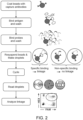
- a method 100 of detecting a target in a sample will now be described. Some of the steps can be performed in any suitable order, in any suitable combination, and can be combined with or modified by any other suitable aspects of the disclosure provided herein.
- the scheme shown in figure 1 and described below is useful for understanding the invention but is not claimed as such.
- the claimed invention in its general sense is defined by claim 1.
- the sample is contacted with a first affinity agent (e.g., an antibody or antibody fragment) linked to a solid support.
- the first affinity agent specifically binds to a first epitope on the target, if present, thereby forming a target bound to the first affinity agent.
- the sample is incubated with the first affinity agent to allow time for the first affinity agent to capture or bind to the target.
- the sample is incubated with the first affinity agent for one hour or more.
- the solid support prior to contacting the sample with the first affinity agent linked to the solid support, is treated with a blocking agent to prevent non-specific binding of material to the support.
- Exemplary blocking agents include, but are not limited to, proteins (e.g., non-fat milk or bovine serum albumin) and detergents (e.g., Tween 20 or Triton X-100).
- the sample is a biological sample.
- Biological samples can be obtained from any biological organism, e.g., an animal, plant, fungus, bacterial, or any other organism.
- the biological sample is from an animal, e.g., a mammal (e.g., a human or a non-human primate, a cow, horse, pig, sheep, cat, dog, mouse, or rat), a bird (e.g., chicken), or a fish.
- a biological sample can be any tissue or bodily fluid obtained from the biological organism, e.g., blood, a blood fraction, or a blood product (e.g., serum, plasma, platelets, red blood cells), sputum or saliva, tissue (e.g., kidney, lung, liver, heart, brain, nervous tissue, thyroid, eye, skeletal muscle, cartilage, or bone tissue); cultured cells, e.g., primary cultures, explants, transformed cells, stem cells, bacterial cells, stool, or urine.
- tissue e.g., kidney, lung, liver, heart, brain, nervous tissue, thyroid, eye, skeletal muscle, cartilage, or bone tissue
- cultured cells e.g., primary cultures, explants, transformed cells, stem cells, bacterial cells, stool, or urine.
- the sample can be prepared to improve the efficient detection of the target (s).
- the sample can be fragmented, fractionated, homogenized, or sonicated.
- a target of interest, or a sub-fraction comprising the target of interest can be extracted or isolated from a sample (e.g., a biological sample).
- the sample is enriched for the presence of the one or more targets.
- the target is enriched in the sample by an affinity method, e.g., immunoaffinity enrichment.
- the target is enriched in the sample using size selection (e.g., removing very small fragments or molecules or very long fragments or molecules).
- Exemplary solid supports include, but are not limited to, particles (e.g., magnetic beads, polymeric beads, or silica-based beads) or a solid surface (e.g., the surface of a reaction vessel such as a tube or a well in a plate).
- the solid support is not chemically modified prior to the attachment of the first affinity agent antibody (e.g., the antibody is attached to the substrate by non-covalent adsorption, based on hydrophobic and other interactions).
- the solid support is chemically modified prior to the attachment of the first affinity agent.
- Exemplary chemically modified solid supports can have carboxyl or amine groups attached and these groups can be used to covalently bind the first affinity agent.
- the first affinity agent is attached to the solid support via carbodiimide mediated chemistry to form an amide bond. In some embodiments, the attachment of the first affinity agent to the solid support is enhanced by a chemical or a photochemical reaction. In some embodiments, the first affinity agent is biotinylated and is attached to the solid support via an avidin-biotin or streptavidin-biotin interaction. In certain embodiments, the first affinity agent is permanently attached to the solid support by a chemical or a photochemical reaction. In some embodiments, the solid support is deactivated after attaching the first affinity agent to the solid support to prevent binding of other agents. For example, active carboxyl groups on the solid support can be deactivated with ethanolamine.
- the target bound to the first affinity agent is separated (e.g., the solid support is washed with a wash solution) from unbound material in the sample based on the presence or absence of the solid support, thereby generating a separated sample comprising the target bound to the first affinity agent.
- a buffer comprising a detergent such as Tween 20 or Triton X-100 is used to remove or separate unbound material from the solid support.
- the target bound to the first affinity agent is separated from the uncomplexed components in the sample using a magnet that attracts the magnetic beads linked to the first affinity agent.
- the target bound to the first affinity agent is separated from the uncomplexed components in the sample by centrifugation.
- the solid support is a surface of a reaction vessel (e.g., a tube or a well)
- the target bound to the first affinity agent is separated from the uncomplexed components in the sample by aspiration.
- the separated sample is contacted with a second affinity agent comprising a first label and a third affinity agent comprising a second label, wherein the second and third affinity agents specifically bind to the target, thereby forming a target-labeled affinity agent complex.
- the second and third affinity agents specifically bind to different epitopes on the target than the first affinity agent.
- the target e.g., a dimeric protein, an aggregate-forming protein, or an oligomeric protein
- the target has a repeating identical epitope such that the first and second affinity agents or the first and third affinity agents recognize the same epitope.
- the target is a complex of two or more interacting proteins and the second and third affinity agents each bind to one of the interacting proteins in the complex.
- the target is a complex of two or more interacting partners (e.g., two proteins, a protein and a non-protein molecule(s), or non-protein molecules) and the method further comprises calculating a binding affinity (KD) of the interacting partners.
- KD binding affinity
- the sample comprises a plurality of different targets, and for each of the plurality of different targets in the sample, a set of first, second, and third affinity agents is provided wherein each of the first, second, and third affinity agents specifically binds to the target.
- the first affinity agent is a plurality of different first affinity agents (i.e., each of the different first affinity agents binding to a different target in the sample)
- the second affinity agent is a plurality of different second affinity agents comprising a plurality of first labels (i.e., each of the different second affinity agents binding to a different target in the sample)
- the third affinity agent is a plurality of different third affinity agents comprising a plurality of second labels (i.e., each of the different third affinity agents binding to a different target in the sample).
- a set of the first, second and third affinity agents specifically bind to the same one of the plurality of different targets.
- the first, second, and third affinity agents are antibodies, antibody fragments, or nucleotide aptamers.
- the antibodies are monoclonal antibodies or polyclonal antibodies.
- the target-labeled affinity agent complex is separated (e.g., the solid support is again washed) from uncomplexed second and third affinity agents based on the presence or absence of the solid support, thereby generating a separated target-labeled affinity agent complex.
- the first affinity agent is cleaved from the solid support prior to the next (partitioning) step, thereby releasing the target-labeled affinity agent complex from the solid support.
- an amino acid tag linking the target-labeled affinity agent complex to the solid support is cleaved by a sequence-specific protease (e.g., TEV, factor Xa, or thrombin).
- a photo-cleavable linker between the solid surface and the first affinity agent is cleaved by exposing the linker to light.
- the target-labeled affinity agent complex is cross-linked prior to partitioning the separated target-labeled affinity agent complex into a plurality of partitions.
- a plurality of partitions are formed from the separated target-labeled affinity agent complex such that a subset of the partitions contains the target-labeled affinity agent complex.
- the partitions can include any of a number of types of partitions, including solid partitions (e.g., wells or tubes) and fluid partitions (e.g., aqueous phase or droplet within an oil phase).
- the partitions are droplets.
- the partitions are microchannels or microwells.
- a droplet comprises an emulsion composition, i.e., a mixture of immiscible fluids (e.g., water and oil).
- a droplet is an aqueous droplet that is surrounded by an immiscible carrier fluid (e.g., oil).
- a droplet is an oil droplet that is surrounded by an immiscible carrier fluid (e.g., an aqueous solution).
- the droplets described herein are relatively stable and have minimal coalescence between two or more droplets.
- the droplet is formed by flowing an oil phase through an aqueous phase.
- the oil for the oil phase can be synthetic or naturally occurring.
- the oil comprises carbon and/or silicon.
- the oil comprises hydrocarbon and/or fluorocarbon.
- Exemplary oils include, but are not limited to, silicone oil, mineral oil, fluorocarbon oil, vegetable oil, or a combination thereof.
- the oil phase can comprise a fluorinated base oil which can additionally be stabilized by combination with a fluorinated surfactant such as a perfluorinated polyether.
- the base oil comprises one or more of a HFE 7500, FC-40, FC-43, FC-70, or another common fluorinated oil.
- the oil phase comprises an anionic fluorosurfactant.
- the anionic fluorosurfactant is Ammonium Krytox (Krytox-AS), the ammonium salt of Krytox FSH, or a morpholino derivative of Krytox FSH.
- Krytox-AS can be present at a concentration of about 0.1%, 0.2%, 0.3%, 0.4%, 0.5%, 0.6%, 0.7%, 0.8%, 0.9%, 1.0%, 2.0%, 3.0%, or 4.0% (w/w). In some embodiments, the concentration of Krytox-AS is about 1.8%. In some embodiments, the concentration of Krytox-AS is about 1.62%. Morpholino derivative of Krytox FSH may be present at a concentration of about 0.1%, 0.2%, 0.3%, 0.4%, 0.5%, 0.6%, 0.7%, 0.8%, 0.9%, 1.0%, 2.0%, 3.0%, or 4.0% (w/w). In some embodiments, the concentration of morpholino derivative of Krytox FSH is about 1.8%. In some embodiments, the concentration of morpholino derivative of Krytox FSH is about 1.62%.
- the oil phase further comprises an additive for tuning the oil properties, such as vapor pressure, viscosity, or surface tension.
- an additive for tuning the oil properties such as vapor pressure, viscosity, or surface tension.
- Non-limiting examples include perfluorooctanol and 1H,1H,2H,2H-Perfluorodecanol.
- 1H,1H,2H,2H-Perfluorodecanol is added to a concentration of about 0.05%, 0.06%, 0.07%, 0.08%, 0.09%, 0.1%, 0.2%, 0.3%, 0.4%, 0.5%, 0.6%, 0.7%, 0.8%, 0.9%, 1.0%, 1.25%, 1.50%, 1.75%, 2.0%, 2.25%, 2.5%, 2.75%, or 3.0% (w/w).
- 1H,1H,2H,2H-Perfluorodecanol is added to a concentration of about 0.18% (w/w).
- the droplet is formed by flowing an oil phase through an aqueous solution phase having a DNA template and one or more components (e.g., reagents) that are used to determine the presence or absence of the target.
- the one or more components used to determine the presence or absence of the target in the aqueous droplet are soluble and/or miscible in water including, but not limited to, one or more salts, buffering agents, reagents (e.g., a releasing agent such as a restriction endonuclease or a protease, PCR components), surfactants, and/or whatever additional components are necessary for a desired reaction(s) that is intended to occur within a formed droplet. All such additional components can be selected to be compatible with the desired reaction or intended assay.
- assay components e.g., a DNA polymerase, dNTPs, and/or a PCR master mix, enzyme substrates
- assay components can be injected into the partition.
- the assay components can be injected into the partition in any order or simultaneously.
- At least 500 partitions e.g., droplets
- at least 1000 partitions at least 2000 partitions, at least 3000 partitions, at least 4000 partitions, at least 5000 partitions, at least 6000 partitions, at least 7000 partitions, at least 8000 partitions, at least 10,000 partitions, at least 15,000 partitions, at least 20,000 partitions, at least 30,000 partitions, at least 40,000 partitions, at least 50,000 partitions, at least 60,000 partitions, at least 70,000 partitions, at least 80,000 partitions, at least 90,000 partitions, at least 100,000 partitions, at least 200,000 partitions, at least 300,000 partitions, at least 400,000 partitions, at least 500,000 partitions, at least 600,000 partitions, at least 700,000 partitions, at least 800,000 partitions, at least 900,000 partitions, at least 1,000,000 partitions, at least 2,000,000 partitions, at least 3,000,000 partitions, at least 4,000,000 partitions, at least 5,000,000 partitions, at least 10,000,000 partitions, at least
- the droplets that are generated are substantially uniform in shape and/or size.
- the droplets are substantially uniform in average diameter.
- the term “substantially” or “about” refers to the recited number and any value within 10% of the recited number.
- the droplets that are generated have an average diameter of about 0.001 ⁇ m, about 0.005 ⁇ m, about 0.01 ⁇ m, about 0.05 ⁇ m, about 0.1 ⁇ m, about 0.5 ⁇ m, about 1 ⁇ m, about 5 ⁇ m, about 10 ⁇ m, about 20 ⁇ m, about 30 ⁇ m, about 40 ⁇ m, about 50 ⁇ m, about 60 ⁇ m, about 70 ⁇ m, about 80 ⁇ m, about 90 ⁇ m, about 100 ⁇ m, about 150 ⁇ m, about 200 ⁇ m, about 300 ⁇ m, about 400 ⁇ m, about 500 ⁇ m, about 600 ⁇ m, about 700 ⁇ m, about 800 ⁇ m, about 900 ⁇ m, or about 1000 ⁇ m.
- the droplets that are generated have an average diameter of less than about 1000 ⁇ m, less than about 900 ⁇ m, less than about 800 ⁇ m, less than about 700 ⁇ m, less than about 600 ⁇ m, less than about 500 ⁇ m, less than about 400 ⁇ m, less than about 300 ⁇ m, less than about 200 ⁇ m, less than about 100 ⁇ m, less than about 50 ⁇ m, or less than about 25 ⁇ m.
- the droplets that are generated are non-uniform in shape and/or size.
- the droplets that are generated are substantially uniform in volume.
- the droplets that are generated have an average volume of about 0.001 nl, about 0.005 nl, about 0.01 nl, about 0.02 nl, about 0.03 nl, about 0.04 nl, about 0.05 nl, about 0.06 nl, about 0.07 nl, about 0.08 nl, about 0.09 nl, about 0.1 nl, about 0.2 nl, about 0.3 nl, about 0.4 nl, about 0.5 nl, about 0.6 nl, about 0.7 nl, about 0.8 nl, about 0.9 nl, about 1 nl, about 1.5 nl, about 2 nl, about 2.5 nl, about 3 nl, about 3.5 nl, about 4 nl, about 4.5 nl, about 5 nl, about 5.5 nl, about 6 nl, about 6.5 nl, about
- the partitions are stable and are capable of long-term storage.
- the partitions are stored at about -70, -20, 0, 3, 4, 5, 6, 7, 8, 9, 10, 15, 20, 25, 30, 35, or 40 ° C. for an extended period of time (e.g., for at least 30 d, at least 60 d, at least 90 d, or longer).
- Partitions as described herein can contain one or more surfactants to reduce coalescence of droplets during transport.
- a surfactant is a surface-active substance capable of reducing the surface tension of a liquid in which it is present.
- a surfactant which also or alternatively is described as a detergent and/or a wetting agent, can incorporate both a hydrophilic portion and a hydrophobic portion, which can collectively confer a dual hydrophilic-hydrophobic character on the surfactant.
- a surfactant can, in some cases, be characterized according to its hydrophilicity relative to its hydrophobicity.
- the aqueous phase incorporates at least one hydrophilic surfactant.
- the aqueous phase can include at least one nonionic surfactant and/or ionic surfactant.
- the aqueous phase includes a surfactant that is a block copolymer of polypropylene oxide and polyethylene oxide.
- the surfactant is a block copolymer of polypropylene oxide and polyethylene oxide sold under the trade names PLURONIC and TETRONIC (BASF).
- the surfactant is a nonionic block copolymer of polypropylene oxide and polyethylene oxide sold under the trade name PLURONIC F-68.
- the surfactant of the aqueous phase is a water-soluble and/or hydrophilic fluorosurfactant.
- Exemplary fluorosurfactants for the aqueous phase are sold under the trade name ZONYL (DuPont), such as ZONYL FSN fluorosurfactants.
- the surfactant can include polysorbate 20 (sold under the trade name TWEEN-20 by ICI Americas, Inc.).
- the concentration of a particular surfactant or total surfactant present in the aqueous phase can be selected to stabilize emulsion droplets prior to heating.
- the concentration of surfactant for the aqueous phase is 0.01 to 10%, 0.05 to 5%, 0.1 to 1%, or 0.5% by weight.
- the presence of the target in the sample is detected by detecting the presence of the first and second labels in at least one same partition (i.e., the first and second labels are co-located in the same partition).
- the first and second labels are nucleic acid (e.g, DNA) labels.
- suitable nucleic acid labels include oligonucleotide sequences, single-stranded DNA, double-stranded DNA, RNA (e.g., mRNA or miRNA), or DNA-RNA hybrids.
- the nucleic acid label is about 10, 15, 20, 25, 30, 35, 40, 45, 50, 60, 70, 80, 90, 100, 150, 200, 250, 300, 350, 400, 450, 500, 600, 700, 800, 900, or 1000 nucleotides in length.
- the oligonucleotide can be synthesized by methods known to those skilled in the art and are commercially available.
- First and second oligonucleotides used as first and second labels in embodiments herein are generally designed such that the first oligonucleotide does not hybridize with the second oligonucleotide.
- hybridize refers to the process of forming a double stranded nucleic acid from joining two complementary strands of DNA or RNA.
- the nucleic acid labels are amplified.
- the nucleic acid label can be amplified by, for example PCR, LCR (Ligase Chain Reaction), SDA (Strand Displacement Amplification), 3SR (Self-Sustained Synthetic Reaction), TMA (Transcription-Mediated Amplification), rolling circle amplification (RCA), or hyper-branched RCA (HRCA).
- the amplified nucleic acid labels are detected by direct incorporation of a label (e.g., a fluorophore, a radioisotope, or an enzyme) into the amplified nucleic acid by using label-conjugated primers or nucleotides.
- a label e.g., a fluorophore, a radioisotope, or an enzyme
- a dye that fluoresces when it intercalates into double-stranded DNA is used to detect the amplified nucleic acids.
- exemplary intercalating dyes include ethidium bromide, propidium iodide, EvaGreen ® dye, and SYBR TM green.
- the amplified nucleic acids are detected by using a nucleic acid probe having a reporter on one end and a quencher on the other end.
- the probe comprises a reporter-quencher combination as employed in a TAQMAN TM probe, a molecular beacon probe, a SCORPION TM probe, a dual hybridization probe, a double-stranded probe, an ECLIPSE TM probe, or a double-quenched probe (e.g., ZEN TM or TAO TM Double-Quenched Probes from IDT).
- the first and second nucleic acid labels are detected using different signal levels of the same DNA probe or intercalating dye.
- the DNA probe for each oligo label, is added at a different concentration.
- different signal intensity for each oligo label is created by either different amplicon length, or different concentration of primers. Droplets positive for a first label will have a mild increase in fluorescence, droplets positive for a second label will have an intermediate increase in fluorescence, and double positive droplets, which indicate linkage, will have the highest fluorescence signal.
- the first and second labels are different enzymes, and the target is detected by detecting a product generated by each of the enzymes.
- suitable enzymes include urease, alkaline phosphatase, (horseradish) hydrogen peroxidase (HRP), glucose oxidase, ⁇ -galactosidase, luciferase, and an esterase.
- HRP horseradish-peroxidase detection system
- ADHP 10-acetyl-3,7-dihydroxyphenoxazine
- An alkaline phosphatase detection system can be used with the fluorogenic substrate Fluorescein Diphosphate (FDP), which yields a soluble product readily detectable at 520 nm.
- FDP Fluorescein Diphosphate
- a ⁇ -galactosidase detection system can be used with the fluorogenic substrate Resorufin ⁇ -D-Galactopyranoside (RBG), which yields a soluble product detectable at 585 nm.
- An esterase detection system can be used with a substrate such as fluorescein diacetate.
- affinity agents used for detecting a target molecule are each labeled with an enzyme (e.g., a first affinity agent labeled with a first enzyme, a second affinity agent labeled with a second enzyme), and each affinity agent that is labeled with an enzyme is detected by detecting a distinguishable product generated by the enzyme.
- an enzyme e.g., a first affinity agent labeled with a first enzyme, a second affinity agent labeled with a second enzyme
- labels are linked to affinity agents by biotin-streptavidin interaction.
- the first and second labels are each linked to streptavidin and the second and third affinity agents are biotinylated.
- the second and third affinity agents are labeled prior to a first step of any of the methods described herein by allowing the first and second labeled streptavidins to bind to the respective biotinylated second and third affinity agents.
- the detectable label (e.g., a label as described herein) can be detected using any of a variety of detector devices. Exemplary detection methods include optical absorbance detection (e.g., fluorescence or chemiluminescence) or radioactive detection. As an example, a fluorescent label can be detected using a detector device equipped with a module to generate excitation light that can be absorbed by a fluorophore, as well as a module to detect light emitted by the fluorophore.
- optical absorbance detection e.g., fluorescence or chemiluminescence
- radioactive detection e.g., a fluorescent label can be detected using a detector device equipped with a module to generate excitation light that can be absorbed by a fluorophore, as well as a module to detect light emitted by the fluorophore.
- the detector further comprises handling capabilities for the partitioned samples (e.g., droplets), with individual partitioned samples entering the detector, undergoing detection, and then exiting the detector.
- partitioned samples e.g., droplets
- partitioned samples are detected serially while the partitioned samples are flowing.
- partitioned samples e.g., droplets
- partitioned samples are arrayed on a surface and a detector moves relative to the surface, detecting signal(s) at each position containing a single partition. Examples of detectors are provided in WO 2010/036352 .
- detectable labels in partitioned samples are detected serially without flowing the partitioned samples (e.g., using a chamber slide).
- a general purpose computer system (referred to herein as a "host computer") can be used to store and process the data.
- Computer-executable logic can be employed to perform such functions as subtraction of background signal, assignment of target and/or reference sequences, and qualification and/or quantification of the data.
- a host computer can be useful for displaying, storing, retrieving, or calculating diagnostic results from the molecular profiling; storing, retrieving, or calculating raw data from expression analysis; or displaying, storing, retrieving, or calculating any sample or patient information useful in the methods of the present invention.
- the host computer can be configured with many different hardware components and can be made in many dimensions and styles (e.g., desktop PC, laptop, tablet PC, handheld computer, server, workstation, mainframe). Standard components, such as monitors, keyboards, disk drives, CD and/or DVD drives, can be included. Where the host computer is attached to a network, the connections can be provided via any suitable transport media (e.g., wired, optical, and/or wireless media) and any suitable communication protocol (e.g., TCP/IP); the host computer can include suitable networking hardware (e.g., modem, Ethernet card, WiFi card).
- the host computer can implement any of a variety of operating systems, including UNIX, Linux, Microsoft Windows, MacOS, or any other operating system.
- Computer code for implementing aspects of the present invention can be written in a variety of languages, including PERL, C, C++, Java, JavaScript, VBScript, AWK, or any other scripting or programming language that can be executed on the host computer or that can be compiled to execute on the host computer. Code can also be written or distributed in low level languages such as assembler languages or machine languages.
- the host computer system advantageously provides an interface via which the user controls operation of the tools.
- software tools are implemented as scripts (e.g., using PERL), execution of which can be initiated by a user from a standard command line interface of an operating system such as Linux or UNIX.
- commands can be adapted to the operating system as appropriate.
- a graphical user interface can be provided, allowing the user to control operations using a pointing device.
- the present invention is not limited to any particular user interface.
- Scripts or programs incorporating various features of the invention as described herein can be encoded on various computer readable media for storage and/or transmission.
- suitable media include magnetic disk or tape, optical storage media such as compact disk (CD) or DVD (digital versatile disk), flash memory, and carrier signals adapted for transmission via wired, optical, and/or wireless networks conforming to a variety of protocols, including the Internet.
- the methods further comprise quantifying the target (e.g., a protein, a protein aggregate, or a protein oligomer) by determining a number of partitions comprising both the first and second labels and determining a total number of partitions. Once a binary "yes-no" result has been determined for each of the partitions, the data for the partitions is analyzed by an algorithm based on Poisson statistics to quantify the amount of target in the sample. In some embodiments, the degree or amount of linkage between the two labels is proportional to the degree or amount of protein aggregation or protein oligomerization and is used to quantify the amount of aggregation or oligomerization.
- An exemplary statistical method for quantifying the concentration or amount of target or targets is described, for example in the aforementioned WO 2010/036352 .
- kits for detecting a target or a plurality of targets in a sample may comprise a first affinity agent linked to a solid support, a second affinity agent conjugated to a first label, and a third affinity agent conjugated to a second label, wherein each of the first, second, and third affinity agents specifically binds to the target.
- a first affinity agent linked to a solid support a second affinity agent conjugated to a first label
- a third affinity agent conjugated to a second label wherein each of the first, second, and third affinity agents specifically binds to the target.
- the first label may be a first oligonucleotide and the second label is a second oligonucleotide.
- the kit can further comprise assay components (e.g., a DNA polymerase, PCR primers, PCR probes, dNTPs, a buffer, a PCR master mix).
- the first label may be a first enzyme and the second label is a second enzyme.
- the kit can further comprise enzyme substrates for each of the first and second enzymes.
- the kit may comprise a chemically modified solid support, a first label, and a second label, wherein the first and second labels are each linked to streptavidin.
- Solid supports and labels are described herein.
- the chemically modified solid support can be linked to a first affinity agent supplied by the user.
- the first and second labeled streptavidins can bind to biotinylated second and third affinity agents, respectively, supplied by the user.
- the kit may comprise a streptavidin-coated solid support.
- the streptavidin-coated solid support can be linked to a biotinylated first affinity agent supplied by the user.
- the kit may further comprise instructions for carrying out the methods described herein.
- EXAMPLE 1 Quantitation of target with a digital affinity linkage assay using monoclonal antibodies
- Capture beads were prepared by binding biotinylated monoclonal Decorin antibody (R&D systems) to Streptavidin coated 1 um tysolactivated magnetic beads (Dynabeads TM MyOne TM Streptavidin T1,Thermo Fisher Scientific). The beads were first washed three times with Dulbecco's phosphate buffered saline pH 7.4 (PBS, Biological Industries, Israel). Next, the beads were mixed with biotinylated Decorin antibody at a ratio of 40 ⁇ g antibody/ 2 ⁇ 10 9 beads in a volume of 280 ⁇ l in PBS buffer and incubated at room temperature for 2 h. The beads were washed from residual antibody five times in PBS supplemented with 0.1% Bovine serum albumin (BSA, Merck).
- BSA Bovine serum albumin
- the detection antibodies were prepared from two different clones of Decorin monoclonal antibodies (R&D systems) directly conjugated to amine modified 100 base long DNA oligonucleotides (IDT) by Innova Biosciences, UK.
- the conjugates were purified from unbound oligonucleotides.
- the beads were then incubated in 50 ⁇ l of a mixture of both detection probes each at a concentration of 2 nM in PBS-T supplemented with 0.1% BSA and 100 ng/ ⁇ l Polyadenylic acid (Merck). The probes were allowed to bind the beads for 1.5 h at room temperature while rotating. After the incubation, unbound detection probes were washed five times in PBS-T and the beads were resuspended in 100 ⁇ l TE solution pH 8.0 (Merck). one ⁇ l of each sample was further diluted in 99 ⁇ l TE solution to reach bead concentration of 3,000 beads/ ⁇ l.
- amplification mix containing PCR primers, TaqMan probes, dNTPs and DNA polymerase was prepared and mixed with the samples. Droplets were generated in QX200 TM Droplet Generator (Bio-Rad).
- Amplification mix contained (per sample): 12 ⁇ l ddPCR supermix for probes (Bio-Rad), 1.2 ⁇ l of 10 ⁇ M amplification primers (forward and reverse primer for each label), 0.5 ⁇ l of 10 ⁇ M FAM hydrolysis probe for the first label, 0.5 ⁇ l of 10 ⁇ M HEX hydrolysis probe for the second label (all oligonucleotides were purchased from IDT) and 6.5 ⁇ l diluted sample.
- Droplets were generated from 20 ⁇ l amplification mix, placed in ddPCR plates (Bio-Rad), sealed with Microseal 'F' PCR plate seal (Bio-Rad) and placed in C1000 Thermal Cycler (Bio-Rad) for amplification. For each sample, four PCR reactions were prepared and approximately 80,000 beads analyzed.
- PCR cycles hold 10 min at 95 °C, cycle 94 °C for 30 s and 56 °C for 1 min 40 times, hold 98 °C for 10 min.
- Droplet fluorescence was measured by the QX200 TM Droplet Reader instrument (Bio-Rad) and for each sample the number of positive and negative droplets for each label was documented.
- Label concentration was calculated by the QuantaSoft TM Software according to Poisson distribution. Assay results are calculated automatically by the software in the "Linkage" output by calculating the number of double positive droplets observed above the expected number from random distribution.
- Fitting the data to 4 parameter logistics (4PL) model was performed using GraphPad Prism version 7.02, GraphPad Software, California USA.
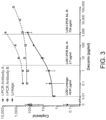
- results of the two immuno-PCR assays and the digital affinity linkage assay are summarized in Fig 3 .
- the results show relatively high Limit of Detection (LOD) values for the individual labeled antibodies used in an immuno-PCR assay (triangle and square markers with dashed curve).
- LOD Limit of Detection
- the noise level is significantly reduced, enabling detection of much lower target concentrations and extending the dynamic range by three orders of magnitude.
- EXAMPLE 2 Quantitation of target with a digital affinity immuno-assay linkage assay using polyclonal antibodies
- Capture beads were prepared for each target by binding the relevant biotinylated polyclonal antibody (R&D systems) to Streptavidin coated 1 ⁇ m tysolactivated magnetic beads (Dynabeads TM MyOne TM Streptavidin T1, Thermo Fisher Scientific). 200 ⁇ l of the beads at stock concertation of 7-10 ⁇ 10 9 beads/ml were transferred to an Eppendorf tube and the buffer was removed by magnetizing the beads and aspirating the supernatant.
- R&D systems biotinylated polyclonal antibody
- Streptavidin coated 1 ⁇ m tysolactivated magnetic beads Dynabeads TM MyOne TM Streptavidin T1, Thermo Fisher Scientific
- the beads were resuspended in 200 ⁇ l Dulbecco's phosphate buffered saline pH 7.4 (PBS, Biological Industries, Israel) supplemented with 50 nM biotinylated polyclonal antibody (R&D systems). Next, the beads were incubated with rotation at room temperature for 1 hour. The beads were washed from residual antibody two times in PBS supplemented with 0.05% tween-20. Finally, the beads were resuspended in 200 ⁇ l PBS supplemented with 0.1% Bovine serum albumin (BSA, Merck) and stored at 4 °C.
- BSA Bovine serum albumin
- Streptavidin conjugated to oligonucleotide tags was prepared in advance in the following manner. Two 5' amino modified 70-80 bases long oligonucleotide were designed and ordered from IDT. The oligonucleotides were designed to have no significant homology to each other or to other natural sequences. They were separately covalently conjugated to Pierce TM Streptavidin (Thermo Fisher Scientific) with the Protein-Oligo Conjugation Kit (TriLink BioTechnologies) according to manufacturer's instructions. Protein concentration and nucleotide concentration in the resulting conjugate were determined by UV absorption.
- the detection antibodies labeled with one of the two oligonucleotide tags were prepared in two separate tubes. 2 ⁇ l of 1 ⁇ M biotinylated polyclonal antibodies were mixed with 2 ⁇ l of 1 ⁇ M (protein concentration) streptavidin-oligo conjugate and 36 ⁇ l PBS supplemented with 0.1% BSA (Merck). Antibodies and conjugate were incubated at room temperature for 1 hour and stored at 4 °C.
- the beads were then incubated in 50 ⁇ l of a mixture of both detection probes each at a concentration of 62.5 nM in blocking solution. The probes were allowed to bind the beads for 1.5 h at room temperature while shaking. After the incubation, unbound detection probes were washed three times in PBS-T and the beads were resuspended in 120 ⁇ l TE solution pH 8.0 (Merck) to a final concentration of 8,333 beads/ ⁇ l.
- amplification mix containing PCR primers, TaqMan probes, dNTPs and DNA polymerase was prepared and mixed with the samples. Droplets were generated in QX200 TM Droplet Generator (Bio-Rad).
- Amplification mix contained (per sample): 12 ⁇ l ddPCR supermix for probes (Bio-Rad), 1.2 ⁇ l of 10 ⁇ M amplification primers (forward and reverse primer for each label), 0.5 ⁇ l of 10 ⁇ M FAM hydrolysis probe for the first label, 0.5 ⁇ l of 10 ⁇ M HEX hydrolysis probe for the second label (all oligonucleotides were purchased from IDT), 4.2 ⁇ l double distilled water and 2 ⁇ l beads.
- Droplets were generated from 20 ⁇ l amplification mix, placed in ddPCR plates (Bio-Rad), sealed with Microseal 'F' PCR plate seal (Bio-Rad) and placed in C1000 Thermal Cycler (Bio-Rad) for amplification. For each sample, two PCR reactions were prepared and approximately 33,000 beads analyzed.
- PCR cycles hold 10 min at 95 °C, cycle 94 °C for 30 s and 56 °C for 1 min 40 times, hold 98 °C for 10 min.
- Droplet fluorescence was measured by the QX200 TM Droplet Reader instrument (Bio-Rad) and for each sample the number of positive and negative droplets for each label was documented.
- Label concentration was calculated by the QuantaSoft TM Software according to Poisson distribution.
- Assay results are calculated automatically by the software in the "Linkage” output by calculating the number of double positive droplets observed above the expected number from random distribution. Fitting the data to 4 parameter logistics (4PL) model was performed using GraphPad Prism version 7.02, GraphPad Software, California USA.
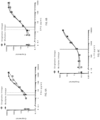
- results show improvement in the LOD values for each of the three targets when comparing the results of the individual immuno-PCRs (triangle and square markers and dashed lines) with the linkage results (circle markers and solid lines).
- the background noise levels calculated from the NPCs dropped 100-1000 fold between the immuno-PCR and linkage assay, resulting in improved LODs.
- EXAMPLE 3 Multiplex quantitation of several targets with a digital affinity immuno-assay linkage assay using polyclonal antibodies
- Capture beads were prepared separately for each target by binding the relevant biotinylated polyclonal antibody (R&D systems) to Streptavidin coated 1 ⁇ m tysol-activated magnetic beads (Dynabeads TM MyOne TM Streptavidin T1, Thermo Fisher Scientific). 200 ⁇ l of the beads at stock concertation of 7-10 ⁇ 10 9 beads/ml were transferred to an Eppendorf tube and the buffer was removed by magnetizing the beads and aspirating the supernatant.
- R&D systems biotinylated polyclonal antibody
- Streptavidin coated 1 ⁇ m tysol-activated magnetic beads Dynabeads TM MyOne TM Streptavidin T1, Thermo Fisher Scientific
- the beads were resuspended in 200 ⁇ l Dulbecco's phosphate buffered saline pH 7.4 (PBS, Biological Industries, Israel) supplemented with 50 nM biotinylated polyclonal antibody (R&D systems). Next, the beads were incubated with rotation at room temperature for 1 h. The beads were washed from residual antibody two times in PBS supplemented with 0.05% tween-20. Finally, the beads were resuspended in 66.6 ⁇ l PBS supplemented with 0.1% Bovine serum albumin (BSA, Merck) and stored at 4 °C.
- BSA Bovine serum albumin
- Streptavidin conjugated to oligonucleotide tags was prepared in advance in the following manner. For each target analyzed, two 5' amino modified 70-80 bases long oligonucleotide were designed and ordered from IDT. The oligonucleotides were designed to have no significant homology to each other or to other natural sequences. They were separately covalently conjugated to Pierce TM Streptavidin (Thermo Fisher Scientific) with the Protein-Oligo Conjugation Kit (TriLink BioTechnologies) according to manufacturer's instructions. Protein concentration and nucleotide concentration in the resulting conjugate were determined by Pierce TM BCA protein assay (ThermoFisher Scientific) and UV absorption.
- the detection antibody for each target was labeled with two unique oligonucleotide tags in two separate tubes.
- 2 ⁇ l of 1 ⁇ M biotinylated polyclonal antibodies were mixed with 2 ⁇ l of 1 ⁇ M (protein concentration) streptavidin-oligo conjugate and 36 ⁇ l PBS supplemented with 0.1% BSA (Merck).
- Antibodies and conjugate were incubated at room temperature for 1 h and stored at 4 °C.
- the beads were then incubated in 50 ⁇ l of a mixture of six detection probes (two detection probes for each antigen), each probe at a concentration of 62.5 nM in blocking solution. The probes were allowed to bind the beads for 1.5 h at room temperature while shaking. After the incubation, unbound detection probes were washed three times in PBS-T and the beads were resuspended in 120 ⁇ l TE solution pH 8.0 (Merck) to a final concentration of 25,000-50,000 beads/ ⁇ l.
- Amplification mixes containing PCR primers, TaqMan probes, dNTPs and DNA polymerase were prepared separately for the detection of each target.
- Each amplification mix contained (per target per sample): 12 ⁇ l ddPCR supermix for probes (Bio-Rad), 1.2 ⁇ l of 10 ⁇ M amplification primers (forward and reverse primer for each of the two labels), 0.5 ⁇ l of 10 ⁇ M FAM hydrolysis probe for the first label, 0.5 ⁇ l of 10 ⁇ M HEX hydrolysis probe for the second label (all oligonucleotides were purchased from IDT) and 4.2 ⁇ l double distilled water. 2 ⁇ l of the bead-bound immune-complexes were then added to the three different amplification mixes and droplets were generated in QX200 TM Droplet Generator (Bio-Rad).
- Droplets were generated from 20 ⁇ l amplification mix, placed in ddPCR plates (Bio-Rad), sealed with Microseal 'F' PCR plate seal (Bio-Rad) and placed in C 1000 Thermal Cycler (Bio-Rad) for amplification. For each sample and each target, two PCR reactions are prepared and approximately 33,000-66,000 beads analyzed.
- PCR cycles hold 10 min at 95 °C, cycle 94 °C for 30 s and 56 °C for 1 min 40 times, hold 98 °C for 10 min.
- Droplet fluorescence was measured by the QX200 TM Droplet Reader instrument (Bio-Rad) and for each sample the number of positive and negative droplets for each label was documented.
- Label concentration was calculated by the QuantaSoft TM Software according to Poisson distribution.
- Assay results were calculated automatically by the software in the "Linkage” output by calculating the number of double positive droplets observed above the expected number from random distribution, for each target separately. Fitting the data to 4 parameter logistics (4PL) model was performed using GraphPad Prism version 7.02, GraphPad Software, California USA.
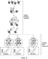
- Fig. 5 illustrates the proposed workflow for multiplex protein quantitation of different targets with a digital affinity linkage assay.
- results show that performing the assay in multiplex does not reduce sensitivity and results in comparable LOD values for each of the three targets when comparing to the singleplex assays.
Landscapes
- Health & Medical Sciences (AREA)
- Immunology (AREA)
- Life Sciences & Earth Sciences (AREA)
- Engineering & Computer Science (AREA)
- Chemical & Material Sciences (AREA)
- Urology & Nephrology (AREA)
- Hematology (AREA)
- Biomedical Technology (AREA)
- Molecular Biology (AREA)
- Medicinal Chemistry (AREA)
- Analytical Chemistry (AREA)
- Cell Biology (AREA)
- Pathology (AREA)
- Food Science & Technology (AREA)
- Biotechnology (AREA)
- Physics & Mathematics (AREA)
- Microbiology (AREA)
- Biochemistry (AREA)
- General Health & Medical Sciences (AREA)
- General Physics & Mathematics (AREA)
- Chemical Kinetics & Catalysis (AREA)
- Measuring Or Testing Involving Enzymes Or Micro-Organisms (AREA)
- Peptides Or Proteins (AREA)
- Investigating, Analyzing Materials By Fluorescence Or Luminescence (AREA)
Claims (14)
- Verfahren zum Nachweis eines Targets in einer Probe, wobei das Verfahren umfasst:In-Kontakt-Bringen der Probe mit einem ersten Affinitätswirkstoff, der an einen festen Träger gebunden ist, einem zweiten Affinitätswirkstoff, der einen ersten Marker umfasst, und einem dritten Affinitätswirkstoff, der einen zweiten Marker umfasst, wobei der erste Marker ein erstes Signal erzeugt und der zweite Marker ein zweites Signal erzeugt und das erste Signal und das zweite Signal unterscheidbar sind; und wobei der erste, zweite und dritte Affinitätswirkstoff spezifisch an das Target binden, falls eines vorhanden ist, wodurch ein Komplex aus Target und markiertem Affinitätswirkstoff entsteht;Abtrennen des Komplexes aus Target und markiertem Affinitätswirkstoff von unkomplexierten Komponenten in der Probe auf der Basis der An- oder Abwesenheit des festen Trägers, wodurch ein abgetrennter Komplex aus Target und markiertem Affinitätswirkstoff erzeugt wird;Partitionieren wenigstens des abgetrennten Komplexes aus Target und markiertem Affinitätswirkstoff in eine Vielzahl von Partitionen; undNachweisen der Anwesenheit des Targets in der Probe durch Nachweisen der Anwesenheit der unterscheidbaren ersten und zweiten Signale von dem ersten und zweiten Marker, die in wenigstens einer identischen Partition colokalisiert sind.
- Verfahren gemäß Anspruch 1, wobei vor dem Schritt des Partitionierens der erste Affinitätswirkstoff von dem festen Träger abgespalten wird, wodurch der Komplex aus Target und markiertem Affinitätswirkstoff von dem festen Träger freigesetzt wird.
- Verfahren gemäß Anspruch 1 oder 2, wobei(i) der erste Marker ein erster Nucleinsäuremarker ist und der zweite Marker ein zweiter Nucleinsäuremarker ist, wobei vorzugsweise der erste und der zweite Nucleinsäuremarker nach dem Partitionieren amplifiziert werden, wobei besonders bevorzugt der erste und der zweite Nucleinsäuremarker jeweils mit Hilfe eines interkalierenden Farbstoffs oder einer DNA-Sonde nachgewiesen werden, die aus einer Sonde, die an einem Ende einen Reporter und am anderen Ende einen Quencher aufweist, einer Molecular-Beacon-Sonde, einer doppelsträngigen Sonde, einer doppelt hybridisierenden Sonde und einer Doppelquenchersonde ausgewählt ist, wobei am meisten bevorzugt der erste und der zweite Nucleinsäuremarker mit Hilfe unterschiedlicher Signalintensitäten derselben DNA-Sonde oder desselben interkalierenden Farbstoffs nachgewiesen werden; oder(ii) der erste Marker ein erstes Fluorophor ist und der zweite Marker ein zweites Fluorophor ist; oder(iii) der erste Marker ein erstes Enzym ist, der zweite Marker ein zweites Enzym ist und der Nachweis das Nachweisen von Produkten umfasst, die durch das erste und das zweite Enzym erzeugt werden.
- Verfahren gemäß Anspruch 1, wobei der erste und der zweite Marker jeweils an Streptavidin gebunden sind und der zweite und der dritte Affinitätswirkstoff biotinyliert sind und wobei vor einem ersten Schritt des Verfahrens der erste streptavidingebundene Marker mit dem biotinylierten ersten Affinitätswirkstoff und der zweite streptavidingebundene Marker mit dem biotinylierten zweiten Affinitätswirkstoff durch Streptavidin-Biotin-Wechselwirkung konjugiert sind.
- Verfahren gemäß Anspruch 1, wobei der erste Affinitätswirkstoff biotinyliert ist und der feste Träger an Streptavidin gebunden ist und wobei vor einem ersten Schritt des Verfahrens der streptavidingebundene feste Träger durch Streptavidin-Biotin-Wechselwirkung an den biotinylierten ersten Affinitätswirkstoff gebunden ist.
- Verfahren gemäß einem der Ansprüche 1-5, wobei der feste Träger oder die Vielzahl von festen Trägern aus magnetischen Kügelchen, nichtmagnetischen Kügelchen und einer Oberfläche eines Reaktionsgefäßes ausgewählt sind, vorzugsweise die unmagnetischen Kügelchen polystyrol Kügelchen oder silica basierte Kügelchen sind.
- Verfahren gemäß Anspruch 1, wobei(i) der feste Träger oder die Vielzahl von festen Trägern magnetische Kügelchen umfassen und der Komplex aus Target und markiertem Affinitätswirkstoff mit Hilfe eines Magneten, der die magnetischen Kügelchen in dem Komplex aus Target und markiertem Affinitätswirkstoff anzieht, von den unkomplexierten Komponenten in der Probe abgetrennt werden; oder(ii) der feste Träger oder die Vielzahl von festen Trägern unmagnetische Kügelchen umfassen und der Komplex aus Target und markiertem Affinitätswirkstoff durch Zentrifugation von den unkomplexierten Komponenten in der Probe abgetrennt wird; oder(iii) der feste Träger eine Oberfläche eines Reaktionsgefäßes ist und der Komplex aus Target und markiertem Affinitätswirkstoff durch Absaugen von den unkomplexierten Komponenten in der Probe abgetrennt wird.
- Verfahren gemäß Anspruch 2, wobei(i) ein Aminosäure-Tag den ersten Affinitätswirkstoff mit dem festen Träger verbindet und wobei der Aminosäure-Tag durch eine sequenzspezifische Protease abgespalten wird, wobei vorzugsweise die Protease aus TEV, Faktor Xa und Thrombin ausgewählt ist; oder(ii) ein photospaltbarer Linker die feste Oberfläche und den ersten Affinitätswirkstoff miteinander verbindet und wobei der Linker dadurch gespalten wird, dass man den Linker Licht aussetzt.
- Verfahren gemäß einem der Ansprüche 1-8, wobei(i) das Target aus einem Protein, einem Proteinaggregat und einem Proteinoligomer ausgewählt ist; oder(ii) das Target ein Komplex aus zwei oder mehr wechselwirkenden Proteinen ist und der zweite und der dritte Affinitätswirkstoff jeweils an eines der wechselwirkenden Proteine in dem Komplex binden, wobei vorzugsweise das Target ein repetitives identisches Epitop aufweist und der erste und der zweite Affinitätswirkstoff oder der erste und der dritte Affinitätswirkstoff dasselbe Epitop erkennen; oder(iii) der erste, der zweite und der dritte Affinitätswirkstoff jeweils spezifisch an ein anderes Epitop an dem Target binden.
- Verfahren gemäß einem der vorstehenden Ansprüche, wobei der erste, der zweite und der dritte Affinitätswirkstoff jeweils aus einem Antikörper, einem Antikörperfragment und einem Nucleotid-Aptamer ausgewählt sind, wobei vorzugsweise der Antikörper ein monoklonaler Antikörper und/oder ein polyklonaler Antikörper ist.
- Verfahren gemäß einem der vorstehenden Ansprüche, weiterhin umfassend das Bestimmen der Anzahl der Partitionen, die den ersten Marker und den zweiten Marker umfassen, wodurch das Target quantifiziert wird.
- Verfahren gemäß einem der vorstehenden Ansprüche, wobei die Partitionen Tröpfchen sind.
- Verfahren gemäß Anspruch 1, wobei die Probe eine Vielzahl unterschiedlicher Targets umfasst und wobei für jedes der Vielzahl unterschiedlicher Targets in der Probe eine Gruppe aus einem ersten, zweiten und dritten Affinitätswirkstoff vorgesehen ist, wobei der erste, der zweite und der dritte Affinitätswirkstoff jeweils spezifisch an das Target binden.
- Verfahren gemäß Anspruch 1, wobei der Komplex aus Target und markiertem Affinitätswirkstoff vor dem Partitionieren des abgetrennten Komplexes aus Target und markiertem Affinitätswirkstoff vernetzt wird.
Priority Applications (1)
| Application Number | Priority Date | Filing Date | Title |
|---|---|---|---|
| EP24201873.7A EP4462123A3 (de) | 2017-09-27 | 2018-09-25 | Test für digitale affinitätsverknüpfung |
Applications Claiming Priority (3)
| Application Number | Priority Date | Filing Date | Title |
|---|---|---|---|
| US201762564029P | 2017-09-27 | 2017-09-27 | |
| EP18861806.0A EP3688185B1 (de) | 2017-09-27 | 2018-09-25 | Test für digitale affinitätsverknüpfung |
| PCT/US2018/052588 WO2019067415A1 (en) | 2017-09-27 | 2018-09-25 | DIGITAL AFFINITY LINK ASSAY |
Related Parent Applications (2)
| Application Number | Title | Priority Date | Filing Date |
|---|---|---|---|
| EP18861806.0A Division-Into EP3688185B1 (de) | 2017-09-27 | 2018-09-25 | Test für digitale affinitätsverknüpfung |
| EP18861806.0A Division EP3688185B1 (de) | 2017-09-27 | 2018-09-25 | Test für digitale affinitätsverknüpfung |
Related Child Applications (2)
| Application Number | Title | Priority Date | Filing Date |
|---|---|---|---|
| EP24201873.7A Division-Into EP4462123A3 (de) | 2017-09-27 | 2018-09-25 | Test für digitale affinitätsverknüpfung |
| EP24201873.7A Division EP4462123A3 (de) | 2017-09-27 | 2018-09-25 | Test für digitale affinitätsverknüpfung |
Publications (2)
| Publication Number | Publication Date |
|---|---|
| EP4209784A1 EP4209784A1 (de) | 2023-07-12 |
| EP4209784B1 true EP4209784B1 (de) | 2024-11-06 |
Family
ID=65807369
Family Applications (3)
| Application Number | Title | Priority Date | Filing Date |
|---|---|---|---|
| EP22210786.4A Active EP4209784B1 (de) | 2017-09-27 | 2018-09-25 | Test für digitale affinitätsverknüpfung |
| EP18861806.0A Active EP3688185B1 (de) | 2017-09-27 | 2018-09-25 | Test für digitale affinitätsverknüpfung |
| EP24201873.7A Pending EP4462123A3 (de) | 2017-09-27 | 2018-09-25 | Test für digitale affinitätsverknüpfung |
Family Applications After (2)
| Application Number | Title | Priority Date | Filing Date |
|---|---|---|---|
| EP18861806.0A Active EP3688185B1 (de) | 2017-09-27 | 2018-09-25 | Test für digitale affinitätsverknüpfung |
| EP24201873.7A Pending EP4462123A3 (de) | 2017-09-27 | 2018-09-25 | Test für digitale affinitätsverknüpfung |
Country Status (4)
| Country | Link |
|---|---|
| US (3) | US11639933B2 (de) |
| EP (3) | EP4209784B1 (de) |
| CN (2) | CN118376776A (de) |
| WO (1) | WO2019067415A1 (de) |
Families Citing this family (5)
| Publication number | Priority date | Publication date | Assignee | Title |
|---|---|---|---|---|
| EP4209784B1 (de) * | 2017-09-27 | 2024-11-06 | Bio-Rad Laboratories, Inc. | Test für digitale affinitätsverknüpfung |
| CN110791551A (zh) * | 2019-09-17 | 2020-02-14 | 北京化工大学 | 一种无载体氯霉素适配体信号放大传感器的建立方法 |
| US11686730B2 (en) | 2020-04-30 | 2023-06-27 | Quanterix Corporation | Quantitative antibody test |
| WO2022056020A1 (en) * | 2020-09-09 | 2022-03-17 | The Regents Of The University Of California | Systems and methods for the amplified detection of molecules on microparticles |
| CN116848266A (zh) * | 2020-12-15 | 2023-10-03 | 宽腾矽公司 | 超灵敏生物传感器方法 |
Family Cites Families (19)
| Publication number | Priority date | Publication date | Assignee | Title |
|---|---|---|---|---|
| US6913884B2 (en) * | 2001-08-16 | 2005-07-05 | Illumina, Inc. | Compositions and methods for repetitive use of genomic DNA |
| US6630298B2 (en) * | 2000-06-15 | 2003-10-07 | Chiron Corporation | HCV antigen/antibody combination assay |
| WO2007084886A2 (en) * | 2006-01-17 | 2007-07-26 | Somalogic, Incorporated | Multiplexed analyses of test samples |
| US20080113875A1 (en) * | 2006-09-08 | 2008-05-15 | Pierre Chaurand | Molecular detection by matrix free desorption ionization mass spectrometry |
| EP4047367A1 (de) * | 2008-07-18 | 2022-08-24 | Bio-Rad Laboratories, Inc. | Verfahren zum nachweis von zielanalyten unter verwendung von tropfenbibliotheken |
| US9156010B2 (en) | 2008-09-23 | 2015-10-13 | Bio-Rad Laboratories, Inc. | Droplet-based assay system |
| CA3149293C (en) | 2008-09-23 | 2023-09-12 | Bio-Rad Laboratories, Inc. | Droplet-based assay system |
| AU2010266010B2 (en) | 2009-06-26 | 2015-08-20 | President And Fellows Of Harvard College | Fluid injection |
| EP2622103B2 (de) | 2010-09-30 | 2022-11-16 | Bio-Rad Laboratories, Inc. | Sandwichassays in tröpfchen |
| JP2014509865A (ja) | 2011-03-18 | 2014-04-24 | バイオ−ラッド・ラボラトリーズ・インコーポレーテッド | シグナルの組合せ使用による多重化デジタルアッセイ |
| AU2012236713B2 (en) | 2011-03-30 | 2016-11-24 | Bio-Rad Laboratories, Inc. | Injection of multiple volumes into or out of droplets |
| WO2012142301A2 (en) * | 2011-04-12 | 2012-10-18 | Quanterix Corporation | Methods of determining a treatment protocol for and/or a prognosis of a patients recovery from a brain injury |
| CN105074303B (zh) | 2013-01-25 | 2018-04-10 | 基纽拜奥股份有限公司 | 实施液滴膨胀的系统和方法 |
| WO2014124046A1 (en) | 2013-02-08 | 2014-08-14 | Bio-Rad Laboratories, Inc. | Affinity-based partition assay for detection of target molecules |
| EP2994559B1 (de) * | 2013-05-09 | 2020-07-08 | Bio-rad Laboratories, Inc. | Magnetischer immundigitaler pcr-test |
| US20150038355A1 (en) * | 2013-07-30 | 2015-02-05 | Bio-Rad Laboratories, Inc. | Multiplex blocker beads for immunoassays |
| US20150132743A1 (en) * | 2013-09-04 | 2015-05-14 | Fluidigm Corporation | Proximity assays for detecting nucleic acids and proteins in a single cell |
| CA2974306A1 (en) * | 2015-02-04 | 2016-08-11 | The Regents Of The University Of California | Sequencing of nucleic acids via barcoding in discrete entities |
| EP4209784B1 (de) * | 2017-09-27 | 2024-11-06 | Bio-Rad Laboratories, Inc. | Test für digitale affinitätsverknüpfung |
-
2018
- 2018-09-25 EP EP22210786.4A patent/EP4209784B1/de active Active
- 2018-09-25 CN CN202410401817.3A patent/CN118376776A/zh active Pending
- 2018-09-25 US US16/141,069 patent/US11639933B2/en active Active
- 2018-09-25 CN CN201880062829.0A patent/CN111164221B/zh active Active
- 2018-09-25 WO PCT/US2018/052588 patent/WO2019067415A1/en not_active Ceased
- 2018-09-25 EP EP18861806.0A patent/EP3688185B1/de active Active
- 2018-09-25 EP EP24201873.7A patent/EP4462123A3/de active Pending
-
2023
- 2023-03-20 US US18/123,777 patent/US12313628B2/en active Active
-
2025
- 2025-04-11 US US19/177,273 patent/US20250258167A1/en active Pending
Also Published As
| Publication number | Publication date |
|---|---|
| CN111164221A (zh) | 2020-05-15 |
| CN111164221B (zh) | 2024-03-29 |
| EP3688185B1 (de) | 2023-01-18 |
| US20250258167A1 (en) | 2025-08-14 |
| EP3688185A4 (de) | 2021-05-12 |
| EP4462123A3 (de) | 2025-02-12 |
| EP4462123A2 (de) | 2024-11-13 |
| CN118376776A (zh) | 2024-07-23 |
| US20230251255A1 (en) | 2023-08-10 |
| US11639933B2 (en) | 2023-05-02 |
| EP3688185A1 (de) | 2020-08-05 |
| EP4209784A1 (de) | 2023-07-12 |
| US20190094214A1 (en) | 2019-03-28 |
| US12313628B2 (en) | 2025-05-27 |
| WO2019067415A1 (en) | 2019-04-04 |
Similar Documents
| Publication | Publication Date | Title |
|---|---|---|
| US12313628B2 (en) | Digital affinity linkage assay | |
| CN105431575B (zh) | 磁性免疫数字pcr试验 | |
| CN105452486B (zh) | 用于差分检测的嵌入染料 | |
| JP4951505B2 (ja) | 生物学的因子の複合検出 | |
| CN105102696B (zh) | 用于目标分子检测的基于亲和力的划分试验 | |
| US20200209229A1 (en) | Sequential multiplex western blotting | |
| KR101041106B1 (ko) | 핵산과 단백질의 신규한 실시간 탐지방법 | |
| US20260016470A1 (en) | Digital amplification for protein detection | |
| CN115698318A (zh) | 用于邻近检测测定的对照 | |
| US20170342463A1 (en) | Digital proximity assay | |
| CN119698486A (zh) | 检测单细胞中的生物分子 | |
| US20240102113A1 (en) | Silica-based chromatographic processes for isolating nucleic acid-protein complexes and detecting target nucleic acids | |
| De Vlaminck et al. | DART-seq | |
| Zhang et al. | A simple microfluidic assay for the detection of ligation product | |
| JP2019180308A (ja) | ニッキングエンザイムを利用した測定方法 |
Legal Events
| Date | Code | Title | Description |
|---|---|---|---|
| PUAI | Public reference made under article 153(3) epc to a published international application that has entered the european phase |
Free format text: ORIGINAL CODE: 0009012 |
|
| STAA | Information on the status of an ep patent application or granted ep patent |
Free format text: STATUS: THE APPLICATION HAS BEEN PUBLISHED |
|
| AC | Divisional application: reference to earlier application |
Ref document number: 3688185 Country of ref document: EP Kind code of ref document: P |
|
| AK | Designated contracting states |
Kind code of ref document: A1 Designated state(s): AL AT BE BG CH CY CZ DE DK EE ES FI FR GB GR HR HU IE IS IT LI LT LU LV MC MK MT NL NO PL PT RO RS SE SI SK SM TR |
|
| STAA | Information on the status of an ep patent application or granted ep patent |
Free format text: STATUS: REQUEST FOR EXAMINATION WAS MADE |
|
| 17P | Request for examination filed |
Effective date: 20240112 |
|
| RBV | Designated contracting states (corrected) |
Designated state(s): AL AT BE BG CH CY CZ DE DK EE ES FI FR GB GR HR HU IE IS IT LI LT LU LV MC MK MT NL NO PL PT RO RS SE SI SK SM TR |
|
| GRAP | Despatch of communication of intention to grant a patent |
Free format text: ORIGINAL CODE: EPIDOSNIGR1 |
|
| STAA | Information on the status of an ep patent application or granted ep patent |
Free format text: STATUS: GRANT OF PATENT IS INTENDED |
|
| INTG | Intention to grant announced |
Effective date: 20240605 |
|
| GRAS | Grant fee paid |
Free format text: ORIGINAL CODE: EPIDOSNIGR3 |
|
| GRAA | (expected) grant |
Free format text: ORIGINAL CODE: 0009210 |
|
| STAA | Information on the status of an ep patent application or granted ep patent |
Free format text: STATUS: THE PATENT HAS BEEN GRANTED |
|
| AC | Divisional application: reference to earlier application |
Ref document number: 3688185 Country of ref document: EP Kind code of ref document: P |
|
| AK | Designated contracting states |
Kind code of ref document: B1 Designated state(s): AL AT BE BG CH CY CZ DE DK EE ES FI FR GB GR HR HU IE IS IT LI LT LU LV MC MK MT NL NO PL PT RO RS SE SI SK SM TR |
|
| REG | Reference to a national code |
Ref country code: GB Ref legal event code: FG4D |
|
| P01 | Opt-out of the competence of the unified patent court (upc) registered |
Free format text: CASE NUMBER: APP_54189/2024 Effective date: 20241001 |
|
| REG | Reference to a national code |
Ref country code: CH Ref legal event code: EP |
|
| REG | Reference to a national code |
Ref country code: DE Ref legal event code: R096 Ref document number: 602018076470 Country of ref document: DE |
|
| REG | Reference to a national code |
Ref country code: IE Ref legal event code: FG4D |
|
| REG | Reference to a national code |
Ref country code: LT Ref legal event code: MG9D |
|
| REG | Reference to a national code |
Ref country code: NL Ref legal event code: MP Effective date: 20241106 |
|
| PG25 | Lapsed in a contracting state [announced via postgrant information from national office to epo] |
Ref country code: IS Free format text: LAPSE BECAUSE OF FAILURE TO SUBMIT A TRANSLATION OF THE DESCRIPTION OR TO PAY THE FEE WITHIN THE PRESCRIBED TIME-LIMIT Effective date: 20250306 Ref country code: PT Free format text: LAPSE BECAUSE OF FAILURE TO SUBMIT A TRANSLATION OF THE DESCRIPTION OR TO PAY THE FEE WITHIN THE PRESCRIBED TIME-LIMIT Effective date: 20250306 Ref country code: HR Free format text: LAPSE BECAUSE OF FAILURE TO SUBMIT A TRANSLATION OF THE DESCRIPTION OR TO PAY THE FEE WITHIN THE PRESCRIBED TIME-LIMIT Effective date: 20241106 |
|
| PG25 | Lapsed in a contracting state [announced via postgrant information from national office to epo] |
Ref country code: FI Free format text: LAPSE BECAUSE OF FAILURE TO SUBMIT A TRANSLATION OF THE DESCRIPTION OR TO PAY THE FEE WITHIN THE PRESCRIBED TIME-LIMIT Effective date: 20241106 Ref country code: NL Free format text: LAPSE BECAUSE OF FAILURE TO SUBMIT A TRANSLATION OF THE DESCRIPTION OR TO PAY THE FEE WITHIN THE PRESCRIBED TIME-LIMIT Effective date: 20241106 |
|
| REG | Reference to a national code |
Ref country code: AT Ref legal event code: MK05 Ref document number: 1739882 Country of ref document: AT Kind code of ref document: T Effective date: 20241106 |
|
| PG25 | Lapsed in a contracting state [announced via postgrant information from national office to epo] |
Ref country code: BG Free format text: LAPSE BECAUSE OF FAILURE TO SUBMIT A TRANSLATION OF THE DESCRIPTION OR TO PAY THE FEE WITHIN THE PRESCRIBED TIME-LIMIT Effective date: 20241106 |
|
| PG25 | Lapsed in a contracting state [announced via postgrant information from national office to epo] |
Ref country code: ES Free format text: LAPSE BECAUSE OF FAILURE TO SUBMIT A TRANSLATION OF THE DESCRIPTION OR TO PAY THE FEE WITHIN THE PRESCRIBED TIME-LIMIT Effective date: 20241106 |
|
| PG25 | Lapsed in a contracting state [announced via postgrant information from national office to epo] |
Ref country code: NO Free format text: LAPSE BECAUSE OF FAILURE TO SUBMIT A TRANSLATION OF THE DESCRIPTION OR TO PAY THE FEE WITHIN THE PRESCRIBED TIME-LIMIT Effective date: 20250206 |
|
| PG25 | Lapsed in a contracting state [announced via postgrant information from national office to epo] |
Ref country code: LV Free format text: LAPSE BECAUSE OF FAILURE TO SUBMIT A TRANSLATION OF THE DESCRIPTION OR TO PAY THE FEE WITHIN THE PRESCRIBED TIME-LIMIT Effective date: 20241106 Ref country code: GR Free format text: LAPSE BECAUSE OF FAILURE TO SUBMIT A TRANSLATION OF THE DESCRIPTION OR TO PAY THE FEE WITHIN THE PRESCRIBED TIME-LIMIT Effective date: 20250207 Ref country code: AT Free format text: LAPSE BECAUSE OF FAILURE TO SUBMIT A TRANSLATION OF THE DESCRIPTION OR TO PAY THE FEE WITHIN THE PRESCRIBED TIME-LIMIT Effective date: 20241106 |
|
| PG25 | Lapsed in a contracting state [announced via postgrant information from national office to epo] |
Ref country code: PL Free format text: LAPSE BECAUSE OF FAILURE TO SUBMIT A TRANSLATION OF THE DESCRIPTION OR TO PAY THE FEE WITHIN THE PRESCRIBED TIME-LIMIT Effective date: 20241106 |
|
| PG25 | Lapsed in a contracting state [announced via postgrant information from national office to epo] |
Ref country code: RS Free format text: LAPSE BECAUSE OF FAILURE TO SUBMIT A TRANSLATION OF THE DESCRIPTION OR TO PAY THE FEE WITHIN THE PRESCRIBED TIME-LIMIT Effective date: 20250206 |
|
| PG25 | Lapsed in a contracting state [announced via postgrant information from national office to epo] |
Ref country code: SM Free format text: LAPSE BECAUSE OF FAILURE TO SUBMIT A TRANSLATION OF THE DESCRIPTION OR TO PAY THE FEE WITHIN THE PRESCRIBED TIME-LIMIT Effective date: 20241106 |
|
| PG25 | Lapsed in a contracting state [announced via postgrant information from national office to epo] |
Ref country code: DK Free format text: LAPSE BECAUSE OF FAILURE TO SUBMIT A TRANSLATION OF THE DESCRIPTION OR TO PAY THE FEE WITHIN THE PRESCRIBED TIME-LIMIT Effective date: 20241106 |
|
| PG25 | Lapsed in a contracting state [announced via postgrant information from national office to epo] |
Ref country code: EE Free format text: LAPSE BECAUSE OF FAILURE TO SUBMIT A TRANSLATION OF THE DESCRIPTION OR TO PAY THE FEE WITHIN THE PRESCRIBED TIME-LIMIT Effective date: 20241106 |
|
| PG25 | Lapsed in a contracting state [announced via postgrant information from national office to epo] |
Ref country code: RO Free format text: LAPSE BECAUSE OF FAILURE TO SUBMIT A TRANSLATION OF THE DESCRIPTION OR TO PAY THE FEE WITHIN THE PRESCRIBED TIME-LIMIT Effective date: 20241106 |
|
| PG25 | Lapsed in a contracting state [announced via postgrant information from national office to epo] |
Ref country code: SK Free format text: LAPSE BECAUSE OF FAILURE TO SUBMIT A TRANSLATION OF THE DESCRIPTION OR TO PAY THE FEE WITHIN THE PRESCRIBED TIME-LIMIT Effective date: 20241106 |
|
| PG25 | Lapsed in a contracting state [announced via postgrant information from national office to epo] |
Ref country code: CZ Free format text: LAPSE BECAUSE OF FAILURE TO SUBMIT A TRANSLATION OF THE DESCRIPTION OR TO PAY THE FEE WITHIN THE PRESCRIBED TIME-LIMIT Effective date: 20241106 |
|
| PG25 | Lapsed in a contracting state [announced via postgrant information from national office to epo] |
Ref country code: IT Free format text: LAPSE BECAUSE OF FAILURE TO SUBMIT A TRANSLATION OF THE DESCRIPTION OR TO PAY THE FEE WITHIN THE PRESCRIBED TIME-LIMIT Effective date: 20241106 |
|
| REG | Reference to a national code |
Ref country code: DE Ref legal event code: R097 Ref document number: 602018076470 Country of ref document: DE |
|
| PG25 | Lapsed in a contracting state [announced via postgrant information from national office to epo] |
Ref country code: SE Free format text: LAPSE BECAUSE OF FAILURE TO SUBMIT A TRANSLATION OF THE DESCRIPTION OR TO PAY THE FEE WITHIN THE PRESCRIBED TIME-LIMIT Effective date: 20241106 |
|
| PLBE | No opposition filed within time limit |
Free format text: ORIGINAL CODE: 0009261 |
|
| STAA | Information on the status of an ep patent application or granted ep patent |
Free format text: STATUS: NO OPPOSITION FILED WITHIN TIME LIMIT |
|
| PGFP | Annual fee paid to national office [announced via postgrant information from national office to epo] |
Ref country code: DE Payment date: 20250926 Year of fee payment: 8 |
|
| 26N | No opposition filed |
Effective date: 20250807 |
|
| PGFP | Annual fee paid to national office [announced via postgrant information from national office to epo] |
Ref country code: GB Payment date: 20250923 Year of fee payment: 8 |
|
| PGFP | Annual fee paid to national office [announced via postgrant information from national office to epo] |
Ref country code: FR Payment date: 20250925 Year of fee payment: 8 |