EP4174084A1 - Anti-rsv-antikörper und pharmazeutische zusammensetzung damit - Google Patents
Anti-rsv-antikörper und pharmazeutische zusammensetzung damit Download PDFInfo
- Publication number
- EP4174084A1 EP4174084A1 EP21828260.6A EP21828260A EP4174084A1 EP 4174084 A1 EP4174084 A1 EP 4174084A1 EP 21828260 A EP21828260 A EP 21828260A EP 4174084 A1 EP4174084 A1 EP 4174084A1
- Authority
- EP
- European Patent Office
- Prior art keywords
- seq
- amino acid
- acid sequence
- variable domain
- light chain
- Prior art date
- Legal status (The legal status is an assumption and is not a legal conclusion. Google has not performed a legal analysis and makes no representation as to the accuracy of the status listed.)
- Pending
Links
Images








Classifications
-
- C—CHEMISTRY; METALLURGY
- C07—ORGANIC CHEMISTRY
- C07K—PEPTIDES
- C07K16/00—Immunoglobulins [IG], e.g. monoclonal or polyclonal antibodies
- C07K16/08—Immunoglobulins [IG], e.g. monoclonal or polyclonal antibodies against material from viruses
- C07K16/10—RNA viruses
-
- A—HUMAN NECESSITIES
- A61—MEDICAL OR VETERINARY SCIENCE; HYGIENE
- A61P—SPECIFIC THERAPEUTIC ACTIVITY OF CHEMICAL COMPOUNDS OR MEDICINAL PREPARATIONS
- A61P31/00—Antiinfectives, i.e. antibiotics, antiseptics, chemotherapeutics
- A61P31/12—Antivirals
- A61P31/14—Antivirals for RNA viruses
-
- A—HUMAN NECESSITIES
- A61—MEDICAL OR VETERINARY SCIENCE; HYGIENE
- A61K—PREPARATIONS FOR MEDICAL, DENTAL OR TOILETRY PURPOSES
- A61K39/00—Medicinal preparations containing antigens or antibodies
-
- C—CHEMISTRY; METALLURGY
- C07—ORGANIC CHEMISTRY
- C07K—PEPTIDES
- C07K16/00—Immunoglobulins [IG], e.g. monoclonal or polyclonal antibodies
- C07K16/005—Immunoglobulins [IG], e.g. monoclonal or polyclonal antibodies constructed by phage libraries
-
- C—CHEMISTRY; METALLURGY
- C07—ORGANIC CHEMISTRY
- C07K—PEPTIDES
- C07K16/00—Immunoglobulins [IG], e.g. monoclonal or polyclonal antibodies
- C07K16/08—Immunoglobulins [IG], e.g. monoclonal or polyclonal antibodies against material from viruses
- C07K16/10—RNA viruses
- C07K16/11—Paramyxoviridae (F); Pneumoviridae (F), e.g. respiratory syncytial virus [RSV]
-
- A—HUMAN NECESSITIES
- A61—MEDICAL OR VETERINARY SCIENCE; HYGIENE
- A61K—PREPARATIONS FOR MEDICAL, DENTAL OR TOILETRY PURPOSES
- A61K39/00—Medicinal preparations containing antigens or antibodies
- A61K2039/505—Medicinal preparations containing antigens or antibodies comprising antibodies
-
- C—CHEMISTRY; METALLURGY
- C07—ORGANIC CHEMISTRY
- C07K—PEPTIDES
- C07K2317/00—Immunoglobulins specific features
- C07K2317/20—Immunoglobulins specific features characterized by taxonomic origin
- C07K2317/21—Immunoglobulins specific features characterized by taxonomic origin from primates, e.g. man
-
- C—CHEMISTRY; METALLURGY
- C07—ORGANIC CHEMISTRY
- C07K—PEPTIDES
- C07K2317/00—Immunoglobulins specific features
- C07K2317/20—Immunoglobulins specific features characterized by taxonomic origin
- C07K2317/24—Immunoglobulins specific features characterized by taxonomic origin containing regions, domains or residues from different species, e.g. chimeric, humanized or veneered
-
- C—CHEMISTRY; METALLURGY
- C07—ORGANIC CHEMISTRY
- C07K—PEPTIDES
- C07K2317/00—Immunoglobulins specific features
- C07K2317/50—Immunoglobulins specific features characterized by immunoglobulin fragments
- C07K2317/56—Immunoglobulins specific features characterized by immunoglobulin fragments variable (Fv) region, i.e. VH and/or VL
- C07K2317/565—Complementarity determining region [CDR]
-
- C—CHEMISTRY; METALLURGY
- C07—ORGANIC CHEMISTRY
- C07K—PEPTIDES
- C07K2317/00—Immunoglobulins specific features
- C07K2317/60—Immunoglobulins specific features characterized by non-natural combinations of immunoglobulin fragments
- C07K2317/62—Immunoglobulins specific features characterized by non-natural combinations of immunoglobulin fragments comprising only variable region components
- C07K2317/622—Single chain antibody (scFv)
-
- C—CHEMISTRY; METALLURGY
- C07—ORGANIC CHEMISTRY
- C07K—PEPTIDES
- C07K2317/00—Immunoglobulins specific features
- C07K2317/70—Immunoglobulins specific features characterized by effect upon binding to a cell or to an antigen
- C07K2317/76—Antagonist effect on antigen, e.g. neutralization or inhibition of binding
-
- C—CHEMISTRY; METALLURGY
- C07—ORGANIC CHEMISTRY
- C07K—PEPTIDES
- C07K2317/00—Immunoglobulins specific features
- C07K2317/90—Immunoglobulins specific features characterized by (pharmaco)kinetic aspects or by stability of the immunoglobulin
- C07K2317/92—Affinity (KD), association rate (Ka), dissociation rate (Kd) or EC50 value
-
- Y—GENERAL TAGGING OF NEW TECHNOLOGICAL DEVELOPMENTS; GENERAL TAGGING OF CROSS-SECTIONAL TECHNOLOGIES SPANNING OVER SEVERAL SECTIONS OF THE IPC; TECHNICAL SUBJECTS COVERED BY FORMER USPC CROSS-REFERENCE ART COLLECTIONS [XRACs] AND DIGESTS
- Y02—TECHNOLOGIES OR APPLICATIONS FOR MITIGATION OR ADAPTATION AGAINST CLIMATE CHANGE
- Y02A—TECHNOLOGIES FOR ADAPTATION TO CLIMATE CHANGE
- Y02A50/00—TECHNOLOGIES FOR ADAPTATION TO CLIMATE CHANGE in human health protection, e.g. against extreme weather
- Y02A50/30—Against vector-borne diseases, e.g. mosquito-borne, fly-borne, tick-borne or waterborne diseases whose impact is exacerbated by climate change
Definitions
- the present invention relates to an anti-respiratory syncytial virus (RSV) antibody and a pharmaceutical composition comprising the same, and particularly, to an anti-RSV antibody specifically binding to an F-protein of RSV, and a pharmaceutical composition for use in preventing or treating an RSV infection.
- RSV anti-respiratory syncytial virus
- Respiratory Syncytial Virus is a virus that belongs to the paramyxovirus family and causes acute respiratory infections.
- RSV is highly toxic and easily spreadable, and causes a runny nose, sore throat, cough and sputum as main symptoms, accompanied by nasal congestion, hoarseness, wheezing, and vomiting.
- RSV infections show mild symptoms (mainly upper respiratory tract infection), like a cold, in adults, but they can cause lower respiratory tract diseases such as bronchiolitis and pneumonia in infants of one year old or younger and often cause pneumonia in newborns.
- RSV includes two major surface glycoproteins, the G-protein and F-protein. While the F-protein facilitated the fusion of the virus and the cell membrane, the G-protein mediates the binding of the virus to a cell receptor and allows the penetration of a ribonucleoprotein of the virus into the cell cytoplasm. Particularly, the F-protein has several sites involved in infection.
- the present inventors have endeavored to develop anti-RSV antibodies for effectively preventing and treating an RSV infection and thus developed a novel anti-RSV antibody having excellent efficacy.
- the present invention is directed to providing an antibody or an antigen-binding fragment thereof specifically binding to respiratory syncytial virus (RSV) selected from the group consisting of the following (i) to (vii):
- RSV respiratory syncytial virus
- the present invention is also directed to providing an antibody or an antigen-binding fragment thereof specifically binding to RSV, selected from the group consisting of the following (viii) to (xv):
- the present invention is also directed to providing an antibody or an antigen-binding fragment thereof specifically binding to RSV, selected from the group consisting of the following (xvi) to (xxiii):
- the present invention is also directed to providing a polynucleotide encoding a light chain variable domain and a light chain variable domain of the aforementioned antibody.
- the present invention is also directed to providing an expression vector comprising the polynucleotide.
- the present invention is also directed to providing a host cell transformed with the expression vector.
- the present invention is also directed to providing a method of preparing an antibody or an antigen-binding fragment thereof specifically binding to respiratory syncytial virus (RSV), comprising the step of culturing the host cell.
- RSV respiratory syncytial virus
- the present invention is also directed to providing a pharmaceutical composition for use in preventing or treating respiratory syncytial virus (RSV) infection, comprising the aforementioned antibody or antigen-binding fragment thereof.
- a pharmaceutical composition for use in preventing or treating respiratory syncytial virus (RSV) infection comprising the aforementioned antibody or antigen-binding fragment thereof.
- the present invention provides an antibody or an antigen-binding fragment thereof specifically binding to respiratory syncytial virus (RSV) selected from the group consisting of the following (i) to (vii):
- the present invention also provides an antibody or an antigen-binding fragment thereof specifically binding to RSV, selected from the group consisting of the following (viii) to (xv):
- the present invention also provides an antibody or an antigen-binding fragment thereof specifically binding to RSV, selected from the group consisting of the following (xvi) to (xxiii):
- the present invention also provides a polynucleotide encoding a light chain variable domain and a light chain variable domain of the aforementioned antibody.
- the present invention also provides an expression vector comprising the polynucleotide.
- the present invention also provides a host cell transformed with the expression vector.
- the present invention also provides a method of preparing an antibody or an antigen-binding fragment thereof specifically binding to respiratory syncytial virus (RSV), comprising the step of culturing the host cell.
- RSV respiratory syncytial virus
- the present invention also provides a pharmaceutical composition for use in preventing or treating respiratory syncytial virus (RSV) infection, comprising the aforementioned antibody or antigen-binding fragment thereof.
- RSV respiratory syncytial virus
- the anti-RSV antibody according to the present invention can effectively prevent RSV infection and has excellent efficacy for alleviating and treating symptoms of RSV infection.
- an antibody refers to an immunoglobulin and an immunoglobulin fragment, which includes any fragment including more than a part of a variable domain of an immunoglobulin molecule retaining the specific binding ability of a full-length immunoglobulin which is naturally occurring, or partially or entirely synthesized, e.g., recombinantly prepared. Therefore, an antibody includes any protein having a binding domain, which is homologous to or substantially homologous to an immunoglobulin antigen-binding domain (antibody binding site).
- An antibody includes antibody fragments, e.g., anti-RSV antibody fragments.
- the antibodies provided in the present invention include synthetic antibodies, recombinantly prepared antibodies, multispecific antibodies (e.g., bispecific antibodies), human antibodies, non-human antibodies, humanized antibodies, chimeric antibodies, intrabodies, and antibody fragments, but the present invention is not limited thereto.
- the antibodies provided in the present invention include a Fab fragment, a Fab' fragment, a F(ab')2 fragment, a Fv fragment, a disulfide-stabilized Fv (dsFv), a Fd fragment, a Fd' fragment, a single-chain Fv (scFv), a single-chain Fab (scFab), a diabody, an anti-idiotype (anti-Id) antibody, or any antigen-binding fragment thereof.
- the antibodies provided in the present invention include components of any immunoglobulin types (e.g., IgG, IgM, IgD, IgE, IgA, and IgY), any categories (e.g., IgG1, IgG2, IgG3, IgG4, IgA1, and IgA2), or subclasses (e.g., IgG2a, and IgG2b).
- immunoglobulin types e.g., IgG, IgM, IgD, IgE, IgA, and IgY
- any categories e.g., IgG1, IgG2, IgG3, IgG4, IgA1, and IgA2
- subclasses e.g., IgG2a, and IgG2b.
- antibody fragment or "antigen-binding fragment” of an antibody, used herein, includes at least a part (e.g., one or more CDRs and/or one or more antibody-binding sites) of a variable domain of an antibody binding to an antigen, smaller than a full-length antibody, and accordingly, it refers to any part of a full-length antibody, retaining at least some of the binding specificity and the specific binding ability of the full-length antibody. Therefore, the antigen-binding fragment refers to an antibody fragment containing an antigen-binding region that binds to the same antigen as an antibody form which the antibody fragment is derived.
- the antibody fragment includes not only an antibody derivative prepared by enzymatic treatment of a full-length antibody, but also a derivative prepared by synthesis, for example, recombination.
- the antibody fragment is included in an antibody.
- the antibody fragments include, but not limited to, Fab, Fab', F(ab')2, single-chain Fv (scFv), Fv, dsFv, a diabody, and Fd and Fd' fragments, and other fragments, such as modified fragments.
- the fragment may include, for example, multiple chains linked together by a disulfide linkage and/or peptide linker.
- Antibody fragments generally include approximately 50 or more amino acids, and typically approximately 200 or more amino acids.
- the antigen-binding fragment includes any antibody fragment that generates an antibody (that is, with a Ka of approximately 10 7 - 10 8 M -1 or more) immunospecifically binding to an antigen during insertion into an antibody framework (like that prepared by substitution of a corresponding region).
- neutralizing antibody used herein is any antibody that binds to a pathogen to hinder the ability of the pathogen to infect cells/ cause a disease in a subject, or an antigen-binding fragment thereof.
- the neutralizing antibodies are neutralizing antibodies binding to a viral, bacterial, and fungal pathogens.
- the neutralizing antibody provided in the present invention binds to the surface of a pathogen.
- a pathogen is a virus
- a neutralizing antibody binding to the virus binds to a surface protein of the virus.
- the surface protein may be a capsid protein (e.g., a capsid protein of a non-enveloped virus) or a viral envelope protein (e.g., viral envelope protein of an enveloped virus).
- the protein may be a glycoprotein.
- the ability to inhibit viral infection may be measured by in vitro neutralization analysis, for example, plaque reduction analysis using Vero host cells.
- the term "surface protein" of a pathogen is any protein located on the outer surface of the pathogen.
- the surface protein may be partially or entirely exposed to an external environment (that is, an outer surface).
- An example of the surface protein is a membrane protein, such as a protein located on a viral envelope surface or a bacterial outer membrane (e.g., a membrane glycoprotein).
- the membrane protein may be a transmembrane protein (i.e., proteins that passes through a lipid bilayer) or a non-transmembrane cell surface-associated protein (e.g., anchored or covalently attached to a membrane surface, like attachment of other proteins to the surface of a pathogen).
- An example of other surface proteins includes a viral capsid protein of a non-enveloped virus at least partially exposed to an external environment.
- epitope determinant refers to any antigenic determinant on an antigen to which a paratope of the antibody binds.
- the epitope determinant generally includes a chemically active surface grouping of a molecule, for example, an amino acid or sugar side chain, and generally has a specific charge property, as well as a specific 3D structural property.
- phage display refers to the expression of a polypeptide on the surface of a filamentous bacteriophage.
- panning refers to an affinity-based selection for separating a molecule having specificity for a binding partner, for example, a captured molecule (e.g., an antigen), or a phage indicating the region, part or location of an amino acid or nucleotide.
- a captured molecule e.g., an antigen
- a phage indicating the region, part or location of an amino acid or nucleotide.
- the "specifically binding” or “immunospecifically binding” related to an antibody used herein or an antigen-binding fragment thereof is interchangeably used, and refers to the ability of an antibody or antigen-binding fragment to form one or more non-covalent bonds with a cognate antigen by non-covalent interaction between the antibody-binding site(s) of an antibody and an antigen.
- polypeptide used herein refers to two or more amino acids which are covalently bonded.
- polypeptide and protein are interchangeably used in the present invention.
- peptide used herein refers to a polypeptide having a length of 2 to approximately 40 amino acids.
- amino acid used herein is an organic compound with an amino group and a carboxyl group. The polypeptide includes two or more amino acids.
- polynucleotide and “nucleic acid molecule” used herein refers to an oligomer or polymer including two or more linked nucleotides or nucleotide derivatives, which are generally bound with each other by a phosphodiester bond, such as deoxyribonucleic acid (DNA) and ribonucleic acid (RNA).
- DNA deoxyribonucleic acid
- RNA ribonucleic acid
- expression refers to a process of generating a polypeptide by the transcription and translation of a polynucleotide.
- the expression level of a polypeptide may be assessed by any method known in the art, for example, a method of determining a polypeptide amount from host cells.
- the method may include, but is not limited to, the quantification of a polypeptide in a cell lysate by ELISA, staining with Coomassie blue after electrophoresis, Lowry protein assay, and Bradford protein assay.
- host cell used herein is a cell used to accommodate, maintain, reproduce, and amplify a vector. Host cells may also be used to express a polypeptide encoded by a vector. A nucleic acid included in the vector is replicated when the host cell divides and the nucleic acid is amplified.
- a host cell is a gene package, which expresses various polypeptides on its surface.
- a host cell is infected with a gene package.
- a host cell may be a phage-displayed compatible host cell, which is transformed with a phage or phagemid vector to accommodate a phage package expressing fusion proteins including variant polypeptides.
- vector used herein is a replicable nucleic acid capable of expressing one or more exogenous proteins upon transformation into a suitable host cell.
- the description on a vector includes vectors into which a nucleic acid encoding a polypeptide or a fragment thereof can be generally introduced by restriction enzyme digestion and ligation.
- the description on a vector also includes vectors having a nucleic acid encoding a polypeptide.
- a vector is used to introduce a nucleic acid encoding a polypeptide into a host cell for the amplification of a nucleic acid, or the expression/display of a polypeptide encoded by a nucleic acid.
- a vector generally remains in an episomal state, and may be designed to accomplish the integration of a gene or a part thereof into the chromosome of a genome.
- the present invention considers vectors, which are artificial chromosomes, for example, a yeast artificial chromosome and a mammalian artificial chromosome. The selection and use of such a vehicle is widely known in the art.
- the vector includes a "virus vector” or "viral vector.”
- the viral vector is an engineered virus operably linked to a foreign gene to transport (as a vehicle or shuttle) the foreign gene into cells.
- the "expression vector” used herein includes a vector that can express DNA operably linked to a regulatory sequence capable of performing the expression of a DNA fragment, for example, a promoter region.
- the additional segments may include promoter and terminator sequences and arbitrarily include one or more replication origins, one or more selection markers, an enhancer, and a polyadenylation sequence.
- An expression vector may be generally derived from plasmid or viral DNA, or include both. Therefore, an expression vector means a recombinant DNA or RNA construct, for example, such as a plasmid, phage, recombinant virus, or other vectors causing the expression of cloned DNA upon introduction into a suitable host cell.
- An appropriate expression vector is well known to those of ordinary skill in the art, and includes those replicable in eukaryotic cells and/or prokaryotic cells and those remaining episomal, or those integrated into the host cell genome.
- modification refers to modification of an amino acid sequence of a polypeptide or a nucleotide sequence in a nucleic acid molecule, and includes deletion, insertion and substitution of each of amino acids or nucleotides.
- a method of modifying a polypeptide is that common used by those of ordinary skill in the art, may be, for example, recombinant DNA technology.
- infection and "RSV infection” used herein refer to a pathological state derived from not only all stages of the RSV life cycle in a host (invasion and replication by RSV in cells or living tissues, but the present invention is not limited thereto), but also infiltration and replication by RSV.
- the infiltration and proliferation by RSV include, but not limited to, the following steps: docking of RSV particles to cells, fusion of the cell membrane and the virus, introduction of viral genetic information into cells, expression of RSV proteins, generation of new RSV particles, and release of RSV particles from cells.
- RSV infection may be upper respiratory tract RSV infection (URI), lower respiratory tract RSV infection (LRI), or a combination thereof.
- a pathological condition resulting from the infiltration and replication by RSV is an acute RSV disease.
- the "acute RSV disease” used herein refers to a clinically serious disease occurring in the lungs or lower respiratory tract as a result of RSV infection, and it may be manifested as pneumonia and/or bronchitis.
- the signs of this disease may include, for example, hypoxia, suffocation, dyspnea, difficulty in breathing, gasping respiration, stridor, and cyanosis.
- An acute RSV disease requires an affected subject to obtain medical intervention, for example, hospitalization, oxygenation, endotracheal insertion and/or ventilation.
- the "treatment" of a subject with a disease or condition means that a subject's symptoms are partially or entirely alleviated, or remain in a static state after treatment. Accordingly, the treatment includes prevention, therapy and/or curing.
- the prevention means the prevention of a potential disease, and/or the prevention of the aggravation of symptoms or disease progression.
- the treatment also includes any pharmaceutical use of any antibody provided herein or an antigen-binding fragment thereof, or a composition provided herein.
- prevention refers to a method of reducing the risk of developing a disease or state.
- pharmaceutically effective drug includes a conventional therapeutic drug including any medicine or bioactive agent, for example, an anesthetic, a vasoconstrictor, a dispersant, a small molecule drug and a therapeutic protein, but the present invention is not limited thereto.
- subject refers to mammals, for example, mammals including a human.
- the animals of the present invention include any animals, for example, primates including a human, a gorilla and a monkey; rodents including a mouse and a rat; poultry such as chicken; ruminants such as goats, cattle, deer and sheep; sheep and other animals such as a pig.
- Non-human animals exclude humans as a considered animal.
- One aspect of the present invention provides an antibody or an antigen-binding fragment thereof specifically binding to respiratory syncytial virus (RSV) selected from the group consisting of the following (i) to (vii):
- Another aspect of the present invention also provides an antibody or an antigen-binding fragment thereof specifically binding to RSV, selected from the group consisting of the following (viii) to (xv):
- Still another aspect of the present invention provides an antibody or an antigen-binding fragment thereof specifically binding to RSV, selected from the group consisting of the following (xvi) to (xxiii):
- the antibody according to the present invention may include an amino acid sequence having a 80% or more, preferably, 90% or more, more preferably 95% or more, and most preferably 99% or more homology with the amino acid sequence(s) of a heavy chain, a light chain, a heavy chain variable domain and/or a light chain variable domain of any one antibody selected from the group consisting of i) to xv).
- the antibody or antigen-binding fragment of the present invention may specifically bind to RSV.
- RSV to which the antibody or antigen-binding fragment binds may be a surface protein of RSV, such as the F-protein.
- some amino acids can be substituted, inserted and/or deleted.
- the conservative substitution of an amino acid may occur.
- the conservative substitution refers to the substitution with another amino acid residue having similar characteristics to the original amino acid sequence.
- lysine, arginine, and histidine have similar characteristics due to having basic side chains, and aspartic acid and glutamic acid have similar characteristics due to having acidic side chains.
- glycine, asparagine, glutamine, serine, threonine, tyrosine, cysteine, and tryptophan have similar properties due to having uncharged polar side chains
- alanine, valine, leucine, threonine, isoleucine, proline, phenylalanine, and methionine have similar characteristics due to having non-polar side chains
- tyrosine, phenylalanine, tryptophan, and histidine have similar characteristics due to having aromatic side chains.
- the antibody or antigen-binding fragment of the present invention may be a humanized antibody.
- humanized antibody used herein refers to a chimera antibody containing the minimal sequence derived from an immunoglobulin of a non-human antibody such as a mouse, and may mean an antibody in which all parts except the sequence corresponding to the hypervariable domain are substituted with the sequence of a human antibody.
- hypervariable domain HVR
- HVR hypervariable domain
- the Kabat's complementarity-determining region (CDR) is most commonly used as a method of classifying domains based on sequence variability.
- An antibody fragment may be used as the antibody as long as the antibody's function is maintained.
- the antibody or antibody fragment may be a single-chain antibody, a diabody, a triabody, a tetrabody, a Fab fragment, a Fab' fragment, a F(ab') 2 fragment, Fd, scFv, a domain antibody, a minibody, a scab, an IgD antibody, an IgE antibody, an IgM antibody, an IgG1 antibody, an IgG2 antibody, an IgG3 antibody, an IgG4 antibody, a derivative of an antibody constant domain, or an artificial antibody based on a protein scaffold, maintaining the binding function to RSV, but the present invention is not limited thereto.
- the antibody or antigen-binding site of the present invention may be a neutralizing antibody.
- the neutralizing antibody refers to any antibody that binds to a pathogen, thereby hindering the ability of a pathogen to infect cells or cause a disease in a subject, or an antigen-binding fragment thereof.
- Another aspect of the present invention provides a polynucleotide encoding a light chain variable domain and a light chain variable domain of the antibody or antigen-binding fragment, and an expression vector comprising the same.
- a polynucleotide encoding the amino acid sequence of the anti-RSV antibody may be used separately or in the form of being inserted into one vector and a polynucleotide encoding a heavy chain or its variable domain may be used separately or in the form of being inserted into one vector.
- An expression vector suitable for the production of the anti-RSV antibody may include a signal sequence for membrane targeting or secretion, in addition to expression regulatory elements such as a promoter, an initiation codon, a termination codon, a polyadenylation signal, and an enhancer.
- the initiation codon and termination codon are generally considered as parts of a polynucleotide sequence encoding an immunogenic target protein, and necessarily act in an individual when a gene construct is administered and must be in-frame with the coding sequence.
- a general promoter may be constitutive or inducible.
- a lac, tac, T3, or T7 promoter for prokaryotic cells simian virus 40 (SV40), or a mouse mammary tumor virus (MMTV) promoter may be used, but the present invention is not limited thereto.
- SV40 simian virus 40
- MMTV mouse mammary tumor virus
- the expression vector may include a selective marker for selecting host cells containing the same.
- the selective marker is for selecting cells transformed with a vector, and may be a marker that imparts a selectable phenotype such as drug resistance, auxotrophy, resistance to a cytotoxic agent, or the expression of a surface protein. In an environment treated with a selective agent, since only cells expressing a selection marker survive, transformed cells may be selected.
- the vector when it is a replicable expression vector, it may include a replication origin, which is a specific nucleic acid sequence from which replication is initiated.
- a recombinant expression vector for inserting a foreign gene various types of vectors such as a plasmid, a virus, and a cosmid may be used. While the type of recombinant vector is not particularly limited as long as it serves to express a desired gene and produce a desired protein in various types of host cells of prokaryotic and/or eukaryotic cells, it is preferable to use a vector capable of possessing a promoter exhibiting potent activity and strong expression ability and massively producing a foreign protein in a form similar to the natural state.
- Still another aspect of the present invention provides a host cell transformed with the expression vector.
- the expression vector may be inserted into a host cell to form a transformant.
- transformation into a host cell may include any method for introducing a nucleic acid into an organism, a cell, tissue or an organ, and may be performed by selecting suitable standard technology according to host cells as known in the art. Specifically, electroporation, protoplast fusion, calcium phosphate (CaPO 4 ) precipitation, calcium chloride (CaCh) precipitation, agitation with a silicon carbide fiber, agrobacterium-mediated transformation, PEG, dextran sulfate, Lipofectamine, and drying/suppression-mediated transformation may be used, but the present invention is not limited thereto.
- Yet another aspect of the present invention provides a method of preparing an antibody or an antigen-binding fragment thereof specifically binding to respiratory syncytial virus (RSV), comprising the step of culturing the host cell.
- the method of preparing an antibody may include preparing a recombinant vector by inserting a nucleotide sequence encoding the anti-RSV antibody; transforming host cells with the recombinant vector and culturing the resulting product; and isolating and purifying a humanized antibody from the cultured transformant.
- Yet another aspect of the present invention provides a pharmaceutical composition for use in preventing or treating RSV infection, comprising the aforementioned antibody or antigen-binding fragment thereof.
- the pharmaceutical composition may further include a pharmaceutically acceptable carrier.
- a binder, a lubricant, a disintegrant, an excipient, a solubilizer, a dispersant, a stabilizer, a suspending agent, a coloring agent, or a fragrance may be used, for injectables, a buffer, a preservative, a pain reliver, a solubilizer, an isotonic agent, and a stabilizer may be mixed and used, and for topical administration, a base material, an excipient, a lubricant, or a preservative may be used.
- the pharmaceutical composition of the present invention may be prepared in various forms by being mixed with the above-described pharmaceutically acceptable carrier.
- the pharmaceutical composition of the present invention may be prepared in various dosage forms such as a tablet, a troche, a capsule, an elixir, a suspension, a syrup and a wafer, and for injectables, the pharmaceutical composition or vaccine composition of the present invention may be prepared in a unit dose ampoule or multiple dose forms.
- the pharmaceutical composition may include a surfactant that can improve membrane permeability.
- a surfactant may be derived from a steroid, a cationic lipid such as N-[1-(2,3-dioleyloxy)propyl-N,N,N-trimethylammonium chloride (DOTMA), or various types of compounds such as cholesterol hemisuccinate or phosphatidyl glycerol, but the present invention is not limited thereto.
- DOTMA N-[1-(2,3-dioleyloxy)propyl-N,N,N-trimethylammonium chloride
- the pharmaceutical composition may be administered together or sequentially with the above-described pharmaceutical or physiological component.
- the pharmaceutical composition may be administered in combination with an additional conventional therapeutic agent, or sequentially or simultaneously administered with a conventional therapeutic agent. Such administration may be performed once or multiple times. Taking all the factors into consideration, it is important to administer the pharmaceutical composition in an amount capable of obtaining the maximum effect in the minimum amount without side effects, and the amount may be easily determined by those of ordinary skill in the art.
- the term "individual” used herein refers to a mammal suffering from a condition or disease that can be alleviated, suppressed or treated by administering the pharmaceutical composition, or having a risk thereof, and preferably, a human.
- administration refers to the introduction of a predetermined material into an individual by an appropriate method, and the pharmaceutical composition may be administered through any route that can reach target tissue.
- Such an administration method may be oral administration, intraperitoneal administration, intravenous administration, intramuscular administration, subcutaneous administration, intradermal administration, oral administration, topical administration, intranasal administration, intrapulmonary administration, or intrarectal administration, but the present invention is not limited thereto.
- proteins are digested, so it may be preferable that an oral composition must be formulated to coat an active ingredient or protect it from being degraded in the stomach.
- the pharmaceutical composition may be administered by any device to allow the active ingredient to move to target cells.
- a F-protein subtype gene expression vector (pcIW) was added to competent cells (competent sample) and mixed. The resulting mixture was transferred to a cuvette for electroporation, and then an electric shock was applied with a gap of 2 mm and a voltage of 2.5 kV. The cells were recovered in a shaking incubator with 1 mL of LB media for 1 hour at 37 °C. The resulting cells were spread on an LB plate containing carbenicillin (75 ⁇ g/mL) for transformation. The obtained plasmid was sequenced to confirm whether a desired gene was cloned in a vector.
- pcIW F-protein subtype gene expression vector
- Expi293 cells (host cells) were sub-cultured at 2 ⁇ 10 6 samples/mL. After confirming that the number of cells reached 3 ⁇ 10 6 samples/mL or more on the day of expression, a mixture containing 30 ⁇ g of prepared DNA in 1.5 mL of Opti-MEM medium and a mixture containing 80 ⁇ L of ExpiFectamine in 1.5 mL of a medium were prepared and incubated for 5 minutes at room temperature. Afterward, the two mixtures were mixed and incubated for 20 to 30 minutes, and then the mixture was added to the prepared cells (25.5 mL). Enhancer 1 and Enhancer 2 were added at 150 ⁇ L and 1.5 mL 16 to 20 hours after expression, respectively, and expressed for 5 to 7 days at 37 °C in a shaking incubator.
- a PBS buffer For buffer concentration and concentration, 10 mL of a PBS buffer was added to a Vivaspin20 membrane, centrifuged at 6500 rpm for 5 minutes to wash the membrane. After adding 10 mL of the PBS buffer, the sample was centrifuged at 6500 rpm for 10 minutes (repeated three times to contain a total of 30 mL of PBS). After centrifugation until approximately 500 ⁇ L of the sample remained, the resulting filtrate was collected to measure a concentration.
- the prepared protein was confirmed using SDS-PAGE.
- the sample and a 4X non-reducing sample buffer were mixed in a volume ratio of 3:1, heated at 90 °C for 3 minutes and then cooled, followed by loading 3 ⁇ g of the prepared sample to each well of a 4 to 12% Bis-Tris gel and performing electrophoresis.
- the gel was isolated and stained with a PageBlue Protein Staining Solution for 30 minutes or more, followed by destaining with distilled water (DW) and confirming the size and purity of the protein.
- DW distilled water
- PBMCs Peripheral blood mononuclear cells
- the supernatant was collected in a new 50 mL conical tube (SPL, 50050) and centrifuged at 300 ⁇ g for 8 minutes (25 °C, Acc5/Dec 5). The resulting product was washed with 20 mL of 2% FBS/PBS, and centrifuged at 300 ⁇ g for 8 minutes (25 °C, Acc5/Dec 5). A supernatant was removed.
- Cells were resuspended with 10 mL of complete media (RPMI GIBCO A10491-01, 10% FBS GIBCO 16000044, 1% GlutaMAX Invitrogen 35050-061, 1% antibiotic-antimyotic GIBCO 15240-062, 1000x 2-mercaptoethanol Thermo Fisher 21985023), and transferred to a new 15-mL conical tube (SPL, 50015). 50 ⁇ L of the resuspended cells was counted, and the number of stored samples was determined (1 ⁇ 10 7 cells/mL, 1 mL/vial). Remaining cells were centrifuged at 300 ⁇ g for 8 minutes (25 °C, Acc5/Dec 5).
- the cells were suspended with FBS (GIBCO, 16000044) to be 2 ⁇ 4 ⁇ 10 7 cell/mL, 20% DMSO/FBS was carefully mixed with FBS in the same volume (1:1) ratio, and then the mixture was transferred to an internal cryogenic vial (Corning, 431417).
- FBS Glutaminol
- DMSO/FBS 20% DMSO/FBS was carefully mixed with FBS in the same volume (1:1) ratio, and then the mixture was transferred to an internal cryogenic vial (Corning, 431417).
- the vial was cryopreserved using CRF and then stored in liquid nitrogen.
- PBMC vials (donor #2, 4, 11, 23, 29, 39, 43, total cell number of 4.47 ⁇ 10 7 cells) were resuspended in RPMI (GIBCO, A10491-01), and centrifuged at 1500 rpm for 10 minutes (25 °C) to discard the supernatant. The remaining cells were resuspended with 1 mL of 2% FBS/PBS, and centrifuged at 1,000 rpm for 3 minutes to discard the supernatant. PBMC cells were resuspended with 0.5 mL of 2% FBS/PBS such that a biotinylated RSV F (DS-Cav1) protein became 200 nM.
- the cells were reacted by tapping at 4 °C for 1 hour to prevent them from settling. After centrifugation at 1000 rpm for 3 minutes, the supernatant was discarded, and a process of washing the cells with 1 mL of 2% FBS/PBS and centrifuging the cells was repeated twice.
- SSC/FSC ⁇ PBMC ⁇ CD3 - CD8 - CD14 - /CD19 + CD20 + ⁇ CD27 + ⁇ PE + /APC + (for the control B cells, PE - /APC - ) was selected, and approximately 1,000 cells were collected at a level of approximately 0.1 to 0.5% of CD27 + B cells in 0.5 mL of 2% FBS/PBS.
- RT-PCR was performed. Isolated B cells were centrifuged at 1,500 rpm for 10 minutes, resuspended with 60 ⁇ L of Quick extraction buffer (Lucigen, QER090150), and reacted at -80 °C overnight. 4 ⁇ L of 5 ⁇ RT buffer (Invitrogen, 18091050), 0.5 ⁇ L of DTT (USB, 707265ML, 100 mM), and dNTPs (NEB, N0447L, 20 mM) were mixed to prepare a cDNA synthesis reaction mixture, and then the mixture was left at room temperature.
- 5 ⁇ RT buffer Invitrogen, 18091050
- DTT USB, 707265ML, 100 mM
- dNTPs N0447L, 20 mM
- PCR Grade H 2 O After 15.5 ⁇ L of PCR Grade H 2 O, 25 ⁇ L of 2X SeqAmp Buffer, and 1 ⁇ L of SeqAmp DNA polymerase were mixed to prepare 41.5 ⁇ L of a mixture, 2.5 ⁇ L of 5'-RACE-finished cDNA, 5 ⁇ L of 10X UPM, and 1 ⁇ L of 5'-GSP, that is, RT-IgGKLA (10 ⁇ M) were added and gently mixed, followed by performing PCR. PCR was performed by repeating 25 cycles at 94 °C for 30 seconds, at 68 °C for 30 seconds, and 72 °C for 3 minutes.
- RT-IgGKLA primer was prepared by mixing four types of sequences shown in Table 7 in a 1:1:1:1 ratio.
- Table 7 Name Sequences (5'-3') SEQ ID NO: RT-IgGKLA GTGTGCACGCCGCTGGTC 334 GTGGGAAGTTTCTGGCGGTCAC 335 TGGAGGGCGTTATCCACCTTCC 336 GTGCTCCCTTCATGCGTGACC 337
- the PCR-finished reaction product was subjected to PCR cleanup.
- 100 ⁇ L of NTI buffer (MN, 740609.10) was added and loaded on a PCR clean-up column (MN, 740609.10), followed by centrifugation.
- a washing process of adding 700 ⁇ L of buffer NT3 (MN, 740609.10) and performing centrifugation again was repeated twice, followed by centrifugation for 1 minute.
- the column was transferred to a new tube, incubated for 1 minute at room temperature with 20 ⁇ L of Buffer NE and then centrifuged, thereby obtaining cDNA.
- a Chromium Single Cell V(D)J Enrichment Human B cell Kit (10X Genomics, 1000016) was used. Sorted cells were centrifuged at 1500 rpm for 10 minutes, and resuspended with 60 ⁇ L of PBSF (1 ⁇ PBS, 2% FBS). The resuspension volume was determined so that the concentration became 100 to 2000 cells/ ⁇ L and a minimum of 40 ⁇ L (volume used in counting (30 ml) + volume used in experiment (10 ⁇ L)) was obtained.
- Counting of B cells specific for the RSV F-protein was sampled enough to perform three times. Counting was performed three times for each sample, and the average value of the results of counting a total of 9 times was obtained. Since the number of sorted cells was approximately 1,000, 31.7 ⁇ L of cells not mixed with nuclease-free water (Corning, 46-000-CM) were prepared.
- a target cell recovery number was set to 2,000 (since targeted cell recovery efficiency is approximately 50%, the target cell recovery number was set to approximately 1,000, twice the level of sorted cells) at a concentration of 600 cells/ ⁇ L, and 5.8 ⁇ L of cells and 25.9 ⁇ L of nuclease-free water (Corning, 46-000-CM) were mixed.
- PBMCs For unsorted control PBMCs, as the concentration of a cell stock was 1,400 cells/ ⁇ L and a targeted cell recovery number was 20000 (for PBMCs, since a B cell population is approximately 10%, 10-fold the target cell recovery number of the B cells was taken), the cells were mixed with 24.8 ⁇ L of a cell stock and 7.9 ⁇ L of nuclease-free water (Corning, 46-000-CM). The amount of cells to be mixed may be changed according to the number of sorted cells and the desired degree of target cell recovery, and the kit guide was referred for each volume.
- RT reagent mix 50 ⁇ L of RT reagent mix, 5.9 ⁇ L of Poly-dT RT primer, 2.4 ⁇ L of Additive A, and 10 ⁇ L of RT enzyme Mix B were mixed by pipetting 15 times, allowed to settle and then stored on ice.
- Chromium Chip A (10X Genomics, 100009) to a 10x chip holder
- 90 ⁇ L, 40 ⁇ L, and 270 ⁇ L of a 50% glycerol solution (Glycerol, Sigma Aldrich G5516) were added to wells that would not be used, marked 1, 2 and 3 on their bottom, respectively.
- Cells mixed with nuclease-free water (Corning, 46-000-CM) were mixed with the RT-reaction mixture prepared on ice, and then 90 ⁇ L of the cell mixture was added to the No. 1 well.
- 40 ⁇ L of gel beads vortexed for 30 seconds were added to the No. 2 well, and 135 ⁇ L of partitioning oil was added to the No. 3 well twice (a total of 270 ⁇ L).
- GEMs were formed by performing the Single Cell program of the Chromium controller.
- GEM-RT was performed in a thermal cycler in which a lid temperature was set to 53 °C at 53 °C for 45 minutes, at 85°C for 5 minutes and at 4°C hold.
- 125 ⁇ L of a recovery agent was added to each sample and then incubated for 60 seconds. After the layers of the mixture were separated, a tip was inserted into the bottom of the tube to remove 125 ⁇ L of the recovery agent/partitioning oil (pink) layers.
- 200 ⁇ L of a Dynabeads cleanup mix prepared by mixing 9 ⁇ L of nuclease-free water (Corning, 46-000-CM), 182 ⁇ L of Buffer Sample Clean Up 1, 4 ⁇ L of Dynabeads MyOne SILANE, and 5 ⁇ L of Additive A was added to each sample. After mixing by pipetting, the resulting mixture was incubated for 10 minutes at room temperature.
- cDNA Amplification Mix 65 ⁇ L of cDNA Amplification Mix was added to the GEM-RT Cleanup-finished sample.
- the cDNA Amplification Mix was prepared by mixing 8 ⁇ L of nuclease-free water, 50 ⁇ L of an amplification master mix, 5 ⁇ L of cDNA Additive, and 2 ⁇ L of cDNA Primer Mix. The sample was mixed using a pipette, allowed to settle, and subjected to cDNA amplification PCR.
- PCR was carried out repeatedly in a thermal cycler in which a lid temperature was set to 105 °C for 14 cycles of 98 °C for 45 seconds, 98 °C for 20 seconds, 67 °C for 30 seconds, and 72 °C for 1 minute, and then at 72 °C for 1 minute and at 4 °C hold.
- a vortexed SPRIselect reagent (Beckman Coulter, B23318) was added to each of the PCR-completed samples and mixed with a pipette. After incubation at room temperature for 5 minutes, the tube was placed in the high position of the magnet, and the supernatant was removed. 30 seconds after putting 200 ⁇ L of 80% ethanol into the pellet, a washing process of removing ethanol was performed a total of three times. After centrifugation, the tube was placed in the low position of the magnet, the remaining ethanol was removed, and air drying was carried out for 1 minute.
- a vortexed SPRIselect reagent Beckman Coulter, B23318
- Buffer EB Buffer EB was added and mixed using a pipette for 15 seconds, and incubated at room temperature for 2 minutes. After placing the tube in the high position of the magnet, 45 ⁇ L of the supernatant was transferred to a new tube. 1 ⁇ L of the sample transferred to the new tube was taken to perform QC and quantification using Agilent Tapestation.
- PCR was performed in a thermal cycler in which a lid temperature was set to 105 °C at 98 °C for 45 seconds, performed repeatedly for 8 cycles of 98 °C for 20 seconds, 67 °C for 30 seconds, and 72 °C for 1 minute (6 cycles for the control B cells), and then performed at 72 °C for 1 minute and at 4 °C hold.
- a vortexed SPRIselect reagent (Beckman Coulter, B23318) was added to each of the PCR-completed samples and mixed with a pipette. After incubation at room temperature for 5 minutes, the tube was placed in the high position of the magnet, and the supernatant was removed. 30 seconds after putting 200 ⁇ L of 80% ethanol (Ethanol, VWR, E193-500 mL) into the pellet, a washing process of removing ethanol was performed a total of three times. After centrifugation, the tube was placed in the low position of the magnet, the remaining ethanol was removed, and air drying was carried out for 1 minute.
- a vortexed SPRIselect reagent (Beckman Coulter, B23318) was added to each of the PCR-completed samples and mixed with a pipette. After incubation at room temperature for 5 minutes, the tube was placed in the high position of the magnet, and the supernatant was removed. 30 seconds after
- Buffer EB Buffer EB was added and mixed using a pipette for 15 seconds, and incubated at room temperature for 2 minutes. After placing the tube in the low position of the magnet, 35 ⁇ L of the supernatant was transferred to a new tube.
- Target Enrichment 2 Reaction Mix prepared by mixing 5 ⁇ L of nuclease-free water (Corning, 46-000-CM), 50 ⁇ L of an amplification master mix, 5 ⁇ L of cDNA Additive, and 25 ⁇ L of B cell Mix was added to 35 ⁇ L of the sample and mixed using a pipette, followed by performing 2nd PCR.
- PCR was performed in a thermal cycler in which a lid temperature was set to 105 °C at 98 °C for 45 seconds, repeated for 10 cycles of 98 °C for 20 seconds, 67 °C for 30 seconds, and 72 °C for 1 minute (8 cycles for the control B cells), and performed at 72 °C for 1 minute and at 4 °C hold.
- a vortexed SPRIselect reagent (Beckman Coulter, B23318) was added to each of the PCR-completed samples and mixed with a pipette. After incubation at room temperature for 5 minutes, the tube was placed in the low position of the magnet, and the supernatant was removed. 30 ⁇ L of a SPRIselect reagent (Beckman Coulter, B23318) was added to the sample transferred to the new tube, mixed and reacted at room temperature for 5 minutes. The tube was placed in the high position of the magnet, and 170 ⁇ L of the supernatant was removed.
- a Fragmentation Mix prepared by mixing 5 ⁇ L of fragmentation buffer, 10 ⁇ L of a fragmentation enzyme blend, and 15 ⁇ L of nuclease-free water was added to each sample, and a fragmentation reaction was carried out using a pre-chilled thermal cycler.
- the reaction was performed in a thermal cycler in which a lid temperature was set to 65 °C under conditions of 4 °C hold (skip immediately after the addition of the sample), 32 °C for 2 minutes, 65 °C for 30 minutes, and 4 °C hold.
- Adaptor Ligation Mix prepared by mixing 17.5 ⁇ L of nuclease-free water, 20 ⁇ L of a ligation buffer, 10 ⁇ L of a DNA ligase, and 2.5 ⁇ L of Adaptor Mix was added to 50 ⁇ L of the reaction-completed sample, mixed using a pipette and incubated using a thermal cycler. The incubation was performed in a thermal cycler in which a lid temperature was set to 30 °C under conditions of 20 °C for 15 minutes and 4 °C hold.
- a cleanup was carried out.
- 80 ⁇ L of the vortexed SPRIselect reagent (Beckman Coulter, B23318) was added to each sample and mixed using a pipette. After incubation at room temperature for 5 minutes, the tube was placed in a high position of the magnet and the supernatant was removed. 30 seconds after putting 200 ⁇ L of 80% ethanol into the pellet, a washing process of removing ethanol was performed a total of three times. After centrifugation, the tube was placed in the low position of the magnet, the remaining ethanol was removed and air drying was carried out for 1 minute.
- Buffer EB was added and mixed using a pipette for 15 seconds, followed by incubation at room temperature for 2 minutes. After placing the tube in the low position of the magnet, 30 ⁇ L of the supernatant was transferred to a new tube, and then sample index PCR was carried out.
- a sample index PCR mix prepared by mixing 8 ⁇ L of nuclease-free water, 50 ⁇ L of an amplification master mix, and 2 ⁇ L of SI-PCR Primer was added to 60 ⁇ L of each sample, and 10 ⁇ L of individual Chromium i7 Sample Index was added to each tube.
- spin down was performed and sample index PCR was performed.
- PCR was performed in a thermal cycler in which a lid temperature was set to 105 °C at 98 °C for 45 seconds, repeated for 9 cycles under conditions of 98 °C for 20 seconds, 54 °C for 30 seconds and 72 °C for 20 seconds, and then performed at 72 °C for 1 minute and at 4 °C hold.
- a SPRIselect reagent (Beckman Coulter, B23318) was added to the sample index PCR-completed sample, and mixed using a pipette. After incubation at room temperature for 5 minutes, the tube was placed in the high position of the magnet and the supernatant was removed. 30 seconds after putting 200 ⁇ L of 80% ethanol (VWR, E193-500mL) into the pellet, a washing process of removing ethanol was performed a total of three times. After centrifugation, the tube was placed in the low position of the magnet, the remaining ethanol was removed and air drying was carried out for 1 minute.
- VWR 80% ethanol
- 1st PCR was performed by mixing 10 ⁇ L of Expand High Fidelity PLUS Reaction Buffer(5X) with 7.5 mM MgCl 2 , 1 ⁇ L of dNTP (NEB, 10 mM), 2 ⁇ L of 1st For primer (10 ⁇ M), 2 ⁇ L of 1st Rev primer (10 ⁇ M), 30.5 ⁇ L of water, 0.5 ⁇ L of Expand High Fidelity PLUS Enzyme blend, and 4 ⁇ L of cDNA. PCR was carried out repeatedly for 40 cycles under conditions of 94 °C for 2 minutes, 94 °C for 30 seconds, 53 °C for 1 minute, and 72 °C for 1 minute, and performed at 72 °C for 7 minutes.
- PCR cleanup was performed. 100 ⁇ L of NTI buffer was added, and then the resulting sample was loaded on a PCR clean-up column (MN, 740609.10) to run centrifugation. After removing the flow-through, a washing process of adding 700 ⁇ L of Buffer NT3(MN, 740609.10) and performing re-centrifugation was repeated twice, and centrifugation was then performed 1 minute to dry the membrane. Subsequently, the column was transferred to a new tube, 20 ⁇ L of Buffer NE (MN, 740609.10) was added to the column, and the column was incubated for 1 minute at room temperature and then centrifuged, thereby obtaining cDNA.
- MN Buffer NE
- PCR was performed at 94 °C for 2 minutes, repeated for 40 cycles of 94 °C for 30 seconds, 53°C for 1 minute and 72 °C for 1 minute, and then at 72 °C for 7 minutes.
- PCR was performed at 94 °C for 2 minutes, repeated for 40 cycles of 94 °C for 30 seconds, 53°C for 1 minute and 72 °C for 1 minute, and then at 72 °C for 7 minutes. After PCR was completed, PCR cleanup was performed. 100 ⁇ L of NTI buffer (MN, 740609.10) was added, and the resulting sample was loaded on a PCR clean-up column (MN, 740609.10) to centrifuge. After removing the flow-through, a washing process of adding 700 ⁇ L of Buffer NT3 (MN, 740609.10) and performing re-centrifugation was repeated twice, and centrifugation was then performed 1 minute to dry the membrane.
- NTI buffer MN, 740609.10
- MN, 740609.10 PCR clean-up column
- VH fragments For the amplification of VH fragments, 10 ⁇ L of Expand High Fidelity PLUS Reaction Buffer(5X) with 7.5 mM MgCl 2 , 1 ⁇ L of dNTP (NEB, 10 mM), 2 ⁇ L of VH For VH primer (10 ⁇ M), 2 ⁇ L of JH Rev primer (10 ⁇ M), 32.5 ⁇ L of water, 0.5 ⁇ L of Expand High Fidelity PLUS Enzyme blend, and 2 ⁇ L of Heavy chain cDNA were mixed to perform PCR. PCR was performed at 94 °C for 2 minutes, repeated for 30 cycles of 94 °C for 30 seconds, 53°C for 30 seconds and 72 °C for 1 minute, and then at 72 °C for 7 minutes.
- V ⁇ fragment primer: V ⁇ /J ⁇ , cDNA: light chain
- V ⁇ fragment primer: V ⁇ /J ⁇ cDNA: light chain
- VH-V ⁇ scFv overlapping PCR was also performed in the same manner as above.
- fragments the VH fragment and the V ⁇ fragment were used, and as the primers, VH For and J ⁇ Rev were used.
- the column was transferred to a new tube, 20 ⁇ L of Buffer NE was added to the column, and the column was incubated for 1 minute at room temperature and then centrifuged, thereby obtaining amplified scFv insert DNA.
- XL1-blue was diluted 1:10, 1:100, or 1: 1000, and 100 ⁇ L each of the Xl1-blue was spread on a SB plate with 100 mg/mL carbenicillin to incubate at 37 °C overnight.
- the aforementioned XL1-blue was grown with 10 ⁇ L carbenicillin (100 mg/mL) and 10 ⁇ L tetracycline (50 mg/mL) at 37 °C and 250 rpm for 2 hours.
- a helper phage (10 12 pfu) was added and standing-incubated at 37 °C for 30 minutes. After 30 minutes, incubation was further performed at 37 °C and 120 rpm.
- 96 individual clones collected from a colony obtained by library panning were seeded in a 96-well cell culture plate (U-bottom) filled with 500 ⁇ L SBC (SB+ carbenicillin 100 ⁇ g/mL). Negative clones were seeded, and each plate was covered with a breathable sealing film to grow the cells at 250 rpm and 37 °C to OD 600 0.8 ⁇ 1.0. After one hour, 50 ⁇ L SBC + 10 mm IPTG was further added, and incubation was further performed at 250 rpm and 25 °C overnight. The next day, the supernatant was recovered by centrifugation at 3200 ⁇ g for 20 minutes. The recovered supernatant (scFv) was stored at 4 °C.
- scFv (1st, 2nd panning output) specifically binding to DS-Cav1
- screening was performed by ELISA.
- An ELISA plate was coated with 50 ⁇ L of 3 ⁇ g/m DS-Cav1 (in-house) at 4 °C overnight. The next day, the DS-Cav1 on the ELISA plate was removed and washed with PBS + Tween 20 0.05% three times. Each antigen-coated well was blocked with 200 ⁇ L of PBS + skim milk 5% at 37 °C for 1 hour. The plate was washed four times with PBS + Tween 20 0.05%. 50 ⁇ L of scFv was added to each well to allow binding at 37 °C for 1 hour.
- the plate was washed with PBS + Tween 20 0.05% four times.
- 50 ⁇ L of an HRP-conjugated anti-HA antibody (3000:1) was added to each well and incubated at 37 °C for 1 hour.
- the plate was washed with PBS + Tween 20 0.05% four times.
- 50 ⁇ L of a TMB solution was added to each well and the cells were incubated for 1 to 15 minutes.
- 50 ⁇ L of a stop solution was added to each well to stop the reaction. After measurement with an ELISA reader, only wells with OD 450 ⁇ 0.5 were selected.
- the selected clone was inoculated into 3 mL of SB medium and grown at 250 rpm and 37 °C overnight. The next day, DNA sequencing of the cultured cells was requested.
- Insert production was performed by a method using gBlock and a PCR method.
- the cases using gBlock include a clone in which an open reading frame of scFv is broken and a clone in which a framework different from a germline is confirmed.
- the clone having a framework different from a germline was manufactured in two formed of a sequence before editing and a sequence after editing.
- the sequence before editing was prepared by PCR and gBlock was used as the sequence after editing. Since the clone was modified after editing, it was represented as m after the clone numbering name. When there is a clone in which any one of a heavy chain and a light chain is modified, there is an m-attached clone.
- PCR was performed after 10 ⁇ L of Expand High Fidelity PLUS Reaction Buffer(5x) with 7.5 mM MgCl 2 , 1 ⁇ L of dNTP (NEB, 10 mm), 2 ⁇ L of HC/LC/KC forward primer (Heavy chain/Lambda chain/Kappa chain forward primer, 10 ⁇ m), 2 ⁇ L of HC/LC/KC reverse primer (HC/LC/KC reverse primer, 10 ⁇ m), 30.5 ⁇ L of water, and 0.5 ⁇ L, scFv, and 2 ⁇ L of Expand High Fidelity PLUS Enzyme blend were mixed.
- PCR was performed at 94 °C for 2 minutes, repeated for 30 cycles under conditions of 94 °C for 30 seconds, 53 °C for 1 minute and 72 °C for 1 minute, and at 72 °C for 7 minutes.
- HC/LC/KC PCR was performed by changing only a template and a primer under the same conditions. A total of 63 clones were performed in the same manner. Additionally, from the NGS sequencing result for the chromium B cell library, 25 types of IgGIs and 6 types of IgG2s were obtained. Inserts for the obtained 31 types of IgGs were manufactured by gBlock synthesis.
- a pCIW_heavy chain, a pCIW_lambda chain and a pCIW_kappa chain were prepared.
- the PCIW_heavy chain was digested with Apa l (NEB,R0114L)/Kpn l (NEB, R0142L).
- the pCIWlambda chain was digested with Bsu36i (NEB, R0524L)/kpn l (NEB, R0142L).
- the pCIW_kappa chain was digested with Bsiwi (NEB, r0553l)/kpn l (NEB, R0142L). After enzyme digestion, gel extraction was performed.
- the resulting product was loaded on a 0.7% agarose gel and then a gel with a size of ⁇ 3000 bp was cut. After adding a 3-fold larger amount QG buffer per g of the gel, the cut gel was dissolved at 50 °C. After the gel was added to a column and centrifuged, and a washing process of removing the flow-through, adding 750 ⁇ L of EB and performing centrifugation again was performed, centrifugation was performed for 1 minute to dry the membrane. Two minutes after the addition of 50 ⁇ L of EB to the membrane, the resulting product was recovered by centrifugation.
- Electro transformation conditions were set to 2500v, 200 ⁇ , and 25 ⁇ f, followed by application of an electric shock.
- 1 mL of SB was resuspended to recover as many cells in the cuvette as possible, incubation was performed at 37 °C for 1 hour at 200 rpm. After incubation, the cells were allowed to settle, the supernatant was discarded, the pellet was resuspended at 100 ⁇ L, and then the resulting suspension was spread on a LB-carb plate.
- a cell pellet was resuspended with 4 mL of P1 buffer. After 4 mL of P2 buffer was added to invert 3 to 4 times, 3 minute-incubation was performed. The resulting product was transferred to a cartridge filter and incubated for 10 minutes. A residue was filtered from the sample in the cartridge filter using a piston, and only the supernatant was obtained. 2 mL of a binding buffer was added, the mixture was added to a column, and the column was washed with 700 ⁇ L of ETR buffer and 750 ⁇ L of PE buffer. Elution was performed using 200 ⁇ L of DDW.
- Expi293 cells were subcultured to 2 ⁇ 10 6 cells/mL. On the day of expression, it was confirmed whether 3 ⁇ 10 6 cells/mL or more of the cells were grown.
- One mixture containing 15 ⁇ g of a heavy chain and 15 ⁇ g of a light chain prepared in 1.5 mL of an Opti-MEM medium and a mixture containing 80 ⁇ L of ExpiFectamine in 1.5 mL of a medium were prepared and incubated for 5 minutes at room temperature. The two mixtures were mixed and incubated for 20 to 30 minutes. The resulting mixture was added to the prepared cells (25.5 mL). After 16 to 20 hours of expression, Enhancers 1 and 2 were added at 150 ⁇ L and 1.5 mL, respectively, and then expression was performed in a shaking culture for 5 to 7 days at 37 °C.
- a desalting column was centrifuged at 1000 g for 2 minutes to filter a preservative. After adding a PBS buffer to an amount suitable for the column, centrifugation was performed at 1000 g for 2 minutes and then the column was washed repeatedly three times. After adding each sample, centrifugation was performed at 1000 g for 2 minutes, a concentration was measured using a wavelength, and then the measurement results were collected.
- the sample and a 4X non-reducing sample buffer were mixed in a volume ratio of 3:1, heated at 90 °C for 3 minutes and then cooled.
- the prepared sample was loaded on a 4 to 12% Bis-Tris gel at 3 ⁇ g per well, and electrophoresis was performed.
- the gel was isolated and stained with a PageBlue Protein Staining Solution for 30 minutes or more, followed by destaining using DW and determining the size and purity of a protein.
- ELISA For the analysis of binding ability for 63 types of selected antibodies against DS-Cav1, ELISA was performed. 50 ⁇ L of 3 ⁇ g/m DS-Cav1 (In-house) was coated on an ELISA plate at 4 °C overnight. The next day, DS-Cav1 on the ELISA plate was removed and washed three times with PBS + Tween 20 0.05%. 200 ⁇ L of PBS + skim milk 5% was added to each well coated with an antigen for blocking at 37 °C for 1 hour. After blocking, the plate was washed four times with PBS + Tween 20 0.05%. Subsequently, 63 types of the selected antibodies IgGs were diluted 1/4 from 2 ⁇ M.
- the dilution was performed a total of 11 times, making 12 points.
- 50 ⁇ L of the diluted antibodies were put into each well and bound at 37 °C for 1 hour.
- the plate was washed again with PBS + Tween 20 0.05% four times.
- 50 ⁇ L of an HRP conjugated anti-Fc antibody (3000:1) was added to each well and incubated at 37 °C for 1 hour.
- the plate was washed with PBS + Tween 20 0.05% four times.
- 50 ⁇ L of a TMB solution was added to each well, and incubation was performed for 1 to 15 minutes.
- 50 ⁇ L of a stop solution was added to each well to stop the reaction. Only clones with OD 450 ⁇ 0.5 were selected after measurement with an ELISA reader.
- Anti-RSV neutralizing antibody screening Analysis of antibody neutralizing activity using focus reduction neutralization test (FRNT)
- A549 cells were prepared in a 96 well plate at 4.5 ⁇ 10 4 cells/well.
- IgG supernatant dilutions were prepared to make 1:10, 1:100, and 1:1000 dilutions using infection media in a 96 deep well plate to a final volume of 300 ⁇ L.
- Titrated virus RSV A2 (ATCC, Cat# VR-1540) P3 stock was prepared by making a 1:125 dilution in infection media (final titers, first: 1:250, second: 1:500).
- each of 200 ⁇ L of an IgG supernatant dilution and 200 ⁇ L of a virus dilution was mixed, and a neutralization reaction was performed at 37 °C in a 5% CO 2 incubator for 1 hour.
- a mixture obtained by neutralizing the prepared A549 cells was added at 25 ⁇ L /well, and incubated at 37 °C in a 5% CO 2 incubator for 2 hours (the plate was shaken every 15 minutes so as not to dry the cells).
- 200 ⁇ L of 0.8% methylcellulose was added to each well and the cells were cultured at 37 °C in a 5% CO 2 incubator for 3 to 4 days.
- overlay media were removed by suction and then the plate was washed with 300 ⁇ L/well of PBS once. 100% cold methanol was added at 100 ⁇ L/well, and the plate was covered with a wrap and stored at 4 °C.
- Sample layouts used in the first and second tests are shown in Table 12 (sample plate layout for first test) and Table 13 (sample plate layout for second test).
- A549 cells were prepared at 4.5 ⁇ 10 4 cells/well in a 96 well plate. Each sample antibody was prepared to make 4x dilutions (40 ⁇ 0.009 nM) with infection media from 40 nM (final 20 nM) in 7 steps in a 96 deep well plate to a final volume of 300 ⁇ L.
- Titrated virus RSV A2 P3 stock was prepared to make a 1:1000 dilution with infection media. 200 ⁇ L of an IgG supernatant dilution and 200 ⁇ L of a virus dilution were mixed in a new 96 deep well plate, and a neutralization reaction was performed at 37 °C in a 5% CO 2 incubator for 1 hour. A mixture obtained by neutralizing the prepared A549 cells was added at 25 ⁇ L /well, and incubated at 37 °C in a 5% CO 2 incubator for 2 hours (the plate was shaken every 15 minutes so as not to dry the cells).
- An antibody to be measured was diluted to a concentration of 0.3 ⁇ g/mL in an HBS-EP buffer solution (10 mM HEPES (pH7.4), 150 mM NaCl, 3 mM EDTA, 0.005% surfactant P20) on a protein A chip (CAT. No. BR-1005-XX, GE Healthcare) and captured up to 200 Ru while flowing at a flow rate of 30 ⁇ L/min for 1 minute.
- the purified F-protein was sequentially diluted to a concentration range from 0.04 nM to 10 nM in an HBS-EP buffer solution and subjected to association for 12 seconds and dissociation for 1800 seconds while flowing at a flow rate of 30 ⁇ L/min.
- 10 mM glycine-HCl pH 2.0 for 30 seconds at a flow rate of 30 ⁇ L/min, the dissociation of the captured antibody onto the chip was induced.
- Affinity was obtained from kinetic rate constants (K on and K off ) and an equilibrium dissociation constant (K D ) using Biacore T-200 evaluation software.
- the RSV F-protein is composed of a trimer and its expected size is approximately 180 kDa.
- the expressed F-protein formed a trimer by a fibritin trimerization domain at the C-terminus.
- DS-Cav1 and DS-Cav1 sc9-10 in SDS-PAGE (a non-covalent bond was broken), it was confirmed that the binding of the fibritin trimerization domain was broken such that most of the F-protein was observed in the form of a monomer.
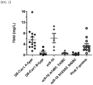
- the DS-Cav1 A-type has a better expression rate than the B-type ( FIG. 2 ).
- the engineered clone sc9-10 has no significant difference in expression level from DS-Cav1, but for a stable trimer form in sc9-10, sc9-10 A149C Y458C and sc9-10 N183GC N428C, which have mutations, exhibit very low expression levels.
- Expi293 cells it was confirmed that there is a difference in expression level. It was confirmed that the longer the cell passage, the lower the expression level, which can eventually affect a yield.
- B cells CD3 - CD8 - CD14 - CD19 + CD20 + were approximately 8% of the total cells, and memory B cells CD27 + were approximately 25% of the B cells.
- memory B cells cells that are double-positive for APC and PE of the RSV F-protein were approximately 0.3%, and approximately 1000 cells of a total of 3.8 ⁇ 10 7 cells were collected in 0.5 mL of 2% FBS/PBS ( FIG. 3 ).
- FACS-sorted B cells lysed with Quick extraction buffer were subjected to RT-PCR and cDNA amplification for Ig transcripts using a SMARTer RACE 5' kit and a RT-IgGKLA primer.
- the cDNA which had experienced RT-PCR and PCR cleanup was immediately used in a volume of approximately 15 ⁇ L to construct a phage-displayed scFv library without gel electrophoresis.
- Chromium single cell V(D)J libraries were constructed for memory B cells (Ag + CD27 + B cells) double-positive for RSV F, memory B cells (Ag - CD27 + B cells) double-negative for RSV F used as a control, and unsorted PBMCs used as a control.
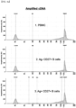
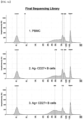
- the concentration after cDNA amplification of Ag + CD27 + B cells (1.82 ng/ ⁇ L), Ag - CD27 + B cells (0.383 ng/ ⁇ L), and PBMC (1.57 ng/ ⁇ L) was 45.5 ⁇ L
- the concentration after a target enrichment step of the Ag + CD27 + B cells (11.2 ng/ ⁇ L), the Ag - CD27 + B cells (1.57 ng/ ⁇ L), and the PBMC (0.542 ng/ ⁇ L) was 45.5 ⁇ L.
- the concentration of the final sequencing library after enzymatic fragmentation and cleanup of the Ag + CD27 + B cells (69.4 ng/ ⁇ L), the Ag - CD27 + B cells (31.2 ng/ ⁇ L), and the PBMC (15.4 ng/ ⁇ L) was 35 ⁇ L. All concentrations were measured with Qubit.
- FIG. 4 The result of QC performed after each step using Agilent Tapestation is shown in FIG. 4 .
- Ig transcripts were observed between 600 to 800 bp, and in the final sequencing library step, traces of enzymatic fragmentation were confirmed.
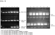
- Each phage-displayed scFv library was constructed using the V(D)J library constructed as described above as a template.
- the results of VH/V ⁇ /V ⁇ PCR and scFv overlapping PCR are shown in FIG. 5 .
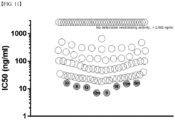
- the IC 50 values of an RSV A2 neutralizing antibody for 112 RSV screening antibody samples were confirmed by an FRNT test. As a result, 8 clones (8, 9, 13, 16, 33, 6m, 11m, and 12m) were confirmed as antibodies exhibiting excellent RSV neutralizing activity ( FIG. 11 ).
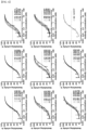
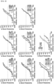
- Table 23 below shows the affinity measurement results for the anti-F antibodies selected from the results of the neutralizing antibody analysis as the kinetic rate constants (K on and K off ) and the equilibrium dissociation constant (K D ) ( FIGS. 12 to 17 ).
- Equilibrium dissociation constant of anti-F antibody Clone K on K off K D 8 2.80X10 6 3.18X10 -5 1.14X10 -11 9 1.50X10 7 5.74X10 -5 3.84X10 -12 12m 3.06X10 6 2.07X10 -5 6.77X10 -12 13 2.03X10 6 2.81X10 -5 1.38X10 -11 15 2.52X10 6 2.89X10 -5 1.15X10 -11 16 1.53X10 7 6.06X10 -5 3.97X10 -12 33 1.77X10 6 6.02X10 -5 3.41X10 -11
Landscapes
- Health & Medical Sciences (AREA)
- Life Sciences & Earth Sciences (AREA)
- Chemical & Material Sciences (AREA)
- Virology (AREA)
- Organic Chemistry (AREA)
- General Health & Medical Sciences (AREA)
- Medicinal Chemistry (AREA)
- Molecular Biology (AREA)
- Immunology (AREA)
- Animal Behavior & Ethology (AREA)
- Veterinary Medicine (AREA)
- Pharmacology & Pharmacy (AREA)
- Public Health (AREA)
- Biochemistry (AREA)
- Biophysics (AREA)
- Genetics & Genomics (AREA)
- Proteomics, Peptides & Aminoacids (AREA)
- Oncology (AREA)
- Communicable Diseases (AREA)
- Chemical Kinetics & Catalysis (AREA)
- General Chemical & Material Sciences (AREA)
- Nuclear Medicine, Radiotherapy & Molecular Imaging (AREA)
- Epidemiology (AREA)
- Mycology (AREA)
- Microbiology (AREA)
- Peptides Or Proteins (AREA)
- Pulmonology (AREA)
- Preparation Of Compounds By Using Micro-Organisms (AREA)
- Micro-Organisms Or Cultivation Processes Thereof (AREA)
- Medicines Containing Antibodies Or Antigens For Use As Internal Diagnostic Agents (AREA)
Applications Claiming Priority (2)
| Application Number | Priority Date | Filing Date | Title |
|---|---|---|---|
| KR1020200077914A KR102467943B1 (ko) | 2020-06-25 | 2020-06-25 | 항-rsv 항체 및 이를 포함하는 약학적 조성물 |
| PCT/KR2021/005742 WO2021261762A1 (ko) | 2020-06-25 | 2021-05-07 | 항-rsv 항체 및 이를 포함하는 약학적 조성물 |
Publications (2)
| Publication Number | Publication Date |
|---|---|
| EP4174084A1 true EP4174084A1 (de) | 2023-05-03 |
| EP4174084A4 EP4174084A4 (de) | 2024-07-17 |
Family
ID=79281484
Family Applications (1)
| Application Number | Title | Priority Date | Filing Date |
|---|---|---|---|
| EP21828260.6A Pending EP4174084A4 (de) | 2020-06-25 | 2021-05-07 | Anti-rsv-antikörper und pharmazeutische zusammensetzung damit |
Country Status (6)
| Country | Link |
|---|---|
| US (1) | US20240182547A1 (de) |
| EP (1) | EP4174084A4 (de) |
| JP (1) | JP7541130B2 (de) |
| KR (1) | KR102467943B1 (de) |
| CN (1) | CN115867574A (de) |
| WO (1) | WO2021261762A1 (de) |
Families Citing this family (1)
| Publication number | Priority date | Publication date | Assignee | Title |
|---|---|---|---|---|
| EP4588936A1 (de) * | 2022-12-08 | 2025-07-23 | Changchun Bcht Biotechnology Co. | Spezifisch an rsv bindende antikörper |
Family Cites Families (7)
| Publication number | Priority date | Publication date | Assignee | Title |
|---|---|---|---|---|
| EP1997830A1 (de) * | 2007-06-01 | 2008-12-03 | AIMM Therapeutics B.V. | RSV-spezifische Bindemoleküle und Mittel zu ihrer Herstellung |
| BR112012031638B1 (pt) * | 2010-07-09 | 2021-01-12 | Janssen Vaccines & Prevention B.V. | anticorpo anti-rsv ou fragmento de ligação de antígeno do mesmo, anticorpo multivalente, composição farmacêutica, uso de anticorpo ou fragmento de ligação de antígeno, método de detectar infecção por rsv, e, ácido nucleico isolado |
| JP5996707B2 (ja) * | 2015-04-15 | 2016-09-21 | メディミューン リミテド | Rsv特異的結合分子 |
| JO3555B1 (ar) * | 2015-10-29 | 2020-07-05 | Merck Sharp & Dohme | جسم مضاد يبطل فعالية فيروس الالتهاب الرئوي البشري |
| RU2723039C2 (ru) * | 2015-12-23 | 2020-06-08 | Пфайзер Инк. | Мутанты белка f rsv |
| CN110035771B (zh) * | 2016-10-21 | 2024-03-08 | 阿迪马布有限责任公司 | 抗呼吸道合胞病毒抗体及其产生和使用方法 |
| EP3554545A4 (de) * | 2016-12-19 | 2020-08-12 | Vanderbilt University | Antikörper gegen die bestätigung der präfusion des proteins des humanen respiratorischen synzytialvirus und verfahren zur verwendung davon |
-
2020
- 2020-06-25 KR KR1020200077914A patent/KR102467943B1/ko active Active
-
2021
- 2021-05-07 EP EP21828260.6A patent/EP4174084A4/de active Pending
- 2021-05-07 JP JP2022580460A patent/JP7541130B2/ja active Active
- 2021-05-07 CN CN202180039509.5A patent/CN115867574A/zh active Pending
- 2021-05-07 WO PCT/KR2021/005742 patent/WO2021261762A1/ko not_active Ceased
- 2021-05-07 US US18/002,881 patent/US20240182547A1/en active Pending
Also Published As
| Publication number | Publication date |
|---|---|
| EP4174084A4 (de) | 2024-07-17 |
| JP7541130B2 (ja) | 2024-08-27 |
| CN115867574A (zh) | 2023-03-28 |
| US20240182547A1 (en) | 2024-06-06 |
| JP2023532487A (ja) | 2023-07-28 |
| KR20220000225A (ko) | 2022-01-03 |
| KR102467943B1 (ko) | 2022-11-16 |
| WO2021261762A1 (ko) | 2021-12-30 |
Similar Documents
| Publication | Publication Date | Title |
|---|---|---|
| CN111153995B (zh) | Nkg2a抗体及其制备方法和应用 | |
| CN104105708B (zh) | PDGF受体β结合多肽 | |
| ES2682254T3 (es) | Polipéptidos solubles | |
| ES2541142T3 (es) | Composiciones y métodos para anticuerpos dirigidos contra la proteína C5 del complemento | |
| TW202126692A (zh) | 抗人Claudin18.2抗體及其應用 | |
| KR102697723B1 (ko) | 항체 분자-약물 접합체 및 이의 용도 | |
| TWI818308B (zh) | 標靶ror1的抗體或其抗原結合片段及製備方法和應用 | |
| TW201339173A (zh) | 可結合及中和b型流感病毒之人類結合分子及其用途 | |
| BR112015027400B1 (pt) | Anticorpo capaz de se ligar especificamente ao her2 ou fragmento de ligação ao antígeno do mesmo, composições farmacêuticas e kit que compreendem os mesmos, bem como uso dos mesmos para prevenir ou tratar um câncer e/ou induzir apoptose | |
| TW202128772A (zh) | 一種bcma結合蛋白及其製備方法和應用 | |
| TW202003584A (zh) | 單特異性及多特異性抗tmeff2抗體及其用途 | |
| CN116333103A (zh) | 抗沙贝病毒属病毒的中和抗体及其制备方法和应用 | |
| CN105820250A (zh) | 一种抗basigin人源化抗体及其应用 | |
| TW202438528A (zh) | 特異性結合Claudin 18.2的抗體及其製法和應用 | |
| EP4174084A1 (de) | Anti-rsv-antikörper und pharmazeutische zusammensetzung damit | |
| TWI788788B (zh) | 標靶EpCAM的抗體及其製備和應用 | |
| CN116284408A (zh) | 结合人muc17的抗体、其制备方法及用途 | |
| JP7450710B2 (ja) | 抗pd-l1抗体及びその製薬用途 | |
| CA2706300C (en) | Human monoclonal nicotine specific antibodies | |
| TW202448937A (zh) | 特異性結合cll1的抗體及其製法和應用 | |
| CN104861068A (zh) | 一种全人源抗her3抗体及含有该抗体的组合物和其用于制备治疗her3相关疾病的药物中的用途 | |
| TWI795872B (zh) | 抗gm2ap抗體及其應用 | |
| EP4527407A1 (de) | Anti-mesothelin-antikörper und verwendungen davon | |
| WO2025228370A1 (zh) | 中和呼吸道合胞病毒的抗原结合蛋白及其应用 | |
| TW202547885A (zh) | 抗體、抗體藥物偶聯物及其用途 |
Legal Events
| Date | Code | Title | Description |
|---|---|---|---|
| STAA | Information on the status of an ep patent application or granted ep patent |
Free format text: STATUS: THE INTERNATIONAL PUBLICATION HAS BEEN MADE |
|
| PUAI | Public reference made under article 153(3) epc to a published international application that has entered the european phase |
Free format text: ORIGINAL CODE: 0009012 |
|
| STAA | Information on the status of an ep patent application or granted ep patent |
Free format text: STATUS: REQUEST FOR EXAMINATION WAS MADE |
|
| 17P | Request for examination filed |
Effective date: 20221130 |
|
| AK | Designated contracting states |
Kind code of ref document: A1 Designated state(s): AL AT BE BG CH CY CZ DE DK EE ES FI FR GB GR HR HU IE IS IT LI LT LU LV MC MK MT NL NO PL PT RO RS SE SI SK SM TR |
|
| DAV | Request for validation of the european patent (deleted) | ||
| DAX | Request for extension of the european patent (deleted) | ||
| A4 | Supplementary search report drawn up and despatched |
Effective date: 20240619 |
|
| RIC1 | Information provided on ipc code assigned before grant |
Ipc: A61K 39/00 20060101ALI20240613BHEP Ipc: A61P 31/14 20060101ALI20240613BHEP Ipc: C07K 16/10 20060101AFI20240613BHEP |