EP4137562A1 - Mesenchymale linienvorläufer oder stammzellen mit erhöhter immunsuppression - Google Patents
Mesenchymale linienvorläufer oder stammzellen mit erhöhter immunsuppression Download PDFInfo
- Publication number
- EP4137562A1 EP4137562A1 EP22187395.3A EP22187395A EP4137562A1 EP 4137562 A1 EP4137562 A1 EP 4137562A1 EP 22187395 A EP22187395 A EP 22187395A EP 4137562 A1 EP4137562 A1 EP 4137562A1
- Authority
- EP
- European Patent Office
- Prior art keywords
- cells
- cell
- mlpscs
- mesenchymal lineage
- gvhd
- Prior art date
- Legal status (The legal status is an assumption and is not a legal conclusion. Google has not performed a legal analysis and makes no representation as to the accuracy of the status listed.)
- Pending
Links
- 210000000130 stem cell Anatomy 0.000 title claims abstract description 187
- 239000002243 precursor Substances 0.000 title claims abstract description 177
- 230000001506 immunosuppresive effect Effects 0.000 title description 16
- 206010062016 Immunosuppression Diseases 0.000 title description 6
- 208000009329 Graft vs Host Disease Diseases 0.000 claims abstract description 176
- 208000024908 graft versus host disease Diseases 0.000 claims abstract description 176
- 238000000034 method Methods 0.000 claims abstract description 109
- 210000004027 cell Anatomy 0.000 claims description 476
- 210000001744 T-lymphocyte Anatomy 0.000 claims description 104
- 230000014509 gene expression Effects 0.000 claims description 63
- 210000003819 peripheral blood mononuclear cell Anatomy 0.000 claims description 57
- 239000001963 growth medium Substances 0.000 claims description 36
- 150000003431 steroids Chemical class 0.000 claims description 28
- 238000012258 culturing Methods 0.000 claims description 21
- 238000005406 washing Methods 0.000 claims description 19
- 238000009295 crossflow filtration Methods 0.000 claims description 12
- 102000004142 Trypsin Human genes 0.000 claims description 11
- 108090000631 Trypsin Proteins 0.000 claims description 11
- 238000002560 therapeutic procedure Methods 0.000 claims description 11
- 239000012588 trypsin Substances 0.000 claims description 11
- 230000009977 dual effect Effects 0.000 claims description 4
- 210000004748 cultured cell Anatomy 0.000 claims 1
- 238000011282 treatment Methods 0.000 abstract description 50
- 238000003556 assay Methods 0.000 abstract description 36
- 208000024891 symptom Diseases 0.000 abstract description 24
- 238000002659 cell therapy Methods 0.000 abstract description 11
- 208000027866 inflammatory disease Diseases 0.000 abstract description 11
- 230000002265 prevention Effects 0.000 abstract description 6
- 208000026278 immune system disease Diseases 0.000 abstract description 2
- 230000001154 acute effect Effects 0.000 description 77
- 239000000203 mixture Substances 0.000 description 66
- 230000037396 body weight Effects 0.000 description 56
- 210000002901 mesenchymal stem cell Anatomy 0.000 description 52
- 230000004044 response Effects 0.000 description 48
- 239000000523 sample Substances 0.000 description 45
- 101710187743 Tumor necrosis factor receptor superfamily member 1A Proteins 0.000 description 42
- 102100033732 Tumor necrosis factor receptor superfamily member 1A Human genes 0.000 description 42
- 239000003446 ligand Substances 0.000 description 39
- 210000003958 hematopoietic stem cell Anatomy 0.000 description 36
- 230000005764 inhibitory process Effects 0.000 description 36
- 108090000623 proteins and genes Proteins 0.000 description 31
- 239000003795 chemical substances by application Substances 0.000 description 30
- 210000001035 gastrointestinal tract Anatomy 0.000 description 30
- 239000000047 product Substances 0.000 description 30
- 210000001185 bone marrow Anatomy 0.000 description 29
- 235000018102 proteins Nutrition 0.000 description 29
- 102000004169 proteins and genes Human genes 0.000 description 29
- 239000008280 blood Substances 0.000 description 28
- 210000004369 blood Anatomy 0.000 description 27
- 239000012634 fragment Substances 0.000 description 26
- 239000000243 solution Substances 0.000 description 26
- 102000017420 CD3 protein, epsilon/gamma/delta subunit Human genes 0.000 description 25
- 108050005493 CD3 protein, epsilon/gamma/delta subunit Proteins 0.000 description 25
- 208000037265 diseases, disorders, signs and symptoms Diseases 0.000 description 25
- 238000002965 ELISA Methods 0.000 description 23
- 239000011324 bead Substances 0.000 description 23
- 210000000056 organ Anatomy 0.000 description 23
- 102100034594 Angiopoietin-1 Human genes 0.000 description 22
- 101000924552 Homo sapiens Angiopoietin-1 Proteins 0.000 description 22
- 201000010099 disease Diseases 0.000 description 22
- 239000013598 vector Substances 0.000 description 22
- 102000005789 Vascular Endothelial Growth Factors Human genes 0.000 description 21
- 108010019530 Vascular Endothelial Growth Factors Proteins 0.000 description 21
- 230000006052 T cell proliferation Effects 0.000 description 20
- 230000004913 activation Effects 0.000 description 20
- 210000001519 tissue Anatomy 0.000 description 20
- 101000914514 Homo sapiens T-cell-specific surface glycoprotein CD28 Proteins 0.000 description 19
- 102100027213 T-cell-specific surface glycoprotein CD28 Human genes 0.000 description 19
- 238000003501 co-culture Methods 0.000 description 19
- 210000004185 liver Anatomy 0.000 description 19
- 239000012139 lysis buffer Substances 0.000 description 19
- IAZDPXIOMUYVGZ-UHFFFAOYSA-N Dimethylsulphoxide Chemical compound CS(C)=O IAZDPXIOMUYVGZ-UHFFFAOYSA-N 0.000 description 18
- 230000000735 allogeneic effect Effects 0.000 description 18
- 238000004519 manufacturing process Methods 0.000 description 18
- 239000003550 marker Substances 0.000 description 18
- 230000004936 stimulating effect Effects 0.000 description 18
- 230000004083 survival effect Effects 0.000 description 18
- 238000005138 cryopreservation Methods 0.000 description 17
- 230000001976 improved effect Effects 0.000 description 17
- 238000001802 infusion Methods 0.000 description 17
- 239000000427 antigen Substances 0.000 description 16
- 108091007433 antigens Proteins 0.000 description 16
- 102000036639 antigens Human genes 0.000 description 16
- 238000011534 incubation Methods 0.000 description 16
- 230000001684 chronic effect Effects 0.000 description 15
- 108020004707 nucleic acids Proteins 0.000 description 15
- 102000039446 nucleic acids Human genes 0.000 description 15
- 150000007523 nucleic acids Chemical class 0.000 description 15
- 230000001225 therapeutic effect Effects 0.000 description 15
- BPYKTIZUTYGOLE-IFADSCNNSA-N Bilirubin Chemical compound N1C(=O)C(C)=C(C=C)\C1=C\C1=C(C)C(CCC(O)=O)=C(CC2=C(C(C)=C(\C=C/3C(=C(C=C)C(=O)N\3)C)N2)CCC(O)=O)N1 BPYKTIZUTYGOLE-IFADSCNNSA-N 0.000 description 14
- 238000010790 dilution Methods 0.000 description 14
- 239000012895 dilution Substances 0.000 description 14
- 230000000694 effects Effects 0.000 description 14
- 230000036961 partial effect Effects 0.000 description 14
- 108040006849 interleukin-2 receptor activity proteins Proteins 0.000 description 13
- 238000012360 testing method Methods 0.000 description 13
- 238000002054 transplantation Methods 0.000 description 13
- 102100025683 Alkaline phosphatase, tissue-nonspecific isozyme Human genes 0.000 description 12
- 206010012735 Diarrhoea Diseases 0.000 description 12
- 239000003153 chemical reaction reagent Substances 0.000 description 12
- 210000005259 peripheral blood Anatomy 0.000 description 12
- 239000011886 peripheral blood Substances 0.000 description 12
- 210000003491 skin Anatomy 0.000 description 12
- 230000000638 stimulation Effects 0.000 description 12
- 239000000758 substrate Substances 0.000 description 12
- JLTPSDHKZGWXTD-UHFFFAOYSA-N 2-[6-(dicyanomethylidene)naphthalen-2-ylidene]propanedinitrile Chemical compound N#CC(C#N)=C1C=CC2=CC(=C(C#N)C#N)C=CC2=C1 JLTPSDHKZGWXTD-UHFFFAOYSA-N 0.000 description 11
- 101710161969 Alkaline phosphatase, tissue-nonspecific isozyme Proteins 0.000 description 11
- 239000000872 buffer Substances 0.000 description 11
- 238000011161 development Methods 0.000 description 11
- 230000018109 developmental process Effects 0.000 description 11
- 239000006166 lysate Substances 0.000 description 11
- XLYOFNOQVPJJNP-UHFFFAOYSA-N water Substances O XLYOFNOQVPJJNP-UHFFFAOYSA-N 0.000 description 11
- 102000004190 Enzymes Human genes 0.000 description 10
- 108090000790 Enzymes Proteins 0.000 description 10
- 210000000612 antigen-presenting cell Anatomy 0.000 description 10
- 230000004071 biological effect Effects 0.000 description 10
- 229940088598 enzyme Drugs 0.000 description 10
- 230000035755 proliferation Effects 0.000 description 10
- 238000010257 thawing Methods 0.000 description 10
- CURLTUGMZLYLDI-UHFFFAOYSA-N Carbon dioxide Chemical compound O=C=O CURLTUGMZLYLDI-UHFFFAOYSA-N 0.000 description 9
- KFZMGEQAYNKOFK-UHFFFAOYSA-N Isopropanol Chemical compound CC(C)O KFZMGEQAYNKOFK-UHFFFAOYSA-N 0.000 description 9
- 239000013592 cell lysate Substances 0.000 description 9
- 239000012881 co-culture medium Substances 0.000 description 9
- 238000003745 diagnosis Methods 0.000 description 9
- 230000008569 process Effects 0.000 description 9
- 108090000765 processed proteins & peptides Proteins 0.000 description 9
- KCXVZYZYPLLWCC-UHFFFAOYSA-N EDTA Chemical compound OC(=O)CN(CC(O)=O)CCN(CC(O)=O)CC(O)=O KCXVZYZYPLLWCC-UHFFFAOYSA-N 0.000 description 8
- 238000011134 hematopoietic stem cell transplantation Methods 0.000 description 8
- 239000004033 plastic Substances 0.000 description 8
- 229920003023 plastic Polymers 0.000 description 8
- 238000002360 preparation method Methods 0.000 description 8
- 210000002966 serum Anatomy 0.000 description 8
- 241000124008 Mammalia Species 0.000 description 7
- 239000000090 biomarker Substances 0.000 description 7
- 238000001514 detection method Methods 0.000 description 7
- 230000004069 differentiation Effects 0.000 description 7
- -1 for example Substances 0.000 description 7
- 230000002496 gastric effect Effects 0.000 description 7
- 238000000338 in vitro Methods 0.000 description 7
- 238000002826 magnetic-activated cell sorting Methods 0.000 description 7
- 229920001184 polypeptide Polymers 0.000 description 7
- 102000004196 processed proteins & peptides Human genes 0.000 description 7
- 238000012216 screening Methods 0.000 description 7
- 230000002483 superagonistic effect Effects 0.000 description 7
- 230000003442 weekly effect Effects 0.000 description 7
- VHRSUDSXCMQTMA-PJHHCJLFSA-N 6alpha-methylprednisolone Chemical compound C([C@@]12C)=CC(=O)C=C1[C@@H](C)C[C@@H]1[C@@H]2[C@@H](O)C[C@]2(C)[C@@](O)(C(=O)CO)CC[C@H]21 VHRSUDSXCMQTMA-PJHHCJLFSA-N 0.000 description 6
- 108091003079 Bovine Serum Albumin Proteins 0.000 description 6
- 108010029697 CD40 Ligand Proteins 0.000 description 6
- 102100032937 CD40 ligand Human genes 0.000 description 6
- 239000006144 Dulbecco’s modified Eagle's medium Substances 0.000 description 6
- 206010028813 Nausea Diseases 0.000 description 6
- 210000004271 bone marrow stromal cell Anatomy 0.000 description 6
- 231100000135 cytotoxicity Toxicity 0.000 description 6
- 230000003013 cytotoxicity Effects 0.000 description 6
- 239000003937 drug carrier Substances 0.000 description 6
- 210000004700 fetal blood Anatomy 0.000 description 6
- 238000000684 flow cytometry Methods 0.000 description 6
- 238000003117 fluorescence-linked immunosorbent assay Methods 0.000 description 6
- 230000028993 immune response Effects 0.000 description 6
- 238000003018 immunoassay Methods 0.000 description 6
- 239000003018 immunosuppressive agent Substances 0.000 description 6
- 229940125721 immunosuppressive agent Drugs 0.000 description 6
- 230000001965 increasing effect Effects 0.000 description 6
- 239000000543 intermediate Substances 0.000 description 6
- 238000001990 intravenous administration Methods 0.000 description 6
- 230000001404 mediated effect Effects 0.000 description 6
- 239000002609 medium Substances 0.000 description 6
- 229960004584 methylprednisolone Drugs 0.000 description 6
- 230000008693 nausea Effects 0.000 description 6
- 230000003287 optical effect Effects 0.000 description 6
- 239000002245 particle Substances 0.000 description 6
- 239000007787 solid Substances 0.000 description 6
- 239000006228 supernatant Substances 0.000 description 6
- 208000023275 Autoimmune disease Diseases 0.000 description 5
- 108010047041 Complementarity Determining Regions Proteins 0.000 description 5
- 102000004127 Cytokines Human genes 0.000 description 5
- 108090000695 Cytokines Proteins 0.000 description 5
- 102100025137 Early activation antigen CD69 Human genes 0.000 description 5
- 101000934374 Homo sapiens Early activation antigen CD69 Proteins 0.000 description 5
- 108010001336 Horseradish Peroxidase Proteins 0.000 description 5
- 206010047700 Vomiting Diseases 0.000 description 5
- 239000013543 active substance Substances 0.000 description 5
- 238000004458 analytical method Methods 0.000 description 5
- 238000002617 apheresis Methods 0.000 description 5
- 210000000988 bone and bone Anatomy 0.000 description 5
- 235000011089 carbon dioxide Nutrition 0.000 description 5
- 238000002512 chemotherapy Methods 0.000 description 5
- 150000001875 compounds Chemical class 0.000 description 5
- 239000003085 diluting agent Substances 0.000 description 5
- 239000003814 drug Substances 0.000 description 5
- 239000012636 effector Substances 0.000 description 5
- 238000011156 evaluation Methods 0.000 description 5
- 239000012091 fetal bovine serum Substances 0.000 description 5
- 210000002950 fibroblast Anatomy 0.000 description 5
- 230000006872 improvement Effects 0.000 description 5
- 239000003112 inhibitor Substances 0.000 description 5
- 238000002347 injection Methods 0.000 description 5
- 239000007924 injection Substances 0.000 description 5
- 210000000265 leukocyte Anatomy 0.000 description 5
- 210000004698 lymphocyte Anatomy 0.000 description 5
- 239000000463 material Substances 0.000 description 5
- 210000001616 monocyte Anatomy 0.000 description 5
- 230000000069 prophylactic effect Effects 0.000 description 5
- 238000013207 serial dilution Methods 0.000 description 5
- 239000000126 substance Substances 0.000 description 5
- 230000035899 viability Effects 0.000 description 5
- 239000013603 viral vector Substances 0.000 description 5
- 239000011534 wash buffer Substances 0.000 description 5
- 208000004998 Abdominal Pain Diseases 0.000 description 4
- 102000002260 Alkaline Phosphatase Human genes 0.000 description 4
- 108020004774 Alkaline Phosphatase Proteins 0.000 description 4
- PMATZTZNYRCHOR-CGLBZJNRSA-N Cyclosporin A Chemical compound CC[C@@H]1NC(=O)[C@H]([C@H](O)[C@H](C)C\C=C\C)N(C)C(=O)[C@H](C(C)C)N(C)C(=O)[C@H](CC(C)C)N(C)C(=O)[C@H](CC(C)C)N(C)C(=O)[C@@H](C)NC(=O)[C@H](C)NC(=O)[C@H](CC(C)C)N(C)C(=O)[C@H](C(C)C)NC(=O)[C@H](CC(C)C)N(C)C(=O)CN(C)C1=O PMATZTZNYRCHOR-CGLBZJNRSA-N 0.000 description 4
- 108010036949 Cyclosporine Proteins 0.000 description 4
- 108091006905 Human Serum Albumin Proteins 0.000 description 4
- 102000008100 Human Serum Albumin Human genes 0.000 description 4
- MHAJPDPJQMAIIY-UHFFFAOYSA-N Hydrogen peroxide Chemical compound OO MHAJPDPJQMAIIY-UHFFFAOYSA-N 0.000 description 4
- TWRXJAOTZQYOKJ-UHFFFAOYSA-L Magnesium chloride Chemical compound [Mg+2].[Cl-].[Cl-] TWRXJAOTZQYOKJ-UHFFFAOYSA-L 0.000 description 4
- 108010029485 Protein Isoforms Proteins 0.000 description 4
- 102000001708 Protein Isoforms Human genes 0.000 description 4
- 108010000134 Vascular Cell Adhesion Molecule-1 Proteins 0.000 description 4
- 102100023543 Vascular cell adhesion protein 1 Human genes 0.000 description 4
- 230000001464 adherent effect Effects 0.000 description 4
- 208000022531 anorexia Diseases 0.000 description 4
- 210000003719 b-lymphocyte Anatomy 0.000 description 4
- 238000001574 biopsy Methods 0.000 description 4
- 230000001413 cellular effect Effects 0.000 description 4
- 229960001265 ciclosporin Drugs 0.000 description 4
- 239000012141 concentrate Substances 0.000 description 4
- 229930182912 cyclosporin Natural products 0.000 description 4
- 230000034994 death Effects 0.000 description 4
- 206010061428 decreased appetite Diseases 0.000 description 4
- LOKCTEFSRHRXRJ-UHFFFAOYSA-I dipotassium trisodium dihydrogen phosphate hydrogen phosphate dichloride Chemical compound P(=O)(O)(O)[O-].[K+].P(=O)(O)([O-])[O-].[Na+].[Na+].[Cl-].[K+].[Cl-].[Na+] LOKCTEFSRHRXRJ-UHFFFAOYSA-I 0.000 description 4
- 238000005516 engineering process Methods 0.000 description 4
- 238000001943 fluorescence-activated cell sorting Methods 0.000 description 4
- 238000001727 in vivo Methods 0.000 description 4
- 230000002401 inhibitory effect Effects 0.000 description 4
- 230000000977 initiatory effect Effects 0.000 description 4
- 239000007788 liquid Substances 0.000 description 4
- 239000008185 minitablet Substances 0.000 description 4
- 238000012986 modification Methods 0.000 description 4
- 230000004048 modification Effects 0.000 description 4
- 210000005087 mononuclear cell Anatomy 0.000 description 4
- RTGDFNSFWBGLEC-SYZQJQIISA-N mycophenolate mofetil Chemical compound COC1=C(C)C=2COC(=O)C=2C(O)=C1C\C=C(/C)CCC(=O)OCCN1CCOCC1 RTGDFNSFWBGLEC-SYZQJQIISA-N 0.000 description 4
- 229960004866 mycophenolate mofetil Drugs 0.000 description 4
- 239000008188 pellet Substances 0.000 description 4
- 239000002953 phosphate buffered saline Substances 0.000 description 4
- 102000005962 receptors Human genes 0.000 description 4
- 108020003175 receptors Proteins 0.000 description 4
- 230000009467 reduction Effects 0.000 description 4
- 238000010187 selection method Methods 0.000 description 4
- 238000001890 transfection Methods 0.000 description 4
- 230000008673 vomiting Effects 0.000 description 4
- OZFAFGSSMRRTDW-UHFFFAOYSA-N (2,4-dichlorophenyl) benzenesulfonate Chemical compound ClC1=CC(Cl)=CC=C1OS(=O)(=O)C1=CC=CC=C1 OZFAFGSSMRRTDW-UHFFFAOYSA-N 0.000 description 3
- 102000004625 Aspartate Aminotransferases Human genes 0.000 description 3
- 108010003415 Aspartate Aminotransferases Proteins 0.000 description 3
- 101150013553 CD40 gene Proteins 0.000 description 3
- 241000701022 Cytomegalovirus Species 0.000 description 3
- 239000012591 Dulbecco’s Phosphate Buffered Saline Substances 0.000 description 3
- 108010008165 Etanercept Proteins 0.000 description 3
- 208000010201 Exanthema Diseases 0.000 description 3
- HTTJABKRGRZYRN-UHFFFAOYSA-N Heparin Chemical compound OC1C(NC(=O)C)C(O)OC(COS(O)(=O)=O)C1OC1C(OS(O)(=O)=O)C(O)C(OC2C(C(OS(O)(=O)=O)C(OC3C(C(O)C(O)C(O3)C(O)=O)OS(O)(=O)=O)C(CO)O2)NS(O)(=O)=O)C(C(O)=O)O1 HTTJABKRGRZYRN-UHFFFAOYSA-N 0.000 description 3
- 101001055144 Homo sapiens Interleukin-2 receptor subunit alpha Proteins 0.000 description 3
- 108010054477 Immunoglobulin Fab Fragments Proteins 0.000 description 3
- 102000001706 Immunoglobulin Fab Fragments Human genes 0.000 description 3
- 102000000589 Interleukin-1 Human genes 0.000 description 3
- 108010002352 Interleukin-1 Proteins 0.000 description 3
- 102000000588 Interleukin-2 Human genes 0.000 description 3
- 108010002350 Interleukin-2 Proteins 0.000 description 3
- 102100026878 Interleukin-2 receptor subunit alpha Human genes 0.000 description 3
- 108700018351 Major Histocompatibility Complex Proteins 0.000 description 3
- 241001465754 Metazoa Species 0.000 description 3
- 241000699666 Mus <mouse, genus> Species 0.000 description 3
- 206010037868 Rash maculo-papular Diseases 0.000 description 3
- FAPWRFPIFSIZLT-UHFFFAOYSA-M Sodium chloride Chemical compound [Na+].[Cl-] FAPWRFPIFSIZLT-UHFFFAOYSA-M 0.000 description 3
- HEMHJVSKTPXQMS-UHFFFAOYSA-M Sodium hydroxide Chemical compound [OH-].[Na+] HEMHJVSKTPXQMS-UHFFFAOYSA-M 0.000 description 3
- 230000005867 T cell response Effects 0.000 description 3
- 101150052863 THY1 gene Proteins 0.000 description 3
- 102100040245 Tumor necrosis factor receptor superfamily member 5 Human genes 0.000 description 3
- 241000700605 Viruses Species 0.000 description 3
- 239000002250 absorbent Substances 0.000 description 3
- 230000002745 absorbent Effects 0.000 description 3
- 208000024340 acute graft versus host disease Diseases 0.000 description 3
- 239000002269 analeptic agent Substances 0.000 description 3
- 230000001363 autoimmune Effects 0.000 description 3
- 229960004669 basiliximab Drugs 0.000 description 3
- 230000015572 biosynthetic process Effects 0.000 description 3
- 210000000601 blood cell Anatomy 0.000 description 3
- 210000001772 blood platelet Anatomy 0.000 description 3
- 238000001516 cell proliferation assay Methods 0.000 description 3
- 238000012790 confirmation Methods 0.000 description 3
- 238000012937 correction Methods 0.000 description 3
- 230000002596 correlated effect Effects 0.000 description 3
- 239000003246 corticosteroid Substances 0.000 description 3
- 229960001334 corticosteroids Drugs 0.000 description 3
- 210000004443 dendritic cell Anatomy 0.000 description 3
- 230000001419 dependent effect Effects 0.000 description 3
- 208000035475 disorder Diseases 0.000 description 3
- 229960000403 etanercept Drugs 0.000 description 3
- 201000005884 exanthem Diseases 0.000 description 3
- 239000012467 final product Substances 0.000 description 3
- 239000011521 glass Substances 0.000 description 3
- ZDXPYRJPNDTMRX-UHFFFAOYSA-N glutamine Natural products OC(=O)C(N)CCC(N)=O ZDXPYRJPNDTMRX-UHFFFAOYSA-N 0.000 description 3
- PCHJSUWPFVWCPO-UHFFFAOYSA-N gold Chemical compound [Au] PCHJSUWPFVWCPO-UHFFFAOYSA-N 0.000 description 3
- 230000036541 health Effects 0.000 description 3
- 229960002897 heparin Drugs 0.000 description 3
- 229920000669 heparin Polymers 0.000 description 3
- 210000003494 hepatocyte Anatomy 0.000 description 3
- 230000002962 histologic effect Effects 0.000 description 3
- 208000008384 ileus Diseases 0.000 description 3
- 239000003547 immunosorbent Substances 0.000 description 3
- 230000006698 induction Effects 0.000 description 3
- 230000001939 inductive effect Effects 0.000 description 3
- 229960000598 infliximab Drugs 0.000 description 3
- 239000002502 liposome Substances 0.000 description 3
- 210000003750 lower gastrointestinal tract Anatomy 0.000 description 3
- 208000012965 maculopapular rash Diseases 0.000 description 3
- 230000003211 malignant effect Effects 0.000 description 3
- 238000005259 measurement Methods 0.000 description 3
- 239000002679 microRNA Substances 0.000 description 3
- 230000001537 neural effect Effects 0.000 description 3
- 210000004789 organ system Anatomy 0.000 description 3
- 230000005298 paramagnetic effect Effects 0.000 description 3
- 230000001575 pathological effect Effects 0.000 description 3
- 230000037361 pathway Effects 0.000 description 3
- 210000003668 pericyte Anatomy 0.000 description 3
- 239000008194 pharmaceutical composition Substances 0.000 description 3
- 206010037844 rash Diseases 0.000 description 3
- 230000002829 reductive effect Effects 0.000 description 3
- 238000011160 research Methods 0.000 description 3
- 230000001177 retroviral effect Effects 0.000 description 3
- 239000012898 sample dilution Substances 0.000 description 3
- 230000028327 secretion Effects 0.000 description 3
- 241000894007 species Species 0.000 description 3
- 239000012089 stop solution Substances 0.000 description 3
- 230000020382 suppression by virus of host antigen processing and presentation of peptide antigen via MHC class I Effects 0.000 description 3
- 239000000725 suspension Substances 0.000 description 3
- 210000002435 tendon Anatomy 0.000 description 3
- 210000001541 thymus gland Anatomy 0.000 description 3
- 238000012546 transfer Methods 0.000 description 3
- 239000012808 vapor phase Substances 0.000 description 3
- 208000032467 Aplastic anaemia Diseases 0.000 description 2
- CIWBSHSKHKDKBQ-JLAZNSOCSA-N Ascorbic acid Chemical compound OC[C@H](O)[C@H]1OC(=O)C(O)=C1O CIWBSHSKHKDKBQ-JLAZNSOCSA-N 0.000 description 2
- 210000001266 CD8-positive T-lymphocyte Anatomy 0.000 description 2
- 102100035793 CD83 antigen Human genes 0.000 description 2
- 241000282472 Canis lupus familiaris Species 0.000 description 2
- 241000283707 Capra Species 0.000 description 2
- 108010067225 Cell Adhesion Molecules Proteins 0.000 description 2
- 102000016289 Cell Adhesion Molecules Human genes 0.000 description 2
- 208000015943 Coeliac disease Diseases 0.000 description 2
- 108020004414 DNA Proteins 0.000 description 2
- 206010012455 Dermatitis exfoliative Diseases 0.000 description 2
- 238000008157 ELISA kit Methods 0.000 description 2
- 241000282326 Felis catus Species 0.000 description 2
- 241000713813 Gibbon ape leukemia virus Species 0.000 description 2
- 102000006395 Globulins Human genes 0.000 description 2
- 108010044091 Globulins Proteins 0.000 description 2
- 239000012981 Hank's balanced salt solution Substances 0.000 description 2
- 102100031573 Hematopoietic progenitor cell antigen CD34 Human genes 0.000 description 2
- 208000036066 Hemophagocytic Lymphohistiocytosis Diseases 0.000 description 2
- 102100021866 Hepatocyte growth factor Human genes 0.000 description 2
- 208000032672 Histiocytosis haematophagic Diseases 0.000 description 2
- 101000946856 Homo sapiens CD83 antigen Proteins 0.000 description 2
- 101000777663 Homo sapiens Hematopoietic progenitor cell antigen CD34 Proteins 0.000 description 2
- 101000851376 Homo sapiens Tumor necrosis factor receptor superfamily member 8 Proteins 0.000 description 2
- 101000851370 Homo sapiens Tumor necrosis factor receptor superfamily member 9 Proteins 0.000 description 2
- 241000701044 Human gammaherpesvirus 4 Species 0.000 description 2
- 241000725303 Human immunodeficiency virus Species 0.000 description 2
- 208000026350 Inborn Genetic disease Diseases 0.000 description 2
- 239000002144 L01XE18 - Ruxolitinib Substances 0.000 description 2
- 108010064548 Lymphocyte Function-Associated Antigen-1 Proteins 0.000 description 2
- 108010061593 Member 14 Tumor Necrosis Factor Receptors Proteins 0.000 description 2
- 108700011259 MicroRNAs Proteins 0.000 description 2
- 241000714177 Murine leukemia virus Species 0.000 description 2
- 206010028980 Neoplasm Diseases 0.000 description 2
- 208000002193 Pain Diseases 0.000 description 2
- 241001494479 Pecora Species 0.000 description 2
- 102000057297 Pepsin A Human genes 0.000 description 2
- 108090000284 Pepsin A Proteins 0.000 description 2
- 239000004793 Polystyrene Substances 0.000 description 2
- WCUXLLCKKVVCTQ-UHFFFAOYSA-M Potassium chloride Chemical compound [Cl-].[K+] WCUXLLCKKVVCTQ-UHFFFAOYSA-M 0.000 description 2
- 241000288906 Primates Species 0.000 description 2
- 102000003923 Protein Kinase C Human genes 0.000 description 2
- 108090000315 Protein Kinase C Proteins 0.000 description 2
- 206010037660 Pyrexia Diseases 0.000 description 2
- 241000700159 Rattus Species 0.000 description 2
- 108700008625 Reporter Genes Proteins 0.000 description 2
- 206010039710 Scleroderma Diseases 0.000 description 2
- PXIPVTKHYLBLMZ-UHFFFAOYSA-N Sodium azide Chemical compound [Na+].[N-]=[N+]=[N-] PXIPVTKHYLBLMZ-UHFFFAOYSA-N 0.000 description 2
- 108010090804 Streptavidin Proteins 0.000 description 2
- QJJXYPPXXYFBGM-LFZNUXCKSA-N Tacrolimus Chemical compound C1C[C@@H](O)[C@H](OC)C[C@@H]1\C=C(/C)[C@@H]1[C@H](C)[C@@H](O)CC(=O)[C@H](CC=C)/C=C(C)/C[C@H](C)C[C@H](OC)[C@H]([C@H](C[C@H]2C)OC)O[C@@]2(O)C(=O)C(=O)N2CCCC[C@H]2C(=O)O1 QJJXYPPXXYFBGM-LFZNUXCKSA-N 0.000 description 2
- 102000004887 Transforming Growth Factor beta Human genes 0.000 description 2
- 108090001012 Transforming Growth Factor beta Proteins 0.000 description 2
- 108060008683 Tumor Necrosis Factor Receptor Proteins 0.000 description 2
- 102100028785 Tumor necrosis factor receptor superfamily member 14 Human genes 0.000 description 2
- 102100036857 Tumor necrosis factor receptor superfamily member 8 Human genes 0.000 description 2
- 102100036856 Tumor necrosis factor receptor superfamily member 9 Human genes 0.000 description 2
- 206010067584 Type 1 diabetes mellitus Diseases 0.000 description 2
- 238000011374 additional therapy Methods 0.000 description 2
- 210000001789 adipocyte Anatomy 0.000 description 2
- 210000000577 adipose tissue Anatomy 0.000 description 2
- 230000002411 adverse Effects 0.000 description 2
- 238000013019 agitation Methods 0.000 description 2
- 239000003146 anticoagulant agent Substances 0.000 description 2
- 239000004019 antithrombin Substances 0.000 description 2
- 210000001130 astrocyte Anatomy 0.000 description 2
- 201000005000 autoimmune gastritis Diseases 0.000 description 2
- 229960002170 azathioprine Drugs 0.000 description 2
- LMEKQMALGUDUQG-UHFFFAOYSA-N azathioprine Chemical compound CN1C=NC([N+]([O-])=O)=C1SC1=NC=NC2=C1NC=N2 LMEKQMALGUDUQG-UHFFFAOYSA-N 0.000 description 2
- 230000009286 beneficial effect Effects 0.000 description 2
- 102000023732 binding proteins Human genes 0.000 description 2
- 108091008324 binding proteins Proteins 0.000 description 2
- 230000000975 bioactive effect Effects 0.000 description 2
- 230000008827 biological function Effects 0.000 description 2
- 239000000091 biomarker candidate Substances 0.000 description 2
- 230000000903 blocking effect Effects 0.000 description 2
- 239000010836 blood and blood product Substances 0.000 description 2
- 229940125691 blood product Drugs 0.000 description 2
- 210000002798 bone marrow cell Anatomy 0.000 description 2
- 210000004556 brain Anatomy 0.000 description 2
- 229940046731 calcineurin inhibitors Drugs 0.000 description 2
- 238000004113 cell culture Methods 0.000 description 2
- 230000006037 cell lysis Effects 0.000 description 2
- 239000006285 cell suspension Substances 0.000 description 2
- 238000005119 centrifugation Methods 0.000 description 2
- 238000012512 characterization method Methods 0.000 description 2
- 210000001612 chondrocyte Anatomy 0.000 description 2
- 230000003021 clonogenic effect Effects 0.000 description 2
- 238000000576 coating method Methods 0.000 description 2
- 238000002052 colonoscopy Methods 0.000 description 2
- 230000003750 conditioning effect Effects 0.000 description 2
- 239000013068 control sample Substances 0.000 description 2
- 230000000139 costimulatory effect Effects 0.000 description 2
- 239000002577 cryoprotective agent Substances 0.000 description 2
- 230000007423 decrease Effects 0.000 description 2
- 230000006866 deterioration Effects 0.000 description 2
- 230000004064 dysfunction Effects 0.000 description 2
- 230000002500 effect on skin Effects 0.000 description 2
- 210000005175 epidermal keratinocyte Anatomy 0.000 description 2
- 210000002919 epithelial cell Anatomy 0.000 description 2
- 210000003743 erythrocyte Anatomy 0.000 description 2
- 238000000605 extraction Methods 0.000 description 2
- 239000012909 foetal bovine serum Substances 0.000 description 2
- 238000007710 freezing Methods 0.000 description 2
- 230000008014 freezing Effects 0.000 description 2
- 230000006870 function Effects 0.000 description 2
- 238000001415 gene therapy Methods 0.000 description 2
- 208000016361 genetic disease Diseases 0.000 description 2
- 210000004602 germ cell Anatomy 0.000 description 2
- RWSXRVCMGQZWBV-WDSKDSINSA-N glutathione Chemical compound OC(=O)[C@@H](N)CCC(=O)N[C@@H](CS)C(=O)NCC(O)=O RWSXRVCMGQZWBV-WDSKDSINSA-N 0.000 description 2
- 239000010931 gold Substances 0.000 description 2
- 229910052737 gold Inorganic materials 0.000 description 2
- 210000003714 granulocyte Anatomy 0.000 description 2
- 239000003102 growth factor Substances 0.000 description 2
- 210000002216 heart Anatomy 0.000 description 2
- 208000014752 hemophagocytic syndrome Diseases 0.000 description 2
- 230000002440 hepatic effect Effects 0.000 description 2
- JYGXADMDTFJGBT-VWUMJDOOSA-N hydrocortisone Chemical compound O=C1CC[C@]2(C)[C@H]3[C@@H](O)C[C@](C)([C@@](CC4)(O)C(=O)CO)[C@@H]4[C@@H]3CCC2=C1 JYGXADMDTFJGBT-VWUMJDOOSA-N 0.000 description 2
- 230000003100 immobilizing effect Effects 0.000 description 2
- 230000008105 immune reaction Effects 0.000 description 2
- 230000005847 immunogenicity Effects 0.000 description 2
- 230000001771 impaired effect Effects 0.000 description 2
- 208000015181 infectious disease Diseases 0.000 description 2
- 229940090044 injection Drugs 0.000 description 2
- NOESYZHRGYRDHS-UHFFFAOYSA-N insulin Chemical compound N1C(=O)C(NC(=O)C(CCC(N)=O)NC(=O)C(CCC(O)=O)NC(=O)C(C(C)C)NC(=O)C(NC(=O)CN)C(C)CC)CSSCC(C(NC(CO)C(=O)NC(CC(C)C)C(=O)NC(CC=2C=CC(O)=CC=2)C(=O)NC(CCC(N)=O)C(=O)NC(CC(C)C)C(=O)NC(CCC(O)=O)C(=O)NC(CC(N)=O)C(=O)NC(CC=2C=CC(O)=CC=2)C(=O)NC(CSSCC(NC(=O)C(C(C)C)NC(=O)C(CC(C)C)NC(=O)C(CC=2C=CC(O)=CC=2)NC(=O)C(CC(C)C)NC(=O)C(C)NC(=O)C(CCC(O)=O)NC(=O)C(C(C)C)NC(=O)C(CC(C)C)NC(=O)C(CC=2NC=NC=2)NC(=O)C(CO)NC(=O)CNC2=O)C(=O)NCC(=O)NC(CCC(O)=O)C(=O)NC(CCCNC(N)=N)C(=O)NCC(=O)NC(CC=3C=CC=CC=3)C(=O)NC(CC=3C=CC=CC=3)C(=O)NC(CC=3C=CC(O)=CC=3)C(=O)NC(C(C)O)C(=O)N3C(CCC3)C(=O)NC(CCCCN)C(=O)NC(C)C(O)=O)C(=O)NC(CC(N)=O)C(O)=O)=O)NC(=O)C(C(C)CC)NC(=O)C(CO)NC(=O)C(C(C)O)NC(=O)C1CSSCC2NC(=O)C(CC(C)C)NC(=O)C(NC(=O)C(CCC(N)=O)NC(=O)C(CC(N)=O)NC(=O)C(NC(=O)C(N)CC=1C=CC=CC=1)C(C)C)CC1=CN=CN1 NOESYZHRGYRDHS-UHFFFAOYSA-N 0.000 description 2
- 210000000936 intestine Anatomy 0.000 description 2
- 238000011835 investigation Methods 0.000 description 2
- 238000002955 isolation Methods 0.000 description 2
- 210000003734 kidney Anatomy 0.000 description 2
- 230000007775 late Effects 0.000 description 2
- 210000003041 ligament Anatomy 0.000 description 2
- 238000007449 liver function test Methods 0.000 description 2
- 206010025135 lupus erythematosus Diseases 0.000 description 2
- 210000001165 lymph node Anatomy 0.000 description 2
- 229940124302 mTOR inhibitor Drugs 0.000 description 2
- 229910001629 magnesium chloride Inorganic materials 0.000 description 2
- FVVLHONNBARESJ-NTOWJWGLSA-H magnesium;potassium;trisodium;(2r,3s,4r,5r)-2,3,4,5,6-pentahydroxyhexanoate;acetate;tetrachloride;nonahydrate Chemical compound O.O.O.O.O.O.O.O.O.[Na+].[Na+].[Na+].[Mg+2].[Cl-].[Cl-].[Cl-].[Cl-].[K+].CC([O-])=O.OC[C@@H](O)[C@@H](O)[C@H](O)[C@@H](O)C([O-])=O FVVLHONNBARESJ-NTOWJWGLSA-H 0.000 description 2
- 239000003628 mammalian target of rapamycin inhibitor Substances 0.000 description 2
- 239000011159 matrix material Substances 0.000 description 2
- 108091070501 miRNA Proteins 0.000 description 2
- HPNSFSBZBAHARI-UHFFFAOYSA-N micophenolic acid Natural products OC1=C(CC=C(C)CCC(O)=O)C(OC)=C(C)C2=C1C(=O)OC2 HPNSFSBZBAHARI-UHFFFAOYSA-N 0.000 description 2
- 239000011859 microparticle Substances 0.000 description 2
- 238000002156 mixing Methods 0.000 description 2
- 201000006417 multiple sclerosis Diseases 0.000 description 2
- HPNSFSBZBAHARI-RUDMXATFSA-N mycophenolic acid Chemical compound OC1=C(C\C=C(/C)CCC(O)=O)C(OC)=C(C)C2=C1C(=O)OC2 HPNSFSBZBAHARI-RUDMXATFSA-N 0.000 description 2
- 210000004165 myocardium Anatomy 0.000 description 2
- 210000000822 natural killer cell Anatomy 0.000 description 2
- 239000013642 negative control Substances 0.000 description 2
- 229940021182 non-steroidal anti-inflammatory drug Drugs 0.000 description 2
- 231100000252 nontoxic Toxicity 0.000 description 2
- 230000003000 nontoxic effect Effects 0.000 description 2
- 210000004248 oligodendroglia Anatomy 0.000 description 2
- 102000002574 p38 Mitogen-Activated Protein Kinases Human genes 0.000 description 2
- 108010068338 p38 Mitogen-Activated Protein Kinases Proteins 0.000 description 2
- 238000004806 packaging method and process Methods 0.000 description 2
- 230000002085 persistent effect Effects 0.000 description 2
- 230000002399 phagocytotic effect Effects 0.000 description 2
- 239000000546 pharmaceutical excipient Substances 0.000 description 2
- 239000013612 plasmid Substances 0.000 description 2
- 229920002223 polystyrene Polymers 0.000 description 2
- 239000001267 polyvinylpyrrolidone Substances 0.000 description 2
- 229920000036 polyvinylpyrrolidone Polymers 0.000 description 2
- 235000013855 polyvinylpyrrolidone Nutrition 0.000 description 2
- 230000003389 potentiating effect Effects 0.000 description 2
- 229960005205 prednisolone Drugs 0.000 description 2
- OIGNJSKKLXVSLS-VWUMJDOOSA-N prednisolone Chemical compound O=C1C=C[C@]2(C)[C@H]3[C@@H](O)C[C@](C)([C@@](CC4)(O)C(=O)CO)[C@@H]4[C@@H]3CCC2=C1 OIGNJSKKLXVSLS-VWUMJDOOSA-N 0.000 description 2
- 238000011321 prophylaxis Methods 0.000 description 2
- 238000003908 quality control method Methods 0.000 description 2
- 206010039073 rheumatoid arthritis Diseases 0.000 description 2
- 230000000630 rising effect Effects 0.000 description 2
- 229960000215 ruxolitinib Drugs 0.000 description 2
- HFNKQEVNSGCOJV-OAHLLOKOSA-N ruxolitinib Chemical compound C1([C@@H](CC#N)N2N=CC(=C2)C=2C=3C=CNC=3N=CN=2)CCCC1 HFNKQEVNSGCOJV-OAHLLOKOSA-N 0.000 description 2
- 230000003248 secreting effect Effects 0.000 description 2
- 238000000926 separation method Methods 0.000 description 2
- 230000019491 signal transduction Effects 0.000 description 2
- 210000002027 skeletal muscle Anatomy 0.000 description 2
- 210000000952 spleen Anatomy 0.000 description 2
- 230000003637 steroidlike Effects 0.000 description 2
- 210000002536 stromal cell Anatomy 0.000 description 2
- 238000002198 surface plasmon resonance spectroscopy Methods 0.000 description 2
- 230000009885 systemic effect Effects 0.000 description 2
- 229960001967 tacrolimus Drugs 0.000 description 2
- QJJXYPPXXYFBGM-SHYZHZOCSA-N tacrolimus Natural products CO[C@H]1C[C@H](CC[C@@H]1O)C=C(C)[C@H]2OC(=O)[C@H]3CCCCN3C(=O)C(=O)[C@@]4(O)O[C@@H]([C@H](C[C@H]4C)OC)[C@@H](C[C@H](C)CC(=C[C@@H](CC=C)C(=O)C[C@H](O)[C@H]2C)C)OC QJJXYPPXXYFBGM-SHYZHZOCSA-N 0.000 description 2
- ZRKFYGHZFMAOKI-QMGMOQQFSA-N tgfbeta Chemical compound C([C@H](NC(=O)[C@H](C(C)C)NC(=O)CNC(=O)[C@H](CCC(O)=O)NC(=O)[C@H](CCCNC(N)=N)NC(=O)[C@H](CC(N)=O)NC(=O)[C@H](CC(C)C)NC(=O)[C@H]([C@@H](C)O)NC(=O)[C@H](CCC(O)=O)NC(=O)[C@H]([C@@H](C)O)NC(=O)[C@H](CC(C)C)NC(=O)CNC(=O)[C@H](C)NC(=O)[C@H](CO)NC(=O)[C@H](CCC(N)=O)NC(=O)[C@@H](NC(=O)[C@H](C)NC(=O)[C@H](C)NC(=O)[C@@H](NC(=O)[C@H](CC(C)C)NC(=O)[C@@H](N)CCSC)C(C)C)[C@@H](C)CC)C(=O)N[C@@H]([C@@H](C)O)C(=O)N[C@@H](C(C)C)C(=O)N[C@@H](CC=1C=CC=CC=1)C(=O)N[C@@H](C)C(=O)N1[C@@H](CCC1)C(=O)N[C@@H]([C@@H](C)O)C(=O)N[C@@H](CC(N)=O)C(=O)N[C@@H](CCC(O)=O)C(=O)N[C@@H](C)C(=O)N[C@@H](CC=1C=CC=CC=1)C(=O)N[C@@H](CCCNC(N)=N)C(=O)N[C@@H](C)C(=O)N[C@@H](CC(C)C)C(=O)N1[C@@H](CCC1)C(=O)N1[C@@H](CCC1)C(=O)N[C@@H](CCCNC(N)=N)C(=O)N[C@@H](CCC(O)=O)C(=O)N[C@@H](CCCNC(N)=N)C(=O)N[C@@H](CO)C(=O)N[C@@H](CCCNC(N)=N)C(=O)N[C@@H](CC(C)C)C(=O)N[C@@H](CC(C)C)C(O)=O)C1=CC=C(O)C=C1 ZRKFYGHZFMAOKI-QMGMOQQFSA-N 0.000 description 2
- 230000000451 tissue damage Effects 0.000 description 2
- 231100000827 tissue damage Toxicity 0.000 description 2
- 102000003298 tumor necrosis factor receptor Human genes 0.000 description 2
- 210000003954 umbilical cord Anatomy 0.000 description 2
- 210000002438 upper gastrointestinal tract Anatomy 0.000 description 2
- 210000002700 urine Anatomy 0.000 description 2
- 230000003612 virological effect Effects 0.000 description 2
- HDTRYLNUVZCQOY-UHFFFAOYSA-N α-D-glucopyranosyl-α-D-glucopyranoside Natural products OC1C(O)C(O)C(CO)OC1OC1C(O)C(O)C(O)C(CO)O1 HDTRYLNUVZCQOY-UHFFFAOYSA-N 0.000 description 1
- MZOFCQQQCNRIBI-VMXHOPILSA-N (3s)-4-[[(2s)-1-[[(2s)-1-[[(1s)-1-carboxy-2-hydroxyethyl]amino]-4-methyl-1-oxopentan-2-yl]amino]-5-(diaminomethylideneamino)-1-oxopentan-2-yl]amino]-3-[[2-[[(2s)-2,6-diaminohexanoyl]amino]acetyl]amino]-4-oxobutanoic acid Chemical compound OC[C@@H](C(O)=O)NC(=O)[C@H](CC(C)C)NC(=O)[C@H](CCCN=C(N)N)NC(=O)[C@H](CC(O)=O)NC(=O)CNC(=O)[C@@H](N)CCCCN MZOFCQQQCNRIBI-VMXHOPILSA-N 0.000 description 1
- FPIPGXGPPPQFEQ-UHFFFAOYSA-N 13-cis retinol Natural products OCC=C(C)C=CC=C(C)C=CC1=C(C)CCCC1(C)C FPIPGXGPPPQFEQ-UHFFFAOYSA-N 0.000 description 1
- AEQDJSLRWYMAQI-UHFFFAOYSA-N 2,3,9,10-tetramethoxy-6,8,13,13a-tetrahydro-5H-isoquinolino[2,1-b]isoquinoline Chemical compound C1CN2CC(C(=C(OC)C=C3)OC)=C3CC2C2=C1C=C(OC)C(OC)=C2 AEQDJSLRWYMAQI-UHFFFAOYSA-N 0.000 description 1
- BDKLKNJTMLIAFE-UHFFFAOYSA-N 2-(3-fluorophenyl)-1,3-oxazole-4-carbaldehyde Chemical compound FC1=CC=CC(C=2OC=C(C=O)N=2)=C1 BDKLKNJTMLIAFE-UHFFFAOYSA-N 0.000 description 1
- VNDNKFJKUBLYQB-UHFFFAOYSA-N 2-(4-amino-6-chloro-5-oxohexyl)guanidine Chemical compound ClCC(=O)C(N)CCCN=C(N)N VNDNKFJKUBLYQB-UHFFFAOYSA-N 0.000 description 1
- PWKSKIMOESPYIA-UHFFFAOYSA-N 2-acetamido-3-sulfanylpropanoic acid Chemical compound CC(=O)NC(CS)C(O)=O PWKSKIMOESPYIA-UHFFFAOYSA-N 0.000 description 1
- CKRJGDYKYQUNIM-UHFFFAOYSA-N 3-fluoro-2,2-dimethylpropanoic acid Chemical compound FCC(C)(C)C(O)=O CKRJGDYKYQUNIM-UHFFFAOYSA-N 0.000 description 1
- 108010039636 3-isopropylmalate dehydrogenase Proteins 0.000 description 1
- 102000002627 4-1BB Ligand Human genes 0.000 description 1
- 108010082808 4-1BB Ligand Proteins 0.000 description 1
- YRNWIFYIFSBPAU-UHFFFAOYSA-N 4-[4-(dimethylamino)phenyl]-n,n-dimethylaniline Chemical compound C1=CC(N(C)C)=CC=C1C1=CC=C(N(C)C)C=C1 YRNWIFYIFSBPAU-UHFFFAOYSA-N 0.000 description 1
- 102100023990 60S ribosomal protein L17 Human genes 0.000 description 1
- 239000013607 AAV vector Substances 0.000 description 1
- 208000024893 Acute lymphoblastic leukemia Diseases 0.000 description 1
- 208000014697 Acute lymphocytic leukaemia Diseases 0.000 description 1
- 208000031261 Acute myeloid leukaemia Diseases 0.000 description 1
- 201000011452 Adrenoleukodystrophy Diseases 0.000 description 1
- 241000710929 Alphavirus Species 0.000 description 1
- 244000105975 Antidesma platyphyllum Species 0.000 description 1
- 208000003343 Antiphospholipid Syndrome Diseases 0.000 description 1
- 206010003445 Ascites Diseases 0.000 description 1
- BSYNRYMUTXBXSQ-UHFFFAOYSA-N Aspirin Chemical compound CC(=O)OC1=CC=CC=C1C(O)=O BSYNRYMUTXBXSQ-UHFFFAOYSA-N 0.000 description 1
- 206010003827 Autoimmune hepatitis Diseases 0.000 description 1
- 241000271566 Aves Species 0.000 description 1
- 108090001008 Avidin Proteins 0.000 description 1
- 208000010839 B-cell chronic lymphocytic leukemia Diseases 0.000 description 1
- 210000002237 B-cell of pancreatic islet Anatomy 0.000 description 1
- 102100024222 B-lymphocyte antigen CD19 Human genes 0.000 description 1
- 108010074708 B7-H1 Antigen Proteins 0.000 description 1
- 208000032791 BCR-ABL1 positive chronic myelogenous leukemia Diseases 0.000 description 1
- 208000023328 Basedow disease Diseases 0.000 description 1
- 208000008439 Biliary Liver Cirrhosis Diseases 0.000 description 1
- 208000033222 Biliary cirrhosis primary Diseases 0.000 description 1
- 102000004506 Blood Proteins Human genes 0.000 description 1
- 108010017384 Blood Proteins Proteins 0.000 description 1
- 241000283690 Bos taurus Species 0.000 description 1
- VOVIALXJUBGFJZ-KWVAZRHASA-N Budesonide Chemical compound C1CC2=CC(=O)C=C[C@]2(C)[C@@H]2[C@@H]1[C@@H]1C[C@H]3OC(CCC)O[C@@]3(C(=O)CO)[C@@]1(C)C[C@@H]2O VOVIALXJUBGFJZ-KWVAZRHASA-N 0.000 description 1
- 102100027207 CD27 antigen Human genes 0.000 description 1
- 102100032912 CD44 antigen Human genes 0.000 description 1
- 102100025221 CD70 antigen Human genes 0.000 description 1
- OYPRJOBELJOOCE-UHFFFAOYSA-N Calcium Chemical compound [Ca] OYPRJOBELJOOCE-UHFFFAOYSA-N 0.000 description 1
- 102000011727 Caspases Human genes 0.000 description 1
- 108010076667 Caspases Proteins 0.000 description 1
- 241000282693 Cercopithecidae Species 0.000 description 1
- 108050000299 Chemokine receptor Proteins 0.000 description 1
- 102000019034 Chemokines Human genes 0.000 description 1
- 208000010833 Chronic myeloid leukaemia Diseases 0.000 description 1
- 241001533384 Circovirus Species 0.000 description 1
- 206010053567 Coagulopathies Diseases 0.000 description 1
- 208000002881 Colic Diseases 0.000 description 1
- 206010010099 Combined immunodeficiency Diseases 0.000 description 1
- 206010053138 Congenital aplastic anaemia Diseases 0.000 description 1
- 208000011231 Crohn disease Diseases 0.000 description 1
- 208000019505 Deglutition disease Diseases 0.000 description 1
- 208000016192 Demyelinating disease Diseases 0.000 description 1
- 241000702421 Dependoparvovirus Species 0.000 description 1
- 229920002307 Dextran Polymers 0.000 description 1
- 206010061818 Disease progression Diseases 0.000 description 1
- 206010013774 Dry eye Diseases 0.000 description 1
- 208000000059 Dyspnea Diseases 0.000 description 1
- 206010013975 Dyspnoeas Diseases 0.000 description 1
- 238000012286 ELISA Assay Methods 0.000 description 1
- 241000283073 Equus caballus Species 0.000 description 1
- 206010015150 Erythema Diseases 0.000 description 1
- HKVAMNSJSFKALM-GKUWKFKPSA-N Everolimus Chemical compound C1C[C@@H](OCCO)[C@H](OC)C[C@@H]1C[C@@H](C)[C@H]1OC(=O)[C@@H]2CCCCN2C(=O)C(=O)[C@](O)(O2)[C@H](C)CC[C@H]2C[C@H](OC)/C(C)=C/C=C/C=C/[C@@H](C)C[C@@H](C)C(=O)[C@H](OC)[C@H](O)/C(C)=C/[C@@H](C)C(=O)C1 HKVAMNSJSFKALM-GKUWKFKPSA-N 0.000 description 1
- 201000004939 Fanconi anemia Diseases 0.000 description 1
- 229920001917 Ficoll Polymers 0.000 description 1
- 206010061958 Food Intolerance Diseases 0.000 description 1
- 208000007882 Gastritis Diseases 0.000 description 1
- 208000012671 Gastrointestinal haemorrhages Diseases 0.000 description 1
- 206010059024 Gastrointestinal toxicity Diseases 0.000 description 1
- 208000015872 Gaucher disease Diseases 0.000 description 1
- 229930182566 Gentamicin Natural products 0.000 description 1
- CEAZRRDELHUEMR-URQXQFDESA-N Gentamicin Chemical compound O1[C@H](C(C)NC)CC[C@@H](N)[C@H]1O[C@H]1[C@H](O)[C@@H](O[C@@H]2[C@@H]([C@@H](NC)[C@@](C)(O)CO2)O)[C@H](N)C[C@@H]1N CEAZRRDELHUEMR-URQXQFDESA-N 0.000 description 1
- 208000007465 Giant cell arteritis Diseases 0.000 description 1
- 208000022461 Glomerular disease Diseases 0.000 description 1
- WQZGKKKJIJFFOK-GASJEMHNSA-N Glucose Natural products OC[C@H]1OC(O)[C@H](O)[C@@H](O)[C@@H]1O WQZGKKKJIJFFOK-GASJEMHNSA-N 0.000 description 1
- 108010024636 Glutathione Proteins 0.000 description 1
- 208000024869 Goodpasture syndrome Diseases 0.000 description 1
- 206010072579 Granulomatosis with polyangiitis Diseases 0.000 description 1
- 208000015023 Graves' disease Diseases 0.000 description 1
- 208000035895 Guillain-Barré syndrome Diseases 0.000 description 1
- 102100028976 HLA class I histocompatibility antigen, B alpha chain Human genes 0.000 description 1
- 102100028967 HLA class I histocompatibility antigen, alpha chain G Human genes 0.000 description 1
- 102100029966 HLA class II histocompatibility antigen, DP alpha 1 chain Human genes 0.000 description 1
- 108010024164 HLA-G Antigens Proteins 0.000 description 1
- 229940121710 HMGCoA reductase inhibitor Drugs 0.000 description 1
- 208000001204 Hashimoto Disease Diseases 0.000 description 1
- 208000030836 Hashimoto thyroiditis Diseases 0.000 description 1
- 206010019315 Heart transplant rejection Diseases 0.000 description 1
- 208000002250 Hematologic Neoplasms Diseases 0.000 description 1
- 208000035186 Hemolytic Autoimmune Anemia Diseases 0.000 description 1
- 208000032843 Hemorrhage Diseases 0.000 description 1
- 208000027761 Hepatic autoimmune disease Diseases 0.000 description 1
- 108090000100 Hepatocyte Growth Factor Proteins 0.000 description 1
- 206010019842 Hepatomegaly Diseases 0.000 description 1
- 102000007625 Hirudins Human genes 0.000 description 1
- 108010007267 Hirudins Proteins 0.000 description 1
- 108010088652 Histocompatibility Antigens Class I Proteins 0.000 description 1
- 208000017604 Hodgkin disease Diseases 0.000 description 1
- 208000021519 Hodgkin lymphoma Diseases 0.000 description 1
- 208000010747 Hodgkins lymphoma Diseases 0.000 description 1
- 241000282412 Homo Species 0.000 description 1
- 101000574445 Homo sapiens Alkaline phosphatase, tissue-nonspecific isozyme Proteins 0.000 description 1
- 101000980825 Homo sapiens B-lymphocyte antigen CD19 Proteins 0.000 description 1
- 101000914511 Homo sapiens CD27 antigen Proteins 0.000 description 1
- 101000868273 Homo sapiens CD44 antigen Proteins 0.000 description 1
- 101000934356 Homo sapiens CD70 antigen Proteins 0.000 description 1
- 101000864089 Homo sapiens HLA class II histocompatibility antigen, DP alpha 1 chain Proteins 0.000 description 1
- 101000930802 Homo sapiens HLA class II histocompatibility antigen, DQ alpha 1 chain Proteins 0.000 description 1
- 101000968032 Homo sapiens HLA class II histocompatibility antigen, DR beta 3 chain Proteins 0.000 description 1
- 101000898034 Homo sapiens Hepatocyte growth factor Proteins 0.000 description 1
- 101001076408 Homo sapiens Interleukin-6 Proteins 0.000 description 1
- 101000984189 Homo sapiens Leukocyte immunoglobulin-like receptor subfamily B member 2 Proteins 0.000 description 1
- 101000984186 Homo sapiens Leukocyte immunoglobulin-like receptor subfamily B member 4 Proteins 0.000 description 1
- 101000991061 Homo sapiens MHC class I polypeptide-related sequence B Proteins 0.000 description 1
- 101001109503 Homo sapiens NKG2-C type II integral membrane protein Proteins 0.000 description 1
- 101000692455 Homo sapiens Platelet-derived growth factor receptor beta Proteins 0.000 description 1
- 101000738771 Homo sapiens Receptor-type tyrosine-protein phosphatase C Proteins 0.000 description 1
- 101000868152 Homo sapiens Son of sevenless homolog 1 Proteins 0.000 description 1
- 101100207070 Homo sapiens TNFSF8 gene Proteins 0.000 description 1
- 101000800116 Homo sapiens Thy-1 membrane glycoprotein Proteins 0.000 description 1
- 101000597785 Homo sapiens Tumor necrosis factor receptor superfamily member 6B Proteins 0.000 description 1
- 229920001612 Hydroxyethyl starch Polymers 0.000 description 1
- 206010020575 Hyperammonaemia Diseases 0.000 description 1
- 206010020772 Hypertension Diseases 0.000 description 1
- 206010061598 Immunodeficiency Diseases 0.000 description 1
- 208000029462 Immunodeficiency disease Diseases 0.000 description 1
- 108060003951 Immunoglobulin Proteins 0.000 description 1
- 208000001019 Inborn Errors Metabolism Diseases 0.000 description 1
- 206010061218 Inflammation Diseases 0.000 description 1
- 208000022559 Inflammatory bowel disease Diseases 0.000 description 1
- 206010051792 Infusion related reaction Diseases 0.000 description 1
- 102000004877 Insulin Human genes 0.000 description 1
- 108090001061 Insulin Proteins 0.000 description 1
- 108090000723 Insulin-Like Growth Factor I Proteins 0.000 description 1
- 102000004218 Insulin-Like Growth Factor I Human genes 0.000 description 1
- 102000048143 Insulin-Like Growth Factor II Human genes 0.000 description 1
- 108090001117 Insulin-Like Growth Factor II Proteins 0.000 description 1
- 102000019223 Interleukin-1 receptor Human genes 0.000 description 1
- 108050006617 Interleukin-1 receptor Proteins 0.000 description 1
- 108010032774 Interleukin-2 Receptor alpha Subunit Proteins 0.000 description 1
- 102000007351 Interleukin-2 Receptor alpha Subunit Human genes 0.000 description 1
- 108090001005 Interleukin-6 Proteins 0.000 description 1
- 102000004890 Interleukin-8 Human genes 0.000 description 1
- 108090001007 Interleukin-8 Proteins 0.000 description 1
- 206010058142 Intestine transplant rejection Diseases 0.000 description 1
- 108020003285 Isocitrate lyase Proteins 0.000 description 1
- 108010055717 JNK Mitogen-Activated Protein Kinases Proteins 0.000 description 1
- 102000019145 JUN kinase activity proteins Human genes 0.000 description 1
- 206010023439 Kidney transplant rejection Diseases 0.000 description 1
- PWKSKIMOESPYIA-BYPYZUCNSA-N L-N-acetyl-Cysteine Chemical compound CC(=O)N[C@@H](CS)C(O)=O PWKSKIMOESPYIA-BYPYZUCNSA-N 0.000 description 1
- ONIBWKKTOPOVIA-BYPYZUCNSA-N L-Proline Chemical compound OC(=O)[C@@H]1CCCN1 ONIBWKKTOPOVIA-BYPYZUCNSA-N 0.000 description 1
- QNAYBMKLOCPYGJ-REOHCLBHSA-N L-alanine Chemical compound C[C@H](N)C(O)=O QNAYBMKLOCPYGJ-REOHCLBHSA-N 0.000 description 1
- ZDXPYRJPNDTMRX-VKHMYHEASA-N L-glutamine Chemical compound OC(=O)[C@@H](N)CCC(N)=O ZDXPYRJPNDTMRX-VKHMYHEASA-N 0.000 description 1
- ROHFNLRQFUQHCH-YFKPBYRVSA-N L-leucine Chemical compound CC(C)C[C@H](N)C(O)=O ROHFNLRQFUQHCH-YFKPBYRVSA-N 0.000 description 1
- ROHFNLRQFUQHCH-UHFFFAOYSA-N Leucine Natural products CC(C)CC(N)C(O)=O ROHFNLRQFUQHCH-UHFFFAOYSA-N 0.000 description 1
- 102100025583 Leukocyte immunoglobulin-like receptor subfamily B member 2 Human genes 0.000 description 1
- 102100025578 Leukocyte immunoglobulin-like receptor subfamily B member 4 Human genes 0.000 description 1
- 239000000232 Lipid Bilayer Substances 0.000 description 1
- 206010024715 Liver transplant rejection Diseases 0.000 description 1
- 208000031422 Lymphocytic Chronic B-Cell Leukemia Diseases 0.000 description 1
- 206010062049 Lymphocytic infiltration Diseases 0.000 description 1
- 208000030289 Lymphoproliferative disease Diseases 0.000 description 1
- 108010091221 Lymphotoxin beta Receptor Proteins 0.000 description 1
- 102000018170 Lymphotoxin beta Receptor Human genes 0.000 description 1
- 102000019149 MAP kinase activity proteins Human genes 0.000 description 1
- 108040008097 MAP kinase activity proteins Proteins 0.000 description 1
- 102000043136 MAP kinase family Human genes 0.000 description 1
- 108091054455 MAP kinase family Proteins 0.000 description 1
- 102100030301 MHC class I polypeptide-related sequence A Human genes 0.000 description 1
- 102100030300 MHC class I polypeptide-related sequence B Human genes 0.000 description 1
- FYYHWMGAXLPEAU-UHFFFAOYSA-N Magnesium Chemical compound [Mg] FYYHWMGAXLPEAU-UHFFFAOYSA-N 0.000 description 1
- 108010090054 Membrane Glycoproteins Proteins 0.000 description 1
- 102000012750 Membrane Glycoproteins Human genes 0.000 description 1
- 201000011442 Metachromatic leukodystrophy Diseases 0.000 description 1
- 206010049567 Miller Fisher syndrome Diseases 0.000 description 1
- 208000002678 Mucopolysaccharidoses Diseases 0.000 description 1
- 206010028116 Mucosal inflammation Diseases 0.000 description 1
- 201000010927 Mucositis Diseases 0.000 description 1
- 208000034578 Multiple myelomas Diseases 0.000 description 1
- 101100407308 Mus musculus Pdcd1lg2 gene Proteins 0.000 description 1
- 101100207071 Mus musculus Tnfsf8 gene Proteins 0.000 description 1
- 241000699670 Mus sp. Species 0.000 description 1
- 201000003793 Myelodysplastic syndrome Diseases 0.000 description 1
- 208000033761 Myelogenous Chronic BCR-ABL Positive Leukemia Diseases 0.000 description 1
- 208000033776 Myeloid Acute Leukemia Diseases 0.000 description 1
- 208000014767 Myeloproliferative disease Diseases 0.000 description 1
- BAWFJGJZGIEFAR-NNYOXOHSSA-O NAD(+) Chemical compound NC(=O)C1=CC=C[N+]([C@H]2[C@@H]([C@H](O)[C@@H](COP(O)(=O)OP(O)(=O)OC[C@@H]3[C@H]([C@@H](O)[C@@H](O3)N3C4=NC=NC(N)=C4N=C3)O)O2)O)=C1 BAWFJGJZGIEFAR-NNYOXOHSSA-O 0.000 description 1
- 108010057466 NF-kappa B Proteins 0.000 description 1
- 102000003945 NF-kappa B Human genes 0.000 description 1
- 102100022683 NKG2-C type II integral membrane protein Human genes 0.000 description 1
- 206010028851 Necrosis Diseases 0.000 description 1
- 208000015914 Non-Hodgkin lymphomas Diseases 0.000 description 1
- 102000004473 OX40 Ligand Human genes 0.000 description 1
- 108010042215 OX40 Ligand Proteins 0.000 description 1
- 206010030154 Oesophageal candidiasis Diseases 0.000 description 1
- 208000005225 Opsoclonus-Myoclonus Syndrome Diseases 0.000 description 1
- 208000003435 Optic Neuritis Diseases 0.000 description 1
- 241000283973 Oryctolagus cuniculus Species 0.000 description 1
- 206010068786 Overlap syndrome Diseases 0.000 description 1
- 206010049169 Pancreas transplant rejection Diseases 0.000 description 1
- 108090000526 Papain Proteins 0.000 description 1
- 208000000733 Paroxysmal Hemoglobinuria Diseases 0.000 description 1
- 241000721454 Pemphigus Species 0.000 description 1
- 208000008469 Peptic Ulcer Diseases 0.000 description 1
- 208000006735 Periostitis Diseases 0.000 description 1
- 102100036050 Phosphatidylinositol N-acetylglucosaminyltransferase subunit A Human genes 0.000 description 1
- 206010035226 Plasma cell myeloma Diseases 0.000 description 1
- 102100026547 Platelet-derived growth factor receptor beta Human genes 0.000 description 1
- 208000002151 Pleural effusion Diseases 0.000 description 1
- 206010036030 Polyarthritis Diseases 0.000 description 1
- 229920000954 Polyglycolide Polymers 0.000 description 1
- 208000006664 Precursor Cell Lymphoblastic Leukemia-Lymphoma Diseases 0.000 description 1
- 208000012654 Primary biliary cholangitis Diseases 0.000 description 1
- 108700030875 Programmed Cell Death 1 Ligand 2 Proteins 0.000 description 1
- 102100024216 Programmed cell death 1 ligand 1 Human genes 0.000 description 1
- 102100024213 Programmed cell death 1 ligand 2 Human genes 0.000 description 1
- 101710089372 Programmed cell death protein 1 Proteins 0.000 description 1
- ONIBWKKTOPOVIA-UHFFFAOYSA-N Proline Natural products OC(=O)C1CCCN1 ONIBWKKTOPOVIA-UHFFFAOYSA-N 0.000 description 1
- 102000007327 Protamines Human genes 0.000 description 1
- 108010007568 Protamines Proteins 0.000 description 1
- 241000125945 Protoparvovirus Species 0.000 description 1
- 208000003251 Pruritus Diseases 0.000 description 1
- 201000004681 Psoriasis Diseases 0.000 description 1
- 208000003670 Pure Red-Cell Aplasia Diseases 0.000 description 1
- 101100412401 Rattus norvegicus Reg3a gene Proteins 0.000 description 1
- 102100037422 Receptor-type tyrosine-protein phosphatase C Human genes 0.000 description 1
- 108020004511 Recombinant DNA Proteins 0.000 description 1
- 208000033464 Reiter syndrome Diseases 0.000 description 1
- 108010017324 STAT3 Transcription Factor Proteins 0.000 description 1
- 240000004808 Saccharomyces cerevisiae Species 0.000 description 1
- 208000034189 Sclerosis Diseases 0.000 description 1
- 229920002684 Sepharose Polymers 0.000 description 1
- 241000713311 Simian immunodeficiency virus Species 0.000 description 1
- 241000700584 Simplexvirus Species 0.000 description 1
- 229920002472 Starch Polymers 0.000 description 1
- 108091027544 Subgenomic mRNA Proteins 0.000 description 1
- 206010042496 Sunburn Diseases 0.000 description 1
- 206010042618 Surgical procedure repeated Diseases 0.000 description 1
- 241000282898 Sus scrofa Species 0.000 description 1
- 229940126530 T cell activator Drugs 0.000 description 1
- 206010043391 Thalassaemia beta Diseases 0.000 description 1
- 102100033523 Thy-1 membrane glycoprotein Human genes 0.000 description 1
- 102100027188 Thyroid peroxidase Human genes 0.000 description 1
- 101710113649 Thyroid peroxidase Proteins 0.000 description 1
- 101710120037 Toxin CcdB Proteins 0.000 description 1
- 108091023040 Transcription factor Proteins 0.000 description 1
- 102000040945 Transcription factor Human genes 0.000 description 1
- 206010052779 Transplant rejections Diseases 0.000 description 1
- HDTRYLNUVZCQOY-WSWWMNSNSA-N Trehalose Natural products O[C@@H]1[C@@H](O)[C@@H](O)[C@@H](CO)O[C@@H]1O[C@@H]1[C@H](O)[C@@H](O)[C@@H](O)[C@@H](CO)O1 HDTRYLNUVZCQOY-WSWWMNSNSA-N 0.000 description 1
- 108060008682 Tumor Necrosis Factor Proteins 0.000 description 1
- 102000000852 Tumor Necrosis Factor-alpha Human genes 0.000 description 1
- 102100032100 Tumor necrosis factor ligand superfamily member 8 Human genes 0.000 description 1
- 101710165473 Tumor necrosis factor receptor superfamily member 4 Proteins 0.000 description 1
- 102100022153 Tumor necrosis factor receptor superfamily member 4 Human genes 0.000 description 1
- 102100035284 Tumor necrosis factor receptor superfamily member 6B Human genes 0.000 description 1
- 102000018594 Tumour necrosis factor Human genes 0.000 description 1
- 108050007852 Tumour necrosis factor Proteins 0.000 description 1
- 102000009270 Tumour necrosis factor alpha Human genes 0.000 description 1
- 108050000101 Tumour necrosis factor alpha Proteins 0.000 description 1
- 208000025865 Ulcer Diseases 0.000 description 1
- 102000003990 Urokinase-type plasminogen activator Human genes 0.000 description 1
- 108090000435 Urokinase-type plasminogen activator Proteins 0.000 description 1
- 206010046851 Uveitis Diseases 0.000 description 1
- 241000700618 Vaccinia virus Species 0.000 description 1
- FPIPGXGPPPQFEQ-BOOMUCAASA-N Vitamin A Natural products OC/C=C(/C)\C=C\C=C(\C)/C=C/C1=C(C)CCCC1(C)C FPIPGXGPPPQFEQ-BOOMUCAASA-N 0.000 description 1
- 208000006110 Wiskott-Aldrich syndrome Diseases 0.000 description 1
- 229960003697 abatacept Drugs 0.000 description 1
- 230000003187 abdominal effect Effects 0.000 description 1
- 230000002159 abnormal effect Effects 0.000 description 1
- 230000005856 abnormality Effects 0.000 description 1
- 239000003070 absorption delaying agent Substances 0.000 description 1
- 238000009825 accumulation Methods 0.000 description 1
- 229960004308 acetylcysteine Drugs 0.000 description 1
- 229960001138 acetylsalicylic acid Drugs 0.000 description 1
- DZBUGLKDJFMEHC-UHFFFAOYSA-O acridine;hydron Chemical compound C1=CC=CC2=CC3=CC=CC=C3[NH+]=C21 DZBUGLKDJFMEHC-UHFFFAOYSA-O 0.000 description 1
- 230000003213 activating effect Effects 0.000 description 1
- 208000002552 acute disseminated encephalomyelitis Diseases 0.000 description 1
- 229960002964 adalimumab Drugs 0.000 description 1
- 239000000853 adhesive Substances 0.000 description 1
- 230000001070 adhesive effect Effects 0.000 description 1
- 239000002671 adjuvant Substances 0.000 description 1
- 239000000556 agonist Substances 0.000 description 1
- 235000004279 alanine Nutrition 0.000 description 1
- FPIPGXGPPPQFEQ-OVSJKPMPSA-N all-trans-retinol Chemical compound OC\C=C(/C)\C=C\C=C(/C)\C=C\C1=C(C)CCCC1(C)C FPIPGXGPPPQFEQ-OVSJKPMPSA-N 0.000 description 1
- HDTRYLNUVZCQOY-LIZSDCNHSA-N alpha,alpha-trehalose Chemical compound O[C@@H]1[C@@H](O)[C@H](O)[C@@H](CO)O[C@@H]1O[C@@H]1[C@H](O)[C@@H](O)[C@H](O)[C@@H](CO)O1 HDTRYLNUVZCQOY-LIZSDCNHSA-N 0.000 description 1
- 125000000539 amino acid group Chemical group 0.000 description 1
- 150000001413 amino acids Chemical group 0.000 description 1
- 238000010171 animal model Methods 0.000 description 1
- 230000002424 anti-apoptotic effect Effects 0.000 description 1
- 230000003302 anti-idiotype Effects 0.000 description 1
- 229940121363 anti-inflammatory agent Drugs 0.000 description 1
- 239000002260 anti-inflammatory agent Substances 0.000 description 1
- 230000003110 anti-inflammatory effect Effects 0.000 description 1
- 230000000781 anti-lymphocytic effect Effects 0.000 description 1
- 230000000702 anti-platelet effect Effects 0.000 description 1
- 230000001028 anti-proliverative effect Effects 0.000 description 1
- 230000001494 anti-thymocyte effect Effects 0.000 description 1
- 229940127219 anticoagulant drug Drugs 0.000 description 1
- 230000000890 antigenic effect Effects 0.000 description 1
- 239000004599 antimicrobial Substances 0.000 description 1
- 239000003963 antioxidant agent Substances 0.000 description 1
- 235000006708 antioxidants Nutrition 0.000 description 1
- 210000000709 aorta Anatomy 0.000 description 1
- 210000000702 aorta abdominal Anatomy 0.000 description 1
- 238000013459 approach Methods 0.000 description 1
- 238000000149 argon plasma sintering Methods 0.000 description 1
- 210000001367 artery Anatomy 0.000 description 1
- 206010003246 arthritis Diseases 0.000 description 1
- 229960005070 ascorbic acid Drugs 0.000 description 1
- 239000011668 ascorbic acid Substances 0.000 description 1
- 235000010323 ascorbic acid Nutrition 0.000 description 1
- 238000012865 aseptic processing Methods 0.000 description 1
- QVGXLLKOCUKJST-UHFFFAOYSA-N atomic oxygen Chemical compound [O] QVGXLLKOCUKJST-UHFFFAOYSA-N 0.000 description 1
- 230000003416 augmentation Effects 0.000 description 1
- 238000013475 authorization Methods 0.000 description 1
- 201000000448 autoimmune hemolytic anemia Diseases 0.000 description 1
- 208000010928 autoimmune thyroid disease Diseases 0.000 description 1
- 210000003651 basophil Anatomy 0.000 description 1
- 230000008901 benefit Effects 0.000 description 1
- 210000000013 bile duct Anatomy 0.000 description 1
- 239000012472 biological sample Substances 0.000 description 1
- 229960002685 biotin Drugs 0.000 description 1
- 239000011616 biotin Substances 0.000 description 1
- 230000002051 biphasic effect Effects 0.000 description 1
- 208000034158 bleeding Diseases 0.000 description 1
- 230000000740 bleeding effect Effects 0.000 description 1
- 208000015294 blood coagulation disease Diseases 0.000 description 1
- 210000004204 blood vessel Anatomy 0.000 description 1
- 238000010322 bone marrow transplantation Methods 0.000 description 1
- 229960004436 budesonide Drugs 0.000 description 1
- 239000006172 buffering agent Substances 0.000 description 1
- 239000004067 bulking agent Substances 0.000 description 1
- 239000011575 calcium Substances 0.000 description 1
- 229910052791 calcium Inorganic materials 0.000 description 1
- 201000011510 cancer Diseases 0.000 description 1
- 239000002775 capsule Substances 0.000 description 1
- 210000004413 cardiac myocyte Anatomy 0.000 description 1
- 210000000845 cartilage Anatomy 0.000 description 1
- 150000001768 cations Chemical class 0.000 description 1
- 230000020411 cell activation Effects 0.000 description 1
- 238000012832 cell culture technique Methods 0.000 description 1
- 230000024245 cell differentiation Effects 0.000 description 1
- 230000032823 cell division Effects 0.000 description 1
- 230000010261 cell growth Effects 0.000 description 1
- 238000011072 cell harvest Methods 0.000 description 1
- 230000004663 cell proliferation Effects 0.000 description 1
- 239000002458 cell surface marker Substances 0.000 description 1
- 230000008859 change Effects 0.000 description 1
- 238000006243 chemical reaction Methods 0.000 description 1
- HVYWMOMLDIMFJA-DPAQBDIFSA-N cholesterol Chemical compound C1C=C2C[C@@H](O)CC[C@]2(C)[C@@H]2[C@@H]1[C@@H]1CC[C@H]([C@H](C)CCCC(C)C)[C@@]1(C)CC2 HVYWMOMLDIMFJA-DPAQBDIFSA-N 0.000 description 1
- 208000017760 chronic graft versus host disease Diseases 0.000 description 1
- 208000032852 chronic lymphocytic leukemia Diseases 0.000 description 1
- 239000011248 coating agent Substances 0.000 description 1
- ACTIUHUUMQJHFO-UPTCCGCDSA-N coenzyme Q10 Chemical compound COC1=C(OC)C(=O)C(C\C=C(/C)CC\C=C(/C)CC\C=C(/C)CC\C=C(/C)CC\C=C(/C)CC\C=C(/C)CC\C=C(/C)CC\C=C(/C)CC\C=C(/C)CCC=C(C)C)=C(C)C1=O ACTIUHUUMQJHFO-UPTCCGCDSA-N 0.000 description 1
- 239000000084 colloidal system Substances 0.000 description 1
- 230000000052 comparative effect Effects 0.000 description 1
- 210000002808 connective tissue Anatomy 0.000 description 1
- 238000010276 construction Methods 0.000 description 1
- 239000000356 contaminant Substances 0.000 description 1
- 238000011109 contamination Methods 0.000 description 1
- 238000001816 cooling Methods 0.000 description 1
- 230000000875 corresponding effect Effects 0.000 description 1
- 238000004132 cross linking Methods 0.000 description 1
- 239000003431 cross linking reagent Substances 0.000 description 1
- 210000001100 crypt cell Anatomy 0.000 description 1
- 238000009109 curative therapy Methods 0.000 description 1
- 230000001351 cycling effect Effects 0.000 description 1
- 230000016396 cytokine production Effects 0.000 description 1
- 102000003675 cytokine receptors Human genes 0.000 description 1
- 108010057085 cytokine receptors Proteins 0.000 description 1
- 230000003436 cytoskeletal effect Effects 0.000 description 1
- GVJHHUAWPYXKBD-UHFFFAOYSA-N d-alpha-tocopherol Natural products OC1=C(C)C(C)=C2OC(CCCC(C)CCCC(C)CCCC(C)C)(C)CCC2=C1C GVJHHUAWPYXKBD-UHFFFAOYSA-N 0.000 description 1
- 229960002806 daclizumab Drugs 0.000 description 1
- 238000005202 decontamination Methods 0.000 description 1
- 230000003588 decontaminative effect Effects 0.000 description 1
- 230000003247 decreasing effect Effects 0.000 description 1
- 230000003412 degenerative effect Effects 0.000 description 1
- 210000003074 dental pulp Anatomy 0.000 description 1
- 210000001968 dental pulp cell Anatomy 0.000 description 1
- 210000004268 dentin Anatomy 0.000 description 1
- 210000004207 dermis Anatomy 0.000 description 1
- 238000013461 design Methods 0.000 description 1
- 230000001627 detrimental effect Effects 0.000 description 1
- 238000003748 differential diagnosis Methods 0.000 description 1
- 230000029087 digestion Effects 0.000 description 1
- 239000000539 dimer Substances 0.000 description 1
- 229960002768 dipyridamole Drugs 0.000 description 1
- IZEKFCXSFNUWAM-UHFFFAOYSA-N dipyridamole Chemical compound C=12N=C(N(CCO)CCO)N=C(N3CCCCC3)C2=NC(N(CCO)CCO)=NC=1N1CCCCC1 IZEKFCXSFNUWAM-UHFFFAOYSA-N 0.000 description 1
- 238000011038 discontinuous diafiltration by volume reduction Methods 0.000 description 1
- 230000005750 disease progression Effects 0.000 description 1
- 239000002612 dispersion medium Substances 0.000 description 1
- 239000002552 dosage form Substances 0.000 description 1
- 230000003828 downregulation Effects 0.000 description 1
- 229940079593 drug Drugs 0.000 description 1
- 210000001755 duct epithelial cell Anatomy 0.000 description 1
- 230000002183 duodenal effect Effects 0.000 description 1
- 201000006549 dyspepsia Diseases 0.000 description 1
- 238000013399 early diagnosis Methods 0.000 description 1
- 210000005069 ears Anatomy 0.000 description 1
- 210000003981 ectoderm Anatomy 0.000 description 1
- 238000004520 electroporation Methods 0.000 description 1
- 230000008030 elimination Effects 0.000 description 1
- 238000003379 elimination reaction Methods 0.000 description 1
- 239000003995 emulsifying agent Substances 0.000 description 1
- 239000008393 encapsulating agent Substances 0.000 description 1
- 201000002491 encephalomyelitis Diseases 0.000 description 1
- 210000001900 endoderm Anatomy 0.000 description 1
- 238000001861 endoscopic biopsy Methods 0.000 description 1
- 238000001839 endoscopy Methods 0.000 description 1
- 230000002255 enzymatic effect Effects 0.000 description 1
- 210000003979 eosinophil Anatomy 0.000 description 1
- 210000002615 epidermis Anatomy 0.000 description 1
- 231100000321 erythema Toxicity 0.000 description 1
- 230000000925 erythroid effect Effects 0.000 description 1
- 201000005655 esophageal candidiasis Diseases 0.000 description 1
- 210000003238 esophagus Anatomy 0.000 description 1
- 229960005167 everolimus Drugs 0.000 description 1
- 230000007717 exclusion Effects 0.000 description 1
- 230000001747 exhibiting effect Effects 0.000 description 1
- 239000013604 expression vector Substances 0.000 description 1
- JEIPFZHSYJVQDO-UHFFFAOYSA-N ferric oxide Chemical compound O=[Fe]O[Fe]=O JEIPFZHSYJVQDO-UHFFFAOYSA-N 0.000 description 1
- 229960005191 ferric oxide Drugs 0.000 description 1
- 239000012894 fetal calf serum Substances 0.000 description 1
- 239000000835 fiber Substances 0.000 description 1
- 239000003527 fibrinolytic agent Substances 0.000 description 1
- MHMNJMPURVTYEJ-UHFFFAOYSA-N fluorescein-5-isothiocyanate Chemical compound O1C(=O)C2=CC(N=C=S)=CC=C2C21C1=CC=C(O)C=C1OC1=CC(O)=CC=C21 MHMNJMPURVTYEJ-UHFFFAOYSA-N 0.000 description 1
- 238000002875 fluorescence polarization Methods 0.000 description 1
- 230000003325 follicular Effects 0.000 description 1
- 238000009472 formulation Methods 0.000 description 1
- 231100000414 gastrointestinal toxicity Toxicity 0.000 description 1
- 230000002068 genetic effect Effects 0.000 description 1
- 102000054766 genetic haplotypes Human genes 0.000 description 1
- 208000007565 gingivitis Diseases 0.000 description 1
- 230000002518 glial effect Effects 0.000 description 1
- 231100000852 glomerular disease Toxicity 0.000 description 1
- 239000008103 glucose Substances 0.000 description 1
- 229960003180 glutathione Drugs 0.000 description 1
- 230000012010 growth Effects 0.000 description 1
- 235000009424 haa Nutrition 0.000 description 1
- 230000003394 haemopoietic effect Effects 0.000 description 1
- 210000003780 hair follicle Anatomy 0.000 description 1
- 210000000442 hair follicle cell Anatomy 0.000 description 1
- 238000003306 harvesting Methods 0.000 description 1
- 210000002837 heart atrium Anatomy 0.000 description 1
- 208000035861 hematochezia Diseases 0.000 description 1
- 230000009033 hematopoietic malignancy Effects 0.000 description 1
- 239000002628 heparin derivative Substances 0.000 description 1
- 229940006607 hirudin Drugs 0.000 description 1
- WQPDUTSPKFMPDP-OUMQNGNKSA-N hirudin Chemical compound C([C@@H](C(=O)N[C@@H](CCC(O)=O)C(=O)N[C@@H](CCC(O)=O)C(=O)N[C@@H]([C@@H](C)CC)C(=O)N1[C@@H](CCC1)C(=O)N[C@@H](CCC(O)=O)C(=O)N[C@@H](CCC(O)=O)C(=O)N[C@@H](CC=1C=CC(OS(O)(=O)=O)=CC=1)C(=O)N[C@@H](CC(C)C)C(=O)N[C@@H](CCC(N)=O)C(O)=O)NC(=O)[C@H](CC(O)=O)NC(=O)CNC(=O)[C@H](CC(O)=O)NC(=O)[C@H](CC(N)=O)NC(=O)[C@H](CC=1NC=NC=1)NC(=O)[C@H](CO)NC(=O)[C@H](CCC(N)=O)NC(=O)[C@H]1N(CCC1)C(=O)[C@H](CCCCN)NC(=O)[C@H]1N(CCC1)C(=O)[C@@H](NC(=O)CNC(=O)[C@H](CCC(O)=O)NC(=O)CNC(=O)[C@@H](NC(=O)[C@@H](NC(=O)[C@H]1NC(=O)[C@H](CCC(N)=O)NC(=O)[C@H](CC(N)=O)NC(=O)[C@H](CCCCN)NC(=O)[C@H](CCC(O)=O)NC(=O)CNC(=O)[C@H](CC(O)=O)NC(=O)[C@H](CO)NC(=O)CNC(=O)[C@H](CC(C)C)NC(=O)[C@H]([C@@H](C)CC)NC(=O)[C@@H]2CSSC[C@@H](C(=O)N[C@@H](CCC(O)=O)C(=O)NCC(=O)N[C@@H](CO)C(=O)N[C@@H](CC(N)=O)C(=O)N[C@H](C(=O)N[C@H](C(NCC(=O)N[C@@H](CCC(N)=O)C(=O)NCC(=O)N[C@@H](CC(N)=O)C(=O)N[C@@H](CCCCN)C(=O)N2)=O)CSSC1)C(C)C)NC(=O)[C@H](CC(C)C)NC(=O)[C@H]1NC(=O)[C@H](CC(C)C)NC(=O)[C@H](CC(N)=O)NC(=O)[C@H](CCC(N)=O)NC(=O)CNC(=O)[C@H](CO)NC(=O)[C@H](CCC(O)=O)NC(=O)[C@H]([C@@H](C)O)NC(=O)[C@@H](NC(=O)[C@H](CC(O)=O)NC(=O)[C@@H](NC(=O)[C@H](CC=2C=CC(O)=CC=2)NC(=O)[C@@H](NC(=O)[C@@H](N)C(C)C)C(C)C)[C@@H](C)O)CSSC1)C(C)C)[C@@H](C)O)[C@@H](C)O)C1=CC=CC=C1 WQPDUTSPKFMPDP-OUMQNGNKSA-N 0.000 description 1
- 210000005260 human cell Anatomy 0.000 description 1
- 210000004408 hybridoma Anatomy 0.000 description 1
- 229960000890 hydrocortisone Drugs 0.000 description 1
- 230000002209 hydrophobic effect Effects 0.000 description 1
- 229940050526 hydroxyethylstarch Drugs 0.000 description 1
- 239000002471 hydroxymethylglutaryl coenzyme A reductase inhibitor Substances 0.000 description 1
- 208000000069 hyperpigmentation Diseases 0.000 description 1
- 230000003810 hyperpigmentation Effects 0.000 description 1
- 230000000774 hypoallergenic effect Effects 0.000 description 1
- 230000002519 immonomodulatory effect Effects 0.000 description 1
- 210000002865 immune cell Anatomy 0.000 description 1
- 230000036039 immunity Effects 0.000 description 1
- 230000007813 immunodeficiency Effects 0.000 description 1
- 102000018358 immunoglobulin Human genes 0.000 description 1
- 238000003364 immunohistochemistry Methods 0.000 description 1
- 238000002650 immunosuppressive therapy Methods 0.000 description 1
- 238000009169 immunotherapy Methods 0.000 description 1
- 239000012535 impurity Substances 0.000 description 1
- 208000016245 inborn errors of metabolism Diseases 0.000 description 1
- 230000003960 inflammatory cascade Effects 0.000 description 1
- 230000004054 inflammatory process Effects 0.000 description 1
- 208000015978 inherited metabolic disease Diseases 0.000 description 1
- 229940125396 insulin Drugs 0.000 description 1
- 230000010354 integration Effects 0.000 description 1
- 230000003993 interaction Effects 0.000 description 1
- 229940096397 interleukin-8 Drugs 0.000 description 1
- XKTZWUACRZHVAN-VADRZIEHSA-N interleukin-8 Chemical compound C([C@H](NC(=O)[C@H](CC(O)=O)NC(=O)[C@H](CC=1C2=CC=CC=C2NC=1)NC(=O)[C@@H](NC(C)=O)CCSC)C(=O)N[C@@H](CC(O)=O)C(=O)N[C@@H](CC(O)=O)C(=O)N[C@@H](CC(C)C)C(=O)N[C@@H](CC(N)=O)C(=O)N[C@@H](CC=1C=CC=CC=1)C(=O)N[C@@H]([C@@H](C)O)C(=O)NCC(=O)N[C@@H](CCSC)C(=O)N1[C@H](CCC1)C(=O)N1[C@H](CCC1)C(=O)N[C@@H](C)C(=O)N[C@H](CC(O)=O)C(=O)N[C@H](CCC(O)=O)C(=O)N[C@H](CC(O)=O)C(=O)N[C@H](CC=1C=CC(O)=CC=1)C(=O)N[C@H](CO)C(=O)N1[C@H](CCC1)C(N)=O)C1=CC=CC=C1 XKTZWUACRZHVAN-VADRZIEHSA-N 0.000 description 1
- 238000001361 intraarterial administration Methods 0.000 description 1
- 230000003834 intracellular effect Effects 0.000 description 1
- 238000000185 intracerebroventricular administration Methods 0.000 description 1
- 238000007918 intramuscular administration Methods 0.000 description 1
- 238000007912 intraperitoneal administration Methods 0.000 description 1
- 238000007913 intrathecal administration Methods 0.000 description 1
- 238000010253 intravenous injection Methods 0.000 description 1
- 238000007914 intraventricular administration Methods 0.000 description 1
- UQSXHKLRYXJYBZ-UHFFFAOYSA-N iron oxide Inorganic materials [Fe]=O UQSXHKLRYXJYBZ-UHFFFAOYSA-N 0.000 description 1
- 235000013980 iron oxide Nutrition 0.000 description 1
- 239000007951 isotonicity adjuster Substances 0.000 description 1
- 210000002510 keratinocyte Anatomy 0.000 description 1
- 229960000681 leflunomide Drugs 0.000 description 1
- VHOGYURTWQBHIL-UHFFFAOYSA-N leflunomide Chemical compound O1N=CC(C(=O)NC=2C=CC(=CC=2)C(F)(F)F)=C1C VHOGYURTWQBHIL-UHFFFAOYSA-N 0.000 description 1
- 208000032839 leukemia Diseases 0.000 description 1
- 201000011486 lichen planus Diseases 0.000 description 1
- 238000012317 liver biopsy Methods 0.000 description 1
- 208000016332 liver symptom Diseases 0.000 description 1
- 239000003589 local anesthetic agent Substances 0.000 description 1
- 229960005015 local anesthetics Drugs 0.000 description 1
- 238000001325 log-rank test Methods 0.000 description 1
- 230000007774 longterm Effects 0.000 description 1
- 239000000314 lubricant Substances 0.000 description 1
- 210000004072 lung Anatomy 0.000 description 1
- 230000002934 lysing effect Effects 0.000 description 1
- 210000002540 macrophage Anatomy 0.000 description 1
- 239000011777 magnesium Substances 0.000 description 1
- 229910052749 magnesium Inorganic materials 0.000 description 1
- 230000005291 magnetic effect Effects 0.000 description 1
- 210000004962 mammalian cell Anatomy 0.000 description 1
- 238000007726 management method Methods 0.000 description 1
- 238000013411 master cell bank Methods 0.000 description 1
- 230000035800 maturation Effects 0.000 description 1
- 239000012528 membrane Substances 0.000 description 1
- 210000004379 membrane Anatomy 0.000 description 1
- 210000001363 mesenteric artery superior Anatomy 0.000 description 1
- 210000003716 mesoderm Anatomy 0.000 description 1
- 239000010445 mica Substances 0.000 description 1
- 229910052618 mica group Inorganic materials 0.000 description 1
- 239000011325 microbead Substances 0.000 description 1
- 238000000520 microinjection Methods 0.000 description 1
- 210000002200 mouth mucosa Anatomy 0.000 description 1
- 206010028093 mucopolysaccharidosis Diseases 0.000 description 1
- 210000002894 multi-fate stem cell Anatomy 0.000 description 1
- 210000003205 muscle Anatomy 0.000 description 1
- 230000003387 muscular Effects 0.000 description 1
- 206010028417 myasthenia gravis Diseases 0.000 description 1
- 229940014456 mycophenolate Drugs 0.000 description 1
- 229960000951 mycophenolic acid Drugs 0.000 description 1
- 239000002105 nanoparticle Substances 0.000 description 1
- 230000017074 necrotic cell death Effects 0.000 description 1
- 210000003061 neural cell Anatomy 0.000 description 1
- 210000004498 neuroglial cell Anatomy 0.000 description 1
- 210000002569 neuron Anatomy 0.000 description 1
- 210000000440 neutrophil Anatomy 0.000 description 1
- 239000013009 nonpyrogenic isotonic solution Substances 0.000 description 1
- 238000010899 nucleation Methods 0.000 description 1
- 235000015097 nutrients Nutrition 0.000 description 1
- 210000004416 odontoblast Anatomy 0.000 description 1
- 210000000963 osteoblast Anatomy 0.000 description 1
- 239000001301 oxygen Substances 0.000 description 1
- 229910052760 oxygen Inorganic materials 0.000 description 1
- 210000000496 pancreas Anatomy 0.000 description 1
- 238000007911 parenteral administration Methods 0.000 description 1
- 201000003045 paroxysmal nocturnal hemoglobinuria Diseases 0.000 description 1
- 239000013618 particulate matter Substances 0.000 description 1
- 244000052769 pathogen Species 0.000 description 1
- 230000001717 pathogenic effect Effects 0.000 description 1
- 230000007170 pathology Effects 0.000 description 1
- 230000007310 pathophysiology Effects 0.000 description 1
- 230000035515 penetration Effects 0.000 description 1
- 229940111202 pepsin Drugs 0.000 description 1
- 210000003460 periosteum Anatomy 0.000 description 1
- 150000004633 phorbol derivatives Chemical class 0.000 description 1
- 239000002644 phorbol ester Substances 0.000 description 1
- 238000005362 photophoresis Methods 0.000 description 1
- 210000002826 placenta Anatomy 0.000 description 1
- 230000036470 plasma concentration Effects 0.000 description 1
- 229940081858 plasmalyte a Drugs 0.000 description 1
- 239000000106 platelet aggregation inhibitor Substances 0.000 description 1
- 229940092853 plerixafor injection Drugs 0.000 description 1
- 229920001606 poly(lactic acid-co-glycolic acid) Polymers 0.000 description 1
- 208000030428 polyarticular arthritis Diseases 0.000 description 1
- 239000004417 polycarbonate Substances 0.000 description 1
- 229920000515 polycarbonate Polymers 0.000 description 1
- 229920000642 polymer Polymers 0.000 description 1
- 239000013641 positive control Substances 0.000 description 1
- 239000001103 potassium chloride Substances 0.000 description 1
- 235000011164 potassium chloride Nutrition 0.000 description 1
- 229960004618 prednisone Drugs 0.000 description 1
- XOFYZVNMUHMLCC-ZPOLXVRWSA-N prednisone Chemical compound O=C1C=C[C@]2(C)[C@H]3C(=O)C[C@](C)([C@@](CC4)(O)C(=O)CO)[C@@H]4[C@@H]3CCC2=C1 XOFYZVNMUHMLCC-ZPOLXVRWSA-N 0.000 description 1
- 230000003449 preventive effect Effects 0.000 description 1
- FYPMFJGVHOHGLL-UHFFFAOYSA-N probucol Chemical compound C=1C(C(C)(C)C)=C(O)C(C(C)(C)C)=CC=1SC(C)(C)SC1=CC(C(C)(C)C)=C(O)C(C(C)(C)C)=C1 FYPMFJGVHOHGLL-UHFFFAOYSA-N 0.000 description 1
- 229960003912 probucol Drugs 0.000 description 1
- 238000012545 processing Methods 0.000 description 1
- 238000004393 prognosis Methods 0.000 description 1
- 230000000750 progressive effect Effects 0.000 description 1
- 230000000770 proinflammatory effect Effects 0.000 description 1
- 239000002089 prostaglandin antagonist Substances 0.000 description 1
- 229940048914 protamine Drugs 0.000 description 1
- 230000001681 protective effect Effects 0.000 description 1
- 230000001823 pruritic effect Effects 0.000 description 1
- 238000011002 quantification Methods 0.000 description 1
- 238000012207 quantitative assay Methods 0.000 description 1
- 230000005855 radiation Effects 0.000 description 1
- 238000003127 radioimmunoassay Methods 0.000 description 1
- 238000001959 radiotherapy Methods 0.000 description 1
- ZAHRKKWIAAJSAO-UHFFFAOYSA-N rapamycin Natural products COCC(O)C(=C/C(C)C(=O)CC(OC(=O)C1CCCCN1C(=O)C(=O)C2(O)OC(CC(OC)C(=CC=CC=CC(C)CC(C)C(=O)C)C)CCC2C)C(C)CC3CCC(O)C(C3)OC)C ZAHRKKWIAAJSAO-UHFFFAOYSA-N 0.000 description 1
- 208000002574 reactive arthritis Diseases 0.000 description 1
- 239000002464 receptor antagonist Substances 0.000 description 1
- 229940044551 receptor antagonist Drugs 0.000 description 1
- 230000000306 recurrent effect Effects 0.000 description 1
- 230000001172 regenerating effect Effects 0.000 description 1
- 230000008521 reorganization Effects 0.000 description 1
- 230000008439 repair process Effects 0.000 description 1
- 230000010076 replication Effects 0.000 description 1
- 230000003362 replicative effect Effects 0.000 description 1
- 230000000284 resting effect Effects 0.000 description 1
- 210000001525 retina Anatomy 0.000 description 1
- 210000000844 retinal pigment epithelial cell Anatomy 0.000 description 1
- 229960004641 rituximab Drugs 0.000 description 1
- 230000002784 sclerotic effect Effects 0.000 description 1
- 230000035945 sensitivity Effects 0.000 description 1
- 239000003352 sequestering agent Substances 0.000 description 1
- 208000026775 severe diarrhea Diseases 0.000 description 1
- 208000013220 shortness of breath Diseases 0.000 description 1
- 208000007056 sickle cell anemia Diseases 0.000 description 1
- 230000011664 signaling Effects 0.000 description 1
- 229960002930 sirolimus Drugs 0.000 description 1
- QFJCIRLUMZQUOT-HPLJOQBZSA-N sirolimus Chemical compound C1C[C@@H](O)[C@H](OC)C[C@@H]1C[C@@H](C)[C@H]1OC(=O)[C@@H]2CCCCN2C(=O)C(=O)[C@](O)(O2)[C@H](C)CC[C@H]2C[C@H](OC)/C(C)=C/C=C/C=C/[C@@H](C)C[C@@H](C)C(=O)[C@H](OC)[C@H](O)/C(C)=C/[C@@H](C)C(=O)C1 QFJCIRLUMZQUOT-HPLJOQBZSA-N 0.000 description 1
- 210000002363 skeletal muscle cell Anatomy 0.000 description 1
- 210000004927 skin cell Anatomy 0.000 description 1
- 210000002460 smooth muscle Anatomy 0.000 description 1
- 210000000329 smooth muscle myocyte Anatomy 0.000 description 1
- 235000017281 sodium acetate Nutrition 0.000 description 1
- 229940087562 sodium acetate trihydrate Drugs 0.000 description 1
- 239000011780 sodium chloride Substances 0.000 description 1
- 239000000176 sodium gluconate Substances 0.000 description 1
- 235000012207 sodium gluconate Nutrition 0.000 description 1
- 229940005574 sodium gluconate Drugs 0.000 description 1
- 229910000144 sodium(I) superoxide Inorganic materials 0.000 description 1
- 239000002904 solvent Substances 0.000 description 1
- 210000002563 splenic artery Anatomy 0.000 description 1
- 239000003381 stabilizer Substances 0.000 description 1
- 238000010561 standard procedure Methods 0.000 description 1
- 239000008107 starch Substances 0.000 description 1
- 235000019698 starch Nutrition 0.000 description 1
- 238000007619 statistical method Methods 0.000 description 1
- 238000002660 stem cell treatment Methods 0.000 description 1
- 230000001954 sterilising effect Effects 0.000 description 1
- 238000004659 sterilization and disinfection Methods 0.000 description 1
- 210000002784 stomach Anatomy 0.000 description 1
- 238000003860 storage Methods 0.000 description 1
- 238000007920 subcutaneous administration Methods 0.000 description 1
- 239000007929 subcutaneous injection Substances 0.000 description 1
- 238000010254 subcutaneous injection Methods 0.000 description 1
- 230000001629 suppression Effects 0.000 description 1
- 239000004094 surface-active agent Substances 0.000 description 1
- 238000001356 surgical procedure Methods 0.000 description 1
- 239000000375 suspending agent Substances 0.000 description 1
- 230000002459 sustained effect Effects 0.000 description 1
- 206010043207 temporal arteritis Diseases 0.000 description 1
- 230000002381 testicular Effects 0.000 description 1
- 230000004797 therapeutic response Effects 0.000 description 1
- 239000002562 thickening agent Substances 0.000 description 1
- 239000013008 thixotropic agent Substances 0.000 description 1
- 206010043554 thrombocytopenia Diseases 0.000 description 1
- 206010043778 thyroiditis Diseases 0.000 description 1
- 230000005944 tissue migration Effects 0.000 description 1
- 230000008427 tissue turnover Effects 0.000 description 1
- 239000011732 tocopherol Substances 0.000 description 1
- 229960001295 tocopherol Drugs 0.000 description 1
- 235000010384 tocopherol Nutrition 0.000 description 1
- 229930003799 tocopherol Natural products 0.000 description 1
- 150000004992 toluidines Chemical class 0.000 description 1
- 231100000331 toxic Toxicity 0.000 description 1
- 230000002588 toxic effect Effects 0.000 description 1
- 238000010361 transduction Methods 0.000 description 1
- 230000026683 transduction Effects 0.000 description 1
- 230000009261 transgenic effect Effects 0.000 description 1
- 230000001228 trophic effect Effects 0.000 description 1
- WFKWXMTUELFFGS-UHFFFAOYSA-N tungsten Chemical compound [W] WFKWXMTUELFFGS-UHFFFAOYSA-N 0.000 description 1
- 229910052721 tungsten Inorganic materials 0.000 description 1
- 239000010937 tungsten Substances 0.000 description 1
- 230000036269 ulceration Effects 0.000 description 1
- 241001515965 unidentified phage Species 0.000 description 1
- 230000003827 upregulation Effects 0.000 description 1
- 229960005356 urokinase Drugs 0.000 description 1
- 229960005486 vaccine Drugs 0.000 description 1
- 230000002792 vascular Effects 0.000 description 1
- 210000003556 vascular endothelial cell Anatomy 0.000 description 1
- 239000003981 vehicle Substances 0.000 description 1
- 210000001835 viscera Anatomy 0.000 description 1
- 235000019155 vitamin A Nutrition 0.000 description 1
- 239000011719 vitamin A Substances 0.000 description 1
- 229940045997 vitamin a Drugs 0.000 description 1
- 238000001262 western blot Methods 0.000 description 1
- GVJHHUAWPYXKBD-IEOSBIPESA-N α-tocopherol Chemical compound OC1=C(C)C(C)=C2O[C@@](CCC[C@H](C)CCC[C@H](C)CCCC(C)C)(C)CCC2=C1C GVJHHUAWPYXKBD-IEOSBIPESA-N 0.000 description 1
Images
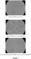

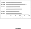


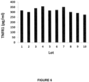
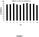
Classifications
-
- C—CHEMISTRY; METALLURGY
- C12—BIOCHEMISTRY; BEER; SPIRITS; WINE; VINEGAR; MICROBIOLOGY; ENZYMOLOGY; MUTATION OR GENETIC ENGINEERING
- C12N—MICROORGANISMS OR ENZYMES; COMPOSITIONS THEREOF; PROPAGATING, PRESERVING, OR MAINTAINING MICROORGANISMS; MUTATION OR GENETIC ENGINEERING; CULTURE MEDIA
- C12N5/00—Undifferentiated human, animal or plant cells, e.g. cell lines; Tissues; Cultivation or maintenance thereof; Culture media therefor
- C12N5/06—Animal cells or tissues; Human cells or tissues
- C12N5/0602—Vertebrate cells
- C12N5/0652—Cells of skeletal and connective tissues; Mesenchyme
- C12N5/0662—Stem cells
- C12N5/0663—Bone marrow mesenchymal stem cells (BM-MSC)
-
- A—HUMAN NECESSITIES
- A61—MEDICAL OR VETERINARY SCIENCE; HYGIENE
- A61K—PREPARATIONS FOR MEDICAL, DENTAL OR TOILETRY PURPOSES
- A61K35/00—Medicinal preparations containing materials or reaction products thereof with undetermined constitution
- A61K35/12—Materials from mammals; Compositions comprising non-specified tissues or cells; Compositions comprising non-embryonic stem cells; Genetically modified cells
- A61K35/28—Bone marrow; Haematopoietic stem cells; Mesenchymal stem cells of any origin, e.g. adipose-derived stem cells
-
- A—HUMAN NECESSITIES
- A61—MEDICAL OR VETERINARY SCIENCE; HYGIENE
- A61P—SPECIFIC THERAPEUTIC ACTIVITY OF CHEMICAL COMPOUNDS OR MEDICINAL PREPARATIONS
- A61P29/00—Non-central analgesic, antipyretic or antiinflammatory agents, e.g. antirheumatic agents; Non-steroidal antiinflammatory drugs [NSAID]
-
- C—CHEMISTRY; METALLURGY
- C12—BIOCHEMISTRY; BEER; SPIRITS; WINE; VINEGAR; MICROBIOLOGY; ENZYMOLOGY; MUTATION OR GENETIC ENGINEERING
- C12N—MICROORGANISMS OR ENZYMES; COMPOSITIONS THEREOF; PROPAGATING, PRESERVING, OR MAINTAINING MICROORGANISMS; MUTATION OR GENETIC ENGINEERING; CULTURE MEDIA
- C12N5/00—Undifferentiated human, animal or plant cells, e.g. cell lines; Tissues; Cultivation or maintenance thereof; Culture media therefor
- C12N5/06—Animal cells or tissues; Human cells or tissues
- C12N5/0602—Vertebrate cells
- C12N5/0652—Cells of skeletal and connective tissues; Mesenchyme
- C12N5/0662—Stem cells
-
- C—CHEMISTRY; METALLURGY
- C12—BIOCHEMISTRY; BEER; SPIRITS; WINE; VINEGAR; MICROBIOLOGY; ENZYMOLOGY; MUTATION OR GENETIC ENGINEERING
- C12N—MICROORGANISMS OR ENZYMES; COMPOSITIONS THEREOF; PROPAGATING, PRESERVING, OR MAINTAINING MICROORGANISMS; MUTATION OR GENETIC ENGINEERING; CULTURE MEDIA
- C12N2502/00—Coculture with; Conditioned medium produced by
- C12N2502/11—Coculture with; Conditioned medium produced by blood or immune system cells
- C12N2502/1157—Monocytes, macrophages
-
- C—CHEMISTRY; METALLURGY
- C12—BIOCHEMISTRY; BEER; SPIRITS; WINE; VINEGAR; MICROBIOLOGY; ENZYMOLOGY; MUTATION OR GENETIC ENGINEERING
- C12N—MICROORGANISMS OR ENZYMES; COMPOSITIONS THEREOF; PROPAGATING, PRESERVING, OR MAINTAINING MICROORGANISMS; MUTATION OR GENETIC ENGINEERING; CULTURE MEDIA
- C12N2523/00—Culture process characterised by temperature
Definitions
- the present disclosure relates to cellular therapy products comprising mesenchymal lineage precursor or stem cells and potency assay for these products.
- the present disclosure also relates to methods for treatment of immune or inflammatory disorders, and treatment or prevention of graft versus host disease (GVHD), or one or more symptoms associated with GVHD, by administration of mesenchymal lineage precursor or stem cells.
- GVHD graft versus host disease
- Physiochemical parameters for example, characterization of size, morphology, light-scattering properties, tensile strength, cell number, confluence, identification of phenotypic markers, secreted substances, genotype, gene expression profile
- physiochemical parameters cannot confirm that a product will be biologically active and potent (i.e., elicit the desired effect).
- biologic characterization takes into account the effect of the product on biologic systems, either modelled in vitro or in vivo in animals and ultimately in the clinic.
- Potency testing must demonstrate the relevant biologic activity or activities of the product. It is not a requirement for potency testing to reflect all of the product's biological functions, but it should indicate one or more relevant biological functions. It is expected that accuracy, sensitivity, specificity and reproducibility will be established for the analytic methods used in potency testing and that they be suitably robust.
- the Applicant has developed an off-the-shelf ex vivo expanded allogeneic mesenchymal lineage precursor or stem cell (MLPSC) product which has improved immunosuppressive activity.
- MPSC stem cell
- the present disclosure provides a composition comprising mesenchymal lineage precursor or stem cells or progeny thereof, wherein the mesenchymal lineage precursor or stem cells are cryopreserved and, after thawing, inhibit proliferation of activated T cells in a sample of PBMC by at least about 65%.
- the inhibition is measured by co-incubation of mesenchymal lineage precursor or stem cells with PBMC at a ratio of 1 mesenchymal lineage precursor or stem cell:5 PBMC, or less. For example, 1:10, 1:20, 1:30, 1:40, 1:50, 1:60, 1:70 1:80, 1:90, or 1 mesenchymal lineage precursor or stem cell: 100 PBMC, or less.
- co-incubation of mesenchymal lineage precursor or stem cells with PBMC at a ratio of 1 mesenchymal lineage precursor or stem cell:5 PBMC, or less inhibits T cell proliferation by at least about 65%, at least about 70%, at least about 75%, at least about 80%, at least about 85%, or at least about 90%.
- the inhibition of proliferation of activated T cells is measured by inhibition of IL-2R 2R ⁇ expression in the activated T cells.
- the mesenchymal lineage precursor or stem cell composition expresses TNFR1 in an amount of at least 110 pg/ml.
- the mesenchymal lineage precursor or stem cell composition expresses TNFR1 in an amount of at least 150 pg/ml, or at least 200 pg/ ml, or at least 250 pg/ml, or at least 300 pg/ml, or at least 320 pg/ml, or at least 330 pg/ml, or at least 340 pg/ml, or at least 350 pg/ml.
- the mesenchymal lineage precursor or stem cells express TNFR1 in an amount of at least 13 pg/10 6 cells.
- the mesenchymal lineage precursor or stem cells express TNFR1 in an amount of at least 15 pg/10 6 cells, or at least 20 pg/10 6 cells, or at least 25 pg/10 6 cells, or at least 30 pg/10 6 cells, or at least 35 pg/10 6 cells, or at least 40 pg/10 6 cells, or at least 45 pg/10 6 cells, or at least 50 pg/10 6 cells.
- the mesenchymal lineage precursor or stem cells are isolated by immunoselection and then culture expanded.
- the mesenchymal lineage precursor or stem cells are culture expanded. In one embodiment the mesenchymal lineage precursor or stem cells are isolated, or isolated and enriched, and culture expanded ex vivo or in vitro prior to cryopreservation. In another example, the mesenchymal lineage precursor or stem cells are isolated, or isolated and enriched, cryopreserved, thawed and subsequently culture expanded. In yet another example, the mesenchymal lineage precursor or stem cells are culture expanded prior to and after cryopreservation.
- the mesenchymal lineage precursor or stem cells comprise at least 5% of the cell population of the composition.
- the composition is cryopreserved in 42.5% Profreeze TM /50% ⁇ MEM/7.5% DMSO.
- the composition is cryopreserved in Plasmalyte-A, 25%HSA and DMSO.
- mesenchymal lineage precursor or stem cells which inhibit the proliferation of T cells by at least about 65% are particularly useful in inhibiting immune responses, and more particularly such mesenchymal lineage precursor or stem cells are useful in the prevention or treatment of graft versus host disease; solid organ transplant rejection such as, for example, heart transplant rejection, liver transplant rejection, pancreas transplant rejection, intestine transplant rejection, and kidney transplant rejection; and autoimmune diseases such as, for example, rheumatoid arthritis, multiple sclerosis, Type I diabetes, Crohn's disease, Guillain-Barre syndrome, lupus erythematosus, myasthenia gravis, optic neuritis, psoriasis, Graves' disease, Hashimoto's disease, Ord's thyroiditis, aplastic anemia, Reiter's syndrome, autoimmune hepatitis, primary biliary cirrhosis, anti
- the mesenchymal lineage precursor or stem cells are also useful for the treatment of inflammatory diseases, in particular T-cell mediated-inflammatory disorders.
- the present invention also provides a method for producing a population of mesenchymal lineage precursor or stem cells which comprises one or more of the following steps:
- the method comprises
- the method comprises
- the method comprises one or more or all of the steps outlined in Figure 2 .
- the mesenchymal lineage precursor or stem cells are isolated by immunoselection.
- the mesenchymal lineage precursor or stem cells isolated by immunoselection may be STRO-1+ mesenchymal precursor cells or progeny thereof.
- the mesenchymal lineage precursor or stem cells are isolated by plastic adherence technology.
- the mesenchymal lineage precursor or stem cells isolated by plastic adherence technology may be mesenchymal stem cells or progeny thereof.
- the present inventors have also developed a potency assay to measure the biological activity or therapeutic efficacy of cellular therapy products comprising mesenchymal lineage precursor or stem cells.
- the present disclosure also provides a method for determining the potency of mesenchymal lineage precursor or stem cells comprising:
- an amount of at least about 65% inhibition is indicative of the cells therapeutic efficacy in inhibiting immune responses.
- an amount of at least about 65% inhibition is indicative of the cells therapeutic efficacy in preventing or treating graft versus host disease.
- the present disclosure also provides a method for selecting potent mesenchymal lineage precursor or stem cells comprising:
- the assay method or selection method described above is used to determine the potency of an enriched population of mesenchymal lineage precursor stem cells.
- the mesenchymal lineage precursor or stem cells are enriched for mesenchymal stem cells.
- the mesenchymal lineage precursor or stem cells are enriched by selection of STRO-1+ cells.
- the mesenchymal lineage precursor cells are STRO-1 bright cells.
- the assay or selection method is used to determine the potency of or select an ex vivo or an in vitro expanded population of mesenchymal lineage precursor or stem cells.
- the mesenchymal lineage precursor or stem cells are isolated, or isolated and enriched, and culture expanded ex vivo or in vitro prior to cryopreservation.
- the mesenchymal lineage precursor or stem cells are isolated, or isolated and enriched, cryopreserved, thawed and subsequently culture expanded.
- the mesenchymal lineage precursor or stem cells are culture expanded prior to and after cryopreservation.
- the mesenchymal lineage precursor or stem cells are human mesenchymal lineage precursor or stem cells.
- the T cells are human T cells. In another embodiment, the T cells express CD4 and CD8. In another embodiment, the T cells express CD69 and/or CD137.
- the population comprising T cells is a population of peripheral blood mononuclear cells (PBMCs).
- PBMCs peripheral blood mononuclear cells
- the assay or selection method comprises culturing the mesenchymal lineage precursor or stem cells with T cells in a culture medium comprising one or more T cell stimulatory ligands.
- the culture medium comprises an anti-CD3 antibody or a fragment thereof and an anti-CD28 antibody or a fragment thereof.
- the method comprises culturing the mesenchymal lineage precursor or stem cells with T cells that have been stimulated and/or activated prior to co-culture with the mesenchymal lineage precursor cells.
- the assay or selection method comprises culturing the cells in DMEM supplemented with 10% FBS and 2mM glutamine and optionally, comprising one or more T cell stimulatory ligands.
- the method comprises co-culturing mesenchymal lineage precursor or stem cells and T cells at a ratio of about 1 mesenchymal lineage precursor or stem cell:2 T cells, or less.
- the method comprises co-culturing the cells for 60 to 84 hours prior to determining IL-2R ⁇ expression.
- the method comprises collecting the cells following co-culture and lysing them to produce a cell lysate.
- the method comprises determining the amount of IL-2R ⁇ in the cell lysate by enzyme-linked immunosorbent assay (ELISA).
- ELISA enzyme-linked immunosorbent assay
- the ELISA comprises:
- the method further comprises:
- the method further comprises: determining TNFR1 expression of the population of mesenchymal lineage precursor or stem cells, wherein an amount of ⁇ 100 pg/ml TNFR1 is indicative of biological activity or therapeutic efficacy of the mesenchymal lineage precursor or stem cells.
- the present disclosure also provides a method of inhibiting an immune response in a subject in need thereof, the method comprising administering a composition comprising the population of MLPSCs of the disclosure to the subject.
- the present disclosure also provides a method of preventing or treating an inflammatory disorder in a subject in need thereof, the method comprising administering a composition comprising MLPSCs of the present disclosure to the subject.
- the inflammatory disorder may be a a T-cell mediated inflammatory disorder.
- the present disclosure also provides a method of preventing, alleviating the development of, or treating graft versus host disease in a subject, comprising administering to the subject the composition comprising MLPSCs of the present disclosure to the subject.
- the present disclosure also provides a method of preventing graft versus host disease in a subject, the method comprising administering hematopoietic stem cells co-cultured with MLPSCs of the present disclosure to the subject.
- the composition is administered to the subject at a dose of less than 3 ⁇ 10 6 cells/kg body weight once per week (qw).
- the present disclosure also provides a method for preventing, alleviating the development of, or treating graft versus host disease (GVHD) in a mammalian subject, comprising administering to the subject, MLPSCs and/or progeny cells thereof at a dose of less than 3 ⁇ 10 6 cells/kg body weight once per week (qw).
- GVHD graft versus host disease
- the subject is administered MLPSCs at a dose of about 2 ⁇ 10 6 cells/kg body weight qw. In another embodiment, the subject is administered cells at a dose up to 2 ⁇ 10 6 cells/kg body weight qw. In another embodiment, the subject is administered a maximal dose of 2 ⁇ 10 6 cells/kg body weight qw.
- the terms "one week”, “weekly” or “qw” is intended to mean a period once every 7 days.
- the MLPSCs are administered as a single dose once per week (qw). In another embodiment, the MLPSCs are administered as a divided dose per week (i.e. 7 days). For example, the subject may receive two doses with each dose of 1 ⁇ 10 6 cells/kg body weight over the course of one week. In other embodiments, the subject may receive two or more doses per week wherein the total dose received is 2 ⁇ 10 6 cells/kg body weight.
- the graft comprises allogenic cells. In another embodiment, the graft comprises autologous cells.
- the MLPSCs administered to the subject are MPCs and/or progeny cells thereof.
- the MLPSCs administered to the subject are MSCs and/or progeny cells thereof.
- the MLPSCs and/or progeny cells thereof are delivered as a single dose qw. In another embodiment, the MLPSCs and/or progeny cells thereof are delivered as a divided dose over the course of one week.
- the mammalian subject is a human subject. In one embodiment, the subject is a pediatric subject. In another embodiment, the subject is an adult subject.
- the subject according to the present disclosure is one having a malignant or genetic disorder of the blood (e.g. cancer).
- the subject has received, is receiving or is about to receive a donor graft comprising hematopoietic cells.
- the graft comprising hematopoietic cells may be selected from the group consisting of blood, peripheral blood mononuclear cells (PBMCs), blood products, or solid organs in which hematopoietic cells are present.
- PBMCs peripheral blood mononuclear cells
- HSCs hematopoietic stem cells
- the subject has acute GVHD.
- Symptoms of acute GVHD are typically graded by standard clinical criteria ( Glucksberg H. et al. (1974) Transplantation 1974;18(4):295-304 ).
- the subject has received steroid therapy prior to administration of the mesenchymal lineage precursor or stem cells.
- the steroid is methylprednisolone.
- the steroid is administered to the subject at least three (3) days prior to administration of the MLPSCs and/or progeny cells thereof.
- the subject is administered MLPSCs and/or progeny cells thereof on the same day as receiving the graft (e.g. bone marrow or PBMCs).
- the graft e.g. bone marrow or PBMCs.
- the MLPSCs and/or progeny cells thereof may be administered to the subject at an appropriate time which may be during or following transplantation of the graft.
- the MLPSCs and/or progeny cells thereof may be administered to the subject beginning on the day of transplantation of the graft.
- the MLPSCs and/or progeny cells thereof may be administered to the subject prior to receiving the graft.
- the MLPSCs and/or progeny cells thereof may be administered within 7 days, within 5 days, within 3 days, or within 2 days prior to receiving the graft.
- the MLPSCs and/or progeny cells thereof are administered to the subject the day before receiving the graft.
- the MLPSCs and/or progeny cells thereof are administered to the subject after the subject has been determined to be steroid refractory. While there is no generally agreed upon definition of steroid-refractory acute GVHD, typically steroid refractory acute GVHD refers to GVHD that worsens after 3-5 days of steroid treatment, that does not improve after 5-7 days or that fails to remit completely after 14 days.
- the MLPSCs and/or progeny cells thereof are administered to the subject after at least three days of steroid or immunosuppressive treatment. In another example, the MLPSCs and/or progeny cells thereof are administered to the subject after at least one month of steroid or immunosuppressive treatment.
- a steroid refractory subject is one that has failed to respond to steroid treatment for grades B-D acute GVHD after at least three days of steroid (e.g. methylprednisolone or equivalent). In a further example, the subject has failed to respond to >1mg/kg/day of methylprednisolone or equivalent.
- the subject has received non-steroid immunosuppressive therapy prior to administration of MLPSCs and/or progeny cells thereof.
- the subject has received one or more non-steroid therapies selected from the group consisting of extracorporeal photophoresis (ECP), infliximab, ruxolitinib, mycophenolate mofetil (MMF), etanercept and basiliximab.
- ECP extracorporeal photophoresis
- MMF mycophenolate mofetil
- etanercept etanercept
- basiliximab basiliximab
- the subject is administered MLPSCs and/or progeny cells thereof until improvement in GVHD is observed. In another example, the subject is administered MLPSCs and/or progeny cells thereof until remission of GVHD is observed.
- the administration of MLPSCs and/or progeny cells thereof prevents, alleviates or treats an adverse event selected from one or more of infusion-related reaction, hypertension, vomiting, nausea, brachycardia and fever. In one example, the administration of MLPSCs and/or progeny cells thereof reduces the number of adverse events experienced by the subject compared to a subject who has not received MLPSCs and/or progeny cells thereof.
- the subject has acute GVHD Grade B, C or D.
- the GVHD involves the skin, gastrointestinal tract or liver or a combination of any one or more of these tissues.
- the GVHD is a result of a T cell immune response.
- the T cells are from a donor and the antigen is from the recipient.
- the T cells may be present in a transplant.
- the T cells are from the recipient and the antigen is from the donor.
- the mesenchymal lineage precursor or stem cells are genetically engineered to express a molecule to block co-stimulation of T-cells.
- the mesenchymal lineage precursor or stem cells have been expanded in culture prior to administration to the subject.
- the MLPSCs and/or progeny cells thereof are administered in the form of a pharmaceutically acceptable composition.
- the pharmaceutically acceptable composition comprises a pharmaceutically acceptable carrier and/or excipient.
- the MLPSCs and/or progeny cells thereof may be administered weekly to the subject for each of four (4) consecutive weeks. In another example, MLPSCs and/or progeny cells thereof are administered to the subject weekly for each of eight consecutive weeks. In another example, the subject's GVHD is assessed after the four once-weekly infusions and if the subject's GVHD response is partial or mixed, the subject is eligible to receive an additional four once-weekly infusions. In one example, the subject is administered up to a maximum of eight MLPSCs and/or progeny cells thereof. In another example, the subject is administered a total of eight MPCs and/or progeny cells thereof. In another example, the subject is administered MLPSCs and/or progeny cells thereof infusions on a weekly basis until at least a partial or complete response is observed.
- the GVHD status or grading in the subject is assessed at baseline (screening), day 0.
- the GVHD status or grading in the subject is assessed at Day 14, Day 28, Day 56 and Day 100. In another embodiment, GVHD is assessed at some of these days, for example, baseline (Day 0), Day 28 and Day 100.
- the subject may be assessed as having a complete response, partial response, mixed response, worsening response or no response.
- the present disclosure also provides a composition comprising MLPSCs and/or progeny cells thereof at a dose of less than 3 ⁇ 10 6 cells/kg body weight on a weekly basis for use in preventing, alleviating the development of, or treating graft versus host disease (GVHD) in a mammalian subject.
- GVHD graft versus host disease
- the composition comprises MLPSCs and/or progeny cells thereof at a dose of about 2 ⁇ 10 6 cells/kg body weight. In another embodiment, the composition comprises MPCs and/or progeny cells thereof at a dose up to 2 ⁇ 10 6 cells/kg body weight. In another embodiment the composition comprises a maximal dose of 2 ⁇ 10 6 cells/kg body weight thereof.
- the composition is a pharmaceutical composition.
- the present disclosure also provides use of MLPSCs and/or progeny cells thereof at a dose of less than 3 ⁇ 10 6 cells/kg body weight in the manufacture of a medicament for preventing development of, or treating GVHD in a mammalian subject.
- the MLPSCs and/or progeny cells thereof are intended for administration to a subject in need thereof once per week (qw).
- the medicament comprises MLPSCs and/or progeny cells thereof at a dose of about 2 ⁇ 10 6 cells/kg body weight. In another example, the medicament comprises MLPSCs and/or progeny cells thereof at a dose up to 2 ⁇ 10 6 cells/kg body weight. In another example, the medicament comprises a maximal dose of 2 ⁇ 10 6 cells/kg body weight.
- the mesenchymal lineage precursor or stem cells is a population of cells enriched for STRO-1 bright cells.
- the mesenchymal lineage precursor or stem cells is a population of cells enriched for one or more additional markers selected from TNAP+, VCAM-1+, THY-1+, STRO-2+, STRO-4+ (HSP-90 ⁇ ) and/or CD146+.
- the mesenchymal lineage precursor or stem cells is a population of mesenchymal stem cells
- the MLPSCs and/or progeny cells thereof are administered systemically.
- the MLPSCs and/or progeny cells thereof may be administered intravenously, intra-arterially, intramuscularly, subcutaneously, into an aorta, into an atrium or ventricle of the heart or into a blood vessel connected to an organ, e.g., an abdominal aorta, a superior mesenteric artery, a pancreatic duodenal artery or a splenic artery.
- the methods of the disclosure further comprise administering an immunosuppressive agent.
- the immunosuppressive agent may be administered for a time sufficient to permit the transplanted hematopoietic cells to be functional.
- the immunosuppressive agent may be selected from one or more of the following, including but not limited to corticosteroids such as prednisone, budesonide and prednisolone; calcineurin inhibitors such as cyclosporine and tacrolimus; mTOR inhibitors such as sirolimus and everolimus; IMDH inhibitors such as azathioprine, leflunomide and mycophenolate; a biologic such as abatacept, adalimumab, etanercept, infliximab or rituximab.
- the immunosuppressive agent is cyclosporine.
- the cyclosporine may be administered at a dosage of from 5 to 40 mg/kg body wt.
- composition of matter, group of steps or group of compositions of matter shall be taken to encompass one and a plurality (i.e., one or more) of those steps, compositions of matter, group of steps or group of compositions of matter.
- stem cells, cell culture, and surgical techniques utilized in the present disclosure are standard procedures, well known to those skilled in the art. Such techniques are described and explained throughout the literature in sources such as Perbal, 1984; Sambrook & Green, 2012; Brown, 1991; Glover & Hames, 1995 and 1996; Ausubel., 1987 including all updates untill present; Harlow & Lane, 1988; and Coligan et al., 1991 including all updates until present.
- graft versus host disease refers to a complication of allogeneic hematopoietic cell transplantation in which the tissues of the host, most frequently the skin, liver and intestine are damaged by lymphocyte from the donor. This disease is discussed in more detail below.
- subject refers to a mammal including human and non-human animals. More particularly, the mammal is a human.
- Terms such as “subject”, “patient” or “individual” are terms that can, in context, be used interchangeably in the present disclosure. In certain examples, the subject may be an adult or a child (pediatric) subject.
- an “effective amount” refers to at least an amount effective, at dosages and for periods of time necessary, to achieve the desired therapeutic or prophylactic result.
- An effective amount can be provided in one or more administrations.
- the term "effective amount” is used to refer to an amount necessary to effect treatment of a disease or condition as hereinbefore described.
- the effective amount may vary according to the disease or condition to be treated and also according to the weight, age, racial background, sex, health and/or physical condition and other factors relevant to the mammal being treated.
- the effective amount will fall within a relatively broad range (e.g. a "dosage" range) that can be determined through routine trial and experimentation by a medical practitioner.
- the effective amount can be administered in a single dose or in a dose repeated once or several times over a treatment period.
- a “therapeutically effective amount” is at least the minimum concentration required to effect a measurable improvement of a particular disorder (e.g. GVHD).
- a therapeutically effective amount herein may vary according to factors such as the disease state, age, sex, and weight of the patient, and the ability of the cellular composition to elicit a desired response in the individual.
- a therapeutically effective amount is also one in which any toxic or detrimental effects of the composition are outweighed by the therapeutically beneficial effects.
- a therapeutically effective amount can reduce the severity, inhibit or delay progression of GVHD and/or relieve to some extent one or more of the symptoms associated with the disorder.
- preventing means preventing, delaying and/or reducing the severity of the symptoms associated with GVHD. This is distinguished from “treatment” which occurs following the onset of the first symptoms of GVHD.
- treatment refers to clinical intervention designed to alter the natural course of the individual or cell being treated during the course of clinical pathology. Desirable effects of treatment include decreasing the rate of disease progression, ameliorating or palliating the disease state, and remission or improved prognosis. An individual is successfully "treated", for example, if one or more symptoms associated with a disease are mitigated or eliminated.
- CR complete response
- partial response or "PR” as referred to herein is defined as improvement by at least one GVHD stage in all initial GVHD target organs without complete resolution and without worsening in any other GVHD target organs, without secondary GVHD therapy.
- no response or "NR” as referred to herein is defined as the same grade of GVHD or progression of GVHD in any organ (e.g. deterioration in at least one evaluable organ symptom by one stage or more) or death, or the addition of secondary GVHD therapy.
- GVHD refers to worsening of GVHD progression in at least one organ with or without amelioration in any organ.
- VGPR very good partial response
- MR mixed response
- progression refers to deterioration in at least one organ system by one stage or more with no improvement in any other organ.
- adult as used herein means a human subject of 18 years of age and older.
- the term "pediatric” as used herein means a human subject ranging in age from birth up to and including 17 years of age.
- GVHD acute GVHD
- graft e.g. bone marrow transplant. It can occur within a matter of days of receiving a graft.
- chronic GVHD refers to GVHD that usually commences more than 3 months after receiving a graft. Symptoms of chronic GVHD can last a lifetime.
- graft refers to a biological sample selected from bone marrow, blood (e.g. whole blood or peripheral blood mononuclear cells (PBMCs), blood products, or solid organs in which hematopoietic cells are present.
- blood e.g. whole blood or peripheral blood mononuclear cells (PBMCs)
- PBMCs peripheral blood mononuclear cells
- solid organs in which hematopoietic cells are present.
- allogeneic refers to a graft (e.g. hematopoietic cells) which are donated by an individual whose genetic characteristics differ from those of the recipient, especially in regards to the major histocompatibility complex (MHC) and minor histocompatibility agents expressed on the surface of the individual's cells.
- MHC major histocompatibility complex
- autologous refers to a graft (e.g. hematopoietic cells present in the bone marrow or peripheral blood) that uses the subject's own cells.
- the cells are usually harvested in advance of the subject undergoing treatment (e.g. with chemotherapy), stored and then re-infused back into the subject.
- steroid refractory refers to GVHD that worsens after 3-5 days of steroid treatment that does not improve after 5-7 days or that fails to remit completely after 14 days.
- the term about refers to +/- 10%, more preferably +/- 5%, of the designated value.
- mesenchymal lineage precursor or stem cells refers to undifferentiated multipotent cells that have the capacity to self-renew while maintaining multipotency and the capacity to differentiate into a number of cell types either of mesenchymal origin, for example, osteoblasts, chondrocytes, adipocytes, stromal cells, fibroblasts and tendons, or non-mesodermal origin, for example, hepatocytes, neural cells and epithelial cells.
- mesenchymal lineage precursor or stem cells includes both parent cells and their undifferentiated progeny.
- the term also includes mesenchymal precursor cells (MPC), multipotent stromal cells, mesenchymal stem cells, perivascular mesenchymal precursor cells, and their undifferentiated progeny.
- Mesenchymal lineage precursor or stem cells can be autologous, allogeneic, xenogeneic, syngeneic or isogeneic. Autologous cells are isolated from the same individual to which they will be reimplanted. Allogeneic cells are isolated from a donor of the same species. Xenogeneic cells are isolated from a donor of another species. Syngeneic or isogeneic cells are isolated from genetically identical organisms, such as twins, clones, or highly inbred research animal models.
- Mesenchymal lineage precursor or stem cells reside primarily in the bone marrow, but have also been shown to be present in diverse host tissues including, for example, cord blood and umbilical cord, adult peripheral blood, adipose tissue, trabecular bone and dental pulp.
- Mesenchymal lineage precursor or stem cells can be isolated from host tissues and enriched for by immunoselection.
- a bone marrow aspirate from a subject may be further treated with an antibody to STRO-1 or TNAP to enable selection of mesenchymal lineage precursor or stem cells.
- the mesenchymal lineage precursor or stem cells can be enriched for by using the STRO-1 antibody described in Simmons & Torok-Storb, 1991.
- STRO-1+ cells are cells found in bone marrow, blood, dental pulp cells, adipose tissue, skin, spleen, pancreas, brain, kidney, liver, heart, retina, brain, hair follicles, intestine, lung, lymph node, thymus, bone, ligament, tendon, skeletal muscle, dermis, and periosteum; and are capable of differentiating into germ lines such as mesoderm and/or endoderm and/or ectoderm.
- STRO-1+ cells are capable of differentiating into a large number of cell types including, but not limited to, adipose, osseous, cartilaginous, elastic, muscular, and fibrous connective tissues.
- the specific lineage-commitment and differentiation pathway which these cells enter depends upon various influences from mechanical influences and/or endogenous bioactive factors, such as growth factors, cytokines, and/or local microenvironmental conditions established by host tissues.
- enriched describes a population of cells in which the proportion of one particular cell type or the proportion of a number of particular cell types is increased when compared with an untreated population of the cells (e.g., cells in their native environment).
- a population enriched for STRO-1+ cells comprises at least about 0.1% or 0.5% or 1% or 2% or 5% or 10% or 15% or 20% or 25% or 30% or 50% or 75% STRO-1+ cells.
- the term “population of cells enriched for STRO-1+ cells” will be taken to provide explicit support for the term “population of cells comprising X% STRO-1+ cells", wherein X% is a percentage as recited herein.
- the STRO-1+ cells can, in some examples, form clonogenic colonies, for example, CFU-F (fibroblasts) or a subset thereof (e.g., 50% or 60% or 70% or 70% or 90% or 95%) can have this activity.
- the population of cells is enriched from a cell preparation comprising STRO-1+ cells in a selectable form.
- the term "selectable form" will be understood to mean that the cells express a marker (e.g., a cell surface marker) permitting selection of the STRO-1+ cells.
- the marker can be STRO-1, but need not be.
- cells e.g., MPCs
- an indication that cells are STRO-1+ does not mean that the cells are selected by STRO-1 expression.
- the cells are selected based on at least STRO-3 expression, e.g., they are STRO-3+ (TNAP+).
- STRO-1+ cells can be selected from or isolated from or enriched from a large variety of sources. That said, in some examples, these terms provide support for selection from any tissue comprising STRO-1+ cells or vascularized tissue or tissue comprising pericytes (e.g., STRO-1+ pericytes) or any one or more of the tissues recited herein.
- the mesenchymal lineage precursor or stem cells of the disclosure express one or more markers individually or collectively selected from the group consisting of TNAP+, VCAM-1+, THY-1+, STRO-2+, STRO-4+ (HSP-90 ⁇ ), CD45+, CD146+, 3G5+.
- a cell that is referred to as being "positive" for a given marker may express either a low (lo or dim or dull), intermediate (median) or a high (bright, bri) level of that marker depending on the degree to which the marker is present on the cell surface, where the terms relate to intensity of fluorescence or other marker used in the sorting process of the cells or flow cytometric analysis of the cells.
- the distinction of low (lo or dim or dull), intermediate (median), or high (bright, bri) will be understood in the context of the marker used on a particular cell population being sorted or analysed.
- a cell that is referred to as being "negative” for a given marker is not necessarily completely absent from that cell. This term means that the marker is expressed at a relatively very low level by that cell, and that it generates a very low signal when detectably labeled or is undetectable above background levels, for example, levels detected using an isotype control antibody.
- bri refers to a marker on a cell surface that generates a relatively high signal when detectably labeled.
- target marker protein for example, the antigen recognized by a STRO-1 antibody
- STRO-1 bri cells produce a greater fluorescent signal, when labeled with a FITC-conjugated STRO-1 antibody as determined by fluorescence activated cell sorting (FACS) analysis, than non-bright cells (STRO-1 lo/dim/dull/intermediate/median ).
- the mesenchymal lineage precursor or stem cells are isolated from bone marrow and enriched for by selection of STRO-1+ cells.
- "bright" cells constitute at least about 0.1% of the most brightly labeled bone marrow mononuclear cells contained in the starting sample. In other examples, "bright” cells constitute at least about 0.1%, at least about 0.5%, at least about 1%, at least about 1.5%, or at least about 2%, of the most brightly labeled bone marrow mononuclear cells contained in the starting sample.
- STRO-1 bright cells have 2 log magnitude higher expression of STRO-1 surface expression relative to "background", namely cells that are STRO-1-.
- STRO-1 lo/dim/dull and/or STRO-1 intermediate/median cells have less than 2 log magnitude higher expression of STRO-1 surface expression, typically about 1 log or less than "background”.
- the STRO-1+ cells are STRO-1 bright .
- the STRO-1 bright cells are preferentially enriched relative to STRO-1 lo/dim/dull or STRO-1 intermediate/median cells.
- the STRO-1 bright cells are additionally one or more of TNAP+, VCAM-1+, THY-1+, STRO-2+, STRO-4+ (HSP-90 ⁇ ) and/or CD146+.
- the cells are selected for one or more of the foregoing markers and/or shown to express one or more of the foregoing markers.
- a cell shown to express a marker need not be specifically tested, rather previously enriched or isolated cells can be tested and subsequently used, isolated or enriched cells can be reasonably assumed to also express the same marker.
- the STRO-1 bright cells are perivascular mesenchymal precursor cells as defined in WO 2004/85630 , characterized by the presence of the perivascular marker 3G5.
- TNAP tissue non-specific alkaline phosphatase
- LAP liver isoform
- BAP bone isoform
- KAP kidney isoform
- the TNAP is BAP.
- TNAP refers to a molecule which can bind the STRO-3 antibody produced by the hybridoma cell line deposited with ATCC on 19 December 2005 under the provisions of the Budapest Treaty under deposit accession number PTA-7282.
- the STRO-1+ cells are capable of giving rise to clonogenic CFU-F.
- a significant proportion of the STRO-1+ cells are capable of differentiation into at least two different germ lines.
- the lineages to which the cells may be committed include bone precursor cells; hepatocyte progenitors, which are multipotent for bile duct epithelial cells and hepatocytes; neural restricted cells, which can generate glial cell precursors that progress to oligodendrocytes and astrocytes; neuronal precursors that progress to neurons; precursors for cardiac muscle and cardiomyocytes, glucose-responsive insulin secreting pancreatic beta cell lines.
- lineages include, but are not limited to, odontoblasts, dentin-producing cells and chondrocytes, and precursor cells of the following: retinal pigment epithelial cells, fibroblasts, skin cells such as keratinocytes, dendritic cells, hair follicle cells, renal duct epithelial cells, smooth and skeletal muscle cells, testicular progenitors, vascular endothelial cells, tendon, ligament, cartilage, adipocyte, fibroblast, marrow stroma, cardiac muscle, smooth muscle, skeletal muscle, pericyte, vascular, epithelial, glial, neuronal, astrocyte and oligodendrocyte cells.
- the mesenchymal lineage precursor or stem cells are mesenchymal stem cells (MSCs).
- the MSCs may be a homogeneous composition or may be a mixed cell population enriched in MSCs.
- Homogeneous MSC compositions may be obtained by culturing adherent bone marrow or periosteal cells, and the MSCs may be identified by specific cell surface markers which are identified with unique monoclonal antibodies.
- a method for obtaining a cell population enriched in MSCs using plastic adherence technology is described, for example, in US patent 5486359 .
- MSC prepared by conventional plastic adherence isolation relies on the non-specific plastic adherent properties of CFU-F.
- Alternative sources for MSCs include, but are not limited to, blood, skin, cord blood, muscle, fat, bone, and perichondrium.
- the mesenchymal lineage precursor or stem cells may be cryopreserved prior to administration to a subject.
- the mesenchymal lineage precursor or stem cells are obtained from a master cell bank derived from mesenchymal lineage precursor or stem cells enriched from the bone marrow of healthy volunteers.
- the use of mesenchymal lineage precursor or stem cells derived from such a source is particularly advantageous for subjects who do not have an appropriate family member available who can serve as the mesenchymal lineage precursor or stem cell donor, or are in need of immediate treatment and are at high risk of relapse, disease-related decline or death, during the time it takes to generate mesenchymal lineage precursor or stem cells.
- mesenchymal precursor cells of the disclosure have unexpectedly high potency in terms of their ability to inhibit T cell proliferation after cryopreservation and thawing.
- prior publications teach that cryopreserved mesenchymal stem cells display impaired immunosuppressive properties following thawing (Francois et al., 2012; Chinnadurai et al., 2016).
- the isolated or enriched mesenchymal lineage precursor or stem cells can be expanded ex vivo or in vitro by culture.
- the isolated or enriched mesenchymal lineage precursor or stem cells can be cryopreserved, thawed and subsequently or further expanded ex vivo or in vitro by culture.
- the cultured mesenchymal lineage precursor or stem cells are phenotypically different to cells in vivo. For example, in one embodiment they express one or more of the following markers, CD44, NG2, DC146 and CD140b.
- the cultured mesenchymal lineage precursor or stem cells are biologically different to cells in vivo, having a higher rate of proliferation compared to the largely non-cycling (quiescent) cells in vivo.
- a population of cells enriched for mesenchymal lineage precursor or stem cells is seeded at about 6000 to 7000 viable cells/cm 2 in serum-supplemented culture medium, for example, Dulbecco's Modified Eagle medium (DMEM) supplemented with 10% fetal bovine serum (FBS) and 2mM glutamine, and allowed to adhere to the culture vessel overnight at 37°C, 20% O 2 .
- serum-supplemented culture medium for example, Dulbecco's Modified Eagle medium (DMEM) supplemented with 10% fetal bovine serum (FBS) and 2mM glutamine
- the cells are seeded at about 6000, 6100, 6200, 6300, 6400, 6500, 6600, 6700, 6800, 6810, 6820, 6830, 6840, 6850, 6860, 6870, 6880, 6890, 6890, 6900, 6910, 6920, 6930, 6940, 6970, 6980, 6990, or 7000 viable cells/cm 2 , preferably at about 6850 to 6860 viable cells/cm 2 .
- the culture medium is subsequently replaced and the cells cultured for a total of 68 to 72 hours at 37°C, 5% O 2 prior to co-culturing with T cells and determining the amount of IL-2R ⁇ expressed by the T cells.
- mesenchymal lineage precursor or stem cells express Ang1 in an amount of at least 0.1 ⁇ g/106 cells.
- MLPSCs express Ang1 in an amount of at least 0.2 ⁇ g/106 cells, 0.3 ⁇ g/106 cells, 0.4 ⁇ g/10 6 cells, 0.5 ⁇ g/10 6 cells, 0.6 ⁇ g/10 6 cells, 0.7 ⁇ g/10 6 cells, 0.8 ⁇ g/10 6 cells, 0.9 ⁇ g/10 6 cells, 1 ⁇ g/10 6 cells, 1.1 ⁇ g/10 6 cells, 1.2 ⁇ g/10 6 cells, 1.3 ⁇ g/10 6 cells, 1.4 ⁇ g/10 6 cells, 1.5 ⁇ g/10 6 cells.
- MLPSCs express VEGF in an amount less than about 0.05 ⁇ g/10 6 cells.
- mesenchymal precursor cells express VEGF in an amount less than about 0.05 ⁇ g/10 6 cells, 0.04 ⁇ g/10 6 cells, 0.03 ⁇ g/10 6 cells, 0.02 ⁇ g/10 6 cells, 0.01 ⁇ g/10 6 cells, 0.009 ⁇ g/10 6 cells, 0.008 ⁇ g/10 6 cells, 0.007 ⁇ g/10 6 cells, 0.006 ⁇ g/10 6 cells, 0.005 ⁇ g/10 6 cells, 0.004 ⁇ g/10 6 cells, 0.003 ⁇ g/10 6 cells, 0.002 ⁇ g/10 6 cells, 0.001 ⁇ g/10 6 cells.
- the amount of cellular Ang1 and/or VEGF that is expressed in a composition or culture of MLPSCs may be determined by methods known to those skilled in the art. Such methods include, but are not limited to, quantitative assays such as quantitative ELISA assays, for example.
- a cell lysate from a culture of mesenchymal precursor cells is added to a well of an ELISA plate.
- the well may be coated with a primary antibody, either a monoclonal or a polyclonal antibody(ies), against the Ang1 or VEGF.
- the well then is washed, and then contacted with a secondary antibody, either a monoclonal or a polyclonal antibody(ies) , against the primary antibody.
- the secondary antibody is conjugated to an appropriate enzyme, such as horseradish peroxidase, for example.
- the well then may be incubated, and then is washed after the incubation period.
- the wells then are contacted with an appropriate substrate for the enzyme conjugated to the secondary antibody, such as one or more chromogens. Chromogens which may be employed include, but are not limited to, hydrogen peroxide and tetramethylbenzidine.
- the substrate(s) is (are) added, the well is incubated for an appropriate period of time.
- a "stop" solution is added to the well in order to stop the reaction of the enzyme with the substrate(s).
- the optical density (OD) of the sample is then measured.
- the optical density of the sample is correlated to the optical densities of samples containing known amounts of Ang1 or VEGF in order to determine the amount of Ang1 or VEGF expressed by the culture of stem cells being tested.
- MLPSCs express Ang1:VEGF at a ratio of at least about 2:1.
- mesenchymal precursor cells express Ang1:VEGF at a ratio of at least about 10:1, 15:1, 20:1, 21:1, 22:1, 23:1, 24:1, 25:1, 26:1, 27:1, 28:1, 29:1, 30:1, 31:1, 32:1, 33:1, 34:1, 35:1, 50:1.
- the MLPSCs of the present disclosure are not genetically modified to express Ang1 and/or VEGF at an above exemplified level or ratio.
- Cells that are not genetically modified to express Ang1 and/or VEGF have not been modified by transfection with a nucleic acid expressing or encoding Ang1 and/or VEGF.
- a mesenchymal precursor cell transfected with a nucleic acid encoding Ang1 and/or VEGF would be considered genetically modified.
- cells not genetically modified to express Ang1 and/or VEGF naturally express Ang1 and/or VEGF to some extent without transfection with a nucleic acid encoding Ang1 and/or VEGF1.
- the potency assay of the disclosure requires co-culture of the mesenchymal lineage precursor or stem cells with T-cells.
- the mesenchymal lineage precursor or stem cells are co-cultured with T-cells in a culture medium comprising at least one T-cell stimulatory ligand.
- the T cells are activated. The T cells may be first stimulated or activated prior to co-culture with the mesenchymal lineage precursor or stem cells.
- T-cell refers to a thymus-derived cell that participates in a variety of cell-mediated immune reactions.
- stimulation means a primary response induced by binding of a stimulatory molecule (e.g., a TCR/CD3 complex) with its cognate ligand thereby mediating a signal transduction event, such as, but not limited to, signal transduction via the TCR/CD3 complex.
- a stimulatory molecule e.g., a TCR/CD3 complex
- Stimulation can mediate altered expression of certain molecules, such as downregulation of TGF- ⁇ , and/or reorganization of cytoskeletal structures, and the like.
- activation refers to the state of a T cell that has been sufficiently stimulated to induce detectable cellular proliferation. Activation can also be associated with induced cytokine production, and detectable effector functions.
- activated T cells refers to, among other things, T cells that are undergoing cell division.
- T cells that have recently been activated will typically express a series of activation markers at different time points following activation.
- Activation markers include receptors such as chemokine and cytokine receptors, adhesion molecules, co-stimulatory molecules, and MHC-class II proteins.
- Flow cytometry can be used to evaluate various types of surface or intracellular markers that indicate the activation status of T cells.
- Two of the most commonly used immediate early activation markers for assessing the activation status of human peripheral blood mononuclear (PBMC) T cells are CD69 and CD40L.
- CD69 (AIM, Leu23, MLR3) is a signaling membrane glycoprotein involved in inducing T cell proliferation. CD69 is typically expressed at very low levels on resting CD4+ or CD8+ T cells in PBMC ( ⁇ 5-10%), and is one of the earliest assessable activation markers, being rapidly upregulated on CD4+ or CD8+ T cells within 1 hour of TCR stimulation or other T cell activators such as phorbol esters via a protein kinase C (PKC) dependent pathway. Expression of CD69 typically peaks by 16-24 hours and then declines, being barely detectable 72 hours after the stimulus has been withdrawn.
- PKC protein kinase C
- CD40L (CD154) is a member of the TNF-receptor superfamily that functions as a co-stimulatory molecule by binding CD40 which is constitutively expressed on antigen presenting cells (APCs).
- the CD40L-CD40 ligation results in the activation of multiple downstream pathways including the MAPK (JNK, p38, ERK1/2), NF- ⁇ B, and STAT3 transcription factors.
- CD40L expression is quickly upregulated within 1-2 hours after TCR stimulation via the transcription factors NFAT and AP-1.
- CD40L expression peaks near 6 hours after stimulation, and declines by 16-24hrs. CD40L expression however is biphasic, and the addition of anti-CD28 or IL-2 along with TCR stimulation typically leads to sustained expression for several days.
- telomere binding partner e.g., a stimulatory and/or costimulatory molecule present on a T cell
- stimulatory ligand means a ligand that can specifically bind with a cognate binding partner (referred to herein as a "stimulatory molecule") on a T cell, thereby mediating a primary response by the T cell.
- Stimulatory ligands are well-known in the art and encompass, inter alia, an MHC Class I molecule loaded with a peptide, an anti-CD3 antibody, a superagonist anti-CD28 antibody, and a superagonist anti-CD2 antibody.
- the stimulatory ligand may be used in soluble form, expressed or attached to the surface of a cell or immobilized on a surface.
- superagonist antibody means an antibody that specifically binds with a molecule on a T cell and can mediate a primary activation signal event in a T cell without interaction of the TCR/CD3 complex or CD2 on the T cell.
- exemplary superagonist antibodies include, but are not limited to, a superagonist anti-CD28 antibody and a superagonist anti-CD2 antibody.
- an anti-CD2 antibody, or an anti-CD28 antibody, and the like is a co-stimulatory ligand as defined elsewhere herein, and provides a co-stimulatory signal rather than a primary activation signal.
- stimulation molecule means a molecule on a T cell that specifically binds with a cognate stimulatory ligand.
- co-stimulatory signal refers to a signal, which in combination with a primary signal, such as TCR/CD3 ligation, mediates a T cell response, such as, but not limited to, activation, proliferation, differentiation into effector cells, induction of cytotoxicity or cytokine secretion.
- co-stimulatory ligand includes a molecule on an antigen presenting cell (APC) (e.g., a dendritic cell, B cell, and the like) or an artificial APC (aAPC) that specifically binds a cognate co-stimulatory molecule on a T cell, thereby providing a signal which, in addition to the primary signal provided by, for instance, binding of a TCR/CD3 complex with an MHC molecule loaded with peptide, mediates a T cell response, including, but not limited to, activation, proliferation, differentiation into effector cells, induction of cytotoxicity or cytokine secretion.
- APC antigen presenting cell
- aAPC artificial APC
- a co-stimulatory ligand can include, but is not limited to, CD7, B7-1 (CD80), B7-2 (CD86), PD-L1, PD-L2, 4-1BBL, OX40L, inducible costimulatory ligand (ICOS-L), intercellular adhesion molecule (ICAM), CD30L, CD40, CD70, CD83, HLA-G, MICA, MICB, HVEM, lymphotoxin beta receptor, 3/TR6, ILT3, ILT4, HVEM, an agonist or antibody that binds Toll ligand receptor and a ligand that specifically binds with B7-H3.
- a co-stimulatory ligand also encompasses, inter alia, an antibody that specifically binds with a co-stimulatory molecule present on a T cell, such as, but not limited to, CD27, CD28, 4-1BB, OX40, CD30, CD40, PD-1, ICOS, lymphocyte function-associated antigen- 1 (LFA-1), CD2, CD7, LIGHT, NKG2C, B7-H3, and a ligand that specifically binds with CD83.
- LFA-1 lymphocyte function-associated antigen- 1
- aAPC as used herein includes, but is not limited to, cell-based aAPCs, bead-based APCs, microparticle aAPCs, and nanoparticle aAPCs.
- Materials which have been used include glass, poly (glycolic acid), poly(lactic-co-glycolic acid), iron-oxide, liposomes, lipid bilayers, sepharose, and polystyrene.
- the aAPC comprises a stimulatory ligand, for example, a stimulatory ligand that specifically binds with a TCR/CD3 complex such that a primary signal is transduced.
- the aAPC may further comprise at least one co-stimulatory ligand that specifically binds with at least one co-stimulatory molecule present on a T cell.
- the term "antibody” includes a protein capable of specifically binding to a stimulatory molecule on a T cell by virtue of an antigen binding domain contained within a Fv.
- This term includes four chain antibodies (e.g., two light (L) chains and two heavy (H) chains), recombinant or modified antibodies (e.g., chimeric antibodies, humanized antibodies, human antibodies, CDR-grafted antibodies, primatized antibodies, de-immunized antibodies, synhumanized antibodies, half-antibodies, bispecific antibodies).
- variable region refers to the portions of the light and/or heavy chains of an antibody as defined herein that is capable of specifically binding to an antigen and, includes amino acid sequences of complementarity determining regions (CDRs), that is, CDR1, CDR2, and CDR3, and framework regions (FRs).
- CDRs complementarity determining regions
- FRs framework regions
- the variable region comprises three or four FRs (e.g., FR1, FR2, FR3 and optionally FR4) together with three CDRs.
- V H refers to the variable region of the heavy chain.
- V L refers to the variable region of the light chain.
- CDRs complementarity determining regions
- CDR1, CDR2, and CDR3 refers to the amino acid residues of an antibody variable region the presence of which are major contributors to specific antigen binding.
- Each variable region domain typically has three CDR regions identified as CDR1, CDR2 and CDR3.
- FRs Framework regions
- the term "Fv” shall be taken to mean any protein, whether comprised of multiple polypeptides or a single polypeptide, in which a V L and a V H associate and form a complex having an antigen binding domain that is capable of specifically binding to an antigen.
- the V H and the V L which form the antigen binding domain can be in a single polypeptide chain or in different polypeptide chains.
- a Fv of the disclosure (as well as any protein of the disclosure) may have multiple antigen binding domains which may or may not bind the same antigen. This term shall be understood to encompass fragments directly derived from an antibody as well as proteins corresponding to such a fragment produced using recombinant means.
- the V H is not linked to a heavy chain constant domain (C H ) 1 and/or the V L is not linked to a light chain constant domain (C L ).
- exemplary Fv containing polypeptides or proteins include a Fab fragment, a Fab' fragment, a F(ab') fragment, a scFv, a diabody, a triabody, a tetrabody or higher order complex, or any of the foregoing linked to a constant region or domain thereof, for example, C H 2 or C H 3 domain, for example, a minibody.
- a "Fab fragment” consists of a monovalent antigen-binding fragment of an immunoglobulin, and can be produced by digestion of a whole antibody with the enzyme papain, to yield a fragment consisting of an intact light chain and a portion of a heavy chain or can be produced using recombinant means.
- a "Fab' fragment" of an antibody can be obtained by treating a whole antibody with pepsin, followed by reduction, to yield a molecule consisting of an intact light chain and a portion of a heavy chain comprising a V H and a single constant domain. Two Fab' fragments are obtained per antibody treated in this manner. A Fab' fragment can also be produced by recombinant means.
- a "F(ab') 2 fragment" of an antibody consists of a dimer of two Fab' fragments held together by two disulfide bonds, and is obtained by treating a whole antibody molecule with the enzyme pepsin, without subsequent reduction.
- a "Fab 2" fragment is a recombinant fragment comprising two Fab fragments linked using, for example, a leucine zipper or a C H 3 domain.
- a “single chain Fv” or “scFv” is a recombinant molecule containing the variable region fragment (Fv) of an antibody in which the variable region of the light chain and the variable region of the heavy chain are covalently linked by a suitable, flexible polypeptide linker.
- the T cells may be stimulated by a single agent.
- T cells are stimulated with two agents, one that induces a primary signal and a second that is a co-stimulatory signal.
- Ligands useful for stimulating a single signal or stimulating a primary signal and an accessory molecule that stimulates a second signal may be used in soluble form, expressed or attached to the surface of a cell or immobilized on a surface.
- the surface may be any surface capable of having an agent/ligand bound thereto or integrated into and that is biocompatible, that is, substantially non-toxic to the target cells to be stimulated.
- the biocompatible surface may be biodegradable or non-biodegradable.
- the surface may be natural or synthetic, and a synthetic surface may be a polymer.
- An agent may be attached or coupled to, or integrated into a surface by a variety of methods known and available in the art.
- the agent may be a natural ligand, a protein ligand, or a synthetic ligand.
- the attachment may be covalent or noncovalent, electrostatic, or hydrophobic and may be accomplished by a variety of attachment means, including for example, chemical, mechanical, enzymatic, electrostatic, or other means whereby a ligand is capable of stimulating the cells.
- the antibody to a ligand first may be attached to a surface, or avidin or streptavidin may be attached to the surface for binding to a biotinylated ligand.
- the antibody to the ligand may be attached to the surface via an anti-idiotype antibody.
- Another example includes using protein A or protein G, or other non-specific antibody binding molecules, attached to surfaces to bind an antibody.
- the ligand may be attached to the surface by chemical means, such as cross-linking to the surface, using commercially available cross-linking reagents (Pierce, Rockford, IL) or other means.
- the amount of a particular ligand attached to a surface may be readily determined by flow cytometric analysis if the surface is that of beads or determined by enzyme-linked immunosorbent assay (ELISA) if the surface is a tissue culture dish, mesh, fibers, bags, for example.
- ELISA enzyme-linked immunosorbent assay
- the agents When coupled to a surface, the agents may be coupled to the same surface (i.e., in "cis” formation) or to separate surfaces (i.e., in "trans” formation). Alternatively, one agent may be coupled to a surface and the other agent in solution. In one embodiment, the agent providing the co-stimulatory signal is bound to a cell surface and the agent providing the primary activation signal is in solution or coupled to a surface. In a preferred embodiment, the two agents are immobilized on beads, either on the same bead, i.e., "cis,” or to separate beads, i.e., "trans.”
- the molecule providing the primary activation signal is a CD3 ligand
- the co-stimulatory molecule is a CD28 ligand.
- the CD3 ligand is an anti-CD3 antibody or a fragment thereof and the CD28 ligand is an anti-CD28 antibody or a fragment thereof.
- the anti-CD3 antibody or fragment thereof and the anti-CD28 antibody of fragment thereof are used in soluble form.
- one or both of the anti-CD3 antibody or fragment thereof and the anti-CD28 antibody of fragment thereof are immobilized onto a surface.
- both the anti-CD3 antibody or fragment thereof and the anti-CD28 antibody of fragment are co-immobilized on the same surface, for example, beads.
- the ratio of CD3:CD28 antibody bound to the beads ranges from 100:1 to 1:100 and all integer values there between. In one aspect of the present invention, more anti-CD28 antibody is bound to the particles than anti-CD3 antibody, i.e. the ratio of CD3:CD28 is less than one. In certain embodiments of the invention, the ratio of anti CD28 antibody to anti CD3 antibody bound to the beads is greater than 2:1. In one particular embodiment, a 1:100 CD3:CD28 ratio of antibody bound to beads is used. In another embodiment, a 1:75 CD3:CD28 ratio of antibody bound to beads is used. In a further embodiment, a 1:50 CD3:CD28 ratio of antibody bound to beads is used.
- a 1:30 CD3:CD28 ratio of antibody bound to beads is used. In one preferred embodiment, a 1:10 CD3:CD28 ratio of antibody bound to beads is used. In another embodiment, a 1:3 CD3:CD28 ratio of antibody bound to the beads is used. In yet another embodiment, a 3:1 CD3:CD28 ratio of antibody bound to the beads is used.
- the agent(s) used for stimulation of the T cells is provided in an amount sufficient to mediate a T cell response, such as, but not limited to, activation, proliferation, differentiation into effector cells, induction of cytotoxicity or cytokine secretion.
- a T cell response such as, but not limited to, activation, proliferation, differentiation into effector cells, induction of cytotoxicity or cytokine secretion.
- the agent(s) used for stimulation of the T cells is provided in an amount sufficient to mediate T cell proliferation.
- a source of T cells Prior to stimulation/activation, a source of T cells is obtained from a subject.
- the term "subject" is intended to include living organisms in which an immune response can be elicited (e.g., mammals). Examples of subjects include humans, dogs, cats, mice, rats, and transgenic species thereof.
- T cells can be obtained from a number of sources, including peripheral blood mononuclear cells, bone marrow, lymph node tissue, cord blood, thymus tissue, tissue from a site of infection, ascites, pleural effusion, spleen tissue, and tumors. In certain embodiments of the present invention, any number of T cell lines available in the art, may be used.
- T cells can be obtained from a unit of blood collected from a subject using any number of techniques known to the skilled person, such as ficoll separation.
- cells from the circulating blood of an individual are obtained by apheresis or leukapheresis.
- the apheresis product typically contains lymphocytes, including T cells, monocytes, granulocytes, B cells, other nucleated white blood cells, red blood cells, and platelets.
- the cells collected by apheresis may be washed to remove the plasma fraction and to place the cells in an appropriate buffer or media for subsequent processing steps.
- the cells are washed with phosphate buffered saline (PBS).
- PBS phosphate buffered saline
- the wash solution lacks calcium and may lack magnesium or may lack many if not all divalent cations.
- a washing step may be accomplished by methods known to those in the art, such as by using a semi- automated "flow-through" centrifuge (for example, the Cobe 2991 cell processor, the Baxter CytoMate, or the Haemonetics Cell Saver 5) according to the manufacturer's instructions.
- the cells may be resuspended in a variety of biocompatible buffers, such as, for example, Ca-free, Mg-free PBS, PlasmaLyte A, or other saline solution with or without buffer.
- the undesirable components of the apheresis sample may be removed and the cells directly resuspended in culture media.
- Enrichment of a T cell population by negative selection can be accomplished with a combination of antibodies directed to surface markers unique to the negatively selected cells.
- One method is cell sorting and/or selection via negative magnetic immunoadherence or flow cytometry that uses a cocktail of monoclonal antibodies directed to cell surface markers present on the cells negatively selected.
- a monoclonal antibody cocktail typically includes antibodies specific for B cells (CD19), monocytes (CD14), NK cells (CD56), etc.
- the antibodies are typically immobilized on a surface (e.g., particles such as beads).
- the concentration of cells to beads can be varied. In certain embodiments, it may be desirable to significantly decrease the volume in which beads and cells are mixed together (i.e., increase the concentration of cells), to ensure maximum contact of cells and beads. For example, in one embodiment, a concentration of 2 billion cells/ml is used. In one embodiment, a concentration of 1 billion cells/ml is used. In a further embodiment, greater than 100 million cells/ml is used. In a further embodiment, a concentration of cells of 10, 15, 20, 25, 30, 35, 40, 45, or 50 million cells/ml is used. In yet another embodiment, a concentration of cells from 75, 80, 85, 90, 95, or 100 million cells/ml is used.
- concentrations of 125 or 150 million cells/ml can be used. Using high concentrations can result in increased cell yield, cell activation, and cell expansion. Further, use of high cell concentrations allows more efficient capture of cells that may weakly express target antigens of interest, such as CD28-negative T cells.
- monocyte populations may be depleted from blood preparations prior to co-culture with the mesenchymal lineage precursor or stem cells or ex vivo expansion by a variety of methodologies, including anti-CD14 coated beads or columns, or utilization of the phagocytotic activity of these cells to facilitate removal, or by the use of counterflow centrifugal elutriation.
- the invention uses paramagnetic particles of a size sufficient to be engulfed by phagocytotic monocytes.
- the paramagnetic particles are commercially available beads, for example, those produced by Dynal AS under the trade name Dynabeads TM .
- Exemplary Dynabeads TM in this regard are M-280, M-450, and M-500.
- other non-specific cells are removed by coating the paramagnetic particles with "irrelevant" proteins (e.g., serum proteins or antibodies).
- Irrelevant proteins and antibodies include those proteins and antibodies or fragments thereof that do not specifically target the T cells to be expanded.
- the irrelevant beads include beads coated with sheep anti-mouse antibodies, goat anti-mouse antibodies, and human serum albumin.
- the stimulated or activated T cells may be further expanded in cell culture using methods generally known in the art.
- the medium used to expand the T cells comprises an agent that can stimulate CD3 and an agent that can stimulate CD28 on the T cell.
- the potency assay of the invention measures the inhibition of T cell IL-2R ⁇ expression following co-culture of T cells with the mesenchymal lineage precursor or stem cells. Inhibition of IL-2R ⁇ expression is associated with a suppressive effect on T cell proliferation.
- the mesenchymal lineage precursor or stem cells may inhibit or suppress T cell stimulation and/or activation and thereby, suppress T cell proliferation or they may act to suppress proliferation of activated T cells.
- the T cells are co-cultured with mesenchymal lineage precursor or stem cells in a culture medium comprising at least one T cell stimulating agent, preferably at a concentration capable of stimulating and/or activating the T cells.
- the T cells are first stimulated and/or activated prior to co-culture with the mesenchymal lineage precursor or stem cells.
- the mesenchymal lineage precursor or stem cells are co-cultured with PBMC.
- mesenchymal lineage precursor or stem cells are co-cultured with PBMC in a culture medium comprising an agent that can stimulate CD3 and an agent that can stimulate CD28 on T cells, such as an antibody to CD3 and an antibody to CD28, for example, mouse anti-human CD3 and mouse anti-human CD28.
- an agent that can stimulate CD3 and an agent that can stimulate CD28 on T cells such as an antibody to CD3 and an antibody to CD28, for example, mouse anti-human CD3 and mouse anti-human CD28.
- the antibody to CD3 and/or the antibody to CD28 is added to the culture medium in soluble form, each at a concentration of about 2 ⁇ g/ml.
- the PBMC are co-cultured with mesenchymal precursor or stem cells at a ratio of 5 PBMC:1 mesenchymal precursor or stem cell.
- 1 ⁇ 10 6 PBMCs could be co-cultured with 2 ⁇ 10 5 cells expanded from an enriched population of mesenchymal lineage precursor or stem cells.
- the cells a co-cultured in a final volume of 1ml.
- the isolated or enriched mesenchymal lineage precursor or stem cells are first expanded ex vivo or in vitro by culture and subsequently co-cultured with PBMC.
- the isolated or enriched or cultured mesenchymal lineage precursor or stem cells are co-cultured with an enriched and/or expanded population of T cells.
- the mesenchymal lineage precursor or stem cells are co-cultured with T-cells in culture medium comprising one or more T-cell stimulating agents/ligands.
- T-cells can be first stimulated and/or activated and then co-cultured with the mesenchymal lineage precursor or stem cells in the presence or absence of at least one T cell stimulating agent.
- the present disclosure contemplates any form of assay, including Western blot, enzyme-linked immunosorbent assay (ELISA), fluorescence-linked immunosorbent assay (FLISA), competition assay, radioimmunoassay, lateral flow immunoassay, flow-through immunoassay, electrochemiluminescent assay, nephelometric-based assays, turbidometric-based assay, fluorescence activated cell sorting (FACS)-based assays for detection of TGF ⁇ 1 in culture medium used to culture mesenchymal lineage or precursor cells, and surface plasmon resonance (SPR or Biacore).
- ELISA enzyme-linked immunosorbent assay
- FLISA fluorescence-linked immunosorbent assay
- competition assay radioimmunoassay
- lateral flow immunoassay lateral flow immunoassay
- flow-through immunoassay flow-through immunoassay
- electrochemiluminescent assay electrochemiluminescent assay
- the cells can be collected and lysed using well-known methods in the art.
- the cell lysates can then be assayed for the presence of IL-2R ⁇ by, for example, ELISA or FLISA.
- the level of IL-2R ⁇ expression may be determined by assaying intact cells by, for example, flow cytometry.
- One form of a suitable assay is, for example, an ELISA or FLISA.
- such an assay involves immobilizing a IL-2R ⁇ binding protein onto a solid matrix, such as, for example a polystyrene or polycarbonate microwell or dipstick, a membrane, or a glass support (e.g., a glass slide).
- a test sample is then brought into direct contact with the IL-2R ⁇ binding protein and IL-2R ⁇ in the sample is bound or captured.
- a protein that binds to IL-2R ⁇ at a distinct epitope is brought into direct contact with the captured IL-2R ⁇ .
- This detector protein is generally labeled with a detectable reporter molecule, such as, for example, an enzyme (e.g.
- HRP horseradish peroxidase
- AP alkaline phosphatase
- ⁇ -gatactosidase a second labeled protein that binds to the detector protein.
- the detectable reporter molecule is detected by the addition of a substrate in the case of an ELISA, such as, for example, hydrogen peroxide, TMB, or toluidine, or 5-bromo-4-chloro-3-indol-beta-D-galactopyranoside (x-gal).
- the immobilized (capture) protein and the detector protein may be used in the opposite manner.
- the level of IL-2R ⁇ in the sample is then determined using a standard curve that has been produced using known quantities of the marker or by comparison to a control sample.
- the inhibition of IL-2R ⁇ expression is measured by comparing the level of IL-2R ⁇ expression of a population of cells comprising T cells to the level of IL-2R ⁇ of a population of cells following co-culture of a population of cells comprising T cells and a population of cells comprising mesenchymal lineage precursor or stem cells, and the difference expressed as "percentage inhibition".
- the assays described above are readily modified to use chemiluminescence or electrochemiluminescence as the basis for detection.
- an immunosorbent method based on the description above using a radiolabel for detection, or a gold label (e.g., colloidal gold) for detection, or a liposome, for example, encapsulating NAD+ for detection or an acridinium linked immunosorbent assay.
- a radiolabel for detection or a gold label (e.g., colloidal gold) for detection
- a liposome for example, encapsulating NAD+ for detection or an acridinium linked immunosorbent assay.
- the level of IL-2R ⁇ is determined using a surface plasmon resonance detector (e.g., BIAcore TM , GE Healthcare, Piscataway, N.J.), a flow through device (e.g., as described in US patent 7205159 ), a micro- or nano-immunoassay device (e.g., as described in US patent 7271007 ), a lateral flow device (e.g., as described in US publication 20040228761 or US publication 20040265926 ), a fluorescence polarization immunoassay (FPIA, e.g., as described in US patent 4593089 or US patent 4751190 ), or an immunoturbidimetric assay (e.g., as described in US patent 5571728 or US patent 6248597 ).
- a surface plasmon resonance detector e.g., BIAcore TM , GE Healthcare, Piscataway, N.J.
- a flow through device e.g.
- the potency assay of the disclosure may also include the step of determining expression of TNFR1 by the mesenchymal lineage precursor or stem cells.
- the TNFR1 may be souble TNFR1 (sTNFR1). This step may be performed following co-culture of the mesenchymal lineage precursor or stem cells and T cells.
- sTNFR1 expression may be measured in cell lysates of isolated or enriched or expanded mesenchymal lineage precursor or stem cells prior to co-culture with T cells.
- TNFR1 expression is measured in cell lysates of cryopreserved enriched and/or expanded mesenchymal lineage precursor or stem cells.
- IL-2R ⁇ expression can also be employed to detect TNFR1 expression.
- cell lysates as assayed by ELISA or FLISA as assayed by ELISA or FLISA.
- such an assay involves immobilizing a TNFR1 binding protein onto a solid matrix.
- a test sample is then brought into direct contact with the TNFR1 binding protein and TNFR1 in the sample is bound or captured.
- a protein that binds to TNFR1 at a distinct epitope is brought into direct contact with the captured TNFR1.
- This detector protein is generally labeled as described above.
- a second labeled protein can be used that binds to the detector protein.
- the detectable reporter molecule is detected by the addition of a substrate in the case of an ELISA as described above.
- the level of TNFR1 in the sample is then determined using a standard curve that has been produced using known quantities of the marker or by comparison to a control sample.
- TNFR1 expression is measured in cell lysates of cryopreserved enriched and/or expanded mesenchymal lineage precursor or stem cells, and at least 100 pg/mL TNFR1 is indicative of biological activity or therapeutic efficacy.
- compositions and administration are provided.
- a composition comprising mesenchymal lineage precursor or stem cells may be prepared in a pharmaceutically acceptable carrier.
- pharmaceutically acceptable carrier refers to compositions of matter that facilitate the storage, administration, and/or maintain the biological activity of the mesenchymal lineage precursor or stem cells.
- the carrier does not produce significant local or systemic adverse effect in the recipient.
- the pharmaceutically acceptable carrier may be solid or liquid.
- Useful examples of pharmaceutically acceptable carriers include, but are not limited to, diluents, solvents, surfactants, excipients, suspending agents, buffering agents, lubricating agents, adjuvants, vehicles, emulsifiers, absorbants, dispersion media, coatings, stabilizers, protective colloids, adhesives, thickeners, thixotropic agents, penetration agents, sequestering agents, scaffolds, isotonic and absorption delaying agents that do not affect the viability and activity of the mesenchymal lineage precursor or stem cells.
- the selection of a suitable carrier is within the skill of those skilled in the art.
- compositions of the disclosure may conveniently be presented in unit dosage form and may be prepared by any of the methods well known in the art.
- dosage unit form refers to physically discrete units suited as unitary dosages for subjects to be treated; each unit containing a predetermined quantity of active compound calculated to produce the desired therapeutic or prophylactic effect in association with the pharmaceutical carrier.
- the dose of mesenchymal lineage precursor or stem cells may vary according to factors such as the disease state, age, sex, and weight of the subject to be treated.
- subject refers to an animal, preferably a mammal including a non-primate (e.g., a cow, pig, horse, cat, dog, rat, or mouse) and a primate (e.g., a monkey, or a human). In a preferred embodiment, the subject is a human.
- a non-primate e.g., a cow, pig, horse, cat, dog, rat, or mouse
- a primate e.g., a monkey, or a human.
- the subject is a human.
- Exemplary doses include at least about 1 ⁇ 10 6 cells.
- a dose can comprise between about 1.0 ⁇ 10 6 to about 1 ⁇ 10 10 cells, for example, between about 1.1 ⁇ 10 6 to about 1 ⁇ 10 9 cells, for example, between about 1.2 ⁇ 10 6 to about 1 ⁇ 10 8 cells, for example, between about 1.3 ⁇ 10 6 to about 1 ⁇ 10 7 cells, for example, between about 1.4 ⁇ 10 6 to about 9 ⁇ 10 6 cells, for example, between about 1.5 ⁇ 10 6 to about 8 ⁇ 10 6 cells, for example, between about 1.6 ⁇ 10 6 to about 7 ⁇ 10 6 cells, for example, between about 1.7 ⁇ 10 6 to about 6 ⁇ 10 6 cells, for example, between about 1.8 ⁇ 10 6 to about 5 ⁇ 10 6 cells, for example, between about 1.9 ⁇ 10 6 to about 4 ⁇ 10 6 cells, for example, between about 2 ⁇ 10 6 to about 3 ⁇ 10 6 cells.
- the dose comprises between about 5 ⁇ 10 5 to 2 ⁇ 10 7 cells, for example, between about 6 ⁇ 10 6 cells to about 1.8 ⁇ 10 7 cells.
- the dose may be, for example, about 6 ⁇ 10 6 cells or about 1.8 ⁇ 10 7 cells.
- the mesenchymal lineage precursor or stem cells comprise at least about 5%, at least about 10%, at least about 15%, at least about 20%, at least about 25%, at least about 30%, at least about 35%, at least about 40%, at least about 45%, at least about 50%, at least about 55%, at least about 60%, at least about 65%, at least about 70%, at least about 75%, at least about 80%, at least about 85%, at least about 90%, or at least about 95% of the cell population of the composition.
- compositions of the disclosure may be cryopreserved.
- Cryopreservation of mesenchymal lineage precursor or stem cells can be carried out using slow-rate cooling methods or 'fast' freezing protocols known in the art.
- the method of cryopreservation maintains similar phenotypes, cell surface markers and growth rates of cryopreserved cells in comparison with unfrozen cells.
- the cryopreserved composition may comprise a cryopreservation solution.
- the pH of the cryopreservation solution is typically 6.5 to 8, preferably 7.4.
- the cyropreservation solution may comprise a sterile, non-pyrogenic isotonic solution such as, for example, PlasmaLyte A TM .
- PlasmaLyte A TM contains 526 mg of sodium chloride, USP (NaCI); 502 mg of sodium gluconate (C 6 H 11 NaO 7 ); 368 mg of sodium acetate trihydrate, USP (C 2 H 3 NaO 2 •3H 2 O); 37 mg of potassium chloride, USP (KCI); and 30 mg of magnesium chloride, USP (MgCl 2 •6H 2 O). It contains no antimicrobial agents.
- the pH is adjusted with sodium hydroxide. The pH is 7.4 (6.5 to 8.0).
- the cryopreservation solution may comprise Profreeze TM .
- the cryopreservation solution may additionally or alternatively comprise culture medium.
- a cryoprotectant such as, for example, dimethylsulfoxide (DMSO)
- DMSO dimethylsulfoxide
- the cryoprotectant should be nontoxic for cells and patients, nonantigenic, chemically inert, provide high survival rate after thawing and allow transplantation without washing.
- the most commonly used cryoprotector, DMSO shows some cytotoxicity .
- Hydroxylethyl starch (HES) may be used as a substitute or in combination with DMSO to reduce cytotoxicity of the cryopreservation solution.
- the cryopreservation solution may comprise one or more of DMSO, hydroxyethyl starch, human serum components and other protein bulking agents.
- the cryopreserved solution comprises about 5% human serum albumin (HSA) and about 10% DMSO.
- the cryopreservation solution may further comprise one or more of methycellulose, polyvinyl pyrrolidone (PVP) and trehalose.
- cells are suspended in 42.5% Profreeze TM /50% ⁇ MEM/7.5% DMSO and cooled in a controlled-rate freezer.
- the cryopreserved composition may be thawed and administered directly to the subject or added to another solution, for example, comprising HA.
- the cryopreserved composition may be thawed and the mesenchymal lineage precursor or stem cells resuspended in an alternate carrier prior to administration.
- compositions of the disclosure can be administered by a route that is suitable for the particular disease state to be treated.
- compositions of the disclosure can be administered systemically, i.e., parenterally, intravenously or by injection.
- Compositions of the disclosure can be targeted to a particular tissue or organ.
- Dosage regimens may be adjusted to provide the optimum therapeutic response. For example, a single bolus may be administered, several divided doses may be administered over time or the dose may be proportionally reduced or increased as indicated by the exigencies of the therapeutic situation. It may be advantageous to formulate parenteral compositions in dosage unit form for ease of administration and uniformity of dosage.
- the cells may be encapsulated in a capsule that is permeable to nutrients and oxygen required by the cell and therapeutic factors the cell is yet impermeable to immune humoral factors and cells.
- the encapsulant is hypoallergenic, is easily and stably situated in a target tissue, and provides added protection to the implanted structure.
- hypoallergenic is easily and stably situated in a target tissue, and provides added protection to the implanted structure.
- the cells may be genetically modified to reduce their immunogenicity.
- mesenchymal lineage precursor or stem cells may be administered with other beneficial drugs or biological molecules (growth factors, trophic factors).
- beneficial drugs or biological molecules growth factors, trophic factors
- they may be administered together in a single pharmaceutical composition, or in separate pharmaceutical compositions, simultaneously or sequentially with the other agents (either before or after administration of the other agents).
- Bioactive factors which may be co-administered include anti-apoptotic agents (e.g., EPO, EPO mimetibody, TPO, IGF-I and IGF-II, HGF, caspase inhibitors); anti-inflammatory agents (e.g., p38 MAPK inhibitors, TGF-beta inhibitors, statins, IL-6 and IL-1 inhibitors, PEMIROLAST TM , TRANILAST TM , REMICADE TM , SIROLIMUS TM , and non-steroidal anti-inflammatory drugs (NSAIDs) such as TEPOXALIN TM , TOLMETIN TM , SUPROFEN TM ); immunosupressive/immunomodulatory agents (e.g., calcineurin inhibitors such as cyclosporine, tacrolimus); mTOR inhibitors (e.g., SIROLIMUS TM , EVEROLIMUS TM ); anti-proliferatives (e.g
- GVHD graft versus host disease
- GVHD Acute and chronic graft versus host disease
- GVHD occurs when immune cells transplanted from a non-identical donor (the graft) recognize the transplant recipient (the host) as foreign, thereby initiating an immune reaction that causes disease in the transplant recipient.
- Clinical manifestations of acute GVHD include a classic maculopapular rash, persistent nausea and/or emesis; abdominal cramps with diarrhea; and a rising serum bilirubin concentration.
- patients with chronic GVHD commonly demonstrate skin involvement resembling lichen planus or the cutaneous manifestations of scleroderma; dry oral mucosa with ulcerations and sclerosis of the gastrointestinal tract; and a rising serum bilirubin concentration.
- GVHD has been classically divided into acute and chronic variants based upon the time of onset using a cut-off of 100 days.
- this conventional division has been challenged by the recognition that signs of acute and chronic GVHD may occur outside of these designated periods. This observation has led to the increased use of clinical findings, rather than a set time period, to differentiate between acute and chronic GVHD.
- NASH National Institutes of Health
- the widely accepted National Institutes of Health (NIH) consensus criteria used to diagnose GVHD classify manifestations of GVHD as "diagnostic” or "distinctive” of chronic GVHD or as common to both acute and chronic GVHD ( Filipovich AH et al. (2005) Biol Blood Marrow Transplant 11:945 ).
- GVHD pathophysiology of acute GVHD has been well described and extensively reviewed ( Ferrara JL et al. (2006) Semin Hematol 43(1):3-10 ). Briefly, the events leading to the development of clinically apparent GVHD begin during pretransplant conditions, during which transplant recipients receive high-dose chemotherapy and/or radiotherapy.
- the tissue damage that occurs as a result of high-dose therapy results in activation of host antigen-presenting cells (APCs), upregulation of major histocompatibility antigen on the APC surface and presentation of host antigens.
- APCs host antigen-presenting cells
- Donor T lymphocytes, infused with the stem cell graft respond to antigenic differences in this milieu by clonal expansion, tissue migration and direct cell-cell cytotoxicity.
- TNF- ⁇ tumour necrosis factor alpha
- IL-1 interleukin-1
- IL-2 interleukin-2
- Acute GVHD is also common in matched unrelated donors and in haploidentical related donors.
- risk factors include increasing age of the host, the cytomegalovirus (CMV) status of the donor and host, donor Epstein-Barr virus (EBV) seropositivity ( Styczynski J et al. (2016) J Clin Oncol 34:2212 ); peripheral blood stem cell versus bone marrow transplantation, the presence of a sterile environment (including gut decontamination), and particular HLA haplotype.
- CMV cytomegalovirus
- EBV Epstein-Barr virus
- risk factors for acute GVHD differ by underlying disease, requiring distinct risk models for each condition ( Hahn T et al. (2008) J Clin Oncol 26:5728 ).
- GVHD is a common complication of allogeneic hematopoietic cell transplant (HCT) that classically presents in the early post-transplantation period.
- HCT allogeneic hematopoietic cell transplant
- the initial signs and symptoms of acute GVHD most commonly occur around the time of white blood cell engraftment.
- NASH National Institutes of Health
- patients presenting with typical findings of acute GVHD prior to day 100 are considered to have "classic acute GVHD," whereas patients presenting with the same findings after day 100, typically upon reduction of immunosuppression, are categorized as having "late onset acute GVHD” ( Vigorito AC et al. (2009) Blood 114:702 ).
- Some clinicians also use the terms "early onset acute GVHD” or “hyperacute GVHD” to describe symptoms of acute GVHD occurring within 14 days of transplant ( Sullivan KM et al. (1986) Blood 67:1172 ).
- the skin, gastrointestinal tract, and liver are the principal target organs in patients with acute GVHD.
- the first (and most common) clinical manifestation of acute GVHD is a maculopapular rash, usually occurring at or near the time of the white blood cell engraftment.
- the rash initially involves the nape of the neck, ears, shoulders, the palms of the hands, and the soles of the feet. It can be described as a sunburn and may be pruritic or painful. Histologic examination of the skin reveals changes in the dermal and epidermal layers ( Sale GE et al. (1977) Am J Pathol 89:621 ).
- Characteristic findings include exocytosed lymphocytes, dyskeratotic epidermal keratinocytes, follicular involvement, satellite lymphocytes adjacent to or surrounding dyskeratotic epidermal keratinocytes, and dermal perivascular lymphocytic infiltration ( Darmstadt GL et al. (1992) J Invest Dermatol 99:397 ).
- the stage of skin involvement is combined with information regarding the stage of gastrointestinal tract and liver involvement to determine the overall severity grade of acute GVHD.
- Acute GVHD frequently involves both the upper and lower gastrointestinal tract. Gastrointestinal involvement usually presents with diarrhea and abdominal pain, but may also manifest as nausea, vomiting, and anorexia. Confirmation of the diagnosis is provided by pathologic evaluation of tissue obtained by upper endoscopy, rectal biopsy or colonoscopy. The diagnosis of gastrointestinal involvement requires pathologic evaluation of the tissue. Once diagnosed, the degree of gastrointestinal involvement is graded based upon the severity of diarrhea: Stage 1 - Diarrhea 500 to 1000 mL/day; Stage 2 - Diarrhea 1000 to 1500 mL/day; Stage 3 - Diarrhea 1500 to 2000 mL/day; and Stage 4 - Diarrhea >2000 mL/day or pain or ileus.
- Involvement of the lower gastrointestinal tract with acute GVHD is often severe, and is characterized by diarrhea, with or without hematochezia, and abdominal cramping. Confirmation of the diagnosis is performed by pathologic evaluation of tissue obtained by rectal biopsy or colonoscopy. Patients with acute GVHD can develop severe diarrhea, occasionally exceeding 10 litres a day. The stool may initially be watery, but frequently becomes bloody. The diarrhea is secretory and characteristically continues despite fasting and occurs day and night. It can be accompanied by crampy abdominal pain that can also be difficult to manage. Severe ileus may develop in association with acute GVHD or result from increased opioid use required to control the physical discomfort. A rectal biopsy is usually helpful in making the diagnosis of acute GVHD affecting the gastrointestinal tract. On histologic examination, crypt cell necrosis is observed with the accumulation of degenerative material in the dead crypts.
- Liver involvement usually presents in patients with signs of cutaneous and/or gastrointestinal acute GVHD ( Ratanatharathorn V et al. (1998) Blood 92:2303 ). Rarely, patients have moderate to severe hepatic GVHD without evidence of other organ involvement. Although liver involvement may be suggested by abnormalities in liver function tests in the setting of cutaneous or gastrointestinal GVHD, liver biopsy is required to document GVHD of the liver. Hepatic involvement is manifested by abnormal liver function tests, with the earliest and most common finding being a rise in the serum levels of conjugated bilirubin and alkaline phosphatase. Serum cholesterol is usually elevated, while coagulopathy and hyperammonemia are very rare but may develop in severe cases.
- Biomarkers or panels of biomarkers are generally used in combination with each other or with other findings.
- An ideal biomarker would be able to not only predict the appearance of clinical acute GVHD but also guide management. There are many candidate biomarkers but none are ready for clinical application.
- ST2 tumorigenicity 2
- Analysis of the plasma microRNA signature might provide a noninvasive biomarker for acute GVHD.
- evaluation of a panel of six microRNAs was able to distinguish HCT recipients who had acute GVHD from patients without acute GVHD, and was able to predict the severity of acute GVHD ( Xiao B et al. (2013) Blood 122:3365 ).
- Four were combined into a panel that was predictive of acute GVHD, and the levels of miRNA biomarkers were positively associated with acute GVHD severity. More importantly, those elevated miRNAs can be detected before onset of acute GVHD.
- grading acute GVHD has been developed. The two most popular are the Glucksberg grade (I-IV) ( Glucksberg H et al (1974) Transplantation 18(4):295 ) and the International Bone Marrow Transplant Registry (IBMTR) grading system (A-D) ( Rowlings PA et al., (1997) Br J Hematol 97(4):855 ).
- the severity of acute GVHD is determined by an assessment of the degree of involvement of the skin, liver, and gastrointestinal tract. The stages of individual organ involvement are combined with (Glucksberg) or without (IBMTR) the patient's performance status to produce an overall grade, which has prognostic significance.
- Grade I(A) GVHD is characterized as mild disease, grade II(B) GVHD as moderate, grade III(C) as severe, and grade IV(D) life-threatening ( Przepiorka D, Weisdorf D, Martin P, Klingemann HG, Beatty P, Hows J, Thomas ED (1995) Bone Marrow Transplant. 1995;15(6):825 ; Cahn JY et al. (2005) Blood 106(4):1495 ).
- the IBMTR grading system defines the severity of acute GVHD as follows:
- Grading is important in terms of assessing the response to prophylaxis or treatment, impact upon survival, and association with graft-versus-leukemia effect.
- Patients with moderate to severe GVHD have a significantly higher mortality rate compared with those with mild disease.
- estimated five-year survival rates of patients with grade III (C) and grade IV (D) acute GVHD are 25 and 5 percent, respectively ( Przepiorka D et al. (1995) Bone Marrow Transplant 15:825 ).
- caution must be used when applying these estimated survival rates to current patient population given changes in post-HCT care. Current preventive regimens may alter overall outcomes and expressions of the disease.
- initial grading of each organ is calculated within a 10-day window (-5 to +5 days) of initiation of steroid therapy.
- Real-time staging and grading is then determined weekly by the attending physician, supported by laboratory and clinical information and histologic confirmation when possible.
- HSCT Hematopoietic Stem Cell Transplantation
- hematopoietic stem cell transplantation is a graft comprising multipotent hematopoietic stem cells which can be derived, for example, from bone marrow or peripheral blood.
- the transplant may include some non-stem cells, for example, APCs including DCs and/or lymphocytes.
- Hematopoietic stem cells can self-renew and differentiate to give rise to all the blood cell types including myeloid (monocytes and macrophages, neutrophils, basophils, eosinophils, dendritic cells), erythroid (erythrocytes), megakaryocytic (platelets) and lymphoid lineages (T-cells, B-cells, NK-cells).
- myeloid monocytes and macrophages
- neutrophils neutrophils
- basophils basophils
- eosinophils dendritic cells
- erythroid erythrocytes
- megakaryocytic platelets
- lymphoid lineages T-cells, B-cells, NK-cells.
- a Lin-, CD34+, CD38-, CD90+, CD45RA- human cell is a hematopoietic stem cell.
- expression of CD34 is used to identify hematopoietic stem cells in peripheral blood isolated from human donors.
- the stem cells can be used for the treatment of failure or dysfunction of normal blood cell production and maturation, hematopoietic malignancy, autoimmune disease, liver disease, or immunodeficiency (by reason of for example, irradiation, chemotherapy or infection with a pathogen).
- the stem cells may be expanded or differentiated ex vivo prior to administration to a subject.
- Allogeneic hematopoietic stem-cell transplantation may be used to treat one or more of the following conditions: acute myeloid leukemia, acute lymphoblastic leukemia, chronic myeloid leukemia, chronic lymphocytic leukemia, myeloproliferative disorders, myelodysplastic syndromes, multiple myeloma, non-Hodgkin lymphoma, Hodgkin disease, aplastic anemia, pure red cell aplasia, paroxysmal nocturnal hemoglobinuria, Fanconi anemia, Thalassemia major, sickle cell anemia, Severe combined immunodeficiency (SCID), Wiskott-Aldrich syndrome, hemophagocytic lymphohistiocytosis (HLH), inborn errors of metabolism (e.g., mucopolysaccharidosis,, Gaucher disease, metachromatic leukodystrophies and adrenoleukodystrophies).
- SCID Severe combined immunode
- the mesenchymal lineage precursor or stem cells and/or progeny cells thereof may be used in a method for prevention or treatment of a subject with GVDH that includes the step of administering the cells, in an effective dose for prevention or treatment, to the subject.
- the subject has received one or more allogeneic hematopoietic stem cell grafts resulting in GVHD.
- GVHD vascular endothelial hypertension
- GI gastrointestinal
- symtoms such as dysphagia, anorexia, nausea, vomiting, abdominal pain, or diarrhea
- liver symptoms as manifested by elevated bilirubin, elevated alkaline phosphatase, and elevated alanine aminotranferease (ALT) /aspartate aminotransferase (AST) (AST/ALT) ratio, shortness of breath, and/or tightness of arms or legs.
- a subject may exhibit multiple symptoms depending on the tissue that is affected by the graft-versus-host disease. Some subjects have 4-5 symptoms others may have 1-2 symptoms.
- the mesenchymal lineage precursor or stem cells and/or progeny cells thereof thereof may be used to treat one, or more, or all and all of the symptoms associated with GVHD in the subject.
- the effective dose per injection of mesenchymal lineage precursor or stem cells and/or progeny cells thereof thereof for treatment of GVHD or the symptoms of GVHD in a mammal and in particular, a human may be between 1 ⁇ 10 4 cells/kg body weight and 1 ⁇ 10 8 cells/kg body weight; between 1 ⁇ 10 4 cells/kg body weight and 1 ⁇ 10 8 cells/kg body weight; between 2 ⁇ 10 4 cells/kg body weight and 1 ⁇ 10 8 cells/kg body weight; between 2.5 ⁇ 10 4 cells/kg body weight and 1 ⁇ 10 8 cells/kg body weight; between 2 ⁇ 10 4 cells/kg body weight and 1 ⁇ 10 7 cells/kg body weight; between 2.5 ⁇ 10 4 cells/kg body weight and 1 ⁇ 10 7 cells/kg body weight; between 2 ⁇ 10 4 cells/kg body weight and 3 ⁇ 10 6 cells/kg body weight; between 2.5 ⁇ 10 4 cells/kg body weight and 3 ⁇ 10 6 cells/kg body weight; between 2 ⁇ 10 4 cells/kg body weight and 2 ⁇ 10 6 cells/kg body weight; between 2.5 ⁇ 10 4 cells/kg body weight and
- the mesenchymal lineage precursor or stem cells and/or progeny cells thereof may be surgically implanted, injected, delivered (e.g., by way of a catheter or syringe), or otherwise administered directly or indirectly to the site in need of repair or augmentation, e.g., an organ or into the blood system of a subject.
- the mesenchymal lineage precursor or stem cells and/or progeny cells thereof are delivered to the blood stream of a subject.
- the mesenchymal lineage precursor or stem cells and/or progeny cells thereof are delivered parenterally.
- routes of parenteral administration include, but are not limited to, intravenous, intramuscular, subcutaneous, intra-arterial, intraperitoneal, intraventricular, intracerebroventricular, intrathecal.
- the mesenchymal lineage precursor or stem cells and/or progeny cells thereof are injected into the site of delivery, e.g., using a syringe or through a catheter or a central line.
- an administration regimen for a therapeutic formulation depends on several factors, including the serum or tissue turnover rate of the entity, the level of symptoms, and the immunogenicity of the entity.
- an administration regimen maximizes the amount of therapeutic compound delivered to the patient consistent with an acceptable level of side effects.
- the mesenchymal lineage precursor or stem cells and/or progeny cells thereof are delivered as a single bolus dose.
- the mesenchymal lineage precursor or stem cells and/or progeny cells thereof are administered by continuous infusion.
- non-oral administration method is preferred. While whole body or partial body administration is possible, whole body administration is preferred and intravenous injection is most preferred.
- the mesenchymal lineage precursor or stem cells and/or progeny cells thereof or composition comprising same can be used in combination with other active agent(s).
- the mesenchymal lineage precursor or stem cells of the disclosure may be combined with corticosteroids, non-steroidal anti-inflammatory compounds, or other agents useful in treating inflammation.
- the combined use of the mesenchymal lineage precursor or stem cells with these other agents may be concurrent, or given sequentially, that is, the mesenchymal lineage precursor or stem cells or composition comprising same may be given prior to or after treatment with one or more other active agents.
- mesenchymal lineage precursor or stem cells and/or progeny cells thereof are administered prior to, simultaneously with, or after administration of hematopoietic stem cells.
- the hematopoietic stem cells are co-cultured with the mesenchymal lineage precursor or stem cells and/or progeny cells thereof prior to administration to the subject.
- the attending physician may decide on the appropriate sequence of administering the mesenchymal lineage precursor or stem cells, or a composition comprising same, in combination with other active agents.
- the present disclosure also provides a method of treating an autoimmune disease in a subject.
- the method comprises administering to the subject, mesenchymal lineage precursor or stem cells or progeny thereof as described herein in an amount effective to treat the autoimmune disease in the subject.
- Autoimmune diseases which may be treated in accordance with the present disclosure include, but are not limited to, multiple sclerosis, Type 1 diabetes, rheumatoid arthritis, uveitis, celiac disease, lupus, autoimmune thyroid diseases, inflammatory bowel disease, autoimmune lymphoproliferative disease (ALPS) demyelinating disease, autoimmune encephalomyelitis, autoimmune gastritis (AIG), and autoimmune glomerular diseases.
- APS autoimmune lymphoproliferative disease
- the mesenchymal lineage precursor or stem cells are genetically modified, for example, to express and/or secrete a protein of interest, for example, a protein providing a therapeutic and/or prophylactic benefit.
- a nucleic acid that is to be expressed in a cell is operably-linked to a promoter for inducing expression in the cell.
- the nucleic acid is linked to a promoter operable in a variety of cells of a subject, such as, for example, a viral promoter, for example, a CMV promoter (e.g., a CMV-IE promoter) or a SV-40 promoter. Additional suitable promoters are known in the art.
- the nucleic acid is provided in the form of an expression construct.
- expression construct refers to a nucleic acid that has the ability to confer expression on a nucleic acid (e.g., a reporter gene and/or a counter-selectable reporter gene) to which it is operably connected, in a cell.
- a nucleic acid e.g., a reporter gene and/or a counter-selectable reporter gene
- an expression construct may comprise or be a plasmid, bacteriophage, phagemid, cosmid, virus sub-genomic or genomic fragment, or other nucleic acid capable of maintaining and/or replicating heterologous DNA in an expressible format.
- each of the components of the expression construct is amplified from a suitable template nucleic acid using, for example, PCR and subsequently cloned into a suitable expression construct, such as, for example, a plasmid or a phagemid.
- an expression vector suitable for the method of the present invention in a mammalian cell is, for example, a vector of the pcDNA vector suite (Invitrogen), a vector of the pCI vector suite (Promega), a vector of the pCMV vector suite (Clontech), a pM vector (Clontech), a pSI vector (Promega), a VP 16 vector (Clontech), or a vector of the pcDNA vector suite (Invitrogen).
- Means for introducing the isolated nucleic acid molecule or a gene construct comprising same into a cell for expression are known to those skilled in the art. The technique used for a given organism depends on the known successful techniques. Means for introducing recombinant DNA into cells include microinjection, transfection mediated by DEAE-dextran, transfection mediated by liposomes such as by using lipofectamine (Gibco, MD, USA) and/or cellfectin (Gibco, MD, USA), PEG-mediated DNA uptake, electroporation and microparticle bombardment such as by using DNA- coated tungsten or gold particles (Agracetus Inc., WI, USA) amongst others.
- an expression construct of the invention is a viral vector.
- Suitable viral vectors are known in the art and commercially available.
- Conventional viral-based systems for the delivery of a nucleic acid and integration of that nucleic acid into a host cell genome include, for example, a retroviral vector, a lentiviral vector or an adeno-associated viral vector.
- an adenoviral vector is useful for introducing a nucleic acid that remains episomal into a host cell.
- Viral vectors are an efficient and versatile method of gene transfer in target cells and tissues. Additionally, high transduction efficiencies have been observed in many different cell types and target tissues.
- a retroviral vector generally comprises cis-acting long terminal repeats (LTRs) with packaging capacity for up to 6-10 kb of foreign sequence.
- LTRs long terminal repeats
- the minimum cis-acting LTRs are sufficient for replication and packaging of a vector, which is then used to integrate the expression construct into the target cell to provide long term expression.
- Widely used retroviral vectors include those based upon murine leukemia virus (MuLV), gibbon ape leukemia virus (GaLV), simian immunodeficiency virus (SrV), human immunodeficiency virus (HIV), and combinations thereof (see, e.g., International publication WO1994/026877 ; Buchschacher & Panganiban, 1992; Johann et al., 1992; Sommerfelt & Weiss, 1990; Wilson et al., 1989; Miller et al., 1991; Lynch, et al., 1991; Miller & Rosman, 1989; Miller, 1990; Scarpa et al., 1991; Burnset al., 1993.
- MuLV murine leukemia virus
- GaLV gibbon ape leukemia virus
- SrV simian immunodeficiency virus
- HAV human immunodeficiency virus
- AAV vector systems have also been developed for nucleic acid delivery.
- AAV vectors can be readily constructed using techniques known in the art. (see, e.g., US patents 5173414 and 5139941 ; International publications WO 92/01070 and WO 93/03769 ; Lebkowski et al., 1988; Vincent et al., 1990; Carter, 1992; Muzyczka, 1992; Kotin, 1994; Shelling & Smith, 1994; Zhou et al., 1994.
- Additional viral vectors useful for delivering an expression construct of the invention include, for example, those derived from the pox family of viruses, such as vaccinia virus and avian poxvirus or an alphavirus or a conjugate virus vector (e.g., that described in Fisher-Hoch et al., 1989.
- the recipient subject will have received a donor graft comprising hematopoietic cells.
- the donor graft is bone marrow or peripheral blood mononuclear cells (PBMC) harvested from the blood.
- PBMC peripheral blood mononuclear cells
- Such grafts will comprise hematopoietic cells.
- Terms such as graft, bone marrow transplant, PBMC transplant are used to describe transplants containing hematopoietic cells.
- Hematopoietic cell transplantation (HCT) is an important and potentially curative treatment option for a wide variety of malignant and nonmalignant diseases.
- the multipotent hematopoietic stem cells required for this procedure are usually obtained from the bone marrow or peripheral blood of a related or unrelated donor.
- Umbilical cord blood the blood remaining in the umbilical cord and placenta following the birth of an infant, has emerged as an established alternative source of hematopoietic stem cells in allogeneic HCT.
- hematopoietic cells as referred to herein is a general term referring to progenitor/hematopoietic stem cells from any source (e.g, bone marrow, peripheral blood, cord blood). Otherwise, the source of such cells will be specified (eg, autologous peripheral blood progenitor cell transplantation).
- the PBMCs are obtained by apheresis of the donor.
- the donor may be an unrelated donor, in which case the graft is referred to as an allogeneic PBPC graft.
- the PBMCs are obtained from the recipient prior to receiving treatment (e.g. chemotherapy treatment).
- the graft is referred to as an autologous PBPC graft.
- the PBPCs are obtained after the donor (ore recipient) has received a course of granulocyte colony stem cell factor (G-CSF) injections.
- G-CSF granulocyte colony stem cell factor
- typically the subject will receive daily sub-cutaneous injections of G-CSF and their leukocyte count will be monitored every few days to monitor stem cell mobilisation into the peripheral blood.
- a subject will have received consecutive daily injections of G-CSF for at least 7 days.
- a subject will have received consecutive daily injections of G-CSF for at least 10 days.
- G-CSF may be combined with another agent (e.g. plerixafor injection or stem cell factor (SCF)).
- SCF stem cell factor
- MLPs Mesenchymal Lineage Precursor or Stem Cells
- MLPSCs were generated de novo from bone marrow as described in US 5,837,539 . Approximately 80-100 ml of bone marrow was aspirated into sterile heparin-containing syringes and taken to the MDACC Cell Therapy Laboratory for MSC generation. The bone marrow mononuclear cells were isolated using ficoll-hypaque and placed into twoT175 flask with 50 ml per flask of MLPSC expansion medium which includes alpha modified MEM ( ⁇ MEM) containing gentamycin, glutamine (2 mM) and 20% (v/v) fetal bovine serum (FBS) (Hyclone).
- ⁇ MEM alpha modified MEM
- FBS fetal bovine serum
- the cells were cultured for 2-3 days in 37°C, 5%CO 2 at which time the non-adherent cells were removed; the remaining adherent cells were continually cultured until the cell confluence reached 70% or higher (7-10 days), and then the cells were trypsinized and replaced in six T175 flasks with expansion medium (50 ml of medium per flask).
- MPSCs Mesenchymal Lineage Precursor or Stem Cells
- Bone marrow (BM) was harvested from healthy normal adult volunteers (20-35 years old). Briefly, 40 ml of BM is aspirated from the posterior iliac crest into lithium-heparin anticoagulant-containing tubes.
- Bone marrow mononuclear cells were prepared by density gradient separation using Lymphoprep TM (Nycomed Pharma, Oslo, Norway) as previously described by Zannettino et al., 1998. Following centrifugation at 400 ⁇ g for 30 minutes at 4°C, the buffy layer is removed with a transfer pipette and washed three times in "HHF", composed of Hank's balanced salt solution (HBSS; Life Technologies, Gaithersburg, MD), containing 5% fetal calf serum (FCS, CSL Limited, Victoria, Australia).
- HHF Hank's balanced salt solution
- FCS fetal calf serum
- STRO-3+ (or TNAP+) cells were subsequently isolated by magnetic activated cell sorting as previously described by Gronthos & Simmons, 1995; and Gronthos, 2003. Briefly, approximately 1-3 ⁇ 10 8 BMMNC are incubated in blocking buffer, consisting of 10% (v/v) normal rabbit serum in HHF for 20 minutes on ice. The cells are incubated with 200 ⁇ l of a 10 ⁇ g/ml solution of STRO-3 mAb in blocking buffer for 1 hour on ice. The cells are subsequently washed twice in HHF by centrifugation at 400 ⁇ g.
- the column is removed from the magnet and the TNAP+ cells are isolated by positive pressure. An aliquot of cells from each fraction can be stained with streptavidin-FITC and the purity assessed by flow cytometry.
- DMEM containing 10% foetal bovine serum and 2 mM GlutaMax-I was prepared and filtered through a 0.2 ⁇ m filter and labelled with a 1 month expiration date at 2-8°C.
- the culture medium was pre-warmed in a 37°C water bath for a minimum of 30 minutes prior to use.
- EDTA containing 0.5% EDTA was prepared and filtered through a 0.2 ⁇ m filter and labelled with a 12 month expiration date or reagent expiration date, whichever is sooner, and stored in aliquots at -20°C.
- MLPSC batch sample vial was thawed. A maximum of 2 samples per batch were thawed. Sample vial was retrieved form the Vapor Phase LN 2 Freezer and transported on dry ice if thawed within ⁇ 15 minutes or placed on wet ice if thawed immediately ( ⁇ 1 minute). Sample vial was placed in the 37°C ⁇ 2°C water bath and thawed for ⁇ 5 minutes, removed from water bath, the outer surface sprayed or wiped with 70% isopropyl alcohol and transferred to a biosafety cabinet.
- Cells were transferred to a 50ml centrifuge tube with 15ml of pre-warmed culture medium drop wise using a 10ml syringe fitted with a 16g or larger needle and mixed well. The cells were subsequently passed through a 40 ⁇ m cell strainer and collected in a new 50ml tube. The cells were then centrifuged at 250 ⁇ g at RT or 2-8°C for 8 minutes. The supernatant was discarded and the cells resuspended in 8 ml of culture medium (3-5 ⁇ 10 6 cells/ml target concentration). The cells were titurated gently to ensure uniform suspension. 0.2ml sample was used to perform a trypan blue cell count. Average viability was ⁇ 70%. The above procedure was repeated if two samples were tested.
- Cells were seeded at 6,857 cells/cm 2 cells in a minimum of 3, preferably 4 ⁇ T175 flasks. Flasks were gently rocked to evenly distribute cells and subsequently incubated at 37°C ⁇ 2°C, 5 ⁇ 2% CO 2 overnight up to 24 hours. Cells were removed from the incubator and examined under a microscope to ensure ⁇ 30% confluence, and observe the cell morphology for visible signs of contamination. The medium from each flask was aseptically aspirated and replaced with 30mL pre-warmed culture medium. The flasks were incubated at 37°C ⁇ 2°C, 5 ⁇ 2% CO 2 for a total of 60-84 hours from incubation start time on day one.
- Co-culture medium was prepared by adding equal volumes of culture medium and DMEM. The co-culture medium was pre-warmed in a 37°C water bath for a minimum of 30 minutes prior to use.
- Qualified PBMC vial was thawed.
- the number of vials thawed depends on the total number of cells per vial (based on manufacturer's claim). If >2 ⁇ 10 7 viable cells per vial, only one vial was thawed.
- Vial(s) was retrieved form the Vapor Phase LN 2 Freezer and transported on dry ice if thawed within ⁇ 15 minutes or placed on wet ice if thawed immediately ( ⁇ 1 minute). Vial was placed in the 37°C ⁇ 2°C water bath. and thawed for 2-3 minutes, removed from water bath, the outer surface sprayed or wiped with 70% isopropyl alcohol and transferred to a biosafety cabinet.
- Cells were transferred to a 50ml centrifuge tube with 20mL of pre-warmed culture medium drop wise using a 10ml syringe fitted with a 16g or larger needle and mixed well. The vial was rinsed with culture medium and any residual cells added to the tube. The cells were subsequently passed through a 40 ⁇ m cell strainer and collected in a new 50mL tube. The cells were then centrifuged at 350 ⁇ g at RT or 2-8°C for 5 minutes. The supernatant was discarded and the cells resuspended in co-culture medium at 2.5 ⁇ 10 6 cells/ml.. 0.2ml sample was used to perform a trypan blue cell count. Average viability was ⁇ 70%.
- Mesenchymal lineage cells were removed from the incubator and examined under a microscope to observe cell adherence and cell morphology (long and flat fibroblast).
- the medium from the flask was aseptically aspirated and cells washed with 9ml pre-warmed DPBS.
- the DPBS was subsequently aspirated and 4ml pre-warmed 0.05% trypsin/EDTA added per flask.
- the flasks were rocked to coat and incubated at 37°C for 3-6 minutes. The sides of the flasks were tapped during incubation. Cells were examined under the microscope to ensure that they were completely detached. If not detached, the flasks were tapped again.
- PBMC suspension 5ml was transferred to a 15ml conical tube and 20 ⁇ l of 1mg/ml CD3 antibody stock and 20 ⁇ l of 1mg/ml CD28 antibody stock added to achieve final concentration of 4 ⁇ g/ml anti-CD3 antibody and 4 ⁇ g/ml anti-CD28 antibody (stimulated PBMC).
- the remaining PBMC were not stimulated.
- a 24-wel plate layout was followed as shown below.
- 500 ⁇ l unstimulated PBMC (negative control) was added to wells B2, B3, C2 and C3 of a 24 well plate.
- 500 ⁇ l stimulated PBMC (positive control) was added to wells B4 and C4.
- 500 ⁇ l stimulated PBMC was added to wells A5, B5, C5 and D5. If only one MLPSC sample was tested, 500 ⁇ l stimulated PBMC was added to wells A5 and B5 only.
- 500 ⁇ l culture expanded MLPSC (sample 1) was added to co-culture wells A5 and B5.
- 500 ⁇ l culture expanded MLPSC (sample 2) was added to co-culture wells C5 and D5.
- 500 ⁇ l co-culture medium was added to wells B2, B3, B4, C2, C3 and C4. All empty wells were filled with 1000 ⁇ l co-culture medium to reduce evaporative loss.
- the plate was incubated at 37°C ⁇ 2°C, 5 ⁇ 2% CO 2 for 60-84 hours and morphology observed (see Figure 1 ).
- IL-2R ⁇ expression was measured by a commercially available ELISA kit, according to the manufacturer's instructions (R&D systems). The ELISA was performed following the manufacturer's protocol.
- the assay employs the quantitative sandwich enzyme immunoassay technique.
- the assay employs a microplate that includes wells that have been pre-coated with a monoclonal antibody specific for IL-2R ⁇ . IL-2R ⁇ present in calibrator samples, quality control samples, or co-cultured samples is captured by the immobilized IL-2R ⁇ antibody. After washing away any unbound substances, enzyme-linked polyclonal antibodies specific for IL-2R ⁇ is added to the wells.
- a substrate solution was added to the wells, and color develops in proportion to the amount of bound IL-2R ⁇ . The color development is then stopped, and the intensity of the color is measured using an ELISA reader. The details of the ELISA are provided below.
- the cells in each well were harvested using a 1000 ⁇ l pipette and transferred to their respective microcentrifuge tubes.
- the wells were rinsed with 200 ⁇ l pre-warmed DPBS and any residual cells added to the tubes.
- the culture plate was observed under the microscope to verify that the cells were completely removed.
- the cells were centrifuged at max rpm at RT or 2-8°C for 90 seconds.
- the supernatants were aspirated and the cell pellets placed on ice while the lysis buffer was prepared.
- the lysis buffer was prepared by adding one complete Mini-Tablet to 10ml CellLytic-M cell Lysis Extraction Reagent.
- the lysis buffer was vortexed to mix and stored on ice. 250 ⁇ l lysis buffer was added to each tube.
- Each pellet was resuspended using a 1000 ⁇ l pipette and vortexed. The cells were incubated on ice for 15 minutes. Duplicate lysates were then pooled and mixed. The lysates were centrifuged at max rpm at RT or 2-8°C for 10 minutes and the lysates transferred to new tubes, avoiding any pelleted materials. The lysates were stored for up to 29 days at ⁇ -60°C or the ELIZA performed on the same day. If the ELIZA was performed on the same day, the lysates were first frozen on dry ice or at ⁇ -60°C freezer for a minimum of 15 minutes.
- Unstimulated samples were prepared as per Table 3 below: Table 3: Unstimulated Samples (Negative control) PBMC lot No. Initial Non-Stimulated PBMC Vol of Un-spiked Sample ( ⁇ l) Volume of 5000 pg/ml Std ( ⁇ l) Final Spike Concentration (pg/ml) Final Sample Dilution 1x 273 27 450 ⁇ 1X 1x 273 27 450 ⁇ 1X
- Co-culture samples were prepared as per Table 4 below: Table 4: Co-culture Samples Sample ID Initial Dilution Volume of ceMSCs/hPBMC lysate ( ⁇ l) Volume of Lysis Buffer ( ⁇ l) Final Sample Dilution 1x 50 200 5X 1x 50 200 5X 1x 50 200 5X 1x 50 200 5X
- Assay Diluent RD1-1 was added to all wells of a microplate pre-coated with an anti-IL-2R ⁇ monoclonal antibody. 50 ⁇ l of standards, controls or samples was added to appropriate wells. Polyclonal anti- IL-2R ⁇ HRP conjugate was added to each well and the plate was incubated covered for 3 hours ⁇ 20 minutes at RT on an orbital shaker with gentle shaking. The plate was then washed 4 times with 300 ⁇ l Wash buffer per well. After the last wash, residual liquid was removed by blotting the plate onto absorbent paper. Within 15 minutes of use, Substrate solution was prepared by mixing equal parts of Reagent A and Reagent B.
- DMEM containing 10% foetal bovine serum and 1 mM GlutaMax-I was prepared and filtered through a 0.2 ⁇ m filter and labelled with a 1 month expiration date at 2-8°C.
- the culture medium was pre-warmed in a 37°C water bath for a minimum of 30 minutes prior to use.
- EDTA containing 0.5% EDTA was prepared and filtered through a 0.2 ⁇ m filter and labelled with a 12 month expiration date or reagent expiration date, whichever is sooner, and stored in aliquots at -20°C.
- MLPSC batch sample vial was thawed. A maximum of 2 samples per batch were thawed. Sample vial was retrieved form the Vapor Phase LN 2 Freezer and transported on dry ice if thawed within ⁇ 15 minutes or placed on wet ice if thawed immediately ( ⁇ 1 minute). Sample vial was placed in the 37°C ⁇ 2°C water bath. and thawed for ⁇ 5 minutes, removed from water bath, the outer surface sprayed or wiped with 70% isopropyl alcohol and transferred to a biosafety cabinet.
- 2ml cells were transferred to 50ml centrifuge tube with 8mL of prewarmed culture medium drop wise using a 10ml syringe fitted with a 16g or larger needle and mixed well. The cells were subsequently passed through a 40 ⁇ m cell strainer and collected in a new 50 mL tube. The cells were then centrifuged at 250 x g at RT or 2-8°C for 5 minutes. The supernatant was discarded and the cells resuspended in 5 mL of culture medium (3-5 x 10 6 cells/mL target concentration). The cells were titurated gently to ensure uniform suspension. 0.2 ml sample was used to perform a trypan blue cell count. Average viability was ⁇ 70%. The above procedure was repeated if two samples were to be tested.
- TNFRI expression was measured by a commercially available ELISA kit, Quantikine, according to the manufacturer's instructions (R&D systems). The ELISA was performed following the manufacturer's protocol. This assay provides for the measurement of both soluble as well as cell-associated TNFRI (Qjwang et al., 1997). The assay employs the quantitative sandwich enzyme immunoassay technique. The assay employs a microplate that includes wells that have been pre-coated with a monoclonal antibody specific for TNFRI. TNFRI present in calibrator samples, quality control samples, or samples of MPC cell lysates is captured by the immobilized TNFRI antibody. After washing away any unbound substances, enzyme-linked polyclonal antibodies specific for TNFRI is added to the wells.
- a substrate solution was added to the wells, and color develops in proportion to the amount of bound TNFRI. The color development then is stopped, and the intensity of the color is measured using an ELISA reader. The details of the ELISA are provided below.
- lysis buffer was prepared by adding two complete Mini-Tablets to 20ml CellLytic-M cell Lysis Extraction Reagent. The lysis buffer was vortexed to mix and stored on ice. Lysis buffer was added. Each pellet was resuspended using a 1000 ⁇ l pipette and vortexed. The cells were incubated on ice for 10-15 minutes and vortexed every 5 minutes.. Duplicate lysates were then pooled, mixed and transferred to 2ml microcentrifuge tube(s).
- the lysates were centrifuged at max rpm at 2-8°C for 10 minutes and the lysates transferred to new tubes, avoiding any pelleted materials.
- the lysates were stored for up to 29 days at ⁇ -60°C or the ELIZA performed on the same day. If the ELIZA was performed on the same day, the lysates were first frozen on dry ice or at ⁇ -60°C freezer for a minimum of 1 hour.
- Assay Diluent HD1-7 was added to all wells of a microplate pre-coated with an anti-TNFR1 monoclonal antibody. 200 ⁇ l of standards, controls or samples was added to appropriate wells. The plate was covered and incubated for 2 hours ⁇ 10 minutes at RT on an orbital shaker with gentle shaking. The plate was then washed 3 times with 300 ⁇ l Wash buffer per well. After the last wash, residual liquid was removed by blotting the plate onto absorbent paper. Polyclonal anti-TNFR1 HRP conjugate was added to each well and the plate was incubated covered for 2 hours ⁇ 10 minutes at RT on an orbital shaker with gentle shaking. The plate was then washed 3 times with 300 ⁇ l Wash buffer per well.
- Substrate solution was prepared by mixing equal parts of Reagent A and Reagent B. 200 ⁇ l substrate solution was added to each well, the plate covered and incubated in the dark for 20 ⁇ 10 minutes at RT. 50 ⁇ l Stop solution was added to each well and the optical density (OD) of each sample read on a microplate reader set to 450 nm with wavelength correction at 570 nm within 5 to 30 minutes. A standard curve was constructed using a four parameter logistic curve fit. The concentration of TNFR1 in each sample was derived from the standard curve and corrected for dilutions to obtain a final result.
- aGVHD acute GVHD
- Kaplan-Meier survival analyses were conducted through day 100.
- a Kaplan-Meier curve was generated for patients who had achieved overall response (complete response or partial response) at day 28 and another Kaplan Meier curve was generated for nonresponders at day 28.
- the null hypothesis of no difference in overall survival between the 2 groups was tested with the log-rank test using commercial software. The testing was performed at a significance level of p ⁇ 0.05.
- Categorical variables were summarised as frequencies and percentages. Continuous variables were summarised using descriptive statistics (number, mean, standard deviation, median and range). All confidence intervals had a 95% confidence level.
- Example 2 Immunoselected mesenchymal lineage precursor or stem cells with enhanced immunosuppressive properties
- immunosuppressive potential of immunoselected mesenchymal lineage precursor or stem cells were assess by comparison with products obtained by the previous manufacturing process (as described in US 9,828,586 ).
- Results of the T cell proliferation assay (% inhibition of IL2R ) performed on three different samples of MLPSCs produced under the previous manufacturing conditions (i.e. samples MLPSC A, MLPSC B and MLPSC C) and three different samples of improved immunoselected MLPSCs with (i.e. samples MLPSC D, MLPSC E and MLPSC F) compared to are shown in Figure 2 .
- the assay differentiates between a first class of multipotential lineage cells and a second class of multipotential lineage cells in terms of ability to inhibit T-cell proliferation.
- the second class of cells inhibits T cell proliferation substantially more effectively that the first class.
- results of the TNFR1 expression assay as shown in Figure 3 showed no significant differentiation between the same two classes of mesenchymal lineage precursor or stem cells.
- TNFR1 expression has previously been described as a marker for the identification of cells that inhibit T cell proliferation (see US20140248244 ).
- results presented herein in Figures 2 and 3 show that it is possible to produce improved mesenchymal lineage precursor or stem cell populations with enhanced ability to inhibit T-cell proliferation.
- These improved mesenchymal lineage precursor or stem cell populations are a subset of cells that express high levels of TNFR1.
- TNFR1 expression can be dissociated from the T cell inhibitory properties of certain mesenchymal lineage precursor or stem cell populations.
- Example 3 Example 4: Mesenchymal lineage precursor or stem cell (MLPSC) with low IL-2R inhibition infused for the treatment of pediatric subjects who are steroid refractory
- GVHD acute graft-versus-host disease
- Failing steroid treatment for acute GVHD is defined as any Grade B-D (IBMTR grading) of acute GVHD that is not improving after at least 3 days of methylprednisolone ( ⁇ 1 mg/kg/day) or equivalent.
- Example 4 Mesenchymal lineage precursor or stem cell (MLPSC) with high IL-2R inhibition infused for the treatment of pediatric subjects who are steroid refractory (Low dose)
- the primary objective was to gather information of the safety of repeated doses of MLPSC with high IL-2R inhibition administered intravenously in subjects with Grade B-D aGVHD who have failed to respond to steroid treatment post allogeneic HSCT and to evaluate the efficiency of repeated doses of MPCs administered intravenously in subjects with Grades B-D aGVHD who have failed to respond to steroid treatment post allogeneic HSCT.
- a secondary objective of the study was to determine the correlation between response to MPCs at Day28 and survival at Day 100.
- the subject may receive an additional 4 once-weekly infusions of MPCs at a reduced dose of 1 ⁇ 10 6 MPC/kg following the initial four doses. Eligibility to receive additional therapy was dependent upon the subject's acute GVHD (aGVHD) response assessment performed at Day 28 (partial or mixed). At most, a subject received eight infusions.
- aGVHD acute GVHD
- All subjects were less than 18 years of age with a range between 3 and 17 years.
- the mean age was 10.3 years and the median age was 10.5 years.
- aGVHD Grades B to D acute GVHD
- HSCT allogeneic haematopoietic stem cell treatment
- GI gastro intestinal tract
- Subjects were treated with a mean of 4.5 GVHD therapies prior to MPC treatment, including the following non-steroid therapies: Extracorporeal photopheresis (ECP; 7 subjects), infliximab (5 subjects), Ruxolitinib (3 subjects), mycophenolic acid (MMF; 3 subjects), Etanercept (1 subject) or Basiliximab (1 subject).
- ECP Extracorporeal photopheresis
- infliximab 5 subjects
- Ruxolitinib 3 subjects
- mycophenolic acid MMF; 3 subjects
- Etanercept 1 subject
- Basiliximab Basiliximab
- Subjects were treated intravenously (IV) at a dose of 2 ⁇ 10 6 cells/kg based on the subject's body weight at screening.
- the cells were delivered to the subject once per week (qw) for each of four consecutive weeks.
- Eligible subjects received an additional four once-weekly infusions of cells at a reduced dose of 1 ⁇ 10 6 cells/kg following the initial four doses. Eligibility to receive additional therapy was dependent upon the subject's acute GVHD (aGVHD) response assessment performed at Day 28 (partial or mixed). At most, a subject received eight infusions.
- aGVHD acute GVHD
- GVHD assessment was performed at screening, Day 14, Day 28, Day 56 and Day 100 (end of study).
- the response to treatment with MLPSC with high IL-2R inhibition (administered 2 x 10 6 MPCs/kg body weight once per week) and survival of subjects is summarised in Table 9 below.
- the GVHD assessments were performed at screening (day 0), day 14, day 28, day 56 and day 100 after receiving cell infusions. Body weight for calculating of cell dosing is based on the body weight of the subject at screening.
- Figure 4 shows graphically the survival through 100 days for responders versus non-responders where survival probability is shown in the Y axis against survival days from first study treatment where Day 0 is baseline. All nine responders at Day 28 survived through to Day 100. In contrast one of three non-responders at Day 28 survived to Day 28 (i.e. 75% versus 8%).
- Table 10 The response to treatment with MLPSC with high IL-2R inhibition and survival of individual subjects in show in Table 10 provided below.
- Table 10 GVHD assessment in subjects Pt ID./age and Institution GVHD Assessment Total No. MPC infusions Baseline Day 14 Day 28 Day 56 Day 100 Fred Hutch 001 Lower GI CR CR CR 4 8 years Grade D 002 Lower GI Died Died Died Died 2 3 years Grade D 003 Lower GI PR PR PR PR 8 12 years Grade D 004 Lower GI PR PR NR-worsening CR 4 15 years Grade D 005 Lower GI CR PR CR PR 8 8 years Grade B 006 Lower GI PR PR CR CR CR 8 11 years Grade D Cleveland Clinic Lower GI NR-stable NR-stable PR NR-stable 4 16759 Grade D 10 years Montefiore Lower GI NR-stable PR CR CR 8 16896 Grade D 17 years .Memorial Lower GI NR-worsen
- results show that comparable, if not superior response rates could be achieved in subjects receiving MLPSC (in this case MPCs) with high IL-2R inhibition at a once weekly dose which is half of that of the 2 ⁇ 10 6 cells/kg body weight twice per week dosage required when administering MLPSC with low IL-2R inhibition (see comparative example 3 above).
- One equipment change relates to the method used to wash, transfer and concentrate cells.
- the previous process used the Cytomate Cell Processor (Baxter) at several different steps in the process for these activities.
- This piece of equipment is a benchtop device with associated disposables that was originally designed for washing, concentrating and transferring white blood cells.
- the Cytomate was used to seed cells into the culture vessels (Cell Factory) and at harvest it was used to wash and concentrate cells by volume reduction.
- procedures for cell seeding (syringe trees) and the cell wash and concentration step (tangential flow filtration (TFF)) replaced the Cytomate.
- Figure 5 shows all the steps from thaw of the donor cell bank (DCB) to cryopreservation and testing of the ce-MSC product.
- Table 8 shows consistently high levels of expression of TNFR1 by all of the product lots tested. As shown in Figure 6 , each product lot showed TNFR1 expression levels of >275 pg/ml.
- Table 8 also shows unexpectedly high levels of inhibition of IL-2Ra expression by the 10 product lots tested. As shown in Figure 7 , each product lot showed inhibition levels of 80% or higher.
- the invention provides, in particular, the following:
Applications Claiming Priority (5)
| Application Number | Priority Date | Filing Date | Title |
|---|---|---|---|
| AU2017901633A AU2017901633A0 (en) | 2017-05-04 | Potency assay for immunosuppression | |
| AU2017901636A AU2017901636A0 (en) | 2017-05-04 | Method for treating Graft versus Host Disease (GVHD) | |
| AU2018900551A AU2018900551A0 (en) | 2018-02-21 | Potency assay for immunosuppression II | |
| EP18726345.4A EP3619294A1 (de) | 2017-05-04 | 2018-05-04 | Mesenchymale linienvorläufer oder stammzellen mit erhöhter immunsuppression |
| PCT/EP2018/061503 WO2018202853A1 (en) | 2017-05-04 | 2018-05-04 | Mesenchymal lineage precursor or stem cells with enhanced immunosuppression |
Related Parent Applications (1)
| Application Number | Title | Priority Date | Filing Date |
|---|---|---|---|
| EP18726345.4A Division EP3619294A1 (de) | 2017-05-04 | 2018-05-04 | Mesenchymale linienvorläufer oder stammzellen mit erhöhter immunsuppression |
Publications (1)
| Publication Number | Publication Date |
|---|---|
| EP4137562A1 true EP4137562A1 (de) | 2023-02-22 |
Family
ID=62222586
Family Applications (2)
| Application Number | Title | Priority Date | Filing Date |
|---|---|---|---|
| EP22187395.3A Pending EP4137562A1 (de) | 2017-05-04 | 2018-05-04 | Mesenchymale linienvorläufer oder stammzellen mit erhöhter immunsuppression |
| EP18726345.4A Pending EP3619294A1 (de) | 2017-05-04 | 2018-05-04 | Mesenchymale linienvorläufer oder stammzellen mit erhöhter immunsuppression |
Family Applications After (1)
| Application Number | Title | Priority Date | Filing Date |
|---|---|---|---|
| EP18726345.4A Pending EP3619294A1 (de) | 2017-05-04 | 2018-05-04 | Mesenchymale linienvorläufer oder stammzellen mit erhöhter immunsuppression |
Country Status (10)
| Country | Link |
|---|---|
| US (1) | US20200325450A1 (de) |
| EP (2) | EP4137562A1 (de) |
| JP (2) | JP7398959B2 (de) |
| KR (2) | KR102660506B1 (de) |
| CN (1) | CN110753751A (de) |
| AU (1) | AU2018262788A1 (de) |
| BR (1) | BR112019022901A2 (de) |
| CA (1) | CA3062112A1 (de) |
| SG (1) | SG11201909856XA (de) |
| WO (1) | WO2018202853A1 (de) |
Families Citing this family (7)
| Publication number | Priority date | Publication date | Assignee | Title |
|---|---|---|---|---|
| EP4106776A1 (de) * | 2020-02-19 | 2022-12-28 | Mesoblast International Sàrl | Verfahren zum behandeln der chronischen transplant-und-wirts-krankheit |
| JP2021136883A (ja) * | 2020-03-02 | 2021-09-16 | 株式会社ガイアバイオメディシン | 高活性nk細胞の処理方法 |
| CN112444479B (zh) * | 2021-02-01 | 2021-05-07 | 宁波大学 | 基于并行处理技术的单细胞质谱分析系统及方法 |
| CN113881707B (zh) * | 2021-10-25 | 2023-07-14 | 中国人民解放军军事科学院军事医学研究院 | 调控脐带间充质干细胞免疫抑制作用的产品、方法及用途 |
| WO2023119239A1 (en) * | 2021-12-23 | 2023-06-29 | Mesoblast International Sarl | Method of treating severe graft versus host disease |
| WO2024009226A1 (en) * | 2022-07-05 | 2024-01-11 | Mesoblast International Sarl | Cryopreserved intermediate and potency assay for same |
| WO2024036052A1 (en) | 2022-08-09 | 2024-02-15 | Exxonmobil Chemical Patents Inc. | Methods for preparing diphenylsilane bridged c1 symmetric catalysts and polymers made therefrom |
Citations (22)
| Publication number | Priority date | Publication date | Assignee | Title |
|---|---|---|---|---|
| US4593089A (en) | 1980-07-30 | 1986-06-03 | Abbott Laboratories | Fluorescent polarization immunoassay utilizing substituted triazinylaminofluorescein aminoglycosides |
| US4751190A (en) | 1985-07-22 | 1988-06-14 | Abbott Laboratories | Fluorescence polarization immunoassay and reagents for use therein |
| WO1992001070A1 (en) | 1990-07-09 | 1992-01-23 | The United States Of America, As Represented By The Secretary, U.S. Department Of Commerce | High efficiency packaging of mutant adeno-associated virus using amber suppressions |
| US5139941A (en) | 1985-10-31 | 1992-08-18 | University Of Florida Research Foundation, Inc. | AAV transduction vectors |
| US5173414A (en) | 1990-10-30 | 1992-12-22 | Applied Immune Sciences, Inc. | Production of recombinant adeno-associated virus vectors |
| WO1993003769A1 (en) | 1991-08-20 | 1993-03-04 | THE UNITED STATES OF AMERICA, represented by THE SECRETARY, DEPARTEMENT OF HEALTH AND HUMAN SERVICES | Adenovirus mediated transfer of genes to the gastrointestinal tract |
| WO1994026877A1 (en) | 1993-05-17 | 1994-11-24 | The Regents Of The University Of California | Ribozyme gene therapy for hiv infection and aids |
| US5486359A (en) | 1990-11-16 | 1996-01-23 | Osiris Therapeutics, Inc. | Human mesenchymal stem cells |
| US5571728A (en) | 1992-04-04 | 1996-11-05 | Behringwerke Aktiengesellschaft | Method for analyzing particle-enhanced agglutination reactions in centrifugal analyzers by determining the brightening of turbidity |
| US5837539A (en) | 1990-11-16 | 1998-11-17 | Osiris Therapeutics, Inc. | Monoclonal antibodies for human mesenchymal stem cells |
| US6248597B1 (en) | 1997-08-11 | 2001-06-19 | Roche Diagnostics Corporation | Microparticle enhanced light scattering agglutination assay |
| WO2004085630A1 (en) | 2003-03-28 | 2004-10-07 | Angioblast Systems Incorporated | Perivascular mesenchymal precursor cells |
| US20040228761A1 (en) | 1999-01-15 | 2004-11-18 | Medtox Scientific, Inc. | Lateral flow test strip |
| US20040265926A1 (en) | 2002-11-21 | 2004-12-30 | Leong Ng | Bodily fluid markers of tissue hypoxia |
| WO2006108229A1 (en) | 2005-04-12 | 2006-10-19 | Angioblast Systems, Inc. | Isolation of adult multipotential cells by tissue non-specific alkaline phosphatase |
| US7205159B2 (en) | 2001-08-20 | 2007-04-17 | Proteome Systems Intellectual Property Pty Ltd. | Diagnostic testing process and apparatus |
| US7271007B2 (en) | 1996-03-29 | 2007-09-18 | University Of Washington | Microscale diffusion immunoassay |
| WO2012000064A1 (en) * | 2010-07-02 | 2012-01-05 | Angioblast Systems, Inc. | Method of treating graft versus host disease |
| US20140248244A1 (en) | 2006-01-13 | 2014-09-04 | Mesoblast International Sarl | Mesenchymal Stem Cells Expressing TNF-alpha Receptors |
| WO2015155187A1 (en) * | 2014-04-07 | 2015-10-15 | Mesoblast International Sarl | Improved stem cell composition |
| WO2016102597A1 (en) * | 2014-12-23 | 2016-06-30 | Mesoblast International Sàrl | Method for treating heart failure |
| US9828586B2 (en) | 2010-10-08 | 2017-11-28 | Mesoblast International Sárl | Enhanced MSC preparations |
Family Cites Families (1)
| Publication number | Priority date | Publication date | Assignee | Title |
|---|---|---|---|---|
| KR20150016117A (ko) * | 2013-07-30 | 2015-02-11 | 코아스템(주) | 인간 골수 유래 중간엽 줄기세포를 유효성분으로 포함하는 자가면역질환의 예방 또는 치료용 약제학적 조성물 |
-
2018
- 2018-05-04 EP EP22187395.3A patent/EP4137562A1/de active Pending
- 2018-05-04 EP EP18726345.4A patent/EP3619294A1/de active Pending
- 2018-05-04 KR KR1020197035053A patent/KR102660506B1/ko active IP Right Grant
- 2018-05-04 CA CA3062112A patent/CA3062112A1/en active Pending
- 2018-05-04 BR BR112019022901A patent/BR112019022901A2/pt unknown
- 2018-05-04 CN CN201880036997.2A patent/CN110753751A/zh active Pending
- 2018-05-04 WO PCT/EP2018/061503 patent/WO2018202853A1/en unknown
- 2018-05-04 KR KR1020247013202A patent/KR20240056790A/ko active Application Filing
- 2018-05-04 JP JP2019560259A patent/JP7398959B2/ja active Active
- 2018-05-04 SG SG11201909856X patent/SG11201909856XA/en unknown
- 2018-05-04 US US16/607,293 patent/US20200325450A1/en active Pending
- 2018-05-04 AU AU2018262788A patent/AU2018262788A1/en active Pending
-
2023
- 2023-10-05 JP JP2023173584A patent/JP2023174760A/ja active Pending
Patent Citations (22)
| Publication number | Priority date | Publication date | Assignee | Title |
|---|---|---|---|---|
| US4593089A (en) | 1980-07-30 | 1986-06-03 | Abbott Laboratories | Fluorescent polarization immunoassay utilizing substituted triazinylaminofluorescein aminoglycosides |
| US4751190A (en) | 1985-07-22 | 1988-06-14 | Abbott Laboratories | Fluorescence polarization immunoassay and reagents for use therein |
| US5139941A (en) | 1985-10-31 | 1992-08-18 | University Of Florida Research Foundation, Inc. | AAV transduction vectors |
| WO1992001070A1 (en) | 1990-07-09 | 1992-01-23 | The United States Of America, As Represented By The Secretary, U.S. Department Of Commerce | High efficiency packaging of mutant adeno-associated virus using amber suppressions |
| US5173414A (en) | 1990-10-30 | 1992-12-22 | Applied Immune Sciences, Inc. | Production of recombinant adeno-associated virus vectors |
| US5486359A (en) | 1990-11-16 | 1996-01-23 | Osiris Therapeutics, Inc. | Human mesenchymal stem cells |
| US5837539A (en) | 1990-11-16 | 1998-11-17 | Osiris Therapeutics, Inc. | Monoclonal antibodies for human mesenchymal stem cells |
| WO1993003769A1 (en) | 1991-08-20 | 1993-03-04 | THE UNITED STATES OF AMERICA, represented by THE SECRETARY, DEPARTEMENT OF HEALTH AND HUMAN SERVICES | Adenovirus mediated transfer of genes to the gastrointestinal tract |
| US5571728A (en) | 1992-04-04 | 1996-11-05 | Behringwerke Aktiengesellschaft | Method for analyzing particle-enhanced agglutination reactions in centrifugal analyzers by determining the brightening of turbidity |
| WO1994026877A1 (en) | 1993-05-17 | 1994-11-24 | The Regents Of The University Of California | Ribozyme gene therapy for hiv infection and aids |
| US7271007B2 (en) | 1996-03-29 | 2007-09-18 | University Of Washington | Microscale diffusion immunoassay |
| US6248597B1 (en) | 1997-08-11 | 2001-06-19 | Roche Diagnostics Corporation | Microparticle enhanced light scattering agglutination assay |
| US20040228761A1 (en) | 1999-01-15 | 2004-11-18 | Medtox Scientific, Inc. | Lateral flow test strip |
| US7205159B2 (en) | 2001-08-20 | 2007-04-17 | Proteome Systems Intellectual Property Pty Ltd. | Diagnostic testing process and apparatus |
| US20040265926A1 (en) | 2002-11-21 | 2004-12-30 | Leong Ng | Bodily fluid markers of tissue hypoxia |
| WO2004085630A1 (en) | 2003-03-28 | 2004-10-07 | Angioblast Systems Incorporated | Perivascular mesenchymal precursor cells |
| WO2006108229A1 (en) | 2005-04-12 | 2006-10-19 | Angioblast Systems, Inc. | Isolation of adult multipotential cells by tissue non-specific alkaline phosphatase |
| US20140248244A1 (en) | 2006-01-13 | 2014-09-04 | Mesoblast International Sarl | Mesenchymal Stem Cells Expressing TNF-alpha Receptors |
| WO2012000064A1 (en) * | 2010-07-02 | 2012-01-05 | Angioblast Systems, Inc. | Method of treating graft versus host disease |
| US9828586B2 (en) | 2010-10-08 | 2017-11-28 | Mesoblast International Sárl | Enhanced MSC preparations |
| WO2015155187A1 (en) * | 2014-04-07 | 2015-10-15 | Mesoblast International Sarl | Improved stem cell composition |
| WO2016102597A1 (en) * | 2014-12-23 | 2016-06-30 | Mesoblast International Sàrl | Method for treating heart failure |
Non-Patent Citations (50)
| Title |
|---|
| "Current Protocols in Molecular Biology", 1987, JOHN WILEY & SONS |
| BUCHSCHACHERPANGANIBAN, JOURNAL OF VIROLOGY, vol. 65, 1992, pages 1635 - 1640 |
| BURNS ET AL., PROCEEDINGS OF THE NATIONAL ACADEMY OF SCIENCES USA, 1993, pages 8033 - 8037 |
| CAHN JY ET AL., BLOOD, vol. 106, no. 4, 2005, pages 1495 |
| CARTER, CURRENT OPINION IN BIOTECHNOLOGY, 1992, pages 533 - 539 |
| CHEN YB ET AL., BLOOD, vol. 120, 2012, pages 691 |
| CHINNADURAI ET AL., TRANSLATIONAL AND CLINICAL RESEARCH, vol. 34, no. 9, 2016, pages 2429 - 2442 |
| DARMSTADT GL ET AL., J INVEST DERMATOL, vol. 99, 1992, pages 397 |
| DNA CLONING: A PRACTICAL APPROACH, vol. 1-4, 1995 |
| FERRARA JL ET AL., BLOOD, vol. 118, 2011, pages 6702 |
| FERRARA JL ET AL., SEMIN HEMATOL, vol. 43, no. 1, 2006, pages 3 - 10 |
| FILIPOVICH AH ET AL., BIOL BLOOD MARROW TRANSPLANT, vol. 11, 2005, pages 945 |
| FISHER-HOCH ET AL., PROCEEDINGS OF THE NATIONAL ACADEMY OF SCIENCES USA, vol. 56, 1989, pages 317 - 321 |
| FRANCOIS ET AL., CYTOTHERAPY, vol. 14, no. 2, 2012, pages 147 - 152 |
| GLUCKSBERG H. ET AL., TRANSPLANTATION, vol. 18, no. 4, 1974, pages 295 - 304 |
| GRONTHOS, JOURNAL OF CELL SCIENCE, vol. 116, 2003, pages 1827 - 1835 |
| GRONTHOSSIMMONS, BLOOD, vol. 85, no. 4, 1995, pages 929 - 940 |
| HAHN T ET AL., J CLIN ONCOL, vol. 26, 2008, pages 5728 |
| JIA ZHE ET AL: "Immunomodulatory effects of mesenchymal stem cells in a rat corneal allograft rejection model", EXPERIMENTAL EYE RESEARCH, vol. 102, 16 July 2012 (2012-07-16), LONDON., pages 44 - 49, XP055936876, ISSN: 0014-4835, Retrieved from the Internet <URL:https://www.sciencedirect.com/science/article/pii/S0014483512001856/pdfft?md5=85a0e2aed955756661b2c0e019176948&pid=1-s2.0-S0014483512001856-main.pdf> DOI: 10.1016/j.exer.2012.06.008 * |
| KOTIN, HUMAN GENE THERAPY, 1994, pages 793 - 801 |
| LE BLANC K ET AL: "Mesenchymal stem cells inhibit the expression of CD25 (interleukin-2 receptor) and CD38 on phytohaemagglutinin-activated lymphocytes", SCANDINAVIAN JOURNAL OF IMMUNOLOGY, BLACKWELL SCIENCE PUBL., OXFORD, GB, vol. 60, no. 3, 1 September 2004 (2004-09-01), pages 307 - 315, XP002536709, ISSN: 0300-9475, DOI: 10.1111/J.0300-9475.2004.01483.X * |
| LEBKOWSKI ET AL., MOLECULAR AND CELLULAR BIOLOGY, 1988, pages 3988 - 3996 |
| LEE SE ET AL., BONE MARROW TRANSPLANT, vol. 48, 2013, pages 587 |
| MACMILLAN ML ET AL., BLOOD, vol. 115, 2010, pages 5412 - 5417 |
| MILLER ET AL., JOURNAL OF VIROLOGY, vol. 1,2, 1991, pages 2220 - 2224 |
| MILLER, HUMAN GENE THERAPY, vol. 7, 1990, pages 5 - 14 |
| MILLERROSMAN, BIOTECHNIQUES, vol. 7, 1989, pages 980 - 990 |
| MUZYCZKA, CURRENT TOPICS IN MICROBIOLOGY AND IMMUNOLOGY, vol. 158, 1992, pages 97 - 129 |
| PERBAL, B. V.: "A Practical Guide to Molecular Cloning", 1984, WILEY |
| PRZEPIORKA DWEISDORF DMARTIN PKLINGEMANN HGBEATTY PHOWS JTHOMAS ED, BONE MARROW TRANSPLANT, vol. 15, no. 6, 1995, pages 825 |
| QJWANG ET AL., BIOCHEMISTRY, vol. 36, 1997, pages 6033 |
| RATANATHARATHORN V ET AL., BLOOD, vol. 92, no. 8, 1998, pages 2613 - 2628 |
| ROWLINGS PA ET AL., BR J HEMATOL, vol. 97, no. 4, 1997, pages 855 |
| SALE GE ET AL., AM J PATHOL, vol. 89, 1977, pages 621 |
| SAMBROOK, J.GREEN, M. R.: "Molecular Cloning: A Laboratory Manual", 2012, COLD SPRING HARBOUR LABORATORY PRESS |
| SCARPA ET AL., VIROLOGY, vol. 75, 1991, pages 849 - 852 |
| SCHWARTZ ET AL., NATURE IMMUNOL, vol. 3, 2002, pages 427 - 434 |
| SCHWARTZ ET AL., NATURE, vol. 410, 2001, pages 604 - 608 |
| SIVANATHAN KISHA NANDINI ET AL: "Interleukin-17A-Induced Human Mesenchymal Stem Cells Are Superior Modulators of Immunological Function", STEM CELLS, vol. 33, no. 9, 1 September 2015 (2015-09-01), pages 2850 - 2863, XP093006576, ISSN: 1066-5099, Retrieved from the Internet <URL:https://onlinelibrary.wiley.com/doi/full-xml/10.1002/stem.2075> DOI: 10.1002/stem.2075 * |
| SOMMERFELTWEISS, VIROLOGY, vol. 76, 1990, pages 58 - 59 |
| SRINIVASAN R, EXP HEMATOL, vol. 34, 2006, pages 796 |
| STYCZYNSKI J ET AL., J CLIN ONCOL, vol. 34, 2016, pages 2212 |
| SULLIVAN KM ET AL., BLOOD, vol. 67, 1986, pages 1172 |
| VIGORITO AC ET AL., BLOOD, vol. 113, 2009, pages 273 |
| VINCENT ET AL., VACCINE, 1990, pages 353 - 359 |
| WEISDORF DJ ET AL., BLOOD, vol. 76, 1990, pages 624 |
| WILSON ET AL., JOURNAL OF VIROLOGY, vol. 63, 1989, pages 2374 - 2378 |
| XIAO B ET AL., BLOOD, vol. 122, 2013, pages 3365 |
| XU D M ET AL: "Mesenchymal stem cells differentially mediate regulatory T cells and conventional effector T cells to protect fully allogeneic islet grafts in mice", DIABETOLOGIA, SPRINGER, BERLIN, DE, vol. 55, no. 4, 20 January 2012 (2012-01-20), pages 1091 - 1102, XP035024374, ISSN: 1432-0428, DOI: 10.1007/S00125-011-2433-9 * |
| ZHANG ET AL., IMMUNITY, vol. 20, 2004, pages 337 - 347 |
Also Published As
| Publication number | Publication date |
|---|---|
| BR112019022901A2 (pt) | 2020-05-19 |
| EP3619294A1 (de) | 2020-03-11 |
| US20200325450A1 (en) | 2020-10-15 |
| SG11201909856XA (en) | 2019-11-28 |
| KR20240056790A (ko) | 2024-04-30 |
| KR20200003044A (ko) | 2020-01-08 |
| JP7398959B2 (ja) | 2023-12-15 |
| CA3062112A1 (en) | 2018-11-08 |
| JP2020518260A (ja) | 2020-06-25 |
| AU2018262788A1 (en) | 2019-11-14 |
| JP2023174760A (ja) | 2023-12-08 |
| KR102660506B1 (ko) | 2024-04-25 |
| CN110753751A (zh) | 2020-02-04 |
| WO2018202853A1 (en) | 2018-11-08 |
Similar Documents
| Publication | Publication Date | Title |
|---|---|---|
| KR102660506B1 (ko) | 면역억제가 향상된 간충직 계통 전구체 또는 줄기 세포 | |
| KR101974842B1 (ko) | 중간엽 줄기 세포의 용도 | |
| JP6315989B2 (ja) | T細胞性免疫不全の治療法 | |
| KR20180033537A (ko) | Pd-l1 발현 조혈 줄기 세포 및 용도 | |
| JP6563999B2 (ja) | リウマチ性疾患を予防または治療する方法 | |
| WO2018229163A1 (en) | Methods of activating v delta 2 negative gamma delta t cells | |
| KR102002090B1 (ko) | 신경성 질환을 치료 또는 예방하는 방법들 | |
| JP2014508527A (ja) | 免疫調節活性を有する細胞集団、単離方法および使用 | |
| KR102009056B1 (ko) | 조골세포 기능의 향상방법 | |
| US20120177618A1 (en) | Hematopoietic stromal progenitor cells and uses thereof | |
| KR101967492B1 (ko) | 비만 및/또는 대사 증후군을 치료하는 방법 | |
| JP2024512029A (ja) | T細胞共培養効力アッセイのための方法及び組成物、ならびに細胞療法製品との使用 | |
| WO2016182944A1 (en) | Cell culture systems for producing il-33 induced t9 cells and methods of using the cells | |
| WO2024009226A1 (en) | Cryopreserved intermediate and potency assay for same |
Legal Events
| Date | Code | Title | Description |
|---|---|---|---|
| PUAI | Public reference made under article 153(3) epc to a published international application that has entered the european phase |
Free format text: ORIGINAL CODE: 0009012 |
|
| STAA | Information on the status of an ep patent application or granted ep patent |
Free format text: STATUS: THE APPLICATION HAS BEEN PUBLISHED |
|
| AC | Divisional application: reference to earlier application |
Ref document number: 3619294 Country of ref document: EP Kind code of ref document: P |
|
| AK | Designated contracting states |
Kind code of ref document: A1 Designated state(s): AL AT BE BG CH CY CZ DE DK EE ES FI FR GB GR HR HU IE IS IT LI LT LU LV MC MK MT NL NO PL PT RO RS SE SI SK SM TR |
|
| STAA | Information on the status of an ep patent application or granted ep patent |
Free format text: STATUS: REQUEST FOR EXAMINATION WAS MADE |
|
| 17P | Request for examination filed |
Effective date: 20230822 |
|
| RBV | Designated contracting states (corrected) |
Designated state(s): AL AT BE BG CH CY CZ DE DK EE ES FI FR GB GR HR HU IE IS IT LI LT LU LV MC MK MT NL NO PL PT RO RS SE SI SK SM TR |
|
| REG | Reference to a national code |
Ref country code: HK Ref legal event code: DE Ref document number: 40088319 Country of ref document: HK |
|
| STAA | Information on the status of an ep patent application or granted ep patent |
Free format text: STATUS: EXAMINATION IS IN PROGRESS |
|
| 17Q | First examination report despatched |
Effective date: 20240226 |