EP4123308A1 - Kit de spectrométrie de masse - Google Patents
Kit de spectrométrie de masse Download PDFInfo
- Publication number
- EP4123308A1 EP4123308A1 EP22190012.9A EP22190012A EP4123308A1 EP 4123308 A1 EP4123308 A1 EP 4123308A1 EP 22190012 A EP22190012 A EP 22190012A EP 4123308 A1 EP4123308 A1 EP 4123308A1
- Authority
- EP
- European Patent Office
- Prior art keywords
- fragment
- immunoglobulin
- antibody
- light chain
- sample
- Prior art date
- Legal status (The legal status is an assumption and is not a legal conclusion. Google has not performed a legal analysis and makes no representation as to the accuracy of the status listed.)
- Pending
Links
Images


Classifications
-
- C—CHEMISTRY; METALLURGY
- C07—ORGANIC CHEMISTRY
- C07K—PEPTIDES
- C07K16/00—Immunoglobulins [IGs], e.g. monoclonal or polyclonal antibodies
-
- C—CHEMISTRY; METALLURGY
- C07—ORGANIC CHEMISTRY
- C07K—PEPTIDES
- C07K16/00—Immunoglobulins [IGs], e.g. monoclonal or polyclonal antibodies
- C07K16/42—Immunoglobulins [IGs], e.g. monoclonal or polyclonal antibodies against immunoglobulins
-
- G—PHYSICS
- G01—MEASURING; TESTING
- G01N—INVESTIGATING OR ANALYSING MATERIALS BY DETERMINING THEIR CHEMICAL OR PHYSICAL PROPERTIES
- G01N1/00—Sampling; Preparing specimens for investigation
- G01N1/28—Preparing specimens for investigation including physical details of (bio-)chemical methods covered elsewhere, e.g. G01N33/50, C12Q
- G01N1/34—Purifying; Cleaning
-
- G—PHYSICS
- G01—MEASURING; TESTING
- G01N—INVESTIGATING OR ANALYSING MATERIALS BY DETERMINING THEIR CHEMICAL OR PHYSICAL PROPERTIES
- G01N27/00—Investigating or analysing materials by the use of electric, electrochemical, or magnetic means
- G01N27/62—Investigating or analysing materials by the use of electric, electrochemical, or magnetic means by investigating the ionisation of gases, e.g. aerosols; by investigating electric discharges, e.g. emission of cathode
-
- G—PHYSICS
- G01—MEASURING; TESTING
- G01N—INVESTIGATING OR ANALYSING MATERIALS BY DETERMINING THEIR CHEMICAL OR PHYSICAL PROPERTIES
- G01N33/00—Investigating or analysing materials by specific methods not covered by groups G01N1/00 - G01N31/00
- G01N33/48—Biological material, e.g. blood, urine; Haemocytometers
- G01N33/50—Chemical analysis of biological material, e.g. blood, urine; Testing involving biospecific ligand binding methods; Immunological testing
- G01N33/68—Chemical analysis of biological material, e.g. blood, urine; Testing involving biospecific ligand binding methods; Immunological testing involving proteins, peptides or amino acids
- G01N33/6803—General methods of protein analysis not limited to specific proteins or families of proteins
- G01N33/6848—Methods of protein analysis involving mass spectrometry
-
- G—PHYSICS
- G01—MEASURING; TESTING
- G01N—INVESTIGATING OR ANALYSING MATERIALS BY DETERMINING THEIR CHEMICAL OR PHYSICAL PROPERTIES
- G01N33/00—Investigating or analysing materials by specific methods not covered by groups G01N1/00 - G01N31/00
- G01N33/48—Biological material, e.g. blood, urine; Haemocytometers
- G01N33/50—Chemical analysis of biological material, e.g. blood, urine; Testing involving biospecific ligand binding methods; Immunological testing
- G01N33/68—Chemical analysis of biological material, e.g. blood, urine; Testing involving biospecific ligand binding methods; Immunological testing involving proteins, peptides or amino acids
- G01N33/6854—Immunoglobulins
-
- G—PHYSICS
- G01—MEASURING; TESTING
- G01N—INVESTIGATING OR ANALYSING MATERIALS BY DETERMINING THEIR CHEMICAL OR PHYSICAL PROPERTIES
- G01N33/00—Investigating or analysing materials by specific methods not covered by groups G01N1/00 - G01N31/00
- G01N33/48—Biological material, e.g. blood, urine; Haemocytometers
- G01N33/50—Chemical analysis of biological material, e.g. blood, urine; Testing involving biospecific ligand binding methods; Immunological testing
- G01N33/68—Chemical analysis of biological material, e.g. blood, urine; Testing involving biospecific ligand binding methods; Immunological testing involving proteins, peptides or amino acids
- G01N33/6893—Chemical analysis of biological material, e.g. blood, urine; Testing involving biospecific ligand binding methods; Immunological testing involving proteins, peptides or amino acids related to diseases not provided for elsewhere
-
- C—CHEMISTRY; METALLURGY
- C07—ORGANIC CHEMISTRY
- C07K—PEPTIDES
- C07K2317/00—Immunoglobulins specific features
- C07K2317/50—Immunoglobulins specific features characterized by immunoglobulin fragments
- C07K2317/54—F(ab')2
-
- G—PHYSICS
- G01—MEASURING; TESTING
- G01N—INVESTIGATING OR ANALYSING MATERIALS BY DETERMINING THEIR CHEMICAL OR PHYSICAL PROPERTIES
- G01N2560/00—Chemical aspects of mass spectrometric analysis of biological material
-
- G—PHYSICS
- G01—MEASURING; TESTING
- G01N—INVESTIGATING OR ANALYSING MATERIALS BY DETERMINING THEIR CHEMICAL OR PHYSICAL PROPERTIES
- G01N2800/00—Detection or diagnosis of diseases
- G01N2800/24—Immunology or allergic disorders
Definitions
- the invention relates to anti-immunoglobulin specific antibodies (or fragments thereof) which comprise one or more non-disulphide cross-links between at least one heavy chain and at least one light chain of the antibodies. These are particularly useful in the isolation or purification of immunoglobulins from samples. Control immunoglobulins are also provided for incorporation into samples to be used as standards in mass spectrometry.
- Antibody molecules also known as immunoglobulins
- the variable domains of the heavy and light chains combine to form an antigen-binding site, so that both chains contribute to the antigen-binding specificity of the antibody molecule.
- the basic tetrameric structure of antibodies comprises two heavy chains covalently linked by a disulphide bond. Each heavy chain is in turn attached to a light chain, again via a disulphide bond. This produces a substantially "Y"-shaped molecule.
- Heavy chains are the larger of the two types of chain found in antibodies, with typical molecular mass of 50,000-77,000 Da, compared with the smaller light chain (25,000 Da).
- IgG is the major immunoglobulin of normal human serum, accounting for 70-75% of the total immunoglobulin pool. This is the major antibody of secondary immune responses. It forms a single tetramer of two heavy chains plus two light chains.
- IgM accounts for approximately 10% of the immunoglobulin pool.
- the individual heavy chains have a molecular weight of approximately 65,000 Da and the whole molecule has a molecular weight of about 970,000 Da.
- IgM is largely confined to the intravascular pool and is the predominant early antibody.
- IgA represents 15-20% of human serum immunoglobulin pool. More than 80% of IgA occurs as a monomer. However, some of the IgA (secretory IgA) exists as a dimeric form.
- IgD accounts for less than 1% of the total plasma immunoglobulin. IgD is found on the surface membrane of maturing B-cells.
- IgE although scarce in normal serum, is found on the surface membrane of basophils and mast-cells. It is associated with allergic diseases such as asthma and hay-fever.
- IgG immunoglobulin G
- IgA immunoglobulin A
- ⁇ Lambda
- Kappa ⁇
- Each chain contains approximately 220 amino acids in a single polypeptide chain that is folded into one constant and one variable domain.
- Plasma cells produce one of the five heavy chain types together with either ⁇ or ⁇ molecules.
- the light chain molecules are not bound to heavy chain molecules, they are known as "free light chain molecules".
- the ⁇ light chains are usually found as monomers. The ⁇ light chains tend to form dimers.
- a plasma cell proliferates to form a monoclonal tumour of identical plasma cells. This results in production of large amounts of identical immunoglobulins and is known as a monoclonal gammopathy.
- myeloma and primary systemic amyloidosis account for approximately 1.5% and 0.3% respectively of cancer deaths in the United Kingdom.
- Multiple myeloma is the second-most common form of haematological malignancy after non-Hodgkin lymphoma. In Caucasian populations the incidence is approximately 40 per million per year.
- diagnosis of multiple myeloma is based on the presence of excess monoclonal plasma cells in the bone marrow, monoclonal immunoglobulins in the serum or urine and related organ or tissue impairment such as hypercalcaemia, renal insufficiency, anaemia or bone lesions.
- Normal plasma cell content of the bone marrow is about 1%, while in multiple myeloma the content is typically greater than 10%, frequently greater than 30%, but may be over 90%.
- AL amyloidosis is a protein conformation disorder characterised by the accumulation of monoclonal free light chain fragments as amyloid deposits. Typically, these patients present with heart or renal failure but peripheral nerves and other organs may also be involved.
- B-cell non-Hodgkin lymphomas cause approximately 2.6% of all cancer deaths in the UK and monoclonal immunoglobulins have been identified in the serum of about 10-15% of patients using standard electrophoresis methods. Initial reports indicate that monoclonal free light chains can be detected in the urine of 60-70% of patients. In B-cell chronic lymphocytic leukaemia monoclonal proteins have been identified by free light chain immunoassay.
- MGUS monoclonal gammopathy of undetermined significance. This term denotes the unexpected presence of a monoclonal intact immunoglobulin in individuals who have no evidence of multiple myeloma, AL amyloidosis, Waldenström's macroglobulinaemia, etc.
- MGUS may be found in 1% of the population over 50 years, 3% over 70 years and up to 10% over 80 years of age. Most of these are IgG- or IgM-related, although more rarely IgA-related or bi-clonal. Although most people with MGUS die from unrelated diseases, MGUS may transform into malignant monoclonal gammopathies.
- the diseases present abnormal concentrations of monoclonal immunoglobulins or free light chains. Where a disease produces the abnormal replication of a plasma cell, this often results in the production of more immunoglobulins by that type of cell as that "monoclone" multiplies and appears in the blood.
- Immunofixation electrophoresis uses a precipitating antibody against the immunoglobulin molecules. Whilst this improves the sensitivity of the test it cannot be used to quantify monoclonal immunoglobulins because of the presence of the precipitating antibody. Immunofixation electrophoresis is also rather laborious to perform and interpretation may be difficult. Capillary zone electrophoresis is used in many clinical laboratories for serum protein separation and is able to detect most monoclonal immunoglobulins. However, when compared with immunofixation, capillary zone electrophoresis fails to detect monoclonal proteins in 5% of samples. These so-called "false negative" results encompass low-concentration monoclonal proteins.
- Total ⁇ and ⁇ assays have been produced. However, total ⁇ and total ⁇ assays are too insensitive for the detection of monoclonal immunoglobulin or free light chain. This is due to high background concentrations of polyclonal bound light chains which interfere with such assays.
- a sensitive assay has been developed that can detect the free ⁇ light chains and separately, the free ⁇ light chains.
- This method uses a polyclonal antibody directed towards either the free ⁇ or the free ⁇ light chains.
- the possibility of raising such antibodies was also discussed as one of a number of different possible specificities, in WO 97/17372 .
- This document discloses methods of tolerising an animal to allow it to produce desired antibodies that are more specific than prior art techniques could produce.
- the free light chain assay uses the antibodies to bind to free ⁇ or free ⁇ light chains.
- the concentration of the free light chains is determined by nephelometry or turbidimetry. This involves the addition of the test sample to a solution containing the appropriate antibody in a reaction vessel or cuvette.
- a beam of light is passed through the cuvette and as the antigen-antibody reaction proceeds, the light passing through the cuvette is scattered increasingly as insoluble immune complexes are formed.
- the light scatter is monitored by measuring the light intensity at an angle away from the incident light, whilst in turbidimetry light scatter is monitored by measuring the decrease in intensity of the incident beam of light.
- a series of calibrators of known antigen (i.e. free ⁇ or free ⁇ ) concentration are assayed initially to produce a calibration curve of measured light scatter versus antigen concentration.
- FLC free-light chains
- heavy chain or subclasses or light chain-type bound to heavy chain class or subclass
- B cell diseases such as multiple myeloma
- immune mediated diseases such as nephropathy
- WO2015/154052 discloses methods of detecting immunoglobulin light chains, immunoglobulin heavy chains, or mixtures thereof, using mass spectrometry (MS).
- MS mass spectrometry
- Samples comprising immunoglobulin light chains, heavy chains or mixtures thereof are immunopurified and subjected to mass spectrometry to obtain a mass spectrum of the sample. This can be used to detect monoclonal proteins in samples from patients. It can also be used to fingerprint, isotype and identify disulphide bonds in monoclonal antibodies.
- MS is used to separate, for example, lambda and kappa chains in the sample by mass and charge. It may also be used to detect the heavy chain component of immunoglobulins, by, for example reducing the disulphide bonds between heavy and light chains using a reducing agent. MS is also described in WO 2015/131169 , herein incorporated in its entirety.
- the purification of immunoglobulins in a sample in diagnostic procedures typically uses anti-whole antibodies, such as anti-IgG or anti IgA antibodies or anti-free light chain antibodies, such as anti-K or anti- ⁇ light chain antibodies.
- a problem with this technique is that light chains or heavy chains from the purifying antibodies themselves can be released into the sample to be tested. This contamination can affect the accuracy of the subsequent characterisation of the sample.
- the heavy chains and light chains of antibodies are normally attached together via disulphide bonds. These bonds are relatively weak and can be broken to release the heavy chain and light chains from each other.
- WO2006/099481A describes the use of intra- and interchain thioether cross links in a wide range of macromolecules including polypeptides such as polyclonal antibodies, monoclonal antibodies, Fab, F(ab) and F(ab')2 fragments, single chain antibodies, human antibodies, harmonised or chimeric antibodies and epitope binding fragments.
- the document describes that the aim of the cross-linking is to enhance the stability and pharmaceutical and functional properties of the antibody or fragment.
- the aim is to cross-link, for example, the heavy and light chains of different monoclonal antibodies, such as anti-viral antigen antibodies, including anti-RSV antibodies.
- the stated aim is to improve the pharmaceutical properties of the antibodies.
- WO00/44788 describes using thioethers to cross-link different antibody molecules of different specificities with the aim of producing improved therapeutic agents.
- bi- or tri-specific F(ab)3 or F(ab)4 conjugates with different specificities are shown in WO91/03493 .
- Thioethers have been observed in therapeutic antibodies with increasing levels on storage ( Zhang Q et al JBC manuscript (2013) M113.468367 ).
- a light chain-heavy chain disulphide (LC214-HC220) can convert to a thioether bond.
- One IgGlk therapeutic antibody was observed to convert to a thioether at that position at a rate of 0.1% per day whilst circulating in blood. Endogenous antibodies were also observed to be formed in healthy human subjects.
- Zhang et al repeated the thioether formation in vitro. This was used to help assess the safety impact of the thioether bonds on therapeutic monoclonal antibodies.
- the Applicant has realised that reducing the amount of contamination, such as the release of free light chains from intact immunoglobulins, F(ab) or F(ab') 2 fragments or releasing nanobodies from the bead, from purifying antibodies would be beneficial.
- the invention provides: an anti-immunoglobulin specific antibody (or fragment thereof), characterised that the antibody (or fragment thereof) comprises one or more non-disulphide cross-links between at least one heavy chain (or fragment thereof) and at least one light chain (or fragment thereof) of the antibody (or fragment thereof).
- Cross-links between adjacent heavy chains may also be provided in addition to or instead of the native heavy chain-light chain disulphide cross-links.
- cross-links are typically intramolecular between chains of the same antibody
- the cross-link typically comprises a thioether bond.
- a thioether cross-link comprises a thioether bond. This is a link between residues of the antibody wherein the link has a single sulphur bond rather than a disulphate bond. That is thioether cross-links do not include links that comprise more than one sulphur atom, such as disulphide bridges that are familiar to those skilled in the art. Instead, a thioether cross-link comprises a single sulphur bond that bridges residues of a macromolecule. One or more additional non-sulphur atoms may additionally form the link.
- the residues linked by thioether cross-links can be natural residues or non-natural residues. Formation of the thioether cross-link can result in a loss of atoms from the residues, as will be recognised by those skilled in the art. For example, formation of a thioether cross-link between side chains of two cysteine residues can result in loss of a sulphur atom and hydrogen atoms from the residues, yet the resulting thioether cross-link will be recognised as linking the cysteine residues by one skilled in the art.
- Thioether cross-links can link any two residues of the antibody.
- One or more of the residues may be selected, for example, from cysteine, aspartic acid, glutamic acid, histidine methionine and tyrosine.
- Two of the residues may be selected from the group consisting of cysteine, aspartic acid, glutamic acid, histidine, methionine and tyrosine. More typically two of the residues are cysteine residues.
- only one thioether cross-link is between the heavy chain and the light chain.
- two, three or more thioether cross-links may be used.
- the heavy chain pair of the antibody, or a fragment thereof may also be linked by one or more non-disulphide cross-links, such as thioether bonds.
- Thioether cross-links are described in, for example, WO2006/099481 , and Zhang et al (2013) J. Biol. Chem. vol 288(23), 16371-8 and Zhang & Flynn (2013) J. Biol. Chem, vol 288(43), 34325-35 incorporated herein by reference.
- Phosphines and phosphites may be used.
- R3P-containing compounds act as strong nucleophiles that can attack disulphide bonds. This can result in reduction of disulphides, however under some conditions, may also result in thioether bond formation.
- Compounds include:
- Cross-links may also comprise cross-linkers such as a maleimide cross-linker, which reacts with free thiols to cross-link to chains of the antibody molecule. This can be made to bind on one side of a thiol group and additionally on another moiety such as a lysine carboxyl group, as described in WO00/44788 .
- cross-linkers such as a maleimide cross-linker, which reacts with free thiols to cross-link to chains of the antibody molecule. This can be made to bind on one side of a thiol group and additionally on another moiety such as a lysine carboxyl group, as described in WO00/44788 .
- Bi-functional cross-linkers may be used comprising two reactive moieties linked together by a linker, especially a flexible linker.
- the linker may comprise one or more carbons covalently bound together in a chain, for example a substituted or non-substituted alkyl.
- the linker especially a C1-C10, most typically a C2-C6 or C3-C6 linker.
- C2-C6 containing cross linkers such as, ⁇ , ⁇ '-Dibromo- m -xylene, BMOE (bismaleimidoethane) or BMB (bismaleimidobutane) particularly useful with relatively high levels of recovery of cross-linked protein.
- Bismaleimide is a homobifunctional sulfhydryl reactive crosslinker
- cross-linker contains two maleimide groups connected by a hydrocarbon or other linker.
- the maleimide groups spontaneously react with free sulfhydryl groups exposed by reduction of disulphides to form a non-reducible thioether bond at each sulfhydryl, thereby covalently crosslinking the two remaining cystines.
- Compounds include:
- ⁇ , ⁇ '-Dibromo-m-xylene is a homobifunctional sulfhydryl reactive crosslinker which may also be used
- Dibromo-m-xylene ( CAS Number 626-15-3 ) is a member of the di-alkyl halide class of compounds and acts as a homobifunctional crosslinker that reacts with free sulfhydryl groups.
- the cross-link may replace one or more naturally occurring disulphide bonds or alternatively may be produced in addition to the disulphide bond.
- the antibodies are cross-linked.
- Cross linking efficiencies of 70% - 80% have been observed using, for example, bismaleimide.
- the cross-linked antibodies may be further purified to produce higher levels of cross-linking, for example by adding a reducing agent to break the disulphide bonds of remaining non-cross-linked antibodies and separating using, for example, gel electrophoresis.
- the antibody or fragment may be anti-free light chain specific (such as anti-kappa free light chain or anti-lambda free light chain), anti-heavy chain subclass specific, anti-heavy chain type specific or anti-heavy chain class-light chain type specific.
- anti-free light chain specific such as anti-kappa free light chain or anti-lambda free light chain
- anti-heavy chain subclass specific anti-heavy chain type specific or anti-heavy chain class-light chain type specific.
- Heavy chain classes may be IgG, IgA, IgM, IgD or IgE, typically IgG or IgA.
- Heavy chain subclasses include IgAl, IgA2, IgGl, IgG2, IgG3 and IgG4.
- the antibodies may be species specific, such as anti-human or anti-horse or anti-sheep or anti-pig.
- the antibody may be raised in a cartilaginous fish, sheep, goat, horse, rabbit, cow, camelids such as llamas, rats or mouse.
- the antibodies or fragments of the invention are capable of specifically binding immunoglobulins.
- the antibody or fragment is attached to a support.
- a support This may be, for example, any suitable chromatographic support generally known in the art for protein purification, such as an immuno-purification of antibodies or fragments. These include: magnetic beads, agarose resins and other supports generally known in the art.
- the covalent and non-covalent attachment of antibodies to supports is generally known in the art. This may be achieved, for example by reacting free amine groups on the antibodies with a support activated with and agent such as cyanogen bromide, N-hydroxysuccinimide (sulfo-NHS) or tresyl chloride.
- the water soluble carbodiimide , EDC may be used to form active ester functionalities with carboxylate groups using sulfo-NHS.
- EDC water soluble carbodiimide
- biotinylated antibodies or supports to bind to the counterpart streptavidin moieties on the other of the support or antibodies has also be used in the art.
- the antibody or fragment thereof may be a monoclonal antibody.
- the antibody or fragment may be a polyclonal antibody or fragment.
- a mixture of antibodies, or fragments, having different specificities may be provided.
- the fragment of the antibody may, for example, be a F(ab')2 fragment.
- a further aspect of the invention provides a method of purifying or characterising an immunoglobulin comprising contacting a sample containing the immunoglobulin with an antibody or fragment according to the invention, allowing immunoglobulin to bind to the antibody or fragment; washing unbound material away from the immunoglobulin bound to the antibody or fragments; and removing or eluting the bound immunoglobulin from the antibody or fragment to produce purified immunoglobulin.
- This purified immunoglobulin may be further characterised, for example, using mass-spectroscopy.
- the sample may be a sample of bodily fluid or tissue. This includes, for example, whole blood, serum, plasma, cerebrospinal fluid or urine.
- Assay kits for use in the purification of immunoglobulins for use in mass spectrometry are also provided. These typically comprise an antibody or fragment according to the invention and one or more mass spectrometry standards. Such standards provide an internal standard to give an indication of the size of the analytes detected by mass spectrometry.
- a suitable standard also known herein as a control, within the subject sample to be tested, would provide an internal standard amount of immunoglobulin against which the amount of immunoglobulin from the subject sample can be quantified.
- that standard is placed within the sample from the subject during the purification of the immunoglobulin prior to quantifying it, any changes in the immunoglobulin or losses, due to the purification process can be taken account of.
- the presence of a control within the sample also assists in removing errors caused by the sample itself when spotted onto a mass spectrometry target.
- a control for example, in MALDI-TOF MS the sample is usually spotted onto the target and dried. The distribution of immunoglobulin across the dried sample is not uniform.
- the presence of the control ensures that when ionised by laser a comparable sample of both the immunoglobulin being tested and the control is removed from the region ionised by the laser.
- Conventionally computers use a median peak intensity leading to inaccuracies. The control removes some of the inaccuracies and assists in the production of quantitative, rather than qualitative measurement of the immunoglobulin.
- the invention therefore provides a method of quantifying an amount of a subject analyte, or a fragment of an analyte in a sample from the subject comprising:
- Two or more different control analytes may be used. These may be at different concentrations to allow improved accuracy of quantification of the analyte of interest
- the quantification technique used to quantify the analyte may be any suitable techniques generally known in the art for quantification of immunoglobulins and their fragments. However, most typically immunoglobulins or fragments are detected.
- the analyte may be measured using mass spectrometry.
- the control analyte is typically distinguishable from the subject analyte by using the mass spectrometry.
- control analyte may be added to a sample from the subject containing the subject's analyte, prior to one or more steps, such as purification steps, to act as an internal control whilst the subjects sample is being prepared for subsequent detection of the analyte.
- This may for example be an immunoglobulin or a fragment of an immunoglobulin.
- any analyte including proteins, peptides, nucleic acids or glycoproteins may be the analyte.
- the molecular weight is above 500Da, or above 100Da, above 1500 Da, above 5kDa, most typically above 10 kDa.
- control analyte may be detectable by having a different charge or molecular weight to shift the control analyte away from an analyte peak observed for the equivalent subject analyte, so that both the control and subject analyte may be measured separately
- the control immunoglobulin may be a higher or lower molecular weight version of intact immunoglobulin.
- the immunoglobulin in a subject being quantified is lambda or kappa light chains, then higher or lower molecular weight lambda or kappa light chains are used in the control.
- the control intact immunoglobulin or heavy chain detected may be the same class of immunoglobulin (such as IgA, IgD, IgE, IgM, or IgG) as that of the subject being quantified.
- control analyte such as the immunoglobulin or fragment
- the amount of control analyte or fragment is predetermined prior to adding to the sample, then if the amount of subject analyte or fragment is determined and compared to the known amount of control analyte or fragment, then the subject analyte or fragment can be quantified.
- MS also use charge to assist in detecting the analyte, the charge may also be varied.
- the control may be a monoclonal intact immunoglobulin, heavy chain, kappa light chain or lambda light chain, Fc fragment or Fab fragment.
- monoclonal proteins are, by definition, proteins derived from clonal cells producing a single form of the protein.
- the protein therefore may be used as it has a predefined molecular mass and will be detected at a predetermined peak on a mass spectrogram.
- immunoglobulins, or fragments of immunoglobulins these may be polyclonal in nature and will therefore have very many different molecular masses individually at relatively low concentrations, due to the high variability between different immunoglobulins and the different antigen binding domains of those immunoglobulins or fragments.
- mass spectrometry of polyclonal proteins produces a range of different sized immunoglobulins from the subject which are detected by the mass spectrometry.
- the use of a monoclonal protein, or fragments, of predetermined size produces a defined peak within that range of proteins from the subject which is readily distinguishable and can be used as the control. Where the protein being detected is a monoclonal immunoglobulin, this will be readily distinguishable from the control due to the unique nature of the individual monoclonal protein.
- the subject may be a patient with a plasma cell associated disease such as a monoclonal gammopathy.
- a plasma cell associated disease such as a monoclonal gammopathy.
- Two or more different control analytes may be used to allow the simultaneous detection of two of more different analytes from the same sample.
- MS as used herein includes, for example, liquid chromatography-mass spectrometry (LC-MS), microflow liquid chromatography electrospray ionisation coupled to a quadrupole time-of-flight mass spectrometry (micro LC-ESI-Q-TOF MS). This may include, for example, the use of positive ion mode.
- LC-MS liquid chromatography-mass spectrometry
- microflow liquid chromatography electrospray ionisation coupled to a quadrupole time-of-flight mass spectrometry micro quadrupole time-of-flight mass spectrometry
- the mass spectrometry technique includes a matrix assisted laser desorption ionisation-time-of-flight mass spectrometry (MALDI-TOF-MS).
- MALDI-TOF-MS matrix assisted laser desorption ionisation-time-of-flight mass spectrometry
- An orbitrap mass spectrometer may also be used
- the increase in the size of the control molecular weight or charge means that the peak in the mass spectrometry readout for the analyte such as immunoglobulin or fragment is shifted compared to the equivalent subject's immunoglobulin or fragment.
- the control immunoglobulin or fragment may be intact immunoglobulin (with one or more light chains attached to one or more heavy chains), a heavy chain, a lambda light chain or a kappa light chain, such as a lambda free light chain or kappa free light chain. Fragments of antibodies such as Fc or Fab fragments may also be detected.
- control analyte such as immunoglobulin may have a higher molecular weight compared to the subject analyte such as immunoglobulin by virtue of the conservative substitution of one or more amino acids within the analyte such as immunoglobulin.
- conservative amino acid substitutions include:
- the analyte, such as immunoglobulins from samples are purified to some extent prior to assaying for the analyte such as immunoglobulin as described above.
- a sample from a subject may potentially be a sample of a tissue or a biological fluid.
- the biological fluid may, for example, be blood, serum, plasma, urine, saliva or cerebrospinal fluid. Most typically the sample is blood, serum or plasma.
- the biological sample can be from a subject that has analytes such as immunoglobulins which includes, but is not limited to, a mammal such as a human, dog, cat, primate, rodent, pig, sheep, cow or horse.
- the sample may be treated to remove components that could interfere with, for example, the mass spectrometry technique.
- the sample may be centrifuged, filtered or subjected to chromatographic techniques to remove interfering components, such as from cells or cell or tissue fragments.
- whole blood samples can be treated using conventional clotting techniques to remove red and white blood cells and platelets.
- a sample can also be de-proteinised.
- a plasma sample can have serum proteins precipitated using conventional reagents such as acetonitrile, KOH, NaOH, optionally followed by centrifugation of the sample.
- Immunoglobulins for example can be isolated from samples or enriched in a sample using standard methods.
- samples can be enriched or purified using immunopurification, centrifugation, filtration, water filtration, dialysis, ion exchange chromatography, size exclusion chromatography, protein A/G affinity chromatography, affinity purification, precipitation, gel electrophoresis, capillary electrophoresis or chemical fractionation.
- the control analyte is added to the sample prior to at least one or those purification techniques. That is it is included within the subject analyte sample prior to undergoing a purification or concentration of the immunoglobulins within the sample, for example by the methods described above. That is typically before techniques are used in addition to the mass spectrometry methods used for detection of the analyte, such as immunoglobulins, such as by LC-MS, orbitrap MS or MALDI-TOF MS.
- the advantage of incorporating this within the sample is that the control analyte acts as an internal control through the purification process of the sample. A known amount of the control analyte or fragment is added to the sample and this can be used as a positive control to confirm that the purification steps have been successful, or to identify where problems have occurred during the purification of the analyte in the subject sample.
- control immunoglobulin is typically used within the purification processes for the subject's immunoglobulin, use of conserved amino acid substitutions within for example the chain of immunoglobulin or fragment, may affect the way that control immunoglobulin or fragment is purified compared to the subject's immunoglobulin or fragment. That is for example, where an affinity purification step, such as immunopurification, is used, subtle differences with the structure of the immunoglobulin may affect the ability to be co-purified with the subject immunoglobulin.
- control immunoglobulin comprises a plurality of additional amino acids at the N or more typically the C-terminal end of the immunoglobulin compared to the equivalent subject immunoglobulin. That is typically there are 5, 10, 15, 20 or more extra amino acids to increase the molecular weight of the immunoglobulin or fragment compared to the equivalent subject immunoglobulin or fragment.
- equivalent analytes such as immunoglobulin or fragment
- the sample to be detected is intact analyte such as immunoglobulin
- the additional increased molecular weight or charge is provided on intact analyte such as immunoglobulin purified from an alternative source than the subject.
- This may, for example, be polyclonal or monoclonal immunoglobulin or polyclonal immunoglobulin.
- the intact immunoglobulin or heavy chain immunoglobulin to be detected is IgA, IgG, IgM, IgD or IgE
- the intact immunoglobulin or heavy chain of the control is from the same immunoglobulin type, that is IgA, IgG, IgM, IgD or IgE as the subject immunoglobulin or heavy chain to be detected.
- control immunoglobulin uses kappa light chains or lambda lights chains of heavier molecular weight.
- the control analyte containing additional amino acids or indeed conserved amino acids may be produced recombinantly.
- a nucleic acid sequence encoding a protein such as an immunoglobulin heavy chain or light chain may have a plurality of codons added to the N-terminal or C-terminal coding region, to increase the number of amino acids produced recombinantly within a suitable host cell such as a prokaryotic or more typically a eukaryotic host cell or in cell free systems.
- the amino acids may, for example, be any suitable naturally or non-naturally amino acid, but typically alanine, for example, to produce polyalanine C or N terminus.
- a plurality of different control analytes such as immunoglobulins or fragments, may be added to the sample from the subject.
- the sample from the subject may have the kappa free light chains and lambda free light chains measured.
- the control immunoglobulin or fragment would include a control free lambda light chain and a control free kappa light chain.
- the control may additionally include, suitable control heavy chain or control intact immunoglobulin.
- control may be a monoclonal antibody or fragment having a predetermined higher or lower molecular weight than the part of the immunoglobulin being assayed.
- the production of monoclonal antibodies is generally known.
- a commercially available monoclonal antibody may be selected on the basis of its molecular weight to be the control. It may, for example, be harmonised to allow it to be copurified with antibodies from a sample from a human.
- control may be used with a cross-linked antibody as defined above.
- a further aspect of the invention provides a method of preparing a sample for mass spectrometry comprising:
- the mass spectrometry target may then be placed in a mass spectrometer and analysed using the mass spectrometer.
- the target is typically the substrate on which the sample is placed and then subsequently placed in the mass spectrometer for the sample to be subjected to mass spectrometry analysis.
- Mass spectrometers comprising within them one or more control analytes or fragments thereof are also provided.
- sample, mass spectrometers, immunoglobulins and controls may be as defined herein.
- kits comprising a cross-linked antibody or fragment as defined herein and a control analyte such as immunoglobulin or fragment, as defined herein are also provided.
- the kit may additionally comprise a mass spectrometry target and/or buffers or salts may be provided.
- Methods of performing mass spectrometry comprising the use of the assay kit is also provided.
- Methods of diagnosing B cell-related diseases or other immune-related diseases comprising purifying and characterising an immunoglobulin using an antibody of the invention, a method of the invention and/or an assay kit according to the invention are also provided.
- B cell-related diseases include, for example, the diagnosis of intact immunoglobulin multiple myeloma (MM), light chain MM, non-secretory MM, AL amyloidosis, light chain deposition disease, smouldering MM, plasmacytoma and MGUS (monoclonal gammopathies of undetermined significance).
- This may also be used in the diagnosis of other B cell dyscrasias or the diagnosis or prognosis of a number of other disorders such as PTLD as described in WO2013/088126 , or cancer, diabetes, heart disease or renal disease ( WO2011/095818 , WO2013/050731 , EP1870710 , WO2011/107965 ) incorporated herein by reference.
- a number of other diseases are also usefully characterised by the identification and characterisation of antibodies from samples from a patient. These include: rheumatoid arthritis, coeliac disease, Graves' disease, pernicious anaemia, Sjögren syndrome and systemic lupus erythematosus.
- the binding activity of anti-free kappa and anti-free lambda antibodies was assessed by ELISA after 0 and 7 days alkaline treatment ( Figure 4 ).
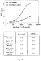
- Antibodies were coated onto ELISA plates and presented with purified kappa light chains, purified lambda light chains or Normal Human Serum. Binding activity was detected by measuring absorbance at 450nm using Tetramethylbenzidine (TMB) chromogenic substrate and anti-light chain antibodies conjugated to Horse Radish Peroxidase (HRP). Whilst there is some reduction in ELISA activity, as shown in Figure 4 , activity still remains in the treated antibodies.
- TMB Tetramethylbenzidine
- HRP Horse Radish Peroxidase
- BMOE Bismaleimidoethane
- Anti-lambda F(ab') 2 antibodies were investigated to see if antibody chains could be cross-linked by BMOE.
- Anti-lambda F(ab') 2 fragments were reduced with 1 mM (TCEP). The TCEP was removed using Hi-Trap Desalting columns and the reduced anti-lambda total F(ab') 2 was cross-linked at 100-500x fold molar excess of BMOE and then analysed by Coomassie Blue stained SDS-PAGE run under reducing conditions.
- Figure 5 shows that BMOE can cross-link F (ab') 2 fragments with an efficiency of over 50%. Moreover, the resulting antibody chain cross-links are resistant to reducing conditions.
- An ELISA plate was coated with polyclonal IgG Lambda and BMOE-treated or untreated anti-total lambda F(ab') 2 was bound to the plate. Binding activity by anti-total lambda was measured by light absorbance at 450nm using anti-sheep-HRP and TMB substrate. Under conditions that produce >50% BMOE cross-linking ( Figure 5 ), anti-total lambda antibody retains over 70% IgG Lambda binding activity ( Figure 6 ).
- Freelite TM antibodies were investigated to see whether antibody chains could be cross-linked by BMOE treatment.
- Whole molecule anti-free kappa and F(ab') 2 anti-free lambda were reduced with 1.5 mM and 1.0 mM TCEP, respectively.
- the TCEP was removed using Hi-Trap Desalting columns and the anti-free kappa and anti-free lambda antibodies were cross-linked at 400-fold and 200-fold molar excess of BMOE, respectively.
- Samples were analysed by Coomassie Blue stained SDS-PAGE run under reducing conditions. As shown in Figure 7 , the BMOE cross-linking efficiency is over 50% for antibodies in either F(ab') 2 or whole molecule formats.
- Thioether cross-linking stabilises sepharose-conjugated anti-IgG3 antibodies.
- BMOE cross-linking stabilises anti-free kappa and anti-free lambda antibodies
- Freelite TM anti-kappa and anti-lambda antibodies were either untreated or cross-linked with BMOE following reduction with TCEP and desalting on Hi-Trap Desalting columns.
- Antibodies were conjugated to sepharose resin, treated with 5% Acetic Acid containing 50 mM TCEP in the absence of prior sample addition and the supernatants were analysed by MALDI-TOF MS. As is shown in Figures 12-13 , significantly fewer antibody fragments were released from the BMOE cross-linked antibodies, indicating a substantial increase in antibody stability.
- the increased stability of the antibodies reduces cross-contamination of material from the purifying antibodies. This improves the accuracy of the further characterisation steps when used to purify and isolate samples of immunoglobulins from, for example, patients.
- Control analyte can be distinguished from subject analyte by Mass Spectrometry
- Human serum was supplemented with a recombinant kappa light chain of known quantity and known mass (control kappa chain).
- the endogenous and control kappa chains were co-purified from the serum using commercially available sepharose conjugated to Camelid anti-kappa antibody and the eluted supernatants were analysed by MALDI-TOF MS ( Figure 14 ).
- the control kappa chain is distinguishable from the endogenous kappa chain due to the difference in its relative molecular mass.
- Control analyte can be used to provide quantitative information about the subject analyte
- Endogenous kappa chains and control kappa chains were co-purified from human serum, spotted onto two 96-target plates and analysed by MALDI-TOF MS ( Figure 14B ). Peak areas corresponding to each kappa chain were determined and used to calculate the coefficient of variation (%CV) across each plate and for 192 replicates encompassing both plates. Figure 14B shows that when the peak area corresponding to endogenous kappa is expressed relative to the control kappa peak area in 192 replicates, the calculated %CV is less than 10%. This indicates a substantial diagnostic potential of the invention.
- control analyte can be used to determine the abundance of a subject analyte relative to the control analyte, and thus, can provide quantitative as well as qualitative information about the subject analyte.
- a control analyte may be used to verify the success of a purification procedure, and serves to minimise errors in data collection caused by, for example, differential ionisation of MALDI targets spots.
Landscapes
- Health & Medical Sciences (AREA)
- Life Sciences & Earth Sciences (AREA)
- Chemical & Material Sciences (AREA)
- Immunology (AREA)
- Engineering & Computer Science (AREA)
- Molecular Biology (AREA)
- Biomedical Technology (AREA)
- Urology & Nephrology (AREA)
- Hematology (AREA)
- General Health & Medical Sciences (AREA)
- Biochemistry (AREA)
- Proteomics, Peptides & Aminoacids (AREA)
- Medicinal Chemistry (AREA)
- Physics & Mathematics (AREA)
- Organic Chemistry (AREA)
- Analytical Chemistry (AREA)
- General Physics & Mathematics (AREA)
- Pathology (AREA)
- Food Science & Technology (AREA)
- Microbiology (AREA)
- Cell Biology (AREA)
- Biotechnology (AREA)
- Biophysics (AREA)
- Genetics & Genomics (AREA)
- Bioinformatics & Cheminformatics (AREA)
- Bioinformatics & Computational Biology (AREA)
- Spectroscopy & Molecular Physics (AREA)
- Chemical Kinetics & Catalysis (AREA)
- Electrochemistry (AREA)
- Peptides Or Proteins (AREA)
- Other Investigation Or Analysis Of Materials By Electrical Means (AREA)
- Sampling And Sample Adjustment (AREA)
- Investigating Or Analysing Biological Materials (AREA)
Applications Claiming Priority (4)
| Application Number | Priority Date | Filing Date | Title |
|---|---|---|---|
| GBGB1603291.4A GB201603291D0 (en) | 2016-02-25 | 2016-02-25 | Antibodies |
| US201662368606P | 2016-07-29 | 2016-07-29 | |
| EP17708320.1A EP3420361B1 (fr) | 2016-02-25 | 2017-02-24 | Kit de spectrométrie de masse |
| PCT/GB2017/050489 WO2017144900A1 (fr) | 2016-02-25 | 2017-02-24 | Kit de spectrométrie de masse |
Related Parent Applications (2)
| Application Number | Title | Priority Date | Filing Date |
|---|---|---|---|
| EP17708320.1A Division-Into EP3420361B1 (fr) | 2016-02-25 | 2017-02-24 | Kit de spectrométrie de masse |
| EP17708320.1A Division EP3420361B1 (fr) | 2016-02-25 | 2017-02-24 | Kit de spectrométrie de masse |
Publications (1)
| Publication Number | Publication Date |
|---|---|
| EP4123308A1 true EP4123308A1 (fr) | 2023-01-25 |
Family
ID=55806928
Family Applications (2)
| Application Number | Title | Priority Date | Filing Date |
|---|---|---|---|
| EP22190012.9A Pending EP4123308A1 (fr) | 2016-02-25 | 2017-02-24 | Kit de spectrométrie de masse |
| EP17708320.1A Active EP3420361B1 (fr) | 2016-02-25 | 2017-02-24 | Kit de spectrométrie de masse |
Family Applications After (1)
| Application Number | Title | Priority Date | Filing Date |
|---|---|---|---|
| EP17708320.1A Active EP3420361B1 (fr) | 2016-02-25 | 2017-02-24 | Kit de spectrométrie de masse |
Country Status (12)
| Country | Link |
|---|---|
| US (1) | US20190094239A1 (fr) |
| EP (2) | EP4123308A1 (fr) |
| JP (3) | JP7246927B2 (fr) |
| KR (1) | KR20180132651A (fr) |
| CN (2) | CN116148479A (fr) |
| AU (1) | AU2017222399B2 (fr) |
| BR (1) | BR112018067435A2 (fr) |
| CA (1) | CA3015296A1 (fr) |
| DK (1) | DK3420361T3 (fr) |
| ES (1) | ES2936783T3 (fr) |
| GB (1) | GB201603291D0 (fr) |
| WO (1) | WO2017144900A1 (fr) |
Families Citing this family (10)
| Publication number | Priority date | Publication date | Assignee | Title |
|---|---|---|---|---|
| GB201708262D0 (en) | 2017-05-23 | 2017-07-05 | Binding Site Group Ltd | Assay for plasma cell associated disease |
| JP2022519246A (ja) * | 2019-01-31 | 2022-03-22 | リジェネロン・ファーマシューティカルズ・インコーポレイテッド | 抗体電荷不均一性についてのネイティブマイクロ流体ce-ms解析法 |
| CN110009008B (zh) * | 2019-03-18 | 2020-11-06 | 四川大学 | 基于提取的免疫固定电泳图特征对其进行自动分类的方法 |
| GB201906599D0 (en) | 2019-05-10 | 2019-06-26 | Binding Site Group Ltd | Mass spectrometry calibrator |
| WO2020229794A1 (fr) | 2019-05-10 | 2020-11-19 | The Binding Site Group Limited | Produit d'étalonnage pour spectrométrie de masse |
| GB201915946D0 (en) | 2019-11-01 | 2019-12-18 | Binding Site Group Ltd | Quantitation of immunoglobulins i n mass spectrometry |
| GB202007047D0 (en) * | 2020-05-13 | 2020-06-24 | Binding Site Group Ltd | Mass spectrometry controls |
| GB202016822D0 (en) | 2020-10-23 | 2020-12-09 | Binding Site Group Ltd | Quantitation of immunoglobulins in mass spectrometry |
| CN113720900A (zh) * | 2021-09-14 | 2021-11-30 | 首都医科大学附属北京朝阳医院 | 一种基于madli-tof ms技术检测血清中m蛋白的方法 |
| CN114324557B (zh) * | 2021-12-03 | 2024-05-10 | 融智生物科技(青岛)有限公司 | 一种基于MALDI-TOF MS的ζ-珠蛋白的检测方法 |
Citations (12)
| Publication number | Priority date | Publication date | Assignee | Title |
|---|---|---|---|---|
| WO1991003493A1 (fr) | 1989-08-29 | 1991-03-21 | The University Of Southampton | CONJUGUES F(ab)3 ou F(ab)4 bi ou trispécifiques |
| WO1997017372A1 (fr) | 1995-11-03 | 1997-05-15 | The Binding Site Limited | Production d'anticorps et applications medicales dans lesquelles lesdits anticorps sont utilises |
| WO2000044788A1 (fr) | 1999-01-28 | 2000-08-03 | Idec Pharmaceuticals Corporation | Production d'anticorps tetravalents |
| WO2006099481A2 (fr) | 2005-03-14 | 2006-09-21 | Medimmune, Inc. | Macromolecules comprenant une reticulation de thioether |
| EP1870710A1 (fr) | 2005-04-12 | 2007-12-26 | Akira Matsumori | Biomarqueur destiné à diagnostiquer une maladie cardiaque et son utilisation |
| WO2011095818A1 (fr) | 2010-02-05 | 2011-08-11 | The Binding Site Group Limited | Dosage à valeur pronostique utilisé en cancérologie |
| WO2011107965A1 (fr) | 2010-03-02 | 2011-09-09 | The Binding Site Group Limited | Dosage pronostique du rein |
| WO2013050731A1 (fr) | 2011-10-05 | 2013-04-11 | The Binding Site Group Limited | Procédé de pronostic pour le diabète |
| WO2013088126A1 (fr) | 2011-12-12 | 2013-06-20 | The Binding Site Group Limited | Dosage |
| WO2014121031A1 (fr) * | 2013-01-31 | 2014-08-07 | Excelimmune, Inc. | Caractérisation de mélanges d'anticorps par spectrométrie de masse |
| WO2015131169A2 (fr) | 2014-02-28 | 2015-09-03 | H. Lee Moffitt Cancer Center And Research Institute, Inc. | Détection personnalisée du myélome |
| WO2015154052A1 (fr) | 2014-04-04 | 2015-10-08 | Mayo Foundation For Medical Education And Research | Isotypage d'immunoglobulines par masse moléculaire précise |
Family Cites Families (5)
| Publication number | Priority date | Publication date | Assignee | Title |
|---|---|---|---|---|
| WO1995031727A1 (fr) * | 1994-05-13 | 1995-11-23 | Therasorb Medizinische Systeme Gmbh | Colonne sterile et apyrogene couplee a une proteine en vue de la fixation et de l'extraction de substances donnees du sang |
| JP2826965B2 (ja) * | 1995-01-12 | 1998-11-18 | オリエンタル酵母工業株式会社 | 免疫吸着体とその製造法 |
| ATE529752T1 (de) * | 2006-12-21 | 2011-11-15 | Novartis Ag | Antikörperquantifizierung |
| CN102687020A (zh) * | 2009-10-09 | 2012-09-19 | 西福根有限公司 | 利用特征肽和质谱对混合物中的单个重组蛋白进行多重定量 |
| WO2016018978A1 (fr) * | 2014-07-29 | 2016-02-04 | Mayo Foundation For Medical Education And Research | Quantification d'agents thérapeutiques de type anticorps monoclonaux par lc-ms/ms |
-
2016
- 2016-02-25 GB GBGB1603291.4A patent/GB201603291D0/en not_active Ceased
-
2017
- 2017-02-24 EP EP22190012.9A patent/EP4123308A1/fr active Pending
- 2017-02-24 ES ES17708320T patent/ES2936783T3/es active Active
- 2017-02-24 CN CN202211574983.0A patent/CN116148479A/zh active Pending
- 2017-02-24 AU AU2017222399A patent/AU2017222399B2/en active Active
- 2017-02-24 EP EP17708320.1A patent/EP3420361B1/fr active Active
- 2017-02-24 JP JP2018544901A patent/JP7246927B2/ja active Active
- 2017-02-24 CN CN201780013491.5A patent/CN108885218A/zh active Pending
- 2017-02-24 KR KR1020187027703A patent/KR20180132651A/ko not_active Application Discontinuation
- 2017-02-24 US US16/078,986 patent/US20190094239A1/en active Pending
- 2017-02-24 WO PCT/GB2017/050489 patent/WO2017144900A1/fr active Application Filing
- 2017-02-24 CA CA3015296A patent/CA3015296A1/fr active Pending
- 2017-02-24 DK DK17708320.1T patent/DK3420361T3/da active
- 2017-02-24 BR BR112018067435A patent/BR112018067435A2/pt unknown
-
2022
- 2022-02-07 JP JP2022017497A patent/JP2022069446A/ja active Pending
- 2022-12-13 JP JP2022198753A patent/JP2023033281A/ja active Pending
Patent Citations (12)
| Publication number | Priority date | Publication date | Assignee | Title |
|---|---|---|---|---|
| WO1991003493A1 (fr) | 1989-08-29 | 1991-03-21 | The University Of Southampton | CONJUGUES F(ab)3 ou F(ab)4 bi ou trispécifiques |
| WO1997017372A1 (fr) | 1995-11-03 | 1997-05-15 | The Binding Site Limited | Production d'anticorps et applications medicales dans lesquelles lesdits anticorps sont utilises |
| WO2000044788A1 (fr) | 1999-01-28 | 2000-08-03 | Idec Pharmaceuticals Corporation | Production d'anticorps tetravalents |
| WO2006099481A2 (fr) | 2005-03-14 | 2006-09-21 | Medimmune, Inc. | Macromolecules comprenant une reticulation de thioether |
| EP1870710A1 (fr) | 2005-04-12 | 2007-12-26 | Akira Matsumori | Biomarqueur destiné à diagnostiquer une maladie cardiaque et son utilisation |
| WO2011095818A1 (fr) | 2010-02-05 | 2011-08-11 | The Binding Site Group Limited | Dosage à valeur pronostique utilisé en cancérologie |
| WO2011107965A1 (fr) | 2010-03-02 | 2011-09-09 | The Binding Site Group Limited | Dosage pronostique du rein |
| WO2013050731A1 (fr) | 2011-10-05 | 2013-04-11 | The Binding Site Group Limited | Procédé de pronostic pour le diabète |
| WO2013088126A1 (fr) | 2011-12-12 | 2013-06-20 | The Binding Site Group Limited | Dosage |
| WO2014121031A1 (fr) * | 2013-01-31 | 2014-08-07 | Excelimmune, Inc. | Caractérisation de mélanges d'anticorps par spectrométrie de masse |
| WO2015131169A2 (fr) | 2014-02-28 | 2015-09-03 | H. Lee Moffitt Cancer Center And Research Institute, Inc. | Détection personnalisée du myélome |
| WO2015154052A1 (fr) | 2014-04-04 | 2015-10-08 | Mayo Foundation For Medical Education And Research | Isotypage d'immunoglobulines par masse moléculaire précise |
Non-Patent Citations (18)
| Title |
|---|
| ALAIN BECK ET AL: "Characterization of Therapeutic Antibodies and Related Products", ANALYTICAL CHEMISTRY, vol. 85, no. 2, 15 January 2013 (2013-01-15), US, pages 715 - 736, XP055383395, ISSN: 0003-2700, DOI: 10.1021/ac3032355 * |
| AUCLAIR ET AL.: "Strategies for stabilizing superoxide dismutase (SOD1), the protein destabilized in the most common form of familial amyotrophic lateral sclerosis", PROC NATL ACAD SCI USA, vol. 107, no. 50, 2010, pages 21394 - 9, XP055266790, DOI: 10.1073/pnas.1015463107 |
| BERNARDES ET AL., ANGEW. CHEM. INT. ED., vol. 47, 2008, pages 2244 - 2247 |
| CAS , no. 2283-11-6 |
| CAS, no. 1608-26-0 |
| FRENZEL A. ET AL., FRONTIERS IN IMMUNOLOGY, vol. 4, 2013 |
| GEULA: "Structure-based analysis of VDAC1 protein: defining oligomer contact sites", J BIOL CHEM, vol. 287, no. 3, 2012, pages 475 - 85 |
| JO ET AL.: "Development of a-Helical Calpain Probes by Mimicking a Natural Protein-Protein Interaction", J AM CHEM SOC., vol. 134, no. 42, 2012, pages 17704 - 13, XP055138213, DOI: 10.1021/ja307599z |
| KIDA ET AL.: "Two translocating hydrophilic segments of a nascent chain span the ER membrane during multispanning protein topogenesis", J CELL BIOL, vol. 171, no. 7, 2007, pages 1441 - 1452 |
| LUPINEK CHRISTIAN ET AL: "Trimolecular complex formation of IgE, Fc(epsilon)RI, and a recombinant nonanaphylactic single-chain antibody fragment with high affinity for IgE", THE JOURNAL OF IMMUNOLOGY, THE AMERICAN ASSOCIATION OF IMMUNOLOGISTS, US, vol. 182, no. 8, 15 April 2009 (2009-04-15), pages 4817 - 4829, XP002555137, ISSN: 0022-1767, DOI: 10.4049/JIMMUNOL.0800726 * |
| MICHEL GOLDBERG ET AL: "Specific Interchain Cross-Linking of Antibodies Using Bismaleimides. Repression of Ligand Leakage in Immunoaffinity Chromatographyt", BIOCONJUGATE CHEM, vol. 2, 1991, pages 275 - 280, XP055367595 * |
| STECH M.KUBICK S., ANTIBODIES, vol. 4, 2015, pages 12 - 33 |
| STEINHOFF ROBERT F ET AL: "Microarray-based MALDI-TOF mass spectrometry enables monitoring of monoclonal antibody production in batch and perfusion cell cultures", METHODS, ACADEMIC PRESS, US, vol. 104, 18 December 2015 (2015-12-18), pages 33 - 40, XP029626003, ISSN: 1046-2023, DOI: 10.1016/J.YMETH.2015.12.011 * |
| ZHANG ET AL., J. BIOL. CHEM., vol. 288, no. 23, 2013, pages 16371 - 8 |
| ZHANG ET AL.: "IgG1 Thioether Bond formation in vivo", JBC, vol. 288, 2013, pages 16371 - 16382 |
| ZHANG Q ET AL., JBC MANUSCRIPT, 2013, pages 468367 |
| ZHANGFLYNN, J. BIOL. CHEM, vol. 288, no. 43, 2013, pages 34325 - 35 |
| ZHANGFLYNN: "Cysteine racemization IgG heavy and light chains", JBC, vol. 288, 2013, pages 34325 - 34335 |
Also Published As
| Publication number | Publication date |
|---|---|
| JP2019509483A (ja) | 2019-04-04 |
| WO2017144900A1 (fr) | 2017-08-31 |
| KR20180132651A (ko) | 2018-12-12 |
| CN108885218A (zh) | 2018-11-23 |
| CA3015296A1 (fr) | 2017-08-31 |
| EP3420361A1 (fr) | 2019-01-02 |
| EP3420361B1 (fr) | 2023-01-04 |
| JP7246927B2 (ja) | 2023-03-28 |
| JP2022069446A (ja) | 2022-05-11 |
| GB201603291D0 (en) | 2016-04-13 |
| US20190094239A1 (en) | 2019-03-28 |
| BR112018067435A2 (pt) | 2019-01-02 |
| ES2936783T3 (es) | 2023-03-22 |
| DK3420361T3 (da) | 2023-01-23 |
| CN116148479A (zh) | 2023-05-23 |
| AU2017222399B2 (en) | 2024-03-14 |
| AU2017222399A1 (en) | 2018-08-30 |
| JP2023033281A (ja) | 2023-03-10 |
Similar Documents
| Publication | Publication Date | Title |
|---|---|---|
| AU2017222399B2 (en) | Mass spectrometry kit | |
| AU2018274704B2 (en) | Assay for plasma cell associated disease | |
| Plomp et al. | Site-specific N-glycosylation analysis of human immunoglobulin e | |
| AU2017222402A1 (en) | Antibodies | |
| JP7506093B2 (ja) | 質量分析キャリブレータ | |
| US20230184782A1 (en) | Mass spectrometry controls |
Legal Events
| Date | Code | Title | Description |
|---|---|---|---|
| PUAI | Public reference made under article 153(3) epc to a published international application that has entered the european phase |
Free format text: ORIGINAL CODE: 0009012 |
|
| STAA | Information on the status of an ep patent application or granted ep patent |
Free format text: STATUS: THE APPLICATION HAS BEEN PUBLISHED |
|
| AC | Divisional application: reference to earlier application |
Ref document number: 3420361 Country of ref document: EP Kind code of ref document: P |
|
| AK | Designated contracting states |
Kind code of ref document: A1 Designated state(s): AL AT BE BG CH CY CZ DE DK EE ES FI FR GB GR HR HU IE IS IT LI LT LU LV MC MK MT NL NO PL PT RO RS SE SI SK SM TR |
|
| P01 | Opt-out of the competence of the unified patent court (upc) registered |
Effective date: 20230529 |
|
| STAA | Information on the status of an ep patent application or granted ep patent |
Free format text: STATUS: REQUEST FOR EXAMINATION WAS MADE |
|
| 17P | Request for examination filed |
Effective date: 20230721 |
|
| RBV | Designated contracting states (corrected) |
Designated state(s): AL AT BE BG CH CY CZ DE DK EE ES FI FR GB GR HR HU IE IS IT LI LT LU LV MC MK MT NL NO PL PT RO RS SE SI SK SM TR |
|
| REG | Reference to a national code |
Ref country code: HK Ref legal event code: DE Ref document number: 40087264 Country of ref document: HK |