EP4111166B1 - Kalibriersatz mit nanopartikeln aus nylon-6 und farbstoffen - Google Patents
Kalibriersatz mit nanopartikeln aus nylon-6 und farbstoffen Download PDFInfo
- Publication number
- EP4111166B1 EP4111166B1 EP21708490.4A EP21708490A EP4111166B1 EP 4111166 B1 EP4111166 B1 EP 4111166B1 EP 21708490 A EP21708490 A EP 21708490A EP 4111166 B1 EP4111166 B1 EP 4111166B1
- Authority
- EP
- European Patent Office
- Prior art keywords
- nanoparticles
- equal
- dye
- less
- average diameter
- Prior art date
- Legal status (The legal status is an assumption and is not a legal conclusion. Google has not performed a legal analysis and makes no representation as to the accuracy of the status listed.)
- Active
Links
Images
Classifications
-
- G—PHYSICS
- G01—MEASURING; TESTING
- G01N—INVESTIGATING OR ANALYSING MATERIALS BY DETERMINING THEIR CHEMICAL OR PHYSICAL PROPERTIES
- G01N15/00—Investigating characteristics of particles; Investigating permeability, pore-volume or surface-area of porous materials
- G01N15/10—Investigating individual particles
- G01N15/1012—Calibrating particle analysers; References therefor
-
- B—PERFORMING OPERATIONS; TRANSPORTING
- B82—NANOTECHNOLOGY
- B82Y—SPECIFIC USES OR APPLICATIONS OF NANOSTRUCTURES; MEASUREMENT OR ANALYSIS OF NANOSTRUCTURES; MANUFACTURE OR TREATMENT OF NANOSTRUCTURES
- B82Y15/00—Nanotechnology for interacting, sensing or actuating, e.g. quantum dots as markers in protein assays or molecular motors
-
- B—PERFORMING OPERATIONS; TRANSPORTING
- B82—NANOTECHNOLOGY
- B82Y—SPECIFIC USES OR APPLICATIONS OF NANOSTRUCTURES; MEASUREMENT OR ANALYSIS OF NANOSTRUCTURES; MANUFACTURE OR TREATMENT OF NANOSTRUCTURES
- B82Y30/00—Nanotechnology for materials or surface science, e.g. nanocomposites
-
- G—PHYSICS
- G01—MEASURING; TESTING
- G01N—INVESTIGATING OR ANALYSING MATERIALS BY DETERMINING THEIR CHEMICAL OR PHYSICAL PROPERTIES
- G01N15/00—Investigating characteristics of particles; Investigating permeability, pore-volume or surface-area of porous materials
- G01N15/10—Investigating individual particles
- G01N15/1012—Calibrating particle analysers; References therefor
- G01N2015/1014—Constitution of reference particles
Definitions
- the present specification generally relates to calibration sets and, more particularly, to calibration sets having properties similar to extracellular vesicles (EVs).
- EVs extracellular vesicles
- characterization methods lack appropriate calibration standards for EV quantitation and sizing technologies.
- polystyrene bead standards are conventionally used in sizing instruments, such as flow cytometers, the polystyrene beads have properties that match those of mammalian cells, such as density, light scattering effects, size, and fluorescent marker binding affinity.
- EVs are products that may be of mammalian cell origin, their inherent properties differ from the properties of their origin cells and, thus, from the polystyrene beads.
- WO9100509 A1 describes a kit of uniform size microbead standards for flow cytometer alignment, compensation, and/or calibration. Chris Gardiner et al. describe a light-scattering technique that is useful for the rapid sizing and enumeration of extracellular vesicles (JOURNAL OF EXTRACELLULAR VESICLES, 2(1), 19671-19691 ).
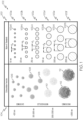
- FIG. 1 schematically illustrates groups of nanoparticles having different average diameters as compared to various extracellular vesicles according to one or more embodiments shown and described herein.
- a calibration set comprises first nanoparticles comprising nylon-6 covalently bound to a first dye and second nanoparticles comprising nylon-6 covalently bound to a second dye that is different from the first dye.
- the first nanoparticles have a first average diameter and a first polydispersity index of greater than or equal to 1.15 and less than or equal to 1.19
- the second nanoparticles have a second average diameter and a second polydispersity index of greater than or equal to 1.15 and less than or equal to 1.19.
- the first average diameter is different from the second average diameter, and each of the first average diameter and the second average diameter are greater than or equal to 30 nm and less than or equal to 3000 nm, with the first average diameter being at least two times the second average diameter.
- Ranges can be expressed herein as from “about” one particular value, and/or to "about” another particular value. When such a range is expressed, another embodiment includes from the one particular value and/or to the other particular value. Similarly, when values are expressed as approximations, by use of the antecedent "about,” it will be understood that the particular value forms another embodiment. It will be further understood that the endpoints of each of the ranges are significant both in relation to the other endpoint, and independently of the other endpoint.
- Each group 102 comprises nanoparticles comprising nylon-6 covalently bound to a corresponding dye, such as an organic dye (e.g., fluorescein, rhodamine, aminomethylcoumarin (AMCA)), a biological fluorophore (e.g., green fluorescent protein, phycoerythrin, allophycocyanain), quantum dots, or a fluorescent lipophilic dye.
- a corresponding dye such as an organic dye (e.g., fluorescein, rhodamine, aminomethylcoumarin (AMCA)), a biological fluorophore (e.g., green fluorescent protein, phycoerythrin, allophycocyanain), quantum dots, or a fluorescent lipophilic dye.
- the first group of nanoparticles 102a comprises nylon-6 covalently bound to a first dye
- the second group of nanoparticles 102b comprises nylon-6 covalently bound to a second dye
- the third group of nanoparticles 102c comprises nylon-6 covalently bound to a third dye
- the fourth group of nanoparticles 102d comprises nylon-6 covalently bound to a fourth dye
- the fifth group of nanoparticles 102e comprises nylon-6 covalently bound to a fifth dye
- the sixth group of nanoparticles 102f comprises nylon-6 covalently bound to a sixth dye.
- the dye in each group of nanoparticles is different from each of the other dyes in the other groups of nanoparticles such that the dye is unique to and indicative of the particular group of nanoparticles.
- the dye is covalently bound to a polymer comprising nylon-6.
- the dye is bound to the polymer prior to the formation of the nanoparticles, as will be described in greater detail below, such that the dye is homogeneously distributed throughout the nanoparticle.
- Nylon-6 has the following structure: The carbonyl group of the nylon-6 structure enables covalent binding with the dye which, in addition to enabling the tracing of the nylon-6 nanoparticles, can also reduce or eliminate dye leakage or disassociation of the dye from the particles.
- conventional standards which encapsulate or incubate polymer particles with the dye may not form direct chemical bonds between the dye and the particles. Accordingly, dye leaking from the particles can introduce error into measurements made using conventional standards.
- the fluorescent dyes described herein are covalently bound to the nylon-6 and incorporated into the nanoparticles and the polymer would have to degrade to release the fluorescent dye, thereby reducing the likelihood of disassociation of the dye from the nanoparticles.
- the nanoparticles have a density of greater than or equal to 1.15 g/mL and less than or equal to 1.19 g/mL, as measured in accordance with ASTM D792.
- the nanoparticles have a density of greater than or equal to 1.15 g/mL and less than or equal to 1.19 g/mL, greater than or equal to 1.15 g/mL and less than or equal to 1.18 g/mL, greater than or equal to 1.15 g/mL and less than or equal to 1.17 g/mL, greater than or equal to 1.15 g/mL and less than or equal to 1.16 g/mL, greater than or equal to 1.16 g/mL and less than or equal to 1.19 g/mL, greater than or equal to 1.16 g/mL and less than or equal to 1.18 g/mL, greater than or equal to 1.16 g/mL and less than or equal to 1.17 g/mL, greater than or equal to 1.17 g/mL,
- the nanoparticles have a density that is similar to the density of EVs (i.e., 1.15-1.19 g/mL) and measurably higher than the density of mammalian cells (i.e., 1.04-1.05 g/mL), thereby eliminating the issue of different material density between the EVs and the standard nanoparticles.
- Each group of nanoparticles has an average hydrodynamic diameter (referred to herein as "average diameter"), determined in accordance with ISO 22412, which is generally referred to as d n .
- the average hydrodynamic diameter for each group of nanoparticles is determined by light scattering measurement techniques, such as dynamic light scattering (DLS) or nanoparticle tracking analysis (NTA) and visually confirmed via microscopy, such as scanning electron microscopy (SEM) or transmission electron microscopy (TEM).
- DLS dynamic light scattering
- NTA nanoparticle tracking analysis
- microscopy such as scanning electron microscopy (SEM) or transmission electron microscopy (TEM).
- d a is the average diameter of the first group of nanoparticles 102a
- d b is the average diameter of the second group of nanoparticles 102b
- d c is the average diameter of the third group of nanoparticles 102c
- d d is the average diameter of the fourth group of nanoparticles 102d
- d e is the average diameter of the fifth group of nanoparticles 102e
- d f is the average diameter of the sixth group of nanoparticles 102f.
- each group of nanoparticles has a standard deviation of size (e.g., a standard deviation of the maximum diameter of each of the particles in the group) of 15% or less, which is determined statistically via software calculations.
- each group of nanoparticles may have a standard deviation of less than or equal to 15%, less than or equal to 12.5%, less than or equal to 10%, less than or equal to 8%, less than or equal to 6%, less than or equal to 5%, less than or equal to 4%, less than or equal to 3%, less than or equal to 2%, less than or equal to 1%, or less than or equal to 0.5%.
- the average diameter of each group of nanoparticles may have a standard deviation of greater than or equal to 0 and less than or equal to 15%, greater than or equal to 0 and less than or equal to 12.5%, greater than or equal to 0 and less than or equal to 10%, greater than or equal to 0 and less than or equal to 8%, greater than or equal to 0 and less than or equal to 6%, greater than or equal to 0 and less than or equal to 5%, greater than or equal to 0 and less than or equal to 4%, greater than or equal to 0 and less than or equal to 3%, greater than or equal to 0 and less than or equal to 2%, greater than or equal to 0 and less than or equal to 1%, greater than or equal to 0 and less than or equal to 0.5%, greater than or equal to 0.5% and less than or equal to 15%, greater than or equal to 0.5% and less than or equal to 12.5%, greater than or equal to 0.5% and less than or equal to 10%, greater than or equal to 0.5% and less than or equal to 12.5%,
- each group of nanoparticles can be further characterized by a polydispersity index of greater than or equal to 1.15 and less than or equal to 1.19.
- the polydispersity index (sometimes referred to as the "PDI" or D) is a measure of the broadness of the size distribution calculated from the cumulants analysis of the group of nanoparticles and is determined using dynamic light scattering (DLS) in compliance with ISO 22412 and ISO 13321.
- each group of nanoparticles may have a PDI of greater than or equal to 1.15 and less than or equal to 1.18, greater than or equal to 1.15 and less than or equal to 1.17, greater than or equal to 1.15 and less than or equal to 1.16, greater than or equal to 1.16 and less than or equal to 1.19, greater than or equal to 1.16 and less than or equal to 1.18, greater than or equal to 1.16 and less than or equal to 1.17, greater than or equal to 1.17 and less than or equal to 1.19, greater than or equal to 1.17 and less than or equal to 1.17, or greater than or equal to 1.18 and less than or equal to 1.19, including any and all ranges and sub-ranges within these ranges. It is contemplated that, in embodiments, each PDI can be a number within this range, and may be the same as or different from the PDI of one or more other groups of nanoparticles.
- each average diameter d n of each group is different from the average diameter of each of the other groups of nanoparticles. Accordingly, in FIG. 1 , d a is different from d b , d c , d d , d e , and d f , d b is different from d e , d d , d e , and d f , d c is different from d d , d e , and d f , d d is different from d e and d f , and d e is different from d f .
- each average diameter is at least two times the next largest average diameter. For example, in FIG.
- d b (100 nm) is two times d a (50 nm)
- d c (250 nm) is at least two times d b (100 nm)
- d d (500 nm) is two times d c (250 nm)
- d e (1000 nm) is two times d d (500 nm)
- d f (2000 nm) is two times d e (1000 nm).
- each average diameter d n is independently greater than or equal to 30 nm and less than or equal to 3000 nm.
- each average diameter d n can be greater than or equal to 30 nm and less than or equal to 3000 nm, greater than or equal to 30 nm and less than or equal to 2500 nm, greater than or equal to 30 nm and less than or equal to 2000 nm, greater than or equal to 30 nm and less than or equal to 1500 nm, greater than or equal to 30 nm and less than or equal to 1000 nm, greater than or equal to 30 nm and less than or equal to 750 nm, greater than or equal to 30 nm and less than or equal to 500 nm, greater than or equal to 30 nm and less than or equal to 300 nm, greater than or equal to 30 nm and less than or equal to 250 nm, greater than or equal to 30 nm and less than or equal to 100 nm, greater than or equal to
- At least one group of nanoparticles has an average diameter less than or equal to 300 nm, less than or equal to 250 nm, less than or equal to 200 nm, less than or equal to 150 nm, less than or equal to 100 nm, less than or equal to 75 nm, or less than or equal to 50 nm.
- the average diameters in the calibration set 100 can vary depending on the particular embodiment.
- one or more groups of nanoparticles can be selected to have an average diameter that is relatively close in range (within 50 nm, within 100 nm, or within 250 nm) to a type of EV of interest.
- FIG. 1 also schematically illustrates various types of EVs 104 and their approximate average diameters.
- FIG. 1 illustrates various types of EVs 104 and their approximate average diameters.
- exosomes 106 which have an average diameter of less than or equal to about 100 nm, a first set of microvesicles 108 having an average diameter of greater than or equal to about 100 nm and less than or equal to about 1000 nm, a second set of microvesicles 110 having an average diameter of greater than or equal to about 1000 nm and less than or equal to about 2000 nm, and oncosomes 112 having an average diameter of greater than about 2000 nm. Accordingly, as shown in FIG. 1 , the nanoparticles described herein can be used to approximate the size of various different EVs.
- one or more groups of nanoparticles may include RNA.
- the RNA is bound to the polymer via the amide group of the polymer prior to formation of the nanoparticle. Accordingly, RNA is homogenously distributed throughout each nanoparticle.
- RNA molecule or “RNA” refers to ribonucleic acid, i.e., a polymer consisting of nucleotides.
- the nucleotides are usually adenosine-monophosphate, uridine-monophosphate, guanosine-monophosphate, and cytidine-monophosphate monomers which are connected to each other along a so-called backbone formed by phosphodiester bonds between the sugar (i.e., ribose) of a first monomer and a phosphate moiety of a second, adjacent monomer.
- the RNA may possess any predetermined RNA sequence.
- the RNA sequence can be, for example a generic RNA sequence, or a specific sequence of interest. Accordingly, results of analysis could be validated by adding the nanoparticles including RNA into a biologic sample and observing the effects of technical steps throughout the experimental protocol, as will be described in greater detail below.
- the nanoparticles of various embodiments can be made in accordance with any one of a number of methods of nanoprecipitation, including, but not limited to, pipette droplet nanoprecipitation, flow-controlled T-mixer nanoprecipitation, and flow-controlled microfluidics nanoprecipitation.
- a polar protic solvent e.g., acetic acid, formic acid, ethanol, etc.
- a polar aprotic solvent e.g., tetrahydrofuran, acetone, acetonitrile, etc.
- the dye is dissolved into the nylon-6 and solvent mixture or introduced via a miscible solvent via emulsion.
- An aqueous mixture of water and surfactant e.g., polyvinyl alcohol, pluronic, or another aggregation protectant
- surfactant e.g., polyvinyl alcohol, pluronic, or another aggregation protectant
- the size of the nanoparticles is controlled by controlling the polymer concentration in the organic phase, the organic solvent choice, the surfactant concentration in the aqueous phase, the surfactant choice, the ratio of the organic phase to the aqueous phase in mixing, the flow rates of the organic phase and aqueous phase during mixing, and the apparatus used for nanoprecipitation.
- increasing the polymer concentration in the organic phase or an increase in the ratio of organic phase to the aqueous phase in mixing results in an increase in particle diameter.
- an increase in surfactant concentration in the aqueous phase or an increase in aqueous phase to organic phase results in a decrease in particle diameter.
- the organic solvent and surfactant choice can have various effects that could be determined empirically in particle size depending on the chemical densities, charges, miscibility, and other chemical properties of the solvent or surfactants themselves.
- An increase in flow rates during mixing can also impact the particle diameter (generally, higher flow rates results in a decrease in particle diameter).
- the flow rates for organic and aqueous phase can be changed independently which may have a different effect similar to that described with their changes associated with ratio in the mixture.
- the particular method for forming the nanoparticles can impact the variability of particle size.
- the more control in the apparatus the more accurate and precise the target particle size can be achieved.
- the flow-controlled microfluidic method will be able to produce particles with a smaller PDI than the T-mixer, and the T-mixer will be able to produce particles with a smaller PDF than the pipette method.
- a small amount of polymer e.g., greater than or equal to 0.5% and less than or equal to 2.0% w/v of nylon-6) is dissolved into organic solvent along with the fluorescent lipophilic dye.
- the fluorescent is added in an amount of greater than or equal to 0.25% and less than or equal to 1.5% w/w dye/polymer, and the ratio of polymer to dye is greater than or equal to 50:1 and less than or equal to 250:1. In particular embodiments, the ratio of polymer to dye is approximately 100:1.
- Nanoparticles are recovered from the aqueous medium by centrifugation.
- the aqueous medium can be centrifuged at about 16,000 x g for about 15 minutes at room temperature.
- the aqueous supernatant is decanted and the nanoparticle pellet is washed by resuspending the nanoparticles in 10 mL of water and centrifuging at about 16,000 x g for about 15 minutes at room temperature.
- the washing procedure can be performed two or more times.
- the washed dispersion is lyophilized for at least 48 hours to yield freeze-dried nanoparticles.
- the nanoparticles can be stored at a temperature of -20 °C until needed.
- nylon-6 and fluorescent lipophilic dye are dissolved into organic solvent at a concentration of about 50 mg/mL nylon-6 and 0.5% w/w dye to polymer.
- An aqueous phase is also prepared by adding greater than or equal to 0.5% and less than or equal to 5.0% w/v surfactant in water.
- Each phase is put into a reservoir which, in various embodiments, is a syringe.
- each syringe is mounted on a syringe pump with an output line of the syringe leading to one entrance port of a T-mixer.
- a collection vessel is positioned at the exit port of the T-mixer.
- the syringe pumps are programmed to deliver the appropriate flow and volumes of each phase simultaneously to the T-mixer.
- organic and aqueous phases are provided to a microfluidic chip at empirically-determined rates. Nanoparticles are recovered and washed as described above.
- the RNA molecules can be added into the aqueous phase of the mixtures described above.
- the aqueous phase includes a slightly acidic buffer (e.g., sodium acetate or tris-EDTA) to facilitate the interaction of the amide group in the nylon-6 and the RNA.
- the pH of the aqueous phase is greater than or equal to 4.5 and less than or equal to 7.
- groups of nanoparticles can be filtered using size exclusion centrifuge filters or other size selection methods in order to achieve desired size distributions.
- nanoparticles comprising nylon-6 and dye
- other methods known to those skilled in the art may be used to manufacture the nanoparticles, including, but not limited to, emulsion techniques, electrospraying, and plasma deposition.
- two or more groups of nanoparticles are added in equal volumes to a single vessel, such as a vial, to form a standard set.
- the nanoparticles may be resuspended by adding water or another solvent prior to use. It is believed that by storing the nanoparticles in a concentrated state may prolong the shelf life of the nanoparticles by limiting hydrolytic degradation of the polymer that may occur when the nanoparticles are suspended in water.
- the nanoparticles of various embodiments can be used in a variety of ways and can, for example, be used to characterize small biologic vesicles, such as various EVs.
- the nanoparticles are used as standards that are spiked into biological samples to assess isolation efficiency and furnish technical normalization.
- nanoparticles loaded with a particular RNA sequence can enable independent validation of results, such as by demonstrating that a particular isolation method retains function (e.g., RNA transfer capacity) of isolated EVs.
- the nanoparticles are used as calibration standards for EV quantitation and sizing technologies, including but not limited to, flow cytometry.
- the calibration set can be used to calibrate a sizing instrument, such as a flow cytometer.
- a method for calibrating a sizing instrument includes uniformly dispersing first nanoparticles and second nanoparticles in a calibration suspension.
- the first nanoparticles include a first dye
- the second nanoparticles include a second dye that is different from the first dye.
- a volume of the calibration suspension comprising the uniformly dispersed first and second nanoparticles is added to a sample buffer to generate a sample comprising a predetermined concentration of the first nanoparticles and the second nanoparticles in the sample buffer.
- the sample is analyzed with a sizing instrument (e.g., Guava ® easyCyte TM Flow Cytometer from Luminex or Attune TM NxT Flow Cytometer from Thermo Fisher) to obtain at least one data output regarding a fluorescence output of the first dye and a fluorescence output of the second dye.
- a sizing instrument e.g., Guava ® easyCyte TM Flow Cytometer from Luminex or Attune TM NxT Flow Cytometer from Thermo Fisher
- adjusting the one or more settings can include maximizing a signal, minimizing a coefficient of variation, or a combination thereof.
- the setting that is adjusted may be related to resolution limit of particle size measurement, range of particle size measurement, sensitivity of scatter photomultiplier tubes, baseline instrument noise, laser alignment, optical alignment, stability of a fluidics system of the flow cytometer, cell sorter drop delay, cell sorter efficiency, or combinations thereof.
- Other adjustments are contemplated, depending on the particular embodiment, and will be based, at least in part, on the particular sizing instrument being calibrated.
- the nanoparticles may be used as artificial EV spike-in standards to standardize recovery of EV and associated RNA from biofluids and to facilitate independent verification of results.
- the artificial EV standard may be used to confirm that the RNA is not damaged by the experimental protocol.
- Experimental protocols can include, for example, ribogreen assays or other quantification assays, or functional assays, as will be appreciated by those skilled in the art.
- the calibration set includes groups of nanoparticles of nylon-6 and a corresponding dye, each of which has a correpsonding average diameter. Accordingly, the nanoparticles exhibit a density, light scattering effects, and a size range comparable to EVs, making them particularly well-suited for use as standards in EV research. Moreover, the nanoparticles of various embodiments may include RNA molecules to enable the nanoparticles to be used as an artificial EV spike in experimental protocols to facilitate independent verification of results and to standardize recovery of EVs and associated RNA.
Landscapes
- Chemical & Material Sciences (AREA)
- Engineering & Computer Science (AREA)
- Nanotechnology (AREA)
- Health & Medical Sciences (AREA)
- General Health & Medical Sciences (AREA)
- Physics & Mathematics (AREA)
- General Physics & Mathematics (AREA)
- Life Sciences & Earth Sciences (AREA)
- Crystallography & Structural Chemistry (AREA)
- Pathology (AREA)
- Immunology (AREA)
- Analytical Chemistry (AREA)
- Biochemistry (AREA)
- Dispersion Chemistry (AREA)
- Condensed Matter Physics & Semiconductors (AREA)
- Molecular Biology (AREA)
- Materials Engineering (AREA)
- Composite Materials (AREA)
- Investigating, Analyzing Materials By Fluorescence Or Luminescence (AREA)
Claims (14)
- Kalibriersatz zur Verwendung mit Quantifizierungs- und/oder Größenbestimmungsinstrumenten extrazellulärer Vesikel, umfassend:erste Nanopartikel, umfassend Nylon-6, das kovalent an einen ersten Farbstoff gebunden ist, wobei die ersten Nanopartikel einen ersten durchschnittlichen Durchmesser und einen ersten Polydispersitätsindex von größer als oder gleich wie 1,15 und kleiner als oder gleich wie 1,19 aufweisen; undzweite Nanopartikel, umfassend Nylon-6, das kovalent an einen zweiten Farbstoff gebunden ist, der sich von dem ersten Farbstoff unterscheidet, wobei die zweiten Nanopartikel einen zweiten durchschnittlichen Durchmesser und einen zweiten Polydispersitätsindex von größer als oder gleich wie 1,15 und kleiner als oder gleich wie 1,19 aufweisen, wobei:sich der erste durchschnittliche Durchmesser von dem zweiten durchschnittlichen Durchmesser unterscheidet;der erste durchschnittliche Durchmesser und der zweite durchschnittliche Durchmesser jeweils unabhängig größer als oder gleich wie 30 nm und kleiner als oder gleich wie 3000 nm sind; undder erste durchschnittliche Durchmesser mindestens das Zweifache des zweiten durchschnittlichen Durchmessers ist.
- Kalibriersatz gemäß Anspruch 1, wobei der erste Farbstoff und der zweite Farbstoff jeweils einen fluoreszierenden lipophilen Farbstoff umfassen.
- Kalibriersatz gemäß Anspruch 1, wobei:der erste Farbstoff eine erste maximale Erregungswellenlänge, λEx1, umfasst;der zweite Farbstoff eine zweite maximale Erregungswellenlänge, λEx2, umfasst;sich λEx1 von λEx2 unterscheidet; undλEx1 und λEx2 jeweils von 350 nm bis 750 nm sind.
- Kalibriersatz gemäß Anspruch 1, wobei der Kalibriersatz einen Behälter umfasst, umfassend ein Gemisch, das sowohl die ersten Nanopartikel als auch die zweiten Nanopartikel beinhaltet.
- Kalibriersatz gemäß Anspruch 1, wobei mindestens einer des ersten durchschnittlichen Durchmessers und des zweiten durchschnittlichen Durchmessers von 30 nm bis 250 nm ist.
- Kalibriersatz gemäß Anspruch 1, wobei die ersten Nanopartikel und die zweiten Nanopartikel jeweils eine Dichte von 1,15 g/ml bis 1,19 g/ml aufweisen.
- Kalibriersatz gemäß Anspruch 1, wobei die ersten Nanopartikel und die zweiten Nanopartikel jeweils RNA umfassen.
- Verfahren zum Verwenden des Kalibriersatzes nach einem der Ansprüche 1-7, das Verfahren umfassend:gleichmäßiges Dispergieren der ersten Nanopartikel und der zweiten Nanopartikel in einer Kalibriersuspension, die ersten Nanopartikel umfassend Nylon-6, das kovalent an einen ersten Farbstoff gebunden ist, und die zweiten Nanopartikel umfassend Nylon-6, das kovalent an einen zweiten Farbstoff gebunden ist, der sich von dem ersten Farbstoff unterscheidet;Hinzufügen eines Volumens der Kalibriersuspension, umfassend die gleichmäßig dispergierten ersten und zweiten Nanopartikel, zu einem Probenpuffer, um eine Probe zu erzeugen, umfassend eine vorbestimmte Konzentration der ersten Nanopartikel und der zweiten Nanopartikel in dem Probenpuffer;Analysieren der Probe mit einem Größenbestimmungsinstrument, um mindestens eine Datenausgabe bezüglich einer Fluoreszenzausgabe des ersten Farbstoffs und einer Fluoreszenzausgabe des zweiten Farbstoffs zu erlangen; undAnpassen einer oder mehrerer Einstellungen des Größenbestimmungsinstruments basierend auf der mindestens einen Datenausgabe.
- Verfahren nach Anspruch 8, wobei das Größenbestimmungsinstrument ein Durchflusszytometer ist.
- Verfahren nach Anspruch 9, wobei die Anpassung der einen oder der mehreren Einstellungen ein Maximieren eines Signals, ein Minimieren eines Variationskoeffizienten oder eine Kombination davon umfasst.
- Verfahren nach Anspruch 9, wobei das Anpassen der einen oder der mehreren Einstellungen ein Anpassen einer Einstellung umfasst, die sich auf Auflösungsgrenze von Partikelgrößenmessung, einen Bereich von Partikelgrößenmessung, eine Empfindlichkeit von Streu-Photomultiplier-Röhren, Basislinieninstrument-Rauschen, Laserausrichtung, optische Ausrichtung, Stabilität eines Fluidiksystems des Durchflusszytometers, Tropfenverzögerung des Zellsortierers, Effizienz des Zellsortierers oder Kombinationen davon bezieht.
- Verfahren nach Anspruch 8, wobei der erste Farbstoff und der zweite Farbstoff jeweils einen fluoreszierenden lipophilen Farbstoff umfassen.
- Verfahren nach Anspruch 8, wobei mindestens einer des ersten durchschnittlichen Durchmessers und des zweiten durchschnittlichen Durchmessers von 30 nm bis 250 nm ist.
- Verfahren nach Anspruch 8, wobei die ersten Nanopartikel und die zweiten Nanopartikel jeweils eine Dichte von 1,15 g/ml bis 1,19 g/ml aufweisen.
Applications Claiming Priority (2)
| Application Number | Priority Date | Filing Date | Title |
|---|---|---|---|
| US202062980653P | 2020-02-24 | 2020-02-24 | |
| PCT/US2021/016351 WO2021173312A1 (en) | 2020-02-24 | 2021-02-03 | Calibration set including nanoparticles of nylon-6 and dyes |
Publications (3)
| Publication Number | Publication Date |
|---|---|
| EP4111166A1 EP4111166A1 (de) | 2023-01-04 |
| EP4111166B1 true EP4111166B1 (de) | 2024-11-20 |
| EP4111166C0 EP4111166C0 (de) | 2024-11-20 |
Family
ID=74759527
Family Applications (1)
| Application Number | Title | Priority Date | Filing Date |
|---|---|---|---|
| EP21708490.4A Active EP4111166B1 (de) | 2020-02-24 | 2021-02-03 | Kalibriersatz mit nanopartikeln aus nylon-6 und farbstoffen |
Country Status (5)
| Country | Link |
|---|---|
| US (1) | US12436080B2 (de) |
| EP (1) | EP4111166B1 (de) |
| JP (1) | JP7617934B2 (de) |
| CN (1) | CN115176138A (de) |
| WO (1) | WO2021173312A1 (de) |
Family Cites Families (50)
| Publication number | Priority date | Publication date | Assignee | Title |
|---|---|---|---|---|
| US5093234A (en) * | 1984-12-24 | 1992-03-03 | Caribbean Microparticles Corporation | Method of aligning, compensating, and calibrating a flow cytometer for analysis of samples, and microbead standards kit therefor |
| US4704891A (en) * | 1986-08-29 | 1987-11-10 | Becton, Dickinson And Company | Method and materials for calibrating flow cytometers and other analysis instruments |
| FR2707658B1 (fr) | 1993-07-12 | 1995-11-24 | Prolabo Sa | Latex fluorescent comportant au moins deux fluorochromes, procédé d'obtention et application dudit latex comme marqueur, notamment en biologie. |
| US6093559A (en) | 1996-10-24 | 2000-07-25 | Corning Incorporated | Producing low binding hydrophobic surfaces by treating with a low HLB number non-ionic surfactant |
| JP4075125B2 (ja) | 1997-03-07 | 2008-04-16 | 東レ株式会社 | ポリアミド樹脂組成物およびその製造方法 |
| US6074879A (en) | 1997-06-23 | 2000-06-13 | Bayer Corporation | Synthetic polymer particles for use as standards and calibrators in flow cytometry |
| US6326157B1 (en) | 1998-03-05 | 2001-12-04 | The Regents Of The University Of California | Recombinant fluorescent protein microsphere calibration standard |
| US6357601B1 (en) | 1998-12-04 | 2002-03-19 | Orbital Biosciences Llc | Ultrafiltration device and method of forming same |
| US6077235A (en) | 1999-02-23 | 2000-06-20 | Becton, Dickinson And Company | Blood collection assembly and method therefor |
| US6635430B1 (en) | 1999-07-16 | 2003-10-21 | Dupont Pharmaceuticals Company | Filtrate plate device and reversible-well plate device |
| WO2003100414A1 (fr) | 2002-05-29 | 2003-12-04 | Sekisui Chemical Co., Ltd. | Tube muni d'un fond pour examen sanguin, bouchon dudit tube et recipient d'examen sanguin |
| EP1742979A4 (de) | 2004-04-23 | 2008-05-21 | Eugenia Kumacheva | Verfahren zur herstellung von polymerteilchen mit ausgewählter grösse, form, morphologie und zusammensetzung |
| FR2885797B1 (fr) | 2005-05-17 | 2007-07-27 | Oreal | Particules d'huile gelifiee comportant au moins un filtre solaire hydrophobe |
| WO2007127799A2 (en) | 2006-04-25 | 2007-11-08 | Research Foundation Of The City University Of New York | Polymer submicron particle preparation by surfactant- mediated precipitation |
| US20080245736A1 (en) | 2006-08-11 | 2008-10-09 | Millipore Corporation | Crosslinked cellulosic nanofiltration membranes |
| US8357296B2 (en) | 2007-09-24 | 2013-01-22 | Emd Millipore Corporation | Centrifugal filter |
| US20110039354A1 (en) | 2008-03-27 | 2011-02-17 | Florida Atlantic University | Method of Characterizing and Quantifying Calcifying Nanoparticles |
| JP5483451B2 (ja) | 2009-12-30 | 2014-05-07 | ローム アンド ハース カンパニー | 均一なポリマービーズを製造する方法 |
| CA2823978C (en) | 2011-01-07 | 2018-05-01 | Junji Fukuda | Method of producing uniform polymer beads of various sizes |
| EP2532716A1 (de) | 2011-06-10 | 2012-12-12 | Eppendorf AG | Substrat mit hydrophobe Gruppen abstoßenden Oberflächeneigenschaften und Verfahren zu dessen Herstellung |
| US9304070B2 (en) | 2011-07-13 | 2016-04-05 | Emd Millipore Corporation | All-in-one sample preparation device and method |
| WO2013059672A1 (en) | 2011-10-21 | 2013-04-25 | Beckman Coulter, Inc. | Calibration kit for flow cytometry |
| EP2797635B1 (de) | 2011-12-30 | 2020-05-20 | University of Washington Through Its Center for Commercialization | Chromophore polymerpunkte mit schmalbandiger emission |
| US9603560B2 (en) | 2012-01-26 | 2017-03-28 | The University Of Akron | Flexible electrode for detecting changes in temperature, humidity, and sodium ion concentration in sweat |
| WO2013147081A1 (ja) * | 2012-03-30 | 2013-10-03 | コニカミノルタ株式会社 | 生体物質検出方法 |
| US20150177115A1 (en) * | 2012-04-06 | 2015-06-25 | Slingshot Biosciences | Hydrogel particles with tunable optical properties |
| GB2520491A (en) | 2013-11-20 | 2015-05-27 | Malvern Instr Ltd | Improvements in or relating to calibration of instruments |
| US10072161B2 (en) | 2014-01-23 | 2018-09-11 | Bar Ilan University | Polyamide nanoparticles and uses thereof |
| WO2017027622A1 (en) | 2015-08-11 | 2017-02-16 | Scintillon Institute For Biomedical And Bioenergy Research | Optical analyses of particles and vesicles |
| JP6862000B2 (ja) | 2015-08-20 | 2021-04-21 | サイティバ・スウェーデン・アクチボラグ | Cff/tff使い捨て流路における再循環ループ |
| WO2017072360A1 (en) | 2015-10-30 | 2017-05-04 | Academisch Medisch Centrum | Flow cytometry method for determination of size and refractive index of substantially spherical single particles |
| US11286345B2 (en) | 2016-03-11 | 2022-03-29 | Thermo Fisher Scientific Baltics Uab | Biodegradable cationic polymers and uses thereof |
| US10337975B2 (en) | 2016-05-06 | 2019-07-02 | Deutsches Rheuma-Forschungszentrum Berlin | Method and system for characterizing particles using a flow cytometer |
| US10526710B2 (en) | 2016-12-16 | 2020-01-07 | Purolite (China) Co., Ltd. | Method of producing uniform polymer beads by vibration jetting with superhydrophobic membrane |
| CN110520045A (zh) | 2017-02-03 | 2019-11-29 | 斯特里克公司 | 带防腐剂的采样管 |
| KR102148298B1 (ko) | 2017-04-24 | 2020-08-26 | ㈜로제타엑소좀 | 나노 입자가 제거된 세포 배양 배지의 제조 방법 |
| WO2019060980A1 (en) | 2017-09-27 | 2019-04-04 | University Of Ottawa | FLUORESCENT ENVELOPED VIRAL PARTICLES AS REFERENCES FOR NANOMATRIC FLOW CYTOMETRY |
| GB201717446D0 (en) | 2017-10-24 | 2017-12-06 | Evox Therapeutics Ltd | Affinity purification of engineering extracellular vesicles |
| WO2019104169A1 (en) | 2017-11-21 | 2019-05-31 | Purina Animal Nutrition Llc | Methods of purifying exosomes |
| CN108103017B (zh) | 2018-02-28 | 2019-10-18 | 江苏大学 | 人脐带间质干细胞外泌体的分离纯化方法和人脐带间质干细胞外泌体的应用 |
| CN112074339A (zh) | 2018-04-04 | 2020-12-11 | 特拉波雷技术有限公司 | 包封颗粒分馏装置和系统以及该装置和系统的使用方法 |
| US12441993B2 (en) | 2018-05-18 | 2025-10-14 | Atlantic Cancer Research Institute | Isolation of extracellular vesicles by precipitation or immobilization using polyethylenimine and polyethylenimine-coated solid supports |
| WO2020127505A1 (en) | 2018-12-18 | 2020-06-25 | Bia Separations D.O.O. | A method for removing nucleosomal contaminants from bioprocessing solutions |
| WO2020154484A1 (en) | 2019-01-25 | 2020-07-30 | Becton, Dickinson And Company | Methods and apparatus to selectively extract constituents from biological samples |
| CN113518655B (zh) | 2019-01-30 | 2023-02-17 | 瑞普利金公司 | 使用径迹蚀刻膜过滤生物流体的方法 |
| DE102019102597A1 (de) | 2019-02-01 | 2020-08-06 | DC Diagnostics Concept UG (haftungsbeschränkt) | Behältnis zur Verwahrung einer Körperflüssigkeit |
| EP3920945A1 (de) | 2019-02-07 | 2021-12-15 | Institut National de la Santé et de la Recherche Médicale (INSERM) | Verfahren zur herstellung von extrazelluläre-vesikel (ev)-abgereicherten medien |
| CA3144267A1 (en) | 2019-06-21 | 2020-12-24 | Entelexo Biotherapeutics Inc. | Platforms, compositions, and methods for therapeutics delivery |
| CN114929386A (zh) | 2019-09-16 | 2022-08-19 | 圣母大学 | 用于高效外泌体分离、浓缩和分级的基于尺寸的不对称纳米孔膜(anm)过滤 |
| WO2023096739A1 (en) | 2021-11-23 | 2023-06-01 | Corning Incorporated | Materials for handling and long-term storage of extracellular vesicles |
-
2021
- 2021-02-03 EP EP21708490.4A patent/EP4111166B1/de active Active
- 2021-02-03 US US17/800,334 patent/US12436080B2/en active Active
- 2021-02-03 WO PCT/US2021/016351 patent/WO2021173312A1/en not_active Ceased
- 2021-02-03 JP JP2022549870A patent/JP7617934B2/ja active Active
- 2021-02-03 CN CN202180016660.7A patent/CN115176138A/zh active Pending
Also Published As
| Publication number | Publication date |
|---|---|
| JP2023515486A (ja) | 2023-04-13 |
| WO2021173312A1 (en) | 2021-09-02 |
| US20230071520A1 (en) | 2023-03-09 |
| EP4111166C0 (de) | 2024-11-20 |
| EP4111166A1 (de) | 2023-01-04 |
| CN115176138A (zh) | 2022-10-11 |
| US12436080B2 (en) | 2025-10-07 |
| JP7617934B2 (ja) | 2025-01-20 |
Similar Documents
| Publication | Publication Date | Title |
|---|---|---|
| US20260016390A1 (en) | Compositions and methods for cell-like calibration particles | |
| CN1220057C (zh) | 用于生物学细胞的鉴定和计数的方法及其试剂 | |
| US4868126A (en) | Method of calibrating a fluorescent microscope using fluorescent calibration microbeads simulating stained cells | |
| CN101349644B (zh) | 一种白细胞分类试剂和其使用方法 | |
| Ren et al. | Anisotropic functionalization of upconversion nanoparticles | |
| EP4111166B1 (de) | Kalibriersatz mit nanopartikeln aus nylon-6 und farbstoffen | |
| CN101787276A (zh) | 表面功能化的磷光微球、含磷光微球的试剂盒及应用 | |
| US20200391169A1 (en) | Droplet Microfluidic Synthesis of Electrically Distinct Polymer Particles for Detection, Quantification, and Barcoding | |
| CN1278534A (zh) | 荧光标记的高分子微球及其制备方法 | |
| DE10108808A1 (de) | Fluoreszierende Mikroteilchen | |
| CN106732839B (zh) | 一种细胞脂肪颗粒检测芯片及其检测试剂 | |
| EP1904841B1 (de) | Mehrfarbige partikel | |
| EP4476336B1 (de) | Referenzkügelchen zur verknüpfung von bildgebungs- und sequenzierungsauslesungen mit einzelzellenauflösung | |
| EP4354119A1 (de) | Verfahren zur erkennung und messung einer zielsubstanz auf basis der messung der polarisationsanisotropie und dafür verwendete partikel | |
| US8088629B1 (en) | Methods for forming dyed microspheres and populations of microspheres | |
| WO2021094421A1 (en) | Color and bardcoded beads for single cell indexing | |
| WO2019106047A1 (de) | Verfahren zur erzeugung von biofunktionalen polymerpartikeln | |
| WO2025218637A1 (zh) | 非病毒核酸载体的一种或多种指标的检测方法 | |
| EP2048502A1 (de) | Verfahren zur bestätigung der probennachweisaktivität von mikropartikeln | |
| WO2024077185A2 (en) | Core-shell hydrogel particles with tunable porosity for digital nucleic acid assays | |
| CN108872167A (zh) | 一种基于荧光淬灭的氧化石墨烯定量分析方法 | |
| JP2005171097A (ja) | 着色樹脂微粒子 | |
| CN116148020A (zh) | 爬行类动物血液多细胞单步染色的单染料染色试剂和方法 | |
| EP4615419A2 (de) | Biomimetische liposome und verfahren zur herstellung und verwendung davon | |
| WO2024103068A2 (en) | Biomimetic liposomes and methods of making and using the same |
Legal Events
| Date | Code | Title | Description |
|---|---|---|---|
| STAA | Information on the status of an ep patent application or granted ep patent |
Free format text: STATUS: UNKNOWN |
|
| STAA | Information on the status of an ep patent application or granted ep patent |
Free format text: STATUS: THE INTERNATIONAL PUBLICATION HAS BEEN MADE |
|
| PUAI | Public reference made under article 153(3) epc to a published international application that has entered the european phase |
Free format text: ORIGINAL CODE: 0009012 |
|
| STAA | Information on the status of an ep patent application or granted ep patent |
Free format text: STATUS: REQUEST FOR EXAMINATION WAS MADE |
|
| 17P | Request for examination filed |
Effective date: 20220901 |
|
| AK | Designated contracting states |
Kind code of ref document: A1 Designated state(s): AL AT BE BG CH CY CZ DE DK EE ES FI FR GB GR HR HU IE IS IT LI LT LU LV MC MK MT NL NO PL PT RO RS SE SI SK SM TR |
|
| DAV | Request for validation of the european patent (deleted) | ||
| DAX | Request for extension of the european patent (deleted) | ||
| STAA | Information on the status of an ep patent application or granted ep patent |
Free format text: STATUS: EXAMINATION IS IN PROGRESS |
|
| 17Q | First examination report despatched |
Effective date: 20230906 |
|
| GRAP | Despatch of communication of intention to grant a patent |
Free format text: ORIGINAL CODE: EPIDOSNIGR1 |
|
| STAA | Information on the status of an ep patent application or granted ep patent |
Free format text: STATUS: GRANT OF PATENT IS INTENDED |
|
| INTG | Intention to grant announced |
Effective date: 20240731 |
|
| GRAS | Grant fee paid |
Free format text: ORIGINAL CODE: EPIDOSNIGR3 |
|
| GRAA | (expected) grant |
Free format text: ORIGINAL CODE: 0009210 |
|
| STAA | Information on the status of an ep patent application or granted ep patent |
Free format text: STATUS: THE PATENT HAS BEEN GRANTED |
|
| AK | Designated contracting states |
Kind code of ref document: B1 Designated state(s): AL AT BE BG CH CY CZ DE DK EE ES FI FR GB GR HR HU IE IS IT LI LT LU LV MC MK MT NL NO PL PT RO RS SE SI SK SM TR |
|
| REG | Reference to a national code |
Ref country code: GB Ref legal event code: FG4D |
|
| REG | Reference to a national code |
Ref country code: CH Ref legal event code: EP |
|
| REG | Reference to a national code |
Ref country code: DE Ref legal event code: R096 Ref document number: 602021022064 Country of ref document: DE |
|
| REG | Reference to a national code |
Ref country code: IE Ref legal event code: FG4D |
|
| U01 | Request for unitary effect filed |
Effective date: 20241122 |
|
| U07 | Unitary effect registered |
Designated state(s): AT BE BG DE DK EE FI FR IT LT LU LV MT NL PT RO SE SI Effective date: 20241129 |
|
| U20 | Renewal fee for the european patent with unitary effect paid |
Year of fee payment: 5 Effective date: 20250110 |
|
| PG25 | Lapsed in a contracting state [announced via postgrant information from national office to epo] |
Ref country code: HR Free format text: LAPSE BECAUSE OF FAILURE TO SUBMIT A TRANSLATION OF THE DESCRIPTION OR TO PAY THE FEE WITHIN THE PRESCRIBED TIME-LIMIT Effective date: 20241120 Ref country code: IS Free format text: LAPSE BECAUSE OF FAILURE TO SUBMIT A TRANSLATION OF THE DESCRIPTION OR TO PAY THE FEE WITHIN THE PRESCRIBED TIME-LIMIT Effective date: 20250320 |
|
| PG25 | Lapsed in a contracting state [announced via postgrant information from national office to epo] |
Ref country code: ES Free format text: LAPSE BECAUSE OF FAILURE TO SUBMIT A TRANSLATION OF THE DESCRIPTION OR TO PAY THE FEE WITHIN THE PRESCRIBED TIME-LIMIT Effective date: 20241120 |
|
| PG25 | Lapsed in a contracting state [announced via postgrant information from national office to epo] |
Ref country code: NO Free format text: LAPSE BECAUSE OF FAILURE TO SUBMIT A TRANSLATION OF THE DESCRIPTION OR TO PAY THE FEE WITHIN THE PRESCRIBED TIME-LIMIT Effective date: 20250220 |
|
| PG25 | Lapsed in a contracting state [announced via postgrant information from national office to epo] |
Ref country code: GR Free format text: LAPSE BECAUSE OF FAILURE TO SUBMIT A TRANSLATION OF THE DESCRIPTION OR TO PAY THE FEE WITHIN THE PRESCRIBED TIME-LIMIT Effective date: 20250221 |
|
| PG25 | Lapsed in a contracting state [announced via postgrant information from national office to epo] |
Ref country code: PL Free format text: LAPSE BECAUSE OF FAILURE TO SUBMIT A TRANSLATION OF THE DESCRIPTION OR TO PAY THE FEE WITHIN THE PRESCRIBED TIME-LIMIT Effective date: 20241120 |
|
| PGFP | Annual fee paid to national office [announced via postgrant information from national office to epo] |
Ref country code: GB Payment date: 20250109 Year of fee payment: 5 |
|
| PG25 | Lapsed in a contracting state [announced via postgrant information from national office to epo] |
Ref country code: RS Free format text: LAPSE BECAUSE OF FAILURE TO SUBMIT A TRANSLATION OF THE DESCRIPTION OR TO PAY THE FEE WITHIN THE PRESCRIBED TIME-LIMIT Effective date: 20250220 |
|
| PG25 | Lapsed in a contracting state [announced via postgrant information from national office to epo] |
Ref country code: SM Free format text: LAPSE BECAUSE OF FAILURE TO SUBMIT A TRANSLATION OF THE DESCRIPTION OR TO PAY THE FEE WITHIN THE PRESCRIBED TIME-LIMIT Effective date: 20241120 |
|
| PG25 | Lapsed in a contracting state [announced via postgrant information from national office to epo] |
Ref country code: SK Free format text: LAPSE BECAUSE OF FAILURE TO SUBMIT A TRANSLATION OF THE DESCRIPTION OR TO PAY THE FEE WITHIN THE PRESCRIBED TIME-LIMIT Effective date: 20241120 |
|
| PG25 | Lapsed in a contracting state [announced via postgrant information from national office to epo] |
Ref country code: CZ Free format text: LAPSE BECAUSE OF FAILURE TO SUBMIT A TRANSLATION OF THE DESCRIPTION OR TO PAY THE FEE WITHIN THE PRESCRIBED TIME-LIMIT Effective date: 20241120 |
|
| PG25 | Lapsed in a contracting state [announced via postgrant information from national office to epo] |
Ref country code: MC Free format text: LAPSE BECAUSE OF FAILURE TO SUBMIT A TRANSLATION OF THE DESCRIPTION OR TO PAY THE FEE WITHIN THE PRESCRIBED TIME-LIMIT Effective date: 20241120 |
|
| PLBE | No opposition filed within time limit |
Free format text: ORIGINAL CODE: 0009261 |
|
| STAA | Information on the status of an ep patent application or granted ep patent |
Free format text: STATUS: NO OPPOSITION FILED WITHIN TIME LIMIT |
|
| REG | Reference to a national code |
Ref country code: CH Ref legal event code: PL |
|
| PG25 | Lapsed in a contracting state [announced via postgrant information from national office to epo] |
Ref country code: CH Free format text: LAPSE BECAUSE OF NON-PAYMENT OF DUE FEES Effective date: 20250228 |
|
| 26N | No opposition filed |
Effective date: 20250821 |
|
| PG25 | Lapsed in a contracting state [announced via postgrant information from national office to epo] |
Ref country code: IE Free format text: LAPSE BECAUSE OF NON-PAYMENT OF DUE FEES Effective date: 20250203 |