EP4001310A2 - Modulateurs des canaux ioniques - Google Patents
Modulateurs des canaux ioniques Download PDFInfo
- Publication number
- EP4001310A2 EP4001310A2 EP21191978.2A EP21191978A EP4001310A2 EP 4001310 A2 EP4001310 A2 EP 4001310A2 EP 21191978 A EP21191978 A EP 21191978A EP 4001310 A2 EP4001310 A2 EP 4001310A2
- Authority
- EP
- European Patent Office
- Prior art keywords
- antibody
- cells
- antibodies
- human
- chain variable
- Prior art date
- Legal status (The legal status is an assumption and is not a legal conclusion. Google has not performed a legal analysis and makes no representation as to the accuracy of the status listed.)
- Withdrawn
Links
Images


Classifications
-
- C—CHEMISTRY; METALLURGY
- C07—ORGANIC CHEMISTRY
- C07K—PEPTIDES
- C07K16/00—Immunoglobulins [IGs], e.g. monoclonal or polyclonal antibodies
- C07K16/18—Immunoglobulins [IGs], e.g. monoclonal or polyclonal antibodies against material from animals or humans
- C07K16/28—Immunoglobulins [IGs], e.g. monoclonal or polyclonal antibodies against material from animals or humans against receptors, cell surface antigens or cell surface determinants
-
- A—HUMAN NECESSITIES
- A61—MEDICAL OR VETERINARY SCIENCE; HYGIENE
- A61P—SPECIFIC THERAPEUTIC ACTIVITY OF CHEMICAL COMPOUNDS OR MEDICINAL PREPARATIONS
- A61P25/00—Drugs for disorders of the nervous system
- A61P25/04—Centrally acting analgesics, e.g. opioids
-
- A—HUMAN NECESSITIES
- A61—MEDICAL OR VETERINARY SCIENCE; HYGIENE
- A61K—PREPARATIONS FOR MEDICAL, DENTAL OR TOILETRY PURPOSES
- A61K39/00—Medicinal preparations containing antigens or antibodies
- A61K2039/505—Medicinal preparations containing antigens or antibodies comprising antibodies
-
- C—CHEMISTRY; METALLURGY
- C07—ORGANIC CHEMISTRY
- C07K—PEPTIDES
- C07K2317/00—Immunoglobulins specific features
- C07K2317/20—Immunoglobulins specific features characterized by taxonomic origin
- C07K2317/21—Immunoglobulins specific features characterized by taxonomic origin from primates, e.g. man
-
- C—CHEMISTRY; METALLURGY
- C07—ORGANIC CHEMISTRY
- C07K—PEPTIDES
- C07K2317/00—Immunoglobulins specific features
- C07K2317/20—Immunoglobulins specific features characterized by taxonomic origin
- C07K2317/24—Immunoglobulins specific features characterized by taxonomic origin containing regions, domains or residues from different species, e.g. chimeric, humanized or veneered
-
- C—CHEMISTRY; METALLURGY
- C07—ORGANIC CHEMISTRY
- C07K—PEPTIDES
- C07K2317/00—Immunoglobulins specific features
- C07K2317/30—Immunoglobulins specific features characterized by aspects of specificity or valency
- C07K2317/34—Identification of a linear epitope shorter than 20 amino acid residues or of a conformational epitope defined by amino acid residues
-
- C—CHEMISTRY; METALLURGY
- C07—ORGANIC CHEMISTRY
- C07K—PEPTIDES
- C07K2317/00—Immunoglobulins specific features
- C07K2317/50—Immunoglobulins specific features characterized by immunoglobulin fragments
- C07K2317/56—Immunoglobulins specific features characterized by immunoglobulin fragments variable (Fv) region, i.e. VH and/or VL
- C07K2317/565—Complementarity determining region [CDR]
-
- C—CHEMISTRY; METALLURGY
- C07—ORGANIC CHEMISTRY
- C07K—PEPTIDES
- C07K2317/00—Immunoglobulins specific features
- C07K2317/60—Immunoglobulins specific features characterized by non-natural combinations of immunoglobulin fragments
- C07K2317/62—Immunoglobulins specific features characterized by non-natural combinations of immunoglobulin fragments comprising only variable region components
- C07K2317/622—Single chain antibody (scFv)
-
- C—CHEMISTRY; METALLURGY
- C07—ORGANIC CHEMISTRY
- C07K—PEPTIDES
- C07K2317/00—Immunoglobulins specific features
- C07K2317/70—Immunoglobulins specific features characterized by effect upon binding to a cell or to an antigen
- C07K2317/75—Agonist effect on antigen
-
- C—CHEMISTRY; METALLURGY
- C07—ORGANIC CHEMISTRY
- C07K—PEPTIDES
- C07K2317/00—Immunoglobulins specific features
- C07K2317/70—Immunoglobulins specific features characterized by effect upon binding to a cell or to an antigen
- C07K2317/76—Antagonist effect on antigen, e.g. neutralization or inhibition of binding
-
- C—CHEMISTRY; METALLURGY
- C07—ORGANIC CHEMISTRY
- C07K—PEPTIDES
- C07K2317/00—Immunoglobulins specific features
- C07K2317/90—Immunoglobulins specific features characterized by (pharmaco)kinetic aspects or by stability of the immunoglobulin
- C07K2317/92—Affinity (KD), association rate (Ka), dissociation rate (Kd) or EC50 value
Definitions
- Chronic pain serves no beneficial purpose, but arises from pathological alterations in nociceptive neural networks.
- Neuropathic pain is a form of chronic pain that arises after nerve injury caused by trauma, infection, or pathology. Neuropathic pain persists long after the initiating event has healed. While neurons are involved in neuropathic pain, they are unlikely to be the sole cell type mediating this condition.
- the ion channel P2X4 is one of seven members of a family of purinergic, cation permeable channels. Each P2X4 subunit has two transmembrane domains, separated by a large ⁇ 280 amino acid extracellular domain. Functional channels are formed of a trimeric arrangement of subunits with a central pore.
- the P2X4 channel is activated by binding of the ligand adenosine 5'-triphosphate (ATP) to residues contained within its extracellular domain. Activation of these receptors instigates a series of conformational changes that allow cations, such as Ca 2+ and Na + , entry into the cell through a cation selective channel.
- ATP adenosine 5'-triphosphate
- P2X4 activation and upregulation is thought to be a key driver of neuropathic pain.
- microglia Downstream of P2X4 activation, microglia release brain derived neurotrophic factor (BDNF), which acts on spinal lamina I neurons to reduce expression of a neuronal chloride transporter KCC2, thereby altering the electrochemical gradient for chloride and rendering one of the main inhibitory neurotransmitters GABA excitatory. Therefore, P2X4-mediated BDNF release in spinal cord is thought to be a key driver of neuropathic pain.
- BDNF brain derived neurotrophic factor
- Neuropathic pain fails to respond to currently available analgesics, and is considered to be one of the most debilitating chronic pain conditions. Accordingly, improved methods for treating neuropathic pain, particularly pain mediated by P2X4 are urgently required.
- the present invention provides antibodies that specifically bind a P2X4 polypeptide and modulate P2X4 channel activity, recombinant P2X4 polypeptides and methods for generating such polypeptides, as well as compositions and methods for generating anti-P2X4 antibodies, and methods of using P2X4 antibodies for the treatment of neuropathic pain and other indications.
- the invention provides an antibody or antigen binding fragment thereof that specifically binds a human P2X4 polypeptide and modulates channel activity.
- the antibody is a P2X4 potentiator.
- the antibody is a P2X4 antagonist.
- the antibody is a P2X4 modulator.
- the antibody is a P2X4 antagonist that reduces P2X4 biological activity by at least about 10, 25, 50, 75, 85, 90 or 95%.
- the antibody binds an epitope containing human P2X4 amino acids 110 -166.
- the antibody binds an epitope containing one or more human P2X4 amino acids selected from any one or more of amino acids 118, 122-139, 145, 159, 180, 183, 184, 231, and 244.
- an amino acid substitution at position 131 of P2X4 reduces or eliminates antibody binding to a human P2X4 polypeptide.
- the serine at position 131 of human P2X4 is substituted by Asparagine.
- the antibody or fragment thereof contains:
- the antibody contains a heavy chain variable region CDR1, CDR2, and CDR3. In other embodiments of the above aspects, the antibody contains a light chain variable region CDR1, CDR2, and CDR3. In other embodiments of the above aspects the antibody contains a heavy chain variable region CDR1, CDR2, and CDR3, and a light chain variable region CDR1, CDR2, and CDR3. In other embodiments of the above aspects the antibody is a phage display derived antibody selected from any one or more of Antibody Nos. 1-34. In particular embodiments, the antibody is Antibody No. 5, 8, 11, 18, 29, or 33.
- the antibody or fragment thereof contains:
- the antibody or fragment thereof contains:
- the antibody contains a heavy chain variable region CDR1, CDR2, and CDR3. In other embodiments of the above aspects, the antibody contains a light chain variable region CDR1, CDR2, and CDR3. In still other embodiments of the above aspects, the antibody contains a heavy chain variable region CDR1, CDR2, and CDR3, and a light chain variable region CDR1, CDR2, and CDR3. In particular embodiments, the antibody is a hybridoma derived antibody selected from any one or more of Antibody Nos. 35-48.
- the antigen binding fragment thereof is a single chain antibody, a single chain variable fragment (scFv), a Fab fragment, or a F(ab')2 fragment.
- the invention provides a polynucleotide encoding the antibody or antigen binding fragment thereof of any of the above aspects.
- the invention provides a vector containing the polynucleotide of the previous aspect.
- the invention provides a host cell containing the vector of the previous aspect.
- the invention provides a method for treating neuropathic pain, the method involving administering to a patient in need thereof an antibody or antigen binding fragment thereof according to any of the above aspects.
- the antibody or antigen binding fragment thereof is administered by intrathecal delivery.
- the invention provides a method for the large scale production of a recombinant P2X4 polypeptide, the method involving expressing a human P2X4 protein in an SF9 cell at 27°C for 72 hours; extracting the P2X4 protein by solubilizing in a buffer containing n-Dodecyl-beta-D-Maltoside, n-Dodecyl thio-Maltoside, CHAPS, and the Cholesteryl Hemisuccinate; then isolating the solubilized protein.
- the SF9 cells were infected with baculovirus particles with a multiplicity of infection of 2 at a cell density of 2 ⁇ 10E6 cells/ml.
- the proteins are purified using affinity and size exclusion chromatography.
- the purified protein is maintained in a buffer containing 50 mM Tris-HCl pH 8.0, 600 mM NaCl, 10% glycerol, 0.025% n-Dodecyl-beta-D-Maltoside, 0.0125% n-Dodecyl thio-Maltoside, 0.0075% CHAPS, and 0.0015% Cholesteryl Hemisuccinate.
- the method generates milligram quantities of purified P2X4 human polypeptide.
- the majority of the P2X4 protein is in the trimeric form.
- the invention provides a recombinant human P2X4 polypeptide produced according to the method of any previous aspect. In one embodiment, at least about 65%-75% of the polypeptide is in the trimeric form.
- P2X purinoceptor 4 P2RX4 or P2X4 polypeptide
- P2X4 biological activity includes Ca 2+ /Na + conducting activity in response to ATP binding and/or P2X4 antibody binding.
- An exemplary human P2X4 sequence is provided below:
- a human P2X4 polypeptide has at least about 65%, 70%, 80%, 85%, 90%, 95%, or even 100% identity to NCBI Accession No. Q99571.
- the invention provides P2X4 polypeptides comprising one or more amino acid substitutions relative to the Q99571 reference sequence, including for example: E95Q, V105M, G114D, A122V, S131N, A151P, G154R, L303P, and N306K.
- An exemplary murine P2X purinoceptor 4 is provided at NCBI Accession No. Q9JJX6, which has the following sequence:
- rat P2X purinoceptor 4 sequence is provided at NCBI Accession No. P51577, which has the following sequence:
- An exemplary cynomologus monkey (e.g. macaque) P2X purinoceptor 4 sequence which has the following sequence:
- P2X4 nucleic acid molecule is meant a polynucleotide encoding a P2X4 polypeptide or fragment thereof.
- An exemplary human P2X4 polynucleotide sequence is provided at NCBI Accession No. NM_002560 , the sequence of which follows:
- P2X4 biological activity is meant ion channel conducting activity or ion channel mediated changes in cytosolic calcium levels.
- ameliorate decrease, suppress, attenuate, diminish, arrest, or stabilize the development or progression of a disease.
- antibody refers to an immunoglobulin or a fragment or a derivative thereof, and encompasses any polypeptide comprising an antigen-binding site, regardless of whether it is produced in vitro or in vivo.
- the term includes, but is not limited to, polyclonal, monoclonal, monospecific, polyspecific, non-specific, humanized, single-chain, chimeric, synthetic, recombinant, hybrid, mutated, and grafted antibodies.
- antibody also includes antibody fragments such as Fab, F(ab')2, Fv, scFv, Fd, dAb, and other antibody fragments that retain antigen-binding function, i.e., the ability to bind a P2X4 polypeptide specifically. Typically, such fragments would comprise an antigen-binding domain.
- an antigen-binding domain refers to a part of an antibody molecule that comprises amino acids responsible for the specific binding between the antibody and the antigen. In instances, where an antigen is large, the antigen-binding domain may only bind to a part of the antigen. A portion of the antigen molecule that is responsible for specific interactions with the antigen-binding domain is referred to as “epitope” or "antigenic determinant.”
- an antigen-binding domain comprises an antibody light chain variable region (V L ) and an antibody heavy chain variable region (V H ), however, it does not necessarily have to comprise both. For example, a so-called Fd antibody fragment consists only of a V H domain, but still retains some antigen-binding function of the intact antibody.
- Binding fragments of an antibody are produced by recombinant DNA techniques, or by enzymatic or chemical cleavage of intact antibodies. Binding fragments include Fab, Fab', F(ab')2, Fv, and single-chain antibodies.
- An antibody other than a "bispecific” or “bifunctional” antibody is understood to have each of its binding sites identical. Digestion of antibodies with the enzyme, papain, results in two identical antigen-binding fragments, known also as "Fab” fragments, and a "Fc” fragment, having no antigen-binding activity but having the ability to crystallize.
- Fv when used herein refers to the minimum fragment of an antibody that retains both antigen-recognition and antigen-binding sites.
- Fab when used herein refers to a fragment of an antibody that comprises the constant domain of the light chain and the CHI domain of the heavy chain.
- mAb refers to monoclonal antibody.
- Antibodies of the invention comprise without limitation whole native antibodies, bispecific antibodies; chimeric antibodies; Fab, Fab', single chain V region fragments (scFv), fusion polypeptides, and unconventional antibodies.
- Detect refers to identifying the presence, absence or amount of the analyte to be detected.
- an antibody of the invention or fragment thereof is used to detect the presence or level of a P2X4 polypeptide.
- detectable label is meant a composition that when linked to a molecule of interest renders the latter detectable, via spectroscopic, photochemical, biochemical, immunochemical, or chemical means.
- useful labels include radioactive isotopes, magnetic beads, metallic beads, colloidal particles, fluorescent dyes, electron-dense reagents, enzymes (for example, as commonly used in an ELISA), biotin, digoxigenin, or haptens.
- disease is meant any condition or disorder that damages or interferes with the normal function of a cell, tissue, or organ.
- diseases include neuropathic pain, particularly pain associated with P2X4 channel activity or the activity of a pathway responsive to P2X4.
- the term "effective amount” refers to a dosage or amount of an agent that is sufficient to reduce the activity of a P2X4 polypeptide to result in amelioration of symptoms in a patient or to achieve a desired biological outcome. Desired biological outcomes include, for example, the amelioration of chronic pain or a symptom thereof, modulation of P2X4 biological activity, or the modulation of a pathway responsive to P2X4 activity.
- isolated refers to a molecule that is substantially free of other elements present in its natural environment.
- an isolated protein is substantially free of cellular material or other proteins from the cell or tissue source from which it is derived.
- isolated also refers to preparations where the isolated protein is sufficiently pure to be administered as a pharmaceutical composition, or at least 70-80% (w/w) pure, more preferably, at least 80-90% (w/w) pure, even more preferably, 90-95% pure; and, most preferably, at least 95%, 96%, 97%, 98%, 99%, or 100% (w/w) pure.
- fragment is meant a portion of a polypeptide or nucleic acid molecule. This portion contains, preferably, at least 10%, 20%, 30%, 40%, 50%, 60%, 70%, 80%, or 90% of the entire length of the reference nucleic acid molecule or polypeptide.
- a fragment of a P2X4 polypeptide may contain 5, 10, 20, 30, 40, 50, 60, 70, 80, 90, 100, 200, or 300 amino acids.
- a “reference sequence” is a defined sequence used as a basis for sequence comparison.
- a reference sequence may be a subset of or the entirety of a specified sequence; for example, a segment of a full-length cDNA or gene sequence, or the complete cDNA or gene sequence.
- the length of the reference polypeptide sequence will generally be at least about 16 amino acids, preferably at least about 20 amino acids, more preferably at least about 25 amino acids, and even more preferably about 35 amino acids, about 50 amino acids, or about 100 amino acids.
- the length of the reference nucleic acid sequence will generally be at least about 50 nucleotides, preferably at least about 60 nucleotides, more preferably at least about 75 nucleotides, and even more preferably about 100 nucleotides or about 300 nucleotides or any integer thereabout or therebetween.
- the term "repertoire” refers to a genetically diverse collection of nucleotides derived wholly or partially from sequences that encode expressed immunoglobulins.
- the sequences are generated by in vivo rearrangement of, e.g., V, D, and J segments for H chains and, e.g., V and J segment for L chains.
- the sequences may be generated from a cell line by in vitro stimulation, in response to which the rearrangement occurs.
- part or all of the sequences may be obtained by combining, e.g., unrearranged V segments with D and J segments, by nucleotide synthesis, randomised mutagenesis, and other methods, e.g., as disclosed in U.S. Pat. No. 5,565,332 .
- binding is meant an agent (e.g., antibody) that recognizes and binds a molecule (e.g., polypeptide), but which does not substantially recognize and bind other molecules in a sample, for example, a biological sample.
- an agent e.g., antibody
- two molecules that specifically bind form a complex that is relatively stable under physiologic conditions.
- Specific binding is characterized by a high affinity and a low to moderate capacity as distinguished from nonspecific binding which usually has a low affinity with a moderate to high capacity.
- binding is considered specific when the affinity constant K A is higher than 10 7 M -1 , or more preferably higher than 10 8 M -1 .
- the strength of the binding between P2X4 and an antibody can be measured using, for example, an enzyme-linked immunoadsorption assay (ELISA), radio-immunoassay (RIA), or surface plasmon resonance-based technology (e.g., Biacore), all of which are techniques well known in the art. If necessary, non-specific binding can be reduced without substantially affecting specific binding by varying the binding conditions.
- the appropriate binding conditions such as concentration of antibodies, ionic strength of the solution, temperature, time allowed for binding, concentration of a blocking agent (e.g., serum albumin, milk casein), etc., may be optimized by a skilled artisan using routine techniques.
- the term "about” is understood as within a range of normal tolerance in the art, for example within 2 standard deviations of the mean. About can be understood as within 10%, 9%, 8%, 7%, 6%, 5%, 4%, 3%, 2%, 1%, 0.5%, 0.1%, 0.05%, or 0.01% of the stated value. Unless otherwise clear from context, all numerical values provided herein are modified by the term about.
- compositions or methods provided herein can be combined with one or more of any of the other compositions and methods provided herein.
- the present invention provides antibodies that specifically bind a P2X4 polypeptide and modulate P2X4 channel activity, recombinant P2X4 polypeptides and methods for generating such polypeptides, as well as compositions and methods for generating anti-P2X4 antibodies, and methods of using P2X4 antibodies for the treatment of neuropathic pain and other indications.
- the present invention provides purified isolated recombinant P2X4 polypeptides that form stable trimeric complexes.
- the invention further provides methods for the large scale production of purified and isolated human and murine P2X4 polypeptides, which is sufficient to produce milligram quantities of P2X4 protein, where the isolated and purified recombinant proteins are predominantly present as stable trimers.
- the total quantities of P2X4 that were produced for the selection and screening experiments described herein included 6.2 mg hP2X4 and 3.2 mg mP2X4.
- the production level of purified protein was 0.2 mg/L insect cell culture. As assayed by fluorescent size exclusion chromatography the protein preparation contains 50-75% trimer.
- trimer rat P2X channels (subtypes 2, 4 and 7) has been performed ( Antonio et al., Br. J. Pharmacol. 163 (2011) 1069-1077 ).
- Rat P2X4 receptors having a C-terminal Hemaglutinin tag were expressed transiently in tsA 201 cells (a sub-clone of HEK293 cells stably expressing the SV40 large T-antigen). Receptors were solubilized in CHAPS detergent and affinity purified. Compared to expression of P2X2 and P2X7, expression of P2X4 was relatively low.
- the purified receptors were used in AFM imaging, which showed trimeric arrangement of the receptors and also double trimers (dimers of trimers).
- the methods present in the prior art uniformly failed to isolate substantial quantities of recombinant P2X4 polypeptides. Moreover, the prior art failed to isolate human P2X4 complexes in milligram quantities where the majority of the isolated proteins were present in trimeric form. In contrast, the methods of the invention, which are suitable for scale up, have allowed milligram scale production of purified recombinant P2X4 polypeptide. The yield of purified P2X4 obtained was 0.2 mg/L insect cell culture medium, corresponding to approximately 8 ⁇ g per 1 ⁇ 10 8 cells.
- the human P2X4 and mouse P2X4 receptors were expressed in S ⁇ 9 insect cells using a baculovirus expression system.
- Expression and protein production are not limited to Sf9 insect cell lines, other insect cell and cell lines that support protein production include Spodoptera ⁇ rugiperda Sf21 cells or Trichoplusia ni derived cell lines Tn-368 and High-Five TM BTI-TN-5B1-4.
- P2X4 expression levels were monitored at the time of harvest, and the quality and homogeneity of the receptors was assessed using a modified size-exclusion chromatography while detecting fluorescence (FSEC) method as described by Backmark et al., (Protein Sci.
- FSEC fluorescence
- anti-P2X4 antibodies that comprise novel antigen-binding fragments.
- the anti-P2X4 antibody is an anti-P2X4 antibody described herein (e.g., Antibodies 1-48) or a fragment thereof.
- antibodies can be made, for example, using traditional hybridoma techniques ( Kohler and Milstein (1975) Nature, 256: 495-499 ), recombinant DNA methods ( U.S. Pat. No. 4,816,567 ), or phage display performed with antibody libraries ( Clackson, T. and Lowman, H.B. Phage Display - A Practical Approach, 2004. Oxford University Press ; (2) Thompson, J. et al. J Mol Biol. 256(1):77-88, 1996 ; (3) Osbourn, J.K. et al. Immunotechnology, 2(3):181-96, 1996 ).
- Exemplary antibodies 35-48 were obtained using hybridoma techniques as described herein.
- Exemplary antibodies 1-34 were obtained using phage display as described herein.
- Antibodies A Laboratory Manual, eds. Harlow et al., Cold Spring Harbor Laboratory, 1988 .
- the invention is not limited to any particular source, species of origin, or method of production.
- Intact antibodies also known as immunoglobulins, are typically tetrameric glycosylated proteins composed of two light (L) chains of approximately 25 kDa each and two heavy (H) chains of approximately 50 kDa each. Two types of light chain, designated as the ⁇ chain and the ⁇ chain, are found in antibodies.
- immunoglobulins can be assigned to five major classes: A, D, E, G, and M, and several of these may be further divided into subclasses (isotypes), e.g., IgG 1 , IgG 2 , IgG 3 , IgG 4 , IgA 1 , and IgA 2 .
- each light chain is composed of an N-terminal variable domain (V L ) and a constant domain (C L ).
- Each heavy chain is composed of an N-terminal variable domain (V H ), three or four constant domains (C H ), and a hinge region.
- the C H domain most proximal to V H is designated as C H 1.
- the V H and V L domains consist of four regions of relatively conserved sequence called framework regions (FR1, FR2, FR3, and FR4), which form a scaffold for three regions of hypervariable sequence called complementarity determining regions (CDRs).
- the CDRs contain most of the residues responsible for specific interactions with the antigen.
- the three CDRs are referred to as CDR1, CDR2, and CDR3.
- CDR constituents on the heavy chain are referred to as H1, H2, and H3, while CDR constituents on the light chain are referred to as L1, L2, and L3, accordingly.
- CDR3 and, particularly H3, are the greatest source of molecular diversity within the antigen-binding domain.
- H3, for example, can be as short as two amino acid residues or greater than 26.
- a heavy chain CDR3 (H3) comprises between about 4 amino acids (see, for example, Ab No. 2) and 22 amino acids (see, for example, Ab Nos. 20 and 34).
- the Fab fragment (Fragment antigen-binding) consists of the V H -C H 1 and V L -C L domains covalently linked by a disulfide bond between the constant regions.
- a so-called single chain (sc) Fv fragment (scFv) can be constructed.
- a scFv a flexible and adequately long polypeptide links either the C-terminus of the V H to the N-terminus of the V L or the C-terminus of the V L to the N-terminus of the V H .
- a 15-residue (Gly 4 Ser) 3 peptide is used as a linker, but other linkers are also known in the art.
- Antibody diversity is a result of combinatorial assembly of multiple germline genes encoding variable regions and a variety of somatic events.
- the somatic events include recombination of variable gene segments with diversity (D) and joining (J) gene segments to make a complete V H region and the recombination of variable and joining gene segments to make a complete V L region.
- D diversity
- J joining
- the recombination process itself is imprecise, resulting in the loss or addition of amino acids at the V(D)J junctions.
- the disclosure provides novel CDRs derived from human immunoglobulin gene libraries.
- the structure for carrying a CDR will generally be an antibody heavy or light chain or a portion thereof, in which the CDR is located at a location corresponding to the CDR of naturally occurring V H and V L .
- the structures and locations of immunoglobulin variable domains may be determined, for example, as described in Kabat et al., Sequences of Proteins of Immunological Interest, No. 91-3242, National Institutes of Health Publications, Bethesda, Md., 1991 .
- amino acid sequences of anti-P2X4 antibodies 1-48, 208, and 287 to 321, including their V H and V L domains are set forth in the Figures and described herein.
- Anti-P2X4 antibodies may optionally comprise antibody constant regions or parts thereof.
- a V L domain may have attached, at its C terminus, antibody light chain constant domains including human C ⁇ or C ⁇ chains.
- a specific antigen-binding domain based on a V H domain may have attached all or part of an immunoglobulin heavy chain derived from any antibody isotope, e.g., IgG, IgA, IgE, and IgM and any of the isotope subclasses, which include but are not limited to, IgG 1 and IgG 4 .
- Certain embodiments comprise a V H and/or V L domain of an Fv fragment from a P2X4 antibody. Further embodiments comprise at least one CDR of any of these V H and V L domains.
- Antibodies comprising at least one of the CDR sequences set forth for Antibody Nos. 1-48 are encompassed within the scope of this invention.
- an antibody of the invention comprises CDR3 of VH
- the V H and/or V L domains may be germlined, i.e., the framework regions (FRs) of these domains are mutated using conventional molecular biology techniques to match those produced by the germline cells.
- the framework sequences remain diverged from the consensus germline sequences.
- the antibodies specifically bind an epitope within the extracellular domain of human P2X4. In certain embodiments, the antibodies specifically bind an epitope within the extracellular domain of human or mouse P2X4, with an affinity of more than 10 6 M -1 , more than 10 7 M -1 , or more than 10 8 M -1 .
- antibodies of the invention may also bind with other proteins, including, for example, recombinant proteins comprising all or a portion of the P2X4 extracellular domain.
- the antibodies of this invention may be used to detect, measure, and inhibit proteins that differ somewhat from P2X4.
- the antibodies are expected to retain the specificity of binding so long as the target protein comprises a sequence which is at least about 60%, 70%, 80%, 90%, 95%, or more identical to any sequence of at least 100, 80, 60, 40, or 20 of contiguous amino acids of P2X4 (NCBI Ref. No. Q99571).
- the percent identity is determined by standard alignment algorithms such as, for example, Basic Local Alignment Tool (BLAST) described in Altshul et al. (1990) J. Mol. Biol., 215: 403-410 , the algorithm of Needleman et al. (1970) J. Mol. Biol., 48: 444-453 , or the algorithm of Meyers et al. (1988) Comput. Appl. Biosci., 4: 11-17 .
- BLAST Basic Local Alignment Tool
- epitope mapping see, e.g., Epitope Mapping Protocols, ed. Morris, Humana Press, 1996
- secondary and tertiary structure analyses can be carried out to identify specific 3D structures assumed by the disclosed antibodies and their complexes with antigens.
- An example of such a 3D structure is provided for Antibody No. 11.
- Such methods include, but are not limited to, X-ray crystallography ( Engstom (1974) Biochem. Exp. Biol., 11:7-13 ) and computer modeling of virtual representations of the presently disclosed antibodies ( Fletterick et al. (1986) Computer Graphics and Molecular Modeling, in Current Communications in Molecular Biology, Cold Spring Harbor Laboratory, Cold Spring Harbor, N.Y .).
- CDRs in such antibodies are not limited to the specific sequences of V H and V L identified herein, and may include variants of these sequences that retain the ability to specifically bind P2X4. Such variants may be derived from the sequences listed herein by a skilled artisan using techniques well known in the art. For example, amino acid substitutions, deletions, or additions, can be made in the FRs and/or in the CDRs. While changes in the FRs are usually designed to improve stability and immunogenicity of the antibody, changes in the CDRs are typically designed to increase affinity of the antibody for its target. Variants of FRs also include naturally occurring immunoglobulin allotypes.
- affinity-increasing changes may be determined empirically by routine techniques that involve altering the CDR and testing the affinity of the antibody for its target. For example, conservative amino acid substitutions can be made within any one of the disclosed CDRs. Various alterations can be made according to the methods described in Antibody Engineering, 2nd ed., Oxford University Press, ed. Borrebaeck, 1995 . These include, but are not limited to, nucleotide sequences that are altered by the substitution of different codons that encode a functionally equivalent amino acid residue within the sequence, thus producing a "silent" change.
- the nonpolar amino acids include alanine, leucine, isoleucine, valine, proline, phenylalanine, tryptophan, and methionine.
- the polar neutral amino acids include glycine, serine, threonine, cysteine, tyrosine, asparagine, and glutamine.
- the positively charged (basic) amino acids include arginine, lysine, and histidine.
- the negatively charged (acidic) amino acids include aspartic acid and glutamic acid. Substitutes for an amino acid within the sequence may be selected from other members of the class to which the amino acid belongs (see Table 1).
- any native residue in the polypeptide may also be substituted with alanine (see, e.g., MacLennan et al. (1998) Acta Physiol. Scand. Suppl. 643:55-67 ; Sasaki et al. (1998) Adv. Biophys. 35:1-24 ).
- Table 1 The above
- Derivatives and analogs of antibodies of the invention can be produced by various techniques well known in the art, including recombinant and synthetic methods (Maniatis (1990) Molecular Cloning, A Laboratory Manual, 2nd ed., Cold Spring Harbor Laboratory, Cold Spring Harbor, N.Y ., and Bodansky et al. (1995) The Practice of Peptide Synthesis, 2nd ed., Spring Verlag, Berlin, Germany ).
- a method for making a V H domain which is an amino acid sequence variant of a V H domain of the invention comprises a step of adding, deleting, substituting, or inserting one or more amino acids in the amino acid sequence of the presently disclosed V H domain, optionally combining the V H domain thus provided with one or more V L domains, and testing the V H domain or V H /V L combination or combinations for a specific binding to P2X4 or and, optionally, testing the ability of such antigen-binding domain to modulate P2X4 activity, for example, using an electrophysiology assay described herein.
- the V L domain may have an amino acid sequence that is identical or is substantially identical to a polypeptide of the invention.
- An analogous method can be employed in which one or more sequence variants of a V L domain disclosed herein are combined with one or more V H domains.
- a further aspect of the disclosure provides a method of preparing an antigen-binding fragment that specifically binds with P2X4.
- the method comprises:
- V L CDR3 i.e., L3
- the donor nucleic acid may be selected from nucleic acids encoding an amino acid sequence substantially as set out in Antibody Nos. 1-48.
- a sequence encoding a CDR of the invention may be introduced into a repertoire of variable domains lacking the respective CDR (e.g., CDR3), using recombinant DNA technology, for example, using methodology described by Marks et al. (Bio/Technology (1992) 10: 779-783 ).
- consensus primers directed at or adjacent to the 5' end of the variable domain area can be used in conjunction with consensus primers to the third framework region of human V H genes to provide a repertoire of V H variable domains lacking a CDR3.
- the repertoire may be combined with a CDR3 of a particular antibody.
- the CDR3-derived sequences may be shuffled with repertoires of V H or V L domains lacking a CDR3, and the shuffled complete V H or V L domains combined with a cognate V L or V H domain to make the P2X4-specific antibodies of the invention.
- the repertoire may then be displayed in a suitable host system such as the phage display system described herein or as described in WO92/01047 so that suitable antigen-binding fragments can be selected.
- One such technique, error-prone PCR is described by Gram et al. (Proc. Nat. Acad. Sci. U.S.A. (1992) 89: 3576-3580 ).
- Another method that may be used is to direct mutagenesis to CDRs of V H or V L genes.
- Such techniques are disclosed by Barbas et al. (Proc. Nat. Acad. Sci. U.S.A. (1994) 91: 3809-3813 ) and Schier et al. (J. Mol. Biol. (1996) 263: 551-567 ).
- one or more, or all three CDRs may be grafted into a repertoire of V H or V L domains, which are then screened for an antigen-binding fragment specific for P2X4.
- a portion of an immunoglobulin variable domain will comprise at least one of the CDRs substantially as set out herein and, optionally, intervening framework regions from the scFv fragments as set out herein.
- the portion may include at least about 50% of either or both of FR1 and FR4, the 50% being the C-terminal 50% of FR1 and the N-terminal 50% of FR4. Additional residues at the N-terminal or C-terminal end of the substantial part of the variable domain may be those not normally associated with naturally occurring variable domain regions.
- construction of antibodies by recombinant DNA techniques may result in the introduction of Nor C-terminal residues encoded by linkers introduced to facilitate cloning or other manipulation steps.
- Other manipulation steps include the introduction of linkers to join variable domains to further protein sequences including immunoglobulin heavy chain constant regions, other variable domains (for example, in the production of diabodies), or proteinaceous labels as discussed in further detail below.
- the embodiments illustrated in the Examples comprise a "matching" pair of V H and V L domains
- alternative embodiments may comprise antigen-binding fragments containing only a single CDR from either V L or V H domain.
- the antigen-binding fragment is CDR3 of V H (H3).
- Either one of the single chain specific binding domains can be used to screen for complementary domains capable of forming a two-domain specific antigen-binding fragment capable of, for example, binding to P2X4.
- the screening may be accomplished by phage display screening methods using the so-called hierarchical dual combinatorial approach disclosed in WO92/01047 , in which an individual colony containing either an H or L chain clone is used to infect a complete library of clones encoding the other chain (L or H) and the resulting two-chain specific binding domain is selected in accordance with phage display techniques as described.
- Anti-P2X4 antibodies described herein can be linked to another functional molecule, e.g., another peptide or protein (albumin, another antibody, etc.).
- another functional molecule e.g., another peptide or protein (albumin, another antibody, etc.).
- the antibodies can be linked by chemical cross-linking or by recombinant methods.
- the disclosed antibodies may also be altered to have a glycosylation pattern that differs from the native pattern.
- one or more carbohydrate moieties can be deleted and/or one or more glycosylation sites added to the original antibody.
- Addition of glycosylation sites to the presently disclosed antibodies may be accomplished by altering the amino acid sequence to contain glycosylation site consensus sequences known in the art.
- Another means of increasing the number of carbohydrate moieties on the antibodies is by chemical or enzymatic coupling of glycosides to the amino acid residues of the antibody. Such methods are described in WO 87/05330 and in Aplin et al. (1981) CRC Crit. Rev. Biochem., 22: 259-306 .
- the antibodies may also be tagged with a detectable, or functional, label.
- Detectable labels include radiolabels such as 131 I or 99 Tc, which may also be attached to antibodies using conventional chemistry.
- Detectable labels also include enzyme labels such as horseradish peroxidase or alkaline phosphatase.
- Detectable labels further include chemical moieties such as biotin, which may be detected via binding to a specific cognate detectable moiety, e.g., labeled avidin.
- an amino acid is substituted by a related amino acid having similar charge, hydrophobic, or stereochemical characteristics. Such substitutions would be within the ordinary skills of an artisan. Unlike in CDRs, more substantial changes can be made in FRs without adversely affecting the binding properties of an antibody. Changes to FRs include, but are not limited to, humanizing a non-human derived or engineering certain framework residues that are important for antigen contact or for stabilizing the binding site, e.g., changing the class or subclass of the constant region, changing specific amino acid residues which might alter the effector function such as Fc receptor binding, e.g., as described in U.S. Pat. Nos. 5,624,821 and 5,648,260 and Lund et al. (1991) J. Immun. 147: 2657-2662 and Morgan et al. (1995) Immunology 86: 319-324 , or changing the species from which the constant region is derived.
- the present disclosure provides the amino acid sequence of the disclosed antibodies. Once provided with this information, one of skill in the art could readily obtain nucleic acid molecules encoding the disclosed antibodies.
- the nucleic acids may comprise DNA or RNA and may be wholly or partially synthetic or recombinant. Reference to a nucleotide sequence encompasses a DNA molecule with the specified sequence, and encompasses a RNA molecule with the specified sequence in which U is substituted for T, unless context requires otherwise.
- the nucleic acids molecules of the invention comprise a coding sequence for a CDR, a V H domain, and/or a V L domain disclosed herein.
- the present disclosure also provides constructs in the form of plasmids, vectors, phagemids, transcription or expression cassettes which comprise at least one nucleic acid molecule encoding a CDR, a V H domain, and/or a V L domain disclosed herein.
- the disclosure further provides a host cell which comprises one or more constructs as above.
- nucleic acids encoding any CDR (HI, H2, H3, L1, L2, or L3), V H or V L domain, as well as methods of making of the encoded products.
- the method comprises expressing the encoded product from the encoding nucleic acid. Expression may be achieved by culturing under appropriate conditions recombinant host cells containing the nucleic acid. Following production by expression a V H or V L domain, or specific binding member may be isolated and/or purified using any suitable technique, then used as appropriate.
- Antigen-binding fragments, V H and/or V L domains and encoding nucleic acid molecules and vectors may be isolated and/or purified from their natural environment, in substantially pure or homogeneous form, or, in the case of nucleic acid, free or substantially free of nucleic acid or genes of origin other than the sequence encoding a polypeptide with the required function.
- suitable host cells include bacteria, plant cells, mammalian cells, and yeast and baculovirus systems.
- Mammalian cell lines available in the art for expression of a heterologous polypeptide include Chinese hamster ovary cells, HeLa cells, baby hamster kidney cells, NS0 mouse myeloma cells, and many others.
- a common bacterial host is E. coli. Any protein expression system compatible with the invention may be used to produce the disclosed antibodies. Suitable expression systems include transgenic animals described in Gene Expression Systems, Academic Press, eds. Fernandez et al., 1999 .
- Suitable vectors can be chosen or constructed, so that they contain appropriate regulatory sequences, including promoter sequences, terminator sequences, polyadenylation sequences, enhancer sequences, marker genes and other sequences as appropriate.
- Vectors may be plasmids or viral, e.g., phage, or phagemid, as appropriate.
- phage e.g., phagemid
- a further aspect of the disclosure provides a host cell comprising a nucleic acid as disclosed here.
- a still further aspect provides a method comprising introducing such nucleic acid into a host cell.
- the introduction may employ any available technique.
- suitable techniques may include calcium phosphate transfection, DEAE-Dextran, electroporation, liposome-mediated transfection and transduction using retrovirus or other virus, e.g., vaccinia or, for insect cells, baculovirus.
- suitable techniques may include calcium chloride transformation, electroporation and transfection using bacteriophage.
- the introduction of the nucleic acid into the cells may be followed by causing or allowing expression from the nucleic acid, e.g., by culturing host cells under conditions for expression of the gene.
- the disclosed anti-P2X4 antibodies are capable of modulating the electrophysiological activity of P2X4.
- antibodies provided herein may be used to inhibit or potentiate P2X4 channel conductance.
- Such antibodies can be used to treat P2X4-associated medical disorders in mammals, especially, in humans.
- antibodies that inhibit P2X4 activity are useful for the treatment of neuropathic pain.
- Antibodies that potentiate P2X4 activity are useful in other therapeutic methods, including but not limited to microglia-mediated diseases and disorders and macrophage-mediated diseases and disorders.
- Antibodies of the invention can also be used for isolating P2X4 or P2X4-expressing cells.
- binding of P2X4 with an anti-P2X4 antibody modulates P2X4 biological activity by potentiating or reducing passage of ions through the P2X4 channel.
- the antibodies or antibody compositions of the present invention are administered in therapeutically effective amounts.
- a therapeutically effective amount may vary with the subject's age, condition, and sex, as well as the severity of the medical condition of the subject.
- the appropriate dose is chosen based on clinical indications by a treating physician.
- the antibodies may be given as a bolus dose, to maximize the circulating levels of antibodies for the greatest length of time after the dose. Continuous infusion may also be used after the bolus dose.
- Anti- P2X4 antibodies of the invention may be used to detect the presence of P2X4 in biological samples. Detection methods that employ antibodies are well known in the art and include, for example, ELISA, radioimmunoassay, immunoblot, Western blot, immunofluorescence, and immunoprecipitation. If desired, an anti-P2X4 antibody is modified, for example, with a ligand group (such as biotin) or a detectable marker group (such as a fluorescent group, a radioisotope or an enzyme). If desired, the antibodies of the invention may be labeled using conventional techniques.
- a ligand group such as biotin
- a detectable marker group such as a fluorescent group, a radioisotope or an enzyme
- Suitable detectable labels include, for example, fluorophores, chromophores, radioactive atoms, electron-dense reagents, enzymes, and ligands having specific binding partners. Enzymes are typically detected by their activity. For example, horseradish peroxidase can be detected by its ability to convert tetramethylbenzidine (TMB) to a blue pigment, quantifiable with a spectrophotometer.
- suitable binding partners include, but are not limited to, biotin and avidin or streptavidin, IgG and protein A, and the numerous receptor-ligand couples known in the art. Other permutations and possibilities will be readily apparent to those of ordinary skill in the art, and are considered as equivalents within the scope of the invention.
- compositions comprising anti-P2X4 antibodies (e.g., Antibody Nos. 1-48). Such compositions are likely suitable for pharmaceutical use and administration to patients.
- the compositions typically comprise one or more antibodies of the present invention and a pharmaceutically acceptable excipient.
- pharmaceutically acceptable excipient includes any and all solvents, dispersion media, coatings, antibacterial agents and antifungal agents, isotonic agents, and absorption delaying agents, and the like, that are compatible with pharmaceutical administration. The use of such media and agents for pharmaceutically active substances is well known in the art.
- the compositions may also contain other active compounds providing supplemental, additional, or enhanced therapeutic functions.
- the pharmaceutical compositions may also be included in a container, pack, or dispenser together with instructions for administration.
- a pharmaceutical composition of the invention is formulated to be compatible with its intended route of administration. Methods to accomplish the administration are known to those of ordinary skill in the art.
- the administration may, for example, be intravenous, intraperitoneal, intramuscular, intracavity, subcutaneous or transdermal.
- neuropathic pain is treated by intrathecal administration. It may also be possible to obtain compositions which may be topically or orally administered, or which may be capable of transmission across mucous membranes.
- Solutions or suspensions used for intradermal or subcutaneous application typically include one or more of the following components: a sterile diluent such as water for injection, saline solution, fixed oils, polyethylene glycols, glycerin, propylene glycol, or other synthetic solvents; antibacterial agents such as benzyl alcohol or methyl parabens; antioxidants such as ascorbic acid or sodium bisulfite; chelating agents such as ethylenediaminetetraacetic acid; buffers such as acetates, citrates or phosphates; and agents for the adjustment of tonicity such as sodium chloride or dextrose.
- the pH can be adjusted with acids or bases, such as hydrochloric acid or sodium hydroxide.
- Such preparations may be enclosed in ampoules, disposable syringes or multiple dose vials made of glass or plastic.
- compositions suitable for injection include sterile aqueous solutions or dispersions and sterile powders for the extemporaneous preparation of sterile injectable solutions or dispersion.
- suitable carriers include physiological saline, bacteriostatic water, Cremophor EL (BASF, Parsippany, N.J.) or phosphate buffered saline (PBS).
- the composition must be sterile and should be fluid to the extent that easy syringability exists. It should be stable under the conditions of manufacture and storage and must be preserved against the contaminating action of microorganisms such as bacteria and fungi.
- the carrier can be a solvent or dispersion medium containing, for example, water, ethanol, polyol (for example, glycerol, propylene glycol, and liquid polyetheylene glycol, and the like), and suitable mixtures thereof.
- the proper fluidity can be maintained, for example, by the use of a coating such as lecithin, by the maintenance of the required particle size in the case of dispersion and/or by the use of surfactants.
- Prolonged absorption of the injectable compositions can be brought about by including in the composition an agent which delays absorption, for example, aluminum monostearate, and gelatin.
- Oral compositions generally include an inert diluent or an edible carrier. They can be enclosed in gelatin capsules or compressed into tablets. For oral administration, the antibodies can be combined with excipients and used in the form of tablets, troches, or capsules. Pharmaceutically compatible binding agents, and/or adjuvant materials can be included as part of the composition.
- the tablets, pills, capsules, troches, and the like can contain any of the following ingredients, or compounds of a similar nature; a binder such as microcrystalline cellulose, gum tragacanth or gelatin; an excipient such as starch or lactose, a disintegrating agent such as alginic acid, Primogel, or corn starch; a lubricant such as magnesium stearate or Sterotes; a glidant such as colloidal silicon dioxide; a sweetening agent such as sucrose or saccharin; or a flavoring agent such as peppermint, methyl salicylate, or orange flavoring.
- a binder such as microcrystalline cellulose, gum tragacanth or gelatin
- an excipient such as starch or lactose, a disintegrating agent such as alginic acid, Primogel, or corn starch
- a lubricant such as magnesium stearate or Sterotes
- a glidant such as colloidal silicon dioxide
- Systemic administration can also be by transmucosal or transdermal means.
- penetrants appropriate to the barrier to be permeated are used in the formulation.
- penetrants are generally known in the art, and include, for example, detergents, bile salts, and fusidic acid derivatives.
- Transmucosal administration may be accomplished, for example, through the use of lozenges, nasal sprays, inhalers, or suppositories.
- compositions may be capable of transmission across mucous membranes in intestine, mouth, or lungs (e.g., via the FcRn receptor-mediated pathway as described in U.S. Pat. No.
- the active compounds may be formulated into ointments, salves, gels, or creams as generally known in the art.
- the antibodies may be delivered in the form of an aerosol spray from pressured container or dispenser, which contains a suitable propellant, e.g., a gas such as carbon dioxide, or a nebulizer.
- the presently disclosed antibodies are prepared with carriers that will protect the compound against rapid elimination from the body, such as a controlled release formulation, including implants and microencapsulated delivery systems.
- a controlled release formulation including implants and microencapsulated delivery systems.
- Biodegradable, biocompatible polymers can be used, such as ethylene vinyl acetate, polyanhydrides, polyglycolic acid, collagen, polyorthoesters, and polylactic acid. Methods for preparation of such formulations will be apparent to those skilled in the art.
- Liposomal suspensions containing the presently disclosed antibodies can also be used as pharmaceutically acceptable carriers. These can be prepared according to methods known to those skilled in the art, for example, as described in U.S. Pat. No. 4,522,811 .
- dosage unit form refers to physically discrete units suited as unitary dosages for the subject to be treated; each unit containing a predetermined quantity of active compound calculated to produce the desired therapeutic effect in association with the required pharmaceutical carrier.
- Toxicity and therapeutic efficacy of the composition of the invention can be determined by standard pharmaceutical procedures in cell cultures or experimental animals, e.g., for determining the LD 50 (the dose lethal to 50% of the population) and the ED 50 (the dose therapeutically effective in 50% of the population).
- the dose ratio between toxic and therapeutic effects is the therapeutic index and it can be expressed as the ratio LD 50 /ED 50 .
- Compositions that exhibit large therapeutic indices are preferred.
- a dose may be formulated in animal models to achieve a circulating plasma concentration range that includes the IC 50 (i.e., the concentration of the antibody which achieves a half-maximal inhibition of symptoms). Circulating levels in plasma may be measured, for example, by high performance liquid chromatography. The effects of any particular dosage can be monitored by a suitable bioassay.
- the dosage lies preferably within a range of circulating concentrations with little or no toxicity. The dosage may vary depending upon the dosage form employed and the route of administration utilized.
- kits for modulating P2X4 activity are useful for the treatment of indications mediated by decreased P2X4 activity as described herein.
- Antibodies that inhibit P2X4 activity are useful for the treatment or prevention of neuropathic pain and/or microglia-mediated diseases and disorders and/or macrophage-mediated diseases and disorders.
- the kit includes a therapeutic or prophylactic composition containing an effective amount of an anti-P2X4 antibody that modulates P2X4 activity in unit dosage form.
- the kit comprises a sterile container which contains a therapeutic or prophylactic cellular composition; such containers can be boxes, ampules, bottles, vials, tubes, bags, pouches, blister-packs, or other suitable container forms known in the art. Such containers can be made of plastic, glass, laminated paper, metal foil, or other materials suitable for holding medicaments.
- an antibody of the invention is provided together with instructions for administering the antibody or agent to a subject having or at risk of developing neuropathic pain.
- the instructions will generally include information about the use of the composition for the treatment or prevention of such indications.
- the instructions include at least one of the following: description of the therapeutic agent; dosage schedule and administration for treatment or prevention of an immune disorder or symptoms thereof; precautions; warnings; indications; counter-indications; overdosage information; adverse reactions; animal pharmacology; clinical studies; and/or references.
- the instructions may be printed directly on the container (when present), or as a label applied to the container, or as a separate sheet, pamphlet, card, or folder supplied in or with the container.
- Human P2X purinoceptor 4 (Q99571), a natural variant of human P2X purinoceptor 4 with an S to G mutation at position 242 (Corresponds to variant rs25644) and murine P2X purinoceptor 4 (Q9JJX6) proteins were designed with a C-terminal AVI tag (Avidity LLC) and a C-terminal Histidine tag.
- the constructs were cloned into pFASTBAC1 vectors (Life Technologies). Bacmids were generated in DH10Bac (Life Technologies) E. coli cells.
- Sf9 insect cells Spodoptera frugiperda Sf9 cells from Life Technologies, cat no 11496-015
- Sf9 insect cells Spodoptera frugiperda Sf9 cells from Life Technologies, cat no 11496-015
- baculovirus particles which in turn were used to infect Sf9 cells for protein expression.
- FSEC Fluorescence-detection size-exclusion chromatography
- Protein expression was performed at 27°C and cells were harvested 72 hours post infection. Expression parameters were selected to enhance the quantity of trimer and homogeneity of protein present as trimers. As assayed by fluorescent size exclusion chromatography, the protein preparation contains 50-75% trimer. Although the total amount of receptor increased with longer post infection times, FSEC analysis indicated that protein quality declined when the expression time was increased past 72 hours.
- Membranes were prepared from SF9 cells. Membrane proteins were extracted from the membranes by detergent solubilization, using combinations of detergents, salts, buffers and additives, including n-Dodecyl-beta-D-Maltoside CAS 69227-93-6 (0-2% (w/v)), n-Dodecyl thio-Maltoside CAS 148565-58-6 (0-1% (w/v)), (3-[(3-Cholamidopropyl)-Dimethylammonio]-1-Propane Sulfonate/N,N-Dimethyl-3-Sulfo-N-[3-[[3 ⁇ ,5 ⁇ ,7 ⁇ ,12 ⁇ )-3,7,12-Trihydroxy-24-Oxocholan-24-yl]Amino]propyl]-1-Propanaminium Hydroxide abbreviated to CHAPS CAS 75621-03-3 (0-0.6 % (
- the proteins underwent standard affinity and size exclusion chromatography purification.
- the purified protein was formulated in a buffer which contained 50 mM Tris-HCl pH 8.0, 600 mM NaCl, 10% (v/v) glycerol, 0.025 (w/v) % n-Dodecyl-beta-D-Maltoside CAS 69227-93-6 , 0.0125% (w/v) , n-Dodecyl thio-Maltoside CAS 148565-58-6 , 0.0075% (w/v) (3-[(3-Cholamidopropyl)-Dimethylammonio]-1-Propane Sulfonate/N,N-Dimethyl-3-Sulfo-N-[3-[[3 ⁇ ,5 ⁇ ,7 ⁇ ,12 ⁇ )-3,7,12-Trihydroxy-24-Oxo
- the purified protein was formulated under alternative conditions, including phosphate buffers and HEPES buffers 2 -[4-(2-hydroxyethyl)piperazin-1-yl]ethanesulfonic acid buffers CAS 7365-45-9 .
- the pH of the various buffers has ranged from 7.0-8.0.
- Salt (NaCl) has been varied between 120-600 mM.
- Glycerol can be excluded from the protein formulation.
- P2X receptors form funtional trimeric ion channels.
- the solubilised and purified P2X4 proteins are typically present in a range of oligomeric states, including monomers, dimers, trimers, and hexamers (i.e., dimers of trimers). This range of oligomeric states is described for example, by references ( Backmark et al., Protein Sci. 22 (2013) 1124-1132 ; Kawate et al., Structure 14 (2006) 673-681 ; Kawate et al., Nature 460 (2009) 592-598 ; Nakazawa et al., European Journal of Pharmacology 518 (2005) 107-110 ; Nicke et al., Mol. Pharmacol. 63 (2003) 243-252 ). To obtain a stable predominantly trimeric arrangement, solubilization conditions were adjusted.
- Optimal solubilization was obtained in buffers containing combinations of the detergents including n-Dodecyl-beta-D-Maltoside CAS 69227-93-6 , n-Dodecyl thio-Maltoside CAS 148565-58-6 , (3-[(3-Cholamidopropyl)-Dimethylammonio]-1-Propane Sulfonate/N,N-Dimethyl-3-Sulfo-N-[3-[[3 ⁇ ,5 ⁇ ,7 ⁇ ,12 ⁇ )-3,7,12-Trihydroxy-24-Oxocholan-24-yl]Amino]propyl]-1-Propanaminium Hydroxide abbreviated to CHAPS CAS 75621-03-3 , and the additive Cholesteryl Hemisuccinate CAS 102601-49-0 .
- the purified protein was formulated in a buffer which contained 50 mM Tris-HCl pH 8.0, 600 mM NaCl, 10% (v/v) glycerol, 0.025 (w/v) % n-Dodecyl-beta-D-Maltoside CAS 69227-93-6 , 0.0125% (w/v) , n-Dodecyl thio-Maltoside CAS 148565-58-6 , 0.0075% (w/v) (3-[(3-Cholamidopropyl)-Dimethylammonio]-1-Propane Sulfonate/N,N-Dimethyl-3-Sulfo-N-[3-[[3 ⁇ ,5 ⁇ ,7 ⁇ ,12 ⁇ )-3,7,12-Trihydroxy-24-Oxocholan-24-yl]Amino]propyl]-1-Propanaminium Hydroxide abbreviated to CHAPS CAS 75621-03-3 , and 0.00
- Example 3 Anti-P2X4 specific antibodies were isolated using phage display selection.
- Naive human single chain Fv (scFv) phage display libraries were cloned into a phagemid vector based on the filamentous phage M13 were used for selections ( Lloyd (2009) Protein Eng Des Sel 22, 159-168 ; Vaughan et al., Nature biotechnology 14, 309-314, 1996 ).
- Anti-P2X4 specific antibodies were isolated from the phage display libraries using a series of selection cycles on recombinant human P2X4 (hu P2X4), essentially as previously described by Vaughan et al (Vaughan et al., supra).
- anti-P2X4 antibodies were isolated as described above except deselection of the purified phage library against the C-terminal peptide huP2X4 370-388 (Alomone Labs #APR-002) or phenyl hydrophobic interaction chromatography (HIC) beads was performed prior to selection with the antigen.
- Example 4 Generation of rat anti-murine P2X4 antibodies using hybridoma technology.
- Purified recombinant murine P2X4 protein and murine P2X4 transfected HEK 293F cells were used to immunise Sprague Dawley rats in three groups. For group 1, rats were immunised with murine P2X4 protein; for group 2, rats were immunised with murine P2X4 transfected HEK 293F cells; and for group 3, rats were immunised by alternating murine P2X4 protein and murine P2X4 transfected HEK 293F cells.
- a twenty eight day immunization protocol was used with a priming immunization on day 0, followed by four subsequent booster immunizations on days 7, 15, 22 and 24.
- group 1 equal volumes of complete Freund's adjuvant and murine P2X4 protein (total protein: 100 ⁇ g) were emulsified together, and delivered to the rats subcutaneously at two sites (200 ⁇ L per site).
- total protein 100 ⁇ g
- the same amount of protein was used, emulsified in Freund's incomplete adjuvant.
- murine P2X4 transfected HEK 293F cells were resuspended at 5E7 cells per mL in PBS and emulsified with equal volumes of complete Freund's adjuvant.
- the cells were injected into rats at two sites (200 ⁇ L per site). For the subsequent three booster injections, the same number of cells was used, emulsified in Freund's incomplete adjuvant.
- group 3 the priming immunization was with murine P2X4 protein as per group 1 above, followed by three booster immunizations with murine P2X4 transfected HEK 293F cells, murine P2X4 protein, and murine P2X4 transfected HEK 293F cells.
- group 1 and group 3 rats received murine P2X4 protein (400 ⁇ L at 50 ⁇ g/mL in Tris buffer), and group 2 rats received murine transfected HEK 293F cells (400 ⁇ L at 5E7/mL).
- Tail vein bleeds were obtained from the rats before immunisation, on day 13 after the first immunization, and on day 20 after second immunisation.
- the IgG titres to anti-murine P2X4 were determined by a cell-based DELFIA (dissociation-enhanced lanthanide fluorescence immunoassay) assay.
- the IgG titres to murine P2X4 in sera were determined by a cell based DELFIA using both mP2X4 transfected HEK 293F and parental HEK cells.
- the serum samples from rats immunised with either cells alone or the alternating protein and cells strategy were incubated with non-transfected HEK 293F cells.
- the sera from rats immunised with protein were assayed without this pre-adsorption step.
- Murine P2X4 transfected HEK 293F and parental HEK cells were plated in culture media onto black collagen coated 96 well microtitre plates at a density of 30,000 cells per well. After overnight incubation at 37°C in a 5% CO 2 incubator, the culture supernatant was removed and the cells were fixed with 3.7% formaldehyde solution at 50 ⁇ L per well. All subsequent incubations were carried out at room temperature. After 5 minutes fixation, the formaldehyde solution was discarded and replaced with 200 ⁇ L of 3%witz/PBS blocking buffer. After one hour, the blocking buffer was removed and the serum samples added in a 3-fold dilution series (50 ⁇ L per well starting from a 1:200 dilution).
- the reaction was allowed to develop for 10 minutes, and then the plate was then read using a PerkinElmer EnVision 2103 multilabel plate reader.
- the TRF (time-resolved fluorescence) signal in each well was measured (excitation 340 nm, emission 615 nm).
- the serum titration curves for murine P2X4 transfected HEK 293F cells and parental HEK 293F cells were plotted and the respective area under the curves (AUC) calculated.
- AUC area under the curves
- specific mP2X4 IgG titres were derived by subtracting the AUC values from parental HEK cells from the AUC values for the murine P2X4 transfected cells.
- lymph nodes were aseptically harvested and cells were isolated by mechanical disruption and counted. These cells were mixed with SP2/0 myeloma cells and fused using an electrofusion apparatus. The resultant fusions were mixed with a methylcellulose-based semi-solid media and plated out into OmniTray plates.
- the semi-solid media comprised CloneMatrix and DMEM supplemented with 20% FCS, 10% BM Condimed H1, 1mM sodium pyruvate and OPI media supplement, 2% hypoxanthine azaserine and FITC conjugated goat anti-rat IgG.
- the cells in semi-solid media were cultured for 13 days at 37°C in a 5% CO 2 incubator.
- the supernatants of the picked colonies were harvested and screened for murine P2X4 specificity by comparing binding to murine P2X4 transfected HEK 293F cells and parental HEK 293F cells by a cell-based fluorometric microvolume assay technology (FMAT) assay.
- FMAT fluorometric microvolume assay technology
- mRNA Messenger RNA
- dT magnetic oligo
- PCR amplification was performed using poly-C and constant region VH/VL primers.
- the hybridomas Prior to purification, the hybridomas were tested by ELISA using a goat anti-rat IgG2a coated microtitre plate to determine which clones secreted Rat IgG2a, as this isotype is purified using a different purification matrix to rat IgG1, IgG2b and IgG2c isotypes.
- the captured rat IgGs were eluted with 75 ⁇ L of 100 mM HEPES, 140 mM NaCl pH 3.0 then neutralised with an equal volume of 200 mM HEPES pH 8.0.
- the purified IgGs were quantified using an absorbance reading at 280 nm in UV-Star 384 well plate.
- Rat hybridoma IgG clones were molecularly reformatted to generate chimeric constructs expressing rat VH and VL domains and human IgG1 constant domains essentially as described by Persic et al., 1997 (Gene 187, 9-18 ) with the following modifications.
- An OriP fragment was included in the expression vectors to facilitate use with CHO-transient cells and to allow episomal replication.
- the VH domain was cloned into a vector (pEU1.4) containing the human heavy chain constant domains and regulatory elements to express whole IgG1 heavy chain in mammalian cells.
- This constant region contained the triple mutations (TM) L234F/L235E/P331S resulting in an effector null human IgG1 ( Oganesyan et al.,(2008) Acta Crystallogr D Biol Crystallogr. 64, 700-704 ).
- the VL domain was cloned into a vector (pEU4.4) for the expression of the human light chain (lambda) constant domains and regulatory elements to express whole IgG light chain in mammalian cells.
- the heavy and light chain IgG expressing vectors were transfected into CHO-transient mammalian cells. IgGs were expressed and secreted into the medium.
- IgG was purified using Protein A chromatography. Culture supernatants were loaded on a column of appropriate size of Ceramic Protein A (Pall 20078-036) and washed with 50 mM Tris-HCl pH 8.0, 250 mM NaCl. Bound IgG was eluted from the column using 0.1 M Sodium Citrate (pH 3.0) and neutralised by the addition of Tris-HCl (pH 9.0).
- the eluted material was buffer exchanged into PBS using Nap10 columns (GE Lifesciences 17-0854-02) and the concentration of IgG was determined spectrophotometrically using an extinction coefficient based on the amino acid sequence of the IgG ( Pace et al., (1995) Protein Sci. 4, 2411-23 ).
- the purified IgG were analysed for purity using SDS-PAGE.
- Example 5 Identification of human P2X4 binding antibodies from phage display selections.
- ScFv antibodies identified from the phage display method described in Example 3 were expressed in bacteria and screened as unpurified bacterial periplasmic extracts (which contain scFv), prepared in: 0.2M HEPES buffer pH7.4, 0.5 mM EDTA and 0.5 M sucrose.
- the heavy and light chain variable regions were amplified by PCR and cloned into a vector for expression as human IgG1 antibodies in HEK293F cells.
- Assay buffer was prepared as follows: 1X Hanks Balanced Salt Solution (HBSS) (Sigma H8264), 0.1% (v/v) BSA (PAA K05-013), 20mM HEPES (Gibco 15630) and 1U/ml Apyrase (Sigma A6535) and 5 ⁇ l added to the assay plate with the bacterial scFv extract.
- Anti-myc detection reagent (Serotec MCA2200) and anti-mouse DyLight649 (Jackson Immuno Research Labs 115-495-071) were diluted in assay buffer to 15.6nM and 24nM respectively in the same solution and 5 ⁇ l added to the assay plate with the scFv sample.
- HEK293F cells expressing human P2X4 (huP2X4) (Q99571, ENSP00000336607) were diluted to 2.6e 5 cells/ml in assay buffer and 15 ⁇ l added to the assay plate.
- parallel scFv samples were also tested for binding to HEK293F cells that did not express huP2X4.
- HEK293F expressed IgG samples 2.5 ⁇ l of cell culture supernatant was added to the 384 well assay plate (Corning 3655). Assay buffer was prepared as described above and 7.5 ⁇ l was added to the assay plate with the IgG sample. Anti-human AlexaFluor 647 (Life Technologies A21445) was diluted in assay buffer to 6nM and 10 ⁇ l added to the assay plate with the IgG sample. HEK293F cells expressing huP2X4 (Q99571, ENSP00000336607) were diluted to 4e 5 cells/ml in assay buffer and 10 ⁇ l added to the assay plate.
- IgG samples were also tested for binding to HEK293F cells that did not express huP2X4.
- Assay plates set up to screen both types of samples were sealed with a Topseal plate sealer (Perkin Elmer 6005250) and incubated at room temperature for at least 4hours before reading on the Fluorescence Microvolume Assay Technology (FMAT), a fluorescence based platform that detects fluorescence localized to bead or cells settled at the bottom of a microwell ( Dietz et al., Cytometry 23:177-186 (1996 ), Miraglia et al., J. Biomol. Screening 4:193-204 (1999 )).
- FMAT Fluorescence Microvolume Assay Technology
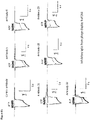
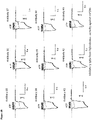
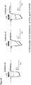
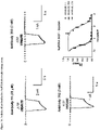
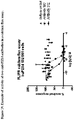
- Purified IgG antibodies were tested for functional activity in the electrophysiology assay and for binding to cells expressing mouse and cynomologus P2X4 using the same method described for the human P2X4 cells described above except a titration of purified IgG sample was used. Results of electrophysiology assays are provided at Figures 3 and 4 .
- Example 6 Identification of hybridoma IgGs that bind specifically to murine P2X4
- Supernatants generated from the immunisations were screened to identify IgGs with specific binding to mP2X4. Briefly supernatants were diluted 10 fold into assay buffer (HBSS, 0.1% (v/v) BSA, 20mM HEPES and 1U/ml Apyrase) and 5 ⁇ l added to the assay plate. Anti-rat detection antibody labeled with Alexa Fluor 647 (Jackson Immuno Research labs) was diluted to 6nM and 10 ⁇ l added to the assay plate. HEK293F cells expressing mP2X4 were diluted to 2.6e 5 /ml and 15 ⁇ l added to the assay plate.
- IgG samples were also tested for non-specific binding in parallel by testing the samples for binding to HEK293F cells.
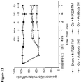
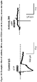
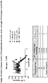
- IgGs demonstrating specific binding to mP2X4 and no binding to HEK293F cells were identified as hits and selected for antibody purification and analysis by electrophysiology. Results of the electrophysiology screen are provided in Figure 7 together with the binding results for these samples against human and cyno P2X4 expressing cell lines using the same assay described previously
- Example 7 Generation of human P2X4 variants and expression by transient transfection in HEK293F cells
- DNA vectors containing huP2X4 sequences with these changes were generated using standard molecular biology techniques. DNA vectors were transfected into HEK293F cells using 293-fectin (Life Technologies 12347019) following the manufacturers guidelines. Cells expressing the huP2X4 variants were incubated with Antibody Nos. 1, 11, 29, and 33 together with the anti-human AlexaFluor 647 (Life Technologies A21445) detection reagent.
- FIGS. 1A-1D show the results of FMAT assays characterizing binding of P2X4 antibodies to HEK293F cells expressing variants of human P2X4.
- QEB QPatch extracellular buffer
- KCl KCl
- MgCl 2 MgCl 2
- CaCl 2 HEPES
- Final composition of compound plate extracellular buffer (CPEB1) was NaCl (137.6), KCl (2.2), MgCl 2 (0.66), CaCl 2 (1.3), HEPES (6.6), KH 2 PO 4 (0.49), NaH 2 PO 4 (2.66).
- pH of extracellular buffers was adjusted to 7.4 with NaOH (1 M), osmolarity was adjusted to 300 mOsm with sucrose and the solutions were 0.2 ⁇ m filtered.
- Compound plate extracellular buffer was supplemented with 0.1% bovine serum albumin.
- the QPatch intracellular buffer contained (in mM) CsF (140), NaCl (10), EGTA (1), HEPES (10). pH of the intracellular buffer was adjusted to 7.3 with CsOH (1 M) and the solution was 0.2 ⁇ m filtered. IgGs were titrated to pH 7.4 with NaOH (1 M).
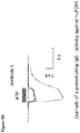
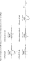
- Exemplar traces showing the effect of inhibitory IgGs 5 mins after IgG application can be seen in Figure 8A whereas an example of a potentiating IgG can be seen in Figure 8D .
- Electrophysiology data presented in Figure 3 and Figure 4 were leak subtracted by subtracting the current in the absence of ligand and the magnitude of the P2X4 response measured as the peak inward current in the presence of ligand. Peak inward current in the presence of IgG+ATP after 5 minutes IgG incubation was expressed as a fraction of the 4 th control ATP response.
- QEB QPatch extracellular buffer
- CPEB2 compound plate extracellular buffer
- pH of extracellular buffers was adjusted to 7.4 with NaOH (1 M) and the solutions were 0.2 ⁇ m filtered.
- the QPatch intracellular buffer contained (in mM) CsF (140), NaCl (10), EGTA (1), HEPES (10). pH of the intracellular buffer was adjusted to 7.3 with CsOH (1 M) and the solution was 0.2 ⁇ m filtered.
- IgGs were titrated to pH 7.4 with NaOH (1 M). After obtaining whole cell configuration, cells were voltage clamped at -50 mV with 70% series resistance compensation employed.
- CPEB2 + IgG was then incubated for 3 minutes followed by a second ATP addition. Data were leak subtracted by subtracting the current in the absence of ligand and the magnitude of the P2X4 response measured as the peak inward current in the presence of ligand.
- the ATP response after IgG addition was expressed as a fraction of the ATP response prior to IgG addition.
- the hIgG1 NIP 228 TM was used as a control antibody to determine the cutoff for defining functional antibodies.
- Example 9 In vivo testing of monoclonal antibodies to P2X4 in Seltzer model of neuropathic pain
- mice 50 female C57BL/6 mice were used for the studies. All mice underwent insertion of transponders for identification purposes at least 5 days before the start of the study. Mechanical hyperalgesia was determined using an analgysemeter (Randall & Selitto 1957) (Ugo Basile). An increasing force was applied to the dorsal surface of each hind paw in turn until a withdrawal response was observed. The application of force was halted at this point and the weight in grams recorded. Data was expressed as withdrawal threshold in grams for ipsilateral and contralateral paws. Following the establishment of baseline readings mice were divided into 2 groups with approximately equal ipsilateral/contralateral ratios which underwent surgery to partially ligate the sciatic nerve or served as sham operated controls.
- mice were anaesthetised with isoflurane. Following this approximately 1cm of the left sciatic nerve was exposed by blunt dissection through an incision at the level of the mid thigh. A suture (9/0 Virgin Silk: Ethicon) was then passed through the dorsal third of the nerve and tied tightly. The incision was then closed using glue and the mice were allowed to recover for at least six days prior to commencement of testing. Sham operated mice underwent the same protocol but following exposure of the nerve the mice were sutured and allowed to recover.
- mice were tested for onset of hyperalgesia on days 7 and 10 post surgery. Any mice showing an ipsilateral/contralateral ratio of greater than 80% were classed as non-responders and removed from the study. Following testing on day 10 mice were further sub-divided into groups giving the final treatment groups;
- Ipsilateral and contralateral readings were taken for each animal at each test time and were entered into EXCEL for calculation of ipsilateral/contralateral ratios. Summary data was transferred into PRISM for graphical and statistical analysis. Results were analysed using 2-way ANOVA. Pairwise comparisons where appropriate were made using Tukey's test.
- Example 10 Generation of mouse anti-human P2X4 antibodies by hybridoma technology
- mice Human P2X4 (hP2X4) transfected HEK 293F and XS63 cells were used to immunise CD1 mice in three groups.
- group 1 mice were immunised with hP2X4 transfected HEK 293F cells
- group 2 mice were immunised with hP2X4 transfected XS63 cells
- group 3 mice were immunised by alternating hP2X4 transfected XS63 cells and hP2X4 transfected HEK 293F cells.
- hP2X4 transfected cells were re-suspended at 1E8/mL and emulsified with equal volumes of complete Freund's adjuvant, and injected into mice at two sites, 100 ⁇ L per site. For the subsequent 3 injections, the same number of cells was emulsified in Freund's incomplete adjuvant and injections were performed as above. The last boost was carried out on day 24, injecting 200 ⁇ L of transfected cells at 1E8/mL intraperitoneally.
- the serum IgG titres to hP2X4 were determined by a cell-based time-resolved fluorescence assays (DELFIA) using parental HEK 293F cells and hP2X4 transfected HEK 293F cells.
- DELFIA cell-based time-resolved fluorescence assays
- Lymphoid cells isolated from spleens and lymph nodes were fused with SP2/0 myeloma cells using an electrofusion method.
- the fusions were plated out into semi-solid selection media containing FITC conjugated goat anti-mouse IgG.
- Supernatants were initially screened for IgGs that specifically bound to hP2X4 using both the hP2X4 expressing HEK 293F and XS63 cells, and parental HEK 293F cells.
- the IgGs that showed specific binding to hP2X4, and no binding to parental HEK293F cells, were selected for further specificity testing on mouse P2X4 (mP2X4) HEK 293F cells.
- IgGs which specifically bound to hP2X4 or to both hP2X4 and mP2X4 were selected for antibody purification and functional analysis by electrophysiology.
- mRNA Messenger RNA
- dT magnetic oligo
- PCR Polymerase chain reaction
- Mouse IgGs of all subclasses (IgG1, IgG2a, IgG2b and IgG3) were purified from overgrown cell culture supernatants on ProPlus resin (Phynexus).
- QEB QPatch extracellular buffer
- CPEB2 compound plate extracellular buffer
- pH of extracellular buffers was adjusted to 7.4 with NaOH (1 M) and the solutions were 0.2 ⁇ m filtered.
- the QPatch intracellular buffer contained (in mM) CsF (140), NaCl (10), EGTA (1), HEPES (10). pH of the intracellular buffer was adjusted to 7.3 with CsOH (1 M) and the solution was 0.2 ⁇ m filtered.
- IgGs were titrated to pH 7.4 with NaOH (1 M). After obtaining whole cell configuration, cells were voltage clamped at - 50 mV with 70% series resistance compensation employed.
- the ligand agonist adenosine 5'-triphosphate disodium salt (ATP, 3 ⁇ M) in QEB was applied for 3 seconds then washed off with QEB + apyrase (1U/ml).
- CPEB2 + IgG was then incubated for 3 minutes followed by a second ATP addition. Data were leak subtracted by subtracting the current in the absence of ligand and the magnitude of the P2X4 response measured as the peak inward current in the presence of ligand.
- the ATP response after IgG addition was expressed as a fraction of the ATP response prior to IgG addition.
- the hIgG1 NIP 228 TM was used as a control antibody to determine the cutoff for defining functional antibodies. Results are provided at Figure 23 .
- Antibody sequences are provided in Figure 13 .
- Antibody No. 11 was optimised for affinity via two approaches either; targeted or random mutagenesis followed by affinity-based phage display selections.
- targeted approach large scFv-phage libraries derived from the lead clone were created by oligonucleotide-directed mutagenesis of the variable heavy (VH) complementarity determining regions 3 (CDR3) and light (VL) chain CDR3 using standard molecular biology techniques as described ( Clackson, T. and Lowman, H.B. Phage Display - A Practical Approach, 2004. Oxford University Press ).
- the libraries were subjected to affinity-based phage display selections in order to select variants with higher affinity for human P2X4.
- Antibody 11 was also optimised using a random mutagenesis approach to identify key residues within the entire variable domain that may improve binding to human P2X4. Such a technique is described by Gram et al. [ Gram et al., 1992, Proc. Natl. Acad. Sci., USA, 89:3576-3580 ], who used error-prone PCR. In some embodiments one or two amino acid substitutions are made within an entire variable domain or set of CDRs. The generated library was subjected to affinity-based selections as described for the targeted selections outlined above.
- Antibodies 287 to 315 Exemplary antibodies from this selection method are disclosed herein as Antibodies 287 to 315, and an alignment of their sequences is shown in Figure 12 .
- assay buffer HBSS, 0.1% BSA, 1U/ml apyrase, either with or without 20mM HEPES.
- Antibody 11-Dylight ® 650 was added to each well except the wells used to define the background binding, to a final concentration of 2nM.
- HEK293F huP2X4 cells were added to each well at a final density of approximately 2000 cells per well. Plates were covered and incubated at room temperature for 2 to 3 hours before reading on a mirrorball ® plate reader (TTP Labtech, Ltd) and determining the total FL3 fluorescence per well (Median (mean intensity) fluorescence multiplied by the number of objects). Individual events were gated on size and fluorescence and a minimum object number of greater than 25 was used to determine wells with sufficient events to report a FL3 total value.
- scFv antibodies were diluted in assay buffer described above to generate a dilution series and the diluted samples were added to the assay plate before the addition of Antibody 11- DyLight ® 650 to a final concentration of 2nM, followed by approximately 2000 HEK293F huP2X4 cells per well. Plates were incubated at room temperature for 2 to 3 hours before being read on the mirrorball ® plate reader. Data was analysed as described above and scFv clones showing inhibition were generated as full IgG antibodies.
- Example 13 Identification of antibodies with improved potency against human P2X4 using the human P2X4 1321N1 cell line FLIPR ® assay
- Antibodies identified in the Antibody 11 competition assay described in example 12 were generated as purified IgG and titrated to generate a dilution series. These antibodies were diluted in assay buffer containing HBSS and 0.1% BSA and pre-incubated with 1321N1 cells expressing huP2X4 for 30mins where the cells had previously been loaded with Fluo-4 NW calcium dye (Molecular Probes TM , Life Technologies) following the manufacturer's instructions. P2X4 was activated by the addition of 1 ⁇ M ATP diluted in assay buffer and the resulting rise in intracellular calcium was detected by the calcium dye and measured by an increase in fluorescence using the FLIPR ® Tetra plate reader (Molecular Devices, LLC).
- Example 14 Identification of antibodies with improved potency against human P2X4 using the HEK 293F huP2X4 cell line on the automated electrophysiology platform Qpatch 16X.
- HEK 293F cells stably expressing human P2X4, were harvested at 50% confluency using accutase. Cells were then resuspended in 10 ml Freestyle 293F media supplemented with HEPES (10 mM) + apyrase (1U/ml, ATPase/ADPase activity 1) at a density of 2-3e 6 cells/ml. P2X4 function was assayed using the automated electrophysiology platform QPatch 16X (Sophion) in population patch configuration. Composition of QPatch extracellular buffer (QEB) was (in mM) NaCl (140), KCl (2), MgCl 2 (1) CaCl 2 (2), HEPES (10).
- QEB QPatch extracellular buffer
- the QPatch intracellular buffer contained (in mM) CsF (140), NaCl (10), EGTA (1), HEPES (10). pH of the intracellular buffer was adjusted to 7.3 with CsOH (1 M) and the solution was 0.2 ⁇ m filtered. IgGs were titrated to pH 7.4 with NaOH (1 M).
- IgGs were serially diluted in QEB + 0.1% bovine serum albumin and tested for function on Qpatch 16X in population patch configuration.
- Extracellular buffer was QEB
- intracellular buffer was QIB
- ATP wash buffer was QEB + apyrase (1U/ml).
- ATP 3 ⁇ M was applied every 10 mins for 3 s with a total of 5 applications per experiment.
- the first two ATP additions (ATP 1 & ATP 2) were preceded by preincubation for 5 mins with QEB buffer + 0.1 % BSA whereas the following three ATP additions were preceded by 5 mins incubation with ascending doses of IgG.
- Example 15 Potency determination of hybridoma derived antibodies at mouse and human P2X4.
- HEK 293F cells expressing P2X4 were handled as in example 8.
- Potency of hybridoma derived IgGs was assayed on Qpatch 16X in population patch configuration.
- IgG potency IgGs were serially diluted in PBS + 0.1% bovine serum albumin and tested for function on Qpatch 16X in population patch configuration.
- IgGs were then diluted 1:3 in QEB + 0.1% BSA resulting in a final buffer composition of NaCl (137.6), KCl (2.2), MgCl 2 (0.66), CaCl 2 (1.3), HEPES (6.6), KH 2 PO 4 (0.49), NaH 2 PO 4 (2.66), BSA (0.1%) equivalent to CPEB1.
- Extracellular buffer was QEB
- intracellular buffer was QIB
- ATP wash buffer was QEB + apyrase (1U/ml).
- ATP 3 ⁇ M was applied every 10 mins for 3 s with a total of 5 applications per experiment.
- ATP 1 & ATP 2 The first two ATP additions (ATP 1 & ATP 2) were preceded by preincubation for 5 mins with CPEB1 + 0.1 % BSA whereas the following three ATP additions were preceded by 5 mins incubation with ascending doses of IgG.
- Log and half log doses were interleaved in post analysis to generate 6 point dose response curves. Data were leak subtracted by subtracting the current in the absence of ligand and the magnitude of the P2X4 response measured as the peak inward current in the presence of ligand. The peak inward current in response to ATP was expressed as fraction of control current (ATP2). Data were fit in Prism using a log (inhibitor) vs. response - Variable slope (four parameters) equation.
- mice Primary mouse microglia were cultured from C57 neonatal pups, P2. Brains were removed from the skulls of mice and kept in media (DMEM + 10% FCS + pen/strep). They were then rolled across filter paper to remove the sticky vasculature and meninges before placing in 20 ml fresh media and triturating to give a single cell suspension. Cells were then filter sterilised through a 40 ⁇ m cell strainer then centrifuged at 1200rpm for 5 min. Cells were then resuspended in 40ml media per flask at 4 brains per T175 flask and cultured for 1 week.
- media DMEM + 10% FCS + pen/strep
- Microglia were removed by shaking overnight in an orbital shaker incubator (no CO 2 ) with HEPES supplemented in the media (20 mM). Purified microglia were centrifuged at 1200 rpm for 5 mins and resuspended in 20 ml DMEM + 10% FCS + pen/strep growth media. Cells were counted and seeded in ultra low bind T75 cell culture flasks (Corning) at 7e6 cells/flask. Microglia were then maintained in culture for 1-7 days before being used for Qpatch 16X electrophysiology assays or FLIPR calcium imaging assays.
- Qpatch 16X was used in population patch configuration and cells voltage clamped at -70 mV. Cells were perfused with either a control antibody or test antibody for 5 minutes before ATP (30 ⁇ M) was applied. Current in the absence of ATP was subtracted from all data. Inward current in response to ATP was measured (see Figure 15 ). External buffer was QEB and internal buffer was QIB. See Figure 17 & 18 .
- Microglia were plated in Cell Coat Poly-D-Lysine coated 384 well plates (black, uclear) with 30 ⁇ l per well and cultured in a humidified incubator at 37 °C for 48 hours.
- ATP (30 uM) was made up in HBSS + 20 mM HEPES + 0.1 % BSA in a separate 384 well compound plate (Compound plate 3 ). Fluo-8 was excited at a wavelength of 470-495 nm and the emitted light measured at a wavelength of 515-575 nm. Camera gain was adjusted to give 1000 counts at rest with an exposure of 0.4 s. 10 ul of solution from compound plate 1 was added to the cells and the fluorescence measured. After 5 mins incubation, 10 ul of solution from compound plate 2 was added. 15 min later, ATP (5 uM final) was added and the peak end fluorescence measured between 200-300 sec post ATP addition.
- Fluorescence counts were normalised to the ATP response in the absence of antibody (minus background fluorescence) and plotted as % of ATP response (See figure 21 ).
- 10 point dose response curves for each IgG were constructed from duplicate wells and the data fit in Prism using a log (inhibitor) vs. response - Variable slope (four parameters) equation.
- Y Bottom + (Top-Bottom)/(1+10 ⁇ ((LogIC50-X) ⁇ HillSlope)). See Figure 20 .
- Human monocytes were isolated from the mononuclear fraction of peripheral blood by centrigugation on a Ficoll-Paque gradient. Cells were then purified by incubating in a T175 cell culture flask in cell culture media in the absence of serum for 1 hour. Non-adherent cells were removed and the remaining cells grown in RPMI Glutamax I media supplemented with 10% FCS (HI/GI) + 1% P/S + 100 ng/ml M-CSF for 7 days. Cells were fed on day 2-3 by adding an additional 10 ml of media. Macrophage were harvested by accutase treatment for 10 mins followed by cell scraping and replated in ultra-low bind T75 flasks at 6e6 cells per flask.
Landscapes
- Health & Medical Sciences (AREA)
- Chemical & Material Sciences (AREA)
- Organic Chemistry (AREA)
- Immunology (AREA)
- Life Sciences & Earth Sciences (AREA)
- General Health & Medical Sciences (AREA)
- Medicinal Chemistry (AREA)
- Biochemistry (AREA)
- Biophysics (AREA)
- Genetics & Genomics (AREA)
- Molecular Biology (AREA)
- Proteomics, Peptides & Aminoacids (AREA)
- Bioinformatics & Cheminformatics (AREA)
- Pharmacology & Pharmacy (AREA)
- Pain & Pain Management (AREA)
- Biomedical Technology (AREA)
- Neurology (AREA)
- Neurosurgery (AREA)
- Chemical Kinetics & Catalysis (AREA)
- General Chemical & Material Sciences (AREA)
- Nuclear Medicine, Radiotherapy & Molecular Imaging (AREA)
- Engineering & Computer Science (AREA)
- Animal Behavior & Ethology (AREA)
- Public Health (AREA)
- Veterinary Medicine (AREA)
- Peptides Or Proteins (AREA)
- Medicines Containing Antibodies Or Antigens For Use As Internal Diagnostic Agents (AREA)
- Medicines That Contain Protein Lipid Enzymes And Other Medicines (AREA)
- Pharmaceuticals Containing Other Organic And Inorganic Compounds (AREA)
- Preparation Of Compounds By Using Micro-Organisms (AREA)
- Micro-Organisms Or Cultivation Processes Thereof (AREA)
Applications Claiming Priority (3)
| Application Number | Priority Date | Filing Date | Title |
|---|---|---|---|
| US201461987929P | 2014-05-02 | 2014-05-02 | |
| EP15720962.8A EP3137501B1 (fr) | 2014-05-02 | 2015-05-02 | Modulateurs des canaux ioniques |
| PCT/EP2015/059633 WO2015166105A2 (fr) | 2014-05-02 | 2015-05-02 | Modulateurs de canal ionique et leur utilisation |
Related Parent Applications (2)
| Application Number | Title | Priority Date | Filing Date |
|---|---|---|---|
| EP15720962.8A Division-Into EP3137501B1 (fr) | 2014-05-02 | 2015-05-02 | Modulateurs des canaux ioniques |
| EP15720962.8A Division EP3137501B1 (fr) | 2014-05-02 | 2015-05-02 | Modulateurs des canaux ioniques |
Publications (2)
| Publication Number | Publication Date |
|---|---|
| EP4001310A2 true EP4001310A2 (fr) | 2022-05-25 |
| EP4001310A3 EP4001310A3 (fr) | 2022-08-10 |
Family
ID=53055033
Family Applications (2)
| Application Number | Title | Priority Date | Filing Date |
|---|---|---|---|
| EP21191978.2A Withdrawn EP4001310A3 (fr) | 2014-05-02 | 2015-05-02 | Modulateurs des canaux ioniques |
| EP15720962.8A Active EP3137501B1 (fr) | 2014-05-02 | 2015-05-02 | Modulateurs des canaux ioniques |
Family Applications After (1)
| Application Number | Title | Priority Date | Filing Date |
|---|---|---|---|
| EP15720962.8A Active EP3137501B1 (fr) | 2014-05-02 | 2015-05-02 | Modulateurs des canaux ioniques |
Country Status (8)
| Country | Link |
|---|---|
| US (2) | US10654926B2 (fr) |
| EP (2) | EP4001310A3 (fr) |
| JP (2) | JP2017515473A (fr) |
| AU (1) | AU2015254558B2 (fr) |
| BR (1) | BR112016025450A2 (fr) |
| CA (1) | CA2946669A1 (fr) |
| ES (1) | ES2899754T3 (fr) |
| WO (1) | WO2015166105A2 (fr) |
Families Citing this family (7)
| Publication number | Priority date | Publication date | Assignee | Title |
|---|---|---|---|---|
| CN110662771B (zh) * | 2018-02-01 | 2023-07-28 | 南京驯鹿生物技术股份有限公司 | 一种结合bcma的嵌合抗原受体(car)及其应用 |
| WO2020186158A2 (fr) * | 2019-03-13 | 2020-09-17 | Ulmert Hans David Staffan | Molécules de liaison de prame et leurs utilisations |
| CN115244082A (zh) * | 2019-08-23 | 2022-10-25 | 新墨西哥大学雨林创新 | 用于疼痛管理的非阿片类组合物和疗法 |
| CA3161825A1 (fr) * | 2019-11-18 | 2021-05-27 | Janssen Biotech, Inc. | Recepteurs antigeniques chimeriques anti-cd79, cellules car-t, et leurs utilisations |
| WO2022082059A1 (fr) * | 2020-10-16 | 2022-04-21 | Senti Biosciences, Inc. | Récepteurs chimériques et procédés d'utilisation de ces derniers |
| CN114685657A (zh) * | 2020-12-31 | 2022-07-01 | 康诺亚生物医药科技(成都)有限公司 | 一种功能增强型抗体阻断剂的开发及其应用 |
| WO2023114962A1 (fr) * | 2021-12-16 | 2023-06-22 | Unm Rainforest Innovations | Composition non opioïde humanisée et thérapies pour la gestion de la douleur |
Citations (8)
| Publication number | Priority date | Publication date | Assignee | Title |
|---|---|---|---|---|
| US4522811A (en) | 1982-07-08 | 1985-06-11 | Syntex (U.S.A.) Inc. | Serial injection of muramyldipeptides and liposomes enhances the anti-infective activity of muramyldipeptides |
| WO1987005330A1 (fr) | 1986-03-07 | 1987-09-11 | Michel Louis Eugene Bergh | Procede pour ameliorer la stabilite des glycoproteines |
| US4816567A (en) | 1983-04-08 | 1989-03-28 | Genentech, Inc. | Recombinant immunoglobin preparations |
| WO1992001047A1 (fr) | 1990-07-10 | 1992-01-23 | Cambridge Antibody Technology Limited | Procede de production de chainon de paires a liaison specifique |
| US5565332A (en) | 1991-09-23 | 1996-10-15 | Medical Research Council | Production of chimeric antibodies - a combinatorial approach |
| US5624821A (en) | 1987-03-18 | 1997-04-29 | Scotgen Biopharmaceuticals Incorporated | Antibodies with altered effector functions |
| US6030613A (en) | 1995-01-17 | 2000-02-29 | The Brigham And Women's Hospital, Inc. | Receptor specific transepithelial transport of therapeutics |
| WO2010093061A1 (fr) | 2009-02-16 | 2010-08-19 | 日本ケミファ株式会社 | Dérivé de diazépinedione |
Family Cites Families (6)
| Publication number | Priority date | Publication date | Assignee | Title |
|---|---|---|---|---|
| US6242216B1 (en) * | 1997-11-14 | 2001-06-05 | Abbott Laboratories | Nucleic acids encoding a functional human purinoreceptor P2X2 and P2X4, and methods of production and use thereof |
| AUPP991199A0 (en) * | 1999-04-21 | 1999-05-13 | University Of Sydney, The | Methods for diagnosing pre-cancerous and cancerous conditions |
| AUPR201500A0 (en) * | 2000-12-11 | 2001-01-11 | Biosceptre Pty Ltd | Methods for identifying pre-neoplastic and neoplastic states in mammals |
| US20050074819A1 (en) | 2003-10-01 | 2005-04-07 | Japan Health Sciences Foundation | Screening method of drug for treatment of neuropathic pain |
| US20080287467A1 (en) * | 2004-05-06 | 2008-11-20 | Gen Tamura | Therapeutic Agent for Respiratory Diseases |
| UA118028C2 (uk) * | 2013-04-03 | 2018-11-12 | Рош Глікарт Аг | Біспецифічне антитіло, специфічне щодо fap і dr5, антитіло, специфічне щодо dr5, і спосіб їх застосування |
-
2015
- 2015-05-02 CA CA2946669A patent/CA2946669A1/fr not_active Abandoned
- 2015-05-02 EP EP21191978.2A patent/EP4001310A3/fr not_active Withdrawn
- 2015-05-02 ES ES15720962T patent/ES2899754T3/es active Active
- 2015-05-02 EP EP15720962.8A patent/EP3137501B1/fr active Active
- 2015-05-02 US US15/308,294 patent/US10654926B2/en active Active
- 2015-05-02 BR BR112016025450A patent/BR112016025450A2/pt not_active Application Discontinuation
- 2015-05-02 WO PCT/EP2015/059633 patent/WO2015166105A2/fr active Application Filing
- 2015-05-02 JP JP2016565470A patent/JP2017515473A/ja active Pending
- 2015-05-02 AU AU2015254558A patent/AU2015254558B2/en active Active
-
2019
- 2019-09-12 JP JP2019166248A patent/JP2020018307A/ja active Pending
-
2020
- 2020-04-06 US US16/840,910 patent/US20200247885A1/en not_active Abandoned
Patent Citations (10)
| Publication number | Priority date | Publication date | Assignee | Title |
|---|---|---|---|---|
| US4522811A (en) | 1982-07-08 | 1985-06-11 | Syntex (U.S.A.) Inc. | Serial injection of muramyldipeptides and liposomes enhances the anti-infective activity of muramyldipeptides |
| US4816567A (en) | 1983-04-08 | 1989-03-28 | Genentech, Inc. | Recombinant immunoglobin preparations |
| WO1987005330A1 (fr) | 1986-03-07 | 1987-09-11 | Michel Louis Eugene Bergh | Procede pour ameliorer la stabilite des glycoproteines |
| US5624821A (en) | 1987-03-18 | 1997-04-29 | Scotgen Biopharmaceuticals Incorporated | Antibodies with altered effector functions |
| US5648260A (en) | 1987-03-18 | 1997-07-15 | Scotgen Biopharmaceuticals Incorporated | DNA encoding antibodies with altered effector functions |
| WO1992001047A1 (fr) | 1990-07-10 | 1992-01-23 | Cambridge Antibody Technology Limited | Procede de production de chainon de paires a liaison specifique |
| US5565332A (en) | 1991-09-23 | 1996-10-15 | Medical Research Council | Production of chimeric antibodies - a combinatorial approach |
| US6030613A (en) | 1995-01-17 | 2000-02-29 | The Brigham And Women's Hospital, Inc. | Receptor specific transepithelial transport of therapeutics |
| WO2010093061A1 (fr) | 2009-02-16 | 2010-08-19 | 日本ケミファ株式会社 | Dérivé de diazépinedione |
| EP2397480A1 (fr) | 2009-02-16 | 2011-12-21 | Nippon Chemiphar Co., Ltd. | Dérivé de diazépinedione |
Non-Patent Citations (49)
| Title |
|---|
| "Fundamental Immunology", 1993, RAVEN PRESS |
| "NCBI", Database accession no. NM 002560 |
| ALTSHUL ET AL., J. MOL. BIOL., vol. 215, 1990, pages 403 - 410 |
| ANTONIO ET AL., BR. J. PHARMACOL., vol. 163, 2011, pages 1069 - 1077 |
| APLIN ET AL., CRC CRIT. REV. BIOCHEM., vol. 22, 1981, pages 259 - 306 |
| AUSUBEL: "Current Protocols in Molecular Biology", 1987, JOHN WILEY & SONS |
| BACKMARK ET AL., PROTEIN SCI, vol. 22, 2013, pages 1124 - 1132 |
| BANNISTER ET AL., BIOTECHNOLOGY AND BIOENGINEERING, vol. 94, 2006, pages 931 - 937 |
| BARBAS ET AL., PROC. NAT. ACAD. SCI. U.S.A., vol. 370, 1994, pages 3809 - 3813 |
| CAS , no. 250692-65-0 |
| CLACKSON, T.LOWMAN, H.B.: "Phage Display - A Practical Approach", 2004, OXFORD UNIVERSITY PRESS |
| COLIGAN: "Current Protocols in Immunology", 1991 |
| DIETZ ET AL., CYTOMETRY, vol. 23, 1996, pages 177 - 186 |
| EDGE ET AL., ANAL. BIOCHEM., vol. 118, 1981, pages 131 |
| ENGSTOM, BIOCHEM. EXP. BIOL., vol. 11, 1974, pages 7 - 13 |
| FLETTERICK ET AL.: "Computer Graphics and Molecular Modeling, in Current Communications in Molecular Biology", 1986, COLD SPRING HARBOR LABORATORY |
| GAIT: "Oligonucleotide Synthesis", 1984 |
| GRAM ET AL., PROC. NAT. ACAD. SCI. U.S.A., vol. 89, 1992, pages 3576 - 3580 |
| HAKIMUDDIN ET AL., ARCH. BIOCHEM. BIOPHYS., vol. 259, 1987, pages 52 |
| KAWATE ET AL., NATURE, vol. 460, 2009, pages 592 - 598 |
| KAWATE ET AL., STRUCTURE, vol. 14, 2006, pages 673 - 681 |
| KOHLERMILSTEIN, NATURE, vol. 256, 1975, pages 495 - 499 |
| LLOYD, PROTEIN ENG DES SEL, vol. 22, 2009, pages 159 - 168 |
| LUND ET AL., J. IMMUN., vol. 147, 1991, pages 2657 - 2662 |
| MACLENNAN ET AL., ACTA PHYSIOL. SCAND. SUPPL., vol. 643, 1998, pages 55 - 67 |
| MANIATIS: "Molecular Cloning, A Laboratory Manual", 1990, COLD SPRING HARBOR LABORATORY |
| MARKS ET AL., BIO/TECHNOLOGY, vol. 10, 1992, pages 779 - 783 |
| MEYERS ET AL., COMPUT. APPL. BIOSCI., vol. 4, 1988, pages 11 - 17 |
| MILLERCALOS: "Gene Transfer Vectors for Mammalian Cells", 1987 |
| MIRAGLIA ET AL., J. BIOMOL. SCREENING, vol. 4, 1999, pages 193 - 204 |
| MORGAN ET AL., IMMUNOLOGY, vol. 86, 1995, pages 319 - 324 |
| MULLIS: "PCR: The Polymerase Chain Reaction", 1994 |
| NAKAZAWA ET AL., EUROPEAN JOURNAL OF PHARMACOLOGY, vol. 518, 2005, pages 107 - 110 |
| NEEDLEMAN ET AL., J. MOL. BIOL., vol. 48, 1970, pages 444 - 453 |
| NICKE ET AL., MOL. PHARMACOL., vol. 63, 2003, pages 243 - 252 |
| OGANESYAN ET AL., ACTA CRYSTALLOGR D BIOL CRYSTALLOGR, vol. 64, 2008, pages 700 - 704 |
| OSBOURN, J.K. ET AL., IMMUNOTECHNOLOGY, vol. 2, no. 3, 1996, pages 181 - 196 |
| PACE ET AL., PROTEIN SCI, vol. 4, 1995, pages 2411 - 23 |
| PERSIC ET AL., GENE, vol. 187, 1997, pages 9 - 18 |
| SAMBROOK ET AL.: "Molecular Cloning: A Laboratory Manual", 1989, COLD SPRING HARBOR LABORATORY PRESS |
| SASAKI ET AL., ADV. BIOPHYS., vol. 35, 1998, pages 1 - 24 |
| SCHIER ET AL., J. MOL. BIOL., vol. 263, 1996, pages 551 - 567 |
| THOMPSON, J ET AL., J MOL BIOL., vol. 256, no. 1, 1996, pages 77 - 88 |
| THOTAKURA ET AL., METH. ENZYMOL., vol. 138, 1987, pages 350 |
| VALENTE ET AL., BIOCHIM. BIOPHYS. ACTA, vol. 1808, 2011, pages 2859 - 2866 |
| VAUGHAN ET AL., NATURE BIOTECHNOLOGY, vol. 14, 1996, pages 309 - 314 |
| VAUGHAN ET AL., NATURE BIOTECHNOLOGY, vol. 14, pages 309 - 314 |
| WEIR: "Handbook of Experimental Immunology", 1996, article "Methods in Enzymology" |
| YOUNG ET AL., J. BIOL. CHEM., vol. 283, 2008, pages 26241 - 26251 |
Also Published As
| Publication number | Publication date |
|---|---|
| US20170166634A1 (en) | 2017-06-15 |
| ES2899754T3 (es) | 2022-03-14 |
| WO2015166105A2 (fr) | 2015-11-05 |
| EP3137501A2 (fr) | 2017-03-08 |
| JP2020018307A (ja) | 2020-02-06 |
| JP2017515473A (ja) | 2017-06-15 |
| EP3137501B1 (fr) | 2021-09-29 |
| BR112016025450A2 (pt) | 2017-12-12 |
| US10654926B2 (en) | 2020-05-19 |
| WO2015166105A3 (fr) | 2015-12-30 |
| AU2015254558A1 (en) | 2016-12-08 |
| EP4001310A3 (fr) | 2022-08-10 |
| CA2946669A1 (fr) | 2015-11-05 |
| AU2015254558B2 (en) | 2021-02-25 |
| US20200247885A1 (en) | 2020-08-06 |
Similar Documents
| Publication | Publication Date | Title |
|---|---|---|
| US20200247885A1 (en) | P2x4 antibodies & uses thereof | |
| WO2020200196A1 (fr) | Anticorps anti-claudine 18.2 et utilisation associée | |
| US20040142382A1 (en) | Neutralizing antibodies against GDF-8 and uses therefor | |
| JP2007525434A (ja) | T細胞、免疫グロブリンドメインおよびムチンドメイン1(tim−1)抗原に対する抗体およびその使用。 | |
| US10894833B2 (en) | Agents, uses and methods for treatment | |
| KR20150136031A (ko) | 인간화 또는 친화도 성숙된 항 앤지오포이에틴-2 항체 및 그의 용도 | |
| US20210347899A1 (en) | Anti-klrg1 antibodies | |
| US20240132600A1 (en) | Tetravalent FZD and WNT Co-receptor Binding Antibody Molecules and Uses Thereof | |
| JP2022527372A (ja) | 抗インテグリン抗体及びそれらの使用 | |
| KR20200068601A (ko) | 엔도텔린 수용체 a 활성 조절 항체 | |
| CN114478771A (zh) | Ox40抗体及其医药用途 | |
| US20170183401A1 (en) | Hypoglycemic agent containing anti-ang2 antibody | |
| JP7457880B2 (ja) | 新規なNav1.7モノクローナル抗体 | |
| US11834512B2 (en) | Inhibiting anti-ENPP1 antibodies | |
| KR20230158526A (ko) | Ccr9에 결합하는 치료용 결합 분자 | |
| WO2023250402A2 (fr) | Molécules d'anticorps de liaison au co-récepteur wnt et fzd tétravalent et leurs utilisations | |
| CN116082513A (zh) | 分离的抗原结合蛋白及其用途 | |
| KR20210158693A (ko) | 항 pcsk9 항체 및 이의 용도 | |
| JP2023169439A (ja) | 抗ヒトtrpv2抗体 | |
| KR20240026496A (ko) | 항-넥틴4 항체 및 이를 포함하는 다중특이적 단백질 복합체 |
Legal Events
| Date | Code | Title | Description |
|---|---|---|---|
| PUAI | Public reference made under article 153(3) epc to a published international application that has entered the european phase |
Free format text: ORIGINAL CODE: 0009012 |
|
| STAA | Information on the status of an ep patent application or granted ep patent |
Free format text: STATUS: THE APPLICATION HAS BEEN PUBLISHED |
|
| AC | Divisional application: reference to earlier application |
Ref document number: 3137501 Country of ref document: EP Kind code of ref document: P |
|
| AK | Designated contracting states |
Kind code of ref document: A2 Designated state(s): AL AT BE BG CH CY CZ DE DK EE ES FI FR GB GR HR HU IE IS IT LI LT LU LV MC MK MT NL NO PL PT RO RS SE SI SK SM TR |
|
| PUAL | Search report despatched |
Free format text: ORIGINAL CODE: 0009013 |
|
| AK | Designated contracting states |
Kind code of ref document: A3 Designated state(s): AL AT BE BG CH CY CZ DE DK EE ES FI FR GB GR HR HU IE IS IT LI LT LU LV MC MK MT NL NO PL PT RO RS SE SI SK SM TR |
|
| RIC1 | Information provided on ipc code assigned before grant |
Ipc: A61P 25/04 20060101ALI20220707BHEP Ipc: C07K 16/28 20060101AFI20220707BHEP |
|
| STAA | Information on the status of an ep patent application or granted ep patent |
Free format text: STATUS: THE APPLICATION IS DEEMED TO BE WITHDRAWN |
|
| 18D | Application deemed to be withdrawn |
Effective date: 20230211 |