EP3989840B1 - Sampling device for biological specimen - Google Patents
Sampling device for biological specimen Download PDFInfo
- Publication number
- EP3989840B1 EP3989840B1 EP20734946.5A EP20734946A EP3989840B1 EP 3989840 B1 EP3989840 B1 EP 3989840B1 EP 20734946 A EP20734946 A EP 20734946A EP 3989840 B1 EP3989840 B1 EP 3989840B1
- Authority
- EP
- European Patent Office
- Prior art keywords
- sampling device
- sample
- support body
- swab tip
- tip
- Prior art date
- Legal status (The legal status is an assumption and is not a legal conclusion. Google has not performed a legal analysis and makes no representation as to the accuracy of the status listed.)
- Active
Links
Images


Classifications
-
- B—PERFORMING OPERATIONS; TRANSPORTING
- B01—PHYSICAL OR CHEMICAL PROCESSES OR APPARATUS IN GENERAL
- B01L—CHEMICAL OR PHYSICAL LABORATORY APPARATUS FOR GENERAL USE
- B01L3/00—Containers or dishes for laboratory use, e.g. laboratory glassware; Droppers
- B01L3/50—Containers for the purpose of retaining a material to be analysed, e.g. test tubes
- B01L3/502—Containers for the purpose of retaining a material to be analysed, e.g. test tubes with fluid transport, e.g. in multi-compartment structures
- B01L3/5029—Containers for the purpose of retaining a material to be analysed, e.g. test tubes with fluid transport, e.g. in multi-compartment structures using swabs
-
- A—HUMAN NECESSITIES
- A61—MEDICAL OR VETERINARY SCIENCE; HYGIENE
- A61B—DIAGNOSIS; SURGERY; IDENTIFICATION
- A61B10/00—Instruments for taking body samples for diagnostic purposes; Other methods or instruments for diagnosis, e.g. for vaccination diagnosis, sex determination or ovulation-period determination; Throat striking implements
- A61B10/0045—Devices for taking samples of body liquids
-
- A—HUMAN NECESSITIES
- A61—MEDICAL OR VETERINARY SCIENCE; HYGIENE
- A61B—DIAGNOSIS; SURGERY; IDENTIFICATION
- A61B10/00—Instruments for taking body samples for diagnostic purposes; Other methods or instruments for diagnosis, e.g. for vaccination diagnosis, sex determination or ovulation-period determination; Throat striking implements
- A61B10/02—Instruments for taking cell samples or for biopsy
-
- A—HUMAN NECESSITIES
- A61—MEDICAL OR VETERINARY SCIENCE; HYGIENE
- A61F—FILTERS IMPLANTABLE INTO BLOOD VESSELS; PROSTHESES; DEVICES PROVIDING PATENCY TO, OR PREVENTING COLLAPSING OF, TUBULAR STRUCTURES OF THE BODY, e.g. STENTS; ORTHOPAEDIC, NURSING OR CONTRACEPTIVE DEVICES; FOMENTATION; TREATMENT OR PROTECTION OF EYES OR EARS; BANDAGES, DRESSINGS OR ABSORBENT PADS; FIRST-AID KITS
- A61F13/00—Bandages or dressings; Absorbent pads
- A61F13/15—Absorbent pads, e.g. sanitary towels, swabs or tampons for external or internal application to the body; Supporting or fastening means therefor; Tampon applicators
- A61F13/38—Swabs having a stick-type handle, e.g. cotton tips
-
- G—PHYSICS
- G01—MEASURING; TESTING
- G01N—INVESTIGATING OR ANALYSING MATERIALS BY DETERMINING THEIR CHEMICAL OR PHYSICAL PROPERTIES
- G01N1/00—Sampling; Preparing specimens for investigation
- G01N1/02—Devices for withdrawing samples
-
- G—PHYSICS
- G01—MEASURING; TESTING
- G01N—INVESTIGATING OR ANALYSING MATERIALS BY DETERMINING THEIR CHEMICAL OR PHYSICAL PROPERTIES
- G01N1/00—Sampling; Preparing specimens for investigation
- G01N1/02—Devices for withdrawing samples
- G01N1/10—Devices for withdrawing samples in the liquid or fluent state
- G01N1/14—Suction devices, e.g. pumps; Ejector devices
-
- A—HUMAN NECESSITIES
- A61—MEDICAL OR VETERINARY SCIENCE; HYGIENE
- A61B—DIAGNOSIS; SURGERY; IDENTIFICATION
- A61B10/00—Instruments for taking body samples for diagnostic purposes; Other methods or instruments for diagnosis, e.g. for vaccination diagnosis, sex determination or ovulation-period determination; Throat striking implements
- A61B10/02—Instruments for taking cell samples or for biopsy
- A61B2010/0216—Sampling brushes
-
- B—PERFORMING OPERATIONS; TRANSPORTING
- B01—PHYSICAL OR CHEMICAL PROCESSES OR APPARATUS IN GENERAL
- B01L—CHEMICAL OR PHYSICAL LABORATORY APPARATUS FOR GENERAL USE
- B01L2300/00—Additional constructional details
- B01L2300/06—Auxiliary integrated devices, integrated components
- B01L2300/0681—Filter
-
- B—PERFORMING OPERATIONS; TRANSPORTING
- B01—PHYSICAL OR CHEMICAL PROCESSES OR APPARATUS IN GENERAL
- B01L—CHEMICAL OR PHYSICAL LABORATORY APPARATUS FOR GENERAL USE
- B01L2300/00—Additional constructional details
- B01L2300/08—Geometry, shape and general structure
- B01L2300/0832—Geometry, shape and general structure cylindrical, tube shaped
- B01L2300/0838—Capillaries
-
- B—PERFORMING OPERATIONS; TRANSPORTING
- B01—PHYSICAL OR CHEMICAL PROCESSES OR APPARATUS IN GENERAL
- B01L—CHEMICAL OR PHYSICAL LABORATORY APPARATUS FOR GENERAL USE
- B01L2400/00—Moving or stopping fluids
- B01L2400/04—Moving fluids with specific forces or mechanical means
- B01L2400/0475—Moving fluids with specific forces or mechanical means specific mechanical means and fluid pressure
- B01L2400/0487—Moving fluids with specific forces or mechanical means specific mechanical means and fluid pressure fluid pressure, pneumatics
- B01L2400/049—Moving fluids with specific forces or mechanical means specific mechanical means and fluid pressure fluid pressure, pneumatics vacuum
-
- G—PHYSICS
- G01—MEASURING; TESTING
- G01N—INVESTIGATING OR ANALYSING MATERIALS BY DETERMINING THEIR CHEMICAL OR PHYSICAL PROPERTIES
- G01N1/00—Sampling; Preparing specimens for investigation
- G01N1/02—Devices for withdrawing samples
- G01N2001/028—Sampling from a surface, swabbing, vaporising
-
- G—PHYSICS
- G01—MEASURING; TESTING
- G01N—INVESTIGATING OR ANALYSING MATERIALS BY DETERMINING THEIR CHEMICAL OR PHYSICAL PROPERTIES
- G01N1/00—Sampling; Preparing specimens for investigation
- G01N1/02—Devices for withdrawing samples
- G01N1/10—Devices for withdrawing samples in the liquid or fluent state
- G01N1/14—Suction devices, e.g. pumps; Ejector devices
- G01N2001/1445—Overpressure, pressurisation at sampling point
Definitions
- the present invention lies in the field of sampling devices for biological specimen.
- the present invention is directed to a sampling device for simultaneous collection, isolation and purification of a biological specimen from a sample or from a location.
- the application further relates to a method for the collection, isolation and purification of a biological specimen from a sample or from a location using said sampling device.
- Standardization of sample collection methodologies is often lacking, which leads to inaccurate results because of several reasons: improper collection methodologies, over dilution of samples, poor storage conditions (temperature, time, leakage), bacterial overgrowth, hemolysis in sera, (partial) degeneration of infectious agents and disintegration of pathogens' nucleic acids.
- Diagnostic matrices such as faeces, respiratory secretions, blood, urine and semen also contain impurities and substances (e.g. heme and metabolites, acidic polysaccharides, bile salts, lipids). which have known inhibitory effects on downstream diagnostics such as (real-time) PCR and sequencing.
- intracellular pathogens hiding in white blood cells and thus requiring cell lysis, are often missed by routine diagnostics.
- next- and third-generation sequencing technologies also becomes more important for diagnostics and will replace many of the currently existing PCR-based assays on the long term.
- Respiratory and enteric disease problems are mostly the result of a complex of different pathogens including viruses and bacteria.
- Simultaneous detection of all pathogens, known and novel, present in a sample is only possible using metagenomics.
- the genomes of pathogens are sequenced and compared to reference databases to see if certain pathogens are present or not. Meanwhile, full genomes can be assembled to provide strain information to adapt prevention/therapeutic actions. This is impossible with other diagnostic procedures.
- a hurdle for its widespread use in the field is the typical low abundance of viral nucleic acids in a sample compared to host and bacterial genomes.
- Viral enrichment strategies are needed to make sure viruses instead of background host and bacterial sequences are analyzed.
- Such protocols include a low-speed centrifugation or ultracentrifugation step, a filtration step and nuclease treatment.
- the present invention provides a sampling device enabling standardization of sample collection and transportation and reducing the time for sample preparation and the time-interval between sample collection and diagnosis.
- the present invention is directed to a sampling device (100) for the aspiration of samples, in particular for collection, isolation and/or purification of a biological specimen from a sample or from a location.
- the sampling device (100) according to the different embodiments of the invention specifically allows the simultaneous collection, isolation and/or purification of a biological specimen using only one handling action.
- Typical for the present invention is that during collection of the sample with the sampling device, the biological specimen can be immediately collected and transferred via the support body of the sampling device into a collection tube for further analysis.
- it is not essential to re-immerse the sampling device in a collection fluid to release (a fluid) biological specimen of interest into said collection fluid. As a result, sample quality is retained and loss of material is reduced.
- the sampling device of the present invention can still be immersed in a collection fluid to dissolve the biological specimen of interest, for example mucus, blood or faeces, into said collection fluid, followed by immediate collection and transfer of the biological specimen via the swab tip and support body of the sampling device into a collection tube.
- a collection fluid to dissolve the biological specimen of interest, for example mucus, blood or faeces
- the present invention discloses a sampling device (100) for collecting and isolating a biological specimen from a sample or from a location, said sampling device comprising a swab tip (1) and a support body (2), wherein the support body has a hollow conformation with a first end (3) and a second end (4), and wherein said first end is in fluid connection with the swab tip, and more specific with the internal surface of the swab tip.
- Said sampling device is characterized in that the swab tip comprises a filter material (8, 9) with a pore size that decreases from the external surface of the swab tip to the internal surface of the swab tip and in that the support body (2) is configured to collect the sample via the sample tip (1) and to transport the sample from its first end (3) towards its second end (4).
- the sampling device further comprises means for creating a negative pressure gradient through the sampling device, thereby facilitating the transport of the sample from the sample tip (1) towards the second end (4) of the support body via the first end (2) of the support body.
- Such means for creating a pressure gradient can be a standard or non-standard syringe device. Though, other aspiration devices capable of achieving a pressure gradient may also be used, including, but not limited to, standard suction devices, air pumping apparatuses, vacuum devices, etc.
- the means for creating a pressure gradient in particular a negative pressure gradient
- the swab tip according to the present invention is typically characterized in that it comprises a filter material (8, 9) with a pore size that decreases from the external surface of the swab tip to the internal surface of the swab tip and wherein the pore size of said filter material is at least 5 ⁇ m, preferably at least 10 ⁇ m.
- the swab tip according to the present invention is also typically characterized in that it comprises a filter material (8, 9) with an average pore size that decreases from the external surface of the swab tip to the internal surface of the swab tip and wherein the pore size of said filter material is at least 5 ⁇ m, preferably at least 10 ⁇ m.
- the filter material of the swab tip is a single-layered filter material (8).
- said filter material is a single-layered filter material with a continuous pore size gradient.
- the pore size continuously and gradually decreases from the external surface of the swab tip to the internal surface of the swab tip.
- the filter material of the swab tip is a multi-layered filter material (9).
- the multi-layered filter material has a discontinuous pore size gradient.
- the multi-layered filter material according to the present invention thus comprises 2 or more layers (5, 6) wherein the pore size of each layer decreases from the external surface of the swab tip to the internal surface of the swab tip. The pore size thus decreases from the external layer of the swab tip towards the internal layer of the swab tip.
- each layer of said multi-layered filter material comprises a specific average pore size wherein said specific average pore size decreases from the external surface of the swab tip to the internal surface of the swab tip.
- each layer of said multi-layered material comprises a specific average pore size wherein said specific average pore size of the external layer is larger than the average pore size of the middle and/or internal layers.
- the average pore size of the internal layer is smaller than the average pore size of the external and/or middle layers.
- said multi-layered filter material comprises 2, 3, 4, or 5 layers.
- the filter material is a multi-layered filter material comprising 2 or 3 layers.
- the sample tip of the sampling device comprises a filter material with a pore size that decreases from the external surface of the swab tip to the internal surface of the swab tip.
- said filter material is a multi-layered filter material (9).
- the pore size of the outer layer of said multi-layered material is at least 50 ⁇ m; preferably at least 100 ⁇ m.
- the pore size of the inner layer of said multi-layered material is between 50 ⁇ m and 5 ⁇ m.
- the filter material comprises a pore size gradient with an inner pore size of 5-50 ⁇ m; preferably 10-50 ⁇ m, and an outer pore size of 50-100 ⁇ m.
- the swab tip of the sampling device further comprises a (fabric) layer (7) on the external surface of the swab tip.
- said layer (7) comprises pores wherein the pore size is at least 100 ⁇ m; preferably at least 200 ⁇ m.
- the sampling device of the present invention comprises a swab tip and a support body, according to the different embodiments, and wherein the swab tip comprises a filter material with a pore size that decreases from the external surface of the swab tip to the internal surface of the swab tip.
- the filter material has an average pore size that decreases from the external surface of the swab tip to the internal surface of the swab tip.
- the swab tip further comprises a (fabric) layer on its external surface.
- the pore size of said layer is larger than or equally to the largest pore size of the filter material.
- the pore size of the fabric layer is at least 100 ⁇ m; preferably at least 200 ⁇ m.
- the swab tip according to the present invention is typically characterized in that it comprises a filter material (8, 9) that has a density that increases from the external surface of the swab tip to the internal surface of the swab tip.
- the density of the filter material is at least 50 g/m 2 , preferably at least 60 g/m 2 , even more preferably at least 80 g/m 2 .
- the filter material of the swab tip is a single-layered filter material (8) with a density that continuously and gradually increases from the external surface of the swab tip to the internal surface of the swab tip.
- the filter material of the swab tip is a multi-layered filter material (9), wherein density in each layer increases from the external surface of the swab tip to the internal surface of the swab tip.
- said multi-layered filter material comprises 2, 3, 4, or 5 layers.
- the filter material is a multi-layered filter material comprising 2 or 3 layers.
- the sample tip of the sampling device comprises a filter material with a density that increases from the external surface of the swab tip to the internal surface of the swab tip.
- said filter material is a multi-layered filter material (9).
- the density of the outer layer of said multi-layered material is maximum 200 g/m 2 , preferably maximum 190 g/m 2 .
- the density of the outer layer of said multi-layer material is between 100 g/m 2 and 200 g/m 2 .
- the density of the outer layer of said multi-layer material is 190 g/m 2 ; in another preferred embodiment, the density of the outer layer of said multi-layer material is 115 g/m 2 .
- the density of the inner layer of said multi-layered material is maximum 400 g/m 2 ; preferably maximum 300 g/m 2 . In another embodiment, the density of the inner layer of said multi-layered material is between 200 g/m 2 and 400 g/m 2 . In a specific embodiment, the density of the inner layer of said multi-layered material is 115 g/m 2 . In another specific embodiment, the density of the inner layer of said multi-layered material is 190 g/m 2 .
- the sampling device comprising a sample tip with a filter material as described herein above, further comprises an additional layer on the external side of the sample tip, which e.g. has a "scraping" function facilitating the sample collection.
- Said additional layer has a density of at least 50 g/m 2 , preferably at least 60 g/m 2 , even more preferably at least 80 g/m 2 .
- said additional layer has a density between 50 g/m 2 and 100 g/m 2 ; preferably a density between 80 g/m 2 and 100 g/m 2 .
- the first end of the support body of the sampling device is in fluid connection with the internal surface of the swab tip.
- the support body (2) of the sampling device (100) is configured to collect the sample via the sample tip (1) and to transport the sample from its first end (3) towards its second end (4).
- the first end of the support body terminates in an open tip.
- the first end of the support body terminates in a closed tip.
- the first end of the support body further comprises one or more openings (10) in the side walls thereof.
- the first end of the support body terminates in an open tip and further comprises one or more openings in its side walls.
- the first end of the support body terminates in a closed tip and further comprises one or more openings in the side walls of the first end of the support body.
- the sampling device of the present invention further comprises a collection tube (11) that is in fluid connection with the second end (4) of the support body (2).
- one or more filters (12) can be positioned in between the support body (2) and said collection tube (11).
- the support body (2) and the collection tube (11) are the same.
- the support body (2) is also a means for creating a negative pressure gradient (13).
- the support body (2) that functions as a means for creating a pressure gradient (13) is also a collection tube (11).
- the support body is a syringe device.
- the present invention also discloses the use of a sampling device according to all possible embodiments as disclosed herein.
- the use of said sampling device for the collection and isolation of a biological specimen from a sample from a subject or from a location is disclosed.
- the present invention further provides a method for the collection and isolation of a biological specimen from a sample or from a location. Said method comprises the following steps:
- the negative pressure gradient can be generated by any means for creating a pressure gradient.
- Such means can be a standard syringe device. Though, other devices capable of achieving a pressure gradient may also be used, including, but not limited to, standard suction device, air pumping apparatuses, vacuum devices, etc.
- said means for creating a pressure gradient function as a collection tube (11).
- the method comprises the following steps:
- the present invention provides a method for the collection and isolation of a biological specimen from a sample or from a location, said method comprising the following steps:
- the pressure gradient can be generated by any means that is able to create a negative pressure gradient.
- Such means can be a standard syringe device.
- other devices capable of achieving a pressure gradient may also be used, including, but not limited to, standard suction device, air pumping apparatuses, vacuum devices, etc.
- said means for creating a pressure gradient function as a collection tube (11).
- the fluid present in the collection tube is any fluid that ensures a good solubility of the biological specimen.
- said fluid is selected from saline, water, a buffered solution, a pathogen transport medium or any other standard rinsing buffer.
- the biological specimen to be collected and isolated can be any biological specimen, preferably the biological specimen is selected from bacteria, viruses, parasites, archaea, fungi and yeasts, in particular bacteria and viruses.
- the sample from which the biological specimen is to be collected and isolated can be a water sample or a bodily fluid sample.
- the water sample is selected from fresh water, salt water, brackish water, waste water.
- the bodily fluid sample is selected from blood, serum, plasma, nasal mucus, sputum, lung aspirate, vaginal fluid, gastric fluid, saliva, urine, faeces, cerebrospinal fluid, breast milk, pus.
- said bodily fluid sample is collected from a subject.
- the sample from which the biological specimen is to be collected and isolated is selected from solid tumors, warts, sarcoids or fibromas.
- the surface of the sample is swept with the sample tip of the sampling device in order to collect the biological specimen.
- papillomaviruses can be collected from sarcoids in horses.
- the subject is selected from a human or a non-human animal; preferably from a human, a non-human mammal, or a non-mammal.
- Non-human mammals are selected from non-human primates, rodents (e.g. mouse or rats), canines, felines, equines, bovines, camelids, ovines, porcines, etc.
- Non-mammals are selected from birds, chicken, bats, fish, mussels, shrimps, prawns, crustaceans, amphibians, reptiles, etc.
- the present invention finds use in research as well as agriculture (plant related), veterinary or human medical or non-medical applications.
- sampling devices for collection of biological specimens typically comprise a sample tip and a support body, wherein the sample tip can be used to scrape or immerse in the sample to be collected, and wherein the biological specimen of interest is first collected on the sample tip, where after the sample tip has to be immersed in a collection fluid to release the specimen in said fluid.
- these devices are widely used, they often require additional steps for further isolation or purification, such as ultracentrifugation, etc.
- the separate handling of the collection fluid also increases the risk for additional contamination and/or loss of specimen material.
- the sampling device (also referred to herein as "sampler") according to the present invention allows the simultaneous collection, isolation and/or purification of a biological specimen from a sample requiring only limited handling action. This device saves processing time at laboratories and improves accuracy (specificity and sensitivity) of analyses.
- the biological specimen can be immediately collected and transferred via a swab tip and through the support body of the sampling device into a collection tube. With the present invention, there is no need to re-immerse the sampling device in a collection fluid to release a biological specimen in said collection fluid for further analysis.
- the device and methods of the present invention allow collection and analysis of samples on site and in a fast and standardized manner.
- the sampling device of the present invention can still be immersed in a collection fluid to dissolve the biological specimen of interest, e.g.
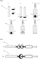
- the sampling device (100) of the present invention is typically characterized in that it comprises a swab tip (1) and a support body (2), wherein the support body has a hollow conformation with a first end (3) and a second end (4), and wherein said first end is in fluid connection with the internal surface of the swab tip ( Fig. 1A ).
- the swab tip comprises a filter material (8, 9) with a pore size that decreases from the external surface (such as the surface in contact with the sample) of the swab tip to the internal surface of the swab tip (such as the surface in open contact with the support body), more specific in the direction of the fluid or sample flow ( Fig. 1B-C ).
- the support body (2) is configured to collect the sample via the sample tip (1) and to transport the sample from its first end (3) towards its second end 4).
- the first end of the support body further comprises one or more openings (10) in the side walls thereof ( Fig. 2 A-C ).
- the first end of the support body terminates in an open tip and further comprises one or more openings in its side walls.
- the first end of the support body terminates in a closed tip and further comprises one or more openings in the side walls of the first end of the support body.
- the support body (2) of the present sampling device has a hollow conformation, and thereby allows the biological specimen (15) to be transferred from the internal surface of the swab tip into said support body, and further via the support body into a collection tube (11).
- the support body and the collection tube are the same.
- the support body therefore has a hollow conformation that can be of any shape or design (e.g. cylindrical, hexagonal, etc.).
- the support body has an elongate conformation and/or a substantially rod-shaped conformation.
- the support body has a hollow tube conformation, a hollow cuboid conformation or a hollow prism conformation.
- the support body has a hollow tube conformation.
- the support body can be of any material and can be rigid or semi-rigid.
- the support body is made of glass, a plastic material, e.g. polystyrene, polypropylene, polycarbonate, polyethylene terephthalate or polyamide, polyvinyl chloride, or metal, e.g. aluminum, titanium or steel.
- the support body is bendable.
- Typical for the present invention is that the first end (3) of the support body (2) is in fluid connection (e.g. through one or more openings) with the internal surface (the inside) of the swab tip (1) and that said support body (2) is configured to collect the sample via the sample tip (1) and to transport the sample from its first end (3) towards its second end (4).
- Fig. 1B-C; Fig. 2 B-C the biological specimen to be collected is transferred from the internal surface of the swab tip into the first end of the support body, and further towards the second end of the support body.
- the internal surface of the swab tip and the first end of the support body are thus in fluid connection with each other.
- the internal surface of the swab tip and both the first end and the second end of the support body are in fluid connection with each other.
- the internal surface of the swab tip and the first end of the support body are in direct contact with and/or are attached to each other, thereby touching each other.
- the filter material of the swab tip and the support body are connected by heat-welding or ultrasonic welding.
- the filter material of the swab tip and the support body are connected by a non-toxic glue which does not have a negative impact on the biological specimen.
- the filter material of the swab tip and the support body are connected with a sealing ring.
- the sampling device is further typically characterized by the presence of a swab tip comprising a filter material with a pore size that decreases from the external surface of the swab tip to the internal surface of the swab tip ( Fig. 1B-C; Fig. 2B-C ). Due to the presence of this filter material the biological specimen can be selectively collected based on its size and the pore size of the filter material.
- the filter material also prevents unwanted substances e.g. impurities, particulates and large DNA structures, to be collected together with the biological specimen of interest, thereby increasing the purity of the biological specimen to be collected. This will facilitate the use of the purified samples in downstream molecular diagnostic pathways (e.g. real-time PCR, PCR, LAMP, sequencing, virus isolation, antigen or antibody ELISA).
- the filter material also prevents clogging of the sampling device.
- the sampling device is typically suitable for the collection of biological specimens such as microorganisms (e.g. bacteria, yeasts, mold, fungi and parasites) and viruses, in particular viruses, bacteria and parasites, more in particular viruses.
- biological specimens such as microorganisms (e.g. bacteria, yeasts, mold, fungi and parasites) and viruses, in particular viruses, bacteria and parasites, more in particular viruses.
- Commercial purification platforms for viruses for example are available but require extensive sample manipulation and laboratory equipment. They are also intended for virus concentration after large-scale production in cells.
- kits and platforms are costly and therefore not widely applicable for cheap and quick on-site preparation for veterinary and human diagnostic applications.
- a sampling device is provided that allows a hygienic, fast and pure collection of the biological specimen of interest.
- the sampling device of the present invention is typically characterized in that it comprises a filter material with a pore size that decreases along the fluid flow direction, in particular from the external surface of the swab tip to the internal surface of the swab tip.
- said filter material has a pore size that still allows the passage of the biological specimen to be collected, but prevents the passage of other unwanted, and larger, substances or particles.
- the purity of the biological specimen to be collected is largely increased, resulting in representative samples instead of dirty mixtures of organic material.
- the sampling device thereby allows on-site purification of viruses, bacteria, parasites and other micro-organisms, without any negative effects on e.g. virus, bacteria or parasite infectivity.
- the filter material is surfactant free.
- the minimum pore size of the filter material is 5 ⁇ m. In a further embodiment the minimum pore size of the filter material is 10 ⁇ m. In still a further embodiment, the minimum pore size of the filter material is 20 ⁇ m.
- the pore size of a filter material can be determined using different technologies, for example electron microscopy, or by evaluating the passage of (fluorescent) microspheres of a pre-defined size through the filter material.
- the pore size of a particular filter material is determined by determining the passage of fluorescent microspheres of a pre-defined size using a flow cytometer or a fluorimeter.
- the pore size of the sampling device can be adapted in function of the biological specimen to be collected. For example, if the sampling device is intended to collect only viruses, the pore size of the filter material will be different from the pore size of the filter material for a sampling device that also needs to collect bacteria.
- Virus sizes range between 15 nm and 200 nm for those infecting mammalian host species. Non-mammalian viruses can have a bigger size of up to 700-800 nm for plant viruses and viruses infecting amoeba.
- the pore size of the filter material of a sampling device to specifically collect viruses can be at least 1 ⁇ m, but maximum 2 ⁇ m.
- Dimensions of bacteria representative for major bacterial phyla range between 700-2000nm for Bacteroidetes, 250-2000nm for Proteobacteria, 500-9000nm for Firmicutes and 600-6000nm for Actinobacteria.
- the pore size of the filter material of a sampling device to specifically collect bacteria is between Sum and 10 ⁇ m, in particular at least 10 ⁇ m.
- Examples of parasites with human and veterinary clinical importance include Eimeria species such as E . acervulina (18x24 ⁇ m), E. brunetti (26x22 ⁇ m), E. maxima (30x20 ⁇ m), E. necatrix (20x17 ⁇ m) and E. tenella (23x19 ⁇ m) in chickens, E. meleagridis (24x18 ⁇ m) in turkeys, E . bovis (28x20 ⁇ m) and E. zuerni (18x16 ⁇ m) in cattle, E. caprina (34x23 ⁇ m) and E . ninakohlyakimovae (21x15 ⁇ m) in goats, E. debliecki (18x14 ⁇ m) and E.
- Eimeria species such as E . acervulina (18x24 ⁇ m), E. brunetti (26x22 ⁇ m), E. maxima (30x20 ⁇ m), E. necatrix (20x17 ⁇ m) and E
- veterinary and medically important fungi are Microspora canis, Trichophyton mentagrophytes, Candida albicans and Candida auris. Fungal spores have a size of 2-4 ⁇ m and can be linked to each other in so-called hyphae. Yeasts are unicellular fungi and have a size of approximately 3-6.5 ⁇ m x 2.5 ⁇ m for the veterinary important Malassezia pachydermatis.
- the filter material has a pore size ranging from about 5 ⁇ m to about 200 or 300 ⁇ m, or from about 10 ⁇ m to about 200 or 300 ⁇ m, or from about 10 to 150 ⁇ m, or from about 10 to 100 ⁇ m, or from about 20 to 200 ⁇ m or 300 ⁇ m, or about 20 to 150 ⁇ m, or from about 20 to 100 ⁇ m.
- the pore size in the filter material (being one layer or multiple layers) decreases in the sample flow direction or from the outer side to the inner side of the swab tip.
- the swab tip comprises (at least) two layers wherein the outer layer has a pore size of about 50 to 200 ⁇ m, of about 50 to 150 ⁇ m or of about 50 to 100 ⁇ m and the inner layer has a pore size of about 5 to 50 ⁇ m, of about 10 to 50 ⁇ m, of about 20 to 50 ⁇ m, of about 5 to 40 ⁇ m, of about 10 to 40 ⁇ m, or of about 20 to 40 ⁇ m.
- the filter material of the present sampling device can be a single-layered filter material, such as for example seen in Fig. 1C or Fig. 2C .
- the single-layered material comprises a continuous pore size gradient that decreases from the external surface of the swab tip to the internal surface of the swab tip. In said instance, the pore sizes are gradually changed and decreased from the external surface to the internal surface of the one-layer filter material in the swab tip.
- a multi-layered filter material is present in the swab tip ( Fig. 1B; Fig. 2B ).
- said multi-layered filter material comprises two or more layers wherein the pore size decreases from the external layer of the swab tip to the internal layer of the swab tip.
- each layer of said multi-layered filter material comprises a specific pore size wherein the pore size of each layer decreases from the external surface of the swab tip to the internal surface of the swab tip.
- said multi-layered filter material comprises 2, 3, 4, or 5 layers.
- the filter material is a multi-layered filter material comprising 2 or 3 layers.
- the filter material of the present sampling device can be made of any material known in the art that allows the presence of pore sizes according to the different embodiments of the invention.
- the filter material is polyamide-based or nylon.
- nylon fabrics can be selected from, though not limited to fabrics with densities between 30g and 500g/m 2 , in particular between 50g and 300g/m 2 .
- the thickness of the nylon fabrics can range between 0.10 and 0.80mm, in particular between 0.20 and 0.60mm. Examples of such nylon fabrics include polyamide fabrics with densities of 190 g/m 2 or 270 g/m 2 and a thickness of 0.48 or 0.56mm.
- the sampling device further comprises means for creating a pressure gradient through the sampling device, in particular means for creating a negative pressure gradient that facilitates the transport of the sample from the sample tip (1) towards the second end (4) of the support body via the first end (2) of the support body.
- means for creating a pressure gradient vacuum or suction can be a standard syringe (or syringe plunger) device.
- other devices or systems capable of achieving a pressure gradient may also be used, including, but not limited to, standard suction devices, bulge, plumber, air pumping apparatuses, vacuum devices, etc.
- the means for creating a pressure gradient forms the collection tube.
- the means for creating a pressure gradient forms the support body of the sampling device.
- the swab tip of the present sampling device may comprise an additional (fabric) layer on its external surface (the outside layer).
- said additional layer is positioned on the external surface of the filter material of the swab tip.
- This layer is typically comprised of a loose layer or material that has scraping or brushing characteristics or capacity to grasp sampling material.
- This layer has a non-smooth surface, which can also be achieved by means of flocking (e.g. using short fibers that are arranged in a perpendicular fashion).
- the additional layer on the external surface of the swab tip comprises a plurality of short fibers that are arranged by flocking.
- the fabric layer comprises pores with a pore size of at least 100 ⁇ m, and typically ranges from 100 ⁇ m to 500 ⁇ m.
- suitable fabrics for said additional layer are natural or synthetic polyamides and cotton.
- the swab tip comprises three layers wherein the fabric layer has a pore size of about 100 to 300 ⁇ m, the outer layer of the filter material has a pore size of about 50 to 100 ⁇ m and the inner layer of the filter material has a pore size of about 10 to 50 ⁇ m.
- the swab tip comprises three layers wherein the fabric layer has a pore size of 200 to 500 ⁇ m, the outer layer of the filter material layer has a pore size of 50 to 200 ⁇ m, and the inner layer of the filter material has a pore size of 10 to 40 ⁇ m.
- the presence of said fabric layer allows the collection of as much as organic material as possible.
- the fabric layer can be used as the scraping surface to collect the specimen at a particular location.
- collection of the biological specimen with the sampling device of the present invention can be done by scraping with the swab tip into a sample of interest or onto a location of interest (e.g. Fig. 3 A-B ).
- collection of the biological specimen can be done by immersion of or bringing the swab tip into a fluid sample (e.g. Fig. 3 C-F ).
- the swab tip ensures that the biological specimen of interest is collected during scraping at the site of collection, or that the biological specimen is collected in the device after immersion of the swab tip into a fluid sample in order to release and/or dissolve the biological specimen into said fluid sample, in particular for non-fluid biological specimen such as mucus or faeces, or when cell lysis is required (e.g. lysis of blood cells).
- cell lysis e.g. lysis of blood cells.
- Samples that may be tested include clinical and non-clinical samples in which further in vitro cell growth is or may be suspected, as well as samples occasionally tested for the presence of microorganisms or viruses. Typically, samples can be tested directly with little or no extensive pretreatment.
- Locations in or on a subject where the swab tip can be used to collect a biological specimen can be any locations in or on a subject. Said locations can be the skin, the airways, the esophagus, the anus, the vagina, the stomach, the mouth, the tongue, nose, ear or any other location that can be reached using the sampling device.
- the sample that can be collected during scraping can be sweat, lung aspirate, vaginal fluid, gastric fluid, faeces, urine, etc.
- the sampling device can be immersed in a fluid sample, such as for example a bodily fluid sample (including secretions/excretions).
- a fluid sample such as for example a bodily fluid sample (including secretions/excretions).
- the bodily fluid sample can be selected from blood, respiratory mucus, lung aspirate, vaginal fluid, gastric fluid, saliva, urine, faeces, or cerebrospinal fluid.
- the sample from which the biological specimen is to be collected and isolated is selected from solid tumors, warts, sarcoids or fibromas.
- the surface of the sample is swept with the swab tip of the sampling device in order to collect the biological specimen.
- the sample tip can be pre-wetted with a rinsing buffer.
- the sampling device can be connected to a collection tube wherein the collection tube us in fluid connection with the second end of the support body and wherein the collection tube is filled with a fluid. Releasing of said fluid via a positive pressure gradient in to a collection container results in a fluid solution comprising the biological specimen. The biological specimen can then be recollected from the collection container via the swab tip into the support body and the collection tube by using a negative pressure gradient.
- the sampling device of the present invention allows the collection of a biological specimen from a subject.
- This subject can be a living subject or a dead subject.
- the subject can be a healthy subject or an ill subject.
- the subject can be a human or a non-human animal; preferably a human, a non-human mammal, or a non-mammal.
- Non-human mammals are selected from non-human primates, rodents (e.g. mouse or rats), canines, felines, equines, bovine, camelids, ovines, porcines.
- Non-mammals are selected from birds, chicken, bats, fish, mussels, shrimps, prawns, crustaceans, amphibians, reptiles, etc.
- the swab tip is designed so that it is applicable in small orifices in order to take the sample (e.g. animal nose).
- the tip diameter can range from about 0.5 cm to about 2 cm, from about 1 cm to about 1.5 cm.
- the swab tip of the sampling device can further be of any shape or design (e.g. cylindrical, ellipsoid, spherical, etc.).
- the swab tip has a cylindrical tube conformation.
- the swab tip has a spherical conformation.
- the swab tip has an ellipsoid conformation.
- the sampling device of the present invention can also be used for the collection of biological specimens at another location than a subject or from another fluid sample than a bodily fluid sample.
- the sampling device can be used for the collection of a water sample via scraping at a particular location, e.g. at the bottom of a pond, a puddle, a lake, a river, or a sea, or via immersion of the sampling device into said water sample.
- Said water sample is selected from fresh water, brackish water, waste water, etc.
- Other non-clinical samples include foodstuffs, beverages, pharmaceuticals, cosmetics, air, soil, sewage, plant material, etc.
- the sampling device of the present invention is particularly useful for the collection of a biological specimen without the risk of any other larger unwanted particles to be present in the collected sample.
- the use of a filter material according to the different embodiments as described herein ensures that any unwanted and larger particles are prevented to be collected.
- the filter material also prevents clogging of the device with said larger particles.
- clogging of the sampling device is further prevented by the presence of one or more (e.g. 2, 3, 4, 5, 6, 7, 8, 9, 10, 11, 12, 13, 14, 15; or more; in particular about and between 1 to 10; preferably between 1 and 8) openings, apertures, grooves or gaps in the side walls of the first end of the support body (e.g. Fig. 2 B-C ).
- the sampling device comprises a swab tip according to any of the possible embodiments and a support body, wherein the support body has a hollow conformation with a first end and a second end, wherein said first end comprises one or more openings in the side walls, and wherein the first end is in fluid connection with the internal surface of the swab tip.
- the first end of the support body can end either in a closed tip or in an open tip. Due to the presence of said one or more openings, clogging of the swab tip is prevented and a more equal transfer of the biological specimen into the support body is achieved.
- the one or more openings in the side wall of the first end of the support body can be of any shape (round, ellipsoid, square, rectangular, etc.).
- the present invention relates to a sampling device for the collection and isolation of a biological specimen. Therefore, in a further embodiment, the sampling device comprises a collection tube that is in fluid connection with the second end of the support body, and wherein the support body is configured to collect the sample via the sample tip and to transport the sample from its first end towards its second end. In an even further embodiment, one or more filters are integrated in either one or positioned in between the support body and the collection tube ( Fig. 4 A-B ).
- Said filters can be any filters known to the skilled person (e.g a nylon, cellulose acetate, polyether sulphone, nitrocellulose, glass fibers, polypropylene, polytetrafluoroethylene, or polyvinyldifluoride filter) and suitable for the present application.
- filters known to the skilled person (e.g a nylon, cellulose acetate, polyether sulphone, nitrocellulose, glass fibers, polypropylene, polytetrafluoroethylene, or polyvinyldifluoride filter) and suitable for the present application.
- said filters are nylon or cellulose acetate (syringe) filters with a pore size of about 0.1 ⁇ m to about 6.5 ⁇ m, in particular with a pore size of 0.1 ⁇ m ,0.22 ⁇ m, 0.45 ⁇ m, 0.8 ⁇ m, 1.2 ⁇ m, 5 ⁇ m, 6 ⁇ m or 6.5 ⁇ m, and even more preferred with a pore size of about 0.8 ⁇ m or about 5 ⁇ m.
- nylon or cellulose acetate (syringe) filters with a pore size of about 0.1 ⁇ m to about 6.5 ⁇ m, in particular with a pore size of 0.1 ⁇ m ,0.22 ⁇ m, 0.45 ⁇ m, 0.8 ⁇ m, 1.2 ⁇ m, 5 ⁇ m, 6 ⁇ m or 6.5 ⁇ m, and even more preferred with a pore size of about 0.8 ⁇ m or about 5 ⁇ m.
- the sampling device comprises a swab tip, a collection tube and one or more filters having a pore size of about and between 4 ⁇ m and 6.5 ⁇ m, such as e.g. 4.0 ⁇ m, 4.5 ⁇ m, 5.0 ⁇ m, 5.5 ⁇ m and 6 ⁇ m; and/or one or more filters having a pore size of about and between 0.1 ⁇ m and 1.5 ⁇ m, such as e.g. 0.1 ⁇ m, 0.22 ⁇ m, 0.45 ⁇ m, 0.8 ⁇ m and 1.2 ⁇ m.
- the filter is a cellulose acetate filter, more in particular free of surfactant.
- Fig. 5 discloses different types of sampling devices, of which only Fig. 5E is a sampling device according to an embodiment of the present invention.
- Fig. 5A represents a sampling device which is only a hollow tube, without the presence of any filter material.
- Fig. 5B represents a closed tube which does not allow any collection of a biological specimen.
- Fig. 5C-D disclose a sampling device without a swab tip comprising filter material. In those sampling devices several openings are present in the side walls of the first end of the support body allowing collection of a biological specimen.
- Fig. 5E discloses a sampling device similar to the device in Fig. 5C or Fig. 5D but including a swab tip comprising a filter material. In Fig. 5E , the one or more openings in the side walls of the first end of the support body are covered by the swab tip.
- the present invention also provides a kit of parts for assembling the sampling device as provided herein.
- said kit of parts includes a support body comprising a swab tip as provided herein, one or more filters, a collection tube and a means for providing a fluid flow or pressure gradient in the device and optionally a container with collection fluid.
- the present invention also includes a method for the collection and/or isolation of a biological specimen from a sample or from a location. Said method comprises the following steps:
- the sampling devices of the invention further comprises a means for creating a negative pressure gradient through the sampling device.
- the negative pressure gradient can be generated by any means for creating a pressure gradient.
- Such means can be a standard syringe device.
- other (automatic or manual) devices capable of achieving a negative pressure gradient may also be used, including, but not limited to, standard suction device, air pumping apparatuses, vacuum devices, etc.
- a size purification step is performed by transferring the biological specimen through one, two or more (uncharged) filters. In case of two or more filters these are positioned as such to create a gradient of decreasing pore sizes in the direction of the sample flow.
- the present invention provides a method for the collection and isolation of a biological specimen from a sample or from a location, said method comprising the following steps:
- the pressure gradient can be generated by any means that is able to create a positive and a negative pressure gradient.
- Such means can be a standard syringe device.
- other devices capable of achieving a pressure gradient may also be used, including, but not limited to, standard suction device, air pumping apparatuses, vacuum devices, etc.
- said means for creating a pressure gradient function as a collection tube (11).
- the fluid present in the collection tube is any fluid that ensures a good solubility of the biological specimen.
- said fluid is selected from saline, water, a buffered solution, pathogen transport medium or any other standard rinsing buffer.
- the method of the present invention comprises an additional step wherein an additional fluid or liquid is added to the sample before scraping with and/or immersing the external surface of the swab tip in the sample or at a location.
- an additional fluid or liquid is added to the sample before scraping with and/or immersing the external surface of the swab tip in the sample or at a location.
- additional fluid can be added to the faeces sample to increase the fluidity of the sample.
- additional fluid can be added to optimize the collection of the biological specimen.
- any other liquid samples may be used, such as water, or transport buffers, cell culture medium, storage buffers, or any other products.
- the biological specimen is collected in a collection tube that is in fluid connection with the second end of the support body.
- an additional treatment step can be applied on the collected biological specimen sample in said collection tube. This additional treatment step can be used to improve the quality or purity of the biological specimen. Said treatment step involve infiltration, precipitation, dilution, distillation, mixing, concentration, inactivating of interfering components, or the addition of extra reagents. Besides physiological fluids, any other liquid samples may be used, such as water, or other products and the like for the performance of any downstream assays.
- the method comprises the step of identifying and/or characterizing the biological specimen(s) present in the sample.
- the pore sizes of the different layers of polyamide fabrics that can be used as filter material in the sampling device of the present invention were defined using fluorescently-labeled microspheres of different sizes.
- the 1 ⁇ m, 10 ⁇ m and 40 ⁇ m microspheres were analyzed using a Cytoflex flow cytometer (Beckmann Coulter). The 80 ⁇ m were not analyzed using the Cytoflex flow cytometer to avoid clogging of the nozzle (100 ⁇ m diameter). A fluorimeter was used to assess the fluorescence of 80 ⁇ m microspheres. All microspheres were handled using wide bore tips.
- Fluorescent microspheres were vortexed during 15 sec, protected from light and stored on ice in between handling.
- the 1 ⁇ m and 10 ⁇ m microspheres were ready-to-use suspensions.
- the 40 ⁇ m and 80 ⁇ m microspheres were dry particles and were resuspended in 5 ml of Dulbecco's Phosphate Buffered Saline (ThermoFischer), followed by vortexing before use. Next, dilutions of the microspheres were made in Dulbecco's Phosphate Buffered Saline (ThermoFischer) resulting in a total volume of 20 ml for use in the experiments. These suspensions were transferred to different 15 ml collection tubes (2 ml suspension for each tube).
- microspheres and their properties Size (diameter) 1 ⁇ m 10 ⁇ m 40 ⁇ m 80 ⁇ m Manufacturer & cat no ThermoFischer G0100 ThermoFischer CDG1000 ThermoFischer 35-7 ThermoFischer 35-10 Name Fluoro-Max Dyed Green Fluoro-Max Dyed Fluoro-Max Green Fluoro-Max Green Aqueous Fluorescent Green Aqueous Dry Fluorescent Dry Fluorescent Particles Fluorescent Particles Particles Particles Volume/mass 10 ml (in water) 10 ml (in water) 1g, 1ea 1 g, 1 ea.
- the syringe content was transferred to a non-autoclaved 1.5 ml collection tube, stored on ice and protected from light using aluminum foil.
- Non-autoclaved tubes were used to avoid retention of microspheres in cracks in the polystyrene.
- Samples containing the 1 ⁇ m, 10 ⁇ m or 40 ⁇ m microspheres were loaded in V-bottom 96-well plates.
- Samples containing the 80 ⁇ m microspheres were loaded in black-walled 96-well microplates for fluorescence-based assays. Analysis of the 1 ⁇ m, 10 ⁇ m and 40 ⁇ m fluorescent microspheres was conducted on a Cytoflex flow cytometer using the Cytexpert software.
- the machine was flushed and quality control using QC beads was conducted on a daily basis.
- the forward scatter and fluorescence signal were used to characterize the particles.
- a 488nm (blue) laser was used to excite the green fluorescent microspheres and detection was done in the fluorescent channel 525/40BP.
- Particle counting was done during 35 sec at a flow of 30 ⁇ l per minute.
- thresholds were set at 60,277, 278,468 and 300,000, respectively. Flushing was conducted after each measurement to exclude carry-over between samples. The number of singlets was counted by the flow cytometer. The experiment and measurements were replicated to generate a total of 3 independent measurements.
- the 80 ⁇ m microspheres were analyzed using a fluorimeter with excitation at 485 nm and detection of emission at 527 nm.
- a serial 1 ⁇ 2 dilution series of the microspheres was enclosed to allow quantification of the unknown samples against a standard curve (range between 35481 and 139 microspheres/ml).
- a standard curve was enclosed in each repetition. Two duplicates were enclosed for each dilution point of the standard curve and each unknown sample. The experiment was independently repeated three times. Linear regression was used to interpolate the unknowns from the standard curve.
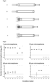
- the number of 1 ⁇ m microspheres is reduced with a factor of approximately 3.4 log 10 when the sampling device comprising 3 layers of fabric is connected to the aforementioned 5 ⁇ m and 0.8 ⁇ m SFCA filters (F) (p ⁇ 0.0001, mean ⁇ SD 6.96 ⁇ 0.11 vs 3.56 ⁇ 0.33 log 10 microspheres/ml), an effect mainly caused by the 0.8 ⁇ m filter (p ⁇ 0.0001, 3.80 ⁇ 0.40).
- F 5 ⁇ m and 0.8 ⁇ m SFCA filters
- the sampling device or its different layers of fabrics did not have a negative impact on the 10 ⁇ m microspheres (p > 0.05) (A, B, C, D). No microspheres were observed after passage through the sampling device comprising 3 layers of fabric in connection to the aforementioned 5 ⁇ m and 0.8 ⁇ m SFCA filters (F) (p ⁇ 0.0001), the 5 ⁇ m SFCA filter (G) (p ⁇ 0.0001) and the 0.8 ⁇ m filter (H) (p ⁇ 0.0001).
- the sampling device and the different layers of the swab tip had no significant impact on the 40 ⁇ m microspheres in comparison to the support tube without swab tip (p > 0.05).
- the 40 ⁇ m microspheres were not detected after collection with the sampling device in connection the aforementioned 5 ⁇ m and 0.8 ⁇ m SFCA filters (F) (p ⁇ 0.0001), the 5 ⁇ m filter (G) (p ⁇ 0.0001) or the 0.8 ⁇ m filter (H) (p ⁇ 0.0001).
- the 80 ⁇ m microspheres were not detected after passage through the sampling device (A) or the most dense first layer of the swab tip (B) and thus significantly different from the number of microspheres detected after collection with the support body without swab tip (E) (p ⁇ 0.0001).
- An approximate 225-fold reduction of microspheres was seen after passage through the second layer of fabrics (C) (p ⁇ 0.0001) in comparison to collection with the support body (4.53 ⁇ 0.10 vs 2.17 ⁇ 1.03).
- a cotton swab with a plastic rayon, composed of a first end (cotton tip), support body and second end were cut at the second end.
- An 18 Gauge needle was inserted in the second end and connected to a 5 ml syringe with a rubber plunger (Terumo).
- the swab tip was immersed in a container with feces from pigs and then transferred into 1 to 5 ml of phosphate buffered saline.
- the suspension was shaken for 10 seconds and the suspension was aspirated using a pressure exerted by the syringe.
- the 115 g/m 2 and 270 g/m 2 fabrics were each covered with a less dense, loose outer layer (density of 80 g/m 2 ) to capture the sample specimen.
- These polyamide fabrics were in fluid connection with the support body, containing eight openings in the side walls at its first end. Feces was collected with both sampling devices and directly transferred to collection tubes filled with 4 ml of transport medium. Next, the swab tip was scraped against the collection tube's wall to homogenize the fecal suspension and aspirated using a syringe.
- an intermediate additional layer of 190 g/m 2 in the swab tip would be beneficial to function as an additional sieve.
- the swab tip of the sampling device composed of subsequent layers of 270 g/m 2 , 190 g/m 2 and 80 g/m 2 proved a good combination and led to the recovery of 2.8 ml of fecal suspension.
- This sampling device of which the swab tip is composed of three layers of polyamide fabrics was subsequently used in the experiments described hereafter.
- the aim of the experiment was to demonstrate the absence of negative effects of the sampling device (comprising a swab tip composed of three layers of fabrics), the sampling device in connection to Sum and 0.8 ⁇ m surfactant-free cellulose acetate filters, or the 5 ⁇ m and 0.8 ⁇ m cellulose filters individually.
- the infectivity of different viruses with a broad range of characteristics was evaluated by applying the above-mentioned conditions. Retaining viral infectivity is important for diagnostics and allows the possibility to isolate the virus in cell cultures. This is beneficial for developing new vaccine candidates for human and veterinary applications, or for autovaccine production in veterinary medicine. Having the ability to filter out impurities and bacteria will also lead to more pure viral samples, reducing the background and chances for false-positive signaling in downstream analyses and reducing the ability of unwanted microbiological contamination upon inoculation in susceptible cells.
- viruses were grown in cell cultures and spiked in phosphate buffered saline.
- PRRSV 2° porcine respiratory and reproductive syndrome virus
- PCV2 4° porcine circovirus 2
- Virus stocks were thawed and centrifuged at 13,000 rpm for 3 min at room temperature. A 1/50 v/v dilution of each virus stock was prepared in phosphate buffered saline (PBS) containing Ca 2+ and Mg 2+ except for equine herpes virus, which was diluted 1/200 (v/v) because of the high stock titer.
- PBS phosphate buffered saline
- the virus suspensions were aspirated through the following components:
- the virus dilution that was made served as positive control, whereas PBS served as negative control to exclude cross-contamination. All samples were stored on ice between the handling steps. For each viral species, the experiment was executed at least three times and the infectious virus titers were immediately assessed by infectivity titration in the respective host cells. Serial 10-fold dilutions of the samples were inoculated for 1h at 37°C and 5% CO 2 for most viruses except influenza virus, which was inoculated for 2 h. Next 100 ⁇ l cell culture medium was added before incubation of the cells during 4 to 10 days, depending on the viral species. As described in literature, trypsin was enclosed in the inoculation and cell culture medium of rotavirus and influenza virus, to enhance infection. Cytopathogenic effects were evaluated using a light microscope and infectious virus titers were determined using the formula of Reed and Muench.
- the aim of the experiment was to demonstrate the ability of the sampling device to capture large DNA strands. This would be beneficial for downstream molecular assays such as PCR, real-time PCR, digital PCR, LAMP and sequencing, as it reduces the chance of background DNA amplification or detection by any form of PCR or sequencing. Large strands of DNA are also difficult to digest using nucleases.
- a bacterial suspension of Escherichia coli DH10B was grown overnight in LB medium for 18h at 37°C on a shaker. Bacterial cells were centrifuged at 4,000 rpm for 10 min at 4°C and resuspended in a buffer (50 mM Tris-Cl (pH 8.0) and 10 mM EDTA) to which SDS and proteinase K was added for lysis at 50°C during 1h. Next, an equal amount of Phenol:Chloroform:lsoamylalcohol was added and mixed by flicking until a white emulsion was formed.
- a buffer 50 mM Tris-Cl (pH 8.0) and 10 mM EDTA
- the emulsion was centrifuged at 13,000 rpm for 15 min at 4°C to separate the aqueous phase from the liquid fraction while generating a protein-rich.
- the upper aqueous phase was collected without disturbing the protein-rich interphase.
- an equal amount of Phenol:Chloroform:lsoamylalcohol was added and the process was repeated to improve the purity.
- the DNA was precipitated from the aqueous phase using absolute ethanol containing 0.03M NaCl and centrifugation at 13,000 rpm for 30 min at 4°C. The supernatant was carefully collected and discarded.
- a washing step was conducted with 70% ethanol by centrifugation for 5 min at 13,000 rpm and 4°C.
- the pellet was dried at 50°C and resuspend in Tris-EDTA buffer (10mM Tris (pH 8.0), 1 mM EDTA).
- Tris-EDTA buffer (10mM Tris (pH 8.0), 1 mM EDTA).
- the DNA was stored at 4°C and presence of large DNA structures was demonstrated by agarose gel electrophoresis.
- the purified DNA was diluted in nuclease-free water and aspirated with:
- the aim of the experiment was to demonstrate absence of retention of bacteria in the sampling device of the present invention upon aspiration of liquid. It was aimed to show a reduction in bacterial load in the filtrate that passed through the 0.8 ⁇ m filter to ensure that bacteria are separated from viruses. Finally, it was tested if the 0.8 ⁇ m filter could be used to harvest the entrapped bacteria by means of back-flushing with fresh phosphate-buffered saline.
- an Escherichia coli BL21 strain was grown in Luria-Bertani (LB) medium containing ampicillin.
- a suspension of the bacterial broth was prepared in PBS and 1ml of this suspension was collected using (1) sampling device comprising 3 layers of fabric (270, 190 and 80 g/m 2 ), (2) the sampling device in connection to the aforementioned 5 ⁇ m and 0.8 ⁇ m SFCA filters (3) an 18G needle connected to a syringe as positive control.
- Suspensions were transferred to 1.5 ml collection tubes and stored on ice.
- the sampling device was composed of the three layers of polyamide fabrics described before and the first end of the support body comprises a closed tip but had eight round openings in the sidewall. Said round openings were created with a 18G needle (diameter of 1.27 mm).
- the 0.8 ⁇ m SFCA filter was separated from the other sampler's components and 1000 ⁇ l of fresh PBS was aspirated into a fresh syringe and used to flush back the bacteria in the other direction. This suspension was collected in a 1.5 ml collection tube and stored on ice.
- serial 1/10 dilution series were prepared in Luria-Bertani broth for all the samples. These suspensions were inoculated (200 ⁇ l) onto petri dishes (58 cm 2 ) containing LB agar containing ampicillin. Sterile glass beads were added and used to spread the inoculum evenly over the agar. Glass beads were removed and petri dished were incubated with the agar turned upside down for 24 hours at 37°C before counting the number of colony-forming units (CFU). This experiment was independently executed three times.
- CFU colony-forming units
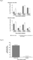
- the sampling device from the present invention did not have a negative impact on the bacterial load as compared to the positive control (6.52 ⁇ 0.12 vs 6.35 ⁇ 0.18 log 10 CFU/ml, no significant difference) ( Fig. 8 ).
- Using the sampling device in connection the 5 ⁇ m and 0.8 ⁇ m SFCA filters only 4.90 ⁇ 0.04 log 10 CFU/ml of bacteria were recovered, resulting in a 28x fold reduction of the bacteria in the filtrate or a mean difference of 1.453 log 10 CFU/ml (p-value ⁇ 0.05). Furthermore, it was possible to flush back the bacteria that were trapped in the 0.8 ⁇ m SFCA filter (6.16 ⁇ 0.93 log 10 CFU/ml, no significant difference with the positive control).
- the aim of the experiment was to mimic the sampling of enteric viruses from feces using an in vitro spiking experiment.
- the performance of the sampling device with a swab tip comprised of 270, 190 and 80 g/m 2 fabrics in connection to the aforementioned 5 ⁇ m and 0.8 ⁇ m SFCA filters was compared to a traditional cotton swab. Detection of spiked enteric viruses was executed using virus infectivity titration and real-time PCR.
- Pig manure was collected from the floor of a stable where 6-weeks-old piglets were housed. These piglets received normal pig meal and where healthy. To obtain batches of references feces, the feces was aliquoted in fecal containers and stored at -20°C until use in spiking experiments.
- RVA/Pig-tc/BEL/12R046/2012/G9P 10 ⁇ 5.93 CCID 50 /ml
- RVA/Pig-tc/BEL/12R050/2012/G5P 10 ⁇ 8.63 CCID 50 /ml
- Feces was weighed (1.6 g) and added to a 50ml falcon tube and spun.
- the suspensions collected under both conditions A and B were titrated immediately in MA104 cells to determine the virus infective titer. Cytopathogenic effect was assessed after 4-5 days with a light microscope and titers were calculated using the formula of Reed and Muench.
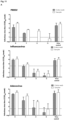
- Fig. 9 The results of the infectivity titration and genome copy quantification are shown in Fig. 9 .
- There was a trend towards higher sensitivity of the sampling device of the present invention compared to the traditional swab when rotavirus was titrated, although the difference between both sampling methods was not statistically significant (p 0.3016).
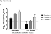
- the aim of the experiment was to mimic the sampling of respiratory viruses from respiratory mucus using two in vitro spiking experiments.
- Pig respiratory mucus was isolated from tracheas that were collected at a Belgian slaughterhouse. Mucus was collected by scraping over the tracheal mucosa with the back of scalpel blade as previously described. Mucus was transferred to 1.5 ml tubes and stored at - 70°C.
- mucus 0.5 cm 2
- the porcine respiratory viruses PRRSV, influenza virus and adenovirus were used for spiking in different experiments.
- the swabs were rubbed for 5 s against the wall of the collection tube, resulting in proper mucus suspension.
- the mucus was spiked with influenza virus and collected with a cotton swab (condition A), the sampler from the present invention (condition B), or the sampling device alone (condition C).
- condition A the sampler from the present invention
- condition B the sampler from the present invention
- condition C the sampling device alone
- the swabs were transferred to separate 15 ml collection tubes containing 4 ml of transport medium and rubbed for 5 s against the wall of the tube to resuspend the mucus.
- 1ml of the mucus suspension was collected from the tube of condition A using a polystyrene serological 1 ml pipet.
- the complete sampler swab, filters and syringe
- the swab was used to directly aspirate the mucus suspension from the collection tube of condition C.
- infectious virus was not detected by the cotton swab upon collection and titration of mucus samples spiked with low loads of PRRSV (1/100 and 1/1000 diluted), while virus infection was seen for the 1/100 virus dilution (3/3 replicates) and the 1/1000 dilution (1/3 replicates) when the sampler of the present invention was used.
- influenza virus infectivity was compared between mucus suspensions collected with a cotton swab, the complete sampling device with two syringe filters and a collection tube, and the device with only the swab tip and the support body.
- Influenza virus infectivity was higher in mucus suspensions collected with the device comprising the swab tip and support body alone, compared to mucus suspensions that were collected with the complete sampling device or a cotton swab, in that order.
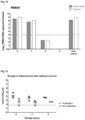
- PRRSV was spiked in blood and samples were analyzed with real-time PCR to compare differences in the collection methods used.
- swine blood was collected from healthy donor animals using heparin and spiked with different dilutions (undiluted, 1/10, 1/100 and 1/1000 v/v) of PRRSV strain 13V091.
- Eight hundred microliters of the spiked blood were brought in 3200 ⁇ l of storage buffer, composed of UP and antibiotics as described above.
- the blood was swirled to mix.
- One ml was collected using a sterile 1 ml serological pipet (Greiner Bio One).
- the remaining suspension was filtered using the swab and the syringe filters.
- Viral RNA was released from the virions and purified using the QIAamp Cador Pathogen Mini Kit according to the manufacturer's instructions.
- PRRSV viral genomes were quantified using an in-house real-time PCR.
- Figure 12 shows the results of PRRSV detection upon collection of blood lysates with the sampler of the invention on the one hand and a standard serological pipet on the other hand.
- Mucus and feces were collected using the sampling device of the invention, resuspended in storage buffer and followed by either purification or no purification and storage of several days at 4°C. Infective virus titers were determined to evaluate possible effects of virus purification and time on virus infectivity.
- Influenza virus Ghent/28/10 ( Qiu et al., 2015, Veterinary Research ) was thawed and centrifuged at 13,000 rpm for 3 min. The supernatant was collected and a 1/10 v/v virus dilution was prepared in storage buffer composed of ultrapure water, penicillin, streptomycin, gentamycin and amphotericin B. Respiratory mucus, collected and stored as described above, was thawed on ice. Mucus (0.25 cm 2 ) was spiked on the bottom of a 6-well cell culture plate and 200 ⁇ l of virus suspension was added.
- the spiked mucus was collected using the new swab and immersed in 4 ml of storage buffer to generate a mucus suspension.
- One ml of the suspension was collected using a serological 1 ml pipet, whereas the remaining suspension was aspirated through the sampling device comprising three layers of fabrics and in connection with the aforementioned 5 ⁇ m and 0.8 ⁇ m SFCA filters to purify the viral particles.
- Negative controls were included by spiking mucus with storage buffer instead of virus or by collection storage buffer. All samples were stored at 4°C for zero, one or two days followed by immediate titration in MDCK cells. The experiment was executed independently three times. CPE was read after 7 days using a light microscope and virus infectivity titers were determined using the formula of Reed and Muench. A repeated measures two-way ANOVA was used to determine effects of purification and time on virus infectivity.
- Feces was collected using the swab of the invention and immersed in 4 ml of storage buffer in 15 ml collection tube. One ml of the fecal suspensions was collected using a serological 1ml pipet, whereas the remaining suspension was filtered using the sampling device from the present invention. Samples were stored at 4°C during zero, one or two days. Titration was performed in MA104 cells and CPE was read after 4 to 5 days using a light microscope. Infectious virus titers were determined using the formula of Reed and Muench. A repeated measures two-way ANOVA was used to determine effects of purification and time on virus infectivity.
- Influenza virus was spiked in mucus and collected using the swab tip from the present invention.
- inhibitory substances e.g. acidic polysaccharides, sialic acids
- Feces and mucus were collected using the sampling device and transferred to a collection tube with 4 ml of storage buffer per tube.
- sample series A To process the sample with the NetoVIR viral metagenomics protocol, one ml of each suspension (sample series A) was collected using a sterile serological one ml pipet and transferred to a fresh 1.5 ml collection tube. The remaining suspension was aspirated using the sampling device in connection to the series of aforementioned SFCA filters (sample series B). The unfiltered suspensions from sample series A were processed using a protocol modified from the NetoVIR strategy for viral enrichment of samples for metagenomics studies using Illumina sequencing (Conceicao-Neto et al., 2015, Scientific Reports).
- the protocol consisted of a centrifugation step (13,000 rpm for 3 min at room temperature), followed by filtration of the supernatant through a 0.8 ⁇ m surfactant-free cellulase acetate filter and subsequent nuclease treatment of the filtrate for 2h at 37°C (Benzonase Nuclease) to discard free-floating nucleic acids.
- nuclease treated sample 400 ⁇ l
- QIAamp Cador Pathogen Mini Kit according to manufacturer's instructions but without addition of carrier RNA.
- the samples from series B did not require additional purification or pretreatment and viral nucleic acids were directly released and purified with the QIAamp Cador Pathogen Mini Kit as described above.
- the viral RNA was converted into complementary DNA and subsequently double-stranded DNA was made using the NEBNext Ultra II Non-Directional RNA Second Strand Synthesis Module before sequencing library preparation using the Rapid Barcoding Sequencing Kit (SQK-RBK004) from Oxford Nanopore Technologies on a R9.4 MinION flowcell. The samples were then sequenced for 6 hours on the MinION from Oxford Nanopore Technologies. Viral reads were identified by comparison against an in-house complete viral database.
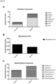
- Fig. 15 shows the number of porcine viruses identified upon processing with the two different methodologies. Using both sample purification methods, all spiked viruses could be detected. In feces, several other viruses (e.g. astrovirus, picornaviruses, picobirnavirus and sapovirus) were also present and detected using both the sampling device and NetoVIR.
- viruses e.g. astrovirus, picornaviruses, picobirnavirus and sapovirus
- the sampling device of the present invention In general, more viral reads were acquired when the samples were processed using the NetoVIR protocol. However, using the sampling device of the present invention, a time gain of 3-4 hours could be achieved in this experiment. Moreover, the sampling method showed good sensitivity for downstream sequencing, as also low-abundant viruses such as PEDV spiked in faeces were efficiently diagnosed using the sampling device of the present invention.
- a sputum sample 500 ⁇ L was spiked with a combination of (1) an in house Viral Mock Community (VMC) (50 ⁇ L), (2) a Bacterial Mock Community (BMC, ZymoBIOMICS Microbial Community Standard (Zymo Research; D6300)) (50 ⁇ L), and (3) Mycoplasma bovis ( M. bovis ) PG45 type strain (ATCC 25523) (50 ⁇ L). More information on the pathogens tested is given in Table 5. Subsequently, the spiked sputum was diluted in 4 mL dPBS to allow collection with the sampling device and aspirated through the swab tip and a 0.8 ⁇ m SFCA filter.
- the unfiltered sputum suspension was processed without filtering for comparison.
- Both samples were processed using a viral metagenomics workflow, which included a viral nucleic acid isolation using a commercially available kit and random viral nucleic acid amplification followed by sequencing for 12 hours on a MinION.
- the original sputum sample was also analyzed with a bacterial DNA isolation kit and sequenced to determine the presence of bacteria in the original sample. This experiment allowed to verify the possible use of the sampling device as part of an all-in-one diagnostic workflow for viruses and bacteria.
- Theuns S Desmarets LM, Heylen E, Zeller M, Dedeurwaerder A, Roukaerts ID, Van Ranst M, Matthijnssens J, Nauwynck HJ.
- Porcine group A rotaviruses with heterogeneous VP7 and VP4 genotype combinations can be found together with enteric bacteria on Belgian swine farms. Vet Microbiol 2014 Aug 6;172(1-2):23-34 .
Landscapes
- Health & Medical Sciences (AREA)
- Life Sciences & Earth Sciences (AREA)
- General Health & Medical Sciences (AREA)
- Chemical & Material Sciences (AREA)
- Pathology (AREA)
- Hematology (AREA)
- Analytical Chemistry (AREA)
- Animal Behavior & Ethology (AREA)
- Veterinary Medicine (AREA)
- Public Health (AREA)
- Heart & Thoracic Surgery (AREA)
- Biomedical Technology (AREA)
- Engineering & Computer Science (AREA)
- Molecular Biology (AREA)
- Surgery (AREA)
- Medical Informatics (AREA)
- Immunology (AREA)
- Biochemistry (AREA)
- Physics & Mathematics (AREA)
- General Physics & Mathematics (AREA)
- Clinical Laboratory Science (AREA)
- Chemical Kinetics & Catalysis (AREA)
- Hydrology & Water Resources (AREA)
- Epidemiology (AREA)
- Vascular Medicine (AREA)
- Sampling And Sample Adjustment (AREA)
- Apparatus Associated With Microorganisms And Enzymes (AREA)
Applications Claiming Priority (2)
| Application Number | Priority Date | Filing Date | Title |
|---|---|---|---|
| EP19183233 | 2019-06-28 | ||
| PCT/EP2020/068027 WO2020260583A1 (en) | 2019-06-28 | 2020-06-26 | Sampling device for biological specimen |
Publications (3)
| Publication Number | Publication Date |
|---|---|
| EP3989840A1 EP3989840A1 (en) | 2022-05-04 |
| EP3989840C0 EP3989840C0 (en) | 2024-05-22 |
| EP3989840B1 true EP3989840B1 (en) | 2024-05-22 |
Family
ID=67137630
Family Applications (1)
| Application Number | Title | Priority Date | Filing Date |
|---|---|---|---|
| EP20734946.5A Active EP3989840B1 (en) | 2019-06-28 | 2020-06-26 | Sampling device for biological specimen |
Country Status (6)
| Country | Link |
|---|---|
| US (1) | US20220241775A1 (pl) |
| EP (1) | EP3989840B1 (pl) |
| CA (1) | CA3144175A1 (pl) |
| ES (1) | ES2985022T3 (pl) |
| PL (1) | PL3989840T3 (pl) |
| WO (1) | WO2020260583A1 (pl) |
Families Citing this family (3)
| Publication number | Priority date | Publication date | Assignee | Title |
|---|---|---|---|---|
| CN113349830B (zh) * | 2021-05-26 | 2022-05-06 | 南京溯远基因科技有限公司 | 一种基因测序取样装置 |
| CN113995447A (zh) * | 2021-11-05 | 2022-02-01 | 上海科罡医疗技术有限公司 | 球囊状生物采样器 |
| GB202402541D0 (en) * | 2024-02-22 | 2024-04-10 | Reardon Robert John Fraser | Sampling apparatus and method of use thereof |
Family Cites Families (11)
| Publication number | Priority date | Publication date | Assignee | Title |
|---|---|---|---|---|
| US4978504A (en) * | 1988-02-09 | 1990-12-18 | Nason Frederic L | Specimen test unit |
| IT1272598B (it) | 1993-09-09 | 1997-06-26 | Copan Italia Spa | Dispositivo per il prelievo e trasporto di campioni in vitro principalmente per uso diagnostico |
| JP2000180443A (ja) * | 1998-12-15 | 2000-06-30 | Fuji Photo Film Co Ltd | 血液濾過残留物の回収方法 |
| GB0215044D0 (en) | 2002-06-28 | 2002-08-07 | Sciona Ltd | Sampling kits, devices and uses thereof |
| US20040022687A1 (en) * | 2002-07-31 | 2004-02-05 | Thomas Wuske | Device and process for collecting and releasing saliva |
| ITMI20030643A1 (it) | 2003-04-01 | 2004-10-02 | Copan Innovation Ltd | Tampone per il prelievo di campioni biologici |
| GB0322983D0 (en) | 2003-10-01 | 2003-11-05 | Isohelix Limitd | Sampling kits, devices and uses thereof |
| TW200712472A (en) * | 2005-08-02 | 2007-04-01 | 3M Innovative Properties Co | Apparatus and method for collecting a sample of material |
| DE102005063572B3 (de) * | 2005-11-17 | 2013-04-04 | Siemens Aktiengesellschaft | Vorrichtung und Verfahren zum Extrahieren einer Abstrichprobe |
| US20090030341A1 (en) * | 2007-07-27 | 2009-01-29 | 3M Innovative Properties Company | Sample release system |
| IT1403618B1 (it) | 2011-01-05 | 2013-10-31 | Copan Italia Spa | Procedimento per realizzare un dispositivo per il prelievo ed il trasferimento di campioni per biologia molecolare |
-
2020
- 2020-06-26 EP EP20734946.5A patent/EP3989840B1/en active Active
- 2020-06-26 ES ES20734946T patent/ES2985022T3/es active Active
- 2020-06-26 US US17/617,812 patent/US20220241775A1/en active Pending
- 2020-06-26 CA CA3144175A patent/CA3144175A1/en active Pending
- 2020-06-26 WO PCT/EP2020/068027 patent/WO2020260583A1/en not_active Ceased
- 2020-06-26 PL PL20734946.5T patent/PL3989840T3/pl unknown
Also Published As
| Publication number | Publication date |
|---|---|
| PL3989840T3 (pl) | 2024-10-14 |
| CA3144175A1 (en) | 2020-12-30 |
| ES2985022T3 (es) | 2024-11-04 |
| EP3989840A1 (en) | 2022-05-04 |
| EP3989840C0 (en) | 2024-05-22 |
| WO2020260583A1 (en) | 2020-12-30 |
| US20220241775A1 (en) | 2022-08-04 |
Similar Documents
| Publication | Publication Date | Title |
|---|---|---|
| Zhang et al. | Virome comparisons in wild-diseased and healthy captive giant pandas | |
| EP3989840B1 (en) | Sampling device for biological specimen | |
| Conceição-Neto et al. | Modular approach to customise sample preparation procedures for viral metagenomics: a reproducible protocol for virome analysis | |
| Kashnikov et al. | Picobirnaviruses: prevalence, genetic diversity, detection methods | |
| Jaramillo et al. | A validated quantitative polymerase chain reaction assay for the detection of ranaviruses (Family Iridoviridae) in fish tissue and cell cultures, using EHNV as a model | |
| JP3950500B2 (ja) | 魚類用のイリドウイルス感染症ワクチンと診断剤並びにこれ等の製法 | |
| Pees et al. | Virus distribution and detection in corn snakes (Pantherophis guttatus) after experimental infection with three different ferlavirus strains | |
| JP2024161453A (ja) | 感染性因子を使用するSalmonellaの迅速検出のための方法およびシステム | |
| Piewbang et al. | Feline morbillivirus‐1 in dogs with respiratory diseases | |
| CN115029323B (zh) | 一种耐药型金黄葡萄球菌噬菌体sp160及其在制备抑菌剂中的应用 | |
| Zaccaria et al. | Detection of astrovirus in a cow with neurological signs by nanopore technology, Italy | |
| Shirato et al. | Middle East respiratory syndrome coronavirus infection not found in camels in Japan | |
| CN101629217A (zh) | 猪流感病毒rt-lamp检测试剂盒及其检测方法 | |
| JP4944643B2 (ja) | 魚類用のイリドウイルス感染症ワクチンと診断剤並びにこれ等の製法 | |
| CN101624634A (zh) | 斑节对虾杆状病毒现场快速高灵敏检测试剂盒及检测方法 | |
| JP2011016845A (ja) | 魚類用のイリドウイルス感染症ワクチンと診断剤並びにこれ等の製法 | |
| CN101921873A (zh) | 对虾传染性肌肉坏死病毒现场快速高灵敏检测试剂盒及检测方法 | |
| CN109517804A (zh) | 一种具有跨种裂解特性的拟态弧菌噬菌体oy-1、制备方法及其应用 | |
| Sudhakaran et al. | In vitro replication of Macrobrachium rosenbergii nodavirus and extra small virus in C6/36 mosquito cell line | |
| Sykes | Canine viral respiratory infections | |
| CN118406662A (zh) | 一种沙门氏菌噬菌体vB_SalP_LDW8及其应用 | |
| Gill et al. | Comparative prevalence and molecular characterization of group A rotavirus in cow calves of Punjab, India | |
| Mándoki et al. | Molecular diagnosis of avian nephritis: preliminary report | |
| Pasick et al. | Characterization of avian influenza virus isolates submitted to the National Centre for Foreign Animal Disease between 1997 and 2001 | |
| CN116987673A (zh) | 沙门氏菌噬菌体及其组合物及应用 |
Legal Events
| Date | Code | Title | Description |
|---|---|---|---|
| STAA | Information on the status of an ep patent application or granted ep patent |
Free format text: STATUS: UNKNOWN |
|
| STAA | Information on the status of an ep patent application or granted ep patent |
Free format text: STATUS: THE INTERNATIONAL PUBLICATION HAS BEEN MADE |
|
| PUAI | Public reference made under article 153(3) epc to a published international application that has entered the european phase |
Free format text: ORIGINAL CODE: 0009012 |
|
| STAA | Information on the status of an ep patent application or granted ep patent |
Free format text: STATUS: REQUEST FOR EXAMINATION WAS MADE |
|
| 17P | Request for examination filed |
Effective date: 20220127 |
|
| AK | Designated contracting states |
Kind code of ref document: A1 Designated state(s): AL AT BE BG CH CY CZ DE DK EE ES FI FR GB GR HR HU IE IS IT LI LT LU LV MC MK MT NL NO PL PT RO RS SE SI SK SM TR |
|
| DAV | Request for validation of the european patent (deleted) | ||
| DAX | Request for extension of the european patent (deleted) | ||
| STAA | Information on the status of an ep patent application or granted ep patent |
Free format text: STATUS: EXAMINATION IS IN PROGRESS |
|
| 17Q | First examination report despatched |
Effective date: 20230516 |
|
| GRAP | Despatch of communication of intention to grant a patent |
Free format text: ORIGINAL CODE: EPIDOSNIGR1 |
|
| STAA | Information on the status of an ep patent application or granted ep patent |
Free format text: STATUS: GRANT OF PATENT IS INTENDED |
|
| INTG | Intention to grant announced |
Effective date: 20231212 |
|
| GRAS | Grant fee paid |
Free format text: ORIGINAL CODE: EPIDOSNIGR3 |
|
| GRAA | (expected) grant |
Free format text: ORIGINAL CODE: 0009210 |
|
| STAA | Information on the status of an ep patent application or granted ep patent |
Free format text: STATUS: THE PATENT HAS BEEN GRANTED |
|
| AK | Designated contracting states |
Kind code of ref document: B1 Designated state(s): AL AT BE BG CH CY CZ DE DK EE ES FI FR GB GR HR HU IE IS IT LI LT LU LV MC MK MT NL NO PL PT RO RS SE SI SK SM TR |
|
| REG | Reference to a national code |
Ref country code: GB Ref legal event code: FG4D |
|
| REG | Reference to a national code |
Ref country code: CH Ref legal event code: EP |
|
| REG | Reference to a national code |
Ref country code: DE Ref legal event code: R096 Ref document number: 602020031280 Country of ref document: DE |
|
| REG | Reference to a national code |
Ref country code: IE Ref legal event code: FG4D |
|
| U01 | Request for unitary effect filed |
Effective date: 20240619 |
|
| REG | Reference to a national code |
Ref country code: LT Ref legal event code: MG9D |
|
| REG | Reference to a national code |
Ref country code: NL Ref legal event code: MP Effective date: 20240522 |
|
| U07 | Unitary effect registered |
Designated state(s): AT BE BG DE DK EE FI FR IT LT LU LV MT NL PT RO SE SI Effective date: 20240902 |
|
| PG25 | Lapsed in a contracting state [announced via postgrant information from national office to epo] |
Ref country code: IS Free format text: LAPSE BECAUSE OF FAILURE TO SUBMIT A TRANSLATION OF THE DESCRIPTION OR TO PAY THE FEE WITHIN THE PRESCRIBED TIME-LIMIT Effective date: 20240922 |
|
| PG25 | Lapsed in a contracting state [announced via postgrant information from national office to epo] |
Ref country code: HR Free format text: LAPSE BECAUSE OF FAILURE TO SUBMIT A TRANSLATION OF THE DESCRIPTION OR TO PAY THE FEE WITHIN THE PRESCRIBED TIME-LIMIT Effective date: 20240522 |
|
| PG25 | Lapsed in a contracting state [announced via postgrant information from national office to epo] |
Ref country code: GR Free format text: LAPSE BECAUSE OF FAILURE TO SUBMIT A TRANSLATION OF THE DESCRIPTION OR TO PAY THE FEE WITHIN THE PRESCRIBED TIME-LIMIT Effective date: 20240823 |
|
| U20 | Renewal fee for the european patent with unitary effect paid |
Year of fee payment: 5 Effective date: 20240916 |
|
| PG25 | Lapsed in a contracting state [announced via postgrant information from national office to epo] |
Ref country code: IS Free format text: LAPSE BECAUSE OF FAILURE TO SUBMIT A TRANSLATION OF THE DESCRIPTION OR TO PAY THE FEE WITHIN THE PRESCRIBED TIME-LIMIT Effective date: 20240922 Ref country code: HR Free format text: LAPSE BECAUSE OF FAILURE TO SUBMIT A TRANSLATION OF THE DESCRIPTION OR TO PAY THE FEE WITHIN THE PRESCRIBED TIME-LIMIT Effective date: 20240522 Ref country code: GR Free format text: LAPSE BECAUSE OF FAILURE TO SUBMIT A TRANSLATION OF THE DESCRIPTION OR TO PAY THE FEE WITHIN THE PRESCRIBED TIME-LIMIT Effective date: 20240823 Ref country code: RS Free format text: LAPSE BECAUSE OF FAILURE TO SUBMIT A TRANSLATION OF THE DESCRIPTION OR TO PAY THE FEE WITHIN THE PRESCRIBED TIME-LIMIT Effective date: 20240822 |
|
| REG | Reference to a national code |
Ref country code: ES Ref legal event code: FG2A Ref document number: 2985022 Country of ref document: ES Kind code of ref document: T3 Effective date: 20241104 |
|
| PG25 | Lapsed in a contracting state [announced via postgrant information from national office to epo] |
Ref country code: CZ Free format text: LAPSE BECAUSE OF FAILURE TO SUBMIT A TRANSLATION OF THE DESCRIPTION OR TO PAY THE FEE WITHIN THE PRESCRIBED TIME-LIMIT Effective date: 20240522 |
|
| PG25 | Lapsed in a contracting state [announced via postgrant information from national office to epo] |
Ref country code: SK Free format text: LAPSE BECAUSE OF FAILURE TO SUBMIT A TRANSLATION OF THE DESCRIPTION OR TO PAY THE FEE WITHIN THE PRESCRIBED TIME-LIMIT Effective date: 20240522 |
|
| PG25 | Lapsed in a contracting state [announced via postgrant information from national office to epo] |
Ref country code: SM Free format text: LAPSE BECAUSE OF FAILURE TO SUBMIT A TRANSLATION OF THE DESCRIPTION OR TO PAY THE FEE WITHIN THE PRESCRIBED TIME-LIMIT Effective date: 20240522 |
|
| PG25 | Lapsed in a contracting state [announced via postgrant information from national office to epo] |
Ref country code: SM Free format text: LAPSE BECAUSE OF FAILURE TO SUBMIT A TRANSLATION OF THE DESCRIPTION OR TO PAY THE FEE WITHIN THE PRESCRIBED TIME-LIMIT Effective date: 20240522 Ref country code: SK Free format text: LAPSE BECAUSE OF FAILURE TO SUBMIT A TRANSLATION OF THE DESCRIPTION OR TO PAY THE FEE WITHIN THE PRESCRIBED TIME-LIMIT Effective date: 20240522 Ref country code: CZ Free format text: LAPSE BECAUSE OF FAILURE TO SUBMIT A TRANSLATION OF THE DESCRIPTION OR TO PAY THE FEE WITHIN THE PRESCRIBED TIME-LIMIT Effective date: 20240522 |
|
| PG25 | Lapsed in a contracting state [announced via postgrant information from national office to epo] |
Ref country code: MC Free format text: LAPSE BECAUSE OF FAILURE TO SUBMIT A TRANSLATION OF THE DESCRIPTION OR TO PAY THE FEE WITHIN THE PRESCRIBED TIME-LIMIT Effective date: 20240522 |
|
| REG | Reference to a national code |
Ref country code: DE Ref legal event code: R097 Ref document number: 602020031280 Country of ref document: DE |
|
| PLBE | No opposition filed within time limit |
Free format text: ORIGINAL CODE: 0009261 |
|
| STAA | Information on the status of an ep patent application or granted ep patent |
Free format text: STATUS: NO OPPOSITION FILED WITHIN TIME LIMIT |
|
| 26N | No opposition filed |
Effective date: 20250225 |
|
| PGFP | Annual fee paid to national office [announced via postgrant information from national office to epo] |
Ref country code: PL Payment date: 20250613 Year of fee payment: 6 |
|
| PGFP | Annual fee paid to national office [announced via postgrant information from national office to epo] |
Ref country code: GB Payment date: 20250618 Year of fee payment: 6 |
|
| PGFP | Annual fee paid to national office [announced via postgrant information from national office to epo] |
Ref country code: NO Payment date: 20250624 Year of fee payment: 6 |
|
| PGFP | Annual fee paid to national office [announced via postgrant information from national office to epo] |
Ref country code: IE Payment date: 20250618 Year of fee payment: 6 |
|
| U20 | Renewal fee for the european patent with unitary effect paid |
Year of fee payment: 6 Effective date: 20250627 |
|
| PGFP | Annual fee paid to national office [announced via postgrant information from national office to epo] |
Ref country code: ES Payment date: 20250731 Year of fee payment: 6 |
|
| PGFP | Annual fee paid to national office [announced via postgrant information from national office to epo] |
Ref country code: CH Payment date: 20250701 Year of fee payment: 6 |
|
| PG25 | Lapsed in a contracting state [announced via postgrant information from national office to epo] |
Ref country code: CY Free format text: LAPSE BECAUSE OF FAILURE TO SUBMIT A TRANSLATION OF THE DESCRIPTION OR TO PAY THE FEE WITHIN THE PRESCRIBED TIME-LIMIT; INVALID AB INITIO Effective date: 20200626 |