EP3878452A1 - Traitement de la vasculopathie au moyen de la prostacycline et de cellules souches mésenchymateuses - Google Patents
Traitement de la vasculopathie au moyen de la prostacycline et de cellules souches mésenchymateuses Download PDFInfo
- Publication number
- EP3878452A1 EP3878452A1 EP21170653.6A EP21170653A EP3878452A1 EP 3878452 A1 EP3878452 A1 EP 3878452A1 EP 21170653 A EP21170653 A EP 21170653A EP 3878452 A1 EP3878452 A1 EP 3878452A1
- Authority
- EP
- European Patent Office
- Prior art keywords
- msc
- cells
- prostacyclin
- composition
- cell
- Prior art date
- Legal status (The legal status is an assumption and is not a legal conclusion. Google has not performed a legal analysis and makes no representation as to the accuracy of the status listed.)
- Granted
Links
- 210000002901 mesenchymal stem cell Anatomy 0.000 title claims abstract description 177
- 229960001123 epoprostenol Drugs 0.000 title claims abstract description 80
- KAQKFAOMNZTLHT-VVUHWYTRSA-N epoprostenol Chemical compound O1C(=CCCCC(O)=O)C[C@@H]2[C@@H](/C=C/[C@@H](O)CCCCC)[C@H](O)C[C@@H]21 KAQKFAOMNZTLHT-VVUHWYTRSA-N 0.000 title claims abstract 17
- 238000011282 treatment Methods 0.000 title abstract description 8
- 238000000034 method Methods 0.000 claims abstract description 103
- 239000008194 pharmaceutical composition Substances 0.000 claims abstract description 11
- PAJMKGZZBBTTOY-ZFORQUDYSA-N treprostinil Chemical compound C1=CC=C(OCC(O)=O)C2=C1C[C@@H]1[C@@H](CC[C@@H](O)CCCCC)[C@H](O)C[C@@H]1C2 PAJMKGZZBBTTOY-ZFORQUDYSA-N 0.000 claims description 76
- 229960005032 treprostinil Drugs 0.000 claims description 74
- 239000000203 mixture Substances 0.000 claims description 62
- 108090000623 proteins and genes Proteins 0.000 claims description 55
- 210000000130 stem cell Anatomy 0.000 claims description 43
- 239000001963 growth medium Substances 0.000 claims description 36
- 108010073929 Vascular Endothelial Growth Factor A Proteins 0.000 claims description 35
- 230000014509 gene expression Effects 0.000 claims description 27
- 230000001965 increasing effect Effects 0.000 claims description 26
- 102000004169 proteins and genes Human genes 0.000 claims description 26
- 102000005789 Vascular Endothelial Growth Factors Human genes 0.000 claims description 25
- 108010019530 Vascular Endothelial Growth Factors Proteins 0.000 claims description 24
- 210000001185 bone marrow Anatomy 0.000 claims description 21
- 230000003511 endothelial effect Effects 0.000 claims description 18
- 208000018262 Peripheral vascular disease Diseases 0.000 claims description 17
- 206010064911 Pulmonary arterial hypertension Diseases 0.000 claims description 17
- 206010034576 Peripheral ischaemia Diseases 0.000 claims description 16
- 208000032064 Chronic Limb-Threatening Ischemia Diseases 0.000 claims description 15
- 102000016761 Haem oxygenases Human genes 0.000 claims description 10
- 108050006318 Haem oxygenases Proteins 0.000 claims description 10
- 102000008052 Nitric Oxide Synthase Type III Human genes 0.000 claims description 10
- 108010075520 Nitric Oxide Synthase Type III Proteins 0.000 claims description 10
- 150000007523 nucleic acids Chemical class 0.000 claims description 10
- 239000003937 drug carrier Substances 0.000 claims description 9
- 102000039446 nucleic acids Human genes 0.000 claims description 9
- 108020004707 nucleic acids Proteins 0.000 claims description 9
- 238000001727 in vivo Methods 0.000 claims description 8
- 150000003839 salts Chemical class 0.000 claims description 8
- 102100033075 Prostacyclin synthase Human genes 0.000 claims description 6
- 230000004071 biological effect Effects 0.000 claims description 6
- 208000029078 coronary artery disease Diseases 0.000 claims description 6
- 206010012601 diabetes mellitus Diseases 0.000 claims description 6
- 101001135385 Homo sapiens Prostacyclin synthase Proteins 0.000 claims description 5
- 108010064377 prostacyclin synthetase Proteins 0.000 claims description 5
- 239000000018 receptor agonist Substances 0.000 claims description 5
- 229940044601 receptor agonist Drugs 0.000 claims description 5
- 229960002890 beraprost Drugs 0.000 claims description 3
- CTPOHARTNNSRSR-APJZLKAGSA-N beraprost Chemical compound O([C@H]1C[C@@H](O)[C@@H]([C@@H]21)/C=C/[C@@H](O)C(C)CC#CC)C1=C2C=CC=C1CCCC(O)=O CTPOHARTNNSRSR-APJZLKAGSA-N 0.000 claims description 3
- 238000004519 manufacturing process Methods 0.000 claims description 3
- 102000009079 Epoprostenol Receptors Human genes 0.000 claims 1
- 108010073099 Epoprostenol Receptors Proteins 0.000 claims 1
- 239000003636 conditioned culture medium Substances 0.000 abstract description 18
- 210000004027 cell Anatomy 0.000 description 263
- KAQKFAOMNZTLHT-OZUDYXHBSA-N prostaglandin I2 Chemical compound O1\C(=C/CCCC(O)=O)C[C@@H]2[C@@H](/C=C/[C@@H](O)CCCCC)[C@H](O)C[C@@H]21 KAQKFAOMNZTLHT-OZUDYXHBSA-N 0.000 description 61
- 210000001956 EPC Anatomy 0.000 description 48
- 239000013598 vector Substances 0.000 description 34
- 210000001808 exosome Anatomy 0.000 description 28
- 239000003550 marker Substances 0.000 description 23
- 210000001147 pulmonary artery Anatomy 0.000 description 20
- 210000001519 tissue Anatomy 0.000 description 20
- 241000282414 Homo sapiens Species 0.000 description 19
- -1 ilprost Chemical compound 0.000 description 19
- 108091032973 (ribonucleotides)n+m Proteins 0.000 description 18
- 230000000694 effects Effects 0.000 description 16
- 239000003102 growth factor Substances 0.000 description 16
- 229920000642 polymer Polymers 0.000 description 16
- LMHIPJMTZHDKEW-XQYLJSSYSA-M Epoprostenol sodium Chemical compound [Na+].O1\C(=C/CCCC([O-])=O)C[C@@H]2[C@@H](/C=C/[C@@H](O)CCCCC)[C@H](O)C[C@@H]21 LMHIPJMTZHDKEW-XQYLJSSYSA-M 0.000 description 14
- 108010080379 Fibrin Tissue Adhesive Proteins 0.000 description 14
- 102100037422 Receptor-type tyrosine-protein phosphatase C Human genes 0.000 description 14
- 210000004369 blood Anatomy 0.000 description 14
- 239000008280 blood Substances 0.000 description 14
- BWGVNKXGVNDBDI-UHFFFAOYSA-N Fibrin monomer Chemical compound CNC(=O)CNC(=O)CN BWGVNKXGVNDBDI-UHFFFAOYSA-N 0.000 description 13
- 238000004113 cell culture Methods 0.000 description 13
- 239000007787 solid Substances 0.000 description 13
- 108010073385 Fibrin Proteins 0.000 description 12
- 102000009123 Fibrin Human genes 0.000 description 12
- 101000738771 Homo sapiens Receptor-type tyrosine-protein phosphatase C Proteins 0.000 description 12
- 239000011324 bead Substances 0.000 description 12
- 229950003499 fibrin Drugs 0.000 description 12
- 238000000338 in vitro Methods 0.000 description 12
- 239000000243 solution Substances 0.000 description 12
- 108010053099 Vascular Endothelial Growth Factor Receptor-2 Proteins 0.000 description 11
- 102100033177 Vascular endothelial growth factor receptor 2 Human genes 0.000 description 11
- 239000007790 solid phase Substances 0.000 description 11
- 108091008605 VEGF receptors Proteins 0.000 description 10
- 241000700605 Viruses Species 0.000 description 10
- 239000000463 material Substances 0.000 description 10
- 239000002609 medium Substances 0.000 description 10
- 239000002245 particle Substances 0.000 description 10
- 102100025683 Alkaline phosphatase, tissue-nonspecific isozyme Human genes 0.000 description 9
- 102000004127 Cytokines Human genes 0.000 description 9
- 108090000695 Cytokines Proteins 0.000 description 9
- 108020004414 DNA Proteins 0.000 description 9
- 108010049003 Fibrinogen Proteins 0.000 description 9
- 102000008946 Fibrinogen Human genes 0.000 description 9
- 102100035877 Monocyte differentiation antigen CD14 Human genes 0.000 description 9
- 102000009484 Vascular Endothelial Growth Factor Receptors Human genes 0.000 description 9
- 102100039037 Vascular endothelial growth factor A Human genes 0.000 description 9
- 239000002260 anti-inflammatory agent Substances 0.000 description 9
- 229940121363 anti-inflammatory agent Drugs 0.000 description 9
- 239000000427 antigen Substances 0.000 description 9
- 108091007433 antigens Proteins 0.000 description 9
- 102000036639 antigens Human genes 0.000 description 9
- 239000003795 chemical substances by application Substances 0.000 description 9
- 150000001875 compounds Chemical class 0.000 description 9
- 238000012258 culturing Methods 0.000 description 9
- 229940012952 fibrinogen Drugs 0.000 description 9
- 108091033319 polynucleotide Proteins 0.000 description 9
- 102000040430 polynucleotide Human genes 0.000 description 9
- 239000002157 polynucleotide Substances 0.000 description 9
- 102000005962 receptors Human genes 0.000 description 9
- 108020003175 receptors Proteins 0.000 description 9
- 230000001177 retroviral effect Effects 0.000 description 9
- 230000028327 secretion Effects 0.000 description 9
- BJHCYTJNPVGSBZ-YXSASFKJSA-N 1-[4-[6-amino-5-[(Z)-methoxyiminomethyl]pyrimidin-4-yl]oxy-2-chlorophenyl]-3-ethylurea Chemical compound CCNC(=O)Nc1ccc(Oc2ncnc(N)c2\C=N/OC)cc1Cl BJHCYTJNPVGSBZ-YXSASFKJSA-N 0.000 description 8
- JLTPSDHKZGWXTD-UHFFFAOYSA-N 2-[6-(dicyanomethylidene)naphthalen-2-ylidene]propanedinitrile Chemical compound N#CC(C#N)=C1C=CC2=CC(=C(C#N)C#N)C=CC2=C1 JLTPSDHKZGWXTD-UHFFFAOYSA-N 0.000 description 8
- 101710161969 Alkaline phosphatase, tissue-nonspecific isozyme Proteins 0.000 description 8
- 238000004458 analytical method Methods 0.000 description 8
- 238000005516 engineering process Methods 0.000 description 8
- 230000012010 growth Effects 0.000 description 8
- 239000011159 matrix material Substances 0.000 description 8
- 238000002360 preparation method Methods 0.000 description 8
- 230000035755 proliferation Effects 0.000 description 8
- 102000004190 Enzymes Human genes 0.000 description 7
- 108090000790 Enzymes Proteins 0.000 description 7
- 101000946889 Homo sapiens Monocyte differentiation antigen CD14 Proteins 0.000 description 7
- 230000004069 differentiation Effects 0.000 description 7
- 239000003814 drug Substances 0.000 description 7
- 229940088598 enzyme Drugs 0.000 description 7
- 230000001939 inductive effect Effects 0.000 description 7
- 238000004806 packaging method and process Methods 0.000 description 7
- 230000001225 therapeutic effect Effects 0.000 description 7
- 230000002792 vascular Effects 0.000 description 7
- 229920000936 Agarose Polymers 0.000 description 6
- 101100481408 Danio rerio tie2 gene Proteins 0.000 description 6
- 101100481410 Mus musculus Tek gene Proteins 0.000 description 6
- 238000011529 RT qPCR Methods 0.000 description 6
- 108090000190 Thrombin Proteins 0.000 description 6
- 239000003242 anti bacterial agent Substances 0.000 description 6
- 229940088710 antibiotic agent Drugs 0.000 description 6
- 210000002889 endothelial cell Anatomy 0.000 description 6
- 238000002474 experimental method Methods 0.000 description 6
- 239000007788 liquid Substances 0.000 description 6
- 238000002493 microarray Methods 0.000 description 6
- 230000004048 modification Effects 0.000 description 6
- 238000012986 modification Methods 0.000 description 6
- 239000000523 sample Substances 0.000 description 6
- 229960004072 thrombin Drugs 0.000 description 6
- 238000002054 transplantation Methods 0.000 description 6
- 230000003612 virological effect Effects 0.000 description 6
- 108010035532 Collagen Proteins 0.000 description 5
- 102000008186 Collagen Human genes 0.000 description 5
- 238000002965 ELISA Methods 0.000 description 5
- PEDCQBHIVMGVHV-UHFFFAOYSA-N Glycerine Chemical compound OCC(O)CO PEDCQBHIVMGVHV-UHFFFAOYSA-N 0.000 description 5
- 101000935043 Homo sapiens Integrin beta-1 Proteins 0.000 description 5
- 102100025304 Integrin beta-1 Human genes 0.000 description 5
- 108010029485 Protein Isoforms Proteins 0.000 description 5
- 102000001708 Protein Isoforms Human genes 0.000 description 5
- 230000002491 angiogenic effect Effects 0.000 description 5
- 229960005475 antiinfective agent Drugs 0.000 description 5
- 230000015572 biosynthetic process Effects 0.000 description 5
- 239000003054 catalyst Substances 0.000 description 5
- 239000006285 cell suspension Substances 0.000 description 5
- 230000003833 cell viability Effects 0.000 description 5
- 230000004087 circulation Effects 0.000 description 5
- 229920001436 collagen Polymers 0.000 description 5
- 239000012228 culture supernatant Substances 0.000 description 5
- 230000003247 decreasing effect Effects 0.000 description 5
- LOKCTEFSRHRXRJ-UHFFFAOYSA-I dipotassium trisodium dihydrogen phosphate hydrogen phosphate dichloride Chemical compound P(=O)(O)(O)[O-].[K+].P(=O)(O)([O-])[O-].[Na+].[Na+].[Cl-].[K+].[Cl-].[Na+] LOKCTEFSRHRXRJ-UHFFFAOYSA-I 0.000 description 5
- 208000037265 diseases, disorders, signs and symptoms Diseases 0.000 description 5
- BFMYDTVEBKDAKJ-UHFFFAOYSA-L disodium;(2',7'-dibromo-3',6'-dioxido-3-oxospiro[2-benzofuran-1,9'-xanthene]-4'-yl)mercury;hydrate Chemical compound O.[Na+].[Na+].O1C(=O)C2=CC=CC=C2C21C1=CC(Br)=C([O-])C([Hg])=C1OC1=C2C=C(Br)C([O-])=C1 BFMYDTVEBKDAKJ-UHFFFAOYSA-L 0.000 description 5
- 150000002148 esters Chemical class 0.000 description 5
- 238000012239 gene modification Methods 0.000 description 5
- 230000005017 genetic modification Effects 0.000 description 5
- 235000013617 genetically modified food Nutrition 0.000 description 5
- 210000002216 heart Anatomy 0.000 description 5
- 230000001506 immunosuppresive effect Effects 0.000 description 5
- 208000015181 infectious disease Diseases 0.000 description 5
- 238000002347 injection Methods 0.000 description 5
- 239000007924 injection Substances 0.000 description 5
- 210000005259 peripheral blood Anatomy 0.000 description 5
- 239000011886 peripheral blood Substances 0.000 description 5
- 239000002953 phosphate buffered saline Substances 0.000 description 5
- 229920003023 plastic Polymers 0.000 description 5
- 239000004033 plastic Substances 0.000 description 5
- 239000002243 precursor Substances 0.000 description 5
- 230000001228 trophic effect Effects 0.000 description 5
- CIWBSHSKHKDKBQ-JLAZNSOCSA-N Ascorbic acid Chemical compound OC[C@H](O)[C@H]1OC(=O)C(O)=C1O CIWBSHSKHKDKBQ-JLAZNSOCSA-N 0.000 description 4
- IAZDPXIOMUYVGZ-UHFFFAOYSA-N Dimethylsulphoxide Chemical compound CS(C)=O IAZDPXIOMUYVGZ-UHFFFAOYSA-N 0.000 description 4
- 101000623903 Homo sapiens Cell surface glycoprotein MUC18 Proteins 0.000 description 4
- 206010062016 Immunosuppression Diseases 0.000 description 4
- 108700018351 Major Histocompatibility Complex Proteins 0.000 description 4
- 241001465754 Metazoa Species 0.000 description 4
- 108700011259 MicroRNAs Proteins 0.000 description 4
- 241000713862 Moloney murine sarcoma virus Species 0.000 description 4
- 241001529936 Murinae Species 0.000 description 4
- MWUXSHHQAYIFBG-UHFFFAOYSA-N Nitric oxide Chemical compound O=[N] MWUXSHHQAYIFBG-UHFFFAOYSA-N 0.000 description 4
- 229920000954 Polyglycolide Polymers 0.000 description 4
- 102000015846 Prostacyclin synthases Human genes 0.000 description 4
- 241000713880 Spleen focus-forming virus Species 0.000 description 4
- 101150052863 THY1 gene Proteins 0.000 description 4
- 108010000134 Vascular Cell Adhesion Molecule-1 Proteins 0.000 description 4
- 102100023543 Vascular cell adhesion protein 1 Human genes 0.000 description 4
- 230000002378 acidificating effect Effects 0.000 description 4
- 235000010443 alginic acid Nutrition 0.000 description 4
- 229920000615 alginic acid Polymers 0.000 description 4
- 230000000735 allogeneic effect Effects 0.000 description 4
- 239000003699 antiulcer agent Substances 0.000 description 4
- 210000001367 artery Anatomy 0.000 description 4
- 210000004204 blood vessel Anatomy 0.000 description 4
- 239000000872 buffer Substances 0.000 description 4
- 230000036755 cellular response Effects 0.000 description 4
- 230000001419 dependent effect Effects 0.000 description 4
- 230000002496 gastric effect Effects 0.000 description 4
- 239000000017 hydrogel Substances 0.000 description 4
- 239000002869 intravenous anesthetic agent Substances 0.000 description 4
- 230000001404 mediated effect Effects 0.000 description 4
- 108020004999 messenger RNA Proteins 0.000 description 4
- 239000011859 microparticle Substances 0.000 description 4
- 210000003470 mitochondria Anatomy 0.000 description 4
- 108091027963 non-coding RNA Proteins 0.000 description 4
- 102000042567 non-coding RNA Human genes 0.000 description 4
- 229920002627 poly(phosphazenes) Polymers 0.000 description 4
- 239000004633 polyglycolic acid Substances 0.000 description 4
- 230000008569 process Effects 0.000 description 4
- 210000003491 skin Anatomy 0.000 description 4
- 125000006850 spacer group Chemical group 0.000 description 4
- 230000020382 suppression by virus of host antigen processing and presentation of peptide antigen via MHC class I Effects 0.000 description 4
- 208000024891 symptom Diseases 0.000 description 4
- 238000012360 testing method Methods 0.000 description 4
- 229940124597 therapeutic agent Drugs 0.000 description 4
- 230000000699 topical effect Effects 0.000 description 4
- 238000012546 transfer Methods 0.000 description 4
- 239000013603 viral vector Substances 0.000 description 4
- FHVDTGUDJYJELY-UHFFFAOYSA-N 6-{[2-carboxy-4,5-dihydroxy-6-(phosphanyloxy)oxan-3-yl]oxy}-4,5-dihydroxy-3-phosphanyloxane-2-carboxylic acid Chemical compound O1C(C(O)=O)C(P)C(O)C(O)C1OC1C(C(O)=O)OC(OP)C(O)C1O FHVDTGUDJYJELY-UHFFFAOYSA-N 0.000 description 3
- 229920001817 Agar Polymers 0.000 description 3
- 102000002260 Alkaline Phosphatase Human genes 0.000 description 3
- 108020004774 Alkaline Phosphatase Proteins 0.000 description 3
- 241000271566 Aves Species 0.000 description 3
- 101710155857 C-C motif chemokine 2 Proteins 0.000 description 3
- 229940118365 Endothelin receptor antagonist Drugs 0.000 description 3
- LFQSCWFLJHTTHZ-UHFFFAOYSA-N Ethanol Chemical compound CCO LFQSCWFLJHTTHZ-UHFFFAOYSA-N 0.000 description 3
- 108090000386 Fibroblast Growth Factor 1 Proteins 0.000 description 3
- 108090000379 Fibroblast growth factor 2 Proteins 0.000 description 3
- 241000287828 Gallus gallus Species 0.000 description 3
- 206010061218 Inflammation Diseases 0.000 description 3
- 102000004889 Interleukin-6 Human genes 0.000 description 3
- 108090001005 Interleukin-6 Proteins 0.000 description 3
- 108010013709 Leukocyte Common Antigens Proteins 0.000 description 3
- 108010031801 Lipopolysaccharide Receptors Proteins 0.000 description 3
- ISWSIDIOOBJBQZ-UHFFFAOYSA-N Phenol Chemical compound OC1=CC=CC=C1 ISWSIDIOOBJBQZ-UHFFFAOYSA-N 0.000 description 3
- 241000288906 Primates Species 0.000 description 3
- DNIAPMSPPWPWGF-UHFFFAOYSA-N Propylene glycol Chemical compound CC(O)CO DNIAPMSPPWPWGF-UHFFFAOYSA-N 0.000 description 3
- 241000283984 Rodentia Species 0.000 description 3
- FAPWRFPIFSIZLT-UHFFFAOYSA-M Sodium chloride Chemical compound [Na+].[Cl-] FAPWRFPIFSIZLT-UHFFFAOYSA-M 0.000 description 3
- GLNADSQYFUSGOU-GPTZEZBUSA-J Trypan blue Chemical compound [Na+].[Na+].[Na+].[Na+].C1=C(S([O-])(=O)=O)C=C2C=C(S([O-])(=O)=O)C(/N=N/C3=CC=C(C=C3C)C=3C=C(C(=CC=3)\N=N\C=3C(=CC4=CC(=CC(N)=C4C=3O)S([O-])(=O)=O)S([O-])(=O)=O)C)=C(O)C2=C1N GLNADSQYFUSGOU-GPTZEZBUSA-J 0.000 description 3
- 108010053096 Vascular Endothelial Growth Factor Receptor-1 Proteins 0.000 description 3
- 102100033178 Vascular endothelial growth factor receptor 1 Human genes 0.000 description 3
- 102100033179 Vascular endothelial growth factor receptor 3 Human genes 0.000 description 3
- 210000000577 adipose tissue Anatomy 0.000 description 3
- 239000008272 agar Substances 0.000 description 3
- 229940072056 alginate Drugs 0.000 description 3
- 150000001413 amino acids Chemical class 0.000 description 3
- 230000033115 angiogenesis Effects 0.000 description 3
- 230000002924 anti-infective effect Effects 0.000 description 3
- 238000013459 approach Methods 0.000 description 3
- 238000003556 assay Methods 0.000 description 3
- 230000000975 bioactive effect Effects 0.000 description 3
- 229920002988 biodegradable polymer Polymers 0.000 description 3
- 239000004621 biodegradable polymer Substances 0.000 description 3
- 210000000988 bone and bone Anatomy 0.000 description 3
- 210000004556 brain Anatomy 0.000 description 3
- 238000005119 centrifugation Methods 0.000 description 3
- 230000001684 chronic effect Effects 0.000 description 3
- 230000001332 colony forming effect Effects 0.000 description 3
- 210000004351 coronary vessel Anatomy 0.000 description 3
- 239000003246 corticosteroid Substances 0.000 description 3
- 201000010099 disease Diseases 0.000 description 3
- 239000002308 endothelin receptor antagonist Substances 0.000 description 3
- 230000006870 function Effects 0.000 description 3
- 230000003394 haemopoietic effect Effects 0.000 description 3
- 239000012510 hollow fiber Substances 0.000 description 3
- 125000001841 imino group Chemical group [H]N=* 0.000 description 3
- 238000003018 immunoassay Methods 0.000 description 3
- 230000002458 infectious effect Effects 0.000 description 3
- 230000004054 inflammatory process Effects 0.000 description 3
- 238000003780 insertion Methods 0.000 description 3
- 230000037431 insertion Effects 0.000 description 3
- 210000003734 kidney Anatomy 0.000 description 3
- 210000004185 liver Anatomy 0.000 description 3
- 239000003589 local anesthetic agent Substances 0.000 description 3
- 229960005015 local anesthetics Drugs 0.000 description 3
- 210000001165 lymph node Anatomy 0.000 description 3
- 239000012528 membrane Substances 0.000 description 3
- 239000002679 microRNA Substances 0.000 description 3
- 238000010369 molecular cloning Methods 0.000 description 3
- 239000000178 monomer Substances 0.000 description 3
- 229940021182 non-steroidal anti-inflammatory drug Drugs 0.000 description 3
- 238000012856 packing Methods 0.000 description 3
- 238000002203 pretreatment Methods 0.000 description 3
- 150000003815 prostacyclins Chemical class 0.000 description 3
- 238000000746 purification Methods 0.000 description 3
- 230000004044 response Effects 0.000 description 3
- 230000011664 signaling Effects 0.000 description 3
- 210000000952 spleen Anatomy 0.000 description 3
- 239000000725 suspension Substances 0.000 description 3
- 238000002560 therapeutic procedure Methods 0.000 description 3
- 241001430294 unidentified retrovirus Species 0.000 description 3
- 230000006711 vascular endothelial growth factor production Effects 0.000 description 3
- 210000005166 vasculature Anatomy 0.000 description 3
- 230000035899 viability Effects 0.000 description 3
- XLYOFNOQVPJJNP-UHFFFAOYSA-N water Substances O XLYOFNOQVPJJNP-UHFFFAOYSA-N 0.000 description 3
- KIUKXJAPPMFGSW-DNGZLQJQSA-N (2S,3S,4S,5R,6R)-6-[(2S,3R,4R,5S,6R)-3-Acetamido-2-[(2S,3S,4R,5R,6R)-6-[(2R,3R,4R,5S,6R)-3-acetamido-2,5-dihydroxy-6-(hydroxymethyl)oxan-4-yl]oxy-2-carboxy-4,5-dihydroxyoxan-3-yl]oxy-5-hydroxy-6-(hydroxymethyl)oxan-4-yl]oxy-3,4,5-trihydroxyoxane-2-carboxylic acid Chemical compound CC(=O)N[C@H]1[C@H](O)O[C@H](CO)[C@@H](O)[C@@H]1O[C@H]1[C@H](O)[C@@H](O)[C@H](O[C@H]2[C@@H]([C@@H](O[C@H]3[C@@H]([C@@H](O)[C@H](O)[C@H](O3)C(O)=O)O)[C@H](O)[C@@H](CO)O2)NC(C)=O)[C@@H](C(O)=O)O1 KIUKXJAPPMFGSW-DNGZLQJQSA-N 0.000 description 2
- 102000040650 (ribonucleotides)n+m Human genes 0.000 description 2
- GVJHHUAWPYXKBD-UHFFFAOYSA-N (±)-α-Tocopherol Chemical compound OC1=C(C)C(C)=C2OC(CCCC(C)CCCC(C)CCCC(C)C)(C)CCC2=C1C GVJHHUAWPYXKBD-UHFFFAOYSA-N 0.000 description 2
- SMZOUWXMTYCWNB-UHFFFAOYSA-N 2-(2-methoxy-5-methylphenyl)ethanamine Chemical compound COC1=CC=C(C)C=C1CCN SMZOUWXMTYCWNB-UHFFFAOYSA-N 0.000 description 2
- YXHLJMWYDTXDHS-IRFLANFNSA-N 7-aminoactinomycin D Chemical compound C[C@H]1OC(=O)[C@H](C(C)C)N(C)C(=O)CN(C)C(=O)[C@@H]2CCCN2C(=O)[C@@H](C(C)C)NC(=O)[C@H]1NC(=O)C1=C(N)C(=O)C(C)=C2OC(C(C)=C(N)C=C3C(=O)N[C@@H]4C(=O)N[C@@H](C(N5CCC[C@H]5C(=O)N(C)CC(=O)N(C)[C@@H](C(C)C)C(=O)O[C@@H]4C)=O)C(C)C)=C3N=C21 YXHLJMWYDTXDHS-IRFLANFNSA-N 0.000 description 2
- 108700012813 7-aminoactinomycin D Proteins 0.000 description 2
- 108010048154 Angiopoietin-1 Proteins 0.000 description 2
- 102000009088 Angiopoietin-1 Human genes 0.000 description 2
- XKRFYHLGVUSROY-UHFFFAOYSA-N Argon Chemical compound [Ar] XKRFYHLGVUSROY-UHFFFAOYSA-N 0.000 description 2
- IJGRMHOSHXDMSA-UHFFFAOYSA-N Atomic nitrogen Chemical compound N#N IJGRMHOSHXDMSA-UHFFFAOYSA-N 0.000 description 2
- 241000894006 Bacteria Species 0.000 description 2
- 241000283690 Bos taurus Species 0.000 description 2
- 102100021943 C-C motif chemokine 2 Human genes 0.000 description 2
- 102100033902 Endothelin-1 Human genes 0.000 description 2
- 101800004490 Endothelin-1 Proteins 0.000 description 2
- 241000283073 Equus caballus Species 0.000 description 2
- 108010071289 Factor XIII Proteins 0.000 description 2
- 102000003971 Fibroblast Growth Factor 1 Human genes 0.000 description 2
- 102000003974 Fibroblast growth factor 2 Human genes 0.000 description 2
- 241000233866 Fungi Species 0.000 description 2
- 108010010803 Gelatin Proteins 0.000 description 2
- WQZGKKKJIJFFOK-GASJEMHNSA-N Glucose Natural products OC[C@H]1OC(O)[C@H](O)[C@@H](O)[C@@H]1O WQZGKKKJIJFFOK-GASJEMHNSA-N 0.000 description 2
- 102000001398 Granzyme Human genes 0.000 description 2
- 108060005986 Granzyme Proteins 0.000 description 2
- 241000282412 Homo Species 0.000 description 2
- 101000994378 Homo sapiens Integrin alpha-3 Proteins 0.000 description 2
- 101000851030 Homo sapiens Vascular endothelial growth factor receptor 3 Proteins 0.000 description 2
- 102100032819 Integrin alpha-3 Human genes 0.000 description 2
- ZDXPYRJPNDTMRX-VKHMYHEASA-N L-glutamine Chemical compound OC(=O)[C@@H](N)CCC(N)=O ZDXPYRJPNDTMRX-VKHMYHEASA-N 0.000 description 2
- 241000283953 Lagomorpha Species 0.000 description 2
- 241000283986 Lepus Species 0.000 description 2
- 102000052508 Lipopolysaccharide-binding protein Human genes 0.000 description 2
- 108010053632 Lipopolysaccharide-binding protein Proteins 0.000 description 2
- 241000124008 Mammalia Species 0.000 description 2
- CERQOIWHTDAKMF-UHFFFAOYSA-N Methacrylic acid Chemical compound CC(=C)C(O)=O CERQOIWHTDAKMF-UHFFFAOYSA-N 0.000 description 2
- 241000713869 Moloney murine leukemia virus Species 0.000 description 2
- GXCLVBGFBYZDAG-UHFFFAOYSA-N N-[2-(1H-indol-3-yl)ethyl]-N-methylprop-2-en-1-amine Chemical compound CN(CCC1=CNC2=C1C=CC=C2)CC=C GXCLVBGFBYZDAG-UHFFFAOYSA-N 0.000 description 2
- 108010025020 Nerve Growth Factor Proteins 0.000 description 2
- 102000015336 Nerve Growth Factor Human genes 0.000 description 2
- 239000000020 Nitrocellulose Substances 0.000 description 2
- 108091028043 Nucleic acid sequence Proteins 0.000 description 2
- 241000283973 Oryctolagus cuniculus Species 0.000 description 2
- 208000002193 Pain Diseases 0.000 description 2
- 229930182555 Penicillin Natural products 0.000 description 2
- JGSARLDLIJGVTE-MBNYWOFBSA-N Penicillin G Chemical compound N([C@H]1[C@H]2SC([C@@H](N2C1=O)C(O)=O)(C)C)C(=O)CC1=CC=CC=C1 JGSARLDLIJGVTE-MBNYWOFBSA-N 0.000 description 2
- 229920002732 Polyanhydride Polymers 0.000 description 2
- 239000004793 Polystyrene Substances 0.000 description 2
- 206010036618 Premenstrual syndrome Diseases 0.000 description 2
- 238000002123 RNA extraction Methods 0.000 description 2
- 108020004511 Recombinant DNA Proteins 0.000 description 2
- 206010039163 Right ventricular failure Diseases 0.000 description 2
- 208000007536 Thrombosis Diseases 0.000 description 2
- 102000006747 Transforming Growth Factor alpha Human genes 0.000 description 2
- 101800004564 Transforming growth factor alpha Proteins 0.000 description 2
- 108010053100 Vascular Endothelial Growth Factor Receptor-3 Proteins 0.000 description 2
- 102000016663 Vascular Endothelial Growth Factor Receptor-3 Human genes 0.000 description 2
- 230000004913 activation Effects 0.000 description 2
- 230000001919 adrenal effect Effects 0.000 description 2
- 238000013019 agitation Methods 0.000 description 2
- 108010004469 allophycocyanin Proteins 0.000 description 2
- 230000000843 anti-fungal effect Effects 0.000 description 2
- 230000001355 anti-mycobacterial effect Effects 0.000 description 2
- 230000000842 anti-protozoal effect Effects 0.000 description 2
- 239000003146 anticoagulant agent Substances 0.000 description 2
- 229940121375 antifungal agent Drugs 0.000 description 2
- 239000004599 antimicrobial Substances 0.000 description 2
- 229940034014 antimycobacterial agent Drugs 0.000 description 2
- 229940036589 antiprotozoals Drugs 0.000 description 2
- 230000001580 bacterial effect Effects 0.000 description 2
- 230000008901 benefit Effects 0.000 description 2
- WQZGKKKJIJFFOK-VFUOTHLCSA-N beta-D-glucose Chemical compound OC[C@H]1O[C@@H](O)[C@H](O)[C@@H](O)[C@@H]1O WQZGKKKJIJFFOK-VFUOTHLCSA-N 0.000 description 2
- 239000011230 binding agent Substances 0.000 description 2
- 229920001400 block copolymer Polymers 0.000 description 2
- 239000000969 carrier Substances 0.000 description 2
- 230000010261 cell growth Effects 0.000 description 2
- 238000002659 cell therapy Methods 0.000 description 2
- 230000005754 cellular signaling Effects 0.000 description 2
- 238000006243 chemical reaction Methods 0.000 description 2
- 239000003153 chemical reaction reagent Substances 0.000 description 2
- 235000013330 chicken meat Nutrition 0.000 description 2
- OSASVXMJTNOKOY-UHFFFAOYSA-N chlorobutanol Chemical compound CC(C)(O)C(Cl)(Cl)Cl OSASVXMJTNOKOY-UHFFFAOYSA-N 0.000 description 2
- 238000010367 cloning Methods 0.000 description 2
- 229920001577 copolymer Polymers 0.000 description 2
- 238000004132 cross linking Methods 0.000 description 2
- 231100000433 cytotoxic Toxicity 0.000 description 2
- 230000001472 cytotoxic effect Effects 0.000 description 2
- 210000001968 dental pulp cell Anatomy 0.000 description 2
- 238000001514 detection method Methods 0.000 description 2
- 238000011161 development Methods 0.000 description 2
- 230000018109 developmental process Effects 0.000 description 2
- 208000035475 disorder Diseases 0.000 description 2
- 238000009826 distribution Methods 0.000 description 2
- 239000002934 diuretic Substances 0.000 description 2
- 229940030606 diuretics Drugs 0.000 description 2
- 239000000975 dye Substances 0.000 description 2
- 210000001671 embryonic stem cell Anatomy 0.000 description 2
- 238000005538 encapsulation Methods 0.000 description 2
- 239000002158 endotoxin Substances 0.000 description 2
- 229960003013 epoprostenol sodium Drugs 0.000 description 2
- 235000019441 ethanol Nutrition 0.000 description 2
- MMXKVMNBHPAILY-UHFFFAOYSA-N ethyl laurate Chemical compound CCCCCCCCCCCC(=O)OCC MMXKVMNBHPAILY-UHFFFAOYSA-N 0.000 description 2
- 239000013604 expression vector Substances 0.000 description 2
- 229940012444 factor xiii Drugs 0.000 description 2
- 210000004700 fetal blood Anatomy 0.000 description 2
- 238000000684 flow cytometry Methods 0.000 description 2
- 239000012530 fluid Substances 0.000 description 2
- MHMNJMPURVTYEJ-UHFFFAOYSA-N fluorescein-5-isothiocyanate Chemical compound O1C(=O)C2=CC(N=C=S)=CC=C2C21C1=CC=C(O)C=C1OC1=CC(O)=CC=C21 MHMNJMPURVTYEJ-UHFFFAOYSA-N 0.000 description 2
- 239000007850 fluorescent dye Substances 0.000 description 2
- 239000006260 foam Substances 0.000 description 2
- 239000000499 gel Substances 0.000 description 2
- 229920000159 gelatin Polymers 0.000 description 2
- 239000008273 gelatin Substances 0.000 description 2
- 235000019322 gelatine Nutrition 0.000 description 2
- 235000011852 gelatine desserts Nutrition 0.000 description 2
- 239000008103 glucose Substances 0.000 description 2
- 239000003292 glue Substances 0.000 description 2
- 235000011187 glycerol Nutrition 0.000 description 2
- 210000003780 hair follicle Anatomy 0.000 description 2
- 238000003306 harvesting Methods 0.000 description 2
- 239000005556 hormone Substances 0.000 description 2
- 229940088597 hormone Drugs 0.000 description 2
- 210000005260 human cell Anatomy 0.000 description 2
- 229920002674 hyaluronan Polymers 0.000 description 2
- 229960003160 hyaluronic acid Drugs 0.000 description 2
- 125000002887 hydroxy group Chemical group [H]O* 0.000 description 2
- 230000001900 immune effect Effects 0.000 description 2
- 230000005847 immunogenicity Effects 0.000 description 2
- 239000003018 immunosuppressive agent Substances 0.000 description 2
- 230000001976 improved effect Effects 0.000 description 2
- 238000011534 incubation Methods 0.000 description 2
- 230000002757 inflammatory effect Effects 0.000 description 2
- 239000003983 inhalation anesthetic agent Substances 0.000 description 2
- 229910052500 inorganic mineral Inorganic materials 0.000 description 2
- 210000000936 intestine Anatomy 0.000 description 2
- 238000002955 isolation Methods 0.000 description 2
- 230000003902 lesion Effects 0.000 description 2
- 231100000636 lethal dose Toxicity 0.000 description 2
- 210000003041 ligament Anatomy 0.000 description 2
- 239000003446 ligand Substances 0.000 description 2
- 229920006008 lipopolysaccharide Polymers 0.000 description 2
- 239000002502 liposome Substances 0.000 description 2
- 210000004072 lung Anatomy 0.000 description 2
- 230000005291 magnetic effect Effects 0.000 description 2
- 210000001161 mammalian embryo Anatomy 0.000 description 2
- 230000007246 mechanism Effects 0.000 description 2
- 239000002207 metabolite Substances 0.000 description 2
- 239000011707 mineral Substances 0.000 description 2
- 210000001616 monocyte Anatomy 0.000 description 2
- 210000005087 mononuclear cell Anatomy 0.000 description 2
- 210000002894 multi-fate stem cell Anatomy 0.000 description 2
- 229920005615 natural polymer Polymers 0.000 description 2
- 229940053128 nerve growth factor Drugs 0.000 description 2
- 229920001220 nitrocellulos Polymers 0.000 description 2
- 229910052757 nitrogen Inorganic materials 0.000 description 2
- 239000003402 opiate agonist Substances 0.000 description 2
- 238000005457 optimization Methods 0.000 description 2
- 230000036407 pain Effects 0.000 description 2
- 210000000496 pancreas Anatomy 0.000 description 2
- 230000003076 paracrine Effects 0.000 description 2
- 230000005298 paramagnetic effect Effects 0.000 description 2
- 230000037361 pathway Effects 0.000 description 2
- 229940049954 penicillin Drugs 0.000 description 2
- 210000003819 peripheral blood mononuclear cell Anatomy 0.000 description 2
- 230000002093 peripheral effect Effects 0.000 description 2
- 239000000546 pharmaceutical excipient Substances 0.000 description 2
- 210000002381 plasma Anatomy 0.000 description 2
- 239000013612 plasmid Substances 0.000 description 2
- 229920000058 polyacrylate Polymers 0.000 description 2
- 229920001282 polysaccharide Polymers 0.000 description 2
- 239000005017 polysaccharide Substances 0.000 description 2
- 150000004804 polysaccharides Chemical class 0.000 description 2
- 229920002223 polystyrene Polymers 0.000 description 2
- 230000000750 progressive effect Effects 0.000 description 2
- XJMOSONTPMZWPB-UHFFFAOYSA-M propidium iodide Chemical compound [I-].[I-].C12=CC(N)=CC=C2C2=CC=C(N)C=C2[N+](CCC[N+](C)(CC)CC)=C1C1=CC=CC=C1 XJMOSONTPMZWPB-UHFFFAOYSA-M 0.000 description 2
- 230000004088 pulmonary circulation Effects 0.000 description 2
- 208000002815 pulmonary hypertension Diseases 0.000 description 2
- RXWNCPJZOCPEPQ-NVWDDTSBSA-N puromycin Chemical compound C1=CC(OC)=CC=C1C[C@H](N)C(=O)N[C@H]1[C@@H](O)[C@H](N2C3=NC=NC(=C3N=C2)N(C)C)O[C@@H]1CO RXWNCPJZOCPEPQ-NVWDDTSBSA-N 0.000 description 2
- 230000006798 recombination Effects 0.000 description 2
- 238000005215 recombination Methods 0.000 description 2
- 230000001105 regulatory effect Effects 0.000 description 2
- 238000007634 remodeling Methods 0.000 description 2
- 230000008439 repair process Effects 0.000 description 2
- 238000013469 resistive pulse sensing Methods 0.000 description 2
- 210000001525 retina Anatomy 0.000 description 2
- 238000003757 reverse transcription PCR Methods 0.000 description 2
- 239000000565 sealant Substances 0.000 description 2
- 238000000926 separation method Methods 0.000 description 2
- 210000002027 skeletal muscle Anatomy 0.000 description 2
- 210000002460 smooth muscle Anatomy 0.000 description 2
- 239000002904 solvent Substances 0.000 description 2
- 238000010561 standard procedure Methods 0.000 description 2
- UCSJYZPVAKXKNQ-HZYVHMACSA-N streptomycin Chemical compound CN[C@H]1[C@H](O)[C@@H](O)[C@H](CO)O[C@H]1O[C@@H]1[C@](C=O)(O)[C@H](C)O[C@H]1O[C@@H]1[C@@H](NC(N)=N)[C@H](O)[C@@H](NC(N)=N)[C@H](O)[C@H]1O UCSJYZPVAKXKNQ-HZYVHMACSA-N 0.000 description 2
- 230000035882 stress Effects 0.000 description 2
- 239000000126 substance Substances 0.000 description 2
- 229920001059 synthetic polymer Polymers 0.000 description 2
- 210000002435 tendon Anatomy 0.000 description 2
- 210000001541 thymus gland Anatomy 0.000 description 2
- 231100000419 toxicity Toxicity 0.000 description 2
- 230000001988 toxicity Effects 0.000 description 2
- 238000010361 transduction Methods 0.000 description 2
- 230000026683 transduction Effects 0.000 description 2
- 238000001890 transfection Methods 0.000 description 2
- 230000014616 translation Effects 0.000 description 2
- 241000701161 unidentified adenovirus Species 0.000 description 2
- 239000003981 vehicle Substances 0.000 description 2
- 210000003462 vein Anatomy 0.000 description 2
- 229940088594 vitamin Drugs 0.000 description 2
- 229930003231 vitamin Natural products 0.000 description 2
- 239000011782 vitamin Substances 0.000 description 2
- 235000013343 vitamin Nutrition 0.000 description 2
- NMWKYTGJWUAZPZ-WWHBDHEGSA-N (4S)-4-[[(4R,7S,10S,16S,19S,25S,28S,31R)-31-[[(2S)-2-[[(1R,6R,9S,12S,18S,21S,24S,27S,30S,33S,36S,39S,42R,47R,53S,56S,59S,62S,65S,68S,71S,76S,79S,85S)-47-[[(2S)-2-[[(2S)-4-amino-2-[[(2S)-2-[[(2S)-2-[[(2S)-2-[[(2S)-2-[[(2S)-2-amino-3-methylbutanoyl]amino]-3-methylbutanoyl]amino]-3-hydroxypropanoyl]amino]-3-(1H-imidazol-4-yl)propanoyl]amino]-3-phenylpropanoyl]amino]-4-oxobutanoyl]amino]-3-carboxypropanoyl]amino]-18-(4-aminobutyl)-27,68-bis(3-amino-3-oxopropyl)-36,71,76-tribenzyl-39-(3-carbamimidamidopropyl)-24-(2-carboxyethyl)-21,56-bis(carboxymethyl)-65,85-bis[(1R)-1-hydroxyethyl]-59-(hydroxymethyl)-62,79-bis(1H-imidazol-4-ylmethyl)-9-methyl-33-(2-methylpropyl)-8,11,17,20,23,26,29,32,35,38,41,48,54,57,60,63,66,69,72,74,77,80,83,86-tetracosaoxo-30-propan-2-yl-3,4,44,45-tetrathia-7,10,16,19,22,25,28,31,34,37,40,49,55,58,61,64,67,70,73,75,78,81,84,87-tetracosazatetracyclo[40.31.14.012,16.049,53]heptaoctacontane-6-carbonyl]amino]-3-methylbutanoyl]amino]-7-(3-carbamimidamidopropyl)-25-(hydroxymethyl)-19-[(4-hydroxyphenyl)methyl]-28-(1H-imidazol-4-ylmethyl)-10-methyl-6,9,12,15,18,21,24,27,30-nonaoxo-16-propan-2-yl-1,2-dithia-5,8,11,14,17,20,23,26,29-nonazacyclodotriacontane-4-carbonyl]amino]-5-[[(2S)-1-[[(2S)-1-[[(2S)-3-carboxy-1-[[(2S)-1-[[(2S)-1-[[(1S)-1-carboxyethyl]amino]-4-methyl-1-oxopentan-2-yl]amino]-4-methyl-1-oxopentan-2-yl]amino]-1-oxopropan-2-yl]amino]-1-oxopropan-2-yl]amino]-3-(1H-imidazol-4-yl)-1-oxopropan-2-yl]amino]-5-oxopentanoic acid Chemical compound CC(C)C[C@H](NC(=O)[C@H](CC(C)C)NC(=O)[C@H](CC(O)=O)NC(=O)[C@H](C)NC(=O)[C@H](Cc1c[nH]cn1)NC(=O)[C@H](CCC(O)=O)NC(=O)[C@@H]1CSSC[C@H](NC(=O)[C@@H](NC(=O)[C@@H]2CSSC[C@@H]3NC(=O)[C@H](Cc4ccccc4)NC(=O)[C@H](CCC(N)=O)NC(=O)[C@@H](NC(=O)[C@H](Cc4c[nH]cn4)NC(=O)[C@H](CO)NC(=O)[C@H](CC(O)=O)NC(=O)[C@@H]4CCCN4C(=O)[C@H](CSSC[C@H](NC(=O)[C@@H](NC(=O)CNC(=O)[C@H](Cc4c[nH]cn4)NC(=O)[C@H](Cc4ccccc4)NC3=O)[C@@H](C)O)C(=O)N[C@@H](CCCNC(N)=N)C(=O)N[C@@H](Cc3ccccc3)C(=O)N[C@@H](CC(C)C)C(=O)N[C@@H](C(C)C)C(=O)N[C@@H](CCC(N)=O)C(=O)N[C@@H](CCC(O)=O)C(=O)N[C@@H](CC(O)=O)C(=O)N[C@@H](CCCCN)C(=O)N3CCC[C@H]3C(=O)N[C@@H](C)C(=O)N2)NC(=O)[C@H](CC(O)=O)NC(=O)[C@H](CC(N)=O)NC(=O)[C@H](Cc2ccccc2)NC(=O)[C@H](Cc2c[nH]cn2)NC(=O)[C@H](CO)NC(=O)[C@@H](NC(=O)[C@@H](N)C(C)C)C(C)C)[C@@H](C)O)C(C)C)C(=O)N[C@@H](Cc2c[nH]cn2)C(=O)N[C@@H](CO)C(=O)NCC(=O)N[C@@H](Cc2ccc(O)cc2)C(=O)N[C@@H](C(C)C)C(=O)NCC(=O)N[C@@H](C)C(=O)N[C@@H](CCCNC(N)=N)C(=O)N1)C(=O)N[C@@H](C)C(O)=O NMWKYTGJWUAZPZ-WWHBDHEGSA-N 0.000 description 1
- FPIPGXGPPPQFEQ-UHFFFAOYSA-N 13-cis retinol Natural products OCC=C(C)C=CC=C(C)C=CC1=C(C)CCCC1(C)C FPIPGXGPPPQFEQ-UHFFFAOYSA-N 0.000 description 1
- SVUOLADPCWQTTE-UHFFFAOYSA-N 1h-1,2-benzodiazepine Chemical compound N1N=CC=CC2=CC=CC=C12 SVUOLADPCWQTTE-UHFFFAOYSA-N 0.000 description 1
- NIXOWILDQLNWCW-UHFFFAOYSA-N 2-Propenoic acid Natural products OC(=O)C=C NIXOWILDQLNWCW-UHFFFAOYSA-N 0.000 description 1
- QRBLKGHRWFGINE-UGWAGOLRSA-N 2-[2-[2-[[2-[[4-[[2-[[6-amino-2-[3-amino-1-[(2,3-diamino-3-oxopropyl)amino]-3-oxopropyl]-5-methylpyrimidine-4-carbonyl]amino]-3-[(2r,3s,4s,5s,6s)-3-[(2s,3r,4r,5s)-4-carbamoyl-3,4,5-trihydroxy-6-(hydroxymethyl)oxan-2-yl]oxy-4,5-dihydroxy-6-(hydroxymethyl)- Chemical compound N=1C(C=2SC=C(N=2)C(N)=O)CSC=1CCNC(=O)C(C(C)=O)NC(=O)C(C)C(O)C(C)NC(=O)C(C(O[C@H]1[C@@]([C@@H](O)[C@H](O)[C@H](CO)O1)(C)O[C@H]1[C@@H]([C@](O)([C@@H](O)C(CO)O1)C(N)=O)O)C=1NC=NC=1)NC(=O)C1=NC(C(CC(N)=O)NCC(N)C(N)=O)=NC(N)=C1C QRBLKGHRWFGINE-UGWAGOLRSA-N 0.000 description 1
- MCSXGCZMEPXKIW-UHFFFAOYSA-N 3-hydroxy-4-[(4-methyl-2-nitrophenyl)diazenyl]-N-(3-nitrophenyl)naphthalene-2-carboxamide Chemical compound Cc1ccc(N=Nc2c(O)c(cc3ccccc23)C(=O)Nc2cccc(c2)[N+]([O-])=O)c(c1)[N+]([O-])=O MCSXGCZMEPXKIW-UHFFFAOYSA-N 0.000 description 1
- 102100022464 5'-nucleotidase Human genes 0.000 description 1
- 241000251468 Actinopterygii Species 0.000 description 1
- 108010088751 Albumins Proteins 0.000 description 1
- 102000009027 Albumins Human genes 0.000 description 1
- GUBGYTABKSRVRQ-XLOQQCSPSA-N Alpha-Lactose Chemical compound O[C@@H]1[C@@H](O)[C@@H](O)[C@@H](CO)O[C@H]1O[C@@H]1[C@@H](CO)O[C@H](O)[C@H](O)[C@H]1O GUBGYTABKSRVRQ-XLOQQCSPSA-N 0.000 description 1
- 102100022749 Aminopeptidase N Human genes 0.000 description 1
- QGZKDVFQNNGYKY-UHFFFAOYSA-O Ammonium Chemical compound [NH4+] QGZKDVFQNNGYKY-UHFFFAOYSA-O 0.000 description 1
- 108010033760 Amphiregulin Proteins 0.000 description 1
- 102100038778 Amphiregulin Human genes 0.000 description 1
- 206010002091 Anaesthesia Diseases 0.000 description 1
- 108010048036 Angiopoietin-2 Proteins 0.000 description 1
- 102100034608 Angiopoietin-2 Human genes 0.000 description 1
- 208000031104 Arterial Occlusive disease Diseases 0.000 description 1
- 241000416162 Astragalus gummifer Species 0.000 description 1
- 201000001320 Atherosclerosis Diseases 0.000 description 1
- 230000003844 B-cell-activation Effects 0.000 description 1
- 102100024222 B-lymphocyte antigen CD19 Human genes 0.000 description 1
- 108700003860 Bacterial Genes Proteins 0.000 description 1
- 241000157302 Bison bison athabascae Species 0.000 description 1
- 108010049955 Bone Morphogenetic Protein 4 Proteins 0.000 description 1
- 102100024505 Bone morphogenetic protein 4 Human genes 0.000 description 1
- 108091003079 Bovine Serum Albumin Proteins 0.000 description 1
- 102100032912 CD44 antigen Human genes 0.000 description 1
- 108010084313 CD58 Antigens Proteins 0.000 description 1
- 101100381481 Caenorhabditis elegans baz-2 gene Proteins 0.000 description 1
- VTYYLEPIZMXCLO-UHFFFAOYSA-L Calcium carbonate Chemical class [Ca+2].[O-]C([O-])=O VTYYLEPIZMXCLO-UHFFFAOYSA-L 0.000 description 1
- BHPQYMZQTOCNFJ-UHFFFAOYSA-N Calcium cation Chemical compound [Ca+2] BHPQYMZQTOCNFJ-UHFFFAOYSA-N 0.000 description 1
- 241000282465 Canis Species 0.000 description 1
- 229940122072 Carbonic anhydrase inhibitor Drugs 0.000 description 1
- 102000053642 Catalytic RNA Human genes 0.000 description 1
- 108090000994 Catalytic RNA Proteins 0.000 description 1
- 241000700199 Cavia porcellus Species 0.000 description 1
- 102000000844 Cell Surface Receptors Human genes 0.000 description 1
- 108010001857 Cell Surface Receptors Proteins 0.000 description 1
- 229930186147 Cephalosporin Natural products 0.000 description 1
- 241000282693 Cercopithecidae Species 0.000 description 1
- 108010012236 Chemokines Proteins 0.000 description 1
- 241001227713 Chiron Species 0.000 description 1
- 108091026890 Coding region Proteins 0.000 description 1
- 229920002261 Corn starch Polymers 0.000 description 1
- 241000699800 Cricetinae Species 0.000 description 1
- PMATZTZNYRCHOR-CGLBZJNRSA-N Cyclosporin A Chemical compound CC[C@@H]1NC(=O)[C@H]([C@H](O)[C@H](C)C\C=C\C)N(C)C(=O)[C@H](C(C)C)N(C)C(=O)[C@H](CC(C)C)N(C)C(=O)[C@H](CC(C)C)N(C)C(=O)[C@@H](C)NC(=O)[C@H](C)NC(=O)[C@H](CC(C)C)N(C)C(=O)[C@H](C(C)C)NC(=O)[C@H](CC(C)C)N(C)C(=O)CN(C)C1=O PMATZTZNYRCHOR-CGLBZJNRSA-N 0.000 description 1
- 229930105110 Cyclosporin A Natural products 0.000 description 1
- 108010036949 Cyclosporine Proteins 0.000 description 1
- 241000701022 Cytomegalovirus Species 0.000 description 1
- FBPFZTCFMRRESA-FSIIMWSLSA-N D-Glucitol Natural products OC[C@H](O)[C@H](O)[C@@H](O)[C@H](O)CO FBPFZTCFMRRESA-FSIIMWSLSA-N 0.000 description 1
- FBPFZTCFMRRESA-KVTDHHQDSA-N D-Mannitol Chemical compound OC[C@@H](O)[C@@H](O)[C@H](O)[C@H](O)CO FBPFZTCFMRRESA-KVTDHHQDSA-N 0.000 description 1
- ZZZCUOFIHGPKAK-UHFFFAOYSA-N D-erythro-ascorbic acid Natural products OCC1OC(=O)C(O)=C1O ZZZCUOFIHGPKAK-UHFFFAOYSA-N 0.000 description 1
- FBPFZTCFMRRESA-JGWLITMVSA-N D-glucitol Chemical compound OC[C@H](O)[C@@H](O)[C@H](O)[C@H](O)CO FBPFZTCFMRRESA-JGWLITMVSA-N 0.000 description 1
- 108091027757 Deoxyribozyme Proteins 0.000 description 1
- 241000702421 Dependoparvovirus Species 0.000 description 1
- 102100024746 Dihydrofolate reductase Human genes 0.000 description 1
- 206010059866 Drug resistance Diseases 0.000 description 1
- 239000006144 Dulbecco’s modified Eagle's medium Substances 0.000 description 1
- LVGKNOAMLMIIKO-UHFFFAOYSA-N Elaidinsaeure-aethylester Natural products CCCCCCCCC=CCCCCCCCC(=O)OCC LVGKNOAMLMIIKO-UHFFFAOYSA-N 0.000 description 1
- 102100037241 Endoglin Human genes 0.000 description 1
- 102000009024 Epidermal Growth Factor Human genes 0.000 description 1
- YQYJSBFKSSDGFO-UHFFFAOYSA-N Epihygromycin Natural products OC1C(O)C(C(=O)C)OC1OC(C(=C1)O)=CC=C1C=C(C)C(=O)NC1C(O)C(O)C2OCOC2C1O YQYJSBFKSSDGFO-UHFFFAOYSA-N 0.000 description 1
- 241000160765 Erebia ligea Species 0.000 description 1
- 241000701959 Escherichia virus Lambda Species 0.000 description 1
- 239000001856 Ethyl cellulose Substances 0.000 description 1
- ZZSNKZQZMQGXPY-UHFFFAOYSA-N Ethyl cellulose Chemical compound CCOCC1OC(OC)C(OCC)C(OCC)C1OC1C(O)C(O)C(OC)C(CO)O1 ZZSNKZQZMQGXPY-UHFFFAOYSA-N 0.000 description 1
- 108010037362 Extracellular Matrix Proteins Proteins 0.000 description 1
- 102000010834 Extracellular Matrix Proteins Human genes 0.000 description 1
- 101150021185 FGF gene Proteins 0.000 description 1
- 101150009958 FLT4 gene Proteins 0.000 description 1
- 241000282324 Felis Species 0.000 description 1
- 102000008857 Ferritin Human genes 0.000 description 1
- 108050000784 Ferritin Proteins 0.000 description 1
- 238000008416 Ferritin Methods 0.000 description 1
- 102100031706 Fibroblast growth factor 1 Human genes 0.000 description 1
- 102100024785 Fibroblast growth factor 2 Human genes 0.000 description 1
- 206010017711 Gangrene Diseases 0.000 description 1
- 108700039691 Genetic Promoter Regions Proteins 0.000 description 1
- CEAZRRDELHUEMR-URQXQFDESA-N Gentamicin Chemical compound O1[C@H](C(C)NC)CC[C@@H](N)[C@H]1O[C@H]1[C@H](O)[C@@H](O[C@@H]2[C@@H]([C@@H](NC)[C@@](C)(O)CO2)O)[C@H](N)C[C@@H]1N CEAZRRDELHUEMR-URQXQFDESA-N 0.000 description 1
- 229930182566 Gentamicin Natural products 0.000 description 1
- 241000699694 Gerbillinae Species 0.000 description 1
- 241000713813 Gibbon ape leukemia virus Species 0.000 description 1
- 229920002683 Glycosaminoglycan Polymers 0.000 description 1
- 108060003393 Granulin Proteins 0.000 description 1
- 102000009465 Growth Factor Receptors Human genes 0.000 description 1
- 108010009202 Growth Factor Receptors Proteins 0.000 description 1
- 208000031886 HIV Infections Diseases 0.000 description 1
- 102000006354 HLA-DR Antigens Human genes 0.000 description 1
- 108010058597 HLA-DR Antigens Proteins 0.000 description 1
- 102100031573 Hematopoietic progenitor cell antigen CD34 Human genes 0.000 description 1
- 229940122957 Histamine H2 receptor antagonist Drugs 0.000 description 1
- 101000678236 Homo sapiens 5'-nucleotidase Proteins 0.000 description 1
- 101000574445 Homo sapiens Alkaline phosphatase, tissue-nonspecific isozyme Proteins 0.000 description 1
- 101000757160 Homo sapiens Aminopeptidase N Proteins 0.000 description 1
- 101000980825 Homo sapiens B-lymphocyte antigen CD19 Proteins 0.000 description 1
- 101000868273 Homo sapiens CD44 antigen Proteins 0.000 description 1
- 101000881679 Homo sapiens Endoglin Proteins 0.000 description 1
- 101000777663 Homo sapiens Hematopoietic progenitor cell antigen CD34 Proteins 0.000 description 1
- 101001078158 Homo sapiens Integrin alpha-1 Proteins 0.000 description 1
- 101001078133 Homo sapiens Integrin alpha-2 Proteins 0.000 description 1
- 101000994375 Homo sapiens Integrin alpha-4 Proteins 0.000 description 1
- 101000935040 Homo sapiens Integrin beta-2 Proteins 0.000 description 1
- 101001015004 Homo sapiens Integrin beta-3 Proteins 0.000 description 1
- 101000884271 Homo sapiens Signal transducer CD24 Proteins 0.000 description 1
- 101000800116 Homo sapiens Thy-1 membrane glycoprotein Proteins 0.000 description 1
- 101000669447 Homo sapiens Toll-like receptor 4 Proteins 0.000 description 1
- 241000701024 Human betaherpesvirus 5 Species 0.000 description 1
- 241000725303 Human immunodeficiency virus Species 0.000 description 1
- 241000713772 Human immunodeficiency virus 1 Species 0.000 description 1
- 241000713340 Human immunodeficiency virus 2 Species 0.000 description 1
- 206010021143 Hypoxia Diseases 0.000 description 1
- 108060003951 Immunoglobulin Proteins 0.000 description 1
- 102100034353 Integrase Human genes 0.000 description 1
- 102100025323 Integrin alpha-1 Human genes 0.000 description 1
- 102100025305 Integrin alpha-2 Human genes 0.000 description 1
- 102100032818 Integrin alpha-4 Human genes 0.000 description 1
- 108010072255 Integrin alpha3beta1 Proteins 0.000 description 1
- 102100025390 Integrin beta-2 Human genes 0.000 description 1
- 102100032999 Integrin beta-3 Human genes 0.000 description 1
- 102000004890 Interleukin-8 Human genes 0.000 description 1
- 108090001007 Interleukin-8 Proteins 0.000 description 1
- 102100020880 Kit ligand Human genes 0.000 description 1
- 101710177504 Kit ligand Proteins 0.000 description 1
- 108010092694 L-Selectin Proteins 0.000 description 1
- MIJPAVRNWPDMOR-ZAFYKAAXSA-N L-ascorbic acid 2-phosphate Chemical compound OC[C@H](O)[C@H]1OC(=O)C(OP(O)(O)=O)=C1O MIJPAVRNWPDMOR-ZAFYKAAXSA-N 0.000 description 1
- 229930182816 L-glutamine Natural products 0.000 description 1
- 102100033467 L-selectin Human genes 0.000 description 1
- OUYCCCASQSFEME-QMMMGPOBSA-N L-tyrosine Chemical compound OC(=O)[C@@H](N)CC1=CC=C(O)C=C1 OUYCCCASQSFEME-QMMMGPOBSA-N 0.000 description 1
- GUBGYTABKSRVRQ-QKKXKWKRSA-N Lactose Natural products OC[C@H]1O[C@@H](O[C@H]2[C@H](O)[C@@H](O)C(O)O[C@@H]2CO)[C@H](O)[C@@H](O)[C@H]1O GUBGYTABKSRVRQ-QKKXKWKRSA-N 0.000 description 1
- 241000713666 Lentivirus Species 0.000 description 1
- 102000017095 Leukocyte Common Antigens Human genes 0.000 description 1
- 102000005482 Lipopolysaccharide Receptors Human genes 0.000 description 1
- 208000019693 Lung disease Diseases 0.000 description 1
- 241000282567 Macaca fascicularis Species 0.000 description 1
- 229930195725 Mannitol Natural products 0.000 description 1
- 102000018697 Membrane Proteins Human genes 0.000 description 1
- 108010052285 Membrane Proteins Proteins 0.000 description 1
- 235000006679 Mentha X verticillata Nutrition 0.000 description 1
- 235000002899 Mentha suaveolens Nutrition 0.000 description 1
- 235000001636 Mentha x rotundifolia Nutrition 0.000 description 1
- 102100039364 Metalloproteinase inhibitor 1 Human genes 0.000 description 1
- 241000699666 Mus <mouse, genus> Species 0.000 description 1
- 101100450263 Mus musculus Hadh gene Proteins 0.000 description 1
- 208000000112 Myalgia Diseases 0.000 description 1
- 229930193140 Neomycin Natural products 0.000 description 1
- 102000003729 Neprilysin Human genes 0.000 description 1
- 108090000028 Neprilysin Proteins 0.000 description 1
- 241000283955 Ochotonidae Species 0.000 description 1
- 108091034117 Oligonucleotide Proteins 0.000 description 1
- 241000906034 Orthops Species 0.000 description 1
- 108010035766 P-Selectin Proteins 0.000 description 1
- 102100023472 P-selectin Human genes 0.000 description 1
- 108091008606 PDGF receptors Proteins 0.000 description 1
- 241000282579 Pan Species 0.000 description 1
- 241000282520 Papio Species 0.000 description 1
- 235000019483 Peanut oil Nutrition 0.000 description 1
- 208000006735 Periostitis Diseases 0.000 description 1
- LTQCLFMNABRKSH-UHFFFAOYSA-N Phleomycin Natural products N=1C(C=2SC=C(N=2)C(N)=O)CSC=1CCNC(=O)C(C(O)C)NC(=O)C(C)C(O)C(C)NC(=O)C(C(OC1C(C(O)C(O)C(CO)O1)OC1C(C(OC(N)=O)C(O)C(CO)O1)O)C=1NC=NC=1)NC(=O)C1=NC(C(CC(N)=O)NCC(N)C(N)=O)=NC(N)=C1C LTQCLFMNABRKSH-UHFFFAOYSA-N 0.000 description 1
- 108010035235 Phleomycins Proteins 0.000 description 1
- 229940123333 Phosphodiesterase 5 inhibitor Drugs 0.000 description 1
- 108700019535 Phosphoprotein Phosphatases Proteins 0.000 description 1
- 102000045595 Phosphoprotein Phosphatases Human genes 0.000 description 1
- 108010069381 Platelet Endothelial Cell Adhesion Molecule-1 Proteins 0.000 description 1
- 102100024616 Platelet endothelial cell adhesion molecule Human genes 0.000 description 1
- 108010038512 Platelet-Derived Growth Factor Proteins 0.000 description 1
- 102000010780 Platelet-Derived Growth Factor Human genes 0.000 description 1
- 241000288935 Platyrrhini Species 0.000 description 1
- 239000004698 Polyethylene Substances 0.000 description 1
- 239000002202 Polyethylene glycol Substances 0.000 description 1
- 229920001710 Polyorthoester Polymers 0.000 description 1
- 101710098940 Pro-epidermal growth factor Proteins 0.000 description 1
- 102000014128 RANK Ligand Human genes 0.000 description 1
- 108010025832 RANK Ligand Proteins 0.000 description 1
- 241000700159 Rattus Species 0.000 description 1
- 101100372762 Rattus norvegicus Flt1 gene Proteins 0.000 description 1
- 102000004278 Receptor Protein-Tyrosine Kinases Human genes 0.000 description 1
- 108090000873 Receptor Protein-Tyrosine Kinases Proteins 0.000 description 1
- 102000007056 Recombinant Fusion Proteins Human genes 0.000 description 1
- 108010008281 Recombinant Fusion Proteins Proteins 0.000 description 1
- 241001068263 Replication competent viruses Species 0.000 description 1
- 239000006146 Roswell Park Memorial Institute medium Substances 0.000 description 1
- 240000004808 Saccharomyces cerevisiae Species 0.000 description 1
- 235000019485 Safflower oil Nutrition 0.000 description 1
- 229920002684 Sepharose Polymers 0.000 description 1
- 102100038081 Signal transducer CD24 Human genes 0.000 description 1
- 241000700584 Simplexvirus Species 0.000 description 1
- 206010040943 Skin Ulcer Diseases 0.000 description 1
- 229920002472 Starch Polymers 0.000 description 1
- 238000000692 Student's t-test Methods 0.000 description 1
- CZMRCDWAGMRECN-UGDNZRGBSA-N Sucrose Chemical compound O[C@H]1[C@H](O)[C@@H](CO)O[C@@]1(CO)O[C@@H]1[C@H](O)[C@@H](O)[C@H](O)[C@@H](CO)O1 CZMRCDWAGMRECN-UGDNZRGBSA-N 0.000 description 1
- 229930006000 Sucrose Natural products 0.000 description 1
- 229940123317 Sulfonamide antibiotic Drugs 0.000 description 1
- 241000283975 Sylvilagus Species 0.000 description 1
- 230000006044 T cell activation Effects 0.000 description 1
- 210000001744 T-lymphocyte Anatomy 0.000 description 1
- 108010022394 Threonine synthase Proteins 0.000 description 1
- 102100026966 Thrombomodulin Human genes 0.000 description 1
- 108010079274 Thrombomodulin Proteins 0.000 description 1
- 102100033523 Thy-1 membrane glycoprotein Human genes 0.000 description 1
- 102100039360 Toll-like receptor 4 Human genes 0.000 description 1
- 229920001615 Tragacanth Polymers 0.000 description 1
- 102000004338 Transferrin Human genes 0.000 description 1
- 108090000901 Transferrin Proteins 0.000 description 1
- 102000004887 Transforming Growth Factor beta Human genes 0.000 description 1
- 108090001012 Transforming Growth Factor beta Proteins 0.000 description 1
- 229940116731 Uricosuric agent Drugs 0.000 description 1
- 108010073925 Vascular Endothelial Growth Factor B Proteins 0.000 description 1
- 102000016549 Vascular Endothelial Growth Factor Receptor-2 Human genes 0.000 description 1
- 102100038217 Vascular endothelial growth factor B Human genes 0.000 description 1
- 206010047139 Vasoconstriction Diseases 0.000 description 1
- QYKIQEUNHZKYBP-UHFFFAOYSA-N Vinyl ether Chemical compound C=COC=C QYKIQEUNHZKYBP-UHFFFAOYSA-N 0.000 description 1
- 108020005202 Viral DNA Proteins 0.000 description 1
- FPIPGXGPPPQFEQ-BOOMUCAASA-N Vitamin A Natural products OC/C=C(/C)\C=C\C=C(\C)/C=C/C1=C(C)CCCC1(C)C FPIPGXGPPPQFEQ-BOOMUCAASA-N 0.000 description 1
- 229930003270 Vitamin B Natural products 0.000 description 1
- 229930003268 Vitamin C Natural products 0.000 description 1
- 229930003316 Vitamin D Natural products 0.000 description 1
- QYSXJUFSXHHAJI-XFEUOLMDSA-N Vitamin D3 Natural products C1(/[C@@H]2CC[C@@H]([C@]2(CCC1)C)[C@H](C)CCCC(C)C)=C/C=C1\C[C@@H](O)CCC1=C QYSXJUFSXHHAJI-XFEUOLMDSA-N 0.000 description 1
- 229930003427 Vitamin E Natural products 0.000 description 1
- 229930003448 Vitamin K Natural products 0.000 description 1
- 238000002679 ablation Methods 0.000 description 1
- 230000002159 abnormal effect Effects 0.000 description 1
- 239000004015 abortifacient agent Substances 0.000 description 1
- 231100000641 abortifacient agent Toxicity 0.000 description 1
- DPXJVFZANSGRMM-UHFFFAOYSA-N acetic acid;2,3,4,5,6-pentahydroxyhexanal;sodium Chemical compound [Na].CC(O)=O.OCC(O)C(O)C(O)C(O)C=O DPXJVFZANSGRMM-UHFFFAOYSA-N 0.000 description 1
- 229960004150 aciclovir Drugs 0.000 description 1
- MKUXAQIIEYXACX-UHFFFAOYSA-N aciclovir Chemical compound N1C(N)=NC(=O)C2=C1N(COCCO)C=N2 MKUXAQIIEYXACX-UHFFFAOYSA-N 0.000 description 1
- 229940121353 acid pump inhibitor Drugs 0.000 description 1
- 239000002535 acidifier Substances 0.000 description 1
- 230000009471 action Effects 0.000 description 1
- 239000004480 active ingredient Substances 0.000 description 1
- 239000000654 additive Substances 0.000 description 1
- 239000000853 adhesive Substances 0.000 description 1
- 230000001070 adhesive effect Effects 0.000 description 1
- 210000004504 adult stem cell Anatomy 0.000 description 1
- 230000002411 adverse Effects 0.000 description 1
- 230000032683 aging Effects 0.000 description 1
- 125000003158 alcohol group Chemical group 0.000 description 1
- 239000000783 alginic acid Substances 0.000 description 1
- 229960001126 alginic acid Drugs 0.000 description 1
- 150000004781 alginic acids Chemical class 0.000 description 1
- FPIPGXGPPPQFEQ-OVSJKPMPSA-N all-trans-retinol Chemical compound OC\C=C(/C)\C=C\C=C(/C)\C=C\C1=C(C)CCCC1(C)C FPIPGXGPPPQFEQ-OVSJKPMPSA-N 0.000 description 1
- 208000026935 allergic disease Diseases 0.000 description 1
- WNROFYMDJYEPJX-UHFFFAOYSA-K aluminium hydroxide Chemical compound [OH-].[OH-].[OH-].[Al+3] WNROFYMDJYEPJX-UHFFFAOYSA-K 0.000 description 1
- 229940052294 amide local anesthetics Drugs 0.000 description 1
- 239000002647 aminoglycoside antibiotic agent Substances 0.000 description 1
- 238000002266 amputation Methods 0.000 description 1
- 230000037005 anaesthesia Effects 0.000 description 1
- 230000036592 analgesia Effects 0.000 description 1
- 230000000202 analgesic effect Effects 0.000 description 1
- 229940035676 analgesics Drugs 0.000 description 1
- 239000003098 androgen Substances 0.000 description 1
- 229940030486 androgens Drugs 0.000 description 1
- 239000002870 angiogenesis inducing agent Substances 0.000 description 1
- 238000002399 angioplasty Methods 0.000 description 1
- 210000004102 animal cell Anatomy 0.000 description 1
- 238000010171 animal model Methods 0.000 description 1
- 150000001450 anions Chemical class 0.000 description 1
- 229940069428 antacid Drugs 0.000 description 1
- 239000003159 antacid agent Substances 0.000 description 1
- 239000000730 antalgic agent Substances 0.000 description 1
- 230000000507 anthelmentic effect Effects 0.000 description 1
- 239000000921 anthelmintic agent Substances 0.000 description 1
- 230000001458 anti-acid effect Effects 0.000 description 1
- 230000003103 anti-anaerobic effect Effects 0.000 description 1
- 230000002280 anti-androgenic effect Effects 0.000 description 1
- 230000001142 anti-diarrhea Effects 0.000 description 1
- 230000003110 anti-inflammatory effect Effects 0.000 description 1
- 230000000078 anti-malarial effect Effects 0.000 description 1
- 230000000702 anti-platelet effect Effects 0.000 description 1
- 230000001139 anti-pruritic effect Effects 0.000 description 1
- 230000002365 anti-tubercular Effects 0.000 description 1
- 230000000840 anti-viral effect Effects 0.000 description 1
- 239000000051 antiandrogen Substances 0.000 description 1
- 229940030495 antiandrogen sex hormone and modulator of the genital system Drugs 0.000 description 1
- 229940125714 antidiarrheal agent Drugs 0.000 description 1
- 239000003793 antidiarrheal agent Substances 0.000 description 1
- 229960002708 antigout preparations Drugs 0.000 description 1
- 239000003430 antimalarial agent Substances 0.000 description 1
- 239000003908 antipruritic agent Substances 0.000 description 1
- 229940124522 antiretrovirals Drugs 0.000 description 1
- 239000003903 antiretrovirus agent Substances 0.000 description 1
- 239000000074 antisense oligonucleotide Substances 0.000 description 1
- 238000012230 antisense oligonucleotides Methods 0.000 description 1
- 239000003443 antiviral agent Substances 0.000 description 1
- 239000012062 aqueous buffer Substances 0.000 description 1
- 239000007864 aqueous solution Substances 0.000 description 1
- 229910052786 argon Inorganic materials 0.000 description 1
- 230000004872 arterial blood pressure Effects 0.000 description 1
- 208000021328 arterial occlusion Diseases 0.000 description 1
- 235000010323 ascorbic acid Nutrition 0.000 description 1
- 229960005070 ascorbic acid Drugs 0.000 description 1
- 239000011668 ascorbic acid Substances 0.000 description 1
- QVGXLLKOCUKJST-UHFFFAOYSA-N atomic oxygen Chemical compound [O] QVGXLLKOCUKJST-UHFFFAOYSA-N 0.000 description 1
- 229940125717 barbiturate Drugs 0.000 description 1
- HNYOPLTXPVRDBG-UHFFFAOYSA-N barbituric acid Chemical compound O=C1CC(=O)NC(=O)N1 HNYOPLTXPVRDBG-UHFFFAOYSA-N 0.000 description 1
- 230000004888 barrier function Effects 0.000 description 1
- 230000009286 beneficial effect Effects 0.000 description 1
- 229940049706 benzodiazepine Drugs 0.000 description 1
- 239000003782 beta lactam antibiotic agent Substances 0.000 description 1
- 239000012867 bioactive agent Substances 0.000 description 1
- 239000000227 bioadhesive Substances 0.000 description 1
- 239000003124 biologic agent Substances 0.000 description 1
- 230000008827 biological function Effects 0.000 description 1
- 210000002459 blastocyst Anatomy 0.000 description 1
- 230000017531 blood circulation Effects 0.000 description 1
- 210000001124 body fluid Anatomy 0.000 description 1
- 239000010839 body fluid Substances 0.000 description 1
- 239000008366 buffered solution Substances 0.000 description 1
- 239000006172 buffering agent Substances 0.000 description 1
- DQXBYHZEEUGOBF-UHFFFAOYSA-N but-3-enoic acid;ethene Chemical compound C=C.OC(=O)CC=C DQXBYHZEEUGOBF-UHFFFAOYSA-N 0.000 description 1
- 229910001424 calcium ion Inorganic materials 0.000 description 1
- 239000001506 calcium phosphate Substances 0.000 description 1
- 229910000389 calcium phosphate Inorganic materials 0.000 description 1
- 235000011010 calcium phosphates Nutrition 0.000 description 1
- 210000001736 capillary Anatomy 0.000 description 1
- 239000003489 carbonate dehydratase inhibitor Substances 0.000 description 1
- 239000001768 carboxy methyl cellulose Substances 0.000 description 1
- 125000002843 carboxylic acid group Chemical group 0.000 description 1
- 229940082638 cardiac stimulant phosphodiesterase inhibitors Drugs 0.000 description 1
- 230000003197 catalytic effect Effects 0.000 description 1
- 150000001768 cations Chemical class 0.000 description 1
- 239000006143 cell culture medium Substances 0.000 description 1
- 230000030833 cell death Effects 0.000 description 1
- 230000032823 cell division Effects 0.000 description 1
- 230000003915 cell function Effects 0.000 description 1
- 239000013592 cell lysate Substances 0.000 description 1
- 230000004663 cell proliferation Effects 0.000 description 1
- 230000001413 cellular effect Effects 0.000 description 1
- 230000019522 cellular metabolic process Effects 0.000 description 1
- 229920002678 cellulose Polymers 0.000 description 1
- 239000001913 cellulose Substances 0.000 description 1
- 229920002301 cellulose acetate Polymers 0.000 description 1
- 229940124587 cephalosporin Drugs 0.000 description 1
- 150000001780 cephalosporins Chemical class 0.000 description 1
- 229960004926 chlorobutanol Drugs 0.000 description 1
- 229960001265 ciclosporin Drugs 0.000 description 1
- 230000035602 clotting Effects 0.000 description 1
- 238000011260 co-administration Methods 0.000 description 1
- 238000003501 co-culture Methods 0.000 description 1
- 238000000975 co-precipitation Methods 0.000 description 1
- 229940110456 cocoa butter Drugs 0.000 description 1
- 235000019868 cocoa butter Nutrition 0.000 description 1
- 239000005515 coenzyme Substances 0.000 description 1
- 239000000512 collagen gel Substances 0.000 description 1
- 239000000515 collagen sponge Substances 0.000 description 1
- 238000002648 combination therapy Methods 0.000 description 1
- 210000002808 connective tissue Anatomy 0.000 description 1
- 238000010276 construction Methods 0.000 description 1
- 238000011109 contamination Methods 0.000 description 1
- 238000007796 conventional method Methods 0.000 description 1
- 235000005687 corn oil Nutrition 0.000 description 1
- 239000002285 corn oil Substances 0.000 description 1
- 239000008120 corn starch Substances 0.000 description 1
- 235000012343 cottonseed oil Nutrition 0.000 description 1
- 239000002385 cottonseed oil Substances 0.000 description 1
- 238000005138 cryopreservation Methods 0.000 description 1
- 229930182912 cyclosporin Natural products 0.000 description 1
- 230000006378 damage Effects 0.000 description 1
- 230000034994 death Effects 0.000 description 1
- 210000004443 dendritic cell Anatomy 0.000 description 1
- 238000000432 density-gradient centrifugation Methods 0.000 description 1
- 210000003074 dental pulp Anatomy 0.000 description 1
- 210000004207 dermis Anatomy 0.000 description 1
- 238000013461 design Methods 0.000 description 1
- 239000003866 digestant Substances 0.000 description 1
- 239000003085 diluting agent Substances 0.000 description 1
- 239000002612 dispersion medium Substances 0.000 description 1
- 239000002552 dosage form Substances 0.000 description 1
- 230000004064 dysfunction Effects 0.000 description 1
- 210000003981 ectoderm Anatomy 0.000 description 1
- 239000003792 electrolyte Substances 0.000 description 1
- 238000004520 electroporation Methods 0.000 description 1
- 230000013020 embryo development Effects 0.000 description 1
- 239000002895 emetic Substances 0.000 description 1
- 239000008144 emollient laxative Substances 0.000 description 1
- 239000000839 emulsion Substances 0.000 description 1
- 210000001900 endoderm Anatomy 0.000 description 1
- 230000010595 endothelial cell migration Effects 0.000 description 1
- 210000003038 endothelium Anatomy 0.000 description 1
- 230000002708 enhancing effect Effects 0.000 description 1
- 108010078428 env Gene Products Proteins 0.000 description 1
- 210000000267 erythroid cell Anatomy 0.000 description 1
- 235000019325 ethyl cellulose Nutrition 0.000 description 1
- 229920001249 ethyl cellulose Polymers 0.000 description 1
- LVGKNOAMLMIIKO-QXMHVHEDSA-N ethyl oleate Chemical compound CCCCCCCC\C=C/CCCCCCCC(=O)OCC LVGKNOAMLMIIKO-QXMHVHEDSA-N 0.000 description 1
- 229940093471 ethyl oleate Drugs 0.000 description 1
- 239000005038 ethylene vinyl acetate Substances 0.000 description 1
- 210000003527 eukaryotic cell Anatomy 0.000 description 1
- 230000007717 exclusion Effects 0.000 description 1
- 230000001747 exhibiting effect Effects 0.000 description 1
- 238000010195 expression analysis Methods 0.000 description 1
- 210000002744 extracellular matrix Anatomy 0.000 description 1
- 238000000605 extraction Methods 0.000 description 1
- 239000012894 fetal calf serum Substances 0.000 description 1
- 210000003754 fetus Anatomy 0.000 description 1
- 108010073651 fibrinmonomer Proteins 0.000 description 1
- 239000000945 filler Substances 0.000 description 1
- 238000001914 filtration Methods 0.000 description 1
- 229940001440 flolan Drugs 0.000 description 1
- 239000012634 fragment Substances 0.000 description 1
- 239000012737 fresh medium Substances 0.000 description 1
- 108010027225 gag-pol Fusion Proteins Proteins 0.000 description 1
- WIGCFUFOHFEKBI-UHFFFAOYSA-N gamma-tocopherol Natural products CC(C)CCCC(C)CCCC(C)CCCC1CCC2C(C)C(O)C(C)C(C)C2O1 WIGCFUFOHFEKBI-UHFFFAOYSA-N 0.000 description 1
- IRSCQMHQWWYFCW-UHFFFAOYSA-N ganciclovir Chemical compound O=C1NC(N)=NC2=C1N=CN2COC(CO)CO IRSCQMHQWWYFCW-UHFFFAOYSA-N 0.000 description 1
- 229960002963 ganciclovir Drugs 0.000 description 1
- 239000004083 gastrointestinal agent Substances 0.000 description 1
- 229940125695 gastrointestinal agent Drugs 0.000 description 1
- 230000009368 gene silencing by RNA Effects 0.000 description 1
- 238000001415 gene therapy Methods 0.000 description 1
- 239000003193 general anesthetic agent Substances 0.000 description 1
- 229940005494 general anesthetics Drugs 0.000 description 1
- 238000003144 genetic modification method Methods 0.000 description 1
- 229960002518 gentamicin Drugs 0.000 description 1
- 210000004602 germ cell Anatomy 0.000 description 1
- ZDXPYRJPNDTMRX-UHFFFAOYSA-N glutamine Natural products OC(=O)C(N)CCC(N)=O ZDXPYRJPNDTMRX-UHFFFAOYSA-N 0.000 description 1
- 150000002334 glycols Chemical class 0.000 description 1
- 150000002344 gold compounds Chemical class 0.000 description 1
- 230000035876 healing Effects 0.000 description 1
- 210000003566 hemangioblast Anatomy 0.000 description 1
- 210000003958 hematopoietic stem cell Anatomy 0.000 description 1
- 230000000004 hemodynamic effect Effects 0.000 description 1
- 230000011132 hemopoiesis Effects 0.000 description 1
- 101150113423 hisD gene Proteins 0.000 description 1
- 239000003485 histamine H2 receptor antagonist Substances 0.000 description 1
- 230000006801 homologous recombination Effects 0.000 description 1
- 238000002744 homologous recombination Methods 0.000 description 1
- 210000004408 hybridoma Anatomy 0.000 description 1
- 239000000416 hydrocolloid Substances 0.000 description 1
- 239000001257 hydrogen Substances 0.000 description 1
- 229910052739 hydrogen Inorganic materials 0.000 description 1
- 150000001261 hydroxy acids Chemical class 0.000 description 1
- 230000003463 hyperproliferative effect Effects 0.000 description 1
- 230000007954 hypoxia Effects 0.000 description 1
- HIFJCPQKFCZDDL-ACWOEMLNSA-N iloprost Chemical compound C1\C(=C/CCCC(O)=O)C[C@@H]2[C@@H](/C=C/[C@@H](O)C(C)CC#CC)[C@H](O)C[C@@H]21 HIFJCPQKFCZDDL-ACWOEMLNSA-N 0.000 description 1
- 230000028993 immune response Effects 0.000 description 1
- 230000002163 immunogen Effects 0.000 description 1
- 102000018358 immunoglobulin Human genes 0.000 description 1
- 229940072221 immunoglobulins Drugs 0.000 description 1
- 238000003364 immunohistochemistry Methods 0.000 description 1
- 230000004957 immunoregulator effect Effects 0.000 description 1
- 229960003444 immunosuppressant agent Drugs 0.000 description 1
- 229940125721 immunosuppressive agent Drugs 0.000 description 1
- 229940124589 immunosuppressive drug Drugs 0.000 description 1
- 230000006698 induction Effects 0.000 description 1
- 230000010354 integration Effects 0.000 description 1
- 102000006495 integrins Human genes 0.000 description 1
- 108010044426 integrins Proteins 0.000 description 1
- 230000003993 interaction Effects 0.000 description 1
- 230000035992 intercellular communication Effects 0.000 description 1
- 230000004068 intracellular signaling Effects 0.000 description 1
- 238000010253 intravenous injection Methods 0.000 description 1
- 230000007794 irritation Effects 0.000 description 1
- 239000000644 isotonic solution Substances 0.000 description 1
- 239000008101 lactose Substances 0.000 description 1
- 239000008141 laxative Substances 0.000 description 1
- 229940125722 laxative agent Drugs 0.000 description 1
- 108091007423 let-7b Proteins 0.000 description 1
- 230000007774 longterm Effects 0.000 description 1
- 239000002171 loop diuretic Substances 0.000 description 1
- 230000001926 lymphatic effect Effects 0.000 description 1
- 210000005073 lymphatic endothelial cell Anatomy 0.000 description 1
- 239000003120 macrolide antibiotic agent Substances 0.000 description 1
- 210000002540 macrophage Anatomy 0.000 description 1
- VTHJTEIRLNZDEV-UHFFFAOYSA-L magnesium dihydroxide Chemical compound [OH-].[OH-].[Mg+2] VTHJTEIRLNZDEV-UHFFFAOYSA-L 0.000 description 1
- 239000000347 magnesium hydroxide Substances 0.000 description 1
- 229910001862 magnesium hydroxide Inorganic materials 0.000 description 1
- 239000006249 magnetic particle Substances 0.000 description 1
- 238000012423 maintenance Methods 0.000 description 1
- 210000004962 mammalian cell Anatomy 0.000 description 1
- 235000010355 mannitol Nutrition 0.000 description 1
- 239000000594 mannitol Substances 0.000 description 1
- 238000005259 measurement Methods 0.000 description 1
- 238000013160 medical therapy Methods 0.000 description 1
- 210000003716 mesoderm Anatomy 0.000 description 1
- 229910052751 metal Inorganic materials 0.000 description 1
- 239000002184 metal Substances 0.000 description 1
- 150000002739 metals Chemical class 0.000 description 1
- 108091047177 miR-199 stem-loop Proteins 0.000 description 1
- 108091025545 miR-199-1 stem-loop Proteins 0.000 description 1
- 108091027240 miR-199-2 stem-loop Proteins 0.000 description 1
- 108091074284 miR-199-4 stem-loop Proteins 0.000 description 1
- 108091025686 miR-199a stem-loop Proteins 0.000 description 1
- 108091083769 miR-199a-1 stem-loop Proteins 0.000 description 1
- 108091047470 miR-199a-2 stem-loop Proteins 0.000 description 1
- 108091048350 miR-199a-3 stem-loop Proteins 0.000 description 1
- 108091056793 miR-199a-4 stem-loop Proteins 0.000 description 1
- 108091062762 miR-21 stem-loop Proteins 0.000 description 1
- 108091041631 miR-21-1 stem-loop Proteins 0.000 description 1
- 108091044442 miR-21-2 stem-loop Proteins 0.000 description 1
- 108091063841 miR-219 stem-loop Proteins 0.000 description 1
- 108091079034 miR-219-3 stem-loop Proteins 0.000 description 1
- 108091049955 miR-21a-1 stem-loop Proteins 0.000 description 1
- 244000005700 microbiome Species 0.000 description 1
- 230000002297 mitogenic effect Effects 0.000 description 1
- 239000003607 modifier Substances 0.000 description 1
- 239000003068 molecular probe Substances 0.000 description 1
- 238000012544 monitoring process Methods 0.000 description 1
- 230000027621 multivesicular body sorting pathway Effects 0.000 description 1
- 210000003205 muscle Anatomy 0.000 description 1
- 208000013465 muscle pain Diseases 0.000 description 1
- 230000003387 muscular Effects 0.000 description 1
- 229960004927 neomycin Drugs 0.000 description 1
- 210000000440 neutrophil Anatomy 0.000 description 1
- QJGQUHMNIGDVPM-UHFFFAOYSA-N nitrogen group Chemical group [N] QJGQUHMNIGDVPM-UHFFFAOYSA-N 0.000 description 1
- 231100000252 nontoxic Toxicity 0.000 description 1
- 230000003000 nontoxic effect Effects 0.000 description 1
- 235000015097 nutrients Nutrition 0.000 description 1
- 239000003921 oil Substances 0.000 description 1
- 235000019198 oils Nutrition 0.000 description 1
- 239000004006 olive oil Substances 0.000 description 1
- 235000008390 olive oil Nutrition 0.000 description 1
- 230000003287 optical effect Effects 0.000 description 1
- 229920000620 organic polymer Polymers 0.000 description 1
- 239000002337 osmotic diuretic agent Substances 0.000 description 1
- 239000001301 oxygen Substances 0.000 description 1
- 229910052760 oxygen Inorganic materials 0.000 description 1
- 238000007911 parenteral administration Methods 0.000 description 1
- 230000001717 pathogenic effect Effects 0.000 description 1
- 230000007310 pathophysiology Effects 0.000 description 1
- 239000000312 peanut oil Substances 0.000 description 1
- 210000003460 periosteum Anatomy 0.000 description 1
- 208000030613 peripheral artery disease Diseases 0.000 description 1
- 239000000825 pharmaceutical preparation Substances 0.000 description 1
- 239000012071 phase Substances 0.000 description 1
- 229960003742 phenol Drugs 0.000 description 1
- 239000002571 phosphodiesterase inhibitor Substances 0.000 description 1
- SHUZOJHMOBOZST-UHFFFAOYSA-N phylloquinone Natural products CC(C)CCCCC(C)CCC(C)CCCC(=CCC1=C(C)C(=O)c2ccccc2C1=O)C SHUZOJHMOBOZST-UHFFFAOYSA-N 0.000 description 1
- 230000004962 physiological condition Effects 0.000 description 1
- 239000013600 plasmid vector Substances 0.000 description 1
- 229920002006 poly(N-vinylimidazole) polymer Polymers 0.000 description 1
- 229920001200 poly(ethylene-vinyl acetate) Polymers 0.000 description 1
- 229920001467 poly(styrenesulfonates) Polymers 0.000 description 1
- 239000004417 polycarbonate Substances 0.000 description 1
- 229920000515 polycarbonate Polymers 0.000 description 1
- 229920000728 polyester Polymers 0.000 description 1
- 229920000573 polyethylene Polymers 0.000 description 1
- 229920001223 polyethylene glycol Polymers 0.000 description 1
- 239000004626 polylactic acid Substances 0.000 description 1
- 229920000193 polymethacrylate Polymers 0.000 description 1
- 229920005862 polyol Polymers 0.000 description 1
- 150000003077 polyols Chemical class 0.000 description 1
- 229920001451 polypropylene glycol Polymers 0.000 description 1
- 229920002689 polyvinyl acetate Polymers 0.000 description 1
- 239000011118 polyvinyl acetate Substances 0.000 description 1
- 229920002451 polyvinyl alcohol Polymers 0.000 description 1
- 235000019422 polyvinyl alcohol Nutrition 0.000 description 1
- 229920000036 polyvinylpyrrolidone Polymers 0.000 description 1
- 239000001267 polyvinylpyrrolidone Substances 0.000 description 1
- 235000013855 polyvinylpyrrolidone Nutrition 0.000 description 1
- 239000011148 porous material Substances 0.000 description 1
- 239000003286 potassium sparing diuretic agent Substances 0.000 description 1
- 229940097241 potassium-sparing diuretic Drugs 0.000 description 1
- 229920001592 potato starch Polymers 0.000 description 1
- 230000002265 prevention Effects 0.000 description 1
- 125000002924 primary amino group Chemical group [H]N([H])* 0.000 description 1
- 230000001023 pro-angiogenic effect Effects 0.000 description 1
- 108090000765 processed proteins & peptides Proteins 0.000 description 1
- 238000004393 prognosis Methods 0.000 description 1
- 208000037821 progressive disease Diseases 0.000 description 1
- 239000002325 prokinetic agent Substances 0.000 description 1
- 230000001737 promoting effect Effects 0.000 description 1
- 230000002685 pulmonary effect Effects 0.000 description 1
- 229950010131 puromycin Drugs 0.000 description 1
- LISFMEBWQUVKPJ-UHFFFAOYSA-N quinolin-2-ol Chemical compound C1=CC=C2NC(=O)C=CC2=C1 LISFMEBWQUVKPJ-UHFFFAOYSA-N 0.000 description 1
- 108091008598 receptor tyrosine kinases Proteins 0.000 description 1
- 102000027426 receptor tyrosine kinases Human genes 0.000 description 1
- 238000010188 recombinant method Methods 0.000 description 1
- 230000009467 reduction Effects 0.000 description 1
- 230000001172 regenerating effect Effects 0.000 description 1
- 230000009095 regulation of endothelial cell differentiation Effects 0.000 description 1
- 230000010045 regulation of endothelial cell proliferation Effects 0.000 description 1
- 229940118867 remodulin Drugs 0.000 description 1
- 238000012552 review Methods 0.000 description 1
- 108091092562 ribozyme Proteins 0.000 description 1
- 235000005713 safflower oil Nutrition 0.000 description 1
- 239000003813 safflower oil Substances 0.000 description 1
- YGSDEFSMJLZEOE-UHFFFAOYSA-M salicylate Chemical compound OC1=CC=CC=C1C([O-])=O YGSDEFSMJLZEOE-UHFFFAOYSA-M 0.000 description 1
- 229960001860 salicylate Drugs 0.000 description 1
- 150000003873 salicylate salts Chemical class 0.000 description 1
- 239000012266 salt solution Substances 0.000 description 1
- 229920006395 saturated elastomer Polymers 0.000 description 1
- 230000003248 secreting effect Effects 0.000 description 1
- 229960003841 selexipag Drugs 0.000 description 1
- QXWZQTURMXZVHJ-UHFFFAOYSA-N selexipag Chemical compound C=1C=CC=CC=1C1=NC(N(CCCCOCC(=O)NS(C)(=O)=O)C(C)C)=CN=C1C1=CC=CC=C1 QXWZQTURMXZVHJ-UHFFFAOYSA-N 0.000 description 1
- 210000002966 serum Anatomy 0.000 description 1
- 239000008159 sesame oil Substances 0.000 description 1
- 235000011803 sesame oil Nutrition 0.000 description 1
- 238000009097 single-agent therapy Methods 0.000 description 1
- 231100000019 skin ulcer Toxicity 0.000 description 1
- 210000000813 small intestine Anatomy 0.000 description 1
- 235000019812 sodium carboxymethyl cellulose Nutrition 0.000 description 1
- 229920001027 sodium carboxymethylcellulose Polymers 0.000 description 1
- 239000011780 sodium chloride Substances 0.000 description 1
- 229940127296 soluble guanylate cyclase stimulator Drugs 0.000 description 1
- 239000000600 sorbitol Substances 0.000 description 1
- 235000010356 sorbitol Nutrition 0.000 description 1
- 239000003549 soybean oil Substances 0.000 description 1
- 235000012424 soybean oil Nutrition 0.000 description 1
- 241000894007 species Species 0.000 description 1
- 208000010110 spontaneous platelet aggregation Diseases 0.000 description 1
- 238000011301 standard therapy Methods 0.000 description 1
- 235000019698 starch Nutrition 0.000 description 1
- 239000007858 starting material Substances 0.000 description 1
- 238000007619 statistical method Methods 0.000 description 1
- 230000004936 stimulating effect Effects 0.000 description 1
- 230000000638 stimulation Effects 0.000 description 1
- 238000003860 storage Methods 0.000 description 1
- 229960005322 streptomycin Drugs 0.000 description 1
- 239000000758 substrate Substances 0.000 description 1
- 239000005720 sucrose Substances 0.000 description 1
- 235000000346 sugar Nutrition 0.000 description 1
- 150000008163 sugars Chemical class 0.000 description 1
- 125000000542 sulfonic acid group Chemical group 0.000 description 1
- 239000006228 supernatant Substances 0.000 description 1
- 239000013589 supplement Substances 0.000 description 1
- 230000008093 supporting effect Effects 0.000 description 1
- 239000000829 suppository Substances 0.000 description 1
- 239000003894 surgical glue Substances 0.000 description 1
- 238000001356 surgical procedure Methods 0.000 description 1
- 230000004083 survival effect Effects 0.000 description 1
- 230000002195 synergetic effect Effects 0.000 description 1
- 230000009885 systemic effect Effects 0.000 description 1
- 239000000454 talc Substances 0.000 description 1
- 229910052623 talc Inorganic materials 0.000 description 1
- 210000004876 tela submucosa Anatomy 0.000 description 1
- 229940072172 tetracycline antibiotic Drugs 0.000 description 1
- ZRKFYGHZFMAOKI-QMGMOQQFSA-N tgfbeta Chemical compound C([C@H](NC(=O)[C@H](C(C)C)NC(=O)CNC(=O)[C@H](CCC(O)=O)NC(=O)[C@H](CCCNC(N)=N)NC(=O)[C@H](CC(N)=O)NC(=O)[C@H](CC(C)C)NC(=O)[C@H]([C@@H](C)O)NC(=O)[C@H](CCC(O)=O)NC(=O)[C@H]([C@@H](C)O)NC(=O)[C@H](CC(C)C)NC(=O)CNC(=O)[C@H](C)NC(=O)[C@H](CO)NC(=O)[C@H](CCC(N)=O)NC(=O)[C@@H](NC(=O)[C@H](C)NC(=O)[C@H](C)NC(=O)[C@@H](NC(=O)[C@H](CC(C)C)NC(=O)[C@@H](N)CCSC)C(C)C)[C@@H](C)CC)C(=O)N[C@@H]([C@@H](C)O)C(=O)N[C@@H](C(C)C)C(=O)N[C@@H](CC=1C=CC=CC=1)C(=O)N[C@@H](C)C(=O)N1[C@@H](CCC1)C(=O)N[C@@H]([C@@H](C)O)C(=O)N[C@@H](CC(N)=O)C(=O)N[C@@H](CCC(O)=O)C(=O)N[C@@H](C)C(=O)N[C@@H](CC=1C=CC=CC=1)C(=O)N[C@@H](CCCNC(N)=N)C(=O)N[C@@H](C)C(=O)N[C@@H](CC(C)C)C(=O)N1[C@@H](CCC1)C(=O)N1[C@@H](CCC1)C(=O)N[C@@H](CCCNC(N)=N)C(=O)N[C@@H](CCC(O)=O)C(=O)N[C@@H](CCCNC(N)=N)C(=O)N[C@@H](CO)C(=O)N[C@@H](CCCNC(N)=N)C(=O)N[C@@H](CC(C)C)C(=O)N[C@@H](CC(C)C)C(O)=O)C1=CC=C(O)C=C1 ZRKFYGHZFMAOKI-QMGMOQQFSA-N 0.000 description 1
- 239000003451 thiazide diuretic agent Substances 0.000 description 1
- RTKIYNMVFMVABJ-UHFFFAOYSA-L thimerosal Chemical compound [Na+].CC[Hg]SC1=CC=CC=C1C([O-])=O RTKIYNMVFMVABJ-UHFFFAOYSA-L 0.000 description 1
- 229940033663 thimerosal Drugs 0.000 description 1
- 230000002537 thrombolytic effect Effects 0.000 description 1
- 238000004448 titration Methods 0.000 description 1
- 210000000515 tooth Anatomy 0.000 description 1
- 239000000196 tragacanth Substances 0.000 description 1
- 235000010487 tragacanth Nutrition 0.000 description 1
- 229940116362 tragacanth Drugs 0.000 description 1
- 238000013518 transcription Methods 0.000 description 1
- 230000035897 transcription Effects 0.000 description 1
- 239000012581 transferrin Substances 0.000 description 1
- 230000009466 transformation Effects 0.000 description 1
- 230000009261 transgenic effect Effects 0.000 description 1
- 238000013519 translation Methods 0.000 description 1
- QORWJWZARLRLPR-UHFFFAOYSA-H tricalcium bis(phosphate) Chemical compound [Ca+2].[Ca+2].[Ca+2].[O-]P([O-])([O-])=O.[O-]P([O-])([O-])=O QORWJWZARLRLPR-UHFFFAOYSA-H 0.000 description 1
- OUYCCCASQSFEME-UHFFFAOYSA-N tyrosine Natural products OC(=O)C(N)CC1=CC=C(O)C=C1 OUYCCCASQSFEME-UHFFFAOYSA-N 0.000 description 1
- 229940121358 tyrosine kinase inhibitor Drugs 0.000 description 1
- 239000005483 tyrosine kinase inhibitor Substances 0.000 description 1
- 229940014025 tyvaso Drugs 0.000 description 1
- 238000000108 ultra-filtration Methods 0.000 description 1
- 239000003383 uricosuric agent Substances 0.000 description 1
- 239000002996 urinary tract agent Substances 0.000 description 1
- 229960005486 vaccine Drugs 0.000 description 1
- 210000005167 vascular cell Anatomy 0.000 description 1
- 230000004218 vascular function Effects 0.000 description 1
- 230000008728 vascular permeability Effects 0.000 description 1
- 230000025033 vasoconstriction Effects 0.000 description 1
- 230000000304 vasodilatating effect Effects 0.000 description 1
- 230000001196 vasorelaxation Effects 0.000 description 1
- 229940105295 ventavis Drugs 0.000 description 1
- 108700026220 vif Genes Proteins 0.000 description 1
- 229920002554 vinyl polymer Polymers 0.000 description 1
- 235000019155 vitamin A Nutrition 0.000 description 1
- 239000011719 vitamin A Substances 0.000 description 1
- 235000019156 vitamin B Nutrition 0.000 description 1
- 239000011720 vitamin B Substances 0.000 description 1
- 235000019154 vitamin C Nutrition 0.000 description 1
- 239000011718 vitamin C Substances 0.000 description 1
- 235000019166 vitamin D Nutrition 0.000 description 1
- 239000011710 vitamin D Substances 0.000 description 1
- 150000003710 vitamin D derivatives Chemical class 0.000 description 1
- 235000019165 vitamin E Nutrition 0.000 description 1
- 229940046009 vitamin E Drugs 0.000 description 1
- 239000011709 vitamin E Substances 0.000 description 1
- 235000019168 vitamin K Nutrition 0.000 description 1
- 239000011712 vitamin K Substances 0.000 description 1
- 150000003721 vitamin K derivatives Chemical class 0.000 description 1
- 229940045997 vitamin a Drugs 0.000 description 1
- 229940046008 vitamin d Drugs 0.000 description 1
- 229940046010 vitamin k Drugs 0.000 description 1
- 239000001993 wax Substances 0.000 description 1
- 230000003442 weekly effect Effects 0.000 description 1
Images


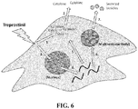


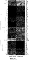


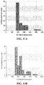
Classifications
-
- A—HUMAN NECESSITIES
- A61—MEDICAL OR VETERINARY SCIENCE; HYGIENE
- A61K—PREPARATIONS FOR MEDICAL, DENTAL OR TOILETRY PURPOSES
- A61K31/00—Medicinal preparations containing organic active ingredients
- A61K31/557—Eicosanoids, e.g. leukotrienes or prostaglandins
- A61K31/558—Eicosanoids, e.g. leukotrienes or prostaglandins having heterocyclic rings containing oxygen as the only ring hetero atom, e.g. thromboxanes
- A61K31/5585—Eicosanoids, e.g. leukotrienes or prostaglandins having heterocyclic rings containing oxygen as the only ring hetero atom, e.g. thromboxanes having five-membered rings containing oxygen as the only ring hetero atom, e.g. prostacyclin
-
- A—HUMAN NECESSITIES
- A61—MEDICAL OR VETERINARY SCIENCE; HYGIENE
- A61K—PREPARATIONS FOR MEDICAL, DENTAL OR TOILETRY PURPOSES
- A61K31/00—Medicinal preparations containing organic active ingredients
- A61K31/185—Acids; Anhydrides, halides or salts thereof, e.g. sulfur acids, imidic, hydrazonic or hydroximic acids
- A61K31/19—Carboxylic acids, e.g. valproic acid
- A61K31/192—Carboxylic acids, e.g. valproic acid having aromatic groups, e.g. sulindac, 2-aryl-propionic acids, ethacrynic acid
-
- A—HUMAN NECESSITIES
- A61—MEDICAL OR VETERINARY SCIENCE; HYGIENE
- A61K—PREPARATIONS FOR MEDICAL, DENTAL OR TOILETRY PURPOSES
- A61K31/00—Medicinal preparations containing organic active ingredients
- A61K31/557—Eicosanoids, e.g. leukotrienes or prostaglandins
-
- A—HUMAN NECESSITIES
- A61—MEDICAL OR VETERINARY SCIENCE; HYGIENE
- A61K—PREPARATIONS FOR MEDICAL, DENTAL OR TOILETRY PURPOSES
- A61K35/00—Medicinal preparations containing materials or reaction products thereof with undetermined constitution
- A61K35/12—Materials from mammals; Compositions comprising non-specified tissues or cells; Compositions comprising non-embryonic stem cells; Genetically modified cells
- A61K35/28—Bone marrow; Haematopoietic stem cells; Mesenchymal stem cells of any origin, e.g. adipose-derived stem cells
-
- A—HUMAN NECESSITIES
- A61—MEDICAL OR VETERINARY SCIENCE; HYGIENE
- A61P—SPECIFIC THERAPEUTIC ACTIVITY OF CHEMICAL COMPOUNDS OR MEDICINAL PREPARATIONS
- A61P43/00—Drugs for specific purposes, not provided for in groups A61P1/00-A61P41/00
-
- A—HUMAN NECESSITIES
- A61—MEDICAL OR VETERINARY SCIENCE; HYGIENE
- A61P—SPECIFIC THERAPEUTIC ACTIVITY OF CHEMICAL COMPOUNDS OR MEDICINAL PREPARATIONS
- A61P9/00—Drugs for disorders of the cardiovascular system
-
- A—HUMAN NECESSITIES
- A61—MEDICAL OR VETERINARY SCIENCE; HYGIENE
- A61P—SPECIFIC THERAPEUTIC ACTIVITY OF CHEMICAL COMPOUNDS OR MEDICINAL PREPARATIONS
- A61P9/00—Drugs for disorders of the cardiovascular system
- A61P9/10—Drugs for disorders of the cardiovascular system for treating ischaemic or atherosclerotic diseases, e.g. antianginal drugs, coronary vasodilators, drugs for myocardial infarction, retinopathy, cerebrovascula insufficiency, renal arteriosclerosis
-
- A—HUMAN NECESSITIES
- A61—MEDICAL OR VETERINARY SCIENCE; HYGIENE
- A61P—SPECIFIC THERAPEUTIC ACTIVITY OF CHEMICAL COMPOUNDS OR MEDICINAL PREPARATIONS
- A61P9/00—Drugs for disorders of the cardiovascular system
- A61P9/12—Antihypertensives
-
- A—HUMAN NECESSITIES
- A61—MEDICAL OR VETERINARY SCIENCE; HYGIENE
- A61P—SPECIFIC THERAPEUTIC ACTIVITY OF CHEMICAL COMPOUNDS OR MEDICINAL PREPARATIONS
- A61P9/00—Drugs for disorders of the cardiovascular system
- A61P9/14—Vasoprotectives; Antihaemorrhoidals; Drugs for varicose therapy; Capillary stabilisers
-
- C—CHEMISTRY; METALLURGY
- C12—BIOCHEMISTRY; BEER; SPIRITS; WINE; VINEGAR; MICROBIOLOGY; ENZYMOLOGY; MUTATION OR GENETIC ENGINEERING
- C12N—MICROORGANISMS OR ENZYMES; COMPOSITIONS THEREOF; PROPAGATING, PRESERVING, OR MAINTAINING MICROORGANISMS; MUTATION OR GENETIC ENGINEERING; CULTURE MEDIA
- C12N5/00—Undifferentiated human, animal or plant cells, e.g. cell lines; Tissues; Cultivation or maintenance thereof; Culture media therefor
- C12N5/06—Animal cells or tissues; Human cells or tissues
- C12N5/0602—Vertebrate cells
- C12N5/0652—Cells of skeletal and connective tissues; Mesenchyme
- C12N5/0662—Stem cells
- C12N5/0663—Bone marrow mesenchymal stem cells (BM-MSC)
-
- C—CHEMISTRY; METALLURGY
- C12—BIOCHEMISTRY; BEER; SPIRITS; WINE; VINEGAR; MICROBIOLOGY; ENZYMOLOGY; MUTATION OR GENETIC ENGINEERING
- C12N—MICROORGANISMS OR ENZYMES; COMPOSITIONS THEREOF; PROPAGATING, PRESERVING, OR MAINTAINING MICROORGANISMS; MUTATION OR GENETIC ENGINEERING; CULTURE MEDIA
- C12N2501/00—Active agents used in cell culture processes, e.g. differentation
- C12N2501/02—Compounds of the arachidonic acid pathway, e.g. prostaglandins, leukotrienes
Definitions
- the present application relates to the use of mesenchymal stem cells in treatment of vasculopathy, including pulmonary arterial hypertension (PAH) and other types of pulmonary hypertension, peripheral vascular disease (PVD), critical limb ischemia (CLI), coronary artery disease, diabetic vasculopathy, etc.
- PAH pulmonary arterial hypertension
- PVD peripheral vascular disease
- CLI critical limb ischemia
- coronary artery disease diabetic vasculopathy, etc.
- Pulmonary arterial hypertension is a progressive lung disorder which, untreated, leads to death on average within 2.8 years after being diagnosed. An increasing constriction of the pulmonary circulation leads to increased stress on the right heart, which may develop into right heart failure.
- mPAP mean pulmonary arterial pressure
- the pathophysiology of pulmonary arterial hypertension is characterized by vasoconstriction and remodeling of the pulmonary vessels. In chronic PAH there is neomuscularization of initially unmuscularized pulmonary vessels, and the vascular muscles of the already muscularized vessels increase in circumference.
- Standard therapies available on the market e.g. prostacyclin analogues, endothelin receptor antagonists, phosphodiesterase inhibitors
- prostacyclin analogues e.g., prostacyclin analogues, endothelin receptor antagonists, phosphodiesterase inhibitors
- the principles of these therapies are primarily hemodynamic, influencing vessel tone but having no direct influence on the pathogenic remodeling processes.
- the possibility of using these medicaments is restricted through the sometimes serious side effects and/or complicated types of administration.
- the period over which the clinical situation of the patients can be improved or stabilized by specific monotherapy is limited.
- the therapy escalates and thus a combination therapy is applied, where a plurality of medicaments must be given concurrently.
- pulmonary arterial hypertension there is as yet no prospect of cure of this serious disorder.
- peripheral vascular disease refers to damage, dysfunction or obstruction within peripheral arteries and veins.
- Peripheral artery disease is the most common form of PVD.
- Peripheral vascular disease is the most common disease of the arteries and is a very common condition in the United States. It occurs mostly in people older than 50 years. Peripheral vascular disease is a leading cause of disability among people older than 50 years, as well as in those people with diabetes. About 10 million people in the United States have peripheral vascular disease, which translates to about 5% of people older than 50 years. The number of people with the condition is expected to grow as the population ages. Men are slightly more likely than women to have peripheral vascular disease.
- Critical limb ischemia due to advanced peripheral arterial occlusion, is characterized by reduced blood flow and oxygen delivery at rest, resulting in muscle pain at rest and non-healing skin ulcers or gangrene ( Rissanen et al., Eur. J. Clin. Invest 31:651-666 (2001 ); Dormandy and Rutherford, J. Vasc. Surg. 31:S1-S296 (2000 )).
- Critical limb ischemia is estimated to develop in 500 to 1000 per million individuals in one year ("Second European Consensus Document on Chronic Critical Leg Ischemia", Circulation 84(4 Suppl.) IV 1-26 (1991)).
- Coronary artery disease is a progressive disease in humans wherein one or more coronary arteries gradually become occluded through the buildup of plaque.
- the coronary arteries of patients having this disease are often treated by balloon angioplasty or the insertion of stents to prop open the partially occluded arteries. Ultimately, these patients are required to undergo coronary artery bypass surgery at great expense and risk.
- the current disclosure is directed to a method for treating or preventing vasculopathy in a subject in need thereof, comprising administering to the subject a prostacyclin and a composition comprising a mesenchymal stem cell (MSC) or a part of a culture medium that has been in contact with the MSC and contains one or more component(s) of the MSC.
- MSC mesenchymal stem cell
- the prostacyclin and the composition can be administered concurrently or separately.
- the MSC prior to the administration, has been in contact with prostacyclin.
- the culture medium or the MSC from which the culture medium is obtained can be placed in contact with prostacyclin, prior to such administration. Accordingly, in some embodiments, the method further includes such a pre-treatment step.
- Non-limiting examples of components obtained from a part of the MSC culture include an exosome, a microvesicle, a microRNA, a messenger RNA, a non-coding RNA, a mitochondria, a growth factor, or combinations thereof.
- Such methods further entail administering to the subject an endothelial progenitor cell (EPC).
- EPC endothelial progenitor cell
- the EPC is obtained from the subject.
- the EPC is transformed with a nucleic acid that increases the expression of biological activity of a protein selected from the group consisting of endothelial nitric oxide synthase (eNOS), heme oxygenase (HMOX1) and prostacyclin synthase (PTGIS).
- eNOS endothelial nitric oxide synthase
- HMOX1 heme oxygenase
- PTGIS prostacyclin synthase
- the nucleic acid encodes the protein.
- prostacyclin examples include, without limitation, epoprostenol sodium, treprostinil, beraprost, ilprost, and a PGI 2 receptor agonist.
- the prostacyclin is treprostinil or a pharmaceutically acceptable salt or ester thereof.
- a pharmaceutical composition comprising a therapeutically effective amount of a prostacyclin and a composition comprising a mesenchymal stem cell (MSC) or a culture medium that has been in contact with the MSC and contains compounds released from the MSC and a pharmaceutically acceptable carrier.
- the composition further comprises an endothelial progenitor cell (EPC).
- Yet another embodiment provides a method for preparing a composition comprising a mesenchymal stem cell (MSC) or a culture medium that has been in contact with the MSC and contains compounds released from the MSC for in vivo delivery, comprising contacting the MSC with a prostacyclin.
- MSC mesenchymal stem cell
- Treated composition obtainable by such a method is also provided.
- the pharmaceutical composition further comprises at least one pharmaceutically-acceptable carrier or at least one therapeutic agent.
- the subject is suffering from vasculopathy, such as pulmonary arterial hypertension (PAH), peripheral vascular disease (PVD), critical limb ischemia (CLI), coronary artery disease, or diabetic vasculopathy.
- vasculopathy such as pulmonary arterial hypertension (PAH), peripheral vascular disease (PVD), critical limb ischemia (CLI), coronary artery disease, or diabetic vasculopathy.
- the current method reduces thrombosis in pulmonary arteries, reduces inflammation in pulmonary arteries, reduces the proliferation of intimal smooth muscle in pulmonary arteries, reduces the formation of plexiform lesions in pulmonary arteries, increases the amount of nitric oxide in pulmonary arteries, increases the amount of PGI 2 in pulmonary arteries, reduces the level of Endothelin-1 in pulmonary arteries, or reduces the amount of growth factors in pulmonary arteries.
- the current method promotes proper endothelial morphology in pulmonary arteries.
- prostacyclin and mesenchymal stem cells possess therapeutic activities for vasculopathy.
- the combination of prostacyclin and MSCs furthermore, produces synergistic effects.
- Such combination can be either co-administration, which can be concurrent or separate, of prostacyclin and MSCs to a patient, or administration to the patient a MSC composition that has been pre-treated with a prostacyclin.
- MSCs can ameliorate vasculopathy in patients, and it is contemplated that such a therapeutic effect is achieved due to MSCs' ability to improve the local microenvironment by delivering anti-inflammatory and pro-angiogenic factors to the diseased area. MSCs, however, are short-lived in the body and not regenerative.
- Prostacyclin such as treprostinil (TP)
- TP treprostinil
- PAH pulmonary arterial hypertension
- prostacyclin has been shown to possess vasodilatory and anti-platelet aggregation activities.
- prostacyclin can enhance the activity of MSCs for the treatment of vasculopathy, exhibiting synergism for such treatment.
- prostacyclin enhances MSCs' beneficial effect on blood vessel growth.
- prostacyclin increases the expression of VEGF at both protein and gene levels.
- Changes in secreted cytokines are also observed as a result of prostacyclin exposure. For instance, IL-6 is increased ⁇ 50-fold while MCP-1 is decreased ⁇ 6-7-fold.
- a “MSC-conditioned culture medium” refers to a culture medium that has been in contact with a MSC (e.g., for the purpose of culturing the MSC) and thus contains compounds released from the MSC.
- Non-limiting examples of such released compounds include exosomes or other microvesicles which can enclose messenger RNA, non-coding RNA, microRNAs, mitochondria, growth factors, or other types of bioactive agents.
- a "culture medium” as used herein encompasses (a) both a culture medium that contains the typical components used for culturing a MSC, such as amino acids, glucose, and various salts, with or without the MSC, and (b) a composition isolated from the culture medium that contains compounds released from the MSC during the culturing.
- one embodiment of the present disclosure provides a method for treating or preventing vasculopathy in a subject in need thereof, comprising administering to the subject a prostacyclin and a composition comprising a mesenchymal stem cell (MSC) or a MSC-conditioned culture medium (collectively a "MSC composition").
- a composition comprising a mesenchymal stem cell (MSC) or a MSC-conditioned culture medium (collectively a "MSC composition").
- the prostacyclin and the MSC composition are administered concurrently. In another aspect, the prostacyclin and the MSC composition are administered separately. When administered separately, the prostacyclin can be administered prior to, or following the administration of the MSC composition.
- a method for treating or preventing vasculopathy in a subject in need thereof comprising contacting a composition comprising an isolated mesenchymal stem cell (MSC) or a MSC-conditioned culture medium with a prostacyclin, and then administering the MSC composition to the subject.
- MSC mesenchymal stem cell
- Non-limiting examples of vasculopathy include pulmonary arterial hypertension (PAH), peripheral vascular disease (PVD), critical limb ischemia (CLI), coronary artery disease and diabetic vasculopathy.
- PAH pulmonary arterial hypertension
- PVD peripheral vascular disease
- CLI critical limb ischemia
- coronary artery disease and diabetic vasculopathy.
- the term "subject” includes warm-blooded animals, preferably mammals, including humans.
- the subject is a primate.
- the subject is a human.
- treating include administering a therapeutically effective amount of cells as defined herein sufficient to reduce or eliminate at least one symptom of vasculopathy.
- preventing include administering a therapeutically effective amount of cells as defined herein sufficient to stop or hinder the development of at least one symptom of vasculopathy.
- prostacyclin used herein explicitly comprises any prostaglandin I 2 (PGI 2 ), any prostacyclin analogues, and any PGI 2 receptor agonists.
- PGI 2 prostaglandin I 2
- prostacyclin suitable for the present technology include epoprostenol sodium (e.g. Flolan®), treprostinil(e.g. TYVASO®, Remodulin®), ilprost (e.g. Ventavis®), and PGI 2 receptor agonist (e.g. Selexipag).
- the prostacyclin is treprostinil or a pharmaceutically acceptable salt or ester thereof.
- MSCs Mesenchymal Stem Cells
- MSCs Mesenchymal stem cells
- adipose tissue adipose tissue
- skin spleen
- pancreas brain
- kidney liver
- heart retina
- brain hair follicles
- intestine lung
- lymph node thymus
- bone ligament
- tendon skeletal muscle
- dermis a periosteum
- periosteum adipose
- MSCs are capable of differentiating into a large number of cell types including, but not limited to, adipose, osseous, cartilaginous, elastic, muscular, and fibrous connective tissues.
- MSCs are thus non-hematopoietic progenitor cells which divide to yield daughter cells that are either stem cells or are precursor cells which in time will irreversibly differentiate to yield a phenotypic cell.
- MSCs include mesenchymal precursor cells (MPCs).
- stem cell refers to self-renewing cells that are capable of giving rise to phenotypically and genotypically identical daughters as well as at least one other final cell type (e.g., terminally differentiated cells).
- stem cells includes totipotential, pluripotential and multipotential cells, as well as progenitor and/or precursor cells derived from the differentiation thereof.
- totipotent cell or “totipotential cell” refers to a cell that is able to form a complete embryo (e.g., a blastocyst).
- pluripotent cell refers to a cell that has complete differentiation versatility, i.e., the capacity to grow into any of the mammalian body's approximately 260 cell types.
- a pluripotent cell can be self-renewing, and can remain dormant or quiescent within a tissue.
- multipotential cell refers to a cell which is capable of giving rise to any of several mature cell types. As used herein, this phrase encompasses adult or embryonic stem cells and progenitor cells, and multipotential progeny of these cells. Unlike a pluripotent cell, a multipotent cell does not have the capacity to form all of the cell types.
- progenitor cell or “precursor cell” refers to a cell that is committed to differentiate into a specific type of cell or to form a specific type of tissue.
- cells used in the methods of the disclosure are enriched from a sample obtained from a subject.
- the terms 'enriched', 'enrichment' or variations thereof are used herein to describe a population of cells in which the proportion of one particular cell type or the proportion of a number of particular cell types is increased when compared with the untreated population.
- the cells used in the present disclosure are TNAP + , STRO-1 + , VCAM-1 + , THY-1 + , STRO-2 + , CD45 + , CD146 + , 3G5 + or any combination thereof.
- a cell When we refer to a cell as being "positive” for a given marker it may be either a low (lo or dim) or a high (bright, bri) expresser of that marker depending on the degree to which the marker is present on the cell surface, where the terms relate to intensity of fluorescence or other color used in the color sorting process of the cells.
- lo or dim or dull
- bri When we refer herein to a cell as being "negative” for a given marker, it does not mean that the marker is not expressed at all by that cell. It means that the marker is expressed at a relatively very low level by that cell, and that it generates a very low signal when detectably labeled.
- TNAP tissue non-specific alkaline phosphatase
- the term encompasses the liver isoform (LAP), the bone isoform (BAP) and the kidney isoform (KAP).
- LAP liver isoform
- BAP bone isoform
- KAP kidney isoform
- the TNAP is BAP.
- TNAP as used herein refers to a molecule which can bind the STRO-3 antibody produced by the hybridoma cell line deposited with ATCC on 19 Dec. 2005 under the provisions of the Budapest Treaty under deposit accession number PTA-7282.
- Stem cells useful for the methods can be derived from adult tissue, an embryo, extraembryonic tissue, or a fetus.
- the term “adult” is used in its broadest sense to include a postnatal subject. In a preferred embodiment, the term “adult” refers to a subject that is postpubertal. The term, "adult” as used herein can also include cord blood taken from a female.
- the stem cells can be progeny cells (which can also be referred to as expanded cells) which are produced from the in vitro culture of the stem cells described herein.
- Expanded cells of the disclosure may have a wide variety of phenotypes depending on the culture conditions (including the number and/or type of stimulatory factors in the culture medium), the number of passages and the like.
- the progeny cells are obtained after about 2, about 3, about 4, about 5, about 6, about 7, about 8, about 9, or about 10 passages from the parental population.
- the progeny cells may be obtained after any number of passages from the parental population.
- the progeny cells can be obtained by culturing in any suitable medium.
- Media may be solid, liquid, gaseous or a mixture of phases and materials.
- Media include liquid growth media as well as liquid media that do not sustain cell growth.
- Media also include gelatinous media such as agar, agarose, gelatin and collagen matrices.
- the term “medium” also refers to material that is intended for use in a cell culture, even if it has not yet been contacted with cells. In other words, a nutrient rich liquid prepared for bacterial culture is a medium.
- the progeny cells are obtained by isolating TNAP+ cells from bone marrow using magnetic beads labelled with the STRO-3 antibody, and plated in ⁇ -MEM supplemented with 20% fetal calf serum, 2 mM L-glutamine and 100 ⁇ m L-ascorbate-2-phosphate.
- such expanded cells can be TNAP-, CC9+, HLA class I+, HLA class II-, CD14-, CD19-; CD3-, CDlla-c-, CD31-, CD86- and/or CD80-.
- the expression of different markers may vary.
- cells of these phenotypes may predominate in the expended cell population it does not mean that there is not a minor proportion of the cells that do not have this phenotype(s) (for example, a small percentage of the expanded cells may be CC9-).
- expanded cells of the disclosure still have the capacity to differentiate into different cell types.
- an expended cell population used in the methods of the disclosure comprises cells wherein at least 25%, more preferably at least 50%, of the cells are CC9+.
- an expended cell population used in the methods of the disclosure comprises cells wherein at least 40%, more preferably at least 45%, of the cells are STRO-1+.
- markers selected from the group consisting of LFA-3, THY-1, VCAM-1, PECAM-1, P-selectin, L-selectin, 3G5, CD49a/CD49b/CD29, CD49c/CD29, CD49d/CD29, CD29, CD18, CD61, integrin beta, 6-19
- the progeny cells are Multipotential Expanded MSC Progeny (MEMPs) as defined in WO 2006/032092 .
- MEMPs Multipotential Expanded MSC Progeny
- Methods for preparing enriched populations of MSC from which progeny may be derived are described in WO 01/04268 and WO 2004/085630 .
- MSCs will rarely be present as an absolutely pure preparation and will generally be present with other cells that are tissue specific committed cells (TSCCs).
- WO 01/04268 refers to harvesting such cells from bone marrow at purity levels of about 0.1% to 90%.
- the population comprising MSC from which progeny are derived may be directly harvested from a tissue source, or alternatively it may be a population that has already been expanded ex vivo.
- the progeny may be obtained from a harvested, unexpanded, population of substantially purified MSC, comprising at least about 0.1, 1, 5, 10, 20, 30, 40, 50, 60, 70, 80 or 95% of total cells of the population in which they are present.
- This level may be achieved, for example, by selecting for cells that are positive for at least one marker selected from the group consisting of TNAP, STRO-1 bri , 3G5+, VCAM-1, THY-1, CD146 and STRO-2.
- the MSC starting population may be derived from any one or more tissue types set out in WO 01/04268 or WO 2004/085630 , namely bone marrow, dental pulp cells, adipose tissue and skin, or perhaps more broadly from adipose tissue, teeth, dental pulp, skin, liver, kidney, heart, retina, brain, hair follicles, intestine, lung, spleen, lymph node, thymus, pancreas, bone, ligament, bone marrow, tendon and skeletal muscle.
- tissue types set out in WO 01/04268 or WO 2004/085630 , namely bone marrow, dental pulp cells, adipose tissue and skin, or perhaps more broadly from adipose tissue, teeth, dental pulp, skin, liver, kidney, heart, retina, brain, hair follicles, intestine, lung, spleen, lymph node, thymus, pancreas, bone, ligament, bone marrow, tendon and skeletal
- MEMPS can be distinguished from freshly harvested MSCs in that they are positive for the marker STRO-lbri and negative for the marker Alkaline phosphatase (ALP). In contrast, freshly isolated MSCs are positive for both STRO-1 bri and ALP. In a preferred embodiment of the present disclosure, at least 15%, 20%, 30%, 40%, 50%, 60%, 70%, 80%, 90% or 95% of the administered cells have the phenotype STRO-1 bri , ALP-. In a further preferred embodiment the MEMPS are positive for one or more of the markers Ki67, CD44 and/or CD49c/CD29, VLA-3, ⁇ 3 ⁇ 1. In yet a further preferred embodiment the MEMPs do not exhibit TERT activity and/or are negative for the marker CD 18.
- the cells are taken from a patient with vasculopathy, cultured in vitro using standard techniques and administered to a patient as an autologous or allogeneic transplant.
- cells of one or more of the established human cell lines are used.
- cells of a non-human animal or if the patient is not a human, from another species are used.
- the present technology can be practiced using cells from any non-human animal species, including but not limited to non-human primate cells, ungulate, canine, feline, lagomorph, rodent, avian, and fish cells.
- Primate cells with which the disclosure may be performed include but are not limited to cells of chimpanzees, baboons, cynomolgus monkeys, and any other New or Old World monkeys.
- Ungulate cells with which the disclosure may be performed include but are not limited to cells of bovines, porcines, ovines, caprines, equines, buffalo and bison.
- Rodent cells with which the disclosure may be performed include but are not limited to mouse, rat, guinea pig, hamster and gerbil cells.
- Examples of lagomorph species with which the disclosure may be performed include domesticated rabbits, jack rabbits, hares, cottontails, snowshoe rabbits, and pikas.
- Chickens (Gallus gallus) are an example of an avian species with which the disclosure may be performed.
- Cells can be stored before use. Methods and protocols for preserving and storing of eukaryotic cells, and in particular mammalian cells, are well known in the art (cf., for example, Pollard, J. W. and Walker, J. M. (1997) Basic Cell Culture Protocols, Second Edition, Humana Press, Totowa, N.J .; Freshney, R. I. (2000) Culture of Animal Cells, Fourth Edition, Wiley-Liss, Hoboken, N.J .). Any method maintaining the biological activity of the isolated stem cells such as mesenchymal stem/progenitor cells, or progeny thereof, may be utilized in connection with the present disclosure. In one preferred embodiment, the cells are maintained and stored by using cryo-preservation.
- Cells can be obtained using a variety of techniques. For example, a number of cell-sorting techniques by which cells are physically separated by reference to a property associated with the cell-antibody complex, or a label attached to the antibody can be used. This label may be a magnetic particle or a fluorescent molecule.
- the antibodies may be cross-linked such that they form aggregates of multiple cells, which are separable by their density. Alternatively the antibodies may be attached to a stationary matrix, to which the desired cells adhere.
- an antibody (or other binding agent) that binds TNAP+, STRO-1+, VCAM-1+, THY-1+, STRO-2+, 3G5+, CD45+, CD146+ is used to isolate the cells. More preferably, an antibody (or other binding agent) that binds TNAP+ or STRO-1+ is used to isolate the cells.
- the antibody bound to the cell can be labelled and then the cells separated by a mechanical cell sorter that detects the presence of the label. Fluorescence-activated cell sorters are well known in the art.
- anti-TNAP antibodies and/or an STRO-1 antibodies are attached to a solid support.
- solid supports are known to those of skill in the art, including, but not limited to, agarose beads, polystyrene beads, hollow fiber membranes, polymers, and plastic petri dishes. Cells that are bound by the antibody can be removed from the cell suspension by simply physically separating the solid support from the cell suspension.
- Super paramagnetic microparticles may be used for cell separations.
- the microparticles may be coated with anti-TNAP antibodies and/or STRO-1 antibodies.
- the antibody-tagged, super paramagnetic microparticles may then be incubated with a solution containing the cells of interest.
- the microparticles bind to the surfaces of the desired stem cells, and these cells can then be collected in a magnetic-field.
- the cell sample is allowed to physically contact, for example, a solid phase-linked anti-TNAP monoclonal antibodies and/or anti-STRO-1 monoclonal antibodies.
- the solid-phase linking can comprise, for instance, adsorbing the antibodies to a plastic, nitrocellulose, or other surface.
- the antibodies can also be adsorbed on to the walls of the large pores (sufficiently large to permit flow-through of cells) of a hollow fiber membrane.
- the antibodies can be covalently linked to a surface or bead, such as Pharmacia Sepharose 6 MB macrobeads.
- the exact conditions and duration of incubation for the solid phase-linked antibodies with the stem cell containing suspension will depend upon several factors specific to the system employed. The selection of appropriate conditions, however, is well within the skill of the art.
- the unbound cells are then eluted or washed away with physiologic buffer after allowing sufficient time for the stem cells to be bound.
- the unbound cells can be recovered and used for other purposes or discarded after appropriate testing has been done to ensure that the desired separation had been achieved.
- the bound cells are then separated from the solid phase by any appropriate method, depending mainly upon the nature of the solid phase and the antibody.
- bound cells can be eluted from a plastic petri dish by vigorous agitation.
- bound cells can be eluted by enzymatically "nicking" or digesting an enzyme-sensitive "spacer" sequence between the solid phase and the antibody. Spacers bound to agarose beads are commercially available from, for example, Pharmacia.
- the eluted, enriched fraction of cells may then be washed with a buffer by centrifugation and said enriched fraction may be cryopreserved in a viable state for later use according to conventional technology, culture expanded and/or introduced into the patient.
- MSCs can carry out their activities through compounds that can be released into the extracellular environment during growth or differentiation.
- such compounds include a microvesicle, referred to as exosome, which is between about 30 nm and about 200 nm in diameter. Exosomes can be internalized by host cells in vivo.
- Exosomes are vesicles derived from the multivesicular body sorting pathway. Recent studies show that exosomes are bioactive vesicles useful for intercellular communication and facilitation of the immunoregulatory process. MSC exosomes contain 20S proteasomes and numerous RNAs (messenger RNA, non-coding RNA, microRNA).
- MSC In addition to exosomes, MSC also release other bioactive molecules/vesicles useful for the purpose of the present disclosure.
- Such molecules and vesicles include, without limitation, mitochondria and growth factors.
- Method of preparing culture media that contain such molecules and vesicles released from MSC and further isolating particular molecules and vesicles are known in the art. See, for instance, Hu et al., Frontiers in Genetics, 2:56, 1-9 (2012 ).
- the MSC or MSC-conditioned culture medium prior to coadministering a MSC or a MSC-conditioned culture medium with prostacyclin to a patient, can be optionally pre-treated with prostacyclin.
- a method for preparing a mesenchymal stem cell (MSC) or MSC-conditioned culture medium for in vivo delivery comprising contacting the MSC or MSC-conditioned culture medium with a prostacyclin.
- a treated MSC or MSC-conditioned culture medium obtainable by such a method.
- Pre-treatment of a cell or a medium with a chemical compound encompasses known techniques.
- the prostacyclin can be added to and co-incubated with a culture medium that contains a MSC.
- co-incubation can further involve the addition of a growth factor (e.g., VEGF and Angiopoietin-1 or -2, platelet-derived growth factor) and/or hypoxia.
- a growth factor e.g., VEGF and Angiopoietin-1 or -2, platelet-derived growth factor
- MSCs or MSC-conditioned culture media can be treated with prostacyclin in various ways.
- prostacyclin can be used to treat MSCs ex vivo during the expansion of MSCs; prostacyclin can also be used to treat MSCs after administration.
- the concentration of prostacyclin is at least about 100 ⁇ g/mL, or at least about 150 ⁇ g/mL, 200 ⁇ g/mL, or 250 ⁇ g/mL.
- the concentration of prostacyclin is not more than about 400 ⁇ g/mL, or not more than about 350 ⁇ g/mL, 300 ⁇ g/mL or 250 ⁇ g/mL.
- MSCs can be prepared from the recipient's own blood or bone marrow.
- prostacyclin can also be used to treat MSCs before they are isolated from the recipients.
- EPC Endothelial progenitor cell
- EPC endothelial progenitor cell
- the EPC can also be pre-treated with prostacyclin.
- the EPCs treated with prostacyclin exhibit a hyperproliferative phenotype with enhanced angiogenic properties, which are advantageous in treating vasculopathy compared to untreated EPCs.
- EPCs can be treated with prostacyclin in various ways.
- prostacyclin can be used to treat EPCs ex vivo during the expansion of EPCs; prostacyclin can be co-administered with EPCs to the recipient; prostacyclin can also be used to treat EPCs after transplantation.
- EPCs are prepared from the recipient's own blood or bone marrow. In that case, prostacyclin can also be used to treat EPCs before they are isolated from the recipients.
- EPC is an undifferentiated cell that can be induced to proliferate.
- EPCs are capable of self-maintenance, such that with each cell division, at least one daughter cell will also be an EPC cell.
- EPCs are capable of being expanded 100, 250, 500, 1000, 2000, 3000, 4000, 5000 or more fold.
- EPC Phenotyping of EPCs reveals that these cells express the committed hematopoietic marker CD45. Additionally, an EPC may be immunoreactive for VEGFR-2 and/or Tie-2. Optionally, the EPC is immunoreactive for CD14. The EPC is a multipotent progenitor cell.
- Vascular endothelial growth factor acts through specific tyrosine kinase receptors that includes VEGFR-1 (flt-1) and VEGFR-2 (flk-1/KDR) and VEGFR-3/Flt-4 which convey signals that are essential for embryonic angiogenesis and hematopoiesis. While VEGF binds to all three receptors, most biological functions are mediated via VEGFR-2 and the role of VEGFR-1 is currently unknown. VEGFR3/Flt4 signaling is known to be important for the development of lymphatic endothelial cells and VEGFR3 signaling may confer lymphatic endothelial-like phenotypes to endothelial cells.
- VEGFRs relay signals for processes essential in stimulation of vessel growth, vasorelaxation, induction of vascular permeability, endothelial cell migration, proliferation and survival. Endothelial cells express all different VEGF-Rs. During embryogenesis, it has been reported that a single progenitor cell, the hemangioblast can give rise to both the hematopoietic and vascular systems.
- Tie-2 is an endothelial-specific receptor tyrosine kinase and a receptor for angiopoietin 1. It is a type I membrane protein that is expressed predominantly in the endothelium of actively growing blood vessels and may represent the earliest mammalian endothelial cell lineage marker. Tie-2 is likely involved in the regulation of endothelial cell proliferation and differentiation and may direct the special orientation of endothelial cells during the formation of blood vessels.
- the CD14 antigen is a high affinity receptor for the complex of lipopolysaccharides (LPS) and LPS-Binding protein (LBP).
- LPS lipopolysaccharides
- LBP LPS-Binding protein
- the CD14 antigen is part of the functional heteromeric LPS receptor complex comprised of CD14, TLR4 and MD-2.
- CD14 is strongly expressed on most human monocytes and macrophages in peripheral blood, other body fluids and various tissues, such as lymph nodes and spleen.
- CD14 is weakly expressed on subpopulations of human neutrophils and myeloid dendritic cells.
- the CD45 antigen is a tyrosine phosphatase, also known as the leukocyte common antigen (LCA).
- CD45 is present on all human cells of hematopoietic origin, except erythroid cells, platelets and their precursor cells.
- the CD45 molecule is required for T cell and B cell activation and is expressed in at least 5 isoforms, depending on the activation status of the cell.
- VEGFR-1+, VEGFR-2+ and Tie-2+ cells constituted approximately 3.0.+-.0.2%, 0.8.+-.0.5%, 2.0.+-.0.3% of the total population of mononuclear cells in blood respectively.
- CD14+/VEGFR-2+ cells constituted approximately 2.0.+-.0.5% of the total population of monocytes and 0.08.+-.0.04% of mononuclear cells in blood.
- EPCs can be maintained in vitro in long-term cultures.
- the EPCs are capable of being passed in culture 2, 3, 4, 5, 6, 7, 8, 9, 10, 11, 12 or more times.
- EPCs comprise endothelial colony-forming cells, typically developed after 1-3 weeks of cell culture. Endothelial colony-forming cells have the characteristics of precursor cells committed to the endothelial lineage and are capable of merging into neovessels, according to Smardja et al., Angiogenesis 14(1):17-27 (2011 ).
- the population of cells are isolated by means of positive selection, or by a mixture of both positive and negative selection in either order.
- the population of progenitor cells is purified.
- a purified population of EPCs contains a significantly higher proportion of EPCs than the crude population of cells from which the cells are isolated.
- the purification procedure should lead at least to a five-fold increase, preferably at least a ten-fold increase, more preferably at least a fifteen fold increase, most preferably at least a twenty fold increase, and optimally at least a twenty-five fold increase in EPCs with respect to the total population.
- the purified population of EPC should include at least 15%, preferably at least 20%, more preferably at least 25%, most preferably at least 35%, and optimally at least 50% of EPCs.
- the methods described herein can lead to mixtures comprising up to 75%, preferably up to 80%, more preferably up to 85%, most preferably up to 90% and optimally up to 95% of stem cells.
- Such methods are capable of producing mixtures comprising 99%, 99.90% and even 100% of EPCs. Accordingly, the purified populations of the disclosure contain significantly higher levels of EPCs than those that exist in nature, as described above.
- the purified population of EPCs can be isolated by contacting a crude mixture of cells containing a population of stem cells that express an antigen characteristic of the EPCs with a molecule that binds specifically to the extracellular portion of the antigen. Such a technique is known as positive selection.
- the binding of the EPCs to the molecule permit the EPCs to be sufficiently distinguished from contaminating cells that do not express the antigen to permit isolating the stem cells from the contaminating cells.
- the antigen is preferably VEGFR, and more preferably VEGFR-2.
- the molecule used to separate progenitor cells from the contaminating cells can be any molecule that binds specifically to the antigen that characterizes the EPCs.
- the molecule can be, for example, a monoclonal antibody, a fragment of a monoclonal antibody, or, in the case of an antigen that is a receptor, the ligand of that receptor.
- the ligand is VEGF.
- the unique isolated cells of the present disclosure can be separated from other cells by virtue of their CD45+ state and possession of vascular endothelial growth factor receptors (VEGFR), e.g. VEGFR-2.
- VEGFR vascular endothelial growth factor receptors
- the cells can be isolated by conventional techniques for separating cells, such as those described in Civin, U.S. Pat. Nos. 4,714,680 , 4,965,204 , 5,035,994 , and 5,130,144 , Tsukamoto et al U.S. Pat. No. 5,750,397 , and Loken et al, U.S. Pat. No. 5,137,809 , each of which are hereby incorporated by reference in their entireties.
- a CD45 specific monoclonal antibody or a VEGFR-specific antibody can be immobilized on a solid support such as nitrocellulose, agarose beads, polystyrene beads, hollow fiber membranes, magnetic beads, and plastic petri dishes. The entire cell population is then be passed through the solid support or added to the beads.
- Cells that are bound to the binding molecule can be removed from the cell suspension by physically separating the solid support from the remaining cell suspension.
- the unbound cells may be eluted or washed away with physiologic buffer after allowing sufficient time for the solid support to bind the stem cells.
- bound cells can be separated from the solid phase by any appropriate method, depending mainly upon the nature of the solid phase and the binding molecule.
- bound cells can be eluted from a plastic petri dish by vigorous agitation.
- bound cells can be eluted by enzymatically "nicking” or digesting an enzyme-sensitive "spacer" sequence between the solid phase and an antibody. Suitable spacer sequences bound to agarose beads are commercially available from, for example, Pharmacia.
- the eluted, enriched fraction of cells may then be washed with a buffer by centrifugation and preserved in a viable state at low temperatures for later use according to conventional technology.
- the cells may also be used immediately, for example by being infused intravenously into a recipient.
- Those which remain attached to the solid support are those cells which contain a marker which is recognized by the antibody used.
- the anti-CD45 antibody is used, then the resulting population will be greatly enriched in CD45+ cells.
- the antibody used is VFGFR, then the resulting population will be greatly enriched in VEGFR+ cells. That population may then be enriched in the other marker by repeating the steps using a solid phase having attached thereto an antibody to the other marker.
- CD45+ VEGFR+ cells are by means of flow cytometry, most preferably by means of a fluorescence-activated cell sorter (FACS), such as those manufactured by Becton-Dickinson under the names FACScan or FACSCalibur.
- FACS fluorescence-activated cell sorter
- the cells having a CD45 marker thereon are tagged with a particular fluorescent dye by means of an anti-CD45 antibody which has been conjugated to such a dye.
- the VEGFR marker of the cells are tagged with a different fluorescent dye by means of an anti-VEGFR antibody which is conjugated to the other dye.
- This emitted light is detected by a photomultiplier tube (PMT) specific for the emission wavelength of the fluorochrome by virtue of a set of optical filters.
- the signal detected by the PMT is amplified in its own channel and displayed by a computer in a variety of different forms--e.g., a histogram, dot display, or contour display.
- fluorescent cells which emit at one wavelength express a molecule that is reactive with the specific fluorochrome-labeled reagent
- non-fluorescent cells or fluorescent cells which emit at a different wavelength do not express this molecule but may express the molecule which is reactive with the fluorochrome-labeled reagent which fluoresces at the other wavelength.
- the flow cytometer is also semi-quantitative in that it displays the amount of fluorescence (fluorescence intensity) expressed by the cell. This correlates, in a relative sense, to the number of the molecules expressed by the cell.
- Flow cytometers can also be equipped to measure non-fluorescent parameters, such as cell volume or light scattered by the cell as it passes through the laser beam.
- Cell volume is usually a direct measurement.
- the light scatter PMTs detect light scattered by the cell either in a forward angle (forward scatter; FSC) or at a right angle (side scatter; SSC).
- FSC forward scatter
- SSC side scatter
- FSC forward scatter
- SSC side scatter
- the flow cytometer is equipped with more than one PMT emission detector.
- the additional PMTs may detect other emission wavelengths, allowing simultaneous detection of more than one fluorochrome, each in individual separate channels.
- Computers allow the analysis of each channel or the correlation of each parameter with another.
- Fluorochromes which are typically used with FACS machines include fluorescein isothiocyanate (FITC), which has an emission peak at 525 nm (green), R-phycoerythrin (PE), which has an emission peak at 575 nm (orange-red), propidium iodide (PI), which has an emission peak at 620 nm (red), 7-aminoactinomycin D (7-AAD), which has an emission peak at 660 nm (red), R-phycoerythrin Cy5 (RPE-Cy5), which has an emission peak at 670 nm (red), and allophycocyanin (APC), which has an emission peak at 655-750 nm (deep red).
- FITC fluorescein isothiocyanate
- PE R-phycoerythrin
- PI propidium iodide
- PI propidium iodide
- 7-AAD 7-aminoactinomycin D
- FACS machines may have the additional capability to physically separate the various fractions by deflecting the cells of different properties into different containers.
- any other method for isolating the CD45+ VEGFR+ population of a starting material, such as bone marrow, peripheral blood or cord blood, may also be used in accordance with the present disclosure.
- the various subpopulations (e.g., CD14+, Tie2+, CD 144-) of the present disclosure may be isolated in similar manners.
- the population of progenitor cells may be further concentrated by methods known in the art.
- the progenitor cells can be enriched by positive selection for one or more antigens characteristic of EPCs.
- antigens include, for example, CD 14 or Tie-2.
- blood is withdrawn directly from the circulating peripheral blood of a donor.
- the blood is percolated continuously through a column containing the solid phase-linked binding molecule, such as an antibody VEGFR-2, to capture EPCs.
- the progenitor cell-depleted blood is returned immediately to the donor's circulatory system by methods known in the art, such as hemapheresis.
- the blood is processed in this way until a sufficient number of progenitor cells binds to the column.
- the stem cells are then isolated from the column by methods known in the art. This method allows rare peripheral blood progenitor cells to be harvested from a very large volume of blood, sparing the donor the expense and pain of harvesting bone marrow and the associated risks of anesthesia, analgesia, blood transfusion, and infection.
- EPCs are cultivated and proliferated using the methods described herein.
- Cells are obtained peripheral blood by isolating peripheral blood mononuclear cells (PBMC) by density gradient centrifugation.
- PBMC peripheral blood mononuclear cells
- Cell suspensions are seeded in any receptacle capable of sustaining cells, particularly culture flasks, culture plates or roller bottles, and more particularly in small culture flasks such as 25 cm 2 culture flasks.
- Cells cultured in suspension are resuspended at approximately 5 ⁇ 10 4 to 2 ⁇ 10 5 cells/ml (for example, 1 ⁇ 10 5 cells/ml).
- Cells plated on a fixed substrate are plated at approximately 2-3 ⁇ 10 3 cells/cm 2 .
- the culture plates are coated with a matrix protein such as collagen.
- the cells can be placed into any known culture medium capable of supporting cell growth, including HEM, DMEM, RPMI, F-12, and the like, containing supplements which are required for cellular metabolism such as glutamine and other amino acids, vitamins, minerals and proteins such as transferrin and the like.
- the culture medium may also contain antibiotics to prevent contamination with yeast, bacteria and fungi such as penicillin, streptomycin, gentamicin and the like.
- the culture medium may contain serum derived from bovine, equine, chicken and the like.
- Conditions for culturing should be close to physiological conditions.
- the pH of the culture medium should be close to physiological pH. (for example, between pH 6-8, between about pH 7 to 7.8, or at pH 7.4).
- Physiological temperatures range between about 30°C. to 40°C.
- EPCs are cultured at temperatures between about 32°C. to about 38°C. (for example, between about 35°C. to about 37°C.).
- the culture medium is supplemented with at least one proliferation-inducing ("mitogenic") growth factor.
- a “growth factor” is protein, peptide or other molecule having a growth, proliferation-inducing, differentiation-inducing, or trophic effect on EPCs.
- proliferation-inducing growth factors are trophic factor that allows EPCs to proliferate, including any molecule that binds to a receptor on the surface of the cell to exert a trophic, or growth-inducing effect on the cell.
- Proliferation-inducing growth factors include EGF, amphiregulin, acidic fibroblast growth factor (aFGF or FGF-1), basic fibroblast growth factor (bFGF or FGF-2), transforming growth factor alpha (TGF ⁇ ), VEGF and combinations thereof.
- Growth factors are usually added to the culture medium at concentrations ranging between about 1 fg/ml to 1 mg/ml. Concentrations between about 1 to 100 ng/ml are usually sufficient. Simple titration assays can easily be performed to determine the optimal concentration of a particular growth factor.
- the biological effects of growth and trophic factors are generally mediated through binding to cell surface receptors.
- the receptors for a number of these factors have been identified and antibodies and molecular probes for specific receptors are available.
- EPCs can be analyzed for the presence of growth factor receptors at all stages of differentiation. In many cases, the identification of a particular receptor provides guidance for the strategy to use in further differentiating the cells along specific developmental pathways with the addition of exogenous growth or trophic factors.
- the culture medium of EPCs is replenished by aspirating the medium, and adding fresh medium to the culture flask.
- the aspirated medium is collected, filtered and used as a condition medium to subsequently passage EPCs.
- the 10%, 20%, 30%, 40% or more condition medium is used.
- the EPC cell culture can be easily passaged to reinitiate proliferation. For example, after 3-7 days in vitro, the culture flasks are shaken well and EPCs are then transferred to a 50 ml centrifuge tube and centrifuged at low speed. The medium is aspirated, the EPCs are resuspended in a small amount of culture medium. The cells are then counted and replated at the desired density to reinitiate proliferation. This procedure can be repeated weekly to result in a logarithmic increase in the number of viable cells at each passage. The procedure is continued until the desired number of EPCs is obtained.
- EPCs and EPC progeny can be cryopreserved by any method known in the art until they are needed.
- the EPCs can be suspended in an isotonic solution, preferably a cell culture medium, containing a particular cryopreservant.
- cryopreservants include dimethyl sulfoxide (DMSO), glycerol and the like.
- cryopreservants are used at a concentration of 5-15% (for example, 8-10%).
- Cells are frozen gradually to a temperature of -10°C. to -150°C. (for example, -20°C. to -100°C., or -70°C. to - 80°C.).
- the cells of the present disclosure, MSCs and/or EPCs are genetically modified.
- such genetic modification enhances the therapeutic activity of the cells.
- Non-limiting examples of such modification include enhanced expression or activation of an endothelial nitric oxide synthase (eNOS), heme oxygenase (HMOX1) and prostacyclin synthase (PTGIS).
- eNOS endothelial nitric oxide synthase
- HMOX1 heme oxygenase
- PTGIS prostacyclin synthase
- the cell is transformed with a nucleic acid that increases the expression of biological activity of a protein selected from the group consisting of endothelial nitric oxide synthase (eNOS), heme oxygenase (HMOX1) and prostacyclin synthase (PTGIS).
- eNOS endothelial nitric oxide synthase
- HMOX1 heme oxygenase
- PTGIS prostacyclin synthase
- the nucleic acid encodes the protein.
- the cells are genetically modified to produce a heterologous protein.
- the cells will be genetically modified such that the heterologous protein is secreted from the cells.
- the cells can be modified to express a functional non-protein encoding polynucleotide such as dsRNA (typically for RNA silencing), an antisense oligonucleotide or a catalytic nucleic acid (such as a ribozyme or DNAzyme).
- Genetically modified cells may be cultured in the presence of at least one cytokine in an amount sufficient to support growth of the modified cells.
- the genetically modified cells thus obtained may be used immediately (e.g., in transplant), cultured and expanded in vitro, or stored for later uses.
- the modified cells may be stored by methods well known in the art, e.g., frozen in liquid nitrogen.
- Genetic modification encompasses any genetic modification method which involves introduction of an exogenous or foreign polynucleotide into a cell described herein or modification of an endogenous gene within the cell. Genetic modification includes but is not limited to transduction (viral mediated transfer of host DNA from a host or donor to a recipient, either in vitro or in vivo), transfection (transformation of cells with isolated viral DNA genomes), liposome mediated transfer, electroporation, calcium phosphate transfection or coprecipitation and others. Methods of transduction include direct co-culture of cells with producer cells (Bregni et al., 1992) or culturing with viral supernatant alone with or without appropriate growth factors and polycations.
- An exogenous polynucleotide is preferably introduced to the cell in a vector.
- the vector preferably includes the necessary elements for the transcription and translation of the inserted coding sequence.
- Methods used to construct such vectors are well known in the art. For example, techniques for constructing suitable expression vectors are described in detail in Sambrook et al., Molecular Cloning: A Laboratory Manual, Cold Spring Harbor Press, N.Y. (3rd Ed., 2000 ); and Ausubel et al., Current Protocols in Molecular Biology, John Wiley & Sons, Inc., New York (1999 ).
- Vectors may include, but are not limited to, viral vectors, such as retroviruses, adenoviruses, adeno-associated viruses, and herpes simplex viruses; cosmids; plasmid vectors; synthetic vectors; and other recombination vehicles typically used in the art.
- Vectors containing both a promoter and a cloning site into which a polynucleotide can be operatively linked are well known in the art.
- Such vectors are capable of transcribing RNA in vitro or in vivo, and are commercially available from sources such as Stratagene (La Jolla, Calif.) and Promega Biotech (Madison, Wis.). Specific examples include, pSG, pSV2CAT, pXtl from Stratagene; and pMSG, pSVL, pBPV and pSVK3 from Pharmacia.
- Vectors can include retroviral vectors (see, Coffin et al., "Retroviruses", Chapter 9 pp; 437-473, Cold Springs Harbor Laboratory Press, 1997 ). Vectors useful in the disclosure can be produced recombinantly by procedures well known in the art. For example, WO94/29438 , WO97/21824 and WO97/21825 describe the construction of retroviral packaging plasmids and packing cell lines. Exemplary vectors include the pCMV mammalian expression vectors, such as pCMV6b and pCMV6c (Chiron Corp.), pSFFV-Neo, and pBluescript-Sk+.
- Non-limiting examples of useful retroviral vectors are those derived from murine, avian or primate retroviruses.
- Common retroviral vectors include those based on the Moloney murine leukemia virus (MoMLV-vector).
- MoMLV derived vectors include, Lmily, LINGFER, MINGFR and MINT.
- Additional vectors include those based on Gibbon ape leukemia virus (GAIN) and Moloney murine sarcoma virus (MOMSV) and spleen focus forming virus (SFFV).
- Vectors derived from the murine stem cell virus (MESV) include MESV-MiLy.
- Retroviral vectors also include vectors based on lentiviruses, and non-limiting examples include vectors based on human immunodeficiency virus (HIV-1 and HIV-2).
- the viral gag, pol and env sequences can be removed from the virus, creating room for insertion of foreign DNA sequences.
- Genes encoded by foreign DNA are usually expressed under the control a strong viral promoter in the long terminal repeat (LTR). Selection of appropriate control regulatory sequences is dependent on the host cell used and selection is within the skill of one in the art. Numerous promoters are known in addition to the promoter of the LTR.
- Non-limiting examples include the phage lambda PL promoter, the human cytomegalovirus (CMV) immediate early promoter; the U3 region promoter of the Moloney Murine Sarcoma Virus (MMSV), Rous Sacroma Virus (RSV), or Spleen Focus Forming Virus (SFFV); Granzyme A promoter; and the Granzyme B promoter. Additionally inducible or multiple control elements may be used. The selection of a suitable promoter will be apparent to those skilled in the art.
- Such a construct can be packed into viral particles efficiently if the gag, pol and env functions are provided in trans by a packing cell line. Therefore, when the vector construct is introduced into the packaging cell, the gag-pol and env proteins produced by the cell, assemble with the vector RNA to produce infectious virons that are secreted into the culture medium. The virus thus produced can infect and integrate into the DNA of the target cell, but does not produce infectious viral particles since it is lacking essential packaging sequences. Most of the packing cell lines currently in use have been transfected with separate plasmids, each containing one of the necessary coding sequences, so that multiple recombination events are necessary before a replication competent virus can be produced. Alternatively the packaging cell line harbours a provirus.
- the provirus has been crippled so that although it may produce all the proteins required to assemble infectious viruses, its own RNA cannot be packaged into virus. RNA produced from the recombinant virus is packaged instead. Therefore, the virus stock released from the packaging cells contains only recombinant virus.
- retroviral packaging lines include PA12, PA317, PE501, PG13, PSI.CRIP, RDI 14, GP7C-tTA-G10, ProPak-A (PPA-6), and PT67.
- adenoviral vectors see, WO 95/27071
- adeno-associated viral vectors are all well known in the art, e.g., as described in Stem Cell Biology and Gene Therapy, eds. Quesenberry et al., John Wiley & Sons, 1998 ; and U.S. Pat. Nos. 5,693,531 and 5,691,176 .
- the use of adenovirus-derived vectors may be advantageous under certain situation because they are not capable of infecting non-dividing cells. Unlike retroviral DNA, the adenoviral DNA is not integrated into the genome of the target cell.
- adenoviral vectors are another useful delivery system.
- the DNA of this virus may be integrated into non-dividing cells, and a number of polynucleotides have been successful introduced into different cell types using adeno-associated viral vectors.
- the construct or vector will include two or more heterologous polynucleotide sequences.
- the additional nucleic acid sequence is a polynucleotide which encodes a selective marker, a structural gene, a therapeutic gene, or a cytokine/chemokine gene.
- a selective marker may be included in the construct or vector for the purposes of monitoring successful genetic modification and for selection of cells into which DNA has been integrated.
- Non-limiting examples include drug resistance markers, such as G148 or hygromycin. Additionally negative selection may be used, for example wherein the marker is the HSV-tk gene. This gene will make the cells sensitive to agents such as acyclovir and gancyclovir.
- the NeoR (neomycin/G148 resistance) gene is commonly used but any convenient marker gene may be used whose gene sequences are not already present in the target cell can be used.
- NGFR low-affinity Nerve Growth Factor
- EFGP enhanced fluorescent green protein
- DHFR dihydrofolate reductase gene
- HSA murine CD24
- HSA murine CD8a(lyt)
- bacterial genes which confer resistance to puromycin or phleomycin and .beta.-glactosidase.
- the additional polynucleotide sequence(s) may be introduced into the cell on the same vector or may be introduced into the host cells on a second vector.
- a selective marker will be included on the same vector as the polynucleotide.
- the present disclosure also encompasses genetically modifying the promoter region of an endogenous gene such that expression of the endogenous gene is up-regulated resulting in the increased production of the encoded protein compared to a wild type cell.
- One embodiment of the present disclosure provides a pharmaceutical composition
- a pharmaceutical composition comprising a therapeutically effective amount of a mesenchymal stem cell (MSC) or a MSC-conditioned culture medium and a prostacyclin and a pharmaceutically acceptable carrier.
- the composition further comprises an endothelial progenitor cell (EPC).
- the pharmaceutical composition further comprises at least one pharmaceutically-acceptable carrier.
- pharmaceutically-acceptable refers to those compounds, materials, compositions, and/or dosage forms which are, within the scope of sound medical judgment, suitable for use in contact with the tissues of human beings and animals without excessive toxicity, irritation, allergic response, or other problem or complication, commensurate with a reasonable benefit/risk ratio.
- pharmaceutically-acceptable carrier as used herein means a pharmaceutically-acceptable material, composition or vehicle, such as a liquid or solid filler, diluent, excipient, or solvent encapsulating material.
- Pharmaceutically acceptable carriers include saline, aqueous buffer solutions, solvents and/or dispersion media.
- the use of such carriers are well known in the art.
- the solution is preferably sterile and fluid to the extent that easy syringability exists.
- the solution is stable under the conditions of manufacture and storage and preserved against the contaminating action of microorganisms such as bacteria and fungi through the use of, for example, parabens, chlorobutanol, phenol, ascorbic acid, thimerosal, and the like.
- materials and solutions which can serve as pharmaceutically-acceptable carriers include: (1) sugars, such as lactose, glucose and sucrose; (2) starches, such as corn starch and potato starch; (3) cellulose, and its derivatives, such as sodium carboxymethyl cellulose, ethyl cellulose and cellulose acetate; (4) powdered tragacanth; (5) malt; (6) gelatin; (7) talc; (8) excipients, such as cocoa butter and suppository waxes; (9) oils, such as peanut oil, cottonseed oil, safflower oil, sesame oil, olive oil, corn oil and soybean oil; (10) glycols, such as propylene glycol; (11) polyols, such as glycerin, sorbitol, mannitol and polyethylene glycol; (12) esters, such as ethyl oleate and ethyl laurate; (13) agar; (14) buffering agents, such as magnesium hydroxide and aluminum
- compositions useful for the methods of the disclosure may comprise a polymeric carrier or extracellular matrix.
- a variety of biological or synthetic solid matrix materials are suitable for use in this disclosure.
- the matrix material is preferably medically acceptable for use in in vivo applications.
- medically acceptable and/or biologically or physiologically acceptable or compatible materials include, but are not limited to, solid matrix materials that are absorbable and/or non-absorbable, such as small intestine submucosa (SIS), e.g., porcine-derived (and other SIS sources); crosslinked or non-crosslinked alginate, hydrocolloid, foams, collagen gel, collagen sponge, polyglycolic acid (PGA) mesh, polyglactin (PGL) mesh, fleeces, foam dressing, bioadhesives (e.g., fibrin glue and fibrin gel) and dead de-epidermized skin equivalents in one or more layers.
- SIS small intestine submucosa
- PGA polyglycolic acid
- PGL polyglactin
- Fibrin glues are a class of surgical sealants which have been used in various clinical settings. As the skilled address would be aware, numerous sealants are useful in compositions for use in the methods of the disclosure. However, a preferred embodiment of the disclosure relates to the use of fibrin glues with the cells described herein.
- fibrin glue refers to the insoluble matrix formed by the cross-linking of fibrin polymers in the presence of calcium ions.
- the fibrin glue may be formed from fibrinogen, or a derivative or metabolite thereof, fibrin (soluble monomers or polymers) and/or complexes thereof derived from biological tissue or fluid which forms a fibrin matrix.
- the fibrin glue may be formed from fibrinogen, or a derivative or metabolite thereof, or fibrin, produced by recombinant DNA technology.
- the fibrin glue may also be formed by the interaction of fibrinogen and a catalyst of fibrin glue formation (such as thrombin and/or Factor XIII).
- a catalyst of fibrin glue formation such as thrombin and/or Factor XIII.
- fibrinogen is proteolytically cleaved in the presence of a catalyst (such as thrombin) and converted to a fibrin monomer.
- the fibrin monomers may then form polymers which may cross-link to form a fibrin glue matrix.
- the cross-linking of fibrin polymers may be enhanced by the presence of a catalyst such as Factor XIII.
- the catalyst of fibrin glue formation may be derived from blood plasma, cryoprecipitate or other plasma fractions containing fibrinogen or thrombin. Alternatively, the catalyst may be produced by recombinant DNA technology.
- the rate at which the clot forms is dependent upon the concentration of thrombin mixed with fibrinogen. Being an enzyme dependent reaction, the higher the temperature (up to 37.degree. C.) the faster the clot formation rate. The tensile strength of the clot is dependent upon the concentration of fibrinogen used.
- U.S. Pat. No. 5,643,192 discloses the extraction of fibrinogen and thrombin components from a single donor, and the combination of only these components for use as a fibrin glue.
- U.S. Pat. No. 5,651,982 describes another preparation and method of use for fibrin glue.
- U.S. Pat. No. 5,651,982 provides a fibrin glue with liposomes for use as a topical sealant in mammals.
- U.S. Pat. No. 4,983,393 discloses a composition for use as an intravaginal insert comprising agarose, agar, saline solution glycosaminoglycans, collagen, fibrin and an enzyme.
- U.S. Pat. No. 3,089,815 discloses an injectable pharmaceutical preparation composed of fibrinogen and thrombin and
- U.S. Pat. No. 6,468,527 discloses a fibrin glue which facilitates the delivery of various biological and non-biological agents to specific sites within the body. Such procedures can be used in the methods of the disclosure.
- Suitable polymeric carriers include porous meshes or sponges formed of synthetic or natural polymers, as well as polymer solutions.
- One form of matrix is a polymeric mesh or sponge; the other is a polymeric hydrogel.
- Natural polymers that can be used include proteins such as collagen, albumin, and fibrin; and polysaccharides such as alginate and polymers of hyaluronic acid.
- Synthetic polymers include both biodegradable and non-biodegradable polymers. Examples of biodegradable polymers include polymers of hydroxy acids such as polylactic acid (PLA), polyglycolic acid (PGA), and polylactic acid-glycolic acid (PLGA), polyorthoesters, polyanhydrides, polyphosphazenes, and combinations thereof.
- Non-biodegradable polymers include polyacrylates, polymethacrylates, ethylene vinyl acetate, and polyvinyl alcohols.
- a hydrogel is a substance formed when an organic polymer (natural or synthetic) is cross-linked via covalent, ionic, or hydrogen bonds to create a three-dimensional open-lattice structure which entraps water molecules to form a gel.
- materials which can be used to form a hydrogel include polysaccharides such as alginate, polyphosphazines, and polyacrylates, which are crosslinked ionically, or block copolymers such as PluronicsTM or TetronicsTM, polyethylene oxide-polypropylene glycol block copolymers which are crosslinked by temperature or pH, respectively.
- Other materials include proteins such as fibrin, polymers such as polyvinylpyrrolidone, hyaluronic acid and collagen.
- these polymers are at least partially soluble in aqueous solutions, such as water, buffered salt solutions, or aqueous alcohol solutions, that have charged side groups, or a monovalent ionic salt thereof.
- aqueous solutions such as water, buffered salt solutions, or aqueous alcohol solutions
- polymers with acidic side groups that can be reacted with cations are poly(phosphazenes), poly(acrylic acids), poly(methacrylic acids), copolymers of acrylic acid and methacrylic acid, poly(vinyl acetate), and sulfonated polymers, such as sulfonated polystyrene.
- Copolymers having acidic side groups formed by reaction of acrylic or methacrylic acid and vinyl ether monomers or polymers can also be used.
- Examples of acidic groups are carboxylic acid groups, sulfonic acid groups, halogenated (preferably fluorinated) alcohol groups, phenolic OH groups, and acidic OH groups.
- Examples of polymers with basic side groups that can be reacted with anions are poly(vinyl amines), poly(vinyl pyridine), poly(vinyl imidazole), and some imino substituted polyphosphazenes.
- the ammonium or quaternary salt of the polymers can also be formed from the backbone nitrogens or pendant imino groups.
- Examples of basic side groups are amino and imino groups.
- a composition used for a method of the disclosure may comprise at least one therapeutic agent.
- the composition may contain an analgesic to aid in treating inflammation or pain, or an anti-infective agent to prevent infection of the site treated with the composition.
- useful therapeutic agents include the following therapeutic categories: analgesics, such as nonsteroidal anti-inflammatory drugs, opiate agonists and salicylates; anti-infective agents, such as antihelmintics, antianaerobics, antibiotics, aminoglycoside antibiotics, antifungal antibiotics, cephalosporin antibiotics, macrolide antibiotics, miscellaneous .beta.-lactam antibiotics, penicillin antibiotics, quinolone antibiotics, sulfonamide antibiotics, tetracycline antibiotics, antimycobacterials, antituberculosis antimycobacterials, antiprotozoals, antimalarial antiprotozoals, antiviral agents, anti-retroviral agents, s
- compositions useful for the methods of the present disclosure may include cell culture components, e.g., culture media including amino acids, metals, coenzyme factors, as well as small populations of other cells, e.g., some of which may arise by subsequent differentiation of the stem cells.
- cell culture components e.g., culture media including amino acids, metals, coenzyme factors, as well as small populations of other cells, e.g., some of which may arise by subsequent differentiation of the stem cells.
- compositions useful for the methods of the present disclosure may be prepared, for example, by sedimenting out the subject cells from the culture medium and re-suspending them in the desired solution or material.
- the cells may be sedimented and/or changed out of the culture medium, for example, by centrifugation, filtration, ultrafiltration, etc.
- any additives in addition to the active cell(s) are present in an amount of 0.001 to 50% (weight) solution in phosphate buffered saline, and the active ingredient is present in the order of micrograms to milligrams, such as about 0.0001 to about 5 wt %, preferably about 0.0001 to about 1 wt %, still more preferably about 0.0001 to about 0.05 wt % or about 0.001 to about 20 wt %, preferably about 0.01 to about 10 wt %, and still more preferably about 0.05 to about 5 wt %.
- any composition to be administered to an animal or human it is preferred to determine therefore: toxicity, such as by determining the lethal dose (LD) and LD50 in a suitable animal model e.g., rodent such as mouse; and, the dosage of the composition(s), concentration of components therein and timing of administering the composition(s), which elicit a suitable response.
- toxicity such as by determining the lethal dose (LD) and LD50 in a suitable animal model e.g., rodent such as mouse
- LD50 lethal dose
- a suitable animal model e.g., rodent such as mouse
- dosage of the composition(s), concentration of components therein and timing of administering the composition(s) which elicit a suitable response.
- compositions useful for the methods of the present disclosure can be administered via, inter alia, localized injection, including catheter administration, systemic injection, localized injection, intravenous injection, intrauterine injection or parenteral administration.
- a therapeutic composition described herein e.g., a pharmaceutical composition
- it will generally be formulated in a unit dosage injectable form (solution, suspension, emulsion).
- compositions can be co-administered with at least one other medicine for vasculopathy, which comprises prostaglandin I 2 (PGI 2 ), prostacyclin analogues, phosphodiesterase-5 (PDE-5) inhibitor, endothelin receptor antagonist (ETRA), tyrosine kinase inhibitors, and soluble guanylate cyclase stimulator.
- PKI 2 prostaglandin I 2
- PDE-5 phosphodiesterase-5
- ETRA endothelin receptor antagonist
- tyrosine kinase inhibitors soluble guanylate cyclase stimulator.
- the method for treating vasculopathy may further comprises reducing thrombosis in pulmonary arteries; reducing inflammation in pulmonary arteries; reducing the proliferation of intimal smooth muscle in pulmonary arteries; reducing the formation of plexiform lesions in pulmonary arteries; increasing the amount of nitric oxide in pulmonary arteries; increasing the amount of PGI2 in pulmonary arteries; reducing the level of Endothelin-1 in pulmonary arteries; reducing the amount of growth factors in pulmonary arteries; or promoting proper endothelial morphology in pulmonary arteries.
- Treating vasculopathy by administering/transplanting progenitor cells are described in Wang et al., J. Am. Coll. Cardiol. 49:1566-71 (2007 ), Zhao et al. Circ. Res. 96:442-450 (2005 ), and Nagaya et al., Circulation 108:889-895(2003 ), the content of which are hereby incorporated by reference in their entireties.
- Suitable cells are cells that meet one or more of the following criteria: (1) can be obtained in large numbers; (2) can be proliferated in vitro to allow insertion of genetic material, if necessary; (3) capable of surviving indefinitely and facilitate vascular repair on transplantation r; and (4) are non-immunogenic, preferably obtained from a patient's own tissue or from a compatible donor. Suitable cells may be autologous, allogeneic or xenogeneic.
- the cells can be administered to a subject with abnormal vasculature or coronary failure symptoms.
- the cells can be prepared from the recipient's own blood or bone marrow.
- the EPCs can be generated from dissociated tissue and proliferated in vitro using the methods described above.
- the EPCs may be harvested, genetically modified if necessary, and readied for direct injection into the recipient's vasculature
- the cells can be prepared from donor tissue that is xenogeneic to the host. For xenografts to be successful, some method of reducing or eliminating the immune response to the implanted tissue is usually employed. Thus the recipients can be immunosuppressed, either through the use of immunosuppressive drugs such as cyclosporin, or through local immunosuppression strategies employing locally applied immunosuppressants. Local immunosuppression is disclosed by Gruber, 54 Transplantation 1-11 (1992 ). U.S. Pat. No. 5,026,365 discloses encapsulation methods suitable for local immunosuppression.
- the cells can be encapsulated and used to deliver factors to the host, according to known encapsulation technologies, including microencapsulation (see, e.g., U.S. Pat. Nos. 4,352,883 ; 4,353,888 ; and 5,084,350 , herein incorporated by reference) and macroencapsulation (see, e.g. U.S. Pat. Nos. 5,284,761 , 5,158,881 , 4,976,859 and 4,968,733 and PCT International patent applications WO 92/19195 and WO 95/05452 , each incorporated herein by reference). Macroencapsulation is described in U.S. Pat. Nos.
- Cells prepared from tissue that is allogeneic to that of the recipient can be tested for use by the well-known methods of tissue typing, to closely match the histocompatibility type of the recipient.
- Cells administered to the vasculature can form a vascular graft, so that the cells form normal connections with neighboring vascular cells, maintaining contact with transplanted or existing endothelial cells.
- transplanted cells can re-establish the vascular tissue which have been damaged due to disease and aging.
- Functional integration of the graft into the host's vascular tissue can be assessed by examining the effectiveness of grafts on restoring various functions.
- cells can be co-administered to the recipient with at least one growth factor, such as FGF, VEGF-A, VEGF-B, BMP-4, TGF-Beta, etc.
- growth factor such as FGF, VEGF-A, VEGF-B, BMP-4, TGF-Beta, etc.
- BM-MSC bone marrow mesenchymal stem cells
- PBS phosphate-buffered saline
- VEGF Vascular Endothelial Growth Factor
- FIG. 1 Flow cytometry analysis ( FIG. 1 ) demonstrated that the bone marrow MSC used in this study were positive for MSC markers CD73, CD105, CD90, and HLA-ABC. Cells were negative or low for CD34, CD45, CD14, CD19 and HLA-DR. Definition of MSC was established by the International Society for Cellular Therapy ( Dominici et al., Cytotherapy 8(4):315-7, 2006 ).
- FIG. 2 is a chart showing VEGF secretion by human bone marrow MSC after 24 hours of exposure to treprostinil.
- This example identifies 250 ⁇ g/mL as a good concentration of treprostinil for enhancing the angiogenic potential of human BM-MSC.
- Example 1 A follow-up experiment to Example 1 was conducted to determine if treprostinil concentrations above 100 ⁇ g/mL affected MSC secretion of VEGF.
- FIG. 4 includes representative images of MSC exposed to increasing concentrations of treprostinil.
- the highest dose 400 ⁇ g/mL
- the increased numbers of rounded up, detaching cells suggested a cytotoxic effect of treprostinil on MSC.
- VEGF secretion increased ⁇ 2-fold, but VEGF-A gene expression was not significantly different from untreated controls after 24 hours of exposure. VEGF-A gene expression did increase over 5-fold at the 200 ⁇ g/mL level of treprostinil, and VEGF secretion was observed at ⁇ 3-fold of control values. VEGF secretion did not increase above this value, even with higher concentrations of treprostinil, suggesting that the effect was saturated. Therefore, 250 ⁇ g/mL was selected as the optimal treprostinil concentration to use in subsequent studies.
- this example selected 250 ⁇ g/mL as the treprostinil dose to elicit a cellular response in MSC. This concentration was based on increased VEGF production compared to untreated control cells, and minimal cytotoxic effects.
- FIG. 6 illustrates a model for the effects of treprostinil on cell signaling, gene expression, and the release of paracrine factors, and with the table below showing assays useful to test the effects.
- Cellular function Assay 1. Intracellular signaling 2. Cytokine release Immunoassay 3. Nuclear signaling 4. RNA expression RT-PCR, Microarray 5. Protein translation 6. RNA packaging 7. Vesicle release RT-PCR, Particle analysis
- treprostinil was analyzed by qRT-PCR and microarray to identify changes in gene expression (4).
- Cell culture supernatant media was assayed for selected inflammatory cytokines by bead-based immunoassays (2).
- Secreted vesicles were isolated from cell culture supernatant, and assayed for RNA content by qRT-PCR (7).
- Vesicles were also assayed for size and concentration by tunable resistive pulse sensing, or TRPS (7) (refer to Example 4).
- FIG. 7A right panel
- FIG. 7A left panel
- FIG. 7B No significant cell death was observed as a result of treprostinil exposure, see FIG. 7B (>95% viability in all replicates and conditions).
- VEGF-A Gene expression of VEGF-A was confirmed in MSC by qRT-PCR. Treprostinil increased VEGF-A expression ⁇ 3.5-fold over untreated controls ( FIG. 8 , upper panel). Additionally, miR-21 was more abundant in exosomes derived from treprostinil-exposed MSC, while let-7b was less prevalent compared to controls ( FIG. 8 , lower panel; asterisks indicate statistical significance (p ⁇ 0.05)).
- Microarray gene expression analysis was also performed on MSC from cultures without (-) or with (+) 250 ug/mL treprostinil. Three (3) biological replicates from each condition were analyzed. Of the 77,612 sequences identified among all replicates, only 24,273 were detected above the arbitrary background of 50 counts. 2,984 RNA sequences were unique to Tre(-) cultures, while 1,781 RNA sequences were unique to Tre(+) cultures (panel A). Genes detected in both conditions were further analyzed for differential expression (panel). Of the 19,508 genes commonly expressed, only 1,690 differed significantly (p ⁇ 0.01). 268 genes were found to be at least 4-fold higher in untreated MSC, and 171 were found to be at least 4-fold higher in Tre(+) MSC cultures.
- Tables 2-3 list genes that are upregulated in response to treprostinil. Genes that are expressed only in Tre(+) cultures with at least an average value of 500 counts are shown in Table 2. Genes that are expressed at least 10-fold higher in Tre(+) compared to untreated cells are shown in Table 3.
- Tables 4-5 list genes that are downregulated in response to treprostinil. Genes that are downregulated at least 10-fold in Tre(+) cultures are shown in Table 4. Genes that are expressed only in Tre(-) cultures (that is to say, completely turned off in the Tre(+) cultures) are shown in Table 5.
- Inflammatory cytokine secretion is altered in MSC treated with treprostinil Protein Name Abbreviation (+) Treprostinil (-) Treprostinil Ferritin FRTN 0.96 +/- 0.01 ng/mL (INCREASED) 0.70 +/- 0.11 ng/mL Inter leukin-6 IL-6 3580 +/- 384 pg/mL** (INCREASED) 62 +/- 6 pg/mL Inter leukin-8 IL-8 Below detection (DECREASED) 2.4 +/- 1.0 pg/mL Monocyte Chemotactic Protein 1 MCP-1 47 +/- 9 pg/mL** (DECREASED) 379 +/- 35 pg/mL Tissue Inhibitor of Metalloproteinases 1 TIMP-1 12 +/- 1 ng/mL* (DECREASED) 29 +/- 3 ng/mL Vascular Endothelial Growth Factor VE
- VEGF-A gene transcripts present in MSC-derived exosomes were increased ⁇ 4-fold as a result of treprostinil exposure ( FIG. 10 ).
- miR-21 and miR-199-3p were significantly more abundant in exosomes derived from treprostinil-treated cells (p ⁇ 0.05).
- miR-21 and miR-199a-3p were observed, which could also influence the activity in target cells ( Lee et al., Circulation 126(22):2601-11, 2012 ). Changes in secreted cytokines were also observed as a result of treprostinil exposure. In particular, IL-6 was produced ⁇ 50-fold more compared to control MSC, while MCP-1 was secreted ⁇ 6-7-fold less.
- Example 4 Physical analysis of exosomes derived from MSC exposed to 250 ⁇ g/mL treprostinil.
- Example 3 The experimental procedure described in Example 3 was repeated to generate enough exosomes for additional analysis.
- Conditioned media from treprostinil-treated and - untreated MSC cultures were analyzed by tunable pulse resistive sensing (TRPS). This method quantifies the number of particles suspended in a sample, as well as the size of each particle, based on changes in electrical current through the sample.
- TRPS pulse resistive sensing
- FIG. 11A-B Size distribution of exosomes derived from treprostinil-treated and -untreated MSC is presented in FIG. 11A-B .
- Exosome preparations from treprostinil-treated MSC ( FIG. 11A ) and -untreated MSC ( FIG. 11B ) were analyzed for 50-600 nm sized particles by tunable resistive pulse sensing (TRPS). These representative histograms for each exosome population demonstrated that a majority of the particles were 150-200 nm in size in both groups.
- Treprostinil-treat MSC yielded a more uniform population of exosomes, with nearly 60% of the population falling into the -200 nm size category. Total particle count for each condition was >500 counts.
Landscapes
- Health & Medical Sciences (AREA)
- Life Sciences & Earth Sciences (AREA)
- Engineering & Computer Science (AREA)
- Chemical & Material Sciences (AREA)
- General Health & Medical Sciences (AREA)
- Pharmacology & Pharmacy (AREA)
- Animal Behavior & Ethology (AREA)
- Veterinary Medicine (AREA)
- Public Health (AREA)
- Medicinal Chemistry (AREA)
- Bioinformatics & Cheminformatics (AREA)
- Organic Chemistry (AREA)
- Developmental Biology & Embryology (AREA)
- Biomedical Technology (AREA)
- Epidemiology (AREA)
- Biotechnology (AREA)
- Zoology (AREA)
- Immunology (AREA)
- Cell Biology (AREA)
- Nuclear Medicine, Radiotherapy & Molecular Imaging (AREA)
- General Chemical & Material Sciences (AREA)
- Chemical Kinetics & Catalysis (AREA)
- Genetics & Genomics (AREA)
- Hematology (AREA)
- Wood Science & Technology (AREA)
- Cardiology (AREA)
- Heart & Thoracic Surgery (AREA)
- Virology (AREA)
- Rheumatology (AREA)
- Biochemistry (AREA)
- General Engineering & Computer Science (AREA)
- Microbiology (AREA)
- Vascular Medicine (AREA)
- Urology & Nephrology (AREA)
- Medicines Containing Material From Animals Or Micro-Organisms (AREA)
- Medicines That Contain Protein Lipid Enzymes And Other Medicines (AREA)
- Micro-Organisms Or Cultivation Processes Thereof (AREA)
- Pharmaceuticals Containing Other Organic And Inorganic Compounds (AREA)
- Acyclic And Carbocyclic Compounds In Medicinal Compositions (AREA)
- Molecular Biology (AREA)
Applications Claiming Priority (4)
| Application Number | Priority Date | Filing Date | Title |
|---|---|---|---|
| US201361750458P | 2013-01-09 | 2013-01-09 | |
| EP14737912.7A EP2943069B1 (fr) | 2013-01-09 | 2014-01-08 | Traitement d'une vasculopathie par la prostacycline et des cellules souches mésenchymateuses |
| PCT/US2014/010623 WO2014110094A1 (fr) | 2013-01-09 | 2014-01-08 | Traitement d'une vasculopathie par la prostacycline et des cellules souches mésenchymateuses |
| EP18185761.6A EP3415150B1 (fr) | 2013-01-09 | 2014-01-08 | Traitement de la vasculopathie au moyen de prostacycline et de cellules souches mésenchymateuses |
Related Parent Applications (3)
| Application Number | Title | Priority Date | Filing Date |
|---|---|---|---|
| EP18185761.6A Division EP3415150B1 (fr) | 2013-01-09 | 2014-01-08 | Traitement de la vasculopathie au moyen de prostacycline et de cellules souches mésenchymateuses |
| EP18185761.6A Division-Into EP3415150B1 (fr) | 2013-01-09 | 2014-01-08 | Traitement de la vasculopathie au moyen de prostacycline et de cellules souches mésenchymateuses |
| EP14737912.7A Division EP2943069B1 (fr) | 2013-01-09 | 2014-01-08 | Traitement d'une vasculopathie par la prostacycline et des cellules souches mésenchymateuses |
Publications (2)
| Publication Number | Publication Date |
|---|---|
| EP3878452A1 true EP3878452A1 (fr) | 2021-09-15 |
| EP3878452B1 EP3878452B1 (fr) | 2023-09-20 |
Family
ID=51061108
Family Applications (3)
| Application Number | Title | Priority Date | Filing Date |
|---|---|---|---|
| EP18185761.6A Active EP3415150B1 (fr) | 2013-01-09 | 2014-01-08 | Traitement de la vasculopathie au moyen de prostacycline et de cellules souches mésenchymateuses |
| EP14737912.7A Active EP2943069B1 (fr) | 2013-01-09 | 2014-01-08 | Traitement d'une vasculopathie par la prostacycline et des cellules souches mésenchymateuses |
| EP21170653.6A Active EP3878452B1 (fr) | 2013-01-09 | 2014-01-08 | Traitement de la vasculopathie au moyen de la prostacycline et de cellules souches mésenchymateuses |
Family Applications Before (2)
| Application Number | Title | Priority Date | Filing Date |
|---|---|---|---|
| EP18185761.6A Active EP3415150B1 (fr) | 2013-01-09 | 2014-01-08 | Traitement de la vasculopathie au moyen de prostacycline et de cellules souches mésenchymateuses |
| EP14737912.7A Active EP2943069B1 (fr) | 2013-01-09 | 2014-01-08 | Traitement d'une vasculopathie par la prostacycline et des cellules souches mésenchymateuses |
Country Status (10)
| Country | Link |
|---|---|
| US (4) | US10080730B2 (fr) |
| EP (3) | EP3415150B1 (fr) |
| JP (2) | JP6431850B2 (fr) |
| KR (2) | KR102348611B1 (fr) |
| CN (1) | CN105025720B (fr) |
| AU (3) | AU2014205557B2 (fr) |
| CA (1) | CA2897805C (fr) |
| ES (3) | ES2697573T3 (fr) |
| IL (2) | IL239603A (fr) |
| WO (1) | WO2014110094A1 (fr) |
Families Citing this family (22)
| Publication number | Priority date | Publication date | Assignee | Title |
|---|---|---|---|---|
| EP2879682B1 (fr) | 2012-08-01 | 2018-03-21 | United Therapeutics Corporation | Traitement de l'hypertension artérielle pulmonaire avec des cellules souches mésenchymateuses |
| WO2014022376A2 (fr) * | 2012-08-01 | 2014-02-06 | United Therapeutics Corporation | Traitement de l'hypertension artérielle pulmonaire avec des cellules progénitrices endothéliales traitées à la prostacycline |
| EP3556850A1 (fr) * | 2012-12-12 | 2019-10-23 | Mesoblast, Inc. | Traitement de maladies du dysfonctionnement endothélial et de l'inflammation endothéliale |
| AU2014205557B2 (en) * | 2013-01-09 | 2017-08-10 | United Therapeutics Corporation | Treatment of vasculopathy with prostacyclin and mesenchymal stem cells |
| WO2014110491A1 (fr) | 2013-01-11 | 2014-07-17 | Theratrophix Llc | Promédicaments de tréprostinil |
| US9505737B2 (en) | 2013-01-11 | 2016-11-29 | Corsair Pharma, Inc. | Treprostinil derivative compounds and methods of using same |
| PT3060041T (pt) | 2013-10-25 | 2021-03-12 | Insmed Inc | Compostos de prostaciclina |
| ES2873873T3 (es) | 2014-11-18 | 2021-11-04 | Insmed Inc | Métodos de fabricación de treprostinilo y profármacos derivados de treprostinilo |
| US9394227B1 (en) | 2015-06-17 | 2016-07-19 | Corsair Pharma, Inc. | Treprostinil derivatives and compositions and uses thereof |
| US9643911B2 (en) | 2015-06-17 | 2017-05-09 | Corsair Pharma, Inc. | Treprostinil derivatives and compositions and uses thereof |
| CA2993955A1 (fr) * | 2015-07-29 | 2017-02-02 | Indiana University Research And Technology Corporation | Materiaux et methodes pour le traitement et l'evaluation de tissu a lesions ischemiques et/ou de reperfusion et/ou tissu sensible a celles-ci |
| CA3041514A1 (fr) * | 2016-10-24 | 2018-05-03 | United Therapeutics Corporation | Amelioration de proprietes immunomodulatrices des csm par le treprostinil |
| CN107602690B (zh) * | 2017-11-03 | 2020-10-20 | 中国医学科学院阜外医院 | 肺动脉高压相关的ptgis基因突变及其应用 |
| WO2019198077A1 (fr) * | 2018-04-10 | 2019-10-17 | Brainstorm Cell Therapeutics Ltd. | Exosomes spécifiques à un type de cellules et utilisation associée |
| WO2020060823A1 (fr) | 2018-09-18 | 2020-03-26 | Eli Lilly And Company | Sel erbumine de tréprostinil |
| US10883084B2 (en) | 2018-10-05 | 2021-01-05 | Xenotherapeutics, Inc. | Personalized cells, tissues, and organs for transplantation from a humanized, bespoke, designated-pathogen free, (non-human) donor and methods and products relating to same |
| IL281867B2 (en) | 2018-10-05 | 2024-07-01 | Xenotherapeutics Inc | Products and methods of cell transplantation |
| US10758571B1 (en) * | 2019-04-09 | 2020-09-01 | Combangio, Inc. | Processes for making and using a mesenchymal stem cell derived secretome |
| BR112021021775A2 (pt) | 2019-04-29 | 2022-01-04 | Insmed Inc | Composições de pó seco de pró-fármacos de treprostinil e métodos de uso destas |
| WO2021020666A1 (fr) * | 2019-07-26 | 2021-02-04 | Brexogen Inc. | Cellules précurseurs de cellules souches mésenchymateuses dérivées de cellules souches pluripotentes induites et leur procédé de préparation |
| WO2023129947A2 (fr) * | 2021-12-29 | 2023-07-06 | Salk Institute For Biological Studies | Méthodes de réduction de la glycémie et de traitement du diabète de type 2 par activation de pde4d3 |
| EP4285915A1 (fr) * | 2022-06-03 | 2023-12-06 | Medizinische Hochschule Hannover | Milieu conditionné de cellules souches mésenchymateuses humaines pour une utilisation dans le traitement des maladies vasculaires et cardiopulmonaires chroniques, en particulier, de l'hypertension artérielle pulmonaire (htap) |
Citations (39)
| Publication number | Priority date | Publication date | Assignee | Title |
|---|---|---|---|---|
| US3089815A (en) | 1951-10-11 | 1963-05-14 | Lieb Hans | Injectable pharmaceutical preparation, and a method of making same |
| US4352883A (en) | 1979-03-28 | 1982-10-05 | Damon Corporation | Encapsulation of biological material |
| US4353888A (en) | 1980-12-23 | 1982-10-12 | Sefton Michael V | Encapsulation of live animal cells |
| US4714680A (en) | 1984-02-06 | 1987-12-22 | The Johns Hopkins University | Human stem cells |
| US4965204A (en) | 1984-02-06 | 1990-10-23 | The Johns Hopkins University | Human stem cells and monoclonal antibodies |
| US4968733A (en) | 1988-09-01 | 1990-11-06 | Akzo N.V. | Process for producing microporous powders and membranes |
| US4976859A (en) | 1988-09-01 | 1990-12-11 | Akzo N.V. | Integral asymmetric polyether-sulfone membrane, process for its production, and use for ultrafiltration and microfiltration |
| US4983393A (en) | 1987-07-21 | 1991-01-08 | Maximed Corporation | Intra-vaginal device and method for sustained drug release |
| US5026365A (en) | 1987-04-29 | 1991-06-25 | The University Of Massachusetts | Method and apparatus for therapeutically treating immunological disorders and disease states |
| US5071741A (en) | 1988-04-18 | 1991-12-10 | Cryolife, Inc. | Cryoprotective agent and its use in cryopreservation of cellular matter |
| US5084350A (en) | 1990-02-16 | 1992-01-28 | The Royal Institution For The Advance Of Learning (Mcgill University) | Method for encapsulating biologically active material including cells |
| WO1992004033A1 (fr) | 1990-08-30 | 1992-03-19 | The General Hospital Corporation | Procede de prevention du rejet d'un tissu transplante |
| US5130144A (en) | 1984-02-06 | 1992-07-14 | The Johns Hopkins University | Human stem cells and monoclonal antibodies |
| US5137809A (en) | 1987-11-09 | 1992-08-11 | Becton, Dickinson And Company | Method to determine the composition of bone marrow samples |
| US5158881A (en) | 1987-11-17 | 1992-10-27 | Brown University Research Foundation | Method and system for encapsulating cells in a tubular extrudate in separate cell compartments |
| WO1992019195A1 (fr) | 1991-04-25 | 1992-11-12 | Brown University Research Foundation | Vehicule implantable immuno-isolateur et biocompatible servant a apporter des produits therapeutiques selectionnes |
| WO1993014191A1 (fr) | 1992-01-21 | 1993-07-22 | Cryopharm Corporation | Procede de congelation de cellules et de matieres analogues a des cellules |
| US5284761A (en) | 1987-11-17 | 1994-02-08 | Brown University Research Foundation | Method of encapsulating cells in a tubular extrudate |
| WO1994029438A1 (fr) | 1993-06-11 | 1994-12-22 | Cell Genesys, Inc. | Procede de production d'un virus a titre eleve et transduction a haut rendement de cellules mammiferes induite par retrovirus |
| WO1995005452A2 (fr) | 1993-08-12 | 1995-02-23 | Cytotherapeutics, Inc. | Compositions et procedes ameliores pour l'administration de molecuiles a activite biologique a l'aide de cellules modifiees genetiquement comprises dans des capsules biocompatibles immuno-isolatrices |
| WO1995007611A1 (fr) | 1993-09-15 | 1995-03-23 | Organogenesis, Inc. | Cryoconservation d'equivalents tissulaires cultives |
| WO1995027071A2 (fr) | 1994-04-04 | 1995-10-12 | Board Of Regents, The University Of Texas System | Systeme supervecteur adenoviral |
| WO1996027287A1 (fr) | 1995-03-08 | 1996-09-12 | Celox Laboratories, Inc. | Solution de cryopreservation |
| WO1996029862A1 (fr) | 1995-03-24 | 1996-10-03 | Organ, Inc. | Cryoconservation de suspensions de cellules |
| WO1997021825A1 (fr) | 1995-12-15 | 1997-06-19 | Systemix, Inc. | Procede de production de lignees de cellules d'encapsidation retrovirales generant un surnageant retroviral a efficacite de transduction elevee |
| WO1997021824A2 (fr) | 1995-12-15 | 1997-06-19 | Systemix, Inc. | Procede visant a obtenir un surnageant vectoriel retroviral a haute efficacite de transduction |
| US5643192A (en) | 1995-04-06 | 1997-07-01 | Hamilton Civic Hospitals Research Development, Inc. | Autologous fibrin glue and methods for its preparation and use |
| US5651982A (en) | 1994-02-17 | 1997-07-29 | New York Blood Center, Inc. | Biologic bioadhesive compositions containing fibrin glue and liposomes, methods of preparation and use |
| US5691176A (en) | 1990-10-30 | 1997-11-25 | Applied Immune Sciences, Inc. | Recombinant adeno-associated virus vector packaging cells and methods for use |
| US5693531A (en) | 1993-11-24 | 1997-12-02 | The United States Of America As Represented By The Department Of Health And Human Services | Vector systems for the generation of adeno-associated virus particles |
| WO1998014058A1 (fr) | 1996-10-03 | 1998-04-09 | The Regents Of The University Of California | Cryoconservation de cellules pancreatiques de foetus et de personne adulte et de plaquettes humaines |
| US5750397A (en) | 1990-03-30 | 1998-05-12 | Systemix, Inc. | Human hematopoietic stem cell |
| WO2001004268A1 (fr) | 1999-07-07 | 2001-01-18 | Medvet Science Pty Ltd | Cellule de precurseur mesenchymateux |
| US6468527B2 (en) | 2000-06-22 | 2002-10-22 | Sam L. Austin | Biological bioadhesive composition and methods of preparation and use |
| WO2004084921A1 (fr) * | 2003-03-28 | 2004-10-07 | Angioblast Systems Incorporated | Formation de vaisseaux sanguins induite par des precurseurs mesenchymateux perivasculaires |
| WO2006032092A1 (fr) | 2004-09-24 | 2006-03-30 | Angioblast Systems, Inc. | Descendance cellulaire de précurseurs mésenchymateux développés multipotents (memp) et son utilisation |
| US20080050349A1 (en) * | 1999-03-26 | 2008-02-28 | Stewart Duncan J | Cell based therapy for the pulmonary system |
| WO2012027740A1 (fr) * | 2010-08-27 | 2012-03-01 | University Of Miami | Cellules précurseur cd271 issues de la moelle osseuse pour une réparation cardiaque |
| US9924630B2 (en) | 2014-11-14 | 2018-03-27 | Luitauras Kairys | Device for connecting two rakes |
Family Cites Families (21)
| Publication number | Priority date | Publication date | Assignee | Title |
|---|---|---|---|---|
| US5811094A (en) * | 1990-11-16 | 1998-09-22 | Osiris Therapeutics, Inc. | Connective tissue regeneration using human mesenchymal stem cell preparations |
| EP0941116B1 (fr) | 1996-11-01 | 2005-01-19 | Ark Therapeutics Limited | Utilisation de vegf pour l'obtention d'un medicament pour le traitment ou prevention de l'hyperplasie endoveineuse et systeme d'administration |
| CA2296704C (fr) * | 1997-07-14 | 2010-10-19 | Osiris Therapeutics, Inc. | Regeneration du muscle cardiaque a l'aide de cellules souche mesenchymateuses |
| US20030118567A1 (en) * | 1999-03-26 | 2003-06-26 | Stewart Duncan John | Cell-based therapy for the pulmonary system |
| US6197294B1 (en) | 1998-10-26 | 2001-03-06 | Neurotech S.A. | Cell surface molecule-induced macrophage activation |
| US9320829B2 (en) | 2000-03-15 | 2016-04-26 | Orbusneich Medical, Inc. | Progenitor endothelial cell capturing with a drug eluting implantable medical device |
| WO2004050180A2 (fr) | 2002-11-30 | 2004-06-17 | Cardiac Pacemakers, Inc. | Methode et appareil de traitement cellulaire et electrique d'un tissu vivant |
| WO2005058303A1 (fr) | 2003-12-16 | 2005-06-30 | United Therapeutics Corporation | Utilisation du treprostinil dans le traitement et la prevention des lesions ischemiques |
| EP2226079B1 (fr) * | 2005-08-19 | 2012-07-11 | Duke University | Facteur paracrine SFRP2 dérivé de cellules souches pour la diminution de la mort cellulaire associée à un infarctus du myocarde |
| US20070065414A1 (en) | 2005-09-16 | 2007-03-22 | Boston Scientific Scimed, Inc. | Enhanced delivery of cells |
| US8652201B2 (en) | 2006-04-26 | 2014-02-18 | The Cleveland Clinic Foundation | Apparatus and method for treating cardiovascular diseases |
| US20090274665A1 (en) | 2006-04-27 | 2009-11-05 | Cell Therapy Technologies, Inc. | Stem Cells For Treating Lung Diseases |
| US20100040584A1 (en) | 2006-12-19 | 2010-02-18 | Children's Medical Center Corporation | Methods for promoting neovascularization |
| MX349225B (es) * | 2007-02-12 | 2017-07-19 | Anthrogenesis Corp | Uso de celulas madre placentarias para preparar medicamentos utiles en el tratamiento de padecimientos inflamatorios. |
| JP2011015609A (ja) * | 2007-10-30 | 2011-01-27 | Asahikawa Medical College | 内皮前駆細胞(epc)の増殖・分化誘導方法 |
| SI2254586T1 (sl) * | 2008-02-22 | 2015-07-31 | Agency For Science, Technology And Research (A*Star) | Delci mezenhimske matične celice |
| EP3269802B1 (fr) | 2011-09-30 | 2019-10-23 | Bluebird Bio, Inc. | Composés permettant d'améliorer la transduction virale |
| WO2014022376A2 (fr) | 2012-08-01 | 2014-02-06 | United Therapeutics Corporation | Traitement de l'hypertension artérielle pulmonaire avec des cellules progénitrices endothéliales traitées à la prostacycline |
| EP2879682B1 (fr) * | 2012-08-01 | 2018-03-21 | United Therapeutics Corporation | Traitement de l'hypertension artérielle pulmonaire avec des cellules souches mésenchymateuses |
| AU2014205557B2 (en) * | 2013-01-09 | 2017-08-10 | United Therapeutics Corporation | Treatment of vasculopathy with prostacyclin and mesenchymal stem cells |
| CA3041514A1 (fr) | 2016-10-24 | 2018-05-03 | United Therapeutics Corporation | Amelioration de proprietes immunomodulatrices des csm par le treprostinil |
-
2014
- 2014-01-08 AU AU2014205557A patent/AU2014205557B2/en active Active
- 2014-01-08 US US14/149,929 patent/US10080730B2/en active Active
- 2014-01-08 JP JP2015551855A patent/JP6431850B2/ja active Active
- 2014-01-08 WO PCT/US2014/010623 patent/WO2014110094A1/fr active Application Filing
- 2014-01-08 CN CN201480008413.2A patent/CN105025720B/zh active Active
- 2014-01-08 KR KR1020157020740A patent/KR102348611B1/ko active IP Right Grant
- 2014-01-08 EP EP18185761.6A patent/EP3415150B1/fr active Active
- 2014-01-08 ES ES14737912T patent/ES2697573T3/es active Active
- 2014-01-08 KR KR1020227000014A patent/KR102466885B1/ko active IP Right Grant
- 2014-01-08 EP EP14737912.7A patent/EP2943069B1/fr active Active
- 2014-01-08 CA CA2897805A patent/CA2897805C/fr active Active
- 2014-01-08 ES ES21170653T patent/ES2963968T3/es active Active
- 2014-01-08 ES ES18185761T patent/ES2888909T3/es active Active
- 2014-01-08 EP EP21170653.6A patent/EP3878452B1/fr active Active
-
2015
- 2015-06-23 IL IL239603A patent/IL239603A/en active IP Right Grant
-
2017
- 2017-11-08 AU AU2017258866A patent/AU2017258866B2/en active Active
- 2017-11-09 IL IL255547A patent/IL255547B/en active IP Right Grant
-
2018
- 2018-09-21 US US16/137,629 patent/US11141393B2/en active Active
- 2018-11-05 JP JP2018207899A patent/JP6709834B2/ja active Active
-
2019
- 2019-01-25 AU AU2019200504A patent/AU2019200504B2/en active Active
-
2021
- 2021-09-08 US US17/469,745 patent/US11839596B2/en active Active
-
2023
- 2023-12-11 US US18/535,865 patent/US20240100003A1/en active Pending
Patent Citations (45)
| Publication number | Priority date | Publication date | Assignee | Title |
|---|---|---|---|---|
| US3089815A (en) | 1951-10-11 | 1963-05-14 | Lieb Hans | Injectable pharmaceutical preparation, and a method of making same |
| US4352883A (en) | 1979-03-28 | 1982-10-05 | Damon Corporation | Encapsulation of biological material |
| US4353888A (en) | 1980-12-23 | 1982-10-12 | Sefton Michael V | Encapsulation of live animal cells |
| US5035994B1 (en) | 1984-02-06 | 1995-02-14 | Univ Johns Hopkins | Human stem cells and monoclonal antibodies |
| US4965204A (en) | 1984-02-06 | 1990-10-23 | The Johns Hopkins University | Human stem cells and monoclonal antibodies |
| US5130144B1 (en) | 1984-02-06 | 1995-08-15 | Univ Johns Hopkins | Human stem cells and monoclonal antibodies |
| US4714680B1 (en) | 1984-02-06 | 1995-06-27 | Univ Johns Hopkins | Human stem cells |
| US5035994A (en) | 1984-02-06 | 1991-07-30 | The Johns Hopkins University | Human stem cells and monoclonal antibodies |
| US4714680A (en) | 1984-02-06 | 1987-12-22 | The Johns Hopkins University | Human stem cells |
| US5130144A (en) | 1984-02-06 | 1992-07-14 | The Johns Hopkins University | Human stem cells and monoclonal antibodies |
| US5026365A (en) | 1987-04-29 | 1991-06-25 | The University Of Massachusetts | Method and apparatus for therapeutically treating immunological disorders and disease states |
| US4983393A (en) | 1987-07-21 | 1991-01-08 | Maximed Corporation | Intra-vaginal device and method for sustained drug release |
| US5137809A (en) | 1987-11-09 | 1992-08-11 | Becton, Dickinson And Company | Method to determine the composition of bone marrow samples |
| US5284761A (en) | 1987-11-17 | 1994-02-08 | Brown University Research Foundation | Method of encapsulating cells in a tubular extrudate |
| US5158881A (en) | 1987-11-17 | 1992-10-27 | Brown University Research Foundation | Method and system for encapsulating cells in a tubular extrudate in separate cell compartments |
| US5071741A (en) | 1988-04-18 | 1991-12-10 | Cryolife, Inc. | Cryoprotective agent and its use in cryopreservation of cellular matter |
| US4976859A (en) | 1988-09-01 | 1990-12-11 | Akzo N.V. | Integral asymmetric polyether-sulfone membrane, process for its production, and use for ultrafiltration and microfiltration |
| US4968733A (en) | 1988-09-01 | 1990-11-06 | Akzo N.V. | Process for producing microporous powders and membranes |
| US5084350A (en) | 1990-02-16 | 1992-01-28 | The Royal Institution For The Advance Of Learning (Mcgill University) | Method for encapsulating biologically active material including cells |
| US5750397A (en) | 1990-03-30 | 1998-05-12 | Systemix, Inc. | Human hematopoietic stem cell |
| WO1992004033A1 (fr) | 1990-08-30 | 1992-03-19 | The General Hospital Corporation | Procede de prevention du rejet d'un tissu transplante |
| US5691176A (en) | 1990-10-30 | 1997-11-25 | Applied Immune Sciences, Inc. | Recombinant adeno-associated virus vector packaging cells and methods for use |
| WO1992019195A1 (fr) | 1991-04-25 | 1992-11-12 | Brown University Research Foundation | Vehicule implantable immuno-isolateur et biocompatible servant a apporter des produits therapeutiques selectionnes |
| US5800828A (en) | 1991-04-25 | 1998-09-01 | Brown University Research Foundation | Implantable biocompatible immunoisolatory vehicle for delivery of selected therapeutic products |
| WO1993014191A1 (fr) | 1992-01-21 | 1993-07-22 | Cryopharm Corporation | Procede de congelation de cellules et de matieres analogues a des cellules |
| WO1994029438A1 (fr) | 1993-06-11 | 1994-12-22 | Cell Genesys, Inc. | Procede de production d'un virus a titre eleve et transduction a haut rendement de cellules mammiferes induite par retrovirus |
| WO1995005452A2 (fr) | 1993-08-12 | 1995-02-23 | Cytotherapeutics, Inc. | Compositions et procedes ameliores pour l'administration de molecuiles a activite biologique a l'aide de cellules modifiees genetiquement comprises dans des capsules biocompatibles immuno-isolatrices |
| WO1995007611A1 (fr) | 1993-09-15 | 1995-03-23 | Organogenesis, Inc. | Cryoconservation d'equivalents tissulaires cultives |
| US5693531A (en) | 1993-11-24 | 1997-12-02 | The United States Of America As Represented By The Department Of Health And Human Services | Vector systems for the generation of adeno-associated virus particles |
| US5651982A (en) | 1994-02-17 | 1997-07-29 | New York Blood Center, Inc. | Biologic bioadhesive compositions containing fibrin glue and liposomes, methods of preparation and use |
| WO1995027071A2 (fr) | 1994-04-04 | 1995-10-12 | Board Of Regents, The University Of Texas System | Systeme supervecteur adenoviral |
| WO1996027287A1 (fr) | 1995-03-08 | 1996-09-12 | Celox Laboratories, Inc. | Solution de cryopreservation |
| WO1996029862A1 (fr) | 1995-03-24 | 1996-10-03 | Organ, Inc. | Cryoconservation de suspensions de cellules |
| US5643192A (en) | 1995-04-06 | 1997-07-01 | Hamilton Civic Hospitals Research Development, Inc. | Autologous fibrin glue and methods for its preparation and use |
| WO1997021825A1 (fr) | 1995-12-15 | 1997-06-19 | Systemix, Inc. | Procede de production de lignees de cellules d'encapsidation retrovirales generant un surnageant retroviral a efficacite de transduction elevee |
| WO1997021824A2 (fr) | 1995-12-15 | 1997-06-19 | Systemix, Inc. | Procede visant a obtenir un surnageant vectoriel retroviral a haute efficacite de transduction |
| WO1998014058A1 (fr) | 1996-10-03 | 1998-04-09 | The Regents Of The University Of California | Cryoconservation de cellules pancreatiques de foetus et de personne adulte et de plaquettes humaines |
| US20080050349A1 (en) * | 1999-03-26 | 2008-02-28 | Stewart Duncan J | Cell based therapy for the pulmonary system |
| WO2001004268A1 (fr) | 1999-07-07 | 2001-01-18 | Medvet Science Pty Ltd | Cellule de precurseur mesenchymateux |
| US6468527B2 (en) | 2000-06-22 | 2002-10-22 | Sam L. Austin | Biological bioadhesive composition and methods of preparation and use |
| WO2004084921A1 (fr) * | 2003-03-28 | 2004-10-07 | Angioblast Systems Incorporated | Formation de vaisseaux sanguins induite par des precurseurs mesenchymateux perivasculaires |
| WO2004085630A1 (fr) | 2003-03-28 | 2004-10-07 | Angioblast Systems Incorporated | Cellules precurseurs mesenchymateuses perivasculaires |
| WO2006032092A1 (fr) | 2004-09-24 | 2006-03-30 | Angioblast Systems, Inc. | Descendance cellulaire de précurseurs mésenchymateux développés multipotents (memp) et son utilisation |
| WO2012027740A1 (fr) * | 2010-08-27 | 2012-03-01 | University Of Miami | Cellules précurseur cd271 issues de la moelle osseuse pour une réparation cardiaque |
| US9924630B2 (en) | 2014-11-14 | 2018-03-27 | Luitauras Kairys | Device for connecting two rakes |
Non-Patent Citations (31)
| Title |
|---|
| "DNA Cloning: A Practical Approach", vol. 1-4, 1995, IRL PRESS |
| "Stem Cell Biology and Gene Therapy", 1998, JOHN WILEY & SONS |
| ALLISON N LAU ET AL: "Stem Cells and Regenerative Medicine in Lung Biology and Diseases", MOLECULAR THERAPY, vol. 20, no. 6, 6 March 2012 (2012-03-06), GB, pages 1116 - 1130, XP055246435, ISSN: 1525-0016, DOI: 10.1038/mt.2012.37 * |
| ASSMUS ET AL., CIRCULATION, vol. 106, 2002, pages 3009 - 16 |
| AUSUBEL ET AL.: "Current Protocols in Molecular Biology", 1999, GREENE PUB. ASSOCIATES AND WILEY-INTERSCIENCE |
| CIRCULATION, vol. 84, 1991, pages 1 - 26 |
| DOMINICI ET AL., CYTOTHERAPY, vol. 8, no. 4, 2006, pages 315 - 7 |
| DORMANDYRUTHERFORD, J. VASE. SURG., vol. 31, 2000, pages S1 - S296 |
| G. E. D'ALONZO ET AL., ANN. INTERN. MED., vol. 115, 1991, pages 343 - 349 |
| HILL ET AL., N. ENGL. J. MED., vol. 348, 2003, pages 593 - 600 |
| HU ET AL., FRONTIERS IN GENETICS, vol. 2, no. 56, 2012, pages 1 - 9 |
| INGRAM ET AL., BLOOD, vol. 104, 2004, pages 2752 - 2760 |
| J. PERBAL: "A Practical Guide to Molecular Cloning", 1984, JOHN WILEY AND SONS |
| J. SAMBROOK ET AL.: "Molecular Cloning: A Laboratory Manual", 2000, COLD SPRING HARBOUR LABORATORY PRESS |
| KALKA ET AL., P.N.A.S., vol. 97, 2000, pages 3422 - 7 |
| KARLSSON ET AL., BIOPHYSICAL J., vol. 65, 1993, pages 2524 - 2536 |
| LEE ET AL., CIRCULATION, vol. 126, no. 22, 2012, pages 2601 - 11 |
| M. ENEROTH ET AL., INT. ORTHOP., vol. 16, 1992, pages 383 - 387 |
| M. HUMBERT ET AL., J. AM. COLL. CARDIOL., vol. 43, 2004, pages 13S - 24S |
| M. R. TYRRELL ET AL., BR. J. SURG., vol. 80, 1993, pages 177 - 180 |
| NAGAYA ET AL., CIRCULATION, vol. 108, 2003, pages 889 - 895 |
| POLLARD, J. W.WALKER, J. M.: "Current Protocols in Immunology", 1997, COLD SPRINGS HARBOR LABORATORY PRESS, pages: 437 - 473 |
| RISSANEN ET AL., EUR. J. CLIN. INVEST, vol. 31, 2001, pages 651 - 666 |
| RUAN CHENG-HUAI ET AL: "Prostacyclin therapy for pulmonary arterial hypertension.", 2010, TEXAS HEART INSTITUTE JOURNAL / FROM THE TEXAS HEART INSTITUTE OF ST. LUKE'S EPISCOPAL HOSPITAL, TEXAS CHILDREN'S HOSPITAL 2010, VOL. 37, NR. 4, PAGE(S) 391 - 399, ISSN: 1526-6702, XP055246337 * |
| S. UMAR ET AL: "Novel Approaches to Treat Experimental Pulmonary Arterial Hypertension: A Review", JOURNAL OF BIOMEDICINE AND BIOTECHNOLOGY, vol. 8, no. 1, 1 January 2010 (2010-01-01), US, pages 8 - 11, XP055246353, ISSN: 1110-7243, DOI: 10.1016/j.jacc.2006.12.037 * |
| SMARDJA ET AL., ANGIOGENESIS, vol. 14, no. 1, 2011, pages 17 - 27 |
| SMITHIES ET AL., NATURE, vol. 317, 1985, pages 230 - 234 |
| WANG ET AL., J. AM. COLL. CARDIOL., vol. 49, 2007, pages 1566 - 71 |
| YODER ET AL., BLOOD, vol. 109, 2007, pages 1801 - 1809 |
| ZHAO ET AL., CIRC. RES., vol. 96, 2005, pages 442 - 450 |
| ZHENG ET AL., PROC. NATL. ACAD. SCI., vol. 1,2, 1991, pages 8067 - 8071 |
Also Published As
Similar Documents
| Publication | Publication Date | Title |
|---|---|---|
| US11839596B2 (en) | Treatment of vasculopathy with prostacyclin and mesenchymal stem cells | |
| US11666602B2 (en) | Treatment of pulmonary arterial hypertension with mesenchymal stem cells | |
| US20240131078A1 (en) | Treatment of pulmonary arterial hypertension with prostacyclin-treated endothelial progenitor cells |
Legal Events
| Date | Code | Title | Description |
|---|---|---|---|
| PUAI | Public reference made under article 153(3) epc to a published international application that has entered the european phase |
Free format text: ORIGINAL CODE: 0009012 |
|
| STAA | Information on the status of an ep patent application or granted ep patent |
Free format text: STATUS: REQUEST FOR EXAMINATION WAS MADE |
|
| 17P | Request for examination filed |
Effective date: 20210427 |
|
| AC | Divisional application: reference to earlier application |
Ref document number: 2943069 Country of ref document: EP Kind code of ref document: P Ref document number: 3415150 Country of ref document: EP Kind code of ref document: P |
|
| AK | Designated contracting states |
Kind code of ref document: A1 Designated state(s): AL AT BE BG CH CY CZ DE DK EE ES FI FR GB GR HR HU IE IS IT LI LT LU LV MC MK MT NL NO PL PT RO RS SE SI SK SM TR |
|
| STAA | Information on the status of an ep patent application or granted ep patent |
Free format text: STATUS: EXAMINATION IS IN PROGRESS |
|
| 17Q | First examination report despatched |
Effective date: 20220720 |
|
| GRAP | Despatch of communication of intention to grant a patent |
Free format text: ORIGINAL CODE: EPIDOSNIGR1 |
|
| STAA | Information on the status of an ep patent application or granted ep patent |
Free format text: STATUS: GRANT OF PATENT IS INTENDED |
|
| RIC1 | Information provided on ipc code assigned before grant |
Ipc: C12N 5/0775 20100101ALI20230312BHEP Ipc: A61P 43/00 20060101ALI20230312BHEP Ipc: A61P 9/12 20060101ALI20230312BHEP Ipc: A61P 9/10 20060101ALI20230312BHEP Ipc: A61P 9/00 20060101ALI20230312BHEP Ipc: A61K 35/28 20150101ALI20230312BHEP Ipc: A61K 31/557 20060101ALI20230312BHEP Ipc: A61K 31/5585 20060101AFI20230312BHEP |
|
| INTG | Intention to grant announced |
Effective date: 20230418 |
|
| GRAS | Grant fee paid |
Free format text: ORIGINAL CODE: EPIDOSNIGR3 |
|
| P01 | Opt-out of the competence of the unified patent court (upc) registered |
Effective date: 20230712 |
|
| GRAA | (expected) grant |
Free format text: ORIGINAL CODE: 0009210 |
|
| STAA | Information on the status of an ep patent application or granted ep patent |
Free format text: STATUS: THE PATENT HAS BEEN GRANTED |
|
| AC | Divisional application: reference to earlier application |
Ref document number: 2943069 Country of ref document: EP Kind code of ref document: P Ref document number: 3415150 Country of ref document: EP Kind code of ref document: P |
|
| AK | Designated contracting states |
Kind code of ref document: B1 Designated state(s): AL AT BE BG CH CY CZ DE DK EE ES FI FR GB GR HR HU IE IS IT LI LT LU LV MC MK MT NL NO PL PT RO RS SE SI SK SM TR |
|
| REG | Reference to a national code |
Ref country code: GB Ref legal event code: FG4D |
|
| REG | Reference to a national code |
Ref country code: CH Ref legal event code: EP |
|
| REG | Reference to a national code |
Ref country code: IE Ref legal event code: FG4D |
|
| REG | Reference to a national code |
Ref country code: DE Ref legal event code: R096 Ref document number: 602014088405 Country of ref document: DE |
|
| REG | Reference to a national code |
Ref country code: LT Ref legal event code: MG9D |
|
| PG25 | Lapsed in a contracting state [announced via postgrant information from national office to epo] |
Ref country code: GR Free format text: LAPSE BECAUSE OF FAILURE TO SUBMIT A TRANSLATION OF THE DESCRIPTION OR TO PAY THE FEE WITHIN THE PRESCRIBED TIME-LIMIT Effective date: 20231221 |
|
| REG | Reference to a national code |
Ref country code: NL Ref legal event code: MP Effective date: 20230920 |
|
| PG25 | Lapsed in a contracting state [announced via postgrant information from national office to epo] |
Ref country code: SE Free format text: LAPSE BECAUSE OF FAILURE TO SUBMIT A TRANSLATION OF THE DESCRIPTION OR TO PAY THE FEE WITHIN THE PRESCRIBED TIME-LIMIT Effective date: 20230920 Ref country code: RS Free format text: LAPSE BECAUSE OF FAILURE TO SUBMIT A TRANSLATION OF THE DESCRIPTION OR TO PAY THE FEE WITHIN THE PRESCRIBED TIME-LIMIT Effective date: 20230920 Ref country code: NO Free format text: LAPSE BECAUSE OF FAILURE TO SUBMIT A TRANSLATION OF THE DESCRIPTION OR TO PAY THE FEE WITHIN THE PRESCRIBED TIME-LIMIT Effective date: 20231220 Ref country code: LV Free format text: LAPSE BECAUSE OF FAILURE TO SUBMIT A TRANSLATION OF THE DESCRIPTION OR TO PAY THE FEE WITHIN THE PRESCRIBED TIME-LIMIT Effective date: 20230920 Ref country code: LT Free format text: LAPSE BECAUSE OF FAILURE TO SUBMIT A TRANSLATION OF THE DESCRIPTION OR TO PAY THE FEE WITHIN THE PRESCRIBED TIME-LIMIT Effective date: 20230920 Ref country code: HR Free format text: LAPSE BECAUSE OF FAILURE TO SUBMIT A TRANSLATION OF THE DESCRIPTION OR TO PAY THE FEE WITHIN THE PRESCRIBED TIME-LIMIT Effective date: 20230920 Ref country code: GR Free format text: LAPSE BECAUSE OF FAILURE TO SUBMIT A TRANSLATION OF THE DESCRIPTION OR TO PAY THE FEE WITHIN THE PRESCRIBED TIME-LIMIT Effective date: 20231221 Ref country code: FI Free format text: LAPSE BECAUSE OF FAILURE TO SUBMIT A TRANSLATION OF THE DESCRIPTION OR TO PAY THE FEE WITHIN THE PRESCRIBED TIME-LIMIT Effective date: 20230920 |
|
| REG | Reference to a national code |
Ref country code: AT Ref legal event code: MK05 Ref document number: 1612892 Country of ref document: AT Kind code of ref document: T Effective date: 20230920 |
|
| PG25 | Lapsed in a contracting state [announced via postgrant information from national office to epo] |
Ref country code: NL Free format text: LAPSE BECAUSE OF FAILURE TO SUBMIT A TRANSLATION OF THE DESCRIPTION OR TO PAY THE FEE WITHIN THE PRESCRIBED TIME-LIMIT Effective date: 20230920 |
|
| REG | Reference to a national code |
Ref country code: ES Ref legal event code: FG2A Ref document number: 2963968 Country of ref document: ES Kind code of ref document: T3 Effective date: 20240403 |
|
| PG25 | Lapsed in a contracting state [announced via postgrant information from national office to epo] |
Ref country code: IS Free format text: LAPSE BECAUSE OF FAILURE TO SUBMIT A TRANSLATION OF THE DESCRIPTION OR TO PAY THE FEE WITHIN THE PRESCRIBED TIME-LIMIT Effective date: 20240120 |
|
| PGFP | Annual fee paid to national office [announced via postgrant information from national office to epo] |
Ref country code: ES Payment date: 20240206 Year of fee payment: 11 |
|
| PG25 | Lapsed in a contracting state [announced via postgrant information from national office to epo] |
Ref country code: AT Free format text: LAPSE BECAUSE OF FAILURE TO SUBMIT A TRANSLATION OF THE DESCRIPTION OR TO PAY THE FEE WITHIN THE PRESCRIBED TIME-LIMIT Effective date: 20230920 |
|
| PG25 | Lapsed in a contracting state [announced via postgrant information from national office to epo] |
Ref country code: SM Free format text: LAPSE BECAUSE OF FAILURE TO SUBMIT A TRANSLATION OF THE DESCRIPTION OR TO PAY THE FEE WITHIN THE PRESCRIBED TIME-LIMIT Effective date: 20230920 Ref country code: RO Free format text: LAPSE BECAUSE OF FAILURE TO SUBMIT A TRANSLATION OF THE DESCRIPTION OR TO PAY THE FEE WITHIN THE PRESCRIBED TIME-LIMIT Effective date: 20230920 Ref country code: IS Free format text: LAPSE BECAUSE OF FAILURE TO SUBMIT A TRANSLATION OF THE DESCRIPTION OR TO PAY THE FEE WITHIN THE PRESCRIBED TIME-LIMIT Effective date: 20240120 Ref country code: EE Free format text: LAPSE BECAUSE OF FAILURE TO SUBMIT A TRANSLATION OF THE DESCRIPTION OR TO PAY THE FEE WITHIN THE PRESCRIBED TIME-LIMIT Effective date: 20230920 Ref country code: CZ Free format text: LAPSE BECAUSE OF FAILURE TO SUBMIT A TRANSLATION OF THE DESCRIPTION OR TO PAY THE FEE WITHIN THE PRESCRIBED TIME-LIMIT Effective date: 20230920 Ref country code: AT Free format text: LAPSE BECAUSE OF FAILURE TO SUBMIT A TRANSLATION OF THE DESCRIPTION OR TO PAY THE FEE WITHIN THE PRESCRIBED TIME-LIMIT Effective date: 20230920 Ref country code: SK Free format text: LAPSE BECAUSE OF FAILURE TO SUBMIT A TRANSLATION OF THE DESCRIPTION OR TO PAY THE FEE WITHIN THE PRESCRIBED TIME-LIMIT Effective date: 20230920 Ref country code: PT Free format text: LAPSE BECAUSE OF FAILURE TO SUBMIT A TRANSLATION OF THE DESCRIPTION OR TO PAY THE FEE WITHIN THE PRESCRIBED TIME-LIMIT Effective date: 20240122 |
|
| PGFP | Annual fee paid to national office [announced via postgrant information from national office to epo] |
Ref country code: DE Payment date: 20240109 Year of fee payment: 11 Ref country code: GB Payment date: 20240115 Year of fee payment: 11 |
|
| PG25 | Lapsed in a contracting state [announced via postgrant information from national office to epo] |
Ref country code: PL Free format text: LAPSE BECAUSE OF FAILURE TO SUBMIT A TRANSLATION OF THE DESCRIPTION OR TO PAY THE FEE WITHIN THE PRESCRIBED TIME-LIMIT Effective date: 20230920 |
|
| PGFP | Annual fee paid to national office [announced via postgrant information from national office to epo] |
Ref country code: IT Payment date: 20240111 Year of fee payment: 11 Ref country code: FR Payment date: 20240111 Year of fee payment: 11 |
|
| REG | Reference to a national code |
Ref country code: DE Ref legal event code: R097 Ref document number: 602014088405 Country of ref document: DE |
|
| PG25 | Lapsed in a contracting state [announced via postgrant information from national office to epo] |
Ref country code: DK Free format text: LAPSE BECAUSE OF FAILURE TO SUBMIT A TRANSLATION OF THE DESCRIPTION OR TO PAY THE FEE WITHIN THE PRESCRIBED TIME-LIMIT Effective date: 20230920 |
|
| PLBE | No opposition filed within time limit |
Free format text: ORIGINAL CODE: 0009261 |
|
| STAA | Information on the status of an ep patent application or granted ep patent |
Free format text: STATUS: NO OPPOSITION FILED WITHIN TIME LIMIT |
|
| PG25 | Lapsed in a contracting state [announced via postgrant information from national office to epo] |
Ref country code: DK Free format text: LAPSE BECAUSE OF FAILURE TO SUBMIT A TRANSLATION OF THE DESCRIPTION OR TO PAY THE FEE WITHIN THE PRESCRIBED TIME-LIMIT Effective date: 20230920 |
|
| PG25 | Lapsed in a contracting state [announced via postgrant information from national office to epo] |
Ref country code: MC Free format text: LAPSE BECAUSE OF FAILURE TO SUBMIT A TRANSLATION OF THE DESCRIPTION OR TO PAY THE FEE WITHIN THE PRESCRIBED TIME-LIMIT Effective date: 20230920 |
|
| 26N | No opposition filed |
Effective date: 20240621 |
|
| PG25 | Lapsed in a contracting state [announced via postgrant information from national office to epo] |
Ref country code: MC Free format text: LAPSE BECAUSE OF FAILURE TO SUBMIT A TRANSLATION OF THE DESCRIPTION OR TO PAY THE FEE WITHIN THE PRESCRIBED TIME-LIMIT Effective date: 20230920 |
|
| REG | Reference to a national code |
Ref country code: CH Ref legal event code: PL |
|
| PG25 | Lapsed in a contracting state [announced via postgrant information from national office to epo] |
Ref country code: LU Free format text: LAPSE BECAUSE OF NON-PAYMENT OF DUE FEES Effective date: 20240108 |
|
| PG25 | Lapsed in a contracting state [announced via postgrant information from national office to epo] |
Ref country code: LU Free format text: LAPSE BECAUSE OF NON-PAYMENT OF DUE FEES Effective date: 20240108 |
|
| PG25 | Lapsed in a contracting state [announced via postgrant information from national office to epo] |
Ref country code: BE Free format text: LAPSE BECAUSE OF NON-PAYMENT OF DUE FEES Effective date: 20240131 |