EP3752176B1 - Cd59 zur behinderung der inflammasom aktivierung - Google Patents
Cd59 zur behinderung der inflammasom aktivierung Download PDFInfo
- Publication number
- EP3752176B1 EP3752176B1 EP19711181.8A EP19711181A EP3752176B1 EP 3752176 B1 EP3752176 B1 EP 3752176B1 EP 19711181 A EP19711181 A EP 19711181A EP 3752176 B1 EP3752176 B1 EP 3752176B1
- Authority
- EP
- European Patent Office
- Prior art keywords
- protein
- eau
- inflammasome
- composition
- expression
- Prior art date
- Legal status (The legal status is an assumption and is not a legal conclusion. Google has not performed a legal analysis and makes no representation as to the accuracy of the status listed.)
- Active
Links
Images











Classifications
-
- A—HUMAN NECESSITIES
- A61—MEDICAL OR VETERINARY SCIENCE; HYGIENE
- A61K—PREPARATIONS FOR MEDICAL, DENTAL OR TOILETRY PURPOSES
- A61K48/00—Medicinal preparations containing genetic material which is inserted into cells of the living body to treat genetic diseases; Gene therapy
- A61K48/005—Medicinal preparations containing genetic material which is inserted into cells of the living body to treat genetic diseases; Gene therapy characterised by an aspect of the 'active' part of the composition delivered, i.e. the nucleic acid delivered
- A61K48/0058—Nucleic acids adapted for tissue specific expression, e.g. having tissue specific promoters as part of a contruct
-
- A—HUMAN NECESSITIES
- A61—MEDICAL OR VETERINARY SCIENCE; HYGIENE
- A61K—PREPARATIONS FOR MEDICAL, DENTAL OR TOILETRY PURPOSES
- A61K38/00—Medicinal preparations containing peptides
- A61K38/16—Peptides having more than 20 amino acids; Gastrins; Somatostatins; Melanotropins; Derivatives thereof
- A61K38/17—Peptides having more than 20 amino acids; Gastrins; Somatostatins; Melanotropins; Derivatives thereof from animals; from humans
- A61K38/177—Receptors; Cell surface antigens; Cell surface determinants
-
- A—HUMAN NECESSITIES
- A61—MEDICAL OR VETERINARY SCIENCE; HYGIENE
- A61K—PREPARATIONS FOR MEDICAL, DENTAL OR TOILETRY PURPOSES
- A61K48/00—Medicinal preparations containing genetic material which is inserted into cells of the living body to treat genetic diseases; Gene therapy
- A61K48/0075—Medicinal preparations containing genetic material which is inserted into cells of the living body to treat genetic diseases; Gene therapy characterised by an aspect of the delivery route, e.g. oral, subcutaneous
-
- A—HUMAN NECESSITIES
- A61—MEDICAL OR VETERINARY SCIENCE; HYGIENE
- A61K—PREPARATIONS FOR MEDICAL, DENTAL OR TOILETRY PURPOSES
- A61K9/00—Medicinal preparations characterised by special physical form
- A61K9/0012—Galenical forms characterised by the site of application
- A61K9/0048—Eye, e.g. artificial tears
-
- A—HUMAN NECESSITIES
- A61—MEDICAL OR VETERINARY SCIENCE; HYGIENE
- A61P—SPECIFIC THERAPEUTIC ACTIVITY OF CHEMICAL COMPOUNDS OR MEDICINAL PREPARATIONS
- A61P27/00—Drugs for disorders of the senses
- A61P27/02—Ophthalmic agents
-
- A—HUMAN NECESSITIES
- A61—MEDICAL OR VETERINARY SCIENCE; HYGIENE
- A61P—SPECIFIC THERAPEUTIC ACTIVITY OF CHEMICAL COMPOUNDS OR MEDICINAL PREPARATIONS
- A61P29/00—Non-central analgesic, antipyretic or antiinflammatory agents, e.g. antirheumatic agents; Non-steroidal antiinflammatory drugs [NSAID]
-
- A—HUMAN NECESSITIES
- A61—MEDICAL OR VETERINARY SCIENCE; HYGIENE
- A61P—SPECIFIC THERAPEUTIC ACTIVITY OF CHEMICAL COMPOUNDS OR MEDICINAL PREPARATIONS
- A61P31/00—Antiinfectives, i.e. antibiotics, antiseptics, chemotherapeutics
- A61P31/12—Antivirals
- A61P31/14—Antivirals for RNA viruses
-
- A—HUMAN NECESSITIES
- A61—MEDICAL OR VETERINARY SCIENCE; HYGIENE
- A61P—SPECIFIC THERAPEUTIC ACTIVITY OF CHEMICAL COMPOUNDS OR MEDICINAL PREPARATIONS
- A61P37/00—Drugs for immunological or allergic disorders
-
- A—HUMAN NECESSITIES
- A61—MEDICAL OR VETERINARY SCIENCE; HYGIENE
- A61P—SPECIFIC THERAPEUTIC ACTIVITY OF CHEMICAL COMPOUNDS OR MEDICINAL PREPARATIONS
- A61P37/00—Drugs for immunological or allergic disorders
- A61P37/02—Immunomodulators
- A61P37/06—Immunosuppressants, e.g. drugs for graft rejection
-
- A—HUMAN NECESSITIES
- A61—MEDICAL OR VETERINARY SCIENCE; HYGIENE
- A61P—SPECIFIC THERAPEUTIC ACTIVITY OF CHEMICAL COMPOUNDS OR MEDICINAL PREPARATIONS
- A61P9/00—Drugs for disorders of the cardiovascular system
- A61P9/10—Drugs for disorders of the cardiovascular system for treating ischaemic or atherosclerotic diseases, e.g. antianginal drugs, coronary vasodilators, drugs for myocardial infarction, retinopathy, cerebrovascula insufficiency, renal arteriosclerosis
-
- C—CHEMISTRY; METALLURGY
- C07—ORGANIC CHEMISTRY
- C07K—PEPTIDES
- C07K14/00—Peptides having more than 20 amino acids; Gastrins; Somatostatins; Melanotropins; Derivatives thereof
- C07K14/435—Peptides having more than 20 amino acids; Gastrins; Somatostatins; Melanotropins; Derivatives thereof from animals; from humans
- C07K14/705—Receptors; Cell surface antigens; Cell surface determinants
- C07K14/70596—Molecules with a "CD"-designation not provided for elsewhere
-
- C—CHEMISTRY; METALLURGY
- C12—BIOCHEMISTRY; BEER; SPIRITS; WINE; VINEGAR; MICROBIOLOGY; ENZYMOLOGY; MUTATION OR GENETIC ENGINEERING
- C12N—MICROORGANISMS OR ENZYMES; COMPOSITIONS THEREOF; PROPAGATING, PRESERVING, OR MAINTAINING MICROORGANISMS; MUTATION OR GENETIC ENGINEERING; CULTURE MEDIA
- C12N15/00—Mutation or genetic engineering; DNA or RNA concerning genetic engineering, vectors, e.g. plasmids, or their isolation, preparation or purification; Use of hosts therefor
- C12N15/09—Recombinant DNA-technology
- C12N15/63—Introduction of foreign genetic material using vectors; Vectors; Use of hosts therefor; Regulation of expression
- C12N15/79—Vectors or expression systems specially adapted for eukaryotic hosts
- C12N15/85—Vectors or expression systems specially adapted for eukaryotic hosts for animal cells
- C12N15/86—Viral vectors
-
- G—PHYSICS
- G01—MEASURING; TESTING
- G01N—INVESTIGATING OR ANALYSING MATERIALS BY DETERMINING THEIR CHEMICAL OR PHYSICAL PROPERTIES
- G01N33/00—Investigating or analysing materials by specific methods not covered by groups G01N1/00 - G01N31/00
- G01N33/48—Biological material, e.g. blood, urine; Haemocytometers
- G01N33/50—Chemical analysis of biological material, e.g. blood, urine; Testing involving biospecific ligand binding methods; Immunological testing
- G01N33/68—Chemical analysis of biological material, e.g. blood, urine; Testing involving biospecific ligand binding methods; Immunological testing involving proteins, peptides or amino acids
- G01N33/6893—Chemical analysis of biological material, e.g. blood, urine; Testing involving biospecific ligand binding methods; Immunological testing involving proteins, peptides or amino acids related to diseases not provided for elsewhere
-
- C—CHEMISTRY; METALLURGY
- C07—ORGANIC CHEMISTRY
- C07K—PEPTIDES
- C07K2319/00—Fusion polypeptide
- C07K2319/32—Fusion polypeptide fusions with soluble part of a cell surface receptor, "decoy receptors"
-
- C—CHEMISTRY; METALLURGY
- C12—BIOCHEMISTRY; BEER; SPIRITS; WINE; VINEGAR; MICROBIOLOGY; ENZYMOLOGY; MUTATION OR GENETIC ENGINEERING
- C12N—MICROORGANISMS OR ENZYMES; COMPOSITIONS THEREOF; PROPAGATING, PRESERVING, OR MAINTAINING MICROORGANISMS; MUTATION OR GENETIC ENGINEERING; CULTURE MEDIA
- C12N2750/00—MICROORGANISMS OR ENZYMES; COMPOSITIONS THEREOF; PROPAGATING, PRESERVING, OR MAINTAINING MICROORGANISMS; MUTATION OR GENETIC ENGINEERING; CULTURE MEDIA ssDNA viruses
- C12N2750/00011—Details
- C12N2750/14011—Parvoviridae
- C12N2750/14111—Dependovirus, e.g. adenoassociated viruses
- C12N2750/14141—Use of virus, viral particle or viral elements as a vector
- C12N2750/14143—Use of virus, viral particle or viral elements as a vector viral genome or elements thereof as genetic vector
-
- G—PHYSICS
- G01—MEASURING; TESTING
- G01N—INVESTIGATING OR ANALYSING MATERIALS BY DETERMINING THEIR CHEMICAL OR PHYSICAL PROPERTIES
- G01N2800/00—Detection or diagnosis of diseases
- G01N2800/16—Ophthalmology
-
- G—PHYSICS
- G01—MEASURING; TESTING
- G01N—INVESTIGATING OR ANALYSING MATERIALS BY DETERMINING THEIR CHEMICAL OR PHYSICAL PROPERTIES
- G01N2800/00—Detection or diagnosis of diseases
- G01N2800/16—Ophthalmology
- G01N2800/162—Conjunctival disorders, e.g. conjunctivitis
-
- G—PHYSICS
- G01—MEASURING; TESTING
- G01N—INVESTIGATING OR ANALYSING MATERIALS BY DETERMINING THEIR CHEMICAL OR PHYSICAL PROPERTIES
- G01N2800/00—Detection or diagnosis of diseases
- G01N2800/52—Predicting or monitoring the response to treatment, e.g. for selection of therapy based on assay results in personalised medicine; Prognosis
-
- G—PHYSICS
- G01—MEASURING; TESTING
- G01N—INVESTIGATING OR ANALYSING MATERIALS BY DETERMINING THEIR CHEMICAL OR PHYSICAL PROPERTIES
- G01N2800/00—Detection or diagnosis of diseases
- G01N2800/70—Mechanisms involved in disease identification
- G01N2800/7095—Inflammation
Definitions
- Complement is a critical component of the innate immune system and its role has been described in various inflammatory diseases including autoimmune diseases, cancer, ischemia/reperfusion injury, among others ( Morgan, B. P., et al. (2015) Nat. Rev. Drug Discov. 14, 857-877 ).
- a role for complement has also been previously described in experimental model of autoimmune uveitis (EAU) ( An, F., et al. (2009) Invest. Ophthalmol. Vis. Sci. 50, 3778-3782 ; Copland, D. A., et al. (2010) Clin. Exp. Immunol. 159, 303-314 ; Read, R. W., et al. (2006) Exp. Eye Res.
- the NLRP3 inflammasome has been implicated in various inflammatory disorders including age-related macular degeneration, cardiomyopathy, arthritis, chronic kidney disease, neurodegenerative diseases etc. ( Amin, J., et al. (2017) Brain pathol. 27, 192-204 ).
- Administration of lipopolysaccharide (LPS) in mice leads to MAC associated IL-1 ⁇ maturation ( Laudisi, F., et al. (2013) J. Immunol. 191, 1006-1010 ).
- LPS lipopolysaccharide
- the inflammasome is highly regulated by complement and is necessary during resolution of tissue injury or disease, the unregulated inflammasome can lead to severe inflammation and damage to host tissues ( Latz, E., et al. (2013) Nat. Rev.
- Activation of the NLRP3 inflammasome is controlled by a two-step process that requires an initial priming signal which involves increased expression of the protein NLRP3 and a subsequent activation signal required for formation of the inflammasome protein complex that results in caspase-1-mediated cleavage of pro-IL-1 ⁇ into mature IL-1 ⁇ ( Broz, P., et al. (2016) Nat. Rev. Immunol. 16, 407-420 ).
- Unregulated IL-1 ⁇ expression can lead to the development of autoimmune and auto-inflammatory diseases including Behcet's disease, Vogt-Koyanagi-Harada disease, rheumatic diseases, autoimmune thyroid disease, insulin-dependent diabetes mellitus, gout, familial Mediterranean fever and cryopyrin-associated periodic syndromes ( Dinarello, C. A., et al. (2013) Semin. Immunol. 25, 469-484 ; Gabay, C., et al. (2010) Nat. Rev. Rheumatol. 6, 232-241 ; Jesus, A. A., et al. (2014) Annu. Rev. Med. 65, 223-244 ).
- Uveitis is a chronic ocular inflammatory disorder of the uveal and retinal layers of the eye responsible for about 10% to about 15% of total blindness in the U.S ( Durrani, O. M., et al. (2004) Ophthalmologica 218, 223-236 ). Uveitis is classified as infectious if an external biological agent triggers an immune response or non-infectious if an autoimmune reaction is triggered. Clinical care for uveitis has been by use of corticosteroids, immunosuppressive drugs or monoclonal antibodies, however these approaches have limited success and are associated with significant side effects ( Knickelbein, J. E., et al. (2017) Handb. Exp. Pharmacol. 242, 231-268 ; Prete, M., et al. (2016) Clin. Exp. Med. 16, 125-136 ). Thus, there is an unmet clinical need to develop efficacious therapies for uveitis.
- the membrane independent CD59 protein can be expressed from an administered nucleic acid or from direct administration of the protein.
- Contemplated are nucleic acids encoding a membrane independent CD59 protein or a composition thereof for use in treating an inflammasome related disorder.
- a membrane independent CD59 protein or composition thereof for use in treating an inflammasome related disorder is in the eye of a subject.
- the inflammasome related disorder comprises a uveitis, an allergic conjunctivitis, a blepharitis, a chronic conjunctivitis, an episcleritis, a keratitis, a retinitis, an ocular cicatricial pemphigoid, a mucous membrane pemphigoid, a pterygium scleritis, a Stevens-Johnson syndrome, an Eales Disease, a Behcet's disease, a sarcoidosis, a systemic lupus erythematosus, a polyarteritis nodosa, a Wegener's granulomatosis a Vogt-Koyanagi-Harada Disease, a sympathetic ophthalmia, and a sarcoidosis.
- the inflammasome related disorder comprises a uveitis.
- the proteins or nucleic acids encoding the soluble/membrane independent CD59 can be administered to a subject that has displayed positive results for expression or activity of at least one inflammasome activity marker.
- the positive results are in an eye of the subject.
- the inflammasome activity marker is selected from: caspase 1, caspase 5, IL-1 ⁇ , IL- ⁇ 17, IL-18, apoptosis-associated speck-like protein containing a CARD (PYCARD/ASC), a NACHT, LRR and PYD domains-containing protein (NALP), IFN- ⁇ , a Th1 T-cell marker or cytokine, a Th17 T-cell marker or cytokine, and CD4+.
- the inflammasome activity marker is selected from: apoptosis-associated speck-like protein containing a CARD (PYCARD/ASC), or a NACHT, LRR and PYD domains-containing protein (NALP).
- PYCARD/ASC apoptosis-associated speck-like protein containing a CARD
- NALP NACHT, LRR and PYD domains-containing protein
- the NALP is NACHT, LRR and PYD domains-containing protein 3 (NLRP3).
- references to methods of treatment in the subsequent paragraphs of this description are to be interpreted as references to the compounds, pharmaceutical compositions and medicaments of the present invention for use in a method of treatment of the human (or animal) body by therapy (or for diagnosis).
- a method for inhibiting inflammasome activation in cells of an inflammation-affected eye in a subject including, administering to the subject a composition comprising a nucleotide sequence encoding a membrane independent CD59 protein operably linked to a promoter for expression and secretion of the membrane independent CD59 protein in the cells of the inflammation-affected eye, the composition inhibiting inflammasome activation.
- membrane independent refers to a CD59 amino acid sequence that lacks a GPI anchor or has a modified GPI anchor that lacks function and ability to bind to a cell membrane or a cell-membrane-associated structure such as a membrane-bound protein.
- An embodiment of the method provides that the membrane independent CD59 inhibits inflammasome activation independent of function of MAC.
- the composition inhibits inflammasome activation independent of function of Membrane Attack Complex (MAC).
- the subject has at least one condition selected from: a uveitis, an allergic conjunctivitis, a blepharitis, a chronic conjunctivitis, an episcleritis, a keratitis, a retinitis, a diabetic retinopathy, an ocular cicatricial pemphigoid, a mucous membrane pemphigoid, a pterygium scleritis, a Stevens-Johnson syndrome, an Eales Disease, a Behcet's disease, a sarcoidosis, a systemic lupus erythematosus, a polyarteritis nodosa, a Wegener's granulomatosis, a Vogt-Koyanagi-Harada Disease, a psorias
- the uveitis is at least one selected from: anterior uveitis, intermediate uveitis, posterior uveitis, and panuveitis.
- An embodiment of the method further includes obtaining a sample from the subject.
- the sample is at least one selected from: tears, blood, urine, eye discharge, phlegm, and mucus.
- An embodiment of the method further includes before administering, measuring in the eye at least one of a retinal function of the eye and an inflammasome activity marker.
- An embodiment of the method further includes after administering, measuring in the eye at least one of a retinal function of the eye and an inflammasome activity marker.
- the inflammasome activity marker is at least one selected from: caspase 1, caspase 5, IL-1 ⁇ , IL- ⁇ 17, IL-18, apoptosis-associated speck-like protein containing a CARD (PYCARD/ASC), a NACHT, LRR and PYD domains-containing protein (NALP), IFN- ⁇ , a Th1 T-cell marker or cytokine, a Th17 T-cell marker or cytokine, and CD4+.
- the inflammasome activity marker is selected from: apoptosis-associated speck-like protein containing a CARD (PYCARD/ASC), or a NACHT, LRR and PYD domains-containing protein (NALP).
- PYCARD/ASC apoptosis-associated speck-like protein containing a CARD
- NALP NACHT, LRR and PYD domains-containing protein
- the NALP is NACHT, LRR and PYD domains-containing protein 3 (NLRP3).
- measuring the retinal function or the inflammasome activity marker further includes performing at least one procedure selected from: an eye exam, an Optical Coherence Tomography (OCT), a Conjunctival Impression Cytology (CIT), an RTPCR, an ELISA and a PCR.
- OCT Optical Coherence Tomography
- CIT Conjunctival Impression Cytology
- RTPCR RTPCR
- ELISA ELISA
- An embodiment of the method further includes administering the composition in a dosage sufficient to treat the uveitis.
- An embodiment of the method further includes prior to administering, engineering the nucleotide sequence in a viral vector.
- An embodiment of the method further includes administering the nucleotide sequence as a naked nucleic acid.
- the viral vector is a genetically engineered genome of at least one virus selected from the group consisting of: an adenovirus, an adeno-associated virus, a herpesvirus, and a lentivirus.
- the lentivirus is a retrovirus.
- An embodiment of the method further includes engineering the membrane independent CD59 protein to have at least one mutation resulting in loss of function of a glycosyl phosphatidyl inositol (GPI) anchoring domain of the translated CD59 protein.
- An embodiment of the method further includes engineering nucleotide sequence encoding the membrane independent CD59 protein by deleting nucleotides encoding the region of the glycosyl phosphatidyl inositol (GPI) anchoring domain.
- administering further includes injecting the composition ocularly. In an embodiment of the method, administering further includes applying the composition topically. In an embodiment of the method, the ocular injection is selected from the group consisting of subretinal injection, vitreous injection, intra-ocular injection, subconjunctival injection, and subtenon injection. In an embodiment of the method, ocular injecting further includes administering to an external layer of the eye.
- An embodiment of the method further includes administering an additional therapeutic agent to the eye.
- the additional therapeutic agent is at least one selected from the group consisting of: anti-tumor, antiviral, antibacterial, anti-mycobacterial, anti-fungal, anti-proliferative and anti-apoptotic.
- the additional therapeutic agent is selected from the group consisting of: a growth factor, an anti-inflammatory agent, a vasopressor agent, a collagenase inhibitor, a steroid, a matrix metalloproteinase inhibitor, an ascorbate, an angiotensin, a calreticulin, a tetracycline, a fibronectin, a collagen, a thrombospondin, a transforming growth factors (TGF), a keratinocyte growth factor (KGF), a fibroblast growth factor (FGF), an insulin-like growth factors (IGFs), an IGF binding protein (IGFBP), an epidermal growth factor (EGF), a platelet derived growth factor (PDGF), a neu differentiation factor (NDF), a hepatocyte growth factor (HGF), a vascular endothelial growth factor (VEGF), a heparin-binding EGF (HBEGF), a thrombospondin,
- TGF transforming growth
- kits for inhibiting an activity of an inflammasome in an inflammation-affected eye in a subject including: a pharmaceutical composition including a membrane-independent CD59 protein and/or a nucleotide sequence encoding the CD59 protein, such that the composition is in a dosage sufficient to inhibit the inflammasome in the inflammation-affected eye in the subject; instructions for use; and, a container.
- kits for treating uveitis in a subject including: a pharmaceutical composition comprising a membrane-independent CD59 protein and/or a nucleotide sequence encoding the CD59 protein, such that the composition is in a dosage sufficient to treat the uveitis in the subject; instructions for use; and, a container.
- the disclosure herein provides a method for inhibiting inflammasome activation in cells of an inflammation-affected eye in a subject, the method comprising: administering to the subject a composition comprising a nucleotide sequence encoding a membrane independent CD59 protein operably linked to a promoter for expression and secretion of the membrane independent CD59 protein in the cells of the inflammation-affected eye, wherein the composition inhibits inflammasome activation, wherein the subject has displayed positive results for expression or activity of at least one inflammasome activity marker in the inflammation-affected eye of the subject.
- the inflammasome activity marker is selected from: caspase 1, caspase 5, IL-1 ⁇ , IL- ⁇ 17, IL-18, apoptosis-associated speck-like protein containing a CARD (PYCARD/ASC), a NACHT, LRR and PYD domains-containing protein (NALP), IFN- ⁇ , a Th1 T-cell marker or cytokine, a Th17 T-cell marker or cytokine, and CD4+.
- PYCARD/ASC apoptosis-associated speck-like protein containing a CARD
- NALP PYD domains-containing protein
- IFN- ⁇ IFN- ⁇
- Th1 T-cell marker or cytokine a Th17 T-cell marker or cytokine
- CD4+ CD4+.
- the inflammasome activity marker is selected from: apoptosis-associated speck-like protein containing a CARD (PYCARD/ASC), or a NACHT, LRR and PYD domains-containing protein (NALP).
- the NALP is NACHT, LRR and PYD domains-containing protein 3 (NLRP3).
- the method further comprises measuring in the eye an inflammasome activity marker after the administering of the composition comprising the nucleotide sequence encoding the membrane independent CD59 protein operably linked to the promoter for the expression and secretion of the membrane independent CD59 protein.
- the subject has been diagnosed with or is suspected of being afflicted with a uveitis, an allergic conjunctivitis, a blepharitis, a chronic conjunctivitis, an episcleritis, a keratitis, a retinitis, an ocular cicatricial pemphigoid, a mucous membrane pemphigoid, a pterygium scleritis, a Stevens-Johnson syndrome, an Eales Disease, a Behcet's disease, a sarcoidosis, a systemic lupus erythematosus, a polyarteritis nodosa, a Wegener's granulomatosis a Vogt-Koyanagi-Harada Disease, a sympathetic ophthalmia, and a sarcoidosis.
- the subject has been diagnosed with or is suspected of being afflicted with a uveitis.
- the positive results for expression or activity of at least one inflammasome activity marker in the inflammation-affected eye of the subject described herein refers to elevated expression or activity of at least one inflammasome activity marker in comparison to expression or activity of at least one inflammasome activity marker in the eye of an individual not diagnosed or afflicted with a uveitis, an allergic conjunctivitis, a blepharitis, a chronic conjunctivitis, an episcleritis, a keratitis, a retinitis, an ocular cicatricial pemphigoid, a mucous membrane pemphigoid, a pterygium scleritis, a Stevens-Johnson syndrome, an Eales Disease, a Behcet's disease, a sarcoidosis, a polyarteritis nodosa, a Wege
- the disclosure provides a method for inhibiting inflammasome activation in the cells of an inflammation-affected subject, the method comprising: (a) administering to the subject a composition comprising a nucleotide sequence encoding a membrane independent CD59 protein operably linked to a promoter for expression and secretion of the membrane independent CD59 protein in the cells of the inflammation-affected subject; or (b) administering to the subject a composition comprising a soluble CD59 protein, wherein the composition inhibits inflammasome activation.
- the composition inhibits inflammasome activation independent of function of Membrane Attack Complex (MAC).
- MAC Membrane Attack Complex
- the subject has at least one inflammasome associated condition selected from: Alzheimer's Disease, a Multiple Sclerosis, a myocardial infarction, an atherosclerotic vascular disease, a microvasculopathy, a thyroiditis, an inflammatory bowel disease, an organ graft rejection, a membranous nephritis, a sympathetic ophthalmia, and a sarcoidosis.
- the method further comprises obtaining a sample from the subject.
- the sample is at least one selected from: blood, plasma, serum, peripheral blood mononuclear cells, cerebrospinal fluid, or urine.
- the method further comprises, before administering, measuring in the subject an inflammasome activity marker. In certain embodiments, the method further comprises, after administering, measuring in the subject an inflammasome activity marker.
- the inflammasome activity marker is at least one selected from: caspase 1, caspase 5, IL-1 ⁇ , IL- ⁇ 17, IL-18, apoptosis-associated speck-like protein containing a CARD (PYCARD/ASC), a NACHT, LRR and PYD domains-containing protein (NALP), IFN- ⁇ , a Th1 T-cell marker or cytokine, a Th17 T-cell marker or cytokine, and CD4+.
- the method further comprises administering the composition in a dosage sufficient to treat the inflammasome associated condition.
- the nucleotide sequence encoding a membrane independent CD59 protein operably linked to a promoter for expression and secretion of the membrane independent CD59 protein is a genetically engineered genome of at least one virus selected from the group consisting of: an adenovirus, an adeno-associated virus, a herpesvirus, and a lentivirus.
- the lentivirus is a retrovirus.
- the administering further comprises injecting the composition intravenously.
- the method further comprises applying the composition topically.
- the method further comprises administering an additional therapeutic agent.
- the additional therapeutic agent is at least one selected from the group consisting of: anti-tumor, antiviral, antibacterial, anti-mycobacterial, anti-fungal, anti-proliferative and anti-apoptotic.
- the additional therapeutic agent is selected from the group consisting of: a growth factor, an anti-inflammatory agent, a vasopressor agent, a collagenase inhibitor, a steroid, a matrix metalloproteinase inhibitor, an ascorbate, an angiotensin, a calreticulin, a tetracycline, a fibronectin, a collagen, a thrombospondin, a transforming growth factors (TGF), a keratinocyte growth factor (KGF), a fibroblast growth factor (FGF), an insulin-like growth factors (IGFs), an IGF binding protein (IGFBP), an epidermal growth factor (EGF), a platelet derived growth factor (PDGF), a neu differentiation factor
- TGF transforming growth
- kits for inhibiting an activity of an inflammasome in an inflammation-affected cell in a subject comprising: (a) a pharmaceutical composition comprising: (i) a membrane-independent CD59 protein and/or a nucleotide sequence encoding the CD59 protein; or (ii) a soluble CD59 protein, wherein the composition is in a dosage sufficient to inhibit the inflammasome in the inflammation-affected cell in the subject; (b) instructions for use; and (c) a container.
- the disclosure provides a method for treating at least one condition selected from: Alzheimer's Disease, a Multiple Sclerosis, a myocardial infarction, an atherosclerotic vascular disease, a microvasculopathy, a thyroiditis, an inflammatory bowel disease, an organ graft rejection, a membranous nephritis, a sympathetic ophthalmia, and a sarcoidosis, the method comprising: (a) administering to the subject a composition comprising (i) a nucleotide sequence encoding a membrane independent CD59 protein operably linked to a promoter for expression and secretion of the membrane independent CD59 protein from cells; or (ii) a soluble CD59 protein; the composition inhibiting inflammasome activation.
- a composition comprising (i) a nucleotide sequence encoding a membrane independent CD59 protein operably linked to a promoter for expression and secretion of the membrane independent CD59 protein from cells; or (ii) a
- Described herein is a method for inhibiting inflammasome activation in cells of an inflammation-affected subject, the method comprising: (a) measuring in the subject an inflammasome activity marker; and (b) if the inflammasome activity marker is positive administering to the subject a composition comprising: (i) a nucleotide sequence encoding a membrane independent CD59 protein operably linked to a promoter for expression and secretion of the membrane independent CD59 protein in the cells of the inflammation-affected subject; or (ii) a soluble CD59 protein; the composition inhibiting inflammasome activation.
- the inflammasome activity marker is selected from: caspase 1, caspase 5, IL-1 ⁇ , IL- ⁇ 17, IL-18, apoptosis-associated speck-like protein containing a CARD (PYCARD/ASC), a NACHT, LRR and PYD domains-containing protein (NALP), IFN- ⁇ , a Th1 T-cell marker or cytokine, a Th17 T-cell marker or cytokine, and CD4+.
- the method further comprises, measuring in the subject an inflammasome activity marker after the administering of the composition comprising the nucleotide sequence encoding the membrane independent CD59 protein operably linked to the promoter for the expression and secretion of the membrane independent CD59 protein or the soluble CD59 protein.
- Described herein is a method for inhibiting inflammasome activation in cells of an inflammation-affected subject, the method comprising administering to the subject a composition comprising: (a) a nucleotide sequence encoding a membrane independent CD59 protein operably linked to a promoter for expression and secretion of the membrane independent CD59 protein in the cells of the inflammation-affected subject, wherein the composition inhibits inflammasome activation; or (b) a soluble CD59 protein; wherein the subject has displayed positive results for expression or activity of at least one inflammasome activity marker in the inflammation-affected subject.
- the inflammasome activity marker is selected from: caspase 1, caspase 5, IL-1 ⁇ , IL- ⁇ 17, IL-18, apoptosis-associated speck-like protein containing a CARD (PYCARD/ASC), a NACHT, LRR and PYD domains-containing protein (NALP), IFN- ⁇ , a Th1 T-cell marker or cytokine, a Th17 T-cell marker or cytokine, and CD4+.
- the inflammasome activity marker is selected from: apoptosis-associated speck-like protein containing a CARD (PYCARD/ASC), or a NACHT, LRR and PYD domains-containing protein (NALP).
- the NALP is NACHT, LRR and PYD domains-containing protein 3 (NLRP3).
- the method further comprises, measuring in the subject an inflammasome activity marker after the administering of the composition comprising: (a) the nucleotide sequence encoding the membrane independent CD59 protein operably linked to the promoter for the expression and secretion of the membrane independent CD59 protein; or (b) the soluble CD59 protein.
- the subject has been diagnosed with or is suspected of being afflicted with a uveitis, an allergic conjunctivitis, a blepharitis, a chronic conjunctivitis, an episcleritis, a keratitis, a retinitis, an ocular cicatricial pemphigoid, a mucous membrane pemphigoid, a pterygium scleritis, a Stevens-Johnson syndrome, an Eales Disease, a Behcet's disease, a sarcoidosis, a systemic lupus erythematosus, a polyarteritis nodosa, a Wegener's granulomatosis a Vogt-Koyanagi-Harada Disease, a sympathetic ophthalmia, and a sarcoidosis.
- the subject has been diagnosed with or is suspected of being afflicted with a uveitis.
- the positive results for expression or activity of at least one inflammasome activity marker in the inflammation-affected subject is elevated expression or activity of at least one inflammasome activity marker in comparison to expression or activity of at least one inflammasome activity marker in a subject not diagnosed or afflicted with Alzheimer's Disease, a Multiple Sclerosis, a myocardial infarction, an atherosclerotic vascular disease, a microvasculopathy, a thyroiditis, an inflammatory bowel disease, an organ graft rejection, a membranous nephritis, a sympathetic ophthalmia, and a sarcoidosis.
- the positive results for expression or activity of at least one inflammasome activity marker in the inflammation-affected subject is determined by a histological score.
- the membrane independent CD59 protein can be expressed from an administered nucleic acid or from direct administration of the protein.
- Contemplated are nucleic acids encoding a membrane independent CD59 protein or a composition thereof for use in treating an inflammasome related disorder.
- a membrane independent CD59 protein or composition thereof for use in treating an inflammasome related disorder is in the eye of a subject.
- the inflammasome related disorder comprises a uveitis, an allergic conjunctivitis, a blepharitis, a chronic conjunctivitis, an episcleritis, a keratitis, a retinitis, an ocular cicatricial pemphigoid, a mucous membrane pemphigoid, a pterygium scleritis, a Stevens-Johnson syndrome, an Eales Disease, a Behcet's disease, a sarcoidosis, a systemic lupus erythematosus, a polyarteritis nodosa, a Wegener's granulomatosis a Vogt-Koyanagi-Harada Disease, a sympathetic ophthalmia, and a sarcoidosis.
- the inflammasome related disorder comprises a uveitis.
- the proteins or nucleic acids encoding the soluble/membrane independent CD59 can be administered to a subject that has displayed positive results for expression or activity of at least one inflammasome activity marker.
- the positive results are in an eye of the subject.
- the inflammasome activity marker is selected from: caspase 1, caspase 5, IL-1 ⁇ , IL- ⁇ 17, IL-18, apoptosis-associated speck-like protein containing a CARD (PYCARD/ASC), a NACHT, LRR and PYD domains-containing protein (NALP), IFN- ⁇ , a Th1 T-cell marker or cytokine, a Th17 T-cell marker or cytokine, and CD4+.
- the inflammasome activity marker is selected from: apoptosis-associated speck-like protein containing a CARD (PYCARD/ASC), or a NACHT, LRR and PYD domains-containing protein (NALP).
- PYCARD/ASC apoptosis-associated speck-like protein containing a CARD
- NALP NACHT, LRR and PYD domains-containing protein
- the NALP is NACHT, LRR and PYD domains-containing protein 3 (NLRP3).
- mice with a peptide derived from retinal interphotoreceptor-binding protein results in T-cell mediated autoimmune disease analogous to the clinical and histological features observed in human uveitis ( Caspi, R. R., et al. (1988) J. Immunol 140, 1490-1495 ; Chan, C. C., et al. (1990) J. Autoimmun. 3, 247-255 ).
- This experimental model of autoimmune uveitis (EAU) is used as an approach for the study of uveitis and for the development of therapies for this disease, which is used in examples herein.
- MAC is contains one molecule each of C5b, C6, C7, C8 and up to twelve C9 molecules. Since C9-/- mice cannot assemble a functioning MAC, such mice may be partially protected from EAU.
- EAU was induced in control C57BL/6J mice and C9-/- mice and the EAU pathology in the control mice was compared to the EAU pathology in C9-/- mice.
- the therapeutic potential of inhibiting C9 in EAU was examined by gene therapy approach using an adeno-associated virus (AAV) expressing a soluble CD59 (AAVCAGsCD59), which is a protein that prevents the incorporation of C9 into the preformed C5b-8 complex.
- AAV adeno-associated virus
- AAVCAGsCD59 adeno-associated virus
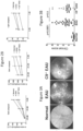
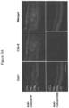
- EAU EAU in AAVCAGsCD59-injected mice was monitored by fundus imaging, spectral domain optimal coherence tomography (SD-OCT), retinal histopathology and immunohistochemistry.
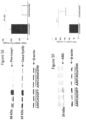
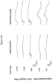
- Retinal function was quantified by the electroretinogram (ERG) and NLRP3 inflammasome activation was measured by western blots, enzyme-Linked Immunosorbent Assay (ELISA) and real-time polymerase chain reaction (RT-PCR).
- EAG electroretinogram
- NLRP3 inflammasome activation was measured by western blots, enzyme-Linked Immunosorbent Assay (ELISA) and real-time polymerase chain reaction (RT-PCR).
- ELISA enzyme-Linked Immunosorbent Assay
- RT-PCR real-time polymerase chain reaction
- the direct role of MAC in the activation of the NLRP3 inflammasome in EAU was investigated.
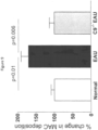
- the data from examples herein demonstrate that MAC is deposited in the EAU retina, which results in activation of the NLRP3 inflammasome and increased production of IL-1 ⁇ .
- the examples further demonstrate that C9 -/- EAU mice fail to form MAC on their retina and concomitantly have attenuated activation of the NLRP3 inflammasome, which indicates a link between deposition of MAC and activation of the NLRP3 inflammasome in EAU.
- the complement system is a major component of innate immunity and consists of a diverse group of plasma and membrane-bound proteins. These membrane-bound proteins play a central role in protection against pathogens and in the regulation of immune and inflammatory processes.
- the activation of complement against pathogens for host defense is necessary, but over-activated complement may inflict damage to host tissues ( Morgan, B. P., et al. (2015) Nat. Rev. Drug Discov. 14, 857-877 ; Carroll, M. V., et al. (2011) Adv. Drug Deliv. Rev. 63, 965-975 ). Therefore, it is necessary to maintain a balance between complement activation and complement inhibition by complement regulatory proteins.
- the NLRP3 inflammasome complex is a group of cytoplasmic proteins consisting of a primary regulatory subunit NLRP3, an adapter subunit ASC and the effector subunit caspase-1 which converts pro-IL-1 ⁇ into active IL-1 ⁇ ( Latz, E., et al. (2013) Nat. Rev.
- IL-1R antagonists anakinra , soluble decoy IL-1R rilonacept , and IL-1b-neutralizing antibody canakinumab have been approved for the treatment of uveitis ( Knickelbein, J. E., et al. (2017) Handb. Exp. Pharmacol. 242, 231-268 ).
- Knickelbein, J. E., et al. (2017) Handb. Exp. Pharmacol. 242, 231-268 Knickelbein, J. E., et al. (2017) Handb. Exp. Pharmacol. 242, 231-268 .
- multiple side effects and short duration of action makes them limited in use.
- increased production of IL-1 ⁇ was observed in mice retinas affected with EAU; however, lower IL-1 ⁇ levels were observed in C9-/- mouse retinas affected with EAU.
- Th1 and Th17 cells were observed in the mice retinas affected with EAU; however, decreased Th1 and Th17 cells was observed in the C9-/- retinas affected with EAU as indicated by the respective IL17 and IFN- ⁇ protein and mRNA levels. Further, increased levels of Th1 and Th17 positive CD4 cells were observed in DLN from EAU mice. However, levels of Th1 and Th17 positive CD4 cells were observed to remain unchanged in DLN from C9-/- EAU mice. Without being limited by any particular theory or mechanism of action, it is here envisioned that MAC-induced IL-1 ⁇ production and differentiation of Th1 and Th17 cells in EAU retina is a local effect.
- complement regulatory proteins are secreted or found on the surface of cells to keep complement mediated damage to host tissues in check. These proteins include Factor H, decay accelerating factor (CD55), membrane cofactor protein (CD46) and protectin (CD59).
- Factor H decay accelerating factor
- CD46 membrane cofactor protein
- CD59 protectin
- the decreased activity or deficiency of these complement regulatory proteins can lead to immunopathologies including EAU and experimental autoimmune anterior uveitis ( Morgan, B. P., et al. (2015) Nat. Rev. Drug Discov. 14, 857-877 ; An, F., et al. (2009) Invest. Ophthalmol. Vis. Sci. 50, 3778-3782 ; Carroll, M. V., et al. (2011) Adv. Drug Deliv. Rev. 63, 965-975 ; Jha, P., et al.
- the examples herein demonstrate that a single intravitreal injection of AAVCAGsCD59 inhibited phenotypic pathologies in EAU mice.
- the clinical signs associated with EAU were significantly improved, such as reduced inflammation, fewer immune cell infiltrates and reduced vasculitis.
- the data obtained in the examples herein demonstrate that AAVCAGsCD59 injection leads to an improvement in loss of retinal function associated with EAU in both dark and lighted adapted ERGs.
- C9 -/- mice were observed to significantly inhibit activation of the NLRP3 inflammasome and observed to reduce upregulation of IL-1 ⁇ .
- a trend of improved histology score and improved retinal function was observed in C9-/- EAU mice the differences were observed to not reach a significant level of statistical significance except for a few data points in ERG.
- AAVCAGsCD59 possibly attenuates inflammation by a means other than blocking incorporation of C9 into the C5b-8 complex.
- GPI-anchored CD59 has been previously reported to bind to CD2 and transduce activation signals within T cells ( Deckert, M. et al. (1995) Eur J Immunol. 25, 1815-22 ).
- CD59 cross-linking induces T cell receptor zeta/ZAP-70 signaling cascade and interleukin-2 (IL-2) synthesis.
- IL-2 has been found to be successful in modulating the immune system in diseases including type 1 diabetes and vasculitis ( Hartemann, A. et al. (2013) The Lancet. Diabetes & Endocrinology.
- Uveitis is a chronic inflammatory disease and many patients face episodes of relapse and some of them are refractory to available therapies.
- conventional therapies remain limited in managing severe and advanced stages of uveitis in patients with systemic autoimmune disease (Bechet's disease, Vogt-Koyanagi disease, etc.) due to significant side effects and short-term efficacy.
- the treatment for managing uveitis has not advanced significantly during the previous few decades ( Knickelbein, J. E., et al. (2017) Handb. Exp. Pharmacol. 242, 231-268 ). Considering these concerns, it is practical to develop a long-acting therapy such as the continuous inhibition of MAC dependent activation of the inflammasome in uveitis patients.
- AAV dependent gene therapies are successful in treating ocular diseases in animal models ( Adhi, M., et al. (2013) PLoS One 8, e79661 ; Ildefonso, C. J., et al. (2015) Hum. Gene Ther. 26, 59-68 ). Further, AAV-mediated gene therapies have successfully advanced into human clinical trials for age-related macular degeneration ( Mingozzi, F., et al. (2011) Nat. Rev. Genet. 12, 341-355 ).
- MAC is an important regulator of NLRP3 inflammasome activation and production of IL-1 ⁇ in EAU.
- MAC plays an important role in differentiation of Th1 and Th17 cells by increasing IL-1 ⁇ production.
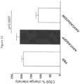
- AAV-mediated expression of sCD59 was observed to efficiently inhibit MAC deposition and subsequently inhibit NLRP3 inflammasome activation.
- a single intravitreal injection of AAVCAGsCD59 was observed to efficiently inhibit the development of EAU in mice.
- CD59 is a membrane-bound glycoprotein found associated with membranes of cells including human erythrocytes, lymphocytes, and vascular endothelial cells. CD59 protein inhibits assembly of functional MACs and thus protects cells from complement-mediated activation and/or lysis.
- CD59 plasma membranes of cells are normally protected from the effects of complement by cell-surface proteins, e.g., CD59, that specifically inhibit activation of the C5b-9 pore upon C9 complement protein binding to membrane C5b-8
- cell-surface proteins e.g., CD59
- CD59 competes with C9 complement protein for binding to C8 complement protein in the C5b-8 complex, thereby decreasing or preventing the formation of the C5b-9 membrane attack complex.
- CD59 thus acts to reduce both cell activation and cell lysis by terminal complement MACs.
- Mature human CD59 protein is composed of 77 amino acids and has a molecular weight of 18-21 kD.
- Precursor human CD59 protein includes an amino-terminal signal peptide of 25 amino acids and a carboxyl-terminal peptide of 26 amino acids which results in attachment of a membrane anchor.
- Amino acid sequences of examples of precursor human CD59, a mature human CD59, and CD59 sequences of other mammals e.g., baboon, African green monkey, owl monkey, marmoset, HVS-15, pig, rabbit, rat, and mouse, are shown in Sims et al. U.S. patent number 7,166,568 issued January 23, 2007 .
- Protein structure of CD59 includes a single cysteine-rich domain, a hydrophobic core with three loops and a small fourth helical loop ( Yu et al. 1997 Journal of Experimental Medicine 185(4): 745-753 ).
- the structure and sequence of the gene encoding CD59 has been characterized ( Fodor et al. U.S. patent number 5,624,837 issued April 29, 1997 ).
- the gene is located on the short arm of chromosome 11 in humans, specifically chromosome 1 1p13 and 1 1p14 (Online Mendelian Inheritance in Man accession number and107271), and consists of 4 exons spanning 20 kb ( Petranka et al. 1992 Proc. Nat. Acad. Sci. 89: 7876-7879 ).
- An untranslated first exon is preceded by a G and C-rich promoter region that lacks a consensus TATA or CAAT motif.
- the second exon encodes the hydrophobic leader sequence of the protein, and the third exon encodes the N-terminal portion of the mature protein.
- the fourth exon encodes the remainder of the mature protein, including the hydrophobic sequence for glycophosphoinosital (GPI) anchor attachment to a cell membrane.
- GPS glycophosphoinosital
- CD59 is a glycosylphosphatidylinositol-anchored glycoprotein that is expressed on human peripheral blood leukocytes, erythrocytes, and many cell lines.
- the protein is expressed on both hematopoietic and non-hematopoietic cells, for example on endothelial cells, peripheral nerve fibers, neurons, microglia, oligodendrocytes, astrocytes, ependymal cells, epithelial cells, acinar cells of the salivary glands, bronchial epithelium, renal tubules and squamous epithelium. See Nose, M. et al. 1990 Immunology 70(2): 145-149 ; Vedeler, C. et al.
- Human CD59 includes 26 amino acids located at the C terminus, which specifies a signal sequence for attachment of a glycosyl phosphatidyl inositol anchor (GPI anchor) at amino acid asparagine at position 77.
- GPI anchor glycosyl phosphatidyl inositol anchor
- the binding site for interactions of human CD59 with human C9 has been identified as amino acid residues 42 to 58 in the sequence of mature human CD59, that bind to the region of human C9 corresponding to human amino acid residues 334 to 418 of that protein, more particularly human C9 amino acid residues 359 to 384, immediately C-terminal to the predicted membrane-inserting domain of C9 ( Sims et al. PCT/US96/17940 filed November 8, 1996 ).
- the active surface exposed amino acid residue side chains that are available to bind C8/C9, identified from solution structure of mature human CD59 from published NMR data and the knowledge of the active portion of the CD59 molecule, are histidine at position 44, asparagine at position 48, aspartic acid at position 49, threonine at positions 51 and 52, arginine at position 55, and glutamic acid at position 58.
- NMR structures for CD59 are described in deposits by Kieffer et al., Human Complement Regulatory Protein CD59 (Extracellular Region, Residues 1 70; NMR, 10 Structures), MMDB Id: 891, PDB Id: 1ERH ; Kieffer et al., Human Complement Regulatory Protein CD59 (Extracellular Region, Residues 1 70; NMR, Restrained), MMDB Id: 890, PDB Id: 1ERG ; Fletcher et al., CD59 Complexed With Glcnac-Beta-1,4-(Fuc-Alpha-1,6)-Glcnac-Beta-1 (NMR, 10 Structures), MMDB Id: 498, PDB Id: 1CDS ; Fletcher et al., CD59 Complexed With Glcnac-Beta-1,4-Glcnac-Beta-1 (NMR, 10 Structures), MMDB Id: 497, PDB I
- Immunol 178: 1783-1790 uses recombinant methods to produce a membrane targeting composition by fusing the binding arm, Fc, of an immunoglobulin G (IgG1) to CD59 protein (rsCD59a-Fc), and injects this fusion into mice by intravenous, intra-ocular (intravitreal) and intraperitoneal routes. Numbers of CNV-positive spots were reduced in subjects treated with the fusion protein by the intraperitoneal route compared to intravitreal and intravenous routes.
- the Fc functionality may result in immediate binding upon contact of the fusion protein generally and non-specifically to cells following administration. Administering purified protein is further limited by metabolism and half-life due to presence of proteases and peptidases. Tomlinson et al.
- IOVS 50(7): 3056-3064 reduced the size of CNV spots in a mouse model by intravenously injecting the animals having CNV spots with a plasmid encoding a fusion protein complement inhibitor, produced by linking the N-terminus binding domain of factor H to a fragment of complement receptor 2 (CR2) that targets membrane molecules on cells.
- a plasmid encoding a fusion protein complement inhibitor, produced by linking the N-terminus binding domain of factor H to a fragment of complement receptor 2 (CR2) that targets membrane molecules on cells.
- CR2 complement receptor 2
- a CD59 composition provided herein lacks the primary amino acid sequence for a functional GPI anchor.
- a functional equivalent protein includes a modified GPI anchor domain amino acid sequence that is functionally defective and lacks the ability to target a membrane.
- a sCD59 is an example of a recombinant membrane-independent CD59 (rmiCD59). Additional methods of obtaining membrane-independent CD59 include non-recombinant methods such as providing an inhibitor of membrane association, for example, synthesizing CD59 in vivo or in vitro such that the GPI anchor is lacking. Methods of obtaining the membrane-independent CD59 are shown in examples herein. Additional recombinant techniques for altering the nucleic acid sequence and amino acid sequence of a molecule are well known in the art of genetics and molecular biology.
- the CD59 protein is a soluble CD59 protein.
- the composition provides a CD59 protein and includes a full length nucleic acid of CD59 that had been modified to remove the signal sequence for attachment of the GPI anchor at the nucleotides encoding amino acid asparagine at position 77.
- the nucleic acid sequence of CD59 was modified by point mutations, substitutions or deletions to obtain a nucleic acid sequence that encodes an amino acid sequence that has a modified amino acid sequence at the GPI anchor location, such that the protein would be unable to attach to a membrane of a cell.
- membrane independent refers to a CD59 amino acid sequence that lacks a GPI anchor or has a modified GPI anchor that lacks function and ability to bind to a cell membrane or a cell-membrane-associated structure such as a membrane-bound protein.
- a GPI anchor can be modified through recombinant DNA technology to remove the GPI anchoring domain or to introduce one or more mutations to disrupt GPI function. These one or more mutations can comprise an insertion of one or more amino acids, a deletion of one or more amino acids, or a substitution of one or more amino acids.
- GPI anchoring involves a multi-step pathway in the endoplasmic reticulum including the interaction of numerous gene products. Many proteins including CD59 require GPI to be expressed at the cell surface and to function effectively. The mechanism by which structure in a protein signal encodes for attachment of GPI anchors is reviewed by Orlean et al. 2007 JLR 48: 993-1011 . GPI attachment involves an amino acid sequence that contains: a hydrophobic N-terminal secretion signal that targets the protein to the ER, and a C-terminal GPI signal anchor sequence.
- CD59 secretion signal which is located at the amino terminus of the protein and is cleaved in vivo
- other secretion signals are suitable for CD59 protein and are within the scope of the methods herein.
- General eukaryotic secretion signals that are suitable for use in mammalian cells here are described for example in Ding et al. U.S. patent number 6,733,997 B1 issued May 11, 2004 ; Tan et al. 2002 Protein Engineering 15(4): 337-345 ; and Tan et al. 1999 Biochim. Biophys. Acta 1452: 103-120 .
- the amino acid to which the GPI becomes linked is referred to as the omega ( ⁇ ) residue, with amino acids N-terminal to the omega residue referred to as omega-minus ( ⁇ -) and with amino acids C-terminal to the omega residue referred to as omega-plus ( ⁇ +).
- the GPI anchor sequence includes a stretch of about ten polar amino acids (i.e., ⁇ -10 to ⁇ -1), for example arginine, lysine, aspartate, glutamate, asparagine, or glutamate, that form a flexible linker region.
- the ⁇ residue has been observed to be one of: glycine, alanine, serine, asparagine, aspartic acid, or cysteine.
- Mutation including substitution and deletion of nucleic acids encoding amino acids at omega positions are used to reduce or eliminate the attachment of the GPI anchor or reduce or eliminate the effective functionality of the GPI anchor.
- a variation includes substituting the nucleic acids encoding hydrophobic leucine (e.g., nucleic acids CTG) and alanine (e.g., nucleic acids GCA) with nucleic acids encoding glutamine (e.g., nucleic acids CAG) and glutamic acid (e.g., nucleic acids GAA), or glycine (e.g., nucleic acids GGN)which are less hydrophobic (i.e., more hydrophilic) amino acids.
- a variation includes substituting the ⁇ residue with another amino acid, for example substituting a glycine for a tyrosine.
- promoter sequences that are useful to regulate transcription of CD59 gene sequences are within the scope of expression of the vectors herein. These promoters are for example constitutive promoters, cell cycle-specific promoters, ubiquitous promoters, tissue-specific promoters, metabolically regulated promoters, inducible promoters, and promoters that are found in specific subjects including humans and animals. Examples of promoters and promoter systems are shown for example in Evans et al. U.S. patent number 6,677,311 B1 issued January 13, 2004 ; Clark et al. U.S. patent number 7,109,029 B2 issued September 19, 2006 ; and Hallenbeck et al. U.S. patent number 5,998,205 issued December 7, 1999 .
- conservative sequence modifications refers to amino acid modifications that do not significantly affect or alter the characteristics of the CD59 protein or membrane-independent CD59 containing the amino acid sequence, i.e., amino acid sequences of CD59 that present these side chains at the same relative positions will function in a manner similar to human CD59.
- conservative modifications include amino acid substitutions, additions and deletions. Modification of the amino acid sequence of CD59 is achieved using any known technique in the art e.g., site-directed mutagenesis or PCR based mutagenesis.
- Conservative amino acid substitutions are ones in which the amino acid residue is replaced with an amino acid residue having a similar side chain.
- Families of amino acid residues having similar side chains have been defined in the art. These families include amino acids with basic side chains (e.g., lysine, arginine, histidine), acidic side chains (e.g., aspartic acid, glutamic acid), uncharged polar side chains (e.g., glycine, asparagine, glutamine, serine, threonine, tyrosine, cysteine, tryptophan), nonpolar side chains (e.g., alanine, valine, leucine, isoleucine, proline, phenylalanine, methionine), beta-branched side chains (e.g., threonine, valine, isoleucine) and aromatic side chains (e.g., tyrosine, phenylalanine, tryptophan, histidine).
- basic side chains
- the CD59 amino acid sequence is an amino acid sequence that is substantially identical to that of the wild type sequence.
- substantially identical is used herein to refer to a first amino acid sequence that contains a sufficient or minimum number of amino acid residues that are identical to aligned amino acid residues in a second amino acid sequence such that the first and second amino acid sequences can have a common structural domain and/or common functional activity.
- amino acid sequences that contain a common structural domain having at least about 60% identity, or at least 75%, 80%, 85%, 90%, 95%, 96%, 98%, or 99% identity.
- the CD59 amino acid sequence is an amino acid sequence that is "identical" to that of the wild type sequence.
- an identical sequence is one that exhibits 100% identity to a wild type sequence of CD59.
- an identical sequence can be flanked on the N or C-terminus by one or more amino acids.
- the one or more amino acids can comprise an epitope tag, a purification tag, the remnant of a cleavage site introduced to allow a purification tag to be removed, or one or more modifications to improve stability or bioavailability of the protein.
- CD59 polypeptides are useful for the treatment of inflammasome mediated disorders.
- the amino acid sequence of a human CD59 polypeptide useful for the methods described herein is set forth by the sequence: MGIQGGSVLFGLLL VLA VFCHSGHSLQCYNCPNPT ADCKTA VNCSSDFDACL ITKAGLQVYNKCWKFEHCNFNDVTTRLRENELTYYCCKKDLCNFNEQLENG GTSLSEKTVLLLVTPFLAAAWSLHP (SEQ ID NO: 1).
- the signal sequence of CD59 is cleaved before secretion from the cell, and is dispensable for therapeutic utility.
- nucleic acids that encode a CD59 with a signal sequence increases secretion of a CD59 polypeptide, and, thus, a signal sequence may be desirable to include in CD59 polypeptides encoded by nucleic acids.
- a signal sequence cleaved human CD59 polypeptide useful for the methods described herein is set forth by the sequence: LQCYNCPNPTADCKTAVNCSSDFDACLITKAGLQVYNKCWKFEHCNFNDVT TRLRENELTYYCCKKDLCNFNEQLENGGTSLSEKTVLLLVTPFLAAAWSLHP (SEQ ID NO: 2).
- the exact signal sequence end may vary from SEQ ID NO: 2 by 1, 2, 3, 4, 5, 6, 7, 8, 9, 10, 11, 12, 13, 14, 15, 16, 17, 18, 19, 20, 21, 22, 23, or 24 amino acids from the first leucine of SEQ ID NO: 2; in either the N-terminal or C-terminal direction of the full CD59 polypeptide set forth in SEQ ID NO: 1.
- a further deletion of the N-terminal 26 amino acids deletes the GPI anchoring domain of human CD59.
- a signal sequence cleaved, soluble human CD59 polypeptide useful for the methods described herein is set forth by the sequence: LQCYNCPNPTADCKTAVNCSSDFDACLITKAGLQVYNKCWKFEHCNFNDVT TRLRENELTYYCCKKDLCNFNEQLEN (SEQ ID NO: 3).
- This soluble version can also vary at its exact N-terminal end by 1, 2, 3, 4, 5, 6, 7, 8, 9, 10, 11, 12, 13, 14, 15, 16, 17, 18, 19, 20, 21, 22, 23, or 24 amino acids, resulting in further deletion from its N-terminus or restoration of its N-terminus.
- the soluble version of CD59 comprises an amino acid sequence as set forth in SEQ ID NO: 2 with one or more mutations to a residue required for GPI attachment.
- the residues are any one or both of the asparagines at amino acid 70 or 77 of SEQ ID NO: 2.
- the CD59 polypeptide useful for the methods described herein comprises deletion of 1, 2, 3, 4, 5, 6, 7, 8, 9, 10, 11, 12, 13, 14, 15, 16, 17, 18, 19, 20, 21, 22, 23, 24, 25, 26, 27, 28, 29, 30, 31, 32, 33, 34, 35, 36, 37, 38, 39, 40, 41, 42, 43, 44, 45, 46, 47, 48, 49, or 50 amino acids from the N-terminal end, C-terminal end, or both ends of any one of SEQ ID NOs: 1, 2, or 3.
- the CD59 polypeptide used with the methods described herein can comprise or consist of an amino acid sequence at least about 80%, 85%, 90%, 95%, 97%, 98%, 99%, or 100% identical to that set forth in any one of SEQ ID NO: 1, SEQ ID NO: 2, or SEQ ID NO: 3. Additionally, the CD59 polypeptide used herein can comprise additional non-CD59 derived sequences, including purification tags, remnants of cleaved purification tags, additional complement inhibiting polypeptides, anti-inflammatory polypeptides, or polypeptides that increase stability or bioavailability of the CD59 polypeptide.
- Nucleic acids encoding a polypeptide that comprises or consists of a CD59 polypeptide, described herein, are also useful for the treatment of inflammasome mediated disorders.
- a nucleic acid or a plurality of nucleic acids that encode a polypeptide comprising or consisting of a CD59 polypeptide described herein can suitably be included in a recombinant vector such as a viral or plasmid vector that is suitable for administration to a subject.
- These vectors can additionally be included in pharmaceutical compositions for administration to an individual with an inflammasome mediated disorder.
- a polypeptide comprising or consisting of the amino acid sequence set forth in any one of SEQ ID NO: 1, SEQ ID NO: 2, or SEQ ID NO: 3 can be used in a method of treating an inflammasome disorder in a subject as described herein.
- a polypeptide comprising or consisting of the amino acid sequence set forth in any one of SEQ ID NO: 1, SEQ ID NO: 2, or SEQ ID NO: 3 is used in a method of treating an inflammasome disorder in an eye of a subject.
- the inflammasome disorder is uveitis.
- the subject has displayed positive results for expression or activity of at least one inflammasome activity marker.
- the inflammasome activity marker is selected from: caspase 1, caspase 5, IL-1 ⁇ , IL- ⁇ 17, IL-18, apoptosis-associated speck-like protein containing a CARD (PYCARD/ASC), a NACHT, LRR and PYD domains-containing protein (NALP), IFN- ⁇ , a Th1 T-cell marker or cytokine, a Th17 T-cell marker or cytokine, and CD4+.
- the inflammasome activity marker is selected from: apoptosis-associated speck-like protein containing a CARD (PYCARD/ASC), or a NACHT, LRR and PYD domains-containing protein (NALP).
- the NALP is NACHT, LRR and PYD domains-containing protein 3 (NLRP3).
- the methods described herein comprise administering a human CD59 polypeptide to an individual that has displayed positive results for expression or activity of at least one inflammasome activity marker.
- the CD59 polypeptide can comprise an amino acid sequence at least about 80%, 85%, 90%, 95%, 97%, 98%, 99%, or 100% identical to that set forth in any one of SEQ ID NO: 1, SEQ ID NO: 2, or SEQ ID NO: 3.
- a nucleic acid or plurality of nucleic acids encoding a polypeptide comprising or consisting of the amino acid sequence set forth in any one of SEQ ID NO: 1, SEQ ID NO: 2, or SEQ ID NO: 3 can be used in a method of treating an inflammasome disorder in a subject.
- the nucleic acid or plurality of nucleic acids encoding a polypeptide comprising or consisting of the amino acid sequence set forth in any one of SEQ ID NO: 1, SEQ ID NO: 2, or SEQ ID NO: 3 is used in a method of treating an inflammasome disorder in an eye of a subject.
- the inflammasome disorder is uveitis.
- the subject has displayed positive results for expression or activity of at least one inflammasome activity marker.
- the inflammasome activity marker is selected from: caspase 1, caspase 5, IL-1 ⁇ , IL- ⁇ 17, IL-18, apoptosis-associated speck-like protein containing a CARD (PYCARD/ASC), a NACHT, LRR and PYD domains-containing protein (NALP), IFN- ⁇ , a Th1 T-cell marker or cytokine, a Th17 T-cell marker or cytokine, and CD4+.
- the inflammasome activity marker is selected from: apoptosis-associated speck-like protein containing a CARD (PYCARD/ASC), or a NACHT, LRR and PYD domains-containing protein (NALP).
- PYCARD/ASC apoptosis-associated speck-like protein containing a CARD
- NALP NACHT, LRR and PYD domains-containing protein
- the NALP is NACHT, LRR and PYD domains-containing protein 3 (NLRP3).
- the methods described herein comprise administering a human CD59 polypeptide to an individual that has displayed positive results for expression or activity of at least one inflammasome activity marker.
- the CD59 polypeptide can comprise or consist of an amino acid sequence at least about 80%, 85%, 90%, 95%, 97%, 98%, 99%, or 100% identical to that set forth in any one of SEQ ID NO: 1, SEQ ID NO: 2, or SEQ ID NO: 3.
- the inflammasome activity marker is selected from: caspase 1, caspase 5, IL-1 ⁇ , IL- ⁇ 17, IL-18, apoptosis-associated speck-like protein containing a CARD (PYCARD/ASC), a NACHT, LRR and PYD domains-containing protein (NALP), IFN- ⁇ , a Th1 T-cell marker or cytokine, a Th17 T-cell marker or cytokine, and CD4+.
- the inflammasome activity marker is selected from: apoptosis-associated speck-like protein containing a CARD (PYCARD/ASC), or a NACHT, LRR and PYD domains-containing protein (NALP).
- PYCARD/ASC apoptosis-associated speck-like protein containing a CARD
- NALP NACHT, LRR and PYD domains-containing protein
- the positive results are obtained from the eye of the individual.
- the methods described herein comprise administering a human CD59 polypeptide to an eye of an individual that has displayed positive results for expression or activity of at least one inflammasome activity marker.
- the CD59 polypeptide can comprise or consist of an amino acid sequence at least about 80%, 85%, 90%, 95%, 97%, 98%, 99%, or 100% identical to that set forth in any one of SEQ ID NO: 1, SEQ ID NO: 2, or SEQ ID NO: 3.
- the inflammasome activity marker is selected from: caspase 1, caspase 5, IL-1 ⁇ , IL- ⁇ 17, IL-18, apoptosis-associated speck-like protein containing a CARD (PYCARD/ASC), a NACHT, LRR and PYD domains-containing protein (NALP), IFN- ⁇ , a Th1 T-cell marker or cytokine, a Th17 T-cell marker or cytokine, and CD4+.
- the inflammasome activity marker is selected from: apoptosis-associated speck-like protein containing a CARD (PYCARD/ASC), or a NACHT, LRR and PYD domains-containing protein (NALP).
- PYCARD/ASC apoptosis-associated speck-like protein containing a CARD
- NALP NACHT, LRR and PYD domains-containing protein
- the positive results are obtained from the eye of the individual.
- the methods described herein comprise administering a human CD59 polypeptide to an individual afflicted with uveitis.
- the CD59 polypeptide can comprise or consist of an amino acid sequence at least about 80%, 85%, 90%, 95%, 97%, 98%, 99%, or 100% identical to that set forth in any one of SEQ ID NO: 1, SEQ ID NO: 2, or SEQ ID NO: 3.
- the polypeptide is administered to an afflicted eye of the individual.
- the methods described herein comprise administering a nucleic acid encoding a human CD59 polypeptide to an individual that has displayed positive results for expression or activity of at least one inflammasome activity marker.
- the nucleic acid can encode a CD59 polypeptide comprising or consisting of an amino acid sequence at least about 80%, 85%, 90%, 95%, 97%, 98%, or 99% identical to that set forth in SEQ ID NO: 1, SEQ ID NO: 2, or SEQ ID NO: 3.
- the inflammasome activity marker is selected from: caspase 1, caspase 5, IL-1 ⁇ , IL- ⁇ 17, IL-18, apoptosis-associated speck-like protein containing a CARD (PYCARD/ASC), a NACHT, LRR and PYD domains-containing protein (NALP), IFN- ⁇ , a Th1 T-cell marker or cytokine, a Th17 T-cell marker or cytokine, and CD4+.
- the inflammasome activity marker is selected from: apoptosis-associated speck-like protein containing a CARD (PYCARD/ASC), or a NACHT, LRR and PYD domains-containing protein (NALP).
- PYCARD/ASC apoptosis-associated speck-like protein containing a CARD
- NALP NACHT, LRR and PYD domains-containing protein
- the positive results are obtained from the eye of the individual.
- the methods described herein comprise administering a nucleic acid encoding a human CD59 polypeptide to an eye of an individual that has displayed positive results for expression or activity of at least one inflammasome activity marker.
- the nucleic acid can encode a CD59 polypeptide comprising or consisting of an amino acid sequence at least about 80%, 85%, 90%, 95%, 97%, 98%, or 99% identical to that set forth in SEQ ID NO: 1, SEQ ID NO: 2, or SEQ ID NO: 3.
- the inflammasome activity marker is selected from: caspase 1, caspase 5, IL-1 ⁇ , IL- ⁇ 17, IL-18, apoptosis-associated speck-like protein containing a CARD (PYCARD/ASC), a NACHT, LRR and PYD domains-containing protein (NALP), IFN- ⁇ , a Th1 T-cell marker or cytokine, a Th17 T-cell marker or cytokine, and CD4+.
- the inflammasome activity marker is selected from: apoptosis-associated speck-like protein containing a CARD (PYCARD/ASC), or a NACHT, LRR and PYD domains-containing protein (NALP).
- PYCARD/ASC apoptosis-associated speck-like protein containing a CARD
- NALP NACHT, LRR and PYD domains-containing protein
- the positive results are obtained from the eye of the individual.
- the methods described herein comprise administering a nucleic acid encoding a human CD59 polypeptide to an individual afflicted with uveitis.
- the nucleic acid can encode a CD59 polypeptide comprising or consisting of an amino acid sequence at least about 80%, 85%, 90%, 95%, 97%, 98%, or 99% identical to that set forth in SEQ ID NO: 1, SEQ ID NO: 2, or SEQ ID NO: 3.
- the nucleic acid is administered to an afflicted eye of the individual.
- sequence identity between sequences are performed as follows. To determine the percent identity of two amino acid sequences, the sequences are aligned for optimal comparison purposes (e.g., gaps can be introduced in one or both of a first and a second amino acid sequence for optimal alignment). The amino acid residues at corresponding amino acid positions or nucleotide positions are then compared. When a position in the first sequence is occupied by the same amino acid residue or nucleotide as the corresponding position in the second sequence, then the proteins are identical at that position. The percent identity between the two sequences is a function of the number of identical positions shared by the sequences, taking into account the number of gaps, and the length of each gap, which need to be introduced for optimal alignment of the two sequences.
- Percent identity between two amino acid sequences is determined using an alignment software program using the default parameters. Suitable programs include, for example, CLUSTAL W by Thompson et al., Nuc. Acids Research 22:4673, 1994 (www.ebi.ac.uk/clustalw ), BL2SEQ by Tatusova and Madden, FEMS Microbiol. Lett. 174:247, 1999 (www.ncbi.nlm.nih.gov/blast/bl2seq/bl2.html ), SAGA by Notredame and Higgins, Nuc.
- recombinant refers to proteins produced by manipulation of genetically modified organisms, for example micro-organisms.
- a source of CD59 includes polynucleotide sequences that encode the CD59 protein, for example, engineered into recombinant DNA molecules to direct expression of the CD59 protein in appropriate host cells.
- a nucleotide sequence encoding the CD59 protein, or functional equivalent is inserted into an appropriate expression vector, i.e. , a vector that contains the necessary nucleic acid encoding elements that regulate transcription and translation of the inserted coding sequence, operably linked to the nucleotide sequence encoding the CD59 protein amino acid sequence.
- a variety of commercially available expression vector/host systems are useful to contain and express a CD59 protein encoding sequence. These include but are not limited to microorganisms such as bacteria transformed with recombinant bacteriophage, plasmid or cosmid DNA expression vectors; yeast transformed with yeast expression vectors; insect cell systems contacted with virus expression vectors (e.g., baculovirus); plant cell systems transfected with virus expression vectors (e.g., cauliflower mosaic virus, CaMV; tobacco mosaic virus, TMV) or transformed with bacterial expression vectors (e.g., Ti, pBR322, or pET25b plasmid); or animal cell systems.
- microorganisms such as bacteria transformed with recombinant bacteriophage, plasmid or cosmid DNA expression vectors; yeast transformed with yeast expression vectors; insect cell systems contacted with virus expression vectors (e.g., baculovirus); plant cell systems transfected with virus expression vectors (e.g., cauliflower mosaic virus, Ca
- Virus vectors include, but are not limited to, adenovirus vectors, lentivirus vectors, adeno-associated virus (AAV) vectors, and helper-dependent adenovirus vectors.
- Virus vectors deliver a nucleic acid sequence that encodes CD59 protein that as shown herein interferes with the deleterious action of the MAC in pathogenesis of AMD.
- Adenovirus packaging vectors are commercially available from American Type Tissue Culture Collection (Manassas, VA). Methods of constructing adenovirus vectors and using adenovirus vectors are shown in Klein et al. 2007 Ophthalmology 114: 253-262 , and van Leeuwen et al. 2003 Eur. J. Epidemiol. 18: 845-854 .
- Adenovirus vectors have been used in eukaryotic gene expression ( Levrero et al. 1991 Gene, 101: 195-202 ) and vaccine development ( Graham et al. 1991 Methods in Molecular Biology: Gene Transfer and Expression Protocols 7, (Murray, Ed.), Humana Press, Clifton, NJ, 109-128 ). Further, recombinant adenovirus vectors are used for gene therapy ( Wu et al. U.S. patent number 7,235,391 issued June 26, 2007 ).
- Recombinant adenovirus vectors are generated, for example, from homologous recombination between a shuttle vector and a provirus vector ( Wu et al., U.S. patent number 7,235,391 issued June 26, 2007 ).
- the adenovirus vectors herein are replication defective, for example, are conditionally defective, lacking adenovirus E1 region, and a polynucleotide encoding CD59 is introduced at the position from which the E1-coding sequences have been removed.
- the polynucleotide encoding the CD59 gene alternatively is inserted in the E3 region.
- Helper cell lines may be derived from human cells such as, 293 human embryonic kidney cells, muscle cells, hematopoietic cells or other human embryonic mesenchymal or epithelial cells.
- the helper cells may be derived from the cells of other mammalian species that are permissive for human adenovirus, e.g., Vero cells or other monkey embryonic mesenchymal or epithelial cells. Generation and propagation of these replication defective adenovirus vectors using a helper cell line is described in Graham et al 1977 J. Gen. Virol. 36: 59-72 .
- Lentiviral vector packaging vectors are commercially available from Invitrogen Corporation (Carlsbad CA). An HIV-based packaging system for the production of lentiviral vectors is prepared using constructs in Naldini et al. 1996 Science 272: 263-267 ; Zufferey et al. 1997 Nature Biotechnol. 15: 871-875 ; and Dull et al. 1998 J. Virol. 72: 8463-8471 .
- the vector construct pRRLsinCMVGFPpre contains a 5' LTR in which the HIV promoter sequence has been replaced with that of Rous sarcoma virus (RSV), a self-inactivating 3' LTR containing a deletion in the U3 promoter region, the HIV packaging signal, RRE sequences linked to a marker gene cassette consisting of the Aequora jellyfish green fluorescent protein (GFP) driven by the CMV promoter, and the woodchuck hepatitis virus PRE element, which appears to enhance nuclear export.
- RSV Rous sarcoma virus
- GFP Aequora jellyfish green fluorescent protein
- the GFP marker gene allows quantitation of transfection or transduction efficiency by direct observation of UV fluorescence microscopy or flow cytometry ( Kafri et al. 1997 Nature Genet. 17: 314-317 ; and Sakoda et al. 1999 J. Mol. Cell. Cardiol. 31: 2037-2047 ).
- a retroviral vector is constructed and packaged into non-infectious transducing viral particles (virions) using an amphotropic packaging system.
- amphotropic packaging system examples of such packaging systems are found in, for example, Miller et al. 1986 Mol. Cell Biol. 6 :2895-2902 ; Markowitz et al. 1988 J. Virol. 62:1120-1124 ; Cosset et al. 1990 J. Virol. 64: 1070-1078 ; U.S.
- Retroviral vectors are found in, for example, Korman et al. 1987 Proc. Natl. Acad. Sci. USA. 84: 2150-2154 ; Morgenstern et al. 1990 Nucleic Acids Res. 18: 3587-3596 ; U.S. patent numbers 4,405,712 , 4,980,289 , and 5,112,767 ; and PCT patent publications numbers WO 85/05629 , WO 90/02797 , and WO 92/07943 .
- Herpesvirus packaging vectors are commercially available from Invitrogen Corporation, (Carlsbad, CA).
- Exemplary herpesviruses are an ⁇ -herpesvirus, such as Varicella-Zoster virus or pseudorabies virus; a herpes simplex virus such as HSV-1 or HSV-2; or a herpesvirus such as Epstein-Barr virus.
- a method for preparing empty herpesvirus particles that can be packaged with a desired nucleotide segment, for example a CD59 nucleotide or polynucleotide sequence, in the absence of a helper virus that is capable to most herpesviruses is shown in Fraefel et al. (U.S. patent number 5,998,208, issued December 7, 1999 ).
- the herpesvirus DNA vector can be constructed using techniques familiar to the skilled artisan. For example, DNA segments encoding the entire genome of a herpesvirus is divided among a number of vectors capable of carrying large DNA segments, e.g., cosmids ( Evans, et al., Gene 79, 9-20, 1989 ), yeast artificial chromosomes (YACS) ( Sambrook, J. et al., MOLECULAR CLONING: A LABORATORY MANUAL, 2nd Edition, Cold Spring Harbor Press, Cold Spring Harbor, N.Y., 1989 ) or E. coli F element plasmids ( O'Conner et al. 1989 Science 244:1307-1313 ).
- cosmids Evans, et al., Gene 79, 9-20, 1989
- yeast artificial chromosomes YACS
- sets of cosmids have been isolated which contain overlapping clones that represent the entire genomes of a variety of herpesviruses including Epstein-Barr virus, Varicella-Zoster virus, pseudorabies virus and HSV-1.
- herpesviruses including Epstein-Barr virus, Varicella-Zoster virus, pseudorabies virus and HSV-1.
- AAV is a dependent parvovirus in that it depends on co-infection with another virus (either adenovirus or a member of the herpes virus family) to undergo a productive infection in cultured cells ( Muzyczka 1992 Curr Top Microbiol Immunol, 158: 97 129 ).
- rAAV recombinant AAV
- virus is made by co-transfecting a plasmid containing the gene of interest, for example, the CD59 gene, flanked by the two AAV terminal repeats ( McLaughlin et al. 1988 J. Virol., 62(6): 1963-1973 ; Samulski et al. 1989 J.
- Virol, 63: 3822-3828 and an expression plasmid containing the wild-type AAV coding sequences without the terminal repeats.
- Cells are also contacted or transfected with adenovirus or plasmids carrying the adenovirus genes required for AAV helper function.
- Recombinant AAV virus stocks made in such fashion include with adenovirus which must be physically separated from the recombinant AAV particles (for example, by cesium chloride density centrifugation).
- Adeno-associated virus (AAV) packaging vectors are commercially available from GeneDetect (Auckland, New Zealand). AAV has been shown to have a high frequency of integration and infects non-dividing cells, thus making it useful for delivery of genes into mammalian cells in tissue culture ( Muzyczka 1992 Curr Top Microbiol Immunol 158: 97-129 ). AAV has a broad host range for infectivity ( Tratschin et al. 1984 Mol. Cell. Biol. 4: 2072-2081 ; Laughlin et al. 1986 J. Virol., 60(2): 515-524 ; Lebkowski et al. 1988 Mol. Cell. Biol. 8(10): 3988-3996 ; McLaughlin et al. 1988 J. Virol. 62(6):1963-1973 ).
- the vectors herein are non-viral vectors for example synthetic gene delivery vehicles or vectors that are not related to a virus particle and that specifically deliver the CD59 gene encoding material to the target cells or tissue.
- non-viral vectors include liposomes, peptides, nanoparticles, emulsions, or encapsulated two or more phase systems or other suitable preparation.
- a method, kit, or composition involves a non-viral vector with nucleic acid that is loaded and contacted to a tissue or cell.
- a liposome containing naked DNA encoding a membrane-independent CD59 protein having a modified GPI anchor that does not target a membrane, or a gene encoding a membrane-independent CD59 protein having no GPI anchor is encapsulated in the liposome and the liposome is contacted to the tissue or cell such that the nucleic acid is effectively delivered to the tissue or cell for treatment of a complement-related disease.
- antibody as referred to herein includes whole antibodies and any antigen binding fragment (i.e., "antigen-binding portion") or single chains of these.
- a naturally occurring “antibody” is a glycoprotein including at least two heavy (H) chains and two light (L) chains inter-connected by disulfide bonds.
- an antibody that "specifically binds to human MAC” is intended to refer to an antibody that binds to human MAC with a K D of 5 ⁇ 10 -9 M or less, 2 ⁇ 10 -9 M or less, or 1 ⁇ 10 -10 M or less.
- the antibody is monoclonal or polyclonal.
- the terms "monoclonal antibody” or “monoclonal antibody composition” as used herein refer to a preparation of antibody molecules of single molecular composition.
- a monoclonal antibody composition displays a single binding specificity and affinity for MAC or for a particular epitope of MAC.
- the antibody is an IgM, IgE, IgG, such as IgG1 or IgG4.
- recombinant human antibody includes all antibodies that are prepared, expressed, created or isolated by recombinant means, such as antibodies isolated from an animal (e.g., a mouse).
- Mammalian host cells for expressing the recombinant antibodies used in the methods herein include Chinese Hamster Ovary (CHO cells) including dhfr- CHO cells, described in Urlaub and Chasin 1980 Proc. Natl. Acad. Sci. USA 77: 4216-4220 , and used with a DH FR selectable marker, e.g., as described in R.J. Kaufman and P.A. Sharp, 1982 Mol. Biol.
- NSO myeloma cells 159:601-621
- NSO myeloma cells COS cells and SP2 cells.
- another expression system is the GS gene expression system shown in WO 87/04462 , WO 89/01036 and EP 338,841 .
- expression vectors encoding intact or a portion of the protein of interest are introduced into mammalian host cells, and the host cells are cultured for a period of time sufficient to allow for expression of the antibody in the host cells or secretion of the antibody into the culture medium in which the host cells are grown.
- Antibodies can be recovered from the culture medium using standard protein purification methods.
- Standard assays to evaluate the binding ability of the antibodies toward the target of various species are known in the art, including for example, ELISAs, western blots and RIAs.
- the binding kinetics (e.g., binding affinity) of the antibodies also can be assessed by standard assays known in the art, such as by Biacore analysis.
- an animal of suitable size such as goats, dogs, sheep, mice, or camels are immunized by administration of an amount of immunogen, such as the intact protein or a portion thereof containing an epitope from human MAC, effective to produce an immune response.
- an exemplary protocol is as follows.
- the animal is subcutaneously injected in the back with 100 micrograms to 100 milligrams of antigen, dependent on the size of the animal, followed three weeks later with an intraperitoneal injection of 100 micrograms to 100 milligrams of immunogen with adjuvant dependent on the size of the animal, for example Freund's complete adjuvant. Additional intraperitoneal injections every two weeks with adjuvant, for example Freund's incomplete adjuvant, are administered until a suitable titer of antibody in the animal's blood is achieved.
- Exemplary titers include a titer of at least about 1:5000 or a titer of 1:100,000 or more, i.e., the dilution having a detectable activity.
- the antibodies are purified, for example, by affinity purification on columns containing human MAC.
- antigen-reactive monoclonal antibodies including antigen-specific IgG, and IgM monoclonal antibodies. Any antibody or fragment thereof having affinity and specific for human MAC is within the scope of the assay for MAC deposition provided herein.
- contacting with CD59 is achieved by injecting cells or tissues with a vector encoding the CD59 gene.
- cell lysis is measured by propidium iodide (PI) uptake.
- PI is commercially available from, for example, Fluka BioChemica (Buchs, Switzerland).
- PI is an intercalating agent that fluoresces when bound to DNA.
- PI is membrane impermeant and generally excluded from viable cells, thus PI is commonly used to identify and/or determine the amount of non-living cells in a mixed population.
- the detectable protein is a fluorescent protein, for example, green fluorescent protein, aequorin, cyan fluorescent protein, DsRed fluorescent protein, enhanced green fluorescent protein, and yellow fluorescent protein.
- Green fluorescent protein (GFP) and aequorin are bioluminescent compositions isolated from the jellyfish Aequorea victoria. When a calcium ion binds to aequorin, the complex breaks down into apoaequorin and a luminescent composition, which emits blue light.
- Synthetic aequorin is commercially available from Sealite, Sciences (Bogart, Ga.) as AQUALITE ® . GFP emits light in the lower green portion of the visible spectrum, and synthetic GFP is commercially available from Clontech (Mountain View, CA).
- Mutations to the amino acid sequence of GFP have been made to produce derivative amino acid sequences of GFP that fluoresce different colors, for example, cyan fluorescent protein, DsRed fluorescent protein, enhanced green fluorescent protein, and yellow fluorescent protein.
- Synthetic cyan fluorescent protein, synthetic DsRed fluorescent protein, synthetic enhanced green fluorescent protein, and synthetic yellow fluorescent protein are each commercially available from Clontech (Mountain View, CA).
- the detectable agent is a fluorescent agent that is not a fluorescent protein, for example, Indocyanine Green, Doxorubicin, Riboflavin, Chlorophyll, and Porphyrin.
- Indocyanine Green is a tricarbocyanine dye that upon excitation, emits lights at about 800nm, about 820nm, about 840nm or at about 860nm. ICG is commercially available from H.W. Sands Corp. (Jupiter, FL). Doxorubicin is fluorescent and emits light at wavelengths of, for example, about 550nm, 600nm, or 650nm. Doxorubicin is commercially available from Sigma-Aldrich (St. Louis, MO). Riboflavin is commercially available from Sigma-Aldrich (St.
- Porphyin makes the compound chromatic, i.e., fluorescent at a wavelength of, for example, about 600nm, or about 650nm, or about 700nm.
- Porphyrin is commercially available from Sigma-Aldrich (St. Louis, MO).
- the detectable agent is an enzymatic agent, which is a protein, for example, ⁇ -galactosidase or alkaline phosphatase, that can be expressed on a nucleotide vector.
- ⁇ -galactosidase is a hydrolase enzyme that catalyzes the hydrolysis of ⁇ -galactosides into monosaccharides.
- a luminescent ⁇ -galactosidase detection kit is commercially available from Clontech (Mountain View, CA).
- Alkaline phosphatase is a hydrolase enzyme responsible for removing phosphate groups from many types of molecules, including nucleotides, proteins, and alkaloids.
- a luminescent alkaline phosphatase detection kit is commercially available from Sigma Aldrich (St. Louis, MO).
- the disclosure provides a method using a nucleic acid encoding a CD59 protein for inhibiting inflammasome activation in cells of an inflammation-affected eye in a subject, or a soluble CD59 protein.
- the CD59 protein includes a membrane-independent (soluble) CD59 protein.
- the CD59 composition is formulated for intravenous administration.
- the composition is compounded as an ophthalmologic formulation for administration to the eye and may be compounded to enhance delivery to the fundus, to provide sustained release locally at the retina or otherwise formulated to provide effective treatment of the vessels and/or tissue involved in ocular diseases including macular degeneration.
- the pharmaceutical composition is formulated sufficiently pure for administration to a human subject, e.g., to the circulation or eye of a human subject.
- these compositions optionally further include one or more additional therapeutic agents.
- the additional therapeutic agent or agents are selected from the group consisting of growth factors, anti-inflammatory agents, vasopressor agents including but not limited to nitric oxide and calcium channel blockers, collagenase inhibitors, topical steroids, matrix metalloproteinase inhibitors, ascorbates, angiotensin II, angiotensin III, calreticulin, tetracyclines, fibronectin, collagen, thrombospondin, transforming growth factors (TGF), keratinocyte growth factor (KGF), fibroblast growth factor (FGF), insulin-like growth factors (IGFs), IGF binding proteins (IGFBPs), epidermal growth factor (EGF), platelet derived growth factor (PDGF), neu differentiation factor (NDF), hepatocyte growth factor (
- the additional agent is a compound, composition, biological or the like that potentiates, stabilizes or synergizes or even substitutes for the ability of CD59 protein to protect cells from MAC deposition.
- therapeutic agents that may beneficially or conveniently be provided at the same time as the CD59 protein, such as agents used to treat the same, a concurrent or a related symptom, condition or disease.
- the drug may include without limitation anti-tumor, antiviral, antibacterial, anti-mycobacterial, anti-fungal, anti-proliferative or anti-apoptotic agents.
- Drugs that are included in the compositions of the invention are well known in the art. See for example, Goodman & Gilman's The Pharmacological Basis of Therapeutics, 9th Ed., Hardman, et al., eds., McGraw-Hill, 1996 .
- the term "pharmaceutically acceptable carrier” includes any and all solvents, diluents, or other liquid vehicle, dispersion or suspension aids, surface active agents, isotonic agents, thickening or emulsifying agents, preservatives, solid binders, lubricants and the like, as suited to the particular dosage form desired.
- Remington's Pharmaceutical Sciences Ed. by Gennaro, Mack Publishing, Easton, PA, 1995 provides various carriers used in formulating pharmaceutical compositions and known techniques for the preparation thereof.
- materials which can serve as pharmaceutically acceptable carriers include, but are not limited to, sugars such as glucose and sucrose; excipients such as cocoa butter and suppository waxes; oils such as peanut oil, cottonseed oil, safflower oil, sesame oil, olive oil, corn oil, and soybean oil; glycols such a propylene glycol; esters such as ethyl oleate and ethyl laurate; agar; buffering agents such as magnesium hydroxide and aluminum hydroxide; alginic acid; pyrogen-free water; isotonic saline; Ringer's solution; ethyl alcohol; and phosphate buffer solutions, as well as other non-toxic compatible lubricants such as sodium lauryl sulfate and magnesium stearate, as well as coloring agents, releasing agents, coating agents, preservatives and antioxidants can also be present in the composition, according to the judgment of the formulator.
- sugars such as glucose and sucrose
- Treatment of an inflammation affected eye by methods provided herein involves contacting a tissue or cells with a pharmaceutical composition, for example, administering a therapeutically effective amount of a pharmaceutical composition having as an active agent a nucleic acid encoding a CD59 protein or a source of expression of a CD59 protein, to a subject in need thereof, in such amounts and for such time as is necessary to achieve the desired result.
- Methods for example include treating uveitis by contacting an ocular tissue or cell with a CD59 protein or with a vector encoding the CD59 protein.
- compositions may be administered using any amount and any route of administration effective for treating uveitis or other complement-related diseases and conditions.
- amount effective for treating uveitis refers to a sufficient amount of composition to beneficially prevent or ameliorate the symptoms of uveitis.
- the exact dosage is chosen by the individual physician in view of the patient to be treated. Dosage and administration are adjusted to provide sufficient levels of the active agent(s) or to maintain the desired effect. Additional factors which may be taken into account include the severity of the disease state, e.g ., intermediate or advanced stage of uveitis; age, weight and gender of the patient; diet, time and frequency of administration; route of administration; drug combinations; reaction sensitivities; and tolerance/response to therapy. Long acting pharmaceutical compositions might be administered hourly, twice hourly, every 3 to four hours, daily, twice daily, every 3 to 4 days, every week, or once every two weeks depending on half-life and clearance rate of the particular composition.
- the active agents of the invention are preferably formulated in dosage unit form for ease of administration and uniformity of dosage.
- dosage unit form refers to a physically discrete unit of active agent appropriate for the patient to be treated. It will be understood, however, that the total daily usage of the compositions of the present invention will be decided by the attending physician within the scope of sound medical judgment.
- the therapeutically effective dose can be estimated initially either in cell culture assays or in animal models, as provided herein, usually mice, but also potentially from rats, rabbits, dogs, or pigs.
- the animal cell model provided herein is also used to achieve a desirable concentration and total dosing range and route of administration. Such information can then be used to determine useful doses and routes for administration in humans.
- a therapeutically effective dose refers to that amount of active agent that ameliorates the symptoms or condition or prevents progression of uveitis.
- Therapeutic efficacy and toxicity of active agents can be determined by standard pharmaceutical procedures in cell cultures or experimental animals, e.g ., ED50 (the dose is therapeutically effective in 50% of the population) and LD50 (the dose is lethal to 50% of the population).
- the dose ratio of toxic to therapeutic effects is the therapeutic index, and it can be expressed as the ratio, LD50/ED50.
- Pharmaceutical compositions which exhibit large therapeutic indices are preferred. The data obtained from cell culture assays and animal studies are used in formulating a range of dosage for human use.
- the daily dosage of the products may be varied over a wide range, such as from 0.001 to 100 mg per adult human per day.
- the compositions are preferably provided in the form of a solution containing 0.001, 0.01, 0.05, 0.1, 0.5, 1.0, 2.5, 5.0, 10.0, 15.0, 25.0, 50.0, 100.0, 250.0, or 500.0 micrograms of the active ingredient for the symptomatic adjustment of the dosage to the patient to be treated.
- the dose of a soluble CD59 protein, administered intravenously can be in a range from 0.01 milligrams per kilogram per dose to about 10 milligrams per kilogram per dose, from 0.1 milligrams per kilogram per dose to about 5 milligrams per kilogram per dose, or from 1.0 milligrams per kilogram per dose to about 5 milligrams per kilogram per dose.
- the dosing protocol includes a frequency of daily, twice a week, once a week, once every two weeks, once every three weeks, or once every four weeks.
- a unit dose typically contains from about 0.001 micrograms to about 500 micrograms of the active ingredient, preferably from about 0.1 micrograms to about 100 micrograms of active ingredient, more preferably from about 1.0 micrograms to about 10 micrograms of active ingredient.
- An effective amount of the drug is ordinarily supplied at a dosage level of from about 0.0001 mg/kg to about 25 mg/kg of body weight per day.
- the range is from about 0.001 to 10 mg/kg of body weight per day, or from about 0.001 mg/kg to 1 mg/kg of body weight per day.
- the compositions may be administered on a regimen of, for example, one to four or more times per day.
- a unit dose may be divided for example, administered in two or more divided doses.
- Administration as a source of expression of a CD59 protein is administration of a dose of a viral vector or a nucleic acid vector, such that the dose contains at least about 50, 100, 500, 1000, or at least about 5000 particles per cell to be treated.
- Cell number can be calculated from retinal area in need of treatment by methods known to one of skill in the art of treating uveitis or eye disorders.
- the pharmaceutical composition provided herein is administered to humans and other mammals topically such as ocularly (as by solutions, ointments, or drops), nasally, bucally, orally, rectally, parenterally, intracisternally, intravaginally, or intraperitoneally.
- Ocular injections include intra-ocular injection into the aqueous or the vitreous humor, or injection into the external layers of the eye, such as via subconjunctival injection or subtenon injection.
- Liquid dosage forms for ocular, oral, intravenous, or other systemic administration include, but are not limited to, pharmaceutically acceptable emulsions, microemulsions, solutions, suspensions, syrups and elixirs.
- the liquid dosage forms may contain inert diluents commonly used in the art such as, for example, water or other solvents, solubilizing agents and emulsifiers such as ethyl alcohol, isopropyl alcohol, ethyl carbonate, ethyl acetate, benzyl alcohol, benzyl benzoate, propylene glycol, 1,3-butylene glycol, dimethylformamide, oils (in particular, cottonseed, groundnut, corn, germ, olive, castor, and sesame oils), glycerol, tetrahydrofurfuryl alcohol, polyethylene glycols and fatty acid esters of sorbitan, and mixtures thereof.
- inert diluents commonly used in the
- Dosage forms for topical or transdermal administration of an inventive pharmaceutical composition include ointments, pastes, creams, lotions, gels, powders, solutions, sprays, inhalants, or patches.
- the active agent is admixed under sterile conditions with a pharmaceutically acceptable carrier and any needed preservatives or buffers as may be required.
- ocular or cutaneous routes of administration are achieved with aqueous drops, a mist, an emulsion, or a cream.
- Administration may be therapeutic or it may be prophylactic.
- the invention includes ophthalmological devices, surgical devices, audiological devices or products, which contain disclosed compositions (e.g ., gauze bandages or strips), and methods of making or using such devices or products. These devices may be coated with, impregnated with, bonded to or otherwise treated with a composition as described herein.
- Transdermal patches have the added advantage of providing controlled delivery of the active ingredients to the body.
- dosage forms can be made by dissolving or dispensing the compound in the proper medium.
- Absorption enhancers can also be used to increase the flux of the compound across the skin.
- the rate can be controlled by either providing a rate controlling membrane or by dispersing the compound in a polymer matrix or gel.
- sterile injectable preparations for example, sterile injectable aqueous or oleaginous suspensions may be formulated according to the known art using suitable dispersing or wetting agents and suspending agents.
- the sterile injectable preparation may also be a sterile injectable solution, suspension or emulsion in a nontoxic parenterally acceptable diluent or solvent, for example, as a solution in 1,3-butanediol.
- acceptable vehicles and solvents that may be employed are water, Ringer's solution, U.S.P. and isotonic sodium chloride solution.
- sterile, fixed oils are conventionally employed as a solvent or suspending medium.
- any bland fixed oil can be employed including synthetic mono- or diglycerides.
- fatty acids such as oleic acid are used in the preparation of inj ectables.
- the injectable formulations can be sterilized, for example, by filtration through a bacterial-retaining filter, or by incorporating sterilizing agents in the form of sterile solid compositions which can be dissolved or dispersed in sterile water or other sterile injectable medium prior to use.
- sterilizing agents in the form of sterile solid compositions which can be dissolved or dispersed in sterile water or other sterile injectable medium prior to use.
- Delayed absorption of a parenterally administered active agent may be accomplished by dissolving or suspending the agent in an oil vehicle.
- Injectable depot forms are made by forming microencapsule matrices of the agent in biodegradable polymers such as polylactide-polyglycolide. Depending upon the ratio of active agent to polymer and the nature of the particular polymer employed, the rate of active agent release can be controlled. Examples of other biodegradable polymers include poly(orthoesters) and poly(anhydrides). Depot injectable formulations are also prepared by entrapping the agent in liposomes or microemulsions which are compatible with body tissues.
- compositions for rectal or vaginal administration are preferably suppositories which can be prepared by mixing the active agent(s) of this invention with suitable non-irritating excipients or carriers such as cocoa butter, polyethylene glycol or a suppository wax which are solid at ambient temperature but liquid at body temperature and therefore melt in the rectum or vaginal cavity and release the active agent(s).
- suitable non-irritating excipients or carriers such as cocoa butter, polyethylene glycol or a suppository wax which are solid at ambient temperature but liquid at body temperature and therefore melt in the rectum or vaginal cavity and release the active agent(s).
- Solid dosage forms for oral administration include capsules, tablets, pills, powders, and granules.
- the active agent is mixed with at least one inert, pharmaceutically acceptable excipient or carrier such as sodium citrate or dicalcium phosphate and/or a) fillers or extenders such as starches, sucrose, glucose, mannitol, and silicic acid, b) binders such as, for example, carboxymethylcellulose, alginates, gelatin, polyvinylpyrrolidinone, sucrose, and acacia, c) humectants such as glycerol, d) disintegrating agents such as agar-agar, calcium carbonate, potato or tapioca starch, alginic acid, certain silicates, and sodium carbonate, e) solution retarding agents such as paraffin, f) absorption accelerators such as quaternary ammonium compounds, g) wetting agents such as, for example, cetyl alcohol and glycerol monostea
- Solid compositions of a similar type may also be employed as fillers in soft and hard-filled gelatin capsules using such excipients as milk sugar as well as high molecular weight polyethylene glycols and the like.
- the solid dosage forms of tablets, dragees, capsules, pills, and granules can be prepared with coatings and shells such as enteric coatings, release controlling coatings and other coatings well known in the pharmaceutical formulating art.
- the active agent(s) may be admixed with at least one inert diluent such as sucrose or starch.
- Such dosage forms may also comprise, as is normal practice, additional substances other than inert diluents, e.g., tableting lubricants and other tableting aids such a magnesium stearate and microcrystalline cellulose.
- additional substances other than inert diluents e.g., tableting lubricants and other tableting aids such a magnesium stearate and microcrystalline cellulose.
- the dosage forms may also comprise buffering agents. They may optionally contain opacifying agents and can also be of a composition that they release the active agent(s) only, or preferentially, in a certain part of the intestinal tract, optionally, in a delayed manner. Examples of embedding compositions which can be used include polymeric substances and waxes.
- the CD59 nucleic acid and protein compositions, described herein, are useful for the treatment of inflammatory and inflammasome related diseases.
- the compositions described can be administered to an individual selected for treatment on the basis of a positive result for a test that assays for inflammasome activity.
- Such a test is not restricted to direct measures of inflammasome activation, such as but also to indirect measures.
- An indirect measure can be for instance measurement of cytokines, chemokines, cell-surface markers of inflamed or diseased tissues, cell activation of specific immune types, presence or absence of specific immune types.
- cytokines such as IL-1 ⁇ , IL-1 ⁇ and IL-18 are elevated in response to inflammasome activation.
- the individual selected may display elevated levels of these cytokines in plasma, serum, blood, histological sections, mRNA or cell extracts of a diseased tissue, or elevated levels of expression or secretion by immune cells isolated from the blood or a diseased tissue.
- a selection criteria for treatment with a CD59 nucleic acid or protein composition can be elevated levels or activity of any one or more of caspase 1, caspase 5, IL-1 ⁇ , IL- ⁇ 17, IL-18, apoptosis-associated speck-like protein containing a CARD (PYCARD/ASC), a NACHT, LRR and PYD domains-containing protein (NALP), IFN- ⁇ , a Th1 T-cell marker or cytokine, a Th17 T-cell marker or cytokine, and CD4+.
- Elevated levels of cytokines IL-1 ⁇ , IL-1 ⁇ , IL- ⁇ 17, IL-18, or IFN- ⁇ can be assessed, for example, by ELISA, AlphaLISA ® , immunohistochemistry, flow cytometry analysis of relevant cell populations, or quantitative RT-PCR.
- Caspase and inflammasome activation can be determined by immunohistochemistry of a biopsy specimen using antibodies specific for activated caspase and inflammasome components, such as caspase 1, caspase 5, apoptosis-associated speck-like protein containing a CARD (PYCARD/ASC), a NACHT, LRR and PYD domains-containing protein (NALP).
- Th1 and Th17 cells can be identified using flow cytometry for cell surface markers CXCR3 or CCR5 or for the transcription factor TBX21, in the case of Th1; or for cell surface markers CCR6 andCCR4, or intracellular staining for the transcription factor RORg/gt in the case of Th17.
- the CD59 nucleic acid and protein compositions described can be administered to an individual selected for treatment on the basis of a diagnosis of an inflammasome related disorder such as Alzheimer's Disease, a Multiple Sclerosis, a myocardial infarction, an atherosclerotic vascular disease, a microvasculopathy, a thyroiditis, an inflammatory bowel disease, an organ graft rejection, a membranous nephritis, a sympathetic ophthalmia, and a sarcoidosis.
- an inflammasome related disorder such as Alzheimer's Disease, a Multiple Sclerosis, a myocardial infarction, an atherosclerotic vascular disease, a microvasculopathy, a thyroiditis, an inflammatory bowel disease, an organ graft rejection, a membranous nephritis, a sympathetic ophthalmia, and a sarcoidosis.
- inflammasome related eye disorders such as a uveitis, an allergic conjunctivitis, a blepharitis, a chronic conjunctivitis, an episcleritis, a keratitis, a retinitis, an ocular cicatricle pemphigoid, a mucous membrane pemphigoid, a pterygium scleritis, a Stevens-Johnson syndrome, an Eales Disease, a Behcet's disease, a sarcoidosis, a systemic lupus erythematosus, a polyarteritis nodosa, a Wegener's granulomatosis a Vogt-Koyanagi-Harada Disease, a sympathetic ophthalmia, and a sarcoidosis.
- the CD59 nucleic acid or protein compositions can be administered after an individual has been monitored for a response to at least one dose of such a composition or another treatment to reduce inflammation or resolve an inflammasome related disease.
- This cycle of administration and monitoring can be repeated 1, 2, 3, 4, 5, 6, 7, 8, 9 or more times or until an adequate clinical response is achieved or until inflammasome activity has been reduced.
- the CD59 nucleic acid and protein compositions can be: 1) administered to an individual selected as having an inflammasome related disease or a positive result for inflammasome activity; 2) the individual can then be monitored and on the basis of worsening, no change, or a sub-optimal response as determined by a test for inflammasome activity or diagnostic criteria for an inflammasome related disease, the individual can receive at least one or more subsequent administrations of the composition.
- the CD59 nucleic acid and protein compositions can be: 1) administered to an individual; 2) the individual can then be monitored and on the basis of worsening, no change, or a sub-optimal or greater response as determined by a test for inflammasome activity or diagnostic criteria for an inflammasome related disease, the individual can receive at least one or more subsequent administrations of the composition.
- C57BL/6J and C9-/- mice in the C57BL/6J background were purchased from The Jackson Laboratory (Bar Harbor, ME) and maintained in the animal facilities at Tufts University School of Medicine, Boston. All animal study protocols conformed to the Association for Research in Vision and Ophthalmology resolution on the use of Animals in Vision Research and the recommendations of the National Institutes of Health Guide for the Care and Use of Laboratory Animals.
- Example 2 AAV constructs and intravitreal injections
- AAV serotype 2 vector expressing a truncated form of human CD59 or protectin (AAVCAGsCD59) in which the glycosyl- phosphatidylinositol (GPI) anchoring signal has been deleted was constructed by protocols described in Cashman, S. M., et al. (2011) PLoS One 6, e19078 .
- the soluble CD59 (sCD59) is expressed from a chicken ⁇ -actin promoter.
- AAVCAGGFP green fluorescent protein
- mice Six-week-old C57Bl/6J mice were injected with 3.5 x 10 9 genome copies/ ⁇ l of AAVCAGsCD59 or AAVCAGGFP or PBS (1 ⁇ l) and one week later the mice were challenged with EAU as described in examples herein.
- mice Six-week-old C57Bl/6J and C9-/- mice in the same genetic background were immunized with 200 ⁇ g of human interphotoreceptor retinoid-binding protein (IRBP) peptide 1-20 (amino acid sequence: GPTHLFQPSLVLDMAKVLLD, SEQ ID NO: 1; Biomatik Corporation, Cambridge, ON, Canada) emulsified in 200 ⁇ l of 1:1 vol/vol with Complete Freund's adjuvant (CFA) containing Mycobacterium tuberculosis strain H37RA (2.5 mg/mL).
- CFA Complete Freund's adjuvant
- the mice simultaneously received 1.5 ⁇ g Bordetella pertussis toxin diluted in 100ul PBS via an intraperitoneal injection ( Agarwal, R. K., et al. (2012) Methods Mol. Biol. 900, 443-469 ).
- Cryostat retinal sections (10 ⁇ m) were rehydrated in PBS for 15 min, blocked with 6% normal goat serum in PBS for one hour and incubated overnight in a moist chamber with the primary antibody against Rabbit Anti-Human C5b-9 (Complement Technology, Inc., Tyler, TX; dilution: 1:800, diluted in PBS containing 2% normal goat serum). Subsequently, sections were washed and incubated in anti-rabbit secondary antibody conjugated to Cy3 (Molecular Probes, Eugene, OR) to localize C5b-9 in retinal sections.
- Rabbit Anti-Human C5b-9 Complement Technology, Inc., Tyler, TX; dilution: 1:800, diluted in PBS containing 2% normal goat serum.
- Murine retinas were harvested and homogenized in ice-cold RIPA buffer containing 50 mM Tris-HCl (pH 7.4), 250 mM NaCl, and 1% Nonidet P-40, with a protease inhibitor cocktail. Each protein sample (35 ⁇ g) was separated by sodium dodecyl sulfate polyacrylamide gel electrophoresis (Any kD TM Mini-PROTEAN ® precast gel, Biorad, CA) for NLRP3, Caspase-1, and ASC, and then transferred onto nitrocellulose membranes.
- sodium dodecyl sulfate polyacrylamide gel electrophoresis Any kD TM Mini-PROTEAN ® precast gel, Biorad, CA
- the cytokines were quantified by sandwich ELISA for mouse IL-1 ⁇ , IFN- ⁇ and IL-17 (PeproTech, Rocky Hills, NJ), as per the manufacturer's instructions using 20 ⁇ g retinal protein supernatant. Supernatants were added in duplicate, and the cytokine being measured was revealed with a monoclonal antibody conjugated to horseradish peroxidase. The concentration of cytokines was described in pg/mL.
- RT-PCR Real-time polymerase chain reaction
- beta-actin Mm02619580_g1
- IL-1 ⁇ Mm00434228_m1
- IL-17 Mm00439619_m1
- IFN- ⁇ Mm01168134_ml
- Denaturation was performed at 95°C for 10 min, followed by 40 cycles at 95°C for 15 seconds, and annealing and extension were performed at 60°C for 60 seconds.
- the final PCR products were electrophoresed on a 2% agarose gel to confirm PCR specificity.
- Draining lymph node cells were isolated from mice and single cell suspensions were obtained by passing the cells through a 40 ⁇ m nylon mesh. Draining lymph node cells that had been stimulated with 50 ng/mL PMA and 500 ng/mL ionomycin (Sigma-Aldrich, St Louis, MO) for four hours in the presence of GolgiStop (BD Biosciences, San Jose, CA) before intracellular cytokine staining. Cells were stained for the surface markers CD4 and then fixed with Cytofix/Cytoperm Buffer (BD Biosciences, San Jose, CA) for cytokine staining.
- ERG Electroretinogram
- Reference electrodes and ground electrodes were inserted subcutaneously in the back of the neck and near the tail base, respectively.
- Scotopic ERGs were elicited with 10 msec flashes of white light at 0 dB (5 cds/m 2 ), -10 dB (25 cds/m 2 ), -20 dB (25 cds/m 2 ).
- Simultaneous, photopic ERGs were examined after a two-minute white light bleach. The photopic responses were elicited with flashes of white light at 0 dB and 1dB (3.15 cds/m 2 ) intensity. Ten responses were averaged at each flash intensity. The amplitude of the a-wave was measured from the baseline to the negative peak of a-wave, and the b-wave was measured from the negative peak of the a-wave to the peak of the b-wave.
- Example 10 Spectral Domain Optical Coherence Tomography (OCT) and Fundus imaging
- OCT Optical Coherence Tomography
- Mice were anesthetized with a ketamine and xylazine cocktail and pupils were dilated with a drop of 1% tropicamide and 2.5% phenylephrine.
- Corneas were kept moistened by topical application of an eye lubricant (GenTeal, Alcon, Inc., Fort Worth, TX).
- Optical Coherence Tomography (OCT) images were acquired using a Bioptigen Spectral Domain Ophthalmic Imaging System (Bioptigen Envisu R2300, Morrisville, NC). Averaged single B scan and volume scans were obtained with images centered on the optic nerve head as described in Chen, J., et al. (2013) PLoS One 8, e63904 .
- Eyes for histologic examination from all groups were harvested at 24 days post immunization and fixed in 10% buffered formalin. After fixation for two days, specimens were dehydrated by graded alcohol steps and embedded in paraffin blocks. Six vertical sections (5 ⁇ m) were cut at six different planes including the optic nerve region and stained with hematoxylin and eosin. The detailed severity of EAU was assessed on a scale of 0-4 for photoreceptor damage, infiltration and vasculitis as described previously in Caspi, R. R., et al. (1988) J. Immunol 140, 1490-1495 . Based on these criteria, the photoreceptor damage score was calculated as a combined score of photoreceptor loss, retinal folds and retinal detachment.
- the infiltration score was comprised of a combination of granuloma, hemorrhage, DF Nodule and infiltrates.
- Vasculitis score represents perivascular inflammation and CD4 cells infiltration around vasculature, formation of thrombi and extent of vasculature affected in the retina.
- Results are shown as mean ⁇ SEM. Mann-Whitney test was used for a clinical score, histology score and FACS analysis. Statistical differences between two groups were analyzed using an unpaired t-test. For comparison between more than two groups, one-way ANOVA was performed. A p-value of less than or equal to 0.05 was considered statistically significant.
- a final step in the activation of complement results in the deposition of the MAC on the surface of cells.
- Recruitment of multiple C9 molecules into the preformed C5b-8 complex is the final and necessary step in the assembly of the MAC (C5b-9). Mice deficient in C9 are thus unable to form functional MAC complexes.
- caspase-1 An important component of the inflammasome complex is caspase-1, which generally occurs in an inactive zymogen form, activated by NLRP3 complex by proteolysis into active p10 and p20 subunits to make the final multimeric inflammasome complex.
- Western blot analyses yielded an 80% greater activation of caspase-1 (p20) in retinas of EAU mice than normal; however, 25% greater activation of caspase-1(p20) was observed in retinas of C9-/- EAU mice compared to normal ( Figure 1E ).
- the expression of ASC protein which is an inflammasome adapter protein in retinas of EAU mice was measured by western blot. Western blot analyses indicated that 44% greater ASC protein was observed in EAU mice, and 18% greater ASC protein was observed in C9-/-EAU mice than normal ( Figure 1F ).
- Example 15 Role of the MAC on Retinal Function in EAU
- EAU mice retinal function was observed to have dark adapted a-wave amplitudes that were reduced by 33%, 50% and 41% at flash intensities of -20dB, -10dB and OdB respectively compared to control C57Bl/6J mice.
- C9-/- EAU mice retinal function was observed to have a-wave amplitudes that less, specifically by 9%, 40% and 27% at flash intensities of -20dB, -10dB and OdB respectively than normal control C57Bl/6J mice ( Figure 2A and Figure 2B ).
- EAU mice were observed to have light adapted b-wave amplitudes that were reduced by 44% and 37% at flash intensities of 0dB and 1dB respectively, compared to control C57Bl/6J mice.
- C9-/- EAU mice were observed to have b-wave amplitudes that were reduced by 5% and 15% at flash intensities of 0dB and 1dB respectively, compared to control C57Bl/6J mice.
- the OdB was observed to be statistically (p ⁇ 0.05) significant ( Figure 2A and Figure 2B ).
- EAU and C9-/- EAU mice were quantified using fundus imaging, OCT, and histology at 24 days post induction of EAU. Based on clinical scoring criteria developed by Xu, H., et al. (2008) Exp. Eye Res. 87, 319-326 , fundus imaging indicated that EAU mice had severe inflammation specifically an overall clinical score 16 fold greater than the control group, including a 38 fold greater vascular inflammation, 8 fold greater infiltration of immune cells, 13 fold greater damage to the optic disc and a 34 fold greater structural damage compared to control C57Bl/6J mice. Unexpectedly, C9-/- mice undergoing uveitis were observed to have pathological outcomes statistically equivalent to EAU mice ( Figure 3 ).
- EAU mice Twenty-four days post immunization, EAU mice were observed to exhibit a severe inflammatory cellular infiltration into the vitreous and choroid, retinal vasculitis, retinal edema and a moderate to severe retinal folding and infiltrates compared to C57Bl/6J control mice as observed by OCT imaging and histopathology ( Figure 4A and Figure 4B ).
- a detailed histological analysis was performed in paraffin embedded sections from control, EAU and C9-/- EAU mice retinas and scored based on criteria described in Caspi, R. R., et al. (1988) J. Immunol 140, 1490-1495 .
- EAU mice were observed to have at least about 27 fold more infiltrates ( Figure 4C ), increased vasculitis ( Figure 4D ), and 78 fold greater photoreceptor damage ( Figure 4D ) than C57Bl/6J control mice. Although the histological score in C9-/- EAU mice had 28% less infiltration, 35% lower photoreceptor damage and 31% reduced vasculitis compared to EAU mice, the differences were not observed to be statistically significant ( Figure-4B and C).
- the photoreceptor damage score was calculated based on criteria described in Caspi, R. R., et al. (1988) J. Immunol 140, 1490-1495 as a combined score of photoreceptor loss, retinal folds and retinal detachment.
- the infiltration score was comprised of a combination of granuloma, hemorrhage, DF Nodule and infiltrates.
- Vasculitis score represents perivascular inflammation and CD4 cells infiltration around vasculature, formation of thrombi and extent of vasculature affected in the retina.
- Example 17 Soluble CD59 Mediated Inhibition of MAC Deposition in EAU
- CD59 is a GPI-anchored protein found on the membrane of most nucleated cells. The major function of CD59 is to prevent the recruitment of C9 into the preformed C5b-8 complex.
- a recombinant adeno-associated virus vector expressing a truncated CD59 (AAVCAGsCD59) that has its GPI anchor signal deleted, enabling it to be secreted and diffuse throughout the retina was described in Cashman, S. M., et al. (2011) PLoS One 6, e19078 .
- mice were injected intravitreally with AAVCAGsCD59 and one week later (to allow for optimal levels of transgene expression) the mice were challenged with EAU as described in the examples herein.
- mice were intravitreally injected with AAVCAGGFP- a virus expressing green fluorescent protein (GFP) that were similarly challenged with EAU.
- GFP green fluorescent protein
- the frozen retinal sections from both sets of mice were examined by staining with C5b-9 antibody.
- mice with phenotypically more severe EAU transgene expression could also be observed in the RPE and photoreceptors ( Figure 14B , bottom row).
- AAVCAGsCD59-injected EAU mice were observed to have approximately 45% less deposition of MAC relative to AAVCAGGFP-injected EAU mice ( Figure 5A and Figure 12 ). Therefore, intravitreally injected AAVCAGsCD59 was observed to inhibit the formation of MAC in the retina of EAU mice.
- C9-/- EAU mice were observed to have significant reduction of activation of the inflammasome compared to control C57Bl/6J mice undergoing EAU.
- NLRP3/Caspase-1 mediated secretion of IL-1 ⁇ in EAU mice that were pre-injected with either AAVCAGsCD59 or AAVCAGGFP was measured as described examples herein.
- AAVCAGsCD59-injected EAU mice were observed to have 40% less IL-1 ⁇ protein and 70% less IL-1 ⁇ mRNA compared to AAVCAGsCD59-injected EAU mice as measured by ELISA and RT-PCR ( Figure 5B and Figure 5C ).
- NLRP3 protein as well as Caspase 1 p20 levels were reduced by 60% in AAVCAGsCD59-injected EAU mice compared to control AAVCAGGFP-injected EAU mice ( Figure 5D and Figure 5E ).
- Substantial difference between the amounts of ASC in AAVCAGsCD59-injected EAU mice compared to control AAVCAGGFP-injected EAU mice was not observed ( Figure 5F ). Therefore, the data obtained indicate that sCD59 inhibits NLRP3 mediated IL-1 ⁇ production by inhibiting MAC deposition in uveitis.
- Example 19 Role of the MAC mediated NLRP3 inflammasome activation in T cell Differentiation in EAU
- CD4+ infiltrating T cells enter the retina in either a differentiated state or in an undifferentiated state into Th1 which are IFN- ⁇ producing cells and Th17 which are IL-17 producing cells.
- Th1 which are IFN- ⁇ producing cells
- Th17 which are IL-17 producing cells.
- RT-PCR data indicated a more than 200-fold and 14-fold increase, respectively, in IFN- ⁇ mRNA in EAU and C9 -/- EAU retinas, respectively, compared to control C57Bl/6J retinas ( Figure 10A and Figure 10B ).
- IL-17 protein was observed to be 99% greater in EAU retinas compared to control C57Bl/6J retinas however expression of IL-17 protein was observed to be only 44% greater in C9 -/- EAU retinas compared to control C57Bl/6J retinas.
- Statistical difference between the expression levels of IL-17 mRNA was not observed in EAU and C9 -/- EAU retinas.
- IL-17 mRNA levels were observed to remain below the detection limit in C57Bl/6J retinas ( Figure 10C and Figure 10D ).
- Th1 CD4+ cells and Th17 CD4+ cells were measured in draining lymph nodes (DLN) at 24 days post-EAU in control C57Bl/6J, EAU, and C9 -/- EAU mice.
- DLN draining lymph nodes
- a greater number of IL-17 and IFN- ⁇ positive CD4+ cells were observed in DLNs of EAU mice compared to control C57Bl/6J mice ( Figure 11A- Figure 11C ).
- a statistically significant difference in the levels of IFN- ⁇ and IL-17 positive CD4+ cells in DLNs between EAU and C9 -/- EAU mice was not observed ( Figure 11A- Figure 11C ).
- the levels of IFN- ⁇ and IL-17 protein were significantly lower in amount by more than 25% and 35% in AAVCAGsCD59-injected EAU retinas compared to control AAVCAGGFP-injected EAU retinas ( Figure 13A and Figure 13C ). Further, the levels of IFN- ⁇ and IL-17 mRNA in freshly isolated AAVCAGsCD59-injected EAU retinas were observed to be 47% and 10% lower respectively compared to control AAVCAGGFP-injected EAU retinas. The differences in mRNA were not observed to be statistically significant ( Figure 13B and Figure 13D ).
- ERGs were performed in AAVCAGsCD59-injected or AAVCAGGFP-injected mice undergoing uveitis.
- AAVCAGsCD59 injected EAU mice were observed to have dark adapted larger a-wave amplitudes by 45%, 49% and 51% at flash intensities of -20dB, -10dB and OdB respectively compared to control AAVCAGGFP-injected mice.
- the dark-adapted b-wave amplitudes in AAVCAGsCD59 injected mice were observed to be larger by 55%, 48% and 40% at -20dB, -10dB and OdB flashes intensities, respectively, compared to control AAVCAGGFP injected EAU control eyes ( Figure 6 ).
- the light adapted b-wave amplitude at OdB and 1dB flash intensity in AAVCAGsCD59 injected EAU eyes were preserved by 12% and 39% compared to control AAVCAGGFP injected control EAU eyes ( Figure 6 ).
- a significant potentially therapeutic role was observed for recombinant sCD59 in preserving retinal function in the inner and outer retina in uveitis.
- Example 23 Treatment of an individual with atherosclerosis using a soluble CD59 protein composition and subsequent monitoring
- An individual is diagnosed with atherosclerotic vascular disease based upon ultrasonography indicating an increased intimal medial thickness (IMT), the individual is then administered with a composition comprising a soluble CD59 protein by intravenous infusion.
- the patient receives three subsequent doses on a once a week basis.
- the individual is reassessed by ultrasonography, indicating no change in IMT the individual then receives an additional four doses of CD59 protein intravenously on a weekly basis followed by reassessment.
- IMT thickness is reduced to an acceptable range and no further CD59 treatments are administered.
- Example 24 Treatment of an individual with sarcoidosis using a soluble CD59 protein composition and subsequent monitoring
- An individual presents to her primary care physician with symptoms consistent with sarcoidosis, a lung X-ray shows growths consistent with such a diagnosis, a biopsy sample is taken, and elevated levels of activated caspase-1 are determined by comparison to a healthy tissue control.
- the individual is then administered with a composition comprising a soluble CD59 protein by intravenous infusion. After dosing the individual is reassessed by X-ray, indicating a partial reduction of the sarcoidosis.
- the patient is administered a soluble CD59 protein composition a second time, reassessment by X-ray indicates a complete response and no further CD59 treatments are administered.
- the term "individual,” “patient,” or “subject” refers to individuals diagnosed with, suspected of being afflicted with, or at-risk of developing at least one disease for which the described compositions and method are useful for treating.
- the individual is a mammal.
- the mammal is a mouse, rat, rabbit, dog, cat, horse, cow, sheep, pig, goat, llama, alpaca, or yak.
- the individual is a human.
Landscapes
- Health & Medical Sciences (AREA)
- Life Sciences & Earth Sciences (AREA)
- Chemical & Material Sciences (AREA)
- Engineering & Computer Science (AREA)
- General Health & Medical Sciences (AREA)
- Medicinal Chemistry (AREA)
- Pharmacology & Pharmacy (AREA)
- Veterinary Medicine (AREA)
- Public Health (AREA)
- Animal Behavior & Ethology (AREA)
- Immunology (AREA)
- Molecular Biology (AREA)
- Organic Chemistry (AREA)
- Bioinformatics & Cheminformatics (AREA)
- Genetics & Genomics (AREA)
- Epidemiology (AREA)
- Biomedical Technology (AREA)
- Biotechnology (AREA)
- Chemical Kinetics & Catalysis (AREA)
- General Chemical & Material Sciences (AREA)
- Nuclear Medicine, Radiotherapy & Molecular Imaging (AREA)
- Proteomics, Peptides & Aminoacids (AREA)
- Biochemistry (AREA)
- Cell Biology (AREA)
- Urology & Nephrology (AREA)
- Zoology (AREA)
- Hematology (AREA)
- Gastroenterology & Hepatology (AREA)
- Ophthalmology & Optometry (AREA)
- Physics & Mathematics (AREA)
- Microbiology (AREA)
- Pathology (AREA)
- General Physics & Mathematics (AREA)
- Analytical Chemistry (AREA)
- Food Science & Technology (AREA)
- Biophysics (AREA)
- Toxicology (AREA)
- Virology (AREA)
- Heart & Thoracic Surgery (AREA)
- Cardiology (AREA)
Claims (11)
- Zusammensetzung zur Verwendung bei der Behinderung der Inflammasom-Aktivierung in Zellen eines von Entzündung betroffenen Subjekts, das an Autoimmun-Uveitis leidet, umfassend eine Nukleotidsequenz, die ein membranunabhängiges CD59-Protein, das mit einem Promotor für Expression und Sekretion des membranunabhängigen CD59-Proteins in den Zellen des von Entzündung betroffenen Subjekts wirkverknüpft ist; oder ein lösliches CD59-Protein kodiert; wobei die Zusammensetzung Inflammasom-Aktivierung behindert und wobei das Subjekt positive Ergebnisse für Expression oder Aktivität von zumindest einem Inflammasom-Aktivitätsmarker angezeigt hat.
- Zusammensetzung zur Verwendung nach Anspruch 1, ferner umfassend Messen eines Inflammasom-Aktivitätsmarkers in den Zellen des Subjekts nach dem Verabreichen der Zusammensetzung, welche die Nukleotidsequenz umfasst, die das membranunabhängige CD59-Protein, das mit dem Promotor für die Expression und Sekretion des membranunabhängigen CD59-Proteins wirkverknüpft ist; oder das lösliche CD59-Protein kodiert.
- Zusammensetzung zur Verwendung nach Anspruch 1 oder 2, wobei die positiven Ergebnisse für Expression oder Aktivität von zumindest einem Inflammasom-Aktivitätsmarker erhöhte Expression oder Aktivität von zumindest einem Inflammasom-Aktivitätsmarker im Vergleich zu Expression oder Aktivität von zumindest einem Inflammasom-Aktivitätsmarker bei einem Individuum sind, bei dem keine Uveitis, allergische Konjunktivitis, Blepharitis, chronische Konjunktivitis, Episkleritis, Keratitis, Retinitis, okuläres vernarbendes Pemphigoid, Schleimhautpemphigoid, Pterygium-Skleritis, Stevens-Johnson-Syndrom, Eales-Krankheit, Behet-Krankheit, Sarkoidose, Polyarteritis nodosa, Wegener-Granulomatose, Vogt-Koyanagi-Harada-Krankheit, sympathische Ophthalmie oder Sarkoidose diagnostiziert ist oder das nicht daran leidet.

- Zusammensetzung zur Verwendung nach einem der Ansprüche 1 bis 3, wobei Verwendung der Zusammensetzung Folgendes umfasst:a. Messen eines Inflammasom-Aktivitätsmarkers bei dem Subjekt; undb. wenn der Inflammasom-Aktivitätsmarker positiv ist, Verabreichen der Zusammensetzung an das Subjekt.
- Zusammensetzung zur Verwendung nach einem der Ansprüche 1 bis 4, wobei der Inflammasom-Aktivitätsmarker in einem Auge des Subjekts gemessen wird.
- Zusammensetzung zur Verwendung bei der Behinderung der Inflammasom-Aktivierung in Zellen eines von Entzündung betroffenen Auges bei einem Subjekt, das an Autoimmun-Uveitis leidet, umfassend eine Nukleotidsequenz, die ein membranunabhängiges CD59-Protein, das mit einem Promotor für Expression und Sekretion des membranunabhängigen CD59-Proteins in den Zellen des von Entzündung betroffenen Auges des Subjekts wirkverknüpft ist; oder ein lösliches CD59-Protein kodiert; wobei die Zusammensetzung Inflammasom-Aktivierung behindert, wobei das Subjekt positive Ergebnisse für Expression oder Aktivität von zumindest einem Inflammasom-Aktivitätsmarker angezeigt hat.
- Zusammensetzung zur Verwendung nach Anspruch 6, wobei das Subjekt positive Ergebnisse für Expression oder Aktivität von zumindest einem Inflammasom-Aktivitätsmarker in einem Auge des Subjekts angezeigt hat.
- Zusammensetzung zur Verwendung nach Anspruch 6 oder 7, ferner umfassend Messen eines Inflammasom-Aktivitätsmarkers bei dem von Entzündung betroffenen Subjekt nach dem Verabreichen der Zusammensetzung, welche die Nukleotidsequenz umfasst, die das membranunabhängige CD59-Protein, das mit dem Promotor für die Expression und Sekretion des membranunabhängigen CD59-Proteins wirkverknüpft ist, kodiert.
- Zusammensetzung zur Verwendung nach einem der Ansprüche 1 bis 8, wobei der Inflammasom-Aktivitätsmarker ausgewählt ist aus: Caspase 1; Caspase 5; IL-1β; IL-β17; IL-18; Apoptose-assoziiertem fleckenartigen Protein, das ein CARD enthält (PYCARD/ASC); einem NACHT-, LRR- und PYD-Domänen enthaltenden Protein (NALP), wie NACHT-, LRR- und PYD-Domänen enthaltendem Protein 3 (NLRP3); IFN-γ; einem Th1-T-Zellmarker oder Zytokin; einem Th17-T-Zellmarker oder Zytokin; und CD4+.
- Zusammensetzung zur Verwendung nach einem der Ansprüche 1 bis 9, wobei bei dem Subjekt eine systemische Erkrankung diagnostiziert worden ist oder der Verdacht besteht, dass es daran leidet, ausgewählt aus einem Schleimhautpemphigoid, einem Stevens-Johnson-Syndrom, einer Behet-Krankheit, einer Sarkoidose, einem systemischen Lupus erythematodes, einer Polyarteritis nodosa, einer Wegener-Granulomatose, einer Vogt-Koyanagi-Harada-Krankheit und einer sympathischen Ophthalmie.
- Zusammensetzung zur Verwendung nach einem der Ansprüche 1 bis 10, wobei der positive Inflammasom-Aktivitätsmarker oder die positiven Ergebnisse für Expression oder Aktivität von zumindest einem Inflammasom-Aktivitätsmarker in dem von Entzündung betroffenen Subjekt erhöhte Expression oder Aktivität von zumindest einem Inflammasom-Aktivitätsmarker im Vergleich zu Expression oder Aktivität von zumindest einem Inflammasom-Aktivitätsmarker in einem Individuum ist, bei dem keine Uveitis, allergische Konjunktivitis, Blepharitis, chronische Konjunktivitis, Episkleritis, Keratitis, Retinitis, okuläres vernarbendes Pemphigoid, Schleimhautpemphigoid, Pterygium-Skleritis, Stevens-Johnson-Syndrom, Eales-Krankheit, Behcet-Krankheit, Sarkoidose, Polyarteritis nodosa, Wegener-Granulomatose, Vogt-Koyanagi-Harada-Krankheit, sympathische Ophthalmie oder Sarkoidose diagnostiziert ist oder das nicht davon betroffen ist.
Priority Applications (1)
| Application Number | Priority Date | Filing Date | Title |
|---|---|---|---|
| EP24193030.4A EP4442320A3 (de) | 2018-02-12 | 2019-02-11 | Verfahren zur hemmung der inflammasomaktivierung |
Applications Claiming Priority (3)
| Application Number | Priority Date | Filing Date | Title |
|---|---|---|---|
| US201862629435P | 2018-02-12 | 2018-02-12 | |
| US201862637460P | 2018-03-02 | 2018-03-02 | |
| PCT/US2019/017512 WO2019157447A1 (en) | 2018-02-12 | 2019-02-11 | Cd59 for inhibiting inflammasome activation |
Related Child Applications (1)
| Application Number | Title | Priority Date | Filing Date |
|---|---|---|---|
| EP24193030.4A Division EP4442320A3 (de) | 2018-02-12 | 2019-02-11 | Verfahren zur hemmung der inflammasomaktivierung |
Publications (2)
| Publication Number | Publication Date |
|---|---|
| EP3752176A1 EP3752176A1 (de) | 2020-12-23 |
| EP3752176B1 true EP3752176B1 (de) | 2024-08-07 |
Family
ID=65767285
Family Applications (2)
| Application Number | Title | Priority Date | Filing Date |
|---|---|---|---|
| EP19711181.8A Active EP3752176B1 (de) | 2018-02-12 | 2019-02-11 | Cd59 zur behinderung der inflammasom aktivierung |
| EP24193030.4A Pending EP4442320A3 (de) | 2018-02-12 | 2019-02-11 | Verfahren zur hemmung der inflammasomaktivierung |
Family Applications After (1)
| Application Number | Title | Priority Date | Filing Date |
|---|---|---|---|
| EP24193030.4A Pending EP4442320A3 (de) | 2018-02-12 | 2019-02-11 | Verfahren zur hemmung der inflammasomaktivierung |
Country Status (12)
| Country | Link |
|---|---|
| US (1) | US20210052743A1 (de) |
| EP (2) | EP3752176B1 (de) |
| JP (3) | JP7430394B2 (de) |
| KR (1) | KR20200119311A (de) |
| CN (1) | CN112203677A (de) |
| AU (2) | AU2019216975B2 (de) |
| BR (1) | BR112020016413A2 (de) |
| CA (1) | CA3090997A1 (de) |
| ES (1) | ES2992014T3 (de) |
| IL (2) | IL276641B2 (de) |
| MX (1) | MX2020008457A (de) |
| WO (1) | WO2019157447A1 (de) |
Families Citing this family (2)
| Publication number | Priority date | Publication date | Assignee | Title |
|---|---|---|---|---|
| JP7669027B2 (ja) * | 2021-03-31 | 2025-04-28 | 国立大学法人金沢大学 | アレルギー抑制剤及び脱顆粒抑制剤 |
| KR20240107156A (ko) * | 2021-11-19 | 2024-07-08 | 얀센 바이오테크 인코포레이티드 | 가용성 cd59를 발현하는 유전자 요법 벡터를 이용한 지도형 위축의 치료 방법 |
Family Cites Families (36)
| Publication number | Priority date | Publication date | Assignee | Title |
|---|---|---|---|---|
| US4405712A (en) | 1981-07-01 | 1983-09-20 | The United States Of America As Represented By The Department Of Health And Human Services | LTR-Vectors |
| US4650764A (en) | 1983-04-12 | 1987-03-17 | Wisconsin Alumni Research Foundation | Helper cell |
| JPS61502932A (ja) | 1984-05-25 | 1986-12-18 | デイナ−フア−バ−・キヤンサ−・インステイテユ−ト | Ltrベクタ−、製法及び用途 |
| US4797368A (en) | 1985-03-15 | 1989-01-10 | The United States Of America As Represented By The Department Of Health And Human Services | Adeno-associated virus as eukaryotic expression vector |
| US5139941A (en) | 1985-10-31 | 1992-08-18 | University Of Florida Research Foundation, Inc. | AAV transduction vectors |
| GB8601597D0 (en) | 1986-01-23 | 1986-02-26 | Wilson R H | Nucleotide sequences |
| US4861719A (en) | 1986-04-25 | 1989-08-29 | Fred Hutchinson Cancer Research Center | DNA constructs for retrovirus packaging cell lines |
| US4980289A (en) | 1987-04-27 | 1990-12-25 | Wisconsin Alumni Research Foundation | Promoter deficient retroviral vector |
| GB8717430D0 (en) | 1987-07-23 | 1987-08-26 | Celltech Ltd | Recombinant dna product |
| WO1989007150A1 (en) | 1988-02-05 | 1989-08-10 | The Trustees Of Columbia University In The City Of | Retroviral packaging cell lines and processes of using same |
| US5112767A (en) | 1988-03-04 | 1992-05-12 | University Of Southern California | Vectors with enhancer domains |
| GB8809129D0 (en) | 1988-04-18 | 1988-05-18 | Celltech Ltd | Recombinant dna methods vectors and host cells |
| US6677311B1 (en) | 1988-07-20 | 2004-01-13 | The Salk Institute For Biological Studies | Inducible HSV-TK in transformed cell populations |
| JP3082204B2 (ja) | 1988-09-01 | 2000-08-28 | ホワイトヘッド・インスティチュート・フォー・バイオメディカル・リサーチ | 両栄養性および環境栄養性宿主域を持つ組換え体レトロウイルス |
| WO1990002797A1 (en) | 1988-09-15 | 1990-03-22 | North Carolina State University | Self-inactivating, replication defective, retroviral vectors and helper cells for producing the same |
| US5124263A (en) | 1989-01-12 | 1992-06-23 | Wisconsin Alumni Research Foundation | Recombination resistant retroviral helper cell and products produced thereby |
| EP0471796A4 (en) | 1989-05-10 | 1993-05-05 | Sloan Kettering Institute For Cancer Research | Stably transformed eucaryotic cells comprising a foreign transcribable dna under the control of a pol iii promoter |
| WO1992005266A2 (en) | 1990-09-21 | 1992-04-02 | Viagene, Inc. | Packaging cells |
| EP0556345B2 (de) | 1990-10-31 | 2005-10-12 | Cell Genesys, Inc. | Nützliche retrovirale vektoren für die gentherapie |
| US5122767A (en) | 1991-01-10 | 1992-06-16 | Northern Telecom Limited | Saw device tapped delay lines |
| DE69131908T3 (de) | 1991-02-19 | 2008-04-03 | Oxford Biomedica (Uk) Ltd. | Viruspartikel mit veraendertem wirtspektrum |
| WO1993014188A1 (en) | 1992-01-17 | 1993-07-22 | The Regents Of The University Of Michigan | Targeted virus |
| US5627264A (en) | 1994-03-03 | 1997-05-06 | Alexion Pharmaceuticals, Inc. | Chimeric complement inhibitor proteins |
| US5998205A (en) | 1994-11-28 | 1999-12-07 | Genetic Therapy, Inc. | Vectors for tissue-specific replication |
| US5851826A (en) | 1995-07-26 | 1998-12-22 | Children's Medical Center Corporation | Helper virus-free herpesvirus vector packaging system |
| US7166568B1 (en) | 1998-02-09 | 2007-01-23 | Oklahoma Medical Research Foundation | Compositions and methods to inhibit formation of the C5b-9 complex of complement |
| US6733997B1 (en) | 1998-10-30 | 2004-05-11 | National University Of Singapore | Isolated nucleic acids encoding a secretory signal for expression and secretion of heterologous recombinant proteins |
| US6689600B1 (en) | 1998-11-16 | 2004-02-10 | Introgen Therapeutics, Inc. | Formulation of adenovirus for gene therapy |
| US7011952B2 (en) | 2000-02-22 | 2006-03-14 | University Of Iowa Research Foundation | Diagnostics and therapeutics for macular degeneration-related disorders |
| JP2004536572A (ja) | 2001-02-23 | 2004-12-09 | セル・ジェネシス・インコーポレイテッド | 新規ベクター構築物 |
| US8454963B2 (en) | 2003-11-13 | 2013-06-04 | Musc Foundation For Research Development | Tissue targeted complement modulators |
| US20070196367A1 (en) | 2006-02-22 | 2007-08-23 | Valentin Dinu | Methods of preventing and treating Alzheimer's disease, age related macular degeneration and other diseases involving extra-cellular debris through the inhibition of the complement system |
| US20070203190A1 (en) | 2006-02-22 | 2007-08-30 | Ghanshyam Patil | Hydroxylamines and derivatives for the inhibition of complement activation |
| EP2252317B1 (de) * | 2008-02-15 | 2014-04-09 | Tufts University | Behandlung von makuladegeneration |
| EP3177732A4 (de) * | 2014-08-08 | 2018-04-25 | ModernaTX, Inc. | Zusammensetzungen und verfahren zur behandlung von augenerkrankungen und augenleiden |
| US9617940B2 (en) | 2014-08-14 | 2017-04-11 | General Electric Company | Engine diagnostic system and an associated method thereof |
-
2019
- 2019-02-11 KR KR1020207026238A patent/KR20200119311A/ko active Pending
- 2019-02-11 EP EP19711181.8A patent/EP3752176B1/de active Active
- 2019-02-11 ES ES19711181T patent/ES2992014T3/es active Active
- 2019-02-11 MX MX2020008457A patent/MX2020008457A/es unknown
- 2019-02-11 CA CA3090997A patent/CA3090997A1/en active Pending
- 2019-02-11 AU AU2019216975A patent/AU2019216975B2/en active Active
- 2019-02-11 US US16/969,406 patent/US20210052743A1/en active Pending
- 2019-02-11 EP EP24193030.4A patent/EP4442320A3/de active Pending
- 2019-02-11 BR BR112020016413-5A patent/BR112020016413A2/pt unknown
- 2019-02-11 IL IL276641A patent/IL276641B2/en unknown
- 2019-02-11 CN CN201980024497.1A patent/CN112203677A/zh active Pending
- 2019-02-11 IL IL308516A patent/IL308516B2/en unknown
- 2019-02-11 WO PCT/US2019/017512 patent/WO2019157447A1/en not_active Ceased
- 2019-02-11 JP JP2020543033A patent/JP7430394B2/ja active Active
-
2024
- 2024-01-24 JP JP2024008422A patent/JP7656369B2/ja active Active
-
2025
- 2025-03-14 JP JP2025040880A patent/JP2025108428A/ja active Pending
- 2025-07-21 AU AU2025205643A patent/AU2025205643A1/en active Pending
Also Published As
| Publication number | Publication date |
|---|---|
| IL308516A (en) | 2024-01-01 |
| AU2025205643A1 (en) | 2025-08-07 |
| IL308516B1 (en) | 2024-09-01 |
| IL276641A (en) | 2020-09-30 |
| US20210052743A1 (en) | 2021-02-25 |
| IL276641B1 (en) | 2023-12-01 |
| JP2021512932A (ja) | 2021-05-20 |
| JP7656369B2 (ja) | 2025-04-03 |
| KR20200119311A (ko) | 2020-10-19 |
| MX2020008457A (es) | 2020-12-07 |
| BR112020016413A2 (pt) | 2021-02-17 |
| CA3090997A1 (en) | 2019-08-15 |
| JP7430394B2 (ja) | 2024-02-13 |
| AU2019216975A1 (en) | 2020-09-03 |
| IL308516B2 (en) | 2025-01-01 |
| JP2024038469A (ja) | 2024-03-19 |
| JP2025108428A (ja) | 2025-07-23 |
| EP4442320A3 (de) | 2024-12-25 |
| ES2992014T3 (es) | 2024-12-05 |
| AU2019216975B2 (en) | 2025-04-24 |
| IL276641B2 (en) | 2024-04-01 |
| EP3752176A1 (de) | 2020-12-23 |
| EP4442320A2 (de) | 2024-10-09 |
| WO2019157447A1 (en) | 2019-08-15 |
| CN112203677A (zh) | 2021-01-08 |
Similar Documents
| Publication | Publication Date | Title |
|---|---|---|
| CA2715373C (en) | A humanized model of membrane attack complex (mac) formation on murine retina and compositions, kits and methods for treatment of macular degeneration | |
| US20240398898A1 (en) | Compositions, kits and methods for treatment of complement-related disorders | |
| AU2025205643A1 (en) | Cd59 for inhibiting inflammasome activation | |
| US11654179B2 (en) | Compositions, methods and kits for treating complement related disorders | |
| WO2015044462A1 (en) | Use of neuroglobin agonist for preventing or treating mitochondrial rcci and/or rcciii deficiency disease | |
| US8877896B2 (en) | Compositions, methods and kits for modeling, diagnosing, and treating complement disorders | |
| CN118369333A (zh) | 用表达可溶性cd59的基因疗法载体治疗地图样萎缩的方法 | |
| AU2015202046B2 (en) | A humanized model of membrane attack complex (mac) formation on murine retina and compositions, kits and methods for treatment of macular degeneration | |
| EP4299588A2 (de) | Behandlungsverfahren für augenerkrankungen |
Legal Events
| Date | Code | Title | Description |
|---|---|---|---|
| STAA | Information on the status of an ep patent application or granted ep patent |
Free format text: STATUS: UNKNOWN |
|
| STAA | Information on the status of an ep patent application or granted ep patent |
Free format text: STATUS: THE INTERNATIONAL PUBLICATION HAS BEEN MADE |
|
| PUAI | Public reference made under article 153(3) epc to a published international application that has entered the european phase |
Free format text: ORIGINAL CODE: 0009012 |
|
| STAA | Information on the status of an ep patent application or granted ep patent |
Free format text: STATUS: REQUEST FOR EXAMINATION WAS MADE |
|
| 17P | Request for examination filed |
Effective date: 20200909 |
|
| AK | Designated contracting states |
Kind code of ref document: A1 Designated state(s): AL AT BE BG CH CY CZ DE DK EE ES FI FR GB GR HR HU IE IS IT LI LT LU LV MC MK MT NL NO PL PT RO RS SE SI SK SM TR |
|
| AX | Request for extension of the european patent |
Extension state: BA ME |
|
| RIN1 | Information on inventor provided before grant (corrected) |
Inventor name: KUMAR, BINIT Inventor name: CASHMAN, SIOBHAN M. Inventor name: KUMAR-SINGH, RAJENDRA |
|
| DAV | Request for validation of the european patent (deleted) | ||
| DAX | Request for extension of the european patent (deleted) | ||
| STAA | Information on the status of an ep patent application or granted ep patent |
Free format text: STATUS: EXAMINATION IS IN PROGRESS |
|
| 17Q | First examination report despatched |
Effective date: 20220407 |
|
| P01 | Opt-out of the competence of the unified patent court (upc) registered |
Effective date: 20230523 |
|
| GRAP | Despatch of communication of intention to grant a patent |
Free format text: ORIGINAL CODE: EPIDOSNIGR1 |
|
| STAA | Information on the status of an ep patent application or granted ep patent |
Free format text: STATUS: GRANT OF PATENT IS INTENDED |
|
| RIC1 | Information provided on ipc code assigned before grant |
Ipc: A61P 31/14 20060101ALI20240208BHEP Ipc: G01N 33/68 20060101ALI20240208BHEP Ipc: A61K 48/00 20060101ALI20240208BHEP Ipc: A61K 38/17 20060101AFI20240208BHEP |
|
| INTG | Intention to grant announced |
Effective date: 20240304 |
|
| GRAS | Grant fee paid |
Free format text: ORIGINAL CODE: EPIDOSNIGR3 |
|
| GRAA | (expected) grant |
Free format text: ORIGINAL CODE: 0009210 |
|
| STAA | Information on the status of an ep patent application or granted ep patent |
Free format text: STATUS: THE PATENT HAS BEEN GRANTED |
|
| AK | Designated contracting states |
Kind code of ref document: B1 Designated state(s): AL AT BE BG CH CY CZ DE DK EE ES FI FR GB GR HR HU IE IS IT LI LT LU LV MC MK MT NL NO PL PT RO RS SE SI SK SM TR |
|
| REG | Reference to a national code |
Ref country code: GB Ref legal event code: FG4D |
|
| REG | Reference to a national code |
Ref country code: CH Ref legal event code: EP |
|
| REG | Reference to a national code |
Ref country code: IE Ref legal event code: FG4D |
|
| REG | Reference to a national code |
Ref country code: DE Ref legal event code: R096 Ref document number: 602019056517 Country of ref document: DE |
|
| REG | Reference to a national code |
Ref country code: NL Ref legal event code: FP |
|
| REG | Reference to a national code |
Ref country code: LT Ref legal event code: MG9D |
|
| REG | Reference to a national code |
Ref country code: ES Ref legal event code: FG2A Ref document number: 2992014 Country of ref document: ES Kind code of ref document: T3 Effective date: 20241205 |
|
| PG25 | Lapsed in a contracting state [announced via postgrant information from national office to epo] |
Ref country code: NO Free format text: LAPSE BECAUSE OF FAILURE TO SUBMIT A TRANSLATION OF THE DESCRIPTION OR TO PAY THE FEE WITHIN THE PRESCRIBED TIME-LIMIT Effective date: 20241107 |
|
| REG | Reference to a national code |
Ref country code: AT Ref legal event code: MK05 Ref document number: 1710190 Country of ref document: AT Kind code of ref document: T Effective date: 20240807 |
|
| PG25 | Lapsed in a contracting state [announced via postgrant information from national office to epo] |
Ref country code: PT Free format text: LAPSE BECAUSE OF FAILURE TO SUBMIT A TRANSLATION OF THE DESCRIPTION OR TO PAY THE FEE WITHIN THE PRESCRIBED TIME-LIMIT Effective date: 20241209 Ref country code: GR Free format text: LAPSE BECAUSE OF FAILURE TO SUBMIT A TRANSLATION OF THE DESCRIPTION OR TO PAY THE FEE WITHIN THE PRESCRIBED TIME-LIMIT Effective date: 20241108 Ref country code: FI Free format text: LAPSE BECAUSE OF FAILURE TO SUBMIT A TRANSLATION OF THE DESCRIPTION OR TO PAY THE FEE WITHIN THE PRESCRIBED TIME-LIMIT Effective date: 20240807 Ref country code: PL Free format text: LAPSE BECAUSE OF FAILURE TO SUBMIT A TRANSLATION OF THE DESCRIPTION OR TO PAY THE FEE WITHIN THE PRESCRIBED TIME-LIMIT Effective date: 20240807 |
|
| PG25 | Lapsed in a contracting state [announced via postgrant information from national office to epo] |
Ref country code: BG Free format text: LAPSE BECAUSE OF FAILURE TO SUBMIT A TRANSLATION OF THE DESCRIPTION OR TO PAY THE FEE WITHIN THE PRESCRIBED TIME-LIMIT Effective date: 20240807 |
|
| PG25 | Lapsed in a contracting state [announced via postgrant information from national office to epo] |
Ref country code: LV Free format text: LAPSE BECAUSE OF FAILURE TO SUBMIT A TRANSLATION OF THE DESCRIPTION OR TO PAY THE FEE WITHIN THE PRESCRIBED TIME-LIMIT Effective date: 20240807 |
|
| PG25 | Lapsed in a contracting state [announced via postgrant information from national office to epo] |
Ref country code: AT Free format text: LAPSE BECAUSE OF FAILURE TO SUBMIT A TRANSLATION OF THE DESCRIPTION OR TO PAY THE FEE WITHIN THE PRESCRIBED TIME-LIMIT Effective date: 20240807 Ref country code: IS Free format text: LAPSE BECAUSE OF FAILURE TO SUBMIT A TRANSLATION OF THE DESCRIPTION OR TO PAY THE FEE WITHIN THE PRESCRIBED TIME-LIMIT Effective date: 20241207 |
|
| PG25 | Lapsed in a contracting state [announced via postgrant information from national office to epo] |
Ref country code: HR Free format text: LAPSE BECAUSE OF FAILURE TO SUBMIT A TRANSLATION OF THE DESCRIPTION OR TO PAY THE FEE WITHIN THE PRESCRIBED TIME-LIMIT Effective date: 20240807 |
|
| PG25 | Lapsed in a contracting state [announced via postgrant information from national office to epo] |
Ref country code: RS Free format text: LAPSE BECAUSE OF FAILURE TO SUBMIT A TRANSLATION OF THE DESCRIPTION OR TO PAY THE FEE WITHIN THE PRESCRIBED TIME-LIMIT Effective date: 20241107 |
|
| PG25 | Lapsed in a contracting state [announced via postgrant information from national office to epo] |
Ref country code: RS Free format text: LAPSE BECAUSE OF FAILURE TO SUBMIT A TRANSLATION OF THE DESCRIPTION OR TO PAY THE FEE WITHIN THE PRESCRIBED TIME-LIMIT Effective date: 20241107 Ref country code: PT Free format text: LAPSE BECAUSE OF FAILURE TO SUBMIT A TRANSLATION OF THE DESCRIPTION OR TO PAY THE FEE WITHIN THE PRESCRIBED TIME-LIMIT Effective date: 20241209 Ref country code: PL Free format text: LAPSE BECAUSE OF FAILURE TO SUBMIT A TRANSLATION OF THE DESCRIPTION OR TO PAY THE FEE WITHIN THE PRESCRIBED TIME-LIMIT Effective date: 20240807 Ref country code: NO Free format text: LAPSE BECAUSE OF FAILURE TO SUBMIT A TRANSLATION OF THE DESCRIPTION OR TO PAY THE FEE WITHIN THE PRESCRIBED TIME-LIMIT Effective date: 20241107 Ref country code: LV Free format text: LAPSE BECAUSE OF FAILURE TO SUBMIT A TRANSLATION OF THE DESCRIPTION OR TO PAY THE FEE WITHIN THE PRESCRIBED TIME-LIMIT Effective date: 20240807 Ref country code: IS Free format text: LAPSE BECAUSE OF FAILURE TO SUBMIT A TRANSLATION OF THE DESCRIPTION OR TO PAY THE FEE WITHIN THE PRESCRIBED TIME-LIMIT Effective date: 20241207 Ref country code: HR Free format text: LAPSE BECAUSE OF FAILURE TO SUBMIT A TRANSLATION OF THE DESCRIPTION OR TO PAY THE FEE WITHIN THE PRESCRIBED TIME-LIMIT Effective date: 20240807 Ref country code: GR Free format text: LAPSE BECAUSE OF FAILURE TO SUBMIT A TRANSLATION OF THE DESCRIPTION OR TO PAY THE FEE WITHIN THE PRESCRIBED TIME-LIMIT Effective date: 20241108 Ref country code: FI Free format text: LAPSE BECAUSE OF FAILURE TO SUBMIT A TRANSLATION OF THE DESCRIPTION OR TO PAY THE FEE WITHIN THE PRESCRIBED TIME-LIMIT Effective date: 20240807 Ref country code: BG Free format text: LAPSE BECAUSE OF FAILURE TO SUBMIT A TRANSLATION OF THE DESCRIPTION OR TO PAY THE FEE WITHIN THE PRESCRIBED TIME-LIMIT Effective date: 20240807 Ref country code: AT Free format text: LAPSE BECAUSE OF FAILURE TO SUBMIT A TRANSLATION OF THE DESCRIPTION OR TO PAY THE FEE WITHIN THE PRESCRIBED TIME-LIMIT Effective date: 20240807 |
|
| PG25 | Lapsed in a contracting state [announced via postgrant information from national office to epo] |
Ref country code: RO Free format text: LAPSE BECAUSE OF FAILURE TO SUBMIT A TRANSLATION OF THE DESCRIPTION OR TO PAY THE FEE WITHIN THE PRESCRIBED TIME-LIMIT Effective date: 20240807 Ref country code: DK Free format text: LAPSE BECAUSE OF FAILURE TO SUBMIT A TRANSLATION OF THE DESCRIPTION OR TO PAY THE FEE WITHIN THE PRESCRIBED TIME-LIMIT Effective date: 20240807 Ref country code: SM Free format text: LAPSE BECAUSE OF FAILURE TO SUBMIT A TRANSLATION OF THE DESCRIPTION OR TO PAY THE FEE WITHIN THE PRESCRIBED TIME-LIMIT Effective date: 20240807 |
|
| PG25 | Lapsed in a contracting state [announced via postgrant information from national office to epo] |
Ref country code: EE Free format text: LAPSE BECAUSE OF FAILURE TO SUBMIT A TRANSLATION OF THE DESCRIPTION OR TO PAY THE FEE WITHIN THE PRESCRIBED TIME-LIMIT Effective date: 20240807 |
|
| PGFP | Annual fee paid to national office [announced via postgrant information from national office to epo] |
Ref country code: CH Payment date: 20250304 Year of fee payment: 7 |
|
| PG25 | Lapsed in a contracting state [announced via postgrant information from national office to epo] |
Ref country code: CZ Free format text: LAPSE BECAUSE OF FAILURE TO SUBMIT A TRANSLATION OF THE DESCRIPTION OR TO PAY THE FEE WITHIN THE PRESCRIBED TIME-LIMIT Effective date: 20240807 |
|
| PG25 | Lapsed in a contracting state [announced via postgrant information from national office to epo] |
Ref country code: SK Free format text: LAPSE BECAUSE OF FAILURE TO SUBMIT A TRANSLATION OF THE DESCRIPTION OR TO PAY THE FEE WITHIN THE PRESCRIBED TIME-LIMIT Effective date: 20240807 |
|
| REG | Reference to a national code |
Ref country code: DE Ref legal event code: R097 Ref document number: 602019056517 Country of ref document: DE |
|
| PLBE | No opposition filed within time limit |
Free format text: ORIGINAL CODE: 0009261 |
|
| STAA | Information on the status of an ep patent application or granted ep patent |
Free format text: STATUS: NO OPPOSITION FILED WITHIN TIME LIMIT |
|
| 26N | No opposition filed |
Effective date: 20250508 |
|
| PG25 | Lapsed in a contracting state [announced via postgrant information from national office to epo] |
Ref country code: SE Free format text: LAPSE BECAUSE OF FAILURE TO SUBMIT A TRANSLATION OF THE DESCRIPTION OR TO PAY THE FEE WITHIN THE PRESCRIBED TIME-LIMIT Effective date: 20240807 |
|
| PG25 | Lapsed in a contracting state [announced via postgrant information from national office to epo] |
Ref country code: MC Free format text: LAPSE BECAUSE OF FAILURE TO SUBMIT A TRANSLATION OF THE DESCRIPTION OR TO PAY THE FEE WITHIN THE PRESCRIBED TIME-LIMIT Effective date: 20240807 |
|
| PG25 | Lapsed in a contracting state [announced via postgrant information from national office to epo] |
Ref country code: LU Free format text: LAPSE BECAUSE OF NON-PAYMENT OF DUE FEES Effective date: 20250211 |
|
| PG25 | Lapsed in a contracting state [announced via postgrant information from national office to epo] |
Ref country code: IT Free format text: LAPSE BECAUSE OF NON-PAYMENT OF DUE FEES Effective date: 20250211 |
|
| PGFP | Annual fee paid to national office [announced via postgrant information from national office to epo] |
Ref country code: NL Payment date: 20260226 Year of fee payment: 8 |
|
| PGFP | Annual fee paid to national office [announced via postgrant information from national office to epo] |
Ref country code: SE Payment date: 20260227 Year of fee payment: 8 |
|
| PGFP | Annual fee paid to national office [announced via postgrant information from national office to epo] |
Ref country code: GB Payment date: 20260227 Year of fee payment: 8 |
|
| PGFP | Annual fee paid to national office [announced via postgrant information from national office to epo] |
Ref country code: ES Payment date: 20260302 Year of fee payment: 8 |
|
| PGFP | Annual fee paid to national office [announced via postgrant information from national office to epo] |
Ref country code: DE Payment date: 20260227 Year of fee payment: 8 Ref country code: IE Payment date: 20260227 Year of fee payment: 8 |
|
| PG25 | Lapsed in a contracting state [announced via postgrant information from national office to epo] |
Ref country code: IT Free format text: LAPSE BECAUSE OF NON-PAYMENT OF DUE FEES Effective date: 20250211 |
|
| PGFP | Annual fee paid to national office [announced via postgrant information from national office to epo] |
Ref country code: BE Payment date: 20260227 Year of fee payment: 8 Ref country code: IT Payment date: 20260219 Year of fee payment: 8 |
|
| PGRI | Patent reinstated in contracting state [announced from national office to epo] |
Ref country code: IT Effective date: 20250630 |
|
| PGFP | Annual fee paid to national office [announced via postgrant information from national office to epo] |
Ref country code: FR Payment date: 20260225 Year of fee payment: 8 |