EP3748007B1 - Gene augmentation therapies for inherited retinal degeneration caused by mutations in the prpf31 gene - Google Patents
Gene augmentation therapies for inherited retinal degeneration caused by mutations in the prpf31 gene Download PDFInfo
- Publication number
- EP3748007B1 EP3748007B1 EP20171646.1A EP20171646A EP3748007B1 EP 3748007 B1 EP3748007 B1 EP 3748007B1 EP 20171646 A EP20171646 A EP 20171646A EP 3748007 B1 EP3748007 B1 EP 3748007B1
- Authority
- EP
- European Patent Office
- Prior art keywords
- prpf31
- rpe
- cells
- vector
- gene
- Prior art date
- Legal status (The legal status is an assumption and is not a legal conclusion. Google has not performed a legal analysis and makes no representation as to the accuracy of the status listed.)
- Active
Links
Images


Classifications
-
- A—HUMAN NECESSITIES
- A61—MEDICAL OR VETERINARY SCIENCE; HYGIENE
- A61K—PREPARATIONS FOR MEDICAL, DENTAL OR TOILETRY PURPOSES
- A61K48/00—Medicinal preparations containing genetic material which is inserted into cells of the living body to treat genetic diseases; Gene therapy
- A61K48/005—Medicinal preparations containing genetic material which is inserted into cells of the living body to treat genetic diseases; Gene therapy characterised by an aspect of the 'active' part of the composition delivered, i.e. the nucleic acid delivered
-
- A—HUMAN NECESSITIES
- A61—MEDICAL OR VETERINARY SCIENCE; HYGIENE
- A61K—PREPARATIONS FOR MEDICAL, DENTAL OR TOILETRY PURPOSES
- A61K9/00—Medicinal preparations characterised by special physical form
- A61K9/0012—Galenical forms characterised by the site of application
- A61K9/0048—Eye, e.g. artificial tears
-
- A—HUMAN NECESSITIES
- A61—MEDICAL OR VETERINARY SCIENCE; HYGIENE
- A61P—SPECIFIC THERAPEUTIC ACTIVITY OF CHEMICAL COMPOUNDS OR MEDICINAL PREPARATIONS
- A61P27/00—Drugs for disorders of the senses
- A61P27/02—Ophthalmic agents
-
- C—CHEMISTRY; METALLURGY
- C12—BIOCHEMISTRY; BEER; SPIRITS; WINE; VINEGAR; MICROBIOLOGY; ENZYMOLOGY; MUTATION OR GENETIC ENGINEERING
- C12N—MICROORGANISMS OR ENZYMES; COMPOSITIONS THEREOF; PROPAGATING, PRESERVING, OR MAINTAINING MICROORGANISMS; MUTATION OR GENETIC ENGINEERING; CULTURE MEDIA
- C12N15/00—Mutation or genetic engineering; DNA or RNA concerning genetic engineering, vectors, e.g. plasmids, or their isolation, preparation or purification; Use of hosts therefor
- C12N15/09—Recombinant DNA-technology
- C12N15/63—Introduction of foreign genetic material using vectors; Vectors; Use of hosts therefor; Regulation of expression
-
- C—CHEMISTRY; METALLURGY
- C12—BIOCHEMISTRY; BEER; SPIRITS; WINE; VINEGAR; MICROBIOLOGY; ENZYMOLOGY; MUTATION OR GENETIC ENGINEERING
- C12N—MICROORGANISMS OR ENZYMES; COMPOSITIONS THEREOF; PROPAGATING, PRESERVING, OR MAINTAINING MICROORGANISMS; MUTATION OR GENETIC ENGINEERING; CULTURE MEDIA
- C12N15/00—Mutation or genetic engineering; DNA or RNA concerning genetic engineering, vectors, e.g. plasmids, or their isolation, preparation or purification; Use of hosts therefor
- C12N15/09—Recombinant DNA-technology
- C12N15/87—Introduction of foreign genetic material using processes not otherwise provided for, e.g. co-transformation
- C12N15/90—Stable introduction of foreign DNA into chromosome
-
- C—CHEMISTRY; METALLURGY
- C12—BIOCHEMISTRY; BEER; SPIRITS; WINE; VINEGAR; MICROBIOLOGY; ENZYMOLOGY; MUTATION OR GENETIC ENGINEERING
- C12N—MICROORGANISMS OR ENZYMES; COMPOSITIONS THEREOF; PROPAGATING, PRESERVING, OR MAINTAINING MICROORGANISMS; MUTATION OR GENETIC ENGINEERING; CULTURE MEDIA
- C12N15/00—Mutation or genetic engineering; DNA or RNA concerning genetic engineering, vectors, e.g. plasmids, or their isolation, preparation or purification; Use of hosts therefor
- C12N15/09—Recombinant DNA-technology
- C12N15/11—DNA or RNA fragments; Modified forms thereof; Non-coding nucleic acids having a biological activity
- C12N15/113—Non-coding nucleic acids modulating the expression of genes, e.g. antisense oligonucleotides; Antisense DNA or RNA; Triplex- forming oligonucleotides; Catalytic nucleic acids, e.g. ribozymes; Nucleic acids used in co-suppression or gene silencing
-
- C—CHEMISTRY; METALLURGY
- C12—BIOCHEMISTRY; BEER; SPIRITS; WINE; VINEGAR; MICROBIOLOGY; ENZYMOLOGY; MUTATION OR GENETIC ENGINEERING
- C12N—MICROORGANISMS OR ENZYMES; COMPOSITIONS THEREOF; PROPAGATING, PRESERVING, OR MAINTAINING MICROORGANISMS; MUTATION OR GENETIC ENGINEERING; CULTURE MEDIA
- C12N2310/00—Structure or type of the nucleic acid
- C12N2310/10—Type of nucleic acid
- C12N2310/20—Type of nucleic acid involving clustered regularly interspaced short palindromic repeats [CRISPR]
-
- C—CHEMISTRY; METALLURGY
- C12—BIOCHEMISTRY; BEER; SPIRITS; WINE; VINEGAR; MICROBIOLOGY; ENZYMOLOGY; MUTATION OR GENETIC ENGINEERING
- C12N—MICROORGANISMS OR ENZYMES; COMPOSITIONS THEREOF; PROPAGATING, PRESERVING, OR MAINTAINING MICROORGANISMS; MUTATION OR GENETIC ENGINEERING; CULTURE MEDIA
- C12N2750/00—MICROORGANISMS OR ENZYMES; COMPOSITIONS THEREOF; PROPAGATING, PRESERVING, OR MAINTAINING MICROORGANISMS; MUTATION OR GENETIC ENGINEERING; CULTURE MEDIA ssDNA viruses
- C12N2750/00011—Details
- C12N2750/14011—Parvoviridae
- C12N2750/14111—Dependovirus, e.g. adenoassociated viruses
- C12N2750/14141—Use of virus, viral particle or viral elements as a vector
- C12N2750/14143—Use of virus, viral particle or viral elements as a vector viral genome or elements thereof as genetic vector
-
- C—CHEMISTRY; METALLURGY
- C12—BIOCHEMISTRY; BEER; SPIRITS; WINE; VINEGAR; MICROBIOLOGY; ENZYMOLOGY; MUTATION OR GENETIC ENGINEERING
- C12N—MICROORGANISMS OR ENZYMES; COMPOSITIONS THEREOF; PROPAGATING, PRESERVING, OR MAINTAINING MICROORGANISMS; MUTATION OR GENETIC ENGINEERING; CULTURE MEDIA
- C12N2800/00—Nucleic acids vectors
- C12N2800/22—Vectors comprising a coding region that has been codon optimised for expression in a respective host
-
- C—CHEMISTRY; METALLURGY
- C12—BIOCHEMISTRY; BEER; SPIRITS; WINE; VINEGAR; MICROBIOLOGY; ENZYMOLOGY; MUTATION OR GENETIC ENGINEERING
- C12N—MICROORGANISMS OR ENZYMES; COMPOSITIONS THEREOF; PROPAGATING, PRESERVING, OR MAINTAINING MICROORGANISMS; MUTATION OR GENETIC ENGINEERING; CULTURE MEDIA
- C12N2830/00—Vector systems having a special element relevant for transcription
- C12N2830/008—Vector systems having a special element relevant for transcription cell type or tissue specific enhancer/promoter combination
Definitions
- the present disclosure relates to methods and compositions for gene therapy of retinitis pigmentosa related to mutations in pre-mRNA processing factor 31 (PRPF31).
- PRPF31 pre-mRNA processing factor 31
- PRPF31 Pre-mRNA Processing Factor 31
- RP retinitis pigmentosa
- IRD retinal dystrophy
- PRPF31 pre-mRNA processing factor 31
- vectors and compositions for use in treating retinitis pigmentosa caused by mutations in PRPF31 in a human subject, or for increasing expression of PRPF31 in the eye of a human subject.
- the methods include delivering to the eye of the subject a therapeutically effective amount of an adeno-associated viral vector, e.g., an Adeno-associated virus type 2 (AAV2) vector, comprising a sequence encoding human PRPF31, operably linked to a promoter that drives expression in retinal pigment epithelial (RPE) cells.
- AAV2 Adeno-associated virus type 2
- the promoter can be RPE-specific or can be a general promoter that drives expression in other cells types as well, e.g., CASI or CAG.
- the promoter is an RPE65 or VMD2 promotor.
- the PRPF31 sequence is codon optimized, e.g., for expression in human cells where the subject is a human.
- the PRPF31 sequence is or comprises, or encodes the same protein as, nts 1319-2818 of SEQ ID NO:34.
- the vector is delivered via sub-retinal injection.
- the vector comprises, or comprises a sequence encoding, an AAV capsid polypeptide described in WO 2015054653 .
- adeno-associated viral vectors e.g., adeno-associated virus type 2 (AAV2) vectors comprising a sequence encoding human PRPF31, operably linked to a promotor that drives expression in retinal pigment epithelial (RPE) cells.
- the promoter can be RPE-specific or can be a general promoter that drives expression in other cells types as well, e.g., CASI or CAG.
- the promotor is an RPE65 or VMD2 promotor.
- the PRPF31 sequence is codon optimized, e.g., for expression in human cells.
- pharmaceutical compositions comprising the vector, preferably formulated for delivery via sub-retinal injection.
- nucleic acids are also provided herein.
- RNA splicing factors are the second most common cause of the dominant form of the blinding disorder retinitis pigmentosa (RP), and thus are an important cause of vision loss ( Hartong et al., Lancet. 2006;368:1795-809 ; Daiger et al., Archives Ophthalmology. 2007;125:151-8 ; Sullivan et al., Investigative Ophthalmology & Visual Science. 2013;54:6255-61. PMCID: 3778873 ).
- the splicing factors affected, pre-mRNA processing factor (PRPF) 3, PRPF4, PRPF6, PRPF8, PRPF31, and SNRNP200 are highly conserved components of the spliceosome, the complex which excises introns from nascent RNA transcripts to generate mature mRNAs ( McKie et al., Human Molecular Genetics. 2001;10:1555-62 ; Vithana et al., Molecular Cell. 2001;8:375-81 ; Chakarova et al., Human Molecular Genetics. 2002;11:87-92 ; Keen et al., European Journal Human Genetics. 2002;10:245-9 ; Nilsen, Bioessays.
- PRPF pre-mRNA processing factor
- PRPF31 causes disease via haploinsuffiency, and thus that this form of dominant RP is amenable to treatment with gene augmentation therapy.
- Many of the mutations identified in PRPF31 are either large chromosomal deletions or are nonsense and frameshift mutations that lead to premature termination codons that undergo nonsense mediated mRNA decay and result in null alleles ( Vithana et al., Molecular Cell. 2001;8:375-81 ; Sullivan et al., Investigative Ophthalmology & Visual Science. 2006;47:4579-88 .; Wang et al., American Journal Medical Genetics A. 2003;121A:235-9 ; Xia et al., Molecular Vision.
- PRPF31-associated retinal degeneration is caused by haploinsufficiency.
- the level of PRPF31 expression from the wild-type allele correlates with the severity of disease in patients with mutations in PRPF31 ( Rio et al., Journal Clinical Investigation. 2008;118:1519-31 ; Vithana et al., Investigative Ophthalmology & Visual Science. 2003;44:4204-9 ; McGee et al., American Journal Human Genetics. 1997;61:1059-66 ).
- Two mechanisms have been reported to contribute to regulation of expression of the wild-type PRPF31 allele.
- CNOT3 regulates PRPF31 expression via transcriptional repression; in asymptomatic carriers of PRPF31 mutations, CNOT3 is expressed at low levels, allowing higher amounts of wild-type PRPF31 transcripts to be produced and preventing manifestation of retinal degeneration ( Venturini et al., PLoS genetics. 2012;8:e1003040. PMCID: 3493449 ; Rose et al., Annals Human Genetics. 2013 ).
- MSR1 has been identified as a cis regulatory element upstream of the PRPF31.
- human genetic variation has provided evidence that augmentation of PRPF31 gene expression can reduce or eliminate vision loss in this disorder.
- RPE cells As described herein, the present inventors have identified RPE cells as the primary cells affected in RNA splicing factor RP; this creates an opportunity to move forward with development of gene augmentation therapy for disease caused by mutations in PRPF31 (see Example 1 ).
- AAV vectors for expressing human PRPF31 which can be used to ameliorate the phenotype in human subjects.
- PRPF31 also known as U4/U6 small nuclear ribonucleoprotein Prp31
- GenBank accession Nos. NM_015629.3 (nucleic acid) and NP_056444.3 (Protein).
- Subjects having RP associated with mutations in PRPF31 can be identified by methods known in the art, e.g., by sequencing the PRPF31 gene (NG_009759.1, Range: 5001 to 21361) or NC_000019.10 Reference GRCh38.p2 Primary Assembly, Range 54115376 to 54131719).
- a large number of mutations in affected individuals have been identified; see, e.g., Villanueva et al.
- a polynucleotide that encodes a PRPF31 polypeptide as described herein in RPE cells, e.g., primarily or only in RPE cells.
- Expression constructs of such components can be administered in any effective carrier, e.g., any formulation or composition capable of effectively delivering the component gene to cells in vivo.
- Approaches include insertion of the gene in viral vectors, including recombinant retroviruses, adenovirus, adeno-associated virus, lentivirus, and herpes simplex virus-1, alphavirus, vaccinia virus, or recombinant bacterial or eukaryotic plasmids; preferred viral vectors are adeno-associated virus type 2 (AAV2).
- AAV2 adeno-associated virus type 2
- Viral vectors transfect cells directly; plasmid DNA can be delivered naked or with the help of, for example, cationic liposomes (lipofectamine) or derivatized (e.g., antibody conjugated), cationic dendrimers, inorganic vectors (e.g., iron oxide magnetofection), lipidoids, cell-penetrating peptides, cyclodextrin polymer (CDP), polylysine conjugates, gramacidin S, artificial viral envelopes or other such intracellular carriers, as well as direct injection of the gene construct or CaPO4 precipitation carried out in vivo.
- liposomes lipofectamine
- derivatized e.g., antibody conjugated
- cationic dendrimers e.g., inorganic vectors (e.g., iron oxide magnetofection)
- lipidoids e.g., cell-penetrating peptides, cyclodextrin polymer (CDP), polylysine conjugates,
- An exemplary approach for in vivo introduction of nucleic acid into a cell is by use of a viral vector containing nucleic acid, e.g., a cDNA.
- a viral vector containing nucleic acid e.g., a cDNA.
- Infection of cells with a viral vector has the advantage that a large proportion of the targeted cells can receive the nucleic acid.
- molecules encoded within the viral vector e.g., by a cDNA contained in the viral vector, are expressed efficiently in cells that have taken up viral vector nucleic acid.
- Viral vectors can be used as a recombinant gene delivery system for the transfer of exogenous genes in vivo, particularly into humans. These vectors provide efficient delivery of genes into cells, and in some cases the transferred nucleic acids are stably integrated into the chromosomal DNA of the host. Protocols for producing recombinant viruses and for infecting cells in vitro or in vivo with such viruses can be found in Ausubel, et al., eds., Gene Therapy Protocols Volume 1: Production and In Vivo Applications of Gene Transfer Vectors, Humana Press, (2008), pp. 1-32 and other standard laboratory manuals.
- a preferred viral vector system useful for delivery of nucleic acids is the adeno-associated virus (AAV).
- Adeno-associated virus is a naturally occurring defective virus that requires another virus, such as an adenovirus or a herpes virus, as a helper virus for efficient replication and a productive life cycle.
- AAV vectors efficiently transduce various cell types and can produce long-term expression of transgenes in vivo.
- AAV vector genomes can persist within cells as episomes, vector integration has been observed (see for example Deyle and Russell, Curr Opin Mol Ther.
- AAV vectors particularly AAV2 have been extensively used for gene augmentation or replacement and have shown therapeutic efficacy in a range of animal models as well as in the clinic; see, e.g., Mingozzi and High, Nature Reviews Genetics 12, 341-355 (2011 ); Deyle and Russell, Curr Opin Mol Ther. 2009 Aug; 11(4): 442-447 ; Asokan et al., Mol Ther. 2012 April; 20(4): 699-708 .
- AAV vectors containing as little as 300 base pairs of AAV can be packaged and can produce recombinant protein expression. Space for exogenous DNA is limited to about 4.5 kb.
- an AAV1, 2, 4, 5, or 8 vector can be used to introduce DNA into RPE cells (such as those described in Maguire et al. (2008). Safety and efficacy of gene transfer for Leber's congenital amaurosis. N Engl J Med 358: 2240-2248 . Maguire et al. (2009). Age-dependent effects of RPE65 gene therapy for Leber's congenital amaurosis: a phase 1 dose-escalation trial. Lancet 374: 1597-1605 ; Bainbridge et al. (2008). Effect of gene therapy on visual function in Leber's congenital amaurosis. N Engl J Med 358: 2231-2239 ; Hauswirth et al. (2008).
- the AAV vector can include (or include a sequence encoding) an AAV capsid polypeptide described in WO 2015054653 ; for example, a virus particle comprising an AAV capsid polypeptide having an amino acid sequence selected from the group consisting of SEQ ID NOs: 1, 3, 5, 7, 9, 11, 13, 15, and 17 of WO 2015054653 , and a PRPF31-encoding sequence as described herein.
- the AAV capsid polypeptide is as shown in Table 1 of WO 2015054653 , reproduced here: Node Polypeptide (SEQ ID NO) Nucleic Acid (SEQ ID NO) Anc80 1 2 Anc81 3 4 Anc82 5 6 Anc83 7 8 Anc84 9 10 Anc94 11 12 Anc113 13 14 Anc126 15 16 Anc127 17 18
- the AAV capsid polypeptide is an Anc80 polypeptide, e.g., an exemplary polypeptide shown in SEQ ID NO: 19 (Anc80L27); SEQ ID NO: 20 (Anc80L59); SEQ ID NO: 21 (Anc80L60); SEQ ID NO: 22 (Anc80L62); SEQ ID NO: 23 (Anc80L65); SEQ ID NO: 24 (Anc80L33); SEQ ID NO: 25 (Anc80L36); and SEQ ID NO:26 (Anc80L44).
- a variety of nucleic acids have been introduced into different cell types using AAV vectors (see for example the references cited above and those cited in Asokan et al., Molecular Therapy (2012); 20 4, 699-708 ; and Hermonat et al., Proc. Natl. Acad. Sci. USA 81:6466-6470 (1984 ); Tratschin et al., Mol. Cell. Biol. 4:2072-2081 (1985 ); Wondisford et al., Mol. Endocrinol. 2:32-39 (1988 ); Tratschin et al., J. Virol. 51:611-619 (1984 ); and Flotte et al., J. Biol. Chem. 268:3781-3790 (1993 ).
- Retroviruses can also be used.
- the development of specialized cell lines (termed “packaging cells") which produce only replication-defective retroviruses has increased the utility of retroviruses for gene therapy, and defective retroviruses are characterized for use in gene transfer for gene therapy purposes (for a review see Katz et al., Human Gene Therapy 24:914 (2013 )).
- a replication defective retrovirus can be packaged into virions, which can be used to infect a target cell through the use of a helper virus by standard techniques.
- suitable retroviruses include pLJ, pZIP, pWE and pEM which are known to those skilled in the art.
- Retroviruses have been used to introduce a variety of genes into many different cell types, including epithelial cells, in vitro and/or in vivo (see for example Eglitis, et al. (1985) Science 230:1395-1398 ; Danos and Mulligan (1988) Proc. Natl. Acad. Sci. USA 85:6460-6464 ; Wilson et al. (1988) Proc. Natl. Acad. Sci. USA 85:3014-3018 ; Armentano et al. (1990) Proc. Natl. Acad. Sci.
- adenovirus-derived vectors The genome of an adenovirus can be manipulated, such that it encodes and expresses a gene product of interest but is inactivated in terms of its ability to replicate in a normal lytic viral life cycle. See, for example, Berkner et al., BioTechniques 6:616 (1988 ); Rosenfeld et al., Science 252:431-434 (1991 ); and Rosenfeld et al., Cell 68:143-155 (1992 ).
- adenoviral vectors derived from the adenovirus strain Ad type 5 dl324 or other strains of adenovirus are known to those skilled in the art.
- Recombinant adenoviruses can be advantageous in certain circumstances, in that they are not capable of infecting non-dividing cells and can be used to infect a wide variety of cell types, including epithelial cells (Rosenfeld et al., (1992) supra).
- the virus particle is relatively stable and amenable to purification and concentration, and as above, can be modified so as to affect the spectrum of infectivity.
- introduced adenoviral DNA (and foreign DNA contained therein) is not integrated into the genome of a host cell but remains episomal, thereby avoiding potential problems that can occur as a result of insertional mutagenesis in situ, where introduced DNA becomes integrated into the host genome (e.g., retroviral DNA).
- the carrying capacity of the adenoviral genome for foreign DNA is large (up to 8 kilobases) relative to other gene delivery vectors (Berkner et al., supra; Haj-Ahmand and Graham, J. Virol. 57:267 (1986 ).
- a gene encoding PRPF31 is entrapped in liposomes bearing positive charges on their surface (e.g., lipofectins), which can be tagged with antibodies against cell surface antigens of the target tissue ( Mizuno et al., No Shinkei Geka 20:547-551 (1992 ); PCT publication WO91/06309 ; Japanese patent application 1047381 ; and European patent publication EP-A-43075 ).
- the gene delivery systems for the therapeutic gene can be introduced into a subject by any of a number of methods, each of which is familiar in the art.
- the route of choice for delivery of gene therapy vectors to the retina is via sub-retinal injection. This provides access to the RPE and photoreceptor cells of the retina.
- Different serotypes of AAV have been shown to transduce these cell populations effectively after sub-retinal injection in animal studies ( Vandenberghe et al., PLoS One. 2013;8:e53463.
- PMCID 3559681 ; Vandenberghe and Auricchio, Gene Therapy. 2012;19:162-8 ; Vandenberghe et al., Science translational medicine.
- Sub-retinal injections can be performed using a standard surgical approach (e.g., as described in Maguire et al., 2008 supra; Bainbridge et al., 2008 supra; Cideciyan et al., 2008 supra; MacLaren et al., 2014 supra).
- the pharmaceutical preparation of the gene therapy construct can consist essentially of the gene delivery system (viral vector and any associated agents such as helper viruses, proteins, lipids, and so on) in an acceptable diluent, or can comprise a slow release matrix in which the gene delivery vehicle is embedded.
- the pharmaceutical preparation can comprise one or more cells, which produce the gene delivery system.
- the spliceosome is a ubiquitous, dynamic ribonucleoprotein macromolecule required for removing introns from a nascent RNA 1 .
- Mutations that cause autosomal dominant retinitis pigmentosa (RP) have been identified in 6 genes that encode proteins (PRPF3, PRPF4, PRPF6, PRPF8, PRPF31, and SNRNP200), which are found in the U4/U6.U5 tri-snRNP 2 .
- mutations in these genes are the second most common cause of dominant RP 3-5 .
- RP is the most common form of inherited retinal degeneration, affecting approximately 1:3500 individuals worldwide 6 .
- Affected tissues include the neural retina, retinal pigment epithelium (RPE), and choroid 4 . Since the components of the spliceosome are ubiquitously expressed in every cell type, it is not clear why mutations in these splicing factors cause only non-syndromic RP. Further, the specific cell type(s) in the retina affected by these mutations has not yet been identified.
- the RPE is vital for the overall well-being of the retina 12 .
- the daily elimination of spent photoreceptor outer segment extremities (POS) is a highly coordinated process, and phagocytosis of shed POS occur on a rhythmic basis 13 .
- Some receptors implicated in POS phagocytosis also participate in overall retinal adhesion and its physiological rhythm 14 . Peak phagocytosis and retinal adhesion occur approximately 2 and 3.5 hours after light onset, respectively, and are at their minimum levels roughly 10 hours later 13, 15, 14 .
- the RPE is a professional macrophage where binding and internalization of a substrate is coordinated by receptors on the RPE cell and ligands in the interphotoreceptor matrix bridging the RPE cell and phosphatidylserines at the POS surface, respectively 16 .
- Some receptors are common between phagocytosis and adhesion, but they use different ligands 13, 14, 15, 17 A loss of regulation of any of these important components of phagocytosis leads to vision loss in human disease as well as in rodent models 13, 18-20 .
- RPE cells from 9-10-day-old animals were isolated as described 13 . Briefly, eyecups were digested with 2 mg/ml of hyaluronidase (Sigma) and the neural retina was gently peeled from the eyecup. RPE were peeled from the Bruch's membrane following digestion with 1 mg/ml trypsin (Invitrogen) and seeded onto 5-mm glass coverslips. Cells were grown to confluency for 5-10 days in DMEM with 10% FBS at 37°C, 5% CO 2 .
- Resident peritoneal macrophages were isolated as previously described 21 .
- Euthanized mice were pinned down to a dissection board and the fur dampened using 70% ethanol in a horizontal flow hood. The skin was delicately separated from the peritoneal wall using forceps and scissors.
- 5 mL of sterile PBS were injected in the abdominal cavity and the belly massaged or the whole body shaken gently for 20-30 seconds. PBS was collected slowly from the cavity and samples from 2 to 3 different animals pooled. Cells were spun for 10 min at 300 g and resuspended in 1 mL RPMI with 10% FBS. Cells were seeded in 96-well plates at 100,000-200,000 cells per well and allowed to adhere for 2 hours. Plates were shaken and wells rinsed once using sterile PBS. Cells were maintained in medium for 2-3 days at 37°C, 5% CO 2 .
- shRNAs were designed to human PRPF31 or mouse Prpf31 and cloned into pCAG-mir30 vector containing a puromycin resistance gene.
- the sequences for these shRNAs are as follows: human shRNA1 - 5'-TGCTGTTGACAGTGAGCGAGCAGATGAGCTCTTAGCTGATTAGTGAAGCC ACAGATGTAATCAGCTAAGAGCTCATCTGCCTGCCTACTGCCTCGGA-3' (SEQ ID NO:27), human shRNA2 - 5'-TGCTGTTGACAGTGAGCGAACCCAACCTGTCCATCATTATTAGTGAAGCC ACAGATGTAATAATGATGGACAGGTTGGGTGTGCCTACTGCCTCGGA-3' (SEQ ID NO:28), and human shRNA3 - 5'-TGCTGTTGACAGTGAGCGAGCTGAGTTCCTCAAGGTCAAGTAGTGAAGCC ACAGATGTACTTGACCTTGAGGAACTCAGCCTGCCTACTGCCTCGGA-3' (SEQ ID NO:29);
- shRNA-containing vectors were linearized with Pst I and transfected into separate ARPE-19 (human RPE cell line, ATCC) or J774A.1 (mouse macrophage cell line, ATCC) cultures using the Amaxa electroporation kit V (Amaxa). Transfected cells were transferred to 6-well plates and 2 ml of culture medium (1:1 DMEM:F-12 with 10% FBS).
- Transfected cells were grown overnight at 37°C, 5% CO 2 .
- Stable cell lines were selected with the addition of 1 (ARPE-19) to 1.25 (J774A.1) ⁇ g/ml of puromycin (Sigma) 24 hours following transfection. Media and puromycin were refreshed every 2 days for 10 days. Following selection, the four ARPE-19 and four J774A.1 knockdown lines were grown to confluence. To determine knockdown efficiency, stable lines were transiently transfected with either V5-tagged PRPF31 in ARPE-19 cells or V5-tagged Prpf31 cloned in a Gateway Destination vector (Invitrogen). Western blot was performed and V5-tagged PRPF31 was quantified using an Odyssey Infrared Imager (Li-Cor).
- Cell viability assays were performed using the Cell Titer-Glo Luminescent Cell Viability Assay (Promega) according to manufacturer's recommendations. Briefly, ARPE-19 cells were seeded at a density of 1,000 cells/well of a 96-well cell culture plate (Corning, Cat# 3904). Cells were grown for 3 days in DMEM with 10% FBS at 37°C, 5% CO 2 . Following this period, cell viability was measured by luminescence, and statistical significance was determined using the Student's t -test.
- Photoreceptor outer segments were isolated from porcine eyes obtained fresh from the slaughterhouse and covalently labeled with FITC dye (Invitrogen) for in vitro phagocytosis assays as previously described 13 .
- Confluent cultured RPE cells were challenged with ⁇ 10 FITC-POS per cell for 1.5 hours.
- Non-specifically bound POS were thoroughly removed with three washes in PBS with 1 mM MgCl 2 and 0.2 mM CaCl 2 .
- To measure internalized POS some wells were incubated with trypan blue for 10 min to quench fluorescence of surface-bound FITC-labeled POS as previously described 26 .
- FITC-POS were counted on a per cell basis for 100 cells and the average determined for three wells for ARPE-19.
- FITC-POS and DAPI-labeled nuclei were quantified by fluorescence plate reading (Infinite M1000, Magellan 6 software, Tecan).
- Binding ratios were calculated by subtracting results obtained in internalization wells (trypan blue-treated) from total phagocytosis (untreated) wells. This was performed for three to six independent assays and significance was determined using the Student's t-test ( P ⁇ 0.05).
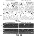
- mice were euthanized at 2 hours before light onset (-2), at light onset (0), and 2, 4, and 8 hours (+2, +4, +8) after light onset, and processed for either electron microscopy or paraffin embedding as previously described 13, 15 .
- all reagents were purchased from Electron Microscopy Sciences. Mice were perfused with 2% glutaraldehyde + 2% paraformaldehyde, and eyecups were transferred to perfusion buffer with the addition of 0.2 M sodium cacodylate buffer. Sixty to eighty nanometer ultrathin sections were stained with lead citrate/uranyl acetate and early phagosomes were counted from 200 nM out from the optic nerve.
- An early phagosome is counted if it meets the following criteria: 1) it is contained within the cytoplasm of the RPE and 2) has visible lamellar structure.
- eyecups were fixed in formaldehyde/ethanol/acetic acid and embedded in paraffin using Ottix Plus solvent substitute (DiaPath).
- Five-micrometer sections were cut and the paraffin was removed using SafeSolv solvent substitute. The sections were rehydrated and incubated in 5% H 2 O 2 in 1X SSC for 10 minutes under illumination to bleach pigments.
- Nuclei were stained with DAPI, and slides mounted with Mowiol. Images were taken with a Leica DM6000 B Epifluorescence microscope using a 40x oil immersion objective. Images were processed with Imaged v1.46r and Photoshop CS6 software.
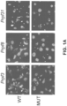
- ARPE-19 is a spontaneously immortalized human RPE cell line that is amenable to transfection and retains the ability to phagocytose 23 .
- Phagocytosis of shed POS by the RPE follows a strong diurnal, synchronized rhythm peaking at 2 hours after light-onset and remaining relatively inactive for the remainder of the day 13 .
- mutant mice only displayed 10-14 phagosomes at the same peak time-point.
- phagocytosis levels remain relatively low in control mice ("off-peak hours", 2-12 phagosomes/100 ⁇ m retina), and these levels are generally increased in mutant mice (6-14 phagosomes/100 ⁇ m retina).
- These results show a decrease in the phagocytic peak intensity in all three types of mutant mice, with a spreading of the time of the peak that lasts longer in Prpf3- and Prpf8 -mutants and starts earlier in Prpf31 -mutants.
- the Prpf8- mutants have significantly more phagosomes at the off-peak time point (+8hrs), relative to the WT controls.
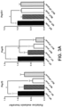
- Adhesion between the RPE apical microvilli and distal tips of the POS is known to follow a synchronized rhythm with maximum strength occurring 3.5 hours after light onset, slightly after the phagocytic peak 14, 15 .
- Adhesion can be determined by peeling the retina from a flattened eyecup immediately after euthanasia, then quantifying both the RPE melanin content and apical RPE protein markers, such as RPE65, transferred to the retina. Using this method, we assessed adhesion in Prpf- mutant mice and littermate controls at 3.5 and 8.5 hours after light onset (peak and off-peak adhesion, respectively).
- RPE adhesion was quantified first using a standard melanin quantification procedure 14 , then western blotting for the presence of RPE65 to confirm the melanin results.
- We noted a decrease of 56 ⁇ 16% ( N 6, p ⁇ 0.05 , variation is equal to the standard deviation) of the melanin content in the Prpf3 T494M/T494M at peak time and no significant change in adhesion at the off-peak time-point ( Figure 3A ).
- Western blot analysis confirmed this observation with a 30 ⁇ 2% decrease in peak adhesion ( Figure 3B ).
- RPE cells are highly polarized, and their function is dependent upon this polarity 24 .
- the specific localization of many proteins expressed in the RPE is important, and irregularities in localization may cause retinal dystrophies such as RP or Best disease 25, 26 .
- RP retinal dystrophies
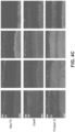
- the main phagocytic receptors ( ⁇ v ⁇ 5 integrin and MerTK) localize at the RPE apical surface 27 , while their ligands can be expressed throughout the POS and RPE 28 .
- extracellular ligands expressed in the interphotoreceptor matrix can be synthesized by both RPE and photoreceptor cells.
- ⁇ v ⁇ 5-integrin with its associated ligand Mfg-E8 milk fat globule-EGF8 are important for phagocytosis and are responsible for the diurnal rhythmicity of this function 13, 15 .
- Mfg-E8 milk fat globule-EGF8
- ⁇ v ⁇ 5-integrin participates in retinal adhesion and its rhythm, but with a ligand different from Mfg-E8 14, 15, 17 .
- ⁇ v integrin subunits associate in complexes with several ⁇ integrin subunits in RPE cells 14 , therefore it is more relevant to analyze the expression of ⁇ 5 integrin subunits.
- the downstream signaling protein FAK (focal adhesion kinase) provides a sequential activation link between ⁇ v ⁇ 5 integrin and MerTK receptors both in vitro and in vivo 29, 30, 13 .
- FAK is found throughout the RPE, and no change to this pattern was observed in the Prpf3- or the Prpf31 -mutant mice ( Figure 4B , 5B ).
- Prpf8 -mutant mice showed FAK localization to the basal side of the RPE.
- Phagocytosis is driven by the timely activation of MerTK via phosphorylation at the time of the activity peak 13, 31, 32 .
- Gas6 and Protein S are ligands of MerTK that can stimulate uptake of shed outer segments in vitro 33 . Both ligands are necessary to the internalization of POS as double knockout animals recapitulate the rapid retinal degeneration occurring in rats in whose MerTK receptors are absent 34 .
- MerTK expression in wild-type tissues is localized to both the apical and basal membranes of the RPE, whereas MerTK is localized solely to the apical side of Prpf31 -mutant RPE cells ( Figures 4C , 5C ).
- the first MerTK ligand Gas6 localizes to the POS and apical layer of the RPE in wild-type tissues. A decrease of expression is observed in the Prpf3 -mutant mice POS, with diffuse expression seen throughout the RPE. Prpf8- mutant mice maintain Gas6 expression in the POS, but appear to lose apical localization in the RPE, also showing a diffuse expression throughout the RPE. No localization changes can be observed in Prpf31 -mutant mice.
- the expression of the second MerTK ligand Protein S is localized specifically to the POS in wild-type and all Prpf -mutant mice ( Figures 4C , 5C ).
- RNA splicing factor RP is a late onset disease
- these results are not surprising and the models afford us the ability to study the mechanisms leading to the onset of disease.
- Our results demonstrate that the RPE is likely to be the primary cell type affected by mutations in these 3 RNA splicing factors in the mouse, and in humans given the similar phagocytic deficiency observed in PRPF31- knockdown human ARPE-19 cells.
- RPE retinal pigment epithelium
- RP RNA splicing retinitis pigmentosa
- a 20 bp guide RNA (gRNA) to exon 7 of PRPF31 was designed and cloned into a pCAG vector containing gRNA scaffolding sequence.
- the gRNA vector was co-transfected with a pCAG-Cas9-GFP vector into ARPE-19 cells.
- GFP positive cells were single cell sorted into individual wells of a 96 well plate and grown to confluence. DNA was isolated from each clone and the region around the predicted cut site was Sanger sequenced to identify those that exhibit correct cutting and non-homologous end joining (NHEJ). Five NHEJ lines were selected for further characterization using both qRT-PCR and phagocytosis assays to quantify FITC-labeled photoreceptor outer segment uptake with flow cytometry. These lines were maintained as confluent cultures for 3 weeks to ensure polarization and maximal expression of RPE-specific genes.
- Phagocytosis of POS was decreased in primary RPE cell cultures from 10-day old mutant mice, and this was replicated by shRNA-mediated knockdown of PRPF31 in human ARPE-19 cells (Example 1, Figure 1 ).
- CRISPR/Cas9 genome editing was used to knockout PRPF31 in normal hiPSCs ( Hou Z, Zhang Y, Propson NE, Howden SE, Chu LF, Sontheimer EJ, Thomson JA. Efficient genome engineering in human pluripotent stem cells using Cas9 from Neisseria meningitidis. Proceedings of the National Academy of Sciences of the United States of America. 2013;110:15644-9 ; Xue H, Wu J, Li S, Rao MS, Liu Y. Genetic Modification in Human Pluripotent Stem Cells by Homologous Recombination and CRISPR/Cas9 System. Methods Molecular Biology. 2014 Peters DT, Cowan CA, Musunuru K.
- hiPSC human RPE cells are similarly affected by mutations in RNA splicing factor genes, since hiPSCs can be readily differentiated into RPE cells (see Example 1, Singh R, Phillips MJ, Kuai D, Meyer JT, Martin J, Smith M, Shen W, Perez ET, Wallace KA, Capowski EE, Wright L, Gamm DM. Functional analysis of serially expanded human iPS cell-derived RPE cultures. Investigative Ophthalmology & Visual Science. 2013;54:6767-78 ; Buchholz DE, Hikita ST, Rowland TJ, Friedrich AM, Hinman CR, Johnson LV, Clegg DO.
- EBs embryoid bodies
- RDM retinal differentiation medium
- TER transepithelial resistance
- Phagocytosis As described above, primary cultures of RPE cells from the Prpf3 T494M/T494M , Prpf8 H2309P/H2309p , and Prpf31 + / - mice have significantly decreased ability to phagocytose POS ( Figure 1 ).
- hiPSCs Changes in RPE phenotype observed in the genome edited hiPSCs are confirmed using hiPSCs from patients with RNA splicing factor RP. Patients and families with RP due to mutations in the PRPF31 gene have been identified, and hiPSCs are generated using fibroblasts from one affected and one unaffected family member from each of 3 families. Briefly, fibroblasts are reprogrammed using non-integrating, oriP-containing plasmid vectors encoding seven reprogramming factors (OCT4, SOX2, NANOG, LIN28, c-Myc, KLF4, and SV40 large T-antigen), as described ( Yu J, Hu K, Smuga-Otto K, Tian S, Stewart R, Slukvin II, Thomson JA.
- OCT4 non-integrating, oriP-containing plasmid vectors encoding seven reprogramming factors
- hiPSC lines with normal karyotypes and that are confirmed to be pluripotent by teratoma studies and expression of the pluripotency markers OCT4, SSEA4, NANOG and TRA-1-81 would be selected for further study ( Singh R, Shen W, Kuai D, Martin JM, Guo X, Smith MA, Perez ET, Phillips MJ, Simonett JM, Wallace KA, Verhoeven AD, Capowski EE, Zhang X, Yin Y, Halbach PJ, Fishman GA, Wright LS, Pattnaik BR, Gamm DM.
- iPS cell modeling of Best disease insights into the pathophysiology of an inherited macular degeneration.
- hiPSC-derived RPE function is characterized in cells from patients and compared to unaffected family members using the techniques described above.
- RNA splicing factor RP RNA splicing factor RP
- AAV is the preferred gene delivery vector for retinal disorders based on the success of clinical trials of gene therapy for RPE65 LCA and choroideremia, as well as other clinical and pre-clinical studies ( Maguire AM, Simonelli F, Pierce EA, Pugh EN, Jr., Mingozzi F, Bennicelli J, Banfi S, Marshall KA, Testa F, Surace EM, Rossi S, Lyubarsky A, Arruda VR, Konkle B, Stone E, Sun J, Jacobs J, Dell'Osso L, Hertle R, Ma JX, Redmond TM, Zhu X, Hauck B, Zelenaia O, Shindler KS, Maguire MG, Wright JF, Volpe NJ, McDonnell JW, Auricchio A, High KA, Bennett J.
- PMCID 2567501 ; Maguire AM, High KA, Auricchio A, Wright JF, Pierce EA, Testa F, Mingozzi F, Bennicelli JL, Ying GS, Rossi S, Fulton A, Marshall KA, Banfi S, Chung DC, Morgan JI, Hauck B, Zelenaia O, Zhu X, Raffini L, Coppieters F, De Baere E, Shindler KS, Volpe NJ, Surace EM, Acerra C, Lyubarsky A, Redmond TM, Stone E, Sun J, McDonnell JW, Leroy BP, Simonelli F, Bennett J.
- PMCID 3277234 ; Maclachlan TK, Lukason M, Collins M, Munger R, Isenberger E, Rogers C, Malatos S, Dufresne E, Morris J, Calcedo R, Veres G, Scaria A, Andrews L, Wadsworth S. Preclinical safety evaluation of AAV2-sFLT01- a gene therapy for age-related macular degeneration. Molecular Therapy. 2011;19:326-34.
- PMCID 3034852 ; Nathwani AC, Tuddenham EG, Rangarajan S, Rosales C, McIntosh J, Linch DC, Chowdary P, Riddell A, Pie AJ, Harrington C, O'Beirne J, Smith K, Pasi J, Glader B, Rustagi P, Ng CY, Kay MA, Zhou J, Spence Y, Morton CL, Allay J, Coleman J, Sleep S, Cunningham JM, Srivastava D, Basner-Tschakarjan E, Mingozzi F, High KA, Gray JT, Reiss UM, Nienhuis AW, Davidoff AM. Adenovirus-associated virus vector-mediated gene transfer in hemophilia B. New England Journal of Medicine. 2011;365:2357-65. PMCID: 3265081 ).
- AAV has an exceptional safety record in early phase clinical studies and also poses less risk of genotoxicity compared to other vector systems since AAV genomes are stable in an episomal form in terminally differentiated cells such as photoreceptor and RPE cells ( Yang GS, Schmidt M, Yan Z, Lindbloom JD, Harding TC, Donahue BA, Engelhardt JF, Kotin R, Davidson BL.
- Virus-mediated transduction of murine retina with adeno-associated virus effects of viral capsid and genome size. JVirol.
- vectors can include promoters that drive expression in many cell types (e.g., CAG or CASI), RPE cells (e.g., promotors for RPE-specific proteins such as VMD2, RPE65, RLBP1, RGR, or TIMP3) and photoreceptor cells (RHO) ( Esumi N, Oshima Y, Li Y, Campochiaro PA, Zack DJ. Analysis of the VMD2 promoter and implication of E-box binding factors in its regulation. Journal Biological Chemistry.
- Novel adeno-associated virus serotypes efficiently transduce murine photoreceptors. J Virol. 2007;81:11372-80 ).
- the components of the AAV vectors are synthesized using codon-optimized PRPF31 sequences to improve the level and duration of gene expression ( Ill CR, Chiou HC. Gene therapy progress and prospects: recent progress in transgene and RNAi expression cassettes. Gene Therapy. 2005;12:795-802 ; Foster H, Sharp PS, Athanasopoulos T, Trollet C, Graham IR, Foster K, Wells DJ, Dickson G. Codon and mRNA sequence optimization of microdystrophin transgenes improves expression and physiological outcome in dystrophic mdx mice following AAV2/8 gene transfer.
- AAV2 As a control serotype, as this vector is known to transduce cultured monolayer cells and transduced the RPE well in vivo ( Pang JJ, Lauramore A, Deng WT, Li Q, Doyle TJ, Chiodo V, Li J, Hauswirth WW. Comparative analysis of in vivo and in vitro AAV vector transduction in the neonatal mouse retina: effects of serotype and site of administration. Vision Research.
- the PRPF31 mutant and control ARPE-19 cells are cultured on Transwell filters, as described in Example 1. Cells are treated with the desired amount of AAN- PRPF31 vectors, and cultured for an additional 11-14 days. Wild-type RPE cells treated with AAA- PRPF37 , and PRFP31 +/- cells treated with AAV-EGFP are used as controls. The effects of the AAV -PRPF31 treatment are evaluated using several approaches.
- PRPF31 protein is evaluated by immunofluorescence microscopy and western blotting experiments 2-4 days following transduction ( Liu Q, Zhou J, Daiger SP, Farber DB, Heckenlively JR, Smith JE, Sullivan LS, Zuo J, Milam AH, Pierce EA. Identification and subcellular localization of the RP1 protein in human and mouse photoreceptors. Investigative Ophthalmology & Visual Science. 2002;43:22-32 ; Liu Q, Zuo J, Pierce EA. The retinitis pigmentosa 1 protein is a photoreceptor microtubule-associated protein. Journal Neuroscience.
- AAV-mediated delivery of PRPF31 is used to treat the defective phagocytosis in Prpf31 +/- mice in vivo.
- the optimal doses of the AAV- PRPF31 vectors identified in cell culture studies is injected sub-retinally into one eye of Prpf31 + / - mice. Eyes are harvested 1 month after injection and evaluated for expression and localization of the full-length PRPF31 protein using immunofluorescence and western blotting assays ( Liu Q, Lyubarsky A, Skalet JH, Pugh EN, Jr., Pierce EA. RP1 is required for the correct stacking of outer segment discs. Investigative Ophthalmology & Visual Science.
- AAV-delivered PRPF31 to prevent and/or rescue the loss of rhythmicity of RPE phagocytosis is assessed at 2 hours before light onset (-2), at light onset (0), and 2, 4, and 6 (+2, +4, +6) hours after light onset using established techniques for immunofluorescent staining for rhodopsin and detection of phagosomes located in the RPE cell layer ( Nandrot EF, Kim Y, Brodie SE, Huang X, Sheppard D, Finnemann SC. Loss of synchronized retinal phagocytosis and age-related blindness in mice lacking alphavbeta5 integrin. Journal Experimental Medicine. 2004;200:1539-45 ; Nandrot EF, Finnemann SC.
- mice are treated at 1 month of age, and the ultrastructure of the RPE is evaluated for phenotypic rescue at 5, 8 and 11 months following AAV -PRPF31 injection ( Graziotto JJ, Farkas MH, Bujakowska K, Deramaudt BM, Zhang Q, Nandrot EF, Inglehearn CF, Bhattacharya SS, Pierce EA.
- RNA splicing factor RP Three gene-targeted mouse models of RNA splicing factor RP show late-onset RPE and retinal degeneration. Investigative Ophthalmology & Visual Science. 2011;52:190-8 ). Based on data from asymptomatic carriers of PRPF31 mutations, we anticipate that even a modest increase in PRPF31 level in the treated RPE cells will be therapeutic ( Rio FT, Wade NM, Ransijn A, Berson EL, Beckmann JS, Rivolta C. Premature termination codons in PRPF31 cause retinitis pigmentosa via haploinsufficiency due to nonsense-mediated mRNA decay. Journal Clinical Investigation.
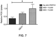
- Example 5 AAV-mediated gene augmentation therapy to ameliorates the defective phagocytosis phenotype in cultured RPE cells
- the level of PRPF31 expression from the wild-type allele correlates with the severity of disease in patients with mutations in PRPF31 ( Rio et al., Journal Clinical Investigation. 2008;118:1519-31 ; Venturini et al., PLoS genetics. 2012;8:e1003040 ; Rose et al., Scientific reports. 2016;6:19450 ).
- AAV.CASI.PRPF31 viral vector, and showed that this can produce full length PRPF31 protein in cultured cells.
- the sequence of this vector is as follows: ITR - pZac2.1 inverted terminal repeat - nts 1-130 and 3291-3420 Promoter - CASI nts 197-1252 Tag - V5 nts 1259-1309 Insert - PRPF31 nts 1319-2818 polyA sequence - rabbit ⁇ -globin nts 2825-3211
- PRPF31 mutant ARPE-19 cells were transduced with AAV.CASI.
- PRPF31 at a multiplicity of infection (MOI) of 0, 10,000, and 15,000.
- MOI multiplicity of infection
- each replicate was incubated with 1 ⁇ 10 6 FITC-labeled photoreceptor outer segments (FITC-POS) for 1 hour at 37°C.
- FITC-POS uptake was determined by counting FITC positive cells using flow cytometry.
- Treatment of the GE31 mutant cell line resulted in increased FITC-POS uptake, in a dose-dependent fashion ( Figure 7 ). This result confirms the potential of gene augmentation therapy to be used for treating PRPF31 -associated retinal degeneration.
Landscapes
- Health & Medical Sciences (AREA)
- Life Sciences & Earth Sciences (AREA)
- Genetics & Genomics (AREA)
- Engineering & Computer Science (AREA)
- Chemical & Material Sciences (AREA)
- Biotechnology (AREA)
- General Health & Medical Sciences (AREA)
- Molecular Biology (AREA)
- Organic Chemistry (AREA)
- Bioinformatics & Cheminformatics (AREA)
- Wood Science & Technology (AREA)
- Biomedical Technology (AREA)
- Zoology (AREA)
- General Engineering & Computer Science (AREA)
- Public Health (AREA)
- Veterinary Medicine (AREA)
- Animal Behavior & Ethology (AREA)
- Pharmacology & Pharmacy (AREA)
- Medicinal Chemistry (AREA)
- Epidemiology (AREA)
- Biophysics (AREA)
- Biochemistry (AREA)
- Physics & Mathematics (AREA)
- Plant Pathology (AREA)
- Microbiology (AREA)
- Ophthalmology & Optometry (AREA)
- Mycology (AREA)
- General Chemical & Material Sciences (AREA)
- Chemical Kinetics & Catalysis (AREA)
- Nuclear Medicine, Radiotherapy & Molecular Imaging (AREA)
- Medicines Containing Material From Animals Or Micro-Organisms (AREA)
- Medicines That Contain Protein Lipid Enzymes And Other Medicines (AREA)
- Peptides Or Proteins (AREA)
- Medicinal Preparation (AREA)
- Pharmaceuticals Containing Other Organic And Inorganic Compounds (AREA)
- Measuring Or Testing Involving Enzymes Or Micro-Organisms (AREA)
Applications Claiming Priority (4)
| Application Number | Priority Date | Filing Date | Title |
|---|---|---|---|
| US201562129638P | 2015-03-06 | 2015-03-06 | |
| US201562147307P | 2015-04-14 | 2015-04-14 | |
| EP16762307.3A EP3265568B1 (en) | 2015-03-06 | 2016-03-07 | Gene augmentation therapies for inherited retinal degeneration caused by mutations in the prpf31 gene |
| PCT/US2016/021226 WO2016144892A1 (en) | 2015-03-06 | 2016-03-07 | Gene augmentation therapies for inherited retinal degeneration caused by mutations in the prpf31 gene |
Related Parent Applications (1)
| Application Number | Title | Priority Date | Filing Date |
|---|---|---|---|
| EP16762307.3A Division EP3265568B1 (en) | 2015-03-06 | 2016-03-07 | Gene augmentation therapies for inherited retinal degeneration caused by mutations in the prpf31 gene |
Publications (3)
| Publication Number | Publication Date |
|---|---|
| EP3748007A1 EP3748007A1 (en) | 2020-12-09 |
| EP3748007B1 true EP3748007B1 (en) | 2024-06-26 |
| EP3748007C0 EP3748007C0 (en) | 2024-06-26 |
Family
ID=56879585
Family Applications (2)
| Application Number | Title | Priority Date | Filing Date |
|---|---|---|---|
| EP20171646.1A Active EP3748007B1 (en) | 2015-03-06 | 2016-03-07 | Gene augmentation therapies for inherited retinal degeneration caused by mutations in the prpf31 gene |
| EP16762307.3A Active EP3265568B1 (en) | 2015-03-06 | 2016-03-07 | Gene augmentation therapies for inherited retinal degeneration caused by mutations in the prpf31 gene |
Family Applications After (1)
| Application Number | Title | Priority Date | Filing Date |
|---|---|---|---|
| EP16762307.3A Active EP3265568B1 (en) | 2015-03-06 | 2016-03-07 | Gene augmentation therapies for inherited retinal degeneration caused by mutations in the prpf31 gene |
Country Status (15)
| Country | Link |
|---|---|
| US (3) | US20180043034A1 (pl) |
| EP (2) | EP3748007B1 (pl) |
| JP (3) | JP6812368B2 (pl) |
| CY (1) | CY1123158T1 (pl) |
| DK (1) | DK3265568T3 (pl) |
| ES (1) | ES2806054T3 (pl) |
| HR (1) | HRP20201225T1 (pl) |
| HU (1) | HUE051491T2 (pl) |
| LT (1) | LT3265568T (pl) |
| PL (1) | PL3265568T3 (pl) |
| PT (1) | PT3265568T (pl) |
| RS (1) | RS60668B1 (pl) |
| SI (1) | SI3265568T1 (pl) |
| SM (1) | SMT202000423T1 (pl) |
| WO (1) | WO2016144892A1 (pl) |
Families Citing this family (21)
| Publication number | Priority date | Publication date | Assignee | Title |
|---|---|---|---|---|
| US8663624B2 (en) | 2010-10-06 | 2014-03-04 | The Regents Of The University Of California | Adeno-associated virus virions with variant capsid and methods of use thereof |
| HRP20171334T1 (hr) | 2011-04-22 | 2017-11-17 | The Regents Of The University Of California | Virioni adeno-povezanog virusa s kapsid varijantom i postupci njihove upotrebe |
| US11136557B2 (en) | 2013-05-31 | 2021-10-05 | The Regents Of The University Of California | Adeno-associated virus variants and methods of use thereof |
| KR102537394B1 (ko) | 2014-03-17 | 2023-05-30 | 애드베룸 바이오테크놀로지스, 인코포레이티드 | 원추세포에서 증강된 유전자 발현을 위한 조성물 및 방법 |
| MY187898A (en) | 2015-03-02 | 2021-10-27 | Adverum Biotechnologies Inc | Compositions and methods for intravitreal delivery of polynucleotides to retinal cones |
| PL3265568T3 (pl) * | 2015-03-06 | 2020-11-30 | Massachusetts Eye & Ear Infirmary | Terapie augmentacji genów w przypadku wrodzonego zwyrodnienia siatkówki spowodowanego mutacjami genu prpf31 |
| JP6836999B2 (ja) * | 2015-03-24 | 2021-03-03 | ザ リージェンツ オブ ザ ユニバーシティ オブ カリフォルニアThe Regents Of The University Of California | アデノ随伴ウイルス変異体及びその使用方法 |
| EP3827812B1 (en) | 2016-07-29 | 2025-10-29 | The Regents of the University of California | Adeno-associated virus virions with variant capsid and methods of use thereof |
| JP2019530737A (ja) | 2016-08-23 | 2019-10-24 | アコーオス インコーポレイテッド | ヒト対象において非加齢性聴力障害を治療するための組成物および方法 |
| CA3040179A1 (en) | 2016-10-19 | 2018-04-26 | Adverum Biotechnologies, Inc. | Modified aav capsids and uses thereof |
| JP7330899B2 (ja) | 2017-05-10 | 2023-08-22 | マサチューセッツ アイ アンド イヤー インファーマリー | ウイルスのアセンブリ活性化タンパク質(aap)依存性を改変する方法および組成物 |
| WO2019006182A1 (en) | 2017-06-30 | 2019-01-03 | The Regents Of The University Of California | VIRIONS OF ADENO-ASSOCIATED VIRUSES OF CAPSID VARIANTS AND METHODS OF USE THEREOF |
| JP2020534788A (ja) | 2017-08-28 | 2020-12-03 | ザ リージェンツ オブ ザ ユニバーシティ オブ カリフォルニア | アデノ随伴ウイルスカプシド変異体及びその使用方法 |
| CA3102095A1 (en) * | 2018-06-01 | 2019-12-05 | University Of Florida Research Foundation, Incorporated | Compositions and methods for treatment of dominant retinitis pigmentosa |
| WO2020102740A2 (en) * | 2018-11-16 | 2020-05-22 | Spark Therapeutics, Inc. | Compositions and methods for increasing or enhancing transduction of gene therapy vectors and for removing or reducing immunoglobulins |
| JP2022525193A (ja) * | 2019-03-15 | 2022-05-11 | ユニヴァーシティ オブ ワシントン | Prpf31遺伝子発現ノックダウンによる、インビトロで分化したヒト細胞の生存の改善 |
| JP7807380B2 (ja) | 2020-02-21 | 2026-01-27 | アコーオス インコーポレイテッド | ヒト対象において非加齢関連聴力障害を処置するための組成物および方法 |
| US12257320B2 (en) * | 2020-03-11 | 2025-03-25 | Massachusetts Eye And Ear Infirmary | Gene therapy for NMNAT1-associated retinal degeneration |
| WO2022262756A1 (zh) * | 2021-06-16 | 2022-12-22 | 北京中因科技有限公司 | Prpf31变体及其用途 |
| AU2023365490A1 (en) * | 2022-10-21 | 2025-05-01 | Biogen Ma Inc. | Compositions and methods for treating retinitis pigmentosa |
| AU2023390868A1 (en) * | 2022-12-05 | 2025-06-19 | The United States Of America, As Represented By The Secretary, Department Of Health And Human Services | Pigment epithelium-derived factor peptides and use for treating retinal degeneration |
Family Cites Families (23)
| Publication number | Priority date | Publication date | Assignee | Title |
|---|---|---|---|---|
| DE3023787A1 (de) | 1980-06-25 | 1982-01-21 | Studiengesellschaft Kohle mbH, 4330 Mülheim | Verfahren zur erhoehung der inkorporation und der expression von genetischem material in die kerne von intakten zellen mit hilfe von liposomen |
| US4980286A (en) | 1985-07-05 | 1990-12-25 | Whitehead Institute For Biomedical Research | In vivo introduction and expression of foreign genetic material in epithelial cells |
| WO1987000201A1 (en) | 1985-07-05 | 1987-01-15 | Whitehead Institute For Biomedical Research | Epithelial cells expressing foreign genetic material |
| EP0378576B1 (en) | 1987-09-11 | 1995-01-18 | Whitehead Institute For Biomedical Research | Transduced fibroblasts and uses therefor |
| WO1989005345A1 (en) | 1987-12-11 | 1989-06-15 | Whitehead Institute For Biomedical Research | Genetic modification of endothelial cells |
| JP2917998B2 (ja) | 1988-02-05 | 1999-07-12 | ホワイトヘッド・インスティチュート・フォー・バイオメディカル・リサーチ | 修飾された肝細胞およびその用途 |
| ATE157012T1 (de) | 1989-11-03 | 1997-09-15 | Univ Vanderbilt | Verfahren zur in vivo-verabreichung von funktionsfähigen fremden genen |
| JP3249516B2 (ja) | 1990-10-31 | 2002-01-21 | ソマティクス セラピー コーポレイション | 遺伝子治療のためのレトロウイルスのベクター |
| ES2150832B1 (es) | 1996-06-12 | 2001-06-16 | Fichtel & Sachs Ag | Dispositivo de maniobra para la maniobra, en particular maniobra neumatica, de un embrague de friccion. |
| US6569662B1 (en) * | 2000-01-21 | 2003-05-27 | Hyseq, Inc. | Nucleic acids and polypeptides |
| CA2442670A1 (en) * | 2001-04-13 | 2002-10-24 | The Trustees Of The University Of Pennsylvania | Method of treating or retarding the development of blindness |
| US20040208847A1 (en) * | 2003-03-28 | 2004-10-21 | Fabienne Rolling | Method and vectors for selectively transducing retinal pigment epithelium cells |
| EP1991701A4 (en) * | 2006-02-14 | 2010-03-17 | Dana Farber Cancer Inst Inc | COMPOSITIONS, KITS AND METHODS OF IDENTIFICATION, ASSESSMENT, PREVENTION AND THERAPY OF CANCER |
| US8257969B2 (en) * | 2007-04-12 | 2012-09-04 | The Provost Fellows And Scholars Of The College Of The Holy And Undivided Trinity Of Queen Elizabeth | Genetic suppression and replacement |
| WO2008137066A1 (en) * | 2007-05-02 | 2008-11-13 | The Board Of Regents Of The University Of Oklahoma | Use of compacted nucleic acid nanoparticles in non-viral treatments of ocular diseases |
| WO2009134681A2 (en) * | 2008-04-30 | 2009-11-05 | The Trustees Of The University Of Pennsylvania | Aav7 viral vectors for targeted delivery of rpe cells |
| WO2010005533A2 (en) * | 2008-06-30 | 2010-01-14 | The Johns Hopkins University | Compositions and methods for the treatment of ocular oxidative stress and retinitis pigmentosa |
| GB201103062D0 (en) * | 2011-02-22 | 2011-04-06 | Isis Innovation | Method |
| TWI775096B (zh) * | 2012-05-15 | 2022-08-21 | 澳大利亞商艾佛蘭屈澳洲私營有限公司 | 使用腺相關病毒(aav)sflt-1治療老年性黃斑部退化(amd) |
| EP2872183B1 (en) * | 2012-07-11 | 2018-09-26 | The Trustees Of The University Of Pennsylvania | Aav-mediated gene therapy for rpgr x-linked retinal degeneration |
| HUE052676T2 (hu) | 2013-10-11 | 2021-05-28 | Massachusetts Eye & Ear Infirmary | Eljárások õsi vírusszekvenciák elõrejelzésére és alkalmazásaik |
| EP3154573A1 (en) * | 2014-06-13 | 2017-04-19 | Universidade De Santiago De Compostela | Nanoparticulate systems for use in gene transfer or gene delivery |
| PL3265568T3 (pl) | 2015-03-06 | 2020-11-30 | Massachusetts Eye & Ear Infirmary | Terapie augmentacji genów w przypadku wrodzonego zwyrodnienia siatkówki spowodowanego mutacjami genu prpf31 |
-
2016
- 2016-03-07 PL PL16762307T patent/PL3265568T3/pl unknown
- 2016-03-07 HR HRP20201225TT patent/HRP20201225T1/hr unknown
- 2016-03-07 RS RS20200945A patent/RS60668B1/sr unknown
- 2016-03-07 WO PCT/US2016/021226 patent/WO2016144892A1/en not_active Ceased
- 2016-03-07 SI SI201630873T patent/SI3265568T1/sl unknown
- 2016-03-07 ES ES16762307T patent/ES2806054T3/es active Active
- 2016-03-07 HU HUE16762307A patent/HUE051491T2/hu unknown
- 2016-03-07 EP EP20171646.1A patent/EP3748007B1/en active Active
- 2016-03-07 EP EP16762307.3A patent/EP3265568B1/en active Active
- 2016-03-07 LT LTEP16762307.3T patent/LT3265568T/lt unknown
- 2016-03-07 PT PT167623073T patent/PT3265568T/pt unknown
- 2016-03-07 DK DK16762307.3T patent/DK3265568T3/da active
- 2016-03-07 US US15/555,915 patent/US20180043034A1/en not_active Abandoned
- 2016-03-07 SM SM20200423T patent/SMT202000423T1/it unknown
- 2016-03-07 JP JP2017564778A patent/JP6812368B2/ja active Active
-
2020
- 2020-08-04 CY CY20201100718T patent/CY1123158T1/el unknown
- 2020-08-13 US US16/992,689 patent/US20210069347A1/en not_active Abandoned
- 2020-12-16 JP JP2020208068A patent/JP7239550B2/ja active Active
-
2023
- 2023-03-02 JP JP2023031931A patent/JP2023071867A/ja active Pending
-
2024
- 2024-10-25 US US18/927,466 patent/US20250195691A1/en active Pending
Also Published As
| Publication number | Publication date |
|---|---|
| US20250195691A1 (en) | 2025-06-19 |
| HK1247637A1 (en) | 2018-09-28 |
| HRP20201225T1 (hr) | 2020-11-13 |
| SMT202000423T1 (it) | 2020-09-10 |
| CY1123158T1 (el) | 2021-10-29 |
| HUE051491T2 (hu) | 2021-03-01 |
| EP3265568A4 (en) | 2018-08-08 |
| ES2806054T3 (es) | 2021-02-16 |
| JP7239550B2 (ja) | 2023-03-14 |
| SI3265568T1 (sl) | 2020-10-30 |
| LT3265568T (lt) | 2020-08-25 |
| EP3748007C0 (en) | 2024-06-26 |
| RS60668B1 (sr) | 2020-09-30 |
| EP3748007A1 (en) | 2020-12-09 |
| EP3265568A1 (en) | 2018-01-10 |
| US20180043034A1 (en) | 2018-02-15 |
| PT3265568T (pt) | 2020-08-20 |
| JP2018511652A (ja) | 2018-04-26 |
| JP2023071867A (ja) | 2023-05-23 |
| EP3265568B1 (en) | 2020-05-06 |
| WO2016144892A1 (en) | 2016-09-15 |
| US20210069347A1 (en) | 2021-03-11 |
| JP6812368B2 (ja) | 2021-01-13 |
| PL3265568T3 (pl) | 2020-11-30 |
| DK3265568T3 (da) | 2020-08-10 |
| JP2021059561A (ja) | 2021-04-15 |
Similar Documents
| Publication | Publication Date | Title |
|---|---|---|
| US20250195691A1 (en) | Gene augmentation therapies for inherited retinal degeneration caused by mutations in the prpf31 gene | |
| Leu et al. | Erbb2 regulates neuromuscular synapse formation and is essential for muscle spindle development | |
| RU2723101C2 (ru) | Вектор, способ лечения хороидермии, способ селективной экспрессии полинуклеотида | |
| Duong et al. | Use of induced pluripotent stem cell models to probe the pathogenesis of Choroideremia and to develop a potential treatment | |
| CN113728230B (zh) | 检测、预防、逆转和治疗神经系统疾病的方法 | |
| JP2024519218A (ja) | 成熟角膜内皮細胞を作製する方法 | |
| Alonso-Martin et al. | SOXF factors regulate murine satellite cell self-renewal and function through inhibition of β-catenin activity | |
| Kotterman et al. | Directed evolution of AAV targeting primate retina by intravitreal injection identifies R100, a variant demonstrating robust gene delivery and therapeutic efficacy in non-human primates | |
| EP3233093B1 (en) | Transgenic rpe cells overexpressing otx2 for the treatment of retinal degeneration | |
| JP2024501882A (ja) | 抑制置換遺伝子療法 | |
| WO2025002450A1 (en) | Engineered reprogramming factors and uses thereof for treating eye diseases | |
| HK40043762B (en) | Gene augmentation therapies for inherited retinal degeneration caused by mutations in the prpf31 gene | |
| HK40043762A (en) | Gene augmentation therapies for inherited retinal degeneration caused by mutations in the prpf31 gene | |
| HK1247637B (en) | Gene augmentation therapies for inherited retinal degeneration caused by mutations in the prpf31 gene | |
| Nizamudheen | Novel therapeutic approaches for the treatment of childhood ocular genetic diseases | |
| US20230235326A1 (en) | Methods and pharmaceutical compositions for treating ocular diseases | |
| Naessens | Recurrent coding and rare non-coding targets for treatment in inherited retinal diseases | |
| HK40096970A (zh) | 产生成熟角膜内皮细胞的方法 | |
| Shortall | Development of gene therapies for retinal degenerations | |
| Gilmore | Advancing Tools for Treating Usher Syndrome 1B and Elucidating the Role of Myosin VIIa in the Retinal Pigment Epithelium | |
| Pérez Luz et al. | DNA repair pathways are altered in neural cell models of frataxin deficiency | |
| CN120624555A (zh) | 一种眼底视网膜退行疾病的动物模型建立和有效的小分子药物 | |
| Raffa | Combined gene and stem cell therapy approach to correct ocular defects in p63-related diseases | |
| Zacharias | Lineage-specific functions of the homeodomain transcription factor Pitx2 in eye development |
Legal Events
| Date | Code | Title | Description |
|---|---|---|---|
| PUAI | Public reference made under article 153(3) epc to a published international application that has entered the european phase |
Free format text: ORIGINAL CODE: 0009012 |
|
| STAA | Information on the status of an ep patent application or granted ep patent |
Free format text: STATUS: THE APPLICATION HAS BEEN PUBLISHED |
|
| AC | Divisional application: reference to earlier application |
Ref document number: 3265568 Country of ref document: EP Kind code of ref document: P |
|
| AK | Designated contracting states |
Kind code of ref document: A1 Designated state(s): AL AT BE BG CH CY CZ DE DK EE ES FI FR GB GR HR HU IE IS IT LI LT LU LV MC MK MT NL NO PL PT RO RS SE SI SK SM TR |
|
| STAA | Information on the status of an ep patent application or granted ep patent |
Free format text: STATUS: REQUEST FOR EXAMINATION WAS MADE |
|
| 17P | Request for examination filed |
Effective date: 20210609 |
|
| RBV | Designated contracting states (corrected) |
Designated state(s): AL AT BE BG CH CY CZ DE DK EE ES FI FR GB GR HR HU IE IS IT LI LT LU LV MC MK MT NL NO PL PT RO RS SE SI SK SM TR |
|
| REG | Reference to a national code |
Ref country code: HK Ref legal event code: DE Ref document number: 40043762 Country of ref document: HK |
|
| STAA | Information on the status of an ep patent application or granted ep patent |
Free format text: STATUS: EXAMINATION IS IN PROGRESS |
|
| 17Q | First examination report despatched |
Effective date: 20220808 |
|
| GRAP | Despatch of communication of intention to grant a patent |
Free format text: ORIGINAL CODE: EPIDOSNIGR1 |
|
| STAA | Information on the status of an ep patent application or granted ep patent |
Free format text: STATUS: GRANT OF PATENT IS INTENDED |
|
| RIC1 | Information provided on ipc code assigned before grant |
Ipc: C12N 15/113 20100101ALN20240111BHEP Ipc: A61P 27/02 20060101ALI20240111BHEP Ipc: A61K 48/00 20060101ALI20240111BHEP Ipc: A61K 9/00 20060101ALI20240111BHEP Ipc: C12N 15/90 20060101AFI20240111BHEP |
|
| INTG | Intention to grant announced |
Effective date: 20240209 |
|
| GRAS | Grant fee paid |
Free format text: ORIGINAL CODE: EPIDOSNIGR3 |
|
| GRAA | (expected) grant |
Free format text: ORIGINAL CODE: 0009210 |
|
| STAA | Information on the status of an ep patent application or granted ep patent |
Free format text: STATUS: THE PATENT HAS BEEN GRANTED |
|
| AC | Divisional application: reference to earlier application |
Ref document number: 3265568 Country of ref document: EP Kind code of ref document: P |
|
| AK | Designated contracting states |
Kind code of ref document: B1 Designated state(s): AL AT BE BG CH CY CZ DE DK EE ES FI FR GB GR HR HU IE IS IT LI LT LU LV MC MK MT NL NO PL PT RO RS SE SI SK SM TR |
|
| REG | Reference to a national code |
Ref country code: GB Ref legal event code: FG4D |
|
| REG | Reference to a national code |
Ref country code: CH Ref legal event code: EP |
|
| REG | Reference to a national code |
Ref country code: DE Ref legal event code: R096 Ref document number: 602016088176 Country of ref document: DE |
|
| U01 | Request for unitary effect filed |
Effective date: 20240726 |
|
| U07 | Unitary effect registered |
Designated state(s): AT BE BG DE DK EE FI FR IT LT LU LV MT NL PT SE SI Effective date: 20240808 |
|
| PG25 | Lapsed in a contracting state [announced via postgrant information from national office to epo] |
Ref country code: HR Free format text: LAPSE BECAUSE OF FAILURE TO SUBMIT A TRANSLATION OF THE DESCRIPTION OR TO PAY THE FEE WITHIN THE PRESCRIBED TIME-LIMIT Effective date: 20240626 |
|
| PG25 | Lapsed in a contracting state [announced via postgrant information from national office to epo] |
Ref country code: GR Free format text: LAPSE BECAUSE OF FAILURE TO SUBMIT A TRANSLATION OF THE DESCRIPTION OR TO PAY THE FEE WITHIN THE PRESCRIBED TIME-LIMIT Effective date: 20240927 |
|
| PG25 | Lapsed in a contracting state [announced via postgrant information from national office to epo] |
Ref country code: NO Free format text: LAPSE BECAUSE OF FAILURE TO SUBMIT A TRANSLATION OF THE DESCRIPTION OR TO PAY THE FEE WITHIN THE PRESCRIBED TIME-LIMIT Effective date: 20240926 Ref country code: HR Free format text: LAPSE BECAUSE OF FAILURE TO SUBMIT A TRANSLATION OF THE DESCRIPTION OR TO PAY THE FEE WITHIN THE PRESCRIBED TIME-LIMIT Effective date: 20240626 Ref country code: GR Free format text: LAPSE BECAUSE OF FAILURE TO SUBMIT A TRANSLATION OF THE DESCRIPTION OR TO PAY THE FEE WITHIN THE PRESCRIBED TIME-LIMIT Effective date: 20240927 Ref country code: RS Free format text: LAPSE BECAUSE OF FAILURE TO SUBMIT A TRANSLATION OF THE DESCRIPTION OR TO PAY THE FEE WITHIN THE PRESCRIBED TIME-LIMIT Effective date: 20240926 |
|
| PG25 | Lapsed in a contracting state [announced via postgrant information from national office to epo] |
Ref country code: PL Free format text: LAPSE BECAUSE OF FAILURE TO SUBMIT A TRANSLATION OF THE DESCRIPTION OR TO PAY THE FEE WITHIN THE PRESCRIBED TIME-LIMIT Effective date: 20240626 |
|
| PG25 | Lapsed in a contracting state [announced via postgrant information from national office to epo] |
Ref country code: IS Free format text: LAPSE BECAUSE OF FAILURE TO SUBMIT A TRANSLATION OF THE DESCRIPTION OR TO PAY THE FEE WITHIN THE PRESCRIBED TIME-LIMIT Effective date: 20241026 |
|
| PG25 | Lapsed in a contracting state [announced via postgrant information from national office to epo] |
Ref country code: CZ Free format text: LAPSE BECAUSE OF FAILURE TO SUBMIT A TRANSLATION OF THE DESCRIPTION OR TO PAY THE FEE WITHIN THE PRESCRIBED TIME-LIMIT Effective date: 20240626 |
|
| PG25 | Lapsed in a contracting state [announced via postgrant information from national office to epo] |
Ref country code: RO Free format text: LAPSE BECAUSE OF FAILURE TO SUBMIT A TRANSLATION OF THE DESCRIPTION OR TO PAY THE FEE WITHIN THE PRESCRIBED TIME-LIMIT Effective date: 20240626 Ref country code: SK Free format text: LAPSE BECAUSE OF FAILURE TO SUBMIT A TRANSLATION OF THE DESCRIPTION OR TO PAY THE FEE WITHIN THE PRESCRIBED TIME-LIMIT Effective date: 20240626 |
|
| PG25 | Lapsed in a contracting state [announced via postgrant information from national office to epo] |
Ref country code: ES Free format text: LAPSE BECAUSE OF FAILURE TO SUBMIT A TRANSLATION OF THE DESCRIPTION OR TO PAY THE FEE WITHIN THE PRESCRIBED TIME-LIMIT Effective date: 20240626 Ref country code: SM Free format text: LAPSE BECAUSE OF FAILURE TO SUBMIT A TRANSLATION OF THE DESCRIPTION OR TO PAY THE FEE WITHIN THE PRESCRIBED TIME-LIMIT Effective date: 20240626 |
|
| PG25 | Lapsed in a contracting state [announced via postgrant information from national office to epo] |
Ref country code: SM Free format text: LAPSE BECAUSE OF FAILURE TO SUBMIT A TRANSLATION OF THE DESCRIPTION OR TO PAY THE FEE WITHIN THE PRESCRIBED TIME-LIMIT Effective date: 20240626 Ref country code: SK Free format text: LAPSE BECAUSE OF FAILURE TO SUBMIT A TRANSLATION OF THE DESCRIPTION OR TO PAY THE FEE WITHIN THE PRESCRIBED TIME-LIMIT Effective date: 20240626 Ref country code: RO Free format text: LAPSE BECAUSE OF FAILURE TO SUBMIT A TRANSLATION OF THE DESCRIPTION OR TO PAY THE FEE WITHIN THE PRESCRIBED TIME-LIMIT Effective date: 20240626 Ref country code: PL Free format text: LAPSE BECAUSE OF FAILURE TO SUBMIT A TRANSLATION OF THE DESCRIPTION OR TO PAY THE FEE WITHIN THE PRESCRIBED TIME-LIMIT Effective date: 20240626 Ref country code: IS Free format text: LAPSE BECAUSE OF FAILURE TO SUBMIT A TRANSLATION OF THE DESCRIPTION OR TO PAY THE FEE WITHIN THE PRESCRIBED TIME-LIMIT Effective date: 20241026 Ref country code: ES Free format text: LAPSE BECAUSE OF FAILURE TO SUBMIT A TRANSLATION OF THE DESCRIPTION OR TO PAY THE FEE WITHIN THE PRESCRIBED TIME-LIMIT Effective date: 20240626 Ref country code: CZ Free format text: LAPSE BECAUSE OF FAILURE TO SUBMIT A TRANSLATION OF THE DESCRIPTION OR TO PAY THE FEE WITHIN THE PRESCRIBED TIME-LIMIT Effective date: 20240626 |
|
| PLBE | No opposition filed within time limit |
Free format text: ORIGINAL CODE: 0009261 |
|
| STAA | Information on the status of an ep patent application or granted ep patent |
Free format text: STATUS: NO OPPOSITION FILED WITHIN TIME LIMIT |
|
| 26N | No opposition filed |
Effective date: 20250327 |
|
| U21 | Renewal fee for the european patent with unitary effect paid with additional fee |
Year of fee payment: 10 Effective date: 20250516 |
|
| PG25 | Lapsed in a contracting state [announced via postgrant information from national office to epo] |
Ref country code: MC Free format text: LAPSE BECAUSE OF FAILURE TO SUBMIT A TRANSLATION OF THE DESCRIPTION OR TO PAY THE FEE WITHIN THE PRESCRIBED TIME-LIMIT Effective date: 20240626 |
|
| REG | Reference to a national code |
Ref country code: CH Ref legal event code: H13 Free format text: ST27 STATUS EVENT CODE: U-0-0-H10-H13 (AS PROVIDED BY THE NATIONAL OFFICE) Effective date: 20251024 |
|
| PG25 | Lapsed in a contracting state [announced via postgrant information from national office to epo] |
Ref country code: CH Free format text: LAPSE BECAUSE OF NON-PAYMENT OF DUE FEES Effective date: 20250331 |
|
| PG25 | Lapsed in a contracting state [announced via postgrant information from national office to epo] |
Ref country code: IE Free format text: LAPSE BECAUSE OF NON-PAYMENT OF DUE FEES Effective date: 20250307 |
|
| PGFP | Annual fee paid to national office [announced via postgrant information from national office to epo] |
Ref country code: GB Payment date: 20260327 Year of fee payment: 11 |