EP3740199B1 - Polymerverkapselte virale vektoren für die gentherapie - Google Patents
Polymerverkapselte virale vektoren für die gentherapie Download PDFInfo
- Publication number
- EP3740199B1 EP3740199B1 EP19743691.8A EP19743691A EP3740199B1 EP 3740199 B1 EP3740199 B1 EP 3740199B1 EP 19743691 A EP19743691 A EP 19743691A EP 3740199 B1 EP3740199 B1 EP 3740199B1
- Authority
- EP
- European Patent Office
- Prior art keywords
- polymer
- vector
- nanoparticle
- cells
- polymers
- Prior art date
- Legal status (The legal status is an assumption and is not a legal conclusion. Google has not performed a legal analysis and makes no representation as to the accuracy of the status listed.)
- Active
Links
Images


Classifications
-
- A—HUMAN NECESSITIES
- A61—MEDICAL OR VETERINARY SCIENCE; HYGIENE
- A61K—PREPARATIONS FOR MEDICAL, DENTAL OR TOILETRY PURPOSES
- A61K9/00—Medicinal preparations characterised by special physical form
- A61K9/48—Preparations in capsules, e.g. of gelatin, of chocolate
- A61K9/50—Microcapsules having a gas, liquid or semi-solid filling; Solid microparticles or pellets surrounded by a distinct coating layer, e.g. coated microspheres, coated drug crystals
- A61K9/51—Nanocapsules; Nanoparticles
- A61K9/5107—Excipients; Inactive ingredients
- A61K9/5176—Compounds of unknown constitution, e.g. material from plants or animals
- A61K9/5184—Virus capsids or envelopes enclosing drugs
-
- A—HUMAN NECESSITIES
- A61—MEDICAL OR VETERINARY SCIENCE; HYGIENE
- A61K—PREPARATIONS FOR MEDICAL, DENTAL OR TOILETRY PURPOSES
- A61K40/00—Cellular immunotherapy
- A61K40/10—Cellular immunotherapy characterised by the cell type used
-
- A—HUMAN NECESSITIES
- A61—MEDICAL OR VETERINARY SCIENCE; HYGIENE
- A61K—PREPARATIONS FOR MEDICAL, DENTAL OR TOILETRY PURPOSES
- A61K40/00—Cellular immunotherapy
- A61K40/30—Cellular immunotherapy characterised by the recombinant expression of specific molecules in the cells of the immune system
- A61K40/31—Chimeric antigen receptors [CAR]
-
- A—HUMAN NECESSITIES
- A61—MEDICAL OR VETERINARY SCIENCE; HYGIENE
- A61K—PREPARATIONS FOR MEDICAL, DENTAL OR TOILETRY PURPOSES
- A61K40/00—Cellular immunotherapy
- A61K40/40—Cellular immunotherapy characterised by antigens that are targeted or presented by cells of the immune system
- A61K40/41—Vertebrate antigens
- A61K40/42—Cancer antigens
- A61K40/4202—Receptors, cell surface antigens or cell surface determinants
- A61K40/421—Immunoglobulin superfamily
- A61K40/4211—CD19 or B4
-
- A—HUMAN NECESSITIES
- A61—MEDICAL OR VETERINARY SCIENCE; HYGIENE
- A61K—PREPARATIONS FOR MEDICAL, DENTAL OR TOILETRY PURPOSES
- A61K47/00—Medicinal preparations characterised by the non-active ingredients used, e.g. carriers or inert additives; Targeting or modifying agents chemically bound to the active ingredient
- A61K47/50—Medicinal preparations characterised by the non-active ingredients used, e.g. carriers or inert additives; Targeting or modifying agents chemically bound to the active ingredient the non-active ingredient being chemically bound to the active ingredient, e.g. polymer-drug conjugates
- A61K47/51—Medicinal preparations characterised by the non-active ingredients used, e.g. carriers or inert additives; Targeting or modifying agents chemically bound to the active ingredient the non-active ingredient being chemically bound to the active ingredient, e.g. polymer-drug conjugates the non-active ingredient being a modifying agent
- A61K47/68—Medicinal preparations characterised by the non-active ingredients used, e.g. carriers or inert additives; Targeting or modifying agents chemically bound to the active ingredient the non-active ingredient being chemically bound to the active ingredient, e.g. polymer-drug conjugates the non-active ingredient being a modifying agent the modifying agent being an antibody, an immunoglobulin or a fragment thereof, e.g. an Fc-fragment
- A61K47/6835—Medicinal preparations characterised by the non-active ingredients used, e.g. carriers or inert additives; Targeting or modifying agents chemically bound to the active ingredient the non-active ingredient being chemically bound to the active ingredient, e.g. polymer-drug conjugates the non-active ingredient being a modifying agent the modifying agent being an antibody, an immunoglobulin or a fragment thereof, e.g. an Fc-fragment the modifying agent being an antibody or an immunoglobulin bearing at least one antigen-binding site
- A61K47/6849—Medicinal preparations characterised by the non-active ingredients used, e.g. carriers or inert additives; Targeting or modifying agents chemically bound to the active ingredient the non-active ingredient being chemically bound to the active ingredient, e.g. polymer-drug conjugates the non-active ingredient being a modifying agent the modifying agent being an antibody, an immunoglobulin or a fragment thereof, e.g. an Fc-fragment the modifying agent being an antibody or an immunoglobulin bearing at least one antigen-binding site the antibody targeting a receptor, a cell surface antigen or a cell surface determinant
-
- A—HUMAN NECESSITIES
- A61—MEDICAL OR VETERINARY SCIENCE; HYGIENE
- A61K—PREPARATIONS FOR MEDICAL, DENTAL OR TOILETRY PURPOSES
- A61K47/00—Medicinal preparations characterised by the non-active ingredients used, e.g. carriers or inert additives; Targeting or modifying agents chemically bound to the active ingredient
- A61K47/50—Medicinal preparations characterised by the non-active ingredients used, e.g. carriers or inert additives; Targeting or modifying agents chemically bound to the active ingredient the non-active ingredient being chemically bound to the active ingredient, e.g. polymer-drug conjugates
- A61K47/69—Medicinal preparations characterised by the non-active ingredients used, e.g. carriers or inert additives; Targeting or modifying agents chemically bound to the active ingredient the non-active ingredient being chemically bound to the active ingredient, e.g. polymer-drug conjugates the conjugate being characterised by physical or galenical forms, e.g. emulsion, particle, inclusion complex, stent or kit
- A61K47/6921—Medicinal preparations characterised by the non-active ingredients used, e.g. carriers or inert additives; Targeting or modifying agents chemically bound to the active ingredient the non-active ingredient being chemically bound to the active ingredient, e.g. polymer-drug conjugates the conjugate being characterised by physical or galenical forms, e.g. emulsion, particle, inclusion complex, stent or kit the form being a particulate, a powder, an adsorbate, a bead or a sphere
- A61K47/6927—Medicinal preparations characterised by the non-active ingredients used, e.g. carriers or inert additives; Targeting or modifying agents chemically bound to the active ingredient the non-active ingredient being chemically bound to the active ingredient, e.g. polymer-drug conjugates the conjugate being characterised by physical or galenical forms, e.g. emulsion, particle, inclusion complex, stent or kit the form being a particulate, a powder, an adsorbate, a bead or a sphere the form being a solid microparticle having no hollow or gas-filled cores
- A61K47/6929—Medicinal preparations characterised by the non-active ingredients used, e.g. carriers or inert additives; Targeting or modifying agents chemically bound to the active ingredient the non-active ingredient being chemically bound to the active ingredient, e.g. polymer-drug conjugates the conjugate being characterised by physical or galenical forms, e.g. emulsion, particle, inclusion complex, stent or kit the form being a particulate, a powder, an adsorbate, a bead or a sphere the form being a solid microparticle having no hollow or gas-filled cores the form being a nanoparticle, e.g. an immuno-nanoparticle
- A61K47/6931—Medicinal preparations characterised by the non-active ingredients used, e.g. carriers or inert additives; Targeting or modifying agents chemically bound to the active ingredient the non-active ingredient being chemically bound to the active ingredient, e.g. polymer-drug conjugates the conjugate being characterised by physical or galenical forms, e.g. emulsion, particle, inclusion complex, stent or kit the form being a particulate, a powder, an adsorbate, a bead or a sphere the form being a solid microparticle having no hollow or gas-filled cores the form being a nanoparticle, e.g. an immuno-nanoparticle the material constituting the nanoparticle being a polymer
- A61K47/6935—Medicinal preparations characterised by the non-active ingredients used, e.g. carriers or inert additives; Targeting or modifying agents chemically bound to the active ingredient the non-active ingredient being chemically bound to the active ingredient, e.g. polymer-drug conjugates the conjugate being characterised by physical or galenical forms, e.g. emulsion, particle, inclusion complex, stent or kit the form being a particulate, a powder, an adsorbate, a bead or a sphere the form being a solid microparticle having no hollow or gas-filled cores the form being a nanoparticle, e.g. an immuno-nanoparticle the material constituting the nanoparticle being a polymer the polymer being obtained otherwise than by reactions involving carbon to carbon unsaturated bonds, e.g. polyesters, polyamides or polyglycerol
-
- A—HUMAN NECESSITIES
- A61—MEDICAL OR VETERINARY SCIENCE; HYGIENE
- A61K—PREPARATIONS FOR MEDICAL, DENTAL OR TOILETRY PURPOSES
- A61K9/00—Medicinal preparations characterised by special physical form
- A61K9/48—Preparations in capsules, e.g. of gelatin, of chocolate
- A61K9/50—Microcapsules having a gas, liquid or semi-solid filling; Solid microparticles or pellets surrounded by a distinct coating layer, e.g. coated microspheres, coated drug crystals
- A61K9/51—Nanocapsules; Nanoparticles
- A61K9/5107—Excipients; Inactive ingredients
- A61K9/513—Organic macromolecular compounds; Dendrimers
- A61K9/5146—Organic macromolecular compounds; Dendrimers obtained otherwise than by reactions only involving carbon-to-carbon unsaturated bonds, e.g. polyethylene glycol, polyamines, polyanhydrides
- A61K9/5153—Polyesters, e.g. poly(lactide-co-glycolide)
-
- A—HUMAN NECESSITIES
- A61—MEDICAL OR VETERINARY SCIENCE; HYGIENE
- A61P—SPECIFIC THERAPEUTIC ACTIVITY OF CHEMICAL COMPOUNDS OR MEDICINAL PREPARATIONS
- A61P35/00—Antineoplastic agents
-
- C—CHEMISTRY; METALLURGY
- C12—BIOCHEMISTRY; BEER; SPIRITS; WINE; VINEGAR; MICROBIOLOGY; ENZYMOLOGY; MUTATION OR GENETIC ENGINEERING
- C12N—MICROORGANISMS OR ENZYMES; COMPOSITIONS THEREOF; PROPAGATING, PRESERVING, OR MAINTAINING MICROORGANISMS; MUTATION OR GENETIC ENGINEERING; CULTURE MEDIA
- C12N15/00—Mutation or genetic engineering; DNA or RNA concerning genetic engineering, vectors, e.g. plasmids, or their isolation, preparation or purification; Use of hosts therefor
- C12N15/09—Recombinant DNA-technology
- C12N15/63—Introduction of foreign genetic material using vectors; Vectors; Use of hosts therefor; Regulation of expression
- C12N15/79—Vectors or expression systems specially adapted for eukaryotic hosts
- C12N15/85—Vectors or expression systems specially adapted for eukaryotic hosts for animal cells
- C12N15/86—Viral vectors
-
- C—CHEMISTRY; METALLURGY
- C07—ORGANIC CHEMISTRY
- C07K—PEPTIDES
- C07K14/00—Peptides having more than 20 amino acids; Gastrins; Somatostatins; Melanotropins; Derivatives thereof
- C07K14/435—Peptides having more than 20 amino acids; Gastrins; Somatostatins; Melanotropins; Derivatives thereof from animals; from humans
- C07K14/705—Receptors; Cell surface antigens; Cell surface determinants
- C07K14/70503—Immunoglobulin superfamily
- C07K14/7051—T-cell receptor (TcR)-CD3 complex
-
- C—CHEMISTRY; METALLURGY
- C07—ORGANIC CHEMISTRY
- C07K—PEPTIDES
- C07K2319/00—Fusion polypeptide
- C07K2319/01—Fusion polypeptide containing a localisation/targetting motif
- C07K2319/03—Fusion polypeptide containing a localisation/targetting motif containing a transmembrane segment
-
- C—CHEMISTRY; METALLURGY
- C12—BIOCHEMISTRY; BEER; SPIRITS; WINE; VINEGAR; MICROBIOLOGY; ENZYMOLOGY; MUTATION OR GENETIC ENGINEERING
- C12N—MICROORGANISMS OR ENZYMES; COMPOSITIONS THEREOF; PROPAGATING, PRESERVING, OR MAINTAINING MICROORGANISMS; MUTATION OR GENETIC ENGINEERING; CULTURE MEDIA
- C12N2740/00—Reverse transcribing RNA viruses
- C12N2740/00011—Details
- C12N2740/10011—Retroviridae
- C12N2740/16011—Human Immunodeficiency Virus, HIV
- C12N2740/16041—Use of virus, viral particle or viral elements as a vector
- C12N2740/16043—Use of virus, viral particle or viral elements as a vector viral genome or elements thereof as genetic vector
-
- C—CHEMISTRY; METALLURGY
- C12—BIOCHEMISTRY; BEER; SPIRITS; WINE; VINEGAR; MICROBIOLOGY; ENZYMOLOGY; MUTATION OR GENETIC ENGINEERING
- C12N—MICROORGANISMS OR ENZYMES; COMPOSITIONS THEREOF; PROPAGATING, PRESERVING, OR MAINTAINING MICROORGANISMS; MUTATION OR GENETIC ENGINEERING; CULTURE MEDIA
- C12N2740/00—Reverse transcribing RNA viruses
- C12N2740/00011—Details
- C12N2740/10011—Retroviridae
- C12N2740/16011—Human Immunodeficiency Virus, HIV
- C12N2740/16041—Use of virus, viral particle or viral elements as a vector
- C12N2740/16045—Special targeting system for viral vectors
Definitions
- Gene therapy delivers exogenous genetic material to the target cells in order to correct genetic abnormalities or provide treatment of a disease by altering cell function.
- both viral and non-viral gene delivery methods have been used, but both approaches still present significant shortcomings.
- the technology described herein provides synthetic packaged viral vector nanoparticle compositions and methods for using them to provide enhanced delivery of genetic material for use in gene therapy and vaccine applications.
- the vector compositions and methods can be employed in any situation for which transduction of cells with one or more transgenes is useful. For example, they can be used for treatment of cancer, infectious diseases, metabolic diseases, neurological diseases, or inflammatory conditions, or to correct genetic defects.
- the present invention relates to a nanoparticle comprising a viral vector coated with a polymer or a mixture of polymers, wherein the viral vector comprises a transgene, and wherein the nanoparticle functions as a vehicle for delivering the transgene to eukaryotic cells, wherein the viral vector lacks envelope protein
- the polymer or the mixture of polymers comprises a poly(beta-amino ester) having the general formula wherein each Pep is an oligopeptide, wherein R is OH, CH 3 , or a cholesterol, and wherein m ranges from 1 to 20, n ranges from 1 to 100, and o ranges from 1 to 10.
- the oligopeptide includes at least three amino acid residues, selected from the group consisting of arginine (R), lysine (K), histidine (H), glutamic acid (E), aspartic acid (D), and cysteine (C).
- R arginine
- K lysine
- H histidine
- E glutamic acid
- D aspartic acid
- C cysteine
- Cysteine can be included to provide a covalent attachment point for the peptide to the polymer; while it carries a slight negative charge at pH 7, it does not significantly alter charge if used together with positively charged amino acids.
- Exemplary peptides are CRRR (SEQ ID NO:1), CHHH (SEQ ID NO:2), CKKK (SEQ ID NO:3), CEEE (SEQ ID NO:4), and CDDD (SEQ ID NO:5).
- any naturally occurring amino acid can be included in the Pep moieties.
- the sequence of the peptide is selected so as to promote cellular uptake and targeting of the polymer-coated vector.
- the oligopeptide sequences and net charge are selected so as to promote cellular uptake and/or endosomal uptake and/or endosomal escape of viral vectors encapsulated with the polymers in the intended target cells.
- Peptides at each of the two ends of the polymer typically are the same on a given polymer molecule, but may be different.
- Polymer molecules in a mixture used to coat a vector may be the same or different; if different, they may differ in the polymer backbone or in the terminal peptides.
- the peptide at both ends of the polymer has the amino acid sequence CRRR (SEQ ID NO:1), or CHHH (SEQ ID NO:2), or CKKK (SEQ ID NO:3), or CEEE (SEQ ID NO:4), or CDDD (SEQ ID NO:5).
- the polymer or mixture of polymers can have a net positive or negative charge, or can be uncharged.
- Oligopeptides containing 2 or more residues of only a single type of amino acid, such as R, K, H, D, or E can be used.
- the polymer can comprise a sugar or sugar alcohol grafted onto the lateral chain attached to the polymer backbone.
- One or more of the polymer molecules encapsulating each vector nanoparticle can optionally comprise a targeting moiety linked to the polymer at any desired position on the polymer molecule, such as at R.
- the polymer molecules can be cross-linked or non-cross-linked.
- the polymer molecules can be attached to the viral vector surface either non-covalently, or covalently such as by covalent attachment to a lipid molecule (e.g., cholesterol, a phospholipid, or a fatty acid) that partitions into the lipid bilayer of the vector, or to a protein embedded in the lipid bilayer.
- a lipid molecule e.g., cholesterol, a phospholipid, or a fatty acid
- the polymer merely coats the vector but is attached non-specifically, i.e., it is not attached to the vector by specific covalent or non-covalent interactions, but only by nonspecific interactions such as net charge, hydrophobicity, or van der Waals interactions.
- the ratio of the polymer or mixture of polymers to the retroviral vector is from about 1:100 to about 5:1 by weight, or the number of polymer molecules per vector particle is from about 10 6 to about 10 12 .
- the viral vector nanoparticles comprise a viral vector.
- the viral vector can be chosen based on desired characteristics (e.g., immunogenicity, capacity to accommodate different sized constructs, integrating and replicative properties, expression level, duration of expression, tissue tropism or targeting characteristics, ability to infect dividing and/or quiescent cells, etc.),
- the viral vector can be a retroviral vector such as a lentiviral vector.
- the viral vector is a retroviral vector.
- the invention can also be practiced with other types of virus and/or viral vectors, including those derived from DNA or RNA viruses.
- the nanoparticles can include one or more targeting moieties that are exposed at the surface of the encapsulated vector nanoparticles, such as by covalent or non-covalent association of the targeting moieties with the polymer coating.
- One molecular species of targeting moiety, or more than one different molecular species of targeting moiety, can be present on each viral vector nanoparticle.
- Targeting moieties can be, for example, antibodies, antibody fragments (including Fab), scFvs, antibody-like protein scaffolds, oligopeptides, aptamers, L-RNA aptamers, or ligands for cell surface receptors.
- the targeting moiety is an anti-CD3 antibody or aptamer for targeting the nanoparticles towards T-cells.
- the transgene encodes a chimeric antigen receptor.
- the nanoparticle is capable of acting as a gene delivery vehicle by transducing cells in vivo in a subject to whom the vehicle is administered, or transducing cells in vitro.
- the nanoparticle is capable of transducing a specific type of cell or class of cells, usually through the action of the targeting moiety and/or through the action of the polymer or polymer mixture.
- the polymer of polymer-coated vector particles can promote cell transduction, not only by attachment to target cell surfaces, but through endosomal uptake, such as by endocytosis, micropinocytosis, phagocytosis, or other mechanisms, and endosomal escape (via membrane fusion after a reduction in pH in the endosomal vesicle).
- endosomal uptake such as by endocytosis, micropinocytosis, phagocytosis, or other mechanisms
- endosomal escape via membrane fusion after a reduction in pH in the endosomal vesicle.
- the amino acid sequence of oligopeptides associated with the polymer can specifically promote cell transduction via the endosomal route.
- the vectors of the invention lack envelope proteins; which differs from pseudotyped vectors.
- envelope proteins such as the HIV gp120-gp41 complex or the vesicular stomatitis virus (VSV-G) glycoprotein form spike-like structures on the outer surface of the viral envelope, which facilitate attachment of the vector particles to host cells and entry of viruses into host cells.
- Pseudotyping proteins such as VSV-G also can be used to protect or bind the vector during purification (e.g., protection during ultracentrifugation, binding to an affinity column or other affinity matrix, or gel purification).
- such structures can interfere with tight association between the vector particle envelope and the polymers.
- Viral envelope proteins also carry a pH-dependent charge, which can limit or interfere with the association of polymers, particularly charged polymers. Further, packaged viral vectors harboring pseudotyping proteins can undergo coating destabilization and destruction in vitro and in vivo, thus releasing viral vector able to transduce cells unspecifically, potentially leading to a reduction in safety profile.
- vectors lacking envelope proteins can enhance the packaging of viral vectors using polymers.
- Coated vectors lacking envelope protein show an improved safety profile compared to vectors having envelope protein, because after destabilization or destruction of the coating, they are not able to transduce cells in vitro or in vivo.
- the nanoparticles of the present technology wherein the viral vector lacks viral envelope protein have a built-in safety mechanism for use in gene therapy or immunotherapy, because the nanoparticles are only capable of transducing a mammalian cell above a threshold number of polymer molecules per vector.
- nanoparticles have an improved safety profile for in vivo use compared to a vector or polymer-encapsulated vector that lacks the threshold feature.
- some membrane proteins such as proteins that are not viral envelope or fusion proteins and do not otherwise form spike-like structures on the membrane outer surface, such as proteins from the vector producing cells not used for pseudotyping, may be present in the lipid membrane of the viral vector particle.
- viral envelope proteins are excluded from the viral vector particles, such as those which have a significant mass protruding from the outer surface of the envelope lipid bilayer, such as at least 20%, at least 30%, at least 40%, or at least 50% of their mass protruding from the outer envelope surface.
- the nanoparticle compositions optionally can contain additional components, such as lipid molecules, surfactants, nucleic acids, protein molecules, or small molecule drugs.
- additional components such as lipid molecules, surfactants, nucleic acids, protein molecules, or small molecule drugs.
- the present technology also contemplates pharmaceutical formulations or compositions containing the nanoparticles together with one or more excipients, carriers, buffers, salts, or liquids, rendering the delivery vehicle suitable for administration via oral, intranasal, or parenteral administration, such as intravenous, intramuscular, subcutaneous, peritumoral or intratumoral injection, or for in vitro administration to cells in an ex vivo gene transfer protocol.
- Such compositions and formulations can also be lyophilized to stabilize them during storage.
- the viral vector nanoparticle of the present technology can serve as a gene delivery vehicle.
- the nanoparticle includes a viral vector coated on its exterior surface with a layer containing a polymer or a mixture of polymers.
- the retroviral vector specifically lacks the viral envelope protein that might typically be included in the envelope of similar retroviral vectors.
- the viral vector specifically lacks any naturally occurring or modified viral vector envelope proteins, such as wild type or modified VSV-G, HIV gp120, HIV gp41, MMTV gp52, MMTV gp36, MLV gp71, syncytin, wild type or modified Sindbis virus envelope protein, measles virus hemagglutinin (H) and fusion (F) glycoproteins, and HEMO.
- the vector contains one or more of such envelope proteins.
- the present invention provides a method of making the above described nanoparticle/gene delivery vehicle (viral vector nanoparticle).
- the method includes the steps of: (a) providing a viral vector lacking envelope protein and containing a transgene; (b) providing a polymer or a mixture of polymers; and (c) contacting the viral vector and the polymer or mixture of polymers, whereby the viral vector and the polymer or the mixture of polymers combine to form the nanoparticle, which contains the viral vector coated with the polymer or mixture of polymers.
- the present invention provides the nanoparticle according to the first aspect of the present invention for use in a method of treating a disease, the method comprising administering a composition comprising a plurality of said nanoparticles to a subject in need thereof, whereby cells of the subject are transduced by the retroviral vector and the transgene is expressed in the transduced cells.
- the disease to be treated is cancer.
- viral envelope proteins such as VSV-G are absent are preferred, as they provide an enhanced safety profile because they lack the ability to transduce non-targeted cells in the host and due to more specific targeting provided by the use of polymer encapsulation to restore cellular uptake and/or endosomal uptake and/or endosomal escape otherwise provided by envelope protein.
- the present invention provides a method of transducing cells in vitro.
- the method includes contacting cultured cells with a plurality of nanoparticles according to the first aspect of the invention, whereby at least some of the cultured cells are transduced by the retroviral vector and the transgene is specifically expressed in the transduced cells.
- the nanoparticles are targeted to CD3-positive cells and the transgene is specifically expressed in CD3-positive cells, or the transgene encodes a chimeric antigen receptor.
- the present invention provides the nanoparticle of the first aspect of the present invention for use in a method of performing gene therapy, the method comprising contacting cells in vitro or within a living organism with a plurality of said nanoparticles, whereby the transgene is expressed in the cells.
- the transgene can replace or modify a deficient gene or replace a missing gene.
- Packaged viral vector nanoparticle compositions that allow enhanced delivery of genetic material for gene therapy and vaccine applications are provided. These compositions include a viral vector encapsulated by polymer or a mixture of polymers.
- the polymer shell provides protection of the viral vector particle in the bloodstream and tissues following parenteral administration to a subject and in some embodiments can assist in delivering the vector to its target cell.
- Fig. 1A shows a viral vector nanoparticle, also referred to herein as gene delivery vehicle, according to the present technology.
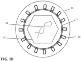
- Fig. 1B shows a viral vector nanoparticle, also referred to herein as gene delivery vehicle, not according to the present technology.
- the basic structure of the vehicle is that of nanoparticle 10, which contains a retroviral vector encapsulated by polymer shell 11.
- the retroviral vector includes membrane envelope 12, protein capsid 13 disposed within the envelope, and one or more nucleic acid molecules 14 disposed within the capsid.
- the viral envelope is a lipid bilayer-based structure that, in the viral vector nanoparticle shown in Fig.
- FIG. 1B contains one or more types of viral envelope or fusion proteins 20 embedded within or attached to the bilayer membrane; such envelope or fusion proteins are lacking in the viral vector nanoparticle depicted in Fig. 1A .
- the viral vector envelope, with or without envelope proteins, is encapsulated, both functionally and essentially entirely, by polymeric matrix 11.
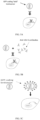
- the vector particle can be encapsulated by more than one polymer shell (see, e.g., Fig. 2 ), i.e., can be encapsulated by a multilayered polymer shell.
- Figure 3 shows an embodiment in which targeting moiety 40 is associated with the polymer shell.
- the present technology provides a novel generation of synthetic viral vectors, particularly lentiviral vectors, in which the viral vector particles lack a viral envelope protein.
- This new platform technology confers several advantages compared to using viral vector particles containing an envelope protein, including cheaper production of viral vector particles by transient transfection. 1) Due to removal of the plasmid coding for the viral envelope protein and due to the fact that the vector is not transducing the producing cells during production, deleting the envelope protein increases the useful production time so the yields of produced vectors.
- Vectors lacking envelope protein are safer for use for in in vivo gene therapy, because the uncoated vectors, lacking a viral envelope protein, cannot transduce cells, and because they have reduced immunogenicity, 3) Vectors lacking envelope protein provide greater accuracy and efficiency of transduction due to the use of specific polymer-mediated targeting. 4) Polymer-encapsulated viral vectors, particularly lentiviral vectors, are more stable at low pH than ordinary (uncoated) viral vectors, which improves their stability and effectiveness in use, especially when endosomal uptake mechanisms are involved.
- Elimination of viral envelope protein from viral vector particles is especially compatible with the use of polymer-coated viral vector particles, because it essentially eliminates the ability of the uncoated vector particles to transduce cells, and the polymer coating can provide specific targeting; thus, the risk of transducing non-targeted cells is drastically reduced. Further, the elimination of viral envelope protein promotes the tight association of polymer molecules with the surface of the vector envelope, providing better stability and more reliable targeting compared to polymer coated viral vector particles containing viral envelope proteins.
- Viral vectors based on retroviruses typically possess a core structure or capsid, which contains a transgene and other necessary nucleic acid sequences, and the core structure is in turn surrounded by a membrane envelope.
- the envelope typically contains a viral fusion protein that assists in attachment and fusion of the viral envelope with the host cell membrane, allowing cellular entry of the viral vector.
- the role of cell targeting, attachment, and entry is assumed (and restored in the case of bald LV) by the polymer shell of the nanoparticulate vector particles, which can act at the cellular uptake and/or endosomal level to transduce cells, rendering the viral fusion protein unnecessary.
- the presence in the vector envelope of a viral fusion protein interferes with the tight and stable attachment of polymer molecules to the envelope. Therefore, in certain embodiments of the present technology, the viral vector nanoparticles have been engineered to lack envelope proteins, particularly to lack viral fusion or "spike" proteins. The result is a more robust nanoparticle structure and a more stable vector that exhibits better activity and longer half-life when administered to a patient, such as by intravenous administration.
- a viral vector to "lack envelope protein” means that the envelope, in particular the lipid bilayer, of the vector envelop is devoid of any proteins typically found on the surface of enveloped viruses, in either their naturally occurring or modified form, such as the Env protein complex or viral envelope glycoproteins exemplified by HIV gp120-gp41 complex, vesicular stomatitis virus glycoprotein (VSV-G), influenza virus hemagglutinin, measles virus H and F proteins, and wild type or modified Sindbis virus envelope protein.
- VSV-G vesicular stomatitis virus glycoprotein
- influenza virus hemagglutinin hemagglutinin
- measles virus H and F proteins wild type or modified Sindbis virus envelope protein
- viral vector nanoparticles of the present technology can either i) lack envelope proteins entirely, ii) essentially lack envelope proteins (i.e., contain only a trace amount such as 0.1% or less of typical viral vector particles), or iii) have a small amount of envelope protein, such as less than 10%, less than 5%, less than 3%, less than 2%, or less than 1% of the typical amount.
- a typical amount of envelope protein is an amount present in a viral vector particle that relies on the envelope protein for cellular entry and transduction, or an amount found in the envelope of an intact, functional virus particle.
- the envelope of the viral vector can be engineered to completely lack envelope protein, for example by eliminating any gene encoding an envelope protein during expression in the cells from which the vector is produced.
- Membrane-embedded or membrane attached proteins that do not form spike-like structures on the outer surface of the envelope may, however, be present in the lipid bilayer of the viral vector nanoparticles in certain embodiments.
- the polymers used to form the viral vector nanoparticle include one or more species of poly(beta-amino ester) (PBAE) having the formula wherein Pep is an oligopeptide and R is OH, CH 3 , or cholesterol, a phospholipid molecule, or a fatty acid or acyl chain, wherein m preferably ranges from 1 to 20. n preferably ranges from 1 to 100, and o ranges from 1 to 10.
- PBAE poly(beta-amino ester)
- Other polymers and mixtures thereof with one another and with PBAE also can be used to encapsulate viral vector particles.
- PBAEs have been described as non-viral polynucleotide delivery vectors capable of condensing both DNA and RNA into discrete nanoparticles ( Green, J.J. et al. Acc. Chem. Res. 41, 749-759 (2008 )), though they show low transduction efficiency and high toxicity.
- PBAEs can be formed from the reaction of di(acrylate ester) monomers with amine monomers, resulting in polymers having terminal acrylate groups that can be functionalized with end modifications. End groups are functionalities or constitutional units that are at an extremity of a macromolecule or oligomer. End modification of PBAEs allows for the improvement of their biological properties.
- the oligopeptide peptide comprises one, two, three, four, five, or more amino acids selected from the group consisting of arginine (R), glutamic acid (E), lysine (K), aspartic acid (D), histidine (H), and cysteine (C).
- the oligopeptide contains one, two, three, or more amino acids that are positively charged (such as R, K, or H), or at least partially positively-charged, under physiological conditions.
- the oligopeptide contains one or more negatively-charged amino acids (such as D or E), either in the absence of positively-charged amino acids or mixed with positively-charged amino acids.
- the PBAE nanoparticle ensures that the vector remains intact during transit through the circulation of the patient.
- the nanoparticle can assist in shielding the viral vector from the host's immune system, for example, ensuring the claimed nanoparticles can maintain high transduction efficiency even in the presence of anti-viral envelope neutralizing antibodies.
- the particle can ensure that the vector transfects only the desired immune cells: the nanoparticle modifications and/or the presence of targeting moieties can direct the nanoparticle specifically to the tissues of interest.
- the polymer layer or shell which coats the viral vector envelope can form a single homogenous polymer layer, or it can form two or more overlapping layers of the same or different polymers, or polymers having different net charge or degree of crosslinking.
- Other non-PBAE polymers can be intermixed with PBAE or added above or below a PBAE layer to form the polymer shell.
- the multilayer coating includes an anionic and a cationic polymer.
- the cationic polymer includes one or more PBAEs.
- the anionic polymer is polyglutamic acid (PGA), chondroitin sulfate and/or hyaluronic acid.
- the viral vector is encapsulated within two or more layers of polymers, such as alternating layers having alternating net electrical charges, i.e., a positively-charged layer is followed by a negatively-charged layer, and then for a positively-charged layer.
- the net charge of the nanoparticle is positive at pH 7.
- the net charge of the nanoparticle is negative at pH 7.
- a particle is said to have a diameter of x nm, there will generally be a distribution of particles about this mean, but at least 50% by number (e.g. >60%, >70%, >80%, >90%, or more) of the particles will have a diameter within the range x ⁇ 20%.
- Retroviruses are RNA viruses that possess the ability to integrate into the host genome in a stable fashion.
- the retroviral vector in the gene delivery vehicle is a lentiviral vector.
- Lentiviral vectors are derived from lentiviruses, which represent a genus of slow viruses of the Retroviridae family. Lentiviruses include human immunodeficiency viruses (HIV), simian immunodeficiency virus (SIV), equine infectious encephalitis virus (EIAV), caprine arthritis encephalitis virus (CAEV), bovine immunodeficiency virus (BIV) and feline immunodeficiency virus (FIV). Lentiviruses can integrate into the genome of dividing and non-dividing cells.
- Bioproduction of integrative but replication-defective lentiviral vectors is based on the separation of cis- and trans-acting sequences of the lentivirus. Transduction in non-dividing cells requires the presence of two cis-acting sequences in the lentiviral genome, the central polypurine tract (cPPT) and the central termination sequence (CTS). This leads to the formation of a triple-stranded DNA "flap", which maximizes the efficiency of gene import into the nuclei of non-dividing cells, including dendritic cells (DCs) ( Zennou et al., 2000, Cell, 101(2) 173-85 ; Arhel et al., 2007, EMBO J, 26(12):3025-25 37 ). Furthermore, removal of the LTR U3 sequence has resulted in "self-inactivating" vectors which are entirely devoid of viral promoter and enhancer sequences and are safer.
- DCs dendritic cells
- the plasmid encoding an envelope protein is omitted completely in certain embodiments, while in other embodiments it is included but its expression is maintained at a low level.
- Lentiviral particle vectors can also be produced continuously by stably inserting the packaging genes and proviral coding DNA into the cellular genome. A combination of integrated plasmids and transient transfection also can be used. It should be noted, however that, in certain embodiments, lentiviral vectors suitable for use in the gene delivery vehicle described herein are devoid of envelope protein.
- Non-integrating lentiviral vectors lacking the envelope protein also can be used in the gene transfer vehicles.
- Examples of non-integrating lentiviral vectors are found in Coutant et al., PLOS ONE 7(11):e48644 (2102 ); Karwacz et al., J. Virol. 83(7):3094-3103 (2009 ); Negri et al., Molecular Therapy 15(9):1716-1723 (2007 ); and Hu et al., Vaccine 28:6675-6683 (2010 ).
- the expression of the transgene is driven by an internal promoter.
- Viral promoters such as the CMV promoter, preferably are not used because of the presence of strong enhancers.
- Ubiquitous promoters can be used, such as promoters for the human genes encoding ubiquitin, PGK, ⁇ -actin, GAPDH, ⁇ -kinesin, and the like.
- promoters active in T-cells and APCs can be used, such as the promoter for human MHC class I genes. In all cases, the promoter preferably does not contain an enhancer.
- the promoter is preferably selected so as to achieve a therapeutic level of expression of the transgene in the target cells.
- the promoter is specific for the target cells, i.e., it enables a therapeutic expression level of the transgene in the target cells, and preferably enables a higher level of transgene expression in target cells than in non-target cells; In certain embodiments, the promoter allows little or no expression of the transgene in non-target cells.
- polymer encapsulation of viral vectors such as LV has not been previously accomplished.
- the inventors have developed methods for polymer encapsulation of LV and other viral vectors that result in stable polymer-encapsulated LV particles with low toxicity and high effectiveness at transducing cells.
- Important factors in producing viable encapsulated vectors include the type of polymer used (e.g., its charge and charge distribution), the ratio of polymer molecules to vector particle, and the pH used for encapsulation. The effect of these parameters was investigated and is described in Examples 1 and 2 below.
- PBAE polymers and LV successful encapsulation, resulting in particles capable of efficient transduction of target cells, requires about 10 8 to 10 11 polymer molecules per vector particle; higher ratios can produce toxicity. Further, the optimum pH for successful encapsulation differs depending on whether the LV contains VSV-G + or not (VSV-G - or "bald" LV). For VSV-G + LV, optimum encapsulation with PBAE occurs at about pH 5, as indicated by the high level of VSV-G antibody-resistant transduction at pH 5, while little or no encapsulation occurred at higher pH values.
- optimum PBAE encapsulation efficiency occurs over a broader and lower pH range, such as about 5.0, 5.5, 6.0, or 6.5, or 5.0-6.5, or 5.0-6.0, or 5.5-6.0, or 5.5-6.5.
- optimum PBAE encapsulation efficiency occurs at a lower pH range, such as about 5.0, 5.5, 6.0, or 6.5, or 5.0-6.5, or 5.0-6.0, or 5.5-6.0, or 5.5-6.5.
- the optimum pH for successful encapsulation that does not impact viability of transduced cells is the same whether the LV contains VSV-G + or not, such as 5.0 or 5.5.
- the nanoparticles can be formed by mixing a solution comprising the required materials and then incubating the solution.
- nanoparticles can be formed by mixing a solution comprising PBAE with a solution comprising the viral vector, then incubating the solution.
- the solution contains a sugar, such as glucose.
- the solution contains 5% glucose.
- the solution contains a buffer.
- the buffer is sodium acetate, and the final concentration of sodium acetate buffer is from 2 to 50 mM, from 10 to 30 mM, or about 25 mM.
- the buffer is Tris-HCl buffer
- the final concentration of Tris-HCl buffer is from 2 to 40 mM, from 5 to 30 mM, or from 15 to 25 mM.
- the solution is incubated for at least 5 minutes. In some embodiments, the solution is incubated for at least 30 minutes. In some embodiments, the solution is incubated overnight.
- the solution can be incubated at 4 to 40°C, or at 17 to 25°C; preferably, the solution is incubated at 4°C.
- the nanoparticles are formed using layer-by-layer deposition of ionic polymers onto retrovirus particles, thus forming multilayer-coated viral vectors.
- a solution containing ionic polymers is contacted with a solution containing viral vector particles.
- the ratio of polymer to viral vector particles can be in the range from about 1:100 to about 5:1 by weight. The ratio can be varied so as to ensure that the nanoparticles contain on average a single viral vector particle per polymer-coated nanoparticle.
- the weight ratio of all polymers to viral vector can be from about 1:100 to about 1:1, or from about 1:50 to about 1:1, or from about 1:20 to about 1:1, or from about 1:10 to about 2:1, or from about 1:1 to about 5:1.
- one or more polymers are added to a suspension of retrovirus particles in glucose or another sugar compatible with lyophilization and direct injection after reconstitution into a human or other subject.
- negatively-charged and positively-charged polymer are added sequentially to form polymer layers having alternating charges.
- the polymers are added at 30 min intervals.
- the multilayer-coated viral vectors have a net negative charge at pH 7.
- the multilayer-coated viral vectors have a net positive charge at pH 7. The number and polarity of layers can be chosen so as to optimize transduction efficiency of particular cell types of interest.
- nanoparticles are formed at a pH at which the viral vector particle surface charge, surface potential, or zeta potential is opposite to that of the one or more polymers that form the polymer shell around the viral vector particle.
- the pH is above that of the isoelectric point of one or more retroviral envelope proteins, or the envelope as a whole.
- the pH can be at or above about 4.0, 4.5, 5.0, 5.4, 5.4, 5.5, 5.6, 5.8, 6.0, 6.2, 6.4, 6.5, 6.8, or 7.0.
- the nanoparticles are formed in a solution having a pH of about 5.
- the nanoparticle vector delivery vehicle can be targeted to specific target cells or tissues requiring therapy, or in which protein expression by the vector is desired.
- Another embodiment of targeting moiety is shown in Fig. 3 , in which the delivery vehicle contains targeting moieties covalently or non-covalently attached to the polymer surface.
- Targeting moieties can target specific cells or classes of cells either in vivo or in vitro.
- Examples of targeting moieties include proteins (e.g., antibodies, including human, mouse, camelid and shark antibodies, single chain antibodies, nanobodies, diabodies, and antigen-binding fragments of antibodies), peptides, nucleic acids such as aptamers or oligonucleotides, or ligands that bind to cell-surface receptors or components of extracellular matrix.
- the delivery vehicle is targeted in particular to T-cells for expression of one or more therapeutic proteins, such as chimeric antigen receptor (CAR) proteins, so as to enhance the killing of cells (e.g., tumor cells or pathogen cells) to which the chimeric receptor binds.
- the target cells for the vector are T-cells (as CD3+ T cells) or NK cells.
- Targeting can be accomplished using one or more DNA aptamers or antibodies having specificity for a protein on the surface of the target cells.
- the aptamer or antibody serves as a targeting moiety when integrated into the nanoparticle and exposed at the surface of the nanoparticle.
- the targeting moiety can specifically bind CD3 or CD8, or a combination of CD3 and CD8.
- the targeting moiety can specifically bind to a surface protein found only on naive T-cells, such as CD45RA, CCR7, CD62L, CD27, CD28, IL7-Ra, or DC-SIGN. Combinations of these targeting moieties also can be used.
- Targeting is not limited to CAR technology or hematologic cells, however. Any cell type can be targeted.
- a specific tissue or cell type e.g., hematopoietic cells, myocytes, islet cells, etc..
- quiescent cells may be targeted over dividing cells so as to reduce risk of recombination events).
- dividing cells such as cancer cells may be targeted, for example in a cancer vaccine.
- the viral vector in the gene delivery vehicle described herein contains one or more transgenes which, after entering a cell, become expressed in the cell and produce expression products.
- the viral vector can contain one, two, three, or more transgenes.
- the transgenes are nucleic acid molecules that encode any naturally occurring or artificial (i.e., recombinant) protein, polypeptide, or peptide, including a therapeutic protein or a marker protein.
- Such vector-expressed proteins preferably have a therapeutic value in the subject in which they are expressed, and are thus therapeutic proteins.
- One type of therapeutic protein is a chimeric antigen receptor (CAR).
- CARs for use in the technology described herein are recombinant antigen receptors, such as T-cell receptors, with binding specificity for cell surface antigens that do not require peptide processing or HLA expression in order to bind.
- the antigen-binding portion can be an scFv derived from an antibody, from an Fab selected from a library, or can be a naturally occurring ligand for a cell surface receptor. It also can be a nanobody (VHH).
- VHH nanobody
- a CAR of the technology includes one or more of each of the following domains: an extracellular antigen-binding domain, a hinge and transmembrane domain, and intracellular signaling domains (CD3zeta, and/or CD28 or 41-BB/OX40). Further details of CARs suitable for use in the gene delivery vehicle and sequences to encode them in a lentiviral vector are found in WO2016/012623A1 .
- genes that can be delivered using the gene delivery vehicles of the present technology include interleukins and cytokines, such as interleukin 1 (IL-1), IL-2, IL-3, IL-4, IL-5, IL-6, IL-7, IL-8, IL-9, IL-10, IL-11 IL-12, GM-CSF, and G-CSF.
- IL-1 interleukin 1
- IL-2 interleukin-2
- IL-3 IL-4
- IL-5 IL-6
- IL-7 IL-8
- IL-9 interleukin-10
- IL-11 IL-12 GM-CSF
- G-CSF G-CSF
- antigens such as viral antigens, bacterial antigens, fungal antigens or parasitic antigens also can be delivered.
- Viruses whose antigens can be expressed include picornavirus, coronavirus, togavirus, flavirvirus, rhabdovirus, paramyxovirus, orthomyxovirus, bunyavirus, arenvirus, reovirus, retrovirus, papovavirus, parvovirus, herpesvirus, poxvirus, hepadnavirus, and spongiform virus.
- Preferred viral targets include influenza, herpes simplex virus 1 and 2, measles, small pox, polio or HIV.
- Pathogens include trypanosomes, tapeworms, roundworms, helminths. Tumor antigens such as fetal antigen or prostate specific antigen also can be targeted in this manner.
- Vaccination of an individual can be carried out according to any desired schedule; preferably vaccination is only required infrequently, such as yearly or biennially, and provides long term immunologic protection against the infectious agent.
- a viral vector transgene also can encode an anti-checkpoint protein.
- anti-checkpoint proteins that can be encoded by the transgene are inhibitors of CTLA-4, PD1, PDL1, LAG-3, TIM 3, B7-H3, ICOS, IDO, 4-1BB, CD47, and combinations thereof.
- the gene delivery vehicles of the present technology can be used in any application that requires effective transduction of genetic material in cells of interest, such as mammalian cells or human cells by means of a retroviral vector, such as a lentiviral vector.
- the delivery vehicles transduce cells with high efficiency and exhibit low toxicity and low immunogenicity. They can permanently integrate genetic material into the host cell genome, making the system particularly suitable for permanent labeling of cells and their progeny. Since the vehicles present high specificity, low toxicity, and high transduction capability even in vivo, they are particularly suitable for gene transfer and gene therapy applications both for therapy in humans and animals, as well as in animal models, thus allowing investigation of the molecular mechanisms underlying different biological functions and their translation into therapy.
- some preparations of polymer-encapsulated viral vector nanoparticles may be limited to transducing certain cell types, such as preferentially transducing dividing cells and not transducing non-dividing cells.
- the delivery vehicles of the present technology can be used to genetically engineer cells for biotechnology applications, such as biologics manufacturing and livestock and plant improvements. Precise and stable insertion of the gene or genes of interest is essential for these activities, and can be consistently achieved with the vehicle of the technology.
- the vehicles can be used to insert genes not originally present in cells used for manufacturing purposes, such as transformed cells (Chinese hamster ovary (CHO) cells, HEK293 cells) or primary cells.
- the vehicles can be used to "knock out" certain genes present in those cells, such as genes involved in protein glycosylation, a common cause of immunogenicity.
- the vehicles may, for example, be used to prevent, treat or cure diseases that affect livestock, or to improve their economic value, by accelerating growth and maturation.
- the gene delivery vehicle can be used to deliver genetic material as part of in vitro or in vivo gene therapy.
- the gene delivery vehicle can be employed in gene therapy to replace a disease-causing mutated version of a gene with a healthy copy of the gene; inactivate or "knock out" a gene that is not functioning properly, and/or to introduce a new gene in a cell, such as to fight a disease.
- retroviral vectors in general, allows for permanent insertion of genetic material into a cell of interest.
- the use of lentiviral vectors in particular, allows for permanent insertion of genetic material even into non-dividing cells.
- the gene delivery vehicles are capable of transducing cells in vivo, leading to functional expression of the transgene and, therefore, can be used to conduct gene therapy.
- the vector delivery vehicle can be administered parenterally, such as intravenously, intramuscularly or intra-tumor.
- the vector delivery vehicle can be administered externally, such as orally or intra nasally.
- the gene delivery vehicle can be administered by any other suitable route, including intracerebral and pulmonary.
- the gene delivery vehicles of the present technology are also capable of transducing cells in vitro, leading to functional expression of the transgene and, therefore, can be used to conduct gene therapy in vitro and ex vivo.
- Patient's cells can be obtained (e.g., via blood or bone marrow sample, or biopsy) and contacted with one of the described vector delivery vehicles in a laboratory setting. The transduced cells can then be returned to the patient, and help treat a disease.
- Diseases that can be treated with gene therapy using gene delivery vehicles of the technology include inherited disorders, such as severe combined immunodeficiency (SCID), chronic granulomatous disease (CGD), Wiskott-Aldrich Syndrome (WAS), relapsed acute 30 lymphoblastic leukemia (ALL), adrenoleukodystrophy (ALD), hemophilia B, adenosine deaminase (ADA) deficiency, cystic fibrosis (CF), beta-thalassemia, sickle cell disease, Duchenne muscular dystrophy, familial hypercholesterolemia, and hereditary blindness. Treatable diseases also include neurodegenerative disorders, such as Parkinson's Disease, Alzheimer's Disease and Huntington's Disease.
- SCID severe combined immunodeficiency
- CCD chronic granulomatous disease
- WAS Wiskott-Aldrich Syndrome
- ALL adrenoleukodystrophy
- ADA adenosine deaminase
- cancers of the prostate such as cancers of the prostate, pancreas, brain, skin, liver, colon, breast, and kidney.
- Polymer-coated lentiviral vectors for use in cell transduction studies were made using the following materials and methods.
- the transfer vector plasmid was pARA-CMV-GFP or pARA-CMV-mCherry or pARA or pAra-hUBC-CD19.
- a kanamycin-resistant plasmid encoded for the provirus (a non-pathogenic and non-replicative recombinant proviral DNA derived from HIV-1, strain NL4-3), in which an expression cassette was cloned.
- the insert contained the transgene, the promoter for transgene expression and sequences added to increase the transgene expression and to allow the lentiviral vector to transduce all cell types including non-mitotic ones.
- the coding sequences corresponded to the gene encoding Green Fluorescent Protein (GFP) or mCherry (red fluorescent protein) or CD19.
- the promoter was the human ubiquitin promoter or the CMV promoter.
- LTR Long Terminal Repeat sequences
- encapsidation sequences SD and 5'Gag
- the central PolyPurine Tract/Central Termination Site for the nuclear translocation of the vectors
- BGH polyadenylation site was added.
- the packaging plasmid was pARA-Pack.
- the kanamycin resistant plasmid encoded for the structural lentiviral proteins (GAG, POL, TAT and REV) used in trans for the encapsidation of the lentiviral provirus.
- the coding sequences corresponded to a polycistronic gene gag-pol-tat-rev, coding for the structural (Matrix MA, Capsid CA and Nucleocapside NC), enzymatic (Protease PR, Integrase IN and Reverse Transcriptase RT) and regulatory (TAT and REV) proteins.
- the non-coding sequences and expression signals corresponded to a minimal promoter from CMV for transcription initiation, a polyadenylation signal from the insulin gene for transcription termination, and an HIV-1 Rev Responsive Element (RRE) participating for the nuclear export of the packaging RNA.
- RRE HIV-1 Rev Responsive Element
- the envelope plasmid when used, was pENV1.
- This kanamycin-resistant plasmid encoded glycoprotein G from the Vesicular Stomatitis Virus (VSV-G) Indiana strain, used for the pseudotyping of some of the lentiviral vectors.
- VSV-G genes were codon optimized for expression in human cells, and the gene was cloned into pVAX1 plasmid (Invitrogen).
- the coding sequences corresponded to codon-optimized VSV-G gene, and the noncoding sequences and expression signals corresponded to a minimal promoter from CMV for transcription initiation, and the BGH polyadenylation site to stabilize the RNA.
- LV293 cells were seeded at 5 ⁇ 10 5 cells/mL in 2 ⁇ 3000 mL Erlenmeyer flasks (Corning, 431252) in 1000 mL of LVmax Production Medium (Gibco, A3583401). The two Erlenmeyers were incubated at 37°C, 65 rpm under humidified 8% CO 2 . The day after seeding, the transient transfection was performed. PEIPro transfectant reagent (PolyPlus, 115-010) were mixed with transfer vector plasmid (pARA-CMV-GFP or pARA-CMV-mCherry or pARA or pAra-hUBC-CD19) and packaging plasmid (pARA-Pack).
- transfer vector plasmid pARA-CMV-GFP or pARA-CMV-mCherry or pARA or pAra-hUBC-CD19
- packaging plasmid pARA-Pack
- the mix PEIPro/Plasmid was added drop by drop to the cell line and incubated at 37°C, 65 rpm under humidified 8% CO 2 .
- the lentivector production was stimulated by sodium butyrate at 5 mM final concentration.
- the bulk mixture was incubated at 37°C, 65 rpm under humidified 8% CO 2 for 24 hours.
- the clarified bulk mixture was incubated 1 hour at room temperature for DNase treatment.
- Lentivector purification was performed by chromatography (Q mustang membrane) and eluted by NaCl gradient. Tangential flow filtration was performed on a 100 kDa HYDROSORT membrane (Sartorius, 3M81446802E-SW), which allowed to reduce the volume and to formulate in specific buffer at pH 7, ensuring at least 2 years of stability. After sterile filtration at 0.22 ⁇ m (Millipore, SLGVM33RS), the bulk drug product was filled in 2 mL glass vials with aliquots less than 1 ml, then labelled, frozen and stored at ⁇ -70°C.
- the bald LV number was evaluated by physical titer quantification.
- the assay was performed by detection and quantitation of the lentivirus associated HIV-1 p24 core protein only (P24 ELISA Cell Biolabs QUICK TITER Lentivirus Titer Kit).
- Poly( ⁇ -amino ester) polymers were synthesized following a two-step procedure similar to that described by Dosta et al. 5-amino-1-pentanol (Sigma-Aldrich 3.9 g, 38 mmol) and 1-Hexylamine (Sigma-Aldrich 3.9 g, 38 mmol) were mixed in a round bottom flask and combined with 1,4-butanediol diacrylate (Aldrich 18 g, 82 mmol) in a 2.1:1 molar ratio of diacrylate to amine monomers. The mixture was heated to 90 °C with stirring for 20 h. Then it was cooled to room temperature to form a light yellow viscous solid oil and stored at -20 °C until further use.
- Oligopeptide-modified PBAEs were obtained by end-modification of acrylate-terminated polymer C6 with thiol-terminated oligopeptide at 1:2.5 molar ratio in dimethyl sulfoxide (DMSO).
- DMSO dimethyl sulfoxide
- the capped polymer was precipitated with 5 volumes of diethyl ether. After centrifugation (10 min at 3000 g), the polymer was washed with 2 volumes of fresh mixture of diethyl ether and acetone (7:3 v/v), then the residue was dried under vacuum before it was formed into a stock of 100 mg/mL in DMSO, which was stored at -20 °C.
- C6-CK3 tri-lysine end-modified PBAE polymer, C6-CK3 was obtained by mixing a solution of C6 (100 mg, 0.048 mmol) dissolved in DMSO (1.1 mL) and a solution of hydrochloride H-Cys-Lys-Lys-Lys-NH 2 (CK3) (78 mg, 0.12 mmol) in DMSO (1.8 mL) and processing the coupling reaction as previously described for C6-CR3.
- CK3 hydrochloride H-Cys-Lys-Lys-Lys-Lys-NH 2
- C6-CH3 the solution of C6 (65 mg, 0.031 mmol) dissolved in DMSO (1.1 mL) that was mixed with a solution of hydrochloride H-Cys-His-His-His-NH2 (CH3) (53 mg, 0.078 mmol) in DMSO (1.2 mL).
- the tri-aspartate end-modified PBAE polymer, C6-CD3 the solution of C6 (96 mg, 0.046 mmol) dissolved in DMSO (1.1 mL) that was mixed with a solution of hydrochloride H-Cys-Asp-Asp-Asp-NH2 (CD3) (96 mg, 0.046 mmol) in DMSO (1.7 mL).
- oligopeptide-modified PBAEs The chemical structure of the oligopeptide-modified PBAEs was confirmed by 1H-NMR spectroscopy by the disappearance of acrylate signals and the presence of signals typically associated with amino acid moieties. Residual free peptide content was quantified by UV detection (wavelength 220 nm) after separation by UPLC ACQUITY system (Waters) equipped with a BEH C18 column (130 A, 1.7 ⁇ m, 2.1x50 mm, temperature 35 °C) using an acetonitrile gradient.
- PGA polyglutamic acid
- the resulting activated PGA was added to a solution of antimouse CD3 ⁇ F(ab')2 fragment (InVivoMAb) in phosphate-buffered saline (PBS) at a 4:1 molar ratio and mixed at room temperature for 2 h. Excess reagents and unlinked PGA were removed by filtration (30,000 MWCO Vivaspin column) and the buffer was exchanged 7 times against PBS. Antibody concentration was determined using a NanoDrop 2000 Spectrophotometer (Thermo Scientific).
- 10 ⁇ L of Drug Product pseudotyping Lentivectors or bald lentivectors were diluted in 10 ⁇ L of different buffers (pH 5 to 7) during different times (0, 15, 30, 60, 90 and 120 minutes) at +4°C.
- HEK293T cells 8 ⁇ 10 5 cells/well
- HEK293T cells 8 ⁇ 10 5 cells/well
- the culture medium was removed and 300 ⁇ L of fresh DMEM containing 10% FBS, 1% PS and 10 ⁇ L ow each condition were added to each well.
- each well was supplemented with 0.5 mL fresh culture medium.
- HEK293T cells in each well were trypsinized, fixed (BD Cell FIX solution #340181) and the number of fluorescence-positive cells was determined using flow cytometry (AttuneNXT; Invitrogen, Inc.).
- TU / ml (% of cells expressing eGFP / 100) ⁇ total number of HEK293T cells at time of infection / volume of virus stock added (mL).
- the titration for the bald lentivectors was performed by physical titer quantification.
- the assay was performed by detection and quantitation of the lentivirus associated HIV-1 p24 core protein only (P24 ELISA Cell Biolabs' QUICK TITER Lentivirus Titer Kit).
- a pre-treatment samples allows to distinguish the free p24 from destroyed Lentivectors.
- Encapsulation of lentiviral vectors was performed as follows. Lentiviral vectors were diluted in 25 mM citrate buffer pH 5.0 (or in an appropriate buffer system at a different defined pH) to prepare a final volume of 75 ⁇ L per replicate. PBAE polymers were diluted in the same buffer as for lentiviral vectors (75 ⁇ L per replicate) and vortexed 2 s for homogenization. The diluted polymers were added to the diluted vectors in a 1:1 ratio (v/v), the mixes were gently vortexed for 10 s and incubated 30 minutes at +4 °C. Finally, an equal volume of culture medium (150 ⁇ L) was added to the encapsulation reactions before transfer to cells.
- Lentiviral vectors were diluted in 25 mM citrate buffer pH 5.0 (or in appropriate buffer system at a defined pH) to prepare a final volume of 75 ⁇ L per replicate.
- PBAEs were diluted in the same buffer as for lentiviral vectors (75 ⁇ L per replicate) and vortexed 2 s for homogenization.
- the diluted polymers were added to the diluted vectors in a 1:1 ratio (v/v). The mixes were gently vortexed for 10 s and incubated 30 minutes at +4 °C.
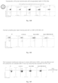
- Particle surface charge measurements of the LV/PBAE/PGA-Fab complexes were determined by dynamic light scattering (DLS) at room temperature with a Zetasizer Nano ZS (Malvern Instruments Ltd, United Kingdom, 4-mW laser) using a wavelength of 633 nm). Measurements were carried out by mixing 200 ⁇ L of complexes with 800 ⁇ L of D-PBS 1x buffer solution (pH 7.1). The results were plotted as mean and standard deviation of 3 measurements analyzed by intensity, and are shown in Figs. 6A-6F . The results showed surface potential modification by the encapsulation of the vector particles with polymer in accordance with the charge of the polymer, indicating successful encapsulation with polymer.
- Table 1 Polymer Coatings Used To Test Effect Of Charge. Influence of the charge on the LV coating (HEK293T) 1 st series: Test of negatively charged polymers (With pilot LV-GFP batch) R-polymer mixed with E-polymer: 100/0 - 66/33 - 33/66 - 0/100 R-chol polymer mixed with E-chol polymer: 100/0 - 66/33 - 33/66 - 0/100 R-polymer mixed with E-chol polymer: 100/0 - 66/33 - 33/66 - 0/100 R-chol polymer mixed with E polymer: 100/0 - 66/33 - 33/66 - 0/100 Test on 3 polymer/LV ratios: 10 9 , 10 10 and 10 11 , +/- antibody Table 2.
- GFP expression data were obtained as flow cytometer results after transduction with vectors having the indicated ratios of polymer molecules per lentiviral vector particle (LV).
- LV lentiviral vector particle
- the addition of polyclonal anti-VSV-G rabbit serum reduced the rate of transduction with a ratio of 10 9 , but Ab failed to reduce transduction at ratios of 10 10 and 10 11 , indicating that at least about 10 10 polymers per LV particle were needed for effective encapsulation.
- the data also confirmed that polymer-encapsulated LV were able to effectively carry out transduction, and that positively charged polymer is most effective, with transduction efficacy decreasing with increasing introduction of negative charge in the polymer.
- PBAE-coated LV lacking VSV-G or expressing VSV-G were prepared and their transduction of Jurkat cells carried out as described in Example 3, except that the cells were grown in suspension and the pH of the coating solution was varied.
- Acetate buffer was used at pH between 4.5 and 6
- citrate buffer was used at pH between 4.5 and 6
- histidine buffer was used at pH between 6 and 7
- Tris buffer was used as pH between 7 and 8.
- the number of polymer molecules per LV particle was varied from 10 6 to 10 10 ..
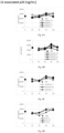
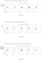
- the stability of the VSV-G+ LV is shown in Fig. 7 .
- the vectors were stable over the pH range from 4.5 to 8.0 and can be kept at those pH ranges up to 120 mins
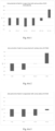
- Figs. 8A-8H show the results of an experiment to test the stability of bald LV particles in the different pH buffers without polymer encapsulation.
- integrity was assessed by measuring their ability to transduce HEK293T cells and cause GFP expression (detected by FACS).
- integrity was assessed by measuring release of p24 protein (an indicator of lysis of the vector particles; p24 was measured in supernatants by ELISA), since no transduction was possible using non-polymer-coated bald LV.
- Physical integrity was determined after 15, 30, 60, 90 and 120 minutes in the different pH buffers.
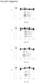
- Fig. 9 shows the results for encapsulation of bald LV at different pH values, followed by transduction of HEK293T cells and GFP expression detected by FACS.
- complete polymer coating of the bald LV, resulting in transduction-capable LV required at least about 10 9 PBAE molecules per LV particle in order to obtain viable particles.
- a clear pH dependence was observed for the coating process. Higher pH (pH 7) gave practically no infectious LV, whereas coating at pH in the range from about 5.0 to about 6.5 gave good transduction results.
- PBAE derivative (PBAE conjugated to CRRR peptides mixed with PBAE-CHHH at a ratio of 60/40 vol/vol, see Example 1) was used for coating in this experiment.
- Fig. 9 also shows that partial encapsulation by polymer results in a lack of transduction capability, which demonstrates that the use of viral vectors lacking envelope protein will enhance the safety profile. Without this feature, vector particles whose polymer coating is lost, partially or completely, or becomes partially degraded, or vector particles that have been incompletely coated during production, could represent a safety concern due to possible transduction of non-targeted cells.
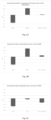
- Fig. 10 shows results of an experiment similar to that depicted in FIG. 9 , but using a mixture (60/40) of the R-cholesterol-PBAE and H-cholesterol-PBAE polymers.
- the addition of cholesterol lowered the polymer/LV particle ratio required to fully encapsulate the bald LV at low pH.
- FIG. 11 An experiment testing the coating of the VSV-G+ LV at different pH values is shown in Fig. 11 .
- the R-PBAE polymer was used for encapsulation, and the sensitivity to anti-VSV-G+ antibody also was tested.
- the results indicate little pH sensitivity to overall transduction rates, but antibody sensitivity (indicative of incomplete polymer coating) was greater at pH 6.5 than at lower pH values. Further indication of more compete coating at lower pH is seen as the higher baseline transduction in the presence of anti-VSV-G antibody at pH 5.
- Example 4 Transduction of Lymphocytes Using Encapsulated Lentiviral Vectors ⁇ VSV-G. Transduction of Jurkat Cells and Human Lymphocytes
- Jurkat cells were seeded in 24-well plates at a density of 8 ⁇ 10 4 cells per well in complete medium containing 10% FBS and 1 % penicillin/streptomycin. 300 ⁇ L of encapsulated vector was added to the cells. After 2 h incubation at 37 °C, 5 % CO 2 , 500 ⁇ L of fresh complete medium was added to each well. The percentage of cells expressing GFP or mCherry transgenes was determined 72 h post-transduction with an Attune NxT flow cytometer (Thermo Fisher) using the BL1 or YL1 channel respectively.
- Attune NxT flow cytometer Thermo Fisher
- the phenotype of transduced cells expressing GFP transgene was determined by flow cytometry staining with different antibodies specific for the following cell types following manufacturer's instructions (BD Biosciences): CD3 (T lymphocytes) and CD19 (B lymphocytes).
- the transduction of human lymphocytes was carried out with the same method except that 10 5 cells/well were seeded.
- Human lymphocytes were seeded in 24-well plates at a density of 5 ⁇ 10 5 cells per well in complete medium containing 10% FBS and 1 % penicillin/streptomycin. 300 ⁇ L of encapsulated CAR CD19 expressing vector was added to the cells. After 2 h incubation, 500 ⁇ L of fresh complete medium was added to each well. After 48 h incubation at 37 °C, 5 % CO 2 , CD19-positive Ramos tumor cells were added at lymphocyte/tumor cell ratios of 50:1 or 1:1.
- PBMCs Peripheral Blood Mononuclear Cells
- Cells were seeded in 24-well plates at a density of 2 ⁇ 10 5 cells per well in RPMI medium containing 10% FBS and 1 % penicillin/streptomycin. 300 ⁇ L of encapsulated vector was added to the cells. After 2 h incubation at 37 °C, 5 % CO 2 , 500 ⁇ L of fresh complete medium was added to each well. The percentage of cells expressing GFP was determined 48 h post-transduction with an Attune NxT flow cytometer using the BL1 channel.
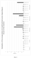
- Fig. 12 shows the GFP expression and cell viability of Jurkat cells transduced with the indicated PBAE-encapsulated LV in the presence and absence of anti-VSV-G antibody. The results are normalized to the minus antibody conditions as 100%. A wide variety of encapsulation polymers and polymer/vector ratios were not able to block inhibition by the antibody, indicating that the VSV-G protein protruded through the polymer coating under all conditions probed and mediated transduction.
- mCherry expression is shown as a measure of transduction of Jurkat cells by bald LV encapsulated by the indicated PBAE polymers, with and without cholesterol conjugation, and at a variety of different polymer/vector ratios and polymer net charge.
- Figs. 14A-14D The results of transduction of human lymphocytes using different vectors are shown in Figs. 14A-14D .
- the number of vector particles per lymphocyte increase from left to right in each figure.
- Fig. 14A shows the results obtained for typical VSV-G + LV; little transduction was observed, as was also the case for the nonencapsulated VSV-G - (bald) LV shown in Fig. 14B .
- the encapsulation of the bald LV with R/H (60/40 vol/vol), ratio 1.10e+9 PBAE per LV with or without 10E+4 PGA-Fab led to successful and dose-dependent transduction as shown in Fig. 14C .
- Fig. 14C In the experiment shown in Fig.
- Fig. 15 shows the results of experiments in which mouse PBMC were transfected with either VSV-G + nonencapsulated LV (two bars at left) or PBAE-encapsulated bald LV (two bars at right).
- the encapsulated bald LV showed much stronger transduction than did the VSV-G + vectors for each cell type present in the PBMC preparation.
Landscapes
- Health & Medical Sciences (AREA)
- Life Sciences & Earth Sciences (AREA)
- Engineering & Computer Science (AREA)
- Chemical & Material Sciences (AREA)
- Bioinformatics & Cheminformatics (AREA)
- General Health & Medical Sciences (AREA)
- Animal Behavior & Ethology (AREA)
- Public Health (AREA)
- Veterinary Medicine (AREA)
- Epidemiology (AREA)
- Genetics & Genomics (AREA)
- Biomedical Technology (AREA)
- Zoology (AREA)
- Medicinal Chemistry (AREA)
- Pharmacology & Pharmacy (AREA)
- Physics & Mathematics (AREA)
- Organic Chemistry (AREA)
- Nanotechnology (AREA)
- Molecular Biology (AREA)
- Wood Science & Technology (AREA)
- General Engineering & Computer Science (AREA)
- Biotechnology (AREA)
- Biophysics (AREA)
- Virology (AREA)
- Optics & Photonics (AREA)
- Microbiology (AREA)
- Plant Pathology (AREA)
- Biochemistry (AREA)
- Botany (AREA)
- Immunology (AREA)
- Cell Biology (AREA)
- Chemical Kinetics & Catalysis (AREA)
- General Chemical & Material Sciences (AREA)
- Nuclear Medicine, Radiotherapy & Molecular Imaging (AREA)
- Medicines That Contain Protein Lipid Enzymes And Other Medicines (AREA)
- Medicines Containing Material From Animals Or Micro-Organisms (AREA)
- Medicinal Preparation (AREA)
- Peptides Or Proteins (AREA)
- Medicines Containing Antibodies Or Antigens For Use As Internal Diagnostic Agents (AREA)
- Micro-Organisms Or Cultivation Processes Thereof (AREA)
Claims (17)
- Nanopartikel, umfassend einen viralen Vektor, der mit einem Polymer oder einem Gemisch von Polymeren beschichtet ist, wobei der virale Vektor ein Transgen umfasst und wobei der Nanopartikel als Vehikel für die Abgabe des Transgens an eukaryontische Zellen fungiert, wobei dem viralen Vektor ein Hüllprotein fehlt.
- Nanopartikel nach Anspruch 1, wobei das Polymer oder das Polymergemisch einen Poly(beta-aminoester) mit der folgenden Formel umfasst

- Nanopartikel nach Anspruch 2, wobei jedes Oligopeptid mindestens drei Aminosäurereste umfasst, die aus der Gruppe ausgewählt sind, die aus Arginin (R), Glutaminsäure (E), Lysin (K), Asparaginsäure (D), Histidin (H) und Cystein (C) besteht, wobei die Aminosäuresequenz von mindestens einem der Oligopeptide vorzugsweise CRRR (SEQ ID NO:1), oder CHHH (SEQ ID NO:2), oder CKKK (SEQ ID NO:3), oder CEEE (SEQ ID NO:4), oder CDDD (SEQ ID NO:5).
- Nanopartikel nach einem der vorhergehenden Ansprüche, wobei das Polymer oder die Polymermischung bei pH 7 eine positive Nettoladung oder eine negative Nettoladung aufweist.
- Nanopartikel nach einem der vorhergehenden Ansprüche, wobei der Nanopartikel 108 bis 1012 Polymermoleküle pro Vektor enthält, wobei der Nanopartikel vorzugsweise etwa 109 bis etwa 1010 Polymermoleküle pro Vektor enthält.
- Nanopartikel nach einem der vorhergehenden Ansprüche, wobei der virale Vektor ein retroviraler Vektor ist, wobei der retrovirale Vektor vorzugsweise ein lentiviraler Vektor ist.
- Nanopartikel nach einem der vorhergehenden Ansprüche, ferner umfassend eine oder mehrere Targeting-Einheiten, die an ein oder mehrere Polymermoleküle gebunden sind, wobei die Targeting-Einheit vorzugsweise aus der Gruppe ausgewählt ist, die aus Antikörpern, Antikörperfragmenten, scFvs, antikörperähnlichen Proteingerüsten, Oligopeptiden, Aptameren, L-RNA-Aptameren und Liganden für Zelloberflächenrezeptoren besteht; die Targeting-Einheit ist beispielsweise ein Anti-CD3-Antikörper oder ein Anti-CD3-Aptamer.
- Nanopartikel nach einem der vorhergehenden Ansprüche, wobei das Transgen für einen chimären Antigenrezeptor kodiert.
- Nanopartikel nach einem der vorhergehenden Ansprüche, das ein Zetapotenzial von etwa -15 mV bis etwa +15 mV aufweist.
- Nanopartikel nach einem der vorhergehenden Ansprüche, dem das VSV-G-Hüllprotein fehlt, wobei das Nanopartikel vorzugsweise ein verbessertes Sicherheitsprofil für die In-vivo-Verwendung im Vergleich zu einem ähnlichen Nanopartikel mit VSV-G-Hüllprotein aufweist.
- Nanopartikel nach einem der vorhergehenden Ansprüche, wobei dem viralen Vektor virales Hüllprotein fehlt und wobei das Nanopartikel nur oberhalb einer Schwellenwertzahl von Polymermolekülen pro Vektor in der Lage ist, eine Säugerzelle zu transduzieren, vorzugsweise:(i) wobei unterhalb der Schwellenwertanzahl von Polymermolekülen pro Vektor die Menge an Polymer nicht ausreicht, um den Vektor vollständig zu umhüllen, und/oder(ii) wobei unterhalb der Schwellenwertanzahl von Polymermolekülen pro Vektor das Nanopartikel strukturell instabil ist oder der Dissoziation von Polymermolekülen von dem Nanopartikel unterliegt, und/oder(iii) wobei das Nanopartikel ein verbessertes Sicherheitsprofil für die In-vivo-Verwendung im Vergleich zu einem Vektor oder polymerverkapselten Vektor aufweist, dem dieser Schwellenwert fehlt.
- Verfahren zur Herstellung eines Nanopartikels nach einem der vorhergehenden Ansprüche, wobei das Verfahren die folgenden Schritte umfasst:(a) Bereitstellen eines viralen Vektors, der ein Transgen enthält, wobei dem viralen Vektor ein Hüllprotein fehlt;(b) Bereitstellen eines Polymers oder einer Mischung von Polymeren und gegebenenfalls einer oder mehrerer Zielgruppeneinheiten und(c) In-Kontakt-Bringen des viralen Vektors und des Polymers oder des Polymergemischs und gegebenenfalls des einen oder der mehreren Targeting-Anteile, wodurch sich der virale Vektor und das Polymer oder das Polymergemisch zur Bildung des Nanopartikels verbinden.
- Verfahren nach Anspruch 12, wobei:(i) Schritt (c) bei einem pH-Wert von etwa 5,0 bis etwa 6,5, vorzugsweise bei einem pH-Wert von etwa 5,5 bis 6,0, durchgeführt wird, oder Schritt (c) bei einem pH-Wert durchgeführt wird, bei dem das Polymer oder das Gemisch von Polymeren eine positive Nettoladung aufweist und der retrovirale Vektor ein negatives Oberflächenpotential hat, und/oder(ii) das Polymer oder die Polymermischung einen Poly(beta-aminoester) mit der Formel
 (iii) der retrovirale Vektor ein retroviraler Vektor, wie z.B. ein lentiviraler Vektor, ist und/oder(iv) das Verfahren ferner das In-Kontakt-Bringen des Vektors mit einer oder mehreren zielgerichteten Einheiten umfasst, wobei die zielgerichteten Einheiten gegebenenfalls mit dem Polymer oder dem Partikelgemisch verbunden sind.
 - Nanopartikel nach einem der Ansprüche 1 bis 11 zur Verwendung in einem Verfahren zur Behandlung einer Krankheit, wie z.B. Krebs, wobei das Verfahren die Verabreichung einer Zusammensetzung, die eine Vielzahl der Nanopartikel umfasst, an ein Subjekt, das diese benötigt, umfasst, wodurch Zellen des Subjekts durch den retroviralen Vektor transduziert werden und das Transgen in den transduzierten Zellen exprimiert wird.
- Verfahren zur Transduktion von Zellen in vitro, wobei das Verfahren das In-Kontakt-Bringen von kultivierten Zellen mit einer Vielzahl von Nanopartikeln nach einem der Ansprüche 1 bis 11 umfasst, wodurch mindestens einige der kultivierten Zellen durch den retroviralen Vektor transduziert werden und das Transgen spezifisch in den transduzierten Zellen exprimiert wird, wobei die Nanopartikel vorzugsweise auf CD3-positive Zellen abzielen und das Transgen spezifisch in CD3-positiven Zellen exprimiert wird, oder wobei das Transgen für einen chimären Antigenrezeptor kodiert.
- Nanopartikel nach einem der Ansprüche 1 bis 11 zur Verwendung in einem Verfahren zur Durchführung einer Gentherapie, wobei das Verfahren das In-Kontakt-Bringen von Zellen in vitro oder innerhalb eines lebenden Organismus mit einer Vielzahl der Nanopartikel umfasst, wodurch das Transgen in den Zellen exprimiert wird.
- Nanopartikel, umfassend einen viralen Vektor, der mit einem Polymer oder einem Gemisch von Polymeren beschichtet ist, wobei der virale Vektor einen reduzierten Gehalt an Hüllprotein und ein Transgen umfasst, wobei das Nanopartikel als Vehikel für die Abgabe des Transgens an eukaryontische Zellen fungiert, und wobei der reduzierte Gehalt an Hüllprotein nicht ausreicht, um den zellulären Eintritt des Vektors und die Transduktion der Zellen durch den Vektor zu fördern, wobei der reduzierte Gehalt an Hüllprotein vorzugsweise 5 % oder weniger der Menge an Hüllprotein in einem funktionalen Virus beträgt, von dem der Vektor abgeleitet wurde.
Applications Claiming Priority (4)
| Application Number | Priority Date | Filing Date | Title |
|---|---|---|---|
| US201862618156P | 2018-01-17 | 2018-01-17 | |
| US201862661990P | 2018-04-24 | 2018-04-24 | |
| US201862715007P | 2018-08-06 | 2018-08-06 | |
| PCT/IB2019/000361 WO2019145796A2 (en) | 2018-01-17 | 2019-01-17 | Polymer-encapsulated viral vectors for genetic therapy |
Publications (2)
| Publication Number | Publication Date |
|---|---|
| EP3740199A2 EP3740199A2 (de) | 2020-11-25 |
| EP3740199B1 true EP3740199B1 (de) | 2024-08-14 |
Family
ID=67394531
Family Applications (1)
| Application Number | Title | Priority Date | Filing Date |
|---|---|---|---|
| EP19743691.8A Active EP3740199B1 (de) | 2018-01-17 | 2019-01-17 | Polymerverkapselte virale vektoren für die gentherapie |
Country Status (14)
| Country | Link |
|---|---|
| US (1) | US20200360297A1 (de) |
| EP (1) | EP3740199B1 (de) |
| JP (2) | JP2021510548A (de) |
| KR (1) | KR102812046B1 (de) |
| CN (1) | CN111902134A (de) |
| AU (2) | AU2019213290A1 (de) |
| BR (1) | BR112020014650A2 (de) |
| CA (1) | CA3088933A1 (de) |
| DK (1) | DK3740199T3 (de) |
| ES (1) | ES2991922T3 (de) |
| FI (1) | FI3740199T3 (de) |
| MX (1) | MX2021001423A (de) |
| PL (1) | PL3740199T3 (de) |
| WO (1) | WO2019145796A2 (de) |
Families Citing this family (6)
| Publication number | Priority date | Publication date | Assignee | Title |
|---|---|---|---|---|
| EP4031185A2 (de) | 2019-09-21 | 2022-07-27 | Ixaka France | Dmso-freie synthese von oligopeptid-modifizierten poly(beta-aminoestern) und deren verwendung in nanoteilchenabgabesystemen |
| EP4058585A1 (de) * | 2019-11-15 | 2022-09-21 | Ixaka France | Polymerverkapselte virale vektoren für die in-vivo-gentherapie |
| WO2021234455A2 (en) | 2020-05-19 | 2021-11-25 | Ixaka France | Promoter sequences for in vitro and in vivo expression of gene therapy products in cd3+ cells |
| EP4634393A1 (de) * | 2022-12-16 | 2025-10-22 | Lilium Therapeutics Inc. | Virale vektoren mit manipulierter funktion und verfahren zur herstellung davon |
| WO2024201067A1 (en) * | 2023-03-31 | 2024-10-03 | Dawn Therapeutics Limited | Transfection system |
| WO2024233882A1 (en) * | 2023-05-11 | 2024-11-14 | Alaya.Bio Inc. | Extracellular vesicles for the delivery of payloads to eukaryotic cells |
Family Cites Families (10)
| Publication number | Priority date | Publication date | Assignee | Title |
|---|---|---|---|---|
| AU2013358922B2 (en) * | 2012-12-14 | 2019-08-22 | The Regents Of The University Of California | Viral vector nanocapsule for targeting gene therapy and its preparation |
| GB201304245D0 (en) * | 2013-03-08 | 2013-04-24 | Inst Quimic De Sarria | Chemical compounds |
| WO2014153114A1 (en) | 2013-03-14 | 2014-09-25 | Fred Hutchinson Cancer Research Center | Compositions and methods to modify cells for therapeutic objectives |
| WO2015069469A1 (en) * | 2013-11-05 | 2015-05-14 | Clontech Laboratories, Inc. | Dry transfection compositions and methods for making and using the same |
| US10335500B2 (en) * | 2014-05-12 | 2019-07-02 | The Johns Hopkins University | Highly stable biodegradable gene vector platforms for overcoming biological barriers |
| EP3511413B1 (de) | 2014-07-25 | 2022-09-07 | Theravectys | Lentivirale vektoren zur regulierten expression eines chimären antigenrezeptormoleküls |
| WO2016019126A1 (en) * | 2014-07-30 | 2016-02-04 | The Research Foundation For The State University Of New York | System and method for delivering genetic material or protein to cells |
| WO2016022947A1 (en) * | 2014-08-07 | 2016-02-11 | The Johns Hopkins University | Nanoparticle modification of human adipose-derived mesenchymal stem cells for treating brain cancer and other neurological diseases |
| GB201501025D0 (en) | 2015-01-21 | 2015-03-04 | Sagetis Biotech Sl | Chemical compounds |
| GB201708203D0 (en) * | 2017-05-22 | 2017-07-05 | Sagetis Biotech Sl | Chemical compounds |
-
2019
- 2019-01-17 FI FIEP19743691.8T patent/FI3740199T3/fi active
- 2019-01-17 JP JP2020560623A patent/JP2021510548A/ja active Pending
- 2019-01-17 PL PL19743691.8T patent/PL3740199T3/pl unknown
- 2019-01-17 CA CA3088933A patent/CA3088933A1/en active Pending
- 2019-01-17 EP EP19743691.8A patent/EP3740199B1/de active Active
- 2019-01-17 US US16/962,686 patent/US20200360297A1/en active Pending
- 2019-01-17 BR BR112020014650-1A patent/BR112020014650A2/pt unknown
- 2019-01-17 KR KR1020207023176A patent/KR102812046B1/ko active Active
- 2019-01-17 AU AU2019213290A patent/AU2019213290A1/en not_active Abandoned
- 2019-01-17 WO PCT/IB2019/000361 patent/WO2019145796A2/en not_active Ceased
- 2019-01-17 ES ES19743691T patent/ES2991922T3/es active Active
- 2019-01-17 DK DK19743691.8T patent/DK3740199T3/da active
- 2019-01-17 CN CN201980020095.4A patent/CN111902134A/zh active Pending
-
2020
- 2020-07-16 MX MX2021001423A patent/MX2021001423A/es unknown
-
2023
- 2023-11-17 JP JP2023195952A patent/JP2024020452A/ja active Pending
-
2024
- 2024-11-11 AU AU2024259893A patent/AU2024259893A1/en active Pending
Also Published As
| Publication number | Publication date |
|---|---|
| CN111902134A (zh) | 2020-11-06 |
| WO2019145796A3 (en) | 2019-11-07 |
| KR20200128520A (ko) | 2020-11-13 |
| FI3740199T3 (fi) | 2024-11-11 |
| BR112020014650A2 (pt) | 2021-02-17 |
| MX2021001423A (es) | 2021-11-17 |
| EP3740199A2 (de) | 2020-11-25 |
| WO2019145796A2 (en) | 2019-08-01 |
| AU2024259893A1 (en) | 2024-11-28 |
| US20200360297A1 (en) | 2020-11-19 |
| PL3740199T3 (pl) | 2024-12-16 |
| AU2019213290A1 (en) | 2020-08-06 |
| JP2024020452A (ja) | 2024-02-14 |
| ES2991922T3 (es) | 2024-12-05 |
| KR102812046B1 (ko) | 2025-05-23 |
| JP2021510548A (ja) | 2021-04-30 |
| DK3740199T3 (da) | 2024-11-11 |
| CA3088933A1 (en) | 2019-08-01 |
Similar Documents
| Publication | Publication Date | Title |
|---|---|---|
| EP3740199B1 (de) | Polymerverkapselte virale vektoren für die gentherapie | |
| ES2997865T3 (en) | Adapter-based retroviral vector system for the selective transduction of target cells | |
| ES2905250T3 (es) | Transducción retroviral mediante el uso de poloxámeros | |
| KR20230006819A (ko) | 표적화된 지질 입자 및 이의 조성물 및 용도 | |
| ES2907486T3 (es) | Métodos y composiciones para reducir metástasis | |
| AU2020341454A1 (en) | CD24-associated particles and related methods and uses thereof | |
| Janssens et al. | Efficiency of onco-retroviral and lentiviral gene transfer into primary mouse and human B-lymphocytes is pseudotype dependent | |
| JP2023534924A (ja) | ウイルスフソソームを産生するための方法及び組成物 | |
| CN117467705A (zh) | 一种高效的BaEV囊膜病毒包装方法 | |
| US20220403416A1 (en) | Polymer-Encapsulated Viral Vectors for In Vivo Genetic Therapy | |
| CN113631703B (zh) | 使用泊洛沙胺的病毒传导 | |
| US20250092192A1 (en) | Ionizable polyesters, polyplexes and methods of use | |
| US20240131184A1 (en) | Compositions and methods for delivering nucleic acids to cells | |
| CN117467706A (zh) | BaEV囊膜糖蛋白及其应用 | |
| Zhang | Engineering Lentiviral Vectors for Gene Delivery and Immunization |
Legal Events
| Date | Code | Title | Description |
|---|---|---|---|
| STAA | Information on the status of an ep patent application or granted ep patent |
Free format text: STATUS: THE INTERNATIONAL PUBLICATION HAS BEEN MADE |
|
| PUAI | Public reference made under article 153(3) epc to a published international application that has entered the european phase |
Free format text: ORIGINAL CODE: 0009012 |
|
| STAA | Information on the status of an ep patent application or granted ep patent |
Free format text: STATUS: REQUEST FOR EXAMINATION WAS MADE |
|
| 17P | Request for examination filed |
Effective date: 20200731 |
|
| AK | Designated contracting states |
Kind code of ref document: A2 Designated state(s): AL AT BE BG CH CY CZ DE DK EE ES FI FR GB GR HR HU IE IS IT LI LT LU LV MC MK MT NL NO PL PT RO RS SE SI SK SM TR |
|
| AX | Request for extension of the european patent |
Extension state: BA ME |
|
| DAV | Request for validation of the european patent (deleted) | ||
| DAX | Request for extension of the european patent (deleted) | ||
| STAA | Information on the status of an ep patent application or granted ep patent |
Free format text: STATUS: EXAMINATION IS IN PROGRESS |
|
| REG | Reference to a national code |
Ref country code: HK Ref legal event code: DE Ref document number: 40041661 Country of ref document: HK |
|
| 17Q | First examination report despatched |
Effective date: 20210803 |
|
| GRAP | Despatch of communication of intention to grant a patent |
Free format text: ORIGINAL CODE: EPIDOSNIGR1 |
|
| STAA | Information on the status of an ep patent application or granted ep patent |
Free format text: STATUS: GRANT OF PATENT IS INTENDED |
|
| INTG | Intention to grant announced |
Effective date: 20220119 |
|
| GRAS | Grant fee paid |
Free format text: ORIGINAL CODE: EPIDOSNIGR3 |
|
| 19U | Interruption of proceedings before grant |
Effective date: 20200603 |
|
| 19W | Proceedings resumed before grant after interruption of proceedings |
Effective date: 20231201 |
|
| RAP3 | Party data changed (applicant data changed or rights of an application transferred) |
Owner name: IXAKA FRANCE |
|
| GRAA | (expected) grant |
Free format text: ORIGINAL CODE: 0009210 |
|
| STAA | Information on the status of an ep patent application or granted ep patent |
Free format text: STATUS: THE PATENT HAS BEEN GRANTED |
|
| AK | Designated contracting states |
Kind code of ref document: B1 Designated state(s): AL AT BE BG CH CY CZ DE DK EE ES FI FR GB GR HR HU IE IS IT LI LT LU LV MC MK MT NL NO PL PT RO RS SE SI SK SM TR |
|
| REG | Reference to a national code |
Ref country code: GB Ref legal event code: FG4D |
|
| REG | Reference to a national code |
Ref country code: CH Ref legal event code: EP |
|
| REG | Reference to a national code |
Ref country code: DE Ref legal event code: R096 Ref document number: 602019056994 Country of ref document: DE |
|
| REG | Reference to a national code |
Ref country code: IE Ref legal event code: FG4D |
|
| REG | Reference to a national code |
Ref country code: FI Ref legal event code: FGE Ref country code: DK Ref legal event code: T3 Effective date: 20241104 |
|
| REG | Reference to a national code |
Ref country code: NL Ref legal event code: FP |
|
| REG | Reference to a national code |
Ref country code: SE Ref legal event code: TRGR |
|
| REG | Reference to a national code |
Ref country code: ES Ref legal event code: FG2A Ref document number: 2991922 Country of ref document: ES Kind code of ref document: T3 Effective date: 20241205 |
|
| REG | Reference to a national code |
Ref country code: LT Ref legal event code: MG9D |
|
| PG25 | Lapsed in a contracting state [announced via postgrant information from national office to epo] |
Ref country code: PT Free format text: LAPSE BECAUSE OF FAILURE TO SUBMIT A TRANSLATION OF THE DESCRIPTION OR TO PAY THE FEE WITHIN THE PRESCRIBED TIME-LIMIT Effective date: 20241216 Ref country code: GR Free format text: LAPSE BECAUSE OF FAILURE TO SUBMIT A TRANSLATION OF THE DESCRIPTION OR TO PAY THE FEE WITHIN THE PRESCRIBED TIME-LIMIT Effective date: 20241115 |
|
| PG25 | Lapsed in a contracting state [announced via postgrant information from national office to epo] |
Ref country code: BG Free format text: LAPSE BECAUSE OF FAILURE TO SUBMIT A TRANSLATION OF THE DESCRIPTION OR TO PAY THE FEE WITHIN THE PRESCRIBED TIME-LIMIT Effective date: 20240814 |
|
| PG25 | Lapsed in a contracting state [announced via postgrant information from national office to epo] |
Ref country code: LV Free format text: LAPSE BECAUSE OF FAILURE TO SUBMIT A TRANSLATION OF THE DESCRIPTION OR TO PAY THE FEE WITHIN THE PRESCRIBED TIME-LIMIT Effective date: 20240814 |
|
| PG25 | Lapsed in a contracting state [announced via postgrant information from national office to epo] |
Ref country code: IS Free format text: LAPSE BECAUSE OF FAILURE TO SUBMIT A TRANSLATION OF THE DESCRIPTION OR TO PAY THE FEE WITHIN THE PRESCRIBED TIME-LIMIT Effective date: 20241214 |
|
| PG25 | Lapsed in a contracting state [announced via postgrant information from national office to epo] |
Ref country code: HR Free format text: LAPSE BECAUSE OF FAILURE TO SUBMIT A TRANSLATION OF THE DESCRIPTION OR TO PAY THE FEE WITHIN THE PRESCRIBED TIME-LIMIT Effective date: 20240814 |
|
| PG25 | Lapsed in a contracting state [announced via postgrant information from national office to epo] |
Ref country code: RS Free format text: LAPSE BECAUSE OF FAILURE TO SUBMIT A TRANSLATION OF THE DESCRIPTION OR TO PAY THE FEE WITHIN THE PRESCRIBED TIME-LIMIT Effective date: 20241114 |
|
| PG25 | Lapsed in a contracting state [announced via postgrant information from national office to epo] |
Ref country code: RS Free format text: LAPSE BECAUSE OF FAILURE TO SUBMIT A TRANSLATION OF THE DESCRIPTION OR TO PAY THE FEE WITHIN THE PRESCRIBED TIME-LIMIT Effective date: 20241114 Ref country code: PT Free format text: LAPSE BECAUSE OF FAILURE TO SUBMIT A TRANSLATION OF THE DESCRIPTION OR TO PAY THE FEE WITHIN THE PRESCRIBED TIME-LIMIT Effective date: 20241216 Ref country code: LV Free format text: LAPSE BECAUSE OF FAILURE TO SUBMIT A TRANSLATION OF THE DESCRIPTION OR TO PAY THE FEE WITHIN THE PRESCRIBED TIME-LIMIT Effective date: 20240814 Ref country code: IS Free format text: LAPSE BECAUSE OF FAILURE TO SUBMIT A TRANSLATION OF THE DESCRIPTION OR TO PAY THE FEE WITHIN THE PRESCRIBED TIME-LIMIT Effective date: 20241214 Ref country code: HR Free format text: LAPSE BECAUSE OF FAILURE TO SUBMIT A TRANSLATION OF THE DESCRIPTION OR TO PAY THE FEE WITHIN THE PRESCRIBED TIME-LIMIT Effective date: 20240814 Ref country code: GR Free format text: LAPSE BECAUSE OF FAILURE TO SUBMIT A TRANSLATION OF THE DESCRIPTION OR TO PAY THE FEE WITHIN THE PRESCRIBED TIME-LIMIT Effective date: 20241115 Ref country code: BG Free format text: LAPSE BECAUSE OF FAILURE TO SUBMIT A TRANSLATION OF THE DESCRIPTION OR TO PAY THE FEE WITHIN THE PRESCRIBED TIME-LIMIT Effective date: 20240814 |
|
| PGFP | Annual fee paid to national office [announced via postgrant information from national office to epo] |
Ref country code: NL Payment date: 20250117 Year of fee payment: 7 |
|
| PGFP | Annual fee paid to national office [announced via postgrant information from national office to epo] |
Ref country code: DE Payment date: 20250127 Year of fee payment: 7 |
|
| PG25 | Lapsed in a contracting state [announced via postgrant information from national office to epo] |
Ref country code: RO Free format text: LAPSE BECAUSE OF FAILURE TO SUBMIT A TRANSLATION OF THE DESCRIPTION OR TO PAY THE FEE WITHIN THE PRESCRIBED TIME-LIMIT Effective date: 20240814 Ref country code: SM Free format text: LAPSE BECAUSE OF FAILURE TO SUBMIT A TRANSLATION OF THE DESCRIPTION OR TO PAY THE FEE WITHIN THE PRESCRIBED TIME-LIMIT Effective date: 20240814 |
|
| PGFP | Annual fee paid to national office [announced via postgrant information from national office to epo] |
Ref country code: FI Payment date: 20250121 Year of fee payment: 7 |
|
| PGFP | Annual fee paid to national office [announced via postgrant information from national office to epo] |
Ref country code: ES Payment date: 20250213 Year of fee payment: 7 |
|
| PGFP | Annual fee paid to national office [announced via postgrant information from national office to epo] |
Ref country code: IE Payment date: 20250129 Year of fee payment: 7 Ref country code: SE Payment date: 20250124 Year of fee payment: 7 |
|
| PGFP | Annual fee paid to national office [announced via postgrant information from national office to epo] |
Ref country code: NO Payment date: 20250128 Year of fee payment: 7 |
|
| PG25 | Lapsed in a contracting state [announced via postgrant information from national office to epo] |
Ref country code: EE Free format text: LAPSE BECAUSE OF FAILURE TO SUBMIT A TRANSLATION OF THE DESCRIPTION OR TO PAY THE FEE WITHIN THE PRESCRIBED TIME-LIMIT Effective date: 20240814 |
|
| PGFP | Annual fee paid to national office [announced via postgrant information from national office to epo] |
Ref country code: AT Payment date: 20250127 Year of fee payment: 7 Ref country code: CH Payment date: 20250201 Year of fee payment: 7 Ref country code: BE Payment date: 20250128 Year of fee payment: 7 |
|
| PGFP | Annual fee paid to national office [announced via postgrant information from national office to epo] |
Ref country code: CZ Payment date: 20250117 Year of fee payment: 7 Ref country code: FR Payment date: 20250124 Year of fee payment: 7 Ref country code: PL Payment date: 20250117 Year of fee payment: 7 |
|
| PG25 | Lapsed in a contracting state [announced via postgrant information from national office to epo] |
Ref country code: SK Free format text: LAPSE BECAUSE OF FAILURE TO SUBMIT A TRANSLATION OF THE DESCRIPTION OR TO PAY THE FEE WITHIN THE PRESCRIBED TIME-LIMIT Effective date: 20240814 |
|
| PGFP | Annual fee paid to national office [announced via postgrant information from national office to epo] |
Ref country code: IT Payment date: 20250127 Year of fee payment: 7 Ref country code: GB Payment date: 20250127 Year of fee payment: 7 |
|
| REG | Reference to a national code |
Ref country code: DE Ref legal event code: R097 Ref document number: 602019056994 Country of ref document: DE |
|
| PLBE | No opposition filed within time limit |
Free format text: ORIGINAL CODE: 0009261 |
|
| STAA | Information on the status of an ep patent application or granted ep patent |
Free format text: STATUS: NO OPPOSITION FILED WITHIN TIME LIMIT |
|
| 26N | No opposition filed |
Effective date: 20250515 |
|
| REG | Reference to a national code |
Ref country code: DK Ref legal event code: EBP Effective date: 20250131 |
|
| PG25 | Lapsed in a contracting state [announced via postgrant information from national office to epo] |
Ref country code: MC Free format text: LAPSE BECAUSE OF FAILURE TO SUBMIT A TRANSLATION OF THE DESCRIPTION OR TO PAY THE FEE WITHIN THE PRESCRIBED TIME-LIMIT Effective date: 20240814 Ref country code: LU Free format text: LAPSE BECAUSE OF NON-PAYMENT OF DUE FEES Effective date: 20250117 |