EP3674414A1 - Procédés permettant d'étudier des acides nucléiques - Google Patents
Procédés permettant d'étudier des acides nucléiques Download PDFInfo
- Publication number
- EP3674414A1 EP3674414A1 EP19205821.2A EP19205821A EP3674414A1 EP 3674414 A1 EP3674414 A1 EP 3674414A1 EP 19205821 A EP19205821 A EP 19205821A EP 3674414 A1 EP3674414 A1 EP 3674414A1
- Authority
- EP
- European Patent Office
- Prior art keywords
- chromatin
- dna
- nucleic acid
- beads
- transposase
- Prior art date
- Legal status (The legal status is an assumption and is not a legal conclusion. Google has not performed a legal analysis and makes no representation as to the accuracy of the status listed.)
- Withdrawn
Links
Images


Classifications
-
- C—CHEMISTRY; METALLURGY
- C40—COMBINATORIAL TECHNOLOGY
- C40B—COMBINATORIAL CHEMISTRY; LIBRARIES, e.g. CHEMICAL LIBRARIES
- C40B50/00—Methods of creating libraries, e.g. combinatorial synthesis
- C40B50/06—Biochemical methods, e.g. using enzymes or whole viable microorganisms
-
- C—CHEMISTRY; METALLURGY
- C07—ORGANIC CHEMISTRY
- C07K—PEPTIDES
- C07K16/00—Immunoglobulins [IGs], e.g. monoclonal or polyclonal antibodies
- C07K16/44—Immunoglobulins [IGs], e.g. monoclonal or polyclonal antibodies against material not provided for elsewhere, e.g. haptens, metals, DNA, RNA, amino acids
-
- C—CHEMISTRY; METALLURGY
- C12—BIOCHEMISTRY; BEER; SPIRITS; WINE; VINEGAR; MICROBIOLOGY; ENZYMOLOGY; MUTATION OR GENETIC ENGINEERING
- C12Q—MEASURING OR TESTING PROCESSES INVOLVING ENZYMES, NUCLEIC ACIDS OR MICROORGANISMS; COMPOSITIONS OR TEST PAPERS THEREFOR; PROCESSES OF PREPARING SUCH COMPOSITIONS; CONDITION-RESPONSIVE CONTROL IN MICROBIOLOGICAL OR ENZYMOLOGICAL PROCESSES
- C12Q1/00—Measuring or testing processes involving enzymes, nucleic acids or microorganisms; Compositions therefor; Processes of preparing such compositions
- C12Q1/68—Measuring or testing processes involving enzymes, nucleic acids or microorganisms; Compositions therefor; Processes of preparing such compositions involving nucleic acids
- C12Q1/6806—Preparing nucleic acids for analysis, e.g. for polymerase chain reaction [PCR] assay
-
- C—CHEMISTRY; METALLURGY
- C40—COMBINATORIAL TECHNOLOGY
- C40B—COMBINATORIAL CHEMISTRY; LIBRARIES, e.g. CHEMICAL LIBRARIES
- C40B50/00—Methods of creating libraries, e.g. combinatorial synthesis
- C40B50/04—Methods of creating libraries, e.g. combinatorial synthesis using dynamic combinatorial chemistry techniques
-
- C—CHEMISTRY; METALLURGY
- C40—COMBINATORIAL TECHNOLOGY
- C40B—COMBINATORIAL CHEMISTRY; LIBRARIES, e.g. CHEMICAL LIBRARIES
- C40B50/00—Methods of creating libraries, e.g. combinatorial synthesis
- C40B50/08—Liquid phase synthesis, i.e. wherein all library building blocks are in liquid phase or in solution during library creation; Particular methods of cleavage from the liquid support
-
- C—CHEMISTRY; METALLURGY
- C07—ORGANIC CHEMISTRY
- C07K—PEPTIDES
- C07K2317/00—Immunoglobulins specific features
- C07K2317/90—Immunoglobulins specific features characterized by (pharmaco)kinetic aspects or by stability of the immunoglobulin
- C07K2317/92—Affinity (KD), association rate (Ka), dissociation rate (Kd) or EC50 value
-
- C—CHEMISTRY; METALLURGY
- C12—BIOCHEMISTRY; BEER; SPIRITS; WINE; VINEGAR; MICROBIOLOGY; ENZYMOLOGY; MUTATION OR GENETIC ENGINEERING
- C12Q—MEASURING OR TESTING PROCESSES INVOLVING ENZYMES, NUCLEIC ACIDS OR MICROORGANISMS; COMPOSITIONS OR TEST PAPERS THEREFOR; PROCESSES OF PREPARING SUCH COMPOSITIONS; CONDITION-RESPONSIVE CONTROL IN MICROBIOLOGICAL OR ENZYMOLOGICAL PROCESSES
- C12Q2521/00—Reaction characterised by the enzymatic activity
- C12Q2521/30—Phosphoric diester hydrolysing, i.e. nuclease
- C12Q2521/327—RNAse, e.g. RNAseH
-
- C—CHEMISTRY; METALLURGY
- C12—BIOCHEMISTRY; BEER; SPIRITS; WINE; VINEGAR; MICROBIOLOGY; ENZYMOLOGY; MUTATION OR GENETIC ENGINEERING
- C12Q—MEASURING OR TESTING PROCESSES INVOLVING ENZYMES, NUCLEIC ACIDS OR MICROORGANISMS; COMPOSITIONS OR TEST PAPERS THEREFOR; PROCESSES OF PREPARING SUCH COMPOSITIONS; CONDITION-RESPONSIVE CONTROL IN MICROBIOLOGICAL OR ENZYMOLOGICAL PROCESSES
- C12Q2522/00—Reaction characterised by the use of non-enzymatic proteins
- C12Q2522/10—Nucleic acid binding proteins
Definitions
- the present invention provides a novel method for preparing a sequencing library and studying molecular interactions involving a nucleic acid.
- the invention relates to a method for preparing a sequencing library, the method comprising the addition of an agent binding to chromatin to a sample comprising a nucleic acid; isolating chromatin bound by said agent; addition of transposase to the isolated chromatin; isolating nucleic acid from chromatin; and obtaining a sequencing library.
- the present invention relates to a method for mapping of molecular interactions involving a nucleic acid, the method comprising the addition of an agent binding to chromatin to a sample comprising a nucleic acid; isolating chromatin bound by said agent; addition of transposase to the isolated chromatin; isolating nucleic acid from chromatin; amplification of nucleic acid; sequencing of amplified nucleic acid; and identifying molecular interactions.
- Chem-Seq The knowledge of interactions between nucleic acids and other chemical substances and/or biomolecules is of high interest for research and medicine.
- a well-known method to study protein-nucleic acid interactions is chromatin immunoprecipitation (ChIP), optionally followed by massive parallel sequencing (ChIP-seq).
- ChIP chromatin immunoprecipitation
- ChIP-seq massive parallel sequencing
- Chem-Seq A method for studying small molecule interactions with nucleic acids and/or proteins, e.g. in chromatin, is Chem-Seq, described further below.
- ChIP ChIP
- chromatin organization histone modification as well as transcription factor binding patterns (using X-ChIP) and their influence on gene regulation in health and disease; see e.g. Nature (2012) 489, pp. 57-74 or Ernst et al. (2011) Nature 473, pp. 43-49 .
- ChIP remains a relatively tedious protocol especially when applied to low-input sample (see e.g. Greenleaf, W.J. (2014) Methods ).
- the classical approach comprises several laborious steps: (i) end-repair of the purified DNA sequences to generate blunt-end double-stranded DNA fragments with a phosphorylated 3' end; (ii) addition of an A-overhang; (iii) ligation of adaptors that have a complementary T-overhang to the double-stranded and end-repaired ChIP-DNA fragments with A-overhang.
- the adapters allow amplification of the DNA fragments, which ensures sufficient amount of fragments for quality control and subsequent sequencing, and it also prepares the fragments for the sequencing procedure by introduction of flow-cell ends for cluster generation and barcode sequences to multiplex sequencing experiments.
- the classical method comes with several limitations: (i) 5-10 ng of input material is typically needed to generate libraries which cannot be recovered from ChIPs on low amounts of cells. Hence, the recommended amount of cells for a ChIP-seq experiment is in the range of 10 6 cells. (ii) The library procedure relies on several enzymatic reactions and DNA purifications, which make library generation a relatively laborious procedure. Imperfect enzymatic reactions as well as DNA purifications also lower the amount of recovered library fragments, which explains the high input requirements. (iii) Adapters can self-ligate and need to be excluded from amplification and sequencing. Hence, a size-selection is necessary to select against excess adapters and adapter-dimers.
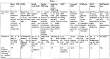
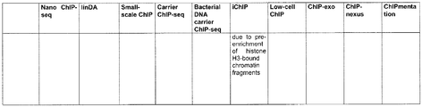
- ChIP-seq protocols for low amounts of starting material such as iChIP ( Lara-Astiaso et al. (2014) Science 345, pp. 943-9 ), linDA ( Shankaranarayanan et al. (2011) Nature Methods 8, pp. 565-7 ) and carrier-assisted ChIP ( Zwart et al. (2013) BMC Genomics 14, 232 or Jakobsen et al.
- iChIP Lara-Astiaso et al. (2014) Science 345, pp. 943-9
- linDA Shankaranarayanan et al. (2011) Nature Methods 8, pp. 565-7
- carrier-assisted ChIP Zwart et al. (2013) BMC Genomics 14, 232 or Jakobsen et al.
- Chem-seq Another method to study interactions between small molecules and their protein/nucleic acid targets in chromatin is Chem-seq.
- the method employs chemical affinity capture coupled with massively parallel DNA sequencing to identify genomic sites where small molecules interact with their target proteins or DNA. It was first described by Anders et al. in Nature Biotechnology (2013), 32(1), pp. 92-6 .
- a further method for library preparation of nucleic acids was recently described.
- the method makes use of the development of a hyperactive Tn5 transposase for simultaneous fragmentation and adapter tagging ("tagmentation") of DNA (see Adey et al. (2010) Genome Biol 11, R119 ). It uses a transposase, which is pre-loaded with sequencing-compatible adapters. The transposase integrates its adapter load into DNA while fragmenting it. Only low amounts of transposase are needed to generate libraries of genomic DNA ( Adey et al. (2010) Genome Biol 11, R119 ), bisulphite converted DNA for DNA-methylation analysis ( Wang, Q. et al.
- transposase to nucleic acid to achieve the desirable size distribution of nucleic acids for next generation sequencing or other downstream applications.
- determination of nucleic acid fragment distribution and abundance is not even feasible, hence making it impossible to find the correct ratios of transposase to nucleic acid to prepare sequencing libraries according to, inter alia , prior art applications reviewed in Furey et al. (2012) Nature Reviews Genetics 13 (12), pp. 840-852 .
- the addition of transposase to cell nuclei recovers regions of open chromatin and delivers information of nucleosome positioning as well as transcription factor footprints in regulatory regions of the genome ( Buenrostro JD. et al. (2013) Nat Meth, vol. 10 (12) pp. 1213-1218 ).
- transposase was not described systematically to be suitable for use in the generation of sequencing libraries from nucleic acids subsequent to ChIP or Chem-Seq. Rather, potential disadvantages of this approach are discussed. These disadvantages result from performing tagmentation on purified DNA subsequent to ChIP, as it is described in WO 2013/078470 or WO 2014/205296 . Accordingly, major drawbacks of the combination of ChIP and tagmentation are: (1) The ChlPed DNA, which is already sonicated to small fragments (200-700bp), is in its entirety further fragmented. Hence, tagmentation can result in very small library fragments to a minimal size down to ⁇ 40 bp ( Adey et al.
- a 600 bp immunoprecipitated DNA fragment can yield twice the amounts of library fragments as compared with a 300 bp fragment, thereby artificially increasing the relative amount of reads in the 600 bp region. This can be problematic for correct peak calling when analyzing ChIP-seq data; (3) the approach to use purified ChIP DNA to generate sequencing libraries by tagmentation is inconvenient as correct size determination and DNA quantification are needed to set up the tagmentation reaction.
- TAM-ChIP makes use of Tn5 transposase conjugated to antibodies for ChIP.
- TAM-ChIP the limiting factor of TAM-ChIP, as also described in WO 2014/190214 , is the limitation to the use of antibody-oligonucleotide conjugates that have to be produced prior to application. This prevents the ad hoc usage of commercially available antibodies that are primarily used to study protein-DNA interactions with chromatin immunoprecipitation. Even if secondary antibody-oligonucleotide conjugates were used in TAM-ChIP to overcome the above limitations, two sets of antibodies need to be used, which increases complexity of the assay while at the same time increasing costs due to the use of a secondary antibody that is normally not used in applications such as CHIP.
- TAM-ChIP requires extensive optimizations of antibody-oligonucleotide-transposase-complexes to input chromatin ratios, as described in WO 2013/078470 .
- the amount of antibody-oligonucleotide-transposase complexes recruited to their recognition site determines the final library size, and because the number of recognition sites can vary from a few hundred to hundred thousand dependent on the target antigens, the ratios of antibody-oligonucleotide-transposase complexes to input chromatin has to be evaluated for each specific antibody-transposase conjugate.
- a more robust method insensitive with regards to rations of transposase to input chromatin is required.
- TAM-ChIP requires large amounts of input chromatin.
- successful sequencing library preparation yielding sequencing results compareable to standard ChIP-seq can only demonstrated using 10 pg of input chromatin, which corresponds to ⁇ 1.5 Mio cells.
- the invention relates to the following items:
- the invention provides for a method for preparing a sequencing library, the method comprising addition of an agent binding to chromatin to a sample comprising a nucleic acid; isolating chromatin bound by said agent; addition of transposase to isolated chromatin; isolating nucleic acid from chromatin; and obtaining a sequencing library.
- the invention provides a method for preparing a sequencing library and a method for mapping of molecular interactions involving nucleic acid, in particular DNA.
- the methods as provided herein comprise in particular the preparation of a sequencing library or the mapping of molecular interactions involving nucleic acid, in particular DNA, by combining steps of adding an agent binding to chromatin to a sample, isolating bound chromatin and adding a transposase to the isolated chromatin in a specific order.
- the invention provides a method for preparing a sequencing library, the method comprising the addition of an agent binding to chromatin to a sample comprising a nucleic acid; isolating chromatin bound by said agent; addition of transposase to isolated chromatin; isolating nucleic acid from chromatin; and obtaining sequencing library.
- the addition of transposase is to be done subsequent to isolating bound chromatin. It is preferred that the nucleic acid is DNA.
- the methods of the invention for mapping of molecular interactions involving nucleic acid comprise the addition of an agent binding to chromatin to a sample comprising a nucleic acid; isolating chromatin bound by said agent; addition of transposase to isolated chromatin; isolating nucleic acid from chromatin; amplification of nucleic acid; sequencing of amplified nucleic acid; and identifying molecular interactions.
- the sample comprising a nucleic acid may be a primary cell sample or a sample obtained by a culturing method.
- the methods further comprise cultivating and harvesting cells; fixing cells; lysing cells and thereby obtaining a first sample comprising a nucleic acid; and sonicating the first sample and thereby obtaining a second sample comprising a nucleic acid.
- said second sample is used in the methods of the invention.
- said nucleic acid is DNA, in particular double-stranded DNA.
- the methods of the invention preferably further comprise fixing cells; lysing cells and thereby obtaining a first sample comprising a nucleic acid; and sonicating the first sample and thereby obtaining a second sample comprising a nucleic acid.
- the present invention preferably relates to a method comprising the addition of an agent binding to chromatin to a sample comprising a nucleic acid; isolating chromatin bound by said agent; addition of transposase to isolated chromatin subsequent to isolating chromatin bound by the agent binding to chromatin; isolating nucleic acid from chromatin; and obtaining sequencing library.
- the nucleic acid is DNA.
- the methods of the invention for mapping of molecular interactions involving nucleic acid preferably comprise the addition of an agent binding to chromatin to a sample comprising a nucleic acid; isolating chromatin bound by said agent; addition of transposase to isolated chromatin subsequent to isolating chromatin bound by the agent binding to chromatin; isolating nucleic acid from chromatin; amplification of nucleic acid; sequencing of amplified nucleic acid; and identifying molecular interactions.
- the standard ChIP-seq protocol comprises the steps of fixation of cells, cell lysis, sonication of chromatin and immunoprecipitation with a specific antibody bound to beads. Reverse-crosslinking is followed by purification of ChIP DNA, which is then subjected to library preparation in a multi-step procedure comprising end repair, purification, A-tailing, adapter ligation and size selection.
- a method called ChIP-tagmentation was found.
- ChIP-tagmentation the purified ChIP DNA is used for tagmentation-based library preparation (see Figure 11 ).
- the method is sensitive to varying DNA concentrations, because tagmentation of purified DNA is sensitive to the ratio of tagmentation enzyme to DNA, and DNA concentrations can be highly variable and too low to quantify in many applications of ChIP-seq.
- the improved robustness of the methods of the invention is achieved by performing tagmentation directly on agent-bound chromatin, which is isolated from unbound chromatin, where proteins protect the nucleic acid from excessive tagmentation.
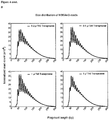
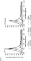
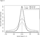
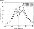
- the resulting protocol of the methods of the invention proved to be highly robust over a 25-fold difference in tagmentation enzyme concentrations, in terms of size distribution of libraries ( Figure 3 ), size distribution of sequencing reads ( Figure 4b ), mapping performance ( Figure 4c ), track quality ( Figure 4d ), and concordance ( Figure 4e ).
- the methods of the invention are also improved in that they do not give rise to sequencing adapter dimers and do not require any nucleic acid purification steps beyond the standard cleanup after the standard ChIP protocol.
- the above advantages of the methods provided herein render them superior vis-à-vis methods known in the prior art.
- the methods provided herein are more flexible, cheaper, more robust, require lower amounts of sample input and allow obtainment of additional information vis-à-vis methods known in the prior art, in particular TAM-ChIP as provided in WO 2014/190214 .
- TAM-ChIP specific antibodiy-oligonucleotide-conjugates or antibody-oligonucleotide-transposase-conjugates are required for transposase-mediated sequencing library preparations.
- commercially available ChIP-seq antibodies can be used ad hoc without laborsome and cost-intensive conjugation reactions.
- TAM-ChIP protocol as described in WO 2014/190214 requires extensive optimization of ratios of antibodiy-oligonucleotide-transposase-conjugates to input chromatin, whereas the methods of the present invention are robust to varying transposase-to-chromatin ratios.
- transposase subsequent to the isolation of the chromatin of interest has further benefits over methods known in the art.
- TAM-ChIP requires determination of optimized ratios of antibody-oligonucleotide-conjugates to target them to their recognition sites in chromatin.
- nucleic acids in immediate proximity of the recognition sites can be tagged, resulting in a relatively low tagging frequency (0.5-5%) when a transposase is used.
- the methods of the present invention allow tagmentation irrespective of the agent used to isolate the chromatin of interest.
- transposase-chromatin ratios an excess amount of transposase can be used in the methods of the invention to maximize the efficiency of sequencing library generation. This achievement substantially lowers input requirements in the methods of the present invention compared to methods known in the art.
- transposase subsequent to isolating the chromatin of interest, unexpectedly allows efficient sequencing library preparation using low amounts of transposase enzyme. This is due to the reduced presence of unspecific template chromatin for the tagmentation reaction due to the isolation of the chromatin of interest while the remainder is discarded.
- the reduction of required transposase amounts is a significant cost-advantage of the methods of the invention over methods known in the art, in particular the methods described in WO 2014/205296 and WO 2014/190214 .
- the methods of the present invention allow the construction and amplification of sequencing libraries from chromatin to study molecular interactions without prior purification or extraction of nucleic acids, in particular as in the ultra-fast method provided herein.
- the methods of the invention for the first time feasibly allow large-scale chromatin accessibility mapping in disease, in particular cancer, cohorts and clinical research by providing a streamlined, low-input workflow for genome-wide mapping of histone marks and transcription factors.
- chromatin profiling assay provided herein is sufficiently fast and straightforward for use in a clinical sequencing laboratory, chromatin deregulation is now tractable as a source of biomarkers for example for stratified cancer therapy; see also Rendeiro et al. (2016) Nature Comm. 7, Article number 11938 .
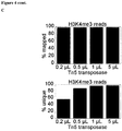
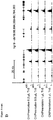
- the methods of the invention were validated for five exemplary histone marks (H3K4me3, H3K27ac, H3K4me1, H3K36me3, and H3K27me3) and four transcription factors (PU.1, CTCF, GATA1, and REST).
- H3K4me3, H3K27ac, H3K4me1, H3K36me3, and H3K27me3 The methods of the invention showed a similar data validity as compared to standard ChIP-Seq ( Figure 5f).
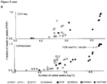
- the methods of the invention allowed the significant reduction of cell input.
- high-quality data was obtained for H3K4me3 and H3K27me3 as well as for GATA1 and CTCF from 10k and 100k cells, respectively, without any pre-amplification ( Figure 4f ).
- the methods provided herein can use chromatin as a template to generate sequencing libraries.
- the methods of the present invention are used in a high-throughput manner, automated manner and/or parallel manner. Accordingly, it is contemplated that high-throughput facilities and/or robots used to facilitate pipetting/improve reproducibility are used to perform the methods of the invention.
- the skilled person will be well-aware of suitable means to perform the methods of the invention in a high-throughput, automated and/or parallel manner.
- multiwell-plates may be used in the methods of the invention to perform multiple experiments in a parallel manner. Such multiwell-plates may for example have 96, 384 or 1536 wells.
- Multiwell-plates may also be used in combination with robotics suitable for high-throughput experiments.
- robotics suitable for high-throughput experiments.
- Known robotic systems allow simultaneous execution of multiple experiments, which reduces time, costs and/or increases reliability of experimental data.
- the Sciclone NGS Workstation P/N SG3-31020-0300, Perkin Elmer
- the agent binding to chromatin in particular the antibody or chemical substance, is attached to magnetic beads, as described further below.
- Magnetic beads are particularly useful in high-throughput methods as they can easily be used for isolation of bead-bound particles from unbound substances. It is also envisaged that the methods of the present invention are used in combination with a microfluidic device.
- a poly(dimethylsiloxane) (PDMS) device featuring a simple microfluidic chamber may be used in combination with the methods of the present invention.
- the microfluidic chamber has one inlet and one outlet, and the outlet has an on-chip pneumatic microvalve that can be partially closed by exerting a pressure at a port.
- Magnetic beads coated with the agent binding to chromatin, in particular the antibody or the chemical substance are flowed into the microfluidic chamber and form a packed bed while the pneumatic microvalve is partially closed. Sonicated chromatin fragments are then flowed through the chamber and adsorbed onto the bead surface.
- the gaps among the beads are smaller than 2 ⁇ m and facilitate rapid and high-efficiency adsorption of target chromatin fragments under the small diffusion length.
- the beads are then washed by oscillatory washing in two different washing buffers to remove nonspecifically adsorbed chromatin fragments. Finally, the beads are flowed out of the chamber and collected for off-chip processing.
- This approach as described by Cao et al. (2015) Nature Methods (available online), in combination with the herein provided novel and inventive methods allows the further reduction of experimental time and costs.
- the combination of the herein provided methods with Drop-Seq as described by Macosko et al. (2015) Cell 161 (5) pp. 1202-14 and Klein et al. (2015) Cell 161(5) pp. 1187-2201 is also contemplated.
- the present invention relates to, inter alia, a method for preparing a sequencing library.
- a method for preparing a sequencing library comprising the addition of an agent binding to chromatin to a sample comprising a nucleic acid; isolating chromatin bound by said agent; addition of transposase to isolated chromatin; isolating nucleic acid from chromatin; and obtaining sequencing library.
- the agent binding to chromatin is a chemical substance other than an antibody
- a substantial number of small-molecule ligands, including therapeutic drugs elicit their effects by binding specific proteins associated with the genome. Mapping the global interactions of these chemical entities with chromatin in a genome-wide manner could provide insights into the mechanisms by which a small molecule influences cellular functions. Chem-seq can be utilized to investigate the genome-wide effects of therapeutic modalities and to understand the effects of drugs on nuclear architecture in various biological contexts. In a broader sense, these methods are useful to enhance understanding of the therapeutic mechanisms through which small molecules modulate the function and activity of genome-associated proteins. Through the identification of the cellular targets of a drug, it becomes possible to gain an increased understanding of the causes of side effects and toxicity in the early stages of drug development, which helps to reduce the attrition rate in development.
- Chem-seq relies on the ability to create a biotinylated version of a small molecule of interest to allow for downstream affinity capture. Chem-seq can be carried out either in vitro or in vivo.
- In vitro Chem-seq begins with the crosslinking of cultured cells in medium with formaldehyde. Cell nuclei are then harvested from the cells and their chromatin is extracted. This extract is sonicated before being incubated with streptavidin magnetic beads that are bound to a biotinylated form of our compound of interest. This provides an opportunity for the small molecule of interest to interact with its target chromatin regions. These chromatin regions are then isolated using a magnet and DNA is purified. From the DNA a library is prepared and subjected to next generation sequencing, followed by an analysis to determine regions enriched for our small molecule of interest.
- the present invention also relates to a method for preparing a sequencing library or mapping of molecular interactions comprising nucleic acid combining the Chem-Seq approach with tagmentation, as described above.
- sequencing library refers to a nucleic acid representation, wherein each nucleic acid is identifiable by, e.g., the use of an individual sequence tag. Accordingly, "obtaining sequencing library” requires a process capable of ensuring that specific adaptor sequences are added to the ends of the nucleic acid fragments to be analyzed. This preparation of nucleic acids is frequently referred to as a "sequencing library”. Most of the next generation sequencing applications require the preparation of a sequencing library, nucleic acids with specific adapters at 5' and 3' ends.
- the Illumina sequencing workflow utilizes partially complementary adaptor oligonucleotides that are used for priming the PCR amplification and introducing the specific nucleotide sequences required for cluster generation by bridge PCR and facilitating the sequencing-by-synthesis reactions.
- the resulting sequencing library of the methods of the present invention for preparing a sequencing library is suitable for use in standard sequencing applications, e.g. next generation sequencing as described further below.
- a "nucleic acid" within the meaning of the present invention is a polymer of any length composed of nucleotides, preferably having a length of more than about 50 nucleotides.
- the methods of the invention allow the preparation of a sequencing library comprising nucleic acids and/or the mapping of molecular interactions involving nucleic acid.
- the nucleic acid comprised in the starting sample of the methods of the invention preferably has a length of about 50 to about 5000 nucleotides, preferably 100 to about 1000, more preferably about 200 to about 700, even more preferably 200 to 700, most preferably 200 to 300 nucleotides.
- the starting sample is not to be confused with the nucleic acid comprised in cells used in the methods of the invention comprising culturing and harvesting cells; fixing cells; lysing cells; and sonicating.
- sonication is used to fragment the nucleic acid comprised and obtained from cells, thereby obtaining the starting sample, also referred to as second sample where the methods comprise the additional steps of culturing and harvesting cells; fixing cells; lysing cells; and sonicating.
- “nucleotides” is intended to include those moieties that contain not only the known purine and pyrimidine bases, but also other heterocyclic bases that have been modified.
- nucleotide includes those moieties that contain hapten or fluorescent labels and may contain not only conventional ribose and deoxyribose sugars, but other sugars as well.
- Modified nucleosides or nucleotides also include modifications on the sugar moiety, e.g., wherein one or more of the hydroxyl groups are replaced with halogen atoms or aliphatic groups, are functionalized as ethers, amines, or the likes.
- nucleic acids used in the methods of the invention will, however, comprise the naturally occurring pyrimidine and purine bases as deoxyribonucleotides ribonucleotides that can participate in Watson-Crick base pairing interactions.
- Naturally-occurring nucleotides include guanine, cytosine, adenine, thymine and uracil (G, C, A, T and U, respectively).
- the nucleic acid may be DNA, RNA or any other type of known nucleic acids. It is preferred that the nucleic acid is DNA, in particular double-stranded DNA.
- agent binding to chromatin includes any agent that is a member of a binding complex comprising chromatin as one binding partner.
- the agent binding to chromatin may be a polypeptide, such as a protein or fragments thereof, in particular an antibody; a nucleic acid, e.g. an oligonucleotide, polynucleotide, and the like; or a small molecule, e.g. a chemical substance.
- the agent binding to chromatin is a polypeptide having a binding domain specific for chromatin and/or further molecules binding to chromatin, in particular other polypeptides.
- agents binding to chromatin may have a methyl-CpG binding domain (MBD) recognizing chromatin.
- MBD methyl-CpG binding domain
- the agent binding to chromatin is a polypeptide, in particular an antibody binding to chromatin, wherein the antibody specifically binds to chromatin, proteins, e.g. transcription factors or histones, associated with chromatin and/or DNA.
- chromatin as used herein is a complex of macromolecules found in cells, comprising DNA, protein and/or RNA.
- the primary functions of chromatin are 1) to package DNA into a smaller volume to fit in the cell, 2) to reinforce the DNA macromolecule to allow mitosis, 3) to prevent DNA damage, and 4) to control gene expression and DNA replication.
- the primary protein components of chromatin are histones that compact the DNA.
- the structure of chromatin depends on several factors. The overall structure depends on the stage of the cell cycle. During interphase, the chromatin is structurally loose to allow access to RNA and DNA polymerases that transcribe and replicate the DNA.
- chromatin The local structure of chromatin during interphase depends on the genes present on the DNA: DNA coding genes that are actively transcribed (“turned on”) are more loosely packaged and are found associated with RNA polymerases (referred to as euchromatin) and transcription factors while DNA coding inactive genes (“turned off”) are found associated with structural proteins and are more tightly packaged (heterochromatin).
- DNA coding genes that are actively transcribed (“turned on") are more loosely packaged and are found associated with RNA polymerases (referred to as euchromatin) and transcription factors while DNA coding inactive genes (“turned off”) are found associated with structural proteins and are more tightly packaged (heterochromatin).
- Epigenetic chemical modification of the structural proteins in chromatin also alters the local chromatin structure, in particular chemical modifications of histone proteins by methylation and acetylation; see further below.
- the basic repeat element of chromatin is the nucleosome, interconnected by sections of linker DNA, a far shorter arrangement than pure DNA in solution.
- linker histone H1
- the nucleosome core particle, together with histone H1 is known as a chromatosome.
- Nucleosomes, with about 20 to 60 base pairs of linker DNA, can form, under non-physiological conditions, an approximately 10 nm "beads-on-a-string" fibre.
- the nucleosomes bind DNA nonspecifically, as required by their function in general DNA packaging. There are, however, large DNA sequence preferences that govern nucleosome positioning.
- agents binding to chromatin may bind to any part of chromatin, euchromatin or heterochromatin.
- the agents binding to chromatin may interact with DNA, RNA or proteins comprised in chromatin.
- agents binding to chromatin may interact with histones or transcription factors comprised in chromatin and/or other proteins associated with histones, transcription factors or chromatin.
- histones are highly alkaline proteins found in eukaryotic cell nuclei that package and order the DNA into structural units called nucleosomes (see above). They are the chief protein components of chromatin, acting as spools around which DNA winds, and play a role in gene regulation.
- Histones H2A, H2B, H3 and H4 are known as the core histones, while histones H1 and H5 are known as the linker histones.
- Two of each of the core histones assemble to form one octameric nucleosome core, approximately 63 Angstroms in diameter (a solenoid (DNA)-like particle).
- the linker histone H1 binds the nucleosome at the entry and exit sites of the DNA, thus locking the DNA into place and allowing the formation of higher order structure.
- the most basic such formation is the 10 nm fiber or beads on a string conformation. This involves the wrapping of DNA around nucleosomes with approximately 50 base pairs of DNA separating each pair of nucleosomes (also referred to as linker DNA).
- Higher-order structures include the 30 nm fiber (forming an irregular zigzag) and 100 nm fiber, these being the structures found in normal cells.
- the condensed chromosomes are assembled through interactions between nucleosomes and other regulatory proteins.
- the agents binding to chromatin in particular the antibody or chemical substance, may interact with histones, i.e. they may specifically bind to histones and/or bind to further polypeptides and/or chemical substances associated with histones. It is preferred that the agents binding to chromatin, in particular the antibody or chemical substance, interact directly with histones.
- known human histones include five classes H1/H5, H2A, H2B, H3 and H4.
- the class H1 includes H1F0, H1FNT, H1FOO, H1FX, HIST1H1A, HIST1H1B, HIST1H1C, HIST1H1D, HIST1H1E and HIST1H1T.
- Class H2A includes H2AFB1, H2AFB2, H2AFB3, H2AFJ, H2AFV, H2AFX, H2AFY, H2AFY2, H2AFZ, HIST1H2AA, HIST1H2AB, HIST1H2AC, HIST1H2AD, HIST1H2AE, HIST1H2AG, HIST1H2AI, HIST1H2AJ, HIST1H2AK, HIST1H2AL, HIST1H2AM, HIST2H2AA3 and HIST2H2AC.
- Class H2B includes H2BFM, H2BFS, H2BFWT, HIST1H2BA, HIST1H2BB, HIST1H2BC, HIST1H2BD, HIST1H2BE, HIST1H2BF, HIST1H2BG, HIST1H2BH, HIST1H2BI, HIST1H2BJ, HIST1H2BK, HIST1H2BL, HIST1H2BM, HIST1H2BN, HIST1H2BO and HIST2H2BE.
- Class H3 includes HISTH3A, HISTH3B, HISTH3C, HISTH3D, HISTH3E, HISTH3F, HISTH3G, HISTH3H, HISTH3I, HISTH3J, HIST2H3C and HIST3H3.
- Class H4 includes HIST1H4A, HIST1H4B, HIST1H4C, HIST1H4D, HIST1H4E, HIST1H4F, HIST1H4G, HIST1H4H, HIST1H4I, HIST1H4J, HIST1H4K, HIST1H4L and HIST4H4.
- agent binding to chromatin in particular the antibody or chemical substance, binds to histones of class H3, in particular H3.3, H3.2, H3.3A, H3.3B or H3.1.
- agent binding to chromatin in particular the antibody or chemical substance, binds to H4, H2A.Z or CENP-A (the latter two containing a histone H3 related histone fold).
- the agents binding to chromatin are specific for modified versions of the known histones.
- Histone modifications have specific meanings and consequences for genomic translation and accessibility of DNA for further binding proteins and/or other chemical substances. Consequently, it is envisaged that the methods of the invention be used for identifying regions bound by modified histones that may undergo alterations in gene expression, e.g. in diseased tissues/cells such as cancer cells.
- Known histone modifications include methylation, acetylation, propionylation, butyrylation, crotonylation, 2-hydroxyisobutyrylation, malonylation, succinylation and ribosylation.
- lysine methlyation arginine methlyation
- lysine acetylation serine/threonine/tyrosine phosphorylation.
- the addition of one, two or three methyl groups to lysine has little effect on the chemistry of the histone; methylation leaves the charge of the lysine intact and adds a minimal number of atoms so steric interactions are mostly unaffected.
- proteins containing Vietnamese, chromo or PHD domains can recognise lysine methylation with extraordinar sensitivity and differentiate mono, di and tri-methyl lysine, to the extent that, for some lysines (e.g.: H4K20) mono, di and tri-methylation have different meanings. Because of this, lysine methylation is a very informative mark and dominates the known histone modification functions. Accordingly, it is envisaged that the agents binding to chromatin are specific for lysine methylated histones and/or proteins recognizing such modified histones, e.g. proteins containing Jewish, chromo or PHD domains.
- arginine methylated histones similar reasoning as above applies, i.e. some protein domains-e.g., Vietnamese domains-can be specific for methyl arginine instead of methyl lysine.
- Arginine is known to be mono- or di-methylated, and methylation can be symmetric or asymmetric, potentially with different meanings.
- lysine acetylation addition of an acetyl group has a major chemical effect on lysine as it neutralises the positive charge. This reduces electrostatic attraction between the histone and the negatively charged DNA backbone, loosening the chromatin structure; highly acetylated histones form more accessible chromatin and tend to be associated with active transcription.
- Lysine acetylation appears to be less precise in meaning than methylation, in that histone acetyltransferases tend to act on more than one lysine; presumably this reflects the need to alter multiple lysines to have a significant effect on chromatin structure. Accordingly, it is also envisaged that the agent binding to chromatin is specific for acetylated lysine and/or proteins interacting with acetylated lysine.
- serine/threonine and/or tyrosine comprised in histones can be modified by phosphorylation. Addition of a negatively charged phosphate group can lead to major changes in protein structure, leading to the well-characterised role of phosphorylation in controlling protein function.
- Histone phosphorylation has clear functions as a post-translational modification, and binding domains such as BRCT (BRCA1 C Terminus domain) have been characterised. Therefore, it is also envisaged that such modified histones, i.e. modified by phosphorylation, be recognized by the agents binding to chromatin.
- binding domains such as BRCT (BRCA1 C Terminus domain)
- H3K4Me3 Trimethylation of H3 lysine 4
- H3K36Me3 trimethylation of H3 lysine 36
- RNA polymerase II undergoes a switch from initiating' to 'elongating', marked by a change in the phosphorylation states of the RNA polymerase II C terminal domain (CTD).
- CTD C terminal domain
- the same enzyme that phosphorylates the CTD also phosphorylates the Rad6 complex, which in turn adds a ubiquitin mark to H2B K123 (K120 in mammals).
- H2BK123Ub occurs throughout transcribed regions, but this mark is required for COMPASS to trimethylate H3K4 at promoters.
- the agent binding to chromatin in particular the antibody or chemical substance
- the agent binding to chromatin in particular the antibody or chemical substance
- This trimethylation occurs in the body of active genes and is deposited by the methyltransferase Set2.
- This protein associates with elongating RNA polymerase II, and H3K36Me3 is indicative of actively transcribed genes.
- H3K36Me3 is recognised by the Rpd3 histone deacetylase complex, which removes acetyl modifications from surrounding histones, increasing chromatin compaction and repressing spurious transcription.
- Increased chromatin compaction prevents transcription factors from accessing DNA, and reduces the likelihood of new transcription events being initiated within the body of the gene. This process therefore helps ensure that transcription is not interrupted.
- acetylation of lysine 27 of histone H3 (H3K27ac) is present at active regulatory elements as promoters and enhancers.
- an enhancer is a short (50-1500 bp) region of DNA that can be bound with proteins (activators) to activate transcription of a gene. These proteins are usually referred to as transcription factors.
- Enhancers are generally cis-acting, located up to 1 Mbp (1,000,000 bp) away from the gene and can be upstream or downstream from the start site, and either in the forward or backward direction.
- H3K27ac was described to distinguish active from poised regulatory elements. Enrichment of H3K27ac at these elements is a good indicator for expression of the associated genetic element. Accordingly, the agent binding to chromatin, in particular the antibody or chemical substance, used in the methods of the present invention may be specific for H3K27ac.
- Histone modifications may also be associated with repression of gene expression.
- H3K27Me3, H3K9Me2/3 and H4K20Me3 are known to be associated with repressed genes.
- H3K27Me3 is deposited by the polycomb complex PRC2. It is a clear marker of gene repression, and is likely bound by other proteins to exert a repressive function.
- Another polycomb complex, PRC1 can bind H3K27Me3 and adds the histone modification H2AK119Ub which aids chromatin compaction.
- the Di and tri-methylation of H3 lysine 9 (H3K9Me2/3) is a well-characterised marker for heterochromatin, and is therefore strongly associated with gene repression.
- H4K20Me3 which is tightly associated with heterochromatin.
- This mark is placed by the Suv4-20h methyltransferase, which is at least in part recruited by heterochromatin protein 1. Accordingly, it is also contemplated that the agents binding to chromatin used in the methods of the invention specifically bind to such modified histones associated with repressed genes and/or proteins associated therewith.
- H2AX phosphorylated H2AX
- gamma H2AX is a marker for DNA double strand breaks, and forms part of the response to DNA damage.
- H2AX is phosphorylated early after detection of DNA double strand break, and forms a domain extending many kilobases either side of the damage.
- Gamma H2AX acts as a binding site for the protein MDC1, which in turn recruits key DNA repair proteins and as such, gamma H2AX forms a vital part of the machinery that ensures genome stability.
- H3K56Acx is required for genome stability.
- H3K56 is acetylated by the p300/Rtt109 complex, but is rapidly deacetylated around sites of DNA damage.
- H3K56 acetylation is also required to stabilise stalled replication forks, preventing dangerous replication fork collapses.
- Phosphorylation of H3 at serine 10 (phospho-H3S10) is associated with condensed, but H3S10 phosphorylation is also present at certain chromosome sites outside mitosis, for example in pericentric heterochromatin of cells during G2.
- H3S10 phosphorylation has also been linked to DNA damage caused by R loop formation at highly transcribed sites.
- the agents binding to chromatin in particular the antibody or chemical substance, used in the methods of the invention may specifically bind to histones, modified histones and/or other factors, in particular polypeptides such as enzymes, interacting with such histones and/or modified histones.
- the agent binding to chromatin in particular the antibody or chemical substance, binds to modified histones
- the agent binding to chromatin in particular the antibody or chemical substance, binds to H3K4me1/2/3, H2BK5me1, H3K27me1/2/3, H3K9me1/2/3, H4K20me1, H3K79me1, H3K36me3, H2AK5ac, H2AK9ac, H2BK5ac, H2BK12ac, H2BK20ac, H2BK120ac, H3K4ac, H3K9ac, H3K14ac, H3K18ac, H3K23ac, H3K27ac, H3K36ac, H4K5ac, H4K8ac, H4K12ac, H4K16ac, H4K91ac, H2Aub or H2Bub.
- the agents binding to chromatin, in particular the antibody or chemical substance, used in the methods of the invention specifically bind to transcription factors.
- a transcription factor (sometimes called a sequence-specific DNA-binding factor) is a protein that binds to specific DNA sequences, thereby controlling the rate of transcription of genetic information from DNA to messenger RNA.
- Exemplary transcription factors include but are not limited to AAF, abl, ADA2, ADA-NF1, AF-1, AFP1, AhR, AIIN3, ALL-1, alpha-CBF, alpha-CP 1, alpha-CP2a, alpha-CP2b, alphaHo, alphaH2-alphaH3, Alx-4, aMEF-2, AML1, AMLIa, AMLIb, AMLIc, AMLIDeltaN, AML2, AML3, AML3a, AML3b, AMY- 1L, A-Myb, ANF, AP-1, AP-2alphaA, AP-2alphaB, AP-2beta, AP-2gamma, AP-3 (1), AP-3 (2), AP-4, AP-5, APC, AR, AREB6, Arnt, Arnt (774 M form), ARP-1, ATBF1-A, ATBF1-B, ATF, ATF-1, ATF-2, ATF-3, ATF-3deltaZIP, ATF-a
- ENKTF-1 EPASI, epsilonFI, ER, Erg-1, Erg-2, ERR1, ERR2, ETF, Ets-1, Ets-1 deltaVil, Ets-2, Evx-1, F2F, factor 2, Factor name, FBP, f-EBP, FKBP59, FKHL18, FKHRL1P2, Fli-1, Fos, FOXB1, FOXC1, FOXC2, FOXD1, FOXD2, FOXD3, FOXD4, FOXE1, FOXE3, FOXF1, FOXF2, FOXGIa, FOXGIb, FOXGIc, FOXH1, FOXI1, FOXJIa, FOXJIb, FOXJ2 (long isoform), FOXJ2 (short isoform), FOXJ3, FOXKIa, FOXKIb, FOXKIc, FOXL1, FOXMIa, FOXMIb, FOXMIc, FOXN1, FOXN2, FOXN3, FOXJ3,
- Transcription factors perform this function alone or with other proteins in a complex, by promoting (as an activator), or blocking (as a repressor) the recruitment of RNA polymerase (the enzyme that performs the transcription of genetic information from DNA to RNA) to specific genes.
- RNA polymerase the enzyme that performs the transcription of genetic information from DNA to RNA
- the agent binding to chromatin may interact directly with a transcription factor, the complex comprising one or more transcription factors and/or proteins associated with transcription factors.
- a defining feature of transcription factors is that they contain one or more DNA-binding domains (DBDs), which attach to specific sequences of DNA adjacent to the genes that they regulate.
- DBDs DNA-binding domains
- Additional proteins such as coactivators, chromatin remodelers, histone acetylases, deacetylases, kinases, and methylases, while also playing crucial roles in gene regulation, lack DNA-binding domains, and, therefore, are not classified as transcription factors.
- the agents binding to chromatin used in the methods of the invention may also interact with such proteins.
- Transcription factors bind to either enhancer or promoter regions of DNA adjacent to the genes that they regulate. Depending on the transcription factor, the transcription of the adjacent gene is either up- or down-regulated. Transcription factors use a variety of mechanisms for the regulation of gene expression.
- HAT histone acetyltransferase
- HDAC histone deacetylase
- transcription factors are of clinical significance for at least two reasons: (1) mutations can be associated with specific diseases, and (2) they can be targets of medications. Accordingly, the agents binding to chromatin used in the methods of the present invention may be relevant in the diagnosis and/or treatment of diseases associated with transcription factors. For example, due to their important roles in development, intercellular signaling, and cell cycle, some human diseases have been associated with mutations in transcription factors. In addition, many transcription factors are either tumor suppressors or oncogenes, and, thus, mutations or aberrant regulation of them is associated with cancer. At least three groups of transcription factors are known to be important in human cancer: (1) the NF-kappaB and AP-1 families, (2) the STAT family and (3) the steroid receptors.
- Table 1 Condition Description Locus Rett syndrome Mutations in the MECP2 transcription factor are associated with Rett syndrome, a neurodevelopmental disorder.
- Xq28 Diabetes A rare form of diabetes called MODY (Maturity onset diabetes of the young) can be caused by mutations in hepatocyte nuclear factors (HNFs) or insulin promoter factor-1 (IPF1/Pdx1).
- HNFs hepatocyte nuclear factors
- IPF1/Pdx1 insulin promoter factor-1
- multiple Developmental verbal dyspraxia Mutations in the FOXP2 transcription factor are associated with developmental verbal dyspraxia, a disease in which individuals are unable to produce the finely coordinated movements required for speech.
- 7q31 Autoimmune diseases Mutations in the FOXP3 transcription factor cause a rare form of autoimmune disease called IPEX.
- the agents binding to chromatin, in particular the antibody or chemical substance, used in the methods of the present invention may interact with transcription factors known to be associated with diseases, e.g. cancer.
- the methods of the invention may be used to study the interaction between DNA and transcription factors in a diseased cell and/or cells derived from diseased tissue.
- the methods of the present invention can be used to study interactions between drugs and DNA/transcription factors.
- approximately 10% of currently prescribed drugs directly target the nuclear receptor class of transcription factors. Examples include tamoxifen and bicalutamide for the treatment of breast and prostate cancer, respectively, and various types of anti-inflammatory and anabolic steroids.
- transcription factors are often indirectly modulated by drugs through signaling cascades.
- the present invention relates to methods for mapping of molecular interactions involving nucleic acid, in particular DNA, wherein the method provides valuable information with regard to the interaction of polypeptides with a nucleic acid, in particular DNA.
- the nucleic acid may be derived from any source, e.g. cells. In particular, cells comprising nucleic acid-protein complexes. It is preferred that the cells are human cells, animal cells, bacterial cells, yeast cells, archaeal cells, plant cells or viruses. It is more preferred that the cells are human cells. However, cells may also be from non-native sources, e.g. engineered cells or artificially modified cells, in particular genetically modified cells.
- the human or animal cells may be diseased cells or non-diseased cells or cells derived from diseased or non-diseased tissue.
- the human or animal cells may be cancer cells, immune cells, blood cells or stem cells. It is preferred that the cells are cancer cells.
- the cancer may be a solid cancer or blood cancer, in particular leukemia or a tumour.
- Known cancers associated with altered transcription, i.e. altered accessibility of DNA, modified histones, modified transcription factors and the like, are summarized by Yeh et al. (2013) Curr. Opin. Oncol. 25(6 ).
- the cells may also be embryonic cells.
- sources having a limited number of cells available as source of the nucleic acid to be analyzed are particularly envisaged.
- Such sources include early embryonic stages of humans or animals.
- the cell numbers may be restricted by the nature of the disease, e.g cancer metastasis, small primary tumors or small diseased organs, rare tissues and rare cell types.
- the cell numbers of human clinical samples can further be restricted by the approach to obtain the sample, e.g needle biopsies or blood draws. Accordingly, samples derived from such sources are also contemplated for use in the methods of the present invention.
- cell numbers may be limited due to other restrictions, e.g. protected animals, rare animals, endangered animals or the like.
- the methods of the invention are particularly useful in single-animal studies, in particular of small animals, such as C. elegans or zebrafish.
- the sample comprising a nucleic acid is preferably prepared by cultivating and harvesting cells; fixing cells; lysing cells and thereby obtaining a first sample comprising a nucleic acid; and sonicating the sample and thereby obtaining a second sample comprising a nucleic acid. It is preferred that said second sample is used in the methods of the invention for preparing a sequencing library or mapping of molecular interactions involving a nucleic acid.
- the sample comprising a nucleic acid is a primary cell sample, e.g. a sample derived from a donor, the step of cultivating and harvesting may be omitted.
- the methods of the invention preferably further comprise fixing cells; lysing cells and thereby obtaining a first sample comprising a nucleic acid; and sonicating the first sample and thereby obtaining a second sample comprising a nucleic acid.
- the sample comprising a nucleic acid is preferably prepared by a method comprising cultivating and harvesting of cells.
- cultivation methods must be suitable for the cell type used in analysis. Such methods are described in, e.g. Helgason et al. (2005) Basic Cell Culture Protocols, Methods in Molecular Biology or Freshney (2010) Culture of Animal Cells, Wiley-Blackwell .
- Harvesting of cells is also done by well-known methods described in the art. For example, cells may be harvested by centrifugation, whereby cells are found in the resulting cell pellet while the supernatant contains the used culture medium.
- the cells may be fixed. Fixation is used to preserve a sample from decay. Accordingly, in this process, structures are preserved in a state (both chemically and structurally) as close to the native state, e.g. in living tissue, as possible. This requires a chemical fixative that can stabilise proteins and/or nucleic acids of the tissue by making them insoluble. In addition to preserving such a state, fixatives are used to crosslink macromolecules, in particular proteins and/or nucleic acids, contained in the sample.
- crosslinking fixatives act by creating covalent chemical bonds between macromolecules, in particular proteins and/or nucleic acids.
- a well-known fixative is formaldehyde. It is preferably used as a 10% Neutral Buffered Formalin (NBF), that is approx. 3.7%-4.0% formaldehyde in phosphate buffered saline. Because formaldehyde is a gas at room temperature, formalin-formaldehyde gas dissolved in water (-37% w/v)-is used when making the former fixative.
- Paraformaldehyde is a polymerised form of formaldehyde, usually obtained as a fine white powder, which depolymerises back to formalin when heated.
- Formaldehyde fixes tissue by cross-linking the proteins, primarily the residues of the basic amino acid lysine. Its effects are reversible by excess water and it avoids formalin pigmentation. Other benefits include: Long term storage and good tissue penetration.
- Another popular aldehyde for fixation is glutaraldehyde. It operates in a similar way to formaldehyde by causing deformation of the alpha-helix structures in proteins.
- glutaraldehyde is a larger molecule, and so its rate of diffusion across membranes is slower than formaldehyde. Consequently glutaraldehyde fixation on thicker samples may be hampered, but this problem can be overcome by reducing the size of the sample.
- glutaraldehyde fixation may offer a more rigid or tightly linked fixed product-its greater length and two aldehyde groups allow it to 'bridge' and link more distant pairs of protein molecules. It causes rapid and irreversible changes, fixes quickly, is well suited for electron microscopy, fixes well at 4°C, and gives best overall cytoplasmic and nuclear detail. However it is not ideal for immunohistochemistry staining.
- fixation protocols call for a combination of formaldehyde and glutaraldehyde so that their respective strengths complement one another.
- crosslinking fixatives-especially formaldehyde-tend to preserve the secondary structure of proteins and may protect significant amounts of tertiary structure as well.
- fixation may also be done using alternative means, e.g. non-chemical fixation using physical means, in particular UV-light as described by, for example, Zhang et al. (2004) Biochem Biophys Res Commun 322(3), 705-11 .
- fixation may be done using a laser, in particular a UV-laser, as for example described by Benedetti et al. (2014) Methods Mol Biol 1204:24-34 .
- fixation is done using a chemical substance and/or physical means.
- physical means comprise UV-light or a UV-laser.
- fixation is done using a chemical substance, preferably formaldehyde or paraformaldehyde.
- the introduced cross-links may be removed subsequent to library preparation, i.e. subsequent to addition of transposase and prior to nucleic acid isolation from chromatin. Reversing cross-links may be done using methods well-known in the art.
- formaldehyde crosslinks may be removed by heating the sample.
- the sample is heated to about 65°C, preferably for several hours.
- the sample may be heated to about 65°C for 4 hours or more, for example over night.
- the sample may be heated to about 95°C for about 10-15 minutes.
- heating to lower temperatures, in particular to about 65°C is preferred to retain integrity of the sample comprising nucleic acid.
- detergents and/or salt for example 0.5-1 % SDS and/or about 300 mM NaCl may be added to remove crosslinks.
- RNase and/or Proteinase K may be added subsequent to removing-croslinks to remove protein and/or RNA, respectively, from the sample comprising nucleic acid, in particular DNA.
- samples can be treated for 30 min at 37° C with 0.5 ⁇ l 10 mg/ml RNase A DNase-free RNase, and subsequently with 1 ⁇ l 20 mg/ml proteinase K for 1-2 hour at 55°C.
- the sample may be heated to high temperatures to reverse cross-links.
- the sample may be heated to about 95°C to reverse cross-links.
- Such high temperatures significantly reduce the time required to reverse cross-links.
- the required time to reverse cross-links may be reduced from several hours, like about 4 hours at about 65°C, to about 10-15 minutes at about 95°C.
- the transposase used in the methods of the present invention preferably comprises oligonucleotides including adapter sequences, such adapter sequences may be integrated prior to reverse cross-links, as reversing cross-links is done subsequent to addition of transposase in the methods of the present invention.
- Avoiding elution from beads further reduces the complexicity of the used method and further reduces the overall time required for practicing the methods of the invention. This is because elution from beads comprises the use of buffers incompatible with the subsequent PCR step, using for example SDS and/or high concentrations of salt. Such buffers render library amplification difficult or impossible without prior DNA cleanup. Accordingly, the ultra fast set up described herein makes DNA purification unnecessary.
- the methods of the present invention involve the use of high-temperatures for reversing cross-links, in particular temperateus of about 95°C
- the methods preferably also comprise a step of end-repairing oligonucleotides introduced during the transposase reaction prior the application of high-temperatures, i.e.
- the methods of the present invention where high temperatures, like about 95°C, are used to reverse cross-links, preferably comprise a step of addition of PCR ingredients for end repair prior to the application of high-temperature.
- the end repair is done on the beads using PCR MM prior to heating at end-repair conditions, e.g. 72°C for 5 min using a DNA polymerase, like Taq polymerase.
- end-repair may also be done using an end-repair mix at lower temperatures.
- the present invention provides an ultra-fast method for preparing a sequencing library.
- the ultra-fast method for preparing a sequencing library comprises the addition of an agent binding to chromatin to a sample comprising a nucleic acid, wherein the sample has been fixed by cross-linking; isolating chromatin bound by said agent; addition of transposase to isolated chromatin bound by said agent; filling-in oligonucleotid ends generated during transposase reaction; reverse cross-links at high temperatures, preferably at about 95°C; and obtaining a sequencing library.
- the present invention relates to an ultra-fast method for mapping of molecular interactions involving nucleic acid.
- the ultra-fast method for mapping of molecular interactions comprises the addition of an agent binding to chromatin to a sample comprising a nucleic acid; isolating chromatin bound by said agent; addition of transposase to isolated chromatin bound by said agent; filling-in oligonucleotid ends generated during transposase reaction; reverse cross-links at high temperatures, preferably at about 95°C; amplification of nucleic acid; sequencing of amplified nucleic acid; and identifying molecular interactions.
- Ultra-fast in this regard means that the ultra-fast methods of the invention significantly reduce overall experiment time expected for known methods.
- the ultra-fast methods of the present invention allow the preparation of a sequencing library or mapping of molecular interactions, respectively, in less than a working day, i.e. less than about 10 hours.
- the overall time required to prepare a sequencing library from obtaining a sample, e.g. obtaining a blood sample from a donor, to obtaining a sequencing library is in the range of about 15 hours.
- the methods of the invention may further comprise a step of lysing cells.
- Lysing refers to the breaking down of cellular membranes. This may be achieved by methods well-known in the art.
- lysis may be achieved by mechanical means or chemical means. For example, mechanical disruption of cell membranes, as by repeated freezing and thawing, sonication, pressure, or filtration may be employed.
- lysis is achieved by chemical means using, in particular, enzymes or detergents or other chaotropic agents. Preferred methods of cell lysis are described in Thermo Scientific Pierce Cell Lysis Technical Handbook or Lottspeich, Engels (2012) Bioanalytik, Springer Spektrum.
- lysis as used in the methods of the invention is done to isolate nucleic acids from the cellular sample, thereby obtaining a first sample comprising a nucleic acid.
- Said first sample is used for further analysis using the methods of the present invention, i.e. said first sample is either used for further preparation of the sample, in particular using sonication, or directly analysed using the methods of the invention for preparing a sequencing library or mapping of molecular interactions involving a nucleic acid.
- the methods of the invention may comprise a step of sonication.
- Sonication has numerous effects, both chemical and physical.
- sonication is commonly used to disrupt or deactivate a biological material.
- sonication is often used to disrupt cell membranes and release cellular contents. This process is called sonoporation.
- Sonication is also used to fragment molecules of nucleic acids, in particular DNA, in which the nucleic acid, in particular DNA, subjected to brief periods of sonication is sheared into smaller fragments.
- Sonication is also used to fragment complexes of molecules containing nucleic acids and protein, in particular chromatin containing nucleic acids, in particular DNA, in which the complexes are subjected to brief periods of sonication where the nucleic acid content in the complex, in particular DNA, is sheared into smaller fragments.
- the sample comprising a nucleic acid, in particular DNA comprises fragments having a length of 200 to 700 base pairs.
- sonication is done until most of the nucleic acid fragments are 200-700 base pairs long. It is well-known how to adjust sonication intensity and duration to generate such fragments. Moreover, it is well-known how to determine the length of such fragments to verify sonication setup.
- the sonication setup may depend on the fixation conditions and cell line/tissue/cell type/organism to obtain the nucleic acid sample.
- sonication setup may depend on the used sonication device.
- nucleic acid sample in particular the sample comprising DNA.
- enzymatic digestion can be used for fragmentation of nucleic acids comprised in chromatin.
- Examplary enzymes are fragmentase (NEB) or MNase (the extracellular nuclease of Staphylococcus aureus).
- Chemical agents or other physical methods besides ultrasound can also be used to fragment nucleic acids comprised in chromatin.
- Sonication results may be verified by methods well-known in the art. For example, in order to verify whether most of the nucleic acid, in particular DNA, fragments are 200-700 base pairs long, fragment length may be tested using agarose gel electrophoresis.
- the methods of the invention comprise as a first step, subsequent to the above described preparatory steps, the addition of an agent binding to chromatin, in particular an antibody or a chemical substance, to a sample comprising a nucleic acid, in particular a DNA. It is preferred that the sample comprising a nucleic acid, in particular a DNA, is derived from a cell, as described above. Subsequent to the addition of the agent binding to chromatin, the chromatin bound by said agent is isolated. In particular, the chromatin bound by said agent is isolated from unbound chromatin. By doing so, the overall amount of chromatin is significantly reduced, which reduces tagmentation events. Isolation of chromatin may be achieved by various techniques described in the art.
- the agent binding to chromatin in particular the antibody or chemical substance, can be immobilized on surfaces via affinity interactions. It is preferred that these surfaces are particles (beads). However, other surfaces are also envisaged, for example, columns.
- the agent binding to chromatin is an antibody
- the Fc-part of the antibody can bind to the surface of the beads via Protein A, Protein G, Protein L or the like.
- Protein A is a 42 kDa surface protein originally found in the cell wall of the bacterium Staphylococcus aureus. It is encoded by the spa gene and its regulation is controlled by DNA topology, cellular osmolarity, and a two-component system called ArIS-ArIR.
- antibodies may bind to surfaces via Protein G, which is an immunoglobulin-binding protein expressed in group C and G Streptococcal bacteria much like Protein A but with differing binding specificities. It is a 65-kDa (G148 protein G) and a 58 kDa (C40 protein G) cell surface protein commonly used for purifying antibodies through its binding to the Fab and Fc region.
- the agent binding to chromatin wherein the agent is an antibody, can be bound to beads via Protein A, Protein G, Protein L or the like to isolate chromatin bound by said agent, in particular the antibody, from unbound chromatin.
- chromatin may also be isolated by other means, for example affinity tags attached to the agent binding to chromatin.
- an affinity tag can include biotin or His that can bind streptavidin or nickel, respectively.
- multiple-component affinity tag complexes include ligands and their receptors, for example, avidin-biotin, streptavidin- biotin, and derivatives of biotin, streptavidin, or avidin, including, but not limited to, 2- iminobiotin, desthiobiotin, NeutrAvidin, CaptAvidin, and the like; binding proteins/peptides, including maltose-maltose binding protein (MBP), calcium- calcium binding protein/peptide (CBP); antigen-antibody, including epitope tags, and their corresponding anti-epitope antibodies; haptens, for example, dinitrophenyl and digoxigenin, and their corresponding antibodies; aptamers and their corresponding targets; poly-His tags
- the agent binding to chromatin wherein the agent binding to chromatin is a chemical substance, is tagged with biotin.
- the beads can be magnetic, latex or agarose based material and the like.
- the immobilized target chromatin can then be isolated by isolation of the beads. This can be achieved by spin centrifugation using filter columns that retain the beads with the agent binding to chromatin on the filter while the non-bound chromatin fraction passes through the filter and can be discarded. In case of magnetic beads, magnetic force is applied to the beads to retain in a reaction vessel while the unbound chromatin fraction can be discarded by pipetting for example.
- the said agent can also be pre-coupled to surfaces/beads before addition to chromatin.
- the agent can also be chemically crosslinked to surfaces when precoupled, and does not rely exclusively on affinity interactions to isolate chromatin.
- DMP Dimethyl pimelimidate
- Isolation of chromatin is often supported by wash steps to remove unspecific interactions of chromatin with the said agent or unspecific interactions of chromatin with the reaction vessel or surface of the isolating reagent. Washing of chromatin isolated by said agent or chemical substance isolated by above mentioned procedures is achieved by addition and subsequent removal of buffered aqueous solutions containing chemicals including salt and detergents. Accordingly, the methods of the invention may further comprise washing steps subsequent to isolation of chromatin bound by the agent binding to chromatin.
- Transposase is an enzyme that binds to the end of a transposon and catalyzes the movement of the transposon to another part of the genome by a cut and paste mechanism or a replicative transposition mechanism.
- Transposases are classified under EC number EC 2.7.7. Genes encoding transposases are widespread in the genomes of most organisms and are the most abundant genes known.
- a preferred transposase within the context of the present invention is Transposase (Tnp) Tn5.
- Tn5 is a member of the RNase superfamily of proteins which includes retroviral integrases. Tn5 can be found in Shewanella and Escherichia bacteria. The transposon codes for antibiotic resistance to kanamycin and other aminoglycoside antibiotics. Tn5 and other transposases are notably inactive. Because DNA transposition events are inherently mutagenic, the low activity of transposases is necessary to reduce the risk of causing a fatal mutation in the host, and thus eliminating the transposable element. One of the reasons Tn5 is so unreactive is because the N- and C-termini are located in relatively close proximity to one another and tend to inhibit each other.
- L372P is a mutation of amino acid 372 in the Tn5 transposase.
- This amino acid is generally a leucine residue in the middle of an alpha helix.
- the alpha helix is broken, introducing a conformational change to the C-Terminal domain, separating it from the N-Terminal domain enough to promote higher activity of the protein.
- the transposase employed in the methods of the invention is loaded with oligonucleotides, which are inserted into the target nucleic acid, in particular the target DNA.
- a transposase encoded by the nucleic acid sequence of SEQ ID NOs: 1 or 2 or a nucleic acid sequence having 80, 85, 90, 95, 96, 97, 98 or 99% sequence identity with any of SEQ ID NOs: 1 or 2 may be used in the methods of the invention.
- the transposase may be produced using an expression vector having a nucleic acid sequence as shown in SEQ ID NO:3 or using an expression vector comprising a sequence encoding a transposase corresponding to a transposase encoded by a nucleic acid sequence having 80, 85, 90, 95, 96, 97, 98 or 99% sequence identity with any of SEQ ID NOs: 1 or 2.
- a hyperactive Tn5 transposase and a Tn5- type transposase recognition site ( Goryshin and Reznikoff, J. Biol. Chem., 273:7367 (1998 )), or MuA transposase and a Mu transposase recognition site comprising RI and R2 end sequences ( Mizuuchi, K., Cell, 35: 785, 1983 ; Savilahti, H, et al, EMBO J., 14: 4893, 1995 ).
- transposition systems that can be used in the methods of the present invention include Staphylococcus aureus Tn552 (dono et al, J.
- any buffer suitable for the used transposase may be used in the methods of the present invention, it is preffered to use a buffer particularly suitable for efficient enzymatic reaction of the used transposase.
- a buffer comprising dimethylformamide is particularly preferred for use in the methods of the present invention, in particular during the transposase reaction.
- buffers comprising alternative buffering systems including TAPS, Tris-acetate or similar systems can be used.
- crowding reagents as polyethylenglycol (PEG) are particularly useful to increase tagmentation efficiency of very low amounts of DNA. Particularly useful conditions for the tagmentation reaction are described by Picelli et al. (2014) Genome Res. 24:2033-2040 .
- the transposase enzyme catalyzes the insertion of a nucleic acid, in particular a DNA in a target nucleic acid, in particular target DNA.
- the target nucleic acid, in particular target DNA, for insertion is comprised in the isolated chromatin bound by the agent binding to chromatin, in particular the antibody or chemical substance.
- the transposase used in the methods of the present invention is loaded with oligonucleotides, which are inserted into the target nucleic acid, in particular the target DNA.
- the complex of transposase and oligonucleotide is also referred- to as transposome.
- the transposome is a heterodimer comprising two different oligonucleotides for integration.
- the oligonucleotides that are loaded onto the transposase comprise multiple sequences.
- the oligonucleotides comprise, at least, a first sequence and a second sequence.
- the first sequence is necessary for loading the oligonucleotide onto the transposase.
- Exemplary sequences for loading the oligonucleotide onto the transposase are given in US 2010/0120098 .
- the second sequence comprises a linker sequence necessary for primer binding during amplification, in particular during PCR amplification. Accordingly, the oligonucleotide comprising the first and second sequence is inserted in the target nucleic acid, in particular the target DNA, by the transposase enzyme.
- the oligonucleotide may further comprise sequences comprising barcode sequences.
- Barcode sequences may be random sequences or defined sequences.
- random sequence in accordance with the invention is to be understood as a sequence of nucleotides, wherein each position has an independent and equal probability of being any nucleotide.
- the random nucleotides can be any of the nucleotides, for example G, A, C, T, U, or chemical analogs thereof, in any order, wherein: G is understood to represent guanylic nucleotides, A adenylic nucleotides, T thymidylic nucleotides, C cytidylic nucleotides and U uracylic nucleotides.
- G is understood to represent guanylic nucleotides
- a adenylic nucleotides A adenylic nucleotides
- T thymidylic nucleotides T thymidylic nucleotides
- C cytidylic nucleotides C cytidylic nucleotides
- U uracylic nucleotides uracylic nucleotides.
- the oligonucleotide for insertion into the target nucleic acid, in particular DNA may further comprise sequencing adaptors, for example adaptors suitable for nanopore sequencing or Roche 454 sequencing.
- the oligonucleotide may comprise biotin tag sequences. It is preferred that the oligonucleotide loaded onto the transposase comprises said first and second sequence and a barcode sequence for indexing. Integration of barcode sequences during the transposase reaction allows the unique identification of each nucleic acid fragment, in particular DNA fragment, during sequencing analysis and/or mapping of molecular interactions.
- the person skilled in the art is well-aware that the time required for the used transposase to efficiently integrate a nucleic acid, in particular a DNA, in a target nucleic acid, in particular target DNA, can vary depending on various parameters, like buffer components, temperature and the like. Accordingly, the person skilled in the art is well-aware that various incubation times may be tested/applied before an optimal incubation time is found. Optimal in this regard refers to the optimal time taking into account integration efficiency and/or required time for performing the methods of the invention.
- incubation times are not necessarily correlated to efficient integration of said nucleic acid, in particular said DNA, in said target nucleic acid, in particular target DNA, it is preferred to use incubation times of less than 10 minutes, less than 5 minutes, preferably, less than 2 minutes. It is most preferred to employ a reaction time of 1 minute for the tagmentation reaction.
- parameters like temperature and volume may be optimized.
- the recommended incubation temperature for Tn5 transposase is about 37°C. Therefore, it is preferred that the methods of the invention comprise a step of addition of transposase and subsequently incubation for tagmentation at about 37°C, preferably for about 1 min.
- alternative reaction temperatures may also be employed, while it is preferred that temperatures above about 16°C and below about 55°C are used in order to maintain sample integrity and transposase efficiency.
- nucleic acids, in particular DNA are isolated from the sample comprising chromatin, i.e. nucleic acids, in particular DNA, are separated from remaining components of chromatin.
- nucleic acids, in particular DNA can be purified using column-purification, Phenolchloroform extraction followed by ethanol precipitation, Solid Phase Reversible Immobilisation and Chelex® 100 and other techniques known in the art.
- Column purification relies on binding of nucleic acids, in particular DNA, (adsorption) to the solid phase (silica or other) depending on the pH and the salt content of the used buffer.
- SPRI Solid Phase Reversible Immobilisation
- beads are paramagnetic (magnetic only in a magnetic field).
- Each bead is made of polystyrene surrounded by a layer of magnetite, which is coated with carboxyl molecules. It is these that reversibly bind DNA in the presence of the "crowding agent” polyethylene glycol (PEG) and salt (commonly 20% PEG, 2.5M NaCl).
- PEG polyethylene glycol
- salt commonly 20% PEG, 2.5M NaCl
- PEG causes the negatively-charged DNA to bind with the carboxyl groups on the bead surface.
- the immobilization is dependent on the concentration of PEG and salt in the reaction, the volumetric ratio of beads to DNA is critical. DNA purification is often supported by removal of RNA and protein by the addition of RNase and Proteinase to the solution.
- a sequencing library may be obtained as described above.
- a library of nucleic acids, in particular DNA, compatible for sequencing comprises fragments of nucleic acids, in particular DNA, comprising adaptor sequences, which are necessary for sequencing.
- the methods of the invention for preparing a sequencing library may further comprise an amplification step for integrating said adaptor sequences. Amplification is done as described below.
- the adaptor sequences vary depending on the sequencing method used subsequent to preparing the sequencing library. For example, where Illumina sequencing is used, i5 and i7 ends may be attached to the nucleic acid fragments. This may also be achieved by the transposase reaction where oligonucleotides loaded onto the transposase enzyme comprise sequencing compatible adaptor sequences.
- the nucleic acid is amplified subsequent to isolation.
- Amplification may be achieved by various techniques known in the art.
- the best-known technique for nucleic acid, in particular DNA, amplification is polymerase chain reaction (PCR), in which a sample is contacted with a pair of oligonucleotide primers under conditions that allow for the hybridization of the primers to a nucleic acid template in the sample.
- the primers are extended under suitable conditions, dissociated from the template, re-annealed, extended, and dissociated to amplify the number of copies of the nucleic acid. This cycle can be repeated.
- the product of amplification can be characterized by techniques such as electrophoresis, restriction endonuclease cleavage patterns, oligonucleotide hybridization or ligation, and/or nucleic acid sequencing.
- Primers suitable for use in the methods of the invention comprise sequences hybridisable to the second sequence comprised in the oligonucleotides comprised in the transposomes used in the methods of the invention.
- primers may comprise sequences necessary for sequencing. It is preferred that in the methods of the invention specific primers are used that are compatible with the subsequently used sequencing method.
- Illumina sequencing as one preferred method of sequencing used in the methods of the invention, is compatible with primers introducing flowcell ends, which can hybridize to the flowcell needed in cluster amplification.
- primers may introduce i5 and i7 ends for Illumina sequencing.
- primers may introduce barcodes for multiplexing.
- barcodes comprised in the primer sequences may be used as unique molecular identifiers to discriminate between PCR duplicates and/or as defined barcodes to combine multiple experiments in one sequencing run.
- the amplified DNA is sequenced.
- the sequencing can be performed using pyrosequencing on a solid support (454 sequencing, Roche), sequencing-by-synthesis with reversible terminations (ILLUMINA® Genome Analyzer), or nanopore technology (e.g. Oxford Nanopore Technologies MinIONTM).
- the isolated tagmented fragments are analyzed, for example by determining the nucleotide sequence.
- the nucleotide sequence is determined using sequencing or hybridization techniques with or without amplification.
- sequence comparison software that aligns sequenced nucleic acids to genomic sequences.
- Genomic sequences are generally known and obtainable from freely accessible data sources.
- a match of a sequenced nucleic acid, which is found in the sample to be analyzed, and a genomic sequence may be used as indicator that said sequenced nucleic acid is bound by a macromolecule, for example a histone or transcription factor, which is recognized by the agent binding to chromatin in the methods of the invention.
- the agent binding to chromatin is a chemical substance, e.g. a drug
- the match to a genomic sequence comprised in the nucleic acid fragment to be analyzed indicates binding of the chemical substance, e.g. drug, to a particular nucleic acid region comprised in chromatin.
- sequenced nucleic acids Based on matching the sequenced nucleic acids to genomic sequences, statistical computational methods can be used to determine regions of significant binding to distinguish them from unspecific "background signal". The identified regions can be used to further infer their biological role by correlating them to other datasets including gene-expression, genome annotation, gene ontology or other systems biology datasets.
- the methods of the invention may also be used to identify regions comprised in the target nucleic acid, in particular DNA, which are inaccessible for the transposase enzyme.
- sequencing library fragments may be generated by the introduction of sequencing-compatible oligonucleotides by a transposase in target chromatin.
- the proteins comprised in chromatin may to some extent intervene with adapter integration in the target nucleic acid, in particular DNA, at the sites of DNA-protein interactions, without interfering with the preparation ofsequencing libraries.
- the said nucleic acid regions protected from transposase insertion may be identified by computational methods. Such regions are "footprints" of proteins comprised in chromatin, thus revealing high-resolution interactions.
- the methods of the present invention may be carried out in one tube, it is preferred to include tube transfers during the reaction.
- the reaction tube may be changed prior to addition of transposase. Additionaly or alternatively, the tube may be changed subsequently to addition of transposase after the transposase reaction is finished. The latter decreases tagmentation of unspecific chromatin fragments sticking to tube walls.
- the methods of the present invention comprise at least one, preferably two, tube transfers, wherein the first tube transfer is carried out subsequent to the transposase reaction prior to isolating nucleic acids from chromatin, or reverse cross-links where the ultra-fast protocol is employed, and wherein the second tube transfer, if a second tube transfer is employed, is carried out prior to addition of transposase.
- kits of the present invention also relates to kits, in particular research kits.
- the kits of the present invention comprise one or more agent(s) binding to chromatin, like one or more chemical substance(s) or one or more antibody/antibodies and transposase.
- the kits of the invention may comprise a hyperactive, preferably also oligonucleotide loaded, tranposase.
- the kits of the invention comprise a tranposase encoded by the nucleic acid sequence of SEQ ID NO: 1 or 2 or an expression vector having a nucleic acid sequence of SEQ ID NO: 3.
- the kits of the invention may also comprise the transposase enzyme in a ready-to-use form.
- the kits of the invention may be used in diagnosis of medical conditions like diseases.
- Said medical conditions may be any condition/disease involving the interaction of DNA with further components like for example, but not limited to, transcription factors/histones and the like.
- diseases known to be related to interaction of DNA with transcription factors/histones include, but are not limited to, proliferative diseases, like for example cancer.
- kits of the present inventin may be used to diagnose diseases including, but not limited to, T-cell acute lymphoblastic leukemia and the like, acute myeloid leukemia, Ewing sarcoma, acute promyelocytic leukemia, acute lymphoblastic leukemia, diffuse large b cell lymphoma, Transitional cell carcinoma, colorectal cancer, pancreatic cancer, breast cancer, myelodysplastic syndrome, midline carcinoma, papillary thyroid cancer, renal carcinoma, medulloblastoma, multiple myeloma, myelodysplastic syndrome, oesophagila cancer, ovarian cancer, prostate cancer, lung cancer, rhabdoid cancer, hepatocellular carcinoma, familial schwannomatosis, chondrosarcoma, epethioloid sarcoma, meningioma, chordoma, undifferentiated sarcoma, Parkinson's disease, Huntington's diseases, Congenital myotonic
- kits may be used to assess/determine interaction of a chemical compound with DNA.
- the kits of the present invention may be used to assess/determine the likelihood of response of an individual, like a patient, to treatment with a chemical compound interacting with DNA.
- Treatment may involve treatment of various diseases/conditions using a chemical compound known to be effective, or where the effectiveness is to be tested, wherein the medical condition to be treated may be any disease/condition involving the interaction of DNA.
- said medical conditions, like diseases may be any condition/disease involving the interaction of DNA with further components like for example, but not limited to, transcription factors/histones and the like.
- kits of the present inventin may be used to diagnose diseases including, but not limited to, T-cell acute lymphoblastic leukemia and the like, acute myeloid leukemia, Ewing sarcoma, acute promyelocytic leukemia, acute lymphoblastic leukemia, diffuse large b cell lymphoma, Transitional cell carcinoma, colorectal cancer, pancreatic cancer, breast cancer, myelodysplastic syndrome, midline carcinoma, papillary thyroid cancer, renal carcinoma, medulloblastoma, multiple myeloma, myelodysplastic syndrome, oesophagila cancer, ovarian cancer, prostate cancer, lung cancer, rhabdoid cancer, hepatocellular carcinoma, familial schwannomatosis, chondrosarcoma, epethioloid sarcoma, mening
- kits (to be prepared in context) of this invention or the methods and uses of the invention may further comprise or be provided with (an) instruction manual(s).
- said instruction manual(s) may guide the skilled person (how) to employ the kit of the invention in the diagnostic uses provided herein and in accordance with the present invention.
- said instruction manual(s) may comprise guidance to use or apply the herein provided methods or uses.
- the kit (to be prepared in context) of this invention may further comprise substances/chemicals and/or equipment suitable/required for carrying out the methods and uses of this invention.
- substances/chemicals and/or equipment are solvents, diluents and/or buffers for stabilizing and/or storing (a) compound(s) required for the uses provided herein, like stabilizing and/or storing the chemical agent(s) and/or transposase comprised in the kits of the present invention.
- Cells were harvested, washed once with PBS and fixed with 1% paraformaldehyde in up to 1.5 ml PBS for 10 minutes at room temperature. Glycine was added to a final amount of 0.125 M for 5 min at room temperature to stop the reaction. Cells were collected at 500 x g for 10 minutes at 4°C and washed twice with up to 1 ml ice-cold PBS supplemented with 1 ⁇ M PMSF.
- the pellet was lysed in RIPA buffer (10 mM Tris-HCI, pH 8.0, 1 mM EDTA, pH 8.0, 140 mM NaCl, 1% Triton x-100, 0.1% SDS, 0.1% DOC, 1x protease inhibitors (Sigma)) and sonicated in a 1 ml milliTUBE in a Covaris S220 for 30 minutes until most of the fragments are 200-700 base pairs long (settings: duty cycle 5%, peak incident power 140 Watts, cycles per burst 200 for K562 cells). Lysates were centrifuged at full speed for 5 minutes at 4°C. The supernatant containing the sheared chromatin was then transferred to a 0.5 PCR tube and kept on ice.
- RIPA buffer 10 mM Tris-HCI, pH 8.0, 1 mM EDTA, pH 8.0, 140 mM NaCl, 1% Triton x-100, 0.1% SDS, 0.1% DOC
- Beads were washed twice with cold Tris-CI pH 8.0 to remove detergent, salts, and EDTA. Next, beads were resuspended in 30 ⁇ l of the tagmentation reaction mix (10 mM Tris pH 8.0, 5 mM MgCI) containing 1 ⁇ l Tagment DNA Enzyme from the Nextera DNA Sample Prep Kit (Illumina) and incubated at 37°C for 10 minutes in a thermocycler. Beads were then placed on the magnet to remove the tagmentation reaction followed by 2 washes with RIPA.
- beads were washed twice with TE pH 8.0. To elute complexes the beads were incubated with 70 ⁇ l elution buffer (0.5% SDS, 300 mM NaCl, 5 mM EDTA, 10 mM Tris-HCI pH 8.0) containing 2 ⁇ l of Proteinase K (NEB) for 1 hour at 55°C and 8 hours at 65°C to revert formaldehyde crosslinking, and supernatant was transferred to a new tube.
- elution buffer 0.5% SDS, 300 mM NaCl, 5 mM EDTA, 10 mM Tris-HCI pH 8.0
- DNA was purified with AMPure XP beads (ratio sample:beads 1:2) or Qiagen MinElute columns.
- 1 ⁇ l of each ChIPmentation reaction was amplified in a 10 ⁇ l qPCR reaction containing 0.15 ⁇ M primers (see Buenrostro et al. Nature Methods - the original ATAC-seq publication - for primer sequences ), 1x SYBR green and 5 ⁇ l KAPA HIFI 2x ready mix to estimate the optimum number of enrichment cycles with the following program: 72°C 5 min, 98°C 30 s, 24 cycles of 98°C 10 s 63°C 30 s 72°C 30 s, and a final elongation at 72°C for 1 min.
- KAPA HIFI 2x ready mix was incubated at 98°C for 45 s prior to preparation of the PCR reaction to activate the hot-start enzyme for successful nick translation in the first PCR step.
- Final enrichment of the libraries was performed in a 50 ⁇ l reaction using 0.75 ⁇ M primers and 25 ⁇ l KAPA HIF 2x ready mix. Libraries were amplified for N cycles, where N is equal to the rounded-up Cq value determined in the qPCR reaction.
- Enriched libraries were purified with a size-selection procedure using SPRI AMPure XP beads with a ratio of 0.7:1 (beads:sample) to remove long fragments (>600 bp), recovering the remaining DNA in the reaction with a 2:1 ratio (beads:sample). Sequencing was performed by the Biomedical Sequencing Facility at CeMM using the Illumina HiSeq 2000/2500 platform.
- K562 cells were cultured in RPMI medium supplemented with 10% FCS and antibiotics. They were analyzed with a CASY cell counter to determine cell numbers.
- Peripheral blood was obtained from healthy volunteers as approved by the ethics committee at the Medical University of Vienna. Coagulation was prevented with EDTA or heparin, peripheral blood was diluted 1:1-1:3 in PBS, and peripheral blood mononuclear cells (PBMCs) were isolated with Lymphoprep density gradient (Axis-Shield) following manufacturer instructions. Purified cells were suspended in RPMI supplemented with 10% FBS and penicillin-streptomycin.
- ChIPmentation was tested in combination with three different protocols for performing the chromatin immunoprecipitation, which are described in detail in Examples 3 to 5.
- Purified ChIP DNA was end-repaired using the NEBNext End Repair Module (NEB) according to manufacturer's instruction. Clean-up was done using Ampure XP beads (Agencourt) according to manufacturer's instruction. Fragments were A-tailed using Klenow (3' ⁇ 5' exo-) polymerase (Enzymatics), and TruSeq-compatible adapters were ligated using T4 DNA Ligase (Enzymatics). The final library was size-selected using Ampure XP beads to remove adapter dimers.
- NEBNext End Repair Module NEBNext End Repair Module
- ChIPmentation is compatible with various different protocols for ChIP, which makes it easy to apply ChIPmentation to antibodies that work best with different ChIP protocols.
- the ChIP protocol of choice is carried out until the beads carrying immunoprecipitated chromatin are washed with LiCl-containing wash buffer (WBIII for ChIP as in Example 3, RIPA-LiCI for ChIP as in Example 4, and TF-WBIII for ChIP as in Example 5). Beads are then washed twice with Tris-CI pH 8.0 to remove detergent, salts and EDTA.
- beads are resuspended in 20-30 ⁇ l of the tagmentation reaction buffer (10 mM Tris pH 8.0, 5 mM MgCI) containing 1 ⁇ l Tagment DNA Enzyme from the Nextera DNA Sample Prep Kit (Illumina) and incubated at 37°C for 10-20 minutes in a thermocycler. Following tagmentation, the beads are washed twice with subsequently 150 ⁇ l of WBI (ChIP Example 3), RIPA (ChIP Example 4), or WBI (ChIP Example 5). Afterwards, the corresponding ChIP protocol is continued with the last bead wash, elution from beads, reverse-crosslinking and DNA purification.
- the tagmentation reaction buffer 10 mM Tris pH 8.0, 5 mM MgCI
- Tagmentation conditions vary dependent on the agent used for chromatin isolation.
- Tagmentation conditions vary in temperature (for example 4°C, 16°C, 55°C and the like), tagmentation time (for example 1, 2, 3, 5, 15, 20, 30, or 60 minutes and the like), Tagment DNA enzyme concentrations (0.001, 0.01, 0.1, 0.2, 0.5, 1.5, 2, 3, 4, 5 or 10 ⁇ l and the like) and reaction volume (0.001, 0.01, 0.1, 1, 5, 10, 15, 20, 50, 100 or 200 ⁇ l and the like).
- the tagmentation reaction buffer varies and may also comprise additives including detergents, salts, solvents and the like (as an example the tagmentation reaction buffer can contain Dimethylformamid, Polyethylenglycol, Manganese(II) acetate and the like).
- PBMCs peripheral blood mononuclear cells
- the tagmentation reaction was performed for 5 minutes at 55°C in a 10 ⁇ l reaction containing diluted DNA, 5 ⁇ l 2x tagmentation buffer (Illumina) and 1 ⁇ l (100 pg DNA) or 0.5 ⁇ l (10 pg and 2 pg) 1:10 diluted Nextera Tag DNA Enzyme (diluted in precooled TE/50% Glycerol).
- the tagmented DNA was amplified with the Nextera DNA Sample Prep Kit (Illumina) according to the manufacturer's instructions with the following program: 72°C 5 min, 98°C 30 s, 14 cycles of 98°C 10 s 63°C 30 s 72°C 30 s, and a final elongation at 72°C for 1 min. Libraries were purified using SPRI AMPure XP beads with a ratio beads:samples of 1.5:1.
- ChIP DNA or deproteinized input DNA from K562 ChIP was prepared as for PBMCs with slight modifications: 5 ng of ChIP DNA was taken for the tagmentation reaction using 0.5 ⁇ l of a 1:10 diluted Tn5 enzyme in a 5 ⁇ l reaction at 55°C for 5 minutes. DNA was purified with the MinElute kit (Qiagen) and amplified with the KAPA HIFI 2x ready mix.
- KAPA HIFI 2x ready mix was incubated at 98°C for 45 s prior to preparation of the PCR reaction to activate the hot-start enzyme for a successful nick translation in the first PCR step.
- Open chromatin mapping was performed with the assay for transposase accessible chromatin (ATAC-seq) as previously described with minor adaptations for K562 cells.
- ATAC-seq transposase accessible chromatin
- 1 x 10 5 cells were washed once in 50 ⁇ l PBS, resuspended in 50 ⁇ l ATAC-seq lysis buffer (10 mM Tris-HCI, pH 7.4, 10 mM NaCl, 3 mM MgCl 2 and 0.01% IGEPAL CA-630), and centrifuged for 10 min at 4°C.
- the pellet was washed briefly in 50 ⁇ l MgCl 2 buffer (10 mM Tris pH 8.0, 5 mM MgCl 2 ) before incubating in the transposase reaction mix (12.5 ⁇ L 2 ⁇ TD buffer, 2 ⁇ L transposase (Illumina) and 10.5 ⁇ L nuclease- free water) for 30 min at 37°C.
- MgCl 2 buffer 10 mM Tris pH 8.0, 5 mM MgCl 2
- the transposase reaction mix (12.5 ⁇ L 2 ⁇ TD buffer, 2 ⁇ L transposase (Illumina) and 10.5 ⁇ L nuclease- free water) for 30 min at 37°C.
- MinElute kit Qiagen
- 1 ⁇ l of the eluted DNA was used in a qPCR reaction to estimate the optimum number of amplification cycles.
- Library amplification was followed by a SPRI size-selection to exclude fragments larger than 1
- Reads were trimmed using skewer. Trimmed reads were aligned to the hg19/GRCh37 assembly of the human genome using Bowtie2 with the "--very-sensitive" parameter. For ChIPmentation and ATAC-seq data, we adjusted the read start positions to represent the center of the transposition event. Reads aligning to the plus strand were offset by +4 bp, and reads aligning to the minus strand were offset by -5 bp as described previously 2 . We used MACS2 to call peaks on ChIPmentation, ChIP-seq, and ATAC-seq samples.
- the pellet was lysed in Cell Lysis Buffer (50 mM HEPES/KOH pH 7.4, 140 mM NaCl, 1 mM EDTA, 0.5 mM EGTA, 10% Glycerol, 0.5% NP-40, 0.25% Triton X-100, 1x protease inhibitors (Sigma)) for 10 minutes on ice.
- Cell Lysis Buffer 50 mM HEPES/KOH pH 7.4, 140 mM NaCl, 1 mM EDTA, 0.5 mM EGTA, 10% Glycerol, 0.5% NP-40, 0.25% Triton X-100, 1x protease inhibitors (Sigma)
- the lysate was adjusted to 200 ⁇ l per IP with a buffer composition of 20 mM HEPES, 0.1% SDS, 1%Triton X-100, 150 mM NaCl, 1 mM EDTA, 0.5 mM EGTA and incubated with an antibody against H3K4me3 (1 ⁇ g/IP, Diagenode pAb-003-050) or H3K27me3 (1 ⁇ g/IP, Diagenode pAb-195-050) overnight at 4°C on a rotator.
- the pellet was lysed in RIPA buffer (10 mM Tris-HCl, pH 8.0, 1 mM EDTA, pH 8.0, 140 mM NaCl, 1% Triton x-100, 0.1% SDS, 0.1% DOC, 1x protease inhibitors (Sigma)) and sonicated in a 1 ml milliTUBE in a Covaris S220 for 30 minutes until most of the fragments were 200-700 base pairs long (settings: duty cycle 5%, peak incident power 140 Watts, cycles per burst 200). Lysates were centrifuged at full speed for 5 minutes at 4°C, and the supernatant containing the sonicated chromatin was transferred to a new tube.
- RIPA buffer 10 mM Tris-HCl, pH 8.0, 1 mM EDTA, pH 8.0, 140 mM NaCl, 1% Triton x-100, 0.1% SDS, 0.1% DOC, 1x protease inhibitors (S
- Blocked antibody-conjugated beads were then placed on a magnet, supernatant was removed, and the sonicated lysate was added to the beads followed by incubation for 3 hours at 4°C on a rotator.
- Beads were washed subsequently with 150 ⁇ l RIPA (twice), RIPA-500 (10 mM Tris-HCl, pH 8.0, 1 mM EDTA, pH 8.0, 500 mM NaCl, 1% Triton x-100, 0.1% SDS, 0.1% DOC,) (twice), RIPA-LiCI (10 mM Tris-HCl, pH 8.0, 1 mM EDTA, pH 8.0, 250 mM LiCI, 1% Triton X-100, 0.5% DOC, 0.5% NP40), and TE pH 8.0 (twice).
- the pellet was lysed in buffer L3B (10 mM Tris-HCl, pH 8.0, 100 mM NaCl, 1 mM EDTA, 0.5 mM EGTA, 0.1% Na-Deoxycholate, 0.5% N-lauroylsarcosine, 1x protease inhibitors (Sigma)) and sonicated in a 1ml milliTUBE in a Covaris S220 for 20 minutes until most of the fragments were 200-700 base pairs long (settings: duty cycle 5%, peak incident power 140 Watts, cycles per burst 200).
- buffer L3B 10 mM Tris-HCl, pH 8.0, 100 mM NaCl, 1 mM EDTA, 0.5 mM EGTA, 0.1% Na-Deoxycholate, 0.5% N-lauroylsarcosine, 1x protease inhibitors (Sigma)
- Lysates were supplemented with 1% Triton-X-100 and centrifuged at full speed for 5 minutes at 4°C, and the supernatant containing the sonicated chromatin was transferred to a new tube.
- beads were blocked and conjugated to an antibody by washing them twice in PBS with 0.5% BSA and resuspending 50 ⁇ l (10 ⁇ l beads for low-input ChIPmentation) of magnetic Protein A or Protein G beads (dependent on the antibody used) per IP in 200 ⁇ l of PBS with 0.5% BSA.
- the antibody was added and bound to the beads by rotating >1 hour at room temperature or 2hr at 4°C in a rotator.
- Used antibodies were H3K27ac (2 ⁇ g, Diagenode pAb-196-050), PU.1 (5 ⁇ g/IP, Santa Cruz sc-352), CTCF (10 ⁇ l/IP, Millipore 07-729), and GATA1 (4 ⁇ g/IP and 2 ⁇ g for low-input, Abcam ab11852).
- Blocked antibody conjugated magnetic beads were added to the tube containing the chromatin and incubated for 3 hours at 4°C.
- TF-WBI 20 mM Tris-HCl/pH 7.4, 150 mM NaCl, 0.1% SDS, 1% Triton X-100, 2 mM EDTA) (twice), TF-WBIII (250 mM LiCI, 1% Triton X-100, 0.7% DOC, 10 mM Tris-HCl, 1 mM EDTA) (twice), and TET (0.2% Tween-20, 10 mM Tris-HCl/pH 8.0, 1 mM EDTA) (twice).
- the chromatin can be in RIPA buffer (10 mM Tris-HCl, pH 8.0, 1 mM EDTA, pH 8.0, 140 mM NaCl, 1% Triton x-100, 0.1% SDS, 0.1% DOC, 1x protease inhibitors (Sigma)) and be sonicated in a 1 ml Covaris milliTUBE in a Covaris S220 (or similar versions of the machine) for 25-30 minutes with the settings: duty cycle 5%, peak incident power 140 Watts, cycles per burst 200, recommended water temperature maximum 8°C, degasing pump switched on.
- RIPA buffer 10 mM Tris-HCl, pH 8.0, 1 mM EDTA, pH 8.0, 140 mM NaCl, 1% Triton x-100, 0.1% SDS, 0.1% DOC, 1x protease inhibitors (Sigma)
- RIPA buffer 10 mM Tris-HCl, pH 8.0, 1 mM EDTA,
- sonication devices can be used, as an example the Bioruptor (Diagenode):
- chromatin can be sonicated in lysis buffer (10 mM Tris-HCl, pH 8.0, 100 mM NaCl, 1 mM EDTA, 0.5 mM EGTA, 0.1% Na-Deoxycholate, 0.5% N-lauroylsarcosine, 1x protease inhibitors (Sigma P8340)) with the settings "High” in either Eppies (50 ⁇ l-200 ⁇ l) 2x 15 minutes, sonication cycles: 30 seconds ON / 30 seconds OFF or in 15 ml conicals (500 ⁇ l - 1.5 ml) with resonators for 15 minutes, sonication cycles: 30 seconds ON / 30 seconds OFF.
- lysis buffer 10 mM Tris-HCl, pH 8.0, 100 mM NaCl, 1 mM EDTA, 0.5 mM EGTA, 0.1% Na-Deoxychol
- a further device used for sonication is a probe sonicator.
- chromatin in lysis buffer (10 mM Tris-HCl, pH 8.0, 100 mM NaCl, 1 mM EDTA, 0.5 mM EGTA, 0.1% Na-Deoxycholate, 0.5% N-lauroylsarcosine, 1x protease inhibitors (Sigma P8340)) can be sonicated on ice 6 times 10 seconds at ⁇ 12 W output with recommended 30 s pause between sonication cycles to prevent overheating of chromatin using a Branson Sonifier 450 with a microtip probe.
- Frozen tumor pieces are sliced to 50 ⁇ m slices using a microtome and transferred to a reaction tube on ice (20-50 slices are sufficient for multiple histone ChIPmentation reactions depending on the size of the tumor).
- the slices are washed once with PBS and fixed using 1% paraformaldehyde in up to 1.5 ml PBS for 10 minutes at room temperature. Glycine is added to stop the reaction.
- Cells are collected at 500 x g for 10 minutes at 4°C (subsequent work is performed on ice and buffers and solutions are cooled unless otherwise specified) and are washed twice with up to 1 ml ice-cold PBS supplemented with 1 ⁇ M PMSF.
- the pellet is lysed in buffer L3B (10 mM Tris-HCl, pH 8.0, 100 mM NaCl, 1 mM EDTA, 0.5 mM EGTA, 0.1% Na-Deoxycholate, 0.5% N-lauroylsarcosine, 1x protease inhibitors (Sigma)) and sonicated in a 1ml milliTUBE in a Covaris S220 for 35 minutes until most of the fragments are 200-700 base pairs long (settings: duty cycle 5%, peak incident power 140 Watts, cycles per burst 200).
- buffer L3B 10 mM Tris-HCl, pH 8.0, 100 mM NaCl, 1 mM EDTA, 0.5 mM EGTA, 0.1% Na-Deoxycholate, 0.5% N-lauroylsarcosine, 1x protease inhibitors (Sigma)
- Lysates are supplemented with 1% Triton-X-100 and centrifuged at full speed for 5 minutes at 4°C, and the supernatant containing the sonicated chromatin is transferred to a new tube.
- beads are blocked and conjugated to an antibody by washing them twice in PBS with 0.5% BSA and resuspending 50 ⁇ l (10 ⁇ l beads for low-input ChIPmentation) of magnetic Protein A or Protein G beads (dependent on the antibody used) per IP in 200 ⁇ l of PBS with 0.5% BSA.
- the antibody is added and bound to the beads by rotating >1 hour at room temperature.
- antibodies examples include H3K27ac (2 ⁇ g, Diagenode pAb-196-050), PU.1 (5 ⁇ g/IP, Santa Cruz sc-352), CTCF (10 ⁇ l/IP, Millipore 07-729), and GATA1 (4 ⁇ g/IP and 2 ⁇ g for low-input, Abcam ab11852).
- Blocked antibody conjugated magnetic beads are added to the tube containing the chromatin and incubated for 3 hours at 4°C.
- Beads are washed subsequently with 150 ⁇ l TF-WBI (20 mM Tris-HCl/pH 7.4, 150 mM NaCl, 0.1% SDS, 1% Triton X-100, 2 mM EDTA) (twice) and TF-WBIII (250 mM LiCI, 1% Triton X-100, 0.7% DOC, 10 mM Tris-HCl, 1 mM EDTA) (twice). Beads are washed twice with cold Tris-CI pH 8.0 to remove detergent, salts, and EDTA.
- Tagment DNA Enzyme from the Nextera DNA Sample Prep Kit (Illumina) and incubated at 37°C for 10 minutes in a thermocycler.
- the tagmentation reaction is removed by placing the reaction on a magnet and removing the supernatant, and beads are washed twice with TF-WBI. Beads are washed with TET (0.2% Tween-20, 10 mM Tris-HCl/pH 8.0, 1 mM EDTA) (twice).
- Beads are then incubated with 70 ⁇ l elution buffer (0.5% SDS, 300 mM NaCl, 5 mM EDTA, 10 mM Tris HCl pH 8.0) containing 2 ⁇ l of Proteinase K (NEB) for 1 hour at 55°C and 8 hours at 65°C to revert formaldehyde crosslinking, and supernatant is transferred to a new tube.
- 70 ⁇ l elution buffer (0.5% SDS, 300 mM NaCl, 5 mM EDTA, 10 mM Tris HCl pH 8.0
- NEB Proteinase K
- Another 30 ⁇ l of elution buffer is added to the beads for 1 minute and eluates are combined and incubated with another 1 ⁇ l of Proteinase K for 1 hour at 55°C.
- DNA is purified with SPRI AMPure XP beads (sample-to-beads ratio 1:2) or Qiagen MinElute columns, eluting in 11 ⁇ l H2O.
- 1 ⁇ l of each ChIPmentation reaction is amplified in a 10 ⁇ l qPCR reaction containing 0.15 ⁇ M primers, 1x SYBR green and 5 ⁇ l KAPA HIFI 2x ready mix to estimate the optimum number of enrichment cycles with the following program: 72°C 5 min, 98°C 30 s, 24 cycles of 98°C 10 s 63°C 30 s 72°C 30 s, and a final elongation at 72°C for 1 min.
- KAPA HIFI 2x ready mix is preincubated at 98°C for 45 s prior to preparation of the PCR reaction to activate the hot-start enzyme for a successful nick translation in the first PCR step.
- Final enrichment of the libraries (using the remaining 10 ⁇ l from the ChIP) is performed in a 50 ⁇ l reaction using 0.75 ⁇ M primers and 25 ⁇ l KAPA HIF 2x ready mix. Libraries are amplified for N cycles, where N is equal to the rounded-up Cq value determined in the qPCR reaction.
- Enriched libraries are purified with a size-selection procedure using SPRI AMPure XP beads with a ratio of 0.7:1 (beads:sample) to remove long fragments (>600 bp), recovering the remaining DNA in the reaction with a 2:1 ratio (beads:sample). Sequencing is performed using Illumina HiSeq 2000/2500 platforms.
- Example 8 ChIPmentation on Formalin-Fixed, Paraffin-Embedded samples (FFPE samples) from clinical specimen or other sources
- Samples are formalin fixed and paraffin embedded with methods known in the art.
- deparaffination of tissue sample sections is carried out through sequential incubations (10 min each) in 1 mL of hystolemon solution (six to eight times) at room temperature. Then samples are rehydrated by decreasing concentrations of ethanol starting from 100% (absolute ethanol) through to 95%, 70%, 50%, and 20%, with water as the final step (5 min at room temperature for each step of rehydration).
- FFPE sections are incubated in 0.5 mL permeabilization buffer [1 x Tris-buffered saline (TBS), 0.5% Tween20, 1 mM PMSF, and 10 ⁇ g/mL RNase A] for 30 min at room temperature in a rotating platform. After centrifugation at 18,000 x g for 5 min at +4 °C, samples are resuspended in 200 ⁇ L digestion buffer [50 mM Tris-HCI (pH 7.4), 0.32M sucrose, 4mM MgCl2, 1mM CaCl2, and 0.1mM PMSF]. FFPE-derived samples are partially fragmented through mild sonication, using a Labsonic L sonicator (B.
- the pellets are washed once with 50 ⁇ L sonication buffer, vortexed for 5 s, and centrifuged again to obtain the second supernatant (to reach a final volume of ⁇ 220 ⁇ L). Chromatin is quantitated fluorimetrically by Qubit (Invitrogen).
- Immunoselection of chromatin is carried out in ChIP buffer [30mMTris-HCI (pH 7.4), 50 mM NaCl, 5 mM Na2EDTA, and 0.1 mM PMSF] using 260-600 ng of chromatin for each assay (dependent on either the amount of chromatin extracted from FFPE samples in each experiment or the number of ChIP assays to perform) and incubated 16 h at +4 °C in a rotating platform with the desired antibody.
- pellets are washed sequentially with 2 mL of washing buffer A [50 mM Tris-HCI (pH 7.4), 1% TritonX-100, 50 mM NaCl, 5 mM Na2EDTA, and 0.1 mM PMSF] and 2 mL of washing buffer B [50 mM Tris-HCl (pH 7.4), 1% TritonX-100, 100 mM NaCl, 5 mM Na2EDTA, and 0.1 mM PMSF].
- washing buffer A 50 mM Tris-HCI (pH 7.4), 1% TritonX-100, 50 mM NaCl, 5 mM Na2EDTA, and 0.1 mM PMSF
- washing buffer B 50 mM Tris-HCl (pH 7.4), 1% TritonX-100, 100 mM NaCl, 5 mM Na2EDTA, and 0.1 mM PMSF.
- Beads are washed twice with cold Tris-CI
- Tagmentation reaction mix 10 mM Tris pH 8.0, 5 mM MgCI
- Tagment DNA Enzyme from the Nextera DNA Sample Prep Kit (Illumina) and incubated at 37°C for 10 minutes in a thermocycler.
- the tagmentation reaction is removed by placing the reaction on a magnet and removing the supernatant, and beads are washed twice with washing buffer A and 10 mL of washing buffer C [50 mM Tris-HCl (pH 7.4), 1% TritonX-100, 150 mM NaCl, 5 mM Na2EDTA, and 0.1 mM PMSF].
- Elution is carried out by adding 200 ⁇ L of elution buffer [1 x Tris- EDTA (TE)/1% SDS] and incubating for 30 min at room temperature in a rotating platform. After centrifugation at 1,200 x g for 5 min at room temperature, the supernatant is saved and the elution repeated to obtain a final volume of 400 ⁇ L (bound fraction). DNA Isolation. De-cross-linking was performed through an overnight incubation at 65 °C in elution buffer/0.2 M NaCl, followed by digestion with 80 ⁇ g/mL proteinase K (3 h at +45 °C).
- DNA was isolated by sequential extractions with one-third volume of phenol:chloroform (1:1), one volume of phenol:chloroform (1:1) and one volume of chloroform. DNA is precipitated overnight at -20 °C. After centrifugation, DNA pellets are resuspended in 11 ⁇ L of TE buffer (stored at -20 °C).
- 1 ⁇ l of each ChIPmentation reaction is amplified in a 10 ⁇ l qPCR reaction containing 0.15 ⁇ M primers, 1x SYBR green and 5 ⁇ l KAPA HIFI 2x ready mix to estimate the optimum number of enrichment cycles with the following program: 72°C 5 min, 98°C 30 s, 24 cycles of 98°C 10 s 63°C 30 s 72°C 30 s, and a final elongation at 72°C for 1 min.
- KAPA HIFI 2x ready mix is preincubated at 98°C for 45 s prior to preparation of the PCR reaction to activate the hot-start enzyme for a successful nick translation in the first PCR step.
- B-cell chronic lymphocytic leukemia also known as chronic lymphoid leukemia (CLL)
- CLL chronic lymphoid leukemia
- B-CLL affects B cell lymphocytes, which originate in the bone marrow, develop in the lymph nodes, and normally fight infection by producing antibodies.
- Sodium butyrate (NaB) is added to fresh peripheral blood to final concentration of 5 mM. This is layered onto an equal volume of Ficoll (GE Healthcare, Amersham, UK) at room temperature and centrifuged at 13.8 g for 20 min. without brake at 4°C. The PBMC layer is extracted and washed twice in 20 ml of PBS containing 5 mM NaB. Obtained cells are washed in complete media and resuspended in 250 ⁇ l Robosep buffer and set up the Robosep machine for negative selection according to manufacturer's instructions (EasySep Human CD2 Positive Selection Kit, catalogue number 18657; StemCell Technologies, Grenoble, France). The cells that do not bind the column (i.e. CD2- population) are collected, resuspended in media and used directly for the immunoprecipitation procedure described in the invention.
- Ficoll GE Healthcare, Amersham, UK
- derived leukemia samples from patients can be frozen using methods known in the art.
- Cells should be frozen at a conc. of 1x10e8 cells/ml. Add dropwise and mix repeatedly an equal vol. of RPMI/50%FCS+20%DMSO over a period of 5 min to the leukemia cells (max. concentration 1x10e8 cells/ml, final conc. of freezing media RPMI/50%FCS+10%DMSO). Transfer 1ml of cells in freezing media to sterile marked cryotubes. Transfer the cryotubes to a cryopreservation box having room temperature and place the cryopreservation box in a -80°C freezer. Transfer the frozen crytubes to a nitrogen tank within 24 hours.
- Cells are washed once with PBS and fixed with 1% paraformaldehyde in up to 1 ml PBS for 5 minutes at room temperature. Glycine is added to stop the reaction. Cells are collected at 500 x g for 10 minutes at 4°C (subsequent work was performed on ice and uses cool buffers and solutions unless otherwise specified) and is washed twice with up to 1 ml ice-cold PBS supplemented with 1 ⁇ M PMSF.
- the pellet is lysed in Cell Lysis Buffer (50 mM HEPES/KOH pH 7.4, 140 mM NaCl, 1 mM EDTA, 0.5 mM EGTA, 10% Glycerol, 0.5% NP-40, 0.25% Triton X-100, 1x protease inhibitors (Sigma)) for 10 minutes on ice.
- Cell Lysis Buffer 50 mM HEPES/KOH pH 7.4, 140 mM NaCl, 1 mM EDTA, 0.5 mM EGTA, 10% Glycerol, 0.5% NP-40, 0.25% Triton X-100, 1x protease inhibitors (Sigma)
- Nuclei are isolated by spinning the lysed cells for 10 minutes at 1,000 x g at 4°C, the supernatant is discarded, and the pellet is resuspended in Sonication Buffer (10 mM Tris-HCI pH 7.6, 1 mM EDTA, 0.1% SDS) and sonicated in a 130 ⁇ l microTUBE (for up to 3 x 10 6 cells) on a Covaris S220 for 15 minutes until most of the fragments are 200-700 base pairs long (settings: duty cycle 2%, peak incident power 105 Watts, cycles per burst 200). Lysates are centrifuged at full speed for 5 minutes at 4°C and the supernatant is transferred to a new tube.
- Sonication Buffer 10 mM Tris-HCI pH 7.6, 1 mM EDTA, 0.1% SDS
- sonicated in a 130 ⁇ l microTUBE for up to 3 x 10 6 cells
- Covaris S220 for 15 minutes until most of the fragments
- the lysate is adjusted to 200 ⁇ l per IP with a buffer composition of 20 mM HEPES, 0.1% SDS, 1%Triton X-100, 150 mM NaCl, 1 mM EDTA, 0.5 mM EGTA and incubated with an antibody of choice overnight at 4°C on a rotator.
- 20 ⁇ l of Protein A (or Protein G, dependent on the antibody used) magnetic beads are blocked overnight with 0.1% BSA in PBS and added to the IP the next day for 2 hours on a rotator at 4°C to capture the immunoprecipitated fragments.
- the immunoprecipitated chromatin is washed subsequently with WBI (20 mM HEPES, 150 mM NaCl, 0.1% SDS, 0.1% DOC, 1% Triton X-100, 1 mM EDTA, 0.5 mM EGTA) (twice), WBII (20 mM HEPES, 500 mM NaCl, 0.1% SDS, 0.1% DOC, 1% Triton X-100, 1 mM EDTA, 0.5 mM EGTA) (once) and WBIII (20 mM HEPES, 250 mM LiCI, 0.5% DOC, 0.5% NP-40, 1 mM EDTA, 0.5 mM EGTA) (once).
- WBI 20 mM HEPES, 150 mM NaCl, 0.1% SDS, 0.1% DOC, 1% Triton X-100, 1 mM EDTA, 0.5 mM EGTA
- WBII 20 mM HEPES,
- Beads are washed twice with cold Tris-CI pH 8.0 to remove detergent, salts, and EDTA. Beads are resuspended carefully in 30 ⁇ l of the tagmentation reaction mix (10 mM Tris pH 8.0, 5 mM MgCI) containing 1 ⁇ l Tagment DNA Enzyme from the Nextera DNA Sample Prep Kit (Illumina) and incubated at 37°C for 10 minutes in a thermocycler. The tagmentation reaction is removed by placing the reaction on a magnet and removing the supernatant, and beads are washed twice with WBI.
- the tagmentation reaction mix (10 mM Tris pH 8.0, 5 mM MgCI) containing 1 ⁇ l Tagment DNA Enzyme from the Nextera DNA Sample Prep Kit (Illumina) and incubated at 37°C for 10 minutes in a thermocycler.
- the tagmentation reaction is removed by placing the reaction on a magnet and removing the supernatant, and beads are washed twice
- Beads are washed with WBIV (20 mM HEPES, 1 mM EDTA, 0.5 mM EGTA) (twice). Beads are then incubated with 70 ⁇ l elution buffer (0.5% SDS, 300 mM NaCl, 5 mM EDTA, 10 mM Tris-HCI pH 8.0) containing 2 ⁇ l of Proteinase K (NEB) for 1 hour at 55°C and 8 hours at 65°C to revert formaldehyde crosslinking, and supernatant is transferred to a new tube.
- WBIV 20 mM HEPES, 1 mM EDTA, 0.5 mM EGTA
- Beads are then incubated with 70 ⁇ l elution buffer (0.5% SDS, 300 mM NaCl, 5 mM EDTA, 10 mM Tris-HCI pH 8.0) containing 2 ⁇ l of Proteinase K (NEB)
- DNA is purified with SPRI AMPure XP beads (sample-to-beads ratio 1:2) or Qiagen MinElute columns. DNA is eluted in 11 ⁇ l of EB buffer (10 mM Tris-HCI pH 8.5).
- 1 ⁇ l of each ChIPmentation reaction is amplified in a 10 ⁇ l qPCR reaction containing 0.15 ⁇ M primers, 1x SYBR green and 5 ⁇ l KAPA HIFI 2x ready mix to estimate the optimum number of enrichment cycles with the following program: 72°C 5 min, 98°C 30 s, 24 cycles of 98°C 10 s 63°C 30 s 72°C 30 s, and a final elongation at 72°C for 1 min.
- KAPA HIFI 2x ready mix is preincubated at 98°C for 45 s prior to preparation of the PCR reaction to activate the hot-start enzyme for a successful nick translation in the first PCR step.
- K562 cells were the first human immortalised myelogenous leukemia line to be established.
- K562 cells are of the erythroleukemia type, and the line is derived from a 53 year old female CML patient in blast crisis. The cells are non-adherent and rounded, are positive for the bcr:abl fusion gene, and bear some proteomic resemblance to both undifferentiated granulocytes and erythrocytes.
- Cells are washed once with PBS and fixed with 1% paraformaldehyde in up to 1 ml PBS for 5 minutes at room temperature. Glycine is added to stop the reaction. Cells are collected at 500 x g for 10 minutes at 4°C (subsequent work was performed on ice and uses cool buffers and solutions unless otherwise specified) and is washed twice with up to 1 ml ice-cold PBS supplemented with 1 ⁇ M PMSF.
- the pellet is lysed in Cell Lysis Buffer (50 mM HEPES/KOH pH 7.4, 140 mM NaCl, 1 mM EDTA, 0.5 mM EGTA, 10% Glycerol, 0.5% NP-40, 0.25% Triton X-100, 1x protease inhibitors (Sigma)) for 10 minutes on ice.
- Cell Lysis Buffer 50 mM HEPES/KOH pH 7.4, 140 mM NaCl, 1 mM EDTA, 0.5 mM EGTA, 10% Glycerol, 0.5% NP-40, 0.25% Triton X-100, 1x protease inhibitors (Sigma)
- Nuclei are isolated by spinning the lysed cells for 10 minutes at 1,000 x g at 4°C, the supernatant is discarded, and the pellet is resuspended in Sonication Buffer (10 mM Tris-HCl pH 7.6, 1 mM EDTA, 0.1% SDS) and sonicated in a 130 ⁇ l microTUBE (for up to 3 x 10 6 cells) on a Covaris S220 for 15 minutes until most of the fragments are 200-700 base pairs long (settings: duty cycle 2%, peak incident power 105 Watts, cycles per burst 200). Lysates are centrifuged at full speed for 5 minutes at 4°C and the supernatant is transferred to a new tube.
- Sonication Buffer 10 mM Tris-HCl pH 7.6, 1 mM EDTA, 0.1% SDS
- sonicated in a 130 ⁇ l microTUBE for up to 3 x 10 6 cells
- Covaris S220 for 15 minutes until most of the fragments
- the lysate is adjusted to 200 ⁇ l per IP with a buffer composition of 20 mM HEPES, 0.1% SDS, 1%Triton X-100, 150 mM NaCl, 1 mM EDTA, 0.5 mM EGTA and incubated with an antibody of choice overnight at 4°C on a rotator.
- 20 ⁇ l of Protein A (or Protein G, dependent on the antibody used) magnetic beads are blocked overnight with 0.1% BSA in PBS and added to the IP the next day for 2 hours on a rotator at 4°C to capture the immunoprecipitated fragments.
- the immunoprecipitated chromatin is washed subsequently with WBI (20 mM HEPES, 150 mM NaCl, 0.1% SDS, 0.1% DOC, 1% Triton X-100, 1 mM EDTA, 0.5 mM EGTA) (twice), WBII (20 mM HEPES, 500 mM NaCl, 0.1% SDS, 0.1% DOC, 1% Triton X-100, 1 mM EDTA, 0.5 mM EGTA) (once) and WBIII (20 mM HEPES, 250 mM LiCI, 0.5% DOC, 0.5% NP-40, 1 mM EDTA, 0.5 mM EGTA) (once).
- WBI 20 mM HEPES, 150 mM NaCl, 0.1% SDS, 0.1% DOC, 1% Triton X-100, 1 mM EDTA, 0.5 mM EGTA
- WBII 20 mM HEPES,
- Beads are washed twice with cold Tris-CI pH 8.0 to remove detergent, salts, and EDTA. Beads are resuspended carefully in 30 ⁇ l of the tagmentation reaction mix (10 mM Tris pH 8.0, 5 mM MgCI) containing 1 ⁇ l Tagment DNA Enzyme from the Nextera DNA Sample Prep Kit (Illumina) and incubated at 37°C for 10 minutes in a thermocycler. The tagmentation reaction is removed by placing the reaction on a magnet and removing the supernatant, and beads are washed twice with WBI.
- the tagmentation reaction mix (10 mM Tris pH 8.0, 5 mM MgCI) containing 1 ⁇ l Tagment DNA Enzyme from the Nextera DNA Sample Prep Kit (Illumina) and incubated at 37°C for 10 minutes in a thermocycler.
- the tagmentation reaction is removed by placing the reaction on a magnet and removing the supernatant, and beads are washed twice
- Beads are washed with WBIV (20 mM HEPES, 1 mM EDTA, 0.5 mM EGTA) (twice). Beads are then incubated with 70 ⁇ l elution buffer (0.5% SDS, 300 mM NaCl, 5 mM EDTA, 10 mM Tris-HCI pH 8.0) containing 2 ⁇ l of Proteinase K (NEB) for 1 hour at 55°C and 8 hours at 65°C to revert formaldehyde crosslinking, and supernatant is transferred to a new tube.
- WBIV 20 mM HEPES, 1 mM EDTA, 0.5 mM EGTA
- Beads are then incubated with 70 ⁇ l elution buffer (0.5% SDS, 300 mM NaCl, 5 mM EDTA, 10 mM Tris-HCI pH 8.0) containing 2 ⁇ l of Proteinase K (NEB)
- DNA is purified with SPRI AMPure XP beads (sample-to-beads ratio 1:2) or Qiagen MinElute columns. DNA is eluted in 11 ⁇ l of EB buffer (10 mM Tris-HCI pH 8.5).
- 1 ⁇ l of each ChIPmentation reaction is amplified in a 10 ⁇ l qPCR reaction containing 0.15 ⁇ M primers, 1x SYBR green and 5 ⁇ l KAPA HIFI 2x ready mix to estimate the optimum number of enrichment cycles with the following program: 72°C 5 min, 98°C 30 s, 24 cycles of 98°C 10 s 63°C 30 s 72°C 30 s, and a final elongation at 72°C for 1 min.
- KAPA HIFI 2x ready mix is preincubated at 98°C for 45 s prior to preparation of the PCR reaction to activate the hot-start enzyme for a successful nick translation in the first PCR step.
- Example 11 ChIPmentation on model organisms or parts of modelorgansism that may have low cell numbers
- the methods of the invention require only low input amounts for analyzing histone-DNA interactions genome-wide. It is anticipated that the invention allows analysis of early developmental stages of individual animals that consist of low cell numbers.
- zebrafish Dianio rerio
- the zebrafish is a tropical freshwater fish belonging to the minnow family (Cyprinidae) of the order Cypriniformes. Native to the Himalayan region, it is a popular aquarium fish, frequently sold under the trade name zebra danio.
- the zebrafish is also an important vertebrate model organism in scientific research. Upon fertilization, eggs divide and after 4 hours the embryo already consists of several thousand cells, which may be enough to analyze a histone modification in a single embryo using the invention.
- pooling of embryos of earlier developmental stages can also be used to increase cell numbers so the invention can be used on the cells.
- the suggested protocol can be adapted to other organisms of all kinds, for example mouse, when isolation of desired cell types/tissues/developmental stages are carried out with methods well known in the art. In certain circumstances it can be hard to obtain the high number of embryos required for this technique at once, i.e. in over-expression or knock-down experiments. In these cases, embryo processing and fixation can be performed in batches that can be frozen in liquid nitrogen and stored at -80 °C until the total number of embryos required is collected.
- E3 medium 5 mM NaCl, 0.17 mM KCI, 0.4 mM CaCl2, 0.16 mM MgSO4
- E3 medium 5 mM NaCl, 0.17 mM KCI, 0.4 mM CaCl2, 0.16 mM MgSO4
- pronase 5 ⁇ l of pronase
- IP dilution buffer (16.7 mM Tris-HCl pH 7.5, 167 mM NaCl, 1.2 mM EDTA, 0.01% SDS).
- IP dilution buffer 16.7 mM Tris-HCl pH 7.5, 167 mM NaCl, 1.2 mM EDTA, 0.01% SDS.
- Triton X-100 to sonicated chromatin.
- 10 ⁇ l of antibody/magnetic beads mix to each aliqut of sonicated chromatin. Incubate the tubes overnight on a rotating platform at 4 °C. Collect the beads from tubes with the magnetic stand and remove supernatant.
- Tagmentation reaction mix 2 ⁇ l Tagment DNA Enzyme from the Nextera DNA Sample Prep Kit (Illumina), 15 ⁇ l 2 x Tagment DNA buffer from the Nextera DNA Sample Prep Kit and 13 ⁇ l nuclease free water
- tagmentation reaction is removed by placing the reaction on a magnet and removing the supernatant, and beads are washed twice with RIPA. Wash once with 1 ml 1X TBS (50 mM Tris pH 7.5, 150 mM NaCl), Collect the beads with the magnetic stand and remove supernatant.
- KAPA HIFI 2x ready mix is preincubated at 98°C for 45 s prior to preparation of the PCR reaction to activate the hot-start enzyme for a successful nick translation in the first PCR step.
- Final enrichment of the libraries (using the remaining 10 ⁇ l from the ChIP) is performed in a 50 ⁇ l reaction using 0.75 ⁇ M primers and 25 ⁇ l KAPA HIF 2x ready mix. Libraries are amplified for N cycles, where N is equal to the rounded-up Cq value determined in the qPCR reaction.
- Enriched libraries are purified with a size-selection procedure using SPRI AMPure XP beads with a ratio of 0.7:1 (beads:sample) to remove long fragments (>600 bp), recovering the remaining DNA in the reaction with a 2:1 ratio (beads:sample). Sequencing is performed using Illumina HiSeq 2000/2500 platforms.
- Example 12 Methods of the invention using a chemical substance as agent binding to chromatin
- Cell nuclei are prepared as follows: cells are lysed in 50 mM HEPES, pH 7.5, 140 mM NaCl, 1 mM EDTA, 10% glycerol, 0.5% NP-40, 0.25% Triton X-100 plus protease inhibitor cocktail 'complete' (Roche), and cell nuclei are washed once with 10 mM Tris-HCL, pH 8.0, 200 mM NaCl, 1 mM EDTA, 0.5 mM EGTA and protease inhibitors.
- Nuclei are resuspended and sonicated in 50 mM HEPES-KOH, pH 7.5, 140 mM NaCl, 1 mM EDTA, 1 mM EGTA,1% Triton X-100, 0.1% Na-deoxycholate, 0.1% SDS (sonication buffer) and protease inhibitor cocktail at 18 W for 10 cycles (30 s each) on ice with 30-s intervals between cycles. Sonicated lysates are cleared by centrifugation and incubated for 16-20 h at 4 °C with magnetic streptavidin Dynabeads (MyOne Streptavidin T1, Invitrogen) (beads are blocked in PBS containing 0.5% BSA before this incubation step).
- beads are washed twice in sonication buffer, once in sonication buffer containing 500 mM NaCl, once in LiCI buffer (20 mM Tris-HCL, pH 8.0, 1 mM EDTA, 250 mM LiCI, 0.5% NP-40, 0.5% Na-deoxycholate). Beads are washed twice with cold Tris-CI pH 8.0 to remove detergent, salts, and EDTA.
- Beads are resuspended carefully in 30 ⁇ l of the tagmentation reaction mix (here: 2 ⁇ l Tagment DNA Enzyme from the Nextera DNA Sample Prep Kit (Illumina), 15 ⁇ l 2 x Tagment DNA buffer from the Nextera DNA Sample Prep Kit and 13 ⁇ l nuclease free water) and incubated at 37°C for 3 minutes in a thermocycler.
- the tagmentation reaction is removed by placing the reaction on a magnet and removing the supernatant, and beads are washed twice with sonication buffer. Beads are then washed once in 10 mM TRIS, pH 7.5, 0.1 mM EDTA.
- Bound protein-DNA complexes are subsequently eluted in 50 mM Tris-HCL, pH 8.0, 10 mM EDTA, 1% SDS at 65 °C for 15 min, and cross-links are reversed by overnight incubation of the eluate at 65 °C.
- Contaminating RNA and protein are digested by addi- tion of RNase and Proteinase K, respectively, and the DNA purified as previ- ously described34. Finally, purified DNA fragments are massively parallel sequenced.
- Drug-bound beads are subsequently washed four times in PBS/0.5% BSA to remove unbound drug, and incubated in nuclear sonicated lysate for 16-20 h at 4 °C. All the following steps are identical to those described above (in vivo Chem-seq method).
- Cells are washed 3 ⁇ in PBS and cell nuclei prepared as follows: cell nuclei are lysed in 50 mM HEPES, pH 7.5, 140 mM NaCl, 1 mM EDTA, 10% glycerol, 0.5% NP-40, 0.25% Triton X-100 plus protease inhibitor cocktail 'complete' (Roche), and cell nuclei are washed once with 10 mM Tris-HCL, pH 8.0, 200 mM NaCl, 1 mM EDTA, 0.5 mM EGTA and protease inhibitors.
- Nuclei are resuspended and sonicated in 50 mM HEPES-KOH, pH 7.5, 140 mM NaCl, 1 mM EDTA, 1 mM EGTA, 0.5% NP-40, 0.5% Triton-X (sonication buffer). Pellets are sonicated at 9-12 W for 4 cycles (30 s each) in a Misonix sonicator on ice with 1-min rest intervals between cycles. Drug-bound beads are added to the cleared sonicate and the precipitation allowed to proceed for 12-18 h. Drug-bound beads are sub- sequently washed three times in sonication buffer.
- Beads are washed twice with cold Tris-CI pH 8.0 to remove detergent, salts, and EDTA. Beads are resuspended carefully in 30 ⁇ l of the tagmentation reaction mix (here: 2 ⁇ l Tagment DNA Enzyme from the Nextera DNA Sample Prep Kit (Illumina), 15 ⁇ l 2 x Tagment DNA buffer from the Nextera DNA Sample Prep Kit and 13 ⁇ l nuclease free water) and incubated at 37°C for 3 minutes in a thermocycler. The tagmentation reaction is removed by placing the reaction on a magnet and removing the supernatant, and beads are washed twice with sonication buffer.
- the tagmentation reaction mix here: 2 ⁇ l Tagment DNA Enzyme from the Nextera DNA Sample Prep Kit (Illumina), 15 ⁇ l 2 x Tagment DNA buffer from the Nextera DNA Sample Prep Kit and 13 ⁇ l nuclease free water
- Proteins are eluted in 1% SDS, and cross-links are reversed by overnight incubation of the eluate at 65 °C in 1% SDS.
- Contaminating RNA and protein are digested by sequential incubation with RNase A and Proteinase K, and the DNA purified as previously described. Purified DNA fragments are subjected to massively parallel sequencing.
- Cell nuclei are prepared from exponentially growing MM.S cells using the Nuclei EZ prep kit (Sigma). Nuclei are then resuspended in ice-cold PBS and directly incubated with 5 ⁇ M biotinylated psoralen or vehicle (DMSO) for 30 min at 4 °C. Nuclei are washed once in PBS and immediately irradiated at 360 nm for 30 min (Stratalinker) on ice.
- Nuclei are resuspended and sonicated in 50 mM HEPES-KOH, pH 7.5, 140 mM NaCl, 1 mM EDTA, 1 mM EGTA, 1% Triton X-100, 0.1% Na-deoxycholate, 0.1% SDS (sonication buffer) and protease inhibitor cocktail at 18 W for 10 cycles (30 s each) on ice with 30 s intervals between cycles. Sonicated lysates are cleared by centrifugation and incubated for 16-20 h at 4 °C with magnetic Streptavidin Dynabeads (MyOne Streptavidin T1, Invitrogen) (beads are blocked in PBS containing 0.5% BSA before this incubation step).
- beads are washed twice in sonication buffer, once in sonication buffer containing 500 mM NaCl, once in LiCI buffer (20 mM Tris-HCL, pH 8.0, 1 mM EDTA, 250 mM LiCI, 0.5% NP-40, 0.5% Na-deoxycholate). Beads are washed twice with cold Tris-CI pH 8.0 to remove detergent, salts, and EDTA.
- Tagmentation reaction mix 2 ⁇ l Tagment DNA Enzyme from the Nextera DNA Sample Prep Kit (Illumina), 15 ⁇ l 2 x Tagment DNA buffer from the Nextera DNA Sample Prep Kit and 13 ⁇ l nuclease free water
- the tagmentation reaction is removed by placing the reaction on a magnet and removing the supernatant, and beads are washed twice with sonication buffer, followed by washing once in 10 mM TRIS, pH 7.5, 0.1 mM EDTA.
- Bound protein-DNA complexes ares ubsequently eluted in 50 mM Tris-HCL, pH 8.0, 10 mM EDTA, 1% SDS and 10 mM Biotin, and the eluate incubated o/n at 65 °C.
- Contaminating RNA and protein are digested by addition of RNase and Proteinase K, respectively, and the DNA purified as previously described. Finally, purified DNA samples are irradiated at 254 nm for 5 min (Stratalinker) to reverse psoralen-DNA cross-links, followed by library preparation, massively parallel DNA sequencing.
- An exemplary protocol provided herein reduces tagmentation time to about 1 minute by using a tube transfer and a specific tagmentation buffer.
- the protocol is compatible with various protocols for ChIP, like the protocols described in Examples 3, 4 and 5, respectively.. This makes it easy to apply the protocol to antibodies that work best with a certain ChIP protocol.
- the best signal-to-noise ration was observed by following the protocols of Examples 3, 4 or 5 until the beads carrying immunoprecipitated chromatin were washed with LiCl-containing wash buffer (WBIII for Example 3, RIPA-LiCI for Example 4, and TF-WBIII for Example 5).
- the beads were washed once with cold Tris-CI pH 8.0 to remove detergent, salts, and EDTA. Subsequently, the beads were again washed with cold Tris-CI pH 8.0 but the reaction was not placed on a magnet to discard supernatant immediately. Instead, the whole reaction including beads was transferred to a new tube, and then placed on a magnet to remove supernatant. This decreased tagmentation of unspecific chromatin fragments sticking to the tube wall.
- the beads were carefully resuspended in 25 ⁇ l of the tagmentation reaction mix (10 mM Tris pH 8.0, 5 mM MgCl 2 , 10 % w/v dimethylformamide) containing 1 ⁇ l Tagment DNA Enzyme from the Nextera DNA Sample Prep Kit (Illumina) and incubated at 37°C for 1 minute in a thermocycler.
- Dimethylformamide as a polar aprotoc solvent that can enhance nucleophilic reactions and may therefore be beneficial for transposition reaction as the transposase uses a watermolecule for a nucleophilic attac as the mechanism for integrating oligonucleotides into DNA.
- the Tagment DNA buffer from the Nexera kit may be used.
- the tagmentation reaction was removed and the beads washed twice with WBI (Example 3), RIPA (Example 4), or TF-WBI (Example 5).
- the ChIP protocol was then followed, but the reaction was again transfered in a new tube when washing for the second time with WBIV (Example 3), TE (Example 4), or TET (Example 5) as already described in a previous step.
- WBIV Example 3
- TE Example 4
- TET Example 5
- the experiment was done using 500k cells and compared to the standard protocol, without tube transfer and not using dimethylformamide, using 500k cells or 10Mio cells, respectively. The results are shown in Figure 12 .
- the optimized protocol gives equal or higher signal-to-noise-ratio than an experiment with 10 mio cells using the standard protocol of the methods described herein.
- the heating temperature to reverse cross-links may be increased. Accordingly, the methods of the present invention were performed with the additional step of "end-repair" to fill in the adaptor sequence on the reverse strand, as e.g. in the Nextera protocol, where a 5 minute 72°C PCR step before the initial denaturation is recommended. To do so, a PCR mastermix was added and the sample heated to 72°C, followed by reverse cross-linking at 95°C. As the transposase could "stick" to the DNA, protocols suggest a "striping" of the transposase with EDTA before the end-repair, quenching the EDTA with MgCl 2 to allow end repair (EDTA would disturb any PCR).
- (a) the ChIPmentation procedure was followed until the beads carrying the chromatin were washed the last time. The supernatant was discarded and the beads were resuspended in cold 14 ⁇ l 1x KAPA HiFi Hot Start ReadyMix (preheated to 95°C for 30s). The reaction was incubated for 5 minutes at 72 °C, then for 10 minutes at 95°C.
- isolation of specific cells from patients e.g CD4+ T cells
- the samples can be sequenced over night, resulting in a complete workflow from patient blood draw to sequences in ⁇ 24 hrs, allowing personalized epigenomes on a clinical time-scale. More specifically, when the ChIPmentation sample is sequenced on an Illumina MiSeq using the MiSeq Reagent Kit v3, 50 sequencing cycles from around 25 million clusters can be completed in 5-6 hours, which results in a total timeframe of ⁇ 15 hours from blood draw to sequenced ChIPmentation experiment.
- an appropriate sequencing machine e.g Illumina MiSeq
- Tn5 enzyme is produced and stored according to Picelli et al. (as above) having the amino acid sequence encoded by the sequence of SEQ ID NOs:1 or 2, wherein SEQ ID NO:1 relates to the nucleic acid encoding the homemade transposase containing a C-terminal intein tag and a chitin-binding domain and SEQ ID NO:2 relates to the core transposase enzyme, and using an expression vector having the sequence of SEQ ID NO:3, then an aliquot of the Tn5 is diluted in Tn5 dilution buffer (dependent on the activity of the Tn5), oligonucleotides are prepared to be loaded on the Tn5 and the diluted Tn5 is then loaded with oligonucleotides (all steps as previously described).
- Tn5 can be used as a direct substitute.
- the following buffers were used: Tn5 dilution buffer (50 mM Tris-HCl at pH 7.5; 100 mM NaCl; 0.1mM EDTA; 50% glycerol; 0.1% Triton X-100 and 1 mM DTT (always add fresh before diluting - 1 ⁇ l 1M DTT per 1ml of buffer).
- Tn5ME-A + Tn5MErev and Tn5ME-B + Tn5MErev oligonucleotides were prepared: (1) preparation of 100 ⁇ M equimolar mixture of Tn5ME-A + Tn5MErev and Tn5ME-B + Tn5MErev oligonucleotides; using Tn5MErev: 5'-[phos]CTGTCTCTTATACACATCT-3' (SEQ ID NO:4) ; Tn5ME-A: 5'-TCGTCGGCAGCGTCAGATGTGTATAAGAGACAG-3' (SEQ ID NO:5) and Tn5ME-B: 5'-GTCTCGTGGGCTCGGAGATGTGTATAAGAGACAG-3' (SEQ ID NO:6), wherein Tn5MErev is phosphorylated at the 5' end; (2) incubation of both mixes at 95°C for 3 minutes and incubation overnight in switched off thermoblock to cool down, afterwards both were mixed 1:1 (3) generation of Transposomes
- K562 leukemia cell line cells were washed once with PBS and fixed with 1% paraformaldehyde in up to 1.5 ml PBS for 10 minutes at room temperature. Glycine was added to stop the reaction. Cells were collected at 500 x g for 10 minutes at 4°C (subsequent work was performed on ice and cool buffers and solutions were used unless otherwise specified) and washed twice with up to 1 ml ice-cold PBS supplemented with 1 ⁇ M PMSF.
- the pellet was lysed in sonication buffer (10 mM Tris-HCl, pH 8.0, 1 mM EDTA, pH 8.0, 0.25% SDS, 1x protease inhibitors (Sigma)) and sonicated in a 1 ml milliTUBE in a Covaris S220 for 20 minutes until most of the fragments were 200-700 base pairs long (settings: duty cycle 5%, peak incident power 140 Watts, cycles per burst 200).
- sonication buffer (10 mM Tris-HCl, pH 8.0, 1 mM EDTA, pH 8.0, 0.25% SDS, 1x protease inhibitors (Sigma)
- Lysates were adjusted to RIPA conditions (10 mM Tris-HCl, pH 8.0, 1 mM EDTA, pH 8.0, 140 mM NaCl, 1% Triton x-100, 0.1% SDS, 0.1% DOC, 1x protease inhibitors (Sigma)). Lysates were centrifuged at full speed for 5 minutes at 4°C, and the supernatant containing the sonicated chromatin was transferred to a new tube. In parallel, 10 ⁇ l magnetic Protein A or Protein G beads (dependent on the antibody used) were blocked and conjugated to an antibody by washing and resuspending twice in PBS, 0.5% BSA, 0.5% Tween-20. The antibody was added and bound to the beads by rotating >1 hour at room temperature.
- RIPA conditions 10 mM Tris-HCl, pH 8.0, 1 mM EDTA, pH 8.0, 140 mM NaCl, 1% Triton x-100, 0.1% SDS, 0.1% DOC, 1x protea
- H3K4me3 (1 ⁇ g/IP, Diagenode). Blocked antibody-conjugated beads were then placed on a magnet, supernatant was removed, and the sonicated lysate was added to the beads followed by incubation for 3 hours at 4°C on a rotator.
- RIPA RIPA
- RIPA-500 10 mM Tris-HCl, pH 8.0, 1 mM EDTA, pH 8.0, 500 mM NaCl, 1% Triton x-100, 0.1% SDS, 0.1% DOC,) (twice)
- RIPA-LiCI 10 mM Tris-HCl, pH 8.0, 1 mM EDTA, pH 8.0, 250 mM LiCI, 1% Triton X-100, 0.5% DOC, 0.5% NP40
- Tris pH 8.0 11 ⁇ l 20 mM EDTA were added to the beads and incubated for 10 minutes at 50°C.
Applications Claiming Priority (4)
| Application Number | Priority Date | Filing Date | Title |
|---|---|---|---|
| EP15180705 | 2015-08-12 | ||
| EP15189788 | 2015-10-14 | ||
| EP16760659.9A EP3334841B1 (fr) | 2015-08-12 | 2016-08-11 | Procédés permettant d'étudier des acides nucléiques |
| PCT/EP2016/069121 WO2017025594A1 (fr) | 2015-08-12 | 2016-08-11 | Procédés pour l'étude des acides nucléiques |
Related Parent Applications (1)
| Application Number | Title | Priority Date | Filing Date |
|---|---|---|---|
| EP16760659.9A Division EP3334841B1 (fr) | 2015-08-12 | 2016-08-11 | Procédés permettant d'étudier des acides nucléiques |
Publications (1)
| Publication Number | Publication Date |
|---|---|
| EP3674414A1 true EP3674414A1 (fr) | 2020-07-01 |
Family
ID=56876995
Family Applications (2)
| Application Number | Title | Priority Date | Filing Date |
|---|---|---|---|
| EP16760659.9A Active EP3334841B1 (fr) | 2015-08-12 | 2016-08-11 | Procédés permettant d'étudier des acides nucléiques |
| EP19205821.2A Withdrawn EP3674414A1 (fr) | 2015-08-12 | 2016-08-11 | Procédés permettant d'étudier des acides nucléiques |
Family Applications Before (1)
| Application Number | Title | Priority Date | Filing Date |
|---|---|---|---|
| EP16760659.9A Active EP3334841B1 (fr) | 2015-08-12 | 2016-08-11 | Procédés permettant d'étudier des acides nucléiques |
Country Status (16)
| Country | Link |
|---|---|
| US (1) | US10934636B2 (fr) |
| EP (2) | EP3334841B1 (fr) |
| JP (1) | JP6697070B2 (fr) |
| CN (1) | CN108368540B (fr) |
| CA (1) | CA2995305A1 (fr) |
| CY (1) | CY1122632T1 (fr) |
| DK (1) | DK3334841T3 (fr) |
| ES (1) | ES2779798T3 (fr) |
| HR (1) | HRP20200062T1 (fr) |
| HU (1) | HUE047175T2 (fr) |
| LT (1) | LT3334841T (fr) |
| PL (1) | PL3334841T3 (fr) |
| PT (1) | PT3334841T (fr) |
| RS (1) | RS59838B1 (fr) |
| SI (1) | SI3334841T1 (fr) |
| WO (1) | WO2017025594A1 (fr) |
Families Citing this family (68)
| Publication number | Priority date | Publication date | Assignee | Title |
|---|---|---|---|---|
| GB0719620D0 (en) | 2007-10-08 | 2007-11-14 | Isis Innovation | Mutant Enzymes |
| US9938524B2 (en) | 2011-11-22 | 2018-04-10 | Active Motif, Inc. | Multiplex isolation of protein-associated nucleic acids |
| US10689643B2 (en) | 2011-11-22 | 2020-06-23 | Active Motif, Inc. | Targeted transposition for use in epigenetic studies |
| US11591637B2 (en) | 2012-08-14 | 2023-02-28 | 10X Genomics, Inc. | Compositions and methods for sample processing |
| US10752949B2 (en) | 2012-08-14 | 2020-08-25 | 10X Genomics, Inc. | Methods and systems for processing polynucleotides |
| US9951386B2 (en) | 2014-06-26 | 2018-04-24 | 10X Genomics, Inc. | Methods and systems for processing polynucleotides |
| MX364957B (es) | 2012-08-14 | 2019-05-15 | 10X Genomics Inc | Composiciones y metodos para microcapsulas. |
| US10273541B2 (en) | 2012-08-14 | 2019-04-30 | 10X Genomics, Inc. | Methods and systems for processing polynucleotides |
| US10323279B2 (en) | 2012-08-14 | 2019-06-18 | 10X Genomics, Inc. | Methods and systems for processing polynucleotides |
| US20150376609A1 (en) | 2014-06-26 | 2015-12-31 | 10X Genomics, Inc. | Methods of Analyzing Nucleic Acids from Individual Cells or Cell Populations |
| US10400280B2 (en) | 2012-08-14 | 2019-09-03 | 10X Genomics, Inc. | Methods and systems for processing polynucleotides |
| US9701998B2 (en) | 2012-12-14 | 2017-07-11 | 10X Genomics, Inc. | Methods and systems for processing polynucleotides |
| US10221442B2 (en) | 2012-08-14 | 2019-03-05 | 10X Genomics, Inc. | Compositions and methods for sample processing |
| US10533221B2 (en) | 2012-12-14 | 2020-01-14 | 10X Genomics, Inc. | Methods and systems for processing polynucleotides |
| CA2894694C (fr) | 2012-12-14 | 2023-04-25 | 10X Genomics, Inc. | Procedes et systemes pour le traitement de polynucleotides |
| CA2900481A1 (fr) | 2013-02-08 | 2014-08-14 | 10X Genomics, Inc. | Generation de codes a barres de polynucleotides |
| AU2014268710B2 (en) | 2013-05-23 | 2018-10-18 | The Board Of Trustees Of The Leland Stanford Junior University | Transposition into native chromatin for personal epigenomics |
| AU2015243445B2 (en) | 2014-04-10 | 2020-05-28 | 10X Genomics, Inc. | Fluidic devices, systems, and methods for encapsulating and partitioning reagents, and applications of same |
| US20160122817A1 (en) | 2014-10-29 | 2016-05-05 | 10X Genomics, Inc. | Methods and compositions for targeted nucleic acid sequencing |
| US9975122B2 (en) | 2014-11-05 | 2018-05-22 | 10X Genomics, Inc. | Instrument systems for integrated sample processing |
| SG11201705615UA (en) | 2015-01-12 | 2017-08-30 | 10X Genomics Inc | Processes and systems for preparing nucleic acid sequencing libraries and libraries prepared using same |
| EP3262188B1 (fr) | 2015-02-24 | 2021-05-05 | 10X Genomics, Inc. | Procédés pour la couverture ciblée de séquences d'acides nucléiques |
| EP3262407B1 (fr) | 2015-02-24 | 2023-08-30 | 10X Genomics, Inc. | Procédés et systèmes de traitement de cloisonnement |
| SG11201804086VA (en) | 2015-12-04 | 2018-06-28 | 10X Genomics Inc | Methods and compositions for nucleic acid analysis |
| WO2017197338A1 (fr) | 2016-05-13 | 2017-11-16 | 10X Genomics, Inc. | Systèmes microfluidiques et procédés d'utilisation |
| US10550429B2 (en) | 2016-12-22 | 2020-02-04 | 10X Genomics, Inc. | Methods and systems for processing polynucleotides |
| US10815525B2 (en) | 2016-12-22 | 2020-10-27 | 10X Genomics, Inc. | Methods and systems for processing polynucleotides |
| US10011872B1 (en) | 2016-12-22 | 2018-07-03 | 10X Genomics, Inc. | Methods and systems for processing polynucleotides |
| EP4029939B1 (fr) | 2017-01-30 | 2023-06-28 | 10X Genomics, Inc. | Procédés et systèmes de codage à barres de cellules individuelles sur la base de gouttelettes |
| US10914729B2 (en) | 2017-05-22 | 2021-02-09 | The Trustees Of Princeton University | Methods for detecting protein binding sequences and tagging nucleic acids |
| CN116064732A (zh) | 2017-05-26 | 2023-05-05 | 10X基因组学有限公司 | 转座酶可接近性染色质的单细胞分析 |
| US10400235B2 (en) | 2017-05-26 | 2019-09-03 | 10X Genomics, Inc. | Single cell analysis of transposase accessible chromatin |
| US20200123591A1 (en) * | 2017-07-17 | 2020-04-23 | The Board Of Trustees Of The Leland Stanford Junior University | Epigenetic profiling using targeted chromatin ligation |
| ES2924185T3 (es) * | 2017-09-25 | 2022-10-05 | Fred Hutchinson Cancer Center | Perfiles in situ de alta eficiencia dirigidos a todo el genoma |
| WO2019076768A1 (fr) * | 2017-10-16 | 2019-04-25 | Tervisetehnoloogiate Arenduskeskus As | Procédé et kit de préparation de banque d'adn |
| WO2019084043A1 (fr) | 2017-10-26 | 2019-05-02 | 10X Genomics, Inc. | Méthodes et systèmes de préparation d'acide nucléique et d'analyse de chromatine |
| CN107841533B (zh) * | 2017-11-13 | 2021-11-05 | 深圳先进技术研究院 | 一种染色质破碎方法及其应用 |
| SG11201913654QA (en) | 2017-11-15 | 2020-01-30 | 10X Genomics Inc | Functionalized gel beads |
| US10829815B2 (en) | 2017-11-17 | 2020-11-10 | 10X Genomics, Inc. | Methods and systems for associating physical and genetic properties of biological particles |
| CN108300774A (zh) * | 2017-12-01 | 2018-07-20 | 苏州睿迈英基因检测科技有限公司 | 一种改进的开放染色质检测方法及其试剂盒和应用 |
| WO2019116306A1 (fr) | 2017-12-13 | 2019-06-20 | Ecole Polytechnique Federale De Lausanne (Epfl) | Système de capture ou d'immunoprécipitation d'un complexe protéine-adn |
| CN108220394B (zh) * | 2018-01-05 | 2021-03-23 | 清华大学 | 基因调控性染色质相互作用的鉴定方法、系统及其应用 |
| CN108285494B (zh) * | 2018-02-11 | 2021-09-07 | 北京大学 | 一种融合蛋白、试剂盒以及CHIP-seq检测方法 |
| WO2019157529A1 (fr) | 2018-02-12 | 2019-08-15 | 10X Genomics, Inc. | Procédés de caractérisation d'analytes multiples à partir de cellules individuelles ou de populations cellulaires |
| SG11202009889VA (en) | 2018-04-06 | 2020-11-27 | 10X Genomics Inc | Systems and methods for quality control in single cell processing |
| US11773441B2 (en) | 2018-05-03 | 2023-10-03 | Becton, Dickinson And Company | High throughput multiomics sample analysis |
| US11932899B2 (en) | 2018-06-07 | 2024-03-19 | 10X Genomics, Inc. | Methods and systems for characterizing nucleic acid molecules |
| CN108913712B (zh) * | 2018-07-17 | 2021-04-02 | 厦门生命互联科技有限公司 | 一种重组Tn5转座酶的表达纯化方法 |
| CN111269284B (zh) * | 2018-12-04 | 2023-07-25 | 中国科学院大连化学物理研究所 | 同步分离细胞质和细胞核中蛋白质和rna的试剂及方法 |
| US11845983B1 (en) | 2019-01-09 | 2023-12-19 | 10X Genomics, Inc. | Methods and systems for multiplexing of droplet based assays |
| CN111440843A (zh) * | 2019-01-16 | 2020-07-24 | 中国科学院生物物理研究所 | 一种利用微量临床穿刺样本进行染色质免疫共沉淀文库制备的方法及其应用 |
| US11467153B2 (en) | 2019-02-12 | 2022-10-11 | 10X Genomics, Inc. | Methods for processing nucleic acid molecules |
| EP3924505A1 (fr) | 2019-02-12 | 2021-12-22 | 10X Genomics, Inc. | Procédés de traitement de molécules d'acides nucléiques |
| CN109880887A (zh) * | 2019-03-20 | 2019-06-14 | 嘉兴菲沙基因信息有限公司 | 一种适用于植物ATAC-seq测序技术的文库构建方法 |
| WO2020198664A1 (fr) * | 2019-03-28 | 2020-10-01 | The Regents Of The University Of California | Procédés et systèmes de séquencage de l'adn acellulaire de microbiome |
| KR102192455B1 (ko) * | 2019-04-05 | 2020-12-17 | 한국과학기술원 | 개인의 전이효소-접근가능한 염색질 시퀀싱 정보를 이용한 암 진단 마커 및 이의 용도 |
| WO2021011433A1 (fr) * | 2019-07-12 | 2021-01-21 | New York Genome Center, Inc | Procédés et compositions pour des écrans d'arn groupés pouvant être mis à l'échelle avec profilage d'accessibilité de la chromatine monocellulaire |
| US11578354B2 (en) * | 2019-07-16 | 2023-02-14 | Meliolabs Inc. | Methods and devices for single-cell based digital high resolution melt |
| KR20220080091A (ko) | 2019-09-06 | 2022-06-14 | 체엠엠 - 포르슝스첸트룸 퓨어 몰레쿨라레 메디친 게엠베하 | Rna 올리고뉴클레오티드를 시퀀싱하기 위한 방법 |
| TW202242130A (zh) * | 2020-12-29 | 2022-11-01 | 比利時商比利時意志有限公司 | 循環轉錄因子分析 |
| EP4288534A1 (fr) * | 2021-02-05 | 2023-12-13 | Ospedale San Raffaele S.r.l. | Transposase modifiée et ses utilisations |
| CN112795563A (zh) * | 2021-03-23 | 2021-05-14 | 上海欣百诺生物科技有限公司 | 生物素化的转座体在回收CUT&Tag或ATAC-seq产物中的用途及方法 |
| CN113186174B (zh) * | 2021-04-23 | 2022-04-15 | 江南大学 | 一种Tn5突变酶的制备和应用 |
| CN113774052A (zh) * | 2021-09-30 | 2021-12-10 | 首都医科大学附属北京安贞医院 | mtDNA的提取和建库试剂 |
| WO2024023219A1 (fr) * | 2022-07-27 | 2024-02-01 | Illumina Cambridge Limited | Flux de travail de tagmentation |
| CN115948363B (zh) * | 2022-08-26 | 2024-02-27 | 武汉影子基因科技有限公司 | Tn5转座酶突变体及其制备方法和应用 |
| CN116590283B (zh) * | 2023-07-17 | 2023-11-07 | 北京微岩医疗器械有限公司 | 一种细胞结合增强剂和去除人源游离核酸的方法 |
| CN117106857B (zh) * | 2023-10-24 | 2024-02-09 | 北京大学 | 一种血浆游离染色质的检测方法、试剂盒及其应用 |
Citations (15)
| Publication number | Priority date | Publication date | Assignee | Title |
|---|---|---|---|---|
| WO1995023875A1 (fr) | 1994-03-02 | 1995-09-08 | The Johns Hopkins University | Transposition in vitro de transposons artificiels |
| US5925545A (en) | 1996-09-09 | 1999-07-20 | Wisconsin Alumni Research Foundation | System for in vitro transposition |
| US5965443A (en) | 1996-09-09 | 1999-10-12 | Wisconsin Alumni Research Foundation | System for in vitro transposition |
| US6159736A (en) | 1998-09-23 | 2000-12-12 | Wisconsin Alumni Research Foundation | Method for making insertional mutations using a Tn5 synaptic complex |
| US6406896B1 (en) | 1999-08-02 | 2002-06-18 | Wisconsin Alumni Research Foundation | Transposase enzyme and method for use |
| US7067644B2 (en) | 2000-12-05 | 2006-06-27 | Wisconsin Alumni Research Foundation | Double transposition methods for manipulating nucleic acids |
| US7083980B2 (en) | 2003-04-17 | 2006-08-01 | Wisconsin Alumni Research Foundation | Tn5 transposase mutants and the use thereof |
| US7316903B2 (en) | 2003-03-28 | 2008-01-08 | United States Of America As Represented By The Department Of Health And Human Services | Detection of nucleic acid sequence variations using phase Mu transposase |
| US7527966B2 (en) | 2002-06-26 | 2009-05-05 | Transgenrx, Inc. | Gene regulation in transgenic animals using a transposon-based vector |
| US7608434B2 (en) | 2004-08-04 | 2009-10-27 | Wisconsin Alumni Research Foundation | Mutated Tn5 transposase proteins and the use thereof |
| US20100120098A1 (en) | 2008-10-24 | 2010-05-13 | Epicentre Technologies Corporation | Transposon end compositions and methods for modifying nucleic acids |
| WO2012103545A1 (fr) | 2011-01-28 | 2012-08-02 | Illumina, Inc. | Remplacement d'oligonucléotides pour bibliothèques marquées aux deux extrémités et directionnelles |
| WO2013078470A2 (fr) | 2011-11-22 | 2013-05-30 | MOTIF, Active | Isolation multiplex d'acides nucléiques associés à des protéines |
| WO2014190214A1 (fr) | 2013-05-22 | 2014-11-27 | Active Motif, Inc. | Transposition ciblée à une utilisation dans des études épigénétiques |
| WO2014205296A1 (fr) | 2013-06-21 | 2014-12-24 | The Broad Institute, Inc. | Procédés de cisaillement et de marquage de l'adn pour immunoprécipitation de la chromatine et séquençage |
Family Cites Families (1)
| Publication number | Priority date | Publication date | Assignee | Title |
|---|---|---|---|---|
| CN102703426A (zh) * | 2012-05-21 | 2012-10-03 | 吴江汇杰生物科技有限公司 | 构建核酸库的方法、试剂及试剂盒 |
-
2016
- 2016-08-11 PT PT167606599T patent/PT3334841T/pt unknown
- 2016-08-11 ES ES16760659T patent/ES2779798T3/es active Active
- 2016-08-11 DK DK16760659.9T patent/DK3334841T3/da active
- 2016-08-11 LT LTEP16760659.9T patent/LT3334841T/lt unknown
- 2016-08-11 US US15/751,918 patent/US10934636B2/en active Active
- 2016-08-11 EP EP16760659.9A patent/EP3334841B1/fr active Active
- 2016-08-11 JP JP2018506840A patent/JP6697070B2/ja active Active
- 2016-08-11 SI SI201630614T patent/SI3334841T1/sl unknown
- 2016-08-11 WO PCT/EP2016/069121 patent/WO2017025594A1/fr active Application Filing
- 2016-08-11 PL PL16760659T patent/PL3334841T3/pl unknown
- 2016-08-11 CA CA2995305A patent/CA2995305A1/fr active Pending
- 2016-08-11 CN CN201680058837.9A patent/CN108368540B/zh active Active
- 2016-08-11 RS RS20200057A patent/RS59838B1/sr unknown
- 2016-08-11 HU HUE16760659A patent/HUE047175T2/hu unknown
- 2016-08-11 EP EP19205821.2A patent/EP3674414A1/fr not_active Withdrawn
-
2020
- 2020-01-16 HR HRP20200062TT patent/HRP20200062T1/hr unknown
- 2020-01-22 CY CY20201100053T patent/CY1122632T1/el unknown
Patent Citations (17)
| Publication number | Priority date | Publication date | Assignee | Title |
|---|---|---|---|---|
| WO1995023875A1 (fr) | 1994-03-02 | 1995-09-08 | The Johns Hopkins University | Transposition in vitro de transposons artificiels |
| US5925545A (en) | 1996-09-09 | 1999-07-20 | Wisconsin Alumni Research Foundation | System for in vitro transposition |
| US5965443A (en) | 1996-09-09 | 1999-10-12 | Wisconsin Alumni Research Foundation | System for in vitro transposition |
| US6437109B1 (en) | 1996-09-09 | 2002-08-20 | Wisconsin Alumni Research Foundation | System for in vitro transposition |
| US6159736A (en) | 1998-09-23 | 2000-12-12 | Wisconsin Alumni Research Foundation | Method for making insertional mutations using a Tn5 synaptic complex |
| US6294385B1 (en) | 1998-09-23 | 2001-09-25 | Wisconsin Alumni Research Foundation | Method for making insertional mutations using a Tn5 synaptic complex |
| US6406896B1 (en) | 1999-08-02 | 2002-06-18 | Wisconsin Alumni Research Foundation | Transposase enzyme and method for use |
| US7067644B2 (en) | 2000-12-05 | 2006-06-27 | Wisconsin Alumni Research Foundation | Double transposition methods for manipulating nucleic acids |
| US7527966B2 (en) | 2002-06-26 | 2009-05-05 | Transgenrx, Inc. | Gene regulation in transgenic animals using a transposon-based vector |
| US7316903B2 (en) | 2003-03-28 | 2008-01-08 | United States Of America As Represented By The Department Of Health And Human Services | Detection of nucleic acid sequence variations using phase Mu transposase |
| US7083980B2 (en) | 2003-04-17 | 2006-08-01 | Wisconsin Alumni Research Foundation | Tn5 transposase mutants and the use thereof |
| US7608434B2 (en) | 2004-08-04 | 2009-10-27 | Wisconsin Alumni Research Foundation | Mutated Tn5 transposase proteins and the use thereof |
| US20100120098A1 (en) | 2008-10-24 | 2010-05-13 | Epicentre Technologies Corporation | Transposon end compositions and methods for modifying nucleic acids |
| WO2012103545A1 (fr) | 2011-01-28 | 2012-08-02 | Illumina, Inc. | Remplacement d'oligonucléotides pour bibliothèques marquées aux deux extrémités et directionnelles |
| WO2013078470A2 (fr) | 2011-11-22 | 2013-05-30 | MOTIF, Active | Isolation multiplex d'acides nucléiques associés à des protéines |
| WO2014190214A1 (fr) | 2013-05-22 | 2014-11-27 | Active Motif, Inc. | Transposition ciblée à une utilisation dans des études épigénétiques |
| WO2014205296A1 (fr) | 2013-06-21 | 2014-12-24 | The Broad Institute, Inc. | Procédés de cisaillement et de marquage de l'adn pour immunoprécipitation de la chromatine et séquençage |
Non-Patent Citations (50)
| Title |
|---|
| ADEY ET AL., GENOME BIOL, vol. 11, 2010, pages R119 |
| ANDERS ET AL., NATURE BIOTECHNOLOGY, vol. 32, no. 1, 2013, pages 92 - 6 |
| AUSUBEL ET AL.: "Current Protocols in Molecular Biology", 1992, GREENE PUBLISHING ASSOCIATES |
| BENEDETTI ET AL., METHODS MOL BIOL, vol. 1204, 2014, pages 24 - 34 |
| BOEKECORCES, ANNU REV MICROBIOL., vol. 43, 1989, pages 403 - 34 |
| BROWN ET AL., PROC NATL ACAD SCI USA, vol. 86, 1989, pages 2525 - 9 |
| BUENROSTRO ET AL.: "the original ATAC-seq publication - for primer sequences", NATURE METHODS |
| BUENROSTRO JD. ET AL., NAT METH, vol. 10, no. 12, 2013, pages 1213 - 1218 |
| CAO ET AL., NATURE METHODS, 2015 |
| CHRISTIAN SCHMIDL ET AL: "ChIPmentation: fast, robust, low-input ChIP-seq for histones and transcription factors", NATURE METHODS, vol. 12, no. 10, 17 August 2015 (2015-08-17), New York, pages 963 - 965, XP055566448, ISSN: 1548-7091, DOI: 10.1038/nmeth.3542 * |
| COLEGIO ET AL., J. BACTERIOL, vol. 183, 2001, pages 2384 - 8 |
| CRAIG, N L, SCIENCE, vol. 271, 1996, pages 1512 |
| CRISTIAN SCHMIDL ET AL: "CeMM ChIPmentation protocol 1", 10 July 2015 (2015-07-10), XP055696371, Retrieved from the Internet <URL:http://www.medical-epigenomics.org/papers/schmidl2015/protocols/ChIPmentation_protocol.v1.0.pdf> [retrieved on 20200518] * |
| DEVINEBOEKE, NUCLEIC ACIDS RES., vol. 22, 1994, pages 3765 - 72 |
| ERNST ET AL., NATURE, vol. 473, 2011, pages 43 - 49 |
| FRESHNEY: "Culture of Animal Cells", 2010, WILEY-BLACKWELL |
| FUREY ET AL., NATURE REVIEWS GENETICS, vol. 13, no. 12, 2012, pages 840 - 852 |
| GLOOR, G B, METHODS MOL. BIOL, vol. 260, 2004, pages 97 - 1 14 |
| GORYSHINREZNIKOFF, J. BIOL. CHEM., vol. 273, 1998, pages 7367 |
| GREENLEAF, W.J., METHODS, 2014 |
| HARLOWLANE: "Antibodies: A Laboratory Manual", 1990, COLD SPRING HARBOR LABORATORY PRESS |
| HELGASON ET AL.: "Basic Cell Culture Protocols", METHODS IN MOLECULAR BIOLOGY, 2005 |
| ICHIKAWAOHTSUBO, J BIOL. CHEM., vol. 265, 1990, pages 18829 - 32 |
| JAKOBSEN ET AL., BMC GENOMICS, vol. 16, 2015, pages 46 |
| KIRBY C ET AL., MOL. MICROBIOL, vol. 43, 2002, pages 173 - 86 |
| KLECKNER N ET AL., CURR TOP MICROBIOL IMMUNOL, vol. 204, 1996, pages 49 - 82 |
| LAMPE D J ET AL., EMBO J., vol. 15, 1996, pages 5470 - 9 |
| LARA-ASTIASO ET AL., SCIENCE, vol. 345, 2014, pages 943 - 9 |
| LOTTSPEICHENGELS: "Thermo Scientific Pierce Cell Lysis Technical Handbook", 2012, SPRINGER SPEKTRUM |
| MACOSKO ET AL., CELL, vol. 161, no. 5, 2015, pages 1187 - 2201 |
| MIZUUCHI, K., CELL, vol. 35, 1983, pages 785 |
| NATURE, vol. 489, 2012, pages 57 - 74 |
| OHTSUBOSEKINE, CURR. TOP. MICROBIOL. IMMUNOL., vol. 204, 1996, pages 1 - 26 |
| PICELLI ET AL., GENOME RES., vol. 24, 2014, pages 2033 - 2040 |
| PICELLI ET AL., GENOME RESEARCH, vol. 24, 2014, pages 2033 - 40 |
| PICELLI S. ET AL., GENOME RES, vol. 24, no. 12, 2014, pages 2033 - 2040 |
| PLASTERK R H, CURR. TOPICS MICROBIOL. IMMUNOL, vol. 204, 1996, pages 125 - 43 |
| RENDEIRO ET AL., NATURE COMM., vol. 7, no. 11938, 2016 |
| SAMBROOK ET AL.: "Molecular Cloning: A Laboratory Manual", 1989, COLD SPRING HARBOR LABORATORY PRESS |
| SAMBROOKRUSSELL: "Molecular Cloning, A Laboratory Manual", 2001, COLD SPRING HARBOR LABORATORY |
| SAVILAHTI, H ET AL., EMBO J., vol. 14, 1995, pages 4893 |
| SHANKARANARAYANAN ET AL., NATURE METHODS, vol. 8, 2011, pages 565 - 7 |
| SIMONE PICELLI ET AL: "Tn5 transposase and tagmentation procedures for massively scaled sequencing projects", GENOME RESEARCH, vol. 24, no. 12, 30 July 2014 (2014-07-30), pages 2033 - 2040, XP055236186, ISSN: 1088-9051, DOI: 10.1101/gr.177881.114 * |
| TERRENCE S. FUREY: "ChIP-seq and beyond: new and improved methodologies to detect and characterize protein-DNA interactions", NATURE REVIEWS GENETICS, vol. 13, no. 12, 23 October 2012 (2012-10-23), GB, pages 840 - 852, XP055235590, ISSN: 1471-0056, DOI: 10.1038/nrg3306 * |
| WANG, Q. ET AL., NATURE PROTOCOLS, vol. 8, 2013, pages 2022 - 2032 |
| WILSON C. ET AL., J. MICROBIOL. METHODS, vol. 71, 2007, pages 332 - 5 |
| YEH ET AL., CURR. OPIN. ONCOL., vol. 25, no. 6, 2013 |
| ZHANG ET AL., BIOCHEM BIOPHYS RES COMMUN, vol. 322, no. 3, 2004, pages 705 - 11 |
| ZHANG ET AL., PLOS GENET., vol. 5, 2009 |
| ZWART ET AL., BMC GENOMICS, vol. 14, 2013, pages 232 |
Also Published As
| Publication number | Publication date |
|---|---|
| CN108368540A (zh) | 2018-08-03 |
| HUE047175T2 (hu) | 2020-04-28 |
| JP2018532373A (ja) | 2018-11-08 |
| PT3334841T (pt) | 2020-01-30 |
| CY1122632T1 (el) | 2021-03-12 |
| DK3334841T3 (da) | 2020-01-20 |
| ES2779798T3 (es) | 2020-08-19 |
| RS59838B1 (sr) | 2020-02-28 |
| EP3334841A1 (fr) | 2018-06-20 |
| SI3334841T1 (sl) | 2020-03-31 |
| CA2995305A1 (fr) | 2017-02-16 |
| WO2017025594A1 (fr) | 2017-02-16 |
| PL3334841T3 (pl) | 2020-05-18 |
| US20180237951A1 (en) | 2018-08-23 |
| JP6697070B2 (ja) | 2020-05-20 |
| CN108368540B (zh) | 2022-10-11 |
| HRP20200062T1 (hr) | 2020-04-03 |
| US10934636B2 (en) | 2021-03-02 |
| LT3334841T (lt) | 2020-02-10 |
| EP3334841B1 (fr) | 2019-10-30 |
Similar Documents
| Publication | Publication Date | Title |
|---|---|---|
| EP3334841B1 (fr) | Procédés permettant d'étudier des acides nucléiques | |
| US11885814B2 (en) | High efficiency targeted in situ genome-wide profiling | |
| McLaughlin et al. | DNA methylation directs polycomb-dependent 3D genome re-organization in naive pluripotency | |
| US10914729B2 (en) | Methods for detecting protein binding sequences and tagging nucleic acids | |
| US20160208323A1 (en) | Methods for Shearing and Tagging DNA for Chromatin Immunoprecipitation and Sequencing | |
| US20230227813A1 (en) | Parallel analysis of individual cells for rna expression and dna from targeted tagmentation by sequencing | |
| US20230332213A1 (en) | Improved high efficiency targeted in situ genome-wide profiling | |
| Hunt et al. | Comprehensive interrogation of a Drosophila embryonic patterning network reveals the impact of chromatin state on tissue-specific burst kinetics and RNA Polymerase II promoter-proximal pause release | |
| Marr | Probing Genome-Wide Native Chromatin Structure via Histone Surface Accessibilities | |
| Curina | Dissection of the mechanisms controlling high constitutive activity of housekeeping and tissue-specific cis-regulatory elements | |
| Qu et al. | Genome-Wide Approaches to Identify the Interplay of Transcription Factors, Regulatory Elements and Target Genes | |
| Zhao et al. | ncRNA–Protein Interactions in Development and Disease from the Perspective of High-Throughput Studies |
Legal Events
| Date | Code | Title | Description |
|---|---|---|---|
| PUAI | Public reference made under article 153(3) epc to a published international application that has entered the european phase |
Free format text: ORIGINAL CODE: 0009012 |
|
| STAA | Information on the status of an ep patent application or granted ep patent |
Free format text: STATUS: THE APPLICATION HAS BEEN PUBLISHED |
|
| AC | Divisional application: reference to earlier application |
Ref document number: 3334841 Country of ref document: EP Kind code of ref document: P |
|
| AK | Designated contracting states |
Kind code of ref document: A1 Designated state(s): AL AT BE BG CH CY CZ DE DK EE ES FI FR GB GR HR HU IE IS IT LI LT LU LV MC MK MT NL NO PL PT RO RS SE SI SK SM TR |
|
| STAA | Information on the status of an ep patent application or granted ep patent |
Free format text: STATUS: THE APPLICATION IS DEEMED TO BE WITHDRAWN |
|
| 18D | Application deemed to be withdrawn |
Effective date: 20210112 |