EP3656867A1 - Method for screening anti-aging substances - Google Patents
Method for screening anti-aging substances Download PDFInfo
- Publication number
- EP3656867A1 EP3656867A1 EP18834563.1A EP18834563A EP3656867A1 EP 3656867 A1 EP3656867 A1 EP 3656867A1 EP 18834563 A EP18834563 A EP 18834563A EP 3656867 A1 EP3656867 A1 EP 3656867A1
- Authority
- EP
- European Patent Office
- Prior art keywords
- dermal
- expression
- cells
- factor
- stem cells
- Prior art date
- Legal status (The legal status is an assumption and is not a legal conclusion. Google has not performed a legal analysis and makes no representation as to the accuracy of the status listed.)
- Withdrawn
Links
- 238000000034 method Methods 0.000 title claims abstract description 41
- 230000003712 anti-aging effect Effects 0.000 title claims abstract description 31
- 238000012216 screening Methods 0.000 title claims abstract description 27
- 239000000126 substance Substances 0.000 title claims abstract description 25
- 230000002500 effect on skin Effects 0.000 claims abstract description 98
- 230000009758 senescence Effects 0.000 claims description 82
- 230000014509 gene expression Effects 0.000 claims description 75
- 210000002950 fibroblast Anatomy 0.000 claims description 65
- 210000000130 stem cell Anatomy 0.000 claims description 40
- 239000003814 drug Substances 0.000 claims description 31
- 229940079593 drug Drugs 0.000 claims description 29
- 238000012258 culturing Methods 0.000 claims description 23
- 101000840577 Homo sapiens Insulin-like growth factor-binding protein 7 Proteins 0.000 claims description 22
- 102100029228 Insulin-like growth factor-binding protein 7 Human genes 0.000 claims description 21
- 210000004927 skin cell Anatomy 0.000 claims description 21
- 101000737554 Homo sapiens Complement factor D Proteins 0.000 claims description 20
- 238000004113 cell culture Methods 0.000 claims description 20
- 239000001963 growth medium Substances 0.000 claims description 20
- 102100035436 Complement factor D Human genes 0.000 claims description 19
- 239000000284 extract Substances 0.000 claims description 19
- 101000825962 Homo sapiens R-spondin-4 Proteins 0.000 claims description 18
- 102100022759 R-spondin-4 Human genes 0.000 claims description 17
- 108090000623 proteins and genes Proteins 0.000 claims description 14
- 244000050053 Rosa multiflora Species 0.000 claims description 12
- 235000000656 Rosa multiflora Nutrition 0.000 claims description 12
- 230000009759 skin aging Effects 0.000 claims description 12
- 230000037303 wrinkles Effects 0.000 claims description 12
- 229940052016 turmeric extract Drugs 0.000 claims description 11
- 235000020240 turmeric extract Nutrition 0.000 claims description 11
- 239000008513 turmeric extract Substances 0.000 claims description 11
- 238000007665 sagging Methods 0.000 claims description 10
- 239000003112 inhibitor Substances 0.000 claims description 7
- 102000004169 proteins and genes Human genes 0.000 claims description 6
- 238000002560 therapeutic procedure Methods 0.000 claims description 6
- 230000002401 inhibitory effect Effects 0.000 claims description 2
- 230000032683 aging Effects 0.000 abstract description 11
- 210000004207 dermis Anatomy 0.000 abstract description 11
- 238000004519 manufacturing process Methods 0.000 abstract description 6
- 230000009467 reduction Effects 0.000 abstract description 2
- 238000000354 decomposition reaction Methods 0.000 abstract 1
- 210000004027 cell Anatomy 0.000 description 81
- 210000002901 mesenchymal stem cell Anatomy 0.000 description 36
- 210000003491 skin Anatomy 0.000 description 18
- 239000002537 cosmetic Substances 0.000 description 12
- 206010040954 Skin wrinkling Diseases 0.000 description 11
- 210000001185 bone marrow Anatomy 0.000 description 11
- 230000002441 reversible effect Effects 0.000 description 11
- 102000008186 Collagen Human genes 0.000 description 9
- 108010035532 Collagen Proteins 0.000 description 9
- 229920001436 collagen Polymers 0.000 description 9
- 230000005764 inhibitory process Effects 0.000 description 9
- 101001013150 Homo sapiens Interstitial collagenase Proteins 0.000 description 8
- 102000000380 Matrix Metalloproteinase 1 Human genes 0.000 description 8
- 230000007423 decrease Effects 0.000 description 8
- 230000000694 effects Effects 0.000 description 8
- 210000001519 tissue Anatomy 0.000 description 7
- 102000016942 Elastin Human genes 0.000 description 6
- 108010014258 Elastin Proteins 0.000 description 6
- 210000000577 adipose tissue Anatomy 0.000 description 6
- 229920002549 elastin Polymers 0.000 description 6
- 210000004209 hair Anatomy 0.000 description 6
- 238000003757 reverse transcription PCR Methods 0.000 description 6
- 239000006144 Dulbecco’s modified Eagle's medium Substances 0.000 description 5
- 239000013592 cell lysate Substances 0.000 description 5
- 239000003795 chemical substances by application Substances 0.000 description 5
- 210000002615 epidermis Anatomy 0.000 description 5
- 238000000605 extraction Methods 0.000 description 5
- 210000003780 hair follicle Anatomy 0.000 description 5
- 208000014674 injury Diseases 0.000 description 5
- 238000011160 research Methods 0.000 description 5
- 239000002904 solvent Substances 0.000 description 5
- 230000008733 trauma Effects 0.000 description 5
- 102100031181 Glyceraldehyde-3-phosphate dehydrogenase Human genes 0.000 description 4
- 238000007792 addition Methods 0.000 description 4
- 210000004204 blood vessel Anatomy 0.000 description 4
- 238000003501 co-culture Methods 0.000 description 4
- 230000003247 decreasing effect Effects 0.000 description 4
- 235000013399 edible fruits Nutrition 0.000 description 4
- 108020004445 glyceraldehyde-3-phosphate dehydrogenase Proteins 0.000 description 4
- 239000000463 material Substances 0.000 description 4
- 210000000056 organ Anatomy 0.000 description 4
- 210000001988 somatic stem cell Anatomy 0.000 description 4
- 238000011282 treatment Methods 0.000 description 4
- 241000196324 Embryophyta Species 0.000 description 3
- 108020004459 Small interfering RNA Proteins 0.000 description 3
- 230000015556 catabolic process Effects 0.000 description 3
- 238000006731 degradation reaction Methods 0.000 description 3
- 210000002514 epidermal stem cell Anatomy 0.000 description 3
- 230000035876 healing Effects 0.000 description 3
- 239000006210 lotion Substances 0.000 description 3
- 239000000203 mixture Substances 0.000 description 3
- 230000002829 reductive effect Effects 0.000 description 3
- 210000001732 sebaceous gland Anatomy 0.000 description 3
- 230000028327 secretion Effects 0.000 description 3
- 210000004003 subcutaneous fat Anatomy 0.000 description 3
- 210000000106 sweat gland Anatomy 0.000 description 3
- PUPZLCDOIYMWBV-UHFFFAOYSA-N (+/-)-1,3-Butanediol Chemical compound CC(O)CCO PUPZLCDOIYMWBV-UHFFFAOYSA-N 0.000 description 2
- KIUKXJAPPMFGSW-DNGZLQJQSA-N (2S,3S,4S,5R,6R)-6-[(2S,3R,4R,5S,6R)-3-Acetamido-2-[(2S,3S,4R,5R,6R)-6-[(2R,3R,4R,5S,6R)-3-acetamido-2,5-dihydroxy-6-(hydroxymethyl)oxan-4-yl]oxy-2-carboxy-4,5-dihydroxyoxan-3-yl]oxy-5-hydroxy-6-(hydroxymethyl)oxan-4-yl]oxy-3,4,5-trihydroxyoxane-2-carboxylic acid Chemical compound CC(=O)N[C@H]1[C@H](O)O[C@H](CO)[C@@H](O)[C@@H]1O[C@H]1[C@H](O)[C@@H](O)[C@H](O[C@H]2[C@@H]([C@@H](O[C@H]3[C@@H]([C@@H](O)[C@H](O)[C@H](O3)C(O)=O)O)[C@H](O)[C@@H](CO)O2)NC(C)=O)[C@@H](C(O)=O)O1 KIUKXJAPPMFGSW-DNGZLQJQSA-N 0.000 description 2
- 108091032973 (ribonucleotides)n+m Proteins 0.000 description 2
- 101150027342 Cfd gene Proteins 0.000 description 2
- 102100037241 Endoglin Human genes 0.000 description 2
- LFQSCWFLJHTTHZ-UHFFFAOYSA-N Ethanol Chemical compound CCO LFQSCWFLJHTTHZ-UHFFFAOYSA-N 0.000 description 2
- 229920002683 Glycosaminoglycan Polymers 0.000 description 2
- 101000881679 Homo sapiens Endoglin Proteins 0.000 description 2
- 101000800116 Homo sapiens Thy-1 membrane glycoprotein Proteins 0.000 description 2
- 101150014058 MMP1 gene Proteins 0.000 description 2
- 102000002274 Matrix Metalloproteinases Human genes 0.000 description 2
- 108010000684 Matrix Metalloproteinases Proteins 0.000 description 2
- XUMBMVFBXHLACL-UHFFFAOYSA-N Melanin Chemical compound O=C1C(=O)C(C2=CNC3=C(C(C(=O)C4=C32)=O)C)=C2C4=CNC2=C1C XUMBMVFBXHLACL-UHFFFAOYSA-N 0.000 description 2
- 102100033523 Thy-1 membrane glycoprotein Human genes 0.000 description 2
- 230000004913 activation Effects 0.000 description 2
- 210000001789 adipocyte Anatomy 0.000 description 2
- 239000012298 atmosphere Substances 0.000 description 2
- 230000003796 beauty Effects 0.000 description 2
- 230000008859 change Effects 0.000 description 2
- 239000006071 cream Substances 0.000 description 2
- 239000000839 emulsion Substances 0.000 description 2
- 238000002474 experimental method Methods 0.000 description 2
- 229920002674 hyaluronan Polymers 0.000 description 2
- 229960003160 hyaluronic acid Drugs 0.000 description 2
- 238000012423 maintenance Methods 0.000 description 2
- 238000013508 migration Methods 0.000 description 2
- 230000005012 migration Effects 0.000 description 2
- 239000002674 ointment Substances 0.000 description 2
- 206010033675 panniculitis Diseases 0.000 description 2
- NRNCYVBFPDDJNE-UHFFFAOYSA-N pemoline Chemical compound O1C(N)=NC(=O)C1C1=CC=CC=C1 NRNCYVBFPDDJNE-UHFFFAOYSA-N 0.000 description 2
- 239000000049 pigment Substances 0.000 description 2
- 210000002826 placenta Anatomy 0.000 description 2
- 238000000638 solvent extraction Methods 0.000 description 2
- 238000001256 steam distillation Methods 0.000 description 2
- 208000024891 symptom Diseases 0.000 description 2
- 230000000451 tissue damage Effects 0.000 description 2
- 231100000827 tissue damage Toxicity 0.000 description 2
- 101150084750 1 gene Proteins 0.000 description 1
- FWMNVWWHGCHHJJ-SKKKGAJSSA-N 4-amino-1-[(2r)-6-amino-2-[[(2r)-2-[[(2r)-2-[[(2r)-2-amino-3-phenylpropanoyl]amino]-3-phenylpropanoyl]amino]-4-methylpentanoyl]amino]hexanoyl]piperidine-4-carboxylic acid Chemical compound C([C@H](C(=O)N[C@H](CC(C)C)C(=O)N[C@H](CCCCN)C(=O)N1CCC(N)(CC1)C(O)=O)NC(=O)[C@H](N)CC=1C=CC=CC=1)C1=CC=CC=C1 FWMNVWWHGCHHJJ-SKKKGAJSSA-N 0.000 description 1
- 102100022464 5'-nucleotidase Human genes 0.000 description 1
- 241001237961 Amanita rubescens Species 0.000 description 1
- 244000163122 Curcuma domestica Species 0.000 description 1
- 238000002965 ELISA Methods 0.000 description 1
- 239000006145 Eagle's minimal essential medium Substances 0.000 description 1
- 101000885147 Enterococcus avium D-arabitol-phosphate dehydrogenase Proteins 0.000 description 1
- 108090000371 Esterases Proteins 0.000 description 1
- 102100031573 Hematopoietic progenitor cell antigen CD34 Human genes 0.000 description 1
- 101000678236 Homo sapiens 5'-nucleotidase Proteins 0.000 description 1
- 101000777663 Homo sapiens Hematopoietic progenitor cell antigen CD34 Proteins 0.000 description 1
- 102000005741 Metalloproteases Human genes 0.000 description 1
- 108010006035 Metalloproteases Proteins 0.000 description 1
- 239000012980 RPMI-1640 medium Substances 0.000 description 1
- 238000000692 Student's t-test Methods 0.000 description 1
- 208000027418 Wounds and injury Diseases 0.000 description 1
- 241000234299 Zingiberaceae Species 0.000 description 1
- 239000002253 acid Substances 0.000 description 1
- 230000009471 action Effects 0.000 description 1
- 239000012190 activator Substances 0.000 description 1
- 239000005667 attractant Substances 0.000 description 1
- 239000003788 bath preparation Substances 0.000 description 1
- 210000004369 blood Anatomy 0.000 description 1
- 239000008280 blood Substances 0.000 description 1
- 210000004413 cardiac myocyte Anatomy 0.000 description 1
- 210000000845 cartilage Anatomy 0.000 description 1
- 230000020411 cell activation Effects 0.000 description 1
- 230000004663 cell proliferation Effects 0.000 description 1
- 238000006243 chemical reaction Methods 0.000 description 1
- 230000031902 chemoattractant activity Effects 0.000 description 1
- 210000001612 chondrocyte Anatomy 0.000 description 1
- 230000037319 collagen production Effects 0.000 description 1
- 239000002299 complementary DNA Substances 0.000 description 1
- 239000000470 constituent Substances 0.000 description 1
- 238000004132 cross linking Methods 0.000 description 1
- 210000004748 cultured cell Anatomy 0.000 description 1
- 235000003373 curcuma longa Nutrition 0.000 description 1
- 238000012217 deletion Methods 0.000 description 1
- 230000037430 deletion Effects 0.000 description 1
- 210000003074 dental pulp Anatomy 0.000 description 1
- 230000002951 depilatory effect Effects 0.000 description 1
- 238000011161 development Methods 0.000 description 1
- 230000018109 developmental process Effects 0.000 description 1
- 238000010586 diagram Methods 0.000 description 1
- 239000002552 dosage form Substances 0.000 description 1
- 231100000673 dose–response relationship Toxicity 0.000 description 1
- 210000004177 elastic tissue Anatomy 0.000 description 1
- 210000001671 embryonic stem cell Anatomy 0.000 description 1
- 210000001339 epidermal cell Anatomy 0.000 description 1
- 210000005175 epidermal keratinocyte Anatomy 0.000 description 1
- 230000001815 facial effect Effects 0.000 description 1
- 210000004700 fetal blood Anatomy 0.000 description 1
- 239000000835 fiber Substances 0.000 description 1
- 229930003935 flavonoid Natural products 0.000 description 1
- 150000002215 flavonoids Chemical class 0.000 description 1
- 235000017173 flavonoids Nutrition 0.000 description 1
- 239000006260 foam Substances 0.000 description 1
- -1 for example Substances 0.000 description 1
- 239000000499 gel Substances 0.000 description 1
- 239000000118 hair dye Substances 0.000 description 1
- 230000003779 hair growth Effects 0.000 description 1
- 210000003630 histaminocyte Anatomy 0.000 description 1
- 230000013632 homeostatic process Effects 0.000 description 1
- 210000004263 induced pluripotent stem cell Anatomy 0.000 description 1
- 238000011081 inoculation Methods 0.000 description 1
- 210000001821 langerhans cell Anatomy 0.000 description 1
- 239000007788 liquid Substances 0.000 description 1
- 238000005259 measurement Methods 0.000 description 1
- 230000007246 mechanism Effects 0.000 description 1
- 210000002752 melanocyte Anatomy 0.000 description 1
- 210000000716 merkel cell Anatomy 0.000 description 1
- 108020004999 messenger RNA Proteins 0.000 description 1
- 238000010208 microarray analysis Methods 0.000 description 1
- 238000012986 modification Methods 0.000 description 1
- 230000004048 modification Effects 0.000 description 1
- 210000003205 muscle Anatomy 0.000 description 1
- 210000000651 myofibroblast Anatomy 0.000 description 1
- 239000013642 negative control Substances 0.000 description 1
- 210000004498 neuroglial cell Anatomy 0.000 description 1
- 239000003921 oil Substances 0.000 description 1
- 150000002894 organic compounds Chemical class 0.000 description 1
- 210000004409 osteocyte Anatomy 0.000 description 1
- 230000003647 oxidation Effects 0.000 description 1
- 238000007254 oxidation reaction Methods 0.000 description 1
- 239000008177 pharmaceutical agent Substances 0.000 description 1
- 239000000825 pharmaceutical preparation Substances 0.000 description 1
- 210000004180 plasmocyte Anatomy 0.000 description 1
- 210000001778 pluripotent stem cell Anatomy 0.000 description 1
- 239000011148 porous material Substances 0.000 description 1
- 239000002243 precursor Substances 0.000 description 1
- 230000035755 proliferation Effects 0.000 description 1
- 230000001737 promoting effect Effects 0.000 description 1
- 238000003753 real-time PCR Methods 0.000 description 1
- 230000001172 regenerating effect Effects 0.000 description 1
- 230000008929 regeneration Effects 0.000 description 1
- 238000011069 regeneration method Methods 0.000 description 1
- 230000008439 repair process Effects 0.000 description 1
- 230000004044 response Effects 0.000 description 1
- 239000002453 shampoo Substances 0.000 description 1
- 239000008257 shaving cream Substances 0.000 description 1
- 210000002363 skeletal muscle cell Anatomy 0.000 description 1
- 230000036548 skin texture Effects 0.000 description 1
- 238000009168 stem cell therapy Methods 0.000 description 1
- 238000009580 stem-cell therapy Methods 0.000 description 1
- 210000004304 subcutaneous tissue Anatomy 0.000 description 1
- 230000009897 systematic effect Effects 0.000 description 1
- 230000030968 tissue homeostasis Effects 0.000 description 1
- 230000017423 tissue regeneration Effects 0.000 description 1
- 230000001256 tonic effect Effects 0.000 description 1
- 230000007306 turnover Effects 0.000 description 1
- 210000003556 vascular endothelial cell Anatomy 0.000 description 1
- 238000001262 western blot Methods 0.000 description 1
Images


Classifications
-
- A—HUMAN NECESSITIES
- A61—MEDICAL OR VETERINARY SCIENCE; HYGIENE
- A61K—PREPARATIONS FOR MEDICAL, DENTAL OR TOILETRY PURPOSES
- A61K8/00—Cosmetics or similar toiletry preparations
- A61K8/18—Cosmetics or similar toiletry preparations characterised by the composition
- A61K8/96—Cosmetics or similar toiletry preparations characterised by the composition containing materials, or derivatives thereof of undetermined constitution
- A61K8/97—Cosmetics or similar toiletry preparations characterised by the composition containing materials, or derivatives thereof of undetermined constitution from algae, fungi, lichens or plants; from derivatives thereof
- A61K8/9783—Angiosperms [Magnoliophyta]
- A61K8/9794—Liliopsida [monocotyledons]
-
- A—HUMAN NECESSITIES
- A61—MEDICAL OR VETERINARY SCIENCE; HYGIENE
- A61K—PREPARATIONS FOR MEDICAL, DENTAL OR TOILETRY PURPOSES
- A61K36/00—Medicinal preparations of undetermined constitution containing material from algae, lichens, fungi or plants, or derivatives thereof, e.g. traditional herbal medicines
- A61K36/18—Magnoliophyta (angiosperms)
- A61K36/88—Liliopsida (monocotyledons)
- A61K36/906—Zingiberaceae (Ginger family)
- A61K36/9066—Curcuma, e.g. common turmeric, East Indian arrowroot or mango ginger
-
- A—HUMAN NECESSITIES
- A61—MEDICAL OR VETERINARY SCIENCE; HYGIENE
- A61K—PREPARATIONS FOR MEDICAL, DENTAL OR TOILETRY PURPOSES
- A61K47/00—Medicinal preparations characterised by the non-active ingredients used, e.g. carriers or inert additives; Targeting or modifying agents chemically bound to the active ingredient
- A61K47/46—Ingredients of undetermined constitution or reaction products thereof, e.g. skin, bone, milk, cotton fibre, eggshell, oxgall or plant extracts
-
- A—HUMAN NECESSITIES
- A61—MEDICAL OR VETERINARY SCIENCE; HYGIENE
- A61K—PREPARATIONS FOR MEDICAL, DENTAL OR TOILETRY PURPOSES
- A61K8/00—Cosmetics or similar toiletry preparations
- A61K8/18—Cosmetics or similar toiletry preparations characterised by the composition
- A61K8/96—Cosmetics or similar toiletry preparations characterised by the composition containing materials, or derivatives thereof of undetermined constitution
- A61K8/97—Cosmetics or similar toiletry preparations characterised by the composition containing materials, or derivatives thereof of undetermined constitution from algae, fungi, lichens or plants; from derivatives thereof
- A61K8/9783—Angiosperms [Magnoliophyta]
- A61K8/9789—Magnoliopsida [dicotyledons]
-
- A—HUMAN NECESSITIES
- A61—MEDICAL OR VETERINARY SCIENCE; HYGIENE
- A61K—PREPARATIONS FOR MEDICAL, DENTAL OR TOILETRY PURPOSES
- A61K9/00—Medicinal preparations characterised by special physical form
- A61K9/0012—Galenical forms characterised by the site of application
- A61K9/0014—Skin, i.e. galenical aspects of topical compositions
-
- A—HUMAN NECESSITIES
- A61—MEDICAL OR VETERINARY SCIENCE; HYGIENE
- A61Q—SPECIFIC USE OF COSMETICS OR SIMILAR TOILETRY PREPARATIONS
- A61Q19/00—Preparations for care of the skin
- A61Q19/08—Anti-ageing preparations
-
- C—CHEMISTRY; METALLURGY
- C12—BIOCHEMISTRY; BEER; SPIRITS; WINE; VINEGAR; MICROBIOLOGY; ENZYMOLOGY; MUTATION OR GENETIC ENGINEERING
- C12N—MICROORGANISMS OR ENZYMES; COMPOSITIONS THEREOF; PROPAGATING, PRESERVING, OR MAINTAINING MICROORGANISMS; MUTATION OR GENETIC ENGINEERING; CULTURE MEDIA
- C12N5/00—Undifferentiated human, animal or plant cells, e.g. cell lines; Tissues; Cultivation or maintenance thereof; Culture media therefor
- C12N5/06—Animal cells or tissues; Human cells or tissues
- C12N5/0602—Vertebrate cells
- C12N5/0625—Epidermal cells, skin cells; Cells of the oral mucosa
-
- G—PHYSICS
- G01—MEASURING; TESTING
- G01N—INVESTIGATING OR ANALYSING MATERIALS BY DETERMINING THEIR CHEMICAL OR PHYSICAL PROPERTIES
- G01N33/00—Investigating or analysing materials by specific methods not covered by groups G01N1/00 - G01N31/00
- G01N33/48—Biological material, e.g. blood, urine; Haemocytometers
- G01N33/50—Chemical analysis of biological material, e.g. blood, urine; Testing involving biospecific ligand binding methods; Immunological testing
- G01N33/5005—Chemical analysis of biological material, e.g. blood, urine; Testing involving biospecific ligand binding methods; Immunological testing involving human or animal cells
- G01N33/5008—Chemical analysis of biological material, e.g. blood, urine; Testing involving biospecific ligand binding methods; Immunological testing involving human or animal cells for testing or evaluating the effect of chemical or biological compounds, e.g. drugs, cosmetics
- G01N33/502—Chemical analysis of biological material, e.g. blood, urine; Testing involving biospecific ligand binding methods; Immunological testing involving human or animal cells for testing or evaluating the effect of chemical or biological compounds, e.g. drugs, cosmetics for testing non-proliferative effects
- G01N33/5023—Chemical analysis of biological material, e.g. blood, urine; Testing involving biospecific ligand binding methods; Immunological testing involving human or animal cells for testing or evaluating the effect of chemical or biological compounds, e.g. drugs, cosmetics for testing non-proliferative effects on expression patterns
-
- A—HUMAN NECESSITIES
- A61—MEDICAL OR VETERINARY SCIENCE; HYGIENE
- A61K—PREPARATIONS FOR MEDICAL, DENTAL OR TOILETRY PURPOSES
- A61K36/00—Medicinal preparations of undetermined constitution containing material from algae, lichens, fungi or plants, or derivatives thereof, e.g. traditional herbal medicines
- A61K36/18—Magnoliophyta (angiosperms)
- A61K36/185—Magnoliopsida (dicotyledons)
- A61K36/73—Rosaceae (Rose family), e.g. strawberry, chokeberry, blackberry, pear or firethorn
- A61K36/738—Rosa (rose)
-
- G—PHYSICS
- G01—MEASURING; TESTING
- G01N—INVESTIGATING OR ANALYSING MATERIALS BY DETERMINING THEIR CHEMICAL OR PHYSICAL PROPERTIES
- G01N2333/00—Assays involving biological materials from specific organisms or of a specific nature
- G01N2333/435—Assays involving biological materials from specific organisms or of a specific nature from animals; from humans
- G01N2333/46—Assays involving biological materials from specific organisms or of a specific nature from animals; from humans from vertebrates
- G01N2333/47—Assays involving proteins of known structure or function as defined in the subgroups
- G01N2333/4701—Details
- G01N2333/4745—Insulin-like growth factor binding protein
-
- G—PHYSICS
- G01—MEASURING; TESTING
- G01N—INVESTIGATING OR ANALYSING MATERIALS BY DETERMINING THEIR CHEMICAL OR PHYSICAL PROPERTIES
- G01N2333/00—Assays involving biological materials from specific organisms or of a specific nature
- G01N2333/90—Enzymes; Proenzymes
- G01N2333/914—Hydrolases (3)
- G01N2333/948—Hydrolases (3) acting on peptide bonds (3.4)
- G01N2333/95—Proteinases, i.e. endopeptidases (3.4.21-3.4.99)
- G01N2333/964—Proteinases, i.e. endopeptidases (3.4.21-3.4.99) derived from animal tissue
- G01N2333/96425—Proteinases, i.e. endopeptidases (3.4.21-3.4.99) derived from animal tissue from mammals
- G01N2333/96427—Proteinases, i.e. endopeptidases (3.4.21-3.4.99) derived from animal tissue from mammals in general
- G01N2333/9643—Proteinases, i.e. endopeptidases (3.4.21-3.4.99) derived from animal tissue from mammals in general with EC number
- G01N2333/96433—Serine endopeptidases (3.4.21)
- G01N2333/96441—Serine endopeptidases (3.4.21) with definite EC number
-
- G—PHYSICS
- G01—MEASURING; TESTING
- G01N—INVESTIGATING OR ANALYSING MATERIALS BY DETERMINING THEIR CHEMICAL OR PHYSICAL PROPERTIES
- G01N2800/00—Detection or diagnosis of diseases
- G01N2800/20—Dermatological disorders
-
- G—PHYSICS
- G01—MEASURING; TESTING
- G01N—INVESTIGATING OR ANALYSING MATERIALS BY DETERMINING THEIR CHEMICAL OR PHYSICAL PROPERTIES
- G01N2800/00—Detection or diagnosis of diseases
- G01N2800/70—Mechanisms involved in disease identification
- G01N2800/7042—Aging, e.g. cellular aging
Definitions
- the present invention relates to a screening method for an anti-aging substance using the expression of a dermal senescence factor as an indicator.
- Stem cells are defined as cells that have the capacity to self-replicate and the capacity to differentiate into other types of cell.
- stem cells other than pluripotent stem cells that can differentiate into all cell types that form one individual such as embryonic stem cells (ES cells) or induced pluripotent stem cells (iPS cells), multipotent somatic stem cells that are not pluripotent but can differentiate into several cell lineages are known to exist in each tissue even in an adult. It is known that somatic stem cells contribute to tissue repair at the time of tissue damage, maintenance of tissue homeostasis and aging. Somatic stem cells are known to exist in various tissues. As stem cells present in skin tissue, epidermal stem cells present in the epidermis layer, and mesenchymal stem cells present in the dermis layer and subcutaneous fat layer are known.
- epidermal stem cells are present in the basal layer of the epidermis, and hair follicle epithelial stem cells and skin pigment stem cells have been reported to be present in a region called the bulge region of the hair follicle.
- the dermis contains skin-derived precursor cells (SKP cells) that differentiate into a plurality of cell lines such as adipose, glia, cartilage, and muscle (NPL 1: Wong C. E. et al., J Cell. Biol. 175: 1005-1015, 2006 ).
- SFP cells skin-derived precursor cells
- dermal mesenchymal stem cells have been isolated (PTL 1: WO2011/034106 ).
- mesenchymal stem cells are attracted to a trauma site when the skin is subjected to trauma. It is thought that the mesenchymal stem cells that are attracted to the trauma site activate surrounding skin cells and promote cell proliferation, differentiate into skin cells, reduce the trauma, and contribute to healing. It has been shown that certain flavonoids have an effect of attracting mesenchymal stem cells (NPL 2: PLOS ONE DOI: 10.137/journal.pone.0144166 ).
- Mesenchymal stem cells that contribute to healing of skin trauma are considered to include bone marrow-derived mesenchymal stem cells which are transported through blood vessels as well as adipose stem cells that exist in the subcutaneous tissue and dermal mesenchymal stem cells that exist around organs such as sebaceous glands, sweat glands, and hair follicles and near blood vessels.
- bone marrow-derived stem cells have conventionally been the subject of research into mesenchymal stem cells, once adipose stem cells are isolated from adipose tissue, due to the availability and multipotent capacity thereof of being able to differentiate into various cell lines, the application thereof to various regenerative treatments is expected (NPL 3: World J Stem Cells 2014 January 26; 6 (1): 65-68 ). Dermal mesenchymal stem cells exist close to blood vessels but there are still many unanswered questions regarding the differentiative capacity and properties thereof and research is expected to be carried out henceforth regarding the application thereof.
- the object of the present invention is the development of a screening method for an anti-aging substance.
- the present inventors conducted research on aging of the dermis and discovered a surprising phenomenon whereby a factor secreted by senescent cells (hereinafter referred to as dermal senescence factor) acted on normal cells such that the production of dermal components decreased and the degradation of dermal components was promoted. Based on these findings, the present inventors developed a screening method for an anti-aging substance using a dermal senescence factor as an indicator. More specifically, the present invention relates to the following.
- the screening method of the present invention can screen for an anti-aging substance using a dermal senescence factor as an indicator.
- the present invention relates to a screening method for an anti-aging substance using the expression of a dermal senescence factor as an indicator. More specifically, this screening method comprises the following steps:
- the screening method according to the present invention may further comprise a preculturing step of culturing a skin cell culture comprising a dermal fibroblast in a culture medium to which the candidate drug has not been added before the culturing step of culturing a skin cell culture comprising a dermal fibroblast in a culture medium containing the candidate drug.
- the preculture may be performed over a discretionary period of time but generally after inoculating the skin cell culture, culturing is performed to subconfluence or confluence.
- the step of culturing a skin cell culture comprising a dermal fibroblast in a culture medium comprising a candidate drug may be achieved by either adding the candidate drug to the culture medium subjected to the preculturing step or by replacing the culture medium with one containing the candidate drug.
- the culturing period for the culturing step of culturing a skin cell culture comprising a dermal fibroblast in a culture medium containing a candidate drug in view of ensuring sufficient time for the candidate drug to act on the cell, is 24 hours or more, preferably 3 days or more, and more preferably 5 days or more.
- a post-culturing step of further culturing the skin cell culture comprising dermal fibroblasts in a culture medium to which the candidate drug has not been added may be performed.
- the culture medium may be any solution in which the cells can survive. Any culture medium that is usually used for culturing a skin cell culture can be used. For example, DMEM, EMEM, and RPMI1640 may be used. Normal culture conditions for culturing a skin cell culture can be used as the culture conditions. For example, culturing may be carried out in 5% CO 2 , at 37 °C, in a humidified atmosphere.
- the dermal senescence factor is a factor mainly secreted from senescent cells and is a factor that acts on normal dermal cells to inhibit the production of dermal components or promote the degradation thereof.
- Examples of dermal senescence factor include IGFBP7, CFD, and RSPO4, and anti-aging substances can be screened for by using the expression of one, two, or all of such factors as an indicator.
- the step of measuring the expression of a dermal senescence factor in a culture refers to a step of measuring the gene expression level or protein level of the dermal senescence factor in the culture medium by any technique.
- the level of expression of the senescence factor can be appropriately measured by using a molecular biological technique.
- a specific antibody is prepared against each dermal senesce factor and the protein level can be measured by using a technique such as western blot or ELISA.
- the gene expression levels of dermal senescence factors can be measured by performing RT-PCR using the following primers. The methods are not intended to be limited hereto. A person skilled in the art would be able to appropriately select an internal standard.
- the step for selecting a candidate drug that can inhibit the expression of a dermal senescence factor is a step for selecting a candidate drug as an anti-aging factor if the expression of the dermal senescence factor is reduced.
- the expression of the dermal senescence factor in a culture is measured for a control group which differs only in that the candidate drug is not added.
- the measurement results of the control group can be compared. Experiments involving the control group can be performed at the same time as the screening method of the present invention, or the experiments involving the control group can be performed beforehand and just the results used for comparison.
- the expression levels of a dermal senescence factor before and after the addition of a candidate drug are compared and if the expression level decreases, the candidate drug can be selected as an anti-aging factor.
- the expression of a dermal senescence factor is only required to decrease compared to the control group but may decrease with a significant difference.
- the skin cell culture may be a culture of any cells that constitute the skin.
- Cells that constitute the skin may include epidermal keratinocytes, dermal fibroblasts, adipocytes present in the subcutaneous fat layer, and other cells such as Langerhans cells, melanocytes, Merkel cells, histocytes, mast cells, plasma cells, and stem cells, and may also contain cells that make up each organ in the skin, such as sebaceous glands, sweat glands, and hair follicles.
- Dermal fibroblasts may be cultured alone or co-cultured with other cells. As one embodiment for a co-culture, dermal fibroblasts and epidermal cells are co-cultured to construct a 3-dimensional model skin culture, and a candidate drug may be added thereto.
- a culture in which dermal fibroblasts and stem cells are co-cultured may be used.
- the stem cells may be epidermal stem cells present in the epidermis or mesenchymal stem cells present in the dermis or subcutaneous adipose tissue.
- Dermal fibroblasts are cells that belong to the mesenchymal group of cells and produce collagen fibers, elastic fibers, and mucopolysaccharides. Fibroblasts have a relatively high rate of division and by repeated passaging, senescent fibroblasts can be obtained.
- Mesenchymal stem cells generally refer to stem cells derived from any tissue classified as mesenchymal.
- Tissue classified as mesenchymal include bone marrow, placenta, adipose tissue, dermis, etc.
- stem cells derived from these tissues include bone marrow-derived mesenchymal stem cells (hereinafter simply referred to as bone marrow stem cells), placenta-derived mesenchymal stem cells, adipose tissue-derived mesenchymal stem cells (hereinafter simply referred to as adipose stem cells), dental pulp-derived mesenchymal stem cells, umbilical cord blood-derived mesenchymal stem cells, dermis-derived mesenchymal stem cells (hereinafter simply referred to as dermal stem cells).
- Some dermal papilla cells are also considered to be mesenchymal stem cells.
- Bone marrow-derived mesenchymal stem cells are stem cells present in the bone marrow. Bone marrow stem cells have the capacity to differentiate into various tissues including cardiomyocytes, skeletal muscle cells, vascular endothelial cells, chondrocytes, osteocytes, and adipocytes. Bone marrow-derived mesenchymal stem cells are characterized by CD105, CD73, CD90 being positive markers, but are not intended to be limited to cells that express these markers. Bone marrow mesenchymal cells are released into the blood in response to signals from tissue damage and the like and are considered to perform a function of healing the wound.
- Adipose tissue-derived mesenchymal stem cells can be simply referred to as adipose stem cells and are a type of mesenchymal stem cell present in the adipose tissue. It is known that adipose stem cells have the capacity to migrate (NPL 3 and PL 2) and it is thought that migration occurs through the adipose tissue. Adipose stem cells are characterized by the expression of CD90 markers, CD105 markers, etc., but this is not intended to limit the cells to those expressing these markers.
- Dermis derived mesenchymal stem cells can be simply referred to as dermal stem cells and are stem cells that are present in the dermis. Dermal stem cells contribute to the maintenance of dermal layer homeostasis by replenishing fibroblasts by differentiating into fibroblasts or through the activation of fibroblasts. Dermal mesenchymal stem cells are present primarily in the dermal layer as a pericyte-like cell group around microvascular sites or organs such as sebaceous glands, sweat glands, and hair follicles. It is thought that dermal mesenchymal stem cells are activated when the skin is damaged, differentiating into fibroblasts or myofibroblasts and contributing to the regeneration and repair of the skin.
- Dermal stem cells are characterized by being double positive for CD34 and NG2, but this is not intended to limit the cells to those expressing these markers. It is thought that dermal stem cells are replenished through blood vessels since capillaries are spread over the vicinity of each organ in which dermal stem cells are present. Thus, dermal stem cells can have properties similar to bone-marrow mesenchymal stem cells.
- the cells to be used are preferably senescent cells since it is necessary for a senescence factor to be produced before adding a candidate drug.
- senescent cells cells having a decreased proliferation rate (e.g. a doubling time of less than 0.5/week and Pdl > 50) after subculturing for a fixed period, for example, 3 months or more, are preferably used.
- primary cells can be acquired from elderly people to obtain senescent cells.
- the use of the thus-obtained senescent cells in which the expression of a senescent factor selected from the group consisting of IGFBP7, CFD, and RSPO4 has been confirmed is preferable.
- the senescent cells can be any cells provided they are contained in the skin, but dermal fibroblasts are preferable in view of expressing a senescence factor selected from the group consisting of IGFBP7, CFD, and RSPO4.
- the anti-aging substance selected by the screening method can inhibit the expression of the senescence factor by directly acting on senescent dermal fibroblasts.
- the anti-aging substance selected by the screening method can inhibit the expression of the senescence factor by either directly acting on senescent dermal fibroblasts or indirectly acting thereon through the other co-cultured cells. It is thought that for the indirect action, senescence factor expression in senescent dermal fibroblasts is suppressed through, for example, the activity of the co-cultured cells. It has been shown that mesenchymal stem cells can inhibit the expression of a senescence factor (IGFBP7) by acting indirectly on dermal fibroblasts ( FIG. 5 ).
- IGFBP7 a senescence factor
- dermal fibroblasts and other cells may be mixed and inoculated on the same place, layered and cultured, or isolated and cultured in the same culture vessel.
- isolating and culturing cells in the same vessel exchange of the culture medium is possible, and by culture medium being mixed through pores of a size sufficient to prevent the migration of cells therethrough, a factor secreted by one group of cells can affect the other group of cells.
- stem cells can be co-cultured with dermal fibroblasts. Such stem cells have the capacity to differentiate into dermal fibroblasts as well as the capacity to act on surrounding cells by releasing some sort of factor.
- this co-culturing system it is possible to screen for an anti-aging substance that can act on stem cells and inhibit senescent dermal fibroblasts from producing a senescence factor.
- the anti-aging substance selected by the screening method of the present invention may be one that acts directly on senescent cells to inhibit secretion of a senescent factor or one that acts on another type of cell, such as stem cells, to indirectly inhibit secretion of a senescence factor, depending on the system used.
- Anti-aging substances that act directly on senescent cells may be referred to as senescence factor expression inhibitors.
- Senescence factors inhibit production of dermal components or promote degradation thereof. Thus, inhibition of senescence factor expression has the effect of increasing dermal components such as collagen or elastin.
- anti-aging substances can be blended with cosmetics or pharmaceutical agents to be used as agents for promoting an increase in dermal components, and as agents for ameliorating wrinkles and sagging, and as an anti-aging agent.
- Anti-aging substances that act on stem cells and indirectly inhibit the secretion of aging factors can be used as stem cell activators and may be used alone. However, the combined use thereof with stem cell therapy or the administration thereof in combination with a stem cell attractant can bring about even more desirable anti-aging effects.
- a turmeric extract and a Rosa multiflora extract could be screened for as anti-aging factor expression inhibitors.
- the turmeric extract refers to an extract from a perennial plant of the ginger family Curcuma Longa which is native to India. Parts of the plant which are extracted include the whole plant, the leaves, the flower, and the roots, but the rhizome is particularly preferable. Any extraction method can be used for the extraction method but, for example, an extract can be obtained by performing steam distillation or solvent extraction on dried roots. Further, any extraction solvent can be used as a solvent but a 50% butylene glycol or the like can be used.
- the Rosa multiflora extract refers to an extract of the fruit or false fruit of Rosa multiflora which is native to east Asia. Any extraction method may be used but, for example, an extract can be obtained by performing steam distillation or solvent extraction on fruit or false fruit that have been dried. Further, any solvent may be used as the solvent, but an aqueous ethanol solution or 50% butylene glycol or the like can be used.
- the candidate drug may be a drug belonging to any library.
- the library may be an organic compound library, an extract library, a cosmetic material library, a pharmaceutical library, etc. From the viewpoint of transdermal administration of the candidate drug, it is preferable to use, as the candidate drug, a cosmetic material that has already been shown to be stable in transdermal administration.
- Anti-aging substances screened for by the method of the present invention can be blended in any composition, for example, cosmetics, pharmaceuticals, and quasi drugs.
- the cosmetics, pharmaceuticals, or quasi-drugs into which the anti-aging substances have been blended are used on subjects concerned about skin aging such as wrinkles and sagging. Such subjects who are concerned about skin aging could be considered to have high expression of senescence factors in the skin.
- the substance be blended in cosmetics.
- the dosage form of the cosmetic in which the substance is blended is discretionary and the substance may be blended in the form of a liquid, gel, foam, emulsion, cream, ointment, sheet, etc.
- the anti-aging substance can be applied to various products by being blended in such cosmetic products as: basic cosmetics such as pharmaceutical preparations for external use, toners, emulsions, creams, ointments, lotions, oils, packs; makeup cosmetics such as facial cleansers and skin cleansers, hair removers, depilatory agents, after shave lotions, pre shave lotions, shaving cream, foundation, lipstick, blusher, eye shadow, eyeliner, mascara; hair care products such as shampoo, conditioner, hair treatment, pre-hair treatment, hair styling products, perm agents, hair tonic, hair dye, hair growth and nourishment product; and bath preparations.
- basic cosmetics such as pharmaceutical preparations for external use, toners, emulsions, creams, ointments, lotions, oils, packs
- makeup cosmetics such as facial cleansers and skin cleansers, hair removers, depilatory agents, after shave lotions, pre shave lotions, shaving cream, foundation, lipstick, blusher, eye shadow
- IGFBP7, CFD, and RSO4 have been determined to be senescence factors.
- the degree of senescence of a cell can be determined. For example, by measuring the level of expression of a gene as an internal standard, the degree of senescence of a cell can be determined.
- the degree of senescence can be determined for a cell obtained from a living body, in particular a skin cell, and can also be determined for cultured cells.
- the present invention relates to a method of determining the degree of senescence of a cell using the level of expression of senescence factors as an indicator.
- Subconstructed senescent fibroblasts and normal fibroblasts were separately inoculated in two wells of the lower layer of the transwell system.
- the upper layer and the lower layer of the transwell system were combined and cultured for 2 days at 37 °C in a humidified atmosphere with 5% CO 2 , and the cells in the upper layer and the lower layer were recovered using Qiazole (Qiagen).
- RNA was extracted from the recovered cell lysate using RNeasy Protect Kit (Qiagen) by following the instructions therefor, then Superscript VILO (Invitrogen) was used to reverse transcribe the RNA into cDNA by following the instructions.
- Superscript VILO Invitrogen
- 28SrRNA 28SrRNA as an internal standard
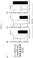
- real time PCR was performed with LightCycler (commercially available) using the primer pairs for the following genes. The results are shown in FIG. 1 .
- the cycle threshold (Ct value) calculated by Lightcycler software ver. 3.5 was normalized for GAPDH mRNA of each sample.
- MMP1 forward ATTTGCCGACAGAGATGAAGTCC (SEQ ID NO: 1) MMP1 reverse GGGTATCCGTGTAGCACATTCTG (SEQ ID NO: 2) ELN forward TGTCCATCCTCCACCCCTCT (SEQ ID NO: 3) ELN reverse CCAGGAACTCCACCAGGAAT (SEQ ID NO: 4) C0L1A1 forward AGCAGGCAAACCTGGTGAAC (SEQ ID NO: 5) C0L1A1 reverse AACCTCTCTCGCCTCTTGCT (SEQ ID NO: 6) GAPDH forward GAAGGTGAAGGTCGGAGT (SEQ ID NO: 7) GAPDH reverse GAAGATGGTGATGGGTTTC (SEQ ID NO: 8)
- siRNAs were designed to knockdown CFD (SI00030100 or SI00030107 (Qiagen), both have similar inhibitory effects) and an siRNA (SI030640318 (Qiagen)) was used as a negative control.
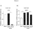
- the siRNAs were introduced into the senescent fibroblasts and, following the aforementioned cell culture protocol, cells with the CFD gene knocked down were inoculated onto the lower layer of the transwell system and cultured together with the cells of the upper layer for 2 days. The cells of the lower layer and the cells of the upper layer were separately acquired and the expression of CFD in the cells of the lower layer and the expression of MMP1 in the cells of the upper layer were examined.
- the expression of the senescence factor (CFD) identified by the present inventors and candidate senescence factors (IGFBP7 and RSPO4) were compared between senescent fibroblasts and young fibroblasts. Senescent and young fibroblasts were separately cultured then recovered using Qiazole (Qiagen). The recovered cell lysates were examined for expression of senescence factors using the same method as the aforementioned RT-PCR. The following primer pairs were used. The results are shown in FIG. 3 . It was shown that the senescence factor identified in the present invention and the candidate senescence factors were highly expressed by senescent fibroblasts.
- the proteins of the candidate senescence factors (IGFBP7 and RSPO4) (purchased from Serotec, R&D SYSTEMS) were added to the culture medium for culturing the young fibroblasts at a concentration of 0 ⁇ g/ml and 10 ⁇ g/ml. After culturing for 2 days, the cells were then separately recovered using Qiazole (Qiagen). In the same way as disclosed in the section relating to quantitative RT-PCR, the expression levels of MMP1 and collagen were examined in the recovered cell lysate. The results are shown in FIG. 4 . IGFBP7 and RSPO4 reduced expression of collagen and also increased MMP1 expression. Accordingly, this shows that IGFBP7 and RSPO4 also act as senescence factors.
- EXAMPLE 2 Inhibition of senescence factors by mesenchymal stem cells
- Senescent fibroblasts were inoculated on the upper layer of a 6-well transwell system (Falcon; Franklin Lakes, NJ) filled with Dulbecco's Modified Eagle Medium supplemented with 10% FBS until the density was 1250 cell/cm 2 .
- Mesenchymal stem cells (MSCs) were purchased from Lonza and cultured on MessenPro culture medium. Subconstructed MSCs were inoculated on the lower layer well of the transwell system. 24 hours later, the culture medium was exchanged with DMEM comprising 10% FBS and 250 ⁇ M acorbic acid, and co-culturing was started. Two days later, Qiazole (Qiagen) was used and the cells were recovered.
- the recovered cell lysate was examined for gene expression of senescence factor (IGFBP7) using the same method as for the aforementioned RT-PCR. The results are shown in FIG. 5 .
- IGFBP7 senescence factor 7
- fibroblasts were cultured alone with no MSCs.
- gene expression of IGFBP7 was significantly reduced (student's t-test: P ⁇ 0.05).
- EXAMPLE 3 Screening method using senescence factor as an indicator
- Senescent fibroblasts were inoculated in a 24-well plate such that the density thereof would be 2500 cells/cm 2 .
- 24 hours after inoculation, added thereto was a 0.5% concentration of each of the following extracts which are cosmetic materials: a Rosa multiflora extract and a turmeric extract.
- a group in which only the extraction solvent for the extract was added served as a control.
- 24 hours after adding the extracts the cells were recovered using Qiazol (Qiagen).
- the expression levels of RSPO4 were examined in the recovered cell lysate using the same method as described in the section relating to quantitative RT-PCR.
- RSPO4 expression in the control group was set as 100 and RSPO4 expression in the addition group was shown. The results are shown in FIG. 6 .
Landscapes
- Health & Medical Sciences (AREA)
- Life Sciences & Earth Sciences (AREA)
- Engineering & Computer Science (AREA)
- General Health & Medical Sciences (AREA)
- Biomedical Technology (AREA)
- Chemical & Material Sciences (AREA)
- Biotechnology (AREA)
- Microbiology (AREA)
- Public Health (AREA)
- Veterinary Medicine (AREA)
- Animal Behavior & Ethology (AREA)
- Epidemiology (AREA)
- Immunology (AREA)
- Botany (AREA)
- Medicinal Chemistry (AREA)
- Mycology (AREA)
- Natural Medicines & Medicinal Plants (AREA)
- Bioinformatics & Cheminformatics (AREA)
- Urology & Nephrology (AREA)
- Molecular Biology (AREA)
- Hematology (AREA)
- Birds (AREA)
- Cell Biology (AREA)
- Biochemistry (AREA)
- Dermatology (AREA)
- Pharmacology & Pharmacy (AREA)
- Genetics & Genomics (AREA)
- Organic Chemistry (AREA)
- Wood Science & Technology (AREA)
- Zoology (AREA)
- Toxicology (AREA)
- Physics & Mathematics (AREA)
- General Physics & Mathematics (AREA)
- Pathology (AREA)
- Food Science & Technology (AREA)
- Tropical Medicine & Parasitology (AREA)
- Analytical Chemistry (AREA)
- General Engineering & Computer Science (AREA)
- Medical Informatics (AREA)
- Alternative & Traditional Medicine (AREA)
Abstract
Description
- The present invention relates to a screening method for an anti-aging substance using the expression of a dermal senescence factor as an indicator.
- Although the various symptoms associated with aging are an unavoidable problem but researches that scientifically analyze the aging to inhibit the progression of aging or ameliorate aging have been carried out. While aging is macroscopically a major factor in skin aging, which is of utmost interest in terms of beauty, there are additional direct factors that affect skin aging such as the effects of dryness, oxidation, UV, and such. The symptoms of skin aging can be seen as skin wrinkles (large wrinkles and fine wrinkles), sagging, spots, dullness, worsening of skin texture, etc. and the causes thereof include a reduction of collagen, cross-linking reactions in collagen, a decrease in mucopolysaccharides including hyaluronic acid, an increase in melanin pigment, and a decrease in epidermal turnover. These causes can be broadly divided into dermal and epidermal causes which each need to be addressed. For dermal causes, it has been elucidated that the promotion of hyaluronic acid production, inhibition of matrix metalloproteinase (MMP) production and activity, promotion of collagen production, and inhibition of esterase activity are effective for dermal anti-aging. In such anti-aging research, activation of cells from the epidermis or dermis layer has been performed, but recently, a technique for skin anti-aging by the activation of stem cells, specifically somatic stem cells, has been examined.
- Stem cells are defined as cells that have the capacity to self-replicate and the capacity to differentiate into other types of cell. As the stem cells, other than pluripotent stem cells that can differentiate into all cell types that form one individual such as embryonic stem cells (ES cells) or induced pluripotent stem cells (iPS cells), multipotent somatic stem cells that are not pluripotent but can differentiate into several cell lineages are known to exist in each tissue even in an adult. It is known that somatic stem cells contribute to tissue repair at the time of tissue damage, maintenance of tissue homeostasis and aging. Somatic stem cells are known to exist in various tissues. As stem cells present in skin tissue, epidermal stem cells present in the epidermis layer, and mesenchymal stem cells present in the dermis layer and subcutaneous fat layer are known.
- In the epidermis, it is well known that epidermal stem cells are present in the basal layer of the epidermis, and hair follicle epithelial stem cells and skin pigment stem cells have been reported to be present in a region called the bulge region of the hair follicle. Meanwhile, the dermis contains skin-derived precursor cells (SKP cells) that differentiate into a plurality of cell lines such as adipose, glia, cartilage, and muscle (NPL 1: Wong C. E. et al., J Cell. Biol. 175: 1005-1015, 2006). Furthermore, cells referred to as dermal mesenchymal stem cells have been isolated (PTL 1:
WO2011/034106 ). - It is known that mesenchymal stem cells are attracted to a trauma site when the skin is subjected to trauma. It is thought that the mesenchymal stem cells that are attracted to the trauma site activate surrounding skin cells and promote cell proliferation, differentiate into skin cells, reduce the trauma, and contribute to healing. It has been shown that certain flavonoids have an effect of attracting mesenchymal stem cells (NPL 2: PLOS ONE DOI: 10.137/journal.pone.0144166). Mesenchymal stem cells that contribute to healing of skin trauma are considered to include bone marrow-derived mesenchymal stem cells which are transported through blood vessels as well as adipose stem cells that exist in the subcutaneous tissue and dermal mesenchymal stem cells that exist around organs such as sebaceous glands, sweat glands, and hair follicles and near blood vessels. Although bone marrow-derived stem cells have conventionally been the subject of research into mesenchymal stem cells, once adipose stem cells are isolated from adipose tissue, due to the availability and multipotent capacity thereof of being able to differentiate into various cell lines, the application thereof to various regenerative treatments is expected (NPL 3: World J Stem Cells 2014 January 26; 6 (1): 65-68). Dermal mesenchymal stem cells exist close to blood vessels but there are still many unanswered questions regarding the differentiative capacity and properties thereof and research is expected to be carried out henceforth regarding the application thereof.
-
- [PTL 1]
WO2011/034106 - [PTL 2] Japanese Unexamined Patent Publication (Kohyo) No.
2013-507956 -
- [NPL 1] J Cell Biol. 175:1005-1015, 2006
- [NPL 2] PLOS ONE DOI:10.137/journal.pone.0144166
- [NPL 3] World J Stem Cells 2014 January 26; 6(1): 65-68
- The object of the present invention is the development of a screening method for an anti-aging substance.
- The present inventors conducted research on aging of the dermis and discovered a surprising phenomenon whereby a factor secreted by senescent cells (hereinafter referred to as dermal senescence factor) acted on normal cells such that the production of dermal components decreased and the degradation of dermal components was promoted. Based on these findings, the present inventors developed a screening method for an anti-aging substance using a dermal senescence factor as an indicator. More specifically, the present invention relates to the following.
- [1] A screening method for an anti-aging substance using the expression of a dermal senescence factor as an indicator.
- [2] The screening method according to
item 1, comprising the steps of:- culturing a skin cell culture containing a dermal fibroblast in a culture medium containing a candidate drug;
- measuring expression of the dermal senescence factor in the culture; and
- selecting a candidate drug that can inhibit the expression of the dermal senescence factor.
- [3] The screening method according to
item 2, wherein the skin cell culture further contains a stem cell. - [4] The screening method according to any one of
items 1 to 3, wherein the expression of the dermal senescence factor is determined by measuring the gene expression or protein level of the dermal senescence factor. - [5] The screening method according to any one of
items 1 to 4, wherein the dermal senescence factor is IGFBP7, CFD, or RSPO4. - [6] A dermal senescence factor expression inhibitor comprising a turmeric extract or a Rosa multiflora extract.
- [7] The dermal senescence factor expression inhibitor according to
item 6, wherein the dermal senescence factor is CFD. - [8] A turmeric extract or a Rosa multiflora extract for use in the treatment of wrinkles, sagging, or skin aging by expression inhibition of the dermal senescence factor.
- [9] A method of administering a composition comprising a turmeric extract or a Rosa multiflora extract as an aesthetic or non-therapeutic method of ameliorating wrinkles, sagging, or skin aging.
- [10] The method according to item 9, comprising ameliorating wrinkles, sagging, or skin aging through expression inhibition of a dermal senescence factor.
- [12] The method according to
item 9 or 10, comprising administration to a subject suffering from wrinkles, sagging, or skin aging. - [13] A use of a turmeric extract or a Rosa multiflora extract for producing an expression inhibitor of a dermal senescence factor.
- [14] A skin amelioration method comprising the administration of a composition comprising a turmeric extract or a Rosa multiflora extract to a subject suffering from wrinkles, sagging, or skin aging.
- [15] An aesthetic or non-therapeutic method of ameliorating wrinkles, sagging, or skin aging characterized by expression inhibition of a dermal senescence factor.
- [16] The aesthetic or non-therapeutic method according to item 15, wherein the dermal senescence factor is IGFBP7, CFD, or RSPO4.
- [17] The aesthetic or non-therapeutic method according to item 16, comprising the administration of a turmeric extract or a Rosa multiflora extract for expression inhibition of the dermal senescence factor.
- The screening method of the present invention can screen for an anti-aging substance using a dermal senescence factor as an indicator. By elucidating a novel mechanism of aging through a dermal senescence factor and using such a factor as an indicator, it will be possible to obtain anti-aging substances that could not be screened for with conventional screening methods.
-
-
FIG. 1 (A) shows a schematic diagram of a transwell system. Normal (young) fibroblasts are cultured on the upper layer and senescent fibroblasts are cultured on the lower layer.FIG. 1(B) to (D) are graphs that compare the gene expression levels of (B) collagen 1A1 (COL1a1), (C) elastin (ELN), and (D) metalloproteinase 1 (MMP1) between (Blank) in which cells are not cultured on the lower layer and (+Senescent) in which senescent fibroblasts are cultured on the lower layer. -
FIG. 2(A) is a graph showing the inhibition of CFD gene expression by senescent fibroblasts in the lower layer of the transwell system andFIG. 2(B) is a graph showing a change in MMP1 gene expression in fibroblasts in the upper layer in such cases. -
FIG. 3 shows graphs comparing gene expression levels of the senescence factors (A) IGFBP7, (B) RSPO4, and (C) CFD between senescent fibroblasts and normal (young) fibroblasts. -
FIG. 4 shows graphs illustrating (A) dose-dependent increase in expression of the MMP1 gene and (B) a decrease in expression of theCollagen 1 gene when IGFBP7and RSPO4 are added. -
FIG. 5 shows a graph illustrating a decrease in expression of the senescence factor (IGFBP7) in the presence of mesenchymal stem cells in a co-culture system of senescent fibroblasts and mesenchymal stem cells. -
FIG. 6 shows a graph illustrating a decrease in expression of the senescence factor RSPO4 compared to a control when a turmeric extract or a Rosa multiflora extract is added to a senescent fibroblast culture then cultured. - The present invention relates to a screening method for an anti-aging substance using the expression of a dermal senescence factor as an indicator. More specifically, this screening method comprises the following steps:
- culturing a skin cell culture comprising a dermal fibroblast in a culture medium comprising a candidate drug;
- measuring expression of the dermal senescence factor in the culture; and
- selecting a candidate drug that can inhibit the expression of the dermal senescence factor.
- The screening method according to the present invention may further comprise a preculturing step of culturing a skin cell culture comprising a dermal fibroblast in a culture medium to which the candidate drug has not been added before the culturing step of culturing a skin cell culture comprising a dermal fibroblast in a culture medium containing the candidate drug. The preculture may be performed over a discretionary period of time but generally after inoculating the skin cell culture, culturing is performed to subconfluence or confluence. The step of culturing a skin cell culture comprising a dermal fibroblast in a culture medium comprising a candidate drug may be achieved by either adding the candidate drug to the culture medium subjected to the preculturing step or by replacing the culture medium with one containing the candidate drug.
- The culturing period for the culturing step of culturing a skin cell culture comprising a dermal fibroblast in a culture medium containing a candidate drug, in view of ensuring sufficient time for the candidate drug to act on the cell, is 24 hours or more, preferably 3 days or more, and more preferably 5 days or more. After the culturing step of culturing a skin cell culture comprising a dermal fibroblast in a culture medium containing a candidate drug, a post-culturing step of further culturing the skin cell culture comprising dermal fibroblasts in a culture medium to which the candidate drug has not been added may be performed.
- The culture medium may be any solution in which the cells can survive. Any culture medium that is usually used for culturing a skin cell culture can be used. For example, DMEM, EMEM, and RPMI1640 may be used. Normal culture conditions for culturing a skin cell culture can be used as the culture conditions. For example, culturing may be carried out in 5% CO2, at 37 °C, in a humidified atmosphere.
- The dermal senescence factor is a factor mainly secreted from senescent cells and is a factor that acts on normal dermal cells to inhibit the production of dermal components or promote the degradation thereof. Examples of dermal senescence factor include IGFBP7, CFD, and RSPO4, and anti-aging substances can be screened for by using the expression of one, two, or all of such factors as an indicator.
- The step of measuring the expression of a dermal senescence factor in a culture refers to a step of measuring the gene expression level or protein level of the dermal senescence factor in the culture medium by any technique. The level of expression of the senescence factor can be appropriately measured by using a molecular biological technique. For example, when measuring the level of protein, a specific antibody is prepared against each dermal senesce factor and the protein level can be measured by using a technique such as western blot or ELISA. The gene expression levels of dermal senescence factors can be measured by performing RT-PCR using the following primers. The methods are not intended to be limited hereto. A person skilled in the art would be able to appropriately select an internal standard.
[Table 1] CFD forward CATCTGGTTGGTCTTTATTGAGC (SEQ ID NO: 9) CFD reverse CATGCTGATCTCGAACTCCTG (SEQ ID NO: 10) IGFBP7 forward TCAAAGGACAGAACTCCTGCGTGGTGA (SEQ ID NO: 11) IGFBP7 reverse GCTGAAGCCTGTCCTTGGGAATTGGATG (SEQ ID NO: 12) RSP04 forward CCAGCAGAGGCTCTTCCTGTTCATCC (SEQ ID NO: 13) RSP04 reverse TCCTGGCTGAAGCAGCTCTGACAAG (SEQ ID NO: 14) - The step for selecting a candidate drug that can inhibit the expression of a dermal senescence factor is a step for selecting a candidate drug as an anti-aging factor if the expression of the dermal senescence factor is reduced. For comparison, the expression of the dermal senescence factor in a culture is measured for a control group which differs only in that the candidate drug is not added. The measurement results of the control group can be compared. Experiments involving the control group can be performed at the same time as the screening method of the present invention, or the experiments involving the control group can be performed beforehand and just the results used for comparison. In another embodiment, the expression levels of a dermal senescence factor before and after the addition of a candidate drug are compared and if the expression level decreases, the candidate drug can be selected as an anti-aging factor. The expression of a dermal senescence factor is only required to decrease compared to the control group but may decrease with a significant difference.
- The skin cell culture may be a culture of any cells that constitute the skin. Cells that constitute the skin may include epidermal keratinocytes, dermal fibroblasts, adipocytes present in the subcutaneous fat layer, and other cells such as Langerhans cells, melanocytes, Merkel cells, histocytes, mast cells, plasma cells, and stem cells, and may also contain cells that make up each organ in the skin, such as sebaceous glands, sweat glands, and hair follicles. Dermal fibroblasts may be cultured alone or co-cultured with other cells. As one embodiment for a co-culture, dermal fibroblasts and epidermal cells are co-cultured to construct a 3-dimensional model skin culture, and a candidate drug may be added thereto. In another embodiment, a culture in which dermal fibroblasts and stem cells are co-cultured may be used. The stem cells may be epidermal stem cells present in the epidermis or mesenchymal stem cells present in the dermis or subcutaneous adipose tissue.
- Dermal fibroblasts are cells that belong to the mesenchymal group of cells and produce collagen fibers, elastic fibers, and mucopolysaccharides. Fibroblasts have a relatively high rate of division and by repeated passaging, senescent fibroblasts can be obtained.
- Mesenchymal stem cells generally refer to stem cells derived from any tissue classified as mesenchymal. Tissue classified as mesenchymal include bone marrow, placenta, adipose tissue, dermis, etc. Examples of stem cells derived from these tissues include bone marrow-derived mesenchymal stem cells (hereinafter simply referred to as bone marrow stem cells), placenta-derived mesenchymal stem cells, adipose tissue-derived mesenchymal stem cells (hereinafter simply referred to as adipose stem cells), dental pulp-derived mesenchymal stem cells, umbilical cord blood-derived mesenchymal stem cells, dermis-derived mesenchymal stem cells (hereinafter simply referred to as dermal stem cells). Some dermal papilla cells are also considered to be mesenchymal stem cells.
- Bone marrow-derived mesenchymal stem cells are stem cells present in the bone marrow. Bone marrow stem cells have the capacity to differentiate into various tissues including cardiomyocytes, skeletal muscle cells, vascular endothelial cells, chondrocytes, osteocytes, and adipocytes. Bone marrow-derived mesenchymal stem cells are characterized by CD105, CD73, CD90 being positive markers, but are not intended to be limited to cells that express these markers. Bone marrow mesenchymal cells are released into the blood in response to signals from tissue damage and the like and are considered to perform a function of healing the wound.
- Adipose tissue-derived mesenchymal stem cells can be simply referred to as adipose stem cells and are a type of mesenchymal stem cell present in the adipose tissue. It is known that adipose stem cells have the capacity to migrate (NPL 3 and PL 2) and it is thought that migration occurs through the adipose tissue. Adipose stem cells are characterized by the expression of CD90 markers, CD105 markers, etc., but this is not intended to limit the cells to those expressing these markers.
- Dermis derived mesenchymal stem cells can be simply referred to as dermal stem cells and are stem cells that are present in the dermis. Dermal stem cells contribute to the maintenance of dermal layer homeostasis by replenishing fibroblasts by differentiating into fibroblasts or through the activation of fibroblasts. Dermal mesenchymal stem cells are present primarily in the dermal layer as a pericyte-like cell group around microvascular sites or organs such as sebaceous glands, sweat glands, and hair follicles. It is thought that dermal mesenchymal stem cells are activated when the skin is damaged, differentiating into fibroblasts or myofibroblasts and contributing to the regeneration and repair of the skin. Dermal stem cells are characterized by being double positive for CD34 and NG2, but this is not intended to limit the cells to those expressing these markers. It is thought that dermal stem cells are replenished through blood vessels since capillaries are spread over the vicinity of each organ in which dermal stem cells are present. Thus, dermal stem cells can have properties similar to bone-marrow mesenchymal stem cells.
- The cells to be used are preferably senescent cells since it is necessary for a senescence factor to be produced before adding a candidate drug. For the senescent cells, cells having a decreased proliferation rate (e.g. a doubling time of less than 0.5/week and Pdl > 50) after subculturing for a fixed period, for example, 3 months or more, are preferably used. Alternatively, primary cells can be acquired from elderly people to obtain senescent cells. In a preferred embodiment, the use of the thus-obtained senescent cells in which the expression of a senescent factor selected from the group consisting of IGFBP7, CFD, and RSPO4 has been confirmed is preferable. The senescent cells can be any cells provided they are contained in the skin, but dermal fibroblasts are preferable in view of expressing a senescence factor selected from the group consisting of IGFBP7, CFD, and RSPO4.
- When the skin cell culture contains only dermal fibroblasts, the anti-aging substance selected by the screening method can inhibit the expression of the senescence factor by directly acting on senescent dermal fibroblasts. Meanwhile, when the skin cell culture is a co-culture of dermal fibroblasts and another type of cell, the anti-aging substance selected by the screening method can inhibit the expression of the senescence factor by either directly acting on senescent dermal fibroblasts or indirectly acting thereon through the other co-cultured cells. It is thought that for the indirect action, senescence factor expression in senescent dermal fibroblasts is suppressed through, for example, the activity of the co-cultured cells. It has been shown that mesenchymal stem cells can inhibit the expression of a senescence factor (IGFBP7) by acting indirectly on dermal fibroblasts (
FIG. 5 ). - When performing a co-culture, dermal fibroblasts and other cells may be mixed and inoculated on the same place, layered and cultured, or isolated and cultured in the same culture vessel. When isolating and culturing cells in the same vessel, exchange of the culture medium is possible, and by culture medium being mixed through pores of a size sufficient to prevent the migration of cells therethrough, a factor secreted by one group of cells can affect the other group of cells. In one embodiment, stem cells can be co-cultured with dermal fibroblasts. Such stem cells have the capacity to differentiate into dermal fibroblasts as well as the capacity to act on surrounding cells by releasing some sort of factor. Thus, by using this co-culturing system, it is possible to screen for an anti-aging substance that can act on stem cells and inhibit senescent dermal fibroblasts from producing a senescence factor.
- Therefore, the anti-aging substance selected by the screening method of the present invention may be one that acts directly on senescent cells to inhibit secretion of a senescent factor or one that acts on another type of cell, such as stem cells, to indirectly inhibit secretion of a senescence factor, depending on the system used. Anti-aging substances that act directly on senescent cells may be referred to as senescence factor expression inhibitors. Senescence factors inhibit production of dermal components or promote degradation thereof. Thus, inhibition of senescence factor expression has the effect of increasing dermal components such as collagen or elastin. Therefore, anti-aging substances can be blended with cosmetics or pharmaceutical agents to be used as agents for promoting an increase in dermal components, and as agents for ameliorating wrinkles and sagging, and as an anti-aging agent. Anti-aging substances that act on stem cells and indirectly inhibit the secretion of aging factors can be used as stem cell activators and may be used alone. However, the combined use thereof with stem cell therapy or the administration thereof in combination with a stem cell attractant can bring about even more desirable anti-aging effects.
- When the screening method of the present invention was carried out using a plurality of cosmetic materials as candidate drugs, a turmeric extract and a Rosa multiflora extract could be screened for as anti-aging factor expression inhibitors.
- The turmeric extract refers to an extract from a perennial plant of the ginger family Curcuma Longa which is native to India. Parts of the plant which are extracted include the whole plant, the leaves, the flower, and the roots, but the rhizome is particularly preferable. Any extraction method can be used for the extraction method but, for example, an extract can be obtained by performing steam distillation or solvent extraction on dried roots. Further, any extraction solvent can be used as a solvent but a 50% butylene glycol or the like can be used.
- The Rosa multiflora extract refers to an extract of the fruit or false fruit of Rosa multiflora which is native to east Asia. Any extraction method may be used but, for example, an extract can be obtained by performing steam distillation or solvent extraction on fruit or false fruit that have been dried. Further, any solvent may be used as the solvent, but an aqueous ethanol solution or 50% butylene glycol or the like can be used.
- The candidate drug may be a drug belonging to any library. The library may be an organic compound library, an extract library, a cosmetic material library, a pharmaceutical library, etc. From the viewpoint of transdermal administration of the candidate drug, it is preferable to use, as the candidate drug, a cosmetic material that has already been shown to be stable in transdermal administration.
- Anti-aging substances screened for by the method of the present invention can be blended in any composition, for example, cosmetics, pharmaceuticals, and quasi drugs. The cosmetics, pharmaceuticals, or quasi-drugs into which the anti-aging substances have been blended are used on subjects worried about skin aging such as wrinkles and sagging. Such subjects who are worried about skin aging could be considered to have high expression of senescence factors in the skin. In particular, in the field of beauty, since anti-aging is desirable, it is preferable that the substance be blended in cosmetics. The dosage form of the cosmetic in which the substance is blended is discretionary and the substance may be blended in the form of a liquid, gel, foam, emulsion, cream, ointment, sheet, etc. The anti-aging substance can be applied to various products by being blended in such cosmetic products as: basic cosmetics such as pharmaceutical preparations for external use, toners, emulsions, creams, ointments, lotions, oils, packs; makeup cosmetics such as facial cleansers and skin cleansers, hair removers, depilatory agents, after shave lotions, pre shave lotions, shaving cream, foundation, lipstick, blusher, eye shadow, eyeliner, mascara; hair care products such as shampoo, conditioner, hair treatment, pre-hair treatment, hair styling products, perm agents, hair tonic, hair dye, hair growth and nourishment product; and bath preparations.
- In the present invention, IGFBP7, CFD, and RSO4, have been determined to be senescence factors. Thus, by examining the expression of these factors, the degree of senescence of a cell can be determined. For example, by measuring the level of expression of a gene as an internal standard, the degree of senescence of a cell can be determined. The degree of senescence can be determined for a cell obtained from a living body, in particular a skin cell, and can also be determined for cultured cells. Thus, in yet a further embodiment of the present invention, the present invention relates to a method of determining the degree of senescence of a cell using the level of expression of senescence factors as an indicator.
- All documents mentioned herein are incorporated herein by reference in their entirety.
- The embodiments of the present invention described below are for illustrative purposes only and do not limit the technical scope of the present invention. The technical scope of the present invention is limited only by the claims. Modifications of the present invention, for example, additions, deletions and replacements of the constituent features can be made without departing from the spirit of the present invention.
- Dermal fibroblasts established from skin from the back of an adult (21 year old) were cultured in Dulbecco's Modified Eagle Medium supplemented with 10% FBS. Cells with a culture period of less than 1 month and Pdl < 30 were used as normal fibroblasts (young fibroblasts). These cells were cultured for 3 months or more and cells which began to proliferate more slowly (doubling time less than 0.5/week and Pdl > 50) were used as senescent cells. Normal fibroblasts were inoculated on the upper layer of a 6-well transwell system (Falcon; Franklin Lakes, NJ) filled with Dulbecco's Modified Eagle Medium supplemented with 10% FBS until the density was 1250 cell/cm2. Subconstructed senescent fibroblasts and normal fibroblasts were separately inoculated in two wells of the lower layer of the transwell system. The upper layer and the lower layer of the transwell system were combined and cultured for 2 days at 37 °C in a humidified atmosphere with 5% CO2, and the cells in the upper layer and the lower layer were recovered using Qiazole (Qiagen).
- RNA was extracted from the recovered cell lysate using RNeasy Protect Kit (Qiagen) by following the instructions therefor, then Superscript VILO (Invitrogen) was used to reverse transcribe the RNA into cDNA by following the instructions. Using 28SrRNA as an internal standard, real time PCR was performed with LightCycler (commercially available) using the primer pairs for the following genes. The results are shown in
FIG. 1 . The cycle threshold (Ct value) calculated by Lightcycler software ver. 3.5 was normalized for GAPDH mRNA of each sample.[Table 2] MMP1 forward ATTTGCCGACAGAGATGAAGTCC (SEQ ID NO: 1) MMP1 reverse GGGTATCCGTGTAGCACATTCTG (SEQ ID NO: 2) ELN forward TGTCCATCCTCCACCCCTCT (SEQ ID NO: 3) ELN reverse CCAGGAACTCCACCAGGAAT (SEQ ID NO: 4) C0L1A1 forward AGCAGGCAAACCTGGTGAAC (SEQ ID NO: 5) C0L1A1reverse AACCTCTCTCGCCTCTTGCT (SEQ ID NO: 6) GAPDH forward GAAGGTGAAGGTCGGAGT (SEQ ID NO: 7) GAPDH reverse GAAGATGGTGATGGGTTTC (SEQ ID NO: 8) - As illustrated in
FIG. 1 (A) , when senescent fibroblasts were inoculated on the lower layer, the expression of MMP1 was significantly increased compared to when the cells were not inoculated on the bottom layer. Furthermore, the expression of elastin and collagen was significantly decreased. When normal fibroblasts (young fibroblasts) were inoculated on the lower layer, there was no significant change compared to when cells were not inoculated on the bottom layer (not shown). From these results, it was clear that the senescent cells secreted some senescence factor, whereby gene expression was changed in dermal fibroblasts sharing the culture medium therewith. - The difference in gene expression between young fibroblasts and senescent fibroblasts was examined by microarray analysis. Genes expressed at least 2.5 times more in senescent fibroblasts than young fibroblasts were identified. The genes listed in the table below were identified as senescence factor candidates.
[Table 3] Gene Name Description Systematic Name Fold Increased RSP04 Homo sapiens R-spondin family, member 4 NM_001029871 5.9 IGFBP7 Homo sapiens insulin-like growth factor binding protein 7 NM_001553 3.6 CFD Homo sapiens complement factor D NM_001928 2.7 - Of these senescence factor candidate genes, siRNAs were designed to knockdown CFD (SI00030100 or SI00030107 (Qiagen), both have similar inhibitory effects) and an siRNA (SI030640318 (Qiagen)) was used as a negative control. The siRNAs were introduced into the senescent fibroblasts and, following the aforementioned cell culture protocol, cells with the CFD gene knocked down were inoculated onto the lower layer of the transwell system and cultured together with the cells of the upper layer for 2 days. The cells of the lower layer and the cells of the upper layer were separately acquired and the expression of CFD in the cells of the lower layer and the expression of MMP1 in the cells of the upper layer were examined. When senescent fibroblasts with knocked down CFD were inoculated on the lower layer, the expression of MMP1 from cells in the upper layer decreased. The results are shown in
FIG. 2 . These results show that CFD was identified as a senescence factor. - The expression of the senescence factor (CFD) identified by the present inventors and candidate senescence factors (IGFBP7 and RSPO4) were compared between senescent fibroblasts and young fibroblasts. Senescent and young fibroblasts were separately cultured then recovered using Qiazole (Qiagen). The recovered cell lysates were examined for expression of senescence factors using the same method as the aforementioned RT-PCR. The following primer pairs were used. The results are shown in
FIG. 3 . It was shown that the senescence factor identified in the present invention and the candidate senescence factors were highly expressed by senescent fibroblasts.[Table 4] CFD forward CATCTGGTTGGTCTTTATTGAGC (SEQ ID NO: 9) CFD reverse CATGCTGATCTCGAACTCCTG (SEQ ID NO: 10) IGFBP7 forward TCAAAGGACAGAACTCCTGCCTGGTGA (SEQ ID NO: 11) IGFBP7 reverse GCTGAAGCCTGTCCTTGGGAATTGGATG (SEQ ID NO: 12) RSP04 forward CCAGCAGAGGCTCTTCCTGTTCATCC (SEQ ID NO: 13) RSP04 reverse TCCTGGCTGAAGCAGCTCTCACAAG (SEQ ID NO: 14) G APDH forward GAAGGTGAAGGTCGGAGT (SEQ ID NO: 7) GAPDH reverse GAAGATGGTGATGGGATTTC (SEQ ID NO: 8) - The proteins of the candidate senescence factors (IGFBP7 and RSPO4) (purchased from Serotec, R&D SYSTEMS) were added to the culture medium for culturing the young fibroblasts at a concentration of 0 µg/ml and 10 µg/ml. After culturing for 2 days, the cells were then separately recovered using Qiazole (Qiagen). In the same way as disclosed in the section relating to quantitative RT-PCR, the expression levels of MMP1 and collagen were examined in the recovered cell lysate. The results are shown in
FIG. 4 . IGFBP7 and RSPO4 reduced expression of collagen and also increased MMP1 expression. Accordingly, this shows that IGFBP7 and RSPO4 also act as senescence factors. - Senescent fibroblasts were inoculated on the upper layer of a 6-well transwell system (Falcon; Franklin Lakes, NJ) filled with Dulbecco's Modified Eagle Medium supplemented with 10% FBS until the density was 1250 cell/cm2. Mesenchymal stem cells (MSCs) were purchased from Lonza and cultured on MessenPro culture medium. Subconstructed MSCs were inoculated on the lower layer well of the transwell system. 24 hours later, the culture medium was exchanged with DMEM comprising 10% FBS and 250 µM acorbic acid, and co-culturing was started. Two days later, Qiazole (Qiagen) was used and the cells were recovered. The recovered cell lysate was examined for gene expression of senescence factor (IGFBP7) using the same method as for the aforementioned RT-PCR. The results are shown in
FIG. 5 . As a control group, fibroblasts were cultured alone with no MSCs. In the presence of MSCs, gene expression of IGFBP7 was significantly reduced (student's t-test: P < 0.05). - Senescent fibroblasts were inoculated in a 24-well plate such that the density thereof would be 2500 cells/cm2. 24 hours after inoculation, added thereto was a 0.5% concentration of each of the following extracts which are cosmetic materials: a Rosa multiflora extract and a turmeric extract. A group in which only the extraction solvent for the extract was added served as a control. 24 hours after adding the extracts, the cells were recovered using Qiazol (Qiagen). The expression levels of RSPO4 were examined in the recovered cell lysate using the same method as described in the section relating to quantitative RT-PCR. RSPO4 expression in the control group was set as 100 and RSPO4 expression in the addition group was shown. The results are shown in
FIG. 6 .


Claims (9)
- A screening method for an anti-aging substance using the expression of a dermal senescence factor as an indicator.
- The screening method according to claim 1, comprising the steps of:culturing a skin cell culture comprising a dermal fibroblast in a culture medium comprising a candidate drug;measuring expression of the dermal senescence factor in the culture; andselecting a candidate drug that can inhibit the expression of the dermal senescence factor.
- The screening method according to claim 2, wherein the skin cell culture further comprises a stem cell.
- The screening method according to any one of claims 1 to 3, wherein the expression of the dermal senescence factor is determined by measuring the level of gene or protein expression of the dermal senescence factor.
- The screening method according to any one of claims 1 to 4, wherein the dermal senescence factor is IGFBP7, CFD, or RSPO4.
- A dermal senescence factor expression inhibitor comprising a turmeric extract or a Rosa multiflora extract.
- The dermal senescence factor expression inhibitor according to claim 6, wherein the dermal senescence factor is CFD.
- An aesthetic or non-therapeutic method of ameliorating wrinkles, sagging, or skin aging, comprising inhibiting expression of a dermal senescence factor.
- The aesthetic or non-therapeutic method according to claim 8, wherein the dermal senescence factor is IGFBP7, CFD, or RSPO4.
Applications Claiming Priority (2)
| Application Number | Priority Date | Filing Date | Title |
|---|---|---|---|
| JP2017139181 | 2017-07-18 | ||
| PCT/JP2018/026783 WO2019017356A1 (en) | 2017-07-18 | 2018-07-17 | Method for screening anti-aging substances |
Publications (1)
| Publication Number | Publication Date |
|---|---|
| EP3656867A1 true EP3656867A1 (en) | 2020-05-27 |
Family
ID=65015112
Family Applications (1)
| Application Number | Title | Priority Date | Filing Date |
|---|---|---|---|
| EP18834563.1A Withdrawn EP3656867A1 (en) | 2017-07-18 | 2018-07-17 | Method for screening anti-aging substances |
Country Status (6)
| Country | Link |
|---|---|
| US (1) | US20200232972A1 (en) |
| EP (1) | EP3656867A1 (en) |
| JP (1) | JP7258749B2 (en) |
| CN (1) | CN110546270A (en) |
| TW (1) | TW201908728A (en) |
| WO (1) | WO2019017356A1 (en) |
Families Citing this family (9)
| Publication number | Priority date | Publication date | Assignee | Title |
|---|---|---|---|---|
| FR3088075A1 (en) * | 2018-11-06 | 2020-05-08 | Societe D'exploitation De Produits Pour Les Industries Chimiques Seppic | PROCESS FOR EVALUATING THE CAPACITY OF A SUBSTANCE OR A COMPOSITION TO PREVENT, SLOWING DOWN OR ELIMINATING THE SIGNS OF AGING OF THE SKIN OR LIPS |
| JP7272808B2 (en) * | 2019-02-07 | 2023-05-12 | ポーラ化成工業株式会社 | Screening method for anti-skin aging agent |
| CN111909960B (en) * | 2020-08-14 | 2024-06-25 | 广州市乾相化妆品有限公司 | Screening method for use proportion of peptide and/or protein composition |
| JP7621765B2 (en) * | 2020-10-12 | 2025-01-27 | 株式会社ナリス化粧品 | Screening method for agents for improving skin photoaging |
| CN112553288B (en) * | 2020-12-09 | 2022-06-07 | 汤臣倍健股份有限公司 | Method for screening natural products with anti-aging potential |
| US20220354805A1 (en) * | 2021-04-30 | 2022-11-10 | Hemix Biotech Limited | Cannabidiol (CBD) as Chemical for Treating Aging-related Degenerative Diseases and Promoting Health Aging |
| JP2023020930A (en) * | 2021-07-28 | 2023-02-09 | ポーラ化成工業株式会社 | Method for searching for command factor or command cell of subcutaneous tissue remodeling phenomenon |
| CN114544865A (en) * | 2021-11-19 | 2022-05-27 | 深圳市萱嘉生物科技有限公司 | System and method for evaluating anti-aging efficacy of raw materials |
| CN114958953A (en) * | 2022-05-31 | 2022-08-30 | 云南贝泰妮生物科技集团股份有限公司 | Screening method for anti-aging efficacy of in-vitro 3D whole skin model of cosmetic raw materials |
Family Cites Families (22)
| Publication number | Priority date | Publication date | Assignee | Title |
|---|---|---|---|---|
| JP2002515898A (en) * | 1997-02-25 | 2002-05-28 | ザ リージェンツ オブ ザ ユニバーシティ オブ ミシガン | Methods and compositions for preventing and treating aging in human skin |
| JP4201399B2 (en) * | 1998-10-06 | 2008-12-24 | 花王株式会社 | Skin aging prevention / improving agent |
| JP2002080338A (en) * | 2000-06-20 | 2002-03-19 | Shiseido Co Ltd | Antiaging skin care preparation |
| US20030113388A1 (en) * | 2001-12-13 | 2003-06-19 | Dung Phan | Methods of treatment for skin disorders using turmeric extract and a hydroxy acid |
| JP2003342159A (en) * | 2002-03-18 | 2003-12-03 | Nikko Chemical Co Ltd | Anti-aging skin cosmetics |
| JP2004075632A (en) * | 2002-08-21 | 2004-03-11 | Noevir Co Ltd | Dermal fibroblast activator |
| JP5164198B2 (en) * | 2007-05-15 | 2013-03-13 | 丸善製薬株式会社 | Profilaggrin production promoter, filaggrin production promoter and pore conspicuousness inhibitor |
| US8962323B2 (en) | 2009-09-15 | 2015-02-24 | Shiseido Company, Ltd. | Method of isolating dermal stem cells |
| US8808682B2 (en) | 2009-10-23 | 2014-08-19 | Rnl Bio Co., Ltd | Method for inducing migration of adipose-derived adult stem cells |
| EP2617835A4 (en) * | 2010-09-17 | 2014-03-19 | Shiseido Co Ltd | Skin activation by means of pdgf-bb activity enhancement |
| EP2678792B1 (en) * | 2011-02-22 | 2020-11-04 | The Procter and Gamble Company | Methods for identifying cosmetic agents for skin care compositions |
| EP2704728A4 (en) * | 2011-05-03 | 2015-04-22 | Dermachip Inc | Expression signatures of genes and gene networks associated with skin aging |
| WO2013016257A1 (en) * | 2011-07-25 | 2013-01-31 | Us Cosmeceu Techs Llc | Botanical antioxidant compositions and methods of preparation and use thereof |
| JP6126216B2 (en) * | 2012-06-18 | 2017-05-10 | ザ プロクター アンド ギャンブル カンパニー | Methods and models for evaluating anti-aging benefits in the pharmaceutical field |
| CN103239380A (en) * | 2013-05-20 | 2013-08-14 | 刘小青 | Application of traditional Chinese medicine, screening method and anti-ageing drug thereof |
| EP3068364B1 (en) * | 2013-11-14 | 2021-11-03 | Metro Biotech NSW Pty Ltd | Nad+ precursor selected from nmn, naad, namn, or a pharmaceutically acceptable salt thereof, for use in treating a side effect of chemotherapy and/or radiotherapy in a subject |
| WO2016044295A1 (en) * | 2014-09-16 | 2016-03-24 | Oncomed Pharmaceuticals, Inc. | Treatment of fibrotic diseases |
| JP6773362B2 (en) * | 2015-10-30 | 2020-10-21 | ハウスウェルネスフーズ株式会社 | Skin moisturizer for ingestion or administration |
| FR3042974B1 (en) * | 2015-11-04 | 2017-12-08 | Isp Investments Inc | SYNERGISTIC EXTRACT OF PALMARIA PALMATA AND JASMIN, COMPOSITIONS COMPRISING THE SAME AND USES THEREOF |
| JP2017112892A (en) * | 2015-12-24 | 2017-06-29 | ポーラ化成工業株式会社 | Method for screening skin aging inhibiting material |
| CN105816807A (en) * | 2016-04-28 | 2016-08-03 | 四川力久知识产权服务有限公司 | Anti-aging traditional Chinese medicine composition containing scrophularia ningpoensis for cosmetics |
| WO2019017355A1 (en) * | 2017-07-18 | 2019-01-24 | 株式会社資生堂 | Mesenchymal-stem-cell induction agent |
-
2018
- 2018-07-17 US US16/632,185 patent/US20200232972A1/en not_active Abandoned
- 2018-07-17 WO PCT/JP2018/026783 patent/WO2019017356A1/en not_active Ceased
- 2018-07-17 EP EP18834563.1A patent/EP3656867A1/en not_active Withdrawn
- 2018-07-17 CN CN201880026766.3A patent/CN110546270A/en active Pending
- 2018-07-17 JP JP2019530554A patent/JP7258749B2/en active Active
- 2018-07-18 TW TW107124814A patent/TW201908728A/en unknown
Also Published As
| Publication number | Publication date |
|---|---|
| JPWO2019017356A1 (en) | 2020-05-28 |
| CN110546270A (en) | 2019-12-06 |
| WO2019017356A1 (en) | 2019-01-24 |
| US20200232972A1 (en) | 2020-07-23 |
| JP7258749B2 (en) | 2023-04-17 |
| TW201908728A (en) | 2019-03-01 |
Similar Documents
| Publication | Publication Date | Title |
|---|---|---|
| US20200232972A1 (en) | Method for screening anti-aging substances | |
| Kim et al. | Conditioned media from human umbilical cord blood-derived mesenchymal stem cells stimulate rejuvenation function in human skin | |
| Patil et al. | Multilineage potential and proteomic profiling of human dental stem cells derived from a single donor | |
| CN113728094B (en) | Cosmetic composition comprising a mesenchymal stem cell culture solution cultured in a culture medium containing human platelet lysate | |
| US20120315259A1 (en) | Method and composition for skin care comprising cord blood serum or plasma or components thereof | |
| US10639264B2 (en) | Mesenchymal stem cell extract and its use | |
| EP3656850A1 (en) | Mesenchymal-stem-cell induction agent | |
| Bellei et al. | Regenerative medicine-based treatment for vitiligo: an overview | |
| JP6185913B2 (en) | Human skin dermis-derived adult stem cells | |
| JP2019031503A (en) | Fibroblast mixture and method for producing and using the same | |
| CN107847424A (en) | Relaxation comprising fat stem cell attractant, by the Voiding caused skin of corium or aging improver | |
| JP6945273B2 (en) | Hair agent | |
| KR102153636B1 (en) | Serum-free medium composition for stem cell culture and methods for culturing stem cell | |
| TWI674104B (en) | Skin sagging or aging improver due to dermal hollowing out of adipose stem cell attractant | |
| JP2021092534A (en) | Method for screening anti-aging component | |
| KR20150141812A (en) | An Whitening Ability of Small-Sized Stem Cells and the Use thereof | |
| KR102153635B1 (en) | Serum-free medium composition for stem cell culture and methods for culturing stem cell | |
| KR102153637B1 (en) | Serum-free medium composition for stem cell culture and methods for culturing stem cell | |
| Silva | Avaliação do secretoma de células estromais mesenquimais humanas (MSCs) como um potencial produto de terapia celular avançada livre de células para o reparo cutâneo e promoção do crescimento capilar | |
| Kumar et al. | Stem Cells in Dermatology: What the Future May Hold | |
| JP7205880B2 (en) | MELANOCYTE DIFFERENTIATION INDUCTIVE AND USAGE THEREOF | |
| KR20250023404A (en) | Composition comprising Nidogen for improving skin condition or maintaining stemness | |
| KR20240119009A (en) | Composition comprising Nidogen for improving skin condition or maintaining stemness | |
| KR20140145473A (en) | Culture media of adult stem cells derived human skin dermis | |
| CN116322731A (en) | IL-8 inhibitors, skin anti-aging agents, and methods of using them to inhibit skin aging |
Legal Events
| Date | Code | Title | Description |
|---|---|---|---|
| STAA | Information on the status of an ep patent application or granted ep patent |
Free format text: STATUS: THE INTERNATIONAL PUBLICATION HAS BEEN MADE |
|
| PUAI | Public reference made under article 153(3) epc to a published international application that has entered the european phase |
Free format text: ORIGINAL CODE: 0009012 |
|
| STAA | Information on the status of an ep patent application or granted ep patent |
Free format text: STATUS: REQUEST FOR EXAMINATION WAS MADE |
|
| 17P | Request for examination filed |
Effective date: 20200217 |
|
| AK | Designated contracting states |
Kind code of ref document: A1 Designated state(s): AL AT BE BG CH CY CZ DE DK EE ES FI FR GB GR HR HU IE IS IT LI LT LU LV MC MK MT NL NO PL PT RO RS SE SI SK SM TR |
|
| AX | Request for extension of the european patent |
Extension state: BA ME |
|
| DAV | Request for validation of the european patent (deleted) | ||
| DAX | Request for extension of the european patent (deleted) | ||
| STAA | Information on the status of an ep patent application or granted ep patent |
Free format text: STATUS: THE APPLICATION IS DEEMED TO BE WITHDRAWN |
|
| 18D | Application deemed to be withdrawn |
Effective date: 20210202 |