EP3655052B1 - Verfahren zur herstellung von mehrschichtigen röhrenförmigen gewebekonstrukten - Google Patents
Verfahren zur herstellung von mehrschichtigen röhrenförmigen gewebekonstrukten Download PDFInfo
- Publication number
- EP3655052B1 EP3655052B1 EP18835590.3A EP18835590A EP3655052B1 EP 3655052 B1 EP3655052 B1 EP 3655052B1 EP 18835590 A EP18835590 A EP 18835590A EP 3655052 B1 EP3655052 B1 EP 3655052B1
- Authority
- EP
- European Patent Office
- Prior art keywords
- cell
- laden
- ink
- cells
- construct
- Prior art date
- Legal status (The legal status is an assumption and is not a legal conclusion. Google has not performed a legal analysis and makes no representation as to the accuracy of the status listed.)
- Active
Links
Images


Classifications
-
- A—HUMAN NECESSITIES
- A61—MEDICAL OR VETERINARY SCIENCE; HYGIENE
- A61L—METHODS OR APPARATUS FOR STERILISING MATERIALS OR OBJECTS IN GENERAL; DISINFECTION, STERILISATION OR DEODORISATION OF AIR; CHEMICAL ASPECTS OF BANDAGES, DRESSINGS, ABSORBENT PADS OR SURGICAL ARTICLES; MATERIALS FOR BANDAGES, DRESSINGS, ABSORBENT PADS OR SURGICAL ARTICLES
- A61L27/00—Materials for grafts or prostheses or for coating grafts or prostheses
- A61L27/36—Materials for grafts or prostheses or for coating grafts or prostheses containing ingredients of undetermined constitution or reaction products thereof, e.g. transplant tissue, natural bone, extracellular matrix
- A61L27/38—Materials for grafts or prostheses or for coating grafts or prostheses containing ingredients of undetermined constitution or reaction products thereof, e.g. transplant tissue, natural bone, extracellular matrix containing added animal cells
- A61L27/3804—Materials for grafts or prostheses or for coating grafts or prostheses containing ingredients of undetermined constitution or reaction products thereof, e.g. transplant tissue, natural bone, extracellular matrix containing added animal cells characterised by specific cells or progenitors thereof, e.g. fibroblasts, connective tissue cells, kidney cells
-
- A—HUMAN NECESSITIES
- A61—MEDICAL OR VETERINARY SCIENCE; HYGIENE
- A61F—FILTERS IMPLANTABLE INTO BLOOD VESSELS; PROSTHESES; DEVICES PROVIDING PATENCY TO, OR PREVENTING COLLAPSING OF, TUBULAR STRUCTURES OF THE BODY, e.g. STENTS; ORTHOPAEDIC, NURSING OR CONTRACEPTIVE DEVICES; FOMENTATION; TREATMENT OR PROTECTION OF EYES OR EARS; BANDAGES, DRESSINGS OR ABSORBENT PADS; FIRST-AID KITS
- A61F2/00—Filters implantable into blood vessels; Prostheses, i.e. artificial substitutes or replacements for parts of the body; Appliances for connecting them with the body; Devices providing patency to, or preventing collapsing of, tubular structures of the body, e.g. stents
- A61F2/02—Prostheses implantable into the body
- A61F2/04—Hollow or tubular parts of organs, e.g. bladders, tracheae, bronchi or bile ducts
- A61F2/06—Blood vessels
-
- A—HUMAN NECESSITIES
- A61—MEDICAL OR VETERINARY SCIENCE; HYGIENE
- A61L—METHODS OR APPARATUS FOR STERILISING MATERIALS OR OBJECTS IN GENERAL; DISINFECTION, STERILISATION OR DEODORISATION OF AIR; CHEMICAL ASPECTS OF BANDAGES, DRESSINGS, ABSORBENT PADS OR SURGICAL ARTICLES; MATERIALS FOR BANDAGES, DRESSINGS, ABSORBENT PADS OR SURGICAL ARTICLES
- A61L27/00—Materials for grafts or prostheses or for coating grafts or prostheses
- A61L27/28—Materials for coating prostheses
-
- A—HUMAN NECESSITIES
- A61—MEDICAL OR VETERINARY SCIENCE; HYGIENE
- A61L—METHODS OR APPARATUS FOR STERILISING MATERIALS OR OBJECTS IN GENERAL; DISINFECTION, STERILISATION OR DEODORISATION OF AIR; CHEMICAL ASPECTS OF BANDAGES, DRESSINGS, ABSORBENT PADS OR SURGICAL ARTICLES; MATERIALS FOR BANDAGES, DRESSINGS, ABSORBENT PADS OR SURGICAL ARTICLES
- A61L27/00—Materials for grafts or prostheses or for coating grafts or prostheses
- A61L27/36—Materials for grafts or prostheses or for coating grafts or prostheses containing ingredients of undetermined constitution or reaction products thereof, e.g. transplant tissue, natural bone, extracellular matrix
- A61L27/3604—Materials for grafts or prostheses or for coating grafts or prostheses containing ingredients of undetermined constitution or reaction products thereof, e.g. transplant tissue, natural bone, extracellular matrix characterised by the human or animal origin of the biological material, e.g. hair, fascia, fish scales, silk, shellac, pericardium, pleura, renal tissue, amniotic membrane, parenchymal tissue, fetal tissue, muscle tissue, fat tissue, enamel
- A61L27/3625—Vascular tissue, e.g. heart valves
-
- A—HUMAN NECESSITIES
- A61—MEDICAL OR VETERINARY SCIENCE; HYGIENE
- A61L—METHODS OR APPARATUS FOR STERILISING MATERIALS OR OBJECTS IN GENERAL; DISINFECTION, STERILISATION OR DEODORISATION OF AIR; CHEMICAL ASPECTS OF BANDAGES, DRESSINGS, ABSORBENT PADS OR SURGICAL ARTICLES; MATERIALS FOR BANDAGES, DRESSINGS, ABSORBENT PADS OR SURGICAL ARTICLES
- A61L27/00—Materials for grafts or prostheses or for coating grafts or prostheses
- A61L27/36—Materials for grafts or prostheses or for coating grafts or prostheses containing ingredients of undetermined constitution or reaction products thereof, e.g. transplant tissue, natural bone, extracellular matrix
- A61L27/3604—Materials for grafts or prostheses or for coating grafts or prostheses containing ingredients of undetermined constitution or reaction products thereof, e.g. transplant tissue, natural bone, extracellular matrix characterised by the human or animal origin of the biological material, e.g. hair, fascia, fish scales, silk, shellac, pericardium, pleura, renal tissue, amniotic membrane, parenchymal tissue, fetal tissue, muscle tissue, fat tissue, enamel
- A61L27/3629—Intestinal tissue, e.g. small intestinal submucosa
-
- A—HUMAN NECESSITIES
- A61—MEDICAL OR VETERINARY SCIENCE; HYGIENE
- A61L—METHODS OR APPARATUS FOR STERILISING MATERIALS OR OBJECTS IN GENERAL; DISINFECTION, STERILISATION OR DEODORISATION OF AIR; CHEMICAL ASPECTS OF BANDAGES, DRESSINGS, ABSORBENT PADS OR SURGICAL ARTICLES; MATERIALS FOR BANDAGES, DRESSINGS, ABSORBENT PADS OR SURGICAL ARTICLES
- A61L27/00—Materials for grafts or prostheses or for coating grafts or prostheses
- A61L27/36—Materials for grafts or prostheses or for coating grafts or prostheses containing ingredients of undetermined constitution or reaction products thereof, e.g. transplant tissue, natural bone, extracellular matrix
- A61L27/3604—Materials for grafts or prostheses or for coating grafts or prostheses containing ingredients of undetermined constitution or reaction products thereof, e.g. transplant tissue, natural bone, extracellular matrix characterised by the human or animal origin of the biological material, e.g. hair, fascia, fish scales, silk, shellac, pericardium, pleura, renal tissue, amniotic membrane, parenchymal tissue, fetal tissue, muscle tissue, fat tissue, enamel
- A61L27/3633—Extracellular matrix [ECM]
-
- A—HUMAN NECESSITIES
- A61—MEDICAL OR VETERINARY SCIENCE; HYGIENE
- A61L—METHODS OR APPARATUS FOR STERILISING MATERIALS OR OBJECTS IN GENERAL; DISINFECTION, STERILISATION OR DEODORISATION OF AIR; CHEMICAL ASPECTS OF BANDAGES, DRESSINGS, ABSORBENT PADS OR SURGICAL ARTICLES; MATERIALS FOR BANDAGES, DRESSINGS, ABSORBENT PADS OR SURGICAL ARTICLES
- A61L27/00—Materials for grafts or prostheses or for coating grafts or prostheses
- A61L27/36—Materials for grafts or prostheses or for coating grafts or prostheses containing ingredients of undetermined constitution or reaction products thereof, e.g. transplant tissue, natural bone, extracellular matrix
- A61L27/38—Materials for grafts or prostheses or for coating grafts or prostheses containing ingredients of undetermined constitution or reaction products thereof, e.g. transplant tissue, natural bone, extracellular matrix containing added animal cells
- A61L27/3804—Materials for grafts or prostheses or for coating grafts or prostheses containing ingredients of undetermined constitution or reaction products thereof, e.g. transplant tissue, natural bone, extracellular matrix containing added animal cells characterised by specific cells or progenitors thereof, e.g. fibroblasts, connective tissue cells, kidney cells
- A61L27/3826—Muscle cells, e.g. smooth muscle cells
-
- A—HUMAN NECESSITIES
- A61—MEDICAL OR VETERINARY SCIENCE; HYGIENE
- A61L—METHODS OR APPARATUS FOR STERILISING MATERIALS OR OBJECTS IN GENERAL; DISINFECTION, STERILISATION OR DEODORISATION OF AIR; CHEMICAL ASPECTS OF BANDAGES, DRESSINGS, ABSORBENT PADS OR SURGICAL ARTICLES; MATERIALS FOR BANDAGES, DRESSINGS, ABSORBENT PADS OR SURGICAL ARTICLES
- A61L27/00—Materials for grafts or prostheses or for coating grafts or prostheses
- A61L27/36—Materials for grafts or prostheses or for coating grafts or prostheses containing ingredients of undetermined constitution or reaction products thereof, e.g. transplant tissue, natural bone, extracellular matrix
- A61L27/38—Materials for grafts or prostheses or for coating grafts or prostheses containing ingredients of undetermined constitution or reaction products thereof, e.g. transplant tissue, natural bone, extracellular matrix containing added animal cells
- A61L27/3839—Materials for grafts or prostheses or for coating grafts or prostheses containing ingredients of undetermined constitution or reaction products thereof, e.g. transplant tissue, natural bone, extracellular matrix containing added animal cells characterised by the site of application in the body
- A61L27/3882—Hollow organs, e.g. bladder, esophagus, urether, uterus
-
- A—HUMAN NECESSITIES
- A61—MEDICAL OR VETERINARY SCIENCE; HYGIENE
- A61L—METHODS OR APPARATUS FOR STERILISING MATERIALS OR OBJECTS IN GENERAL; DISINFECTION, STERILISATION OR DEODORISATION OF AIR; CHEMICAL ASPECTS OF BANDAGES, DRESSINGS, ABSORBENT PADS OR SURGICAL ARTICLES; MATERIALS FOR BANDAGES, DRESSINGS, ABSORBENT PADS OR SURGICAL ARTICLES
- A61L27/00—Materials for grafts or prostheses or for coating grafts or prostheses
- A61L27/36—Materials for grafts or prostheses or for coating grafts or prostheses containing ingredients of undetermined constitution or reaction products thereof, e.g. transplant tissue, natural bone, extracellular matrix
- A61L27/38—Materials for grafts or prostheses or for coating grafts or prostheses containing ingredients of undetermined constitution or reaction products thereof, e.g. transplant tissue, natural bone, extracellular matrix containing added animal cells
- A61L27/3886—Materials for grafts or prostheses or for coating grafts or prostheses containing ingredients of undetermined constitution or reaction products thereof, e.g. transplant tissue, natural bone, extracellular matrix containing added animal cells comprising two or more cell types
- A61L27/3891—Materials for grafts or prostheses or for coating grafts or prostheses containing ingredients of undetermined constitution or reaction products thereof, e.g. transplant tissue, natural bone, extracellular matrix containing added animal cells comprising two or more cell types as distinct cell layers
-
- A—HUMAN NECESSITIES
- A61—MEDICAL OR VETERINARY SCIENCE; HYGIENE
- A61L—METHODS OR APPARATUS FOR STERILISING MATERIALS OR OBJECTS IN GENERAL; DISINFECTION, STERILISATION OR DEODORISATION OF AIR; CHEMICAL ASPECTS OF BANDAGES, DRESSINGS, ABSORBENT PADS OR SURGICAL ARTICLES; MATERIALS FOR BANDAGES, DRESSINGS, ABSORBENT PADS OR SURGICAL ARTICLES
- A61L27/00—Materials for grafts or prostheses or for coating grafts or prostheses
- A61L27/50—Materials characterised by their function or physical properties, e.g. injectable or lubricating compositions, shape-memory materials, surface modified materials
- A61L27/507—Materials characterised by their function or physical properties, e.g. injectable or lubricating compositions, shape-memory materials, surface modified materials for artificial blood vessels
-
- B—PERFORMING OPERATIONS; TRANSPORTING
- B33—ADDITIVE MANUFACTURING TECHNOLOGY
- B33Y—ADDITIVE MANUFACTURING, i.e. MANUFACTURING OF THREE-DIMENSIONAL [3-D] OBJECTS BY ADDITIVE DEPOSITION, ADDITIVE AGGLOMERATION OR ADDITIVE LAYERING, e.g. BY 3-D PRINTING, STEREOLITHOGRAPHY OR SELECTIVE LASER SINTERING
- B33Y10/00—Processes of additive manufacturing
-
- B—PERFORMING OPERATIONS; TRANSPORTING
- B33—ADDITIVE MANUFACTURING TECHNOLOGY
- B33Y—ADDITIVE MANUFACTURING, i.e. MANUFACTURING OF THREE-DIMENSIONAL [3-D] OBJECTS BY ADDITIVE DEPOSITION, ADDITIVE AGGLOMERATION OR ADDITIVE LAYERING, e.g. BY 3-D PRINTING, STEREOLITHOGRAPHY OR SELECTIVE LASER SINTERING
- B33Y70/00—Materials specially adapted for additive manufacturing
-
- B—PERFORMING OPERATIONS; TRANSPORTING
- B33—ADDITIVE MANUFACTURING TECHNOLOGY
- B33Y—ADDITIVE MANUFACTURING, i.e. MANUFACTURING OF THREE-DIMENSIONAL [3-D] OBJECTS BY ADDITIVE DEPOSITION, ADDITIVE AGGLOMERATION OR ADDITIVE LAYERING, e.g. BY 3-D PRINTING, STEREOLITHOGRAPHY OR SELECTIVE LASER SINTERING
- B33Y80/00—Products made by additive manufacturing
-
- C—CHEMISTRY; METALLURGY
- C12—BIOCHEMISTRY; BEER; SPIRITS; WINE; VINEGAR; MICROBIOLOGY; ENZYMOLOGY; MUTATION OR GENETIC ENGINEERING
- C12N—MICROORGANISMS OR ENZYMES; COMPOSITIONS THEREOF; PROPAGATING, PRESERVING, OR MAINTAINING MICROORGANISMS; MUTATION OR GENETIC ENGINEERING; CULTURE MEDIA
- C12N5/00—Undifferentiated human, animal or plant cells, e.g. cell lines; Tissues; Cultivation or maintenance thereof; Culture media therefor
- C12N5/06—Animal cells or tissues; Human cells or tissues
- C12N5/0602—Vertebrate cells
- C12N5/0652—Cells of skeletal and connective tissues; Mesenchyme
- C12N5/0656—Adult fibroblasts
-
- C—CHEMISTRY; METALLURGY
- C12—BIOCHEMISTRY; BEER; SPIRITS; WINE; VINEGAR; MICROBIOLOGY; ENZYMOLOGY; MUTATION OR GENETIC ENGINEERING
- C12N—MICROORGANISMS OR ENZYMES; COMPOSITIONS THEREOF; PROPAGATING, PRESERVING, OR MAINTAINING MICROORGANISMS; MUTATION OR GENETIC ENGINEERING; CULTURE MEDIA
- C12N5/00—Undifferentiated human, animal or plant cells, e.g. cell lines; Tissues; Cultivation or maintenance thereof; Culture media therefor
- C12N5/06—Animal cells or tissues; Human cells or tissues
- C12N5/0602—Vertebrate cells
- C12N5/0652—Cells of skeletal and connective tissues; Mesenchyme
- C12N5/0661—Smooth muscle cells
-
- C—CHEMISTRY; METALLURGY
- C12—BIOCHEMISTRY; BEER; SPIRITS; WINE; VINEGAR; MICROBIOLOGY; ENZYMOLOGY; MUTATION OR GENETIC ENGINEERING
- C12N—MICROORGANISMS OR ENZYMES; COMPOSITIONS THEREOF; PROPAGATING, PRESERVING, OR MAINTAINING MICROORGANISMS; MUTATION OR GENETIC ENGINEERING; CULTURE MEDIA
- C12N5/00—Undifferentiated human, animal or plant cells, e.g. cell lines; Tissues; Cultivation or maintenance thereof; Culture media therefor
- C12N5/06—Animal cells or tissues; Human cells or tissues
- C12N5/0602—Vertebrate cells
- C12N5/069—Vascular Endothelial cells
- C12N5/0691—Vascular smooth muscle cells; 3D culture thereof, e.g. models of blood vessels
-
- C—CHEMISTRY; METALLURGY
- C12—BIOCHEMISTRY; BEER; SPIRITS; WINE; VINEGAR; MICROBIOLOGY; ENZYMOLOGY; MUTATION OR GENETIC ENGINEERING
- C12N—MICROORGANISMS OR ENZYMES; COMPOSITIONS THEREOF; PROPAGATING, PRESERVING, OR MAINTAINING MICROORGANISMS; MUTATION OR GENETIC ENGINEERING; CULTURE MEDIA
- C12N5/00—Undifferentiated human, animal or plant cells, e.g. cell lines; Tissues; Cultivation or maintenance thereof; Culture media therefor
- C12N5/06—Animal cells or tissues; Human cells or tissues
- C12N5/0697—Artificial constructs associating cells of different lineages, e.g. tissue equivalents
-
- A—HUMAN NECESSITIES
- A61—MEDICAL OR VETERINARY SCIENCE; HYGIENE
- A61F—FILTERS IMPLANTABLE INTO BLOOD VESSELS; PROSTHESES; DEVICES PROVIDING PATENCY TO, OR PREVENTING COLLAPSING OF, TUBULAR STRUCTURES OF THE BODY, e.g. STENTS; ORTHOPAEDIC, NURSING OR CONTRACEPTIVE DEVICES; FOMENTATION; TREATMENT OR PROTECTION OF EYES OR EARS; BANDAGES, DRESSINGS OR ABSORBENT PADS; FIRST-AID KITS
- A61F2/00—Filters implantable into blood vessels; Prostheses, i.e. artificial substitutes or replacements for parts of the body; Appliances for connecting them with the body; Devices providing patency to, or preventing collapsing of, tubular structures of the body, e.g. stents
- A61F2/02—Prostheses implantable into the body
- A61F2/04—Hollow or tubular parts of organs, e.g. bladders, tracheae, bronchi or bile ducts
- A61F2002/041—Bile ducts
-
- A—HUMAN NECESSITIES
- A61—MEDICAL OR VETERINARY SCIENCE; HYGIENE
- A61F—FILTERS IMPLANTABLE INTO BLOOD VESSELS; PROSTHESES; DEVICES PROVIDING PATENCY TO, OR PREVENTING COLLAPSING OF, TUBULAR STRUCTURES OF THE BODY, e.g. STENTS; ORTHOPAEDIC, NURSING OR CONTRACEPTIVE DEVICES; FOMENTATION; TREATMENT OR PROTECTION OF EYES OR EARS; BANDAGES, DRESSINGS OR ABSORBENT PADS; FIRST-AID KITS
- A61F2/00—Filters implantable into blood vessels; Prostheses, i.e. artificial substitutes or replacements for parts of the body; Appliances for connecting them with the body; Devices providing patency to, or preventing collapsing of, tubular structures of the body, e.g. stents
- A61F2/02—Prostheses implantable into the body
- A61F2/04—Hollow or tubular parts of organs, e.g. bladders, tracheae, bronchi or bile ducts
- A61F2002/043—Bronchi
-
- A—HUMAN NECESSITIES
- A61—MEDICAL OR VETERINARY SCIENCE; HYGIENE
- A61F—FILTERS IMPLANTABLE INTO BLOOD VESSELS; PROSTHESES; DEVICES PROVIDING PATENCY TO, OR PREVENTING COLLAPSING OF, TUBULAR STRUCTURES OF THE BODY, e.g. STENTS; ORTHOPAEDIC, NURSING OR CONTRACEPTIVE DEVICES; FOMENTATION; TREATMENT OR PROTECTION OF EYES OR EARS; BANDAGES, DRESSINGS OR ABSORBENT PADS; FIRST-AID KITS
- A61F2/00—Filters implantable into blood vessels; Prostheses, i.e. artificial substitutes or replacements for parts of the body; Appliances for connecting them with the body; Devices providing patency to, or preventing collapsing of, tubular structures of the body, e.g. stents
- A61F2/02—Prostheses implantable into the body
- A61F2/04—Hollow or tubular parts of organs, e.g. bladders, tracheae, bronchi or bile ducts
- A61F2002/044—Oesophagi or esophagi or gullets
-
- A—HUMAN NECESSITIES
- A61—MEDICAL OR VETERINARY SCIENCE; HYGIENE
- A61F—FILTERS IMPLANTABLE INTO BLOOD VESSELS; PROSTHESES; DEVICES PROVIDING PATENCY TO, OR PREVENTING COLLAPSING OF, TUBULAR STRUCTURES OF THE BODY, e.g. STENTS; ORTHOPAEDIC, NURSING OR CONTRACEPTIVE DEVICES; FOMENTATION; TREATMENT OR PROTECTION OF EYES OR EARS; BANDAGES, DRESSINGS OR ABSORBENT PADS; FIRST-AID KITS
- A61F2/00—Filters implantable into blood vessels; Prostheses, i.e. artificial substitutes or replacements for parts of the body; Appliances for connecting them with the body; Devices providing patency to, or preventing collapsing of, tubular structures of the body, e.g. stents
- A61F2/02—Prostheses implantable into the body
- A61F2/04—Hollow or tubular parts of organs, e.g. bladders, tracheae, bronchi or bile ducts
- A61F2002/045—Stomach, intestines
-
- A—HUMAN NECESSITIES
- A61—MEDICAL OR VETERINARY SCIENCE; HYGIENE
- A61F—FILTERS IMPLANTABLE INTO BLOOD VESSELS; PROSTHESES; DEVICES PROVIDING PATENCY TO, OR PREVENTING COLLAPSING OF, TUBULAR STRUCTURES OF THE BODY, e.g. STENTS; ORTHOPAEDIC, NURSING OR CONTRACEPTIVE DEVICES; FOMENTATION; TREATMENT OR PROTECTION OF EYES OR EARS; BANDAGES, DRESSINGS OR ABSORBENT PADS; FIRST-AID KITS
- A61F2/00—Filters implantable into blood vessels; Prostheses, i.e. artificial substitutes or replacements for parts of the body; Appliances for connecting them with the body; Devices providing patency to, or preventing collapsing of, tubular structures of the body, e.g. stents
- A61F2/02—Prostheses implantable into the body
- A61F2/04—Hollow or tubular parts of organs, e.g. bladders, tracheae, bronchi or bile ducts
- A61F2002/046—Tracheae
-
- A—HUMAN NECESSITIES
- A61—MEDICAL OR VETERINARY SCIENCE; HYGIENE
- A61F—FILTERS IMPLANTABLE INTO BLOOD VESSELS; PROSTHESES; DEVICES PROVIDING PATENCY TO, OR PREVENTING COLLAPSING OF, TUBULAR STRUCTURES OF THE BODY, e.g. STENTS; ORTHOPAEDIC, NURSING OR CONTRACEPTIVE DEVICES; FOMENTATION; TREATMENT OR PROTECTION OF EYES OR EARS; BANDAGES, DRESSINGS OR ABSORBENT PADS; FIRST-AID KITS
- A61F2/00—Filters implantable into blood vessels; Prostheses, i.e. artificial substitutes or replacements for parts of the body; Appliances for connecting them with the body; Devices providing patency to, or preventing collapsing of, tubular structures of the body, e.g. stents
- A61F2/02—Prostheses implantable into the body
- A61F2/04—Hollow or tubular parts of organs, e.g. bladders, tracheae, bronchi or bile ducts
- A61F2002/048—Ureters
-
- A—HUMAN NECESSITIES
- A61—MEDICAL OR VETERINARY SCIENCE; HYGIENE
- A61F—FILTERS IMPLANTABLE INTO BLOOD VESSELS; PROSTHESES; DEVICES PROVIDING PATENCY TO, OR PREVENTING COLLAPSING OF, TUBULAR STRUCTURES OF THE BODY, e.g. STENTS; ORTHOPAEDIC, NURSING OR CONTRACEPTIVE DEVICES; FOMENTATION; TREATMENT OR PROTECTION OF EYES OR EARS; BANDAGES, DRESSINGS OR ABSORBENT PADS; FIRST-AID KITS
- A61F2/00—Filters implantable into blood vessels; Prostheses, i.e. artificial substitutes or replacements for parts of the body; Appliances for connecting them with the body; Devices providing patency to, or preventing collapsing of, tubular structures of the body, e.g. stents
- A61F2/02—Prostheses implantable into the body
- A61F2/04—Hollow or tubular parts of organs, e.g. bladders, tracheae, bronchi or bile ducts
- A61F2/06—Blood vessels
- A61F2002/065—Y-shaped blood vessels
-
- A—HUMAN NECESSITIES
- A61—MEDICAL OR VETERINARY SCIENCE; HYGIENE
- A61F—FILTERS IMPLANTABLE INTO BLOOD VESSELS; PROSTHESES; DEVICES PROVIDING PATENCY TO, OR PREVENTING COLLAPSING OF, TUBULAR STRUCTURES OF THE BODY, e.g. STENTS; ORTHOPAEDIC, NURSING OR CONTRACEPTIVE DEVICES; FOMENTATION; TREATMENT OR PROTECTION OF EYES OR EARS; BANDAGES, DRESSINGS OR ABSORBENT PADS; FIRST-AID KITS
- A61F2240/00—Manufacturing or designing of prostheses classified in groups A61F2/00 - A61F2/26 or A61F2/82 or A61F9/00 or A61F11/00 or subgroups thereof
- A61F2240/001—Designing or manufacturing processes
-
- A—HUMAN NECESSITIES
- A61—MEDICAL OR VETERINARY SCIENCE; HYGIENE
- A61L—METHODS OR APPARATUS FOR STERILISING MATERIALS OR OBJECTS IN GENERAL; DISINFECTION, STERILISATION OR DEODORISATION OF AIR; CHEMICAL ASPECTS OF BANDAGES, DRESSINGS, ABSORBENT PADS OR SURGICAL ARTICLES; MATERIALS FOR BANDAGES, DRESSINGS, ABSORBENT PADS OR SURGICAL ARTICLES
- A61L2420/00—Materials or methods for coatings medical devices
- A61L2420/08—Coatings comprising two or more layers
-
- C—CHEMISTRY; METALLURGY
- C12—BIOCHEMISTRY; BEER; SPIRITS; WINE; VINEGAR; MICROBIOLOGY; ENZYMOLOGY; MUTATION OR GENETIC ENGINEERING
- C12N—MICROORGANISMS OR ENZYMES; COMPOSITIONS THEREOF; PROPAGATING, PRESERVING, OR MAINTAINING MICROORGANISMS; MUTATION OR GENETIC ENGINEERING; CULTURE MEDIA
- C12N2513/00—3D culture
-
- C—CHEMISTRY; METALLURGY
- C12—BIOCHEMISTRY; BEER; SPIRITS; WINE; VINEGAR; MICROBIOLOGY; ENZYMOLOGY; MUTATION OR GENETIC ENGINEERING
- C12N—MICROORGANISMS OR ENZYMES; COMPOSITIONS THEREOF; PROPAGATING, PRESERVING, OR MAINTAINING MICROORGANISMS; MUTATION OR GENETIC ENGINEERING; CULTURE MEDIA
- C12N2533/00—Supports or coatings for cell culture, characterised by material
- C12N2533/50—Proteins
- C12N2533/54—Collagen; Gelatin
-
- C—CHEMISTRY; METALLURGY
- C12—BIOCHEMISTRY; BEER; SPIRITS; WINE; VINEGAR; MICROBIOLOGY; ENZYMOLOGY; MUTATION OR GENETIC ENGINEERING
- C12N—MICROORGANISMS OR ENZYMES; COMPOSITIONS THEREOF; PROPAGATING, PRESERVING, OR MAINTAINING MICROORGANISMS; MUTATION OR GENETIC ENGINEERING; CULTURE MEDIA
- C12N2533/00—Supports or coatings for cell culture, characterised by material
- C12N2533/50—Proteins
- C12N2533/56—Fibrin; Thrombin
-
- C—CHEMISTRY; METALLURGY
- C12—BIOCHEMISTRY; BEER; SPIRITS; WINE; VINEGAR; MICROBIOLOGY; ENZYMOLOGY; MUTATION OR GENETIC ENGINEERING
- C12N—MICROORGANISMS OR ENZYMES; COMPOSITIONS THEREOF; PROPAGATING, PRESERVING, OR MAINTAINING MICROORGANISMS; MUTATION OR GENETIC ENGINEERING; CULTURE MEDIA
- C12N2533/00—Supports or coatings for cell culture, characterised by material
- C12N2533/90—Substrates of biological origin, e.g. extracellular matrix, decellularised tissue
Definitions
- Blood vessel serve as conduits throughout the body, but their function is not limited to simple nutrient and oxygen transport. They regulate blood pressure, body temperature and electrolyte homeostasis, and have immuno-modulatory function. They can be divided in blood vessel, arterial or venous type. Both arteries and veins have the same three distinct tissue layers, called tunics.
- the tunics of blood vessels are three layers: an inner, middle, and outer layer that are called, respectively, the tunica intima, the tunica media, and the tunica externa (or tunica adventitia). Between arteries and veins the layer thickness changes with the veins having thinner walls compared to capillaries. Capillaries are comprised of intima and vessel-stabilizing pericytes.
- the layers of a blood vessel each serve a distinct biological function.
- the intima endothelium
- the tunica media is the muscle layer of the vessel with smooth muscle cells, collagen and elastin aligned in a highly organized fashion perpendicular to blood flow direction.
- adventitia layer also known as tunica externa is the outermost tunica (layer) of a blood vessel, surrounding the tunica media. It is mainly composed of collagen and, in arteries, is supported by external elastic lamina between tunica media and adventitia. The tunica adventitia serves to anchor the blood vessel to nearby organs, giving it stability.
- vascular tissue engineering holds great clinical promise in treatment of vascular disease and grafting implants into human hosts.
- extensive progress has been made to move towards clinically relevant models and new techniques to recreate full vascular function.
- extensive progress has been made to move towards clinically relevant models and new techniques to recreate full vascular function.
- Efforts have been focused on producing constructs with the right mechanical and immunogenic properties to realize fully transplantable grafts. This has been pursued via various routes, e.g. from entirely synthetic polymers to tissue engineered cell sheets wrapped around a mandrel.
- US2015/050686 describes a omnidirectional, multiaxial bioprinted tissue system, techniques and applications.
- WO 2016/179242 describes tubular tissue constructs and a method of printing.
- vascular tissue engineering also, has focused on producing biomimetic, straight conduits for implantation just the vessel as the tissue of interest.
- tissue engineering in general, there is a need for bridging an engineered vascular tissue for implantation and host perfusion upon implantation.
- a suturable access site could mimic the current methods of organ transplantation, where the organ or tissue piece is transplanted with its corresponding vasculature and the blood vessels are used to suture the tissue into the new host facilitating the direct blood perfusion of the transplanted tissue.
- tissue constructs having more complex architecture and ready for use in numerous medical applications, e.g. implantation of a tissue connected to the blood vessel as a suturing site.
- references to methods for treatment of the human or animal body by surgery or therapy and diagnostic methods practices on the human or animal body are not to be construed as claiming protection for such methods as such but are instead to be construed as referring to products, in particular substances or compositions for use in any of these methods.
- the present invention relates to a method of producing a perfusable multi-layered tubular tissue construct comprising: depositing on a substrate one or more filaments, each filament comprising: a plurality of concentric and coaxial cell-laden ink layers, each cell-laden ink layer comprising one or more predetermined cell types and extending at least a portion of the length of the filament, and a core comprising a fugitive ink, wherein the fugitive ink serves as a template for an open perfusable lumen within the filament; removing the fugitive ink to create the open perfusable lumen; injecting a suspension of endothelial cells into the open perfusable lumen after removing the fugitive ink; and exposing the one or more filaments to fluid perfusion to induce cell proliferation and development thereby producing the perfusable multi-layered tubular tissue construct, as recited in the claims.
- the step of depositing on a substrate one or more filaments comprises flowing the fugitive ink through a first extrusion tube, flowing a first cell-laden ink comprising one or more predetermined cell types through a second extrusion tube overlaying the first extrusion tube, the first cell-laden ink flowing around and enclosing the fugitive ink, and flowing a second cell-laden ink comprising one or more predetermined cell types through a third extrusion tube overlaying the second extrusion tube, the second cell-laden ink flowing around and enclosing the first cell-laden ink, thereby forming the core comprising the fugitive ink surrounded by an inner layer comprising a first cell-laden ink layer and an outer layer comprising a second cell-laden ink layer.
- the method may further comprise providing an extrusion head including the first, second, and third extrusion tubes arranged in a concentric configuration, wherein the extrusion head is moved relative to the substrate during the flowing of the fugitive ink, the first and the second cell-laden inks, the filament being deposited on the substrate in a predetermined configuration.
- Each cell-laden ink layer may comprise a different type of viable cells. Alternatively, each cell-laden ink layer may comprise overlapping populations of viable cells.
- one or more predetermined cell types may be in aggregates or clusters of cells. The cell aggregates or clusters may range in size from about 10 cells/aggregate or cluster to about 1 million cells/aggregate or cluster.
- the cells may be selected from the group consisting of smooth muscle cells (vascular, intestinal, or bronchial), mesenchymal cell (fibroblasts, mesenchymal stem cells), pericytes, endothelial cells (vascular or lymph endothelium), and epithelial cells (intestinal epithelial lining, colon epithelial lining, or airway epithelial lining).
- the first cell-laden layer may comprise smooth muscle cells.
- the second cell-laden layer may comprise fibroblast cells.
- the method includes a step of injecting a suspension of endothelial cells into the open perfusable lumen after the fugitive ink is removed. The injected endothelial cells may form the intimal layer of a blood vessel.
- the cell-laden ink layers may form a medial layer and an adventitial layer of a blood vessel.
- the multi-layered tubular construct may be structurally and functionally similar or the same as a blood vessel, or be a blood vessel.
- the multi-layered tubular construct may be a branched multi-layered tubular construct.
- the method may further include at least partially surrounding the one or more filaments with an extracellular matrix composition.
- the extracellular matrix composition may comprise one or more of gelatin, fibrin, fibrinogen, transglutaminase, thrombin and gelatin methacrylate, collagen, collagen-acrylate of any kind or its cross-linkable version, Matrigel, poly lactic-co-glycolic acid (PLGA), alginate, or chitosan.
- the method may further include a step of depositing one or more sacrificial filaments on the substrate prior to at least partially surrounding the one or more cell-laden filaments with the extracellular matrix composition to form a sacrificial filament network interpenetrating the one or more cell-laden filaments, each of the sacrificial filaments comprising a fugitive ink.
- the network may comprise flow channels in fluid communication with the cell-laden filaments for perfusion thereof after removal of the fugitive ink.
- the cell-laden filaments may comprise one or more functional chemical substances selected from the group consisting of: drugs, small molecules, toxins, proteins, and hormones.
- Each of the cell-laden ink layers may comprise a cell concentration of from one cell/ml to about 10 9 cells/ml.
- the cell concentration may be uniform throughout each of the cell-laden ink layer.
- the step of exposing the one or more filaments to fluid perfusion may be under a fluid sheer stress (FSS).
- FSS fluid sheer stress
- the substrate may be plastic or glass.
- the substrate may be Matrigel.
- the substrate may be plasma treated or coated with a layer of at least one of Matrigel, poly L-lysine, geltrex, gelatin, fibrin, fibrinogen, fibronectin, nitogen, vitrogen, collagen I, collagen IV, chitosan, alginate, glycosaminoglycans, or any other biomaterial.
- the cell-laden ink layers may all have the same or varying thickness.
- Each filament may further comprise one or more concentric and coaxial non-cellular fugitive ink layer, wherein the non-cellular fugitive ink layers of the filament comprise one or more materials that impart mechanical stability to the perfusable multi-layered tissue construct.
- Each filament may further comprise one or more concentric and coaxial layer comprising growth factors.
- Another embodiment described herein which does not form part of the claimed invention relates to a perfusable multi-layered tubular tissue construct produced by the method described herein.
- the present invention further relates to the use of the perfusable multi-layered tubular tissue construct defined in the claims as an interface with another printed tissue construct, fugitive network, or a body organ that enables a specific tissue function, (e.g., for suturing into a body, for maturation in vitro, or implantation), wherein the use does not comprise a method for treatment of the human or animal body by surgery or therapy, as recited in the claims.
- the present invention further relates to the multi-layered tubular tissue construct defined in the claims for use in vascular disease modeling as recited in the claims.
- the present invention further relates to the multi-layered tubular tissue construct defined in the claims for use in drug toxicity studies as recited in the claims.
- the present invention further relates to the multi-layered tubular tissue construct defined in the claims for use in drug screening applications as recited in the claims.
- the present invention further relates to the multi-layered tubular tissue construct defined in the claims as a vascular tissue for use in regenerative medicine for replacing of at least one of arteries, veins, arterioles, venules, bronchus, bronchiolus, ureter, trachea, esophagus, intestine, colon, lymph duct, milk duct, pancreatic duct, or bile duct as recited in the claims.
- the present invention further relates to the multi-layered tubular tissue construct defined in the claims as a vascular tissue for use in replacing a tubular structure having concentric and coaxial cell layers within the body as recited in the claims.
- Yet further embodiment relates to a method of producing a blood vessel construct, comprising depositing on a substrate one or more filaments, each filament comprising: a first cell-laden ink layer and a second cell-laden ink layer, the first and the second cell-laden layers being concentric and extending at least a portion of the length of the filament, the first cell-laden ink layer comprising a smooth-muscle cell (SMC)-containing cell-laden ink and the second cell-laden ink layer comprising a fibroblast-containing cell laden ink, and within the cell-laden ink layers a core comprising a fugitive ink, wherein the fugitive ink serves as a template for an open perfusable lumen within the filament; removing the fugitive ink to create the open perfusable lumen; after removing the fugitive ink, injecting a suspension of endothelial cells into the open perfusable lumen; and exposing the one or more filaments to fluid perfusion to induce cell
- the present invention further relates to a printed perfusable multi-layered tubular construct, comprising one or more filaments, each filament comprising: a plurality of concentric and coaxial cell-laden layers, each cell-laden layer comprising one or more predetermined cell types and extending at least a portion of the length of the filament, an open perfusable lumen within the one or more filaments; endothelial cells in the open perfusable lumen; a sacrificial filament network interpenetrating the one or more cell-laden filaments, each of the sacrificial filaments comprising a fugitive ink; and an extracellular matrix composition at least partially surrounding the one or more filaments and the sacrificial filament network as recited in the claims.
- the printed multi-layered tubular construct may be a large scale vessel, an artery, an arteriole, a small-scale vessel, a vein, trachea, bronchus, airway tissue, milk duct, colon, or intestinal section.
- the printed multi-layered tubular construct may be comparable to an endothelial or epithelial human tissue.
- Each cell-laden layer may comprise a different type of viable cells.
- each cell-laden layer may comprise overlapping populations of viable cells.
- One or more predetermined cell types may be in aggregates or clusters of cells. The cell aggregates or clusters may range in size from about 10 cells/aggregate or cluster to about I million cells/aggregate or cluster.
- the cells may be selected from the group consisting of smooth muscle cells (vascular, intestinal, or bronchial), mesenchymal cell (fibroblasts, mesenchymal stem cells), pericytes, endothelial cells (vascular or lymph endothelium), or epithelial cells (intestinal epithelial lining, colon epithelial lining, airway epithelial lining).
- a first cell-laden layer may comprise smooth muscle cells.
- a second cell-laden layer may comprise fibroblast cells.
- the printed multi-layered tubular construct comprises endothelial cells in the open perfusable lumen.
- the cell-laden layers may form a medial layer and an adventitial layer to form a functional blood vessel.
- the printed multi-layered tubular construct may be a blood vessel.
- the printed perfusable multi-layered tubular construct may be a branched multi-layered tubular construct.
- the one or more filaments may be at least partially surrounded with an extracellular matrix composition, wherein the extracellular matrix composition comprises one or more of gelatin, fibrin, fibrinogen, transglutaminase, thrombin and gelatin methacrylate, collagen, collagen-acrylate (or a cross-linkable version) of any kind, Matrigel, poly lactic-co-glycolic acid (PLGA), alginate, and chitosan.
- the network may include flow channels in fluid communication with the cell-laden filaments for perfusion thereof.
- the cell-laden filaments may comprise one or more functional chemical substances selected from the group consisting of: drugs, small molecules, toxins, proteins, growth factors, and hormones.
- Each of the cell-laden layers may comprise a cell concentration of from one cell/ml to about 10 9 cells/ml.
- the cell concentration may be uniform throughout each of the cell-laden layer.
- the cell-laden layers may all have the same, or varying thickness.
- at least one or more filaments further may comprise one or more concentric and coaxial non-cellular fugitive ink layer, wherein the non-cellular layers of the one or more filaments comprise one or more materials that can impart mechanical stability to the multi-layered tissue construct.
- One or more filaments may further comprise one or more concentric and coaxial layer comprising growth factors.
- the present invention further relates to a kit comprising the printed multi-layered tubular construct defined in the claims, hardware for chip assembly, and instructional materials as recited in the claims.
- the hardware includes stainless steel bottom plates, acrylic lids, screws, printed gaskets, tubing components, and perfusion pins.
- the kit may further comprise analysis tools, such as a live/dead cellular staining protocol and the needed reagents; staining reagents to stain the tissue of interest for common markers of that specific tissue; histological staining components to visualize tissue damage or matrix remodeling; primers and other reagents for PCR applications.
- kits comprising hardware parts to print the described multi-layered tubular construct, one or more cell culture media, a biomaterial or supporting material, instructional materials, and optionally, multiple cell sources such as primary cells, embryonic stem cells, pluripotent stem cells, or cells differentiated from a stem cell type; cellular aggregates; growth factors and/or small molecules.
- the biomaterial or supporting material is at least one of gelatin, fibrin, fibrinogen, transglutaminase, thrombin and gelatin methacrylate, collagen, glycosaminoglycans, collagen-acrylate (or a cross-linkable version) of any kind, Matrigel, poly lactic-co-glycolic acid (PLGA), alginate, or chitosan.
- the kit may further include custom designed nozzles or commercially available nozzles for extrusion-based bioprinting.
- the kit may also include hardware for chip assembly, such as stainless steel bottom plates, acrylic lids, screws, printed gaskets, tubing components and perfusion pins.
- U.S. Provisional Application Serial No. 62/431,653 describes 3D printed core-shell filament and method of printing a core-shell filament
- U.S. Provisional Patent Application Serial No. 62/431,723, filed December 8, 2016 describes Core-shell nozzle for three-dimensional printing and method of use.
- the term “about” also encompasses amounts that differ due to aging of a formulation with a particular initial concentration or mixture, and amounts that differ due to mixing or processing a formulation with a particular initial concentration or mixture.
- the described method relates to a novel three-dimensional bioprinting technique, a fabrication technique that is principally capable of producing biomimetic multi-layered tubular tissue constructs, such as tri-layer blood vessel constructs, of arbitrary geometry.
- the method results in constructs or architectures with enhanced complexity that are capable of supporting future integration with other tissue engineering strategies, as well as transplantation.
- tissue construct generally, refers to any engineered tissue, such as a blood vessel, or a more complex construct, such as intestine with or without interpenetrating vasculature.
- a tissue construct is any multi-layered tubular tissue construct, and includes anywhere from 2 to 8 or more concentric and coaxial cell layers (thereby "multi-layered”).
- coaxial means that two or more three-dimensional linear forms (e.g., layers) share a common axis; i.e., the cell layers of the tissue construct share a common axis.
- concentration in reference to the cell layers means that the layers have a common center.
- Exemplary tissue constructs produced by the described methods include, e.g., a large scale vessel, an artery, an arteriole, a small scale vessel, a vein, a venule, trachea, bronchus, bronchi, airway tissue, milk duct, colon, and intestinal section.
- the construct may be a branched construct.
- the term "branched" in reference to the tubular tissue construct described herein refers to having two or more branches.
- the method of the invention as defined in the claims is for producing a perfusable multi-layered tubular tissue construct.
- the method comprises depositing on a substrate one or more filaments, wherein each deposited filament can comprise a plurality of concentric and coaxial cell-laden ink layers, each cell-laden ink layer comprising one or more predetermined cell types and extending at least a portion of the length of the filament, and, a core comprising a fugitive ink, wherein the fugitive ink serves as a template for an open perfusable lumen within the filament.
- the method also comprises removing the fugitive ink to create the open perfusable lumen, injecting a suspension of endothelial cells into the open perfusable lumen after removing the fugitive ink and exposing the one or more filaments to fluid perfusion to induce cell proliferation and development thereby producing the perfusable multi-layered tubular tissue construct.
- depositing on a substrate one or more filaments may be understood as depositing the filaments directly on the substrate or directly on another filament, channel or portion previously deposited or formed on the substrate.
- depositing also includes embedding, which in reference to "embedding a filament in a substrate, e.g., an extracellular matrix material (ECM)" refers to either placing the filament on top of the substrate, or embedding the filament within the substrate, or printing the filament into the substrate.
- ECM extracellular matrix material
- these cell-laden ink layers of the multi-layered tubular tissue construct described herein may be referred to as a first, a second, a third, etc., cell-laden ink layers or cell-laden layers, where the "first" cell-laden ink layer is the closest to the core of the filament; the "second” cell-laden ink layer is immediately adjacent and surrounding the first cell-laden ink layer; and the "third” cell-laden ink layer is immediately adjacent and surrounding the second and the first cell-laden ink layers, etc..
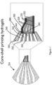
- cell layers e.g., adventitial and medial layer of a blood vessel can be bioprinted using a multicore-shell approach as illustrated in Figure 1 .
- Exemplary core-shell nozzles that may be used in the bioprinting methods described herein were described in PCT Pub. Nos. WO 2018/106704 and WO 2018/106705 .
- the nozzle may include a first nozzle tip defining a first outlet, where the first nozzle tip includes a first channel extending there through.
- the nozzle may further include a second nozzle tip defining a second outlet, where the second nozzle tip includes a second channel extending there through, and where the first channel surrounds the second outlet.
- the second nozzle tip may be retracted longitudinally with respect to the first nozzle tip such that the second outlet of the second nozzle tip is located in the first channel.
- Figure 1 shows an illustration of an extrusion process from a three-material core-shell nozzle 334.
- the nozzle 334 may, as shown, have a first channel 344, a second channel 342, and a third channel 340 for respectively extruding a first material 332 (e.g., fugitive ink), and second material 330 (e.g., a first cell-laden ink), and a third material 328 (e.g., a second cell-laden ink).
- the flow rates of each of the materials may be precisely controlled.
- the flow rate of the first material through the first channel may be slightly increased, and/or the flow rate of the second material through the second channel may be slightly decreased, etc.
- the multi-core shell approach was also previously described in PCT Pub. No. WO 2016/019087 ; and an article by Frutiger et al., Advanced Materials, 27:2440-2446 (2015 ), in reference to producing a soft sensor fiber.
- the multicore-shell approach is also illustrated in Figures 2 and 3 , with cell-laden inks containing predetermined type or types of viable cells, e.g., the layer-specific fibroblasts and smooth muscle cells for the adventitial and medial layer of a blood vessel, respectively.
- the multicore-shell approach allows for printing multi-layered tubular tissue constructs that are perfusable: the predetermined type or types of viable cells are printed in the pre-determined layer(s), and the construct is perfused and seeded with the endothelial cells.
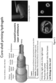
- Figure 3 shows pictures of an exemplary 3D printer and equipment details for the described methods:
- (A) shows a picture of a custom-built 3D printer with 4 individually addressable print heads (inset on the bottom left)
- (A) shows a picture of a custom-built 3D printer with 4 individually addressable print heads (inset on the bottom left)
- (A) shows a picture of a custom-built 3D printer with 4 individually addressable print heads (inset on the bottom left)
- the dark arrow points to one of the compressed air pressure boxes powering the extrusion-based printing method (inset on the bottom right).
- Image of the exemplary multicore-shell nozzle rendered in SolidWorks with the respective nozzle diameters is shown in (B).
- the core dimeter may be 750 ⁇ m
- shell width may be 250 ⁇ m.
- Figure 3(D) shows an exemplary PDMS printed gasket on a glass slide for housing the printed construct, dimension shown: inner width (e.g., 11 mm), inner length (e.g., 60 mm).
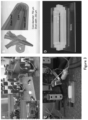
- Figure 4 depicts an exemplary 3D bioprinting printing process as described herein in still images of a printing video (left to right, top to bottom) with one wall of the PDMS gasket cut away for illustration purposes.
- a fugitive ink pillar is printed and tethered to the inlet perfusion pin.
- the fugitive ink core is connected to the outlet perfusion pin and the printing process is finished.
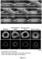
- Cross-sections (bottom of the figure) after printing allow appreciation of the varying layer thickness and lumen diameter that is individually controllable. Furthermore, layer integrity is maintained after casting the filament in a surrounding hydrogel matrix.
- the step of depositing on a substrate one or more filaments includes: (i) flowing a fugitive ink through a first extrusion tube, (ii) flowing a first cell-laden ink comprising one or more predetermined cell types through a second extrusion tube overlaying the first extrusion tube, the first cell-laden ink flowing around and enclosing the fugitive ink, and (iii) flowing a second cell-laden ink comprising one or more predetermined cell types through a third extrusion tube overlaying the second extrusion tube, the second cell-laden ink flowing around and enclosing the first cell-laden ink.
- additional cell-laden inks may be flowed to create additional cell-laden ink layers to create multi-layered tubular tissue constructs that are perfusable and have 3 or more cell layers.
- a core comprising a fugitive ink surrounded by an inner layer comprising a first cell-laden ink (in some embodiments also referred to as "middle shell"), and an outer layer comprising a second cell-laden ink (in some embodiments also referred to as "outer shell”).
- enclosing may be understood to mean fully enclosing or partially enclosing (e.g., radially covering), e.g., for producing blood vessels similar to an arteriole.
- a multi-layered tubular tissue construct comprising a core comprising fugitive ink (wherein the fugitive ink serves as a template for an open perfusable lumen within the filament) surrounded by an inner cell layer(s) and an outer cell layer is thus formed.
- open perfusable lumen refers to the lumen or core of the 3D printed filament and tissue construct that is capable of being perfused and capable of being in fluid communication with other tissues/channels, once sutured or implanted.
- the method may further include providing an extrusion head (or printhead) including the first, second, third, and, optionally fourth and/or fifth extrusion tubes arranged in a concentric configuration, and moving the extrusion head relative to a surface during the flowing of the inks, such that the multi-layered filament is deposited on the surface in a predetermined configuration or pattern.
- an extrusion head or printhead
- Such a process may be referred to as 3D printing or direct-write printing.
- 3D printing methods of printing various structures e.g., a functional part that includes a 3D structure comprising a structural material, and at least one functional electronic device is at least partially embedded in the 3D structure are provided in PCT Pub. No. WO 2014/209994 ; and a printed stretchable strain sensor are provided in PCT Pub. No. WO 2015/073944 .
- the flowing of the fugitive ink may occur at a first flow rate f
- the flowing of the second cell-laden ink may occur at a second flow rate f 1
- the flowing of the first cell-laden ink may occur at a third flow rate f 2 , etc., e.g., where f 2 >f 1 >f.
- flowing of the fugitive ink and the cell-laden ink(s) may have an alternative order of the flow rates, e.g., f 2 >f>f 1 ; or f>f 1 >f; or f>f 2 >f 1 ; or f>f 1 >f 2 ; or f 1 >f>f 2 ; or f 1 >f 2 >f.
- each of the flow rates f, f 1 , and f 2 is from about 0.1 L/s to about 10 L/s, although flow rates of up to tens of mL/s or hundreds of mL/s are possible depending on the nozzle size and print speed.
- the printhead or extrusion head may move relative to the surface at a print speed of from about I mm/s to about 100 mm/s, and more typically from about I mm/s to about 10 mm/s.
- the printhead may move relative to the surface at a first print speed and at a second print speed different from the first speed during printing.
- the first print speed may be lower than the second print speed and may be employed to form ends of the multi-layered filament having increased layer thicknesses.
- the method may also include flowing a third cell-laden ink through a fourth extrusion tube overlying the third extrusion tube, the third cell-laden ink flowing around and enclosing the first cell-laden ink; and so on so forth.
- a fourth extrusion tube overlying the third extrusion tube, the third cell-laden ink flowing around and enclosing the first cell-laden ink; and so on so forth.
- multiple layers anywhere from 2 to 8 or more layers that are concentric and coaxial may be created.
- the various concentric and coaxial layers of the cell-laden filament may or may not be continuous throughout the entire printed filament.
- the deposited filaments are formed by utilizing cell-laden inks (e.g., cell-laden inks comprising one or more predetermined cell types, fugitive inks, structural inks, or ECM inks) having a suitable composition and rheological properties.
- cell-laden inks e.g., cell-laden inks comprising one or more predetermined cell types, fugitive inks, structural inks, or ECM inks
- These inks may be viscoelastic and comprise a viscosity with a non-linear shear dependence.
- the viscosity of the precursor inks may fall in the range of from about 0.001 Pa-sec to about 10,000 Pa-sec.
- the inks may optionally include viscosifiers or other rheological modifiers to help control the rheological properties.
- Each cell-laden ink, and optionally, the fugitive and/or ECM ink may include one or more cells of one or more predetermined cell types in a carrier that may be a liquid or a gel.
- the carrier may include, in addition to an extracellular matrix material as described above, one or more functional chemical substances as described above.
- the carrier may also or alternatively include a cell culture medium designed to support the growth of cells.
- a predetermined amount of a hydrogel precursor powder is mixed with a cell culture medium to form a solution of an appropriate composition.
- the cells of interest are then dispersed in the solution at the desired cell concentration (e.g., any of the cell concentrations set forth above for the cell-laden filaments), and mixed thoroughly.
- Steps to prepare exemplary cell-laden GelMA inks, cell-laden gelatin-fibrin inks, Pluronic F127 fugitive inks, and PDMS structural inks are described in the Examples below.
- each cell-laden ink and the resulting cell-laden ink layer can include a different type or types of viable cells.
- the first cell-laden ink may include Cell Type A and the second cell-laden ink may include Cell Type B; or the first cell-laden ink may include a combination of Cell Types A and C and the second cell-laden ink may include a combination of Cell Types B and E.
- certain types of cells may be overlapping in the resulting cell-laden ink layers.
- the first cell-laden ink may include Cell Type A and B, where the second cell-laden ink may include Cell Type B and C. This would result in the first cell-laden ink layer having Cell Type A and B, and the second cell-laden ink layer having Cell Type B and C (Cell Type B being present in both cell-laden inks and layers).
- Each of the one or more cell-laden ink layers can include at least one viable cell and may include a large number of viable cells.
- each of the cell-laden ink layers may have a cell concentration of at least about 100 cells/ml, at least about 1000 cells/ml, at least about 10 4 cells/ml, at least about 10 5 cells/ml, at least about 10 6 cells/ml, at least about 10 7 cells/ml, or at least about 10 8 cells/ml.
- the cell concentration is no higher than about 10 9 cells/ml, or no higher than about 10 8 cells/ml.
- the cell concentration may be uniform or substantially uniform (e.g., within ⁇ 10%, within ⁇ 5%, or within ⁇ 1%) throughout each of the cell-laden layer, and the cell concentration may also be substantially uniform throughout each of the deposited filament.
- cell-laden layers and filaments that include aggregates or clusters of cells that may range in size from about 10 cells/cluster to about I million cells/cluster; or from about 10 cells/cluster to about 10000 cells/cluster, or from about 10 cells/cluster to about 100 cells/cluster.
- Such clusters may be dispersed uniformly or non-uniformly within the cell-laden layers and filaments ( Figure 15 ).
- the cell concentration may be substantially uniform throughout the tissue construct, or the cell concentration may include predetermined inhomogeneities within the tissue construct that may be defined by the location and morphology of the tissue construct, and/or by the cell distribution within the tissue construct.
- the cell clusters can be primary isolated clusters of parenchyma, like beta islets or exocrine part of the pancreas or liver tissue pieces; organoids formed of primary isolated single cells, like liver organoids or intestinal organoids; organoids differentiated from embryonic or induced-pluripotent stem cells, such as kidney organoids; aggregates of stem cells called embryoid bodies; aggregates of primary cells and/or cell lines, e.g. endothelial cell/fibroblast aggregates, cancer spheroids, B-cell zones from lymph nodes.
- each cell-laden ink layer comprises both cell clusters of a single or multiple cell types and single cells dispersed within the ink materials of one or more cell types ( Figure 15 ).
- each of the cell-laden ink layers comprises a cell concentration of from one cell/ml to about 10 9 cells/ml.
- the viable cells and the predetermined cell types in the multi-layered tubular tissue construct may include any mammalian cell type selected from cells that make up the mammalian body, including germ cells, somatic cells, and stem cells.
- the cells may be patient-derived. Depending on the type of cell, cells that make up the mammalian body can be derived from one of the three primary germ cell layers in the very early embryo: endoderm, ectoderm or mesoderm.
- endoderm ectoderm or mesoderm.
- the term “germ cells” refers to any line of cells that give rise to gametes (eggs and sperm).
- sermatic cells refers to any biological cells forming the body of a multicellular organism; any cell other than a gamete, germ cell, gametocyte or undifferentiated stem cell.
- somatic cells make up all the internal organs, skin, bones, blood and connective tissue.
- a cell may include any somatic cell isolated from mammalian tissue, including organs, skin, bones, blood and connective tissue (i.e., stromal cells).
- somatic cells examples include fibroblasts, chondrocytes, osteoblasts, tendon cells, mast cells, wandering cells, immune cells, pericytes, inflammatory cells, endothelial cells, myocytes (cardiac, skeletal and smooth muscle cells), adipocytes (i.e., lipocytes or fat cells), parenchyma cells (neurons and glial cells, nephron cells, hepatocytes, pancreatic cells, lung parenchyma cells) and non-parenchymal cells (e.g., sinusoidal hepatic endothelial cells, Kupffer cells and hepatic stellate cells).
- stem cells refers to cells that have the ability to divide for indefinite periods and to give rise to virtually all of the tissues of the mammalian body, including specialized cells.
- the stem cells include pluripotent cells, which upon undergoing further specialization become multipotent progenitor cells that can give rise to functional or somatic cells.
- stem and progenitor cells examples include hematopoietic stem cells (adult stem cells; i.e., hemocytoblasts) from the bone marrow that give rise to red blood cells, white blood cells, and platelets; mesenchymal stem cells (adult stem cells) from the bone marrow that give rise to stromal cells, fat cells, and types of bone cells; epithelial stem cells (progenitor cells) that give rise to the various types of skin cells; neural stem cells and neural progenitor cells that give rise to neuronal and glial cells; and muscle satellite cells (progenitor cells) that contribute to differentiated muscle tissue.
- hematopoietic stem cells adult stem cells; i.e., hemocytoblasts
- mesenchymal stem cells adult stem cells
- epithelial stem cells progenitor cells
- neural stem cells and neural progenitor cells that give rise to neuronal and glial cells
- muscle satellite cells progenitor cells
- the cell type or types used as components in the cell-laden ink or printed or placed adjacent to the printed perfusable multilayered or single layered filaments can be pre-aggregated into organoids, spheroids, embryoid bodies, or more general cellular aggregates.
- the cells may be, but are not limited to smooth muscle cells (vascular, intestinal, or bronchial), mesenchymal cell (fibroblasts, mesenchymal stem cells), endothelial cells (vascular or lymph endothelium), epithelial cells (intestinal epithelial lining, airway epithelial lining), and pericytes or stromal cells.
- smooth muscle cells vascular, intestinal, or bronchial
- mesenchymal cell fibroblasts, mesenchymal stem cells
- endothelial cells vascular or lymph endothelium
- epithelial cells intestinal epithelial lining, airway epithelial lining
- pericytes or stromal cells pericytes or stromal cells.
- the structure can also be fabricated to include a polygonal (e.g., square or rectangular) cross-section, and individual layers can be provided with either rounded or polygonal (e.g., square or rectangular) shaped cross-sections by using tubes that have the desired cross-sectional shape. More complex cross-sectional shapes can also be provided.
- a polygonal e.g., square or rectangular
- individual layers can be provided with either rounded or polygonal (e.g., square or rectangular) shaped cross-sections by using tubes that have the desired cross-sectional shape. More complex cross-sectional shapes can also be provided.
- the cell-laden ink layers all have the same thickness.
- the cell-laden ink layers all have varying thickness.
- one or more sacrificial filaments comprising the same or different (i.e., "second" fugitive ink) fugitive ink may be deposited on a substrate to form a sacrificial filament network that interpenetrates one or more multi-layered cell-laden filaments.
- the network may include a two- or three-dimensional interconnected arrangement or network of the one or more sacrificial filaments. Removal of the fugitive ink after partial or complete encapsulation with the extracellular matrix composition creates a perfusable network of channels in the tissue construct.
- the perfusable system may serve as perfusion of, e.g. nutrients, but also allow for drainage of fluids of interest.
- the sacrificial filaments may be deposited in a 3D printing process that involves extrusion through a micronozzle, it may be advantageous for the fugitive ink to: (1) exhibit shear thinning behavior; (2) exhibit a defined yield stress ⁇ y ; and/or (3) have a shear elastic modulus G' and a shear viscous modulus G" modulus where G'>G" at room temperature.
- the fugitive ink is removed from the core of the filaments to create an open perfusable lumen(s). This can occur before, during, or after encapsulation of the filaments, and optionally the sacrificial filaments in an extracellular matrix.
- the fugitive ink may be removed by methods described below.
- the fugitive ink may be removed to generate open perfusable lumen(s).
- the fugitive ink may comprise a biocompatible material and may be designed for compatibility with the cell-laden formulations and the extracellular matrix composition during room temperature deposition.
- Suitable fugitive inks may include, for example, Pluronic F127, Pluronic F123, agarose, sugar, wax, gelatin and fatty oils (e.g., animal fat derived oils such as Crisco).
- a hydrogel is employed for the extracellular matrix composition (and/or the extracellular matrix material), and a hydrogel such as Pluronic F127 is employed as the fugitive ink, it may be advantageous for the fugitive ink and the matrix hydrogel to have similar water contents (e.g., within ⁇ 30%) to avoid distortion of the fugitive ink after printing.
- the fugitive ink and the extracellular matrix composition may also be selected to have complementary thermal transitions, as discussed further below.
- Pluronic F127 is an FDA-approved material that is biologically inert to multiple cell types over the short time periods needed to complete the fabrication process.
- the material includes a hydrophobic poly(propylene oxide) (PPO) segment and two hydrophilic poly(ethylene oxide) (PEO) segments arranged in a PEO-PPO-PEO configuration.
- Pluronic F127 undergoes thermally reversible gelation above a critical micelle concentration (CMC; about 21 wt.%) and the gelation temperature. The gelation temperature decreases from approximately 10°C to 4°C as the PEO-PPO-PEO concentration increases.
- CMC critical micelle concentration
- micelles form as the hydrophilic PEO segments self-assemble into corona that are well solvated by water, while the hydrophobic PPO segments tightly associate within the micelle cores.
- the hydrophobic PPO units are hydrated, such that individual PEO-PPO-PEO species become soluble in water giving rise to a gel-to-fluid transition for systems whose concentration exceeds the CMC.
- the material liquefies upon cooling below the gel point.
- the extracellular matrix material and/or the extracellular matrix composition may comprise a gel.
- An ideal gel for bioprinting applications may exhibit a rapid transition from a low viscosity solution to a solid-like gel, which may be seen by an initial increase in shear elastic modulus. Rapid, controllable gelation may enhance printed structure fidelity by minimizing or obviating swelling and dissociation typical of slow gelation processes.
- the term "gel” may refer to a semi-solid substance that may comprise a gelling agent to provide viscosity or stiffness. The gel may be formed upon use of a gelling agent, such as a thickening agent, crosslinking agent or a polymerization agent, and may comprise a cross-linked structure or a non-cross-linked structure.
- the gel may be hydrophobic or hydrophilic.
- suitable gels include a hydrogel, thermo-reversible gel, a photo-sensitive gel, a pH sensitive gel, a peptide gel, or a cell type specific gel.
- Additional examples of gels include silica gel, silicone gel, aloe vera gel, agarose gel, nafion, polyurethane, elastomers (thermoplastic, mineral-oil thermoplastic, etc.), ion-exchange beads, organogels, xerogels and hydrocolloids.
- Hydrogels include those derived from collagen, hyaluronate, fibrin, alginate, agarose, chitosan, gelatin, Matrigel, glycosaminoglycans, and combinations thereof.
- the gel may comprise gelatin methacrylate (GeIMA), which is denatured collagen that is modified with photopolymerizable methacrylate (MA) groups.
- Suitable hydrogels may comprise a synthetic polymer.
- hydrogels may include those derived from poly(acrylic acid) and derivatives thereof, poly(ethylene oxide) and copolymers thereof, poly(vinyl alcohol), polyphosphazene, and combinations thereof.
- the extracellular matrix material and/or the extracellular matrix composition may comprise a naturally derived biocompatible material, such as one or more extracellular matrix components, including collagen, fibronectin, laminin, hyaluronates, elastin, and/or proteoglycans.
- suitable biocompatible materials for the extracellular matrix material and/or the extracellular matrix composition may include variations of cellulose, Matrigel, acrylates, acrylamides, polylactic co-glycolic acid, epoxies, aldehydes, ureas, alcohols, polyesters, silk, proteins, glycosaminoglycans, carbohydrates, minerals, salts, clays, hydroxyapatite, and/or calcium phosphate.
- the extracellular matrix material and/or the extracellular matrix composition may comprise gelatin and fibrin.
- the gelatin and fibrin may form an interpenetrating polymer network that mimics natural extracellular matrix (ECM) and may be optimized for cell attachment, bioprinting, transparency, and biocompatibility.
- the fibrin-gelatin interpenetrating polymer network may be created by mixing solutions of fibrinogen and gelatin with transglutaminase (TG), a slow-acting Ca 2+ dependent enzyme, to create a gel-precursor solution that may later be mixed with bovine thrombin to create a fibrin gel backbone.
- Fibrin may be made from a concentrated fibrinogen solution that has been activated by bovine thrombin and calcium chloride.
- Fibrin is a rapidly coagulating phase that permits rapid, controllable gelation of a printed structure.
- fibrin and gelatin can be welded together via mobile surface chain entanglement, while forming a strong interface.
- Creating monolithic gels of this nature is possible due to the slow crosslinking kinetics of transglutaminase (TG).
- TG transglutaminase
- thrombin rapidly induces fibrin gel formation
- the gelatin present in the IPN allows one to print sacrificial ink on the already cast layer, and, ultimately, to encapsulate with liquid gelatin-fibrin.
- the two phases may be weld together, creating a monolithic gel.
- This material system which is discussed further below in the Examples, can be readily tailored to modify gelation kinetics, interface adhesion, mechanical properties, optical properties, and cell-material interactions.
- the extracellular matrix composition comprises one or more of gelatin, fibrin, fibrinogen, transglutaminase, thrombin and gelatin methacrylate, collagen, collagen-acrylate (or a cross-linkable version) of any kind, Matrigel, poly lactic-co-glycolic acid (PLGA), alginate, chitosan.
- the fugitive ink may be removed without damage to the tissue construct. For example, if the fugitive ink undergoes a gel-to-fluid transition as described above, cooling of the vascular pattern after encapsulation may be effective for removal of the fugitive ink. To remove Pluronic F127, the construct may be cooled to a temperature of no more than about 1°C, depending on the concentration.
- the fugitive ink may be dissolved in a suitable aqueous solution for removal. Once the fugitive ink is liquefied or dissolved, a vacuum may be applied to an exposed end of the vascular pattern to extract the ink. Alternatively, cell culture medium can be used to wash out the liquefied fugitive ink.
- the one or more filaments are exposed to fluid perfusion to induce cell proliferation and development, thereby producing a functional perusable multi-layered tubular tissue construct.
- the step of exposing the one or more filaments to fluid perfusion is under a fluid sheer stress (FSS).
- FSS fluid sheer stress
- the FSS may be pulsed to mimic blood pressure changes during regular heartbeats. Fluid flow is an essential feature of every microsystem involving cell handling, culture or sorting. Flows inevitably generates FSS.
- Fluid shear stress of "FSS” refer to the stress coplanar component along with a cross section of a material. This occurs due to the component's force vector that is analogous to the cross section.
- the fluid perfusion may be at FSS anywhere from about 0.000001 dyn/cm 2 to about 100 dyn/cm 2 ; alternatively, the fluid perfusion may be at FSS from about 0.01 dyn/cm 2 to about 10 dyn/cm 2 .
- the exposure to FSS may be constant and can be anywhere from 1 day to 100 days. Methods for producing tissue constructs using FSS were previously described in U.S. Provisional Patent Application Serial No. 62/517,536 .
- the method of the invention comprises a step of injecting a suspension of endothelial cells into the open perfusable lumen after removing the fugitive ink.
- an endothelial layer having up to 100% confluency may be formed lining the wall of the core, where "100% confluency" means that the wall is completely covered by endothelial cells.
- Each endothelial layer formed in cell-laden filament may have a confluency of at least about 80%, at least about 90%, at least about 95%, at least about 98%, at least about 99%, or 100%, or any confluency in between 80% and 100%, so that the filament may form a perfusable and functional structure, such as an actual blood vessel.
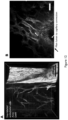
- the injected cells may grow into different layers, e.g., the endothelial cells can start vascularizing the wall of the blood vessel as they do in vivo (see Figure 12 ).
- the endothelial cells might grow into the walls, vascularizing the tubular construct wall upon cues from the surrounding cells with a dense capillary vasculature network connected to the lumen.
- the multi-layered tubular construct is a branched multi-layered tubular construct.
- the branched multi-layered tubular construct may be created by employing two multicore-shell nozzles, one of which has an elongated core.
- Exemplary multicore shell nozzle designs were previously described in U.S. Provisional Patent Application Serial No. 62/431,723, filed December 8, 2016 , entitled "Core-shell nozzle for three-dimensional printing and method of use," PCT Pub. No. WO 2018/106704 ; and PCT Pub. No. WO 2018/106705 .
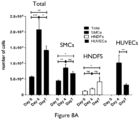
- Figures 8A through 8C illustrate exemplary multicore-shell nozzles.
- the cell-laden filaments and/or the sacrificial filaments may include one or more functional chemical substances, such as drugs, small molecules, toxins, proteins, growth factors, and hormones.
- the substrate for deposition typically comprises a material such as glass or other ceramics, PDMS, acrylic, polyurethane, polystyrene or other polymers.
- the substrate may comprise living tissue or dehydrated tissue, or one of the extracellular matrix compositions described above.
- the substrate may be cleaned and surface treated prior to printing. For example, glass substrates may undergo a silane treatment to promote bonding of the cell-laden filaments to the glass substrate.
- the substrate may not be a solid-phase material but may instead be in the liquid or gel phase and may have carefully controlled rheological properties, as described, for example, in W. Wu et al., Adv. Mater. 23 (2011) H178-H 183 .
- a fugitive ink was printed directly into synthetic hydrogels to create network structures.
- these synthetic materials do not support cell attachment and proliferation, limiting their use to non-biological applications.
- an extracellular matrix composition that facilitates cell attachment, migration, proliferation, and tissue-specific function while maintaining the appropriate rheology for printing is described.
- the multi-layered cell-laden and/or sacrificial filaments are embedded in the extracellular matrix composition during printing, and thus the at least partial surrounding of the tissue and sacrificial filament network with the extracellular matrix composition occurs during deposition of each of the multi-layered cell-laden and/or sacrificial filaments.
- the substrate onto which deposition occurs may be considered to be the container that holds the extracellular matrix composition or the extracellular matrix composition itself.
- the substrate is plastic or glass.
- the substrate may be Matrigel.
- the substrate may be plasma treated or coated with a layer of at least one of Matrigel, poly L-lysine, geltrex, gelatin, fibrin, fibrinogen, fibronectin, nitogen, vitrogen, collagen I, collagen IV, chitosan, alginate, glycosaminoglycans, or any other biomaterial.
- a substrate may be used to deposit the tissue construct components; in alternative embodiments, a substrate-free method incorporating embedded-printing may be used. Other methods of depositing a tissue construct in the housing are also contemplated (e.g., pin pull-out). Some alternative methods of printing three-dimensional living organs, producing organoids, embryoid bodies, endothelial sprouts, with or without embedded vasculature were previously described in PCT Pub. No. WO 2015/069619 and its corresponding U.S. Pat. Pub. No. US 2016-0287756 A1 ; and PCT Pub. No. WO 2016/179242 .
- the method of producing a multi-layered constructs may also include a step of exposing the construct to one or more biological agents, a biological agent gradient, a pressure, and/or an oxygen tension gradient, thereby inducing angiogenesis of capillary vessels to and/or from the tissue construct.
- the one or more biological agents, the biological agent gradient, the pressure, and/or the oxygen tension gradient further direct development, differentiation, and/or functioning of the tissue construct.
- the "exposing" may be by flowing the one or more biological agents, like blood, growth factors or other cell types, like blood cells, through the core of the tubular, perfusable tissue construct or by intermittently stopping perfusion.
- the cell-laden multi-layered filament produced by the methods described herein may also include one or more coaxial and/or concentric fugitive ink layer(s) to produce one or more concentric tubule(s) that are not interconnected.
- one multicore-shell filament can serve as a venous access while the other serves as an arterial access.
- not all multi-layered filaments serve the function of a blood vessel, e.g., one filament could serve the function of a draining channel, similar in function to a ureter of collecting bile duct.
- the cell-laden multi-layered filament produced by the methods described herein may also include one or more coaxial and/or concentric layers that include growth factor(s), alone or in combination with one or more types of viable cells.
- growth factors include fibroblasts growth factor, epithelial growth factor, vascular endothelial growth factor, insulin-like growth factor, platelet-derived growth factor, hormones like corticosteroids, or small molecules like blebbistatin.
- the cell-laden multi-layered filament produced by the methods described herein may include one or more coaxial and/or concentric layers that include materials that impart mechanical stability to the constructs.
- Such exemplary materials include polymeric materials like PLGA, PGA, PLA, PCL, polyurethane, polyhydroxyoctanoate or photocrosslinkable materials like GeIMA, methacylated hyaluronic acid, or similarly modified biopolymers.
- the method may further include, prior to surrounding or encapsulating the tissue and/or network with the extracellular matrix composition, depositing one or more structural filaments layer by layer on the substrate in a predetermined pattern to form a mold.
- the structural filaments may comprise one or more structural materials selected from among the exemplary extracellular matrix compositions or extracellular matrix materials provided above.
- the mold may hold the extracellular matrix composition during the encapsulation and may remain as part of the tissue construct, or it may be removed after processing.
- the structural filaments may define the perimeter of the tissue construct on the substrate and all or at least a portion of the three-dimensional shape of the tissue construct out of the XY plane.
- Certain embodiments relate to a method of producing a blood vessel construct, comprising depositing on a substrate one or more filaments, each filament comprising: a first cell-laden ink layer and a second cell-laden ink layer, the first and the second cell-laden layers being concentric and extending at least a portion of the length of the filament, the first cell-laden ink layer comprising a smooth-muscle cell (SMC)-containing cell-laden ink and the second cell-laden ink layer comprising a fibroblast-containing cell laden ink, and within the cell-laden ink layers a core comprising a fugitive ink, wherein the fugitive ink serves as a template for an open perfusable lumen within the filament; removing the fugitive ink to create the open perfusable lumen; after removing the fugitive ink, injecting a suspension of endothelial cells into the open perfusable lumen; and exposing the one or more filaments to fluid perfusion to induce cell proliferation
- the present invention also relates to a printed perfusable multi-layered tubular tissue construct as defined in the claims.
- the tissue construct may have any desired 2D or 3D shape.
- the tissue construct may have a planar geometry constructed from a single layer or multiple layers of multi-layered cell-laden filaments and an interpenetrating sacrificial filament network.
- Such structures may have any desired height (thickness).
- the tissue construct has a height of about 100 cm or less, about 10 cm or less, about I cm or less, about I mm or less, about 500 microns or less, or about 100 microns or less, and typically at least about 10 microns, at least about 100 microns, at least about 200 microns, or at least about I mm, with applications ranging from tissue cultures for implantation and drug screening to skin constructs and corneal replacements.
- the tissue construct may have an arbitrary or application-dependent 3D size and shape.
- the tissue construct may have a solid structure, a porous structure, and/or a hollow structure (e.g., tubular or nontubular) and may be fabricated to mimic the morphology and function of particular organ.
- the tissue construct may have the size and shape of a kidney, heart, pancreas, liver, bladder, vagina, urethra, trachea, esophagus, stomach, any segment of the intestinal tract, skin or other bodily organ.
- the 3D size and shape may in some cases be determined by a mold.
- the tissue construct or part of a tissue construct may have a size and shape of hierarchical architectures similar to the human vasculature of arteries, arterioles, and small-scale vessels for, ultimately, implantation or suturing of a fully 3D printed tissue.
- the printed multi-layered tubular construct of the invention as defined in the claims comprises one or more filaments, each filament comprising: a plurality of concentric and coaxial cell-laden layers, each cell-laden layer comprising one or more predetermined cell types and extending at least a portion of the length of the filament, and an open perfusable lumen within the one or more filaments; endothelial cells in the open perfusable lumen; a sacrificial filament network interpenetrating the one or more cell-laden filaments, each of the sacrificial filaments comprising a fugitive ink; and an extracellular matrix composition at least partially surrounding the one or more filaments and the sacrificial filament network.
- non-coaxial and concentric filaments containing one or more cell types, one or more types of cell clusters, or both may be printed into the construct.
- the cell-laden multi-layered filament(s) of the construct produced by the methods described herein may also include one or more coaxial and/or concentric fugitive ink layer(s) to produce one or more concentric tubule(s) that are or are not interconnected.
- the cell-laden multi-layered filament(s) of the tissue construct produced by the methods described herein may also include one or more coaxial and/or concentric layers that include growth factor(s), alone or in combination with one or more types of viable cells.
- the cell-laden multi-layered filament(s) of the tissue construct produced by the methods described herein may include one or more coaxial and/or concentric layers that include materials that impart stability to the constructs.
- the printed multi-layered tubular construct may be a large scale vessel, an artery, an arteriole, a small-scale vessel, a vein, trachea, bronchus, airway tissue, milk duct, colon, lymphatic tissue, heart tube or intestinal section.
- the methods described herein are for producing blood vessels.
- the methods described herein are for producing other tissues, such as bronchus, which may be implanted and function as such in the body.
- Other functional tissue structures may also be produced by the described methods, and include, e.g., ureter, trachea, esophagus, intestine, colon, lymph duct, milk duct, pancreatic duct, or bile duct, as well as nerve sheath.
- the present invention provides the use of the multi-layered tubular tissue construct as defined in the claims as an interface with another printed tissue construct, fugitive network, or a body organ that enables a specific tissue function, wherein the use does not comprise a method for treatment of the human or animal body by surgery or therapy.
- the present invention further provides the perfusable multi-layered tubular tissue construct as defined in the claims for use as an interface with a body organ that enables a specific tissue function.
- the multi-layered tubular tissue construct defined in the claims may be for use by suturing into a body or connecting a printed tissue construct adjacent to the multi-layered tubular tissue construct and in fluid communication with it to a desired host's circulation, like an implantation to a mouse's circulatory system or a human's circulatory system.
- the multi-layered tubular tissue construct defined in the claims may be for maturation in vitro.
- the present invention further provides the multi-layered tubular tissue construct defined in the claims for use in vascular disease modeling as recited in the claims.
- the present invention further provides the multi-layered tubular tissue construct defined in the claims for use in drug toxicity studies as recited in the claims.
- the present invention further provides the multi-layered tubular tissue construct defined in the claims for use in drug screening applications as recited in the claims.
- the present invention further provides the multi-layered tubular tissue construct defined in the claims as a vascular tissue for use in replacing of arteries, veins, arterioles, venules, bronchus, bronchiolus, ureter, trachea, esophagus, intestine, colon, lymph duct, lymphatic tissue, milk duct, pancreatic duct, or bile duct, as recited in the claims.
- the present invention further provides the multi-layered tubular tissue construct as defined in the claims for use in replacing a tubular structure having concentric and coaxial cell layers within the body, as recited in the claims.
- the tissue constructs produced by the methods described herein can be further developed into a disease model of varying vessel wall stiffness.
- a significant number of pathological processes, such as inflammation or fibrosis include stiffening of the vessel wall and hence inhibiting the body's ability to sustain a regulated blood pressure and flow profile.
- the stiffness of the printed hydrogel inks can be altered or chemical composition can be varied (like addition of collagen or hyaluronic acid) in order to study effects of extracellular matrix composition and mechanical properties on the maturation and performance of the printed vessel structure.
- the printed cells or clusters of cells in the multilayered tissue construct might be diseased cells, like cancer cells or cancer cell aggregates, in addition to cells of cell clusters of the tissue of interest in which the cancer resides.
- Hierarchical architecture encompasses the biological hierarchical organization of the vasculature tree from layer arteries and arterioles down the capillary beds supplying the organs/printed tissue with nutrients to then venules and larger veins.
- the open capillary bed surrogate could incorporate additional printed or casted engineered tissue, such as, e.g., liver or pancreatic tissue, muscle tissue or kidney tissue.
- the structure of the printed capillary surrogate is not limited to the current design. In certain embodiments, it could be a 3D lattice structure or comprised of finer channels more hierarchically branched. The matrix around this bed could also be engineered to allow for angiogenesis and capillary sprouting from the main channel in order to perfuse the introduced tissue.
- this entire platform could serve as a step closer to rapidly producing physiologically relevant engineered tissue that are entirely biocompatible and implantable (regenerative medicine applications).
- this technology could be used to engineer different types of tissue other than the blood vessel, as described herein.
- tissue in the human body are comprised of different cell types, but also have a concentric and coaxial layer structure. Since this nozzle design is meant for producing blood vessels, in preferred embodiments, it is designed for a trilayer printed structure.
- additional/more layers can be introduced to create, e.g., a 4- or 5-layer structure to incorporate more cell types.
- These layers could be designed to have all the same or varying thickness and could contain multiple cell types, clusters of cells and different ink formulations.
- the intestinal wall is of high spatial organization of epithelial, muscular, and nervous tissue and built in this concentric and coaxial fashion.
- the perfusable lumen could serve as the intestinal lumen
- the first layer could contain nervous and connective tissue cells, e.g., Meissner plexus neurons and fibroblast or clusters thereof
- the second layer could contain intestinal smooth muscle cells or clusters thereof
- the third layer could contain additional smooth muscle cells in a different orientation and nervous cells, e.g.
- Auerbach plexus neurons and a fourth layer could contain connective tissue cells like fibroblasts.
- tissue of the larger airways the bronchi are similarly constructed as the blood vessels with a contractile smooth muscle layer of a different cell type, specifically airway smooth muscle cells, and also have a connective tissue layer similar to the blood vessel adventitial layer surrounding the smooth muscle layer.
- the first layer there could be clusters of glandular cells dispersed in addition to the airway smooth muscle cells fulfilling the function of mucus production and secretion into the lumen.
- the open lumen in this case would be perfused with air not liquid.
- the methods described herein may be used to produce a multi-layered tissue construct including intestine, milk duct, sweat gland, colon, esophagus, stomach, airway epithelium, epididymis, urethra, liver bile duct, pancreatic duct, nephron, kidney, or lymph duct.
- tissue structures like intestinal tissue, trachea, or bronchial and airway tissues could be manufactured in this printing method.
- the cell types functioning as the parenchyma could be pre-aggregated and dispersed in a layer of the multilayered construct with other cell types dispersed in the same layer alongside the aggregates.
- primary isolated beta islets of the pancreas along with pre-aggregated or single cells of the exocrine portion of the pancreas derived from stem cells or primary isolation could be printed into the first layer of the multilayered tissue construct.
- the second layer in this case could comprise endothelial cells and fibroblasts to form a vascular plexus to support the tissue.
- the perfused lumen in this case could be used to supply nutrients, but also as a site to sample the secreted enzymes, peptides, and hormones from the endocrine (beta islets) and exocrine part of the pancreatic cells present in the layers of the tissue construct.
- the present invention further provides a kit as defined in the claims.
- the kit of the invention includes the printed multi-layered tubular construct defined in the claims along with hardware for chip assembly such as, for example, stainless steel bottom plates, acrylic lids, screws, printed gaskets, tubing components and perfusion pins; and instructional materials.
- kits may include parts to print the tissue.
- a kit may comprise: one or more cell culture media; a biomaterial or supporting material as mentioned above, for example, gelatin, fibrin, fibrinogen, transglutaminase, thrombin and gelatin methacrylate, collagen, glycosaminoglycans, collagen-acrylate (or a cross-linkable version) of any kind, Matrigel, poly lactic-co-glycolic acid (PLGA), alginate, and chitosan; one or more of the mentioned materials; or multiple cell sources, such as primary cells, embryonic stem cells, pluripotent stem cells, or cells differentiated from a stem cell type; cellular aggregates; growth factors and/or small molecules; custom designed nozzles or commercially available nozzles for extrusion-based bioprinting; hardware for chip assembly such as, for example, stainless steel bottom plates, acrylic lids, screws, printed gaskets, tubing components and perfusion pins; and instructional materials like a manual directing the
- kits may also include analysis tools, such as a live/dead cellular staining protocol and the needed reagents, an assortment of staining reagents to stain the tissue of interest for common markers of that specific tissue (e.g. in the case of a blood vessel one or more adventitial markers, one or more medial markers, and one or more intimal markers); histological staining components to visualize tissue damage or matrix remodeling; primers and other reagents for PCR applications, such as polymerases (e.g., to investigate expression levels of common markers or disease markers for the tissue of interest).
- analysis tools such as a live/dead cellular staining protocol and the needed reagents, an assortment of staining reagents to stain the tissue of interest for common markers of that specific tissue (e.g. in the case of a blood vessel one or more adventitial markers, one or more medial markers, and one or more intimal markers); histological staining components to visualize tissue damage or matrix remodeling; primers and other
- Example 1 Printing robust human vasculature via multicore-shell 3D printing
- Gelatin from porcine skin was dissolved at 15 w/v % in phosphate-buffered saline (PBS) without calcium or magnesium while stirring at 70°C.
- PBS phosphate-buffered saline
- the solution was processed at 70°C for 12 h, the solution's pH was adjusted to 7.5 by titration with 2 N sodium hydroxide, and filtered warm using a sterile 200 nm pore size standard cell culture filter. All solutions were stored in solid-state aliquots at 4°C.
- ink formulation the aliquots were melted to liquid state in a 37°C warming block. Fibrinogen was prepared freshly each print session. It was dissolved at 80 mg/mL and left undisturbed for at least 30 min in PBS without calcium or magnesium to avoid precipitation. Pluronic F127 was homogenized with a speed mixer at 40 wt. % in sterile deionized water, and stored at 4 °C. From this stock, the fugitive ink was made.
- Thrombin from bovine plasma was dissolved in sterile deionized water at 2000 U/mL and stored in aliquots at -20°C.
- Calcium chloride was dissolved at 250 mM as a stock solution and sterile filtered.
- Transglutaminase (Moo Gloo) was prepared freshly for every print session: It was dissolved at 6 w/v% in PBS without calcium or magnesium for 30 min at 37 °C and sterile filtered once dissolved. 22
- the formulation of the ink was 7.5 w/v% gelatin, 20 mg/mL fibrinogen, and 2.5 mM calcium chloride in PBS without calcium or magnesium unless otherwise indicated.
- the fibroblasts were dispersed in PBS before mixing into the ink at 0.5 million cells/mL (final concentration).
- SMC ink was brought to 8-10 million cells/mL depending on availability of cells. All inks were mixed well at 37°C by gentle pipetting to ensure proper resuspension of the cells in the viscous ink.
- Cell-laden inks were loaded into syringes and cooled to 4°C for 15 min to induce thermal gelation of the gelatin. Before printing, the inks were equilibrated to room temperature for 15 min.
- the fugitive ink was prepared from the 40 wt% Pluronic mixture. It was diluted to 33 wt% and thrombin was added at 50 U/mL to induce rapid polymerization of the fibrinogen in the cell-laden inks upon printing.
- the ink was homogenized by speed mixing, loaded into a dispensing syringe, cooled to 4 °C and centrifuged to remove air bubbles. Before printing, the fugitive ink was equilibrated to room temperature for 15 min to solidify it and make it suitable for printing.
- SMCs, GFP-HNDFs, and RFP-labeled human umbilical vein endothelial cells were cultured as according to their respective manufacturer's protocol, and not used in experiments past passage 10, 15, and 12, respectively.
- the cells were trypsinized for collection and subsequent ink preparation. All cell types were counted via an automated cell counter (Cellometer) according to manufacturer's protocols and diluted to the target number of cells to add to the cell-laden inks. To concentrate the cells, they were centrifuged at 220 g and the supernatant was aspirated. The cells were resuspended in the PBS compartment of the ink formulation and mixed with gelatin, fibrinogen, and calcium chloride as described in the paragraph above.
- the 3D printer used for all the experiments was a custom-built Aerotech 3D Printer with an overall size of 725 mm ⁇ 650 mm ⁇ 150 mm.
- Four print heads with a syringe barrel or nozzle attached were individually addressed and controlled.
- a mounted camera was used to define X, Y, and Z coordinates of the nozzle in respect to the printing substrate, and to determine X and Y offsets between the two nozzles on the different print-heads for multi-material 3D printing.
- a syringe containing the fugitive ink with a 0.4 mm inner diameter straight metal nozzle was mounted in the syringe barrel of the first printhead.
- the multi-core shell nozzle was attached to the second printhead via a small adapter plate and the syringes with the respective cell-laden inks were mounted on top of the nozzle.
- Room temperature is kept at 20°C- 21°C, below the reverse gelation point of the gelatin inks. If the room is reaching too high of a temperature, a cooling system can be attached to the cell-laden ink syringes to maintain ink temperature of 20°C.
- the multicore-shell nozzle was tilted 45° to the right to enable printing of the 3-layer filament close to the substrate. All syringes were connected to individually controlled compressed air driven pressure boxes (EFD, Nordson) to extrude the respective ink upon printing.
- EFD individually controlled compressed air driven pressure boxes
- the printer was controlled by a custom written G code program for this particular application loaded and edited in the printer software (Aerotech CNC Operator Interface/Aerobasic Editor).
- the multicore-shell nozzle design was used as described in U.S. Provisional Patent Application Serial No. 62/431,723, filed December 8, 2016 , entitled "Core-shell nozzle for three-dimensional printing and method of use". It is a tapered tri-layer nozzle with coaxially and concentrically organized shells.
- the generated part was printed on a stereolithography printer (Aureus, EnvisionTec) with a resin crosslinking at 700 W/dm2 (EnvisionTec). After printing the nozzle was carefully detached from the building platform, rinsed with isopropanol and water, and air-dried. The printed nozzles were good for 3-4 print sessions, after that clogging issue with the nozzles occur.
- the gaskets that house the subsequently printed constructs were printed in a two-component silicone elastomer (SE1700) on a 75x50 mm glass slide.
- Base and platinum-based crosslinker for the two-component system were mixed 10:1 by weight using a Thinky at a speed of 2000 for 5 min.
- Matlab script for G code generation Kolesky et al., Proc Natl Acad Sci U S A. 2016; 113(12):3 179-3 184 )
- the gaskets were designed to hold a 4.5 cm long printed vessel, suspended in matrix.
- the inlets the perfusion pins (1.27 mm outer diameter) were 5.2 mm above the bottom of the glass slide.
- the diameter of the opening in the PDMS gasket was 0.4 mm, about a third the outer diameter of the perfusion pin.
- PDMS is stretchable and can close tightly around the pin.
- Overall height of the printed gasket was 8.8 mm, rendering the total volume of the gasket to ⁇ 6 mL.
- the elastomer was prepared with the catalyst in a ratio of 10:1 by weight and loaded into a syringe and centrifuged for 5 min at 3000 g to remove air bubbles.
- a tapered plastic nozzle with an inner diameter of 410 ⁇ m (EFD Nordson) was connected to the syringe and zeroed on the print substrate.
- the gasket was printed onto the glass slide from the aforementioned G code and cured overnight at 80 °C in a furnace.
- the printed vessel construct fabrication process was adopted from Kolesky et al. ( Kolesky et al., Proc Natl Acad Sci U S A. 2016;113(12):3179-3184 ), and this protocol was altered to incorporate the multicore-shell printing (see, e.g., PCT Pub. No. WO 2016/019087 ).
- the construct was completed by casting a top hydrogel matrix to encapsulate the printed features.
- the fabricated construct was assembled into a perfusion system, allowing for flow of cell culture medium through the open lumen. All components for assembling the perfusion chips were autoclaved apart from the peristaltic tubing, which had to be ethanol sterilized.
- the construct can be subdivided into bottom, printed vessel, and top matrix.
- the matrix was comprised of 7.5 w/v% gelatin, 10 mg/mL fibrinogen, 2.5 mM calcium chloride, and 0.2 w/v% transglutaminase in PBS without calcium or magnesium.
- the liquid mixture was incubated for 15 min at 37 °C to pre-crosslink the matrix with the transglutaminase.
- the inner pin was sticking out about I mm out of the outer perfusion pin to facilitate optimal connection of the fugitive ink with the inner pin to produce a continuously perfusable construct.
- Inner pins were pulled out after casting.
- a pillar of fugitive ink was printed onto the dried bottom to facilitate connection to the printed vessel. Subsequently, the vessel was printed by first connecting the fugitive ink core to the printed pillar and then starting to extrude first the SMC ink to form the inner shell around the fugitive ink.
- the GFP-HNDF cell-laden ink was switched on to add the outer shell of the printed vessel.
- the cell-laden inks were switched off and the fugitive ink, still extruding, was used to connect to the perfusion pin on the other side of the gasket.
- the gasket was carefully filled in with the top matrix.
- the top matrix was prepared in the same fashion as the bottom matrix (see above) and 4.5 mL were mixed with I U/mL thrombin before casting, again to induce rapid transition of fibrinogen and proper connection between bottom matrix, printed construct and top matrix.
- the first layer of the bottom gel was prepared and casted exactly as in the dried hydrogel variant.
- the bottom layer was not dried but equilibrated to room temperature, and a I mL layer of 7.5 w/v% gelatin, 10 mg/mL fibrinogen, and 2.5 mM calcium chloride in PBS was cast on top of the already crosslinked hydrogel layer.
- the second layer was covered with a glass slide to reduce water evaporation and thermally gelled for ⁇ 30 min at room temperature. Subsequently, it was subjected to the same printing process as described above.
- the hydrogel inks were prepared cell-free for initial testing of the code.
- an additional multicore-shell nozzle with an elongated core part was used to penetrate the already trilayer filament and connect the fugitive ink cores.
- Printing of the branched constructs was performed in a gasket with two inlets and two outlets, where only one inlet was supplied with a perfusion pin.
- the printed branched construct was encapsulated in hydrogel ink and the fugitive ink was evacuated to test perfusion of both branches.
- Hierarchical printing was enabled by using another nozzle depositing fugitive ink, penetrating the already printed trilayer filament in the same way as the multicore-shell nozzle with an elongated core.
- the fugitive ink in the hierarchical printing was connected to the core of an already printed trilayer filament on the top and then a branching capillary network was printed directly onto the hydrogel.
- the fugitive ink was brought down into the core ink part of the already printed filament, piercing the outer vessel layers and the construct was subsequently encapsulated in hydrogel matrix. After evacuation of the fugitive ink, the construct was perfused with a red dye to test for full connection of the structures.
- Figure 14 illustrates examples of branched and hierarchical printing.
- the core part of the nozzle is elongated ( Figures 5A and 5B ).
- the ink employed for the interconnecting structure is the core ink without the surrounding layers.
- Branched and hierarchical prints enable interfaces with other biologically active tissue under continuous perfusion.
- the matrix was gelled at room temperature for 5 min and subsequently transferred to a 37 °C incubator for 2 h to fully crosslink the structures via the slow-acting transglutaminase. Following incubation, the construct was cooled to 4 °C for 15 min to thermally stabilize the gelatin-containing matrix and liquefy the fugitive ink comprised of Pluronic.
- the perfusion chip was assembled as previously described: the inner pins were pulled from the perfusion pins and tubing was attached to outer ends of the pins.
- a syringe reservoir (EFD Nordson) was attached containing 10 mL of incubator equilibrated SMC cell culture medium (5% CO2 environment at 37 °C) supplemented with additional 1 % antibiotic, antimycotic solution and 8 % fetal bovine serum (FBS).
- FBS fetal bovine serum
- a 0.2 ⁇ m sterile filter was attached via a Luer lock connection to the outlet of the syringe.
- a nozzle matching the inner diameter of the peristaltic tubing (PharMed BPT) was connected to the outlet of the filter, and the peristaltic tubing was used to connect medium reservoir and perfusion pin at the inlet of the chip.
- Cell culture medium was used to flush out the now liquid fugitive ink.
- the systems' circulation was closed with silicone tubing attached to the outlet perfusion pin that returned the perfusate back into the syringe reservoir via a small connector pin in its lid.
- the entire perfusion system was connected to a peristaltic pump (Ismatec) located in a 37 °C, 5 % CO2, and 95 % humidity cell culture incubator. Perfusion rate was set to 100 ⁇ L/min up to 2 mL/min, unless otherwise stated, and the construct was continuously perfused. Cell culture medium was changed every other day.
- a pipet containing 150 ⁇ L of cell suspension was attached to the outlet and the cell suspension was slowly pipetted into the channel. Pipet tip and tubing was removed, and outlet silicone tubing was reattached. The entire construct was transferred to the incubator and circulation of medium was interrupted with two tubing clips closing inlet and outlet, respectively, to ensure the cells remain in the channel. After 30 min, the construct was flipped upside-down to make sure the channel was evenly exposed to endothelial cells. The construct was left at 37°C without perfusion overnight or for more than 7 hours and put back into perfusion the next day.
- Live/dead cell staining with ethidium homodimer and calcein to investigate cell viability during the printing process and after 1 week of perfusion was performed using a kit containing the two dyes (Live/dead stain, Molecular Probes) according to the manufacturer's instruction.
- First and last prints of a print session were incubated in the staining solution and imaged on a confocal microscope right away to investigate longevity of the cells in the inks and viability after extrusion through the nozzle where the cells experience high shear.
- Cast controls and samples from 1 week of perfusion were also stained and imaged. Six fields of view per sample and one sample per print condition and cast control condition were analyzed.
- Figure 6A shows a representative cell viability confocal microscopy image of live and dead cells stained with a mammalian cell live/dead staining kit (calcein AM and ethidium bromide homodimer).
- Figure 6A shows great cell viability in the first print.
- the samples were fixed in formalin if not otherwise indicated.
- the perfused constructs were taken out of the incubator and first washed with 5mL PBS containing calcium and magnesium by gravity feed with a pressure head of 40 cm ( ⁇ 4 kPa). Subsequently, formalin was flushed into the chip and the inside of the channel was fixed for 15 min at room temperature. The chip was taken apart and the perfusion pins were pulled out of the construct.
- the hydrogel construct was taken out of the gasket and transferred to a 15 mL conical tube containing 9 mL formalin to complete the fixation process with an additional 45 min of fixation. Slice cultures and cast controls were washed in PBS for 5 min and subsequently fixed for 30 min at room temperature in formalin.
- Figure 8A depicts layer-specific proliferation of the different cell types over the course of 7 days in the system analyzed via flow cytometric analysis, a p-value below 0.05 was considered statistically significant.
- Figure 8B shows marker expression of the various layers in the printed constructs after 2 weeks of maturation under active perfusion.
- Figure 8C shows alignment analysis of the subendothelial layer of cells. Alignment was analyzed via the OrientationJ Plugin in ImageJ and 6 different frames of upper or lower longitudinal section were analyzed per sample.
- Figure 8D shows a still 3D reconstruction image of capillaries invading the vessel wall.
- Figure 9 depicts: (A) Confocal images of longitudinal cross-sections for endothelial marker expression in construct matured for 4 weeks in two different extracellular matrix compositions with (left panels) or without (right panels) fully printed wall structures. RFP-HUVECs do not perform as well and can be replaced with 'normal' HUVEC cells not expressing a fluorescent marker; and (B) Confocal images of longitudinal and horizontal cross-sections for medial marker expression in construct matured for 4 weeks in two different extracellular matrix compositions. Smooth muscle cells express specific markers for up to 4 weeks of culture time.
- sections were immersed in water for 30 s to remove residual salts and buffers. Then, the slide was dipped into a Coplin jar containing Mayer's hematoxylin and agitated for 30 s. The staining solution was rinsed off in water for 1 min. This step was repeated once to achieve optimal staining. Subsequently, the section was stained with 1 % Eosin staining solution for 30 s. Afterwards, sections were dehydrated with two changes of 95 % ethanol and two changes of 100 % ethanol for 30 s each. The alcohol was extracted by two changes of xylene and the section was mounted in DPX resinous mountant with a cover glass on top of the section.
- Figure 7 illustrates: (A) Hematoxylin and Eosin (H&E) stained cryosections of a construct matured for 2 weeks under active perfusion; and (B) Detail images of (A) with arrow pointing to dense protein deposition by the cells lose to the lumen resembling native extracellular matrix.
- H&E Hematoxylin and Eosin
- cells were subjected to flow cytometry analysis at day 1, day 4 and day 7 after printing.
- flow cytometry analysis of cell numbers in the different layers of the printed constructs the constructs needed to be dissociated and the cells isolated. Three samples per time point were dissociated and individually collected. Protocols for dissociation were adopted from Homan et al. A dissociation solution comprising of 0.5 % trypsin and 10 mM EDTA was made. The solution was adjusted to pH 8 and equilibrated in a cell culture incubator for I h. Three constructs were taken out of the incubator per time point, the chips were disassembled and the hydrogel construct taken out of the gasket.
- the sample was cut open longitudinally, and any residual gel matrix not containing cells was removed using a sterile scalpel blade. These strips were then washed in PBS and incubated for 30 min in the dissociation solution at room temperature. Detachment of cells was controlled visually under a microscope during this incubation time. After incubation, strips were removed, detached cells were concentrated by centrifugation for 5 min at 220 g, and resuspended in 50 ⁇ L of PBS. Cells were fixed by addition of 500 ⁇ L of formalin and subsequently washed two times in PBS. Before flow cytometry, cells were stained with DAPI (1:1000) for 5 min. The samples were analyzed by a flow cytometer counting 10000 cells for each sample (BD Arialll). Data analysis was performed in FlowJo and GraphPad Prism.
- Figure 8A shows layer-specific proliferation of the different cell types in the system was analyzed via flow cytometric analysis, a p-value below 0.05 was considered statistically significant.
- constructs were subjected to distensibility measurements and subsequent fixation (see above) with 3 samples each on day 1, day 4, day 7, and day 14 after printing.
- the samples were equilibrated to room temperature for 1 hour and the open lumen was filled with water. Since this measurement was conducted to investigate mechanical behavior of the vessel wall over time, cellular survival was neglected.
- the chip inlet tubing and reservoir of each sample was removed, the chip was mounted on the stage of a digital Keyence microscope, and the inlet perfusion pin was connected to a long piece of tubing (Versilic) containing water.
- the outlet tubing was clipped and the long tubing at the inlet was used to apply hydrostatic pressure.
- the diameter of the vessel was measured via the microscope's software. The values were recorded and used for subsequent analysis. The data was processed using Excel and Prism.
- Figure 10 shows images from suturability testing as well as still images of sutured constructs.
- the samples were fixed and stained (see above) and imaging was performed on vessels cut open along their long axis to expose the inner part of the channels.
- Figure 8C depicts alignment analysis of the sub-endothelial layer of cells. Alignment was analyzed via the OrientationJ Plugin in ImageJ and 6 different frames of upper or lower longitudinal section were analyzed per sample.
- Statistical analysis was performed in GraphPad Prism 5. Generally, a p-value of ⁇ 0.05 was considered to indicate a statistically significant difference between means. For comparison of experimental groups of the cell viability analysis via live/dead cell staining, and the flow cytometry data, an ANOVA with Tukey's post-test with a significance level of 0.05 was performed to determine statistical differences between groups. For the MTS assay testing cell culture medium compatibility, a two-way ANOVA with a Bonferroni's post-test was performed to determine statistical differences between the medium treatments.
- the constructs were matured for 2/4 weeks under active perfusion and used for live analysis.
- the resistance to flow was measured by measuring volumetric flowrate under increasing hydrostatic pressure. Volumetric flow was measured by weighing out the collective flow through the construct after 30 s of flow at a given pressure. Resistance to flow could then be calculated. All measurements were conducted using HEPES-buffered Tyrode's solution.
- Tyrode's solution was supplemented with increasing concentrations of Norepinephrine (0.1 nM to 10 ⁇ M) and resistance to flow was measured as mentioned above (data not shown).
- the printed blood vessels were matured for 2/4 weeks under active perfusion and used for live staining with a calcium dye, rhod-2 AM.
- the constructs were taken out of their housing live and cut into 3-5 mm thick tissue sections.
- the thus produced samples were washed and loaded with the dye at 10 ⁇ M concentration in calcium free cell culture medium. After loading for 45 min at 37 ° C and 5 % CO 2 , the samples were washed 3 times with calcium free PBS and kept alive in warm serum free cell culture medium until the measurement started. Baseline fluorescence without stimulation was assessed with a scanning laser confocal microscope as mentioned above in times series mode.
- Figure 11A shows images of a sutured vessel part before and after cutting away the surrounding hydrogel.
- Figure 11B shows stills from time series videos of calcium imaging data (Rhod-2 AM dye) of baseline fluorescence and norepinephrine (NA) injection (top figure): the arrows and the oval correspond to the calcium traces in the bottom figure.
- the bottom of Figure 11B shows calcium traces obtained from image analysis of the time series acquired in, the black arrow points to the time point of injecting 10 uM norepinephrine.
- AoSMCs aortic smooth muscle cells
- the dye Evans-Blue was supplied at a concentration of 0.05 % in the perfusing cell culture medium for 2.5 h.
- the vessel was flushed once with phosphate-buffered saline and each vessel was cut into 5 pieces of 5 mm thickness. Subsequently, the dye was extracted via formamide extraction for 2 days at room temperature and the amount of dye was measured colorimetrically by absorbance measurement at 630 nm wavelength. The amount of dye measured was normalized by weight surface area of the vessel piece.
- the seeded endothelium in the in vitro vessels serves as a diffusional barrier for molecules like albumin of ⁇ 66 kDa as it does in vivo.
- vasoactivity vasoactivity (vaso-contraction or dilation) of the printed blood vessel construct
- various other compounds like endothelin-1, nitric oxide, or histamine can be tested in the setup for the calcium imaging that is mentioned above.
- a more generic stimulus can be applied. Smooth muscle cells are responsive to electrical stimulation and a testbed is being developed, in which electrical field stimulation can be applied while like calcium imaging is performed.
- the diameter of the blood vessel section can be monitored via brightfield microscopy during the stimulation.
- the vessel's contractile ability could be stimulated via biological agents like noradrenaline or endothelin-I.
- Testing tissue mechanics after maturation will include quantitative suturability testing and burst pressure measurements.
- cellular remodeling of the matrix can be measured via changes in modulus of the tissue over time. This data will then be used in modelling the mechanics of the tissue surrounded by matrix and predict the possible distention (contraction/relaxation) of the tissue. Consequently, from these analyses, the force generated by the cells under this current maturation regime will be calculated.
Landscapes
- Health & Medical Sciences (AREA)
- Engineering & Computer Science (AREA)
- Life Sciences & Earth Sciences (AREA)
- Biomedical Technology (AREA)
- Chemical & Material Sciences (AREA)
- Zoology (AREA)
- General Health & Medical Sciences (AREA)
- Cell Biology (AREA)
- Public Health (AREA)
- Animal Behavior & Ethology (AREA)
- Transplantation (AREA)
- Veterinary Medicine (AREA)
- Oral & Maxillofacial Surgery (AREA)
- Epidemiology (AREA)
- Dermatology (AREA)
- Medicinal Chemistry (AREA)
- Wood Science & Technology (AREA)
- Biotechnology (AREA)
- Organic Chemistry (AREA)
- Genetics & Genomics (AREA)
- Bioinformatics & Cheminformatics (AREA)
- Chemical Kinetics & Catalysis (AREA)
- Botany (AREA)
- Vascular Medicine (AREA)
- Urology & Nephrology (AREA)
- Biochemistry (AREA)
- General Engineering & Computer Science (AREA)
- Microbiology (AREA)
- Materials Engineering (AREA)
- Manufacturing & Machinery (AREA)
- Molecular Biology (AREA)
- Rheumatology (AREA)
- Cardiology (AREA)
- Heart & Thoracic Surgery (AREA)
- Gastroenterology & Hepatology (AREA)
- Pulmonology (AREA)
- Biophysics (AREA)
- Reproductive Health (AREA)
- Materials For Medical Uses (AREA)
- Prostheses (AREA)
Claims (30)
- Ein Verfahren zum Herstellen eines perfundierbaren mehrschichtigen röhrenförmigen Gewebekonstrukts, das Folgendes umfasst:Aufbringen einer oder mehrerer Fasern auf einem Substrat, wobei jede Faser Folgendes umfasst: eine Vielzahl von konzentrischen und koaxialen zellbeladenen Tintenschichten, wobei jede zellbeladene Tintenschicht einen oder mehrere vorbestimmte Zelltypen umfasst und sich mindestens über einen Anteil der Länge der Faser hinweg erstreckt, und einen Kern, der eine flüchtige Tinte umfasst, wobei die flüchtige Tinte als eine Schablone für ein offenes perfundierbares Lumen innerhalb der Faser dient;Entfernen der flüchtigen Tinte, um das offene perfundierbare Lumen zu schaffen;Injizieren einer Suspension von Endothelzellen in das offene perfundierbare Lumen nach dem Entfernen der flüchtigen Tinte; undAussetzen der einen odermehreren Fasern gegenüber Fluidperfusion, um Zellproliferation und -entwicklung zu induzieren, wodurch das perfundierbare mehrschichtige röhrenförmige Gewebekonstrukt hergestellt wird.
- Verfahren nach Anspruch 1, wobei:jede zellbeladene Tinte einen unterschiedlichen Typ von lebensfähigen Zellen umfasst;jede zellbeladene Tinte überlappende Populationen von lebensfähigen Zellen umfasst; und/oderdie Zellen in jeder zellbeladenen Tinte aus der Gruppe ausgewählt sind, die aus glatten Muskelzellen (vaskulär, intestinal oder bronchial), Mesenchymzellen (Fibroblasten, mesenchymale Stammzellen), Perizyten, Endothelzellen (vaskulär oder Lymphendothel) oder Epithelzellen (intestinale Epithelauskleidung, Kolonepithelauskleidung, Epithelauskleidung der Atemwege) besteht.
- Verfahren nach Anspruch 1 oder Anspruch 2, wobei der Schritt des Aufbringens einer oder mehrerer Fasern auf einem Substrat Folgendes umfasst:Strömenlassen der flüchtigen Tinte durch eine erste Extrusionsröhre;Strömenlassen einer ersten zellbeladenen Tinte, die einen oder mehrere vorbestimmte Zelltypen umfasst, durch eine zweite, die erste Extrusionsröhre überlagernde Extrusionsröhre, wobei die erste zellbeladene Tinte um die flüchtige Tinte herum strömt und diese umschließt;Strömenlassen einer zweiten zellbeladenen Tinte, die einen oder mehrere vorbestimmte Zelltypen umfasst, durch eine dritte, die zweite Extrusionsröhre überlagernde Extrusionsröhre, wobei die zweite zellbeladene Tinte um die erste zellbeladene Tinte herum strömt und diese umschließt,dadurch Bilden des Kerns, der die flüchtige Tinte umfasst, umgeben von einer inneren Schicht, die eine erste zellbeladene Tintenschicht umfasst, und von einer äußeren Schicht, die eine zweite zellbeladene Tintenschicht umfasst.
- Verfahren nach Anspruch 3, wobei die erste zellbeladene Tinte glatte Muskelzellen umfasst; und/oder wobei die zweite zellbeladene Tinte Fibroblastenzellen umfasst.
- Verfahren nach Anspruch 3 oder Anspruch 4, das ferner das Bereitstellen eines Extrusionskopfs umfasst, der die erste, zweite und dritte Extrusionsröhre einschließt, die in einer konzentrischen Konfiguration angeordnet sind, wobei der Extrusionskopf während des Strömenlassens der flüchtigen Tinte, der ersten und der zweiten zellbeladenen Tinte relativ zu dem Substrat bewegt wird, wobei die Faser in einer vorbestimmten Konfiguration auf dem Substrat aufgebracht wird.
- Verfahren nach einem der Ansprüche 1 bis 5, das ferner Folgendes umfasst:mindestens teilweises Umgeben der einen oder mehreren Fasern mit einer extrazellulären Matrixzusammensetzung, wobei die extrazelluläre Matrixzusammensetzung optional eines oder mehrere von Gelatine, Fibrin, Fibrinogen, Transglutaminase, Thrombin und Gelatinemethacrylat, Collagen, Collagenacrylat (oder eine vernetzbare Version) einer beliebigen Art, Matrigel, Polylactid-co-glycolsäure (PLGA), Alginat oder Chitosan umfasst; und/oderAufbringen einer oder mehrerer Opferfasern auf dem Substrat vor dem mindestens teilweisen Umgeben der einen oder mehreren zellbeladenen Fasern mit der extrazellulären Matrixzusammensetzung, um ein Opferfasernetzwerk zu bilden, das die eine oder mehreren zellbeladenen Fasern interpenetriert, wobei jede der Opferfasern eine flüchtige Tinte umfasst, wobei das Netzwerk optional Strömungskanäle in Fluidverbindung mit den zellbeladenen Fasern zu deren Perfusion nach dem Entfernen der flüchtigen Tinte umfasst.
- Verfahren nach einem der Ansprüche 1 bis 6, wobei die zellbeladenen Tintenschichten eine Media-Schicht und eine Adventitia-Schicht eines Blutgefäßes bilden; und/oder wobei das perfundierbare mehrschichtige röhrenförmige Konstrukt einem Blutgefäß in Bezug auf Struktur und Funktion ähnlich ist; und/oderwobei das mehrschichtige röhrenförmige Konstrukt ein verzweigtes mehrschichtiges röhrenförmiges Konstrukt ist.
- Verfahren nach einem der Ansprüche 1 bis 7, wobei der eine oder die mehreren vorbestimmten Zelltypen Zellenaggregate oder Cluster von Zellen sind; wobei dieZellenaggregate oder Cluster optional eine Größe im Bereich von etwa 10 Zellen/Aggregat oder Cluster bis etwa 1 Million Zellen/Aggregat oder Cluster aufweisen.
- Verfahren nach einem der Ansprüche 1 bis 8, wobei die zellbeladenen Fasern eine oder mehrere funktionelle chemische Substanzen umfassen, die aus der Gruppe ausgewählt sind, die aus Folgendem besteht: Wirkstoffen, kleinen Molekülen, Toxinen, Proteinen, Wachstumsfaktoren und Hormonen.
- Verfahren nach einem der Ansprüche 1 bis 9, wobei jede der zellbeladenen Tintenschichten eine Zellkonzentration von einer Zelle/ml bis etwa 109 Zellen/ml umfasst; wobei die Zellkonzentration über jede der zellbeladenen Tintenschichten hinweg gleichförmig ist; und/oder wobei die zellbeladenen Tintenschichten alle die gleiche oder eine variierende Dicke aufweisen.
- Verfahren nach einem der Ansprüche 1 bis 10, wobei der Schritt des Aussetzens der einen oder mehreren Fasern gegenüber Fluidperfusion unter einer Fluid-Scherbeanspruchung (FSS) stattfindet; wobei die FSS optional gepulst ist, um Blutdruckänderungen während regelmäßiger Herzschläge nachzuahmen.
- Verfahren nach einem der Ansprüche 1 bis 11, wobei das Substrat Kunststoff, Glas oder Matrigel ist und wobei das Substrat optional plasmabehandelt ist oder mit einer Schicht von mindestens einem von Matrigel, Poly-L-Lysin, Geltrex, Gelatine, Fibrin, Fibrinogen, Fibronectin, Nitogen, Vitrogen, Collagen I, Collagen IV, Chitosan, Alginat, Glycosaminoglycanen oder einem anderen Biomaterial beschichtet ist.
- Verfahren nach einem der Ansprüche 1 bis 12, wobei jede Faser ferner eine oder mehrere konzentrische und koaxiale nichtzelluläre flüchtige Tintenschichten und/oder eine oder mehrere konzentrische und koaxiale Schichten umfasst, die Wachstumsfaktoren umfassen; und/oderwobei die nichtzellulären flüchtigen Tintenschichten der Faser ein oder mehrere Materialien umfassen, die dem perfundierbaren mehrschichtigen Gewebekonstrukt mechanische Stabilität verleihen.
- Verfahren nach einem der Ansprüche 1-13, wobei das perfundierbare mehrschichtige röhrenförmige Gewebekonstrukt ein Blutgefäßkonstrukt ist, wobei das Verfahren Folgendes umfasst:Aufbringen einer oder mehrerer Fasern auf einem Substrat, wobei jede Faser Folgendes umfasst: eine erste zellbeladene Tintenschicht und eine zweite zellbeladene Tintenschicht, wobei die erste und die zweite zellbeladene Schicht konzentrisch sind und sich mindestens über einen Anteil der Länge der Faser hinweg erstrecken, wobei die erste zellbeladene Tintenschicht eine glatte Muskelzellen (SMC) enthaltende zellbeladene Tinte umfasst und die zweite zellbeladene Tintenschicht eine Fibroblasten enthaltende zellbeladene Tinte umfasst, und innerhalb der zellbeladenen Tintenschichten einen Kern, der eine flüchtige Tinte umfasst, wobei die flüchtige Tinte als eine Schablone für ein offenes perfundierbares Lumen innerhalb der Faser dient;Entfernen der flüchtigen Tinte, um das offene perfundierbare Lumen zu schaffen;nach dem Entfernen der flüchtigen Tinte, Injizieren einer Suspension von Endothelzellen in das offene perfundierbare Lumen; undAussetzen der einen odermehreren Fasern gegenüber Fluidperfusion, um Zellproliferation und -reifung zu induzieren, wodurch das Blutgefäßkonstrukt hergestellt wird.
- Ein gedrucktes perfundierbares mehrschichtiges röhrenförmiges Konstrukt, das Folgendes umfasst:eine oder mehrere Fasern, wobei jede Faser Folgendes umfasst: eine Vielzahl von konzentrischen und koaxialen zellbeladenen Schichten, wobei jede zellbeladene Schicht einen oder mehrere vorbestimmte Zelltypen umfasst und sich mindestens über einen Anteil der Länge der Faser hinweg erstreckt, undein offenes perfundierbares Lumen innerhalb der einen oder mehreren Fasern;Endothelzellen in dem offenen perfundierbaren Lumen;ein Opferfasernetzwerk, das die eine oder mehreren zellbeladenen Fasern interpenetriert, wobei jede der Opferfasern eine flüchtige Tinte umfasst; undeine extrazelluläre Matrixzusammensetzung, die die eine oder mehreren Fasern und das Opferfasernetzwerk mindestens teilweise umgibt.
- Gedrucktes perfundierbares mehrschichtiges röhrenförmiges Konstrukt nach Anspruch 15, wobei das Konstrukt ein großkalibriges Blutgefäß, eine Arterie, eine Arteriole, ein kleinkalibriges Blutgefäß, eine Vene, Trachea, ein Bronchus, Atemwegsgewebe, Milchgang, Kolon oder Darmabschnitt ist; und/oder wobei das gedruckte mehrschichtige röhrenförmige Konstrukt ein verzweigtes mehrschichtiges röhrenförmiges Konstrukt ist.
- Gedrucktes perfundierbares mehrschichtiges röhrenförmiges Konstrukt nach Anspruch 15 oder Anspruch 16, wobei:jede zellbeladene Schicht einen unterschiedlichen Typ von lebensfähigen Zellen umfasst;jede zellbeladene Schicht überlappende Populationen von lebensfähigen Zellen umfasst; und/oderdie Zellen in jeder zellbeladenen Schicht aus der Gruppe ausgewählt sind, die aus glatten Muskelzellen (vaskulär, intestinal oder bronchial), Mesenchymzellen (Fibroblasten, mesenchymale Stammzellen), Perizyten, Endothelzellen (vaskulär oder Lymphendothel) oder Epithelzellen (intestinale Epithelauskleidung, Kolonepithelauskleidung, Epithelauskleidung der Atemwege) besteht;wobei eine erste zellbeladene Schicht optional glatte Muskelzellen umfasst und/oderwobei eine zweite zellbeladene Schicht Fibroblastenzellen umfasst.
- Gedrucktes perfundierbares mehrschichtiges röhrenförmiges Konstrukt nach einem der Ansprüche 15-17, wobei: die zellbeladenen Schichten eine Media-Schicht und eine Adventitia-Schicht bilden, um ein funktionelles Blutgefäß zu bilden; und/oder das gedruckte mehrschichtige röhrenförmige Konstrukt ein Blutgefäß ist.
- Gedrucktes perfundierbares mehrschichtiges röhrenförmiges Konstrukt nach einem der Ansprüche 15-18, wobei die eine oder mehreren Fasern mindestens teilweise von einer extrazellulären Matrixzusammensetzung umgeben sind, wobei die extrazelluläre Matrixzusammensetzung eines oder mehrere von Gelatine, Fibrin, Fibrinogen, Transglutaminase, Thrombin und Gelatinemethacrylat, Collagen, Glycosaminoglycanen, Collagenacrylat (oder eine vernetzbare Version) einer beliebigen Art, Matrigel, Polylactid-co-glycolsäure (PLGA), Alginat oder Chitosan umfasst.
- Gedrucktes perfundierbares mehrschichtiges röhrenförmiges Konstrukt nach einem der Ansprüche 15-19, wobei das Opferfasernetzwerk Strömungskanäle in Fluidverbindung mit den zellbeladenen Fasern zu deren Perfusion umfasst.
- Gedrucktes perfundierbares mehrschichtiges röhrenförmiges Konstrukt nach einem der Ansprüche 15-20, wobei die zellbeladenen Fasern eine oder mehrere funktionelle chemische Substanzen umfassen, die aus der Gruppe ausgewählt sind, die aus Folgendem besteht: Wirkstoffen, kleinen Molekülen, Toxinen, Proteinen, Wachstumsfaktoren und Hormonen.
- Gedrucktes perfundierbares mehrschichtiges röhrenförmiges Konstrukt nach einem der Ansprüche 15-21, wobei: jede der zellbeladenen Schichten eine Zellkonzentration von einer Zelle/ml bis etwa 109 Zellen/ml umfasst; die Zellkonzentration über jede der zellbeladenen Tintenschichten hinweg gleichförmig ist; und/oder die zellbeladenen Tintenschichten alle die gleiche oder eine variierende Dicke aufweisen.
- Gedrucktes perfundierbares mehrschichtiges röhrenförmiges Konstrukt nach einem der Ansprüche 15-22, wobei der eine oder die mehreren vorbestimmten Zelltypen Zellenaggregate oder Cluster von Zellen sind; wobei die Zellenaggregate oder Cluster von Zellen optional eine Größe im Bereich von etwa 10 Zellen/Aggregat oder Cluster bis etwa 1 Million Zellen/Aggregat oder Cluster aufweisen.
- Gedrucktes perfundierbares mehrschichtiges röhrenförmiges Konstrukt nach einem der Ansprüche 15-23, wobei mindestens eine oder mehrere Fasern ferner eine oder mehrere konzentrische und koaxiale nichtzelluläre flüchtige Tintenschichten und/oder eine oder mehrere konzentrische und koaxiale Schichten umfasst, die Wachstumsfaktoren umfassen; und/oderwobei die nichtzellulären Schichten der einen oder mehreren Fasern ein oder mehrere Materialien umfassen, die dem mehrschichtigen Gewebekonstrukt mechanische Stabilität verleihen.
- Verwendung des perfundierbaren mehrschichtigen röhrenförmigen Gewebekonstrukts nach einem der Ansprüche 15-24 als eine Grenzfläche mit einem anderen gedruckten Gewebekonstrukt, flüchtigen Netzwerk oder Körperorgan, die eine spezifische Gewebefunktion ermöglicht, wobei die Verwendung kein Verfahren zur Behandlung des menschlichen oder tierischen Körpers durch Chirurgie oder Therapie umfasst; wie etwa wobei das mehrschichtige röhrenförmige Gewebekonstrukt zur Reifung in vitro dient.
- Perfundierbares mehrschichtiges röhrenförmiges Gewebekonstrukt nach einem der Ansprüche 15-24 zur Verwendung als eine Grenzfläche mit einem Körperorgan, die eine spezifische Gewebefunktion ermöglicht; wie etwa wobei das perfundierbare mehrschichtige röhrenförmige Gewebekonstrukt zur Verwendung durch Nähen oder Implantieren in den Körper dient.
- Perfundierbares mehrschichtiges röhrenförmiges Gewebekonstrukt nach einem der Ansprüche 15-24 zur Verwendung beim Modeling von Gefäßerkrankungen, bei Arzneimittel-Toxizitätsstudien oder bei Drogen-Screening-Anwendungen.
- Perfundierbares mehrschichtiges röhrenförmiges Gewebekonstrukt nach einem der Ansprüche 15-24 als Gefäßgewebe zur Verwendung beim Ersatz einer röhrenförmigen Struktur mit konzentrischen und koaxialen Zellschichten innerhalb des Körpers; wie etwa wobei die röhrenförmige Struktur aus der Gruppe ausgewählt ist, die aus einer Arterie, einer Arteriole, einem kleinkalibrigen Gefäß, einer Vene, Trachea, einem Bronchus, Atemwegsgewebe, Milchgang, Kolon, Pankreas und Darmabschnitt besteht;
oder zur Verwendung in der regenerativen Medizin zum Ersatz von mindestens einem der Folgenden: Arterien, Venen, Arteriolen, Venülen, Bronchus, Bronchiolus, Ureter, Trachea, Ösophagus, Darm, Kolon, Lymphgang, Lymphgewebe, Milchgang, Pankreasgang oder Gallengang. - Ein Kit, der das gedruckte mehrschichtige röhrenförmige Konstrukt nach einem der Ansprüche 15-24 umfasst; wobei der Kit ferner Folgendes umfasst:Hardware zum Bestücken von Chips, wie etwa wobei die Hardware aus der Gruppe ausgewählt ist, die aus rostfreien Stahlbodenplatten, Acryldeckeln, Schrauben, gedruckten Dichtungen, Röhrenkomponenten und Perfusionsstiften besteht; undAnweisungsmaterialien.
- Kit nach Anspruch 29, der ferner Analysewerkzeuge umfasst; wobei die Analysewerkzeuge optional aus der Gruppe ausgewählt sind, die aus Folgenden besteht: einem Lebend-/Tot-Zellfärbeprotokoll und den benötigten Reagenzien; Färbereagenzien zum Färben des Gewebes von Interesse für häufig vorkommende Marker dieses speziellen Gewebes; histologische Färbekomponenten zum Sichtbarmachen von Gewebeschäden oder zum Matrix-Remodeling; Primer und andere Reagenzien für PCR-Anwendungen.
Applications Claiming Priority (2)
| Application Number | Priority Date | Filing Date | Title |
|---|---|---|---|
| US201762535557P | 2017-07-21 | 2017-07-21 | |
| PCT/US2018/043042 WO2019018737A1 (en) | 2017-07-21 | 2018-07-20 | METHODS FOR PRODUCING MULTILAYER TUBULAR TISSUE CONSTRUCTIONS |
Publications (4)
| Publication Number | Publication Date |
|---|---|
| EP3655052A1 EP3655052A1 (de) | 2020-05-27 |
| EP3655052A4 EP3655052A4 (de) | 2021-04-21 |
| EP3655052C0 EP3655052C0 (de) | 2025-06-25 |
| EP3655052B1 true EP3655052B1 (de) | 2025-06-25 |
Family
ID=65015342
Family Applications (1)
| Application Number | Title | Priority Date | Filing Date |
|---|---|---|---|
| EP18835590.3A Active EP3655052B1 (de) | 2017-07-21 | 2018-07-20 | Verfahren zur herstellung von mehrschichtigen röhrenförmigen gewebekonstrukten |
Country Status (5)
| Country | Link |
|---|---|
| US (2) | US12258585B2 (de) |
| EP (1) | EP3655052B1 (de) |
| CN (1) | CN111065422A (de) |
| AU (1) | AU2018302288A1 (de) |
| WO (1) | WO2019018737A1 (de) |
Families Citing this family (26)
| Publication number | Priority date | Publication date | Assignee | Title |
|---|---|---|---|---|
| SG11201908329QA (en) | 2017-03-15 | 2019-10-30 | Aspect Biosystems Ltd | Systems and methods for printing a fiber structure |
| WO2019018737A1 (en) | 2017-07-21 | 2019-01-24 | President And Fellows Of Harvard College | METHODS FOR PRODUCING MULTILAYER TUBULAR TISSUE CONSTRUCTIONS |
| WO2019021295A1 (en) | 2017-07-28 | 2019-01-31 | Stratasys Ltd. | METHOD AND SYSTEM FOR MANUFACTURING AN OBJECT HAVING HARD TISSUE PROPERTIES |
| WO2019021294A1 (en) | 2017-07-28 | 2019-01-31 | Stratasys Ltd. | ADDITIVE MANUFACTURING METHODS USING MATERIAL HAVING PROPERTIES OF A SOFT BODY TISSUE |
| ES2967431T3 (es) | 2017-07-28 | 2024-04-30 | Stratasys Ltd | Formulaciones utilizables en la fabricación aditiva de un objeto tridimensional hecho de un material blando |
| CN111148483B (zh) * | 2017-07-28 | 2022-11-15 | 斯特拉塔西斯公司 | 制造具有血管特性的物体的方法和系统 |
| AU2018336901B2 (en) | 2017-09-21 | 2024-07-18 | President And Fellows Of Harvard College | Tissue construct, methods of producing and using the same |
| WO2019079809A1 (en) * | 2017-10-22 | 2019-04-25 | The Johns Hopkins University | BIOMIMETIC PLATFORMS FOR MODELING VASCULAR PHYSIOPATHOLOGY, DIAGNOSIS AND THERAPY |
| US20200391443A1 (en) * | 2017-11-15 | 2020-12-17 | SMR Patents S.à.r.l. | Human machine interface (hmi) for a biopen |
| US20210316498A1 (en) * | 2018-08-20 | 2021-10-14 | Trustees Of Tufts College | Systems and methods for 3d printing of proteins |
| JP7535038B2 (ja) | 2018-09-19 | 2024-08-15 | アスペクト バイオシステムズ リミティド | コア繊維をプリントするためのシステム及び方法 |
| KR20210146895A (ko) * | 2019-03-29 | 2021-12-06 | 고리츠다이가쿠호진 요코하마시리츠다이가쿠 | 매트릭스 조성물 |
| KR102261908B1 (ko) * | 2019-05-15 | 2021-06-08 | 주식회사 이노리젠 | 이중 가교 가능한 2액형 바이오 잉크 조성물 및 이를 이용한 조직 유사 구조체의 제조방법 |
| JP7452792B2 (ja) * | 2019-09-06 | 2024-03-19 | 国立大学法人 長崎大学 | 気管支断端瘻治療用細胞構造体とその製造方法 |
| EP4051487A4 (de) * | 2019-11-01 | 2023-11-22 | Aspect Biosystems Ltd. | Systeme und verfahren zum bedrucken einer kern-hülle-faser |
| US12023856B2 (en) | 2020-04-22 | 2024-07-02 | Rowan University | Multi-material printer device for manufacture of high-throughput screening microfluidic chips |
| KR102438496B1 (ko) * | 2020-09-07 | 2022-09-02 | 포항공과대학교 산학협력단 | 생체외 인공장기 모델 및 세포 치료제로의 적용이 가능한 관류형 이중 근위세뇨관 관상 세포 구조체 및 그 생성 방법 |
| US12030256B2 (en) | 2021-02-19 | 2024-07-09 | The Board Of Trustees Of The University Of Illinois | Coated tubular construct for biological and industrial applications and method of making a tubular construct |
| US20240173117A1 (en) * | 2021-04-08 | 2024-05-30 | The George Washington University | 3d printing of biomimetic flexible multilayer blood vessels |
| GB202108078D0 (en) * | 2021-06-06 | 2021-07-21 | Copner Jordan Callum | Negative space 3D inkjet printing |
| SE546373C2 (en) | 2022-06-29 | 2024-10-22 | Cellink Bioprinting Ab | Nozzle for coaxial extrusion |
| EP4561645A2 (de) * | 2022-07-27 | 2025-06-04 | The Brigham & Women's Hospital, Inc. | Doppelt vernetzte 3d-gedruckte biomaterialien |
| FR3138661A1 (fr) * | 2022-08-08 | 2024-02-09 | Treefrog Therapeutics | Substitut de matrice extracellulaire dans un microcompartiment cellulaire |
| CN115814152A (zh) * | 2022-11-23 | 2023-03-21 | 中国人民解放军陆军军医大学 | 一种具有定向排列纤维微结构的高强度互穿网络水凝胶血管的制备方法 |
| WO2025064597A2 (en) * | 2023-09-22 | 2025-03-27 | President And Fellows Of Harvard College | Methods for embedded 3d printing of functional constructs for biological, robotic or other applications |
| AT527788A1 (de) * | 2023-12-07 | 2025-06-15 | Univ Wien Tech | Verfahren zur Herstellung eines Prüfkörpers |
Family Cites Families (16)
| Publication number | Priority date | Publication date | Assignee | Title |
|---|---|---|---|---|
| US3413387A (en) * | 1965-06-04 | 1968-11-26 | Haveg Industries Inc | Concentric extrusion |
| CN101259292A (zh) * | 2007-03-06 | 2008-09-10 | 首都医科大学宣武医院 | 一种组织工程血管的构建方法 |
| US10462907B2 (en) | 2013-06-24 | 2019-10-29 | President And Fellows Of Harvard College | Printed three-dimensional (3D) functional part and method of making |
| US20150050686A1 (en) * | 2013-08-13 | 2015-02-19 | Lawrence Livermore National Security, Llc | Omnidirectional, multiaxial bioprinted tissue system, techniques and applications |
| CN106163581B (zh) | 2013-11-05 | 2019-10-25 | 哈佛学院院长及董事 | 打印具有包埋的脉管系统的组织构建体的方法 |
| US10151649B2 (en) | 2013-11-18 | 2018-12-11 | President And Fellows Of Harvard College | Printed stretchable strain sensor |
| WO2016019087A1 (en) | 2014-07-31 | 2016-02-04 | President And Fellows Of Harvard College | Soft sensor fiber and method of making a soft sensor fiber |
| CN104490489B (zh) | 2014-12-02 | 2017-02-01 | 淮安皓运生物科技有限公司 | 基于3d生物打印技术制备组织工程血管的方法 |
| CN107427537A (zh) | 2015-03-03 | 2017-12-01 | 哈佛学院院长及董事 | 产生功能性人体组织的方法 |
| WO2016179242A1 (en) * | 2015-05-05 | 2016-11-10 | President And Fellows Of Harvard College | Tubular tissue construct and a method of printing |
| KR101751986B1 (ko) * | 2015-07-10 | 2017-06-30 | 한국기계연구원 | 인공 혈관 제작용 생체관 스캐폴드 및 그 제작 방법 |
| CN105561395B (zh) * | 2016-01-05 | 2018-08-21 | 广州医科大学附属第三医院 | 一种共轴式血管化组织工程骨的构建及其应用 |
| CN110035778B (zh) | 2016-09-06 | 2022-01-21 | 哈佛大学的校长及成员们 | 用于替代器官或特定器官功能的生活装置、方法及其用途 |
| US11207831B2 (en) | 2016-12-08 | 2021-12-28 | President And Fellows Of Harvard College | 3D printed core-shell filament and method of 3D printing a core-shell filament |
| KR101974716B1 (ko) * | 2017-05-12 | 2019-05-02 | 포항공과대학교 산학협력단 | 혈관 모사체 및 혈관 모사체 배양 방법 |
| WO2019018737A1 (en) | 2017-07-21 | 2019-01-24 | President And Fellows Of Harvard College | METHODS FOR PRODUCING MULTILAYER TUBULAR TISSUE CONSTRUCTIONS |
-
2018
- 2018-07-20 WO PCT/US2018/043042 patent/WO2019018737A1/en not_active Ceased
- 2018-07-20 CN CN201880058893.1A patent/CN111065422A/zh active Pending
- 2018-07-20 AU AU2018302288A patent/AU2018302288A1/en not_active Abandoned
- 2018-07-20 EP EP18835590.3A patent/EP3655052B1/de active Active
- 2018-07-20 US US16/631,677 patent/US12258585B2/en active Active
-
2025
- 2025-03-04 US US19/070,172 patent/US20250243465A1/en active Pending
Also Published As
| Publication number | Publication date |
|---|---|
| WO2019018737A1 (en) | 2019-01-24 |
| AU2018302288A1 (en) | 2020-02-13 |
| EP3655052C0 (de) | 2025-06-25 |
| US20250243465A1 (en) | 2025-07-31 |
| US20200164109A1 (en) | 2020-05-28 |
| CN111065422A (zh) | 2020-04-24 |
| US12258585B2 (en) | 2025-03-25 |
| EP3655052A4 (de) | 2021-04-21 |
| EP3655052A1 (de) | 2020-05-27 |
Similar Documents
| Publication | Publication Date | Title |
|---|---|---|
| US20250243465A1 (en) | Methods of producing multi-layered tubular tissue constructs | |
| JP6945453B2 (ja) | 管状組織構築物及び印刷方法 | |
| AU2014346959B2 (en) | Method of printing a tissue construct with embedded vasculature | |
| US7887843B2 (en) | Method for in vitro production of three-dimensional vital cartilage tissue and use thereof as transplant material | |
| JP2024015176A (ja) | 臓器および組織を脱細胞化および再細胞化する方法 | |
| Lewis et al. | Directing the growth and alignment of biliary epithelium within extracellular matrix hydrogels | |
| US11324779B2 (en) | Injectable microtissue systems, devices, and methods | |
| CN109913400A (zh) | 人工组织前体及制备其的方法 | |
| Yu et al. | Tissue strands as “bioink” for scale-up organ printing | |
| KR20180099843A (ko) | 첨가 증착에 의한 신체 대체제의 제조 방법 | |
| CN113846050B (zh) | 一种组织类器官的制备方法 | |
| Yang et al. | Fabrication of centimeter-sized 3D constructs with patterned endothelial cells through assembly of cell-laden microbeads as a potential bone graft | |
| WO2019060318A1 (en) | MANUFACTURING A BIOMEMETIC PLATFORM SYSTEM AND METHODS OF USE | |
| KR20030043937A (ko) | 혈관분포된 조직 이식편 | |
| Hu et al. | Rapid fabrication of gelatin-based scaffolds with prevascularized channels for organ regeneration | |
| JP2024513007A (ja) | 細胞集合体の培養のための微細流体の懸滴培養デバイス | |
| JP2024093080A (ja) | 立体的組織の製造方法 | |
| US20240425824A1 (en) | Scalable fabrication of a self-assembled cell sheet and uses thereof | |
| Nguyen et al. | Advances in three-dimensional printing of hydrogel formulations for vascularized tissue and organ regeneration | |
| US20240408816A1 (en) | Bioprinting of Collagenous Bioinks | |
| Ayan | Aspiration-assisted Bioprinting | |
| WO2025141425A1 (en) | A bioprinted liver model, a method for manufacturing the liver model and the use thereof in cytotoxicity evaluation | |
| CN120550195A (zh) | 一种组织工程基质及其用途 | |
| JP2023070669A (ja) | 人工血管床、人工三次元生体組織及び血管網を備えた人工三次元生体組織の作製方法 | |
| Voice | High-throughput Modular Tissue Engineering and Applications to Scale-up Tissue Constructs |
Legal Events
| Date | Code | Title | Description |
|---|---|---|---|
| STAA | Information on the status of an ep patent application or granted ep patent |
Free format text: STATUS: THE INTERNATIONAL PUBLICATION HAS BEEN MADE |
|
| PUAI | Public reference made under article 153(3) epc to a published international application that has entered the european phase |
Free format text: ORIGINAL CODE: 0009012 |
|
| STAA | Information on the status of an ep patent application or granted ep patent |
Free format text: STATUS: REQUEST FOR EXAMINATION WAS MADE |
|
| 17P | Request for examination filed |
Effective date: 20200117 |
|
| AK | Designated contracting states |
Kind code of ref document: A1 Designated state(s): AL AT BE BG CH CY CZ DE DK EE ES FI FR GB GR HR HU IE IS IT LI LT LU LV MC MK MT NL NO PL PT RO RS SE SI SK SM TR |
|
| AX | Request for extension of the european patent |
Extension state: BA ME |
|
| DAV | Request for validation of the european patent (deleted) | ||
| DAX | Request for extension of the european patent (deleted) | ||
| A4 | Supplementary search report drawn up and despatched |
Effective date: 20210319 |
|
| RIC1 | Information provided on ipc code assigned before grant |
Ipc: A61L 27/38 20060101AFI20210315BHEP Ipc: A61L 27/40 20060101ALI20210315BHEP Ipc: A61L 27/54 20060101ALI20210315BHEP Ipc: B33Y 80/00 20150101ALI20210315BHEP |
|
| STAA | Information on the status of an ep patent application or granted ep patent |
Free format text: STATUS: EXAMINATION IS IN PROGRESS |
|
| 17Q | First examination report despatched |
Effective date: 20230201 |
|
| GRAP | Despatch of communication of intention to grant a patent |
Free format text: ORIGINAL CODE: EPIDOSNIGR1 |
|
| STAA | Information on the status of an ep patent application or granted ep patent |
Free format text: STATUS: GRANT OF PATENT IS INTENDED |
|
| RIC1 | Information provided on ipc code assigned before grant |
Ipc: B33Y 80/00 20150101ALI20241218BHEP Ipc: C12N 5/071 20100101ALI20241218BHEP Ipc: B33Y 70/00 20200101ALI20241218BHEP Ipc: A61L 27/50 20060101ALI20241218BHEP Ipc: A61L 27/54 20060101ALI20241218BHEP Ipc: A61L 27/40 20060101ALI20241218BHEP Ipc: A61L 27/38 20060101AFI20241218BHEP |
|
| INTG | Intention to grant announced |
Effective date: 20250122 |
|
| RIN1 | Information on inventor provided before grant (corrected) |
Inventor name: LEWIS, JENNIFER A. Inventor name: LUSTENBERGER, PATRICK Inventor name: KOLESKY, DAVID Inventor name: UZEL, SEBASTIEN G.M. Inventor name: SKYLAR-SCOTT, MARK Inventor name: HOMAN, KIMBERLY Inventor name: KROLL, KATHARINA |
|
| GRAS | Grant fee paid |
Free format text: ORIGINAL CODE: EPIDOSNIGR3 |
|
| GRAA | (expected) grant |
Free format text: ORIGINAL CODE: 0009210 |
|
| STAA | Information on the status of an ep patent application or granted ep patent |
Free format text: STATUS: THE PATENT HAS BEEN GRANTED |
|
| AK | Designated contracting states |
Kind code of ref document: B1 Designated state(s): AL AT BE BG CH CY CZ DE DK EE ES FI FR GB GR HR HU IE IS IT LI LT LU LV MC MK MT NL NO PL PT RO RS SE SI SK SM TR |
|
| REG | Reference to a national code |
Ref country code: GB Ref legal event code: FG4D |
|
| REG | Reference to a national code |
Ref country code: CH Ref legal event code: EP |
|
| REG | Reference to a national code |
Ref country code: CH Ref legal event code: EP |
|
| REG | Reference to a national code |
Ref country code: IE Ref legal event code: FG4D |
|
| REG | Reference to a national code |
Ref country code: DE Ref legal event code: R096 Ref document number: 602018082977 Country of ref document: DE |
|
| U01 | Request for unitary effect filed |
Effective date: 20250710 |
|
| U07 | Unitary effect registered |
Designated state(s): AT BE BG DE DK EE FI FR IT LT LU LV MT NL PT RO SE SI Effective date: 20250717 |
|
| U20 | Renewal fee for the european patent with unitary effect paid |
Year of fee payment: 8 Effective date: 20250728 |
|
| PG25 | Lapsed in a contracting state [announced via postgrant information from national office to epo] |
Ref country code: NO Free format text: LAPSE BECAUSE OF FAILURE TO SUBMIT A TRANSLATION OF THE DESCRIPTION OR TO PAY THE FEE WITHIN THE PRESCRIBED TIME-LIMIT Effective date: 20250925 Ref country code: GR Free format text: LAPSE BECAUSE OF FAILURE TO SUBMIT A TRANSLATION OF THE DESCRIPTION OR TO PAY THE FEE WITHIN THE PRESCRIBED TIME-LIMIT Effective date: 20250926 |
|
| PGFP | Annual fee paid to national office [announced via postgrant information from national office to epo] |
Ref country code: GB Payment date: 20250929 Year of fee payment: 8 |
|
| PG25 | Lapsed in a contracting state [announced via postgrant information from national office to epo] |
Ref country code: HR Free format text: LAPSE BECAUSE OF FAILURE TO SUBMIT A TRANSLATION OF THE DESCRIPTION OR TO PAY THE FEE WITHIN THE PRESCRIBED TIME-LIMIT Effective date: 20250625 |
|
| PG25 | Lapsed in a contracting state [announced via postgrant information from national office to epo] |
Ref country code: RS Free format text: LAPSE BECAUSE OF FAILURE TO SUBMIT A TRANSLATION OF THE DESCRIPTION OR TO PAY THE FEE WITHIN THE PRESCRIBED TIME-LIMIT Effective date: 20250925 |