EP3635398B1 - Chimärer rezeptor zur verwendung in ganzzelligen sensoren zur detektion von analyten von interesse - Google Patents
Chimärer rezeptor zur verwendung in ganzzelligen sensoren zur detektion von analyten von interesse Download PDFInfo
- Publication number
- EP3635398B1 EP3635398B1 EP18729959.9A EP18729959A EP3635398B1 EP 3635398 B1 EP3635398 B1 EP 3635398B1 EP 18729959 A EP18729959 A EP 18729959A EP 3635398 B1 EP3635398 B1 EP 3635398B1
- Authority
- EP
- European Patent Office
- Prior art keywords
- cell
- analyte
- seq
- expression
- domain
- Prior art date
- Legal status (The legal status is an assumption and is not a legal conclusion. Google has not performed a legal analysis and makes no representation as to the accuracy of the status listed.)
- Active
Links
Images


Classifications
-
- G—PHYSICS
- G01—MEASURING; TESTING
- G01N—INVESTIGATING OR ANALYSING MATERIALS BY DETERMINING THEIR CHEMICAL OR PHYSICAL PROPERTIES
- G01N33/00—Investigating or analysing materials by specific methods not covered by groups G01N1/00 - G01N31/00
- G01N33/48—Biological material, e.g. blood, urine; Haemocytometers
- G01N33/50—Chemical analysis of biological material, e.g. blood, urine; Testing involving biospecific ligand binding methods; Immunological testing
- G01N33/53—Immunoassay; Biospecific binding assay; Materials therefor
- G01N33/575—Immunoassay; Biospecific binding assay; Materials therefor for cancer
-
- C—CHEMISTRY; METALLURGY
- C07—ORGANIC CHEMISTRY
- C07K—PEPTIDES
- C07K16/00—Immunoglobulins [IG], e.g. monoclonal or polyclonal antibodies
- C07K16/18—Immunoglobulins [IG], e.g. monoclonal or polyclonal antibodies against material from animals or humans
-
- G—PHYSICS
- G01—MEASURING; TESTING
- G01N—INVESTIGATING OR ANALYSING MATERIALS BY DETERMINING THEIR CHEMICAL OR PHYSICAL PROPERTIES
- G01N33/00—Investigating or analysing materials by specific methods not covered by groups G01N1/00 - G01N31/00
- G01N33/48—Biological material, e.g. blood, urine; Haemocytometers
- G01N33/50—Chemical analysis of biological material, e.g. blood, urine; Testing involving biospecific ligand binding methods; Immunological testing
- G01N33/53—Immunoassay; Biospecific binding assay; Materials therefor
- G01N33/5308—Immunoassay; Biospecific binding assay; Materials therefor for analytes not provided for elsewhere, e.g. nucleic acids, uric acid, worms, mites
-
- G—PHYSICS
- G01—MEASURING; TESTING
- G01N—INVESTIGATING OR ANALYSING MATERIALS BY DETERMINING THEIR CHEMICAL OR PHYSICAL PROPERTIES
- G01N33/00—Investigating or analysing materials by specific methods not covered by groups G01N1/00 - G01N31/00
- G01N33/48—Biological material, e.g. blood, urine; Haemocytometers
- G01N33/50—Chemical analysis of biological material, e.g. blood, urine; Testing involving biospecific ligand binding methods; Immunological testing
- G01N33/53—Immunoassay; Biospecific binding assay; Materials therefor
- G01N33/543—Immunoassay; Biospecific binding assay; Materials therefor with an insoluble carrier for immobilising immunochemicals
- G01N33/54366—Apparatus specially adapted for solid-phase testing
- G01N33/54386—Analytical elements
- G01N33/54387—Immunochromatographic test strips
-
- G—PHYSICS
- G01—MEASURING; TESTING
- G01N—INVESTIGATING OR ANALYSING MATERIALS BY DETERMINING THEIR CHEMICAL OR PHYSICAL PROPERTIES
- G01N33/00—Investigating or analysing materials by specific methods not covered by groups G01N1/00 - G01N31/00
- G01N33/48—Biological material, e.g. blood, urine; Haemocytometers
- G01N33/50—Chemical analysis of biological material, e.g. blood, urine; Testing involving biospecific ligand binding methods; Immunological testing
- G01N33/53—Immunoassay; Biospecific binding assay; Materials therefor
- G01N33/543—Immunoassay; Biospecific binding assay; Materials therefor with an insoluble carrier for immobilising immunochemicals
- G01N33/554—Immunoassay; Biospecific binding assay; Materials therefor with an insoluble carrier for immobilising immunochemicals the carrier being a biological cell or cell fragment, e.g. bacteria, yeast cells
-
- G—PHYSICS
- G01—MEASURING; TESTING
- G01N—INVESTIGATING OR ANALYSING MATERIALS BY DETERMINING THEIR CHEMICAL OR PHYSICAL PROPERTIES
- G01N33/00—Investigating or analysing materials by specific methods not covered by groups G01N1/00 - G01N31/00
- G01N33/48—Biological material, e.g. blood, urine; Haemocytometers
- G01N33/50—Chemical analysis of biological material, e.g. blood, urine; Testing involving biospecific ligand binding methods; Immunological testing
- G01N33/53—Immunoassay; Biospecific binding assay; Materials therefor
- G01N33/563—Immunoassay; Biospecific binding assay; Materials therefor involving antibody fragments
-
- G—PHYSICS
- G01—MEASURING; TESTING
- G01N—INVESTIGATING OR ANALYSING MATERIALS BY DETERMINING THEIR CHEMICAL OR PHYSICAL PROPERTIES
- G01N33/00—Investigating or analysing materials by specific methods not covered by groups G01N1/00 - G01N31/00
- G01N33/48—Biological material, e.g. blood, urine; Haemocytometers
- G01N33/50—Chemical analysis of biological material, e.g. blood, urine; Testing involving biospecific ligand binding methods; Immunological testing
- G01N33/53—Immunoassay; Biospecific binding assay; Materials therefor
- G01N33/566—Immunoassay; Biospecific binding assay; Materials therefor using specific carrier or receptor proteins as ligand binding reagents where possible specific carrier or receptor proteins are classified with their target compounds
-
- G—PHYSICS
- G01—MEASURING; TESTING
- G01N—INVESTIGATING OR ANALYSING MATERIALS BY DETERMINING THEIR CHEMICAL OR PHYSICAL PROPERTIES
- G01N33/00—Investigating or analysing materials by specific methods not covered by groups G01N1/00 - G01N31/00
- G01N33/48—Biological material, e.g. blood, urine; Haemocytometers
- G01N33/50—Chemical analysis of biological material, e.g. blood, urine; Testing involving biospecific ligand binding methods; Immunological testing
- G01N33/53—Immunoassay; Biospecific binding assay; Materials therefor
- G01N33/569—Immunoassay; Biospecific binding assay; Materials therefor for microorganisms, e.g. protozoa, bacteria, viruses
- G01N33/56911—Bacteria
-
- G—PHYSICS
- G01—MEASURING; TESTING
- G01N—INVESTIGATING OR ANALYSING MATERIALS BY DETERMINING THEIR CHEMICAL OR PHYSICAL PROPERTIES
- G01N33/00—Investigating or analysing materials by specific methods not covered by groups G01N1/00 - G01N31/00
- G01N33/48—Biological material, e.g. blood, urine; Haemocytometers
- G01N33/50—Chemical analysis of biological material, e.g. blood, urine; Testing involving biospecific ligand binding methods; Immunological testing
- G01N33/68—Chemical analysis of biological material, e.g. blood, urine; Testing involving biospecific ligand binding methods; Immunological testing involving proteins, peptides or amino acids
- G01N33/6854—Immunoglobulins
- G01N33/6857—Antibody fragments
-
- G—PHYSICS
- G01—MEASURING; TESTING
- G01N—INVESTIGATING OR ANALYSING MATERIALS BY DETERMINING THEIR CHEMICAL OR PHYSICAL PROPERTIES
- G01N33/00—Investigating or analysing materials by specific methods not covered by groups G01N1/00 - G01N31/00
- G01N33/48—Biological material, e.g. blood, urine; Haemocytometers
- G01N33/50—Chemical analysis of biological material, e.g. blood, urine; Testing involving biospecific ligand binding methods; Immunological testing
- G01N33/68—Chemical analysis of biological material, e.g. blood, urine; Testing involving biospecific ligand binding methods; Immunological testing involving proteins, peptides or amino acids
- G01N33/6872—Intracellular protein regulatory factors and their receptors, e.g. including ion channels
-
- G—PHYSICS
- G01—MEASURING; TESTING
- G01N—INVESTIGATING OR ANALYSING MATERIALS BY DETERMINING THEIR CHEMICAL OR PHYSICAL PROPERTIES
- G01N33/00—Investigating or analysing materials by specific methods not covered by groups G01N1/00 - G01N31/00
- G01N33/48—Biological material, e.g. blood, urine; Haemocytometers
- G01N33/50—Chemical analysis of biological material, e.g. blood, urine; Testing involving biospecific ligand binding methods; Immunological testing
- G01N33/68—Chemical analysis of biological material, e.g. blood, urine; Testing involving biospecific ligand binding methods; Immunological testing involving proteins, peptides or amino acids
- G01N33/6893—Chemical analysis of biological material, e.g. blood, urine; Testing involving biospecific ligand binding methods; Immunological testing involving proteins, peptides or amino acids related to diseases not provided for elsewhere
-
- C—CHEMISTRY; METALLURGY
- C07—ORGANIC CHEMISTRY
- C07K—PEPTIDES
- C07K2317/00—Immunoglobulins specific features
- C07K2317/80—Immunoglobulins specific features remaining in the (producing) cell, i.e. intracellular antibodies or intrabodies
-
- C—CHEMISTRY; METALLURGY
- C07—ORGANIC CHEMISTRY
- C07K—PEPTIDES
- C07K2319/00—Fusion polypeptide
- C07K2319/80—Fusion polypeptide containing a DNA binding domain, e.g. Lacl or Tet-repressor
Definitions
- the present disclosure relates to chimeric receptors that can be used in whole-cell sensors for detecting analytes of interest.
- IVDs In vitro diagnostic tests
- bioengineers have developed attractive methodologies that rely on synthetic nanoprobes (4-6) or microfluidics (7, 8).
- biosensing devices whole-cell biosensors mainly based on bacteria have proven to be applicable for the detection and quantification of a wide range of analytes (11, 12).
- Living cells have many attractive properties when it comes to diagnostics development. Cells detect biomolecules with high sensitivity and specificity and are capable of integrated and complex signal processing. Cells also provide a self-manufacturing platform via autonomous replication (12, 13), and the production of laboratory prototypes can be scaled using existing industrial frameworks (14). Spores from whole-cell biosensors can remain functional for extended periods of time, increasing the shelf life of a diagnostic product in harsh storage conditions (15). Last, whole-cell biosensors are highly versatile and can be used as stand-alone devices or interfaced with other technologies such as electronics, microfluidics, or micropatterning (16-18). All of these advantages have prompted the development of whole-cell biosensors that measure a variety of clinical parameters (19-24).
- Fominaya, Jes ⁇ s, and Winfried Wels teach the use of VHH single domain antibody in fusion protein comprising a DNA binding domain, but the system does not exploit exploiting the principle of split-DNA binding domain as taught by the present invention ( Fominaya, Jes ⁇ s, and Winfried Wels. "Target Cell-specific DNA Transfer Mediated by a Chimeric Multidomain Protein: NOVEL NON-VIRAL GENE DELIVERY SYSTEM (*).” Journal of Biological Chemistry 271.18 (1996): 10560-10568 ).
- WO199923116 discloses a CadC-fusion polypeptide comprising: a binding domain (i.e. a periplasmic domain), a linker (i.e. a transmembrane domain) and a DNA binding domain (i.e. a CadC transcriptional regulatory.
- WO2013022739 discloses a chimeric receptor polypeptide comprising i) a ligand binding domain, ii) a transmembrane domain and iii) a transcription factor.
- the fusion protein according to WO2013022739 further comprises a protease cleavage site.
- the present disclosure relates to a chimeric receptor polypeptide comprising i) a first DNA binding domain, ii) at least one binding domain having specificity for an analyte, and iii) a linker between the DNA binding domain and the binding domain.
- DNA binding domain refers to, but is not limited to, a motif that can bind to a specific DNA sequence (e.g., a genomic DNA sequence). DNA binding domains have at least one motif that recognizes and binds to single-stranded or double-stranded DNA. DNA binding domains can interact with DNA in a sequence-specific or a non-sequence-specific manner.
- CadC transcriptional activator has its general meaning in the art and refers to the membrane-integrated transcriptional regulator CadC of Escherichia coli.
- CadC activates expression of the cadBA operon at low external pH with concomitantly available lysine, providing adaptation to mild acidic stress.
- CadC is a representative of the ToxR-like proteins that combine sensory, signal transduction, and DNA-binding activities within a single polypeptide.
- CadC is composed of a C-terminal periplasmic pH-sensing domain, a single transmembrane helix and an N-terminal cytoplasmic winged helix-turn-helix DNA-binding domain ( Buchner S, Schlundt A, Lassak J, Sattler M, Jung K. Structural and Functional Analysis of the Signal-Transducing Linker in the pH-Responsive One-Component System CadC of Escherichia coli. J Mol Biol. 2015 Jul 31;427(15):2548-61 .). CadC dimerizes via its its C-terminal periplasmic pH-sensing domain.
- a coli CadC transcriptional activator DNA binding domain refers to the cytoplasmic domain of CadC that is capable of restoring its function via oligomerization of its C-terminal fusion domain.
- the E coli CadC transcriptional activator DNA binding domain comprises an amino acid sequence having at least 70% of identity with SEQ ID NO:1.
- the CadC transcriptional activator DNA binding domain comprises an amino acid sequence having at least 70% of identity with SEQ ID NO: 2.
- binding domain refers to one or more regions of a polypeptide that mediate specific binding with a target molecule (e.g. an analyte).
- the binding domain allows the receptor to bind a molecule that is not usually recognized by the natural receptor binding domain.
- binding refers to a non-covalent, preferably reversible binding of a molecule to the binding domain portion.
- binding involves non-covalent interactions such as salt bridges, hydrogen bonds, van der Waal forces, stacking forces, complex formation or combinations thereof between the compound and the binding domain portion. It also includes interactions with water molecules in the binding pocket.
- the binding domain is a single heavy chain variable domain of antibodies of the type that can be found in Camelid mammals which are naturally devoid of light chains.
- Such single domain antibody are also called VHH or "nanobody ® ".
- VHH single domain antibody
- (single) domain antibodies reference is also made to the prior art cited above, as well as to EP 0 368 684 , Ward et al. (Nature 1989 Oct 12; 341 (6242): 544-6 ), Holt et al., Trends Biotechnol., 2003, 21(11):484-490 ; and WO 06/030220 , WO 06/003388 .
- VHHs refers to the variable region of a single domain antibody found in camelids.
- analyte refers to compounds that can be bound by at least two chimeric receptors of the present and/or generate a conformational change in the chimeric receptor herein disclosed thus allowing the oligomerization of the CadC transcriptional activator DNA binding domain or the dimerization of the LexA transcriptional repressor binding domain.
- the analyte is selected from the group consisting of sugars, amino acids, peptides, proteins, nucleic acids, organic acids, anions, metals (e.g. molybolate, mercury, iron, zinc or nickel) or ions, oxides, hydroxides or conjugates thereof, inorganic ions (e.g.
- the term "specificity" refers to the ability of the binding domain of the chimeric receptor herein disclosed to bind the analyte of interest, while having relatively little detectable reactivity with others analytes. Specificity can be relatively determined by binding or competitive binding assays, using, e.g., Biacore instruments, as described elsewhere herein. Specificity can be exhibited by, e.g., an about 10:1, about 20:1, about 50:1, about 100:1, 10.000:1 or greater ratio of affinity/avidity in binding to the specific antigen versus nonspecific binding to other irrelevant molecules.
- affinity means the strength of the binding of an antibody to an epitope.
- affinity of an antibody is given by the dissociation constant Kd, defined as [Ab] x [Ag] / [Ab-Ag], where [Ab-Ag] is the molar concentration of the antibody-antigen complex, [Ab] is the molar concentration of the unbound antibody and [Ag] is the molar concentration of the unbound antigen.
- Kd dissociation constant
- Ka is defined by 1/Kd.
- the chimeric receptor polypeptide herein disclosed comprises two single heavy chain variable domains which target two different epitopes.
- linker refers to a sequence of at least one amino acid that links the DNA binding domain and the single heavy chain variable domain.
- the linker comprises an amino acid sequence having at least 50% of identity with SEQ ID NO:14.
- the linker between the DNA binding domain and the single heavy chain variable domain is a transmembrane domain.
- transmembrane domain refers to a domain natural or not (i.e. artificial) that spans the membrane of E coli bacterium and links the CadC transcriptional activator DNA binding domain to the binding domain having specificity for an analyte.
- Such transmembrane domain is composed of 18-25 mostly apolar amino acids and is responsible for the insertion and achoring of chimeric receptor polypeptide into membrane to form a cytoplasmic N-terminal (CadC transcriptional activator DNA binding domain) and an exoplasmic C-terminus (binding domain) topology.
- the transmembrane domain has to be monomeric with in lipid bilayers.
- Such transmembrane domains are characterized in Zhou et al., Proc. Natl. Acad. Sci. U.S.A., 98:2250, 2001 ; Lindner et al., J. Mol. Biol., 426:2942-2957, 2014 .
- the transmembrane domain comprises an amino acid sequence having at 50% of identity with SEQ ID NO:3 to NO:6.
- the transmembrane domain further comprises an extracellular spacer inserted between the transmembrane domain and the binding domain.
- spacer refers to a sequence of at least one amino acid that links the transmembrane domain with the binding domain. Such a spacer may be useful to prevent steric hindrances.
- said spacer is an amino acid sequence consisting of DTRLPMS (SEQ ID NO:7) or an amino acid sequence consisting of GGGSG (SEQ ID NO:12).
- all the domains containing in the chimeric receptor herein disclosed are fused in frame, i.e. operably linked to each other.
- the chimeric receptor herein disclosed comprises a first domain comprising an amino acid sequence having at least 70% of identity with SED ID NO:8 fused to a binding domain.
- a first amino acid sequence having at least 50% of identity with a second amino acid sequence means that the first sequence has 50; 51; 52; 53; 54; 55; 56; 57; 58; 59; 60; 61; 62; 63; 64; 65; 66; 67; 68; 69; 70; 71; 72; 73; 74; 75; 76; 77; 78; 79; 80; 81; 82; 83; 84; 85; 86; 87; 88; 89; 90; 91; 92; 93; 94; 95; 96; 97; 98; 99; or 100% of identity with the second amino acid sequence.
- a first amino acid sequence having at least 70% of identity with a second amino acid sequence means that the first sequence has 70; 71; 72; 73; 74; 75; 76; 77; 78; 79; 80; 81; 82; 83; 84; 85; 86; 87; 88; 89; 90; 91; 92; 93; 94; 95; 96; 97; 98; 99; or 100% of identity with the second amino acid sequence. Sequence identity is frequently measured in terms of percentage identity (or similarity or homology); the higher the percentage, the more similar are the two sequences. Methods of alignment of sequences for comparison are well known in the art. Various programs and alignment algorithms are described in: Smith and Waterman, Adv.
- ALIGN Myers and Miller, CABIOS 4:11-17, 1989
- LFASTA Nearson and Lipman, 1988
- ALIGN compares entire sequences against one another
- LFASTA compares regions of local similarity.
- the Blast 2 sequences function can be employed using the default BLOSUM62 matrix set to default parameters, (gap existence cost of 11, and a per residue gap cost of 1).
- the alignment should be performed using the Blast 2 sequences function, employing the PAM30 matrix set to default parameters (open gap 9, extension gap 1 penalties).
- the BLAST sequence comparison system is available, for instance, from the NCBI web site; see also Altschul et al., J. Mol. Biol., 215:403-410, 1990 ; Gish. & States, Nature Genet., 3:266-272, 1993 ; Madden et al. Meth. Enzymol., 266:131-141, 1996 ; Altschul et al., Nucleic Acids Res., 25:3389-3402, 1997 ; and Zhang & Madden, Genome Res., 7:649-656, 1997 .
- a further aspect of the present disclosure relates to a nucleic acid encoding for a chimeric receptor herein disclosed.
- nucleic acid molecule has its general meaning in the art and refers to a DNA or RNA molecule.
- the term captures sequences that include any of the known base analogues of DNA and RNA such as, but not limited to 4-acetylcytosine, 8-hydroxy-N6-methyladenosine, aziridinylcytosine, pseudoisocytosine, 5-(carboxyhydroxylmethyl) uracil, 5-fiuorouracil, 5-bromouracil, 5-carboxymethylaminomethyl-2-thiouracil, 5-carboxymethyl-aminomethyluracil, dihydrouracil, inosine, N6-isopentenyladenine, 1 -methyladenine, 1 -methylpseudouracil, 1-methylguanine, 1- methylinosine, 2,2-dimethylguanine, 2-methyladenine, 2-methylguanine, 3-methylcytosine, 5- methylcytosine, N6-methyladenine, 7-methylguanine, 5-methylaminomethyluracil, 5-methoxyamino-methyl-2-
- Suitable expression control sequences include promoters that are applicable in the target host organism.
- promoters are well known to the person skilled in the art for diverse hosts from prokaryotic organisms and are described in the literature.
- promoters can be isolated from naturally occurring genes or can be synthetic or chimeric promoters.
- the promoter can already be present in the target genome and will be linked to the nucleic acid molecule by a suitable technique known in the art, such as for example homologous recombination.
- Expression cassettes according to the invention are particularly meant for an easy to use insertion into target nucleic acid molecules such as vectors or genomic DNA.
- the expression cassette is preferably provided with nucleotide sequences at its 5'- and 3'-flanks facilitating its removal from and insertion into specific sequence positions like, for instance, restriction enzyme recognition sites or target sequences for homologous recombination as, e.g. catalyzed by recombinases.
- the present disclosure also relates to vectors, particularly plasmids, cosmids, viruses and bacteriophages used conventionally in genetic engineering, that comprise a nucleic acid molecule or an expression cassette herein disclosed.
- the vectors herein disclosed are suitable for the transformation of prokaryotic cells. Methods which are well known to those skilled in the art can be used to construct recombinant vectors.
- the vector may contain further genes such as marker genes which allow for the selection of said vector in a suitable host cell and under suitable conditions.
- the vector also contains one or more origins of replication.
- the nucleic acid molecules contained in the vectors are operably linked to expression control sequences allowing expression, i.e. ensuring transcription and synthesis of a translatable RNA, in prokaryotic cells.
- expression control sequences allowing expression, i.e. ensuring transcription and synthesis of a translatable RNA, in prokaryotic cells.
- the nucleic acid molecules herein disclosed or parts of these molecules can be introduced into plasmids.
- Expression vectors have been widely described in the literature. As a rule, they contain not only a selection marker gene and a replication origin ensuring replication in the host selected, but also a bacterial promoter and, in most cases, a termination signal for transcription.
- promoters there is in general at least one restriction site or a polylinker which enables the insertion of a coding nucleotide sequence. It is possible to use promoters ensuring constitutive expression of the gene and inducible promoters which permit a deliberate control of the expression of the gene. Bacterial promoter sequences possessing these properties are described in detail in the literature. Regulatory sequences for the expression in microorganisms (for instance E. coli) are sufficiently described in the literature. Inducible promoters are also possible. These promoters often lead to higher protein yields than do constitutive promoters.
- the invention relates to a method for producing a prokaryotic cell capable of expressing the chimeric receptor herein disclosed comprising genetically engineering cells with an above-described nucleic acid molecule, expression cassette or vector herein disclosed.
- the prokaryotic cells is selected among gram-negative bacteria.
- the prokaryotic cell is E. Coli.
- the prokaryotic cell is genetically engineered in such a way that it contains the introduced nucleic acid molecule stably integrated into the genome.
- the transformation of the prokaryotic cell with a nucleic acid molecule or vector according to the invention can be carried out by standard methods. For example, calcium chloride transfection is commonly utilized for prokaryotic cells.
- the prokaryotic cell is cultured in nutrient media meeting the requirements of the particular prokaryotic cell used, in particular in respect of the pH value, temperature, salt concentration, aeration, antibiotics, vitamins, trace elements etc.
- the prokaryotic cell herein disclosed comprises at least one detection protein for which the expression is under the control of the chimeric receptor herein disclosed.
- the binding of the analyte to the chimeric receptor triggers its oligomerization and thus allowing the oligomerization of the CadC transcriptional activator DNA binding domain which can then activate the expression of at least one detection protein which is placed under the control of CadBA promoter.
- the prokaryotic cell herein disclosed further comprises a nucleic acid molecule encoding for a detection protein operatively linked to a CadBA promoter.
- An exemplary nucleic acid for the CadBA promoter is represented by SEQ ID NO:9.
- the binding of the analyte to the chimeric receptor triggers its dimerization and its binding to the LexA operator, blocking expression of the reporter gene.
- the prokaryotic cell herein disclosed further comprises a nucleic acid molecule encoding for a detection protein operatively linked to a LexA promoter.
- An exemplary nucleic acid for the LexA promoter is represented by SEQ ID NO: 15.
- the detection protein refers to any protein that can be detected by biological or physical means.
- the detection protein is a fluorescent protein.
- the advent of fluorescent proteins has allowed non-invasive intracellular labeling, which are easily detectable by optical means.
- the green fluorescent protein (GFP) from Aequorea Victoria is now the most widely used reporter gene in many organisms. Multiple variants with different spectral properties have been developed.
- the prokaryotic cell comprises different combinations of fluorescent proteins exhibiting energy transfer provide for differential fluorescence.
- the detection protein is selected among luminescent proteins. Certain bacteria (e.g., Vibrio fischeri ) have autoinducible luminescent genes that express luciferase, which causes cleaving of luciferin and emission of blue light.
- Bacteria produce signal molecules, N-acyl homoseine lactones (AELs) that enter bacterial cells and induce transcriptional activation of the genes LuxI, which encodes AHL synthetase, and LuxR, which encodes the AHL-dependent transcriptional activator.
- a sufficiently high concentration of AHL in the cell causes binding to the LuxR activator and transcription of the luminescence genes.
- the detection proteins can be fusion proteins (e.g., green fluorescent protein-Fv) that have a detectable property and that are secreted from the cell.
- the secretion can be triggered by analyte binding to the chimeric receptor herein disclosed.
- the detection protein is produced in excess rather than in proportion to the analyte binding.
- the detection can be performed using RNA aptamers specifically binding a fluorescent probe. Binding of the probe to the aptamer increases its fluorescence and allows detection of gene expression.
- Other kind of output signals include production of pigments via specific operons (like the violacein operon, or the expression of Flavin Mono Oxydase converting tryptophane into indigo), or by the expression of an enzyme which substrate exogenously supplied is transformed in a colorimetric product, like the enzyme Beta-galactosidase and its substrate X-gal for example. More complex prokaryotic cells with higher levels of functionality can be created using techniques developed in the field of cellular computation.
- a cell serves as a biochemical computer, processing an input such as analyte binding using internal logic gates to generate an output.
- Complex conditional responses to multiple inputs have been engineered for example by implementing AND, NOT, OR, XOR, and IMPLIES logic gates in E. coli cells.
- these gates can be implemented using DNA-binding proteins to regulate expression of recombinant vectors.
- Others systems can be used, such as, but not limited to, recombinase-based logic gates, nucleic acids-based logic gates, or protein-based logic gates.
- the output can be a signal proportional to the amount of analyte binding and nature of the analyte.
- a single plasmid can be created with different promoters switched on by different binding events to control expression of different detection proteins.
- the output can control a genetic switch that can be tuned to respond to a particular signal threshold.
- switches have been engineered by using different verions of recombinases having various translational and/or proteolysis control elements, allowing the response threshold to be tuned over several orders of magnitudes (see Courbet et al., "Detection of pathological biomarkers in human clinical samples via amplifying genetic switches and logic gates”. Sci Transl Med. 2015 May 27;7(289):289ra83 .).
- the whole cell biosensing system can be tuned to respond to specific signal threshold that have been determined to be clinically or environmentally relevant.
- the prokaryotic cell is L-form bacteria.
- L-form bacteria or “L-phase bacteria”, “L-phase variants”, and “cell wall-deficient (CWD) bacteria” refers to strains of bacteria that lack cell walls. See EXAMPLE 4.
- the prokaryotic cell herein disclosed constitutes whole-cells biosensor that can be suitable for the detection and quantification of analytes.
- a further object of the present disclosure relates to a method for assaying for at least one analyte, comprising i) providing at least one set of prokaryotic cells herein disclosed, each comprising a chimeric receptor capable of binding to said analyte coupled to one detection protein ; b) contacting said set of cells with a sample suspected of containing said analyte for a time sufficient allowing the oligomerization of the chimeric receptors binding and then the expression of the detection protein; and c) detecting the expression level of the detection protein wherein the expression level correlated with the amount of the analyte present in the sample.
- sample refers to any volume of a liquid or suspension in which an analyte to be measured can be present in solution.
- the sample is bodily fluid sample.
- the sample is selected from the group consisting of blood samples (including serum or plasma samples), urine samples, cerebrospinal samples, tear samples, saliva samples and synovial samples.
- the whole-cell sensors herein disclosed may be used in multiplexed assays. Multiple analytes of interest in a single sample can be detected simultaneously through their capture by different cell types or subsets.
- an assay may be set up so that each cell type has a unique chimeric receptor herein disclosed specific for an analyte of interest and a unique detection protein. That is, there is a one-to-one relationship between the analyte and the whole cell sensor.
- the amount of analyte captured and identity of each cell are detected to determine the amount of each analyte in the original sample.
- the cells can also be used in assays for a single analyte or in multiple spatially separated assays. This can be useful when multiple assays are performed in separate wells of a microtiter plate.
- each well contains a different cell type, and the identity of the cell in each well is confirmed by detecting the cell identifier.
- logic gates in the whole-cell sensor herein disclosed allows the detection of different analytes present in the sample by single whole-cell sensor of the present.
- the whole-cell sensor herein disclosed can include two or more transcriptional units and form OR, XOR, AND, or other kinds of logic gates.
- one way to build such logic gates would be to have a first transcriptional unit including a first gene and a first promoter while the second transcriptional unit would include a second gene and a second promoter. Both promoters can have equivalent gene transcription effects.
- the first gene can be joined to its promoter in a sense orientation and the second gene can be joined to the second promoter in an anti-sense orientation, the second gene positioned upstream from the first gene.
- the presence of at least one analyte or the presence of the two analytes can be detected.
- the binding of two analytes can control the expression of two different recombinase each inverting one of a pair of asymmetric transcriptional terminator placed between a promoter and a reporter gene, as exemplified in Bonnet et al.,"Amplifying genetic logic gates" Science, 340(6132):599 ⁇ 603, 2013 .
- only the presence of the two analytes can be detected.
- Other logic functions can be produced, as exemplified, but not limited to, the ones depicted in Bonnet et al. 2013.
- the detection proteins in each cell are assayed for and detected to quantify the bound analyte.
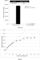
- the fluorescence intensity on each cell can be read by methods known in the art such as flow cytometry, laser scanning cytometry, or imaging microscopy. In this way, the fluorescence intensity in all desired wavelength ranges on each individual cell can be detected. From this information, the analyte amount on each cell and the identity of each cell can be determined. The amount or concentration of analyte in the original sample can then be determined using standard methods.
- a calibration curve is constructed by measuring the detection protein expression (i.e., its fluorescence) when the cells are combined with samples containing known concentrations of analyte. As long as a reproducible curve can be constructed, it is not necessary that the response be linear. The measured fluorescence intensity of the detection protein during an assay can then be correlated with the analyte concentration in the sample using the calibration curve.
- the method herein disclosed may be used in the detection, identification and quantification of analytes in biological and non-biological samples, such as the diagnosis of disease and infectious agents in medicine, veterinarian science and phytopathology, toxicology testing, analysis of metabolic products in living organisms, quality assurance through contaminant detection and monitoring of environmental pollution. These applications can be either commercial (in the sense of routine analyses) or serve pure research purposes. Because the method herein disclosed may be employed using a virtually limitless variety of modalities, it enables the specific detection of thousands of different analytes. The method herein disclosed may also be used to discriminate between different cell types or different developmental stages of a single cell/tissue, depending on which the analytes produced by the cells.
- the whole-cell sensors herein disclosed can also be also used as a medical diagnostics and disease management in the case of in vitro assays but also in the form of implantable sensors.
- the whole-cell sensor herein disclosed could also be converted into a therapeutic device by linking the detection of pathological biomarker or a pathological biomarkers signature to the activation of a downstream pathway directing the synthesis of a drug (like taxol in E.
- a biosensor device can be formed using the whole cell sensors herein disclosed to be deployed in a microenvironment or microfluidic devices, or a collection of these devices in a multi-chip module or distributed wireless network.
- the biosensor device can respond to one or more specific chemical and/or physical inputs (e.g. heat or electrical current), accessing a small part of the DNA memory of the cell by addressing a particular promoter, generating outputs in the form of detection protein, and communicating with a physical transducer through calorimetric, electrochemical, or preferably fluorescence bioluminescence means.
- the chimeric receptor polypeptide herein disclosed may be used in a cell-free system.
- the term "cell-free system” refers to a set of reagents capable of providing for or supporting a biosynthetic reaction in vitro in the absence of cells.
- a cell-free system comprises promoter-containing DNA, RNA polymerase, ribonucleotides, and a buffer system.
- Cell-free systems can be prepared using enzymes, coenzymes, and other subcellular components either isolated or purified from eukaryotic or prokaryotic cells, including recombinant cells, or prepared as extracts or fractions of such cells.
- a cell-free system can be derived from a variety of sources, including, but not limited to, eukaryotic and prokaryotic cells, such as bacteria, rabbit reticulocytes, mouse cells, human cell lines, primary human cell lines and budding yeast and the like.
- the chimeric receptor polypeptide herein disclosed may be used in any vehicle, including cellular extracts prepared from living cells.
- the vehicle may be used in a solution containing reconstituted components necessary for transcription and translation.
- Such a system may comprise a low-ionic-strength buffer (e.g., physiological salt, such as simple saline or phosphate- and/or Tris-buffered saline or other as described above), or a whole or fractionated cell lysate.
- the chimeric receptor polypeptide herein disclosed is embedded partially or completely in a solid support, preferably in a porous substrate.
- the solid support can be in any form including, but is not limited to, a well, a tube, a planar substrate (e.g., a chip or a plate), a sphere, a porous substrate (e.g., a mesh or a foam), a 3D scaffold, a patterned surface (e.g., nano-patterns, or micro-patterns, or both), a porous or solid bead, a hydrogel, a channel (e.g., a microfluidic channel), a smooth surface, and a rough surface.
- the solid support is hydrophilic and preferably a porous substrate.
- the term "porous substrate” refers to a substrate that contain pores or interstices via which a liquid composition may penetrate the substrate surface. Paper is one example of a porous substrate.
- the porous substrate comprises paper.
- the solid support comprises several (1, 2, 3, 4, 5, 6, 7, 8, 9, 10, 20, or more) spatially distinct reaction regions where the cell-free system is confined. The area that contains the cell-free system is herein referred to as "a reaction region.”
- reaction regions can be created by a chemical process such as using hydrophobic barriers on a piece of paper. The hydrophobic barriers are minimally permeable by water.
- the hydrophobic barrier can comprise hydrophobic materials such as hydrophobic polymer or wax.
- the hydrophobic barrier can be patterned by any existing patterning method (e.g., micro-contact printing, or dip pen lithography, photolithography, e-beam lithography, laser printing, inject printing, or a micro-arrayer). Methods of creating hydrophobic patterns on paper are known in the art; see for example, WO2009121041 and WO2008/049083 .
- the solid support comprises one or more fluidic channels (e.g., microfluidic channels) that connect reaction regions with an area for adding an aqueous sample.
- the fluid is wicked away to the reaction regions, thereby a plurality of reaction regions can be activated by the same sample.
- the solid support is a reaction chip.
- the reaction chip can comprise a sample hosting layer, a light blocking layer, a hydration layer, a transparent layer, a humidity maintaining layer, and a water vapor permeable layer.
- the hydration layer can comprise a hydrated material or chamber that provides humidity during incubation and/or measurement.
- the humidity maintaining layer can be water impermeable.
- the water vapor permeable layer can regulate humidity for the sample.
- the cell-free system functions as a sensor.
- the sensor can detect an analyte.
- the analyte activates the sensor, which produces a signal, indicating the detection of the analyte.
- the signal is optical.
- an optical signal can be fluorescence, luminescence, absorption or reflection of a given wavelength, ultraviolet, visible color, or infrared.
- the sensor comprises a reporter component. The function of the reporter component is to produce a detectable signal when an analyte is detected.
- the reporter component comprises a reporter gene. A reporter gene encoding any fluorescent protein can be applicable in the invention.
- the fluorescent protein includes, but is not limited to, for example, GFP, mCherry, Venus, and Cerulean.
- genes encoding fluorescent proteins that can be used in accordance with the compositions and methods described herein include, without limitation, those proteins provided in U.S. Patent Application No. 2012/0003630 .
- a reporter gene encoding any enzyme can be applicable as well.
- Enzymes that produce colored substrates (“colorimetric enzymes") can also be used for visualization and/or quantification. Enzymatic products can be quantified using spectrophotometers or other instruments that can take absorbance measurements including plate readers (for example, see FIG. 26).
- genes encoding colorimetric enzymes that can be used in accordance with the compositions and methods described herein include, without limitation, lacZ alpha fragment, lacZ (encoding beta-galactosidase, full-length), and xylE.
- An enzyme e.g., glucose oxidase or glucose deshydrogenase ( Scognamiglio, V. Nanotechnology in glucose monitoring: advances and challenges in the last 10 years Biosens. Bioelectron. 2013, 47, 12- 25
- glucose oxidase or glucose deshydrogenase Scognamiglio, V. Nanotechnology in glucose monitoring: advances and challenges in the last 10 years Biosens. Bioelectron. 2013, 47, 12- 25
- a nuclease enzyme can cleave a nucleic acid sequence such that an electronic and optical signal is generated.
- an enzyme can separate a fluorescence resonance energy transfer (FRET) or quenching pair to induce a change in fluorescence.
- FRET fluorescence resonance energy transfer
- sensors producing fluorescent signals it would be apparent to a skilled artisan that any commercial or homemade device or system that can detect fluorescence can be used for the purpose of detecting the signals, including, but not limited to, a microscope, a fluorescence microplate reader, and a fluorescence spectrometer.
- the cell-free system comprises a logic circuit, and thus can perform one or more logic functions upon activation.
- the logic circuit can be activated by contacting the logic circuit with water and a composition comprising one or more analytes.
- an AND gate is one of the most basic logic circuits, requiring the simultaneous presence of two appropriate analytes in order for the AND gate to turn on. If only one of the analytes is present, the AND gate would not turn on.
- the cell-free system is lyophilized on the solid support and thus may be reactivated upon hydration.
- Lyophilization also known as freeze-drying, is a dehydration process that involves freezing a material and then reducing the surrounding pressure to allow water to sublimate. Parameters such as freezing temperature, rate of temperature change, and pressure are variables for different lyophilization process. Accordingly, the lyophilization processes used in the methods and compositions herein are not limited to a specific set of parameters. It should be apparent to a skilled artisan that preferred lyophilization processes would yield a shelf-stable composition with a long shelf life.
- the cell-free systems and/or cell-free systems should be kept frozen, i.e., prevented from thawing, until the application of low pressure (e.g., vacuum).
- low pressure e.g., vacuum
- This can present a challenge when time is needed to transport low-volume frozen cell-free systems and/or cell-free systems to the lyophilizer.
- This can be addressed, for example, by placing cold and dense materials (e.g., metal or acrylics that have been frozen at -80° C. or in liquid nitrogen) in contact with the cell-free systems and/or cell-free systems to serve as a cold source during transit to the lyophilizer.
- the material can have large heat capacity.
- the frozen cell-free systems and/or cell-free systems are substantially shielded from light during the lyophilization process. This is particularly useful for protecting components sensitive to light.Instruments for performing lyophilization are commercially available through vendors such as Cole-Parmer and Millrock Technology.
- the shelf-stable composition is produced by a process comprising contacting a solid support with an aqueous solution comprising a cell-free system and a cell-free system, and lyophilizing said solid support.
- room temperature i.e., about 20° C. to 24° C.
- relative humidity of no more than 10% the composition has a shelf life of at least two weeks, at least one month, at least two months, or at least six months.
- a further object of the present disclosure relates to method of detecting an analyte, comprising: (i) providing the cell-free system herein disclosed; (ii) contacting the cell-free system with a sample to be tested for said analyte; and (iii) detecting a signal, wherein detection of said signal indicates the presence of said analyte.
- FIGURES are a diagrammatic representation of FIGURES.
- Bacterial strains Bacterial strains, plasmids, and materials.
- CadC and LexA transcriptional factor DNA binding domain, Leucine zipper GCN4, V H Hcafe and V H HRNase were synthesized by Integrated DNA Technologies (IDT).
- Different DNA components such as GFP reporter, CadBA promoter, pLexA promoter and CadC variants were constructed into BioBrick standard vector pSB4K5 by Gibson assembly methods. The resulting constructs were transformed into E.coli strain NEB10beta (New England Biolabs, NEB) and determined their performance.
- the expression level of CadC variants are under the control of pLac-1 promoter, which can be induced by IPTG.
- the CadC variants control the expression of reporter GFP through pCadBA promoter.
- the PCR amplification was carried out in a 40 ⁇ l reaction mixture consisting of 0.1 ⁇ 10ng of template DNA fragment, 1 ⁇ l of each forward/reversed primer (20 ⁇ M), and 20 ⁇ L of Q5 hot start high-fidelity 2X master mix (NEB). After 30 seconds of initial denaturation at 98°C, 35 cycles were conducted with the PCR procedures of 10 seconds at 98°C, 30 seconds at corresponding annealing temperature (different with each primer combination, calculated with NEB Tm calculator: http://tmcalculator.neb.com/#!/), and elongation (2kb/min) at 72 °C, with a final extension at 72 °C for 10 minutes. The PCR product was verified by gel electrophoresis, then purified by PCR clean up kit and determined the DNA concentration by Nanodrop spectrophotometer.
- the DNA templates from E.coli in the purified PCR product was further digested with 1 ⁇ l DpnI (20 units/ ⁇ l, NEB) in 40 ⁇ l Cut Smart reaction buffer (NEB) at 37°C for 1 hours.
- the resulting product was applied to Gibson assembly reaction directly.
- 100 ng of vector DNA fragment and 3 - 5 folds of insert fragments was incubated with 10 ⁇ l of 2X Gibson assembly master mix (NEB) in a final volume of 20 ⁇ L at 50°C for 60 minutes.
- the reaction mix was further heat inactivated at 80°C for 15 minutes.
- LexA DBD The repressive activity of the monomeric LexA DBD can be restored by forcing its dimerization through fusion with leucine zippers.
- LexA DBD was used in two-hybrid screens to probe protein-protein.
- LexA-408 In order to prevent interference from endogenous E.coli LexA, we used the mutant LexA-408, and its corresponding promoter which is not recognized by the wild type LexA.
- LexA DBD We fused LexA DBD to a monomeric LBD that undergoes ligand-induced homodimerization.
- LexA DBD should be able to bind DNA and repress target gene expression.
- Choice of a synthetic ligand binding domain In order to develop a scalable receptor platform, we set several criteria for selecting an ideal LBD scaffold: (i) potential for further engineering to bind many different ligands, and of different types (e.g. proteins, small molecules); (ii) high solubility and stability; and (iii) low propensity to aggregate to ensure monomeric state before ligand binding.
- Antibodies are an ideal scaffold to detect various kinds of ligands and can be applied to different therapeutic or diagnosis applications.
- IgGs have poor expression levels in prokaryotic system.
- LexA DBD was capable of gene repression a high concentration (14% and 35% repression at 50 ⁇ M and 100 ⁇ M IPTG, respectively, data not shown).
- LexA VHH- Caffeine and LexA-VHH-Control were characterized in response to increasing concentrations of IPTG and caffeine (data not shown).
- WB Western blot
- both fusion proteins were expressed at similar levels (data not shown).
- LexA-VHH-Caffeine and LexA-VEHControl displayed a concentration-dependent repressive activity (30% and 45% at 50 ⁇ M and 100 ⁇ M IPTG induction, respectively, data not shown).
- LexA-VHH-Caffeine and LexAVHH- Control had a comparable repressive activity to LexADBD at a similar IPTG concentration, suggesting that this repressive effect is primarily due to the DBD and that VHHs are in a monomeric state in the cytoplasm.
- LexA-VHH fusions were monitored to increase concentrations of caffeine at different IPTG concentrations. While no change was detectable in response to cafeine for LexA-VHH-Control (data not shown), LexA-VHHCaffeine had a dose-dependent response to caffeine starting at 25 ⁇ M IPTG concentration and 100 nM caffeine (data not shown).
- CadC is composed of a N-terminal cytosolic DBD and a C-terminal periplasmic pH sensor domain. It activates the pCadBA promoter when environmental pH decreases and in the presence of lysine. Interestingly, dimerization of transmembrane helixes bound to the cytosolic CadC DNA binding domain is sufficien to restore CadC transcriptional activity. We thus aimed to restore CadC activity through ligand-induced dimerization of a periplasmic sensor domain. In order to remove endogenous regulations by the Lysine permease LysP, we used an artificial transmembrane domain composed of 16 Leucine repeat.
- CadC-VHH fusion proteins composed of CadC DBD, CadC juxtamembrane domain (JM), the Leu(16)TM, CadC wild type external linker region (EL), and the VHH ligand binding domain (data not shown), and placed their expression under the control of the pLacO1 promoter (data not shown). Both fusion proteins had similar expression levels across the IPTG concentration range (data not shown).
- L-forms can be generated transiently with antibiotics that inhibit peptidoglycan synthesis or disrupt cell wall formation or permanently by mutation of genes related to peptidoglycan synthesis. Because of their lack of outer membrane, Lform bacteria could be suitable candidates for developing whole cell biosensors to detect large molecules such as proteins.
- PBP penicillin binding protein
- the antibodies are fused to with LexA-DBD.
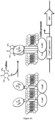
- the Lam4-induced LexA-DBD dimerization then allows binding of the complex to the operator of the pLexA promoter and leads to repression of the expression of deGFP ( Figure 6 ).
- Plasmid Preparation Genes encoding the GFP reporter, LexA-DBD-VHHcafe as well as LexA-DBD-Lam4 were cloned as described in Birnboim HC, Doly J. A rapid alkaline extraction procedure for screening recombinant plasmid DNA. Nucleic Acids Res. 1979; 7: 1513-1523 . All the amplification was done in E. coli. Plasmid purification was performed with Qiagen ® kits.
- the Cell-Free platform makes it possible to express functional proteins that can dimerize through ligand detection and binding and suppress expression of the deGFP.
Landscapes
- Health & Medical Sciences (AREA)
- Life Sciences & Earth Sciences (AREA)
- Immunology (AREA)
- Engineering & Computer Science (AREA)
- Molecular Biology (AREA)
- Chemical & Material Sciences (AREA)
- Biomedical Technology (AREA)
- Hematology (AREA)
- Urology & Nephrology (AREA)
- Cell Biology (AREA)
- General Health & Medical Sciences (AREA)
- Biochemistry (AREA)
- Medicinal Chemistry (AREA)
- Microbiology (AREA)
- Physics & Mathematics (AREA)
- Analytical Chemistry (AREA)
- Food Science & Technology (AREA)
- Biotechnology (AREA)
- General Physics & Mathematics (AREA)
- Pathology (AREA)
- Proteomics, Peptides & Aminoacids (AREA)
- Organic Chemistry (AREA)
- Tropical Medicine & Parasitology (AREA)
- Virology (AREA)
- Biophysics (AREA)
- Genetics & Genomics (AREA)
- Mycology (AREA)
- Micro-Organisms Or Cultivation Processes Thereof (AREA)
- Measuring Or Testing Involving Enzymes Or Micro-Organisms (AREA)
- Immobilizing And Processing Of Enzymes And Microorganisms (AREA)
- Peptides Or Proteins (AREA)
Claims (11)
- Chimäres Rezeptorpolypeptid, umfassend i) eine erste DNA-Bindungsdomäne, die aus einer Aminosäuresequenz gemäß SEQ ID NO: 1 oder SEQ ID NO: 2 besteht, ii) eine Bindungsdomäne, die aus einem VHH-Einzeldomänen-Antikörper besteht, der bei Bindung an seinen Liganden dimerisieren kann, und iii) eine Transmembrandomäne, umfassend eine Aminosäuresequenz gemäß SEQ ID NO: 3, SEQ ID NO: 4, SEQ ID NO: 5 oder SEQ ID NO: 6 zwischen der DNA-Bindungsdomäne und der Bindungsdomäne.
- Chimäres Rezeptorpolypeptid nach Anspruch 1, das weiter einen Abstandshalter umfasst, der zwischen der Transmembrandomäne und der Bindungsdomäne eingefügt ist, wobei der Abstandshalter eine Aminosäuresequenz ist, die aus DTRLPMS (SEQ ID NO: 7) oder GGGSG (SEQ ID NO: 12) besteht.
- Chimäres Rezeptorpolypeptid nach Anspruch 1, das eine erste Domäne umfasst, die eine Aminosäuresequenz mit mindestens 70 % Identität mit SED ID NO:8 umfasst, die an die Bindungsdomäne fusioniert ist.
- Nukleinsäure, die für den chimären Rezeptor nach Anspruch 1 kodiert.
- Expressionskassette, die das Nukleinsäuremolekül nach Anspruch 4 umfasst, das funktionsfähig mit Kontrollsequenzen verbunden ist, die Expression in einer prokaryotischen Zelle ermöglichen.
- Prokaryotische Zelle, die mit dem Nukleinsäuremolekül nach Anspruch 4 oder der Expressionskassette nach Anspruch 6 gentechnisch verändert wurde, wobei die prokaryotische Zelle aus grampositiven oder gramnegativen Bakterien, wie E. Coli, ausgewählt ist.
- Prokaryotische Zelle nach Anspruch 6, die weiter ein Nukleinsäuremolekül umfasst, das für ein Nachweisprotein kodiert, das operativ mit einem CadB A-Promotor verbunden ist.
- Verfahren zum Testen auf mindestens einen Analyten, umfassend i) Bereitstellen mindestens eines Satzes prokaryotischer Zellen nach Anspruch 6, wobei jede einen chimären Rezeptor umfasst, der bei Bindung an den Analyten zur Dimerisierung fähig ist und an ein Nachweisprotein gekoppelt ist; b) Inkontaktbringen des Satzes von Zellen mit einer Probe, von der vermutet wird, dass sie den Analyten enthält, für eine Zeit, die ausreicht, um die Oligomerisierung der Bindung der chimären Rezeptoren und dann die Expression des Nachweisproteins zu ermöglichen; und c) Nachweisen des Expressionsniveaus des Nachweisproteins, wobei das Expressionsniveau mit der Menge des in der Probe vorhandenen Analyten korreliert.
- Zellfreies System, das das chimäre Rezeptorpolypeptid nach Anspruch 1 umfasst, wobei das chimäre Rezeptorpolypeptid teilweise oder vollständig in einen festen Träger eingebettet ist, wobei der feste Träger ein poröses Substrat, wie etwa Papier, ist.
- Zellfreies System nach Anspruch 9, das ein Reportergen umfasst, wobei das Reportergen für ein fluoreszierendes Protein kodiert.
- Verfahren zum Nachweisen eines Analyten, umfassend:(i) Bereitstellen des zellfreien Systems nach Anspruch 9;(ii) Inkontaktbringen des zellfreien Systems mit einer Probe, die auf den Analyten getestet werden soll; und(iii) Nachweisen eines Signals, wobei der Nachweis des Signals das Vorhandensein des Analyten anzeigt.
Applications Claiming Priority (3)
| Application Number | Priority Date | Filing Date | Title |
|---|---|---|---|
| EP17305690 | 2017-06-08 | ||
| EP17306060 | 2017-08-09 | ||
| PCT/EP2018/065074 WO2018224611A1 (en) | 2017-06-08 | 2018-06-07 | Chimeric receptor for use in whole-cell sensors for detecting analytes of interest |
Publications (2)
| Publication Number | Publication Date |
|---|---|
| EP3635398A1 EP3635398A1 (de) | 2020-04-15 |
| EP3635398B1 true EP3635398B1 (de) | 2024-07-24 |
Family
ID=62563156
Family Applications (1)
| Application Number | Title | Priority Date | Filing Date |
|---|---|---|---|
| EP18729959.9A Active EP3635398B1 (de) | 2017-06-08 | 2018-06-07 | Chimärer rezeptor zur verwendung in ganzzelligen sensoren zur detektion von analyten von interesse |
Country Status (5)
| Country | Link |
|---|---|
| US (1) | US12517124B2 (de) |
| EP (1) | EP3635398B1 (de) |
| JP (1) | JP7265487B2 (de) |
| ES (1) | ES2986584T3 (de) |
| WO (1) | WO2018224611A1 (de) |
Families Citing this family (6)
| Publication number | Priority date | Publication date | Assignee | Title |
|---|---|---|---|---|
| US12190997B2 (en) | 2018-08-13 | 2025-01-07 | Aarhus Universitet | Genetically altered LysM receptors with altered agonist specificity and affinity |
| CN111117940B (zh) * | 2019-12-04 | 2022-06-28 | 天津大学 | 一种高产戊二胺的大肠杆菌工程菌与方法 |
| WO2022167653A1 (en) * | 2021-02-08 | 2022-08-11 | INSERM (Institut National de la Santé et de la Recherche Médicale) | Bile salts bactosensor and use thereof for diagnostic and therapeutic purposes |
| CN114774337B (zh) * | 2022-03-22 | 2023-08-04 | 东北大学 | 一种基于工程大肠杆菌的HCoV-229E病毒检测系统 |
| CN114774425B (zh) * | 2022-03-22 | 2023-09-22 | 东北大学 | 一种基于工程大肠杆菌的MERS-CoV病毒检测系统 |
| WO2025068399A1 (en) * | 2023-09-29 | 2025-04-03 | Univerza V Ljubljani | Novel screening assays for the detection of protein homodimerization |
Family Cites Families (15)
| Publication number | Priority date | Publication date | Assignee | Title |
|---|---|---|---|---|
| AU634186B2 (en) | 1988-11-11 | 1993-02-18 | Medical Research Council | Single domain ligands, receptors comprising said ligands, methods for their production, and use of said ligands and receptors |
| US6265174B1 (en) | 1997-11-03 | 2001-07-24 | Morphochem, Inc. | Methods and compositions for identifying and modulating ctionprotein-interactions |
| US7052906B1 (en) * | 1999-04-16 | 2006-05-30 | Celltech R & D Limited | Synthetic transmembrane components |
| AU2002243280A1 (en) * | 2000-10-23 | 2002-06-24 | Engeneos, Inc. | Engineered stimulus-responsive switches |
| US6716589B2 (en) * | 2000-11-20 | 2004-04-06 | Alphabeta Ab | Discordant helix stabilization for prevention of amyloid formation |
| US20060073141A1 (en) | 2001-06-28 | 2006-04-06 | Domantis Limited | Compositions and methods for treating inflammatory disorders |
| US20050003367A1 (en) * | 2002-09-05 | 2005-01-06 | Whitney Michael Allen | Methods and compositions for rapid development of screening assays |
| US7563443B2 (en) | 2004-09-17 | 2009-07-21 | Domantis Limited | Monovalent anti-CD40L antibody polypeptides and compositions thereof |
| CA2667702C (en) | 2006-10-18 | 2016-06-14 | President And Fellows Of Harvard College | Lateral flow and flow-through bioassay devices based on patterned porous media, methods of making same, and methods of using same |
| KR20100128340A (ko) | 2008-03-27 | 2010-12-07 | 프레지던트 앤드 펠로우즈 오브 하바드 칼리지 | 종이 기반 마이크로유체 시스템 |
| US8645115B2 (en) | 2008-12-22 | 2014-02-04 | Trustees Of Boston University | Modular nucleic acid-based circuits for counters, binary operations, memory and logic |
| WO2012031109A2 (en) * | 2010-09-03 | 2012-03-08 | E. I. Du Pont De Nemours And Company | Methods and compositions for engineering recombinant dna molecules |
| WO2012125652A2 (en) | 2011-03-14 | 2012-09-20 | University Of Southern California | Antibody and antibody mimetic for visualization and ablation of endogenous proteins |
| WO2013022739A1 (en) | 2011-08-05 | 2013-02-14 | Leonard Joshua N | Modular extracellular sensor architecture for cell-based biosensors |
| US10954500B2 (en) * | 2015-10-23 | 2021-03-23 | Fred Hutchinson Cancer Research Center | Methods to create chemically-induced dimerizing protein systems for regulation of cellular events |
-
2018
- 2018-06-07 EP EP18729959.9A patent/EP3635398B1/de active Active
- 2018-06-07 JP JP2019567367A patent/JP7265487B2/ja active Active
- 2018-06-07 WO PCT/EP2018/065074 patent/WO2018224611A1/en not_active Ceased
- 2018-06-07 ES ES18729959T patent/ES2986584T3/es active Active
- 2018-06-07 US US16/620,155 patent/US12517124B2/en active Active
Non-Patent Citations (2)
| Title |
|---|
| GREGORY J. SONNESON ET AL: "Hapten-Induced Dimerization of a Single-Domain VHH Camelid Antibody", BIOCHEMISTRY, vol. 48, no. 29, 28 July 2009 (2009-07-28), pages 6693 - 6695, XP055649963, ISSN: 0006-2960, DOI: 10.1021/bi900862r * |
| PETER C FRIDY ET AL: "A robust pipeline for rapid production of versatile nanobody repertoires", NATURE METHODS, vol. 11, no. 12, 2 November 2014 (2014-11-02), New York, pages 1253 - 1260, XP055247223, ISSN: 1548-7091, DOI: 10.1038/nmeth.3170 * |
Also Published As
| Publication number | Publication date |
|---|---|
| US12517124B2 (en) | 2026-01-06 |
| WO2018224611A1 (en) | 2018-12-13 |
| EP3635398A1 (de) | 2020-04-15 |
| US20200096507A1 (en) | 2020-03-26 |
| JP7265487B2 (ja) | 2023-04-26 |
| JP2020527028A (ja) | 2020-09-03 |
| ES2986584T3 (es) | 2024-11-12 |
Similar Documents
| Publication | Publication Date | Title |
|---|---|---|
| EP3635398B1 (de) | Chimärer rezeptor zur verwendung in ganzzelligen sensoren zur detektion von analyten von interesse | |
| Liu et al. | Colorimetric assay of bacterial pathogens based on Co3O4 magnetic nanozymes conjugated with specific fusion phage proteins and magnetophoretic chromatography | |
| Guo et al. | Investigating acid production by Streptococcus mutans with a surface-displayed pH-sensitive green fluorescent protein | |
| Struss et al. | Paper strip whole cell biosensors: a portable test for the semiquantitative detection of bacterial quorum signaling molecules | |
| Zhang et al. | Transpeptidation-mediated assembly of tripartite split green fluorescent protein for label-free assay of sortase activity | |
| Jung et al. | Fluorescein and rhodamine B-binding domains from autodisplayed fv-antibody library | |
| Curtis et al. | Development of a mast cell-based biosensor | |
| CN105807064B (zh) | 一种荧光素酶互补量子点生物传感器及其构建方法及其应用 | |
| US12385906B2 (en) | Methods of detection of compound, antibody or protein using recombinant endospores or bacteria as sensing element | |
| Park et al. | Flow cytometric immunoassay using E. coli with autodisplayed Z-domains | |
| CN102080068B (zh) | 荧光素酶活性片段及其应用 | |
| Wu et al. | Flow cytometric single-cell analysis for quantitative in vivo detection of protein–protein interactions via relative reporter protein expression measurement | |
| US9400249B2 (en) | Detection of biopolymer interactions, cancer cells, and pathogens using split-supercharged GFP | |
| EP3622292B1 (de) | Genetisch codierte kaliumionenindikatoren | |
| Nguyen et al. | Detection of matrix metalloproteinase activity by bioluminescence via intein-mediated biotinylation of luciferase | |
| TWI826663B (zh) | 以重組內孢子或細菌作為感測元件以偵測化合物、抗體或蛋白質之方法 | |
| JP6525199B2 (ja) | インスリンの検出方法、および、インスリンの検出キット | |
| Cox | 10 Bacteriophage-based Methods of Bacterial Detection and Identification | |
| Mehta et al. | The use of phages and aptamers as alternatives to antibodies in medical and food diagnostics | |
| Fraile et al. | Programming bacterial adhesion to functionalized surfaces through cellular display of recombinant nanobodies | |
| Hacıosmanoğlu et al. | Design of synthetic biological devices for detection and targeting human diseases | |
| WO2026064477A1 (en) | Oligomer-induced reconstitution of bioluminescence sensors for multiplex detection of analytes | |
| Edwards | Microscopy Techniques for Investigating Interactions in Microbial Systems | |
| TWI412589B (zh) | 突變藍色螢光蛋白及其用於螢光共振能量傳遞與藍色螢光魚之方法 | |
| Gregoire | Monitoring Human Copper/Zinc Superoxide Dismutase Misfolding/Aggregation in Mammalian Cells |
Legal Events
| Date | Code | Title | Description |
|---|---|---|---|
| STAA | Information on the status of an ep patent application or granted ep patent |
Free format text: STATUS: UNKNOWN |
|
| STAA | Information on the status of an ep patent application or granted ep patent |
Free format text: STATUS: THE INTERNATIONAL PUBLICATION HAS BEEN MADE |
|
| PUAI | Public reference made under article 153(3) epc to a published international application that has entered the european phase |
Free format text: ORIGINAL CODE: 0009012 |
|
| STAA | Information on the status of an ep patent application or granted ep patent |
Free format text: STATUS: REQUEST FOR EXAMINATION WAS MADE |
|
| 17P | Request for examination filed |
Effective date: 20191206 |
|
| AK | Designated contracting states |
Kind code of ref document: A1 Designated state(s): AL AT BE BG CH CY CZ DE DK EE ES FI FR GB GR HR HU IE IS IT LI LT LU LV MC MK MT NL NO PL PT RO RS SE SI SK SM TR |
|
| AX | Request for extension of the european patent |
Extension state: BA ME |
|
| DAV | Request for validation of the european patent (deleted) | ||
| DAX | Request for extension of the european patent (deleted) | ||
| STAA | Information on the status of an ep patent application or granted ep patent |
Free format text: STATUS: EXAMINATION IS IN PROGRESS |
|
| 17Q | First examination report despatched |
Effective date: 20201006 |
|
| GRAP | Despatch of communication of intention to grant a patent |
Free format text: ORIGINAL CODE: EPIDOSNIGR1 |
|
| STAA | Information on the status of an ep patent application or granted ep patent |
Free format text: STATUS: GRANT OF PATENT IS INTENDED |
|
| INTG | Intention to grant announced |
Effective date: 20240325 |
|
| GRAS | Grant fee paid |
Free format text: ORIGINAL CODE: EPIDOSNIGR3 |
|
| GRAA | (expected) grant |
Free format text: ORIGINAL CODE: 0009210 |
|
| STAA | Information on the status of an ep patent application or granted ep patent |
Free format text: STATUS: THE PATENT HAS BEEN GRANTED |
|
| AK | Designated contracting states |
Kind code of ref document: B1 Designated state(s): AL AT BE BG CH CY CZ DE DK EE ES FI FR GB GR HR HU IE IS IT LI LT LU LV MC MK MT NL NO PL PT RO RS SE SI SK SM TR |
|
| REG | Reference to a national code |
Ref country code: GB Ref legal event code: FG4D |
|
| REG | Reference to a national code |
Ref country code: CH Ref legal event code: EP |
|
| REG | Reference to a national code |
Ref country code: IE Ref legal event code: FG4D Ref country code: DE Ref legal event code: R096 Ref document number: 602018072159 Country of ref document: DE |
|
| REG | Reference to a national code |
Ref country code: NL Ref legal event code: FP |
|
| REG | Reference to a national code |
Ref country code: LT Ref legal event code: MG9D |
|
| REG | Reference to a national code |
Ref country code: ES Ref legal event code: FG2A Ref document number: 2986584 Country of ref document: ES Kind code of ref document: T3 Effective date: 20241112 |
|
| PG25 | Lapsed in a contracting state [announced via postgrant information from national office to epo] |
Ref country code: PT Free format text: LAPSE BECAUSE OF FAILURE TO SUBMIT A TRANSLATION OF THE DESCRIPTION OR TO PAY THE FEE WITHIN THE PRESCRIBED TIME-LIMIT Effective date: 20241125 |
|
| REG | Reference to a national code |
Ref country code: AT Ref legal event code: MK05 Ref document number: 1706757 Country of ref document: AT Kind code of ref document: T Effective date: 20240724 |
|
| PG25 | Lapsed in a contracting state [announced via postgrant information from national office to epo] |
Ref country code: PT Free format text: LAPSE BECAUSE OF FAILURE TO SUBMIT A TRANSLATION OF THE DESCRIPTION OR TO PAY THE FEE WITHIN THE PRESCRIBED TIME-LIMIT Effective date: 20241125 |
|
| PG25 | Lapsed in a contracting state [announced via postgrant information from national office to epo] |
Ref country code: NO Free format text: LAPSE BECAUSE OF FAILURE TO SUBMIT A TRANSLATION OF THE DESCRIPTION OR TO PAY THE FEE WITHIN THE PRESCRIBED TIME-LIMIT Effective date: 20241024 |
|
| PG25 | Lapsed in a contracting state [announced via postgrant information from national office to epo] |
Ref country code: GR Free format text: LAPSE BECAUSE OF FAILURE TO SUBMIT A TRANSLATION OF THE DESCRIPTION OR TO PAY THE FEE WITHIN THE PRESCRIBED TIME-LIMIT Effective date: 20241025 Ref country code: FI Free format text: LAPSE BECAUSE OF FAILURE TO SUBMIT A TRANSLATION OF THE DESCRIPTION OR TO PAY THE FEE WITHIN THE PRESCRIBED TIME-LIMIT Effective date: 20240724 Ref country code: PL Free format text: LAPSE BECAUSE OF FAILURE TO SUBMIT A TRANSLATION OF THE DESCRIPTION OR TO PAY THE FEE WITHIN THE PRESCRIBED TIME-LIMIT Effective date: 20240724 |
|
| PG25 | Lapsed in a contracting state [announced via postgrant information from national office to epo] |
Ref country code: BG Free format text: LAPSE BECAUSE OF FAILURE TO SUBMIT A TRANSLATION OF THE DESCRIPTION OR TO PAY THE FEE WITHIN THE PRESCRIBED TIME-LIMIT Effective date: 20240724 |
|
| PG25 | Lapsed in a contracting state [announced via postgrant information from national office to epo] |
Ref country code: LV Free format text: LAPSE BECAUSE OF FAILURE TO SUBMIT A TRANSLATION OF THE DESCRIPTION OR TO PAY THE FEE WITHIN THE PRESCRIBED TIME-LIMIT Effective date: 20240724 |
|
| PG25 | Lapsed in a contracting state [announced via postgrant information from national office to epo] |
Ref country code: IS Free format text: LAPSE BECAUSE OF FAILURE TO SUBMIT A TRANSLATION OF THE DESCRIPTION OR TO PAY THE FEE WITHIN THE PRESCRIBED TIME-LIMIT Effective date: 20241124 Ref country code: AT Free format text: LAPSE BECAUSE OF FAILURE TO SUBMIT A TRANSLATION OF THE DESCRIPTION OR TO PAY THE FEE WITHIN THE PRESCRIBED TIME-LIMIT Effective date: 20240724 |
|
| PG25 | Lapsed in a contracting state [announced via postgrant information from national office to epo] |
Ref country code: HR Free format text: LAPSE BECAUSE OF FAILURE TO SUBMIT A TRANSLATION OF THE DESCRIPTION OR TO PAY THE FEE WITHIN THE PRESCRIBED TIME-LIMIT Effective date: 20240724 |
|
| PG25 | Lapsed in a contracting state [announced via postgrant information from national office to epo] |
Ref country code: RS Free format text: LAPSE BECAUSE OF FAILURE TO SUBMIT A TRANSLATION OF THE DESCRIPTION OR TO PAY THE FEE WITHIN THE PRESCRIBED TIME-LIMIT Effective date: 20241024 |
|
| PG25 | Lapsed in a contracting state [announced via postgrant information from national office to epo] |
Ref country code: RS Free format text: LAPSE BECAUSE OF FAILURE TO SUBMIT A TRANSLATION OF THE DESCRIPTION OR TO PAY THE FEE WITHIN THE PRESCRIBED TIME-LIMIT Effective date: 20241024 Ref country code: PL Free format text: LAPSE BECAUSE OF FAILURE TO SUBMIT A TRANSLATION OF THE DESCRIPTION OR TO PAY THE FEE WITHIN THE PRESCRIBED TIME-LIMIT Effective date: 20240724 Ref country code: NO Free format text: LAPSE BECAUSE OF FAILURE TO SUBMIT A TRANSLATION OF THE DESCRIPTION OR TO PAY THE FEE WITHIN THE PRESCRIBED TIME-LIMIT Effective date: 20241024 Ref country code: LV Free format text: LAPSE BECAUSE OF FAILURE TO SUBMIT A TRANSLATION OF THE DESCRIPTION OR TO PAY THE FEE WITHIN THE PRESCRIBED TIME-LIMIT Effective date: 20240724 Ref country code: IS Free format text: LAPSE BECAUSE OF FAILURE TO SUBMIT A TRANSLATION OF THE DESCRIPTION OR TO PAY THE FEE WITHIN THE PRESCRIBED TIME-LIMIT Effective date: 20241124 Ref country code: HR Free format text: LAPSE BECAUSE OF FAILURE TO SUBMIT A TRANSLATION OF THE DESCRIPTION OR TO PAY THE FEE WITHIN THE PRESCRIBED TIME-LIMIT Effective date: 20240724 Ref country code: GR Free format text: LAPSE BECAUSE OF FAILURE TO SUBMIT A TRANSLATION OF THE DESCRIPTION OR TO PAY THE FEE WITHIN THE PRESCRIBED TIME-LIMIT Effective date: 20241025 Ref country code: FI Free format text: LAPSE BECAUSE OF FAILURE TO SUBMIT A TRANSLATION OF THE DESCRIPTION OR TO PAY THE FEE WITHIN THE PRESCRIBED TIME-LIMIT Effective date: 20240724 Ref country code: BG Free format text: LAPSE BECAUSE OF FAILURE TO SUBMIT A TRANSLATION OF THE DESCRIPTION OR TO PAY THE FEE WITHIN THE PRESCRIBED TIME-LIMIT Effective date: 20240724 Ref country code: AT Free format text: LAPSE BECAUSE OF FAILURE TO SUBMIT A TRANSLATION OF THE DESCRIPTION OR TO PAY THE FEE WITHIN THE PRESCRIBED TIME-LIMIT Effective date: 20240724 |
|
| PG25 | Lapsed in a contracting state [announced via postgrant information from national office to epo] |
Ref country code: SM Free format text: LAPSE BECAUSE OF FAILURE TO SUBMIT A TRANSLATION OF THE DESCRIPTION OR TO PAY THE FEE WITHIN THE PRESCRIBED TIME-LIMIT Effective date: 20240724 Ref country code: DK Free format text: LAPSE BECAUSE OF FAILURE TO SUBMIT A TRANSLATION OF THE DESCRIPTION OR TO PAY THE FEE WITHIN THE PRESCRIBED TIME-LIMIT Effective date: 20240724 Ref country code: RO Free format text: LAPSE BECAUSE OF FAILURE TO SUBMIT A TRANSLATION OF THE DESCRIPTION OR TO PAY THE FEE WITHIN THE PRESCRIBED TIME-LIMIT Effective date: 20240724 |
|
| PG25 | Lapsed in a contracting state [announced via postgrant information from national office to epo] |
Ref country code: EE Free format text: LAPSE BECAUSE OF FAILURE TO SUBMIT A TRANSLATION OF THE DESCRIPTION OR TO PAY THE FEE WITHIN THE PRESCRIBED TIME-LIMIT Effective date: 20240724 |
|
| PG25 | Lapsed in a contracting state [announced via postgrant information from national office to epo] |
Ref country code: CZ Free format text: LAPSE BECAUSE OF FAILURE TO SUBMIT A TRANSLATION OF THE DESCRIPTION OR TO PAY THE FEE WITHIN THE PRESCRIBED TIME-LIMIT Effective date: 20240724 |
|
| REG | Reference to a national code |
Ref country code: DE Ref legal event code: R097 Ref document number: 602018072159 Country of ref document: DE |
|
| PG25 | Lapsed in a contracting state [announced via postgrant information from national office to epo] |
Ref country code: SK Free format text: LAPSE BECAUSE OF FAILURE TO SUBMIT A TRANSLATION OF THE DESCRIPTION OR TO PAY THE FEE WITHIN THE PRESCRIBED TIME-LIMIT Effective date: 20240724 |
|
| PLBE | No opposition filed within time limit |
Free format text: ORIGINAL CODE: 0009261 |
|
| STAA | Information on the status of an ep patent application or granted ep patent |
Free format text: STATUS: NO OPPOSITION FILED WITHIN TIME LIMIT |
|
| PGFP | Annual fee paid to national office [announced via postgrant information from national office to epo] |
Ref country code: NL Payment date: 20250520 Year of fee payment: 8 |
|
| 26N | No opposition filed |
Effective date: 20250425 |
|
| PGFP | Annual fee paid to national office [announced via postgrant information from national office to epo] |
Ref country code: DE Payment date: 20250520 Year of fee payment: 8 |
|
| PGFP | Annual fee paid to national office [announced via postgrant information from national office to epo] |
Ref country code: GB Payment date: 20250520 Year of fee payment: 8 |
|
| PGFP | Annual fee paid to national office [announced via postgrant information from national office to epo] |
Ref country code: LU Payment date: 20250523 Year of fee payment: 8 Ref country code: BE Payment date: 20250520 Year of fee payment: 8 Ref country code: IT Payment date: 20250520 Year of fee payment: 8 |
|
| PGFP | Annual fee paid to national office [announced via postgrant information from national office to epo] |
Ref country code: FR Payment date: 20250520 Year of fee payment: 8 |
|
| PG25 | Lapsed in a contracting state [announced via postgrant information from national office to epo] |
Ref country code: SE Free format text: LAPSE BECAUSE OF FAILURE TO SUBMIT A TRANSLATION OF THE DESCRIPTION OR TO PAY THE FEE WITHIN THE PRESCRIBED TIME-LIMIT Effective date: 20240724 |
|
| PGFP | Annual fee paid to national office [announced via postgrant information from national office to epo] |
Ref country code: ES Payment date: 20250701 Year of fee payment: 8 |
|
| PGFP | Annual fee paid to national office [announced via postgrant information from national office to epo] |
Ref country code: CH Payment date: 20250701 Year of fee payment: 8 |
|
| P01 | Opt-out of the competence of the unified patent court (upc) registered |
Free format text: CASE NUMBER: UPC_APP_0014367_3635398/2025 Effective date: 20251121 |
|
| PG25 | Lapsed in a contracting state [announced via postgrant information from national office to epo] |
Ref country code: MC Free format text: LAPSE BECAUSE OF FAILURE TO SUBMIT A TRANSLATION OF THE DESCRIPTION OR TO PAY THE FEE WITHIN THE PRESCRIBED TIME-LIMIT Effective date: 20240724 |