EP3613348B1 - Vorrichtung und verfahren zur mammografischen brustkompression - Google Patents
Vorrichtung und verfahren zur mammografischen brustkompression Download PDFInfo
- Publication number
- EP3613348B1 EP3613348B1 EP19192419.0A EP19192419A EP3613348B1 EP 3613348 B1 EP3613348 B1 EP 3613348B1 EP 19192419 A EP19192419 A EP 19192419A EP 3613348 B1 EP3613348 B1 EP 3613348B1
- Authority
- EP
- European Patent Office
- Prior art keywords
- compression
- breast
- pressure
- patient
- support plate
- Prior art date
- Legal status (The legal status is an assumption and is not a legal conclusion. Google has not performed a legal analysis and makes no representation as to the accuracy of the status listed.)
- Active
Links
Images



Classifications
-
- A—HUMAN NECESSITIES
- A61—MEDICAL OR VETERINARY SCIENCE; HYGIENE
- A61B—DIAGNOSIS; SURGERY; IDENTIFICATION
- A61B6/00—Apparatus or devices for radiation diagnosis; Apparatus or devices for radiation diagnosis combined with radiation therapy equipment
- A61B6/02—Arrangements for diagnosis sequentially in different planes; Stereoscopic radiation diagnosis
-
- A—HUMAN NECESSITIES
- A61—MEDICAL OR VETERINARY SCIENCE; HYGIENE
- A61B—DIAGNOSIS; SURGERY; IDENTIFICATION
- A61B6/00—Apparatus or devices for radiation diagnosis; Apparatus or devices for radiation diagnosis combined with radiation therapy equipment
- A61B6/04—Positioning of patients; Tiltable beds or the like
- A61B6/0407—Supports, e.g. tables or beds, for the body or parts of the body
- A61B6/0414—Supports, e.g. tables or beds, for the body or parts of the body with compression means
-
- A—HUMAN NECESSITIES
- A61—MEDICAL OR VETERINARY SCIENCE; HYGIENE
- A61B—DIAGNOSIS; SURGERY; IDENTIFICATION
- A61B5/00—Measuring for diagnostic purposes; Identification of persons
- A61B5/0059—Measuring for diagnostic purposes; Identification of persons using light, e.g. diagnosis by transillumination, diascopy, fluorescence
- A61B5/0082—Measuring for diagnostic purposes; Identification of persons using light, e.g. diagnosis by transillumination, diascopy, fluorescence adapted for particular medical purposes
- A61B5/0091—Measuring for diagnostic purposes; Identification of persons using light, e.g. diagnosis by transillumination, diascopy, fluorescence adapted for particular medical purposes for mammography
-
- A—HUMAN NECESSITIES
- A61—MEDICAL OR VETERINARY SCIENCE; HYGIENE
- A61B—DIAGNOSIS; SURGERY; IDENTIFICATION
- A61B5/00—Measuring for diagnostic purposes; Identification of persons
- A61B5/02—Detecting, measuring or recording for evaluating the cardiovascular system, e.g. pulse, heart rate, blood pressure or blood flow
- A61B5/021—Measuring pressure in heart or blood vessels
- A61B5/02108—Measuring pressure in heart or blood vessels from analysis of pulse wave characteristics
-
- A—HUMAN NECESSITIES
- A61—MEDICAL OR VETERINARY SCIENCE; HYGIENE
- A61B—DIAGNOSIS; SURGERY; IDENTIFICATION
- A61B5/00—Measuring for diagnostic purposes; Identification of persons
- A61B5/02—Detecting, measuring or recording for evaluating the cardiovascular system, e.g. pulse, heart rate, blood pressure or blood flow
- A61B5/021—Measuring pressure in heart or blood vessels
- A61B5/022—Measuring pressure in heart or blood vessels by applying pressure to close blood vessels, e.g. against the skin; Ophthalmodynamometers
- A61B5/0225—Measuring pressure in heart or blood vessels by applying pressure to close blood vessels, e.g. against the skin; Ophthalmodynamometers the pressure being controlled by electric signals, e.g. derived from Korotkoff sounds
- A61B5/02255—Measuring pressure in heart or blood vessels by applying pressure to close blood vessels, e.g. against the skin; Ophthalmodynamometers the pressure being controlled by electric signals, e.g. derived from Korotkoff sounds the pressure being controlled by plethysmographic signals, e.g. derived from optical sensors
-
- A—HUMAN NECESSITIES
- A61—MEDICAL OR VETERINARY SCIENCE; HYGIENE
- A61B—DIAGNOSIS; SURGERY; IDENTIFICATION
- A61B6/00—Apparatus or devices for radiation diagnosis; Apparatus or devices for radiation diagnosis combined with radiation therapy equipment
- A61B6/04—Positioning of patients; Tiltable beds or the like
-
- A—HUMAN NECESSITIES
- A61—MEDICAL OR VETERINARY SCIENCE; HYGIENE
- A61B—DIAGNOSIS; SURGERY; IDENTIFICATION
- A61B6/00—Apparatus or devices for radiation diagnosis; Apparatus or devices for radiation diagnosis combined with radiation therapy equipment
- A61B6/04—Positioning of patients; Tiltable beds or the like
- A61B6/0407—Supports, e.g. tables or beds, for the body or parts of the body
- A61B6/0435—Supports, e.g. tables or beds, for the body or parts of the body with means for imaging suspended breasts
-
- A—HUMAN NECESSITIES
- A61—MEDICAL OR VETERINARY SCIENCE; HYGIENE
- A61B—DIAGNOSIS; SURGERY; IDENTIFICATION
- A61B6/00—Apparatus or devices for radiation diagnosis; Apparatus or devices for radiation diagnosis combined with radiation therapy equipment
- A61B6/50—Apparatus or devices for radiation diagnosis; Apparatus or devices for radiation diagnosis combined with radiation therapy equipment specially adapted for specific body parts; specially adapted for specific clinical applications
- A61B6/502—Apparatus or devices for radiation diagnosis; Apparatus or devices for radiation diagnosis combined with radiation therapy equipment specially adapted for specific body parts; specially adapted for specific clinical applications for diagnosis of breast, i.e. mammography
-
- A—HUMAN NECESSITIES
- A61—MEDICAL OR VETERINARY SCIENCE; HYGIENE
- A61B—DIAGNOSIS; SURGERY; IDENTIFICATION
- A61B6/00—Apparatus or devices for radiation diagnosis; Apparatus or devices for radiation diagnosis combined with radiation therapy equipment
- A61B6/50—Apparatus or devices for radiation diagnosis; Apparatus or devices for radiation diagnosis combined with radiation therapy equipment specially adapted for specific body parts; specially adapted for specific clinical applications
- A61B6/507—Apparatus or devices for radiation diagnosis; Apparatus or devices for radiation diagnosis combined with radiation therapy equipment specially adapted for specific body parts; specially adapted for specific clinical applications for determination of haemodynamic parameters, e.g. perfusion CT
-
- A—HUMAN NECESSITIES
- A61—MEDICAL OR VETERINARY SCIENCE; HYGIENE
- A61B—DIAGNOSIS; SURGERY; IDENTIFICATION
- A61B6/00—Apparatus or devices for radiation diagnosis; Apparatus or devices for radiation diagnosis combined with radiation therapy equipment
- A61B6/54—Control of apparatus or devices for radiation diagnosis
-
- A—HUMAN NECESSITIES
- A61—MEDICAL OR VETERINARY SCIENCE; HYGIENE
- A61B—DIAGNOSIS; SURGERY; IDENTIFICATION
- A61B6/00—Apparatus or devices for radiation diagnosis; Apparatus or devices for radiation diagnosis combined with radiation therapy equipment
- A61B6/54—Control of apparatus or devices for radiation diagnosis
- A61B6/542—Control of apparatus or devices for radiation diagnosis involving control of exposure
- A61B6/544—Control of apparatus or devices for radiation diagnosis involving control of exposure dependent on patient size
-
- A—HUMAN NECESSITIES
- A61—MEDICAL OR VETERINARY SCIENCE; HYGIENE
- A61B—DIAGNOSIS; SURGERY; IDENTIFICATION
- A61B5/00—Measuring for diagnostic purposes; Identification of persons
- A61B5/0048—Detecting, measuring or recording by applying mechanical forces or stimuli
- A61B5/0053—Detecting, measuring or recording by applying mechanical forces or stimuli by applying pressure, e.g. compression, indentation, palpation, grasping, gauging
-
- A—HUMAN NECESSITIES
- A61—MEDICAL OR VETERINARY SCIENCE; HYGIENE
- A61B—DIAGNOSIS; SURGERY; IDENTIFICATION
- A61B5/00—Measuring for diagnostic purposes; Identification of persons
- A61B5/02—Detecting, measuring or recording for evaluating the cardiovascular system, e.g. pulse, heart rate, blood pressure or blood flow
- A61B5/021—Measuring pressure in heart or blood vessels
-
- A—HUMAN NECESSITIES
- A61—MEDICAL OR VETERINARY SCIENCE; HYGIENE
- A61B—DIAGNOSIS; SURGERY; IDENTIFICATION
- A61B5/00—Measuring for diagnostic purposes; Identification of persons
- A61B5/70—Means for positioning the patient in relation to the detecting, measuring or recording means
- A61B5/708—Breast positioning means
-
- G—PHYSICS
- G06—COMPUTING OR CALCULATING; COUNTING
- G06T—IMAGE DATA PROCESSING OR GENERATION, IN GENERAL
- G06T2207/00—Indexing scheme for image analysis or image enhancement
- G06T2207/30—Subject of image; Context of image processing
- G06T2207/30004—Biomedical image processing
- G06T2207/30068—Mammography; Breast
-
- G—PHYSICS
- G06—COMPUTING OR CALCULATING; COUNTING
- G06T—IMAGE DATA PROCESSING OR GENERATION, IN GENERAL
- G06T7/00—Image analysis
- G06T7/0002—Inspection of images, e.g. flaw detection
- G06T7/0012—Biomedical image inspection
Definitions
- Embodiments of the invention relate generally to mammographic imaging and, more particularly, to an apparatus and method for controlling and coordinating tissue compression during mammographic imaging.
- the process of obtaining high quality mammographic images from breast tissue requires a technician to position the breast of a patient between one or more paddles that compress the breast in order to immobilize and flatten it during image acquisition.
- the compression force applied to a breast improves image quality by reducing the thickness of the breast while spreading the breast tissue over a larger area; this facilitates interpretation of obtained imagery since the amount of overlying tissue for structures within the imaged breast is minimized.
- Reduction of the breast thickness by compression is also important in managing patient radiation dosage.
- the thicker the compressed breast the more x-ray attenuation. Therefore, a higher x-ray dosage is necessary when imaging thicker breast tissue as compared to the dosage required for thinner tissue.
- greater compression forces are desirable for obtaining clear images with lower radiation dosages, greater compression forces may contribute to patient pain or discomfort. Such patients may not schedule or may delay any future examinations due to the fear of an uncomfortable procedure, thereby possibly increasing the risk that a serious medical condition may not be detected in a timely fashion.
- Some imaging systems have been developed that monitor and control the compression force applied to a patient's breast during mammographic imaging in an effort to minimize discomfort.
- Such systems may employ force sensors that measure the force applied to the breast to maintain the compression forces below certain predetermined thresholds. Depending on the patient, however, even compression forces below predetermined thresholds may cause pain or discomfort.
- EP 2 536 336 describes a mammography apparatus for detecting malignant cells in a breast comprising an x-ray source and an x-ray detector that cooperates with an x-ray source for providing an x-ray image of said breast, and further comprising a paddle for flattening the breast by pressing it against said x-ray detector, wherein at least one sensor is applied for measuring a parameter that is used for determining the pressure at which the paddles presses the breast, and wherein a control system is provided which controls the actuation of the paddle depending upon the pressure that is applied to the breast.
- a measured contact area between the breast and the paddle resulting from the breast compression can be used together with a pre-established force-level to calculate and apply a specific mean compression pressure independent of the dimensions of the individual breast.
- a mean compression pressure in the order of the diastolic blood pressure avoids excessive discomfort in many cases, particularly with small breasts.
- a mammography apparatus in an embodiment, includes a support plate for supporting a breast of a patient, a compression plate movable toward and away from the support plate for compressing the breast against the support plate, and a controller configured to control movement of the compression plate toward and away from the support plate.
- the controller is configured to adjust at least one of a rate of compression and/or a pressure and/or compressive force applied to the breast based on a measurement of at least one of a diastolic pressure and/or a systolic pressure of the patient.
- the apparatus includes a support plate for supporting tissue of a patient, a compression plate movable toward and away from the support plate for compressing the tissue against the support plate, and a controller configured to control movement of the compression plate toward and away from the support plate, and to adjust in real-time at least one of a rate of compression and/or a pressure applied to the tissue based on a measurement of a physiological parameter of a patient taken during at least one of a compression phase and/or a clamping phase of the apparatus.
- a method of operating a mammography apparatus includes the steps of positioning a breast of a patient on a support plate, moving a compression plate towards the support plate to compress the breast of a patient, measuring at least one of a diastolic pressure and/or a systolic pressure of a patient, and varying at least one of a rate of compression and/or a pressure applied to the breast based on the measurement of the at least one of the diastolic pressure and/or the systolic pressure.
- connection may include a direct conductive connection, i.e., without an intervening capacitive, inductive or active element, an inductive connection, a capacitive connection, and/or any other suitable electrical connection. Intervening components may be present.
- embodiments disclosed herein are described with respect to a mammography apparatus for the 2-dimensional imaging of breast tissue, it is to be understood that embodiments of the invention may be applicable to other types of imaging devices for both 2-dimensional and 3-dimensional imaging including, for example, digital breast tomosynthesis (DBT) and spectral mammography (single or multi-energy), as well as for imaging procedures for tissue other than breast tissue. Further still, embodiments of the invention may be used to analyze tissue, generally, and are not limited to analyzing human tissue.
- DBT digital breast tomosynthesis
- spectral mammography single or multi-energy
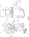
- the system 10 includes a radiation source/device 16, a radiation detector 18, and a collimator 20.
- the radiation source 16 is movable between a variety of imaging positions relative to the detector 18, and is operative to emit radiation rays 22 ( FIG. 2 ) that are received by the radiation detector 18 to provide an image of a breast 52.
- the system 10 may include a patient shield 24 mounted to the radiation source 16 via face shield rails 26 to prevent the patient's head from obstructing the radiation rays and protecting the patient from the radiation rays 22.
- the system 10 also includes a compression plate 28 and a support structure 30 to which one or more of the radiation source 16, radiation detector 18, and/or compression plate 28 may be mounted to.
- the system 10 may further include a controller 32.
- the controller 32 may be a workstation having at least one processor and a memory device as shown in FIG. 1 or, in other embodiments, the controller 32 may be embedded / integrated into one or more of the various components of the system 10 disclosed above.
- the controller 32 may be in electrical communication with the radiation source 16, radiation detector 18, and/or the compression plate 28 via a cable 34.
- the connection 34 may be a wireless connection.
- the controller 32 may include a radiation shield 36 that protects an operator of the system 10 from the radiation rays 22 emitted by the radiation source 16.
- the controller 32 may further include a display 38, a keyboard 40, mouse 42, and/or other appropriate user input devices that facilitate control of the system 10 via a user interface 44.
- the radiation source 16 along with the radiation detector 18, forms part of an x-ray system which provides x-ray imagery for the purpose of imaging a body part of a patient, such as breast 52.
- the radiation source 16 emits the radiation rays 22 such that the radiation rays 22 travel from the radiation source 16 to the radiation detector 18. While the radiation rays 22 are discussed herein as being x-rays, it is to be understood that the radiation source 16 may emit other types of electromagnetic rays which can be used to image a patient.
- the radiation source 16 may be mounted to the support structure 30 such that the radiation source can rotate around an axis 46 in relation to the radiation detector 18, although movement of the radiation source 16 in paths other than rotation about a fixed axis, such as during digital breast tomosynthesis (DBT), are also envisioned.
- the radiation detector 18 may be configured to rotate or translate within its housing, such as in the directions indicated by arrows 53 and 55.
- the radiation detector 18 receives the radiation rays 22 emitted by the radiation source 16.
- data regarding the radiation rays 22 received by the radiation detector 18 may be electrically communicated to the controller 32 from the radiation detector 18 via cable/electronic connection 34 such that the controller 32 generates one or more images which may be shown on the display 38 and stored in the memory device.
- the compression plate 28 is operative, in response to instruction from the controller 32 or in response to instructions from controller(s) on or near the mammography system 10 or switch controllers 80, to move towards and away from the radiation detector 18 as indicated by arrows 48 such that the compression plate 28 flattens and holds a body part, e.g., breast 52, in place against the surface 50 of the radiation detector 18.
- the radiation detector 18 and the surface 50 thereof is referred to herein as a "support plate” that cooperates with the compression plate 28 to compress and clamp a breast of a patient therebetween.
- the system 10 may further, or alternatively, include a biopsy tool 100, illustrated in FIG. 3 .
- the radiation source 16, along with the radiation detector 18, forms part of an x-ray system which provides x-ray imagery for the purpose of guiding the biopsy tool 100, e.g., needle, to a suspect site within a body part of a patient.
- the biopsy tool 100 may be disposed on the support structure 30 such that it also rotates about the axis 46, in a manner similar to the radiation source 16, and/or moves in a vertical and/or horizontal direction, in a manner similar to the compression plate 28.
- the breast 52 of the patient may be placed onto the surface 50 of the radiation detector 18.
- imaging or procedures e.g., a biopsy
- the radiation source 16 is selectively adjusted such that it is moved/rotated to a first scanning position and scans the breast 52.
- the radiation detector 18 receives the radiation rays 22 passing through the breast 52 and sends data to the controller 32 which then generates one or more x-ray images of the breast 52.
- the controller 32 moves the compression plate 28 away from the support plate 18 to free the breast 52.
- the system 10 may include one or more physiological monitoring or sensor devices 54, 56, 58, 60 communicatively coupled with the controller 32 for monitoring one or more physiological parameters of a patient (and for transmitting physiological parameter data to the controller 32). While FIG.1 illustrates that the sensor devices 54, 56, 58 are connected to the controller 32, in some embodiments, one or more of the sensor devices may be communicatively coupled with the mammography apparatus, without departing from the broader aspects of the invention.
- the sensor devices may be selected to monitor and/or measure any physiological information of a patient desired, including, but not limited to, diastolic blood pressure, systolic blood pressure, body temperature, blood oxygen level, patient weight, skin conductance, pulse rate, etc.
- one or more of the sensor devices e.g., sensor device 60
- the sensor devices may be physically integrated with the compression plate 28 and/or the detector/support plate 18.
- physiological parameter data of the patient may be acquired and transmitted to the controller 32 without requiring any additional intervention by the system operator.
- the sensor device 60 may be a force sensor for measuring the amount of pressure or compressive force applied to the breast 52. Additional sensors for measuring physiological parameters may be configured to either directly measure or allow the calculation of variables such as force, pressure, temperature, rigidity, elasticity, breast size and/or volume, and/or tissue density and could be embedded in compression plate 28 or support plate 18 or attached as part of mammography system 10.
- the various sensor devices 54, 56, 58, 60 may be configured to acquire physiological parameter data from a patient during system operation. More specifically, physiological parameter data may be acquired continuously or at predetermined time intervals before breast compression and imaging, during the compression phase of the system and/or during the clamping phase of the system. In an embodiment, the physiological parameter data may be acquired continuously or at predetermined time intervals during at least the compression phase. In other embodiments, the physiological parameter data may be acquired continuously or at predetermined time intervals during at least the compression phase and the clamping phase.
- At least one of the sensor devices 54, 56, 58, 60 is a blood pressure measuring device configured to measure at least one of diastolic blood pressure and systolic blood pressure of a patient.
- the blood pressure measuring device may be a blood pressure cuff configured for placement on the arm of a patient, an ultrasound device or a thermal imaging device.
- the blood pressure measuring device may be integrated with at least one of the compression plate 28 and/or the support plate 18 (e.g., sensing device 60) for measuring the blood pressure of a patient directly from the patient's breast.
- the blood pressure measuring device may take continuous blood pressure measurements via pulse wave velocity measurements, continuous noninvasive arterial pressure methods and the like, and is not limited to any particular technique, method or apparatus.
- the controller 32 is configured to control movement of the compression plate 28 toward the support plate 18 to flatten the breast 52 during the compression phase, and to clamp the breast 52 during the clamping phase, as discussed above.
- the controller 32 is also configured to control movement of the compression plate 28 to adjust at least one of a rate of compression and/or a pressure or compressive force applied to the breast 52 in response to the physiological parameter data acquired from the patient.
- the controller 32 is configured to vary at least one of the rate of compression and/or the pressure applied to the breast 52 during the compression phase and/or clamping phase based on a measurement of at least one of a diastolic pressure and/or a systolic pressure of the patient taken during the compression phase and/or the clamping phase.
- the controller 32 may be configured to vary at least one of the rate of compression and/or the pressure applied in real-time, as practicable, as measured blood pressure changes.
- real-time refers to a level of responsiveness that a user senses as sufficiently immediate, or that enables the controller (or other processor) to keep up with an external process, e.g., compression, pressure, etc.
- rate of compression refers to the change in compressive force applied to the breast in relation to elapsed time.
- At least one of the rate of compression and/or the pressure applied may be varied in real-time during the compression phase, and a target pressure has been reached for optimal image acquisition, the clamping phase may be initiated and the plates held in static position during the scan (2D, DBT, multi-energy, etc.). This ensures that the object being imaged remains in the same position during the scan.
- the controller 32 may be configured to vary at least one of the rate of compression and/or the pressure applied after each measurement.
- the real-time blood pressure measurements may be smoothed as a running average by the controller 32.
- the controller 32 is configured to vary at least one of the rate of compression and/or the pressure applied in based on measured blood pressure and one or more other physiological parameters such as, for example, a measured size or volume of the breast 52.
- the size or volume of the breast may be measured prior to commencing the procedure and input into the controller, or measured automatically using sensors integrated with the compression plate 28 and/or support plate 18.
- a density or rigidity measurement and/or determination using one or more sensors integrated with the compression plate 28 and/or support plate 18 may be used by the controller 32 to determine when to cease compression during the compression phase.
- density or rigidity sensors may be utilized to determine when a target density or rigidity for optimal image quality is achieved, and the controller 32 may be configured to transition the system 10 from the compression phase to the clamping phase when a target tissue density and/or rigidity is achieved.
- At least one of the sensing devices may be configured to measure a physiological parameter that can be utilized by the controller 32 to determine whether or not blood is flowing in the compressed tissue.
- the controller 32 may be further configured to immediately cease and/or release compression on the breast tissue if the sensor feedback indicates that blood has ceased flowing through the breast.
- the system 10 contains a built in failsafe mechanism that prevents overcompression to extent that could cause damage to a patient.
- switch controls 80 e.g., footswitch controls, such as disclosed in U.S. Patent No. 10,004,470 .
- Switch controls 80 are typically connected via a cable/wire 82 to mammography imaging system 10. The controls are also often mirrored on the opposite side of mammography imaging system 10 (not shown). Other controls (not shown) may be present on particular accessories placed either in the paddle/breast support area.
- the switch controls may be a handheld control unit 84 with a wired, wireless, Bluetooth or other connection with the system 10.
- the patient may control the rate of compression and/or pressure or force applied during the compression phase and/or clamping phase using the switch controls.
- a feedback device e.g. controller 32
- the feedback device e.g., controller 32, may be configured to provide feedback information to the patient regarding the rate of compression (greater or lower rate of compression) and/or amount of pressure (higher or lower) required to produce an optimal image, in dependence upon the information received from the various sensor devices 54, 56, 58, 60.
- the feedback device informs the patient when compression rate and/or pressure applied is sufficient to obtain a quality image, as determined from a blood pressure or other measurement taken from the patient through sensing devices 54, 56, 58, or 60, before or during the compression and/or clamping phase.
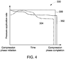
- FIG. 4 a graph 300 illustrating pressure application rates during the compression phase is depicted.
- Line 302 represents the pressure application rate change during the compression phase based on feedback from a blood pressure sensing device, as described above.
- the rate of pressure application and the final target force applied to the breast is varied in response to blood pressure measurements taken during the procedure. In this instance, the rate of applied pressure decreases throughout the compression phase with some periods of constant rate.
- the rate of pressure application initially increases before sharply dropping off.
- static techniques represented by line 306, where the rate of pressure application and the final target pressure are fixed based on an initial blood pressure reading.
- FIG. 5 is a graph 400 illustrating applied pressure during a compression phase and a clamping phase for various imaging procedures.
- Line 402 shows the applied pressure during the compression phase and clamping phase are varied based on blood pressure measurements taken during both the compression phase and the clamping phase, as discussed above.
- line 404 illustrates typical static techniques, which show that applied pressure increases at a uniform rate during the compression phase, and is held constant during the clamping phase. As illustrated, therefore, by measuring blood pressure continuously during the compression phase and clamping phase, lower applied pressures can be used while still obtaining optimal image quality which, again, translates to greater patient comfort.
- the system 10 is able to optimize the compressive force applied and/or the rate of compression to minimize patient discomfort while also ensuring that a minimum level of compression is achieved to ensure that quality images can be obtained.
- the feedback from the various sensing devices allows for automated, real-time changes in compression based on factors that ensure optimal image quality while at the same time reducing the pain experienced during the procedure.
- the acquired physiological data may be read directly into a patient medical record along with other data collected by the system 10, without requiring separate measurements by the operator or clinician. Accordingly, by utilizing the system 10, the overall length of the mammography procedure and the associated time required for preparation may be shortened.
- the system 10 may include the necessary electronics, software, memory, storage, databases, firmware, logic/state machines, microprocessors, communication links, displays or other visual or audio user interfaces, printing devices, and any other input/output interfaces to perform the functions described herein and/or to achieve the results described herein.
- the system may include at least one processor and system memory / data storage structures, which may include random access memory (RAM) and read-only memory (ROM).
- the at least one processor of the system 10 may include one or more conventional microprocessors and one or more supplementary co-processors such as math co-processors or the like.
- the data storage structures discussed herein may include an appropriate combination of magnetic, optical and/or semiconductor memory, and may include, for example, RAM, ROM, flash drive, an optical disc such as a compact disc and/or a hard disk or drive.
- a software application that adapts the controller to perform the methods disclosed herein may be read into a main memory of the at least one processor from a computer-readable medium.
- computer-readable medium refers to any medium that provides or participates in providing instructions to the at least one processor of the system 10 (or any other processor of a device described herein) for execution.
- Such a medium may take many forms, including but not limited to, non-volatile media and volatile media.
- Non-volatile media include, for example, optical, magnetic, or opto-magnetic disks, such as memory.
- Volatile media include dynamic random access memory (DRAM), which typically constitutes the main memory.
- Computer-readable media include, for example, a floppy disk, a flexible disk, hard disk, magnetic tape, any other magnetic medium, a CD-ROM, DVD, any other optical medium, a RAM, a PROM, an EPROM or EEPROM (electronically erasable programmable read-only memory), a FLASH-EEPROM, any other memory chip or cartridge, or any other medium from which a computer can read.
- a mammography apparatus in an embodiment, includes a support plate for supporting a breast of a patient, a compression plate movable toward and away from the support plate for compressing the breast against the support plate, and a controller configured to control movement of the compression plate toward and away from the support plate.
- the controller is configured to adjust at least one of a rate of compression and/or a pressure applied to the breast based on a measurement of a diastolic pressure and/or a systolic pressure of the patient.
- the measurement of the diastolic pressure and/or the systolic pressure is taken during at least one of a compression phase and/or a clamping phase of the mammography apparatus.
- the controller is configured to adjust in real time at least one of the rate of compression and/or the pressure applied to the breast based on a plurality of measurements the diastolic pressure and/or the systolic pressure of the patient taken during at least one of the compression phase and/or the clamping phase of the mammography apparatus.
- the pressure is at least one of a target maximum pressure or a clamping phase pressure.
- the apparatus includes a sensor device configured to measure the diastolic pressure and/or the systolic pressure.
- the sensor device is configured for placement on a body part of the patient not undergoing compression. The sensor device may be integrated within one of the support plate or the compression plate.
- the sensor device is at least one of an ultrasound device, and/or automated cuff or a thermal imaging device.
- the measurement is a continuous measurement taken during at least one of the compression phase and/or the clamping phase.
- the controller is configured to adjust the at least one of the rate of compression and the pressure applied to the breast based on at least one of a measured size and a measured volume of the breast.
- the apparatus may include at least one of a blood oxygen sensor and/or a temperature sensor integrated with at least one of the support plate or the compression plate, wherein the controller is further configured to adjust at least one of the rate of compression and/or the pressure applied to the breast based on a measured blood oxygen content or body temperature.
- the controller is further configured to cease movement of the compression plate to cease and/or reduce compression of the breast when a target tissue density or target tissue rigidity is detected. In an embodiment, the controller is further configured to automatically cease and/or reduce compression of the breast if a stoppage in a flow of blood in the breast is detected.
- the apparatus includes a radiation source for applying radiation to the breast.
- the apparatus includes a support plate for supporting tissue of a patient, a compression plate movable toward and away from the support plate for compressing the tissue against the support plate, and a controller configured to control movement of the compression plate toward and away from the support plate, and to adjust in real time at least one of a rate of compression and/or a pressure applied to the tissue based on a measurement of a physiological parameter of a patient taken during at least one of a compression phase and/or a clamping phase of the apparatus.
- the controller allows a patient to control movement of the compression plate toward and away from the support plate to adjust the rate of compression and/or the pressure applied to the tissue based upon feedback provided by the apparatus regarding the measurement of the physiological parameter taken during at least one of a compression phase and/or a clamping phase.
- the apparatus includes a patient-controllable switch operable to control movement of the compression plate toward and away from the support plate.
- the physiological parameter is one of a diastolic pressure or a systolic pressure of the patient.
- the controller is configured to adjust the at least one of the rate of compression and/or the pressure applied to the tissue based on at least one of a measured size and/or a measured volume of the tissue.
- a method of operating a mammography apparatus includes the steps of positioning a breast of a patient on a support plate, moving a compression plate towards the support plate to compress the breast of a patient, measuring a diastolic pressure and/or a systolic pressure of a patient, and varying at least one of a rate of compression and/or a pressure applied to the breast based on the measurement the diastolic pressure and/or the systolic pressure.
- the step of measuring includes continuously measuring the diastolic pressure and/or the systolic pressure throughout a compression phase of the apparatus.
- the step of measuring is carried out utilizing a sensor device integrated with one of the support plate and the compression plate.
- the method may also include determining at least one of a size and/or a volume of the breast, and varying at least one of the rate of compression and/or the pressure applied to the breast based on the at least one of the size and/or the volume of the breast.
Landscapes
- Health & Medical Sciences (AREA)
- Life Sciences & Earth Sciences (AREA)
- Medical Informatics (AREA)
- Engineering & Computer Science (AREA)
- General Health & Medical Sciences (AREA)
- Veterinary Medicine (AREA)
- Biophysics (AREA)
- Physics & Mathematics (AREA)
- Public Health (AREA)
- Animal Behavior & Ethology (AREA)
- Surgery (AREA)
- Pathology (AREA)
- Molecular Biology (AREA)
- Biomedical Technology (AREA)
- Heart & Thoracic Surgery (AREA)
- Radiology & Medical Imaging (AREA)
- Optics & Photonics (AREA)
- Nuclear Medicine, Radiotherapy & Molecular Imaging (AREA)
- High Energy & Nuclear Physics (AREA)
- Cardiology (AREA)
- Dentistry (AREA)
- Oral & Maxillofacial Surgery (AREA)
- Vascular Medicine (AREA)
- Physiology (AREA)
- Ophthalmology & Optometry (AREA)
- Apparatus For Radiation Diagnosis (AREA)
Claims (13)
- Mammographievorrichtung (10), umfassend:eine Lagerungsplatte (18) zur Auflage einer Brust (52) einer Patientin,eine Kompressionsplatte (28), die zu der Lagerungsplatte (18) hin und von dieser weg beweglich ist, um die Brust (52) gegen die Lagerungsplatte (18) zu komprimieren, undeine Steuerung (32), die dazu ausgestaltet ist, auf der Grundlage einer Messung eines diastolischen Drucks und/oder eines systolischen Drucks der Patientin die Bewegung der Kompressionsplatte (28) zu der Lagerungsplatte (18) hin und von dieser weg zu steuern und eine Kompressionsrate und/oder einen auf die Brust (52) ausgeübten Druck zu verstellen,wobei die Messung des diastolischen Drucks und/oder des systolischen Drucks der Patientin während einer Kompressionsphase und/oder einer Klemmphase der Mammographievorrichtung (10) erfolgt.
- Mammographievorrichtung (10) nach Anspruch 1, wobei:
die Steuerung (32) dazu ausgestaltet ist, die Kompressionsrate und/oder den auf die Brust (52) ausgeübten Druck auf der Grundlage einer Vielzahl von Messungen des diastolischen Drucks und/oder des systolischen Drucks der Patientin während einer Kompressionsphase und/oder einer Klemmphase der Mammographievorrichtung (10) kontinuierlich in Echtzeit zu verstellen. - Mammographievorrichtung (10) nach Anspruch 1 oder Anspruch 2, wobei:
der Druck ein maximaler Zieldruck und/oder ein Klemmphasendruck ist. - Mammographievorrichtung (10) nach einem der vorhergehenden Ansprüche, ferner umfassend:
eine Sensorvorrichtung (54), die zum Messen des diastolischen Drucks und/oder des systolischen Drucks ausgestaltet ist. - Mammographievorrichtung (10) nach Anspruch 4, wobei:
die Sensorvorrichtung (54) zur Platzierung an einem Körperteil der Patientin, der nicht komprimiert wird, ausgestaltet ist. - Mammographievorrichtung (10) nach Anspruch 4, wobei:
die Sensorvorrichtung (54) in der Lagerungsplatte (18) oder der Kompressionsplatte (28) integriert ist. - Mammographievorrichtung (10) nach Anspruch 4, wobei:
die Sensorvorrichtung (54) eine Ultraschallvorrichtung und/oder eine automatische Manschette und/oder ein Wärmebildgerät ist. - Mammographievorrichtung (10) nach einem der Ansprüche 4 bis 7, wobei:
die Messung eine kontinuierliche Messung ist, die während der Kompressionsphase und/oder der Klemmphase erfolgt. - Mammographievorrichtung (10) nach einem der vorhergehenden Ansprüche, wobei:
die Steuerung (32) dazu ausgestaltet ist, auf der Grundlage einer gemessenen Größe und/oder eines gemessenen Volumens der Brust (52) die Kompressionsrate und/oder den auf die Brust (52) ausgeübten Druck zu verstellen. - Mammographievorrichtung (10) nach einem der Ansprüche 4 bis 9, ferner umfassend:einen Blutsauerstoffsensor und/oder einen Temperatursensor, die mit der Lagerungsplatte (18) und/oder der Kompressionsplatte (28) integriert sind,die Steuerung (32) ferner dazu ausgestaltet ist, auf der Grundlage eines gemessenen Blutsauerstoffgehalts oder einer gemessenen Körpertemperatur die Kompressionsrate und/oder den auf die Brust (52) ausgeübten Druck zu verstellen.
- Mammographievorrichtung (10) nach einem der Ansprüche 4 bis 10, wobei:
die Steuerung (32) ferner dazu ausgestaltet ist, die Kompression der Brust (52) automatisch zu stoppen und/oder zu reduzieren, wenn ein Stoppen der Durchblutung der Brust (52) erfasst wird. - Verfahren zum Betreiben einer Mammographievorrichtung (10), umfassend die folgenden Schritte:Positionieren einer Brust (52) einer Patientin auf einer Lagerungsplatte (18),Bewegen einer Kompressionsplatte (28) zu der Lagerungsplatte (18) hin, um die Brust (52) einer Patientin zu komprimieren,Messen eines diastolischen Drucks und/oder eines systolischen Drucks einer Patientin undVariieren einer Kompressionsrate und/oder eines auf die Brust (52) ausgeübten Drucks auf der Grundlage der Messung des diastolischen Drucks und/oder des systolischen Drucks,wobei der Messschritt das kontinuierliche Messen des diastolischen Drucks und/oder des systolischen Drucks während einer Kompressionsphase der Vorrichtung aufweist.
- Verfahren nach Anspruch 12, wobei:
der Messschritt unter Verwendung einer mit der Lagerungsplatte (18) oder der Kompressionsplatte (28) integrierten Sensorvorrichtung (54) ausgeführt wird.
Applications Claiming Priority (1)
| Application Number | Priority Date | Filing Date | Title |
|---|---|---|---|
| US16/108,891 US10695010B2 (en) | 2018-08-22 | 2018-08-22 | Apparatus and method for mammographic breast compression |
Publications (2)
| Publication Number | Publication Date |
|---|---|
| EP3613348A1 EP3613348A1 (de) | 2020-02-26 |
| EP3613348B1 true EP3613348B1 (de) | 2021-06-16 |
Family
ID=67659376
Family Applications (1)
| Application Number | Title | Priority Date | Filing Date |
|---|---|---|---|
| EP19192419.0A Active EP3613348B1 (de) | 2018-08-22 | 2019-08-19 | Vorrichtung und verfahren zur mammografischen brustkompression |
Country Status (4)
| Country | Link |
|---|---|
| US (1) | US10695010B2 (de) |
| EP (1) | EP3613348B1 (de) |
| JP (1) | JP7019640B2 (de) |
| CN (1) | CN110856657B (de) |
Families Citing this family (12)
| Publication number | Priority date | Publication date | Assignee | Title |
|---|---|---|---|---|
| EP3618079A1 (de) * | 2018-08-30 | 2020-03-04 | Koninklijke Philips N.V. | Bildgebendes verfahren mit schmerzsensor |
| DE102020202205A1 (de) * | 2020-02-20 | 2021-08-26 | Siemens Healthcare Gmbh | Verfahren zur Kompression von Brustgewebe |
| WO2022060553A1 (en) * | 2020-09-15 | 2022-03-24 | Hologic, Inc. | Systems and methods for determining performance of a breast compression paddle |
| DE102020212205A1 (de) | 2020-09-28 | 2022-03-31 | Siemens Healthcare Gmbh | Individuelle Ermittlung der Brustkompression bei der Mammographie mit künstlicher Intelligenz |
| US12251257B2 (en) | 2022-10-19 | 2025-03-18 | GE Precision Healthcare LLC | System and method for analyzing breast support environment |
| US12343185B2 (en) | 2022-12-28 | 2025-07-01 | GE Precision Healthcare LLC | Device and method for securely holding breast on mammography imaging system |
| US12350085B2 (en) | 2023-02-15 | 2025-07-08 | Ge Healthcare | Mammography imaging system with universal attachment structures |
| US12490950B2 (en) | 2023-04-17 | 2025-12-09 | GE Precision Healthcare LLC | Biopsy positioner for mammography imaging system |
| DE102023206155A1 (de) * | 2023-06-29 | 2025-01-02 | Siemens Healthineers Ag | Computerimplementiertes Verfahren zum Betrieb einer Röntgeneinrichtung, Röntgeneinrichtung, Computerprogramm und elektronisch lesbarer Datenträger |
| US12465317B2 (en) | 2023-09-19 | 2025-11-11 | GE Precision Healthcare LLC | Non-uniform thickness breast imaging phantom for 2D/3D spectral imaging |
| US20250107762A1 (en) | 2023-09-29 | 2025-04-03 | GE Precision Healthcare LLC | Device and Method for Integrated Tissue Sample Imaging on Radiography Imaging System |
| US12456939B2 (en) | 2023-09-29 | 2025-10-28 | GE Precision Healthcare LLC | Device and method for automatic detection and configuration of a motor |
Family Cites Families (16)
| Publication number | Priority date | Publication date | Assignee | Title |
|---|---|---|---|---|
| US8100839B2 (en) | 2003-12-30 | 2012-01-24 | Galkin Benjamin M | Acoustic monitoring of a breast and sound databases for improved detection of breast cancer |
| DE102004052614B3 (de) * | 2004-10-29 | 2006-01-05 | Siemens Ag | Kompressionsplatte, insbesondere für ein Mammographiegerät |
| WO2010078048A2 (en) * | 2008-12-17 | 2010-07-08 | Therapy Positioning Technologies, Llc | Apparatus and method for concurrent tensive and compressive immobilization of the breast in imaging and intervention |
| NL2005509C2 (en) * | 2010-02-19 | 2011-08-23 | Academisch Medisch Ct Bij De Universiteit Van Amsterdam | Mammography-apparatus. |
| JP5419821B2 (ja) * | 2010-07-20 | 2014-02-19 | 富士フイルム株式会社 | 放射線画像撮影装置、及び圧迫板 |
| JP2012170718A (ja) * | 2011-02-23 | 2012-09-10 | Toshiba Corp | マンモグラフィ装置 |
| JP6247221B2 (ja) * | 2011-11-21 | 2017-12-13 | コーニンクレッカ フィリップス エヌ ヴェKoninklijke Philips N.V. | マンモグラフィ検査のための圧縮力の個別監視 |
| JP5943598B2 (ja) | 2011-12-26 | 2016-07-05 | キヤノン株式会社 | 被検体情報取得装置 |
| KR102083820B1 (ko) * | 2012-03-29 | 2020-03-03 | 제네럴 일렉트릭 컴퍼니 | 맘모그래피 장치 및 그의 위치 정렬 제어 방법 |
| JP2015522337A (ja) * | 2012-06-25 | 2015-08-06 | コーニンクレッカ フィリップス エヌ ヴェ | 光学的に適合可能な胸部圧迫素子 |
| US9237872B2 (en) * | 2013-01-18 | 2016-01-19 | General Electric Company | X-ray source with moving anode or cathode |
| KR101484674B1 (ko) * | 2013-02-20 | 2015-01-20 | 메디퓨처(주) | 유방두께 측정장치 및 그를 이용한 맘모그래피 장치 |
| US10004470B2 (en) | 2013-07-18 | 2018-06-26 | General Electric Company | Breast imaging system giving feedback information to the patient and method using thereof |
| US20170082557A1 (en) * | 2015-09-21 | 2017-03-23 | General Electric Company | Method and system to image biopsy samples during an x-ray guided biopsy procedure |
| US10357216B2 (en) * | 2015-12-21 | 2019-07-23 | Toshiba Medical Systems Corporation | Mammography apparatus |
| JP6141488B2 (ja) * | 2016-05-23 | 2017-06-07 | キヤノン株式会社 | 被検体情報取得装置 |
-
2018
- 2018-08-22 US US16/108,891 patent/US10695010B2/en active Active
-
2019
- 2019-08-09 CN CN201910735604.3A patent/CN110856657B/zh active Active
- 2019-08-16 JP JP2019149229A patent/JP7019640B2/ja active Active
- 2019-08-19 EP EP19192419.0A patent/EP3613348B1/de active Active
Non-Patent Citations (1)
| Title |
|---|
| None * |
Also Published As
| Publication number | Publication date |
|---|---|
| US20200060632A1 (en) | 2020-02-27 |
| CN110856657B (zh) | 2023-09-12 |
| JP7019640B2 (ja) | 2022-02-15 |
| JP2020032183A (ja) | 2020-03-05 |
| US10695010B2 (en) | 2020-06-30 |
| CN110856657A (zh) | 2020-03-03 |
| EP3613348A1 (de) | 2020-02-26 |
Similar Documents
| Publication | Publication Date | Title |
|---|---|---|
| US10695010B2 (en) | Apparatus and method for mammographic breast compression | |
| US11129594B2 (en) | Hardness deriving device, medical imaging system, hardness deriving method and hardness deriving program storage medium | |
| US9855014B2 (en) | Compression paddle for use in breast imaging | |
| US9949719B2 (en) | Breast imaging method and system | |
| AU2022263562A1 (en) | Method Of Imaging | |
| US20080031413A1 (en) | Method for the reconstruction of a body map | |
| US12226233B2 (en) | Personalized breast imaging system | |
| JP6307590B2 (ja) | マンモグラフィ装置 | |
| US8831171B2 (en) | Full field mammography with tissue exposure control, tomosynthesis, and dynamic field of view processing | |
| JP2008253765A (ja) | 被検体の呼吸周期を追跡するシステム及び方法 | |
| JP2010069241A (ja) | 乳房断層画像撮影装置 | |
| JP2024514268A (ja) | 発泡体圧迫要素の厚さを測定するためのシステムおよび方法 | |
| JP2016515436A (ja) | 乳房のx線及び機械撮像のための圧迫装置 | |
| EP4356838A1 (de) | System und verfahren zur analyse der bruststützumgebung | |
| JP2024504604A (ja) | 撮像のために乳房に力を加えるためのシステムおよび方法 | |
| EP3701871B1 (de) | Steuerungsvorrichtung, radiographiesystem, medizinisches bildgebungssystem, steuerungsverfahren und steuerungsprogramm | |
| US12343185B2 (en) | Device and method for securely holding breast on mammography imaging system | |
| KR102083821B1 (ko) | 맘모그래피 장치 및 이를 이용한 피검체 촬영 방법 | |
| US12082628B2 (en) | Brassiere for use in mammography imaging procedures | |
| CN117678981A (zh) | 一种心内科防止大量出血的介入控制系统及控制方法 | |
| CN120078438A (zh) | 用于针对改进的乳房摄影成像系统定位生成预测视图的系统和方法 | |
| KR20140126490A (ko) | 유방의 탄성도 분포 정보 및 엑스선 영상의 획득을 위한 유방촬영용 영상진단기기 |
Legal Events
| Date | Code | Title | Description |
|---|---|---|---|
| PUAI | Public reference made under article 153(3) epc to a published international application that has entered the european phase |
Free format text: ORIGINAL CODE: 0009012 |
|
| STAA | Information on the status of an ep patent application or granted ep patent |
Free format text: STATUS: REQUEST FOR EXAMINATION WAS MADE |
|
| 17P | Request for examination filed |
Effective date: 20190819 |
|
| AK | Designated contracting states |
Kind code of ref document: A1 Designated state(s): AL AT BE BG CH CY CZ DE DK EE ES FI FR GB GR HR HU IE IS IT LI LT LU LV MC MK MT NL NO PL PT RO RS SE SI SK SM TR |
|
| AX | Request for extension of the european patent |
Extension state: BA ME |
|
| GRAP | Despatch of communication of intention to grant a patent |
Free format text: ORIGINAL CODE: EPIDOSNIGR1 |
|
| STAA | Information on the status of an ep patent application or granted ep patent |
Free format text: STATUS: GRANT OF PATENT IS INTENDED |
|
| INTG | Intention to grant announced |
Effective date: 20201124 |
|
| RIN1 | Information on inventor provided before grant (corrected) |
Inventor name: BLASKI, RONALD Inventor name: SUTTY, JEROME Inventor name: IORDACHE, RAZVAN |
|
| GRAJ | Information related to disapproval of communication of intention to grant by the applicant or resumption of examination proceedings by the epo deleted |
Free format text: ORIGINAL CODE: EPIDOSDIGR1 |
|
| STAA | Information on the status of an ep patent application or granted ep patent |
Free format text: STATUS: REQUEST FOR EXAMINATION WAS MADE |
|
| GRAP | Despatch of communication of intention to grant a patent |
Free format text: ORIGINAL CODE: EPIDOSNIGR1 |
|
| STAA | Information on the status of an ep patent application or granted ep patent |
Free format text: STATUS: GRANT OF PATENT IS INTENDED |
|
| INTC | Intention to grant announced (deleted) | ||
| INTG | Intention to grant announced |
Effective date: 20210205 |
|
| GRAS | Grant fee paid |
Free format text: ORIGINAL CODE: EPIDOSNIGR3 |
|
| GRAA | (expected) grant |
Free format text: ORIGINAL CODE: 0009210 |
|
| STAA | Information on the status of an ep patent application or granted ep patent |
Free format text: STATUS: THE PATENT HAS BEEN GRANTED |
|
| AK | Designated contracting states |
Kind code of ref document: B1 Designated state(s): AL AT BE BG CH CY CZ DE DK EE ES FI FR GB GR HR HU IE IS IT LI LT LU LV MC MK MT NL NO PL PT RO RS SE SI SK SM TR |
|
| REG | Reference to a national code |
Ref country code: GB Ref legal event code: FG4D |
|
| REG | Reference to a national code |
Ref country code: CH Ref legal event code: EP |
|
| REG | Reference to a national code |
Ref country code: DE Ref legal event code: R096 Ref document number: 602019005365 Country of ref document: DE |
|
| REG | Reference to a national code |
Ref country code: AT Ref legal event code: REF Ref document number: 1401640 Country of ref document: AT Kind code of ref document: T Effective date: 20210715 |
|
| REG | Reference to a national code |
Ref country code: IE Ref legal event code: FG4D Ref country code: NL Ref legal event code: FP |
|
| REG | Reference to a national code |
Ref country code: LT Ref legal event code: MG9D |
|
| PG25 | Lapsed in a contracting state [announced via postgrant information from national office to epo] |
Ref country code: HR Free format text: LAPSE BECAUSE OF FAILURE TO SUBMIT A TRANSLATION OF THE DESCRIPTION OR TO PAY THE FEE WITHIN THE PRESCRIBED TIME-LIMIT Effective date: 20210616 Ref country code: LT Free format text: LAPSE BECAUSE OF FAILURE TO SUBMIT A TRANSLATION OF THE DESCRIPTION OR TO PAY THE FEE WITHIN THE PRESCRIBED TIME-LIMIT Effective date: 20210616 Ref country code: FI Free format text: LAPSE BECAUSE OF FAILURE TO SUBMIT A TRANSLATION OF THE DESCRIPTION OR TO PAY THE FEE WITHIN THE PRESCRIBED TIME-LIMIT Effective date: 20210616 Ref country code: BG Free format text: LAPSE BECAUSE OF FAILURE TO SUBMIT A TRANSLATION OF THE DESCRIPTION OR TO PAY THE FEE WITHIN THE PRESCRIBED TIME-LIMIT Effective date: 20210916 |
|
| REG | Reference to a national code |
Ref country code: AT Ref legal event code: MK05 Ref document number: 1401640 Country of ref document: AT Kind code of ref document: T Effective date: 20210616 |
|
| PG25 | Lapsed in a contracting state [announced via postgrant information from national office to epo] |
Ref country code: GR Free format text: LAPSE BECAUSE OF FAILURE TO SUBMIT A TRANSLATION OF THE DESCRIPTION OR TO PAY THE FEE WITHIN THE PRESCRIBED TIME-LIMIT Effective date: 20210917 Ref country code: LV Free format text: LAPSE BECAUSE OF FAILURE TO SUBMIT A TRANSLATION OF THE DESCRIPTION OR TO PAY THE FEE WITHIN THE PRESCRIBED TIME-LIMIT Effective date: 20210616 Ref country code: NO Free format text: LAPSE BECAUSE OF FAILURE TO SUBMIT A TRANSLATION OF THE DESCRIPTION OR TO PAY THE FEE WITHIN THE PRESCRIBED TIME-LIMIT Effective date: 20210916 Ref country code: SE Free format text: LAPSE BECAUSE OF FAILURE TO SUBMIT A TRANSLATION OF THE DESCRIPTION OR TO PAY THE FEE WITHIN THE PRESCRIBED TIME-LIMIT Effective date: 20210616 Ref country code: RS Free format text: LAPSE BECAUSE OF FAILURE TO SUBMIT A TRANSLATION OF THE DESCRIPTION OR TO PAY THE FEE WITHIN THE PRESCRIBED TIME-LIMIT Effective date: 20210616 |
|
| PG25 | Lapsed in a contracting state [announced via postgrant information from national office to epo] |
Ref country code: SK Free format text: LAPSE BECAUSE OF FAILURE TO SUBMIT A TRANSLATION OF THE DESCRIPTION OR TO PAY THE FEE WITHIN THE PRESCRIBED TIME-LIMIT Effective date: 20210616 Ref country code: EE Free format text: LAPSE BECAUSE OF FAILURE TO SUBMIT A TRANSLATION OF THE DESCRIPTION OR TO PAY THE FEE WITHIN THE PRESCRIBED TIME-LIMIT Effective date: 20210616 Ref country code: ES Free format text: LAPSE BECAUSE OF FAILURE TO SUBMIT A TRANSLATION OF THE DESCRIPTION OR TO PAY THE FEE WITHIN THE PRESCRIBED TIME-LIMIT Effective date: 20210616 Ref country code: AT Free format text: LAPSE BECAUSE OF FAILURE TO SUBMIT A TRANSLATION OF THE DESCRIPTION OR TO PAY THE FEE WITHIN THE PRESCRIBED TIME-LIMIT Effective date: 20210616 Ref country code: CZ Free format text: LAPSE BECAUSE OF FAILURE TO SUBMIT A TRANSLATION OF THE DESCRIPTION OR TO PAY THE FEE WITHIN THE PRESCRIBED TIME-LIMIT Effective date: 20210616 Ref country code: SM Free format text: LAPSE BECAUSE OF FAILURE TO SUBMIT A TRANSLATION OF THE DESCRIPTION OR TO PAY THE FEE WITHIN THE PRESCRIBED TIME-LIMIT Effective date: 20210616 Ref country code: RO Free format text: LAPSE BECAUSE OF FAILURE TO SUBMIT A TRANSLATION OF THE DESCRIPTION OR TO PAY THE FEE WITHIN THE PRESCRIBED TIME-LIMIT Effective date: 20210616 Ref country code: PT Free format text: LAPSE BECAUSE OF FAILURE TO SUBMIT A TRANSLATION OF THE DESCRIPTION OR TO PAY THE FEE WITHIN THE PRESCRIBED TIME-LIMIT Effective date: 20211018 |
|
| PG25 | Lapsed in a contracting state [announced via postgrant information from national office to epo] |
Ref country code: PL Free format text: LAPSE BECAUSE OF FAILURE TO SUBMIT A TRANSLATION OF THE DESCRIPTION OR TO PAY THE FEE WITHIN THE PRESCRIBED TIME-LIMIT Effective date: 20210616 |
|
| REG | Reference to a national code |
Ref country code: DE Ref legal event code: R097 Ref document number: 602019005365 Country of ref document: DE |
|
| PG25 | Lapsed in a contracting state [announced via postgrant information from national office to epo] |
Ref country code: MC Free format text: LAPSE BECAUSE OF FAILURE TO SUBMIT A TRANSLATION OF THE DESCRIPTION OR TO PAY THE FEE WITHIN THE PRESCRIBED TIME-LIMIT Effective date: 20210616 |
|
| PLBE | No opposition filed within time limit |
Free format text: ORIGINAL CODE: 0009261 |
|
| REG | Reference to a national code |
Ref country code: BE Ref legal event code: MM Effective date: 20210831 |
|
| STAA | Information on the status of an ep patent application or granted ep patent |
Free format text: STATUS: NO OPPOSITION FILED WITHIN TIME LIMIT |
|
| PG25 | Lapsed in a contracting state [announced via postgrant information from national office to epo] |
Ref country code: DK Free format text: LAPSE BECAUSE OF FAILURE TO SUBMIT A TRANSLATION OF THE DESCRIPTION OR TO PAY THE FEE WITHIN THE PRESCRIBED TIME-LIMIT Effective date: 20210616 |
|
| 26N | No opposition filed |
Effective date: 20220317 |
|
| PG25 | Lapsed in a contracting state [announced via postgrant information from national office to epo] |
Ref country code: LU Free format text: LAPSE BECAUSE OF NON-PAYMENT OF DUE FEES Effective date: 20210819 Ref country code: AL Free format text: LAPSE BECAUSE OF FAILURE TO SUBMIT A TRANSLATION OF THE DESCRIPTION OR TO PAY THE FEE WITHIN THE PRESCRIBED TIME-LIMIT Effective date: 20210616 |
|
| PG25 | Lapsed in a contracting state [announced via postgrant information from national office to epo] |
Ref country code: IT Free format text: LAPSE BECAUSE OF FAILURE TO SUBMIT A TRANSLATION OF THE DESCRIPTION OR TO PAY THE FEE WITHIN THE PRESCRIBED TIME-LIMIT Effective date: 20210616 Ref country code: IE Free format text: LAPSE BECAUSE OF NON-PAYMENT OF DUE FEES Effective date: 20210819 Ref country code: BE Free format text: LAPSE BECAUSE OF NON-PAYMENT OF DUE FEES Effective date: 20210831 |
|
| REG | Reference to a national code |
Ref country code: CH Ref legal event code: PL |
|
| PG25 | Lapsed in a contracting state [announced via postgrant information from national office to epo] |
Ref country code: LI Free format text: LAPSE BECAUSE OF NON-PAYMENT OF DUE FEES Effective date: 20220831 Ref country code: CH Free format text: LAPSE BECAUSE OF NON-PAYMENT OF DUE FEES Effective date: 20220831 |
|
| PG25 | Lapsed in a contracting state [announced via postgrant information from national office to epo] |
Ref country code: CY Free format text: LAPSE BECAUSE OF FAILURE TO SUBMIT A TRANSLATION OF THE DESCRIPTION OR TO PAY THE FEE WITHIN THE PRESCRIBED TIME-LIMIT Effective date: 20210616 |
|
| P01 | Opt-out of the competence of the unified patent court (upc) registered |
Effective date: 20230528 |
|
| PG25 | Lapsed in a contracting state [announced via postgrant information from national office to epo] |
Ref country code: HU Free format text: LAPSE BECAUSE OF FAILURE TO SUBMIT A TRANSLATION OF THE DESCRIPTION OR TO PAY THE FEE WITHIN THE PRESCRIBED TIME-LIMIT; INVALID AB INITIO Effective date: 20190819 |
|
| PG25 | Lapsed in a contracting state [announced via postgrant information from national office to epo] |
Ref country code: MK Free format text: LAPSE BECAUSE OF FAILURE TO SUBMIT A TRANSLATION OF THE DESCRIPTION OR TO PAY THE FEE WITHIN THE PRESCRIBED TIME-LIMIT Effective date: 20210616 |
|
| PG25 | Lapsed in a contracting state [announced via postgrant information from national office to epo] |
Ref country code: TR Free format text: LAPSE BECAUSE OF FAILURE TO SUBMIT A TRANSLATION OF THE DESCRIPTION OR TO PAY THE FEE WITHIN THE PRESCRIBED TIME-LIMIT Effective date: 20210616 |
|
| PG25 | Lapsed in a contracting state [announced via postgrant information from national office to epo] |
Ref country code: MT Free format text: LAPSE BECAUSE OF FAILURE TO SUBMIT A TRANSLATION OF THE DESCRIPTION OR TO PAY THE FEE WITHIN THE PRESCRIBED TIME-LIMIT Effective date: 20210616 |
|
| REG | Reference to a national code |
Ref country code: NL Ref legal event code: PD Owner name: GE PRECISION HEALTHCARE LLC; US Free format text: DETAILS ASSIGNMENT: CHANGE OF OWNER(S), ASSIGNMENT; FORMER OWNER NAME: GENERAL ELECTRIC COMPANY Effective date: 20250604 |
|
| REG | Reference to a national code |
Ref country code: DE Ref legal event code: R081 Ref document number: 602019005365 Country of ref document: DE Owner name: GE PRECISION HEALTHCARE LLC, WAUKESHA, US Free format text: FORMER OWNER: GENERAL ELECTRIC COMPANY, SCHENECTADY, NY, US |
|
| PGFP | Annual fee paid to national office [announced via postgrant information from national office to epo] |
Ref country code: NL Payment date: 20250723 Year of fee payment: 7 |
|
| REG | Reference to a national code |
Ref country code: GB Ref legal event code: 732E Free format text: REGISTERED BETWEEN 20250807 AND 20250813 |
|
| PGFP | Annual fee paid to national office [announced via postgrant information from national office to epo] |
Ref country code: DE Payment date: 20250724 Year of fee payment: 7 |
|
| PGFP | Annual fee paid to national office [announced via postgrant information from national office to epo] |
Ref country code: GB Payment date: 20250725 Year of fee payment: 7 |
|
| PGFP | Annual fee paid to national office [announced via postgrant information from national office to epo] |
Ref country code: FR Payment date: 20250725 Year of fee payment: 7 |