EP3558265B1 - Interpenetrierende mikrostrukturen für nanokanalbasierte frachtlieferung - Google Patents
Interpenetrierende mikrostrukturen für nanokanalbasierte frachtlieferung Download PDFInfo
- Publication number
- EP3558265B1 EP3558265B1 EP17885274.5A EP17885274A EP3558265B1 EP 3558265 B1 EP3558265 B1 EP 3558265B1 EP 17885274 A EP17885274 A EP 17885274A EP 3558265 B1 EP3558265 B1 EP 3558265B1
- Authority
- EP
- European Patent Office
- Prior art keywords
- planar substrate
- microstructures
- channel
- microstructure
- reservoir
- Prior art date
- Legal status (The legal status is an assumption and is not a legal conclusion. Google has not performed a legal analysis and makes no representation as to the accuracy of the status listed.)
- Active
Links
Images
























Classifications
-
- A—HUMAN NECESSITIES
- A61—MEDICAL OR VETERINARY SCIENCE; HYGIENE
- A61M—DEVICES FOR INTRODUCING MEDIA INTO, OR ONTO, THE BODY; DEVICES FOR TRANSDUCING BODY MEDIA OR FOR TAKING MEDIA FROM THE BODY; DEVICES FOR PRODUCING OR ENDING SLEEP OR STUPOR
- A61M37/00—Other apparatus for introducing media into the body; Percutany, i.e. introducing medicines into the body by diffusion through the skin
- A61M37/0015—Other apparatus for introducing media into the body; Percutany, i.e. introducing medicines into the body by diffusion through the skin by using microneedles
-
- A—HUMAN NECESSITIES
- A61—MEDICAL OR VETERINARY SCIENCE; HYGIENE
- A61B—DIAGNOSIS; SURGERY; IDENTIFICATION
- A61B17/00—Surgical instruments, devices or methods
- A61B17/20—Surgical instruments, devices or methods for vaccinating or cleaning the skin previous to the vaccination
- A61B17/205—Vaccinating by means of needles or other puncturing devices
-
- A—HUMAN NECESSITIES
- A61—MEDICAL OR VETERINARY SCIENCE; HYGIENE
- A61K—PREPARATIONS FOR MEDICAL, DENTAL OR TOILETRY PURPOSES
- A61K9/00—Medicinal preparations characterised by special physical form
- A61K9/0002—Galenical forms characterised by the drug release technique; Application systems commanded by energy
- A61K9/0009—Galenical forms characterised by the drug release technique; Application systems commanded by energy involving or responsive to electricity, magnetism or acoustic waves; Galenical aspects of sonophoresis, iontophoresis, electroporation or electroosmosis
-
- A—HUMAN NECESSITIES
- A61—MEDICAL OR VETERINARY SCIENCE; HYGIENE
- A61K—PREPARATIONS FOR MEDICAL, DENTAL OR TOILETRY PURPOSES
- A61K9/00—Medicinal preparations characterised by special physical form
- A61K9/0012—Galenical forms characterised by the site of application
- A61K9/0019—Injectable compositions; Intramuscular, intravenous, arterial, subcutaneous administration; Compositions to be administered through the skin in an invasive manner
- A61K9/0021—Intradermal administration, e.g. through microneedle arrays or needleless injectors
-
- A—HUMAN NECESSITIES
- A61—MEDICAL OR VETERINARY SCIENCE; HYGIENE
- A61M—DEVICES FOR INTRODUCING MEDIA INTO, OR ONTO, THE BODY; DEVICES FOR TRANSDUCING BODY MEDIA OR FOR TAKING MEDIA FROM THE BODY; DEVICES FOR PRODUCING OR ENDING SLEEP OR STUPOR
- A61M37/00—Other apparatus for introducing media into the body; Percutany, i.e. introducing medicines into the body by diffusion through the skin
- A61M37/0092—Other apparatus for introducing media into the body; Percutany, i.e. introducing medicines into the body by diffusion through the skin using ultrasonic, sonic or infrasonic vibrations, e.g. phonophoresis
-
- A—HUMAN NECESSITIES
- A61—MEDICAL OR VETERINARY SCIENCE; HYGIENE
- A61M—DEVICES FOR INTRODUCING MEDIA INTO, OR ONTO, THE BODY; DEVICES FOR TRANSDUCING BODY MEDIA OR FOR TAKING MEDIA FROM THE BODY; DEVICES FOR PRODUCING OR ENDING SLEEP OR STUPOR
- A61M5/00—Devices for bringing media into the body in a subcutaneous, intra-vascular or intramuscular way; Accessories therefor, e.g. filling or cleaning devices, arm-rests
- A61M5/14—Infusion devices, e.g. infusing by gravity; Blood infusion; Accessories therefor
-
- A—HUMAN NECESSITIES
- A61—MEDICAL OR VETERINARY SCIENCE; HYGIENE
- A61M—DEVICES FOR INTRODUCING MEDIA INTO, OR ONTO, THE BODY; DEVICES FOR TRANSDUCING BODY MEDIA OR FOR TAKING MEDIA FROM THE BODY; DEVICES FOR PRODUCING OR ENDING SLEEP OR STUPOR
- A61M5/00—Devices for bringing media into the body in a subcutaneous, intra-vascular or intramuscular way; Accessories therefor, e.g. filling or cleaning devices, arm-rests
- A61M5/14—Infusion devices, e.g. infusing by gravity; Blood infusion; Accessories therefor
- A61M5/158—Needles for infusions; Accessories therefor, e.g. for inserting infusion needles, or for holding them on the body
-
- A—HUMAN NECESSITIES
- A61—MEDICAL OR VETERINARY SCIENCE; HYGIENE
- A61N—ELECTROTHERAPY; MAGNETOTHERAPY; RADIATION THERAPY; ULTRASOUND THERAPY
- A61N1/00—Electrotherapy; Circuits therefor
- A61N1/02—Details
- A61N1/04—Electrodes
- A61N1/0404—Electrodes for external use
- A61N1/0408—Use-related aspects
- A61N1/0412—Specially adapted for transcutaneous electroporation, e.g. including drug reservoirs
-
- A—HUMAN NECESSITIES
- A61—MEDICAL OR VETERINARY SCIENCE; HYGIENE
- A61N—ELECTROTHERAPY; MAGNETOTHERAPY; RADIATION THERAPY; ULTRASOUND THERAPY
- A61N1/00—Electrotherapy; Circuits therefor
- A61N1/18—Applying electric currents by contact electrodes
- A61N1/32—Applying electric currents by contact electrodes alternating or intermittent currents
- A61N1/327—Applying electric currents by contact electrodes alternating or intermittent currents for enhancing the absorption properties of tissue, e.g. by electroporation
-
- A—HUMAN NECESSITIES
- A61—MEDICAL OR VETERINARY SCIENCE; HYGIENE
- A61M—DEVICES FOR INTRODUCING MEDIA INTO, OR ONTO, THE BODY; DEVICES FOR TRANSDUCING BODY MEDIA OR FOR TAKING MEDIA FROM THE BODY; DEVICES FOR PRODUCING OR ENDING SLEEP OR STUPOR
- A61M37/00—Other apparatus for introducing media into the body; Percutany, i.e. introducing medicines into the body by diffusion through the skin
- A61M2037/0007—Other apparatus for introducing media into the body; Percutany, i.e. introducing medicines into the body by diffusion through the skin having means for enhancing the permeation of substances through the epidermis, e.g. using suction or depression, electric or magnetic fields, sound waves or chemical agents
-
- A—HUMAN NECESSITIES
- A61—MEDICAL OR VETERINARY SCIENCE; HYGIENE
- A61M—DEVICES FOR INTRODUCING MEDIA INTO, OR ONTO, THE BODY; DEVICES FOR TRANSDUCING BODY MEDIA OR FOR TAKING MEDIA FROM THE BODY; DEVICES FOR PRODUCING OR ENDING SLEEP OR STUPOR
- A61M37/00—Other apparatus for introducing media into the body; Percutany, i.e. introducing medicines into the body by diffusion through the skin
- A61M37/0015—Other apparatus for introducing media into the body; Percutany, i.e. introducing medicines into the body by diffusion through the skin by using microneedles
- A61M2037/0023—Drug applicators using microneedles
-
- A—HUMAN NECESSITIES
- A61—MEDICAL OR VETERINARY SCIENCE; HYGIENE
- A61M—DEVICES FOR INTRODUCING MEDIA INTO, OR ONTO, THE BODY; DEVICES FOR TRANSDUCING BODY MEDIA OR FOR TAKING MEDIA FROM THE BODY; DEVICES FOR PRODUCING OR ENDING SLEEP OR STUPOR
- A61M37/00—Other apparatus for introducing media into the body; Percutany, i.e. introducing medicines into the body by diffusion through the skin
- A61M37/0015—Other apparatus for introducing media into the body; Percutany, i.e. introducing medicines into the body by diffusion through the skin by using microneedles
- A61M2037/003—Other apparatus for introducing media into the body; Percutany, i.e. introducing medicines into the body by diffusion through the skin by using microneedles having a lumen
-
- A—HUMAN NECESSITIES
- A61—MEDICAL OR VETERINARY SCIENCE; HYGIENE
- A61M—DEVICES FOR INTRODUCING MEDIA INTO, OR ONTO, THE BODY; DEVICES FOR TRANSDUCING BODY MEDIA OR FOR TAKING MEDIA FROM THE BODY; DEVICES FOR PRODUCING OR ENDING SLEEP OR STUPOR
- A61M37/00—Other apparatus for introducing media into the body; Percutany, i.e. introducing medicines into the body by diffusion through the skin
- A61M37/0015—Other apparatus for introducing media into the body; Percutany, i.e. introducing medicines into the body by diffusion through the skin by using microneedles
- A61M2037/0038—Other apparatus for introducing media into the body; Percutany, i.e. introducing medicines into the body by diffusion through the skin by using microneedles having a channel at the side surface
-
- A—HUMAN NECESSITIES
- A61—MEDICAL OR VETERINARY SCIENCE; HYGIENE
- A61M—DEVICES FOR INTRODUCING MEDIA INTO, OR ONTO, THE BODY; DEVICES FOR TRANSDUCING BODY MEDIA OR FOR TAKING MEDIA FROM THE BODY; DEVICES FOR PRODUCING OR ENDING SLEEP OR STUPOR
- A61M37/00—Other apparatus for introducing media into the body; Percutany, i.e. introducing medicines into the body by diffusion through the skin
- A61M37/0015—Other apparatus for introducing media into the body; Percutany, i.e. introducing medicines into the body by diffusion through the skin by using microneedles
- A61M2037/0046—Solid microneedles
-
- A—HUMAN NECESSITIES
- A61—MEDICAL OR VETERINARY SCIENCE; HYGIENE
- A61M—DEVICES FOR INTRODUCING MEDIA INTO, OR ONTO, THE BODY; DEVICES FOR TRANSDUCING BODY MEDIA OR FOR TAKING MEDIA FROM THE BODY; DEVICES FOR PRODUCING OR ENDING SLEEP OR STUPOR
- A61M37/00—Other apparatus for introducing media into the body; Percutany, i.e. introducing medicines into the body by diffusion through the skin
- A61M37/0015—Other apparatus for introducing media into the body; Percutany, i.e. introducing medicines into the body by diffusion through the skin by using microneedles
- A61M2037/0053—Methods for producing microneedles
-
- A—HUMAN NECESSITIES
- A61—MEDICAL OR VETERINARY SCIENCE; HYGIENE
- A61M—DEVICES FOR INTRODUCING MEDIA INTO, OR ONTO, THE BODY; DEVICES FOR TRANSDUCING BODY MEDIA OR FOR TAKING MEDIA FROM THE BODY; DEVICES FOR PRODUCING OR ENDING SLEEP OR STUPOR
- A61M37/00—Other apparatus for introducing media into the body; Percutany, i.e. introducing medicines into the body by diffusion through the skin
- A61M37/0015—Other apparatus for introducing media into the body; Percutany, i.e. introducing medicines into the body by diffusion through the skin by using microneedles
- A61M2037/0061—Methods for using microneedles
-
- A—HUMAN NECESSITIES
- A61—MEDICAL OR VETERINARY SCIENCE; HYGIENE
- A61M—DEVICES FOR INTRODUCING MEDIA INTO, OR ONTO, THE BODY; DEVICES FOR TRANSDUCING BODY MEDIA OR FOR TAKING MEDIA FROM THE BODY; DEVICES FOR PRODUCING OR ENDING SLEEP OR STUPOR
- A61M2202/00—Special media to be introduced, removed or treated
- A61M2202/30—Vaccines
Definitions
- This invention relates to nanotechnology, and more particularly to microstructure arrays.
- a common technique for delivering therapeutic agents across or into a biological tissue is the use of nanochannel arrays.
- current nanochannel-based delivery methods can only directly deliver cargo to the outermost cell layer of a tissue, significantly limiting the action range of this technology.
- nanochannel-based delivery methods that can deliver cargo to deeper cell layers of a tissue.
- US2004/0164454 describes a method for manufacturing microstructures of the type which contain a substrate and an array of protruding microelements with through-holes, which are used in penetrating layers of skin.
- the microelements are embossed or pressed into an initial substrate structure, which in some embodiments is formed from extruded polymeric material, and in some cases from two layers of polymer that are co-extruded.
- the through-holes are formed from filled through-cylinders of a second material that is removed after the embossing or pressing step; in other instances, the through-holes are left hollow during the embossing or pressing step.
- the invention is defined in the claims.
- devices for topically and controllably delivering cargo across or into biological tissues, particularly the skin These devices permit delivery of cargo to deeper cell layers of a tissue.
- These devices include microstructure arrays comprising nanochannels.
- the microstructure array includes a planar substrate with a top surface and a bottom surface, a reservoir in fluid communication with the top surface of the planar substrate, and a plurality of microstructures projecting from the bottom surface of the planar substrate.
- Each of the plurality of microstructures comprises a solid body portion tapering from a base to a distal tip positioned at a height from the bottom of the planar substrate, thereby defining a microstructure surface.
- Each of the plurality of microstructures also comprises a first delivery channel extending from the top surface of the planar substrate to a first channel opening within the microstructure surface, thereby fluidly connecting the reservoir to the first channel opening.
- Each of the plurality of microstructures also comprises a second delivery channel extending from the top surface of the planar substrate to a second channel opening within the microstructure surface, thereby fluidly connecting the reservoir to the second channel opening.
- the first channel opening is positioned within a first plane parallel to the planar surface and the second channel opening is positioned within a second plane parallel to the planar substrate.
- the first plane is distally spaced apart from the second plane.
- the first plane is distally spaced apart from the second plane by a distance of from 20% to 60% of the height between the distal tip and the bottom of the planar substrate.
- the first channel opening is positioned at the distal tip.
- each of the plurality of microstructures further comprises a third delivery channel extending from the top surface of the planar substrate to a third channel opening within the microstructure surface, thereby fluidly connecting the reservoir to the third channel opening.
- the first channel opening is positioned at a first plane parallel to the planar substrate
- the second channel opening is positioned within a second plane parallel to the planar substrate
- the third channel opening is positioned within a third plane parallel to the planar substrate.
- the first plane is distally spaced apart from the second plane and the second plane is distally spaced apart from the third plane.
- the first plane is distally spaced apart from the second plane by a distance of from 20% to 60% of the height between the distal tip and the bottom of the planar substrate
- the second plane is distally spaced apart from the third plane by a distance of from 20% to 60% of the height between the distal tip and the bottom of the planar substrate.
- the height between the distal tip and the bottom of the planar substrate is from 20 microns to 1000 microns.
- the base has a substantially circular shape. In other embodiments the base has a substantially rectangular shape.
- the planar substrate comprises a plurality of delivery channels, each of which extends from the top surface of the planar substrate to a channel opening within the bottom surface of the planar substrate, thereby fluidly connecting the reservoir to the channel opening within the bottom surface of the planar substrate.
- a cell includes a plurality of cells, including mixtures thereof.
- an “effective amount” is an amount sufficient to effect beneficial or desired results.
- An effective amount can be administered in one or more administrations, applications or dosages.
- carrier or “pharmaceutically acceptable carrier” means a carrier or excipient that is useful in preparing a pharmaceutical or therapeutic composition that is generally safe and non-toxic, and includes a carrier that is acceptable for veterinary and/or human pharmaceutical or therapeutic use.
- carrier or “pharmaceutically acceptable carrier” encompasses can include phosphate buffered saline solution, water, emulsions (such as an oil/water or water/oil emulsion) and/or various types of wetting agents.
- Ranges can be expressed herein as from “about” one particular value, and/or to "about” another particular value. When such a range is expressed, another embodiment includes from the one particular value and/or to the other particular value. Similarly, when values are expressed as approximations, by use of the antecedent "about,” it will be understood that the particular value forms another embodiment. It will be further understood that the endpoints of each of the ranges are significant both in relation to the other endpoint, and independently of the other endpoint. It is also understood that there are a number of values disclosed herein, and that each value is also herein disclosed as “about” that particular value in addition to the value itself. For example, if the value “10” is disclosed, then “about 10" is also disclosed.
- terapéuticaally effective amount refers to the amount of a composition, such as glucose-modified insulin bound to a glucose-binding structure, that will elicit the biological or medical response of a tissue, system, animal, or human that is being sought by the researcher, veterinarian, medical doctor or other clinician over a generalized period of time.
- a desired biological or medical response is achieved following administration of multiple dosages of the composition to the subject over a period of days, weeks, or years.
- subject or “recipient” is defined herein to include animals such as mammals, including, but not limited to, primates (e.g., humans), cows, sheep, goats, horses, dogs, cats, rabbits, rats, mice and the like. In some embodiments, the subject is a human.
- the microstructure array disclosed herein is useful in transport of material into or across biological barriers.
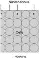
- the microstructure array disclosed herein has the ability to deliver substances to different layers of cells within tissue simultaneously (or sequentially) because the microstructures comprise multiple channels which can reach different levels of cells. This is because multiple channels on an angled microstructure allow for different heights of the channels within the microstructure. When the microstructure penetrates tissue, the channels are placed within different layers of cells, and can therefore deliver substances at different levels within the tissue.
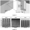
- the microstructure array (100) can comprise: a planar substrate (101) having a top surface (102) and a bottom surface (103); a reservoir (104) in fluid communication with the top surface of the planar substrate; and a plurality of microstructures (105) projecting from the bottom surface of the planar substrate, each of the plurality of microstructures comprising: a solid body portion (106) tapering from a base (107) to a distal tip (108) positioned at a height from the bottom surface of the planar substrate, thereby defining a microstructure surface; a first delivery channel (109) extending from the top surface of the planar substrate to a first channel opening (110) within the microstructure surface, thereby fluidly connecting the reservoir to the first channel opening; and a second delivery channel (111) extending from the top surface of the planar substrate to a second channel opening (112) within the microstructure surface, thereby fluidly connecting the reservoir to the second channel opening.
- the microstructures (105) can comprise 1, 2, 3, 4, 5, 6, 7, 8, 9, 10, 11, 12, or more channels. These channels can act as a conduit between the reservoir and the channel openings. Therefore, the channels can be in fluid communication with the reservoir, such that material placed in the reservoir can travel through the channel and be delivered at the channel openings.
- the first channel opening (110) is positioned at the distal tip, with additional channel openings flanking the distal tip. For example, there can be three channel openings on the same microstructure, with one being at the distal tip, and an additional two channel openings (112 and 114) on either side of the distal tip. Multiple channels within the microstructure (105) can run parallel to each other, and can run parallel to the vertical plane of the microstructure, as shown in Figures 1 and 2 .
- the height of the solid body portion (106) of the microstructure (105) can be measured from the distal tip (108) of the microstructure to the base (107) of the microstructure, which can be located on the bottom surface (103) of the planar substrate (101).
- the height of the microstructure surface can be 5, 10, 15, 20, 30, 40, 50, 60, 70, 80, 90, 100, 200, 300, 400, 500, 600, 700, 800, 900, or 1000 microns in length, or more, less, or any amount in between.
- the height of the microstructure can be from 100 to 500 microns.
- the height of the microstructure (105) is measured in a plane b which can run perpendicular to the plane of the planar substrate (101).
- the base (107) of the microstructure can have any shape, such as a triangle, square, diamond, rectangular, oval, or circle. In a particular embodiment, the base has a circular shape.
- the width of the base (107) of the microstructure (105) can be measured as the diameter of the base of the microstructure where it makes surface contact with the planar substrate (101).
- the plane a of the base (107) of the microstructure (105) can run parallel with the planar substrate (101), as seen in Figure 17B .
- the width of the base can be 5, 10, 20, 30, 40, 50, 60, 70, 80, 90, 100, 200, 300, 400, or 500 microns in length, or more, less, or any amount in between.
- the width of the base can also be measured in relation to the height of the microstructure, so that, for example, the width of the base of the microstructure can be 10, 20, 30, 40, 50, 60, 70, 80, 90, 100, 150, or 200% of the height, or more, less, or any amount in between.
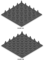
- the distal tip (108) of the microstructure can have any shape, and can be pointed, rounded, slanted, flared, tapered, blunted or combinations thereof, as can be seen in Figure 14A to 14D .
- the microstructure is pointed.
- the microstructures lack an ordered shape or pattern.
- the disclosed microstructures could be produced by tailored/blank etching of a silicon surface to introduce randomly-distributed microstructures/roughness with sharp tips that can penetrate tissues/living systems.
- the reservoir can comprise a release mechanism for releasing the substance to be delivered, thereby permitting the substance to be transported into and through the at least one channel of the microstructure.
- the release mechanism may utilize a mechanical force or sheer force, which can be manually, by heat, a chemical reaction, an electric field, a magnetic field, a pressure field, ultrasonic energy, tension, diffusion injection, osmosis, concentration gradient, vacuum, pressure, or a combination thereof.
- the reservoir (104) can include a porous material, wherein the substance to be administered is stored in pores of the porous material.
- the reservoir is sealed.
- the microstructure array further includes at least one puncturing barb extending from the first surface of the planar substrate, wherein the puncturing barb can be used to puncture the sealed reservoir.
- the feedback component can detect changes in a physiological signal, such as pH or temperature.
- the feedback component can comprise a "cut off value" such that when the pH or the temperature changes by a certain amount, or reaches a certain numerical value (a pH below 6.5, for example, or a temperature above 99.1, for example), the feedback component allows for a change in the release of the substance, or the amount of the substance released, and subsequently administered to the recipient.
- the substance in the reservoir to be delivered to the recipient can be a therapeutic, prophylactic, diagnostic, or theranostic substance. More than one substance can be delivered at a time, or different substances can be delivered sequentially. Different substances can be delivered through different channels at the same time, for example. 2, 3, 4, 5, 6, or more substances can be delivered simultaneously through different channels. Because the channel openings can reach different layers of cells, different substances can be administered to different strata of cells within tissue simultaneously utilizing the microstructure array disclosed herein. Specifically a first substance can be delivered via a first channel to a first layer of cells, and a second substance can be delivered via a second channel to a second layer of cells.
- the substance to be delivered can be selected from the group consisting of peptides, proteins, carbohydrates, nucleic acid molecules, lipids, organic molecules, biologically active inorganic molecules, and combinations thereof.
- a wide range of drugs may be formulated for delivery with the present microneedle devices.
- the microstructure array 100 disclosed herein can further include a stimulus (release mechanism) for translocating the cargo from the reservoir to through the channels.
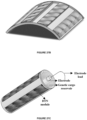
- this stimulus involves formation electrical field (e.g. electroporation), in which a porating electric field is applied to disrupt/deform lipid membranes and allow intracellular cargo delivery.
- electroporation e.g. electroporation
- applying a porating electric field across the system results in the deformation and/or disruption of cellular membranes, which allows for cargo translocation into the intracellular space.
- Electric field strengths can be accommodated depending on the target tissue/system.
- Figure 23 illustrates use of the disclosed microstructure array 100 to deliver nucleic acids into skin tissue using electroporation.
- a substance can be delivered through a channel opening (110, 112) at the distal tip (108) so that it permeates through tissue outwardly in a region.
- the region can coincide with the electrical field being generated between first electrode structure and second electrode structure.
- the electrical field enhances a cellular permeability, thus enhancing the delivery of the desired target substance to the cells.
- microarray of the invention in a method for administering a substance to a subject in need thereof, which includes the steps of inserting into the skin of the subject the microstructures (105) of the array (100) described above, and causing the substance to be transported from the reservoir (104) through the at least one channel of the microstructure and through the stratum corneum of the skin.
- TNT tissue nanotransfection
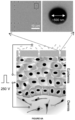
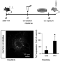
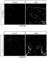
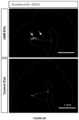
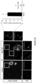
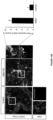
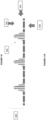
- Fig. 6f can also orchestrate a coordinated response that results in reprogramming stimuli propagation (i.e., epidermis to dermis) beyond the initial transfection boundary (i.e., epidermis) ( Fig. 6g-i ) possibly via dispatch of extracellular vesicles (EVs) rich in target gene mRNAs/cDNAs ( Fig. 6h,i ) ( Valadi et al. Nat Cell Biol 2007, 9(6): 654-659 ). Exposing na ⁇ ve cells to ABM-loaded EVs isolated from TNT-treated skin ( Fig 6j-l ) established that these EVs can be spontaneously internalized by remote cells ( Fig. 6k ).
- Fig. 6k Extracellular vesicles
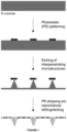
- TNT devices were fabricated from thinned (approximately 200 ⁇ m) double-side polished (I00) silicon wafers ( Fig. 7 ). Briefly, approximately I.5 ⁇ m thick layers of AZ52I4E photoresist were first spin coated on the silicon wafers at approximately 3000 rpm. Nanoscale openings were subsequently patterned on the photoresist using a GCA 6I00C stepper. Up to I6 dies of nanoscale opening arrays were patterned per I00-mm wafer. Such openings were then used as etch masks to drill approximately 10 ⁇ m deep nanochannels on the silicon surface using deep reactive ion etching (DRIE) (Oxford Plasma Lab I00 system).
- DRIE deep reactive ion etching
- Optimized etching conditions included SF6 gas: I3 s/I00 sccm gas flow/700O WICP power/40 W RF power/30 mT APC pressure; C4F8 gas condition: 7 s/I00 sccm gas flow/700 W ICP power/I0 W RF power/30 mT APC pressure.
- Microscale reservoirs were then patterned on the back-side of the wafers via contact photolithography and DRIE. Finally, an approximately 50 nm thick insulating/protective layer of silicon nitride was deposited on the TNT platform surface.
- Genotyping PCR for ROSAmT/mG mice was conducted using primers oIMR7318- CTC TGC TGC CTC CTG GCT TCT (SEQ ID NO: 1), oIMR73I9- CGA GGC GGA TCA CAA GCA ATA (SEQ ID NO: 2) and oIMR732O- TCA ATG GGC GGG GGT CGT T (SEQ ID NO: 3), while K-I4 Cre transgene was confirmed using primers oIMRIO84-GCG GTC TGG CAG TAA AAA CTA TC (SEQ ID NO: 4); oIMRIO85-GTG AAA CAG CAT TGC TGT CAC TT (SEQ ID NO: 5). The animals were tagged and grouped randomly using a computer based algorithm.
- a pulsed electric field was used to deliver cocktail of plasmids (0.05 ⁇ g/ ⁇ l) into the cells consisting of a 1:1:1 mixture of Fli1:Etv2:Foxc2.
- cocktail of plasmids (0.05 ⁇ g/ ⁇ l) into the cells consisting of a 1:1:1 mixture of Fli1:Etv2:Foxc2.
- the cells were then harvested 24 hours after plasmid delivery, placed in EBM-2 basal medium (CC-3I56, Lonza) supplemented with EGM-2 MV SingleQuot kit (CC-4I47, Lonza), and further processed for additional experiments/measurements.
- the areas to be treated were first naired 24-48 hours prior to TNT.
- the skin was then exfoliated to eliminate the dead/keratin cell layer and expose nucleated cells in the epidermis.
- the TNT devices were placed directly over the exfoliated skin surface.
- ABM or EFF plasmid cocktails were loaded in the reservoir at a concentration of 0.05-.1 ⁇ g/ ⁇ l.
- a gold-coated electrode i.e., cathode
- a 24G needle counter-electrode i.e., anode
- a pulsed electrical stimulation i.e., I0 pulses of 250 V in amplitude and a duration of I0 ms per pulse
- I0 pulses of 250 V in amplitude and a duration of I0 ms per pulse
- ABM plasmids were mixed at a 2:I:I molar ratio.
- Transient focal cerebral ischemia was induced in mice by middle cerebral artery occlusion (MCAO) was achieved by using the intraluminal filament insertion technique previously described ( Khanna et al. J Cereb Blood Flow Metab 20I3, 33(8): 1197-1206 ).
- MRI images were used to determine infarct size as a percentage of the contralateral hemisphere after correcting for edema.
- mice Unilateral hind-limb ischemia was induced via occlusion and subsequent transection of the femoral artery followed by transection ( Limcios et al. Nat Protoc 2009, 4(12): 1737-1746 ).
- mice 8-I0 week mice were anesthetized with I-3% isoflurane, placed supine under a stereomicroscope (Zeiss OPMI) on a heated pad.
- the femoral artery was exposed and separated from the femoral vein through an approximately I cm incision.
- Proximal and distal end occlusion were induced with 7-0 silk suture, which was then followed by complete transection of the artery.
- a single dose of buprenorphine was administered subcutaneously to control pain.
- Laser speckle imaging (MoorLDI-Mark 2) was conducted 2 hours post-surgery to confirm successful blood flow occlusion.
- EVs were isolated from I2 mm diameter skin biopsies that were collected in OCT blocks and stored frozen for later use. Briefly, the blocks were thawed and washed with phosphate buffer saline (PBS) to eliminate the OCT. Following removal of the fat tissue with a scalpel, the skin tissue was minced into approximately I mm pieces and homogenized with a micro-grinder in PBS. After centrifugation at 3000 g, an Exoquick kit (System Biosciences) was used at a 1:5 ratio (Exoquick:supernatant) to isolate EVs from the supernatant for I2 hours at 4°C. EVs were precipitated via centrifugation at I500 g for 30 min. Total RNA was then extracted from pellet using the mirvana kit (Life technologies) following the recommendations provided by the manufacturer.
- PBS phosphate buffer saline
- ABM and EFF plasmids were prepared using plasmid DNA purification kit (Qiagen Maxi-prep, catalogue number 12161, and Clontech Nucleobond catalogue number 740410). DNA concentrations were obtained from a Nanodrop 2000c Spectrophotemeter (Thermoscientific). A list of plasmid DNA constructs and their original sources can be found Table 1.
- Table 1 Plasmid cDNA Construct Name Gene Insert Plasmid Backbone Brn2-RFP Brn2 pCAGGs Myt1I-CFP Myt1I pCAGGs Ascl1-GFP Ascl1 pCAGGs pIRES-ER71(HA)3 Etsvp71 (ER71) pIRES-hrGFP-2a pAd-HA-FIi1-IRES-hrGFP HA-Fli1 pAd-IRES-GFP mFoxc2 mFoxc2 pCDNA3.0
- LCM was performed using a laser microdissection system from PALM Technologies (Bernreid, Germany). Specific regions of tissue sections, identified based on morphology and/or immunostaining, were cut and captured under a 20x ocular lens. The samples were catapulted into 25 ⁇ l of cell direct lysis extraction buffer (Invitrogen). Approximately 1,000,000 ⁇ m 2 of tissue area was captured into each cap and the lysate was then stored at -80°C for further processing. qRT-PCR of the LCM samples were performed from cell direct lysis buffer following manufacture's instruction. A list of primers can be found in Table 2. Table 2.
- Tissue immunostaining was carried out using specific antibodies and standard procedures. Briefly, OCT-embedded tissue was cryosectioned at 10 ⁇ m thick, fixed with cold acetone, blocked with I0% normal goat serum and incubated with specific antibodies. Signal was visualized by subsequent incubation with fluorescence-tagged appropriate secondary antibodies (Alexa 488-tagged a-guinea pig, I:200, Alexa 488-tagged a-rabbit, I:200; Alexa 568- tagged a-rabbit, I:200) and counter stained with DAPI. Images were captured by laser scanning confocal microscope (Olympus FVI000 filter/spectral).
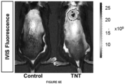
- the animals were imaged with anesthesia 24 h after FAM-DNA transfection using IVIS Lumina II optical imaging system. Overlay images with luminescence images were made using Living Image software.
- Magnetic resonance angiography was used to validate our MCAO model in mice and to optimize the occluder size and the internal carotid artery insertion distance for effective MCAO.
- T2-weighted MRI was performed on anesthetized mice 48 h after MCA-reperfusion using 9.4 T MRI (Bruker Corporation, Bruker BioSpin Corporation, Billerica, MA, USA).
- MR images were acquired using a Rapid Acquisition with Relaxation Enhancement (RARE) sequence using the following parameters: field of view (FOV) 30 ⁇ 30 mm, acquisition matrix 256 ⁇ 256, TR 3,500 ms, TE 46.92 ms, slice gap 1.0 mm, rare factor 8, number of averages 3. Resolution of 8.5 pixels per mm.
- FOV field of view
- Raw MR images were converted to the standard DICOM format and processed. After appropriate software contrast enhancement of images using Osirix v3.4, digital planimetry was performed by a masked observer to delineate the infarct area in each coronal brain slice. Infarct areas from brain slices were summed, multiplied by slice thickness, and corrected for edema-induced swelling as previously described to determine infarct volume ( Khanna S, et al. J Cereb Blood Flow Metab 2013, 33(8):1197-1206 ).
- Muscle energetics was evaluated NMR spectroscopy measurements on a 9.4 Tesla scanner (Bruker BioSpec) using a volume coil for RF transmission and a 3IP coil for reception ( Fiedler et al. MAGMA 20I5, 28(5): 493-501 .).
- In vivo imaging was conducted in a custom-made 1H/31P transceiver coil array. Data were acquired using single pulse sequence. The raw data were windowed for noise reduction and Fourier transformed to spectral domain.
- Blood vessel formation was parallely monitored via ultrasound imaging. Briefly, a Vevo 2I00 system (Visual Sonics, Toronto, ON, Canada) was used to obtain ultrasound images on B-mode with a MS 250 linear array probe ( Gnyawali et al. J Vis Exp 2010(41 ). Doppler color flow imaging was implemented to monitor and quantify blood flow characteristics under systole and diastole.
- TNT-based approach is indicative of what can be accomplished using nanochannel-based delivery, including the microstructure array disclosed herein.
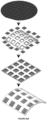
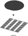
- Arrayed interpenetrating nanochannels can be fabricated from silicon-based materials using cleanroom methods such as photolithography and wet or dry etching.
- cleanroom methods such as photolithography and wet or dry etching.
- an array of conical or pyramidal interpenetrating microstructures (approximately 20-500 microns tall) is defined on a silicon surface using photolithography and wet or dry etching.
- the silicon substrate is patterned on the backside with an array of nanowells (approximately 300-1000 nm diameter) using projection lithography.
- DRIE deep reactive ion etch
- Arrayed interpenetrating nanochannels can be fabricated from a premade nanochanneled substrate platform that can either be silicon-based or made from anodized alumina via selective surface etching. Selective surface etching is used to define interpenetrating microstructures
- An interpenetrating nanochannel array an be used to perform a method delivering cargo to biological tissues at multiple levels.
- the interpenetrating nanochannel array is placed in contact with exfoliated skin tissue.
- a positive electrode is inserted intradermally, while a negative electrode is put in contact with the cargo solution.
- a pulsed electric field 250 V, 10 ms pulses, 10 pulses is then applied across the electrodes to nanoporate exposed cell membranes and inject the cargo directly into the cytosol.
- the cargo solution comprised a mixture of three plasmids, encoding for Etv2, Foxc2 and FI1.
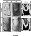
- Small tissue biopsies were collected 24 h after delivery and analyzed by qRT-PCR. Additional biopsies were collected 7 days post-delivery and analyzed via immunohistochemistry for endothelial markers. DTN-based delivery results in superior transgene expression as well as widely distributed reprogramming responses across the entire tissue thickness
- ETS transcription factor ETV2 directly converts human fibroblasts into functional endothelial cells. Proc Natl Acad Sci U S A 20I5, 112(1): 160-165 .
- Sen CK Ghatak S. miRNA control of tissue repair and regeneration. Am J Pathol 20I5, 185(10): 2629-2640 .
Landscapes
- Health & Medical Sciences (AREA)
- Life Sciences & Earth Sciences (AREA)
- Engineering & Computer Science (AREA)
- Veterinary Medicine (AREA)
- Public Health (AREA)
- General Health & Medical Sciences (AREA)
- Animal Behavior & Ethology (AREA)
- Biomedical Technology (AREA)
- Heart & Thoracic Surgery (AREA)
- Dermatology (AREA)
- Hematology (AREA)
- Anesthesiology (AREA)
- Medical Informatics (AREA)
- Nuclear Medicine, Radiotherapy & Molecular Imaging (AREA)
- Surgery (AREA)
- Pharmacology & Pharmacy (AREA)
- Medicinal Chemistry (AREA)
- Epidemiology (AREA)
- Chemical & Material Sciences (AREA)
- Vascular Medicine (AREA)
- Biophysics (AREA)
- Radiology & Medical Imaging (AREA)
- Bioinformatics & Cheminformatics (AREA)
- Molecular Biology (AREA)
- Apparatus Associated With Microorganisms And Enzymes (AREA)
- Physical Or Chemical Processes And Apparatus (AREA)
- Media Introduction/Drainage Providing Device (AREA)
- Medicinal Preparation (AREA)
- Medicines That Contain Protein Lipid Enzymes And Other Medicines (AREA)
- Medicines Containing Antibodies Or Antigens For Use As Internal Diagnostic Agents (AREA)
- Pharmaceuticals Containing Other Organic And Inorganic Compounds (AREA)
- Medicines Containing Material From Animals Or Micro-Organisms (AREA)
Claims (13)
- Mikrostrukturanordnung (100), umfassend:ein ebenes Substrat (101) mit einer Oberseite (102) und einer Unterseite (103);ein Reservoir (104) in Fluidverbindung mit der Oberseite des ebenen Substrats;eine Mehrzahl von Mikrostrukturen (105), die von der Unterseite des ebenen Substrats vorstehen, wobei jede der Mehrzahl von Mikrostrukturen umfasst:einen jeweiligen Festkörperabschnitt (106), der sich von einer jeweiligen Basis (107) zu einer jeweiligen distalen Spitze (108), die in einer jeweiligen Höhe von der Unterseite des ebenen Substrats positioniert ist, verjüngt, wobei der jeweilige Festkörperabschnitt von der jeweiligen Basis zu der jeweiligen distalen Spitze eine jeweilige Mikrostrukturoberfläche definiert;einen jeweiligen ersten Zuführkanal (109) oder Schlitz, der sich von der Oberseite des ebenen Substrats zu einer jeweiligen ersten Kanal- oder Schlitzöffnung (110) innerhalb der jeweiligen Mikrostrukturoberfläche erstreckt, wodurch das Reservoir mit der jeweiligen ersten Kanal- oder Schlitzöffnung fluidtechnisch verbunden wird; undeinen jeweiligen zweiten Zuführkanal (111) oder Schlitz, der sich von der Oberseite des ebenen Substrats zu einer jeweiligen zweiten Kanal- oder Schlitzöffnung (112) innerhalb der Mikrostrukturoberfläche erstreckt, wodurch das Reservoir mit der jeweiligen zweiten Kanal- oder der Schlitzöffnung fluidtechnisch verbunden wird; dadurch gekennzeichnet, dass die Mikrostrukturanordnung ferner umfasst:
eine erste Elektrode, die in elektrischem Kontakt mit dem Reservoir steht, und eine zweite Elektrode, die dazu konfiguriert ist, ein an der Unterseite des ebenen Substrats positioniertes Gewebe elektrisch zu kontaktieren,wobei der Festkörperabschnitt jeder der Mehrzahl von Mikrostrukturen aus Silizium gebildet ist - unter Verwendung von Photolithographie und Ätzen,wobei der erste Zuführkanal oder Schlitz und der zweite Zuführkanal oder Schlitz einen Innendurchmesser von 1 bis 999 nm aufweisen,wobei die jeweilige erste Kanal- oder Schlitzöffnung innerhalb einer ersten Ebene parallel zu dem ebenen Substrat positioniert ist, wobei die jeweilige zweite Kanal- oder Schlitzöffnung innerhalb einer zweiten Ebene parallel zu dem ebenen Substrat positioniert ist, und wobei die erste Ebene von der zweiten Ebene in einem Abstand von 20 % bis 60 % der Höhe von der Unterseite des ebenen Substrats distal beabstandet ist;wobei die jeweilige erste Kanal- oder Schlitzöffnung an der jeweiligen distalen Spitze positioniert ist. - Mikrostruktur nach Anspruch 1, wobei mindestens eine der Mehrzahl von Mikrostrukturen ferner einen jeweiligen dritten Zuführkanal (113) umfasst, der sich von der Oberseite des ebenen Substrats zu einer jeweiligen dritten Kanalöffnung (114) innerhalb der jeweiligen Mikrostrukturoberfläche erstreckt, wodurch das Reservoir mit der jeweiligen dritten Kanalöffnung fluidtechnisch verbunden wird,
optional, wobei die erste Kanalöffnung in einer ersten Ebene parallel zu dem ebenen Substrat positioniert ist, wobei die zweite Kanalöffnung in einer zweiten Ebene parallel zu dem ebenen Substrat positioniert ist, wobei die dritte Kanalöffnung innerhalb einer dritten Ebene parallel zu dem ebenen Substrat positioniert ist; und wobei die erste Ebene von der zweiten Ebene distal beabstandet ist und die zweite Ebene von der dritten Ebene distal beabstandet ist. - Mikrostrukturanordnung nach Anspruch 2, wobei die zweite Ebene von der dritten Ebene um einen Abstand von 20% bis 60% der Höhe von der Unterseite des ebenen Substrats distal beabstandet ist.
- Mikrostrukturanordnung nach einem der Ansprüche 1 bis 3, wobei die Höhe der Mikrostrukturoberfläche 5 Mikrometer bis 1000 Mikrometer beträgt; optional, wobei die jeweilige Basis jeder der Mehrzahl von Mikrostrukturen eine Breite von 5 Mikrometer bis 500 Mikrometer aufweist.
- Mikrostrukturanordnung nach einem der Ansprüche 1 bis 4, wobei die jeweilige Basis jeder der Mehrzahl von Mikrostrukturen die im Wesentlichen kreisförmige Form oder die im Wesentlichen rechteckige Form aufweist.
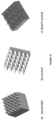
- Mikrostrukturanordnung nach einem der Ansprüche 1 bis 5, wobei der jeweilige Festkörper der Mehrzahl von jeder der Mikrostrukturen unabhängig voneinander parallele Rippen, in einem Fischgrätenmuster angeordnete Rippen, in einem Wellenformmuster angeordnete Rippen, Kegel oder Pyramiden umfasst.
- Mikrostrukturanordnung nach einem der Ansprüche 1 bis 6, wobei jede jeweilige distale Spitze der Mehrzahl von Mikrostrukturen eine Form hat, die unabhängig voneinander aus spitz, gerundet, abgeschrägt, aufgeweitet, verjüngt oder abgestumpft ausgewählt ist.
- Mikrostrukturanordnung nach einem der Ansprüche 1 bis 7, wobei der jeweilige erste Zuführkanal und der jeweilige zweite Zuführkanal jeweils einen jeweiligen Querschnitt in einer Ebene parallel zu dem ebenen Substrat aufweisen, der unabhängig aus im Wesentlichen kreisförmig oder im Wesentlichen rechteckig ausgewählt ist.
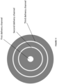
- Mikrostrukturanordnung nach Anspruch 1 bis 8, wobei der jeweilige erste Zuführkanal und der jeweilige zweite Zuführkanal jeder der Mehrzahl von Mikrostrukturen einen im Wesentlichen toroidalen Querschnitt in einer Ebene parallel zu dem planaren Substrat aufweist; und
wobei der jeweilige zweite Zuführkanal der Mehrzahl von Mikrostrukturen koaxial um den jeweiligen ersten Zuführkanal der Mehrzahl von Mikrostrukturen angeordnet ist. - Mikrostrukturanordnung nach einem der Ansprüche 1 bis 9, wobei der jeweilige erste Zuführkanal von mindestens einer der Mehrzahl von Mikrostrukturen einen im Wesentlichen kreisförmigen Querschnitt in einer Ebene parallel zu dem ebenen Substrat aufweist und der jeweilige zweite Zuführkanal von mindestens einer der Mehrzahl von Mikrostrukturen einen im Wesentlichen toroidalen Querschnitt in einer Ebene parallel zu dem ebenen Substrat aufweist; und
wobei der jeweilige zweite Zuführkanal der mindestens einen der Mehrzahl von Mikrostrukturen koaxial um den jeweiligen ersten Zuführkanal angeordnet ist. - Mikrostrukturanordnung nach einem der Ansprüche 1 bis 10, wobei sich der jeweilige Festkörperabschnitt jeder der Mehrzahl von Mikrostrukturen in stufenweiser Form von der Basis zur distalen Spitze hin verjüngt, oder wobei der jeweilige Festkörperabschnitt jeder der Mehrzahl von Mikrostrukturen eine kegelstumpfförmige Form aufweist.
- Mikrostrukturanordnung nach einem der Ansprüche 1 bis 11, wobei das ebene Substrat ferner eine Mehrzahl von Zuführkanälen umfasst, von denen sich jeder von der Oberseite des ebenen Substrats zu einer Kanalöffnung innerhalb der Unterseite des ebenen Substrats erstreckt, wodurch das Reservoir mit der Kanalöffnung innerhalb der Unterseite des ebenen Substrats fluidtechnisch verbunden wird.
- Die Mikrostrukturanordnung nach einem der Ansprüche 1 bis 12 zur Verwendung in einem Verfahren zum Zuführen einer Substanz an Zellen innerhalb eines Gewebes, wobei das Verfahren umfasst:Aufbringen der Mikrostrukturanordnung nach einem der Ansprüche 1 bis 12 auf das Gewebe, so dass die Unterseite des ebenen Substrats an dem Gewebe positioniert ist und sich die Mehrzahl von Mikrostrukturen in das Gewebe hinein erstreckt;Zuführen einer Substanz aus dem Reservoir durch den ersten Zuführkanal und den zweiten Zuführkanal jeder der Mehrzahl von Mikrostrukturen an die Zellen innerhalb des Gewebes.
Applications Claiming Priority (2)
| Application Number | Priority Date | Filing Date | Title |
|---|---|---|---|
| US201662438256P | 2016-12-22 | 2016-12-22 | |
| PCT/US2017/067630 WO2018119090A1 (en) | 2016-12-22 | 2017-12-20 | Interpentrating microstructures for nanochannel-based cargo delivery |
Publications (3)
| Publication Number | Publication Date |
|---|---|
| EP3558265A1 EP3558265A1 (de) | 2019-10-30 |
| EP3558265A4 EP3558265A4 (de) | 2020-11-04 |
| EP3558265B1 true EP3558265B1 (de) | 2025-04-16 |
Family
ID=62627699
Family Applications (1)
| Application Number | Title | Priority Date | Filing Date |
|---|---|---|---|
| EP17885274.5A Active EP3558265B1 (de) | 2016-12-22 | 2017-12-20 | Interpenetrierende mikrostrukturen für nanokanalbasierte frachtlieferung |
Country Status (9)
| Country | Link |
|---|---|
| US (1) | US11235132B2 (de) |
| EP (1) | EP3558265B1 (de) |
| JP (1) | JP7114094B2 (de) |
| KR (1) | KR102539625B1 (de) |
| CN (2) | CN121714506A (de) |
| AU (1) | AU2017379879C1 (de) |
| BR (1) | BR112019012924A2 (de) |
| CA (1) | CA3045958A1 (de) |
| WO (1) | WO2018119090A1 (de) |
Families Citing this family (10)
| Publication number | Priority date | Publication date | Assignee | Title |
|---|---|---|---|---|
| JP7737718B2 (ja) * | 2019-07-22 | 2025-09-11 | ザ・トラスティーズ・オブ・インディアナ・ユニバーシティー | マイクロチャネルを有するニードルのための技術 |
| WO2021222491A1 (en) * | 2020-05-01 | 2021-11-04 | The Trustees Of Indiana University | Neurogenic tissue nanotransfection in the management of cutaneous diabetic polyneuropathy |
| CN113337451B (zh) * | 2021-06-02 | 2023-03-07 | 吉林医药学院 | 一种多剪切力微流控芯片中单细胞精准图案化方法 |
| CN113896166B (zh) * | 2021-09-06 | 2026-02-06 | 同济大学 | 等离激元正金字塔阵列基底及体外细胞内物质递送方法 |
| CN114754669B (zh) * | 2022-03-22 | 2024-01-30 | 南京理工大学 | 一种多模光纤束扩展光源抑制干涉仪相干噪声的方法 |
| CN115154877A (zh) * | 2022-05-24 | 2022-10-11 | 北京化工大学 | 一种微注射给药式聚合物多流道微针模具法及其多流道微针应用 |
| KR102922531B1 (ko) * | 2023-04-26 | 2026-02-04 | 서울과학기술대학교 산학협력단 | 마이크로 니들 모듈 및 이를 포함하는 바이오 패치 |
| WO2025042328A1 (en) * | 2023-08-22 | 2025-02-27 | Chee Yen Lim | A microneedle probe |
| KR20260000249A (ko) * | 2024-06-25 | 2026-01-02 | 재단법인 아산사회복지재단 | 림프 타겟 무통증 약물 전달패치 |
| CN118501229B (zh) * | 2024-07-18 | 2025-02-18 | 有研工程技术研究院有限公司 | 一种电化学微针生物传感器及其制备方法 |
Family Cites Families (34)
| Publication number | Priority date | Publication date | Assignee | Title |
|---|---|---|---|---|
| US5389069A (en) | 1988-01-21 | 1995-02-14 | Massachusetts Institute Of Technology | Method and apparatus for in vivo electroporation of remote cells and tissue |
| US6090790A (en) | 1989-12-14 | 2000-07-18 | Eriksson; Elof | Gene delivery by microneedle injection |
| US5697901A (en) | 1989-12-14 | 1997-12-16 | Elof Eriksson | Gene delivery by microneedle injection |
| US5273525A (en) | 1992-08-13 | 1993-12-28 | Btx Inc. | Injection and electroporation apparatus for drug and gene delivery |
| US5702359A (en) | 1995-06-06 | 1997-12-30 | Genetronics, Inc. | Needle electrodes for mediated delivery of drugs and genes |
| US5571083A (en) | 1994-02-18 | 1996-11-05 | Lemelson; Jerome H. | Method and system for cell transplantation |
| US5591139A (en) | 1994-06-06 | 1997-01-07 | The Regents Of The University Of California | IC-processed microneedles |
| US5869326A (en) | 1996-09-09 | 1999-02-09 | Genetronics, Inc. | Electroporation employing user-configured pulsing scheme |
| US5928207A (en) | 1997-06-30 | 1999-07-27 | The Regents Of The University Of California | Microneedle with isotropically etched tip, and method of fabricating such a device |
| US6055453A (en) | 1997-08-01 | 2000-04-25 | Genetronics, Inc. | Apparatus for addressing needle array electrodes for electroporation therapy |
| US6241701B1 (en) | 1997-08-01 | 2001-06-05 | Genetronics, Inc. | Apparatus for electroporation mediated delivery of drugs and genes |
| US6503231B1 (en) | 1998-06-10 | 2003-01-07 | Georgia Tech Research Corporation | Microneedle device for transport of molecules across tissue |
| US6312612B1 (en) | 1999-06-09 | 2001-11-06 | The Procter & Gamble Company | Apparatus and method for manufacturing an intracutaneous microneedle array |
| US6623457B1 (en) * | 1999-09-22 | 2003-09-23 | Becton, Dickinson And Company | Method and apparatus for the transdermal administration of a substance |
| US6331266B1 (en) | 1999-09-29 | 2001-12-18 | Becton Dickinson And Company | Process of making a molded device |
| IL134997A0 (en) * | 2000-03-09 | 2001-05-20 | Yehoshua Yeshurun | Health care system based on micro device |
| US6663820B2 (en) | 2001-03-14 | 2003-12-16 | The Procter & Gamble Company | Method of manufacturing microneedle structures using soft lithography and photolithography |
| US7127284B2 (en) | 2001-06-11 | 2006-10-24 | Mercator Medsystems, Inc. | Electroporation microneedle and methods for its use |
| US6881203B2 (en) * | 2001-09-05 | 2005-04-19 | 3M Innovative Properties Company | Microneedle arrays and methods of manufacturing the same |
| US7578954B2 (en) | 2003-02-24 | 2009-08-25 | Corium International, Inc. | Method for manufacturing microstructures having multiple microelements with through-holes |
| EP1682217A4 (de) * | 2003-11-13 | 2008-04-30 | Alza Corp | System udn verfahren für die transdermale abgabe |
| US20070185432A1 (en) * | 2005-09-19 | 2007-08-09 | Transport Pharmaceuticals, Inc. | Electrokinetic system and method for delivering methotrexate |
| US20090099502A1 (en) * | 2006-04-07 | 2009-04-16 | Hisamitsu Pharmaceutical Co., Inc. | Microneedle Device And Transdermal Administration Device Provided With Microneedles |
| TWI300055B (en) * | 2006-05-26 | 2008-08-21 | Nat Univ Tsing Hua | Method of manufacturing hollow micro-needle structures |
| JP5559054B2 (ja) * | 2007-09-28 | 2014-07-23 | ザ クイーンズ ユニヴァーシティ オブ ベルファスト | 送達装置および方法 |
| US20100298808A1 (en) * | 2007-11-28 | 2010-11-25 | Janisys Limited | Method and a delivery device for administering an active substance to a subject |
| FI20095433A0 (fi) * | 2009-04-20 | 2009-04-20 | Valtion Teknillinen | Mikroneula |
| EP2493551A4 (de) * | 2009-10-26 | 2013-04-17 | Emkinetics Inc | Verfahren und vorrichtung zur elektromagnetischen stimulation von nerven, muskeln und körpergewebe |
| WO2011135531A2 (en) * | 2010-04-28 | 2011-11-03 | Kimberly-Clark Worldwide, Inc. | MEDICAL DEVICES FOR DELIVERY OF siRNA |
| KR20130058012A (ko) * | 2010-04-28 | 2013-06-03 | 킴벌리-클라크 월드와이드, 인크. | 사출 성형된 마이크로니들 어레이 및 마이크로니들 어레이의 형성 방법 |
| US8696637B2 (en) * | 2011-02-28 | 2014-04-15 | Kimberly-Clark Worldwide | Transdermal patch containing microneedles |
| BR112015027608A2 (pt) | 2013-05-23 | 2017-07-25 | Kimberly Clark Co | microagulhas com geometrias transversais de canal aberto melhoradas |
| KR101549086B1 (ko) * | 2014-11-10 | 2015-09-02 | 주식회사 스몰랩 | 마이크로 니들 및 마이크로 니들 패치 |
| CN105126243B (zh) * | 2015-09-06 | 2018-08-07 | 台州薇凯生物科技有限公司 | 微针注射弹及其制备方法和微针注射设备 |
-
2017
- 2017-12-20 EP EP17885274.5A patent/EP3558265B1/de active Active
- 2017-12-20 CA CA3045958A patent/CA3045958A1/en active Pending
- 2017-12-20 JP JP2019534295A patent/JP7114094B2/ja active Active
- 2017-12-20 CN CN202510891619.4A patent/CN121714506A/zh active Pending
- 2017-12-20 BR BR112019012924A patent/BR112019012924A2/pt not_active IP Right Cessation
- 2017-12-20 CN CN201780080259.3A patent/CN110494126A/zh active Pending
- 2017-12-20 WO PCT/US2017/067630 patent/WO2018119090A1/en not_active Ceased
- 2017-12-20 KR KR1020197021274A patent/KR102539625B1/ko active Active
- 2017-12-20 US US16/471,907 patent/US11235132B2/en active Active
- 2017-12-20 AU AU2017379879A patent/AU2017379879C1/en active Active
Also Published As
| Publication number | Publication date |
|---|---|
| WO2018119090A1 (en) | 2018-06-28 |
| AU2017379879C1 (en) | 2023-10-12 |
| KR102539625B1 (ko) | 2023-06-05 |
| CN121714506A (zh) | 2026-03-24 |
| US11235132B2 (en) | 2022-02-01 |
| AU2017379879B2 (en) | 2023-07-06 |
| CN110494126A (zh) | 2019-11-22 |
| KR20190099479A (ko) | 2019-08-27 |
| JP7114094B2 (ja) | 2022-08-08 |
| EP3558265A4 (de) | 2020-11-04 |
| EP3558265A1 (de) | 2019-10-30 |
| CA3045958A1 (en) | 2018-06-28 |
| AU2017379879A1 (en) | 2019-07-04 |
| BR112019012924A2 (pt) | 2019-12-10 |
| US20190329014A1 (en) | 2019-10-31 |
| JP2020513900A (ja) | 2020-05-21 |
Similar Documents
| Publication | Publication Date | Title |
|---|---|---|
| AU2017379879B2 (en) | Interpentrating microstructures for nanochannel-based cargo delivery | |
| Gallego-Pérez et al. | Topical tissue nano-transfection mediates non-viral stroma reprogramming and rescue | |
| JP7249693B2 (ja) | 誘導されたエキソソームを含む皮膚再生及び創傷治癒用組成物 | |
| Huang et al. | Efficient delivery of nucleic acid molecules into skin by combined use of microneedle roller and flexible interdigitated electroporation array | |
| Wosicka et al. | Targeting to the hair follicles: current status and potential | |
| ES2831298T3 (es) | Composición que incluye exosomas derivados de células madre para blanquear la piel o mejorar las arrugas | |
| US20080214987A1 (en) | Microdevice And Method For Transdermal Delivery And Sampling Of Active Substances | |
| US10525246B2 (en) | Microdevice and method for transdermal delivery and sampling of active substances | |
| JP2022502033A (ja) | 誘導されたエキソソームを含む毛髪再生組成物 | |
| CN111329832B (zh) | 一种治疗脱发的纳米脂质载体微针及其应用 | |
| CN105999535A (zh) | 海绵骨针作为皮肤物理促渗剂的应用 | |
| CN110123838B (zh) | 负载白藜芦醇的人多能干细胞外泌体及其制备方法与用途 | |
| Hong et al. | Hair grows hair: Dual-effective hair regrowth through a hair enhanced dissolvable microneedle patch cooperated with the pure yellow light irradiation | |
| Xing et al. | Microenvironment-responsive recombinant collagen XVII-based composite microneedles for the treatment of androgenetic alopecia | |
| US10232159B2 (en) | Microneedle based cell delivery | |
| US12415058B2 (en) | Interpenetrating microstructures for nanochannel-based sampling and/or cargo delivery | |
| Huang et al. | Transdermal delivery of nucleic acid mediated by punching and electroporation | |
| JP2024539937A (ja) | シュワン細胞への標的化送達のためのデザイナー細胞外小胞 | |
| US12201798B2 (en) | Method for increasing cell viability by using drug delivery system and ultrasound irradiation, and ultrasound irradiation apparatus using same | |
| HK40016692A (en) | Interpentrating microstructures for nanochannel-based cargo delivery | |
| Bambale et al. | Exploring Novel Treatments for Alopecia: Nanoparticles, Iontophoresis, and Cell Therapy | |
| Chen et al. | Research progress on the application of microneedle transdermal drug delivery system in dermatology | |
| CN108472410A (zh) | 用于生产皮肤等同物的方法及其用于体外测试和体内移植的用途 | |
| CN107405506A (zh) | R‑脊椎蛋白激动剂介导的毛发生长 | |
| CN121177250A (zh) | 一种负载siPCSK9纳米囊泡、可溶性微针及其制备方法和应用 |
Legal Events
| Date | Code | Title | Description |
|---|---|---|---|
| STAA | Information on the status of an ep patent application or granted ep patent |
Free format text: STATUS: THE INTERNATIONAL PUBLICATION HAS BEEN MADE |
|
| PUAI | Public reference made under article 153(3) epc to a published international application that has entered the european phase |
Free format text: ORIGINAL CODE: 0009012 |
|
| STAA | Information on the status of an ep patent application or granted ep patent |
Free format text: STATUS: REQUEST FOR EXAMINATION WAS MADE |
|
| 17P | Request for examination filed |
Effective date: 20190607 |
|
| AK | Designated contracting states |
Kind code of ref document: A1 Designated state(s): AL AT BE BG CH CY CZ DE DK EE ES FI FR GB GR HR HU IE IS IT LI LT LU LV MC MK MT NL NO PL PT RO RS SE SI SK SM TR |
|
| AX | Request for extension of the european patent |
Extension state: BA ME |
|
| DAV | Request for validation of the european patent (deleted) | ||
| DAX | Request for extension of the european patent (deleted) | ||
| A4 | Supplementary search report drawn up and despatched |
Effective date: 20201005 |
|
| RIC1 | Information provided on ipc code assigned before grant |
Ipc: A61K 9/00 20060101AFI20200929BHEP Ipc: A61M 37/00 20060101ALI20200929BHEP Ipc: A61M 5/14 20060101ALI20200929BHEP Ipc: A61M 5/158 20060101ALI20200929BHEP |
|
| STAA | Information on the status of an ep patent application or granted ep patent |
Free format text: STATUS: EXAMINATION IS IN PROGRESS |
|
| 17Q | First examination report despatched |
Effective date: 20230711 |
|
| GRAP | Despatch of communication of intention to grant a patent |
Free format text: ORIGINAL CODE: EPIDOSNIGR1 |
|
| STAA | Information on the status of an ep patent application or granted ep patent |
Free format text: STATUS: GRANT OF PATENT IS INTENDED |
|
| INTG | Intention to grant announced |
Effective date: 20241111 |
|
| GRAS | Grant fee paid |
Free format text: ORIGINAL CODE: EPIDOSNIGR3 |
|
| GRAA | (expected) grant |
Free format text: ORIGINAL CODE: 0009210 |
|
| STAA | Information on the status of an ep patent application or granted ep patent |
Free format text: STATUS: THE PATENT HAS BEEN GRANTED |
|
| AK | Designated contracting states |
Kind code of ref document: B1 Designated state(s): AL AT BE BG CH CY CZ DE DK EE ES FI FR GB GR HR HU IE IS IT LI LT LU LV MC MK MT NL NO PL PT RO RS SE SI SK SM TR |
|
| REG | Reference to a national code |
Ref country code: GB Ref legal event code: FG4D |
|
| REG | Reference to a national code |
Ref country code: CH Ref legal event code: EP Ref country code: DE Ref legal event code: R096 Ref document number: 602017088997 Country of ref document: DE |
|
| REG | Reference to a national code |
Ref country code: IE Ref legal event code: FG4D |
|
| REG | Reference to a national code |
Ref country code: NL Ref legal event code: MP Effective date: 20250416 |
|
| PG25 | Lapsed in a contracting state [announced via postgrant information from national office to epo] |
Ref country code: NL Free format text: LAPSE BECAUSE OF FAILURE TO SUBMIT A TRANSLATION OF THE DESCRIPTION OR TO PAY THE FEE WITHIN THE PRESCRIBED TIME-LIMIT Effective date: 20250416 |
|
| REG | Reference to a national code |
Ref country code: AT Ref legal event code: MK05 Ref document number: 1785072 Country of ref document: AT Kind code of ref document: T Effective date: 20250416 |
|
| PG25 | Lapsed in a contracting state [announced via postgrant information from national office to epo] |
Ref country code: FI Free format text: LAPSE BECAUSE OF FAILURE TO SUBMIT A TRANSLATION OF THE DESCRIPTION OR TO PAY THE FEE WITHIN THE PRESCRIBED TIME-LIMIT Effective date: 20250416 Ref country code: ES Free format text: LAPSE BECAUSE OF FAILURE TO SUBMIT A TRANSLATION OF THE DESCRIPTION OR TO PAY THE FEE WITHIN THE PRESCRIBED TIME-LIMIT Effective date: 20250416 Ref country code: PT Free format text: LAPSE BECAUSE OF FAILURE TO SUBMIT A TRANSLATION OF THE DESCRIPTION OR TO PAY THE FEE WITHIN THE PRESCRIBED TIME-LIMIT Effective date: 20250818 |
|
| REG | Reference to a national code |
Ref country code: LT Ref legal event code: MG9D |
|
| PG25 | Lapsed in a contracting state [announced via postgrant information from national office to epo] |
Ref country code: NO Free format text: LAPSE BECAUSE OF FAILURE TO SUBMIT A TRANSLATION OF THE DESCRIPTION OR TO PAY THE FEE WITHIN THE PRESCRIBED TIME-LIMIT Effective date: 20250716 Ref country code: GR Free format text: LAPSE BECAUSE OF FAILURE TO SUBMIT A TRANSLATION OF THE DESCRIPTION OR TO PAY THE FEE WITHIN THE PRESCRIBED TIME-LIMIT Effective date: 20250717 |
|
| PG25 | Lapsed in a contracting state [announced via postgrant information from national office to epo] |
Ref country code: PL Free format text: LAPSE BECAUSE OF FAILURE TO SUBMIT A TRANSLATION OF THE DESCRIPTION OR TO PAY THE FEE WITHIN THE PRESCRIBED TIME-LIMIT Effective date: 20250416 |
|
| PG25 | Lapsed in a contracting state [announced via postgrant information from national office to epo] |
Ref country code: BG Free format text: LAPSE BECAUSE OF FAILURE TO SUBMIT A TRANSLATION OF THE DESCRIPTION OR TO PAY THE FEE WITHIN THE PRESCRIBED TIME-LIMIT Effective date: 20250416 |
|
| PG25 | Lapsed in a contracting state [announced via postgrant information from national office to epo] |
Ref country code: HR Free format text: LAPSE BECAUSE OF FAILURE TO SUBMIT A TRANSLATION OF THE DESCRIPTION OR TO PAY THE FEE WITHIN THE PRESCRIBED TIME-LIMIT Effective date: 20250416 |
|
| PG25 | Lapsed in a contracting state [announced via postgrant information from national office to epo] |
Ref country code: AT Free format text: LAPSE BECAUSE OF FAILURE TO SUBMIT A TRANSLATION OF THE DESCRIPTION OR TO PAY THE FEE WITHIN THE PRESCRIBED TIME-LIMIT Effective date: 20250416 |
|
| PG25 | Lapsed in a contracting state [announced via postgrant information from national office to epo] |
Ref country code: RS Free format text: LAPSE BECAUSE OF FAILURE TO SUBMIT A TRANSLATION OF THE DESCRIPTION OR TO PAY THE FEE WITHIN THE PRESCRIBED TIME-LIMIT Effective date: 20250716 |
|
| PG25 | Lapsed in a contracting state [announced via postgrant information from national office to epo] |
Ref country code: IS Free format text: LAPSE BECAUSE OF FAILURE TO SUBMIT A TRANSLATION OF THE DESCRIPTION OR TO PAY THE FEE WITHIN THE PRESCRIBED TIME-LIMIT Effective date: 20250816 |
|
| PG25 | Lapsed in a contracting state [announced via postgrant information from national office to epo] |
Ref country code: LV Free format text: LAPSE BECAUSE OF FAILURE TO SUBMIT A TRANSLATION OF THE DESCRIPTION OR TO PAY THE FEE WITHIN THE PRESCRIBED TIME-LIMIT Effective date: 20250416 |
|
| PGFP | Annual fee paid to national office [announced via postgrant information from national office to epo] |
Ref country code: GB Payment date: 20251229 Year of fee payment: 9 |
|
| PG25 | Lapsed in a contracting state [announced via postgrant information from national office to epo] |
Ref country code: DK Free format text: LAPSE BECAUSE OF FAILURE TO SUBMIT A TRANSLATION OF THE DESCRIPTION OR TO PAY THE FEE WITHIN THE PRESCRIBED TIME-LIMIT Effective date: 20250416 Ref country code: SM Free format text: LAPSE BECAUSE OF FAILURE TO SUBMIT A TRANSLATION OF THE DESCRIPTION OR TO PAY THE FEE WITHIN THE PRESCRIBED TIME-LIMIT Effective date: 20250416 |
|
| PGFP | Annual fee paid to national office [announced via postgrant information from national office to epo] |
Ref country code: FR Payment date: 20251226 Year of fee payment: 9 |
|
| REG | Reference to a national code |
Ref country code: DE Ref legal event code: R097 Ref document number: 602017088997 Country of ref document: DE |
|
| PG25 | Lapsed in a contracting state [announced via postgrant information from national office to epo] |
Ref country code: CZ Free format text: LAPSE BECAUSE OF FAILURE TO SUBMIT A TRANSLATION OF THE DESCRIPTION OR TO PAY THE FEE WITHIN THE PRESCRIBED TIME-LIMIT Effective date: 20250416 |
|
| PG25 | Lapsed in a contracting state [announced via postgrant information from national office to epo] |
Ref country code: EE Free format text: LAPSE BECAUSE OF FAILURE TO SUBMIT A TRANSLATION OF THE DESCRIPTION OR TO PAY THE FEE WITHIN THE PRESCRIBED TIME-LIMIT Effective date: 20250416 |
|
| PG25 | Lapsed in a contracting state [announced via postgrant information from national office to epo] |
Ref country code: SK Free format text: LAPSE BECAUSE OF FAILURE TO SUBMIT A TRANSLATION OF THE DESCRIPTION OR TO PAY THE FEE WITHIN THE PRESCRIBED TIME-LIMIT Effective date: 20250416 Ref country code: RO Free format text: LAPSE BECAUSE OF FAILURE TO SUBMIT A TRANSLATION OF THE DESCRIPTION OR TO PAY THE FEE WITHIN THE PRESCRIBED TIME-LIMIT Effective date: 20250416 |
|
| PG25 | Lapsed in a contracting state [announced via postgrant information from national office to epo] |
Ref country code: IT Free format text: LAPSE BECAUSE OF FAILURE TO SUBMIT A TRANSLATION OF THE DESCRIPTION OR TO PAY THE FEE WITHIN THE PRESCRIBED TIME-LIMIT Effective date: 20250416 |
|
| PLBE | No opposition filed within time limit |
Free format text: ORIGINAL CODE: 0009261 |
|
| STAA | Information on the status of an ep patent application or granted ep patent |
Free format text: STATUS: NO OPPOSITION FILED WITHIN TIME LIMIT |
|
| REG | Reference to a national code |
Ref country code: CH Ref legal event code: L10 Free format text: ST27 STATUS EVENT CODE: U-0-0-L10-L00 (AS PROVIDED BY THE NATIONAL OFFICE) Effective date: 20260225 |
|
| 26N | No opposition filed |
Effective date: 20260119 |
|
| PGFP | Annual fee paid to national office [announced via postgrant information from national office to epo] |
Ref country code: DE Payment date: 20251229 Year of fee payment: 9 |