EP3554613B1 - Ballonnet d'angioplastie revêtu d'un agent thérapeutique muni d'un filtre embolique et d'un couvercle de protection - Google Patents
Ballonnet d'angioplastie revêtu d'un agent thérapeutique muni d'un filtre embolique et d'un couvercle de protection Download PDFInfo
- Publication number
- EP3554613B1 EP3554613B1 EP17880585.9A EP17880585A EP3554613B1 EP 3554613 B1 EP3554613 B1 EP 3554613B1 EP 17880585 A EP17880585 A EP 17880585A EP 3554613 B1 EP3554613 B1 EP 3554613B1
- Authority
- EP
- European Patent Office
- Prior art keywords
- filter
- catheter
- lumen
- sheath
- wire
- Prior art date
- Legal status (The legal status is an assumption and is not a legal conclusion. Google has not performed a legal analysis and makes no representation as to the accuracy of the status listed.)
- Active
Links
Images


Classifications
-
- A—HUMAN NECESSITIES
- A61—MEDICAL OR VETERINARY SCIENCE; HYGIENE
- A61F—FILTERS IMPLANTABLE INTO BLOOD VESSELS; PROSTHESES; DEVICES PROVIDING PATENCY TO, OR PREVENTING COLLAPSING OF, TUBULAR STRUCTURES OF THE BODY, e.g. STENTS; ORTHOPAEDIC, NURSING OR CONTRACEPTIVE DEVICES; FOMENTATION; TREATMENT OR PROTECTION OF EYES OR EARS; BANDAGES, DRESSINGS OR ABSORBENT PADS; FIRST-AID KITS
- A61F2/00—Filters implantable into blood vessels; Prostheses, i.e. artificial substitutes or replacements for parts of the body; Appliances for connecting them with the body; Devices providing patency to, or preventing collapsing of, tubular structures of the body, e.g. stents
- A61F2/01—Filters implantable into blood vessels
- A61F2/013—Distal protection devices, i.e. devices placed distally in combination with another endovascular procedure, e.g. angioplasty or stenting
-
- A—HUMAN NECESSITIES
- A61—MEDICAL OR VETERINARY SCIENCE; HYGIENE
- A61L—METHODS OR APPARATUS FOR STERILISING MATERIALS OR OBJECTS IN GENERAL; DISINFECTION, STERILISATION OR DEODORISATION OF AIR; CHEMICAL ASPECTS OF BANDAGES, DRESSINGS, ABSORBENT PADS OR SURGICAL ARTICLES; MATERIALS FOR BANDAGES, DRESSINGS, ABSORBENT PADS OR SURGICAL ARTICLES
- A61L29/00—Materials for catheters, medical tubing, cannulae, or endoscopes or for coating catheters
- A61L29/08—Materials for coatings
-
- A—HUMAN NECESSITIES
- A61—MEDICAL OR VETERINARY SCIENCE; HYGIENE
- A61L—METHODS OR APPARATUS FOR STERILISING MATERIALS OR OBJECTS IN GENERAL; DISINFECTION, STERILISATION OR DEODORISATION OF AIR; CHEMICAL ASPECTS OF BANDAGES, DRESSINGS, ABSORBENT PADS OR SURGICAL ARTICLES; MATERIALS FOR BANDAGES, DRESSINGS, ABSORBENT PADS OR SURGICAL ARTICLES
- A61L29/00—Materials for catheters, medical tubing, cannulae, or endoscopes or for coating catheters
- A61L29/14—Materials characterised by their function or physical properties, e.g. lubricating compositions
- A61L29/16—Biologically active materials, e.g. therapeutic substances
-
- A—HUMAN NECESSITIES
- A61—MEDICAL OR VETERINARY SCIENCE; HYGIENE
- A61M—DEVICES FOR INTRODUCING MEDIA INTO, OR ONTO, THE BODY; DEVICES FOR TRANSDUCING BODY MEDIA OR FOR TAKING MEDIA FROM THE BODY; DEVICES FOR PRODUCING OR ENDING SLEEP OR STUPOR
- A61M25/00—Catheters; Hollow probes
- A61M25/10—Balloon catheters
- A61M25/104—Balloon catheters used for angioplasty
-
- A—HUMAN NECESSITIES
- A61—MEDICAL OR VETERINARY SCIENCE; HYGIENE
- A61F—FILTERS IMPLANTABLE INTO BLOOD VESSELS; PROSTHESES; DEVICES PROVIDING PATENCY TO, OR PREVENTING COLLAPSING OF, TUBULAR STRUCTURES OF THE BODY, e.g. STENTS; ORTHOPAEDIC, NURSING OR CONTRACEPTIVE DEVICES; FOMENTATION; TREATMENT OR PROTECTION OF EYES OR EARS; BANDAGES, DRESSINGS OR ABSORBENT PADS; FIRST-AID KITS
- A61F2/00—Filters implantable into blood vessels; Prostheses, i.e. artificial substitutes or replacements for parts of the body; Appliances for connecting them with the body; Devices providing patency to, or preventing collapsing of, tubular structures of the body, e.g. stents
- A61F2/01—Filters implantable into blood vessels
- A61F2/011—Instruments for their placement or removal
-
- A—HUMAN NECESSITIES
- A61—MEDICAL OR VETERINARY SCIENCE; HYGIENE
- A61F—FILTERS IMPLANTABLE INTO BLOOD VESSELS; PROSTHESES; DEVICES PROVIDING PATENCY TO, OR PREVENTING COLLAPSING OF, TUBULAR STRUCTURES OF THE BODY, e.g. STENTS; ORTHOPAEDIC, NURSING OR CONTRACEPTIVE DEVICES; FOMENTATION; TREATMENT OR PROTECTION OF EYES OR EARS; BANDAGES, DRESSINGS OR ABSORBENT PADS; FIRST-AID KITS
- A61F2/00—Filters implantable into blood vessels; Prostheses, i.e. artificial substitutes or replacements for parts of the body; Appliances for connecting them with the body; Devices providing patency to, or preventing collapsing of, tubular structures of the body, e.g. stents
- A61F2/95—Instruments specially adapted for placement or removal of stents or stent-grafts
- A61F2/9517—Instruments specially adapted for placement or removal of stents or stent-grafts handle assemblies therefor
-
- A—HUMAN NECESSITIES
- A61—MEDICAL OR VETERINARY SCIENCE; HYGIENE
- A61F—FILTERS IMPLANTABLE INTO BLOOD VESSELS; PROSTHESES; DEVICES PROVIDING PATENCY TO, OR PREVENTING COLLAPSING OF, TUBULAR STRUCTURES OF THE BODY, e.g. STENTS; ORTHOPAEDIC, NURSING OR CONTRACEPTIVE DEVICES; FOMENTATION; TREATMENT OR PROTECTION OF EYES OR EARS; BANDAGES, DRESSINGS OR ABSORBENT PADS; FIRST-AID KITS
- A61F2/00—Filters implantable into blood vessels; Prostheses, i.e. artificial substitutes or replacements for parts of the body; Appliances for connecting them with the body; Devices providing patency to, or preventing collapsing of, tubular structures of the body, e.g. stents
- A61F2/01—Filters implantable into blood vessels
- A61F2002/016—Filters implantable into blood vessels made from wire-like elements
-
- A—HUMAN NECESSITIES
- A61—MEDICAL OR VETERINARY SCIENCE; HYGIENE
- A61F—FILTERS IMPLANTABLE INTO BLOOD VESSELS; PROSTHESES; DEVICES PROVIDING PATENCY TO, OR PREVENTING COLLAPSING OF, TUBULAR STRUCTURES OF THE BODY, e.g. STENTS; ORTHOPAEDIC, NURSING OR CONTRACEPTIVE DEVICES; FOMENTATION; TREATMENT OR PROTECTION OF EYES OR EARS; BANDAGES, DRESSINGS OR ABSORBENT PADS; FIRST-AID KITS
- A61F2230/00—Geometry of prostheses classified in groups A61F2/00 - A61F2/26 or A61F2/82 or A61F9/00 or A61F11/00 or subgroups thereof
- A61F2230/0002—Two-dimensional shapes, e.g. cross-sections
- A61F2230/0004—Rounded shapes, e.g. with rounded corners
-
- A—HUMAN NECESSITIES
- A61—MEDICAL OR VETERINARY SCIENCE; HYGIENE
- A61F—FILTERS IMPLANTABLE INTO BLOOD VESSELS; PROSTHESES; DEVICES PROVIDING PATENCY TO, OR PREVENTING COLLAPSING OF, TUBULAR STRUCTURES OF THE BODY, e.g. STENTS; ORTHOPAEDIC, NURSING OR CONTRACEPTIVE DEVICES; FOMENTATION; TREATMENT OR PROTECTION OF EYES OR EARS; BANDAGES, DRESSINGS OR ABSORBENT PADS; FIRST-AID KITS
- A61F2230/00—Geometry of prostheses classified in groups A61F2/00 - A61F2/26 or A61F2/82 or A61F9/00 or A61F11/00 or subgroups thereof
- A61F2230/0063—Three-dimensional shapes
- A61F2230/0073—Quadric-shaped
- A61F2230/0076—Quadric-shaped ellipsoidal or ovoid
-
- A—HUMAN NECESSITIES
- A61—MEDICAL OR VETERINARY SCIENCE; HYGIENE
- A61L—METHODS OR APPARATUS FOR STERILISING MATERIALS OR OBJECTS IN GENERAL; DISINFECTION, STERILISATION OR DEODORISATION OF AIR; CHEMICAL ASPECTS OF BANDAGES, DRESSINGS, ABSORBENT PADS OR SURGICAL ARTICLES; MATERIALS FOR BANDAGES, DRESSINGS, ABSORBENT PADS OR SURGICAL ARTICLES
- A61L2300/00—Biologically active materials used in bandages, wound dressings, absorbent pads or medical devices
- A61L2300/40—Biologically active materials used in bandages, wound dressings, absorbent pads or medical devices characterised by a specific therapeutic activity or mode of action
- A61L2300/416—Anti-neoplastic or anti-proliferative or anti-restenosis or anti-angiogenic agents, e.g. paclitaxel, sirolimus
-
- A—HUMAN NECESSITIES
- A61—MEDICAL OR VETERINARY SCIENCE; HYGIENE
- A61M—DEVICES FOR INTRODUCING MEDIA INTO, OR ONTO, THE BODY; DEVICES FOR TRANSDUCING BODY MEDIA OR FOR TAKING MEDIA FROM THE BODY; DEVICES FOR PRODUCING OR ENDING SLEEP OR STUPOR
- A61M25/00—Catheters; Hollow probes
- A61M25/10—Balloon catheters
- A61M2025/1043—Balloon catheters with special features or adapted for special applications
- A61M2025/105—Balloon catheters with special features or adapted for special applications having a balloon suitable for drug delivery, e.g. by using holes for delivery, drug coating or membranes
-
- A—HUMAN NECESSITIES
- A61—MEDICAL OR VETERINARY SCIENCE; HYGIENE
- A61M—DEVICES FOR INTRODUCING MEDIA INTO, OR ONTO, THE BODY; DEVICES FOR TRANSDUCING BODY MEDIA OR FOR TAKING MEDIA FROM THE BODY; DEVICES FOR PRODUCING OR ENDING SLEEP OR STUPOR
- A61M25/00—Catheters; Hollow probes
- A61M25/10—Balloon catheters
- A61M2025/1043—Balloon catheters with special features or adapted for special applications
- A61M2025/1081—Balloon catheters with special features or adapted for special applications having sheaths or the like for covering the balloon but not forming a permanent part of the balloon, e.g. retractable, dissolvable or tearable sheaths
-
- A—HUMAN NECESSITIES
- A61—MEDICAL OR VETERINARY SCIENCE; HYGIENE
- A61M—DEVICES FOR INTRODUCING MEDIA INTO, OR ONTO, THE BODY; DEVICES FOR TRANSDUCING BODY MEDIA OR FOR TAKING MEDIA FROM THE BODY; DEVICES FOR PRODUCING OR ENDING SLEEP OR STUPOR
- A61M25/00—Catheters; Hollow probes
- A61M25/0021—Catheters; Hollow probes characterised by the form of the tubing
- A61M25/0023—Catheters; Hollow probes characterised by the form of the tubing by the form of the lumen, e.g. cross-section, variable diameter
- A61M25/0026—Multi-lumen catheters with stationary elements
Definitions
- Angioplasty catheters are used in catheter-based procedures to open up a blocked vessel and restore blood flow.
- physicians use separate devices to perform a single procedure. That is, when treating a vascular stenosis, separate devices/tools are used for embolic protection and balloon dilatation.
- the use of multiple devices to complete a single procedure has many drawbacks. For example, exchanging devices leads to longer procedure time, which poses patient safety risks; manipulation of multiple devices poses potential clinical risk; and interaction between multiple devices poses a risk of device failure. Thus, it is necessary for the physician to be trained on multiple devices, and there are higher costs to use multiple devices separately.
- angioplasty catheters for treatment of atherosclerotic lesions in the arteries of the lower extremities, physicians use angioplasty catheters in which the exterior of the balloon element has been coated with a pharmaceutical that is designed to inhibit regrowth of tissue following treatment.
- drug coated balloons One problem, however, with drug coated balloons is that the drug coating material may fragment off the balloon while it is being expanded within the treatment site resulting in a bolus of embolic particles carried along the artery toward more distal anatomy. Since the arteries of the legs decrease in diameter as the blood flows toward the feet, these smaller arteries are more likely to become blocked as a result of this embolic flow.
- the accumulation of pharmaceuticals with antiproliferative properties in the lower extremities can cause various medical problems such as delayed wound healing.
- a further problem is that the drug can begin to degrade off the balloon surface as soon as the catheter is introduced into the circulatory system.
- the concentration of the drug on the balloon surface may be diluted so that it is insufficient to deliver the specified dosage to affect the desired inhibitory response.
- the increased concentration of the pharmaceutical in the circulatory system may impart systemic toxicological effects.
- vascular embolic filters have been designed. These filters can be positioned within the artery past the lesion to be treated and remain in place during the entire procedure. However, this requires the use of a separate device, additional maneuvering within the artery, and added complexity of the procedure.
- a drug coated balloon angioplasty catheter for use within the arteries of the lower extremities that combines a distal protection filter into the same device and includes an exterior cover that isolates the coated balloon during introduction into the artery until the balloon is positioned within the target lesion in order to prevent premature delivery of the drug from the surface of the balloon.
- US 2010/036481 A1 relates to medical devices for repairing and/or serving as conduits for body passageways requiring reinforcement, dilatation and disease prevention and for delivering therapies to specific locations in the body.
- the invention is defined by claim 1 and consists of a percutaneous transluminal angioplasty device that includes a multi-lumen catheter, a filter, an expandable balloon coated with an anti-restenosis pharmaceutical coating.
- the filter When deployed within the vasculature, the filter serves to catch any fragments that separate from the drug coated balloon during its expansion.
- the percutaneous transluminal device also includes a moveable outer sheath that covers the coated balloon until the operator is ready to perform the dilatation procedure. The sheath serves to limit the protect the drug coating on the surface of the balloon from dilution or degradation as the balloon is delivered to the target lesion. The inclusion of each of these features as part of one device reduces the complexity, time, and risk associated with the procedure.
- the multi-lumen catheter has a proximal end and a distal end.
- the catheter defines a first lumen, a second lumen, and a third lumen, and each lumen extends through at least a portion of the catheter.
- the filter is disposed adjacent the distal end of the catheter, and the filter is movable between unexpanded and expanded configuration.
- the expandable balloon is disposed between the filter and the distal end of the catheter. At least a portion of the expandable balloon is coated with an anti-restenosis therapeutic agent (e.g., a drug).
- the device can also include a sheath wire that is coupled to the movable sheath.
- the sheath wire extends through one of the lumens defined by the catheter, and movement of the sheath wire translates the sheath axially.
- the sheath wire is moved axially to translate the sheath axially, and the axial movement of the sheath wire translates the sheath in the same direction as the axial movement of the sheath wire.
- the therapeutic agent is an anti-stenotic therapeutic agent, such as Sirolimus or Paclitaxel.
- the therapeutic agent comprises any therapeutic agent for delivery to an interior wall of a vessel.
- the device further includes a filter activation wire that is disposed within a first lumen, and a distal end of the filter activation wire is coupled to the filter.
- the filter includes a filter frame and a filter membrane.
- the filter frame has a distal end and a proximal end, and the proximal end of the filter frame is fixedly coupled to the catheter.
- the distal end of the filter frame is slidably coupled to the catheter.
- the filter membrane has a distal end and proximal end, and the distal end of the filter membrane is fixedly coupled to the catheter distally of the proximal end of the filter membrane and the distal end of the filter frame.
- the proximal end of the filter membrane is fixedly coupled to a portion of the filter frame.
- the distal end of the filter activation wire is coupled to the distal end of the filter frame, and tensioning the filter activation wire in a proximal direction urges the distal end of the filter frame in axial proximal direction from an unexpanded configuration to an expanded configuration.
- the device includes a handle coupled to a proximal end of the catheter, and the handle is coupled to the filter activation wire and the sheath wire.
- the handle includes a first actuator coupled to the filter activation wire and a second actuator coupled to the sheath wire. The first actuator is manipulatable to expand and contract the filter via the filter activation wire, and the second actuator is manipulatable to axially move the sheath.
- the third lumen is a balloon inflation lumen
- the catheter further defines an inflation port between an external surface of the catheter and the third lumen.
- the catheter defines a guidewire port
- the guidewire port has a first opening defined by one of the first, second, or third lumen and a second opening defined by an exterior surface of the catheter.
- the first opening of the guidewire port is disposed distally relative to the second opening.
- a guide wire is disposed within at least a portion of the first, second, or third lumen that defines the first opening of the guidewire port.
- At least a portion of the filter has a radius in the expanded configuration that corresponds to an inner diameter of a blood vessel into which the filter is disposed.
- the catheter includes a proximal portion and a distal portion, and the proximal portion is disposed adjacent a proximal end of the catheter and the distal portion is disposed adjacent a distal end of the catheter.
- the proximal portion of the catheter defines a sheath wire lumen, a proximal filter activation wire lumen, and a proximal balloon inflation lumen.
- the distal portion of the catheter defines a guidewire lumen, a distal filter activation wire lumen, and a distal balloon inflation lumen.
- proximal balloon inflation lumen and the distal balloon inflation lumen are axially aligned
- proximal filter activation wire lumen and the distal filter activation wire lumen are axially aligned
- sheath wire lumen and the guidewire lumen are axially aligned.
- the methods include routing a percutaneous transluminal angioplasty device through a body to a site of a vascular stenosis, disposing a distal end of a multi-lumen catheter downstream of the vascular stenosis such that the balloon is disposed radially inward of the vascular stenosis and the filter is disposed downstream of the vascular stenosis, deploying the filter downstream of the vascular stenosis, inflating the balloon to push the outer surface of the balloon against the vascular stenosis and deliver the therapeutic agent to the vascular stenosis, deflating the balloon, contracting the filter, and removing the catheter from the body.
- the sheath prior to inflating the expandable balloon the sheath is moved proximally to expose either a portion of or the entirety of the expandable balloon. When only a portion of the expandable balloon is exposed, the exposed portion of the balloon is expanded to a greater diameter than the portion of the balloon remaining under the axially movable sheath.
- Various implementations relate to percutaneous transluminal angioplasty devices suitable for use therewith.
- FIG. 1 is a side view of the percutaneous transluminal angioplasty device 200 with the sheath 284 covering the filter assembly 240 and balloon 260.
- FIGS. 2 and 3 illustrate cross sectional view of the device 200 as taken through the B-B and C-C lines, respectively.
- FIGS. 4 and 5 illustrate the configuration and operation of the device 200 as the filter assembly 240 is exposed and then deployed, respectively.
- the device 200 includes a catheter 220 having a proximal end 225 and a distal end 223.
- the catheter 220 includes a proximal portion 220a disposed adjacent the proximal end 225, a distal portion 220b disposed adjacent the distal end 223, and mid portion 220c.
- the proximal portion 220a and the distal portion 220b are coupled together at the mid portion 220c.
- the proximal portion 220a and the distal portion 220b are integrally formed together at mid portion 220c according to some implementations.
- the portions 220a, 220b are formed separately and coupled together at mid portion 220c using thermal or chemical bonding mechanisms, for example.
- the catheter 220 includes one or more portions, and the number of portions depends at least in part on the control components to be provided by the device.
- FIG. 2 illustrates a cross sectional view of the proximal portion 220a of the catheter 220 as taken through line B-B as shown in FIG. 1
- FIG. 3 illustrates a cross sectional view of the distal portion 220b of the catheter 220 as taken through line C-C as shown in FIG. 1 , according to one implementation.
- the cross-sectional views in FIGS. 2 and 3 illustrate an exemplary arrangement of one or more lumens extending through at least a portion of the catheter 220.
- the proximal portion 220a defines a proximal balloon inflation lumen 224a, a sheath wire lumen 226, and a proximal filter activation wire lumen 222a.
- FIG. 2 illustrates a cross sectional view of the proximal portion 220a of the catheter 220 as taken through line B-B as shown in FIG. 1
- the cross-sectional views in FIGS. 2 and 3 illustrate an exemplary arrangement of one or more lumens extending through at least a
- the distal portion 220b defines a distal balloon inflation lumen 224b, a distal filter activation wire lumen 222b, and a guidewire lumen 227.
- the proximal and distal balloon inflation lumens 224a, 224b are axially aligned, and in other implementations, the lumens 224a, 224b are in communication with each other but are not axially aligned.
- the proximal and distal filter activation wire lumens 222a, 222b are axially aligned, and in other implementations, the lumens 222a, 222b are in communication with each other but are not axially aligned.
- the sheath wire lumen 226 is axially aligned with the guidewire lumen 227, and other implementations, the sheath wire lumen 226 and the guidewire lumen 227 are not axially aligned. Further, in some implementations, the sheath wire lumen 226 and the guidewire lumen 227 are in communication with each other, regardless of their axial alignment. In addition, in some implementations, distal ends of one or more of lumens 222a, 224a, 226 in the proximal portion 220a of the catheter 220 are axially spaced apart from proximal ends of one or more lumens 222b, 224b, 227 in the distal portion 220b of the catheter 220.
- the distal ends of one or more lumens 222a, 224a, 226 abut the proximal ends of one or more lumens 222b, 224b, 227 in the distal portion 220b of the catheter 220.
- the lumens are sized to accommodate various control components passing through the lumens, and the orientation, sizes, and/or number of lumens shown in FIGS. 2 and 3 is selected depending on the components to be controlled by the device 200.
- the control components described above in relation to FIG. 1 are exemplary, and, in other implementations, the device includes more or less control components and/or lumens, depending on the intended use of the device.
- the lumens described above in relation to FIG. 1 receive one control component each, but in other implementations, one or more lumens are sized to receive one or more control components.
- the device 200 further includes a distal tip 235 coupled to the distal end 223.
- the distal tip 235 is conical or frusto-conically shaped to facilitate penetration through the body.
- the tip 235 defines a guidewire port through which a guidewire 250 extends during placement of the device 200 within the body.
- the tip 235 according to one implementation includes a low durometer material, such as PEBAX.
- the tip includes other suitable shapes (e.g., spherical or hemispherical, pyramidal, blunted) depending on the intended path of the tip through the body.
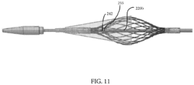
- the filter assembly 240 is coupled to the distal portion 220b of the catheter 220 adjacent the distal end 223 of the catheter 220 and is disposed axially proximal to the tip 235. Distal portion 220b extends axially through the filter assembly 240 (shown in FIG. 11 ).
- the filter assembly 240 is moveable between an expanded and unexpanded configuration.
- the filter assembly 240 in the unexpanded configuration which is illustrated in FIG. 4 , is sized and configured for insertion and passage through a blood vessel.
- the filter assembly 240 is sized and configured to capture emboli within the bloodstream. For example, at least a portion of the filter assembly 240 in the expanded configuration extends across a diameter of the vessel to catch emboli that may be flowing through the bloodstream.
- the filter assembly 240 includes a filter membrane 240a and a filter frame 240b.
- the filter membrane 240a is frusto-conically shape, and the filter frame 240b is egg shaped in the implementation shown in FIG. 5 .
- a conical tip 240c of the membrane 240a is fixedly coupled around the distal portion 220b of the catheter 220, and a distal end 240e of the filter frame 240b is disposed proximally of the conical tip 240c of the membrane 240a and is slidably coupled around the distal portion 220b.
- a proximal portion 240f of the filter membrane 240a is fixedly coupled to a central portion 240g of the filter frame 240b, such as via thermal or chemical bonding or another suitable coupling mechanism. And, a proximal portion 240d of the filter frame 240b is fixedly coupled around the distal portion 220b.
- the shape of the membrane and/or filter frame may be different than shown in FIG. 5 and may be based at least in part on the anatomy in which the filter assembly is to be disposed.
- a filter activation wire 242 extends through the filter activation wire lumens 222a, 222b, and a distal end of the filter activation wire 242 extends through a filter activation wire port 255 and is coupled to the distal end 240e of the filter frame 240b.
- the filter activation wire port 255 is defined by the distal portion 220b of the catheter 220.
- the filter activation wire port 255 has a first opening and a second opening. The first opening is defined by an external surface of the distal portion 220b of the catheter 220 and is disposed between the distal end 240e of the filter frame 240b and the proximal end 240d of the filter frame 240b.
- the second opening is defined by lumen 222b.
- the second opening of the port 255 is axially proximal the first opening, and in other implementations, the first and second openings of port 255 are radially aligned.
- the filter activation wire port 255 is distally disposed relative to the expandable balloon 260.
- an outer diameter of the filter frame 240b around the central portion 240g and an outer diameter of the proximal portion 240f of the filter membrane 240a correspond to an inner diameter of an artery or vessel to ensure that any embolic material is captured by the filter assembly 240.
- the filter membrane 240a and the filter frame 240b allow blood/fluid to flow therethrough.
- the filter membrane 240a comprises a biocompatible, elastic polymer sheet (e.g., polyurethane) that defines an array of openings.
- the openings are 40 micrometers in diameter, which allows blood to flow through but captures small particulates.
- the openings are formed by laser drilling.
- the filter frame 240b comprises a biocompatible, expandable structure that defines a plurality of openings. The openings of the filter frame 240b are larger than the openings defined by the filter membrane 240a.
- the filter frame 240b includes a material having memory properties, such as a braided nitinol structure or a laser cut nitinol tube structure.
- a material having memory properties such as a braided nitinol structure or a laser cut nitinol tube structure.
- Other suitable biocompatible materials include titanium and titanium alloys, stainless steel, platinum, gold, or other metals, as well as ceramics or polymers.
- the filter frame 240b has a memory of the unexpanded configuration such that when tension on the filter activation wire 242 is released, the filter frame 240 returns toward its unexpanded configuration, capturing any embolic materials that have been captured within the filter assembly 240.
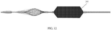
- an expandable balloon 260 is disposed between the proximal end 240d of the filter frame 240b and the proximal end of the distal portion 220b of the catheter 220.
- Air and/or fluid is provided to the balloon 260 for inflation via the balloon inflation lumens 224a, 224b defined by the proximal portion 220a and distal portion 220b of the catheter 220.
- a tube such as a hypotube, is disposed within the balloon inflation lumens 224a, 224b for delivering the air/fluid to the balloon 260.
- a distal balloon inflation port (not shown) is defined by the distal portion 220b of the catheter 220 and extends between the balloon inflation lumen 224b and a portion of the external surface of the distal portion 220b that is in fluid communication with an inside of the balloon 260.
- the balloon 260 can be any impermeable, flexible membrane defining a chamber that is expandable by the introduction of fluid into the chamber.
- a movable sheath 284 can extend over the balloon 260 and filter assembly 240.
- exemplary sheaths include a wire, coiled wire, polymer filament, or polymer braid sheath.
- the sheath 284 comprises an inner polymer layer (e.g., PTFE composite) to reduce friction with components disposed radially within the sheath 284, a structural sheath layer (e.g., a wire, coiled wire, polymer filament, or polymer braid sheath layer (e.g., a braided stainless steel sheath layer)) to maintain the radial strength of the sheath 284, and an outer polymer layer (e.g., nylon) to protect the structural sheath layer.
- the sheath 284 is a 6F sheath/8F guide compatible sheath, according to one implementation.
- the sheath 284 By disposing the sheath 284 over the therapeutic agent coated balloon 260 while introducing the balloon 260 into the body and routing the balloon 260 and filter assembly 240 through the body to the target site, the sheath 284 prevents loss of the therapeutic agent from the outer surface of the balloon 260 (e.g., by blood or other fluid(s) flowing past the balloon 260 or by other obstructions that may degrade or disturb the therapeutic agent).
- the sheath 284 does not extend over the filter assembly 240, and in other implementations, the sheath 284 extends over a portion of the filter assembly 240. Some implementations, such as the one shown in FIG. 12 , do not include a sheath at all.
- the therapeutic agent is an anti-stenotic therapeutic agent, such as Sirolimus (rapamycin) or Paclitaxel (taxol).
- anti-stenotic therapeutic agents include heparin, other taxanes, tacrolimus, actinomycin D, angiopeptin, vassenoids, flavoperidol, estrogen, halofuginone, matrix metallopreteinase inhibitors, and interferons.
- the therapeutic agent comprises any therapeutic agent for delivery to an interior wall of a vessel.
- classes of therapeutic agents that can be included in the devices described herein depend on the specific disease being treated and the physical properties of the agent, which include, for example, pro- or anti-proliferative, anti-inflammatory, antimitotic, anti-platelet, anticoagulant, antifibrin, antithrombin, cytostatic, antibiotic, anti-enzymatic, anti-metabolic, angiogenic, cytoprotective, angiotensin converting enzyme (ACE) inhibiting, angiotensin II receptor antagonizing and/or cardioprotective agents.
- pro- or anti-proliferative anti-inflammatory, antimitotic, anti-platelet, anticoagulant, antifibrin, antithrombin, cytostatic, antibiotic, anti-enzymatic, anti-metabolic, angiogenic, cytoprotective, angiotensin converting enzyme (ACE) inhibiting, angiotensin II receptor antagonizing and/or cardioprotective agents.
- ACE angiotensin converting enzyme
- antiproliferative drugs include, without limitation, actinomycins, taxol, docetaxel, paclitaxel, sirolimus (rapamycin), biolimus A9 (Biosensors International, Singapore), deforolimus, AP23572 (Ariad Pharmaceuticals), tacrolimus, temsirolimus, pimecrolimus, zotarolimus (ABT-578), 40-O-(2-hydroxy)ethyl-rapamycin (everolimus), 40-O-(3-hydroxypropyl)rapamycin (a structural derivative of rapamycin), 40-O-[2-(2-hydroxy)ethoxy]ethyl-rapamycin (a structural derivative of rapamycin), 40-O-tetrazole-rapamycin (a structural derivative of rapamycin), 40-O-tetrazolylrapamycin, 40-epi-(N-1-tetrazole)-rapamycin, and pirfenidone.
- anti-inflammatory drugs include both steroidal and non-steroidal (NSAID) anti-inflammatories such as, without limitation, clobetasol, alclofenac, alclometasone dipropionate, algestone acetonide, alpha amylase, amcinafal, amcinafide, amfenac sodium, amiprilose hydrochloride, anakinra, anirolac, anitrazafen, apazone, balsalazide disodium, bendazac, benoxaprofen, benzydamine hydrochloride, bromelains, broperamole, budesonide, carprofen, cicloprofen, cintazone, cliprofen, clobetasol propionate, clobetasone butyrate, clopirac, cloticasone propionate, cormethasone acetate, cortodoxone, deflazacort, desonide, desoximetasone, dex
- anti-platelet, anticoagulant, antifibrin, and antithrombin drugs include, without limitation, heparin, sodium heparin, low molecular weight heparins, heparinoids, hirudin, argatroban, forskolin, vapiprost, prostacyclin, prostacyclin dextran, D-phe-pro-arg-chloromethylketone, dipyridamole, glycoprotein IIb/IIIa platelet membrane receptor antagonist antibody, recombinant hirudin and thrombin, thrombin inhibitors such as ANGIOMAX ® (bivalirudin, from Biogen), calcium channel blockers such as nifedipine, colchicine, fish oil (omega 3-fatty acid), histamine antagonists, lovastatin, monoclonal antibodies such as those specific for Platelet-Derived Growth Factor (PDGF) receptors, nitroprusside, phosphodiesterase inhibitors, prostaglandin inhibitor
- cytostatic drugs include, without limitation, angiopeptin, angiotensin converting enzyme inhibitors such as captopril, cilazapril or lisinopril, calcium channel blockers such as nifedipine; colchicine, fibroblast growth factor (FGF) antagonists; fish oil ( ⁇ -3-fatty acid); histamine antagonists; lovastatin, monoclonal antibodies such as, without limitation, those specific for Platelet-Derived Growth Factor (PDGF) receptors; nitroprusside, phosphodiesterase inhibitors, prostaglandin inhibitors, suramin, serotonin blockers, steroids, thioprotease inhibitors, triazolopyrimidine (a PDGF antagonist) and nitric oxide.
- angiopeptin angiotensin converting enzyme inhibitors such as captopril, cilazapril or lisinopril
- calcium channel blockers such as nifedipine
- colchicine fibroblast growth factor (
- ACE inhibitors include, without limitation, quinapril, perindopril, ramipril, captopril, benazepril, trandolapril, fosinopril, lisinopril, moexipril and enalapril.
- angiotensin II receptor antagonists include, without limitation, irbesartan and losartan.
- Therapeutic agents can be formulated as solid formulations, gels, or liquids suitable for administration using a therapeutic agent-coated balloon catheter. Such formulations are known in the art.
- the formulation can be a coating comprising a therapeutic agent formed on an outer surface of an expandable balloon.
- the coating can be formed by spraying, dipping, pouring, pumping, brushing, wiping, vacuum deposition, vapor deposition, plasma deposition, electrostatic deposition, ultrasonic deposition, epitaxial growth, electrochemical deposition or any other method known to those skilled in the art.
- the coating can comprise one or more therapeutic agents and optionally one or more excipients and/or additives as described above.
- the coating can include a biocompatible polymer.
- Suitable polymers can include both biostable and biodegradable polymers, such as microcrystalline cellulose, hydroxypropyl cellulose, hydroxypropyl methylcellulose, polyalkylene oxides such as polyethylene oxide (PEG), polyanhydrides, poly(ester anhydrides), polyhydroxy acids such as polylactide (PLA), polyglycolide (PGA), poly(lactide-co-glycolide) (PLGA), poly-3-hydroxybutyrate (PHB) and copolymers thereof, poly-4-hydroxybutyrate (P4HB) and copolymers thereof, polycaprolactone and copolymers thereof, and combinations thereof.
- the coating can comprise rate-controlling excipients, including hydrophobic materials, including acceptable fats and fatty substances (e.g., fatty alcohols, such as lauryl, myristyl stearyl, cetyl or cetostearyl alcohol, fatty acids and derivatives, including, but not limited to, fatty acid esters, fatty acid glycerides (mono-, di- and tri-glycerides), and hydrogenated fats), waxes and wax-like substances (e.g., natural or synthetic waxes, hydrocarbons, and normal waxes, including beeswax, glycowax, castor wax, carnauba wax, paraffins and candelilla wax), ion-exchange resins,water-insoluble proteins (e.g., zein), wicking agents (e.g., starch derivatives such as waxy maltodextrin and drum dried corn starch, cellulose derivatives such as hydroxypropylmethyl cellulose, hydroxypropyl
- one or more barrier layers can be placed over the coating to prevent dissolution of the therapeutic agent layer prior to positioning of the catheter where administration of the therapeutic agent is intended.
- the sheath 284 includes a radio-opaque marker 293 around a portion of the sheath 284 to assist in verifying proper placement of the sheath within the vessel before retracting the sheath 284 to expose the filter 240 and balloon 260.
- the sheath 284 may not include the radio-opaque marker 293.
- the sheath 284 may be tapered from its distal end toward its proximal end, wherein the distal end of the sheath 284 has a larger diameter than the proximal end of the sheath 284.
- a sheath wire exit port 288 is defined between an external surface of the proximal portion 220a of the catheter 220 and the sheath wire lumen 226, and a sheath wire 286 extends between the sheath wire lumen 226 and the sheath 284 via the sheath wire exit port 288 (sheath 284 not shown in FIG. 8 ).
- the sheath wire exit port 288 is defined adjacent a distal end of the proximal portion 220a of the catheter 220.
- a distal end of the sheath wire 286 extends over the external surface of the distal portion 220b of the catheter to be coupled to the sheath 284.
- the sheath wire 286 is coupled to the sheath 284 by embedding the distal end of the sheath wire 286 between the braided structural layer and the outer polymer layer.
- the sheath 284 does not extend over the entire length of the catheter.
- the physician is able to stabilize (e.g., hold steady) the catheter 220 while the sheath 284 is moved axially proximal to the balloon 260, which reduces or prevents movement of the distal portion 220b of the catheter 220 and unintentional axial movement of the balloon relative to the target location during deployment of the balloon.
- the sheath is not coupled to a sheath wire, and the sheath extends proximally over the entire length of the catheter. Thus, there is no space available on the catheter to hold the catheter steady during sheath deployment.
- Known devices do not include a sheath wire.
- a portion of the sheath wire 286 extending between the sheath wire exit port 288 and the sheath 284 is exposed.
- a sleeve 291 e.g., a polymer sleeve
- At least a portion of the exterior surface of mid portion 220c defines a recessed, axially extending groove 226b that is in communication with the sheath wire lumen 226 defined by the proximal portion 220a.
- the sheath wire 286 is radially movable in and out of the groove 226b, as seen in FIG. 9B .
- guidewire 250 is routed through a proximal end of the sleeve 291 toward the guidewire lumen 227 defined by the distal portion 220b of the catheter 220.
- the sheath 284 and the sleeve 291 are coupled together.
- the sheath 284 and sleeve 291 are separately formed and disposed axially adjacent each other.
- a proximal end of the distal portion 220b of the catheter 220 defines a guidewire port 302 that extends between the guidewire lumen 227 and an external surface of the catheter 220.
- the opening of the guidewire port 302 defined by the external surface of the catheter 220 is proximal to guidewire lumen 227 to facilitate rapid exchange of the guidewire 250.
- the guidewire port 302 is defined by the opening of the guidewire lumen 227 at the proximal end of the distal portion 220b.
- a proximal portion of the guidewire 250 extends out of the distal portion 220b of catheter 220 proximally of the sheath 284 via the guidewire port 302.
- the guidewire 250 has a diameter of between 0.25 and 0.96 mm (e.g. 0.36 mm) such as 0.010 inches and 0.038 inches (e.g., 0.014 inches).
- the guidewire port includes a first opening and a second opening.
- the first opening of the guidewire port is defined by the exterior surface of the catheter that is radially spaced apart from the guidewire lumen 227
- the second opening of the guidewire port is defined by an interior surface of the lumen 226 and is distally spaced apart from the first opening along the longitudinal axis of the guidewire lumen 227. That is, in various implementations, the guidewire port extends through the catheter 220 from a first opening towards a second opening defined by a lumen that is distally spaced from the first opening.
- the device 200 further includes a handle 290 coupled to the proximal end 225 of the proximal portion 220a of the catheter 220.
- the handle 290 includes controls (e.g., buttons, knobs, etc.) that are coupled to one or more of the filter activation wire 242, the sheath wire 286, and/or the guidewire 250 to allow the user to actuate the filter 240, the sheath 284, and/or the guide wire 250.
- knobs 310, 315 are disposed on the handle 290 and are coupled to the filter activation wire 242 and the sheath wire 286, respectively.
- the handle 290 defines a proximal balloon inflation port 265 that is in fluid communication with the balloon inflation lumens 224a, 224b and the balloon 260 to provide air/fluid to the balloon 260 for expansion.
- the percutaneous transluminal angioplasty device 200 and its corresponding components are formed from one or more biocompatible materials, such as cobalt chromium, titanium and titanium alloys, stainless steel, nitinol, platinum, gold, or other metals, as well as ceramics or polymers.
- the device 200 or portions thereof includes a coated or sheathed material.
- the device 200 includes a bioresorbable material or has a bioresorbable coating or sheathing.
- FIGS. 10A and 10B illustrate how the device 200 is operated within the body according to one implementation.
- the sheath 284 is moved axially toward the proximal end 225 of the catheter 220 by pulling the sheath wire 286 proximally to expose the filter assembly 240.
- the filter assembly 240 is deployed into the expanded configuration by tensioning the filter activation wire 242. Deploying the filter assembly 240 allows the filter assembly 240 to catch any embolic material that is dislodged during deployment of the balloon 260.
- the drug coating material may fragment off the balloon while it is being expanded within the treatment site resulting in a bolus of embolic particles carried along the artery toward more distal anatomy.
- the filter assembly 240 also catches any portion of the balloon coating that flakes off or becomes separated from the balloon 260 during expansion.
- the sheath is moved further axially toward the proximal end 225 to expose the therapeutic agent coated balloon 260.
- the distance the sheath is moved can be varied to expose either a portion of or the entirety of the balloon 260.
- the exposed, expanded length of the balloon can be varied.
- the portion of the balloon 260 remaining under the sheath 284 remains unexpanded, or at least expanded to a lesser diameter than the exposed portion of the balloon.
- one catheter can be constructed with one long balloon (such as 200 mm long, for example).
- the exposed and expanded length of the balloon can be from anywhere from about 5 millimeters to 200 millimeters, including 5 millimeters, 25 millimeters, 50 millimeters, 75 millimeters, 100 millimeters, 125 millimeters, 150 millimeters, 175 millimeters, and 200 millimeters, depending on how far the sheath is retracted.
- a hospital could buy one catheter having a 200 millimeter balloon instead of multiple catheters having balloons with separate lengths, saving money and reducing inventory.
- the balloon 260 is inflated against an inner surface of the artery such that the vessel wall is expanded and the therapeutic agent on the surface of the balloon is delivered into the anastomotic lesion.
- the balloon 260 is inflated (or deflated) via fluid/air provided to (or removed from) a central chamber of the balloon 260 via port 265.
- the balloon 260 is deflated, tension in the filter activation wire 242 is released, and the filter membrane 240a and the filter net 240b are collapsed by releasing the filter activation wire 242, which securely capture any embolic material captured by the filter assembly 240.
- the embolic material may include material from the vessel and fragments of the drug coating that separated from the balloon 260 during expansion. The blocked vessel is opened and blood flow is restored.
- the filter assembly 240 is then contracted by actuating the filter activation wire 242, and the device 200, which includes the deflated balloon 260 and the contracted filter assembly 240, are removed from the vessel.
- the catheter 220 is moved axially out of the body, which pulls the filter assembly 240 holding any captured embolic material and the unexpanded balloon 260 out of the body. Because the filter assembly 240 is able to capture and hold the embolic material upon release of the filter activation wire 242, it is not necessary to move the sheath 284 distally over the filter assembly 240 prior to removal of the device 200 from the body, which reduces the time required for the procedure.
- the proximal portion 220a and the distal portion 220b of the catheter 220 are able to be steadied by the physician (e.g., by holding the proximal portion 220a of the catheter) to prevent or reduce movement of the proximal portion 220a and the distal portion 220b relative to the sheath 284.
- Having one device 200 that includes a filter, expandable therapeutic agent coated balloon, and a sheath activation wire reduces the time required to perform a vascular expansion procedure and reduces the potential for complications resulting from the procedure.
- the various embodiments disclosed herein are adaptable for use in virtually any vessel where the capture emboli within the bloodstream is required for a therapeutic or diagnostic purpose.
- certain embodiments could be used for purposes other than medical, such as construction, manufacturing, and excavation, among others; accordingly, nothing herein is intended to limit application of the various embodiments to purely medical uses.
Landscapes
- Health & Medical Sciences (AREA)
- Life Sciences & Earth Sciences (AREA)
- Animal Behavior & Ethology (AREA)
- General Health & Medical Sciences (AREA)
- Public Health (AREA)
- Veterinary Medicine (AREA)
- Heart & Thoracic Surgery (AREA)
- Engineering & Computer Science (AREA)
- Biomedical Technology (AREA)
- Epidemiology (AREA)
- Vascular Medicine (AREA)
- Child & Adolescent Psychology (AREA)
- Biophysics (AREA)
- Pulmonology (AREA)
- Anesthesiology (AREA)
- Hematology (AREA)
- Cardiology (AREA)
- Oral & Maxillofacial Surgery (AREA)
- Transplantation (AREA)
- Molecular Biology (AREA)
- Medicinal Chemistry (AREA)
- Chemical & Material Sciences (AREA)
- Surgical Instruments (AREA)
- Materials For Medical Uses (AREA)
Claims (15)
- Dispositif d'angioplastie transluminale percutanée (200), comprenant :un cathéter (220) à lumières multiples ayant une extrémité proximale (225) et une extrémité distale (223), le cathéter (220) définissant une première lumière, une deuxième lumière et une troisième lumière, chaque lumière s'étendant à travers au moins une partie du cathéter (220) ;un ballonnet déployable (260) disposé sur le cathéter (220), dans lequel le ballonnet déployable (260) a une surface extérieure, et au moins une partie de la surface extérieure comprend un agent thérapeutique ;une gaine (284) mobile s'étendant sur au moins une partie du ballonnet déployable (260) dans une configuration non déployée du ballonnet déployable (260) ;un fil de gaine (286) accouplé à la gaine (284) mobile, le fil de gaine (286) s'étendant à travers l'une des lumières définies par le cathéter (220), dans lequel le mouvement du fil de gaine (286) translate la gaine axialement ; etun filtre (240) disposé de manière adjacente à l'extrémité distale (223) du cathéter (220), le filtre (240) étant mobile entre une configuration non déployée et une configuration déployée, et le ballonnet déployable (260) étant disposé de manière axialement proximale au filtre (240).
- Dispositif d'angioplastie transluminale percutanée (200) selon la revendication 1, dans lequel l'agent thérapeutique est un agent thérapeutique anti-sténotique.
- Dispositif d'angioplastie transluminale percutanée (200) selon la revendication 2, dans lequel l'agent thérapeutique anti-sténotique est choisi dans le groupe constitué par le sirolimus et le paclitaxel.
- Dispositif d'angioplastie transluminale percutanée (200) selon l'une quelconque des revendications 1, 2 ou 3, dans lequel le fil de gaine (286) est déplacé axialement pour translater la gaine (284) axialement, et dans lequel le mouvement axial du fil de gaine (286) translate la gaine (284) dans la même direction que le mouvement axial du fil de gaine (286) .
- Dispositif d'angioplastie transluminale percutanée (200) selon l'une quelconque des revendications 1 à 4, dans lequel un fil d'activation de filtre (242) est disposé à l'intérieur d'une première lumière, et une extrémité distale du fil d'activation de filtre (242) est accouplée au filtre (240).
- Dispositif d'angioplastie transluminale percutanée (200) selon la revendication 5, dans lequel le filtre (240) comprend un cadre de filtre (240b) et une membrane de filtre (240a), le cadre de filtre (240b) a une extrémité distale et une extrémité proximale, l'extrémité proximale du cadre de filtre (240b) étant accouplée de manière fixe au cathéter (220), et l'extrémité distale du cadre de filtre (240b) étant accouplée de manière coulissante au cathéter (220), la membrane de filtre (240a) a une extrémité distale et une extrémité proximale, et l'extrémité distale de la membrane de filtre (240a) est accouplée de manière fixe au cathéter (220) distalement de l'extrémité proximale de la membrane de filtre (240a) et de l'extrémité distale du cadre de filtre (240b), et l'extrémité proximale de la membrane de filtre (240a) est accouplée de manière fixe à une partie du cadre de filtre (240b), et l'extrémité distale du fil d'activation de filtre (242) est accouplée à l'extrémité distale du cadre de filtre (240b), dans lequel la mise en tension du fil d'activation de filtre (242) dans une direction proximale pousse l'extrémité distale du cadre de filtre (240b) dans la direction proximale axiale à partir d'une configuration non déployée vers une configuration déployée.
- Dispositif d'angioplastie transluminale percutanée (200) selon l'une quelconque des revendications 5 ou 6, comprenant en outre une poignée (290) accouplée à l'extrémité proximale (225) du cathéter (220), la poignée (290) étant accouplée au fil d'activation de filtre (242) et au fil de gaine (286).
- Dispositif d'angioplastie transluminale percutanée (200) selon la revendication 7, dans lequel la poignée (290) comprend un premier actionneur accouplé au fil d'activation de filtre (242) et un second actionneur accouplé au fil de gaine (286), le premier actionneur pouvant être manipulé pour déployer et contracter le filtre (240) par le biais du fil d'activation de filtre (242), et le second actionneur pouvant être manipulé pour déplacer la gaine (284) axialement.
- Dispositif d'angioplastie transluminale percutanée (200) selon l'une quelconque des revendications 1 à 8, dans lequel la troisième lumière est une lumière de gonflage de ballonnet, le cathéter (220) définissant en outre un orifice de gonflage entre une surface externe du cathéter (220) et la troisième lumière.
- Dispositif d'angioplastie transluminale percutanée (200) selon l'une quelconque des revendications 1 à 9, dans lequel le cathéter (220) définit un orifice de fil-guide, l'orifice de fil-guide ayant une première ouverture définie par la première, la deuxième ou la troisième lumière et une deuxième ouverture définie par une surface extérieure du cathéter (220), dans lequel la première ouverture de l'orifice de fil-guide est disposée distalement par rapport à la deuxième ouverture, dans lequel un fil-guide (250) est disposé à l'intérieur d'au moins une partie de la première, de la deuxième ou de la troisième lumière qui définit la première ouverture de l'orifice de fil-guide.
- Dispositif d'angioplastie transluminale percutanée (200) selon la revendication 1, dans lequel au moins une partie du filtre (240) a un rayon dans la configuration déployée qui correspond à un diamètre interne d'un vaisseau sanguin dans lequel le filtre (240) est disposé.
- Dispositif d'angioplastie transluminale percutanée (200) selon l'une quelconque des revendications 1 à 11, dans lequel le cathéter (220) comprend une partie proximale (220a) et une partie distale (220b), la partie proximale (220a) étant disposée de manière adjacente à l'extrémité proximale (225) du cathéter (220) et la partie distale (220b) étant disposée de manière adjacente à l'extrémité distale (223) du cathéter (220), dans lequel la partie proximale (220a) du cathéter (220) définit une lumière de fil de gaine (226), une lumière de fil d'activation de filtre proximale (222a) et une lumière de gonflage de ballonnet proximale (224a), et la partie distale (220b) du cathéter (220) définit une lumière de fil de guidage (227), une lumière de fil d'activation de filtre distale (222b) et une lumière de gonflage de ballonnet distale (224b).
- Dispositif d'angioplastie transluminale percutanée (200) selon la revendication 12, dans lequel la lumière de gonflage de ballonnet proximale (224a) et la lumière de gonflage de ballonnet distale (224b) sont alignées axialement.
- Dispositif d'angioplastie transluminale percutanée (200) selon l'une quelconque des revendications 12 ou 13, dans lequel la lumière de fil d'activation de filtre proximale (222a) et la lumière de fil d'activation de filtre distale (222b) sont alignées axialement.
- Dispositif d'angioplastie transluminale percutanée (200) selon l'une quelconque des revendications 13 ou 14, dans lequel la lumière de fil de gaine (226) et la lumière de fil-guide (227) sont alignées axialement.
Applications Claiming Priority (2)
| Application Number | Priority Date | Filing Date | Title |
|---|---|---|---|
| US201662433521P | 2016-12-13 | 2016-12-13 | |
| PCT/US2017/066067 WO2018112022A1 (fr) | 2016-12-13 | 2017-12-13 | Ballonnet d'angioplastie revêtu d'un agent thérapeutique muni d'un filtre embolique et d'un couvercle de protection |
Publications (3)
| Publication Number | Publication Date |
|---|---|
| EP3554613A1 EP3554613A1 (fr) | 2019-10-23 |
| EP3554613A4 EP3554613A4 (fr) | 2020-07-22 |
| EP3554613B1 true EP3554613B1 (fr) | 2024-02-28 |
Family
ID=62488476
Family Applications (1)
| Application Number | Title | Priority Date | Filing Date |
|---|---|---|---|
| EP17880585.9A Active EP3554613B1 (fr) | 2016-12-13 | 2017-12-13 | Ballonnet d'angioplastie revêtu d'un agent thérapeutique muni d'un filtre embolique et d'un couvercle de protection |
Country Status (6)
| Country | Link |
|---|---|
| US (1) | US10849730B2 (fr) |
| EP (1) | EP3554613B1 (fr) |
| AU (1) | AU2017376270B2 (fr) |
| CA (1) | CA3047097A1 (fr) |
| ES (1) | ES2977758T3 (fr) |
| WO (1) | WO2018112022A1 (fr) |
Families Citing this family (4)
| Publication number | Priority date | Publication date | Assignee | Title |
|---|---|---|---|---|
| US12114877B2 (en) | 2017-10-16 | 2024-10-15 | Retriever Medical, Inc. | Clot removal methods and devices with multiple independently controllable elements |
| US20190110804A1 (en) | 2017-10-16 | 2019-04-18 | Michael Bruce Horowitz | Catheter based retrieval device with proximal body having axial freedom of movement |
| US12201315B2 (en) | 2017-10-16 | 2025-01-21 | Retriever Medical, Inc. | Clot removal methods and devices with multiple independently controllable elements |
| WO2021117049A1 (fr) * | 2019-12-11 | 2021-06-17 | Healing Hands Clinic Private Limited | Dispositif pour le traitement d'une fistule anale et d'une fistule anale complexe |
Family Cites Families (41)
| Publication number | Priority date | Publication date | Assignee | Title |
|---|---|---|---|---|
| SE445884B (sv) | 1982-04-30 | 1986-07-28 | Medinvent Sa | Anordning for implantation av en rorformig protes |
| US5772669A (en) | 1996-09-27 | 1998-06-30 | Scimed Life Systems, Inc. | Stent deployment catheter with retractable sheath |
| US5843027A (en) * | 1996-12-04 | 1998-12-01 | Cardiovascular Dynamics, Inc. | Balloon sheath |
| US5938697A (en) | 1998-03-04 | 1999-08-17 | Scimed Life Systems, Inc. | Stent having variable properties |
| US20100036481A1 (en) * | 1998-04-27 | 2010-02-11 | Artemis Medical, Inc. | Cardiovascular Devices and Methods |
| US6786889B1 (en) | 1999-03-31 | 2004-09-07 | Scimed Life Systems, Inc | Textured and/or marked balloon for stent delivery |
| AU4924500A (en) | 1999-05-19 | 2000-12-12 | Malte Neuss | Radially expandable vessel support |
| US6270521B1 (en) | 1999-05-21 | 2001-08-07 | Cordis Corporation | Stent delivery catheter system for primary stenting |
| US6569193B1 (en) | 1999-07-22 | 2003-05-27 | Advanced Cardiovascular Systems, Inc. | Tapered self-expanding stent |
| US6511503B1 (en) | 1999-12-30 | 2003-01-28 | Advanced Cardiovascular Systems, Inc. | Catheter apparatus for treating occluded vessels and filtering embolic debris and method of use |
| US6391050B1 (en) * | 2000-02-29 | 2002-05-21 | Scimed Life Systems, Inc. | Self-expanding stent delivery system |
| WO2001091844A1 (fr) * | 2000-05-31 | 2001-12-06 | Kerberos Proximal Solutions, Inc. | Systeme de protection contre l'embolisation pour interventions vasculaires |
| US7208002B2 (en) | 2001-01-04 | 2007-04-24 | Boston Scientific Scimed, Inc. | Expansion-assisting delivery system for self-expanding stent |
| US20030055480A1 (en) | 2001-09-14 | 2003-03-20 | Fischell David R. | Recannalization device with integrated distal emboli protection |
| US7473242B2 (en) | 2003-04-30 | 2009-01-06 | Medtronic Vascular, Inc. | Method and systems for treating vulnerable plaque |
| US7947070B2 (en) | 2003-05-16 | 2011-05-24 | Boston Scientific Scimed, Inc. | Dilatation and stent delivery system and related methods |
| US20050004594A1 (en) * | 2003-07-02 | 2005-01-06 | Jeffrey Nool | Devices and methods for aspirating from filters |
| US7722634B2 (en) | 2003-07-03 | 2010-05-25 | Regents Of The University Of Minnesota | Medical device and method of intravenous filtration |
| US9078780B2 (en) | 2003-11-08 | 2015-07-14 | Cook Medical Technologies Llc | Balloon flareable branch vessel prosthesis and method |
| US8403976B2 (en) | 2004-04-08 | 2013-03-26 | Contego Medical Llc | Percutaneous transluminal angioplasty device with integral embolic filter |
| US20070156168A1 (en) | 2005-12-29 | 2007-07-05 | Medtronic Vascular, Inc. | Polymer marker and retention bands |
| US8083792B2 (en) | 2006-01-24 | 2011-12-27 | Cordis Corporation | Percutaneous endoprosthesis using suprarenal fixation and barbed anchors |
| US20080077223A1 (en) | 2006-09-21 | 2008-03-27 | Fischell Robert E | Stent delivery system with improved deliverabilty features |
| US7776080B2 (en) * | 2007-04-25 | 2010-08-17 | Abbott Cardiovascualr Systems Inc. | Stent delivery catheter system and method of implanting a self-expanding stent with embolic protection |
| US9370642B2 (en) * | 2007-06-29 | 2016-06-21 | J.W. Medical Systems Ltd. | Adjustable-length drug delivery balloon |
| US8758421B2 (en) | 2008-01-30 | 2014-06-24 | Boston Scientific Scimed, Inc. | Medical systems and related methods |
| US9149376B2 (en) | 2008-10-06 | 2015-10-06 | Cordis Corporation | Reconstrainable stent delivery system |
| US8956385B2 (en) | 2009-04-14 | 2015-02-17 | Aharon FRIMERMAN | Integrated distal embolization protection apparatus for endo-luminal devices such as balloon, stent or tavi apparatus |
| US20100268263A1 (en) | 2009-04-21 | 2010-10-21 | Boston Scientific Scimed, Inc. | Embolic protection filters, filter membranes, and methods for making and using the same |
| US20120041469A1 (en) * | 2010-08-11 | 2012-02-16 | Svelte Medical Systems, Inc. | Revascularization device with integrated distal emboli protection |
| EP2701788A1 (fr) * | 2011-04-29 | 2014-03-05 | Boston Scientific Scimed, Inc. | Surfaces de protection pour dispositifs médicaux enrobés de médicament |
| US9220584B2 (en) * | 2012-03-30 | 2015-12-29 | Abbott Cardiovascular Systems Inc. | Treatment of diabetic patients with a stent and locally administered adjunctive therapy |
| CN104936550B (zh) | 2012-11-15 | 2017-09-22 | 恩菲纽姆血管技术有限公司 | 临时血管支撑架和刻划装置 |
| CN105120798B (zh) * | 2012-11-27 | 2017-03-15 | 康特戈医疗有限责任公司 | 具有整体式栓子滤器的经皮腔内血管成形术设备 |
| US20140214067A1 (en) | 2012-11-27 | 2014-07-31 | Contego Medical, Llc | Percutaneous transluminal angioplasty device with integral embolic filter |
| EP2928537A4 (fr) * | 2012-12-04 | 2016-08-03 | Angioslide Ltd | Cathéter à ballonnet et procédés d'utilisation |
| US9974676B2 (en) | 2013-08-09 | 2018-05-22 | Cook Medical Technologies Llc | Wire collection device with geared advantage |
| EP3065668A4 (fr) | 2013-11-08 | 2017-09-27 | Contego Medical, LLC | Dénervation artérielle percutanée par cathéter à filtre embolique intégré |
| JP2017529222A (ja) | 2014-09-25 | 2017-10-05 | コンテゴ メディカル エルエルシー | 一時的塞栓保護デバイス及びその方法 |
| AU2016209053B2 (en) * | 2015-01-23 | 2020-04-16 | Contego Medical, Inc. | Interventional device having an integrated embolic filter and associated methods |
| CN112043475B (zh) | 2015-10-27 | 2022-05-31 | 康特戈医疗股份有限公司 | 用于与腔内血管成形术装置一起使用的支架 |
-
2017
- 2017-12-13 EP EP17880585.9A patent/EP3554613B1/fr active Active
- 2017-12-13 ES ES17880585T patent/ES2977758T3/es active Active
- 2017-12-13 CA CA3047097A patent/CA3047097A1/fr active Pending
- 2017-12-13 WO PCT/US2017/066067 patent/WO2018112022A1/fr not_active Ceased
- 2017-12-13 US US15/840,294 patent/US10849730B2/en active Active
- 2017-12-13 AU AU2017376270A patent/AU2017376270B2/en active Active
Also Published As
| Publication number | Publication date |
|---|---|
| ES2977758T3 (es) | 2024-08-29 |
| EP3554613A4 (fr) | 2020-07-22 |
| US10849730B2 (en) | 2020-12-01 |
| EP3554613A1 (fr) | 2019-10-23 |
| US20180161143A1 (en) | 2018-06-14 |
| AU2017376270A1 (en) | 2019-07-04 |
| AU2017376270B2 (en) | 2022-08-11 |
| WO2018112022A1 (fr) | 2018-06-21 |
| CA3047097A1 (fr) | 2018-06-21 |
Similar Documents
| Publication | Publication Date | Title |
|---|---|---|
| US11717654B2 (en) | Wedge dissectors for a medical balloon | |
| EP3554613B1 (fr) | Ballonnet d'angioplastie revêtu d'un agent thérapeutique muni d'un filtre embolique et d'un couvercle de protection | |
| JP6533253B2 (ja) | 封入薬物組成物およびその使用方法 | |
| AU2012205348B2 (en) | Endoluminal drug applicator and method of treating diseased vessels of the body | |
| US20250268613A1 (en) | Method and Apparatus for Delivery of Cell Therapies | |
| US20040236414A1 (en) | Devices and methods for treatment of stenotic regions | |
| JP2019134930A (ja) | 薬物放出性医療機器のための除去可能なカバー | |
| US8840678B2 (en) | Drug-eluting bioabsorbable renal artery stent for renal cancer and inflammatory disorders | |
| WO2013015025A1 (fr) | Dispositif de traitement | |
| EP2063810B1 (fr) | Anneaux implantables contenant un agent bioactif |
Legal Events
| Date | Code | Title | Description |
|---|---|---|---|
| STAA | Information on the status of an ep patent application or granted ep patent |
Free format text: STATUS: THE INTERNATIONAL PUBLICATION HAS BEEN MADE |
|
| PUAI | Public reference made under article 153(3) epc to a published international application that has entered the european phase |
Free format text: ORIGINAL CODE: 0009012 |
|
| STAA | Information on the status of an ep patent application or granted ep patent |
Free format text: STATUS: REQUEST FOR EXAMINATION WAS MADE |
|
| 17P | Request for examination filed |
Effective date: 20190621 |
|
| AK | Designated contracting states |
Kind code of ref document: A1 Designated state(s): AL AT BE BG CH CY CZ DE DK EE ES FI FR GB GR HR HU IE IS IT LI LT LU LV MC MK MT NL NO PL PT RO RS SE SI SK SM TR |
|
| AX | Request for extension of the european patent |
Extension state: BA ME |
|
| DAV | Request for validation of the european patent (deleted) | ||
| DAX | Request for extension of the european patent (deleted) | ||
| A4 | Supplementary search report drawn up and despatched |
Effective date: 20200622 |
|
| RIC1 | Information provided on ipc code assigned before grant |
Ipc: A61F 2/01 20060101ALI20200616BHEP Ipc: A61M 25/10 20130101AFI20200616BHEP |
|
| RAP1 | Party data changed (applicant data changed or rights of an application transferred) |
Owner name: CONTEGO MEDICAL, INC. |
|
| RAP3 | Party data changed (applicant data changed or rights of an application transferred) |
Owner name: CONTEGO MEDICAL, INC. |
|
| GRAP | Despatch of communication of intention to grant a patent |
Free format text: ORIGINAL CODE: EPIDOSNIGR1 |
|
| STAA | Information on the status of an ep patent application or granted ep patent |
Free format text: STATUS: GRANT OF PATENT IS INTENDED |
|
| INTG | Intention to grant announced |
Effective date: 20230719 |
|
| GRAS | Grant fee paid |
Free format text: ORIGINAL CODE: EPIDOSNIGR3 |
|
| GRAJ | Information related to disapproval of communication of intention to grant by the applicant or resumption of examination proceedings by the epo deleted |
Free format text: ORIGINAL CODE: EPIDOSDIGR1 |
|
| GRAL | Information related to payment of fee for publishing/printing deleted |
Free format text: ORIGINAL CODE: EPIDOSDIGR3 |
|
| STAA | Information on the status of an ep patent application or granted ep patent |
Free format text: STATUS: REQUEST FOR EXAMINATION WAS MADE |
|
| P01 | Opt-out of the competence of the unified patent court (upc) registered |
Effective date: 20230926 |
|
| GRAP | Despatch of communication of intention to grant a patent |
Free format text: ORIGINAL CODE: EPIDOSNIGR1 |
|
| INTC | Intention to grant announced (deleted) | ||
| STAA | Information on the status of an ep patent application or granted ep patent |
Free format text: STATUS: GRANT OF PATENT IS INTENDED |
|
| INTG | Intention to grant announced |
Effective date: 20231116 |
|
| GRAA | (expected) grant |
Free format text: ORIGINAL CODE: 0009210 |
|
| STAA | Information on the status of an ep patent application or granted ep patent |
Free format text: STATUS: THE PATENT HAS BEEN GRANTED |
|
| AK | Designated contracting states |
Kind code of ref document: B1 Designated state(s): AL AT BE BG CH CY CZ DE DK EE ES FI FR GB GR HR HU IE IS IT LI LT LU LV MC MK MT NL NO PL PT RO RS SE SI SK SM TR |
|
| REG | Reference to a national code |
Ref country code: GB Ref legal event code: FG4D |
|
| REG | Reference to a national code |
Ref country code: CH Ref legal event code: EP |
|
| REG | Reference to a national code |
Ref country code: DE Ref legal event code: R096 Ref document number: 602017079634 Country of ref document: DE |
|
| REG | Reference to a national code |
Ref country code: IE Ref legal event code: FG4D |
|
| REG | Reference to a national code |
Ref country code: LT Ref legal event code: MG9D |
|
| PG25 | Lapsed in a contracting state [announced via postgrant information from national office to epo] |
Ref country code: IS Free format text: LAPSE BECAUSE OF FAILURE TO SUBMIT A TRANSLATION OF THE DESCRIPTION OR TO PAY THE FEE WITHIN THE PRESCRIBED TIME-LIMIT Effective date: 20240628 |
|
| REG | Reference to a national code |
Ref country code: NL Ref legal event code: MP Effective date: 20240228 |
|
| PG25 | Lapsed in a contracting state [announced via postgrant information from national office to epo] |
Ref country code: LT Free format text: LAPSE BECAUSE OF FAILURE TO SUBMIT A TRANSLATION OF THE DESCRIPTION OR TO PAY THE FEE WITHIN THE PRESCRIBED TIME-LIMIT Effective date: 20240228 |
|
| PG25 | Lapsed in a contracting state [announced via postgrant information from national office to epo] |
Ref country code: GR Free format text: LAPSE BECAUSE OF FAILURE TO SUBMIT A TRANSLATION OF THE DESCRIPTION OR TO PAY THE FEE WITHIN THE PRESCRIBED TIME-LIMIT Effective date: 20240529 |
|
| PG25 | Lapsed in a contracting state [announced via postgrant information from national office to epo] |
Ref country code: HR Free format text: LAPSE BECAUSE OF FAILURE TO SUBMIT A TRANSLATION OF THE DESCRIPTION OR TO PAY THE FEE WITHIN THE PRESCRIBED TIME-LIMIT Effective date: 20240228 Ref country code: NL Free format text: LAPSE BECAUSE OF FAILURE TO SUBMIT A TRANSLATION OF THE DESCRIPTION OR TO PAY THE FEE WITHIN THE PRESCRIBED TIME-LIMIT Effective date: 20240228 Ref country code: RS Free format text: LAPSE BECAUSE OF FAILURE TO SUBMIT A TRANSLATION OF THE DESCRIPTION OR TO PAY THE FEE WITHIN THE PRESCRIBED TIME-LIMIT Effective date: 20240528 |
|
| PG25 | Lapsed in a contracting state [announced via postgrant information from national office to epo] |
Ref country code: RS Free format text: LAPSE BECAUSE OF FAILURE TO SUBMIT A TRANSLATION OF THE DESCRIPTION OR TO PAY THE FEE WITHIN THE PRESCRIBED TIME-LIMIT Effective date: 20240528 Ref country code: NO Free format text: LAPSE BECAUSE OF FAILURE TO SUBMIT A TRANSLATION OF THE DESCRIPTION OR TO PAY THE FEE WITHIN THE PRESCRIBED TIME-LIMIT Effective date: 20240528 Ref country code: NL Free format text: LAPSE BECAUSE OF FAILURE TO SUBMIT A TRANSLATION OF THE DESCRIPTION OR TO PAY THE FEE WITHIN THE PRESCRIBED TIME-LIMIT Effective date: 20240228 Ref country code: LT Free format text: LAPSE BECAUSE OF FAILURE TO SUBMIT A TRANSLATION OF THE DESCRIPTION OR TO PAY THE FEE WITHIN THE PRESCRIBED TIME-LIMIT Effective date: 20240228 Ref country code: IS Free format text: LAPSE BECAUSE OF FAILURE TO SUBMIT A TRANSLATION OF THE DESCRIPTION OR TO PAY THE FEE WITHIN THE PRESCRIBED TIME-LIMIT Effective date: 20240628 Ref country code: HR Free format text: LAPSE BECAUSE OF FAILURE TO SUBMIT A TRANSLATION OF THE DESCRIPTION OR TO PAY THE FEE WITHIN THE PRESCRIBED TIME-LIMIT Effective date: 20240228 Ref country code: GR Free format text: LAPSE BECAUSE OF FAILURE TO SUBMIT A TRANSLATION OF THE DESCRIPTION OR TO PAY THE FEE WITHIN THE PRESCRIBED TIME-LIMIT Effective date: 20240529 Ref country code: FI Free format text: LAPSE BECAUSE OF FAILURE TO SUBMIT A TRANSLATION OF THE DESCRIPTION OR TO PAY THE FEE WITHIN THE PRESCRIBED TIME-LIMIT Effective date: 20240228 Ref country code: BG Free format text: LAPSE BECAUSE OF FAILURE TO SUBMIT A TRANSLATION OF THE DESCRIPTION OR TO PAY THE FEE WITHIN THE PRESCRIBED TIME-LIMIT Effective date: 20240228 |
|
| PG25 | Lapsed in a contracting state [announced via postgrant information from national office to epo] |
Ref country code: PL Free format text: LAPSE BECAUSE OF FAILURE TO SUBMIT A TRANSLATION OF THE DESCRIPTION OR TO PAY THE FEE WITHIN THE PRESCRIBED TIME-LIMIT Effective date: 20240228 Ref country code: PT Free format text: LAPSE BECAUSE OF FAILURE TO SUBMIT A TRANSLATION OF THE DESCRIPTION OR TO PAY THE FEE WITHIN THE PRESCRIBED TIME-LIMIT Effective date: 20240628 |
|
| REG | Reference to a national code |
Ref country code: AT Ref legal event code: MK05 Ref document number: 1660667 Country of ref document: AT Kind code of ref document: T Effective date: 20240228 |
|
| REG | Reference to a national code |
Ref country code: ES Ref legal event code: FG2A Ref document number: 2977758 Country of ref document: ES Kind code of ref document: T3 Effective date: 20240829 |
|
| PG25 | Lapsed in a contracting state [announced via postgrant information from national office to epo] |
Ref country code: SE Free format text: LAPSE BECAUSE OF FAILURE TO SUBMIT A TRANSLATION OF THE DESCRIPTION OR TO PAY THE FEE WITHIN THE PRESCRIBED TIME-LIMIT Effective date: 20240228 Ref country code: PT Free format text: LAPSE BECAUSE OF FAILURE TO SUBMIT A TRANSLATION OF THE DESCRIPTION OR TO PAY THE FEE WITHIN THE PRESCRIBED TIME-LIMIT Effective date: 20240628 Ref country code: PL Free format text: LAPSE BECAUSE OF FAILURE TO SUBMIT A TRANSLATION OF THE DESCRIPTION OR TO PAY THE FEE WITHIN THE PRESCRIBED TIME-LIMIT Effective date: 20240228 Ref country code: LV Free format text: LAPSE BECAUSE OF FAILURE TO SUBMIT A TRANSLATION OF THE DESCRIPTION OR TO PAY THE FEE WITHIN THE PRESCRIBED TIME-LIMIT Effective date: 20240228 |
|
| PG25 | Lapsed in a contracting state [announced via postgrant information from national office to epo] |
Ref country code: DK Free format text: LAPSE BECAUSE OF FAILURE TO SUBMIT A TRANSLATION OF THE DESCRIPTION OR TO PAY THE FEE WITHIN THE PRESCRIBED TIME-LIMIT Effective date: 20240228 |
|
| PG25 | Lapsed in a contracting state [announced via postgrant information from national office to epo] |
Ref country code: SM Free format text: LAPSE BECAUSE OF FAILURE TO SUBMIT A TRANSLATION OF THE DESCRIPTION OR TO PAY THE FEE WITHIN THE PRESCRIBED TIME-LIMIT Effective date: 20240228 |
|
| PG25 | Lapsed in a contracting state [announced via postgrant information from national office to epo] |
Ref country code: EE Free format text: LAPSE BECAUSE OF FAILURE TO SUBMIT A TRANSLATION OF THE DESCRIPTION OR TO PAY THE FEE WITHIN THE PRESCRIBED TIME-LIMIT Effective date: 20240228 Ref country code: CZ Free format text: LAPSE BECAUSE OF FAILURE TO SUBMIT A TRANSLATION OF THE DESCRIPTION OR TO PAY THE FEE WITHIN THE PRESCRIBED TIME-LIMIT Effective date: 20240228 |
|
| PG25 | Lapsed in a contracting state [announced via postgrant information from national office to epo] |
Ref country code: AT Free format text: LAPSE BECAUSE OF FAILURE TO SUBMIT A TRANSLATION OF THE DESCRIPTION OR TO PAY THE FEE WITHIN THE PRESCRIBED TIME-LIMIT Effective date: 20240228 |
|
| PG25 | Lapsed in a contracting state [announced via postgrant information from national office to epo] |
Ref country code: SK Free format text: LAPSE BECAUSE OF FAILURE TO SUBMIT A TRANSLATION OF THE DESCRIPTION OR TO PAY THE FEE WITHIN THE PRESCRIBED TIME-LIMIT Effective date: 20240228 |
|
| PG25 | Lapsed in a contracting state [announced via postgrant information from national office to epo] |
Ref country code: SM Free format text: LAPSE BECAUSE OF FAILURE TO SUBMIT A TRANSLATION OF THE DESCRIPTION OR TO PAY THE FEE WITHIN THE PRESCRIBED TIME-LIMIT Effective date: 20240228 Ref country code: SK Free format text: LAPSE BECAUSE OF FAILURE TO SUBMIT A TRANSLATION OF THE DESCRIPTION OR TO PAY THE FEE WITHIN THE PRESCRIBED TIME-LIMIT Effective date: 20240228 Ref country code: RO Free format text: LAPSE BECAUSE OF FAILURE TO SUBMIT A TRANSLATION OF THE DESCRIPTION OR TO PAY THE FEE WITHIN THE PRESCRIBED TIME-LIMIT Effective date: 20240228 Ref country code: EE Free format text: LAPSE BECAUSE OF FAILURE TO SUBMIT A TRANSLATION OF THE DESCRIPTION OR TO PAY THE FEE WITHIN THE PRESCRIBED TIME-LIMIT Effective date: 20240228 Ref country code: DK Free format text: LAPSE BECAUSE OF FAILURE TO SUBMIT A TRANSLATION OF THE DESCRIPTION OR TO PAY THE FEE WITHIN THE PRESCRIBED TIME-LIMIT Effective date: 20240228 Ref country code: CZ Free format text: LAPSE BECAUSE OF FAILURE TO SUBMIT A TRANSLATION OF THE DESCRIPTION OR TO PAY THE FEE WITHIN THE PRESCRIBED TIME-LIMIT Effective date: 20240228 Ref country code: AT Free format text: LAPSE BECAUSE OF FAILURE TO SUBMIT A TRANSLATION OF THE DESCRIPTION OR TO PAY THE FEE WITHIN THE PRESCRIBED TIME-LIMIT Effective date: 20240228 |
|
| REG | Reference to a national code |
Ref country code: DE Ref legal event code: R097 Ref document number: 602017079634 Country of ref document: DE |
|
| PLBE | No opposition filed within time limit |
Free format text: ORIGINAL CODE: 0009261 |
|
| STAA | Information on the status of an ep patent application or granted ep patent |
Free format text: STATUS: NO OPPOSITION FILED WITHIN TIME LIMIT |
|
| 26N | No opposition filed |
Effective date: 20241129 |
|
| PGFP | Annual fee paid to national office [announced via postgrant information from national office to epo] |
Ref country code: ES Payment date: 20250116 Year of fee payment: 8 |
|
| PG25 | Lapsed in a contracting state [announced via postgrant information from national office to epo] |
Ref country code: SI Free format text: LAPSE BECAUSE OF FAILURE TO SUBMIT A TRANSLATION OF THE DESCRIPTION OR TO PAY THE FEE WITHIN THE PRESCRIBED TIME-LIMIT Effective date: 20240228 |
|
| PG25 | Lapsed in a contracting state [announced via postgrant information from national office to epo] |
Ref country code: MC Free format text: LAPSE BECAUSE OF FAILURE TO SUBMIT A TRANSLATION OF THE DESCRIPTION OR TO PAY THE FEE WITHIN THE PRESCRIBED TIME-LIMIT Effective date: 20240228 |
|
| REG | Reference to a national code |
Ref country code: CH Ref legal event code: PL |
|
| PG25 | Lapsed in a contracting state [announced via postgrant information from national office to epo] |
Ref country code: LU Free format text: LAPSE BECAUSE OF NON-PAYMENT OF DUE FEES Effective date: 20241213 |
|
| GBPC | Gb: european patent ceased through non-payment of renewal fee |
Effective date: 20241213 |
|
| REG | Reference to a national code |
Ref country code: BE Ref legal event code: MM Effective date: 20241231 |
|
| PG25 | Lapsed in a contracting state [announced via postgrant information from national office to epo] |
Ref country code: GB Free format text: LAPSE BECAUSE OF NON-PAYMENT OF DUE FEES Effective date: 20241213 Ref country code: BE Free format text: LAPSE BECAUSE OF NON-PAYMENT OF DUE FEES Effective date: 20241231 |
|
| PGFP | Annual fee paid to national office [announced via postgrant information from national office to epo] |
Ref country code: FR Payment date: 20250930 Year of fee payment: 9 |
|
| PG25 | Lapsed in a contracting state [announced via postgrant information from national office to epo] |
Ref country code: CH Free format text: LAPSE BECAUSE OF NON-PAYMENT OF DUE FEES Effective date: 20241231 |
|
| PG25 | Lapsed in a contracting state [announced via postgrant information from national office to epo] |
Ref country code: IE Free format text: LAPSE BECAUSE OF NON-PAYMENT OF DUE FEES Effective date: 20241213 |
|
| PGFP | Annual fee paid to national office [announced via postgrant information from national office to epo] |
Ref country code: DE Payment date: 20250930 Year of fee payment: 9 |
|
| PGFP | Annual fee paid to national office [announced via postgrant information from national office to epo] |
Ref country code: IT Payment date: 20251121 Year of fee payment: 9 |