EP3554539B9 - Traitement d'une maladie du tractus gastro-intestinal avec un inhibiteur de l'integrine - Google Patents
Traitement d'une maladie du tractus gastro-intestinal avec un inhibiteur de l'integrine Download PDFInfo
- Publication number
- EP3554539B9 EP3554539B9 EP17826640.9A EP17826640A EP3554539B9 EP 3554539 B9 EP3554539 B9 EP 3554539B9 EP 17826640 A EP17826640 A EP 17826640A EP 3554539 B9 EP3554539 B9 EP 3554539B9
- Authority
- EP
- European Patent Office
- Prior art keywords
- ingestible device
- integrin inhibitor
- cases
- disease
- reservoir
- Prior art date
- Legal status (The legal status is an assumption and is not a legal conclusion. Google has not performed a legal analysis and makes no representation as to the accuracy of the status listed.)
- Active
Links
Images


Classifications
-
- C—CHEMISTRY; METALLURGY
- C07—ORGANIC CHEMISTRY
- C07K—PEPTIDES
- C07K16/00—Immunoglobulins [IG], e.g. monoclonal or polyclonal antibodies
- C07K16/18—Immunoglobulins [IG], e.g. monoclonal or polyclonal antibodies against material from animals or humans
- C07K16/28—Immunoglobulins [IG], e.g. monoclonal or polyclonal antibodies against material from animals or humans against receptors, cell surface antigens or cell surface determinants
- C07K16/2839—Immunoglobulins [IG], e.g. monoclonal or polyclonal antibodies against material from animals or humans against receptors, cell surface antigens or cell surface determinants against the integrin superfamily
-
- A—HUMAN NECESSITIES
- A61—MEDICAL OR VETERINARY SCIENCE; HYGIENE
- A61B—DIAGNOSIS; SURGERY; IDENTIFICATION
- A61B5/00—Measuring for diagnostic purposes; Identification of persons
- A61B5/07—Endoradiosondes
- A61B5/073—Intestinal transmitters
-
- A—HUMAN NECESSITIES
- A61—MEDICAL OR VETERINARY SCIENCE; HYGIENE
- A61K—PREPARATIONS FOR MEDICAL, DENTAL OR TOILETRY PURPOSES
- A61K39/00—Medicinal preparations containing antigens or antibodies
- A61K39/395—Antibodies; Immunoglobulins; Immune serum, e.g. antilymphocytic serum
-
- A—HUMAN NECESSITIES
- A61—MEDICAL OR VETERINARY SCIENCE; HYGIENE
- A61B—DIAGNOSIS; SURGERY; IDENTIFICATION
- A61B10/00—Instruments for taking body samples for diagnostic purposes; Other methods or instruments for diagnosis, e.g. for vaccination diagnosis, sex determination or ovulation-period determination; Throat striking implements
- A61B10/0045—Devices for taking samples of body liquids
-
- A—HUMAN NECESSITIES
- A61—MEDICAL OR VETERINARY SCIENCE; HYGIENE
- A61B—DIAGNOSIS; SURGERY; IDENTIFICATION
- A61B5/00—Measuring for diagnostic purposes; Identification of persons
- A61B5/0059—Measuring for diagnostic purposes; Identification of persons using light, e.g. diagnosis by transillumination, diascopy, fluorescence
- A61B5/0082—Measuring for diagnostic purposes; Identification of persons using light, e.g. diagnosis by transillumination, diascopy, fluorescence adapted for particular medical purposes
- A61B5/0084—Measuring for diagnostic purposes; Identification of persons using light, e.g. diagnosis by transillumination, diascopy, fluorescence adapted for particular medical purposes for introduction into the body, e.g. by catheters
-
- A—HUMAN NECESSITIES
- A61—MEDICAL OR VETERINARY SCIENCE; HYGIENE
- A61K—PREPARATIONS FOR MEDICAL, DENTAL OR TOILETRY PURPOSES
- A61K31/00—Medicinal preparations containing organic active ingredients
- A61K31/33—Heterocyclic compounds
- A61K31/395—Heterocyclic compounds having nitrogen as a ring hetero atom, e.g. guanethidine or rifamycins
- A61K31/495—Heterocyclic compounds having nitrogen as a ring hetero atom, e.g. guanethidine or rifamycins having six-membered rings with two or more nitrogen atoms as the only ring heteroatoms, e.g. piperazine or tetrazines
- A61K31/505—Pyrimidines; Hydrogenated pyrimidines, e.g. trimethoprim
- A61K31/517—Pyrimidines; Hydrogenated pyrimidines, e.g. trimethoprim ortho- or peri-condensed with carbocyclic ring systems, e.g. quinazoline, perimidine
-
- A—HUMAN NECESSITIES
- A61—MEDICAL OR VETERINARY SCIENCE; HYGIENE
- A61K—PREPARATIONS FOR MEDICAL, DENTAL OR TOILETRY PURPOSES
- A61K31/00—Medicinal preparations containing organic active ingredients
- A61K31/33—Heterocyclic compounds
- A61K31/395—Heterocyclic compounds having nitrogen as a ring hetero atom, e.g. guanethidine or rifamycins
- A61K31/495—Heterocyclic compounds having nitrogen as a ring hetero atom, e.g. guanethidine or rifamycins having six-membered rings with two or more nitrogen atoms as the only ring heteroatoms, e.g. piperazine or tetrazines
- A61K31/505—Pyrimidines; Hydrogenated pyrimidines, e.g. trimethoprim
- A61K31/519—Pyrimidines; Hydrogenated pyrimidines, e.g. trimethoprim ortho- or peri-condensed with heterocyclic rings
-
- A—HUMAN NECESSITIES
- A61—MEDICAL OR VETERINARY SCIENCE; HYGIENE
- A61K—PREPARATIONS FOR MEDICAL, DENTAL OR TOILETRY PURPOSES
- A61K31/00—Medicinal preparations containing organic active ingredients
- A61K31/70—Carbohydrates; Sugars; Derivatives thereof
- A61K31/7088—Compounds having three or more nucleosides or nucleotides
-
- A—HUMAN NECESSITIES
- A61—MEDICAL OR VETERINARY SCIENCE; HYGIENE
- A61K—PREPARATIONS FOR MEDICAL, DENTAL OR TOILETRY PURPOSES
- A61K38/00—Medicinal preparations containing peptides
-
- A—HUMAN NECESSITIES
- A61—MEDICAL OR VETERINARY SCIENCE; HYGIENE
- A61K—PREPARATIONS FOR MEDICAL, DENTAL OR TOILETRY PURPOSES
- A61K38/00—Medicinal preparations containing peptides
- A61K38/04—Peptides having up to 20 amino acids in a fully defined sequence; Derivatives thereof
- A61K38/12—Cyclic peptides, e.g. bacitracins; Polymyxins; Gramicidins S, C; Tyrocidins A, B or C
- A61K38/13—Cyclosporins
-
- A—HUMAN NECESSITIES
- A61—MEDICAL OR VETERINARY SCIENCE; HYGIENE
- A61K—PREPARATIONS FOR MEDICAL, DENTAL OR TOILETRY PURPOSES
- A61K38/00—Medicinal preparations containing peptides
- A61K38/16—Peptides having more than 20 amino acids; Gastrins; Somatostatins; Melanotropins; Derivatives thereof
- A61K38/17—Peptides having more than 20 amino acids; Gastrins; Somatostatins; Melanotropins; Derivatives thereof from animals; from humans
- A61K38/177—Receptors; Cell surface antigens; Cell surface determinants
- A61K38/178—Lectin superfamily, e.g. selectins
-
- A—HUMAN NECESSITIES
- A61—MEDICAL OR VETERINARY SCIENCE; HYGIENE
- A61K—PREPARATIONS FOR MEDICAL, DENTAL OR TOILETRY PURPOSES
- A61K39/00—Medicinal preparations containing antigens or antibodies
- A61K39/395—Antibodies; Immunoglobulins; Immune serum, e.g. antilymphocytic serum
- A61K39/39533—Antibodies; Immunoglobulins; Immune serum, e.g. antilymphocytic serum against materials from animals
- A61K39/3955—Antibodies; Immunoglobulins; Immune serum, e.g. antilymphocytic serum against materials from animals against proteinaceous materials, e.g. enzymes, hormones, lymphokines
-
- A—HUMAN NECESSITIES
- A61—MEDICAL OR VETERINARY SCIENCE; HYGIENE
- A61K—PREPARATIONS FOR MEDICAL, DENTAL OR TOILETRY PURPOSES
- A61K9/00—Medicinal preparations characterised by special physical form
- A61K9/0002—Galenical forms characterised by the drug release technique; Application systems commanded by energy
- A61K9/0009—Galenical forms characterised by the drug release technique; Application systems commanded by energy involving or responsive to electricity, magnetism or acoustic waves; Galenical aspects of sonophoresis, iontophoresis, electroporation or electroosmosis
-
- A—HUMAN NECESSITIES
- A61—MEDICAL OR VETERINARY SCIENCE; HYGIENE
- A61K—PREPARATIONS FOR MEDICAL, DENTAL OR TOILETRY PURPOSES
- A61K9/00—Medicinal preparations characterised by special physical form
- A61K9/0012—Galenical forms characterised by the site of application
- A61K9/0053—Mouth and digestive tract, i.e. intraoral and peroral administration
- A61K9/0056—Mouth soluble or dispersible forms; Suckable, eatable, chewable coherent forms; Forms rapidly disintegrating in the mouth; Lozenges; Lollipops; Bite capsules; Baked products; Baits or other oral forms for animals
-
- A—HUMAN NECESSITIES
- A61—MEDICAL OR VETERINARY SCIENCE; HYGIENE
- A61K—PREPARATIONS FOR MEDICAL, DENTAL OR TOILETRY PURPOSES
- A61K9/00—Medicinal preparations characterised by special physical form
- A61K9/0087—Galenical forms not covered by A61K9/02 - A61K9/7023
- A61K9/0097—Medicinal compositions released by microdevices, e.g. microelectromechanical systems [MEMS], microdevices comprising chips or microdevices on silicon
-
- A—HUMAN NECESSITIES
- A61—MEDICAL OR VETERINARY SCIENCE; HYGIENE
- A61K—PREPARATIONS FOR MEDICAL, DENTAL OR TOILETRY PURPOSES
- A61K9/00—Medicinal preparations characterised by special physical form
- A61K9/08—Solutions
-
- A—HUMAN NECESSITIES
- A61—MEDICAL OR VETERINARY SCIENCE; HYGIENE
- A61P—SPECIFIC THERAPEUTIC ACTIVITY OF CHEMICAL COMPOUNDS OR MEDICINAL PREPARATIONS
- A61P1/00—Drugs for disorders of the alimentary tract or the digestive system
-
- A—HUMAN NECESSITIES
- A61—MEDICAL OR VETERINARY SCIENCE; HYGIENE
- A61P—SPECIFIC THERAPEUTIC ACTIVITY OF CHEMICAL COMPOUNDS OR MEDICINAL PREPARATIONS
- A61P1/00—Drugs for disorders of the alimentary tract or the digestive system
- A61P1/04—Drugs for disorders of the alimentary tract or the digestive system for ulcers, gastritis or reflux esophagitis, e.g. antacids, inhibitors of acid secretion, mucosal protectants
-
- A—HUMAN NECESSITIES
- A61—MEDICAL OR VETERINARY SCIENCE; HYGIENE
- A61P—SPECIFIC THERAPEUTIC ACTIVITY OF CHEMICAL COMPOUNDS OR MEDICINAL PREPARATIONS
- A61P29/00—Non-central analgesic, antipyretic or antiinflammatory agents, e.g. antirheumatic agents; Non-steroidal antiinflammatory drugs [NSAID]
-
- A—HUMAN NECESSITIES
- A61—MEDICAL OR VETERINARY SCIENCE; HYGIENE
- A61P—SPECIFIC THERAPEUTIC ACTIVITY OF CHEMICAL COMPOUNDS OR MEDICINAL PREPARATIONS
- A61P43/00—Drugs for specific purposes, not provided for in groups A61P1/00-A61P41/00
-
- C—CHEMISTRY; METALLURGY
- C07—ORGANIC CHEMISTRY
- C07K—PEPTIDES
- C07K16/00—Immunoglobulins [IG], e.g. monoclonal or polyclonal antibodies
- C07K16/18—Immunoglobulins [IG], e.g. monoclonal or polyclonal antibodies against material from animals or humans
- C07K16/24—Immunoglobulins [IG], e.g. monoclonal or polyclonal antibodies against material from animals or humans against cytokines, lymphokines or interferons
- C07K16/241—Tumor Necrosis Factors
-
- C—CHEMISTRY; METALLURGY
- C07—ORGANIC CHEMISTRY
- C07K—PEPTIDES
- C07K16/00—Immunoglobulins [IG], e.g. monoclonal or polyclonal antibodies
- C07K16/18—Immunoglobulins [IG], e.g. monoclonal or polyclonal antibodies against material from animals or humans
- C07K16/24—Immunoglobulins [IG], e.g. monoclonal or polyclonal antibodies against material from animals or humans against cytokines, lymphokines or interferons
- C07K16/244—Interleukins [IL]
-
- C—CHEMISTRY; METALLURGY
- C12—BIOCHEMISTRY; BEER; SPIRITS; WINE; VINEGAR; MICROBIOLOGY; ENZYMOLOGY; MUTATION OR GENETIC ENGINEERING
- C12N—MICROORGANISMS OR ENZYMES; COMPOSITIONS THEREOF; PROPAGATING, PRESERVING, OR MAINTAINING MICROORGANISMS; MUTATION OR GENETIC ENGINEERING; CULTURE MEDIA
- C12N15/00—Mutation or genetic engineering; DNA or RNA concerning genetic engineering, vectors, e.g. plasmids, or their isolation, preparation or purification; Use of hosts therefor
- C12N15/09—Recombinant DNA-technology
- C12N15/11—DNA or RNA fragments; Modified forms thereof; Non-coding nucleic acids having a biological activity
- C12N15/113—Non-coding nucleic acids modulating the expression of genes, e.g. antisense oligonucleotides; Antisense DNA or RNA; Triplex- forming oligonucleotides; Catalytic nucleic acids, e.g. ribozymes; Nucleic acids used in co-suppression or gene silencing
-
- G—PHYSICS
- G01—MEASURING; TESTING
- G01N—INVESTIGATING OR ANALYSING MATERIALS BY DETERMINING THEIR CHEMICAL OR PHYSICAL PROPERTIES
- G01N33/00—Investigating or analysing materials by specific methods not covered by groups G01N1/00 - G01N31/00
- G01N33/48—Biological material, e.g. blood, urine; Haemocytometers
- G01N33/50—Chemical analysis of biological material, e.g. blood, urine; Testing involving biospecific ligand binding methods; Immunological testing
- G01N33/68—Chemical analysis of biological material, e.g. blood, urine; Testing involving biospecific ligand binding methods; Immunological testing involving proteins, peptides or amino acids
- G01N33/6863—Cytokines, i.e. immune system proteins modifying a biological response such as cell growth proliferation or differentiation, e.g. TNF, CNF, GM-CSF, lymphotoxin, MIF or their receptors
-
- A—HUMAN NECESSITIES
- A61—MEDICAL OR VETERINARY SCIENCE; HYGIENE
- A61B—DIAGNOSIS; SURGERY; IDENTIFICATION
- A61B10/00—Instruments for taking body samples for diagnostic purposes; Other methods or instruments for diagnosis, e.g. for vaccination diagnosis, sex determination or ovulation-period determination; Throat striking implements
- A61B10/0045—Devices for taking samples of body liquids
- A61B2010/0061—Alimentary tract secretions, e.g. biliary, gastric, intestinal, pancreatic secretions
-
- A—HUMAN NECESSITIES
- A61—MEDICAL OR VETERINARY SCIENCE; HYGIENE
- A61B—DIAGNOSIS; SURGERY; IDENTIFICATION
- A61B2562/00—Details of sensors; Constructional details of sensor housings or probes; Accessories for sensors
- A61B2562/02—Details of sensors specially adapted for in-vivo measurements
- A61B2562/0233—Special features of optical sensors or probes classified in A61B5/00
- A61B2562/0238—Optical sensor arrangements for performing transmission measurements on body tissue
-
- A—HUMAN NECESSITIES
- A61—MEDICAL OR VETERINARY SCIENCE; HYGIENE
- A61K—PREPARATIONS FOR MEDICAL, DENTAL OR TOILETRY PURPOSES
- A61K39/00—Medicinal preparations containing antigens or antibodies
- A61K2039/505—Medicinal preparations containing antigens or antibodies comprising antibodies
-
- A—HUMAN NECESSITIES
- A61—MEDICAL OR VETERINARY SCIENCE; HYGIENE
- A61K—PREPARATIONS FOR MEDICAL, DENTAL OR TOILETRY PURPOSES
- A61K39/00—Medicinal preparations containing antigens or antibodies
- A61K2039/54—Medicinal preparations containing antigens or antibodies characterised by the route of administration
- A61K2039/541—Mucosal route
- A61K2039/542—Mucosal route oral/gastrointestinal
-
- A—HUMAN NECESSITIES
- A61—MEDICAL OR VETERINARY SCIENCE; HYGIENE
- A61K—PREPARATIONS FOR MEDICAL, DENTAL OR TOILETRY PURPOSES
- A61K39/00—Medicinal preparations containing antigens or antibodies
- A61K2039/545—Medicinal preparations containing antigens or antibodies characterised by the dose, timing or administration schedule
-
- C—CHEMISTRY; METALLURGY
- C12—BIOCHEMISTRY; BEER; SPIRITS; WINE; VINEGAR; MICROBIOLOGY; ENZYMOLOGY; MUTATION OR GENETIC ENGINEERING
- C12N—MICROORGANISMS OR ENZYMES; COMPOSITIONS THEREOF; PROPAGATING, PRESERVING, OR MAINTAINING MICROORGANISMS; MUTATION OR GENETIC ENGINEERING; CULTURE MEDIA
- C12N2310/00—Structure or type of the nucleic acid
- C12N2310/10—Type of nucleic acid
- C12N2310/11—Antisense
-
- G—PHYSICS
- G01—MEASURING; TESTING
- G01N—INVESTIGATING OR ANALYSING MATERIALS BY DETERMINING THEIR CHEMICAL OR PHYSICAL PROPERTIES
- G01N2333/00—Assays involving biological materials from specific organisms or of a specific nature
- G01N2333/435—Assays involving biological materials from specific organisms or of a specific nature from animals; from humans
- G01N2333/52—Assays involving cytokines
- G01N2333/525—Tumor necrosis factor [TNF]
-
- G—PHYSICS
- G01—MEASURING; TESTING
- G01N—INVESTIGATING OR ANALYSING MATERIALS BY DETERMINING THEIR CHEMICAL OR PHYSICAL PROPERTIES
- G01N2333/00—Assays involving biological materials from specific organisms or of a specific nature
- G01N2333/435—Assays involving biological materials from specific organisms or of a specific nature from animals; from humans
- G01N2333/52—Assays involving cytokines
- G01N2333/54—Interleukins [IL]
- G01N2333/5412—IL-6
-
- G—PHYSICS
- G01—MEASURING; TESTING
- G01N—INVESTIGATING OR ANALYSING MATERIALS BY DETERMINING THEIR CHEMICAL OR PHYSICAL PROPERTIES
- G01N2333/00—Assays involving biological materials from specific organisms or of a specific nature
- G01N2333/435—Assays involving biological materials from specific organisms or of a specific nature from animals; from humans
- G01N2333/52—Assays involving cytokines
- G01N2333/54—Interleukins [IL]
- G01N2333/545—IL-1
-
- G—PHYSICS
- G01—MEASURING; TESTING
- G01N—INVESTIGATING OR ANALYSING MATERIALS BY DETERMINING THEIR CHEMICAL OR PHYSICAL PROPERTIES
- G01N2333/00—Assays involving biological materials from specific organisms or of a specific nature
- G01N2333/435—Assays involving biological materials from specific organisms or of a specific nature from animals; from humans
- G01N2333/52—Assays involving cytokines
- G01N2333/54—Interleukins [IL]
- G01N2333/55—IL-2
-
- G—PHYSICS
- G01—MEASURING; TESTING
- G01N—INVESTIGATING OR ANALYSING MATERIALS BY DETERMINING THEIR CHEMICAL OR PHYSICAL PROPERTIES
- G01N2333/00—Assays involving biological materials from specific organisms or of a specific nature
- G01N2333/435—Assays involving biological materials from specific organisms or of a specific nature from animals; from humans
- G01N2333/52—Assays involving cytokines
- G01N2333/555—Interferons [IFN]
- G01N2333/57—IFN-gamma
Definitions
- This disclosure features methods and compositions for treating diseases of the gastrointestinal tract with an integrin inhibitor (e.g., an integrin ⁇ 4 ⁇ 7 inhibitor).
- an integrin inhibitor e.g., an integrin ⁇ 4 ⁇ 7 inhibitor.
- Integrins are proteins that function by attaching the cell cytoskeleton to the extracellular matrix (ECM). Integrins can also sense whether adhesion has occurred and transduce a signal to the interior of the cell.
- the integrin family of proteins consists of a variety of alpha and beta subtypes, which together form transmembrane heterodimers.
- One type of integrin heterodimer is the ⁇ 4 ⁇ 7 integrin heterodimer.
- the gastrointestinal (GI) tract generally provides a therapeutic medium for an individual's body. At times, therapeutic drugs may need to be dispensed to specified locations within the small intestine or large intestine, which is more effective than oral administration of the therapeutic drugs to cure or alleviate the symptoms of some medical conditions.
- therapeutic drugs dispensed directly within the small intestine would not be contaminated, digested or otherwise compromised in the stomach, and thus allow a higher dose to be delivered at a specific location within the small intestine.
- dispensing therapeutic drugs directly within the small intestine inside a human body e.g., the cecum, the ascending colon
- a device or mechanism e.g., special formulation
- Dispensing therapeutic drugs directly within other locations in the GI tract of the human body can be similarly difficult.
- IBD inflammatory bowel disease
- the present disclosure provides novel treatment paradigms for inflammatory conditions of the gastrointestinal tract.
- the methods and compositions described herein allow for the regio-specific release of therapeutic drugs in the cecum, wherein the site of disease is in the colon.
- the bioavailability of the drug can be increased at the site of injury and/or decreased in the systemic circulation, thereby resulting in improved overall safety and/or efficacy and fewer adverse side effects.
- Advantages may include one or more of increased drug engagement at the target, leading to new and more efficacious treatment regimens, and/or lower systemic drug levels, which can translate to reduced toxicity and reduced immunogenicity, e.g., in the case of biologics.
- releasing a therapeutic drug locally also provides for new modes of action that may be unique to local delivery in the cecum as opposed to systemic administration. For patients, clinicians and payors, this can mean an easier or simpler route of administration, fewer co-medicaments (e.g., immunomodulators), fewer side effects, and/or better outcomes.
- co-medicaments e.g., immunomodulators
- the methods can include one or more of:
- the present disclosure accordingly provides patients and physicians more personalized treatment options for GI disorders by facilitating regimens which can release a therapeutic agent according to desired (e.g., customized or optimized) dosage, timing, and/or location parameters.
- the treatment methods can employ one or more ingestible devices to achieve the benefits disclosed herein.
- a method of treating a disease of the gastrointestinal tract in a subject comprising:
- the pharmaceutical formulation is administered in an ingestible device.
- the pharmaceutical formulation is released from an ingestible device.
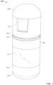
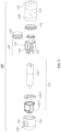
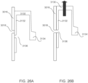
- the ingestible device comprises a housing, a reservoir containing the pharmaceutical formulation, and a release mechanism for releasing the pharmaceutical formulation from the device, wherein the reservoir is releasably or permanently attached to the exterior of the housing or internal to the housing.
- a disease of the gastrointestinal tract in a subject comprising:
- the housing is non-biodegradable in the GI tract.
- the release of the formulation is triggered autonomously.
- the location of one or more sites of disease is predetermined.
- the reservoir is made of a material that allows the formulation to leave the reservoir, such as a biodegradable material.
- the release of the formulation is triggered by a pre-programmed algorithm. In some embodiments, the release of the formulation is triggered by data from a sensor or detector to identify the location of the device. In some more particular embodiments, the data is not based solely on a physiological parameter (such as pH, temperature, and/or transit time).
- a physiological parameter such as pH, temperature, and/or transit time
- the device comprises a detector configured to detect light reflectance from an environment external to the housing.
- the release is triggered autonomously or based on the detected reflectance.
- the device releases the formulation at substantially the same time as one or more sites of disease are detected.
- the one or more sites of disease are detected by the device (e.g., by imaging the GI tract).
- the release mechanism is an actuation system. In some embodiments, the release mechanism is a chemical actuation system. In some embodiments, the release mechanism is a mechanical actuation system. In some embodiments, the release mechanism is an electrical actuation system. In some embodiments, the actuation system comprises a pump and releasing the formulation comprises pumping the formulation out of the reservoir. In some embodiments, the actuation system comprises a gas generating cell.
- the formulation comprises a therapeutically effective amount of the integrin inhibitor. In some embodiments, the formulation comprises a human equivalent dose (HED) of the integrin inhibitor.
- HED human equivalent dose
- the device is a device capable of releasing a solid integrin inhibitor or a solid formulation comprising the integrin inhibitor. In some embodiments, the device is a device capable of releasing a liquid integrin inhibitor or a liquid formulation comprising the integrin inhibitor. Accordingly, in some embodiments, the pharmaceutical formulation released from the device is a solid formulation.
- the pharmaceutical formulation release from the device is a liquid formulation.
- the devices disclosed herein are capable of releasing an integrin inhibitor or a formulation comprising the integrin inhibitor irrespective of the particular type of integrin inhibitor.
- the integrin inhibitor may be a small molecule, a biological, a nucleic acid, an antibody, a fusion protein, and so on.
- a method of releasing an integrin inhibitor into the cecum of a subject for treating one or more sites of disease within the colon comprising: administering to the subject a therapeutically effective amount of the integrin inhibitor housed in an ingestible device, wherein the ingestible device comprises:
- Also provided herein is a method of releasing an integrin inhibitor into the cecum of a subject for treating one or more pre-determined sites of disease within the colon, the method comprising: administering to the subject a therapeutically effective amount of the integrin contained in an ingestible device, wherein the ingestible device comprises:
- a method of releasing an integrin inhibitor into the cecum of a subject for treating one or more sites of disease within the colon comprising:
- a method of releasing an integrin inhibitor into the cecum of a subject for treating one or more sites of disease within the colon comprising:
- a method of treating a disease of the gastrointestinal tract in a subject comprising: releasing an integrin inhibitor in the cecum of the subject, wherein the method comprises administering to the subject a pharmaceutical composition comprising a therapeutically effective amount of the integrin inhibitor.
- a disease of the gastrointestinal tract in a subject comprising:
- a method of treating a disease of the gastrointestinal tract in a subject comprising: releasing an integrin inhibitor in the cecum of the subject, wherein the method comprises administering to the subject a pharmaceutical composition comprising a therapeutically effective amount of the integrin inhibitor, wherein the method provides a concentration of the integrin inhibitor in the plasma of the subject that is less than 3 ⁇ g/ml.
- an integrin inhibitor for use in a method of treating a disease of the gastrointestinal tract in a subject according to claim 1.
- the present disclosure provides a composition comprising or consisting of an ingestible device loaded with a therapeutically effective amount of an integrin inhibitor, for use in a method of treatment, wherein the method comprises orally administering the composition to the subject, wherein the integrin inhibitor is released by the device in the cecum.
- the present disclosure provides an ingestible device loaded with a therapeutically effective amount of an integrin inhibitor, wherein the device is controllable to release the integrin inhibitor in the cecum of the subject .
- the device may be for use in a method of treatment of the human or animal body, for example, any method as described herein.
- the present disclosure provides an ingestible device for use in a method of treating a disease of the gastrointestinal tract in a subject, wherein the method comprises orally administering to the subject the ingestible device loaded with a therapeutically effective amount of an integrin inhibitor, wherein the integrin inhibitor is released by the device in the cecum of the subject.
- An ingestible device as used in the present disclosure may comprise one or more mechanical and/or electrical mechanisms which actively control release of the integrin inhibitor.
- the ingestible device as used in the present invention may comprise a release mechanism for release of the integrin inhibitor (e.g., from a reservoir comprising the integrin inhibitor) and an actuator controlling the release mechanism.
- the ingestible device comprises:
- the ingestible device comprises
- the exit valve can be considered as the release mechanism having a closed state which retains the integrin inhibitor in the reservoir and an open state which releases the integrin inhibitor from the reservoir to the exterior of the device, and the mechanism for releasing the integrin inhibitor from the reservoir can be considered as the actuator.
- the one or more disease sites may have been pre-determined (e.g., determined in a step preceding the administration of the composition of the present disclosure).
- the disease site(s) may have been determined by imaging the gastrointestinal tract.
- the disease site(s) may have been pre-determined by endoscopy (e.g., a step of colonoscopy, enteroscopy, or using a capsule endoscope).
- the location of the device in the gut may be detected by tracking the device.
- the device may comprise a localization mechanism which may be a communication system for transmitting localization data, e.g., by radiofrequency transmission.
- the device may additionally or alternatively comprise a communication system for receiving a signal remotely triggering the actuator and thus causing release of the integrin inhibitor. The signal may be sent when it is determined that the device is in the correct location in the gut.
- the ingestible device may comprise:
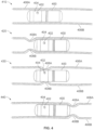
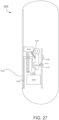
- the ingestible device as used in the present invention may comprise an environmental sensor for detecting the location of the device in the gut .
- the environment sensor may be an image sensor for obtaining images in vivo.
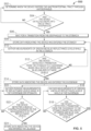
- actuation of the release mechanism may be triggered by a processor or controller communicably coupled to the environmental sensor.
- the device may not require any external signal or control in order to release the drug.
- the ingestible device comprises:
- detection of a transition from the ileum to the cecum may be based on environmental data indicating the location of the device in the GI tract (and reference to a pre-determined disease site).
- the device may further comprise a communication system adapted to transmit the environment data to an external receiver (e.g., outside of the body).
- This data may be used, for example, for diagnostic purposes.
- the external receiver may comprise means for displaying the data.
- an external signal may then be sent to the device to trigger release of the drug.
- the communication system may further be adapted to receive a signal remotely triggering the actuator and thus causing release of the integrin inhibitor.
- the signal may be sent from an external transmitter in response to receipt/analysis and/or assessment of the environmental data, e.g., data indicating that the device has reached the desired location of the gut (where the location of the diseased tissue has been pre-determined) and/or data indicating the presence of diseased tissue.
- “External” may be "outside of the body”.
- the ingestible device may comprise:
- a system comprising:
- disease of the GI tract may be an inflammatory bowel disease.
- the disease of the GI tract is ulcerative colitis.
- the disease of the GI tract is Crohn's disease.
- gastrointestinal tract diseases that can be treated include, without limitation, inflammatory bowel disease (IBD), Crohn's disease (e.g., active Crohn's disease, refractory Crohn's disease, or fistulizing Crohn's disease), ulcerative colitis, indeterminate colitis, microscopic colitis, infectious colitis, drug or chemical-induced colitis, diverticulitis, and ischemic colitis, gastritis, peptic ulcers, stress ulcers, bleeding ulcers, gastric hyperacidity, dyspepsia, gastroparesis, Zollinger-Ellison syndrome, gastroesophageal reflux disease, short-bowel (anastomosis) syndrome, a hypersecretory state associated with systemic mastocytosis or basophilic leukemia or hyperhistaminemia, Celiac disease (e.g., nontropical Sprue), enteropathy associated with IBD
- Crohn's disease e.g., active Crohn's disease, refractory Crohn'
- apparatuses, compositions, and methods disclosed herein are used to treat one gastrointestinal disease. In some embodiments, apparatuses, compositions, and methods disclosed herein are used to treat more than one gastrointestinal disease. In some disclosures, apparatuses, compositions, and methods disclosed herein are used to treat multiple gastrointestinal diseases that occur in the same area of the gastrointestinal tract (the colon, or any sub-region thereof). In some disclosures, apparatuses, compositions, and methods disclosed herein are used to treat multiple gastrointestinal diseases that occur in different areas of the gastrointestinal tract.
- administration e.g., local administration to the gastrointestinal tract
- integrin inhibitor is useful in the treatment of gastrointestinal diseases including, but not limited to, inflammatory bowel disease (IBD), ulcerative colitis, Crohn's disease, or any of the other gastrointestinal diseases described herein.
- IBD inflammatory bowel disease
- ulcerative colitis Crohn's disease
- Crohn's disease or any of the other gastrointestinal diseases described herein.
- any details described herein for methods of treatment apply equally to an integrin inhibitor, composition or ingestible device for use in said treatment.
- Any details described for a device apply equally to methods of treatment using the device, or to an integrin inhibitor or composition for use in a method of treatment involving the device.
- a method of treating a disease of the gastrointestinal tract in a subject comprises administering to the subject a pharmaceutical formulation comprising an integrin inhibitor wherein the pharmaceutical formulation is released in the subject's cecum.
- the pharmaceutical formulation comprises a therapeutically effective amount of an integrin inhibitor.
- the formulation is contained in an ingestible device, and the device releases the formulation in the cecum.
- the location of the site of disease may be predetermined.
- the release of the formulation may be triggered autonomously, as further described herein.
- a "formulation" of an integrin inhibitor may refer to either the integrin inhibitor in pure form, such as, for example, a lyophilized integrin inhibitor, or a mixture of the integrin inhibitor with one or more physiologically acceptable carriers, excipients or stabilizers.
- therapeutic formulations or medicaments can be prepared by mixing the integrin inhibitor having the desired degree of purity with optional physiologically acceptable carriers, excipients or stabilizers ( Remington's Pharmaceutical Sciences 16th edition, Osol, A. Ed. (1980 )), in the form of lyophilized formulations or aqueous solutions.
- Acceptable carriers, excipients, or stabilizers are nontoxic to recipients at the dosages and concentrations employed, and include buffers such as phosphate, citrate, and other organic acids; antioxidants including ascorbic acid and methionine; preservatives (such as octadecyldimethylbenzyl ammonium chloride; hexamethonium chloride; benzalkonium chloride, benzethonium chloride; phenol, butyl or benzyl alcohol; alkyl parabens such as methyl or propyl paraben; catechol; resorcinol; cyclohexanol; 3-pentanol; and m-cresol); low molecular weight (less than about 10 residues) antibody; proteins, such as serum albumin, gelatin, or immunoglobulins; hydrophilic polymers such as polyvinylpyrrolidone; amino acids such as glycine, glutamine, asparagine, histidine, arginine
- Exemplary pharmaceutically acceptable carriers herein further include insterstitial drug dispersion agents such as soluble neutral-active hyaluronidase glycoproteins (sHASEGP), for example, human soluble PH-20 hyaluronidase glycoproteins, such as rHuPH20 (HYLENEX ⁇ ® >, Baxter International, Inc.).
- sHASEGP soluble neutral-active hyaluronidase glycoproteins
- rHuPH20 HYLENEX ⁇ ® >, Baxter International, Inc.
- Certain exemplary sHASEGPs and methods of use, including rHuPH20 are described in US Patent Publication Nos. 2005/0260186 and 2006/0104968 .
- a sHASEGP is combined with one or more additional glycosaminoglycanases such as chondroitinases.
- Exemplary lyophilized formulations are described in US Patent No. 6,267,958 .
- Aqueous formulations include those described in US
- a formulation of an integrin inhibitor as disclosed herein, e.g., sustained-release formulations, can further include a mucoadhesive agent, e.g., one or more of polyvinyl pyrolidine, methyl cellulose, sodium carboxyl methyl cellulose, hydroxyl propyl cellulose, carbopol, a polyacrylate, chitosan, a eudragit analogue, a polymer, and a thiomer.
- a mucoadhesive agent e.g., one or more of polyvinyl pyrolidine, methyl cellulose, sodium carboxyl methyl cellulose, hydroxyl propyl cellulose, carbopol, a polyacrylate, chitosan, a eudragit analogue, a polymer, and a thiomer.
- mucoadhesive agents that can be included in a formulation with an integrin inhibitor are described in, e.g., Peppas et al., Biomaterials 17(16): 1553-1561, 1996 ; Kharenko et al., Pharmaceutical Chemistry J. 43(4):200-208, 2009 ; Salamat-Miller et al., Adv. Drug Deliv. Reviews 57(11):1666-1691, 2005 ; Bernkop-Schnurch, Adv. Drug Deliv. Rev. 57(11):1569-1582, 2005 ; and Harding et al., Biotechnol. Genet. Eng. News 16(1):41-86, 1999 .
- components of a formulation may include any one of the following components, or any combination thereof: Acacia, Alginate, Alginic Acid, Aluminum Acetate, an antiseptic, Benzyl Alcohol, Butyl Paraben, Butylated Hydroxy Toluene, an antioxidant.
- the method comprises administering to the subject a pharmaceutical composition that is a formulation as disclosed herein.
- the formulation is a dosage form, which may be, as an example, a solid form such as, for example, a capsule, a tablet, a sachet, or a lozenge; or which may be, as an example, a liquid form such as, for example, a solution, a suspension, an emulsion, or a syrup.
- the formulation is comprised in an ingestible device as defined in claim 1.
- the formulation may be suitable for oral administration.
- the formulation may be, for example, a solid dosage form or a liquid dosage form as disclosed herein.
- the formulation is suitable for introduction and optionally for storage in the device. In some embodiments the formulation is suitable for introduction and optionally for storage in a reservoir comprised in the device. In some embodiments the formulation is suitable for introduction and optionally for storage in a reservoir comprised in the device.
- a reservoir comprising a therapeutically effective amount of an integrin inhibitor, wherein the reservoir is configured to fit into an ingestible device. In some disclosures, the reservoir comprising a therapeutically effective amount of an integrin inhibitor is attachable to an ingestible device.
- the formulation is suitable for introduction in a spray catheter, as disclosed herein.
- the formulation herein may also contain more than one active compound as necessary for the particular indication being treated, for example, those with complementary activities that do not adversely affect each other.
- the formulation may further comprise another integrin inhibitor or a chemotherapeutic agent.
- Such molecules are suitably present in combination in amounts that are effective for the purpose intended.
- the active ingredients may also be entrapped in microcapsules prepared, for example, by coacervation techniques or by interfacial polymerization, for example, hydroxymethylcellulose or gelatin-microcapsule and poly-(methylmethacylate) microcapsule, respectively, in colloidal drug delivery systems (for example, liposomes, albumin microspheres, microemulsions, nanoparticles and nanocapsules) or in macroemulsions.
- colloidal drug delivery systems for example, liposomes, albumin microspheres, microemulsions, nanoparticles and nanocapsules
- the formulations to be used for in vivo administration must be sterile. This is readily accomplished by filtration through sterile filtration membranes.
- Sustained-release preparations may be prepared. Suitable examples of sustained- release preparations include semipermeable matrices of solid hydrophobic polymers containing the integrin inhibitor, which matrices are in the form of shaped articles, e.g., films, or microcapsule. Examples of sustained-release matrices include polyesters, hydrogels (for example, poly(2-hydroxyethyl-methacrylate), or poly(vinylalcohol)), polylactides ( U.S. Pat. No.
- copolymers of L-glutamic acid and ⁇ ethyl-L-glutamate copolymers of L-glutamic acid and ⁇ ethyl-L-glutamate, non-degradable ethylene-vinyl acetate, degradable lactic acid-glycolic acid copolymers such as the LUPRON DEPOT TM (injectable microspheres composed of lactic acid-glycolic acid copolymer and leuprolide acetate), and poly-D-(-)-3-hydroxybutyric acid. While polymers such as ethylene-vinyl acetate and lactic acid-glycolic acid enable release of molecules for over 100 days, certain hydrogels release proteins for shorter time periods.
- encapsulated integrin inhibitors When encapsulated integrin inhibitors remain in the body for a long time, they may denature or aggregate as a result of exposure to moisture at 37°C, resulting in a loss of biological activity and possible changes in immunogenicity. Rational strategies can be devised for stabilization depending on the mechanism involved. For example, if the aggregation mechanism is discovered to be intermolecular S-S bond formation through thio-disulfide interchange, stabilization may be achieved by modifying sulfhydryl residues, lyophilizing from acidic solutions, controlling moisture content, using appropriate additives, and developing specific polymer matrix compositions.
- compositions may contain one or more integrin inhibitors.
- the pharmaceutical formulations may be formulated in any manner known in the art.
- the formulations include one or more of the following components: a sterile diluent (e.g., sterile water or saline), a fixed oil, polyethylene glycol, glycerin, propylene glycol, or other synthetic solvents, antibacterial or antifungal agents, such as benzyl alcohol or methyl parabens, chlorobutanol, phenol, ascorbic acid, thimerosal, and the like, antioxidants, such as ascorbic acid or sodium bisulfite, chelating agents, such as ethylenediaminetetraacetic acid, buffers, such as acetates, citrates, or phosphates, and isotonic agents, such as sugars (e.g., dextrose), polyalcohols (e.g., mannitol or sorbitol), or salts (e.g
- Liposomal suspensions can also be used as pharmaceutically acceptable carriers (see, e.g., U.S. Patent No. 4,522,811 ).
- the formulations can be formulated and enclosed in ampules, disposable syringes, or multiple dose vials. Where required, proper fluidity can be maintained by, for example, the use of a coating, such as lecithin, or a surfactant.
- Controlled release of the integrin inhibitor can be achieved by implants and microencapsulated delivery systems, which can include biodegradable, biocompatible polymers (e.g., ethylene vinyl acetate, polyanhydrides, polyglycolic acid, collagen, polyorthoesters, and polylactic acid; Alza Corporation and Nova Pharmaceutical, Inc.).
- the integrin inhibitor is present in a pharmaceutical formulation within the device.
- the integrin inhibitor is present in solution within the device.
- the integrin inhibitor is present in a suspension in a liquid medium within the device.
- the integrin inhibitor is present as a pure, powder (e.g., lyophilized) form of the integrin inhibitor.
- Gastrointestinal inflammatory disorders are a group of chronic disorders that cause inflammation and/or ulceration in the mucous membrane. These disorders include, for example, inflammatory bowel disease (e.g., Crohn's disease, ulcerative colitis, indeterminate colitis and infectious colitis), mucositis (e.g., oral mucositis, gastrointestinal mucositis, nasal mucositis and proctitis), necrotizing enterocolitis and esophagitis.
- inflammatory bowel disease e.g., Crohn's disease, ulcerative colitis, indeterminate colitis and infectious colitis
- mucositis e.g., oral mucositis, gastrointestinal mucositis, nasal mucositis and proctitis
- necrotizing enterocolitis and esophagitis necrotizing enterocolitis and esophagitis.
- IBD ulcerative colitis
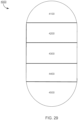
- the GI tract can be divided into four main different sections, the oesophagus, stomach, small intestine and large intestine or colon.
- the small intestine possesses three main subcompartments: the duodenum, jejunum and ileum.
- the large intestine consists of six sections: the cecum, ascending colon, transverse colon, ascending colon, sigmoid colon, and the rectum.
- the small intestine is about 6 m long, its diameter is 2.5 to 3 cm and the transit time through it is typically 3 hours.
- the duodenum has a C-shape, and is 30 cm long.
- jejunum and ileum are sections that can freely move.
- the jejunum is 2.4 m in length and the ileum is 3.6 m in length and their surface areas are 180 m 2 and 280 m 2 respectively.
- the large intestine is 1.5 m long, its diameter is between 6.3 and 6.5 cm, the transit time though this section is 20 hours and has a reduced surface area of approximately 150 m 2 .
- the higher surface area of the small intestine enhances its capacity for systemic drug absorption.
- IBD immunodeficiency virus
- corticosteroids and immunomodulator therapy e.g., azathioprine, 6 mercaptopurine, and methotrexate administered via traditional routes such as tablet form, oral suspension, or intravenously
- azathioprine, 6 mercaptopurine, and methotrexate administered via traditional routes such as tablet form, oral suspension, or intravenously
- steroids monoclonal antibodies targeting tumor necrosis factor alpha (TNF-a), such as infliximab (a chimeric antibody) and adalimumab (a fully human antibody) are currently used in the management of CD.
- TNF-a tumor necrosis factor alpha
- infliximab a chimeric antibody
- adalimumab a fully human antibody
- Infliximab has also shown efficacy and has been approved for use in UC.
- approximately 10%-20% of patients with CD are primary nonresponders to anti TNF therapy, and another ⁇ 20%-30% of CD patients lose response over time ( Schnitzler et al., Gut 58:492-500 (2009 )).
- Other adverse events (AEs) associated with anti TNFs include elevated rates of bacterial infection, including tuberculosis, and, more rarely, lymphoma and demyelination ( Chang et al., Nat Clin Pract Gastroenterol Hepatology 3:220 (2006 ); Hoentjen et al., World J. Gastroenterol. 15(17):2067 (2009 )).
- IBD Inflammatory bowel syndrome
- GI gastrointestinal
- UC ulcerative colitis
- CD Crohn's disease
- CD Crohn's disease
- CD Crohn's disease
- Crohn's disease is the granular, reddish-purple edematous thickening of the bowel wall. With the development of inflammation, these granulomas often lose their circumscribed borders and integrate with the surrounding tissue. Diarrhea and obstruction of the bowel are the predominant clinical features. As with ulcerative colitis, the course of Crohn's disease may be continuous or relapsing, mild or severe, but unlike ulcerative colitis, Crohn's disease is not curable by resection of the involved segment of bowel.
- Crohn's disease may involve any part of the alimentary tract from the mouth to the anus, although typically it appears in the ileocolic, small-intestinal or colonic-anorectal regions. Histopathologically, the disease manifests by discontinuous granulomatomas, crypt abscesses, fissures and aphthous ulcers.
- the inflammatory infiltrate is mixed, consisting of lymphocytes (both T and B cells), plasma cells, macrophages, and neutrophils. There is a disproportionate increase in IgM- and IgG-secreting plasma cells, macrophages and neutrophils.
- CDAI Crohn's Disease Activity Index
- Backward stepwise regression analysis identified eight independent predictors which are the number of liquid or soft stools, severity of abdominal pain, general well-being, occurrence of extra-intestinal symptoms, need for anti-diarrheal drugs, presence of an abdominal mass, hematocrit, and body weight.
- the final score is a composite of these eight items, adjusted using regression coefficients and standardization to construct an overall CDAI score, ranging from 0 to 600 with higher score indicating greater disease activity.
- CDAI ⁇ 150 is defined as clinical remission
- 150 to 219 is defined as mildly active disease
- 220 to 450 is defined as moderately active disease
- above 450 is defined as very severe disease ( Best WR, et al., Gastroenterology 77:843-6, 1979 ).
- Vedolizumab and natalizumab have been approved on the basis of demonstrated clinical remission, i.e. CDAI ⁇ 150.
- CDAI has been in use for over 40 years, and has served as the basis for drug approval, it has several limitations as an outcome measure for clinical trials. For example, most of the overall score comes from the patient diary card items (pain, number of liquid bowel movements, and general well-being), which are vaguely defined and not standardized terms ( Sandler et al., J. Clin. Epidemiol 41 :451-8, 1988 ; Thia et al., Inflamm. Bowel Dis 17: 105-11, 2011 ). In addition, measurement of pain is based on a four-point scale rather than an updated seven-point scale. The remaining 5 index items contribute very little to identifying an efficacy signal and may be a source of measurement noise.
- the PRO2 and PRO3 tools are such adaptations of the CDAI and have been recently described in Khanna et al., Aliment Pharmacol. Ther. 41 : 77-86, 2015 .
- the PRO2 evaluates the frequency of loose/liquid stools and abdominal pain ⁇ Id). These items are derived and weighted accordingly from the CDAI and are the CDAI diary card items, along with general well-being, that contribute most to the observed clinical benefit measured by CDAI ( Sandler et al., J. Clin.
- the remission score of ⁇ 11 is the CDAI-weighted sum of the average stool frequency and pain scores in a 7-day period, which yielded optimum sensitivity and specificity for identification of CDAI remission (score of ⁇ 150) in a retrospective data analysis of ustekinumab induction treatment for moderate to severe CD in a Phase II clinical study ( Gasink C, et al., abstract, ACG Annual Meeting 2014 ).
- the PRO2 was shown to be sensitive and responsive when used as a continuous outcome measure in a retrospective data analysis of MTX treatment in active CD ( Khanna R, et al., Inflamm Bowel Dis 20: 1850-61, 2014 ) measured by CDAI. Additional outcome measures include the Mayo Clinic Score, the Crohn disease endoscopic index of severity (CDEIS), and the Ulcerative colitis endoscopic index of severity (UCEIS). Additional outcome measures include Clinical remission, Mucosal healing, Histological healing (transmural), MRI or ultrasound for measurement or evaluation of bowel wall thickness, abscesses, fistula and histology.
- SES- CD Simplified Endoscopic Activity Score for Crohn's Disease
- the current therapy of IBD usually involves the administration of antiinflammatory or immunosuppressive agents, such as sulfasalazine, corticosteroids, 6- mercaptopurine/azathioprine, or cyclosporine, all of which are not typically delivered by localized release of a drug at the site or location of disease.
- antiinflammatory or immunosuppressive agents such as sulfasalazine, corticosteroids, 6- mercaptopurine/azathioprine, or cyclosporine, all of which are not typically delivered by localized release of a drug at the site or location of disease.
- Refining a diagnosis of inflammatory bowel disease involves evaluating the progression status of the diseases using standard classification criteria.
- the classification systems used in IBD include the Truelove and Witts Index ( Truelove S. C. and Witts, L.J. Br Med J. 1955;2: 1041-1048 ), which classifies colitis as mild, moderate, or severe, as well as Lennard- Jones. ( Lennard-Jones JE. Scand J Gastroenterol Suppl 1989; 170:2-6 ) and the simple clinical colitis activity index (SCCAI). ( Walmsley et. al. Gut. 1998;43:29-32 ) These systems track such variables as daily bowel movements, rectal bleeding, temperature, heart rate, hemoglobin levels, erythrocyte sedimentation rate, weight, hematocrit score, and the level of serum albumin.
- UC ulcerative colitis
- CD can appear anywhere in the bowel, with occasional involvement of stomach, esophagus and duodenum, and the lesions are usually described as extensive linear fissures.
- pANCA perinuclear anti-neutrophil antibody
- ASCA anti-Saccharomyces cervisiae antibody
- a third test which measures the presence and accumulation of circulating anti-microbial antibodies - particularly flagellin antibodies, has proven to be useful for detecting susceptibility to Crohn's Disease before disease development. See Choung, R. S., et al. "Serologic microbial associated markers can predict Crohn's disease behaviour years before disease diagnosis.” Alimentary pharmacology & therapeutics 43.12 (2016): 1300-1310 .
- Ulcerative colitis afflicts the large intestine.
- the course of the disease may be continuous or relapsing, mild or severe.
- the earliest lesion is an inflammatory infiltration with abscess formation at the base of the crypts of Lieberkuhn. Coalescence of these distended and ruptured crypts tends to separate the overlying mucosa from its blood supply, leading to ulceration.
- Symptoms of the disease include cramping, lower abdominal pain, rectal bleeding, and frequent, loose discharges consisting mainly of blood, pus and mucus with scanty fecal particles.
- a total colectomy may be required for acute, severe or chronic, unremitting ulcerative colitis.
- UC ulcerative colitis
- antibody and “immunoglobulin” are used interchangeably in the broadest sense and include monoclonal antibodies (for example, full length or intact monoclonal antibodies), polyclonal antibodies, multivalent antibodies, multispecific antibodies (e.g., bispecific, trispecific etc. antibodies so long as they exhibit the desired biological activity) and may also include certain antibody fragments (as described in greater detail herein).
- An antibody can be human, humanized and/or affinity matured.
- Antibody fragments comprise only a portion of an intact antibody, where in certain embodiments, the portion retains at least one, and typically most or all, of the functions normally associated with that portion when present in an intact antibody.
- an antibody fragment comprises an antigen binding site of the intact antibody and thus retains the ability to bind antigen.
- an antibody fragment for example one that comprises the Fc region, retains at least one of the biological functions normally associated with the Fc region when present in an intact antibody, such as FcRn binding, antibody half-life modulation, ADCC function and complement binding.
- an antibody fragment is a monovalent antibody that has an in vivo half-life substantially similar to an intact antibody.
- such an antibody fragment may comprise on antigen binding arm linked to an Fc sequence capable of conferring in vivo stability to the fragment.
- monoclonal antibody refers to an antibody obtained from a population of substantially homogeneous antibodies, i.e., the individual antibodies comprising the population are identical except for possible naturally occurring mutations that may be present in minor amounts. Monoclonal antibodies are highly specific, being directed against a single antigen. Furthermore, in contrast to polyclonal antibody preparations that typically include different antibodies directed against different determinants (epitopes), each monoclonal antibody is directed against a single determinant on the antigen.
- the monoclonal antibodies herein specifically include "chimeric" antibodies in which a portion of the heavy and/or light chain is identical with or homologous to corresponding sequences in antibodies derived from a particular species or belonging to a particular antibody class or subclass, while the remainder of the chain(s) is identical with or homologous to corresponding sequences in antibodies derived from another species or belonging to another antibody class or subclass, as well as fragments of such antibodies, so long as they exhibit the desired biological activity ( U.S. Patent No. 4,816,567 ; and Morrison et al, Proc. Natl. Acad. Sci. USA 81 :6851-6855 (1984 )).
- Treatment regimen refers to a combination of dosage, frequency of administration, or duration of treatment, with or without addition of a second medication.
- Effective treatment regimen refers to a treatment regimen that will offer beneficial response to a patient receiving the treatment.
- Effective amount refers to an amount of drug that offers beneficial response to a patient receiving the treatment.
- an effective amount may be a Human Equivalent Dose (HED).
- Dispensable refers to any substance that may be released from an ingestible device as disclosed herein, or from a component of the device such as a reservoir.
- a dispensable substance may be an integrin inhibitor, and/or a formulation comprising an integrin inhibitor.
- Patient response or “patient responsiveness” can be assessed using any endpoint indicating a benefit to the patient, including, without limitation, (1) inhibition, to some extent, of disease progression, including slowing down and complete arrest; (2) reduction in the number of disease episodes and/or symptoms; (3) reduction in lesional size; (4) inhibition (i.e., reduction, slowing down or complete stopping) of disease cell infiltration into adjacent peripheral organs and/or tissues; (5) inhibition (i.e., reduction, slowing down or complete stopping) of disease spread; (6) decrease of auto-immune response, which may, but does not have to, result in the regression or ablation of the disease lesion; (7) relief, to some extent, of one or more symptoms associated with the disorder; (8) increase in the length of disease-free presentation following treatment; and/or (9) decreased mortality at a given point of time following treatment.
- responsiveness refers to a measurable response, including complete response (CR) and partial response (PR).
- Partial response refers to a decrease of at least 50% in the severity of inflammation, in response to treatment.
- a "beneficial response" of a patient to treatment with a therapeutic agent and similar wording refers to the clinical or therapeutic benefit imparted to a patient at risk for or suffering from a gastrointestinal inflammatory disorder from or as a result of the treatment with the agent.
- Such benefit includes cellular or biological responses, a complete response, a partial response, a stable disease (without progression or relapse), or a response with a later relapse of the patient from or as a result of the treatment with the agent.
- non-response or “lack of response” or similar wording means an absence of a complete response, a partial response, or a beneficial response to treatment with a therapeutic agent.
- a patient maintains responsiveness to a treatment" when the patient's responsiveness does not decrease with time during the course of a treatment.
- a "symptom" of a disease or disorder is any morbid phenomenon or departure from the normal in structure, function, or sensation, experienced by a subject and indicative of disease.
- integrin inhibitor refers to an agent which decreases the expression of one or more integrins and/or decreases the binding of an integrin ligand to one or more integrins that play a role in the recruitment, extravasation, and/or activation of a leukocyte.
- the integrin inhibitor specifically binds to at least a portion of a ligand binding site on a target integrin.
- the integrin inhibitor specifically binds to a target integrin at the same site as an endogenous ligand.
- the integrin inhibitor decreases the level of expression of the target integrin in a mammalian cell.
- the integrin inhibitor specifically binds to an integrin ligand.
- Non-limiting examples of integrins that can be targeted by any of the integrin inhibitors described herein include: ⁇ 2 ⁇ 1 integrin, ⁇ 1 ⁇ 1 integrin, ⁇ 4 ⁇ 7 integrin, integrin ⁇ 4 ⁇ 1 (VLA-4), E-selectin, ICAM-1, ⁇ 5 ⁇ 1 integrin, ⁇ 4 ⁇ 1 integrin, VLA-4, ⁇ 2 ⁇ 1 integrin, ⁇ 5 ⁇ 3 integrin, ⁇ 5 ⁇ 5 integrin, ⁇ IIb ⁇ 3 integrin, VCAM1 and MAdCAM-1.
- a non-limiting example of integrin inhibitor that can decrease the expression and/or activity of ⁇ 4 ⁇ 7 integrin is FTY720.
- a non-limiting example of an integrin inhibitor that specifically targets MAdCAM is PF-547659 (Pfizer).
- Non-limiting examples of an integrin inhibitor that specifically targets ⁇ 4 ⁇ 7 is AJM300 (Ajinomoto), etrolizumab (Genentech), and vedolizumab (Millenium/Takeda).
- the integrin inhibitor is an ⁇ IIb ⁇ 3 integrin inhibitor.
- the ⁇ IIb ⁇ 3 integrin inhibitor is abciximab (ReoPro ® , c7E3; Kononczuk et al., Curr. Drug Targets 16(13):1429-1437, 2015 ; Jiang et al., Appl. Microbiol. Biotechnol. 98(1): 105-114, 2014 ), eptifibatide (Integrilin ® ; Scarborough et al., J. Biol. Chem.
- the integrin inhibitor is an ⁇ L-selective integrin inhibitor. In some embodiments, the integrin inhibitor is a ⁇ 2 integrin inhibitor.
- the integrin inhibitor is an ⁇ 4 integrin (e.g., an ⁇ 4 ⁇ 1 integrin (e.g., Very Late Antigen-4 (VLA-4), CD49d, or CD29)) inhibitor, an ⁇ 4 ⁇ 7 integrin inhibitor.
- an ⁇ 4 integrin e.g., an ⁇ 4 ⁇ 1 integrin (e.g., Very Late Antigen-4 (VLA-4), CD49d, or CD29)
- VLA-4 Very Late Antigen-4
- CD49d CD49d
- CD29 CD29
- the integrin inhibitor targets endothelial VCAM1, fibronectin, mucosal addressin cellular adhesion molecule-1 (MAdCAM-1), vitronectin, tenascin-C, osteopontin (OPN), nephronectin, agiostatin, tissue-type transglutaminase, factor XIII, Von Willebrand factor (VWF), an ADAM protein, an ICAM protein, collagen, e-cadherin, laminin, fibulin-5, or TGF ⁇ .
- the ⁇ 4 integrin inhibitor is natalizumab (Tysabri ® ; Targan et al., Gastroenterology 132(5):1672-1683, 2007 ; Sandborn et al., N. Engl. J. Med. 353(18):1912-1925, 2005 ; Nakamura et al., Intern. Med. 56(2):211-214, 2017 ; and Singh et al., J. Pediatr. Gastroenterol. Nutr. 62(6):863-866, 2016 ).
- the integrin inhibitor is an endogenous integrin inhibitor (e.g., SHARPIN ( Rantala et al., Nat. Cell. Biol. 13(11):1315-1324, 2011 ).
- the integrin inhibitor is an ⁇ v integrin (e.g., an ⁇ 5 ⁇ 1 integrin, an ⁇ 5 ⁇ 3 integrin, an ⁇ 5 ⁇ 5 integrin inhibitor, and/or an ⁇ 5 ⁇ 6 integrin) inhibitor.
- an ⁇ v integrin e.g., an ⁇ 5 ⁇ 1 integrin, an ⁇ 5 ⁇ 3 integrin, an ⁇ 5 ⁇ 5 integrin inhibitor, and/or an ⁇ 5 ⁇ 6 integrin
- the integrin inhibitor is an ⁇ 5 ⁇ 1 integrin inhibitor.
- the integrin inhibitor is a VCAM1 inhibitor. In some embodiments, the VCAM1 inhibitor targets the extracellular domain of tissue factor.
- an integrin inhibitor is an inhibitory nucleic acid, an antibody or antigen-binding fragment thereof, a fusion protein, an integrin antagonist, a cyclic peptide, a disintegrin, a peptidomimetic, or a small molecule.
- the inhibitory nucleic acid is a small hairpin RNA, a small interfering RNA, an antisense, an aptamer, or a microRNA.
- VCAM1 Vascular Cell Adhesion Molecule 1
- a VCAM1 inhibitory agent is an inhibitory nucleic acid.
- the inhibitory nucleic acid is an antisense nucleic acid, a small interfering RNA, or a microRNA. Examples of aspects of these different inhibitory nucleic acids are described below. Any of the examples of inhibitory nucleic acids that can decrease expression of a VCAM1 in a mammalian cell can be synthesized in vitro.
- inhibitory nucleic acids specifically bind (e.g., hybridize) to an mRNA encoding VCAM1 to treat inflammatory diseases (e.g., chronic inflammation, irritable bowel syndrome (IBS), rheumatoid arthritis, ulcerative colitis, Crohn's Disease, psoriasis, multiple sclerosis, or auto-inflammatory disease).
- inflammatory diseases e.g., chronic inflammation, irritable bowel syndrome (IBS), rheumatoid arthritis, ulcerative colitis, Crohn's Disease, psoriasis, multiple sclerosis, or auto-inflammatory disease.
- Inhibitory nucleic acids that can decrease the expression of VCAM1 expression in a mammalian cell include antisense nucleic acid molecules, i.e., nucleic acid molecules whose nucleotide sequence is complementary to all or part of VCAM1 mRNA (e.g., complementary to all or a part of any one of SEQ ID NOs: 28-30).
- An antisense nucleic acid molecule can be complementary to all or part of a non-coding region of the coding strand of a nucleotide sequence encoding a VCAM1 protein.
- Non-coding regions (5' and 3' untranslated regions) are the 5' and 3' sequences that flank the coding region in a gene and are not translated into amino acids.
- the VCAM1 antisense nucleic acid comprises GCCTGGGAGGGTATTCAGCTC (SEQ ID NO: 31). In some embodiments, the VCAM1 antisense nucleic acid comprises AACCCTTATTTGTGTCCCACC (SEQ ID NO: 32). In some embodiments, the VCAM1 antisense nucleic acid comprises CCCAGGCATTTTAAGTTGCTG (SEQ ID NO: 33). In some embodiments, the VCAM1 antisense nucleic acid comprises CAC GAGGCCACCACTCATCTC (SEQ ID NO: 34). In some embodiments, the VCAM1 antisense nucleic acid comprises CTTTGACTTCTTGCTCACAGC (SEQ ID NO: 35).
- the VCAM1 antisense nucleic acid comprises AACTCCTCCAGTTCTCTCATC (SEQ ID NO: 36). In some embodiments, the VCAM1 antisense nucleic acid comprises ACCTGTGTGCCTGGGAGGG (SEQ ID NO: 37). Additional VCAM1 antisense nucleic acid are known in the art, e.g., in US 5,596,090 .
- Antisense nucleic acids targeting a nucleic acid encoding a VCAM1 can be designed using the software available at the Integrated DNA Technologies web site.
- An antisense nucleic acid can be, for example, about 5, 10, 15, 20, 25, 30, 35, 40, 45, or 50 nucleotides or more in length.
- An antisense oligonucleotide can be constructed using chemical synthesis and enzymatic ligation reactions using procedures known in the art.
- an antisense nucleic acid can be chemically synthesized using naturally occurring nucleotides or variously modified nucleotides designed to increase the biological stability of the molecules or to increase the physical stability of the duplex formed between the antisense and sense nucleic acids, e.g., phosphorothioate derivatives and acridine substituted nucleotides can be used.
- modified nucleotides which can be used to generate an antisense nucleic acid include 5-fluorouracil, 5-bromouracil, 5-chlorouracil, 5-iodouracil, hypoxanthine, xanthine, 4-acetylcytosine, 5-(carboxyhydroxylmethyl) uracil, 5-carboxymethylaminomethyl-2-thiouridine, 5-carboxymethylaminomethyluracil, dihydrouracil, beta-D-galactosylqueosine, inosine, N6-isopentenyladenine, 1-methylguanine, 1-methylinosine, 2,2-dimethylguanine, 2-methyladenine, 2-methylguanine, 3-methylcytosine, 5-methylcytosine, N6-adenine, 7-methylguanine, 5-methylaminomethyluracil, 5-methoxyaminomethyl-2-thiouracil, beta-D-mannosylqueosine, 5'-methoxycar
- the antisense nucleic acid can be produced biologically using an expression vector into which a nucleic acid has been subcloned in an antisense orientation (i.e., RNA transcribed from the inserted nucleic acid will be of an antisense orientation to a target nucleic acid of interest).
- the antisense nucleic acid comprises a 2'O-methoxyethyl nucleotide. See, e.g., Rijcken et al., Gut 51: 529-535, 2002 ).
- the antisense nucleic acid molecules described herein can be prepared in vitro and administered to a mammal, e.g., a human. Alternatively, they can be generated in situ such that they hybridize with or bind to cellular mRNA and/or genomic DNA encoding a VCAM1 protein to thereby inhibit expression, e.g., by inhibiting transcription and/or translation.
- the hybridization can be by conventional nucleotide complementarities to form a stable duplex, or, for example, in the case of an antisense nucleic acid molecule that binds to DNA duplexes, through specific interactions in the major groove of the double helix.
- the antisense nucleic acid molecules can be delivered to a mammalian cell using a vector (e.g., a lentivirus, a retrovirus, or an adenovirus vector).
- An antisense nucleic acid can be an ⁇ -anomeric nucleic acid molecule.
- An ⁇ -anomeric nucleic acid molecule forms specific double-stranded hybrids with complementary RNA in which, contrary to the usual, ⁇ -units, the strands run parallel to each other ( Gaultier et al., Nucleic Acids Res. 15:6625-6641, 1987 ).
- the antisense nucleic acid can also comprise a 2'-O-methylribonucleotide ( Inoue et al., Nucleic Acids Res. 15:6131-6148, 1987 ) or a chimeric RNA-DNA analog ( Inoue et al., FEBS Lett. 215:327-330, 1987 ).
- an inhibitory nucleic acid is a ribozyme that has specificity for a nucleic acid encoding a VCAM1 protein (e.g., specificity for a VCAM1 mRNA, e.g., specificity for SEQ ID NO: 28, 29, or 30).
- Ribozymes are catalytic RNA molecules with ribonuclease activity that are capable of cleaving a single-stranded nucleic acid, such as an mRNA, to which they have a complementary region.
- ribozymes e.g., hammerhead ribozymes (described in Haselhoff and Gerlach, Nature 334:585-591, 1988 )
- ribozymes can be used to catalytically cleave mRNA transcripts to thereby inhibit translation of the protein encoded by the mRNA.
- a ribozyme having specificity for a VCAM1 mRNA can be designed based upon the nucleotide sequence of any of the VCAM1 mRNA sequences disclosed herein.
- a derivative of a Tetrahymena L-19 IVS RNA can be constructed in which the nucleotide sequence of the active site is complementary to the nucleotide sequence to be cleaved in a VCAM1 mRNA (see, e.g., U.S. Patent. Nos. 4,987,071 and 5,116,742 ).
- a VCAM1 mRNA can be used to select a catalytic RNA having a specific ribonuclease activity from a pool of RNA molecules. See, e.g., Bartel et al., Science 261:1411-1418, 1993 .
- An inhibitor nucleic acid can also be a nucleic acid molecule that forms triple helical structures.
- expression of a VCAM1 polypeptide can be inhibited by targeting nucleotide sequences complementary to the regulatory region of the gene encoding the VCAM1 polypeptide (e.g., the promoter and/or enhancer, e.g., a sequence that is at least 1 kb, 2 kb, 3 kb, 4 kb, or 5 kb upstream of the transcription initiation start state) to form triple helical structures that prevent transcription of the gene in target cells.
- the promoter and/or enhancer e.g., a sequence that is at least 1 kb, 2 kb, 3 kb, 4 kb, or 5 kb upstream of the transcription initiation start state
- inhibitory nucleic acids can be modified at the base moiety, sugar moiety, or phosphate backbone to improve, e.g., the stability, hybridization, or solubility of the molecule.
- the deoxyribose phosphate backbone of the nucleic acids can be modified to generate peptide nucleic acids (see, e.g., Hyrup et al., Bioorganic Medicinal Chem. 4(1):5-23, 1996 ).
- Peptide nucleic acids PNAs are nucleic acid mimics, e.g., DNA mimics, in which the deoxyribose phosphate backbone is replaced by a pseudopeptide backbone and only the four natural nucleobases are retained.
- PNAs The neutral backbone of PNAs allows for specific hybridization to DNA and RNA under conditions of low ionic strength.
- the synthesis of PNA oligomers can be performed using standard solid phase peptide synthesis protocols (see, e.g., Perry-O'Keefe et al., Proc. Natl. Acad. Sci. U.S.A. 93:14670-675, 1996 ).
- PNAs can be used as antisense or antigene agents for sequence-specific modulation of gene expression by, e.g., inducing transcription or translation arrest or inhibiting replication.
- PNAs can be modified, e.g., to enhance their stability or cellular uptake, by attaching lipophilic or other helper groups to PNA, by the formation of PNA-DNA chimeras, or by the use of liposomes or other techniques of drug delivery known in the art.
- PNA-DNA chimeras can be generated which may combine the advantageous properties of PNA and DNA.
- Such chimeras allow DNA recognition enzymes, e.g., RNAse H and DNA polymerases, to interact with the DNA portion while the PNA portion would provide high binding affinity and specificity.
- PNA-DNA chimeras can be linked using linkers of appropriate lengths selected in terms of base stacking, number of bonds between the nucleobases, and orientation.
- PNA-DNA chimeras can be performed as described in Finn et al., Nucleic Acids Res. 24:3357-63, 1996 .
- a DNA chain can be synthesized on a solid support using standard phosphoramidite coupling chemistry and modified nucleoside analogs.
- Compounds such as 5'-(4-methoxytrityl)amino-5'-deoxy-thymidine phosphoramidite can be used as a link between the PNA and the 5' end of DNA ( Mag et al., Nucleic Acids Res. 17:5973-88, 1989 ).
- PNA monomers are then coupled in a stepwise manner to produce a chimeric molecule with a 5' PNA segment and a 3' DNA segment ( Finn et al., Nucleic Acids Res. 24:3357-63, 1996 ).
- chimeric molecules can be synthesized with a 5' DNA segment and a 3' PNA segment ( Peterser et al., Bioorganic Med. Chem. Lett. 5:1119-11124, 1975 ).
- the inhibitory nucleic acids can include other appended groups such as peptides, or agents facilitating transport across the cell membrane (see, Letsinger et al., Proc. Natl. Acad. Sci. U.S.A. 86:6553-6556, 1989 ; Lemaitre et al., Proc. Natl. Acad. Sci. U.S.A. 84:648-652, 1989 ; and WO 88/09810 ).
- other appended groups such as peptides, or agents facilitating transport across the cell membrane
- inhibitory nucleic acids can be modified with hybridization-triggered cleavage agents (see, e.g., Krol et al., Bio/Techniques 6:958-976, 1988 ) or intercalating agents (see, e.g., Zon, Pharm. Res. 5:539-549, 1988 ).
- the oligonucleotide may be conjugated to another molecule, e.g., a peptide, hybridization triggered cross-linking agent, transport agent, hybridization-triggered cleavage agent, etc.
- RNAi RNA interference
- dsRNA double-stranded RNA
- siRNAs short interfering RNAs
- the RISC targets the homologous transcript by base pairing interactions between one of the siRNA strands and the endogenous mRNA. It then cleaves the mRNA about 12 nucleotides from the 3' terminus of the siRNA (see Sharp et al., Genes Dev. 15:485-490, 2001 , and Hammond et al., Nature Rev. Gen. 2:110-119, 2001 ).
- RNA-mediated gene silencing can be induced in a mammalian cell in many ways, e.g., by enforcing endogenous expression of RNA hairpins (see, Paddison et al., Proc. Natl. Acad. Sci. U.S.A. 99:1443-1448, 2002 ) or, as noted above, by transfection of small (21-23 nt) dsRNA (reviewed in Caplen, Trends Biotech. 20:49-51, 2002 ). Methods for modulating gene expression with RNAi are described, e.g., in U.S. Patent No. 6,506,559 and US 2003/0056235A1 .
- Standard molecular biology techniques can be used to generate siRNAs.
- Short interfering RNAs can be chemically synthesized, recombinantly produced, e.g., by expressing RNA from a template DNA, such as a plasmid, or obtained from commercial vendors, such as Dharmacon.
- the RNA used to mediate RNAi can include synthetic or modified nucleotides, such as phosphorothioate nucleotides.
- the siRNA molecules used to decrease expression of a VCAM1 mRNA can vary in a number of ways. For example, they can include a 3' hydroxyl group and strands of 21, 22, or 23 consecutive nucleotides. They can be blunt ended or include an overhanging end at either the 3' end, the 5' end, or both ends.
- at least one strand of the RNA molecule can have a 3' overhang from about 1 to about 6 nucleotides (e.g., 1-5, 1-3, 2-4 or 3-5 nucleotides (whether pyrimidine or purine nucleotides) in length. Where both strands include an overhang, the length of the overhangs may be the same or different for each strand.
- the 3' overhangs can be stabilized against degradation (by, e.g., including purine nucleotides, such as adenosine or guanosine nucleotides or replacing pyrimidine nucleotides by modified analogues (e.g., substitution of uridine 2-nucleotide 3' overhangs by 2'-deoxythymidine is tolerated and does not affect the efficiency of RNAi).
- purine nucleotides such as adenosine or guanosine nucleotides
- pyrimidine nucleotides by modified analogues (e.g., substitution of uridine 2-nucleotide 3' overhangs by 2'-deoxythymidine is tolerated and does not affect the efficiency of RNAi).
- siRNA can be used in the methods of decreasing VCAM1 mRNA, provided it has sufficient homology to the target of interest (e.g., a sequence present in any one of SEQ ID NOs: 28-30, e.g., a target sequence encompassing the translation start site or the first exon of the mRNA).
- the target of interest e.g., a sequence present in any one of SEQ ID NOs: 28-30, e.g., a target sequence encompassing the translation start site or the first exon of the mRNA.
- the siRNA can range from about 21 base pairs of the gene to the full length of the gene or more (e.g., about 20 to about 30 base pairs, about 50 to about 60 base pairs, about 60 to about 70 base pairs, about 70 to about 80 base pairs, about 80 to about 90 base pairs, or about 90 to about 100 base pairs).
- Non-limiting examples of short interfering RNA (siRNA) that target nucleic acid that encodes VCAM1 are described in, e.g., Ho et al., Ann Biomed Eng. 44(4): 895-902, 2016 .
- Inhibitory nucleic acids targeting VCAM1 also include microRNAs (e.g., miR-126 ( Harris et al., PNAS 105(5): 1516-1521, 2008 ; Asgeirsdottir et al., Am J Physiol Renal Physiol 302: F1630-F1639, 2012 ), miR-181b ( Sun et al., J Clin Invest. 122(6): 1973 1990, 2012 ).
- miR-126 Harris et al., PNAS 105(5): 1516-1521, 2008 ; Asgeirsdottir et al., Am J Physiol Renal Physiol 302: F1630-F1639, 2012
- miR-181b Sun et al., J Clin Invest. 122(6): 1973 1990, 2012 ).
- a therapeutically effective amount of an inhibitory nucleic acid targeting VCAM1 can be administered to a subject (e.g., a human subject) in need thereof.
- the inhibitory nucleic acid can be about 10 nucleotides to about 40 nucleotides (e.g., about 10 to about 30 nucleotides, about 10 to about 25 nucleotides, about 10 to about 20 nucleotides, about 10 to about 15 nucleotides, 10 nucleotides, 11 nucleotides, 12 nucleotides, 13 nucleotides, 14 nucleotides, 15 nucleotides, 16 nucleotides, 17 nucleotides, 18 nucleotides, 19 nucleotides, 20 nucleotides, 21 nucleotides, 22 nucleotides, 23 nucleotides, 24 nucleotides, 25 nucleotides, 26 nucleotides, 27 nucleotides, 28 nucleotides, 29 nucleotides, 30 nucleotides, 31 nucleotides, 32 nucleotides, 33 nucleotides, 34 nucleotides, 35 nucleot
- thermal melting point refers to the temperature, under defined ionic strength, pH, and inhibitory nucleic acid concentration, at which 50% of the inhibitory nucleic acids complementary to the target sequence hybridize to the target sequence at equilibrium.
- an inhibitory nucleic acid can bind specifically to a target nucleic acid under stingent conditions, e.g., those in which the salt concentration is at least about 0.01 to 1.0 M Na ion concentration (or other salts) at pH 7.0 to 8.3 and the temperature is at least about 30 °C. for short oligonucleotides (e.g., 10 to 50 nucleotide). Stringent conditions can also be achieved with the addition of destabilizing agents such as formamide.
- the inhibitory nucleic acid binds to a target nucleic acid (e.g., a nucleic acid encodinga VCAM1) with a T m of greater than 20 °C, greater than 22 °C, greater than 24 °C, greater than 26 °C, greater than 28 °C, greater than 30 °C, greater than 32 °C, greater than 34 °C, greater than 36 °C, greater than 38 °C, greater than 40 °C, greater than 42 °C, greater than 44 °C, greater than 46 °C, greater than 48 °C, greater than 50 °C, greater than 52 °C, greater than 54 °C, greater than 56 °C, greater than 58 °C, greater than 60 °C, greater than 62 °C, greater than 64 °C, greater than 66 °C, greater than 68 °C, greater than 70 °C, greater than 72 °C, greater than
- the inhibitory nucleic acid binds to a target nucleic acid (e.g., a nucleic acid encoding a VCAM1) with a T m of about 20 °C to about 80 °C, about 78 °C, about 76 °C, about 74 °C, about 72 °C, about 70 °C, about 68 °C, about 66 °C, about 64 °C, about 62 °C, about 60 °C, about 58 °C, about 56 °C, about 54 °C, about 52 °C, about 50 °C, about 48 °C, about 46 °C, about 44 °C, about 42 °C, about 40 °C, about 38 °C, about 36 °C, about 34 °C, about 32 °C, about 30 °C, about 28 °C, about 26 °C, about 24 °C, or about 22 °C (inclusive
- a target nucleic acid e.g
- the inhibitory nucleic acid can be formulated in a nanoparticle (e.g., a nanoparticle including one or more synthetic polymers, e.g., Patil et al., Pharmaceutical Nanotechnol. 367:195-203, 2009 ; Yang et al., ACSAppl. Mater. Interfaces, doi: 10.1021/acsami.6b16556, 2017 ; Perepelyuk et al., Mol. Ther. Nucleic Acids 6:259-268, 2017 ).
- the nanoparticle can be a mucoadhesive particle (e.g., nanoparticles having a positively-charged exterior surface) ( Andersen et al., Methods Mol. Biol. 555:77-86, 2009 ).
- the nanoparticle can have a neutrally-charged exterior surface.
- the inhibitory nucleic acid can be formulated, e.g., as a liposome ( Buyens et al., J. Control Release 158(3): 362-370, 2012 ; Scarabel et al., Expert Opin. Drug Deliv.
- a pharmaceutical composition can include a sterile saline solution and one or more inhibitory nucleic acid (e.g., any of the inhibitory nucleic acids described herein).
- a pharmaceutical composition consists of a sterile saline solution and one or more inhibitory nucleic acid (e.g., any of the inhibitory nucleic acids described herein).
- the sterile saline is a pharmaceutical grade saline.
- a pharmaceutical composition can include one or more inhibitory nucleic acid (e.g., any of the inhibitory nucleic acids described herein) and sterile water.
- a pharmaceutical composition consists of one or more inhibitory nucleic acid (e.g., any of the inhibitory nucleic acids described herein) and sterile water.
- a pharmaceutical composition includes one or more inhibitory nucleic acid (e.g., any of the inhibitory nucleic acids described herein) and phosphate-buffered saline (PBS).
- a pharmaceutical composition consists of one or more inhibitory nucleic acids (e.g., any of the inhibitory nucleic acids described herein) and sterile phosphate-buffered saline (PBS).
- the sterile saline is a pharmaceutical grade PBS.
- one or more inhibitory nucleic acids may be admixed with pharmaceutically acceptable active and/or inert substances for the preparation of pharmaceutical compositions or formulations.
- Compositions and methods for the formulation of pharmaceutical compositions depend on a number of criteria, including, but not limited to, route of administration, extent of disease, or dose to be administered.
- compositions including one or more inhibitory nucleic acids encompass any pharmaceutically acceptable salts, esters, or salts of such esters.
- Non-limiting examples of pharmaceutical compositions include pharmaceutically acceptable salts of inhibitory nucleic acids.
- Suitable pharmaceutically acceptable salts include, but are not limited to, sodium and potassium salts.
- prodrugs that can include additional nucleosides at one or both ends of an inhibitory nucleic acid which are cleaved by endogenous nucleases within the body, to form the active inhibitory nucleic acid.
- Lipid moieties can be used to formulate an inhibitory nucleic acid.
- the inhibitory nucleic acid is introduced into preformed liposomes or lipoplexes made of mixtures of cationic lipids and neutral lipids.
- inhibitory nucleic acid complexes with mono- or poly-cationic lipids are formed without the presence of a neutral lipid.
- a lipid moiety is selected to increase distribution of an inhibitory nucleic acid to a particular cell or tissue in a mammal.
- a lipid moiety is selected to increase distribution of an inhibitory nucleic acid to fat tissue in a mammal.
- a lipid moiety is selected to increase distribution of an inhibitory nucleic acid to muscle tissue.
- compositions provided herein comprise one or more inhibitory nucleic acid and one or more excipients.
- excipients are selected from water, salt solutions, alcohol, polyethylene glycols, gelatin, lactose, amylase, magnesium stearate, talc, silicic acid, viscous paraffin, hydroxymethylcellulose and polyvinylpyrrolidone.
- a pharmaceutical composition provided herein includes liposomes and emulsions. Liposomes and emulsions can be used to formulate hydrophobic compounds. In some examples, certain organic solvents such as dimethylsulfoxide are used.
- a pharmaceutical composition provided herein includes one or more tissue-specific delivery molecules designed to deliver one or more inhibitory nucleic acids to specific tissues or cell types in a mammal.
- a pharmaceutical composition can include liposomes coated with a tissue-specific antibody.
- a pharmaceutical composition provided herein can include a co-solvent system.
- co-solvent systems include benzyl alcohol, a nonpolar surfactant, a water-miscible organic polymer, and an aqueous phase.
- VPD co-solvent system is a solution of absolute ethanol comprising 3% w/v benzyl alcohol, 8% w/v of the nonpolar surfactant Polysorbate 80 TM and 65% w/v polyethylene glycol 300.
- polyethylene glycol may be varied; other biocompatible polymers may replace polyethylene glycol, e.g., polyvinyl pyrrolidone; and other sugars or polysaccharides may substitute for dextrose.
- a pharmaceutical composition can be formulated for oral administration. In some examples, pharmaceutical compositions are formulated for buccal administration.
- a pharmaceutical composition is formulated for administration by injection (e.g., intravenous, subcutaneous, intramuscular, etc.).
- a pharmaceutical composition includes a carrier and is formulated in aqueous solution, such as water or physiologically compatible buffers such as Hanks's solution, Ringer's solution, or physiological saline buffer.
- other ingredients are included (e.g., ingredients that aid in solubility or serve as preservatives).
- injectable suspensions are prepared using appropriate liquid carriers, suspending agents, and the like.
- compositions for injection are suspensions, solutions, or emulsions in oily or aqueous vehicles, and may contain formulatory agents such as suspending, stabilizing, and/or dispersing agents.
- Solvents suitable for use in pharmaceutical compositions for injection include, but are not limited to, lipophilic solvents and fatty oils, such as sesame oil, synthetic fatty acid esters, such as ethyl oleate or triglycerides, and liposomes.
- the VCAM1 inhibitor is an antibody or an antigen-binding fragment thereof (e.g., a Fab or a scFv).
- the antibody can be a humanized antibody, a chimeric antibody, a multivalent antibody, or a fragment thereof.
- an antibody can be a scFv-Fc, a V H H domain, a V NAR domain, a (scFv) 2 , a minibody, or a BiTE.
- an antibody can be a DVD-Ig, and a dual-affinity re-targeting antibody (DART), a triomab, kih IgG with a common LC, a crossmab, an ortho-Fab IgG, a 2-in-1-IgG, IgG-ScFv, scFv 2 -Fc, a bi-nanobody, tanden antibody, a DART-Fc, a scFv-HAS-scFv, DNL-Fab3, DAF (two-in-one or four-in-one), DutaMab, DT-IgG, knobs-in-holes common LC, knobs-in-holes assembly, charge pair antibody, Fab-arm exchange antibody, SEEDbody, Triomab, LUZ-Y, Fcab, k ⁇ -body, orthogonal Fab, DVD-IgG, IgG(H)-scFv, scF
- DART
- Non-limiting examples of an antigen-binding fragment of an antibody include an Fv fragment, a Fab fragment, a F(ab') 2 fragment, and a Fab' fragment.
- Additional examples of an antigen-binding fragment of an antibody is an antigen-binding fragment of an IgG (e.g., an antigen-binding fragment of IgG1, IgG2, IgG3, or IgG4) (e.g., an antigen-binding fragment of a human or humanized IgG, e.g., human or humanized IgG1, IgG2, IgG3, or IgG4); an antigen-binding fragment of an IgA (e.g., an antigen-binding fragment of IgA1 or IgA2) (e.g., an antigen-binding fragment of a human or humanized IgA, e.g., a human or humanized IgA1 or IgA2); an antigen-binding fragment of an IgD (e.g
- any of the antibodies or antigen-binding fragments thereof described herein can bind to any of the VCAM1s described herein or any of the VCAM1 ligands described herein.
- the VCAM1 antibody is a monoclonal antibody.
- the antibody can be a Fab fragment of a monoclonal chimeric mouse-human antibody, or a variant thereof.
- the VCAM1 antibody is a humanized monoclonal antibody.
- Non-limiting examples of human VCAM1 antibodies are V6 and V7 described in Park et al., Atherosclerosis 226(2):356-363, 2013 .
- the antibody comprises or consists of an antigen-binding fragment of MK1.91 ( Soriano et al., Laboratory Investigation 80(10): 1541, 2000 ).
- the inhibitor is one of the following: Common Name Brand name Company vedolizumab (MLN-00002, MLN02) Entyvio Takeda natalizumab Tysabri Biogen and Elan natalizumab biosimilar BioXpress Therapeutics; Harvest Moon Pharmaceuticals, Etrolizumab Genentech (Roche) PF-00547659 (SHP647) licensed from Pfizer to Shire abrilumab (AMG 181; MEDI-7183) Amgen and AZ SAN-300 (hAQC2) Biogen > Salix > Valeant abciximab (ReoPro, c7E3) Janssen efalizumab Raptiva ® Genetech (Roche) STX-100 Stromedix ® Biogen rovelizumab LeukArrest and Hu23F2G Icos PTG-100 TBD Protagonist AJM300 TBD Ajinomoto Pharmaceuticals abciximab Reopro TM vitaxin (MLN-0000
- the inhibitor is:
- any of the antibodies or antigen-binding fragments described herein has a dissociation constant (K D ) of less than 1 ⁇ 10 -5 M (e.g., less than 0.5 ⁇ 10 -5 M, less than 1 ⁇ 10 -6 M, less than 0.5 ⁇ 10 -6 M, less than 1 ⁇ 10 -7 M, less than 0.5 ⁇ 10 -7 M, less than 1 ⁇ 10 -8 M, less than 0.5 ⁇ 10 -8 M, less than 1 ⁇ 10 -9 M, less than 0.5 ⁇ 10 -9 M, less than 1 ⁇ 10 -10 M, less than 0.5 ⁇ 10 -10 M, less than 1 ⁇ 10 -11 M, less than 0.5 ⁇ 10 -11 M, or less than 1 ⁇ 10 -12 M), e.g., as measured in phosphate buffered saline using surface plasmon resonance (SPR).
- SPR surface plasmon resonance
- any of the antibodies or antigen-binding fragments described herein has a K D of about 1 ⁇ 10 -12 M to about 1 ⁇ 10 -5 M, about 0.5 ⁇ 10 -5 M, about 1 ⁇ 10 -6 M, about 0.5 ⁇ 10 -6 M, about 1 ⁇ 10 -7 M, about 0.5 ⁇ 10 -7 M, about 1 ⁇ 10 -8 M, about 0.5 ⁇ 10 -8 M, about 1 ⁇ 10 -9 M, about 0.5 ⁇ 10 -9 M, about 1 ⁇ 10 -10 M, about 0.5 ⁇ 10 -10 M, about 1 ⁇ 10 -11 M, or about 0.5 ⁇ 10 -11 M (inclusive); about 0.5 ⁇ 10 -11 M to about 1 ⁇ 10 -5 M, about 0.5 ⁇ 10 -5 M, about 1 ⁇ 10 -6 M, about 0.5 ⁇ 10 -6 M, about 1 ⁇ 10 -7 M, about 0.5 ⁇ 10 -7 M, about 1 ⁇ 10 -8 M, about 0.5 ⁇ 10 -12 M to about
- any of the antibodies or antigen-binding fragments described herein has a K off of about 1 ⁇ 10 -6 s -1 to about 1 ⁇ 10 -3 s -1 , about 0.5 ⁇ 10 -3 s -1 , about 1 ⁇ 10 -4 s -1 , about 0.5 ⁇ 10 -4 s -1 , about 1 ⁇ 10 -5 s -1 , or about 0.5 ⁇ 10 -5 s -1 (inclusive); about 0.5 ⁇ 10 -5 s -1 to about 1 ⁇ 10 -3 s , about 0.5 ⁇ 10 -3 s -1 , about 1 ⁇ 10 -4 s -1 , about 0.5 ⁇ 10 -4 s -1 , or about 1 ⁇ 10 -5 s -1 (inclusive); about 1 ⁇ 10 -5 s -1 to about 1 ⁇ 10 -3 s -1 , about 0.5 ⁇ 10 -3 s -1 , about 1 ⁇ 10 -4 s -1 , about 1 ⁇
- any of the antibodies or antigen-binding fragments described herein has a K on of about 1 ⁇ 10 2 M -1 s -1 to about 1 ⁇ 10 6 M -1 s -1 , about 0.5 ⁇ 10 6 M -1 s -1 , about 1 ⁇ 10 5 M -1 s -1 , about 0.5 ⁇ 10 5 M -1 s -1 , about 1 ⁇ 10 4 M -1 s -1 , about 0.5 ⁇ 10 4 M -1 s -1 , about 1 ⁇ 10 3 M -1 s , or about 0.5 ⁇ 10 3 M -1 s -1 (inclusive); about 0.5 ⁇ 10 3 M -1 s -1 to about 1 ⁇ 10 6 M -1 s -1 , about 0.5 ⁇ 10 6 M -1 s -1 , about 1 ⁇ 10 5 M -1 s -1 , about 0.5 ⁇ 10 5 M -1 s -1 , about 1 ⁇ 10 4 M -1 s
- the integrin inhibitor is a fusion protein (e.g., an Fc fusion protein of an extracellular domain of an integrin or an integrin receptor), a soluble receptor, (e.g., the extracellular domain of an integrin or an integrin receptor), or a recombinantintegrin binding protein (e.g., an integrin ligand).
- a fusion protein e.g., an Fc fusion protein of an extracellular domain of an integrin or an integrin receptor
- a soluble receptor e.g., the extracellular domain of an integrin or an integrin receptor
- a recombinantintegrin binding protein e.g., an integrin ligand
- the integrin inhibitor is a small molecule.
- the small molecule is a non-peptide small molecule.
- the non-peptide small molecule is a RGD (ArgGlyAsp)-mimetic antagonist (e.g., tirofiban (Aggrastat ® );. Pierro et al., Eur. J. Ophthalmol. 26(4):e74-76, 2016 ; Guan et al., Eur. J. Pharmacol 761:144-152, 2015 .
- the small molecule is ⁇ 4 antagonist (e.g., firategrast ( Miller et al., Lancet Neurol.
- the small molecule is ⁇ 4 ⁇ 1 antagonist (e.g., IVL745 ( Norris et al., J. Allergy Clin. Immunol. 116(4):761-767, 2005 ; Cox et al., Nat. Rev. Drug Discov.
- ⁇ 4 ⁇ 1 antagonist e.g., IVL745 ( Norris et al., J. Allergy Clin. Immunol. 116(4):761-767, 2005 ; Cox et al., Nat. Rev. Drug Discov.
- BIO-1211 Abraham et al., Am. J. Respir. Crit. Care Med. 162:603-611, 2000 ; Ramroodi et al., Immunol. Invest. 44(7):694-712, 2015 ; Lin et al., J. Med. Chem. 42(5):920-934, 1999 ), HMR 1031 ( Diamant et al., Clin. Exp. Allergy 35(8):1080-1087, 2005 ); valategrast (R411) ( Cox et al., Nat. Rev. Drug Discov.
- the small molecule is ⁇ v ⁇ 3 antagonist (e.g., L0000845704, SB273005).
- the small molecule is ⁇ 5 ⁇ 1 antagonist (e.g., JSM6427).
- GLPG0974 Vermeire et al., J.
- the small molecule is MK-0429 ( Pickarksi et al., Oncol. Rep. 33(6):2737-45, 2015 ; Rosenthal et al., Asia Pac J. Clin. Oncol. 6:42-8, 2010 ).
- the small molecule is JSM-6427 or a variant thereof ( Zahn et al., Arch. Ophthalmol.127(10):1329-1335, 2009 ; Stragies et al., J. Med. Chem. 50:3786-94, 2007 ).
- the small molecule targets a ⁇ 2 integrin.
- the small molecule is SAR-118 (SAR1118) or a variant thereof ( Zhong et al., ACS Med. Chem. Lett. 3(3):203-206, 2012 ; Suchard et al., J. Immunol. 184:3917-3926, 2010 ; Yandrapu et al., J. Ocul. Pharmacol. Ther. 29(2):236-248, 2013 ; Semba et al., Am. J. Ophthalmol. 153:1050-60, 2012 ).
- the small molecule is BMS-587101 or a variant thereof ( Suchard et al., J. Immunol.
- the integrin inhibitor is a cyclic peptide.
- the cyclic peptide comprises or consists of an amino acid sequence as set forth in the amino acid sequence of a ligand recognition sequence of an endogenous integrin ligand.
- the cyclic peptide competes for a target integrin ligand binding site with an endogenous integrin ligand.
- the cyclic peptide includes one or more (e.g., 1, 2, 3, 4, 5, 6, 7, 8) D-amino acids.
- the cyclic peptide is a synthetic cyclic peptide.
- the synthetic cyclic peptide is a heptapeptide.
- the synthetic cyclic peptide is eptifabitide (Integrilin TM ) , or a variant thereof.
- the cyclic peptide comprises a heterocyclic nucleic (e.g., a benzodiazepinone, a piperazine, a benzoazepinone, a nitroaryl, an isoxazoline, an indazole, or a phenol; Spalluto et al., Curr. Med. Chem. 12:51-70, 2005 ).
- the cyclic peptide is a macrocycle (see, e.g., Halland et al., ACS Med. Chem. Lett.
- the peptide is ALG-1001 or a variant thereof ( Mathis et al., Retin. Phys. 9:70, 2012 ).
- the cyclic peptide is an imidazolone-phenylalanine derivative, a heteroaryl, hetrocyclic, and aryl derivative, a bicyclic-aromatic amino acid derivative, a cyclohexane-carboxylic acid derivative, a di-aryl substituted urea derivative, a multimeric L-alanine derivative, a L-alanine derivative, or a pyrimidyl-sulfonamide derivative (see, e.g., U.S.
- the integrin inhibitor is a peptidomimetic.
- the peptidomimetic has an integrin-ligand recognition motif (e.g., RGD, KTS, or MLD). See, e.g., Carron et al., Cancer Research 58: 1930-1935, 1998 ; Fanelli et al., Vascular Cell 6:11, 2014 ; and De Marco et al., Curr. Top. Med. Chem. 16(3):343-359, 2016 .
- the peptidomimetic is an RGD(ArgGlyAsp)-based peptide ( US Patent No. 8,809,338 ).
- the RGD-based peptide can be cilengitide or a variant thereof (EMD 12974) ( Mas-Moruno et al., Anticancer Agents Med. Chem. 10:753-768, 2010 ; Reardon et al., Future Oncol. 7(3):339-354, 2011 ; Beekman et al., Clin. Genitourin Cancer 4(4):299-302, 2006 ; SC56631 (e.g., Engleman et al., Am Soc. Clin. Invest.
- the peptidomimetic can be a Lys-Gly-Asp (KGD)-based peptide.
- the peptidomimetic can be vipegitide or a variant thereof ( Momic et al., Drug Design Devel. Therapy 9:291-304, 2015 ).
- the peptidomimetic can be a peptide conjugated with an antimicrobial synthetic peptide. (e.g., ACDCRGDCFC conjugated with (KI,AKLAK)2 ( Ellerby et al., Nat. Med. 5(9):1032-1038, 1999 ). See, e.g., U.S. Patent No. 8,636,977 .
- the integrin inhibitor can be a disintegrin.
- disintegrin refers to a low molecular weight peptide integrin inhibitor derived from a snake venom (e.g., pit viper venom).
- the disintegrin is a RGD(ArgGlyAsp)-, a KTS- or an MLD-based disintegrin.
- Non-limiting examples of disintegrins include accutin, accurhagin-C, albolabrin, alternagin-c, barbourin, basilicin, bitisgabonin-1, bitisgabonin-2, bitistatin, cerastin, cereberin, cumanastatin 1, contortrostatin, cotiarin, crotatroxin, dendroaspin, disba-01, durissin, echistatin, EC3, elegantin, eristicophin, eristostatin, EMS11, EO4, EO5, flavoridin, flavostatin, insularin, jarastatin, jerdonin, jerdostatin, lachesin, lebein (e.g., lebein-1, lebein-2), leberagin-C, lebestatin, lutosin, molossin, obtustatin, ocellatusin, rhodocetin, rho
- a method of treating a disease of the gastrointestinal tract comprises administering to the subject a pharmaceutical formulation wherein the pharmaceutical formulation is delivered to the cecum by one of various methods.
- the pharmaceutical formulation may be delivered via a medical device such as an ingestible device ; the pharmaceutical formulation may be a solid dosage form, a liquid dosage form, a suppository or an enema for rectal administration with different types of release such as sustained or delayed release.
- the pharmaceutical formulation is delivered to the cecum by an ingestible device containing the pharmaceutical formulation .
- Ingestible devices are advantageous over spray catheters in that they are less invasive, thereby allowing for regular dosing more frequently than spray catheters. Another advantage of ingestible devices is the greater ease with which they can access, relative to a catheter, certain sections of the GI tract such as the ascending colon, the cecum, and all portions of the small intestine.
- one or more different mechanisms can be used to determine the location of an ingestible device within the GI tract.
- Various implementations may be used for localization of ingestible devices within the GI tract.
- certain implementations can include one or more electromagnetic sensor coils, magnetic fields, electromagnetic waves, electric potential values, ultrasound positioning systems, gamma scintigraphy techniques or other radio-tracker technology have been described by others.
- imaging can be used to localize, for example, using anatomical landmarks or more complex algorithms for 3D reconstruction based on multiple images.
- Other technologies rely on radio frequency, which relies on sensors placed externally on the body to receive the strength of signals emitted by the capsule.
- Ingestible devices may also be localized based on reflected light in the medium surrounding the device; pH; temperature; time following ingestion; and/or acoustic signals.
- the disclosure provides an ingestible device, as well as related systems and methods that provide for determining the position of the ingestible device within the GI tract of a subject with very high accuracy.
- the ingestible device can autonomously determine its position within the GI tract of the subject.
- the ingestible device includes one or more processing devices, and one more machine readable hardware storage devices.
- the one or more machine readable hardware storage devices store instructions that are executable by the one or more processing devices to determine the location of the ingestible device in a portion of a GI tract of the subject.
- the one or more machine readable hardware storage devices store instructions that are executable by the one or more processing devices to transmit data to an external device (e.g., a base station external to the subject, such as a base station carried on an article worn by the subject) capable of implementing the data to determine the location of the device within the GI tract of the subject.
- an external device e.g., a base station external to the subject, such as a base station carried on an article worn by the subject
- the location of the ingestible device within the cecum of the subject can be determined to an accuracy of at least 85%, e.g., at least 90%, at least 95%, at least 97%, at least 98%, at least 99%, 100%.
- Example 13 An exemplary and non-limiting disclosure is provided below in Example 13.
- the term "reflectance” refers to a value derived from light emitted by the device, reflected back to the device, and received by a detector in or on the device. For example, in some cases this refers to light emitted by the device, wherein a portion of the light is reflected by a surface external to the device, and the light is received by a detector located in or on the device.
- an illumination refers to any electromagnetic emission.
- an illumination may be within the range of Infrared Light (IR), the visible spectrum and ultraviolet light (UV), and an illumination may have a majority of its power centered at a particular wavelength in the range of 100nm to 1000nm.
- IR Infrared Light
- UV ultraviolet light
- an illumination may have a majority of its power limited to one of the infrared (750nm-1000nm), red (600nm-750nm), green (495nm-600nm), blue (400nm-495nm), or ultraviolet (100nm-400nm) spectrums.
- a plurality of illuminations with different wavelengths may be used.
- One option is to use green or blue spectrums of light.
- any suitable light having a wavelength that is substantially or approximately within the green or blue spectra defined above, and the localization systems and methods described herein may use any suitable spectra of light.
- an ingestible device 100 which may be used to identify a location within a gastrointestinal (GI) tract.
- the ingestible device 100 may be configured to autonomously determine whether it is located in the stomach, a particular portion of the small intestine such as a duodenum, jejunum, or ileum, or the large intestine by utilizing sensors operating with different wavelengths of light. Additionally, ingestible device 100 may be configured to autonomously determine whether it is located within certain portions of the small intestine or large intestine, such as the duodenum, the jejunum, the cecum, or the colon.
- Ingestible device 100 may have a housing 102 shaped similar to a pill or capsule.
- the housing 102 of ingestible device 100 may have a first end portion 104, and a second end portion 106.
- the first end portion 104 may include a first wall portion 108
- second end portion 106 may include a second wall portion 110.
- the first end portion 104 and second end portion 106 of ingestible device 100 may be manufactured separately, and may be affixed together by a connecting portion 112.
- the ingestible device 100 may include an optically transparent window 114.
- Optically transparent window 114 may be transparent to various types of illumination in the visible spectrum, infrared spectrum, or ultraviolet light spectrum, and ingestible device 100 may have various sensors and illuminators located within the housing 102, and behind the transparent window 114. This may allow ingestible device 100 to be configured to transmit illumination at different wavelengths through transparent window 114 to an environment external to housing 102 of ingestible device 100, and to detect a reflectance from a portion of the illumination that is reflected back through transparent window 114 from the environment external to housing 102. Ingestible device 100 may then use the detected level of reflectance in order to determine a location of ingestible device 100 within a GI tract.
- the optically transparent window 114 may be of any shape and size, and may wrap around the circumference of ingestible device 100.
- ingestible device 100 may have multiple sets of sensors and illuminators positioned at different locations azimuthally behind window 114.
- the ingestible device 100 may optionally include an opening 116 in the second wall portion 110.
- the second wall portion 110 may be configured to rotate around the longitudinal axis of ingestible device 100 (e.g., by means of a suitable motor or other actuator housed within ingestible device 100). This may allow ingestible device 100 to release a substance into the GI tract, through opening 116.
- FIG. 2 shows an exploded view of ingestible device 100.
- the ingestible device 100 may optionally include a rotation assembly 118.
- Optional rotation assembly 118 may include a motor 118-1 driven by a microcontroller (e.g., a microcontroller coupled to printed circuit board 120), a rotation position sensing ring 118-2, and a storage sub-unit 118-3 configured to fit snugly within the second end portion 104.
- the rotation assembly 118 may cause second end portion 104, and opening 116, to rotate relative to the storage sub-unit 118-3.
- the cavity on the side of the storage sub-unit 118-3 may be exposed to the environment external to the housing 102 of ingestible device 100.
- the storage sub-unit 118-3 may be loaded with a medicament or other substance prior to the ingestible device 100 being administered to a subject.
- the medicament or other substance may be released from the ingestible device 100 by aligning opening 116 with the cavity within storage sub-unit 118-3.
- Other ingestible devices capable of releasing substances are discussed in commonly-assigned PCT Application WO/2013/120184filed February 15, 2013 , commonly-assigned U.S.
- Ingestible device 100 may include a printed circuit board (PCB) 120, and a battery 128 configured to power PCB 120.
- PCB 120 may include a programmable microcontroller, and control and memory circuitry for holding and executing firmware or software for coordinating the operation of ingestible device 100, and the various components of ingestible device 100.
- PCB 120 may include memory circuitry for storing data, such as data sets of measurements collected by sensing sub-unit 126, or instructions to be executed by control circuitry to implement a localization process, such as, for example, one or more of the processes, discussed herein, including those discussed below in connection with one or more of the associated flow charts.
- PCB 120 may include a detector 122 and an illuminator 124, which together form sensing sub-unit 126.
- the control circuitry within PCB 120 may include processing units, communication circuitry, or any other suitable type of circuitry for operating ingestible device 100.
- a single detector 122 and a single illuminator 124 forming a single sensing sub-unit 126 are shown. However, it is understood that in some cases there may be multiple sensing sub-units, each with a separate illuminator and detector, within ingestible device 100.
- sensing sub-units there may be several sensing sub-units spaced azimuthally around the circumference of the PCB 120, which may enable ingestible device 100 to transmit illumination and detect reflectances or ambient light in all directions around the circumference of the device.
- the sensing sub-unit 126 may be configured to generate an illumination using illuminator 124, which is directed through the window 114 in a radial direction away from ingestible device 100. This illumination may reflect off of the environment external to ingestible device 100, and the reflected light coming back into ingestible device 100 through window 114 may be detected as a reflectance by detector 122.
- the window 114 may be of any suitable shape and size.
- window 114 may extend around a full circumference of ingestible device 100.
- there may be a plurality of sensing sub-units e.g., similar to sensing sub-unit 126) located at different positions behind the window.
- three sensing sub-units may be positioned behind the window at the same longitudinal location, but spaced 120 degrees apart azimuthally. This may enable ingestible device 100 to transmit illuminations in all directions radially around ingestible device 100, and to measure each of the corresponding reflectances.
- the illuminator 124 may be capable of producing illumination at a variety of different wavelengths in the ultraviolet, infrared, or visible spectrum.
- illuminator 124 may be implemented by using Red-Green-Blue Light-Emitting diode packages (RGB-LED). These types of RGB-LED packages are able to transmit red, blue, or green illumination, or combinations of red, blue, or green illumination.
- detector 122 may be configured to sense reflected light of the same wavelengths as the illumination produced by illuminator 124.
- detector 122 may be configured to detect different reflectances produced by red, blue, or green illumination (e.g., through the use of an appropriately configured photodiode). These detected reflectances may be stored by ingestible device 100 (e.g., within memory circuitry of PCB 120), and may then be used by ingestible device 100 in determining a location of ingestible device 100 within the GI tract (e.g., through the use of process 500 ( FIG. 5 ), process 600 ( FIG. 6 ), or process 900 ( FIG. 9 )).
- ingestible device 100 is intended to be illustrative, and not limiting. It will be understood that modifications to the general shape and structure of the various devices and mechanisms described in relation to FIG. 1 and FIG. 2 may be made without significantly changing the functions and operations of the devices and mechanisms.
- ingestible device 100 may have a housing formed from a single piece of molded plastic, rather than being divided into a first end portion 104 and a second end portion 106.
- the location of window 114 within ingestible device 100 may be moved to some other location, such as the center of ingestible device 100, or to one of the ends of ingestible device 100.
- ingestible device 1-10 may be implemented on any suitable type of ingestible device, provided that the ingestible device is capable of detecting reflectances or levels of illumination in some capacity.
- the ingestible device 100 may be modified to replace detector 122 with an image sensor, and the ingestible device may be configured to measure relative levels of red, blue, or green light by decomposing a recorded image into its individual spectral components.
- Other examples of ingestible devices with localization capabilities which may be utilized in order to implement the systems and methods discussed in relation to FIG. 1-11 , are discussed in co-owned PCT Application WO/2017/049602filed on September 25, 2015 .
- FIG. 3 is a diagram of an ingestible device during an example transit through a gastrointestinal (GI) tract, in accordance with some aspects of the disclosure.
- Ingestible device 300 may include any portion of any other ingestible device discussed in this disclosure (e.g., ingestible device 100 ( FIG. 1 )), and may be any suitable type of ingestible device with localization capabilities.
- ingestible device 300 may be one of an ingestible device 100 without the optional opening 116 ( FIG. 1 ) or optional rotation assembly 118 ( FIG. 2 )).
- the ingestible device 300 may be ingested by a subject, and as ingestible device 300 traverses the GI tract, ingestible device 300 may be configured to determine its location within the GI tract. For example, the movement of ingestible device 300 and the amount of light detected by ingestible device 300 (e.g., via detector 122 ( FIG. 2 )) may vary substantially depending on the location of ingestible device 300 within the GI tract, and ingestible device 300 may be configured to use this information to determine a location of ingestible device 300 within the GI tract.
- ingestible device 300 may detect ambient light from the surrounding environment, or reflectances based on illumination generated by ingestible device 300 (e.g., generated by illuminator 124 ( FIG. 1 )), and use this information to determine a location of ingestible device 300 through processes, such as described herein.
- the current location of ingestible device 300, and the time that ingestible device 300 detected each transition between the various portions of the GI tract, may then be stored by ingestible device 300 (e.g., in memory circuitry of PCB 120 ( FIG. 2 )), and may be used for any suitable purpose.
- ingestible device 300 Shortly after ingestible device 300 is ingested, ingestible device will traverse the esophagus 302, which may connect the subject's mouth to a stomach 306.
- the ingestible device 300 may be configured to determine that it has entered the esophagus portion GI tract by measuring the amount and type of light (e.g., via detector 122 ( FIG. 2 )) in the environment surrounding the ingestible device 300. For instance, ingestible device 300 may detect higher levels of light in the visible spectrum (e.g., via detector 122 ( FIG. 2 )) while outside the subject's body, as compared to the levels of light detected while within the GI tract.
- the ingestible device 300 may have previously stored data (e.g., on memory circuitry of PCB 120 ( FIG. 2 )) indicating a typical level of light detected when outside of the body, and the ingestible device 300 may be configured to determine that entry to the body has occurred when a detected level of light (e.g., detected via detector 122 ( FIG. 2 )) has been reduced beyond a threshold level (e.g., at least a 20-30% reduction) for a sufficient period of time (e.g., 5.0 seconds).
- a detected level of light e.g., detected via detector 122 ( FIG. 2 )
- a threshold level e.g., at least a 20-30% reduction
- a sufficient period of time e.g., 5.0 seconds
- the ingestible device 100 may also be configured to monitor for one or more transitions.
- transitions can include an ileoceacal transition from the ileum to the cecum, a transition from the cecum to the colon, or detect exit from the body (e.g., by measuring reflectances, temperature, or levels of ambient light).
- FIG. 7 is a plot illustrating data collected during an example operation of an ingestible device (e.g., ingestible device 100, 300, or 400), which may be used when determining a location of an ingestible device as it transits through a gastrointestinal (GI) tract, in accordance with some disclosures of the disclosure.
- an ingestible device e.g., ingestible device 100, 300, or 400
- GI gastrointestinal
- plot 700 depicts the ratios of the measured green reflectance levels to the measured blue reflectance levels over time.
- ingestible device 100 may have computed the value for each point in the data set 702 by transmitting green and blue illumination at a given time (e.g., via illuminator 124 ( FIG. 2 )), measuring the resulting green and blue reflectances (e.g., via detector 122 ( FIG. 2 )), calculating the ratio of the resulting reflectances, and storing the ratio in the data set along with a timestamp indicating the time that the reflectances were gathered.
- ingestible device 100 determines that it has reached at least the stomach (e.g., as a result of making a determination similar to the determination discussed in relation to 506 in process 500 ( FIG. 5 )). Ingestible device 100 continues to gather additional measurements of green and blue reflectance levels, and at 706 ingestible device 100 determines that a pyloric transition has occurred from the stomach to the duodenum (e.g., as a result of making a determination similar to the determinations discussed in relation to 616-624 of process 600 ( FIG. 6 )). Notably, the values in data set 702 around 706 jump up precipitously, which is indicative of the higher ratios of measured green reflectance levels to measured blue reflectance levels typical of the duodenum.
- the remainder of the data set 702 depicts the ratios of the measured green reflectance levels to the measured blue reflectance levels throughout the remainder of the GI tract.
- ingestible device 100 has reached the jejunum (e.g., as determined through measurements of muscle contractions, as discussed in relation to FIG. 9 ), and by 710, ingestible device 100 has reached the cecum. It is understood that, in some cases, the overall character and appearance of data set 702 changes within the small intestine (i.e., the duodenum, jejunum, and ileum) versus the cecum.
- the cecum ingestible device 100 may measure a relatively stable ratio of the measured green reflectance levels to the measured blue reflectance levels.
- the ingestible device 100 may be configured to determine transitions from the small intestine to the cecum based on these differences. For example, ingestible device 100 may compare recent windows of data to past windows of data, and detect a transition to the cecum in response to determining that the standard deviation of the ratios in the recent window of data is substantially less than the standard deviation of the ratios in the past window of data.
- FIG. 8 is another plot illustrating data collected during an example operation of an ingestible device, which may be used when determining a location of an ingestible device as it transits through a gastrointestinal (GI) tract, in accordance with some aspects of the disclosure. Similar to FIG. 7, FIG. 8 may be described in connection with the ingestible device 100 for illustrative purposes. However, this is not intended to be limiting, and plot 800 and data set 802 may be typical of data gathered by any device discussed in this application.
- GI gastrointestinal
- ingestible device 100 determines that it has reached at least the stomach (e.g., as a result of making a determination similar to the determination discussed in relation to 506 in process 500 ( FIG. 5 )). Ingestible device 100 continues to gather additional measurements of green and blue reflectance levels (e.g., via sensing sub-unit 126 ( FIG. 2 )), and at 806 ingestible device 100 determines that a pyloric transition has occurred from the stomach to the duodenum (e.g., as a result of making a determination similar to the determinations discussed in relation to 616-624 of process 600 ( FIG. 6 )).
- ingestible device 100 determines that a reverse pyloric transition has occurred from the duodenum back to the stomach at 808 (e.g., as a result of making a determination similar to the determinations discussed in relation to 610-612 of process 600 ( FIG. 6 )).
- ingestible device 100 determines that another pyloric transition has occurred from the stomach to the duodenum, and shortly thereafter ingestible device 100 proceeds onwards to the jejunum, ileum, and cecum.
- the remainder of the data set 802 depicts the ratios of the measured green reflectance levels to the measured blue reflectance levels throughout the remainder of the GI tract.
- ingestible device reaches the transition point between the ileum and the cecum.
- the transition to the cecum is marked by a reduced standard deviation in the ratios of measured green reflectances and measured blue reflectances over time, and ingestible device 100 may be configured to detect a transition to the cecum based on determining that the standard deviation of a recent set of measurements is substantially smaller than the standard deviation of past measurements taken from the j ej unum or ileum.
- the ingestible device may be configured to deliver a dispensable substance that is pre-stored within the ingestible device from the ingestible device into the cecum in response to identifying the change in the location of the ingestible device.
- ingestible device 100 may have a dispensable substance pre-stored within the ingestible device 100 (e.g., within a storage chamber or cavity on optional storage sub-unit 118-3 ( FIG. 2 )), and ingestible device 100 may be configured to dispense the substance into the cecum (e.g., through the use of optional opening 116 and optional rotating assembly 118 ( FIG. 2 )) when the ingestible device 100 detects that the ingestible device 100 is located within the cecum.
- the ingestible device may be configured to perform an action based on the total number of detected muscle contractions.
- ingestible device 100 may be configured to retrieve data indicative of the total number of muscle contractions (e.g., from memory circuitry of PCB 120 ( FIG. 2 )), and compare that to an expected number of muscle contractions in a healthy individual.
- the ingestible device may dispense a substance into the cecum (e.g., through the use of optional opening 116 and optional rotating assembly 118 ( FIG. 2 )).
- ingestible device 100 may be configured to deliver a substance into the cecum (such as a medicament), in response to determining that the detected muscle contractions are inconsistent with a functioning GI tract in a healthy individual.
- the steps and descriptions of the flowcharts of this disclosure are merely illustrative. Any of the steps and descriptions of the flowcharts, , may be modified, omitted, rearranged, performed in alternate orders or in parallel, two or more of the steps may be combined, or any additional steps may be added, without departing from the scope of the present disclosure.
- the ingestible device 100 may calculate the mean and the standard deviation of multiple data sets in parallel (e.g., multiple data sets, each one corresponding to a different wavelength of reflectance or different sensing sub-unit used to detect the reflectance) in order to speed up the overall computation time.
- FIG. 11 is a plot illustrating muscle contractions detected by an ingestible device over time, which may be used when determining a location of an ingestible device as it transits through a gastrointestinal (GI) tract, in accordance with some aspects of the disclosure.
- the ingestible device 100 may be configured to detect muscle contractions, and store data indicative of when each muscle contraction is.
- Plot 1100 depicts the detected muscle contractions 1106 over time, with each muscle contraction being represented by a vertical line reaching from "0" to "1" on the y-axis.
- ingestible device 100 first enters the duodenum. Shortly thereafter, at 1108, ingestible device 100 begins to detect several muscle contractions 1106 in quick succession, which may be indicative of the strong peristaltic waves that form in the jejunum (e.g., jejunum 314 ( FIG. 3 )). Later, around 1110, ingestible device 100 continues to detect intermittent muscle contractions, which may be consistent with an ingestible device 100 within the ileum. Finally, at 1104, ingestible device 100 transitions out of the small intestine, and into the cecum.
- jejunum e.g., jejunum 314 ( FIG. 3 )
- ingestible device 100 detects more frequent muscle contractions in the jejunum portion of the small intestine as compared to the ileum portion of the small intestine, and ingestible device 100 does not measure any muscle contractions after having exited the small intestine.
- the ingestible device 100 may incorporate this information into a localization process.
- ingestible device 100 may be configured to detect a transition from a jejunum to an ileum in response to determining that a frequency of detected muscle contractions (e.g., the number of muscle contractions measured in a given 10-minute window) has fallen below a threshold number.
- ingestible device 100 may be configured to detect a transition from an ileum to a cecum in response to determining that no muscle contractions have been detected for a threshold period of time. It is understood that these examples are intended to be illustrative, and not limiting, and that measurements of muscle contractions may be combined with any of the other processes, systems, or methods discussed in this disclosure.
- FIG. 14 is a flowchart 1400 for an exemplary process for determining a transition of the device from the ileum to the cecum.
- the process involves detecting changes in the reflected optical signal (e.g., red light, blue light, green light, ratio of red light to green light, ratio of red light to blue light, and/or ratio of green light to blue light).
- the process includes detecting changes in the ratio of reflected red light to reflected green light, and also detecting changes in the ratio of reflected green light to reflected blue light.
- the sliding window analysis first and second windows discussed with respect to process 600 is continued.
- Step 1410 includes setting a first threshold in a detected signal, e.g., ratio of detected red light to detected green light, and setting a second threshold for the coefficient of variation for a detected signal, e.g., the coefficient of variation for the ratio of detected green light to detected blue light.
- the first threshold can be set to a fraction (e.g., from 0.5 to 0.9, from 0.6 to 0.8) of the average signal (e.g., ratio of detected red light to detected green light) in the first window, or a fraction (e.g., from 0.4 to 0.8, from 0.5 to 0.7) of the mean difference between the detected signal (e.g., ratio of detected red light to detected green light) in the two windows.
- the second threshold can be set to 0.1 (e.g., 0.05, 0.02).
- Step 1420 includes detecting the signals in the first and second windows that are to be used for comparing to the first and second thresholds.
- Step 1430 includes comparing the detected signals to the first and second thresholds. If the corresponding value is not below the first threshold or the corresponding value is not below the second threshold, then it is determined that the device has not left the ileum and entered the cecum, and the process returns to step 1420. If the corresponding value is below the first threshold and the corresponding value is below the second threshold, then it is determined that the device has left the ileum and entered the cecum, and the proceeds to step 1440.
- Step 1450 includes determining whether it is the first time that that the device was determined to leave the ileum and enter the cecum. If it is the first time that the device was determined to leave the ileum and enter the cecum, then the process proceeds to step 1460. If it is not the first time that the device has left the ileum and entered the cecum, then the process proceeds to step 1470.
- Step 1460 includes setting a reference signal.
- the optical signal e.g., ratio of detected red light to detected green light
- the optical signal e.g., ratio of detected red light to detected green light
- Step 1470 includes determining whether the device may have left the cecum and returned to the ileum.
- the device is determined to have left the cecum and returned to the ileum if the corresponding detected signal (e.g., ratio of detected red light to detected green light) is statistically comparable to the reference signal (determined in step 1460) and the coefficient of variation for the corresponding detected signal (e.g., ratio of detected green light to detected blue light) exceeds the second threshold. If it is determined that the device may have left the cecum and returned to the ileum, the process proceeds to step 1480.
- the corresponding detected signal e.g., ratio of detected red light to detected green light
- the coefficient of variation for the corresponding detected signal e.g., ratio of detected green light to detected blue light
- Step 1480 includes continuing to detect the relevant optical signals for a period of time (e.g., at least one minute, from five minutes to 15 minutes).
- a period of time e.g., at least one minute, from five minutes to 15 minutes.
- Step 1490 includes determining whether the signals determined in step 1480 indicate (using the methodology discussed in step 1470) that the device re-entered the ileum. If the signals indicate that the device re-entered the ileum, the process proceeds to step 1420. If the signals indicate that the device is in the cecum, the process proceeds to step 1492.
- Step 1492 includes continuing to monitor the relevant optical signals for a period of time (e.g., at least 30 minutes, at least one hour, at least two hours).
- a period of time e.g., at least 30 minutes, at least one hour, at least two hours.
- Step 1494 includes determining whether the signals determined in step 1492 indicate (using the methodology discussed in step 1470) that the device re-entered the ileum. If the signals indicate that the device re-entered the ileum, the process proceeds to step 1420. If the signals indicate that the device is in the cecum, the process proceeds to step 1496.
- the process determines that the device is in the cecum.
- the disclosure focuses primarily on a number of different examples of an ingestible device, and methods for determining a location of an ingestible device within a GI tract.
- the possible ingestible devices that may be constructed are not limited to these examples, and variations in the shape and design may be made without significantly changing the functions and operations of the device.
- the possible procedures for determining a location of the ingestible device within the GI tract are not limited to the specific procedures and disclosed(e.g., process 1400 ( FIG. 14 )).
- At least some of the elements of the ingestible device described herein which are implemented via software may be written in a high-level procedural language such as object oriented programming, a scripting language or both. Accordingly, the program code may be written in C, C ++ or any other suitable programming language and may comprise modules or classes, as is known to those skilled in object oriented programming. Alternatively, or in addition, at least some of the elements of the of the ingestible device described herein that are implemented via software may be written in assembly language, machine language or firmware as needed. In either case, the language may be a compiled or an interpreted language.
- At least some of the program code used to implement the ingestible device can be stored on a storage media or on a computer readable medium that is readable by a general or special purpose programmable computing device having a processor, an operating system and the associated hardware and software that is necessary to implement the functionality of at least one of the elements described herein.
- the program code when read by the computing device, configures the computing device to operate in a new, specific and predefined manner in order to perform at least one of the methods described herein.
- the programs associated with the systems, devices, and methods described herein are capable of being distributed in a computer program product comprising a computer readable medium that bears computer usable instructions for one or more processors.
- the medium may be provided in various forms, including non-transitory forms such as, but not limited to, one or more diskettes, compact disks, tapes, chips, and magnetic and electronic storage.
- the medium may be transitory in nature such as, but not limited to, wire-line transmissions, satellite transmissions, internet transmissions (e.g. downloads), media, digital and analog signals, and the like.
- the computer useable instructions may also be in various formats, including compiled and non-compiled code.
- the techniques described above can be implemented using software for execution on a computer.
- the software forms procedures in one or more computer programs that execute on one or more programmed or programmable computer systems (which may be of various architectures such as distributed, client/server, or grid) each including at least one processor, at least one data storage system (including volatile and non-volatile memory and/or storage elements), at least one input device or port, and at least one output device or port.
- programmed or programmable computer systems which may be of various architectures such as distributed, client/server, or grid
- the software may be provided on a storage medium, such as a CD-ROM, readable by a general or special purpose programmable computer or delivered (encoded in a propagated signal) over a communication medium of a network to the computer where it is executed. All of the functions may be performed on a special purpose computer, or using special-purpose hardware, such as coprocessors.
- the software may be implemented in a distributed manner in which different parts of the computation specified by the software are performed by different computers.
- Each such computer program is preferably stored on or downloaded to a storage media or device (e.g., solid state memory or media, or magnetic or optical media) readable by a general or special purpose programmable computer, for configuring and operating the computer when the storage media or device is read by the computer system to perform the procedures described herein.
- a storage media or device e.g., solid state memory or media, or magnetic or optical media
- the inventive system may also be considered to be implemented as a computer-readable storage medium, configured with a computer program, where the storage medium so configured causes a computer system to operate in a specific and predefined manner to perform the functions described herein.
- FIG. 16 provides an example mock-up diagram illustrating aspects of a structure of an ingestible device 1600 for delivering a dispensable substance, such as a formulation of a therapeutic agent described herein, according to some cases described herein.
- the ingestible device 1600 may generally be in the shape of a capsule, a pill or any swallowable form that may be orally consumed by an individual. In this way, the ingestible device 1600 may be ingested by a patient and may be prescribed by healthcare practitioners and patients.
- the ingestible device 1600 includes a housing 1601 that may take a shape similar to a capsule, a pill, and/or the like, which may include two ends 1602a-b.
- the housing 1601 may be designed to withstand the chemical and mechanical environment of the GI tract (e.g., effects of muscle contractile forces and concentrated hydrochloric acid in the stomach).
- the wall of the housing 1601 may have a thickness of 0.5mm-1mm, which is sufficient to sustain an internal explosion (e.g., caused by hydrogen ignition or over pressure inside the housing).
- the housing 1601 may or may not have a pH-sensitive enteric coating to detect or otherwise be sensitive to a pH level of the environment external to the ingestible device.
- the ingestible device 1600 may additionally or alternatively include one more sensors, e.g., temperature sensor, optical sense.
- the housing 1601 may be formed by coupling two enclosure portions together.
- the ingestible device 1600 may include an electronic component within the housing 1600.
- the electronic component may be placed proximally to an end 1602b of the housing, and includes a printed circuit board (PCB), a battery, an optical sensing unit, and/or the like.
- PCB printed circuit board
- the ingestible device 1600 further includes a gas generating cell 1603 that is configured to generate gas and thus cause an internal pressure within the housing 1601.
- the gas generating cell may include or be connected to a separate channel or valve of the ingestible device such that gas may be release through the channel or valve to create a motion to alter the position of the ingestible device within the GI tract. Such gas release can also be used to position the ingestible device relative to the intestinal lining.
- gas may be released through the separate channel or valve to alter the surface orientation of the intestinal tissue prior to delivery of the dispensable substance.
- a traveling plunger 1604 may be placed on top of the gas generating cell 1603 within the housing 1601.
- the traveling plunger 1604 is a membrane that separates the gas generating cell 1603 and a storage reservoir that stores the dispensable substance 1605.
- the traveling plunger 1604 may be a movable piston.
- the traveling plunger 1604 may instead be a flexible membrane such as but not limited to a diaphragm.
- the traveling plunger 1604, which may have the form of a flexible diaphragm may be placed along an axial direction of the housing 1601, instead of being placed on top of the gas generating cell 1603.
- the traveling plunger or the membrane 1604 may move (when the membrane 1604 is a piston) or deform (when the membrane 1604 is a diaphragm) towards a direction of the end 1602a of the housing, when the gas generating cell 1603 generates gas to create an internal pressure that pushes the membrane 1604. In this way, the membrane or traveling plunger 1604 may push the dispensable substance 1605 out of the housing via a dispensing outlet 1607.
- the housing 1601 may include a storage reservoir storing one or more dispensable substances 1605 adjacent to the traveling plunger 1604.
- the dispensable substance 1605 may be a therapeutic or medical agent that may take a form of a powder, a compressed powder, a fluid, a semi-liquid gel, or any other dispensable or deliverable form.
- the delivery of the dispensable substance 1605 may take a form such as but not limited to bolus, semi-bolus, continuous, burst drug delivery, and/or the like.
- a single bolus is delivered to the cecum.
- more than one bolus is released to the cecum.
- the release of more than one bolus is triggered according to a preprogrammed algorithm.
- the release profile is continuous.
- the release profile is time-based.
- the release profile is location-based.
- the amount delivered is based on the severity and/or extent of the disease in the following manner.
- the integrin inhibitor is DATK32.
- the dispensable substance is a small molecule therapeutic that is released in the cecum Small molecules that are administerered by typical oral routes are primarily absorbed in the small intestine, with much lower absorption taking place in the large intestine (outside of the rectum). Accordingly, an ingestible device that is capable of releasing a small molecule selectively in the cecum with resulting low systemic levels (even when high doses are used) is attractive for subjects with inflammatory bowel disease in the large intestine.
- the storage reservoir may include multiple chambers, and each chamber stores a different dispensable substance.
- the different dispensable substances can be released at the same time via the dispensing outlet 1607.
- the multiple chambers may take a form of different layers within the storage reservoir such that the different dispensable substance from each chamber is delivered sequentially in an order.
- each of the multiple chambers is controlled by a separate traveling plunger, which may be propelled by gas generation.
- the electronic component may control the gas generating cell 1603 to generate gas to propel a specific traveling plunger, e.g., via a separate gas generation chamber, etc., to delivery the respective substance.
- the content of the multiple chambers may be mixed or combined prior to release, for example, to activate the drug.
- the ingestible device 1600 may include a dispensing outlet 1607 at one end 1602a of the housing 1601 to direct the dispensable substance 105 out of the housing.
- the dispensing outlet 1607 may include an exit valve, a slit or a hole, a jet injection nozzle with a syringe, and/or the like.
- a pressure relief device 1606 may be placed within the housing 1601, e.g., at the end 1602a of the housing 1601.
- the housing 1601 may include small holes (e.g., with a diameter smaller than 2 mm), e.g., on the side of the housing 1601, or at the end 1602a to facilitate loading the dispensable substance into the storage reservoir.
- small holes e.g., with a diameter smaller than 2 mm
- a feedback control circuit e.g., a feedback resistor, etc.
- a feedback resistor may be added to send feedback from the gas generating cell 1603 to the electronic component such that when the internal pressure reaches a threshold level, the electronic component may control the gas generating cell 1603 to turn off gas generation, or to activate other safety mechanism (e.g., feedback-controlled release valve, etc.).
- an internal pressure sensor may be used to measure the internal pressure within the ingestible device and generate feedback to the feedback control circuit.
- FIG. 17 provides an example diagram illustrating aspects of a mechanism for a gas generating cell 1603 configured to generate a gas to dispense a substance, according to some cases described herein.
- the gas generating cell 1603 generates a gas 1611 which can propel the dispensable substance 1605 out of the dispensing outlet 1607.
- a variable resistor 1608 may be connected to a circuit with the gas generating cell 1603 such that the variable resistor 1608 may be used to control an intensity and/or an amount of gas 1611 (e.g., hydrogen) generated by the cell 1603.
- the gas generating cell 1603 may be a battery form factor cell that is capable of generating hydrogen when a resistor is applied.
- the gas generating cell 1603 may be integrated into an ingestible device such as a capsule with limited energy/power available.
- the gas generating cell 1603 may be compatible with a capsule at a size of 26mm x 13mm or smaller.
- the time required may be 30 seconds or longer.
- the time to generate a volume of hydrogen equivalent to 500 ⁇ L of fluid would be approximately 5 minutes.
- a longer period of time may be needed based upon non-ideal conditions within the ingestible device, such as friction, etc.
- gas generation may need to start prior to the ingestible device arriving at the site of delivery to build pressure up within the device. The ingestible device may then need to know when it is approaching the site of delivery.
- the device may start producing gas on an "entry transition," which is determined by temperature, so as to produce enough gas to be close to the pressure high enough to deliver the dispensable substance.
- the ingestible device may then only start producing gas again when it arrives at the site of delivery, which will cause the internal pressure within the ingestible device to reach a level required by the dispensing outlet to release the dispensable substance.
- the ingestible device may estimate the time it takes to build up enough pressure to deliver the dispensable substance before the ingestible device arrives at a specific location, to activate gas generation.
- the gas 1611 that may be generated for a continuous delivery of drug may equate to 1 cc hydrogen in approximately 2000L of exhaled air, or approximately 0.5 ppm H2, which is below physiologic values of exhaled hydrogen. Reducing this time to 10 minutes equates to approximately 13 ppm hydrogen.
- the ingestible device may possess a higher localized value than physiologic.
- FIGs. 18 and 19 illustrated in US patent publication US2018/0070857A1 , illustrates an example of an ingestible device for localized delivery of pharmaceutical compositions disclosed herein, in accordance with particular implementations.
- the ingestible device 1600 includes a piston or drive element 1634 to push for drug delivery, in accordance with particular implementations described herein.
- the ingestible device 1600 may have one or more batteries 1631 placed at one end 1602a of a housing 1601 to provide power for the ingestible device 1600.
- a printed circuit board (PCB) 1632 may be placed adjacent to a battery or other power source 1631, and a gas generating cell 1603 may be mounted on or above the PCB 1632.
- PCB printed circuit board
- the gas generating cell 1603 may be sealed from the bottom chamber (e.g., space including 1631 and 1632) of the ingestible device 1600.
- a movable piston 1634 may be placed adjacent to the gas generating cell 1603. In this way, gas generation from the gas generating cell 1603 may propel a piston 1634 to move towards another end 1602b of the housing 1601 such that the dispensable substance in a reservoir compartment 1635 can be pushed out of the housing through a dispensing outlet 1607, e.g., the movement is shown at 1636, with the piston 1634 at a position after dispensing the substance.
- the dispensing outlet 1607 may comprise a plug.
- the reservoir compartment 1635 can store the dispensable substance (e.g., drug substance), or alternatively the reservoir compartment can house a storage reservoir 1661 which comprises the dispensable substance.
- the reservoir compartment 1635 or storage reservoir 1661 may have a volume of approximately 600 ⁇ L or even more dispensable substance, which may be dispensed in a single bolus, or gradually over a period of time.
- the battery cells 1631 may have a height of 1.65 mm each, and one to three batteries may be used.
- the height of the piston may be reduced with custom molded part for around 1.5mm to save space.
- the gas generating cell 1603 is integrated with the piston 1634, the overall height of the PCB, batteries and gas generating cell in total can be reduced to around 5 mm, thus providing more space for drug storage.
- a reservoir compartment 1635 or a storage reservoir 1661 of approximately 600 ⁇ L may be used for drug delivery.
- a reservoir compartment 1635 or a storage reservoir 1661 of approximately 1300 ⁇ L may be used for drug release.
- the reservoir 1635 or 1661 for storing a therapeutically effective amount of the integrin inhibitor forms at least a portion of the device housing 1601.
- the therapeutically effective amount of the integrin inhibitor can be stored in the reservoir 1635 or 1661 at a particular pressure, for example, determined to be higher than a pressure inside the GI tract so that once the reservoir 1635 or 1661 is in fluid communication with the GI tract, the integrin inhibitor is automatically released.
- the reservoir compartment 1635 includes a plurality of chambers, and each of the plurality of the chambers stores a different dispensable substance or a different storage reservoir 1661.
- the storage reservoir 1661 is a compressible component or has compressible side walls.
- the compressible component can be composed, at least in part, or coated (e.g., internally) with polyvinyl chloride (PVC), silicone, DEHP (di-2-ethylhexyl phthalate), Tyvek, polyester film, polyolefin, polyethylene, polyurethane, or other materials that inhibit the integrin inhibitor from sticking to the reservoir and provide a sterile reservoir environment for the integrin inhibitor.
- PVC polyvinyl chloride
- silicone silicone
- DEHP di-2-ethylhexyl phthalate
- Tyvek polyester film
- polyolefin polyethylene
- polyurethane polyurethane
- the reservoir compartment 1635 or storage reservoir 1661 can be configured to store integrin inhibitor in quantities in the range of 0.01 mL - 2 mL, such as 0.05 mL - 2 mL, such as 0.05 mL - 2 mL, such as 0.6mL - 2 mL.
- the storage reservoir 1661 is attachable to the device housing 1601, for example, in the reservoir compartment. Accordingly, the storage reservoir 1635 can be loaded with the integrin inhibitor prior to being positioned in and/or coupled to the ingestible device housing 1601.
- the ingestible device housing 1601 includes one or more openings configured as a loading port to load the dispensable substance into the reservoir compartment. In another case, the ingestible device housing 1601 includes one or more openings configured as a vent.
- a storage reservoir (optionally, containing an integrin inhibitor, such as a therapeutically effective amount of integrin inhibitor) is attachable to an ingestible device.
- the storage reservoir and ingestible device can be designed in any appropriate fashion so that the storage reservoir can attach to the ingestible device when desired. Examples of designs include a storage reservoir that fits entirely within the ingestible device (e.g., in the ingestible device so that the storage reservoir is sealed within the device at the time the device is ingested by a subject), a storage reservoir that fits partially within the ingestible device, and a storage reservoir that is carried by the housing of the device. In some cases, the storage reservoir snap fits with the ingestible device.
- the storage reservoir is friction fit with the ingestible device.
- the storage reservoir is held together with the ingestible device via a biasing mechanism, such as one or more springs, one or more latches, one or more hooks, one or more magnets, and/or electromagnetic radiation.
- the storage reservoir can be a piercable member.
- the ingestible device has a sleeve into which the storage reservoir securely fits.
- the storage reservoir is disposed in/on a slidable track/groove so that it can move onto a piercing needle when delivery of the therapeutic agent is desired.
- the storage reservoir is made of a soft plastic coating, which is contacted with a needle at any orientation to deliver the therapeutic agent when desired.
- the storage reservoir can be made of one or more appropriate materials, such as, for example, one or more plastics and/or one or more metals or alloys. Exemplary materials include silicone, polyvinyl chloride, polycarbonate and stainless steel.
- the design may be such that the storage reservoir carries some or all of the electrical componentry to be used by the ingestible device.
- an ingestible device can be designed to carry any desired number (e.g., two, three, four, five) storage reservoirs. Different storage reservoirs can have the same or different designs.
- the ingestible device (when fully assembled and packaged) satisfies the regulatory requirements for marketing a medical device in one or more jurisdictions selected from the United States of America, the European Union or any member state thereof, Japan, China, Brazil, Canada, Mexico, Colombia, Chile, Peru, Russia, the UK, Switzerland, Norway, Turkey, Israel, any member state of the Gulf Cooperative Council, South Africa, India, Australia, New Zealand, South Korea, Singapore, Thailand, the Philippines, Malaysia, Viet Nam, and Indonesia, Taiwan and Hong Kong.
- the ingestible device housing 1601 includes one or more actuation systems (e.g., gas generating cell 1603) for pumping the integrin inhibitor from the reservoir 1635.
- the actuation system can include a mechanical, electrical, electromechanical, hydraulic, and/or fluid actuation system.
- a chemical actuation means may use chemical reaction of mixing one or more reagents to generate a sufficient volume of gas to propel the piston or drive element 1634 for drug release.
- the actuation system can be integrated into the reservoir compartment 1635 or can be an auxiliary system acting on or outside of the reservoir compartment 1635.
- the actuation system can include pumping system for pushing/pulling the integrin inhibitor out of the reservoir compartment 1635 or the actuation system can be configured to cause the reservoir compartment 1635 to change structurally so that the volume inside of the reservoir compartment 1635 changes, thereby dispensing the integrin inhibitor from the reservoir compartment 1635.
- the actuation system can include an energy storage component such as a battery or a capacitor for powering the actuation system.
- the actuation system can be actuated via gas pressure or a system storing potential energy, such as energy from an elastic reservoir component being expanded during loading of the reservoir and after being positioned in the ingestible device housing 1601 being subsequently released from the expanded state when the ingestible device housing is at the location for release within the cecum.
- the reservoir compartment 1635 can include a membrane portion, whereby the integrin inhibitor is dispensed from the reservoir compartment 1635 or storage reservoir 1661 via osmotic pressure.
- the storage reservoir 1661 is in a form of a bellow that is configured to be compressed via a pressure from the gas generating cell.
- the integrin inhibitor may be loaded into the bellow, which may be compressed by gas generation from the gas generating cell or other actuation means to dispense the dispensable substance through the dispensing outlet 1607 and out of the housing 1601.
- the ingestible device includes a capillary plate placed between the gas generating cell and the first end of the housing, and a wax seal between the gas generating cell and the reservoir, wherein the wax seal is configured to melt and the dispensable substance is pushed through the capillary plate by a pressure from the gas generating cell.
- the shape of the bellow may aid in controlled delivery.
- the reservoir compartment 1635 includes a dispensing outlet, such as a valve or dome slit 1662 extending out of an end of the housing 1601, in accordance with particular implementations.
- a dispensing outlet such as a valve or dome slit 1662 extending out of an end of the housing 1601, in accordance with particular implementations.
- the reservoir compartment 1635 includes one or more valves (e.g. a valve in the dispensing outlet 1607) that are configured to move or open to fluidly couple the reservoir compartment 1635 to the GI tract.
- a housing wall of the housing 1601 can form a portion of the reservoir compartment 1635.
- the housing walls of the reservoir serve as a gasket.
- One or more of the one or more valves are positioned in the housing wall of the device housing 1601, in accordance with particular implementations.
- One or more conduits may extend from the reservoir 1635 to the one or more valves, in certain implementations.
- a housing wall of the housing 1601 can be formed of a material that is configured to dissolve, for example, in response to contact at the disease site. In certain cases, a housing wall of the housing 1601 can be configured to dissolve in response to a chemical reaction or an electrical signal.
- the one or more valves and/or the signals for causing the housing wall of the housing 1601 to dissolve or dissipate can be controlled by one or more processors or controllers positioned on PCB 1632 in the device housing 1601.
- the controller is communicably coupled to one or more sensors or detectors configured to determine when the device housing 1601 is in the cecum.
- the sensors or detectors comprise a plurality of electrodes comprising a coating, in certain implementations.
- the one or more sensors can include a chemical sensor, an electrical sensor, an optical sensor, an electromagnetic sensor, a light sensor, and/or a radiofrequency sensor.
- the device housing 1601 can include one or more pumps configured to pump the therapeutically effective amount of the integrin inhibitor from the reservoir compartment 1635.
- the pump is communicably coupled to the one or more controllers.
- the controller is configured to activate the pump in response to detection by the one or more detectors of the disease site and activation of the valves to allow the reservoir 1635 to be in fluid communication with the GI tract.
- the pump can include a fluid actuated pump, an electrical pump, or a mechanical pump.
- FIG. 20 provides an example structural diagram having a flexible diaphragm 1665 that may deform towards the dispensing outlet 1607 when the gas generating cell 1603 generates gas. The dispensable substance may then be propelled by the deformed diaphragm out of the housing through the dispensing outlet 1607.
- the dispensing outlet 1607 shown at FIG. 20 is in the form of a ring valve, however, any outlet design can be applied.
- an ingestible device can have an umbrella-shaped exit valve structure as a dispensing outlet of the ingestible device.
- an ingestible device can have a flexible diaphragm to deform for drug delivery, and/or an integrated piston and gas generating cell such that the gas generating cell is movable with the piston to push for drug delivery.
- the gas generating cell when the gas generating cell generates gas to propel the piston to move towards the nozzle such that the dispensable substance can be pushed under the pressure to break a burst disc to be injected via the nozzle.
- an ingestible device has a jet delivery mechanism with enhanced usable volume of dispensable substance.
- the nozzle may be placed at the center of the ingestible device, and gas channels may be placed longitudinally along the wall of the ingestible device to transport gas from the gas generating cell to propel the piston, which is placed at an end of the ingestible device.
- the reservoir is attachable to an ingestible device.
- the ingestible device comprises a housing and the reservoir is attachable to the housing.
- an integrin inhibitor for use in a method of treating a disease of the gastrointestinal tract as disclosed herein, wherein the integrin inhibitor is contained in a reservoir suitable for attachment to a device housing, and wherein the method comprises attaching the reservoir to the device housing to form the ingestible device, prior to orally administering the ingestible device to the subject.
- an attachable reservoir containing an integrin inhibitor for use in a method of treating a disease of the gastrointestinal tract, wherein the method comprises attaching the reservoir to a device housing to form an ingestible device and orally administering the ingestible device to a subject, wherein the integrin inhibitor is released by the device in the cecum of the subject wherein the site of disease is in the colon.
- an attachable reservoir containing an integrin inhibitor wherein the reservoir is attachable to a device housing to form an ingestible device that is suitable for oral administration to a subject and that is capable of releasing the integrin inhibitor in the cecum of the subject wherein the site of disease is in the colon.
- the ingestible device includes cameras (e.g., video cameras) that affords inspection of the entire GI tract without discomfort or the need for sedation, thus avoiding many of the potential risks of conventional endoscopy.
- Video imaging can be used to help determine one or more characteristics of the GI tract, including the location of disease (e.g., presence or location of inflamed tissue and/or lesions associated with inflammatory bowel disease).
- the ingestible device 101 may comprise a camera for generating video imaging data of the GI tract which can be used to determine, among other things, the location of the device.
- video imaging capsules examples include Medtronic's PillCam TM , Olympus' Endocapsule ® , and IntroMedic's MicroCam TM .
- imaging capsules see Basar et al. "Ingestible Wireless Capsule Technology: A Review of Development and Future Indication” International Journal of Antennas and Propagation (2012); 1-14 ).
- Other imaging technologies implemented with the device 101 can include thermal imaging cameras, and those that employ ultrasound or Doppler principles to generate different images (see Chinese patent application CN104473611 : "Capsule endoscope system having ultrasonic positioning function".
- Ingestible devices can be equipped with sources for generating reflected light, including light in the Ultraviolet, Visible, Near-infrared and/or Mid-infrared spectrum, and the corresponding detectors for spectroscopy and hyperspectral imaging.
- autofluorescense may be used to characterize GI tissue (e.g., subsurface vessel information), or low-dose radiation (see Check-Cap TM ) can be used to obtain 3D reconstructed images.
- An ingestible device may comprise a component made of a non-digestible material and contain the integrin inhibitor.
- the material is plastic.
- the device is single-use.
- the device is loaded with a drug prior to the time of administration.
- a medicinal product comprising the device pre-filled with the drug.
- Ingestible devices can be active or passive, depending on whether they have controlled or non-controlled locomotion. Passive (non-controlled) locomotion is more commonly used among ingestible devices given the challenges of implementing a locomotion module. Active (controlled) locomotion is more common in endoscopic ingestible capsules.
- a capsule may comprise a miniaturized locomotion system (internal locomotion).
- Internal locomotion mechanisms may employ independent miniaturized propellers actuated by DC brushed motors, or the use of water jets.
- a mechanism may comprise flagellar or flap-based swimming mechanisms.
- a mechanism may comprise cyclic compression/extension shape-memory alloy (SMA) spring actuators and anchoring systems based on directional micro-needles.
- SMA shape-memory alloy
- a mechanism may comprise six SMA actuated units, each provided with two SMA actuators for enabling bidirectional motion.
- a mechanism may comprise a motor adapted to electrically stimulating the GI muscles to generate a temporary restriction in the bowel.
- a capsule may comprise a magnet and motion of the capsule is caused by an external magnetic field.
- a locomotion system may comprise an ingestible capsule and an external magnetic field source.
- the system may comprise an ingestible capsule and magnetic guidance equipment such as, for example, magnetic resonance imaging and computer tomography, coupled to a dedicated control interface.
- drug release mechanisms may also be triggered by an external condition, such as temperature, pH, movement, acoustics, or combinations thereof.
- An ingestible device can include an outlet port connected to the volume within housing of the ingestible device.
- the outlet port may provide a path for the gas to exit the ingestible device and be released into the environment surrounding the ingestible device. This may prevent pressure from building up within the housing of the ingestible device.
- an ingestible device does not include an outlet port, and the gas stays inside the volume of the ingestible device.
- the outlet port may contain a gas permeable membrane, a one-way valve, a hydrophobic channel, or some other mechanism to avoid unwanted material, (e.g., fluids and solid particulates from within the GI tract), from entering the ingestible device through the outlet port.
- An ingestible device may be equipped with a communication system adapted to transmit and/or receive data, including imaging and/or localization data.
- a communication system may employ radiofrequency transmission. Ingestible devices using radiofrequency communication are attractive because of their efficient transmission through the layers of the skin. This is especially true for low frequency transmission (UHF-433 ISM and lower, including the Medical Device Radio Communication Service band (MDRS) band 402-406MHz).
- MDRS Medical Device Radio Communication Service band
- acoustics are used for communications, including the transmission of data.
- an ingestible capsule may be able to transmit information by applying one or more base voltages to an electromechanical transducer or piezoelectric (e.g., PZT, PVDF, etc.) device to cause the piezoelectric device to ring at particular frequencies, resulting in an acoustic transmission.
- a multi-sensor array for receiving the acoustic transmission may include a plurality of acoustic transducers that receive the acoustic transmission from a movable device such as an ingestible capsule as described in US Patent US8615284B2filed September 6, 2007 .
- a communication system may employ human body communication technology.
- Human body communication technology uses the human body as a conductive medium, which generally requires a large number of sensor electrodes on the skin.
- a communication system may integrate a data storage system.
- the device may comprise environmental sensors to measure pH, temperature, transit times, or combinations thereof.
- environmental sensors include, but are not limited to a capacitance sensor, an impedance sensor, a heart rate sensor, acoustic sensor such as a microphone or hydrophone, image sensor, and/or a movement sensor.
- the ingestible device comprises a plurality of different environmental sensors for generating different kinds of environmental data.
- the patient is given a patency capsule a few days before swallowing an ingestible device.
- Any dissolvable non-endoscopic capsule may be used to determine the patency of the GI tract.
- the patency capsule is usually the same size as the ingestible device and can be made of cellophane.
- the patency capsule contains a mixture of barium and lactose, which allows visualization by x-ray.
- the patency capsule may also include a radiotag or other label, which allows for it to be detected by radio-scanner externally.
- the patency capsule may comprise wax plugs, which allow for intestinal fluid to enter and dissolve the content, thereby dividing the capsule into small particles.
- the methods herein comprise (a) identifying a subject having a disease of the gastrointestinal tract and (b) evaluating the subject for suitability to treatment.
- the methods herein comprise evaluating for suitability to treatment a subject identified as having a disease of the gastrointestinal tract.
- evaluating the subject for suitability to treatment comprises determining the patency of the subject's GI tract.
- the ingestible device contains an electric energy emitting means, a radio signal transmitting means, a medicament storage means and a remote actuatable medicament releasing means.
- the capsule signals a remote receiver as it progresses through the alimentary tract in a previously mapped route and upon reaching the cecum is remotely triggered to release a dosage of medicament. Accordingly, in some cases, releasing the integrin inhibitor is triggered by a remote electromagnetic signal.
- the ingestible device includes a housing introducible into a body cavity and of a material insoluble in the body cavity fluids, but formed with an opening covered by a material which is soluble in body cavity fluids.
- a diaphragm divides the interior of the housing into a medication chamber including the opening, and a control chamber.
- An electrolytic cell in the control chamber generates a gas when electrical current is passed therethrough to deliver medication from the medication chamber through the opening into the body cavity at a rate controlled by the electrical current. Accordingly, in some cases, releasing the integrin inhibitor is triggered by generation in the composition of a gas in an amount sufficient to expel the integrin inhibitor.
- the ingestible device includes an oral drug delivery device having a housing with walls of water permeable material and having at least two chambers separated by a displaceable membrane.
- the first chamber receives drug and has an orifice through which the drug is expelled under pressure.
- the second chamber contains at least one of two spaced apart electrodes forming part of an electric circuit which is closed by the ingress of an aqueous ionic solution into the second chamber. When current flows through the circuit, gas is generated and acts on the displaceable membrane to compress the first chamber and expel the active ingredient through the orifice for progressive delivery to the gastrointestinal tract.
- the ingestible device includes an ingestible device for delivering a substance to a chosen location in the GI tract of a mammal includes a receiver of electromagnetic radiation for powering an openable part of the device to an opened position for dispensing of the substance.
- the receiver includes a coiled wire that couples the energy field, the wire having an air or ferrite core.
- an apparatus for generating the electromagnetic radiation including one or more pairs of field coils supported in a housing.
- the device optionally includes a latch defined by a heating resistor and a fusible restraint.
- the device may also include a flexible member that may serve one or both the functions of activating a transmitter circuit to indicate dispensing of the substance; and restraining of a piston used for expelling the substance.
- the ingestible device is a device a swallowable capsule.
- a sensing module is disposed in the capsule.
- a bioactive substance dispenser is disposed in the capsule.
- a memory and logic component is disposed in the capsule and in communication with the sensing module and the dispenser.
- the ingestible device includes electronic control means for dispensing the drug substantially to the cecum, according to a pre-determined drug release profile obtained prior to administration from the specific mammal. Accordingly, in some cases, releasing the integrin inhibitor is triggered by an electromagnetic signal generated within the device. The releasing may occur according to a pre-determined drug release profile.
- the ingestible device can include at least one guide tube, one or more tissue penetrating members positioned in the guide tube, a delivery member, an actuating mechanism and a release element. The release element degrades upon exposure to various conditions in the intestine so as to release and actuate the actuating mechanism. These are particularly useful for the delivery of drugs which are poorly absorbed, tolerated and/or degraded within the GI tract.
- the ingestible device includes an electronic pill comprising at least one reservoir with a solid powder or granulate medicament or formulation, a discharge opening and an actuator responsive to control circuitry for displacing medicine from the reservoir to the discharge opening.
- the medicament or formulation comprises a dispersion of one or more active ingredients--e.g., solids in powder or granulate form--in an inert carrier matrix.
- the active ingredients are dispersed using intestinal moisture absorbed into the pill via a semipermeable wall section.
- the ingestible device includes a sensor comprising a plurality of electrodes having a miniature size and a lower power consumption and a coating exterior to the electrodes, wherein the coating interacts with a target condition thereby producing a change in an electrical property of the electrodes, wherein the change is transduced into an electrical signal by the electrodes. Accordingly, in some cases, releasing the integrin inhibitor is triggered by an electric signal by the electrodes resulting from the interaction of the coating with the one or more sites of disease. Further provided herein is a system for medication delivery comprising such sensor and a pill.
- the ingestible device includes an electronic pill comprising a plurality of reservoirs, each of the reservoirs comprising a discharge opening covered by a removable cover.
- the pill comprises at least one actuator responsive to control circuitry for removing the cover from the discharge opening.
- the actuator can for example be a spring loaded piston breaking a foil cover when dispensing the medicament.
- the cover can be a rotatable disk or cylinder with an opening which can be brought in line with the discharge opening of a reservoir under the action of the actuator.
- the ingestible device includes an electronically and remotely controlled pill or medicament delivery system.
- the pill includes a housing; a reservoir for storing a medicament; an electronically controlled release valve or hatch for dispensing one or more medicaments stored in the reservoir while traversing the gastrointestinal tract; control and timing circuitry for opening and closing the valve; and a battery.
- the control and timing circuitry opens and closes the valve throughout a dispensing time period in accordance with a preset dispensing timing pattern which is programmed within the control and timing circuitry.
- RF communication circuitry receives control signals for remotely overriding the preset dispensing timing pattern, reprogramming the control and timing circuitry or terminating the dispensing of the medicament within the body.
- the pill includes an RFID tag for tracking, identification, inventory and other purposes.
- the ingestible device includes an electronic capsule which has a discrete drive element comprising: a housing, electronics for making the electronic capsule operable, a pumping mechanism for dosing and displacing a substance, a power source for powering the electronic capsule and enabling the electronics and the pumping mechanism to operate, and a locking mechanism; and a discrete payload element comprising: a housing, a reservoir for storing the substance, one or more openings in the housing for releasing the substance from the reservoir and a locking mechanism for engaging the drive element locking mechanism. Engagement of the drive element locking mechanism with the payload element locking mechanism secures the drive element to the payload element, thereby making the electronic capsule operable and specific.
- a discrete drive element comprising: a housing, electronics for making the electronic capsule operable, a pumping mechanism for dosing and displacing a substance, a power source for powering the electronic capsule and enabling the electronics and the pumping mechanism to operate, and a locking mechanism
- a discrete payload element comprising: a
- the ingestible device may be a mucoadhesive device configured for release of an active agent, i.e. an anti-integrin inhibitor.
- the ingestible device may also include one or more environmental sensors.
- Environmental sensor may be used to generate environmental data for the environment external to device in the gastrointestinal (GI) tract of the subject. In some cases, environmental data is generated at or near the location within the GI tract of the subject where a drug is delivered. Examples of environmental sensor include, but are not limited to a capacitance sensor, a temperature sensor, an impedance sensor, a pH sensor, a heart rate sensor, acoustic sensor, image sensor (e.g., a hydrophone), and/or a movement sensor (e.g., an accelerometer).
- the ingestible device comprises a plurality of different environmental sensors for generating different kinds of environmental data.
- the image sensor is a video camera suitable for obtaining images in vivo of the tissues forming the GI tract of the subject.
- the environmental data is used to help determine one or more characteristics of the GI tract, including the location of disease (e.g., presence or location of inflamed tissue and/or lesions associated with inflammatory bowel disease).
- the ingestible device may comprise a camera for generating video imaging data of the GI tract which can be used to determine, among other things, the location of the device.
- the ingestible device described herein may be localized using a gamma scintigraphy technique or other radio-tracker technology as employed by Phaeton Research's Enterion TM capsule (See Teng, Renli, and Juan Maya. "Absolute bioavailability and regional absorption of ticagrelor in healthy volunteers.” Journal of Drug Assessment 3.1 (2014): 43-50 ), or monitoring the magnetic field strength of permanent magnet in the ingestible device (see T. D. Than, et al., "A review of localization systems for robotic endoscopic capsules," IEEE Trans. Biomed. Eng., vol. 59, no. 9, pp. 2387-2399, Sep. 2012 ).
- the one or more environmental sensors measure pH, temperature, transit times, or combinations thereof.
- releasing the integrin inhibitor is dependent on the pH at or in the vicinity of the location.
- releasing the integrin inhibitor is not dependent on the pH at or in the vicinity of the location. In some disclosures, releasing the integrin inhibitor is triggered by degradation of a release component located in the capsule. In some cases, the integrin inhibitor is not triggered by degradation of a release component located in the capsule. In some disclosures, wherein releasing the integrin inhibitor is not dependent on enzymatic activity at or in the vicinity of the location. In some cases, releasing the integrin inhibitor is not dependent on bacterial activity at or in the vicinity of the location.
- the integrin inhibitor is present in a therapeutically effective amount.
- the integrin inhibitor is delivered at a location in the cecum of the subject. In some embodiments, the location is in the proximal portion of the cecum. In some embodiments, the location is in the distal portion of the cecum.
- the integrin inhibitor is delivered at a location in the cecum of the subject and is not delivered at other locations in the gastrointestinal tract. In some disclosures, the integrin inhibitor is delivered at a location in the distal portion of the cecum of the subject and is not delivered at other locations in the gastrointestinal tract, wherein a site of disease is in the colon, and no site of disease is present at other locations in the gastrointestinal tract.
- the site of disease is in the colon and the integrin inhibitor is released in the cecum.
- a site of disease is in the ascending colon and the integrin inhibitor is released in the cecum.
- the subject is diagnosed with colitis throughout the colon and the integrin inhibitor is released in the cecum
- the integrin inhibitor is delivered 150 cm or less from the one or more sites of disease.
- the integrin inhibitor is delivered 125 cm or less from the one or more sites of disease.
- the integrin inhibitor is delivered 100 cm or less from the one or more sites of disease.
- the integrin inhibitor is delivered 50 cm or less from the one or more sites of disease.
- the integrin inhibitor is delivered 40 cm or less from the one or more sites of disease.
- the integrin inhibitor is delivered 30 cm or less from the one or more sites of disease.
- the integrin inhibitor is delivered 20 cm or less from the one or more sites of disease. In some cases, the integrin inhibitor is delivered 10 cm or less from the one or more sites of disease. In some cases, the integrin inhibitor is delivered 5 cm or less from the one or more sites of disease. In some cases, the integrin inhibitor is delivered 2 cm or less from the one or more sites of disease. In some disclosures, the method further comprises using an ingestible device to deliver the integrin inhibitor and using localization methods disclosed herein (e.g., such as discussed in Example 13 below) to determine the location of the ingestible device within the GI tract (e.g., relative to the site of disease).
- localization methods disclosed herein e.g., such as discussed in Example 13 below
- the method further comprises using an ingestible device to deliver the integrin inhibitor and determining the period of time since the ingestible device was ingested to determine the location of the ingestible device within the GI tract (e.g., relative to the site of disease).
- the method further comprises identifying the one or more sites of disease by a method comprising imaging of the gastrointestinal tract.
- imaging of the gastrointestinal tract comprises video imaging.
- imaging of the gastrointestinal tract comprises thermal imaging.
- imaging of the gastrointestinal tract comprises ultrasound imaging.
- imaging of the gastrointestinal tract comprises Doppler imaging.
- the at least 80%, at least 90%, at least 95%, at least 96%, at least 97%, or at least 98% of the integrin inhibitor is delivered 150 cm or less from the one or more sites of disease. In some cases, the at least 80%, at least 90%, at least 95%, at least 96%, at least 97%, or at least 98% of the integrin inhibitor is delivered 125 cm or less from the one or more sites of disease. In some cases, the at least 80%, at least 90%, at least 95%, at least 96%, at least 97%, or at least 98% of the integrin inhibitor is delivered 100 cm or less from the one or more sites of disease.
- the at least 80%, at least 90%, at least 95%, at least 96%, at least 97%, or at least 98% of the integrin inhibitor is delivered 50 cm or less from the one or more sites of disease. In some cases, the at least 80%, at least 90%, at least 95%, at least 96%, at least 97%, or at least 98% of the integrin inhibitor is delivered 40 cm or less from the one or more sites of disease. In some cases, the at least 80%, at least 90%, at least 95%, at least 96%, at least 97%, or at least 98% of the integrin inhibitor is delivered 30 cm or less from the one or more sites of disease.
- the at least 80%, at least 90%, at least 95%, at least 96%, at least 97%, or at least 98% of the integrin inhibitor is delivered 20 cm or less from the one or more sites of disease. In some cases, the at least 80%, at least 90%, at least 95%, at least 96%, at least 97%, or at least 98% of the integrin inhibitor is delivered 10 cm or less from the one or more sites of disease. In some cases, the at least 80%, at least 90%, at least 95%, at least 96%, at least 97%, or at least 98% of the integrin inhibitor is delivered 5 cm or less from the one or more sites of disease.
- the at least 80%, at least 90%, at least 95%, at least 96%, at least 97%, or at least 98% of the integrin inhibitor is delivered 2 cm or less from the one or more sites of disease.
- the method further comprises using an ingestible device to deliver the integrin inhibitor and using localization methods disclosed herein (e.g., such as discussed in Example 13 below) to determine the location of the ingestible device within the GI tract (e.g., relative to the site of disease).
- the method further comprises using an ingestible device to deliver the integrin inhibitor and determining the period of time since the ingestible device was ingested to determine the location of the ingestible device within the GI tract (e.g., relative to the site of disease).
- the amount of integrin inhibitor that is delivered is a Human Equivalent Dose.
- the HED may be calculated in one of the following ways, where in each case “animal dose” refers to the ratio of the amount of integrin inhibitor in mg to the weight of the animal in kg:
- SA/kg refers to the GI tract Surface Area in cm 2 divided by the weight in kg.
- the GI Surface Area is the sum of the surface area of the cecum and the surface area of the colon.
- the ratio of SA values is derived as shown in the following table for pigs, humans, and mice: Pig (1) Pig (2) Pig (1,2) Human (2) Human (3) Human (4) Human Mouse (5) Weight (kg) 318 100 60 60 60 0.02 Cecum Diameter (cm) 3 7 7 7.57 0.54 Cecum Length (cm) 23 23 20 20 6.7 3.5 Surface Area 1 Cecum (cm 2 ) 230.91 3665 516.79 516.79 249.35 6.40 Colon Diameter (cm) 3 5 4.8 4.98 0.29 Colon Length (cm) 499 413 169 189 182.9 8.2 Surface Area Colon (cm 2 ) 4717.11 3540 2693.92 2886.25 2900.46 7.60 Total Surface Area(cm 2 ) 4948.
- the weight of a mouse is 20 g (0.02 kg) and the weight of a human is 60 kg.
- mg/dose mg/kg of mouse HED (i) mg/kg HED (i) mg/dose HED (ii) mg/kg HED (ii) mg/dose HED (iii) mg/kg HED(iii) mg/dose 0.5 (Q3D, days 0-14) 25 2.0 120 1.78 106.82 1.925 115.50 0.5 (QD, days 0-14) 25 2.0 120 1.78 106.82 1.925 115.50 0.1 (QD, days 0-14) 5 0.4 24 0.36 21.36 0.385 23.10
- the amount of the DATK32 integrin inhibitor that is administered to the human is in a range that includes (HED)(i), HED(ii), and/or HED(iii). In some cases, the amount of the DATK32 integrin inhibitor that is administered to the human is in a range from 5 mg to 600 mg. In some cases, the amount of the DATK32 integrin inhibitor that is administered to the human is in a range from 10 mg to 300 mg. In some cases, the amount of the DATK32 integrin inhibitor that is administered to the human is in a range from 15 mg to 200 mg. In some cases, the amount of the DATK32 integrin inhibitor that is administered to the human is in a range from 15 mg to 150 mg.
- the amount of the DATK32 integrin inhibitor that is administered to the human is in a range from 20 mg to 150 mg. In some cases, the amount of the DATK32 integrin inhibitor that is administered to the human is in a range from 20 mg to 125 mg. In some cases, the amount of the DATK32 integrin inhibitor that is administered to the human is in a range from 20 mg to 30 mg.In some cases, the amount of the DATK32 integrin inhibitor that is administered to the human is in a range from 20 mg to 25 mg. In some cases, the amount of the DATK32 integrin inhibitor that is administered to the human is in a range from 100 mg to 150 mg.
- the amount of the DATK32 integrin inhibitor that is administered to the human is in a range from 100 mg to 125mg. In some cases, the amount of the DATK32 integrin inhibitor that is administered to the human is in a range from 110 mg to 125 mg. In some cases, the amount of the DATK32 integrin inhibitor that is administered to the human is in a range from 120 mg to 125 mg.
- the integrin inhibitor is delivered to the location by mucosal contact.
- GI tissue refers to tissue in the gastrointestinal (GI) tract, such as tissue in one or more of duodenum, jejunum, ileum, cecum, ascending colon, transverse colon, descending colon, sigmoid colon, and rectum.
- GI tissue refers to tissue in the proximal portion of one or more of duodenum, jejunum, ileum, cecum, ascending colon, transverse colon, descending colon, and sigmoid colon. In one particular cases, GI tissue refers to tissue in the distal portion of one or more of duodenum, jejunum, ileum, cecum, ascending colon, transverse colon, descending colon, and sigmoid colon.
- the GI tissue may be, for example, GI tissue proximate to one or more sites of disease.. In some cases the antibody or antigen-binding antibody fragment can penetrate the cecum tissue proximate to one or more sites of disease.
- an antibody or antigen-binding fragment thereof e.g., a F(ab') 2 , a Fv, or a scFv
- a F(ab') 2 , a Fv, or a scFv can penetrate one or more (e.g., two, three, or four) of the lumen/superficial mucosa, the lamina basement, the submucosa, and the tunica muscularis/serosa.
- any of the devices or compositions described herein can release a recombinant antibody (e.g., a humanized or fully human antibody, e.g., human or humanized IgG1, human or humanized IgG2, human or humanized IgG3, human or humanized IgG4, human or humanized IgA1, human or humanized IgA2, human or humanized IgD, human or humanized IgE, or human or humanized IgM), which is degraded into an antigen-binding antibody fragment (e.g., a Fab, a Fv, or a F(ab') 2 ), which in turn is able to penetrate GI tissue (e.g., one or more (e.g., two, three, or four) of the lumen/superficial mucosa, the lamina intestinal, the submucosa, and the tunica muscularis/serosa) of the subject.
- a recombinant antibody e.g., a humanized or fully human
- the device releases an antigen-binding antibody fragment (e.g., any of the antigen-binding antibody fragments described herein).
- administration of an antibody or an antigen-binding fragment thereof using any of the compositions or devices described herein results in penetration (e.g., a detectable level of penetration) of GI tissue (e.g., one or more (e.g., two, three, or four) of the lumen/superficial mucosa, the lamina intestinal, the submucosa, and the tunica muscularis/serosa) within a time period of about 10 minutes to about 10 hours.
- Penetration of GI tissue by an antibody or an antigen-binding antibody fragment can be detected by administering a labeled antibody or labeled antigen-binding antibody fragment, and performing imaging on the subject (e.g., ultrasound, computed tomography, or magnetic resonance imaging).
- the label can be a radioisotope, a heavy metal, a fluorophore, or a luminescent agent (e.g., any suitable radioisotopes, heavy metals, fluorophores, or luminescent agents used for imaging known in the art).
- a concentration gradient of the integrin inhibitor is generated in the mucosa, and that administration of an integrin inhibitor using a device as described herein advantageously results in a "reverse" concentration gradient when compared to the concentration gradient resulting from systemic administration.
- the drug concentration is highest from superficial to deep with respect to the mucosal surface.
- Systemic administration instead typically results in concentrations of the drug being highest from deep to superficial.
- a "reverse" concentration gradient as described above aligns more favorably with the pathophysiology of IBD.
- administration of an antibody or an antigen-binding antibody fragment can provide for treatment (e.g., a reduction in the number, severity, and/or duration of one or more symptoms of any of the disorders described herein in a subject) for a time period of between about 1 hour to about 30 days in a subject following first administration of an antibody or antigen-binding antibody fragment using any of the compositions or devices described herein.
- treatment e.g., a reduction in the number, severity, and/or duration of one or more symptoms of any of the disorders described herein in a subject
- symptoms of a disease described herein are described below.
- treatment can result in a decrease (e.g., about 1% to about 99% decrease ) in one or more (e.g., two, three, four, five, six, seven, eight, or nine) of: the level of interferon-K in GI tissue, the level of IIL-1,4 in GI tissue, the level of IL-6 in GI tissue, the level of IL-22 in GI tissue, the level of IL-17A in the GI tissue, the level of TNFI in GI tissue, the level of IL-2 in GI tissue,and endoscopy score in a subject (e.g., as compared to the level in the subject prior to treatment or compared to a subject or population of subjects having a similar disease but receiving a placebo or a different treatment) (e.g., for a time period of between about 1 hour to about 30 days (e.g., or any of the subranges herein) following the first administration of an antibody or antigen-binding antibody fragment using any of the compositions or devices described herein
- Exemplary methods for determining the endoscopy score are described herein and other methods for determining the endoscopy score are known in the art. Exemplary methods for determining the levels of interferon-K, IL-13, IL-6, IL-22, IL-17A, TNFI, and IL-2 are described herein. Additional methods for determining the levels of these cytokines are known in the art.
- treatment can result in an increase (e.g., about 1% to about 500% increase ) in one or both of stool consistency score and weight of a subject (e.g., as compared to the level in the subject prior to treatment or compared to a subject or population of subjects having a similar disease but receiving a placebo or a different treatment) (e.g., for a time period of between about 1 hour to about 30 days (e.g., or any of the subranges herein) following the first administration of an antibody or antigen-binding antibody fragment using any of the compositions or devices described herein.
- Exemplary methods for determining stool consistency score are described herein. Additional methods for determining a stool consistency score are known in the art.
- administration of an antibody or an antigen-binding antibody fragment using any of the devices or compositions described herein can result in a ratio of GI tissue concentration of the antibody or the antigen-binding antibody fragment to the blood, serum, or plasma concentration of the antibody or the antigen-binding antibody fragment of, e.g., about 2.8 to about 6. 0
- GI tissue may be, for example, GI tissue proximate to one or more sites of disease.
- GI content refers to the content of the gastrointestinal (GI) tract, such as the content of one or more of duodenum, jejunum, ileum, cecum, ascending colon, transverse colon, descending colon, sigmoid colon, and rectum, more particularly of the proximal portion of one or more of duodenum, jejunum, ileum, cecum, ascending colon, transverse colon, descending colon, and sigmoid colon, or of the distal portion of one or more of duodenum, jejunum, ileum, cecum, ascending colon, transverse colon, descending colon, and sigmoid colon.
- the methods described herein can result in a selective suppression of the inflammatory response in the dudodenum tissue proximate to one or more sites of disease, while maintaining the systemic immune response.
- the methods described herein can result in a selective suppression of the inflammatory response in the jejunum tissue proximate to one or more sites of disease, while maintaining the systemic immune response.
- the methods described herein can result in a selective suppression of the inflammatory response in the ileum tissue proximate to one or more sites of disease, while maintaining the systemic immune response.
- the methods described herein can result in a selective suppression of the inflammatory response in the cecum tissue proximate to one or more sites of disease, while maintaining the systemic immune response.
- the methods described herein can result in a selective suppression of the inflammatory response in the ascending colon tissue proximate to one or more sites of disease, while maintaining the systemic immune response. In some cases, the methods described herein can result in a selective suppression of the inflammatory response in the transverse colon tissue proximate to one or more sites of disease, while maintaining the systemic immune response. In some cases, the methods described herein can result in a selective suppression of the inflammatory response in the descending colon tissue proximate to one or more sites of disease, while maintaining the systemic immune response. In some cases, the methods described herein can result in a selective suppression of the inflammatory response in the sigmoid colon tissue proximate to one or more sites of disease, while maintaining the systemic immune response.
- the methods described herein can result in a 1% increase to 500% increase in one or more (e.g., two, three, four, five, six, seven, eight, nine, or ten) of: the plasma, serum, or blood level of IL-6; the plasma, serum, or blood level of IL-2; the plasma, serum, or blood level of IL-13; the plasma, serum, or blood level of TNF ⁇ ; the plasma, serum, or blood level of IL-17A; the plasma, serum, or blood level of IL-22; the plasma, serum, or blood level of interferon-K; the level of blood Th memory cells (CD44 + CD45RB - CD4 + cells); and the level of ⁇ 4 ⁇ 7 expression in blood cells; e.g., each as compared to the corresponding level in a subject systemically administered the same dose of the same integrin inhibitor.
- the plasma, serum, or blood level of IL-6 the plasma, serum, or blood level of IL-2; the plasma, serum, or blood level of IL-13; the plasma,
- any of the methods described herein can result, e.g., in a 1% to 99% decrease (or any of the subranges of this range described herein) in one or more (e.g., two, three, four, five, six, or seven) of: the level of interferon-K in GI tissue or GI content; the level of IL-1 ⁇ in GI tissue or GI content; the level of IL-6 in GI tissue or GI content; the level of IL-22 in GI tissue or GI content; the level of IL-17A in GI tissue or GI content; the level of TNF ⁇ in GI tissue or GI content; and the level of IL-2 in GI tissue or GI content, e.g., as compared to the corresponding level in a subject not administered a treatment, or not administered an integrin inhibitor locally as disclosed herein.
- the methods described herein can result, e.g., in a 1% to 99% decrease (or any of the subranges of this range described herein) in one or more (e.g., two, three, four, five, six, or seven) of the level of interferon-K; the level of IL-1 ⁇ ; the level of IL-6; the level of IL-22; the level of IL-17A; the level of TNF ⁇ ; and the level of IL-2, in the duodenum tissue proximate to one or more sites of disease.
- the methods described herein can result, e.g., in a 1% to 99% decrease (or any of the subranges of this range described herein) in one or more (e.g., two, three, four, five, six, or seven) of the level of interferon-K; the level of IL-13; the level of IL-6; the level of IL-22; the level of IL-17A; the level of TNF ⁇ ; and the level of IL-2, in the duodenum tissue proximate to one or more sites of disease.
- the methods described herein can result, e.g., in a 1% to 99% decrease (or any of the subranges of this range described herein) in one or more (e.g., two, three, four, five, six, or seven) of the level of interferon-K; the level of IL-13; the level of IL-6; the level of IL-22; the level of IL-17A; the level of TNF ⁇ ; and the level of IL-2, in the jejunum tissue proximate to one or more sites of disease.
- the methods described herein can result, e.g., in a 1% to 99% decrease (or any of the subranges of this range described herein) in one or more (e.g., two, three, four, five, six, or seven) of the level of interferon-K; the level of IL-19; the level of IL-6; the level of IL-22; the level of IL-17A; the level of TNF ⁇ ; and the level of IL-2, in the cecum tissue proximate to one or more sites of disease.
- the methods described herein can result, e.g., in a 1% to 99% decrease (or any of the subranges of this range described herein) in one or more (e.g., two, three, four, five, six, or seven) of the level of interferon-K; the level of IL-19; the level of IL-6; the level of IL-22; the level of IL-17A; the level of TNF ⁇ ; and the level of IL-2, in the ascending colon tissue proximate to one or more sites of disease.
- the methods described herein can result, e.g., in a 1% to 99% decrease (or any of the subranges of this range described herein) in one or more (e.g., two, three, four, five, six, or seven) of the level of interferon-K; the level of IL-1H; the level of IL-6; the level of IL-22; the level of IL-17A; the level of TNF ⁇ ; and the level of IL-2, in the transverse colon tissue proximate to one or more sites of disease.
- the methods described herein can result, e.g., in a 1% to 99% decrease (or any of the subranges of this range described herein) in one or more (e.g., two, three, four, five, six, or seven) of the level of interferon-K; the level of IL-1H; the level of IL-6; the level of IL-22; the level of IL-17A; the level of TNF ⁇ ; and the level of IL-2, in the decending colon tissue proximate to one or more sites of disease.
- the methods described herein can result, e.g., in a 1% to 99% decrease (or any of the subranges of this range described herein) in one or more (e.g., two, three, four, five, six, or seven) of the level of interferon-K; the level of IL-19; the level of IL-6; the level of IL-22; the level of IL-17A; the level of TNF ⁇ ; and the level of IL-2, in the sigmoid colon tissue proximate to one or more sites of disease.
- the amount of the integrin inhibitor that is administered is from about 1 mg to about 500 mg. In some cases, the amount of the integrin inhibitor that is administered is from about 1 mg to about 100 mg. In some cases, the amount of the integrin inhibitor that is administered is from about 5 mg to about 40 mg.
- the amount of the integrin inhibitor that is administered is less than an amount that is effective when the integrin inhibitor is delivered systemically.
- the amount of the integrin inhibitor that is administered is an induction dose.
- such induction dose is effective to induce remission of the TNF and cytokine storm and healing of acute inflammation and lesions.
- the induction dose is administered once a day. In some cases, the induction dose is administered once every three days. In some cases, the induction dose is administered once a week. In some cases, the induction dose is administered once a day, once every three days, or once a week, over a period of about 6-8 weeks.
- the method comprises administering (i) an amount of the integrin inhibitor that is an induction dose, and (ii) an amount of the integrin inhibitor that is a maintenance dose, in this order.
- step (ii) is repeated one or more times.
- the induction dose is equal to the maintenance dose.
- the induction dose is greater than the maintenance dose.
- the induction dose is five times greater than the maintenance dose.
- the induction dose is two times greater than the maintenance dose.
- the induction dose is the same as or higher than an induction dose administered systemically for treatment of the same disorder to a subject.
- the induction dose is the same as or higher than an induction dose administered systemically for treatment of the same disorder to a subject, and the maintenance dose is lower than the maintenance dose administered systemically for treatment of the same disorder to a subject.
- the induction dose is the same as or higher than an induction dose administered systemically for treatment of the same disorder to a subject, and the maintenance dose is higher than the maintenance dose administered systemically for treatment of the same disorder to a subj ect.
- an induction dose of integrin inhibitor and a maintenance dose of integrin inhibitor are each administered to the subject by administering a pharmaceutical composition comprising a therapeutically effective amount of the integrin inhibitor, wherein the pharmaceutical composition is a device.
- an induction dose of integrin inhibitor is administered to the subject in a different manner from the maintenance dose.
- the induction dose may be administered systemically.
- the induction dose may be administered other than orally.
- the induction dose may be administered rectally.
- the induction dose may be administered intravenously.
- the induction dose may be administered subcutaneously.
- the induction dose may be administered by spray catheter.
- the concentration of the integrin inhibitor delivered at the location in the gastrointestinal tract is 10%, 25%, 50%, 75%, 100%, 200%, 300%, 400%, 500%, 1000%, 2000% greater than the concentration of integrin inhibitor in plasma.
- the method provides a concentration of the integrin inhibitor at the colon (a site of disease ) that is 2-100 times greater than at a location that is not a site of disease.
- the method comprises delivering the integrin inhibitor at the cecum as a single bolus.
- the method comprises delivering the integrin inhibitor at the cecum as more than one bolus.
- the method comprises delivering the integrin inhibitor at the cecum in a continuous manner.
- the method comprises delivering the integrin inhibitor at the cecum over a time period of 20 or more minutes.
- the method provides a concentration of the integrin inhibitor in the plasma of the subject that is less than 10 ⁇ g/ml. In some cases, the method provides a concentration of the integrin inhibitor in the plasma of the subject that is less than 3 ⁇ g/ml. In some cases, the method provides a concentration of the integrin inhibitor in the plasma of the subject that is less than 1 ⁇ g/ml. In some cases, the method provides a concentration of the integrin inhibitor in the plasma of the subject that is less than 0.3 ⁇ g/ml. In some cases, the method provides a concentration of the integrin inhibitor in the plasma of the subject that is less than 0.1 ⁇ g/ml.
- the method provides a concentration of the integrin inhibitor in the plasma of the subject that is less than 0.01 ⁇ g/ml.
- the values of the concentration of the integrin inhibitor in the plasma of the subject provided herein refer to C trough , that is, the lowest value of the concentration prior to administration of the next dose.
- the method provides a concentration C max of the integrin inhibitor in the plasma of the subject that is less than 10 ⁇ g/ml. In some disclosures, the method provides a concentration C max of the integrin inhibitor in the plasma of the subject that is less than 3 ⁇ g/ml. In some cases, the method provides a concentration C max of the integrin inhibitor in the plasma of the subject that is less than 1 ⁇ g/ml. In some cases, the method provides a concentration C max of the integrin inhibitor in the plasma of the subject that is less than 0.3 ⁇ g/ml. In some cases, the method provides a concentration C max of the I integrin inhibitor in the plasma of the subject that is less than 0.1 ⁇ g/ml. In some cases, the method provides a concentration C max of the integrin inhibitor in the plasma of the subject that is less than 0.01 ⁇ g/ml.
- the methods disclosed herein comprise producing a therapeutically effective degradation product of the integrin inhibitor in the gastrointestinal tract. In some cases, a therapeutically effective amount of the degradation product is produced. In some cases, the degradation product is a therapeutic antibody fragment.
- the antibody can be a humanized antibody, a chimeric antibody, a multivalent antibody, or a fragment thereof.
- an antibody can be a scFv-Fc ( Sokolowska-Wedzina et al., Mol. Cancer Res. 15(8):1040-1050, 2017 ), a VHH domain ( Li et al., Immunol. Lett. 188:89-95, 2017 ), a VNAR domain ( Hasler et al., Mol. Immunol. 75:28-37, 2016 ), a (scFv) 2 , a minibody ( Kim et al., PLoS One 10(1):e113442, 2014 ), or a BiTE.
- an antibody can be a DVD-Ig ( Wu et al., Nat. Biotechnol. 25(11):1290-1297, 2007 ; WO 08/024188 ; WO 07/024715 ), and a dual-affinity re-targeting antibody (DART) ( Tsai et al., Mol. Ther. Oncolytics 3:15024, 2016 ), a triomab ( Chelius et al., MAbs 2(3):309-319, 2010 ), kih IgG with a common LC ( Kontermann et al., Drug Discovery Today 20(7):838-847, 2015 ), a crossmab ( Regula et al., EMBO Mol.
- nanobody-HSA a diabody (e.g., Poljak, Structure 2(12):1121-1123, 1994 ; Hudson et al., J. Immunol. Methods 23(1-2):177-189, 1999 ), a TandAb ( Reusch et al., mAbs 6(3):727-738, 2014 ), scDiabody ( Cuesta et al., Trends in Biotechnol.
- scDiabody-CH3 Sanz et al., Trends in Immunol. 25(2):85-91, 2004
- Diabody-CH3 (Guo et al., , Triple Body, miniantibody, minibody, TriBi minibody, scFv-CH3 KIH, Fab-scFv, scFv-CH-CL-scFv, F(ab')2-scFV2, scFv-KIH, Fab-scFv-Fc, tetravalent HCAb, scDiabody-Fc, diabody-Fc, tandem scFv-Fc, intrabody ( Huston et al., Human Antibodies 10(3-4):127-142, 2001 ; Wheeler et al., Mol.
- Non-limiting examples of an antigen-binding fragment of an antibody include an Fv fragment, a Fab fragment, a F(ab') 2 fragment, and a Fab' fragment.
- Additional examples of an antigen-binding fragment of an antibody is an antigen-binding fragment of an IgG (e.g., an antigen-binding fragment of IgG1, IgG2, IgG3, or IgG4) (e.g., an antigen-binding fragment of a human or humanized IgG, e.g., human or humanized IgG1, IgG2, IgG3, or IgG4); an antigen-binding fragment of an IgA (e.g., an antigen-binding fragment of IgA1 or IgA2) (e.g., an antigen-binding fragment of a human or humanized IgA, e.g., a human or humanized IgA1 or IgA2); an antigen-binding fragment of an IgD (e.g
- an antibody can be an IgNAR, a bispecific antibody ( Milstein and Cuello, Nature 305:537-539, 1983 ; Suresh et al., Methods in Enzymology 121:210, 1986 ; WO 96/27011 ; Brennan et al., Science 229:81, 1985 ; Shalaby et al., J. Exp. Med. 175:217-225, 1992 ; Kolstelny et al., J. Immunol. 148(5):1547-1553, 1992 ; Hollinger et al., Proc. Natl. Acad. Sci. U.S.A. 90:6444-6448, 1993 ; Gruber et al., J.
- an intrabody e.g., a monoclonal antibody (e.g., a human or humanized monoclonal antibody), a heteroconjugate antibody (e.g., U.S. Patent No. 4,676,980 ), a linear antibody ( Zapata et al., Protein Eng. 8(10:1057-1062, 1995 ), a trispecific antibody ( Tutt et al., J. Immunol. 147:60, 1991 ), a Fabs-in-Tandem immunoglobulin ( WO 15/103072 ), or a humanized camelid antibody.
- a monoclonal antibody e.g., a human or humanized monoclonal antibody
- a heteroconjugate antibody e.g., U.S. Patent No. 4,676,980
- a linear antibody Zapata et al., Protein Eng. 8(10:1057-1062, 1995
- a trispecific antibody Tutt et al., J. Immunol
- the methods comprising administering the integrin inhibitor in the manner disclosed herein result in a reduced immunosuppressive properties relative to methods of administration of the integrin inhibitor systemically.
- the methods comprising administering the integrin inhibitor in the manner disclosed herein result in reduced immunogenicity relative to methods of administration of the integrin inhibitor systemically.
- data obtained from cell culture assays and animal studies can be used in formulating an appropriate dosage of any given integrin inhibitor.
- the effectiveness and dosing of any integrin inhibitor can be determined by a health care professional or veterinary professional using methods known in the art, as well as by the observation of one or more disease symptoms in a subject (e.g., a human). Certain factors may influence the dosage and timing required to effectively treat a subject (e.g., the severity of the disease or disorder, previous treatments, the general health and/or age of the subject, and the presence of other diseases).
- the subject is further administered an additional therapeutic agent (e.g., any of the additional therapeutic agents described herein).
- the additional therapeutic agent can be administered to the subject at substantially the same time as the integrin inhibitor or pharmaceutical composition comprising it is administered and/or at one or more other time points.
- the additional therapeutic agent is formulated together with the integrin inhibitor (e.g., using any of the examples of formulations described herein).
- the subject is administered a dose of the integrin inhibitor at least once a month (e.g., at least twice a month, at least three times a month, at least four times a month, at least once a week, at least twice a week, three times a week, once a day, or twice a day).
- the integrin inhibitor may be administered to a subject chronically.
- Chronic treatments include any form of repeated administration for an extended period of time, such as repeated administrations for one or more months, between a month and a year, one or more years, more than five years, more than 10 years, more than 15 years, more than 20 years, more than 25 years, more than 30 years, more than 35 years, more than 40 years, more than 45 years, or longer.
- chronic treatments may be administered.
- Chronic treatments can involve regular administrations, for example one or more times a day, one or more times a week, or one or more times a month.
- chronic treatment can include administration (e.g., intravenous administration) about every two weeks (e.g., between about every 10 to 18 days).
- a suitable dose may be the amount that is the lowest dose effective to produce a desired therapeutic effect. Such an effective dose will generally depend upon the factors described herein. If desired, an effective daily dose of integrin inhibitor can be administered as two, three, four, five, or six or more sub-doses administered separately at appropriate intervals throughout the day, optionally, in unit dosage forms.
- administration of an integrin inhibitor using any of the compositions or devices described herein can result in the onset of treatment (e.g., a reduction in the number, severity, or duration of one or more symptoms and/or markers of any of the diseases described herein) or drug-target engagement in a subject within a time period of about 10 minutes to about 10 hours of administration of a dose of an integrin inhibitor using any of the devices or compositions described herein.
- Drug-target engagement may be determined, for example, as disclosed in Simon GM, Niphakis MJ, Cravatt BF, Nature chemical biology. 2013;9(4):200-205 .
- administration of an integrin inhibitor using any of the devices or compositions described herein can provide for treatment (e.g., a reduction in the number, severity, and/or duration of one or more symptoms and/or markers of any of the disorders described herein in a subject) for a time period of between about 1 hour to about 30 days in a subject following first administration of an integrin inhibitor using any of the compositions or devices described herein.
- treatment e.g., a reduction in the number, severity, and/or duration of one or more symptoms and/or markers of any of the disorders described herein in a subject
- treatment can result in a decrease (e.g., about 1% to about 99% decrease) in one or more (e.g., two, three, four, five, six, seven, eight, or nine) of: the level of interferon-K in GI tissue, the level of IL-1,4 in GI tissue, the level of IL-6 in GI tissue, the level of IL-22 in GI tissue, the level of II,-17A in the GI tissue, the level of TNFI in GI tissue, the level of IL-2 in GI tissue, and endoscopy score in a subject (e.g., as compared to the level in the subject prior to treatment or compared to a subject or population of subjects having a similar disease but receiving a placebo or a different treatment) (e.g., for a time period of between about 1 hour to about 30 days (e.g., or any of the subranges herein) following the first administration of an integrin inhibitor using any of the compositions or devices described herein.
- a decrease e
- GI tissue refers to tissue in the gastrointestinal (GI) tract, such as tissue in one or more of duodenum, jejunum, ileum, cecum, ascending colon, transverse colon, descending colon, sigmoid colon, and rectum, more particularly in the proximal portion of one or more of duodenum, jejunum, ileum, cecum, ascending colon, transverse colon, descending colon, and sigmoid colon, or in the distal portion of one or more of duodenum, jejunum, ileum, cecum, ascending colon, transverse colon, descending colon, and sigmoid colon.
- the GI tissue may be, for example, GI tissue proximate to one or more sites of disease.
- Exemplary methods for determining the endoscopy score are described herein and other methods for determining the endoscopy score are known in the art. Exemplary methods for determining the levels of interferon-K, IL-1,4, IL-6, IL-22, IL-17A, TNFI, and IL-2 are described herein. Additional methods for determining the levels of these cytokines are known in the art.
- treatment can result in an increase (e.g., about 1 % to about 500% increase) in one or both of stool consistency score and weight of a subject (e.g., as compared to the level in the subject prior to treatment or compared to a subject or population of subjects having a similar disease but receiving a placebo or a different treatment) (e.g., for a time period of between about 1 hour to about 30 days (e.g., or any of the subranges herein) following the first administration of an integrin inhibitor using any of the compositions or devices described herein.
- Exemplary methods for determining stool consistency score are described herein. Additional methods for determining a stool consistency score are known in the art.
- the integrin inhibitors disclosed herein may be optionally be used with additional agents in the treatment of the diseases disclosed herein.
- agents for treating or preventing inflammatory bowel disease in such adjunct therapy include substances that suppress cytokine production, down-regulate or suppress self-antigen expression, or mask the MHC antigens.
- agents include 2-amino-6-aryl-5 -substituted pyrimidines (see U.S. Patent No.
- non-steroidal antiinflammatory drugs NSAIDs
- ganciclovir tacrolimus
- lucocorticoids such as Cortisol or aldosterone
- anti-inflammatory agents such as a cyclooxygenase inhibitor; a 5 -lipoxygenase inhibitor; or a leukotriene receptor antagonist
- purine antagonists such as azathioprine or mycophenolate mofetil (MMF)
- alkylating agents such as cyclophosphamide; bromocryptine; danazol; dapsone; glutaraldehyde (which masks the MHC antigens, as described in U.S. Patent No.
- anti-idiotypic antibodies for MHC antigens and MHC fragments include cyclosporine; 6-mercaptopurine; steroids such as corticosteroids or glucocorticosteroids or glucocorticoid analogs, e.g., prednisone, methylprednisolone, including SOLU-MEDROL ® , methylprednisolone sodium succinate, and dexamethasone; dihydrofolate reductase inhibitors such as methotrexate (oral or subcutaneous); anti-malarial agents such as chloroquine and hydroxychloroquine; sulfasalazine; leflunomide; cytokine or cytokine receptor antibodies or antagonists including anti-interferon-alpha, -beta, or -gamma antibodies, anti-tumor necrosis factor(TNF)-alpha antibodies (infliximab (REMICADE ® ) or ad
- streptokinase transforming growth factor-beta (TGF-beta); streptodomase; RNA or DNA from the host; FK506; RS-61443; chlorambucil; deoxyspergualin; rapamycin; T-cell receptor ( Cohen et al, U.S. Patent No.
- T-cell receptor fragments Offner et al, Science, 251 : 430-432 (1991 ); WO 90/11294 ; Ianeway, Nature, 341 : 482 (1989 ); and WO 91/01133 ); BAFF antagonists such as BAFF or BR3 antibodies or immunoadhesins and zTNF4 antagonists (for review, see Mackay and Mackay, Trends Immunol, 23: 113-5 (2002 ) and see also definition below); biologic agents that interfere with T cell helper signals, such as anti-CD40 receptor or anti-CD40 ligand (CD 154), including blocking antibodies to CD40-CD40 ligand.(e.g., Durie et al, Science, 261 : 1328-30 (1993 ); Mohan et al, J.
- adjunct agents also include the following: budenoside; epidermal growth factor; aminosalicylates; metronidazole; mesalamine; olsalazine; balsalazide; antioxidants; thromboxane inhibitors; IL-1 receptor antagonists; anti-IL-1 monoclonal antibodies; growth factors; elastase inhibitors; pyridinyl-imidazole compounds; TNF antagonists; IL-4, IL-10, IL-13 and/or TGF ⁇ cytokines or agonists thereof (e.g., agonist antibodies); IL-11; glucuronide- or dextran-conjugated prodrugs of prednisolone, dexamethasone or budesonide; ICAM-I antisense phosphorothioate oligodeoxynucleotides (ISIS 2302; Isis Pharmaceuticals, Inc.); soluble complement receptor 1 (TPlO; T Cell Sciences, Inc.); slow-
- agents for UC are sulfasalazine and related salicylate-containing drugs for mild cases and corticosteroid drugs in severe cases.
- Topical administration of either salicylates or corticosteroids is sometimes effective, particularly when the disease is limited to the distal bowel, and is associated with decreased side effects compared with systemic use.
- Supportive measures such as administration of iron and antidiarrheal agents are sometimes indicated.
- Azathioprine, 6-mercaptopurine and methotrexate are sometimes also prescribed for use in refractory corticosteroid-dependent cases.
- An integrin inhibitor as described herein may be administered with one or more of: a CHST15 inhibitor, a IL-6 receptor inhibitor, a TNF inhibitor, an integrin inhibitor, a JAK inhibitor, a SMAD7 inhibitor, a IL-13 inhibitor, an IL-1 receptor inhibitor, a TLR agonist, an immunosuppressant, a live biotherapeutic such as a stem cell, IL-10 or an IL-10 agonist, copaxone, a CD40 inhibitor, an S 1P-inhibitor, or a chemokine/chemokine receptor inhibitor.
- An integrin inhibitor as described herein may be administered with a vitamin C infusion, one or more corticosteroids, and optionally thiamine.
- the first component (component (1)) is administered in an ingestible device
- the second component (component (2)) is administered either in an ingestible device, which may be the same or different ingestible device as the first component, or by another form of administration.
- the methods disclosed herein comprise administering (i) the integrin inhibitor as disclosed herein, and (ii) a second agent orally, intravenously or subcutaneously, wherein the second agent in (ii) is the same integrin inhibitor in (i); a different integrin inhibitor; or an agent having a different biological target from the integrin inhibitor.
- the methods disclosed herein comprise administering (i) the integrin inhibitor in the manner disclosed herein, and (ii) a second agent orally, intravenously or subcutaneously, wherein the second agent in (ii) is an agent suitable for treating an inflammatory bowel disease.
- the integrin inhibitor is administered prior to the second agent. In some cases, the integrin inhibitor is administered after the second agent. In some cases, the integrin inhibitor and the second agent are administered substantially at the same time. In some cases, the integrin inhibitor is delivered prior to the second agent. In some cases, the integrin inhibitor is delivered after the second agent. In some cases, the integrin inhibitor and the second agent are delivered substantially at the same time.
- the second agent is an agent suitable for the treatment of a disease of the gastrointestinal tract. In some cases, the second agent is an agent suitable for the treatment of an inflammatory bowel disease. In some cases, the second agent is administered intravenously. In some cases, the second agent is administered subcutaneously. In some cases, the second agent is methotrexate.
- delivery of the integrin inhibitor to the cecum results in systemic immunogenicity levels at or below systemic immunogenicity levels resulting from administration of the integrin inhibitor systemically.
- delivery of the integrin inhibitor to the cecum results in systemic immunogenicity levels at or below systemic immunogenicity levels resulting fromadministration of the integrin inhibitor systemically and the second agent systemically.
- the method comprises administering the integrin inhibitor in the manner disclosed herein and a second agent, wherein the amount of the second agent is less than the amount of the second agent when the integrin inhibitor and the second agent are both administered systemically.
- the second agent is an integrin inhibitor.
- the method comprises administering the integrin inhibitor in the manner disclosed herein and does not comprise administering a second agent.
- DSS dextran sulfate sodium
- mice are allocated to one of seven cohorts, depending on the agent that is administered:
- control or agent is applied to a damaged mucosal surface of the bowel via administration through a cecal catheter at the dose levels described above.
- the animals are separated into two groups. One group receives a single dose of the control or agent on day 10 or 12. The other group receives daily (or similar) dosing of the control or agent.
- cytotoxic T-cell levels are determined in blood, feces, and tissue (tissue levels are determined after animal sacrifice).
- tissue samples levels are additionally determined, and the level of cytotoxic T cells is normalized to the level of HER2.
- other cytokine levels are determined in tissue (e.g., phospho STAT 1, STAT 3 and STAT 5), in plasma (e.g., VEGF, VCAM, ICAM, IL-6), or both.
- Pharmacokinetics are determined both systemically (e.g., in the plasma) and locally (e.g., in colon tissue).
- blood and/or feces is collected from the animals at one or more timepoints after administration (e.g., plasma samples are collected at 15 minutes, 30 minutes, 1 hour, 2 hours, 4 hours, and/or 8 hours after administration).
- Local/colon tissue samples are collected once after animal sacrifice.
- TNBS trinitrobenzene sulfonic acid
- the TNBS is retained at the dose site for 12 minutes by use of two Foley catheters with 60-ml balloons placed in the mid-section of the descending colon below the dose site. A second animal is similarly treated, but with a solution containing 10 grams of TNBS. An Endoscope is employed to positively identify the dose site in both animals prior to TNBS administration.
- the dose site and mucosal tissues above and below the dose site are evaluated by the veterinary surgeon using an endoscope.
- Pinch Biopsies are obtained necessary, as determined by the surgeon.
- the animals may be euthanized for tissue collection on that day, or may proceed on study pending the results of subsequent endoscopy exams for 1 to 4 more days. Macroscopic and microscopic alterations of colonic architecture, possible necrosis, thickening of the colon, and substantial histologic changes are observed at the proper TNBS dose.
- Clinical signs e.g., ill health, behavioral changes, etc.
- Clinical signs are recorded at least daily during acclimation and throughout the study. Additional pen-side observations are conducted twice daily (once-daily on weekends). Body weight is measured for both animals Days 1 and 7 (and on the day of euthanasia if after Day 7).
- the animals are euthanized via injection of a veterinarian-approved euthanasia solution.
- colon tissues are collected, opened, rinsed with saline, and a detailed macroscopic examination of the colon is performed to identify macroscopic finings related to TNBS-damage. Photos are taken. Tissue samples are taken from the proximal, mid, and distal transverse colon; the dose site; the distal colon; the rectum; and the anal canal. Samples are placed into NBF and evaluated by a board certified veterinary pathologist.
- test article On Day 0, the test article is applied to a damaged mucosal surface of the bowel via intra-rectal administration or subcutaneous injection by a veterinary surgeon at the dose levels and volume described above.
- Clinical observations are conducted at least once daily.
- Clinical signs e.g., ill health, behavioral changes, etc.
- Clinical signs are recorded on all appropriate animals at least daily prior to the initiation of experiment and throughout the study until termination. Additional clinical observations may be performed if deemed necessary.
- Animals whose health condition warrants further evaluation are examined by a Clinical Veterinarian. Body weight is measured for all animals Days -6, 0, and after the last blood collections.
- Blood is collected (cephalic, jugular, and/or catheter) into EDTA tubes during acclimation on Day-7, just prior to dose on Day 0, and 0.5, 1, 2, 4, 6, 8, 12, 24, and 48 hours post-dose.
- the EDTA samples are split into two aliquots and one is centrifuged for pharmacokinetic plasma and either analyzed immediately, or stored frozen (-80°C) for later pharmacokinetic analyses. The remaining sample of whole blood is used for pharmacodynamic analyses.
- Feces is collected Day -7, 0 and 0.5, 1, 2, 4, 6, 8, 12, 24 and 48 hours post-dose, and either analyzed immediately, or flash-frozen on liquid nitrogen and stored frozen at -70°C pending later analysis of drug levels and inflammatory cytokines.
- colon tissues are collected, opened, rinsed with saline, and a detailed macroscopic examination of the colon is performed to identify macroscopic finings related to TNBS-damage.
- Triplicate samples of normal and damaged tissues are either analyzed immediately, or are flash-frozen on liquid nitrogen and stored frozen at -70°C pending later analysis of drug concentration, inflammatory cytokines and histology.
- Samples are analyzed for adalimumab levels (local mucosal tissue levels and systemic circulation levels), and for levels of inflammatory cytokines including TNF-alpha.
- Animals are euthanized as per the schedule in Table AA, where one animal each of Vehicle and Treated Control groups is euthanized at 6 and 48 hours post-dose, and one animal of each the adalimumab groups are euthanized at 6, 12, 24 and 48 hours post-dose. Animals are discarded after the last blood collection unless retained for a subsequent study.
- the test article is applied to a damaged mucosal surface of the bowel via intra-rectal administration or subcutaneous injection by a veterinary surgeon at the dose levels and volume described above.
- Clinical signs e.g., ill health, behavioral changes, etc.
- Clinical signs are recorded on all appropriate animals at least daily prior to the initiation of experiment and throughout the study until termination. Additional clinical observations may be performed if deemed necessary. Animals whose health condition warrants further evaluation are examined by a Clinical Veterinarian.
- Blood is collected (cephalic, jugular, and/or catheter) into EDTA tubes during acclimation on Day-7, just prior to dose on Day 0, and 12 hours post-dose.
- the EDTA samples are split into two aliquots and one is centrifuged for pharmacokinetic plasma and either analyzed immediately, or stored frozen (-80°C) for later pharmacokinetic analyses. The remaining sample of whole blood is used for pharmacodynamic analyses.
- Feces is collected Day -7, 0 and 12 hours post-dose, and either analyzed immediately, or flash-frozen on liquid nitrogen and stored frozen at -70°C pending later analysis of drug levels and inflammatory cytokines.
- colon tissues are collected, opened, rinsed with saline, and a detailed macroscopic examination of the colon is performed to identify macroscopic finings related to DSS-damage.
- Triplicate samples of normal and damaged tissues are either analyzed immediately, or are flash-frozen on liquid nitrogen and stored frozen at -70°C pending later analysis of drug concentration, inflammatory cytokines and histology.
- Samples are analyzed for adalimumab levels (local mucosal tissue levels and systemic circulation levels), and for levels of inflammatory cytokines including TNF-alpha.
- the objective of this study was to compare the efficacy of an IL-12 inhibitor (anti-IL-12 p40; anti-p40 mAb; BioXCell (Cat#: BE0051)), when dosed systemically versus intracecally, to the treat dextran sulfate sodium salt (DSS)-induced colitis in male C57Bl/6 mice.
- an IL-12 inhibitor anti-IL-12 p40; anti-p40 mAb; BioXCell (Cat#: BE0051
- DSS dextran sulfate sodium salt
- mice normal male C57Bl/6 mice between the ages of 6-8 weeks old, weighing 20-24 g, were obtained from Charles River Laboratories. The mice were randomized into thirteen groups of twelve animals and two groups of eight animals, and housed in groups of 6-8 per cage, and acclimatized for at least three days prior to entering the study. Animal rooms were set to maintain a minimum of 12 to 15 air changes per hour, with an automatic timer for a light/dark cycle of 12 hours on/off, and fed with Labdiet 5053 sterile rodent chow, with water administered ad libitum.
- mice All animals were weighed daily and visually assessed for the presence of diarrhea and/or bloody stool at the time of dosing.
- Endoscopy Scoring Score Description of Endoscopy Score 0 Normal 1 Loss of vascularity 2 Loss of vascularity and friability 3 Friability and erosions 4 Ulcerations and bleeding Table 1.2.
- Stool Consistency Score Score Description of Stool Consistency 0 Normal, well-formed pellet 1 Loose stool, soft, staying in shape 2 Loose stool, abnormal form with excess moisture 3 Watery or diarrhea 4 Bloody diarrhea
- mice were treated with anti-IL-12 p40 during the acute phase of colitis due to its efficacy in the treatment of DSS-induced colitis.
- the test article was dosed at a volume of 0.1 mL/20 g from days 0 to 14.
- Anti-IL-12 p40 was administered intraperitoneally at a dose of 10 mg/kg every 3 days, and intracecally at a dose of 10 mg/kg, either every 3 days or every day. There was also a lower dose of 1 mg/kg given every day intracecally.
- the control groups were not administered drugs, and the vehicles (sterile PBS) were administered the placebo drug intraperitoneally and intracecally every day. These drugs were given from days 5-14, which is 9 days of administration.
- Table 1.3 A more detailed explanation of dosing and groups can be seen in Table 1.3.
- mice were euthanized by CO 2 inhalation immediately following endoscopy on day 14.
- the blood was collected via cardiac puncture into K 2 EDTA-coated tubes and centrifuged at 4000 x g for 10 minutes.
- the blood cell pellet was retained and snapped frozen.
- the resulting plasma was then split into two separate cryotubes, with 100 ⁇ L in one tube and the remainder in the second. Plasma and cell pellet were also collected, flash frozen, and stored at -80 degrees Celsius.
- the cecum and colon were removed from each animal and contents were collected, weighed, and snap frozen in separate cryovials.
- the colon was excised, rinsed, measured, weighed, and then trimmed to 6 cm in length and divided into 5 pieces.
- the most proximal 1 cm of colon was snapped frozen for subsequent bioanalysis of test article levels.
- the most distal and proximal 1.5-cm sections was placed in formalin for 24 hours then transferred to 70% ethanol for subsequent histological evaluation.
- the middle 2-cm portion was bisected longitudinally and placed into two separate cryotubes, weighed, and snap frozen in liquid nitrogen.
- the data in Figure 30 show that the DSS mice that were intracecally administered an anti-IL-12 p40 (IgG2A) antibody had decreased weight loss as compared to DSS mice that were intraperitoneally administered the anti-IL-12 p40 antibody.
- IgG2A anti-IL-12 p40
- the data in Figure 31 show that the plasma concentration of the anti-IL-12 p40 antibody was decreased in DSS mice that were intracecally administered the anti-IL-12 p40 antibody as compared to DSS mice that were intraperitoneally administered the anti-IL-12 p40 antibody.
- the data in Figure 32 show that the cecum and colon concentration of the anti-IL-12 p40 antibody is increased in DSS mice that were intracecally administered the anti-IL-12 p40 antibody as compared to the DSS mice that were intraperitoneally administered the anti-IL-12 p40 antibody.
- the data in Figures 33 and 34 show that the anti-IL-12 p40 antibody is able to penetrate colon tissues (the lumen superficial, lamina intestinal, submucosa, and tunica muscularis/serosa) in DSS mice intracecally administered the anti-IL-12 p40 antibody, while the anti-IL-12 p40 antibody did not detectably penetrate the colon tissues of DSS mice intraperitoneally administered the anti-IL-12 p40 antibody.
- the data in Figure 35 also show that the ratio of the concentration of anti-IL-12 p40 antibody in colon tissue to the concentration of the anti-IL-12 p40 antibody in plasma is increased in DSS mice intracecally administered the anti-IL-12 p40 antibody as compared to the ratio in DSS mice intraperitoneally administered the anti-IL-12 p40 antibody.
- the data in Figure 36 show that the concentration of IL-1 ⁇ in colon tissue is decreased in DSS mice intracecally administered the anti-IL-12 p40 antibody as compared to the concentration of IL-1& in colon tissue in DSS mice intraperitoneally administered the anti-IL-12 p40 antibody.
- the data in Figure 37 show that the concentration of IL-6 in colon tissue is decreased in DSS mice intracecally administered the anti-IL-12 p40 antibody as compared to the concentration of IL-6 in colon tissue in DSS mice intraperitoneally administered the anti-IL-12 p40 antibody.
- the data in Figure 38 show that the concentration of IL-17A in colon tissue is decreased in DSS mice intracecally administered the anti-IL-12 p40 antibody as compared to the concentration of IL-17A in colon tissue in DSS mice intraperitoneally administered the anti-IL-12 p40 antibody.
- AUC body weight-loss
- anti-IL-12 p40 antibody (10 mg/kg, QD) treatment groups showed penetration of the antibody in all layers of colon tissue, including lumen mucosa, lamina intestinal, submucosa, tunica muscularis, via intra-cecal delivery.
- the distribution of anti-IL-12 p40 antibody was found in all segments of the colon, however, higher levels were detected in the proximal region.
- a significantly higher mean concentration of anti-IL-12 p40 antibody was found in the gastrointestinal contents and colon tissues when delivered via intra-cecal administration (Anti-p40: 10 mg/kg and 1mg/kg, QD) compared with intraperitoneal administration (anti-p40: 10 mg/kg, Q3D).
- the blood level of anti-IL-12 p40 antibody was significantly higher when delivered via intraperitoneal administration (Q3D) as compared to intra-cecal administration (Q3D & QD).
- the concentrations of inflammatory cytokines, including IL-1 ⁇ , IL-6, and IL-17, were significantly reduced by anti-IL-12 p40 antibody (10 mg/kg, QD) treatment when delivered via intra-cecal administration as compared to vehicle controls.
- compositions and devices provided herein can suppress the local immune response in the intestine, while having less of a suppressive effect on the systemic immune response of an animal.
- compositions and devices will provide for treatment of colitis and other pro-inflammatory disorders of the intestine.
- the objective of this study was to compare the efficacy of an integrin inhibitor (antiintegrin ⁇ 4 ⁇ 7; anti-LPAM1; DATK-32 mAb; BioXCell (Cat#: BE0034)) when dosed systemically versus intracecally for treating dextran sulfate sodium salt (DSS)-induced colitis in male C57Bl/6 mice.
- an integrin inhibitor antiintegrin ⁇ 4 ⁇ 7; anti-LPAM1; DATK-32 mAb; BioXCell (Cat#: BE0034)
- mice normal male C57Bl/6 mice between the ages of 6-8 weeks old, weighing 20-24 g, were obtained from Charles River Laboratories. The mice were randomized into thirteen groups of twelve animals and two groups of eight animals, and housed in groups of 6-8 per cage, and acclimatized for at least three days prior to entering the study. Animal rooms were set to maintain a minimum of 12 to 15 air changes per hour, with an automatic timer for a light/dark cycle of 12 hours on/off, and fed with Labdiet 5053 sterile rodent chow, with water administered ad libitum.
- the animals were placed under isoflurane anesthesia, with the cecum exposed via a midline incision in the abdomen. A small point incision was made in the distal cecum where 1-2 cm of the cannula was inserted. The incision was closed with a purse string suture using 5-0 silk. An incision was then made in the left abdominal wall through which the distal end of the cannula was inserted and pushed subcutaneously to the dorsal aspect of the back. The site was then washed copiously with warmed saline prior to closing the abdominal wall. A small incision was also made in the skin of the back between the shoulder blades, exposing the tip of the cannula. The cannula was secured in place using suture, wound clips, and tissue glue.
- mice were treated with DATK32 during the acute phase of colitis due to its efficacy in the treatment of DSS-induced colitis.
- the test article was dosed at a volume of 0.1 mL/20 g from days 0 to 14.
- DATK32 was administered intraperitoneally at a dose of 25 mg/kg every 3 days, and intracecally at a dose of 25 mg/kg, either every 3 days or every day. There was also a lower dose of 5 mg/kg given every day intracecally.
- the control groups were not administered drugs, and the vehicle (sterile PBS) was administered as the placebo drug intraperitoneally and intracecally every day. These drugs were given from days 5-14, which is 9 days of administration.
- mice were euthanized by CO 2 inhalation immediately following endoscopy on day 14.
- the blood was collected via cardiac puncture into K 2 EDTA-coated tubes and centrifuged at 4000 x g for 10 minutes.
- the blood cell pellet was retained and snapped frozen.
- the resulting plasma was then split into two separate cryotubes, with 100 ⁇ L in one tube and the remainder in the second. Plasma and the cell pellet were also collected, flash frozen, and stored at -80 degrees Celsius.
- An ELISA was used to determine the level of rat IgG2A.
- the cecum and colon were removed from each animal and contents were collected, weighed, and snap frozen in separate cryovials.
- the colon was excised, rinsed, measured, weighed, and then trimmed to 6 cm in length and divided into 5 pieces.
- the most proximal 1 cm of colon was snapped frozen for subsequent bioanalysis of anti-DATK32 levels.
- the most distal and proximal 1.5-cm sections was placed in formalin for 24 hours then transferred to 70% ethanol for subsequent histological evaluation.
- the middle 2-cm portion was bisected longitudinally and placed into two separate cryotubes, weighed, and snap frozen in liquid nitrogen.
- APC-Vio770 Defines T-Helper Cells CD44 VioBlue Memory/Naive Discrimination CD45RB FITC Memory/Naive Discrimination ⁇ 4 APC Defines T-helper memory subset of interest ⁇ 7 PE Defines T-helper memory subset of interest CD 16/32 - Fc Block
- the data in Figure 39 show decreased weight loss in DSS mice intracecally administered DATK antibody as compared to DSS mice that were intraperitoneally administered the DATK antibody.
- the data in Figure 40 show that DSS mice intracecally administered DATK antibody have a decreased plasma concentration of DATK antibody as compared to DSS mice that were intraperitoneally administered DATK antibody.
- the data in Figures 41 and 42 show that DSS mice intracecally administered DATK antibody have an increased concentration of DATK antibody in the cecum and colon content as compared to DSS mice intraperitoneally administered DATK antibody.
- the data in Figures 43 and 44 show that DSS mice intracecally administered DATK antibody have an increased concentration of DATK antibody in colon tissue as compared to DSS mice intraperitoneally administered DATK antibody.
- the data in Figures 45 and 46 show an increased level of penetration of DATK antibody into colon tissue in DSS mice intracecally administered the DATK antibody as compared to an intracecal vehicle control (PBS).
- the data in Figure 47 show that DSS mice intracecally administered DATK antibody have an increased ratio of the concentration of DATK antibody in colon tissue to the plasma concentration of the DATK antibody, as compared to the same ratio in DSS mice intraperitoneally administered the DATK antibody.
- the data in Figure 48 show that DSS mice intracecally administered the DATK antibody have an increased percentage of blood Th memory cells as compared to DSS mice intraperitoneally administered the DATK antibody.
- DATK32 (5 mg/kg, QD) treatment (IC) when compared to vehicle control at the endpoint (day 14).
- the immunohistochemistry staining in DATK32 (25 mg/kg, QD) treatment groups showed penetration of DATK32 in all layers of colon tissue, including lumen mucosa, lamina intestinal, submucosa, tunica muscularis, via intra-cecal delivery.
- DATK32 The distribution of DATK32 was found in all segments of the colon, however, higher levels were detected in the proximal region.
- a significantly higher mean concentration of DATK32 was found in gastrointestinal contents and colon tissues when delivered via intra-cecal administration (DATK32: 25 mg/kg and 5 mg/kg, QD) as compared to intraperitoneal administration (DATK32: 25 mg/kg, Q3D).
- the blood level of DATK32 was significantly higher when delivered via intraperitoneal administration (Q3D) as compared to intra-cecal administration (Q3D & QD).
- the pharmacokinetics of DATK32 (25 mg/kg, QD) showed significantly higher mean concentrations of DATK32 when delivered via intra-cecal administration at 1, 2, and 4 h post-dose in the gastrointestinal contents, and 1, 2, 4 and 24 h in colon tissue as compared with the mean concentrations of DATK32 following intraperitoneal administration.
- the mean number of gut-homing T cells (Th memory cells) was significantly higher in the blood of groups treated with DATK32 via intra-cecal administration (QD 25 mg/kg and QD 5 mg/kg) as compared to the groups treated with DATK32 via intraperitoneal administration (Q3D 25 mg/kg).
- Th memory cells The mean number of Th memory cells was significantly lower in the Peyer's Patches of groups treated with DATK32 via intra-cecal administration (QD 25 mg/kg and 5 mg/kg) as compared to the groups treated with DATK32 via intraperitoneal administration (Q3D 25 mg/kg).
- the mean number of Th memory cells in mesenteric lymph nodes (MLN) was significantly lower in groups treated with DATK32 via intra-cecal administratoin (QD and Q3D 25 mg/kg and QD 5 mg/kg) as compared to the groups treated with DATK32 via intraperitoneal administration (Q3D 25 mg/kg).
- compositions and devices provided herein can suppress the local immune response in the intestine, while having less of a suppressive effect on the systemic immune response of an animal.
- release of DATK-32 antibody in the colon can result in a suppression of leukocyte recruitment and may provide for the treatment of colitis and other pro-inflammatory diseases of the intestine.
- the objective of this study is to assess DATK32 bio-distribution when dosed intracecally in male C57Bl/6 mice.
- a minimum of 10 days prior to the start of the experiment a cohort of animals will undergo surgical implantation of a cecal cannula.
- a sufficient number of animals will undergo implantation to allow for 24 cannulated animals to be enrolled in the main study (e.g., 31 animals).
- Animals were dosed with vehicle or test article via intracecal injection (IC) on Day 0 as indicated in Table 3. Animals from all groups were sacrificed for terminal sample collection three hours following test article administration.
- mice normal male C57Bl/6 mice between the ages of 6-8 weeks old, weighing 20-24 g, were obtained from Charles River Laboratories. The mice were randomized into two groups of twelve animals, and housed in groups of 12 per cage, and acclimatized for at least three days prior to entering the study. Animal rooms were set to maintain a minimum of 12 to 15 air changes per hour, with an automatic timer for a light/dark cycle of 12 hours on/off, and fed with Labdiet 5053 sterile rodent chow, with water administered ad libitum.
- the animals were placed under isoflurane anesthesia, with the cecum exposed via a midline incision in the abdomen. A small point incision was made in the distal cecum where 1-2 cm of the cannula was inserted. The incision was closed with a purse string suture using 5-0 silk. An incision was then made in the left abdominal wall through which the distal end of the cannula was inserted and pushed subcutaneously to the dorsal aspect of the back. The site was then washed copiously with warmed saline prior to closing the abdominal wall. A small incision was also made in the skin of the back between the shoulder blades, exposing the tip of the cannula. The cannula was secured in place using suture, wound clips, and tissue glue.
- Terminal blood was collected and prepared for plasma using K 2 EDTA as the anti-coagulant.
- the plasma will be split into two cryotubes, with 50 ⁇ L in one tube (PK analysis) and the remainder in another (other). Both samples were flash-frozen in liquid nitrogen.
- Plasma was stored at -80°C for downstream analysis.
- Mesenteric lymph nodes (mLN) were collected, weighed, and flash-frozen in liquid nitrogen. Mesenteric lymph nodes were stored at -80°C for downstream analysis. The small intestine was excised and rinsed, and the most distal 1 cm of ilium was dissected, weighed, and flash-frozen in liquid nitrogen. The samples were stored at - 80°C for downstream analysis.
- FIGs. 63A-F show no significant differences in clinical observations. No gastrointestinal-specific or adverse effects were found in the group administered DATK32 via intra-cecal administration as compared to the group administered a vehicle control. No toxicity resulting from the treatments was reported. The level of DATK32 in the group intra-cecally administered DATK32 was significantly higher in cecum and colon content, and colon tissue compared to the group administered a vehicle control at 3h post-dose. A small amount of DATK32 was also detected in plasma, small intestine, and mesenteric lymph node in the group intra-cecally administered DATK32.
- GLP Good Laboratory Practice
- the TNBS-induced colitis model was validated when a single administration on Day 1 of 40 mL of 100% ethanol (EtOH) mixed with 5 grams of TNBS diluted in 10 mL of water via an enema using a rubber catheter resulted in the intended reproducible induction of damaged mucosal surface (induced colitis) in England-Cross farm swine.
- This study investigated whether topical delivery of adalimumab would result in increased local mucosal tissue levels with limited drug reaching systemic circulation, as compared to subcutaneous administration; whether local mucosal tissue levels of drug would be greater in damaged tissues when compared to normal tissues; whether increasing the dose of drug would result in increased mucosal tissue levels in local and distal TNBS-damaged tissues; and whether topical delivery of adalimumab would result in reductions in inflammatory cytokines such as TNF- ⁇ in damaged tissues, feces, and possibly blood.
- TNBS trinitrobenzene sulfonic acid
- the test drug (adalimumab) was administered to all groups on study day 1.
- the intra-rectal administrations (Groups 2-5) were applied to damaged mucosal surface of the bowel vial intra-rectal administration by a veterinary surgeon.
- the EDTA samples were split into two aliquots, and one was centrifuged for PK plasma, and stored frozen (-80°C) for PK analyses and reporting.
- Fecal samples were collected for the same time-points (87 fecal collections). Fecal samples were flash-frozen in liquid nitrogen and then stored at -80°C for analysis of drug levels and inflammatory cytokines. Groups 2, 3, 4, and 5 were euthanized and subjected to gross necropsy and tissue collection 6, 12, 24, and 48 hours post-dose, respectively. Group 1 was similarly euthanized and necropsied 48 hours post-dose. The animals were euthanized via injection of a veterinarian-approved euthanasia solution as per the schedule.
- colon tissues were collected, opened, rinsed with saline, and a detailed macroscopic examination of the colon were performed to identify macroscopic findings related to TNBS-damage.
- Tissue samples were taken from the proximal, mid, and distal transverse colon; the dose site; and the distal colon. Each tissue sample was divided into two approximate halves; one tissue section was placed into 10% neutral buffered formalin (NBF) and evaluated by a Board certified veterinary pathologist, and the remaining tissue section was flash frozen in liquid nitrogen and stored frozen at -80 °C.
- Clinical signs (ill health, behavioral changes, etc.) were recorded daily beginning on day -3. Additional pen-side observations were conducted once or twice daily. Animals observed to be in ill health were examined by a veterinarian. Body weight was measured for all animals on day -3, and prior to scheduled euthanasia. Table 4.1, depicted below, shows the study design.
- Adalimumab (EXEMPTIA TM ) is a Tumour Necrosis Factor (TNF) inhibitor.
- TNF Tumour Necrosis Factor
- Table 4.1. Study Design Table Days Hours -3 -2 -1 1 0.5 1 2 4 6 8 12 24 48 General Sample size Dose Route Fast • Food/Water ad libidum oral • • • • • • Observations clinical observations • • • • • • • • • • • • • Observations clinical observations • • • • • • • • • body weight • • • • • Treatments (groups) TNBS (all animals) intra rectal • 1.
- adalimumab Either subcutaneous or intra-rectal administration of adalimumab was well tolerated and did not result in death, morbidity, adverse clinical observations, or body weight changes. A decreased level of total TNBS-related inflammatory response was observed by adalimumab treatment via intra-rectal administration when applied to the damaged mucosal surface of the bowel when compared to subcutaneous delivery. A significantly higher concentration of adalimumab was measured in blood following subcutaneous delivery as compared to the blood concentration following intra-rectal administration.
- Intra-rectal administration of adalimumab decreased the total and normalized TNF ⁇ concentration over time (6 ⁇ 48h) and was more effective at reducing TNF ⁇ at the endpoint (48h) as compared to groups administered adalimumab subcutaneously.
- compositions and devices provided herein can suppress the local immune response in the intestine, while having less of a suppressive effect on the systemic immune response of an animal.
- these data show that intracecal administration of adalimumab using a device as described herein can provide for local delivery of adalimumab to the site of disease, without suppressing the systemic immune response.
- These data also show that local administration of adalimumab using a device as described herein can result in a significant reduction of the levels of TNF ⁇ in diseases animals.
- the objective of this study was to compare the efficacy of an immunosuppressant agent (cyclosporine A; CsA) when dosed systemically versus intracecally to treat dextran sulfate sodium salt (DSS)-induced colitis in male C57Bl/6 mice.
- CsA immunosuppressant agent
- DSS dextran sulfate sodium salt
- a minimum of 10 days prior to the start of the experiment a cohort of animals underwent surgical implantation of a cecal cannula. A sufficient number of animals underwent implantation to allow for 44 cannulated animals to be enrolled in the main study (e.g., 76 animals). Colitis was induced in 60 male C5Bl/6 mice by exposure to 3% DSS-treated drinking water from day 0 to day 5. Two groups of eight additional animals (cannulated and non-cannulated) served as no-disease controls (Groups 1 and 2). Animals were dosed with cyclosporine A via intraperitoneal injection (IP), oral gavage (PO), or intracecal injection (IC) from day 0 to 14 as indicated in Table 5.1.
- IP intraperitoneal injection
- PO oral gavage
- IC intracecal injection
- Terminal blood was collected via cardiac puncture and prepared for plasma using K 2 EDTA as the anti-coagulant.
- the blood cell pellet was retained and snap frozen while the resulting plasma was split into two separate cryotubes, with 100 ⁇ L in one tube and the remainder in the second.
- the cecum and colon were removed from all animals; the contents were collected, weighed, and snap frozen in separate cyrovials. The colon was then rinsed, measured, weighed, and then trimmed to 6 cm in length and divided into five pieces.
- Colitis was induced on day 0 via addition of 3% DSS (MP Biomedicals, Cat #0260110) to the drinking water. Fresh DSS/water solutions were made on day 3 and any of the remaining original DSS solution was discarded.
- DSS MP Biomedicals, Cat #0260110
- Animals were dosed by oral gavage (PO), intraperitoneal injection (IP), or intracecal injection (IC) at a volume of 0.1 mL/20 g on days 0 to 14 as indicated in Table 5.1.
- PO oral gavage
- IP intraperitoneal injection
- IC intracecal injection
- FACS Antibody Panel Antibody Target Fluorochrome Purpose CD4 APC-Vio770 Defines T H cells CD44 VioBlue Memory/ Naive discrimination CD45RB FITC Memory/ Naive discrimination ⁇ 4 APC Defines T H -memory subset of interest ⁇ 7 PE Defines T H -memory subset of interest CD16/32 - Fc block
- the data in Figure 55 show a decrease in weight loss is observed in DSS mice intracecally administered cyclosporine A as compared to DSS mice orally administered cyclosporine A.
- the data in Figure 56 show a decrease in plasma concentration of cyclosporine A in DSS mice intracecally administered cyclosporine A as compared to DSS mice orally administered cyclosporine A.
- the data in Figures 57-59 show an increased concentration of cyclosporine A in the colon tissue of DSS mice intracecally administered cyclosporine A as compared to the concentration of cyclosporine A in the colon tissue of DSS mice orally administered cyclosporine A.
- the data in Figure 60 show that DSS mice intracecally administered cyclosporine A have an increased concentration of IL-2 in colon tissue as compared to DSS mice orally administered cyclosporine A.
- the data in Figure 61 show that DSS mice intracecally administered cyclosporine A have a decreased concentration of IL-6 in colon tissue as compared to DSS mice orally administered cyclosporine A.
- compositions and devices provided herein can suppress the local immune response in the intestine, while having less of a suppressive effect on the systemic immune response of an animal.
- these data demonstrate that the present compositions and devices can be used to release cyclosporine A to the intestine and that this results in a selective immune suppression in the colon, while having less of an effect on the immune system outside of the intesting.
- the present compositions and devices will provide for the treatment of colitis and other pro-inflammatory disorders of the intestine.
- FIG. 64 illustrates the tapered silicone bellows
- FIG. 65 illustrates the tapered silicone bellows in the simulated device jig
- FIG. 66 illustrates the smooth PVC bellows
- FIG. 67 illustrates the smooth PVC in the simulated device jig.
- the drug was subsequently extracted using the respective dispensing systems and tested by a competitive inhibition assay.
- the test method has been developed from the literature ( Velayudhan et al., "Demonstration of functional similarity of proposed biosimilar ABP501 to adalimumab” BioDrugs 30:339-351 (2016 ) and Barbeauet et al., "Application Note: Screening for inhibitors of TNF ⁇ /s TNFR1 Binding using AlphaScreenTM Technology”. PerkinElmer Technical Note ASC-016. (2002 )), as well as pre-testing development work using control drug and experiments using the provided AlphaLISA test kits.
- FIG. 68 demonstrates the principle of the competition assay performed in the experiment.
- the bellows were loaded as follows: aseptically wiped the dispensing port of the simulated ingestible device jig with 70% ethanol; allowed to air dry for one minute; used an adalimumab delivery syringe to load each set of bellows with 200 ⁇ L of drug; took a photo of the loaded device; gently rotated the device such that the drug is allowed to come in contact with all bellows surfaces; protected the bellows from light; and incubate at room temperature for the predetermined time period to allow full contact of the drug with all bellows' surfaces.
- the drug was extracted as follows: after completion of the incubation period; the device jig was inverted such that the dispensing port was positioned over a sterile collection microfuge tube and petri dish below; five cubic centimeters of air was drawn into an appropriate syringe; the lure lock was attached to the device jig; the syringe was used to gently apply positive pressure to the bellow with air such that the drug was recovered in the collection microfuge tube; where possible, a video of drug dispensing was taken; samples were collected from each bellows type; a control drug sample was collected by directly dispensing 200 ⁇ L of drug from the commercial dispensing syringe into a sterile microfuge tube; the control drug-free sample was collected by directly dispensing 200 ⁇ L of PBS using a sterile pipette into a sterile microfuge tube; the collected drug was protected from light; and the drug was diluted over the following dilution range (250, 125, 25, 2.5, 0.
- the drug (stored either in the syringe, silicon bellows, PVC bellows) was stored at room temperature while protected from light for 24 hours and 72 hours. Samples were then extracted and the steps in the preceding paragraph were repeated.
- test was performed as follows: the above standard dilution ranges were in a separate 96-well plate; to ensure consistent mixing, samples were mixed up and down gently with a pipette five times; a 384-well test plate was prepared according to the test layout diagram depicted Table 7; five microliters of 10,000 pg/mL TNF ⁇ standard from the previously made dilution plate was added to each corresponding concentration as shown in Table 6; five microliters of recovered drug (directly from the commercial syringe (A), from the silicone bellows (B Si), from the PVC bellows (B PVC), or from the PBS control (C) was added into the corresponding wells described in Table 5; the test plate was incubated for one hour at room temperature while protected from light; 10 microliters of acceptor beads were added to each previously accessed well; the wells were incubated for 30 minutes at room temperature while protected from light; 10 ⁇ L of biotinylated antibody was added to each previously accessed well; the wells were incubated
- FIGs. 69-71 The data demonstrate that the bellows do not negatively impact the drug function after shelf lives of 4 hours, 24 hours, or 336 hours.
- the IC 50 values of the drug dispensed from the bellows were comparable to the IC 50 values of the standard dispensation method (Table 6).
- a slight right shift was noted in the bellows curves after 24 hours ( FIG. 70 ), but this shift was well within the error bars of the curves.
- Example 9 A Comparison Study of Systemic vs Intracecal Delivery of SMAD7 Bio-Distribution in DSS-Induced Colitis in Male C57Bl/6 Mice
- the objective of this study was to compare the efficacy of novel test articles, e.g., fluorescent SMAD7 antisense oligonucleotides (SMAD7 AS), when dosed systemically versus intracecally in the treatment of DSS-induced colitis, in male C57Bl/6 mice.
- novel test articles e.g., fluorescent SMAD7 antisense oligonucleotides (SMAD7 AS)
- SMAD7 AS fluorescent SMAD7 antisense oligonucleotides
- Animals were dosed with test-article via oral gavage (PO) or intracecal injection (IC) once on Day 9 as indicated in Table 12.
- PO oral gavage
- IC intracecal injection
- This 2-cm portion was bisected longitudinally; each piece was separately weighed and flash-frozen in liquid nitrogen. Snap-frozen blood pellet, cecum/colon contents, and tissue samples were used for downstream fluoremetry or RP-HPLC. The details of the study design are shown in Table 12.
- Table 12 Study design Group Animals Cecal Cannula Colitis Induction Treatment Route Schedule Terminal Collections Day 10 1 6 NO -- -- -- -- Whole blood, plasma, cecal contents, colon contents, cecal tissue, colon tissue 2 6 NO Fluorescently labeled SMAD7 antisense 50 ⁇ g ⁇ PO QD Day 9** 3 6 YES IC 4 6 NO 3% DSS Days 0-5 PO 5 6 YES IC ⁇ Per mouse. TA is administered in 0.075 mL/animal. ⁇ Animals are dosed on Day 9 and collections are performed 12 hours later.
- mice normal male C57Bl/6 mice between the ages of 6-8 weeks old, weighing 20-24 g, were obtained from Charles River Laboratories. The mice were randomized into five groups of six mice each, and housed in groups of 8-15 per cage, and acclimatized for at least three days prior to entering the study. Animal rooms were set to maintain a minimum of 12 to 15 air changes per hour, with an automatic timer for a light/dark cycle of 12 hours on/off, and fed with Labdiet 5053 sterile rodent chow, with water administered ad libitum.
- the animals were placed under isoflurane anesthesia, with the cecum exposed via a midline incision in the abdomen. A small point incision was made in the distal cecum, where 1-2 cm of the cannula was inserted. The incision was closed with a purse string suture using 5-0 silk. An incision was then made in the left abdominal wall through which the distal end of the cannula was inserted and pushed subcutaneously to the dorsal aspect of the back. The site was then washed copiously with warmed saline prior to closing the abdominal wall. A small incision was also made in the skin of the back between the shoulder blades, exposing the tip of the cannula. The cannula was secured in place using suture, wound clips, and tissue glue.
- All animals were administered 1 mL of warm sterile saline (subcutaneous injection) and were monitored closely until recovery before returning to their cage. All animals were administered 0.6 mg/kg BID buprenorphine for the first 3 days, and Baytril ® at 10mg/Kg every day for the first 5 days post-surgery.
- Colitis was induced on Day 0 via addition of 3% DSS (MP Biomedicals, Cat #0260110) to the drinking water. Fresh DSS/water solutions was provided on Day 3 and any of the remaining original DSS solution is discarded.
- DSS MP Biomedicals, Cat #0260110
- Animals were dosed with test-article via oral gavage (PO) or intracecal injection (IC) once on Day 9 as indicated in Table 12. Animals in Group 0 were not dosed. Animals in Groups 2 and 4 were dosed PO with SMAD7 antisense. Animals in Groups 3 and 5 were dosed IC with SMAD7 antisense.
- Terminal blood was collected into two K 2 EDTA tubes and processed for plasma. The approximate volume of each blood sample was recorded prior to centrifugation. Both plasma and pellet samples were snap-frozen in liquid nitrogen and stored at -80 °C. The first pellet sample (sample 1) was used for fluoremetry. The second pellet sample (sample 2) was used for RP-HPLC.
- Example 1 Cecum contents was removed and contents were split into two aliquots. Both aliquots were weighed and snap frozen in separate cryovials in liquid nitrogen. The first sample (sample 1) was used for fluorometry. The second sample (sample 2) was used for RP-HPLC.
- the cecum was excised and bisected longitudinally; each piece was separately weighed and snap-frozen.
- the first sample (sample 1) was used for fluoremetry.
- the second sample (sample 2) was used for RP-HPLC.
- Colon contents were removed and contents were split into two aliquots. Both aliquots were weighed and snap frozen in separate cryovials in liquid nitrogen. The first sample (sample 1) was used for fluorometry. The second sample (sample 2) was used for RP-HPLC.
- the colon was rinsed, and the most proximal 2 cm of colon was collected and bisected longitudinally. Each piece was separately weighed and flash-frozen in liquid nitrogen. The first sample (sample 1) was used for fluorometry. The second sample (sample 2) was used for RP-HPLC.
- Samples flash-frozen for fluoremetry were homogenized in 0.5 mL buffer RLT+ (Qiagen). Homogenate was centrifuged (4000 x g; 10 minutes), and supernatant was collected. Forty microliters of the sample was diluted 1:6 in 200 ⁇ L of bicarbonate solution and 100 ⁇ L of diluted supernatant was analyzed on a fluorescent plate reader (485 excitation; 535 emission) in duplicate.
- samples were harvested from a na ⁇ ve animal and flash-frozen. Samples were then homogenized in 0.5 mL buffer RLT+, homogenate was centrifuged (4000 x g; 10 minutes) and supernatant was collected and diluted 1:6 with bicarbonate solution (i.e., 0.5 mL supernatant was added to 2.5 mL of PBS). An aliquot (0.200 mL (90 ⁇ L for each duplicate) of each diluted sample was pipetted into 15 (14 dilution of FAM-AS-SAMD7+ blank control) Eppendorf tubes. One tube was set-aside to be used as a blank sample.
- Ten microliters of fluorescently-labeled SMAD7 antisense was then spiked into all other sample to achieve final concentrations of 50 ⁇ g/mL, 16.67 ⁇ g/mL, 5.56 ⁇ g/mL, 1.85 ⁇ g/mL, 0.62 ⁇ g/mL, 0.21 ⁇ g/mL, 0.069 ⁇ g/mL, 0.023 ⁇ g/mL, 7.6 ng/mL, 2.5 ng/mL, 0.847 ng/mL, 0.282 ng/mL, 0.094 ng/mL, and 0.024 ng/mL respectively.
- the fluorescently-labeled SMAD7 antisense was prepared and serially diluted such that the volume added to each organ homogenate sample was the same for each of the above concentrations. These samples were analyzed on a fluorescent plate reader (485 excitation; 535 emission) in duplicate.
- Samples flash-frozen for RP-HPLC were homogenized in buffer RLT+ (Qiagen). Homogenate was centrifuged (4000 x g; 10 minutes), and supernatant was used to perform RP-HPLC analysis.
- the data in Figures 73 and 74 show that significantly more SMAD7 anstisense oligonucleotide was present in cecum tissue and colon tissue for mice with or without DSS treatment that were intra-cecally administered the SMAD7 antisense oligonucleotide as compared to mice with or without DSS treatment that were orally administered the SMAD7 antisense oligonucleotide.
- the data in Figure 75 show that there is about the same level of SMAD7 antisense oligonucleotide in the cecum contents of mice with or without DSS treatment that were orally or intra-cecally administered the SMAD7 antisense oligonucleotide. No SMAD7 antisense oligonucleotide was found in the plasma or white blood cell pellet of SMAD7 antisense oligonucleotide treated mice.
- Example 10 Comparison of the Tissue, Plasma, and GI Content Pharmacokinetics of Tacrolimus through Oral vs. Intra-Cecal Ingestible Device Delivery in Yorkshire-Cross Farm Swine
- the primary objective of this study was to compare the tissue, plasma, rectal sample, and GI content pharmacokinetics of tacrolimus through oral versus intra-cecal ingestible device delivery in normal Yorkshire-Cross farm swine.
- This study compares the effects of administration of: a single intra-cecal administration of an ingestible device containing 0.8 mL sterile vehicle solution (80% alcohol, 20% castor oil (HCO-60)); a single oral dose of tacrolimus at 4 mg/0.8 mL (in sterile vehicle solution); and a single intra-cecal administration of an ingestible device containing either 1 mg/0.8 mL (in sterile vehicle solution), 2 mg/0.8 mL (in sterile vehicle solution), or 4 mg /0.8 mL (in sterile vehicle solution).
- a single intra-cecal administration of an ingestible device containing either 1 mg/0.8 mL (in sterile vehicle solution), 2 mg/0.8 mL (in sterile vehicle solution), or 4 mg /0.8 mL (in sterile vehicle solution).
- This study employed five groups of three female swine weighing approximately 45 to 50 kg at study start. Swine were randomly placed into animal rooms/pens as they are transferred from the delivery vehicle without regard to group. Group numbers were assigned to the rooms in order of room number. No further randomization procedure was employed. The study design is provided in Table 13. Table 13. Study Design Table Days Pre-Dose Hours Post-dose -11 -10 -5 -1 1 0.5 1 2 3 4 6 12 General Group size Dose Route Fast • • • Food/Water ad libidum • • • • • • • • • Observations clinical observations Day -10 ⁇ -5 & Day 1 • • • • • • • • • • • body weight* • • • • • Treatments (Groups) 1.
- Tissue samples [drug] five GI section cecum (CAC); proximal colon (PCN); transverse colon (TCN); distal colon (DCN); rectum (RTM), plus mesenteric lymph nodes and Peyer's Patch).
- Luminal contents cecum (CAC); proximal colon (PCN); transverse colon (TCN); distal colon (DCN); rectum (RTM)).
- Animals in Group 1 received an ingestible device containing 0.8 mL of vehicle solution (80% alcohol, 20% HCO-60).
- Animals in Group 2 received orally 4 mL liquid formulation of tacrolimus at 4 mg/0.8 mL per animal (Prograf: 5 mg/mL).
- Animals in Group 3 received intra-cecally an ingestible device containing tacrolimus at 1 mg in 0.8 mL per ingestible device.
- Animals in Group 4 received intra-cecally an ingestible device containing tacrolimus at 2 mg in 0.8 mL per ingestible device.
- Animals in Group 5 received intra-cecally an ingestible device containing tacrolimus at 4 mg in 0.8 mL per ingestible device.
- Animals in Group 1 were administered a single intra-cecal ingestible device containing 0.8 mL Vehicle solution (80% alcohol, 20% castor oil (HCO-60) on Day 1.
- Vehicle solution 80% alcohol, 20% castor oil (HCO-60)
- the animals were anesthetized, and a veterinary surgeon surgically placed an intra-cecal port in each animal.
- each animal was placed into a sling then a single intra-cecal ingestible device containing 0.8 mL vehicle solution (80% alcohol, 20% castor oil (HCO-60)) is introduced by the veterinary surgeon into the cecum via the cecal port in each animal.
- HCO-60 80% alcohol, 20% castor oil
- fecal swab Copan Diagnostics Nylon Flocked Dry Swabs, 502CS01.
- the fecal swab was pre-weighed and weighed after collection in the collection tube (Sterile Tube and Cap No Media, PFPM913S), and the sample weight was recorded.
- the fecal swab was broken via the breakpoint, and was stored in the collection tube, and immediately frozen at -70 °C.
- Whole blood (2 mL) was collected into K 2 EDTA coated tubes for pharmacokinetics at each time-point of pre-dose and 1, 2, 3, 4, 6 and 12 hours post-dose. Immediately following euthanasia, tissue was collected. A total of 105 samples were collected.
- tissue necropsy small intestine fluid and cecal fluid were collected separately from all the animals into two separate square plastic bottles, and stored at -20 °C. The length and diameter of the cecum and the colon was measured from one animal in each group and recorded for reference. Tissues were collected for pharmacokinetic analyses and include mesenteric lymph nodes, a Peyer's Patch, and five gastrointestinal sections, including cecum, proximal colon, transverse colon, distal colon, and rectum. All samples were weighed, and the tissue sample weights were recorded. In each of the five gastrointestinal sections, tissue samples were collected in three different areas where the mucosal surface was visible and not covered by luminal content by using an 8.0-mm punch biopsy tool.
- Luminal contents were collected for pharmacokinetic analyses from the surface of the tissue from each of five gastrointestinal sections: cecum, proximal colon, transverse colon, distal colon, and rectum (total of 75). The contents were collected in pre-weighed 15-mL conical tubes and the sample weights were recorded. Samples were snap-frozen in liquid nitrogen stored frozen at approximately -70 °C or below.
- tissue samples were collected via an 8.0-mm punch biopsy in each section of the five tissue gastrointestinal sections described above. Around 3 grams of the total punched sample were collected into a pre-weighed 15-mL conical tube, and the tissue weight was recorded (total of 75). Tissues were snap-frozen in liquid nitrogen and stored frozen at approximately -70 °C or below.
- Group 2 animals were administered a single oral dose of tacrolimus at 1 mg/0.8 mL (in the vehicle solution) on Day 1.
- Plasma, rectal content sample, tissue collection, GI content collection and related procedures/storage/shipments was the same as those employed in Group 1.
- Group 3 animals were administered a single intra-cecal ingestible device containing tacrolimus at 0.5 mg/0.8 mL (in the vehicle solution) on Day 1 by a veterinary surgeon.
- Plasma, rectal content sample, tissue collection, GI content collection and related procedures/storage/shipments was the same as those employed in Group 1. All samples wereanalyzed for tacrolimus.
- Group 4 animals were administered a single intra-cecal ingestible device of tacrolimus at 2 mg/0.8 mL (in sterile vehicle solution) on Day 1 by a veterinary surgeon.
- Plasma, rectal content sample, tissue collection, GI content collection and related procedures/storage/shipments were the same as those employed in Group 1. All samples were analyzed for tacrolimus.
- Group 5 animals are administered a single intra-cecal ingestible device containing tacrolimus at 4 mg /0.8 mL (in the vehicle solution) on Day 1 by a veterinary surgeon.
- Plasma, rectal content sample, tissue collection, GI content collection and related procedures/storage/shipments were the same as those employed in Group 1. All samples were analyzed for tacrolimus.
- Tacrolimus is macrolide immunosuppressant and the active ingredient of Prograf.
- 0.8 mL of Prograf (5 mg/mL) was administrated through oral gavage per animal in group 2.
- Prograf (5 mg/mL) was diluted 2x folds (2.5 mg/mL) and 4x folds (1.25 mg/mL) by using vehicle solution.
- Each mL contained polyoxyl 60 hydrogenated castor oil (HCO-60), 200 mg, and dehydrated alcohol, USP, 80.0% v/v.
- HCO-60 polyoxyl 60 hydrogenated castor oil
- the data in Figure 76 show that the mean concentration of tacrolimus in the cecum tissue and the proximate colon tissue were higher in swine that were inta-cecally administered tacrolimus as compared to swine that were orally administered tacrolimus.
- Example 11 Comparison of the Tissue, Plasma, and GI Content Pharmacokinetics of Adalimumab through SC vs. Intra-Cecal Ingestible Device Delivery in Indiana-Cross Farm Swine in DSS-induced Colitis
- GLP Good Laboratory Practice
- the concentration of adalimumab and TNF ⁇ is measured in plasma at 1, 2, 3, 4, 6, and 12 hours post-dose.
- the concentration of adalimumab is measured in rectal contents at 1, 3, 6, and 12 hours post-dose and in luminal content at 12 hours post-dose.
- Concentration of adalimumab and TNF ⁇ , HER2, and total protein is measured in gastrointestinal tissue, e.g., cecum sample (CAC), proximal colon sample (PCN), transverse colon sample (TCN), distal colon sample (DCNi) inflamed, distal colon non-inflamed sample (DCNn), and rectum sample (RTM), at 12 hours post-dose.
- CAC cecum sample
- PCN proximal colon sample
- TCN transverse colon sample
- DCNi distal colon sample
- DCNn distal colon non-inflamed sample
- RTM rectum sample
- the patient population of this study is patients that (1) have moderate to severe ulcerative colitis, regardless of extent, and (2) have had an insufficient response to a previous treatment, e.g., a conventional therapy (e.g., 5-ASA, corticosteroid, and/or immunosuppressant) or a FDA-approved treatment.
- a conventional therapy e.g., 5-ASA, corticosteroid, and/or immunosuppressant
- FDA-approved treatment e.g., a FDA-approved treatment.
- patients are randomized. All patient undergo a colonoscopy at the start of the study (baseline) and at week 8.
- Patients enrolled in the study are assessed for clinical status of disease by stool frequency, rectal bleeding, abdominal pain, physician's global assessment, and biomarker levels such as fecal calprotectin and hsCRP.
- the primary endpoint is a shift in endoscopy scores from Baseline to Week 8.
- Secondary and exploratory endpoints include safety and tolerability, change in rectal bleeding score, change in abdominal pain score, change in stool frequency, change in partial Mayo score, change in Mayo score, proportion of subjects achieving endoscopy remission, proportion of subjects achieving clinical remission, change in histology score, change in biomarkers of disease such as fecal calprotectin and hsCRP, level of adalimumab in the blood/tissue/stool, change in cytokine levels (e.g., TNF ⁇ , IL-6) in the blood and tissue.
- cytokine levels e.g., TNF ⁇ , IL-6
- Figure 72 describes an exemplary process of what would occur in clinical practice, and when, where, and how the ingestible device will be used.
- a patient displays symptoms of ulcerative colitis, including but not limited to: diarrhea, bloody stool, abdominal pain, high c-reactive protein (CRP), and/or high fecal calprotectin.
- a patient may or may not have undergone a colonoscopy with diagnosis of ulcerative colitis at this time.
- the patient's primary care physician refers the patient.
- the patient undergoes a colonoscopy with a biopsy, CT scan, and/or MRI. Based on this testing, the patient is diagnosed with ulcerative colitis. Most patients are diagnosed with ulcerative colitis by colonoscopy with biopsy.
- the severity based on clinical symptoms and endoscopic appearance, and the extent, based on the area of involvement on colonoscopy with or without CT/MRI is documented. Treatment is determined based on diagnosis, severity and extent.
- treatment for a patient that is diagnosed with ulcerative colitis is an ingestible device programmed to release a single bolus of a therapeutic agent, e.g., 40 mg adalimumab, in the cecum .
- a therapeutic agent e.g. 40 mg adalimumab
- the patient Prior to administration of the treatment, the patient is fasted overnight and is allowed to drink clear fluids. Four hours after swallowing the ingestible device, the patient can resume a normal diet. An ingestible device is swallowed at the same time each day. The ingestible device is not recovered.
- the ingestible device can include a mapping tool, which can be used after 8 to 12 weeks of induction therapy, to assess the response status (e.g., based on one or more of the following: drug level, drug antibody level, biomarker level, and mucosal healing status).
- a subject may continue to receive an induction regimen or maintenance regimen of adalimumab.
- TLC1 An ingestible medical device according to the disclosure
- TLC1 was a biocompatible polycarbonate ingestible device that contained a power supply, electronics and software.
- An onboard software algorithm used time, temperature and reflected light spectral data to determine the location of the ingestible device as it traveled the GI tract.
- the ingestible device is 0.51 ⁇ 1.22 inches which is larger than a vitamin pill which is 0.4 x 0.85 inches.
- the subjects fasted overnight before participating in the study.
- Computerized tomography (“CT”) were used as a basis for determining the accuracy of the localization data collected with TLC1.
- CT data was lacking for another one of the 20 subjects.
- TLC1 sampled RGB data (radially transmitted) every 15 seconds for the first 14 hours after it entered the subject's stomach, and then samples every five minutes after that until battery dies. TLC1 did not start to record optical data until it reached the subject's stomach. Thus, there was no RGB-based data for the mouth-esophagus transition for any of the subjects.
- PillCam ® SB Garnier Imaging
- CT Computerized tomography
- the subjects fasted overnight before joining the study. PillCam videos were recorded within each subject.
- the sampling frequency of PillCam is velocity dependent. The faster PillCam travels, the faster it would sample data.
- Each video is about seven to eight hours long, starting from when the ingestible device was administrated into the subject's mouth. RGB optical data were recorded in a table.
- a physician provided notes on where stomach-duodenum transition and ileum-cecum transition occured in each video.
- Computerized tomography was used as a basis for determining the accuracy of the localization data collected with PillCam.
- the algorithm used a dumbbell shape two-sliding-window approach with a two-minute gap between the front (first) and back (second) windows.
- the two-minute gap was designed, at least in part, to skip the rapid transition from stomach to small intestine and capture the small intestine signal after ingestible device settles down in small intestine.
- the algorithm was as follows:
- TLC1 For TLC1, one of the 18 subjects had too few samples ( ⁇ 3 minutes) taken in the stomach due to the delayed esophagus-stomach transition identification by previously developed localization algorithm. Thus, this subject was excluded from the stomach-duodenum transition algorithm test.
- PillCam For one of the PillCam subjects, the ingestible device stayed in the subject's stomach all the time before the video ended. For another two of the PillCam subjects, too few samples were taken in the stomach to run the localization algorithm. These three PillCam subjects were excluded from the stomach-duodenum transition localization algorithm performance test.
- the performance summary of pyloric transition localization algorithm for PillCam was as follows:
- jejunum is redder and more vascular than ileum, and that the jejenum has a thicker intestine wall with more mesentery fat. These differences can cause various optical responses between jejunum and ileum, particularly for the reflected red light signal.
- TLC1 and PillCam two different approaches were explored to track the change of red signal at the j ejunum-ileum transition.
- the first approach was a single-sliding-window analysis, where the window is 10 minutes long, and the mean signal was compared with a threshold value while the window was moving along.
- the second approach was a two-sliding-window analysis, where each window was 10 minutes long with a 20 minute spacing between the two windows.
- the algorithm for the jejunum-ileum transition localization was as follows:
- the flag setting and location reversal criteria particularly designed for TLC1 device were as follows:
- PillCam endoscopy videos For three subjects the endoscopy video ended before PillCam reached cecum, and another two subjects had only very limited video data (less than five minutes) in the large intestine. These five subjects were excluded from ileum-cecum transition localization algorithm performance test. The performance summary of ileum-cecum transition detection for PillCam is listed below:
- the threshold values above were chosen based on a statistical analysis of data taken by TLC1.
Landscapes
- Health & Medical Sciences (AREA)
- Life Sciences & Earth Sciences (AREA)
- Chemical & Material Sciences (AREA)
- General Health & Medical Sciences (AREA)
- Medicinal Chemistry (AREA)
- Veterinary Medicine (AREA)
- Public Health (AREA)
- Animal Behavior & Ethology (AREA)
- Engineering & Computer Science (AREA)
- Pharmacology & Pharmacy (AREA)
- Organic Chemistry (AREA)
- Immunology (AREA)
- Molecular Biology (AREA)
- Epidemiology (AREA)
- Bioinformatics & Cheminformatics (AREA)
- Genetics & Genomics (AREA)
- Biomedical Technology (AREA)
- Proteomics, Peptides & Aminoacids (AREA)
- Biophysics (AREA)
- Biochemistry (AREA)
- Chemical Kinetics & Catalysis (AREA)
- Nuclear Medicine, Radiotherapy & Molecular Imaging (AREA)
- General Chemical & Material Sciences (AREA)
- Microbiology (AREA)
- Physics & Mathematics (AREA)
- Pathology (AREA)
- Zoology (AREA)
- Surgery (AREA)
- Medical Informatics (AREA)
- Heart & Thoracic Surgery (AREA)
- Biotechnology (AREA)
- Gastroenterology & Hepatology (AREA)
- Mycology (AREA)
- Wood Science & Technology (AREA)
- General Engineering & Computer Science (AREA)
- Hematology (AREA)
- Dermatology (AREA)
- Nutrition Science (AREA)
- Physiology (AREA)
- Cell Biology (AREA)
Claims (11)
- Inhibiteur d'intégrine à utiliser dans un procédé de traitement de la rectocolite hémorragique chez un sujet, le procédé comprenant l'administration au sujet de l'inhibiteur d'intégrine dans un dispositif pouvant être ingéré, dans lequel le dispositif pouvant être ingéré comprend :un logement pouvant être ingéré comprenant un réservoir dans lequel est stockée une quantité thérapeutiquement efficace de l'inhibiteur d'intégrine ;un mécanisme de libération présentant un état fermé qui retient l'inhibiteur d'intégrine dans le réservoir et un état ouvert qui permet la libération de l'inhibiteur d'intégrine à partir du réservoir vers l'extérieur du dispositif ;un actionneur qui commande la transition du mécanisme de libération de l'état fermé à l'état ouvert ;un détecteur pour détecter l'emplacement du dispositif pouvant être ingéré dans le tractus gastro-intestinal du sujet ; etun processeur qui est couplé au détecteur et à l'actionneur et qui déclenche l'actionneur pour amener le mécanisme de libération à passer de son état fermé à son état ouvert afin de libérer l'inhibiteur d'intégrine dans le cæcum lorsqu'il est déterminé que le dispositif pouvant être ingéré est dans le cæcum du sujet, dans lequel le site de la maladie est dans le côlon,dans lequel la détermination de l'emplacement du dispositif pouvant être ingéré dans le tractus gastro-intestinal comprend une détection d'une transition du dispositif pouvant être ingéré de l'iléon vers le cæcum.
- inhibiteur d'intégrine à utiliser selon l'une quelconque des revendications précédentes, dans lequel le réservoir est configuré pour se fixer au logement du dispositif pouvant être ingéré, facultativement dans lequel le réservoir est configuré pour être ajusté par friction avec le dispositif pouvant être ingéré.
- inhibiteur d'intégrine à utiliser selon l'une quelconque des revendications précédentes, dans lequel le dispositif pouvant être ingéré détermine de manière autonome qu'il est situé au niveau du cæcum.
- inhibiteur d'intégrine à utiliser selon l'une quelconque des revendications précédentes, dans lequel le dispositif pouvant être ingéré comprend une cellule génératrice de gaz située à l'intérieur du logement, dans lequel le dispositif pouvant être ingéré est configuré de telle sorte que lorsque la cellule génératrice de gaz génère un gaz, le gaz crée une pression interne qui force un mécanisme de libération d'un état fermé, qui retient l'inhibiteur d'intégrine dans le réservoir, à un état ouvert, en permettant ainsi la libération de l'inhibiteur d'intégrine à partir du réservoir vers l'extérieur du dispositif.
- inhibiteur d'intégrine à utiliser selon l'une quelconque des revendications précédentes, dans lequel le dispositif pouvant être ingéré comprend un dispositif de sécurité placé à l'intérieur du logement ou fixé à celui-ci, dans lequel le dispositif de sécurité est configuré pour libérer une pression interne à l'intérieur du logement lorsque la pression interne dépasse un niveau de seuil.
- inhibiteur d'intégrine à utiliser selon l'une quelconque des revendications précédentes, dans lequel le dispositif pouvant être ingéré comprend une unité de détection optique supportée par le logement, dans lequel l'unité de détection optique est configurée pour détecter une réflectance à partir d'un environnement externe au logement.
- inhibiteur d'intégrine à utiliser selon la revendication 1 ou la revendication 2, dans lequel l'emplacement du dispositif pouvant être ingéré à l'intérieur du tractus gastro-intestinal du sujet est déterminé à une précision d'au moins 85 %.
- inhibiteur d'intégrine à utiliser selon l'une quelconque des revendications précédentes, dans lequel le procédé de traitement fournit un niveau d'inhibiteur d'intégrine dans du plasma chez le sujet à un instant suivant l'administration du dispositif qui est inférieur au niveau de l'inhibiteur d'intégrine dans le plasma chez un sujet sensiblement au même instant suivant une administration systémique d'une quantité égale de l'inhibiteur d'intégrine.
- inhibiteur d'intégrine à utiliser selon la revendication 1 ou 2, dans lequel le procédé comprend une administration orale du dispositif pouvant être ingéré.
- inhibiteur d'intégrine à utiliser selon l'une quelconque des revendications précédentes, dans lequel le procédé de traitement comprend l'administration de l'inhibiteur d'intégrine au niveau de l'emplacement dans le tractus gastro-intestinal sous la forme d'un bol.
- Anticorps d'intégrine à utiliser selon l'une quelconque des revendications précédentes, dans lequel l'inhibiteur d'intégrine est choisi parmi le vedolizumab, le natalizumab, l'étrolizumab et l'AJM300; des équivalents génériques de ceux-ci ; des modifications de ceux-ci présentant une homologie de séquence d'au moins 90 % ; des modifications de ceux-ci différant dans le motif de glycosylation ; et des modifications de ceux-ci présentant une homologie de séquence d'au moins 90 % et différant quant au motif de glycosylation.
Priority Applications (1)
| Application Number | Priority Date | Filing Date | Title |
|---|---|---|---|
| EP23164280.2A EP4233902A3 (fr) | 2016-12-14 | 2017-12-14 | Traitement d'une maladie du tractus gastro-intestinal avec un inhibiteur de l'integrine |
Applications Claiming Priority (5)
| Application Number | Priority Date | Filing Date | Title |
|---|---|---|---|
| US201662434369P | 2016-12-14 | 2016-12-14 | |
| US201762478863P | 2017-03-30 | 2017-03-30 | |
| US201762545220P | 2017-08-14 | 2017-08-14 | |
| US201762583804P | 2017-11-09 | 2017-11-09 | |
| PCT/US2017/066441 WO2018112215A1 (fr) | 2016-12-14 | 2017-12-14 | Traitement d'une maladie du tractus gastro-intestinal avec un inhibiteur de l'intégrine |
Related Child Applications (2)
| Application Number | Title | Priority Date | Filing Date |
|---|---|---|---|
| EP23164280.2A Division EP4233902A3 (fr) | 2016-12-14 | 2017-12-14 | Traitement d'une maladie du tractus gastro-intestinal avec un inhibiteur de l'integrine |
| EP23164280.2A Division-Into EP4233902A3 (fr) | 2016-12-14 | 2017-12-14 | Traitement d'une maladie du tractus gastro-intestinal avec un inhibiteur de l'integrine |
Publications (4)
| Publication Number | Publication Date |
|---|---|
| EP3554539A1 EP3554539A1 (fr) | 2019-10-23 |
| EP3554539C0 EP3554539C0 (fr) | 2023-06-07 |
| EP3554539B1 EP3554539B1 (fr) | 2023-06-07 |
| EP3554539B9 true EP3554539B9 (fr) | 2023-10-04 |
Family
ID=60953955
Family Applications (2)
| Application Number | Title | Priority Date | Filing Date |
|---|---|---|---|
| EP17826640.9A Active EP3554539B9 (fr) | 2016-12-14 | 2017-12-14 | Traitement d'une maladie du tractus gastro-intestinal avec un inhibiteur de l'integrine |
| EP23164280.2A Withdrawn EP4233902A3 (fr) | 2016-12-14 | 2017-12-14 | Traitement d'une maladie du tractus gastro-intestinal avec un inhibiteur de l'integrine |
Family Applications After (1)
| Application Number | Title | Priority Date | Filing Date |
|---|---|---|---|
| EP23164280.2A Withdrawn EP4233902A3 (fr) | 2016-12-14 | 2017-12-14 | Traitement d'une maladie du tractus gastro-intestinal avec un inhibiteur de l'integrine |
Country Status (11)
| Country | Link |
|---|---|
| US (1) | US20200253506A1 (fr) |
| EP (2) | EP3554539B9 (fr) |
| JP (1) | JP7150723B2 (fr) |
| KR (1) | KR20190095936A (fr) |
| CN (2) | CN116712540A (fr) |
| AU (1) | AU2017376801B9 (fr) |
| BR (1) | BR112019012071A2 (fr) |
| CA (1) | CA3045931A1 (fr) |
| MX (1) | MX2019006867A (fr) |
| TW (1) | TW201834710A (fr) |
| WO (1) | WO2018112215A1 (fr) |
Cited By (3)
| Publication number | Priority date | Publication date | Assignee | Title |
|---|---|---|---|---|
| US20210219817A1 (en) * | 2018-10-03 | 2021-07-22 | Olympus Corporation | Measurement apparatus, measurement method, and recording medium |
| US12304956B1 (en) | 2023-05-30 | 2025-05-20 | Paragon Therapeutics, Inc. | Dosing regimen for treating inflammatory bowel disease |
| US12606627B2 (en) | 2023-08-14 | 2026-04-21 | Paragon Therapeutics, Inc. | α4β7 integrin binding proteins and methods of use |
Families Citing this family (14)
| Publication number | Priority date | Publication date | Assignee | Title |
|---|---|---|---|---|
| CA3085330A1 (fr) * | 2017-12-14 | 2019-06-20 | Progenity, Inc. | Traitement d'une maladie du tractus gastro-intestinal avec un modulateur de s1p |
| US20200390426A1 (en) * | 2017-12-22 | 2020-12-17 | Aobiome Llc | Devices and methods for microbiome sampling |
| US20230033021A1 (en) * | 2018-06-20 | 2023-02-02 | Progenity, Inc. | Treatment of a disease of the gastrointestinal tract with an integrin inhibitor |
| JP7778567B2 (ja) * | 2019-04-17 | 2025-12-02 | ミレニアム ファーマシューティカルズ, インコーポレイテッド | α4β7阻害薬及びIL-23阻害薬の併用療法 |
| CN115666704B (zh) * | 2019-12-13 | 2025-09-26 | 比特比德科有限责任公司 | 用于将治疗剂递送至胃肠道的可摄取装置 |
| US12514569B2 (en) * | 2019-12-16 | 2026-01-06 | Eli Lilly And Company | Capsule for gastrointestinal sampling |
| TWI911387B (zh) * | 2021-02-22 | 2026-01-11 | 美商碩騰服務公司 | 藥劑遞送系統及相關聯方法 |
| WO2022185323A1 (fr) * | 2021-03-05 | 2022-09-09 | Photopill Medical Ltd | Dispositif et procédé d'estimation de localisation gastro-intestinale |
| JP2024513706A (ja) * | 2021-04-01 | 2024-03-27 | メディビーコン,インク. | 粘膜治癒をモニタリングする方法 |
| JP7699774B2 (ja) * | 2021-09-07 | 2025-06-30 | 公立大学法人名古屋市立大学 | 間質性膀胱炎・膀胱痛症候群の予防又は治療用医薬組成物 |
| CN114659995B (zh) * | 2022-05-24 | 2022-08-23 | 广东省农业科学院动物科学研究所 | 一种基于显微高光谱的鱼类诱食剂效果评价方法及系统 |
| CN114907468B (zh) * | 2022-06-10 | 2023-05-23 | 东莞理工学院 | 一种仿刺参整合素多克隆抗体、抗原蛋白及其制备方法 |
| CN116027048A (zh) * | 2023-02-06 | 2023-04-28 | 山东中鸿特检生物科技有限公司 | 一种细胞因子风暴综合征诊断试剂盒及其应用 |
| CN121752327A (zh) * | 2023-09-01 | 2026-03-27 | 比奥拉治疗股份有限公司 | 可摄入药物递送装置 |
Family Cites Families (103)
| Publication number | Priority date | Publication date | Assignee | Title |
|---|---|---|---|---|
| US3773919A (en) | 1969-10-23 | 1973-11-20 | Du Pont | Polylactide-drug mixtures |
| US4190328A (en) | 1978-12-01 | 1980-02-26 | Levine Robert A | Process for detection of blood-borne parasites |
| DE2928477C3 (de) * | 1979-07-14 | 1982-04-15 | Battelle-Institut E.V., 6000 Frankfurt | Vorrichtung zur Freisetzung von Substanzen an definierten Orten des Verdauungstraktes |
| US4522811A (en) | 1982-07-08 | 1985-06-11 | Syntex (U.S.A.) Inc. | Serial injection of muramyldipeptides and liposomes enhances the anti-infective activity of muramyldipeptides |
| US4816567A (en) | 1983-04-08 | 1989-03-28 | Genentech, Inc. | Recombinant immunoglobin preparations |
| US4676980A (en) | 1985-09-23 | 1987-06-30 | The United States Of America As Represented By The Secretary Of The Department Of Health And Human Services | Target specific cross-linked heteroantibodies |
| US5116742A (en) | 1986-12-03 | 1992-05-26 | University Patents, Inc. | RNA ribozyme restriction endoribonucleases and methods |
| US4987071A (en) | 1986-12-03 | 1991-01-22 | University Patents, Inc. | RNA ribozyme polymerases, dephosphorylases, restriction endoribonucleases and methods |
| US4904582A (en) | 1987-06-11 | 1990-02-27 | Synthetic Genetics | Novel amphiphilic nucleic acid conjugates |
| WO1989012232A1 (fr) | 1988-06-08 | 1989-12-14 | London Diagnostics, Inc. | Analyses mettant en oeuvre la production de signaux detectables induite par activateur |
| US6251581B1 (en) | 1991-05-22 | 2001-06-26 | Dade Behring Marburg Gmbh | Assay method utilizing induced luminescence |
| US5314805A (en) | 1991-10-28 | 1994-05-24 | Molecular Probes, Inc. | Dual-fluorescence cell viability assay using ethidium homodimer and calcein AM |
| US5596090A (en) | 1992-07-24 | 1997-01-21 | The United States Of America As Represented By The Secretary Of The Navy | Antisense oligonucleotides directed against human VCAM-1 RNA |
| DK1621554T4 (da) | 1992-08-21 | 2012-12-17 | Univ Bruxelles | Immunoglobuliner blottet for lette kæder |
| US5731168A (en) | 1995-03-01 | 1998-03-24 | Genentech, Inc. | Method for making heteromultimeric polypeptides |
| US6267958B1 (en) | 1995-07-27 | 2001-07-31 | Genentech, Inc. | Protein formulation |
| US5763602A (en) | 1996-10-01 | 1998-06-09 | Li; Ying-Syi | Methods of syntheses of phthalocyanine compounds |
| US5919792A (en) | 1996-10-30 | 1999-07-06 | Merck & Co., Inc. | Integrin antagonists |
| US6214834B1 (en) | 1997-03-28 | 2001-04-10 | Dupont Pharmaceuticals Company | Integrin inhibitor prodrugs |
| US6171586B1 (en) | 1997-06-13 | 2001-01-09 | Genentech, Inc. | Antibody formulation |
| NZ502872A (en) | 1997-08-28 | 2002-10-25 | Novartis Ag | Mevinolin and compaction as LFA-1 antagonists for treating autoimmune diseases |
| US6506559B1 (en) | 1997-12-23 | 2003-01-14 | Carnegie Institute Of Washington | Genetic inhibition by double-stranded RNA |
| JP2002504689A (ja) | 1998-02-18 | 2002-02-12 | デイド・ベーリング・インコーポレイテッド | 複数の検体の検出において使用するための化学ルミネセンス組成物 |
| US6833373B1 (en) | 1998-12-23 | 2004-12-21 | G.D. Searle & Co. | Method of using an integrin antagonist and one or more antineoplastic agents as a combination therapy in the treatment of neoplasia |
| ATE455106T1 (de) | 1999-01-22 | 2010-01-15 | Elan Pharm Inc | Acyl derivate zur behandlung von krankheiten die in zusammenhang mit vla-4 stehen |
| US8636648B2 (en) * | 1999-03-01 | 2014-01-28 | West View Research, Llc | Endoscopic smart probe |
| US6331530B1 (en) | 1999-07-13 | 2001-12-18 | The Trustees Of Columbia University In The City Of New York | Hydrophilic carrier for photosensitizers that cleaves when they catalyze the formation of singlet oxygen |
| US6703248B1 (en) | 1999-12-15 | 2004-03-09 | Dade Behring Marburg Gmbh | Particles for diagnostic and therapeutic use |
| MXPA02007249A (es) | 2000-02-03 | 2002-12-09 | Eisai Co Ltd | Inhibidor de la expresion de integrina. |
| NZ521437A (en) | 2000-02-25 | 2004-04-30 | Immunex Corp | Integrin antagonists suitable as inhibitors of angiogenesis |
| WO2001068586A2 (fr) | 2000-03-14 | 2001-09-20 | Novartis Ag | INHIBITEURS D'INTEGRINES α4β1 ET α4β¿7? |
| US6794506B2 (en) | 2000-07-21 | 2004-09-21 | Elan Pharmaceuticals, Inc. | 3-(heteroaryl) alanine derivatives-inhibitors of leukocyte adhesion mediated by VLA-4 |
| DE10040105A1 (de) | 2000-08-17 | 2002-02-28 | Merck Patent Gmbh | Peptid- und Peptidmimetika-Derivate mit Integrin-Inhibitor-Eigenschaften |
| IL157007A0 (en) * | 2001-01-22 | 2004-02-08 | Target Technologies Ltd V | Ingestible device |
| US7160258B2 (en) * | 2001-06-26 | 2007-01-09 | Entrack, Inc. | Capsule and method for treating or diagnosing the intestinal tract |
| DE10204789A1 (de) | 2002-02-06 | 2003-08-14 | Merck Patent Gmbh | Inhibitoren des Integrins alpha¶v¶beta6 |
| JP2006509183A (ja) | 2002-03-05 | 2006-03-16 | アクララ バイオサイエンシーズ, インコーポレイテッド | 膜結合増感剤を用いる多重分析 |
| NZ535425A (en) | 2002-03-13 | 2008-05-30 | Biogen Idec Inc | Anti-alphavbeta6 antibodies |
| MXPA05000128A (es) * | 2002-07-05 | 2005-09-30 | Temrel Inc | Composicion de liberacion controlada. |
| US9211259B2 (en) | 2002-11-29 | 2015-12-15 | Foamix Pharmaceuticals Ltd. | Antibiotic kit and composition and uses thereof |
| CN1774239A (zh) * | 2003-01-29 | 2006-05-17 | 埃-皮尔制药公司 | 用于胃肠道的有效药物递送 |
| DE10305784A1 (de) | 2003-02-12 | 2004-08-26 | Merck Patent Gmbh | Verfahren zur Herstellug bicyclischer aromatischer Aminosäuren sowie deren Zwischenprodukte |
| US20060104968A1 (en) | 2003-03-05 | 2006-05-18 | Halozyme, Inc. | Soluble glycosaminoglycanases and methods of preparing and using soluble glycosaminogly ycanases |
| US7871607B2 (en) | 2003-03-05 | 2011-01-18 | Halozyme, Inc. | Soluble glycosaminoglycanases and methods of preparing and using soluble glycosaminoglycanases |
| US7402398B2 (en) | 2003-07-17 | 2008-07-22 | Monogram Biosciences, Inc. | Measuring receptor homodimerization |
| KR20060092254A (ko) | 2003-09-23 | 2006-08-22 | 라이트 사이언시즈 코포레이션 | 표적화된 pdt 제제의 선택성을 증진시키는 표적 결합에의해 활성화된 일중항 산소 감광제 |
| ATE399542T1 (de) | 2003-10-01 | 2008-07-15 | Merck Patent Gmbh | Alfavbeta3 und alfavbeta6 integrin antagonisten als antifibrotische mittel |
| TW200524865A (en) | 2003-10-08 | 2005-08-01 | Nicholas Piramal India Ltd | Fibrinogen receptor antagonists and their use |
| CA2544678C (fr) | 2003-11-05 | 2013-12-31 | Sunesis Pharmaceuticals, Inc. | Modulateurs de l'adhesion cellulaire |
| CN1956707B (zh) * | 2004-03-26 | 2010-12-29 | 力奇制药公司 | 包含n-(2-(2-苯二酰亚氨基乙氧基)-乙酰基)-l-丙氨酰基-d-谷氨酸(lk-423)的耐胃酸的药物剂型 |
| EP1778726A4 (fr) | 2004-08-16 | 2009-03-18 | Medimmune Inc | Antagonistes de l'intégrine possédant une action cytotoxique à médiation cellulaire dépendante de l'anticorps améliorée |
| DK176145B1 (da) | 2004-09-23 | 2006-10-02 | Guldmann V As | Kobling |
| JO3000B1 (ar) | 2004-10-20 | 2016-09-05 | Genentech Inc | مركبات أجسام مضادة . |
| DE102004059613A1 (de) * | 2004-12-10 | 2006-06-14 | Liedtke, Rainer K., Dr. | Zusammensetzung und Methode einer topischen Therapie von Neurodermitis |
| CN101107038A (zh) * | 2005-01-18 | 2008-01-16 | 皇家飞利浦电子股份有限公司 | 电控胶囊 |
| ITMI20050328A1 (it) | 2005-03-03 | 2006-09-04 | Univ Degli Studi Milano | Composti peptidomimetrici e preparazione di derivati biologicamente attivi |
| WO2007054128A1 (fr) | 2005-11-14 | 2007-05-18 | Dominion Pharmakine S.L. | Nouveaux inhibiteurs de l'interaction lfa-1/icam-1, et applications |
| GB0507918D0 (en) | 2005-04-19 | 2005-05-25 | Novartis Ag | Organic compounds |
| MX2007014267A (es) | 2005-05-20 | 2008-02-07 | Elan Pharm Inc | Derivados de imidazolona fenilamina como antagonistas vla-4. |
| NZ612578A (en) | 2005-08-19 | 2014-11-28 | Abbvie Inc | Dual variable domain immunoglobin and uses thereof |
| US7612181B2 (en) | 2005-08-19 | 2009-11-03 | Abbott Laboratories | Dual variable domain immunoglobulin and uses thereof |
| GB0520378D0 (en) | 2005-10-06 | 2005-11-16 | Novartis Ag | Organic compounds |
| WO2007059195A1 (fr) | 2005-11-14 | 2007-05-24 | University Of Southern California | Petites molecules de liaison a une integrine |
| EP1961750B1 (fr) | 2005-12-13 | 2013-09-18 | Daiichi Sankyo Company, Limited | Médicament inhibiteur de vla-4 |
| CA2643838A1 (fr) | 2006-02-27 | 2007-09-07 | Elan Pharmaceuticals, Inc. | Composes de sulfonamide de pyrimidinyle inhibant l'adhesion leucocytaire mediee par vla-4 |
| NZ615012A (en) | 2006-03-21 | 2015-11-27 | Genentech Inc | Combinatorial therapy involving alpha5beta1 antagonists |
| WO2007139359A1 (fr) | 2006-05-31 | 2007-12-06 | Hanwha Chemical Corporation | Anticorps monoclonal vcam-1 spécifique |
| CN101472640B (zh) * | 2006-06-23 | 2012-12-12 | 皇家飞利浦电子股份有限公司 | 药物输送系统 |
| EP2064244B1 (fr) | 2006-08-03 | 2019-06-05 | MedImmune Limited | ANTICORPS DIRIGÉS CONTRE alphaVbeta6 ET LEURS UTILISATIONS |
| WO2008104968A1 (fr) * | 2007-02-26 | 2008-09-04 | Duocure, Inc. | Administration par pulvérisation de compositions comprenant des agents actifs tels que des peptides au tractus gastro-intestinal |
| ES2393814T3 (es) * | 2007-04-04 | 2012-12-28 | Sigmoid Pharma Limited | Una composición farmacéutica oral |
| WO2009010290A2 (fr) | 2007-07-17 | 2009-01-22 | Merck Patent Gmbh | Anticorps hybrides modifiés dirigés contre l'intégrine alpha v |
| WO2009076434A1 (fr) | 2007-12-12 | 2009-06-18 | Molecular Insight Pharmaceuticals, Inc. | Inhibiteurs d'integrine vla-4 |
| WO2011004395A1 (fr) | 2009-07-06 | 2011-01-13 | Intas Biopharmaceuticals Limited | Formulation de microémulsion pour substances biologiques |
| WO2011016002A1 (fr) * | 2009-08-05 | 2011-02-10 | Tel Hashomer Medical Research Infrastructure And Services, Ltd. | Procédés et dispositifs permettant dobtenir des informations utiles pour le diagnostic danomalies dans lappareil gastro-intestinal |
| WO2011049412A2 (fr) | 2009-10-23 | 2011-04-28 | Hanwha Chemical Corporation | Anticorps monoclonal recombinant humain qui se lie spécifiquement à vcam-1 et inhibe l'adhésion et la transmigration entre les leucocytes et les cellules endothéliales |
| ITFI20100019A1 (it) | 2010-02-12 | 2011-08-13 | Univ Firenze | Inibitori peptidomimetici di integrine basati sull'1,2,3-triazolo per la diagnosi e terapia dei tumori. |
| EP2547693B1 (fr) | 2010-03-19 | 2015-03-11 | H. Lee Moffitt Cancer Center & Research Institute | Inhibiteurs des interactions de l'intégrine pour traiter le cancer |
| WO2011137418A1 (fr) | 2010-04-30 | 2011-11-03 | Elan Pharmaceuticals, Inc. | Inhibiteurs sélectifs d'une intégrine |
| US9415004B2 (en) * | 2010-12-23 | 2016-08-16 | Rani Therapeutics, Llc | Therapeutic agent preparations for delivery into a lumen of the intestinal tract using a swallowable drug delivery device |
| US8907081B2 (en) | 2011-05-27 | 2014-12-09 | Sharp Laboratories Of America, Inc. | Long wavelength absorbing porphyrin photosensitizers for dye-sensitized solar cells |
| JP6243335B2 (ja) | 2011-08-17 | 2017-12-06 | ピラマル イメージング ソシエテ アノニム | 血小板固有の糖タンパク質IIb/IIIaに結合する化合物と血栓造影のためのその使用 |
| FR2979346B1 (fr) | 2011-08-23 | 2013-09-27 | Univ Joseph Fourier | Nanocorps anti-vcam-1 |
| WO2013113326A1 (fr) | 2012-01-31 | 2013-08-08 | Curevac Gmbh | Composition pharmaceutique comprenant un complexe support polymère - charge et au moins un antigène de protéine ou de peptide |
| FR2986432B1 (fr) | 2012-02-06 | 2014-03-14 | Univ Lille Ii Droit & Sante | Proteines 28kda gst provenant de schistosomes pour leur utilisation dans le traitement des maladies inflammatoires auto-immunes engendrant une reponse de type th1 et/ou th17 |
| GB201207155D0 (en) | 2012-04-24 | 2012-06-06 | Isis Innovation | Antibodies |
| US20140349944A1 (en) | 2013-05-23 | 2014-11-27 | Musc Foundation For Research Development | Chaperone-based integrin inhibitors for the treatment of cancer and inflammatory diseases |
| JP2016521683A (ja) | 2013-05-24 | 2016-07-25 | ネステク ソシエテ アノニム | 過敏性腸症候群の診断を予測するための経路特異的なアッセイ |
| EP3018123B1 (fr) * | 2013-07-03 | 2023-05-10 | Takeda Pharmaceutical Company Limited | Composé amide |
| CN105916541A (zh) * | 2013-09-26 | 2016-08-31 | 医学量度个性化药物输送有限公司 | 具有阈值释放装置的传送胶囊 |
| US20160249793A1 (en) * | 2013-12-27 | 2016-09-01 | Kang-Huai Wang | Capsule Camera Device with Multi-Spectral Light Sources |
| US10519251B2 (en) | 2013-12-30 | 2019-12-31 | Epimab Biotherapeutics, Inc. | Fabs-in-tandem immunoglobulin and uses thereof |
| GB201400442D0 (en) * | 2014-01-10 | 2014-02-26 | Sigmoid Pharma Ltd | Compositions for use in the treatment of ulcerative colitis |
| WO2015112575A1 (fr) | 2014-01-22 | 2015-07-30 | The Board Of Regents Of The University Of Nebraska | Système d'implantation de sonde gastro-intestinale |
| EP3110976B1 (fr) * | 2014-02-27 | 2020-05-13 | Biogen MA Inc. | Procédé d'évaluation du risque de lemp |
| DE102014004843A1 (de) | 2014-04-02 | 2015-10-08 | Schaltbau Gmbh | Gleichstromschütz mit zusätzlicher Schalttauglichkeit für Wechselstromlasten und Polung entgegen der Vorzugsstromrichtung |
| KR102425303B1 (ko) * | 2014-08-13 | 2022-07-25 | 세다르스-신나이 메디칼 센터 | 항-메탄생성 조성물 및 그것의 용도 |
| KR102397233B1 (ko) * | 2014-09-25 | 2022-05-12 | 프로제너티, 인크. | 위치추정 능력을 갖는 전기기계 알약 장치 |
| US9714288B2 (en) | 2014-09-30 | 2017-07-25 | The Regents Of The University Of California | Antisense compounds and uses thereof |
| US9750923B2 (en) * | 2014-11-19 | 2017-09-05 | Velóce Corporation | Wireless communications system integrating electronics into orally ingestible products for controlled release of active ingredients |
| CN104473611B (zh) | 2015-01-19 | 2018-01-30 | 吉林大学 | 具有超声波定位功能的胶囊内窥镜系统 |
| EP3265119B1 (fr) * | 2015-03-06 | 2023-10-11 | Theriva Biologics, Inc. | Dosage de beta-lactamase sûr et efficace pour la protection du microbiome |
| MX2024005984A (es) | 2021-11-17 | 2024-08-27 | H2 Clipper Inc | Sistema, metodo y aparato mejorados para fabricacion de aerostatos usando robotica. |
-
2017
- 2017-12-14 US US16/467,744 patent/US20200253506A1/en not_active Abandoned
- 2017-12-14 EP EP17826640.9A patent/EP3554539B9/fr active Active
- 2017-12-14 KR KR1020197020004A patent/KR20190095936A/ko not_active Ceased
- 2017-12-14 MX MX2019006867A patent/MX2019006867A/es unknown
- 2017-12-14 CA CA3045931A patent/CA3045931A1/fr active Pending
- 2017-12-14 TW TW106144036A patent/TW201834710A/zh unknown
- 2017-12-14 AU AU2017376801A patent/AU2017376801B9/en active Active
- 2017-12-14 WO PCT/US2017/066441 patent/WO2018112215A1/fr not_active Ceased
- 2017-12-14 EP EP23164280.2A patent/EP4233902A3/fr not_active Withdrawn
- 2017-12-14 CN CN202310500984.9A patent/CN116712540A/zh active Pending
- 2017-12-14 BR BR112019012071-8A patent/BR112019012071A2/pt not_active Application Discontinuation
- 2017-12-14 JP JP2019531759A patent/JP7150723B2/ja active Active
- 2017-12-14 CN CN201780077488.XA patent/CN110167589B/zh active Active
Cited By (4)
| Publication number | Priority date | Publication date | Assignee | Title |
|---|---|---|---|---|
| US20210219817A1 (en) * | 2018-10-03 | 2021-07-22 | Olympus Corporation | Measurement apparatus, measurement method, and recording medium |
| US12304956B1 (en) | 2023-05-30 | 2025-05-20 | Paragon Therapeutics, Inc. | Dosing regimen for treating inflammatory bowel disease |
| US12404334B2 (en) | 2023-05-30 | 2025-09-02 | Paragon Therapeutics, Inc. | Methods of treating gastrointestinal inflammatory disease |
| US12606627B2 (en) | 2023-08-14 | 2026-04-21 | Paragon Therapeutics, Inc. | α4β7 integrin binding proteins and methods of use |
Also Published As
| Publication number | Publication date |
|---|---|
| WO2018112215A1 (fr) | 2018-06-21 |
| KR20190095936A (ko) | 2019-08-16 |
| EP3554539C0 (fr) | 2023-06-07 |
| AU2017376801B2 (en) | 2024-07-11 |
| JP2020502125A (ja) | 2020-01-23 |
| CA3045931A1 (fr) | 2018-06-21 |
| CN110167589A (zh) | 2019-08-23 |
| US20200253506A1 (en) | 2020-08-13 |
| EP4233902A3 (fr) | 2024-02-28 |
| MX2019006867A (es) | 2020-01-13 |
| BR112019012071A2 (pt) | 2019-11-12 |
| EP4233902A2 (fr) | 2023-08-30 |
| JP7150723B2 (ja) | 2022-10-11 |
| EP3554539A1 (fr) | 2019-10-23 |
| CN116712540A (zh) | 2023-09-08 |
| EP3554539B1 (fr) | 2023-06-07 |
| TW201834710A (zh) | 2018-10-01 |
| CN110167589B (zh) | 2023-05-16 |
| AU2017376801A1 (en) | 2019-06-13 |
| AU2017376801B9 (en) | 2024-08-01 |
Similar Documents
| Publication | Publication Date | Title |
|---|---|---|
| EP3554539B9 (fr) | Traitement d'une maladie du tractus gastro-intestinal avec un inhibiteur de l'integrine | |
| EP3554540B1 (fr) | Traitement d'une maladie du tractus gastro-intestinal avec un inhibiteur des interleukines il-12/il-23 libéré à l'aide d'un dispositif ingérable | |
| EP3554342B1 (fr) | Traitement d'une maladie du tractus gastro-intestinal avec un inhibiteur du tnf | |
| EP3554485B1 (fr) | Traitement d'une maladie du tractus gastro-intestinal avec un inhibiteur de jak et dispositifs associés | |
| EP3554346B1 (fr) | Traitement d'une maladie du tractus gastro-intestinal avec un immunosuppresseur | |
| EP3554541B1 (fr) | Traitement d'une maladie du tractus gastro-intestinal avec une chimoikine/un inhibiteur du récepteur de chimiokine | |
| EP3723801B9 (fr) | Traitement d'une maladie inflammatoire à l'aide d'un dispositif ingestible de libération d'un modulateur immunitaire | |
| EP3600416B1 (fr) | Traitement d'une maladie du tractus gastro-intestinal avec un agent immunomodulateur libéré à l'aide d'un dispositif ingérable | |
| US20230033021A1 (en) | Treatment of a disease of the gastrointestinal tract with an integrin inhibitor | |
| EP3601531B1 (fr) | Traitement d'une maladie du tractus gastro-intestinal avec des biopharmaceutiques vivants | |
| JP2022177262A5 (fr) | ||
| CN111225686A (zh) | 使用可摄入装置释放免疫调节剂治疗炎性疾病 | |
| US20200094031A1 (en) | Treatment of a disease of the gastrointestinal tract with a chst15 inhibitor | |
| US11134889B2 (en) | Treatment of a disease of the gastrointestinal tract with a SMAD7 inhibitor | |
| WO2018182641A1 (fr) | Méthodes et dispositifs pouvant être ingérés pour la libération régio-spécifique d'inhibiteurs il-13 au site d'une maladie du tractus gastro-intestinal | |
| WO2018182623A1 (fr) | Méthodes et dispositifs ingérables pour la libération régiospécifique d'inhibiteurs de chst15 au niveau du site d'une maladie du tractus gastro-intestinal | |
| HK40011907B (en) | Treatment of a disease of the gastrointestinal tract with a jak inhibitor and devices | |
| HK40011907A (en) | Treatment of a disease of the gastrointestinal tract with a jak inhibitor and devices | |
| HK40014645B (en) | Treatment of a disease of the gastrointestinal tract with a tnf inhibitor | |
| HK40014645A (en) | Treatment of a disease of the gastrointestinal tract with a tnf inhibitor | |
| WO2018111325A1 (fr) | Méthodes et dispositifs ingérables pour la libération régio-spécifique d'inhibiteurs d'il-6r au niveau du site d'une maladie du tractus gastro-intestinal | |
| WO2018111328A1 (fr) | Méthodes et dispositifs ingérables pour la libération régio-spécifique d'inhibiteurs de tnf sur le site d'une maladie du tractus gastro-intestinal |
Legal Events
| Date | Code | Title | Description |
|---|---|---|---|
| STAA | Information on the status of an ep patent application or granted ep patent |
Free format text: STATUS: UNKNOWN |
|
| STAA | Information on the status of an ep patent application or granted ep patent |
Free format text: STATUS: THE INTERNATIONAL PUBLICATION HAS BEEN MADE |
|
| PUAI | Public reference made under article 153(3) epc to a published international application that has entered the european phase |
Free format text: ORIGINAL CODE: 0009012 |
|
| STAA | Information on the status of an ep patent application or granted ep patent |
Free format text: STATUS: REQUEST FOR EXAMINATION WAS MADE |
|
| 17P | Request for examination filed |
Effective date: 20190701 |
|
| AK | Designated contracting states |
Kind code of ref document: A1 Designated state(s): AL AT BE BG CH CY CZ DE DK EE ES FI FR GB GR HR HU IE IS IT LI LT LU LV MC MK MT NL NO PL PT RO RS SE SI SK SM TR |
|
| AX | Request for extension of the european patent |
Extension state: BA ME |
|
| RIN1 | Information on inventor provided before grant (corrected) |
Inventor name: WAHL, CHRISTOPHER LOREN Inventor name: SINGH, SHARAT Inventor name: JONES, MITCHELL LAWRENCE Inventor name: STYLLI, HARRY |
|
| DAV | Request for validation of the european patent (deleted) | ||
| DAX | Request for extension of the european patent (deleted) | ||
| STAA | Information on the status of an ep patent application or granted ep patent |
Free format text: STATUS: EXAMINATION IS IN PROGRESS |
|
| 17Q | First examination report despatched |
Effective date: 20200414 |
|
| REG | Reference to a national code |
Ref country code: HK Ref legal event code: DE Ref document number: 40012367 Country of ref document: HK |
|
| RAP3 | Party data changed (applicant data changed or rights of an application transferred) |
Owner name: BIORA THERAPEUTICS, INC. |
|
| GRAP | Despatch of communication of intention to grant a patent |
Free format text: ORIGINAL CODE: EPIDOSNIGR1 |
|
| STAA | Information on the status of an ep patent application or granted ep patent |
Free format text: STATUS: GRANT OF PATENT IS INTENDED |
|
| INTG | Intention to grant announced |
Effective date: 20221025 |
|
| RIN1 | Information on inventor provided before grant (corrected) |
Inventor name: STYLLI, HARRY Inventor name: WAHL, CHRISTOPHER LOREN Inventor name: SINGH, SHARAT Inventor name: JONES, MITCHELL LAWRENCE |
|
| GRAS | Grant fee paid |
Free format text: ORIGINAL CODE: EPIDOSNIGR3 |
|
| GRAA | (expected) grant |
Free format text: ORIGINAL CODE: 0009210 |
|
| STAA | Information on the status of an ep patent application or granted ep patent |
Free format text: STATUS: THE PATENT HAS BEEN GRANTED |
|
| AK | Designated contracting states |
Kind code of ref document: B1 Designated state(s): AL AT BE BG CH CY CZ DE DK EE ES FI FR GB GR HR HU IE IS IT LI LT LU LV MC MK MT NL NO PL PT RO RS SE SI SK SM TR |
|
| REG | Reference to a national code |
Ref country code: GB Ref legal event code: FG4D |
|
| REG | Reference to a national code |
Ref country code: CH Ref legal event code: EP Ref country code: AT Ref legal event code: REF Ref document number: 1572927 Country of ref document: AT Kind code of ref document: T Effective date: 20230615 |
|
| REG | Reference to a national code |
Ref country code: DE Ref legal event code: R096 Ref document number: 602017069849 Country of ref document: DE |
|
| U01 | Request for unitary effect filed |
Effective date: 20230621 |
|
| U07 | Unitary effect registered |
Designated state(s): AT BE BG DE DK EE FI FR IT LT LU LV MT NL PT SE SI Effective date: 20230720 |
|
| REG | Reference to a national code |
Ref country code: CH Ref legal event code: PK Free format text: BERICHTIGUNG B9 |
|
| REG | Reference to a national code |
Ref country code: LT Ref legal event code: MG9D |
|
| PG25 | Lapsed in a contracting state [announced via postgrant information from national office to epo] |
Ref country code: NO Free format text: LAPSE BECAUSE OF FAILURE TO SUBMIT A TRANSLATION OF THE DESCRIPTION OR TO PAY THE FEE WITHIN THE PRESCRIBED TIME-LIMIT Effective date: 20230907 Ref country code: ES Free format text: LAPSE BECAUSE OF FAILURE TO SUBMIT A TRANSLATION OF THE DESCRIPTION OR TO PAY THE FEE WITHIN THE PRESCRIBED TIME-LIMIT Effective date: 20230607 |
|
| PG25 | Lapsed in a contracting state [announced via postgrant information from national office to epo] |
Ref country code: RS Free format text: LAPSE BECAUSE OF FAILURE TO SUBMIT A TRANSLATION OF THE DESCRIPTION OR TO PAY THE FEE WITHIN THE PRESCRIBED TIME-LIMIT Effective date: 20230607 Ref country code: HR Free format text: LAPSE BECAUSE OF FAILURE TO SUBMIT A TRANSLATION OF THE DESCRIPTION OR TO PAY THE FEE WITHIN THE PRESCRIBED TIME-LIMIT Effective date: 20230607 Ref country code: GR Free format text: LAPSE BECAUSE OF FAILURE TO SUBMIT A TRANSLATION OF THE DESCRIPTION OR TO PAY THE FEE WITHIN THE PRESCRIBED TIME-LIMIT Effective date: 20230908 |
|
| PG25 | Lapsed in a contracting state [announced via postgrant information from national office to epo] |
Ref country code: SK Free format text: LAPSE BECAUSE OF FAILURE TO SUBMIT A TRANSLATION OF THE DESCRIPTION OR TO PAY THE FEE WITHIN THE PRESCRIBED TIME-LIMIT Effective date: 20230607 |
|
| PG25 | Lapsed in a contracting state [announced via postgrant information from national office to epo] |
Ref country code: IS Free format text: LAPSE BECAUSE OF FAILURE TO SUBMIT A TRANSLATION OF THE DESCRIPTION OR TO PAY THE FEE WITHIN THE PRESCRIBED TIME-LIMIT Effective date: 20231007 |
|
| PG25 | Lapsed in a contracting state [announced via postgrant information from national office to epo] |
Ref country code: SM Free format text: LAPSE BECAUSE OF FAILURE TO SUBMIT A TRANSLATION OF THE DESCRIPTION OR TO PAY THE FEE WITHIN THE PRESCRIBED TIME-LIMIT Effective date: 20230607 Ref country code: SK Free format text: LAPSE BECAUSE OF FAILURE TO SUBMIT A TRANSLATION OF THE DESCRIPTION OR TO PAY THE FEE WITHIN THE PRESCRIBED TIME-LIMIT Effective date: 20230607 Ref country code: RO Free format text: LAPSE BECAUSE OF FAILURE TO SUBMIT A TRANSLATION OF THE DESCRIPTION OR TO PAY THE FEE WITHIN THE PRESCRIBED TIME-LIMIT Effective date: 20230607 Ref country code: IS Free format text: LAPSE BECAUSE OF FAILURE TO SUBMIT A TRANSLATION OF THE DESCRIPTION OR TO PAY THE FEE WITHIN THE PRESCRIBED TIME-LIMIT Effective date: 20231007 Ref country code: CZ Free format text: LAPSE BECAUSE OF FAILURE TO SUBMIT A TRANSLATION OF THE DESCRIPTION OR TO PAY THE FEE WITHIN THE PRESCRIBED TIME-LIMIT Effective date: 20230607 |
|
| U20 | Renewal fee for the european patent with unitary effect paid |
Year of fee payment: 7 Effective date: 20231222 |
|
| PG25 | Lapsed in a contracting state [announced via postgrant information from national office to epo] |
Ref country code: PL Free format text: LAPSE BECAUSE OF FAILURE TO SUBMIT A TRANSLATION OF THE DESCRIPTION OR TO PAY THE FEE WITHIN THE PRESCRIBED TIME-LIMIT Effective date: 20230607 |
|
| REG | Reference to a national code |
Ref country code: DE Ref legal event code: R097 Ref document number: 602017069849 Country of ref document: DE |
|
| PLBE | No opposition filed within time limit |
Free format text: ORIGINAL CODE: 0009261 |
|
| STAA | Information on the status of an ep patent application or granted ep patent |
Free format text: STATUS: NO OPPOSITION FILED WITHIN TIME LIMIT |
|
| REG | Reference to a national code |
Ref country code: GB Ref legal event code: 732E Free format text: REGISTERED BETWEEN 20240321 AND 20240327 |
|
| 26N | No opposition filed |
Effective date: 20240308 |
|
| U70 | Rights in rem or legal means of execution for european patent with unitary effect registered |
Designated state(s): AT BE BG DE DK EE FI FR IT LT LU LV MT NL PT SE SI Effective date: 20240319 |
|
| REG | Reference to a national code |
Ref country code: CH Ref legal event code: PL |
|
| PG25 | Lapsed in a contracting state [announced via postgrant information from national office to epo] |
Ref country code: MC Free format text: LAPSE BECAUSE OF FAILURE TO SUBMIT A TRANSLATION OF THE DESCRIPTION OR TO PAY THE FEE WITHIN THE PRESCRIBED TIME-LIMIT Effective date: 20230607 |
|
| PG25 | Lapsed in a contracting state [announced via postgrant information from national office to epo] |
Ref country code: MC Free format text: LAPSE BECAUSE OF FAILURE TO SUBMIT A TRANSLATION OF THE DESCRIPTION OR TO PAY THE FEE WITHIN THE PRESCRIBED TIME-LIMIT Effective date: 20230607 |
|
| REG | Reference to a national code |
Ref country code: IE Ref legal event code: MM4A |
|
| PG25 | Lapsed in a contracting state [announced via postgrant information from national office to epo] |
Ref country code: IE Free format text: LAPSE BECAUSE OF NON-PAYMENT OF DUE FEES Effective date: 20231214 |
|
| PG25 | Lapsed in a contracting state [announced via postgrant information from national office to epo] |
Ref country code: CH Free format text: LAPSE BECAUSE OF NON-PAYMENT OF DUE FEES Effective date: 20231231 |
|
| PG25 | Lapsed in a contracting state [announced via postgrant information from national office to epo] |
Ref country code: IE Free format text: LAPSE BECAUSE OF NON-PAYMENT OF DUE FEES Effective date: 20231214 Ref country code: CH Free format text: LAPSE BECAUSE OF NON-PAYMENT OF DUE FEES Effective date: 20231231 |
|
| U20 | Renewal fee for the european patent with unitary effect paid |
Year of fee payment: 8 Effective date: 20241220 |
|
| PG25 | Lapsed in a contracting state [announced via postgrant information from national office to epo] |
Ref country code: CY Free format text: LAPSE BECAUSE OF FAILURE TO SUBMIT A TRANSLATION OF THE DESCRIPTION OR TO PAY THE FEE WITHIN THE PRESCRIBED TIME-LIMIT; INVALID AB INITIO Effective date: 20171214 |
|
| U1K | Transfer of rights of the unitary patent after the registration of the unitary effect |
Owner name: BT BIDCO, INC.; US |
|
| PG25 | Lapsed in a contracting state [announced via postgrant information from national office to epo] |
Ref country code: HU Free format text: LAPSE BECAUSE OF FAILURE TO SUBMIT A TRANSLATION OF THE DESCRIPTION OR TO PAY THE FEE WITHIN THE PRESCRIBED TIME-LIMIT; INVALID AB INITIO Effective date: 20171214 |
|
| PG25 | Lapsed in a contracting state [announced via postgrant information from national office to epo] |
Ref country code: TR Free format text: LAPSE BECAUSE OF FAILURE TO SUBMIT A TRANSLATION OF THE DESCRIPTION OR TO PAY THE FEE WITHIN THE PRESCRIBED TIME-LIMIT Effective date: 20230607 |
|
| PGFP | Annual fee paid to national office [announced via postgrant information from national office to epo] |
Ref country code: GB Payment date: 20251231 Year of fee payment: 9 |
|
| U20 | Renewal fee for the european patent with unitary effect paid |
Year of fee payment: 9 Effective date: 20251229 |