EP3552588B1 - System zur messung und korrektur von astigmatismus mittels lasererzeugter hornhautschnitte - Google Patents
System zur messung und korrektur von astigmatismus mittels lasererzeugter hornhautschnitte Download PDFInfo
- Publication number
- EP3552588B1 EP3552588B1 EP19175512.3A EP19175512A EP3552588B1 EP 3552588 B1 EP3552588 B1 EP 3552588B1 EP 19175512 A EP19175512 A EP 19175512A EP 3552588 B1 EP3552588 B1 EP 3552588B1
- Authority
- EP
- European Patent Office
- Prior art keywords
- laser
- axis
- astigmatism
- eye
- light
- Prior art date
- Legal status (The legal status is an assumption and is not a legal conclusion. Google has not performed a legal analysis and makes no representation as to the accuracy of the status listed.)
- Active
Links
Images


Classifications
-
- A—HUMAN NECESSITIES
- A61—MEDICAL OR VETERINARY SCIENCE; HYGIENE
- A61B—DIAGNOSIS; SURGERY; IDENTIFICATION
- A61B3/00—Apparatus for testing the eyes; Instruments for examining the eyes
- A61B3/10—Objective types, i.e. instruments for examining the eyes independent of the patients' perceptions or reactions
- A61B3/103—Objective types, i.e. instruments for examining the eyes independent of the patients' perceptions or reactions for determining refraction, e.g. refractometers, skiascopes
- A61B3/1035—Objective types, i.e. instruments for examining the eyes independent of the patients' perceptions or reactions for determining refraction, e.g. refractometers, skiascopes for measuring astigmatism
-
- A—HUMAN NECESSITIES
- A61—MEDICAL OR VETERINARY SCIENCE; HYGIENE
- A61B—DIAGNOSIS; SURGERY; IDENTIFICATION
- A61B3/00—Apparatus for testing the eyes; Instruments for examining the eyes
- A61B3/10—Objective types, i.e. instruments for examining the eyes independent of the patients' perceptions or reactions
- A61B3/107—Objective types, i.e. instruments for examining the eyes independent of the patients' perceptions or reactions for determining the shape or measuring the curvature of the cornea
-
- A—HUMAN NECESSITIES
- A61—MEDICAL OR VETERINARY SCIENCE; HYGIENE
- A61F—FILTERS IMPLANTABLE INTO BLOOD VESSELS; PROSTHESES; DEVICES PROVIDING PATENCY TO, OR PREVENTING COLLAPSING OF, TUBULAR STRUCTURES OF THE BODY, e.g. STENTS; ORTHOPAEDIC, NURSING OR CONTRACEPTIVE DEVICES; FOMENTATION; TREATMENT OR PROTECTION OF EYES OR EARS; BANDAGES, DRESSINGS OR ABSORBENT PADS; FIRST-AID KITS
- A61F9/00—Methods or devices for treatment of the eyes; Devices for putting in contact-lenses; Devices to correct squinting; Apparatus to guide the blind; Protective devices for the eyes, carried on the body or in the hand
- A61F9/007—Methods or devices for eye surgery
- A61F9/008—Methods or devices for eye surgery using laser
- A61F9/00825—Methods or devices for eye surgery using laser for photodisruption
- A61F9/00827—Refractive correction, e.g. lenticle
-
- A—HUMAN NECESSITIES
- A61—MEDICAL OR VETERINARY SCIENCE; HYGIENE
- A61F—FILTERS IMPLANTABLE INTO BLOOD VESSELS; PROSTHESES; DEVICES PROVIDING PATENCY TO, OR PREVENTING COLLAPSING OF, TUBULAR STRUCTURES OF THE BODY, e.g. STENTS; ORTHOPAEDIC, NURSING OR CONTRACEPTIVE DEVICES; FOMENTATION; TREATMENT OR PROTECTION OF EYES OR EARS; BANDAGES, DRESSINGS OR ABSORBENT PADS; FIRST-AID KITS
- A61F9/00—Methods or devices for treatment of the eyes; Devices for putting in contact-lenses; Devices to correct squinting; Apparatus to guide the blind; Protective devices for the eyes, carried on the body or in the hand
- A61F9/007—Methods or devices for eye surgery
- A61F9/008—Methods or devices for eye surgery using laser
- A61F9/00825—Methods or devices for eye surgery using laser for photodisruption
- A61F9/00834—Inlays; Onlays; Intraocular lenses [IOL]
-
- A—HUMAN NECESSITIES
- A61—MEDICAL OR VETERINARY SCIENCE; HYGIENE
- A61F—FILTERS IMPLANTABLE INTO BLOOD VESSELS; PROSTHESES; DEVICES PROVIDING PATENCY TO, OR PREVENTING COLLAPSING OF, TUBULAR STRUCTURES OF THE BODY, e.g. STENTS; ORTHOPAEDIC, NURSING OR CONTRACEPTIVE DEVICES; FOMENTATION; TREATMENT OR PROTECTION OF EYES OR EARS; BANDAGES, DRESSINGS OR ABSORBENT PADS; FIRST-AID KITS
- A61F9/00—Methods or devices for treatment of the eyes; Devices for putting in contact-lenses; Devices to correct squinting; Apparatus to guide the blind; Protective devices for the eyes, carried on the body or in the hand
- A61F9/007—Methods or devices for eye surgery
- A61F9/008—Methods or devices for eye surgery using laser
- A61F2009/00853—Laser thermal keratoplasty or radial keratotomy
-
- A—HUMAN NECESSITIES
- A61—MEDICAL OR VETERINARY SCIENCE; HYGIENE
- A61F—FILTERS IMPLANTABLE INTO BLOOD VESSELS; PROSTHESES; DEVICES PROVIDING PATENCY TO, OR PREVENTING COLLAPSING OF, TUBULAR STRUCTURES OF THE BODY, e.g. STENTS; ORTHOPAEDIC, NURSING OR CONTRACEPTIVE DEVICES; FOMENTATION; TREATMENT OR PROTECTION OF EYES OR EARS; BANDAGES, DRESSINGS OR ABSORBENT PADS; FIRST-AID KITS
- A61F9/00—Methods or devices for treatment of the eyes; Devices for putting in contact-lenses; Devices to correct squinting; Apparatus to guide the blind; Protective devices for the eyes, carried on the body or in the hand
- A61F9/007—Methods or devices for eye surgery
- A61F9/008—Methods or devices for eye surgery using laser
- A61F2009/00861—Methods or devices for eye surgery using laser adapted for treatment at a particular location
- A61F2009/0087—Lens
-
- A—HUMAN NECESSITIES
- A61—MEDICAL OR VETERINARY SCIENCE; HYGIENE
- A61F—FILTERS IMPLANTABLE INTO BLOOD VESSELS; PROSTHESES; DEVICES PROVIDING PATENCY TO, OR PREVENTING COLLAPSING OF, TUBULAR STRUCTURES OF THE BODY, e.g. STENTS; ORTHOPAEDIC, NURSING OR CONTRACEPTIVE DEVICES; FOMENTATION; TREATMENT OR PROTECTION OF EYES OR EARS; BANDAGES, DRESSINGS OR ABSORBENT PADS; FIRST-AID KITS
- A61F9/00—Methods or devices for treatment of the eyes; Devices for putting in contact-lenses; Devices to correct squinting; Apparatus to guide the blind; Protective devices for the eyes, carried on the body or in the hand
- A61F9/007—Methods or devices for eye surgery
- A61F9/008—Methods or devices for eye surgery using laser
- A61F2009/00861—Methods or devices for eye surgery using laser adapted for treatment at a particular location
- A61F2009/00872—Cornea
-
- A—HUMAN NECESSITIES
- A61—MEDICAL OR VETERINARY SCIENCE; HYGIENE
- A61F—FILTERS IMPLANTABLE INTO BLOOD VESSELS; PROSTHESES; DEVICES PROVIDING PATENCY TO, OR PREVENTING COLLAPSING OF, TUBULAR STRUCTURES OF THE BODY, e.g. STENTS; ORTHOPAEDIC, NURSING OR CONTRACEPTIVE DEVICES; FOMENTATION; TREATMENT OR PROTECTION OF EYES OR EARS; BANDAGES, DRESSINGS OR ABSORBENT PADS; FIRST-AID KITS
- A61F9/00—Methods or devices for treatment of the eyes; Devices for putting in contact-lenses; Devices to correct squinting; Apparatus to guide the blind; Protective devices for the eyes, carried on the body or in the hand
- A61F9/007—Methods or devices for eye surgery
- A61F9/008—Methods or devices for eye surgery using laser
- A61F2009/00885—Methods or devices for eye surgery using laser for treating a particular disease
- A61F2009/00887—Cataract
- A61F2009/00889—Capsulotomy
Definitions
- the present invention relates to a system for performing an astigmatism measurement for the purpose of correcting astigmatism.
- the present invention also has to do with marking the measured axis of astigmatism with a laser-created mark.
- the astigmatism is first measured by a benchtop corneal topographer, such as the Humphrey Atlas corneal topographer manufactured by Zeiss of Dublin, CA or a keratometer, such as the LenStar keratometer manufactured byHaag Streit of Bern, Switzerland.
- the patient's eye is manually marked with an ink marker to indicate the axis of astigmatism or a reference horizontal axis or other axis from which the astigmatism axis can be later referenced.
- ink marks reduces the effect of cyclotorsion on the astigmatism treatment; however, it is inconvenient -for best results, it requires a separate seating of the patient at a slit lamp - but still has limited accuracy because of the inevitable errors in manually placing the initial marks, and the "bleeding" of the marks as the tear film reacts with the marking ink.
- the use of ink marks is avoided by the Placido ring measurement system described in U.S. Patent Application Serial No. 13/017,499 ("the '499 application").
- the images of the reflections of Placido rings are in the form of circular or elliptical bands, with sharp, high contrast edges which allow the image analysis software in the system to accurately find the edges of each reflected circular or elliptical band.
- the found edges are curve fit to an ellipse.
- the reflections are circular (i.e. an ellipse of eccentricity equal to 0) if the cornea has no astigmatism.
- the clock angle of the minor axis of the elliptical image gives the orientation of the axis of astigmatism.
- the clock angle is measured relative to a polar coordinate defined such that 0° is in the nasal direction; 90° is superior and 180°, temporal.
- the length of the major and minor axes of the ellipses provides the information from which the magnitude of the spherical power and cylindrical power (astigmatism) of the cornea is derived.
- the Placido ring invention disclosed in the '499 application allows for the astigmatism axis to be measured while the patient is laying on a gurney under the laser so no manual measurement or marking of the eye is needed. (If a toric IOL is to be used during the corrective procedure, the laser cuts a reference mark into the capsulotomy allowing the surgeon to accurately position the clock angle of the IOL to the axis of astigmatism measured by the laser. If LRls are to be used during the corrective procedure, the laser uses the Placido ring/keratometer measurement of axis to orient the LRls to the correct clock angle.)
- Placido ring invention described in the '499 application is that it does not take into account that various preoperative measurement instruments, measuring the same parameters, generate different values of the parameters because of differences between the measurement principles, implementation of engineering, etc., of different instruments.
- K values are the optical power, in Diopters, of the steep axis (axis in the plane perpendicular to the optic axis which has the highest lens curvature) and shallow axis (axis in the planeperpendicular to the optic axis which has the least lens curvature).
- the "clock" angle of the steep and shallow axes are conventionally measured in degrees from 0° to 180° in an angular coordinate system perpendicular to and centered on the optic axis of the eye. From the point of view of an optometrist or ophthalmologist looking at the patient, 0° is to the right, on the nasal/temporal axis. The scale proceeds counterclockwise from 0° to 180°. The difference between the K value of the steep and shallow axes is the magnitude of the astigmatism of the eye. The angle of the steep axis, measured on the coordinate system described above is the axis of astigmatism. The values and axis of astigmatism are used, along with other measurements of the eye, in one of several common IOL power formulae (ref) to determine the proper IOL optical power to be used for the patient.
- IOL power formulae ref
- a typical cataract procedure using a laser system can involve the following processes: making preoperative measurements of the patient's eye for selection of the power and other characteristics of the IOL, placement of the patient on a gurney under the laser, measuring the patient's axis of astigmatism by an integral astigmatism axis measurement system built into the laser, docking the patient's eye to the laser, performing the laser treatment, including LRIs or capsulotomy with tagged astigmatism axis if the patient's astigmatism is to be treated, retracting the laser head, removing the patient's cataractous lens and implanting an IOL is implanted.
- the patient's surgically repaired eye is refracted by determining the amount of refractive correction needed to bring the patient's vision to its sharpest distance focus.
- the refraction can be measured in the same units as those used by the preoperative measurements of the patient's cornea, i.e., Diopters of curvature along the steep and shallow axes and axis of astigmatism. These values are generally converted via simple mathematical relationships to the magnitudes of the residual spherical and cylindrical power of the eye and the axis of astigmatism.
- the refraction measures ocular, rather than corneal optical power, i.e., the optical power of the whole eye including the newly implanted IOL, rather than just the corneal optical power as was measured preoperatively.
- a surgeon intends to select an IOL which brings the patient's vision as close as possible to perfect focus for distance vision, i.e., to bring the patient's residual optical power to zero or near zero for both the spherical and cylindrical components of the optical power.
- a cataract surgeon may monitor the post-operative refractions of his or her patients, grouped by which type or design of IOL is used. If there is a bias in the clinical outcomes for a particular type of lens, for example: patients implanted with lens Type A have an average residual spherical power of 0.5 Diopters, an adjustment parameter called a "lens constant" used in the IOL power formula is changed to allow the adjusted formula to more accurately select IOL power for future patients.
- the lens constant adjustment is intended to compensate for a number of factors which can affect clinical refractive outcomes.
- the lens constant also implicitly accounts for differences in pre- and post-operative measurement techniques and, in particular, the type of instrument used to measure the K values and axis of astigmatism, which, as mentioned above, vary from instrument to instrument. For example, a keratometer which consistently measures values a bit higher than normal would tend to cause an IOL of higher than required power to be selected for a treatment.
- the lens constant for that type of IOL would be adjusted to eliminate the bias.
- Random error occurs with any type of instrumental measurement but can be reduced to an arbitrarily small magnitude by averaging a sufficient number of repeated measurements.
- Systematic error between instruments is due to fundamental differences in measurement technique, calibration, etc. and represents an irreducible bias between the two instruments. No amount of averaging of repeated measurements can eliminate the bias.
- lens constants could be used for each surgeon/clinic combination to correctly account for differences in refractive outcomes related to practices at each hospital or clinic, or, more likely, a single lens constant would be used across clinics even though a higher variability in clinical refractive outcomes would result.
- EP 0 397 962 A1 defines a system having a keratometer not directing co-planar rings as claimed.
- the keratometer includes a first set of individual light sources that are equally spaced from one another along a first ring and that direct a first light toward an eye and a second set of individual light sources that are equally spaced from another along a second ring and direct a second light toward the eye, wherein the first ring and said second ring are co-planar and concentric with one another about the axis.
- the keratometer also includes a telecentric lens that receives the first light and second light reflected off of the eye and a detector that receives light from the telecentric lens and forms an image of the individual light sources including the first and second lights.
- the keratometer further includes a processor that receives signals from said detector representative of the image and determines an astigmatism axis of the eye based on the signals.
- a second aspect of the present disclosure regards a method of determining properties of an eye, the method including positioning an eye so that it receives a laser beam that is emitted by a laser source beam along an axis and generating first light toward the eye from a first set of individual light sources that are equally spaced from one another along a first ring.
- the method including generating second light toward said eye from a second set of individual light sources that are equally spaced from another along a second ring and direct a second light toward the eye, wherein the first ring and the second ring are co-planar and concentric with one another about the axis.
- the method further including forming an image of light reflected off of the eye from the first light and the second light and determining an astigmatism axis of the eye based on the image.
- the laser source, the first set of individual light sources and the second set of individual light sources are integrated in a common housing to allow the cyclotorsion of the eye which occurs between preoperative measurement, which is performed with the patient in a sitting position and at the time or surgery, when the patient is lying under the laser.
- the incorporation of the laser and keratometer in a common housing also allows the user to measure all patients with the same measuring device so that systematic errors in determination of IOL lens constants are avoided or reduced.
- a third aspect of the present disclosure regards a method of treating an eye, the method including positioning an eye so that it receives a laser beam that is originally emitted by a laser source beam along an axis; and generating first light toward the eye from a first set of individual light sources that are equally spaced from one another along a first ring.
- the method including generating second light toward said eye from a second set of individual light sources that are equally spaced from another along a second ring and direct a second light toward the eye, wherein the first ring and the second ring are co-planar and concentric with one another about the axis.
- the method further including forming an image of light reflected off of the eye from the first light and the second light and determining an astigmatism axis of the eye based on the image.
- the method further including controlling the laser beam so that the laser beam performs a cutting of the eye based on the astigmatism axis.
- One or more aspects of the present invention allow for measurement of the properties of an astigmatism axis of an eye.
- One or more aspects of the present invention allow for reducing or eliminating systematic errors during measurement of the properties of an astigmatism axis of an eye.
- FIG. 1 schematically shows a measuring and treatment system 100 for measuring the corneal astigmatism axis and for performing an ophthalmological procedure on the eye 102 of a patient.
- the system 100 includes a keratometer 250 which includes a light generator 203 (dashed lines) and a telecentric detection system 200.
- the light generator 203 includes two light sources, each comprising a ring of 10-20 discrete LEDs 202.
- the telecentric detection system 200 is used for measuringt concentric rings of the LEDs 202 and for alignment of the patient's eye with the keratometer.
- the system 100 also includes a Scheimpflug-based lens and cornea locating system 300, and a treatment laser system that includes a treatment laser 104.
- the patient typically lies on a gurney or a reclining surgical chair which is rolled into position under the optical head of the treatment laser 104.
- the keratometer 250 and the Scheimpflug-based lens and cornea locating system 300 may be designed to work with the patient in a reclining position under the treatment laser system since in this position the cyclotorsion of the eye, which occurs when a patient who is in a sitting position (for example to allow conventional astigmatism measurements to be made) changes to a reclining position, has already occurred.
- the detection system 200 and the Scheimpflug-based lens and cornea locating system 300 are so located such that the patient can remain stationary for both the measurements and laser treatment, since this obviates or lessens the time consuming step of re-aligning the patient with the laser for the subsequent laser treatment.
- a medical procedure can be performed with the laser systems described in U.S. Patents Applications Serial Nos. 1 1/337, 127 ; 12/217,285 ; 12/217,295 ; 12/509,412 ; 12/509,021 ; 12/509,21 1 and 112/509,454 .
- Possible procedures to be performed by the laser systems to correct or reduce astigmatism are the performance of limbal relaxing incisions or LASIK.
- Another possible procedure is the use of the treatment laser to assist in cataract removal and IOL implantation.
- the treatment laser is also used to create a reference mark on the anterior capsule to allow the subsequent implantation of a toric IOL to be correctly oriented with respect to the axis of astigmatism.
- Operation of keratometer 250 includes having the patient lie on a patient bed in position for the laser surgery.
- the patient is instructed to stare at a red fixation light generated by fixation light source 225 that is housed in the telecentric detection system 200.
- the fixation light source 225 includes an LED which generates red light.
- the red light is collimated and directed to a beam combiner 227 which reflects the light to mirror 220.
- the red light is then redirected toward the eye of the patient so that the red light is aligned to be collinear with the axis of the laser beam generated by laser 104 and centered at the middle of the concentric rings of LEDs 202, i.e., the axis of the keratometer.
- the optical head of the treatment laser 104 is aligned, using a joystick that controls a 3-axis motion control system, to the patient's cornea.
- the optical head of the treatment laser system houses both the keratometer 250 and the Scheimpflug-based lens and cornea locating system 300 as well as the optics that are used to guide the treatment laser beam.
- aligning this optical head relative to the patient serves the purpose of aligning all three systems (200; 300 and treatment laser system) simultaneously relative to the patient's eye and, thus, reduces the need for time consuming re-alignments for the sequential operations.
- the patient's visual axis is aligned with the keratometer 250 and treatment laser 104.
- a sensor detects when the z position (position along a direction parallel to the axis of the laser beam passing through a concentric rings of LEDs 202 of light generator 203 as shown in FIG. 1 ) is correct for the astigmatism axis measurement; the sensor generates a signal when the eye is at the correct distance below the light generator 203.
- a software reticule is superimposed on the image of the eye on the telecentric camera's monitor, to assist in the assessment of centration.
- Telecentric system 200 is part of the keratometer 250, which is similar to the one manufactured and sold under the tradename LenStar LS-900 by Haag Streit of Bern, Switzerland.
- the keratometer 250 includes two sets of LEDs 202, wherein one set of 16 LEDs are equally spaced from one another along a first circle or ring.
- the second set of 16 LEDS 202 are equally spaced from one another along a second circle or ring.
- the first and second circles are co-planar and concentric with one another and concentric about the common optical axis of the fixation light source 225and treatment laser 104.
- the LEDs 202 are chosen to approximate point sources of light so that the images of the reflections of the LEDs 202 from the cornea are as compact as possible and can be located on the camera image as accurately and precisely as possible.
- Each set of LEDs 202, as described above, is denoted as a ring source.
- red light 260 from the fixation light source 225 is directed by beam combiner 227 to the eye of the patient.
- red light 260 from the fixation light source 225 is directed by beam combiner 227 to the eye of the patient.
- red light 260 improved alignment of the patient's eye with the axis of the keratometer 250 is achieved.
- light 201 from one or more concentric ring sources of light generator 203 is directed towards the cornea of the eye 102 and then reflected light 214 is directed towards an objective and telecentric lens 204 of telecentric system 200.
- the ring sources are concentric relative to an axis of the treatment laser beam passing through the opening of the light generator 203.
- the light from objective lens 204 is directed through a telecentric stop 206 that is positioned at a focal plane of the lens 204.
- the stop 206 includes an opening 208 positioned at a focal point of the lens 204 so that only light reflected from the cornea that was initially parallel to the axis of the objective lens is allowed to pass through the opening 208 and be received on the video image plane 210 of a detector 212.
- additional optics such as a beam scanning system 216, beam combiner 218 and beam splitter 220, can be used to direct the reflected light 214 toward the lens 204.
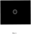
- one or more concentric (relative to the axis of laser beam from optical head 104, which is collinear with the axis of the objective lens, 204, in FIG. 2 ) diverging beams of light 201 are directed from the ring sources of light source 203 toward the cornea of the eye 102. If the cornea were perfectly spherical in shape, then the beams of light 201 which reflect from the cornea into a direction parallel to that of the objective lens 204 would pass through the telecentric stop aperture 208 and form images of the discrete LEDs in theconcentric rings of light on the video image plane 210. As shown in FIG. 2 , a processor 230 analyzes overall image to find the positions of each discrete LED 202 from the two concentric LED rings.
- the system geometry is such that the diameters of the two concentric rings of LEDs which are imaged by the telecentric viewing system are approximately 2.3 mm and 1.65 mm, respectively, as shown in FIG. 3 .
- the size of the reflected rings will differ and a determination of the size of the image of the reflected concentric rings of LEDs 202 on the telecentric camera detector 212 is used to determine the radius of curvature of the cornea.
- the cornea is astigmatic, the cornea's shape will deviate slightly from that of a perfect sphere in such a way as to cause the image of the reflection of the ring sources to have a nearly elliptical shape.
- the shape and size of the two circular or elliptical LED patterns formed on the video image plane 210 is determined by the processor 230, using standard numerical methods such as those described in Turuwhenua, Jason, "An Improved Low Order Method for Corneal Reconstruction", Optometry and Vision Science,Vol. 85, No. 3,March 2008, pp. E21 1 -E218 .
- the curvature of the cornea along the direction of a steep and shallow meridian i.e. the "K values”
- the "clock" angle of the the axes of the steep and shallow meridian with respect to the standard eye-fixed coordinate system, described above can be determined by a processor. If only the astigmatism axis is needed, a simple method of extracting it from the reflected images is to determine the angles of the semi-major axes of the ellipses using a simple least squares curve fitting technique.
- the choice of geometry to cause the reflected diameters of the rings of LEDs 202 to fall into the roughly 1.5 mm to 3 mm range results in an astigmatism (and corneal shape) measurement that is accurate for the central 3 mm of the cornea.
- Such a central region-biased measurement of optical power results in better vision for most patients over a variety of lighting conditions and patient activities (ref).
- the optical power of the cornea is quite non-uniform; the average optical power over a small central region may differ significantly from the average power averaged over, for example, a 6 mm to 7 mm diameter circular region centered on the optical axis of the cornea.
- femtosecond ophthalmic laser means a laser used in ophthalmology for making incisions in the the eye using the mechanism of photodisruption.
- Such lasers have pulse widths that are generally between 100 femtoseconds and 10,000 femtoseconds.
- the built-in keratometer could be used for measuring the K values and axes of astigmatism of all patients at the time of the procedure and those results be used for determination of the spherical and cylindrical power in the IOL to be used for treatment. In this way, all variability due to variation in measurement of these parameters with different types of optical power measuring instruments would be eliminated.
- the lens constants determined by the method described above would account for other factors, such as surgical technique/IOL characteristics, but would not be subject to variability associated with optical power measurement.
- the built-in keratometer could be used in conjunction with a standalone keratometer of the same type of design to reduce variability in the measurement of axis of astigmatism.
- This use of the built-in keratometer in conjunction with a stand-alone keratometer of the same design for pre-operative measurements recognizes that the measurement of K values and axis of astigmatism depend on the type of optical design used. Although the K values and axis of astigmatism measured on a given eye by all types of measuring instruments will be similar, differences in reported values may vary significantly.
- Instrument-to-instrument variation may be due to the region of the cornea measured by an instrument (for example one instrument may measure optical power over the central 2.5 mm of the cornea; another may measure over 3.5 mm), the type of illumination source used (for example Placido rings versus rings of discrete LEDs), how the data is analyzed, etc.
- the effect of an error of as little as 10° in treatment of astigmatism axis is a 30% under correction of the astigmatism ( A M Fea, et al, Eye 20, 764-768 (2006 )).
- the corneal optical power of a patient undergoing a cataract treatment with associated correction of astigmatism would be measured on a particular type of standalone keratometer, for example the keratometer sold under the tradename LenStar LS900 by Haag Streit of Bern, Switzerland).
- the K values of the pre-operative measurement would be used for determination of IOL spherical and cylindrical optical power.
- the built-in keratometer would be used to measure the axis of astigmatism of the patient's cornea.
- this measurement of axis, with the patient lying horizontally, is needed to compensate for cyclotorsion of the patient's eye between the pre-operative keratometer measurement made with the patient in a sitting position and that measured in the operative position of the patient, lying on a gurney.
- the built-in keratometer would be designed in all significant aspects to measure K values and axis of astigmatism in the same manner and to produce identical results (except for those associated with cyclotorsion) as the pre-operative, standalone keratometer.
- the optical head of treatment laser 104 is moved directly upward, out of the way, to allow access to the patient's eye 102 for application of a suction ring.
- a suction ring (not shown) is applied manually to the patient's eye 102.
- the optical head of treatment laserI04 is docked, using the previously described joystick.
- the treatment laser 104 can now be used to correct or reduce the astigmatism of the eye 102, based on the previously described astigmatism axis determination and/or the corneal shape determination, using limbal relaxing incisions (LRIs) or LASIK, aligning the astigmatism treatment to the measured axis of astigmatism.
- LRIs limbal relaxing incisions
- LASIK LASIK
- the above described alignment system and process can also be applied to procedures that involve implanting a toric intraocular lens (IOL) to treat astigmatism.
- IOL intraocular lens
- lOLs are synthetic lenses implanted into the capsular bag in the eye, after a cataractous lens is removed.
- the IOL restores vision by replacing partially opaque cataractous lens with a clear lens of appropriate power.
- a conventional IOL has only spherical power.
- a toric IOL has both spherical and cylindrical power and can thus correct astigmatism in the eye.
- the treatment laser 104 can be used to mark the axis of astigmatism for later use in aligning the axis of astigmatism 410 (shown inFIG. 4) of the IOL 405 (with haptics 406 used for anchoring IOL 405 in the capsular bag), with the marked axis of astigmatism of the eye 102.
- a round opening is manually torn or cut by a laser in the crystalline lens anterior capsule.
- the cataractous lens is removed through the opening and an IOL is placed into the capsular bag, generally centered behind the capsular opening.
- the treatment laser 104 can be used to cut a small "tag" as part of the circular capsulotomy 400.
- the "tag” provides a visible reference mark along which the axis of astigmatism of the IOL 410 can be aligned.
- the "tags" 430 in the capsular openings can be positioned inwardly or outwardly.
- the "tag” is cut in a smooth curve along the capsulotomy cut to avoid risk of radial capsular tears during the cataract procedure.
- Possible smooth shapes of the "tags” are shown schematically in close-up 425.
- This method of marking the astigmatism axis by incorporating a "tag” in the capsulotomy allows the astigmatism mark, i.e. the "tag” to be ideally placed for use in aligning the astigmatism axis of the IOL.
- the "tag” is in the immediate vicinity of the astigmatism mark on the IOL and may in fact be directly over the astigmatism axis mark on the IOL, avoiding any errors in registration which might occur when aligning the IOL mark with, for example, an ink mark on the sclera, a considerable distance from the IOL.
- the "tag” provides a visual marker so that the surgeon implanting a toric IOL can line up the astigmatism axis of the IOL with marked axis of astigmatism of the eye 102.
- a small mark for example a line, could be made by the laser in the center of the lens capsule immediately after the astigmatism axis was measured as described above. Then, after affixing the suction ring and docking the eye 102 to the optical head, the marks in the center of the capsule could be used, either manually or using automatic image recognition techniques built into a computer program, to set the position of the "tag" -marked laser-cut capsulotomy for use in the toric IOL implantation.
- Still another alternate method of marking the astigmatism axis with the treatment laser would entail shooting several laser shots, either at full or reduced energy at the position of the astigmatism axis at the limbus to make a persistent visible reference mark.
- the telecentric viewing system 200 is also used as a general viewing system, to assist the laser system associated with the optical head of the treatment laser 104 when the optical head is docked to the suction ring.
- the measuring system 250 would allow measuring the astigmatism axis in situ, while the patient is lying on the treatment bed, just in advance of the laser treatment - thus eliminating the need for pre-operative eye marks.
- the automatic measurement of the astigmatism axis by system 100 increases the accuracy of the placement of the limbal relaxing incisions, thereby improving the efficacy of the treatment.
- the method can also be used in conjunction with the laser to mark the astigmatism axis for cyclotorsional registration of a toric IOL.
- the present invention eliminates the need for manually marking the eye and circumvents the inaccuracies inherent in manual placing of marks and the dispersion of the ink marks by the eye's tear film; in addition, it provides a means to more accurately determine IOL lens constants to reduce clinical outcome variability.
- the integral astigmatism measurement in combination with use of marks made by the treatment laser can be used to mark the axis of astigmatism for later registration of a toric IOL or for any subsequent refractive treatment of the eye requiring knowledge of the axis of astigmatism.
- the system 100 also makes dual use of a camera 212 and ring light sources 202 for both the astigmatism measurement and for general viewing of the eye during the eye docking and lasing parts of the procedure.
Landscapes
- Health & Medical Sciences (AREA)
- Life Sciences & Earth Sciences (AREA)
- Ophthalmology & Optometry (AREA)
- Heart & Thoracic Surgery (AREA)
- Public Health (AREA)
- Surgery (AREA)
- Engineering & Computer Science (AREA)
- Biomedical Technology (AREA)
- Physics & Mathematics (AREA)
- Veterinary Medicine (AREA)
- General Health & Medical Sciences (AREA)
- Animal Behavior & Ethology (AREA)
- Optics & Photonics (AREA)
- Nuclear Medicine, Radiotherapy & Molecular Imaging (AREA)
- Vascular Medicine (AREA)
- Biophysics (AREA)
- Medical Informatics (AREA)
- Molecular Biology (AREA)
- Eye Examination Apparatus (AREA)
Claims (6)
- Lasersystem, Folgendes umfassend:a) eine Laserquelle, die dazu angepasst ist, einen Femtosekundenimpuls-Laserstrahl entlang einer Laserstrahlachse zu emittieren;b) eine Fixierungslichtquelle, die einen Fixierungsstrahl entlang einer Fixierungsstrahlachse emittiert, wobei die Laserstrahlachse und die Fixierungsstrahlachse kollinear sind;c) ein Keratometer, Folgendes umfassend:i) einen ersten Satz einzelner Lichtquellen, die entlang eines ersten Rings gleichmäßig voneinander beabstandet sind und dazu angepasst sind, ein erstes Licht auf ein Auge zu richten;(ii) einen zweiten Satz einzelner Lichtquellen, die entlang eines zweiten Rings gleichmäßig voneinander beabstandet sind und dazu angepasst sind, ein zweites Licht auf das Auge zu richten, wobei der erste Ring und der zweite Ring koplanar und konzentrisch zueinander sind und um die Laserstrahl- und Fixierungsstrahlachse zentriert sind;(iii) eine telezentrische Linse, die dazu angepasst ist, das erste Licht und das zweite Licht zu empfangen, die von dem Auge reflektiert werden;(iv) einen Detektor, der dazu angepasst ist, Licht von der telezentrischen Linse zu empfangen und ein Bild zu erzeugen; und(v) einen Prozessor, der dazu angepasst ist, Signale von dem Detektor zu empfangen, die für das Bild repräsentativ sind, und eine Astigmatismusachse des Auges basierend auf den Signalen zu bestimmen;f) wobei das System konfiguriert ist, den Femtosekundenimpuls-Laserstrahl abzugeben, um eine Kapsulotomie mit einer Markierung zu schneiden;g) wobei die Markierung eine glatte Kurve ist, wodurch die Gefahr von radialen Kapselrissen vermieden wird; undh) wobei die Markierung an der bestimmten Astigmatismusachse ausgerichtet ist.
- Lasersystem nach Anspruch 1, wobei der Prozessor dazu angepasst ist, einen Hornhautwert und eine Ausrichtung der Astigmatismusachse basierend auf den Signalen zu bestimmen.
- Lasersystem nach Anspruch 1, umfassend ein zweites eigenständiges Keratometer;
wobei das System in der Lage ist, Daten aus dem Keratometer und dem zweiten Keratometer zu vergleichen; wobei das System systematische Fehler bei der Verwendung des Lasersystems reduziert. - Lasersystem nach Anspruch 1, wobei die Laserquelle und das Keratometer in einem gemeinsamen Gehäuse untergebracht sind.
- Lasersystem nach Anspruch 4, umfassend ein zweites eigenständiges Keratometer;
wobei das System in der Lage ist, Daten aus dem Keratometer und dem zweiten Keratometer zu vergleichen; wobei das System systematische Fehler bei der Verwendung des Lasersystems reduziert. - Lasersystem nach Anspruch 1, wobei der Prozessor mit der Laserquelle in Kommunikation steht und dazu angepasst ist, den Laserstrahl so zu steuern, dass er das Auge basierend auf den Eigenschaften der Astigmatismusachse schneidet.
Applications Claiming Priority (4)
| Application Number | Priority Date | Filing Date | Title |
|---|---|---|---|
| US201161467622P | 2011-03-25 | 2011-03-25 | |
| US201161467592P | 2011-03-25 | 2011-03-25 | |
| PCT/US2012/030059 WO2012134931A1 (en) | 2011-03-25 | 2012-03-22 | System and method for measuring and correcting astigmatism using laser generated corneal incisions |
| EP12764966.3A EP2688459B1 (de) | 2011-03-25 | 2012-03-22 | System zur messung und korrektur von astigmatismus mittels lasererzeugter hornhautschnitte |
Related Parent Applications (1)
| Application Number | Title | Priority Date | Filing Date |
|---|---|---|---|
| EP12764966.3A Division EP2688459B1 (de) | 2011-03-25 | 2012-03-22 | System zur messung und korrektur von astigmatismus mittels lasererzeugter hornhautschnitte |
Publications (3)
| Publication Number | Publication Date |
|---|---|
| EP3552588A1 EP3552588A1 (de) | 2019-10-16 |
| EP3552588C0 EP3552588C0 (de) | 2024-12-25 |
| EP3552588B1 true EP3552588B1 (de) | 2024-12-25 |
Family
ID=46931851
Family Applications (4)
| Application Number | Title | Priority Date | Filing Date |
|---|---|---|---|
| EP19175512.3A Active EP3552588B1 (de) | 2011-03-25 | 2012-03-22 | System zur messung und korrektur von astigmatismus mittels lasererzeugter hornhautschnitte |
| EP23164593.8A Pending EP4223264A1 (de) | 2011-03-25 | 2012-03-22 | Lasersystem zur eliminierung systematischer fehler bei der durchführung eines katarakts |
| EP12764966.3A Active EP2688459B1 (de) | 2011-03-25 | 2012-03-22 | System zur messung und korrektur von astigmatismus mittels lasererzeugter hornhautschnitte |
| EP18167230.4A Active EP3434234B1 (de) | 2011-03-25 | 2012-03-22 | System zur messung von astigmatismus durch lasererzeugte hornhautschnitte |
Family Applications After (3)
| Application Number | Title | Priority Date | Filing Date |
|---|---|---|---|
| EP23164593.8A Pending EP4223264A1 (de) | 2011-03-25 | 2012-03-22 | Lasersystem zur eliminierung systematischer fehler bei der durchführung eines katarakts |
| EP12764966.3A Active EP2688459B1 (de) | 2011-03-25 | 2012-03-22 | System zur messung und korrektur von astigmatismus mittels lasererzeugter hornhautschnitte |
| EP18167230.4A Active EP3434234B1 (de) | 2011-03-25 | 2012-03-22 | System zur messung von astigmatismus durch lasererzeugte hornhautschnitte |
Country Status (5)
| Country | Link |
|---|---|
| US (3) | US20120265181A1 (de) |
| EP (4) | EP3552588B1 (de) |
| CN (2) | CN103501686B (de) |
| ES (2) | ES3010062T3 (de) |
| WO (1) | WO2012134931A1 (de) |
Families Citing this family (32)
| Publication number | Priority date | Publication date | Assignee | Title |
|---|---|---|---|---|
| US10842675B2 (en) | 2006-01-20 | 2020-11-24 | Lensar, Inc. | System and method for treating the structure of the human lens with a laser |
| US9889043B2 (en) | 2006-01-20 | 2018-02-13 | Lensar, Inc. | System and apparatus for delivering a laser beam to the lens of an eye |
| US9545338B2 (en) | 2006-01-20 | 2017-01-17 | Lensar, Llc. | System and method for improving the accommodative amplitude and increasing the refractive power of the human lens with a laser |
| US8500723B2 (en) | 2008-07-25 | 2013-08-06 | Lensar, Inc. | Liquid filled index matching device for ophthalmic laser procedures |
| US8480659B2 (en) | 2008-07-25 | 2013-07-09 | Lensar, Inc. | Method and system for removal and replacement of lens material from the lens of an eye |
| US8617146B2 (en) | 2009-07-24 | 2013-12-31 | Lensar, Inc. | Laser system and method for correction of induced astigmatism |
| AU2010275380A1 (en) | 2009-07-24 | 2012-02-16 | Lensar, Inc. | System and method for performing ladar assisted procedures on the lens of an eye |
| CA2769090A1 (en) | 2009-07-24 | 2011-01-27 | Lensar, Inc. | System and method for providing laser shot patterns to the lens of an eye |
| US8758332B2 (en) | 2009-07-24 | 2014-06-24 | Lensar, Inc. | Laser system and method for performing and sealing corneal incisions in the eye |
| US8556425B2 (en) | 2010-02-01 | 2013-10-15 | Lensar, Inc. | Purkinjie image-based alignment of suction ring in ophthalmic applications |
| USD694890S1 (en) | 2010-10-15 | 2013-12-03 | Lensar, Inc. | Laser system for treatment of the eye |
| USD695408S1 (en) | 2010-10-15 | 2013-12-10 | Lensar, Inc. | Laser system for treatment of the eye |
| ES2937241T3 (es) | 2010-10-15 | 2023-03-27 | Lensar Inc | Sistema y método de iluminación controlada por barrido de estructuras dentro de un ojo |
| US10463541B2 (en) | 2011-03-25 | 2019-11-05 | Lensar, Inc. | System and method for correcting astigmatism using multiple paired arcuate laser generated corneal incisions |
| DE102012019474A1 (de) * | 2012-09-28 | 2014-04-03 | Carl Zeiss Meditec Ag | Vorrichtung zur verlässlichen Bestimmung biometrischer Messgrößen des gesamten Auges |
| CN108309465B (zh) * | 2013-04-17 | 2022-04-15 | 眼力健发展有限责任公司 | 用于白内障手术中的轴对准的激光基准 |
| US10369053B2 (en) | 2013-04-17 | 2019-08-06 | Optimedica Corporation | Corneal topography measurements and fiducial mark incisions in laser surgical procedures |
| DE102013105738A1 (de) * | 2013-06-04 | 2014-12-04 | Faramarz Madjlessi | Laserbehandlungsvorrichtung für die Refraktivchirurgie |
| DE102014106993A1 (de) * | 2014-05-19 | 2015-11-19 | Chronos Vision Gmbh | Verfahren und Vorrichtung zur Bestimmung der Ausrichtung des Auges bei Augenoperationen |
| DE102014210786A1 (de) | 2014-06-05 | 2015-12-17 | Carl Zeiss Meditec Ag | Topographiemodul für ophthalmologische Geräte mit entfernungsunabhängiger Keratometrie-Messeinrichtung und Verfahren zu dessen Anwendung |
| WO2016061454A1 (en) * | 2014-10-17 | 2016-04-21 | Optimedica Corporation | Corneal topography measurements and fiducial mark incisions in laser surgical procedures |
| DE102015013237A1 (de) * | 2015-10-12 | 2017-04-13 | Novartis Ag | Zentriertechnik bei einem Schneidlaser für die refraktive Augenchirurgie |
| US10383767B2 (en) * | 2015-12-17 | 2019-08-20 | Novartis Ag | Ophthalmic relaxing incisions and associated devices, systems, and methods |
| KR20250016517A (ko) * | 2016-09-12 | 2025-02-03 | 렌사르, 인크. | 눈 구조로의 장치의 정렬 삽입을 위한 레이저 및 시스템 |
| CN106344173A (zh) * | 2016-09-21 | 2017-01-25 | 爱博诺德(北京)医疗科技有限公司 | 用于角膜散光实时定位装置的光源 |
| DE102017223512A1 (de) * | 2017-12-21 | 2019-06-27 | Carl Zeiss Meditec Ag | Anordnung zur Bestimmung der Topographie der Kornea eines Auges |
| US11596547B2 (en) | 2018-01-31 | 2023-03-07 | Centricity Vision, Inc. | Hydrodissection and posterior capsule opacification prevention during capsulotomy procedure |
| AU2019334910A1 (en) | 2018-09-04 | 2020-12-10 | Amo Development, Llc | Narrow angle illumination ring for ophthalmic surgical laser system |
| CN109363625B (zh) * | 2018-12-17 | 2021-07-13 | 温州医科大学 | 一种在线标示散光轴位的扩增实境系统 |
| DE102019219122A1 (de) | 2019-09-10 | 2021-03-11 | Carl Zeiss Meditec Ag | Positioniereinrichtung |
| CN111513917B (zh) * | 2020-05-22 | 2022-03-22 | 杭州明视康眼科医院有限公司 | 一种散光型icl术后残留散光的转位调整方法并预估转位调整后的屈光度的方法 |
| CN114246706A (zh) * | 2020-09-22 | 2022-03-29 | 上海康恩德医疗科技有限公司 | 散光矫正型人工晶状体 |
Citations (1)
| Publication number | Priority date | Publication date | Assignee | Title |
|---|---|---|---|---|
| EP0397962A1 (de) * | 1989-04-28 | 1990-11-22 | Taunton Technologies, Inc. | Gerät zur Messung der Kontur |
Family Cites Families (27)
| Publication number | Priority date | Publication date | Assignee | Title |
|---|---|---|---|---|
| DE2643344A1 (de) * | 1976-09-25 | 1978-03-30 | Zeiss Carl Fa | Vorrichtung zur ermittlung von hornhautastigmatismus am menschlichen auge |
| FR2497087A1 (fr) * | 1980-12-30 | 1982-07-02 | Essilor Int | Keratometre automatique |
| WO1985000740A1 (en) * | 1983-08-11 | 1985-02-28 | Selig Percy Amoils | Method and apparatus for use in ocular surgery |
| JP2775297B2 (ja) * | 1989-06-22 | 1998-07-16 | 株式会社トプコン | 眼屈折力測定装置 |
| US6450641B2 (en) * | 1992-06-02 | 2002-09-17 | Lasersight Technologies, Inc. | Method of corneal analysis using a checkered placido apparatus |
| ATE208174T1 (de) * | 1992-08-10 | 2001-11-15 | Noel Ami Alpins | Vorrichtung zur durchführung eines chirurgischen eingriffs an der hornhaut |
| US5549597A (en) * | 1993-05-07 | 1996-08-27 | Visx Incorporated | In situ astigmatism axis alignment |
| JP3740546B2 (ja) * | 1997-11-11 | 2006-02-01 | 株式会社トプコン | 眼科測定装置 |
| JP3848492B2 (ja) * | 1998-09-04 | 2006-11-22 | 株式会社ニデック | 角膜手術装置 |
| EP1139857A2 (de) * | 1998-12-10 | 2001-10-10 | CARL ZEISS JENA GmbH | Anordnung und verfahren zur berührungslosen messung der achslänge und/oder der hornhautkrümmung und/oder der vorderkammertiefe des auges, vorzugsweise zur iol-berechnung |
| US6129722A (en) * | 1999-03-10 | 2000-10-10 | Ruiz; Luis Antonio | Interactive corrective eye surgery system with topography and laser system interface |
| BR0008243B1 (pt) * | 1999-10-21 | 2008-11-18 | aparelho para determinar uma quantidade de ablaÇço corneana. | |
| AU778420B2 (en) * | 1999-10-21 | 2004-12-02 | Technolas Gmbh Ophthalmologische Systeme | Iris recognition and tracking for optical treatment |
| DE19950790A1 (de) * | 1999-10-21 | 2001-06-21 | Technolas Gmbh | Spezifische Hornhautmodellierung |
| US6193371B1 (en) * | 2000-03-27 | 2001-02-27 | Richard Snook | Keratometer/pachymeter |
| US6929638B2 (en) * | 2000-04-19 | 2005-08-16 | Alcon Refractivehorizons, Inc. | Eye registration and astigmatism alignment control systems and method |
| WO2002064030A1 (en) * | 2001-02-09 | 2002-08-22 | Kabushiki Kaisha Topcon | Eye characteristics measuring device |
| JP4133480B2 (ja) * | 2003-03-18 | 2008-08-13 | 株式会社トプコン | 検眼装置 |
| ATE485758T1 (de) * | 2006-03-16 | 2010-11-15 | Sis Ag Surgical Instr Systems | Ophthalmologische vorrichtung und ophthalmologisches messverfahren |
| EP3308756B1 (de) * | 2007-03-13 | 2020-02-19 | Optimedica Corporation | Vorrichtung zur erzeugung von inzisionen zur verbesserung der platzierung intraokularer linsen |
| US20080312675A1 (en) * | 2007-06-18 | 2008-12-18 | Advanced Medical Optics, Inc. | System and method for calculating limbal relaxing incisions |
| WO2009036098A2 (en) * | 2007-09-10 | 2009-03-19 | Lensx Lasers, Inc. | Apparatus, systems and techniques for interfacing with an eye in laser surgery |
| JP2011502585A (ja) * | 2007-11-02 | 2011-01-27 | アルコン レンゼックス, インコーポレーテッド | 術後の眼の光学的性能を改善するための方法および装置 |
| US20100324542A1 (en) * | 2007-11-02 | 2010-12-23 | Kurtz Ronald M | Method to Guide a Cataract Procedure by Corneal Imaging |
| WO2010054268A2 (en) * | 2008-11-06 | 2010-05-14 | Wavetec Vision Systems, Inc. | Optical angular measurement system for ophthalmic applications and method for positioning of a toric intraocular lens with increased accuracy |
| WO2011094666A1 (en) * | 2010-02-01 | 2011-08-04 | Lensar, Inc. | Placido ring measurement of astigmatism axis and laser marking of astigmatism axis |
| US20110251630A1 (en) * | 2010-04-08 | 2011-10-13 | Richardson Gary A | Corneal marking apparatus |
-
2012
- 2012-03-22 EP EP19175512.3A patent/EP3552588B1/de active Active
- 2012-03-22 CN CN201280015199.4A patent/CN103501686B/zh active Active
- 2012-03-22 EP EP23164593.8A patent/EP4223264A1/de active Pending
- 2012-03-22 CN CN201710049137.XA patent/CN106974615B/zh active Active
- 2012-03-22 ES ES19175512T patent/ES3010062T3/es active Active
- 2012-03-22 EP EP12764966.3A patent/EP2688459B1/de active Active
- 2012-03-22 US US13/427,130 patent/US20120265181A1/en not_active Abandoned
- 2012-03-22 EP EP18167230.4A patent/EP3434234B1/de active Active
- 2012-03-22 ES ES18167230T patent/ES3019567T3/es active Active
- 2012-03-22 WO PCT/US2012/030059 patent/WO2012134931A1/en not_active Ceased
-
2017
- 2017-08-27 US US15/687,501 patent/US11076756B2/en active Active
- 2017-08-27 US US15/687,502 patent/US11089955B2/en active Active
Patent Citations (1)
| Publication number | Priority date | Publication date | Assignee | Title |
|---|---|---|---|---|
| EP0397962A1 (de) * | 1989-04-28 | 1990-11-22 | Taunton Technologies, Inc. | Gerät zur Messung der Kontur |
Also Published As
| Publication number | Publication date |
|---|---|
| CN106974615A (zh) | 2017-07-25 |
| EP2688459B1 (de) | 2019-05-22 |
| EP3552588C0 (de) | 2024-12-25 |
| CN103501686B (zh) | 2017-02-08 |
| US20170347877A1 (en) | 2017-12-07 |
| US20120265181A1 (en) | 2012-10-18 |
| EP4223264A1 (de) | 2023-08-09 |
| EP3434234B1 (de) | 2025-03-12 |
| EP3552588A1 (de) | 2019-10-16 |
| EP2688459A1 (de) | 2014-01-29 |
| CN103501686A (zh) | 2014-01-08 |
| US20170354325A1 (en) | 2017-12-14 |
| WO2012134931A1 (en) | 2012-10-04 |
| CN106974615B (zh) | 2019-11-26 |
| ES3010062T3 (en) | 2025-04-01 |
| ES3019567T3 (en) | 2025-05-20 |
| EP3434234A1 (de) | 2019-01-30 |
| US11076756B2 (en) | 2021-08-03 |
| US11089955B2 (en) | 2021-08-17 |
| EP2688459A4 (de) | 2014-11-26 |
Similar Documents
| Publication | Publication Date | Title |
|---|---|---|
| US11089955B2 (en) | System and method for measuring and correcting astigmatism using laser generated corneal incisions | |
| US20110190740A1 (en) | Placido ring measurement of astigmatism axis and laser marking of astigmatism axis | |
| US10390996B2 (en) | Ophthalmic range finding | |
| JP6046774B2 (ja) | 術後の眼の光学的性能を改善するための方法および装置 | |
| US9521949B2 (en) | Ophthalmic range finding | |
| US20130335705A1 (en) | Integrated surgical microscope and wavefront sensor | |
| TWI631926B (zh) | 用於屈光眼科手術中之切割雷射的定中心技術 | |
| US11284793B2 (en) | Method and device for determining the orientation of the eye during eye surgeries | |
| US20210267799A1 (en) | System and Methods for Customizing an Intraocular Lens Using a Wavefront Aberrometer | |
| US20220175244A1 (en) | System and method for measuring and correcting astigmatism using laser generated corneal incisions | |
| US8500283B1 (en) | Microscope-attachable aberrometer | |
| AU2009231656B2 (en) | System and method for identifying a position to insert a scleral prosthesis into an eye | |
| RU2850981C1 (ru) | Способ контроля позиционирования интраокулярной линзы с ротационно-асимметричной оптикой относительно зрительной оси глаза | |
| Dhull et al. | Optiwave Refractive Analysis (ORA) | |
| HK1174522A (en) | Placido ring measurement of astigmatism axis and laser marking of astigmatism axis | |
| Dandekar et al. | Aberrometry in the calculation of intraocular lens power and Toric intraocular lens alignment devices |
Legal Events
| Date | Code | Title | Description |
|---|---|---|---|
| PUAI | Public reference made under article 153(3) epc to a published international application that has entered the european phase |
Free format text: ORIGINAL CODE: 0009012 |
|
| STAA | Information on the status of an ep patent application or granted ep patent |
Free format text: STATUS: THE APPLICATION HAS BEEN PUBLISHED |
|
| AC | Divisional application: reference to earlier application |
Ref document number: 2688459 Country of ref document: EP Kind code of ref document: P |
|
| AK | Designated contracting states |
Kind code of ref document: A1 Designated state(s): AL AT BE BG CH CY CZ DE DK EE ES FI FR GB GR HR HU IE IS IT LI LT LU LV MC MK MT NL NO PL PT RO RS SE SI SK SM TR |
|
| STAA | Information on the status of an ep patent application or granted ep patent |
Free format text: STATUS: REQUEST FOR EXAMINATION WAS MADE |
|
| 17P | Request for examination filed |
Effective date: 20200415 |
|
| RBV | Designated contracting states (corrected) |
Designated state(s): AL AT BE BG CH CY CZ DE DK EE ES FI FR GB GR HR HU IE IS IT LI LT LU LV MC MK MT NL NO PL PT RO RS SE SI SK SM TR |
|
| STAA | Information on the status of an ep patent application or granted ep patent |
Free format text: STATUS: EXAMINATION IS IN PROGRESS |
|
| 17Q | First examination report despatched |
Effective date: 20220701 |
|
| P01 | Opt-out of the competence of the unified patent court (upc) registered |
Effective date: 20230530 |
|
| GRAP | Despatch of communication of intention to grant a patent |
Free format text: ORIGINAL CODE: EPIDOSNIGR1 |
|
| STAA | Information on the status of an ep patent application or granted ep patent |
Free format text: STATUS: GRANT OF PATENT IS INTENDED |
|
| INTG | Intention to grant announced |
Effective date: 20240802 |
|
| RIN1 | Information on inventor provided before grant (corrected) |
Inventor name: FREY, RUDOLPH W |
|
| GRAS | Grant fee paid |
Free format text: ORIGINAL CODE: EPIDOSNIGR3 |
|
| GRAA | (expected) grant |
Free format text: ORIGINAL CODE: 0009210 |
|
| STAA | Information on the status of an ep patent application or granted ep patent |
Free format text: STATUS: THE PATENT HAS BEEN GRANTED |
|
| AC | Divisional application: reference to earlier application |
Ref document number: 2688459 Country of ref document: EP Kind code of ref document: P |
|
| AK | Designated contracting states |
Kind code of ref document: B1 Designated state(s): AL AT BE BG CH CY CZ DE DK EE ES FI FR GB GR HR HU IE IS IT LI LT LU LV MC MK MT NL NO PL PT RO RS SE SI SK SM TR |
|
| REG | Reference to a national code |
Ref country code: GB Ref legal event code: FG4D |
|
| REG | Reference to a national code |
Ref country code: CH Ref legal event code: EP |
|
| REG | Reference to a national code |
Ref country code: DE Ref legal event code: R096 Ref document number: 602012081287 Country of ref document: DE |
|
| REG | Reference to a national code |
Ref country code: IE Ref legal event code: FG4D |
|
| U01 | Request for unitary effect filed |
Effective date: 20250113 |
|
| U07 | Unitary effect registered |
Designated state(s): AT BE BG DE DK EE FI FR IT LT LU LV MT NL PT RO SE SI Effective date: 20250120 |
|
| P04 | Withdrawal of opt-out of the competence of the unified patent court (upc) registered |
Free format text: CASE NUMBER: APP_2621/2025 Effective date: 20250116 |
|
| REG | Reference to a national code |
Ref country code: ES Ref legal event code: FG2A Ref document number: 3010062 Country of ref document: ES Kind code of ref document: T3 Effective date: 20250401 |
|
| PG25 | Lapsed in a contracting state [announced via postgrant information from national office to epo] |
Ref country code: HR Free format text: LAPSE BECAUSE OF FAILURE TO SUBMIT A TRANSLATION OF THE DESCRIPTION OR TO PAY THE FEE WITHIN THE PRESCRIBED TIME-LIMIT Effective date: 20241225 |
|
| PGFP | Annual fee paid to national office [announced via postgrant information from national office to epo] |
Ref country code: IE Payment date: 20250321 Year of fee payment: 14 |
|
| PG25 | Lapsed in a contracting state [announced via postgrant information from national office to epo] |
Ref country code: NO Free format text: LAPSE BECAUSE OF FAILURE TO SUBMIT A TRANSLATION OF THE DESCRIPTION OR TO PAY THE FEE WITHIN THE PRESCRIBED TIME-LIMIT Effective date: 20250325 |
|
| PG25 | Lapsed in a contracting state [announced via postgrant information from national office to epo] |
Ref country code: GR Free format text: LAPSE BECAUSE OF FAILURE TO SUBMIT A TRANSLATION OF THE DESCRIPTION OR TO PAY THE FEE WITHIN THE PRESCRIBED TIME-LIMIT Effective date: 20250326 |
|
| PGFP | Annual fee paid to national office [announced via postgrant information from national office to epo] |
Ref country code: GB Payment date: 20250321 Year of fee payment: 14 |
|
| PG25 | Lapsed in a contracting state [announced via postgrant information from national office to epo] |
Ref country code: RS Free format text: LAPSE BECAUSE OF FAILURE TO SUBMIT A TRANSLATION OF THE DESCRIPTION OR TO PAY THE FEE WITHIN THE PRESCRIBED TIME-LIMIT Effective date: 20250325 |
|
| U20 | Renewal fee for the european patent with unitary effect paid |
Year of fee payment: 14 Effective date: 20250409 |
|
| PG25 | Lapsed in a contracting state [announced via postgrant information from national office to epo] |
Ref country code: SM Free format text: LAPSE BECAUSE OF FAILURE TO SUBMIT A TRANSLATION OF THE DESCRIPTION OR TO PAY THE FEE WITHIN THE PRESCRIBED TIME-LIMIT Effective date: 20241225 |
|
| PG25 | Lapsed in a contracting state [announced via postgrant information from national office to epo] |
Ref country code: PL Free format text: LAPSE BECAUSE OF FAILURE TO SUBMIT A TRANSLATION OF THE DESCRIPTION OR TO PAY THE FEE WITHIN THE PRESCRIBED TIME-LIMIT Effective date: 20241225 |
|
| PGFP | Annual fee paid to national office [announced via postgrant information from national office to epo] |
Ref country code: ES Payment date: 20250409 Year of fee payment: 14 |
|
| PG25 | Lapsed in a contracting state [announced via postgrant information from national office to epo] |
Ref country code: IS Free format text: LAPSE BECAUSE OF FAILURE TO SUBMIT A TRANSLATION OF THE DESCRIPTION OR TO PAY THE FEE WITHIN THE PRESCRIBED TIME-LIMIT Effective date: 20250425 |
|
| PG25 | Lapsed in a contracting state [announced via postgrant information from national office to epo] |
Ref country code: SK Free format text: LAPSE BECAUSE OF FAILURE TO SUBMIT A TRANSLATION OF THE DESCRIPTION OR TO PAY THE FEE WITHIN THE PRESCRIBED TIME-LIMIT Effective date: 20241225 |
|
| PG25 | Lapsed in a contracting state [announced via postgrant information from national office to epo] |
Ref country code: CZ Free format text: LAPSE BECAUSE OF FAILURE TO SUBMIT A TRANSLATION OF THE DESCRIPTION OR TO PAY THE FEE WITHIN THE PRESCRIBED TIME-LIMIT Effective date: 20241225 |
|
| PG25 | Lapsed in a contracting state [announced via postgrant information from national office to epo] |
Ref country code: MC Free format text: LAPSE BECAUSE OF FAILURE TO SUBMIT A TRANSLATION OF THE DESCRIPTION OR TO PAY THE FEE WITHIN THE PRESCRIBED TIME-LIMIT Effective date: 20241225 |
|
| REG | Reference to a national code |
Ref country code: CH Ref legal event code: H13 Free format text: ST27 STATUS EVENT CODE: U-0-0-H10-H13 (AS PROVIDED BY THE NATIONAL OFFICE) Effective date: 20251023 |
|
| PLBE | No opposition filed within time limit |
Free format text: ORIGINAL CODE: 0009261 |
|
| STAA | Information on the status of an ep patent application or granted ep patent |
Free format text: STATUS: NO OPPOSITION FILED WITHIN TIME LIMIT |
|
| 26N | No opposition filed |
Effective date: 20250926 |
|
| PG25 | Lapsed in a contracting state [announced via postgrant information from national office to epo] |
Ref country code: CH Free format text: LAPSE BECAUSE OF NON-PAYMENT OF DUE FEES Effective date: 20250331 |