EP3545981A1 - Magnetisch betätigtes gelenkknorpelregenerationssystem - Google Patents
Magnetisch betätigtes gelenkknorpelregenerationssystem Download PDFInfo
- Publication number
- EP3545981A1 EP3545981A1 EP17864166.8A EP17864166A EP3545981A1 EP 3545981 A1 EP3545981 A1 EP 3545981A1 EP 17864166 A EP17864166 A EP 17864166A EP 3545981 A1 EP3545981 A1 EP 3545981A1
- Authority
- EP
- European Patent Office
- Prior art keywords
- cartilage
- composition
- scaffold
- magnetic field
- magnetic
- Prior art date
- Legal status (The legal status is an assumption and is not a legal conclusion. Google has not performed a legal analysis and makes no representation as to the accuracy of the status listed.)
- Granted
Links
Images




Classifications
-
- A—HUMAN NECESSITIES
- A61—MEDICAL OR VETERINARY SCIENCE; HYGIENE
- A61L—METHODS OR APPARATUS FOR STERILISING MATERIALS OR OBJECTS IN GENERAL; DISINFECTION, STERILISATION OR DEODORISATION OF AIR; CHEMICAL ASPECTS OF BANDAGES, DRESSINGS, ABSORBENT PADS OR SURGICAL ARTICLES; MATERIALS FOR BANDAGES, DRESSINGS, ABSORBENT PADS OR SURGICAL ARTICLES
- A61L27/00—Materials for grafts or prostheses or for coating grafts or prostheses
- A61L27/36—Materials for grafts or prostheses or for coating grafts or prostheses containing ingredients of undetermined constitution or reaction products thereof, e.g. transplant tissue, natural bone, extracellular matrix
- A61L27/38—Materials for grafts or prostheses or for coating grafts or prostheses containing ingredients of undetermined constitution or reaction products thereof, e.g. transplant tissue, natural bone, extracellular matrix containing added animal cells
- A61L27/3804—Materials for grafts or prostheses or for coating grafts or prostheses containing ingredients of undetermined constitution or reaction products thereof, e.g. transplant tissue, natural bone, extracellular matrix containing added animal cells characterised by specific cells or progenitors thereof, e.g. fibroblasts, connective tissue cells, kidney cells
- A61L27/3817—Cartilage-forming cells, e.g. pre-chondrocytes
-
- A—HUMAN NECESSITIES
- A61—MEDICAL OR VETERINARY SCIENCE; HYGIENE
- A61B—DIAGNOSIS; SURGERY; IDENTIFICATION
- A61B6/00—Apparatus or devices for radiation diagnosis; Apparatus or devices for radiation diagnosis combined with radiation therapy equipment
- A61B6/12—Arrangements for detecting or locating foreign bodies
-
- A—HUMAN NECESSITIES
- A61—MEDICAL OR VETERINARY SCIENCE; HYGIENE
- A61B—DIAGNOSIS; SURGERY; IDENTIFICATION
- A61B6/00—Apparatus or devices for radiation diagnosis; Apparatus or devices for radiation diagnosis combined with radiation therapy equipment
- A61B6/50—Apparatus or devices for radiation diagnosis; Apparatus or devices for radiation diagnosis combined with radiation therapy equipment specially adapted for specific body parts; specially adapted for specific clinical applications
-
- A—HUMAN NECESSITIES
- A61—MEDICAL OR VETERINARY SCIENCE; HYGIENE
- A61B—DIAGNOSIS; SURGERY; IDENTIFICATION
- A61B6/00—Apparatus or devices for radiation diagnosis; Apparatus or devices for radiation diagnosis combined with radiation therapy equipment
- A61B6/50—Apparatus or devices for radiation diagnosis; Apparatus or devices for radiation diagnosis combined with radiation therapy equipment specially adapted for specific body parts; specially adapted for specific clinical applications
- A61B6/505—Apparatus or devices for radiation diagnosis; Apparatus or devices for radiation diagnosis combined with radiation therapy equipment specially adapted for specific body parts; specially adapted for specific clinical applications for diagnosis of bone
-
- A—HUMAN NECESSITIES
- A61—MEDICAL OR VETERINARY SCIENCE; HYGIENE
- A61B—DIAGNOSIS; SURGERY; IDENTIFICATION
- A61B6/00—Apparatus or devices for radiation diagnosis; Apparatus or devices for radiation diagnosis combined with radiation therapy equipment
- A61B6/52—Devices using data or image processing specially adapted for radiation diagnosis
- A61B6/5211—Devices using data or image processing specially adapted for radiation diagnosis involving processing of medical diagnostic data
- A61B6/5217—Devices using data or image processing specially adapted for radiation diagnosis involving processing of medical diagnostic data extracting a diagnostic or physiological parameter from medical diagnostic data
-
- A—HUMAN NECESSITIES
- A61—MEDICAL OR VETERINARY SCIENCE; HYGIENE
- A61F—FILTERS IMPLANTABLE INTO BLOOD VESSELS; PROSTHESES; DEVICES PROVIDING PATENCY TO, OR PREVENTING COLLAPSING OF, TUBULAR STRUCTURES OF THE BODY, e.g. STENTS; ORTHOPAEDIC, NURSING OR CONTRACEPTIVE DEVICES; FOMENTATION; TREATMENT OR PROTECTION OF EYES OR EARS; BANDAGES, DRESSINGS OR ABSORBENT PADS; FIRST-AID KITS
- A61F2/00—Filters implantable into blood vessels; Prostheses, i.e. artificial substitutes or replacements for parts of the body; Appliances for connecting them with the body; Devices providing patency to, or preventing collapsing of, tubular structures of the body, e.g. stents
- A61F2/02—Prostheses implantable into the body
- A61F2/30—Joints
-
- A—HUMAN NECESSITIES
- A61—MEDICAL OR VETERINARY SCIENCE; HYGIENE
- A61F—FILTERS IMPLANTABLE INTO BLOOD VESSELS; PROSTHESES; DEVICES PROVIDING PATENCY TO, OR PREVENTING COLLAPSING OF, TUBULAR STRUCTURES OF THE BODY, e.g. STENTS; ORTHOPAEDIC, NURSING OR CONTRACEPTIVE DEVICES; FOMENTATION; TREATMENT OR PROTECTION OF EYES OR EARS; BANDAGES, DRESSINGS OR ABSORBENT PADS; FIRST-AID KITS
- A61F2/00—Filters implantable into blood vessels; Prostheses, i.e. artificial substitutes or replacements for parts of the body; Appliances for connecting them with the body; Devices providing patency to, or preventing collapsing of, tubular structures of the body, e.g. stents
- A61F2/02—Prostheses implantable into the body
- A61F2/30—Joints
- A61F2/30756—Cartilage endoprostheses
-
- A—HUMAN NECESSITIES
- A61—MEDICAL OR VETERINARY SCIENCE; HYGIENE
- A61K—PREPARATIONS FOR MEDICAL, DENTAL OR TOILETRY PURPOSES
- A61K35/00—Medicinal preparations containing materials or reaction products thereof with undetermined constitution
- A61K35/12—Materials from mammals; Compositions comprising non-specified tissues or cells; Compositions comprising non-embryonic stem cells; Genetically modified cells
- A61K35/28—Bone marrow; Haematopoietic stem cells; Mesenchymal stem cells of any origin, e.g. adipose-derived stem cells
-
- A—HUMAN NECESSITIES
- A61—MEDICAL OR VETERINARY SCIENCE; HYGIENE
- A61K—PREPARATIONS FOR MEDICAL, DENTAL OR TOILETRY PURPOSES
- A61K35/00—Medicinal preparations containing materials or reaction products thereof with undetermined constitution
- A61K35/12—Materials from mammals; Compositions comprising non-specified tissues or cells; Compositions comprising non-embryonic stem cells; Genetically modified cells
- A61K35/32—Bones; Osteocytes; Osteoblasts; Tendons; Tenocytes; Teeth; Odontoblasts; Cartilage; Chondrocytes; Synovial membrane
-
- A—HUMAN NECESSITIES
- A61—MEDICAL OR VETERINARY SCIENCE; HYGIENE
- A61K—PREPARATIONS FOR MEDICAL, DENTAL OR TOILETRY PURPOSES
- A61K41/00—Medicinal preparations obtained by treating materials with wave energy or particle radiation ; Therapies using these preparations
-
- A—HUMAN NECESSITIES
- A61—MEDICAL OR VETERINARY SCIENCE; HYGIENE
- A61K—PREPARATIONS FOR MEDICAL, DENTAL OR TOILETRY PURPOSES
- A61K47/00—Medicinal preparations characterised by the non-active ingredients used, e.g. carriers or inert additives; Targeting or modifying agents chemically bound to the active ingredient
- A61K47/50—Medicinal preparations characterised by the non-active ingredients used, e.g. carriers or inert additives; Targeting or modifying agents chemically bound to the active ingredient the non-active ingredient being chemically bound to the active ingredient, e.g. polymer-drug conjugates
- A61K47/69—Medicinal preparations characterised by the non-active ingredients used, e.g. carriers or inert additives; Targeting or modifying agents chemically bound to the active ingredient the non-active ingredient being chemically bound to the active ingredient, e.g. polymer-drug conjugates the conjugate being characterised by physical or galenical forms, e.g. emulsion, particle, inclusion complex, stent or kit
- A61K47/6921—Medicinal preparations characterised by the non-active ingredients used, e.g. carriers or inert additives; Targeting or modifying agents chemically bound to the active ingredient the non-active ingredient being chemically bound to the active ingredient, e.g. polymer-drug conjugates the conjugate being characterised by physical or galenical forms, e.g. emulsion, particle, inclusion complex, stent or kit the form being a particulate, a powder, an adsorbate, a bead or a sphere
- A61K47/6923—Medicinal preparations characterised by the non-active ingredients used, e.g. carriers or inert additives; Targeting or modifying agents chemically bound to the active ingredient the non-active ingredient being chemically bound to the active ingredient, e.g. polymer-drug conjugates the conjugate being characterised by physical or galenical forms, e.g. emulsion, particle, inclusion complex, stent or kit the form being a particulate, a powder, an adsorbate, a bead or a sphere the form being an inorganic particle, e.g. ceramic particles, silica particles, ferrite or synsorb
-
- A—HUMAN NECESSITIES
- A61—MEDICAL OR VETERINARY SCIENCE; HYGIENE
- A61K—PREPARATIONS FOR MEDICAL, DENTAL OR TOILETRY PURPOSES
- A61K47/00—Medicinal preparations characterised by the non-active ingredients used, e.g. carriers or inert additives; Targeting or modifying agents chemically bound to the active ingredient
- A61K47/50—Medicinal preparations characterised by the non-active ingredients used, e.g. carriers or inert additives; Targeting or modifying agents chemically bound to the active ingredient the non-active ingredient being chemically bound to the active ingredient, e.g. polymer-drug conjugates
- A61K47/69—Medicinal preparations characterised by the non-active ingredients used, e.g. carriers or inert additives; Targeting or modifying agents chemically bound to the active ingredient the non-active ingredient being chemically bound to the active ingredient, e.g. polymer-drug conjugates the conjugate being characterised by physical or galenical forms, e.g. emulsion, particle, inclusion complex, stent or kit
- A61K47/6921—Medicinal preparations characterised by the non-active ingredients used, e.g. carriers or inert additives; Targeting or modifying agents chemically bound to the active ingredient the non-active ingredient being chemically bound to the active ingredient, e.g. polymer-drug conjugates the conjugate being characterised by physical or galenical forms, e.g. emulsion, particle, inclusion complex, stent or kit the form being a particulate, a powder, an adsorbate, a bead or a sphere
- A61K47/6927—Medicinal preparations characterised by the non-active ingredients used, e.g. carriers or inert additives; Targeting or modifying agents chemically bound to the active ingredient the non-active ingredient being chemically bound to the active ingredient, e.g. polymer-drug conjugates the conjugate being characterised by physical or galenical forms, e.g. emulsion, particle, inclusion complex, stent or kit the form being a particulate, a powder, an adsorbate, a bead or a sphere the form being a solid microparticle having no hollow or gas-filled cores
- A61K47/6929—Medicinal preparations characterised by the non-active ingredients used, e.g. carriers or inert additives; Targeting or modifying agents chemically bound to the active ingredient the non-active ingredient being chemically bound to the active ingredient, e.g. polymer-drug conjugates the conjugate being characterised by physical or galenical forms, e.g. emulsion, particle, inclusion complex, stent or kit the form being a particulate, a powder, an adsorbate, a bead or a sphere the form being a solid microparticle having no hollow or gas-filled cores the form being a nanoparticle, e.g. an immuno-nanoparticle
-
- A—HUMAN NECESSITIES
- A61—MEDICAL OR VETERINARY SCIENCE; HYGIENE
- A61L—METHODS OR APPARATUS FOR STERILISING MATERIALS OR OBJECTS IN GENERAL; DISINFECTION, STERILISATION OR DEODORISATION OF AIR; CHEMICAL ASPECTS OF BANDAGES, DRESSINGS, ABSORBENT PADS OR SURGICAL ARTICLES; MATERIALS FOR BANDAGES, DRESSINGS, ABSORBENT PADS OR SURGICAL ARTICLES
- A61L27/00—Materials for grafts or prostheses or for coating grafts or prostheses
- A61L27/02—Inorganic materials
- A61L27/10—Ceramics or glasses
-
- A—HUMAN NECESSITIES
- A61—MEDICAL OR VETERINARY SCIENCE; HYGIENE
- A61L—METHODS OR APPARATUS FOR STERILISING MATERIALS OR OBJECTS IN GENERAL; DISINFECTION, STERILISATION OR DEODORISATION OF AIR; CHEMICAL ASPECTS OF BANDAGES, DRESSINGS, ABSORBENT PADS OR SURGICAL ARTICLES; MATERIALS FOR BANDAGES, DRESSINGS, ABSORBENT PADS OR SURGICAL ARTICLES
- A61L27/00—Materials for grafts or prostheses or for coating grafts or prostheses
- A61L27/14—Macromolecular materials
- A61L27/18—Macromolecular materials obtained otherwise than by reactions only involving carbon-to-carbon unsaturated bonds
-
- A—HUMAN NECESSITIES
- A61—MEDICAL OR VETERINARY SCIENCE; HYGIENE
- A61L—METHODS OR APPARATUS FOR STERILISING MATERIALS OR OBJECTS IN GENERAL; DISINFECTION, STERILISATION OR DEODORISATION OF AIR; CHEMICAL ASPECTS OF BANDAGES, DRESSINGS, ABSORBENT PADS OR SURGICAL ARTICLES; MATERIALS FOR BANDAGES, DRESSINGS, ABSORBENT PADS OR SURGICAL ARTICLES
- A61L27/00—Materials for grafts or prostheses or for coating grafts or prostheses
- A61L27/36—Materials for grafts or prostheses or for coating grafts or prostheses containing ingredients of undetermined constitution or reaction products thereof, e.g. transplant tissue, natural bone, extracellular matrix
- A61L27/38—Materials for grafts or prostheses or for coating grafts or prostheses containing ingredients of undetermined constitution or reaction products thereof, e.g. transplant tissue, natural bone, extracellular matrix containing added animal cells
- A61L27/3804—Materials for grafts or prostheses or for coating grafts or prostheses containing ingredients of undetermined constitution or reaction products thereof, e.g. transplant tissue, natural bone, extracellular matrix containing added animal cells characterised by specific cells or progenitors thereof, e.g. fibroblasts, connective tissue cells, kidney cells
- A61L27/3834—Cells able to produce different cell types, e.g. hematopoietic stem cells, mesenchymal stem cells, marrow stromal cells, embryonic stem cells
-
- A—HUMAN NECESSITIES
- A61—MEDICAL OR VETERINARY SCIENCE; HYGIENE
- A61L—METHODS OR APPARATUS FOR STERILISING MATERIALS OR OBJECTS IN GENERAL; DISINFECTION, STERILISATION OR DEODORISATION OF AIR; CHEMICAL ASPECTS OF BANDAGES, DRESSINGS, ABSORBENT PADS OR SURGICAL ARTICLES; MATERIALS FOR BANDAGES, DRESSINGS, ABSORBENT PADS OR SURGICAL ARTICLES
- A61L27/00—Materials for grafts or prostheses or for coating grafts or prostheses
- A61L27/36—Materials for grafts or prostheses or for coating grafts or prostheses containing ingredients of undetermined constitution or reaction products thereof, e.g. transplant tissue, natural bone, extracellular matrix
- A61L27/38—Materials for grafts or prostheses or for coating grafts or prostheses containing ingredients of undetermined constitution or reaction products thereof, e.g. transplant tissue, natural bone, extracellular matrix containing added animal cells
- A61L27/3839—Materials for grafts or prostheses or for coating grafts or prostheses containing ingredients of undetermined constitution or reaction products thereof, e.g. transplant tissue, natural bone, extracellular matrix containing added animal cells characterised by the site of application in the body
- A61L27/3843—Connective tissue
- A61L27/3852—Cartilage, e.g. meniscus
-
- A—HUMAN NECESSITIES
- A61—MEDICAL OR VETERINARY SCIENCE; HYGIENE
- A61L—METHODS OR APPARATUS FOR STERILISING MATERIALS OR OBJECTS IN GENERAL; DISINFECTION, STERILISATION OR DEODORISATION OF AIR; CHEMICAL ASPECTS OF BANDAGES, DRESSINGS, ABSORBENT PADS OR SURGICAL ARTICLES; MATERIALS FOR BANDAGES, DRESSINGS, ABSORBENT PADS OR SURGICAL ARTICLES
- A61L27/00—Materials for grafts or prostheses or for coating grafts or prostheses
- A61L27/36—Materials for grafts or prostheses or for coating grafts or prostheses containing ingredients of undetermined constitution or reaction products thereof, e.g. transplant tissue, natural bone, extracellular matrix
- A61L27/38—Materials for grafts or prostheses or for coating grafts or prostheses containing ingredients of undetermined constitution or reaction products thereof, e.g. transplant tissue, natural bone, extracellular matrix containing added animal cells
- A61L27/3895—Materials for grafts or prostheses or for coating grafts or prostheses containing ingredients of undetermined constitution or reaction products thereof, e.g. transplant tissue, natural bone, extracellular matrix containing added animal cells using specific culture conditions, e.g. stimulating differentiation of stem cells, pulsatile flow conditions
-
- A—HUMAN NECESSITIES
- A61—MEDICAL OR VETERINARY SCIENCE; HYGIENE
- A61L—METHODS OR APPARATUS FOR STERILISING MATERIALS OR OBJECTS IN GENERAL; DISINFECTION, STERILISATION OR DEODORISATION OF AIR; CHEMICAL ASPECTS OF BANDAGES, DRESSINGS, ABSORBENT PADS OR SURGICAL ARTICLES; MATERIALS FOR BANDAGES, DRESSINGS, ABSORBENT PADS OR SURGICAL ARTICLES
- A61L27/00—Materials for grafts or prostheses or for coating grafts or prostheses
- A61L27/50—Materials characterised by their function or physical properties, e.g. injectable or lubricating compositions, shape-memory materials, surface modified materials
-
- A—HUMAN NECESSITIES
- A61—MEDICAL OR VETERINARY SCIENCE; HYGIENE
- A61L—METHODS OR APPARATUS FOR STERILISING MATERIALS OR OBJECTS IN GENERAL; DISINFECTION, STERILISATION OR DEODORISATION OF AIR; CHEMICAL ASPECTS OF BANDAGES, DRESSINGS, ABSORBENT PADS OR SURGICAL ARTICLES; MATERIALS FOR BANDAGES, DRESSINGS, ABSORBENT PADS OR SURGICAL ARTICLES
- A61L27/00—Materials for grafts or prostheses or for coating grafts or prostheses
- A61L27/50—Materials characterised by their function or physical properties, e.g. injectable or lubricating compositions, shape-memory materials, surface modified materials
- A61L27/56—Porous materials, e.g. foams or sponges
-
- A—HUMAN NECESSITIES
- A61—MEDICAL OR VETERINARY SCIENCE; HYGIENE
- A61L—METHODS OR APPARATUS FOR STERILISING MATERIALS OR OBJECTS IN GENERAL; DISINFECTION, STERILISATION OR DEODORISATION OF AIR; CHEMICAL ASPECTS OF BANDAGES, DRESSINGS, ABSORBENT PADS OR SURGICAL ARTICLES; MATERIALS FOR BANDAGES, DRESSINGS, ABSORBENT PADS OR SURGICAL ARTICLES
- A61L27/00—Materials for grafts or prostheses or for coating grafts or prostheses
- A61L27/50—Materials characterised by their function or physical properties, e.g. injectable or lubricating compositions, shape-memory materials, surface modified materials
- A61L27/58—Materials at least partially resorbable by the body
-
- A—HUMAN NECESSITIES
- A61—MEDICAL OR VETERINARY SCIENCE; HYGIENE
- A61N—ELECTROTHERAPY; MAGNETOTHERAPY; RADIATION THERAPY; ULTRASOUND THERAPY
- A61N2/00—Magnetotherapy
- A61N2/002—Magnetotherapy in combination with another treatment
-
- A—HUMAN NECESSITIES
- A61—MEDICAL OR VETERINARY SCIENCE; HYGIENE
- A61N—ELECTROTHERAPY; MAGNETOTHERAPY; RADIATION THERAPY; ULTRASOUND THERAPY
- A61N2/00—Magnetotherapy
- A61N2/004—Magnetotherapy specially adapted for a specific therapy
-
- A—HUMAN NECESSITIES
- A61—MEDICAL OR VETERINARY SCIENCE; HYGIENE
- A61N—ELECTROTHERAPY; MAGNETOTHERAPY; RADIATION THERAPY; ULTRASOUND THERAPY
- A61N2/00—Magnetotherapy
- A61N2/02—Magnetotherapy using magnetic fields produced by coils, including single turn loops or electromagnets
-
- A—HUMAN NECESSITIES
- A61—MEDICAL OR VETERINARY SCIENCE; HYGIENE
- A61N—ELECTROTHERAPY; MAGNETOTHERAPY; RADIATION THERAPY; ULTRASOUND THERAPY
- A61N2/00—Magnetotherapy
- A61N2/06—Magnetotherapy using magnetic fields produced by permanent magnets
-
- G—PHYSICS
- G16—INFORMATION AND COMMUNICATION TECHNOLOGY [ICT] SPECIALLY ADAPTED FOR SPECIFIC APPLICATION FIELDS
- G16H—HEALTHCARE INFORMATICS, i.e. INFORMATION AND COMMUNICATION TECHNOLOGY [ICT] SPECIALLY ADAPTED FOR THE HANDLING OR PROCESSING OF MEDICAL OR HEALTHCARE DATA
- G16H50/00—ICT specially adapted for medical diagnosis, medical simulation or medical data mining; ICT specially adapted for detecting, monitoring or modelling epidemics or pandemics
- G16H50/30—ICT specially adapted for medical diagnosis, medical simulation or medical data mining; ICT specially adapted for detecting, monitoring or modelling epidemics or pandemics for calculating health indices; for individual health risk assessment
-
- A—HUMAN NECESSITIES
- A61—MEDICAL OR VETERINARY SCIENCE; HYGIENE
- A61F—FILTERS IMPLANTABLE INTO BLOOD VESSELS; PROSTHESES; DEVICES PROVIDING PATENCY TO, OR PREVENTING COLLAPSING OF, TUBULAR STRUCTURES OF THE BODY, e.g. STENTS; ORTHOPAEDIC, NURSING OR CONTRACEPTIVE DEVICES; FOMENTATION; TREATMENT OR PROTECTION OF EYES OR EARS; BANDAGES, DRESSINGS OR ABSORBENT PADS; FIRST-AID KITS
- A61F2/00—Filters implantable into blood vessels; Prostheses, i.e. artificial substitutes or replacements for parts of the body; Appliances for connecting them with the body; Devices providing patency to, or preventing collapsing of, tubular structures of the body, e.g. stents
- A61F2/02—Prostheses implantable into the body
- A61F2/30—Joints
- A61F2/30756—Cartilage endoprostheses
- A61F2002/30766—Scaffolds for cartilage ingrowth and regeneration
-
- A—HUMAN NECESSITIES
- A61—MEDICAL OR VETERINARY SCIENCE; HYGIENE
- A61L—METHODS OR APPARATUS FOR STERILISING MATERIALS OR OBJECTS IN GENERAL; DISINFECTION, STERILISATION OR DEODORISATION OF AIR; CHEMICAL ASPECTS OF BANDAGES, DRESSINGS, ABSORBENT PADS OR SURGICAL ARTICLES; MATERIALS FOR BANDAGES, DRESSINGS, ABSORBENT PADS OR SURGICAL ARTICLES
- A61L2400/00—Materials characterised by their function or physical properties
- A61L2400/12—Nanosized materials, e.g. nanofibres, nanoparticles, nanowires, nanotubes; Nanostructured surfaces
-
- A—HUMAN NECESSITIES
- A61—MEDICAL OR VETERINARY SCIENCE; HYGIENE
- A61L—METHODS OR APPARATUS FOR STERILISING MATERIALS OR OBJECTS IN GENERAL; DISINFECTION, STERILISATION OR DEODORISATION OF AIR; CHEMICAL ASPECTS OF BANDAGES, DRESSINGS, ABSORBENT PADS OR SURGICAL ARTICLES; MATERIALS FOR BANDAGES, DRESSINGS, ABSORBENT PADS OR SURGICAL ARTICLES
- A61L2430/00—Materials or treatment for tissue regeneration
- A61L2430/06—Materials or treatment for tissue regeneration for cartilage reconstruction, e.g. meniscus
Definitions
- the present invention relates to a magnetically-actuated articular cartilage regeneration system, and more specifically, to a magnetically-actuated articular cartilage regeneration system consisting of a composition for regenerating cartilage, an image-photographing unit, and a magnetic field forming unit for nonsurgically regenerating articular cartilage.
- osteoarthritis is the most frequent disease that occurs in elderly people. For example, it occurs in 70% or more of the population over 65 years of age worldwide and over 24% of the population over 50 years of age in Korea. Osteoarthritis frequently occurs in the knees, pelvis, fingertip joints, etc. and it accompanies abnormal bones around the joints along with articular cartilage defects thereby resulting in deterioration and loss of articular functions. Joints have a relatively low density of stem cells and progenitor cells in the tissue and have no blood vessels. As a result, migration of chondrocytes from the periphery is limited after cartilage damage, and its regeneration ability is lower than other tissues.
- Stem cells involved in healing during cartilage damage are also regenerated into fibrous cartilage with weak mechanical properties or have limited regeneration ability.
- treatment of articular cartilage damage e.g., arthritis
- a conservative therapy such as drugs, physical therapy, etc.
- therapies such as arthroscopic surgery, replacement arthroplasty, etc.
- stem cell transplantation by surgical methods (e.g., a microfracture surgery using biomaterials, autologous chondrocytes or matrix-based chondrocyte implantation, etc.) is performed.
- the method of the above reference has problems in that it is costly due to power consumption because of the use of superconductors; it is difficult to maintain the cells located in the lesion for a long period of time; and magnetic particles directly inserted into stem cells act on the stem cells thereby affecting their proliferation, differentiation, etc.
- the present invention has been made to solve the above problems, and an object of the present invention is to provide a magnetically-actuated articular cartilage regeneration system for efficiently and nonsurgically regenerating articular cartilage.
- an object of the present invention is to provide a magnetically-actuated articular cartilage regeneration system for efficiently and nonsurgically regenerating articular cartilage.
- the object is for illustration purpose only and the scope of the present invention is not limited by the same.
- composition for regenerating cartilage which contains a microstructure of a magnetic substance and a cartilage-producing cell.
- a magnetically-actuated articular cartilage regeneration system which includes the composition for regenerating cartilage; an image-photographing unit for imaging the composition for regenerating cartilage and a lesion after irradiating x-ray on a defected area of cartilage; and a magnetic field forming unit for transporting the composition for regenerating cartilage to the defected area of cartilage.
- a nonsurgical and efficient effect of cartilage regeneration can be embodied using a magnetically-actuated articular cartilage regeneration system.
- the scope of the present invention is not limited by such an effect.
- osteoarthritis also called degenerative arthritis
- degenerative arthritis is a disease which accompanies progressive loss of articular cartilage in local joints and secondary changes and symptoms related thereto, and it is a disease where joint-forming bones, ligaments, etc. are damaged due to progressive damages or degenerative changes of joint-protecting cartilage thereby causing inflammation and pain thereon.
- magnetic field which is also called magnetic flux, refers to a space where lines of magnetic force are spread out, that is, a space exerted by magnetism, such as the periphery of electric current or magnet and the surface of the Earth, etc.
- microfracture surgery using biomaterials which is an improved method of a microfracture surgery which was suitable for a short-term clinical study, refers to autologous matrix-induced chondrogenesis (AMIC) where bone marrow mesenchymal stem cells were allowed to leak out of a defected area and then the defected area is sutured with collagen; or autologous collagen-induced chondrogenesis (ACIC) where stem cells are fixed to a defected area using a biomaterial in the form of a gel.
- AMIC autologous matrix-induced chondrogenesis
- ACIC autologous collagen-induced chondrogenesis
- chondrocyte implantation refers to autologous chondrocyte implantation (ACI), where a defected area of cartilage is blocked with collagen or periosteum and then autologous chondrocytes cultured ex vivo are implanted thereto thereby inducing cartilage regeneration; and matrix-induced autologous chondrocyte implantation (MACI), where biomaterials containing autologous chondrocytes are implanted to a local area where it is difficult to apply periosteum.
- ACI autologous chondrocyte implantation
- ACI autologous chondrocyte implantation
- MACI matrix-induced autologous chondrocyte implantation
- a composition for regenerating cartilage containing a microstructure, to which a magnetic substance and a cartilage-producing cell are attached is provided.
- the cartilage-producing cells may be, but are not limited to, chondrocytes or stem cells
- the stem cells may be bone marrow-derived stem cells, umbilical cord blood-derived stem cells, or adipose-derived stem cells, and dedifferentiated stem cells, embryo-derived stem cells, etc. may also be used.
- the magnetic substance may be magnetic nanoparticles
- the magnetic nanoparticles may be amine-functionalized Fe 3 O 4 nanoparticles.
- the amine-functionalization may be achieved by attaching polyethylenimine (hereinafter, PEI) to the magnetic nanoparticles, and the amine-functionalized Fe 3 O 4 nanoparticles may be prepared by a covalent bond between PEI and a carboxyl group of the microstructure.
- PEI polyethylenimine
- the microstructure may be a biocompatible polymer scaffold, and the biocompatible polymer scaffold may be a porous scaffold.
- biocompatible polymer refers to a polymer which has biocompatibility that does not induce any adverse effects with regard to a bio-organism in vivo among biodegradable polymers and non-biodegradable polymers, and more preferably a biodegradable polymer, but the biocompatible polymer is not limited thereto.
- the polymer may form a porous structure for the formation of a microstructure, and the biodegradable polymer may be a natural polymer or a synthetic polymer.
- the natural polymer may be collagen, hyaluronic acid, gelatin, or chitosan
- the synthetic polymer may be poly(lactic-co-glycolic acid) (PLGA), poly(glycolic acid) (PGA), poly (lactic acid) (PLA), poyethylene glycol (PEG), polyvinyl alcohol (PVA), polyethylene glycol (PEG), poly(N-2-hydroxypropyl methacrylamide), polyhydroxyalkanoic acid (PHA), polycaprolactone (PCL), poly(propylene fumarate), polyanhydride, polyacetal, or polycarbonate, poly(ortho ester).
- the non-biodegradable polymer may be polyester, Teflon , PET, nylon, polypropylene, or polystyrene.
- the porous scaffold may be prepared by various methods, for example, a solvent evaporation method, a polymerization method, a seed swelling method, a sinter method, a synthesis method, a phase separation method, a spray drying method, a melt dispersion method, etc. ( Cai et al., Int. J. Nanomedicine., 8: 1111-1120, 2013 ).
- the porous scaffold was prepared by the solvent evaporation method.
- the porous scaffold may be adjusted to have a diameter of 200-800 ⁇ m, and a pore size of the surface and interior of 20-200 ⁇ m.
- the three-dimensional embryoid bodies of the cartilage-producing cells may be naturally formed when they are cultured in a culture container for three-dimensional culture of cells having conical grooves instead of a conventional plate culture container.
- the magnetic substance may be uptaken into the cartilage-producing cell, and may be those which are attached to the surface of the cartilage-producing cell or the polymer scaffold by a covalent bond or non-covalent bond.
- the uptake of the magnetic substance into the cartilage-producing cell may be naturally achieved by phagocytosis of the cartilage-producing cell when the cartilage-producing cell are cultured in a medium containing the magnetic substance.
- a magnetically-actuated articular cartilage regeneration system which includes: the composition for regenerating cartilage; an image-photographing unit for imaging the composition for regenerating cartilage and a lesion after irradiating x-ray on a defected area of cartilage; and a magnetic field forming unit for transporting the composition for regenerating cartilage to the defected area of cartilage.
- the magnetic field forming unit may be a coil-type or a wearable-type, and the magnetic field formation of the magnetic field forming unit may be by a soft magnet, permanent magnet, or electromagnet.
- the permanent magnet may be ferrite, neodymium, alnico, samarium cobalt, or rubber magnet.
- the cartilage-producing cell may be a chondrocyte or a stem cell
- the stem cell may be a bone marrow-derived stem cell, an umbilical cord blood-derived stem cell, or an adipose-derived stem cell.
- first, second, etc. are used to describe various elements, components, regions, layers and/or parts, but it is apparent that the elements, components, regions, layers and/or parts should not be limited to these terms. These terms are used only to distinguish one element, component, region, layer or part from another region, layer, or part. Accordingly, a first element, component, region, layer, or part described below may refer to a second element, component, region, layer, or part without departing from the teachings of the present invention.
- relative terms such as “above” or “over” and “below” or “under” may be used herein to describe the relationship of certain elements to other elements as illustrated in the figures. It may be understood that relative terms are intended to encompass different orientations of the device in addition to the orientation depicted in the figures. For example, in the drawings, when a device is turned over, the elements illustrated as being on the upper surface of other elements will have orientations on the lower surface of the other elements. Therefore, the term “above” exemplified may encompass both orientations of “above” and “below” depending on a particular direction in the figures. If an element is directed to a different orientation (rotated 90 degrees with respect to the other orientation), the relative descriptions used herein can be interpreted accordingly.
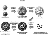
- FIG. 1a schematically shows a process for manufacturing a microstructure to which magnetic substances and cells for cartilage regeneration are attached, according to an embodiment of the present invention.
- a microstructure to which magnetic substances and cells for regenerating cartilage are attached in such a form that cells are attached to pores within the PLGA-micro-scaffold can be manufactured by manufacturing a PLGA micro-scaffold, where gelatin beads are incorporated, by forming a W-O-W emulsion along with gelatin beads after selecting a biocompatible polymer as PLGA; manufacturing a PLGA micro-scaffold by removing the gelatin; forming a covalent bond between amine-functionalized magnetic nanoparticles with PEI and the PLGA micro-scaffold through the EDC/NHS reaction; and three-dimensional culturing the mesenchymal stem cells along with the PLGA micro-scaffold to which magnetic particles are attached.
- the method is for illustrative purpose and a microstructure to which the magnetic nanoparticles and cells for cartilage regeneration are attached can be manufactured by other various methods, for example, by attaching magnetic nanoparticles and cells for cartilage regeneration to a microstructure manufactured through a method such as electrospinning, etc.
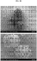
- the present inventors have manufactured a PLGA-based microstructure by the method as described above, and as a result of photographing the same by a scanning electron microscope, it was confirmed that the microstructure manufactured as described above has a spherical shape as shown in FIG. 1B .
- the microstructure formed as described above was shown to have a diameter of 269.83 ⁇ 10.982 ⁇ m, a diameter of the surface pore of 37.68 ⁇ 7.55 ⁇ m, and a diameter of the inner pore of 40.12 ⁇ 7.45 ⁇ m, thus satisfying the requirements of a microstructure as a cell transporter (a diameter of 100 ⁇ m to 500 ⁇ m and a pore size of 20 ⁇ m or greater) (see FIGS. 1C to IE).
- the microstructure to which cells for cartilage regeneration are attached contains a large amount of magnetic substances along with the cells for cartilage regeneration, it has an excellent ability to respond to a magnetic field, and it is much easier to precisely place the microstructure to a lesion of a patient and the microstructure provides a higher therapeutic effect.
- the therapeutic agent of cartilage may be prepared by appropriate adjustment according to the purpose of the researcher or the conditions of the lesion.
- 100 ⁇ g/mL of magnetic substances be added to cells for cartilage regeneration (1 ⁇ 10 5 cells/mL) and co-cultured.
- the cells for cartilage regeneration chondrocytes or stem cells that can be differentiated into cartilage may be used, and as the stem cells, adipose-derived stem cells, umbilical cord blood-derived stem cells, or bone marrow-derived stem cells may be used.
- FIG. 2A shows a schematic diagram illustrating a magnetically-actuated articular cartilage regeneration system 100 according to an embodiment of the present invention.
- an image-photographing unit 110 for irradiating X-ray to a lesion of the patient is formed at the upper part;
- a detector 115 for detecting X-ray as an image is formed at the bottom part;
- a coil-type magnetic field forming unit 150 which consists of 8 coil-type electromagnetic field forming devices for accurately placing the magnetic substances contained in the therapeutic agent for cartilage administered to the lesion, is formed at the central part.
- the magnetically-actuated articular cartilage regeneration system 100 obtains a real-time three-dimensional image through X-ray in the image-photographing unit 110 and the detector 115 after administering a therapeutic agent for cartilage treatment to the knee, and thereby the position of the magnetic substances included in the therapeutic agent for cartilage treatment prepared according to an embodiment of the present invention and the exact position of the lesion can be identified, and the therapeutic agent for cartilage treatment can be precisely located to the defected area of cartilage through control of magnetic field using the coil-type the magnetic field forming unit 150. It is important to produce a magnetic field with higher intensity so as to more precisely locate the therapeutic agent for cartilage treatment, compared to the conventional ones, in which magnetic substances are included to a lesion.
- each electromagnetic field forming device being comprised of a coil-type (i.e ., a shape where an electromagnetic coil surrounds a core comprised of soft magnet), has a magnetic field with high intensity and is able to freely control the intensity and direction of the magnetic field. Additionally, such a magnetic field forming device can control the intensity and direction of a magnetic field using the outer permanent magnet configuration and control of positions. Accordingly, the treatment of articular cartilage can be maximized based on the imaging of the image-photographing unit 110 and the detector 115.
- the coil-type magnetic field forming unit 150 of the present invention consists of eight coil-type electromagnetic field forming devices, however, the number and shape of these devices can also be appropriately adjusted according to the objects of the researcher and the position of the lesion. After the area and position for treatment of a therapeutic agent for cartilage treatment by the coil-type magnetic field forming unit 150 are identified, as shown in FIG.
- the efficiency of cartilage treatment is sought by wearing a wearable magnetic field forming device 200, not a coil-type, and the wearable magnetic field forming device 200 is composed of a wearable device 210 for wearing it on the knee considering convenience, and a plurality of permanent magnets 250 are formed inside of the device so as to generate a magnetic field with regard to the magnetic substances of the administered therapeutic agent for cartilage treatment.
- the permanent magnet 250 may be ferrite, neodymium, alnico, samarium cobalt, or rubber magnet.
- FIG. 3A represents a schematic diagram illustrating the process of treating articular cartilage using the magnetically-actuated articular cartilage regeneration system 100 according to an embodiment of the present invention.
- a therapeutic agent for cartilage treatment 130 which is in the form of single cells for cartilage regeneration where magnetic substances are bound to single cartilage regeneration cells, or a cell structure for cartilage regeneration in a spheroid shape where magnetic substances are three-dimensionally bound to multiple cells for cartilage regeneration, is administered to a lesion of a patient using a syringe; the image-photographing unit 110 irradiates x-ray on the lesion, which is embodied as a three-dimensional real-time imaging through the detector 115, and thereby the location of the administered single cells or cell structure can be identified.
- the intensity of the magnetic field is controlled so that the single cells or cell structure can move close to the defected area of the patient's cartilage by actuating the coil-type magnetic field forming unit 150 based on the photographed three-dimensional real-time imaging, and the administered single cells or cell structure can be precisely located to the defected area of cartilage lesion by controlling the intensity of all or part of the magnetic field of the coil-type magnetic field forming unit 150.
- the intensity of the magnetic field of the coil-type magnetic field forming unit 150 may be formed within a range of about 1,000 mT, and it is preferred that the magnetic intensity be controlled by utilizing the same in appropriate cartilage treatment.
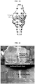
- FIG. 3B shows an image in which the wearable magnetic field forming device 200 according to an embodiment of the present invention is worn on a cartilage lesion.
- the wearable magnetic field forming device 200 is worn so as to fix the magnetic field forming position for a lesion based on the area of treatment identified previously by controlling the direction and intensity of magnetic field of the coil-type magnetic field forming unit 150 using the magnetically-actuated articular cartilage regeneration system 100.
- the wearable magnetic field forming device 200 can be directly worn by a patient on a lesion without a separate additional device and can be conveniently carried on.
- the magnetic substances i.e.
- multiple permanent magnets 250 installed inside of the wearable device 210 included in the administered therapeutic agent for cartilage treatment (single cells or a cell structure) are distributed in the most optimized location for treatment of a defected area of cartilage. Accordingly, the multiple permanent magnets 250 installed inside of the wearable device 210 may be densely distributed to a single lesion or dispersedly distributed considering the efficiency of treatment.
- FIG. 4 shows a schematic diagram illustrating the process of treating defected cartilage using the magnetically-actuated articular cartilage regeneration system 100 according to an embodiment of the present invention.
- the obtained 3D images e.g ., CT, MRI images, etc.
- the lesion is extracted, and thereby the area for treatment is determined.
- a therapeutic agent for cartilage treatment in which cells for cartilage regeneration (i.e. , stem cells or chondrocytes) are bound to magnetic substances, are administered to the periphery of treatment area using a syringe, etc.
- the therapeutic agent for cartilage treatment 130 in the form of single cells or a cell structure may be administered according to the severity and location of the lesion.
- the coil-type magnetic field forming unit 150 is actuated to control the direction and intensity of the formed magnetic field are controlled in real-time, and thereby the therapeutic agent for cartilage treatment 130 is precisely delivered to the area for treatment.
- the distribution of the delivered therapeutic agent for cartilage treatment 130 within the area for treatment is examined, and the location and distribution of the permanent magnet 250 of the wearable magnetic field forming device 200 are fixed thereby enhancing the efficiency of long-term articular cartilage treatment.
- Example 1 Manufacture of PLGA micro-scaffold body
- a PLGA micro-scaffold body was prepared by the double emulsion method using a fluidic device with a slightly modified process according to an embodiment of the present invention.
- the fluidic device was comprised of a PVE tube (1/32 in i.d. ⁇ 3/32 in o.d.), a 21G needle, and a syringe pump, and a two-way flow channels device was manufactured by inserting a needle to the PVC tube.
- a PVE tube (1/32 in i.d. ⁇ 3/32 in o.d.
- 21G needle 21G needle
- a syringe pump a two-way flow channels device was manufactured by inserting a needle to the PVC tube.
- PLGA solutions and gelatin solutions were prepared for the W-O emulsion.
- PLGA 85 mg/mL was dissolved in 100 mL PVA 5%, aqueous gelatin (0.1 g/ml) solution, and 1 mL dichloromethane (DCM)/Span80 (100:1, v/v) solution, respectively, and the W-0 emulsion was prepared by mixing with the gelatin solution (0.55-0.85 mL, 0.1 mL interval, optimized value: 0.65 mL) in the PLGA solution at 2500 rpm for 2 minutes 30 seconds.
- DCM dichloromethane
- the W-0 emulsion was filled into a syringe with a 26G needle, and the needle of the syringe was inserted into the center of the 21G needle of the fluidic device formed of PVA 1% with a flow rate of 3 mL/min passing continuously through the PVC tube of the fluidic device. Then, the W-O-W droplets formed in the fluidic channel were flowed along the 21G needle of the fluidic device, and collected into deionized water in which a 500 mL beaker in an ice bath is located.
- the dichloromethane in the collected droplets was evaporated while gently stirring for 6 hours, and for the removal of gelatin of the droplets, the resultant was immersed in the 500 mL beaker with deionized water at 37°C, and gently stirred for 4 hours. Finally, the droplets were washed 3 times with deionized water, and the gelatin-leached PLGA scaffold body was stored in deionized water in a 25 mL vial.
- the PLGA micro-scaffolds manufactured above was photographed using a scanning electron microscope. As a result, it was confirmed that the micro-scaffolds in a spherical shape with multiple pores formed thereon were normally formed as shown in FIG. 1B .
- the present inventors analyzed the distribution of the diameter, and the size of outer pores and inner pores of the PLGA micro-scaffolds manufactured above. As a result, it was confirmed that the diameter was 269.83 ⁇ 10.982 ⁇ m, the diameter of the surface pores was 37.68 ⁇ 7.55 ⁇ m, the diameter of the inner pores was 40.12 ⁇ 7.45 ⁇ m; and as shown in FIGS. 1C to 1E , it was confirmed that 90% or more of the scaffolds had a diameter of 250 ⁇ m to 290 ⁇ m, 90% or more of the surface pores had a size of 23 ⁇ m to 50 ⁇ m, and 90% or more of the inner pores had a size of 25 ⁇ m to 50 ⁇ m, thus showing a normal distribution pattern.
- Such a distribution of relatively uniform diameter and the size of surface and inner pores shows that the PLGA micro-scaffold according to an embodiment of the present invention can be efficient as a cell transporter.
- the above results satisfy the conditions of the microstructure as a cell transporter (diameter of 100 ⁇ m to 500 ⁇ m; and pore size of 20 ⁇ m or greater) ( Choi et al., J. Mater. Chem. 22: 11442, 2012 ).
- Amine-functionalized Fe 3 O 4 MNPs for providing an electromagnetic actuation ability to the scaffold body manufactured in Example 1 according to an embodiment of the present invention was manufactured.
- polyethylenimine was selected.
- 10 mM ferric chloride and 5 mM ferrous chloride were dissolved in 1 M HCl solution (12 mL), and the mixed solution was added to 1 M sodium solution (50 mL) in a four-neck round flask with a mechanical stirrer.
- the reaction solution was heated at 80°C in a dry nitrogen atmosphere, vigorously stirred for 2 hours, and formed Fe 3 O 4 MNPs in the above step and precipitated at the bottom of the flask.
- PEI (10 g) was added to the Fe 3 O 4 solution, and the solution was heated at 90°C and maintained thereat for 1 hour.
- the PEI precipitant coated with the MNPs was washed 5 times with deionized water and dispersed in an aqueous solution.
- Example 3 Manufacture of micro-scaffold to which magnetic nanoparticles are attached
- the PLGA scaffold was immersed in 0.1 M MES solution (5 mL) supplemented with 1.5 mM N-hydroxysuccinimide (NHS) and 1-ethyl-(dimethylaminopropyl)carbodiimide (EDC) at 33°C, and the solution containing the micro-scaffold was mechanically stirred for 6 hours so as to activate carboxyl groups on the surface of the PLGA micro-scaffold.
- NHS N-hydroxysuccinimide
- EDC 1-ethyl-(dimethylaminopropyl)carbodiimide
- the PEI where the MNPs(10 mg/mL) modified in 0.1 M MES (5 mL) were dispersed, was added to the solution, and stirred at 33°C for 12 hours. Then, the solution was filtered to remove unreactive MNPs and the MNPs of the micro-scaffold fixed to the filter were collected and washed three times with deionized water.
- Example 4 Manufacture and cultivation of micro-scaffold to which stem cells are loaded
- micro-scaffold to which magnetic nanoparticles (MNPs) are attached, manufactured in Example 3 was immersed in DMEM medium containing 10% FBS, and mouse bone marrow-derived mesenchymal stem cells (MSC) (1 ⁇ 10 6 cells/scaffold) were injected onto the micro-scaffold under a microscope, and cultured in an incubator with 5% CO 2 at 37°C. The medium was replaced every two days and the mesenchymal stem cells were allowed to settle in the micro-scaffold. In particular, as the control group, mesenchymal stem cells at the same concentration were injected to a micro-scaffold to which magnetic nanoparticles (MNPs) are not attached.
- MSC mouse bone marrow-derived mesenchymal stem cells
- the mesenchymal stem cells which were introduced into a micro-scaffold, were cultured in a incubator with 5% CO 2 at 36°C as described above, and simultaneously, the number of cells was measured using the Cell Titer 96 Cell Proliferation Assay kit (Promega, USA).
- Example 5 Experiment of confirming differentiation ability of stem cells
- the present inventors have examined the differentiation ability of the mesenchymal stem cells, in order to confirm whether the mesenchymal stem cells, which are attached to the micro-scaffold structure manufactured in Example 4 can be able to differentiate into chondrocytes.
- a culture dish in which the magnetic nanoparticles-coated micro-scaffold structure manufactured in Example 4 into which mesenchymal stem cells are introduced is contained, was treated with a medium for differentiation into chondrocytes consisting of DMEM medium (StemPro Chondrocyte Differentiation Kit, Thermo Fisher Scientific, USA) containing glucose (4.5 g/L), 1% ITS (insulin (25 ⁇ g/mL), transferrin (25 ( ⁇ g/mL), and sodium selenite (25 ng/mL), 0.1 ⁇ 10 -6 M dexamethasone, L-ascorbic acid-2-phophosphate (50 ⁇ g/mL), and TGF- ⁇ 1 (10 ⁇ g/mL). After 21 days, the cells were stained with Alcian Blue and the degree of differentiation of the cells into chondrocytes were observed. Part of the micro-scaffold was prepared into frozen sections and stained with Alcian Blue.
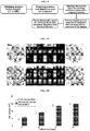
- FIGS. 7A to 7F it was confirmed that the bone marrow-derived mesenchymal stem cells settled in the micro-scaffold were differentiated into chondrocytes.
- the present inventors have analyzed the expression levels of marker genes (i.e ., Aggrecan (AGG), collagen 2A1 (COL2A1), and SOX9) related to differentiation into chondrocytes using qRT-PCR ( FIG. 8 ).
- marker genes i.e ., Aggrecan (AGG), collagen 2A1 (COL2A1), and SOX9 related to differentiation into chondrocytes using qRT-PCR ( FIG. 8 ).
- the magnetic nanoparticles-coated micro-scaffold was treated with medium for differentiation into chondrocytes, and after 21 days, the total RNA was extracted using an RNA extraction kit (TaKaRa MiniEST Universial RNA Extraction Kit, Takara, Japan), and the extract was subjected to reverse transcription using a reverse transcription kit (PrimeScript Master Mix, Takara, Japan), and then to qRT-PCR using 5X FIREPol, EvaGreen, and qPCR Supermix (Solis BioDyne, Estonia). As a result, as shown in FIG.
- the mesenchymal stem cells loaded in the differentiation-induced magnetic nanoparticles-coated micro-scaffold showed higher expression levels of marker genes related to differentiation into chondrocytes compared to the control group where differentiation was not induced. Accordingly, it was confirmed that the magnetic nanoparticles-coated micro-scaffold according to an embodiment of the present invention can normally induce the differentiation of mesenchymal stem cells into chondrocytes.
- MNPs magnetic nanoparticles
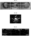
- a mold for a PDMS chamber was manufactured by attaching a dumbbell-type structure to a glass substrate using a 3D printer. Then, a PDMS polymer was prepared by mixing a curing agent and a PDMS precursor in a ratio of 10:1 was casted into a mold, and a PDMS block was attached to a slide glass using a plasma, and the prepared chamber was divided into three domains ( i.e ., control, loading, and target areas) ( FIG. 9A ).
- MSC complexes were loaded into the loading area of the microfluidic chamber, and placed on the workbench of an EMA system, in which an inverted microscope is installed, that can confirm the optical image and fluorescent image of the micro-scaffold MSC complex within the microfluidic chamber in real-time, and observed.
- the EMA system was tested as described below using a magnetically-actuated system ( FIG. 9B ) consisting of a permanent magnet comprised of four cylindrical neodymium magnets (diameter 10 mm ⁇ height 20 mm) with a magnetization value of 955 kA/m.
- the micro-scaffold was loaded into the loading section in the microfluidic chamber of a PDMS material in which glycerin (70 wt%) solution, which has a viscosity at room temperature is similar to that of the synovial fluid, is supported; and then the microfluidic chamber was placed on the workbench of a magnetically-actuated system A, and the fixed uniform magnetic field (30 mT) and variable magnetic gradient field (0.2-1.2 T/m, interval 0.2 T/m) were produced in the EMA system along each of x and z axes, and the micro-scaffold was promoted toward the direction of the gradient magnetic field. Finally, to obtain the velocity, the recorded locomotion was analyzed.
- the targeting ability of the magnetically-actuated micro-scaffold manufactured according to an embodiment of the present invention to the defected area of the joint was observed.
- the knee phantom for the test of 3D targeting ability was manufactured from a 3D CAD model of a knee using a 3D printer ( FIG. 10 ).
- apertures with a depth of 3 mm and a diameter of 2 mm were formed toward the target area inside of the femur, and the above model manufactured for the test of 3D targeting ability was placed in the acrylic chamber filled with glycerin solution (70 wt%).
- 5 micro-scaffolds were injected between the defected areas of the joint of the knee model phantom by pipetting, and the locomotion into the target domain was observed.
- the use of the magnetically-actuated articular cartilage regeneration system 100 of the present invention after the administration of a therapeutic agent for cartilage treatment containing magnetic substances, can control the intensity of magnetic field and precisely locate and fix the therapeutic agent for cartilage treatment to a lesion based on the real-time image information, thereby promoting nonsurgical and efficient effect of cartilage treatment.
- composition for regenerating cartilage may be effectively used for the treatment of other diseases due to cartilage damage such as osteoarthritis.
Landscapes
- Health & Medical Sciences (AREA)
- Life Sciences & Earth Sciences (AREA)
- Engineering & Computer Science (AREA)
- Chemical & Material Sciences (AREA)
- Public Health (AREA)
- Biomedical Technology (AREA)
- General Health & Medical Sciences (AREA)
- Veterinary Medicine (AREA)
- Animal Behavior & Ethology (AREA)
- Epidemiology (AREA)
- Medicinal Chemistry (AREA)
- Oral & Maxillofacial Surgery (AREA)
- Transplantation (AREA)
- Dermatology (AREA)
- Cell Biology (AREA)
- Medical Informatics (AREA)
- Radiology & Medical Imaging (AREA)
- Nuclear Medicine, Radiotherapy & Molecular Imaging (AREA)
- Zoology (AREA)
- Chemical Kinetics & Catalysis (AREA)
- Heart & Thoracic Surgery (AREA)
- Botany (AREA)
- Pharmacology & Pharmacy (AREA)
- Developmental Biology & Embryology (AREA)
- Pathology (AREA)
- Rheumatology (AREA)
- Orthopedic Medicine & Surgery (AREA)
- Optics & Photonics (AREA)
- Biophysics (AREA)
- Surgery (AREA)
- Molecular Biology (AREA)
- High Energy & Nuclear Physics (AREA)
- Physics & Mathematics (AREA)
- Urology & Nephrology (AREA)
- Immunology (AREA)
- Vascular Medicine (AREA)
- Hematology (AREA)
- Bioinformatics & Cheminformatics (AREA)
- Dentistry (AREA)
- Dispersion Chemistry (AREA)
Applications Claiming Priority (2)
| Application Number | Priority Date | Filing Date | Title |
|---|---|---|---|
| KR1020160139848A KR101927196B1 (ko) | 2016-10-26 | 2016-10-26 | 자기구동 관절연골 재생 시스템 |
| PCT/KR2017/011901 WO2018080191A1 (ko) | 2016-10-26 | 2017-10-26 | 자기구동 관절연골 재생 시스템 |
Publications (3)
| Publication Number | Publication Date |
|---|---|
| EP3545981A1 true EP3545981A1 (de) | 2019-10-02 |
| EP3545981A4 EP3545981A4 (de) | 2020-07-01 |
| EP3545981B1 EP3545981B1 (de) | 2025-02-19 |
Family
ID=60387543
Family Applications (1)
| Application Number | Title | Priority Date | Filing Date |
|---|---|---|---|
| EP17864166.8A Active EP3545981B1 (de) | 2016-10-26 | 2017-10-26 | Magnetisch betätigtes gelenkknorpelregenerationssystem |
Country Status (5)
| Country | Link |
|---|---|
| US (1) | US10668184B2 (de) |
| EP (1) | EP3545981B1 (de) |
| JP (1) | JP6901166B2 (de) |
| KR (1) | KR101927196B1 (de) |
| WO (1) | WO2018080191A1 (de) |
Families Citing this family (16)
| Publication number | Priority date | Publication date | Assignee | Title |
|---|---|---|---|---|
| US10507111B2 (en) * | 2018-03-09 | 2019-12-17 | Stephen Bramblett Johnson | Magnetic prosthetic |
| KR102114300B1 (ko) * | 2018-04-16 | 2020-05-22 | 전남대학교 산학협력단 | 최소 침습 골연골 재생용 자기 구동 마이크로지지체 |
| KR102315368B1 (ko) * | 2018-08-13 | 2021-10-20 | 재단법인대구경북과학기술원 | 자기장 구동 약물전달 마이크로 구조체 및 이의 제조방법 |
| KR102248370B1 (ko) | 2018-12-11 | 2021-05-07 | (주)바이오트코리아 | 세포 탑재용 모듈 장치 |
| KR102366335B1 (ko) * | 2019-03-29 | 2022-02-23 | 전남대학교산학협력단 | 조직재생용 성장인자 담지 다공성 마이크로스캐폴드 및 이의 제조방법 |
| KR102351937B1 (ko) * | 2019-03-29 | 2022-01-18 | 전남대학교산학협력단 | 무릎 관절 재생을 위한 자기 구동 시스템 |
| KR102366334B1 (ko) * | 2019-03-29 | 2022-02-22 | 전남대학교산학협력단 | 자기 구동력이 증가된 조직재생용 다공성 마이크로스캐폴드 및 이의 제조방법 |
| KR20210062378A (ko) * | 2019-11-21 | 2021-05-31 | (주)바이오트코리아 | 자기 구동이 가능한 기능성 마이크로스캐폴드 및 이의 제조방법 |
| KR102428203B1 (ko) * | 2020-02-25 | 2022-08-04 | 서울대학교산학협력단 | 질병 치료를 위한 교류 자기장 발생 장치 및 시스템 |
| KR102396312B1 (ko) * | 2020-08-07 | 2022-05-10 | 전남대학교산학협력단 | 체외에 위치한 자석 배열 장치와 병변 부위에 삽입된 자기 반응 임플란트를 이용한 골질환 치료제 표적화 장치 |
| KR20220076663A (ko) * | 2020-12-01 | 2022-06-08 | 재단법인대구경북과학기술원 | 세포치료제 전달용 마이크로 로봇 제작 방법 및 그에 따른 마이크로 로봇 |
| CN112657048B (zh) * | 2021-01-26 | 2024-04-02 | 南方科技大学 | 一种非手性磁控微型机器人及其制备方法和应用 |
| CN114767345A (zh) * | 2022-03-18 | 2022-07-22 | 上海应用技术大学 | 磁流变弹性体半月板 |
| KR102882681B1 (ko) * | 2022-06-15 | 2025-11-11 | (주)바이오트코리아 | 자기구동이 가능한 치료물질 전달용 하이드로겔 마이크로비즈의 제조방법, 이에 의해 제조된 하이드로겔 마이크로비즈 및 이를 포함하는 근골격계 질환 치료용 약학 조성물 |
| KR20250132868A (ko) * | 2024-02-29 | 2025-09-05 | 가톨릭대학교 산학협력단 | 자성 나노 입자가 함입된 코중격 연골세포를 유효성분으로 포함하는 연골 질환 치료용 약학적 조성물 |
| WO2025254247A1 (ko) * | 2024-06-05 | 2025-12-11 | (주)이즈소프트 | 히알루론산을 이용한 치간유두의 재건방법 |
Family Cites Families (12)
| Publication number | Priority date | Publication date | Assignee | Title |
|---|---|---|---|---|
| US5655546A (en) * | 1995-06-07 | 1997-08-12 | Halpern; Alan A. | Method for cartilage repair |
| KR100799060B1 (ko) * | 2003-06-30 | 2008-01-29 | 에자이 가부시키가이샤 | 자성 세포 및 그의 사용 방법 |
| IT1401487B1 (it) * | 2010-07-29 | 2013-07-26 | Consiglio Nazionale Ricerche | Idrossiapatite intrinsecamente magnetica |
| KR20120004971U (ko) | 2010-12-30 | 2012-07-10 | 김정환 | 무릎 관절 보호 보조대 |
| CN102302822B (zh) * | 2011-05-19 | 2012-07-18 | 中国人民解放军第三军医大学第一附属医院 | 治疗关节软骨缺损的磁标记细胞靶向移植可控磁聚焦仪 |
| KR20140121590A (ko) * | 2013-04-08 | 2014-10-16 | 재단법인대구경북과학기술원 | 자기장 제어 가능한 이동식 생체 지지체 및 그 제조방법 |
| KR101630017B1 (ko) | 2013-10-04 | 2016-06-13 | 동국대학교 산학협력단 | 자성나노입자와 전자기장을 이용한 골 재생 촉진 방법 |
| KR20160028070A (ko) | 2014-09-02 | 2016-03-11 | 단국대학교 천안캠퍼스 산학협력단 | 자성 나노입자를 포함하는 고세포부착성 인산칼슘 골시멘트 조성물, 이의 제조방법 및 이의 제조를 위한 키트 |
| KR20160031683A (ko) | 2014-09-12 | 2016-03-23 | 단국대학교 천안캠퍼스 산학협력단 | 기계적 및 생물학적 특성이 향상된 자성 나노섬유 스캐폴드의 제조방법 및 그 제조방법에 의해 제조된 자성 나노섬유 스캐폴드 |
| KR101627043B1 (ko) * | 2014-09-12 | 2016-06-03 | 단국대학교 천안캠퍼스 산학협력단 | 표면 기능화된 나노입자를 포함하는 뼈 재생용 폴리머 자성 스캐폴드의 제조방법 및 그 제조방법에 의하여 제조된 뼈 재생용 자성 스캐폴드 |
| CN107106209B (zh) * | 2014-10-23 | 2020-07-14 | 诺威适骨科专科公司 | 骨骼生长装置和用于该骨骼生长装置的外部遥控 |
| KR20170099417A (ko) * | 2016-02-04 | 2017-09-01 | 전남대학교산학협력단 | 환자 맞춤 착용형 관절질환 치료용 의료장치 |
-
2016
- 2016-10-26 KR KR1020160139848A patent/KR101927196B1/ko active Active
-
2017
- 2017-10-26 WO PCT/KR2017/011901 patent/WO2018080191A1/ko not_active Ceased
- 2017-10-26 JP JP2019523699A patent/JP6901166B2/ja active Active
- 2017-10-26 US US16/097,714 patent/US10668184B2/en active Active
- 2017-10-26 EP EP17864166.8A patent/EP3545981B1/de active Active
Also Published As
| Publication number | Publication date |
|---|---|
| US20190167847A1 (en) | 2019-06-06 |
| JP2020500058A (ja) | 2020-01-09 |
| JP6901166B2 (ja) | 2021-07-14 |
| KR101927196B1 (ko) | 2018-12-11 |
| WO2018080191A1 (ko) | 2018-05-03 |
| US10668184B2 (en) | 2020-06-02 |
| EP3545981A4 (de) | 2020-07-01 |
| EP3545981B1 (de) | 2025-02-19 |
| KR20170125290A (ko) | 2017-11-14 |
Similar Documents
| Publication | Publication Date | Title |
|---|---|---|
| US10668184B2 (en) | Magnetically actuated articular cartilage regeneration system | |
| Spangenberg et al. | Bioprinting of magnetically deformable scaffolds | |
| Xu et al. | Magnetic responsive scaffolds and magnetic fields in bone repair and regeneration | |
| Zhao et al. | Magnetic bioinspired micro/nanostructured composite scaffold for bone regeneration | |
| Gao et al. | Emerging translational research on magnetic nanoparticles for regenerative medicine | |
| Li et al. | Adaptive materials based on iron oxide nanoparticles for bone regeneration | |
| US8815276B2 (en) | Three-dimensional nanostructured hybrid scaffold and manufacture thereof | |
| CN101980730B (zh) | 用于磁性粒子成像的生物相容产品 | |
| Sedaghati et al. | Investigation of Schwann cell behaviour on RGD-functionalised bioabsorbable nanocomposite for peripheral nerve regeneration | |
| Shakhssalim et al. | Bladder smooth muscle cells on electrospun poly (ε-caprolactone)/poly (l-lactic acid) scaffold promote bladder regeneration in a canine model | |
| KR102114300B1 (ko) | 최소 침습 골연골 재생용 자기 구동 마이크로지지체 | |
| KR102641841B1 (ko) | 자성 나노입자를 함유하는 세포-약물 전달용 다공성 마이크로 입자 및 이의 제조방법 | |
| US20210155897A1 (en) | Surface acoustic wave (saw) 3d printing method | |
| Kubinová | Biomaterials and magnetic stem cell delivery in the treatment of spinal cord injury | |
| KR20140121590A (ko) | 자기장 제어 가능한 이동식 생체 지지체 및 그 제조방법 | |
| WO2021232108A1 (en) | Ceramic biofabrication | |
| Kryuchkova et al. | Magnetothermal spider silk-based scaffolds for cartilage regeneration | |
| JP2022552097A (ja) | 新規な多孔性スキャフォールドおよびその製造方法 | |
| Oliveira et al. | Nanoparticles-based systems for osteochondral tissue engineering | |
| US20260115359A1 (en) | Tissue-derived implant useful for cartilage regeneration | |
| Klapper et al. | Session 4: Cell-Material Interaction | |
| Wang et al. | 3D-printed shape memory scaffolds from Pickering emulsion for codelivery of exosomes and Mg²⁺ to promote bone regeneration | |
| Yin | Preparation of Electrospun Fiber Membranes with Different Morphologies and Their Effects on the Proliferation and Differentiation of Osteoblasts | |
| Zhu et al. | Elastic porous microspheres combined with extracellular matrix hydrogel as injectable composites for the dual-release of bioactive factors to promote tissue regeneration | |
| KR101574585B1 (ko) | 자기장 제어 가능한 이동식 생체 지지체 |
Legal Events
| Date | Code | Title | Description |
|---|---|---|---|
| STAA | Information on the status of an ep patent application or granted ep patent |
Free format text: STATUS: THE INTERNATIONAL PUBLICATION HAS BEEN MADE |
|
| PUAI | Public reference made under article 153(3) epc to a published international application that has entered the european phase |
Free format text: ORIGINAL CODE: 0009012 |
|
| STAA | Information on the status of an ep patent application or granted ep patent |
Free format text: STATUS: REQUEST FOR EXAMINATION WAS MADE |
|
| 17P | Request for examination filed |
Effective date: 20190524 |
|
| AK | Designated contracting states |
Kind code of ref document: A1 Designated state(s): AL AT BE BG CH CY CZ DE DK EE ES FI FR GB GR HR HU IE IS IT LI LT LU LV MC MK MT NL NO PL PT RO RS SE SI SK SM TR |
|
| AX | Request for extension of the european patent |
Extension state: BA ME |
|
| DAV | Request for validation of the european patent (deleted) | ||
| DAX | Request for extension of the european patent (deleted) | ||
| A4 | Supplementary search report drawn up and despatched |
Effective date: 20200603 |
|
| RIC1 | Information provided on ipc code assigned before grant |
Ipc: A61F 2/30 20060101ALI20200526BHEP Ipc: A61L 27/38 20060101AFI20200526BHEP Ipc: A61L 27/58 20060101ALI20200526BHEP Ipc: A61L 27/10 20060101ALI20200526BHEP Ipc: A61L 27/56 20060101ALI20200526BHEP |
|
| STAA | Information on the status of an ep patent application or granted ep patent |
Free format text: STATUS: EXAMINATION IS IN PROGRESS |
|
| 17Q | First examination report despatched |
Effective date: 20220105 |
|
| REG | Reference to a national code |
Ref country code: DE Ipc: A61B0006500000 Ref country code: DE Ref legal event code: R079 Ref document number: 602017087916 Country of ref document: DE Free format text: PREVIOUS MAIN CLASS: A61L0027380000 Ipc: A61B0006500000 |
|
| GRAP | Despatch of communication of intention to grant a patent |
Free format text: ORIGINAL CODE: EPIDOSNIGR1 |
|
| STAA | Information on the status of an ep patent application or granted ep patent |
Free format text: STATUS: GRANT OF PATENT IS INTENDED |
|
| RIC1 | Information provided on ipc code assigned before grant |
Ipc: A61N 2/06 20060101ALI20240812BHEP Ipc: G16H 50/30 20180101ALI20240812BHEP Ipc: A61N 2/02 20060101ALI20240812BHEP Ipc: A61N 2/00 20060101ALI20240812BHEP Ipc: A61L 27/58 20060101ALI20240812BHEP Ipc: A61L 27/56 20060101ALI20240812BHEP Ipc: A61L 27/50 20060101ALI20240812BHEP Ipc: A61L 27/38 20060101ALI20240812BHEP Ipc: A61L 27/18 20060101ALI20240812BHEP Ipc: A61K 47/69 20170101ALI20240812BHEP Ipc: A61K 41/00 20060101ALI20240812BHEP Ipc: A61K 35/32 20150101ALI20240812BHEP Ipc: A61K 35/28 20150101ALI20240812BHEP Ipc: A61F 2/30 20060101ALI20240812BHEP Ipc: A61B 6/00 20060101ALI20240812BHEP Ipc: A61B 6/50 20240101AFI20240812BHEP |
|
| INTG | Intention to grant announced |
Effective date: 20240911 |
|
| GRAS | Grant fee paid |
Free format text: ORIGINAL CODE: EPIDOSNIGR3 |
|
| GRAA | (expected) grant |
Free format text: ORIGINAL CODE: 0009210 |
|
| STAA | Information on the status of an ep patent application or granted ep patent |
Free format text: STATUS: THE PATENT HAS BEEN GRANTED |
|
| AK | Designated contracting states |
Kind code of ref document: B1 Designated state(s): AL AT BE BG CH CY CZ DE DK EE ES FI FR GB GR HR HU IE IS IT LI LT LU LV MC MK MT NL NO PL PT RO RS SE SI SK SM TR |
|
| REG | Reference to a national code |
Ref country code: GB Ref legal event code: FG4D |
|
| REG | Reference to a national code |
Ref country code: CH Ref legal event code: EP |
|
| REG | Reference to a national code |
Ref country code: DE Ref legal event code: R096 Ref document number: 602017087916 Country of ref document: DE |
|
| REG | Reference to a national code |
Ref country code: IE Ref legal event code: FG4D |
|
| REG | Reference to a national code |
Ref country code: NL Ref legal event code: MP Effective date: 20250219 |
|
| PG25 | Lapsed in a contracting state [announced via postgrant information from national office to epo] |
Ref country code: RS Free format text: LAPSE BECAUSE OF FAILURE TO SUBMIT A TRANSLATION OF THE DESCRIPTION OR TO PAY THE FEE WITHIN THE PRESCRIBED TIME-LIMIT Effective date: 20250519 |
|
| PG25 | Lapsed in a contracting state [announced via postgrant information from national office to epo] |
Ref country code: FI Free format text: LAPSE BECAUSE OF FAILURE TO SUBMIT A TRANSLATION OF THE DESCRIPTION OR TO PAY THE FEE WITHIN THE PRESCRIBED TIME-LIMIT Effective date: 20250219 |
|
| PG25 | Lapsed in a contracting state [announced via postgrant information from national office to epo] |
Ref country code: PL Free format text: LAPSE BECAUSE OF FAILURE TO SUBMIT A TRANSLATION OF THE DESCRIPTION OR TO PAY THE FEE WITHIN THE PRESCRIBED TIME-LIMIT Effective date: 20250219 |
|
| PG25 | Lapsed in a contracting state [announced via postgrant information from national office to epo] |
Ref country code: ES Free format text: LAPSE BECAUSE OF FAILURE TO SUBMIT A TRANSLATION OF THE DESCRIPTION OR TO PAY THE FEE WITHIN THE PRESCRIBED TIME-LIMIT Effective date: 20250219 |
|
| REG | Reference to a national code |
Ref country code: LT Ref legal event code: MG9D |
|
| PG25 | Lapsed in a contracting state [announced via postgrant information from national office to epo] |
Ref country code: IS Free format text: LAPSE BECAUSE OF FAILURE TO SUBMIT A TRANSLATION OF THE DESCRIPTION OR TO PAY THE FEE WITHIN THE PRESCRIBED TIME-LIMIT Effective date: 20250619 Ref country code: NO Free format text: LAPSE BECAUSE OF FAILURE TO SUBMIT A TRANSLATION OF THE DESCRIPTION OR TO PAY THE FEE WITHIN THE PRESCRIBED TIME-LIMIT Effective date: 20250519 |
|
| PG25 | Lapsed in a contracting state [announced via postgrant information from national office to epo] |
Ref country code: NL Free format text: LAPSE BECAUSE OF FAILURE TO SUBMIT A TRANSLATION OF THE DESCRIPTION OR TO PAY THE FEE WITHIN THE PRESCRIBED TIME-LIMIT Effective date: 20250219 |
|
| PG25 | Lapsed in a contracting state [announced via postgrant information from national office to epo] |
Ref country code: HR Free format text: LAPSE BECAUSE OF FAILURE TO SUBMIT A TRANSLATION OF THE DESCRIPTION OR TO PAY THE FEE WITHIN THE PRESCRIBED TIME-LIMIT Effective date: 20250219 |
|
| PG25 | Lapsed in a contracting state [announced via postgrant information from national office to epo] |
Ref country code: LV Free format text: LAPSE BECAUSE OF FAILURE TO SUBMIT A TRANSLATION OF THE DESCRIPTION OR TO PAY THE FEE WITHIN THE PRESCRIBED TIME-LIMIT Effective date: 20250219 Ref country code: PT Free format text: LAPSE BECAUSE OF FAILURE TO SUBMIT A TRANSLATION OF THE DESCRIPTION OR TO PAY THE FEE WITHIN THE PRESCRIBED TIME-LIMIT Effective date: 20250620 |
|
| PG25 | Lapsed in a contracting state [announced via postgrant information from national office to epo] |
Ref country code: GR Free format text: LAPSE BECAUSE OF FAILURE TO SUBMIT A TRANSLATION OF THE DESCRIPTION OR TO PAY THE FEE WITHIN THE PRESCRIBED TIME-LIMIT Effective date: 20250520 Ref country code: BG Free format text: LAPSE BECAUSE OF FAILURE TO SUBMIT A TRANSLATION OF THE DESCRIPTION OR TO PAY THE FEE WITHIN THE PRESCRIBED TIME-LIMIT Effective date: 20250219 |
|
| REG | Reference to a national code |
Ref country code: AT Ref legal event code: MK05 Ref document number: 1767536 Country of ref document: AT Kind code of ref document: T Effective date: 20250219 |
|
| PG25 | Lapsed in a contracting state [announced via postgrant information from national office to epo] |
Ref country code: SE Free format text: LAPSE BECAUSE OF FAILURE TO SUBMIT A TRANSLATION OF THE DESCRIPTION OR TO PAY THE FEE WITHIN THE PRESCRIBED TIME-LIMIT Effective date: 20250219 |
|
| PG25 | Lapsed in a contracting state [announced via postgrant information from national office to epo] |
Ref country code: SM Free format text: LAPSE BECAUSE OF FAILURE TO SUBMIT A TRANSLATION OF THE DESCRIPTION OR TO PAY THE FEE WITHIN THE PRESCRIBED TIME-LIMIT Effective date: 20250219 |
|
| PG25 | Lapsed in a contracting state [announced via postgrant information from national office to epo] |
Ref country code: DK Free format text: LAPSE BECAUSE OF FAILURE TO SUBMIT A TRANSLATION OF THE DESCRIPTION OR TO PAY THE FEE WITHIN THE PRESCRIBED TIME-LIMIT Effective date: 20250219 |
|
| PGFP | Annual fee paid to national office [announced via postgrant information from national office to epo] |
Ref country code: IT Payment date: 20250922 Year of fee payment: 9 |
|
| PG25 | Lapsed in a contracting state [announced via postgrant information from national office to epo] |
Ref country code: AT Free format text: LAPSE BECAUSE OF FAILURE TO SUBMIT A TRANSLATION OF THE DESCRIPTION OR TO PAY THE FEE WITHIN THE PRESCRIBED TIME-LIMIT Effective date: 20250219 |
|
| PGFP | Annual fee paid to national office [announced via postgrant information from national office to epo] |
Ref country code: FR Payment date: 20250908 Year of fee payment: 9 |
|
| PG25 | Lapsed in a contracting state [announced via postgrant information from national office to epo] |
Ref country code: EE Free format text: LAPSE BECAUSE OF FAILURE TO SUBMIT A TRANSLATION OF THE DESCRIPTION OR TO PAY THE FEE WITHIN THE PRESCRIBED TIME-LIMIT Effective date: 20250219 Ref country code: CZ Free format text: LAPSE BECAUSE OF FAILURE TO SUBMIT A TRANSLATION OF THE DESCRIPTION OR TO PAY THE FEE WITHIN THE PRESCRIBED TIME-LIMIT Effective date: 20250219 |
|
| PG25 | Lapsed in a contracting state [announced via postgrant information from national office to epo] |
Ref country code: RO Free format text: LAPSE BECAUSE OF FAILURE TO SUBMIT A TRANSLATION OF THE DESCRIPTION OR TO PAY THE FEE WITHIN THE PRESCRIBED TIME-LIMIT Effective date: 20250219 |
|
| PG25 | Lapsed in a contracting state [announced via postgrant information from national office to epo] |
Ref country code: SK Free format text: LAPSE BECAUSE OF FAILURE TO SUBMIT A TRANSLATION OF THE DESCRIPTION OR TO PAY THE FEE WITHIN THE PRESCRIBED TIME-LIMIT Effective date: 20250219 |
|
| REG | Reference to a national code |
Ref country code: DE Ref legal event code: R097 Ref document number: 602017087916 Country of ref document: DE |
|
| PLBE | No opposition filed within time limit |
Free format text: ORIGINAL CODE: 0009261 |
|
| STAA | Information on the status of an ep patent application or granted ep patent |
Free format text: STATUS: NO OPPOSITION FILED WITHIN TIME LIMIT |
|
| PGFP | Annual fee paid to national office [announced via postgrant information from national office to epo] |
Ref country code: DE Payment date: 20250902 Year of fee payment: 9 |
|
| 26N | No opposition filed |
Effective date: 20251120 |