EP3541409B1 - Engineered tgf-beta monomers and their use for inhibiting tgf-beta signaling - Google Patents
Engineered tgf-beta monomers and their use for inhibiting tgf-beta signaling Download PDFInfo
- Publication number
- EP3541409B1 EP3541409B1 EP17871472.1A EP17871472A EP3541409B1 EP 3541409 B1 EP3541409 B1 EP 3541409B1 EP 17871472 A EP17871472 A EP 17871472A EP 3541409 B1 EP3541409 B1 EP 3541409B1
- Authority
- EP
- European Patent Office
- Prior art keywords
- tgf
- monomer
- residue
- amino acid
- substitution
- Prior art date
- Legal status (The legal status is an assumption and is not a legal conclusion. Google has not performed a legal analysis and makes no representation as to the accuracy of the status listed.)
- Active
Links
Images


Classifications
-
- A—HUMAN NECESSITIES
- A61—MEDICAL OR VETERINARY SCIENCE; HYGIENE
- A61P—SPECIFIC THERAPEUTIC ACTIVITY OF CHEMICAL COMPOUNDS OR MEDICINAL PREPARATIONS
- A61P19/00—Drugs for skeletal disorders
-
- A—HUMAN NECESSITIES
- A61—MEDICAL OR VETERINARY SCIENCE; HYGIENE
- A61K—PREPARATIONS FOR MEDICAL, DENTAL OR TOILETRY PURPOSES
- A61K45/00—Medicinal preparations containing active ingredients not provided for in groups A61K31/00 - A61K41/00
- A61K45/06—Mixtures of active ingredients without chemical characterisation, e.g. antiphlogistics and cardiaca
-
- C—CHEMISTRY; METALLURGY
- C07—ORGANIC CHEMISTRY
- C07K—PEPTIDES
- C07K14/00—Peptides having more than 20 amino acids; Gastrins; Somatostatins; Melanotropins; Derivatives thereof
- C07K14/435—Peptides having more than 20 amino acids; Gastrins; Somatostatins; Melanotropins; Derivatives thereof from animals; from humans
- C07K14/475—Growth factors; Growth regulators
- C07K14/495—Transforming growth factor [TGF]
-
- A—HUMAN NECESSITIES
- A61—MEDICAL OR VETERINARY SCIENCE; HYGIENE
- A61K—PREPARATIONS FOR MEDICAL, DENTAL OR TOILETRY PURPOSES
- A61K38/00—Medicinal preparations containing peptides
-
- C—CHEMISTRY; METALLURGY
- C07—ORGANIC CHEMISTRY
- C07K—PEPTIDES
- C07K2319/00—Fusion polypeptide
-
- C—CHEMISTRY; METALLURGY
- C07—ORGANIC CHEMISTRY
- C07K—PEPTIDES
- C07K2319/00—Fusion polypeptide
- C07K2319/30—Non-immunoglobulin-derived peptide or protein having an immunoglobulin constant or Fc region, or a fragment thereof, attached thereto
-
- C—CHEMISTRY; METALLURGY
- C07—ORGANIC CHEMISTRY
- C07K—PEPTIDES
- C07K2319/00—Fusion polypeptide
- C07K2319/31—Fusion polypeptide fusions, other than Fc, for prolonged plasma life, e.g. albumin
Definitions
- TGF- ⁇ monomers selected from TGF- ⁇ 1, TGF- ⁇ 2, and TGF- ⁇ 3 modified to inhibit dimerization while retaining the capacity to bind the high affinity TGF- ⁇ type II receptor (T ⁇ RII).
- TGF- ⁇ RII TGF- ⁇ type II receptor
- TGF- ⁇ is a multifunctional cytokine with diverse biological effects on cellular processes, including cell proliferation, migration, differentiation, and apoptosis.
- Receptor activation induces both SMAD proteins and other downstream targets, including Ras, RhoA, TAK1, MEKK1, PI3K, and PP2A, to produce the full spectrum of TGF- ⁇ responses ( Roberts and Wakefield, Proc Natl Acad Sci USA 100:8621-8623, 2003 ; Derynck and Zhang, Nature 425:577-584, 2003 ; Massagué, Cell 134:215-230, 2008 ).
- TGF- ⁇ proteins are known to promote the progression of fibrotic disorders and certain types of cancer. In the context of fibrotic disorders, TGF- ⁇ potently stimulates the expression of extracellular matrix (ECM) proteins. Dysregulation of the ECM remodeling can lead to pathological fibrosis.
- ECM extracellular matrix
- TGF- ⁇ isoforms, TGF- ⁇ 1, - ⁇ 2 and - ⁇ 3 are also known to suppress host immune surveillance and to stimulate epithelial-to-mesenchymal transitions, which drive cancer progression and metastasis.
- the recombinant human TGF- ⁇ monomer further includes at least one amino acid substitution relative to a wild-type TFG- ⁇ 2 monomer that increases affinity of the TGF- ⁇ monomer for TGF- ⁇ type II receptor (T ⁇ RII), wherein the at least one amino acid substitution that increases affinity of the monomer for T ⁇ RII comprises a substitution at an amino acid residue corresponding to residue 23, 24, 25, 26, 27, 28, 29, 30, 31, 32, 33, 34, 35, 36, 37, 89, 90, 91, 92, 93, 94, 95, 96, 97, 98 or 99 of SEQ ID NO: 2, or any combination of two or more residues thereof.
- T ⁇ RII TGF- ⁇ type II receptor
- the recombinant human TGF- ⁇ 2 monomer comprises at least one amino acid substitution that increases affinity of the monomer for T ⁇ RII comprises at least one substitution at residue 23, 24, 25, 26, 27, 28, 29, 30, 31, 32, 33, 34, 35, 36 or 37, and at least one substitution at residue 89, 90, 91, 92, 93, 94, 95, 96, 97, 98 or 99.
- the recombinant human TGF- ⁇ 2 monomer comprises at least one amino acid substitution that increases affinity of the monomer for T ⁇ RII comprises a lysine to arginine at residue 25, an arginine to lysine at residue 26, a leucine to valine at residue 89, an isoleucine to valine at residue 92, an asparagine to arginine at residue 94, a threonine to lysine at residue 95, an isoleucine to valine at residue 98; or any combination of two or more thereof.
- the recombinant human TGF- ⁇ 2 monomer comprises at least one amino acid substitution that increases affinity of the monomer for T ⁇ RII comprises a lysine to arginine at residue 25, an arginine to lysine at residue 26, a leucine to valine at residue 89, an isoleucine to valine at residue 92, an asparagine to arginine at residue 94, a threonine to lysine at residue 95, and an isoleucine to valine at residue 98.
- the recombinant human TGF- ⁇ 2 monomer comprises the amino acid sequence of SEQ ID NO: 8 or SEQ ID NO: 10.
- the recombinant human TGF- ⁇ monomer is a human TGF- ⁇ 1 monomer.
- the recombinant human TGF- ⁇ 1 monomer comprises at least one amino acid substitution that increases net charge of the monomer comprises:
- the recombinant human TGF- ⁇ 1 monomer comprises the amino acid sequence of SEQ ID NO: 7. In some embodiments, the recombinant human TGF- ⁇ monomer is a human TGF- ⁇ 3 monomer.
- the recombinant human TGF- ⁇ 3 monomer comprises at least one amino acid substitution that increases net charge of the monomer comprises:
- Fusion proteins that include a recombinant human TGF- ⁇ monomer and a heterologous protein are also provided, particularly wherein the heterologous protein comprises a protein tag,
- the recombinant human TGF- ⁇ monomer, fusion protein or composition are for use in inhibiting TGF- ⁇ signaling in a cell by contacting the cell with a recombinant human TGF- ⁇ monomer, fusion protein, or composition disclosed herein.
- the use further includes administering the recombinant human TGF- ⁇ monomer, fusion protein or composition to a subject having a disease or disorder associated with aberrant TGF- ⁇ signaling.
- the recombinant human TGF- ⁇ monomer, fusion protein, or composition are for or use in treating a disease or disorder associated with aberrant TGF- ⁇ signaling in a subject, comprising administering to the subject the recombinant human TGF- ⁇ monomer, fusion protein, or composition disclosed herein, particularly wherein the disease or disorder associated with aberrant TGF- ⁇ signaling is a fibrotic disorder,
- An isolated cell for use in treating a disease or disorder associated with aberrant TGF- ⁇ signaling in a subject by administering to the subject an isolated cell (such as a T cell) comprising the disclosed nucleic acids or vectors are further provided, particularly wherein the disease or disorder associated with aberrant TGF- ⁇ signaling is a fibrotic disorder,
- Conservative amino acid substitutions are those substitutions that, when made, least interfere with the properties of the original protein, that is, the structure and especially the function of the protein is conserved and not significantly changed by such substitutions. Examples of conservative substitutions are shown below.
- Original Residue Conservative Substitutions Ala Ser Arg Lys Asn Gln, His Asp Glu Cys Ser Gln Asn Glu Asp His Asn; Gln Ile Leu, Val Leu Ile; Val Lys Arg; Gln; Glu Met Leu; Ile Phe Met; Leu; Tyr Ser Thr Thr Ser Trp Tyr Tyr Trp; Phe Val Ile; Leu
- Conservative substitutions generally maintain (a) the structure of the polypeptide backbone in the area of the substitution, for example, as a sheet or helical conformation, (b) the charge or hydrophobicity of the molecule at the target site, or (c) the bulk of the side chain.
- substitutions which in general are expected to produce the greatest changes in protein properties will be non-conservative, for instance changes in which (a) a hydrophilic residue, for example, serine or threonine, is substituted for (or by) a hydrophobic residue, for example, leucine, isoleucine, phenylalanine, valine or alanine; (b) a cysteine or proline is substituted for (or by) any other residue; (c) a residue having an electropositive side chain, for example, lysine, arginine, or histidine, is substituted for (or by) an electronegative residue, for example, glutamine or aspartic acid; or (d) a residue having a bulky side chain, for example, phenylalanine, is substituted for (or by) one not having a side chain, for example, glycine.
- a hydrophilic residue for example, serine or threonine
- a hydrophobic residue for example, leucine,
- a recombinant nucleic acid or protein is one that has a sequence that is not naturally occurring or has a sequence that is made by an artificial combination of two otherwise separated segments of sequence. This artificial combination is often accomplished by chemical synthesis or by the artificial manipulation of isolated segments of nucleic acids, for example, by genetic engineering techniques.
- the term recombinant includes nucleic acids and proteins that have been altered by addition, substitution, or deletion of a portion of a natural nucleic acid molecule or protein.
- NCBI Basic Local Alignment Search Tool (BLAST) ( Altschul et al., J. Mol. Biol. 215:403-10, 1990 ) is available from several sources, including the National Center for Biological Information (NCBI) and on the internet, for use in connection with the sequence analysis programs blastp, blastn, blastx, tblastn and tblastx. Additional information can be found at the NCBI web site.
- NCBI National Center for Biological Information
- Subject Living multi-cellular organisms, including vertebrate organisms, a category that includes both human and non-human mammals.
- the tag is a protein tag.
- the protein tag is an affinity tag (for example, Avitag, hexahistidine, chitin binding protein, maltose binding protein, or glutathione-S-transferase), an epitope tag (for example, V5, c-myc, HA or FLAG) or a fluorescent tag (e.g., GFP or another well-known fluorescent protein).
- Therapeutically effective amount A quantity of compound or composition, for instance, a recombinant human TGF- ⁇ monomer, sufficient to achieve a desired effect in a subject being treated. For instance, this can be the amount necessary to inhibit or block TGF- ⁇ signaling in a cell.
- TGF- ⁇ Transforming growth factor- ⁇
- TGF- ⁇ A secreted, multi-functional protein that regulates proliferation, cellular differentiation, and a number of other cellular functions. Many cells synthesize TGF- ⁇ and nearly all cells express receptors for TGF- ⁇ .
- the term "TGF- ⁇ " refers to three different protein isoforms, TGF- ⁇ 1, TGF- ⁇ 2 and TGF- ⁇ 3, encoded by the genes TGFB1, TGFB2, TGFB3, respectively.
- TGF- ⁇ signaling pathway A signaling pathway involved in a number of cellular processes, such as cell proliferation, differentiation and apoptosis.
- Members of the TGF- ⁇ pathway include, but are not limited to, TGF- ⁇ 1, TGF- ⁇ 2, TGF- ⁇ 3 and TGF- ⁇ receptor type I and TGF- ⁇ receptor type II.
- TGF- ⁇ receptor includes TGF- ⁇ receptor type I (encoded by TGFBR1) and TGF- ⁇ receptor type II (encoded by TGFBR2).
- TGF- ⁇ receptors are serine/threonine protein kinases.
- the type I and type II TGF- ⁇ receptors form a heterodimeric complex when bound to TGF- ⁇ , transducing the TGF- ⁇ signal from the cell surface to the cytoplasm.
- TGF- ⁇ monomers that are modified to inhibit dimerization and type I receptor binding, but retain the capacity to bind the high affinity TGF- ⁇ type II receptor (T ⁇ RII).
- the recombinant human TGF- ⁇ monomers disclosed herein can be used to inhibit TGF- ⁇ signaling, such as for the treatment of diseases or disorders characterized by aberrant TGF- ⁇ signaling, for example fibrotic disorders, ocular diseases, certain types of cancer, or a genetic disorder of connective tissue.
- nucleic acid molecules encoding a recombinant human TGF- ⁇ monomer can be used to reprogram T cells to overproduce the recombinant protein.
- T cells engineered to overexpress the recombinant human TGF- ⁇ monomer can be used in gene therapy applications, such as for the treatment of diseases or disorders characterized by aberrant TGF- ⁇ signaling.
- a recombinant human TGF- ⁇ monomer selected from TGF- ⁇ 1, TGF- ⁇ 2, and TGF- ⁇ 3 that includes a cysteine to serine substitution at amino acid residue 77 of SEQ ID NO: 2; a deletion of amino acid residues 52-71 of SEQ ID NO: 2; and at least one amino acid substitution (for example, a substitution proximal to the deleted residues) relative to a wild-type TFG- ⁇ monomer that increases net charge of the monomer, wherein
- the cysteine to serine substitution prevents disulfide bond formation between TGF- ⁇ monomers.
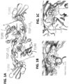
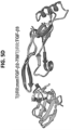
- the deletion of amino acid residues 52-71 removes the ⁇ -helical 3 ( ⁇ 3) region (the primary dimerization motif), as well as a few flanking residues ( FIG. 1D ). When residues 52-71 are removed, the remaining residues form a loop that contains polar and charged residues ( FIGS. 1D and 2B ).
- the TGF- ⁇ monomer is a human monomer.
- the TGF- ⁇ monomer further includes at least one amino acid substitution relative to a wild-type TFG- ⁇ 2 monomer that increases affinity of the TGF- ⁇ monomer for T ⁇ RII.
- the TGF- ⁇ monomer is a human TGF- ⁇ 2 monomer.
- the at least one amino acid substitution that increases net charge of the human TGF- ⁇ 2 monomer includes a leucine to arginine substitution at residue 51; an alanine to lysine substitution at residue 74; or both a leucine to arginine substitution at residue 51 and an alanine to lysine substitution at residue 74 (with reference to SEQ ID NO: 2).
- the at least one amino acid substitution that increases affinity of the human TGF- ⁇ 2 monomer for T ⁇ RII includes a substitution at an amino acid residue corresponding to residue 23, 24, 25, 26, 27, 28, 29, 30, 31, 32, 33, 34, 35, 36, 37, 89, 90, 91, 92, 93, 94, 95, 96, 97, 98 or 99 of SEQ ID NO: 2, or any combination of two or more residues thereof.
- the at least one amino acid substitution that increases affinity of the monomer for T ⁇ RII comprises at least one substitution at residue 23, 24, 25, 26, 27, 28, 29, 30, 31, 32, 33, 34, 35, 36 or 37, and at least one substitution at residue 89, 90, 91, 92, 93, 94, 95, 96, 97, 98 or 99.
- the at least one amino acid substitution that increases affinity of the human TGF- ⁇ 2 monomer for T ⁇ RII includes a lysine to arginine at residue 25, an arginine to lysine at residue 26, a leucine to valine at residue 89, an isoleucine to valine at residue 92, an asparagine to arginine at residue 94, a threonine to lysine at residue 95, an isoleucine to valine at residue 98, or any combination of two or more thereof, such as three or more, four or more, five or more, or six or more.
- the recombinant human TGF- ⁇ 2 monomer includes a lysine to arginine at residue 25, an arginine to lysine at residue 26, a leucine to valine at residue 89, an isoleucine to valine at residue 92, an asparagine to arginine at residue 94, a threonine to lysine at residue 95, and an isoleucine to valine at residue 98.
- the amino acid sequence of the human TGF- ⁇ 2 monomer is at least 95%, at least 96%, at least 97%, at least 98%, or at least 99% identical to SEQ ID NO: 8 or SEQ ID NO: 10. In some instances, the human TGF- ⁇ 2 monomer is at least 95%, at least 96%, at least 97%, at least 98%, or at least 99% identical to SEQ ID NO: 8 or SEQ ID NO: 10 and contains only conservative amino acid substitutions. In particular non-limiting examples, the amino acid sequence of the human TGF- ⁇ 2 monomer comprises or consists of SEQ ID NO: 8 or SEQ ID NO: 10.
- recombinant human TGF- ⁇ monomer is a human TGF- ⁇ 1 monomer.
- the at least one amino acid substitution that increases net charge of the human TGF- ⁇ 1 monomer includes an isoleucine to arginine substitution at residue 51; an alanine to lysine substitution at residue 74; an alanine to serine substitution at residue 75; or an isoleucine to arginine substitution at residue 51, an alanine to lysine substitution at residue 74 and an alanine to serine substitution at residue 75 (with reference to SEQ ID NO: 1).
- the amino acid sequence of the human TGF- ⁇ 1 monomer is at least 95%, at least 96%, at least 97%, at least 98%, or at least 99% identical to SEQ ID NO: 7. In some instances, the human TGF- ⁇ 1 monomer is at least 95%, at least 96%, at least 97%, at least 98%, or at least 99% identical to SEQ ID NO: 7 and contains only conservative amino acid substitutions. In particular non-limiting examples, the amino acid sequence of the human TGF- ⁇ 1 monomer comprises or consists of SEQ ID NO: 7.
- the recombinant human TGF- ⁇ monomer is a human TGF- ⁇ 3 monomer.
- the at least one amino acid substitution that increases net charge of the human TGF- ⁇ 3 monomer includes a leucine to glutamate substitution at residue 51; an alanine to glutamate substitution at residue 72; an alanine to aspartate substitution at residue 74; or a leucine to glutamate substitution at residue 51, an alanine to glutamate substitution at residue 72 and an alanine to aspartate substitution at residue 74 (with reference to SEQ ID NO: 3).
- the amino acid sequence of the human TGF- ⁇ 3 monomer is at least 95%, at least 96%, at least 97%, at least 98%, or at least 99% identical to SEQ ID NO: 9. In some instances, the human TGF- ⁇ 3 monomer is at least 95%, at least 96%, at least 97%, at least 98%, or at least 99% identical to SEQ ID NO: 9 and contains only conservative amino acid substitutions. In particular non-limiting examples, the amino acid sequence of the human TGF- ⁇ 3 monomer comprises or consists of SEQ ID NO: 9.
- the recombinant human TGF- ⁇ monomer is PEGylated, glycosylated, hyper-glycosylated, or includes another modification that prolongs circulatory time.
- fusion proteins that include a TGF- ⁇ monomer and a heterologous protein.
- the heterologous protein is a protein tag.
- the protein tag is an affinity tag (for example, Avitag, hexahistidine, chitin binding protein, maltose binding protein, or glutathione-S-transferase), an epitope tag (for example, V5, c-myc, HA or FLAG) or a fluorescent tag (e.g., GFP or another well-known fluorescent protein).
- the heterologous protein comprises an Fc domain, such as a mouse or human Fc domain.
- the heterologous protein promotes intermolecular association into homodimeric (for example, Fc domain from human IgG1, IgG2, IgG3), heterodimeric (for example, an engineered Fc domain, E/K coiled-coil), or multimeric (for example, pentabodies, nanoparticles) states of the fusion protein.
- the heterologous protein is albumin, an albumin-binding protein or agent, or another protein that increases circulatory time of the TGF- ⁇ monomer in vivo.
- recombinant human TGF- ⁇ monomers or fusion proteins comprising a radiotherapy agent, a cytotoxic agent for chemotherapy, or a drug. Further provided are recombinant human TGF- ⁇ monomers or fusion proteins comprising an imaging agent, a fluorescent dye, or a fluorescent protein tag.
- composition such as a pharmaceutical composition, that includes a recombinant human TGF- ⁇ monomer or fusion protein disclosed herein, and a pharmaceutically acceptable carrier, diluent, or excipient.
- the method is an in vitro method of inhibiting TGF- ⁇ signaling in a cell, comprising contacting the cell with the recombinant human TGF- ⁇ monomer, the fusion protein, or the composition disclosed herein.
- the use includes administering the recombinant human TGF- ⁇ monomer, fusion protein or composition to a subject having a disease or disorder associated with aberrant TGF- ⁇ signaling.
- the recombinant human TGF- ⁇ monomer, fusion protein or composition is administered by injection, such as by subcutaneous, intramuscular, intradermal, intraperitoneal, intravenous or intratumoral injection.
- a recombinant human TGF- ⁇ monomer, fusion protein or composition disclosed herein for use in a method of treating a disease or disorder associated with aberrant TGF- ⁇ signaling.
- the use in the method includes administering a recombinant human TGF- ⁇ monomer, fusion protein or composition disclosed herein to a subject.
- the disease or disorder associated with aberrant TGF- ⁇ signaling is a fibrotic disorder, such as but not limited to, pulmonary fibrosis, cystic fibrosis, idiopathic pulmonary fibrosis, interstitial lung disease, liver cirrhosis, kidney fibrosis (such as from damage caused by diabetes), atrial fibrosis, endomyocardial fibrosis, atherosclerosis, restenosis, scleroderma, or fibrosis caused by a surgical complication, chemotherapeutic drugs, radiation, injury or burns.
- pulmonary fibrosis such as but not limited to, pulmonary fibrosis, cystic fibrosis, idiopathic pulmonary fibrosis, interstitial lung disease, liver cirrhosis, kidney fibrosis (such as from damage caused by diabetes), atrial fibrosis, endomyocardial fibrosis, atherosclerosis, restenosis, scleroderma, or fibrosis caused by
- the disease or disorder associated with aberrant TGF- ⁇ signaling is breast cancer, brain cancer, pancreatic cancer, prostate cancer, skin cancer, bladder cancer, liver cancer, ovarian cancer, renal cancer, endometrial cancer, colorectal cancer, gastric cancer, skin cancer (such as malignant melanoma), or thyroid cancer.
- the disease or disorder associated with aberrant TGF- ⁇ signaling is an ocular disease.
- the disease or disorder associated with aberrant TGF- ⁇ signaling is a genetic disorder of connective tissue.
- nucleic acid molecules encoding a recombinant human TGF- ⁇ monomer disclosed herein.
- the nucleic acid molecule is operably linked to a promoter, such as a T cell specific promoter.
- vectors that include a TGF- ⁇ monomer-encoding nucleic acid molecule.
- the vector is a viral vector, such as a lentiviral vector.
- Isolated cells such as, but not limited to, isolated T cells comprising a nucleic acid molecule or vector encoding a recombinant human TGF- ⁇ monomer disclosed herein are further provided.
- the cells can be autologous to the subject, or they can be heterologous (allogeneic).
- Compositions that include the isolated cells and a pharmaceutically acceptable carrier are also provided.
- nucleic acid molecule, vector or isolated cell disclosed herein for use in methods of treating a disease or disorder associated with aberrant TGF- ⁇ signaling in a subject, comprising administering to the subject a nucleic acid molecule, vector or isolated cell disclosed herein.
- the disease or disorder associated with aberrant TGF- ⁇ signaling is a fibrotic disorder.
- the disease or disorder associated with aberrant TGF- ⁇ signaling is breast cancer, brain cancer, pancreatic cancer, prostate cancer, or skin cancer.
- the disease or disorder associated with aberrant TGF- ⁇ signaling is an ocular disease.
- the disease or disorder associated with aberrant TGF- ⁇ signaling is a genetic disorder of connective tissue.
- parenteral formulations usually comprise injectable fluids that are pharmaceutically and physiologically acceptable fluid vehicles such as water, physiological saline, other balanced salt solutions, aqueous dextrose, glycerol or the like.
- injectable fluids e.g., water, physiological saline, other balanced salt solutions, aqueous dextrose, glycerol or the like.
- solid compositions e.g., powder, pill, tablet, or capsule forms
- conventional non-toxic solid carriers can include, for example, pharmaceutical grades of mannitol, lactose, starch, or magnesium stearate.
- compositions to be administered can contain minor amounts of non-toxic auxiliary substances, such as wetting or emulsifying agents, preservatives, pH buffering agents, or the like, for example sodium acetate or sorbitan monolaurate.
- auxiliary substances such as wetting or emulsifying agents, preservatives, pH buffering agents, or the like, for example sodium acetate or sorbitan monolaurate.
- Excipients that can be included are, for instance, other proteins, such as human serum albumin or plasma preparations.
- aqueous carriers can be used, for example, buffered saline and the like, for introducing the cells. These solutions are sterile and generally free of undesirable matter. These compositions may be sterilized by conventional, well known sterilization techniques.
- the compositions may contain pharmaceutically acceptable auxiliary substances as required to approximate physiological conditions such as pH adjusting and buffering agents, toxicity adjusting agents and the like, for example, sodium acetate, sodium chloride, potassium chloride, calcium chloride, sodium lactate and the like.
- the concentration in these formulations can vary widely, and will be selected primarily based on fluid volumes, viscosities, body weight and the like in accordance with the particular mode of administration selected and the subject's needs.
- Topical preparations can include eye drops, ointments, sprays, patches and the like.
- Inhalation preparations can be liquid (e.g., solutions or suspensions) and include mists, sprays and the like.
- Oral formulations can be liquid (e.g., syrups, solutions or suspensions), or solid ( e.g., powders, pills, tablets, or capsules).
- Suppository preparations can also be solid, gel, or in a suspension form.
- conventional non-toxic solid carriers can include pharmaceutical grades of mannitol, lactose, starch, or magnesium stearate. Actual methods of preparing such dosage forms are known, or will be apparent, to those skilled in the art.
- compositions such as pharmaceutical compositions, that include a recombinant human TGF- ⁇ monomer
- the amount of TGF- ⁇ monomer administered will be dependent on the subject being treated, the severity of the affliction, and the manner of administration, and is best left to the judgment of the prescribing clinician.
- the formulation to be administered will contain a quantity of the active component(s) in amounts effective to achieve the desired effect in the subject being treated.
- the TGF- ⁇ monomers, or compositions thereof, can be administered to humans or other animals on whose tissues they are effective in various manners such as topically, orally, intravenously, intramuscularly, intraperitoneally, intranasally, intradermally, intrathecally, subcutaneously, via inhalation or via suppository.
- the particular mode of administration and the dosage regimen will be selected by the attending clinician, taking into account the particulars of the case (e.g. the subject, the disease, the disease state involved, and whether the treatment is prophylactic). Treatment can involve daily or multi-daily doses of compound(s) over a period of a few days to months, or even years.
- Example 1 An engineered TGF- ⁇ monomer that functions as a dominant negative to block TGF- ⁇ signaling
- This example describes an engineered TGF- ⁇ monomer that is capable of blocking TGF- ⁇ signaling.
- the engineered TGF- ⁇ monomer referred to herein as mmTGF- ⁇ 2-7M, has three changes relative to the monomer of wild type dimeric TGF- ⁇ 2:
- mmTGF- ⁇ 2-7M and other engineered TGF- ⁇ variants disclosed herein are described below and listed in Table 1.
- the sequences of all engineered TGF- ⁇ variants are shown in FIG. 8 and set forth as SEQ ID NOs: 1-11.
- TGF- ⁇ 1 and TGF- ⁇ 3 monomers that is TGF- ⁇ 1 and TGF- ⁇ 3 monomers with the cysteine residue that normally forms the interchain disulfide, Cys77, substituted to serine
- midpoint stimulatory potencies EC 50 s
- TGF- ⁇ 1 Cys77 ⁇ Ser and TGF- ⁇ 3 Cys77 ⁇ Ser variants would retain such significant signaling activity since one of the two essential receptors that binds to the growth factor, the TGF- ⁇ type I receptor (T ⁇ RI) was shown to bind by straddling the TGF- ⁇ homodimer interface ( FIG. 1A ) ( Groppe et al., Mol Cell 29:157-168, 2008 ).
- TGF- ⁇ monomers were signaling by non-covalently dimerizing and binding the receptors, which in turn stabilized the noncovalent dimers (by virtue of the fact that at least one of them, T ⁇ RI, binds across the dimer interface).
- TGF- ⁇ monomer that would function as an inhibitor, rather than a stimulator of TGF-beta signaling, an engineered monomer was produced in which the primary dimerization motif, the interfacial ⁇ -helix, ⁇ 3, was replaced with a flexible loop ( FIGS. 1A and 1D ).
- TGF- ⁇ 1 was expressed as a secreted protein bound to its prodomain in stably transfected Chinese hamster ovary (CHO) cells.
- the cell line used to produce TGF- ⁇ 1, and the accompanying procedure to isolate the mature disulfide-linked TGF- ⁇ 1 homodimer from the conditioned medium has been previously described ( Zou and Sun, Protein Expr Purif 37, 265-272, 2004 ).
- TGF- ⁇ 2 Human homodimeric TGF- ⁇ 2 (TGF- ⁇ 2), human homodimeric TGF- ⁇ 3 (TGF- ⁇ 3), and variants, including avi-tagged ( Cull and Schatz, Methods Enzymol 326, 430-440, 2000 ) homodimeric TGF- ⁇ 3 (TGF- ⁇ 3-avi), monomeric TGF- ⁇ 2 (mTGF- ⁇ 2), monomeric TGF- ⁇ 2 (mTGF- ⁇ 3), mini monomeric TGF- ⁇ 1 (mmTGF- ⁇ 1), mini monomeric TGF- ⁇ 2 (mmTGF- ⁇ 2), mini monomeric TGF- ⁇ 3 (mmTGF- ⁇ 3), mini monomeric TGF- ⁇ 2 with seven substitutions to enable high affinity T ⁇ RII binding (mmTGF- ⁇ 2-7M), and avi-tagged ( Cull and Schatz, Methods Enzymol 326, 430-440, 2000 ) mini monomeric TGF- ⁇ 2 with seven substitutions to enable high affinity T ⁇ RII binding (mmTGF-
- TGF- ⁇ 2, TGF- ⁇ 3, TGF- ⁇ 3-avi native folded disulfide-linked homodimers
- monomers mTGF- ⁇ 1, mTGF- ⁇ 2, mTGF- ⁇ 3, mmTGF- ⁇ 1, mmTGF- ⁇ 2, mmTGF- ⁇ 3, mmTGF- ⁇ 2-7M, mmTGF- ⁇ 2-7M-avi
- purified to homogeneity using high resolution cation exchange chromatography Source Q, GE Healthcare, Piscataway, NJ
- Huang and Hinck Methods Mol Biol 1344, 63-92, 2016 .
- TGF- ⁇ variants Variant Name (SEQ ID NO) Variant Description Number Residues per Monomer Single amino acid substitution(s) Deletion Tag TGF- ⁇ 1 (1) Human TGF- ⁇ 1 wild type homodimer 112 None None None TGF- ⁇ 2 (2) Human TGF- ⁇ 2 wild type homodimer 112 None None None TGF- ⁇ 3 (3) Human TGF- ⁇ 3 wild type homodimer 112 None None None avi-TGF- ⁇ 3 (4) Human TGF- ⁇ 3 wild type homodimer with N-terminal Avitag 127 None None N-terminal Avitag mTGF- ⁇ 2 (5) Human TGF- ⁇ 2 covalent monomer 112 C77S None None mTGF- ⁇ 3 (6) Human TGF- ⁇ 3 covalent monomer 112 C77S None None mmTGF- ⁇ 1 (7) Human TGF- ⁇
- T ⁇ RI T ⁇ RI
- T ⁇ RI- ⁇ C-Avi The human T ⁇ RI ectodomain (T ⁇ RI), spanning residues 1-101 of the mature receptor, or a variant spanning residues 1-88 of the mature receptor with a 15 amino acid avitag ( Cull and Schatz, Methods Enzymol 326, 430-440, 2000 ) appended to the C-terminus (T ⁇ RI- ⁇ C-Avi) was expressed in E. coli, refolded from inclusion bodies, and purified to homogeneity as previously described ( Z ⁇ iga et al., J Mol Biol 354, 1052-1068, 2005 ).
- T ⁇ RII T ⁇ RII ectodomain
- T ⁇ RII-His C-terminal hexahistidine tag
- TGF- ⁇ dimers and monomers were concentrated in 100 mM acetic acid to concentrations of 300 ⁇ M or higher and diluted to the desired concentration in either 100 mM acetic acid or phosphate buffered saline (PBS, 10 mM Na 2 HPO 4 , 1.8 mM KH 2 PO 4 , 137 mM NaCl, 2.7 mM KCl, pH 7.4).
- PBS phosphate buffered saline
- the pH of the samples diluted into PBS were adjusted with small aliquots of NaOH to ensure a final pH of 7.4.
- the light scattering at 340 nm of the samples were measured using a HP 8452 diode array spectrophotometer (HP, Palo Alto, CA).
- mmTGF- ⁇ 2 and mmTGF- ⁇ 2-7M samples isotopically labeled with 15 N or 15 N and 13 C for NMR were prepared by growing bacterial cells in M9 media containing 0.1 % (w/v) 15 NH 4 Cl or 0.1 % (w/v) 15 NH 4 Cl and 0.03% (w/v) 13 C labeled glucose. All NMR samples were prepared in 10 mM sodium phosphate, 10 mM 3-[(3-cholamidopropyl)dimethylammonio]-1-propanesulfonate (CHAPS), 5% 2 H 2 O, 0.02% w/v sodium azide at a protein concentration of 0.2 mM - 0.4 mM, pH 4.7.
- CHAPS 3-[(3-cholamidopropyl)dimethylammonio]-1-propanesulfonate
- T 2 relaxation parameters were measured in an interleaved manner at 300°K at a 15 N frequency of 70.95 MHz using 1 H-detected pulse schemes previously described ( Kay et al., Biochemistry 28:8972-8979, 1989 ).
- the T 2 data sets were each collected using 8 - 10 delay times, varying between 16 - 192 ms.
- the T 2 relaxation times were obtained by fitting relative peak intensities as a function of the T 2 delay time to a two parameter decaying exponential.
- FIGS. 3A-3B SPR measurements with TGF- ⁇ 2 and mmTGF- ⁇ 2 shown in FIGS. 3A-3B were performed using a BIACORE TM 3000 SPR (G.E. Healthcare, Piscataway, NJ) instrument with direct immobilization of TGF- ⁇ 2 or mmTGF- ⁇ 2 on the surface of a CM5 sensor chip (G.E. Healthcare, Piscataway, NJ) using an amine (carbodiimide-based) coupling kit (G.E. Healthcare, Piscataway, NJ). SPR experiments shown in FIGS. 3C , 3E and 3G and FIGS.
- Biotinylated TGF- ⁇ 3 or mmTGF- ⁇ 2-7M was generated by expressing TGF- ⁇ 3 or mmTGF- ⁇ 2-7M with an N-terminal 15 amino acid avitag ( Cull and Schatz, Methods Enzymol 326, 430-440, 2000 ).
- TGF- ⁇ 3-avi or mmTGF- ⁇ 2-7M-avi was bound to T ⁇ RII in 10 mM bicine at pH 8.0 and biotinylated by incubating with a catalytic amount of bacterially expressed BirA recombinase, biotin, and ATP at 37°C for 2 hours as described ( Huang and Hinck, Methods Mol Biol 1344, 63-92, 2016 ).
- Biotinylated avi-tagged TGF- ⁇ 3 or avi-tagged TGF- ⁇ 2-7m were bound to a C4 reverse phase column equilibrated with 94.9% water/5% acetonitrile/0.1% triflouroacetic acid and eluted with a linear acetonitrile gradient.
- FIGS. 3A-3E SPR measurements shown in FIGS. 3A-3E were performed in HBS-EP buffer (10 mM HEPES, pH 7.4, 150 mM NaCl, 3 mM EDTA, 0.005% surfactant P20; GE Healthcare, Piscataway, NJ) with the receptor indicated injected over a series of two-fold dilutions over the concentration range shown. Injections were carried out in duplicate and included 10 buffer blank injections at the start of the experiment. Binding was allowed to associate for 2 - 3 minutes at a flow rate of 100 mL min -1 , followed by dissociation for 1 minute or longer. Each cycle of injection was followed by 10 ml of regeneration with 4 M guanidine•HCl, 2 M NaCl.
- Crystals of mmTGF- ⁇ 2 were formed in sitting drops at 25°C by combining 0.2 ⁇ L of a 7.9 mg mL -1 protein stock solution in 10 mM MES pH 5.5 with 0.2 ⁇ L of the precipitant from the well, 20% PEG 3350, 0.2 M sodium thiocyanate. Harvested crystals were mounted in undersized nylon loops with excess mother liquor wicked off, followed by flash-cooling in liquid nitrogen prior to data collection. Data were acquired at the Advanced Photon Source NE-CAT beamline 24-ID-C and integrated and scaled using XDS ( Kabsch, Acta Crystallogr D Biol Crystallogr 66, 125-132, 2010 ).
- the structure was determined by the molecular replacement method implemented in PHASER ( McCoy et al., J Appl Crystallogr 40, 658-674, 2007 ) using a truncated version of PDB entry 2TGI ( Daopin et al., Science 257, 369-373, 1992 ) as the search model. Coordinates were refined using PHENIX ( Adams et al., Acta Crystallogr D Biol Crystallogr 66, 213-221, 2010 ), including simulated annealing with torsion angle dynamics, and alternated with manual rebuilding using COOT ( Emsley et al., Acta Crystallogr D Biol Crystallogr 66, 486-501, 2010 ). Data collection and refinement statistics are shown in Table 3.
- Crystals of the mmTGF- ⁇ 2-7M:T ⁇ RII complex were formed in hanging drops at 25°C by combining 1.0 ⁇ L of a 7.4 mg mL -1 stock solution of the complex in 10 mM Tris, pH 7.4 with 1.0 ⁇ L of 0.1 M HEPES, pH 7.5, 60 % v/v (+/-)-2-Methyl-2,4-pentanediol.
- Harvested crystals were mounted in nylon loops, followed by flash-cooling in liquid nitrogen prior to data collection. Data were acquired at the Advanced Photon Source 24-ID-C and integrated and scaled using HKL2000 ( Otwinowski and Minor, Method Enzymol 276, 307-326, 1997 ).
- the structure was determined by the molecular replacement method implemented in PHASER ( McCoy et al., J Appl Crystallogr 40, 658-674, 2007 ) using T ⁇ RII (PDB 1M9Z; Boesen et al., Structure 10, 913-919, 2002 ) and mmTGF- ⁇ 2 as search models. Coordinates were refined using PHENIX ( Adams et al., Acta Crystallogr D Biol Crystallogr 66, 213-221, 2010 ), alternated with manual rebuilding using COOT ( Emsley et al., Acta Crystallogr D Biol Crystallogr 66, 486-501, 2010 ). Data collection and refinement statistics are shown in Table 3.
- Crystals of mmTGF- ⁇ 2-7M were formed in hanging drops at 25°C by combining 1.0 ⁇ L of a 10 mg mL -1 protein stock solution in 20 mM acetic acid with 0.8 ⁇ L of the precipitant from the well, 100 mM sodium acetate dibasic trihydrate, pH 4.6, 25% 2-propanol, and 400 mM calcium chloride dehydrate, and 0.2 ⁇ L 5% n-ocyl- ⁇ -D-glucoside.
- Harvested crystals were mounted in nylon loops and cryoprotected in well buffer containing 20% glycerol and flash-cooled in a nitrogen stream.
- FIGS. 2A-2D , 5A-5E , 9A-9B and 10A-10B Results of structural studies are shown in FIGS. 2A-2D , 5A-5E , 9A-9B and 10A-10B .
- Table 3. X-ray Data collection and refinement statistics Data collection Molecule mmTGF- ⁇ 2 (PDB 5TX2) mmTGF- ⁇ 27m (PDB 5TX6) mmTGF- ⁇ 27m:T ⁇ RII (PDB 5TX4) X-ray Source Adv. Photon Source 24-ID-C Rigaku 007 generator and Saturn 944 CCD detector Adv.
- HEK293 Human embryonic kidney 293 (HEK293) cells stably transfected with the CAGA 12 TGF- ⁇ reporter were used for the luciferase reporter assays ( Thies et al., Growth Factors 18:251-259, 2001 ).
- HEK293 cells containing the stably transfected CAGA 12 TGF- ⁇ reporter were maintained in Dulbecco's modified eagles medium (DMEM) containing 10% fetal bovine serum (FBS) and 1% penicillin/streptomycin.
- DMEM Dulbecco's modified eagles medium
- FBS fetal bovine serum
- TGF- ⁇ TGF- ⁇ 1, mTGF- ⁇ 3 or mmTGF- ⁇ 2-7M
- mmTGF- ⁇ 2-7M concentration series TGF- ⁇ 1, 8 pM; TGF- ⁇ 2, 20 pM; TGF- ⁇ 3, 10 pM
- Proteins were diluted in DMEM containing 0.1% w/v BSA. After 16 hours, cells were lysed with Tropix lysis buffer (ThermoFisher, Waltham, MA) and luciferase activity was read with a Promega GloMax luminometer (Promega, Madison, WI).
- Luciferase activity was normalized to total protein levels determined by bicinchoninic acid (BCA) protein assay.
- BCA bicinchoninic acid
- Graphpad Prism 6 was used to fit the data to standard models for ligand activity (EC 50 ) and ligand inhibitory activity (IC 50 ) (Graphpad, La Jolla, CA). Results are shown in FIGS. 6A-6B .
- TGF- ⁇ 3, mTGF- ⁇ 3, mmTGF- ⁇ 2-7M TGF- ⁇ 3, mTGF- ⁇ 3, mmTGF- ⁇ 2-7M, biotinylated T ⁇ RI- ⁇ C-Avi and T ⁇ RII-His.
- 20 ⁇ M binary complexes of TGF- ⁇ 3:TbRII-His (1:2), mTGF- ⁇ 3:T ⁇ RII-His (1:1), and mmTGF- ⁇ 2-7M:T ⁇ RII-His (1:1) were formed in a 50 mM Tris, pH 7.5 buffer and stored at 4°C.
- TR-FRET time-resolved fluorescence resonance energy transfer
- the buffer conditions for each assay were 50 mM Tris, 50 mM NaCl, pH 7.5.
- the assays were performed in Corning black 384 well low flange microplates (ThermoFisher, Waltham, MA). After a 2-hour incubation, the assay plate was measured for terbium/XL-665 TR-FRET on a BMG Labtech Pherastar FS multimode plate reader (BMG Labtech Inc., Cary, NC). An optic module containing 337, 490 and 665 nm filters was used to monitor TR-FRET producing raw data for 337/490 (terbium emission) and 337/665 (XL-665) emission.
- the ratio of 665 emission/490 emission was determined for each condition and was subsequently used to calculate ⁇ F, which is a measure that reflects the signal of the sample versus the background.
- ⁇ F was calculated using the following equation: (Ratio signal -Ratio negative /Ratio negative ) ⁇ 100.
- the Ratio signal refers to the assays containing the trimeric complexes or buffer control.
- the Ratio negative refers to two assays buffer control (2 nM Tb-anti-His and 30 nM SA-665). For the buffer control, 2 out of the 6 replicates were assigned as negative controls for the purpose of calculating ⁇ F. ⁇ F was calculated for the remaining 4 buffer control replicates. Results are shown in FIGS 7A, 7B and 14 .
- mTGF- ⁇ 3, mmTGF- ⁇ 2, and mmTGF- ⁇ 2-7M were analyzed by sedimentation velocity to establish equilibrium constants for self-association of monomeric TGF- ⁇ s to form homodimers.
- mTGF- ⁇ 3, mmTGF- ⁇ 2, and mmTGF- ⁇ 2-7M were each measured at 280 nm in an epon two channel centerpiece fitted with quartz windows, and centrifuged at 20°C and 42,000 rpm for 27 hours in a 15 mM sodium phosphate buffer adjusted to pH 3.8, containing 100 mM NaCl. Three hundred scans were collected in intensity mode on a Beckman Optima XL-I analytical ultracentrifuge at the CAUMA facility at the UTHSCSA.
- UltraScan release 2130 Demeler and Gorbet, Analytical ultracentrifugation data analysis with Ultrascan-III, In Analytical Ultracentrifugation: Instrumentation, Software, and Applications (Uchiyama, S., Stafford, W., and Laue, T., Eds.), pp 119-143, Springer, 2016 ; Demeler et al., A comprehensive data analysis package for analytical ultracentrifugation experiments, 2016 ), calculations were performed at the Texas Advanced Computing Center on Lonestar-5.
- the sedimentation velocity data were initially fitted with the two-dimensional spectrum analysis as described in ( Demeler, Curr Protoc Protein Sci Chapter 7, Unit 7 13, 2010 ) to remove time- and radially invariant noise from the raw data, and to fit the meniscus position. Subsequently, the data were fitted to a discrete monomer-dimer model using the adaptive space-time finite element method ( Cao and Demeler, Biophys J 95, 54-65, 2008 ) and genetic algorithms for the parameter optimization ( Demeler et al., Macromol Biosci 10, 775-782, 2010 ).
- the monomer-dimer model accounts for mass action and the reversible association behavior, fitting the K D , hydrodynamic parameters, as well as the partial specific volume while assuming the predicted molar mass for either wildtype or mutant.
- a Monte Carlo analysis Demeler and Brookes, Colloid Polym Sci 286, 129-137, 2008 ) with 100 iterations was performed for each dataset to obtain fitting statistics. Buffer density and viscosity were estimated with UltraScan based on buffer composition and all hydrodynamic values were corrected for standard conditions (20°C and water). The fitting results provided an excellent fit with random residuals and very low RMSD values. All results are summarized in Table 4, and FIGS. 11-13 . Table 4.
- TGF- ⁇ receptor complexes Groppe et al., Mol Cell 29, 157-168, 2008 ; Radaev et al., J Biol Chem 285:14806-14814, 2010 ), as well as accompanying binding and cross-linking studies with TGF- ⁇ 3 C77S ( Z ⁇ iga et al., J Mol Biol 354, 1052-1068, 2005 ; Groppe et al., Mol Cell 29, 157-168, 2008 ; Huang et al., EMBO J 30:1263-1276, 2011 ), suggested that the signaling capacity of monomeric TGF- ⁇ s (TGF- ⁇ 1 C77S or mTGF- ⁇ 1 and TGF- ⁇ 3 C77S or mTGF- ⁇ 3) arise from their ability to non-covalently dimerize and in turn bind their receptors ( FIGS.
- bacterial expression constructs were generated for TGF- ⁇ 1, TGF- ⁇ 2, and TGF- ⁇ 3 in which residues 52-71 were eliminated and Cys-77 was substituted with serine. This corresponds to deletion of all of ⁇ -helix 3, as well as five flanking residues on the N-terminal end and three flanking residues on the C-terminal end ( FIG. 1D ).
- the length of the deletion was chosen so as to leave a sufficient number of residues between the last residue of ⁇ -strand 4 (Gly-48) and the first residue of ⁇ -strand 5 (Cys-77/Ser-77) to form an unconstrained loop that bridges ⁇ -strands 4 and 5.
- TGF- ⁇ 1, - ⁇ 2, and - ⁇ 3 "mini-monomers" described above, designated mmTGF- ⁇ 1, mmTGF- ⁇ 2, and mmTGF- ⁇ 3, were expressed in Escherichia coli and accumulated in the form of insoluble inclusion bodies.
- the inclusion bodies were isolated, and after reconstitution and purification in denaturant, the mini-monomers were renatured by dilution into CHAPS-containing buffer at pH 9.0 as described previously ( Huang et al., Methods Mol Biol 1344:63-92, 2016 ).
- the folding of the mini-monomers differed greatly; a large portion of the mmTGF- ⁇ 2 remained soluble during the folding and yielded large amounts of monomeric protein after purification by cation exchange chromatography, whereas only a small amount of mmTGF- ⁇ 1 and mmTGF- ⁇ 3 remained soluble during the folding, and either no monomeric protein (TGF- ⁇ 1) or a very small amount of monomeric protein (TGF- ⁇ 3) was obtained after purification by cation exchange chromatography.
- mmTGF- ⁇ 2 was suitable for further development in the manner described above, it was characterized in terms of its folding, solubility, and receptor binding properties.
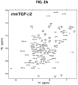
- a 15 N-labeled sample of mmTGF- ⁇ 2 was prepared and examined by recording a two-dimensional 1 H- 15 N shift correlation spectrum ( FIG. 2A ). This revealed a highly dispersed spectrum characteristic of natively folded protein. The spectrum could be fully assigned, and analysis of the assigned chemical shifts to identify secondary structure propensities showed that the protein had the expected secondary structure, particularly in the palm region formed by the cysteine knot and the finger region where T ⁇ RII binds ( FIG. 9A ).
- mmTGF- ⁇ 2 was crystallized, and its structure was determined to a resolution of 1.8 ⁇ using molecular replacement (Table 3).
- the overall fold of mmTGF- ⁇ 2 was shown to be highly similar to that previously determined for TGF- ⁇ 2, with the exception of the newly created loop, which was shown to take the place of ⁇ -helix 3 as anticipated ( FIG. 2B ).
- Superimposition of the mmTGF- ⁇ 2 with the monomer from the structure of TGF- ⁇ 2 shows that there is a systematic displacement of up to about 1.5 ⁇ of the finger region of mmTGF- ⁇ 2 relative to TGF- ⁇ 2.
- TGF- ⁇ 2 The solubility of mmTGF- ⁇ 2 appeared to be significantly better than that of TGF- ⁇ 2 and the full-length TGF- ⁇ 2 monomer, mTGF- ⁇ 2, as samples of the former could be readily prepared at concentrations of 2-3 mg ml -1 without noticeable precipitation at pH 7.0, whereas samples of the latter two proteins were completely precipitated under these same conditions.
- TGF- ⁇ 2, mTGF- ⁇ 2, and mmTGF- ⁇ 2 were prepared as concentrated stocks in 100 mM acetic acid, pH 2.9, where they were readily soluble and then diluted into PBS, pH 7.4.
- the mini-monomeric TGF- ⁇ 2 (mmTGF- ⁇ 2) in contrast, exhibited modest light scattering and a corresponding modest reduction in the amount of soluble protein relative to that expected when the protein concentration was 40 ⁇ M or higher, indicating that indeed mmTGF- ⁇ 2 was reasonably soluble at neutral pH, although not perfectly so. This was reflected in NMR spectra, which showed that although 100-200 ⁇ M 15 N mmTGF- ⁇ 2 samples could be readily prepared, the spectrum was nonetheless poor, with the only detectable signals arising from residues in the flexible parts of the protein, namely the N terminus, the exposed loop between ⁇ -helix 1 and ⁇ -strand 1, and the newly created loop between ⁇ -strands 4 and 5.
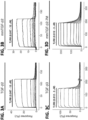
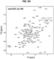
- the folding and homogeneity of the isolated mmTGF- ⁇ 2-7M was evaluated by NMR, and as with mmTGF- ⁇ 2, the protein was found to have the expected number of signals in a 2D 1 H- 15 N shift correlation spectrum ( FIG. 5A ) as well as secondary structure, as determined by an analysis of the NMR secondary shifts ( FIG. 10A ).
- the solubility of mmTGF- ⁇ 2-7M was evaluated as before, and as shown, its behavior was comparable or perhaps slightly better than that of mmTGF- ⁇ 2 ( FIGS. 4C and 4D ). This slight improvement in the macroscopic solubility did not however change the microscopic solubility as NMR analysis showed that it was still necessary to include 10 mM CHAPS in the sample buffer to detect signals from all of the backbone amide resonances in the protein.
- the three-dimensional structure of mmTGF- ⁇ 2-7M was determined by crystallography to a resolution of 2.75 ⁇ (Table 3), and as before the overall fold was preserved relative to TGF- ⁇ 2, with the only difference being a slight hinge bending of the monomer as described for mmTGF- ⁇ 2 ( FIGS. 5B and 5C ).
- the increase in the 15 N T 2 relaxation times in the region corresponding to the newly formed loop in mmTGF- ⁇ 2-7M was comparable with that in mmTGF- ⁇ 2 ( FIG. 10B ).
- the mmTGF- ⁇ 2-7M ⁇ T ⁇ RII complex was crystallized, and its structure was determined to a resolution of 1.88 ⁇ (Table 3).
- the overall structure of the mmTGF- ⁇ 2-7M ⁇ T ⁇ RII complex was shown to be very similar to that of one of the T ⁇ RII-bound monomers from the structure of the TGF- ⁇ 3 ⁇ T ⁇ RII ⁇ T ⁇ RI complex, with T ⁇ RII bound to the mmTGF- ⁇ 2-7M fingertips in a manner that is essentially indistinguishable from that of TGF- ⁇ 3 ( FIG. 5D ).
- mmTGF- ⁇ 2-7M possesses one of the essential attributes required to function as a dominant negative inhibitor of TGF- ⁇ signaling, which is the ability to bind T ⁇ RII with high affinity comparable with that of TGF- ⁇ 1 and TGF- ⁇ 3.
- TGF- ⁇ signaling was assessed by treating HEK293 cells stably transfected with a TGF- ⁇ luciferase reporter under the control of a CAGA 12 promoter ( Thies et al., Growth Factors 18:251-259, 2001 ) with increasing concentrations of TGF- ⁇ s.
- TGF- ⁇ 1 TGF- ⁇ 1
- mTGF- ⁇ 3 full-length monomeric TGF- ⁇ 3
- TGF- ⁇ 1 and mTGF- ⁇ 3 were in close accord with the values previously reported by Amatayakul-Chantler et al. (J Biol Chem 269:27687-27691, 1994 ) for TGF- ⁇ 1 and by Z ⁇ iga et al. (J Mol Biol 354, 1052-1068, 2005 ) for mTGF- ⁇ 3.
- the potent sub-nanomolar signaling activity observed for TGF- ⁇ 1 and mTGF- ⁇ 3 stands in contrast to that of mmTGF- ⁇ 2-7M, which had no detectable signaling activity at the concentration that led to a saturating response for mTGF- ⁇ 3 (ca.
- mmTGF- ⁇ 2-7M was either completely devoid of signaling activity or it possessed signaling activity, but with a potency more than a 10,000-fold less than that of mTGF- ⁇ 3.
- mmTGF- ⁇ 2-7M a competition experiment was performed in which the same HEK293 luciferase reporter cell line was stimulated with a constant sub-EC 50 concentration of dimeric TGF- ⁇ 1 (8.0 pM) and increasing concentrations of mTGF- ⁇ 3 or mmTGF- ⁇ 2-7M.
- the results showed that mTGF- ⁇ 3 further stimulated signaling with a midpoint concentration similar to that of mTGF- ⁇ 3 alone ( FIG. 6B ).
- the fitted EC 50 values confirm this, with an EC 50 of 182 ⁇ 16 pM for the data shown in FIG. 6A and EC 50 of 194 ⁇ 36 pM for the data shown in FIG. 6B .
- mmTGF- ⁇ 2-7M The behavior of mmTGF- ⁇ 2-7M was very different, with no detectable change in the signaling activity when added up to concentrations of 10 nM, but with a sharp decrease to no detectable signaling activity when the concentration was increased to 100 nM ( FIG. 6B ). This shows that mmTGF- ⁇ 2-7M indeed possesses no signaling activity and that it can function to completely block and inhibit TGF- ⁇ signaling.
- the normalized luciferase responses could be readily fitted to a standard model for ligand-dependent inhibition and yielded an IC 50 value of 68 ⁇ 7 nM.
- mmTGF- ⁇ 2-7M also functioned as a potent competitive inhibitor against the other TGF- ⁇ isoforms, TGF- ⁇ 2 and TGF- ⁇ 3, with measured IC 50 values (TGF- ⁇ 2 IC 50 19 ⁇ 3 nM and TGF- ⁇ 3 IC 50 21 ⁇ 8 nM) within a factor of 2-3 of that measured for TGF- ⁇ 1 ( FIGS. 15A and 15B ).
- IC 50 values are on the lower end of the range of affinities that have been reported for binding of the high affinity TGF- ⁇ isoforms to T ⁇ RII, including mmTGF- ⁇ 2-7M reported herein (Table 2).
- mmTGF- ⁇ 2-7M functions to inhibit TGF- ⁇ signaling by binding to and blocking endogenous T ⁇ RII.
- mmTGF- ⁇ 2-7M possesses no apparent signaling activity, and functions as a low nanomolar inhibitor of TGF- ⁇ signaling, suggests that the elimination of ⁇ -helix 3 diminished non-covalent association of the monomers and greatly attenuated or abrogated T ⁇ RI binding.
- SPR experiments were performed to determine whether mmTGF- ⁇ 2-7M could recruit T ⁇ RI in the presence of T ⁇ RII. To accomplish this, increasing concentrations of T ⁇ RI and the same concentration series of T ⁇ RI in the presence of near-saturating amounts of T ⁇ RII (2 ⁇ M) were injected over the same TGF- ⁇ 3 and mmTGF- ⁇ 2-7M SPR chip surfaces used for the T ⁇ RII binding measurements described above.
- T ⁇ RI alone binding is negligible to both TGF- ⁇ 3 and mmTGF- ⁇ 2-7M ( FIGS. 3E and 3F ), but unlike TGF- ⁇ 3, T ⁇ RII-bound mmTGF- ⁇ 2-7M is unable to recruit T ⁇ RI ( FIGS. 3G and 3H ).
- T ⁇ RII-bound mTGF- ⁇ 3 was significantly or completely impaired in terms of its ability to bind and recruit T ⁇ RI.
- T ⁇ RII-bound TGF- ⁇ monomers are incapable of binding and recruiting T ⁇ RI, but because the mmTGF- ⁇ 2-7M was immobilized on the surface of the sensor, it alone does not provide any insight as to whether mmTGF- ⁇ 2-7M might be capable of non-covalently dimerizing and binding and recruiting T ⁇ RI.
- AUC analytical ultracentrifugation
- TR-FRET time-resolved fluorescence resonance energy transfer
- TR-FRET was used to assess the ability of dimeric and monomeric TGF- ⁇ s to bind and bring T ⁇ RI and T ⁇ RII together. This was accomplished by generating differentially tagged forms of T ⁇ RII and T ⁇ RI and in turn binding to these tags with proteins labeled with fluorescent donors and acceptors.
- T ⁇ RII was tagged with a C-terminal His tag and was bound by a terbium cryptate-labeled anti-His monoclonal antibody fluorescent donor, and T ⁇ RI was tagged with an N-terminal avitag, which after enzymatic biotinylation was bound to a dye-labeled (XL-665) streptavidin fluorescent acceptor ( FIG. 7A ).
- TGF- ⁇ The addition of TGF- ⁇ to the tagged receptors brings them together and leads to a large increase in the ⁇ F value, which is defined as the ratio of the acceptor and donor emission fluorescent intensities.
- the TR-FRET assay is demonstrated by the data presented in FIG. 14 and was used here to compare the ability of the TGF- ⁇ 3 full-length monomer, mTGF- ⁇ 3, and the TGF- ⁇ 2 mini-monomer that binds T ⁇ RII with high affinity, mmTGF- ⁇ 2-7M, to bind and bring T ⁇ RI and T ⁇ RII together.
- the TR-FRET signal for mTGF- ⁇ 3 was shown to be comparable with that of TGF- ⁇ 3, and this did not depend on whether the TGF- ⁇ concentration was 100 or 250 nM ( FIG.
- the TGF- ⁇ s are responsible for promoting the progression of numerous human diseases ( Dietz et al., Nature 352:337-339, 1991 ; Biernacka et al., Growth Factors 29:196-202, 2011 ; Massagué, Cell 134:215-230, 2008 ; Loeys et al., Pediatr Endocrinol Rev 10:417-423, 2013 ), yet despite nearly two decades of preclinical studies and clinical trials, no inhibitors have been approved for use in humans.
- the small size of the inhibitor ( ⁇ 10 kDa) further confers a much greater ability to penetrate tumors and other dense tissues where the TGF- ⁇ s drive disease progression, a distinct advantage compared with IgG antibodies, which are much larger ( ⁇ 150 kDa) and tend to occupy only the vascular and interstitial space of well perfused organs ( Meibohm (2012) in Therapeutic Proteins: Strategies to Modulate Their Plasma Half-Lives (Kontermann R., editor), pages 23-38, Wiley-Blackwell, Weinheim, Germany ; Meibohm and Braeckman (2008), "Pharmacokinetics and Pharmacodynamics of Peptides and Protein Drugs," in Pharmaceutical Biotechnology: Fundamentals and Applications (Crommelin D. J.
- TGF- ⁇ receptor complexes together with the previously published chemical cross-linking data, suggested that the potent signaling activity of TGF- ⁇ 1 C77S and TGF- ⁇ 3 C77S was due to the ability of the monomers to non-covalently dimerize and in turn assemble a (T ⁇ RI ⁇ T ⁇ RII) 2 heterotetramer.
- the results presented here namely the AUC experiments that were used to assess non-covalent dimer formation and the TR-FRET experiments that were used to assess assembly of complexes with T ⁇ RI and T ⁇ RII, provided further evidence for this.
- the AUC data showed that full-length monomeric TGF- ⁇ 3, mTGF- ⁇ 3, self-associates to form dimers with a dimerization constant of 4.1 ⁇ M (Table 4).
- the TR-FRET data showed that at a concentration of 0.1 or 0.25 ⁇ M and in the presence of comparable concentrations of the T ⁇ RI and T ⁇ RII ectodomains, mTGF- ⁇ 3 assembles T ⁇ RI ⁇ T ⁇ RII complexes to the same extent as dimeric TGF- ⁇ 3 ( FIG. 7B ). That this occurs, even under conditions where the mTGF- ⁇ 3 concentrations (0.1-0.25 ⁇ M, FIG.
- T ⁇ RI ⁇ T ⁇ RII complexes with mTGF- ⁇ 3, and presumably mTGF- ⁇ 1 as well, therefore appears to be a cooperative process, much like protein folding, in which multiple weaker interactions, including monomer-monomer, non-covalent dimer-receptor, and receptor-receptor interactions, cooperate to enable formation of a thermodynamically stable TGF- ⁇ T ⁇ RI ⁇ T ⁇ RII complex.
- This manner of cooperative assembly is likely responsible for the ability of mTGF- ⁇ 1 and mTGF- ⁇ 3 to induce signaling at concentrations that are more than 4 orders of magnitude below the K D value for self-association of the monomers (EC 50 values of about 0.1 nM versus K D values for self-association of 4.1 ⁇ M).
- the elimination of the heel helix from the TGF- ⁇ monomer was shown to be very effective in terms of blocking the cooperative assembly of T ⁇ RI ⁇ T ⁇ RII complexes as shown by the TR-FRET data ( FIG. 7B ) and the cell based signaling data ( FIGS. 6A and 6B ).
- the AUC data showed that elimination of the heel helix led to the weakening of the monomer-monomer interaction by one order of magnitude (Table 4).
- T ⁇ RII-bound form of mmTGF- ⁇ 2-7M was incapable of binding and recruiting T ⁇ RI, which is expected based on published structures of TGF- ⁇ receptor complexes that show that T ⁇ RI binds to a composite interface formed by both chains of TGF- ⁇ , as well as T ⁇ RII ( Groppe et al., Mol Cell 29, 157-168, 2008 ; Radaev et al., J Biol Chem 285:14806-14814, 2010 ).
- the data show that the reduced propensity of the engineered monomer to self-associate, together with what would be expected to be very weak binding of T ⁇ RI to any dimers that do form, is responsible for the inability of mmTGF- ⁇ 2-7M to assemble a T ⁇ RI ⁇ T ⁇ RII complex. This accounts for the lack of signaling activity, and this together with the retention of high affinity T ⁇ RII binding accounts for the inhibitory activity.
- T ⁇ RII The other type II receptors of the family, activin type II receptor II, activin type IIB receptor, BMP type II receptor, and anti-Miillerian hormone type II receptor, have either been shown or are predicted to bind the GF knuckle and not the GF fingertips, as does T ⁇ RII ( Hinck et al., Cold Spring Harb Perspect Biol 8:a022103, 2016 ). Nonetheless, they share the same property as T ⁇ RII in that they bind only by contacting residues from a single GF monomer and not both monomers as has been shown or is predicted for all type I receptors of the family ( Hinck et al., Cold Spring Harb Perspect Biol 8:a022103, 2016 ).
- FIGS. 2B-2D and FIGS. 5B-5E suggest that it might be possible to generate monomers of other GFs of the family lacking the heel helix that function as inhibitors.
- These types of inhibitors have numerous potential applications, ranging from research tools for probing roles of specific ligands in vivo to clinically useful inhibitors for treating disease, which are driven by hyperactive signaling by other ligands of the family, such as cancer cachexia by activin ( Coerver et al., Mol Endocrinol 10:534-543, 1996 ).
Landscapes
- Health & Medical Sciences (AREA)
- Chemical & Material Sciences (AREA)
- Life Sciences & Earth Sciences (AREA)
- Organic Chemistry (AREA)
- Medicinal Chemistry (AREA)
- General Health & Medical Sciences (AREA)
- Biochemistry (AREA)
- Biophysics (AREA)
- Gastroenterology & Hepatology (AREA)
- Genetics & Genomics (AREA)
- Zoology (AREA)
- Molecular Biology (AREA)
- Proteomics, Peptides & Aminoacids (AREA)
- Toxicology (AREA)
- Pharmacology & Pharmacy (AREA)
- Veterinary Medicine (AREA)
- Animal Behavior & Ethology (AREA)
- Public Health (AREA)
- General Chemical & Material Sciences (AREA)
- Engineering & Computer Science (AREA)
- Bioinformatics & Cheminformatics (AREA)
- Physical Education & Sports Medicine (AREA)
- Chemical Kinetics & Catalysis (AREA)
- Epidemiology (AREA)
- Nuclear Medicine, Radiotherapy & Molecular Imaging (AREA)
- Peptides Or Proteins (AREA)
- Micro-Organisms Or Cultivation Processes Thereof (AREA)
- Medicines That Contain Protein Lipid Enzymes And Other Medicines (AREA)
- Medicinal Preparation (AREA)
- Medicines Containing Material From Animals Or Micro-Organisms (AREA)
- Medicines Containing Antibodies Or Antigens For Use As Internal Diagnostic Agents (AREA)
Applications Claiming Priority (2)
| Application Number | Priority Date | Filing Date | Title |
|---|---|---|---|
| US201662423920P | 2016-11-18 | 2016-11-18 | |
| PCT/US2017/062233 WO2018094173A1 (en) | 2016-11-18 | 2017-11-17 | Engineered tgf-beta monomers and their use for inhibiting tgf-beta signaling |
Publications (3)
| Publication Number | Publication Date |
|---|---|
| EP3541409A1 EP3541409A1 (en) | 2019-09-25 |
| EP3541409A4 EP3541409A4 (en) | 2020-06-17 |
| EP3541409B1 true EP3541409B1 (en) | 2025-01-22 |
Family
ID=62146703
Family Applications (1)
| Application Number | Title | Priority Date | Filing Date |
|---|---|---|---|
| EP17871472.1A Active EP3541409B1 (en) | 2016-11-18 | 2017-11-17 | Engineered tgf-beta monomers and their use for inhibiting tgf-beta signaling |
Country Status (6)
| Country | Link |
|---|---|
| US (2) | US11091523B2 (https=) |
| EP (1) | EP3541409B1 (https=) |
| JP (3) | JP7079779B2 (https=) |
| ES (1) | ES3011833T3 (https=) |
| PT (1) | PT3541409T (https=) |
| WO (1) | WO2018094173A1 (https=) |
Families Citing this family (9)
| Publication number | Priority date | Publication date | Assignee | Title |
|---|---|---|---|---|
| US11091523B2 (en) * | 2016-11-18 | 2021-08-17 | University of Pittsburgh—of the Commonwealth System of Higher Education | Engineered TGF-β monomers and their use for inhibiting TGF-β signaling |
| WO2021101796A1 (en) * | 2019-11-18 | 2021-05-27 | The Brigham And Women's Hospital, Inc. | Codon optimized new generation regulatable fusogenic oncolytic herpes simplex virus type 1 virus and methods of use |
| CA3162431A1 (en) * | 2020-01-21 | 2021-07-29 | Augustinus Bader | Cosmetic formulation for topical administration comprising novel peptides that improve appearance and regeneration of skin |
| JP2023539195A (ja) * | 2020-08-27 | 2023-09-13 | ユニバーシティ オブ ピッツバーグ - オブ ザ コモンウェルス システム オブ ハイヤー エデュケイション | 組換えトランスフォーミング増殖因子(tgf)-ベータ単量体をコードする腫瘍溶解性ウイルスおよびその使用 |
| EP4269429A4 (en) * | 2020-12-23 | 2025-07-30 | Korea Inst Sci & Tech | NEW PEPTIDE CAPABLE OF INHIBITING TGF-BETA SIGNALING AND ITS UTILIZATION |
| WO2023064747A1 (en) * | 2021-10-11 | 2023-04-20 | University Of Pittsburgh - Of The Commonwealth System Of Higher Education | Engineered tgf-beta monomers and methods of use |
| JP2024096518A (ja) * | 2023-01-03 | 2024-07-16 | エフビーデー バイオロジクス リミテッド | 操作されたtgfbriiバリアント及びその使用方法 |
| WO2024163457A2 (en) * | 2023-01-30 | 2024-08-08 | Seattle Children's Hospital D/B/A Seattle Children's Research Institute | Inducible cytokine transgenes for potentiated immune cell function |
| EP4490201A4 (en) | 2023-05-31 | 2026-03-11 | Fbd Biologics Ltd | CD47/PD-L1 Targeted Protein Complex and Its Methods of Use |
Family Cites Families (6)
| Publication number | Priority date | Publication date | Assignee | Title |
|---|---|---|---|---|
| US5766897A (en) * | 1990-06-21 | 1998-06-16 | Incyte Pharmaceuticals, Inc. | Cysteine-pegylated proteins |
| US5595756A (en) * | 1993-12-22 | 1997-01-21 | Inex Pharmaceuticals Corporation | Liposomal compositions for enhanced retention of bioactive agents |
| WO2005024035A1 (en) * | 2003-09-05 | 2005-03-17 | National Research Council Of Canada | Coiled-coil fusion proteins comprising cell receptor domains |
| WO2011094749A2 (en) * | 2010-02-01 | 2011-08-04 | Board Of Regents, The University Of Texas System | Small molecule inhibitors that block assembly of the tgf-beta signaling complex |
| CU24181B1 (es) * | 2012-11-09 | 2016-04-25 | Ct De Inmunología Molecular | POLIPÉPTIDOS DERIVADOS DEL TGFß |
| US11091523B2 (en) | 2016-11-18 | 2021-08-17 | University of Pittsburgh—of the Commonwealth System of Higher Education | Engineered TGF-β monomers and their use for inhibiting TGF-β signaling |
-
2017
- 2017-11-17 US US16/461,747 patent/US11091523B2/en active Active
- 2017-11-17 JP JP2019526306A patent/JP7079779B2/ja active Active
- 2017-11-17 EP EP17871472.1A patent/EP3541409B1/en active Active
- 2017-11-17 ES ES17871472T patent/ES3011833T3/es active Active
- 2017-11-17 WO PCT/US2017/062233 patent/WO2018094173A1/en not_active Ceased
- 2017-11-17 PT PT178714721T patent/PT3541409T/pt unknown
-
2021
- 2021-07-07 US US17/369,736 patent/US12428460B2/en active Active
- 2021-12-28 JP JP2021214183A patent/JP7623273B2/ja active Active
-
2023
- 2023-09-25 JP JP2023160199A patent/JP2023164748A/ja not_active Withdrawn
Also Published As
| Publication number | Publication date |
|---|---|
| US20210332096A1 (en) | 2021-10-28 |
| JP2020500511A (ja) | 2020-01-16 |
| JP7079779B2 (ja) | 2022-06-02 |
| PT3541409T (pt) | 2025-02-13 |
| JP2023164748A (ja) | 2023-11-10 |
| EP3541409A4 (en) | 2020-06-17 |
| JP7623273B2 (ja) | 2025-01-28 |
| US20190359667A1 (en) | 2019-11-28 |
| US11091523B2 (en) | 2021-08-17 |
| US12428460B2 (en) | 2025-09-30 |
| WO2018094173A1 (en) | 2018-05-24 |
| ES3011833T3 (en) | 2025-04-08 |
| EP3541409A1 (en) | 2019-09-25 |
| JP2022033239A (ja) | 2022-02-28 |
| CA3041727A1 (en) | 2018-05-24 |
Similar Documents
| Publication | Publication Date | Title |
|---|---|---|
| US12428460B2 (en) | Engineered TGF-beta monomers and their use for inhibiting TGF-beta signaling | |
| JP7270105B2 (ja) | インスリン類似体およびその使用方法 | |
| EP2350126B1 (en) | Bispecific egfr/igfir binding molecules | |
| TWI526220B (zh) | Fgf21突變體及其用途 | |
| US20170299608A1 (en) | Compositions and methods for modulating body weight | |
| US11512124B2 (en) | Fibronectin type III domain proteins with enhanced solubility | |
| EP3642338B1 (en) | Fusion protein with half-life extending polypeptide | |
| US20090117662A1 (en) | Mutants of IGF Binding Proteins and Methods of Production of Antagonists Thereof | |
| CN113840831A (zh) | 医用ngf拮抗剂 | |
| AU2017298565A1 (en) | Insulin analogs | |
| RU2769476C2 (ru) | Аналоги инсулина | |
| JP2011526491A (ja) | インスリン融合ポリペプチド | |
| CA3041727C (en) | Engineered tgf-beta monomers and their use for inhibiting tgf-beta signaling | |
| HK40015109B (en) | Engineered tgf-beta monomers and their use for inhibiting tgf-beta signaling | |
| HK40015109A (en) | Engineered tgf-beta monomers and their use for inhibiting tgf-beta signaling | |
| WO2014071234A1 (en) | Gb1 peptidic compounds and methods for making and using the same | |
| AU2022367394B2 (en) | Engineered tgf-beta monomers and methods of use | |
| Kim et al. | Engineered TGF-β Monomer that Blocks TGF-β Signaling | |
| JP2002112782A (ja) | 抗血栓活性を有する蛋白質及びその製造法 | |
| HK40109585A (zh) | 工程化TGF- β单体及其使用方法 | |
| WO2022096738A1 (en) | Novel type ii collagen binding proteins | |
| Morrow | α-Catenin Can Form Asymmetric Homodimeric Complexes and/or Heterodimeric Complexes with ॆ-Catenin | |
| WO2013151627A1 (en) | Methods of treating glucose metabolism disorders |
Legal Events
| Date | Code | Title | Description |
|---|---|---|---|
| STAA | Information on the status of an ep patent application or granted ep patent |
Free format text: STATUS: THE INTERNATIONAL PUBLICATION HAS BEEN MADE |
|
| PUAI | Public reference made under article 153(3) epc to a published international application that has entered the european phase |
Free format text: ORIGINAL CODE: 0009012 |
|
| STAA | Information on the status of an ep patent application or granted ep patent |
Free format text: STATUS: REQUEST FOR EXAMINATION WAS MADE |
|
| 17P | Request for examination filed |
Effective date: 20190605 |
|
| AK | Designated contracting states |
Kind code of ref document: A1 Designated state(s): AL AT BE BG CH CY CZ DE DK EE ES FI FR GB GR HR HU IE IS IT LI LT LU LV MC MK MT NL NO PL PT RO RS SE SI SK SM TR |
|
| AX | Request for extension of the european patent |
Extension state: BA ME |
|
| DAV | Request for validation of the european patent (deleted) | ||
| DAX | Request for extension of the european patent (deleted) | ||
| A4 | Supplementary search report drawn up and despatched |
Effective date: 20200515 |
|
| RIC1 | Information provided on ipc code assigned before grant |
Ipc: A61K 45/06 20060101ALI20200511BHEP Ipc: C07K 14/495 20060101ALI20200511BHEP Ipc: A61P 19/00 20060101ALI20200511BHEP Ipc: A61K 38/18 20060101AFI20200511BHEP Ipc: A61K 38/00 20060101ALI20200511BHEP |
|
| REG | Reference to a national code |
Ref country code: HK Ref legal event code: DE Ref document number: 40015109 Country of ref document: HK |
|
| STAA | Information on the status of an ep patent application or granted ep patent |
Free format text: STATUS: EXAMINATION IS IN PROGRESS |
|
| 17Q | First examination report despatched |
Effective date: 20230301 |
|
| GRAP | Despatch of communication of intention to grant a patent |
Free format text: ORIGINAL CODE: EPIDOSNIGR1 |
|
| STAA | Information on the status of an ep patent application or granted ep patent |
Free format text: STATUS: GRANT OF PATENT IS INTENDED |
|
| INTG | Intention to grant announced |
Effective date: 20240930 |
|
| GRAS | Grant fee paid |
Free format text: ORIGINAL CODE: EPIDOSNIGR3 |
|
| GRAA | (expected) grant |
Free format text: ORIGINAL CODE: 0009210 |
|
| STAA | Information on the status of an ep patent application or granted ep patent |
Free format text: STATUS: THE PATENT HAS BEEN GRANTED |
|
| AK | Designated contracting states |
Kind code of ref document: B1 Designated state(s): AL AT BE BG CH CY CZ DE DK EE ES FI FR GB GR HR HU IE IS IT LI LT LU LV MC MK MT NL NO PL PT RO RS SE SI SK SM TR |
|
| RAP3 | Party data changed (applicant data changed or rights of an application transferred) |
Owner name: NATIONAL RESEARCH COUNCIL OF CANADA Owner name: UNIVERSITY OF PITTSBURGH- OF THE COMMONWEALTHSYSTEM OF HIGHER EDUCATION |
|
| REG | Reference to a national code |
Ref country code: GB Ref legal event code: FG4D |
|
| REG | Reference to a national code |
Ref country code: CH Ref legal event code: EP |
|
| REG | Reference to a national code |
Ref country code: IE Ref legal event code: FG4D Ref country code: NL Ref legal event code: FP |
|
| REG | Reference to a national code |
Ref country code: PT Ref legal event code: SC4A Ref document number: 3541409 Country of ref document: PT Date of ref document: 20250213 Kind code of ref document: T Free format text: AVAILABILITY OF NATIONAL TRANSLATION Effective date: 20250207 Ref country code: DE Ref legal event code: R096 Ref document number: 602017087480 Country of ref document: DE |
|
| P01 | Opt-out of the competence of the unified patent court (upc) registered |
Free format text: CASE NUMBER: APP_3190/2025 Effective date: 20250120 |
|
| REG | Reference to a national code |
Ref country code: ES Ref legal event code: FG2A Ref document number: 3011833 Country of ref document: ES Kind code of ref document: T3 Effective date: 20250408 |
|
| PG25 | Lapsed in a contracting state [announced via postgrant information from national office to epo] |
Ref country code: RS Free format text: LAPSE BECAUSE OF FAILURE TO SUBMIT A TRANSLATION OF THE DESCRIPTION OR TO PAY THE FEE WITHIN THE PRESCRIBED TIME-LIMIT Effective date: 20250422 |
|
| PG25 | Lapsed in a contracting state [announced via postgrant information from national office to epo] |
Ref country code: FI Free format text: LAPSE BECAUSE OF FAILURE TO SUBMIT A TRANSLATION OF THE DESCRIPTION OR TO PAY THE FEE WITHIN THE PRESCRIBED TIME-LIMIT Effective date: 20250122 |
|
| PG25 | Lapsed in a contracting state [announced via postgrant information from national office to epo] |
Ref country code: PL Free format text: LAPSE BECAUSE OF FAILURE TO SUBMIT A TRANSLATION OF THE DESCRIPTION OR TO PAY THE FEE WITHIN THE PRESCRIBED TIME-LIMIT Effective date: 20250122 |
|
| REG | Reference to a national code |
Ref country code: LT Ref legal event code: MG9D |
|
| PG25 | Lapsed in a contracting state [announced via postgrant information from national office to epo] |
Ref country code: IS Free format text: LAPSE BECAUSE OF FAILURE TO SUBMIT A TRANSLATION OF THE DESCRIPTION OR TO PAY THE FEE WITHIN THE PRESCRIBED TIME-LIMIT Effective date: 20250522 Ref country code: NO Free format text: LAPSE BECAUSE OF FAILURE TO SUBMIT A TRANSLATION OF THE DESCRIPTION OR TO PAY THE FEE WITHIN THE PRESCRIBED TIME-LIMIT Effective date: 20250422 |
|
| REG | Reference to a national code |
Ref country code: AT Ref legal event code: MK05 Ref document number: 1760987 Country of ref document: AT Kind code of ref document: T Effective date: 20250122 |
|
| PG25 | Lapsed in a contracting state [announced via postgrant information from national office to epo] |
Ref country code: HR Free format text: LAPSE BECAUSE OF FAILURE TO SUBMIT A TRANSLATION OF THE DESCRIPTION OR TO PAY THE FEE WITHIN THE PRESCRIBED TIME-LIMIT Effective date: 20250122 |
|
| PG25 | Lapsed in a contracting state [announced via postgrant information from national office to epo] |
Ref country code: LV Free format text: LAPSE BECAUSE OF FAILURE TO SUBMIT A TRANSLATION OF THE DESCRIPTION OR TO PAY THE FEE WITHIN THE PRESCRIBED TIME-LIMIT Effective date: 20250122 |
|
| PG25 | Lapsed in a contracting state [announced via postgrant information from national office to epo] |
Ref country code: BG Free format text: LAPSE BECAUSE OF FAILURE TO SUBMIT A TRANSLATION OF THE DESCRIPTION OR TO PAY THE FEE WITHIN THE PRESCRIBED TIME-LIMIT Effective date: 20250122 Ref country code: GR Free format text: LAPSE BECAUSE OF FAILURE TO SUBMIT A TRANSLATION OF THE DESCRIPTION OR TO PAY THE FEE WITHIN THE PRESCRIBED TIME-LIMIT Effective date: 20250423 |
|
| PG25 | Lapsed in a contracting state [announced via postgrant information from national office to epo] |
Ref country code: AT Free format text: LAPSE BECAUSE OF FAILURE TO SUBMIT A TRANSLATION OF THE DESCRIPTION OR TO PAY THE FEE WITHIN THE PRESCRIBED TIME-LIMIT Effective date: 20250122 |
|
| PG25 | Lapsed in a contracting state [announced via postgrant information from national office to epo] |
Ref country code: SE Free format text: LAPSE BECAUSE OF FAILURE TO SUBMIT A TRANSLATION OF THE DESCRIPTION OR TO PAY THE FEE WITHIN THE PRESCRIBED TIME-LIMIT Effective date: 20250122 |
|
| PG25 | Lapsed in a contracting state [announced via postgrant information from national office to epo] |
Ref country code: SM Free format text: LAPSE BECAUSE OF FAILURE TO SUBMIT A TRANSLATION OF THE DESCRIPTION OR TO PAY THE FEE WITHIN THE PRESCRIBED TIME-LIMIT Effective date: 20250122 |
|
| PG25 | Lapsed in a contracting state [announced via postgrant information from national office to epo] |
Ref country code: DK Free format text: LAPSE BECAUSE OF FAILURE TO SUBMIT A TRANSLATION OF THE DESCRIPTION OR TO PAY THE FEE WITHIN THE PRESCRIBED TIME-LIMIT Effective date: 20250122 |
|
| PGFP | Annual fee paid to national office [announced via postgrant information from national office to epo] |
Ref country code: GB Payment date: 20250925 Year of fee payment: 9 |
|
| PGFP | Annual fee paid to national office [announced via postgrant information from national office to epo] |
Ref country code: FR Payment date: 20250908 Year of fee payment: 9 |
|
| PG25 | Lapsed in a contracting state [announced via postgrant information from national office to epo] |
Ref country code: EE Free format text: LAPSE BECAUSE OF FAILURE TO SUBMIT A TRANSLATION OF THE DESCRIPTION OR TO PAY THE FEE WITHIN THE PRESCRIBED TIME-LIMIT Effective date: 20250122 Ref country code: CZ Free format text: LAPSE BECAUSE OF FAILURE TO SUBMIT A TRANSLATION OF THE DESCRIPTION OR TO PAY THE FEE WITHIN THE PRESCRIBED TIME-LIMIT Effective date: 20250122 |
|
| REG | Reference to a national code |
Ref country code: DE Ref legal event code: R097 Ref document number: 602017087480 Country of ref document: DE |
|
| PG25 | Lapsed in a contracting state [announced via postgrant information from national office to epo] |
Ref country code: RO Free format text: LAPSE BECAUSE OF FAILURE TO SUBMIT A TRANSLATION OF THE DESCRIPTION OR TO PAY THE FEE WITHIN THE PRESCRIBED TIME-LIMIT Effective date: 20250122 |
|
| PG25 | Lapsed in a contracting state [announced via postgrant information from national office to epo] |
Ref country code: SK Free format text: LAPSE BECAUSE OF FAILURE TO SUBMIT A TRANSLATION OF THE DESCRIPTION OR TO PAY THE FEE WITHIN THE PRESCRIBED TIME-LIMIT Effective date: 20250122 |
|
| PLBE | No opposition filed within time limit |
Free format text: ORIGINAL CODE: 0009261 |
|
| STAA | Information on the status of an ep patent application or granted ep patent |
Free format text: STATUS: NO OPPOSITION FILED WITHIN TIME LIMIT |
|
| 26N | No opposition filed |
Effective date: 20251023 |
|
| PGFP | Annual fee paid to national office [announced via postgrant information from national office to epo] |
Ref country code: DE Payment date: 20250923 Year of fee payment: 9 |
|
| PGFP | Annual fee paid to national office [announced via postgrant information from national office to epo] |
Ref country code: IT Payment date: 20251022 Year of fee payment: 9 |
|
| PGFP | Annual fee paid to national office [announced via postgrant information from national office to epo] |
Ref country code: ES Payment date: 20251211 Year of fee payment: 9 |