EP3533063B1 - Netzwerk zur analyse von medizinischen bildern, entscheidungsunterstützungssystem und zugehörige anwendungen für grafische benutzeroberfläche (gui) - Google Patents
Netzwerk zur analyse von medizinischen bildern, entscheidungsunterstützungssystem und zugehörige anwendungen für grafische benutzeroberfläche (gui) Download PDFInfo
- Publication number
- EP3533063B1 EP3533063B1 EP17794628.2A EP17794628A EP3533063B1 EP 3533063 B1 EP3533063 B1 EP 3533063B1 EP 17794628 A EP17794628 A EP 17794628A EP 3533063 B1 EP3533063 B1 EP 3533063B1
- Authority
- EP
- European Patent Office
- Prior art keywords
- patient
- image
- spect
- scan
- composite
- Prior art date
- Legal status (The legal status is an assumption and is not a legal conclusion. Google has not performed a legal analysis and makes no representation as to the accuracy of the status listed.)
- Active
Links
Images


Classifications
-
- G—PHYSICS
- G16—INFORMATION AND COMMUNICATION TECHNOLOGY [ICT] SPECIALLY ADAPTED FOR SPECIFIC APPLICATION FIELDS
- G16H—HEALTHCARE INFORMATICS, i.e. INFORMATION AND COMMUNICATION TECHNOLOGY [ICT] SPECIALLY ADAPTED FOR THE HANDLING OR PROCESSING OF MEDICAL OR HEALTHCARE DATA
- G16H50/00—ICT specially adapted for medical diagnosis, medical simulation or medical data mining; ICT specially adapted for detecting, monitoring or modelling epidemics or pandemics
- G16H50/20—ICT specially adapted for medical diagnosis, medical simulation or medical data mining; ICT specially adapted for detecting, monitoring or modelling epidemics or pandemics for computer-aided diagnosis, e.g. based on medical expert systems
-
- A—HUMAN NECESSITIES
- A61—MEDICAL OR VETERINARY SCIENCE; HYGIENE
- A61B—DIAGNOSIS; SURGERY; IDENTIFICATION
- A61B6/00—Apparatus or devices for radiation diagnosis; Apparatus or devices for radiation diagnosis combined with radiation therapy equipment
- A61B6/02—Arrangements for diagnosis sequentially in different planes; Stereoscopic radiation diagnosis
- A61B6/03—Computed tomography [CT]
- A61B6/037—Emission tomography
-
- A—HUMAN NECESSITIES
- A61—MEDICAL OR VETERINARY SCIENCE; HYGIENE
- A61B—DIAGNOSIS; SURGERY; IDENTIFICATION
- A61B6/00—Apparatus or devices for radiation diagnosis; Apparatus or devices for radiation diagnosis combined with radiation therapy equipment
- A61B6/52—Devices using data or image processing specially adapted for radiation diagnosis
- A61B6/5211—Devices using data or image processing specially adapted for radiation diagnosis involving processing of medical diagnostic data
-
- G—PHYSICS
- G01—MEASURING; TESTING
- G01N—INVESTIGATING OR ANALYSING MATERIALS BY DETERMINING THEIR CHEMICAL OR PHYSICAL PROPERTIES
- G01N33/00—Investigating or analysing materials by specific methods not covered by groups G01N1/00 - G01N31/00
- G01N33/48—Biological material, e.g. blood, urine; Haemocytometers
- G01N33/50—Chemical analysis of biological material, e.g. blood, urine; Testing involving biospecific ligand binding methods; Immunological testing
- G01N33/53—Immunoassay; Biospecific binding assay; Materials therefor
- G01N33/575—Immunoassay; Biospecific binding assay; Materials therefor for cancer
- G01N33/57555—Immunoassay; Biospecific binding assay; Materials therefor for cancer of the prostate
-
- G—PHYSICS
- G06—COMPUTING OR CALCULATING; COUNTING
- G06F—ELECTRIC DIGITAL DATA PROCESSING
- G06F9/00—Arrangements for program control, e.g. control units
- G06F9/06—Arrangements for program control, e.g. control units using stored programs, i.e. using an internal store of processing equipment to receive or retain programs
- G06F9/44—Arrangements for executing specific programs
- G06F9/451—Execution arrangements for user interfaces
-
- G—PHYSICS
- G16—INFORMATION AND COMMUNICATION TECHNOLOGY [ICT] SPECIALLY ADAPTED FOR SPECIFIC APPLICATION FIELDS
- G16H—HEALTHCARE INFORMATICS, i.e. INFORMATION AND COMMUNICATION TECHNOLOGY [ICT] SPECIALLY ADAPTED FOR THE HANDLING OR PROCESSING OF MEDICAL OR HEALTHCARE DATA
- G16H15/00—ICT specially adapted for medical reports, e.g. generation or transmission thereof
-
- G—PHYSICS
- G16—INFORMATION AND COMMUNICATION TECHNOLOGY [ICT] SPECIALLY ADAPTED FOR SPECIFIC APPLICATION FIELDS
- G16H—HEALTHCARE INFORMATICS, i.e. INFORMATION AND COMMUNICATION TECHNOLOGY [ICT] SPECIALLY ADAPTED FOR THE HANDLING OR PROCESSING OF MEDICAL OR HEALTHCARE DATA
- G16H30/00—ICT specially adapted for the handling or processing of medical images
- G16H30/20—ICT specially adapted for the handling or processing of medical images for handling medical images, e.g. DICOM, HL7 or PACS
-
- G—PHYSICS
- G16—INFORMATION AND COMMUNICATION TECHNOLOGY [ICT] SPECIALLY ADAPTED FOR SPECIFIC APPLICATION FIELDS
- G16H—HEALTHCARE INFORMATICS, i.e. INFORMATION AND COMMUNICATION TECHNOLOGY [ICT] SPECIALLY ADAPTED FOR THE HANDLING OR PROCESSING OF MEDICAL OR HEALTHCARE DATA
- G16H30/00—ICT specially adapted for the handling or processing of medical images
- G16H30/40—ICT specially adapted for the handling or processing of medical images for processing medical images, e.g. editing
-
- G—PHYSICS
- G16—INFORMATION AND COMMUNICATION TECHNOLOGY [ICT] SPECIALLY ADAPTED FOR SPECIFIC APPLICATION FIELDS
- G16H—HEALTHCARE INFORMATICS, i.e. INFORMATION AND COMMUNICATION TECHNOLOGY [ICT] SPECIALLY ADAPTED FOR THE HANDLING OR PROCESSING OF MEDICAL OR HEALTHCARE DATA
- G16H50/00—ICT specially adapted for medical diagnosis, medical simulation or medical data mining; ICT specially adapted for detecting, monitoring or modelling epidemics or pandemics
- G16H50/30—ICT specially adapted for medical diagnosis, medical simulation or medical data mining; ICT specially adapted for detecting, monitoring or modelling epidemics or pandemics for calculating health indices; for individual health risk assessment
-
- G—PHYSICS
- G16—INFORMATION AND COMMUNICATION TECHNOLOGY [ICT] SPECIALLY ADAPTED FOR SPECIFIC APPLICATION FIELDS
- G16H—HEALTHCARE INFORMATICS, i.e. INFORMATION AND COMMUNICATION TECHNOLOGY [ICT] SPECIALLY ADAPTED FOR THE HANDLING OR PROCESSING OF MEDICAL OR HEALTHCARE DATA
- G16H50/00—ICT specially adapted for medical diagnosis, medical simulation or medical data mining; ICT specially adapted for detecting, monitoring or modelling epidemics or pandemics
- G16H50/70—ICT specially adapted for medical diagnosis, medical simulation or medical data mining; ICT specially adapted for detecting, monitoring or modelling epidemics or pandemics for mining of medical data, e.g. analysing previous cases of other patients
-
- G—PHYSICS
- G16—INFORMATION AND COMMUNICATION TECHNOLOGY [ICT] SPECIALLY ADAPTED FOR SPECIFIC APPLICATION FIELDS
- G16H—HEALTHCARE INFORMATICS, i.e. INFORMATION AND COMMUNICATION TECHNOLOGY [ICT] SPECIALLY ADAPTED FOR THE HANDLING OR PROCESSING OF MEDICAL OR HEALTHCARE DATA
- G16H70/00—ICT specially adapted for the handling or processing of medical references
- G16H70/40—ICT specially adapted for the handling or processing of medical references relating to drugs, e.g. their side effects or intended usage
Definitions
- This invention relates generally to systems and methods for creation, analysis, and/or presentation of medical image data. More particularly, in certain embodiments, the invention relates to a cloud-based platform and supported GUI decision-making tools for use by medical practitioners and/or their patients, e.g., to aide in the process of making decisions about a course of cancer treatment and/or to track treatment and/or the progress of a disease.
- Targeted image analysis involves the use of radiolabeled small molecules that bind to specific receptors, enzymes and proteins in the body that are altered during the evolution of disease. After administration to a patient, these molecules circulate in the blood until they find their intended target. The bound radiopharmaceutical remains at the site of disease, while the rest of the agent clears from the body.
- the radioactive portion of the molecule serves as a beacon so that an image may be obtained depicting the disease location and concentration using commonly available nuclear medicine cameras, known as single-photon emission computerized tomography (SPECT) or positron emission tomography (PET) cameras, found in most hospitals throughout the world. Physicians can then use this information to determine the presence and the extent of disease in a patient. The physician can use this information to provide a recommended course of treatment to the patient and to track the progression of disease.
- SPECT single-photon emission computerized tomography
- PET positron emission tomography
- the small molecule diagnostic 1404 targets the extracellular domain of prostate specific membrane antigen (PSMA), a protein amplified on the surface of >95% of prostate cancer cells and a validated target for the detection of primary and metastatic prostate cancer. 1404 is labeled with technetium-99m, a gamma-emitter isotope that is widely available, relatively inexpensive, facilitates efficient preparation, and has spectrum characteristics attractive for nuclear medicine imaging applications.
- PSMA prostate specific membrane antigen
- 1404 is labeled with technetium-99m, a gamma-emitter isotope that is widely available, relatively inexpensive, facilitates efficient preparation, and has spectrum characteristics attractive for nuclear medicine imaging applications.
- PyL TM also known as [ 18 F]DCFPyL
- PyL TM is a clinical-stage, fluorinated PSMA-targeted PET imaging agent for prostate cancer.
- An oncologist may use images from a targeted PET or SPECT study of a patient as input in her assessment of whether the patient has a particular disease, e.g., prostate cancer, what stage of the disease is evident, what the recommended course of treatment (if any) would be, whether surgical intervention is indicated, and likely prognosis.
- the oncologist may use a radiologist report in this assessment.
- a radiologist report is a technical evaluation of the PET or SPECT images prepared by a radiologist for a physician who requested the imaging study and includes, for example, the type of study performed, the clinical history, a comparison between images, the technique used to perform the study, the radiologist's observations and findings, as well as overall impressions and recommendations the radiologist may have based on the imaging study results.
- a signed radiologist report is sent to the physician ordering the study for the physician's review, followed by a discussion between the physician and patient about the results and recommendations for treatment.
- the process involves having a radiologist perform an imaging study on the patient, analyzing the images obtained, creating a radiologist report, forwarding the report to the requesting physician, having the physician formulate an assessment and treatment recommendation, and having the physician communicate the results, recommendations, and risks to the patient.
- the process may also involve repeating the imaging study due to inconclusive results, or ordering further tests based on initial results.
- the physician discusses various treatment options, including surgery, as well as risks of doing nothing or adopting a watchful waiting or active surveillance approach, rather than having surgery.
- a disease or condition e.g., cancer
- WO 2015/058151 A2 discloses a method of evaluating a subject suspected of harboring a prostate tumor.
- GUI graphical user interface
- a network-based (e.g., cloud-based) support platform allowing multiple users to store, access, analyze, and/or provide feedback regarding a given set of image data for a patient; platform supports software tools for automated analysis of targeted PET/SPECT/or other image(s), generation of radiologist reports, and application of machine learning algorithms to update process by which images are analyzed (e.g. updating segmentation and/or classification routines based on growing image database).
- the targeted PET/SPECT image(s) may be obtained using PyL TM and/or 1404 as the radiopharmaceutical(s).
- multiple (accredited) users can access the information, e.g., to weigh in on data interpretation.
- a software tool e.g., mobile app
- GUI graphical user interface
- the tool may be supported by the network-based support platform above.
- This tool can provides an easily-understood, user-friendly, interactive, controllable pictorial display to communicate information about a patient's condition to the patient (and/or to the physician, or to the patient's family with the patient's permission).
- a low-risk patient who is told by his physician that he has a non-zero risk of cancer may find comfort in a visual, controllable comparison between his situation and that of someone (e.g., where the reference to which the patient's risk situation is compared can be tuned for age, weight, and/or other risk factors of the patient).
- the medical images in the database comprise a series of medical images of a first patient taken over time (e.g., over the course of multiple visits to one or more doctors), and wherein the instructions cause the processor to determine a value of at least a first risk index for each medical image of the series, thereby tracking determined values of at least the first risk index for the first patient over time.
- the nuclear medicine image is a PET scan.
- the nuclear medicine image is a SPECT scan.
- the medical images comprise a nuclear medicine image (e.g., a whole-body scan made with a gamma camera) of a first patient obtained following administration to the first patient of an imaging agent comprising a radionuclide (e.g., 99m Tc)(e.g., wherein the imaging agent comprises 99m Tc MDP), wherein the method comprises automatically analyzing the nuclear medicine image by: (a) geographically identifying a boundary (e.g., a 2D boundary; e.g., a 3D boundary) for each of one or more regions of imaged tissue [e.g., organs (e.g., a prostate; e.g., a liver; e.g., lungs or a lung; e.g., lymph nodes), organ structures, sub-organs, organ regions, and/or other regions (e.g., one or more particular bones; e.g., a skeletal region of the patient), e.g., regions of interest] within an imaging agent compris
- step (c) comprises, for at least one risk index of the one or more risk indices, computing the value of the risk index by: determining, for each of the one or more regions, a corresponding cancerous tissue level within the region based on intensity values of the nuclear medicine image within the boundary of the region (e.g., by identifying within the nuclear medicine image, a plurality of hotspots within the 3D boundary of the region and computing a total number of and/or a total volume of the identified hotspots); and computing the value of the risk index based on the determined cancerous tissue levels within the one or more regions.
- the processor is a processor of a cloud-based system.
- the processor is a processor of one or more network or Internet host servers.
- the medical images of first image subseries are obtained over a first period of time, when prostate cancer of the particular patient is localized (e.g., substantially localized to a prostate of the particular patient), and the medical images of the second image subseries are obtained over a second period of time, when prostate cancer of the particular patient is metastatic (e.g., having spread to regions of the patient outside of the prostate).
- the first image subseries comprises one or more composite SPECT-CT image(s), each composite SPECT-CT image comprising a CT scan overlaid with a SPECT scan acquired at substantially the same time;
- the second image subseries comprises one or more composite PET-CT image(s), each composite PET-CT image comprising a CT scan overlaid with a PET scan acquired at substantially the same time;
- step (b) comprises: automatically analyzing each of the one or more composite SPECT-CT images by: using the composite SPECT-CT image to geographically identify a 3D boundary of a prostate region (e.g., corresponding to a prostate of the patient) within the SPECT scan of the composite SPECT-CT image (e.g., such that portions of the nuclear medicine image falling within and/or outside of the 3D boundary of the prostate region can be differentiated from each other); and computing a value of the first risk index using the SPECT scan with the identified 3D boundary of the prostate region (e.g., computed based on
- the first image subseries comprises one or more composite SPECT-CT image(s), each composite SPECT-CT image comprising a CT scan overlaid with a SPECT scan acquired at substantially the same time;
- the second image subseries comprises one or more whole-body scan(s);
- step (b) comprises: automatically analyzing each of the one or more composite SPECT-CT images by: using the composite SPECT-CT image to geographically identify a 3D boundary of a prostate region (e.g., corresponding to a prostate of the patient) within the SPECT scan of the composite SPECT-CT image (e.g., such that portions of the nuclear medicine image falling within and/or outside of the 3D boundary of the prostate region can be differentiated from each other); and computing a value of the first risk index using the SPECT scan with the identified 3D boundary of the prostate region (e.g., computed based on a region of the SPECT scan corresponding to the identified 3D boundary of the prostate region); and automatically analyzing each of the
- the invention is directed to a system for tracking prostate cancer progression and treatment efficacy over time, for one or more patient(s), the system comprising: a processor (e.g., of a network or Internet host server); and a memory having instructions stored thereon, wherein the instructions, when executed by the processor, cause the processor to: (a) repeatedly receive and store in a database, over time, a plurality of medical images for each of the one or more patient(s) to obtain, for each of the one or more patient(s), a series of medical images taken over time (e.g., over the course of multiple visits to one or more doctors); (b) for each of the one or more patient(s), automatically analyze the series of medical images for the patient to determine values of one or more risk indices [e.g., the values of the one or more risk indices corresponding to numeric values indicative of prostate cancer state and/or progression in the patient (e.g., numeric values identifying a particular cancer stage; e.g., numeric values corresponding to
- the series of medical images for a particular patient of the one or more patient(s) comprises: (i) a first image subseries comprising one or more medical images obtained using a first nuclear imaging modality (e.g., SPECT scans; e.g., composite SPECT-CT images) each following administration to the particular patient of a first radiopharmaceutical (e.g., 99m Tc-MIP-1404) (e.g., wherein the first radiopharmaceutical facilitates imaging of localized disease, e.g., localized prostate cancer); and (ii) a second image subseries comprising one or more medical images obtained using a second nuclear imaging modality (e.g., PET scans; e.g., composite PET-CT images; e.g., whole-body scans) each following administration to the particular patient of a second radiopharmaceutical (e.g., [18F]DCFPyL; e.g., 99m Tc MDP) (e.g.
- the medical images of first image subseries are obtained over a first period of time, when prostate cancer of the particular patient is localized (e.g., substantially localized to a prostate of the particular patient), and the medical images of the second image subseries are obtained over a second period of time, when prostate cancer of the particular patient is metastatic (e.g., having spread to regions of the patient outside of the prostate).
- the first image subseries comprises one or more composite SPECT-CT image(s), each composite SPECT-CT image comprising a CT scan overlaid with a SPECT scan acquired at substantially the same time;
- the second image subseries comprises one or more composite PET-CT image(s), each composite PET-CT image comprising a CT scan overlaid with a PET scan acquired at substantially the same time;
- the instructions cause the processor to, at step (b): automatically analyze each of the one or more composite SPECT-CT images by: using the composite SPECT-CT image to geographically identify a 3D boundary of a prostate region (e.g., corresponding to a prostate of the patient) within the SPECT scan of the composite SPECT-CT image (e.g., such that portions of the nuclear medicine image falling within and/or outside of the 3D boundary of the prostate region can be differentiated from each other); and computing a value of the first risk index using the SPECT scan with the identified 3D boundary of the prostate region (e.g.,
- the first image subseries comprises one or more composite SPECT-CT image(s), each composite SPECT-CT image comprising a CT scan overlaid with a SPECT scan acquired at substantially the same time;
- the second image subseries comprises one or more whole-body scan(s); and the instructions cause the processor to, at step (b): automatically analyze each of the one or more composite SPECT-CT images by: using the composite SPECT-CT image to geographically identify a 3D boundary of a prostate region (e.g., corresponding to a prostate of the patient) within the SPECT scan of the composite SPECT-CT image (e.g., such that portions of the nuclear medicine image falling within and/or outside of the 3D boundary of the prostate region can be differentiated from each other); and computing a value of the first risk index using the SPECT scan with the identified 3D boundary of the prostate region (e.g., computed based on a region of the SPECT scan corresponding to the identified 3D boundary of the prostate region); and
- Embodiments described with respect to one aspect of the invention may be, applied to another aspect of the invention (e.g., features of embodiments described with respect to one independent claim, e.g., a method claim, are contemplated to be applicable to other embodiments of other independent claims, e.g., a system claim, and vice versa).
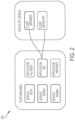
- FIG. 1 shows mobile app icons 100 for three cloud-based services, according to illustrative embodiments.
- the cloud-based services of the platform described herein provide for processing and analysis of medical images in a fully automated fashion and/or in combination with a user interaction (e.g., in a semi-automated fashion).
- the medical images include nuclear medicine images, obtained using a nuclear imaging modality such as whole-body scans with a gamma camera, Positron Emission Tomography (PET) scans, and Single-Photon Emission Tomography (SPECT) scans.
- PET Positron Emission Tomography
- SPECT Single-Photon Emission Tomography
- nuclear medicine images use imaging agents comprising radiopharmaceuticals.
- Nuclear medicine images are obtained following administration of a radiopharmaceutical to a patient, and provide information regarding the distribution of the radiopharmaceutical within the patient.
- Radiopharmaceuticals are compounds that comprise a radionuclide.
- radionuclide refers to a moiety comprising a radioactive isotope of at least one element.
- exemplary suitable radionuclides include but are not limited to those described herein.
- a radionuclide is one used in positron emission tomography (PET).
- PET positron emission tomography
- SPECT single-photon emission computed tomography
- a non-limiting list of radionuclides includes 99m Tc, 111 In, 64 Cu, 67 Ga, 68 Ga, 186 Re, 188 Re, 153 Sm, 177 Lu, 67 Cu, 123 I, 124 I, 125 I, 126 I, 131 I , 11 C, 13 N, 15 O, 18 F , 153 Sm, 166 Ho, 177 Lu, 149 Pm, 90 Y, 213 Bi, 103 Pd, 109 Pd, 159 Gd, 140 La, 198 Au, 199 Au, 169 Yb, 175 Yb, 165 Dy, 166 Dy, 105 Rh, 111 Ag, 89 Zr, 225 Ac, 82 Rb, 75 Br, 76 Br, 77 Br, 80 Br, 80m Br, 82 Br, 83 Br, 211 At and 192 Ir.
- radiopharmaceutical refers to a compound comprising a radionuclide.
- radiopharmaceuticals are used for diagnostic and/or therapeutic purposes.
- radiopharmaceuticals include small molecules that are labeled with one or more radionuclide(s), antibodies that are labeled with one or more radionuclide(s), and antigen-binding portions of antibodies that are labeled with one or more radionuclide(s).
- Nuclear medicine images detect radiation emitted from the radionuclides of radiopharmaceuticals to form an image.
- the distribution of a particular radiopharmaceutical within a patient may be determined by biological mechanisms such as blood flow or perfusion, as well as by specific enzymatic or receptor binding interactions.
- Different radiopharmaceuticals may be designed to take advantage of different biological mechanisms and/or particular specific enzymatic or receptor binding interactions and thus, when administered to a patient, selectively concentrate within particular types of tissue and/or regions within the patient.
- intensity variations within a nuclear medicine image can be used to map the distribution of radiopharmaceutical within the patient. This mapped distribution of radiopharmaceutical within the patient can be used to, for example, infer the presence of cancerous tissue within various regions of the patient's body.
- risk indices that correlate with patient overall survival and other prognostic metrics indicative of disease state, progression, treatment efficacy, and the like can be computed based on automated analysis of intensity variations in whole-body scans obtained following administration of 99m Tc MDP to a patient.
- other radiopharmaceuticals can also be used in a similar fashion to 99m Tc MDP.
- the particular radiopharmaceutical used depends on the particular nuclear medicine imaging modality used.
- 18 F sodium fluoride (NaF) also accumulates in bone lesions, similar to 99m Tc MDP, but can be used with PET imaging.
- PET imaging may also utilize a radioactive form of the vitamin choline, which is readily absorbed by prostate cancer cells.
- radiopharmaceuticals that selectively bind to particular proteins or receptors of interest - particularly those whose expression is increased in cancerous tissue may be used.
- proteins or receptors of interest include, but are not limited to tumor antigens, such as CEA, which is expressed in colorectal carcinomas, Her2/neu, which is expressed in multiple cancers, BRCA 1 and BRCA 2, expressed in breast and ovarian cancers; and TRP-1 and -2, expressed in melanoma.
- nuclear medicine images obtained using PSMA binding agents are used to identify the presence of cancerous tissue within the prostate, when the disease is in a localized state.
- nuclear medicine images obtained using radiopharmaceuticals comprising PSMA binding agents are used to identify the presence of cancerous tissue within a variety of regions that include not only the prostate, but also other organs and tissue regions such as lungs, lymph nodes, and bones, as is relevant when the disease is metastatic.
- the radionuclide labelled PSMA binding agent comprises [18F]DCFPyL (also referred to as PyL TM ; also referred to as DCFPyL-18F): or a pharmaceutically acceptable salt thereof.
- the radionuclide labelled PSMA binding agent comprises 1427 (also referred to as MIP-1427): or a pharmaceutically acceptable salt thereof.
- the radionuclide labelled PSMA binding agent comprises 1428 (also referred to as MIP-1428): or a pharmaceutically acceptable salt thereof.
- the radionuclide labelled PSMA binding agent comprises 99m Tc-MIP-1404, which is 1404 labelled with (e.g., chelated to) 99m Tc: or a pharmaceutically acceptable salt thereof.
- 1404 may be chelated to other metal radioisotopes [e.g., a radioisotope of rhenium (Re) (e.g., rhenium-188 ( 188 Re); e.g., rhenium-186 ( 186 Re)); e.g., a radioisotope of yttrium (Y) (e.g., 90 Y); e.g., a radioisotope of lutetium (Lu)(e.g., 177 Lu); e.g., a radioisotope of gallium (Ga) (e.g., 68 Ga; e.g., 67 Ga); e.g., a radioisotope of indium (e.g., 111 In); e.g., a radioisotope of copper (Cu) (e.g., 67 Cu)] to form a compound having a structure similar to the structure shown above for 99m T
- Re
- the radionuclide labelled PSMA binding agent comprises 99m Tc-MIP-1405, which is 1405 labelled with (e.g., chelated to) 99m Tc: or a pharmaceutically acceptable salt thereof.
- 1405 may be chelated to other metal radioisotopes [e.g., a radioisotope of rhenium (Re) (e.g., rhenium-188 ( 188 Re); e.g., rhenium-186 ( 186 Re)); e.g., a radioisotope of yttrium (Y) (e.g., 90 Y); e.g., a radioisotope of lutetium (Lu)(e.g., 177 Lu); e.g., a radioisotope of gallium (Ga) (e.g., 68 Ga; e.g., 67 Ga); e.g., a radioisotope of indium (e.g., 111 In); e.g., a radioisotope of copper (Cu) (e.g., 67 Cu)] to form a compound having a structure similar to the structure shown above for 99m T
- Re
- 1427 is labelled with (e.g., chelated to) a radioisotope of a metal, to form a compound according to the formula below: or a pharmaceutically acceptable salt thereof, wherein M is a metal radioisotope [e.g., a radioisotope of technetium (Tc) (e.g., technetium-99m ( 99m Tc)); e.g., a radioisotope of rhenium (Re) (e.g., rhenium-188 ( 188 Re); e.g., rhenium-186 ( 186 Re)); e.g., a radioisotope of yttrium (Y) (e.g., 90 Y); e.g., a radioisotope of lutetium (Lu)(e.g., 177 Lu); e.g., a radioisotope of gallium (Ga) (e.g., 68 Ga), a radio
- 1428 is labelled with (e.g., chelated to) a radioisotope of a metal, to form a compound according to the formula below: or a pharmaceutically acceptable salt thereof, wherein M is a metal radioisotope [e.g., a radioisotope of technetium (Tc) (e.g., technetium-99m ( 99m Tc)); e.g., a radioisotope of rhenium (Re) (e.g., rhenium-188 ( 188 Re); e.g., rhenium-186 ( 186 Re)); e.g., a radioisotope of yttrium (Y) (e.g., 90 Y); e.g., a radioisotope of lutetium (Lu)(e.g., 177 Lu); e.g., a radioisotope of gallium (Ga) (e.g., 68 Ga), a radio
- the radionuclide labelled PSMA binding agent comprises PSMA I&S: or a pharmaceutically acceptable salt thereof. In certain embodiments, the radionuclide labelled PSMA binding agent comprises 99m Tc-PSMA I&S, which is PSMA I&S labelled with 99m Tc, or a pharmaceutically acceptable salt thereof.
- PyL Cloud 120 refers to a cloud-based system that uses medical images obtained with the agent PyL TM , which is DCFPyL labeled with 18 F ([18F]DCFPyL).
- the patient after injection of the imaging agent, receives a positron emission tomography (PET) scan to identify hot spots, and a CT scan. Further information about the PyL TM imaging agent is provided above, and, for example, in U.S. Patent No. 8,778,305 .
- composite images comprising CT scans overlaid with PET scans are obtained using dedicated PET-CT scanner instruments common in many hospitals, and are uploaded as medical images to the cloud-based platform described herein.
- a PET scan and a corresponding CT scan are obtained separately (but at substantially the same time), and uploaded to the cloud-based platform described herein. In such cases, the separately obtained PET and CT scans can be automatically fused to create a composite image.
- one or more risk indices can be computed in a similar fashion to that described above with regard to BSI.
- intensity values of the PET scan in relation to (e.g., within and/or outside of) the 3D boundaries of the identified regions can be used to determine levels of cancerous tissue within the identified regions, e.g., based on features of detected hotspots (e.g., detected hotspots corresponding to metastases). Risk indices can then be computed based on the determined cancerous tissue levels.
- hotspots within the PET scan can be identified, and, based on features such as their size, number, and distribution with respect to the identified regions, used to compute one or more risk indices.
- risk indices determined from PET scans can be correlated with prognostic values and tracked over time (e.g., over the course of multiple visits to one or more doctors) to provide patients or their physician with an objective metric of what state their cancer is in, how fast it is progressing, what their outlook is, and whether one or more particular treatments are proving effective.
- Cloud 130 refers to a cloud-based system that uses medical images obtained with the agent 99m Tc-MIP-1404, which, as described above, is 1404 labeled with Tc 99m .
- the patient receives a single-photon emission computerized tomography (SPECT) scan, e.g., to identify hot spots, and a computed tomography (CT) scan, e.g., to identify anatomical features.
- SPECT/CT composite image
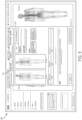
- FIG. 5 is a GUI window in the BSI Cloud application showing an automatically or semi-automatically generated radiologist report 510, which can be signed and dated by a radiologist.
- the automatically identified hotspots may be adjusted by the radiologist (or other medical practitioner attending to the patient), with the change(s) reflected in the report.
- an identified hotspot may be deactivated by the radiologist, or a new hotspot may be activated by the radiologist, such changes possibly affecting the computed BSI value, displayed in the report.
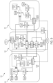
- FIG. 6 is a block flow diagram of an illustrative network-based (e.g., cloud based) decision support system, according to an illustrative embodiment of the invention.
- FIG. 6 shows various functions that may be performed by the cloud based services described herein.
- FIG. 10 is a schematic showing clinical stages 1010 of prostate cancer progression, along with various therapies 1020 and diagnostic imaging modalities 1030 appropriate for various disease states. As shown in the schematic, depending on the clinical state of a patient's prostate cancer, different imaging modalities and/or different radiopharmaceuticals may be appropriate. Similarly, different risk indices computed based on different imaging modalities and/or different radiopharmaceuticals may most appropriate, depending on the state of the patient's prostate cancer.
- a first subseries of medical images may be obtained using a first imaging modality and a first radiopharmaceutical, such as SPECT-CT imaging with 99m Tc-MIP-1404, when the patient's prostate cancer is in a localized state.
- a second subseries of images may comprise images obtained via a different, second imaging modality and/or a different, second radiopharmaceutical.
- the second subseries of medical images may be PET-CT images obtained using PyL TM .
- the second subseries of medical images may be whole-body scans obtained using 99m Tc-MDP.
- Risk indices can be computed for the first image subseries and for the second image subseries to provide a unified picture of the patient's prostate cancer progression and treatment over time. This approach can be performed for multiple patients, not only for decision making purposes with regard to each patient's disease progression and particular course of treatment, but also in the context of clinical trials, for example to compare efficacy of a particular treatment with others or a control.
- the cloud computing environment 700 may include a resource manager 706.
- the resource manager 706 may be connected to the resource providers 702 and the computing devices 704 over the computer network 708.
- the resource manager 706 may facilitate the provision of computing resources by one or more resource providers 702 to one or more computing devices 704.
- the resource manager 706 may receive a request for a computing resource from a particular computing device 704.
- the resource manager 706 may identify one or more resource providers 702 capable of providing the computing resource requested by the computing device 704.
- the resource manager 706 may select a resource provider 702 to provide the computing resource.
- the resource manager 706 may facilitate a connection between the resource provider 702 and a particular computing device 704.
- FIG. 8 shows an example of a computing device 800 and a mobile computing device 850 that can be used in the methods and systems described in this disclosure.
- the computing device 800 is intended to represent various forms of digital computers, such as laptops, desktops, workstations, personal digital assistants, servers, blade servers, mainframes, and other appropriate computers.
- the mobile computing device 850 is intended to represent various forms of mobile devices, such as personal digital assistants, cellular telephones, smart-phones, and other similar computing devices.
- the components shown here, their connections and relationships, and their functions, are meant to be examples only, and are not meant to be limiting.
- the computing device 800 includes a processor 802, a memory 804, a storage device 806, a high-speed interface 808 connecting to the memory 804 and multiple high-speed expansion ports 810, and a low-speed interface 812 connecting to a low-speed expansion port 814 and the storage device 806.
- Each of the processor 802, the memory 804, the storage device 806, the high-speed interface 808, the high-speed expansion ports 810, and the low-speed interface 812 are interconnected using various busses, and may be mounted on a common motherboard or in other manners as appropriate.
- the processor 802 can process instructions for execution within the computing device 800, including instructions stored in the memory 804 or on the storage device 806 to display graphical information for a GUI on an external input/output device, such as a display 816 coupled to the high-speed interface 808.
- an external input/output device such as a display 816 coupled to the high-speed interface 808.
- multiple processors and/or multiple buses may be used, as appropriate, along with multiple memories and types of memory.
- multiple computing devices may be connected, with each device providing portions of the necessary operations (e.g., as a server bank, a group of blade servers, or a multi-processor system).
- the mobile computing device 850 may communicate wirelessly through the communication interface 866, which may include digital signal processing circuitry where necessary.
- the communication interface 866 may provide for communications under various modes or protocols, such as GSM voice calls (Global System for Mobile communications), SMS (Short Message Service), EMS (Enhanced Messaging Service), or MMS messaging (Multimedia Messaging Service), CDMA (code division multiple access), TDMA (time division multiple access), PDC (Personal Digital Cellular), WCDMA (Wideband Code Division Multiple Access), CDMA2000, or GPRS (General Packet Radio Service), among others.
- GSM voice calls Global System for Mobile communications
- SMS Short Message Service
- EMS Enhanced Messaging Service
- MMS messaging Multimedia Messaging Service
- CDMA code division multiple access
- TDMA time division multiple access
- PDC Personal Digital Cellular
- WCDMA Wideband Code Division Multiple Access
- CDMA2000 Code Division Multiple Access
- GPRS General Packet Radio Service
- the mobile computing device 850 may also communicate audibly using an audio codec 860, which may receive spoken information from a user and convert it to usable digital information.
- the audio codec 860 may likewise generate audible sound for a user, such as through a speaker, e.g., in a handset of the mobile computing device 850.
- Such sound may include sound from voice telephone calls, may include recorded sound (e.g., voice messages, music files, etc.) and may also include sound generated by applications operating on the mobile computing device 850.
Landscapes
- Health & Medical Sciences (AREA)
- Engineering & Computer Science (AREA)
- Medical Informatics (AREA)
- Public Health (AREA)
- General Health & Medical Sciences (AREA)
- Primary Health Care (AREA)
- Epidemiology (AREA)
- Biomedical Technology (AREA)
- Pathology (AREA)
- Data Mining & Analysis (AREA)
- Life Sciences & Earth Sciences (AREA)
- Databases & Information Systems (AREA)
- Radiology & Medical Imaging (AREA)
- Nuclear Medicine, Radiotherapy & Molecular Imaging (AREA)
- Molecular Biology (AREA)
- Physics & Mathematics (AREA)
- Immunology (AREA)
- Chemical & Material Sciences (AREA)
- Software Systems (AREA)
- Biophysics (AREA)
- Theoretical Computer Science (AREA)
- Medicinal Chemistry (AREA)
- Urology & Nephrology (AREA)
- Veterinary Medicine (AREA)
- Animal Behavior & Ethology (AREA)
- General Physics & Mathematics (AREA)
- Surgery (AREA)
- Hematology (AREA)
- Heart & Thoracic Surgery (AREA)
- Optics & Photonics (AREA)
- High Energy & Nuclear Physics (AREA)
- Bioinformatics & Cheminformatics (AREA)
- Microbiology (AREA)
- General Engineering & Computer Science (AREA)
- Food Science & Technology (AREA)
- Computer Vision & Pattern Recognition (AREA)
- Biotechnology (AREA)
- Toxicology (AREA)
- Pharmacology & Pharmacy (AREA)
- Cell Biology (AREA)
Claims (11)
- Verfahren zum Verfolgen des Fortschreitens von Prostatakrebs und der Wirksamkeit der Behandlung im Laufe der Zeit für einen oder mehrere Patienten, wobei das Verfahren umfasst:(a) wiederholtes Empfangen und Speichern einer Vielzahl von medizinischen Bildern für jeden des einen oder der mehreren Patienten in einer Datenbank im Laufe der Zeit durch einen Prozessor einer Datenverarbeitungsvorrichtung, um für jeden des einen oder der mehreren Patienten eine Serie von im Laufe der Zeit aufgenommenen medizinischen Bildern zu erhalten;(b) für jeden des einen oder der mehreren Patienten, automatisches Analysieren der Serie von medizinischen Bildern für den Patienten durch den Prozessor, um Werte eines oder mehrerer Risikoindizes für jedes medizinische Bild der Serie zu ermitteln, wodurch die ermittelten Werte des einen oder der mehreren Risikoindizes im Verlauf des Fortschritts und der Behandlung des Prostatakrebses für den Patienten verfolgt werden; und(c) für jeden des einen oder der mehreren Patienten, Speichern der ermittelten Werte des einen oder der mehreren Risikoindizes für den Patienten durch den Prozessor zur weiteren Verarbeitung und/oder Veranlassen des Anzeigens einer grafischen Darstellung der ermittelten Werte des einen oder der mehreren Risikoindizes für den Patienten durch den Prozessor,
wobei für jeden bestimmten Patienten des einen oder der mehreren Patienten die Vielzahl von medizinischen Bildern einen oder mehrere zusammengesetzte PET-CT-Scans des bestimmten Patienten umfasst, die nach Verabreichung von [18F]DCFPyL an den bestimmten Patienten erhalten wurden, und wobei der Prozessor ein Prozessor eines Cloud-basierten Systems ist. - System zum Verfolgen des Fortschreitens von Prostatakrebs und der Wirksamkeit der Behandlung im Laufe der Zeit für einen oder mehrere Patienten, wobei das System umfasst:einen Prozessor; undeinen Speicher, der darauf gespeicherte Anweisungen aufweist, wobei die Anweisungen, wenn sie durch den Prozessor ausgeführt werden, den Prozessor veranlassen zum:(a) wiederholten Empfangen und Speichern einer Vielzahl von medizinischen Bildern für jeden des einen oder der mehreren Patienten in einer Datenbank im Laufe der Zeit, um für jeden des einen oder der mehreren Patienten eine Serie von im Laufe der Zeit aufgenommenen medizinischen Bildern zu erhalten;(b) für jeden des einen oder der mehreren Patienten, automatischen Analysieren der Serie von medizinischen Bildern für den Patienten, um Werte eines oder mehrerer Risikoindizes für jedes medizinische Bild der Serie zu ermitteln, wodurch die ermittelten Werte des einen oder der mehreren Risikoindizes im Verlauf des Fortschritts und der Behandlung des Prostatakrebses für den Patienten verfolgt werden; und(c) für jeden des einen oder der mehreren Patienten, Speichern der ermittelten Werte des einen oder der mehreren Risikoindizes für den Patienten zur weiteren Verarbeitung und/oder Veranlassen des Anzeigens einer grafischen Darstellung der ermittelten Werte des einen oder der mehreren Risikoindizes für den Patienten,wobei für jeden bestimmten Patienten des einen oder der mehreren Patienten die Vielzahl von medizinischen Bildern einen oder mehrere zusammengesetzte PET-CT-Scans des bestimmten Patienten umfasst, die nach Verabreichung von [18F]DCFPyL an den bestimmten Patienten erhalten wurden, und wobei das System ein Cloud-basiertes System ist.
- Verfahren nach Anspruch 1 oder System nach Anspruch 2, wobei die Serie von medizinischen Bildern für einen bestimmten Patienten des einen oder der mehreren Patienten umfasst:(i) eine erste Bilder-Teilserie, die ein oder mehrere medizinische Bilder umfasst, die jeweils nach Verabreichung eines ersten Radiopharmazeutikums an den bestimmten Patienten unter Verwendung einer ersten Nuklearbildgebungsmodalität aufgenommen wurden; und(i) eine zweite Bilder-Teilserie, die ein oder mehrere medizinische Bilder umfasst, die jeweils nach Verabreichung eines zweiten Radiopharmazeutikums an den bestimmten Patienten unter Verwendung einer zweiten Nuklearbildgebungsmodalität aufgenommen wurden,
sodass die Werte des einen oder der mehreren Risikoindizes, die in Schritt (b) für den bestimmten Patienten ermittelt wurden, eine erste Teilserie von Werten eines ersten Risikoindex, der durch automatisierte Analyse der ersten Bilder-Teilserie ermittelt wurde, und eine zweite Teilserie von Werten eines zweiten Risikoindex umfassen, der durch automatisierte Analyse der zweiten Bilder-Teilserie ermittelt wurde. - Verfahren oder System nach Anspruch 3, wobei die medizinischen Bilder der ersten Bilder-Teilserie über einen ersten Zeitraum erhalten werden, wenn der Prostatakrebs des bestimmten Patienten lokalisiert ist, und die medizinischen Bilder der zweiten Bilder-Teilserie über einen zweiten Zeitraum erhalten werden, wenn der Prostatakrebs des bestimmten Patienten metastatisch ist.
- Verfahren oder System nach Anspruch 3 oder 4, wobei:die erste Bilder-Teilserie ein oder mehrere zusammengesetzte SPECT-CT-Bilder umfasst, wobei jedes zusammengesetzte SPECT-CT-Bild einen CT-Scan umfasst, der mit einem im Wesentlichen gleichzeitig erfassten SPECT-Scan überlagert ist;die zweite Bilder-Teilserie ein oder mehrere zusammengesetzte PET-CT-Bilder umfasst, wobei jedes zusammengesetzte PET-CT-Bild einen CT-Scan umfasst, der mit einem im Wesentlichen gleichzeitig erfassten PET-Scan überlagert ist; undSchritt (b) umfasst:
automatisches Analysieren jedes des einen oder der mehreren zusammengesetzten SPECT-CT-Bilder durch:Verwenden des zusammengesetzten SPECT-CT-Bildes, um eine 3D-Grenze einer Prostataregion innerhalb des SPECT-Scans des zusammengesetzten SPECT-CT-Bildes geografisch zu identifizieren; undBerechnen eines Wertes des ersten Risikoindex unter Verwendung des SPECT-Scans mit der identifizierten 3D-Grenze der Prostataregion;
undautomatisches Analysieren jedes des einen oder der mehreren zusammengesetzten PET-CT-Bilder durch:Verwenden des zusammengesetzten PET-CT-Bildes zum geografischen Identifizieren einer 3D-Grenze einer oder mehrerer metastatischer Regionen innerhalb des PET-Scans des zusammengesetzten PET-CT-Bildes, wobei die eine oder die mehreren metastatischen Regionen Regionen einschließen, die Patientengewebestellen außerhalb der Prostata entsprechen; undBerechnen eines Wertes des zweiten Risikoindex unter Verwendung des PET-Scans mit den identifizierten 3D-Grenzen der einen oder mehreren metastatischen Regionen. - Verfahren oder System nach Anspruch 3 oder 4, wobei:die erste Bilder-Teilserie ein oder mehrere zusammengesetzte SPECT-CT-Bilder umfasst, wobei jedes zusammengesetzte SPECT-CT-Bild einen CT-Scan umfasst, der mit einem im Wesentlichen gleichzeitig erfassten SPECT-Scan überlagert ist;die zweite Bilder-Teilserie einen oder mehrere Ganzkörperscans umfasst; undSchritt (b) umfasst:
automatisches Analysieren jedes des einen oder der mehreren zusammengesetzten SPECT-CT-Bilder durch:Verwenden des zusammengesetzten SPECT-CT-Bildes, um eine 3D-Grenze einer Prostataregion innerhalb des SPECT-Scans des zusammengesetzten SPECT-CT-Bildes geografisch zu identifizieren; undBerechnen eines Wertes des ersten Risikoindex unter Verwendung des SPECT-Scans mit der identifizierten 3D-Grenze der Prostataregion; undautomatisches Analysieren jedes des einen oder der mehreren Ganzkörperscans durch:geografisches Identifizieren einer Grenze einer oder mehrerer metastatischer Regionen innerhalb des Ganzkörperscans, wobei die eine oder die mehreren metastatischen Regionen Regionen einschließen, die Patientengewebestellen außerhalb der Prostata entsprechen; undBerechnen eines Wertes des zweiten Risikoindex unter Verwendung des Ganzkörperscans mit den identifizierten 3D-Grenzen der einen oder mehreren metastatischen Regionen. - Verfahren oder System nach Anspruch 5 oder 6, wobei die eine oder die mehreren metastatischen Regionen einen oder mehrere bestimmte Knochen umfassen.
- Verfahren oder System nach Anspruch 4, das die Verwendung von PET-CT-Bildgebung zum Beurteilen von Prostatakrebs sowohl im lokalisierten als auch im metastatischen Stadium umfasst.
- Verfahren oder System nach einem der Ansprüche 3 bis 7, wobei das erste und das zweite Radiopharmazeutikum prostataspezifische Membranantigenbindemittel (PSMA-Bindemittel) umfassen.
- Verfahren oder System nach Anspruch 6, wobei die Vielzahl von medizinischen Bildern für jeden bestimmten Patienten des einen oder der mehreren Patienten einen oder mehrere mit einer Gammakamera erstellte Ganzkörperscans des bestimmten Patienten umfasst, die nach Verabreichung eines Bildgebungsmittels an den bestimmten Patienten erhalten wurden, das Technetium-99m-Methylendiphosphonat (99mTcMDP) umfasst.
- Verfahren nach Anspruch 1 oder System nach Anspruch 2, wobei die medizinischen Bilder (i) ein oder mehrere zusammengesetzte PET-CT-Bilder, (ii) ein oder mehrere zusammengesetzte SPECT-CT-Bilder oder (iii) einen oder mehrere Ganzkörperscans umfassen.
Priority Applications (1)
| Application Number | Priority Date | Filing Date | Title |
|---|---|---|---|
| EP25182316.7A EP4636783A3 (de) | 2016-10-27 | 2017-10-26 | Netzwerk zur medizinischen bildanalyse, entscheidungsunterstützungssystem und zugehörige anwendungen einer grafischen benutzeroberfläche (gui) |
Applications Claiming Priority (2)
| Application Number | Priority Date | Filing Date | Title |
|---|---|---|---|
| US201662413936P | 2016-10-27 | 2016-10-27 | |
| PCT/US2017/058418 WO2018081354A1 (en) | 2016-10-27 | 2017-10-26 | Network for medical image analysis, decision support system, and related graphical user interface (gui) applications |
Related Child Applications (1)
| Application Number | Title | Priority Date | Filing Date |
|---|---|---|---|
| EP25182316.7A Division EP4636783A3 (de) | 2016-10-27 | 2017-10-26 | Netzwerk zur medizinischen bildanalyse, entscheidungsunterstützungssystem und zugehörige anwendungen einer grafischen benutzeroberfläche (gui) |
Publications (3)
| Publication Number | Publication Date |
|---|---|
| EP3533063A1 EP3533063A1 (de) | 2019-09-04 |
| EP3533063C0 EP3533063C0 (de) | 2025-07-02 |
| EP3533063B1 true EP3533063B1 (de) | 2025-07-02 |
Family
ID=60263152
Family Applications (2)
| Application Number | Title | Priority Date | Filing Date |
|---|---|---|---|
| EP25182316.7A Pending EP4636783A3 (de) | 2016-10-27 | 2017-10-26 | Netzwerk zur medizinischen bildanalyse, entscheidungsunterstützungssystem und zugehörige anwendungen einer grafischen benutzeroberfläche (gui) |
| EP17794628.2A Active EP3533063B1 (de) | 2016-10-27 | 2017-10-26 | Netzwerk zur analyse von medizinischen bildern, entscheidungsunterstützungssystem und zugehörige anwendungen für grafische benutzeroberfläche (gui) |
Family Applications Before (1)
| Application Number | Title | Priority Date | Filing Date |
|---|---|---|---|
| EP25182316.7A Pending EP4636783A3 (de) | 2016-10-27 | 2017-10-26 | Netzwerk zur medizinischen bildanalyse, entscheidungsunterstützungssystem und zugehörige anwendungen einer grafischen benutzeroberfläche (gui) |
Country Status (8)
| Country | Link |
|---|---|
| US (7) | US10340046B2 (de) |
| EP (2) | EP4636783A3 (de) |
| JP (3) | JP7390188B2 (de) |
| CN (2) | CN109844865B (de) |
| AU (2) | AU2017348111B2 (de) |
| CA (1) | CA3036754A1 (de) |
| ES (1) | ES3041039T3 (de) |
| WO (1) | WO2018081354A1 (de) |
Families Citing this family (45)
| Publication number | Priority date | Publication date | Assignee | Title |
|---|---|---|---|---|
| WO2009084995A1 (en) | 2007-12-28 | 2009-07-09 | Exini Diagnostics Ab | System for detecting bone cancer metastases |
| ES3041039T3 (en) | 2016-10-27 | 2025-11-06 | Progenics Pharm Inc | Network for medical image analysis, decision support system, and related graphical user interface (gui) applications |
| US10346981B2 (en) * | 2016-11-04 | 2019-07-09 | Eric Kenneth Anderson | System and method for non-invasive tissue characterization and classification |
| US10918346B2 (en) | 2017-09-06 | 2021-02-16 | General Electric Company | Virtual positioning image for use in imaging |
| EP3499459A1 (de) * | 2017-12-18 | 2019-06-19 | FEI Company | Verfahren, vorrichtung und system zum entfernten tiefenlernen zur mikroskopischen bildrekonstruktion und -segmentierung |
| US10973486B2 (en) | 2018-01-08 | 2021-04-13 | Progenics Pharmaceuticals, Inc. | Systems and methods for rapid neural network-based image segmentation and radiopharmaceutical uptake determination |
| JP6906462B2 (ja) * | 2018-02-28 | 2021-07-21 | 富士フイルム株式会社 | 医用画像表示装置、方法およびプログラム |
| KR102274564B1 (ko) * | 2018-07-03 | 2021-07-07 | (주) 프로큐라티오 | 빅데이터분석기반 암진단장치 |
| WO2020066132A1 (ja) * | 2018-09-27 | 2020-04-02 | 富士フイルム株式会社 | 医用画像診断支援装置、方法及びプログラム |
| CA3115807A1 (en) * | 2018-10-11 | 2020-04-16 | Progenics Pharmaceuticals, Inc | Methods of making prostate cancer treatment decisions |
| CN113164450A (zh) * | 2018-10-11 | 2021-07-23 | 普罗热尼奇制药公司 | 用于治疗转移性前列腺癌的组合治疗 |
| JP2022506135A (ja) * | 2018-10-30 | 2022-01-17 | アレン インスティテュート | ヒトの寄与を組み込む反復的深層学習フローを使用した顕微鏡画像内の3d細胞間構造のセグメント化 |
| US10943681B2 (en) * | 2018-11-21 | 2021-03-09 | Enlitic, Inc. | Global multi-label generating system |
| US11514571B2 (en) * | 2018-12-17 | 2022-11-29 | Siemens Healthcare Gmbh | Hierarchical analysis of medical images for identifying and assessing lymph nodes |
| AU2020206584B2 (en) | 2019-01-07 | 2024-11-07 | Exini Diagnostics Ab | Systems and methods for platform agnostic whole body image segmentation |
| WO2020167736A2 (en) * | 2019-02-15 | 2020-08-20 | Arterys Inc. | Deep learning-based eddy current correction |
| TWI872062B (zh) * | 2019-04-24 | 2025-02-11 | 美商普吉尼製藥公司 | 用於偵測轉移之骨掃描影像之自動及互動式分析系統、裝置及方法 |
| JP7539921B2 (ja) | 2019-04-24 | 2024-08-26 | プロジェニクス ファーマシューティカルズ, インコーポレイテッド | 核医学画像における強度ウィンドウ処理の双方向調節のためのシステムおよび方法 |
| WO2020243193A1 (en) * | 2019-05-28 | 2020-12-03 | PAIGE.AI, Inc. | Systems and methods for processing images to prepare slides for processed images for digital pathology |
| TWI738001B (zh) | 2019-06-03 | 2021-09-01 | 睿傳數據股份有限公司 | 醫學影像中身體部位識別方法及裝置 |
| US12417533B2 (en) | 2019-09-27 | 2025-09-16 | Progenics Pharmaceuticals, Inc. | Systems and methods for artificial intelligence-based image analysis for cancer assessment |
| US11544407B1 (en) | 2019-09-27 | 2023-01-03 | Progenics Pharmaceuticals, Inc. | Systems and methods for secure cloud-based medical image upload and processing |
| US11564621B2 (en) | 2019-09-27 | 2023-01-31 | Progenies Pharmacenticals, Inc. | Systems and methods for artificial intelligence-based image analysis for cancer assessment |
| US11900597B2 (en) | 2019-09-27 | 2024-02-13 | Progenics Pharmaceuticals, Inc. | Systems and methods for artificial intelligence-based image analysis for cancer assessment |
| US11842498B2 (en) | 2019-12-16 | 2023-12-12 | Siemens Healthineers International Ag | Systems and methods for automatic segmentation in medical imaging with multiple anatomical structure segmentation models |
| FR3104934B1 (fr) * | 2019-12-18 | 2023-04-07 | Quantum Surgical | Méthode de planification automatique d’une trajectoire pour une intervention médicale |
| US11429808B2 (en) | 2019-12-19 | 2022-08-30 | Varian Medical Systems International Ag | Systems and methods for scalable segmentation model training |
| US11386988B2 (en) | 2020-04-23 | 2022-07-12 | Exini Diagnostics Ab | Systems and methods for deep-learning-based segmentation of composite images |
| US11321844B2 (en) | 2020-04-23 | 2022-05-03 | Exini Diagnostics Ab | Systems and methods for deep-learning-based segmentation of composite images |
| US20210398653A1 (en) * | 2020-06-17 | 2021-12-23 | Fovia, Inc. | Key image updating multiple stacks |
| US11721428B2 (en) * | 2020-07-06 | 2023-08-08 | Exini Diagnostics Ab | Systems and methods for artificial intelligence-based image analysis for detection and characterization of lesions |
| WO2022008374A1 (en) | 2020-07-06 | 2022-01-13 | Exini Diagnostics Ab | Systems and methods for artificial intelligence-based image analysis for detection and characterization of lesions |
| EP4178448A4 (de) * | 2020-07-10 | 2025-06-18 | Progenics Pharmaceuticals, Inc. | Verfahren zur durchführung von prostatakrebsbehandlungsentscheidungen |
| CN116134529A (zh) * | 2020-07-16 | 2023-05-16 | 皇家飞利浦有限公司 | 用于放射学报告的自动确定性评估器 |
| WO2022108989A1 (en) * | 2020-11-19 | 2022-05-27 | Cerecin Inc. | Radiopharmaceutical ketone and dual tracer imaging in alzheimer's disease, cognitive impairment, and other conditions of altered cerebral metabolism |
| US12148528B1 (en) | 2021-01-22 | 2024-11-19 | Cardinal Health Commercial Technologies, Llc | Point-of-care clinical decision support platform |
| US11188393B1 (en) * | 2021-03-02 | 2021-11-30 | NortonLifeLock Inc. | Systems and methods for performing load balancing and distributed high-availability |
| US12082881B2 (en) * | 2021-03-22 | 2024-09-10 | Biosense Webster (Israel) Ltd. | Visualizing multiple parameters overlaid on an anatomical map |
| DE102021204238A1 (de) * | 2021-04-28 | 2022-11-03 | Siemens Healthcare Gmbh | Verfahren und System zur Erzeugung und Strukturierung medizinischer Untersuchungsinformationen |
| CN113222038B (zh) * | 2021-05-24 | 2021-10-22 | 北京安德医智科技有限公司 | 基于核磁图像的乳腺病灶分类和定位方法及装置 |
| WO2023081408A1 (en) * | 2021-11-05 | 2023-05-11 | Memorial Sloan Kettering Cancer Center | Systems and methods for generating a corrected planar scintigraphy image (cpsi) |
| KR102821475B1 (ko) * | 2022-09-05 | 2025-06-16 | 부산대학교 산학협력단 | 의료데이터 컴퓨팅 시스템 및 방법 |
| JP2024112204A (ja) * | 2023-02-07 | 2024-08-20 | キヤノンメディカルシステムズ株式会社 | 医用情報処理装置、医用情報処理方法及び医用情報処理プログラム |
| US12086990B1 (en) * | 2024-04-23 | 2024-09-10 | MultiFunctional Imaging LLC | Systems and methods for simultaneous imaging of multiple positron emission tomography (PET) tracers |
| GB2643197A (en) * | 2024-08-05 | 2026-02-11 | Univ Stellenbosch | Method and system of segmentation of medical images |
Citations (4)
| Publication number | Priority date | Publication date | Assignee | Title |
|---|---|---|---|---|
| US20110126159A1 (en) * | 2009-11-23 | 2011-05-26 | Samsung Electronics Co., Ltd. | Gui providing method, and display apparatus and 3d image providing system using the same |
| US20130129168A1 (en) * | 2011-11-23 | 2013-05-23 | The Regents Of The University Of Michigan | Voxel-Based Approach for Disease Detection and Evolution |
| WO2015058151A2 (en) * | 2013-10-18 | 2015-04-23 | Molecular Insight Pharmaceuticals, Inc. | Methods of using spect/ct analysis for staging cancer |
| US20170178266A1 (en) * | 2015-12-16 | 2017-06-22 | Sap Se | Interactive data visualisation of volume datasets with integrated annotation and collaboration functionality |
Family Cites Families (147)
| Publication number | Priority date | Publication date | Assignee | Title |
|---|---|---|---|---|
| US5165410A (en) | 1987-05-15 | 1992-11-24 | Medical & Scientific Enterprises, Inc. | Position indicating system for a multidiagnostic scanner |
| CN1111823C (zh) | 1997-05-27 | 2003-06-18 | 力捷电脑股份有限公司 | 使用分段加权校正的影像处理装置及其处理方法 |
| US6058322A (en) | 1997-07-25 | 2000-05-02 | Arch Development Corporation | Methods for improving the accuracy in differential diagnosis on radiologic examinations |
| US6944330B2 (en) | 2000-09-07 | 2005-09-13 | Siemens Corporate Research, Inc. | Interactive computer-aided diagnosis method and system for assisting diagnosis of lung nodules in digital volumetric medical images |
| WO2002061457A2 (en) | 2001-01-30 | 2002-08-08 | Decharms R Christopher | Methods for physiological monitoring, training, exercise and regulation |
| US7295691B2 (en) | 2002-05-15 | 2007-11-13 | Ge Medical Systems Global Technology Company, Llc | Computer aided diagnosis of an image set |
| US7450747B2 (en) | 2002-07-12 | 2008-11-11 | Ge Medical Systems Global Technology Company, Llc | System and method for efficiently customizing an imaging system |
| SE524500C2 (sv) | 2002-09-16 | 2004-08-17 | Weaidu In Europ Ab | Förfarande och anordning för bestämning av en tredimensionell kontur av ett organ i en patients kropp |
| EP1508872A1 (de) | 2003-08-22 | 2005-02-23 | Semeion | Algorithmus zum Erkennen von Verhältnissen zwischen Daten einer Datenbank und auf diesem Algorithmus basierende Bildmustererkennungsmethode |
| US7935055B2 (en) | 2003-09-19 | 2011-05-03 | Siemens Medical Solutions Usa, Inc. | System and method of measuring disease severity of a patient before, during and after treatment |
| DE10356272B4 (de) | 2003-11-28 | 2006-02-23 | Siemens Ag | Verfahren zur Navigation in 3-dimensionalen Bilddaten |
| WO2006051531A2 (en) | 2004-11-09 | 2006-05-18 | Spectrum Dynamics Llc | Radioimaging |
| US20090311182A1 (en) | 2004-03-31 | 2009-12-17 | Dong Wang | Macromolecular Delivery Systems for Non-Invasive Imaging, Evaluation and Treatment of Arthritis and Other Inflammatory Diseases |
| CN101076724B (zh) | 2004-04-14 | 2012-11-14 | 美国医软科技公司 | 肝病诊断系统,方法和图形用户界面 |
| DE102004027710A1 (de) | 2004-06-07 | 2006-01-26 | Siemens Ag | Verfahren zur automatischen Detektion einer Struktur bei bildgebenden medizinischen Verfahren, Computertomografiegerät, Arbeitsstation und Comupterprogrammprodukt |
| US7397475B2 (en) * | 2004-09-02 | 2008-07-08 | Siemens Medical Solutions Usa, Inc. | Interactive atlas extracted from volume data |
| US20140193336A1 (en) | 2005-07-19 | 2014-07-10 | Biosensors International Group, Ltd. | Imaging protocols |
| US7668342B2 (en) | 2005-09-09 | 2010-02-23 | Carl Zeiss Meditec, Inc. | Method of bioimage data processing for revealing more meaningful anatomic features of diseased tissues |
| US7876938B2 (en) * | 2005-10-06 | 2011-01-25 | Siemens Medical Solutions Usa, Inc. | System and method for whole body landmark detection, segmentation and change quantification in digital images |
| US7920730B2 (en) | 2005-10-07 | 2011-04-05 | Siemens Medical Solutions Usa, Inc. | Automatic bone detection in MRI images |
| DE102005048853A1 (de) | 2005-10-12 | 2007-04-26 | Siemens Ag | Bildgebende medizinische Modalität |
| WO2007062135A2 (en) | 2005-11-23 | 2007-05-31 | Junji Shiraishi | Computer-aided method for detection of interval changes in successive whole-body bone scans and related computer program product and system |
| DE102005059209B4 (de) | 2005-12-12 | 2010-11-25 | Siemens Ag | Verfahren und Vorrichtung zur Visualisierung einer Folge von tomographischen Bilddatensätzen |
| EP2005388B1 (de) | 2006-03-24 | 2010-08-11 | Exini Diagnostics Ab | Automatische interpretation von medizinischen 3d-bildern des hirns und verfahren zum produzieren von zwischenergebnissen |
| JP5127276B2 (ja) | 2006-05-26 | 2013-01-23 | 株式会社東芝 | 画像処理装置および磁気共鳴イメージング装置 |
| US20080027315A1 (en) | 2006-07-31 | 2008-01-31 | Icad, Inc. | Processing and presentation of electronic subtraction for tagged colonic fluid and rectal tube in computed colonography |
| RU2450832C2 (ru) * | 2006-10-25 | 2012-05-20 | Конинклейке Филипс Электроникс Н.В. | Контрастные вещества для детекции рака предстательной железы |
| EP2618102A2 (de) | 2006-11-21 | 2013-07-24 | Mantisvision Ltd. | Geometrische 3D-Modellierung und 3D-Videoinhaltserzeugung |
| US8545809B2 (en) | 2007-01-11 | 2013-10-01 | Immunomedics, Inc. | Methods and compositions for improved 18F labeling of proteins, peptides and other molecules |
| WO2009084995A1 (en) | 2007-12-28 | 2009-07-09 | Exini Diagnostics Ab | System for detecting bone cancer metastases |
| GB0803064D0 (en) | 2008-02-20 | 2008-03-26 | Siemens Medical Solutions | System for defining volumes of interest with reference to anatomical features |
| US20090309874A1 (en) | 2008-06-11 | 2009-12-17 | Siemens Medical Solutions Usa, Inc. | Method for Display of Pre-Rendered Computer Aided Diagnosis Results |
| JP2010029481A (ja) * | 2008-07-29 | 2010-02-12 | Univ Of Tsukuba | 腫瘍の経過観察レポート自動作成診断支援システム |
| US8778305B2 (en) | 2008-08-01 | 2014-07-15 | The Johns Hopkins University | PSMA-binding agents and uses thereof |
| US20100032575A1 (en) | 2008-08-08 | 2010-02-11 | Andrei Iagaru | Methods and systems for pet/ct scanning for evaluation of malignancy |
| US8705887B2 (en) | 2008-08-22 | 2014-04-22 | Weyerhaeuser Nr Company | Method and apparatus for filling in or replacing image pixel data |
| US9785858B2 (en) | 2008-09-26 | 2017-10-10 | Siemens Healthcare Gmbh | Method and system for hierarchical parsing and semantic navigation of full body computed tomography data |
| EP2706057B1 (de) | 2008-12-05 | 2016-04-20 | Molecular Insight Pharmaceuticals, Inc. | Bis(imidazolyl)verbindungen and Radionuclidkomplexe |
| EP2373621A2 (de) | 2008-12-05 | 2011-10-12 | Molecular Insight Pharmaceuticals, Inc. | Technetium- und rhenium-bis(heteroaryl)komplexe und verfahren zu deren anwendung |
| US20100158332A1 (en) | 2008-12-22 | 2010-06-24 | Dan Rico | Method and system of automated detection of lesions in medical images |
| ES2712732T3 (es) * | 2009-02-17 | 2019-05-14 | Cornell Res Foundation Inc | Métodos y kits para el diagnóstico de cáncer y la predicción de valor terapéutico |
| JP5744834B2 (ja) | 2009-03-26 | 2015-07-08 | コーニンクレッカ フィリップス エヌ ヴェ | 医学的ガイドラインで支持されるpet/ct治療モニタシステム |
| US8073220B2 (en) | 2009-04-20 | 2011-12-06 | Siemens Aktiengesellschaft | Methods and systems for fully automatic segmentation of medical images |
| US8588486B2 (en) | 2009-06-18 | 2013-11-19 | General Electric Company | Apparatus and method for isolating a region in an image |
| US20110007954A1 (en) | 2009-07-07 | 2011-01-13 | Siemens Corporation | Method and System for Database-Guided Lesion Detection and Assessment |
| US8467856B2 (en) | 2009-07-17 | 2013-06-18 | Koninklijke Philips Electronics N.V. | Anatomy modeling for tumor region of interest definition |
| JP5613235B2 (ja) | 2009-07-20 | 2014-10-22 | コーニンクレッカ フィリップス エヌ ヴェ | 関心腫瘍領域の画成のための生体構造モデリング |
| JP2011067594A (ja) | 2009-08-25 | 2011-04-07 | Fujifilm Corp | 肝機能造影像を用いた医用画像診断装置および方法、並びにプログラム |
| CN101639937B (zh) | 2009-09-03 | 2011-12-14 | 复旦大学 | 一种基于人工神经网络的超分辨率方法 |
| US20110063288A1 (en) | 2009-09-11 | 2011-03-17 | Siemens Medical Solutions Usa, Inc. | Transfer function for volume rendering |
| WO2011077303A1 (en) | 2009-12-23 | 2011-06-30 | Koninklijke Philips Electronics N.V. | Methods and apparatuses for prostate cancer detection, staging, and therapy response assessment |
| AU2011207453A1 (en) | 2010-01-22 | 2012-08-02 | The Research Foundation Of The State University Of New York | System and method for prostate visualization and cancer detection |
| EP2533816A1 (de) | 2010-02-08 | 2012-12-19 | Piramal Imaging SA | F18-tyrosin-derivate zur bildgebung von knochenmetastasen |
| US9401047B2 (en) | 2010-04-15 | 2016-07-26 | Siemens Medical Solutions, Usa, Inc. | Enhanced visualization of medical image data |
| CN102096804A (zh) | 2010-12-08 | 2011-06-15 | 上海交通大学 | 骨扫描中肿瘤骨转移的图像识别方法 |
| CN103607954B (zh) | 2011-06-10 | 2016-08-24 | 皇家飞利浦有限公司 | 用于混合式扫描器上ac和定位的剂量优化方案 |
| US9123155B2 (en) | 2011-08-09 | 2015-09-01 | Covidien Lp | Apparatus and method for using augmented reality vision system in surgical procedures |
| US9002081B2 (en) * | 2011-10-18 | 2015-04-07 | Matthew Sherman Brown | Computer-aided bone scan assessment with automated lesion detection and quantitative assessment of bone disease burden changes |
| CN103930030B (zh) | 2011-10-18 | 2017-06-16 | 迷笛公司 | 具有自动化病变检测和骨疾病负荷变化的量化评定的计算机辅助骨扫描评定 |
| US9547061B2 (en) | 2012-01-27 | 2017-01-17 | Koninklijke Philips N.V. | Tumor segmentation and tissue classification in 3D multi-contrast |
| US8682049B2 (en) * | 2012-02-14 | 2014-03-25 | Terarecon, Inc. | Cloud-based medical image processing system with access control |
| US20130211231A1 (en) | 2012-02-14 | 2013-08-15 | Manivannan Sundarapandian | Method and system for visualization of treatment volumes |
| HK1205788A1 (en) * | 2012-02-24 | 2015-12-24 | Cornell University | Elevated psma identifies lethal prostate cancers |
| EP3647822A3 (de) | 2012-05-08 | 2020-08-12 | Spectrum Dynamics Medical Limited | Nuklearmedizinische tomografiesysteme, detektoren und verfahren |
| JP6170284B2 (ja) | 2012-06-22 | 2017-07-26 | 富士フイルムRiファーマ株式会社 | 画像処理プログラム、記録媒体、画像処理装置、及び画像処理方法 |
| JP6013042B2 (ja) | 2012-06-27 | 2016-10-25 | 富士フイルムRiファーマ株式会社 | 画像処理プログラム、記録媒体、画像処理装置、及び画像処理方法 |
| CA2897437C (en) * | 2013-01-14 | 2021-12-14 | Molecular Insight Pharmaceuticals, Inc. | Triazine based radiopharmaceuticals and radioimaging agents |
| WO2014113786A1 (en) | 2013-01-18 | 2014-07-24 | H. Lee Moffitt Cancer Center And Research Institute, Inc. | Quantitative predictors of tumor severity |
| EP2816525A1 (de) | 2013-06-18 | 2014-12-24 | Thomson Licensing | Verfahren und Vorrichtung zur Erzeugung eines hochauflösenden Bilds aus einem Einzelbild |
| US9721340B2 (en) | 2013-08-13 | 2017-08-01 | H. Lee Moffitt Cancer Center And Research Institute, Inc. | Systems, methods and devices for analyzing quantitative information obtained from radiological images |
| US9324140B2 (en) | 2013-08-29 | 2016-04-26 | General Electric Company | Methods and systems for evaluating bone lesions |
| US20150161331A1 (en) * | 2013-12-04 | 2015-06-11 | Mark Oleynik | Computational medical treatment plan method and system with mass medical analysis |
| US20150287188A1 (en) | 2014-04-02 | 2015-10-08 | Algotec Systems Ltd. | Organ-specific image display |
| US20150331995A1 (en) * | 2014-05-14 | 2015-11-19 | Tiecheng Zhao | Evolving contextual clinical data engine for medical data processing |
| WO2015176011A1 (en) | 2014-05-15 | 2015-11-19 | The Johns Hopkins University | Method, system and computer-readable media for treatment plan risk analysis |
| GB2528249B (en) | 2014-07-11 | 2019-03-06 | Siemens Medical Solutions Usa Inc | Automatic background region selection for lesion delineation in medical images |
| US10112974B2 (en) | 2014-08-24 | 2018-10-30 | Max-Planck-Gesellschaft Zur Foerderung Der Wissenschaften E.V. | Method for the production of 18F-labeled active esters and their application exemplified by the preparation of a PSMA-specific PET-tracer |
| US10061003B2 (en) | 2014-09-01 | 2018-08-28 | bioProtonics, L.L.C. | Selective sampling for assessing structural spatial frequencies with specific contrast mechanisms |
| CN104463840A (zh) | 2014-09-29 | 2015-03-25 | 北京理工大学 | 基于pet/ct影像的发热待查计算机辅助诊断方法 |
| WO2016086289A1 (en) | 2014-12-01 | 2016-06-09 | Quikflo Technologies Inc. | Decision support tool for stroke patients |
| WO2016087592A1 (en) | 2014-12-03 | 2016-06-09 | Ventana Medical Systems, Inc. | Systems and methods for early-stage cancer prognosis |
| EP3043318B1 (de) | 2015-01-08 | 2019-03-13 | Imbio | Analyse medizinischer bilder und erzeugung eines berichts |
| JP6545591B2 (ja) | 2015-09-28 | 2019-07-17 | 富士フイルム富山化学株式会社 | 診断支援装置、方法及びコンピュータプログラム |
| BR112018005899A2 (pt) | 2015-09-30 | 2018-10-16 | Deutsches Krebsforschungszentrum | composto e composição farmacêutica |
| US10058393B2 (en) | 2015-10-21 | 2018-08-28 | P Tech, Llc | Systems and methods for navigation and visualization |
| WO2017091833A1 (en) | 2015-11-29 | 2017-06-01 | Arterys Inc. | Automated cardiac volume segmentation |
| EP3389497B1 (de) | 2015-12-17 | 2020-12-09 | Koninklijke Philips N.V. | Verfahren zur optimierung eines verfahrens zur verarbeitung medizinischer bilder |
| US9801601B2 (en) | 2015-12-29 | 2017-10-31 | Laboratoires Bodycad Inc. | Method and system for performing multi-bone segmentation in imaging data |
| CN106127819B (zh) | 2016-06-30 | 2019-10-08 | 上海联影医疗科技有限公司 | 医学图像中提取血管中心线的方法及其装置 |
| US10210634B2 (en) | 2016-07-20 | 2019-02-19 | Shanghai United Imaging Healthcare Co., Ltd. | System and method for segmenting medical image |
| US10810740B2 (en) | 2016-07-20 | 2020-10-20 | Tel Hashomer Medical Research Infrastructure And Services Ltd. | System and method for automated characterization of solid tumors using medical imaging |
| CN106558045B (zh) | 2016-10-20 | 2019-07-19 | 上海联影医疗科技有限公司 | 一种肺组织分割方法、装置,医学图像处理系统 |
| ES3041039T3 (en) | 2016-10-27 | 2025-11-06 | Progenics Pharm Inc | Network for medical image analysis, decision support system, and related graphical user interface (gui) applications |
| US11132529B2 (en) | 2016-11-16 | 2021-09-28 | Ventana Medical Systems, Inc. | Convolutional neural networks for locating objects of interest in images of biological samples |
| JP6849966B2 (ja) | 2016-11-21 | 2021-03-31 | 東芝エネルギーシステムズ株式会社 | 医用画像処理装置、医用画像処理方法、医用画像処理プログラム、動体追跡装置および放射線治療システム |
| WO2018140596A2 (en) | 2017-01-27 | 2018-08-02 | Arterys Inc. | Automated segmentation utilizing fully convolutional networks |
| US10492723B2 (en) | 2017-02-27 | 2019-12-03 | Case Western Reserve University | Predicting immunotherapy response in non-small cell lung cancer patients with quantitative vessel tortuosity |
| EP4438016A3 (de) | 2017-03-31 | 2024-12-04 | Hipsley, AnnMarie | Systeme für augenlaserchirurgie und therapeutische behandlungen |
| KR101754291B1 (ko) | 2017-04-04 | 2017-07-06 | 이현섭 | 개인 맞춤형 뇌질병 진단 및 상태 판정을 위한 의료 영상 처리 시스템 및 방법 |
| GB201705876D0 (en) | 2017-04-11 | 2017-05-24 | Kheiron Medical Tech Ltd | Recist |
| US11664114B2 (en) | 2017-05-25 | 2023-05-30 | Enlitic, Inc. | Medical scan assisted review system |
| EP3629898A4 (de) | 2017-05-30 | 2021-01-20 | Arterys Inc. | Automatisierte läsionserkennung, segmentierung und längsidentifizierung |
| CA3067824C (en) | 2017-06-26 | 2025-05-27 | The Research Foundation For The State University Of New York | COMPUTER-ACCESSIBLE SYSTEM, METHOD, AND MEDIA FOR VIRTUAL PANCREATOGRAPHY |
| JP2017198697A (ja) | 2017-06-30 | 2017-11-02 | 富士フイルムRiファーマ株式会社 | 画像処理プログラム、記録媒体、画像処理装置、及び画像処理方法 |
| WO2019023900A1 (zh) | 2017-07-31 | 2019-02-07 | 深圳联影医疗科技有限公司 | 在体数据中提取感兴趣区域的方法及系统 |
| EP3437559B1 (de) | 2017-08-03 | 2020-06-24 | Siemens Healthcare GmbH | Ermitteln eines funktionsparameters betreffend eine lokale gewebefunktion für mehrere gewebebereiche |
| EP3470006B1 (de) | 2017-10-10 | 2020-06-10 | Holo Surgical Inc. | Automatische segmentierung von dreidimensionalen knochenstrukturbildern |
| US10223610B1 (en) | 2017-10-15 | 2019-03-05 | International Business Machines Corporation | System and method for detection and classification of findings in images |
| WO2019103912A2 (en) | 2017-11-22 | 2019-05-31 | Arterys Inc. | Content based image retrieval for lesion analysis |
| US20200342600A1 (en) | 2018-01-08 | 2020-10-29 | Progenics Pharmaceuticals, Inc. | Systems and methods for rapid neural network-based image segmentation and radiopharmaceutical uptake determination |
| EP3738097B1 (de) | 2018-01-08 | 2025-03-05 | Progenics Pharmaceuticals, Inc. | System und verfahren zur schnellen bildsegmentierung auf der grundlage eines neuronalen netzes |
| US10973486B2 (en) | 2018-01-08 | 2021-04-13 | Progenics Pharmaceuticals, Inc. | Systems and methods for rapid neural network-based image segmentation and radiopharmaceutical uptake determination |
| EP3514756A1 (de) | 2018-01-18 | 2019-07-24 | Koninklijke Philips N.V. | Medizinisches analyseverfahren zur vorhersage von metastasen in einer testgewebeprobe |
| DK3765097T3 (da) | 2018-03-16 | 2022-05-23 | Univ Koeln | 2-alkoxy-6-[18f]fluornicotinoylsubstituerede lys-c(o)-glu-derivater som effektive prober til billeddannelse af psma-udtrykkende væv |
| US10140544B1 (en) | 2018-04-02 | 2018-11-27 | 12 Sigma Technologies | Enhanced convolutional neural network for image segmentation |
| WO2019245009A1 (ja) | 2018-06-22 | 2019-12-26 | 株式会社Aiメディカルサービス | 消化器官の内視鏡画像による疾患の診断支援方法、診断支援システム、診断支援プログラム及びこの診断支援プログラムを記憶したコンピュータ読み取り可能な記録媒体 |
| US11026649B2 (en) | 2018-06-25 | 2021-06-08 | Siemens Medical Solutions Usa, Inc. | Method and system for determining tumor burden in medical images |
| US10902588B2 (en) | 2018-08-13 | 2021-01-26 | International Business Machines Corporation | Anatomical segmentation identifying modes and viewpoints with deep learning across modalities |
| JP7018856B2 (ja) | 2018-09-14 | 2022-02-14 | 富士フイルム株式会社 | 医用画像処理装置、方法およびプログラム |
| US11457871B2 (en) | 2018-11-21 | 2022-10-04 | Enlitic, Inc. | Medical scan artifact detection system and methods for use therewith |
| US11282198B2 (en) | 2018-11-21 | 2022-03-22 | Enlitic, Inc. | Heat map generating system and methods for use therewith |
| US10943681B2 (en) | 2018-11-21 | 2021-03-09 | Enlitic, Inc. | Global multi-label generating system |
| US11145059B2 (en) | 2018-11-21 | 2021-10-12 | Enlitic, Inc. | Medical scan viewing system with enhanced training and methods for use therewith |
| US20200170604A1 (en) | 2018-12-04 | 2020-06-04 | Howmedica Osteonics Corp. | CT Based Probabilistic Cancerous Bone Region Detection |
| US11514571B2 (en) | 2018-12-17 | 2022-11-29 | Siemens Healthcare Gmbh | Hierarchical analysis of medical images for identifying and assessing lymph nodes |
| EP3890611B1 (de) | 2019-01-07 | 2024-11-27 | Siemens Medical Solutions USA, Inc. | Bestimmung der metabolischen rate aus einer statischen pet-abtastung |
| AU2020206584B2 (en) | 2019-01-07 | 2024-11-07 | Exini Diagnostics Ab | Systems and methods for platform agnostic whole body image segmentation |
| WO2020190821A1 (en) | 2019-03-15 | 2020-09-24 | Genentech, Inc. | Deep convolutional neural networks for tumor segmentation with positron emission tomography |
| EP3664034B1 (de) | 2019-03-26 | 2021-09-01 | Siemens Healthcare GmbH | Verfahren und datenverarbeitungssystem zur bereitstellung von lymphknoteninformationen |
| TWI872062B (zh) | 2019-04-24 | 2025-02-11 | 美商普吉尼製藥公司 | 用於偵測轉移之骨掃描影像之自動及互動式分析系統、裝置及方法 |
| JP7539921B2 (ja) | 2019-04-24 | 2024-08-26 | プロジェニクス ファーマシューティカルズ, インコーポレイテッド | 核医学画像における強度ウィンドウ処理の双方向調節のためのシステムおよび方法 |
| US12417533B2 (en) | 2019-09-27 | 2025-09-16 | Progenics Pharmaceuticals, Inc. | Systems and methods for artificial intelligence-based image analysis for cancer assessment |
| US11564621B2 (en) | 2019-09-27 | 2023-01-31 | Progenies Pharmacenticals, Inc. | Systems and methods for artificial intelligence-based image analysis for cancer assessment |
| US11900597B2 (en) | 2019-09-27 | 2024-02-13 | Progenics Pharmaceuticals, Inc. | Systems and methods for artificial intelligence-based image analysis for cancer assessment |
| US11321844B2 (en) | 2020-04-23 | 2022-05-03 | Exini Diagnostics Ab | Systems and methods for deep-learning-based segmentation of composite images |
| US11386988B2 (en) | 2020-04-23 | 2022-07-12 | Exini Diagnostics Ab | Systems and methods for deep-learning-based segmentation of composite images |
| US20230351586A1 (en) | 2020-07-06 | 2023-11-02 | Exini Diagnostics Ab | Systems and methods for artificial intelligence-based image analysis for detection and characterization of lesions |
| WO2022008374A1 (en) | 2020-07-06 | 2022-01-13 | Exini Diagnostics Ab | Systems and methods for artificial intelligence-based image analysis for detection and characterization of lesions |
| US11721428B2 (en) | 2020-07-06 | 2023-08-08 | Exini Diagnostics Ab | Systems and methods for artificial intelligence-based image analysis for detection and characterization of lesions |
| JPWO2022215530A1 (de) | 2021-04-07 | 2022-10-13 | ||
| AU2022361659A1 (en) | 2021-10-08 | 2024-03-21 | Exini Diagnostics Ab | Systems and methods for automated identification and classification of lesions in local lymph and distant metastases |
| CN114219787B (zh) | 2021-12-17 | 2024-08-06 | 三峡大学 | 一种初诊多发性骨髓瘤预后分期系统 |
| AU2023282844A1 (en) | 2022-06-08 | 2024-11-28 | Exini Diagnostics Ab | Systems and methods for assessing disease burden and progression |
| US20240285248A1 (en) | 2023-02-13 | 2024-08-29 | Progenics Pharmaceuticals, Inc. | Systems and methods for predicting biochemical progression free survival in prostate cancer patients |
| EP4690131A1 (de) | 2023-04-07 | 2026-02-11 | Progenics Pharmaceuticals, Inc. | Systeme und verfahren zur erleichterung der läsionsprüfung und -analyse |
| WO2025072177A1 (en) | 2023-09-25 | 2025-04-03 | Progenics Pharmaceuticals, Inc. | Systems and methods for automated cancer staging and risk prediction |
| US20250104225A1 (en) | 2023-09-25 | 2025-03-27 | Progenics Pharmaceuticals, Inc. | Systems and methods for ai-assisted analysis of primary tumor images for prediction of metastases |
-
2017
- 2017-10-26 ES ES17794628T patent/ES3041039T3/es active Active
- 2017-10-26 CA CA3036754A patent/CA3036754A1/en active Pending
- 2017-10-26 CN CN201780064203.9A patent/CN109844865B/zh active Active
- 2017-10-26 CN CN202110270491.1A patent/CN113035327B/zh active Active
- 2017-10-26 US US15/794,220 patent/US10340046B2/en active Active
- 2017-10-26 EP EP25182316.7A patent/EP4636783A3/de active Pending
- 2017-10-26 AU AU2017348111A patent/AU2017348111B2/en active Active
- 2017-10-26 JP JP2019521481A patent/JP7390188B2/ja active Active
- 2017-10-26 WO PCT/US2017/058418 patent/WO2018081354A1/en not_active Ceased
- 2017-10-26 EP EP17794628.2A patent/EP3533063B1/de active Active
-
2019
- 2019-05-21 US US16/418,527 patent/US10762993B2/en active Active
- 2019-12-18 US US16/718,839 patent/US10665346B2/en active Active
-
2020
- 2020-07-24 US US16/938,488 patent/US11424035B2/en active Active
-
2022
- 2022-01-27 JP JP2022010804A patent/JP2022048246A/ja not_active Withdrawn
- 2022-07-12 US US17/862,528 patent/US11894141B2/en active Active
-
2023
- 2023-07-01 AU AU2023204237A patent/AU2023204237B2/en active Active
- 2023-12-18 US US18/543,113 patent/US12431246B2/en active Active
-
2024
- 2024-01-29 JP JP2024010876A patent/JP2024038500A/ja not_active Withdrawn
- 2024-10-31 US US18/934,152 patent/US12224067B1/en active Active
Patent Citations (4)
| Publication number | Priority date | Publication date | Assignee | Title |
|---|---|---|---|---|
| US20110126159A1 (en) * | 2009-11-23 | 2011-05-26 | Samsung Electronics Co., Ltd. | Gui providing method, and display apparatus and 3d image providing system using the same |
| US20130129168A1 (en) * | 2011-11-23 | 2013-05-23 | The Regents Of The University Of Michigan | Voxel-Based Approach for Disease Detection and Evolution |
| WO2015058151A2 (en) * | 2013-10-18 | 2015-04-23 | Molecular Insight Pharmaceuticals, Inc. | Methods of using spect/ct analysis for staging cancer |
| US20170178266A1 (en) * | 2015-12-16 | 2017-06-22 | Sap Se | Interactive data visualisation of volume datasets with integrated annotation and collaboration functionality |
Also Published As
Similar Documents
| Publication | Publication Date | Title |
|---|---|---|
| US12431246B2 (en) | Network for medical image analysis, decision support system, and related graphical user interface (GUI) applications | |
| US12243637B2 (en) | Systems and methods for artificial intelligence-based image analysis for detection and characterization of lesions | |
| ES3034085T3 (en) | Systems and methods for automated and interactive analysis of bone scan images for detection of metastases | |
| US20210334974A1 (en) | Systems and methods for deep-learning-based segmentation of composite images | |
| JP2022516316A (ja) | プラットホーム非依存の全身画像セグメント化のためのシステムおよび方法 | |
| US12032722B2 (en) | Systems and methods for secure cloud-based medical image upload and processing | |
| HK40012471B (en) | Network for medical image analysis, decision support system, and related graphical user interface (gui) applications | |
| HK40012471A (en) | Network for medical image analysis, decision support system, and related graphical user interface (gui) applications | |
| HK40051821A (en) | Network for medical image analysis, decision support system, and related graphical user interface (gui) applications | |
| HK40051821B (zh) | 用於医学图像分析的网络、决策支持系统和相关图形用户界面(gui)应用 |
Legal Events
| Date | Code | Title | Description |
|---|---|---|---|
| STAA | Information on the status of an ep patent application or granted ep patent |
Free format text: STATUS: UNKNOWN |
|
| STAA | Information on the status of an ep patent application or granted ep patent |
Free format text: STATUS: THE INTERNATIONAL PUBLICATION HAS BEEN MADE |
|
| PUAI | Public reference made under article 153(3) epc to a published international application that has entered the european phase |
Free format text: ORIGINAL CODE: 0009012 |
|
| STAA | Information on the status of an ep patent application or granted ep patent |
Free format text: STATUS: REQUEST FOR EXAMINATION WAS MADE |
|
| 17P | Request for examination filed |
Effective date: 20190410 |
|
| AK | Designated contracting states |
Kind code of ref document: A1 Designated state(s): AL AT BE BG CH CY CZ DE DK EE ES FI FR GB GR HR HU IE IS IT LI LT LU LV MC MK MT NL NO PL PT RO RS SE SI SK SM TR |
|
| AX | Request for extension of the european patent |
Extension state: BA ME |
|
| DAV | Request for validation of the european patent (deleted) | ||
| DAX | Request for extension of the european patent (deleted) | ||
| REG | Reference to a national code |
Ref country code: HK Ref legal event code: DE Ref document number: 40012471 Country of ref document: HK |
|
| STAA | Information on the status of an ep patent application or granted ep patent |
Free format text: STATUS: EXAMINATION IS IN PROGRESS |
|
| 17Q | First examination report despatched |
Effective date: 20210422 |
|
| GRAP | Despatch of communication of intention to grant a patent |
Free format text: ORIGINAL CODE: EPIDOSNIGR1 |
|
| STAA | Information on the status of an ep patent application or granted ep patent |
Free format text: STATUS: GRANT OF PATENT IS INTENDED |
|
| RIC1 | Information provided on ipc code assigned before grant |
Ipc: G16H 50/70 20180101ALI20240830BHEP Ipc: G16H 50/30 20180101ALI20240830BHEP Ipc: G16H 30/20 20180101ALI20240830BHEP Ipc: G16H 15/00 20180101ALI20240830BHEP Ipc: G16H 50/20 20180101ALI20240830BHEP Ipc: G16H 30/40 20180101AFI20240830BHEP |
|
| INTG | Intention to grant announced |
Effective date: 20240918 |
|
| GRAJ | Information related to disapproval of communication of intention to grant by the applicant or resumption of examination proceedings by the epo deleted |
Free format text: ORIGINAL CODE: EPIDOSDIGR1 |
|
| STAA | Information on the status of an ep patent application or granted ep patent |
Free format text: STATUS: EXAMINATION IS IN PROGRESS |
|
| GRAP | Despatch of communication of intention to grant a patent |
Free format text: ORIGINAL CODE: EPIDOSNIGR1 |
|
| STAA | Information on the status of an ep patent application or granted ep patent |
Free format text: STATUS: GRANT OF PATENT IS INTENDED |
|
| INTC | Intention to grant announced (deleted) | ||
| INTG | Intention to grant announced |
Effective date: 20250130 |
|
| GRAS | Grant fee paid |
Free format text: ORIGINAL CODE: EPIDOSNIGR3 |
|
| GRAA | (expected) grant |
Free format text: ORIGINAL CODE: 0009210 |
|
| STAA | Information on the status of an ep patent application or granted ep patent |
Free format text: STATUS: THE PATENT HAS BEEN GRANTED |
|
| AK | Designated contracting states |
Kind code of ref document: B1 Designated state(s): AL AT BE BG CH CY CZ DE DK EE ES FI FR GB GR HR HU IE IS IT LI LT LU LV MC MK MT NL NO PL PT RO RS SE SI SK SM TR |
|
| REG | Reference to a national code |
Ref country code: GB Ref legal event code: FG4D |
|
| REG | Reference to a national code |
Ref country code: CH Ref legal event code: EP |
|
| REG | Reference to a national code |
Ref country code: DE Ref legal event code: R096 Ref document number: 602017090313 Country of ref document: DE |
|
| REG | Reference to a national code |
Ref country code: IE Ref legal event code: FG4D |
|
| U01 | Request for unitary effect filed |
Effective date: 20250703 |
|
| U07 | Unitary effect registered |
Designated state(s): AT BE BG DE DK EE FI FR IT LT LU LV MT NL PT RO SE SI Effective date: 20250710 |
|
| REG | Reference to a national code |
Ref country code: ES Ref legal event code: FG2A Ref document number: 3041039 Country of ref document: ES Kind code of ref document: T3 Effective date: 20251106 |
|
| U20 | Renewal fee for the european patent with unitary effect paid |
Year of fee payment: 9 Effective date: 20251027 |
|
| PG25 | Lapsed in a contracting state [announced via postgrant information from national office to epo] |
Ref country code: IS Free format text: LAPSE BECAUSE OF FAILURE TO SUBMIT A TRANSLATION OF THE DESCRIPTION OR TO PAY THE FEE WITHIN THE PRESCRIBED TIME-LIMIT Effective date: 20251102 |
|
| PGFP | Annual fee paid to national office [announced via postgrant information from national office to epo] |
Ref country code: GB Payment date: 20251027 Year of fee payment: 9 |
|
| PG25 | Lapsed in a contracting state [announced via postgrant information from national office to epo] |
Ref country code: NO Free format text: LAPSE BECAUSE OF FAILURE TO SUBMIT A TRANSLATION OF THE DESCRIPTION OR TO PAY THE FEE WITHIN THE PRESCRIBED TIME-LIMIT Effective date: 20251002 |
|
| PG25 | Lapsed in a contracting state [announced via postgrant information from national office to epo] |
Ref country code: HR Free format text: LAPSE BECAUSE OF FAILURE TO SUBMIT A TRANSLATION OF THE DESCRIPTION OR TO PAY THE FEE WITHIN THE PRESCRIBED TIME-LIMIT Effective date: 20250702 |
|
| PG25 | Lapsed in a contracting state [announced via postgrant information from national office to epo] |
Ref country code: GR Free format text: LAPSE BECAUSE OF FAILURE TO SUBMIT A TRANSLATION OF THE DESCRIPTION OR TO PAY THE FEE WITHIN THE PRESCRIBED TIME-LIMIT Effective date: 20251003 |
|
| PG25 | Lapsed in a contracting state [announced via postgrant information from national office to epo] |
Ref country code: CZ Free format text: LAPSE BECAUSE OF FAILURE TO SUBMIT A TRANSLATION OF THE DESCRIPTION OR TO PAY THE FEE WITHIN THE PRESCRIBED TIME-LIMIT Effective date: 20250702 |
|
| REG | Reference to a national code |
Ref country code: CH Ref legal event code: W10 Free format text: ST27 STATUS EVENT CODE: U-0-0-W10-W00 (AS PROVIDED BY THE NATIONAL OFFICE) Effective date: 20260121 |
|
| PG25 | Lapsed in a contracting state [announced via postgrant information from national office to epo] |
Ref country code: PL Free format text: LAPSE BECAUSE OF FAILURE TO SUBMIT A TRANSLATION OF THE DESCRIPTION OR TO PAY THE FEE WITHIN THE PRESCRIBED TIME-LIMIT Effective date: 20250702 |
|
| PG25 | Lapsed in a contracting state [announced via postgrant information from national office to epo] |
Ref country code: RS Free format text: LAPSE BECAUSE OF FAILURE TO SUBMIT A TRANSLATION OF THE DESCRIPTION OR TO PAY THE FEE WITHIN THE PRESCRIBED TIME-LIMIT Effective date: 20251002 |
|
| PGFP | Annual fee paid to national office [announced via postgrant information from national office to epo] |
Ref country code: ES Payment date: 20251103 Year of fee payment: 9 |
|
| RAP4 | Party data changed (patent owner data changed or rights of a patent transferred) |
Owner name: PROGENICS PHARMACEUTICALS, INC. |
|
| U1H | Name or address of the proprietor changed after the registration of the unitary effect |
Owner name: PROGENICS PHARMACEUTICALS, INC.; US |