EP3505622A1 - Cellules mésendodermiques et cellules de ligne pré-primitive - Google Patents
Cellules mésendodermiques et cellules de ligne pré-primitive Download PDFInfo
- Publication number
- EP3505622A1 EP3505622A1 EP19151982.6A EP19151982A EP3505622A1 EP 3505622 A1 EP3505622 A1 EP 3505622A1 EP 19151982 A EP19151982 A EP 19151982A EP 3505622 A1 EP3505622 A1 EP 3505622A1
- Authority
- EP
- European Patent Office
- Prior art keywords
- cells
- cell
- expression
- human
- paragraph
- Prior art date
- Legal status (The legal status is an assumption and is not a legal conclusion. Google has not performed a legal analysis and makes no representation as to the accuracy of the status listed.)
- Pending
Links
Images


Classifications
-
- C—CHEMISTRY; METALLURGY
- C12—BIOCHEMISTRY; BEER; SPIRITS; WINE; VINEGAR; MICROBIOLOGY; ENZYMOLOGY; MUTATION OR GENETIC ENGINEERING
- C12N—MICROORGANISMS OR ENZYMES; COMPOSITIONS THEREOF; PROPAGATING, PRESERVING, OR MAINTAINING MICROORGANISMS; MUTATION OR GENETIC ENGINEERING; CULTURE MEDIA
- C12N5/00—Undifferentiated human, animal or plant cells, e.g. cell lines; Tissues; Cultivation or maintenance thereof; Culture media therefor
- C12N5/06—Animal cells or tissues; Human cells or tissues
- C12N5/0602—Vertebrate cells
- C12N5/0603—Embryonic cells ; Embryoid bodies
- C12N5/0606—Pluripotent embryonic cells, e.g. embryonic stem cells [ES]
-
- C—CHEMISTRY; METALLURGY
- C12—BIOCHEMISTRY; BEER; SPIRITS; WINE; VINEGAR; MICROBIOLOGY; ENZYMOLOGY; MUTATION OR GENETIC ENGINEERING
- C12N—MICROORGANISMS OR ENZYMES; COMPOSITIONS THEREOF; PROPAGATING, PRESERVING, OR MAINTAINING MICROORGANISMS; MUTATION OR GENETIC ENGINEERING; CULTURE MEDIA
- C12N2500/00—Specific components of cell culture medium
- C12N2500/90—Serum-free medium, which may still contain naturally-sourced components
-
- C—CHEMISTRY; METALLURGY
- C12—BIOCHEMISTRY; BEER; SPIRITS; WINE; VINEGAR; MICROBIOLOGY; ENZYMOLOGY; MUTATION OR GENETIC ENGINEERING
- C12N—MICROORGANISMS OR ENZYMES; COMPOSITIONS THEREOF; PROPAGATING, PRESERVING, OR MAINTAINING MICROORGANISMS; MUTATION OR GENETIC ENGINEERING; CULTURE MEDIA
- C12N2501/00—Active agents used in cell culture processes, e.g. differentation
- C12N2501/10—Growth factors
- C12N2501/16—Activin; Inhibin; Mullerian inhibiting substance
-
- C—CHEMISTRY; METALLURGY
- C12—BIOCHEMISTRY; BEER; SPIRITS; WINE; VINEGAR; MICROBIOLOGY; ENZYMOLOGY; MUTATION OR GENETIC ENGINEERING
- C12N—MICROORGANISMS OR ENZYMES; COMPOSITIONS THEREOF; PROPAGATING, PRESERVING, OR MAINTAINING MICROORGANISMS; MUTATION OR GENETIC ENGINEERING; CULTURE MEDIA
- C12N2506/00—Differentiation of animal cells from one lineage to another; Differentiation of pluripotent cells
- C12N2506/02—Differentiation of animal cells from one lineage to another; Differentiation of pluripotent cells from embryonic cells
Definitions
- the present invention relates to the fields of medicine and cell biology.
- the present invention relates to compositions comprising preprimitive streak and/or mesendoderm cells as well as methods of making, isolating and using such cells.
- Human pluripotent stem cells such as embryonic stem (ES) cells and embryonic germ (EG) cells, were first isolated in culture without fibroblast feeders in 1994 (Bongso et al., 1994) and with fibroblast feeders (Hogan, 1997). Later, Thomson, Reubinoff and Shamblott established continuous cultures of human ES and EG cells using mitotically inactivated mouse feeder layers (Reubinoff et al., 2000; Shamblott et al., 1998; Thomson et al., 1998).
- hESCs Human ES and EG cells
- hESCs Human ES and EG cells
- these diseases such as diabetes mellitus and Parkinson's disease.
- the use of insulin-producing ⁇ -cells derived from hESCs would offer a vast improvement over current cell therapy procedures that utilize cells from donor pancreases for the treatment of diabetes.
- current cell therapy treatments for diabetes mellitus, which utilize islet cells from donor pancreases are limited by the scarcity of high quality islet cells needed for transplant.
- Cell therapy for a single Type I diabetic patient requires a transplant of approximately 8 x 10 8 pancreatic islet cells.
- pluripotency is defined by the ability of hESCs to differentiate to derivatives of all 3 primary germ layers (endoderm, mesoderm, ectoderm) which, in turn, form all somatic cell types of the mature organism in addition to extraembryonic tissues (e.g. placenta) and germ cells.
- pluripotency imparts extraordinary utility upon hESCs, this property also poses unique challenges for the study and manipulation of these cells and their derivatives. Owing to the large variety of cell types that may arise in differentiating hESC cultures, the vast majority of cell types are produced at very low efficiencies. Additionally, success in evaluating production of any given cell type depends critically on defining appropriate markers. Achieving efficient, directed differentiation is of great importance for therapeutic application of hESCs.
- hESCs in addition to achieving efficient directed differentiation of hESCs, it would be beneficial to identify markers which can be used to identify and/or segregate cells at their earliest stages of differentiation away from hESCs. Additionally, it would be beneficial to identify factors which promote the differentiation of these early precursor cells derived from hESCs to cell types useful for cell therapies.
- Embodiments of the present invention relate to cell cultures comprising human cells.
- the preprimitive streak cells are multipotent cells that can differentiate into mesendoderm cells.
- at least about 10% to at least about 90% of the human cells in culture are preprimitive streak cells.
- human feeder cells are also present in the cell cultures.
- from at least about 5% to at least about 75% of human cells other than feeder cells are preprimitive streak cells.
- the preprimitive streak cells express a marker, such as FGF8 and/or nuclear-localized ⁇ -catenin.
- the expression of one or both of these markers is greater than the expression of brachyury, FGF4, SNAI1, SOX17, FOXA2, SOX7 and/or SOX1.
- the cell cultures are substantially free of visceral endodermal cells, parietal endodermal cells, primitive endodermal cells, definitive endodermal cells, ectodermal cells and/or mesodermal cells.
- the preprimitive streak cell cultures comprise pluripotent human cells, such as human embryonic stem cells (hESCs). In such embodiments, from at least about 2 to at least about 10 preprimitive streak cells are present for about every 1 hESC in the cell cultures.
- the hESCs are derived from a morula, embryonic inner cell mass (ICM) or embryonic gonadal ridges.
- the cell cultures containing human preprimitive streak cells comprise a medium comprising from less than about 2% (v/v) to from less than about 0.2% (v/v) serum. In preferred embodiments, such cell cultures comprise a medium that lacks serum or serum replacement.
- the cell cultures containing human preprimitive streak cells comprise a growth factor of the Nodal/Activin subgroup of the TGF ⁇ superfamily. In preferred embodiments, the growth factor is activin A.
- Additional embodiments described herein relate to cell cultures comprising mesendoderm cells, wherein the mesendoderm cells are multipotent cells that can differentiate into mesoderm or definitive endoderm cells.
- the cell cultures comprise human cells, wherein at least about 5% of the human cells are mesendoderm cells. In other embodiments, at least about 10% to at least about 90% of the human cells in culture are mesendoderm cells.
- human feeder cells are also present in the cell cultures. In such embodiments, from at least about 5% to at least about 75% of human cells other than feeder cells are mesendoderm cells.
- the mesendoderm cells express a marker, such as brachyury, FGF4 and/or SNAI1.
- the expression of one or more of these markers is greater than the expression of OCT4, SOX17, CXCR4, FOXA2, SOX7 and/or SOX1.
- the cell cultures are substantially free of visceral endodermal cells, parietal endodermal cells, primitive endodermal cells, definitive endodermal cells, ectodermal cells and/or mesodermal cells.
- the mesendoderm cell cultures comprise pluripotent human cells, such as human embryonic stem cells (hESCs). In such embodiments, from at least about 2 to at least about 10 mesendoderm cells are present for about every 1 hESC in the cell cultures.
- the hESCs are derived from a morula, embryonic inner cell mass (ICM) or embryonic gonadal ridges.
- the cell cultures containing human mesendoderm cells comprise a medium comprising from less than about 2% (v/v) to from less than about 0.2% (v/v) serum. In preferred embodiments, such cell cultures comprise a medium that lacks serum or serum replacement.
- the cell cultures containing human mesendoderm cells comprise a growth factor of the Nodal/Activin subgroup of the TGF ⁇ superfamily. In preferred embodiments, the growth factor is activin A.
- preprimitive streak cells are multipotent cells that can differentiate into mesendoderm cells.
- at least about 95% to at least about 98% of the human cells in the population are preprimitive streak cells.
- preprimitive streak cells express a marker, such as FGF8 and/or nuclear-localized ⁇ -catenin.
- the expression of one or both of these markers is greater than the expression of brachyury, FGF4, SNAI1, SOX17, FOXA2, SOX7 and/or SOX1.
- the cell populations are substantially free of visceral endodermal cells, parietal endodermal cells, primitive endodermal cells, definitive endodermal cells, ectodermal cells and/or mesodermal cells.
- the mesendoderm cells are multipotent cells that can differentiate into mesoderm cells and/or definitive endoderm cells. In other embodiments, at least about 95% to at least about 98% of the human cells in the population are mesendoderm cells.
- mesendoderm cells express a marker, such as brachyury, FGF4 and/or SNAI1. In certain embodiments, the expression of one or both of these markers is greater than the expression of OCT4, SOX17, CXCR4, FOXA2, SOX7 and/or SOX1.
- the cell populations are substantially free of visceral endodermal cells, parietal endodermal cells, primitive endodermal cells, definitive endodermal cells, ectodermal cells and/or mesodermal cells.
- a cell population comprising pluripotent human cells, such as hESCs, is obtained.
- Pluripotent human cells within the cell population are differentiated in a medium comprising less than about 2% serum and at least one growth factor of the TGF ⁇ superfamily, wherein the growth factor is present in the medium in an amount sufficient to promote differentiation of at least a portion of said pluripotent cells to preprimitive streak cells which are multipotent and can differentiate into mesendoderm cells.
- Some embodiments include a further step that comprises allowing sufficient time for preprimitive streak cells to form, wherein said sufficient time for preprimitive streak cells to form has been determined by detecting the presence of preprimitive streak cells in said cell population. In some embodiments, sufficient time is at least about 6 hours.
- detecting the presence of preprimitive streak cells in the cell population comprises detecting the expression of at least one marker selected from the group consisting of FGF8 and nuclear-localized ⁇ -catenin and at least one marker from the group consisting of brachyury, FGF4, SNAI1, SOX17, FOXA2, SOX7 and SOX1 in cells of the cell population, wherein the expression of a marker selected from the group consisting of FGF8 and nuclear-localized ⁇ -catenin is greater than the expression of a marker selected from the group consisting of brachyury, FGF4, SNAI1, SOX17, FOXA2, SOX7 and SOX1 in said preprimitive streak cells.
- marker detection can be by quantitative polymerase chain reaction (Q-PCR), immunocytochemistry or other comparable method.
- the growth factor present in the medium is a growth factor of the Nodal/Activin subgroup of the TGF ⁇ superfamily.
- the growth factor is activin A.
- the growth factor is present in the medium at a concentration ranging from at least about 10 ng/ml to at least about 1000 ng/ml.
- the growth factor is withdrawn after about 6 hours, 12 hours or 18 hours.
- the medium comprises from less than about 1% (v/v) to less than about 0.2% (v/v) serum.
- the medium is low serum RPMI.
- the cell population is differentiated in the absence of serum or serum replacement.
- a cell population comprising pluripotent human cells, such as hESCs, is obtained.
- Pluripotent human cells within the cell population are differentiated in a medium comprising less than about 2% serum and at least one growth factor of the TGF ⁇ superfamily, wherein the growth factor is present in the medium in an amount sufficient to promote differentiation of at least a portion of said pluripotent cells to mesendoderm cells which are multipotent and can differentiate into mesoderm cells and/or definitive endoderm cells.
- Some embodiments include a further step that comprises allowing sufficient time for mesendoderm cells to form, wherein said sufficient time for mesendoderm cells to form has been determined by detecting the presence of mesendoderm cells in said cell population. In some embodiments, sufficient time is at least about 24 hours.
- detecting the presence of mesendoderm in the cell population comprises detecting the expression of at least one marker selected from the group consisting of brachyury, FGF4 and/or SNAI1 and at least one marker from the group consisting of OCT4, SOX17, CXCR4, FOXA2, SOX7 and/or SOX1 in cells of the cell population, wherein the expression of a marker selected from the group consisting of brachyury, FGF4 and/or SNAI1 is greater than the expression of a marker selected from the group consisting of OCT4, SOX17, CXCR4, FOXA2, SOX7 and/or SOX1 in said mesendoderm cells.
- marker detection can be by quantitative polymerase chain reaction (Q-PCR), immunocytochemistry or other comparable method.
- the growth factor present in the medium is a growth factor of the Nodal/Activin subgroup of the TGF ⁇ superfamily.
- the growth factor is activin A.
- the growth factor is present in the medium at a concentration ranging from at least about 10 ng/ml to at least about 1000 ng/ml.
- the growth factor is withdrawn after about 24 hours, 36 hours or 48 hours.
- the medium comprises from less than about 1% (v/v) to less than about 0.2% (v/v) serum.
- the medium is low serum RPMI.
- the cell population is differentiated in the absence of serum or serum replacement.
- Still other embodiments described herein relate to methods for producing a cell population that is enriched in preprimitive streak cells.
- Such methods comprise the steps of (a) obtaining a population of pluripotent cells, such as hESCs, wherein at least a one cell of the pluripotent cell population comprises a copy of a nucleic acid sequence encoding green fluorescent protein (GFP) or a biologically active fragment thereof under the control of the FGF8 promoter, (b) differentiating the pluripotent cells so as to produce preprimitive streak cells which are multipotent cells that can differentiate into mesendoderm cells, and (c) separating the preprimitive streak cells from cells that do not express GFP.
- GFP green fluorescent protein
- the cell population comprises at least about 95% to at least about 98% preprimitive streak cells.
- the differentiating step of the methods described herein comprises providing a pluripotent cell population with at least one growth factor of the TGF ⁇ superfamily, such as activin A. Preferred concentrations of activin A range from at least about 50 ng/ml to at least about 500 ng/ml.
- the cell population is differentiated in a medium comprising from less than about 1% (v/v) to less than about 0.1% (v/v) serum. In other embodiments, the medium is low serum RPMI. In still other embodiments, the cell population is differentiated in the absence of serum or serum replacement.
- Such methods comprise the steps of (a) obtaining a populations of pluripotent cells, such as hESCs, wherein at least a one cell of the pluripotent cell population comprises a copy of a nucleic acid sequence encoding green fluorescent protein (GFP) or a biologically active fragment thereof under the control of the brachyury, FGF4 or SNAI1 promoter, (b) differentiating the pluripotent cells so as to produce mesendoderm cells which are multipotent cells that can differentiate into mesoderm cells and/or definitive endoderm cells, and (c) separating the mesendoderm cells from cells that do not express GFP.
- pluripotent cells such as hESCs
- the cell population comprises at least about 95% to at least about 98% mesendoderm cells.
- the differentiating step of the methods described herein comprises providing a pluripotent cell population with at least one growth factor of the TGF ⁇ superfamily, such as activin A. Preferred concentrations of activin A range from at least about 50 ng/ml to at least about 500 ng/ml.
- the cell population is differentiated in a medium comprising from less than about 1% (v/v) to less than about 0.1% (v/v) serum. In other embodiments, the medium is low serum RPMI. In still other embodiments, the cell population is differentiated in the absence of serum or serum replacement.
- Some embodiments described herein are screening methods for identifying a differentiation factor capable of promoting the differentiation of preprimitive streak cells in a cell population comprising human cells. Such methods comprise the steps of (a) obtaining a cell population comprising human preprimitive streak cells, (b) providing a candidate differentiation factor to the cell population, (c) determining expression of a marker in the cell population at a first time point, determining expression of the same marker in the cell population at a second time point, wherein the second time point is subsequent to the first time point and wherein the second time point is subsequent to providing the population with the candidate differentiation factor, (d) and determining if expression of the marker in the cell population at the second time point is increased or decreased as compared to the expression of the marker in the cell population at the first time point, wherein an increase or decrease in expression of the marker in the cell population indicates that the candidate differentiation factor is capable of promoting the differentiation of the preprimitive streak cells.
- the first time point is prior to or at approximately the same time as providing the candidate differentiation factor. In other embodiments, the first time point is subsequent to providing the candidate differentiation factor.
- the human preprimitive streak cells differentiate into cells, such as mesendoderm cells, mesoderm cells and/or definitive endoderm cells, in response to the candidate differentiation factor.
- mesendoderm is indicated by the expression of markers, such as brachyury, FGF4 and/or SNAI1.
- mesoderm is indicated by the expression of markers, such as FOXF1, FLK1, BMP4, MOX1 and SDF1.
- definitive endoderm is indicated by the expression of markers, such as CXCR4 and/or SOX17.
- certain embodiments relate to providing a candidate differentiation factor, such as at least one growth factor from the TGF ⁇ superfamily, such as activin A.
- the candidate differentiation factor is a small molecule or a polypeptide.
- the candidate differentiation factor is not a factor of the TGF ⁇ superfamily.
- the candidate differentiation factor is a factor that is not known to cause the differentiation of preprimitive streak cells.
- Such methods comprise the steps of (a) obtaining a cell population comprising human mesendoderm cells, (b) providing a candidate differentiation factor to the cell population, (c) determining expression of a marker in the cell population at a first time point, determining expression of the same marker in the cell population at a second time point, wherein the second time point is subsequent to the first time point and wherein the second time point is subsequent to providing the population with the candidate differentiation factor, (d) and determining if expression of the marker in the cell population at the second time point is increased or decreased as compared to the expression of the marker in the cell population at the first time point, wherein an increase or decrease in expression of the marker in the cell population indicates that the candidate differentiation factor is capable of promoting the differentiation of the mesendoderm cells.
- the first time point is prior to or at approximately the same time as providing the candidate differentiation factor. In other embodiments, the first time point is subsequent to providing the candidate differentiation factor.
- the human mesendoderm cells differentiate into cells, such as mesoderm cells and/or definitive endoderm cells, in response to the candidate differentiation factor.
- mesoderm is indicated by the expression of markers, such as FOXF1, FLK1, BMP4, MOX1 and SDF1.
- definitive endoderm is indicated by the expression of markers, such as CXCR4 and/or SOX17.
- certain embodiments relate to providing a candidate differentiation factor, such as at least one growth factor from the TGF ⁇ superfamily, such as activin A.
- the candidate differentiation factor is a small molecule or a polypeptide.
- the candidate differentiation factor is not a factor of the TGF ⁇ superfamily.
- the candidate differentiation factor is a factor that is not known to cause the differentiation of mesendoderm cells.
- Additional embodiments relate to a method of increasing the expression of the FGF8 gene product in a human embryonic stem cell (hESC) in vitro.
- the method comprises obtaining an hESC in a medium comprising less than about 2% (v/v) serum and contacting the hESC with a differentiation factor in an amount sufficient to increase expression of the FGF8 gene product.
- the differentiation factor is at least one growth factor from the TGF ⁇ superfamily, such as activin A.
- the medium does not comprise serum replacement.
- Still other embodiments relate to a method of increasing the expression of a gene product selected from the group consisting of brachyury, FGF4 and SNAI1 in a human embryonic stem cell (hESC) in vitro.
- the method comprises obtaining an hESC in a medium comprising less than about 2% (v/v) serum and contacting the hESC with a differentiation factor in an amount sufficient to increase expression of a gene product selected from the group consisting of brachyury, FGF4 and SNAI1.
- the differentiation factor is at least one growth factor from the TGF ⁇ superfamily, such as activin A.
- the medium does not comprise serum replacement.
- Some embodiments described herein relate to a cell culture comprising human embryonic stem cells (hESCs) and a medium comprising less than about 2% (v/v) serum, wherein the hESCs begin differentiating at a reference time point such that expression of FGF8 mRNA is substantially upregulated as compared to baseline FGF8 mRNA expression in the hESCs by about 6 hours from the reference time point.
- the expression of ⁇ -catenin polypeptide begins to become localized to the cell nucleus by about 17 hours from the reference time point.
- the expression of brachyury, FGF4 and/or SNAI1 mRNA is substantially upregulated by about 24 hours from the reference time point.
- the expression of E-cadherin mRNA begins to be downregulated by about 12 hours from the reference time point. Additionally, in some embodiments, the expression of SOX17 mRNA is substantially upregulated by about 48 hours from the reference time point and/or the expression of FOXA2 mRNA is substantially upregulated by about 96 hours from the reference time point.
- the medium comprises from less than about 1% (v/v) to less than about 0.2% (v/v) serum. In other embodiments, the medium comprises about 0% (v/v) serum. In still other embodiments, the medium does not comprise serum replacement.
- Additional embodiments described herein relate to cells culture comprising human embryonic stem cells, a differentiation factor of the TGF ⁇ superfamily and a medium comprising less than about 2% (v/v) serum, wherein a first set of marker genes is upregulated or downregulated prior to, or at about the same time as, the upregulation or peak expression of a second set and/or a third set of marker genes.
- the medium does not include serum or serum replacement.
- Still other embodiments relate to methods of differentiating cells in a cell culture by contacting a cell culture comprising human embryonic stem cells with a medium comprising less that about 2% serum, providing the hESCs with a differentiation factor of the TGF ⁇ superfamily, and permitting differentiation of the hESCs to occur.
- such methods produce cells having a first set of marker genes that is upregulated or downregulated prior to, or at about the same time as, the upregulation or peak expression of a second set and/or a third set of marker genes.
- the medium does not include serum or serum replacement.
- Patent Application Number 11/021,618 entitled DEFINITIVE ENDODERM, filed December 23, 2004, U.S. Patent Application Number 11/115,868 , entitled PDX1 EXPRESSING ENDODERM, filed April 26, 2005, U.S. Patent Application Number 11/165,305 , entitled METHODS FOR IDENTIFYING FACTORS FOR DIFFERENTIATING DEFINITIVE ENDODERM, filed June 23, 2005 and U.S. Provisional Patent Application Number 60/693,364 , entitled PREPRIMITIVE STREAK CELLS AND MESENDODERM CELLS, filed June 23, 2005, the disclosures of which are incorporated herein by reference in their entireties.
- Gastrulation is extremely significant because it is at this time that the three primary germ layers are first specified and organized (Lu et al., 2001; Schoenwolf and Smith, 2000).
- the ectoderm is responsible for the eventual formation of the outer coverings of the body and the entire nervous system whereas the heart, blood, bone, skeletal muscle and other connective tissues are derived from the mesoderm.
- Definitive endoderm is defined as the germ layer that is responsible for formation of the entire gut tube which includes the esophagus, stomach and small and large intestines, and the organs which derive from the gut tube such as the lungs, liver, thymus, parathyroid and thyroid glands, gall bladder and pancreas (Grapin-Botton and Melton, 2000; Kimelman and Griffin, 2000; Tremblay et al., 2000; Wells and Melton, 1999; Wells and Melton, 2000).
- the primitive endoderm is primarily responsible for formation of extra-embryonic tissues, mainly the parietal and visceral endoderm portions of the placental yolk sac and the extracellular matrix material of Reichert's membrane.
- mesendoderm cells cells competent to form mesoderm or endoderm
- EMT epithelial to mesenchymal transition
- definitive endoderm populates first the most anterior gut tube and culminates with the formation of the posterior end of the gut tube.
- preprimitive streak cells and primitive streak cells (mesendoderm cells) are early stage precursor cells the give rise to the mesoderm and the definitive endoderm.
- preprimitive streak cells and mesendoderm cells may be the earliest precursors in the developmental process from pluripotency to terminally differentiated cells, tissues and/or organs made from the mesoderm and definitive endoderm lineages.
- cell populations enriched in human preprimitive streak cells nor cell populations enriched in human mesendoderm cells have been obtained.
- the cells of such cell populations have not been previously characterized in vitro.
- some embodiments of the invention described herein relate to cell cultures and/or enriched cell populations comprising human preprimitive streak cells and cell cultures and/or enriched cell populations comprising human mesendoderm cells.
- the preprimitive streak cells and the mesendoderm cells are capable of further differentiation into mesoderm cells and/or definitive endoderm cells as well as cells, tissues and/or organs derived from these lineages.
- inventions relate to methods for producing cell cultures and/or enriched cell populations comprising human preprimitive streak cells as well as methods for producing cell cultures and/or enriched cell populations comprising human mesendoderm cells.
- Still other embodiments described herein relate to screening methods for identifying one or more differentiation factors that are useful for differentiating cells in a cell population comprising preprimitive streak cells or mesendoderm cells. Such factors are useful for promoting the differentiation of these cell types to mesoderm and/or definitive endoderm cells as well as cells, tissues and/or organs derived from either of these cell lineages.
- Certain other aspects of the present invention relate to methods for increasing the expression of certain early stage cell markers. Further aspects relate to cell compositions comprising cells expressing certain markers during the course of differentiation.
- embryonic refers to a range of developmental stages of an organism beginning with a single zygote and ending with a multicellular structure that no longer comprises pluripotent or totipotent cells other than developed gametic cells.
- embryos derived by gamete fusion the term “embryonic” refers to embryos derived by somatic cell nuclear transfer.
- multipotent or “multipotent cell” refers to a cell type that can give rise to a limited number of other particular cell types.
- expression refers to the production of a material or substance as well as the level or amount of production of a material or substance.
- determining the expression of a specific marker refers to detecting either the relative or absolute amount of the marker that is expressed or simply detecting the presence or absence of the marker.
- a marker refers to any molecule that can be observed or detected.
- a marker can include, but is not limited to, a nucleic acid, such as a transcript of a specific gene, a polypeptide product of a gene, a non-gene product polypeptide, a glycoprotein, a carbohydrate, a glycolipd, a lipid, a lipoprotein or a small molecule (for example, molecules having a molecular weight of less than 10,000 amu)
- portion means any non-zero amount of the cell culture or cell population, which ranges from a single cell to the entirety of the cell culture or cells population.
- the phrase "substantially free of” means that the specified cell type of which the cell culture or cell population is free, is present in an amount of less than about 5% of the total number of cells present in the cell culture or cell population.
- low serum RPMI refers to a low serum containing medium, wherein the serum concentration is gradually increased over a defined time period.
- low serum RPMI comprises a concentration of about 0.2% fetal bovine serum (FBS) on the first day of cell growth, about 0.5% FBS on the second day of cell growth and about 2% FBS on the third through fifth day of cell growth.
- low serum RPMI comprises a concentration of about 0% on day one, about 0.2% on day two and about 2% on the third and subsequent days.
- serum replacement refers to serum substitute comprising IGF or insulin.
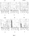
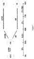
- Figure 1 displays a model summarizing the early transitions of human embryonic stem cells (hESCs) in vitro.
- Differentiation of hESCs through a process that closely recapitulates gastrulation can be orchestrated by the application of high dose activin A in the context of low serum supplementation.
- the expression of FGF8 and nuclear localization of ⁇ -catenin, events that occur in the proximal epiblast prior to primitive streak formation, is evident prior to about 24 hours (preprimitive streak cells).
- High level expression of the primitive streak-expressed genes (brachyury and FGF4) occurs at about 24 hours. If maintained in high dose activin A, the primitive streak cells (mesendoderm cells) are efficiently converted into definitive endoderm. In contrast, in the absence of activins, these mesendoderm precursors become mesoderm.
- Treatment of hESCs with BMP4 and SU5402 induces gene expression associated with primitive endoderm and trophectoderm.
- Embodiments described herein relate to novel, defined processes for the production of preprimitive streak cells and/or mesendoderm cells in culture by differentiating pluripotent cells, such as stem cells into preprimitive streak cells and/or mesendoderm cells.
- preprimitive streak cells are capable of differentiating into mesendoderm cells as well as cells, tissues and/or organs derived therefrom.
- Mesendoderm cells are capable of differentiating into mesoderm cells and/or definitive endoderm cells as well as cells, tissues and/or organs derived from either of these lineages.
- the preprimitive steak cells are converted, through a mesendoderm intermediate, into terminally differentiated cells of either the mesoderm or definitive endoderm lineages.
- processes can provide the basis for efficient production of a variety of human endodermal and mesodermal derived tissues.
- such processes can provide the basis for efficient production of human endodermal derived tissues, such as pancreas, liver, lungs, stomach, intestine, thyroid, thymus, pharynx, gallbladder and urinary bladder.
- production of preprimitive streak cells and/or mesendoderm cells is an early step in differentiation of a stem cell to a functional insulin-producing ⁇ -cell.
- preprimitive streak cell and/or mesendoderm cell differentiation can provide the basis for efficient production of human mesodermal derived tissues, such as blood cells, cardiovascular tissues, skeletal tissues as well as other structural and connective tissues.
- mesodermal derived tissues such as blood cells, cardiovascular tissues, skeletal tissues as well as other structural and connective tissues.
- high efficiency differentiation is desirable for each of the differentiation steps that occur prior to reaching the terminally differentiated cell fate. Since differentiation of stem cells to preprimitive streak cells and/or mesendoderm cells represents very early steps towards the production of functional terminally differentiated cells of the mesoderm and definitive endoderm cell lineages (as shown in Figure 1 ), high efficiency differentiation at this step is particularly desirable.
- some aspects of the differentiation processes described herein relate to in vitro methodology that results in approximately 5-90% conversion of pluripotent cells to preprimitive streak cells and/or mesendoderm cells.
- such methods encompass the application of culture and growth factor conditions in a defined and temporally specified fashion.
- Further enrichment of the cell population for preprimitive streak cells and/or mesendoderm cells can be achieved by isolation and/or purification of the preprimitive streak cells and/or mesendoderm cells from other cells in the population by sorting cells based on differential fluorescent marker expression.
- some embodiments described herein relate to preprimitive streak cells as well as methods for producing and isolating and/or purifying such cells.
- Other embodiments relate to mesendoderm cells as well as methods for producing and isolating and/or purifying such cells.
- certain embodiments described herein relate to cell markers whose presence, absence and/or relative expression levels are specific for preprimitive streak cells and methods for detecting and determining the expression of such markers.
- the presence, absence and/or level of expression of a marker is determined by quantitative PCR (Q-PCR).
- Q-PCR quantitative PCR
- the amount of transcript produced by certain genetic markers such as OCT4, ECAD, FGF8, ⁇ -catenin, brachyury, FGF4, SNAI1, SOX17, CXCR4, GSC, MIXL1, FOXA2, SOX7, FOXF1, FLK1, BML4, MOX1, SDF1 and other markers described herein is determined by quantitative Q-PCR.
- immunohistochemistry is used to detect the proteins expressed by the above-mentioned genes.
- immunohistochemistry/ immunocytochemistry is used to detect cell compartmental localization of certain polypeptide markers, such as the nuclear localization of ⁇ -catenin.
- Q-PCR and immunohistochemical techniques are both used to identify and determine the amount or relative proportions of such markers.
- preprimitive streak cells and/or mesendoderm cells By using methods, such as those described above, to determine the expression of one or more appropriate markers, it is possible to identify preprimitive streak cells and/or mesendoderm cells, as well as determine the proportion of preprimitive streak cells and/or mesendoderm cells in a cell culture or cell population.
- the preprimitive streak cells or cell populations that are produced express the FGF8 marker and/or nuclear-localized ⁇ -catenin at a level of about 2 orders of magnitude greater than non- preprimitive streak cell types or cell populations.
- the preprimitive streak cells or cell populations that are produced express the FGF8 marker and/or nuclear-localized ⁇ -catenin at a level of more than 2 orders of magnitude greater than non-preprimitive streak cell types or cell populations.
- the mesendoderm cells or cell populations that are produced express the brachyury, FGF4 and/or SNAI1 markers at a level of about 2 orders of magnitude greater than non- mesendoderm cell types or cell populations.
- the mesendoderm cells or cell populations that are produced express the brachyury, FGF4 and/or SNAI1 markers at a level of more than 2 orders of magnitude greater than non-mesendoderm cell types or cell populations.
- Embodiments described herein also relate to preprimitive streak and/or mesendoderm compositions.
- some embodiments relate to cell cultures comprising preprimitive streak cells and/or mesendoderm cells, whereas others relate to cell populations enriched in preprimitive streak cells and/or mesendoderm cells.
- Some preferred embodiments relate to cell cultures which comprise preprimitive streak cells and/or mesendoderm cells, wherein at least about 5-90% of the cells in culture are preprimitive streak cells and/or mesendoderm cells.
- An especially preferred embodiment relates to cells cultures comprising human cells, wherein at least about 5-90% of the human cells in culture are preprimitive streak cells and/or mesendoderm cells.
- the differentiation procedures described herein can result in about 5%, about 10%, about 15%, about 20%, about 25%, about 30%, about 35%, about 40%, about 45%, about 50%, about 55%, about 60%, about 65%, about 70%, about 75%, about 80%, about 85%, about 90%, about 95%, or greater than about 95% conversion of pluripotent cells to preprimitive streak cells and/or mesendoderm cells.
- conversion of a pluripotent cell population, such as a stem cell population to substantially pure preprimitive streak cell and/or mesendoderm cell population is contemplated.
- compositions and methods described herein have several useful features.
- the cell cultures and cell populations comprising preprimitive streak cells and/or mesendoderm cells as well as the methods for producing such cell cultures and cell populations are useful for modeling the early stages of human development.
- the compositions and methods described herein can also serve for therapeutic intervention in disease states, such as diabetes mellitus.
- preprimitive streak cells and/or mesendoderm cells serve as the source for only a limited number of tissues, they can be used in the development of pure tissue or cell types.
- the pluripotent cells used as starting material are stem cells.
- preprimitive streak cell cultures and enriched cell populations comprising preprimitive streak cells are produced from embryonic stem cells.
- a preferred method for deriving preprimitive streak cells utilizes human embryonic stem cells as the starting material for preprimitive streak cell production.
- Such pluripotent cells can be cells that originate from the morula, embryonic inner cell mass or those obtained from embryonic gonadal ridges.
- Human embryonic stem cells can be maintained in culture in a pluripotent state without substantial differentiation using methods that are known in the art. Such methods are described, for example, in US Patent Nos. 5,453,357 , 5,670,372 , 5,690,926 5,843,780 , 6,200,806 and 6,251,671 the disclosures of which are incorporated herein by reference in their entireties.
- hESCs are maintained on a feeder layer.
- any feeder layer which allows hESCs to be maintained in a pluripotent state can be used.
- One commonly used feeder layer for the cultivation of human embryonic stem cells is a layer of mouse fibroblasts. More recently, human fibroblast feeder layers have been developed for use in the cultivation of hESCs (see US Patent Application No. 2002/0072117 , the disclosure of which is incorporated herein by reference in its entirety).
- Alternative processes for producing preprimitive streak cells permit the maintenance of pluripotent hESC without the use of a feeder layer. Methods of maintaining pluripotent hESCs under feeder-free conditions have been described in US Patent Application No. 2003/0175956 , the disclosure of which is incorporated herein by reference in its entirety.
- the human embryonic stem cells used herein can be maintained in culture either with or without serum. In some embryonic stem cell maintenance procedures, serum replacement is used. In others, serum free culture techniques, such as those described in US Patent Application No. 2003/0190748 , the disclosure of which is incorporated herein by reference in its entirety, are used.
- Stem cells are maintained in culture in a pluripotent state by routine passage until it is desired that they be differentiated into preprimitive streak cells.
- differentiation to preprimitive streak cells is achieved by providing to the stem cell culture a differentiation factor, such as a growth factor of the TGF ⁇ superfamily, in an amount sufficient to promote differentiation to preprimitive streak cells.
- Growth factors of the TGF ⁇ superfamily which are useful for the production of preprimitive streak cells are selected from the Nodal/Activin subgroups.
- the growth factor is selected from the group consisting of Nodal, activin A, and activin B.
- the growth factor activin A is used.
- the above-mentioned growth factors are provided to the cells so that the growth factors are present in the cultures at concentrations sufficient to promote differentiation of at least a portion of the stem cells to preprimitive streak cells.
- the above-mentioned growth factors are present in the cell culture at a concentration of at least about 5 ng/ml, at least about 10 ng/ml, at least about 25 ng/ml, at least about 50 ng/ml, at least about 75 ng/ml, at least about 100 ng/ml, at least about 200 ng/ml, at least about 300 ng/ml, at least about 400 ng/ml, at least about 500 ng/ml, at least about 1000 ng/ml, at least about 2000 ng/ml, at least about 3000 ng/ml, at least about 4000 ng/ml, at least about 5000 ng/ml or more than about 5000 ng/ml.
- the above-mentioned growth factors are removed from the cell culture subsequent to their addition.
- the growth factors can be removed within about 1 hour, within about 2 hours, within about 3 hours, within about 4 hours, within about 5 hours, within about 6 hours, within about 7 hours, within about 8 hours, within about 9 hours, within about 10 hours, within about 11 hours, within about 12 hours, within about 13 hours, within about 14 hours, within about 15 hours, within about 16 hours, within about 17 hours, within about 18 hours, within about 19 hours, within about 20 hours, within about 21 hours, within about 22 hours, within about 23 hours, within about 24 hours or within about more than 24 hours.
- serum concentrations can range from about 0% (v/v) to about 10% (v/v).
- the serum concentration of the medium can be less than about 0.05% (v/v), less than about 0.1% (v/v), less than about 0.2% (v/v), less than about 0.3% (v/v), less than about 0.4% (v/v), less than about 0.5% (v/v), less than about 0.6% (v/v), less than about 0.7% (v/v), less than about 0.8% (v/v), less than about 0.9% (v/v), less than about 1% (v/v), less than about 2% (v/v), less than about 3% (v/v), less than about 4% (v/v), less than about 5% (v/v), less than about 6% (v/v), less than about 7% (v/v), less than about 8% (v/v), less than about 9% (v/v), less than about 1% (v/v), less than about 2% (v/v), less than about 3% (v/v), less
- preprimitive streak cells are grown without serum or without serum replacement.
- preprimitive streak cells are grown in the presence of B27.
- the concentration of B27 supplement can range from about 0.1% (v/v) to about 20% (v/v).
- the progression of the hESC culture to preprimitive streak cells can be monitored by determining the temporal expression of markers characteristic of preprimitive streak cells.
- the expression of certain markers is determined by detecting the presence or absence of the marker.
- the expression of certain markers can be determined by measuring the level at which the marker is present in the cells of the cell culture or cell population at one or more time points subsequent to the addition of the differentiation factor.
- the measurement of marker expression can be qualitative or quantitative.
- One method of quantitating the expression of markers that are produced by marker genes is through the use of quantitative polymerase chain reaction (Q-PCR). Methods of performing Q-PCR are well known in the art. Other methods which are known in the art can also be used to quantitate marker gene expression.
- Q-PCR quantitative polymerase chain reaction
- the expression of a marker gene product can be detected by using antibodies specific for the marker gene product of interest.
- the expression of marker genes characteristic of preprimitive streak cells as well as the lack of significant expression of marker genes characteristic of hESCs and other cell types is determined.
- both the timing and amount of expression of marker genes characteristic of preprimitive streak cells at one or more time points subsequent to the addition of the differentiation factor is determined.
- markers of preprimitive streak cells are FGF8 and ⁇ -catenin.
- the preprimitive streak cells produced by the processes described herein express the FGF8 and ⁇ -catenin marker genes, thereby producing the FGF8 and ⁇ -catenin marker gene products.
- the FGF8 mRNA is substantially expressed in preprimitive streak cells but not in hESCs. Substantial upregulation of the FGF8 mRNA, to near peak levels, can be observed in a differentiating hESC culture by 6 hours after contacting the hESCs with an appropriate differentiation factor, such as activin A.
- markers indicative of other cells types such as mesendoderm, primitive endoderm, definitive endoderm, mesoderm and ectoderm (see Table 1), is still comparatively low.
- markers indicative of mesendoderm, primitive endoderm, definitive endoderm, mesoderm and ectoderm are not substantially expressed by 6 hours after contacting the hESCs with the differentiation factor.
- FGF8 mRNA expression is maintained at high levels for at least about 24 hours after contacting the hESCs with the differentiation factor but begins to decline thereafter.
- nuclear localization of the ⁇ -catenin polypeptide is observed by immunocytochemistry.
- the ⁇ -catenin polypeptide is present at the cell periphery but not in the nucleus.
- FGF8 and nuclear-localized ⁇ -catenin expression is induced over a range of different levels in preprimitive streak cells depending on the differentiation conditions.
- the expression of the FGF8 marker and/or the nuclear-localized ⁇ -catenin marker in preprimitive streak cells or cell populations is at least about 2-fold higher to at least about 10,000-fold higher than the expression of these markers in non-preprimitive streak cells or cell populations, during about the first 6 to 18 hours of differentiation from hESCs.
- the expression of the FGF8 marker and/or the nuclear-localized ⁇ -catenin marker in preprimitive streak cells or cell populations is at least about 4-fold higher, at least about 6-fold higher, at least about 8-fold higher, at least about 10-fold higher, at least about 15-fold higher, at least about 20-fold higher, at least about 40-fold higher, at least about 80-fold higher, at least about 100-fold higher, at least about 150-fold higher, at least about 200-fold higher, at least about 500-fold higher, at least about 750-fold higher, at least about 1000-fold higher, at least about 2500-fold higher, at least about 5000-fold higher, at least about 7500-fold higher or at least about 10,000-fold higher than the expression of the FGF8 marker and/or the nuclear-localized ⁇ -catenin marker in non-preprimitive streak cells or cell populations, during about the first 6 to 18 hours of differentiation from hESCs.
- the expression of the FGF8 marker and/or the nuclear-localized ⁇ -catenin marker in preprimitive streak cells or cell populations is infinitely higher than the expression of the FGF8 marker and/or the nuclear-localized ⁇ -catenin marker in non-preprimitive streak cells or cell populations, during about the first 6 to 18 hours of differentiation from hESCs.
- the expression of the FGF8 marker and/or the nuclear-localized ⁇ -catenin marker is at least about 2-fold higher to at least about 10,000-fold higher than the expression of the brachyury, FGF4, SNAI1, SOX17, FOXA2, SOX7 and/or SOX1 markers.
- the expression of the FGF8 marker and/or the nuclear-localized ⁇ -catenin marker is at least about 4-fold higher, at least about 6-fold higher, at least about 8-fold higher, at least about 10-fold higher, at least about 15-fold higher, at least about 20-fold higher, at least about 40-fold higher, at least about 80-fold higher, at least about 100-fold higher, at least about 150-fold higher, at least about 200-fold higher, at least about 500-fold higher, at least about 750-fold higher, at least about 1000-fold higher, at least about 2500-fold higher, at least about 5000-fold higher, at least about 7500-fold higher or at least about 10,000-fold higher than the expression of the brachyury, FGF4, SNAI1, SOX17, FOXA2, SOX7 and/or SOX1 markers.
- the brachyury, FGF4, SNAI1, SOX17, FOXA2, SOX7 and/or SOX1 markers are not significantly expressed in preprimitive
- preprimitive streak cells can be enriched, isolated and/or purified.
- cell populations enriched for preprimitive streak cells are produced by isolating such cells from cell cultures.
- preprimitive streak cells are fluorescently labeled then isolated from non-labeled cells by using a fluorescence activated cell sorter (FACS).
- FACS fluorescence activated cell sorter
- a nucleic acid encoding green fluorescent protein (GFP) or another nucleic acid encoding an expressible fluorescent marker gene is used to label preprimitive streak cells.
- GFP green fluorescent protein
- at least one copy of a nucleic acid encoding GFP or a biologically active fragment thereof is introduced into a pluripotent cell, preferably a human embryonic stem cell, downstream of the FGF8 promoter such that the expression of the GFP gene product or biologically active fragment thereof is under control of the FGF8 promoter.
- the entire coding region of the nucleic acid, which encodes FGF8, is replaced by a nucleic acid encoding GFP or a biologically active fragment thereof.
- the nucleic acid encoding GFP or a biologically active fragment thereof is fused in frame with at least a portion of the nucleic acid encoding FGF8, thereby generating a fusion protein.
- the fusion protein retains a fluorescent activity similar to GFP.
- Fluorescently marked cells such as the above-described pluripotent cells, are differentiated to preprimitive streak cells as described previously above. Because preprimitive streak cells express the fluorescent marker gene, whereas non-preprimitive streak cells do not, these two cell types can be separated.
- cell suspensions comprising a mixture of fluorescently-labeled preprimitive streak cells and unlabeled non-preprimitive streak cells are sorted using a FACS. Preprimitive streak cells are collected separately from non-preprimitive streak cells, thereby resulting in the isolation of such cell types. If desired, the isolated cell compositions can be further purified by additional rounds of sorting using the same or different markers that are specific for preprimitive streak cells.
- preprimitive streak cells may also be isolated by other techniques for cell isolation. Additionally, preprimitive streak cells may also be enriched or isolated by methods of serial subculture in growth conditions which promote the selective survival or selective expansion of the preprimitive streak cells.
- enriched, isolated and/or purified populations of preprimitive streak cells and/or tissues can be produced in vitro from hESC cultures or cell populations which have undergone differentiation for from about 1 hour to about 24 hours.
- the cells undergo random differentiation.
- the cells are directed to differentiate primarily into preprimitive streak cells.
- cell populations or cell cultures can be enriched in preprimitive streak cell content by at least about 2- to about 1000-fold as compared to untreated or unenriched cell populations or cell cultures.
- preprimitive streak cells can be enriched by at least about 5- to about 500-fold as compared to untreated or unenriched cell populations or cell cultures.
- preprimitive streak cells can be enriched from at least about 10- to about 200-fold as compared to untreated or unenriched cell populations or cell cultures.
- preprimitive streak cells can be enriched from at least about 20- to about 100-fold as compared to untreated or unenriched cell populations or cell cultures.
- preprimitive streak cells can be enriched from at least about 40- to about 80-fold as compared to untreated or unenriched cell populations or cell cultures. In certain embodiments, preprimitive streak cells can be enriched from at least about 2-to about 20-fold as compared to untreated or unenriched cell populations or cell cultures.
- compositions Comprising Preprimitive Streak Cells
- Cell compositions produced by the above-described methods include cell cultures comprising preprimitive streak cells and cell populations enriched in preprimitive streak cells.
- cell cultures which comprise preprimitive streak cells wherein at least about 5-90% of the cells in culture are preprimitive streak cells, can be produced.
- the differentiation procedures described herein can result in about 5%, about 10%, about 15%, about 20%, about 25%, about 30%, about 35%, about 40%, about 45%, about 50%, about 55%, about 60%, about 65%, about 70%, about 75%, about 80%, about 85%, about 90%, about 95%, or greater than about 95% conversion of pluripotent cells to preprimitive streak cells.
- a substantially pure preprimitive streak cell population can be recovered.
- compositions such as cell populations and cell cultures, that comprise both pluripotent cells, such as stem cells, and preprimitive streak cells.
- compositions comprising mixtures of hESCs and preprimitive streak cells can be produced.
- compositions comprising at least about 5 preprimitive streak cells for about every 95 pluripotent cells are produced.
- compositions comprising at least about 95 preprimitive streak cells for about every 5 pluripotent cells are produced.
- compositions comprising other ratios of preprimitive streak cells to pluripotent cells are contemplated.
- compositions comprising at least about 1 preprimitive streak cell for about every 1,000,000 pluripotent cells, at least about 1 preprimitive streak cell for about every 100,000 pluripotent cells, at least about 1 preprimitive streak cell for about every 10,000 pluripotent cells, at least about 1 preprimitive streak cell for about every 1000 pluripotent cells, at least about 1 preprimitive streak cell for about every 500 pluripotent cells, at least about 1 preprimitive streak cell for about every 100 pluripotent cells, at least about 1 preprimitive streak cell for about every 10 pluripotent cells, at least about 1 preprimitive streak cell for about every 5 pluripotent cells, at least about 1 preprimitive streak cell for about every 2 pluripotent cells, at least about 2 preprimitive streak cells for about every 1 pluripotent cell, at least about 5 preprimitive streak cells for about every 1 pluripotent cell, at least about 10 preprimitive streak cells for about every 1 pluripotent cell, at least about 20 preprimi
- the pluripotent cells are human pluripotent stem cells.
- the stem cells are derived from a morula, the inner cell mass of an embryo or the gonadal ridges of an embryo.
- the pluripotent cells are derived from the gonadal or germ tissues of a multicellular structure that has developed past the embryonic stage.
- cell cultures or cell populations comprising from at least about 5% preprimitive streak cells to at least about 99% preprimitive streak cells.
- the cell cultures or cell populations comprise mammalian cells.
- the cell cultures or cell populations comprise human cells.
- certain specific embodiments relate to cell cultures comprising human cells, wherein from at least about 5% to at least about 99% of the human cells are preprimitive streak cells.
- cell cultures comprising human cells, wherein at least about 5%, at least about 10%, at least about 15%, at least about 20%, at least about 25%, at least about 30%, at least about 35%, at least about 40%, at least about 45%, at least about 50%, at least about 55%, at least about 60%, at least about 65%, at least about 70%, at least about 75%, at least about 80%, at least about 85%, at least about 90%, at least about 95%, at least about 98%, at least about 99%, or greater than 99% of the human cells are preprimitive streak cells.
- the above percentages are calculated without respect to the human feeder cells in the cell cultures or cell populations.
- compositions such as cell cultures or cell populations, comprising human cells, such as human preprimitive streak cells, wherein the expression of the FGF8 marker and/or the nuclear-localized ⁇ -catenin marker is greater than the expression of the brachyury, FGF4, SNAI1, SOX17, FOXA2, SOX7 and/or SOX1 markers in at least about 5% of the human cells.
- the expression of either the FGF8 marker and/or the nuclear-localized ⁇ -catenin marker is greater than the expression of the brachyury, FGF4, SNAI1, SOX17, FOXA2, SOX7 and/or SOX1 marker in at least about 10% of the human cells, in at least about 15% of the human cells, in at least about 20% of the human cells, in at least about 25% of the human cells, in at least about 30% of the human cells, in at least about 35% of the human cells, in at least about 40% of the human cells, in at least about 45% of the human cells, in at least about 50% of the human cells, in at least about 55% of the human cells, in at least about 60% of the human cells, in at least about 65% of the human cells, in at least about 70% of the human cells, in at least about 75% of the human cells, in at least about 80% of the human cells, in at least about 85% of the human cells, in at least about 90% of the human cells, in at least about 95% of the human cells,
- compositions such as cell cultures or cell populations, comprising human hESCs and human preprimitive streak cells, wherein substantial upregulation of the expression of FGF8 mRNA occurs in cells of the cell culture or cell population by about 1 hour, by about 2 hours, by about 3 hours, by about 4 hours, by about 5 hours, by about 6 hours, by about 7 hours, by about 8 hours, by about 9 hours, by about 10 hours, by about 11 hours, by about 12 hours, by about 13 hours, by about 14 hours, by about 15 hours, by about 16 hours, by about 17 hours, by about 18 hours, by about 19 hours, by about 20 hours, by about 21 hours, by about 22 hours, by about 23 hours, by about 24 hours, or by greater than about 24 hours after contacting hESCs in the culture with an appropriate differentiation factor, such as activin A.
- an appropriate differentiation factor such as activin A.
- substantial upregulation of the expression of FGF8 mRNA occurs in at least about 5% of the human cells, at least about 10% of the human cells, in at least about 15% of the human cells, in at least about 20% of the human cells, in at least about 25% of the human cells, in at least about 30% of the human cells, in at least about 35% of the human cells, in at least about 40% of the human cells, in at least about 45% of the human cells, in at least about 50% of the human cells, in at least about 55% of the human cells, in at least about 60% of the human cells, in at least about 65% of the human cells, in at least about 70% of the human cells, in at least about 75% of the human cells, in at least about 80% of the human cells, in at least about 85% of the human cells, in at least about 90% of the human cells, in at least about 95% of the human cells, in at least about 98% of the human cells, in at least about 99% of the human cells or in greater than 99% of the human cells.
- compositions such as cell cultures or cell populations, comprising human hESCs and human preprimitive streak cells, wherein substantial nuclear localization of the ⁇ -catenin polypeptide (expression of nuclear localized ⁇ -catenin marker) occurs in cells of the cell culture or cell population by about 1 hour, by about 2 hours, by about 3 hours, by about 4 hours, by about 5 hours, by about 6 hours, by about 7 hours, by about 8 hours, by about 9 hours, by about 10 hours, by about 11 hours, by about 12 hours, by about 13 hours, by about 14 hours, by about 15 hours, by about 16 hours, by about 17 hours, by about 18 hours, by about 19 hours, by about 20 hours, by about 21 hours, by about 22 hours, by about 23 hours, by about 24 hours, or by greater than about 24 hours after contacting hESCs in the culture with an appropriate differentiation factor, such as activin A.
- an appropriate differentiation factor such as activin A.
- expression of nuclear-localized ⁇ -catenin occurs in at least about 5% of the human cells, at least about 10% of the human cells, in at least about 15% of the human cells, in at least about 20% of the human cells, in at least about 25% of the human cells, in at least about 30% of the human cells, in at least about 35% of the human cells, in at least about 40% of the human cells, in at least about 45% of the human cells, in at least about 50% of the human cells, in at least about 55% of the human cells, in at least about 60% of the human cells, in at least about 65% of the human cells, in at least about 70% of the human cells, in at least about 75% of the human cells, in at least about 80% of the human cells, in at least about 85% of the human cells, in at least about 90% of the human cells, in at least about 95% of the human cells, in at least about 98% of the human cells, in at least about 99% of the human cells or in greater than 99% of the human cells.
- the cell cultures in at least about 5%
- compositions comprising preprimitive streak cells substantially free of other cell types can be produced.
- the preprimitive streak cell populations or cell cultures produced by the methods described herein are substantially free of cells that significantly express the brachyury, FGF4, SNAI1, SOX17, FOXA2, SOX7 and/or SOX1 marker genes.
- a description of a preprimitive streak cell based on the expression of marker genes is, FGF8 high, nuclear-localized ⁇ -catenin high, brachyury low, FGF4 low, SNAI1 low, SOX17 low, FOXA2 low, SOX7 low and SOX1 low.
- the pluripotent cells used as starting material are stem cells.
- mesendoderm cell cultures and enriched cell populations comprising mesendoderm cells are produced from embryonic stem cells.
- a preferred method for deriving mesendoderm cells utilizes human embryonic stem cells as the starting material for mesendoderm cell production.
- Such pluripotent cells can be cells that originate from the morula, embryonic inner cell mass or those obtained from embryonic gonadal ridges.
- Human embryonic stem cells can be maintained in culture in a pluripotent state without substantial differentiation using methods that are known in the art. Such methods are described, for example, in US Patent Nos. 5,453,357 , 5,670,372 , 5,690,926 5,843,780 , 6,200,806 and 6,251,671 the disclosures of which are incorporated herein by reference in their entireties.
- hESCs are maintained on a feeder layer.
- any feeder layer which allows hESCs to be maintained in a pluripotent state can be used.
- One commonly used feeder layer for the cultivation of human embryonic stem cells is a layer of mouse fibroblasts. More recently, human fibroblast feeder layers have been developed for use in the cultivation of hESCs (see US Patent Application No. 2002/0072117 , the disclosure of which is incorporated herein by reference in its entirety).
- Alternative processes for producing mesendoderm cells permit the maintenance of pluripotent hESC without the use of a feeder layer. Methods of maintaining pluripotent hESCs under feeder-free conditions have been described in US Patent Application No. 2003/0175956 , the disclosure of which is incorporated herein by reference in its entirety.
- the human embryonic stem cells used herein can be maintained in culture either with or without serum. In some embryonic stem cell maintenance procedures, serum replacement is used. In others, serum free culture techniques, such as those described in US Patent Application No. 2003/0190748 , the disclosure of which is incorporated herein by reference in its entirety, are used.
- Stem cells are maintained in culture in a pluripotent state by routine passage until it is desired that they be differentiated into mesendoderm cells.
- differentiation to mesendoderm cells is achieved by providing to the stem cell culture a differentiation factor, such as a growth factor of the TGF ⁇ superfamily, in an amount sufficient to promote differentiation to mesendoderm cells.
- Growth factors of the TGF ⁇ superfamily which are useful for the production of mesendoderm cells are selected from the Nodal/Activin subgroups.
- the growth factor is selected from the group consisting of Nodal, activin A, and activin B.
- the growth factor activin A is used.
- the above-mentioned growth factors are provided to the cells so that the growth factors are present in the cultures at concentrations sufficient to promote differentiation of at least a portion of the stem cells to mesendoderm cells.
- the above-mentioned growth factors are present in the cell culture at a concentration of at least about 5 ng/ml, at least about 10 ng/ml, at least about 25 ng/ml, at least about 50 ng/ml, at least about 75 ng/ml, at least about 100 ng/ml, at least about 200 ng/ml, at least about 300 ng/ml, at least about 400 ng/ml, at least about 500 ng/ml, at least about 1000 ng/ml, at least about 2000 ng/ml, at least about 3000 ng/ml, at least about 4000 ng/ml, at least about 5000 ng/ml or more than about 5000 ng/ml.
- the above-mentioned growth factors are removed from the cell culture subsequent to their addition.
- the growth factors can be removed within about 18 hours, within about 19 hours, within about 20 hours, within about 21 hours, within about 22 hours, within about 23 hours, within about 24 hours, within about 25 hours, within about 26 hours, within about 27 hours, within about 28 hours, within about 29 hours, within about 30 hours, within about 31 hours, within about 32 hours, within about 33 hours, within about 34 hours, within about 35 hours, within about 36 hours, within about 37 hours, within about 38 hours, within about 39 hours, within about 40 hours, within about 41 hours, within about 42 hours, within about 43 hours, within about 44 hours, within about 45 hours, within about 46 hours, within about 47 hours, within about 48 hours or within about more than 48 hours.
- the serum concentration of the medium can be less than about 0.05% (v/v), less than about 0.1% (v/v), less than about 0.2% (v/v), less than about 0.3% (v/v), less than about 0.4% (v/v), less than about 0.5% (v/v), less than about 0.6% (v/v), less than about 0.7% (v/v), less than about 0.8% (v/v), less than about 0.9% (v/v), less than about 1% (v/v), less than about 2% (v/v), less than about 3% (v/v), less than about 4% (v/v), less than about 5% (v/v), less than about 6% (v/v), less than about 7% (v/v), less than about 8% (v/v), less than about 9% (v/v), less than about 1% (v/v), less than about 2% (v/v), less than about 3% (v/v), less than about 4% (v/v), less than about 5% (v/v), less than about 6%
- mesendoderm cells are grown without serum or without serum replacement.
- mesendoderm cells are grown in the presence of B27.
- the concentration of B27 supplement can range from about 0.1% (v/v) to about 20% (v/v).
- the progression of the hESC culture to mesendoderm cells can be monitored by determining the temporal expression of markers characteristic of mesendoderm cells.
- the expression of certain markers is determined by detecting the presence or absence of the marker.
- the expression of certain markers can be determined by measuring the level at which the marker is present in the cells of the cell culture or cell population at one or more time points subsequent to the addition of the differentiation factor.
- the measurement of marker expression can be qualitative or quantitative.
- One method of quantitating the expression of markers that are produced by marker genes is through the use of Q-PCR. Other methods which are known in the art can also be used to quantitate marker gene expression.
- the expression of a marker gene product can be detected by using antibodies specific for the marker gene product of interest.
- the expression of marker genes characteristic of mesendoderm cells as well as the lack of significant expression of marker genes characteristic of hESCs and other cell types is determined.
- both the timing and amount of expression of marker genes characteristic of mesendoderm cells at one or more time points subsequent to the addition of the differentiation factor is determined.
- markers of mesendoderm cells are brachyury, FGF4 and SNAI1.
- the mesendoderm cells produced by the processes described herein express the brachyury, FGF4 and SNAI1 marker genes, thereby producing the brachyury, FGF4 and SNAI1 marker gene products.
- the brachyury, FGF4 and/or SNAI1 mRNA is substantially expressed in mesendoderm cells but not in hESCs.
- Substantial upregulation of the brachyury, FGF4 and/or SNAI1 mRNA, to near peak levels, can be observed in a differentiating hESC culture by 24 hours after contacting the hESCs with an appropriate differentiation factor, such as activin A.
- an appropriate differentiation factor such as activin A.
- expression of certain markers indicative of other cells types such as primitive endoderm, definitive endoderm, mesoderm and ectoderm (see Table 1), is still comparatively low.
- certain markers indicative of primitive endoderm, definitive endoderm, mesoderm and ectoderm are not substantially expressed by 24 hours after contacting the hESCs with the differentiation factor.
- brachyury, FGF4 and/or SNAI1 mRNA expression begins to decline after about 24 hours subsequent to contacting the hESCs with the differentiation factor. In some embodiments, brachyury, FGF4 and/or SNAI1 mRNA expression begins to decline after about 30 hours, after about 36 hours, after about 42 hours, after about 48 hours or after more than about 48 hours subsequent to contacting the hESCs with the differentiation factor.
- brachyury, FGF4 and/or SNAI1 expression is induced over a range of different levels in mesendoderm cells depending on the differentiation conditions.
- the expression of the brachyury, FGF4 and/or SNAI1 marker in mesendoderm cells or cell populations is at least about 2-fold higher to at least about 10,000-fold higher than the expression of these markers in non-mesendoderm cells or cell populations, after about 24 hours of differentiation from hESCs.
- the expression of the brachyury, FGF4 and/or SNAI1 marker in mesendoderm cells or cell populations is at least about 4-fold higher, at least about 6-fold higher, at least about 8-fold higher, at least about 10-fold higher, at least about 15-fold higher, at least about 20-fold higher, at least about 40-fold higher, at least about 80-fold higher, at least about 100-fold higher, at least about 150-fold higher, at least about 200-fold higher, at least about 500-fold higher, at least about 750-fold higher, at least about 1000-fold higher, at least about 2500-fold higher, at least about 5000-fold higher, at least about 7500-fold higher or at least about 10,000-fold higher than the expression of the brachyury, FGF4 and/or SNAI1 in non-mesendoderm cells or cell populations, after about 24 hours of differentiation from hESCs.
- the expression of the brachyury, FGF4 and/or SNAI1 marker in mesendoderm cells 'or cell populations is infinitely higher than the expression of the brachyury, FGF4 and/or SNAI1 marker in non-mesendoderm cells or cell populations, after about 24 hours of differentiation from hESCs.
- the expression of the brachyury, FGF4 and/or SNAI1 markers is at least about 2-fold higher to at least about 10,000-fold higher than the expression of the OCT4, SOX17, CXCR4, FOXA2, SOX7 and/or SOX1 markers.
- the expression of the brachyury, FGF4 and/or SNAI1 marker is at least about 4-fold higher, at least about 6-fold higher, at least about 8-fold higher, at least about 10-fold higher, at least about 15-fold higher, at least about 20-fold higher, at least about 40-fold higher, at least about 80-fold higher, at least about 100-fold higher, at least about 150-fold higher, at least about 200-fold higher, at least about 500-fold higher, at least about 750-fold higher, at least about 1000-fold higher, at least about 2500-fold higher, at least about 5000-fold higher, at least about 7500-fold higher or at least about 10,000-fold higher than the expression of the OCT4, SOX17, CXCR4, FOXA2, SOX7 and/or SOX1 markers.
- the OCT4, SOX17, CXCR4, FOXA2, SOX7 and/or SOX1 markers are not significantly expressed in mesendoderm cells.
- mesendoderm cells can be enriched, isolated and/or purified.
- cell populations enriched for mesendoderm cells are produced by isolating such cells from cell cultures.
- mesendoderm cells are fluorescently labeled then isolated from non-labeled cells by using a FACS.
- a nucleic acid encoding green fluorescent protein (GFP) or another nucleic acid encoding an expressible fluorescent marker gene is used to label mesendoderm cells.
- At least one copy of a nucleic acid encoding GFP or a biologically active fragment thereof is introduced into a pluripotent cell, preferably a human embryonic stem cell, downstream of the brachyury, FGF4 or SNAI1 promoter such that the expression of the GFP gene product or biologically active fragment thereof is under control of the brachyury, FGF4 or SNAI1 promoter.
- the entire coding region of the nucleic acid, which encodes brachyury, FGF4 or SNAI1 is replaced by a nucleic acid encoding GFP or a biologically active fragment thereof.
- the nucleic acid encoding GFP or a biologically active fragment thereof is fused in frame with at least a portion of the nucleic acid encoding brachyury, FGF4 or SNAI1, thereby generating a fusion protein.
- the fusion protein retains a fluorescent activity similar to GFP.
- Fluorescently marked cells such as the above-described pluripotent cells, are differentiated to mesendoderm cells as described previously above. Because mesendoderm cells express the fluorescent marker gene, whereas non-mesendoderm cells do not, these two cell types can be separated.
- cell suspensions comprising a mixture of fluorescently-labeled mesendoderm cells and unlabeled non-mesendoderm cells are sorted using a FACS. Mesendoderm cells are collected separately from non-mesendoderm cells, thereby resulting in the isolation of such cell types. If desired, the isolated cell compositions can be further purified by additional rounds of sorting using the same or different markers that are specific for mesendoderm cells.
- mesendoderm cells may also be isolated by other techniques for cell isolation. Additionally, mesendoderm cells may also be enriched or isolated by methods of serial subculture in growth conditions which promote the selective survival or selective expansion of the mesendoderm cells.
- enriched, isolated and/or purified populations of mesendoderm cells and/or tissues can be produced in vitro from hESC cultures or cell populations which have undergone differentiation for from about 18 hours to about 48 hours.
- the cells undergo random differentiation.
- the cells are directed to differentiate primarily into mesendoderm cells.
- Some preferred enrichment, isolation and/or purification methods relate to the in vitro production of mesendoderm cells from human embryonic stem cells.
- cell populations or cell cultures can be enriched in mesendoderm cell content by at least about 2- to about 1000-fold as compared to untreated or unenriched cell populations or cell cultures.
- mesendoderm cells can be enriched by at least about 5- to about 500-fold as compared to untreated or unenriched cell populations or cell cultures.
- mesendoderm cells can be enriched from at least about 10- to about 200-fold as compared to untreated or unenriched cell populations or cell cultures.
- mesendoderm cells can be enriched from at least about 20- to about 100-fold as compared to untreated or unenriched cell populations or cell cultures.
- mesendoderm cells can be enriched from at least about 40- to about 80-fold as compared to untreated or unenriched cell populations or cell cultures. In certain embodiments, mesendoderm cells can be enriched from at least about 2- to about 20-fold as compared to untreated or unenriched cell populations or cell cultures.
- compositions comprising Mesendoderm Cells
- Cell compositions produced by the above-described methods include cell cultures comprising mesendoderm cells and cell populations enriched in mesendoderm cells.
- cell cultures which comprise mesendoderm cells, wherein at least about 5-90% of the cells in culture are mesendoderm cells can be produced.
- the differentiation procedures described herein can result in about 5%, about 10%, about 15%, about 20%, about 25%, about 30%, about 35%, about 40%, about 45%, about 50%, about 55%, about 60%, about 65%, about 70%, about 75%, about 80%, about 85%, about 90%, about 95%, or greater than about 95% conversion of pluripotent cells to mesendoderm cells.
- a substantially pure mesendoderm cell population can be recovered.
- compositions such as cell populations and cell cultures, that comprise both pluripotent cells, such as stem cells, and mesendoderm cells.
- pluripotent cells such as stem cells
- mesendoderm cells comprising mixtures of hESCs and mesendoderm cells.
- compositions comprising at least about 5 mesendoderm cells for about every 95 pluripotent cells are produced.
- compositions comprising at least about 95 mesendoderm cells for about every 5 pluripotent cells are produced.
- compositions comprising other ratios of mesendoderm cells to pluripotent cells are contemplated.
- compositions comprising at least about 1 mesendoderm cell for about every 1,000,000 pluripotent cells, at least about 1 mesendoderm cell for about every 100,000 pluripotent cells, at least about 1 mesendoderm cell for about every 10,000 pluripotent cells, at least about 1 mesendoderm cell for about every 1000 pluripotent cells, at least about 1 mesendoderm cell for about every 500 pluripotent cells, at least about 1 mesendoderm cell for about every 100 pluripotent cells, at least about 1 mesendoderm cell for about every 10 pluripotent cells, at least about 1 mesendoderm cell for about every 5 pluripotent cells, at least about 1 mesendoderm cell for about every 2 pluripotent cells, at least about 2 mesendoderm cells for about every 1 pluripotent cell, at least about 5 mesendoderm cells for about every 1 pluripotent cell, at least about 10 mesendoderm cells for about every 1 pluripotent cell, at least about 20 mesend
- the pluripotent cells are human pluripotent stem cells.
- the stem cells are derived from a morula, the inner cell mass of an embryo or the gonadal ridges of an embryo.
- the pluripotent cells are derived from the gonadal or germ tissues of a multicellular structure that has developed past the embryonic stage.
- cell cultures or cell populations comprising from at least about 5% mesendoderm cells to at least about 99% mesendoderm cells.
- the cell cultures or cell populations comprise mammalian cells.
- the cell cultures or cell populations comprise human cells.
- certain specific embodiments relate to cell cultures comprising human cells, wherein from at least about 5% to at least about 99% of the human cells are mesendoderm cells.
- cell cultures comprising human cells, wherein at least about 5%, at least about 10%, at least about 15%, at least about 20%, at least about 25%, at least about 30%, at least about 35%, at least about 40%, at least about 45%, at least about 50%, at least about 55%, at least about 60%, at least about 65%, at least about 70%, at least about 75%, at least about 80%, at least about 85%, at least about 90%, at least about 95%, at least about 98%, at least about 99%, or greater than 99% of the human cells are mesendoderm cells.
- the above percentages are calculated without respect to the human feeder cells in the cell cultures or cell populations.
- compositions such as cell cultures or cell populations, comprising human cells, such as human mesendoderm cells, wherein the expression of the brachyury, FGF4 and/or SNAI1 markers is greater than the expression of the OCT4, SOX17, CXCR4, FOXA2, SOX7 and/or SOX1 markers in at least about 5% of the human cells.
- the expression of either the brachyury, FGF4 and/or SNAI1 marker is greater than the expression of the OCT4, SOX17, CXCR4, FOXA2, SOX7 and/or SOX1 marker in at least about 10% of the human cells, in at least about 15% of the human cells, in at least about 20% of the human cells, in at least about 25% of the human cells, in at least about 30% of the human cells, in at least about 35% of the human cells, in at least about 40% of the human cells, in at least about 45% of the human cells, in at least about 50% of the human cells, in at least about 55% of the human cells, in at least about 60% of the human cells, in at least about 65% of the human cells, in at least about 70% of the human cells, in at least about 75% of the human cells, in at least about 80% of the human cells, in at least about 85% of the human cells, in at least about 90% of the human cells, in at least about 95% of the human cells, in at least about 98% of the
- compositions such as cell cultures or cell populations, comprising human hESCs and human mesendoderm cells, wherein substantial upregulation of the expression of brachyury, FGF4 and/or SNAI1 mRNA occurs in cells of the cell culture or cell population by about 18 hours, by about 19 hours, by about 20 hours, by about 21 hours, by about 22 hours, by about 23 hours, by about 24 hours, by about 25 hours, by about 26 hours, by about 27 hours, by about 28 hours, by about 29 hours, by about 30 hours, by about 31 hours, by about 32 hours, by about 33 hours, by about 34 hours, by about 35 hours, by about 36 hours, by about 37 hours, by about 38 hours, by about 39 hours, by about 40 hours, by about 41 hours, by about 42 hours, by about 43 hours, by about 44 hours, by about 45 hours, by about 46 hours, by about 47 hours, by about 48 hours or by greater than about 48 hours after contacting hESCs in the culture with an appropriate differentiation factor, such as activin A
- substantial upregulation of the expression of brachyury, FGF4 and/or SNAI1 mRNA occurs in at least about 5% of the human cells, at least about 10% of the human cells, in at least about 15% of the human cells, in at least about 20% of the human cells, in at least about 25% of the human cells, in at least about 30% of the human cells, in at least about 35% of the human cells, in at least about 40% of the human cells, in at least about 45% of the human cells, in at least about 50% of the human cells, in at least about 55% of the human cells, in at least about 60% of the human cells, in at least about 65% of the human cells, in at least about 70% of the human cells, in at least about 75% of the human cells, in at least about 80% of the human cells, in at least about 85% of the human cells, in at least about 90% of the human cells, in at least about 95% of the human cells, in at least about 98% of the human cells, in at least about 99% of the human cells or in greater than
- compositions comprising mesendoderm cells substantially free of other cell types can be produced.
- the mesendoderm cell populations or cell cultures produced by the methods described herein are substantially free of cells that significantly express the OCT4, SOX17, CXCR4, FOXA2, SOX7 and/or SOX1 marker genes.
- a description of a mesendoderm cell based on the expression of marker genes is, brachyury high, FGF4 high, SNAI1 high, SOX17 low, CXCR4 low, FOXA2 low, SOX7 low and SOX1 low.
- pluripotent cells used as starting material are stem cells.
- definitive endoderm cell cultures and enriched cell populations comprising definitive endoderm cells are produced from embryonic stem cells.
- a preferred method for deriving definitive endoderm cells utilizes human embryonic stem cells as the starting material for definitive endoderm production.
- pluripotent cells can be cells that originate from the morula, embryonic inner cell mass or those obtained from embryonic gonadal ridges.
- Human embryonic stem cells can be maintained in culture in a pluripotent state without substantial differentiation using methods that are known in the art. Such methods are described, for example, in US Patent Nos. 5,453,357 , 5,670,372 , 5,690,926 5,843,780 , 6,200,806 and 6,251,671 the disclosures of which are incorporated herein by reference in their entireties.
- hESCs are maintained on a feeder layer.
- any feeder layer which allows hESCs to be maintained in a pluripotent state can be used.
- One commonly used feeder layer for the cultivation of human embryonic stem cells is a layer of mouse fibroblasts. More recently, human fibroblast feeder layers have been developed for use in the cultivation of hESCs (see US Patent Application No. 2002/0072117 , the disclosure of which is incorporated herein by reference in its entirety).
- Alternative processes for producing definitive endoderm permit the maintenance of pluripotent hESC without the use of a feeder layer. Methods of maintaining pluripotent hESCs under feeder-free conditions have been described in US Patent Application No. 2003/0175956 , the disclosure of which is incorporated herein by reference in its entirety.
- the human embryonic stem cells used herein can be maintained in culture either with or without serum. In some embryonic stem cell maintenance procedures, serum replacement is used. In others, serum free culture techniques, such as those described in US Patent Application No. 2003/0190748 , the disclosure of which is incorporated herein by reference in its entirety, are used.
- Stem cells are maintained in culture in a pluripotent state by routine passage until it is desired that they be differentiated into definitive endoderm.
- differentiation to definitive endoderm is achieved by providing to the stem cell culture a growth factor of the TGF ⁇ superfamily in an amount sufficient to promote differentiation to definitive endoderm.
- Growth factors of the TGF ⁇ superfamily which are useful for the production of definitive endoderm are selected from the Nodal/Activin subgroups.
- the growth factor is selected from the group consisting of Nodal, Activin A and Activin B.
- combinations of any of the above-mentioned growth factors can be used.
- the above-mentioned growth factors are provided to the cells so that the growth factors are present in the cultures at concentrations sufficient to promote differentiation of at least a portion of the stem cells to definitive endoderm cells.
- the above-mentioned growth factors are present in the cell culture at a concentration of at least about 5 ng/ml, at least about 10 ng/ml, at least about 25 ng/ml, at least about 50 ng/ml, at least about 75 ng/ml, at least about 100 ng/ml, at least about 200 ng/ml, at least about 300 ng/ml, at least about 400 ng/ml, at least about 500 ng/ml, at least about 1000 ng/ml, at least about 2000 ng/ml, at least about 3000 ng/ml, at least about 4000 ng/ml, at least about 5000 ng/ml or more than about 5000 ng/ml.
- the above-mentioned growth factors are removed from the cell culture subsequent to their addition.
- the growth factors can be removed within about one day, about two days, about three days, about four days, about five days, about six days, about seven days, about eight days, about nine days or about ten days after their addition.
- the growth factors are removed about four days after their addition.
- serum concentrations can range from about 0.05% v/v to about 20% v/v.
- the serum concentration of the medium can be less than about 0.05% (v/v), less than about 0.1% (v/v), less than about 0.2% (v/v), less than about 0.3% (v/v), less than about 0.4% (v/v), less than about 0.5% (v/v), less than about 0.6% (v/v), less than about 0.7% (v/v), less than about 0.8% (v/v), less than about 0.9% (v/v), less than about 1% (v/v), less than about 2% (v/v), less than about 3% (v/v), less than about 4% (v/v), less than about 5% (v/v), less than about 6% (v/v), less than about 7% (v/v), less than about 8% (v/v), less than about 9% (v/v), less than about 1% (v/v), less than about 2% (v/v), less than about 3% (v/v), less
- definitive endoderm cells are grown without serum or without serum replacement. In some embodiments, definitive endoderm cells are grown with serum replacement. In still other processes, definitive endoderm cells are grown in the presence of B27. In such processes, the concentration of B27 supplement can range from about 0.1% v/v to about 20% v/v.
- the progression of the hESC culture to definitive endoderm can be monitored by determining the expression of markers characteristic of definitive endoderm.
- the expression of certain markers is determined by detecting the presence or absence of the marker.
- the expression of certain markers can determined by measuring the level at which the marker is present in the cells of the cell culture or cell population. In such processes, the measurement of marker expression can be qualitative or quantitative.
- One method of quantitating the expression of markers that are produced by marker genes is through the use of Q-PCR. Other methods which are known in the art can also be used to quantitate marker gene expression.
- the expression of a marker gene product can be detected by using antibodies specific for the marker gene product of interest.
- the expression of marker genes characteristic of definitive endoderm as well as the lack of significant expression of marker genes characteristic of hESCs and other cell types is determined.
- a reliable marker of definitive endoderm is the SOX17 gene.
- the definitive endoderm cells produced by the processes described herein express the SOX17 marker gene, thereby producing the SOX17 gene product.
- Other markers of definitive endoderm are MIXL1, GATA4, HNF3b, GSC, FGF17, VWF, CALCR, FOXQ1, CMKOR1 and CRIP1. Since definitive endoderm cells express the SOX17 marker gene at a level higher than that of the SOX7 marker gene, which is characteristic of primitive and visceral endoderm, in some processes, the expression of both SOX17 and SOX7 is monitored.
- CXCR4 gene Another marker of definitive endoderm is the CXCR4 gene.
- the CXCR4 gene encodes a cell surface chemokine receptor whose ligand is the chemoattractant SDF-1.
- SDF-1 chemoattractant
- the principal roles of the CXCR4 receptor-bearing cells in the adult are believed to be the migration of hematopoetic cells to the bone marrow, lymphocyte trafficking and the differentiation of various B cell and macrophage blood cell lineages [ Kim, C., and Broxmeyer, H. J. Leukocyte Biol. 65, 6-15 (1999 )].
- the CXCR4 receptor also functions as a coreceptor for the entry of HIV-1 into T-cells [ Feng, Y., et al. Science, 272, 872-877 (1996 )].
- CXCR4 is the most abundant chemokine receptor messenger RNA detected during early gastrulating embryos (E7.5) using a combination of RNase protection and in situ hybridization methodologies.
- E7.5 early gastrulating embryos
- CXCR4/SDF-1 signaling appears to be mainly involved in inducing migration of primitive-streak germlayer cells and is expressed on definitive endoderm, mesoderm and extraembryonic mesoderm present at this time.
- definitive endoderm cells produced by differentiating pluripotent cells express the CXCR4 marker gene
- expression of CXCR4 can be monitored in order to track the production of definitive endoderm cells.
- definitive endoderm cells produced by the methods described herein express other markers of definitive endoderm including, but not limited to, SOX17, MIXL1, GATA4, HNF3b, GSC, FGF17, VWF, CALCR, FOXQ1, CMKOR1 and CRIP1.
- definitive endoderm cells express the CXCR4 marker gene at a level higher than that of the SOX7 marker gene, the expression of both CXCR4 and SOX7 can be monitored. In other processes, expression of the both the CXCR4 marker gene and the OCT4 marker gene, is monitored. Additionally, because definitive endoderm cells express the CXCR4 marker gene at a level higher than that of the AFP, SPARC or Thrombomodulin (TM) marker genes, the expression of these genes can also be monitored.
- TM Thrombomodulin
- CXCR4 in endodermal cells does not preclude the expression of SOX17.
- definitive endoderm cells produced by the processes described herein will substantially express SOX17 and CXCR4 but will not substantially express AFP, TM, SPARC or PDX1.
- Definitive endoderm cells produced by any of the above-described processes can be enriched, isolated and/or purified by using an affinity tag that is specific for such cells.
- affinity tags specific for definitive endoderm cells are antibodies, ligands or other binding agents that are specific to a marker molecule, such as a polypeptide, that is present on the cell surface of definitive endoderm cells but which is not substantially present on other cell types that would be found in a cell culture produced by the methods described herein.
- an antibody which binds to CXCR4 is used as an affinity tag for the enrichment, isolation or purification of definitive endoderm cells.
- the chemokine SDF-1 or other molecules based on SDF-1 can also be used as affinity tags. Such molecules include, but not limited to, SDF-1 fragments, SDF-1 fusions or SDF-1 mimetics.
- an antibody which binds to CXCR4 is attached to a magnetic bead and then allowed to bind to definitive endoderm cells in a cell culture which has been enzymatically treated to reduce intercellular and substrate adhesion.
- the cell/antibody/bead complexes are then exposed to a movable magnetic field which is used to separate bead-bound definitive endoderm cells from unbound cells. Once the definitive endoderm cells are physically separated from other cells in culture, the antibody binding is disrupted and the cells are replated in appropriate tissue culture medium.
- the CXCR4 antibody is incubated with a definitive endoderm-containing cell culture that has been treated to reduce intercellular and substrate adhesion.
- the cells are then washed, centrifuged and resuspended.
- the cell suspension is then incubated with a secondary antibody, such as an FITC-conjugated antibody that is capable of binding to the primary antibody.
- the cells are then washed, centrifuged and resuspended in buffer.
- the cell suspension is then analyzed and sorted using a fluorescence activated cell sorter (FACS).
- FACS fluorescence activated cell sorter
- CXCR4-positive cells are collected separately from CXCR4-negative cells, thereby resulting in the isolation of such cell types.
- the isolated cell compositions can be further purified by using an alternate affinity-based method or by additional rounds of sorting using the same or different markers that are specific for definitive endoderm.
- definitive endoderm cells are enriched, isolated and/or purified using a ligand or other molecule that binds to CXCR4.
- the molecule is SDF-1 or a fragment, fusion or mimetic thereof.
- definitive endoderm cells are enriched, isolated and/or purified from other non-definitive endoderm cells after the stem cell cultures are induced to differentiate towards the definitive endoderm lineage. It will be appreciated that the above-described enrichment, isolation and purification procedures can be used with such cultures at any stage of differentiation.
- definitive endoderm cells may also be isolated by other techniques for cell isolation. Additionally, definitive endoderm cells may also be enriched or isolated by methods of serial subculture in growth conditions which promote the selective survival or selective expansion of the definitive endoderm cells.
- enriched, isolated and/or purified populations of definitive endoderm cells and or tissues can be produced in vitro from pluripotent cell cultures or cell populations, such as stem cell cultures or populations, which have undergone at least some differentiation. In some methods, the cells undergo random differentiation. In a preferred method, however, the cells are directed to differentiate primarily into definitive endoderm. Some preferred enrichment, isolation and/or purification methods relate to the in vitro production of definitive endoderm from human embryonic stem cells. Using the methods described herein, cell populations or cell cultures can be enriched in definitive endoderm content by at least about 2- to about 1000-fold as compared to untreated cell populations or cell cultures.
- compositions Comprising Definitive Endoderm Cells
- Cell compositions produced by the above-described methods include cell cultures comprising definitive endoderm and cell populations enriched in definitive endoderm.
- cell cultures which comprise definitive endoderm cells wherein at least about 50-99% of the cells in the cell culture or the cell population are definitive endoderm cells, can be produced.
- the differentiation procedures described herein can result in about 5%, about 10%, about 15%, about 20%, about 25%, about 30%, about 35%, about 40%, about 45%, about 50%, about 55%, about 60%, about 65%, about 70%, about 75%, about 80%, about 85%, about 90%, about 95%, about 98%, about 99% or greater than about 99% conversion of pluripotent cells to definitive endoderm.
- a substantially pure definitive endoderm cell population can be recovered.
- the above percentages are calculated without respect to the human feeder cells in the cell cultures or cell populations.
- Certain screening methods described herein relate to methods for identifying at least one differentiation factor that is capable of promoting the differentiation of preprimitive streak and/or mesendoderm cells.
- a cell population comprising preprimitive streak and/or mesendoderm cells, such as human preprimitive streak and/or mesendoderm cells, is obtained.
- the cell population is then provided with a candidate differentiation factor.
- expression of a marker is determined.
- expression of the marker can be determined after providing the candidate differentiation factor.
- Whether the candidate differentiation factor is capable of promoting the differentiation of the definitive endoderm cells is determined by comparing expression of the marker at the first time point with the expression of the marker at the second time point. If expression of the marker at the second time point is increased or decreased as compared to expression of the marker at the first time point, then the candidate differentiation factor is capable of promoting the differentiation of definitive endoderm cells.
- the cell population can be a substantially purified population of human preprimitive streak and/or mesendoderm cells.
- the cell population can be an enriched population of human preprimitive streak and/or mesendoderm cells, wherein at least about 90%, at least about 91%, at least about 92%, at least about 93%, at least about 94%, at least about 95%, at least about 96%, at least about 97%, at least about 98%, at least about 99% or greater than at least about 99% of the human cells in the cell population are human preprimitive streak and/or mesendoderm cells.
- the cell population comprises human cells wherein at least about 10%, at least about 15%, at least about 20%, at least about 25%, at least about 30%, at least about 35%, at least about 40%, at least about 45%, at least about 50%, at least about 55%, at least about 60%, at least about 65%, at least about 70%, at least about 75%, at least about 80%, at least about 85% or greater than at least about 85% of the human cells are human preprimitive streak and/or mesendoderm cells.
- the cell population includes non-human cells such as non-human feeder cells. In other embodiments, the cell population includes human feeder cells.
- the cell population is contacted or otherwise provided with a candidate (test) differentiation factor.
- the candidate differentiation factor can comprise any molecule that may have the potential to promote the differentiation of human preprimitive streak and/or mesendoderm cells.
- the candidate differentiation factor comprises a molecule that is known to be a differentiation factor for one or more types of cells.
- the candidate differentiation factor comprises a molecule that in not known to promote cell differentiation.
- the candidate differentiation factor comprises molecule that is not known to promote the differentiation of human preprimitive streak and/or mesendoderm cells.
- the candidate differentiation factor comprises a small molecule.
- a small molecule is a molecule having a molecular mass of about 10,000 amu or less.
- the small molecule is a retinoid, such as retinoic acid.
- the candidate differentiation factor comprises a polypeptide.
- the polypeptide can be any polypeptide including, but not limited to, a glycoprotein, a lipoprotein, an extracellular matrix protein, a cytokine, a chemokine, a peptide hormone, an interleukin or a growth factor.
- Preferred polypeptides include growth factors.
- the candidate differentiation factors comprises one or more growth factors selected from the group consisting of FGF10, FGF4, FGF2 and Wnt3B.
- the candidate differentiation factors comprise one or more growth factors selected from the group consisting of Amphiregulin, B-lymphocyte stimulator, IL-16, Thymopoietin, TRAIL/Apo-2, Pre B cell colony enhancing factor, Endothelial differentiation-related factor 1 (EDF1), Endothelial monocyte activating polypeptide II, Macrophage migration inhibitory factor (MIF), Natural killer cell enhancing factor (NKEFA), Bone mophogenetic protein 2, Bone mophogenetic protein 8 (osteogeneic protein 2), Bone morphogenic protein 6, Bone morphogenic protein 7, Connective tissue growth factor (CTGF), CGI-149 protein (neuroendocrine differentiation factor), Cytokine A3 (macrophage inflammatory protein 1-alpha), Gliablastoma cell differentiation-related protein (GBDR1), Hepatoma-derived growth factor, Neuromedin U-25 precursor, Vascular endothelial growth factor (VEGF1), Endothelial differentiation-related
- thyroxine triiodothyronine, calcitonin, estradiol, estrone, progesterone, testosterone, cortisol, corticosterone, aldosterone, epinephrine, norepinepherine, androstiene, calcitriol, collagen, Dexamethasone, ⁇ -mercaptoethanol, Retinoic acid, Butylated hydroxyanisole, 5-azacytidine, Amphotericin B, Ascorbic acid, Ascrorbate, isobutylxanthine, indomethacin, ⁇ -glycerolphosphate, nicotinamide, DMSO, Thiazolidinediones, and TWS119.
- the candidate differentiation factor is provided to the cell population in one or more concentrations.
- the candidate differentiation factor is provided to the cell population so that the concentration of the candidate differentiation factor in the medium surrounding the cells ranges from about 0.1 ng/ml to about 10 mg/ml.
- the concentration of the candidate differentiation factor in the medium surrounding the cells ranges from about 1 ng/ml to about 1 mg/ml.
- the concentration of the candidate differentiation factor in the medium surrounding the cells ranges from about 10 ng/ml to about 100 ⁇ g/ml.
- the concentration of the candidate differentiation factor in the medium surrounding the cells ranges from about 100 ng/ml to about 10 ⁇ g/ml.
- the concentration of the candidate differentiation factor in the medium surrounding the cells is about 5 ng/ml, about 25 ng/ml, about 50 ng/ml, about 75 ng/ml, about 100 ng/ml, about 125 ng/ml, about 150 ng/ml, about 175 ng/ml, about 200 ng/ml, about 225 ng/ml, about 250 ng/ml, about 275 ng/ml, about 300 ng/ml, about 325 ng/ml, about 350 ng/ml, about 375 ng/ml, about 400 ng/ml, about 425 ng/ml, about 450 ng/ml, about 475 ng/ml, about 500 ng/ml, about 525 ng/ml, about 550 ng/ml, about 575 ng/ml, about 600 ng/ml, about 625 ng/ml, about 650 ng/ml, about 675 ng/
- the cell population is provided with a candidate differentiation factor which comprises any molecule other than foregut differentiation factor.
- the cell population is provided with a candidate differentiation factor which comprises any molecule other than a retinoid, a member of the TGF ⁇ superfamily of growth factors, FGF10 or FGF4.
- the cell population is provided with a candidate differentiation factor which comprises any molecule other than retinoic acid.
- steps of the screening methods described herein comprise determining expression of at least one marker at a first time point and a second time point.
- the first time point can be prior to or at approximately the same time as providing the cell population with the candidate differentiation factor.
- the first time point is subsequent to providing the cell population with the candidate differentiation factor.
- expression of a plurality of markers is determined at a first time point.
- some embodiments of the screening methods described herein contemplate determining expression of at least one marker at a second time point, which is subsequent to the first time point and which is subsequent to providing the cell population with the candidate differentiation factor.
- expression of the same marker is determined at both the first and second time points.
- expression of a plurality of markers is determined at both the first and second time points.
- expression of the same plurality of markers is determined at both the first and second time points.
- marker expression is determined at a plurality of time points, each of which is subsequent to the first time point, and each of which is subsequent to providing the cell population with the candidate differentiation factor.
- marker expression is determined by Q-PCR.
- marker expression is determined by immunocytochemistry.
- the marker having its expression is determined at the first and second time points is a marker that is associated with the differentiation of human preprimitive streak cells and/or mesendoderm cells to cells which are the precursors of cells which make up tissues and/or organs that are derived from the gut tube.
- the tissues and/or organs that are derived from the gut tube comprise terminally differentiated cells.
- the marker is indicative of pancreatic cells or pancreatic precursor cells.
- the marker is pancreatic-duodenal homeobox factor-1 (PDX1).
- the marker is homeobox A13 (HOXA13) or homeobox C6 (HOXC6).
- the marker is indicative of liver cells or liver precursor cells.
- the marker is albumin, hepatocyte specific antigen (HSA) or prospero-related homeobox 1 (PROX1).
- the marker is indicative of lung or lung precursor cells.
- the marker is thyroid transcription factor 1 (TITF1).
- the marker is indicative of intestinal or intestinal precursor cells.
- the marker is villin or caudal type homeobox transcription factor 2 (CDX2).
- the marker is indicative of stomach or stomach precursor cells.
- the marker is VCAM1, VWF or CXCR4.
- the marker is indicative of thyroid or thyroid precursor cells.
- the marker is TITF1.
- the marker is indicative of thymus or thymus precursor cells.
- sufficient time is allowed to pass between providing the cell population with the candidate differentiation factor and determining marker expression at the second time point.
- Sufficient time between providing the cell population with the candidate differentiation factor and determining expression of the marker at the second time point can be as little as from about 1 hour to as much as about 10 days.
- the expression of at least one marker is determined multiple times subsequent to providing the cell population with the candidate differentiation factor.
- sufficient time is at least about 1 hour, at least about 6 hours, at least about 12 hours, at least about 18 hours, at least about 24 hours, at least about 30 hours, at least about 36 hours, at least about 42 hours, at least about 48 hours, at least about 54 hours, at least about 60 hours, at least about 66 hours, at least about 72 hours, at least about 78 hours, at least about 84 hours, at least about 90 hours, at least about 96 hours, at least about 102 hours, at least about 108 hours, at least about 114 hours, at least about 120 hours, at least about 126 hours, at least about 132 hours, at least about 138 hours, at least about 144 hours, at least about 150 hours, at least about 156 hours, at least about 162 hours, at least about 168 hours, at least about 174 hours, at least about 180 hours, at least about 186 hours, at least about 192 hours, at least about 198 hours, at least about 204 hours, at least about 210 hours, at least about 216 hours, at least about 186 hours, at
- An increase or decrease in the expression of the at least one marker indicates that the candidate differentiation factor is capable of promoting the differentiation of the definitive endoderm cells.
- expression of a plurality of markers is determined, it is further determined whether the expression of the plurality of markers at the second time point has increased or decreased as compared to the expression of this plurality of markers at the first time point.
- An increase or decrease in marker expression can be determined by measuring or otherwise evaluating the amount, level or activity of the marker in the cell population at the first and second time points. Such determination can be relative to other markers, for example housekeeping gene expression, or absolute.
- the amount of increase is at least about 2-fold, at least about 5-fold, at least about 10-fold, at least about 20-fold, at least about 30-fold, at least about 40-fold, at least about 50-fold, at least about 60-fold, at least about 70-fold, at least about 80-fold, at least about 90-fold, at least about 100-fold or more than at least about 100-fold. In some embodiments, the amount of increase is less than 2-fold.
- the amount of decrease is at least about 2-fold, at least about 5-fold, at least about 10-fold, at least about 20-fold, at least about 30-fold, at least about 40-fold, at least about 50-fold, at least about 60-fold, at least about 70-fold, at least about 80-fold, at least about 90-fold, at least about 100-fold or more than at least about 100-fold. In some embodiments, the amount of decrease is less than 2-fold.
- the human preprimitive streak and/or mesendoderm cells differentiate into one or more cell types of the definitive endoderm lineage.
- the human preprimitive streak and/or mesendoderm cells differentiate into cells that are derived from the gut tube. Such cells include, but are not limited to, cells of the pancreas, liver, lungs, stomach, intestine, thyroid, thymus, pharynx, gallbladder and urinary bladder as well as precursors of such cells. Additionally, these cells can further develop into higher order structures such as tissues and/or organs.
- the human preprimitive streak and/or mesendoderm cells differentiate into one or more cell types of the mesoderm lineage.
- the human preprimitive streak and/or mesendoderm cells differentiate into cells which include, but are not limited to, blood cells, cells of the cardiovascular system, skeletal tissues and other structural and connective tissues as well as precursors of each of the aforementioned cell types. Additionally, these cells can further develop into higher order structures such as tissues and/or organs.
- Some embodiments of the methods described herein relate to a method of increasing the expression of the FGF8 gene product in a human embryonic stem cell in vitro. Such methods comprise the step of obtaining said hESC in a medium comprising less than about 2% (v/v) serum.
- the medium can comprise serum at a concentration of about 0% (v/v), of about 0.05% (v/v), about 0.1% (v/v), about 0.2% (v/v), about 0.3% (v/v), about 0.4% (v/v), about 0.5% (v/v), about 0.6% (v/v), about 0.1% (v/v), about 0.2% (v/v), about 0.3% (v/v), about 0.4% (v/v), about 0.5% (v/v), about 0.6% (v/v), about 0.7% (v/v), about 0.8% (v/v), about 0.9% (v/v), about 1% (v/v), about 1.1% (v/v), about 1.2% (v/v), about 1.3% (v/v), about 1.4% (v/v), about 1.5% (v/v), about 1.6% (v/v), about 1.7% (v/v), about 1.8% (v/v) or about 1.9% (v/v).
- the medium does not comprise serum replacement.
- the hESCs are contacted with a differentiation factor in an amount sufficient to increase expression of the FGF8 gene product.
- the differentiation factor is at least one growth factor of the TGF ⁇ superfamily.
- the growth factor of the TGF ⁇ superfamily is activin A.
- concentration of differentiation factor that is used to contact the hESCs ranges from about 1 ng/ml to about 1 mg/ml.
- the hESCs can be contacted with a differentiation factor at a concentration of 1 ng/ml, about 5 ng/ml, about 25 ng/ml, about 50 ng/ml, about 75 ng/ml, about 100 ng/ml, about 125 ng/ml, about 150 ng/ml, about 175 ng/ml, about 200 ng/ml, about 225 ng/ml, about 250 ng/ml, about 275 ng/ml, about 300 ng/ml, about 325 ng/ml, about 350 ng/ml, about 375 ng/ml, about 400 ng/ml, about 425 ng/ml, about 450 ng/ml, about 475 ng/ml, about 500 ng/ml, about 525 ng/ml, about 550 ng/ml, about 575 ng/ml, about 600 ng/ml, about 625 ng/ml, about 650 ng/m
- inventions described herein relate to a method of increasing the expression of the brachyury, FGF4 and/or SNAI1 gene product in a human embryonic stem cell in vitro.
- Such methods comprise the step of obtaining said hESC in a medium comprising less than about 2% (v/v) serum.
- the medium can comprise serum at a concentration of about 0% (v/v), of about 0.05% (v/v), about 0.1% (v/v), about 0.2% (v/v), about 0.3% (v/v), about 0.4% (v/v), about 0.5% (v/v), about 0.6% (v/v), about 0.1% (v/v), about 0.2% (v/v), about 0.3% (v/v), about 0.4% (v/v), about 0.5% (v/v), about 0.6% (v/v), about 0.7% (v/v), about 0.8% (v/v), about 0.9% (v/v), about 1% (v/v), about 1.1% (v/v), about 1.2% (v/v), about 1.3% (v/v), about 1.4% (v/v), about 1.5% (v/v), about 1.6% (v/v), about 1.7% (v/v), about 1.8% (v/v) or about 1.9% (v/v).
- the medium does not comprise serum replacement.
- the hESCs are contacted with a differentiation factor in an amount sufficient to increase expression of the brachyury, FGF4 and/or SNAI1 gene product.
- the differentiation factor is at least one growth factor of the TGF ⁇ superfamily.
- the growth factor of the TGF ⁇ superfamily is activin A.
- concentration of differentiation factor that is used to contact the hESCs ranges from about 1 ng/ml to about 1 mg/ml.
- the hESCs can be contacted with a differentiation factor at a concentration of 1 ng/ml, about 5 ng/ml, about 25 ng/ml, about 50 ng/ml, about 75 ng/ml, about 100 ng/ml, about 125 ng/ml, about 150 ng/ml, about 175 ng/ml, about 200 ng/ml, about 225 ng/ml, about 250 ng/ml, about 275 ng/ml, about 300 ng/ml, about 325 ng/ml, about 350 ng/ml, about 375 ng/ml, about 400 ng/ml, about 425 ng/ml, about 450 ng/ml, about 475 ng/ml, about 500 ng/ml, about 525 ng/ml, about 550 ng/ml, about 575 ng/ml, about 600 ng/ml, about 625 ng/ml, about 650 ng/m
- the cell culture comprise human embryonic stem cells (hESCs) and a medium comprising less than about 2% (v/v) serum.
- the medium can comprise serum at a concentration of about 0% (v/v), of about 0.05% (v/v), about 0.1% (v/v), about 0.2% (v/v), about 0.3% (v/v), about 0.4% (v/v), about 0.5% (v/v), about 0.6% (v/v), about 0.1% (v/v), about 0.2% (v/v), about 0.3% (v/v), about 0.4% (v/v), about 0.5% (v/v), about 0.6% (v/v), about 0.7% (v/v), about 0.8% (v/v), about 0.9% (v/v), about 1% (v/v), about 1.1% (v/v), about 1.2% (v/v), about 1.3% (v/v), about 1.4% (v/v), about 1.5% (v/v/v), about 1.5% (v/v/v), about 1% (v/v), about 1.2% (
- hESCs in culture begin differentiating at a reference time point.
- the reference time point is the point at which a differentiation factor is provided to the cells.
- the differentiation factor is at least one growth factor of the TGF ⁇ superfamily.
- the growth factor of the TGF ⁇ superfamily is activin A.
- concentration of differentiation factor that is provided to the hESCs ranges from about 1 ng/ml to about 1 mg/ml.
- the hESCs can be contacted with a differentiation factor at a concentration of 1 ng/ml, about 5 ng/ml, about 25 ng/ml, about 50 ng/ml, about 75 ng/ml, about 100 ng/ml, about 125 ng/ml, about 150 ng/ml, about 175 ng/ml, about 200 ng/ml, about 225 ng/ml, about 250 ng/ml, about 275 ng/ml, about 300 ng/ml, about 325 ng/ml, about 350 ng/ml, about 375 ng/ml, about 400 ng/ml, about 425 ng/ml, about 450 ng/ml, about 475 ng/ml, about 500 ng/ml, about 525 ng/ml, about 550 ng/ml, about 575 ng/ml, about 600 ng/ml, about 625 ng/ml, about 650 ng/m
- FGF8 mRNA is substantially upregulated as compared to baseline FGF8 mRNA expression in the hESCs.
- Baseline FGF8 expression in hESCs is the expression of FGF8 gene product, such as mRNA, present in an hESC cell culture that is maintained in it undifferentiated state.
- FGF8 mRNA expression is substantially upregulated by about 6 hours from the reference time point.
- expression of FGF8 mRNA is downregulated after about 24 hours from the reference time point.
- peak expression of FGF8 mRNA is reached at a time between about 6 hours and about 24 hours from the reference time point.
- peak expression of FGF8 may be reached at a time less than about 6 hours from the reference time point.
- the ⁇ -catenin polypeptide begins to become localized to the cell nucleus by about 17 hours from the reference time point. In some embodiments, the ⁇ -catenin polypeptide begins to become localized to the cell nucleus by less than about 17 hours from the reference time point. In still other embodiments, the ⁇ -catenin polypeptide becomes predominantly localized to the cell nucleus by about 17 hours from the reference time point.
- brachyury mRNA expression is substantially upregulated by about 24 hours from the reference time point. In some embodiments, brachyury mRNA expression is substantially upregulated prior to about 24 hours from the reference time point. In some embodiments, expression of brachyury mRNA is substantially downregulated by about 48 hours from the reference time point. In certain embodiments, peak expression of brachyury mRNA is reached at a time between about 12 hours and about 48 hours from the reference time point. In preferred embodiments, the brachyury mRNA in not substantially expressed by about 72 hours from the reference time point. In other preferred embodiments, brachyury mRNA is substantially upregulated by about 24 hours from the reference time point and is not substantially expressed by about 72 hours from the reference time point.
- FGF4 mRNA expression is substantially upregulated by about 24 hours from the reference time point. In some embodiments, FGF4 mRNA expression is substantially upregulated prior to about 24 hours from the reference time point. In some embodiments, expression of FGF4 mRNA is substantially downregulated by about 48 hours from the reference time point. In certain embodiments, peak expression of FGF4 mRNA is reached at a time between about 12 hours and about 48 hours from the reference time point. In preferred embodiments, the FGF4 mRNA in not substantially expressed by about 72 hours from the reference time point. In other preferred embodiments, FGF4 mRNA is substantially upregulated by about 24 hours from the reference time point and is not substantially expressed by about 72 hours from the reference time point.
- Preferred embodiments of the cell cultures described herein relate to cell cultures having increased expression of brachyury and FGF4 mRNA.
- brachyury and FGF4 mRNA expression is substantially upregulated by about 24 hours from the reference time point.
- brachyury and FGF4 mRNA expression is substantially upregulated prior to about 24 hours from the reference time point.
- expression of brachyury and FGF4 mRNA is substantially downregulated by about 48 hours from the reference time point.
- peak expression of brachyury and FGF4 mRNA is reached at a time between about 12 hours and about 48 hours from the reference time point.
- the brachyury and FGF4 mRNA in not substantially expressed by about 72 hours from the reference time point. In other preferred embodiments, brachyury and FGF4 mRNA is substantially upregulated by about 24 hours from the reference time point and is not substantially expressed by about 72 hours from the reference time point.
- SNAI1 mRNA expression is substantially upregulated by about 24 hours from the reference time point. In some embodiments, SNAI1 mRNA expression is substantially upregulated prior to about 24 hours from the reference time point. In some embodiments, expression of SNAI1 mRNA is downregulated by about 48 hours from the reference time point. In certain embodiments, peak expression of SNAI1 mRNA is reached at a time between about 12 hours and about 48 hours from the reference time point.
- Embodiments of the cell cultures described herein also relate to cell cultures having a specific temporal expression of the E-cadherin (ECAD) gene product.
- ECAD E-cadherin
- expression of E-cadherin mRNA begins to be downregulated by about 12 hours from the reference time point.
- expression of E-cadherin mRNA can be downregulated by about less than 12 hours from the reference time point.
- expression of E-cadherin mRNA is substantially downregulated by about 48 hours from the reference time point.
- expression of SOX17 mRNA is substantially upregulated by about 48 hours from the reference time point.
- expression of FOXA2 mRNA is substantially upregulated by about 96 hours from the reference time point.
- the cell cultures comprise hESCs, a differentiation factor, such as a differentiation factor of the TGF ⁇ superfamily, and a medium comprising less than about 2% (v/v) serum.
- the medium comprises greater than 2% (v/v) serum.
- the medium lacks serum replacement.
- the cell culture comprises all or predominantly all hESCs.
- the hESCs differentiate into other cell types as indicated by the expression of the products (mRNA and/or polypeptides) of certain marker genes.
- the expression of a first set of marker genes is upregulated prior to the upregulation of a second and/or a third set of marker genes.
- each set of marker genes can include one or more marker genes. Upregulation of gene expression can range from slight to substantial. For example, expression of a marker gene can be upregulated by at least about 10% as compared to the expression of the same marker gene in undifferentiated hESCs.
- expression of a marker gene can be upregulated by at least about 20%, at least about 30%, at least about 40%, at least about 50%, at least about 60%, at least about 70%, at least about 80%, at least about 90% or greater than at least about 90% as compared to the expression of the same marker gene in undifferentiated hESCs.
- the marker gene expression can be upregulated by at least 2-fold, at least about 3-fold, at least about 4-fold, at least about 5-fold, at least about 6-fold, at least about 7-fold, at least about 8-fold, at least about 9-fold, at least about 10-fold, at least about 15-fold, at least about 20-fold, at least about 30-fold, at least about 40-fold, at least about 50-fold, at least about 60-fold, at least about 70-fold, at least about 80-fold, at least about 90-fold, at least about 100-fold or greater than at least about 100-fold as compared to the expression of the same marker gene in undifferentiated hESCs.
- the expression of a first set of marker genes is downregulated regulated prior to the upregulation of a second and/or a third set of marker genes.
- the each set of marker genes can include one or more marker genes.
- downregulation can range from slight to substantial.
- expression of a marker gene can be downregulated by at least about 10% as compared to the expression of the same marker gene in undifferentiated hESCs.
- expression of a marker gene can be downregulated by at least about 20%, at least about 30%, at least about 40%, at least about 50%, at least about 60%, at least about 70%, at least about 80%, at least about 90% or greater than at least about 90% as compared to the expression of the same marker gene in undifferentiated hESCs.
- the marker gene expression can be downregulated by at least 2-fold, at least about 3-fold, at least about 4-fold, at least about 5-fold, at least about 6-fold, at least about 7-fold, at least about 8-fold, at least about 9-fold, at least about 10-fold, at least about 15-fold, at least about 20-fold, at least about 30-fold, at least about 40-fold, at least about 50-fold, at least about 60-fold, at least about 70-fold, at least about 80-fold, at least about 90-fold, at least about 100-fold or greater than at least about 100-fold as compared to the expression of the same marker gene in undifferentiated hESCs.
- the expression of a first set of marker genes is upregulated prior to or at about the same time as the peak expression of a second set and/or a third set of marker genes.
- each set of marker genes can comprise one or more marker genes.
- the expression of a first set of marker genes is downregulated prior to or at about the same time as the peak expression of a second set and/or a third set of marker genes.
- each set of marker genes can comprise one or more marker genes.
- both upregulation and downregulation of gene expression can range from slight to substantial.
- expression of a marker gene can be upregulated or downregulated by at least about 10% as compared to the expression of the same marker gene in undifferentiated hESCs.
- expression of a marker gene can be upregulated or downregulated by at least about 20%, at least about 30%, at least about 40%, at least about 50%, at least about 60%, at least about 70%, at least about 80%, at least about 90% or greater than at least about 90% as compared to the expression of the same marker gene in undifferentiated hESCs.
- the marker gene expression can be upregulated or downregulated by at least 2-fold, at least about 3-fold, at least about 4-fold, at least about 5-fold, at least about 6-fold, at least about 7-fold, at least about 8-fold, at least about 9-fold, at least about 10-fold, at least about 15-fold, at least about 20-fold, at least about 30-fold, at least about 40-fold, at least about 50-fold, at least about 60-fold, at least about 70-fold, at least about 80-fold, at least about 90-fold, at least about 100-fold or greater than at least about 100-fold as compared to the expression of the same marker gene in undifferentiated hESCs.
- expression of a marker gene selected from the group consisting of FGF8, Nodal, HEG, HEY1, GATA2, BIK and ED1 is upregulated prior to upregulation of expression of a marker gene selected from the group consisting of brachyury, FGF4, SNAI1, Wnt3, MIXL1, DKK4, NETO1, T, DACT1, FLJ22662, SLIT2, GAD1 and GRM4.
- expression of a marker gene selected from the group consisting of FGF8, Nodal, HEG, HEY1, GATA2, BIK and ID1 is upregulated prior to peak expression of a marker gene selected from the group consisting of brachyury, FGF4, SNAI1, Wnt3, MIXL1, DKK4, NETO1, T, DACT1, FLJ22662, SLIT2, GAD1 and GRM4.
- expression of a marker gene selected from the group consisting of HEY1, GATA2, BIK and ID1 is downregulated prior to or at about the same time as peak expression of a marker gene selected from the group consisting of brachyury, FGF4, SNAI1, Wnt3, MIXL1, DKK4, NETO1, T, DACT1, FLJ22662, SLIT2, GAD1 and GRM4.
- expression of a marker gene selected from the group consisting of FGF8, Nodal, HEG, HEY1, GATA2, BIK and ID1 is upregulated prior to upregulation of expression of a marker gene selected from the group consisting of SOX17, FOXA2, CXCR4 and MIXL1.
- expression of a marker gene selected from the group consisting of brachyury, FGF4, SNAI1, Wnt3, MIXL1, DKK4, NETO1, T, DACT1, FLJ22662, SLIT2, GAD1 and GRM4 is upregulated prior to or at about the same time as upregulation of expression of a marker gene selected from the group consisting of SOX17, FOXA2, CXCR4 and MIXL1.
- peak expression of a marker gene selected from the group consisting of brachyury, FGF4, SNAI1, Wnt3, MIXL1, DKK4, NETO1, T, DACT1, FLJ22662, SLIT2, GAD1 and GRM4 is reached prior to or at about the same time as upregulation of expression of a marker gene selected from the group consisting of SOX17, FOXA2, CXCR4 and MIXL1.
- expression of a marker gene selected from the group consisting of FGF8, Nodal, HEG, HEY1, GATA2, BIK and ID1 is upregulated prior to upregulation of expression of a marker gene selected from the group consisting of brachyury, FGF4, SNAI1, Wnt3, MIXL1, DKK4, NETO1, T, DACT1, FLJ22662, SLIT2, GAD1 and GRM4.
- Some of the cell cultures having one or more of the temporal gene expression patterns described above include a medium comprising less than about 2% (v/v) serum. In some embodiments, the medium does not include serum. In other embodiments, the medium lacks serum replacement. Some of the media used in the cell cultures described herein include serum at a concentration of less than about 1.9% (v/v), less than about 1.8% (v/v), less than about 1.7% (v/v), less than about 1.6% (v/v), less than about 1.5% (v/v), less than about 1.4% (v/v), less than about 1.3% (v/v), less than about 1.2% (v/v), less than about 1.1% (v/v), less than about 1% (v/v), less than about 0.9 % (v/v), less than about 0.8% (v/v), less than about 0.7% (v/v), less than about 0.6% (v/v), less than about 0.5% (v/v), less than about 0.4% (v/v), less than about 0.3% (v/v), less than about
- Some of the cell cultures having one or more of the temporal gene expression patterns described above comprise at least one differentiation factor of the TGF ⁇ superfamily.
- the growth factor is nodal, activin A and/or activin B.
- the differentiation factor is activin A.
- the activin A is present in the medium at a concentration of about 100 ng/ml.
- the differentiation factor of the TGF ⁇ superfamily can be supplied to the cell culture at concentrations ranging from about 1 ng/ml to about 1 mg/ml.
- the differentiation factor of the TGF ⁇ superfamily is supplied to the cell culture at about 5 ng/ml, about 25 ng/ml, about 50 ng/ml, about 75 ng/ml, about 100 ng/ml, about 125 ng/ml, about 150 ng/ml, about 175 ng/ml, about 200 ng/ml, about 225 ng/ml, about 250 ng/ml, about 275 ng/ml, about 300 ng/ml, about 325 ng/ml, about 350 ng/ml, about 375 ng/ml, about 400 ng/ml, about 425 ng/ml, about 450 ng/ml, about 475 ng/ml, about 500 ng/ml, about 525 ng/ml, about
- hESCs human embryonic stem cells
- some embodiments relate to a method of differentiating human embryonic stem cells (hESCs), by contacting the hESCs with a medium comprising less that about 2% serum, providing the hESCs with a differentiation factor of the TGF ⁇ superfamily, and then permitting differentiation of the hESCs to occur.
- hESCs human embryonic stem cells
- the expression of a first set of marker genes is upregulated prior to the upregulation of a second and/or a third set of marker genes.
- each set of marker genes can include one or more marker genes. Upregulation of gene expression can range from slight to substantial. For example, expression of a marker gene can be upregulated by at least about 10% as compared to the expression of the same marker gene in undifferentiated hESCs.
- expression of a marker gene can be upregulated by at least about 20%, at least about 30%, at least about 40%, at least about 50%, at least about 60%, at least about 70%, at least about 80%, at least about 90% or greater than at least about 90% as compared to the expression of the same marker gene in undifferentiated hESCs.
- the marker gene expression can be upregulated by at least 2-fold, at least about 3-fold, at least about 4-fold, at least about 5-fold, at least about 6-fold, at least about 7-fold, at least about 8-fold, at least about 9-fold, at least about 10-fold, at least about 15-fold, at least about 20-fold, at least about 30-fold, at least about 40-fold, at least about 50-fold, at least about 60-fold, at least about 70-fold, at least about 80-fold, at least about 90-fold, at least about 100-fold or greater than at least about 100-fold as compared to the expression of the same marker gene in undifferentiated hESCs.
- the expression of a first set of marker genes is downregulated regulated prior to the upregulation of a second and/or a third set of marker genes.
- the each set of marker genes can include one or more marker genes.
- downregulation can range from slight to substantial.
- expression of a marker gene can be downregulated by at least about 10% as compared to the expression of the same marker gene in undifferentiated hESCs.
- expression of a marker gene can be downregulated by at least about 20%, at least about 30%, at least about 40%, at least about 50%, at least about 60%, at least about 70%, at least about 80%, at least about 90% or greater than at least about 90% as compared to the expression of the same marker gene in undifferentiated hESCs.
- the marker gene expression can be downregulated by at least 2-fold, at least about 3-fold, at least about 4-fold, at least about 5-fold, at least about 6-fold, at least about 7-fold, at least about 8-fold, at least about 9-fold, at least about 10-fold, at least about 15-fold, at least about 20-fold, at least about 30-fold, at least about 40-fold, at least about 50-fold, at least about 60-fold, at least about 70-fold, at least about 80-fold, at least about 90-fold, at least about 100-fold or greater than at least about 100-fold as compared to the expression of the same marker gene in undifferentiated hESCs.
- the expression of a first set of marker genes is upregulated prior to or at about the same time as the peak expression of a second set and/or a third set of marker genes.
- each set of marker genes can comprise one or more marker genes.
- the expression of a first set of marker genes is downregulated prior to or at about the same time as the peak expression of a second set and/or a third set of marker genes.
- each set of marker genes can comprise one or more marker genes.
- both upregulation and downregulation of gene expression can range from slight to substantial.
- expression of a marker gene can be upregulated or downregulated by at least about 10% as compared to the expression of the same marker gene in undifferentiated hESCs.
- expression of a marker gene can be upregulated or downregulated by at least about 20%, at least about 30%, at least about 40%, at least about 50%, at least about 60%, at least about 70%, at least about 80%, at least about 90% or greater than at least about 90% as compared to the expression of the same marker gene in undifferentiated hESCs.
- the marker gene expression can be upregulated or downregulated by at least 2-fold, at least about 3-fold, at least about 4-fold, at least about 5-fold, at least about 6-fold, at least about 7-fold, at least about 8-fold, at least about 9-fold, at least about 10-fold, at least about 15-fold, at least about 20-fold, at least about 30-fold, at least about 40-fold, at least about 50-fold, at least about 60-fold, at least about 70-fold, at least about 80-fold, at least about 90-fold, at least about 100-fold or greater than at least about 100-fold as compared to the expression of the same marker gene in undifferentiated hESCs.
- expression of a marker gene selected from the group consisting of FGF8, Nodal, HEG, HEY1, GATA2, BIK and ID1 is upregulated prior to upregulation of expression of a marker gene selected from the group consisting of brachyury, FGF4, SNAI1, Wnt3, MIXL1, DKK4, NETO1, T, DACT1, FLJ22662, SLIT2, GAD1 and GRM4.
- expression of a marker gene selected from the group consisting of FGF8, Nodal, HEG, HEY1, GATA2, BIK and ID1 is upregulated prior to peak expression of a marker gene selected from the group consisting of brachyury, FGF4, SNAI1, Wnt3, MIXL1, DKK4, NETO1, T, DACT1, FLJ22662, SLIT2, GAD1 and GRM4.
- expression of a marker gene selected from the group consisting of HEY1, GATA2, BIK and ID1 is downregulated prior to or at about the same time as peak expression of a marker gene selected from the group consisting of brachyury, FGF4, SNAI1, Wnt3, MIXL1, DKK4, NETO1, T, DACT1, FLJ22662, SLIT2, GAD1 and GRM4.
- expression of a marker gene selected from the group consisting of FGF8, Nodal, HEG, HEY1, GATA2, BIK and ID1 is upregulated prior to upregulation of expression of a marker gene selected from the group consisting of SOX17, FOXA2, CXCR4 and MIXL1.
- expression of a marker gene selected from the group consisting of brachyury, FGF4, SNAI1, Wnt3, MIXL1, DKK4, NETO1, T, DACT1, FLJ22662, SLIT2, GAD1 and GRM4 is upregulated prior to or at about the same time as upregulation of expression of a marker gene selected from the group consisting of SOX17, FOXA2, CXCR4 and MIXL1.
- peak expression of a marker gene selected from the group consisting of brachyury, FGF4, SNAI1, Wnt3, MIXL1, DKK4, NETO1, T, DACT1, FLJ22662, SLIT2, GAD1 and GRM4 is reached prior to or at about the same time as upregulation of expression of a marker gene selected from the group consisting of SOX17, FOXA2, CXCR4 and MIXL1.
- expression of a marker gene selected from the group consisting of FGF8, Nodal, HEG, HEY1, GATA2, BIK and ID1 is upregulated prior to upregulation of expression of a marker gene selected from the group consisting of brachyury, FGF4, SNAI1, Wnt3, MIXL1, DKK4, NETO1, T, DACT1, FLJ22662, SLIT2, GAD1 and GRM4.
- cells of the cell culture are contacted or otherwise provided with a medium that comprises less than about 2% (v/v) serum.
- the medium does not include serum.
- the medium lacks serum replacement.
- Some of the media used in the methods described herein include serum at a concentration of less than about 1.9% (v/v), less than about 1.8% (v/v), less than about 1.7% (v/v), less than about 1.6% (v/v), less than about 1.5% (v/v), less than about 1.4% (v/v), less than about 1.3% (v/v), less than about 1.2% (v/v), less than about 1.1% (v/v), less than about 1% (v/v), less than about 0.9 % (v/v), less than about 0.8% (v/v), less than about 0.7% (v/v), less than about 0.6% (v/v), less than about 0.5% (v/v), less than about 0.4% (v/v), less than about 0.3% (v/v), less than about 0.3% (v/v), less than less than
- Some of the methods for differentiating hESCs to produce the above-described temporal patterns of gene expression include providing the cells in culture, such as hESCs, with at least one differentiation factor of the TGF ⁇ superfamily.
- the growth factor is nodal, activin A and/or activin B.
- the differentiation factor is activin A.
- the activin A is present in the medium at a concentration of about 100 ng/ml.
- the differentiation factor of the TGF ⁇ superfamily can be supplied to the cell culture at concentrations ranging from about 1 ng/ml to about 1 mg/ml.
- the differentiation factor of the TGF ⁇ superfamily is supplied to the cell culture at about 5 ng/ml, about 25 ng/ml, about 50 ng/ml, about 75 ng/ml, about 100 ng/ml, about 125 ng/ml, about 150 ng/ml, about 175 ng/ml, about 200 ng/ml, about 225 ng/ml, about 250 ng/ml, about 275 ng/ml, about 300 ng/ml, about 325 ng/ml, about 350 ng/ml, about 375 ng/ml, about 400 ng/ml, about 425 ng/ml, about 450 ng/ml, about 475 ng/ml, about 500 ng/ml, about 525 ng/ml, about
- pluripotent human cells Many of the examples below describe the use of pluripotent human cells. Methods of producing pluripotent human cells are well known in the art and have been described numerous scientific publications, including U.S. Patent Nos. 5,453,357 , 5,670,372 , 5,690,926 , 6,090,622 , 6,200,806 and 6,251,671 as well as U.S. Patent Application Publication No. 2004/0229350 , the disclosures of which are incorporated herein by reference in their entireties.
- ES cells which are pluripotent and can divide seemingly indefinitely in culture while maintaining a normal karyotype.
- ES cells were derived from the 5-day-old embryo inner cell mass using either immunological or mechanical methods for isolation.
- the human embryonic stem cell line hESCyt-25 was derived from a supernumerary frozen embryo from an in vitro fertilization cycle following informed consent by the patient Upon thawing the hatched blastocyst was plated on mouse embryonic fibroblasts (MEF), in ES medium (DMEM, 20% FBS, non essential amino acids, beta-mercaptoethanol, ITS supplement).
- hESCyt-25 has been serially passaged over 100 times.
- stem cells or other pluripotent cells can also be used as starting material for the differentiation procedures described herein.
- cells obtained from embryonic gonadal ridges which can be isolated by methods known in the art, can be used as pluripotent cellular starting material.
- the human embryonic stem cell line, hESCyt-25 has maintained a normal morphology, karyotype, growth and self-renewal properties over 18 months in culture.
- This cell line displays strong immunoreactivity for the OCT4, SSEA-4 and TRA-1-60 antigens, all of which, are characteristic of undifferentiated hESCs and displays alkaline phosphatase activity as well as a morphology identical to other established hESC lines.
- the human stem cell line, hESCyt-25 also readily forms embryoid bodies (EBs) when cultured in suspension.
- EBs embryoid bodies
- Ectoderm production was demonstrated by Q-PCR for ZIC1 as well as immunocytochemistry (ICC) for nestin and more mature neuronal markers.
- Immunocytochemical staining for ⁇ -III tubulin was observed in clusters of elongated cells, characteristic of early neurons.
- VE visceral endoderm
- Treated cells expressed high levels of ⁇ -fetoprotein (AFP) and SOX7, two markers of VE, by 54 hours of treatment.
- AFP ⁇ -fetoprotein
- SOX7 two markers of VE
- the hESCyT-25 cell line was also capable of forming definitive endoderm, as validated by real-time quantitative polymerase chain reaction (Q-PCR) and immunocytochemistry for SOX17, in the absence of AFP expression.
- Q-PCR quantitative polymerase chain reaction
- EBs were analyzed for Brachyury gene expression at several time points. Brachyury expression increased progressively over the course of the experiment.
- the hESCyT-25 line is pluripotent as shown by the ability to form cells representing the three germ layers.
- hESCs Human embryonic stem cells
- DMEM/F12 media supplemented with 20% KnockOut Serum Replacement, 4 ng/ml recombinant human basic fibroblast growth factor (bFGF), 0.1 mM 2-mercaptoethanol, L-glutamine, non-essential amino acids and penicillin/streptomycin.
- hESCs were differentiated to definitive endoderm by culturing for 5 days in RPMI media supplemented with 100 ng/ml of recombinant human activin A, fetal bovine serum (FBS), and penicillin/streptomycin.
- the concentration of FBS was varied each day as follows: 0.1% (first day), 0.2% (second day), 2% (days 3-5).
- FACS fluorescence activated cell sorting
- RNA was submitted in duplicate to Expression Analysis (Durham, NC) for generation of the expression profile data using the Affymetrix platform and U133 Plus 2.0 high-density oligonucleotide arrays. Data presented is a group comparison that identifies genes differentially expressed between the two populations, hESCs and definitive endoderm. Genes that exhibited a robust upward change in expression level over that found in hESCs were selected as new candidate markers that are highly characteristic of definitive endoderm. Select genes were assayed by Q-PCR, as described above, to verify the gene expression changes found on the gene chip and also to investigate the expression pattern of these genes during a time course of hESC differentiation.
- Figures 2A-M show the gene expression results for certain markers. Results are displayed for cell cultures analyzed 1, 3 and 5 days after the addition of 100 ng/ml activin A, CXCR4-expressing definitive endoderm cells purified at the end of the five day differentiation procedure (CXDE), and in purified hESCs.
- CXDE five day differentiation procedure
- a comparison of Figures 2C and G-M demonstrates that the six marker genes, FGF17, VWF, CALCR, FOXQ1, CMKOR1 and CRIP1, exhibited an expression pattern that is almost identical to each other and which is also identical to the pattern of expression of CXCR4 and the ratio of SOX17/SOX7.
- SOX17 was expressed in both the definitive endoderm as well as in the SOX7-expressing extra-embryonic endoderm. Since SOX7 was not expressed in the definitive endoderm, the ratio of SOX17/SOX7 provides a reliable estimate of definitive endoderm contribution to the SOX17 expression witnessed in the population as a whole.
- the similarity of panels G-L and M to panel C indicates that FGF17, VWF, CALCR, FOXQ1, CMKOR1 and CRIP1 are markers of definitive endoderm and that they are not significantly expressed in extra-embryonic endoderm cells.
- hESCs were cultured in the absence of FBS for the first 24 hours, in 0.2% (v/v) FBS for the second 24 hours and in 2% (v/v) FBS on days 3 and 4, with continuous exposure to either 100 ng/ml of activin A to induce differentiation to definitive endoderm or 100 ng/ml of BMP4 and 2.5 ⁇ M of the FGFR1 inhibitor, SU5402, to induce differentiation to a mixed cell population of trophectoderm (TE) and primitive endoderm (PrE).
- the dynamics of gene expression in such cultures were determined by measuring gene expression at various time points during the differentiation procedure.
- gene expression was determined by Q-PCR at the time of addition of the differentiation factors as well as 6 hours, 12 hours, 24 hours, 48 hours, 72 hours and 96 hours after the addition of these factors.
- Gene expression profiles over this time course for several different marker genes are displayed in Figures 3A-L .
- FIG. 3A shows that expression of FGF8 increased to near its maximum level within 6 hours after treatment with activin A. FGF8 expression remained high for about 24 hours and then began to decline thereafter. Treatment with BMP4/SU5402 caused very little change in the level of FGF8 expression. ( Figure 3A ). These results demonstrate that activin A mediates a rapid transition of hESCs to FGF8 expressing cells. As FGF8 expression is one the first indicators of posterior pattern formation in the epiblast, the rapid up-regulation of FGF8 in differentiating hESCs indicates that these early stage cells have differentiated away from the hESC cell type to form a "pre-streak" (preprimitive streak) cell population.
- pre-streak preprimitive streak
- brachyury and FGF4 which are not expressed in definitive endoderm cells, down-regulated extremely rapidly to at or below hESC levels by 48-72 hours post activin A treatment ( Figures 3B-C ).
- SNAI1, MIXL1, and GSC continued to be significantly expressed at 48-96 hours ( Figures 3D-F ).
- SOX17 displayed robust up-regulation, which is consistent with a transition from a primitive streak (mesendoderm) intermediate to a definitive endoderm cell fate ( Figure 3G ). No such upregulation of SOX17 expression occurred in the BMP4/SU5402 treated cell cultures ( Figure 3G ).
- Figures 3H-L show that at about 24 to 48 hours, robust upregulation of trophectoderm and primitive endoderm markers, such as HCG, DAB2, SOX7, CHRD and E-cadherin (ECAD), began in cells treated with BMP4/SU5402. The upregulation of these markers continued through 72 and 96 hours. In contrast, no upregulation of HCG, DAB2, SOX7, CHRD or E-cadherin occurred in the cell cultures exposed to 100 ng/ml activin A ( Figures 3H-L ). These results indicate that hESCs have the potential to differentiate to trophectoderm and primitive endoderm under appropriate conditions, however, differentiation to these lineages did not occur to a significant degree in activin treated cultures.
- HCG trophectoderm and primitive endoderm markers
- EMT epithelial to mesenchymal transition
- FIG. 4A-C show the Q-PCR-determined expression of brachyury, E-cadherin and SNAI1 mRNA, respectively. Immunocytochemistry was used to determine the expression of brachyury, E-cadherin and activated (unphosphorylated) B-catenin.
- ⁇ -catenin intracellular localization of ⁇ -catenin was determined as hESCs differentiated to definitive endoderm. Using an antibody that recognized only the unphosphorylated (Ser37 and Thr41) form of ⁇ -catenin, it was found that this protein was localized only at the cytoplasmic surface in undifferentiated hESCs. By 17 hours post activin treatment, both nuclear and membrane bound ⁇ -catenin was detected in both brachyury-negative and brachyury-expressing cells. Furthermore, western blot analyses confirmed the presence of ⁇ -catenin in the soluble, membrane-excluded protein fractions of differentiating but not undifferentiated cells.
- This Example demonstrates the competency of mesendoderm precursor cells to differentiate into definitive endoderm.
- brachyury protein expression shows the appearance of immunoreactive cells which began at the periphery of most large colonies at 12 hrs and rapidly spread to the interior of the colonies by 36-48 hours. At these later times, the majority of the cells in the culture were positive for brachyury protein. As such, the brachyury protein expression pattern was delayed by 12-24 hours in relation to the peak of brachyury mRNA expression shown in Figure 5A .
- Figure 7 shows the experimental design and culture conditions used in this Example. Specifically, two parallel hESCs were cultured for four days in RPMI medium. As in Example 4, the serum concentration of this medium was adjusted such that it contained 0% (v/v) FBS on the first day, 0.2% (v/v) FBS on the second day and 2% (v/v) FBS on the third and fourth days.
- activin A was supplied in the culture medium at a concentration of 100 ng/ml for the first 24 hours. At the end of this time period, activin A was removed from one of the parallel cultures (NF) and maintained in the other (A100).
- Figures 8A-F show that there was a lack of expression for the mesoderm markers brachyury, MOX1, FOXF1, FLK1, BMP4, and SDF1 under high activin and low FBS conditions. These results demonstrate that the brachyury/FGF4/MIXL1-expressing mesendoderm cells were patterned towards the production of definitive endoderm rather than to mesoderm under high activin A conditions. This was also demonstrated by the robust expression of definitive endoderm markers, GSC, SOX17 and FOXA2 in the culture treated with activin over the entire four day period ( Figures 9A-C ).
- hESCs were differentiated to definitive endoderm cells medium containing various serum concentrations.
- Human embryonic stem cells were cultured for five days in RPMI medium containing 100 ng/ml activin A in the presence of 10% (v/v) FBS, 2% (v/v) FBS or 0.5% (v/v) FBS. Control cell cultures containing no activin A were also established at each of the three test serum levels. For each of these cultures, expression of several marker genes was determined by Q-PCR ( Figures 10A-I ). Additionally, the amount of the SOX17 protein produced by cells in these cultures was determined by immunocytochemistry using the antibody to SOX17 that has been previously described herein.
- Figures 10A-I show the expression of certain definitive endoderm marker genes at varying concentrations of FBS.
- Figures 10A-D show that decreasing the serum concentration in cell cultures treated with activin A resulted in substantial increases in the production of mRNA corresponding to the definitive endoderm positive markers, SOX17, GSC, MIXL1 and FOXA2.
- markers for which mRNA expression was decreased in response to activin A markers of non-definitive endoderm cell types, including Brachyury, MOX1, SOX7, SOX1 and ZIC1
- decreasing the serum concentrations resulted in substantial decreases in the production of mRNA corresponding to these markers ( Figures 10E-I ).
- the relative proportion of hESC converted to definitive endoderm cells at each of the serum concentrations was determined by immunocytochemistry using antibody to SOX17. After 5 days of differentiation in 100 ng/ml activin A in 0.5% FBS, the proportion of SOX17 positive cells was greater than 80% (>3 separate experiments) and there was no detectable immunoreactivity for alpha-fetoprotein (AFP) or thrombomodulin (THBD), markers of VE/PE and trophectoderm (TE), respectively.
- AFP alpha-fetoprotein
- THBD thrombomodulin
- TE trophectoderm
- Human embryonic stem cells were cultured in RPMI medium for four days in the presence of activin A and low serum as described previously in Example 4. Expression of SOX17 and FOXA2 mRNA was determined by Q-PCR.
- This Example shows that definitive endoderm cells can be produced from eight independently derived hESC lines using the methods described herein.
- hESC lines (CyT25, CyT-DM3, BG01, BG02, BG03, H7, H9 and HUES7) were cultured separately for five days in the presence of 100 ng/ml activin A in low serum RPMI medium.
- the RPMI medium contained 0% (v/v) FBS on day 1, 0.2% (v/v) FBS on day 2 and 2% (v/v) FBS on days 3-5.
- human definitive endoderm cells produced using the methods described herein are capable of responding to differentiation factors so as to produce cells derived from the gut tube, such human definitive endoderm cells were subjected to an in vivo differentiation protocol.
- Human definitive endoderm cells were produced as described in the foregoing Examples. Such cells were harvested and transplanted under the kidney capsule of immunocompromised mice using standard procedures. After three weeks, the mice were sacrificed and the transplanted tissue was removed, sectioned and subjected to histological and immunocytochemical analysis.
- Figures 12A-D show that after three weeks post-transplantation, the human definitive endoderm cells differentiated into cells and cellular structures derived from the gut tube.
- Figure 12A shows hematoxylin and eosin stained sections of transplanted human definitive endoderm tissue that has differentiated into gut-tube-like structures.
- Figure 12B shows a transplanted human definitive endoderm section immunostained with antibody to hepatocyte specific antigen (HSA). This result indicates that the human definitive endoderm cells are capable of differentiating into liver or liver precursor cells.
- Figures 12C and 12D show a transplanted human definitive endoderm section immunostained with antibody to villin and antibody to caudal type homeobox transcription factor 2 (CDX2), respectively. These results indicate that the human definitive endoderm cells are capable of differentiating into intestinal cells or intestinal cell precursors.
- CDX2 caudal type homeobox transcription factor 2
- hESCs having FGF8 or brachyury gene promoters fused with an expressible reporter gene are constructed.
- this Example describes the construction of a vector comprising a reporter cassette which comprises a reporter gene under the control of the FGF8 regulatory region.
- a vector comprising a reporter cassette which comprises a reporter gene under the control of the brachyury regulatory region is described.
- This Example also describes the preparation of a cell, such as a human embryonic stem cell, transfected with one or more of these vectors as well as a cell having this one or both of these reporter cassettes integrated into its genome.
- FGF8-expressing preprimitive streak cell lines and brachyury-expressing mesendoderm cell lines genetically tagged with a reporter gene are constructed by placing a GFP reporter gene under the control of the regulatory region (promoter) of the FGF8 gene or the brachyury gene, respectively.
- a plasmid construct in which EGFP expression is driven by the human FGF8 or brachyury gene promoter is generated by replacing the CMV promoter of vector pEGFP-N1 (Clontech) with the human FGF8 or brachyury control region.
- These control regions contain the characterized regulatory elements of either the FGF8 or the brachyury gene, and they is sufficient to confer the normal expression pattern of these genes in transgenic mice.
- expression of EFGP is driven by either the FGF8 promoter or the brachyury promoter. In some experiments, this vector can be transfected into hESCs.
- the FGF8 promoter/EGFP cassette or the brachyury promoter/EGFP cassette is excised from the above vector, and then subcloned into a selection vector containing the neomycin phosphotransferase gene under control of the phosphoglycerate kinase-1 promoter.
- the selection cassette is flanked by flp recombinase recognition sites to allow removal of the cassette.
- This selection vector is linearized, and then introduced into hESCs using standard lipofection methods. Following 10-14 days of selection in G418, undifferentiated transgenic hESC clones are isolated and expanded.
- vectors containing GFP or EGFP reporter genes under the control of the FGF4 or SNAI1 promoter can also be constructed.
- reporter genes other than GFP or EGFP can be used in any of the above-described constructs provided that the reporter allows for cell separation by FACS.
- hESCs comprising an FGF8 promoter/EGFP cassette can be differentiated into preprimitive streak cells and then subsequently isolated by fluorescence-activated cell sorting (FACS).
- FACS fluorescence-activated cell sorting
- FGF8 promoter/EGFP transgenic hESCs are differentiated for approximately 6, 12 and 18 hours in growth medium containing 100 ng/ml activin A and no serum. The differentiated cells are then harvested by trypsin digestion and sorted on a Becton Dickinson FACS Diva directly into RNA lysis buffer or PBS. A sample of single live cells is taken without gating for EGFP and single live cells are gated into EGFP positive and GFP negative populations. In a separate experiment, the EGFP positive fraction is separated into two equally sized populations according to fluorescence intensity (Hi and Lo).
- RNA is prepared using Qiagen RNeasy columns and then converted to cDNA.
- Q-PCR is conducted as described previously.
- immunocytochemistry analysis cells are sorted into PBS, fixed for 10 minutes in 4% paraformaldehyde, and adhered to glass slides using a Cytospin centrifuge. The primary antibody is to ⁇ -catenin. An appropriate secondary antibody conjugated to FITC (green) or Rhodamine (Red) is used to detect binding of the primary antibody.
- Sorted cells are further subjected to Q-PCR analysis. Differentiated cells show a correlation of EGFP fluorescence with endogenous FGF8 gene expression. Compared to non-fluorescing cells, the EGFP positive cells show a greater than 2-fold increase in FGF8 expression levels. The separation of high and low EGFP intensity cells indicates that EGFP expression level correlates with FGF8 expression level.
- sorted cells are subjected to immunocytochemistry analysis of nuclear-localized ⁇ -catenin (nuclear ⁇ -catenin). Nuclear localization of this marker is enriched in the EGFP positive fraction. In contrast, little nuclear localization of ⁇ -catenin is seen in the EGFP negative fraction.
- At least about 5% of the cells present in the differentiated cell cultures prior to sorting are FGF8/nuclear ⁇ -catenin-positive cells. At least about 90% of the cells in the sorted cell populations are FGF8/nuclear ⁇ -catenin-positive preprimitive streak cells.
- hESCs comprising a brachyury promoter/EGFP cassette can be differentiated into primitive streak (mesendoderm) cells and then subsequently isolated by fluorescence-activated cell sorting (FACS).
- FACS fluorescence-activated cell sorting
- Brachyury promoter/EGFP transgenic hESCs are differentiated for approximately 24 hours in growth medium containing 100 ng/ml activin A and no serum or 100 ng./ml activin A and 0.1% (v/v) FBS.
- the differentiated cells are then harvested by trypsin digestion and sorted on a Becton Dickinson FACS Diva directly into RNA lysis buffer or PBS.
- a sample of single live cells is taken without gating for EGFP and single live cells are gated into EGFP positive and GFP negative populations.
- the EGFP positive fraction is separated into two equally sized populations according to fluorescence intensity (Hi and Lo).
- RNA is prepared using Qiagen RNeasy columns and then converted to cDNA.
- Q-PCR is conducted as described previously.
- immunocytochemistry analysis cells are sorted into PBS, fixed for 10 minutes in 4% paraformaldehyde, and adhered to glass slides using a Cytospin centrifuge.
- the primary antibodies are to FGF4 and SNAI1.
- Appropriate secondary antibodies conjugated to FITC (green) or Rhodamine (Red) are used to detect binding of the primary antibodies.
- Sorted cells are further subjected to Q-PCR analysis. Differentiated cells show a correlation of EGFP fluorescence with endogenous brachyury gene expression. Compared to non-fluorescing cells, the EGFP positive cells show a greater than 2-fold increase in brachyury expression levels. The separation of high and low EGFP intensity cells indicates that EGFP expression level correlates with brachyury expression level. In addition to brachyury marker analysis, sorted cells are subjected to immunocytochemistry analysis of FGF4 and SNAI1. Each of these markers are enriched in the EGFP positive fraction. In contrast, little FGF4 and/or SNAI1 is seen in the EGFP negative fraction.
- At least about 5% of the cells present in the differentiated cell cultures prior to sorting are brachyury/FGF4/SNAI1-positive cells. At least about 90% of the cells in the sorted cell populations are brachyury/FGF4/SNAI1-positive mesendoderm cells.
- vectors comprising an EGFP/brachyury construct comprising a GFP or EGFP reporter gene under the control of the FGF4 or SNAI1 promoter can be used in the mesendoderm enrichment methods described in this Example.
- populations of human preprimitive streak and mesendoderm cells produced using the methods described herein are separately provided with several candidate differentiation factors while determining the normalized expression levels of certain marker gene products at various time points.
- hESCs cells are grown in the presence of 100 ng/ml activin A in RPMI medium without serum for 12 hours (preprimitive streak) or 24 hours (mesendoderm). After formation of preprimitive streak cells and mesendoderm cells, the cell populations are maintained in individual plates in RPMI containing 0.2% FBS and are treated with one of: Wnt3B at 20 ng/ml, FGF2 at 5 ng/ml or FGF2 at 100 ng/ml. The expression of marker gene products for albumin, PROX1 and TITF1 are quantitated using Q-PCR.
- This Example provides a Table which summarizes the expression of marker genes useful in the identification and/or detection of cell types obtained during the early differentiation of hESCs.
- Table 1 below the full name and abbreviation for each marker is provided in the first two columns. Each of the next columns describes whether the gene is expressed in a specific tissue type.
- the following abbreviations for cell type are used: ICM/ESC refers to hESCs; PS refers to primitive streak (mesendoderm) cells; Ecto refers to ectoderm cells; Meso refers to mesoderm cells; DE refers to definitive endoderm cells; PrE refers to primitive endoderm; VE refers visceral endoderm; and PE refers to parietal endoderm.
- This Example describes additional markers that are useful in identifying and/or detecting preprimitive streak and/or mesendoderm cells. It will be appreciated that the preprimitive streak markers described in this Example can be used in place of or in addition to any of the previously-described markers for preprimitive streak cells. It will also be appreciated that the mesendoderm markers described in this Example can be used in place of or in addition to any of the previously-described markers for mesendoderm cells.
- hESCs were grown in low serum RPMI medium in the presence of 100 ng/ml activin A for 4 days. Serum supplementation was 0% (v/v) for the first 24 hours, 0.2% (v/v) for the second 24 hours and 2% (v/v) for days 3 and 4 of differentiation. After the first 24 hours of differentiation, mesoderm was induced in one set of cultures by the application of SU5402 at 5 ⁇ M in addition to the activin A. The other set of cultures remained in activin A alone and thus produced definitive endoderm at high efficiency. Samples were taken in duplicate at 0, 2, 6, 24, 30, 48 and 96 hour time points.
- Gene expression patterns over the time course of hESC differentiation were studied using hierarchical clustering analyses as well as comparative analyses between groups of paired samples at the various time points. Genes that displayed an expression pattern characterized by a robust increase in expression over the first 6 hours followed by either maintenance or decreases in expression at the later time points were identified as genes involved in differentiation of hESCs to preprimitive streak cells. Genes that displayed peaks in expression at the 24-30 hour time points were identified as genes involved in mesendoderm differentiation.
- Table 2 below describes marker genes that were expressed at highly elevated levels by 6 hours after contacting the hESCs with activin A. These genes are indicative of preprimitive streak cells.
- Table 3, below describes marker genes that were expressed at peak levels by about 24 to about 30 hours after contacting the hESCs with activin A. These genes are indicative of mesendoderm cells.
- the Unigene database is publicly available via the National Center for Biotechnology Information (NCBI) website. The nucleotide and amino acid sequences for any of the marker genes described in these tables can therefore be obtained by a query for the appropriate Unigene Identification Number at the NCBI website.
- Unigene database is incorporated herein by reference in its entirety. Column three of the each table provides the commonly known gene symbol for each of the markers. The remainder of the columns provide the relative levels of gene expression at the 0, 2, 6, 24, 30, 48 and 96 hour time points. As used in these tables, "DE” refers to definitive endoderm and "M” refers to mesoderm.
- This Example describes markers that are useful in identifying and/or detecting stem cell differentiation prior to the preprimitive streak stage. Such cell types are referred to here as primitive ectoderm cells. It will be appreciated that the primitive ectoderm cell markers described in this Example can be associated with primitive ectoderm cell compositions and used in the methods that have already been described for producing and screening preprimitive streak cells and/or mesendoderm cells.
- Table 4 below describes marker genes that were expressed at increased or decreased elevated levels prior to 6 hours after contacting the hESCs with activin A. These genes are indicative of primitive ectoderm cells.
- the Unigene database is publicly available via the National Center for Biotechnology Information (NCBI) website. The nucleotide and amino acid sequences for any of the marker genes described in this table can therefore be obtained by a query for the appropriate Unigene Identification Number at the NCBI website.
- the Unigene database is incorporated herein by reference in its entirety. Column three of the Table 4 provides the commonly known gene symbol for each of the markers. The remainder of the columns provide the relative levels of gene expression at the 0, 2, 6, 24, 30, 48 and 96 hour time points.
- DE refers to definitive endoderm
- M refers to mesoderm.
- Table 4 shows that the expression of FZD10, FGF5 and OCT4 mRNAs is substantially upregulated by 2 hours after contacting the hESCs with 100 ng/ml activin A.
- the expression of GBX2, ZFP42 and SOX2 mRNA is substantially downregulated by 2 hours after activin A treatment.
- the expression of Nanog mRNA is expected to be substantially upregulated at a time prior to six hours post activin A treatment.
- the complete citation is in the body of the text.
- the citation in the body of the text is by author and year, the complete citation being as follows:
- the present invention relates to the following items:
Landscapes
- Health & Medical Sciences (AREA)
- Life Sciences & Earth Sciences (AREA)
- Engineering & Computer Science (AREA)
- Biomedical Technology (AREA)
- Wood Science & Technology (AREA)
- Zoology (AREA)
- Organic Chemistry (AREA)
- Biotechnology (AREA)
- Chemical & Material Sciences (AREA)
- Bioinformatics & Cheminformatics (AREA)
- Genetics & Genomics (AREA)
- Gynecology & Obstetrics (AREA)
- Developmental Biology & Embryology (AREA)
- Reproductive Health (AREA)
- General Health & Medical Sciences (AREA)
- Biochemistry (AREA)
- General Engineering & Computer Science (AREA)
- Microbiology (AREA)
- Cell Biology (AREA)
- Micro-Organisms Or Cultivation Processes Thereof (AREA)
- Peptides Or Proteins (AREA)
- Measuring Or Testing Involving Enzymes Or Micro-Organisms (AREA)
- Materials For Medical Uses (AREA)
- Preparation Of Compounds By Using Micro-Organisms (AREA)
Applications Claiming Priority (6)
| Application Number | Priority Date | Filing Date | Title |
|---|---|---|---|
| US58656604P | 2004-07-09 | 2004-07-09 | |
| US58794204P | 2004-07-14 | 2004-07-14 | |
| US11/021,618 US7510876B2 (en) | 2003-12-23 | 2004-12-23 | Definitive endoderm |
| US69336405P | 2005-06-23 | 2005-06-23 | |
| EP05769491.1A EP1773986B1 (fr) | 2004-07-09 | 2005-07-08 | Cellules mesendodermiques et cellules de ligne pre-primitive |
| PCT/US2005/024161 WO2006017134A2 (fr) | 2004-07-09 | 2005-07-08 | Cellules mesendodermiques et cellules de ligne pre-primitive |
Related Parent Applications (1)
| Application Number | Title | Priority Date | Filing Date |
|---|---|---|---|
| EP05769491.1A Division EP1773986B1 (fr) | 2004-07-09 | 2005-07-08 | Cellules mesendodermiques et cellules de ligne pre-primitive |
Publications (1)
| Publication Number | Publication Date |
|---|---|
| EP3505622A1 true EP3505622A1 (fr) | 2019-07-03 |
Family
ID=40262947
Family Applications (2)
| Application Number | Title | Priority Date | Filing Date |
|---|---|---|---|
| EP05769491.1A Active EP1773986B1 (fr) | 2004-07-09 | 2005-07-08 | Cellules mesendodermiques et cellules de ligne pre-primitive |
| EP19151982.6A Pending EP3505622A1 (fr) | 2004-07-09 | 2005-07-08 | Cellules mésendodermiques et cellules de ligne pré-primitive |
Family Applications Before (1)
| Application Number | Title | Priority Date | Filing Date |
|---|---|---|---|
| EP05769491.1A Active EP1773986B1 (fr) | 2004-07-09 | 2005-07-08 | Cellules mesendodermiques et cellules de ligne pre-primitive |
Country Status (10)
| Country | Link |
|---|---|
| EP (2) | EP1773986B1 (fr) |
| JP (7) | JP5433879B2 (fr) |
| CN (1) | CN102586170B (fr) |
| AU (1) | AU2005271944B2 (fr) |
| CA (1) | CA2573354C (fr) |
| DK (2) | DK1773986T3 (fr) |
| ES (2) | ES2754038T3 (fr) |
| MX (1) | MX2007000317A (fr) |
| NZ (1) | NZ582597A (fr) |
| WO (1) | WO2006017134A2 (fr) |
Families Citing this family (27)
| Publication number | Priority date | Publication date | Assignee | Title |
|---|---|---|---|---|
| WO2007059007A2 (fr) | 2005-11-14 | 2007-05-24 | Cythera, Inc. | Marqueurs d'endoderme definitif |
| US8647873B2 (en) | 2004-04-27 | 2014-02-11 | Viacyte, Inc. | PDX1 expressing endoderm |
| US20050266554A1 (en) | 2004-04-27 | 2005-12-01 | D Amour Kevin A | PDX1 expressing endoderm |
| US7985585B2 (en) | 2004-07-09 | 2011-07-26 | Viacyte, Inc. | Preprimitive streak and mesendoderm cells |
| US7704738B2 (en) | 2003-12-23 | 2010-04-27 | Cythera, Inc. | Definitive endoderm |
| US7541185B2 (en) | 2003-12-23 | 2009-06-02 | Cythera, Inc. | Methods for identifying factors for differentiating definitive endoderm |
| US7625753B2 (en) | 2003-12-23 | 2009-12-01 | Cythera, Inc. | Expansion of definitive endoderm cells |
| CA2576872C (fr) | 2004-08-13 | 2013-11-12 | University Of Georgia Research Foundation, Inc. | Compositions et procedes d'auto-renouvellement et de differentiation dans des cellules souches embryonnaires humaines |
| AU2005322007B2 (en) * | 2004-12-23 | 2011-06-30 | Viacyte, Inc. | Expansion of definitive endoderm cells |
| FI20055398A0 (fi) | 2005-07-08 | 2005-07-08 | Suomen Punainen Risti Veripalv | Menetelmä solupopulaatioiden evaluoimiseksi |
| ES2743202T3 (es) | 2005-10-27 | 2020-02-18 | Viacyte Inc | Endodermo de intestino proximal dorsal y ventral que expresa PDX1 |
| DK2420565T3 (en) | 2006-02-23 | 2017-12-04 | Viacyte Inc | APPLICABLE COMPOSITIONS AND METHODS FOR CULTIVATING DIFFERENTIBLE CELLS |
| US7695965B2 (en) | 2006-03-02 | 2010-04-13 | Cythera, Inc. | Methods of producing pancreatic hormones |
| SG10201405380QA (en) | 2006-03-02 | 2014-10-30 | Cythera Inc | Endocrine precursor cells, pancreatic hormone-expressing cells and methods of production |
| US11254916B2 (en) | 2006-03-02 | 2022-02-22 | Viacyte, Inc. | Methods of making and using PDX1-positive pancreatic endoderm cells |
| AU2007277364B2 (en) | 2006-07-26 | 2010-08-12 | Viacyte, Inc. | Methods of producing pancreatic hormones |
| WO2008048647A1 (fr) * | 2006-10-17 | 2008-04-24 | Cythera, Inc. | Modulation de la voie de la phosphatidylinositol-3-kinase dans la différentiation des cellules souches embryonnaires humaines |
| US7695963B2 (en) | 2007-09-24 | 2010-04-13 | Cythera, Inc. | Methods for increasing definitive endoderm production |
| US8008075B2 (en) | 2008-11-04 | 2011-08-30 | Viacyte, Inc. | Stem cell aggregate suspension compositions and methods of differentiation thereof |
| US8895300B2 (en) | 2008-11-04 | 2014-11-25 | Viacyte, Inc. | Scalable primate pluripotent stem cell aggregate suspension culture and differentiation thereof |
| CN105349517B (zh) | 2008-11-14 | 2021-05-04 | 维赛特公司 | 源于人多能干细胞的胰腺细胞的包封 |
| EP2367931B1 (fr) | 2008-12-03 | 2017-05-31 | International Stem Cell Corporation | Procédés de dérivation de cellules différenciées à partir de cellules souches |
| WO2020243663A1 (fr) | 2019-05-31 | 2020-12-03 | W. L. Gore & Associates, Inc. | Composite à membrane biocompatible |
| JP2022535239A (ja) | 2019-05-31 | 2022-08-05 | ダブリュ.エル.ゴア アンド アソシエイツ,インコーポレイティド | 生体適合性メンブレン複合体 |
| US20220370184A1 (en) | 2019-05-31 | 2022-11-24 | W. L. Gore & Associates, Inc. | A biocompatible membrane composite |
| CN114401752B (zh) | 2019-05-31 | 2023-04-04 | W.L.戈尔及同仁股份有限公司 | 具有受控氧扩散距离的细胞封装装置 |
| CA3190604A1 (fr) | 2020-08-15 | 2022-02-24 | Regeneron Pharmaceuticals, Inc. | Traitement de l'obesite chez des sujets ayant des molecules d'acide nucleique variantes codant pour le recepteur de la calcitonine (calcr) |
Citations (13)
| Publication number | Priority date | Publication date | Assignee | Title |
|---|---|---|---|---|
| US2161804A (en) | 1935-11-09 | 1939-06-13 | Baldwin Southwark Corp | Hydraulic press |
| US5453357A (en) | 1992-10-08 | 1995-09-26 | Vanderbilt University | Pluripotential embryonic stem cells and methods of making same |
| US5690926A (en) | 1992-10-08 | 1997-11-25 | Vanderbilt University | Pluripotential embryonic cells and methods of making same |
| US5843780A (en) | 1995-01-20 | 1998-12-01 | Wisconsin Alumni Research Foundation | Primate embryonic stem cells |
| US6090622A (en) | 1997-03-31 | 2000-07-18 | The Johns Hopkins School Of Medicine | Human embryonic pluripotent germ cells |
| US6251671B1 (en) | 1996-02-28 | 2001-06-26 | Vanderbilt University | Compositions and methods of making embryonic stem cells |
| US20020072117A1 (en) | 2000-01-11 | 2002-06-13 | Chunhui Xu | Human feeder cells that support proliferation of undifferentiated pluripotent stem cells |
| US20030175956A1 (en) | 1997-10-23 | 2003-09-18 | Bodnar Andrea G. | Feeder-free culture method for embryonic stem cell |
| US20030190748A1 (en) | 2000-03-09 | 2003-10-09 | Thomson James A. | Serum free cultivation of primate embryonic stem cells |
| WO2004098490A2 (fr) * | 2002-05-17 | 2004-11-18 | Mount Sinai School Of Medicine Of New York University | Population de cellules endodermiques definitives et mesodermiques |
| US20040229350A1 (en) | 2003-05-12 | 2004-11-18 | Nikolai Strelchenko | Morula derived embryonic stem cells |
| WO2006020919A2 (fr) * | 2004-08-13 | 2006-02-23 | University Of Georgia Research Foundation, Inc. | Compositions et procedes d'auto-renouvellement et de differentiation dans des cellules souches embryonnaires humaines |
| US11586805B2 (en) | 2021-07-26 | 2023-02-21 | Atlassian Pty Ltd. | Machine-learning-based natural language processing techniques for low-latency document summarization |
Family Cites Families (11)
| Publication number | Priority date | Publication date | Assignee | Title |
|---|---|---|---|---|
| CA2267106A1 (fr) * | 1996-10-07 | 1998-07-16 | The Johns Hopkins University School Of Medicine | Nouveaux polypeptides derives d'une proteine herisson |
| EP0968285A1 (fr) * | 1997-02-19 | 2000-01-05 | Regeneron Pharmaceuticals, Inc. | Proteines morphogeniques |
| US6331406B1 (en) * | 1997-03-31 | 2001-12-18 | The John Hopkins University School Of Medicine | Human enbryonic germ cell and methods of use |
| US6075007A (en) * | 1997-07-17 | 2000-06-13 | Regeneron Pharmaceuticals, Inc. | Modified noggin polypeptide and compositions |
| JP2000217571A (ja) * | 1999-01-29 | 2000-08-08 | Japan Science & Technology Corp | インビトロで誘導した移植用臓器 |
| US6458589B1 (en) * | 2000-04-27 | 2002-10-01 | Geron Corporation | Hepatocyte lineage cells derived from pluripotent stem cells |
| AU2001286173B2 (en) * | 2000-08-01 | 2007-10-25 | Yissum Research Development Company | Directed differentiation of embryonic cells |
| EP1456374A4 (fr) * | 2001-11-26 | 2005-08-17 | Advanced Cell Tech Inc | Procedes de production et d'utilisation de noyaux de cellules somatiques humaines reprogrammees et de cellules souches humaines autologues et isogeniques |
| CA2470539C (fr) * | 2001-12-07 | 2011-10-04 | Geron Corporation | Cellules d'ilots pancreatiques provenant de cellules souches embryonnaires humaines |
| JP2005523328A (ja) * | 2002-04-19 | 2005-08-04 | ユニバーシティ オブ ピッツバーグ オブ ザ コモンウェルス システム オブ ハイヤー エデュケーション | 胎盤由来の幹細胞及びその使用 |
| US7704738B2 (en) * | 2003-12-23 | 2010-04-27 | Cythera, Inc. | Definitive endoderm |
-
2005
- 2005-07-08 AU AU2005271944A patent/AU2005271944B2/en active Active
- 2005-07-08 WO PCT/US2005/024161 patent/WO2006017134A2/fr active Application Filing
- 2005-07-08 ES ES10013206T patent/ES2754038T3/es active Active
- 2005-07-08 CN CN201210020574.6A patent/CN102586170B/zh active Active
- 2005-07-08 ES ES05769491T patent/ES2716827T3/es active Active
- 2005-07-08 EP EP05769491.1A patent/EP1773986B1/fr active Active
- 2005-07-08 MX MX2007000317A patent/MX2007000317A/es active IP Right Grant
- 2005-07-08 NZ NZ582597A patent/NZ582597A/en not_active IP Right Cessation
- 2005-07-08 CA CA2573354A patent/CA2573354C/fr active Active
- 2005-07-08 DK DK05769491.1T patent/DK1773986T3/en active
- 2005-07-08 JP JP2007520513A patent/JP5433879B2/ja active Active
- 2005-07-08 DK DK10013206T patent/DK2341147T3/da active
- 2005-07-08 EP EP19151982.6A patent/EP3505622A1/fr active Pending
-
2013
- 2013-07-19 JP JP2013150092A patent/JP5956388B2/ja active Active
-
2015
- 2015-04-21 JP JP2015086989A patent/JP6077044B2/ja active Active
-
2016
- 2016-04-07 JP JP2016076972A patent/JP6473708B2/ja active Active
-
2017
- 2017-11-09 JP JP2017216772A patent/JP6777620B2/ja active Active
-
2020
- 2020-04-07 JP JP2020068885A patent/JP7161665B2/ja active Active
-
2022
- 2022-07-15 JP JP2022113855A patent/JP7525102B2/ja active Active
Patent Citations (15)
| Publication number | Priority date | Publication date | Assignee | Title |
|---|---|---|---|---|
| US2161804A (en) | 1935-11-09 | 1939-06-13 | Baldwin Southwark Corp | Hydraulic press |
| US5453357A (en) | 1992-10-08 | 1995-09-26 | Vanderbilt University | Pluripotential embryonic stem cells and methods of making same |
| US5670372A (en) | 1992-10-08 | 1997-09-23 | Vanderbilt University | Pluripotential embryonic stem cells and methods of making same |
| US5690926A (en) | 1992-10-08 | 1997-11-25 | Vanderbilt University | Pluripotential embryonic cells and methods of making same |
| US6200806B1 (en) | 1995-01-20 | 2001-03-13 | Wisconsin Alumni Research Foundation | Primate embryonic stem cells |
| US5843780A (en) | 1995-01-20 | 1998-12-01 | Wisconsin Alumni Research Foundation | Primate embryonic stem cells |
| US6251671B1 (en) | 1996-02-28 | 2001-06-26 | Vanderbilt University | Compositions and methods of making embryonic stem cells |
| US6090622A (en) | 1997-03-31 | 2000-07-18 | The Johns Hopkins School Of Medicine | Human embryonic pluripotent germ cells |
| US20030175956A1 (en) | 1997-10-23 | 2003-09-18 | Bodnar Andrea G. | Feeder-free culture method for embryonic stem cell |
| US20020072117A1 (en) | 2000-01-11 | 2002-06-13 | Chunhui Xu | Human feeder cells that support proliferation of undifferentiated pluripotent stem cells |
| US20030190748A1 (en) | 2000-03-09 | 2003-10-09 | Thomson James A. | Serum free cultivation of primate embryonic stem cells |
| WO2004098490A2 (fr) * | 2002-05-17 | 2004-11-18 | Mount Sinai School Of Medicine Of New York University | Population de cellules endodermiques definitives et mesodermiques |
| US20040229350A1 (en) | 2003-05-12 | 2004-11-18 | Nikolai Strelchenko | Morula derived embryonic stem cells |
| WO2006020919A2 (fr) * | 2004-08-13 | 2006-02-23 | University Of Georgia Research Foundation, Inc. | Compositions et procedes d'auto-renouvellement et de differentiation dans des cellules souches embryonnaires humaines |
| US11586805B2 (en) | 2021-07-26 | 2023-02-21 | Atlassian Pty Ltd. | Machine-learning-based natural language processing techniques for low-latency document summarization |
Non-Patent Citations (126)
| Title |
|---|
| ALEXANDER, J.; ROTHENBERG, M.; HENRY, G. L.; STAINIER, D. Y.: "Casanova plays an early and essential role in endoderm formation in zebrafish", DEV BIOL, vol. 215, 1999, pages 343 - 357 |
| ALEXANDER, J.; STAINIER, D. Y.: "A molecular pathway leading to endoderm formation in zebrafish", CURR BIOL, vol. 9, 1999, pages 1147 - 1157, XP005106511, DOI: doi:10.1016/S0960-9822(00)80016-0 |
| ANG, S.L. ET AL.: "The formation and maintenance of the definitive endoderm lineage in the mouse: involvement of HNF3/forkhead proteins", DEVELOPMENT, vol. 119, 1993, pages 1301 - 1315, XP002350817 |
| ANG, S.L.; ROSSANT, J.: "HNF-3 beta is essential for node and notochord formation in mouse development", CELL, vol. 78, 1994, pages 561 - 574 |
| AOKI, T. O.; MATHIEU, J.; SAINT-ETIENNE, L.; REBAGLIATI, M. R.; PEYRIERAS, N.; ROSA, F. M.: "Regulation of nodal signalling and mesendoderm formation by TARAM-A, a TGFbeta-related type I receptor", DEV BIOL, vol. 241, 2002, pages 273 - 288 |
| ARNOLD, S.J. ET AL.: "Brachyury is a target gene of the Wnt/beta-catenin signaling pathway", MECH DEV, vol. 91, 2000, pages 249 - 258 |
| BACHILLER, D. ET AL.: "The organizer factors Chordin and Noggin are required for mouse forebrain development", NATURE, vol. 403, 2000, pages 658 - 661 |
| BATLLE, E. ET AL.: "The transcription factor snail is a repressor of E-cadherin gene expression in epithelial tumour cells", NAT CELL BIOL, vol. 2, 2000, pages 84 - 89, XP002941265, DOI: doi:10.1038/35000034 |
| BECK, S.; LE GOOD, J. A.; GUZMAN, M.; BEN HAIM, N.; ROY, K.; BEERMANN, F.; CONSTAM, D. B.: "Extra-embryonic proteases regulate Nodal signalling during gastrulation", NAT CELL BIOL, vol. 4, 2002, pages 981 - 985 |
| BEDDINGTON, R. S.; RASHBASS, P.; WILSON, V.: "Brachyury--a gene affecting mouse gastrulation and early organogenesis", DEV SUPPL, 1992, pages 157 - 165 |
| BLUM, M. ET AL.: "Gastrulation in the mouse: the role of the homeobox gene goosecoid", CELL, vol. 69, 1992, pages 1097 - 1106, XP024246277, DOI: doi:10.1016/0092-8674(92)90632-M |
| BONGSO, A.; FONG, C. Y.; NG, S. C.; RATNAM, S.: "Isolation and culture of inner cell mass cells from human blastocysts", HUM REPROD, vol. 9, 1994, pages 2110 - 2117 |
| BRENNAN, J. ET AL.: "Nodal signalling in the epiblast patterns the early mouse embryo", NATURE, vol. 411, 2001, pages 965 - 969 |
| CANDIA, A.F. ET AL.: "Mox-1 and Mox-2 define a novel homeobox gene subfamily and are differentially expressed during early mesodermal patterning in mouse embryos", DEVELOPMENT, vol. 116, 1992, pages 1123 - 1136 |
| CANDIA, A.F.; WRIGHT, C.V.: "Differential localization of Mox-1 and Mox-2 proteins indicates distinct roles during development", INT J DEV BIOL, vol. 40, 1996, pages 1179 - 1184 |
| CHANG, H.; BROWN, C. W.; MATZUK, M. M.: "Genetic analysis of the mammalian transforming growth factor-beta superfamily", ENDOCR REV, vol. 23, 2002, pages 787 - 823, XP008140665, DOI: doi:10.1210/er.2002-0003 |
| CIRUNA, B.; ROSSANT, J.: "FGF signaling regulates mesoderm cell fate specification and morphogenetic movement at the primitive streak", DEV CELL, vol. 1, 2001, pages 37 - 49 |
| CIRUNA, B.G.; SCHWARTZ, L.; HARPAL, K.; YAMAGUCHI, T.P.; ROSSANT, J.: "Chimeric analysis of fibroblast growth factor receptor-1 (Fgfr1) function: a role for FGFR1 in morphogenetic movement through the primitive streak", DEVELOPMENT, vol. 124, 1997, pages 2829 - 2841 |
| CONLON, F. L.; LYONS, K. M.; TAKAESU, N.; BARTH, K. S.; KISPERT, A.; HEMNANN, B.; ROBERTSON, E. J.: "A primary requirement for nodal in the formation and maintenance of the primitive streak in the mouse", DEVELOPMENT, vol. 120, 1994, pages 1919 - 1928 |
| DE CAESTECKER, M.: "The transforming growth factor-beta superfamily of receptors", CYTOKINE GROWTH FACTOR REV, vol. 15, 2004, pages 1 - 11 |
| DOUGAN, S. T.; WARGA, R. M.; KANE, D. A.; SCHIER, A. F.; TALBOT, W. S.: "The role of the zebrafish nodal-related genes squint and cyclops in patterning of mesendoderm", DEVELOPMENT, vol. 130, 2003, pages 1837 - 1851 |
| ELMS, P. ET AL.: "Overlapping and distinct expression domains of Zic2 and Zic3 during mouse gastrulation", GENE EXPR PATTERNS, vol. 4, 2004, pages 505 - 511 |
| FELDMAN, B.; GATES, M. A.; EGAN, E. S.; DOUGAN, S. T.; RENNEBECK, G.; SIROTKIN, H. I.; SCHIER, A. F.; TALBOT, W. S.: "Zebrafish organizer development and germ-layer formation require nodal-related signals", NATURE, vol. 395, 1998, pages 181 - 185 |
| FENG, Y. ET AL., SCIENCE, vol. 272, 1996, pages 872 - 877 |
| FENG, Y.; BRODER, C. C.; KENNEDY, P. E.; BERGER, E. A.: "HIV-1 entry cofactor: functional cDNA cloning of a seven-transmembrane, G protein-coupled receptor", SCIENCE, vol. 272, 1996, pages 872 - 877 |
| FUTAKI, S.; HAYASHI, Y.; YAMASHITA, M.; YAGI, K.; BONO, H.; HAYASHIZAKI, Y.; OKAZAKI, Y.; SEKIGUCHI, K.: "Molecular basis of constitutive production of basement membrane components: Gene expression profiles of engelbreth-holm-swarm tumor and F9 embryonal carcinoma cells", J BIOL CHEM., 2003 |
| GRAPIN-BOTTON, A.; MELTON, D. A.: "Endoderm development: from patterning to organogenesis", TRENDS GENET, vol. 16, 2000, pages 124 - 130, XP004202562, DOI: doi:10.1016/S0168-9525(99)01957-5 |
| HAEGEL, H. ET AL.: "Lack of beta-catenin affects mouse development at gastrulation", DEVELOPMENT, vol. 121, 1995, pages 3529 - 3537 |
| HALLONET, M. ET AL.: "Maintenance of the specification of the anterior definitive endoderm and forebrain depends on the axial mesendoderm: a study using HNF3beta/Foxa2 conditional mutants", DEV BIOL, vol. 243, 2002, pages 20 - 33 |
| HARRIS, T. M.; CHILDS, G.: "Global gene expression patterns during differentiation of F9 embryonal carcinoma cells into parietal endoderm", FUNCT INTEGR GENOMICS, vol. 2, 2002, pages 105 - 119 |
| HENRY, G.L.; MELTON, D.A.: "Mixer, a homeobox gene required for endoderm development", SCIENCE, vol. 281, 1998, pages 91 - 96 |
| HERRMANN, B.G.; LABEIT, S.; POUSTKA, A.; KING, T.R.; LEHRACH, H.: "Cloning of the T gene required in mesoderm formation in the mouse", NATURE, vol. 343, 1990, pages 617 - 622 |
| HOGAN, B. L.: "Bone morphogenetic proteins in development", CURR OPIN GENET DEV, vol. 6, 1996, pages 432 - 438 |
| HOGAN, B. L.: "Pluripotent embryonic cells and methods of making same", 1997, VANDERBILT UNIVERSITY |
| HOWE, C. C.; OVERTON, G. C.; SAWICKI, J.; SOLTER, D.; STEIN, P.; STRICKLAND, S.: "Expression of SPARC/osteonectin transcript in murine embryos and gonads", DIFFERENTIATION, vol. 37, 1988, pages 20 - 25 |
| HUDSON, C.; CLEMENTS, D.; FRIDAY, R. V.; STOTT, D.; WOODLAND, H. R.: "Xsoxl7alpha and -beta mediate endoderm formation in Xenopus", CELL, vol. 91, 1997, pages 397 - 405 |
| HUELSKEN, J. ET AL.: "Requirement for beta-catenin in anterior-posterior axis formation in mice", J CELL BIOL, vol. 148, 2000, pages 567 - 578, XP002389730, DOI: doi:10.1083/jcb.148.3.567 |
| IMADA, M.; IMADA, S.; IWASAKI, H.; KUME, A.; YAMAGUCHI, H.; MOORE, E. E.: "Fetomodulin: marker surface protein of fetal development which is modulatable by cyclic AMP", DEVBIOL, vol. 122, 1987, pages 483 - 491, XP024850607, DOI: doi:10.1016/0012-1606(87)90312-5 |
| KALINICHENKO, V.V.; GUSAROVA, G.A.; SHIN, B.; COSTA, R.H.: "The forkhead box F1 transcription factor is expressed in brain and head mesenchyme during mouse embryonic development", GENE EXPR PATTERNS, vol. 3, 2003, pages 153 - 158, XP027496624, DOI: doi:10.1016/S1567-133X(03)00010-3 |
| KANAI-AZUMA, M.; KANAI, Y.; GAD, J. M.; TAJIMA, Y.; TAYA, C.; KUROHMARU, M.; SANAI, Y.; YONEKAWA, H.; YAZAKI, K.; TARN, P. P.: "Depletion of definitive gut endoderm in Soxl7-null mutant mice", DEVELOPMENT, vol. 129, 2002, pages 2367 - 2379 |
| KATOH, M.: "Expression of human SOX7 in normal tissues and tumors", INT J MOL MED, vol. 9, 2002, pages 363 - 368 |
| KIKUCHI, Y.; AGATHON, A.; ALEXANDER, J.; THISSE, C.; WALDRON, S.; YELON, D.; THISSE, B.; STAINIER, D. Y.: "casanova encodes a novel Sox-related protein necessary and sufficient for early endoderm formation in zebrafish", GENES DEV, vol. 15, 2001, pages 1493 - 1505 |
| KIM, C. H.; BROXMEYER, H. E.: "Chemokines: signal lamps for trafficking of T and B cells for development and effector function", J LEUKOC BIOL, vol. 65, 1999, pages 6 - 15, XP009015997 |
| KIM, C.; BROXMEYER, H., J. LEUKOCYTE BIOL., vol. 65, 1999, pages 6 - 15 |
| KIM, C.; BROXMYER, H., J. LEUKOCYTE BIOL., vol. 65, 1999, pages 6 - 15 |
| KIMELMAN, D.; GRIFFIN, K. J.: "Vertebrate mesendoderm induction and patterning", CURR OPIN GENET DEV, vol. 10, 2000, pages 350 - 356 |
| KINDER, S.J. ET AL.: "The organizer of the mouse gastrula is composed of a dynamic population of progenitor cells for the axial mesoderm", DEVELOPMENT, vol. 128, 2001, pages 3623 - 3634 |
| KUBO A ET AL: "Development of definitive endoderm from embryonic stem cells in culture", DEVELOPMENT, COMPANY OF BIOLOGISTS, CAMBRIDGE,, GB, vol. 131, no. 7, April 2004 (2004-04-01), pages 1651 - 1662, XP002985523, ISSN: 0950-1991 * |
| KUBO A; SHINOZAKI K; SHANNON JM; KOUSKOFF V; KENNEDY M; WOO S; FEHLING HJ; KELLER G.: "Development of definitive endoderm from embryonic stem cells in culture", DEVELOPMENT, vol. 131, 2004, pages 1651 - 62, XP002985523, DOI: doi:10.1242/dev.01044 |
| KUMAR, A.; NOVOSELOV, V.; CELESTE, A. J.; WOLFMAN, N. M.; TEN DIJKE, P.; KUEHN, M. R.: "Nodal signaling uses activin and transforming growth factor-beta receptor-regulated Smads", J BIOL CHEM, vol. 276, 2001, pages 656 - 661 |
| LABOSKY, P. A.; BARLOW, D. P.; HOGAN, B. L.: "Embryonic germ cell lines and their derivation from mouse primordial germ cells", CIBA FOUND SYMP, vol. 182, 1994, pages 157 - 168 |
| LABOSKY, P. A.; BARLOW, D. P.; HOGAN, B. L.: "Mouse embryonic germ (EG) cell lines: transmission through the germline and differences in the methylation imprint of insulin-like growth factor 2 receptor (Igf2r) gene compared with embryonic stem (ES) cell lines", DEVELOPMENT, vol. 120, 1994, pages 3197 - 3204, XP008000839 |
| LACROIX, M.C.; GUIBOURDENCHE, J.; FRENDO, J.L.; PIDOUX, G.; EVAIN-BRION, D: "Placental growth hormones", ENDOCRINE, vol. 19, 2002, pages 73 - 79 |
| LAWSON, K.A. ET AL.: "Bmp4 is required for the generation of primordial germ cells in the mouse embryo", GENES DEV, vol. 13, 1999, pages 424 - 436, XP002210793 |
| LICKERT, H.; KUTSCH, S.; KANZLER, B.; TAMAI, Y.; TAKETO, M. M.; KEMLER, R.: "Formation of multiple hearts in mice following deletion of beta-catenin in the embryonic endoderm", DEV CELL, vol. 3, 2002, pages 171 - 181 |
| LIU, P. ET AL.: "Requirement for Wnt3 in vertebrate axis formation", NAT GENET, vol. 22, 1999, pages 361 - 365 |
| LOWE, L.A.; YAMADA, S.; KUEHN, M.R.: "Genetic dissection of nodal function in patterning the mouse embryo", DEVELOPMENT, vol. 128, 2001, pages 1831 - 1843 |
| LU, C. C.; BRENNAN, J.; ROBERTSON, E. J.: "From fertilization to gastrulation: axis formation in the mouse embryo", CURR OPIN GENET DEV, vol. 11, 2001, pages 384 - 392 |
| MA, Q. ET AL., IMMUNITY, vol. 10, 1999, pages 463 - 471 |
| MA, Q.; JONES, D.; SPRINGER, T. A.: "The chemokine receptor CXCR4 is required for the retention of B lineage and granulocytic precursors within the bone marrow microenvironment", IMMUNITY, vol. 10, 1999, pages 463 - 471, XP002453198, DOI: doi:10.1016/S1074-7613(00)80046-1 |
| MARUOKA, Y. ET AL.: "Comparison of the expression of three highly related genes, Fgf8, Fgfl7 and Fgfl8, in the mouse embryo", MECH DEV, vol. 74, 1998, pages 175 - 177, XP027370392 |
| MCGRATH KE; KONISKI AD; MALTBY KM; MCGANN JK; PALIS J.: "Embryonic expression and function of the chemokine SDF-1 and its receptor, CXCR4", DEV BIOL., vol. 213, 1999, pages 442 - 56 |
| MCGRATH, K.E. ET AL., DEV. BIOLOGY, vol. 213, 1999, pages 442 - 456 |
| MIYAZONO, K.; KUSANAGI, K.; INOUE, H.: "Divergence and convergence of TGF-beta/BMP signaling", J CELL PHYSIOL, vol. 187, 2001, pages 265 - 276 |
| NAGAI, T. ET AL.: "The expression of the mouse Zicl, Zic2, and Zic3 gene suggests an essential role for Zic genes in body pattern formation", DEV BIOL, vol. 182, 1997, pages 299 - 313, XP002194263, DOI: doi:10.1006/dbio.1996.8449 |
| NAGASAWA ET AL., NATURE, vol. 382, 1996, pages 635 - 638 |
| NAGASAWA, T.; HIROTA, S.; TACHIBANA, K.; TAKAKURA, N.; NISHIKAWA, S.; KITAMURA, Y.; YOSHIDA, N.; KIKUTANI, H.; KISHIMOTO, T.: "Defects of B-cell lymphopoiesis and bone-marrow myelopoiesis in mice lacking the CXC chemokine PBSF/SDF-1", NATURE, vol. 382, 1996, pages 635 - 638 |
| NIETO, M.A.: "The snail superfamily of zinc-finger transcription factors", NAT REV MOL CELL BIOL, vol. 3, 2002, pages 155 - 166 |
| NIETO, M.A.; BENNETT, M.F.; SARGENT, M.G.; WILKINSON, D.G.: "Cloning and developmental expression of Sna, a murine homologue of the Drosophila snail gene", DEVELOPMENT, vol. 116, 1992, pages 227 - 237 |
| NISWANDER, L.; MARTIN, G.R.: "Fgf-4 expression during gastrulation, myogenesis, limb and tooth development in the mouse", DEVELOPMENT, vol. 114, 1992, pages 755 - 768 |
| NIWA, H.: "Molecular mechanism to maintain stem cell renewal of ES cells", CELL STRUCT FUNCT, vol. 26, 2001, pages 137 - 148, XP002356164, DOI: doi:10.1247/csf.26.137 |
| OGURA, H.; ARUGA, J.; MIKOSHIBA, K.: "Behavioral abnormalities of Zic 1 and Zic2 mutant mice: implications as models for human neurological disorders", BEHAV GENET, vol. 31, 2001, pages 317 - 324, XP019273999 |
| ORMESTAD, M.; ASTORGA, J.; CARLSSON, P.: "Differences in the embryonic expression patterns of mouse Foxfl and -2 match their distinct mutant phenotypes", DEV DYN, vol. 229, 2004, pages 328 - 333 |
| PEARCE, J.J.; EVANS, M.J.: "Mml, a mouse Mix-like gene expressed in the primitive streak", MECHDEV, vol. 87, 1999, pages 189 - 192, XP027424276, DOI: doi:10.1016/S0925-4773(99)00135-5 |
| PERA, M.F. ET AL.: "Regulation of human embryonic stem cell differentiation by BMP-2 and its antagonist noggin", J CELL SCI, vol. 117, 2004, pages 1269 - 1280, XP009030261, DOI: doi:10.1242/jcs.00970 |
| PEREA-GOMEZ, A. ET AL.: "Initiation of gastrulation in the mouse embryo is preceded by an apparent shift in the orientation of the anterior-posterior axis", CURR BIOL, vol. 14, 2004, pages 197 - 207 |
| PESCE, M.; SCHOLER, H.R.: "Oct-4: gatekeeper in the beginnings of mammalian development", STEM CELLS, vol. 19, 2001, pages 271 - 278 |
| PEVNY, L.H.; SOCKANATHAN, S.; PLACZEK, M.; LOVELL-BADGE, R.: "A role for SOX1 in neural determination", DEVELOPMENT, vol. 125, 1998, pages 1967 - 1978, XP002998439 |
| REUBINOFF, B. E.; PERA, M. F.; FONG, C. Y.; TROUNSON, A.; BONGSO, A.: "Embryonic stem cell lines from human blastocysts: somatic differentiation in vitro", NAT BIOTECHNOL, vol. 18, 2000, pages 399 - 404, XP002195338, DOI: doi:10.1038/74447 |
| ROBB, L.; TAM, P.P.: "Gastrula organiser and embryonic patterning in the mouse", SEMIN CELL DEV BIOL, vol. 15, 2004, pages 543 - 554 |
| RODAWAY, A.; PATIENT, R.: "Mesendoderm. an ancient germ layer?", CELL, vol. 105, 2001, pages 169 - 172 |
| RODAWAY, A.; TAKEDA, H.; KOSHIDA, S.; BROADBENT, J.; PRICE, B.; SMITH, J. C.; PATIENT, R.; HOLDER, N.: "Induction of the mesendoderm in the zebrafish germ ring by yolk cell-derived TGF-beta family signals and discrimination of mesoderm and endoderm by FGF", DEVELOPMENT, vol. 126, 1999, pages 3067 - 3078 |
| ROHR, K. B.; SCHULTE-MERKER, S.; TAUTZ, D.: "Zebrafish zicl expression in brain and somites is affected by BMP and hedgehog signalling", MECH DEV, vol. 85, 1999, pages 147 - 159 |
| ROSSANT, J.; TAM, P.P.: "Emerging asymmetry and embryonic patterning in early mouse development", DEV CELL, vol. 7, 2004, pages 155 - 164 |
| SASAKI, H.; HOGAN, B.L.: "Differential expression of multiple fork head related genes during gastrulation and axial pattern formation in the mouse embryo", DEVELOPMENT, vol. 118, 1993, pages 47 - 59, XP002066982 |
| SCHIER, A. F.: "Nodal signaling in vertebrate development", ANNU REV CELL DEV BIOL, vol. 19, 2003, pages 589 - 621 |
| SCHOENWOLF, G. C.; SMITH, J. L.: "Gastrulation and early mesodermal patterning in vertebrates", METHODS MOL BIOL, vol. 135, 2000, pages 113 - 125 |
| SCHULDINER MAYA ET AL: "Effects of eight growth factors on the differentiation of cells derived from human embryonic stem cells", PROCEEDINGS OF THE NATIONAL ACADEMY OF SCIENCES OF USA, NATIONAL ACADEMY OF SCIENCE, WASHINGTON, DC, US, vol. 97, no. 21, 10 October 2000 (2000-10-10), pages 11307 - 11312, XP002184277, ISSN: 0027-8424 * |
| SHALABY, F. ET AL.: "Failure of blood-island formation and vasculogenesis in Flk-1-deficient mice", NATURE, vol. 376, 1995, pages 62 - 66, XP002915861, DOI: doi:10.1038/376062a0 |
| SHAMBLOTT, M. J.; AXELMAN, J.; WANG, S.; BUGG, E. M.; LITTLEFIELD, J. W.; DONOVAN, P. J.; BLUMENTHAL, P. D.; HUGGINS, G. R.; GEARH: "Derivation of pluripotent stem cells from cultured human primordial germ cells", PROC NATL ACAD SCI U S A, vol. 95, 1998, pages 13726 - 13731, XP002175262, DOI: doi:10.1073/pnas.95.23.13726 |
| SHAPIRO, A. M.; LAKEY, J. R.; RYAN, E. A.; KORBUTT, G. S.; TOTH, E.; WARNOCK, G. L.; KNETEMAN, N. M.; RAJOTTE, R. V.: "Islet transplantation in seven patients with type 1 diabetes mellitus using a glucocorticoid-free immunosuppressive regimen", N ENGL J MED, vol. 343, 2000, pages 230 - 238, XP002220559, DOI: doi:10.1056/NEJM200007273430401 |
| SHAPIRO, A. M.; RYAN, E. A.; LAKEY, J. R.: "Pancreatic islet transplantation in the treatment of diabetes mellitus", BEST PRACT RES CLIN ENDOCRINOL METAB, vol. 15, 2001, pages 241 - 264, XP055201788, DOI: doi:10.1053/beem.2001.0138 |
| SHAPIRO, J.; RYAN, E.; WARNOCK, G. L.; KNETEMAN, N. M.; LAKEY, J.; KORBUTT, G. S.; RAJOTTE, R. V.: "Could fewer islet cells be transplanted in type 1 diabetes? Insulin independence should be dominant force in islet transplantation", BMJ, vol. 322, 2001, pages 861 |
| SHIOZAWA, M.; HIRAOKA, Y.; KOMATSU, N.; OGAWA, M.; SAKAI, Y.; AISO, S.: "Cloning and characterization of Xenopus laevis xSox7 cDNA", BIOCHIM BIOPHYS ACTA, vol. 1309, 1996, pages 73 - 76, XP022263143, DOI: doi:10.1016/S0167-4781(96)00145-5 |
| SHOOK, D.; KELLER, R.: "Mechanisms, mechanics and function of epithelial-mesenchymal transitions in early development", MECH DEV, vol. 120, 2003, pages 1351 - 1383 |
| SINNER, D.; RANKIN, S.; LEE, M.; ZORN, A.M.: "Soxl7 and beta-catenin cooperate to regulate the transcription of endodermal genes", DEVELOPMENT, vol. 131, 2004, pages 3069 - 3080 |
| SMITH, J. C.; ARMES, N. A.; CONLON, F. L.; TADA, M.; UMBHAUER, M.; WESTON, K. M.: "Upstream and downstream from Brachyury, a gene required for vertebrate mesoderm formation", COLD SPRING HARB SYMP QUANT BIOL, vol. 62, 1997, pages 337 - 346 |
| SMITH, J.: "Brachyury and the T-box genes", CURR OPIN GENET DEV, vol. 7, 1997, pages 474 - 480 |
| STAINIER, D.Y.: "A glimpse into the molecular entrails of endoderm formation", GENES DEV, vol. 16, 2002, pages 893 - 907 |
| STEMMLER, M.P.; HECHT, A.; KINZEL, B.; KEMLER, R.: "Analysis of regulatory elements of E-cadherin with reporter gene constructs in transgenic mouse embryos", DEV DYN, vol. 227, 2003, pages 238 - 245 |
| SUN, X. ET AL.: "Conditional inactivation of Fgf4 reveals complexity of signalling during limb bud development", NAT GENET, vol. 25, 2000, pages 83 - 86 |
| SUN, X.; MEYERS, E.N.; LEWANDOSKI, M.; MARTIN, G.R.: "Targeted disruption of Fgf8 causes failure of cell migration in the gastrulating mouse embryo", GENES DEV, vol. 13, 1999, pages 1834 - 1846 |
| TAKASH, W.; CANIZARES, J.; BONNEAUD, N.; POULAT, F.; MATTEI, M. G.; JAY, P.; BERTA, P.: "SOX7 transcription factor: sequence, chromosomal localisation, expression, transactivation and interference with Wnt signalling", NUCLEIC ACIDS RES, vol. 29, 2001, pages 4274 - 4283 |
| TANIGUCHI, K.; HIRAOKA, Y.; OGAWA, M.; SAKAI, Y.; KIDO, S.; AISO, S.: "Isolation and characterization of a mouse SRY-related cDNA, mSox7", BIOCHIM BIOPHYS ACTA, vol. 1445, 1999, pages 225 - 231, XP004275380, DOI: doi:10.1016/S0167-4781(99)00047-0 |
| TARN, P.P.; BEHRINGER, R.R.: "Mouse gastrulation: the formation of a mammalian body plan", MECH DEV, vol. 68, 1997, pages 3 - 25, XP027370282 |
| TECHNAU, U.: "Brachyury, the blastopore and the evolution of the mesoderm", BIOESSAYS, vol. 23, 2001, pages 788 - 794 |
| THOMAS, T.; VOSS, A.K.; PETROU, P.; GRUSS, P.: "The murine gene, Traube, is essential for the growth of preimplantation embryos", DEV BIOL, vol. 227, 2000, pages 324 - 342 |
| THOMSON, J. A.; ITSKOVITZ-ELDOR, J.; SHAPIRO, S. S.; WAKNITZ, M. A.; SWIERGIEL, J. J.; MARSHALL, V. S.; JONES, J. M.: "Embryonic stem cell lines derived from human blastocysts", SCIENCE, vol. 282, 1998, pages 1145 - 1147, XP002933311, DOI: doi:10.1126/science.282.5391.1145 |
| TREMBLAY, K. D.; HOODLESS, P. A.; BIKOFF, E. K.; ROBERTSON, E. J.: "Formation of the definitive endoderm in mouse is a Smad2-dependent process", DEVELOPMENT, vol. 127, 2000, pages 3079 - 3090 |
| VALLIER, L.; REYNOLDS, D.; PEDERSEN, R.A.: "Nodal inhibits differentiation of human embryonic stem cells along the neuroectodermal default pathway", DEV BIOL, vol. 275, 2004, pages 403 - 421, XP004606335, DOI: doi:10.1016/j.ydbio.2004.08.031 |
| VANDESOMPELE, J.; DE PRETER, K.; PATTYN, F.; POPPE, B.; VAN ROY, N.; DE PAEPE, A.; SPELEMAN, F.: "Accurate normalization of real-time quantitative RT-PCR data by geometric averaging of multiple internal control genes", GENOME BIOL, vol. 3, 2002, XP021021149, DOI: doi:10.1186/gb-2002-3-7-research0034 |
| VARLET, I.; COLLIGNON, J.; ROBERTSON, E. J.: "nodal expression in the primitive endoderm is required for specification of the anterior axis during mouse gastrulation", DEVELOPMENT, vol. 124, 1997, pages 1033 - 1044 |
| VINCENT, S. D.; DUNN, N. R.; HAYASHI, S.; NORRIS, D. P.; ROBERTSON, E. J.: "Cell fate decisions within the mouse organizer are governed by graded Nodal signals", GENES DEV, vol. 17, 2003, pages 1646 - 1662 |
| WEI, C.L. ET AL.: "Transcriptome profiling of human and murine ESCs identifies divergent paths required to maintain the stem cell state", STEM CELLS, vol. 23, 2005, pages 166 - 185 |
| WEILER-GUETTLER, H.; AIRD, W. C.; RAYBUM, H.; HUSAIN, M.; ROSENBERG, R. D.: "Developmentally regulated gene expression of thrombomodulin in postimplantation mouse embryos", DEVELOPMENT, vol. 122, 1996, pages 2271 - 2281, XP002446621 |
| WEILER-GUETTLER, H.; YU, K.; SOFF, G.; GUDAS, L. J.; ROSENBERG, R. D.: "Thrombomodulin gene regulation by cAMP and retinoic acid in F9 embryonal carcinoma cells", PROCEEDINGS OF THE NATIONAL ACADEMY OF SCIENCES OF THE UNITED STATES OF AMERICA, vol. 89, 1992, pages 2155 - 2159 |
| WEINSTEIN, D.C. ET AL.: "The winged-helix transcription factor HNF-3 beta is required for notochord development in the mouse embryo", CELL, vol. 78, 1994, pages 575 - 588 |
| WELLS, J. M.; MELTON, D. A.: "Early mouse endoderm is patterned by soluble factors from adjacent germ layers", DEVELOPMENT, vol. 127, 2000, pages 1563 - 1572 |
| WELLS, J. M.; MELTON, D. A.: "Vertebrate endoderm development", ANNU REV CELL DEV BIOL, vol. 15, 1999, pages 393 - 410 |
| WILLISON, K.: "The mouse Brachyury gene and mesoderm formation", TRENDS GENET, vol. 6, 1990, pages 104 - 105 |
| XU, R.H. ET AL.: "BMP4 initiates human embryonic stem cell differentiation to trophoblast", NAT BIOTECHNOL, vol. 20, 2002, pages 1261 - 1264, XP002323561, DOI: doi:10.1038/nbt761 |
| YAMAGUCHI, T.P.; DUMONT, D.J.; CONLON, R.A.; BREITMAN, M.L.; ROSSANT, J.: "flk-1, an fit-related receptor tyrosine kinase is an early marker for endothelial cell precursors", DEVELOPMENT, vol. 118, 1993, pages 489 - 498 |
| YAMAGUCHI, T.P.; TAKADA, S.; YOSHIKAWA, Y.; WU, N.; MCMAHON, A.P.: "T (Brachyury) is a direct target of Wnt3a during paraxial mesoderm specification", GENES DEV, vol. 13, 1999, pages 3185 - 3190, XP055184955, DOI: doi:10.1101/gad.13.24.3185 |
| YANG, D.H. ET AL.: "Disabled-2 is essential for endodermal cell positioning and structure formation during mouse embryogenesis", DEV BIOL, vol. 251, 2002, pages 27 - 44 |
| ZHAO, G. Q.: "Consequences of knocking out BMP signaling in the mouse", GENESIS, vol. 35, 2003, pages 43 - 56 |
| ZHOU, X.; SASAKI, H.; LOWE, L.; HOGAN, B. L.; KUEHN, M. R.: "Nodal is a novel TGF-beta-like gene expressed in the mouse node during gastrulation", NATURE, vol. 361, 1993, pages 543 - 547, XP002047505, DOI: doi:10.1038/361543a0 |
Also Published As
| Publication number | Publication date |
|---|---|
| JP2008505643A (ja) | 2008-02-28 |
| JP7525102B2 (ja) | 2024-07-30 |
| JP2018027099A (ja) | 2018-02-22 |
| JP2020103321A (ja) | 2020-07-09 |
| JP6777620B2 (ja) | 2020-10-28 |
| JP6473708B2 (ja) | 2019-02-20 |
| JP2022142795A (ja) | 2022-09-30 |
| ES2716827T3 (es) | 2019-06-17 |
| CA2573354A1 (fr) | 2006-02-16 |
| AU2005271944B2 (en) | 2012-02-16 |
| NZ582597A (en) | 2011-08-26 |
| JP7161665B2 (ja) | 2022-10-27 |
| CA2573354C (fr) | 2015-02-24 |
| MX2007000317A (es) | 2007-03-26 |
| EP1773986A2 (fr) | 2007-04-18 |
| WO2006017134A2 (fr) | 2006-02-16 |
| DK2341147T3 (da) | 2019-11-11 |
| JP2016119919A (ja) | 2016-07-07 |
| CN102586170A (zh) | 2012-07-18 |
| CN102586170B (zh) | 2019-11-29 |
| JP2015130889A (ja) | 2015-07-23 |
| ES2754038T3 (es) | 2020-04-15 |
| JP5956388B2 (ja) | 2016-07-27 |
| DK1773986T3 (en) | 2019-04-08 |
| EP1773986B1 (fr) | 2019-01-16 |
| WO2006017134A3 (fr) | 2007-01-18 |
| JP2013212121A (ja) | 2013-10-17 |
| JP6077044B2 (ja) | 2017-02-08 |
| JP5433879B2 (ja) | 2014-03-05 |
| AU2005271944A1 (en) | 2006-02-16 |
Similar Documents
| Publication | Publication Date | Title |
|---|---|---|
| US11667889B2 (en) | Methods of making human primitive ectoderm cells | |
| EP1773986B1 (fr) | Cellules mesendodermiques et cellules de ligne pre-primitive | |
| EP1786896B1 (fr) | Methodes d'identification de facteurs pour differencier l'endoderme definitif | |
| US8216836B2 (en) | Methods for identifying factors for differentiating definitive endoderm | |
| EP2341147B1 (fr) | Cellules de lignee preprimitive et cellules mesendodermiques | |
| CN101014702A (zh) | 前原条和中内胚层细胞 | |
| AU2015203517B2 (en) | Preprimitive streak and mesendoderm cells | |
| AU2012202529B2 (en) | Preprimitive streak and mesendoderm cells | |
| AU2016235001B2 (en) | Methods for identifying factors for differentiating definitive endoderm | |
| AU2013205842B2 (en) | Methods for identifying factors for differentiating definitive endoderm |
Legal Events
| Date | Code | Title | Description |
|---|---|---|---|
| PUAI | Public reference made under article 153(3) epc to a published international application that has entered the european phase |
Free format text: ORIGINAL CODE: 0009012 |
|
| STAA | Information on the status of an ep patent application or granted ep patent |
Free format text: STATUS: THE APPLICATION HAS BEEN PUBLISHED |
|
| AC | Divisional application: reference to earlier application |
Ref document number: 1773986 Country of ref document: EP Kind code of ref document: P |
|
| AK | Designated contracting states |
Kind code of ref document: A1 Designated state(s): AT BE BG CH CY CZ DE DK EE ES FI FR GB GR HU IE IS IT LI LT LU LV MC NL PL PT RO SE SI SK TR |
|
| STAA | Information on the status of an ep patent application or granted ep patent |
Free format text: STATUS: REQUEST FOR EXAMINATION WAS MADE |
|
| 17P | Request for examination filed |
Effective date: 20191213 |
|
| RBV | Designated contracting states (corrected) |
Designated state(s): AT BE BG CH CY CZ DE DK EE ES FI FR GB GR HU IE IS IT LI LT LU LV MC NL PL PT RO SE SI SK TR |
|
| RAP3 | Party data changed (applicant data changed or rights of an application transferred) |
Owner name: VIACYTE, INC. |