EP3468998B1 - Anti-tnfrsf25 antibodies - Google Patents
Anti-tnfrsf25 antibodies Download PDFInfo
- Publication number
- EP3468998B1 EP3468998B1 EP17731434.1A EP17731434A EP3468998B1 EP 3468998 B1 EP3468998 B1 EP 3468998B1 EP 17731434 A EP17731434 A EP 17731434A EP 3468998 B1 EP3468998 B1 EP 3468998B1
- Authority
- EP
- European Patent Office
- Prior art keywords
- antibody
- tnfrsf25
- seq
- cells
- sequence
- Prior art date
- Legal status (The legal status is an assumption and is not a legal conclusion. Google has not performed a legal analysis and makes no representation as to the accuracy of the status listed.)
- Active
Links
Images


Classifications
-
- C—CHEMISTRY; METALLURGY
- C07—ORGANIC CHEMISTRY
- C07K—PEPTIDES
- C07K16/00—Immunoglobulins [IGs], e.g. monoclonal or polyclonal antibodies
- C07K16/18—Immunoglobulins [IGs], e.g. monoclonal or polyclonal antibodies against material from animals or humans
- C07K16/28—Immunoglobulins [IGs], e.g. monoclonal or polyclonal antibodies against material from animals or humans against receptors, cell surface antigens or cell surface determinants
- C07K16/2878—Immunoglobulins [IGs], e.g. monoclonal or polyclonal antibodies against material from animals or humans against receptors, cell surface antigens or cell surface determinants against the NGF-receptor/TNF-receptor superfamily, e.g. CD27, CD30, CD40, CD95
-
- A—HUMAN NECESSITIES
- A61—MEDICAL OR VETERINARY SCIENCE; HYGIENE
- A61K—PREPARATIONS FOR MEDICAL, DENTAL OR TOILETRY PURPOSES
- A61K35/00—Medicinal preparations containing materials or reaction products thereof with undetermined constitution
- A61K35/12—Materials from mammals; Compositions comprising non-specified tissues or cells; Compositions comprising non-embryonic stem cells; Genetically modified cells
- A61K35/14—Blood; Artificial blood
- A61K35/17—Lymphocytes; B-cells; T-cells; Natural killer cells; Interferon-activated or cytokine-activated lymphocytes
-
- A—HUMAN NECESSITIES
- A61—MEDICAL OR VETERINARY SCIENCE; HYGIENE
- A61K—PREPARATIONS FOR MEDICAL, DENTAL OR TOILETRY PURPOSES
- A61K39/00—Medicinal preparations containing antigens or antibodies
- A61K39/395—Antibodies; Immunoglobulins; Immune serum, e.g. antilymphocytic serum
- A61K39/39533—Antibodies; Immunoglobulins; Immune serum, e.g. antilymphocytic serum against materials from animals
- A61K39/3955—Antibodies; Immunoglobulins; Immune serum, e.g. antilymphocytic serum against materials from animals against proteinaceous materials, e.g. enzymes, hormones, lymphokines
-
- A—HUMAN NECESSITIES
- A61—MEDICAL OR VETERINARY SCIENCE; HYGIENE
- A61K—PREPARATIONS FOR MEDICAL, DENTAL OR TOILETRY PURPOSES
- A61K45/00—Medicinal preparations containing active ingredients not provided for in groups A61K31/00 - A61K41/00
- A61K45/06—Mixtures of active ingredients without chemical characterisation, e.g. antiphlogistics and cardiaca
-
- A—HUMAN NECESSITIES
- A61—MEDICAL OR VETERINARY SCIENCE; HYGIENE
- A61P—SPECIFIC THERAPEUTIC ACTIVITY OF CHEMICAL COMPOUNDS OR MEDICINAL PREPARATIONS
- A61P35/00—Antineoplastic agents
-
- A—HUMAN NECESSITIES
- A61—MEDICAL OR VETERINARY SCIENCE; HYGIENE
- A61P—SPECIFIC THERAPEUTIC ACTIVITY OF CHEMICAL COMPOUNDS OR MEDICINAL PREPARATIONS
- A61P37/00—Drugs for immunological or allergic disorders
- A61P37/02—Immunomodulators
- A61P37/04—Immunostimulants
-
- A—HUMAN NECESSITIES
- A61—MEDICAL OR VETERINARY SCIENCE; HYGIENE
- A61K—PREPARATIONS FOR MEDICAL, DENTAL OR TOILETRY PURPOSES
- A61K39/00—Medicinal preparations containing antigens or antibodies
- A61K2039/505—Medicinal preparations containing antigens or antibodies comprising antibodies
-
- C—CHEMISTRY; METALLURGY
- C07—ORGANIC CHEMISTRY
- C07K—PEPTIDES
- C07K2317/00—Immunoglobulins specific features
- C07K2317/20—Immunoglobulins specific features characterized by taxonomic origin
- C07K2317/24—Immunoglobulins specific features characterized by taxonomic origin containing regions, domains or residues from different species, e.g. chimeric, humanized or veneered
-
- C—CHEMISTRY; METALLURGY
- C07—ORGANIC CHEMISTRY
- C07K—PEPTIDES
- C07K2317/00—Immunoglobulins specific features
- C07K2317/30—Immunoglobulins specific features characterized by aspects of specificity or valency
- C07K2317/34—Identification of a linear epitope shorter than 20 amino acid residues or of a conformational epitope defined by amino acid residues
-
- C—CHEMISTRY; METALLURGY
- C07—ORGANIC CHEMISTRY
- C07K—PEPTIDES
- C07K2317/00—Immunoglobulins specific features
- C07K2317/50—Immunoglobulins specific features characterized by immunoglobulin fragments
- C07K2317/52—Constant or Fc region; Isotype
-
- C—CHEMISTRY; METALLURGY
- C07—ORGANIC CHEMISTRY
- C07K—PEPTIDES
- C07K2317/00—Immunoglobulins specific features
- C07K2317/50—Immunoglobulins specific features characterized by immunoglobulin fragments
- C07K2317/56—Immunoglobulins specific features characterized by immunoglobulin fragments variable (Fv) region, i.e. VH and/or VL
- C07K2317/565—Complementarity determining region [CDR]
-
- C—CHEMISTRY; METALLURGY
- C07—ORGANIC CHEMISTRY
- C07K—PEPTIDES
- C07K2317/00—Immunoglobulins specific features
- C07K2317/50—Immunoglobulins specific features characterized by immunoglobulin fragments
- C07K2317/56—Immunoglobulins specific features characterized by immunoglobulin fragments variable (Fv) region, i.e. VH and/or VL
- C07K2317/567—Framework region [FR]
-
- C—CHEMISTRY; METALLURGY
- C07—ORGANIC CHEMISTRY
- C07K—PEPTIDES
- C07K2317/00—Immunoglobulins specific features
- C07K2317/60—Immunoglobulins specific features characterized by non-natural combinations of immunoglobulin fragments
- C07K2317/62—Immunoglobulins specific features characterized by non-natural combinations of immunoglobulin fragments comprising only variable region components
- C07K2317/622—Single chain antibody (scFv)
-
- C—CHEMISTRY; METALLURGY
- C07—ORGANIC CHEMISTRY
- C07K—PEPTIDES
- C07K2317/00—Immunoglobulins specific features
- C07K2317/70—Immunoglobulins specific features characterized by effect upon binding to a cell or to an antigen
- C07K2317/75—Agonist effect on antigen
-
- C—CHEMISTRY; METALLURGY
- C07—ORGANIC CHEMISTRY
- C07K—PEPTIDES
- C07K2317/00—Immunoglobulins specific features
- C07K2317/70—Immunoglobulins specific features characterized by effect upon binding to a cell or to an antigen
- C07K2317/76—Antagonist effect on antigen, e.g. neutralization or inhibition of binding
-
- C—CHEMISTRY; METALLURGY
- C07—ORGANIC CHEMISTRY
- C07K—PEPTIDES
- C07K2317/00—Immunoglobulins specific features
- C07K2317/90—Immunoglobulins specific features characterized by (pharmaco)kinetic aspects or by stability of the immunoglobulin
- C07K2317/92—Affinity (KD), association rate (Ka), dissociation rate (Kd) or EC50 value
Definitions
- This document relates to anti-TNFRSF25 antibodies, and to their use in research, therapeutic, and diagnostic purposes.
- Tumor necrosis factor receptor superfamily member 25 is a TNFreceptor superfamily member that is preferentially expressed by activated and antigenexperienced T lymphocytes.
- TNFRSF25 is activated by its ligand, TL1A (also called TNFSF15), which is rapidly upregulated in antigen presenting cells and in some endothelial cells following Toll-Like Receptor or Fc receptor activation.
- TL1A also called TNFSF15
- TNFRSF25 can stimulate NF-kappa B activity, and also can stimulate caspase activation to regulate cell apoptosis ( Bodmer et al., Immunity 6(1):79-88, 1997 ; and Kitson et al., Nature 384(6607):372-375, 1996 ).
- TNFR1 TNF receptor 1
- the extracellular domain of TNFRSF25 includes four cysteine-rich domains, and the cytoplasmic region contains a death domain known to signal apoptosis.
- Alternative splicing produces multiple distinct isoforms of TNFRSF25, most of which are potentially secreted molecules.
- Alternative splicing of the TNFRSF25 gene in B and T cells encounters a program change upon T-cell activation, which predominantly produces full-length, membrane bound isoforms, and is thought to be involved in controlling lymphocyte proliferation induced by T-cell activation.
- TNFRSF25 Activation of TNFRSF25 is dependent on previous engagement of the T cell receptor. After binding of TNARSF25 to TL1A, TNFRSF25 signaling increases the sensitivity of T cells to endogenous IL-2, and enhances T cell proliferation. Since activation of TNFRSF25 is T cell receptor dependent, the activity of TNFRSF25 in vivo is specific to T cells that are encountering cognate antigen. At rest, and when there is no underlying autoimmunity, the majority of T cells that regularly encounter cognate antigen are FoxP3+ regulatory T cells.
- TNFRSF25 Stimulation of TNFRSF25, in the absence of any other exogenous signals, stimulates highly specific proliferation of FoxP3+ regulatory T cells from a baseline of 8-10% of all CD4+ T cells to 35-40% of all CD4+ T cells, within five days ( Schreiber et al., J Clin Invest 120(10):3629-3640, 2010 ).
- Therapeutic agonists of TNFRSF25 can be used to stimulate Treg expansion, which can reduce inflammation in experimental models of asthma, allogeneic solid organ transplantation, and ocular keratitis (Schreiber et al., supra ; Reddy et al., J Virol 86(19): 10606-10620, 2012 ; and Wolf et al., Transplantation 94(6):569-574, 2012 ).
- TNFRSF25 activation is antigen dependent
- costimulation of TNFRSF25 together with an autoantigen or with a vaccine antigen can lead to exacerbation of immunopathology or enhanced vaccine-stimulated immunity, respectively ( Schreiber et al., J Immunol 189(7):3311-3318, 2010 ).
- WO 2016/081455 discloses antibodies and antigen binding fragments that bind specifically to TNFRSF25, including methods for their use.
- the antibodies can crossreact between species.
- the present document provides antibodies that can bind to rodent and human TNFRSF25 polypeptides with K d values that are within 100-fold (e.g., within 10-fold) of each other.
- the antibodies described herein are capable of eliciting a signaling event that is consistent with the signaling activity of TL1A (e.g., rodent or human TL1A) binding to TNFRSF25.
- the antibodies are capable of binding to an epitope in the region of amino acids C48-L71 of human TNFRSF25.
- the antibodies can bind specifically to an epitope in the region of amino acids P64-T69.
- affinity matured antibodies targeted to TNFRSF25 can have increased affinity for TNFRSFS25, as compared to the parental anti-TNFRSF25 antibody, or the affinity matured antibodies can have increased activity as compared to the parental anti-TNFRSF25 antibody, or the affinity matured antibodies can have both increased TNFRSF25 affinity and increased activity, as compared to the parental anti-TNFRSF25 antibody.
- an anti-tumor necrosis factor receptor superfamily member 25 (TNFRSF25) antibody wherein the antibody comprises:
- the antibody or antibody fragment can include (i) a heavy chain variable region containing heavy chain CDR1, CDR2, and CDR3 sequences, where the heavy chain CDR1 sequence is GFTFSNHDLN (SEQ ID NO: 12), the heavy chain CDR2 sequence is YISSASGLISYADAVRG (SEQ ID NO:14); and the heavy chain CDR3 sequence is DPAYTGLYALDF (SEQ ID NO:26); and (ii) a light chain variable region containing light chain CDR1, CDR2, and CDR3 sequences, wherein the light chain CDR1 sequence is TLSSELSWYTIV (SEQ ID NO:25), the light chain CDR2 sequence is LKSDGSHSKGD (SEQ ID NO:21), and the light chain CDR3 sequence is CGAGYTLAGQYGWV (SEQ ID NO:23).
- the antibody or antibody fragment can include (i) a heavy chain variable region containing heavy chain CDR1, CDR2, and CDR3 sequences, where the heavy chain CDR1 sequence is GFTFSNHDLN (SEQ ID NO:12), the heavy chain CDR2 sequence is YISSASGLISYADAVRG (SEQ ID NO:14); and the heavy chain CDR3 sequence is DPPYSGLYALDF (SEQ ID NO: 16); and (ii) a light chain variable region containing light chain CDR1, CDR2, and CDR3 sequences, wherein the light chain CDR1 sequence is TLSSELSWYTIV (SEQ ID NO:25), the light chain CDR2 sequence is LKSDGSHSKGD (SEQ ID NO:21), and the light chain CDR3 sequence is CGAGYTLAGQYGWV (SEQ ID NO:23).
- the antibody or antibody fragment can include (i) a heavy chain variable region comprising heavy chain CDR1, CDR2, and CDR3 sequences, wherein the heavy chain CDR1 sequence is GFTFSNHDLN (SEQ ID NO:12), the heavy chain CDR2 sequence is YISSASGLISYADAVRG (SEQ ID NO:14); and the heavy chain CDR3 sequence is DPAYTGLYALDF (SEQ ID NO:26); and (ii) a light chain variable region comprising light chain CDR1, CDR2, and CDR3 sequences, wherein the light chain CDR1 sequence is TLSSELSGFTIV (SEQ ID NO:27), the light chain CDR2 sequence is LKSDGSHSKGD (SEQ ID NO:21), and the light chain CDR3 sequence is CGAGYTLANQYGWV (SEQ ID NO:28).
- the heavy chain CDR1 sequence is GFTFSNHDLN (SEQ ID NO:12)
- the heavy chain CDR2 sequence is YISSASGLISYADAVRG (S
- the antibody or antigen binding fragment can further include variable region framework (FW) sequences juxtaposed between the CDRs according to the formula (FW1)-(CDR1)-(FW2)-(CDR2)-(FW3)-(CDR3)-(FW4), where the variable region FW sequences in the heavy chain variable region are heavy chain variable region FW sequences, and wherein the variable region FW sequences in the light chain variable region are light chain variable region FW sequences.
- the variable region FW sequences can be human.
- the antibody or antigen binding fragment can further contain human heavy chain and light chain constant regions.
- the constant regions can be selected from the group consisting of human IgG1, IgG2, IgG3, and IgG4.
- the constant regions are IgG1 or IgG4.
- Tumor cell apoptosis in a subject can be increased following administration of the antibody or antigen binding fragment to the subject at a dose of about 0.1 mg/kg to about 50 mg/kg.
- this document features a pharmaceutical composition containing a pharmaceutically acceptable carrier and an antibody or antigen binding fragment as disclosed herein.
- this document features an article of manufacture containing the above pharmaceutical composition, and at least one additional agent for treating cancer.
- the at least one additional agent can be an agent that targets CTLA-4, PD-1, PD-L1, LAG-3, Tim-3, TNFRSF4, TNFRSF9, TNFRSF18, CD27, CD39, CD47, CD73, or CD278, or can be an A2A receptor antagonist or a TGF-beta antagonist.
- the at least one additional agent can be one or more of a B7 family costimulatory molecule, a TNF receptor superfamily costimulatory molecule, a vaccine composition, or a chemotherapeutic agent.
- the at least one additional agent can include chimeric antigen receptor-transfected T cells or expanded tumor infiltrating lymphocytes for use in an adoptive T cell therapy in vitro or in a subject.
- the at least one additional agent can be used during the in vitro manufacturing process of an autologous T cell therapy.
- this document features a method for treating a tumor in a subject, where the method includes administering to the subject an amount of the composition described herein that is effective to induce apoptosis of TNFRSF25-expressing tumor cells in the tumor.
- This document also features a method for stimulating proliferation of CD8+ T cells in a subject, where the method includes administering to the subject a therapeutically effective amount of a composition as described herein.
- the proliferation of CD8+ T cells can be increased by at least about 20% as compared to the baseline level of proliferation prior to the administering, as determined by flow cytometry analysis of antigen specific CD8+ T cells.
- this document features a method of eliciting an immune response in a subject, where the method includes administering to the subject a therapeutically effective amount of a composition as described herein.
- This document also features a method for stimulating proliferation of CD4+FoxP3+ regulatory T cells in a subject, where the method includes administering to the subject a therapeutically effective amount of a composition as described herein.
- this document features an isolated monoclonal antibody, or an Fab fragment thereof, that specifically binds to human TNFRSF25, wherein the antibody or Fab fragment thereof, upon binding to TNFRSF25, elicits a signaling event that is consistent with a signaling even elicited by TL1A binding to TNFRSF25, and wherein the antibody binds to an epitope in the region of amino acids C48-L71 of human TNFRSF25.
- the antibody or fragment can bind to an epitope containing at least one of the following residues of TNFRSF25: C48, R49, G50, C51, P52, A53, G54, H55, Y56, L57, K58, A59, P60, C61, T62, E63, P64, C65, G66, N67, S68, T69, C70, or L71 of SEQ ID NO:1. Binding of the antibody or fragment to TNFRSF25 can block binding of TL1A to TNFRSF25.
- the antibody can be a humanized antibody or a human antibody.
- the antibody can be an IgG antibody of any subtype.
- the antibody or fragment can be capable of binding to mouse TNFRSF25, non-human primate TNFRSF25, and human TNFRSF25 with K d values within 100-fold of the other.
- this document features an isolated monoclonal antibody, or an Fab fragment thereof, that specifically binds to human TNFRSF25, wherein the antibody or Fab fragment thereof, upon binding to TNFRSF25, elicits a signaling event that is characteristic of a signaling even elicited by TL1A binding to TNFRSF25, and wherein the antibody binds to an epitope in the region of amino acids P64-T69 of human TNFRSF25.
- the antibody or fragment can bind to an epitope containing at least one of the following residues of TNFRSF25: P64, C65, G66, N67, S68, or T69 of SEQ ID NO:1.
- Binding of the antibody or fragment to TNFRSF25 can block binding of TL1A to TNFRSF25.
- the antibody can be a humanized antibody or a human antibody.
- the antibody can be an IgG antibody of any subtype.
- the antibody or fragment can be capable of binding to mouse TNFRSF25, non-human primate TNFRSF25, and human TNFRSF25 with K d values within 100-fold of the other.
- this document features an isolated monoclonal antibody, or an Fab fragment thereof, that specifically binds to human TNFRSF25, wherein the antibody binds to an epitope having a sequence at least 80% identical to the sequence set forth in C48-L71 of SEQ ID NO:1.
- the antibody can bind to an epitope having a sequence at least 90% identical to the sequence set forth in C48-L71 of SEQ ID NO: 1, or to an epitope having a sequence at least 95%, at least 98%, or at least 99% identical to the sequence set forth in C48-L71 of SEQ ID NO:1.
- this document features an isolated monoclonal antibody, or an Fab fragment thereof, that specifically binds to human TNFRSF25, wherein the antibody binds to an epitope having a sequence at least 85%, at least 90%, or at least 95% identical to the sequence set forth in P64-T69 of SEQ ID NO:1.
- This document also features an isolated monoclonal antibody, or an Fab fragment thereof, that specifically binds to human TNFRSF25, wherein the antibody binds to an epitope having the sequence set forth in C48-L71 of SEQ ID NO:1, but with four or fewer amino acid substitutions.
- the antibody can bind to an epitope having the sequence set forth in C48-L71 of SEQ ID NO: 1, but with three or fewer amino acid substitutions, with two or fewer amino acid substitutions, or with one amino acid substitution.
- this document features an isolated monoclonal antibody, or an Fab fragment thereof, that specifically binds to human TNFRSF25, wherein the antibody binds to an epitope having the sequence set forth in P64-T69 of SEQ ID NO: 1, but with one amino acid substitution.
- this document features a method for inhibiting tumor growth in a mammal.
- the method can include administering to the mammal a composition containing a pharmaceutically acceptable carrier and a monoclonal antibody, or an Fab fragment thereof, that binds specifically to human TNFRSF25, wherein the antibody or Fab fragment thereof is capable of mimicking a signaling event stimulated by TL1A binding to TNFRSF25, and wherein the antibody binds to an epitope in the region of amino acids C48-L71 of human TNFRSF25.
- the antibody or fragment can bind to an epitope containing at least one of the following residues of TNFRSF25: C48, R49, G50, C51, P52, A53, G54, H55, Y56, L57, K58, A59, P60, C61, T62, E63, P64, C65, G66, N67, S68, T69, C70, or L71 of SEQ ID NO:1.
- the antibody or fragment can bind to an epitope containing at least one of the following residues of TNFRSF25: P64, C65, G66, N67, S68, or T69 of SEQ ID NO:1. Binding of the antibody or fragment to TNFRSF25 can block binding of TL1A to TNFRSF25.
- the antibody can be a humanized antibody or a human antibody.
- the antibody can be an IgG antibody of any subtype.
- the antibody or fragment can be capable of binding to mouse TNFRSF25, non-human primate TNFRSF25, and human TNFRSF25 with K d values within 100-fold of the other.
- this document features a pharmaceutical composition containing a pharmaceutically acceptable carrier and an isolated monoclonal antibody or Fab fragment as described herein.
- this document features the use of the pharmaceutical composition for treating cancer, infectious disease, or tissue graft in a patient.
- this document is based, at least in part, on the development of antibodies targeted to particular epitopes within TNFSF25.
- this document provides antibodies that can bind to epitopes in the region of amino acids C48-L71 of human TNFRSF25.
- the antibodies can bind to rodent and human TNFRSF25 polypeptides with Kd values that are within 100-fold (e.g., within 10-fold) of each other, and can be capable of mimicking the signaling activity of rodent or human TL1A binding to TNFRSF25.
- the antibodies can bind to an epitope in the region of amino acids P64-T69, which is a conserved region of the polypeptide in placental mammals.
- T cells e.g., human T cells, murine T cells, or macaque T cells
- methods for using the one or more antibodies or compositions to treat human cancer patients e.g., by administering an amount of an anti-TNFRSF25 antibody that is effective to stimulate proliferation of CD8+ T cells.
- affinity matured humanized TNFRSF25 specific monoclonal antibodies and antigen binding fragments of the affinity matured antibodies.
- methods for using the affinity matured antibodies to, inter alia stimulate proliferation of T cells (e.g., human T cells, including naturally-occurring tumor-reactive CD8+ T cells or CD4+FoxP3+ regulatory T cells, as well as murine T cells or macaque T cells), and methods for using affinity matured antibodies in the treatment of cancer and other disease states in which the expansion of T cells can have a beneficial effect (e.g., infectious disease, graft versus host disease, and autoimmune diseases).
- the methods can include, for example, administering an amount of an affinity matured antibody that is effective to stimulate proliferation of CD8+ T cells or the appropriate population of regulatory T cells.
- a partial amino acid sequence for human TNFRSF25 is shown in FIG. 1 , aligned with amino acid sequences for human TNFR1 and FAs.
- a representative full-length amino acid sequence of human TNFRSF25 is: MEQRPRGCAAVAAALLLVLLGARA OGGTRSPRCDCAGDFHKKIGLFCCRGCPACJHYLKAPCTEPCGNSTCLVCPQDTFLA WENHHNSECARCQACDEQASQVALENCSAVADTRCGCKPGWFVECQVSQCVSSSPF YCQPCLDCGALHRHTRLLCSRRDTDCGTCLPGFYEHGDGCVSCPT STLGSCPERCA AVCGWRQMF WVQVLLAGLVVPLLLGATLTYTYRHCPHKPLVTADEAGMEAL TPPPATHLSPLDSAHTLLAPPDSSEKICTVQLVGNSWTPGYPETQEALCPQVTWS WDQLPSRALGPAAAPTLSPESPAGSPAMMLQPGPQLYDVMDAVPARRWKEFVR TL
- antibody refers to any immunoglobulin or antibody (e.g., human, hamster, feline, mouse, cartilaginous fish, or camelid antibodies), and any derivative or conjugate thereof, that specifically binds to an antigen.
- antibodies include monoclonal antibodies, polyclonal antibodies, humanized antibodies, multi-specific antibodies (e.g., bi-specific antibodies), single-chain antibodies (e.g., single-domain antibodies, camelid antibodies, and cartilaginous fish antibodies), chimeric antibodies, feline antibodies, and felinized antibodies.
- Monoclonal antibodies are homogeneous populations of antibodies to a particular epitope of an antigen.
- antibody also includes antibody derivatives and conjugates (e.g., an antibody conjugated to a stabilizing protein, a detectable moiety, or a therapeutic agent).
- an "isolated” polypeptide is one that is expressed and produced in an environment other than the environment in which the polypeptide would naturally expressed and produced.
- a plant polypeptide is isolated when expressed and produced in bacteria or fungi.
- a plant polypeptide is isolated when its gene coding sequence is operably linked to a chimeric regulatory element and expressed in a tissue where the polypeptide is not naturally expressed.
- An isolated polypeptide can yield a single major band on a non-reducing polyacrylamide gel.
- An isolated polypeptide can be at least about 75% pure (e.g., at least 80%, 85%, 90%, 95%, 97%, 98%, 99%, or 100% pure).
- Isolated polypeptides can be obtained by, for example, extraction from a natural source, by chemical synthesis, or by recombinant production in a host cell or transgenic plant, and can be purified using, for example, affinity chromatography, immunoprecipitation, size exclusion chromatography, and ion exchange chromatography. The extent of purification can be measured using any appropriate method, including, without limitation, column chromatography, polyacrylamide gel electrophoresis, or high-performance liquid chromatography.
- an “antigen binding fragment” is any portion of a full-length antibody that contains at least one variable domain (e.g., a variable domain of a mammalian (e.g., feline, human, hamster, or mouse) heavy or light chain immunoglobulin, a camelid variable antigen binding domain (VHH), or a cartilaginous fish immunoglobulin new antigen receptor (Ig-NAR) domain) that is capable of specifically binding to an antigen.
- variable domain e.g., a variable domain of a mammalian (e.g., feline, human, hamster, or mouse) heavy or light chain immunoglobulin, a camelid variable antigen binding domain (VHH), or a cartilaginous fish immunoglobulin new antigen receptor (Ig-NAR) domain
- VHH camelid variable antigen binding domain
- Ig-NAR cartilaginous fish immunoglobulin new antigen receptor
- Additional antibody fragments containing at least one camelid VHH domain or at least one cartilaginous fish Ig-NAR domain include mini-bodies, microantibodies, subnano-antibodies, and nano-antibodies, and any of the other forms of antibodies described, for example, in U.S. Publication No. 2010/0092470 .
- an “Fv fragment” is the minimum antibody fragment that contains a complete antigen recognition and binding site. This region consists of a dimer of one heavy chain variable domain and one light chain variable domain in tight, non-covalent association. It is in this configuration that the three complementary determining regions (CDRs) of each variable domain interact to define an antigen binding site on the surface of the VH-VL dimer.
- CDR complementary determining region
- the term "complementary determining region” or “CDR” refers to a region within an immunoglobulin (a heavy or light chain immunoglobulin) that forms part of an antigen binding site in an antibody or antigen binding fragment thereof. As is known in the art, heavy chain and light chain immunoglobulins each contain three CDRs, referred to as CDR1, CDR2, and CDR3.
- the three CDRs from the heavy chain immunoglobulin and the three CDRs from the light chain immunoglobulin together form an antigen binding site in the antibody or antigen binding fragment thereof.
- the Kabat Database is one system used in the art to number CDR sequences present in a light chain immunoglobulin or a heavy chain immunoglobulin.
- the six CDR's confer antigen binding specificity to the antibody.
- a single variable domain or half of an Fv comprising only three CDR's specific for an antigen
- the "Fab fragment” also contains the constant domain of the light chain and the first constant domain (C H 1) of the heavy chain.
- the “Fab fragment” differs from the “Fab' fragment” by the addition of a few residues at the carboxy terminus of the heavy chain C H 1 domain, including one or more cysteines from the antibody hinge region.
- the "F(ab')2 fragment” originally is produced as a pair of "Fab' fragments" which have hinge cysteines between them.
- F(ab')2 fragments can be produced by pepsin digestion of an antibody molecule, and Fab fragments can be generated by reducing the disulfide bridges of F(ab')2 fragments.
- Fab expression libraries can be constructed. See , for example, Huse et al., Science, 246:1275, 1989 .
- antibodies or fragments thereof can be tested for recognition of a TNFRSF25 polypeptide using standard immunoassay methods such as ELISA techniques, radioimmunoassays, and Western blotting. See, Short Protocols in Molecular Biology, Chapter 11, Green Publishing Associates and John Wiley & Sons, Ed. Ausubel et al., 1992 .
- An antibody can be of the IgA-, IgD-, IgE, IgG- or IgM-type, including IgG- or IgM-types such as, without limitation, IgG1-, IgG2-, IgG3-, IgG4-, IgM1- and IgM2-types.
- the antibody is of the IgG1-, IgG2- or IgG4- type.
- antibodies as provided herein can be fully human or humanized antibodies.
- human antibody is meant an antibody that is encoded by a nucleic acid (e.g., a rearranged human immunoglobulin heavy or light chain locus) present in the genome of a human.
- a human antibody can be produced in a human cell culture (e.g., feline hybridoma cells).
- a human antibody can be produced in a non-human cell (e.g., a mouse or hamster cell line).
- a human antibody can be produced in a bacterial or yeast cell.
- Human antibodies can avoid certain problems associated with xenogeneic antibodies, such as antibodies that possess murine or rat variable and/or constant regions.
- the effector portion is human, it can interact better with other parts of the human immune system, e.g., to destroy target cells more efficiently by complement-dependent cytotoxicity or antibody-dependent cellular cytotoxicity.
- the human immune system should not recognize the antibody as foreign.
- half-life in human circulation will be similar to naturally occurring human antibodies, allowing smaller and less frequent doses to be given. Methods for preparing human antibodies are known in the art.
- humanized antibody refers to a human antibody that contains minimal sequence derived from non-human (e.g., mouse, hamster, rat, rabbit, or goat) immunoglobulin.
- Humanized antibodies generally are chimeric or mutant monoclonal antibodies from mouse, rat, hamster, rabbit or other species, bearing human constant and/or variable region domains or specific changes.
- humanized antibodies are human antibodies (recipient antibody) in which hypervariable region (HVR) residues of the recipient antibody are replaced by HVR residues from a non-human species (donor) antibody, such as a mouse, rat, rabbit, or goat antibody having the desired specificity, affinity, and capacity.
- HVR hypervariable region
- Fv framework residues of the human immunoglobulin can be replaced by corresponding non-human residues.
- humanized antibodies can contain residues that are not found in the recipient antibody or in the donor antibody. Such modifications can be made to refine antibody performance, for example.
- a humanized antibody can contain substantially all of at least one, and typically two, variable domains, in which all or substantially all of the hypervariable loops (CDRs) correspond to those of a non-human immunoglobulin, while all or substantially all of the framework regions are those of a human immunoglobulin sequence.
- a humanized antibody also can contain at least a portion of an immunoglobulin constant (Fc) region, typically that of a human immunoglobulin.
- a humanized antibody or antigen binding fragment as provided herein can have reduced or minimal effector function (e.g., as compared to corresponding, non-humanized antibody), such that it does not stimulate effector cell action to the same extent that a corresponding non-humanized antibody would.
- DNA sequences encoding antigen binding portions or CDRs of murine monoclonal antibodies can be grafted by molecular means into DNA sequences encoding frameworks of human antibody heavy and light chains ( Jones et al., Nature 321:522, 1986 ; and Riechmann et al., Nature 332:323, 1988 ). Expressed recombinant products are called "reshaped" or humanized antibodies, and contain the framework of a human antibody light or heavy chain and antigen recognition portions, CDRs, of a murine monoclonal antibody.
- single-chain antibody refers to a single polypeptide that contains at least one variable binding domain (e.g., a variable domain of a mammalian heavy or light chain immunoglobulin, a camelid VHH, or a cartilaginous fish (e.g., shark) Ig-NAR domain) that is capable of specifically binding to an antigen.
- variable binding domain e.g., a variable domain of a mammalian heavy or light chain immunoglobulin, a camelid VHH, or a cartilaginous fish (e.g., shark) Ig-NAR domain
- single-chain antibodies include single-domain antibodies.
- single-domain antibody refers to a polypeptide that contains one camelid VHH or at least one cartilaginous fish Ig-NAR domain that is capable of specifically binding to an antigen.
- single-domain antibodies are described, for example, in U.S. Publication No. 2010/0092470 .
- An antibody or antigen binding fragment thereof "specifically binds" to a particular antigen, e.g., TNFRFS25, when it binds to that antigen in a sample, and does not recognize and bind, or recognizes and binds to a lesser extent, other molecules in the sample.
- a particular antigen e.g., TNFRFS25
- an antibody or an antigen binding fragment thereof can selectively bind to an epitope with an affinity (K d ) equal to or less than, for example, about 1 ⁇ 10 -6 M (e.g., equal to or less than about 1 ⁇ 10 -9 M, equal to or less than about 1 ⁇ 10 -10 M, equal to or less than about 1 ⁇ 10 -11 M, or equal to or less than about 1 ⁇ 10 -12 M) in phosphate buffered saline.
- K d affinity
- the ability of an antibody or antigen binding fragment to specifically bind a protein epitope can be determined using any of the methods known in the art or those methods described herein (e.g., by Biacore/Surface Plasmon Resonance).
- This can include, for example, binding to TNFRSF25 on live cells as a method to stimulate caspase activation in live transformed cells, binding to an immobilized target substrate including human TNFRSF25 fusion proteins as detected using an ELISA method, binding to TNFRSF25 on live cells as detected by flow cytometry, or binding to an immobilized substrate by surface plasmon resonance (including ProteOn).
- Antibodies having specific binding affinity for TNFRSF25 can be produced using standard methods.
- a TNFRSF25 polypeptide e.g., having the sequence set forth in SEQ ID NO: 1, SEQ ID NO:2, or a fragment of SEQ ID NO:1 or SEQ ID NO:2 that is at least six to ten amino acids in length
- a biological sample e.g., a heterologous expression system
- chemically synthesized esized to immunize host animals, including rabbits, chickens, mice, guinea pigs, or rats.
- Various adjuvants that can be used to increase the immunological response depend on the host species and include Freund's adjuvant (complete and incomplete), mineral gels such as aluminum hydroxide, surface active substances such as lysolecithin, pluronic polyols, polyanions, peptides, oil emulsions, keyhole limpet hemocyanin and dinitrophenol.
- Monoclonal antibodies can be prepared using a TNFRSF25 polypeptide and standard hybridoma technology. In particular, monoclonal antibodies can be obtained by any technique that provides for the production of antibody molecules by continuous cell lines in culture such as described by Kohler et al. (Nature 256:495, 1975 ), the human B-cell hybridoma technique of Kosbor et al.
- Such antibodies can be of any immunoglobulin class including IgG, IgM, IgE, IgA, IgD, and any subclass thereof.
- the hybridoma producing the monoclonal antibodies can be cultivated in vitro and in vivo.
- a monoclonal anti-TNFRSF25 antibody as provided herein has a heavy chain variable region containing the amino acid sequence set forth in SEQ ID NO:5, but with one to 24 modifications (e.g., substitutions, additions, or deletions) such that the amino acid sequence is between 80% and 99.5% identical to SEQ ID NO:5.
- a monoclonal anti-TNFRSF25 antibody as provided herein has a light chain variable region containing the amino acid sequence set forth in SEQ ID NO:6, but with one to 23 modifications (e.g., substitutions, additions, or deletions) such that the amino acid sequence is between 80% and 99.9% identical to SEQ ID NO:6.
- the sequences of SEQ ID NO:5 and SEQ ID NO:6 are as follows:
- this document provides heavy chain variable region polypeptides containing the amino acid sequence set forth in SEQ ID NO:5, or an antigen binding fragment thereof, but with one to 24 sequence modifications, as well as polypeptides having at least about 80% (e.g., about 85%, about 90%, about 91%, about 92%, about 93%, about 94%, about 95%, about 96%, about 97%, about 98%, or about 99%) amino acid sequence identity to SEQ ID NO:5, or an antigen binding fragment thereof.
- polypeptides having at least about 80% (e.g., about 85%, about 90%, about 91%, about 92%, about 93%, about 94%, about 95%, about 96%, about 97%, about 98%, or about 99%) amino acid sequence identity to SEQ ID NO:5, or an antigen binding fragment thereof.
- a heavy chain variable region polypeptide can contain 24 or less (e.g., 24, 23, 22, 21, 20, 19, 18, 17, 16, 15, 14, 13, 12, 11, ten, nine, eight, seven, six, five, four, three, two, or one) amino acid substitution as compared to SEQ ID NO:5, or an antigen binding fragment thereof.
- This document also provides light chain variable region polypeptides containing the amino acid sequence set forth in SEQ ID NO:6, or an antigen binding fragment thereof, but with one to 23 sequence modifications, as well as polypeptides having at least about 80% (e.g., about 85%, about 90%, about 91%, about 92%, about 93%, about 94%, about 95%, about 96%, about 97%, about 98%, or about 99%) amino acid sequence identity to SEQ ID NO:6, or an antigen binding fragment thereof.
- polypeptides having at least about 80% (e.g., about 85%, about 90%, about 91%, about 92%, about 93%, about 94%, about 95%, about 96%, about 97%, about 98%, or about 99%) amino acid sequence identity to SEQ ID NO:6, or an antigen binding fragment thereof.
- a light chain variable region polypeptide can contain 23 or less (e.g., 23, 22, 21, 20, 19, 18, 17, 16, 15, 14, 13, 12, 11, ten, nine, eight, seven, six, five, four, three, two, or one) amino acid substitutions as compared to SEQ ID NO:6, or an antigen binding fragment thereof.
- an antibody or antigen binding fragment can contain both a heavy chain variable region sequence comprising the amino acid sequence set forth in SEQ ID NO:5 with one to 24 amino acid substitutions (e.g., one to five, five to ten, ten to 15, 15 to 20, or 20 to 24 total amino acid substitutions), and a light chain variable region sequence comprising the amino acid sequence of SEQ ID NO:6 with one to 23 amino acid substitutions (e.g., one to five, five to ten, ten to 15, 15 to 20, or 20 to 23 total amino acid substitution).
- An amino acid substitution refers to the replacement of one amino acid residue with another in a peptide sequence.
- an anti-TNFRSF25 antibody or antigen-binding fragment thereof can bind to an epitope of TNFRSF25 that has an amino acid sequence with at least 80% identity (e.g., at least 85%, at least 90%, or at least 95% identity) to the sequence set forth in C48-L71 of SEQ ID NO:1.
- an antibody or antigen-binding fragment thereof can bind to an epitope of TNFRSF25 that has the sequence set forth in C48-L71 of SEQ ID NO:1, but with four or fewer (e.g., three or fewer, or two or fewer) amino acid substitutions, or with one amino acid substitution.
- an anti-TNFRSF25 antibody or antigen-binding fragment thereof can bind to an epitope of TNFRSF25 that has an amino acid sequence with at least 85% identity to the sequence set forth in P64-T69 of SEQ ID NO:1.
- an antibody or antigen-binding fragment thereof can bind to an epitope of TNFRSF25 that has the sequence set forth in P64-T69 of SEQ ID NO:1, but with one amino acid substitution.
- a humanized monoclonal antibody against TNFRSF25 generated as described elsewhere ( see , WO 2016/081455 ) was used in affinity maturation studies, leading to the identification of several heavy and light chain variable region CDR modifications that appeared to be associated with increased affinity and/or activity.
- the clones demonstrating the greatest binding affinity for TNFRSF25 were not always the clones that had the greatest activity over the parent antibody.
- a clone identified herein as "M5" showed improved agonistic activity as determined by a caspase-3 release assay, but surprisingly, its binding to a TNFRSF25-Fc fusion protein was significantly weaker than most of the other clones identified by combinatorial library screening.
- An exception to this finding was a clone identified as "M4,” which exhibited both improved binding to TNFRSF25-Fc and improved agonistic activity as compared to the parent antibody.
- the antibodies provided herein can have increased binding affinity for TNFRSF25, increased agonistic activity (e.g., as determined by caspase-3 assay), or both increased affinity and activity, as compared to the parental humanized 4C12 antibody.
- increased affinity or activity is meant an increase of at least 5% (e.g., at least 10%, 20%, 30%, 40%, 50%, 60%, 70%, 80%, 90%, 100%, or more than 100%) as compared to the affinity or agonist activity of humanized 4C12.
- Sequences of the variable regions of the 4C12 heavy and light chains are set forth herein in SEQ ID NOS:5 and 6, respectively.
- the agonistic activity of an antibody can be assessed using, for example, a caspase-3 assay as described herein, or a TNFRSF25 receptor signaling assay as described elsewhere ( see , e.g., Bu et al., Bone 33(5):760-770, 2003 ).
- this document provides heavy chain variable region polypeptides containing the CDR1 sequence set forth in SEQ ID NO:12, the CDR2 sequence set forth in SEQ ID NO: 14, and a CDR3 sequence as set forth in any of SEQ ID NOS:16, 26, and 32.
- the polypeptide also can include variable region heavy chain framework (FW) sequences juxtaposed between the CDRs, according to the formula (FW1)-(CDR1)-(FW2)-(CDR2)-(FW3)-(CDR3)-(FW4), for example.
- the FW sequences can be human sequences.
- a heavy chain variable region polypeptide can include the FW1 sequence set forth in SEQ ID NO:11, the FW2 sequence set forth in SEQ ID NO: 13, the FW3 sequence set forth in SEQ ID NO: 15, and the FW4 sequence set forth in SEQ ID NO:17.
- This document also provides light chain variable region polypeptides containing a CDR1 sequence as set forth in any of SEQ ID NOS:25, 27, and 29, the CDR2 sequence set forth in SEQ ID NO:21, and a CDR3 sequence as set forth in any of SEQ ID NOS:23, 28, and 30.
- the polypeptide also can include variable region light chain FW juxtaposed between the CDRs, according to the formula (FW1)-(CDR1)-(FW2)-(CDR2)-(FW3)-(CDR3)-(FW4).
- the FW sequences can be human sequences.
- a light chain variable region polypeptide can include the FW1 sequence set forth in SEQ ID NO:18, the FW2 sequence set forth in SEQ ID NO:20, the FW3 sequence set forth in SEQ ID NO:22, and the FW4 sequence set forth in SEQ ID NO:24.
- an antibody or antigen binding fragment can contain both a heavy chain variable region sequence having heavy chain CDR sequences as disclosed herein, and a light chain variable region sequence having light chain CDR sequences as disclosed herein.
- the antibody is not a 4C12 antibody, such that it does not contain heavy chain variable region CDRS as set forth in SEQ ID NOS:12, 14, and 16, and does not contain light chain variable region CDRs as set forth in SEQ ID NOS:19, 21, and 23.
- amino acid substitutions can be made by selecting conservative substitutions that do not differ significantly in their effect on maintaining (a) the structure of the peptide backbone in the area of the substitution, (b) the charge or hydrophobicity of the molecule at the target site, or (c) the bulk of the side chain.
- residues can be divided into groups based on side-chain properties: (1) hydrophobic amino acids (norleucine, methionine, alanine, valine, leucine, and isoleucine); (2) neutral hydrophilic amino acids (cysteine, serine, and threonine); (3) acidic amino acids (aspartic acid and glutamic acid); (4) basic amino acids (asparagine, glutamine, histidine, lysine, and arginine); (5) amino acids that influence chain orientation (glycine and proline); and (6) aromatic amino acids (tryptophan, tyrosine, and phenylalanine). Substitutions made within these groups can be considered conservative substitutions.
- Non-limiting examples of conservative substitutions include, without limitation, substitution of valine for alanine, lysine for arginine, glutamine for asparagine, glutamic acid for aspartic acid, serine for cysteine, asparagine for glutamine, aspartic acid for glutamic acid, proline for glycine, arginine for histidine, leucine for isoleucine, isoleucine for leucine, arginine for lysine, leucine for methionine, leucine for phenylalanine, glycine for proline, threonine for serine, serine for threonine, tyrosine for tryptophan, phenylalanine for tyrosine, and/or leucine for valine.
- an amino acid substitution can be non-conservative, such that a member of one of the amino acid classes described above is exchanged for a member of another class.
- the percent sequence identity between a particular nucleic acid or amino acid sequence and a sequence referenced by a particular sequence identification number is determined as follows. First, a nucleic acid or amino acid sequence is compared to the sequence set forth in a particular sequence identification number using the BLAST 2 Sequences (Bl2seq) program from the stand-alone version of BLASTZ containing BLASTN version 2.0.14 and BLASTP version 2.0.14. This stand-alone version of BLASTZ can be obtained online at fr.com/blast or at ncbi.nlm.nih.gov. Instructions explaining how to use the Bl2seq program can be found in the readme file accompanying BLASTZ.
- Bl2seq BLAST 2 Sequences
- Bl2seq performs a comparison between two sequences using either the BLASTN or BLASTP algorithm.
- BLASTN is used to compare nucleic acid sequences
- BLASTP is used to compare amino acid sequences.
- the options are set as follows: -i is set to a file containing the first nucleic acid sequence to be compared (e.g., C: ⁇ seq1.txt); -j is set to a file containing the second nucleic acid sequence to be compared (e.g., C: ⁇ seq2.txt); -p is set to blastn; -o is set to any desired file name (e.g., C: ⁇ output.txt); -q is set to -1; -r is set to 2; and all other options are left at their default setting.
- the following command can be used to generate an output file containing a comparison between two sequences: C: ⁇ Bl2seq -i c: ⁇ seq1.txt -j c: ⁇ seq2.txt -p blastn -o c: ⁇ output.txt -q -1 -r 2.
- Bl2seq are set as follows: -i is set to a file containing the first amino acid sequence to be compared (e.g., C: ⁇ seq1.txt); -j is set to a file containing the second amino acid sequence to be compared (e.g., C: ⁇ seq2.txt); -p is set to blastp; -o is set to any desired file name (e.g., C: ⁇ output.txt); and all other options are left at their default setting.
- -i is set to a file containing the first amino acid sequence to be compared (e.g., C: ⁇ seq1.txt)
- -j is set to a file containing the second amino acid sequence to be compared (e.g., C: ⁇ seq2.txt)
- -p is set to blastp
- -o is set to any desired file name (e.g., C: ⁇ output.txt); and all other options are left at
- the following command can be used to generate an output file containing a comparison between two amino acid sequences: C: ⁇ Bl2seq -i c: ⁇ seq1.txt -j c: ⁇ seq2.txt -p blastp -o c: ⁇ output.txt. If the two compared sequences share homology, then the designated output file will present those regions of homology as aligned sequences. If the two compared sequences do not share homology, then the designated output file will not present aligned sequences.
- the number of matches is determined by counting the number of positions where an identical nucleotide or amino acid residue is presented in both sequences.
- the percent sequence identity is determined by dividing the number of matches either by the length of the sequence set forth in the identified sequence (e.g., SEQ ID NO: 1), or by an articulated length (e.g., 100 consecutive nucleotides or amino acid residues from a sequence set forth in an identified sequence), followed by multiplying the resulting value by 100.
- percent sequence identity value is rounded to the nearest tenth.
- 75.11, 75.12, 75.13, and 75.14 is rounded down to 75.1
- 75.15, 75.16, 75.17, 75.18, and 75.19 is rounded up to 75.2.
- the length value will always be an integer.
- compositions that contain an antibody or antigen binding fragment as described herein, in combination with a pharmaceutically acceptable carrier.
- a “pharmaceutically acceptable carrier” (also referred to as an “excipient” or a “carrier”) is a pharmaceutically acceptable solvent, suspending agent, stabilizing agent, or any other pharmacologically inert vehicle for delivering one or more therapeutic compounds to a subject (e.g., a mammal, such as a human, non-human primate, dog, cat, sheep, pig, horse, cow, mouse, rat, or rabbit), which is nontoxic to the cell or subject being exposed thereto at the dosages and concentrations employed.
- a subject e.g., a mammal, such as a human, non-human primate, dog, cat, sheep, pig, horse, cow, mouse, rat, or rabbit
- Pharmaceutically acceptable carriers can be liquid or solid, and can be selected with the planned manner of administration in mind so as to provide for the desired bulk, consistency, and other pertinent transport and chemical properties, when combined with one or more of therapeutic compounds and any other components of a given pharmaceutical composition.
- Typical pharmaceutically acceptable carriers that do not deleteriously react with amino acids include, by way of example and not limitation: water, saline solution, binding agents (e.g., polyvinylpyrrolidone or hydroxypropyl methylcellulose), fillers (e.g., lactose and other sugars, gelatin, or calcium sulfate), lubricants (e.g., starch, polyethylene glycol, or sodium acetate), disintegrates (e.g., starch or sodium starch glycolate), and wetting agents (e.g., sodium lauryl sulfate).
- binding agents e.g., polyvinylpyrrolidone or hydroxypropyl methylcellulose
- fillers e.g., lacto
- Pharmaceutically acceptable carriers also include aqueous pH buffered solutions or liposomes (small vesicles composed of various types of lipids, phospholipids and/or surfactants which are useful for delivery of a drug to a mammal).
- Further examples of pharmaceutically acceptable carriers include buffers such as phosphate, citrate, and other organic acids, antioxidants such as ascorbic acid, low molecular weight (less than about 10 residues) polypeptides, proteins such as serum albumin, gelatin, or immunoglobulins, hydrophilic polymers such as polyvinylpyrrolidone, amino acids such as glycine, glutamine, asparagine, arginine or lysine, monosaccharides, disaccharides, and other carbohydrates including glucose, mannose or dextrins, chelating agents such as EDTA, sugar alcohols such as mannitol or sorbitol, salt-forming counterions such as sodium, and/or nonionic surfactants such as TWEEN TM
- compositions can be formulated by mixing one or more active agents with one or more physiologically acceptable carriers, diluents, and/or adjuvants, and optionally other agents that are usually incorporated into formulations to provide improved transfer, delivery, tolerance, and the like.
- a pharmaceutical composition can be formulated, e.g., in lyophilized formulations, aqueous solutions, dispersions, or solid preparations, such as tablets, dragees or capsules.
- a multitude of appropriate formulations can be found in the formulary known to all pharmaceutical chemists: Remington's Pharmaceutical Sciences (18th ed, Mack Publishing Company, Easton, PA (1990)), particularly Chapter 87 by Block, Lawrence , therein.
- formulations include, for example, powders, pastes, ointments, jellies, waxes, oils, lipids, lipid (cationic or anionic) containing vesicles (such as LIPOFECTIN TM ), DNA conjugates, anhydrous absorption pastes, oil-in-water and water-in-oil emulsions, emulsions carbowax (polyethylene glycols of various molecular weights), semi-solid gels, and semi-solid mixtures containing carbowax. Any of the foregoing mixtures may be appropriate in treatments and therapies as described herein, provided that the active agent in the formulation is not inactivated by the formulation and the formulation is physiologically compatible and tolerable with the route of administration.
- compositions include, without limitation, solutions, emulsions, aqueous suspensions, and liposome-containing formulations. These compositions can be generated from a variety of components that include, for example, preformed liquids, self-emulsifying solids and self-emulsifying semisolids.
- Emulsions are often biphasic systems comprising of two immiscible liquid phases intimately mixed and dispersed with each other; in general, emulsions are either of the water-in-oil (w/o) or oil-in-water (o/w) variety.
- Emulsion formulations have been widely used for oral delivery of therapeutics due to their ease of formulation and efficacy of solubilization, absorption, and bioavailability.
- compositions and formulations can contain sterile aqueous solutions, which also can contain buffers, diluents and other suitable additives (e.g., penetration enhancers, carrier compounds and other pharmaceutically acceptable carriers). Compositions additionally can contain other adjunct components conventionally found in pharmaceutical compositions. Thus, the compositions also can include compatible, pharmaceutically active materials such as, for example, antipruritics, astringents, local anesthetics or anti-inflammatory agents, or additional materials useful in physically formulating various dosage forms of the compositions provided herein, such as dyes, flavoring agents, preservatives, antioxidants, opacifiers, thickening agents and stabilizers.
- suitable additives e.g., penetration enhancers, carrier compounds and other pharmaceutically acceptable carriers.
- Compositions additionally can contain other adjunct components conventionally found in pharmaceutical compositions.
- the compositions also can include compatible, pharmaceutically active materials such as, for example, antipruritics, astringents, local anesthetics or anti-inflammatory agents,
- compositions can be mixed with auxiliary agents, e.g., lubricants, preservatives, stabilizers, wetting agents, emulsifiers, salts for influencing osmotic pressure, buffers, colorings, flavorings, and aromatic substances.
- auxiliary agents e.g., lubricants, preservatives, stabilizers, wetting agents, emulsifiers, salts for influencing osmotic pressure, buffers, colorings, flavorings, and aromatic substances.
- auxiliary agents e.g., lubricants, preservatives, stabilizers, wetting agents, emulsifiers, salts for influencing osmotic pressure, buffers, colorings, flavorings, and aromatic substances.
- a composition containing an antibody or antigen binding fragment as provided herein can be in the form of a solution or powder with or without a diluent to make an injectable suspension.
- the composition may contain additional ingredients including, without limitation, pharmaceutically acceptable vehicles, such as saline, water, lactic acid, mannitol, or combinations thereof, for example.
- Administration can be, for example, parenteral (e.g., by subcutaneous, intrathecal, intraventricular, intramuscular, or intraperitoneal injection, or by intravenous drip). Administration can be rapid (e.g., by injection) or can occur over a period of time (e.g., by slow infusion or administration of slow release formulations). In some embodiments, administration can be topical (e.g., transdermal, sublingual, ophthalmic, or intranasal), pulmonary (e.g., by inhalation or insufflation of powders or aerosols), or oral. In addition, a composition containing an antibody or antigen binding fragment as described herein can be administered prior to, after, or in lieu of surgical resection of a tumor.
- a composition containing an anti-TNFRSF25 antibody or antigen binding fragment can be administered to a mammal in any appropriate amount, at any appropriate frequency, and for any appropriate duration effective to achieve a desired outcome.
- an anti-TNFRSF25 antibody or antigen binding fragment can be administered to a subject in an amount effective to stimulate proliferation of T cells in vitro or in vivo (e.g., human, murine, hamster, or macaque T cells, including CD8+ T cells and/or CD4+FoxP3+ regulatory T cells), to stimulate apoptosis of tumor cells that express TNFRSF25, to reduce tumor size, or to increase progression-free survival of a cancer patient.
- an anti-TNFRSF25 antibody or antigen binding fragment can be administered at a dosage of about 0.1 mg/kg to about 10 mg/kg (e.g., about 0.1 mg/kg to about 1 mg/kg, about 1 mg/kg to about 5 mg/kg, or about 5 mg/kg to about 10 mg/kg), and can be administered once every one to three weeks (e.g., every week, every 10 days, every two weeks, or every three weeks).
- T cells e.g., naturally-occurring tumor-reactive CD8+ T cells or CD4+FoxP3+ regulatory T cells
- this document also provides methods for stimulating proliferation of T cells in a subject, by administering to the subject an antibody, antigen-binding fragment, or composition as disclosed herein.
- a composition containing an anti-TNFRSF25 antibody or antigen binding fragment as described herein can be administered to a subject in an amount effective to increase proliferation of T cells (e.g., by at least about 10 percent, about 20 percent, about 25 percent, about 50 percent, about 60 percent, about 70 percent, about 75 percent, about 80 percent, about 90 percent, about 100 percent, or more than 100 percent), as compared to the "baseline" level of T cell proliferation in the subject prior to administration of the composition, or as compared to the level of T cell proliferation in a control subject or population of subjects to whom the composition was not administered.
- the T cells can be, for example, CD8+ T cells, or CD4+FoxP3+ regulatory T cells.
- Any suitable method can be used to determine whether or not the level of T cell proliferation is increased in the subject.
- Such methods can include, without limitation, flow cytometry analysis of antigen specific T cells (e.g., flow cytometry analysis of the proportion of antigen specific CD8+ T cells as a fraction of the total CD8+ T cell pool), analysis of cell proliferation markers (e.g., expression of Ki67) in CD8+ T cells, increased counts of CD8+ T cells, or increased proportions of individual TCR sequences of a particular clone of CD8+ T cells.
- flow cytometry analysis of antigen specific T cells e.g., flow cytometry analysis of the proportion of antigen specific CD8+ T cells as a fraction of the total CD8+ T cell pool
- analysis of cell proliferation markers e.g., expression of Ki67
- compositions containing an antibody or antigen binding fragment as provided herein can be administered to a subject (e.g., a cancer patient) in an amount effective to increase apoptosis of TNFRSF25-expressing tumor cells (e.g., by at least about 10 percent, about 20 percent, about 25 percent, about 50 percent, about 60 percent, about 70 percent, about 75 percent, about 80 percent, about 90 percent, about 100 percent, or more than 100 percent), as compared to the "baseline" level of tumor cell apoptosis in the subject prior to administration of the composition, or as compared to the level of tumor cell apoptosis in a control subject or population of subjects to whom the composition was not administered.
- Any suitable method can be used to determine whether or not the level of tumor cell apoptosis is increased in the subject.
- This can include, for example, radiologic techniques such as CT or MRI, with or without contrast that indicates the presence of a necrotic or apoptotic tumor, biopsy of a tumor sample indicating increased tumor cell death, caspase induction within tumor cells, elimination of detectable tumor lesions by radiologic, or surgical or physical examination.
- a composition containing an antibody or antigen binding fragment as described herein can be administered to a subject having cancer in an amount effective to reduce the progression rate of the cancer (e.g., by at least about 10 percent, about 20 percent, about 25 percent, about 50 percent, about 60 percent, about 70 percent, about 75 percent, about 80 percent, about 90 percent, or more than 90 percent), as compared to the rate of cancer progression in the subject prior to administration of the composition, or as compared to the rate of cancer progression in a control subject or population of subjects to whom the composition was not administered.
- the progression rate of the cancer e.g., by at least about 10 percent, about 20 percent, about 25 percent, about 50 percent, about 60 percent, about 70 percent, about 75 percent, about 80 percent, about 90 percent, or more than 90 percent
- the progression rate can be reduced such that no additional cancer progression is detected. Any appropriate method can be used to determine whether or not the progression rate of cancer is reduced.
- skin cancer e.g., melanoma
- the progression rate can be assessed by imaging tissue at different time points and determining the amount of cancer cells present. The amounts of cancer cells determined within tissue at different times can be compared to determine the progression rate. After treatment as described herein, the progression rate can be determined again over another time interval. In some cases, the stage of cancer after treatment can be determined and compared to the stage before treatment to determine whether or not the progression rate has been reduced.
- a composition containing an antibody or antigen binding fragment as described herein also can be administered to a subject having cancer under conditions where progression-free survival is increased (e.g., by at least about 10 percent, about 20 percent, about 25 percent, about 50 percent, about 60 percent, about 70 percent, about 75 percent, about 80 percent, about 90 percent, about 100 percent, or more than 100 percent), as compared to the median progression-free survival of corresponding subjects having untreated cancer or the median progression-free survival of corresponding subjects having cancer and treated with other therapies (e.g., chemotherapeutic agents alone).
- Progression-free survival can be measured over any length of time (e.g., one month, two months, three months, four months, five months, six months, or longer).
- An effective amount of a composition containing a molecule as provided herein can be any amount that has a desired defect (e.g., stimulates proliferation of CD8+ T cells, stimulates apoptosis of TNFRSF25-expressing tumor cells, stimulates or elicits an immune response in a subject, reduces tumor size, reduces the progression rate of cancer, increases progression-free survival of a cancer patient, or increases the median time to progression without producing significant toxicity).
- Optimum dosages can vary depending on the relative potency of individual polypeptides (e.g., antibodies and antigen binding fragments), and can generally be estimated based on EC 50 found to be effective in in vitro and in vivo animal models.
- dosage is from 0.01 ⁇ g to 100 g per kg of body weight.
- an effective amount of an antibody or antigen binding fragment can be from about 0.1 mg/kg to about 50 mg/kg (e.g., about 0.4 mg/kg, about 2 mg/kg, about 5 mg/kg, about 10 mg/kg, about 20 mg/kg, about 30 mg/kg, about 40 mg/kg, or about 50 mg/kg), or any range there between, such as about 0.1 mg/kg to about 10 mg/kg, about 0.4 mg/kg to about 20 mg/kg, about 2 mg/kg to about 30 mg/kg, or about 5 mg/kg to about 40 mg/kg.
- the amount of the antibody or antigen binding fragment can be increased by, for example, two fold. After receiving this higher concentration, the subject can be monitored for both responsiveness to the treatment and toxicity symptoms, and adjustments made accordingly.
- the effective amount can remain constant or can be adjusted as a sliding scale or variable dose depending on the subject's response to treatment. Various factors can influence the actual effective amount used for a particular application. For example, the frequency of administration, duration of treatment, use of multiple treatment agents, route of administration, and severity of the cancer may require an increase or decrease in the actual effective amount administered.
- the frequency of administration can be any frequency that, for example, stimulates proliferation of CD8+ T cells, stimulates apoptosis of TNFRSF25-expressing tumor cells, reduces tumor size, reduces the progression rate of cancer, increases progression-free survival of a cancer patient, or increases the median time to progression without producing significant toxicity.
- the frequency of administration can be once or more daily, biweekly, weekly, monthly, or even less.
- the frequency of administration can remain constant or can be variable during the duration of treatment.
- a course of treatment can include rest periods.
- a composition containing an antibody or antigen binding fragment as provided herein can be administered over a two-week period followed by a two-week rest period, and such a regimen can be repeated multiple times.
- the effective amount various factors can influence the actual frequency of administration used for a particular application.
- the effective amount, duration of treatment, use of multiple treatment agents, route of administration, and severity of the cancer may require an increase or decrease in administration frequency.
- An effective duration for administering a composition provided herein can be any duration that stimulates proliferation of CD8+ T cells, stimulates apoptosis of TNFRSF25-expressing tumor cells, reduces tumor size, reduces the progression rate of cancer, increases progression-free survival of a cancer patient, or increases the median time to progression without producing significant toxicity.
- an effective duration can vary from several days to several weeks, months, or years.
- the effective duration for the treatment of cancer can range in duration from several weeks to several months.
- an effective duration can be for as long as an individual subject is alive. Multiple factors can influence the actual effective duration used for a particular treatment.
- an effective duration can vary with the frequency of administration, effective amount, use of multiple treatment agents, route of administration, and severity of the cancer.
- the patient can be monitored to determine whether or not the cancer was treated.
- a subject can be assessed after treatment to determine whether or not the progression rate of the cancer has been reduced (e.g., stopped). Any method, including those that are standard in the art, can be used to assess progression and survival rates.
- a method for using an antibody or antigen binding fragment as provided herein can be combined with known methods of treatment for cancer, for example, either as combined or additional treatment steps, or as additional components of a therapeutic formulation.
- enhancing a host's immune function can be useful to combat tumors.
- Methods can include, without limitation, APC enhancement, such as by injection into a tumor of DNA encoding foreign MHC antigens (including tumor antigens, mutation derived antigens, or other antigens), or transfecting biopsied tumor cells with genes that increase the probability of immune antigen recognition (e.g., immune stimulatory cytokines, GM-CSF, or co-stimulatory molecules B7.1, B7.2) of the tumor.
- immune stimulatory cytokines e.g., immune stimulatory cytokines, GM-CSF, or co-stimulatory molecules B7.1, B7.2
- Other methods can include, for example, solubilization of specific tumor antigens into depot or sustained release preparations, transfection of allogeneic tumor cells with adjuvant proteins or antigen carrier proteins, transfection of allogeneic tumor cells with immune stimulatory proteins such as alpha galactosylceramide, incorporation of specific tumor antigens into virus-derived vaccine regimens, incorporation of specific tumor antigens into Listeria derived vaccine regimens, adoptive cellular immunotherapy (including chimeric antigen receptor transfected T cells), or treatment with activated tumor-specific T-cells (including ex vivo expanded tumor infiltrating lymphocytes).
- Adoptive cellular immunotherapy can include isolating tumor-infiltrating host T-lymphocytes and expanding the population in vitro (e.g., by stimulation with IL-2). The T-cells then can be re-administered to the host.
- Other treatments that can be used in combination with an antibody or antigen-binding fragment as provided herein include, for example, radiation therapy, chemotherapy, hormonal therapy, and the use of angiogenesis inhibitors.
- checkpoint inhibitors e.g., anti-PD1/L1, anti-CTLA-4, anti-LAG3, anti-B7-H3, anti-B7-H4, anti-TIM3, anti-TIGIT, anti-CD47, anti-TMIGD2, anti-BTLA, anti-CEACAM, or anti-GARP
- other costimulatory antibodies e.g., anti-OX40, anti-ICOS, anti-CD137, anti-GITR, or anti-CD40
- cancer vaccines e.g., virus based vaccines, peptide vaccines, whole-cell vaccines, or RNA based vaccines
- targeted agents e.g., HERCEPTIN ® (trastuzumab), TARCEVA ® (erlotinib), AVASTIN ® (bevacizumab), or IMBRUVICA ® (ibrutinib)].
- an anti-TNFRSF25 antibody or antigen binding fragment can be used in combination with one or more additional monoclonal antibodies that inhibit binding of PD-L1 to PD-1, inhibit binding of CTLA-4 to CD80 or CD86, or activate signaling via the TNFRSF4, TNFRSF9, or TNFRSF18 pathways, for example.
- This also can include administration with another antibody, fusion protein, or small molecule that binds a specific target on a tumor cell (e.g., combinations with monoclonal antibodies that bind targets such as CD20, Her2, EGFRvIII, DR4, DR5, VEGF, CD39, and CD73).
- an anti-TNFRSF25 antibody or antigen binding fragment also can be used in combination with a cancer vaccine approach to enhance the activation of tumor antigen specific T cells in a cancer patient.
- an anti-TNFRSF25 antibody or antigen binding fragment can be used after administration of autologous or allogeneic T or NK cells engineered to express a chimeric T cell receptor that recognizes a specific tumor antigen.
- an anti-TNFRSF25 antibody or antigen binding fragment can be used in combination with specific chemotherapy or radiation therapy strategies as a method to expand tumor specific T cells and enhance the activity of either approach as a monotherapy in a cancer patient.
- the conventional therapy(ies) can be administered prior to, subsequent to, or simultaneously with administration of the anti-TNFRSF25 antibody or antigen binding fragment.
- a PD-1 blocking antibody can be administered to a patient prior to administration of a TNFRSF25 agonist antibody. Such a regimen can be cycled over a period of weeks, months, or years, for example.
- a PD-1 blocking antibody can be administered at the same time or after administration of a TNFRSF25 agonist antibody.
- Such a regimen also can be cycled over a period of weeks, months, or years.
- combination therapies that are repeatedly administered over a period of time can include two or more of the above administration strategies.
- an anti-TNFRSF25 antibody or antigen binding fragment as provided herein can be used during an in vitro assay or manufacturing process as a method for stimulating proliferation of tumor infiltrating lymphocytes isolated from a cancer patient, or to stimulate proliferation of chimeric antigen receptor expressing T cells being expanded in vitro and intended for subsequent infusion for the treatment of a cancer patient.
- an article of manufacture containing an antibody or antigen binding fragment as described herein, or a pharmaceutical composition containing the antibody or antigen binding fragment.
- the antibody or pharmaceutical composition can be within a container (e.g., a bottle, vial, or syringe).
- the article of manufacture also can include a label with directions for reconstituting and/or using the antibody, antigen binding fragment, or composition.
- an article of manufacture can include one or more additional items (e.g., one or more buffers, diluents, filters, needles, syringes, and/or package inserts with further instructions for use).
- An article of manufacture also can include at least one additional agent for treating cancer.
- an article of manufacture as provided herein can contain an agent that targets CTLA-4, PD-1, PD-L1, LAG-3, Tim-3, TNFRSF4, TNFRSF9, TNFRSF18, CD27, CD39, CD47, CD73, or CD278.
- an article of manufacture can contain an A2A receptor antagonist or a TGF-beta antagonist.
- an article of manufacture can include a B7 family costimulatory molecule (e.g., CD28 or CD278) or a TNF receptor superfamily costimulatory molecule (e.g., TNFRSF4, TNFRSF9, or TNFRSF18), a chemotherapeutic agent, or an anti-tumor vaccine composition.
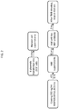
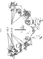
- TNFRSF25 expression constructs were prepared. These included either the entire extracellular domain (ECD) extending from the GIn at position 25 (Q25) to the Phe at position 201 (F201), or a soluble splice variant extending from Q25 to the Thr at position 181 (T181).
- ECD extracellular domain
- F201 Phe at position 201
- T181 a soluble splice variant extending from Q25 to the Thr at position 181
- the constructs also included various tags, including 6His, hFc-6His, and mFc-His.
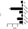
- Six constructs (TABLE 1) were transfected into 293F cells, and their expression levels were measured as depicted in FIG. 2 .
- W381-hProl.ECD-M.mFcHis demonstrated the highest expression ( FIG. 3 ).
- This version of TNFRSF25 therefore was chosen as the backbone for alanine scanning-based epitope mapping.
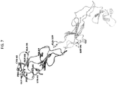
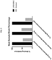
- FIG. 4 To identify the epitope of TNFRSF25 recognized by the antibody, binding of the antibody to TNFRSF25 muteins was then evaluated using the steps depicted in FIG. 4 . Specifically, studies were conducted to assess the binding of a chimeric antibody, W330-cAb1.hIgG1(35783) (the parent, hamster anti-TNFRSF25 antibody that was made chimeric with human IgFc), to alanine mutants of WBP330-hPro1.ECD.hFcHis that were expressed at greater than 1500 ng/ml. The binding affinity to each mutein was compared to that of non-mutated WBP330-hProl.ECD.hFcHis ( FIG. 5 ). Muteins that exhibited a signal change more than 25% (fold change ⁇ 0.75; TABLE 3 and FIG. 6 ) were used for structure modeling ( FIGS. 7 and 8 ).
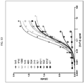
- Affinity optimization of W3153-P8R32-H6 antibody Each amino acid of the three CDRs of the parental clone was individually mutated to all 20 amino acids using a hybridization mutagenesis method ( Kunkel, Proc Natl Acad Sci USA 82(2):488-492, 1985 ). DNA primers containing a NNS codon encoding 20 amino acids were used to introduce mutations at each targeted CDR position. The individual degenerate primers were used in hybridization mutagenesis reactions. Briefly, each degenerate primer was phosphorylated, then used in a 10:1 ratio with uridinylated ssDNA. The mixture was heated to 85°C for 5 minutes and then cooled down to 55°C over 1 hour.
- the primary screen consisted of a single point ELISA (SPE) assay, which was carried out using periplasmic extract (PE) of bacteria grown in 96-well (deep well) plates. Briefly, this capture ELISA involved coating individual wells of a 96-well Maxisorp Immunoplate with anti-c-myc antibody in coating buffer (200 mM Na 2 CO 3 /NaHCO 3 ) at pH 9.2 overnight at 4°C. The next day, the plate was blocked with Casein for 1 hour at room temperature. scFv PE was then added to the plate and incubated at room temperature for 1 hour.
- SPE single point ELISA
- PE periplasmic extract
- biotinylated antigen protein was added to the well and the mixture was incubated for 1 hour at room temperature. This was followed by incubation with streptavidin-horseradish peroxidase (HRP) conjugate for 1 hour at room temperature. HRP activity was detected with tetramethyl-benzidine (TMB) substrate, and the reaction was quenched with 2M HCl. Plates were read at 450 nM. Clones exhibiting an optical density (OD) signal at 450 nm that was greater than the OD signal of the parental clone were picked and re-assayed by ELISA (as described above) in duplicate, to confirm positive results.
- HRP streptavidin-horseradish peroxidase
- Clones that repeatedly exhibited a signal greater than that of the parental antibody were sequenced.
- the scFv protein concentration of each clone that had a CDR change was then determined by a quantitative scFv ELISA, where a scFv with known concentration was used as a reference.
- the scFv protein concentration was determined by comparing the ELISA signals with signals generated by the reference scFv.
- the binding assay was repeated once more for all positive variants under normalized scFv concentrations in order to determine the relative binding affinity of the mutant scFv and the parental antibody.
- Selected scFv hits displaying improved binding were re-formulated to IgG1 and tested in a caspase-3 assay as described below.
- Combinatorial screening of the scFv library Point mutations in the VH and VL chains that were determined to be beneficial for binding to antigen were combined to determine whether additional binding synergy was attained.
- the combinatorial mutants were expressed as scFv and screened using the capture ELISA. Clones exhibiting an OD signal at 450 nm that was greater than the OD signal of the parental clone were sequenced and further ranked by binding ELISA as described above. Eleven top-ranked clones were selected for IgG4 re-formatting and further characterization as described above.
- V-genes of the top-rScFv clones identified from the primary and combinatorial screening were amplified by PCR and cloned into WuXi Biologies' proprietary expression vector, and expressed from 293F cells. Culture supernatants containing the antibodies were harvested and purified using Protein A chromatography.
- Caspase release assay P815 cells expressing TNFRSF25 were incubated with various concentrations of antibodies (from 1 ⁇ g/ml to 1 ng/ml) for 5 hours at 37°C in a 96-well plate. Caspase activity was determined using the Homogeneous Caspase Assay kit (Roche: 005372001). Fluorescence was read using a Molecular Devices SPECTRAMAX ® M5e plate reader.
- p815-hDR3 Parental p815 cells and p815 cells expressing human TNFRSF25 (also referred to as p815-hDR3) were cultured in suspension in IMDM + 10% FBS. Cells were counted at the time of harvest, pelleted and resuspended at a concentration of 300,000 cells/100 ⁇ L in serum free media. 100 ⁇ L aliquots of the cell suspension were placed into Eppendorf tubes.
- ALEXA FLUOR ® 647 (AF647)-conjugated 4C12 and hTL1A-Ig (AKTA purification, round 1) were generated using a Molecular Probes ALEXA FLUOR ® labeling kit (RL# 170512) per the manufacturer's instructions.
- the degree of labeling for 4C12-AF647 was 5.98 moles of dye to antibody, and for hTL1A-Ig-AF647 the degree of labeling was 4.55 moles of dye to protein (both considered to be within the acceptable range).
- hTL1A-Ig-AF647 or 4C12-AF647 was added to the Eppendorf tubes at a concentration of 0.5 ⁇ g/mL .
- Unlabeled 4C12, affinity matured 4C12 (hIgG1 subtype), or isotype control hIgG1 was added immediately afterward at concentrations ranging from 2 ⁇ g/mL to 0.0078 ⁇ g/mL. The mixtures were as incubated at 37°C in a humidified incubator with 5% CO 2 for 1 hour. Cells were pelleted in a microcentrifuge and washed 1x with FACS buffer. Pellets were resuspended in 300 ⁇ L FACS buffer and analyzed by a Sony SH800 flow cytometer. Live single cells were gated on, and MFI for AF647 was determined for each sample.
- clones with improved binding to the TNFRSF25-Fc protein were designated as follows:
Landscapes
- Health & Medical Sciences (AREA)
- Chemical & Material Sciences (AREA)
- Life Sciences & Earth Sciences (AREA)
- Immunology (AREA)
- Medicinal Chemistry (AREA)
- General Health & Medical Sciences (AREA)
- Public Health (AREA)
- Veterinary Medicine (AREA)
- Animal Behavior & Ethology (AREA)
- Pharmacology & Pharmacy (AREA)
- Organic Chemistry (AREA)
- Epidemiology (AREA)
- Engineering & Computer Science (AREA)
- Cell Biology (AREA)
- Bioinformatics & Cheminformatics (AREA)
- Chemical Kinetics & Catalysis (AREA)
- General Chemical & Material Sciences (AREA)
- Nuclear Medicine, Radiotherapy & Molecular Imaging (AREA)
- Virology (AREA)
- Biochemistry (AREA)
- Developmental Biology & Embryology (AREA)
- Biotechnology (AREA)
- Biomedical Technology (AREA)
- Hematology (AREA)
- Proteomics, Peptides & Aminoacids (AREA)
- Molecular Biology (AREA)
- Genetics & Genomics (AREA)
- Zoology (AREA)
- Biophysics (AREA)
- Mycology (AREA)
- Microbiology (AREA)
- Endocrinology (AREA)
- Peptides Or Proteins (AREA)
- Medicines Containing Antibodies Or Antigens For Use As Internal Diagnostic Agents (AREA)
- Preparation Of Compounds By Using Micro-Organisms (AREA)
- Medicines That Contain Protein Lipid Enzymes And Other Medicines (AREA)
Applications Claiming Priority (2)
| Application Number | Priority Date | Filing Date | Title |
|---|---|---|---|
| US201662348009P | 2016-06-09 | 2016-06-09 | |
| PCT/US2017/036817 WO2017214547A1 (en) | 2016-06-09 | 2017-06-09 | Anti-tnfrsf25 antibodies |
Publications (2)
| Publication Number | Publication Date |
|---|---|
| EP3468998A1 EP3468998A1 (en) | 2019-04-17 |
| EP3468998B1 true EP3468998B1 (en) | 2021-12-01 |
Family
ID=59078259
Family Applications (1)
| Application Number | Title | Priority Date | Filing Date |
|---|---|---|---|
| EP17731434.1A Active EP3468998B1 (en) | 2016-06-09 | 2017-06-09 | Anti-tnfrsf25 antibodies |
Country Status (15)
| Country | Link |
|---|---|
| US (3) | US10005843B2 (OSRAM) |
| EP (1) | EP3468998B1 (OSRAM) |
| JP (1) | JP6961625B2 (OSRAM) |
| KR (1) | KR102423942B1 (OSRAM) |
| CN (1) | CN109219620B (OSRAM) |
| AU (1) | AU2017278192A1 (OSRAM) |
| BR (1) | BR112018075222A2 (OSRAM) |
| CA (1) | CA3026652A1 (OSRAM) |
| DK (1) | DK3468998T3 (OSRAM) |
| IL (1) | IL263406B2 (OSRAM) |
| MX (1) | MX389320B (OSRAM) |
| RU (1) | RU2746314C2 (OSRAM) |
| SG (1) | SG11201810853UA (OSRAM) |
| WO (1) | WO2017214547A1 (OSRAM) |
| ZA (1) | ZA201808317B (OSRAM) |
Families Citing this family (11)
| Publication number | Priority date | Publication date | Assignee | Title |
|---|---|---|---|---|
| MX2014003689A (es) | 2011-09-30 | 2014-12-05 | Teva Pharmaceuticals Australia Pty Ltd | Anticuerpos contra tl1a y sus usos. |
| JP6744318B2 (ja) * | 2015-02-06 | 2020-08-26 | ヒート バイオロジクス,インコーポレイテッド | ワクチンおよび共刺激分子を共発現するベクター |
| TWI703158B (zh) | 2015-09-18 | 2020-09-01 | 美商希佛隆公司 | 特異性結合tl1a之抗體 |
| US12331320B2 (en) | 2018-10-10 | 2025-06-17 | The Research Foundation For The State University Of New York | Genome edited cancer cell vaccines |
| AU2020340372A1 (en) * | 2019-08-30 | 2022-03-10 | Pelican Therapeutics, Inc. | Methods of treating cancer using TNFRSF25 antibodies |
| CA3151319A1 (en) * | 2019-09-26 | 2021-04-01 | Matthew M. Seavey | Tnfrsf25-mediated treatments of immune diseases and disorders |
| WO2022212668A1 (en) * | 2021-03-31 | 2022-10-06 | Heat Biologics, Inc. | Methods and compositions related to tnfrsf25/dr3 agonists |
| WO2023244089A1 (ko) * | 2022-06-17 | 2023-12-21 | 주식회사 셀렉신 | 인간 dr3에 특이적으로 결합하는 항체 및 이의 용도 |
| CN120399074A (zh) * | 2023-06-02 | 2025-08-01 | 安徽金百奥生物科技有限公司 | 一种与人dr3胞外域高亲和力的纳米抗体 |
| GB202315963D0 (en) | 2023-10-18 | 2023-11-29 | Cancer Research Tech Ltd | Binding molecules |
| WO2025158389A1 (en) * | 2024-01-24 | 2025-07-31 | Shattuck Labs, Inc. | Antibodies that bind tnfrsf25 |
Family Cites Families (26)
| Publication number | Priority date | Publication date | Assignee | Title |
|---|---|---|---|---|
| US4816567A (en) | 1983-04-08 | 1989-03-28 | Genentech, Inc. | Recombinant immunoglobin preparations |
| JPS61134325A (ja) | 1984-12-04 | 1986-06-21 | Teijin Ltd | ハイブリツド抗体遺伝子の発現方法 |
| US5618920A (en) | 1985-11-01 | 1997-04-08 | Xoma Corporation | Modular assembly of antibody genes, antibodies prepared thereby and use |
| US5530101A (en) | 1988-12-28 | 1996-06-25 | Protein Design Labs, Inc. | Humanized immunoglobulins |
| CZ282603B6 (cs) | 1991-03-06 | 1997-08-13 | Merck Patent Gesellschaft Mit Beschränkter Haftun G | Humanizované a chimerické monoklonální protilátky |
| CA2078539C (en) | 1991-09-18 | 2005-08-02 | Kenya Shitara | Process for producing humanized chimera antibody |
| US5565332A (en) | 1991-09-23 | 1996-10-15 | Medical Research Council | Production of chimeric antibodies - a combinatorial approach |
| GB9125979D0 (en) | 1991-12-06 | 1992-02-05 | Wellcome Found | Antibody |
| US5863537A (en) | 1992-02-19 | 1999-01-26 | Schering Corporation | Humanized monoclonal antibodies against human interleukin-4 |
| US5733743A (en) | 1992-03-24 | 1998-03-31 | Cambridge Antibody Technology Limited | Methods for producing members of specific binding pairs |
| US5639641A (en) | 1992-09-09 | 1997-06-17 | Immunogen Inc. | Resurfacing of rodent antibodies |
| US5705154A (en) | 1995-03-08 | 1998-01-06 | Schering Corporation | Humanized monoclonal antibodies against human interleukin-4 |
| ES2428321T3 (es) | 2003-08-20 | 2013-11-07 | University Of Miami | Composiciones y métodos para tratar la enfermedad del pulmón inflamado |
| US7612179B2 (en) | 2003-11-28 | 2009-11-03 | Astrazeneca Ab | antibodies binding to a C-terminal fragment of apoliopoprotein E |
| CN101253199B (zh) | 2005-08-30 | 2019-06-14 | 迈阿密大学 | 免疫调节肿瘤坏死因子受体25(tnfr25)的激动剂、拮抗剂和免疫毒素 |
| ME02269B (me) | 2006-10-02 | 2016-04-28 | Medarex Inc | Humana antitela koja se vezuju za cxcr4 i njihove upotrebe |
| FR2907341B1 (fr) | 2006-10-18 | 2012-08-17 | Pf Medicament | Utilisation d'un anticorps anti-cd151 pour le traitement du cancer |
| WO2010033913A1 (en) | 2008-09-22 | 2010-03-25 | Icb International, Inc. | Antibodies, analogs and uses thereof |
| KR102033276B1 (ko) | 2009-08-03 | 2019-10-16 | 유니버시티 오브 마이애미 | T 조절 세포의 생체 내 확장 방법 |
| WO2011106707A2 (en) * | 2010-02-26 | 2011-09-01 | Human Genome Sciences, Inc. | Antibodies that specifically bind to dr3 |
| GB201020995D0 (en) | 2010-12-10 | 2011-01-26 | Bioinvent Int Ab | Biological materials and uses thereof |
| JP2013040160A (ja) | 2011-07-01 | 2013-02-28 | Genentech Inc | 自己免疫疾患を治療するための抗cd83アゴニスト抗体の使用 |
| WO2014110258A1 (en) | 2013-01-09 | 2014-07-17 | Podack Eckhard R | Compositions and methods for the regulation of t regulatory cells using tl1a-ig fusion protein |
| RU2551235C2 (ru) * | 2013-08-26 | 2015-05-20 | Илья Владимирович Духовлинов | Гуманизированное моноклональное антитело, специфичное к синдекану-1 |
| CN103592439B (zh) * | 2013-09-03 | 2015-10-21 | 天津亿美诺生物科技有限公司 | 人尿路上皮癌特异性抗体及其应用 |
| WO2016081455A1 (en) * | 2014-11-17 | 2016-05-26 | Pelican Therapeutics, Inc. | Human tnfrsf25 antibody |
-
2017
- 2017-06-09 SG SG11201810853UA patent/SG11201810853UA/en unknown
- 2017-06-09 AU AU2017278192A patent/AU2017278192A1/en not_active Abandoned
- 2017-06-09 BR BR112018075222-3A patent/BR112018075222A2/pt not_active IP Right Cessation
- 2017-06-09 WO PCT/US2017/036817 patent/WO2017214547A1/en not_active Ceased
- 2017-06-09 RU RU2018143977A patent/RU2746314C2/ru active
- 2017-06-09 KR KR1020187035804A patent/KR102423942B1/ko active Active
- 2017-06-09 DK DK17731434.1T patent/DK3468998T3/da active
- 2017-06-09 US US15/618,993 patent/US10005843B2/en active Active
- 2017-06-09 EP EP17731434.1A patent/EP3468998B1/en active Active
- 2017-06-09 CA CA3026652A patent/CA3026652A1/en active Pending
- 2017-06-09 MX MX2018014716A patent/MX389320B/es unknown
- 2017-06-09 IL IL263406A patent/IL263406B2/en unknown
- 2017-06-09 JP JP2018563618A patent/JP6961625B2/ja not_active Expired - Fee Related
- 2017-06-09 CN CN201780034593.5A patent/CN109219620B/zh not_active Expired - Fee Related
-
2018
- 2018-04-27 US US15/964,775 patent/US10683359B2/en not_active Expired - Fee Related
- 2018-12-10 ZA ZA2018/08317A patent/ZA201808317B/en unknown
-
2020
- 2020-05-14 US US16/874,290 patent/US20210017287A1/en not_active Abandoned
Non-Patent Citations (1)
| Title |
|---|
| M CHODORGE ET AL: "A series of Fas receptor agonist antibodies that demonstrate an inverse correlation between affinity and potency", CELL DEATH AND DIFFERENTIATION., vol. 19, no. 7, 20 January 2012 (2012-01-20), GB, pages 1187 - 1195, XP055332053, ISSN: 1350-9047, DOI: 10.1038/cdd.2011.208 * |
Also Published As
| Publication number | Publication date |
|---|---|
| IL263406B1 (en) | 2023-05-01 |
| KR102423942B1 (ko) | 2022-07-22 |
| BR112018075222A2 (pt) | 2019-03-19 |
| EP3468998A1 (en) | 2019-04-17 |
| CN109219620B (zh) | 2023-01-31 |
| RU2746314C2 (ru) | 2021-04-12 |
| SG11201810853UA (en) | 2019-01-30 |
| KR20190015716A (ko) | 2019-02-14 |
| IL263406A (en) | 2018-12-31 |
| US20170355773A1 (en) | 2017-12-14 |
| JP2019526528A (ja) | 2019-09-19 |
| AU2017278192A1 (en) | 2018-12-13 |
| RU2018143977A3 (OSRAM) | 2020-10-05 |
| US20210017287A1 (en) | 2021-01-21 |
| CN109219620A (zh) | 2019-01-15 |
| ZA201808317B (en) | 2019-08-28 |
| RU2018143977A (ru) | 2020-07-09 |
| WO2017214547A1 (en) | 2017-12-14 |
| MX2018014716A (es) | 2019-03-11 |
| US10683359B2 (en) | 2020-06-16 |
| US20180312599A1 (en) | 2018-11-01 |
| IL263406B2 (en) | 2023-09-01 |
| CA3026652A1 (en) | 2017-12-14 |
| MX389320B (es) | 2025-03-20 |
| DK3468998T3 (en) | 2022-03-07 |
| JP6961625B2 (ja) | 2021-11-10 |
| US10005843B2 (en) | 2018-06-26 |
Similar Documents
| Publication | Publication Date | Title |
|---|---|---|
| EP3468998B1 (en) | Anti-tnfrsf25 antibodies | |
| US12030944B2 (en) | Enhanced depletion of targeted cells with CD47 blockade and an immune costimulatory agonist | |
| JP6907124B2 (ja) | Cdh3及びcd3に対する二重特異性抗体構築物 | |
| US10683358B2 (en) | Human TNFRSF25 antibody | |
| JP2021522801A (ja) | ヒトネクチン4に特異的な抗体 | |
| KR102608028B1 (ko) | 인간 원형질막 소포 관련 단백질 pv-1에 특이적으로 결합하는 단일클론항체 및 이의 제조방법과 용도 | |
| CN114790241B (zh) | 抗tigit抗体及其应用 | |
| KR20230159823A (ko) | Cd112r에 대한 항체 및 이의 용도 | |
| CN116554325A (zh) | 抗b7h3抗体及其应用 | |
| HK40007009B (en) | Anti-tnfrsf25 antibodies | |
| WO2025101526A1 (en) | Multi-specific cancer-targeting antibodies | |
| CA2996167C (en) | Enhanced depletion of targeted cells with cd47 blockade and an immune costimulatory agonist | |
| HK1257824B (en) | Enhanced depletion of targeted cells with cd47 blockade and an immune costimulatory agonist |
Legal Events
| Date | Code | Title | Description |
|---|---|---|---|
| STAA | Information on the status of an ep patent application or granted ep patent |
Free format text: STATUS: UNKNOWN |
|
| STAA | Information on the status of an ep patent application or granted ep patent |
Free format text: STATUS: THE INTERNATIONAL PUBLICATION HAS BEEN MADE |
|
| PUAI | Public reference made under article 153(3) epc to a published international application that has entered the european phase |
Free format text: ORIGINAL CODE: 0009012 |
|
| STAA | Information on the status of an ep patent application or granted ep patent |
Free format text: STATUS: REQUEST FOR EXAMINATION WAS MADE |
|
| 17P | Request for examination filed |
Effective date: 20181221 |
|
| AK | Designated contracting states |
Kind code of ref document: A1 Designated state(s): AL AT BE BG CH CY CZ DE DK EE ES FI FR GB GR HR HU IE IS IT LI LT LU LV MC MK MT NL NO PL PT RO RS SE SI SK SM TR |
|
| AX | Request for extension of the european patent |
Extension state: BA ME |
|
| DAV | Request for validation of the european patent (deleted) | ||
| DAX | Request for extension of the european patent (deleted) | ||
| STAA | Information on the status of an ep patent application or granted ep patent |
Free format text: STATUS: EXAMINATION IS IN PROGRESS |
|
| 17Q | First examination report despatched |
Effective date: 20191112 |
|
| REG | Reference to a national code |
Ref country code: HK Ref legal event code: DE Ref document number: 40007009 Country of ref document: HK |
|
| REG | Reference to a national code |
Ref country code: DE Ref legal event code: R079 Ref document number: 602017050180 Country of ref document: DE Free format text: PREVIOUS MAIN CLASS: C07K0016280000 Ipc: A61P0035000000 |
|
| RIC1 | Information provided on ipc code assigned before grant |
Ipc: A61P 35/00 20060101AFI20210419BHEP Ipc: A61P 37/04 20060101ALI20210419BHEP Ipc: C07K 16/28 20060101ALI20210419BHEP |
|
| GRAP | Despatch of communication of intention to grant a patent |
Free format text: ORIGINAL CODE: EPIDOSNIGR1 |
|
| STAA | Information on the status of an ep patent application or granted ep patent |
Free format text: STATUS: GRANT OF PATENT IS INTENDED |
|
| INTG | Intention to grant announced |
Effective date: 20210625 |
|
| GRAS | Grant fee paid |
Free format text: ORIGINAL CODE: EPIDOSNIGR3 |
|
| GRAA | (expected) grant |
Free format text: ORIGINAL CODE: 0009210 |
|
| STAA | Information on the status of an ep patent application or granted ep patent |
Free format text: STATUS: THE PATENT HAS BEEN GRANTED |
|
| AK | Designated contracting states |
Kind code of ref document: B1 Designated state(s): AL AT BE BG CH CY CZ DE DK EE ES FI FR GB GR HR HU IE IS IT LI LT LU LV MC MK MT NL NO PL PT RO RS SE SI SK SM TR |
|
| REG | Reference to a national code |
Ref country code: GB Ref legal event code: FG4D |
|
| REG | Reference to a national code |
Ref country code: AT Ref legal event code: REF Ref document number: 1451205 Country of ref document: AT Kind code of ref document: T Effective date: 20211215 Ref country code: CH Ref legal event code: EP |
|
| REG | Reference to a national code |
Ref country code: IE Ref legal event code: FG4D |
|
| REG | Reference to a national code |
Ref country code: DE Ref legal event code: R096 Ref document number: 602017050180 Country of ref document: DE |
|
| REG | Reference to a national code |
Ref country code: DK Ref legal event code: T3 Effective date: 20220301 |
|
| REG | Reference to a national code |
Ref country code: NL Ref legal event code: FP |
|
| REG | Reference to a national code |
Ref country code: LT Ref legal event code: MG9D |
|
| REG | Reference to a national code |
Ref country code: AT Ref legal event code: MK05 Ref document number: 1451205 Country of ref document: AT Kind code of ref document: T Effective date: 20211201 |
|
| PG25 | Lapsed in a contracting state [announced via postgrant information from national office to epo] |
Ref country code: RS Free format text: LAPSE BECAUSE OF FAILURE TO SUBMIT A TRANSLATION OF THE DESCRIPTION OR TO PAY THE FEE WITHIN THE PRESCRIBED TIME-LIMIT Effective date: 20211201 Ref country code: LT Free format text: LAPSE BECAUSE OF FAILURE TO SUBMIT A TRANSLATION OF THE DESCRIPTION OR TO PAY THE FEE WITHIN THE PRESCRIBED TIME-LIMIT Effective date: 20211201 Ref country code: FI Free format text: LAPSE BECAUSE OF FAILURE TO SUBMIT A TRANSLATION OF THE DESCRIPTION OR TO PAY THE FEE WITHIN THE PRESCRIBED TIME-LIMIT Effective date: 20211201 Ref country code: BG Free format text: LAPSE BECAUSE OF FAILURE TO SUBMIT A TRANSLATION OF THE DESCRIPTION OR TO PAY THE FEE WITHIN THE PRESCRIBED TIME-LIMIT Effective date: 20220301 Ref country code: AT Free format text: LAPSE BECAUSE OF FAILURE TO SUBMIT A TRANSLATION OF THE DESCRIPTION OR TO PAY THE FEE WITHIN THE PRESCRIBED TIME-LIMIT Effective date: 20211201 |
|
| PG25 | Lapsed in a contracting state [announced via postgrant information from national office to epo] |
Ref country code: SE Free format text: LAPSE BECAUSE OF FAILURE TO SUBMIT A TRANSLATION OF THE DESCRIPTION OR TO PAY THE FEE WITHIN THE PRESCRIBED TIME-LIMIT Effective date: 20211201 Ref country code: PL Free format text: LAPSE BECAUSE OF FAILURE TO SUBMIT A TRANSLATION OF THE DESCRIPTION OR TO PAY THE FEE WITHIN THE PRESCRIBED TIME-LIMIT Effective date: 20211201 Ref country code: NO Free format text: LAPSE BECAUSE OF FAILURE TO SUBMIT A TRANSLATION OF THE DESCRIPTION OR TO PAY THE FEE WITHIN THE PRESCRIBED TIME-LIMIT Effective date: 20220301 Ref country code: LV Free format text: LAPSE BECAUSE OF FAILURE TO SUBMIT A TRANSLATION OF THE DESCRIPTION OR TO PAY THE FEE WITHIN THE PRESCRIBED TIME-LIMIT Effective date: 20211201 Ref country code: HR Free format text: LAPSE BECAUSE OF FAILURE TO SUBMIT A TRANSLATION OF THE DESCRIPTION OR TO PAY THE FEE WITHIN THE PRESCRIBED TIME-LIMIT Effective date: 20211201 Ref country code: GR Free format text: LAPSE BECAUSE OF FAILURE TO SUBMIT A TRANSLATION OF THE DESCRIPTION OR TO PAY THE FEE WITHIN THE PRESCRIBED TIME-LIMIT Effective date: 20220302 Ref country code: ES Free format text: LAPSE BECAUSE OF FAILURE TO SUBMIT A TRANSLATION OF THE DESCRIPTION OR TO PAY THE FEE WITHIN THE PRESCRIBED TIME-LIMIT Effective date: 20211201 |
|
| PG25 | Lapsed in a contracting state [announced via postgrant information from national office to epo] |
Ref country code: SM Free format text: LAPSE BECAUSE OF FAILURE TO SUBMIT A TRANSLATION OF THE DESCRIPTION OR TO PAY THE FEE WITHIN THE PRESCRIBED TIME-LIMIT Effective date: 20211201 Ref country code: SK Free format text: LAPSE BECAUSE OF FAILURE TO SUBMIT A TRANSLATION OF THE DESCRIPTION OR TO PAY THE FEE WITHIN THE PRESCRIBED TIME-LIMIT Effective date: 20211201 Ref country code: RO Free format text: LAPSE BECAUSE OF FAILURE TO SUBMIT A TRANSLATION OF THE DESCRIPTION OR TO PAY THE FEE WITHIN THE PRESCRIBED TIME-LIMIT Effective date: 20211201 Ref country code: PT Free format text: LAPSE BECAUSE OF FAILURE TO SUBMIT A TRANSLATION OF THE DESCRIPTION OR TO PAY THE FEE WITHIN THE PRESCRIBED TIME-LIMIT Effective date: 20220401 Ref country code: EE Free format text: LAPSE BECAUSE OF FAILURE TO SUBMIT A TRANSLATION OF THE DESCRIPTION OR TO PAY THE FEE WITHIN THE PRESCRIBED TIME-LIMIT Effective date: 20211201 Ref country code: CZ Free format text: LAPSE BECAUSE OF FAILURE TO SUBMIT A TRANSLATION OF THE DESCRIPTION OR TO PAY THE FEE WITHIN THE PRESCRIBED TIME-LIMIT Effective date: 20211201 |
|
| PGFP | Annual fee paid to national office [announced via postgrant information from national office to epo] |
Ref country code: NL Payment date: 20220626 Year of fee payment: 6 Ref country code: LU Payment date: 20220627 Year of fee payment: 6 Ref country code: GB Payment date: 20220628 Year of fee payment: 6 Ref country code: DK Payment date: 20220629 Year of fee payment: 6 |
|
| PGFP | Annual fee paid to national office [announced via postgrant information from national office to epo] |
Ref country code: BE Payment date: 20220627 Year of fee payment: 6 |
|
| REG | Reference to a national code |
Ref country code: DE Ref legal event code: R097 Ref document number: 602017050180 Country of ref document: DE |
|
| PG25 | Lapsed in a contracting state [announced via postgrant information from national office to epo] |
Ref country code: IS Free format text: LAPSE BECAUSE OF FAILURE TO SUBMIT A TRANSLATION OF THE DESCRIPTION OR TO PAY THE FEE WITHIN THE PRESCRIBED TIME-LIMIT Effective date: 20220401 |
|
| PGFP | Annual fee paid to national office [announced via postgrant information from national office to epo] |
Ref country code: FR Payment date: 20220627 Year of fee payment: 6 |
|
| PLBE | No opposition filed within time limit |
Free format text: ORIGINAL CODE: 0009261 |
|
| STAA | Information on the status of an ep patent application or granted ep patent |
Free format text: STATUS: NO OPPOSITION FILED WITHIN TIME LIMIT |
|
| PG25 | Lapsed in a contracting state [announced via postgrant information from national office to epo] |
Ref country code: AL Free format text: LAPSE BECAUSE OF FAILURE TO SUBMIT A TRANSLATION OF THE DESCRIPTION OR TO PAY THE FEE WITHIN THE PRESCRIBED TIME-LIMIT Effective date: 20211201 |
|
| PGFP | Annual fee paid to national office [announced via postgrant information from national office to epo] |
Ref country code: DE Payment date: 20220629 Year of fee payment: 6 |
|
| 26N | No opposition filed |
Effective date: 20220902 |
|
| PG25 | Lapsed in a contracting state [announced via postgrant information from national office to epo] |
Ref country code: SI Free format text: LAPSE BECAUSE OF FAILURE TO SUBMIT A TRANSLATION OF THE DESCRIPTION OR TO PAY THE FEE WITHIN THE PRESCRIBED TIME-LIMIT Effective date: 20211201 |
|
| PGFP | Annual fee paid to national office [announced via postgrant information from national office to epo] |
Ref country code: CH Payment date: 20220706 Year of fee payment: 6 |
|
| PG25 | Lapsed in a contracting state [announced via postgrant information from national office to epo] |
Ref country code: MC Free format text: LAPSE BECAUSE OF FAILURE TO SUBMIT A TRANSLATION OF THE DESCRIPTION OR TO PAY THE FEE WITHIN THE PRESCRIBED TIME-LIMIT Effective date: 20211201 |
|
| PG25 | Lapsed in a contracting state [announced via postgrant information from national office to epo] |
Ref country code: IE Free format text: LAPSE BECAUSE OF NON-PAYMENT OF DUE FEES Effective date: 20220609 |
|
| PG25 | Lapsed in a contracting state [announced via postgrant information from national office to epo] |
Ref country code: IT Free format text: LAPSE BECAUSE OF FAILURE TO SUBMIT A TRANSLATION OF THE DESCRIPTION OR TO PAY THE FEE WITHIN THE PRESCRIBED TIME-LIMIT Effective date: 20211201 |
|
| REG | Reference to a national code |
Ref country code: DE Ref legal event code: R119 Ref document number: 602017050180 Country of ref document: DE |
|
| REG | Reference to a national code |
Ref country code: DK Ref legal event code: EBP Effective date: 20230630 |
|
| REG | Reference to a national code |
Ref country code: CH Ref legal event code: PL |
|
| REG | Reference to a national code |
Ref country code: NL Ref legal event code: MM Effective date: 20230701 |
|
| REG | Reference to a national code |
Ref country code: BE Ref legal event code: MM Effective date: 20230630 |
|
| GBPC | Gb: european patent ceased through non-payment of renewal fee |
Effective date: 20230609 |
|
| PG25 | Lapsed in a contracting state [announced via postgrant information from national office to epo] |
Ref country code: LU Free format text: LAPSE BECAUSE OF NON-PAYMENT OF DUE FEES Effective date: 20230609 |
|
| PG25 | Lapsed in a contracting state [announced via postgrant information from national office to epo] |
Ref country code: NL Free format text: LAPSE BECAUSE OF NON-PAYMENT OF DUE FEES Effective date: 20230701 Ref country code: LU Free format text: LAPSE BECAUSE OF NON-PAYMENT OF DUE FEES Effective date: 20230609 Ref country code: HU Free format text: LAPSE BECAUSE OF FAILURE TO SUBMIT A TRANSLATION OF THE DESCRIPTION OR TO PAY THE FEE WITHIN THE PRESCRIBED TIME-LIMIT; INVALID AB INITIO Effective date: 20170609 |
|
| PG25 | Lapsed in a contracting state [announced via postgrant information from national office to epo] |
Ref country code: MK Free format text: LAPSE BECAUSE OF FAILURE TO SUBMIT A TRANSLATION OF THE DESCRIPTION OR TO PAY THE FEE WITHIN THE PRESCRIBED TIME-LIMIT Effective date: 20211201 Ref country code: DE Free format text: LAPSE BECAUSE OF NON-PAYMENT OF DUE FEES Effective date: 20240103 Ref country code: CY Free format text: LAPSE BECAUSE OF FAILURE TO SUBMIT A TRANSLATION OF THE DESCRIPTION OR TO PAY THE FEE WITHIN THE PRESCRIBED TIME-LIMIT Effective date: 20211201 Ref country code: GB Free format text: LAPSE BECAUSE OF NON-PAYMENT OF DUE FEES Effective date: 20230609 Ref country code: CH Free format text: LAPSE BECAUSE OF NON-PAYMENT OF DUE FEES Effective date: 20230630 |
|
| PG25 | Lapsed in a contracting state [announced via postgrant information from national office to epo] |
Ref country code: FR Free format text: LAPSE BECAUSE OF NON-PAYMENT OF DUE FEES Effective date: 20230630 Ref country code: BE Free format text: LAPSE BECAUSE OF NON-PAYMENT OF DUE FEES Effective date: 20230630 |
|
| PG25 | Lapsed in a contracting state [announced via postgrant information from national office to epo] |
Ref country code: TR Free format text: LAPSE BECAUSE OF FAILURE TO SUBMIT A TRANSLATION OF THE DESCRIPTION OR TO PAY THE FEE WITHIN THE PRESCRIBED TIME-LIMIT Effective date: 20211201 |
|
| PG25 | Lapsed in a contracting state [announced via postgrant information from national office to epo] |
Ref country code: DK Free format text: LAPSE BECAUSE OF NON-PAYMENT OF DUE FEES Effective date: 20230630 |
|
| PG25 | Lapsed in a contracting state [announced via postgrant information from national office to epo] |
Ref country code: DK Free format text: LAPSE BECAUSE OF NON-PAYMENT OF DUE FEES Effective date: 20230630 |
|
| PG25 | Lapsed in a contracting state [announced via postgrant information from national office to epo] |
Ref country code: MT Free format text: LAPSE BECAUSE OF FAILURE TO SUBMIT A TRANSLATION OF THE DESCRIPTION OR TO PAY THE FEE WITHIN THE PRESCRIBED TIME-LIMIT Effective date: 20211201 |