EP3455631B1 - Procédés pour la classification de patients atteints d'un cancer solide - Google Patents
Procédés pour la classification de patients atteints d'un cancer solide Download PDFInfo
- Publication number
- EP3455631B1 EP3455631B1 EP17725195.6A EP17725195A EP3455631B1 EP 3455631 B1 EP3455631 B1 EP 3455631B1 EP 17725195 A EP17725195 A EP 17725195A EP 3455631 B1 EP3455631 B1 EP 3455631B1
- Authority
- EP
- European Patent Office
- Prior art keywords
- cells
- density
- cancer
- biological markers
- tumor
- Prior art date
- Legal status (The legal status is an assumption and is not a legal conclusion. Google has not performed a legal analysis and makes no representation as to the accuracy of the status listed.)
- Active
Links
- 206010028980 Neoplasm Diseases 0.000 title claims description 173
- 238000000034 method Methods 0.000 title claims description 137
- 201000011510 cancer Diseases 0.000 title claims description 67
- 239000007787 solid Substances 0.000 title claims description 15
- 108090000623 proteins and genes Proteins 0.000 claims description 151
- 239000000090 biomarker Substances 0.000 claims description 126
- 210000004027 cell Anatomy 0.000 claims description 95
- 150000007523 nucleic acids Chemical group 0.000 claims description 77
- 230000014509 gene expression Effects 0.000 claims description 69
- 102000039446 nucleic acids Human genes 0.000 claims description 50
- 108020004707 nucleic acids Proteins 0.000 claims description 50
- 238000011282 treatment Methods 0.000 claims description 43
- 102000004169 proteins and genes Human genes 0.000 claims description 35
- 230000004044 response Effects 0.000 claims description 34
- -1 ICOS Proteins 0.000 claims description 30
- 238000003199 nucleic acid amplification method Methods 0.000 claims description 29
- 230000003321 amplification Effects 0.000 claims description 28
- 238000009396 hybridization Methods 0.000 claims description 27
- 230000033289 adaptive immune response Effects 0.000 claims description 26
- 230000028993 immune response Effects 0.000 claims description 26
- 230000001506 immunosuppresive effect Effects 0.000 claims description 24
- 210000000987 immune system Anatomy 0.000 claims description 23
- 210000001266 CD8-positive T-lymphocyte Anatomy 0.000 claims description 22
- 102100040678 Programmed cell death protein 1 Human genes 0.000 claims description 21
- 101000946843 Homo sapiens T-cell surface glycoprotein CD8 alpha chain Proteins 0.000 claims description 19
- 102100034922 T-cell surface glycoprotein CD8 alpha chain Human genes 0.000 claims description 19
- 108020004999 messenger RNA Proteins 0.000 claims description 19
- 230000004083 survival effect Effects 0.000 claims description 17
- 102000017420 CD3 protein, epsilon/gamma/delta subunit Human genes 0.000 claims description 16
- 108050005493 CD3 protein, epsilon/gamma/delta subunit Proteins 0.000 claims description 16
- 206010009944 Colon cancer Diseases 0.000 claims description 14
- 229940126547 T-cell immunoglobulin mucin-3 Drugs 0.000 claims description 14
- 208000001333 Colorectal Neoplasms Diseases 0.000 claims description 13
- 102100034458 Hepatitis A virus cellular receptor 2 Human genes 0.000 claims description 13
- 101710083479 Hepatitis A virus cellular receptor 2 homolog Proteins 0.000 claims description 13
- 102100024216 Programmed cell death 1 ligand 1 Human genes 0.000 claims description 12
- 102100031151 C-C chemokine receptor type 2 Human genes 0.000 claims description 11
- 101710149815 C-C chemokine receptor type 2 Proteins 0.000 claims description 11
- 101000974815 Homo sapiens BTB/POZ domain-containing protein KCTD11 Proteins 0.000 claims description 11
- 101001009599 Homo sapiens Granzyme A Proteins 0.000 claims description 11
- 101000579218 Homo sapiens Renin Proteins 0.000 claims description 11
- 101000738413 Homo sapiens T-cell surface glycoprotein CD3 gamma chain Proteins 0.000 claims description 11
- 108090000172 Interleukin-15 Proteins 0.000 claims description 11
- 102000003812 Interleukin-15 Human genes 0.000 claims description 11
- 102100037911 T-cell surface glycoprotein CD3 gamma chain Human genes 0.000 claims description 11
- 238000003364 immunohistochemistry Methods 0.000 claims description 11
- 238000000338 in vitro Methods 0.000 claims description 11
- 102100022790 BTB/POZ domain-containing protein KCTD11 Human genes 0.000 claims description 10
- 102100039498 Cytotoxic T-lymphocyte protein 4 Human genes 0.000 claims description 10
- 102100030386 Granzyme A Human genes 0.000 claims description 10
- 101000889276 Homo sapiens Cytotoxic T-lymphocyte protein 4 Proteins 0.000 claims description 10
- 101000611936 Homo sapiens Programmed cell death protein 1 Proteins 0.000 claims description 10
- 102100028467 Perforin-1 Human genes 0.000 claims description 10
- 230000000259 anti-tumor effect Effects 0.000 claims description 10
- 238000004393 prognosis Methods 0.000 claims description 10
- 102100032367 C-C motif chemokine 5 Human genes 0.000 claims description 9
- 102100030385 Granzyme B Human genes 0.000 claims description 9
- 101000797762 Homo sapiens C-C motif chemokine 5 Proteins 0.000 claims description 9
- 101000998146 Homo sapiens Interleukin-17A Proteins 0.000 claims description 9
- 101000987581 Homo sapiens Perforin-1 Proteins 0.000 claims description 9
- 101000713602 Homo sapiens T-box transcription factor TBX21 Proteins 0.000 claims description 9
- 102100022341 Integrin alpha-E Human genes 0.000 claims description 9
- 102100037850 Interferon gamma Human genes 0.000 claims description 9
- 102100033461 Interleukin-17A Human genes 0.000 claims description 9
- 102100036840 T-box transcription factor TBX21 Human genes 0.000 claims description 9
- 102100039037 Vascular endothelial growth factor A Human genes 0.000 claims description 9
- 238000009826 distribution Methods 0.000 claims description 9
- 102100025279 C-X-C motif chemokine 11 Human genes 0.000 claims description 8
- 102100038393 Granzyme H Human genes 0.000 claims description 8
- 102100038395 Granzyme K Human genes 0.000 claims description 8
- 101000858060 Homo sapiens C-X-C motif chemokine 11 Proteins 0.000 claims description 8
- 101001009603 Homo sapiens Granzyme B Proteins 0.000 claims description 8
- 101001033000 Homo sapiens Granzyme H Proteins 0.000 claims description 8
- 101001033007 Homo sapiens Granzyme K Proteins 0.000 claims description 8
- 101001046687 Homo sapiens Integrin alpha-E Proteins 0.000 claims description 8
- 101000599940 Homo sapiens Interferon gamma Proteins 0.000 claims description 8
- 101000598002 Homo sapiens Interferon regulatory factor 1 Proteins 0.000 claims description 8
- 101000582950 Homo sapiens Platelet factor 4 Proteins 0.000 claims description 8
- 101000610551 Homo sapiens Prominin-1 Proteins 0.000 claims description 8
- 101000946860 Homo sapiens T-cell surface glycoprotein CD3 epsilon chain Proteins 0.000 claims description 8
- 101000845170 Homo sapiens Thymic stromal lymphopoietin Proteins 0.000 claims description 8
- 101000808011 Homo sapiens Vascular endothelial growth factor A Proteins 0.000 claims description 8
- 101710139099 Indian hedgehog protein Proteins 0.000 claims description 8
- 102100036981 Interferon regulatory factor 1 Human genes 0.000 claims description 8
- 102100040120 Prominin-1 Human genes 0.000 claims description 8
- 108010044012 STAT1 Transcription Factor Proteins 0.000 claims description 8
- 102100029904 Signal transducer and activator of transcription 1-alpha/beta Human genes 0.000 claims description 8
- 102100035794 T-cell surface glycoprotein CD3 epsilon chain Human genes 0.000 claims description 8
- 102100020997 Fractalkine Human genes 0.000 claims description 7
- 102100021186 Granulysin Human genes 0.000 claims description 7
- 101000854520 Homo sapiens Fractalkine Proteins 0.000 claims description 7
- 102100026214 Indian hedgehog protein Human genes 0.000 claims description 7
- 102100030304 Platelet factor 4 Human genes 0.000 claims description 7
- 102100031294 Thymic stromal lymphopoietin Human genes 0.000 claims description 7
- 206010006187 Breast cancer Diseases 0.000 claims description 6
- 208000026310 Breast neoplasm Diseases 0.000 claims description 6
- 102100021943 C-C motif chemokine 2 Human genes 0.000 claims description 6
- 102100028990 C-X-C chemokine receptor type 3 Human genes 0.000 claims description 6
- 102100025248 C-X-C motif chemokine 10 Human genes 0.000 claims description 6
- 102100025277 C-X-C motif chemokine 13 Human genes 0.000 claims description 6
- 102100036170 C-X-C motif chemokine 9 Human genes 0.000 claims description 6
- 102100025137 Early activation antigen CD69 Human genes 0.000 claims description 6
- 101000897480 Homo sapiens C-C motif chemokine 2 Proteins 0.000 claims description 6
- 101000916050 Homo sapiens C-X-C chemokine receptor type 3 Proteins 0.000 claims description 6
- 101000858088 Homo sapiens C-X-C motif chemokine 10 Proteins 0.000 claims description 6
- 101000858064 Homo sapiens C-X-C motif chemokine 13 Proteins 0.000 claims description 6
- 101000947172 Homo sapiens C-X-C motif chemokine 9 Proteins 0.000 claims description 6
- 101000934374 Homo sapiens Early activation antigen CD69 Proteins 0.000 claims description 6
- 101001040751 Homo sapiens Granulysin Proteins 0.000 claims description 6
- 101001137987 Homo sapiens Lymphocyte activation gene 3 protein Proteins 0.000 claims description 6
- 101001117317 Homo sapiens Programmed cell death 1 ligand 1 Proteins 0.000 claims description 6
- 101000738335 Homo sapiens T-cell surface glycoprotein CD3 zeta chain Proteins 0.000 claims description 6
- 206010058467 Lung neoplasm malignant Diseases 0.000 claims description 6
- 206010033128 Ovarian cancer Diseases 0.000 claims description 6
- 206010061535 Ovarian neoplasm Diseases 0.000 claims description 6
- 108010019992 STAT4 Transcription Factor Proteins 0.000 claims description 6
- 102000005886 STAT4 Transcription Factor Human genes 0.000 claims description 6
- 102100037906 T-cell surface glycoprotein CD3 zeta chain Human genes 0.000 claims description 6
- 210000003719 b-lymphocyte Anatomy 0.000 claims description 6
- 201000005202 lung cancer Diseases 0.000 claims description 6
- 208000020816 lung neoplasm Diseases 0.000 claims description 6
- 102100022087 Granzyme M Human genes 0.000 claims description 5
- 101000900697 Homo sapiens Granzyme M Proteins 0.000 claims description 5
- 101000946863 Homo sapiens T-cell surface glycoprotein CD3 delta chain Proteins 0.000 claims description 5
- 206010060862 Prostate cancer Diseases 0.000 claims description 5
- 208000000236 Prostatic Neoplasms Diseases 0.000 claims description 5
- 102100035891 T-cell surface glycoprotein CD3 delta chain Human genes 0.000 claims description 5
- 230000004043 responsiveness Effects 0.000 claims description 5
- 102000017578 LAG3 Human genes 0.000 claims description 4
- 230000002596 correlated effect Effects 0.000 claims description 4
- 206010005003 Bladder cancer Diseases 0.000 claims description 3
- 208000007097 Urinary Bladder Neoplasms Diseases 0.000 claims description 3
- 201000005112 urinary bladder cancer Diseases 0.000 claims description 3
- 108010019530 Vascular Endothelial Growth Factors Proteins 0.000 claims description 2
- 102000005789 Vascular Endothelial Growth Factors Human genes 0.000 claims description 2
- 201000010536 head and neck cancer Diseases 0.000 claims 1
- 208000014829 head and neck neoplasm Diseases 0.000 claims 1
- 239000000523 sample Substances 0.000 description 142
- 210000001519 tissue Anatomy 0.000 description 77
- 238000001514 detection method Methods 0.000 description 35
- YBJHBAHKTGYVGT-ZKWXMUAHSA-N (+)-Biotin Chemical compound N1C(=O)N[C@@H]2[C@H](CCCCC(=O)O)SC[C@@H]21 YBJHBAHKTGYVGT-ZKWXMUAHSA-N 0.000 description 24
- 230000000875 corresponding effect Effects 0.000 description 19
- 102000004190 Enzymes Human genes 0.000 description 18
- 108090000790 Enzymes Proteins 0.000 description 18
- 229940088598 enzyme Drugs 0.000 description 18
- 239000003550 marker Substances 0.000 description 18
- 238000007901 in situ hybridization Methods 0.000 description 16
- 239000003446 ligand Substances 0.000 description 16
- 239000002096 quantum dot Substances 0.000 description 16
- 230000027455 binding Effects 0.000 description 15
- 239000003795 chemical substances by application Substances 0.000 description 15
- 239000002955 immunomodulating agent Substances 0.000 description 15
- 239000012528 membrane Substances 0.000 description 15
- 229940076838 Immune checkpoint inhibitor Drugs 0.000 description 14
- 239000000427 antigen Substances 0.000 description 14
- 108091007433 antigens Proteins 0.000 description 14
- 102000036639 antigens Human genes 0.000 description 14
- 239000011230 binding agent Substances 0.000 description 14
- 239000012274 immune-checkpoint protein inhibitor Substances 0.000 description 14
- 238000011002 quantification Methods 0.000 description 14
- 102000037984 Inhibitory immune checkpoint proteins Human genes 0.000 description 13
- 108091008026 Inhibitory immune checkpoint proteins Proteins 0.000 description 13
- 210000001744 T-lymphocyte Anatomy 0.000 description 13
- 239000003153 chemical reaction reagent Substances 0.000 description 13
- 108091070501 miRNA Proteins 0.000 description 13
- 108091032973 (ribonucleotides)n+m Proteins 0.000 description 12
- 229960002685 biotin Drugs 0.000 description 12
- 235000020958 biotin Nutrition 0.000 description 12
- 239000011616 biotin Substances 0.000 description 12
- 238000009169 immunotherapy Methods 0.000 description 12
- 239000002773 nucleotide Substances 0.000 description 12
- 125000003729 nucleotide group Chemical group 0.000 description 12
- 230000009870 specific binding Effects 0.000 description 12
- 238000010186 staining Methods 0.000 description 12
- 108090001008 Avidin Proteins 0.000 description 11
- 229940045513 CTLA4 antagonist Drugs 0.000 description 11
- 101710089372 Programmed cell death protein 1 Proteins 0.000 description 11
- 238000004458 analytical method Methods 0.000 description 11
- 239000003112 inhibitor Substances 0.000 description 11
- 238000002493 microarray Methods 0.000 description 11
- WSFSSNUMVMOOMR-UHFFFAOYSA-N Formaldehyde Chemical compound O=C WSFSSNUMVMOOMR-UHFFFAOYSA-N 0.000 description 10
- 102000037982 Immune checkpoint proteins Human genes 0.000 description 10
- 108091008036 Immune checkpoint proteins Proteins 0.000 description 10
- 108091028043 Nucleic acid sequence Proteins 0.000 description 10
- 238000003556 assay Methods 0.000 description 10
- 238000003757 reverse transcription PCR Methods 0.000 description 10
- 102000004127 Cytokines Human genes 0.000 description 9
- 108090000695 Cytokines Proteins 0.000 description 9
- 150000001875 compounds Chemical class 0.000 description 9
- 230000000694 effects Effects 0.000 description 9
- 201000001441 melanoma Diseases 0.000 description 9
- 238000002560 therapeutic procedure Methods 0.000 description 9
- 102000040650 (ribonucleotides)n+m Human genes 0.000 description 8
- 102000002260 Alkaline Phosphatase Human genes 0.000 description 8
- 108020004774 Alkaline Phosphatase Proteins 0.000 description 8
- ZHNUHDYFZUAESO-UHFFFAOYSA-N Formamide Chemical compound NC=O ZHNUHDYFZUAESO-UHFFFAOYSA-N 0.000 description 8
- 206010038389 Renal cancer Diseases 0.000 description 8
- VYPSYNLAJGMNEJ-UHFFFAOYSA-N Silicium dioxide Chemical compound O=[Si]=O VYPSYNLAJGMNEJ-UHFFFAOYSA-N 0.000 description 8
- FAPWRFPIFSIZLT-UHFFFAOYSA-M Sodium chloride Chemical compound [Na+].[Cl-] FAPWRFPIFSIZLT-UHFFFAOYSA-M 0.000 description 8
- 239000002246 antineoplastic agent Substances 0.000 description 8
- 238000001574 biopsy Methods 0.000 description 8
- 239000000872 buffer Substances 0.000 description 8
- 229940127089 cytotoxic agent Drugs 0.000 description 8
- MHMNJMPURVTYEJ-UHFFFAOYSA-N fluorescein-5-isothiocyanate Chemical compound O1C(=O)C2=CC(N=C=S)=CC=C2C21C1=CC=C(O)C=C1OC1=CC(O)=CC=C21 MHMNJMPURVTYEJ-UHFFFAOYSA-N 0.000 description 8
- 239000012188 paraffin wax Substances 0.000 description 8
- 230000002285 radioactive effect Effects 0.000 description 8
- 208000008839 Kidney Neoplasms Diseases 0.000 description 7
- 239000002671 adjuvant Substances 0.000 description 7
- 210000001151 cytotoxic T lymphocyte Anatomy 0.000 description 7
- 210000004443 dendritic cell Anatomy 0.000 description 7
- 201000010982 kidney cancer Diseases 0.000 description 7
- 210000001165 lymph node Anatomy 0.000 description 7
- 206010008342 Cervix carcinoma Diseases 0.000 description 6
- 102000007644 Colony-Stimulating Factors Human genes 0.000 description 6
- 108010071942 Colony-Stimulating Factors Proteins 0.000 description 6
- 108010050904 Interferons Proteins 0.000 description 6
- 102000014150 Interferons Human genes 0.000 description 6
- 102000015696 Interleukins Human genes 0.000 description 6
- 108010063738 Interleukins Proteins 0.000 description 6
- 208000006105 Uterine Cervical Neoplasms Diseases 0.000 description 6
- 238000002619 cancer immunotherapy Methods 0.000 description 6
- 229940022399 cancer vaccine Drugs 0.000 description 6
- 238000009566 cancer vaccine Methods 0.000 description 6
- 201000010881 cervical cancer Diseases 0.000 description 6
- 238000006243 chemical reaction Methods 0.000 description 6
- 210000000349 chromosome Anatomy 0.000 description 6
- 229940047120 colony stimulating factors Drugs 0.000 description 6
- 238000005516 engineering process Methods 0.000 description 6
- 239000007850 fluorescent dye Substances 0.000 description 6
- 230000006870 function Effects 0.000 description 6
- 230000005746 immune checkpoint blockade Effects 0.000 description 6
- 229940047124 interferons Drugs 0.000 description 6
- 229960005386 ipilimumab Drugs 0.000 description 6
- 238000002372 labelling Methods 0.000 description 6
- 229960003301 nivolumab Drugs 0.000 description 6
- 102000005962 receptors Human genes 0.000 description 6
- 108020003175 receptors Proteins 0.000 description 6
- 238000012552 review Methods 0.000 description 6
- 102100039619 Granulocyte colony-stimulating factor Human genes 0.000 description 5
- 102100039620 Granulocyte-macrophage colony-stimulating factor Human genes 0.000 description 5
- 108010047761 Interferon-alpha Proteins 0.000 description 5
- 102000006992 Interferon-alpha Human genes 0.000 description 5
- 102100038929 V-set domain-containing T-cell activation inhibitor 1 Human genes 0.000 description 5
- 239000002253 acid Substances 0.000 description 5
- 239000003814 drug Substances 0.000 description 5
- 239000000975 dye Substances 0.000 description 5
- 230000002255 enzymatic effect Effects 0.000 description 5
- 239000011521 glass Substances 0.000 description 5
- 230000001024 immunotherapeutic effect Effects 0.000 description 5
- 230000002779 inactivation Effects 0.000 description 5
- 102000006639 indoleamine 2,3-dioxygenase Human genes 0.000 description 5
- 108020004201 indoleamine 2,3-dioxygenase Proteins 0.000 description 5
- 229940047122 interleukins Drugs 0.000 description 5
- 210000004698 lymphocyte Anatomy 0.000 description 5
- 210000002540 macrophage Anatomy 0.000 description 5
- 239000000463 material Substances 0.000 description 5
- 230000001394 metastastic effect Effects 0.000 description 5
- 206010061289 metastatic neoplasm Diseases 0.000 description 5
- OHDXDNUPVVYWOV-UHFFFAOYSA-N n-methyl-1-(2-naphthalen-1-ylsulfanylphenyl)methanamine Chemical compound CNCC1=CC=CC=C1SC1=CC=CC2=CC=CC=C12 OHDXDNUPVVYWOV-UHFFFAOYSA-N 0.000 description 5
- 210000000822 natural killer cell Anatomy 0.000 description 5
- 238000010606 normalization Methods 0.000 description 5
- 238000010561 standard procedure Methods 0.000 description 5
- 238000012360 testing method Methods 0.000 description 5
- 108010074708 B7-H1 Antigen Proteins 0.000 description 4
- 108020004414 DNA Proteins 0.000 description 4
- 238000000018 DNA microarray Methods 0.000 description 4
- 108700039887 Essential Genes Proteins 0.000 description 4
- 102100031351 Galectin-9 Human genes 0.000 description 4
- 101000955999 Homo sapiens V-set domain-containing T-cell activation inhibitor 1 Proteins 0.000 description 4
- 108060003951 Immunoglobulin Proteins 0.000 description 4
- 108010002350 Interleukin-2 Proteins 0.000 description 4
- 102000000588 Interleukin-2 Human genes 0.000 description 4
- 230000003044 adaptive effect Effects 0.000 description 4
- 238000000701 chemical imaging Methods 0.000 description 4
- 238000002512 chemotherapy Methods 0.000 description 4
- 210000001771 cumulus cell Anatomy 0.000 description 4
- BFMYDTVEBKDAKJ-UHFFFAOYSA-L disodium;(2',7'-dibromo-3',6'-dioxido-3-oxospiro[2-benzofuran-1,9'-xanthene]-4'-yl)mercury;hydrate Chemical compound O.[Na+].[Na+].O1C(=O)C2=CC=CC=C2C21C1=CC(Br)=C([O-])C([Hg])=C1OC1=C2C=C(Br)C([O-])=C1 BFMYDTVEBKDAKJ-UHFFFAOYSA-L 0.000 description 4
- YJGVMLPVUAXIQN-UHFFFAOYSA-N epipodophyllotoxin Natural products COC1=C(OC)C(OC)=CC(C2C3=CC=4OCOC=4C=C3C(O)C3C2C(OC3)=O)=C1 YJGVMLPVUAXIQN-UHFFFAOYSA-N 0.000 description 4
- 238000003384 imaging method Methods 0.000 description 4
- 102000018358 immunoglobulin Human genes 0.000 description 4
- 238000011534 incubation Methods 0.000 description 4
- 230000002401 inhibitory effect Effects 0.000 description 4
- 238000007834 ligase chain reaction Methods 0.000 description 4
- 230000008018 melting Effects 0.000 description 4
- 238000002844 melting Methods 0.000 description 4
- 239000000203 mixture Substances 0.000 description 4
- 239000003068 molecular probe Substances 0.000 description 4
- 210000000581 natural killer T-cell Anatomy 0.000 description 4
- 230000003287 optical effect Effects 0.000 description 4
- YJGVMLPVUAXIQN-XVVDYKMHSA-N podophyllotoxin Chemical compound COC1=C(OC)C(OC)=CC([C@@H]2C3=CC=4OCOC=4C=C3[C@H](O)[C@@H]3[C@@H]2C(OC3)=O)=C1 YJGVMLPVUAXIQN-XVVDYKMHSA-N 0.000 description 4
- 229960001237 podophyllotoxin Drugs 0.000 description 4
- YVCVYCSAAZQOJI-UHFFFAOYSA-N podophyllotoxin Natural products COC1=C(O)C(OC)=CC(C2C3=CC=4OCOC=4C=C3C(O)C3C2C(OC3)=O)=C1 YVCVYCSAAZQOJI-UHFFFAOYSA-N 0.000 description 4
- 239000013641 positive control Substances 0.000 description 4
- 238000002360 preparation method Methods 0.000 description 4
- 230000002035 prolonged effect Effects 0.000 description 4
- 230000003439 radiotherapeutic effect Effects 0.000 description 4
- 238000002271 resection Methods 0.000 description 4
- 229920005989 resin Polymers 0.000 description 4
- 239000011347 resin Substances 0.000 description 4
- 239000000377 silicon dioxide Substances 0.000 description 4
- 239000011780 sodium chloride Substances 0.000 description 4
- 239000000126 substance Substances 0.000 description 4
- 239000000758 substrate Substances 0.000 description 4
- WYWHKKSPHMUBEB-UHFFFAOYSA-N tioguanine Chemical compound N1C(N)=NC(=S)C2=C1N=CN2 WYWHKKSPHMUBEB-UHFFFAOYSA-N 0.000 description 4
- 238000005406 washing Methods 0.000 description 4
- 102100026031 Beta-glucuronidase Human genes 0.000 description 3
- 108010074604 Epoetin Alfa Proteins 0.000 description 3
- 102000003951 Erythropoietin Human genes 0.000 description 3
- 108090000394 Erythropoietin Proteins 0.000 description 3
- LFQSCWFLJHTTHZ-UHFFFAOYSA-N Ethanol Chemical compound CCO LFQSCWFLJHTTHZ-UHFFFAOYSA-N 0.000 description 3
- GHASVSINZRGABV-UHFFFAOYSA-N Fluorouracil Chemical compound FC1=CNC(=O)NC1=O GHASVSINZRGABV-UHFFFAOYSA-N 0.000 description 3
- 101710121810 Galectin-9 Proteins 0.000 description 3
- 108010017080 Granulocyte Colony-Stimulating Factor Proteins 0.000 description 3
- 108010017213 Granulocyte-Macrophage Colony-Stimulating Factor Proteins 0.000 description 3
- 101000756632 Homo sapiens Actin, cytoplasmic 1 Proteins 0.000 description 3
- 101000933465 Homo sapiens Beta-glucuronidase Proteins 0.000 description 3
- 101000579123 Homo sapiens Phosphoglycerate kinase 1 Proteins 0.000 description 3
- 101000980827 Homo sapiens T-cell surface glycoprotein CD1a Proteins 0.000 description 3
- 101000835093 Homo sapiens Transferrin receptor protein 1 Proteins 0.000 description 3
- 102000008394 Immunoglobulin Fragments Human genes 0.000 description 3
- 108010021625 Immunoglobulin Fragments Proteins 0.000 description 3
- FBOZXECLQNJBKD-ZDUSSCGKSA-N L-methotrexate Chemical compound C=1N=C2N=C(N)N=C(N)C2=NC=1CN(C)C1=CC=C(C(=O)N[C@@H](CCC(O)=O)C(O)=O)C=C1 FBOZXECLQNJBKD-ZDUSSCGKSA-N 0.000 description 3
- 206010027476 Metastases Diseases 0.000 description 3
- 108091034117 Oligonucleotide Proteins 0.000 description 3
- KJWZYMMLVHIVSU-IYCNHOCDSA-N PGK1 Chemical compound CCCCC[C@H](O)\C=C\[C@@H]1[C@@H](CCCCCCC(O)=O)C(=O)CC1=O KJWZYMMLVHIVSU-IYCNHOCDSA-N 0.000 description 3
- 102100028251 Phosphoglycerate kinase 1 Human genes 0.000 description 3
- 108010090804 Streptavidin Proteins 0.000 description 3
- 102100024219 T-cell surface glycoprotein CD1a Human genes 0.000 description 3
- 102100026144 Transferrin receptor protein 1 Human genes 0.000 description 3
- 230000008901 benefit Effects 0.000 description 3
- 239000012620 biological material Substances 0.000 description 3
- 238000004422 calculation algorithm Methods 0.000 description 3
- 229930195731 calicheamicin Natural products 0.000 description 3
- HXCHCVDVKSCDHU-LULTVBGHSA-N calicheamicin Chemical compound C1[C@H](OC)[C@@H](NCC)CO[C@H]1O[C@H]1[C@H](O[C@@H]2C\3=C(NC(=O)OC)C(=O)C[C@](C/3=C/CSSSC)(O)C#C\C=C/C#C2)O[C@H](C)[C@@H](NO[C@@H]2O[C@H](C)[C@@H](SC(=O)C=3C(=C(OC)C(O[C@H]4[C@@H]([C@H](OC)[C@@H](O)[C@H](C)O4)O)=C(I)C=3C)OC)[C@@H](O)C2)[C@@H]1O HXCHCVDVKSCDHU-LULTVBGHSA-N 0.000 description 3
- 230000001413 cellular effect Effects 0.000 description 3
- 229940079593 drug Drugs 0.000 description 3
- 229940105423 erythropoietin Drugs 0.000 description 3
- 238000002474 experimental method Methods 0.000 description 3
- 238000010195 expression analysis Methods 0.000 description 3
- GNBHRKFJIUUOQI-UHFFFAOYSA-N fluorescein Chemical compound O1C(=O)C2=CC=CC=C2C21C1=CC=C(O)C=C1OC1=CC(O)=CC=C21 GNBHRKFJIUUOQI-UHFFFAOYSA-N 0.000 description 3
- 229960002949 fluorouracil Drugs 0.000 description 3
- 239000003102 growth factor Substances 0.000 description 3
- RAXXELZNTBOGNW-UHFFFAOYSA-N imidazole Natural products C1=CNC=N1 RAXXELZNTBOGNW-UHFFFAOYSA-N 0.000 description 3
- 210000002865 immune cell Anatomy 0.000 description 3
- 230000001900 immune effect Effects 0.000 description 3
- 230000000670 limiting effect Effects 0.000 description 3
- GLVAUDGFNGKCSF-UHFFFAOYSA-N mercaptopurine Chemical compound S=C1NC=NC2=C1NC=N2 GLVAUDGFNGKCSF-UHFFFAOYSA-N 0.000 description 3
- 229910052751 metal Inorganic materials 0.000 description 3
- 239000002184 metal Substances 0.000 description 3
- 150000002739 metals Chemical class 0.000 description 3
- 229960000485 methotrexate Drugs 0.000 description 3
- KKZJGLLVHKMTCM-UHFFFAOYSA-N mitoxantrone Chemical compound O=C1C2=C(O)C=CC(O)=C2C(=O)C2=C1C(NCCNCCO)=CC=C2NCCNCCO KKZJGLLVHKMTCM-UHFFFAOYSA-N 0.000 description 3
- 229960002621 pembrolizumab Drugs 0.000 description 3
- 229920000642 polymer Polymers 0.000 description 3
- OXCMYAYHXIHQOA-UHFFFAOYSA-N potassium;[2-butyl-5-chloro-3-[[4-[2-(1,2,4-triaza-3-azanidacyclopenta-1,4-dien-5-yl)phenyl]phenyl]methyl]imidazol-4-yl]methanol Chemical compound [K+].CCCCC1=NC(Cl)=C(CO)N1CC1=CC=C(C=2C(=CC=CC=2)C2=N[N-]N=N2)C=C1 OXCMYAYHXIHQOA-UHFFFAOYSA-N 0.000 description 3
- 230000008569 process Effects 0.000 description 3
- 102000004196 processed proteins & peptides Human genes 0.000 description 3
- 108090000765 processed proteins & peptides Proteins 0.000 description 3
- 238000012545 processing Methods 0.000 description 3
- 238000010839 reverse transcription Methods 0.000 description 3
- 230000035945 sensitivity Effects 0.000 description 3
- 238000007619 statistical method Methods 0.000 description 3
- 238000013517 stratification Methods 0.000 description 3
- IIQKYWMOMQWBER-VIFPVBQESA-N (2s)-2-amino-3-(1-benzofuran-3-yl)propanoic acid Chemical compound C1=CC=C2C(C[C@H](N)C(O)=O)=COC2=C1 IIQKYWMOMQWBER-VIFPVBQESA-N 0.000 description 2
- GAUUPDQWKHTCAX-VIFPVBQESA-N (2s)-2-amino-3-(1-benzothiophen-3-yl)propanoic acid Chemical compound C1=CC=C2C(C[C@H](N)C(O)=O)=CSC2=C1 GAUUPDQWKHTCAX-VIFPVBQESA-N 0.000 description 2
- AWLWPSSHYJQPCH-VIFPVBQESA-N (2s)-2-amino-3-(6-nitro-1h-indol-3-yl)propanoic acid Chemical compound [O-][N+](=O)C1=CC=C2C(C[C@H](N)C(O)=O)=CNC2=C1 AWLWPSSHYJQPCH-VIFPVBQESA-N 0.000 description 2
- ZADWXFSZEAPBJS-JTQLQIEISA-N 1-methyl-L-tryptophan Chemical compound C1=CC=C2N(C)C=C(C[C@H](N)C(O)=O)C2=C1 ZADWXFSZEAPBJS-JTQLQIEISA-N 0.000 description 2
- RTQWWZBSTRGEAV-PKHIMPSTSA-N 2-[[(2s)-2-[bis(carboxymethyl)amino]-3-[4-(methylcarbamoylamino)phenyl]propyl]-[2-[bis(carboxymethyl)amino]propyl]amino]acetic acid Chemical compound CNC(=O)NC1=CC=C(C[C@@H](CN(CC(C)N(CC(O)=O)CC(O)=O)CC(O)=O)N(CC(O)=O)CC(O)=O)C=C1 RTQWWZBSTRGEAV-PKHIMPSTSA-N 0.000 description 2
- VFTRKSBEFQDZKX-UHFFFAOYSA-N 3,3'-diindolylmethane Chemical compound C1=CC=C2C(CC=3C4=CC=CC=C4NC=3)=CNC2=C1 VFTRKSBEFQDZKX-UHFFFAOYSA-N 0.000 description 2
- GOLORTLGFDVFDW-UHFFFAOYSA-N 3-(1h-benzimidazol-2-yl)-7-(diethylamino)chromen-2-one Chemical compound C1=CC=C2NC(C3=CC4=CC=C(C=C4OC3=O)N(CC)CC)=NC2=C1 GOLORTLGFDVFDW-UHFFFAOYSA-N 0.000 description 2
- STQGQHZAVUOBTE-UHFFFAOYSA-N 7-Cyan-hept-2t-en-4,6-diinsaeure Natural products C1=2C(O)=C3C(=O)C=4C(OC)=CC=CC=4C(=O)C3=C(O)C=2CC(O)(C(C)=O)CC1OC1CC(N)C(O)C(C)O1 STQGQHZAVUOBTE-UHFFFAOYSA-N 0.000 description 2
- 108010085238 Actins Proteins 0.000 description 2
- IJGRMHOSHXDMSA-UHFFFAOYSA-N Atomic nitrogen Chemical compound N#N IJGRMHOSHXDMSA-UHFFFAOYSA-N 0.000 description 2
- 102100022718 Atypical chemokine receptor 2 Human genes 0.000 description 2
- 208000003174 Brain Neoplasms Diseases 0.000 description 2
- 102100025074 C-C chemokine receptor-like 2 Human genes 0.000 description 2
- 102100038078 CD276 antigen Human genes 0.000 description 2
- 102100032937 CD40 ligand Human genes 0.000 description 2
- GAGWJHPBXLXJQN-UORFTKCHSA-N Capecitabine Chemical compound C1=C(F)C(NC(=O)OCCCCC)=NC(=O)N1[C@H]1[C@H](O)[C@H](O)[C@@H](C)O1 GAGWJHPBXLXJQN-UORFTKCHSA-N 0.000 description 2
- 241000283707 Capra Species 0.000 description 2
- OKTJSMMVPCPJKN-UHFFFAOYSA-N Carbon Chemical compound [C] OKTJSMMVPCPJKN-UHFFFAOYSA-N 0.000 description 2
- 201000009030 Carcinoma Diseases 0.000 description 2
- 108091006146 Channels Proteins 0.000 description 2
- 102000009410 Chemokine receptor Human genes 0.000 description 2
- 108050000299 Chemokine receptor Proteins 0.000 description 2
- 102000019034 Chemokines Human genes 0.000 description 2
- 108010012236 Chemokines Proteins 0.000 description 2
- 206010052358 Colorectal cancer metastatic Diseases 0.000 description 2
- 108020004635 Complementary DNA Proteins 0.000 description 2
- UHDGCWIWMRVCDJ-CCXZUQQUSA-N Cytarabine Chemical compound O=C1N=C(N)C=CN1[C@H]1[C@@H](O)[C@H](O)[C@@H](CO)O1 UHDGCWIWMRVCDJ-CCXZUQQUSA-N 0.000 description 2
- 108010092160 Dactinomycin Proteins 0.000 description 2
- SHIBSTMRCDJXLN-UHFFFAOYSA-N Digoxigenin Natural products C1CC(C2C(C3(C)CCC(O)CC3CC2)CC2O)(O)C2(C)C1C1=CC(=O)OC1 SHIBSTMRCDJXLN-UHFFFAOYSA-N 0.000 description 2
- AOJJSUZBOXZQNB-TZSSRYMLSA-N Doxorubicin Chemical compound O([C@H]1C[C@@](O)(CC=2C(O)=C3C(=O)C=4C=CC=C(C=4C(=O)C3=C(O)C=21)OC)C(=O)CO)[C@H]1C[C@H](N)[C@H](O)[C@H](C)O1 AOJJSUZBOXZQNB-TZSSRYMLSA-N 0.000 description 2
- 108010029961 Filgrastim Proteins 0.000 description 2
- WZUVPPKBWHMQCE-UHFFFAOYSA-N Haematoxylin Chemical compound C12=CC(O)=C(O)C=C2CC2(O)C1C1=CC=C(O)C(O)=C1OC2 WZUVPPKBWHMQCE-UHFFFAOYSA-N 0.000 description 2
- 101000678892 Homo sapiens Atypical chemokine receptor 2 Proteins 0.000 description 2
- 101000884279 Homo sapiens CD276 antigen Proteins 0.000 description 2
- 101000868215 Homo sapiens CD40 ligand Proteins 0.000 description 2
- 101000599852 Homo sapiens Intercellular adhesion molecule 1 Proteins 0.000 description 2
- 101000652220 Homo sapiens Suppressor of cytokine signaling 4 Proteins 0.000 description 2
- 101000652226 Homo sapiens Suppressor of cytokine signaling 6 Proteins 0.000 description 2
- 101000716102 Homo sapiens T-cell surface glycoprotein CD4 Proteins 0.000 description 2
- 208000022559 Inflammatory bowel disease Diseases 0.000 description 2
- 102100037877 Intercellular adhesion molecule 1 Human genes 0.000 description 2
- 102100026720 Interferon beta Human genes 0.000 description 2
- 108090000467 Interferon-beta Proteins 0.000 description 2
- 108010065805 Interleukin-12 Proteins 0.000 description 2
- 102000013462 Interleukin-12 Human genes 0.000 description 2
- 102100030704 Interleukin-21 Human genes 0.000 description 2
- 102000004388 Interleukin-4 Human genes 0.000 description 2
- 108090000978 Interleukin-4 Proteins 0.000 description 2
- 102000002698 KIR Receptors Human genes 0.000 description 2
- 108010043610 KIR Receptors Proteins 0.000 description 2
- 102100020862 Lymphocyte activation gene 3 protein Human genes 0.000 description 2
- 206010027480 Metastatic malignant melanoma Diseases 0.000 description 2
- 108010063954 Mucins Proteins 0.000 description 2
- 101100407308 Mus musculus Pdcd1lg2 gene Proteins 0.000 description 2
- NWIBSHFKIJFRCO-WUDYKRTCSA-N Mytomycin Chemical compound C1N2C(C(C(C)=C(N)C3=O)=O)=C3[C@@H](COC(N)=O)[C@@]2(OC)[C@@H]2[C@H]1N2 NWIBSHFKIJFRCO-WUDYKRTCSA-N 0.000 description 2
- 239000000020 Nitrocellulose Substances 0.000 description 2
- 238000000636 Northern blotting Methods 0.000 description 2
- 108020004711 Nucleic Acid Probes Proteins 0.000 description 2
- CTQNGGLPUBDAKN-UHFFFAOYSA-N O-Xylene Chemical compound CC1=CC=CC=C1C CTQNGGLPUBDAKN-UHFFFAOYSA-N 0.000 description 2
- 102000004160 Phosphoric Monoester Hydrolases Human genes 0.000 description 2
- 108090000608 Phosphoric Monoester Hydrolases Proteins 0.000 description 2
- 108700030875 Programmed Cell Death 1 Ligand 2 Proteins 0.000 description 2
- 101710094000 Programmed cell death 1 ligand 1 Proteins 0.000 description 2
- 102100024213 Programmed cell death 1 ligand 2 Human genes 0.000 description 2
- 208000006265 Renal cell carcinoma Diseases 0.000 description 2
- 201000000582 Retinoblastoma Diseases 0.000 description 2
- 102100040756 Rhodopsin Human genes 0.000 description 2
- 102100032799 Smoothened homolog Human genes 0.000 description 2
- 102100030529 Suppressor of cytokine signaling 7 Human genes 0.000 description 2
- 102100036011 T-cell surface glycoprotein CD4 Human genes 0.000 description 2
- 210000000662 T-lymphocyte subset Anatomy 0.000 description 2
- NKANXQFJJICGDU-QPLCGJKRSA-N Tamoxifen Chemical compound C=1C=CC=CC=1C(/CC)=C(C=1C=CC(OCCN(C)C)=CC=1)/C1=CC=CC=C1 NKANXQFJJICGDU-QPLCGJKRSA-N 0.000 description 2
- 210000000447 Th1 cell Anatomy 0.000 description 2
- FOCVUCIESVLUNU-UHFFFAOYSA-N Thiotepa Chemical compound C1CN1P(N1CC1)(=S)N1CC1 FOCVUCIESVLUNU-UHFFFAOYSA-N 0.000 description 2
- 108010073929 Vascular Endothelial Growth Factor A Proteins 0.000 description 2
- JLCPHMBAVCMARE-UHFFFAOYSA-N [3-[[3-[[3-[[3-[[3-[[3-[[3-[[3-[[3-[[3-[[3-[[5-(2-amino-6-oxo-1H-purin-9-yl)-3-[[3-[[3-[[3-[[3-[[3-[[5-(2-amino-6-oxo-1H-purin-9-yl)-3-[[5-(2-amino-6-oxo-1H-purin-9-yl)-3-hydroxyoxolan-2-yl]methoxy-hydroxyphosphoryl]oxyoxolan-2-yl]methoxy-hydroxyphosphoryl]oxy-5-(5-methyl-2,4-dioxopyrimidin-1-yl)oxolan-2-yl]methoxy-hydroxyphosphoryl]oxy-5-(6-aminopurin-9-yl)oxolan-2-yl]methoxy-hydroxyphosphoryl]oxy-5-(6-aminopurin-9-yl)oxolan-2-yl]methoxy-hydroxyphosphoryl]oxy-5-(6-aminopurin-9-yl)oxolan-2-yl]methoxy-hydroxyphosphoryl]oxy-5-(6-aminopurin-9-yl)oxolan-2-yl]methoxy-hydroxyphosphoryl]oxyoxolan-2-yl]methoxy-hydroxyphosphoryl]oxy-5-(5-methyl-2,4-dioxopyrimidin-1-yl)oxolan-2-yl]methoxy-hydroxyphosphoryl]oxy-5-(4-amino-2-oxopyrimidin-1-yl)oxolan-2-yl]methoxy-hydroxyphosphoryl]oxy-5-(5-methyl-2,4-dioxopyrimidin-1-yl)oxolan-2-yl]methoxy-hydroxyphosphoryl]oxy-5-(5-methyl-2,4-dioxopyrimidin-1-yl)oxolan-2-yl]methoxy-hydroxyphosphoryl]oxy-5-(6-aminopurin-9-yl)oxolan-2-yl]methoxy-hydroxyphosphoryl]oxy-5-(6-aminopurin-9-yl)oxolan-2-yl]methoxy-hydroxyphosphoryl]oxy-5-(4-amino-2-oxopyrimidin-1-yl)oxolan-2-yl]methoxy-hydroxyphosphoryl]oxy-5-(4-amino-2-oxopyrimidin-1-yl)oxolan-2-yl]methoxy-hydroxyphosphoryl]oxy-5-(4-amino-2-oxopyrimidin-1-yl)oxolan-2-yl]methoxy-hydroxyphosphoryl]oxy-5-(6-aminopurin-9-yl)oxolan-2-yl]methoxy-hydroxyphosphoryl]oxy-5-(4-amino-2-oxopyrimidin-1-yl)oxolan-2-yl]methyl [5-(6-aminopurin-9-yl)-2-(hydroxymethyl)oxolan-3-yl] hydrogen phosphate Polymers Cc1cn(C2CC(OP(O)(=O)OCC3OC(CC3OP(O)(=O)OCC3OC(CC3O)n3cnc4c3nc(N)[nH]c4=O)n3cnc4c3nc(N)[nH]c4=O)C(COP(O)(=O)OC3CC(OC3COP(O)(=O)OC3CC(OC3COP(O)(=O)OC3CC(OC3COP(O)(=O)OC3CC(OC3COP(O)(=O)OC3CC(OC3COP(O)(=O)OC3CC(OC3COP(O)(=O)OC3CC(OC3COP(O)(=O)OC3CC(OC3COP(O)(=O)OC3CC(OC3COP(O)(=O)OC3CC(OC3COP(O)(=O)OC3CC(OC3COP(O)(=O)OC3CC(OC3COP(O)(=O)OC3CC(OC3COP(O)(=O)OC3CC(OC3COP(O)(=O)OC3CC(OC3COP(O)(=O)OC3CC(OC3COP(O)(=O)OC3CC(OC3CO)n3cnc4c(N)ncnc34)n3ccc(N)nc3=O)n3cnc4c(N)ncnc34)n3ccc(N)nc3=O)n3ccc(N)nc3=O)n3ccc(N)nc3=O)n3cnc4c(N)ncnc34)n3cnc4c(N)ncnc34)n3cc(C)c(=O)[nH]c3=O)n3cc(C)c(=O)[nH]c3=O)n3ccc(N)nc3=O)n3cc(C)c(=O)[nH]c3=O)n3cnc4c3nc(N)[nH]c4=O)n3cnc4c(N)ncnc34)n3cnc4c(N)ncnc34)n3cnc4c(N)ncnc34)n3cnc4c(N)ncnc34)O2)c(=O)[nH]c1=O JLCPHMBAVCMARE-UHFFFAOYSA-N 0.000 description 2
- 150000007513 acids Chemical class 0.000 description 2
- RJURFGZVJUQBHK-UHFFFAOYSA-N actinomycin D Natural products CC1OC(=O)C(C(C)C)N(C)C(=O)CN(C)C(=O)C2CCCN2C(=O)C(C(C)C)NC(=O)C1NC(=O)C1=C(N)C(=O)C(C)=C2OC(C(C)=CC=C3C(=O)NC4C(=O)NC(C(N5CCCC5C(=O)N(C)CC(=O)N(C)C(C(C)C)C(=O)OC4C)=O)C(C)C)=C3N=C21 RJURFGZVJUQBHK-UHFFFAOYSA-N 0.000 description 2
- 230000008649 adaptation response Effects 0.000 description 2
- 229940037003 alum Drugs 0.000 description 2
- 150000001412 amines Chemical class 0.000 description 2
- 230000033115 angiogenesis Effects 0.000 description 2
- 239000003242 anti bacterial agent Substances 0.000 description 2
- 229940088710 antibiotic agent Drugs 0.000 description 2
- 238000011394 anticancer treatment Methods 0.000 description 2
- 229940120638 avastin Drugs 0.000 description 2
- 239000011324 bead Substances 0.000 description 2
- 229960000397 bevacizumab Drugs 0.000 description 2
- 230000015572 biosynthetic process Effects 0.000 description 2
- 238000001815 biotherapy Methods 0.000 description 2
- 230000000903 blocking effect Effects 0.000 description 2
- 235000013877 carbamide Nutrition 0.000 description 2
- 229910052799 carbon Inorganic materials 0.000 description 2
- 230000010261 cell growth Effects 0.000 description 2
- 239000002771 cell marker Substances 0.000 description 2
- 229960004630 chlorambucil Drugs 0.000 description 2
- JCKYGMPEJWAADB-UHFFFAOYSA-N chlorambucil Chemical compound OC(=O)CCCC1=CC=C(N(CCCl)CCCl)C=C1 JCKYGMPEJWAADB-UHFFFAOYSA-N 0.000 description 2
- 208000006990 cholangiocarcinoma Diseases 0.000 description 2
- 239000003086 colorant Substances 0.000 description 2
- 230000000052 comparative effect Effects 0.000 description 2
- 230000000295 complement effect Effects 0.000 description 2
- 230000009089 cytolysis Effects 0.000 description 2
- 230000001472 cytotoxic effect Effects 0.000 description 2
- STQGQHZAVUOBTE-VGBVRHCVSA-N daunorubicin Chemical compound O([C@H]1C[C@@](O)(CC=2C(O)=C3C(=O)C=4C=CC=C(C=4C(=O)C3=C(O)C=21)OC)C(C)=O)[C@H]1C[C@H](N)[C@H](O)[C@H](C)O1 STQGQHZAVUOBTE-VGBVRHCVSA-N 0.000 description 2
- 230000007423 decrease Effects 0.000 description 2
- 230000001419 dependent effect Effects 0.000 description 2
- 230000008021 deposition Effects 0.000 description 2
- QONQRTHLHBTMGP-UHFFFAOYSA-N digitoxigenin Natural products CC12CCC(C3(CCC(O)CC3CC3)C)C3C11OC1CC2C1=CC(=O)OC1 QONQRTHLHBTMGP-UHFFFAOYSA-N 0.000 description 2
- SHIBSTMRCDJXLN-KCZCNTNESA-N digoxigenin Chemical compound C1([C@@H]2[C@@]3([C@@](CC2)(O)[C@H]2[C@@H]([C@@]4(C)CC[C@H](O)C[C@H]4CC2)C[C@H]3O)C)=CC(=O)OC1 SHIBSTMRCDJXLN-KCZCNTNESA-N 0.000 description 2
- 201000010099 disease Diseases 0.000 description 2
- 208000037265 diseases, disorders, signs and symptoms Diseases 0.000 description 2
- 238000006073 displacement reaction Methods 0.000 description 2
- VLCYCQAOQCDTCN-UHFFFAOYSA-N eflornithine Chemical compound NCCCC(N)(C(F)F)C(O)=O VLCYCQAOQCDTCN-UHFFFAOYSA-N 0.000 description 2
- 230000002708 enhancing effect Effects 0.000 description 2
- 238000011156 evaluation Methods 0.000 description 2
- 230000001747 exhibiting effect Effects 0.000 description 2
- 238000000684 flow cytometry Methods 0.000 description 2
- 238000001215 fluorescent labelling Methods 0.000 description 2
- OVBPIULPVIDEAO-LBPRGKRZSA-N folic acid Chemical compound C=1N=C2NC(N)=NC(=O)C2=NC=1CNC1=CC=C(C(=O)N[C@@H](CCC(O)=O)C(O)=O)C=C1 OVBPIULPVIDEAO-LBPRGKRZSA-N 0.000 description 2
- 108020001507 fusion proteins Proteins 0.000 description 2
- 102000037865 fusion proteins Human genes 0.000 description 2
- CHPZKNULDCNCBW-UHFFFAOYSA-N gallium nitrate Chemical compound [Ga+3].[O-][N+]([O-])=O.[O-][N+]([O-])=O.[O-][N+]([O-])=O CHPZKNULDCNCBW-UHFFFAOYSA-N 0.000 description 2
- 229960003297 gemtuzumab ozogamicin Drugs 0.000 description 2
- 201000003115 germ cell cancer Diseases 0.000 description 2
- 150000004676 glycans Chemical class 0.000 description 2
- 230000012010 growth Effects 0.000 description 2
- 238000010438 heat treatment Methods 0.000 description 2
- 201000011066 hemangioma Diseases 0.000 description 2
- 229960001001 ibritumomab tiuxetan Drugs 0.000 description 2
- 229960001101 ifosfamide Drugs 0.000 description 2
- HOMGKSMUEGBAAB-UHFFFAOYSA-N ifosfamide Chemical compound ClCCNP1(=O)OCCCN1CCCl HOMGKSMUEGBAAB-UHFFFAOYSA-N 0.000 description 2
- 238000003119 immunoblot Methods 0.000 description 2
- 238000012308 immunohistochemistry method Methods 0.000 description 2
- 230000001965 increasing effect Effects 0.000 description 2
- 230000003993 interaction Effects 0.000 description 2
- 108010074108 interleukin-21 Proteins 0.000 description 2
- 230000016507 interphase Effects 0.000 description 2
- 108010045069 keyhole-limpet hemocyanin Proteins 0.000 description 2
- 230000033001 locomotion Effects 0.000 description 2
- 238000001325 log-rank test Methods 0.000 description 2
- 230000002101 lytic effect Effects 0.000 description 2
- 238000004519 manufacturing process Methods 0.000 description 2
- 230000001404 mediated effect Effects 0.000 description 2
- 210000003071 memory t lymphocyte Anatomy 0.000 description 2
- 229960001428 mercaptopurine Drugs 0.000 description 2
- 239000002923 metal particle Substances 0.000 description 2
- 230000031864 metaphase Effects 0.000 description 2
- 230000009401 metastasis Effects 0.000 description 2
- 208000021039 metastatic melanoma Diseases 0.000 description 2
- 229960001156 mitoxantrone Drugs 0.000 description 2
- 210000001616 monocyte Anatomy 0.000 description 2
- QZGIWPZCWHMVQL-UIYAJPBUSA-N neocarzinostatin chromophore Chemical compound O1[C@H](C)[C@H](O)[C@H](O)[C@@H](NC)[C@H]1O[C@@H]1C/2=C/C#C[C@H]3O[C@@]3([C@@H]3OC(=O)OC3)C#CC\2=C[C@H]1OC(=O)C1=C(O)C=CC2=C(C)C=C(OC)C=C12 QZGIWPZCWHMVQL-UIYAJPBUSA-N 0.000 description 2
- 125000004999 nitroaryl group Chemical group 0.000 description 2
- 229920001220 nitrocellulos Polymers 0.000 description 2
- 239000002853 nucleic acid probe Substances 0.000 description 2
- 229940127084 other anti-cancer agent Drugs 0.000 description 2
- 150000002916 oxazoles Chemical class 0.000 description 2
- 239000002245 particle Substances 0.000 description 2
- 230000007170 pathology Effects 0.000 description 2
- 239000013610 patient sample Substances 0.000 description 2
- 229920003023 plastic Polymers 0.000 description 2
- 239000004033 plastic Substances 0.000 description 2
- BASFCYQUMIYNBI-UHFFFAOYSA-N platinum Chemical compound [Pt] BASFCYQUMIYNBI-UHFFFAOYSA-N 0.000 description 2
- 229920001282 polysaccharide Polymers 0.000 description 2
- 239000005017 polysaccharide Substances 0.000 description 2
- RXWNCPJZOCPEPQ-NVWDDTSBSA-N puromycin Chemical compound C1=CC(OC)=CC=C1C[C@H](N)C(=O)N[C@H]1[C@@H](O)[C@H](N2C3=NC=NC(=C3N=C2)N(C)C)O[C@@H]1CO RXWNCPJZOCPEPQ-NVWDDTSBSA-N 0.000 description 2
- 238000004445 quantitative analysis Methods 0.000 description 2
- ZADWXFSZEAPBJS-UHFFFAOYSA-N racemic N-methyl tryptophan Natural products C1=CC=C2N(C)C=C(CC(N)C(O)=O)C2=C1 ZADWXFSZEAPBJS-UHFFFAOYSA-N 0.000 description 2
- 238000000163 radioactive labelling Methods 0.000 description 2
- 238000001959 radiotherapy Methods 0.000 description 2
- 229960004622 raloxifene Drugs 0.000 description 2
- GZUITABIAKMVPG-UHFFFAOYSA-N raloxifene Chemical compound C1=CC(O)=CC=C1C1=C(C(=O)C=2C=CC(OCCN3CCCCC3)=CC=2)C2=CC=C(O)C=C2S1 GZUITABIAKMVPG-UHFFFAOYSA-N 0.000 description 2
- 238000003753 real-time PCR Methods 0.000 description 2
- 210000003289 regulatory T cell Anatomy 0.000 description 2
- 210000003705 ribosome Anatomy 0.000 description 2
- 229960004641 rituximab Drugs 0.000 description 2
- 150000003839 salts Chemical class 0.000 description 2
- 108010038379 sargramostim Proteins 0.000 description 2
- 230000011218 segmentation Effects 0.000 description 2
- 238000012163 sequencing technique Methods 0.000 description 2
- 229910052709 silver Inorganic materials 0.000 description 2
- 239000004332 silver Substances 0.000 description 2
- 238000002553 single reaction monitoring Methods 0.000 description 2
- 230000003595 spectral effect Effects 0.000 description 2
- 238000007447 staining method Methods 0.000 description 2
- 230000000638 stimulation Effects 0.000 description 2
- 238000003860 storage Methods 0.000 description 2
- 238000001356 surgical procedure Methods 0.000 description 2
- 150000003557 thiazoles Chemical class 0.000 description 2
- 150000003573 thiols Chemical class 0.000 description 2
- 229960001196 thiotepa Drugs 0.000 description 2
- 150000003585 thioureas Chemical class 0.000 description 2
- 229960003087 tioguanine Drugs 0.000 description 2
- 229960005267 tositumomab Drugs 0.000 description 2
- 238000013518 transcription Methods 0.000 description 2
- 230000035897 transcription Effects 0.000 description 2
- 150000003648 triterpenes Chemical class 0.000 description 2
- 238000007473 univariate analysis Methods 0.000 description 2
- 150000003672 ureas Chemical class 0.000 description 2
- 239000008096 xylene Substances 0.000 description 2
- WMBWREPUVVBILR-WIYYLYMNSA-N (-)-Epigallocatechin-3-o-gallate Chemical compound O([C@@H]1CC2=C(O)C=C(C=C2O[C@@H]1C=1C=C(O)C(O)=C(O)C=1)O)C(=O)C1=CC(O)=C(O)C(O)=C1 WMBWREPUVVBILR-WIYYLYMNSA-N 0.000 description 1
- NNJPGOLRFBJNIW-HNNXBMFYSA-N (-)-demecolcine Chemical compound C1=C(OC)C(=O)C=C2[C@@H](NC)CCC3=CC(OC)=C(OC)C(OC)=C3C2=C1 NNJPGOLRFBJNIW-HNNXBMFYSA-N 0.000 description 1
- XJRIDJAGAYGJCK-UHFFFAOYSA-N (1-acetyl-5-bromoindol-3-yl) acetate Chemical compound C1=C(Br)C=C2C(OC(=O)C)=CN(C(C)=O)C2=C1 XJRIDJAGAYGJCK-UHFFFAOYSA-N 0.000 description 1
- WDQLRUYAYXDIFW-RWKIJVEZSA-N (2r,3r,4s,5r,6r)-4-[(2s,3r,4s,5r,6r)-3,5-dihydroxy-4-[(2r,3r,4s,5s,6r)-3,4,5-trihydroxy-6-(hydroxymethyl)oxan-2-yl]oxy-6-[[(2r,3r,4s,5s,6r)-3,4,5-trihydroxy-6-(hydroxymethyl)oxan-2-yl]oxymethyl]oxan-2-yl]oxy-6-(hydroxymethyl)oxane-2,3,5-triol Chemical compound O[C@@H]1[C@@H](CO)O[C@@H](O)[C@H](O)[C@H]1O[C@H]1[C@H](O)[C@@H](O[C@H]2[C@@H]([C@@H](O)[C@H](O)[C@@H](CO)O2)O)[C@H](O)[C@@H](CO[C@H]2[C@@H]([C@@H](O)[C@H](O)[C@@H](CO)O2)O)O1 WDQLRUYAYXDIFW-RWKIJVEZSA-N 0.000 description 1
- FLWWDYNPWOSLEO-HQVZTVAUSA-N (2s)-2-[[4-[1-(2-amino-4-oxo-1h-pteridin-6-yl)ethyl-methylamino]benzoyl]amino]pentanedioic acid Chemical compound C=1N=C2NC(N)=NC(=O)C2=NC=1C(C)N(C)C1=CC=C(C(=O)N[C@@H](CCC(O)=O)C(O)=O)C=C1 FLWWDYNPWOSLEO-HQVZTVAUSA-N 0.000 description 1
- FPJGLSZLQLNZIW-VIFPVBQESA-N (2s)-2-amino-3-(4-methyl-1h-indol-3-yl)propanoic acid Chemical compound CC1=CC=CC2=C1C(C[C@H](N)C(O)=O)=CN2 FPJGLSZLQLNZIW-VIFPVBQESA-N 0.000 description 1
- KZDNJQUJBMDHJW-VIFPVBQESA-N (2s)-2-amino-3-(5-bromo-1h-indol-3-yl)propanoic acid Chemical compound C1=C(Br)C=C2C(C[C@H](N)C(O)=O)=CNC2=C1 KZDNJQUJBMDHJW-VIFPVBQESA-N 0.000 description 1
- GDMRVYIFGPMUCG-JTQLQIEISA-N (2s)-2-azaniumyl-3-(6-methyl-1h-indol-3-yl)propanoate Chemical compound CC1=CC=C2C(C[C@H](N)C(O)=O)=CNC2=C1 GDMRVYIFGPMUCG-JTQLQIEISA-N 0.000 description 1
- CGMTUJFWROPELF-YPAAEMCBSA-N (3E,5S)-5-[(2S)-butan-2-yl]-3-(1-hydroxyethylidene)pyrrolidine-2,4-dione Chemical compound CC[C@H](C)[C@@H]1NC(=O)\C(=C(/C)O)C1=O CGMTUJFWROPELF-YPAAEMCBSA-N 0.000 description 1
- TVIRNGFXQVMMGB-OFWIHYRESA-N (3s,6r,10r,13e,16s)-16-[(2r,3r,4s)-4-chloro-3-hydroxy-4-phenylbutan-2-yl]-10-[(3-chloro-4-methoxyphenyl)methyl]-6-methyl-3-(2-methylpropyl)-1,4-dioxa-8,11-diazacyclohexadec-13-ene-2,5,9,12-tetrone Chemical compound C1=C(Cl)C(OC)=CC=C1C[C@@H]1C(=O)NC[C@@H](C)C(=O)O[C@@H](CC(C)C)C(=O)O[C@H]([C@H](C)[C@@H](O)[C@@H](Cl)C=2C=CC=CC=2)C/C=C/C(=O)N1 TVIRNGFXQVMMGB-OFWIHYRESA-N 0.000 description 1
- XRBSKUSTLXISAB-XVVDYKMHSA-N (5r,6r,7r,8r)-8-hydroxy-7-(hydroxymethyl)-5-(3,4,5-trimethoxyphenyl)-5,6,7,8-tetrahydrobenzo[f][1,3]benzodioxole-6-carboxylic acid Chemical compound COC1=C(OC)C(OC)=CC([C@@H]2C3=CC=4OCOC=4C=C3[C@H](O)[C@@H](CO)[C@@H]2C(O)=O)=C1 XRBSKUSTLXISAB-XVVDYKMHSA-N 0.000 description 1
- XRBSKUSTLXISAB-UHFFFAOYSA-N (7R,7'R,8R,8'R)-form-Podophyllic acid Natural products COC1=C(OC)C(OC)=CC(C2C3=CC=4OCOC=4C=C3C(O)C(CO)C2C(O)=O)=C1 XRBSKUSTLXISAB-UHFFFAOYSA-N 0.000 description 1
- AESVUZLWRXEGEX-DKCAWCKPSA-N (7S,9R)-7-[(2S,4R,5R,6R)-4-amino-5-hydroxy-6-methyloxan-2-yl]oxy-6,9,11-trihydroxy-9-(2-hydroxyacetyl)-4-methoxy-8,10-dihydro-7H-tetracene-5,12-dione iron(3+) Chemical compound [Fe+3].COc1cccc2C(=O)c3c(O)c4C[C@@](O)(C[C@H](O[C@@H]5C[C@@H](N)[C@@H](O)[C@@H](C)O5)c4c(O)c3C(=O)c12)C(=O)CO AESVUZLWRXEGEX-DKCAWCKPSA-N 0.000 description 1
- JXVAMODRWBNUSF-KZQKBALLSA-N (7s,9r,10r)-7-[(2r,4s,5s,6s)-5-[[(2s,4as,5as,7s,9s,9ar,10ar)-2,9-dimethyl-3-oxo-4,4a,5a,6,7,9,9a,10a-octahydrodipyrano[4,2-a:4',3'-e][1,4]dioxin-7-yl]oxy]-4-(dimethylamino)-6-methyloxan-2-yl]oxy-10-[(2s,4s,5s,6s)-4-(dimethylamino)-5-hydroxy-6-methyloxan-2 Chemical compound O([C@@H]1C2=C(O)C=3C(=O)C4=CC=CC(O)=C4C(=O)C=3C(O)=C2[C@@H](O[C@@H]2O[C@@H](C)[C@@H](O[C@@H]3O[C@@H](C)[C@H]4O[C@@H]5O[C@@H](C)C(=O)C[C@@H]5O[C@H]4C3)[C@H](C2)N(C)C)C[C@]1(O)CC)[C@H]1C[C@H](N(C)C)[C@H](O)[C@H](C)O1 JXVAMODRWBNUSF-KZQKBALLSA-N 0.000 description 1
- INAUWOVKEZHHDM-PEDBPRJASA-N (7s,9s)-6,9,11-trihydroxy-9-(2-hydroxyacetyl)-7-[(2r,4s,5s,6s)-5-hydroxy-6-methyl-4-morpholin-4-yloxan-2-yl]oxy-4-methoxy-8,10-dihydro-7h-tetracene-5,12-dione;hydrochloride Chemical compound Cl.N1([C@H]2C[C@@H](O[C@@H](C)[C@H]2O)O[C@H]2C[C@@](O)(CC=3C(O)=C4C(=O)C=5C=CC=C(C=5C(=O)C4=C(O)C=32)OC)C(=O)CO)CCOCC1 INAUWOVKEZHHDM-PEDBPRJASA-N 0.000 description 1
- RCFNNLSZHVHCEK-IMHLAKCZSA-N (7s,9s)-7-(4-amino-6-methyloxan-2-yl)oxy-6,9,11-trihydroxy-9-(2-hydroxyacetyl)-4-methoxy-8,10-dihydro-7h-tetracene-5,12-dione;hydrochloride Chemical compound [Cl-].O([C@H]1C[C@@](O)(CC=2C(O)=C3C(=O)C=4C=CC=C(C=4C(=O)C3=C(O)C=21)OC)C(=O)CO)C1CC([NH3+])CC(C)O1 RCFNNLSZHVHCEK-IMHLAKCZSA-N 0.000 description 1
- NOPNWHSMQOXAEI-PUCKCBAPSA-N (7s,9s)-7-[(2r,4s,5s,6s)-4-(2,3-dihydropyrrol-1-yl)-5-hydroxy-6-methyloxan-2-yl]oxy-6,9,11-trihydroxy-9-(2-hydroxyacetyl)-4-methoxy-8,10-dihydro-7h-tetracene-5,12-dione Chemical compound N1([C@H]2C[C@@H](O[C@@H](C)[C@H]2O)O[C@H]2C[C@@](O)(CC=3C(O)=C4C(=O)C=5C=CC=C(C=5C(=O)C4=C(O)C=32)OC)C(=O)CO)CCC=C1 NOPNWHSMQOXAEI-PUCKCBAPSA-N 0.000 description 1
- FPVKHBSQESCIEP-UHFFFAOYSA-N (8S)-3-(2-deoxy-beta-D-erythro-pentofuranosyl)-3,6,7,8-tetrahydroimidazo[4,5-d][1,3]diazepin-8-ol Natural products C1C(O)C(CO)OC1N1C(NC=NCC2O)=C2N=C1 FPVKHBSQESCIEP-UHFFFAOYSA-N 0.000 description 1
- IEXUMDBQLIVNHZ-YOUGDJEHSA-N (8s,11r,13r,14s,17s)-11-[4-(dimethylamino)phenyl]-17-hydroxy-17-(3-hydroxypropyl)-13-methyl-1,2,6,7,8,11,12,14,15,16-decahydrocyclopenta[a]phenanthren-3-one Chemical compound C1=CC(N(C)C)=CC=C1[C@@H]1C2=C3CCC(=O)C=C3CC[C@H]2[C@H](CC[C@]2(O)CCCO)[C@@]2(C)C1 IEXUMDBQLIVNHZ-YOUGDJEHSA-N 0.000 description 1
- FDKXTQMXEQVLRF-ZHACJKMWSA-N (E)-dacarbazine Chemical compound CN(C)\N=N\c1[nH]cnc1C(N)=O FDKXTQMXEQVLRF-ZHACJKMWSA-N 0.000 description 1
- LKJPYSCBVHEWIU-KRWDZBQOSA-N (R)-bicalutamide Chemical compound C([C@@](O)(C)C(=O)NC=1C=C(C(C#N)=CC=1)C(F)(F)F)S(=O)(=O)C1=CC=C(F)C=C1 LKJPYSCBVHEWIU-KRWDZBQOSA-N 0.000 description 1
- AGNGYMCLFWQVGX-AGFFZDDWSA-N (e)-1-[(2s)-2-amino-2-carboxyethoxy]-2-diazonioethenolate Chemical compound OC(=O)[C@@H](N)CO\C([O-])=C\[N+]#N AGNGYMCLFWQVGX-AGFFZDDWSA-N 0.000 description 1
- FONKWHRXTPJODV-DNQXCXABSA-N 1,3-bis[2-[(8s)-8-(chloromethyl)-4-hydroxy-1-methyl-7,8-dihydro-3h-pyrrolo[3,2-e]indole-6-carbonyl]-1h-indol-5-yl]urea Chemical compound C1([C@H](CCl)CN2C(=O)C=3NC4=CC=C(C=C4C=3)NC(=O)NC=3C=C4C=C(NC4=CC=3)C(=O)N3C4=CC(O)=C5NC=C(C5=C4[C@H](CCl)C3)C)=C2C=C(O)C2=C1C(C)=CN2 FONKWHRXTPJODV-DNQXCXABSA-N 0.000 description 1
- NCYCYZXNIZJOKI-IOUUIBBYSA-N 11-cis-retinal Chemical compound O=C/C=C(\C)/C=C\C=C(/C)\C=C\C1=C(C)CCCC1(C)C NCYCYZXNIZJOKI-IOUUIBBYSA-N 0.000 description 1
- BTOTXLJHDSNXMW-POYBYMJQSA-N 2,3-dideoxyuridine Chemical compound O1[C@H](CO)CC[C@@H]1N1C(=O)NC(=O)C=C1 BTOTXLJHDSNXMW-POYBYMJQSA-N 0.000 description 1
- BOMZMNZEXMAQQW-UHFFFAOYSA-N 2,5,11-trimethyl-6h-pyrido[4,3-b]carbazol-2-ium-9-ol;acetate Chemical compound CC([O-])=O.C[N+]1=CC=C2C(C)=C(NC=3C4=CC(O)=CC=3)C4=C(C)C2=C1 BOMZMNZEXMAQQW-UHFFFAOYSA-N 0.000 description 1
- QCXJFISCRQIYID-IAEPZHFASA-N 2-amino-1-n-[(3s,6s,7r,10s,16s)-3-[(2s)-butan-2-yl]-7,11,14-trimethyl-2,5,9,12,15-pentaoxo-10-propan-2-yl-8-oxa-1,4,11,14-tetrazabicyclo[14.3.0]nonadecan-6-yl]-4,6-dimethyl-3-oxo-9-n-[(3s,6s,7r,10s,16s)-7,11,14-trimethyl-2,5,9,12,15-pentaoxo-3,10-di(propa Chemical compound C[C@H]1OC(=O)[C@H](C(C)C)N(C)C(=O)CN(C)C(=O)[C@@H]2CCCN2C(=O)[C@H](C(C)C)NC(=O)[C@H]1NC(=O)C1=C(N=C2C(C(=O)N[C@@H]3C(=O)N[C@H](C(N4CCC[C@H]4C(=O)N(C)CC(=O)N(C)[C@@H](C(C)C)C(=O)O[C@@H]3C)=O)[C@@H](C)CC)=C(N)C(=O)C(C)=C2O2)C2=C(C)C=C1 QCXJFISCRQIYID-IAEPZHFASA-N 0.000 description 1
- VNBAOSVONFJBKP-UHFFFAOYSA-N 2-chloro-n,n-bis(2-chloroethyl)propan-1-amine;hydrochloride Chemical compound Cl.CC(Cl)CN(CCCl)CCCl VNBAOSVONFJBKP-UHFFFAOYSA-N 0.000 description 1
- UGWULZWUXSCWPX-UHFFFAOYSA-N 2-sulfanylideneimidazolidin-4-one Chemical class O=C1CNC(=S)N1 UGWULZWUXSCWPX-UHFFFAOYSA-N 0.000 description 1
- YIMDLWDNDGKDTJ-QLKYHASDSA-N 3'-deamino-3'-(3-cyanomorpholin-4-yl)doxorubicin Chemical compound N1([C@H]2C[C@@H](O[C@@H](C)[C@H]2O)O[C@H]2C[C@@](O)(CC=3C(O)=C4C(=O)C=5C=CC=C(C=5C(=O)C4=C(O)C=32)OC)C(=O)CO)CCOCC1C#N YIMDLWDNDGKDTJ-QLKYHASDSA-N 0.000 description 1
- 235000010045 3,3'-diindolylmethane Nutrition 0.000 description 1
- 229940093768 3,3'-diindolylmethane Drugs 0.000 description 1
- NDMPLJNOPCLANR-UHFFFAOYSA-N 3,4-dihydroxy-15-(4-hydroxy-18-methoxycarbonyl-5,18-seco-ibogamin-18-yl)-16-methoxy-1-methyl-6,7-didehydro-aspidospermidine-3-carboxylic acid methyl ester Natural products C1C(CC)(O)CC(CC2(C(=O)OC)C=3C(=CC4=C(C56C(C(C(O)C7(CC)C=CCN(C67)CC5)(O)C(=O)OC)N4C)C=3)OC)CN1CCC1=C2NC2=CC=CC=C12 NDMPLJNOPCLANR-UHFFFAOYSA-N 0.000 description 1
- FWBHETKCLVMNFS-UHFFFAOYSA-N 4',6-Diamino-2-phenylindol Chemical compound C1=CC(C(=N)N)=CC=C1C1=CC2=CC=C(C(N)=N)C=C2N1 FWBHETKCLVMNFS-UHFFFAOYSA-N 0.000 description 1
- AOJJSUZBOXZQNB-VTZDEGQISA-N 4'-epidoxorubicin Chemical compound O([C@H]1C[C@@](O)(CC=2C(O)=C3C(=O)C=4C=CC=C(C=4C(=O)C3=C(O)C=21)OC)C(=O)CO)[C@H]1C[C@H](N)[C@@H](O)[C@H](C)O1 AOJJSUZBOXZQNB-VTZDEGQISA-N 0.000 description 1
- DODQJNMQWMSYGS-QPLCGJKRSA-N 4-[(z)-1-[4-[2-(dimethylamino)ethoxy]phenyl]-1-phenylbut-1-en-2-yl]phenol Chemical compound C=1C=C(O)C=CC=1C(/CC)=C(C=1C=CC(OCCN(C)C)=CC=1)/C1=CC=CC=C1 DODQJNMQWMSYGS-QPLCGJKRSA-N 0.000 description 1
- TVZGACDUOSZQKY-LBPRGKRZSA-N 4-aminofolic acid Chemical compound C1=NC2=NC(N)=NC(N)=C2N=C1CNC1=CC=C(C(=O)N[C@@H](CCC(O)=O)C(O)=O)C=C1 TVZGACDUOSZQKY-LBPRGKRZSA-N 0.000 description 1
- IDPUKCWIGUEADI-UHFFFAOYSA-N 5-[bis(2-chloroethyl)amino]uracil Chemical compound ClCCN(CCCl)C1=CNC(=O)NC1=O IDPUKCWIGUEADI-UHFFFAOYSA-N 0.000 description 1
- NMUSYJAQQFHJEW-KVTDHHQDSA-N 5-azacytidine Chemical compound O=C1N=C(N)N=CN1[C@H]1[C@H](O)[C@H](O)[C@@H](CO)O1 NMUSYJAQQFHJEW-KVTDHHQDSA-N 0.000 description 1
- LDCYZAJDBXYCGN-VIFPVBQESA-N 5-hydroxy-L-tryptophan Chemical compound C1=C(O)C=C2C(C[C@H](N)C(O)=O)=CNC2=C1 LDCYZAJDBXYCGN-VIFPVBQESA-N 0.000 description 1
- 229940000681 5-hydroxytryptophan Drugs 0.000 description 1
- KVNPSKDDJARYKK-JTQLQIEISA-N 5-methoxytryptophan Chemical compound COC1=CC=C2NC=C(C[C@H](N)C(O)=O)C2=C1 KVNPSKDDJARYKK-JTQLQIEISA-N 0.000 description 1
- HUNCSWANZMJLPM-UHFFFAOYSA-N 5-methyltryptophan Chemical compound CC1=CC=C2NC=C(CC(N)C(O)=O)C2=C1 HUNCSWANZMJLPM-UHFFFAOYSA-N 0.000 description 1
- XHLKOHSAWQPOFO-UHFFFAOYSA-N 5-phenyl-1h-imidazole Chemical compound N1C=NC=C1C1=CC=CC=C1 XHLKOHSAWQPOFO-UHFFFAOYSA-N 0.000 description 1
- YMEXGEAJNZRQEH-UHFFFAOYSA-N 6-Fluoro-DL-tryptophan Chemical compound FC1=CC=C2C(CC(N)C(O)=O)=CNC2=C1 YMEXGEAJNZRQEH-UHFFFAOYSA-N 0.000 description 1
- WYXSYVWAUAUWLD-SHUUEZRQSA-N 6-azauridine Chemical compound O[C@@H]1[C@H](O)[C@@H](CO)O[C@H]1N1C(=O)NC(=O)C=N1 WYXSYVWAUAUWLD-SHUUEZRQSA-N 0.000 description 1
- 229960005538 6-diazo-5-oxo-L-norleucine Drugs 0.000 description 1
- YCWQAMGASJSUIP-YFKPBYRVSA-N 6-diazo-5-oxo-L-norleucine Chemical compound OC(=O)[C@@H](N)CCC(=O)C=[N+]=[N-] YCWQAMGASJSUIP-YFKPBYRVSA-N 0.000 description 1
- UFGQWTWQNIGAEB-UHFFFAOYSA-N 7-chloroquinoline-3-carboxylic acid Chemical compound C1=C(Cl)C=CC2=CC(C(=O)O)=CN=C21 UFGQWTWQNIGAEB-UHFFFAOYSA-N 0.000 description 1
- BSFODEXXVBBYOC-UHFFFAOYSA-N 8-[4-(dimethylamino)butan-2-ylamino]quinolin-6-ol Chemical compound C1=CN=C2C(NC(CCN(C)C)C)=CC(O)=CC2=C1 BSFODEXXVBBYOC-UHFFFAOYSA-N 0.000 description 1
- ZGXJTSGNIOSYLO-UHFFFAOYSA-N 88755TAZ87 Chemical compound NCC(=O)CCC(O)=O ZGXJTSGNIOSYLO-UHFFFAOYSA-N 0.000 description 1
- HDZZVAMISRMYHH-UHFFFAOYSA-N 9beta-Ribofuranosyl-7-deazaadenin Natural products C1=CC=2C(N)=NC=NC=2N1C1OC(CO)C(O)C1O HDZZVAMISRMYHH-UHFFFAOYSA-N 0.000 description 1
- 108010016281 ADP-Ribosylation Factor 1 Proteins 0.000 description 1
- 102100031585 ADP-ribosyl cyclase/cyclic ADP-ribose hydrolase 1 Human genes 0.000 description 1
- 102100034341 ADP-ribosylation factor 1 Human genes 0.000 description 1
- 102100034540 Adenomatous polyposis coli protein Human genes 0.000 description 1
- CEIZFXOZIQNICU-UHFFFAOYSA-N Alternaria alternata Crofton-weed toxin Natural products CCC(C)C1NC(=O)C(C(C)=O)=C1O CEIZFXOZIQNICU-UHFFFAOYSA-N 0.000 description 1
- 206010061424 Anal cancer Diseases 0.000 description 1
- 101710185050 Angiotensin-converting enzyme Proteins 0.000 description 1
- 101710145634 Antigen 1 Proteins 0.000 description 1
- 102100030346 Antigen peptide transporter 1 Human genes 0.000 description 1
- 102100030343 Antigen peptide transporter 2 Human genes 0.000 description 1
- 208000007860 Anus Neoplasms Diseases 0.000 description 1
- 102100033715 Apolipoprotein A-I Human genes 0.000 description 1
- 102100027308 Apoptosis regulator BAX Human genes 0.000 description 1
- 108050006685 Apoptosis regulator BAX Proteins 0.000 description 1
- 102100021569 Apoptosis regulator Bcl-2 Human genes 0.000 description 1
- 101100404726 Arabidopsis thaliana NHX7 gene Proteins 0.000 description 1
- 102100029361 Aromatase Human genes 0.000 description 1
- 108010078554 Aromatase Proteins 0.000 description 1
- 102100035682 Axin-1 Human genes 0.000 description 1
- NOWKCMXCCJGMRR-UHFFFAOYSA-N Aziridine Chemical class C1CN1 NOWKCMXCCJGMRR-UHFFFAOYSA-N 0.000 description 1
- 102100029822 B- and T-lymphocyte attenuator Human genes 0.000 description 1
- 230000003844 B-cell-activation Effects 0.000 description 1
- 102100024222 B-lymphocyte antigen CD19 Human genes 0.000 description 1
- 108091012583 BCL2 Proteins 0.000 description 1
- 108700020463 BRCA1 Proteins 0.000 description 1
- 101150072950 BRCA1 gene Proteins 0.000 description 1
- 241000193830 Bacillus <bacterium> Species 0.000 description 1
- 102100026596 Bcl-2-like protein 1 Human genes 0.000 description 1
- 101150008012 Bcl2l1 gene Proteins 0.000 description 1
- VGGGPCQERPFHOB-MCIONIFRSA-N Bestatin Chemical compound CC(C)C[C@H](C(O)=O)NC(=O)[C@@H](O)[C@H](N)CC1=CC=CC=C1 VGGGPCQERPFHOB-MCIONIFRSA-N 0.000 description 1
- 206010004593 Bile duct cancer Diseases 0.000 description 1
- 108010006654 Bleomycin Proteins 0.000 description 1
- 206010005949 Bone cancer Diseases 0.000 description 1
- 102100024506 Bone morphogenetic protein 2 Human genes 0.000 description 1
- 208000018084 Bone neoplasm Diseases 0.000 description 1
- NHMBEDDKDVIBQD-UHFFFAOYSA-N Brassilexin Chemical class N1C2=CC=CC=C2C2=C1SN=C2 NHMBEDDKDVIBQD-UHFFFAOYSA-N 0.000 description 1
- 206010055113 Breast cancer metastatic Diseases 0.000 description 1
- 102100025401 Breast cancer type 1 susceptibility protein Human genes 0.000 description 1
- SGHZXLIDFTYFHQ-UHFFFAOYSA-L Brilliant Blue Chemical compound [Na+].[Na+].C=1C=C(C(=C2C=CC(C=C2)=[N+](CC)CC=2C=C(C=CC=2)S([O-])(=O)=O)C=2C(=CC=CC=2)S([O-])(=O)=O)C=CC=1N(CC)CC1=CC=CC(S([O-])(=O)=O)=C1 SGHZXLIDFTYFHQ-UHFFFAOYSA-L 0.000 description 1
- MBABCNBNDNGODA-LTGLSHGVSA-N Bullatacin Natural products O=C1C(C[C@H](O)CCCCCCCCCC[C@@H](O)[C@@H]2O[C@@H]([C@@H]3O[C@H]([C@@H](O)CCCCCCCCCC)CC3)CC2)=C[C@H](C)O1 MBABCNBNDNGODA-LTGLSHGVSA-N 0.000 description 1
- KGGVWMAPBXIMEM-ZRTAFWODSA-N Bullatacinone Chemical compound O1[C@@H]([C@@H](O)CCCCCCCCCC)CC[C@@H]1[C@@H]1O[C@@H]([C@H](O)CCCCCCCCCC[C@H]2OC(=O)[C@H](CC(C)=O)C2)CC1 KGGVWMAPBXIMEM-ZRTAFWODSA-N 0.000 description 1
- KGGVWMAPBXIMEM-JQFCFGFHSA-N Bullatacinone Natural products O=C(C[C@H]1C(=O)O[C@H](CCCCCCCCCC[C@H](O)[C@@H]2O[C@@H]([C@@H]3O[C@@H]([C@@H](O)CCCCCCCCCC)CC3)CC2)C1)C KGGVWMAPBXIMEM-JQFCFGFHSA-N 0.000 description 1
- COVZYZSDYWQREU-UHFFFAOYSA-N Busulfan Chemical compound CS(=O)(=O)OCCCCOS(C)(=O)=O COVZYZSDYWQREU-UHFFFAOYSA-N 0.000 description 1
- 101710098191 C-4 methylsterol oxidase ERG25 Proteins 0.000 description 1
- 102100031172 C-C chemokine receptor type 1 Human genes 0.000 description 1
- 101710149814 C-C chemokine receptor type 1 Proteins 0.000 description 1
- 102100024167 C-C chemokine receptor type 3 Human genes 0.000 description 1
- 101710149862 C-C chemokine receptor type 3 Proteins 0.000 description 1
- 101710149863 C-C chemokine receptor type 4 Proteins 0.000 description 1
- 102100035875 C-C chemokine receptor type 5 Human genes 0.000 description 1
- 101710149870 C-C chemokine receptor type 5 Proteins 0.000 description 1
- 102100036301 C-C chemokine receptor type 7 Human genes 0.000 description 1
- 102100036305 C-C chemokine receptor type 8 Human genes 0.000 description 1
- 102100023702 C-C motif chemokine 13 Human genes 0.000 description 1
- 102100023700 C-C motif chemokine 16 Human genes 0.000 description 1
- 102100023701 C-C motif chemokine 18 Human genes 0.000 description 1
- 102100036842 C-C motif chemokine 19 Human genes 0.000 description 1
- 102100036848 C-C motif chemokine 20 Human genes 0.000 description 1
- 102100036846 C-C motif chemokine 21 Human genes 0.000 description 1
- 102100036850 C-C motif chemokine 23 Human genes 0.000 description 1
- 102100036849 C-C motif chemokine 24 Human genes 0.000 description 1
- 102100021933 C-C motif chemokine 25 Human genes 0.000 description 1
- 102100021935 C-C motif chemokine 26 Human genes 0.000 description 1
- 102100021936 C-C motif chemokine 27 Human genes 0.000 description 1
- 102100021942 C-C motif chemokine 28 Human genes 0.000 description 1
- 102100032366 C-C motif chemokine 7 Human genes 0.000 description 1
- 102100034871 C-C motif chemokine 8 Human genes 0.000 description 1
- 102100036166 C-X-C chemokine receptor type 1 Human genes 0.000 description 1
- 102100028989 C-X-C chemokine receptor type 2 Human genes 0.000 description 1
- 102100031650 C-X-C chemokine receptor type 4 Human genes 0.000 description 1
- 102100031658 C-X-C chemokine receptor type 5 Human genes 0.000 description 1
- 102100025618 C-X-C chemokine receptor type 6 Human genes 0.000 description 1
- 102100025250 C-X-C motif chemokine 14 Human genes 0.000 description 1
- 102100039396 C-X-C motif chemokine 16 Human genes 0.000 description 1
- 102100039398 C-X-C motif chemokine 2 Human genes 0.000 description 1
- 102100036189 C-X-C motif chemokine 3 Human genes 0.000 description 1
- 102100036150 C-X-C motif chemokine 5 Human genes 0.000 description 1
- 102100036153 C-X-C motif chemokine 6 Human genes 0.000 description 1
- 102100032976 CCR4-NOT transcription complex subunit 6 Human genes 0.000 description 1
- 102100038077 CD226 antigen Human genes 0.000 description 1
- 102100027207 CD27 antigen Human genes 0.000 description 1
- 102000049320 CD36 Human genes 0.000 description 1
- 108010045374 CD36 Antigens Proteins 0.000 description 1
- 108010062802 CD66 antigens Proteins 0.000 description 1
- 210000001239 CD8-positive, alpha-beta cytotoxic T lymphocyte Anatomy 0.000 description 1
- 102100035793 CD83 antigen Human genes 0.000 description 1
- 102100021975 CREB-binding protein Human genes 0.000 description 1
- 108091011896 CSF1 Proteins 0.000 description 1
- 108010021064 CTLA-4 Antigen Proteins 0.000 description 1
- 108090000835 CX3C Chemokine Receptor 1 Proteins 0.000 description 1
- 102100039196 CX3C chemokine receptor 1 Human genes 0.000 description 1
- 102100025805 Cadherin-1 Human genes 0.000 description 1
- 102100022480 Cadherin-20 Human genes 0.000 description 1
- KLWPJMFMVPTNCC-UHFFFAOYSA-N Camptothecin Natural products CCC1(O)C(=O)OCC2=C1C=C3C4Nc5ccccc5C=C4CN3C2=O KLWPJMFMVPTNCC-UHFFFAOYSA-N 0.000 description 1
- 101001110283 Canis lupus familiaris Ras-related C3 botulinum toxin substrate 1 Proteins 0.000 description 1
- GAGWJHPBXLXJQN-UHFFFAOYSA-N Capecitabine Natural products C1=C(F)C(NC(=O)OCCCCC)=NC(=O)N1C1C(O)C(O)C(C)O1 GAGWJHPBXLXJQN-UHFFFAOYSA-N 0.000 description 1
- SHHKQEUPHAENFK-UHFFFAOYSA-N Carboquone Chemical compound O=C1C(C)=C(N2CC2)C(=O)C(C(COC(N)=O)OC)=C1N1CC1 SHHKQEUPHAENFK-UHFFFAOYSA-N 0.000 description 1
- 102100024533 Carcinoembryonic antigen-related cell adhesion molecule 1 Human genes 0.000 description 1
- 208000005623 Carcinogenesis Diseases 0.000 description 1
- 206010007279 Carcinoid tumour of the gastrointestinal tract Diseases 0.000 description 1
- AOCCBINRVIKJHY-UHFFFAOYSA-N Carmofur Chemical compound CCCCCCNC(=O)N1C=C(F)C(=O)NC1=O AOCCBINRVIKJHY-UHFFFAOYSA-N 0.000 description 1
- DLGOEMSEDOSKAD-UHFFFAOYSA-N Carmustine Chemical compound ClCCNC(=O)N(N=O)CCCl DLGOEMSEDOSKAD-UHFFFAOYSA-N 0.000 description 1
- 102000014914 Carrier Proteins Human genes 0.000 description 1
- 108090000397 Caspase 3 Proteins 0.000 description 1
- 102100029855 Caspase-3 Human genes 0.000 description 1
- 102100026550 Caspase-9 Human genes 0.000 description 1
- 102100028914 Catenin beta-1 Human genes 0.000 description 1
- 108091007854 Cdh1/Fizzy-related Proteins 0.000 description 1
- 102100025064 Cellular tumor antigen p53 Human genes 0.000 description 1
- 102100035294 Chemokine XC receptor 1 Human genes 0.000 description 1
- 241001227713 Chiron Species 0.000 description 1
- JWBOIMRXGHLCPP-UHFFFAOYSA-N Chloditan Chemical compound C=1C=CC=C(Cl)C=1C(C(Cl)Cl)C1=CC=C(Cl)C=C1 JWBOIMRXGHLCPP-UHFFFAOYSA-N 0.000 description 1
- XCDXSSFOJZZGQC-UHFFFAOYSA-N Chlornaphazine Chemical compound C1=CC=CC2=CC(N(CCCl)CCCl)=CC=C21 XCDXSSFOJZZGQC-UHFFFAOYSA-N 0.000 description 1
- MKQWTWSXVILIKJ-LXGUWJNJSA-N Chlorozotocin Chemical compound OC[C@@H](O)[C@@H](O)[C@H](O)[C@H](C=O)NC(=O)N(N=O)CCCl MKQWTWSXVILIKJ-LXGUWJNJSA-N 0.000 description 1
- 208000010126 Chondromatosis Diseases 0.000 description 1
- 208000019591 Chondromyxoid fibroma Diseases 0.000 description 1
- 208000005243 Chondrosarcoma Diseases 0.000 description 1
- 201000009047 Chordoma Diseases 0.000 description 1
- 208000006332 Choriocarcinoma Diseases 0.000 description 1
- 102100033775 Collagen alpha-5(IV) chain Human genes 0.000 description 1
- 101150073133 Cpt1a gene Proteins 0.000 description 1
- 208000009798 Craniopharyngioma Diseases 0.000 description 1
- 229930188224 Cryptophycin Natural products 0.000 description 1
- 108010058546 Cyclin D1 Proteins 0.000 description 1
- 108010024986 Cyclin-Dependent Kinase 2 Proteins 0.000 description 1
- 108010025464 Cyclin-Dependent Kinase 4 Proteins 0.000 description 1
- 108010009356 Cyclin-Dependent Kinase Inhibitor p15 Proteins 0.000 description 1
- 102000009512 Cyclin-Dependent Kinase Inhibitor p15 Human genes 0.000 description 1
- 108010009392 Cyclin-Dependent Kinase Inhibitor p16 Proteins 0.000 description 1
- 108010016788 Cyclin-Dependent Kinase Inhibitor p21 Proteins 0.000 description 1
- 108010016777 Cyclin-Dependent Kinase Inhibitor p27 Proteins 0.000 description 1
- 102000000577 Cyclin-Dependent Kinase Inhibitor p27 Human genes 0.000 description 1
- 102100036239 Cyclin-dependent kinase 2 Human genes 0.000 description 1
- 102100036252 Cyclin-dependent kinase 4 Human genes 0.000 description 1
- 102100033270 Cyclin-dependent kinase inhibitor 1 Human genes 0.000 description 1
- 108010037462 Cyclooxygenase 2 Proteins 0.000 description 1
- CMSMOCZEIVJLDB-UHFFFAOYSA-N Cyclophosphamide Chemical compound ClCCN(CCCl)P1(=O)NCCCO1 CMSMOCZEIVJLDB-UHFFFAOYSA-N 0.000 description 1
- 108010074922 Cytochrome P-450 CYP1A2 Proteins 0.000 description 1
- 102100026533 Cytochrome P450 1A2 Human genes 0.000 description 1
- 102100038637 Cytochrome P450 7A1 Human genes 0.000 description 1
- 102100026234 Cytokine receptor common subunit gamma Human genes 0.000 description 1
- 102100038497 Cytokine receptor-like factor 2 Human genes 0.000 description 1
- IGXWBGJHJZYPQS-SSDOTTSWSA-N D-Luciferin Chemical compound OC(=O)[C@H]1CSC(C=2SC3=CC=C(O)C=C3N=2)=N1 IGXWBGJHJZYPQS-SSDOTTSWSA-N 0.000 description 1
- YVGGHNCTFXOJCH-UHFFFAOYSA-N DDT Chemical compound C1=CC(Cl)=CC=C1C(C(Cl)(Cl)Cl)C1=CC=C(Cl)C=C1 YVGGHNCTFXOJCH-UHFFFAOYSA-N 0.000 description 1
- 101100317380 Danio rerio wnt4a gene Proteins 0.000 description 1
- WEAHRLBPCANXCN-UHFFFAOYSA-N Daunomycin Natural products CCC1(O)CC(OC2CC(N)C(O)C(C)O2)c3cc4C(=O)c5c(OC)cccc5C(=O)c4c(O)c3C1 WEAHRLBPCANXCN-UHFFFAOYSA-N 0.000 description 1
- 102100035784 Decorin Human genes 0.000 description 1
- 102100029790 Defensin-6 Human genes 0.000 description 1
- CYCGRDQQIOGCKX-UHFFFAOYSA-N Dehydro-luciferin Natural products OC(=O)C1=CSC(C=2SC3=CC(O)=CC=C3N=2)=N1 CYCGRDQQIOGCKX-UHFFFAOYSA-N 0.000 description 1
- NNJPGOLRFBJNIW-UHFFFAOYSA-N Demecolcine Natural products C1=C(OC)C(=O)C=C2C(NC)CCC3=CC(OC)=C(OC)C(OC)=C3C2=C1 NNJPGOLRFBJNIW-UHFFFAOYSA-N 0.000 description 1
- 108010002156 Depsipeptides Proteins 0.000 description 1
- AUGQEEXBDZWUJY-ZLJUKNTDSA-N Diacetoxyscirpenol Chemical compound C([C@]12[C@]3(C)[C@H](OC(C)=O)[C@@H](O)[C@H]1O[C@@H]1C=C(C)CC[C@@]13COC(=O)C)O2 AUGQEEXBDZWUJY-ZLJUKNTDSA-N 0.000 description 1
- AUGQEEXBDZWUJY-UHFFFAOYSA-N Diacetoxyscirpenol Natural products CC(=O)OCC12CCC(C)=CC1OC1C(O)C(OC(C)=O)C2(C)C11CO1 AUGQEEXBDZWUJY-UHFFFAOYSA-N 0.000 description 1
- 102100030074 Dickkopf-related protein 1 Human genes 0.000 description 1
- 102100020743 Dipeptidase 1 Human genes 0.000 description 1
- 108090000204 Dipeptidase 1 Proteins 0.000 description 1
- 102100029921 Dipeptidyl peptidase 1 Human genes 0.000 description 1
- 102100037830 Docking protein 2 Human genes 0.000 description 1
- 102100031134 Docking protein 6 Human genes 0.000 description 1
- 101000708615 Drosophila melanogaster Protein smoothened Proteins 0.000 description 1
- 102100031480 Dual specificity mitogen-activated protein kinase kinase 1 Human genes 0.000 description 1
- 229930193152 Dynemicin Natural products 0.000 description 1
- 102100023471 E-selectin Human genes 0.000 description 1
- 102100035989 E3 SUMO-protein ligase PIAS1 Human genes 0.000 description 1
- 102100036254 E3 SUMO-protein ligase PIAS2 Human genes 0.000 description 1
- 102100030837 E3 SUMO-protein ligase PIAS3 Human genes 0.000 description 1
- 102100030987 E3 SUMO-protein ligase PIAS4 Human genes 0.000 description 1
- 102100034893 E3 ubiquitin-protein ligase HUWE1 Human genes 0.000 description 1
- 102100031788 E3 ubiquitin-protein ligase MYLIP Human genes 0.000 description 1
- 101710190174 E3 ubiquitin-protein ligase MYLIP Proteins 0.000 description 1
- 108050002772 E3 ubiquitin-protein ligase Mdm2 Proteins 0.000 description 1
- 102000012199 E3 ubiquitin-protein ligase Mdm2 Human genes 0.000 description 1
- 102100038631 E3 ubiquitin-protein ligase SMURF1 Human genes 0.000 description 1
- 102100037241 Endoglin Human genes 0.000 description 1
- 206010014733 Endometrial cancer Diseases 0.000 description 1
- 206010014759 Endometrial neoplasm Diseases 0.000 description 1
- 108010067770 Endopeptidase K Proteins 0.000 description 1
- 102100023387 Endoribonuclease Dicer Human genes 0.000 description 1
- 102100033902 Endothelin-1 Human genes 0.000 description 1
- 102100029112 Endothelin-converting enzyme 1 Human genes 0.000 description 1
- AFMYMMXSQGUCBK-UHFFFAOYSA-N Endynamicin A Natural products C1#CC=CC#CC2NC(C=3C(=O)C4=C(O)C=CC(O)=C4C(=O)C=3C(O)=C3)=C3C34OC32C(C)C(C(O)=O)=C(OC)C41 AFMYMMXSQGUCBK-UHFFFAOYSA-N 0.000 description 1
- SAMRUMKYXPVKPA-VFKOLLTISA-N Enocitabine Chemical compound O=C1N=C(NC(=O)CCCCCCCCCCCCCCCCCCCCC)C=CN1[C@H]1[C@@H](O)[C@H](O)[C@@H](CO)O1 SAMRUMKYXPVKPA-VFKOLLTISA-N 0.000 description 1
- 102100023688 Eotaxin Human genes 0.000 description 1
- 206010014967 Ependymoma Diseases 0.000 description 1
- HTIJFSOGRVMCQR-UHFFFAOYSA-N Epirubicin Natural products COc1cccc2C(=O)c3c(O)c4CC(O)(CC(OC5CC(N)C(=O)C(C)O5)c4c(O)c3C(=O)c12)C(=O)CO HTIJFSOGRVMCQR-UHFFFAOYSA-N 0.000 description 1
- OBMLHUPNRURLOK-XGRAFVIBSA-N Epitiostanol Chemical compound C1[C@@H]2S[C@@H]2C[C@]2(C)[C@H]3CC[C@](C)([C@H](CC4)O)[C@@H]4[C@@H]3CC[C@H]21 OBMLHUPNRURLOK-XGRAFVIBSA-N 0.000 description 1
- 239000004593 Epoxy Substances 0.000 description 1
- 101000809594 Escherichia coli (strain K12) Shikimate kinase 1 Proteins 0.000 description 1
- 208000000461 Esophageal Neoplasms Diseases 0.000 description 1
- 229930189413 Esperamicin Natural products 0.000 description 1
- JOYRKODLDBILNP-UHFFFAOYSA-N Ethyl urethane Chemical compound CCOC(N)=O JOYRKODLDBILNP-UHFFFAOYSA-N 0.000 description 1
- 102000005233 Eukaryotic Initiation Factor-4E Human genes 0.000 description 1
- 108060002636 Eukaryotic Initiation Factor-4E Proteins 0.000 description 1
- 102100027186 Extracellular superoxide dismutase [Cu-Zn] Human genes 0.000 description 1
- 102100037362 Fibronectin Human genes 0.000 description 1
- 201000008808 Fibrosarcoma Diseases 0.000 description 1
- BJGNCJDXODQBOB-UHFFFAOYSA-N Fivefly Luciferin Natural products OC(=O)C1CSC(C=2SC3=CC(O)=CC=C3N=2)=N1 BJGNCJDXODQBOB-UHFFFAOYSA-N 0.000 description 1
- 208000004057 Focal Nodular Hyperplasia Diseases 0.000 description 1
- 102100027581 Forkhead box protein P3 Human genes 0.000 description 1
- 102100021259 Frizzled-1 Human genes 0.000 description 1
- 102100024165 G1/S-specific cyclin-D1 Human genes 0.000 description 1
- 102100037858 G1/S-specific cyclin-E1 Human genes 0.000 description 1
- 102100032340 G2/mitotic-specific cyclin-B1 Human genes 0.000 description 1
- WMBWREPUVVBILR-UHFFFAOYSA-N GCG Natural products C=1C(O)=C(O)C(O)=CC=1C1OC2=CC(O)=CC(O)=C2CC1OC(=O)C1=CC(O)=C(O)C(O)=C1 WMBWREPUVVBILR-UHFFFAOYSA-N 0.000 description 1
- 102100029974 GTPase HRas Human genes 0.000 description 1
- 102100030708 GTPase KRas Human genes 0.000 description 1
- 208000022072 Gallbladder Neoplasms Diseases 0.000 description 1
- 201000004066 Ganglioglioma Diseases 0.000 description 1
- 101001070329 Geobacillus stearothermophilus 50S ribosomal protein L18 Proteins 0.000 description 1
- 208000000527 Germinoma Diseases 0.000 description 1
- 206010018338 Glioma Diseases 0.000 description 1
- 102100031132 Glucose-6-phosphate isomerase Human genes 0.000 description 1
- 108010070600 Glucose-6-phosphate isomerase Proteins 0.000 description 1
- 102100025783 Glutamyl aminopeptidase Human genes 0.000 description 1
- 102100030943 Glutathione S-transferase P Human genes 0.000 description 1
- 108010051975 Glycogen Synthase Kinase 3 beta Proteins 0.000 description 1
- 102100038104 Glycogen synthase kinase-3 beta Human genes 0.000 description 1
- 102100021182 Golgi integral membrane protein 4 Human genes 0.000 description 1
- BLCLNMBMMGCOAS-URPVMXJPSA-N Goserelin Chemical compound C([C@@H](C(=O)N[C@H](COC(C)(C)C)C(=O)N[C@@H](CC(C)C)C(=O)N[C@@H](CCCN=C(N)N)C(=O)N1[C@@H](CCC1)C(=O)NNC(N)=O)NC(=O)[C@H](CO)NC(=O)[C@H](CC=1C2=CC=CC=C2NC=1)NC(=O)[C@H](CC=1NC=NC=1)NC(=O)[C@H]1NC(=O)CC1)C1=CC=C(O)C=C1 BLCLNMBMMGCOAS-URPVMXJPSA-N 0.000 description 1
- 108010069236 Goserelin Proteins 0.000 description 1
- 206010018691 Granuloma Diseases 0.000 description 1
- 101710168479 Granulysin Proteins 0.000 description 1
- 108060005986 Granzyme Proteins 0.000 description 1
- 102100033067 Growth factor receptor-bound protein 2 Human genes 0.000 description 1
- 102100034221 Growth-regulated alpha protein Human genes 0.000 description 1
- 102100027490 H2.0-like homeobox protein Human genes 0.000 description 1
- 102100028976 HLA class I histocompatibility antigen, B alpha chain Human genes 0.000 description 1
- 102100028971 HLA class I histocompatibility antigen, C alpha chain Human genes 0.000 description 1
- 102100031258 HLA class II histocompatibility antigen, DM beta chain Human genes 0.000 description 1
- 102100031547 HLA class II histocompatibility antigen, DO alpha chain Human genes 0.000 description 1
- 102100031546 HLA class II histocompatibility antigen, DO beta chain Human genes 0.000 description 1
- 102100029966 HLA class II histocompatibility antigen, DP alpha 1 chain Human genes 0.000 description 1
- 102100036242 HLA class II histocompatibility antigen, DQ alpha 2 chain Human genes 0.000 description 1
- 102100040505 HLA class II histocompatibility antigen, DR alpha chain Human genes 0.000 description 1
- 108010058607 HLA-B Antigens Proteins 0.000 description 1
- 108010052199 HLA-C Antigens Proteins 0.000 description 1
- 108010050568 HLA-DM antigens Proteins 0.000 description 1
- 108010093061 HLA-DPA1 antigen Proteins 0.000 description 1
- 108010081606 HLA-DQA2 antigen Proteins 0.000 description 1
- 108010067802 HLA-DR alpha-Chains Proteins 0.000 description 1
- 101150017737 HSPB3 gene Proteins 0.000 description 1
- 102100039168 Heat shock protein beta-3 Human genes 0.000 description 1
- 102100031573 Hematopoietic progenitor cell antigen CD34 Human genes 0.000 description 1
- 102100028006 Heme oxygenase 1 Human genes 0.000 description 1
- 206010019629 Hepatic adenoma Diseases 0.000 description 1
- 101000777636 Homo sapiens ADP-ribosyl cyclase/cyclic ADP-ribose hydrolase 1 Proteins 0.000 description 1
- 101000924577 Homo sapiens Adenomatous polyposis coli protein Proteins 0.000 description 1
- 101000733802 Homo sapiens Apolipoprotein A-I Proteins 0.000 description 1
- 101000874566 Homo sapiens Axin-1 Proteins 0.000 description 1
- 101000864344 Homo sapiens B- and T-lymphocyte attenuator Proteins 0.000 description 1
- 101000980825 Homo sapiens B-lymphocyte antigen CD19 Proteins 0.000 description 1
- 101000762366 Homo sapiens Bone morphogenetic protein 2 Proteins 0.000 description 1
- 101000777558 Homo sapiens C-C chemokine receptor type 10 Proteins 0.000 description 1
- 101000716068 Homo sapiens C-C chemokine receptor type 6 Proteins 0.000 description 1
- 101000716065 Homo sapiens C-C chemokine receptor type 7 Proteins 0.000 description 1
- 101000716063 Homo sapiens C-C chemokine receptor type 8 Proteins 0.000 description 1
- 101000716070 Homo sapiens C-C chemokine receptor type 9 Proteins 0.000 description 1
- 101000934394 Homo sapiens C-C chemokine receptor-like 2 Proteins 0.000 description 1
- 101000978379 Homo sapiens C-C motif chemokine 13 Proteins 0.000 description 1
- 101000978375 Homo sapiens C-C motif chemokine 16 Proteins 0.000 description 1
- 101000978371 Homo sapiens C-C motif chemokine 18 Proteins 0.000 description 1
- 101000713106 Homo sapiens C-C motif chemokine 19 Proteins 0.000 description 1
- 101000713099 Homo sapiens C-C motif chemokine 20 Proteins 0.000 description 1
- 101000713085 Homo sapiens C-C motif chemokine 21 Proteins 0.000 description 1
- 101000713081 Homo sapiens C-C motif chemokine 23 Proteins 0.000 description 1
- 101000713078 Homo sapiens C-C motif chemokine 24 Proteins 0.000 description 1
- 101000897486 Homo sapiens C-C motif chemokine 25 Proteins 0.000 description 1
- 101000897493 Homo sapiens C-C motif chemokine 26 Proteins 0.000 description 1
- 101000897494 Homo sapiens C-C motif chemokine 27 Proteins 0.000 description 1
- 101000897477 Homo sapiens C-C motif chemokine 28 Proteins 0.000 description 1
- 101000797758 Homo sapiens C-C motif chemokine 7 Proteins 0.000 description 1
- 101000946794 Homo sapiens C-C motif chemokine 8 Proteins 0.000 description 1
- 101000947174 Homo sapiens C-X-C chemokine receptor type 1 Proteins 0.000 description 1
- 101000922348 Homo sapiens C-X-C chemokine receptor type 4 Proteins 0.000 description 1
- 101000922405 Homo sapiens C-X-C chemokine receptor type 5 Proteins 0.000 description 1
- 101000856683 Homo sapiens C-X-C chemokine receptor type 6 Proteins 0.000 description 1
- 101000858068 Homo sapiens C-X-C motif chemokine 14 Proteins 0.000 description 1
- 101000889133 Homo sapiens C-X-C motif chemokine 16 Proteins 0.000 description 1
- 101000889128 Homo sapiens C-X-C motif chemokine 2 Proteins 0.000 description 1
- 101000947193 Homo sapiens C-X-C motif chemokine 3 Proteins 0.000 description 1
- 101000947186 Homo sapiens C-X-C motif chemokine 5 Proteins 0.000 description 1
- 101000947177 Homo sapiens C-X-C motif chemokine 6 Proteins 0.000 description 1
- 101000884298 Homo sapiens CD226 antigen Proteins 0.000 description 1
- 101000914511 Homo sapiens CD27 antigen Proteins 0.000 description 1
- 101000946856 Homo sapiens CD83 antigen Proteins 0.000 description 1
- 101000896987 Homo sapiens CREB-binding protein Proteins 0.000 description 1
- 101000899410 Homo sapiens Cadherin-19 Proteins 0.000 description 1
- 101000899459 Homo sapiens Cadherin-20 Proteins 0.000 description 1
- 101000935111 Homo sapiens Cadherin-7 Proteins 0.000 description 1
- 101000983523 Homo sapiens Caspase-9 Proteins 0.000 description 1
- 101000916173 Homo sapiens Catenin beta-1 Proteins 0.000 description 1
- 101000804783 Homo sapiens Chemokine XC receptor 1 Proteins 0.000 description 1
- 101000710886 Homo sapiens Collagen alpha-5(IV) chain Proteins 0.000 description 1
- 101000957672 Homo sapiens Cytochrome P450 7A1 Proteins 0.000 description 1
- 101001055227 Homo sapiens Cytokine receptor common subunit gamma Proteins 0.000 description 1
- 101000956427 Homo sapiens Cytokine receptor-like factor 2 Proteins 0.000 description 1
- 101001000206 Homo sapiens Decorin Proteins 0.000 description 1
- 101000865479 Homo sapiens Defensin-6 Proteins 0.000 description 1
- 101000864646 Homo sapiens Dickkopf-related protein 1 Proteins 0.000 description 1
- 101000793922 Homo sapiens Dipeptidyl peptidase 1 Proteins 0.000 description 1
- 101000805166 Homo sapiens Docking protein 2 Proteins 0.000 description 1
- 101000845687 Homo sapiens Docking protein 6 Proteins 0.000 description 1
- 101000622123 Homo sapiens E-selectin Proteins 0.000 description 1
- 101001074629 Homo sapiens E3 SUMO-protein ligase PIAS2 Proteins 0.000 description 1
- 101000583444 Homo sapiens E3 SUMO-protein ligase PIAS3 Proteins 0.000 description 1
- 101000583450 Homo sapiens E3 SUMO-protein ligase PIAS4 Proteins 0.000 description 1
- 101001019732 Homo sapiens E3 ubiquitin-protein ligase HUWE1 Proteins 0.000 description 1
- 101000664993 Homo sapiens E3 ubiquitin-protein ligase SMURF1 Proteins 0.000 description 1
- 101000881679 Homo sapiens Endoglin Proteins 0.000 description 1
- 101000907904 Homo sapiens Endoribonuclease Dicer Proteins 0.000 description 1
- 101000925493 Homo sapiens Endothelin-1 Proteins 0.000 description 1
- 101000841259 Homo sapiens Endothelin-converting enzyme 1 Proteins 0.000 description 1
- 101000978392 Homo sapiens Eotaxin Proteins 0.000 description 1
- 101000836222 Homo sapiens Extracellular superoxide dismutase [Cu-Zn] Proteins 0.000 description 1
- 101001027128 Homo sapiens Fibronectin Proteins 0.000 description 1
- 101000861452 Homo sapiens Forkhead box protein P3 Proteins 0.000 description 1
- 101000819438 Homo sapiens Frizzled-1 Proteins 0.000 description 1
- 101000738568 Homo sapiens G1/S-specific cyclin-E1 Proteins 0.000 description 1
- 101000868643 Homo sapiens G2/mitotic-specific cyclin-B1 Proteins 0.000 description 1
- 101000584633 Homo sapiens GTPase HRas Proteins 0.000 description 1
- 101000584612 Homo sapiens GTPase KRas Proteins 0.000 description 1
- 101001130151 Homo sapiens Galectin-9 Proteins 0.000 description 1
- 101000719019 Homo sapiens Glutamyl aminopeptidase Proteins 0.000 description 1
- 101001010139 Homo sapiens Glutathione S-transferase P Proteins 0.000 description 1
- 101001040736 Homo sapiens Golgi integral membrane protein 4 Proteins 0.000 description 1
- 101000746367 Homo sapiens Granulocyte colony-stimulating factor Proteins 0.000 description 1
- 101000746373 Homo sapiens Granulocyte-macrophage colony-stimulating factor Proteins 0.000 description 1
- 101000871017 Homo sapiens Growth factor receptor-bound protein 2 Proteins 0.000 description 1
- 101001069921 Homo sapiens Growth-regulated alpha protein Proteins 0.000 description 1
- 101001081101 Homo sapiens H2.0-like homeobox protein Proteins 0.000 description 1
- 101000866278 Homo sapiens HLA class II histocompatibility antigen, DO alpha chain Proteins 0.000 description 1
- 101000866281 Homo sapiens HLA class II histocompatibility antigen, DO beta chain Proteins 0.000 description 1
- 101000777663 Homo sapiens Hematopoietic progenitor cell antigen CD34 Proteins 0.000 description 1
- 101001079623 Homo sapiens Heme oxygenase 1 Proteins 0.000 description 1
- 101000599951 Homo sapiens Insulin-like growth factor I Proteins 0.000 description 1
- 101000994375 Homo sapiens Integrin alpha-4 Proteins 0.000 description 1
- 101001015004 Homo sapiens Integrin beta-3 Proteins 0.000 description 1
- 101000852870 Homo sapiens Interferon alpha/beta receptor 1 Proteins 0.000 description 1
- 101000852865 Homo sapiens Interferon alpha/beta receptor 2 Proteins 0.000 description 1
- 101001001420 Homo sapiens Interferon gamma receptor 1 Proteins 0.000 description 1
- 101001033249 Homo sapiens Interleukin-1 beta Proteins 0.000 description 1
- 101001076418 Homo sapiens Interleukin-1 receptor type 1 Proteins 0.000 description 1
- 101001003142 Homo sapiens Interleukin-12 receptor subunit beta-1 Proteins 0.000 description 1
- 101001003138 Homo sapiens Interleukin-12 receptor subunit beta-2 Proteins 0.000 description 1
- 101001010600 Homo sapiens Interleukin-12 subunit alpha Proteins 0.000 description 1
- 101000852992 Homo sapiens Interleukin-12 subunit beta Proteins 0.000 description 1
- 101001003132 Homo sapiens Interleukin-13 receptor subunit alpha-2 Proteins 0.000 description 1
- 101001003140 Homo sapiens Interleukin-15 receptor subunit alpha Proteins 0.000 description 1
- 101001019598 Homo sapiens Interleukin-17 receptor A Proteins 0.000 description 1
- 101001019600 Homo sapiens Interleukin-17 receptor B Proteins 0.000 description 1
- 101001055144 Homo sapiens Interleukin-2 receptor subunit alpha Proteins 0.000 description 1
- 101001055145 Homo sapiens Interleukin-2 receptor subunit beta Proteins 0.000 description 1
- 101000853012 Homo sapiens Interleukin-23 receptor Proteins 0.000 description 1
- 101000852980 Homo sapiens Interleukin-23 subunit alpha Proteins 0.000 description 1
- 101000853009 Homo sapiens Interleukin-24 Proteins 0.000 description 1
- 101000852964 Homo sapiens Interleukin-27 subunit beta Proteins 0.000 description 1
- 101001033279 Homo sapiens Interleukin-3 Proteins 0.000 description 1
- 101001043817 Homo sapiens Interleukin-31 receptor subunit alpha Proteins 0.000 description 1
- 101001043809 Homo sapiens Interleukin-7 receptor subunit alpha Proteins 0.000 description 1
- 101001055219 Homo sapiens Interleukin-9 receptor Proteins 0.000 description 1
- 101001049181 Homo sapiens Killer cell lectin-like receptor subfamily B member 1 Proteins 0.000 description 1
- 101000971538 Homo sapiens Killer cell lectin-like receptor subfamily F member 1 Proteins 0.000 description 1
- 101000971533 Homo sapiens Killer cell lectin-like receptor subfamily G member 1 Proteins 0.000 description 1
- 101001018097 Homo sapiens L-selectin Proteins 0.000 description 1
- 101000984192 Homo sapiens Leukocyte immunoglobulin-like receptor subfamily B member 3 Proteins 0.000 description 1
- 101001138059 Homo sapiens Leukocyte-associated immunoglobulin-like receptor 2 Proteins 0.000 description 1
- 101000608935 Homo sapiens Leukosialin Proteins 0.000 description 1
- 101000917858 Homo sapiens Low affinity immunoglobulin gamma Fc region receptor III-A Proteins 0.000 description 1
- 101001043562 Homo sapiens Low-density lipoprotein receptor-related protein 2 Proteins 0.000 description 1
- 101000972291 Homo sapiens Lymphoid enhancer-binding factor 1 Proteins 0.000 description 1
- 101000804764 Homo sapiens Lymphotactin Proteins 0.000 description 1
- 101000764535 Homo sapiens Lymphotoxin-alpha Proteins 0.000 description 1
- 101000991061 Homo sapiens MHC class I polypeptide-related sequence B Proteins 0.000 description 1
- 101000577881 Homo sapiens Macrophage metalloelastase Proteins 0.000 description 1
- 101000934372 Homo sapiens Macrosialin Proteins 0.000 description 1
- 101000990902 Homo sapiens Matrix metalloproteinase-9 Proteins 0.000 description 1
- 101001027925 Homo sapiens Metastasis-associated protein MTA1 Proteins 0.000 description 1
- 101000652174 Homo sapiens Mothers against decapentaplegic homolog 6 Proteins 0.000 description 1
- 101001059662 Homo sapiens Mucosal addressin cell adhesion molecule 1 Proteins 0.000 description 1
- 101001030211 Homo sapiens Myc proto-oncogene protein Proteins 0.000 description 1
- 101000958741 Homo sapiens Myosin-6 Proteins 0.000 description 1
- 101001109472 Homo sapiens NKG2-F type II integral membrane protein Proteins 0.000 description 1
- 101000884270 Homo sapiens Natural killer cell receptor 2B4 Proteins 0.000 description 1
- 101000581981 Homo sapiens Neural cell adhesion molecule 1 Proteins 0.000 description 1
- 101001124991 Homo sapiens Nitric oxide synthase, inducible Proteins 0.000 description 1
- 101000622137 Homo sapiens P-selectin Proteins 0.000 description 1
- 101000947178 Homo sapiens Platelet basic protein Proteins 0.000 description 1
- 101001056707 Homo sapiens Proepiregulin Proteins 0.000 description 1
- 101000945496 Homo sapiens Proliferation marker protein Ki-67 Proteins 0.000 description 1
- 101000735881 Homo sapiens Proteasome subunit beta type-5 Proteins 0.000 description 1
- 101000986265 Homo sapiens Protein MTSS 1 Proteins 0.000 description 1
- 101000979599 Homo sapiens Protein NKG7 Proteins 0.000 description 1
- 101000573199 Homo sapiens Protein PML Proteins 0.000 description 1
- 101000764357 Homo sapiens Protein Tob1 Proteins 0.000 description 1
- 101000695187 Homo sapiens Protein patched homolog 1 Proteins 0.000 description 1
- 101000830689 Homo sapiens Protein tyrosine phosphatase type IVA 3 Proteins 0.000 description 1
- 101000781955 Homo sapiens Proto-oncogene Wnt-1 Proteins 0.000 description 1
- 101000655540 Homo sapiens Protransforming growth factor alpha Proteins 0.000 description 1
- 101001110313 Homo sapiens Ras-related C3 botulinum toxin substrate 2 Proteins 0.000 description 1
- 101001130305 Homo sapiens Ras-related protein Rab-23 Proteins 0.000 description 1
- 101001012157 Homo sapiens Receptor tyrosine-protein kinase erbB-2 Proteins 0.000 description 1
- 101000738771 Homo sapiens Receptor-type tyrosine-protein phosphatase C Proteins 0.000 description 1
- 101000742859 Homo sapiens Retinoblastoma-associated protein Proteins 0.000 description 1
- 101000611338 Homo sapiens Rhodopsin Proteins 0.000 description 1
- 101000633778 Homo sapiens SLAM family member 5 Proteins 0.000 description 1
- 101000633786 Homo sapiens SLAM family member 6 Proteins 0.000 description 1
- 101000633784 Homo sapiens SLAM family member 7 Proteins 0.000 description 1
- 101000633782 Homo sapiens SLAM family member 8 Proteins 0.000 description 1
- 101000864743 Homo sapiens Secreted frizzled-related protein 1 Proteins 0.000 description 1
- 101000867413 Homo sapiens Segment polarity protein dishevelled homolog DVL-1 Proteins 0.000 description 1
- 101000701405 Homo sapiens Serine/threonine-protein kinase 36 Proteins 0.000 description 1
- 101001068027 Homo sapiens Serine/threonine-protein phosphatase 2A catalytic subunit alpha isoform Proteins 0.000 description 1
- 101000595252 Homo sapiens Serine/threonine-protein phosphatase PP1-alpha catalytic subunit Proteins 0.000 description 1
- 101000648012 Homo sapiens Signal transducing adapter molecule 1 Proteins 0.000 description 1
- 101000863692 Homo sapiens Ski oncogene Proteins 0.000 description 1
- 101000708614 Homo sapiens Smoothened homolog Proteins 0.000 description 1
- 101000617130 Homo sapiens Stromal cell-derived factor 1 Proteins 0.000 description 1
- 101000687808 Homo sapiens Suppressor of cytokine signaling 2 Proteins 0.000 description 1
- 101000652224 Homo sapiens Suppressor of cytokine signaling 5 Proteins 0.000 description 1
- 101000652229 Homo sapiens Suppressor of cytokine signaling 7 Proteins 0.000 description 1
- 101000661807 Homo sapiens Suppressor of tumorigenicity 14 protein Proteins 0.000 description 1
- 101000934376 Homo sapiens T-cell differentiation antigen CD6 Proteins 0.000 description 1
- 101000934346 Homo sapiens T-cell surface antigen CD2 Proteins 0.000 description 1
- 101000934341 Homo sapiens T-cell surface glycoprotein CD5 Proteins 0.000 description 1
- 101000914514 Homo sapiens T-cell-specific surface glycoprotein CD28 Proteins 0.000 description 1
- 101000914484 Homo sapiens T-lymphocyte activation antigen CD80 Proteins 0.000 description 1
- 101000796134 Homo sapiens Thymidine phosphorylase Proteins 0.000 description 1
- 101000801481 Homo sapiens Tissue-type plasminogen activator Proteins 0.000 description 1
- 101000763579 Homo sapiens Toll-like receptor 1 Proteins 0.000 description 1
- 101000763537 Homo sapiens Toll-like receptor 10 Proteins 0.000 description 1
- 101000831567 Homo sapiens Toll-like receptor 2 Proteins 0.000 description 1
- 101000831496 Homo sapiens Toll-like receptor 3 Proteins 0.000 description 1
- 101000669447 Homo sapiens Toll-like receptor 4 Proteins 0.000 description 1
- 101000669460 Homo sapiens Toll-like receptor 5 Proteins 0.000 description 1
- 101000669406 Homo sapiens Toll-like receptor 6 Proteins 0.000 description 1
- 101000669402 Homo sapiens Toll-like receptor 7 Proteins 0.000 description 1
- 101000800483 Homo sapiens Toll-like receptor 8 Proteins 0.000 description 1
- 101000653540 Homo sapiens Transcription factor 7 Proteins 0.000 description 1
- 101000895882 Homo sapiens Transcription factor E2F4 Proteins 0.000 description 1
- 101000962461 Homo sapiens Transcription factor Maf Proteins 0.000 description 1
- 101000652324 Homo sapiens Transcription factor SOX-17 Proteins 0.000 description 1
- 101000635938 Homo sapiens Transforming growth factor beta-1 proprotein Proteins 0.000 description 1
- 101000611183 Homo sapiens Tumor necrosis factor Proteins 0.000 description 1
- 101000830565 Homo sapiens Tumor necrosis factor ligand superfamily member 10 Proteins 0.000 description 1
- 101000638161 Homo sapiens Tumor necrosis factor ligand superfamily member 6 Proteins 0.000 description 1
- 101000801234 Homo sapiens Tumor necrosis factor receptor superfamily member 18 Proteins 0.000 description 1
- 101000801228 Homo sapiens Tumor necrosis factor receptor superfamily member 1A Proteins 0.000 description 1
- 101000801232 Homo sapiens Tumor necrosis factor receptor superfamily member 1B Proteins 0.000 description 1
- 101000611185 Homo sapiens Tumor necrosis factor receptor superfamily member 5 Proteins 0.000 description 1
- 101000611023 Homo sapiens Tumor necrosis factor receptor superfamily member 6 Proteins 0.000 description 1
- 101000851376 Homo sapiens Tumor necrosis factor receptor superfamily member 8 Proteins 0.000 description 1
- 101000851370 Homo sapiens Tumor necrosis factor receptor superfamily member 9 Proteins 0.000 description 1
- 101000690425 Homo sapiens Type-1 angiotensin II receptor Proteins 0.000 description 1
- 101000890951 Homo sapiens Type-2 angiotensin II receptor Proteins 0.000 description 1
- 101001022129 Homo sapiens Tyrosine-protein kinase Fyn Proteins 0.000 description 1
- 101000997832 Homo sapiens Tyrosine-protein kinase JAK2 Proteins 0.000 description 1
- 101000934996 Homo sapiens Tyrosine-protein kinase JAK3 Proteins 0.000 description 1
- 101000818543 Homo sapiens Tyrosine-protein kinase ZAP-70 Proteins 0.000 description 1
- 101000617285 Homo sapiens Tyrosine-protein phosphatase non-receptor type 6 Proteins 0.000 description 1
- 101000863873 Homo sapiens Tyrosine-protein phosphatase non-receptor type substrate 1 Proteins 0.000 description 1
- 101000622304 Homo sapiens Vascular cell adhesion protein 1 Proteins 0.000 description 1
- 101000665937 Homo sapiens Wnt inhibitory factor 1 Proteins 0.000 description 1
- 101000976643 Homo sapiens Zinc finger protein ZIC 2 Proteins 0.000 description 1
- 108010001336 Horseradish Peroxidase Proteins 0.000 description 1
- VSNHCAURESNICA-UHFFFAOYSA-N Hydroxyurea Chemical compound NC(=O)NO VSNHCAURESNICA-UHFFFAOYSA-N 0.000 description 1
- 206010021042 Hypopharyngeal cancer Diseases 0.000 description 1
- 206010056305 Hypopharyngeal neoplasm Diseases 0.000 description 1
- 102000001284 I-kappa-B kinase Human genes 0.000 description 1
- 108060006678 I-kappa-B kinase Proteins 0.000 description 1
- 102000026633 IL6 Human genes 0.000 description 1
- 108091058560 IL8 Proteins 0.000 description 1
- MPBVHIBUJCELCL-UHFFFAOYSA-N Ibandronate Chemical compound CCCCCN(C)CCC(O)(P(O)(O)=O)P(O)(O)=O MPBVHIBUJCELCL-UHFFFAOYSA-N 0.000 description 1
- 101150098378 Il17a gene Proteins 0.000 description 1
- IVYPNXXAYMYVSP-UHFFFAOYSA-N Indole-3-carbinol Natural products C1=CC=C2C(CO)=CNC2=C1 IVYPNXXAYMYVSP-UHFFFAOYSA-N 0.000 description 1
- 102100037852 Insulin-like growth factor I Human genes 0.000 description 1
- 102100032818 Integrin alpha-4 Human genes 0.000 description 1
- 102100032832 Integrin alpha-7 Human genes 0.000 description 1
- 102100032999 Integrin beta-3 Human genes 0.000 description 1
- 102100037872 Intercellular adhesion molecule 2 Human genes 0.000 description 1
- 101710148794 Intercellular adhesion molecule 2 Proteins 0.000 description 1
- 102100036714 Interferon alpha/beta receptor 1 Human genes 0.000 description 1
- 102100036718 Interferon alpha/beta receptor 2 Human genes 0.000 description 1
- 102100035678 Interferon gamma receptor 1 Human genes 0.000 description 1
- 102100036157 Interferon gamma receptor 2 Human genes 0.000 description 1
- 102100038251 Interferon regulatory factor 9 Human genes 0.000 description 1
- 108010074328 Interferon-gamma Proteins 0.000 description 1
- 102100039065 Interleukin-1 beta Human genes 0.000 description 1
- 102100026016 Interleukin-1 receptor type 1 Human genes 0.000 description 1
- 108090000174 Interleukin-10 Proteins 0.000 description 1
- 102000003814 Interleukin-10 Human genes 0.000 description 1
- 108090000177 Interleukin-11 Proteins 0.000 description 1
- 102000003815 Interleukin-11 Human genes 0.000 description 1
- 102100020790 Interleukin-12 receptor subunit beta-1 Human genes 0.000 description 1
- 102100020792 Interleukin-12 receptor subunit beta-2 Human genes 0.000 description 1
- 102100030698 Interleukin-12 subunit alpha Human genes 0.000 description 1
- 102100036701 Interleukin-12 subunit beta Human genes 0.000 description 1
- 108090000176 Interleukin-13 Proteins 0.000 description 1
- 102000003816 Interleukin-13 Human genes 0.000 description 1
- 102100020793 Interleukin-13 receptor subunit alpha-2 Human genes 0.000 description 1
- 102100020789 Interleukin-15 receptor subunit alpha Human genes 0.000 description 1
- 102100035018 Interleukin-17 receptor A Human genes 0.000 description 1
- 102100035014 Interleukin-17 receptor B Human genes 0.000 description 1
- 102000003810 Interleukin-18 Human genes 0.000 description 1
- 108090000171 Interleukin-18 Proteins 0.000 description 1
- 102100026878 Interleukin-2 receptor subunit alpha Human genes 0.000 description 1
- 102100026879 Interleukin-2 receptor subunit beta Human genes 0.000 description 1
- 108010017411 Interleukin-21 Receptors Proteins 0.000 description 1
- 102100030699 Interleukin-21 receptor Human genes 0.000 description 1
- 102100036672 Interleukin-23 receptor Human genes 0.000 description 1
- 102100036705 Interleukin-23 subunit alpha Human genes 0.000 description 1
- 102100036671 Interleukin-24 Human genes 0.000 description 1
- 108010066979 Interleukin-27 Proteins 0.000 description 1
- 102100036678 Interleukin-27 subunit alpha Human genes 0.000 description 1
- 102100036712 Interleukin-27 subunit beta Human genes 0.000 description 1
- 102100039064 Interleukin-3 Human genes 0.000 description 1
- 102100021594 Interleukin-31 receptor subunit alpha Human genes 0.000 description 1
- 102100039078 Interleukin-4 receptor subunit alpha Human genes 0.000 description 1
- 102100039897 Interleukin-5 Human genes 0.000 description 1
- 108010002616 Interleukin-5 Proteins 0.000 description 1
- 108090001005 Interleukin-6 Proteins 0.000 description 1
- 102100021592 Interleukin-7 Human genes 0.000 description 1
- 108010002586 Interleukin-7 Proteins 0.000 description 1
- 102100021593 Interleukin-7 receptor subunit alpha Human genes 0.000 description 1
- 102000004890 Interleukin-8 Human genes 0.000 description 1
- 108090001007 Interleukin-8 Proteins 0.000 description 1
- 108010018951 Interleukin-8B Receptors Proteins 0.000 description 1
- 102100026871 Interleukin-9 Human genes 0.000 description 1
- 108010002335 Interleukin-9 Proteins 0.000 description 1
- 102100026244 Interleukin-9 receptor Human genes 0.000 description 1
- 208000037396 Intraductal Noninfiltrating Carcinoma Diseases 0.000 description 1
- 206010073094 Intraductal proliferative breast lesion Diseases 0.000 description 1
- 208000005125 Invasive Hydatidiform Mole Diseases 0.000 description 1
- 108020003285 Isocitrate lyase Proteins 0.000 description 1
- 208000007766 Kaposi sarcoma Diseases 0.000 description 1
- 102100023678 Killer cell lectin-like receptor subfamily B member 1 Human genes 0.000 description 1
- 102100021458 Killer cell lectin-like receptor subfamily F member 1 Human genes 0.000 description 1
- 102100021457 Killer cell lectin-like receptor subfamily G member 1 Human genes 0.000 description 1
- ZDXPYRJPNDTMRX-VKHMYHEASA-N L-glutamine Chemical compound OC(=O)[C@@H](N)CCC(N)=O ZDXPYRJPNDTMRX-VKHMYHEASA-N 0.000 description 1
- 102100033467 L-selectin Human genes 0.000 description 1
- JLERVPBPJHKRBJ-UHFFFAOYSA-N LY 117018 Chemical compound C1=CC(O)=CC=C1C1=C(C(=O)C=2C=CC(OCCN3CCCC3)=CC=2)C2=CC=C(O)C=C2S1 JLERVPBPJHKRBJ-UHFFFAOYSA-N 0.000 description 1
- 206010023825 Laryngeal cancer Diseases 0.000 description 1
- 229920001491 Lentinan Polymers 0.000 description 1
- 102100025582 Leukocyte immunoglobulin-like receptor subfamily B member 3 Human genes 0.000 description 1
- 102100020858 Leukocyte-associated immunoglobulin-like receptor 2 Human genes 0.000 description 1
- 102100039564 Leukosialin Human genes 0.000 description 1
- 108010000817 Leuprolide Proteins 0.000 description 1
- HLFSDGLLUJUHTE-SNVBAGLBSA-N Levamisole Chemical compound C1([C@H]2CN3CCSC3=N2)=CC=CC=C1 HLFSDGLLUJUHTE-SNVBAGLBSA-N 0.000 description 1
- 206010073099 Lobular breast carcinoma in situ Diseases 0.000 description 1
- GQYIWUVLTXOXAJ-UHFFFAOYSA-N Lomustine Chemical compound ClCCN(N=O)C(=O)NC1CCCCC1 GQYIWUVLTXOXAJ-UHFFFAOYSA-N 0.000 description 1
- 101001089108 Lotus tetragonolobus Anti-H(O) lectin Proteins 0.000 description 1
- 102100029193 Low affinity immunoglobulin gamma Fc region receptor III-A Human genes 0.000 description 1
- 102100021922 Low-density lipoprotein receptor-related protein 2 Human genes 0.000 description 1
- 108060001084 Luciferase Proteins 0.000 description 1
- 239000005089 Luciferase Substances 0.000 description 1
- DDWFXDSYGUXRAY-UHFFFAOYSA-N Luciferin Natural products CCc1c(C)c(CC2NC(=O)C(=C2C=C)C)[nH]c1Cc3[nH]c4C(=C5/NC(CC(=O)O)C(C)C5CC(=O)O)CC(=O)c4c3C DDWFXDSYGUXRAY-UHFFFAOYSA-N 0.000 description 1
- 108010047357 Luminescent Proteins Proteins 0.000 description 1
- 102000006830 Luminescent Proteins Human genes 0.000 description 1
- 102100022699 Lymphoid enhancer-binding factor 1 Human genes 0.000 description 1
- 108010074338 Lymphokines Proteins 0.000 description 1
- 102000008072 Lymphokines Human genes 0.000 description 1
- 102100035304 Lymphotactin Human genes 0.000 description 1
- 102100026238 Lymphotoxin-alpha Human genes 0.000 description 1
- 108010068342 MAP Kinase Kinase 1 Proteins 0.000 description 1
- 102100030301 MHC class I polypeptide-related sequence A Human genes 0.000 description 1
- 102100030300 MHC class I polypeptide-related sequence B Human genes 0.000 description 1
- 101150053046 MYD88 gene Proteins 0.000 description 1
- 102100028123 Macrophage colony-stimulating factor 1 Human genes 0.000 description 1
- 102100027998 Macrophage metalloelastase Human genes 0.000 description 1
- 102100025136 Macrosialin Human genes 0.000 description 1
- 108700018351 Major Histocompatibility Complex Proteins 0.000 description 1
- 208000006644 Malignant Fibrous Histiocytoma Diseases 0.000 description 1
- 208000032271 Malignant tumor of penis Diseases 0.000 description 1
- VJRAUFKOOPNFIQ-UHFFFAOYSA-N Marcellomycin Natural products C12=C(O)C=3C(=O)C4=C(O)C=CC(O)=C4C(=O)C=3C=C2C(C(=O)OC)C(CC)(O)CC1OC(OC1C)CC(N(C)C)C1OC(OC1C)CC(O)C1OC1CC(O)C(O)C(C)O1 VJRAUFKOOPNFIQ-UHFFFAOYSA-N 0.000 description 1
- 102100030412 Matrix metalloproteinase-9 Human genes 0.000 description 1
- 229930126263 Maytansine Natural products 0.000 description 1
- 208000037196 Medullary thyroid carcinoma Diseases 0.000 description 1
- 208000000172 Medulloblastoma Diseases 0.000 description 1
- 108010023335 Member 2 Subfamily B ATP Binding Cassette Transporter Proteins 0.000 description 1
- IVDYZAAPOLNZKG-KWHRADDSSA-N Mepitiostane Chemical compound O([C@@H]1[C@]2(CC[C@@H]3[C@@]4(C)C[C@H]5S[C@H]5C[C@@H]4CC[C@H]3[C@@H]2CC1)C)C1(OC)CCCC1 IVDYZAAPOLNZKG-KWHRADDSSA-N 0.000 description 1
- 206010027406 Mesothelioma Diseases 0.000 description 1
- 102100037517 Metastasis-associated protein MTA1 Human genes 0.000 description 1
- 229930192392 Mitomycin Natural products 0.000 description 1
- 102100025751 Mothers against decapentaplegic homolog 2 Human genes 0.000 description 1
- 101710143123 Mothers against decapentaplegic homolog 2 Proteins 0.000 description 1
- 102100025748 Mothers against decapentaplegic homolog 3 Human genes 0.000 description 1
- 102100025725 Mothers against decapentaplegic homolog 4 Human genes 0.000 description 1
- 101710143112 Mothers against decapentaplegic homolog 4 Proteins 0.000 description 1
- 102100030590 Mothers against decapentaplegic homolog 6 Human genes 0.000 description 1
- 102100028793 Mucosal addressin cell adhesion molecule 1 Human genes 0.000 description 1
- 208000034578 Multiple myelomas Diseases 0.000 description 1
- 101100144701 Mus musculus Drosha gene Proteins 0.000 description 1
- 102100038895 Myc proto-oncogene protein Human genes 0.000 description 1
- 102100024134 Myeloid differentiation primary response protein MyD88 Human genes 0.000 description 1
- 102100038319 Myosin-6 Human genes 0.000 description 1
- OVBPIULPVIDEAO-UHFFFAOYSA-N N-Pteroyl-L-glutaminsaeure Natural products C=1N=C2NC(N)=NC(=O)C2=NC=1CNC1=CC=C(C(=O)NC(CCC(O)=O)C(O)=O)C=C1 OVBPIULPVIDEAO-UHFFFAOYSA-N 0.000 description 1
- ZDZOTLJHXYCWBA-VCVYQWHSSA-N N-debenzoyl-N-(tert-butoxycarbonyl)-10-deacetyltaxol Chemical compound O([C@H]1[C@H]2[C@@](C([C@H](O)C3=C(C)[C@@H](OC(=O)[C@H](O)[C@@H](NC(=O)OC(C)(C)C)C=4C=CC=CC=4)C[C@]1(O)C3(C)C)=O)(C)[C@@H](O)C[C@H]1OC[C@]12OC(=O)C)C(=O)C1=CC=CC=C1 ZDZOTLJHXYCWBA-VCVYQWHSSA-N 0.000 description 1
- 108010018525 NFATC Transcription Factors Proteins 0.000 description 1
- 102000002673 NFATC Transcription Factors Human genes 0.000 description 1
- 102100022700 NKG2-F type II integral membrane protein Human genes 0.000 description 1
- 208000001894 Nasopharyngeal Neoplasms Diseases 0.000 description 1
- 206010061306 Nasopharyngeal cancer Diseases 0.000 description 1
- 102100038082 Natural killer cell receptor 2B4 Human genes 0.000 description 1
- 206010061309 Neoplasm progression Diseases 0.000 description 1
- 208000034176 Neoplasms, Germ Cell and Embryonal Diseases 0.000 description 1
- 102100027347 Neural cell adhesion molecule 1 Human genes 0.000 description 1
- 206010029260 Neuroblastoma Diseases 0.000 description 1
- 102100029438 Nitric oxide synthase, inducible Human genes 0.000 description 1
- SYNHCENRCUAUNM-UHFFFAOYSA-N Nitrogen mustard N-oxide hydrochloride Chemical compound Cl.ClCC[N+]([O-])(C)CCCl SYNHCENRCUAUNM-UHFFFAOYSA-N 0.000 description 1
- 102000007999 Nuclear Proteins Human genes 0.000 description 1
- 108010089610 Nuclear Proteins Proteins 0.000 description 1
- 108091005461 Nucleic proteins Proteins 0.000 description 1
- 208000000160 Olfactory Esthesioneuroblastoma Diseases 0.000 description 1
- 201000010133 Oligodendroglioma Diseases 0.000 description 1
- 229930187135 Olivomycin Natural products 0.000 description 1
- 206010031096 Oropharyngeal cancer Diseases 0.000 description 1
- 206010057444 Oropharyngeal neoplasm Diseases 0.000 description 1
- 208000001715 Osteoblastoma Diseases 0.000 description 1
- 102100023472 P-selectin Human genes 0.000 description 1
- 102100037602 P2X purinoceptor 7 Human genes 0.000 description 1
- 101710189965 P2X purinoceptor 7 Proteins 0.000 description 1
- 229930012538 Paclitaxel Natural products 0.000 description 1
- VREZDOWOLGNDPW-ALTGWBOUSA-N Pancratistatin Chemical compound C1=C2[C@H]3[C@@H](O)[C@H](O)[C@@H](O)[C@@H](O)[C@@H]3NC(=O)C2=C(O)C2=C1OCO2 VREZDOWOLGNDPW-ALTGWBOUSA-N 0.000 description 1
- VREZDOWOLGNDPW-MYVCAWNPSA-N Pancratistatin Natural products O=C1N[C@H]2[C@H](O)[C@H](O)[C@H](O)[C@H](O)[C@@H]2c2c1c(O)c1OCOc1c2 VREZDOWOLGNDPW-MYVCAWNPSA-N 0.000 description 1
- 206010061902 Pancreatic neoplasm Diseases 0.000 description 1
- 108090000526 Papain Proteins 0.000 description 1
- 208000002471 Penile Neoplasms Diseases 0.000 description 1
- 206010034299 Penile cancer Diseases 0.000 description 1
- 108010057150 Peplomycin Proteins 0.000 description 1
- 102000035195 Peptidases Human genes 0.000 description 1
- 108091005804 Peptidases Proteins 0.000 description 1
- 108010056995 Perforin Proteins 0.000 description 1
- KHGNFPUMBJSZSM-UHFFFAOYSA-N Perforine Natural products COC1=C2CCC(O)C(CCC(C)(C)O)(OC)C2=NC2=C1C=CO2 KHGNFPUMBJSZSM-UHFFFAOYSA-N 0.000 description 1
- 108091000080 Phosphotransferase Proteins 0.000 description 1
- 108010004729 Phycoerythrin Proteins 0.000 description 1
- KMSKQZKKOZQFFG-HSUXVGOQSA-N Pirarubicin Chemical compound O([C@H]1[C@@H](N)C[C@@H](O[C@H]1C)O[C@H]1C[C@@](O)(CC=2C(O)=C3C(=O)C=4C=CC=C(C=4C(=O)C3=C(O)C=21)OC)C(=O)CO)[C@H]1CCCCO1 KMSKQZKKOZQFFG-HSUXVGOQSA-N 0.000 description 1
- 208000007913 Pituitary Neoplasms Diseases 0.000 description 1
- 206010035226 Plasma cell myeloma Diseases 0.000 description 1
- 208000007452 Plasmacytoma Diseases 0.000 description 1
- 108010022233 Plasminogen Activator Inhibitor 1 Proteins 0.000 description 1
- 102100039418 Plasminogen activator inhibitor 1 Human genes 0.000 description 1
- 102100036154 Platelet basic protein Human genes 0.000 description 1
- HFVNWDWLWUCIHC-GUPDPFMOSA-N Prednimustine Chemical compound O=C([C@@]1(O)CC[C@H]2[C@H]3[C@@H]([C@]4(C=CC(=O)C=C4CC3)C)[C@@H](O)C[C@@]21C)COC(=O)CCCC1=CC=C(N(CCCl)CCCl)C=C1 HFVNWDWLWUCIHC-GUPDPFMOSA-N 0.000 description 1
- 102100033237 Pro-epidermal growth factor Human genes 0.000 description 1
- 101710098940 Pro-epidermal growth factor Proteins 0.000 description 1
- 102100025498 Proepiregulin Human genes 0.000 description 1
- 102100034836 Proliferation marker protein Ki-67 Human genes 0.000 description 1
- 102100038280 Prostaglandin G/H synthase 2 Human genes 0.000 description 1
- 239000004365 Protease Substances 0.000 description 1
- 102100036127 Proteasome subunit beta type-5 Human genes 0.000 description 1
- 108010038241 Protein Inhibitors of Activated STAT Proteins 0.000 description 1
- 102100028951 Protein MTSS 1 Human genes 0.000 description 1
- 102100023370 Protein NKG7 Human genes 0.000 description 1
- 102100026375 Protein PML Human genes 0.000 description 1
- 102100028680 Protein patched homolog 1 Human genes 0.000 description 1
- 102100024601 Protein tyrosine phosphatase type IVA 3 Human genes 0.000 description 1
- 102100032350 Protransforming growth factor alpha Human genes 0.000 description 1
- VSWDORGPIHIGNW-UHFFFAOYSA-N Pyrrolidine dithiocarbamic acid Chemical compound SC(=S)N1CCCC1 VSWDORGPIHIGNW-UHFFFAOYSA-N 0.000 description 1
- AHHFEZNOXOZZQA-ZEBDFXRSSA-N Ranimustine Chemical compound CO[C@H]1O[C@H](CNC(=O)N(CCCl)N=O)[C@@H](O)[C@H](O)[C@H]1O AHHFEZNOXOZZQA-ZEBDFXRSSA-N 0.000 description 1
- 102100022129 Ras-related C3 botulinum toxin substrate 2 Human genes 0.000 description 1
- 102100031522 Ras-related protein Rab-23 Human genes 0.000 description 1
- 101000613608 Rattus norvegicus Monocyte to macrophage differentiation factor Proteins 0.000 description 1
- 108010038036 Receptor Activator of Nuclear Factor-kappa B Proteins 0.000 description 1
- 102100030086 Receptor tyrosine-protein kinase erbB-2 Human genes 0.000 description 1
- 102100037422 Receptor-type tyrosine-protein phosphatase C Human genes 0.000 description 1
- 108010002342 Retinoblastoma-Like Protein p107 Proteins 0.000 description 1
- 102000000582 Retinoblastoma-Like Protein p107 Human genes 0.000 description 1
- 102100038042 Retinoblastoma-associated protein Human genes 0.000 description 1
- OWPCHSCAPHNHAV-UHFFFAOYSA-N Rhizoxin Natural products C1C(O)C2(C)OC2C=CC(C)C(OC(=O)C2)CC2CC2OC2C(=O)OC1C(C)C(OC)C(C)=CC=CC(C)=CC1=COC(C)=N1 OWPCHSCAPHNHAV-UHFFFAOYSA-N 0.000 description 1
- 108090000820 Rhodopsin Proteins 0.000 description 1
- 102100029216 SLAM family member 5 Human genes 0.000 description 1
- 102100029197 SLAM family member 6 Human genes 0.000 description 1
- 102100029198 SLAM family member 7 Human genes 0.000 description 1
- 102100029214 SLAM family member 8 Human genes 0.000 description 1
- 108700022176 SOS1 Proteins 0.000 description 1
- 108010081691 STAT2 Transcription Factor Proteins 0.000 description 1
- 108010017324 STAT3 Transcription Factor Proteins 0.000 description 1
- 101150058731 STAT5A gene Proteins 0.000 description 1
- 101150063267 STAT5B gene Proteins 0.000 description 1
- 108010011005 STAT6 Transcription Factor Proteins 0.000 description 1
- 101100197320 Saccharomyces cerevisiae (strain ATCC 204508 / S288c) RPL35A gene Proteins 0.000 description 1
- 101000744436 Saccharomyces cerevisiae (strain ATCC 204508 / S288c) Trans-acting factor D Proteins 0.000 description 1
- 208000004337 Salivary Gland Neoplasms Diseases 0.000 description 1
- 206010061934 Salivary gland cancer Diseases 0.000 description 1
- 102100030058 Secreted frizzled-related protein 1 Human genes 0.000 description 1
- 102100032758 Segment polarity protein dishevelled homolog DVL-1 Human genes 0.000 description 1
- 201000010208 Seminoma Diseases 0.000 description 1
- 102100030513 Serine/threonine-protein kinase 36 Human genes 0.000 description 1
- 102100034464 Serine/threonine-protein phosphatase 2A catalytic subunit alpha isoform Human genes 0.000 description 1
- 102100036033 Serine/threonine-protein phosphatase PP1-alpha catalytic subunit Human genes 0.000 description 1
- 102100023978 Signal transducer and activator of transcription 2 Human genes 0.000 description 1
- 102100024040 Signal transducer and activator of transcription 3 Human genes 0.000 description 1
- 102100024481 Signal transducer and activator of transcription 5A Human genes 0.000 description 1
- 102100024474 Signal transducer and activator of transcription 5B Human genes 0.000 description 1
- 102100023980 Signal transducer and activator of transcription 6 Human genes 0.000 description 1
- 102100025245 Signal transducing adapter molecule 1 Human genes 0.000 description 1
- 108010074687 Signaling Lymphocytic Activation Molecule Family Member 1 Proteins 0.000 description 1
- 102100029215 Signaling lymphocytic activation molecule Human genes 0.000 description 1
- 101000873420 Simian virus 40 SV40 early leader protein Proteins 0.000 description 1
- 229920000519 Sizofiran Polymers 0.000 description 1
- 102100029969 Ski oncogene Human genes 0.000 description 1
- 208000000453 Skin Neoplasms Diseases 0.000 description 1
- 108700031297 Smad3 Proteins 0.000 description 1
- 101150077909 Smad3 gene Proteins 0.000 description 1
- 206010041067 Small cell lung cancer Diseases 0.000 description 1
- 101710090597 Smoothened homolog Proteins 0.000 description 1
- 101150045565 Socs1 gene Proteins 0.000 description 1
- 101150043341 Socs3 gene Proteins 0.000 description 1
- 102100032929 Son of sevenless homolog 1 Human genes 0.000 description 1
- 101150100839 Sos1 gene Proteins 0.000 description 1
- 101000942604 Sphingomonas wittichii (strain DC-6 / KACC 16600) Chloroacetanilide N-alkylformylase, oxygenase component Proteins 0.000 description 1
- 208000005718 Stomach Neoplasms Diseases 0.000 description 1
- 102100021669 Stromal cell-derived factor 1 Human genes 0.000 description 1
- 108010021188 Superoxide Dismutase-1 Proteins 0.000 description 1
- 102100038836 Superoxide dismutase [Cu-Zn] Human genes 0.000 description 1
- 102100032891 Superoxide dismutase [Mn], mitochondrial Human genes 0.000 description 1
- 108700027336 Suppressor of Cytokine Signaling 1 Proteins 0.000 description 1
- 108700027337 Suppressor of Cytokine Signaling 3 Proteins 0.000 description 1
- 102100024779 Suppressor of cytokine signaling 1 Human genes 0.000 description 1
- 102100024784 Suppressor of cytokine signaling 2 Human genes 0.000 description 1
- 102100024283 Suppressor of cytokine signaling 3 Human genes 0.000 description 1
- 102100030524 Suppressor of cytokine signaling 4 Human genes 0.000 description 1
- 102100030523 Suppressor of cytokine signaling 5 Human genes 0.000 description 1
- 102100037942 Suppressor of tumorigenicity 14 protein Human genes 0.000 description 1
- 230000005867 T cell response Effects 0.000 description 1
- BXFOFFBJRFZBQZ-QYWOHJEZSA-N T-2 toxin Chemical compound C([C@@]12[C@]3(C)[C@H](OC(C)=O)[C@@H](O)[C@H]1O[C@H]1[C@]3(COC(C)=O)C[C@@H](C(=C1)C)OC(=O)CC(C)C)O2 BXFOFFBJRFZBQZ-QYWOHJEZSA-N 0.000 description 1
- 102100025131 T-cell differentiation antigen CD6 Human genes 0.000 description 1
- 102100025237 T-cell surface antigen CD2 Human genes 0.000 description 1
- 102100025244 T-cell surface glycoprotein CD5 Human genes 0.000 description 1
- 102100027213 T-cell-specific surface glycoprotein CD28 Human genes 0.000 description 1
- 102100027222 T-lymphocyte activation antigen CD80 Human genes 0.000 description 1
- 102100033447 T-lymphocyte surface antigen Ly-9 Human genes 0.000 description 1
- 101710114141 T-lymphocyte surface antigen Ly-9 Proteins 0.000 description 1
- 102100033456 TGF-beta receptor type-1 Human genes 0.000 description 1
- 108091007178 TNFRSF10A Proteins 0.000 description 1
- 101800000849 Tachykinin-associated peptide 2 Proteins 0.000 description 1
- CGMTUJFWROPELF-UHFFFAOYSA-N Tenuazonic acid Natural products CCC(C)C1NC(=O)C(=C(C)/O)C1=O CGMTUJFWROPELF-UHFFFAOYSA-N 0.000 description 1
- 208000033878 Tertiary Lymphoid Structures Diseases 0.000 description 1
- 208000024313 Testicular Neoplasms Diseases 0.000 description 1
- 206010057644 Testis cancer Diseases 0.000 description 1
- 210000000068 Th17 cell Anatomy 0.000 description 1
- 210000004241 Th2 cell Anatomy 0.000 description 1
- 102100031372 Thymidine phosphorylase Human genes 0.000 description 1
- 208000000728 Thymus Neoplasms Diseases 0.000 description 1
- 208000024770 Thyroid neoplasm Diseases 0.000 description 1
- 102100033571 Tissue-type plasminogen activator Human genes 0.000 description 1
- 102000019347 Tob1 Human genes 0.000 description 1
- 108010060818 Toll-Like Receptor 9 Proteins 0.000 description 1
- 102100027010 Toll-like receptor 1 Human genes 0.000 description 1
- 102100027009 Toll-like receptor 10 Human genes 0.000 description 1
- 102100024333 Toll-like receptor 2 Human genes 0.000 description 1
- 102100024324 Toll-like receptor 3 Human genes 0.000 description 1
- 102100039360 Toll-like receptor 4 Human genes 0.000 description 1
- 102100039357 Toll-like receptor 5 Human genes 0.000 description 1
- 102100039387 Toll-like receptor 6 Human genes 0.000 description 1
- 102100039390 Toll-like receptor 7 Human genes 0.000 description 1
- 102100033110 Toll-like receptor 8 Human genes 0.000 description 1
- 102100033117 Toll-like receptor 9 Human genes 0.000 description 1
- IWEQQRMGNVVKQW-OQKDUQJOSA-N Toremifene citrate Chemical compound OC(=O)CC(O)(C(O)=O)CC(O)=O.C1=CC(OCCN(C)C)=CC=C1C(\C=1C=CC=CC=1)=C(\CCCl)C1=CC=CC=C1 IWEQQRMGNVVKQW-OQKDUQJOSA-N 0.000 description 1
- 102100030627 Transcription factor 7 Human genes 0.000 description 1
- 102100021783 Transcription factor E2F4 Human genes 0.000 description 1
- 102100039189 Transcription factor Maf Human genes 0.000 description 1
- 102100030243 Transcription factor SOX-17 Human genes 0.000 description 1
- 108010011702 Transforming Growth Factor-beta Type I Receptor Proteins 0.000 description 1
- 108010082684 Transforming Growth Factor-beta Type II Receptor Proteins 0.000 description 1
- 102000004060 Transforming Growth Factor-beta Type II Receptor Human genes 0.000 description 1
- 102100030742 Transforming growth factor beta-1 proprotein Human genes 0.000 description 1
- UMILHIMHKXVDGH-UHFFFAOYSA-N Triethylene glycol diglycidyl ether Chemical compound C1OC1COCCOCCOCCOCC1CO1 UMILHIMHKXVDGH-UHFFFAOYSA-N 0.000 description 1
- 102000004142 Trypsin Human genes 0.000 description 1
- 108090000631 Trypsin Proteins 0.000 description 1
- 108010078814 Tumor Suppressor Protein p53 Proteins 0.000 description 1
- 102100040247 Tumor necrosis factor Human genes 0.000 description 1
- 102100024598 Tumor necrosis factor ligand superfamily member 10 Human genes 0.000 description 1
- 102100031988 Tumor necrosis factor ligand superfamily member 6 Human genes 0.000 description 1
- 102100040113 Tumor necrosis factor receptor superfamily member 10A Human genes 0.000 description 1
- 102100028787 Tumor necrosis factor receptor superfamily member 11A Human genes 0.000 description 1
- 102100033728 Tumor necrosis factor receptor superfamily member 18 Human genes 0.000 description 1
- 102100033732 Tumor necrosis factor receptor superfamily member 1A Human genes 0.000 description 1
- 102100033733 Tumor necrosis factor receptor superfamily member 1B Human genes 0.000 description 1
- 102100040245 Tumor necrosis factor receptor superfamily member 5 Human genes 0.000 description 1
- 102100040403 Tumor necrosis factor receptor superfamily member 6 Human genes 0.000 description 1
- 102100036857 Tumor necrosis factor receptor superfamily member 8 Human genes 0.000 description 1
- 102100036856 Tumor necrosis factor receptor superfamily member 9 Human genes 0.000 description 1
- 102100033254 Tumor suppressor ARF Human genes 0.000 description 1
- 206010054094 Tumour necrosis Diseases 0.000 description 1
- 102100026803 Type-1 angiotensin II receptor Human genes 0.000 description 1
- 102100040372 Type-2 angiotensin II receptor Human genes 0.000 description 1
- 102100035221 Tyrosine-protein kinase Fyn Human genes 0.000 description 1
- 102100033444 Tyrosine-protein kinase JAK2 Human genes 0.000 description 1
- 102100025387 Tyrosine-protein kinase JAK3 Human genes 0.000 description 1
- 102100021125 Tyrosine-protein kinase ZAP-70 Human genes 0.000 description 1
- 102100021657 Tyrosine-protein phosphatase non-receptor type 6 Human genes 0.000 description 1
- 102100029948 Tyrosine-protein phosphatase non-receptor type substrate 1 Human genes 0.000 description 1
- 208000015778 Undifferentiated pleomorphic sarcoma Diseases 0.000 description 1
- 208000002495 Uterine Neoplasms Diseases 0.000 description 1
- 206010046799 Uterine leiomyosarcoma Diseases 0.000 description 1
- 108010079206 V-Set Domain-Containing T-Cell Activation Inhibitor 1 Proteins 0.000 description 1
- 102100023543 Vascular cell adhesion protein 1 Human genes 0.000 description 1
- JXLYSJRDGCGARV-WWYNWVTFSA-N Vinblastine Natural products O=C(O[C@H]1[C@](O)(C(=O)OC)[C@@H]2N(C)c3c(cc(c(OC)c3)[C@]3(C(=O)OC)c4[nH]c5c(c4CCN4C[C@](O)(CC)C[C@H](C3)C4)cccc5)[C@@]32[C@H]2[C@@]1(CC)C=CCN2CC3)C JXLYSJRDGCGARV-WWYNWVTFSA-N 0.000 description 1
- 206010047741 Vulval cancer Diseases 0.000 description 1
- 208000004354 Vulvar Neoplasms Diseases 0.000 description 1
- 101150010310 WNT-4 gene Proteins 0.000 description 1
- 102100038258 Wnt inhibitory factor 1 Human genes 0.000 description 1
- 102000052547 Wnt-1 Human genes 0.000 description 1
- 102000052548 Wnt-4 Human genes 0.000 description 1
- 108700020984 Wnt-4 Proteins 0.000 description 1
- 108010088665 Zinc Finger Protein Gli2 Proteins 0.000 description 1
- 102100035558 Zinc finger protein GLI2 Human genes 0.000 description 1
- 102100023492 Zinc finger protein ZIC 2 Human genes 0.000 description 1
- 102100025093 Zinc fingers and homeoboxes protein 2 Human genes 0.000 description 1
- ZYVSOIYQKUDENJ-ASUJBHBQSA-N [(2R,3R,4R,6R)-6-[[(6S,7S)-6-[(2S,4R,5R,6R)-4-[(2R,4R,5R,6R)-4-[(2S,4S,5S,6S)-5-acetyloxy-4-hydroxy-4,6-dimethyloxan-2-yl]oxy-5-hydroxy-6-methyloxan-2-yl]oxy-5-hydroxy-6-methyloxan-2-yl]oxy-7-[(3S,4R)-3,4-dihydroxy-1-methoxy-2-oxopentyl]-4,10-dihydroxy-3-methyl-5-oxo-7,8-dihydro-6H-anthracen-2-yl]oxy]-4-[(2R,4R,5R,6R)-4-hydroxy-5-methoxy-6-methyloxan-2-yl]oxy-2-methyloxan-3-yl] acetate Chemical class COC([C@@H]1Cc2cc3cc(O[C@@H]4C[C@@H](O[C@@H]5C[C@@H](O)[C@@H](OC)[C@@H](C)O5)[C@H](OC(C)=O)[C@@H](C)O4)c(C)c(O)c3c(O)c2C(=O)[C@H]1O[C@H]1C[C@@H](O[C@@H]2C[C@@H](O[C@H]3C[C@](C)(O)[C@@H](OC(C)=O)[C@H](C)O3)[C@H](O)[C@@H](C)O2)[C@H](O)[C@@H](C)O1)C(=O)[C@@H](O)[C@@H](C)O ZYVSOIYQKUDENJ-ASUJBHBQSA-N 0.000 description 1
- SPJCRMJCFSJKDE-ZWBUGVOYSA-N [(3s,8s,9s,10r,13r,14s,17r)-10,13-dimethyl-17-[(2r)-6-methylheptan-2-yl]-2,3,4,7,8,9,11,12,14,15,16,17-dodecahydro-1h-cyclopenta[a]phenanthren-3-yl] 2-[4-[bis(2-chloroethyl)amino]phenyl]acetate Chemical compound O([C@@H]1CC2=CC[C@H]3[C@@H]4CC[C@@H]([C@]4(CC[C@@H]3[C@@]2(C)CC1)C)[C@H](C)CCCC(C)C)C(=O)CC1=CC=C(N(CCCl)CCCl)C=C1 SPJCRMJCFSJKDE-ZWBUGVOYSA-N 0.000 description 1
- IFJUINDAXYAPTO-UUBSBJJBSA-N [(8r,9s,13s,14s,17s)-17-[2-[4-[4-[bis(2-chloroethyl)amino]phenyl]butanoyloxy]acetyl]oxy-13-methyl-6,7,8,9,11,12,14,15,16,17-decahydrocyclopenta[a]phenanthren-3-yl] benzoate Chemical compound C([C@@H]1[C@@H](C2=CC=3)CC[C@]4([C@H]1CC[C@@H]4OC(=O)COC(=O)CCCC=1C=CC(=CC=1)N(CCCl)CCCl)C)CC2=CC=3OC(=O)C1=CC=CC=C1 IFJUINDAXYAPTO-UUBSBJJBSA-N 0.000 description 1
- IHGLINDYFMDHJG-UHFFFAOYSA-N [2-(4-methoxyphenyl)-3,4-dihydronaphthalen-1-yl]-[4-(2-pyrrolidin-1-ylethoxy)phenyl]methanone Chemical compound C1=CC(OC)=CC=C1C(CCC1=CC=CC=C11)=C1C(=O)C(C=C1)=CC=C1OCCN1CCCC1 IHGLINDYFMDHJG-UHFFFAOYSA-N 0.000 description 1
- XZSRRNFBEIOBDA-CFNBKWCHSA-N [2-[(2s,4s)-4-[(2r,4s,5s,6s)-4-amino-5-hydroxy-6-methyloxan-2-yl]oxy-2,5,12-trihydroxy-7-methoxy-6,11-dioxo-3,4-dihydro-1h-tetracen-2-yl]-2-oxoethyl] 2,2-diethoxyacetate Chemical compound O([C@H]1C[C@](CC2=C(O)C=3C(=O)C4=CC=CC(OC)=C4C(=O)C=3C(O)=C21)(O)C(=O)COC(=O)C(OCC)OCC)[C@H]1C[C@H](N)[C@H](O)[C@H](C)O1 XZSRRNFBEIOBDA-CFNBKWCHSA-N 0.000 description 1
- 239000002250 absorbent Substances 0.000 description 1
- 230000002745 absorbent Effects 0.000 description 1
- ZOZKYEHVNDEUCO-XUTVFYLZSA-N aceglatone Chemical compound O1C(=O)[C@H](OC(C)=O)[C@@H]2OC(=O)[C@@H](OC(=O)C)[C@@H]21 ZOZKYEHVNDEUCO-XUTVFYLZSA-N 0.000 description 1
- 229950002684 aceglatone Drugs 0.000 description 1
- FSQKKOOTNAMONP-UHFFFAOYSA-N acemetacin Chemical compound CC1=C(CC(=O)OCC(O)=O)C2=CC(OC)=CC=C2N1C(=O)C1=CC=C(Cl)C=C1 FSQKKOOTNAMONP-UHFFFAOYSA-N 0.000 description 1
- 229960004892 acemetacin Drugs 0.000 description 1
- 229930183665 actinomycin Natural products 0.000 description 1
- RJURFGZVJUQBHK-IIXSONLDSA-N actinomycin D Chemical compound C[C@H]1OC(=O)[C@H](C(C)C)N(C)C(=O)CN(C)C(=O)[C@@H]2CCCN2C(=O)[C@@H](C(C)C)NC(=O)[C@H]1NC(=O)C1=C(N)C(=O)C(C)=C2OC(C(C)=CC=C3C(=O)N[C@@H]4C(=O)N[C@@H](C(N5CCC[C@H]5C(=O)N(C)CC(=O)N(C)[C@@H](C(C)C)C(=O)O[C@@H]4C)=O)C(C)C)=C3N=C21 RJURFGZVJUQBHK-IIXSONLDSA-N 0.000 description 1
- 230000009471 action Effects 0.000 description 1
- 230000003213 activating effect Effects 0.000 description 1
- 230000004913 activation Effects 0.000 description 1
- 230000004721 adaptive immunity Effects 0.000 description 1
- 208000009956 adenocarcinoma Diseases 0.000 description 1
- 238000009098 adjuvant therapy Methods 0.000 description 1
- 229950004955 adozelesin Drugs 0.000 description 1
- BYRVKDUQDLJUBX-JJCDCTGGSA-N adozelesin Chemical compound C1=CC=C2OC(C(=O)NC=3C=C4C=C(NC4=CC=3)C(=O)N3C[C@H]4C[C@]44C5=C(C(C=C43)=O)NC=C5C)=CC2=C1 BYRVKDUQDLJUBX-JJCDCTGGSA-N 0.000 description 1
- 230000001919 adrenal effect Effects 0.000 description 1
- 150000001299 aldehydes Chemical class 0.000 description 1
- 108700025316 aldesleukin Proteins 0.000 description 1
- 229960000548 alemtuzumab Drugs 0.000 description 1
- 229940045714 alkyl sulfonate alkylating agent Drugs 0.000 description 1
- 150000008052 alkyl sulfonates Chemical class 0.000 description 1
- 229940100198 alkylating agent Drugs 0.000 description 1
- 239000002168 alkylating agent Substances 0.000 description 1
- SHGAZHPCJJPHSC-YCNIQYBTSA-N all-trans-retinoic acid Chemical compound OC(=O)\C=C(/C)\C=C\C=C(/C)\C=C\C1=C(C)CCCC1(C)C SHGAZHPCJJPHSC-YCNIQYBTSA-N 0.000 description 1
- 108010058061 alpha E integrins Proteins 0.000 description 1
- 229960000473 altretamine Drugs 0.000 description 1
- 206010065867 alveolar rhabdomyosarcoma Diseases 0.000 description 1
- WLDHEUZGFKACJH-UHFFFAOYSA-K amaranth Chemical compound [Na+].[Na+].[Na+].C12=CC=C(S([O-])(=O)=O)C=C2C=C(S([O-])(=O)=O)C(O)=C1N=NC1=CC=C(S([O-])(=O)=O)C2=CC=CC=C12 WLDHEUZGFKACJH-UHFFFAOYSA-K 0.000 description 1
- 150000001413 amino acids Chemical group 0.000 description 1
- 229960003437 aminoglutethimide Drugs 0.000 description 1
- ROBVIMPUHSLWNV-UHFFFAOYSA-N aminoglutethimide Chemical compound C=1C=C(N)C=CC=1C1(CC)CCC(=O)NC1=O ROBVIMPUHSLWNV-UHFFFAOYSA-N 0.000 description 1
- 229960002749 aminolevulinic acid Drugs 0.000 description 1
- 229960003896 aminopterin Drugs 0.000 description 1
- 229960001220 amsacrine Drugs 0.000 description 1
- XCPGHVQEEXUHNC-UHFFFAOYSA-N amsacrine Chemical compound COC1=CC(NS(C)(=O)=O)=CC=C1NC1=C(C=CC=C2)C2=NC2=CC=CC=C12 XCPGHVQEEXUHNC-UHFFFAOYSA-N 0.000 description 1
- 239000012491 analyte Substances 0.000 description 1
- BBDAGFIXKZCXAH-CCXZUQQUSA-N ancitabine Chemical compound N=C1C=CN2[C@@H]3O[C@H](CO)[C@@H](O)[C@@H]3OC2=N1 BBDAGFIXKZCXAH-CCXZUQQUSA-N 0.000 description 1
- 229950000242 ancitabine Drugs 0.000 description 1
- 239000003098 androgen Substances 0.000 description 1
- 229940030486 androgens Drugs 0.000 description 1
- 230000002280 anti-androgenic effect Effects 0.000 description 1
- 229940124650 anti-cancer therapies Drugs 0.000 description 1
- 229940046836 anti-estrogen Drugs 0.000 description 1
- 230000001833 anti-estrogenic effect Effects 0.000 description 1
- 230000000340 anti-metabolite Effects 0.000 description 1
- 239000000051 antiandrogen Substances 0.000 description 1
- 229940030495 antiandrogen sex hormone and modulator of the genital system Drugs 0.000 description 1
- 238000011319 anticancer therapy Methods 0.000 description 1
- 239000013059 antihormonal agent Substances 0.000 description 1
- 229940100197 antimetabolite Drugs 0.000 description 1
- 239000002256 antimetabolite Substances 0.000 description 1
- 229940045687 antimetabolites folic acid analogs Drugs 0.000 description 1
- 201000011165 anus cancer Diseases 0.000 description 1
- 150000008209 arabinosides Chemical class 0.000 description 1
- 230000002238 attenuated effect Effects 0.000 description 1
- 229960002756 azacitidine Drugs 0.000 description 1
- VSRXQHXAPYXROS-UHFFFAOYSA-N azanide;cyclobutane-1,1-dicarboxylic acid;platinum(2+) Chemical compound [NH2-].[NH2-].[Pt+2].OC(=O)C1(C(O)=O)CCC1 VSRXQHXAPYXROS-UHFFFAOYSA-N 0.000 description 1
- 229950011321 azaserine Drugs 0.000 description 1
- 150000001541 aziridines Chemical class 0.000 description 1
- 108700000711 bcl-X Proteins 0.000 description 1
- 230000006399 behavior Effects 0.000 description 1
- 229960000997 bicalutamide Drugs 0.000 description 1
- 208000026900 bile duct neoplasm Diseases 0.000 description 1
- 108091008324 binding proteins Proteins 0.000 description 1
- 230000008512 biological response Effects 0.000 description 1
- 239000012472 biological sample Substances 0.000 description 1
- 229950008548 bisantrene Drugs 0.000 description 1
- 229950006844 bizelesin Drugs 0.000 description 1
- OYVAGSVQBOHSSS-UAPAGMARSA-O bleomycin A2 Chemical class N([C@H](C(=O)N[C@H](C)[C@@H](O)[C@H](C)C(=O)N[C@@H]([C@H](O)C)C(=O)NCCC=1SC=C(N=1)C=1SC=C(N=1)C(=O)NCCC[S+](C)C)[C@@H](O[C@H]1[C@H]([C@@H](O)[C@H](O)[C@H](CO)O1)O[C@@H]1[C@H]([C@@H](OC(N)=O)[C@H](O)[C@@H](CO)O1)O)C=1N=CNC=1)C(=O)C1=NC([C@H](CC(N)=O)NC[C@H](N)C(N)=O)=NC(N)=C1C OYVAGSVQBOHSSS-UAPAGMARSA-O 0.000 description 1
- 210000004369 blood Anatomy 0.000 description 1
- 239000008280 blood Substances 0.000 description 1
- 210000000601 blood cell Anatomy 0.000 description 1
- 201000011143 bone giant cell tumor Diseases 0.000 description 1
- 210000001185 bone marrow Anatomy 0.000 description 1
- 238000002725 brachytherapy Methods 0.000 description 1
- 201000000220 brain stem cancer Diseases 0.000 description 1
- QYKQWFZDEDFELK-UHFFFAOYSA-N brassinin Chemical class C1=CC=C2C(CNC(=S)SC)=CNC2=C1 QYKQWFZDEDFELK-UHFFFAOYSA-N 0.000 description 1
- 210000000481 breast Anatomy 0.000 description 1
- 201000005389 breast carcinoma in situ Diseases 0.000 description 1
- 229960005520 bryostatin Drugs 0.000 description 1
- MJQUEDHRCUIRLF-TVIXENOKSA-N bryostatin 1 Chemical compound C([C@@H]1CC(/[C@@H]([C@@](C(C)(C)/C=C/2)(O)O1)OC(=O)/C=C/C=C/CCC)=C\C(=O)OC)[C@H]([C@@H](C)O)OC(=O)C[C@H](O)C[C@@H](O1)C[C@H](OC(C)=O)C(C)(C)[C@]1(O)C[C@@H]1C\C(=C\C(=O)OC)C[C@H]\2O1 MJQUEDHRCUIRLF-TVIXENOKSA-N 0.000 description 1
- MUIWQCKLQMOUAT-AKUNNTHJSA-N bryostatin 20 Natural products COC(=O)C=C1C[C@@]2(C)C[C@]3(O)O[C@](C)(C[C@@H](O)CC(=O)O[C@](C)(C[C@@]4(C)O[C@](O)(CC5=CC(=O)O[C@]45C)C(C)(C)C=C[C@@](C)(C1)O2)[C@@H](C)O)C[C@H](OC(=O)C(C)(C)C)C3(C)C MUIWQCKLQMOUAT-AKUNNTHJSA-N 0.000 description 1
- MBABCNBNDNGODA-LUVUIASKSA-N bullatacin Chemical compound O1[C@@H]([C@@H](O)CCCCCCCCCC)CC[C@@H]1[C@@H]1O[C@@H]([C@H](O)CCCCCCCCCC[C@@H](O)CC=2C(O[C@@H](C)C=2)=O)CC1 MBABCNBNDNGODA-LUVUIASKSA-N 0.000 description 1
- 229960002092 busulfan Drugs 0.000 description 1
- 108700002839 cactinomycin Proteins 0.000 description 1
- 229950009908 cactinomycin Drugs 0.000 description 1
- 238000004364 calculation method Methods 0.000 description 1
- IVFYLRMMHVYGJH-PVPPCFLZSA-N calusterone Chemical compound C1C[C@]2(C)[C@](O)(C)CC[C@H]2[C@@H]2[C@@H](C)CC3=CC(=O)CC[C@]3(C)[C@H]21 IVFYLRMMHVYGJH-PVPPCFLZSA-N 0.000 description 1
- 229950009823 calusterone Drugs 0.000 description 1
- 229940112129 campath Drugs 0.000 description 1
- 229940127093 camptothecin Drugs 0.000 description 1
- VSJKWCGYPAHWDS-FQEVSTJZSA-N camptothecin Chemical compound C1=CC=C2C=C(CN3C4=CC5=C(C3=O)COC(=O)[C@]5(O)CC)C4=NC2=C1 VSJKWCGYPAHWDS-FQEVSTJZSA-N 0.000 description 1
- 230000036952 cancer formation Effects 0.000 description 1
- 230000005907 cancer growth Effects 0.000 description 1
- 238000009924 canning Methods 0.000 description 1
- 229960004117 capecitabine Drugs 0.000 description 1
- 229960004562 carboplatin Drugs 0.000 description 1
- 229960002115 carboquone Drugs 0.000 description 1
- 150000001735 carboxylic acids Chemical class 0.000 description 1
- 231100000504 carcinogenesis Toxicity 0.000 description 1
- 229960003261 carmofur Drugs 0.000 description 1
- 229960005243 carmustine Drugs 0.000 description 1
- 229950007509 carzelesin Drugs 0.000 description 1
- BBZDXMBRAFTCAA-AREMUKBSSA-N carzelesin Chemical compound C1=2NC=C(C)C=2C([C@H](CCl)CN2C(=O)C=3NC4=CC=C(C=C4C=3)NC(=O)C3=CC4=CC=C(C=C4O3)N(CC)CC)=C2C=C1OC(=O)NC1=CC=CC=C1 BBZDXMBRAFTCAA-AREMUKBSSA-N 0.000 description 1
- 108010047060 carzinophilin Proteins 0.000 description 1
- 230000022534 cell killing Effects 0.000 description 1
- 239000002458 cell surface marker Substances 0.000 description 1
- 201000007455 central nervous system cancer Diseases 0.000 description 1
- 201000010288 cervix melanoma Diseases 0.000 description 1
- 229960005395 cetuximab Drugs 0.000 description 1
- 239000005081 chemiluminescent agent Substances 0.000 description 1
- 229950008249 chlornaphazine Drugs 0.000 description 1
- 229960001480 chlorozotocin Drugs 0.000 description 1
- DQLATGHUWYMOKM-UHFFFAOYSA-L cisplatin Chemical compound N[Pt](N)(Cl)Cl DQLATGHUWYMOKM-UHFFFAOYSA-L 0.000 description 1
- 229960004316 cisplatin Drugs 0.000 description 1
- 210000001072 colon Anatomy 0.000 description 1
- 208000029742 colonic neoplasm Diseases 0.000 description 1
- 230000001276 controlling effect Effects 0.000 description 1
- 230000001054 cortical effect Effects 0.000 description 1
- 108010089438 cryptophycin 1 Proteins 0.000 description 1
- PSNOPSMXOBPNNV-VVCTWANISA-N cryptophycin 1 Chemical compound C1=C(Cl)C(OC)=CC=C1C[C@@H]1C(=O)NC[C@@H](C)C(=O)O[C@@H](CC(C)C)C(=O)O[C@H]([C@H](C)[C@@H]2[C@H](O2)C=2C=CC=CC=2)C/C=C/C(=O)N1 PSNOPSMXOBPNNV-VVCTWANISA-N 0.000 description 1
- 108010090203 cryptophycin 8 Proteins 0.000 description 1
- PSNOPSMXOBPNNV-UHFFFAOYSA-N cryptophycin-327 Natural products C1=C(Cl)C(OC)=CC=C1CC1C(=O)NCC(C)C(=O)OC(CC(C)C)C(=O)OC(C(C)C2C(O2)C=2C=CC=CC=2)CC=CC(=O)N1 PSNOPSMXOBPNNV-UHFFFAOYSA-N 0.000 description 1
- 238000005520 cutting process Methods 0.000 description 1
- 229960004397 cyclophosphamide Drugs 0.000 description 1
- 229960000684 cytarabine Drugs 0.000 description 1
- 230000001461 cytolytic effect Effects 0.000 description 1
- 230000001086 cytosolic effect Effects 0.000 description 1
- 231100000433 cytotoxic Toxicity 0.000 description 1
- 230000007402 cytotoxic response Effects 0.000 description 1
- 238000002784 cytotoxicity assay Methods 0.000 description 1
- 231100000263 cytotoxicity test Toxicity 0.000 description 1
- 229960003901 dacarbazine Drugs 0.000 description 1
- 229960000640 dactinomycin Drugs 0.000 description 1
- 229960000975 daunorubicin Drugs 0.000 description 1
- 230000034994 death Effects 0.000 description 1
- 230000003247 decreasing effect Effects 0.000 description 1
- 229960005052 demecolcine Drugs 0.000 description 1
- 229950003913 detorubicin Drugs 0.000 description 1
- 238000011161 development Methods 0.000 description 1
- 230000018109 developmental process Effects 0.000 description 1
- 238000010586 diagram Methods 0.000 description 1
- WVYXNIXAMZOZFK-UHFFFAOYSA-N diaziquone Chemical compound O=C1C(NC(=O)OCC)=C(N2CC2)C(=O)C(NC(=O)OCC)=C1N1CC1 WVYXNIXAMZOZFK-UHFFFAOYSA-N 0.000 description 1
- 229950002389 diaziquone Drugs 0.000 description 1
- 230000004069 differentiation Effects 0.000 description 1
- FOCAHLGSDWHSAH-UHFFFAOYSA-N difluoromethanethione Chemical compound FC(F)=S FOCAHLGSDWHSAH-UHFFFAOYSA-N 0.000 description 1
- 201000009777 distal biliary tract carcinoma Diseases 0.000 description 1
- VSJKWCGYPAHWDS-UHFFFAOYSA-N dl-camptothecin Natural products C1=CC=C2C=C(CN3C4=CC5=C(C3=O)COC(=O)C5(O)CC)C4=NC2=C1 VSJKWCGYPAHWDS-UHFFFAOYSA-N 0.000 description 1
- 239000003534 dna topoisomerase inhibitor Substances 0.000 description 1
- AMRJKAQTDDKMCE-UHFFFAOYSA-N dolastatin Chemical compound CC(C)C(N(C)C)C(=O)NC(C(C)C)C(=O)N(C)C(C(C)C)C(OC)CC(=O)N1CCCC1C(OC)C(C)C(=O)NC(C=1SC=CN=1)CC1=CC=CC=C1 AMRJKAQTDDKMCE-UHFFFAOYSA-N 0.000 description 1
- 229930188854 dolastatin Natural products 0.000 description 1
- ZWAOHEXOSAUJHY-ZIYNGMLESA-N doxifluridine Chemical compound O[C@@H]1[C@H](O)[C@@H](C)O[C@H]1N1C(=O)NC(=O)C(F)=C1 ZWAOHEXOSAUJHY-ZIYNGMLESA-N 0.000 description 1
- 229950005454 doxifluridine Drugs 0.000 description 1
- 229960004679 doxorubicin Drugs 0.000 description 1
- NOTIQUSPUUHHEH-UXOVVSIBSA-N dromostanolone propionate Chemical compound C([C@@H]1CC2)C(=O)[C@H](C)C[C@]1(C)[C@@H]1[C@@H]2[C@@H]2CC[C@H](OC(=O)CC)[C@@]2(C)CC1 NOTIQUSPUUHHEH-UXOVVSIBSA-N 0.000 description 1
- 229950004683 drostanolone propionate Drugs 0.000 description 1
- 208000028715 ductal breast carcinoma in situ Diseases 0.000 description 1
- 201000007273 ductal carcinoma in situ Diseases 0.000 description 1
- 229960005501 duocarmycin Drugs 0.000 description 1
- VQNATVDKACXKTF-XELLLNAOSA-N duocarmycin Chemical compound COC1=C(OC)C(OC)=C2NC(C(=O)N3C4=CC(=O)C5=C([C@@]64C[C@@H]6C3)C=C(N5)C(=O)OC)=CC2=C1 VQNATVDKACXKTF-XELLLNAOSA-N 0.000 description 1
- 229930184221 duocarmycin Natural products 0.000 description 1
- AFMYMMXSQGUCBK-AKMKHHNQSA-N dynemicin a Chemical compound C1#C\C=C/C#C[C@@H]2NC(C=3C(=O)C4=C(O)C=CC(O)=C4C(=O)C=3C(O)=C3)=C3[C@@]34O[C@]32[C@@H](C)C(C(O)=O)=C(OC)[C@H]41 AFMYMMXSQGUCBK-AKMKHHNQSA-N 0.000 description 1
- 239000012636 effector Substances 0.000 description 1
- 210000003162 effector t lymphocyte Anatomy 0.000 description 1
- 229960002759 eflornithine Drugs 0.000 description 1
- 229940056913 eftilagimod alfa Drugs 0.000 description 1
- XOPYFXBZMVTEJF-PDACKIITSA-N eleutherobin Chemical compound C(/[C@H]1[C@H](C(=CC[C@@H]1C(C)C)C)C[C@@H]([C@@]1(C)O[C@@]2(C=C1)OC)OC(=O)\C=C\C=1N=CN(C)C=1)=C2\CO[C@@H]1OC[C@@H](O)[C@@H](O)[C@@H]1OC(C)=O XOPYFXBZMVTEJF-PDACKIITSA-N 0.000 description 1
- XOPYFXBZMVTEJF-UHFFFAOYSA-N eleutherobin Natural products C1=CC2(OC)OC1(C)C(OC(=O)C=CC=1N=CN(C)C=1)CC(C(=CCC1C(C)C)C)C1C=C2COC1OCC(O)C(O)C1OC(C)=O XOPYFXBZMVTEJF-UHFFFAOYSA-N 0.000 description 1
- 229950000549 elliptinium acetate Drugs 0.000 description 1
- 201000009409 embryonal rhabdomyosarcoma Diseases 0.000 description 1
- 201000003908 endometrial adenocarcinoma Diseases 0.000 description 1
- 208000029382 endometrium adenocarcinoma Diseases 0.000 description 1
- 229950011487 enocitabine Drugs 0.000 description 1
- YQGOJNYOYNNSMM-UHFFFAOYSA-N eosin Chemical compound [Na+].OC(=O)C1=CC=CC=C1C1=C2C=C(Br)C(=O)C(Br)=C2OC2=C(Br)C(O)=C(Br)C=C21 YQGOJNYOYNNSMM-UHFFFAOYSA-N 0.000 description 1
- 102000052116 epidermal growth factor receptor activity proteins Human genes 0.000 description 1
- 108700015053 epidermal growth factor receptor activity proteins Proteins 0.000 description 1
- 229940030275 epigallocatechin gallate Drugs 0.000 description 1
- 229960001904 epirubicin Drugs 0.000 description 1
- 210000002919 epithelial cell Anatomy 0.000 description 1
- 229950002973 epitiostanol Drugs 0.000 description 1
- 229960003388 epoetin alfa Drugs 0.000 description 1
- 229940089118 epogen Drugs 0.000 description 1
- 229930013356 epothilone Natural products 0.000 description 1
- 150000003883 epothilone derivatives Chemical class 0.000 description 1
- 229940082789 erbitux Drugs 0.000 description 1
- 201000004101 esophageal cancer Diseases 0.000 description 1
- 229950002017 esorubicin Drugs 0.000 description 1
- ITSGNOIFAJAQHJ-BMFNZSJVSA-N esorubicin Chemical compound O([C@H]1C[C@@](O)(CC=2C(O)=C3C(=O)C=4C=CC=C(C=4C(=O)C3=C(O)C=21)OC)C(=O)CO)[C@H]1C[C@H](N)C[C@H](C)O1 ITSGNOIFAJAQHJ-BMFNZSJVSA-N 0.000 description 1
- LJQQFQHBKUKHIS-WJHRIEJJSA-N esperamicin Chemical compound O1CC(NC(C)C)C(OC)CC1OC1C(O)C(NOC2OC(C)C(SC)C(O)C2)C(C)OC1OC1C(\C2=C/CSSSC)=C(NC(=O)OC)C(=O)C(OC3OC(C)C(O)C(OC(=O)C=4C(=CC(OC)=C(OC)C=4)NC(=O)C(=C)OC)C3)C2(O)C#C\C=C/C#C1 LJQQFQHBKUKHIS-WJHRIEJJSA-N 0.000 description 1
- 208000032099 esthesioneuroblastoma Diseases 0.000 description 1
- 239000000328 estrogen antagonist Substances 0.000 description 1
- QSRLNKCNOLVZIR-KRWDZBQOSA-N ethyl (2s)-2-[[2-[4-[bis(2-chloroethyl)amino]phenyl]acetyl]amino]-4-methylsulfanylbutanoate Chemical compound CCOC(=O)[C@H](CCSC)NC(=O)CC1=CC=C(N(CCCl)CCCl)C=C1 QSRLNKCNOLVZIR-KRWDZBQOSA-N 0.000 description 1
- 229960005237 etoglucid Drugs 0.000 description 1
- VJJPUSNTGOMMGY-MRVIYFEKSA-N etoposide Chemical compound COC1=C(O)C(OC)=CC([C@@H]2C3=CC=4OCOC=4C=C3[C@@H](O[C@H]3[C@@H]([C@@H](O)[C@@H]4O[C@H](C)OC[C@H]4O3)O)[C@@H]3[C@@H]2C(OC3)=O)=C1 VJJPUSNTGOMMGY-MRVIYFEKSA-N 0.000 description 1
- 230000005284 excitation Effects 0.000 description 1
- 229940043168 fareston Drugs 0.000 description 1
- 229960004177 filgrastim Drugs 0.000 description 1
- 239000000834 fixative Substances 0.000 description 1
- 229960000961 floxuridine Drugs 0.000 description 1
- ODKNJVUHOIMIIZ-RRKCRQDMSA-N floxuridine Chemical compound C1[C@H](O)[C@@H](CO)O[C@H]1N1C(=O)NC(=O)C(F)=C1 ODKNJVUHOIMIIZ-RRKCRQDMSA-N 0.000 description 1
- 229960000390 fludarabine Drugs 0.000 description 1
- GIUYCYHIANZCFB-FJFJXFQQSA-N fludarabine phosphate Chemical compound C1=NC=2C(N)=NC(F)=NC=2N1[C@@H]1O[C@H](COP(O)(O)=O)[C@@H](O)[C@@H]1O GIUYCYHIANZCFB-FJFJXFQQSA-N 0.000 description 1
- 239000012530 fluid Substances 0.000 description 1
- ZFKJVJIDPQDDFY-UHFFFAOYSA-N fluorescamine Chemical compound C12=CC=CC=C2C(=O)OC1(C1=O)OC=C1C1=CC=CC=C1 ZFKJVJIDPQDDFY-UHFFFAOYSA-N 0.000 description 1
- 229960002074 flutamide Drugs 0.000 description 1
- MKXKFYHWDHIYRV-UHFFFAOYSA-N flutamide Chemical compound CC(C)C(=O)NC1=CC=C([N+]([O-])=O)C(C(F)(F)F)=C1 MKXKFYHWDHIYRV-UHFFFAOYSA-N 0.000 description 1
- 229960000304 folic acid Drugs 0.000 description 1
- 235000019152 folic acid Nutrition 0.000 description 1
- 239000011724 folic acid Substances 0.000 description 1
- 150000002224 folic acids Chemical class 0.000 description 1
- 229960004783 fotemustine Drugs 0.000 description 1
- YAKWPXVTIGTRJH-UHFFFAOYSA-N fotemustine Chemical compound CCOP(=O)(OCC)C(C)NC(=O)N(CCCl)N=O YAKWPXVTIGTRJH-UHFFFAOYSA-N 0.000 description 1
- 239000012634 fragment Substances 0.000 description 1
- 230000008014 freezing Effects 0.000 description 1
- 238000007710 freezing Methods 0.000 description 1
- 201000010175 gallbladder cancer Diseases 0.000 description 1
- 229940044658 gallium nitrate Drugs 0.000 description 1
- 108010067667 gamma Subunit Interferon-Stimulated Gene Factor 3 Proteins 0.000 description 1
- 206010017758 gastric cancer Diseases 0.000 description 1
- 229960005277 gemcitabine Drugs 0.000 description 1
- SDUQYLNIPVEERB-QPPQHZFASA-N gemcitabine Chemical compound O=C1N=C(N)C=CN1[C@H]1C(F)(F)[C@H](O)[C@@H](CO)O1 SDUQYLNIPVEERB-QPPQHZFASA-N 0.000 description 1
- 238000007429 general method Methods 0.000 description 1
- 238000011331 genomic analysis Methods 0.000 description 1
- 238000012826 global research Methods 0.000 description 1
- ZDXPYRJPNDTMRX-UHFFFAOYSA-N glutamine Natural products OC(=O)C(N)CCC(N)=O ZDXPYRJPNDTMRX-UHFFFAOYSA-N 0.000 description 1
- 102000006602 glyceraldehyde-3-phosphate dehydrogenase Human genes 0.000 description 1
- 108020004445 glyceraldehyde-3-phosphate dehydrogenase Proteins 0.000 description 1
- 229930182470 glycoside Natural products 0.000 description 1
- 150000002338 glycosides Chemical class 0.000 description 1
- PCHJSUWPFVWCPO-UHFFFAOYSA-N gold Chemical compound [Au] PCHJSUWPFVWCPO-UHFFFAOYSA-N 0.000 description 1
- 229910052737 gold Inorganic materials 0.000 description 1
- 239000010931 gold Substances 0.000 description 1
- 229960002913 goserelin Drugs 0.000 description 1
- 201000000079 gynecomastia Diseases 0.000 description 1
- 210000003128 head Anatomy 0.000 description 1
- 206010073071 hepatocellular carcinoma Diseases 0.000 description 1
- 231100000844 hepatocellular carcinoma Toxicity 0.000 description 1
- 229940022353 herceptin Drugs 0.000 description 1
- UUVWYPNAQBNQJQ-UHFFFAOYSA-N hexamethylmelamine Chemical compound CN(C)C1=NC(N(C)C)=NC(N(C)C)=N1 UUVWYPNAQBNQJQ-UHFFFAOYSA-N 0.000 description 1
- 230000001744 histochemical effect Effects 0.000 description 1
- XLYOFNOQVPJJNP-UHFFFAOYSA-M hydroxide Chemical compound [OH-] XLYOFNOQVPJJNP-UHFFFAOYSA-M 0.000 description 1
- 229960001330 hydroxycarbamide Drugs 0.000 description 1
- 201000006866 hypopharynx cancer Diseases 0.000 description 1
- 229940015872 ibandronate Drugs 0.000 description 1
- 238000010191 image analysis Methods 0.000 description 1
- 230000008105 immune reaction Effects 0.000 description 1
- 230000036039 immunity Effects 0.000 description 1
- 230000016784 immunoglobulin production Effects 0.000 description 1
- 229940072221 immunoglobulins Drugs 0.000 description 1
- 238000013115 immunohistochemical detection Methods 0.000 description 1
- DBIGHPPNXATHOF-UHFFFAOYSA-N improsulfan Chemical compound CS(=O)(=O)OCCCNCCCOS(C)(=O)=O DBIGHPPNXATHOF-UHFFFAOYSA-N 0.000 description 1
- 229950008097 improsulfan Drugs 0.000 description 1
- 238000000099 in vitro assay Methods 0.000 description 1
- 238000011065 in-situ storage Methods 0.000 description 1
- 235000002279 indole-3-carbinol Nutrition 0.000 description 1
- RUMVKBSXRDGBGO-UHFFFAOYSA-N indole-3-carbinol Chemical compound C1=CC=C[C]2C(CO)=CN=C21 RUMVKBSXRDGBGO-UHFFFAOYSA-N 0.000 description 1
- 230000006698 induction Effects 0.000 description 1
- 230000005764 inhibitory process Effects 0.000 description 1
- 108010092830 integrin alpha7beta1 Proteins 0.000 description 1
- 108010085650 interferon gamma receptor Proteins 0.000 description 1
- 108010093036 interleukin receptors Proteins 0.000 description 1
- 102000002467 interleukin receptors Human genes 0.000 description 1
- 208000014899 intrahepatic bile duct cancer Diseases 0.000 description 1
- 229940065638 intron a Drugs 0.000 description 1
- 206010073095 invasive ductal breast carcinoma Diseases 0.000 description 1
- 206010073096 invasive lobular breast carcinoma Diseases 0.000 description 1
- 206010023841 laryngeal neoplasm Diseases 0.000 description 1
- 229940115286 lentinan Drugs 0.000 description 1
- 229940087875 leukine Drugs 0.000 description 1
- GFIJNRVAKGFPGQ-LIJARHBVSA-N leuprolide Chemical compound CCNC(=O)[C@@H]1CCCN1C(=O)[C@H](CCCNC(N)=N)NC(=O)[C@H](CC(C)C)NC(=O)[C@@H](CC(C)C)NC(=O)[C@@H](NC(=O)[C@H](CO)NC(=O)[C@H](CC=1C2=CC=CC=C2NC=1)NC(=O)[C@H](CC=1N=CNC=1)NC(=O)[C@H]1NC(=O)CC1)CC1=CC=C(O)C=C1 GFIJNRVAKGFPGQ-LIJARHBVSA-N 0.000 description 1
- 229960004338 leuprorelin Drugs 0.000 description 1
- 229960001614 levamisole Drugs 0.000 description 1
- 239000007788 liquid Substances 0.000 description 1
- 201000007270 liver cancer Diseases 0.000 description 1
- 208000014018 liver neoplasm Diseases 0.000 description 1
- 201000011059 lobular neoplasia Diseases 0.000 description 1
- 230000004807 localization Effects 0.000 description 1
- 229960002247 lomustine Drugs 0.000 description 1
- 229960003538 lonidamine Drugs 0.000 description 1
- WDRYRZXSPDWGEB-UHFFFAOYSA-N lonidamine Chemical compound C12=CC=CC=C2C(C(=O)O)=NN1CC1=CC=C(Cl)C=C1Cl WDRYRZXSPDWGEB-UHFFFAOYSA-N 0.000 description 1
- 210000004072 lung Anatomy 0.000 description 1
- 238000010801 machine learning Methods 0.000 description 1
- 208000015486 malignant pancreatic neoplasm Diseases 0.000 description 1
- MQXVYODZCMMZEM-ZYUZMQFOSA-N mannomustine Chemical compound ClCCNC[C@@H](O)[C@@H](O)[C@H](O)[C@H](O)CNCCCl MQXVYODZCMMZEM-ZYUZMQFOSA-N 0.000 description 1
- 229950008612 mannomustine Drugs 0.000 description 1
- 238000004949 mass spectrometry Methods 0.000 description 1
- WKPWGQKGSOKKOO-RSFHAFMBSA-N maytansine Chemical compound CO[C@@H]([C@@]1(O)C[C@](OC(=O)N1)([C@H]([C@@H]1O[C@@]1(C)[C@@H](OC(=O)[C@H](C)N(C)C(C)=O)CC(=O)N1C)C)[H])\C=C\C=C(C)\CC2=CC(OC)=C(Cl)C1=C2 WKPWGQKGSOKKOO-RSFHAFMBSA-N 0.000 description 1
- 229960004961 mechlorethamine Drugs 0.000 description 1
- HAWPXGHAZFHHAD-UHFFFAOYSA-N mechlorethamine Chemical compound ClCCN(C)CCCl HAWPXGHAZFHHAD-UHFFFAOYSA-N 0.000 description 1
- 208000023356 medullary thyroid gland carcinoma Diseases 0.000 description 1
- 229960001924 melphalan Drugs 0.000 description 1
- SGDBTWWWUNNDEQ-LBPRGKRZSA-N melphalan Chemical compound OC(=O)[C@@H](N)CC1=CC=C(N(CCCl)CCCl)C=C1 SGDBTWWWUNNDEQ-LBPRGKRZSA-N 0.000 description 1
- 102000006240 membrane receptors Human genes 0.000 description 1
- 108020004084 membrane receptors Proteins 0.000 description 1
- 206010027191 meningioma Diseases 0.000 description 1
- 229950009246 mepitiostane Drugs 0.000 description 1
- MYWUZJCMWCOHBA-VIFPVBQESA-N methamphetamine Chemical compound CN[C@@H](C)CC1=CC=CC=C1 MYWUZJCMWCOHBA-VIFPVBQESA-N 0.000 description 1
- VJRAUFKOOPNFIQ-TVEKBUMESA-N methyl (1r,2r,4s)-4-[(2r,4s,5s,6s)-5-[(2s,4s,5s,6s)-5-[(2s,4s,5s,6s)-4,5-dihydroxy-6-methyloxan-2-yl]oxy-4-hydroxy-6-methyloxan-2-yl]oxy-4-(dimethylamino)-6-methyloxan-2-yl]oxy-2-ethyl-2,5,7,10-tetrahydroxy-6,11-dioxo-3,4-dihydro-1h-tetracene-1-carboxylat Chemical compound O([C@H]1[C@@H](O)C[C@@H](O[C@H]1C)O[C@H]1[C@H](C[C@@H](O[C@H]1C)O[C@H]1C[C@]([C@@H](C2=CC=3C(=O)C4=C(O)C=CC(O)=C4C(=O)C=3C(O)=C21)C(=O)OC)(O)CC)N(C)C)[C@H]1C[C@H](O)[C@H](O)[C@H](C)O1 VJRAUFKOOPNFIQ-TVEKBUMESA-N 0.000 description 1
- 108091025836 miR-519b stem-loop Proteins 0.000 description 1
- 239000010445 mica Substances 0.000 description 1
- 229910052618 mica group Inorganic materials 0.000 description 1
- HPNSFSBZBAHARI-UHFFFAOYSA-N micophenolic acid Natural products OC1=C(CC=C(C)CCC(O)=O)C(OC)=C(C)C2=C1C(=O)OC2 HPNSFSBZBAHARI-UHFFFAOYSA-N 0.000 description 1
- 239000002679 microRNA Substances 0.000 description 1
- 238000013508 migration Methods 0.000 description 1
- 230000005012 migration Effects 0.000 description 1
- 229960003539 mitoguazone Drugs 0.000 description 1
- MXWHMTNPTTVWDM-NXOFHUPFSA-N mitoguazone Chemical compound NC(N)=N\N=C(/C)\C=N\N=C(N)N MXWHMTNPTTVWDM-NXOFHUPFSA-N 0.000 description 1
- VFKZTMPDYBFSTM-GUCUJZIJSA-N mitolactol Chemical compound BrC[C@H](O)[C@@H](O)[C@@H](O)[C@H](O)CBr VFKZTMPDYBFSTM-GUCUJZIJSA-N 0.000 description 1
- 229950010913 mitolactol Drugs 0.000 description 1
- 229960004857 mitomycin Drugs 0.000 description 1
- 229960000350 mitotane Drugs 0.000 description 1
- 239000003607 modifier Substances 0.000 description 1
- FOYWNSCCNCUEPU-UHFFFAOYSA-N mopidamol Chemical compound C12=NC(N(CCO)CCO)=NC=C2N=C(N(CCO)CCO)N=C1N1CCCCC1 FOYWNSCCNCUEPU-UHFFFAOYSA-N 0.000 description 1
- 229950010718 mopidamol Drugs 0.000 description 1
- 210000000214 mouth Anatomy 0.000 description 1
- 201000010879 mucinous adenocarcinoma Diseases 0.000 description 1
- 238000000491 multivariate analysis Methods 0.000 description 1
- 229960000951 mycophenolic acid Drugs 0.000 description 1
- HPNSFSBZBAHARI-RUDMXATFSA-N mycophenolic acid Chemical compound OC1=C(C\C=C(/C)CCC(O)=O)C(OC)=C(C)C2=C1C(=O)OC2 HPNSFSBZBAHARI-RUDMXATFSA-N 0.000 description 1
- DAZSWUUAFHBCGE-KRWDZBQOSA-N n-[(2s)-3-methyl-1-oxo-1-pyrrolidin-1-ylbutan-2-yl]-3-phenylpropanamide Chemical compound N([C@@H](C(C)C)C(=O)N1CCCC1)C(=O)CCC1=CC=CC=C1 DAZSWUUAFHBCGE-KRWDZBQOSA-N 0.000 description 1
- NJSMWLQOCQIOPE-OCHFTUDZSA-N n-[(e)-[10-[(e)-(4,5-dihydro-1h-imidazol-2-ylhydrazinylidene)methyl]anthracen-9-yl]methylideneamino]-4,5-dihydro-1h-imidazol-2-amine Chemical compound N1CCN=C1N\N=C\C(C1=CC=CC=C11)=C(C=CC=C2)C2=C1\C=N\NC1=NCCN1 NJSMWLQOCQIOPE-OCHFTUDZSA-N 0.000 description 1
- YOHYSYJDKVYCJI-UHFFFAOYSA-N n-[3-[[6-[3-(trifluoromethyl)anilino]pyrimidin-4-yl]amino]phenyl]cyclopropanecarboxamide Chemical compound FC(F)(F)C1=CC=CC(NC=2N=CN=C(NC=3C=C(NC(=O)C4CC4)C=CC=3)C=2)=C1 YOHYSYJDKVYCJI-UHFFFAOYSA-N 0.000 description 1
- KKFHAJHLJHVUDM-UHFFFAOYSA-N n-vinylcarbazole Chemical compound C1=CC=C2N(C=C)C3=CC=CC=C3C2=C1 KKFHAJHLJHVUDM-UHFFFAOYSA-N 0.000 description 1
- 239000002159 nanocrystal Substances 0.000 description 1
- 208000018795 nasal cavity and paranasal sinus carcinoma Diseases 0.000 description 1
- 229940086322 navelbine Drugs 0.000 description 1
- 210000003739 neck Anatomy 0.000 description 1
- 238000013188 needle biopsy Methods 0.000 description 1
- 238000009099 neoadjuvant therapy Methods 0.000 description 1
- 229940071846 neulasta Drugs 0.000 description 1
- 229940082926 neumega Drugs 0.000 description 1
- 229940029345 neupogen Drugs 0.000 description 1
- 208000007538 neurilemmoma Diseases 0.000 description 1
- XWXYUMMDTVBTOU-UHFFFAOYSA-N nilutamide Chemical compound O=C1C(C)(C)NC(=O)N1C1=CC=C([N+]([O-])=O)C(C(F)(F)F)=C1 XWXYUMMDTVBTOU-UHFFFAOYSA-N 0.000 description 1
- 229960002653 nilutamide Drugs 0.000 description 1
- 229960001420 nimustine Drugs 0.000 description 1
- VFEDRRNHLBGPNN-UHFFFAOYSA-N nimustine Chemical compound CC1=NC=C(CNC(=O)N(CCCl)N=O)C(N)=N1 VFEDRRNHLBGPNN-UHFFFAOYSA-N 0.000 description 1
- YMVWGSQGCWCDGW-UHFFFAOYSA-N nitracrine Chemical compound C1=CC([N+]([O-])=O)=C2C(NCCCN(C)C)=C(C=CC=C3)C3=NC2=C1 YMVWGSQGCWCDGW-UHFFFAOYSA-N 0.000 description 1
- 229950008607 nitracrine Drugs 0.000 description 1
- 229910052757 nitrogen Inorganic materials 0.000 description 1
- 208000002154 non-small cell lung carcinoma Diseases 0.000 description 1
- 238000007899 nucleic acid hybridization Methods 0.000 description 1
- CZDBNBLGZNWKMC-MWQNXGTOSA-N olivomycin Chemical class O([C@@H]1C[C@@H](O[C@H](C)[C@@H]1O)OC=1C=C2C=C3C[C@H]([C@@H](C(=O)C3=C(O)C2=C(O)C=1)O[C@H]1O[C@@H](C)[C@H](O)[C@@H](OC2O[C@@H](C)[C@H](O)[C@@H](O)C2)C1)[C@H](OC)C(=O)[C@@H](O)[C@@H](C)O)[C@H]1C[C@H](O)[C@H](OC)[C@H](C)O1 CZDBNBLGZNWKMC-MWQNXGTOSA-N 0.000 description 1
- 229950011093 onapristone Drugs 0.000 description 1
- 238000011275 oncology therapy Methods 0.000 description 1
- 108010046821 oprelvekin Proteins 0.000 description 1
- 238000012634 optical imaging Methods 0.000 description 1
- 201000005443 oral cavity cancer Diseases 0.000 description 1
- 210000000056 organ Anatomy 0.000 description 1
- 201000006958 oropharynx cancer Diseases 0.000 description 1
- 201000008968 osteosarcoma Diseases 0.000 description 1
- 210000001672 ovary Anatomy 0.000 description 1
- LDCYZAJDBXYCGN-UHFFFAOYSA-N oxitriptan Natural products C1=C(O)C=C2C(CC(N)C(O)=O)=CNC2=C1 LDCYZAJDBXYCGN-UHFFFAOYSA-N 0.000 description 1
- 229960001592 paclitaxel Drugs 0.000 description 1
- VREZDOWOLGNDPW-UHFFFAOYSA-N pancratistatine Natural products C1=C2C3C(O)C(O)C(O)C(O)C3NC(=O)C2=C(O)C2=C1OCO2 VREZDOWOLGNDPW-UHFFFAOYSA-N 0.000 description 1
- 201000002528 pancreatic cancer Diseases 0.000 description 1
- 208000008443 pancreatic carcinoma Diseases 0.000 description 1
- 229940055729 papain Drugs 0.000 description 1
- 235000019834 papain Nutrition 0.000 description 1
- 238000003909 pattern recognition Methods 0.000 description 1
- 108010044644 pegfilgrastim Proteins 0.000 description 1
- 229960002340 pentostatin Drugs 0.000 description 1
- FPVKHBSQESCIEP-JQCXWYLXSA-N pentostatin Chemical compound C1[C@H](O)[C@@H](CO)O[C@H]1N1C(N=CNC[C@H]2O)=C2N=C1 FPVKHBSQESCIEP-JQCXWYLXSA-N 0.000 description 1
- QIMGFXOHTOXMQP-GFAGFCTOSA-N peplomycin Chemical compound N([C@H](C(=O)N[C@H](C)[C@@H](O)[C@H](C)C(=O)N[C@@H]([C@H](O)C)C(=O)NCCC=1SC=C(N=1)C=1SC=C(N=1)C(=O)NCCCN[C@@H](C)C=1C=CC=CC=1)[C@@H](O[C@H]1[C@H]([C@@H](O)[C@H](O)[C@H](CO)O1)O[C@@H]1[C@H]([C@@H](OC(N)=O)[C@H](O)[C@@H](CO)O1)O)C=1NC=NC=1)C(=O)C1=NC([C@H](CC(N)=O)NC[C@H](N)C(N)=O)=NC(N)=C1C QIMGFXOHTOXMQP-GFAGFCTOSA-N 0.000 description 1
- 229950003180 peplomycin Drugs 0.000 description 1
- 229930192851 perforin Natural products 0.000 description 1
- 102000013415 peroxidase activity proteins Human genes 0.000 description 1
- 108040007629 peroxidase activity proteins Proteins 0.000 description 1
- 230000002688 persistence Effects 0.000 description 1
- 102000020233 phosphotransferase Human genes 0.000 description 1
- 229960000952 pipobroman Drugs 0.000 description 1
- NJBFOOCLYDNZJN-UHFFFAOYSA-N pipobroman Chemical compound BrCCC(=O)N1CCN(C(=O)CCBr)CC1 NJBFOOCLYDNZJN-UHFFFAOYSA-N 0.000 description 1
- NUKCGLDCWQXYOQ-UHFFFAOYSA-N piposulfan Chemical compound CS(=O)(=O)OCCC(=O)N1CCN(C(=O)CCOS(C)(=O)=O)CC1 NUKCGLDCWQXYOQ-UHFFFAOYSA-N 0.000 description 1
- 229950001100 piposulfan Drugs 0.000 description 1
- 229960001221 pirarubicin Drugs 0.000 description 1
- 201000002511 pituitary cancer Diseases 0.000 description 1
- 229910052697 platinum Inorganic materials 0.000 description 1
- 150000003057 platinum Chemical class 0.000 description 1
- 201000009463 pleomorphic rhabdomyosarcoma Diseases 0.000 description 1
- 229920001184 polypeptide Polymers 0.000 description 1
- 239000011148 porous material Substances 0.000 description 1
- 230000004481 post-translational protein modification Effects 0.000 description 1
- 229960004694 prednimustine Drugs 0.000 description 1
- CPTBDICYNRMXFX-UHFFFAOYSA-N procarbazine Chemical compound CNNCC1=CC=C(C(=O)NC(C)C)C=C1 CPTBDICYNRMXFX-UHFFFAOYSA-N 0.000 description 1
- 229960000624 procarbazine Drugs 0.000 description 1
- 229940029359 procrit Drugs 0.000 description 1
- 229940002612 prodrug Drugs 0.000 description 1
- 239000000651 prodrug Substances 0.000 description 1
- 229940087463 proleukin Drugs 0.000 description 1
- 230000035755 proliferation Effects 0.000 description 1
- 230000001737 promoting effect Effects 0.000 description 1
- LFULEKSKNZEWOE-UHFFFAOYSA-N propanil Chemical compound CCC(=O)NC1=CC=C(Cl)C(Cl)=C1 LFULEKSKNZEWOE-UHFFFAOYSA-N 0.000 description 1
- 210000002307 prostate Anatomy 0.000 description 1
- 235000019833 protease Nutrition 0.000 description 1
- 230000004952 protein activity Effects 0.000 description 1
- 230000004853 protein function Effects 0.000 description 1
- 238000001742 protein purification Methods 0.000 description 1
- 238000000575 proteomic method Methods 0.000 description 1
- WOLQREOUPKZMEX-UHFFFAOYSA-N pteroyltriglutamic acid Chemical compound C=1N=C2NC(N)=NC(=O)C2=NC=1CNC1=CC=C(C(=O)NC(CCC(=O)NC(CCC(=O)NC(CCC(O)=O)C(O)=O)C(O)=O)C(O)=O)C=C1 WOLQREOUPKZMEX-UHFFFAOYSA-N 0.000 description 1
- 150000003212 purines Chemical class 0.000 description 1
- 229950010131 puromycin Drugs 0.000 description 1
- 150000003230 pyrimidines Chemical class 0.000 description 1
- 238000006862 quantum yield reaction Methods 0.000 description 1
- UOWVMDUEMSNCAV-WYENRQIDSA-N rachelmycin Chemical compound C1([C@]23C[C@@H]2CN1C(=O)C=1NC=2C(OC)=C(O)C4=C(C=2C=1)CCN4C(=O)C1=CC=2C=4CCN(C=4C(O)=C(C=2N1)OC)C(N)=O)=CC(=O)C1=C3C(C)=CN1 UOWVMDUEMSNCAV-WYENRQIDSA-N 0.000 description 1
- 238000011362 radionuclide therapy Methods 0.000 description 1
- 229960002185 ranimustine Drugs 0.000 description 1
- BMKDZUISNHGIBY-UHFFFAOYSA-N razoxane Chemical compound C1C(=O)NC(=O)CN1C(C)CN1CC(=O)NC(=O)C1 BMKDZUISNHGIBY-UHFFFAOYSA-N 0.000 description 1
- 229960000460 razoxane Drugs 0.000 description 1
- 210000000664 rectum Anatomy 0.000 description 1
- 230000000306 recurrent effect Effects 0.000 description 1
- 230000009467 reduction Effects 0.000 description 1
- 201000010174 renal carcinoma Diseases 0.000 description 1
- 208000015347 renal cell adenocarcinoma Diseases 0.000 description 1
- 229930002330 retinoic acid Natural products 0.000 description 1
- 201000009410 rhabdomyosarcoma Diseases 0.000 description 1
- OWPCHSCAPHNHAV-LMONGJCWSA-N rhizoxin Chemical compound C/C([C@H](OC)[C@@H](C)[C@@H]1C[C@H](O)[C@]2(C)O[C@@H]2/C=C/[C@@H](C)[C@]2([H])OC(=O)C[C@@](C2)(C[C@@H]2O[C@H]2C(=O)O1)[H])=C\C=C\C(\C)=C\C1=COC(C)=N1 OWPCHSCAPHNHAV-LMONGJCWSA-N 0.000 description 1
- PYWVYCXTNDRMGF-UHFFFAOYSA-N rhodamine B Chemical compound [Cl-].C=12C=CC(=[N+](CC)CC)C=C2OC2=CC(N(CC)CC)=CC=C2C=1C1=CC=CC=C1C(O)=O PYWVYCXTNDRMGF-UHFFFAOYSA-N 0.000 description 1
- 229950004892 rodorubicin Drugs 0.000 description 1
- MBABCNBNDNGODA-WPZDJQSSSA-N rolliniastatin 1 Natural products O1[C@@H]([C@@H](O)CCCCCCCCCC)CC[C@H]1[C@H]1O[C@@H]([C@H](O)CCCCCCCCCC[C@@H](O)CC=2C(O[C@@H](C)C=2)=O)CC1 MBABCNBNDNGODA-WPZDJQSSSA-N 0.000 description 1
- VHXNKPBCCMUMSW-FQEVSTJZSA-N rubitecan Chemical compound C1=CC([N+]([O-])=O)=C2C=C(CN3C4=CC5=C(C3=O)COC(=O)[C@]5(O)CC)C4=NC2=C1 VHXNKPBCCMUMSW-FQEVSTJZSA-N 0.000 description 1
- 229930182947 sarcodictyin Natural products 0.000 description 1
- 229960002530 sargramostim Drugs 0.000 description 1
- 206010039667 schwannoma Diseases 0.000 description 1
- 238000012216 screening Methods 0.000 description 1
- 238000012882 sequential analysis Methods 0.000 description 1
- 230000019491 signal transduction Effects 0.000 description 1
- 229950001403 sizofiran Drugs 0.000 description 1
- 201000000849 skin cancer Diseases 0.000 description 1
- 201000008261 skin carcinoma Diseases 0.000 description 1
- 208000000649 small cell carcinoma Diseases 0.000 description 1
- 208000000587 small cell lung carcinoma Diseases 0.000 description 1
- 150000003384 small molecules Chemical class 0.000 description 1
- VIDRYROWYFWGSY-UHFFFAOYSA-N sotalol hydrochloride Chemical compound Cl.CC(C)NCC(O)C1=CC=C(NS(C)(=O)=O)C=C1 VIDRYROWYFWGSY-UHFFFAOYSA-N 0.000 description 1
- 125000006850 spacer group Chemical group 0.000 description 1
- 238000001228 spectrum Methods 0.000 description 1
- ICXJVZHDZFXYQC-UHFFFAOYSA-N spongistatin 1 Natural products OC1C(O2)(O)CC(O)C(C)C2CCCC=CC(O2)CC(O)CC2(O2)CC(OC)CC2CC(=O)C(C)C(OC(C)=O)C(C)C(=C)CC(O2)CC(C)(O)CC2(O2)CC(OC(C)=O)CC2CC(=O)OC2C(O)C(CC(=C)CC(O)C=CC(Cl)=C)OC1C2C ICXJVZHDZFXYQC-UHFFFAOYSA-N 0.000 description 1
- 206010041823 squamous cell carcinoma Diseases 0.000 description 1
- 210000000130 stem cell Anatomy 0.000 description 1
- 230000004936 stimulating effect Effects 0.000 description 1
- 201000011549 stomach cancer Diseases 0.000 description 1
- 229960001052 streptozocin Drugs 0.000 description 1
- ZSJLQEPLLKMAKR-GKHCUFPYSA-N streptozocin Chemical compound O=NN(C)C(=O)N[C@H]1[C@@H](O)O[C@H](CO)[C@@H](O)[C@@H]1O ZSJLQEPLLKMAKR-GKHCUFPYSA-N 0.000 description 1
- 108010045815 superoxide dismutase 2 Proteins 0.000 description 1
- 230000001629 suppression Effects 0.000 description 1
- 230000020382 suppression by virus of host antigen processing and presentation of peptide antigen via MHC class I Effects 0.000 description 1
- 229960001603 tamoxifen Drugs 0.000 description 1
- 230000008685 targeting Effects 0.000 description 1
- RCINICONZNJXQF-MZXODVADSA-N taxol Chemical compound O([C@@H]1[C@@]2(C[C@@H](C(C)=C(C2(C)C)[C@H](C([C@]2(C)[C@@H](O)C[C@H]3OC[C@]3([C@H]21)OC(C)=O)=O)OC(=O)C)OC(=O)[C@H](O)[C@@H](NC(=O)C=1C=CC=CC=1)C=1C=CC=CC=1)O)C(=O)C1=CC=CC=C1 RCINICONZNJXQF-MZXODVADSA-N 0.000 description 1
- RCINICONZNJXQF-XAZOAEDWSA-N taxol® Chemical compound O([C@@H]1[C@@]2(CC(C(C)=C(C2(C)C)[C@H](C([C@]2(C)[C@@H](O)C[C@H]3OC[C@]3(C21)OC(C)=O)=O)OC(=O)C)OC(=O)[C@H](O)[C@@H](NC(=O)C=1C=CC=CC=1)C=1C=CC=CC=1)O)C(=O)C1=CC=CC=C1 RCINICONZNJXQF-XAZOAEDWSA-N 0.000 description 1
- 229940063683 taxotere Drugs 0.000 description 1
- NRUKOCRGYNPUPR-QBPJDGROSA-N teniposide Chemical compound COC1=C(O)C(OC)=CC([C@@H]2C3=CC=4OCOC=4C=C3[C@@H](O[C@H]3[C@@H]([C@@H](O)[C@@H]4O[C@@H](OC[C@H]4O3)C=3SC=CC=3)O)[C@@H]3[C@@H]2C(OC3)=O)=C1 NRUKOCRGYNPUPR-QBPJDGROSA-N 0.000 description 1
- 229960001278 teniposide Drugs 0.000 description 1
- 201000003120 testicular cancer Diseases 0.000 description 1
- BPEWUONYVDABNZ-DZBHQSCQSA-N testolactone Chemical compound O=C1C=C[C@]2(C)[C@H]3CC[C@](C)(OC(=O)CC4)[C@@H]4[C@@H]3CCC2=C1 BPEWUONYVDABNZ-DZBHQSCQSA-N 0.000 description 1
- 229960005353 testolactone Drugs 0.000 description 1
- 201000009377 thymus cancer Diseases 0.000 description 1
- 201000002510 thyroid cancer Diseases 0.000 description 1
- 208000030901 thyroid gland follicular carcinoma Diseases 0.000 description 1
- 208000013818 thyroid gland medullary carcinoma Diseases 0.000 description 1
- YFTWHEBLORWGNI-UHFFFAOYSA-N tiamiprine Chemical compound CN1C=NC([N+]([O-])=O)=C1SC1=NC(N)=NC2=C1NC=N2 YFTWHEBLORWGNI-UHFFFAOYSA-N 0.000 description 1
- 229950011457 tiamiprine Drugs 0.000 description 1
- 229940044693 topoisomerase inhibitor Drugs 0.000 description 1
- 229960000303 topotecan Drugs 0.000 description 1
- UCFGDBYHRUNTLO-QHCPKHFHSA-N topotecan Chemical compound C1=C(O)C(CN(C)C)=C2C=C(CN3C4=CC5=C(C3=O)COC(=O)[C@]5(O)CC)C4=NC2=C1 UCFGDBYHRUNTLO-QHCPKHFHSA-N 0.000 description 1
- XFCLJVABOIYOMF-QPLCGJKRSA-N toremifene Chemical compound C1=CC(OCCN(C)C)=CC=C1C(\C=1C=CC=CC=1)=C(\CCCl)C1=CC=CC=C1 XFCLJVABOIYOMF-QPLCGJKRSA-N 0.000 description 1
- 229960005026 toremifene Drugs 0.000 description 1
- 239000003053 toxin Substances 0.000 description 1
- 231100000765 toxin Toxicity 0.000 description 1
- 108700012359 toxins Proteins 0.000 description 1
- 238000012546 transfer Methods 0.000 description 1
- 229960000575 trastuzumab Drugs 0.000 description 1
- 229950007217 tremelimumab Drugs 0.000 description 1
- 229950001353 tretamine Drugs 0.000 description 1
- IUCJMVBFZDHPDX-UHFFFAOYSA-N tretamine Chemical compound C1CN1C1=NC(N2CC2)=NC(N2CC2)=N1 IUCJMVBFZDHPDX-UHFFFAOYSA-N 0.000 description 1
- 229960001727 tretinoin Drugs 0.000 description 1
- PXSOHRWMIRDKMP-UHFFFAOYSA-N triaziquone Chemical compound O=C1C(N2CC2)=C(N2CC2)C(=O)C=C1N1CC1 PXSOHRWMIRDKMP-UHFFFAOYSA-N 0.000 description 1
- 229960004560 triaziquone Drugs 0.000 description 1
- 229930013292 trichothecene Natural products 0.000 description 1
- 150000003327 trichothecene derivatives Chemical class 0.000 description 1
- 229960001670 trilostane Drugs 0.000 description 1
- KVJXBPDAXMEYOA-CXANFOAXSA-N trilostane Chemical compound OC1=C(C#N)C[C@]2(C)[C@H]3CC[C@](C)([C@H](CC4)O)[C@@H]4[C@@H]3CC[C@@]32O[C@@H]31 KVJXBPDAXMEYOA-CXANFOAXSA-N 0.000 description 1
- NOYPYLRCIDNJJB-UHFFFAOYSA-N trimetrexate Chemical compound COC1=C(OC)C(OC)=CC(NCC=2C(=C3C(N)=NC(N)=NC3=CC=2)C)=C1 NOYPYLRCIDNJJB-UHFFFAOYSA-N 0.000 description 1
- 229960001099 trimetrexate Drugs 0.000 description 1
- 229950000212 trioxifene Drugs 0.000 description 1
- 229960000875 trofosfamide Drugs 0.000 description 1
- UMKFEPPTGMDVMI-UHFFFAOYSA-N trofosfamide Chemical compound ClCCN(CCCl)P1(=O)OCCCN1CCCl UMKFEPPTGMDVMI-UHFFFAOYSA-N 0.000 description 1
- 239000012588 trypsin Substances 0.000 description 1
- HDZZVAMISRMYHH-LITAXDCLSA-N tubercidin Chemical compound C1=CC=2C(N)=NC=NC=2N1[C@@H]1O[C@@H](CO)[C@H](O)[C@H]1O HDZZVAMISRMYHH-LITAXDCLSA-N 0.000 description 1
- 230000004614 tumor growth Effects 0.000 description 1
- 230000005751 tumor progression Effects 0.000 description 1
- 208000029729 tumor suppressor gene on chromosome 11 Diseases 0.000 description 1
- 230000005641 tunneling Effects 0.000 description 1
- 229950009811 ubenimex Drugs 0.000 description 1
- 208000010576 undifferentiated carcinoma Diseases 0.000 description 1
- 229960001055 uracil mustard Drugs 0.000 description 1
- 210000003932 urinary bladder Anatomy 0.000 description 1
- 206010046766 uterine cancer Diseases 0.000 description 1
- 206010046885 vaginal cancer Diseases 0.000 description 1
- 208000013139 vaginal neoplasm Diseases 0.000 description 1
- 229960003048 vinblastine Drugs 0.000 description 1
- JXLYSJRDGCGARV-XQKSVPLYSA-N vincaleukoblastine Chemical compound C([C@@H](C[C@]1(C(=O)OC)C=2C(=CC3=C([C@]45[C@H]([C@@]([C@H](OC(C)=O)[C@]6(CC)C=CCN([C@H]56)CC4)(O)C(=O)OC)N3C)C=2)OC)C[C@@](C2)(O)CC)N2CCC2=C1NC1=CC=CC=C21 JXLYSJRDGCGARV-XQKSVPLYSA-N 0.000 description 1
- 229960004528 vincristine Drugs 0.000 description 1
- OGWKCGZFUXNPDA-XQKSVPLYSA-N vincristine Chemical compound C([N@]1C[C@@H](C[C@]2(C(=O)OC)C=3C(=CC4=C([C@]56[C@H]([C@@]([C@H](OC(C)=O)[C@]7(CC)C=CCN([C@H]67)CC5)(O)C(=O)OC)N4C=O)C=3)OC)C[C@@](C1)(O)CC)CC1=C2NC2=CC=CC=C12 OGWKCGZFUXNPDA-XQKSVPLYSA-N 0.000 description 1
- OGWKCGZFUXNPDA-UHFFFAOYSA-N vincristine Natural products C1C(CC)(O)CC(CC2(C(=O)OC)C=3C(=CC4=C(C56C(C(C(OC(C)=O)C7(CC)C=CCN(C67)CC5)(O)C(=O)OC)N4C=O)C=3)OC)CN1CCC1=C2NC2=CC=CC=C12 OGWKCGZFUXNPDA-UHFFFAOYSA-N 0.000 description 1
- 229960004355 vindesine Drugs 0.000 description 1
- UGGWPQSBPIFKDZ-KOTLKJBCSA-N vindesine Chemical compound C([C@@H](C[C@]1(C(=O)OC)C=2C(=CC3=C([C@]45[C@H]([C@@]([C@H](O)[C@]6(CC)C=CCN([C@H]56)CC4)(O)C(N)=O)N3C)C=2)OC)C[C@@](C2)(O)CC)N2CCC2=C1N=C1[C]2C=CC=C1 UGGWPQSBPIFKDZ-KOTLKJBCSA-N 0.000 description 1
- GBABOYUKABKIAF-IELIFDKJSA-N vinorelbine Chemical compound C1N(CC=2C3=CC=CC=C3NC=22)CC(CC)=C[C@H]1C[C@]2(C(=O)OC)C1=CC([C@]23[C@H]([C@@]([C@H](OC(C)=O)[C@]4(CC)C=CCN([C@H]34)CC2)(O)C(=O)OC)N2C)=C2C=C1OC GBABOYUKABKIAF-IELIFDKJSA-N 0.000 description 1
- 229960002066 vinorelbine Drugs 0.000 description 1
- CILBMBUYJCWATM-PYGJLNRPSA-N vinorelbine ditartrate Chemical compound OC(=O)[C@H](O)[C@@H](O)C(O)=O.OC(=O)[C@H](O)[C@@H](O)C(O)=O.C1N(CC=2C3=CC=CC=C3NC=22)CC(CC)=C[C@H]1C[C@]2(C(=O)OC)C1=CC([C@]23[C@H]([C@@]([C@H](OC(C)=O)[C@]4(CC)C=CCN([C@H]34)CC2)(O)C(=O)OC)N2C)=C2C=C1OC CILBMBUYJCWATM-PYGJLNRPSA-N 0.000 description 1
- 238000012800 visualization Methods 0.000 description 1
- 201000005102 vulva cancer Diseases 0.000 description 1
- 239000001993 wax Substances 0.000 description 1
- 229940053867 xeloda Drugs 0.000 description 1
- 229940055760 yervoy Drugs 0.000 description 1
- 229950009268 zinostatin Drugs 0.000 description 1
- 229960000641 zorubicin Drugs 0.000 description 1
- FBTUMDXHSRTGRV-ALTNURHMSA-N zorubicin Chemical compound O([C@H]1C[C@@](O)(CC=2C(O)=C3C(=O)C=4C=CC=C(C=4C(=O)C3=C(O)C=21)OC)C(\C)=N\NC(=O)C=1C=CC=CC=1)[C@H]1C[C@H](N)[C@H](O)[C@H](C)O1 FBTUMDXHSRTGRV-ALTNURHMSA-N 0.000 description 1
- AIFRHYZBTHREPW-UHFFFAOYSA-N β-carboline Chemical class N1=CC=C2C3=CC=CC=C3NC2=C1 AIFRHYZBTHREPW-UHFFFAOYSA-N 0.000 description 1
Images
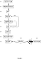
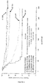
Classifications
-
- C—CHEMISTRY; METALLURGY
- C12—BIOCHEMISTRY; BEER; SPIRITS; WINE; VINEGAR; MICROBIOLOGY; ENZYMOLOGY; MUTATION OR GENETIC ENGINEERING
- C12Q—MEASURING OR TESTING PROCESSES INVOLVING ENZYMES, NUCLEIC ACIDS OR MICROORGANISMS; COMPOSITIONS OR TEST PAPERS THEREFOR; PROCESSES OF PREPARING SUCH COMPOSITIONS; CONDITION-RESPONSIVE CONTROL IN MICROBIOLOGICAL OR ENZYMOLOGICAL PROCESSES
- C12Q1/00—Measuring or testing processes involving enzymes, nucleic acids or microorganisms; Compositions therefor; Processes of preparing such compositions
- C12Q1/68—Measuring or testing processes involving enzymes, nucleic acids or microorganisms; Compositions therefor; Processes of preparing such compositions involving nucleic acids
- C12Q1/6876—Nucleic acid products used in the analysis of nucleic acids, e.g. primers or probes
- C12Q1/6883—Nucleic acid products used in the analysis of nucleic acids, e.g. primers or probes for diseases caused by alterations of genetic material
- C12Q1/6886—Nucleic acid products used in the analysis of nucleic acids, e.g. primers or probes for diseases caused by alterations of genetic material for cancer
-
- C—CHEMISTRY; METALLURGY
- C12—BIOCHEMISTRY; BEER; SPIRITS; WINE; VINEGAR; MICROBIOLOGY; ENZYMOLOGY; MUTATION OR GENETIC ENGINEERING
- C12Q—MEASURING OR TESTING PROCESSES INVOLVING ENZYMES, NUCLEIC ACIDS OR MICROORGANISMS; COMPOSITIONS OR TEST PAPERS THEREFOR; PROCESSES OF PREPARING SUCH COMPOSITIONS; CONDITION-RESPONSIVE CONTROL IN MICROBIOLOGICAL OR ENZYMOLOGICAL PROCESSES
- C12Q1/00—Measuring or testing processes involving enzymes, nucleic acids or microorganisms; Compositions therefor; Processes of preparing such compositions
- C12Q1/68—Measuring or testing processes involving enzymes, nucleic acids or microorganisms; Compositions therefor; Processes of preparing such compositions involving nucleic acids
- C12Q1/6844—Nucleic acid amplification reactions
- C12Q1/6851—Quantitative amplification
-
- C—CHEMISTRY; METALLURGY
- C12—BIOCHEMISTRY; BEER; SPIRITS; WINE; VINEGAR; MICROBIOLOGY; ENZYMOLOGY; MUTATION OR GENETIC ENGINEERING
- C12Q—MEASURING OR TESTING PROCESSES INVOLVING ENZYMES, NUCLEIC ACIDS OR MICROORGANISMS; COMPOSITIONS OR TEST PAPERS THEREFOR; PROCESSES OF PREPARING SUCH COMPOSITIONS; CONDITION-RESPONSIVE CONTROL IN MICROBIOLOGICAL OR ENZYMOLOGICAL PROCESSES
- C12Q1/00—Measuring or testing processes involving enzymes, nucleic acids or microorganisms; Compositions therefor; Processes of preparing such compositions
- C12Q1/68—Measuring or testing processes involving enzymes, nucleic acids or microorganisms; Compositions therefor; Processes of preparing such compositions involving nucleic acids
- C12Q1/6844—Nucleic acid amplification reactions
- C12Q1/6862—Ligase chain reaction [LCR]
-
- G—PHYSICS
- G01—MEASURING; TESTING
- G01N—INVESTIGATING OR ANALYSING MATERIALS BY DETERMINING THEIR CHEMICAL OR PHYSICAL PROPERTIES
- G01N33/00—Investigating or analysing materials by specific methods not covered by groups G01N1/00 - G01N31/00
- G01N33/48—Biological material, e.g. blood, urine; Haemocytometers
- G01N33/50—Chemical analysis of biological material, e.g. blood, urine; Testing involving biospecific ligand binding methods; Immunological testing
- G01N33/53—Immunoassay; Biospecific binding assay; Materials therefor
- G01N33/574—Immunoassay; Biospecific binding assay; Materials therefor for cancer
- G01N33/57407—Specifically defined cancers
- G01N33/57419—Specifically defined cancers of colon
-
- G—PHYSICS
- G01—MEASURING; TESTING
- G01N—INVESTIGATING OR ANALYSING MATERIALS BY DETERMINING THEIR CHEMICAL OR PHYSICAL PROPERTIES
- G01N33/00—Investigating or analysing materials by specific methods not covered by groups G01N1/00 - G01N31/00
- G01N33/48—Biological material, e.g. blood, urine; Haemocytometers
- G01N33/50—Chemical analysis of biological material, e.g. blood, urine; Testing involving biospecific ligand binding methods; Immunological testing
- G01N33/53—Immunoassay; Biospecific binding assay; Materials therefor
- G01N33/574—Immunoassay; Biospecific binding assay; Materials therefor for cancer
- G01N33/57484—Immunoassay; Biospecific binding assay; Materials therefor for cancer involving compounds serving as markers for tumor, cancer, neoplasia, e.g. cellular determinants, receptors, heat shock/stress proteins, A-protein, oligosaccharides, metabolites
-
- G—PHYSICS
- G16—INFORMATION AND COMMUNICATION TECHNOLOGY [ICT] SPECIALLY ADAPTED FOR SPECIFIC APPLICATION FIELDS
- G16H—HEALTHCARE INFORMATICS, i.e. INFORMATION AND COMMUNICATION TECHNOLOGY [ICT] SPECIALLY ADAPTED FOR THE HANDLING OR PROCESSING OF MEDICAL OR HEALTHCARE DATA
- G16H50/00—ICT specially adapted for medical diagnosis, medical simulation or medical data mining; ICT specially adapted for detecting, monitoring or modelling epidemics or pandemics
- G16H50/20—ICT specially adapted for medical diagnosis, medical simulation or medical data mining; ICT specially adapted for detecting, monitoring or modelling epidemics or pandemics for computer-aided diagnosis, e.g. based on medical expert systems
-
- G—PHYSICS
- G16—INFORMATION AND COMMUNICATION TECHNOLOGY [ICT] SPECIALLY ADAPTED FOR SPECIFIC APPLICATION FIELDS
- G16H—HEALTHCARE INFORMATICS, i.e. INFORMATION AND COMMUNICATION TECHNOLOGY [ICT] SPECIALLY ADAPTED FOR THE HANDLING OR PROCESSING OF MEDICAL OR HEALTHCARE DATA
- G16H50/00—ICT specially adapted for medical diagnosis, medical simulation or medical data mining; ICT specially adapted for detecting, monitoring or modelling epidemics or pandemics
- G16H50/30—ICT specially adapted for medical diagnosis, medical simulation or medical data mining; ICT specially adapted for detecting, monitoring or modelling epidemics or pandemics for calculating health indices; for individual health risk assessment
-
- C—CHEMISTRY; METALLURGY
- C12—BIOCHEMISTRY; BEER; SPIRITS; WINE; VINEGAR; MICROBIOLOGY; ENZYMOLOGY; MUTATION OR GENETIC ENGINEERING
- C12Q—MEASURING OR TESTING PROCESSES INVOLVING ENZYMES, NUCLEIC ACIDS OR MICROORGANISMS; COMPOSITIONS OR TEST PAPERS THEREFOR; PROCESSES OF PREPARING SUCH COMPOSITIONS; CONDITION-RESPONSIVE CONTROL IN MICROBIOLOGICAL OR ENZYMOLOGICAL PROCESSES
- C12Q2600/00—Oligonucleotides characterized by their use
- C12Q2600/118—Prognosis of disease development
-
- C—CHEMISTRY; METALLURGY
- C12—BIOCHEMISTRY; BEER; SPIRITS; WINE; VINEGAR; MICROBIOLOGY; ENZYMOLOGY; MUTATION OR GENETIC ENGINEERING
- C12Q—MEASURING OR TESTING PROCESSES INVOLVING ENZYMES, NUCLEIC ACIDS OR MICROORGANISMS; COMPOSITIONS OR TEST PAPERS THEREFOR; PROCESSES OF PREPARING SUCH COMPOSITIONS; CONDITION-RESPONSIVE CONTROL IN MICROBIOLOGICAL OR ENZYMOLOGICAL PROCESSES
- C12Q2600/00—Oligonucleotides characterized by their use
- C12Q2600/158—Expression markers
-
- G—PHYSICS
- G01—MEASURING; TESTING
- G01N—INVESTIGATING OR ANALYSING MATERIALS BY DETERMINING THEIR CHEMICAL OR PHYSICAL PROPERTIES
- G01N2800/00—Detection or diagnosis of diseases
- G01N2800/50—Determining the risk of developing a disease
-
- G—PHYSICS
- G01—MEASURING; TESTING
- G01N—INVESTIGATING OR ANALYSING MATERIALS BY DETERMINING THEIR CHEMICAL OR PHYSICAL PROPERTIES
- G01N2800/00—Detection or diagnosis of diseases
- G01N2800/52—Predicting or monitoring the response to treatment, e.g. for selection of therapy based on assay results in personalised medicine; Prognosis
-
- G—PHYSICS
- G01—MEASURING; TESTING
- G01N—INVESTIGATING OR ANALYSING MATERIALS BY DETERMINING THEIR CHEMICAL OR PHYSICAL PROPERTIES
- G01N2800/00—Detection or diagnosis of diseases
- G01N2800/60—Complex ways of combining multiple protein biomarkers for diagnosis
Definitions
- the present invention relates to methods for classifying patients suffering from a solid cancer, particularly to methods for the prognosis of the survival time of a patient suffering from a solid cancer and/or to methods for assessing the responsiveness of a patient suffering from a solid cancer to antitumoral treatment.
- evidence supports the notion to include immunological biomarkers, implemented as a tool for the prediction of prognosis and response to therapy.
- Accumulating data, collected from large cohorts of human cancers, has demonstrated the impact of immune-classification, which has a prognostic value that may add to the significance of the AJCC/UICC TNM-classification. It is therefore imperative to begin to incorporate the 'Immunoscore' into traditional classification, thus providing an essential prognostic and potentially predictive tool.
- Introduction of this parameter as a biomarker to classify cancers, as part of routine diagnostic and prognostic assessment of tumors, will facilitate clinical decision-making including rational stratification of patient treatment.
- WO2010/120814 discloses, inter alia , methods relying on a quartile statistical analysis for prognosing patients suffering from inflammatory bowel disease (IBD).
- IBD inflammatory bowel disease
- the present inventors managed to further increase the accuracy of the methods for the prognosis of the survival time of a patient suffering from a solid cancer and of the methods for assessing the responsiveness of a patient suffering from a solid cancer to antitumoral treatment.
- the present invention relates to an in vitro method for the prognosis of the survival time of a patient suffering from a solid cancer, which method comprises the following steps:
- the present invention also relates to an in vitro method for assessing the responsiveness of a patient suffering from a solid cancer to an antitumoral treatment, which method comprises the following steps:
- the weights wi may represent a measure for the reliability of the influence upon the mean by the respective values.
- the median value is particularly adapted when many (typically more than 5, 10, 15, 200) biological markers are quantified.
- the methods of the invention have unique and multiple advantages: Said methods perform very significantly and have a strong discriminatory power because the biomarkers considered in these methods have important specific characteristics.
- the biomarkers considered are immune biomarkers.
- the biomarkers are adaptive immune biomarkers.
- One of their essential characteristics is that these immune biomarkers are associated with the time of recurrence, with the time before death, with the time before the response to treatment, with the amplitude of response to treatment, and with the prolonged time during which the patient is responding to treatment.
- One of the advantages of the methods of the invention is that the level of intensity, expression, density, quantity of each biomarker is associated with the length, with the time of survival, with the prolonged survival, and the prolonged response to the treatment.
- the methods of the invention apply because the higher or the lower the levels of the biomarkers are, the longer or the shorter the survival is and the better or the worse the response to the treatment is.
- One of the essential novel elements associated with the great performance of the methods is that the methods are independent of the clinical data and follow-up of the patients.
- the mean Percentile can be calculated based on the raw and normalized data of the biomarker, independently of the outcome of the patients.
- a comprehensive adaptive immune reaction is characterized by a set of biomarkers, that when co-enriched, the biomarkers are improving the stratification of the patients into categories of prolonged survival and better response to treatment.
- the mean of the percentile values of all biomarkers, weighted for each biomarker, allow a very accurate stratification of the patients into risk categories.
- the reference group of patients suffering from said cancer comprises at least: 100, 200, 300, 400, 500, 1000 or 2000 patients.
- the methods of the invention apply to various organs of cancer origin (such as breast, colon, rectum, lung, head and neck, bladder, ovary, prostate), and also to various cancer cell types (adenocarcinoma, squamous cell carcinoma, large cell cancer, melanoma, etc).
- organs of cancer origin such as breast, colon, rectum, lung, head and neck, bladder, ovary, prostate
- cancer cell types adenocarcinoma, squamous cell carcinoma, large cell cancer, melanoma, etc.
- the patient subjected to the above method may suffer from a solid cancer selected from the group consisting adrenal cortical cancer, anal cancer, bile duct cancer (e.g. periphilar cancer, distal bile duct cancer, intrahepatic bile duct cancer), bladder cancer, bone cancer (e.g. osteoblastoma, osteochrondroma, hemangioma, chondromyxoid fibroma, osteosarcoma, chondrosarcoma, fibrosarcoma, malignant fibrous histiocytoma, giant cell tumor of the bone, chordoma, multiple myeloma), brain and central nervous system cancer (e.g.
- a solid cancer selected from the group consisting adrenal cortical cancer, anal cancer, bile duct cancer (e.g. periphilar cancer, distal bile duct cancer, intrahepatic bile duct cancer), bladder cancer, bone cancer (e.g. osteoblastoma, osteochrondrom
- breast cancer e.g. ductal carcinoma in situ, infiltrating ductal carcinoma, infiltrating lobular carcinoma, lobular carcinoma in situ, gynecomastia
- cervical cancer colorectal cancer
- endometrial cancer e.g.
- small cell lung cancer non-small cell lung cancer
- mesothelioma plasmacytoma, nasal cavity and paranasal sinus cancer (e.g. esthesioneuroblastoma, midline granuloma), nasopharyngeal cancer, neuroblastoma, oral cavity and oropharyngeal cancer, ovarian cancer, pancreatic cancer, penile cancer, pituitary cancer, prostate cancer, retinoblastoma, rhabdomyosarcoma (e.g. embryonal rhabdomyosarcoma, alveolar rhabdomyosarcoma, pleomorphic rhabdomyosarcoma), salivary gland cancer, skin cancer (e.g.
- melanoma nonmelanoma skin cancer
- stomach cancer testicular cancer (e.g. seminoma, nonseminoma germ cell cancer), thymus cancer, thyroid cancer (e.g. follicular carcinoma, anaplastic carcinoma, poorly differentiated carcinoma, medullary thyroid carcinoma), vaginal cancer, vulvar cancer, and uterine cancer (e.g. uterine leiomyosarcoma).
- testicular cancer e.g. seminoma, nonseminoma germ cell cancer
- thymus cancer thyroid cancer (e.g. follicular carcinoma, anaplastic carcinoma, poorly differentiated carcinoma, medullary thyroid carcinoma), vaginal cancer, vulvar cancer, and uterine cancer (e.g. uterine leiomyosarcoma).
- testicular cancer e.g. seminoma, nonseminoma germ cell cancer
- thymus cancer e.g. follicular carcinoma, anaplastic carcinoma, poorly differentiated carcinoma
- the cancer is colorectal cancer.
- tumor tissue sample means any tissue tumor sample derived from the patient. Said tissue sample is obtained for the purpose of the in vitro evaluation.
- the tumor sample may result from the tumor resected from the patient.
- the tumor sample may result from a biopsy performed in the primary tumour of the patient or performed in metastatic sample distant from the primary tumor of the patient. For example an endoscopical biopsy performed in the bowel of the patient affected by a colorectal cancer.
- the tumor tissue sample is fixed in formalin and embedded in a rigid fixative, such as paraffin (wax) or epoxy, which is placed in a mould and later hardened to produce a block which is readily cut.
- TMA tissue microarrays
- TMA technology is described in WO2004000992 , US8068988 , Olli et al 2001 Human Molecular Genetics, Tzankov et al 2005, Elsevier ; Kononen et al 1198; Nature Medicine .
- the tumor tissue sample encompasses (i) a global primary tumor (as a whole), (ii) a tissue sample from the center of the tumor, (iii) a tissue sample from the tissue directly surrounding the tumor which tissue may be more specifically named the "invasive margin" of the tumor, (iv) lymphoid islets in close proximity with the tumor, (v) the lymph nodes located at the closest proximity of the tumor, (vi) a tumor tissue sample collected prior surgery (for follow-up of patients after treatment for example), and (vii) a distant metastasis.
- the "invasive margin” has its general meaning in the art and refers to the cellular environment surrounding the tumor.
- the tumor tissue sample irrespective of whether it is derived from the center of the tumor, from the invasive margin of the tumor, or from the closest lymph nodes, encompasses pieces or slices of tissue that have been removed from the tumor center of from the invasive margin surrounding the tumor, including following a surgical tumor resection or following the collection of a tissue sample for biopsy, for further quantification of one or several biological markers, notably through histology or immunohistochemistry methods, through flow cytometry methods and through methods of gene or protein expression analysis, including genomic and proteomic analysis.
- the tumor tissue sample can, of course, be subjected to a variety of well-known post-collection preparative and storage techniques (e.g., fixation, storage, freezing, etc.).
- the sample can be fresh, frozen, fixed (e.g., formalin fixed), or embedded (e.g., paraffin embedded).
- the quantification of the number of tumor-draining lymph nodes is performed in the ressected tumor, the tumor tissue sample results from said ressected tumor and encompasses the center of the tumor, and optionally the invasive margin of the tumor.
- the quantification of the marker of the immune adaptive response is typically performed by immunohistochemistry (IHC) a described after.
- the quantification of the marker of the immune adaptive response is typically performed by determining the expression level of at least one gene.
- the tumor sample may be selected from the group consisting of (i) a global primary tumor (as a whole), (ii) a tissue sample from the center of the tumor, (iii) a tissue sample from the tissue directly surrounding the tumor which tissue may be more specifically named the "invasive margin" of the tumor, (iv) the lymph nodes located at the closest proximity of the tumor or a tertiary lymphoid structure induced by the tumor, (v) a tumor biopsie performed at any time and typically prior surgery, and (vi) a distant metastasis.
- the two or more biological markers are quantified in the center of the tumor and/or in the invasive margin of the tumor. In a preferred embodiment the two or more biological markers are quantified in the center of the tumor and/or in the invasive margin of the tumor.
- the sample can be fresh, frozen, fixed (e.g., formalin fixed), or embedded (e.g., paraffin embedded).
- the tumour sample results from biopsy performed in a tumour of the patient.
- An example is an endoscopical biopsy performed in the bowel of the patient suffering from colorectal cancer or suspected to suffer from colorectal cancer.
- a "biological marker” consists of any detectable, measurable and quantifiable parameter that is indicative of the status of the immune response of the cancer patient against the tumor.
- Biological markers include the presence of, or the number or density of, cells from the immune system at the tumor site.
- Bio markers also include the presence of, or the amount of proteins specifically produced by cells from the immune system at the tumor site.
- Bio markers also include the presence of, or the amount of, any biological material that is indicative of the expression level of genes related to the raising of a specific immune response of the host, at the tumor site.
- biological markers include the presence of, or the amount of, messenger RNA (mRNA) transcribed from genomic DNA encoding proteins which are specifically produced by cells from the immune system, at the tumor site.
- mRNA messenger RNA
- Bio markers thus include surface antigens that are specifically expressed by cells from the immune system, including by B lymphocytes, T lymphocytes, monocytes/macrophages dendritic cells, NK cells, NKT cells, and NK-DC cells., that are recruited within the tumor tissue or at its close proximity, including within the invasive margin of the tumor and in the closest lymph nodes, or alternatively mRNA encoding for said surface antigens.
- surface antigens of interest used as biological markers include CD3, CD4, CD8 and CD45RO that are expressed by T cells or T cell subsets.
- the quantification of this biological marker is indicative of the level of the adaptive immune response of the patient involving all T lymphocytes and NKT cells.
- the quantification of this biological marker is indicative of the level of the adaptive immune response of the patient involving cytotoxic T lymphocytes.
- the quantification of this biological marker is indicative of the level of the adaptive immune response of the patient involving memory T lymphocytes or memory effector T lymphocytes.
- proteins used as biological markers also include cytolytic proteins specifically produced by cells from the immune system, like perforin, granulysin and also granzyme-B.
- WO2015007625 WO2014023706
- WO2014009535 WO2013186374
- WO2013107907 WO2013107900
- WO2012095448 WO2012072750
- WO2007045996 WO2007045996
- a combination of 2, 3, 4, 5, 6, 7, 8, 9, 10, 11, 12, 13, 14, 15, 16, 17, 18, 19, 20, 21, 22, 23, 24, 25, 26, 27, 28, 29, 30, 31, 32, 33, 34, 35, 36, 37, 38, 39, 40, 41, 42, 43, 44, 45, 46, 47, 48, 49 and 50 distinct biological markers may be quantified, preferably a combination of 2, 3, 4, 5, 6, 7, 8, 9, or 10 biological markers and more preferably a combination of 2, 3, 4, 5, or 6, biological markers.
- the biological markers indicative of the status of the immune response are those described in WO2007045996 .
- the biological markers which may be used are the cell density of cells from the immune system.
- the two or more biological markers comprise the density of CD3+ cells, the density of CD8+ cells, the density of CD45RO+ cells, the density of GZM-B+ cells and/or the density of B cells.
- the two or more biological markers comprise the density of CD3+ cells and the density of CD8+ cells, the density of CD3+ cells and the density of CD45RO+ cells, the density of CD3+ cells the density of GZM-B+ cells, the density of CD8+ cells and the density of CD45RO+ cells, the density of CD8+ cells and the density of GZM-B+ cells; or the density of CD45RO+ cells and the density of GZM-B+ cells.
- the density of B-cells may also be measured (see WO2013107900 and WO2013107907 ).
- the density of DC cells may also be measured (see WO2013107907 ).
- the density of cells from the immune system are quantified in the center of the tumor and/or in the invasive margin of the tumor.
- the two or more biological markers comprise the density of CD3+ cells in center of the tumor, the density of CD8+ cells in the center of the tumor, the density of CD3+ cells in the invasive margin, and the density of CD8+ cells in the invasive margin.
- the density may be measured in the "cold spot", i.e., in the regions of the tumor sample where the density is the lowest, or in the 2, 3, 4, 5, 6, 7, 8, 9, 10 "cold spots", corresponding to the 2 to 10 area with the lowest densities.
- the density may also be measured in the "hot spot”, i.e., in the regions where the density is the highest, or in the 2, 3, 4, 5, 6, 7, 8, 9, 10 "hot spots", corresponding to the 2 to 10 area with the highest densities.
- WO2013186374 may be used for quantifying the immune cells in the tumor sample.
- the term "marker” consists of any detectable, measurable or quantifiable parameter that is indicative of the status of the adaptive immune response of the subject.
- a marker becomes a "biological marker” for the purpose of carrying the method of the present invention when a good statistical correlation is found between (i) an increase or a decrease of the quantification value for said marker and (ii) the survival time actually observed within patients.
- any one of the statistical method known by the one skilled in the art may be used.
- any marker for which a P value of less than 0.05, and even preferably less than 10 -3 , 10 -4 , 10 -5 , 10 -6 or 10 -7 (according to univariate and multivariate analysis (for example, log-rank test and Cox test, respectively) is determined consists of a "biological marker" useable in the cancer prognosis method of the invention.
- the marker includes the presence of, or the number or density of, cells from the immune system.
- the marker includes the presence of, or the amount of proteins specifically produced by cells from the immune system. In some embodiments, the marker includes the presence of, or the amount of, any biological material that is indicative of the level of genes related to the raising of a specific immune response of the host. Thus, in some embodiments, the marker includes the presence of, or the amount of, messenger RNA (mRNA) transcribed from genomic DNA encoding proteins which are specifically produced by cells from the immune system.
- mRNA messenger RNA
- the marker includes surface antigens that are specifically expressed by cells from the immune system, including by B lymphocytes, T lymphocytes, monocytes/macrophages dendritic cells, NK cells, NKT cells, and NK-DC cells or alternatively mRNA encoding for said surface antigens.
- the number of distinct biological markers that are quantified at step a) are usually of less than 100 distinct markers, and in most embodiments of less than 50 distinct markers.
- the number of distinct biological markers that is necessary for obtaining an accurate and reliable prognosis, using the method of the present invention may vary notably according to the type of technique for quantification.
- high statistical significance can be found with a combination of a small number of biological markers, when the method of the present invention is performed by in situ immunohistochemical detection of protein markers of interest, in particular when separate quantification of the said markers are performed both in the center of the tumor (CT) and in the invasive margin (IM).
- CT center of the tumor
- IM invasive margin
- high statistical significance was obtained with only one marker or a combination of two markers, as disclosed in the EXAMPLE.
- high statistical significance was also found with a small number of biological markers, when the method of the present invention is performed by gene expression analysis of gene markers of interest.
- method of the present invention is performed by using a gene expression analysis for biological marker quantification, and by using a combination of ten distinct biological markers, and more preferably a combination of fifteen distinct biological markers, most preferably twenty distinct biological markers, or more.
- the level of 1; 2; 3; 4; 5; 6; 7; 8; 9; 10; 11; 12; 13; 14; 15; 16; 17; 18; 19; 20; 21; 22; 23; 24; 25; 26; 27; 28; 29; 30; 31; 32; 33; 34; 35; 36; 37; 38; 39; 40; 41; 42; 43; 44; 45; 46; 47; 48; 49; or 50 markers is (are) determined.
- the name of each of the various biological markers of interest refers to the internationally recognised name of the corresponding gene, as found in internationally recognised gene sequences and protein sequences databases, including in the database from the HUGO Gene Nomenclature Committee.
- the name of each of the various biological markers of interest may also refer to the internationally recognised name of the corresponding gene, as found in the internationally recognised gene sequences and protein sequences database Genbank.
- the nucleic acid and the amino acid sequences corresponding to each of the biological marker of interest described herein may be retrieved by the one skilled in the art.
- the biological markers indicative of the status of the immune response may comprise the expression level of one or more genes or corresponding proteins listed in Table 9 of WO2007045996 which are: 18s, ACE, ACTB, AGTR1, AGTR2, APC, APOA1, ARF1, AXIN1, BAX, BCL2, BCL2L1, CXCR5, BMP2, BRCA1, BTLA, C3, CASP3, CASP9, CCL1, CCL11, CCL13, CCL16, CCL17, CCL18, CCL19, CCL2, CCL20, CCL21, CCL22, CCL23, CCL24, CCL25, CCL26, CCL27, CCL28, CCL3, CCL5, CCL7, CCL8, CCNB1, CCND1, CCNE1, CCR1, CCR10, CCR2, CCR3, CCR4, CCR5, CCR6, CCR7, CCR8, CCR9, CCRL2, CD154, CD19, CD1a, CD2, CD226, CD244, PDCD1LG
- the name of each of the genes of interest refers to the internationally recognised name of the corresponding gene, as found in internationally recognised gene sequences and protein sequences databases, including the database from the HUGO Gene Nomenclature Committee.
- the name of each of the genes of interest may also refer to the internationally recognised name of the corresponding gene, as found in the internationally recognised gene sequences database Genbank.
- the nucleic acid to each of the gene of interest described herein may be retrieved by one skilled in the art.
- the biological markers indicative of the status of the immune response are those described in WO2014023706 :
- genes than one or two pairs of genes are more difficult to implement and more expensive and time consuming but however provides other advantages. For example if the assessment of the expression level of one gene is erroneous, the overall result is compensated by the reserved of the other genes of the same kind (human adaptive immune response or immunosuppressive response).
- the expression “gene representative of the adaptive immune response” refers to any gene that is expressed by a cell that is an actor of the adaptive immune response in the tumor or that contributes to the settlement of the adaptive immune response in the tumor.
- the adaptive immune response also called “acquired immune response”
- cells of the adaptive immune response include but are not limited to cytotoxic T cells, T memory T cells, Th1 and Th2 cells, activated macrophages and activated dendritic cells, NK cells and NKT cells.
- a gene representative of the adaptive immune response may be typically selected from the cluster of the co-modulated genes for the Th1 adaptive immunity, for the cytotoxic response, or for the memory response, and may encode for a Th1 cell surface marker, an interleukin (or an interleukin receptor), or a chemokine or (a chemokine receptor).
- the gene representative of the adaptive immune response is selected from the group consisting of
- the expression "gene representative of the immunosuppressive response” refers to any gene that is expressed by a cell that is an actor of the immunosuppressive response in the tumor or that contributes to the settlement of the immunosuppressive response in the tumor.
- the immunosuppressive response comprises
- cells of the immunosuppressive response include immature dendritic cells (CD1A), regulatory T cells (Treg cells) and Th17 cells expressing IL17A gene.
- a gene representative of the adaptive immune response may be typically selected from the group of the co-modulated adaptive immune genes, whereas the immunosuppressive genes, may be representative of the inactivation of immune cells (e.g. dendritic cells) and may contribute to induction of an immunosuppressive response.
- the immunosuppressive genes may be representative of the inactivation of immune cells (e.g. dendritic cells) and may contribute to induction of an immunosuppressive response.
- the gene or corresponding proteins representative of the immunosuppressive response is selected from the group consisting of genes reported in Table 2 hereunder: Table 2 CD274 CTLA4 IHH IL17A PDCD1 PF4 PROM1 REN TIM-3 TSLP VEGFA
- Said genes are preferred because they provide the best results for the response of a patient to the treatment.
- a gene representative of the adaptive immune response is selected from the group consisting of GNLY, CXCL13, CX3CL1, CXCL9, ITGAE, CCL5, GZMH, IFNG, CCR2, CD3D, CD3E, CD3G, CD8A, CXCL10, CXCL11, GZMA, GZMB, GZMK, GZMM, IL15, IRF1, LTK, PRF1, STAT1, CD69, CD247, ICOS, CXCR3, STAT4, CCL2 and TBX21 and a gene representative of the immunosuppressive response is selected from the group consisting of PF4, REN, VEGFA, TSLP, IL17A, PROM1, IHH, CD1A, CTLA4, PDCD1, CD276, CD274, TIM-3 and VTCN1 (B7H4).
- a gene representative of the adaptive immune response and a gene representative of the immunosuppressive response are selected respectively from the groups consisting of the genes of Tables 1 and 2 above. Preferred combinations of two pairs of genes (total of 4 genes) are
- genes selected for use in the present process may depend on the type of treatment contemplated for the patient.
- genes selected from the group consisting of CX3CL1 IL15, CD247, CD3G, CD8A, PRF1, CCL5 and TBX21 for the immunosuppressive response preferably CX3CL1 and IL15 and gene CTLA4 for the adaptive immune response will be preferred when a treatment using a drug such as monoclonal antibody working by activating the immune system such as Ipilimumab, also known as MDX-010 or MDX-101, marketed as Yervoy®, is contemplated for a patient.
- Genes selected from the group consisting of IL15 and GZMA for the adaptive immune response, and gene VEGFA for the immunosuppressive response will be preferred when a treatment such as an antibody that inhibits vascular endothelial growth factor A (VEGF-A) such as bevacizumab marketed as Avastin®, is contemplated for a patient.
- VEGF-A vascular endothelial growth factor A
- GZMA - PDCD1 also designated as CD279
- a treatment such as an antibody that targets PD-1 such as BMS-936558, is contemplated for a patient.
- the biological markers indicative of the status of the immune response are those described in WO2014009535 :
- the biological markers indicative of the status of the immune response may comprise the expression level of one or more genes from the group consisting of CCR2, CD3D, CD3E, CD3G, CD8A, CXCL10, CXCL11, GZMA, GZMB, GZMK, GZMM, IL15, IRF1, PRF1, STAT1, CD69, ICOS, CXCR3, STAT4, CCL2, and TBX21.
- the biological markers indicative of the status of the immune response are those described in WO2012095448 :
- the biological markers indicative of the status of the immune response may comprise the expression level of one or more genes from the group consisting of GZMH, IFNG, CXCL13, GNLY, LAG3, ITGAE, CCL5, CXCL9, PF4, IL17A, TSLP, REN, IHH, PROM1 and VEGFA.
- the biological markers indicative of the status of the immune response are those described in WO2012072750 :
- the biological markers indicative of the status of the immune response may comprise the expression level of a miRNA cluster comprising: miR.609, miR.518c, miR.520f, miR.220a, miR.362, miR.29a, miR.660, miR.603, miR.558, miR519b, miR.494, miR.130a, or miR.639.
- any one of the methods known by the one skilled in the art for quantifying cellular types, a protein-type or an nucleic acid-type biological marker encompassed herein may be used for performing the cancer prognosis method of the invention.
- any one of the standard and non-standard (emerging) techniques well known in the art for detecting and quantifying a protein or a nucleic acid in a sample can readily be applied.
- Expression of a biological marker used in the invention may be assessed by any of a wide variety of well known methods for detecting expression of a transcribed nucleic acid or protein.
- Non-limiting examples of such methods include immunological methods for detection of secreted, cell-surface, cytoplasmic, or nuclear proteins, protein purification methods, protein function or activity assays, nucleic acid hybridization methods, nucleic acid reverse transcription methods, and nucleic acid amplification methods.
- expression of a marker is assessed using an antibody (e.g. a radio-labeled, chromophore-labeled, fluorophore-labeled, polymer-backbone-antibody, or enzyme-labeled antibody), an antibody derivative (e.g. an antibody conjugated with a substrate or with the protein or ligand of a protein-ligand pair ⁇ e.g. biotin-streptavidin ⁇ ), or an antibody fragment (e.g. a single-chain antibody, an isolated antibody hypervariable domain, etc.) which binds specifically with a marker protein or fragment thereof, including a marker protein which has undergone all or a portion of its normal post-translational modification.
- an antibody e.g. a radio-labeled, chromophore-labeled, fluorophore-labeled, polymer-backbone-antibody, or enzyme-labeled antibody
- an antibody derivative e.g. an antibody conjugated with a substrate or with the protein or
- a biological marker may be quantified with any one of the immunohistochemistry methods known in the art.
- one thin section of the tumor is firstly incubated with labeled antibodies directed against one biological marker of interest. After washing, the labeled antibodies that are bound to said biological marker of interest are revealed by the appropriate technique, depending of the kind of label is borne by the labeled antibody, e.g. radioactive, fluorescent or enzyme label. Multiple labelling can be performed simultaneously.
- Immunohistochemistry typically includes the following steps i) fixing the tumor tissue sample with formalin, ii) embedding said tumor tissue sample in paraffin, iii) cutting said tumor tissue sample into sections for staining, iv) incubating said sections with the binding partner specific for the immune checkpoint protein of interest, v) rinsing said sections, vi) incubating said section with a secondary antibody typically biotinylated and vii) revealing the antigen-antibody complex typically with avidin-biotin-peroxidase complex. Accordingly, the tumor tissue sample is firstly incubated with the binding partners having for the immune checkpoint protein of interest.
- the labeled antibodies that are bound to the immune checkpoint protein of interest are revealed by the appropriate technique, depending of the kind of label is borne by the labeled antibody, e.g. radioactive, fluorescent or enzyme label. Multiple labelling can be performed simultaneously.
- the method of the present invention may use a secondary antibody coupled to an amplification system (to intensify staining signal) and enzymatic molecules.
- Such coupled secondary antibodies are commercially available, e.g. from Dako, EnVision system.
- Counterstaining may be used, e.g. Hematoxylin & Eosin, DAPI, Hoechst.
- Other staining methods may be accomplished using any suitable method or system as would be apparent to one of skill in the art, including automated, semi-automated or manual systems.
- one or more labels can be attached to the antibody, thereby permitting detection of the target protein (i.e. the biological markers).
- exemplary labels include radioactive isotopes, fluorophores, ligands, chemiluminescent agents, enzymes, and combinations thereof.
- Non-limiting examples of labels that can be conjugated to primary and/or secondary affinity ligands include fluorescent dyes or metals (e.g. fluorescein, rhodamine, phycoerythrin, fluorescamine), chromophoric dyes (e.g. rhodopsin), chemiluminescent compounds (e.g. luminal, imidazole) and bioluminescent proteins (e.g.
- luciferin e.g. luciferin, luciferase
- haptens e.g. biotin
- a variety of other useful fluorescers and chromophores are described in Stryer L (1968) Science 162:526-533 and Brand L and Gohlke J R (1972) Annu. Rev. Biochem. 41:843-868 .
- Affinity ligands can also be labeled with enzymes (e.g. horseradish peroxidase, alkaline phosphatase, beta-lactamase), radioisotopes (e.g. 3 H, 14 C, 32 P, 35 S or 125 I) and particles (e.g. gold).
- the different types of labels can be conjugated to an affinity ligand using various chemistries, e.g. the amine reaction or the thiol reaction. However, other reactive groups than amines and thiols can be used, e.g. aldehydes, carboxylic acids and glutamine.
- Various enzymatic staining methods are known in the art for detecting a protein of interest. For example, enzymatic interactions can be visualized using different enzymes such as peroxidase, alkaline phosphatase, or different chromogens such as DAB, AEC or Fast Red.
- the label is a quantum dot.
- Qdots Quantum dots
- Qdots are becoming increasingly useful in a growing list of applications including immunohistochemistry, flow cytometry, and plate-based assays, and may therefore be used in conjunction with this invention.
- Qdot nanocrystals have unique optical properties including an extremely bright signal for sensitivity and quantitation; high photostability for imaging and analysis. A single excitation source is needed, and a growing range of conjugates makes them useful in a wide range of cell-based applications.
- Qdot Bioconjugates are characterized by quantum yields comparable to the brightest traditional dyes available. Additionally, these quantum dot-based fluorophores absorb 10-1000 times more light than traditional dyes.
- the emission from the underlying Qdot quantum dots is narrow and symmetric which means overlap with other colors is minimized, resulting in minimal bleed through into adjacent detection channels and attenuated crosstalk, in spite of the fact that many more colors can be used simultaneously.
- the antibody can be conjugated to peptides or proteins that can be detected via a labeled binding partner or antibody.
- a secondary antibody or second binding partner is necessary to detect the binding of the first binding partner, as it is not labeled.
- the resulting stained specimens are each imaged using a system for viewing the detectable signal and acquiring an image, such as a digital image of the staining.
- Methods for image acquisition are well known to one of skill in the art.
- any optical or non-optical imaging device can be used to detect the stain or biomarker label, such as, for example, upright or inverted optical microscopes, scanning confocal microscopes, cameras, scanning or tunneling electron microscopes, canning probe microscopes and imaging infrared detectors.
- the image can be captured digitally.
- the obtained images can then be used for quantitatively or semi-quantitatively determining the amount of the immune checkpoint protein in the sample, or the absolute number of cells positive for the maker of interest, or the surface of cells positive for the maker of interest.
- Various automated sample processing, scanning and analysis systems suitable for use with IHC are available in the art. Such systems can include automated staining and microscopic scanning, computerized image analysis, serial section comparison (to control for variation in the orientation and size of a sample), digital report generation, and archiving and tracking of samples (such as slides on which tissue sections are placed).
- Cellular imaging systems are commercially available that combine conventional light microscopes with digital image processing systems to perform quantitative analysis on cells and tissues, including immunostained samples.
- detection can be made manually or by image processing techniques involving computer processors and software.
- the images can be configured, calibrated, standardized and/or validated based on factors including, for example, stain quality or stain intensity, using procedures known to one of skill in the art (see e.g., published U.S. Patent Publication No. US20100136549 ).
- the image can be quantitatively or semi-quantitatively analyzed and scored based on staining intensity of the sample.
- Quantitative or semi-quantitative histochemistry refers to method of scanning and scoring samples that have undergone histochemistry, to identify and quantify the presence of the specified biomarker (i.e. immune checkpoint protein).
- Quantitative or semi-quantitative methods can employ imaging software to detect staining densities or amount of staining or methods of detecting staining by the human eye, where a trained operator ranks results numerically. For example, images can be quantitatively analyzed using a pixel count algorithms and tissue recognition pattern (e.g.
- a ratio of strong positive stain (such as brown stain) to the sum of total stained area can be calculated and scored.
- the amount of the detected biomarker i.e. the immune checkpoint protein
- the amount is quantified and given as a percentage of positive pixels and/or a score. For example, the amount can be quantified as a percentage of positive pixels. In some examples, the amount is quantified as the percentage of area stained, e.g., the percentage of positive pixels.
- a sample can have at least or about at least or about 0, 1%, 2%, 3%, 4%, 5%, 6%, 7%, 8%, 9%, 10%, 11%, 12%, 13%, 14%, 15%, 16%, 17%, 18%, 19%, 20%, 21%, 22%, 23%, 24%, 25%, 26%, 27%, 28%, 29%, 30%, 31%, 32%, 33%, 34%, 35%, 40%, 45%, 50%, 55%, 60%, 65%, 70%, 75%, 80%, 85%, 90%, 95% or more positive pixels as compared to the total staining area.
- the amount can be quantified as an absolute number of cells positive for the maker of interest.
- a score is given to the sample that is a numerical representation of the intensity or amount of the histochemical staining of the sample, and represents the amount of target biomarker (e.g., the immune checkpoint protein) present in the sample.
- target biomarker e.g., the immune checkpoint protein
- Optical density or percentage area values can be given a scaled score, for example on an integer scale.
- the method of the present invention comprises the steps consisting in i) providing one or more immunostained slices of tissue section obtained by an automated slide-staining system by using a binding partner capable of selectively interacting with the biological marker, ii) proceeding to digitalisation of the slides of step i).by high resolution scan capture, iii) detecting the slice of tissue section on the digital picture iv) providing a size reference grid with uniformly distributed units having a same surface, said grid being adapted to the size of the tissue section to be analysed, and v) detecting, quantifying and measuring intensity or the absolute number of stained cells in each unit.
- Multiplex tissue analysis techniques are particularly useful for quantifying several immune checkpoint proteins in the tumor tissue sample. Such techniques should permit at least five, or at least ten or more biomarkers to be measured from a single tumor tissue sample. Furthermore, it is advantageous for the technique to preserve the localization of the biomarker and be capable of distinguishing the presence of biomarkers in cancerous and non-cancerous cells. Such methods include layered immunohistochemistry (L-IHC), layered expression scanning (LES) or multiplex tissue immunoblotting (MTI) taught, for example, in U.S. Pat. Nos. 6,602,661 , 6,969,615 , 7,214,477 and 7,838,222 ; U.S. Publ. No.
- each reference teaches making up to 8, up to 9, up to 10, up to 11 or more images of a tissue section on layered and blotted membranes, papers, filters and the like, can be used.
- Coated membranes useful for conducting the L-IHC/MTI process are available from 20/20 GeneSystems, Inc. (Rockville, MD).
- the L-IHC method can be performed on any of a variety of tissue samples, whether fresh or preserved.
- the samples included core needle biopsies that were routinely fixed in 10% normal buffered formalin and processed in the pathology department. Standard five ⁇ m thick tissue sections were cut from the tissue blocks onto charged slides that were used for L-IHC.
- L-IHC enables testing of multiple markers in a tissue section by obtaining copies of molecules transferred from the tissue section to plural bioaffinity- coated membranes to essentially produce copies of tissue "images."
- the tissue section is deparaffinized as known in the art, for example, exposing the section to xylene or a xylene substitute such as NEO-CLEAR®, and graded ethanol solutions.
- the section can be treated with a proteinase, such as, papain, trypsin, proteinase K and the like.
- a stack of a membrane substrate comprising, for example, plural sheets of a 10 ⁇ m thick coated polymer backbone with 0.4 ⁇ m diameter pores to channel tissue molecules, such as, proteins, through the stack, then is placed on the tissue section.
- tissue molecules such as, proteins
- the movement of fluid and tissue molecules is configured to be essentially perpendicular to the membrane surface.
- the sandwich of the section, membranes, spacer papers, absorbent papers, weight and so on can be exposed to heat to facilitate movement of molecules from the tissue into the membrane stack.
- a portion of the proteins of the tissue are captured on each of the bioaffinity-coated membranes of the stack (available from 20/20 GeneSystems, Inc., Rockville, MD).
- each membrane comprises a copy of the tissue and can be probed for a different biomarker using standard immunoblotting techniques, which enables open-ended expansion of a marker profile as performed on a single tissue section.
- the amount of protein can be lower on membranes more distal in the stack from the tissue, which can arise, for example, on different amounts of molecules in the tissue sample, different mobility of molecules released from the tissue sample, different binding affinity of the molecules to the membranes, length of transfer and so on, normalization of values, running controls, assessing transferred levels of tissue molecules and the like can be included in the procedure to correct for changes that occur within, between and among membranes and to enable a direct comparison of information within, between and among membranes.
- total protein can be determined per membrane using, for example, any means for quantifying protein, such as, biotinylating available molecules, such as, proteins, using a standard reagent and method, and then revealing the bound biotin by exposing the membrane to a labeled avidin or streptavidin; a protein stain, such as, Blot fastStain, Ponceau Red, brilliant blue stains and so on, as known in the art.
- biotinylating available molecules such as, proteins
- the present methods utilize Multiplex Tissue Imprinting (MTI) technology for measuring biomarkers, wherein the method conserves precious biopsy tissue by allowing multiple biomarkers, in some cases at least six biomarkers.
- MMI Multiplex Tissue Imprinting
- alternative multiplex tissue analysis systems exist that may also be employed as part of the present invention.
- One such technique is the mass spectrometry-based Selected Reaction Monitoring (SRM) assay system ("Liquid Tissue” available from OncoPlexDx (Rockville, MD)). That technique is described in U.S. Pat. No. 7,473,532 .
- SRM Selected Reaction Monitoring
- the method of the present invention utilized the multiplex IHC technique developed by GE Global Research (Niskayuna, NY). That technique is described in U.S. Pub. Nos. 2008/0118916 and 2008/0118934 . There, sequential analysis is performed on biological samples containing multiple targets including the steps of binding a fluorescent probe to the sample followed by signal detection, then inactivation of the probe followed by binding probe to another target, detection and inactivation, and continuing this process until all targets have been detected.
- multiplex tissue imaging can be performed when using fluorescence (e.g. fluorophore or Quantum dots) where the signal can be measured with a multispectral imagine system.
- Multispectral imaging is a technique in which spectroscopic information at each pixel of an image is gathered and the resulting data analysed with spectral image -processing software.
- the system can take a series of images at different wavelengths that are electronically and continuously selectable and then utilized with an analysis program designed for handling such data. The system can thus be able to obtain quantitative information from multiple dyes simultaneously, even when the spectra of the dyes are highly overlapping or when they are co-localized, or occurring at the same point in the sample, provided that the spectral curves are different.
- Multispectral imaging can unmix, or separate out, autofluorescence from tissue and, thereby, increase the achievable signal-to-noise ratio.
- the quantification can be performed by following steps: i) providing a tumor tissue microarray (TMA) obtained from the patient, ii) TMA samples are then stained with anti-antibodies having specificity of the immune checkpoint protein(s) of interest, iii) the TMA slide is further stained with an epithelial cell marker to assist in automated segmentation of tumour and stroma, iv) the TMA slide is then scanned using a multispectral imaging system, v) the scanned images are processed using an automated image analysis software (e.g.Perkin Elmer Technology) which allows the detection, quantification and segmentation of specific tissues through powerful pattern recognition algorithms.
- the machine-learning algorithm was typically previously trained to segment tumor from stroma and identify cells labelled.
- Determining an expression level of a gene in a tumor sample obtained from a patient can be implemented by a panel of techniques well known in the art.
- an expression level of a gene is assessed by determining the quantity of mRNA produced by this gene.
- nucleic acid contained in the samples e.g., cell or tissue prepared from the patient
- the thus extracted mRNA is then detected by hybridization (e. g., Northern blot analysis) and/or amplification (e.g., RT-PCR).
- hybridization e. g., Northern blot analysis
- amplification e.g., RT-PCR
- quantitative or semi-quantitative RT-PCR is preferred.
- Real-time quantitative or semi-quantitative RT-PCR is particularly advantageous.
- LCR ligase chain reaction
- TMA transcription-mediated amplification
- SDA strand displacement amplification
- NASBA nucleic acid sequence based amplification
- NGS quantitative new generation sequencing of RNA
- Nucleic acids comprising at least 10 nucleotides and exhibiting sequence complementarity or homology to the mRNA of interest herein find utility as hybridization probes or amplification primers. It is understood that such nucleic acids need not be completely identical, but are typically at least about 80% identical to the homologous region of comparable size, more preferably 85% identical and even more preferably 90-95% identical. In certain embodiments, it will be advantageous to use nucleic acids in combination with appropriate means, such as a detectable label, for detecting hybridization. A wide variety of appropriate indicators are known in the art including, fluorescent, radioactive, enzymatic or other ligands (e. g. avidin/biotin).
- Probes typically comprise single-stranded nucleic acids of between 10 to 1000 nucleotides in length, for instance of between 10 and 800, more preferably of between 15 and 700, typically of between 20 and 500 nucleotides.
- Primers typically are shorter single-stranded nucleic acids, of between 10 to 25 nucleotides in length, designed to perfectly or almost perfectly match a nucleic acid of interest, to be amplified.
- the probes and primers are "specific" to the nucleic acids they hybridize to, i.e. they preferably hybridize under high stringency hybridization conditions (corresponding to the highest melting temperature Tm, e.g., 50 % formamide, 5x or 6x SCC.
- SCC is a 0.15 M NaCl, 0.015 M Na-citrate).
- Nucleic acids which may be used as primers or probes in the above amplification and detection method may be assembled as a kit.
- a kit includes consensus primers and molecular probes.
- a preferred kit also includes the components necessary to determine if amplification has occurred.
- a kit may also include, for example, PCR buffers and enzymes; positive control sequences, reaction control primers; and instructions for amplifying and detecting the specific sequences.
- the expression of a biological marker used in the invention may be assessed by tagging the biomarker (in its DNA, RNA or protein for) with a digital oligonucleotide barcode, and to measure or count the number of barcodes.
- the methods of the invention comprise the steps of providing total RNAs extracted from cumulus cells and subjecting the RNAs to amplification and hybridization to specific probes, more particularly by means of a quantitative or semi-quantitative RT-PCR.
- Probes made using the disclosed methods can be used for nucleic acid detection, such as in situ hybridization (ISH) procedures (for example, fluorescence in situ hybridization (FISH), chromogenic in situ hybridization (CISH) and silver in situ hybridization (SISH)) or comparative genomic hybridization (CGH).
- ISH in situ hybridization
- FISH fluorescence in situ hybridization
- CISH chromogenic in situ hybridization
- SISH silver in situ hybridization
- CGH comparative genomic hybridization
- ISH In situ hybridization
- a sample containing target nucleic acid sequence e.g., genomic target nucleic acid sequence
- a metaphase or interphase chromosome preparation such as a cell or tissue sample mounted on a slide
- a labeled probe specifically hybridizable or specific for the target nucleic acid sequence (e.g., genomic target nucleic acid sequence).
- the slides are optionally pretreated, e.g., to remove paraffin or other materials that can interfere with uniform hybridization.
- the sample and the probe are both treated, for example by heating to denature the double stranded nucleic acids.
- the probe (formulated in a suitable hybridization buffer) and the sample are combined, under conditions and for sufficient time to permit hybridization to occur (typically to reach equilibrium).
- the chromosome preparation is washed to remove excess probe, and detection of specific labeling of the chromosome target is performed using standard techniques.
- a biotinylated probe can be detected using fluorescein-labeled avidin or avidin-alkaline phosphatase.
- fluorescein-labeled avidin or avidin-alkaline phosphatase For fluorochrome detection, the fluorochrome can be detected directly, or the samples can be incubated, for example, with fluorescein isothiocyanate (FITC)-conjugated avidin. Amplification of the FITC signal can be effected, if necessary, by incubation with biotin-conjugated goat antiavidin antibodies, washing and a second incubation with FITC-conjugated avidin.
- FITC fluorescein isothiocyanate
- samples can be incubated, for example, with streptavidin, washed, incubated with biotin-conjugated alkaline phosphatase, washed again and pre-equilibrated (e.g., in alkaline phosphatase (AP) buffer).
- AP alkaline phosphatase
- Numerous reagents and detection schemes can be employed in conjunction with FISH, CISH, and SISH procedures to improve sensitivity, resolution, or other desirable properties.
- probes labeled with fluorophores including fluorescent dyes and QUANTUM DOTS®
- fluorophores including fluorescent dyes and QUANTUM DOTS®
- the probe can be labeled with a nonfluorescent molecule, such as a hapten (such as the following non-limiting examples: biotin, digoxigenin, DNP, and various oxazoles, pyrrazoles, thiazoles, nitroaryls, benzofurazans, triterpenes, ureas, thioureas, rotenones, coumarin, courmarin-based compounds, Podophyllotoxin, Podophyllotoxin-based compounds, and combinations thereof), ligand or other indirectly detectable moiety.
- a hapten such as the following non-limiting examples: biotin, digoxigenin, DNP, and various oxazoles, pyrrazoles, thiazoles, nitroaryls, benzofurazans, triterpenes, ureas, thioureas, rotenones, coumarin, courmarin-based compounds, Podophyllotoxin, Podo
- Probes labeled with such non-fluorescent molecules (and the target nucleic acid sequences to which they bind) can then be detected by contacting the sample (e.g., the cell or tissue sample to which the probe is bound) with a labeled detection reagent, such as an antibody (or receptor, or other specific binding partner) specific for the chosen hapten or ligand.
- a labeled detection reagent such as an antibody (or receptor, or other specific binding partner) specific for the chosen hapten or ligand.
- the detection reagent can be labeled with a fluorophore (e.g., QUANTUM DOT®) or with another indirectly detectable moiety, or can be contacted with one or more additional specific binding agents (e.g., secondary or specific antibodies), which can be labeled with a fluorophore.
- the probe, or specific binding agent (such as an antibody, e.g., a primary antibody, receptor or other binding agent) is labeled with an enzyme that is capable of converting a fluorogenic or chromogenic composition into a detectable fluorescent, colored or otherwise detectable signal (e.g., as in deposition of detectable metal particles in SISH).
- the enzyme can be attached directly or indirectly via a linker to the relevant probe or detection reagent. Examples of suitable reagents (e.g., binding reagents) and chemistries (e.g., linker and attachment chemistries) are described in U.S. Patent Application Publications Nos. 2006/0246524 ; 2006/0246523 , and 2007/0117153 .
- multiplex detection schemes can be produced to facilitate detection of multiple target nucleic acid sequences (e.g., genomic target nucleic acid sequences) in a single assay (e.g., on a single cell or tissue sample or on more than one cell or tissue sample).
- a first probe that corresponds to a first target sequence can be labelled with a first hapten, such as biotin, while a second probe that corresponds to a second target sequence can be labelled with a second hapten, such as DNP.
- the bound probes can be detected by contacting the sample with a first specific binding agent (in this case avidin labelled with a first fluorophore, for example, a first spectrally distinct QUANTUM DOT®, e.g., that emits at 585 mn) and a second specific binding agent (in this case an anti-DNP antibody, or antibody fragment, labelled with a second fluorophore (for example, a second spectrally distinct QUANTUM DOT®, e.g., that emits at 705 mn).
- a first specific binding agent in this case avidin labelled with a first fluorophore, for example, a first spectrally distinct QUANTUM DOT®, e.g., that emits at 585 mn
- a second specific binding agent in this case an anti-DNP antibody, or antibody fragment, labelled with a second fluorophore (for example, a second spectrally distinct QUANTUM DOT®,
- Probes typically comprise single-stranded nucleic acids of between 10 to 1000 nucleotides in length, for instance of between 10 and 800, more preferably of between 15 and 700, typically of between 20 and 500.
- Primers typically are shorter single-stranded nucleic acids, of between 10 to 25 nucleotides in length, designed to perfectly or almost perfectly match a nucleic acid of interest, to be amplified.
- the probes and primers are "specific" to the nucleic acids they hybridize to, i.e. they preferably hybridize under high stringency hybridization conditions (corresponding to the highest melting temperature Tm, e.g., 50 % formamide, 5x or 6x SCC.
- SCC is a 0.15 M NaCl, 0.015 M Na-citrate).
- the nucleic acid primers or probes used in the above amplification and detection method may be assembled as a kit.
- a kit includes consensus primers and molecular probes.
- a preferred kit also includes the components necessary to determine if amplification has occurred.
- the kit may also include, for example, PCR buffers and enzymes; positive control sequences, reaction control primers; and instructions for amplifying and detecting the specific sequences.
- the methods of the invention comprise the steps of providing total RNAs extracted from cumulus cells and subjecting the RNAs to amplification and hybridization to specific probes, more particularly by means of a quantitative or semi-quantitative RT-PCR.
- the expression level is determined by DNA chip analysis.
- DNA chip or nucleic acid microarray consists of different nucleic acid probes that are chemically attached to a substrate, which can be a microchip, a glass slide or a microsphere-sized bead.
- a microchip may be constituted of polymers, plastics, resins, polysaccharides, silica or silica-based materials, carbon, metals, inorganic glasses, or nitrocellulose.
- Probes comprise nucleic acids such as cDNAs or oligonucleotides that may be about 10 to about 60 base pairs.
- a sample from a test subject optionally first subjected to a reverse transcription, is labelled and contacted with the microarray in hybridization conditions, leading to the formation of complexes between target nucleic acids that are complementary to probe sequences attached to the microarray surface.
- the labelled hybridized complexes are then detected and can be quantified or semi-quantified. Labelling may be achieved by various methods, e.g. by using radioactive or fluorescent labelling.
- Many variants of the microarray hybridization technology are available to the man skilled in the art (see e.g. the review by Hoheisel, Nature Reviews, Genetics, 2006, 7:200-210 ).
- the expression level of a gene may be expressed as absolute expression level or normalized expression level. Both types of values may be used in the present method.
- the expression level of a gene is preferably expressed as normalized expression level when quantitative PCR is used as method of assessment of the expression level because small differences at the beginning of an experiment could provide huge differences after a number of cycles.
- expression levels are normalized by correcting the absolute expression level of a gene by comparing its expression to the expression of a gene that is not relevant for determining the cancer stage of the patient, e.g., a housekeeping gene that is constitutively expressed.
- Suitable genes for normalization include housekeeping genes such as the actin gene ACTB, ribosomal 18S gene, GUSB, PGK1 and TFRC. This normalization allows comparing the expression level of one sample, e.g., a patient sample, with the expression level of another sample, or comparing samples from different sources.
- the name of each of the genes of interest refers to the internationally recognised name of the corresponding gene, as found in internationally recognised gene sequences and protein sequences databases, including the database from the HUGO Gene Nomenclature Committee.
- the name of each of the genes of interest may also refer to the internationally recognised name of the corresponding gene, as found in the internationally recognised gene sequences database Genbank.
- the nucleic acid to each of the gene of interest described herein may be retrieved by one skilled in the art.
- the cancer prognosis method of the invention may be performed with a combination of genes provided that the combination comprises at least one one gene representative of the adaptive immune response and at least one gene representative of the immunosuppressive response.
- the number of genes that may be used in the present method is only limited by the number of distinct biological genes of interest that are practically available at the time of carrying out the method.
- a combination of 2, 3, 4, 5, 6, 7, 8, 9, 10, 11, 12, 13, 14, 15, 16, 17, 18, 19, 20, 21, 22, 23, 24, 25, 26, 27, 28, 29, 30, 31, 32, 33, 34, 35, 36, 37, 38, 39, 40, 41, 42, 43, 44, 45, 46, 47, 48, 49 and 50 distinct genes are quantified, preferably a combination of 2, 3, 4, 5, 6, 7, 8, 9, or 10 genes and more preferably a combination of 2, 3, 4, 5, or 6, genes.
- the number of combined genes that are required for reaching a high statistical relevance e.g. P value lower than 10 -3
- the number of genes used as genes representative of the adaptive immune response and the number of genes used as genes representative of the immunosuppressive response may be the same or different.
- an about balanced number of genes of each kind is used, for example 2 of each, or three of each, or 5 of one kind and 6 of the other kind.
- Determining an expression level of a gene in a tumor sample obtained from a patient can be implemented by a panel of techniques well known in the art.
- an expression level of a gene is assessed by determining the quantity of mRNA produced by this gene.
- a subject of the present application is therefore a a method for screening patients with a cancer defined above comprising determining an expression level ELA of one or several genes representative of human adaptive immune response or the expression level ELI of one or several genes representative of human immunosuppressive response by determining the quantity of mRNA corresponding to said genes.
- nucleic acid contained in the samples e.g., cell or tissue prepared from the patient
- the thus extracted mRNA is then detected by hybridization (e. g., Northern blot analysis) and/or amplification (e.g., RT-PCR).
- hybridization e. g., Northern blot analysis
- amplification e.g., RT-PCR
- quantitative or semi-quantitative RT-PCR is preferred.
- Real-time quantitative or semi-quantitative RT-PCR is particularly advantageous.
- LCR ligase chain reaction
- TMA transcription-mediated amplification
- SDA strand displacement amplification
- NASBA nucleic acid sequence based amplification
- NGS quantitative new generation sequencing of RNA
- Nucleic acids comprising at least 10 nucleotides and exhibiting sequence complementarity or homology to the mRNA of interest herein find utility as hybridization probes or amplification primers. It is understood that such nucleic acids need not be completely identical, but are typically at least about 80% identical to the homologous region of comparable size, more preferably 85% identical and even more preferably 90-95% identical. In certain embodiments, it will be advantageous to use nucleic acids in combination with appropriate means, such as a detectable label, for detecting hybridization. A wide variety of appropriate indicators are known in the art including, fluorescent, radioactive, enzymatic or other ligands (e. g. avidin/biotin).
- Probes typically comprise single-stranded nucleic acids of between 10 to 1000 nucleotides in length, for instance of between 10 and 800, more preferably of between 15 and 700, typically of between 20 and 500 nucleotides.
- Primers typically are shorter single-stranded nucleic acids, of between 10 to 25 nucleotides in length, designed to perfectly or almost perfectly match a nucleic acid of interest, to be amplified.
- the probes and primers are "specific" to the nucleic acids they hybridize to, i.e. they preferably hybridize under high stringency hybridization conditions (corresponding to the highest melting temperature Tm, e.g., 50 % formamide, 5x or 6x SCC.
- SCC is a 0.15 M NaCl, 0.015 M Na-citrate).
- Nucleic acids which may be used as primers or probes in the above amplification and detection method may be assembled as a kit.
- a kit includes consensus primers and molecular probes.
- a preferred kit also includes the components necessary to determine if amplification has occurred.
- a kit may also include, for example, PCR buffers and enzymes; positive control sequences, reaction control primers; and instructions for amplifying and detecting the specific sequences.
- the methods of the invention comprise the steps of providing total RNAs extracted from cumulus cells and subjecting the RNAs to amplification and hybridization to specific probes, more particularly by means of a quantitative or semi-quantitative RT-PCR.
- Probes made using the disclosed methods can be used for nucleic acid detection, such as in situ hybridization (ISH) procedures (for example, fluorescence in situ hybridization (FISH), chromogenic in situ hybridization (CISH) and silver in situ hybridization (SISH)) or comparative genomic hybridization (CGH).
- ISH in situ hybridization
- FISH fluorescence in situ hybridization
- CISH chromogenic in situ hybridization
- SISH silver in situ hybridization
- CGH comparative genomic hybridization
- ISH In situ hybridization
- a sample containing target nucleic acid sequence e.g., genomic target nucleic acid sequence
- a metaphase or interphase chromosome preparation such as a cell or tissue sample mounted on a slide
- a labeled probe specifically hybridizable or specific for the target nucleic acid sequence (e.g., genomic target nucleic acid sequence).
- the slides are optionally pretreated, e.g., to remove paraffin or other materials that can interfere with uniform hybridization.
- the sample and the probe are both treated, for example by heating to denature the double stranded nucleic acids.
- the probe (formulated in a suitable hybridization buffer) and the sample are combined, under conditions and for sufficient time to permit hybridization to occur (typically to reach equilibrium).
- the chromosome preparation is washed to remove excess probe, and detection of specific labeling of the chromosome target is performed using standard techniques.
- a biotinylated probe can be detected using fluorescein-labeled avidin or avidin-alkaline phosphatase.
- fluorescein-labeled avidin or avidin-alkaline phosphatase For fluorochrome detection, the fluorochrome can be detected directly, or the samples can be incubated, for example, with fluorescein isothiocyanate (FITC)-conjugated avidin. Amplification of the FITC signal can be effected, if necessary, by incubation with biotin-conjugated goat antiavidin antibodies, washing and a second incubation with FITC-conjugated avidin.
- FITC fluorescein isothiocyanate
- samples can be incubated, for example, with streptavidin, washed, incubated with biotin-conjugated alkaline phosphatase, washed again and pre-equilibrated (e.g., in alkaline phosphatase (AP) buffer).
- AP alkaline phosphatase
- Numerous reagents and detection schemes can be employed in conjunction with FISH, CISH, and SISH procedures to improve sensitivity, resolution, or other desirable properties.
- probes labeled with fluorophores including fluorescent dyes and QUANTUM DOTS®
- fluorophores including fluorescent dyes and QUANTUM DOTS®
- the probe can be labeled with a nonfluorescent molecule, such as a hapten (such as the following non-limiting examples: biotin, digoxigenin, DNP, and various oxazoles, pyrrazoles, thiazoles, nitroaryls, benzofurazans, triterpenes, ureas, thioureas, rotenones, coumarin, courmarin-based compounds, Podophyllotoxin, Podophyllotoxin-based compounds, and combinations thereof), ligand or other indirectly detectable moiety.
- a hapten such as the following non-limiting examples: biotin, digoxigenin, DNP, and various oxazoles, pyrrazoles, thiazoles, nitroaryls, benzofurazans, triterpenes, ureas, thioureas, rotenones, coumarin, courmarin-based compounds, Podophyllotoxin, Podo
- Probes labeled with such non-fluorescent molecules (and the target nucleic acid sequences to which they bind) can then be detected by contacting the sample (e.g., the cell or tissue sample to which the probe is bound) with a labeled detection reagent, such as an antibody (or receptor, or other specific binding partner) specific for the chosen hapten or ligand.
- a labeled detection reagent such as an antibody (or receptor, or other specific binding partner) specific for the chosen hapten or ligand.
- the detection reagent can be labeled with a fluorophore (e.g., QUANTUM DOT®) or with another indirectly detectable moiety, or can be contacted with one or more additional specific binding agents (e.g., secondary or specific antibodies), which can be labeled with a fluorophore.
- the probe, or specific binding agent (such as an antibody, e.g., a primary antibody, receptor or other binding agent) is labeled with an enzyme that is capable of converting a fluorogenic or chromogenic composition into a detectable fluorescent, colored or otherwise detectable signal (e.g., as in deposition of detectable metal particles in SISH).
- the enzyme can be attached directly or indirectly via a linker to the relevant probe or detection reagent. Examples of suitable reagents (e.g., binding reagents) and chemistries (e.g., linker and attachment chemistries) are described in U.S. Patent Application Publications Nos. 2006/0246524 ; 2006/0246523 , and 2007/0117153 .
- multiplex detection schemes can be produced to facilitate detection of multiple target nucleic acid sequences (e.g., genomic target nucleic acid sequences) in a single assay (e.g., on a single cell or tissue sample or on more than one cell or tissue sample).
- a first probe that corresponds to a first target sequence can be labelled with a first hapten, such as biotin, while a second probe that corresponds to a second target sequence can be labelled with a second hapten, such as DNP.
- the bound probes can be detected by contacting the sample with a first specific binding agent (in this case avidin labelled with a first fluorophore, for example, a first spectrally distinct QUANTUM DOT®, e.g., that emits at 585 mn) and a second specific binding agent (in this case an anti-DNP antibody, or antibody fragment, labelled with a second fluorophore (for example, a second spectrally distinct QUANTUM DOT®, e.g., that emits at 705 mn).
- a first specific binding agent in this case avidin labelled with a first fluorophore, for example, a first spectrally distinct QUANTUM DOT®, e.g., that emits at 585 mn
- a second specific binding agent in this case an anti-DNP antibody, or antibody fragment, labelled with a second fluorophore (for example, a second spectrally distinct QUANTUM DOT®,
- Probes typically comprise single-stranded nucleic acids of between 10 to 1000 nucleotides in length, for instance of between 10 and 800, more preferably of between 15 and 700, typically of between 20 and 500.
- Primers typically are shorter single-stranded nucleic acids, of between 10 to 25 nucleotides in length, designed to perfectly or almost perfectly match a nucleic acid of interest, to be amplified.
- the probes and primers are "specific" to the nucleic acids they hybridize to, i.e. they preferably hybridize under high stringency hybridization conditions (corresponding to the highest melting temperature Tm, e.g., 50 % formamide, 5x or 6x SCC.
- SCC is a 0.15 M NaCl, 0.015 M Na-citrate).
- the nucleic acid primers or probes used in the above amplification and detection method may be assembled as a kit.
- a kit includes consensus primers and molecular probes.
- a preferred kit also includes the components necessary to determine if amplification has occurred.
- the kit may also include, for example, PCR buffers and enzymes; positive control sequences, reaction control primers; and instructions for amplifying and detecting the specific sequences.
- the methods of the invention comprise the steps of providing total RNAs extracted from cumulus cells and subjecting the RNAs to amplification and hybridization to specific probes, more particularly by means of a quantitative or semi-quantitative RT-PCR.
- the expression level is determined by DNA chip analysis.
- DNA chip or nucleic acid microarray consists of different nucleic acid probes that are chemically attached to a substrate, which can be a microchip, a glass slide or a microsphere-sized bead.
- a microchip may be constituted of polymers, plastics, resins, polysaccharides, silica or silica-based materials, carbon, metals, inorganic glasses, or nitrocellulose.
- Probes comprise nucleic acids such as cDNAs or oligonucleotides that may be about 10 to about 60 base pairs.
- a sample from a test subject optionally first subjected to a reverse transcription, is labelled and contacted with the microarray in hybridization conditions, leading to the formation of complexes between target nucleic acids that are complementary to probe sequences attached to the microarray surface.
- the labelled hybridized complexes are then detected and can be quantified or semi-quantified. Labelling may be achieved by various methods, e.g. by using radioactive or fluorescent labelling.
- Many variants of the microarray hybridization technology are available to the man skilled in the art (see e.g. the review by Hoheisel, Nature Reviews, Genetics, 2006, 7:200-210 ).
- the expression level of a gene may be expressed as absolute expression level or normalized expression level. Both types of values may be used in the present method.
- the expression level of a gene is preferably expressed as normalized expression level when quantitative PCR is used as method of assessment of the expression level because small differences at the beginning of an experiment could provide huge differences after a number of cycles.
- expression levels are normalized by correcting the absolute expression level of a gene by comparing its expression to the expression of a gene that is not relevant for determining the cancer stage of the patient, e.g., a housekeeping gene that is constitutively expressed.
- Suitable genes for normalization include housekeeping genes such as the actin gene ACTB, ribosomal 18S gene, GUSB, PGK1 and TFRC. This normalization allows comparing the expression level of one sample, e.g., a patient sample, with the expression level of another sample, or comparing samples from different sources.
- the present method therefore allows defining inter alia a new group of patients which had never been identified until now, i.e. patients whose cancer will be successfully treated by an anti-cancer treatment.
- An anti-cancer treatment may consist of radiotherapy, chemotherapy or immunotherapy.
- the treatment may consist of an adjuvant therapy (i.e. treatment after chirurgical resection of the primary tumor) of a neoadjuvant therapy (i.e. treatment before chirurgical resection of the primary tumor).
- the present invention therefore relates to a chemotherapeutic agent, a radiotherapeutic agent, or an immunotherapeutic agent, preferably the latter, for use in the treatment of a stage I-III cancer patient who has been considered as a good responder to antitumoral treatment according to the above method of the invention.
- chemotherapeutic agent refers to chemical compounds that are effective in inhibiting tumor growth.
- examples of chemotherapeutic agents include alkylating agents such as thiotepa and cyclosphosphamide; alkyl sulfonates such as busulfan, improsulfan and piposulfan; aziridines such as benzodopa, carboquone, meturedopa, and uredopa; ethylenimines and methylamelamines including altretamine, triethylenemelamine, trietylenephosphoramide, triethylenethiophosphaoramide and trimethylolomelamine; acetogenins (especially bullatacin and bullatacinone); a camptothecin (including the synthetic analogue topotecan); bryostatin; callystatin; CC-1065 (including its adozelesin, carzelesin and bizelesin synthetic analogues); cryptophycins (particularly crypto
- calicheamicin especially calicheamicin (11 and calicheamicin 211, see, e.g., Agnew Chem Intl. Ed. Engl. 33:183-186 (1994 ); dynemicin, including dynemicin A; an esperamicin; as well as neocarzinostatin chromophore and related chromoprotein enediyne antiobiotic chromomophores), aclacinomysins, actinomycin, authramycin, azaserine, bleomycins, cactinomycin, carabicin, canninomycin, carzinophilin, chromomycins, dactinomycin, daunorubicin, detorubicin, 6-diazo-5-oxo-L-norleucine, doxorubicin (including morpholino-doxorubicin, cyanomorpholino-doxorubicin, 2-pyrrolin
- paclitaxel (TAXOL®, Bristol-Myers Squibb Oncology, Princeton, N.].) and doxetaxel (TAXOTERE®, Rhone-Poulenc Rorer, Antony, France); chlorambucil; gemcitabine; 6-thioguanine; mercaptopurine; methotrexate; platinum analogs such as cisplatin and carboplatin; vinblastine; platinum; etoposide (VP-16); ifosfamide; mitomycin C; mitoxantrone; vincristine; vinorelbine; navelbine; novantrone; teniposide; daunomycin; aminopterin; xeloda; ibandronate; CPT-1 1 ; topoisomerase inhibitor RFS 2000; difluoromethylornithine (DMFO); retinoic acid; capecitabine; and phannaceutically acceptable salts, acids or derivatives of any of the above.
- antihormonal agents that act to regulate or inhibit honnone action on tumors
- anti-estrogens including for example tamoxifen, raloxifene, aromatase inhibiting 4(5)-imidazoles, 4-hydroxytamoxifen, trioxifene, keoxifene, LY117018, onapristone, and toremifene (Fareston); and anti-androgens such as flutamide, nilutamide, bicalutamide, leuprolide, and goserelin; and phannaceutically acceptable salts, acids or derivatives of any of the above.
- immunotherapeutic agent refers to a compound, composition or treatment that indirectly or directly enhances, stimulates or increases the body's immune response against cancer cells and/or that decreases the side effects of other anticancer therapies. Immunotherapy is thus a therapy that directly or indirectly stimulates or enhances the immune system's responses to cancer cells and/or lessens the side effects that may have been caused by other anti-cancer agents. Immunotherapy is also referred to in the art as immunologic therapy, biological therapy biological response modifier therapy and biotherapy. Examples of common immunotherapeutic agents known in the art include, but are not limited to, cytokines, cancer vaccines, monoclonal antibodies and non-cytokine adjuvants. Alternatively the immunotherapeutic treatment may consist of administering the patient with an amount of immune cells (T cells, NK, cells, dendritic cells, B cells).
- Immunotherapeutic agents can be non-specific, i.e. boost the immune system generally so that the human body becomes more effective in fighting the growth and/or spread of cancer cells, or they can be specific, i.e. targeted to the cancer cells themselves immunotherapy regimens may combine the use of non-specific and specific immunotherapeutic agents.
- Non-specific immunotherapeutic agents are substances that stimulate or indirectly improve the immune system.
- Non-specific immunotherapeutic agents have been used alone as a main therapy for the treatment of cancer, as well as in addition to a main therapy, in which case the non-specific immunotherapeutic agent functions as an adjuvant to enhance the effectiveness of other therapies (e.g. cancer vaccines).
- Non-specific immunotherapeutic agents can also function in this latter context to reduce the side effects of other therapies, for example, bone marrow suppression induced by certain chemotherapeutic agents.
- Non-specific immunotherapeutic agents can act on key immune system cells and cause secondary responses, such as increased production of cytokines and immunoglobulins. Alternatively, the agents can themselves comprise cytokines.
- Non-specific immunotherapeutic agents are generally classified as cytokines or non-cytokine adjuvants.
- cytokines have found application in the treatment of cancer either as general non-specific immunotherapies designed to boost the immune system, or as adjuvants provided with other therapies.
- Suitable cytokines include, but are not limited to, interferons, interleukins and colony-stimulating factors.
- Interferons contemplated by the present invention include the common types of IFNs, IFN-alpha (IFN-a), IFN-beta (IFN-beta) and IFN-gamma (IFN-y).
- IFNs can act directly on cancer cells, for example, by slowing their growth, promoting their development into cells with more normal behaviour and/or increasing their production of antigens thus making the cancer cells easier for the immune system to recognise and destroy.
- IFNs can also act indirectly on cancer cells, for example, by slowing down angiogenesis, boosting the immune system and/or stimulating natural killer (NK) cells, T cells and macrophages.
- NK natural killer
- IFN-alpha Recombinant IFN-alpha is available commercially as Roferon (Roche Pharmaceuticals) and Intron A (Schering Corporation).
- Roferon Roche Pharmaceuticals
- Intron A Strecombinant IFN-alpha
- Interleukins contemplated by the present invention include IL-2, IL-4, IL-11 and IL-12.
- Examples of commercially available recombinant interleukins include Proleukin® (IL-2; Chiron Corporation) and Neumega® (IL-12; Wyeth Pharmaceuticals).
- Zymogenetics, Inc. (Seattle, Wash.) is currently testing a recombinant form of IL-21, which is also contemplated for use in the combinations of the present invention.
- Interleukins alone or in combination with other immunotherapeutics or with chemotherapeutics, have shown efficacy in the treatment of various cancers including renal cancer (including metastatic renal cancer), melanoma (including metastatic melanoma), ovarian cancer (including recurrent ovarian cancer), cervical cancer (including metastatic cervical cancer), breast cancer, colorectal cancer, lung cancer, brain cancer, and prostate cancer.
- renal cancer including metastatic renal cancer
- melanoma including metastatic melanoma
- ovarian cancer including recurrent ovarian cancer
- cervical cancer including metastatic cervical cancer
- breast cancer including metastatic cervical cancer
- colorectal cancer lung cancer
- brain cancer and prostate cancer.
- Interleukins have also shown good activity in combination with IFN-alpha in the treatment of various cancers ( Negrier et al., Ann Oncol. 2002 13(9):1460-8 ; Touranietal, J. Clin. Oncol. 2003 21(21):398794 ).
- Colony-stimulating factors contemplated by the present invention include granulocyte colony stimulating factor (G-CSF or filgrastim), granulocyte-macrophage colony stimulating factor (GM-CSF or sargramostim) and erythropoietin (epoetin alfa, darbepoietin).
- G-CSF or filgrastim granulocyte colony stimulating factor
- GM-CSF or sargramostim granulocyte-macrophage colony stimulating factor
- erythropoietin epoetin alfa, darbepoietin
- colony stimulating factors are available commercially, for example, Neupogen® (G-CSF; Amgen), Neulasta (pelfilgrastim; Amgen), Leukine (GM-CSF; Berlex), Procrit (erythropoietin; Ortho Biotech), Epogen (erythropoietin; Amgen), Arnesp (erytropoietin).
- Colony stimulating factors have shown efficacy in the treatment of cancer, including melanoma, colorectal cancer (including metastatic colorectal cancer), and lung cancer.
- Non-cytokine adjuvants suitable for use in the combinations of the present invention include, but are not limited to, Levamisole, alum hydroxide (alum), Calmette-Guerin bacillus (ACG), incomplete Freund's Adjuvant (IFA), QS-21, DETOX, Keyhole limpet hemocyanin (KLH) and dinitrophenyl (DNP).
- Non-cytokine adjuvants in combination with other immuno- and/or chemotherapeutics have demonstrated efficacy against various cancers including, for example, colon cancer and colorectal cancer (Levimasole); melanoma (BCG and QS-21); renal cancer and bladder cancer (BCG).
- immunotherapeutic agents can be active, i.e. stimulate the body's own immune response, or they can be passive, i.e. comprise immune system components that were generated external to the body.
- Passive specific immunotherapy typically involves the use of one or more monoclonal antibodies that are specific for a particular antigen found on the surface of a cancer cell or that are specific for a particular cell growth factor.
- Monoclonal antibodies may be used in the treatment of cancer in a number of ways, for example, to enhance a subject's immune response to a specific type of cancer, to interfere with the growth of cancer cells by targeting specific cell growth factors, such as those involved in angiogenesis, or by enhancing the delivery of other anticancer agents to cancer cells when linked or conjugated to agents such as chemotherapeutic agents, radioactive particles or toxins.
- Monoclonal antibodies currently used as cancer immunotherapeutic agents that are suitable for inclusion in the combinations of the present invention include, but are not limited to, rituximab (Rituxan®), trastuzumab (Herceptin®), ibritumomab tiuxetan (Zevalin®), tositumomab (Bexxar®), cetuximab (C-225, Erbitux®), bevacizumab (Avastin®), gemtuzumab ozogamicin (Mylotarg®), alemtuzumab (Campath®), and BL22.
- Monoclonal antibodies are used in the treatment of a wide range of cancers including breast cancer (including advanced metastatic breast cancer), colorectal cancer (including advanced and/or metastatic colorectal cancer), ovarian cancer, lung cancer, prostate cancer, cervical cancer, melanoma and brain tumours.
- Other examples include anti-CTLA4 antibodies (e.g. Ipilimumab), anti-PDl antibodies, anti-PDLl antibodies, anti-TIM3 antibodies, anti-LAG3 antibodies, anti-B7H3 antibodies, anti-B7H4 antibodies or anti-B7H6 antibodies.
- Monoclonal antibodies can be used alone or in combination with other immunotherapeutic agents or chemotherapeutic agents.
- Cancer vaccines have been developed that comprise whole cancer cells, parts of cancer cells or one or more antigens derived from cancer cells. Cancer vaccines, alone or in combination with one or more immuno- or chemotherapeutic agents are being investigated in the treatment of several types of cancer including melanoma, renal cancer, ovarian cancer, breast cancer, colorectal cancer, and lung cancer. Non-specific immunotherapeutics are useful in combination with cancer vaccines in order to enhance the body's immune response.
- the immunotherapeutic treatment may consist of an adoptive immunotherapy as described by Nicholas P. Restifo, Mark E. Dudley and Steven A. Rosenberg "Adoptive immunotherapy for cancer: harnessing the T cell response, Nature Reviews Immunology, Volume 12, April 2012 ).
- adoptive immunotherapy the patient's circulating lymphocytes, or tumor infiltrated lymphocytes, are isolated in vitro, activated by lymphokines such as IL-2 or transuded with genes for tumor necrosis, and readministered (Rosenberg et al., 1988; 1989).
- the activated lymphocytes are most preferably be the patient's own cells that were earlier isolated from a blood or tumor sample and activated (or "expanded") in vitro.
- radiotherapeutic agent as used herein, is intended to refer to any radiotherapeutic agent known to one of skill in the art to be effective to treat or ameliorate cancer, without limitation.
- the radiotherapeutic agent can be an agent such as those administered in brachytherapy or radionuclide therapy.
- Such methods can optionally further comprise the administration of one or more additional cancer therapies, such as, but not limited to, chemotherapies, and/or another radiotherapy.
- the antitumoral treatment is a treatment with a checkpoint blockade cancer immunotherapy agent.
- checkpoint blockade cancer immunotherapy agent or “immune checkpoint inhibitor” (both expressions will be used interchangeably) has its general meaning in the art and refers to any compound inhibiting the function of an immune inhibitory checkpoint protein. Inhibition includes reduction of function and full blockade.
- Preferred immune checkpoint inhibitors are antibodies that specifically recognize immune checkpoint proteins. A number of immune checkpoint inhibitors are known and in analogy of these known immune checkpoint protein inhibitors, alternative immune checkpoint inhibitors may be developed in the (near) future.
- the immune checkpoint inhibitors include peptides, antibodies, nucleic acid molecules and small molecules.
- CD8+ T cells has its general meaning in the art and refers to a subset of T cells which express CD8 on their surface. They are MHC class I-restricted, and function as cytotoxic T cells. "CD8+ T cells” are also called CD8+ T cells are called cytotoxic T lymphocytes (CTL), T-killer cell, cytolytic T cells, CD8+ T cells or killer T cells.
- CTL cytotoxic T lymphocytes
- T-killer cell cytolytic T cells
- CD8 antigens are members of the immunoglobulin supergene family and are associative recognition elements in major histocompatibility complex class I-restricted interactions.
- the ability of the immune checkpoint inhibitor to enhance T CD8 cell killing activity may be determined by any assay well known in the art.
- said assay is an in vitro assay wherein CD8+ T cells are brought into contact with target cells (e.g. target cells that are recognized and/or lysed by CD8+ T cells).
- the immune checkpoint inhibitor used in the present invention can be selected for the ability to increase specific lysis by CD8+ T cells by more than about 20%, preferably with at least about 30%, at least about 40%, at least about 50%, or more of the specific lysis obtained at the same effector: target cell ratio with CD8+ T cells or CD8 T cell lines that are contacted by the immune checkpoint inhibitor used in the present invention, Examples of protocols for classical cytotoxicity assays are conventional.
- the checkpoint blockade cancer immunotherapy agent is an agent which blocks an immunosuppressive receptor expressed by activated T lymphocytes, such as cytotoxic T lymphocyte-associated protein 4 (CTLA4) and programmed cell death 1 (PDCD1, best known as PD-1), or by NK cells, like various members of the killer cell immunoglobulin-like receptor (KIR) family, or an agent which blocks the principal ligands of these receptors, such as PD-1 ligand CD274 (best known as PD-L1 or B7-H1).
- CTL4 cytotoxic T lymphocyte-associated protein 4
- PDCD1 programmed cell death 1
- NK cells like various members of the killer cell immunoglobulin-like receptor (KIR) family, or an agent which blocks the principal ligands of these receptors, such as PD-1 ligand CD274 (best known as PD-L1 or B7-H1).
- the checkpoint blockade cancer immunotherapy agent is an antibody.
- the checkpoint blockade cancer immunotherapy agent is an antibody selected from the group consisting of anti-CTLA4 antibodies, anti-PDl antibodies, anti-PDLl antibodies, anti-PDL2 antibodies, anti-TIM-3 antibodies, anti-LAG3 antibodies, anti-IDOl antibodies, anti-TIGIT antibodies, anti-B7H3 antibodies, anti-B7H4 antibodies, anti-BTLA antibodies, and anti-B7H6 antibodies.
- anti-CTLA-4 antibodies are described in US Patent Nos: 5,811,097 ; 5,811,097 ; 5,855,887 ; 6,051,227 ; 6,207,157 ; 6,682,736 ; 6,984,720 ; and 7,605,238 .
- One anti-CDLA-4 antibody is tremelimumab, (ticilimumab, CP-675,206).
- the anti-CTLA-4 antibody is ipilimumab (also known as 10D1, MDX-D010) a fully human monoclonal IgG antibody that binds to CTLA-4.
- PD-1 and PD-L1 antibodies are described in US Patent Nos.
- the PD-1 blockers include anti-PD-Ll antibodies.
- the PD-1 blockers include anti-PD-1 antibodies and similar binding proteins such as nivolumab (MDX 1106, BMS 936558, ONO 4538), a fully human IgG4 antibody that binds to and blocks the activation of PD-1 by its ligands PD-L1 and PD-L2; lambrolizumab (MK-3475 or SCH 900475), a humanized monoclonal IgG4 antibody against PD-1 ; CT-011 a humanized antibody that binds PD-1 ; AMP-224 is a fusion protein of B7-DC; an antibody Fc portion; BMS-936559 (MDX- 1105-01) for PD-L1 (B7-H1) blockade.
- nivolumab MDX 1106, BMS 936558, ONO 4538
- a fully human IgG4 antibody that binds to and blocks the activation of PD-1 by its ligands PD-L1 and PD-L2
- immune-checkpoint inhibitors include lymphocyte activation gene-3 (LAG-3) inhibitors, such as IMP321, a soluble Ig fusion protein ( Wegnone et al., 2007, J. Immunol. 179:4202-4211 ).
- Other immune-checkpoint inhibitors include B7 inhibitors, such as B7-H3 and B7-H4 inhibitors.
- the anti-B7-H3 antibody MGA271 Loo et al., 2012, Clin. Cancer Res. July 15 (18) 3834 .
- TIM3 T-cell immunoglobulin domain and mucin domain 3 inhibitors ( Fourcade et al., 2010, J. Exp. Med.
- TIM-3 has its general meaning in the art and refers to T cell immunoglobulin and mucin domain-containing molecule 3.
- the natural ligand of TIM-3 is galectin 9 (Gal9).
- TIM-3 inhibitor refers to a compound, substance or composition that can inhibit the function of TIM-3.
- the inhibitor can inhibit the expression or activity of TIM-3, modulate or block the TIM-3 signaling pathway and/or block the binding of TIM-3 to galectin-9.
- the immune checkpoint inhibitor is an Indoleamine 2,3-dioxygenase (IDO) inhibitor, preferably an IDOl inhibitor. Examples of IDO inhibitors are described in WO 2014150677 .
- IDO inhibitors include without limitation 1-methyl-tryptophan (IMT), ⁇ - (3-benzofuranyl)-alanine, ⁇ -(3-benzo(b)thienyl)-alanine), 6-nitro-tryptophan, 6- fluoro-tryptophan, 4-methyl-tryptophan, 5 -methyl tryptophan, 6-methyl-tryptophan, 5-methoxy-tryptophan, 5 -hydroxy-tryptophan, indole 3-carbinol, 3,3'- diindolylmethane, epigallocatechin gallate, 5-Br-4-Cl-indoxyl 1,3-diacetate, 9- vinylcarbazole, acemetacin, 5-bromo-tryptophan, 5-bromoindoxyl diacetate, 3- Amino-naphtoic acid, pyrrolidine dithiocarbamate, 4-phenylimidazole a brassinin derivative, a thiohydanto
- the IDO inhibitor is selected from 1-methyl-tryptophan, ⁇ -(3-benzofuranyl)-alanine, 6-nitro-L-tryptophan, 3-Amino-naphtoic acid and ⁇ -[3-benzo(b)thienyl] -alanine or a derivative or prodrug thereof.
- the immune checkpoint inhibitor is an anti-TIGIT (T cell immunoglobin and ITIM domain) antibody.
- the checkpoint blockade cancer immunotherapy agent is a CTLA4 blocking antibody, such as Ipilimumab, or a PD-1 blocking antibody, such as Nivolumab or Pembrolizumab, or a combination thereof.
- Whole slide tumors were stained with CD3 and CD8 antibodies.
- IM invasive margin
- CT core
- the densities of CD3 and CD8 in CT and IM regions were quantified (cells/mm 2 ).
- the distribution of all the densities or all the markers is plotted and reported into a table.
- the corresponding percentiles i.e. with 5% steps interval
- the mean over the 4 density biomarker values was calculated with a weight of 1 for each biomarker, in the example illustrated in the table below.
- Kaplan Meier curves for such cohort is illustrated on Figure 3 , with 5 groups categories (I0, I1, I2, I3, I4), corresponding to the mean percentile values of 0-10%, 10-25%, 25-75%, 75-95%, 95-100%, respectively.
- the arithmetic mean value of percentile for the hypothetical patient would be about 47.5.
- the hypothetical patient would be classified as belonging to the group 12.
- the same type of experiments have been performed with the 21 biological markers which are the expression levels of the 21 genes from the group consisting of CCR2, CD3D, CD3E, CD3G, CD8A, CXCL10, CXCL11, GZMA, GZMB, GZMK, GZMM, IL15, IRF1, PRF1, STAT1, CD69, ICOS, CXCR3, STAT4, CCL2, and TBX21 and with the 15 biological markers which are the expression levels of the 15 genes from the group consisting of GZMH, IFNG, CXCL13, GNLY, LAG3, ITGAE, CCL5, CXCL9, PF4, IL17A, TSLP, REN, IHH, PROM1 and VEGFA.
- the 21 biological markers which are the expression levels of the 21 genes from the group consisting of CCR2, CD3D, CD3E, CD3G, CD8A, CXCL10, CXCL11, GZMA, GZMB, GZMK, GZMM
Landscapes
- Health & Medical Sciences (AREA)
- Life Sciences & Earth Sciences (AREA)
- Engineering & Computer Science (AREA)
- Chemical & Material Sciences (AREA)
- Immunology (AREA)
- Biomedical Technology (AREA)
- Molecular Biology (AREA)
- Pathology (AREA)
- General Health & Medical Sciences (AREA)
- Hematology (AREA)
- Urology & Nephrology (AREA)
- Analytical Chemistry (AREA)
- Organic Chemistry (AREA)
- Microbiology (AREA)
- Physics & Mathematics (AREA)
- Biochemistry (AREA)
- Biotechnology (AREA)
- Proteomics, Peptides & Aminoacids (AREA)
- Oncology (AREA)
- Hospice & Palliative Care (AREA)
- Cell Biology (AREA)
- Zoology (AREA)
- Wood Science & Technology (AREA)
- General Physics & Mathematics (AREA)
- Medical Informatics (AREA)
- Public Health (AREA)
- Food Science & Technology (AREA)
- Medicinal Chemistry (AREA)
- Genetics & Genomics (AREA)
- Biophysics (AREA)
- Bioinformatics & Cheminformatics (AREA)
- General Engineering & Computer Science (AREA)
- Databases & Information Systems (AREA)
- Data Mining & Analysis (AREA)
- Primary Health Care (AREA)
- Epidemiology (AREA)
- Chemical Kinetics & Catalysis (AREA)
- Measuring Or Testing Involving Enzymes Or Micro-Organisms (AREA)
- Investigating Or Analysing Biological Materials (AREA)
- Medicines Containing Antibodies Or Antigens For Use As Internal Diagnostic Agents (AREA)
Claims (19)
- Méthode in vitro pour pronostiquer le temps de survie d'un patient souffrant d'un cancer solide, laquelle méthode comprend les étapes suivantes :a) détection et quantification de deux, ou plus, marqueurs biologiques indicatifs du statut de la réponse immunitaire dudit patient contre ledit cancer, dans lesquelles chaque marqueur biologique du statut de la réponse immunitaire est détecté in vitro et quantifié dans un échantillon de tumeur obtenu dudit patient ;b) comparaison de chacune des valeurs obtenues à l'étape a) pour lesdits deux, ou plus, marqueurs biologiques avec une distribution de valeurs obtenues pour chacun desdits deux, ou plus, marqueurs biologiques provenant d'un groupe de référence de patients souffrant dudit cancer ;c) détermination, pour chacune des valeurs obtenues à l'étape a) pour lesdits deux, ou plus, marqueurs biologiques, du percentile de la distribution auquel correspondent les valeurs obtenues à l'étape a) ;d) calcul de la valeur arithmétique moyenne ou de la valeur médiane des percentiles des marqueurs biologiques déterminés à l'étape c) ; ete) comparaison de la valeur arithmétique moyenne ou de la valeur médiane des percentiles obtenue à l'étape d) avec une valeur arithmétique moyenne de référence prédéterminée ou une valeur médiane prédéterminée de percentiles, laquelle valeur de référence prédéterminée est corrélée au temps de survie.
- Méthode in vitro pour déterminer la sensibilité d'un patient souffrant d'un cancer solide à un traitement antitumoral, laquelle méthode comprend les étapes suivantes :a) détection et quantification de deux, ou plus, marqueurs biologiques indicatifs du statut de la réponse immunitaire dudit patient contre ledit cancer, dans lesquelles chaque marqueur biologique du statut de la réponse immunitaire est détecté in vitro et quantifié dans un échantillon de tumeur obtenu dudit patient ;b) comparaison de chacune des valeurs obtenues à l'étape a) pour lesdits deux, ou plus, marqueurs biologiques avec une distribution de valeurs obtenues pour chacun desdits deux, ou plus, marqueurs biologiques provenant d'un groupe de référence de patients souffrant dudit cancer ;c) détermination, pour chacune des valeurs obtenues à l'étape a) pour lesdits deux, ou plus, marqueurs biologiques, du percentile de la distribution auquel correspondent les valeurs obtenues à l'étape a) ;d) calcul de la valeur arithmétique moyenne ou de la valeur médiane des percentiles des marqueurs biologiques, déterminés à l'étape c) ; ete) comparaison de la valeur arithmétique moyenne ou de la valeur médiane des percentiles obtenue à l'étape d) avec une valeur arithmétique moyenne de référence prédéterminée ou une valeur médiane prédéterminée de percentiles, laquelle valeur de référence prédéterminée est corrélée à la réponse audit traitement antitumoral.
- Méthode selon la revendication 1 ou 2, dans laquelle le cancer solide est un cancer colorectal, un cancer du sein, un cancer du poumon, un cancer de la tête et du cou, un cancer de la vessie, un cancer ovarien, ou un cancer de la prostate.
- Méthode selon la revendication 3, dans laquelle le cancer solide est un cancer colorectal.
- Méthode selon l'une quelconque des revendications 1 à 4, dans laquelle les deux, ou plus, marqueurs biologiques comprennent la densité cellulaire de cellules du système immunitaire.
- Méthode selon la revendication 5, dans laquelle les deux, ou plus, marqueurs biologiques comprennent la densité de cellules CD3+, la densité des cellule CD8+, la densité de cellules CD45RO+, la densité de cellules GZM-B+, la densité de cellules B, et/ou la densité de cellules DC.
- Méthode selon la revendication 6, dans laquelle les deux, ou plus, marqueurs biologiques comprennent la densité de cellules CD3+ et la densité de cellules CD8+, la densité de cellules CD3+ et la densité de cellules CD45RO+, la densité de cellules CD3+ et la densité de cellules GZM-B+, la densité de cellules CD8+ et la densité de cellules CD45RO+, la densité de cellules CD8+ et la densité de cellules GZM-B+ ; ou la densité de cellules CD45RO+ et la densité de cellules GZM-B+.
- Méthode selon l'une quelconque des revendications 5 à 7, dans laquelle la densité des cellules du système immunitaire est quantifiée dans le centre de la tumeur et/ou dans la marge invasive de la tumeur.
- Méthode selon la revendication 8, dans laquelle les deux, ou plus, marqueurs biologiques comprennent la densité de cellules CD3+ au centre de la tumeur, la densité de cellules CD8+ au centre de la tumeur, la densité de cellules CD3+ dans le contour invasif, et la densité de cellules CD8+ dans le contour invasif.
- Méthode selon l'une quelconque des revendications 5 à 9, dans laquelle la densité est mesurée dans une région de l'échantillon de tumeur où la densité est la plus faible.
- Méthode selon l'une quelconque des revendications 5 à 9, dans laquelle la densité est mesurée dans une région de l'échantillon de tumeur où la densité est la plus forte.
- Méthode selon l'une quelconque des revendications 1 à 11, dans laquelle les deux, ou plus, marqueurs biologiques comprennent le niveau d'expression d'un ou plusieurs gènes du groupe constitué par CCR2, CD3D, CD3E, CD3G, CD8A, CXCL10, CXCL11, GZMA, GZMB, GZMK, GZMM, IL15, IRF1, PRF1, STAT1, CD69, ICOS, CXCR3, STAT4, CCL2, et TBX21.
- Méthode selon l'une quelconque des revendications 1 à 12, dans laquelle les deux, ou plus, marqueurs biologiques comprennent le niveau d'expression d'un ou plusieurs gènes du groupe constitué par GZMH, IFNG, CXCL13, GNLY, LAG3, ITGAE, CCL5, CXCL9, PF4, IL17A, TSLP, REN, IHH, PROM1 et VEGFA.
- Méthode selon l'une quelconque des revendications 1 à 12, dans laquelle les deux, ou plus, marqueurs biologiques comprennent le niveau d'expression d'au moins un gène représentatif de la réponse immunitaire adaptative humaine et le niveau d'expression d'au moins un gène représentatif de la réponse immunodépressive humaine.
- Méthode selon la revendication 14, dans laquelle l'au moins un gène représentatif de la réponse immunitaire adaptative humaine est choisi dans le groupe constitué par
CCL5 CCR2 CD247 CD3E CD3G CD8A CX3CL1 CXCL11 GZMA GZMB GZMH GZMK IFNG IL15 IRF1 ITGAE PRF1 STAT1 TBX21 CD274 CTLA4 IHH IL17A PDCD1 PF4 PROM1 REN TIM-3 TSLP VEGF - Méthode selon l'une quelconque des revendications précédentes, dans laquelle chaque marqueur biologique est un acide nucléique.
- Méthode selon l'une quelconque des revendications précédentes, dans laquelle chaque marqueur biologique est détecté par hybridation et/ou amplification d'ARNm.
- Méthode selon l'une quelconque des revendications 1 à 15, dans laquelle chaque marqueur biologique est une protéine.
- Méthode selon l'une quelconque des revendications 1 à 15 et 18, dans laquelle chaque marqueur biologique est détecté par immunohistochimie.
Priority Applications (1)
| Application Number | Priority Date | Filing Date | Title |
|---|---|---|---|
| PL17725195T PL3455631T3 (pl) | 2016-05-09 | 2017-05-09 | Sposoby klasyfikowania pacjentów z nowotworem litym |
Applications Claiming Priority (2)
| Application Number | Priority Date | Filing Date | Title |
|---|---|---|---|
| EP16305536 | 2016-05-09 | ||
| PCT/EP2017/061089 WO2017194556A1 (fr) | 2016-05-09 | 2017-05-09 | Méthodes de classification de patients atteints d'une tumeur cancéreuse solide |
Publications (2)
| Publication Number | Publication Date |
|---|---|
| EP3455631A1 EP3455631A1 (fr) | 2019-03-20 |
| EP3455631B1 true EP3455631B1 (fr) | 2020-06-24 |
Family
ID=56014938
Family Applications (1)
| Application Number | Title | Priority Date | Filing Date |
|---|---|---|---|
| EP17725195.6A Active EP3455631B1 (fr) | 2016-05-09 | 2017-05-09 | Procédés pour la classification de patients atteints d'un cancer solide |
Country Status (14)
| Country | Link |
|---|---|
| US (1) | US20190309369A1 (fr) |
| EP (1) | EP3455631B1 (fr) |
| JP (3) | JP7281903B2 (fr) |
| KR (1) | KR102245021B1 (fr) |
| CN (2) | CN115198018A (fr) |
| AU (1) | AU2017261685B2 (fr) |
| BR (1) | BR112018072993A8 (fr) |
| ES (1) | ES2808004T3 (fr) |
| MX (1) | MX2018013744A (fr) |
| PL (1) | PL3455631T3 (fr) |
| RU (1) | RU2745730C2 (fr) |
| SG (1) | SG11201809317VA (fr) |
| WO (1) | WO2017194556A1 (fr) |
| ZA (1) | ZA201807020B (fr) |
Families Citing this family (16)
| Publication number | Priority date | Publication date | Assignee | Title |
|---|---|---|---|---|
| US11367509B2 (en) * | 2017-06-13 | 2022-06-21 | Bostongene Corporation | Systems and methods for generating, visualizing and classifying molecular functional profiles |
| EP3659110A1 (fr) | 2017-07-24 | 2020-06-03 | Ventana Medical Systems, Inc. | Méthodes et systèmes d'évaluation d'infiltrat de cellules immunitaires dans des échantillons de tumeurs |
| DK3746790T3 (da) * | 2018-01-31 | 2023-12-11 | Ventana Med Syst Inc | Fremgangsmåder og systemer til evaluering af immuncelleinfiltrat i kolorektal stadie III cancer |
| FR3079617B1 (fr) * | 2018-03-29 | 2023-12-22 | Office National Detude Et De Rech Aerospatiales Onera | Methode de detection de cellules presentant au moins une anomalie dans un echantillon cytologique |
| BR112021024374A8 (pt) * | 2019-06-03 | 2022-12-06 | Inst Nat Sante Rech Med | Método para modular um regime de tratamento |
| CN110988324B (zh) * | 2019-11-29 | 2021-08-24 | 广州市雷德医学检验实验室有限公司 | 免疫状态确定系统、方法、装置及存储介质 |
| CN111257563B (zh) * | 2020-01-22 | 2022-08-23 | 广州泛恩生物科技有限公司 | Cxcl13检测剂在制备预测免疫治疗效果的试剂盒中的用途 |
| CN111999503B (zh) * | 2020-05-28 | 2022-05-20 | 首都医科大学附属北京地坛医院 | 一组用于预测急性病毒性呼吸道传染病重症化的标志物及其应用和试剂盒 |
| CN113174439B (zh) * | 2021-03-30 | 2022-06-28 | 中国医学科学院肿瘤医院 | 一种基于免疫基因对评分体系在预测非小细胞肺癌患者免疫治疗效果中的应用 |
| EP4330676A1 (fr) * | 2021-04-27 | 2024-03-06 | Institut National de la Santé et de la Recherche Médicale (INSERM) | Procédés de prédiction de risque de métastase et/ou de réapparition de ganglions lymphatiques chez des patients souffrant de cancer t1 traité par résection endoscopique |
| KR20220154639A (ko) * | 2021-05-13 | 2022-11-22 | 주식회사 센트릭스바이오 | 항-CD300c 단클론 항체 및 이의 암 예방 또는 치료용 바이오마커 |
| WO2023175366A1 (fr) | 2022-03-17 | 2023-09-21 | Veracyte | Méthodes de prédiction de la réponse à un traitement immunothérapeutique chez un patient atteint d'un cancer |
| WO2023233310A1 (fr) * | 2022-06-03 | 2023-12-07 | Universita' Degli Studi Di Roma "La Sapienza" | Procédé de détermination du score de pronostic chez des patients atteints d'un carcinome rénal métastatique |
| CN115747331B (zh) * | 2022-09-22 | 2023-08-25 | 中山大学肿瘤防治中心(中山大学附属肿瘤医院、中山大学肿瘤研究所) | 用于预测鼻咽癌预后的三级淋巴结构成分标志物组合、系统及应用 |
| TWI826081B (zh) * | 2022-10-28 | 2023-12-11 | 臺北醫學大學 | 癌症進展評估方法及其系統 |
| CN117476097A (zh) * | 2023-10-25 | 2024-01-30 | 中山大学附属第六医院 | 一种基于三级淋巴结构特征基因的结直肠癌预后和治疗反应预测模型及其构建方法和应用 |
Family Cites Families (8)
| Publication number | Priority date | Publication date | Assignee | Title |
|---|---|---|---|---|
| AR053272A1 (es) * | 2005-05-11 | 2007-04-25 | Hoffmann La Roche | Determinacion de responsivos a la quimioterapia |
| AU2010236568B2 (en) * | 2009-04-14 | 2015-08-27 | Société des Produits Nestlé S.A. | Inflammatory bowel disease prognostics |
| GB201021289D0 (en) | 2010-12-15 | 2011-01-26 | Immatics Biotechnologies Gmbh | Novel biomarkers for a prediction of the outcome of an immunotherapy against cancer |
| ES2671423T3 (es) * | 2012-01-20 | 2018-06-06 | Inserm - Institut National De La Santé Et De La Recherche Médicale | Métodos para predecir el tiempo de supervivencia de un paciente que padece un cáncer sólido basándose en la densidad de linfocitos B |
| EP2861986B1 (fr) * | 2012-06-14 | 2021-11-10 | Institut National de la Santé et de la Recherche Médicale (INSERM) | Méthode de quantification de céllules immunitaires dans les tissues tumoraux et leurs applications |
| ES2648176T3 (es) * | 2012-07-12 | 2017-12-28 | INSERM (Institut National de la Santé et de la Recherche Médicale) | Métodos de predicción del tiempo de supervivencia y de la respuesta al tratamiento de un paciente que padece un cáncer sólido con un distintivo de al menos 7 genes |
| SG11201500922RA (en) * | 2012-08-06 | 2015-04-29 | Inserm Inst Nat De La Santé Et De La Rech Médicale | Methods and kits for screening patients with a cancer |
| WO2015007625A1 (fr) * | 2013-07-15 | 2015-01-22 | INSERM (Institut National de la Santé et de la Recherche Médicale) | Méthode permettant de pronostiquer le temps de survie d'un patient souffrant d'un cancer solide |
-
2017
- 2017-05-09 AU AU2017261685A patent/AU2017261685B2/en active Active
- 2017-05-09 US US16/099,451 patent/US20190309369A1/en active Pending
- 2017-05-09 ES ES17725195T patent/ES2808004T3/es active Active
- 2017-05-09 JP JP2018559227A patent/JP7281903B2/ja active Active
- 2017-05-09 MX MX2018013744A patent/MX2018013744A/es unknown
- 2017-05-09 PL PL17725195T patent/PL3455631T3/pl unknown
- 2017-05-09 CN CN202210976807.3A patent/CN115198018A/zh active Pending
- 2017-05-09 WO PCT/EP2017/061089 patent/WO2017194556A1/fr unknown
- 2017-05-09 RU RU2018143409A patent/RU2745730C2/ru active
- 2017-05-09 CN CN201780028301.7A patent/CN109690314B/zh active Active
- 2017-05-09 KR KR1020187035300A patent/KR102245021B1/ko active IP Right Grant
- 2017-05-09 BR BR112018072993A patent/BR112018072993A8/pt not_active Application Discontinuation
- 2017-05-09 SG SG11201809317VA patent/SG11201809317VA/en unknown
- 2017-05-09 EP EP17725195.6A patent/EP3455631B1/fr active Active
-
2018
- 2018-10-22 ZA ZA2018/07020A patent/ZA201807020B/en unknown
-
2021
- 2021-11-19 JP JP2021188647A patent/JP2022027788A/ja active Pending
-
2023
- 2023-12-14 JP JP2023211166A patent/JP2024019551A/ja active Pending
Non-Patent Citations (1)
| Title |
|---|
| None * |
Also Published As
| Publication number | Publication date |
|---|---|
| CN109690314B (zh) | 2022-08-02 |
| RU2745730C2 (ru) | 2021-03-31 |
| JP2022027788A (ja) | 2022-02-14 |
| JP2019516979A (ja) | 2019-06-20 |
| CN115198018A (zh) | 2022-10-18 |
| JP7281903B2 (ja) | 2023-05-26 |
| PL3455631T3 (pl) | 2021-04-06 |
| AU2017261685B2 (en) | 2023-09-07 |
| BR112018072993A2 (pt) | 2019-03-06 |
| SG11201809317VA (en) | 2018-11-29 |
| WO2017194556A1 (fr) | 2017-11-16 |
| EP3455631A1 (fr) | 2019-03-20 |
| KR20190016025A (ko) | 2019-02-15 |
| JP2024019551A (ja) | 2024-02-09 |
| MX2018013744A (es) | 2019-08-16 |
| BR112018072993A8 (pt) | 2022-11-08 |
| US20190309369A1 (en) | 2019-10-10 |
| RU2018143409A3 (fr) | 2020-06-29 |
| KR102245021B1 (ko) | 2021-04-26 |
| ZA201807020B (en) | 2020-05-27 |
| RU2018143409A (ru) | 2020-06-10 |
| ES2808004T3 (es) | 2021-02-25 |
| AU2017261685A1 (en) | 2018-11-15 |
| CN109690314A (zh) | 2019-04-26 |
Similar Documents
| Publication | Publication Date | Title |
|---|---|---|
| EP3455631B1 (fr) | Procédés pour la classification de patients atteints d'un cancer solide | |
| US20200299784A1 (en) | Methods and kits for screening patients with cancer | |
| US20180216193A1 (en) | Methods for predicting the survival time and treatment responsiveness of a patient suffering from a solid cancer | |
| US20230266322A1 (en) | Methods for predicting the risk of recurrence and/or death of patients suffering from a solid cancer after preoperative adjuvant therapy and radical surgery | |
| US20220236276A1 (en) | Methods for modulating a treatment regimen | |
| JP2022174309A (ja) | がんの重症度を評価するためのtim-3 | |
| US20230235408A1 (en) | Methods for predicting the risk of recurrence and/or death of patients suffering from a solid cancer after preoperative adjuvant therapies | |
| Kortekaas | Towards a tailored therapeutic approach for vulvar cancer patients |
Legal Events
| Date | Code | Title | Description |
|---|---|---|---|
| STAA | Information on the status of an ep patent application or granted ep patent |
Free format text: STATUS: UNKNOWN |
|
| STAA | Information on the status of an ep patent application or granted ep patent |
Free format text: STATUS: THE INTERNATIONAL PUBLICATION HAS BEEN MADE |
|
| PUAI | Public reference made under article 153(3) epc to a published international application that has entered the european phase |
Free format text: ORIGINAL CODE: 0009012 |
|
| STAA | Information on the status of an ep patent application or granted ep patent |
Free format text: STATUS: REQUEST FOR EXAMINATION WAS MADE |
|
| 17P | Request for examination filed |
Effective date: 20181106 |
|
| AK | Designated contracting states |
Kind code of ref document: A1 Designated state(s): AL AT BE BG CH CY CZ DE DK EE ES FI FR GB GR HR HU IE IS IT LI LT LU LV MC MK MT NL NO PL PT RO RS SE SI SK SM TR |
|
| AX | Request for extension of the european patent |
Extension state: BA ME |
|
| DAV | Request for validation of the european patent (deleted) | ||
| DAX | Request for extension of the european patent (deleted) | ||
| STAA | Information on the status of an ep patent application or granted ep patent |
Free format text: STATUS: EXAMINATION IS IN PROGRESS |
|
| 17Q | First examination report despatched |
Effective date: 20200109 |
|
| GRAP | Despatch of communication of intention to grant a patent |
Free format text: ORIGINAL CODE: EPIDOSNIGR1 |
|
| STAA | Information on the status of an ep patent application or granted ep patent |
Free format text: STATUS: GRANT OF PATENT IS INTENDED |
|
| GRAS | Grant fee paid |
Free format text: ORIGINAL CODE: EPIDOSNIGR3 |
|
| RIC1 | Information provided on ipc code assigned before grant |
Ipc: G01N 33/574 20060101AFI20200313BHEP Ipc: G16H 50/30 20180101ALI20200313BHEP |
|
| INTG | Intention to grant announced |
Effective date: 20200331 |
|
| GRAA | (expected) grant |
Free format text: ORIGINAL CODE: 0009210 |
|
| STAA | Information on the status of an ep patent application or granted ep patent |
Free format text: STATUS: THE PATENT HAS BEEN GRANTED |
|
| REG | Reference to a national code |
Ref country code: HK Ref legal event code: DE Ref document number: 40007449 Country of ref document: HK |
|
| AK | Designated contracting states |
Kind code of ref document: B1 Designated state(s): AL AT BE BG CH CY CZ DE DK EE ES FI FR GB GR HR HU IE IS IT LI LT LU LV MC MK MT NL NO PL PT RO RS SE SI SK SM TR |
|
| REG | Reference to a national code |
Ref country code: GB Ref legal event code: FG4D |
|
| REG | Reference to a national code |
Ref country code: CH Ref legal event code: EP |
|
| REG | Reference to a national code |
Ref country code: DE Ref legal event code: R096 Ref document number: 602017018689 Country of ref document: DE |
|
| REG | Reference to a national code |
Ref country code: AT Ref legal event code: REF Ref document number: 1284399 Country of ref document: AT Kind code of ref document: T Effective date: 20200715 |
|
| REG | Reference to a national code |
Ref country code: IE Ref legal event code: FG4D |
|
| REG | Reference to a national code |
Ref country code: NL Ref legal event code: FP |
|
| RAP2 | Party data changed (patent owner data changed or rights of a patent transferred) |
Owner name: UNIVERSITE DE PARIS Owner name: INSERM (INSTITUT NATIONAL DE LA SANTE ET DE LA RECHERCHE MEDICALE) Owner name: ASSISTANCE PUBLIQUE - HOPITAUX DE PARIS Owner name: SORBONNE UNIVERSITE |
|
| PG25 | Lapsed in a contracting state [announced via postgrant information from national office to epo] |
Ref country code: SE Free format text: LAPSE BECAUSE OF FAILURE TO SUBMIT A TRANSLATION OF THE DESCRIPTION OR TO PAY THE FEE WITHIN THE PRESCRIBED TIME-LIMIT Effective date: 20200624 Ref country code: FI Free format text: LAPSE BECAUSE OF FAILURE TO SUBMIT A TRANSLATION OF THE DESCRIPTION OR TO PAY THE FEE WITHIN THE PRESCRIBED TIME-LIMIT Effective date: 20200624 Ref country code: NO Free format text: LAPSE BECAUSE OF FAILURE TO SUBMIT A TRANSLATION OF THE DESCRIPTION OR TO PAY THE FEE WITHIN THE PRESCRIBED TIME-LIMIT Effective date: 20200924 Ref country code: LT Free format text: LAPSE BECAUSE OF FAILURE TO SUBMIT A TRANSLATION OF THE DESCRIPTION OR TO PAY THE FEE WITHIN THE PRESCRIBED TIME-LIMIT Effective date: 20200624 |
|
| REG | Reference to a national code |
Ref country code: CH Ref legal event code: NV Representative=s name: VALIPAT S.A. C/O BOVARD SA NEUCHATEL, CH |
|
| REG | Reference to a national code |
Ref country code: LT Ref legal event code: MG4D |
|
| PG25 | Lapsed in a contracting state [announced via postgrant information from national office to epo] |
Ref country code: BG Free format text: LAPSE BECAUSE OF FAILURE TO SUBMIT A TRANSLATION OF THE DESCRIPTION OR TO PAY THE FEE WITHIN THE PRESCRIBED TIME-LIMIT Effective date: 20200924 Ref country code: RS Free format text: LAPSE BECAUSE OF FAILURE TO SUBMIT A TRANSLATION OF THE DESCRIPTION OR TO PAY THE FEE WITHIN THE PRESCRIBED TIME-LIMIT Effective date: 20200624 Ref country code: HR Free format text: LAPSE BECAUSE OF FAILURE TO SUBMIT A TRANSLATION OF THE DESCRIPTION OR TO PAY THE FEE WITHIN THE PRESCRIBED TIME-LIMIT Effective date: 20200624 Ref country code: LV Free format text: LAPSE BECAUSE OF FAILURE TO SUBMIT A TRANSLATION OF THE DESCRIPTION OR TO PAY THE FEE WITHIN THE PRESCRIBED TIME-LIMIT Effective date: 20200624 |
|
| REG | Reference to a national code |
Ref country code: GR Ref legal event code: EP Ref document number: 20200402615 Country of ref document: GR Effective date: 20201116 |
|
| PG25 | Lapsed in a contracting state [announced via postgrant information from national office to epo] |
Ref country code: AL Free format text: LAPSE BECAUSE OF FAILURE TO SUBMIT A TRANSLATION OF THE DESCRIPTION OR TO PAY THE FEE WITHIN THE PRESCRIBED TIME-LIMIT Effective date: 20200624 |
|
| PG25 | Lapsed in a contracting state [announced via postgrant information from national office to epo] |
Ref country code: SM Free format text: LAPSE BECAUSE OF FAILURE TO SUBMIT A TRANSLATION OF THE DESCRIPTION OR TO PAY THE FEE WITHIN THE PRESCRIBED TIME-LIMIT Effective date: 20200624 Ref country code: EE Free format text: LAPSE BECAUSE OF FAILURE TO SUBMIT A TRANSLATION OF THE DESCRIPTION OR TO PAY THE FEE WITHIN THE PRESCRIBED TIME-LIMIT Effective date: 20200624 Ref country code: PT Free format text: LAPSE BECAUSE OF FAILURE TO SUBMIT A TRANSLATION OF THE DESCRIPTION OR TO PAY THE FEE WITHIN THE PRESCRIBED TIME-LIMIT Effective date: 20201026 Ref country code: RO Free format text: LAPSE BECAUSE OF FAILURE TO SUBMIT A TRANSLATION OF THE DESCRIPTION OR TO PAY THE FEE WITHIN THE PRESCRIBED TIME-LIMIT Effective date: 20200624 Ref country code: CZ Free format text: LAPSE BECAUSE OF FAILURE TO SUBMIT A TRANSLATION OF THE DESCRIPTION OR TO PAY THE FEE WITHIN THE PRESCRIBED TIME-LIMIT Effective date: 20200624 |
|
| REG | Reference to a national code |
Ref country code: ES Ref legal event code: FG2A Ref document number: 2808004 Country of ref document: ES Kind code of ref document: T3 Effective date: 20210225 |
|
| PG25 | Lapsed in a contracting state [announced via postgrant information from national office to epo] |
Ref country code: SK Free format text: LAPSE BECAUSE OF FAILURE TO SUBMIT A TRANSLATION OF THE DESCRIPTION OR TO PAY THE FEE WITHIN THE PRESCRIBED TIME-LIMIT Effective date: 20200624 Ref country code: IS Free format text: LAPSE BECAUSE OF FAILURE TO SUBMIT A TRANSLATION OF THE DESCRIPTION OR TO PAY THE FEE WITHIN THE PRESCRIBED TIME-LIMIT Effective date: 20201024 |
|
| REG | Reference to a national code |
Ref country code: DE Ref legal event code: R097 Ref document number: 602017018689 Country of ref document: DE |
|
| PG25 | Lapsed in a contracting state [announced via postgrant information from national office to epo] |
Ref country code: DK Free format text: LAPSE BECAUSE OF FAILURE TO SUBMIT A TRANSLATION OF THE DESCRIPTION OR TO PAY THE FEE WITHIN THE PRESCRIBED TIME-LIMIT Effective date: 20200624 |
|
| PLBE | No opposition filed within time limit |
Free format text: ORIGINAL CODE: 0009261 |
|
| STAA | Information on the status of an ep patent application or granted ep patent |
Free format text: STATUS: NO OPPOSITION FILED WITHIN TIME LIMIT |
|
| 26N | No opposition filed |
Effective date: 20210325 |
|
| PG25 | Lapsed in a contracting state [announced via postgrant information from national office to epo] |
Ref country code: SI Free format text: LAPSE BECAUSE OF FAILURE TO SUBMIT A TRANSLATION OF THE DESCRIPTION OR TO PAY THE FEE WITHIN THE PRESCRIBED TIME-LIMIT Effective date: 20200624 |
|
| PG25 | Lapsed in a contracting state [announced via postgrant information from national office to epo] |
Ref country code: MC Free format text: LAPSE BECAUSE OF FAILURE TO SUBMIT A TRANSLATION OF THE DESCRIPTION OR TO PAY THE FEE WITHIN THE PRESCRIBED TIME-LIMIT Effective date: 20200624 Ref country code: LU Free format text: LAPSE BECAUSE OF NON-PAYMENT OF DUE FEES Effective date: 20210509 |
|
| REG | Reference to a national code |
Ref country code: ES Ref legal event code: PC2A Owner name: UNIVERSITE PARIS CITE Effective date: 20230224 |
|
| REG | Reference to a national code |
Ref country code: DE Ref legal event code: R081 Ref document number: 602017018689 Country of ref document: DE Owner name: UNIVERSITE PARIS CITE, FR Free format text: FORMER OWNERS: ASSISTANCE PUBLIQUE HOPITAUX DE PARIS, PARIS, FR; INSERM (INSTITUT NATIONAL DE LA SANTE ET DE LA RECHERCHE MEDICALE), PARIS, FR; SORBONNE UNIVERSITE, PARIS, FR; UNIVERSITE PARIS DESCARTES, PARIS, FR; UNIVERSITE PARIS DIDEROT - PARIS 7, PARIS, FR Ref country code: DE Ref legal event code: R081 Ref document number: 602017018689 Country of ref document: DE Owner name: SORBONNE UNIVERSITE, FR Free format text: FORMER OWNERS: ASSISTANCE PUBLIQUE HOPITAUX DE PARIS, PARIS, FR; INSERM (INSTITUT NATIONAL DE LA SANTE ET DE LA RECHERCHE MEDICALE), PARIS, FR; SORBONNE UNIVERSITE, PARIS, FR; UNIVERSITE PARIS DESCARTES, PARIS, FR; UNIVERSITE PARIS DIDEROT - PARIS 7, PARIS, FR Ref country code: DE Ref legal event code: R081 Ref document number: 602017018689 Country of ref document: DE Owner name: INSERM (INSTITUT NATIONAL DE LA SANTE ET DE LA, FR Free format text: FORMER OWNERS: ASSISTANCE PUBLIQUE HOPITAUX DE PARIS, PARIS, FR; INSERM (INSTITUT NATIONAL DE LA SANTE ET DE LA RECHERCHE MEDICALE), PARIS, FR; SORBONNE UNIVERSITE, PARIS, FR; UNIVERSITE PARIS DESCARTES, PARIS, FR; UNIVERSITE PARIS DIDEROT - PARIS 7, PARIS, FR Ref country code: DE Ref legal event code: R081 Ref document number: 602017018689 Country of ref document: DE Owner name: ASSISTANCE PUBLIQUE HOPITAUX DE PARIS, FR Free format text: FORMER OWNERS: ASSISTANCE PUBLIQUE HOPITAUX DE PARIS, PARIS, FR; INSERM (INSTITUT NATIONAL DE LA SANTE ET DE LA RECHERCHE MEDICALE), PARIS, FR; SORBONNE UNIVERSITE, PARIS, FR; UNIVERSITE PARIS DESCARTES, PARIS, FR; UNIVERSITE PARIS DIDEROT - PARIS 7, PARIS, FR |
|
| REG | Reference to a national code |
Ref country code: GB Ref legal event code: 732E Free format text: REGISTERED BETWEEN 20230406 AND 20230412 |
|
| REG | Reference to a national code |
Ref country code: BE Ref legal event code: PD Owner name: ASSISTANCE PUBLIQUE - HOPITAUX DE PARIS; FR Free format text: DETAILS ASSIGNMENT: CHANGE OF OWNER(S), MERGE; FORMER OWNER NAME: UNIVERSITE PARIS DIDEROT - PARIS 7 Effective date: 20230331 Ref country code: BE Ref legal event code: HC Owner name: ASSISTANCE PUBLIQUE - HOPITAUX DE PARIS; FR Free format text: DETAILS ASSIGNMENT: CHANGE OF OWNER(S), CHANGE OF OWNER(S) NAME; FORMER OWNER NAME: UNIVERSITE DE PARIS Effective date: 20230331 |
|
| REG | Reference to a national code |
Ref country code: AT Ref legal event code: PC Ref document number: 1284399 Country of ref document: AT Kind code of ref document: T Owner name: ASSISTANCE PUBLIQUE - HOPITAUX DE PARIS, FR Effective date: 20230322 Ref country code: AT Ref legal event code: PC Ref document number: 1284399 Country of ref document: AT Kind code of ref document: T Owner name: UNIVERSITE PARIS CITE, FR Effective date: 20230322 Ref country code: AT Ref legal event code: PC Ref document number: 1284399 Country of ref document: AT Kind code of ref document: T Owner name: SORBONNE UNIVERSITE, FR Effective date: 20230322 Ref country code: AT Ref legal event code: PC Ref document number: 1284399 Country of ref document: AT Kind code of ref document: T Owner name: INSERM (INSTITUT NATIONAL DE LA SANTE ET DE LA, FR Effective date: 20230322 |
|
| PG25 | Lapsed in a contracting state [announced via postgrant information from national office to epo] |
Ref country code: CY Free format text: LAPSE BECAUSE OF FAILURE TO SUBMIT A TRANSLATION OF THE DESCRIPTION OR TO PAY THE FEE WITHIN THE PRESCRIBED TIME-LIMIT Effective date: 20200624 |
|
| P01 | Opt-out of the competence of the unified patent court (upc) registered |
Effective date: 20230614 |
|
| PG25 | Lapsed in a contracting state [announced via postgrant information from national office to epo] |
Ref country code: HU Free format text: LAPSE BECAUSE OF FAILURE TO SUBMIT A TRANSLATION OF THE DESCRIPTION OR TO PAY THE FEE WITHIN THE PRESCRIBED TIME-LIMIT; INVALID AB INITIO Effective date: 20170509 |
|
| PGFP | Annual fee paid to national office [announced via postgrant information from national office to epo] |
Ref country code: IT Payment date: 20230420 Year of fee payment: 7 Ref country code: IE Payment date: 20230420 Year of fee payment: 7 Ref country code: FR Payment date: 20230420 Year of fee payment: 7 Ref country code: ES Payment date: 20230601 Year of fee payment: 7 Ref country code: DE Payment date: 20230419 Year of fee payment: 7 Ref country code: CH Payment date: 20230602 Year of fee payment: 7 |
|
| PGFP | Annual fee paid to national office [announced via postgrant information from national office to epo] |
Ref country code: TR Payment date: 20230428 Year of fee payment: 7 Ref country code: PL Payment date: 20230424 Year of fee payment: 7 Ref country code: GR Payment date: 20230420 Year of fee payment: 7 Ref country code: AT Payment date: 20230420 Year of fee payment: 7 |
|
| PGFP | Annual fee paid to national office [announced via postgrant information from national office to epo] |
Ref country code: BE Payment date: 20230419 Year of fee payment: 7 |
|
| REG | Reference to a national code |
Ref country code: NL Ref legal event code: PD Owner name: ASSISTANCE PUBLIQUE - HOPITAUX DE PARIS; FR Free format text: DETAILS ASSIGNMENT: CHANGE OF OWNER(S), MERGE; FORMER OWNER NAME: UNIVERSITE PARIS DIDEROT - PARIS 7 Effective date: 20231009 Ref country code: NL Ref legal event code: HC Owner name: UNIVERSITE PARIS CITE; FR Free format text: DETAILS ASSIGNMENT: CHANGE OF OWNER(S), CHANGE OF OWNER(S) NAME; FORMER OWNER NAME: UNIVERSITE DE PARIS Effective date: 20231009 |
|
| PGFP | Annual fee paid to national office [announced via postgrant information from national office to epo] |
Ref country code: GB Payment date: 20230420 Year of fee payment: 7 |
|
| PG25 | Lapsed in a contracting state [announced via postgrant information from national office to epo] |
Ref country code: MK Free format text: LAPSE BECAUSE OF FAILURE TO SUBMIT A TRANSLATION OF THE DESCRIPTION OR TO PAY THE FEE WITHIN THE PRESCRIBED TIME-LIMIT Effective date: 20200624 |
|
| PGFP | Annual fee paid to national office [announced via postgrant information from national office to epo] |
Ref country code: NL Payment date: 20240418 Year of fee payment: 8 |