EP3437590B1 - Prostheses for artificial knee replacement - Google Patents
Prostheses for artificial knee replacement Download PDFInfo
- Publication number
- EP3437590B1 EP3437590B1 EP17773202.1A EP17773202A EP3437590B1 EP 3437590 B1 EP3437590 B1 EP 3437590B1 EP 17773202 A EP17773202 A EP 17773202A EP 3437590 B1 EP3437590 B1 EP 3437590B1
- Authority
- EP
- European Patent Office
- Prior art keywords
- ellipse
- femoral
- prosthesis
- medial
- lateral
- Prior art date
- Legal status (The legal status is an assumption and is not a legal conclusion. Google has not performed a legal analysis and makes no representation as to the accuracy of the status listed.)
- Active
Links
- 238000013150 knee replacement Methods 0.000 title 1
- 210000004417 patella Anatomy 0.000 claims description 34
- 238000013461 design Methods 0.000 claims description 31
- 238000011882 arthroplasty Methods 0.000 claims description 21
- 210000004439 collateral ligament Anatomy 0.000 claims description 13
- 210000003127 knee Anatomy 0.000 claims description 13
- 210000000689 upper leg Anatomy 0.000 claims description 11
- 210000003041 ligament Anatomy 0.000 claims description 3
- 210000000629 knee joint Anatomy 0.000 description 48
- 210000001188 articular cartilage Anatomy 0.000 description 38
- 238000002595 magnetic resonance imaging Methods 0.000 description 12
- 201000008482 osteoarthritis Diseases 0.000 description 12
- 238000011883 total knee arthroplasty Methods 0.000 description 11
- 238000000034 method Methods 0.000 description 10
- 238000004519 manufacturing process Methods 0.000 description 9
- 238000001356 surgical procedure Methods 0.000 description 9
- 241000469816 Varus Species 0.000 description 8
- 230000005499 meniscus Effects 0.000 description 8
- 239000002639 bone cement Substances 0.000 description 7
- 210000002303 tibia Anatomy 0.000 description 7
- 210000000988 bone and bone Anatomy 0.000 description 6
- 210000000845 cartilage Anatomy 0.000 description 6
- 208000032170 Congenital Abnormalities Diseases 0.000 description 5
- 206010061619 Deformity Diseases 0.000 description 5
- 230000008859 change Effects 0.000 description 5
- 210000004353 tibial menisci Anatomy 0.000 description 5
- 210000003141 lower extremity Anatomy 0.000 description 4
- 208000002193 Pain Diseases 0.000 description 3
- 239000004698 Polyethylene Substances 0.000 description 3
- 230000000694 effects Effects 0.000 description 3
- -1 polyethylene Polymers 0.000 description 3
- 229920000573 polyethylene Polymers 0.000 description 3
- 230000008569 process Effects 0.000 description 3
- 238000005481 NMR spectroscopy Methods 0.000 description 2
- 238000012937 correction Methods 0.000 description 2
- 238000012938 design process Methods 0.000 description 2
- 238000005516 engineering process Methods 0.000 description 2
- 230000006870 function Effects 0.000 description 2
- 230000006872 improvement Effects 0.000 description 2
- 239000000463 material Substances 0.000 description 2
- 239000000243 solution Substances 0.000 description 2
- 238000007619 statistical method Methods 0.000 description 2
- KIUKXJAPPMFGSW-DNGZLQJQSA-N (2S,3S,4S,5R,6R)-6-[(2S,3R,4R,5S,6R)-3-Acetamido-2-[(2S,3S,4R,5R,6R)-6-[(2R,3R,4R,5S,6R)-3-acetamido-2,5-dihydroxy-6-(hydroxymethyl)oxan-4-yl]oxy-2-carboxy-4,5-dihydroxyoxan-3-yl]oxy-5-hydroxy-6-(hydroxymethyl)oxan-4-yl]oxy-3,4,5-trihydroxyoxane-2-carboxylic acid Chemical compound CC(=O)N[C@H]1[C@H](O)O[C@H](CO)[C@@H](O)[C@@H]1O[C@H]1[C@H](O)[C@@H](O)[C@H](O[C@H]2[C@@H]([C@@H](O[C@H]3[C@@H]([C@@H](O)[C@H](O)[C@H](O3)C(O)=O)O)[C@H](O)[C@@H](CO)O2)NC(C)=O)[C@@H](C(O)=O)O1 KIUKXJAPPMFGSW-DNGZLQJQSA-N 0.000 description 1
- 206010061258 Joint lock Diseases 0.000 description 1
- 208000008558 Osteophyte Diseases 0.000 description 1
- 206010066902 Surgical failure Diseases 0.000 description 1
- 206010048873 Traumatic arthritis Diseases 0.000 description 1
- 241001227561 Valgus Species 0.000 description 1
- 238000004458 analytical method Methods 0.000 description 1
- 230000009286 beneficial effect Effects 0.000 description 1
- 230000015572 biosynthetic process Effects 0.000 description 1
- 238000010276 construction Methods 0.000 description 1
- 238000009795 derivation Methods 0.000 description 1
- 229940079593 drug Drugs 0.000 description 1
- 239000003814 drug Substances 0.000 description 1
- 201000010934 exostosis Diseases 0.000 description 1
- 229920002674 hyaluronan Polymers 0.000 description 1
- 229960003160 hyaluronic acid Drugs 0.000 description 1
- 238000002347 injection Methods 0.000 description 1
- 239000007924 injection Substances 0.000 description 1
- 208000014674 injury Diseases 0.000 description 1
- 230000007774 longterm Effects 0.000 description 1
- 230000014759 maintenance of location Effects 0.000 description 1
- 238000012986 modification Methods 0.000 description 1
- 230000004048 modification Effects 0.000 description 1
- 239000000041 non-steroidal anti-inflammatory agent Substances 0.000 description 1
- 238000012148 non-surgical treatment Methods 0.000 description 1
- 230000035764 nutrition Effects 0.000 description 1
- 235000016709 nutrition Nutrition 0.000 description 1
- 230000002980 postoperative effect Effects 0.000 description 1
- 238000012545 processing Methods 0.000 description 1
- 230000000750 progressive effect Effects 0.000 description 1
- 238000011084 recovery Methods 0.000 description 1
- 238000011160 research Methods 0.000 description 1
- 206010039073 rheumatoid arthritis Diseases 0.000 description 1
- 230000035807 sensation Effects 0.000 description 1
- 230000003637 steroidlike Effects 0.000 description 1
- 210000005065 subchondral bone plate Anatomy 0.000 description 1
- 208000024891 symptom Diseases 0.000 description 1
- 230000001225 therapeutic effect Effects 0.000 description 1
- 210000003906 tibiofibular joint Anatomy 0.000 description 1
- 210000003437 trachea Anatomy 0.000 description 1
- 230000008733 trauma Effects 0.000 description 1
Images


Classifications
-
- A—HUMAN NECESSITIES
- A61—MEDICAL OR VETERINARY SCIENCE; HYGIENE
- A61F—FILTERS IMPLANTABLE INTO BLOOD VESSELS; PROSTHESES; DEVICES PROVIDING PATENCY TO, OR PREVENTING COLLAPSING OF, TUBULAR STRUCTURES OF THE BODY, e.g. STENTS; ORTHOPAEDIC, NURSING OR CONTRACEPTIVE DEVICES; FOMENTATION; TREATMENT OR PROTECTION OF EYES OR EARS; BANDAGES, DRESSINGS OR ABSORBENT PADS; FIRST-AID KITS
- A61F2/00—Filters implantable into blood vessels; Prostheses, i.e. artificial substitutes or replacements for parts of the body; Appliances for connecting them with the body; Devices providing patency to, or preventing collapsing of, tubular structures of the body, e.g. stents
- A61F2/02—Prostheses implantable into the body
- A61F2/30—Joints
- A61F2/38—Joints for elbows or knees
- A61F2/3859—Femoral components
-
- A—HUMAN NECESSITIES
- A61—MEDICAL OR VETERINARY SCIENCE; HYGIENE
- A61F—FILTERS IMPLANTABLE INTO BLOOD VESSELS; PROSTHESES; DEVICES PROVIDING PATENCY TO, OR PREVENTING COLLAPSING OF, TUBULAR STRUCTURES OF THE BODY, e.g. STENTS; ORTHOPAEDIC, NURSING OR CONTRACEPTIVE DEVICES; FOMENTATION; TREATMENT OR PROTECTION OF EYES OR EARS; BANDAGES, DRESSINGS OR ABSORBENT PADS; FIRST-AID KITS
- A61F2/00—Filters implantable into blood vessels; Prostheses, i.e. artificial substitutes or replacements for parts of the body; Appliances for connecting them with the body; Devices providing patency to, or preventing collapsing of, tubular structures of the body, e.g. stents
- A61F2/02—Prostheses implantable into the body
- A61F2/30—Joints
- A61F2/30721—Accessories
- A61F2/30749—Fixation appliances for connecting prostheses to the body
-
- A—HUMAN NECESSITIES
- A61—MEDICAL OR VETERINARY SCIENCE; HYGIENE
- A61F—FILTERS IMPLANTABLE INTO BLOOD VESSELS; PROSTHESES; DEVICES PROVIDING PATENCY TO, OR PREVENTING COLLAPSING OF, TUBULAR STRUCTURES OF THE BODY, e.g. STENTS; ORTHOPAEDIC, NURSING OR CONTRACEPTIVE DEVICES; FOMENTATION; TREATMENT OR PROTECTION OF EYES OR EARS; BANDAGES, DRESSINGS OR ABSORBENT PADS; FIRST-AID KITS
- A61F2/00—Filters implantable into blood vessels; Prostheses, i.e. artificial substitutes or replacements for parts of the body; Appliances for connecting them with the body; Devices providing patency to, or preventing collapsing of, tubular structures of the body, e.g. stents
- A61F2/02—Prostheses implantable into the body
- A61F2/30—Joints
- A61F2/38—Joints for elbows or knees
- A61F2/3877—Patellae or trochleae
-
- A—HUMAN NECESSITIES
- A61—MEDICAL OR VETERINARY SCIENCE; HYGIENE
- A61F—FILTERS IMPLANTABLE INTO BLOOD VESSELS; PROSTHESES; DEVICES PROVIDING PATENCY TO, OR PREVENTING COLLAPSING OF, TUBULAR STRUCTURES OF THE BODY, e.g. STENTS; ORTHOPAEDIC, NURSING OR CONTRACEPTIVE DEVICES; FOMENTATION; TREATMENT OR PROTECTION OF EYES OR EARS; BANDAGES, DRESSINGS OR ABSORBENT PADS; FIRST-AID KITS
- A61F2/00—Filters implantable into blood vessels; Prostheses, i.e. artificial substitutes or replacements for parts of the body; Appliances for connecting them with the body; Devices providing patency to, or preventing collapsing of, tubular structures of the body, e.g. stents
- A61F2/02—Prostheses implantable into the body
- A61F2/30—Joints
- A61F2/38—Joints for elbows or knees
- A61F2/389—Tibial components
-
- A—HUMAN NECESSITIES
- A61—MEDICAL OR VETERINARY SCIENCE; HYGIENE
- A61B—DIAGNOSIS; SURGERY; IDENTIFICATION
- A61B17/00—Surgical instruments, devices or methods, e.g. tourniquets
- A61B17/14—Surgical saws ; Accessories therefor
- A61B17/15—Guides therefor
- A61B17/154—Guides therefor for preparing bone for knee prosthesis
- A61B17/155—Cutting femur
-
- A—HUMAN NECESSITIES
- A61—MEDICAL OR VETERINARY SCIENCE; HYGIENE
- A61B—DIAGNOSIS; SURGERY; IDENTIFICATION
- A61B17/00—Surgical instruments, devices or methods, e.g. tourniquets
- A61B17/16—Bone cutting, breaking or removal means other than saws, e.g. Osteoclasts; Drills or chisels for bones; Trepans
- A61B17/1662—Bone cutting, breaking or removal means other than saws, e.g. Osteoclasts; Drills or chisels for bones; Trepans for particular parts of the body
- A61B17/1675—Bone cutting, breaking or removal means other than saws, e.g. Osteoclasts; Drills or chisels for bones; Trepans for particular parts of the body for the knee
-
- A—HUMAN NECESSITIES
- A61—MEDICAL OR VETERINARY SCIENCE; HYGIENE
- A61B—DIAGNOSIS; SURGERY; IDENTIFICATION
- A61B17/00—Surgical instruments, devices or methods, e.g. tourniquets
- A61B17/16—Bone cutting, breaking or removal means other than saws, e.g. Osteoclasts; Drills or chisels for bones; Trepans
- A61B17/17—Guides or aligning means for drills, mills, pins or wires
- A61B17/1739—Guides or aligning means for drills, mills, pins or wires specially adapted for particular parts of the body
- A61B17/1764—Guides or aligning means for drills, mills, pins or wires specially adapted for particular parts of the body for the knee
-
- A—HUMAN NECESSITIES
- A61—MEDICAL OR VETERINARY SCIENCE; HYGIENE
- A61F—FILTERS IMPLANTABLE INTO BLOOD VESSELS; PROSTHESES; DEVICES PROVIDING PATENCY TO, OR PREVENTING COLLAPSING OF, TUBULAR STRUCTURES OF THE BODY, e.g. STENTS; ORTHOPAEDIC, NURSING OR CONTRACEPTIVE DEVICES; FOMENTATION; TREATMENT OR PROTECTION OF EYES OR EARS; BANDAGES, DRESSINGS OR ABSORBENT PADS; FIRST-AID KITS
- A61F2/00—Filters implantable into blood vessels; Prostheses, i.e. artificial substitutes or replacements for parts of the body; Appliances for connecting them with the body; Devices providing patency to, or preventing collapsing of, tubular structures of the body, e.g. stents
- A61F2/02—Prostheses implantable into the body
- A61F2/30—Joints
- A61F2002/30001—Additional features of subject-matter classified in A61F2/28, A61F2/30 and subgroups thereof
- A61F2002/30108—Shapes
- A61F2002/3011—Cross-sections or two-dimensional shapes
- A61F2002/30112—Rounded shapes, e.g. with rounded corners
- A61F2002/30125—Rounded shapes, e.g. with rounded corners elliptical or oval
-
- A—HUMAN NECESSITIES
- A61—MEDICAL OR VETERINARY SCIENCE; HYGIENE
- A61F—FILTERS IMPLANTABLE INTO BLOOD VESSELS; PROSTHESES; DEVICES PROVIDING PATENCY TO, OR PREVENTING COLLAPSING OF, TUBULAR STRUCTURES OF THE BODY, e.g. STENTS; ORTHOPAEDIC, NURSING OR CONTRACEPTIVE DEVICES; FOMENTATION; TREATMENT OR PROTECTION OF EYES OR EARS; BANDAGES, DRESSINGS OR ABSORBENT PADS; FIRST-AID KITS
- A61F2/00—Filters implantable into blood vessels; Prostheses, i.e. artificial substitutes or replacements for parts of the body; Appliances for connecting them with the body; Devices providing patency to, or preventing collapsing of, tubular structures of the body, e.g. stents
- A61F2/02—Prostheses implantable into the body
- A61F2/30—Joints
- A61F2002/30001—Additional features of subject-matter classified in A61F2/28, A61F2/30 and subgroups thereof
- A61F2002/30108—Shapes
- A61F2002/3011—Cross-sections or two-dimensional shapes
- A61F2002/30112—Rounded shapes, e.g. with rounded corners
- A61F2002/30131—Rounded shapes, e.g. with rounded corners horseshoe- or crescent- or C-shaped or U-shaped
-
- A—HUMAN NECESSITIES
- A61—MEDICAL OR VETERINARY SCIENCE; HYGIENE
- A61F—FILTERS IMPLANTABLE INTO BLOOD VESSELS; PROSTHESES; DEVICES PROVIDING PATENCY TO, OR PREVENTING COLLAPSING OF, TUBULAR STRUCTURES OF THE BODY, e.g. STENTS; ORTHOPAEDIC, NURSING OR CONTRACEPTIVE DEVICES; FOMENTATION; TREATMENT OR PROTECTION OF EYES OR EARS; BANDAGES, DRESSINGS OR ABSORBENT PADS; FIRST-AID KITS
- A61F2/00—Filters implantable into blood vessels; Prostheses, i.e. artificial substitutes or replacements for parts of the body; Appliances for connecting them with the body; Devices providing patency to, or preventing collapsing of, tubular structures of the body, e.g. stents
- A61F2/02—Prostheses implantable into the body
- A61F2/30—Joints
- A61F2002/30001—Additional features of subject-matter classified in A61F2/28, A61F2/30 and subgroups thereof
- A61F2002/30316—The prosthesis having different structural features at different locations within the same prosthesis; Connections between prosthetic parts; Special structural features of bone or joint prostheses not otherwise provided for
- A61F2002/30317—The prosthesis having different structural features at different locations within the same prosthesis
- A61F2002/30327—The prosthesis having different structural features at different locations within the same prosthesis differing in diameter
-
- A—HUMAN NECESSITIES
- A61—MEDICAL OR VETERINARY SCIENCE; HYGIENE
- A61F—FILTERS IMPLANTABLE INTO BLOOD VESSELS; PROSTHESES; DEVICES PROVIDING PATENCY TO, OR PREVENTING COLLAPSING OF, TUBULAR STRUCTURES OF THE BODY, e.g. STENTS; ORTHOPAEDIC, NURSING OR CONTRACEPTIVE DEVICES; FOMENTATION; TREATMENT OR PROTECTION OF EYES OR EARS; BANDAGES, DRESSINGS OR ABSORBENT PADS; FIRST-AID KITS
- A61F2/00—Filters implantable into blood vessels; Prostheses, i.e. artificial substitutes or replacements for parts of the body; Appliances for connecting them with the body; Devices providing patency to, or preventing collapsing of, tubular structures of the body, e.g. stents
- A61F2/02—Prostheses implantable into the body
- A61F2/30—Joints
- A61F2/38—Joints for elbows or knees
- A61F2/3859—Femoral components
- A61F2002/3863—Condyles fitted on an anchored base
-
- A—HUMAN NECESSITIES
- A61—MEDICAL OR VETERINARY SCIENCE; HYGIENE
- A61F—FILTERS IMPLANTABLE INTO BLOOD VESSELS; PROSTHESES; DEVICES PROVIDING PATENCY TO, OR PREVENTING COLLAPSING OF, TUBULAR STRUCTURES OF THE BODY, e.g. STENTS; ORTHOPAEDIC, NURSING OR CONTRACEPTIVE DEVICES; FOMENTATION; TREATMENT OR PROTECTION OF EYES OR EARS; BANDAGES, DRESSINGS OR ABSORBENT PADS; FIRST-AID KITS
- A61F2/00—Filters implantable into blood vessels; Prostheses, i.e. artificial substitutes or replacements for parts of the body; Appliances for connecting them with the body; Devices providing patency to, or preventing collapsing of, tubular structures of the body, e.g. stents
- A61F2/02—Prostheses implantable into the body
- A61F2/30—Joints
- A61F2/38—Joints for elbows or knees
- A61F2002/3895—Joints for elbows or knees unicompartimental
Definitions
- the present disclosure relates to the artificial knee arthroplasty technique, and more particularly, to artificial femoral and tibial prostheses for knee arthroplasty, and a prosthesis for unicompartmental arthroplasty applied to early-stage osteoarthritis in a medial compartment and a lateral compartment of a knee joint, and a trochlea joint.
- TKA Artificial Total Knee Arthroplasty
- Geometric features of an articular surface of the components of the femoral prosthesis directly affect functions of the knee joint after the surgery, and therefore it is very important to design a femoral prosthesis.
- femoral condyles are represented by circles that rotate around a fixed axis.
- a TKA femoral prosthesis design using a single radius of curvature was proposed, as shown in Fig. 1 .
- the principle for designing this femoral prosthesis 1 is to consider a medial femoral condyle and a lateral femoral condyle as arcs of circles having similar radii.
- TEA TransEpicondylar Axis
- Knee joint postoperative kinematics are also significantly different from that of a normal knee.
- a symmetry design of posterior condyles of a medial and a lateral side does not follow the tension characteristics of collateral ligaments when the knee flexion, and which is considered to be a reason of flexion instability.
- femoral condyles was also considered by some scholars as ahelical shape with a un-fixed rotational axis, i.e. an instantaneous centers.
- Fig. 2 illustrates a progressive radius femoral component according to the related art, wherein the condyle of the TKA femoral prosthesis uses the design principle of an instantaneous center.
- a contour of an articular surface of the femoral condyle is considered to be a helical shape and the femoral condyle is considered to be composed of a plurality of curved surfaces with different circular radii.
- the method of designing a prosthesis using this principle is extremely cumbersome.
- a contour of the distal femur is considered to be composed of three or four circles having different radii, and centers of these circles are separated from each other, as shown in Fig. 3 .
- a rotational center of a femoral condyle may suddenly jump from one center to the next. This sudden large jump of the rotational center may impair the stability of the knee joint prosthesis and cause wear of the polyethylene.
- a knee joint is divided into three compartments, which are a medial compartment, a lateral compartment and a patellofemoral compartment respectively.
- Early-stage knee joint osteoarthritis may involve any compartment, but especially involves the medial compartment of the knee joint.
- the disphyseal axis of the lower extremity located on the medial side (varus deformity), which results in excessive wear on the medial compartment, which cause the cartilage of the medial femoral condyle and the corresponding medial tibial plateau to become thin and exfoliated.
- Typical symptoms of the OA in the medial compartment are varus deformity, pain and joint lock, osteophyte formation, and relaxation of the lateralcollateral ligament.
- a conservative or non-surgical treatment (such as non-steroidal anti-inflammatory analgesics, nutrition protection drugs for an articular cartilage, intra-articular injection of hyaluronic acid, knee joint brace, etc.) has only a limited therapeutic effect on patients with mild OA.
- an unicompartmental knee arthroplasty (UKA) for the medial compartment is an effective treatment manner.
- the UKA for the medial compartment is to resect the articular surface of the tibiofemoral joint, that is, the articular cartilage surface which directly contacts with the medial tibial plateau during flexion and extension, and the corresponding articular cartilage surface of the tibial plateau.
- UKA The purpose of UKA is to preserve a normal articular structure as much as possible with minimal surgical trauma, so as to ultimately achieve functional recovery, while preserving sufficient residual bone mass for the possiblility of total knee arthroplasty revision.
- efficacy of the UKA for the medial compartment has been increasingly accepted.
- the incidence of OA in the lateral compartment and the patellofemoral compartment is significantly less than that of OA in the medial compartment, but the treatment principle for OA in the lateral compartment and the patellofemoral compartment is the same as that for OA in the medial compartment, and if necessary, UKA will be performed.
- UKA prostheses for the medial compartment and the lateral compartment may be further divided into tibial UKA prostheses (UKA prostheses of medial and lateral tibial plateaus) and femoral UKA prostheses (UKA prostheses of a medial femoral condyle and a lateral femoral condyle); and UKA prostheses for the patellofemoral compartment are divided into a trochlear (portion) UKA prosthesis and a patellar prosthesis.
- the design of the femoral UKA prosthesis is more important than the tibial UKA prosthesis because it directly affects functions of the knee joint after a surgery.
- the Oxford unicompartmental prosthesis was designed; according to the theory that the femoral condyle is composed of two or more circles, the Miler-Galante prosthesis was designed, etc.
- the current UKA prostheses of the medial femoral condyle and the lateral femoral condyle have more or less disadvantages.
- a geometry of a femoral trochlea is a basis for designing a femoral trochlear prosthesis, but geometrical features of the femoral trochlea are more complicated and difficult to replicate, and therefore the UKA prosthesis of the trochlea is simply designed to have aV shape groove and replace the surface of the patella accordingly.
- the invention is defined by claim 1.
- a femoral prosthesis for artificial knee arthroplasty comprising:
- an articular surface of a most concave portion of a trochlear groove between the medial trochlear portion and the lateral trochlear portion is an arc of a fifth circle (70, the most concave portion of the trochlear groove) in the sagittal section.
- a major axis of the first ellipse is perpendicular to a disphyseal axis of a femur, and a center of the first ellipse corresponds to an attachment point of a medial collateral ligament of the medial femoral condyle.
- a major axis of the fourth ellipse is angled clockwise relative to a major axis of the first ellipse, and a center of the fourth ellipse corresponds to the an attachment point of a lateral collateral ligament of the lateral femoral condyle.
- a center of the first ellipse and a center of the fourth ellipse coincide in the sagittal section, and a line for connecting the centers is in a direction of the TransEpicondylar Axis (TEA), and is perpendicular to the Whiteside line.
- TAA TransEpicondylar Axis
- a major axis of the second ellipse is perpendicular to a major axis of the first ellipse, a center of the second ellipse or circle and a center of the third ellipse or circle coincide in the sagittal section, and a line for connecting the centers is in a direction of the TransEpicondylar Axis (TEA), and is perpendicular to the Whiteside line.
- TAA TransEpicondylar Axis
- a major axis and a minor axis of the first ellipse intersect with a major axis and a minor axis of the second ellipse to form a rectangle having a length between 8mm and 16mm and a width between 4mm and 12mm.
- an angle between a line for connecting a center of the first ellipse and a center of the second ellipse and a major axis of the first ellipse ranges from 25 degrees to 35 degrees.
- centers of all corresponding first ellipses on various sagittal planes coincide in the sagittal section, and major axes and minor axes of the first ellipses are in the same direction respectively.
- All the first ellipses form a complete shape of medial femoral condyle portion in a three-dimensional space, and a line for connecting all the centers coincides with the TransEpicondylar Axis (TEA), and is perpendicular to the Whiteside line.
- TAA TransEpicondylar Axis
- centers of all fourth ellipses on various sagittal planes coincide in the sagittal section, and major axes and minor axes of the fourth ellipses are in the same direction respectively.
- All the fourth ellipses form a shape of a complete shape of lateral femoral condyle portion in a three-dimensional space, and a line for connecting all the centers coincides with the TransEpicondylar Axis (TEA), is perpendicular to the Whiteside line, and coincides with a line for connecting centers of the medial femoral condyle.
- TOA TransEpicondylar Axis
- centers of all second ellipses or circles on various sagittal planes coincide in the sagittal section, and the second ellipses or circles form a complete shape of articular surfaces of a medial femoral trochlea in a three-dimensional space.
- the articular surfaces of the medial femoral trochlea are concentric ellipses, major axes and minor axes of these ellipses are in the same direction respectively, the ellipses have different eccentricities, sizes of these ellipses are ranked in a Fibonacci sequence, and a line for connecting all the centers is parallel to the TransEpicondylar Axis (TEA) and is perpendicular to the Whiteside line.
- TAA TransEpicondylar Axis
- centers of all third ellipses or circles on various sagittal planes coincide in the sagittal section, and the third ellipses or circles form a complete shape of articular surfaces of a lateral femoral trochlea in a three-dimensional space.
- a line for connecting all the centers is parallel to the TransEpicondylar Axis (TEA), is perpendicular to the Whiteside line, and coincides with a line for connecting centers of the articular surfaces of the medial femoral trochlea.
- TAA TransEpicondylar Axis
- a posterior portion of the lateral condyle portion is shorter than and lower than a posterior portion of the medial condyle portion in the sagittal section.
- an anterior edge of the lateral trochlear portion is longer than and higher than an anterior edge of the medial trochlear portion in the sagittal section.
- geometric design parameters of inside and outside structures of the entire femoral prosthesis are determined by the relationship and size parameters between the first ellipse and the second ellipse.
- an articular surface of the medial condyle portion in a coronal section is represented as an arc of the sixth circle (94, the coronal circle of the medial femoral condyle), which has a center coincident with a center of the first ellipse and a radius equal to a semi-minor axis of the first ellipse.
- the arc of the sixth circle representing the articular surface of the medial condyle portion in the coronal section has an angle ranging from 50 degrees to 90 degrees.
- an articular surface of the lateral condyle portion in a coronal section is represented as an arc of the seventh ellipse (96, the coronal ellipse of lateral femoral condyle), which has a center coincident with a center of the fourth ellipse, and an internal rotational angle of a major axis of the ellipse relative to a TransEpicondylar Axis (TEA) in the coronal section ranges from 25 degrees to 35 degrees.
- TAA TransEpicondylar Axis
- the arc of the seventh ellipse representing the articular surface of the lateral condyle portion in the coronal section has an angle ranging from 50 degrees to 90 degrees.
- the articular surface of the lateral condyle portion in the coronal section is represented as an arc of a circle, which has a center coincident with a center of the fourth ellipse, and a radius equal to a semi-minor axis of the fourth ellipse.
- an inter-condylar structure of the prosthesis is adjusted to adapt to a cruciate ligament retaining-type prosthesis or a posterior stabilizing-type prosthesis or other revision-type prostheses or an individualized 3D printed prosthesis, or a non-patella arthroplasty-type prosthesis, a patella arthroplasty-type prosthesis, or a kinematic alignment-type prosthesis or an assembly-type prosthesis.
- a width of the prosthesis on coronal section is directly related to size parameters of the circle of the medial femoral condyle and the ellipse of the lateral femoral condyle in the coronal section.
- an arc of the first ellipse has an angle ranging from 150 degrees to 200 degrees on sagittal section
- an arc of the fourth ellipse has an angle ranging from 120 degrees to 160 degrees on sagittal section.
- the prosthesis according to the above embodiments of the present disclosure has a geometry closer to that of a femoral condyle of a normal human body, which simplifies the design parameter values of various models of femoral prostheses.
- a shape of the TKA femoral prosthesis according to the embodiments of the present disclosure has geometric features closer to those of a femoral condyle portion and a femoral trochlear portion of a normal human body.
- An elliptical principle and a design method applied to the femoral prosthesis will be described in detail in one or more embodiments below.
- One or more embodiments will be presented in illustrations. However, these illustrations and descriptions do not limit inventive content to be protected by that the present disclosure. Each illustration and description will be associated with other illustrations.
- elements of the femoral prosthesis according to the present disclosure comprise an articular surface of the femoral prosthesis and an inside structure of the femoral prosthesis.
- the articular surface of the femoral prosthesis is further divided into a medial condyle, a lateral condyle, a medial trochlea, a lateral trochlea, and a most concave plane of the trochlea of the femoral prosthesis.
- the inside structure of the femoral prosthesis is further divided into an anterior section, a distal section, a posterior section, an anterior slope section, and a posterior slope section of the femoral prosthesis.
- the medial condyle of the femoral prosthesis refers to a portion which is jointly connected to a medial compartment of a tibia during the motion of a knee joint;
- the lateral condyle of the femoral prosthesis refers to a portion which is jointly connected to a lateral compartment of the tibia during the motion of the knee joint;
- the medial femoral trochlea refers to a portion which corresponds to a medial side of the patella during the motion of the knee joint;
- the lateral femoral trochlea refers to a portion which corresponds to a lateral surface of the patella during the motion of the knee joint;
- the most concave plane of the femoral trochlea refers to a plane where the medial femoral trochlea is intersected with the lateral femoral trochlea, which corresponds clinically to a position of a Whiteside line.
- the anterior section of the femoral prosthesis refers to a portion which is adjacent to an osteotomy surface of an anterior femoral condyle after the femoral prosthesis is placed;
- the distal section of the femoral prosthesis refers to a portion which is adjacent to an osteotomy surface of a distal femoral condyle after the femoral prosthesis is placed;
- the posterior section of the femoral prosthesis refers to a portion which is adjacent to an osteotomy surface of a posterior femoral condyle after the femoral prosthesis is placed;
- anterior refers to pointing to a ventral side of a human body; “posterior” refers to pointing to a back side of the human body; “near” refers to pointing to a head side of the human body; “distal” refers to pointing to a tail side of the human body, and so on.
- descriptions of “sagittal section”, “coronal section” and “axial section” are defined to be the same as those in terms of anatomical planes.
- “Horizontal axis” points to “anterior” and “posterior” directions and is parallel to the ground; and “vertical axis” points to “distal” and “proximal” directions and is perpendicular to the ground.
- the "most distal point” of an element of the femoral prosthesis refers to the most distal contact point established with a corresponding tibial support when the knee joint is fully extended; and the "most posterior point” of the element of the femoral prosthesis is a point having a maximum posterior offset of the femoral prosthesis, which is perpendicular to the "most distal point”.
- the "most anterior point” of the element of the femoral prosthesis is a point having a maximum anterior offset of the femoral prosthesis, which is opposite to the "most posterior point”.
- the embodiments described in the present disclosure are shown as elements of the femoral prosthesis on the left side.
- the elements of the femoral prosthesis on the right side are mirrored relative to the elements of the femoral prosthesis on the left side in a sagittal section. Therefore, it is stated that the principle of the features of the femoral prosthesis described here is equally applicable to left knee joint or right knee joint configuration.
- the design of the femoral prosthesis according to the present disclosure comprises a "Cross-ligament Retention (CR)" prosthesis.
- Some other contemplated designs comprise a "Post-Stabilized (PS)" prosthesis and a “Middle Level-Constrained (MLC)” prosthesis etc.
- the CR prosthesis is relative to the PS prosthesis, and a cam structure on the elements of the femoral prosthesis and a post structure on elements of the tibial prosthesis are omitted.
- the PS prosthesis and the MLC prosthesis may be formed by suitably modifying the femoral prosthesis according to the present disclosure to add the cam structure on the elements of the femoral prosthesis and the post structure on the tibial side. Therefore, all of the principles described in the present disclosure may be used with any potentially contemplated femoral prosthesis design.
- any potentially contemplated femoral prosthesis design may comprise all of the features described here, it is also contemplated that some potentially contemplated femoral prosthesis designs may omit or may be added with some of the features described here, depending on particular applications or requirements in other situations.
- the contour of articular surfaces of the medial condyle and the lateral condyle of the femoral prosthesis are composed of ellipses, and the contour of articular surfaces of the medial trochlea and the lateral trochlea of the femoral prosthesis are composed of ellipses and/or circles; and in the coronal section, the contour of the articular surfaces of the medial condyle and the lateral condyle of the femoral prosthesis are composed of circles and ellipses, respectively.
- the medial condyle and the lateral condyle of the femoral prosthesis are designed and constructed according to the principle of ellipse, and each are an arc of an ellipse.
- the medial femoral condyle is a slightly larger ellipse, which has a major axis parallel to a horizontal line; and the lateral femoral condyle is a slightly smaller ellipse, which has a major axis direction angled clockwise relative to the ellipse of the medial femoral condyle.
- various sagittal planes of an articular cartilage surface of the medial femoral condyle are a set of ellipses, which form a complete shape of the medial femoral condyle and the lateral femoral condyle in three dimensions.
- the articular cartilage surface of the medial femoral condyle has a concentric elliptical structure in a direction perpendicular to the TEA line and parallel to the Whiteside line; and the articular cartilage surface of the lateral femoral condyle has a concentric elliptical structure in a direction perpendicular to the TEA line and parallel to the Whiteside line.
- the trochlear portion of the femoral prosthesis is designed and constructed according to the principle of a circle and an ellipse.
- An articular surface of a most concave portion of a femoral trochlear groove is an arc of a circle;
- an articular surface of the medial femoral trochlea is an arc of an ellipse or circle;
- an articular surface of the lateral femoral trochlea is an arc of a circle or ellipse.
- a major axis direction of the ellipse is perpendicular to a major axis of the ellipse of the medial femoral condyle. Centers of ellipses or circles of the femoral trochlear portion coincide in the sagittal section.
- all sections of the femoral trochlea may appear as ellipses or circles. They form a complete femoral trochlear structure in three dimensions.
- Various sagittal planes of the articular cartilage surface of the medial femoral trochlea is a set of ellipses, major axes and minor axes of these ellipses are in the same direction respectively, and centers of various ellipses are arranged concentrically.
- various ellipses have different eccentricities. Sizes of these ellipses are, for example, ordered in a Fibonacci sequence.
- All sections of the lateral femoral trochlea appear as circles. Although various circles of the lateral trochlea have different radiuses, projections of centers of the circles are coincident.
- a shape of the UKA prosthesis (comprising the medial femoral condyle, the lateral femoral condyle and the articular surface of the femoral trochlea) according to the embodiments of the present disclosure has geometric features closest to those of a femoral condyle portion and a femoral trochlear portion of a normal human body. This elliptical principle and a design method applied to UKA will be described in detail in one or more embodiments below.
- elements of the UKA prosthesis according to the present disclosure comprise arthroplasty components for the medial femoral condyle, the lateral femoral condyle, and the femoral trochlea. They may be applied alone in a case of specific unicompartmental osteoarthritis, or may also be applied in combination in a case of dual-compartmental or triple-compartmental osteoarthritis.
- elements of the UKA prosthesis of the medial femoral condyle refers to a portion which is jointly connected to a medial compartment of a tibia during the motion of the knee joint
- elements of the UKA prosthesis of the lateral condyle refers to a portion which is jointly connected to a lateral compartment of the tibia during the motion of the knee joint
- elements of the UKA prosthesis of the femoral trochlea refers to a portion which corresponds to a patella during the motion of the knee joint.
- any of elements of the UKA prosthesis comprises an articular surface of the prosthesis and an inside structure of the prosthesis.
- anterior refers to pointing to a ventral side of a human body; “posterior” refers to pointing to a back side of the human body; “medial” refers to being towards to a central axis of a torso of the human body; “lateral” refers to being away from the central axis of the torso of the human body; “proximal” refers to pointing to a head side of the human body; “distal” refers to pointing to a tail side of the human body, and so on.
- sagittal section “coronal section” and “axial section” are defined to be the same as those in terms of anatomical planes.
- “Horizontal axis” points to “anterior” and “posterior” directions and is parallel to the ground; and “vertical axis” points to “distal” and “proximal” directions and is perpendicular to the ground.
- the "most distal point” of an element of the UKA prosthesis refers to the most distal contact point established with a corresponding tibial side when the knee joint is fully extended; and the "most posterior point” of the element of the UKA prosthesis is a point having a maximum posterior offset from the back of the UKA prosthesis, which is perpendicular to the "most distal point”.
- the "most anterior point” of the element of the UKA prosthesis is a point having a maximum anterior offset from the front of the UKA prosthesis, which is opposite to the "most posterior point”.
- the embodiments described in the present disclosure are shown as elements of the UKA prosthesis on the left side.
- the elements of the UKA prosthesis on the right side are mirrored relative to the elements of the UKA prosthesis on the left side in the sagittal section. Therefore, it is stated that the principle of the features of the UKA prosthesis described here is equally applicable to left knee joint or right knee joint configuration.
- the UKA prosthesis design of the femoral trochlea according to the present disclosure comprises a prosthesis in a case of "articular surface with patella arthroplasty" and a prosthesis in a case of "articular surface with non-patella arthroplasty".
- the UKA prosthesis of the femoral trochlea with "articular surface with patella arthroplasty” is designed with corresponding trochlear groove and angle corresponding to the patella compared with the prosthesis with "articular surface with non-patella arthroplasty”.
- the contour of the articular surfaces of the medial femoral condyle and lateral femoral condyle are composed of ellipses, and the contour of the articular surfaces of the medial trochlea and lateral trochlea are composed of ellipses and/or circles; and in the coronal plane, the contour of the articular surfaces of the medial femoral condyle and lateral femoral condyle are composed of circles and ellipses, respectively.
- the UKA prosthesis of the medial femoral condyle is designed and constructed according to the principle of an ellipse in the sagittal section and a circle in the coronal section.
- various planes of an articular surface of the medial femoral condyle are a set of ellipses, which form a complete shape of the medial femoral condyle in three dimensions.
- the articular cartilage surface of the medial femoral condyle has a concentric elliptical structure in a direction perpendicular to the TEA line and parallel to the Whiteside line; and the articular surface of the medial femoral condyle appears as an arc of a circle in coronal plane.
- the UKA prosthesis of the lateral femoral condyle is designed and constructed according to the principle of an ellipse in the sagittal section and an ellipse in the coronal section.
- various planes of the articular surface of the lateral femoral condyle are a set of ellipses, which form a complete shape of the lateral femoral condyle in three dimensions.
- the lateral femoral condyle is a slightly smaller than the ellipse of the medial femoral condyle, which has a major axis direction angled clockwise relative to the ellipse of the medial femoral condyle.
- the articular cartilage surface of the lateral femoral condyle has a concentric elliptical structure in a direction perpendicular to the TEA line and parallel to the Whiteside line. In the coronal section, the articular surface of lateral femoral condyle appears as an arc of an ellipse.
- the UKA prosthesis of the femoral trochlea is designed and constructed according to a principle of an ellipse and circle.
- various planes of the femoral trochlea may appear as ellipses or circles. They form a complete femoral trochlear structure in three dimensions.
- Various sagittal planes of the articular cartilage surface of the medial femoral trochlea are a set of ellipses, major axes and minor axes of these ellipses are in the same direction respectively, and centers of various ellipses are arranged concentrically.
- various ellipses have different eccentricities.
- Sizes of these ellipses are, for example, ordered in a Fibonacci sequence. All planes of the lateral femoral trochlea appear in a circular shape. Although various circles of the lateral trochlea have different radii, projections of centers of the circles are coincident. A line for connecting the centers of the ellipses and circles of the femoral trochlea is parallel to the TEA line and perpendicular to the Whiteside line. Parameters of the ellipses of the medial femoral condyle and circles on the most concave plane of the femoral trochlea determine a shape of the entire prosthesis and parameters of the major axes and the minor axes.
- the best or most correct manner for scanning the knee joint using the Magnetic Resonance Imaging (MRI) in the sagittal section is that when the knee joint is 0 degree extension position, axial MRI scanning of the knee joint is set to be in a direction of a line for connecting vertexes of the medial femoral condyle and the lateral femoral condyle (TEA orientation), and coronal MRI scanning of the knee joint is set to be in a direction which is tangent to an articular surface of a tibial plateau.
- MRI Magnetic Resonance Imaging
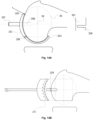
- Geometric characteristics of the medial femoral condyle may be represented by an ellipse, and belongs to an arc of the ellipse, for example, the arc ranges from 150 degrees to 200 degrees.
- a sagittal plane having a maximum posterior offset of the medial femoral condyle is selected, that is, an intermediate plane of the medial femoral condyle.
- the medial femoral condyle is best-fitted by an ellipse is shown in Fig. 4A .
- the articular cartilage surface 36 of the medial femoral condyle 42 in this segment is best-fitted by an ellipse 38.
- the major axis of the ellipse 38 is perpendicular to the mechanical axis of the diaphyseal shaft, and an elliptical center 39 corresponding to an attachment point 123 of a medial collateral ligament of the medial femoral condyle on the axial MRI and coronal MRI .
- the ellipse 38 has a semi-major axis of 31 mm, a semi-minor axis of 25mm, and an eccentricity of 0.591.
- the ellipse here has a semi-major axis of 27mm, a semi-minor axis of 22mm, and an eccentricity of 0.58.
- the semi-major axis is between 20mm and 35mm
- the semi-minor axis is between 16mm and 30mm
- the eccentricity is between 0.5 and 0.7.
- an arc length of the articular cartilage surface 36 in the segment may be described through an angle ⁇ between lines for connecting a center 39 of the ellipse and the anterior and posterior notches 34 and 35, and an angle ⁇ between a line for connecting the center 39 of the ellipse and the posterior notch 35 and the major axis of the ellipse 38.
- the angle ⁇ is 180 degrees and the angle ⁇ is 35 degrees.
- the angle ⁇ is 190 degrees and the angle ⁇ is 40 degrees.
- the angle ⁇ is between 170 and 195 degrees and the angle ⁇ is between 20 and 45 degrees.
- there is no articular surface of the medial femoral trochlea in front of the intermediate plane of the medial femoral condyle that is, the ellipse 38 in the intermediate plane of the medial femoral condyle does not correspond to a plane having a maximum anterior offset of the medial femoral trochlea, and ellipses in the two planes are not in a same scanning section.
- the ellipse 38 of the medial femoral condyle may be projected in the MRI sagittal scanning direction to the plane having a maximum anterior offset of the medial femoral trochlea, as shown in Fig. 4B .
- the articular cartilage surface 37 in this segment may be represented by an arc of an ellipse 40.
- articular surfaces in this segment for femoral trochlea prostheses of some subjects shows as circles
- the femoral trochlea prostheses of most subjects show as ellipses.
- a major axis of the ellipse 40 of the medial femoral trochlea is perpendicular to a major axis of the ellipse 38 in the intermediate plane of the medial femoral condyle.
- This ellipse 40 is based on the circle 70 on the most concave plane of the femoral trochlea as shown in Fig.
- the ellipse 40 has a semi-major axis of 29mm, a semi-minor axis of 27mm, and an eccentricity of 0.365. In multiple embodiments, the ellipse 40 has a semi-major axis between 20mm and 35mm and a semi-minor axis between 20mm and 30mm.
- a difference between the semi-major axis and the semi-minor axis of the ellipse 40 is not large, such as 1mm, 2mm, or 3mm.
- an arc 37 of the articular cartilage surface of the trochlea in this segment may be described through an angle ⁇ between lines for connecting the center 41 and the anterior notch 46 and an end point of the cartilage surface of the trochlea, and an angle ⁇ ' between a line for connecting the center 41 and the end point of the cartilage surface of the trochlea and the semi-minor axis of the ellipse 40.
- the angle ⁇ is between 40 and 80 degrees and the angle ⁇ ' is between -5 and 40 degrees.
- a positional relationship between the center 39 of the ellipse 38 of the medial femoral condyle and the center 41 of the ellipse 40 of the medial femoral trochlea determines a spatial positional relationship between the femoral condyle and the femoral trochlea, which determines the values of the outside parameters and the inside parameters of the femoral prosthesis.
- the relationship between the femoral trochlea and the femoral condyle may be represented by a rectangle 50 formed by the major axes and the minor axes of the ellipse 38 of the medial femoral condyle and the ellipse 40 of the medial femoral trochlea.
- the rectangle 50 has a length 107 of 13mm and a width 109 of 9mm.
- the rectangle 50 has a length 107 of 12mm and a width 109 of 7mm.
- the length 107 of the rectangle 50 is between 8mm and 16mm and the width 109 of the rectangle 50 is between 4mm and 12mm.
- the angle between a line for connecting the centers 39 and 41 of the two ellipse 38 and 40 and the major axis of the ellipse 38 of the medial femoral condyle is ⁇ .
- ⁇ is 32 degrees.
- ⁇ is 35 degrees.
- the angle ⁇ ranges between 25 degrees and 35 degrees.
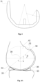
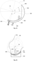
- the most concave plane 62 of the femoral trochlea is a plane where the Whiteside line is clinically located, as shown in Fig. 5 .
- This plane 62 is an important basis for determining a geometrical shape of the articular surface of the medial and lateral femoral trochleas.
- the Blumensaat line 63 is clinically encompassed by this circle 70.
- the articular cartilage surface 64 of this plane 62 is an arc of the circle 70 and may be represented by a radius of the circle 70 and an angle.
- an angle between lines for connecting the center 41' and the anterior and posterior boundaries of the articular cartilage surface 64 of the trochlea is ⁇ ; and an angle between the line for connecting the center 41' and the anterior boundary of the articular cartilage surface 64 and the horizontal axis is ⁇ .
- the circle 70 has a radius of 24mm, ⁇ of 100 degrees, and ⁇ of 0 degrees.
- the circle 70 has a radius of 25mm, ⁇ of 105 degrees, and ⁇ of 5 degrees.
- the circle 70 has a radius of 16mm to 30mm, ⁇ ranging from 90 degrees to 125 degrees, and ⁇ ranging from -20 degrees to 10 degrees.
- there is a specific ratio of the radius of the circle 70 relative to a length of the semi-major axis of the ellipse 38 of the medial femoral condyle for example, 2/5, 3/5 or 3/4.
- a geometry of the lateral femoral condyle may be represented by an ellipse, and belongs to an arc of the ellipse, for example, the arc ranges from 120 degrees to 160 degrees.
- a sagittal plane having a maximum posterior offset of the lateral femoral condyle that is, an intermediate plane of the lateral femoral condyle, is selected.
- This sagittal plane is also a plane having a maximum anterior offset of the lateral femoral trochlea, and relationships thereof are shown in Fig. 6 .
- the articular cartilage surface 76 of the lateral femoral condyle 82 in this segment is best-fitted by an ellipse 78.
- the major axis of the ellipse 78 is rotated clockwise by a certain angle ⁇ relative to the major axis of the ellipse 38 of the medial femoral condyle, such as 12 degrees in one embodiment, and 18 degrees in another embodiment, and in multiple embodiments, an average value of ⁇ is between 5 degrees and 25 degrees.
- a projection of the center 79 thereof in the sagittal section coincides with the center 39 of the ellipse 38 of the medial femoral condyle; and corresponds to an attachment point 122 of the collateral ligament of the lateral femoral condyle in the MRI axial section ( Fig. 9A ).
- the ellipse 78 has a semi-major axis of 30mm and a semi-minor axis of 26mm; and in another embodiment, the ellipse 78 has a semi-major axis of 26mm and a semi-minor axis of 23mm. In multiple embodiments, the ellipse 78 has a semi-major axis between 21 mm and 33mm, a semi-minor axis between 16mm and 30mm, and an eccentricity between 0.5 and 0.7.
- an arc of the articular surface 76 in this segment may be accurately described by measuring the angle ⁇ between lines for connecting the center 79 and the anterior and posterior recesses 74 and 75, and the angle ⁇ between the line for connecting the center 79 and the posterior recess 75 and the major axis of the ellipse 81 of the lateral condyle.
- ⁇ is 130 degrees and ⁇ is 40 degrees.
- the included angle ⁇ is between 120 degrees and 160 degrees and the included angle ⁇ is between 30 degrees and 70 degrees.
- this segment 77 may be represented by a circle or an ellipse 80. Although this part appears as ellipses for some subjects, it appears as circles for most subjects.
- the center 41" of the circle 80 of the plane 72 of the lateral femoral trochlea is completely coincident with the center 41 of the ellipse 40 of the medial femoral trochlea and the center 41' of the most concave plane 62 of the femoral trochlea in the MRI sagittal section.
- the circle 80 has a radius between 25mm and 35mm, such as 28mm, or 26mm.
- An angle between a line for connecting the center 41" of the circle 80 and an intersection point below the ellipse 78 and a line for connecting the center 41" of the circle 80 and the end point of the articular cartilage surface of the lateral femoral trochlea is ⁇ ; and an angle between a line for connecting the center 41" of the circle 80 and the end point of the articular cartilage surface of the lateral femoral trochlea and the horizontal axis is ⁇ '.
- the angle ⁇ is between 80 degrees and 120 degrees, for example, 90 degrees, 100 degrees or 110 degrees; and the angle ⁇ ' is between -30 degrees and 20 degrees, for example, -10 degrees, 0 degrees, or 10 degrees.
- articular cartilage surfaces of the medial femoral condyle and the lateral femoral condyle may almost be represented by ellipses
- articular cartilage surfaces of the medial femoral trochlea and the lateral femoral trochlea may almost be represented by ellipses and/or circles
- the most concave portion of the femoral trochlea i.e., the center of the trochlear groove
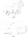
- Various sagittal planes of an articular cartilage surface of the medial femoral condyle are a set 92 of concentric ellipses, wherein various ellipses have different sizes, major axes and minor axes of the ellipses are in the same direction and are coincident respectively, and various ellipses have similar eccentricities, as shown in Fig. 7A .
- Various sagittal planes of an articular cartilage surface of the lateral femoral condyle are a set 93 of ellipses, as shown in Fig. 7A .
- various ellipses have different sizes, and major axes and minor axes of the ellipses are in the same direction and are approximately coincident respectively, that is, centers of various ellipses are approximately coincident and are arranged as concentrically.
- Various sagittal planes of an articular cartilage surface of the medial femoral trochlea are a set of ellipses ( Fig. 7A ), major axes and minor axes of the ellipses are in the same direction respectively, and centers of the ellipses are arranged concentrically.
- various ellipses have different eccentricities. Sizes of these ellipses are, for example, ordered in a Fibonacci sequence.
- the femoral condyle is scanned using the MRI in the sagittal section, and all planes of the lateral femoral trochlea appear as circles or ellipses.
- various circles of the lateral trochlea have different radiuses
- various ellipses of the lateral trochlea have different major and minor axis
- projections of the centers 41" thereof are coincident ( Fig. 7A ).
- the articular surfaces 95, 97 of the medial femoral condyle and the lateral femoral condyle in the coronal section may be represented by a circle and an ellipse, as shown in Fig. 7B .
- a circle 94 (the sixth circle) can well fit the articular surface 95 of the medial femoral condyle in the coronal section, and a radius of the circle 94 is equal to the semi-minor axis of the ellipse 38 of the medial femoral condyle.
- An arc angle of the articular surface in this segment may be represented by an angle ⁇ , for example, the arc angle ranges from 50 degrees to 90 degrees.
- ⁇ is divided by a vertical line into ⁇ 1 and ⁇ 2, wherein ⁇ 1 and ⁇ 2 may or may not be equal.
- the angle ⁇ is 65 degrees; and in another embodiment, the angle ⁇ is 70 degrees.
- an ellipse 96 (the seventh ellipse) is rotated clockwise by ⁇ y degrees, is then just tangent to the medial circle 94 (the sixth circle) and best-fit the articular surface 97 of the lateral femoral condyle in the coronal section. If the ellipse 96 has an eccentricity of 0.618, it is a perfect ellipse.
- An arc angle of the articular surface in this segment may be represented by an angle ⁇ , for example, the arc angle ranges from 50 degrees to 90 degrees.
- ⁇ is divided by a vertical line into ⁇ 1 and ⁇ 2, wherein ⁇ 1 and ⁇ 2 are not equal.
- the angle ⁇ is 70 degrees; and in another embodiment, the angle ⁇ is 75 degrees.
- the femoral prosthesis according to the embodiments of the present disclosure has sagittal posterior condyles in an elliptical geometry, and sagittal trochleas in an elliptical and/or a circular geometry, wherein the lateral posterior condyle is shorter than and lower than the medial posterior condyle, and the lateral trochlea is longer than and higher than the medial trochlea.
- the center 39 of the ellipse 38 of the medial femoral condyle coincides with the center 79 of the ellipse 78 of the lateral femoral condyle, and the center 41 of the ellipse 40 of the medial femoral trochlea, the center 41" of the circle 80 of the lateral femoral trochlea, and the center 41' of the circle 70 at the most concave portion of the femoral trochlea are coincident. Therefore, a sagittal shape of the TKA femoral prosthesis 100 according to the embodiments of the present disclosure is as shown in Fig. 8 .
- the femoral prosthesis 100 is divided into articular surface portions, that is, an outside surface of the prosthesis which is in contact with the patella and the tibia plateau during the motion of the knee joint; and an inside portion, that is, a portion which is adjacent to an osteotomy surface of the femoral condyle and a bone cement after the femoral prosthesis is implanted.
- the sagittal articular surface of the femoral prosthesis 100 is designed to be asymmetric.
- Anterioposterior diameters and heights of the sagittal articular surface of the femoral prosthesis 100 are determined by parameters of five basic elements 38 (the ellipse of the medial femoral condyle), 40 (the ellipse of the medial femoral trochlea), 70 (the circle of the most concave trochlear groove), 78 (the ellipse of the lateral femoral condyle), 80 (the circle or ellipse of the lateral femoral trochlea) which form the articular surface and the angles ⁇ and ⁇ .
- the femoral prosthesis 100 may be divided into medial elements which are a half of elements of the femoral prosthesis, i.e., including a medial condyle portion 51 of the femoral prosthesis and a medial trochlear portion 131 of the femoral prosthesis; lateral elements which are a half of the elements of the femoral prosthesis, i.e., including a lateral condyle portion 91 of the femoral prosthesis and a lateral trochlear portion 141 of the femoral prosthesis; and a trochlear groove 101 of the femoral prosthesis, i.e., a position where the most concave plane of the trochlea is located.
- the articular surface geometry of the medial elements 51, 131 which are a half of the elements of the femoral prosthesis is composed of an ellipse 38 and an ellipse 40;
- the articular surface geometry of the lateral elements 91,141 which are a half of the elements of the femoral prosthesis is composed of an ellipse 78 and a circle or ellipse 80;
- the articular surface geometry of the trochlear groove 101 of the femoral prosthesis is composed of a circle 70.
- the geometric shape of the articular surface of the femoral prosthesis is asymmetric, with the lateral portion of the femoral prosthesis being forwardly spaced relative to the medial portion of the femoral prosthesis. Therefore, the posterior offset of the lateral condyle 91 of the femoral prosthesis is less than that of the medial condyle 51, and the posterior height of the lateral condyle 91 is lower than that of the medial condyle 51.
- the anterior offset of the lateral trochlea 141 of the femoral prosthesis is greater than that of the medial trochlea 131. In the axial section, the femoral prosthesis has its own external rotation.
- the posterior lateral condyle of the femoral prosthesis is shorter and lower than that of the posterior medial condyle of the femoral prosthesis.
- This design feature is beneficial to increase a flexion angle of the knee joint.
- the anterioposterior diameters and heights of the articular surfaces of the femoral prosthesis may be accurately calculated using the parameters of ellipses and circles which form the femoral prosthesis, and the important angle values.
- the parameter values of the femoral prosthesis thereof vary accordingly with the prosthetic sizes.
- the lateral elements 91, 141 which are a half of the elements of the femoral prosthesis are forwardly spaced relative to the medial members 51, 131 which are a half of elements of the femoral prosthesis.
- This forward distance has different parameter values depending on the prosthetic sizes, for example, 1 mm, 2mm, 3mm, or 4mm.
- the posterior portion of the lateral condyle 91 of the femoral prosthesis is shorter than and lower than the posterior portion of the medial condyle 51 of the femoral prosthesis. This forms a distance difference Dp, and a height difference Hd, as shown in Fig. 8 .
- the parameter values of Dp and Hd are not fixed values, which vary with the prosthetic sizes, that is, they vary with the parameter values of the ellipses 38, 40, 78 and the circles or ellipse 70, 80 which form the articular surface.
- the value of Dp may be 2mm, 3mm, or 4mm; and the value of Hd may be 1mm, 2mm, or 3mm.
- the posterior end of the medial condyle 51 of the femoral prosthesis is more flat and blunt 117 than that of the posterior end of the lateral condyle 91 of the femoral prosthesis . Parameter values of this flat blunt surface 117 also vary with the prosthetic sizes.
- An anterior edge of the lateral trochlear portion 141 of the femoral prosthesis is more forward in the sagittal section, and is higher in the axial section than an anterior edge of the medial trochlear portion 131 of the femoral prosthesis, as in Fig. 9A .
- a value of Da is not fixed, and varies with the parameters of the ellipses 38, 40, 78 and the circles and ellipse 70, 80 which form the articular surface.
- the value of Da may be 2mm, 3mm, 4mm, or 5mm.
- Anterosuperior portions (flange) 118, 119, 120 of an the femoral prosthesis 100 are tangent to an anterior bone cortex of a femoral shaft, and are designed to have a short linear shape to reduce a pressure on the patella, wherein the vertex of the anterosuperior portions (flange) is in the same level as the vertex of the circle 70 at the most concave portion of the femoral trochlea.
- the inside structure of the femoral prosthesis is symmetrical to facilitate the osteotomy step and the gap balancing step.
- the parameter values of the inside structure that corresponding to the osteotomy line thereof may be accurately calculated using the ellipses and the circles which form the femoral prosthesis, and important angle values.
- the parameter values thereof vary accordingly with the prosthetic sizes.
- the inside structure 52, 53, 54, 55, 56 of the femoral prosthesis 100 have a rectangular structure (with/without bone cement) which is in contact with the distal femur after osteotomy.
- the inside structure of the femoral prosthesis 100 are designed to be a symmetrical rectangle.
- the posterior section 52 of the inside sides of the femoral prosthesis is perpendicular to the horizontal axis, i.e., being perpendicular to the major axis of the medial ellipse 38 of the medial femoral condyle, as shown in Fig. 8 .
- the posterior section 52 can be cut just to the end position of the articular surface of the medial and lateral femoral condyles 42, 82, as shown in Fig. 4B . Further, the parameter values of the position and height of the posterior section 52 of the medial sides of the femoral prosthesis may be determined through the medial ellipse 38 of the medial femoral condyle.
- a line for connecting the end point of the articular surface of the posterior condyle of the medial condyle portion 51 of the femoral prostheses and the center 39 of the medial ellipse 38 of the medial femoral condyle passes through the end point of the condyle articular surface of the lateral condyle portion 91 of the femoral prosthesis.
- An angle between the line and the major axis of the medial ellipse 38 of the medial femoral condyle is ⁇ . Therefore, the position and height of the posterior section 52 of the inside sides of the femoral prosthesis may be calculated according to the basic formula of the ellipse. The position and height of the posterior section 52 vary with the prosthetic sizes.
- the parameter values of the inferior section 53 and the posterior slope section 55 of the inside sides of the femoral prosthesis are directly affected by the posterior section 52; and the end point of the articular surface of the posterior condyle of the medial femoral condyle portion 51 is located in a rectangular frame formed by the major axis and the minor axis of the medial ellipse 38 of the femur; and parameter values thereof vary with the prosthetic sizes.
- the anterior section 54 of the inside sides of the femoral prosthesis 100 moves back by a thickness of one cartilage surface relative to the trochlear groove 101 of the femoral prosthesis to ensure removal of the cartilage surface at the most concave portion of the trochlea, for example 2mm or 3mm.
- the anteversion angle may also be 1 or 3 degrees.
- a segment from the end of the inferior section 53 of the inside sides intersects with the anterior section 54 of the inside sides at an angle of 45 degrees relative to the major axis of the medial ellipse 38, to form an anterior slope section 56 of the inside sides.
- parameters of the anterior section 54 and the anterior slope section 56 of the inside sides of the femoral prosthesis are determined by the parameters of the medial and lateral femoral ellipses 38, 78, the ellipse 40 of the femoral trochlea, and the circles or ellipse 70, 80 of the femoral trochlea, and the angles ⁇ and ⁇ .
- the trochlear groove 101 of the femoral prosthesis is designed as a 1/4 arc, and its specific parameter values (depth and radius) are directly determined by the circle 70 in the most concave plane of the trochlea, the medial femoral ellipse 38, and the angle ⁇ , and vary with the prosthetic sizes.
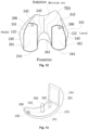
- the femoral prosthesis 100 is viewed from the perspective of an axial section, as shown in Fig. 9A .
- the medial trochlear portion 131 of the femoral prosthesis, the lateral condyle portion 91 of the femoral prosthesis, the lateral trochlear portion 141 of the femoral prosthesis, and the trochlear groove 101 of the femoral prosthesis are designed to be perpendicular to a line 133 for connecting the centers 39, 79 of the ellipses of the medial femoral condyle and the lateral femoral condyle (TEA direction).
- the center 39 of the medial femoral ellipse 38 corresponds to the vertex of the medial femoral condyle in the axial section, in other words, the attachment point of the medial collateral ligament 123; and the center 79 of the lateral femoral ellipse 78 corresponds to the vertex of the lateral femoral condyle in the axial section, in other words, the attachment point of the lateral collateral ligament 122.
- the anterior offset of the articular surface of the lateral femoral trochlea is greater than that of the articular surface of the medial femoral trochlea, with a distance therebetween of Da described above; the posterior offset of the lateral condyle 91 of the femoral prosthesis is less than that of the medial condyle 51, with a difference therebetween of Dp described above; and an angle between a line for connecting the lateral posterior condyle and the medial posterior condyle and the TEA is ⁇ , for example, the angle ⁇ may be 3 degrees, 2 degrees or 4 degrees, etc.
- the medial femoral condyle has a circular arc structure in the coronal section; and the lateral femoral condyle has an elliptical arc structure in the coronal section.
- the tibial plateau side has a corresponding asymmetric structure. Further, the tibial plateau side is modified to adapt to a varus angle of the mechanical axis of the lower extremity.
- the medial femoral condyle and the lateral femoral condyle have a symmetrical circular arc structure in the coronal section.
- a posterior viewing of the femoral prosthesis 100 according to the present disclosure in the coronal section is as shown in Fig. 9B .
- the lateral condyle 91 of the femoral prosthesis is lower than the medial condyle 51 of the femoral prosthesis, with a difference therebetween of Hd described above.
- design of the contour of the articular surfaces of the medial femoral condyle and the lateral femoral condyle in the coronal section and the femoral trochlear groove 101 may be divided into two types: a non-patella arthroplasty type and a patella arthroplasty type.
- the articular surfaces of the medial condyle and the lateral condyle of the femoral prosthesis 100 in the coronal section are designed to be arcs of a circle and an ellipse, as shown in Figs. 7B and 9B .
- the articular surface 95 of the medial condyle of the femoral prosthesis in the coronal section is an arc of a circle 94, which is represented by an angle ⁇ .
- the center of the circle 94 is the center 39 of the ellipse 38 of the medial femoral condyle and has a radius equal to the semi-minor axis of the ellipse 38 of the medial femoral condyle.
- a coronal surface 151 of the medial plateau of the plateau prosthesis 150 corresponding to the articular surface 95 has a concavo shape which fully adapts to the curvature of the articular surface 95.
- the articular surface 97 of the lateral condyle of the femoral prosthesis in the coronal section is an arc of an ellipse 96, which is represented by an angle ⁇ .
- the center of the ellipse 96 is the center 79 of the ellipse 78 of the lateral femoral condyle having an eccentricity of 0.618 and a radius approximately equal to the semi-minor axis of the ellipse 38 of the medial femoral condyle.
- a coronal surface 152 of the lateral plateau of the plateau prosthesis 150 corresponding to the articular surface 97 has a concave structure which fully adapts to the ellipse of the articular surface 97. Therefore, the coronal structure of the tibial plateau prosthesis 150 corresponding to the non-patella arthroplasty-type femoral prosthesis is as described above.
- the distal end surface 154 of the tibial plateau prosthesis 150 may be designed to be perpendicular to a mechanical axis of the tibia or has a varus angle ⁇ , so as to adapt to the varus of the normal tibial plateau related to the mechanical axis of lower extremity. (i.e., kinematic alignment).
- the angle ⁇ may be 1 degree, 2 degrees or 3 degrees.
- the trochlear groove 101 is designed with reference to the medial femoral trochlea and the lateral femoral trochlea in the sagittal section which are composed of ellipses and circles 40, 70, 80, as shown in Fig. 7A .
- the articular surface of the lateral condyle in the coronal section is designed to be an arc of a circle; and the trochlear groove is designed to be a groove with a valgus angle of 6°, to correspond to a dome shape of the patella prosthesis.
- the femoral prosthesis constructed according to the above ellipse principle may be adapted for the production of a CR prosthesis and/or a PS prosthesis.
- the medial and lateral condyles of the femoral prosthesis are arcs of ellipses in the sagittal section.
- the major axes of the medial condyle ellipses are perpendicular to the diaphyseal axis of the femur, and the major axes of the lateral condyle ellipses may be rotated clockwise by a certain angle (7 degrees to 22 degrees) relative to the major axis of the ellipse of the medial condyle.
- shapes of both of the medial and lateral femoral condyles may be represented by ellipses on the sagittal scanning plane.
- the articular surface of the medial femoral trochlea is an arc of an ellipse in the sagittal section, and the ellipse has a major axis perpendicular to the major axis of the medial ellipse.
- the articular surface of the lateral femoral trochlea in the sagittal section is an arc of a circle or an ellipse.
- the ellipse of the medial femoral condyle is designed to have a concentric elliptical structure perpendicular to the TEA and parallel to the Whiteside line in the sagittal plane, which is most consistent with the direction and geometric shape of a medial femoral condyle in a normal human body.
- the ellipse of the lateral femoral condyle is designed according to a shape of an articular cartilage surface of a lateral femoral condyle of a normal knee.
- the ellipse of the lateral femoral condyle is slightly less than the ellipse of the medial femoral condyle.
- a major axis direction of the lateral femoral condyle is rotated clockwise by a certain angle relative to the ellipse of the medial femoral condyle.
- centers of the ellipses of the medial and lateral femoral condyles coincide in the sagittal section of the femoral prosthesis.
- the major and minor axis of the lateral femoral condyle ellipse may be simplified to have the same directions as those of the medial femoral condyle.
- the clockwise rotation may be omitted, which further simplifies the design and manufacturing process of the femoral prosthesis.
- the medial and lateral femoral trochleas are described as being composed of an ellipse or circle. This plan is obtained by a final statistical analysis. Although the medial femoral trochlea according to most of the embodiments appears as ellipses, there are a few embodiments in which the medial femoral trochlea appears as circles; and although the lateral femoral trochlea according to most of the embodiments appears as circles, there are a few embodiments in which the lateral femoral trochlea appears as ellipses.
- the design of the femoral prosthesis according to the present disclosure comprises a CR prosthesis, a PS prosthesis or other revision prosthesis designs.
- the CR prosthesis relative to the PS prosthesis, omits a cam structure on the elements of the femoral prosthesis and a post structure on elements of the tibial prosthesis. This allows the CR prosthesis to appear between the lateral condyle and the medial condyle as an intercondyal space with an open box structure.
- any types of prostheses may be formed by suitably modifying the femoral prosthesis according to the present disclosure to add the cam structure on the elements of the femoral prosthesis and the post structure on the tibial side. Therefore, all of the principles described in the present disclosure may be used to design any potentially knee prosthesis. Although any potentially contemplated knee prosthesis design may comprise all of the features described here, it is also contemplated that some potentially contemplated knee prosthesis designs may omit or may be added with some of the features described here, depending on particular applications or requirements in other situations.
- the UKA prosthesis of the medial femoral condyle has an elliptical geometry in the sagittal section and a circular geometry in the coronal section.
- the medial femoral condyle is a set of concentric ellipses, and these elliptical planes are spatially parallel to the Whiteside line of the trochlea. Centers of these ellipses correspond to the attachment point of the medial collateral ligament of the medial femoral condyle. Therefore, the geometry of the UKA prosthesis of the medial femoral condyle is composed of concentric ellipses in the sagittal section, as shown in Fig.
- the UKA prosthesis 201 of the medial femoral condyle is divided into an articular surface portion, that is, an outside surface of the prosthesis which is in contact with a medial patella and a medial tibial plateau during motion of a knee joint; and an inside side portion, that is, a portion adjacent to an osteotomy surface of a femoral condyle portion and a bone cement after the UKA prosthesis 201 of the medial femoral condyle is placed, wherein the inside side portion appears as a posterior condyle of the inside side in a straight section, and a distal portion in the inside side conformal to the arc of the articular surface.
- the UKA prosthesis 201 of the medial femoral condyle is an arc 203 of an ellipse 38, as shown in Fig. 10 .
- Anterior and posterior edges of the arc 203 correspond to the recesses of the anterior and posterior meniscus, and form an arc angle ⁇ range of, for example, 150 degrees to 200 degrees, wherein the arc angle ⁇ range is 175 degrees in one embodiment, and 185 degrees in another embodiment, and 180 degrees in yet another embodiment.
- angle ⁇ may be expressed as an angle ⁇ between a straight line for connecting anterior and posterior recesses 207, 208 of the meniscus and passing through the center 39 of the ellipse of the medial femoral condyle and the major axis of this ellipse.
- This angle ⁇ is 30 degrees in one embodiment, 35 degrees in another embodiment, and 40 degrees in yet another embodiment.
- the posterior section 202 of the inside side of the UKA prosthesis of the medial femoral condyle is a line orienting the posterior recess 208 of the meniscus which is perpendicular to the major axis of the ellipse, and corresponding the osteotomy on the posterior condyle. This position changes with the parameters of the prosthesis change.
- the distal portion 203 of the UKA prosthesis 201 of the medial femoral condyle has an elliptical arc configuration.
- the inside side thereof has two pillars, which are a central pillar 204 corresponding to the center 39 of the ellipse of the medial femoral condyle, and a posterior pillar 205 corresponding to the focus of this ellipse, respectively.
- a locking screw hole 206 for locking a screw to correspond to a locking screw 206'. In a case of a normal human body, this position contacts the meniscus and neither contacts the articular surface of the tibial plateau, nor contacts the patella.
- screw fixation at this position does not affect the articular surface.
- a direction in which the fixing screw 206' is locked is different from that of the central pillar and the posterior pillar, and the stability of the prosthesis can be enhanced. It can be understood that those skilled in the art can provide a greater number of pillars as desired.
- the shape of the articular surface of the medial femoral condyle in the coronal section may be represented by an arc 95 of a circle 94, which has an arc angle of ⁇ , for example, a range of the arc angle is from 50 degrees to 90 degrees. Therefore, the shape of the UKA prosthesis 201 of the medial condyle in the coronal section is as shown in Fig. 11 .
- an arc 203 may be seen as being approximately coincident with a circle 221 having a radius greater than that of the circle 94 of the UKA prosthesis 201 in the coronal section. A curvature and parameters of this circle 221 are used as parameters of an abrasive tool to prepare a bone bed surface.
- the articular surface of the UKA prosthesis 201 of the medial femoral condyle is asymmetrical, as shown in Fig. 12 .
- the prosthesis is placed to be parallel to the Whiteside line and perpendicular to the TEA.
- a medial arc edge 241 is arc-shaped to adapt to the shape of the distal shape of the medial femoral condyle; an anterior arc edge 242 has a curvature corresponding to parameters of a circle 221 of an abrasive tool, and a posterior portion 244 has a curvature of the circle 94 in the coronal section. Therefore, a stereogram of the UKA prosthesis 201 of the medial femoral condyle is shown in Fig. 13 . In addition to various positions described above, the inside sides of the prosthesis have corresponding concave grooves to adapt to the bone cement.
- Best size and position of the prosthesis may be planned on a preoperative MRI scanning on the femoral condyle as described above.
- a specific surgical operation comprises: after exposure, firstly determining the Whiteside line of the trochlear groove, and marking a direction line of the prosthesis which is parallel to the Whiteside line on the medial condyle surface using an electric knife.
- An ellipse measuring tool 251 which is most compatible with the ellipse of the medial femoral condyle, fits well to the articular surface, as shown in Fig. 14A .
- An anterior edge of the measuring tool 251 has a grapple structure 254 which can hold the medial posterior condyle well.
- a hollow handrail 257 of the measuring tool 251 is right against the direction of the attachment point of the medial collateral ligament, i.e., the direction of the center 39 of the ellipse of the medial femoral condyle.
- the hollow handrail 257 may have a drill bit placed therein, to drill a tunnel 258 in the medial femoral condyle to facilitate the next step of placing a fixed pile at the center of the power drill.
- osteotomy groove 256 there is an osteotomy groove 256 at the bottom of the measuring tool 251, and the osteotomy groove 256 is right against a posterior osteotomy line 202 of the medial femoral condyle.
- the measuring tool 251 is then removed, the fixed pile 259 is placed on the central tunnel 258, and a hollow drill 271 is used, which has a radius equal to that of the circle 221 described above.
- a depth of a rasion is limited by the fixed pile 259, and the prosthesis is continuously tested for the depth during this period.
- the UKA prosthesis of the lateral femoral condyle has an elliptical geometry in both the sagittal section and the coronal section.
- the lateral femoral condyle is a set of concentric ellipses, and these ellipses are spatially parallel to the Whiteside line of the trochlea. Centers of these ellipses correspond to the attachment point of the lateral collateral ligament of the lateral femoral condyle. Therefore, the geometry of the UKA prosthesis of the lateral femoral condyle is composed of concentric ellipses in the sagittal section and ellipses in the coronal section, as shown in Fig.
- the UKA prosthesis 301 of the lateral femoral condyle is divided into an articular surface portion, that is, an outside surface of the prosthesis which is in contact with the lateral patella and the lateral tibial plateau during the motion of the knee joint; and an inside side portion, that is, a portion adjacent to an osteotomy surface of a femoral condyle portion and a bone cement after the UKA prosthesis 301 of the lateral femoral condyle is placed, wherein the inside side portion appears as a posterior condyle of the inside side in a straight section, and a distal portion of the inside side conformal to the arc of the articular surface.
- the UKA prosthesis 301 of the lateral femoral condyle is an arc of an ellipse 78, as shown in Fig. 15 .
- the anterior and posterior edges of the arc correspond to the recesses 307, 308 of the anterior and posterior meniscus, and form an arc angle range, for example, the arc angle range is from 120 degrees to 160 degrees.
- the arc angle is 145 degrees, and in another embodiment, the arc angle is 150 degrees. Specifically, it may be expressed as an angle ⁇ ' between the anterior and posterior recesses 307, 308 of the meniscus and the center 79 of the ellipse of the lateral femoral condyle.
- An angle between a line for connecting the center 79 and the posterior recess 308 of the meniscus and the horizontal axis is ⁇ ', which is 35 degrees in one embodiment, 40 degrees in another embodiment, and an average of 35 degrees in multiple embodiments.
- the posterior section 302 of the inside side that is corresponding to the osteotomy direction of the UKA prosthesis 301 of the lateral femoral condyle is perpendicular to the horizontal axis. This position changes as the parameters of the ellipse change.
- the distal portion 303 of the UKA prosthesis 301 of the lateral femoral condyle has an elliptical arc configuration.
- a pillar on the inside side that is, a posterior pillar 305 corresponding to the focus of the ellipse.
- a locking screw hole 306 at the anterior edge of component 303 to correspond to a locking screw 306'.
- this position contacts the meniscus and does not contact the articular surface of the tibial plateau; and at the same time, the position of the screw hole does not contact the patella. Therefore, screw fixation at this position does not affect the contact of the articular surface.
- a direction in which the fixing screw 306' is locked is different from that of the posterior pillar, and the stability of the prosthesis can be enhanced.
- the shape of the articular surface of the lateral femoral condyle which passes through the center 79 may be represented by an arc 97 of an ellipse 96, which has an arc angle of ⁇ , for example, a range of the arc angle is from 50 degrees to 90 degrees. Therefore, the shape of the UKA prosthesis 301 of the lateral condyle in the coronal section is shown in Fig. 16 , and a shape of an articular surface of a corresponding tibial prosthesis in the coronal section is a concave shape 325 adapted to this ellipse.
- This arc 97 may approximately coincide with a circle 321 which does not pass through the center 79.
- the center is 322 of the circle 321, and a radius of the circle 321 may be regarded to be equal to a length of the semi-minor axis of the ellipse 96.
- the direction axis 323 is not only the direction of the posterior pillar 305, but also a direction of a burr drill and the fixed pile.
- An angle between the direction axis 323 and the vertical axis is 15 degrees.
- the curvature and parameters of the circle 321 are used as the parameters of the abrasive tool to prepare the bone bed surface. In the coronal section, an angle between a direction of the locking screw holes and the locking screw 306 and the vertical axis is 15 degrees. Therefore, an angle between the locking screw 306 and the posterior pillar 305 is 30 degrees to achieve maximum stability of the prosthesis.
- the articular surface of the UKA prosthesis 301 of the lateral femoral condyle is asymmetrical, as shown in Fig. 12 .
- the prosthesis is placed to be parallel to the Whiteside line and perpendicular to the TEA.
- the lateral arc edge 341 is arc-shaped to adapt to a shape of a distal shape of the lateral femoral condyle; an anterior arc edge 342 has a curvature corresponding to curvature parameters of the circle 321, and a posterior portion 344 has a curvature of the ellipse 96 in the coronal section. Therefore, a stereogram of the UKA prosthesis 301 of the lateral femoral condyle is shown in Fig. 17 . In addition to various positions described above, the inside sides of the prosthesis have corresponding concave grooves to adapt to the bone cement.
- the UKA prosthesis of the femoral trochlea has a geometry of an ellipse or a circle of the medial trochlea and a geometry of a circle or an ellipse of a lateral trochlea in the sagittal section, and designs adapted to non-patella arthroplasty and patella arthroplasty.
- the femoral trochlea prosthesis 401 comprises an articular surface, which is a surface in contact with an articular surface of a patella during motion of a knee joint, wherein the articular surface appears in a sagittal section as a set of an arc (37) of a ellipse or circle 40 and an arc 77 of a ellipse or circle 80; and an inside side which is a portion adjacent to an osteotomy surface of a femoral trochlear portion and a bone cement after the prosthesis is placed, wherein the surface appears as an inside side 409 conformal to an articular surface of the femoral trochlea.
- the medial and lateral femoral trochleas are formed by arranging the arcs of ellipses and the arcs of circles concentrically respectively, as shown in Fig. 4 . Therefore, the UKA prosthesis 401 of the femoral trochlea according to the present disclosure is designed to be formed by a geometry of ellipses of the medial femoral trochlea and circles of the lateral femoral trochlea, which are arranged concentrically.
- a concentric axis 41* (connecting the centers of 41, 41', and 41") is spatially parallel to the TEA and perpendicular to the Whiteside line. As shown in Fig.
- the UKA prosthesis 401 of the femoral trochlea has a pillar 402 at a center of inside structure thereof and four surrounding locking screw holes 403, 404, 405, 406 in which locking screws are mounted, as shown in Fig. 18B .
- the UKA prosthesis of the lateral femoral condyle is designed based on the elliptical structure of the lateral femoral condyle, and the ellipse of the lateral femoral condyle is designed according to a shape of an articular cartilage surface of a lateral femoral condyle of a knee joint of a normal human body.
- the ellipse of the lateral femoral condyle is slightly less than the ellipse of the medial femoral condyle.
- a major axis direction of the lateral femoral condyle is rotated clockwise by a certain angle relative to the ellipse of the medial femoral condyle.
- centers of the ellipses of the medial and lateral femoral condyles coincide in the sagittal section of the femoral prosthesis.
- the ellipse of the lateral femoral condyle may be simplified to have the same major and minor axis directions as those of the ellipse of the medial femoral condyle, and the clockwise rotation step may be omitted, which further simplifies the design and manufacturing process of the femoral prosthesis.
- the shape after the change is not coincident with a shape of an articular cartilage surface of a lateral femoral condyle of a knee joint of a normal human body, it is also feasible.
- a good joint kinematic effect can also be obtained with a matching polyethylene tray at the tibial side.
- the medial and lateral femoral trochleas are described as being composed of ellipses and circles. This plan is obtained by a final statistical analysis. Although the medial femoral condyle according to most of the embodiments appears as ellipses, there are a few embodiments in which the medial femoral condyle appears as circles; and although the lateral femoral condyle according to most of the embodiments appears as circles, there are a few embodiments in which the lateral femoral condyle appears as ellipses.
- a specific implementation is based on an analysis on a normal knee joint morphology of Chinese people, and does not exclude differences caused by ethnic differences. If the medial femoral condyle is described as a circle, the lateral femoral condyle is described as an ellipse, or both the medial and lateral femoral condyles are described as circles, or both the medial and lateral femoral condyles are described as ellipses, a good joint kinematic effect can also be obtained with a matching patellar prosthesis.
- prosthesis according to the present disclosure will also be protected by the present patent in non-mass production, such as customized individualized three-dimensional (3D) printed knee joint prosthesis.
- the elliptical or circular prostheses according to the embodiments of the present disclosure are more in conformity with the geometrical structure of the normal human knee joint.
- This ellipse and circle principle simplifies the complex, un-interpretable knee joint structure into a simple and effectively repeatable elliptical and circular shapes.
- parameters of various components thereof may be represented by ellipses, circles, and important angle parameters, and change accordingly with various parameters, to achieve precise production of different prosthetic sizes.
- each individual UKA prosthesis can be used alone or in combination. Correction of articular alignment can be achieved.
Priority Applications (1)
| Application Number | Priority Date | Filing Date | Title |
|---|---|---|---|
| EP23167308.8A EP4218684A1 (en) | 2016-03-31 | 2017-03-28 | Artificial prosthesis for knee arthroplasty |
Applications Claiming Priority (3)
| Application Number | Priority Date | Filing Date | Title |
|---|---|---|---|
| CN201610202287.5A CN107280818B (zh) | 2016-03-31 | 2016-03-31 | 人工膝关节置换用股骨侧假体和胫骨侧假体 |
| CN201610196679.5A CN107280817A (zh) | 2016-03-31 | 2016-03-31 | 股骨侧内侧、外侧单髁假体和股骨滑车假体 |
| PCT/CN2017/078397 WO2017167172A1 (zh) | 2016-03-31 | 2017-03-28 | 人工膝关节置换用假体 |
Related Child Applications (2)
| Application Number | Title | Priority Date | Filing Date |
|---|---|---|---|
| EP23167308.8A Division-Into EP4218684A1 (en) | 2016-03-31 | 2017-03-28 | Artificial prosthesis for knee arthroplasty |
| EP23167308.8A Division EP4218684A1 (en) | 2016-03-31 | 2017-03-28 | Artificial prosthesis for knee arthroplasty |
Publications (3)
| Publication Number | Publication Date |
|---|---|
| EP3437590A1 EP3437590A1 (en) | 2019-02-06 |
| EP3437590A4 EP3437590A4 (en) | 2020-06-10 |
| EP3437590B1 true EP3437590B1 (en) | 2023-06-14 |
Family
ID=59962587
Family Applications (2)
| Application Number | Title | Priority Date | Filing Date |
|---|---|---|---|
| EP17773202.1A Active EP3437590B1 (en) | 2016-03-31 | 2017-03-28 | Prostheses for artificial knee replacement |
| EP23167308.8A Pending EP4218684A1 (en) | 2016-03-31 | 2017-03-28 | Artificial prosthesis for knee arthroplasty |
Family Applications After (1)
| Application Number | Title | Priority Date | Filing Date |
|---|---|---|---|
| EP23167308.8A Pending EP4218684A1 (en) | 2016-03-31 | 2017-03-28 | Artificial prosthesis for knee arthroplasty |
Country Status (4)
| Country | Link |
|---|---|
| US (2) | US11096791B2 (ja) |
| EP (2) | EP3437590B1 (ja) |
| JP (2) | JP6838134B2 (ja) |
| WO (1) | WO2017167172A1 (ja) |
Families Citing this family (7)
| Publication number | Priority date | Publication date | Assignee | Title |
|---|---|---|---|---|
| CN102821717B (zh) * | 2010-01-29 | 2016-01-20 | 史密夫和内修有限公司 | 十字固位膝假体 |
| IT201600075924A1 (it) * | 2016-07-20 | 2018-01-20 | Augusto Magagnoli | Dispositivo spaziatore modulare del corpo umano |
| CN109620480A (zh) * | 2019-01-31 | 2019-04-16 | 苏州微创关节医疗科技有限公司 | 单髁股骨假体以及胫骨衬垫 |
| WO2021033761A1 (ja) * | 2019-08-22 | 2021-02-25 | 平田 仁 | 人工関節 |
| EP4093312A2 (en) * | 2020-01-22 | 2022-11-30 | Symbios Orthopédie S.A. | Anatomic knee prosthesis and designing method |
| IT202000005524A1 (it) * | 2020-03-16 | 2021-09-16 | Giovanni Balato | Spaziatore per ginocchio |
| CN116098746B (zh) * | 2023-04-12 | 2023-06-27 | 北京纳通医疗科技控股有限公司 | 人工膝关节假体和人工膝关节假体系统 |
Family Cites Families (30)
| Publication number | Priority date | Publication date | Assignee | Title |
|---|---|---|---|---|
| FR2663839B1 (fr) * | 1990-06-29 | 1997-09-19 | Omci | Prothese de genou. |
| US5282869A (en) | 1990-10-24 | 1994-02-01 | Kyocera Corporation | Artificial knee joint |
| ES2070789B1 (es) * | 1993-11-25 | 1996-01-01 | Moure Carlos M Pichel | Protesis articular de rotula y femur distal. |
| US5964808A (en) * | 1996-07-11 | 1999-10-12 | Wright Medical Technology, Inc. | Knee prosthesis |
| US6152960A (en) * | 1998-10-13 | 2000-11-28 | Biomedical Engineering Trust I | Femoral component for knee endoprosthesis |
| JP3781355B2 (ja) * | 2001-12-10 | 2006-05-31 | 徹 勝呂 | 人工膝関節における大腿骨コンポーネント |
| DE10220591B4 (de) * | 2002-05-08 | 2004-03-18 | Mathys Medizinaltechnik Ag | Gelenkprothese mit Zwischenelement mit unterschiedlichen Krümmungsradien |
| US7172597B2 (en) * | 2003-02-04 | 2007-02-06 | Zimmer Technology, Inc. | Provisional orthopedic implant and recutting instrument guide |
| US20050137708A1 (en) * | 2003-12-23 | 2005-06-23 | Ron Clark | Device and method of arthroscopic knee joint resurfacing |
| CN2681706Y (zh) * | 2004-03-09 | 2005-03-02 | 张翀 | 髌-股关节人工关节假体 |
| CN101123928A (zh) * | 2005-01-12 | 2008-02-13 | R·I·W·理查森 | 修复膝 |
| AU2006297137A1 (en) * | 2005-09-30 | 2007-04-12 | Conformis Inc. | Joint arthroplasty devices |
| AU2007227678A1 (en) | 2006-03-13 | 2007-09-27 | Mako Surgical Corp. | Prosthetic device and system and method for implanting prosthetic device |
| MY146046A (en) * | 2007-08-10 | 2012-06-15 | Sancheti Kantilal Hastimal | Knee joint prosthesis |
| US8366783B2 (en) * | 2007-08-27 | 2013-02-05 | Samuelson Kent M | Systems and methods for providing deeper knee flexion capabilities for knee prosthesis patients |
| US20090132055A1 (en) * | 2007-11-08 | 2009-05-21 | Ferro Thomas D | Arthroplasty device |
| US8480679B2 (en) * | 2008-04-29 | 2013-07-09 | Otismed Corporation | Generation of a computerized bone model representative of a pre-degenerated state and useable in the design and manufacture of arthroplasty devices |
| US8545509B2 (en) * | 2007-12-18 | 2013-10-01 | Otismed Corporation | Arthroplasty system and related methods |
| EP2254519B1 (en) * | 2008-02-18 | 2015-05-06 | Maxx Orthopedics, Inc. | Total knee replacement prosthesis |
| US8475535B2 (en) * | 2008-03-04 | 2013-07-02 | Mako Surgical Corp. | Multi-compartmental prosthetic device with patellar component transition |
| ES2419661T3 (es) * | 2008-07-24 | 2013-08-21 | Christiaan Rudolf Oosthuizen | Prótesis ortopédica |
| WO2010099360A1 (en) | 2009-02-25 | 2010-09-02 | Mohamed Rashwan Mahfouz | Customized orthopaedic implants and related methods |
| CN102917670B (zh) * | 2009-12-09 | 2016-08-03 | 马萨诸塞总医院运营总医院公司 | 用于恢复正常弯曲范围和膝关节运动性能的植入物 |
| US20130123931A1 (en) * | 2011-11-14 | 2013-05-16 | Maxx Orthopedics, Inc. | Knee Revision Prosthesis With Progressive Restraint |
| US8808387B2 (en) * | 2012-01-26 | 2014-08-19 | Epic Ortho, LLC | Prosthetic joint |
| JP2017018150A (ja) * | 2013-11-21 | 2017-01-26 | 経憲 武井 | 人工膝関節置換術用コンポーネント及び人工膝関節構造 |
| CN104068948A (zh) * | 2014-07-18 | 2014-10-01 | 山东威高骨科材料有限公司 | 一种单髁假体 |
| CN204709084U (zh) * | 2015-05-08 | 2015-10-21 | 嘉思特华剑医疗器材(天津)有限公司 | 一种改良滑车沟型股骨髁假体 |
| CN205849593U (zh) * | 2016-03-31 | 2017-01-04 | 温晓玉 | 股骨侧内侧、外侧单髁假体和股骨滑车假体 |
| CN206120506U (zh) * | 2016-03-31 | 2017-04-26 | 杨晨 | 人工膝关节置换用股骨侧假体和胫骨侧假体 |
-
2017
- 2017-03-28 EP EP17773202.1A patent/EP3437590B1/en active Active
- 2017-03-28 US US16/089,819 patent/US11096791B2/en active Active
- 2017-03-28 JP JP2019502130A patent/JP6838134B2/ja active Active
- 2017-03-28 WO PCT/CN2017/078397 patent/WO2017167172A1/zh active Application Filing
- 2017-03-28 EP EP23167308.8A patent/EP4218684A1/en active Pending
-
2021
- 2021-01-04 US US17/247,999 patent/US20210128311A1/en active Pending
- 2021-02-10 JP JP2021020081A patent/JP7114762B2/ja active Active
Also Published As
| Publication number | Publication date |
|---|---|
| WO2017167172A1 (zh) | 2017-10-05 |
| JP6838134B2 (ja) | 2021-03-17 |
| US20190117407A1 (en) | 2019-04-25 |
| US11096791B2 (en) | 2021-08-24 |
| EP3437590A1 (en) | 2019-02-06 |
| US20210128311A1 (en) | 2021-05-06 |
| EP3437590A4 (en) | 2020-06-10 |
| JP7114762B2 (ja) | 2022-08-08 |
| JP2021074616A (ja) | 2021-05-20 |
| EP4218684A1 (en) | 2023-08-02 |
| JP2019510605A (ja) | 2019-04-18 |
Similar Documents
| Publication | Publication Date | Title |
|---|---|---|
| EP3437590B1 (en) | Prostheses for artificial knee replacement | |
| US7780671B2 (en) | Bone resection apparatus and method for knee surgery | |
| EP3348237B1 (en) | Asymmetric tibial components for a knee prosthesis | |
| US9839521B2 (en) | Prosthetic knee implant | |
| US10064634B2 (en) | Customized arthroplasty cutting guides and surgical methods using the same | |
| US8480753B2 (en) | Spot facing trochlear groove | |
| JP2019510605A5 (ja) | ||
| CN117224288A (zh) | 股骨侧内侧、外侧单髁假体和股骨滑车假体 | |
| US20170172749A1 (en) | Extended patellofemoral | |
| US9398958B2 (en) | Knee prosthesis | |
| CN107280818B (zh) | 人工膝关节置换用股骨侧假体和胫骨侧假体 | |
| CN111772887A (zh) | 膝关节系统、胫骨假体、胫骨托假体系列及胫骨托假体组 | |
| CN212438953U (zh) | 膝关节系统、胫骨假体、胫骨托假体系列及胫骨托假体组 | |
| CN114053000A (zh) | 股骨假体旋转定位的参考系 |
Legal Events
| Date | Code | Title | Description |
|---|---|---|---|
| STAA | Information on the status of an ep patent application or granted ep patent |
Free format text: STATUS: THE INTERNATIONAL PUBLICATION HAS BEEN MADE |
|
| PUAI | Public reference made under article 153(3) epc to a published international application that has entered the european phase |
Free format text: ORIGINAL CODE: 0009012 |
|
| STAA | Information on the status of an ep patent application or granted ep patent |
Free format text: STATUS: REQUEST FOR EXAMINATION WAS MADE |
|
| 17P | Request for examination filed |
Effective date: 20181030 |
|
| AK | Designated contracting states |
Kind code of ref document: A1 Designated state(s): AL AT BE BG CH CY CZ DE DK EE ES FI FR GB GR HR HU IE IS IT LI LT LU LV MC MK MT NL NO PL PT RO RS SE SI SK SM TR |
|
| AX | Request for extension of the european patent |
Extension state: BA ME |
|
| STAA | Information on the status of an ep patent application or granted ep patent |
Free format text: STATUS: REQUEST FOR EXAMINATION WAS MADE |
|
| DAV | Request for validation of the european patent (deleted) | ||
| DAX | Request for extension of the european patent (deleted) | ||
| RIC1 | Information provided on ipc code assigned before grant |
Ipc: A61F 2/38 20060101AFI20200129BHEP |
|
| A4 | Supplementary search report drawn up and despatched |
Effective date: 20200512 |
|
| RIC1 | Information provided on ipc code assigned before grant |
Ipc: A61F 2/38 20060101AFI20200507BHEP |
|
| GRAP | Despatch of communication of intention to grant a patent |
Free format text: ORIGINAL CODE: EPIDOSNIGR1 |
|
| STAA | Information on the status of an ep patent application or granted ep patent |
Free format text: STATUS: GRANT OF PATENT IS INTENDED |
|
| INTG | Intention to grant announced |
Effective date: 20230110 |
|
| GRAS | Grant fee paid |
Free format text: ORIGINAL CODE: EPIDOSNIGR3 |
|
| GRAA | (expected) grant |
Free format text: ORIGINAL CODE: 0009210 |
|
| STAA | Information on the status of an ep patent application or granted ep patent |
Free format text: STATUS: THE PATENT HAS BEEN GRANTED |
|
| AK | Designated contracting states |
Kind code of ref document: B1 Designated state(s): AL AT BE BG CH CY CZ DE DK EE ES FI FR GB GR HR HU IE IS IT LI LT LU LV MC MK MT NL NO PL PT RO RS SE SI SK SM TR |
|
| REG | Reference to a national code |
Ref country code: CH Ref legal event code: EP |
|
| REG | Reference to a national code |
Ref country code: DE Ref legal event code: R096 Ref document number: 602017070215 Country of ref document: DE |
|
| P01 | Opt-out of the competence of the unified patent court (upc) registered |
Effective date: 20230602 |
|
| REG | Reference to a national code |
Ref country code: AT Ref legal event code: REF Ref document number: 1578682 Country of ref document: AT Kind code of ref document: T Effective date: 20230715 |
|
| REG | Reference to a national code |
Ref country code: LT Ref legal event code: MG9D |
|
| REG | Reference to a national code |
Ref country code: NL Ref legal event code: MP Effective date: 20230614 |
|
| PG25 | Lapsed in a contracting state [announced via postgrant information from national office to epo] |
Ref country code: SE Free format text: LAPSE BECAUSE OF FAILURE TO SUBMIT A TRANSLATION OF THE DESCRIPTION OR TO PAY THE FEE WITHIN THE PRESCRIBED TIME-LIMIT Effective date: 20230614 Ref country code: NO Free format text: LAPSE BECAUSE OF FAILURE TO SUBMIT A TRANSLATION OF THE DESCRIPTION OR TO PAY THE FEE WITHIN THE PRESCRIBED TIME-LIMIT Effective date: 20230914 Ref country code: ES Free format text: LAPSE BECAUSE OF FAILURE TO SUBMIT A TRANSLATION OF THE DESCRIPTION OR TO PAY THE FEE WITHIN THE PRESCRIBED TIME-LIMIT Effective date: 20230614 |
|
| REG | Reference to a national code |
Ref country code: AT Ref legal event code: MK05 Ref document number: 1578682 Country of ref document: AT Kind code of ref document: T Effective date: 20230614 |
|
| PG25 | Lapsed in a contracting state [announced via postgrant information from national office to epo] |
Ref country code: RS Free format text: LAPSE BECAUSE OF FAILURE TO SUBMIT A TRANSLATION OF THE DESCRIPTION OR TO PAY THE FEE WITHIN THE PRESCRIBED TIME-LIMIT Effective date: 20230614 Ref country code: NL Free format text: LAPSE BECAUSE OF FAILURE TO SUBMIT A TRANSLATION OF THE DESCRIPTION OR TO PAY THE FEE WITHIN THE PRESCRIBED TIME-LIMIT Effective date: 20230614 Ref country code: LV Free format text: LAPSE BECAUSE OF FAILURE TO SUBMIT A TRANSLATION OF THE DESCRIPTION OR TO PAY THE FEE WITHIN THE PRESCRIBED TIME-LIMIT Effective date: 20230614 Ref country code: LT Free format text: LAPSE BECAUSE OF FAILURE TO SUBMIT A TRANSLATION OF THE DESCRIPTION OR TO PAY THE FEE WITHIN THE PRESCRIBED TIME-LIMIT Effective date: 20230614 Ref country code: HR Free format text: LAPSE BECAUSE OF FAILURE TO SUBMIT A TRANSLATION OF THE DESCRIPTION OR TO PAY THE FEE WITHIN THE PRESCRIBED TIME-LIMIT Effective date: 20230614 Ref country code: GR Free format text: LAPSE BECAUSE OF FAILURE TO SUBMIT A TRANSLATION OF THE DESCRIPTION OR TO PAY THE FEE WITHIN THE PRESCRIBED TIME-LIMIT Effective date: 20230915 |
|
| PG25 | Lapsed in a contracting state [announced via postgrant information from national office to epo] |
Ref country code: FI Free format text: LAPSE BECAUSE OF FAILURE TO SUBMIT A TRANSLATION OF THE DESCRIPTION OR TO PAY THE FEE WITHIN THE PRESCRIBED TIME-LIMIT Effective date: 20230614 |
|
| PG25 | Lapsed in a contracting state [announced via postgrant information from national office to epo] |
Ref country code: SK Free format text: LAPSE BECAUSE OF FAILURE TO SUBMIT A TRANSLATION OF THE DESCRIPTION OR TO PAY THE FEE WITHIN THE PRESCRIBED TIME-LIMIT Effective date: 20230614 |
|
| PG25 | Lapsed in a contracting state [announced via postgrant information from national office to epo] |
Ref country code: IS Free format text: LAPSE BECAUSE OF FAILURE TO SUBMIT A TRANSLATION OF THE DESCRIPTION OR TO PAY THE FEE WITHIN THE PRESCRIBED TIME-LIMIT Effective date: 20231014 |
|
| PG25 | Lapsed in a contracting state [announced via postgrant information from national office to epo] |
Ref country code: SM Free format text: LAPSE BECAUSE OF FAILURE TO SUBMIT A TRANSLATION OF THE DESCRIPTION OR TO PAY THE FEE WITHIN THE PRESCRIBED TIME-LIMIT Effective date: 20230614 Ref country code: SK Free format text: LAPSE BECAUSE OF FAILURE TO SUBMIT A TRANSLATION OF THE DESCRIPTION OR TO PAY THE FEE WITHIN THE PRESCRIBED TIME-LIMIT Effective date: 20230614 Ref country code: RO Free format text: LAPSE BECAUSE OF FAILURE TO SUBMIT A TRANSLATION OF THE DESCRIPTION OR TO PAY THE FEE WITHIN THE PRESCRIBED TIME-LIMIT Effective date: 20230614 Ref country code: PT Free format text: LAPSE BECAUSE OF FAILURE TO SUBMIT A TRANSLATION OF THE DESCRIPTION OR TO PAY THE FEE WITHIN THE PRESCRIBED TIME-LIMIT Effective date: 20231016 Ref country code: IS Free format text: LAPSE BECAUSE OF FAILURE TO SUBMIT A TRANSLATION OF THE DESCRIPTION OR TO PAY THE FEE WITHIN THE PRESCRIBED TIME-LIMIT Effective date: 20231014 Ref country code: EE Free format text: LAPSE BECAUSE OF FAILURE TO SUBMIT A TRANSLATION OF THE DESCRIPTION OR TO PAY THE FEE WITHIN THE PRESCRIBED TIME-LIMIT Effective date: 20230614 Ref country code: CZ Free format text: LAPSE BECAUSE OF FAILURE TO SUBMIT A TRANSLATION OF THE DESCRIPTION OR TO PAY THE FEE WITHIN THE PRESCRIBED TIME-LIMIT Effective date: 20230614 Ref country code: AT Free format text: LAPSE BECAUSE OF FAILURE TO SUBMIT A TRANSLATION OF THE DESCRIPTION OR TO PAY THE FEE WITHIN THE PRESCRIBED TIME-LIMIT Effective date: 20230614 |
|
| PG25 | Lapsed in a contracting state [announced via postgrant information from national office to epo] |
Ref country code: PL Free format text: LAPSE BECAUSE OF FAILURE TO SUBMIT A TRANSLATION OF THE DESCRIPTION OR TO PAY THE FEE WITHIN THE PRESCRIBED TIME-LIMIT Effective date: 20230614 |
|
| PGFP | Annual fee paid to national office [announced via postgrant information from national office to epo] |
Ref country code: IE Payment date: 20240226 Year of fee payment: 8 Ref country code: LU Payment date: 20240320 Year of fee payment: 8 |
|
| PLBE | No opposition filed within time limit |
Free format text: ORIGINAL CODE: 0009261 |
|
| STAA | Information on the status of an ep patent application or granted ep patent |
Free format text: STATUS: NO OPPOSITION FILED WITHIN TIME LIMIT |
|
| PGFP | Annual fee paid to national office [announced via postgrant information from national office to epo] |
Ref country code: MC Payment date: 20240320 Year of fee payment: 8 |