EP3429609B1 - Compositions et procédés pour le traitement de déficits en collagène de type vii - Google Patents
Compositions et procédés pour le traitement de déficits en collagène de type vii Download PDFInfo
- Publication number
- EP3429609B1 EP3429609B1 EP17767569.1A EP17767569A EP3429609B1 EP 3429609 B1 EP3429609 B1 EP 3429609B1 EP 17767569 A EP17767569 A EP 17767569A EP 3429609 B1 EP3429609 B1 EP 3429609B1
- Authority
- EP
- European Patent Office
- Prior art keywords
- cells
- transduction
- ltr
- vector
- modified
- Prior art date
- Legal status (The legal status is an assumption and is not a legal conclusion. Google has not performed a legal analysis and makes no representation as to the accuracy of the status listed.)
- Active
Links
- 238000000034 method Methods 0.000 title claims description 87
- 239000000203 mixture Substances 0.000 title claims description 37
- 108010017377 Collagen Type VII Proteins 0.000 title claims description 22
- 102000004510 Collagen Type VII Human genes 0.000 title claims description 21
- 230000007812 deficiency Effects 0.000 title claims description 8
- 238000011282 treatment Methods 0.000 title description 18
- 210000004027 cell Anatomy 0.000 claims description 582
- 238000010361 transduction Methods 0.000 claims description 359
- 239000013598 vector Substances 0.000 claims description 345
- 230000026683 transduction Effects 0.000 claims description 323
- 210000002950 fibroblast Anatomy 0.000 claims description 164
- 201000000744 recessive dystrophic epidermolysis bullosa Diseases 0.000 claims description 137
- 238000004519 manufacturing process Methods 0.000 claims description 85
- 101150056204 COL7A1 gene Proteins 0.000 claims description 81
- 101100496573 Homo sapiens COL7A1 gene Proteins 0.000 claims description 81
- 241000282414 Homo sapiens Species 0.000 claims description 70
- 210000002510 keratinocyte Anatomy 0.000 claims description 62
- 239000002245 particle Substances 0.000 claims description 55
- 208000010975 Dystrophic epidermolysis bullosa Diseases 0.000 claims description 49
- 208000004298 epidermolysis bullosa dystrophica Diseases 0.000 claims description 49
- 238000002347 injection Methods 0.000 claims description 48
- 239000007924 injection Substances 0.000 claims description 48
- 230000002463 transducing effect Effects 0.000 claims description 45
- 238000012546 transfer Methods 0.000 claims description 45
- 210000004207 dermis Anatomy 0.000 claims description 37
- 101000909498 Homo sapiens Collagen alpha-1(VII) chain Proteins 0.000 claims description 35
- 230000002500 effect on skin Effects 0.000 claims description 35
- 241000700605 Viruses Species 0.000 claims description 33
- 238000012217 deletion Methods 0.000 claims description 30
- 230000037430 deletion Effects 0.000 claims description 30
- 102100024335 Collagen alpha-1(VII) chain Human genes 0.000 claims description 28
- 230000001965 increasing effect Effects 0.000 claims description 27
- 108700019146 Transgenes Proteins 0.000 claims description 26
- 230000001124 posttranscriptional effect Effects 0.000 claims description 25
- 238000004806 packaging method and process Methods 0.000 claims description 24
- 230000001105 regulatory effect Effects 0.000 claims description 22
- 230000002950 deficient Effects 0.000 claims description 16
- 239000013613 expression plasmid Substances 0.000 claims description 16
- 238000000338 in vitro Methods 0.000 claims description 16
- 241000701022 Cytomegalovirus Species 0.000 claims description 15
- 125000003275 alpha amino acid group Chemical group 0.000 claims description 15
- 208000006454 hepatitis Diseases 0.000 claims description 14
- 231100000283 hepatitis Toxicity 0.000 claims description 14
- 210000002615 epidermis Anatomy 0.000 claims description 13
- 239000011159 matrix material Substances 0.000 claims description 13
- 239000002773 nucleotide Substances 0.000 claims description 7
- 125000003729 nucleotide group Chemical group 0.000 claims description 7
- 108700037244 Complement Component 7 Deficiency Proteins 0.000 claims description 5
- 238000012258 culturing Methods 0.000 claims description 5
- 241000283923 Marmota monax Species 0.000 claims description 4
- 208000030254 Recessive dystrophic epidermolysis bullosa inversa Diseases 0.000 claims description 4
- 108700029229 Transcriptional Regulatory Elements Proteins 0.000 claims description 4
- 239000008194 pharmaceutical composition Substances 0.000 claims description 4
- 108090000623 proteins and genes Proteins 0.000 description 121
- 230000014509 gene expression Effects 0.000 description 103
- 102000004169 proteins and genes Human genes 0.000 description 77
- 238000003556 assay Methods 0.000 description 74
- 239000013612 plasmid Substances 0.000 description 61
- 239000008186 active pharmaceutical agent Substances 0.000 description 53
- 229940088679 drug related substance Drugs 0.000 description 53
- 235000018102 proteins Nutrition 0.000 description 45
- 108091003079 Bovine Serum Albumin Proteins 0.000 description 43
- 210000003491 skin Anatomy 0.000 description 43
- 238000012360 testing method Methods 0.000 description 43
- 238000004458 analytical method Methods 0.000 description 42
- LOKCTEFSRHRXRJ-UHFFFAOYSA-I dipotassium trisodium dihydrogen phosphate hydrogen phosphate dichloride Chemical compound P(=O)(O)(O)[O-].[K+].P(=O)(O)([O-])[O-].[Na+].[Na+].[Cl-].[K+].[Cl-].[Na+] LOKCTEFSRHRXRJ-UHFFFAOYSA-I 0.000 description 39
- 239000002953 phosphate buffered saline Substances 0.000 description 39
- 238000012549 training Methods 0.000 description 39
- 210000001519 tissue Anatomy 0.000 description 35
- 239000006144 Dulbecco’s modified Eagle's medium Substances 0.000 description 34
- 239000001963 growth medium Substances 0.000 description 33
- 239000002609 medium Substances 0.000 description 32
- 230000000694 effects Effects 0.000 description 30
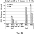
- 230000027455 binding Effects 0.000 description 29
- 239000012091 fetal bovine serum Substances 0.000 description 28
- 239000000523 sample Substances 0.000 description 27
- 239000000047 product Substances 0.000 description 26
- 239000002131 composite material Substances 0.000 description 25
- 150000007523 nucleic acids Chemical class 0.000 description 25
- 239000006228 supernatant Substances 0.000 description 25
- 239000013638 trimer Substances 0.000 description 23
- 239000011248 coating agent Substances 0.000 description 22
- 238000000576 coating method Methods 0.000 description 22
- 238000011161 development Methods 0.000 description 22
- 230000018109 developmental process Effects 0.000 description 22
- 229940126534 drug product Drugs 0.000 description 22
- 238000010899 nucleation Methods 0.000 description 22
- 239000000825 pharmaceutical preparation Substances 0.000 description 22
- OKKJLVBELUTLKV-UHFFFAOYSA-N Methanol Chemical compound OC OKKJLVBELUTLKV-UHFFFAOYSA-N 0.000 description 21
- 238000011529 RT qPCR Methods 0.000 description 21
- 238000004873 anchoring Methods 0.000 description 21
- 239000006285 cell suspension Substances 0.000 description 21
- 239000012634 fragment Substances 0.000 description 21
- 238000001114 immunoprecipitation Methods 0.000 description 21
- 230000003612 virological effect Effects 0.000 description 21
- 238000005119 centrifugation Methods 0.000 description 20
- 102000039446 nucleic acids Human genes 0.000 description 20
- 108020004707 nucleic acids Proteins 0.000 description 20
- 230000008569 process Effects 0.000 description 20
- 102000008186 Collagen Human genes 0.000 description 19
- 108010035532 Collagen Proteins 0.000 description 19
- 229920001436 collagen Polymers 0.000 description 19
- 230000029087 digestion Effects 0.000 description 19
- 238000011534 incubation Methods 0.000 description 19
- 108091032973 (ribonucleotides)n+m Proteins 0.000 description 18
- 238000001574 biopsy Methods 0.000 description 18
- 239000013604 expression vector Substances 0.000 description 18
- 238000010166 immunofluorescence Methods 0.000 description 18
- 230000005012 migration Effects 0.000 description 18
- 238000013508 migration Methods 0.000 description 18
- 241000713772 Human immunodeficiency virus 1 Species 0.000 description 17
- 235000001014 amino acid Nutrition 0.000 description 17
- 230000015572 biosynthetic process Effects 0.000 description 17
- 230000010261 cell growth Effects 0.000 description 17
- 238000010367 cloning Methods 0.000 description 17
- 239000000243 solution Substances 0.000 description 17
- 101100114361 Arabidopsis thaliana COL7 gene Proteins 0.000 description 16
- 206010052428 Wound Diseases 0.000 description 16
- 238000012512 characterization method Methods 0.000 description 16
- 239000012228 culture supernatant Substances 0.000 description 16
- 238000010232 migration assay Methods 0.000 description 16
- CURLTUGMZLYLDI-UHFFFAOYSA-N Carbon dioxide Chemical compound O=C=O CURLTUGMZLYLDI-UHFFFAOYSA-N 0.000 description 15
- 208000027418 Wounds and injury Diseases 0.000 description 15
- 239000011324 bead Substances 0.000 description 15
- 229940098773 bovine serum albumin Drugs 0.000 description 15
- 208000015181 infectious disease Diseases 0.000 description 15
- 230000003833 cell viability Effects 0.000 description 14
- 238000006243 chemical reaction Methods 0.000 description 14
- 230000006870 function Effects 0.000 description 14
- 102000040650 (ribonucleotides)n+m Human genes 0.000 description 13
- 241000283707 Capra Species 0.000 description 13
- 108020004414 DNA Proteins 0.000 description 13
- 102100034349 Integrase Human genes 0.000 description 13
- 210000001185 bone marrow Anatomy 0.000 description 13
- 239000000872 buffer Substances 0.000 description 13
- 238000004113 cell culture Methods 0.000 description 13
- 230000012292 cell migration Effects 0.000 description 13
- 238000009472 formulation Methods 0.000 description 13
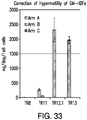
- 230000035873 hypermotility Effects 0.000 description 13
- 239000000463 material Substances 0.000 description 13
- CSCPPACGZOOCGX-UHFFFAOYSA-N Acetone Chemical compound CC(C)=O CSCPPACGZOOCGX-UHFFFAOYSA-N 0.000 description 12
- IJGRMHOSHXDMSA-UHFFFAOYSA-N Atomic nitrogen Chemical compound N#N IJGRMHOSHXDMSA-UHFFFAOYSA-N 0.000 description 12
- 101100114362 Caenorhabditis elegans col-7 gene Proteins 0.000 description 12
- PEDCQBHIVMGVHV-UHFFFAOYSA-N Glycerine Chemical compound OCC(O)CO PEDCQBHIVMGVHV-UHFFFAOYSA-N 0.000 description 12
- 238000007792 addition Methods 0.000 description 12
- 231100000673 dose–response relationship Toxicity 0.000 description 12
- 239000013642 negative control Substances 0.000 description 12
- 108010056030 retronectin Proteins 0.000 description 12
- 238000010186 staining Methods 0.000 description 12
- 208000031886 HIV Infections Diseases 0.000 description 11
- 150000001413 amino acids Chemical group 0.000 description 11
- 238000010790 dilution Methods 0.000 description 11
- 239000012895 dilution Substances 0.000 description 11
- 108010028309 kalinin Proteins 0.000 description 11
- 230000035772 mutation Effects 0.000 description 11
- 101710091045 Envelope protein Proteins 0.000 description 10
- 108091092584 GDNA Proteins 0.000 description 10
- 101710188315 Protein X Proteins 0.000 description 10
- 229940024606 amino acid Drugs 0.000 description 10
- 210000000234 capsid Anatomy 0.000 description 10
- 230000008859 change Effects 0.000 description 10
- 238000010276 construction Methods 0.000 description 10
- 238000001514 detection method Methods 0.000 description 10
- RWSXRVCMGQZWBV-WDSKDSINSA-N glutathione Chemical compound OC(=O)[C@@H](N)CCC(=O)N[C@@H](CS)C(=O)NCC(O)=O RWSXRVCMGQZWBV-WDSKDSINSA-N 0.000 description 10
- 238000003306 harvesting Methods 0.000 description 10
- 230000002458 infectious effect Effects 0.000 description 10
- 230000036512 infertility Effects 0.000 description 10
- 230000002441 reversible effect Effects 0.000 description 10
- 210000002966 serum Anatomy 0.000 description 10
- 102000010834 Extracellular Matrix Proteins Human genes 0.000 description 9
- 108010037362 Extracellular Matrix Proteins Proteins 0.000 description 9
- 108090001074 Nucleocapsid Proteins Proteins 0.000 description 9
- BELBBZDIHDAJOR-UHFFFAOYSA-N Phenolsulfonephthalein Chemical compound C1=CC(O)=CC=C1C1(C=2C=CC(O)=CC=2)C2=CC=CC=C2S(=O)(=O)O1 BELBBZDIHDAJOR-UHFFFAOYSA-N 0.000 description 9
- 238000013019 agitation Methods 0.000 description 9
- 230000004071 biological effect Effects 0.000 description 9
- 108700004025 env Genes Proteins 0.000 description 9
- 238000002474 experimental method Methods 0.000 description 9
- 239000012737 fresh medium Substances 0.000 description 9
- 229960003180 glutathione Drugs 0.000 description 9
- 230000003993 interaction Effects 0.000 description 9
- 230000004899 motility Effects 0.000 description 9
- 238000005457 optimization Methods 0.000 description 9
- 229960003531 phenolsulfonphthalein Drugs 0.000 description 9
- 238000002415 sodium dodecyl sulfate polyacrylamide gel electrophoresis Methods 0.000 description 9
- 230000029663 wound healing Effects 0.000 description 9
- 241000713666 Lentivirus Species 0.000 description 8
- 230000001419 dependent effect Effects 0.000 description 8
- 230000012010 growth Effects 0.000 description 8
- 238000003119 immunoblot Methods 0.000 description 8
- 230000010354 integration Effects 0.000 description 8
- 239000013641 positive control Substances 0.000 description 8
- 238000006467 substitution reaction Methods 0.000 description 8
- 239000011534 wash buffer Substances 0.000 description 8
- 239000007760 Iscove's Modified Dulbecco's Medium Substances 0.000 description 7
- 238000012937 correction Methods 0.000 description 7
- 238000011156 evaluation Methods 0.000 description 7
- 230000006872 improvement Effects 0.000 description 7
- 239000007788 liquid Substances 0.000 description 7
- 230000001566 pro-viral effect Effects 0.000 description 7
- 230000009467 reduction Effects 0.000 description 7
- 239000000725 suspension Substances 0.000 description 7
- 238000012384 transportation and delivery Methods 0.000 description 7
- IAZDPXIOMUYVGZ-UHFFFAOYSA-N Dimethylsulphoxide Chemical compound CS(C)=O IAZDPXIOMUYVGZ-UHFFFAOYSA-N 0.000 description 6
- 238000005138 cryopreservation Methods 0.000 description 6
- 210000002744 extracellular matrix Anatomy 0.000 description 6
- 239000012595 freezing medium Substances 0.000 description 6
- 239000000499 gel Substances 0.000 description 6
- 238000001727 in vivo Methods 0.000 description 6
- 229910052757 nitrogen Inorganic materials 0.000 description 6
- 239000008188 pellet Substances 0.000 description 6
- 230000037390 scarring Effects 0.000 description 6
- 238000007390 skin biopsy Methods 0.000 description 6
- 238000001890 transfection Methods 0.000 description 6
- 230000035899 viability Effects 0.000 description 6
- 238000005406 washing Methods 0.000 description 6
- XLYOFNOQVPJJNP-UHFFFAOYSA-N water Substances O XLYOFNOQVPJJNP-UHFFFAOYSA-N 0.000 description 6
- 239000012099 Alexa Fluor family Substances 0.000 description 5
- 101000800116 Homo sapiens Thy-1 membrane glycoprotein Proteins 0.000 description 5
- 241001465754 Metazoa Species 0.000 description 5
- 241000699670 Mus sp. Species 0.000 description 5
- 102100033523 Thy-1 membrane glycoprotein Human genes 0.000 description 5
- 241001492404 Woodchuck hepatitis virus Species 0.000 description 5
- 238000013459 approach Methods 0.000 description 5
- 239000003153 chemical reaction reagent Substances 0.000 description 5
- 230000001143 conditioned effect Effects 0.000 description 5
- 238000000684 flow cytometry Methods 0.000 description 5
- 230000002068 genetic effect Effects 0.000 description 5
- 238000003384 imaging method Methods 0.000 description 5
- 230000005764 inhibitory process Effects 0.000 description 5
- 230000000977 initiatory effect Effects 0.000 description 5
- 230000004807 localization Effects 0.000 description 5
- 239000003550 marker Substances 0.000 description 5
- 239000012528 membrane Substances 0.000 description 5
- 108020004999 messenger RNA Proteins 0.000 description 5
- 229920001184 polypeptide Polymers 0.000 description 5
- 238000002360 preparation method Methods 0.000 description 5
- 230000002265 prevention Effects 0.000 description 5
- 108090000765 processed proteins & peptides Proteins 0.000 description 5
- 102000004196 processed proteins & peptides Human genes 0.000 description 5
- 230000010076 replication Effects 0.000 description 5
- 238000010561 standard procedure Methods 0.000 description 5
- 238000003860 storage Methods 0.000 description 5
- 231100000331 toxic Toxicity 0.000 description 5
- 230000002588 toxic effect Effects 0.000 description 5
- 230000009466 transformation Effects 0.000 description 5
- QAPSNMNOIOSXSQ-YNEHKIRRSA-N 1-[(2r,4s,5r)-4-[tert-butyl(dimethyl)silyl]oxy-5-(hydroxymethyl)oxolan-2-yl]-5-methylpyrimidine-2,4-dione Chemical compound O=C1NC(=O)C(C)=CN1[C@@H]1O[C@H](CO)[C@@H](O[Si](C)(C)C(C)(C)C)C1 QAPSNMNOIOSXSQ-YNEHKIRRSA-N 0.000 description 4
- 108091026890 Coding region Proteins 0.000 description 4
- 241000283074 Equus asinus Species 0.000 description 4
- 238000012413 Fluorescence activated cell sorting analysis Methods 0.000 description 4
- DHMQDGOQFOQNFH-UHFFFAOYSA-N Glycine Chemical compound NCC(O)=O DHMQDGOQFOQNFH-UHFFFAOYSA-N 0.000 description 4
- 241000699666 Mus <mouse, genus> Species 0.000 description 4
- 238000011579 SCID mouse model Methods 0.000 description 4
- 108700005078 Synthetic Genes Proteins 0.000 description 4
- 108020000999 Viral RNA Proteins 0.000 description 4
- 238000002835 absorbance Methods 0.000 description 4
- 230000003698 anagen phase Effects 0.000 description 4
- 239000003242 anti bacterial agent Substances 0.000 description 4
- 229940088710 antibiotic agent Drugs 0.000 description 4
- 238000003149 assay kit Methods 0.000 description 4
- 210000002469 basement membrane Anatomy 0.000 description 4
- 239000002299 complementary DNA Substances 0.000 description 4
- 208000037265 diseases, disorders, signs and symptoms Diseases 0.000 description 4
- 239000003814 drug Substances 0.000 description 4
- 239000002158 endotoxin Substances 0.000 description 4
- 230000007613 environmental effect Effects 0.000 description 4
- 238000010195 expression analysis Methods 0.000 description 4
- 230000036541 health Effects 0.000 description 4
- JYGXADMDTFJGBT-VWUMJDOOSA-N hydrocortisone Chemical compound O=C1CC[C@]2(C)[C@H]3[C@@H](O)C[C@](C)([C@@](CC4)(O)C(=O)CO)[C@@H]4[C@@H]3CCC2=C1 JYGXADMDTFJGBT-VWUMJDOOSA-N 0.000 description 4
- 238000003125 immunofluorescent labeling Methods 0.000 description 4
- 238000002955 isolation Methods 0.000 description 4
- 238000012545 processing Methods 0.000 description 4
- 238000000159 protein binding assay Methods 0.000 description 4
- 238000003908 quality control method Methods 0.000 description 4
- 230000002829 reductive effect Effects 0.000 description 4
- 238000011160 research Methods 0.000 description 4
- 230000001177 retroviral effect Effects 0.000 description 4
- 238000013341 scale-up Methods 0.000 description 4
- 239000013605 shuttle vector Substances 0.000 description 4
- 239000007858 starting material Substances 0.000 description 4
- 101150084750 1 gene Proteins 0.000 description 3
- 101100328883 Arabidopsis thaliana COL1 gene Proteins 0.000 description 3
- 101100328892 Arabidopsis thaliana COL4 gene Proteins 0.000 description 3
- 241001123248 Arma Species 0.000 description 3
- 102000000844 Cell Surface Receptors Human genes 0.000 description 3
- 108010001857 Cell Surface Receptors Proteins 0.000 description 3
- 102000004190 Enzymes Human genes 0.000 description 3
- 108090000790 Enzymes Proteins 0.000 description 3
- 241000588724 Escherichia coli Species 0.000 description 3
- 108010067306 Fibronectins Proteins 0.000 description 3
- 102000016359 Fibronectins Human genes 0.000 description 3
- 206010016654 Fibrosis Diseases 0.000 description 3
- CEAZRRDELHUEMR-URQXQFDESA-N Gentamicin Chemical compound O1[C@H](C(C)NC)CC[C@@H](N)[C@H]1O[C@H]1[C@H](O)[C@@H](O[C@@H]2[C@@H]([C@@H](NC)[C@@](C)(O)CO2)O)[C@H](N)C[C@@H]1N CEAZRRDELHUEMR-URQXQFDESA-N 0.000 description 3
- 229930182566 Gentamicin Natural products 0.000 description 3
- HVLSXIKZNLPZJJ-TXZCQADKSA-N HA peptide Chemical compound C([C@@H](C(=O)N[C@@H](CC(O)=O)C(=O)N[C@@H](C(C)C)C(=O)N1[C@@H](CCC1)C(=O)N[C@@H](CC(O)=O)C(=O)N[C@@H](CC=1C=CC(O)=CC=1)C(=O)N[C@@H](C)C(O)=O)NC(=O)[C@H]1N(CCC1)C(=O)[C@@H](N)CC=1C=CC(O)=CC=1)C1=CC=C(O)C=C1 HVLSXIKZNLPZJJ-TXZCQADKSA-N 0.000 description 3
- 241000700721 Hepatitis B virus Species 0.000 description 3
- 241000282412 Homo Species 0.000 description 3
- 241000725303 Human immunodeficiency virus Species 0.000 description 3
- 108091028043 Nucleic acid sequence Proteins 0.000 description 3
- 101710149951 Protein Tat Proteins 0.000 description 3
- 101710120037 Toxin CcdB Proteins 0.000 description 3
- 101100237842 Xenopus laevis mmp18 gene Proteins 0.000 description 3
- 229960000723 ampicillin Drugs 0.000 description 3
- AVKUERGKIZMTKX-NJBDSQKTSA-N ampicillin Chemical compound C1([C@@H](N)C(=O)N[C@H]2[C@H]3SC([C@@H](N3C2=O)C(O)=O)(C)C)=CC=CC=C1 AVKUERGKIZMTKX-NJBDSQKTSA-N 0.000 description 3
- 239000000427 antigen Substances 0.000 description 3
- 108091007433 antigens Proteins 0.000 description 3
- 102000036639 antigens Human genes 0.000 description 3
- 239000012512 bulk drug substance Substances 0.000 description 3
- 235000011089 carbon dioxide Nutrition 0.000 description 3
- 238000012832 cell culture technique Methods 0.000 description 3
- 238000002659 cell therapy Methods 0.000 description 3
- 230000005754 cellular signaling Effects 0.000 description 3
- 239000003636 conditioned culture medium Substances 0.000 description 3
- 238000007405 data analysis Methods 0.000 description 3
- 201000010099 disease Diseases 0.000 description 3
- 230000004761 fibrosis Effects 0.000 description 3
- 238000001943 fluorescence-activated cell sorting Methods 0.000 description 3
- 238000001415 gene therapy Methods 0.000 description 3
- 231100000025 genetic toxicology Toxicity 0.000 description 3
- 230000001738 genotoxic effect Effects 0.000 description 3
- 229960002518 gentamicin Drugs 0.000 description 3
- PCHJSUWPFVWCPO-UHFFFAOYSA-N gold Chemical class [Au] PCHJSUWPFVWCPO-UHFFFAOYSA-N 0.000 description 3
- 238000003780 insertion Methods 0.000 description 3
- 230000037431 insertion Effects 0.000 description 3
- 230000003902 lesion Effects 0.000 description 3
- 239000003446 ligand Substances 0.000 description 3
- 210000004379 membrane Anatomy 0.000 description 3
- 238000000386 microscopy Methods 0.000 description 3
- 210000000056 organ Anatomy 0.000 description 3
- 230000008520 organization Effects 0.000 description 3
- 239000004033 plastic Substances 0.000 description 3
- 239000000758 substrate Substances 0.000 description 3
- 238000002560 therapeutic procedure Methods 0.000 description 3
- 238000013518 transcription Methods 0.000 description 3
- 230000035897 transcription Effects 0.000 description 3
- 239000013603 viral vector Substances 0.000 description 3
- 238000001262 western blot Methods 0.000 description 3
- WOXWUZCRWJWTRT-UHFFFAOYSA-N 1-amino-1-cyclohexanecarboxylic acid Chemical compound OC(=O)C1(N)CCCCC1 WOXWUZCRWJWTRT-UHFFFAOYSA-N 0.000 description 2
- OGNSCSPNOLGXSM-UHFFFAOYSA-N 2,4-diaminobutyric acid Chemical compound NCCC(N)C(O)=O OGNSCSPNOLGXSM-UHFFFAOYSA-N 0.000 description 2
- UAIUNKRWKOVEES-UHFFFAOYSA-N 3,3',5,5'-tetramethylbenzidine Chemical compound CC1=C(N)C(C)=CC(C=2C=C(C)C(N)=C(C)C=2)=C1 UAIUNKRWKOVEES-UHFFFAOYSA-N 0.000 description 2
- PECYZEOJVXMISF-UHFFFAOYSA-N 3-aminoalanine Chemical compound [NH3+]CC(N)C([O-])=O PECYZEOJVXMISF-UHFFFAOYSA-N 0.000 description 2
- 108020003589 5' Untranslated Regions Proteins 0.000 description 2
- 102100024643 ATP-binding cassette sub-family D member 1 Human genes 0.000 description 2
- 201000011452 Adrenoleukodystrophy Diseases 0.000 description 2
- 108090000565 Capsid Proteins Proteins 0.000 description 2
- BVKZGUZCCUSVTD-UHFFFAOYSA-L Carbonate Chemical compound [O-]C([O-])=O BVKZGUZCCUSVTD-UHFFFAOYSA-L 0.000 description 2
- 102100023321 Ceruloplasmin Human genes 0.000 description 2
- 108020004705 Codon Proteins 0.000 description 2
- 102000012422 Collagen Type I Human genes 0.000 description 2
- 108010022452 Collagen Type I Proteins 0.000 description 2
- 230000006820 DNA synthesis Effects 0.000 description 2
- 238000012286 ELISA Assay Methods 0.000 description 2
- 108700007698 Genetic Terminator Regions Proteins 0.000 description 2
- 239000004471 Glycine Substances 0.000 description 2
- 238000012369 In process control Methods 0.000 description 2
- 239000004472 Lysine Substances 0.000 description 2
- KDXKERNSBIXSRK-UHFFFAOYSA-N Lysine Natural products NCCCCC(N)C(O)=O KDXKERNSBIXSRK-UHFFFAOYSA-N 0.000 description 2
- 108010049137 Member 1 Subfamily D ATP Binding Cassette Transporter Proteins 0.000 description 2
- 206010028980 Neoplasm Diseases 0.000 description 2
- 239000000020 Nitrocellulose Substances 0.000 description 2
- 108091005461 Nucleic proteins Proteins 0.000 description 2
- 108091005804 Peptidases Proteins 0.000 description 2
- 101710150344 Protein Rev Proteins 0.000 description 2
- 102000052575 Proto-Oncogene Human genes 0.000 description 2
- 108700020978 Proto-Oncogene Proteins 0.000 description 2
- 108010092799 RNA-directed DNA polymerase Proteins 0.000 description 2
- MTCFGRXMJLQNBG-UHFFFAOYSA-N Serine Natural products OCC(N)C(O)=O MTCFGRXMJLQNBG-UHFFFAOYSA-N 0.000 description 2
- FAPWRFPIFSIZLT-UHFFFAOYSA-M Sodium chloride Chemical compound [Na+].[Cl-] FAPWRFPIFSIZLT-UHFFFAOYSA-M 0.000 description 2
- 206010042566 Superinfection Diseases 0.000 description 2
- 108700009124 Transcription Initiation Site Proteins 0.000 description 2
- 208000006110 Wiskott-Aldrich syndrome Diseases 0.000 description 2
- APKFDSVGJQXUKY-INPOYWNPSA-N amphotericin B Chemical compound O[C@H]1[C@@H](N)[C@H](O)[C@@H](C)O[C@H]1O[C@H]1/C=C/C=C/C=C/C=C/C=C/C=C/C=C/[C@H](C)[C@@H](O)[C@@H](C)[C@H](C)OC(=O)C[C@H](O)C[C@H](O)CC[C@@H](O)[C@H](O)C[C@H](O)C[C@](O)(C[C@H](O)[C@H]2C(O)=O)O[C@H]2C1 APKFDSVGJQXUKY-INPOYWNPSA-N 0.000 description 2
- 230000003466 anti-cipated effect Effects 0.000 description 2
- 239000012984 antibiotic solution Substances 0.000 description 2
- 238000013096 assay test Methods 0.000 description 2
- 229940055553 azficel-t Drugs 0.000 description 2
- 230000001580 bacterial effect Effects 0.000 description 2
- 230000008901 benefit Effects 0.000 description 2
- 238000013357 binding ELISA Methods 0.000 description 2
- 239000012148 binding buffer Substances 0.000 description 2
- 239000006143 cell culture medium Substances 0.000 description 2
- 238000012710 chemistry, manufacturing and control Methods 0.000 description 2
- 238000004140 cleaning Methods 0.000 description 2
- 239000000470 constituent Substances 0.000 description 2
- 239000013078 crystal Substances 0.000 description 2
- 230000001186 cumulative effect Effects 0.000 description 2
- 230000001351 cycling effect Effects 0.000 description 2
- 238000004163 cytometry Methods 0.000 description 2
- 230000001086 cytosolic effect Effects 0.000 description 2
- 238000013461 design Methods 0.000 description 2
- UQLDLKMNUJERMK-UHFFFAOYSA-L di(octadecanoyloxy)lead Chemical compound [Pb+2].CCCCCCCCCCCCCCCCCC([O-])=O.CCCCCCCCCCCCCCCCCC([O-])=O UQLDLKMNUJERMK-UHFFFAOYSA-L 0.000 description 2
- 238000011026 diafiltration Methods 0.000 description 2
- 229940079593 drug Drugs 0.000 description 2
- 239000003937 drug carrier Substances 0.000 description 2
- 239000000975 dye Substances 0.000 description 2
- 238000000635 electron micrograph Methods 0.000 description 2
- 239000012149 elution buffer Substances 0.000 description 2
- 238000005516 engineering process Methods 0.000 description 2
- 230000006862 enzymatic digestion Effects 0.000 description 2
- 230000003203 everyday effect Effects 0.000 description 2
- 239000012467 final product Substances 0.000 description 2
- 238000002509 fluorescent in situ hybridization Methods 0.000 description 2
- 229960000890 hydrocortisone Drugs 0.000 description 2
- 238000004191 hydrophobic interaction chromatography Methods 0.000 description 2
- 238000010965 in-process control Methods 0.000 description 2
- NOESYZHRGYRDHS-UHFFFAOYSA-N insulin Chemical compound N1C(=O)C(NC(=O)C(CCC(N)=O)NC(=O)C(CCC(O)=O)NC(=O)C(C(C)C)NC(=O)C(NC(=O)CN)C(C)CC)CSSCC(C(NC(CO)C(=O)NC(CC(C)C)C(=O)NC(CC=2C=CC(O)=CC=2)C(=O)NC(CCC(N)=O)C(=O)NC(CC(C)C)C(=O)NC(CCC(O)=O)C(=O)NC(CC(N)=O)C(=O)NC(CC=2C=CC(O)=CC=2)C(=O)NC(CSSCC(NC(=O)C(C(C)C)NC(=O)C(CC(C)C)NC(=O)C(CC=2C=CC(O)=CC=2)NC(=O)C(CC(C)C)NC(=O)C(C)NC(=O)C(CCC(O)=O)NC(=O)C(C(C)C)NC(=O)C(CC(C)C)NC(=O)C(CC=2NC=NC=2)NC(=O)C(CO)NC(=O)CNC2=O)C(=O)NCC(=O)NC(CCC(O)=O)C(=O)NC(CCCNC(N)=N)C(=O)NCC(=O)NC(CC=3C=CC=CC=3)C(=O)NC(CC=3C=CC=CC=3)C(=O)NC(CC=3C=CC(O)=CC=3)C(=O)NC(C(C)O)C(=O)N3C(CCC3)C(=O)NC(CCCCN)C(=O)NC(C)C(O)=O)C(=O)NC(CC(N)=O)C(O)=O)=O)NC(=O)C(C(C)CC)NC(=O)C(CO)NC(=O)C(C(C)O)NC(=O)C1CSSCC2NC(=O)C(CC(C)C)NC(=O)C(NC(=O)C(CCC(N)=O)NC(=O)C(CC(N)=O)NC(=O)C(NC(=O)C(N)CC=1C=CC=CC=1)C(C)C)CC1=CN=CN1 NOESYZHRGYRDHS-UHFFFAOYSA-N 0.000 description 2
- 230000000670 limiting effect Effects 0.000 description 2
- 150000002632 lipids Chemical class 0.000 description 2
- 238000011068 loading method Methods 0.000 description 2
- 230000007774 longterm Effects 0.000 description 2
- 238000012554 master batch record Methods 0.000 description 2
- 238000007431 microscopic evaluation Methods 0.000 description 2
- 235000013336 milk Nutrition 0.000 description 2
- 239000008267 milk Substances 0.000 description 2
- 210000004080 milk Anatomy 0.000 description 2
- 238000002156 mixing Methods 0.000 description 2
- 238000012544 monitoring process Methods 0.000 description 2
- 229920001220 nitrocellulos Polymers 0.000 description 2
- 230000037311 normal skin Effects 0.000 description 2
- 230000003287 optical effect Effects 0.000 description 2
- 230000002688 persistence Effects 0.000 description 2
- 239000013600 plasmid vector Substances 0.000 description 2
- 238000007747 plating Methods 0.000 description 2
- 238000003762 quantitative reverse transcription PCR Methods 0.000 description 2
- 238000010791 quenching Methods 0.000 description 2
- 230000000171 quenching effect Effects 0.000 description 2
- 239000002994 raw material Substances 0.000 description 2
- 102000005962 receptors Human genes 0.000 description 2
- 108020003175 receptors Proteins 0.000 description 2
- 230000000717 retained effect Effects 0.000 description 2
- 238000011218 seed culture Methods 0.000 description 2
- 238000012163 sequencing technique Methods 0.000 description 2
- 210000004927 skin cell Anatomy 0.000 description 2
- 239000002904 solvent Substances 0.000 description 2
- 241000894007 species Species 0.000 description 2
- 210000004500 stellate cell Anatomy 0.000 description 2
- 238000013190 sterility testing Methods 0.000 description 2
- UCSJYZPVAKXKNQ-HZYVHMACSA-N streptomycin Chemical compound CN[C@H]1[C@H](O)[C@@H](O)[C@H](CO)O[C@H]1O[C@@H]1[C@](C=O)(O)[C@H](C)O[C@H]1O[C@@H]1[C@@H](NC(N)=N)[C@H](O)[C@@H](NC(N)=N)[C@H](O)[C@H]1O UCSJYZPVAKXKNQ-HZYVHMACSA-N 0.000 description 2
- 208000024891 symptom Diseases 0.000 description 2
- 229940124597 therapeutic agent Drugs 0.000 description 2
- -1 they can be 50% Chemical class 0.000 description 2
- 231100000027 toxicology Toxicity 0.000 description 2
- 238000013519 translation Methods 0.000 description 2
- 230000032258 transport Effects 0.000 description 2
- PIEPQKCYPFFYMG-UHFFFAOYSA-N tris acetate Chemical compound CC(O)=O.OCC(N)(CO)CO PIEPQKCYPFFYMG-UHFFFAOYSA-N 0.000 description 2
- 230000006490 viral transcription Effects 0.000 description 2
- OCLLVJCYGMCLJG-CYBMUJFWSA-N (2r)-2-azaniumyl-2-naphthalen-1-ylpropanoate Chemical compound C1=CC=C2C([C@@](N)(C(O)=O)C)=CC=CC2=C1 OCLLVJCYGMCLJG-CYBMUJFWSA-N 0.000 description 1
- QFQYGJMNIDGZSG-YFKPBYRVSA-N (2r)-3-(acetamidomethylsulfanyl)-2-azaniumylpropanoate Chemical compound CC(=O)NCSC[C@H]([NH3+])C([O-])=O QFQYGJMNIDGZSG-YFKPBYRVSA-N 0.000 description 1
- BFNDLDRNJFLIKE-ROLXFIACSA-N (2s)-2,6-diamino-6-hydroxyhexanoic acid Chemical compound NC(O)CCC[C@H](N)C(O)=O BFNDLDRNJFLIKE-ROLXFIACSA-N 0.000 description 1
- BVAUMRCGVHUWOZ-ZETCQYMHSA-N (2s)-2-(cyclohexylazaniumyl)propanoate Chemical compound OC(=O)[C@H](C)NC1CCCCC1 BVAUMRCGVHUWOZ-ZETCQYMHSA-N 0.000 description 1
- DWKNTLVYZNGBTJ-IBGZPJMESA-N (2s)-2-amino-6-(dibenzylamino)hexanoic acid Chemical compound C=1C=CC=CC=1CN(CCCC[C@H](N)C(O)=O)CC1=CC=CC=C1 DWKNTLVYZNGBTJ-IBGZPJMESA-N 0.000 description 1
- FNRJOGDXTIUYDE-ZDUSSCGKSA-N (2s)-2-amino-6-[benzyl(methyl)amino]hexanoic acid Chemical compound OC(=O)[C@@H](N)CCCCN(C)CC1=CC=CC=C1 FNRJOGDXTIUYDE-ZDUSSCGKSA-N 0.000 description 1
- WAMWSIDTKSNDCU-ZETCQYMHSA-N (2s)-2-azaniumyl-2-cyclohexylacetate Chemical compound OC(=O)[C@@H](N)C1CCCCC1 WAMWSIDTKSNDCU-ZETCQYMHSA-N 0.000 description 1
- UKAUYVFTDYCKQA-UHFFFAOYSA-N -2-Amino-4-hydroxybutanoic acid Natural products OC(=O)C(N)CCO UKAUYVFTDYCKQA-UHFFFAOYSA-N 0.000 description 1
- BWKMGYQJPOAASG-UHFFFAOYSA-N 1,2,3,4-tetrahydroisoquinoline-3-carboxylic acid Chemical compound C1=CC=C2CNC(C(=O)O)CC2=C1 BWKMGYQJPOAASG-UHFFFAOYSA-N 0.000 description 1
- YTPMCWYIRHLEGM-BQYQJAHWSA-N 1-[(e)-2-propylsulfonylethenyl]sulfonylpropane Chemical compound CCCS(=O)(=O)\C=C\S(=O)(=O)CCC YTPMCWYIRHLEGM-BQYQJAHWSA-N 0.000 description 1
- KNQHBAFIWGORKW-UHFFFAOYSA-N 2,3-diamino-3-oxopropanoic acid Chemical compound NC(=O)C(N)C(O)=O KNQHBAFIWGORKW-UHFFFAOYSA-N 0.000 description 1
- JKMHFZQWWAIEOD-UHFFFAOYSA-N 2-[4-(2-hydroxyethyl)piperazin-1-yl]ethanesulfonic acid Chemical compound OCC[NH+]1CCN(CCS([O-])(=O)=O)CC1 JKMHFZQWWAIEOD-UHFFFAOYSA-N 0.000 description 1
- VHVGNTVUSQUXPS-UHFFFAOYSA-N 2-amino-3-hydroxy-3-phenylpropanoic acid Chemical compound OC(=O)C(N)C(O)C1=CC=CC=C1 VHVGNTVUSQUXPS-UHFFFAOYSA-N 0.000 description 1
- JINGUCXQUOKWKH-UHFFFAOYSA-N 2-aminodecanoic acid Chemical compound CCCCCCCCC(N)C(O)=O JINGUCXQUOKWKH-UHFFFAOYSA-N 0.000 description 1
- YXDGRBPZVQPESQ-QMMMGPOBSA-N 4-[(2s)-2-amino-2-carboxyethyl]benzoic acid Chemical compound OC(=O)[C@@H](N)CC1=CC=C(C(O)=O)C=C1 YXDGRBPZVQPESQ-QMMMGPOBSA-N 0.000 description 1
- FWMNVWWHGCHHJJ-SKKKGAJSSA-N 4-amino-1-[(2r)-6-amino-2-[[(2r)-2-[[(2r)-2-[[(2r)-2-amino-3-phenylpropanoyl]amino]-3-phenylpropanoyl]amino]-4-methylpentanoyl]amino]hexanoyl]piperidine-4-carboxylic acid Chemical compound C([C@H](C(=O)N[C@H](CC(C)C)C(=O)N[C@H](CCCCN)C(=O)N1CCC(N)(CC1)C(O)=O)NC(=O)[C@H](N)CC=1C=CC=CC=1)C1=CC=CC=C1 FWMNVWWHGCHHJJ-SKKKGAJSSA-N 0.000 description 1
- CMUHFUGDYMFHEI-QMMMGPOBSA-N 4-amino-L-phenylalanine Chemical compound OC(=O)[C@@H](N)CC1=CC=C(N)C=C1 CMUHFUGDYMFHEI-QMMMGPOBSA-N 0.000 description 1
- GTVVZTAFGPQSPC-UHFFFAOYSA-N 4-nitrophenylalanine Chemical compound OC(=O)C(N)CC1=CC=C([N+]([O-])=O)C=C1 GTVVZTAFGPQSPC-UHFFFAOYSA-N 0.000 description 1
- 229930024421 Adenine Natural products 0.000 description 1
- GFFGJBXGBJISGV-UHFFFAOYSA-N Adenine Chemical compound NC1=NC=NC2=C1N=CN2 GFFGJBXGBJISGV-UHFFFAOYSA-N 0.000 description 1
- 201000004384 Alopecia Diseases 0.000 description 1
- 229930183010 Amphotericin Natural products 0.000 description 1
- QGGFZZLFKABGNL-UHFFFAOYSA-N Amphotericin A Natural products OC1C(N)C(O)C(C)OC1OC1C=CC=CC=CC=CCCC=CC=CC(C)C(O)C(C)C(C)OC(=O)CC(O)CC(O)CCC(O)C(O)CC(O)CC(O)(CC(O)C2C(O)=O)OC2C1 QGGFZZLFKABGNL-UHFFFAOYSA-N 0.000 description 1
- APKFDSVGJQXUKY-KKGHZKTASA-N Amphotericin-B Natural products O[C@H]1[C@@H](N)[C@H](O)[C@@H](C)O[C@H]1O[C@H]1C=CC=CC=CC=CC=CC=CC=C[C@H](C)[C@@H](O)[C@@H](C)[C@H](C)OC(=O)C[C@H](O)C[C@H](O)CC[C@@H](O)[C@H](O)C[C@H](O)C[C@](O)(C[C@H](O)[C@H]2C(O)=O)O[C@H]2C1 APKFDSVGJQXUKY-KKGHZKTASA-N 0.000 description 1
- 108091093088 Amplicon Proteins 0.000 description 1
- 239000004475 Arginine Substances 0.000 description 1
- DCXYFEDJOCDNAF-UHFFFAOYSA-N Asparagine Natural products OC(=O)C(N)CC(N)=O DCXYFEDJOCDNAF-UHFFFAOYSA-N 0.000 description 1
- 108020000946 Bacterial DNA Proteins 0.000 description 1
- 241000283690 Bos taurus Species 0.000 description 1
- 101001011741 Bos taurus Insulin Proteins 0.000 description 1
- 101000766308 Bos taurus Serotransferrin Proteins 0.000 description 1
- 230000005653 Brownian motion process Effects 0.000 description 1
- 238000011740 C57BL/6 mouse Methods 0.000 description 1
- 101150104396 C7 gene Proteins 0.000 description 1
- 102000009016 Cholera Toxin Human genes 0.000 description 1
- 108010049048 Cholera Toxin Proteins 0.000 description 1
- 208000032170 Congenital Abnormalities Diseases 0.000 description 1
- 206010011026 Corneal lesion Diseases 0.000 description 1
- 238000007399 DNA isolation Methods 0.000 description 1
- 230000004543 DNA replication Effects 0.000 description 1
- 238000001712 DNA sequencing Methods 0.000 description 1
- 206010048768 Dermatosis Diseases 0.000 description 1
- 102100024746 Dihydrofolate reductase Human genes 0.000 description 1
- 102100038132 Endogenous retrovirus group K member 6 Pro protein Human genes 0.000 description 1
- 102000009024 Epidermal Growth Factor Human genes 0.000 description 1
- 206010014989 Epidermolysis bullosa Diseases 0.000 description 1
- 108700039887 Essential Genes Proteins 0.000 description 1
- 206010015548 Euthanasia Diseases 0.000 description 1
- NIGWMJHCCYYCSF-UHFFFAOYSA-N Fenclonine Chemical compound OC(=O)C(N)CC1=CC=C(Cl)C=C1 NIGWMJHCCYYCSF-UHFFFAOYSA-N 0.000 description 1
- 231100001273 GLP toxicology study Toxicity 0.000 description 1
- 101710177291 Gag polyprotein Proteins 0.000 description 1
- WHUUTDBJXJRKMK-UHFFFAOYSA-N Glutamic acid Natural products OC(=O)C(N)CCC(O)=O WHUUTDBJXJRKMK-UHFFFAOYSA-N 0.000 description 1
- 102000003886 Glycoproteins Human genes 0.000 description 1
- 108090000288 Glycoproteins Proteins 0.000 description 1
- 108060003393 Granulin Proteins 0.000 description 1
- 241001326189 Gyrodactylus prostae Species 0.000 description 1
- 239000007995 HEPES buffer Substances 0.000 description 1
- 239000012981 Hank's balanced salt solution Substances 0.000 description 1
- 208000028782 Hereditary disease Diseases 0.000 description 1
- 101500025419 Homo sapiens Epidermal growth factor Proteins 0.000 description 1
- 108700020121 Human Immunodeficiency Virus-1 rev Proteins 0.000 description 1
- PMMYEEVYMWASQN-DMTCNVIQSA-N Hydroxyproline Chemical compound O[C@H]1CN[C@H](C(O)=O)C1 PMMYEEVYMWASQN-DMTCNVIQSA-N 0.000 description 1
- 102000004877 Insulin Human genes 0.000 description 1
- 108090001061 Insulin Proteins 0.000 description 1
- 108010061833 Integrases Proteins 0.000 description 1
- 206010023201 Joint contracture Diseases 0.000 description 1
- AHLPHDHHMVZTML-BYPYZUCNSA-N L-Ornithine Chemical compound NCCC[C@H](N)C(O)=O AHLPHDHHMVZTML-BYPYZUCNSA-N 0.000 description 1
- ZGUNAGUHMKGQNY-ZETCQYMHSA-N L-alpha-phenylglycine zwitterion Chemical compound OC(=O)[C@@H](N)C1=CC=CC=C1 ZGUNAGUHMKGQNY-ZETCQYMHSA-N 0.000 description 1
- DCXYFEDJOCDNAF-REOHCLBHSA-N L-asparagine Chemical compound OC(=O)[C@@H](N)CC(N)=O DCXYFEDJOCDNAF-REOHCLBHSA-N 0.000 description 1
- CKLJMWTZIZZHCS-REOHCLBHSA-N L-aspartic acid Chemical compound OC(=O)[C@@H](N)CC(O)=O CKLJMWTZIZZHCS-REOHCLBHSA-N 0.000 description 1
- JTTHKOPSMAVJFE-VIFPVBQESA-N L-homophenylalanine Chemical compound OC(=O)[C@@H](N)CCC1=CC=CC=C1 JTTHKOPSMAVJFE-VIFPVBQESA-N 0.000 description 1
- UKAUYVFTDYCKQA-VKHMYHEASA-N L-homoserine Chemical compound OC(=O)[C@@H](N)CCO UKAUYVFTDYCKQA-VKHMYHEASA-N 0.000 description 1
- LRQKBLKVPFOOQJ-YFKPBYRVSA-N L-norleucine Chemical compound CCCC[C@H]([NH3+])C([O-])=O LRQKBLKVPFOOQJ-YFKPBYRVSA-N 0.000 description 1
- COLNVLDHVKWLRT-QMMMGPOBSA-N L-phenylalanine Chemical compound OC(=O)[C@@H](N)CC1=CC=CC=C1 COLNVLDHVKWLRT-QMMMGPOBSA-N 0.000 description 1
- VHVGNTVUSQUXPS-YUMQZZPRSA-N L-threo-3-phenylserine Chemical compound [O-]C(=O)[C@@H]([NH3+])[C@@H](O)C1=CC=CC=C1 VHVGNTVUSQUXPS-YUMQZZPRSA-N 0.000 description 1
- QIVBCDIJIAJPQS-VIFPVBQESA-N L-tryptophane Chemical compound C1=CC=C2C(C[C@H](N)C(O)=O)=CNC2=C1 QIVBCDIJIAJPQS-VIFPVBQESA-N 0.000 description 1
- 108010085895 Laminin Proteins 0.000 description 1
- 102000007547 Laminin Human genes 0.000 description 1
- 102000006835 Lamins Human genes 0.000 description 1
- 108010047294 Lamins Proteins 0.000 description 1
- 108010052014 Liberase Proteins 0.000 description 1
- 239000000232 Lipid Bilayer Substances 0.000 description 1
- 101710125418 Major capsid protein Proteins 0.000 description 1
- 241000124008 Mammalia Species 0.000 description 1
- 102000012750 Membrane Glycoproteins Human genes 0.000 description 1
- 108010090054 Membrane Glycoproteins Proteins 0.000 description 1
- 102000018697 Membrane Proteins Human genes 0.000 description 1
- 108010052285 Membrane Proteins Proteins 0.000 description 1
- 208000024556 Mendelian disease Diseases 0.000 description 1
- 201000011442 Metachromatic leukodystrophy Diseases 0.000 description 1
- 206010027626 Milia Diseases 0.000 description 1
- 206010062575 Muscle contracture Diseases 0.000 description 1
- 241000204031 Mycoplasma Species 0.000 description 1
- UEEJHVSXFDXPFK-UHFFFAOYSA-N N-dimethylaminoethanol Chemical compound CN(C)CCO UEEJHVSXFDXPFK-UHFFFAOYSA-N 0.000 description 1
- 206010028698 Nail dystrophy Diseases 0.000 description 1
- 229930193140 Neomycin Natural products 0.000 description 1
- 108700026244 Open Reading Frames Proteins 0.000 description 1
- AHLPHDHHMVZTML-UHFFFAOYSA-N Orn-delta-NH2 Natural products NCCCC(N)C(O)=O AHLPHDHHMVZTML-UHFFFAOYSA-N 0.000 description 1
- UTJLXEIPEHZYQJ-UHFFFAOYSA-N Ornithine Natural products OC(=O)C(C)CCCN UTJLXEIPEHZYQJ-UHFFFAOYSA-N 0.000 description 1
- 241000283973 Oryctolagus cuniculus Species 0.000 description 1
- 229930182555 Penicillin Natural products 0.000 description 1
- JGSARLDLIJGVTE-MBNYWOFBSA-N Penicillin G Chemical compound N([C@H]1[C@H]2SC([C@@H](N2C1=O)C(O)=O)(C)C)C(=O)CC1=CC=CC=C1 JGSARLDLIJGVTE-MBNYWOFBSA-N 0.000 description 1
- 239000004365 Protease Substances 0.000 description 1
- 241001478271 Rahnella aquatilis Species 0.000 description 1
- 108010008281 Recombinant Fusion Proteins Proteins 0.000 description 1
- 102000007056 Recombinant Fusion Proteins Human genes 0.000 description 1
- 241001068263 Replication competent viruses Species 0.000 description 1
- 108700008625 Reporter Genes Proteins 0.000 description 1
- 241000283984 Rodentia Species 0.000 description 1
- 240000003801 Sigesbeckia orientalis Species 0.000 description 1
- 235000003407 Sigesbeckia orientalis Nutrition 0.000 description 1
- 238000002105 Southern blotting Methods 0.000 description 1
- 208000022626 Syndactyly type 6 Diseases 0.000 description 1
- 210000001744 T-lymphocyte Anatomy 0.000 description 1
- 101150052863 THY1 gene Proteins 0.000 description 1
- AYFVYJQAPQTCCC-UHFFFAOYSA-N Threonine Natural products CC(O)C(N)C(O)=O AYFVYJQAPQTCCC-UHFFFAOYSA-N 0.000 description 1
- 239000004473 Threonine Substances 0.000 description 1
- AUYYCJSJGJYCDS-LBPRGKRZSA-N Thyrolar Chemical compound IC1=CC(C[C@H](N)C(O)=O)=CC(I)=C1OC1=CC=C(O)C(I)=C1 AUYYCJSJGJYCDS-LBPRGKRZSA-N 0.000 description 1
- 102000004338 Transferrin Human genes 0.000 description 1
- 108090000901 Transferrin Proteins 0.000 description 1
- GLNADSQYFUSGOU-GPTZEZBUSA-J Trypan blue Chemical compound [Na+].[Na+].[Na+].[Na+].C1=C(S([O-])(=O)=O)C=C2C=C(S([O-])(=O)=O)C(/N=N/C3=CC=C(C=C3C)C=3C=C(C(=CC=3)\N=N\C=3C(=CC4=CC(=CC(N)=C4C=3O)S([O-])(=O)=O)S([O-])(=O)=O)C)=C(O)C2=C1N GLNADSQYFUSGOU-GPTZEZBUSA-J 0.000 description 1
- 101710162629 Trypsin inhibitor Proteins 0.000 description 1
- 229940122618 Trypsin inhibitor Drugs 0.000 description 1
- QIVBCDIJIAJPQS-UHFFFAOYSA-N Tryptophan Natural products C1=CC=C2C(CC(N)C(O)=O)=CNC2=C1 QIVBCDIJIAJPQS-UHFFFAOYSA-N 0.000 description 1
- 102000044209 Tumor Suppressor Genes Human genes 0.000 description 1
- 108700025716 Tumor Suppressor Genes Proteins 0.000 description 1
- 108010059993 Vancomycin Proteins 0.000 description 1
- 241000711975 Vesicular stomatitis virus Species 0.000 description 1
- 108010067390 Viral Proteins Proteins 0.000 description 1
- 208000036142 Viral infection Diseases 0.000 description 1
- 108010006886 Vitrogen Proteins 0.000 description 1
- 108010084455 Zeocin Proteins 0.000 description 1
- 230000001594 aberrant effect Effects 0.000 description 1
- 230000006978 adaptation Effects 0.000 description 1
- 101150063416 add gene Proteins 0.000 description 1
- 229960000643 adenine Drugs 0.000 description 1
- 230000002411 adverse Effects 0.000 description 1
- 239000011543 agarose gel Substances 0.000 description 1
- 208000026935 allergic disease Diseases 0.000 description 1
- 231100000360 alopecia Toxicity 0.000 description 1
- 229960004821 amikacin Drugs 0.000 description 1
- LKCWBDHBTVXHDL-RMDFUYIESA-N amikacin Chemical compound O([C@@H]1[C@@H](N)C[C@H]([C@@H]([C@H]1O)O[C@@H]1[C@@H]([C@@H](N)[C@H](O)[C@@H](CO)O1)O)NC(=O)[C@@H](O)CCN)[C@H]1O[C@H](CN)[C@@H](O)[C@H](O)[C@H]1O LKCWBDHBTVXHDL-RMDFUYIESA-N 0.000 description 1
- 125000000539 amino acid group Chemical group 0.000 description 1
- JINBYESILADKFW-UHFFFAOYSA-N aminomalonic acid Chemical compound OC(=O)C(N)C(O)=O JINBYESILADKFW-UHFFFAOYSA-N 0.000 description 1
- 229940009444 amphotericin Drugs 0.000 description 1
- 229960003942 amphotericin b Drugs 0.000 description 1
- 230000003321 amplification Effects 0.000 description 1
- 238000005571 anion exchange chromatography Methods 0.000 description 1
- 238000000137 annealing Methods 0.000 description 1
- 229940121363 anti-inflammatory agent Drugs 0.000 description 1
- 239000002260 anti-inflammatory agent Substances 0.000 description 1
- 230000003110 anti-inflammatory effect Effects 0.000 description 1
- 101150010487 are gene Proteins 0.000 description 1
- ODKSFYDXXFIFQN-UHFFFAOYSA-N arginine Natural products OC(=O)C(N)CCCNC(N)=N ODKSFYDXXFIFQN-UHFFFAOYSA-N 0.000 description 1
- 235000009582 asparagine Nutrition 0.000 description 1
- 229960001230 asparagine Drugs 0.000 description 1
- 235000003704 aspartic acid Nutrition 0.000 description 1
- 210000003719 b-lymphocyte Anatomy 0.000 description 1
- 210000001142 back Anatomy 0.000 description 1
- 238000001266 bandaging Methods 0.000 description 1
- 230000009286 beneficial effect Effects 0.000 description 1
- OQFSQFPPLPISGP-UHFFFAOYSA-N beta-carboxyaspartic acid Natural products OC(=O)C(N)C(C(O)=O)C(O)=O OQFSQFPPLPISGP-UHFFFAOYSA-N 0.000 description 1
- 230000003115 biocidal effect Effects 0.000 description 1
- 229960000074 biopharmaceutical Drugs 0.000 description 1
- IXIBAKNTJSCKJM-BUBXBXGNSA-N bovine insulin Chemical compound C([C@@H](C(=O)N[C@@H](CC(C)C)C(=O)N[C@H]1CSSC[C@H]2C(=O)N[C@@H](C)C(=O)N[C@@H](CO)C(=O)N[C@H](C(=O)N[C@H](C(N[C@@H](CO)C(=O)N[C@@H](CC(C)C)C(=O)N[C@@H](CC=3C=CC(O)=CC=3)C(=O)N[C@@H](CCC(N)=O)C(=O)N[C@@H](CC(C)C)C(=O)N[C@@H](CCC(O)=O)C(=O)N[C@@H](CC(N)=O)C(=O)N[C@@H](CC=3C=CC(O)=CC=3)C(=O)N[C@@H](CSSC[C@H](NC(=O)[C@H](C(C)C)NC(=O)[C@H](CC(C)C)NC(=O)[C@H](CC=3C=CC(O)=CC=3)NC(=O)[C@H](CC(C)C)NC(=O)[C@H](C)NC(=O)[C@H](CCC(O)=O)NC(=O)[C@H](C(C)C)NC(=O)[C@H](CC(C)C)NC(=O)[C@H](CC=3NC=NC=3)NC(=O)[C@H](CO)NC(=O)CNC1=O)C(=O)NCC(=O)N[C@@H](CCC(O)=O)C(=O)N[C@@H](CCCNC(N)=N)C(=O)NCC(=O)N[C@@H](CC=1C=CC=CC=1)C(=O)N[C@@H](CC=1C=CC=CC=1)C(=O)N[C@@H](CC=1C=CC(O)=CC=1)C(=O)N[C@@H]([C@@H](C)O)C(=O)N1[C@@H](CCC1)C(=O)N[C@@H](CCCCN)C(=O)N[C@@H](C)C(O)=O)C(=O)N[C@@H](CC(N)=O)C(O)=O)=O)CSSC[C@@H](C(N2)=O)NC(=O)[C@H](CCC(N)=O)NC(=O)[C@H](CCC(O)=O)NC(=O)[C@H](C(C)C)NC(=O)[C@@H](NC(=O)CN)[C@@H](C)CC)C(C)C)NC(=O)[C@H](CCC(N)=O)NC(=O)[C@H](CC(N)=O)NC(=O)[C@@H](NC(=O)[C@@H](N)CC=1C=CC=CC=1)C(C)C)C1=CN=CN1 IXIBAKNTJSCKJM-BUBXBXGNSA-N 0.000 description 1
- 238000005537 brownian motion Methods 0.000 description 1
- 229910000389 calcium phosphate Inorganic materials 0.000 description 1
- 239000001506 calcium phosphate Substances 0.000 description 1
- 235000011010 calcium phosphates Nutrition 0.000 description 1
- 201000011510 cancer Diseases 0.000 description 1
- 239000002775 capsule Substances 0.000 description 1
- 230000034303 cell budding Effects 0.000 description 1
- 238000011072 cell harvest Methods 0.000 description 1
- 230000006727 cell loss Effects 0.000 description 1
- 210000000170 cell membrane Anatomy 0.000 description 1
- 230000005101 cell tropism Effects 0.000 description 1
- 239000003795 chemical substances by application Substances 0.000 description 1
- 238000004587 chromatography analysis Methods 0.000 description 1
- 239000013599 cloning vector Substances 0.000 description 1
- 238000012761 co-transfection Methods 0.000 description 1
- 101150059781 col-7 gene Proteins 0.000 description 1
- 238000010835 comparative analysis Methods 0.000 description 1
- 238000004883 computer application Methods 0.000 description 1
- 238000012790 confirmation Methods 0.000 description 1
- 238000011109 contamination Methods 0.000 description 1
- 208000006111 contracture Diseases 0.000 description 1
- 238000009109 curative therapy Methods 0.000 description 1
- 230000002354 daily effect Effects 0.000 description 1
- 230000034994 death Effects 0.000 description 1
- 238000001804 debridement Methods 0.000 description 1
- 230000007423 decrease Effects 0.000 description 1
- 230000007547 defect Effects 0.000 description 1
- 230000006735 deficit Effects 0.000 description 1
- 208000002925 dental caries Diseases 0.000 description 1
- 230000008021 deposition Effects 0.000 description 1
- 230000001627 detrimental effect Effects 0.000 description 1
- 238000010586 diagram Methods 0.000 description 1
- 238000009792 diffusion process Methods 0.000 description 1
- 108020001096 dihydrofolate reductase Proteins 0.000 description 1
- 239000003085 diluting agent Substances 0.000 description 1
- 238000004141 dimensional analysis Methods 0.000 description 1
- 208000035475 disorder Diseases 0.000 description 1
- 108010007093 dispase Proteins 0.000 description 1
- 238000009826 distribution Methods 0.000 description 1
- PMMYEEVYMWASQN-UHFFFAOYSA-N dl-hydroxyproline Natural products OC1C[NH2+]C(C([O-])=O)C1 PMMYEEVYMWASQN-UHFFFAOYSA-N 0.000 description 1
- 238000001035 drying Methods 0.000 description 1
- 230000002900 effect on cell Effects 0.000 description 1
- 238000004520 electroporation Methods 0.000 description 1
- 230000002708 enhancing effect Effects 0.000 description 1
- 210000002514 epidermal stem cell Anatomy 0.000 description 1
- 210000003238 esophagus Anatomy 0.000 description 1
- 239000000284 extract Substances 0.000 description 1
- 239000013020 final formulation Substances 0.000 description 1
- 239000000834 fixative Substances 0.000 description 1
- 239000012530 fluid Substances 0.000 description 1
- 210000003953 foreskin Anatomy 0.000 description 1
- 238000012395 formulation development Methods 0.000 description 1
- 230000004927 fusion Effects 0.000 description 1
- 231100001014 gastrointestinal tract lesion Toxicity 0.000 description 1
- 238000012239 gene modification Methods 0.000 description 1
- 235000013922 glutamic acid Nutrition 0.000 description 1
- 239000004220 glutamic acid Substances 0.000 description 1
- ZDXPYRJPNDTMRX-UHFFFAOYSA-N glutamine Natural products OC(=O)C(N)CCC(N)=O ZDXPYRJPNDTMRX-UHFFFAOYSA-N 0.000 description 1
- 102000005396 glutamine synthetase Human genes 0.000 description 1
- 108020002326 glutamine synthetase Proteins 0.000 description 1
- 244000144993 groups of animals Species 0.000 description 1
- 102000050118 human COL7A1 Human genes 0.000 description 1
- 210000005260 human cell Anatomy 0.000 description 1
- 229940116978 human epidermal growth factor Drugs 0.000 description 1
- 239000000017 hydrogel Substances 0.000 description 1
- 230000028993 immune response Effects 0.000 description 1
- 238000010185 immunofluorescence analysis Methods 0.000 description 1
- 238000010820 immunofluorescence microscopy Methods 0.000 description 1
- 238000012744 immunostaining Methods 0.000 description 1
- 230000003116 impacting effect Effects 0.000 description 1
- 230000001771 impaired effect Effects 0.000 description 1
- 238000013339 in-process testing Methods 0.000 description 1
- 238000010348 incorporation Methods 0.000 description 1
- QNRXNRGSOJZINA-UHFFFAOYSA-N indoline-2-carboxylic acid Chemical compound C1=CC=C2NC(C(=O)O)CC2=C1 QNRXNRGSOJZINA-UHFFFAOYSA-N 0.000 description 1
- 230000001939 inductive effect Effects 0.000 description 1
- 230000002401 inhibitory effect Effects 0.000 description 1
- 238000013383 initial experiment Methods 0.000 description 1
- 229940102223 injectable solution Drugs 0.000 description 1
- 238000007689 inspection Methods 0.000 description 1
- 229940125396 insulin Drugs 0.000 description 1
- 238000001361 intraarterial administration Methods 0.000 description 1
- 238000007918 intramuscular administration Methods 0.000 description 1
- 238000007912 intraperitoneal administration Methods 0.000 description 1
- 238000001990 intravenous administration Methods 0.000 description 1
- 238000011835 investigation Methods 0.000 description 1
- 230000001788 irregular Effects 0.000 description 1
- 230000007794 irritation Effects 0.000 description 1
- 208000008106 junctional epidermolysis bullosa Diseases 0.000 description 1
- 229960000318 kanamycin Drugs 0.000 description 1
- 229930027917 kanamycin Natural products 0.000 description 1
- SBUJHOSQTJFQJX-NOAMYHISSA-N kanamycin Chemical compound O[C@@H]1[C@@H](O)[C@H](O)[C@@H](CN)O[C@@H]1O[C@H]1[C@H](O)[C@@H](O[C@@H]2[C@@H]([C@@H](N)[C@H](O)[C@@H](CO)O2)O)[C@H](N)C[C@@H]1N SBUJHOSQTJFQJX-NOAMYHISSA-N 0.000 description 1
- 229930182823 kanamycin A Natural products 0.000 description 1
- 210000005053 lamin Anatomy 0.000 description 1
- 229940055525 laviv Drugs 0.000 description 1
- 208000032839 leukemia Diseases 0.000 description 1
- 238000001638 lipofection Methods 0.000 description 1
- 239000006193 liquid solution Substances 0.000 description 1
- 210000004185 liver Anatomy 0.000 description 1
- 238000007726 management method Methods 0.000 description 1
- 108010082117 matrigel Proteins 0.000 description 1
- 230000035800 maturation Effects 0.000 description 1
- 238000005259 measurement Methods 0.000 description 1
- 230000001404 mediated effect Effects 0.000 description 1
- 210000004779 membrane envelope Anatomy 0.000 description 1
- 210000002901 mesenchymal stem cell Anatomy 0.000 description 1
- 238000002493 microarray Methods 0.000 description 1
- 230000000813 microbial effect Effects 0.000 description 1
- 238000010369 molecular cloning Methods 0.000 description 1
- 238000009126 molecular therapy Methods 0.000 description 1
- 230000000877 morphologic effect Effects 0.000 description 1
- 238000010172 mouse model Methods 0.000 description 1
- 210000002200 mouth mucosa Anatomy 0.000 description 1
- 238000007838 multiplex ligation-dependent probe amplification Methods 0.000 description 1
- 238000002703 mutagenesis Methods 0.000 description 1
- 231100000350 mutagenesis Toxicity 0.000 description 1
- 229960004927 neomycin Drugs 0.000 description 1
- GVUGOAYIVIDWIO-UFWWTJHBSA-N nepidermin Chemical compound C([C@@H](C(=O)N[C@@H]([C@@H](C)CC)C(=O)NCC(=O)N[C@@H](CCC(O)=O)C(=O)N[C@@H](CCCNC(N)=N)C(=O)N[C@@H](CS)C(=O)N[C@@H](CCC(N)=O)C(=O)N[C@@H](CC=1C=CC(O)=CC=1)C(=O)N[C@@H](CCCNC(N)=N)C(=O)N[C@@H](CC(O)=O)C(=O)N[C@@H](CC(C)C)C(=O)N[C@@H](CCCCN)C(=O)N[C@@H](CC=1C2=CC=CC=C2NC=1)C(=O)N[C@@H](CC=1C2=CC=CC=C2NC=1)C(=O)N[C@@H](CCC(O)=O)C(=O)N[C@@H](CC(C)C)C(=O)N[C@@H](CCCNC(N)=N)C(O)=O)NC(=O)CNC(=O)[C@@H](NC(=O)[C@@H](NC(=O)[C@H](CS)NC(=O)[C@H](CC(N)=O)NC(=O)[C@H](CS)NC(=O)[C@H](C)NC(=O)[C@H](CC=1C=CC(O)=CC=1)NC(=O)[C@H](CCCCN)NC(=O)[C@H](CC(O)=O)NC(=O)[C@H](CC(C)C)NC(=O)[C@H](C)NC(=O)[C@H](CCC(O)=O)NC(=O)[C@@H](NC(=O)[C@H](CC=1C=CC(O)=CC=1)NC(=O)[C@H](CCSC)NC(=O)[C@H](CS)NC(=O)[C@@H](NC(=O)CNC(=O)[C@H](CC(O)=O)NC(=O)[C@H](CC=1NC=NC=1)NC(=O)[C@H](CC(C)C)NC(=O)[C@H](CS)NC(=O)[C@H](CC=1C=CC(O)=CC=1)NC(=O)CNC(=O)[C@H](CC(O)=O)NC(=O)[C@H](CC=1NC=NC=1)NC(=O)[C@H](CO)NC(=O)[C@H](CC(C)C)NC(=O)[C@H]1N(CCC1)C(=O)[C@H](CS)NC(=O)[C@H](CCC(O)=O)NC(=O)[C@H](CO)NC(=O)[C@H](CC(O)=O)NC(=O)[C@H](CO)NC(=O)[C@@H](N)CC(N)=O)C(C)C)[C@@H](C)CC)C(C)C)C(C)C)C1=CC=C(O)C=C1 GVUGOAYIVIDWIO-UFWWTJHBSA-N 0.000 description 1
- 238000007857 nested PCR Methods 0.000 description 1
- 239000002547 new drug Substances 0.000 description 1
- 230000009871 nonspecific binding Effects 0.000 description 1
- 230000030147 nuclear export Effects 0.000 description 1
- 238000003199 nucleic acid amplification method Methods 0.000 description 1
- 238000002515 oligonucleotide synthesis Methods 0.000 description 1
- 231100000590 oncogenic Toxicity 0.000 description 1
- 230000002246 oncogenic effect Effects 0.000 description 1
- 229960003104 ornithine Drugs 0.000 description 1
- 238000013021 overheating Methods 0.000 description 1
- 229940049954 penicillin Drugs 0.000 description 1
- 239000000546 pharmaceutical excipient Substances 0.000 description 1
- COLNVLDHVKWLRT-UHFFFAOYSA-N phenylalanine Natural products OC(=O)C(N)CC1=CC=CC=C1 COLNVLDHVKWLRT-UHFFFAOYSA-N 0.000 description 1
- CWCMIVBLVUHDHK-ZSNHEYEWSA-N phleomycin D1 Chemical compound N([C@H](C(=O)N[C@H](C)[C@@H](O)[C@H](C)C(=O)N[C@@H]([C@H](O)C)C(=O)NCCC=1SC[C@@H](N=1)C=1SC=C(N=1)C(=O)NCCCCNC(N)=N)[C@@H](O[C@H]1[C@H]([C@@H](O)[C@H](O)[C@H](CO)O1)O[C@@H]1[C@H]([C@@H](OC(N)=O)[C@H](O)[C@@H](CO)O1)O)C=1N=CNC=1)C(=O)C1=NC([C@H](CC(N)=O)NC[C@H](N)C(N)=O)=NC(N)=C1C CWCMIVBLVUHDHK-ZSNHEYEWSA-N 0.000 description 1
- 238000011020 pilot scale process Methods 0.000 description 1
- 230000001817 pituitary effect Effects 0.000 description 1
- 230000008488 polyadenylation Effects 0.000 description 1
- 229920000642 polymer Polymers 0.000 description 1
- 102000040430 polynucleotide Human genes 0.000 description 1
- 108091033319 polynucleotide Proteins 0.000 description 1
- 239000002157 polynucleotide Substances 0.000 description 1
- 231100000175 potential carcinogenicity Toxicity 0.000 description 1
- 230000003389 potentiating effect Effects 0.000 description 1
- 239000002243 precursor Substances 0.000 description 1
- 238000004886 process control Methods 0.000 description 1
- 238000011165 process development Methods 0.000 description 1
- 230000002062 proliferating effect Effects 0.000 description 1
- 230000000644 propagated effect Effects 0.000 description 1
- 210000004777 protein coat Anatomy 0.000 description 1
- 230000004853 protein function Effects 0.000 description 1
- 238000007388 punch biopsy Methods 0.000 description 1
- 238000000746 purification Methods 0.000 description 1
- 238000003753 real-time PCR Methods 0.000 description 1
- 230000006798 recombination Effects 0.000 description 1
- 238000005215 recombination Methods 0.000 description 1
- 239000011347 resin Substances 0.000 description 1
- 229920005989 resin Polymers 0.000 description 1
- 230000004044 response Effects 0.000 description 1
- 108091008146 restriction endonucleases Proteins 0.000 description 1
- 238000003757 reverse transcription PCR Methods 0.000 description 1
- 238000012552 review Methods 0.000 description 1
- 150000003839 salts Chemical class 0.000 description 1
- 238000005070 sampling Methods 0.000 description 1
- 238000007790 scraping Methods 0.000 description 1
- 238000006748 scratching Methods 0.000 description 1
- 230000002393 scratching effect Effects 0.000 description 1
- 238000012216 screening Methods 0.000 description 1
- 238000002864 sequence alignment Methods 0.000 description 1
- 238000001542 size-exclusion chromatography Methods 0.000 description 1
- 208000017520 skin disease Diseases 0.000 description 1
- 239000011780 sodium chloride Substances 0.000 description 1
- 210000004872 soft tissue Anatomy 0.000 description 1
- 210000001082 somatic cell Anatomy 0.000 description 1
- 238000013112 stability test Methods 0.000 description 1
- 210000000130 stem cell Anatomy 0.000 description 1
- 239000011550 stock solution Substances 0.000 description 1
- 229960005322 streptomycin Drugs 0.000 description 1
- 238000007920 subcutaneous administration Methods 0.000 description 1
- 239000000126 substance Substances 0.000 description 1
- 230000002459 sustained effect Effects 0.000 description 1
- 230000009747 swallowing Effects 0.000 description 1
- 208000011580 syndromic disease Diseases 0.000 description 1
- 238000012385 systemic delivery Methods 0.000 description 1
- 230000008685 targeting Effects 0.000 description 1
- NPDBDJFLKKQMCM-UHFFFAOYSA-N tert-butylglycine Chemical compound CC(C)(C)C(N)C(O)=O NPDBDJFLKKQMCM-UHFFFAOYSA-N 0.000 description 1
- 238000010998 test method Methods 0.000 description 1
- 230000001225 therapeutic effect Effects 0.000 description 1
- 210000003371 toe Anatomy 0.000 description 1
- 230000000699 topical effect Effects 0.000 description 1
- 231100000041 toxicology testing Toxicity 0.000 description 1
- BJBUEDPLEOHJGE-IMJSIDKUSA-N trans-3-hydroxy-L-proline Chemical compound O[C@H]1CC[NH2+][C@@H]1C([O-])=O BJBUEDPLEOHJGE-IMJSIDKUSA-N 0.000 description 1
- PMMYEEVYMWASQN-IMJSIDKUSA-N trans-4-Hydroxy-L-proline Natural products O[C@@H]1CN[C@H](C(O)=O)C1 PMMYEEVYMWASQN-IMJSIDKUSA-N 0.000 description 1
- 239000012581 transferrin Substances 0.000 description 1
- 230000001052 transient effect Effects 0.000 description 1
- 238000002054 transplantation Methods 0.000 description 1
- QORWJWZARLRLPR-UHFFFAOYSA-H tricalcium bis(phosphate) Chemical compound [Ca+2].[Ca+2].[Ca+2].[O-]P([O-])([O-])=O.[O-]P([O-])([O-])=O QORWJWZARLRLPR-UHFFFAOYSA-H 0.000 description 1
- 239000002753 trypsin inhibitor Substances 0.000 description 1
- 230000003827 upregulation Effects 0.000 description 1
- 238000011144 upstream manufacturing Methods 0.000 description 1
- 229960003165 vancomycin Drugs 0.000 description 1
- MYPYJXKWCTUITO-LYRMYLQWSA-N vancomycin Chemical compound O([C@@H]1[C@@H](O)[C@H](O)[C@@H](CO)O[C@H]1OC1=C2C=C3C=C1OC1=CC=C(C=C1Cl)[C@@H](O)[C@H](C(N[C@@H](CC(N)=O)C(=O)N[C@H]3C(=O)N[C@H]1C(=O)N[C@H](C(N[C@@H](C3=CC(O)=CC(O)=C3C=3C(O)=CC=C1C=3)C(O)=O)=O)[C@H](O)C1=CC=C(C(=C1)Cl)O2)=O)NC(=O)[C@@H](CC(C)C)NC)[C@H]1C[C@](C)(N)[C@H](O)[C@H](C)O1 MYPYJXKWCTUITO-LYRMYLQWSA-N 0.000 description 1
- MYPYJXKWCTUITO-UHFFFAOYSA-N vancomycin Natural products O1C(C(=C2)Cl)=CC=C2C(O)C(C(NC(C2=CC(O)=CC(O)=C2C=2C(O)=CC=C3C=2)C(O)=O)=O)NC(=O)C3NC(=O)C2NC(=O)C(CC(N)=O)NC(=O)C(NC(=O)C(CC(C)C)NC)C(O)C(C=C3Cl)=CC=C3OC3=CC2=CC1=C3OC1OC(CO)C(O)C(O)C1OC1CC(C)(N)C(O)C(C)O1 MYPYJXKWCTUITO-UHFFFAOYSA-N 0.000 description 1
- 239000012808 vapor phase Substances 0.000 description 1
- 239000003981 vehicle Substances 0.000 description 1
- 238000012795 verification Methods 0.000 description 1
- 238000003026 viability measurement method Methods 0.000 description 1
- 108700026220 vif Genes Proteins 0.000 description 1
- 230000029812 viral genome replication Effects 0.000 description 1
- 230000009385 viral infection Effects 0.000 description 1
- 210000002845 virion Anatomy 0.000 description 1
- 238000011179 visual inspection Methods 0.000 description 1
- 230000003442 weekly effect Effects 0.000 description 1
Images


Classifications
-
- A—HUMAN NECESSITIES
- A61—MEDICAL OR VETERINARY SCIENCE; HYGIENE
- A61K—PREPARATIONS FOR MEDICAL, DENTAL OR TOILETRY PURPOSES
- A61K38/00—Medicinal preparations containing peptides
- A61K38/16—Peptides having more than 20 amino acids; Gastrins; Somatostatins; Melanotropins; Derivatives thereof
- A61K38/17—Peptides having more than 20 amino acids; Gastrins; Somatostatins; Melanotropins; Derivatives thereof from animals; from humans
- A61K38/39—Connective tissue peptides, e.g. collagen, elastin, laminin, fibronectin, vitronectin, cold insoluble globulin [CIG]
-
- A—HUMAN NECESSITIES
- A61—MEDICAL OR VETERINARY SCIENCE; HYGIENE
- A61K—PREPARATIONS FOR MEDICAL, DENTAL OR TOILETRY PURPOSES
- A61K35/00—Medicinal preparations containing materials or reaction products thereof with undetermined constitution
- A61K35/12—Materials from mammals; Compositions comprising non-specified tissues or cells; Compositions comprising non-embryonic stem cells; Genetically modified cells
- A61K35/33—Fibroblasts
-
- A—HUMAN NECESSITIES
- A61—MEDICAL OR VETERINARY SCIENCE; HYGIENE
- A61K—PREPARATIONS FOR MEDICAL, DENTAL OR TOILETRY PURPOSES
- A61K35/00—Medicinal preparations containing materials or reaction products thereof with undetermined constitution
- A61K35/12—Materials from mammals; Compositions comprising non-specified tissues or cells; Compositions comprising non-embryonic stem cells; Genetically modified cells
- A61K35/36—Skin; Hair; Nails; Sebaceous glands; Cerumen; Epidermis; Epithelial cells; Keratinocytes; Langerhans cells; Ectodermal cells
-
- A—HUMAN NECESSITIES
- A61—MEDICAL OR VETERINARY SCIENCE; HYGIENE
- A61K—PREPARATIONS FOR MEDICAL, DENTAL OR TOILETRY PURPOSES
- A61K48/00—Medicinal preparations containing genetic material which is inserted into cells of the living body to treat genetic diseases; Gene therapy
- A61K48/005—Medicinal preparations containing genetic material which is inserted into cells of the living body to treat genetic diseases; Gene therapy characterised by an aspect of the 'active' part of the composition delivered, i.e. the nucleic acid delivered
-
- A—HUMAN NECESSITIES
- A61—MEDICAL OR VETERINARY SCIENCE; HYGIENE
- A61K—PREPARATIONS FOR MEDICAL, DENTAL OR TOILETRY PURPOSES
- A61K9/00—Medicinal preparations characterised by special physical form
- A61K9/0012—Galenical forms characterised by the site of application
- A61K9/0019—Injectable compositions; Intramuscular, intravenous, arterial, subcutaneous administration; Compositions to be administered through the skin in an invasive manner
-
- A—HUMAN NECESSITIES
- A61—MEDICAL OR VETERINARY SCIENCE; HYGIENE
- A61L—METHODS OR APPARATUS FOR STERILISING MATERIALS OR OBJECTS IN GENERAL; DISINFECTION, STERILISATION OR DEODORISATION OF AIR; CHEMICAL ASPECTS OF BANDAGES, DRESSINGS, ABSORBENT PADS OR SURGICAL ARTICLES; MATERIALS FOR BANDAGES, DRESSINGS, ABSORBENT PADS OR SURGICAL ARTICLES
- A61L27/00—Materials for grafts or prostheses or for coating grafts or prostheses
- A61L27/14—Macromolecular materials
- A61L27/22—Polypeptides or derivatives thereof, e.g. degradation products
- A61L27/227—Other specific proteins or polypeptides not covered by A61L27/222, A61L27/225 or A61L27/24
-
- A—HUMAN NECESSITIES
- A61—MEDICAL OR VETERINARY SCIENCE; HYGIENE
- A61L—METHODS OR APPARATUS FOR STERILISING MATERIALS OR OBJECTS IN GENERAL; DISINFECTION, STERILISATION OR DEODORISATION OF AIR; CHEMICAL ASPECTS OF BANDAGES, DRESSINGS, ABSORBENT PADS OR SURGICAL ARTICLES; MATERIALS FOR BANDAGES, DRESSINGS, ABSORBENT PADS OR SURGICAL ARTICLES
- A61L27/00—Materials for grafts or prostheses or for coating grafts or prostheses
- A61L27/14—Macromolecular materials
- A61L27/22—Polypeptides or derivatives thereof, e.g. degradation products
- A61L27/24—Collagen
-
- A—HUMAN NECESSITIES
- A61—MEDICAL OR VETERINARY SCIENCE; HYGIENE
- A61L—METHODS OR APPARATUS FOR STERILISING MATERIALS OR OBJECTS IN GENERAL; DISINFECTION, STERILISATION OR DEODORISATION OF AIR; CHEMICAL ASPECTS OF BANDAGES, DRESSINGS, ABSORBENT PADS OR SURGICAL ARTICLES; MATERIALS FOR BANDAGES, DRESSINGS, ABSORBENT PADS OR SURGICAL ARTICLES
- A61L27/00—Materials for grafts or prostheses or for coating grafts or prostheses
- A61L27/50—Materials characterised by their function or physical properties, e.g. injectable or lubricating compositions, shape-memory materials, surface modified materials
- A61L27/52—Hydrogels or hydrocolloids
-
- A—HUMAN NECESSITIES
- A61—MEDICAL OR VETERINARY SCIENCE; HYGIENE
- A61P—SPECIFIC THERAPEUTIC ACTIVITY OF CHEMICAL COMPOUNDS OR MEDICINAL PREPARATIONS
- A61P17/00—Drugs for dermatological disorders
-
- C—CHEMISTRY; METALLURGY
- C12—BIOCHEMISTRY; BEER; SPIRITS; WINE; VINEGAR; MICROBIOLOGY; ENZYMOLOGY; MUTATION OR GENETIC ENGINEERING
- C12N—MICROORGANISMS OR ENZYMES; COMPOSITIONS THEREOF; PROPAGATING, PRESERVING, OR MAINTAINING MICROORGANISMS; MUTATION OR GENETIC ENGINEERING; CULTURE MEDIA
- C12N15/00—Mutation or genetic engineering; DNA or RNA concerning genetic engineering, vectors, e.g. plasmids, or their isolation, preparation or purification; Use of hosts therefor
- C12N15/09—Recombinant DNA-technology
- C12N15/63—Introduction of foreign genetic material using vectors; Vectors; Use of hosts therefor; Regulation of expression
- C12N15/79—Vectors or expression systems specially adapted for eukaryotic hosts
- C12N15/85—Vectors or expression systems specially adapted for eukaryotic hosts for animal cells
- C12N15/86—Viral vectors
-
- C—CHEMISTRY; METALLURGY
- C12—BIOCHEMISTRY; BEER; SPIRITS; WINE; VINEGAR; MICROBIOLOGY; ENZYMOLOGY; MUTATION OR GENETIC ENGINEERING
- C12N—MICROORGANISMS OR ENZYMES; COMPOSITIONS THEREOF; PROPAGATING, PRESERVING, OR MAINTAINING MICROORGANISMS; MUTATION OR GENETIC ENGINEERING; CULTURE MEDIA
- C12N5/00—Undifferentiated human, animal or plant cells, e.g. cell lines; Tissues; Cultivation or maintenance thereof; Culture media therefor
- C12N5/06—Animal cells or tissues; Human cells or tissues
- C12N5/0602—Vertebrate cells
- C12N5/0625—Epidermal cells, skin cells; Cells of the oral mucosa
- C12N5/0629—Keratinocytes; Whole skin
-
- C—CHEMISTRY; METALLURGY
- C12—BIOCHEMISTRY; BEER; SPIRITS; WINE; VINEGAR; MICROBIOLOGY; ENZYMOLOGY; MUTATION OR GENETIC ENGINEERING
- C12N—MICROORGANISMS OR ENZYMES; COMPOSITIONS THEREOF; PROPAGATING, PRESERVING, OR MAINTAINING MICROORGANISMS; MUTATION OR GENETIC ENGINEERING; CULTURE MEDIA
- C12N5/00—Undifferentiated human, animal or plant cells, e.g. cell lines; Tissues; Cultivation or maintenance thereof; Culture media therefor
- C12N5/06—Animal cells or tissues; Human cells or tissues
- C12N5/0602—Vertebrate cells
- C12N5/0652—Cells of skeletal and connective tissues; Mesenchyme
- C12N5/0656—Adult fibroblasts
-
- C—CHEMISTRY; METALLURGY
- C12—BIOCHEMISTRY; BEER; SPIRITS; WINE; VINEGAR; MICROBIOLOGY; ENZYMOLOGY; MUTATION OR GENETIC ENGINEERING
- C12N—MICROORGANISMS OR ENZYMES; COMPOSITIONS THEREOF; PROPAGATING, PRESERVING, OR MAINTAINING MICROORGANISMS; MUTATION OR GENETIC ENGINEERING; CULTURE MEDIA
- C12N2510/00—Genetically modified cells
-
- C—CHEMISTRY; METALLURGY
- C12—BIOCHEMISTRY; BEER; SPIRITS; WINE; VINEGAR; MICROBIOLOGY; ENZYMOLOGY; MUTATION OR GENETIC ENGINEERING
- C12N—MICROORGANISMS OR ENZYMES; COMPOSITIONS THEREOF; PROPAGATING, PRESERVING, OR MAINTAINING MICROORGANISMS; MUTATION OR GENETIC ENGINEERING; CULTURE MEDIA
- C12N2740/00—Reverse transcribing RNA viruses
- C12N2740/00011—Details
- C12N2740/10011—Retroviridae
- C12N2740/16011—Human Immunodeficiency Virus, HIV
- C12N2740/16041—Use of virus, viral particle or viral elements as a vector
- C12N2740/16043—Use of virus, viral particle or viral elements as a vector viral genome or elements thereof as genetic vector
-
- C—CHEMISTRY; METALLURGY
- C12—BIOCHEMISTRY; BEER; SPIRITS; WINE; VINEGAR; MICROBIOLOGY; ENZYMOLOGY; MUTATION OR GENETIC ENGINEERING
- C12N—MICROORGANISMS OR ENZYMES; COMPOSITIONS THEREOF; PROPAGATING, PRESERVING, OR MAINTAINING MICROORGANISMS; MUTATION OR GENETIC ENGINEERING; CULTURE MEDIA
- C12N2740/00—Reverse transcribing RNA viruses
- C12N2740/00011—Details
- C12N2740/10011—Retroviridae
- C12N2740/16011—Human Immunodeficiency Virus, HIV
- C12N2740/16051—Methods of production or purification of viral material
- C12N2740/16052—Methods of production or purification of viral material relating to complementing cells and packaging systems for producing virus or viral particles
Definitions
- the present invention relates to the treatment of Type VII collagen deficiencies such as dystrophic epidermolysis bullosa comprising the administration of autologous genetically modified cells comprising a nucleotide sequence encoding Type VII collagen (C7) or a functional variant thereof.
- Type VII collagen deficiencies such as dystrophic epidermolysis bullosa
- C7 Type VII collagen
- Type VII collagen is a protein important for anchoring fibril formation at the dermal-epidermal junction (DEJ), which holds together the layers of skin (Burgeson 1993; Leigh et al. 1988).
- Dystrophic epidermolysis bullosa (DEB) is an inherited disease characterized by a deficiency in C7 resulting in an impairment of anchoring fibrils to adhere between the epidermis and the underlying dermis affecting the skin and organs. Mutations of the COL7A1 gene encoding C7 causes the C7 deficiency. Patients suffering from DEB are highly susceptible to severe blistering.
- Dominant dystrophic epidermolysis bullosa DDEB is characterized by generalized blistering.
- Transient bullous dermatosis is a form of DDEB, which corrects itself during infancy.
- Recessive dystrophic epidermolysis bullosa (RDEB) is a debilitating skinblistering disorder.
- RDEB inversa is another form of RDEB, where blisters form on areas where skin rubs on skin. Without these fibrils, skin layers separate causing severe blistering, open wounds and scarring in response to any kind of friction, including normal daily activities like rubbing or scratching.
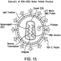
- LV replication-defective, self-inactivating lentiviral vectors
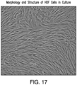
- GM-HDF autologous genetically-modified human dermal fibroblasts
- hCOL7A1 gene-corrected fibroblasts are better than keratinocytes at supplying C7 to the basement membrane zone (BMZ) in mice with RDEB skin grafts (Goto et al. 2006).
- injection of fibroblasts can be more easily completed than grafting of keratinocytes.
- the present invention is directed to autologous genetically modified cells for use in a method of treating a patient suffering from a Type VII collagen (C7) deficiency, wherein said method comprises: (i) contacting cells, obtained from the dermis or epidermis, preferably fibroblasts or keratinocytes, of the C7-deficient patient, with a transducing lentiviral vector comprising a nucleotide sequence encoding COL7A1 or a functional variant thereof to form the autologous genetically modified cells having a vector copy number wherein said lentiviral vector has a transducing vector copy number in the range of 0.1 to 5.0 copies per cell; (ii) culturing said autologous genetically modified cells; and administering an effective amount of the genetically modified cells to the C7-deficient patient.
- a transducing lentiviral vector comprising a nucleotide sequence encoding COL7A1 or a functional variant thereof to form the autologous genetically modified cells having
- the transducing lentiviral vector further comprises (a) a modif 5' long terminal repeat (LTR), wherein the promoter of the modified 5' LTR is is a cytomegalovirus promoter, (b) at least one lentiviral central polypurine tract element, and (c) a modified 3' LTR, wherein the modified 3' LTR comprises a deletion relative to the wild-type 3' LTR, wherein a hepatitis virus post-transcriptional regulatory element (PRE) has been deleted administration may be done by injection, preferably intradermal injection.
- LTR modif 5' long terminal repeat
- PRE hepatitis virus post-transcriptional regulatory element
- the C7 deficiency is dystrophic epidermolysis bullosa (DEB), such as recessive dystrophic epidermolysis bullosa (RDEB) or dominant dystrophic epidermolysis bullosa (DDEB).
- DEB dystrophic epidermolysis bullosa
- RDEB recessive dystrophic epidermolysis bullosa
- DDEB dominant dystrophic epidermolysis bullosa
- Subtypes of RDEB are also envisioned to be treated according to the present invention including Hallopeau-Siemens, non-Hallopeau-Siemens RDEB, RDEB inversa, pretibial RDEB, acral RDEB, and RDEB centripetalis.
- the transducing lentiviral vector is in the form of a lentiviral vector particle.
- the transducing lentiviral vector particle is constructed from a transfer lentiviral vector comprising (a) a modified 5' long terminal repeat in LTR, wherein the promoter of the modified 5' LTR is a cytomegalovirus promoter, (b) the COL7A1 gene or a functional variant thereof, (c) at least one lentiviral central polypurine tract, and (d) a modified 3' LTR, wherein the modified 3' LTR comprises a deletion relative to the wild-type 3' LTR, wherein a hepatitis virus post-transcriptional regulatory element (PRE) has been deleted, wherein the COL7A1 gene or functional variant is incorporated into the cells to form genetically modified cells having a functional COL7A1 gene or functional variant thereof.
- a transfer lentiviral vector comprising (a) a modified 5' long terminal repeat in LTR, wherein the promoter of the modified 5' LTR is a cytomegalovirus promoter, (b) the COL7A1
- the PRE is woodchuck hepatitis virus post-transcriptional regulatory element (WPRE).
- WPRE woodchuck hepatitis virus post-transcriptional regulatory element
- the transfer lentiviral vector used to construct the transducing vector is selected from the group consisting of pSMPUW and pFUGW.
- the transducing lentiviral vector particle is INXN-2004 or INXN-2002.
- the INXN-2004 lentiviral vector particle comprises a transfer vector comprising the amino acid sequence set forth in SEQ ID NO: 34.
- the INXN-2002 lentiviral vector particle is constructed from a pSMPUW lentiviral expression plasmid comprising (a) a modified 5' long terminal repeat in LTR, wherein the promoter of the modified 5' LTR is a cytomegalovirus promoter, (b) a functional COL7A 1 gene, (c) at least one lentiviral central polypurine tract, and (d) a modified 3' LTR, wherein the modified 3' LTR comprises a deletion relative to the wild-type 3' LTR, wherein a hepatitis virus posttranscriptional regulatory element (PRE) has been deleted.
- PRE hepatitis virus posttranscriptional regulatory element
- Administration of the genetically modified fibroblasts autologous to the C7-deficient patient may be done in any suitable manner, including by injection, topically, orally, or embedded in a biocompatible matrix.
- Another aspect of the invention is directed to autologous genetically modified fibroblasts from a C7-deficient patient, such as RDEB or DDEB patient, transduced with a a lentiviral vector particle comprising a functional COL7A1 gene or a functional variant thereof, and expressing type VII collagen, wherein the lentiviral vector particle has a transducing vector copy number in the range of 0.1 to 5.0 copies per cell, wherein said transducing lentiviral vector particle is constructed from a transfer lentiviral vector comprising (a) a modified 5' long terminal repeat in LTR, wherein the promoter of the modified 5' LTR is a cytomegalovirus promoter, (b) a functional COL7A 1 gene, (c) at least one lentiviral central polypurine tract, and (d) a modified 3' LTR, wherein the modified 3' LTR comprises a deletion relative to the wild-type 3' LTR, wherein a hepatitis virus posttranscriptional regulatory element (
- Another embodiment is directed to a self-inactivating lentiviral vector formed from a transfer vector comprising (a) a modified 5' long terminal in LTR, wherein the promoter of the modified 5' LTR is a cytomegalovirus promoter, (b) the COL7A1 gene or a functional variant thereof, (c) at least one lentiviral central polypurine tract element, and (d) a modified 3' LTR, wherein the modified 3' LTR comprises a deletion relative to the wild-type 3' LTR, and wherein a hepatitis virus post-transcriptional regulatory element (PRE) has been deleted, wherein the deleted PRE is a woodchuck post-transcriptional regulatory element PRE(WPRE).
- a transfer vector comprising (a) a modified 5' long terminal in LTR, wherein the promoter of the modified 5' LTR is a cytomegalovirus promoter, (b) the COL7A1 gene or a functional variant thereof, (c) at least one lentiviral
- the vector comprises at least two lentiviral central polypurine tract elements.
- the vector of the invention includes the vector designated INXN-2004 and comprising the sequence of IGE308 plasmid (SEQ) ID NO: 34); or the vector designated INXN-2002 herein.
- compositions comprising a fibroblast obtained from a patient suffering from RDEB or DDEB transduced with a lentiviral vector according to the claims, such as the lentiviral vector designated INXN-2004 comprising the sequence of IGE308 plasmid; or the lentiviral vector designated INXN-2002 herein.
- the present invention is directed to cells transduced in vitro or ex vivo using the vectors according to the claims, such as INXN-2002 and INXN-2004.
- the present invention is directed to transduced autologous fibroblasts using vectors INXN-2002 or INXN-2004, which may be obtained and propagated according to methods described in U.S. Patent No. 8,728,819 and International Patent Application/
- the present invention also relates to patient an autologous population of cells for use in treating a suffering from pseudosyndactyly, wherein said autologous population of cells is to be obtained from said patient and transduced with a lentiviral vector particle comprising a functional COL7A1 gene or a functional variant thereof, and expressing type VII collagen, wherein said lentiviral vector particle has a transducing vector copy number in the range of 0.1 to 5.0 copies per cell, wherein said transducing lentiviral vector particle is constructed from a transfer lentiviral vector comprising (a) a modified 5' long terminal repeat in LTR, wherein the promoter of the modified 5' LTR is a cytomegalovirus promoter, (b) a functional COL7A 1 gene, (c) at least one lentiviral central polypurine tract, and (d) a modified 3' LTR, wherein the modified 3' LTR comprises a deletion relative to the wild-type 3' LTR, wherein a hepatitis virus
- the invention further relates to a medical use for treating, inhibiting or reducing the blistering of a dystrophic epidermolysis bullosa (DEB) or for treating lesions in patients suffering from RDEB comprising administering the autologous genetically modified cells of the present invention.
- DEB dystrophic epidermolysis bullosa
- Another aspect of the invention relates to isolated population of genetically modified fibroblasts autologous to a C7-deficient patient, such as DDEB or RDEB patient, transduced with a vector particle comprising a functional COL7A1 gene or functional variant thereof and expressing type VII collagen, as well as methods of making these isolated populations, as defined by the claims.
- the invention also relates to a method of increasing the integrated transgene copy number per cell in genetically modified human dermal fibroblasts or keratinocytes comprising (a) contacting a transducing lentiviral vector comprising a nucleotide sequence encoding a COL7A1 gene or a functional variant thereof with a human dermal fibroblast or keratinocyte obtained from a C7-deficient patient to form a transduction composition; (b) subjecting the first transduction composition to spinoculation to form transduced human dermal fibroblast or keratinocyte; (c) contacting the transduced human dermal fibroblasts or keratinocytes of step (b) with a second transducing lentiviral vector to form na second transduction composition; and (d) subjecting said first transduction composition to spinoculation to form transduced human dermal fibroblast or keratinocyte, wherein the integrated transgene copy number of the transduced human dermal fibroblasts or
- the transducing lentiviral vector is INXN-2002 or INXN-2004, preferably INXN-2004. It has been found that the transduced human dermal fibroblast or keratinocytes have at least or about 1, 2, 5, 10, 20, 25, 27, 28, 29, 30, 35, 40, 45, or 50-fold greater integrated transgene copy number per cell relative to a transduction composition not subjected to spinoculation or super-transduction. In another embodiment, the invention relates to having an integrated transgene copy number per cell of at least 0.05, 0.09, 0.41, or 0.74 or a range of between about 0.1 to about 5, 0.1 to about 1, 0.4 to about 1, or 0.4 to about 0.75.
- Expression has been found to be increased using the transduction methods described herein. For example, it has been found that expression of C7 has increased by 10, 25, 50, 100, 150, or 200-fold relative to transduced human dermal fibroblasts or keratinocytes not subjected to spinoculation or a second transduction.
- the term "about” is used to indicate that a value includes the inherent variation of error for the device, the method being employed to determine the value, or the variation that exists among the study subjects. Typically, the term is meant to encompass approximately or less than 1%, 2%, 3%, 4%, 5%, 6%, 7%, 8%, 9%, 10%, 11%, 12%, 13%, 14%, 15%, 16%, 17%, 18%, 19% or 20% variability depending on the situation.
- the words “comprising” (and any form of comprising, such as “comprise” and “comprises”), “having” (and any form of having, such as “have” and “has”), “including” (and any form of including, such as “includes” and “include”) or “containing” (and any form of containing, such as “contains” and “contain”) are inclusive or open-ended and do not exclude additional, unrecited elements or method steps. It is contemplated that any embodiment discussed in this specification can be implemented with respect to any method or composition of the invention, and vice versa. Furthermore, compositions of the invention can be used to achieve methods of the invention.
- Nucleic acids and/or nucleic acid sequences are "homologous" when they are derived, naturally or artificially, from a common ancestral nucleic acid or nucleic acid sequence. Proteins and/or protein sequences are homologous when their encoding DNAs are derived, naturally or artificially, from a common ancestral nucleic acid or nucleic acid sequence.
- the homologous molecules can be termed homologs.
- any naturally occurring proteins, as described herein can be modified by any available mutagenesis method. When expressed, this mutagenized nucleic acid encodes a polypeptide that is homologous to the protein encoded by the original nucleic acid. Homology is generally inferred from sequence identity between two or more nucleic acids or proteins (or sequences thereof).
- sequence identity between sequences that is useful in establishing homology varies with the nucleic acid and protein at issue, but as little as 25% sequence identity is routinely used to establish homology. Higher levels of sequence identity, e.g., 30%, 40%, 50%, 60%, 70%, 80%, 90%, 95% or 99% or more can also be used to establish homology. Methods for determining sequence identity percentages (e.g., BLASTP and BLASTN using default parameters) are described herein and are generally available.
- sequence identity in the context of two nucleic acid sequences or amino acid sequences of polypeptides refers to the residues in the two sequences which are the same when aligned for maximum correspondence over a specified comparison window.
- the polypeptides herein are at least 70%, generally at least 75%, optionally at least 80%, 85%, 90%, 98% or 99% or more identical to a reference polypeptide, or a fragment thereof, e.g., as measured by BLASTP (or CLUSTAL, or any other available alignment software) using default parameters.
- nucleic acids can also be described with reference to a starting nucleic acid, e.g., they can be 50%, 60%, 70%, 75%, 80%, 85%, 90%, 98%, 99% or more identical to a reference nucleic acid or a fragment thereof, e.g., as measured by BLASTN (or CLUSTAL, or any other available alignment software) using default parameters.
- one molecule When one molecule is said to have certain percentage of sequence identity with a larger molecule, it means that when the two molecules are optimally aligned, said percentage of residues in the smaller molecule finds a match residue in the larger molecule in accordance with the order by which the two molecules are optimally aligned.
- nucleic acid or amino acid sequences comprises a sequence that has at least 90% sequence identity or more, preferably at least 95%, more preferably at least 98% and most preferably at least 99%, compared to a reference sequence using the programs described above (preferably BLAST) using standard parameters.
- the BLASTP program uses as defaults a word length (W) of 3, an expectation (E) of 10, and the BLOSUM62 scoring matrix (see Henikoff & Henikoff, Proc. Natl. Acad. Sci. USA 89:10915 (1992 )). Percentage of sequence identity is determined by comparing two optimally aligned sequences over a comparison window, wherein the portion of the polynucleotide sequence in the comparison window may comprise additions or deletions (i.e., gaps) as compared to the reference sequence (which does not comprise additions or deletions) for optimal alignment of the two sequences.
- the percentage is calculated by determining the number of positions at which the identical nucleic acid base or amino acid residue occurs in both sequences to yield the number of matched positions, dividing the number of matched positions by the total number of positions in the window of comparison and multiplying the result by 100 to yield the percentage of sequence identity.
- the substantial identity exists over a region of the sequences that is at least about 50 residues in length, more preferably over a region of at least about 100 residues, and most preferably the sequences are substantially identical over at least about 150 residues. In a most preferred embodiment, the sequences are substantially identical over the entire length of the coding regions.
- a “functional variant” of a protein disclosed herein can, for example, comprise the amino acid sequence of the reference protein with at least or about 1, 2, 3, 4, 5, 6, 7, 8, 9, 10, 11, 12, 13, 14, 15, 16, 17, 18, 19, or 20 conservative amino acid substitutions.
- the phrase "conservative amino acid substitution” or “conservative mutation” refers to the replacement of one amino acid by another amino acid with a common property.
- a functional way to define common properties between individual amino acids is to analyze the normalized frequencies of amino acid changes between corresponding proteins of homologous organisms ( Schulz, G. E. and Schirmer, R. H., Principles of Protein Structure, Springer-Verlag, New York (1979 )).
- groups of amino acids may be defined where amino acids within a group exchange preferentially with each other, and therefore resemble each other most in their impact on the overall protein structure (Schulz, G. E. and Schirmer, R. H., supra).
- conservative mutations include amino acid substitutions of amino acids within the sub-groups above, for example, lysine for arginine and vice versa such that a positive charge may be maintained; glutamic acid for aspartic acid and vice versa such that a negative charge may be maintained; serine for threonine such that a free --OH can be maintained; and glutamine for asparagine such that a free --NH 2 can be maintained.
- a COL7A1 gene encodes for the C7 protein or a functional variant thereof, provided that the variant is capable of anchoring the fibrils between the epidermis and dermis.
- the functional variants can comprise the amino acid sequence of the reference protein with at least one non-conservative amino acid substitution.
- Non-conservative mutations involve amino acid substitutions between different groups, for example, lysine for tryptophan, or phenylalanine for serine, etc. In this case, it is preferable for the non-conservative amino acid substitution to not interfere with, or inhibit the biological activity of, the functional variant.
- the non-conservative amino acid substitution may enhance the biological activity of the functional variant, such that the biological activity of the functional variant is increased as compared to the reference sequence.
- Proteins disclosed herein may comprise synthetic amino acids in place of one or more naturally-occurring amino acids.
- Such synthetic amino acids are known in the art, and include, for example, aminocyclohexane carboxylic acid, norleucine, ⁇ -amino n-decanoic acid, homoserine, S-acetylaminomethyl-cysteine, trans-3- and trans-4-hydroxyproline, 4-aminophenylalanine, 4-nitrophenylalanine, 4-chlorophenylalanine, 4-carboxyphenylalanine, ⁇ -phenylserine ⁇ -hydroxyphenylalanine, phenylglycine, ⁇ -naphthylalanine, cyclohexylalanine, cyclohexylglycine, indoline-2-carboxylic acid, 1,2,3,4-tetrahydroisoquinoline-3-carboxylic acid, aminomalonic acid, aminomalonic acid
- the invention includes the vectors INXN-2002; and IGE308 also referred to herein as INXN-2004, as well as vectors substantially identical and/or homologous thereto as well as functional variants thereof.
- patient or “subject” refers to mammals, including humans and animals.
- treating refers to reducing or alleviating the symptoms, and/or preventing relapses and/or the progression of DEB, including RDEB and/or DDEB.
- autologous cells refers to cells obtained and then returned to the same individual.
- cells may be removed from the body of a DEB patient, which may subsequently be genetically modified, cultivated to proliferate, and then returned to the body of the patient from which they were originally removed.
- transduction refers to the delivery of a gene(s) using a viral or retroviral vector by means of viral infection rather than by transfection.
- transducing lentiviral vector or “transducing lentiviral vector particle” refers to the infectious lentiviral vector particles formed from the co-transfection of a packaging cell line with the lentiviral expression/transfer plasmid vector comprising the COL7A1 gene or functional variant thereof, a packaging vector(s), and an envelope vector.
- the transducing lentiviral vector is harvested from the supernatant of the producer cell culture after transfection.
- Suitable packaging cell lines are known in the art and include, for example, the 293T cell line.
- lentiviral vector refers to a vector containing structural and functional genetic elements outside the LTRs that are primarily derived from a lentivirus.
- SIN self-inactivating vector
- U3 region the 3' LTR enhancerpromoter region
- SIN vectors greatly reduce risk of creating unwanted replication-competent virus since the 3' LTR U3 region has been modified to prevent viral transcription beyond the first round of replication, eliminating the ability of the virus to be passed.
- pharmaceutically acceptable carrier is employed herein to refer to liquid solutions which are, within the scope of sound medical judgment, suitable for use in contact with the autologous, genetically-modified cells without affecting their activity, and without being toxic to the tissues of human beings and animals or causing irritation, allergic response, or other complications, commensurate with a reasonable benefit/risk ratio.
- a useful pharmaceutically acceptable carrier may be an injectable solution which is biocompatible with the autologous, genetically-modified cells and does not reduce their activity or cause their death.
- the present disclosure relates to an autologous, genetically-modified cell therapy for C7-deficient patients.
- C7-deficient patients either lack or have substantially reduced production of type VII collagen (C7), which is important for anchoring fibril formation at the dermal-epidermal junction (DEJ), resulting in dystrophic epidermolysis bullosa (DEB).
- DEB dermal-epidermal junction
- the present disclosure relates to genetically modifying DEB cells obtained from a patient, such as fibroblasts or keratinocytes, to insert a functional COL7A1 gene or functional variant thereof, expanding the genetically modified cells, and administering a population of the genetically-modified cells back into the DEB patient.
- Cells may be obtained from a patient suffering from DEB. Fibroblasts or keratinocytes are preferred. The cells may be obtained through known methods, including from patient skin samples, scrapings, and biopsies, for example. In one embodiment, the cells are obtained from the non-blistering skin of a DEB patient. In another embodiment, the cells are obtained from the blistering skin of a DEB patient. The cells obtained from a DEB patient have either a mutated, non-functional or missing COL7A1 gene. These cells are cultured using standard cell culture techniques. The cells obtained from the patient are subsequently treated ex vivo with genetic material encoding a functional type VII collagen gene (C7) or functional variant thereof.
- C7 functional type VII collagen gene
- the COL7A1 gene has the nucleic acid, SEQ ID NO:1, which encodes for a 290 kDa alpha chain, wherein three chains form a triple helix (trimer), as depicted in Figure 1A .
- C7 has the amino acid sequence of SEQ ID NO: 2 and is the protein important for anchoring fibril formation at the dermal-epidermal junction (DEJ). Mutations in the C7 gene cause an absence or reduction of C7, which make up anchoring fibrils that maintain binding of the epidermis to the dermis.
- C7 anchoring fibrils bind to other collagens, extracellular matrix proteins, and Lam332 that mediate the attachment of the dermis to the epidermis, as shown in Figure 1B .
- Vectors may comprise nucleic acids and polypeptides substantially identical to COL7A1 and C7 respectively.
- vectors may comprise nucleic acids that encode a functional variant of the COL7A1 or C7 protein, provided that the variant is capable of anchoring the fibrils between the epidermis and dermis.
- only the open reading frame of the COL7A1 gene, which encodes the C7 protein or a functional variant thereof may be used.
- the COL7A1 nucleic acid is codon-optimized for increased expression of C7 in the transduced cell, as is known in the art.
- the COL7A1 can be transduced into the DEB cells obtained from a patient using a transducing lentiviral vector, which is replication defective and self-inactivating.
- Self-inactivating lentiviral vectors are derived from the human immunodeficiency virus (HIV-1), which are pseudotyped with a heterologous VSV-G envelope protein instead of the HIV-1 envelope protein.
- HSV-1 human immunodeficiency virus
- a 400 bp deletion is introduced to the U3 region of the LTR resulting in a self-inactivating (SIN) vector (Zuffery 1998).
- the vectors of the present invention are distinguished from 2nd and 3rd generation SIN vectors.
- the vectors of the present invention have been modified to enhance the safety features and increase the cloning capacity as further described herein.
- Vectors used to construct the transducing lentiviral vectors of the present invention are introduced via transfection or infection into a packaging cell line.
- the packaging cell line produces transducing lentiviral vector particles that contain the vector genome.
- the recombinant virus is recovered from the culture media and titered by standard methods used by those of skill in the art.
- the packaging constructs can be introduced into human cell lines by calcium phosphate transfection, lipofection or electroporation, generally together with a dominant selectable marker, such as kanamycin, neomycin, DHFR, or Glutamine synthetase, followed by selection in the presence of the appropriate drug and isolation of clones.
- the selectable marker gene can be linked physically to the packaging genes in the construct.
- Stable cell lines wherein the packaging functions are configured to be expressed by a suitable packaging cell are known. For example, see U.S. Pat. No. 5,686,279 ; and Ory et al., (1996), which describe packaging cells.
- the packaging cells with a lentiviral vector incorporated in them form producer cells. Producer cells are thus cells or cell-lines that can produce or release packaged infectious viral particles carrying the therapeutic gene of interest.
- An example of a suitable lentiviral vector packaging cell lines includes 293 cells.
- the transducing lentiviral vector is constructed from a lentiviral expression plasmid vector comprising (a) a modified 5' long terminal repeat (LTR), wherein the promoter of the modified 5' LTR is a cytomegalovirus promoter, (b) the COL7A1 gene, (c) at least one lentiviral central polypurine tract element, and (d) a modified 3' LTR, wherein the modified 3' LTR comprises a deletion relative to the wild-type 3' LTR, wherein a hepatitis B virus post- transcriptional regulatory element (PRE) has been deleted.
- LTR modified 5' long terminal repeat
- PRE hepatitis B virus post- transcriptional regulatory element
- the deleted hepatitis B virus post- transcriptional regulatory element is a woodchuck post-transcriptional regulatory element PRE (WPRE).
- the lentiviral expression plasmid vector comprises two or more lentiviral central polypurine tract (cPPT) elements. Incorporation of cPPT element has been found to increase gene expression levels.
- the 3' LTR may be modified to delete a 400 bp fragment (as disclosed by Zuffery 1998), instead of the commonly used 133 bp deletion of standard 3rd generation SIN LV vectors.
- the 400 bp deletion is believed to increase safety by preventing read-through transcription as well increase viral titer because the vector transcript is more stable in packaging cells.
- the lentiviral expression plasmid may incorporate a hepatitis B virus post-transcriptional regulatory element (PRE), which is preferably woodchuck post-transcriptional regulatory element PRE (WPRE). WPRE has been found to increase the viral titer of the vector.
- PRE hepatitis B virus post-transcriptional regulatory element
- WPRE woodchuck post-transcriptional regulatory element PRE
- the present invention further includes the use of lentiviral transfer vectors in which certain elements common for 3rd generation lentiviral expression vectors are deleted.
- safety concerns for use of lentiviral vectors in humans hinge on the fear of generating a replication-competent lentivirus, which may arise from recombination between split genomes of 3rd generation lentiviral vectors. (Tareen et al., 2013.).
- the gag sequence and/or the revresponsive elements (RRE) may be deleted from the viral vector, in order to reduce sequence homology with other helper plasmids and thereby increase safety to humans. Accordingly, in one embodiment of the invention, the gag sequence is absent in the vector. In another embodiment of the invention, both the gag sequence and RRE is absent in the vector.
- the starting materials to generate the lentiviral vectors of the present invention is preferably a lentiviral expression plasmid vector that comprises a cPPT and PRE that can accommodate the insertion of the large COL7A1 gene (8.89 kbp) or a functional variant thereof.
- the starting material to construct the transducing lentiviral vector of the present invention is selected from the lentiviral expression plasmid vectors, pSMPUW (Cell Biolabs, Inc., San Diego, CA) and pFUGW (Addgene, Cambridge, MA).
- the pSMPUW lentiviral expression plasmid vector is selected for construction of the transducing lentiviral vector.
- Figure 6 shows a schematic of the genetic elements in the pSMPUW lentiviral expression plasmid vector.
- Features of the pSMPUW lentiviral expression vector have been modified from 3rd generation lentivirus expression vectors in order to enhance gene expression levels and safety features, as depicted in Figure 7 .
- the pSMPUW lentiviral expression vector encodes for a multicloning site (MCS) followed by the Woodchuck Hepatitis Virus Post-transcriptional regulatory element (WPRE).
- MCS multicloning site
- WPRE Woodchuck Hepatitis Virus Post-transcriptional regulatory element
- the residual gag ( ⁇ gag) and the RRE element were removed from the pSMPUW vector construct. Additionally, the pSMPUW vector construct utilized a larger 400 bp deletion in the 3'LTR U3 region instead of the commonly used 133 bp deletion in a standard 3rd generation SIN LV vector. Wild type COL7A1 gene was incorporated into the pSMPUW vector.
- the pFUGW lentiviral expression plasmid is selected for construction of the transducing lentiviral vector.
- Figure 8 shows a schematic of the genetic elements in this plasmid vector.
- Features of the pFUGW vector include a RRE, two cPPT elements, and a WPRE element, which may improve lentiviral production and transgene expression.
- the WPRE element has been deleted from the lentiviral vector.
- transducing lentiviral vector particles of the present invention are designated INXN-2002 (vector transfer plasmid - IGE230) or INXN-2004 (vector transfer plasmid - IGE308), or a substantially identical vector comprising functional variants thereof.
- the cells obtained from the DEB patient are transformed with a functional COL7A1 gene.
- the cells are transduced with a lentiviral vector having the COL7A1 gene.
- the lentiviral vector is a self-inactivating vector.
- the copy number of the integrated transgene can be assessed using any known methods. For example, copy number may be determined through quantitative PCR, multiplex ligationdependent probe amplification, fluorescent in situ hybridization (FISH), microarray-based copy number screening, and conventional karyotyping.
- the number of copies of the transgene integrated into each cell may be modulated by the virus dose given to the cells during production.
- the integrated transgene copy number per cell in the obtained RDEB cells transduced with a COL7A1 -containing vector is dose dependent.
- Cells obtained from the DEB patient and transformed with the functional COL7A1 gene will have a functional COL7A1 gene and exhibit normal cell morphologies. These cells may be caused to proliferate or expanded in culture using standard cell culture techniques.
- Normal fibroblast morphological characteristics are observed among obtained fibroblasts transduced with COL7A1 -transducing vectors within the scope of the present invention.
- normal fibroblast morphologies include cells displaying elongated, fusiform or spindle appearance with slender extensions. Normal morphologies further include cells appearing larger, flattened stellate cells which may have cytoplasmic leading edges.
- Figure 9 displays an example of normal cell morphology for fibroblasts transduced with a COL7A1- transducing vector.
- C7 produced by DEB cells obtained from a patient and transduced with a COL7A1 -transducing vector has also been observed.
- the formation of C7 trimers is important in the assembly of anchoring fibrils. It has been found that DEB patient cells transduced with a COL7A1 -transducing vector are capable of forming the C7 trimers with the correct structure, size, and function.
- the C7 expressed by fibroblasts transduced with a COL7A1 -transducing vector will be capable of forming anchoring fibrils using immunoprecipitation with an anti-C7 specific antibody.
- the anti-C7 specific antibody, fNC1 may be used for selective capture and the concentration of C7 from culture supernatants for detection by SDS-PAGE/immunoblot.
- Figures 9 and 10 demonstrate that C7 produced by fibroblasts transduced with a COL7AI-transducing vector were predominantly trimeric.
- C7 may be assessed using known methods, such as using a laminin binding assay or cell migration assay.
- C7 has been shown to bind immobilized extracellular matrix (ECM) components, including fibronectin, Laminin 332 (Lam332), COL1, and COL4 (Chen, et al., 2002a).
- ECM extracellular matrix
- the interaction between C7 and Lam332 occurs through the NH2-terminal NC1 domain of C7 and is dependent upon the native conformation of both Lam332 and C7 NC1 (Rousselle, et al., 1997).
- the association between C7 and Lam332 is important for establishing correct Lam332 architecture at the dermal-epidermal junction.
- a cell migration assay can be used to assess functional C7 activity.
- Previous studies have shown that RDEB fibroblasts and keratinocytes show an increase in motility relative to their normal counterparts, and that normal motility can be restored by expression of C7 (Chen, et al., 2000; Chen, et al., 2002b; Cogan, et al. 2014; Baldeschi et al., 2003).
- Suitable assay includes the colloidal gold salt migration assay to measure the migration of fibroblasts and keratinocytes, or a wound healing assay to measure the migration of keratinocytes. Cells having functional C7 activity will exhibit a reduced motility relative to RDEB cells in such assays.
- the present invention is directed, in one aspect, to pharmaceutical formulations comprising the autologous, genetically-modified cells from DEB patients.
- These cells may be present in any amount suitable for the delivery to the patient in which the cells were originally harvested.
- that cells may be present in a cell concentration of 1.0 - 5.0 ⁇ 10 7 cells/mL, 1.0 - 4.0 ⁇ 10 7 cells/mL, 1.0 - 3.0 ⁇ 10 7 cells/ mL, or 1.0 - 2.0 ⁇ 10 7 cells/mL.
- the cells are present in present in a suspension suitable to sustain the viability of the cells, such as in Dulbecco's Modified Eagle's Medium (DMEM).
- DMEM Dulbecco's Modified Eagle's Medium
- the viability of the cells in the formulation are present in an amount of ⁇ 60%, 70%, 75% or 80%.
- Suitable excipients may also be present in the formulation, such as phosphate buffered saline which may be used to wash the cells from thawed vials containing the autologous, genetically-modified.
- no phenol red is present in the final formulation.
- the autologous genetically modified cells of the present disclosure have a transducing vector copy number of at least or about 0.05, 0.09, 0.41, or 0.74 or in the range of about 0.1 to about 6.0, about 0.1 to about 5.5, about 0.1 to about 5.0, about 0.1 to about 4.5, about 0.1 to about 4.0, about 0.1 to about 3.5, about 0.1 to about 3.0, about 0.1 to about 1, 0.4 to about 1, or 0.4 to about 0.75.
- the transducing vector copy number is in the range of about 0.1 about 5.0.
- the transduced human dermal fibroblast or keratinocytes have at least or about 1, 2, 5, 10, 20, 25, 27, 28, 29, 30, 35, 40, 45, or 50-fold greater integrated transgene copy number per cell relative to a transduction composition not subjected to spinoculation or super-transduction.
- the C7 protein expression values of the FCX-007 cells of the present invention are ⁇ 300, 350, 400, 450, 500, 550, or 600 ng/day/E6 cell.
- the C7 protein expression values of the autologous genetically-modified cells of the present invention are ⁇ 500 ng/day/E6 cell.
- the present invention has also found that expression of C7 has increased by 10, 25, 50, 100, 150, or 200-fold relative to transduced human dermal fibroblasts or keratinocytes not subjected to spinoculation or a second transduction.
- the multiplicity of infection refers to the number of vector particles per cell used in transduction.
- the desired transducing vector copy number has been achieved even as the MOIs decreases, as shown in the examples below.
- the invention relates to MOIs of ⁇ 15, ⁇ 14, ⁇ 13, ⁇ 12, ⁇ 11, or ⁇ 10.
- the MOI is between about 1 to 10.
- the formulations of the present invention may be for use in treating various maladies of patients suffering from Type VII deficiency.
- the formulations of the present invention are suitable to treat DEB, including DDEB or RDEB.
- the medical use is for treating subtypes of RDEB, including but not limited to Hallopeau-Seiemens, non-Hallopeau-Siemens RDEB, RDEB inversa, pretibial RDEB, acral RDEB, and RDEB centripetalis.
- the invention further relates to the medical use for the treatment, reduction, prevention, and/or inhibition of various symptoms attributed to C7 deficiencies.
- DEB is known to cause scarring after blisters, which may cause contracture deformities, swallowing difficulty if the mouth and esophagus are affected, fusion of fingers and toes, and limited mobility. Therefore, in one aspect of the invention envisions the medical use for the treatment, reduction, inhibition, and/or prevention of pseudosyndactyly, also known as mitten hand syndrome.
- the invention relates to the medical use for the treatment, reduction, inhibition, and/or prevention of deep fibrosis and/or scarring associated with RDEB and/or DDEB, which may result in milia, joint contractures, generalized soft tissue fibrosis, organ fibrosis, corneal lesions, scarring plaques, scarring alopecia, nail dystrophy, ankyloglosia, and increased frequency of dental caries, for example.
- the invention relates to the medical use for the treatment, reduction, prevention, and/or inhibition of oral mucosa lesions and gastrointestinal lesions associated with RDEB.
- Another embodiment relates to the medical use for treatment, prevention, reduction and/or inhibition of blisters associated with DEB patients.
- Administration of the autologous genetically modified cells may be done at any appropriate time as one skilled in the art would be capable of determining based on the needs of the patient. For example, administration may be done once, once a day, once a month, once a quarter, or 1-2 times per year.
- the formulation of the present invention containing the autologous genetically modified cells may be administered to the patient by any known method, including but not limited to injection, topically, orally, or embedded in a biocompatible matrix.
- the injection may be, e.g., parenteral, intradermal, subcutaneous, intramuscular, intravenous, intraosseous, intraarterial, ocular and intraperitoneal or direct injection into a specific organ/tissue, e.g. prostate or liver.
- the formulation may be administered directly to an affected site, such as at the site of a lesion. Debridement of the affected tissue can precede the direct application of the formulation to the site.
- the formulation may be encapsulated in a suitable delivery system, such as in a polymer capsule, or embedded in a biocompatible matrix or graft, e.g., collagen matrix, in a hydrogel, skin graft, or a mesh.
- a suitable delivery system such as in a polymer capsule, or embedded in a biocompatible matrix or graft, e.g., collagen matrix, in a hydrogel, skin graft, or a mesh.
- the autologous genetically modified cells of the present invention may be administered solely or in combination with other treatments for a patient suffering from DEB.
- therapeutic agents used for treating DEB include topical care, such as Zorblisa, skin grafts, antiinflammatories, antibodies, other potential gene or cell-based therapies.
- the modified cells of the present invention may be administered for the skin or muoscal areas where bone marrow transplant therapies (or other system cellular therapies such as mesenchymal stem cells) did not provide sufficient therapy.
- the C7-expressing ability of the autologous genetically modified cells is preferably not impaired by a combination with other therapeutic agents.
- the present invention further relates to a method of enhancing or increasing the integrated transgene copy number per cell in the genetically modified cells obtained from C7-deficient patients and transduced according to the present invention.
- a method of enhancing or increasing the integrated transgene copy number per cell in the genetically modified cells obtained from C7-deficient patients and transduced according to the present invention it has been found that certain steps during transduction substantially enhanced the copy number of the transgene in these cells.
- the cells obtained from the C7-deficient patient are contacted with the transducing lentiviral vector of the present invention and that composition is subjected to spinoculation, also known as spin transduction. In this manner, the lentiviral vectors are spun onto the targeted cells.
- spinoculation has unexpectedly increased the copy number per cells in the genetically modified human dermal fibroblasts by a level of at least 5, 10, 15, 20, 25, 27, 28, 29, 30, 35, 40, 45, or 50- fold relative to a transduction without spinoculation or transduction.
- a second transduction step or super-transduction in which the targeted cells are passaged through the first transduction with spinoculation, and then subsequently passaged through a second transduction optionally with spinoculation.
- this super-transduction with spinoculation increases the copy number by an additional 2-fold increase relative to the original traditional transduction method that did not include spinoculation or super-transduction.
- the present invention relates to increasing the copy number of the transduced cells from the C7-deficient patient.
- FCX-007 is a suspension of live, autologous human dermal fibroblast cells genetically modified using either a lentiviral vector (INXN-2002) (as shown in Example 1) or a lentiviral vector (INXN-2004) (as shown in Example 2) to express the human collagen type 7 protein.
- FCX-007 cells derived from the lentiviral vector INXN-2002 cells are suspended in a cryopreservation medium consisting of Iscove's Modified Dulbecco's Medium (IMDM) without fetal bovine serum (50.0%), Profreeze-CDMTM (42.5%) and dimethyl sulfoxide (DMSO) (7.5%).
- IMDM Iscove's Modified Dulbecco's Medium
- DMSO dimethyl sulfoxide
- the structural features for FCX-007 Drug Substance (DS) include the primary structure of the autologous human dermal fibroblast (HDF) cells and the structure of the lentiviral vector used to transduce and gene modify the HDF cells.
- HDF autologous human dermal fibroblast
- INXN-2002 Lentiviral Vector which is used to transduce and to introduce the human collagen 7A1 gene into the HDF cells, is a recombinant lentiviral vector encoding the human collagen 7A1 gene.
- INXN-2002 LV is a self-inactivating (SIN) lentiviral vector that is constructed based on the human immunodeficiency virus type 1 (HIV-1) pseudotyped with a heterologous VSV-G envelope protein.
- the virus particle for INXN-2002 lentiviral vector is approximately 120 nm in diameter and is comprised of numerous proteins with two copies of a single-stranded RNA genome. A specific molecular formula, molecular weight, or stereochemistry is not available.
- Structural features for INXN-2002 LV include the primary structure of the RNA viral genome and the structure of the viral particle.
- the primary structure of the RNA genome of INXN-2002 is determined by the full nucleotide sequencing of the viral genome.
- the viral particles structure is deduced from the particle structure of HIV-1 with added VSV-G protein pseudotyping. An overview of the nucleic acid structure and the structure of the viral particle are provided below.
- INXN-2002 LV is a self-inactivating (SIN) lentiviral vector that is constructed based on the human immunodeficiency virus type 1 (HIV-1) pseudotyped with a heterologous VSV-G envelope protein instead of the HIV-1 envelope protein.
- a 400 bp deletion is introduced to the U3 region of the LTR resulting in a self-inactivating (SIN) vector (Zuffery 1998).
- the pSMPUW lentiviral expression plasmid vector (Cell Biolabs, Inc., San Diego, CA) was selected for construction of the INXN-2002 LV.
- Figure 6 shows a schematic of the genetic elements in the pSMPUW lentiviral expression plasmid vector.
- the coding elements between the 5' and 3' LTRs of the HIV-1 virus are fully gutted.
- the vector encodes for a multicloning site followed by the Woodchuck Hepatitis Virus Posttransscriptional Regulatory Element (WPRE).
- WPRE Woodchuck Hepatitis Virus Posttransscriptional Regulatory Element
- these elements were removed by digesting the vector with BamHI from the multicloning site and KpnI at the 5' end of the 3' left terminal repeat.
- the COL7A1 gene expression cassette with a CMV promoter is cloned into the digested vector by single strand annealing to generate the lentiviral vector plasmid construct encoding the COL7A1 gene as shown in Figure 13 .
- the lentiviral vector plasmid construct is co-transfected into HEK293T cells with three helper plasmids (pCMV-G, pCMV-Rev2 and pCgp) to produce the INXN-2002 lentiviral vector particles.
- pCMV-G plasmid provides the VSV-G pseudotyping surface protein
- pCMV-Rev2 provides the HIV-1 Rev protein for efficient RNA transport and packaging
- pCgp provides the structural and viral enzyme proteins for production of the INXN-2002 lentiviral vector particles.
- Figure 14 shows a schematic of the proviral RNA genome structure of the INXN-2002 lentiviral vector.
- INXN-2002 lentiviral vector is constructed based on a HIV-1 derived vector backbone and has a similar viral particle structure to the HIV-1 virus.
- Figure 9 shows an electron micrograph of HIV-1 particles.
- the HIV-1 particle has a spherical shape of approximately 120 nm in diameter, with an estimated molecular weight of 277MDa (Carlson 2008).
- the particle has a lipid bilayer membrane stubbed with envelope protein.
- the envelope protein interacts with the receptors on the target cells for infection and delivery of the RNA viral genome to the target cells.
- a cone shaped nucleo-core where two strands of viral RNA genome is housed, can be observed inside the virus particle.
- the cone shaped nucleo-core is formed by the viral capsid protein.
- VSV-G glycoprotein of the vesicular stomatitis virus (VSV-G)
- VSV-G glycoprotein of the vesicular stomatitis virus
- INXN-2002 viral particles assemble and bud out from the surface of transfected HEK293T cells.
- the VSV-G protein is provided by the pCMV-G helper plasmid
- the vector core and enzyme proteins are provided by the pCgp helper plasmid
- the Rev protein which is needed for efficient RNA genome transport and packaging into the viral particle, is provided by the pCMV-Rev2 plasmid.
- all the other HIV-1 accessory proteins including Vpu, Vif, Vpr, Nef, and Tat, are deleted from the INXN-2002 vector.
- the protease enzyme packaged inside the virus particle cleaves the Gag precursor protein into its constituent proteins (MA, CA, NC), to convert the immature virion into a mature infectious INXN-2002 vector particle.
- Figure 15 shows a schematic depicting the details of the mature INXN-2002 virus particle.
- the nucleocapsid (NC) protein forms a stable complex with the RNA genome inside the capsid core.
- the matrix protein forms a coat on the inner surface of the membrane.
- the virus buds through the cell plasma membrane spiked with the VSV-G envelope protein and forms the lipid envelope.
- Table 1 shows the component proteins that make up the INXN-2002 lentiviral vector and a brief description of the function of each of the components.
- Table 1 INXN-2002 Lentiviral Vector Major Component Proteins Component Proteins Protein MW (kD) Protein functions Capsid (CA) 24 Forms the nucleo-core Matrix (MA) 17 Forms the protein coat on the inner surface of the lipid membrane Nucleocapsid (NC) 7 Forms a stable complex with the RNA genome VSV-G 69 Pseudotyped envelope protein interacts and binds to receptors on target cells for transduction to deliver the vector RNA genome to the cells Protease 11 Plays an important role in the maturation of the INXN-2002 vector by cleaving the Gag proteins to its functional constituents, CA, MA, and NC Reverse transcriptase 66/51 Builds a DNA copy of the viral RNA genome Integrase 31 Inserts the DNA copy of the viral RNA genome into the infected cell genome
- Table 2 provides a list of the characterization assays and specifications for INXN-2002 manufactured by City of Hope. Characterization assay results are provided in the attached CoA for INXN-2002 lot number 0786-240-0002-1, the lot intended for use in manufacture of the FCX-007 clinical product.
- Table 2 Characterization of INXN-2002 Category Assay Test Method Lab SOP Target Specification Identity Vector identity RT-PCR CoH QC-SOP-0843 Band of correct size detected Vector insert stability Southern blot analysis Indiana University VP-10-17.16 Report result Potency Viral antigen Physical Titer (p24) ELISA Indiana University VP-10-05.06 Report result TU titer H1299 transduction with qPCR readout BioReliance 016135.BSV Report result C7 expression (Potency) ELISA BioReliance 016136.BSV Report result Appearance and pH Appearance Visual inspection CoH QC-SOP-0734 Opaque solution pH pH meter CoH QC-SOP-0675 6.9 - 7.8
- IVIM in vitro immortalization
- the principle of the IVIM test is based on the understanding that normal Lineage negative (Lin- ) bone marrow (BM) cells will stop proliferating after 3-4 weeks in vitro, but in the presence of certain vector integrations which cause upregulation of proto-oncogenes, some clones will continue to proliferate after 5 or more weeks.
- the number of such immortalized clones is representative of the oncogenic potential of the vector.
- the immortalized clones are expanded and further analyzed for stem cell markers by FACS, vector copy number by qPCR or the site of integration by LAM-PCR. Insertion in common integration sites (cis) such as Evil or Prdm 16 is frequently observed in immortalized clones generated in IVIM assays (Calmels et al., 2005; Modlich et al., 2008).
- Lineage-negative (Lin-) bone marrow (BM) cells from C57BL/6 mice were isolated from complete BM by magnetic sorting using lineage-specific antibodies. The Lin- BM cells were then cultured and stimulated in complete growth medium for 2 days. On Day 4, cells were transduced with INXN-2002 vector in a 48-well plate coated with RetroNectin (10 ⁇ g/cm 2 , Takara).
- Figure 16 shows a schematic setup of the IVIM assay.
- INXN-2002 vector was concentrated approximately 14-fold using a spin concentrator. For transduction, 80 ⁇ L of the concentrated INXN-2002 was added to each well containing 1.5 ⁇ 10 5 cells with 150 ⁇ L of transduction medium. The plate was centrifuged for 1000g at 32°C for 70 minutes (spinocculation) to further enhance the transduction efficiency. Cells were incubated at 37°C, 5% CO 2 incubator O/N.
- INXN-2002 transduction was repeated on Day 5, Day 6, and Day 7 for a total of 4 consecutive transductions.
- the transduced Lin- BM cells were expanded for 10 days and cell concentration was adjusted to 2 - 4 ⁇ 10 5 cells/ml every two days, and fresh medium was provided as needed. On Day 10 post transduction, cells were harvested and counted. A portion of the cells was then submitted for DNA isolation and qPCR to determine the vector copy number (VCN). The remaining cells were placed back into culture for potential plating of the IVIM assay between days 18 - 21.
- Table 3 shows the pilot test run result of INXN-2002 transduced Lin- BM cells harvested on Day 10.
- Table 3 Day 10 Post-Transduction Lin- BM Cells Analysis Test Article Total Cells Cell Viability Vector Copy per Cell INXN-2002 1.5 x 10 6 89% 0.09 Control vector-1 2.7 x 10 6 95% 0.81 Control vector-2 2.1 x 10 6 91% 3.58 Mock 3.0 x 10 6 88% 0.0
- the human dermal fibroblast cells used for the manufacture of FCX-007 are derived from a live skin biopsy.
- the biopsy is digested using Liberase (Roche) to release the dermal fibroblast cells and then the cells are expanded in culture using standard cell culture techniques.
- the cells are harvested and washed, then formulated to contain 1.0 - 3.0 ⁇ 10 7 cells/mL.
- the DS is tested for purity and confirmed to contain ⁇ 98% fibroblasts by CD90 staining with cell viability of ⁇ 85%.
- FCX-007 cells transduced with the INXN-2002 LV in the DS formulation display typical fibroblast morphologies when cultured on tissue culture surfaces. Specifically, cells may display an elongated, fusiform or spindle appearance with slender extensions, or cells may appear as larger, flattened stellate cells which may have cytoplasmic leading edges. A mixture of these morphologies may also be observed.
- Figure 17 shows a typical cell morphology and structure of FCX-007 DS in culture.
- the cells express proteins characteristic of normal fibroblasts including the fibroblast specific marker, CD90 (Thy-1), a 35 kDa cell-surface glycoprotein, and the extracellular matrix proteins, such as various types of collagen.
- CD90 Thi-1
- a 35 kDa cell-surface glycoprotein a 35 kDa cell-surface glycoprotein
- extracellular matrix proteins such as various types of collagen.
- FCX-007 Drug Substance is characterized for release at three stages during manufacturing: in- process, bulk harvest, and after cryopreservation (DS).
- Table 4 provides a list of the characterization assays and specifications for release of FCX-007 DS manufactured by PCT. Characterization assay results for the finished Drug Substance are provided in the attached CoTs for Training Run 8 Arm A, Arm B and Arm C (Non-Transduced Control).
- FCX-007 Additional characterization of FCX-007 has been performed on the training production runs to confirm expression of functional C7.
- the representative assessments shown below are from Training Run 8 (TR8) where cells were transduced with either a high dose (3.4 IU/cell) or low dose (1.7 IU/cell) of LV-COL7, or were mock-transduced. These transduction arms are also referred to as Arm A, B, and C, respectively.
- ELISA enzyme-linked immunofluorescence assay
- Anchoring fibrils are formed from the assembly of C7 trimers.
- the proper expression and formation of C7 trimers by FCX-007 can be detected by immunoprecipitation of C7 followed by non-denaturing SDS-PAGE/immunoblot analyses.
- C7 was immunoprecipitated from FCX-007 cell culture supernatants at passage 1 and passage 2 post- thaw using a NC1-specific antibody and was separated on non-denaturing SDS-PAGE then visualized by western blot.
- the C7 produced by RDEB fibroblasts was predominantly trimeric (red arrow; ⁇ 870 kDa) with LV-COL7-transduced cells (Transduction Arms A and B) expressing more C7 than mock-transduced cells (Arm C, starred). Monomeric (290 kDa) and dimeric (580 kDa) forms were also observed. Assay controls included immunopreciptation of purified C7 (Pur COL7) and immunoprecipitation without antibody or test sample (IP Controls).
- C7 interacts with Laminin332 at the dermal/epidermal junction (DEJ).
- the interaction between C7 and Lammin332 is important for anchoring fibril functionality (Chen 2002, Rousselle 1997, Waterman 2007).
- a binding assay for detection of this interaction was developed at Intrexon. Drug Substance vials (high dose, low dose, and mock-transduced) were thawed and cultured for 2 days. Conditioned cell culture supernatants were collected and were incubated with Lam332 or bovine serum albumin (BSA)-coated wells and bound C7 was detected using a C7 NC1- specific antibody and an HRP-conjugated secondary antibody. The assay readout is optical density at 450 nm (OD450). Results in Figure 20 show virus dose-dependent binding to Lam332 compared with a BSA control.
- a migration assay was developed to assess function of C7. Chen 2000 has demonstrated that RDEB patient skin cells migrate faster than normal skin cells into an artificial wound margin created on tissue culture vessels, and that application of C7 can restore the migration rate.
- Normal human dermal fibroblasts (NHDFs; Lonza) and cells from FCX-007 Drug Substance vials were thawed and cultured. Cells were seeded into culture dishes with an insert to prevent cell adherence in a small strip of the culture dish. The strip was then removed and the rate at which the cells migrated into the open area was monitored by microscopy and quantified using the irregular shape delineation plugin for ImageJ software.
- Figures 21A-21B show both the percent migration (A) and the images of migration (B) for this assay.
- FCX-007 is an autologous fibroblast cell product genetically modified by INXN-2004 lentiviral vector (LV-COL7) to express the human collagen 7 protein (C7).
- the materials used to manufacture FCX-007 DS/INXN-2004 are described below are similar to that set forth above in Example 1, except that the IGE-230 LV-COL7 vector plasmid was used for the production of INXN-2002.
- IGE-308 LV-COL7 vector plasmid was used to make the INXN-2004 vector.
- the same helper plasmids, pCMV-G, pCMV-Rev2, and pCgp were used to co-transfect a 293 WCB cell line.
- the IGE308 plasmid was constructed using standard molecular cloning methods. The construction process involved cloning of the human COL7A1 gene and introduction of the cloned COL7A1 gene into a lentiviral vector (SIN) backbone, pFUGW, to produce the IGE308 LV-COL7 vector transfer plasmid.
- SI lentiviral vector
- the COL7A1 gene cloned into the IGE308 LV-COL7 vector transfer plasmid is the same COL7A1 gene cloned into INXN-2002 (IGE230 vector transfer plasmid).
- Primers were designed and produced with the Takara PrimeScript reverse transcriptase to amplify the COL7A1 gene from human genomic cDNA.
- Four primer pairs, as shown in Table 5 were designed, each to amplify approximately 2 kb of the COL7A1 gene.
- Products were amplified with two primer pairs (CollF2/CollR4 and CollF4/CollR1) designed to amplify the 5' end 3790 base pairs (bp) of COL7A1 from human genomic cDNA along with a 5' overhang for cloning into the expression vector.
- the two 5' PCR products (2127 bp and 2993 bp) were joined by overlap extension PCR to generate a 3790 bp final product.
- the two COL7A1 gene fragments (3790 bp and Col1A1-3) were assembled into an inducible expression vector (VVN-257673) that had previously been digested with NheI and ClaI using the In-Fusion HD Cloning Kit.
- Bacterial clones were screened by PCR with primers specific to the backbone vector and COL7A1 sequence to identify candidates for sequencing. Positive clones were confirmed by digestion and COL7A1 fragment DNA sequencing.
- the resulting plasmid was named VVN-4311835.
- VVN-4311835 and a COL7A1 expression plasmid purchased from Origene were digested with BstZ17I and SapI.
- a 6276 bp fragment of the COL7A1 gene was cloned from SC300011 into VVN-4311835.
- the resulting plasmid, VVN-4311835 (used the same plasmid number), was completely sequenced and the COL7A1 sequence was confirmed to be complete and without mutations.
- VVN-4311835 plasmid was digested with NheI and ClaI, two restriction enzymes with sites just outside of the COL7A1 coding sequence.
- the excised COL7A1 gene was cloned into a constitutive expression vector, VVN-257231 which was also digested with NheI and ClaI, to produce VVN-4319958.
- VVN-4319958 expresses the full length human COL7A1 under the control of the constitutive CMV promoter.
- Figure 22 provides a schematic for the cloning of the human COL7A1 gene.
- the pSMPUW lentiviral expression vector (VPK-211, Cell Biolabs, Inc., San Diego, CA) was initially selected for construction of INXN-2002 lentiviral vector encoding the COL7A1 gene based on its enhanced safety features and large cloning capacity.
- Figure 7 compares the pSMPUW vector to a standard 3rd generation SIN LV vector.
- the pSMPUW vector encodes for a multicloning site (MCS) followed by the Woodchuck Hepatitis Virus Post-transcriptional regulatory element (WPRE).
- MCS multicloning site
- WPRE Woodchuck Hepatitis Virus Post-transcriptional regulatory element
- the residual gag ( ⁇ gag) and the RRE element were removed from the pSMPUW vector construct.
- the pSMPUW vector construct utilized a larger 400 bp deletion in the 3'LTR U3 region instead of the commonly used 133 bp deletion in a standard 3rd generation SIN LV vector.
- the wild type COL7A1 gene was cut with FspI and SapI from VVN-4319958. This fragment was ligated to the digested IGE228 and the 5' linker to generate a lentivirus construct for the expression of COL7A1 , VVN-4580853 (also named IGE230). Positive clones were identified using the primers ColS13, specific to COL7A1 and 63968-3R, specific to the lentiviral backbone. The plasmid was sequenced from the CMV promoter to the 3' end of COL7A1.
- Table 7 shows the synthetic gene sequences and primers used for introduction of the COL7A1 gene into the pSMPUW lentiviral expression vector.
- Table 7 Synthetic Gene Elements and Primers Gene Element / Primer Name Sequence CColG (SEQ ID NO: 12) ColG2 (SEQ ID NO: 13) 63968-3R (SEQ ID NO: 14) ATGGAAAAACGCCAGCAACG ColS14 (SEQ ID NO: 15) CTGTCACCCTTTTGTCTATG EPF5 (SEQ ID NO: 16) GTTGCCGGACACTTCTTGTCCTCT CollR3 (SEQ ID NO: 17) CCCGCACAGTGTAGCTAAGCCC ColS13 (SEQ ID NO: 18) AGAGGCCCCGAAGGACTTCA
- Figure 23 provides a schematic of the cloning of the COL7A1 gene into the pSMPUW expression vector to produce the INXN-2002 lentiviral vector transfer plasmid, IGE230.
- FIG. 8 provides a schematic of the genetic elements in the pFUGW lentiviral expression plasmid vector.
- the pFUGW vector contains a RRE, two cPPT elements, as well as a WPRE (woodchuck hepatitis virus posttranscriptional regulatory element) element for achieving improved lentiviral vector production and transgene expression.
- the IGE308 lentiviral vector transfer plasmid was constructed to maximize the transgene cloning capacity to accommodate the insertion of the large COL7A1 gene (8.8 kbp).
- the WPRE element, the hUBC promoter and the GFP reporter gene were removed through digestion of the vector with PacI and XhoI.
- An insert was generated containing the 5' end of the CMV promoter followed by a small fragment of the 3' end of COL7A1 gene.
- This insert (ColCN) was synthesized at IDT as a G-Block fragment (sequence in Table 8). The fragment was assembled with the digested pFUGW vector using a Gibson cloning derivative method. Positive clones were identified using colony PCR with primers specific to the plasmid backbone (FugwQCF and FugwQCR). Plasmids from positive clones were purified and sequence confirmed to generate IGE301.
- IGE301 was then digested with BaeI.
- IGE230 was digested with AseI and SapI to create a fragment containing the 3' end of the CMV promoter, the 5' UTR, and the majority of the COL7A1 gene.
- the fragment was transferred to the BaeI-digested IGE301 vector using a Gibson cloning derivative method.
- Positive clones were identified using the primers ColS13, specific to COL7A1 and FugwQCR, specific to the lentiviral backbone. Plasmids from positive clones were purified and sequence-confirmed to generate IGE308.
- Table 8 below provides the synthetic gene sequences and primers used for introduction of the COL7A1 gene into the pFUGW lentiviral expression vector.
- Figure 24 provides a schematic representation of the construction of the INXN-2004 lentiviral vector transfer plasmid (IGE308).
- the IGE308 plasmid was produced using a process consisting of five steps: transformation, glycerol stock production, scale up, capture, diafiltration and formulation.
- Transformation a seed stock of the IGE308 plasmid was provided to Aldevron for transformation into the competent E. coli (DH10B). Transformation plates were stored at 34°C which is the standard for lentiviral constructs.
- Glycerol stock production - Seed cultures were created by picking isolated colonies from the transformation plate. Each culture was mini prepped and the best seed culture, as determined via agarose gel, was used to make the working glycerol stocks. Scale Up - One 500 mL culture was grown in each of the following media types: Rapid Growth media and Maximum Yield media (for a total of two 500 mL cultures) in order to compare media. The cultures were inoculated using the glycerol stocks created in step two and a full panel of QC assays was run on each prep. This also allowed Aldevron to test the stability of the plasmid in each media type.
- IGE308 plasmid A total of 200 mg of IGE308 plasmid was shipped at a concentration of 1.4 mg/mL. The IGE308 plasmid was analyzed prior to further manufacture of the INXN-2004.
- the IGE308 plasmid used for INXN-2004 manufacture was fully sequenced at SeqWright under GLP conditions using primer walking and oligonucleotide synthesis to yield 4-fold, bi-directional sequence coverage.
- the IGE308 plasmid has a size of 16,777 bp. The sequence showed 100% match to the IGE308 construction reference sequence provided to SeqWright.
- Gene elements in the IGE308 plasmid are illustrated in Figures 25A-25B . Table 9 provides a list of gene elements with their corresponding functions.
- Table 9 Size and Functions of Gene Elements in IGE308 Plasmid Location Size (bp) Element Function 1-238 238 pFUGW backbone Backbone element 239-815 577 CMV promoter Promoter to drive expression of the proviral genome 239-1015 777 5' LTR Proviral genome packaging and integration 835-894 60 R region of 5' LTR Repeat region in 5' and 3' LTRs, transcription initiation site 895-1015 121 U5 region of 5' LTR Unique 5' sequence contains the Tat binding site and packaging sequences of HIV 1016-1125 110 pFUGW backbone Backbone element 1126-1170 45 Psi packaging signal RNA target site for packaging the viral RNA genome into viral capsid during replication 1171-1679 509 pFUGW backbone Backbone element 1680-1913 234 RRE Facilitates mRNA transcript nucleus export when bound to Rev protein 1914-2443 529 pFUGW backbone Backbone element 2444-2459 16 cPPT Recognition site for proviral DNA synthesis, increases transduction efficiency and
- the 293T WCB used for INXN-2004 LV vector production was provided by CoH.
- Three 3-4 mm punch skin biopsies are collected from an un-blistered area of the RDEB subject's body using standard aseptic practices.
- the biopsies are collected by the treating physician, placed into a vial containing cold, sterile phosphate buffered saline (PBS), and shipped via next day delivery in a refrigerated Infectious Shipper container, which is appropriate for shipping potentially biohazardous tissue and is designed to maintain a temperature of 2-8°C.
- PBS cold, sterile phosphate buffered saline
- FCX-007 Drug Substance manufacturing process Fetal Bovine Serum (FBS) and Trypsin-EDTA solution. Complete Growth Medium
- CGM Complete Growth Medium
- FCX-007 FCX-007 manufacturing during all the cell culture expansion steps.
- CGM is prepared by mixing 18 L of IMDM in a 20 L bag with 2 L of FBS through aseptic transfer.
- the formulated CGM bag is stored in a 5 ⁇ 3°C refrigerator.
- Initiation Growth Medium is used in FCX-007 manufacturing for the initial seeding of fibroblasts after biopsy digestion into a T-75 cell culture flask in the presence of antibiotics.
- Initiation Growth Medium is prepared fresh by adding 0.6 mL of the100-fold concentrated antibiotic solution (GA) aseptically inside an ISO5 BSC to 58.2 mL of CGM in a 500 mL square media bottle. Final concentrations are 0.4 mg/mL gentamicin, 0.295 ⁇ g/mL amphotericin B.
- GSH solution (1.2 mL of 50X solution) is also added to the 500 mL square media bottle. The bottles are closed and inverted 3-5 times to mix.
- the IGM is warmed inside a 37°C incubator for at least 60 minutes before immediate use.
- the Initiation Growth Medium is prepared fresh immediately prior to use and therefore no expiration date is applied and no Quality Control release tests are performed.
- Transduction Medium is used during the INXN-2004 transduction of fibroblast cells.
- Transduction Medium is prepared fresh inside an ISO5 BSC by adding 40 mL INMM to a 250 mL centrifuge tube containing 10 mL CGM to dilute the FBS concentration to 2% in the medium. The 250 mL tube is mixed and placed inside a 37.0°C Incubator to warm until needed for INXN-2004 transduction.
- the Transduction Medium is prepared fresh immediately prior to use and therefore no expiration date is applied and no Quality Control release tests are performed.
- GSH is supplemented to the Initiation Growth Medium (IGM) and CGM used in the early stage T-75 and T-175 fibroblast cell cultures to enhance cell growth.
- IGM Initiation Growth Medium
- CGM used in the early stage T-75 and T-175 fibroblast cell cultures to enhance cell growth.
- a 50X GSH stock solution is prepared inside an ISO5 BSC by dissolving 51.1 g of GSH into 1 L of IMDM. The solution is 0.22 ⁇ m filtered. The filtered solution is aliquoted into 50 mL centrifuge tubes at 20 mL per tube and stored frozen at -80°C for further use.
- RetroNectin TM is used in the FCX-007 production process to enhance the INXN-2004 transduction efficiency on fibroblast cells.
- 2.5 mL WFI is aseptically transferred into a vial of clinical grade RetroNectin ® to reconstitute RetroNectin ® at 1.0 mg/mL.
- RetroNectin ® is dissolved thoroughly by swirling gently. The entire contents of the RetroNectin ® vial are withdrawn using the attached 5 mL syringe.
- the 18g needle is aseptically replaced with a 0.22 ⁇ m syringe filter onto the syringe, and the reconstituted RetroNectin ® is sterile filtered into a 250 mL centrifuge tube.
- 122.5 mL PBS is transferred to the reconstituted RetroNectin ® in the 250 mL centrifuge tube to dilute the RetroNectin ® to 20 ⁇ g/mL, which is then mixed well using a pipette.
- RetroNectin ® solution Using an appropriately sized pipette, 6.3 mL of the diluted RetroNectin ® solution is added to each of T-25 flasks to coat the surface with RetroNectin ® at 5 ⁇ g/cm 2 .
- 5x to 10x T-25 flasks are prepared.
- 18x T-25 flasks are prepared.
- RetroNectin TM solution is prepared fresh immediately prior to use and therefore no expiration date is applied and no Quality Control release tests are performed.
- Cryopreservation Medium is a two-fold concentrated solution that is added to harvested and washed fibroblasts during the FCX-007 manufacturing process to formulate the cell seed stock and FCX-007 Drug Substance for storage in liquid nitrogen.
- Cryopreservation Medium is prepared by mixing 0.85 volume ProFreeze TM -CDM (2X) with 0.15 volume DMSO to obtain one volume Cryopreservation Medium.
- the Cryopreservation Medium is prepared fresh immediately prior to use and therefore no expiration date is applied and no Quality Control release tests are performed.
- the biopsy digestion method was suspected to not be able to achieve effective digestion of the 3-4mm sized biopsy tissue.
- TR8 the application of additional shear force during the digestion process was incorporated into the process to improve the overall digestion efficiency.
- the digestion process was modified to the following: pulse vortex the centrifuge tube at maximum setting for 5 seconds, every 15 ⁇ 2 minutes during the digestion, returning the centrifuge tube to the orbital shaker after each vortex; at the end of the 60 minutes of incubation, pulse vortex the centrifuge tube at maximum setting for 10 seconds.
- a biopsy from an RDEB donor was processed and digested using the enhanced digestion method.
- Cells from the digestion were seeded into a T-75 flask using culture medium supplemented with GSH. Cells showed good growth and reached 90% confluence on day 14 for passaging.
- cells harvested from the T-75 flask were seeded into 3x T-175 flasks, as Control, Arm A, and Arm B. Cells in all three arms were expanded, transduced with the 10 L pilot LV-COL7 vector in 1-CS (the control arm was mock transduced), and then further expanded into 2x 10-CS before being harvested for cryopreservation.
- Table 11 Cell Growth and Cell Yields in Training Run 8 Activity Days in Culture Viable Cells Cell Viability Note Biopsy digestion, seed into one T-75 flask, with 35 mL culture medium, 37°C and 5% CO 2 0 3.4 x 10 5 99% Good cell growth was observed in the T-75 flask; approximately 90% confluence on Day 14.
- Control 26 6.1 x 10 7 99% Arm A 27 6.6 x 10 7 99% Arm B 26 6.4 x 10 7 99% Passage 4: to 2x 10-CS Cells from each arm were harvested and expanded to two corresponding 10-CS.
- Control 35 4.0 x 10 8 99% Arm A 39 3.3 x 10 8 98% Arm B 38 3.8 x 10 8 98% Harvest: 2x10-CS Cells were harvested from each of the 2x10-CS for cryopreservation and testing.
- Control 45 6.9 x 10 8 95% Arm A 47 4.0 x 10 8 98% Arm B 46 5.3 x 10 8 97%
- the total number of cells harvested from the 10-CS was considered adequate for further production of FCX-007 drug product for injection.
- the harvested cells were tested for COL7A1 gene copy number (transduction efficiency), C7 protein expression, cell viability, and cell purity.
- Table 12 provides the analysis results for the harvested cells from all three arms.
- the Arm A cell product from TR8 was used for Proof of Concept studies and toxicology and biodistribution studies.
- a biopsy from an RDEB donor was processed and digested using the enhanced digestion method.
- Cells from the digestion were seeded into a T-75 flask using culture medium supplemented with GSH. Similar to TR8, cells showed good growth and reached 90% confluence on Day 19 for passaging.
- To evaluate the LV-COL7 transduction conditions cells harvested from the T-75 flask were seeded into 3x T-175 flasks, as Control, Arm A, and Arm B. Cells in all three arms were expanded, transduced with LV-HA-COL7 vector (containing an HA tag in the construct) in 1-CS (the control arm was mock transduced), then further expanded into 2x 10-CS before being harvested.
- the harvested cells were tested for COL7A1 gene copy number (transduction efficiency), C7 protein expression, and cell purity.
- Table 13 provides the cell growth in TR9.
- Passage 1 to three T-175 flasks with 50 mL culture medium, 37°C and 10% CO 2 19 4.5 x 10 6 99%
- Flask 1 mock transduction control
- Flask 2 LV transduction (Arm A)
- Flask 3 LV transduction (Arm B)
- Passage 2 to 1-CS and LV transduction Cells were harvested from each of the T-175 flask and expanded into corresponding 1-CS for LV-COL7 transduction (CoH Pilot LV-HA-COL7, a research vector) 1.
- Control 26 1.0 x 10 7 99% Arm A 25 7.1 x 10 6 97% Arm B 25 7.8 x 10 6 98%
- Passage 3 to 10-CS Cells from each arm were harvested and expanded to one corresponding 10-CS.
- Control 33 3.8 x 10 7 99% Arm A 32 5.3 x 10 7 98% Arm B 31 2.5 x 10 7 97% Passage 4: to 2x 10-CS Cells from each arm were harvested and expanded to two corresponding 10-CS.
- Control 46 2.6 x 10 8 97% Arm A 42 3.6 x 10 8 98% Arm B 45 2.8 x 10 8 97% Harvest: 2x10-CS Cells were harvested from each of the 2x10-CS for cryopreservation and testing.
- 1 LV-HA-COL7 vector is a research vector which incorporated an HA tag to the COL7A1 sequence in the INXN-2002 LV vector. 2 The total number of cells harvested from the 10-CS is considered adequate for further production of FCX-007 drug product for injection.
- Table 14 provides the analysis results for the harvested cells from all three arms.
- Table 14 Analysis of Cells Harvested from Training Run 9 Condition LV-HA-Col7 Transductio n MOI (IU/cell) 1 Col7 Gene Copy/Cell 2 Col7 Protein Expression (ng/mL) Purity 2 (% CD90 + ) Cell Viability (%) Endotoxin Arm A 1.0 0.009 71.4 100% 92% Pass Arm B 1.9 0.021 91.6 99% 95% Pass Control Mock BLQ 3 BLQ 3 100% 92% Pass 1 LV-HA-Col7 vector titer: 2.0 ⁇ 10 6 IU/mL which was determined at Intrexon using an early development stage H1299 infectious titer assay. 2 Details of the assays are provided in Section 3.2. 5.4. 3 BLQ: below limit of quantitation
- FCX-007 drug product cells are needed for a single treatment dose, with a possibility of repeat dosing.
- FCX-007 product needs, it is necessary to scale-up the final cell expansion step to six 10-CSs based on the cell yields attained in TR8 and TR9.
- a biopsy from an RDEB donor was processed and digested using the enhanced digestion method.
- Cells from the digestion were seeded into a T-75 flask using culture medium supplemented with GSH. Cells showed good growth and reached 90% confluence on Day 19 for passaging.
- Cells harvested from the T-75 flask were seeded into 2x T-175 flasks, one for INXN-2002 transduction, and one for cell substrate control. Cells from one flask were expanded, transduced with INXN-2002 in 1-CS, and then further expanded into 6x 10-CS before being harvested. Table 15 provides the cell growth in TR10.
- Table 15 Cell Growth and Cell Yields in Training Run 10 Activity Days in Culture Viable Cells Cell Viability Note Biopsy digestion, seed into one T-75 flask, with 35mL culture medium, 37°C and 5% CO 2 0 NA NA Good cell growth was observed in the T-75 flask; approximately 90% confluence on Day 19.
- Passage 1 to two T-175 flasks with 50mL culture medium, 37°C and 10% CO 2 19 3.5 x 10 6 98%
- Flask 1 LV transduction
- Flask 2 control cells for analysis
- Passage 2 to 1-CS and LV transduction 30 4.6 x 10 6 96% Cells from Flask 1 were harvested and expanded into 1-CS for LV-COL7 transduction (GMP grade INXN-2002).
- Passage 3 to 10-CS 38 2.1 x 10 7 97% Cells were harvested and expanded to one 10-CS.
- Passage 4 to 6x 10-CS 60 1.9 x 10 8 95% Cells were harvested and expanded to six 10-CS.
- the total number of cells harvested from the 6x 10-CS is considered adequate for production of one dosage of FCX-007 drug product for injection.
- Table 16 provides the analysis results for the harvested cells. Table 16: Analysis of Cells Harvested from TR10 INXN-2002 Transduction MOI (IU/cell) 1 COL7 Gene Copy/Cell 2 C7 Protein Expression 2 (ng/mL) Purity 2 (% CD90 + ) Cell Viability (%) Endotoxin 2.0 0.013 60.09 100% 92% Pass 1 INXN-2002 IU titer: 9.2 ⁇ 10 IU/mL which was determined at Intrexon using an early development stage H1299 infectious titer assay. 2 Details of the assays are provided in Section 3.2. 5.4.
- Lentiviral transduction of dermal fibroblast cells was initially developed and optimized using normal human dermal fibroblast cells (NHDF; Lonza, CC-2511) cultured in 96-well plates with a model lentiviral GFP (GeneCopoeia, LP-EGFP-LV105-0205).
- NHDFs in exponential growth phase were harvested and seeded into 96-well plates on two different dates at different seeding densities.
- One set of non tissue culture treated 96-well plates were coated with RetroNectin TM per the manufacturer's recommendation (the RetroNectin TM coating density was 20 ⁇ g/cm 2 ).
- the RetroNectin TM coating density was 20 ⁇ g/cm 2 .
- the spent medium is removed and fresh medium (100 ⁇ L) with the LV vector is added to the wells on Day 0 for transduction.
- the MOI used was 2000 vp/cell for all conditions.
- a set of non tissue culture treated 96-well plates was pre-coated with RetroNectin TM (20 ⁇ g/cm 2 ) before being used for transduction.
- RetroNectin TM 20 ⁇ g/cm 2
- 3000 cells with LV vector in a volume of 100 ⁇ L were added to a well. After an overnight incubation, the medium was removed and the cells were fed with 100 ⁇ L fresh medium for further culture.
- the culture medium was removed from the wells one day after the initial transduction (approximately 24 hours), and the same amount of LV vector in 100 ⁇ L fresh medium was added to the wells. After 3 hours of transduction, the medium was removed and the cells were fed with 100 ⁇ L fresh medium for further culture.
- Table 18 provides the LV transduction efficiencies under the different conditions. Table 18: Effect of MOI and Super-Transduction on LV Transduction of Fibroblast Cells (% GFP Positive Cells) MOI (vp/cell) Without Super-Transduction With Super-Transduction 0 1.6% 2.0% 200 4.3% 5.8% 500 7.7% 10.0% 1000 15.3% 16.3% 2000 22.6% 27.5%
- Lentiviral vector particles first need to make contact with fibroblast cells in culture in order to achieve transduction. Like other viral particles, LV particles in solution follow Brownian motion, and a productive transduction is generally a random event. Reduction of culture medium depth to a minimum volume or the use of spino-transduction has been shown to improve viral vector transduction on target cells (Nyberg-Hoffman 1997). The effect of transduction volume/culture depth and medium FBS concentration on LV transduction of fibroblast cells were evaluated.
- a set of non tissue culture treated 96-well plates was pre-coated with RetroNectin TM (20 ⁇ g/cm 2 ) before being used for transduction.
- RetroNectin TM (20 ⁇ g/cm 2 )
- 3000 cells with LV vector in a volume of 100 ⁇ L or 50 ⁇ L of medium with 10% FBS were added to the wells.
- the same transduction conditions were repeated in medium with 2% FBS. After an overnight incubation, the medium was removed and the cells were all fed with 100 ⁇ L fresh medium with 10% FBS for further culture.
- the MOI used was 2000 vp/cell for all conditions.
- Table 19 provides the LV transduction efficiencies under the different conditions. Table 19: Effect of Culture Volume (Depth) and FBS on LV Transduction (% GFP Positive Cells) Transduction Medium Volume in 96-well plate ( ⁇ L) Transduction Medium Depth (mm) Transduction Medium FBS % 2% 10% 50 1.6 46.7% 46.2% 100 3.1 31.8% 27%
- RetroNectin TM coating of the culture surface significantly increased the LV transduction of fibroblast cells.
- the RetroNectin TM manufacturer recommends a RetroNectin TM coating amount in the range of 4 ⁇ g/cm 2 to 20 ⁇ g/cm 2 . To minimize the amount of RetroNectin TM used without negatively impacting the LV transduction of fibroblast cells, the amount of RetroNectin TM used for coating was evaluated.
- RetroNectin TM manufacturer recommends use of a non tissue culture treated plastic surface for RetroNectin TM coating.
- culture flasks and CellSTACKs ® used for fibroblast cell culture are all tissue culture-treated. Both tissue culture treated and non tissue culture treated 96-well plates were coated with different amounts of RetroNectin TM and evaluated for LV transduction of fibroblast cells.
- RetroNectin TM 20 ⁇ g/cm 2 10 ⁇ g/cm 2 and 5 ⁇ g/cm 2 .
- RetroNectin TM 20 ⁇ g/cm 2 10 ⁇ g/cm 2 and 5 ⁇ g/cm 2 .
- 3000 cells with LV vector in a volume of 50 ⁇ L of transduction medium with 2% FBS were added to each well. After 3 hours transduction, the medium was removed and the cells were all fed with 100 ⁇ L fresh medium with 10% FBS for further culture.
- the MOI used was 2000 vp/cell for all conditions.
- Table 20 provides the LV transduction efficiencies under the different conditions. Table 20: Effect of RetroNectin TM Amount and Culture Surface Type on LV Transduction of Fibroblast Cells (% GFP Positive Cells) Type of Culture Surface Amount of RetroNectin TM Used for Coating ( ⁇ g/cm 2 ) 5 10 20 Tissue culture treated 27.8% 25.8% 28.1% Non tissue culture treated 38.2% 38.8% 38.5%
- RetroNectin TM Pre-coat the culture surface with RetroNectin TM at 5 ⁇ g/cm 2 .
- Tissue culture treated flasks can be used for RetroNectin TM coating.
- INXN-2002 was used to transduce normal human fibroblast cells (NHDF, Lonza, CC-2511) in a 96-well plate. Considering the relative low titer of the INXN-2002, three doses of INXN-2002 were used for transduction, 12.5 ⁇ L (1:4 dilution), 3.1 ⁇ L (1:16 dilution), and 0.8 ⁇ L (1:64 dilution). The transduced cells were passaged three times to ensure stable integration of the LV-COL7 vector into the transduced cell genome. At passage 3, genomic DNA was extracted from the cells and analyzed by qPCR using a primer specific to the LV-COL7 vector sequence.
- the transduction efficiency was quantified as gene copy numbers per cell.
- Table 21 provides the transduction efficiency as measured by gene copy number per cell.
- Table 21 INXN-2002 Transduction of Fibroblast Cells INXN-2002 Vector Dosing 1 Gene Copy Number per Cell 2 Dilution factor Vector volume per well of 96-well plate ( ⁇ L) Calculated MOI (IU/cell) 1/4 12.5 40 0.08 1/16 3.1 10 0.86 1/64 0.8 2.5 0.11 1 INXN-2002 IU titer: 9.2 ⁇ 10 6 IU/mL which was determined at Intrexon using an early development stage H1299 infectious titer assay.
- INXN-2002 transduction of RDEB fibroblast cells was conducted at scale in TR8, TR9, and TR10.
- the 1-layer CellSTACK ® used for INXN-2002 transduction was pre-coated with RetroNectin TM at 5 ⁇ g/cm 2 .
- cells and INXN-2002 were added simultaneously in 60 mL of transduction medium with 2% FBS to the 1-layer CellSTACK ® . After 3 hours in the incubator at 37°C, the cells were fed with 130 mL of complete growth medium with 10% FBS.
- the transduced cells were passaged twice, first to one 10-layer CellSTACK ® , and then to two or six 10-layer CellSTACKs ® for cell harvest. Gene copy numbers in the harvested cells were analyzed by qPCR. Table 22 provides the INXN-2002 transduction results from the executed Training Runs.
- Table 22 Summary of LV-COL7 Transduction in Training Runs Training Run LV-COL7 Vector LV-COL7 Vector Titer (IU/mL) Arm Amount of Vector Used for Transduction (mL) (dilution factor) Number of Cells for Transduction MOI 1 (IU/cell) Gene Copy per Cell 2 8 INXN-2002 LV-COL7 (Pilot) 9.2x10 6 A 3.75 (1/16) 1.0 x 10 7 3.4 0.021 B 1.88 (1/32) 1.0 x 10 7 1.7 0.017 Control 0 1.4 x 10 7 0 BLQ 1 9 LV-HA-COL7 (Pilot) 2.0x10 6 A 3.75 (1/16) 7.1 x 10 6 1.0 0.009 B 7.5 (1/8) 7.4 x 10 6 1.9 0.021 Control 0 1.0 x 10 7 0 BLQ 3 10 INXN-2002 LV-COL7 (GMP) 2.3x10 6 TR10 4 (1/15) 4.6 x 10 6 2.0 0.013 1 MOI was determined
- Table 23 Cell Growth and Cell Yields in Engineering Run Activity Days in Culture Viable Cells Cell Viability Note Biopsy digestion, seed into one T-75 flask, with 35mL culture medium, 37°C and 5% CO 2 0 9.6 x 10 5 92% Good cell growth in the T-75 flask, reached approximately 90% confluence on Day 19 Passage 1: to one T-175 flask and one T-25 flask, 37°C and 10% CO 2 20 3.5 x 10 6 99% T-175 Flask: LV transduction T-25 Flask: control cells for analysis Passage 2: to 1-CS and LV transduction 29 9.9 x 10 6 98% Cells from T-175 Flask were harvested and expanded into 1-CS for LV-COL7 transduction (GMP grade INXN-2002) Passage 3: to 10-CS 38 3.3 x 10 7 98% Cells were harvested and expanded to one 10-CS. Passage 4: to 6x 10-CS 49 3.1 x 10 8 96% Cells were harvested and expanded to six 10-CS. Harvest:
- the total number of cells harvested from the 6x 10-CS is considered adequate for production of two doses of FCX-007 drug product for injection.
- FCX-007 Drug Substance Specifications The harvested cells were tested according to the proposed FCX-007 Drug Substance Specifications. Table 25 provides the analysis results for the harvested cells. Table 25: FCX-007 Drug Substance Engineering Run Analysis Test 1 Test Method Lab SOP Specifications Test Result Mycoplasma USP ⁇ 63> BioReliance 102063GMP.BSV Negative Negative Replication Competent Lentivirus C8166 cell BioReliance 009130GMP.BUK No RCL detected ND 4 Sterility USP ⁇ 71> BioReliance 510120GMP.BSV No Growth No growth Endotoxin Endosafe PCT SOP-0249 ⁇ 5.0 EU/mL ⁇ 2.00 EU/mL Cell Count Hemacytometer PCT SOP-0329 1.0 - 3.0x10 7 cells/mL 1.6x10 7 cells/mL Cell Viability Hemacytometer PCT SOP-0329 ⁇ 85% viability 94% Purity FACS Calibur PCT SOP-1067 ⁇
- FCX-007 cell products manufactured using the process described above had low levels of gene modification efficiency as indicated by the low gene copy number and C7 expression levels when INXN-2002 vector was used for cell transduction.
- Two approaches were pursued to increase the LV-Col7 transduction efficiency on RDEB fibroblast cells: 1) development of a new LV-Col7 lentiviral vector (INXN-2004) which offers better vector titer and improved stable transgene (collagen VII) expression in the transduced fibroblast cells, and 2) further optimization of the LV-Col7 transduction process on fibroblast cells. Details of the development of the new INXN-2004 vector are described in Section 3.2.S.2.3. Studies in further optimization of the LV-Col7 transduction process, as well as cell expansion procedures are described below.
- RDEB fibroblast cells from the previous TR8 were cultured. Cells in exponential growth phase were harvested and used for this study.
- the previous INXN-2002 vector lots were used for transduction.
- 1 ⁇ 10 3 cells with different MOIs of INXN-2002 in 50 ⁇ L of transduction media (IMDM + 2%FBS) were added into each well of a 96-well tissue culture coated plates which were pre-coated with RetroNectin (5 ⁇ g/cm 2 ).
- the plates containing cells plus INXN-2002 vector were centrifuged at 1300g, 4°C for 1.5 hours. After centrifugation, the plates were placed into a 37°C incubator to allow the cells to recover and grow for 3 hours.
- Table 26 C7 Expression Level INXN-2002 MOI (IU/cell) Vector Dilution used for Transduction Transduction Method Spin Transduction Non-spin transduction GMP lot 6.31 257.4 ⁇ 62.17 311.75 ⁇ 70.89 1.60 539.50 ⁇ 70.49 251.27 ⁇ 22.41 0.4 243.94 ⁇ 3.40 207.51 ⁇ 25.26 Pilot lot 30 327.76 ⁇ 21.69 238.56 ⁇ 27.93 15 391.59 ⁇ 42.82 208.39 ⁇ 16.80 7.5 269.07 ⁇ 25.51 214.23 ⁇ 16.83
- Table 27 provides the vector copy number per cells of the harvested cells.
- Table 27 Vector Copy Number per Cell INXN-2002 MOI (IU/cell) Vector dilution used for transduction Transduction Method Spin Transduction Non-spin transduction GMP lot 6.31 0.11 ⁇ 0.02 0.10 ⁇ 0.02 1.60 0.11 ⁇ 0.01 0.06 ⁇ 0.00 0.4 0.10 ⁇ 0.03 0.04 ⁇ 0.01 Pilot lot 30 0.09 ⁇ 0.01 0.06 ⁇ 0.02 15 0.09 ⁇ 0.02 0.05 ⁇ 0.01 7.5 0.05 ⁇ 0.03 0.07 ⁇ 0.02 INXN-2002, IU titer: 2.36 ⁇ 10 lU/mL (determined at Intrexon) INXN-2002, IU titer: 2.36 ⁇ 10 6 lU/mL (determined at Intrexon)
- the spin transduction parameters including centrifugation time, speed and temperature, were evaluated with an aim to further increase the INXN-2002 transduction efficiency.
- the same cell culture condition as described above was used in this study.
- centrifugation temperature was examined.
- 96-well plates containing cells plus INXN-2002 vector were centrifuged at either 4°C or 25°C (RT) for 1.5 hours at 1300g.
- RT 25°C
- 96-well plates containing cells plus INXN-2002 vector were centrifuged at 4°C or 25°C (RT) for 1 or 2 hours, at either a high speed of 1300g, or a low speed of 300g, to examine the effect of centrifugation speed on INXN-2002 transduction efficiency.
- Table 28 provides the effect of spin transduction temperature on Col7 expression levels and vector copy number per cell.
- the standard non-spin transduction was included as a control.
- Table 29 below provides the effect of centrifugation speed as well as temperature and time on INXN-2002 transduction efficiency.
- Table 28 The Effect of Spin Transduction Temperature on INXN-2002 Transduction Efficiency INXN-2002 MOI C7 Expression (ng C7/day/E6 cells) Vector Copy Number per Cell No spin Spin Transduction No Spin Spin Transduction 4°C RT 4°C RT 18 105.84 ⁇ 11.69 177.36 ⁇ 24.98 313.59 ⁇ 39.13 0.07 ⁇ 0.04 0.07 ⁇ 0.02 0.08 ⁇ 0.03 6 117.82 ⁇ 14.79 196.26 ⁇ 23.75 143.61 ⁇ 19.01 0.02 ⁇ 0.01 0.08 ⁇ 0.03 0.05 ⁇ 0.00 2 50.05 ⁇ 13.60 136.55 ⁇ 6.82 105.57 ⁇ 17.90 0.01 ⁇ 0.00 0.02 ⁇ 0.00 0.03 ⁇ 0.02 INXN-2002 IU titer: 2.36 ⁇ 10 6 ICT/mL (determined at Intrexon) Table 29: Effect of Spin Transduction Temperature, Time, and Speed on
- Super transduction (a second transduction) was evaluated in the early development of INXN-2002 transduction, which is described above. Super transduction was evaluated again in combination with spin transduction to further increase INXN-2002 transduction efficiency on fibroblast cells. Unlike the previous method, in the current study's super transduction was performed on cells one passage after the initial spin transduction. The inclusion of the one cell passage is expected to allow the cells to recover from the initial spin transduction and become more receptive to the 2 nd super transduction.
- RDEB fibroblast cells from the previous TR8 in exponential growth phase were harvested and used for this study.
- the transduced cells were passaged once at 72-96 hours post the initial spin transduction. After 72-96 hours of culture in the second passage, cells were harvested and re-transduced using the same spin transduction conditions. After the super spin transduction, cells were passaged an additional three times before being harvested for Col7 expression analysis in the culture supernatant, and vector copy number per cell analysis in the harvested cells.
- the INXN-2002 vector was used for the study. Table 30 provides the effect of super spin transduction on INXN-2002 transduction efficiency.
- Table 30 Effect of Super Spin Transduction INXN-2002 MOI C7 Protein Expression Vector Copy Number per Cell One Round of Spin Transduction Super Spin Transduction One Round of Spin Transduction Super Spin Transduction 1.9 257.8 ⁇ 3.37 431.9 ⁇ 95.5 0.07 ⁇ 0.03 0.22 ⁇ 0.07 INXN-2002 IU titer: 2.36 ⁇ 10 6 IU/mL (determined at Intrexon)
- RDEB fibroblast cells from the previous TR8 in exponential growth phase were harvested and used for this study.
- single round of spin transduction as described above was carried out using a pilot lot of INXN-2004 vector.
- the standard no-spin transduction was included in the study as a control.
- Cells were passaged three times before being harvested for Col7 expression analysis in the culture supernatant, and vector copy number per cell analysis in the harvested cells.
- Table 31 provides the Col7 expression levels and vector copy number per cell using the INXN-2004 vector transduction.
- the INXN-2004 vector appears to be significantly more active in transducing RDEB fibroblast cells even under a no-spin transduction condition, as indicated by the approximately 10-fold higher C7 protein expression and vector copy number per cell.
- the results support the use of the pFUGW base for construction of the new INXN-2004 vector.
- Table 32 Effect of Super Spin Transduction for the INXN-2004 Vector INXN-2004 MOI C7 Protein Expression Vector Copy Number per Cell Single Spin Transduction Super Spin Transduction Single Spin Transduction Super Spin Transduction 0.11 3178 ⁇ 20.53 4029 ⁇ 59.67 0.12 ⁇ 0.007 0.57 ⁇ 0.134 INXN-2004 IU titer: 2.36 ⁇ 10 6 ICT/mL (determined at Intrexon)
- RetroNectin TM coating Pre-coat culture surface with RetroNectin at 5 ⁇ g/cm 2 . Tissue culture-treated flasks are used for RetroNectin TM coating.
- Cell seeding density is approximately 1 ⁇ 10 4 cells/cm 2 .
- RDEB Genetically Modified Human Dermal Fibroblasts (GM-HDF) derived from INXN-2002 lentiviral vector was analyzed in order to:
- GM-HDF derived from INXN-2002
- An alternative approach was used to evaluate function by direct ID injection of existing grafts in six back-up animals on study.
- RDEB composite grafts prepared on porcine dermis appear to be more robust than anticipated, making direct injection into established RDEB skin grafts possible. This approach more closely mimics the disease and clinical route of administration.
- This study report presents data from an initial evaluation of ID injection of GM-HDF into established RDEB grafts. Additional studies to achieve long term objectives for in vivo pharmacology are planned.
- Test article was prepared and tested for Drug Substance release specifications prior to shipment.
- the test article for skin graft injection was prepared one day prior to each scheduled day of treatment.
- GM-HDF was thawed, washed, resuspended in DMEM, and evaluated against release specifications.
- the GM-HDF was shipped for overnight delivery and administered within 48 hours.
- Non-corrected RDEB HDF (mock transduced) was isolated and expanded from the identical patient as the GM-HDF by the same methods.
- test article and cells to be used for preparation of composite skin grafts was shipped frozen. Cells used for skin graft preparation will be thawed and cultured for testing.
- Lot number TR8 (Non-Transduced Control) was used as the RDEB-HDF negative control.
- Lot number TR8 Arm A (GM-HDF-LV-COL7) was used as the GM-HDF test article.
- Test article analysis was performed at the manufacturing facility prior to shipment. Remaining test article will be stored at -70°C for 1 year. The test article will be stored in Dulbecco's Modified Eagle Medium (DMEM).
- DMEM Dulbecco's Modified Eagle Medium
- Table 33 Other Study Materials Description Source Normal Porcine Skin Porcine cadaver RDEB keratinocytes Human skin biopsy RDEB fibroblasts Human skin biopsy Normal keratinocytes Human skin biopsy Normal fibroblasts Human skin biopsy
- Group 1 (Negative Control) Cells Untransduced RDEB cells from the donor from training run 8 were thawed, and passaged once prior to transfection. Two days after passaging the cells, the cells were collected and 1.2 ⁇ 10 6 cells were transfected in a 50ml conical tube in low serum growth (ATCC PCS-201-030 and PCS-201-041) media using the lipid based transfection agent Transfex (ATCC) with COL7 plasmid VVN 4317513.
- mice in these groups of animals are not randomized.
- the NOD. CB.17-PRKDC scid/scid mouse is the standard species for use in proof of concept preclinical efficacy/toxicology studies and is a rodent species of choice for such evaluations using the human xenograft models. This study was designed to minimize the number of animals on study that would provide sufficient data to evaluate the test articles in.
- Fibroblasts will be administered by intradermal injection for normal skin grafts.
- Composite grafts will be administered cells by seeding prior to grafting.
- Intradermal cell injections of lentivirus transduced (COL7A1-corrected) RDEB GM-HDF (using INXN-2002) and negative controls (vehicle or RDEB-HDF) will be done with a 30-gauge needle. The injection will be performed by first piercing the skin, then directing the needle as superficially as possible back upward toward the surface.
- Composite grafts will be administered cells by seeding prior to grafting according to protocol outlined in the in vitro study with the distinction that the composite culture system will not be lifted to air-fluid interface prior to grafting.
- Fibroblasts were removed from the liquid nitrogen storage at one week prior to injection, thawed in 37°C water bath until a small ice crystal remains, then disinfected and transferred to BSC. Vials were resuspended in 10ml of PBS wash volume and spun for 10 minutes at 1000 RPM. The supernatant was discarded and the cells were washed in DMEM+10% FBS 1x Antibiotic media and re-spun for 10minutes at 1000RPM. The pellet was resuspended in appropriate volume and plated on 15cm TC dishes. Media was changed every other day and passaged at 80% confluence until grafts are ready to inject.
- fibroblasts were trypsinized, neutralized in DMEM+10% FBS media and spun 10min at 10000 RPM, the pellet of cells was resuspended and an aliquot of cells was taken, cells were counted with trypan blue stain to determine viability. The cells were resuspended such that 1.0 ⁇ 10 6 cells in 50uL volume is drawn up into syringe. A 30g needle was attached and placed at 4°C until mouse was ready tobe injected.
- Table 34 Composite Skin Grafts (Fibroblast Seeding) Grp N Sex Composite Skin Graft Number of Cells Collection of Specimens Keratinocytes Fibroblasts Keratinocytes Fibroblasts 1 3 M WT WT 1 ⁇ 10 6 1 ⁇ 10 6 Day 15 2 3 M RDEB Keratinocytes RDEB HDF 1 ⁇ 10 6 1 ⁇ 10 6 Day 15 3 3 M RDEB Keratinocytes RDEB-GM-HDF 1 ⁇ 10 6 1 ⁇ 10 6 Day 15 4 3 M RDEB Keratinocytes RDEB - GM-HDF 1 ⁇ 10 6 5 ⁇ 10 5 Day 15 5 3 M RDEB Keratinocytes RDEB - GM-HDF 1 ⁇ 10 6 2.5 ⁇ 10 5 Day 15 6 3 M RDEB Keratinocytes RDEB - GM-HDF 1 ⁇ 10 6 1.25 ⁇ 10 5 Day 15 Table 35: Composite Skin Grafts (Fibroblast Intradermal Injections) Grp N Se
- RDEB GM-HDF Genetically engineered human fibroblasts
- lentiviral gene transfer including the INXN-2002 lentiviral vector, along with normal fibroblasts and non-corrected RDEB fibroblasts as controls, are the cell types used for initially infusing dermis skin.
- Cells are cultured onto devitalized dermis to form skin equivalents approximately 2 cm square.
- Regenerated skin composites consist of human keratinocytes cultured atop fibroblast-infused human devitalized dermis.
- Wild-type keratinocyte cells are isolated from neonatal foreskin, whereas primary RDEB keratinocytes are isolated from patient skin samples. The latter is obtained as a 6mm punch biopsy submerged in 15 mL of keratinocyte growth medium 50/50V in a 15 mL centrifuge tube shipped on wet ice. No bubbles can be present in shipped samples.
- Medium 50/50V contains 50% medium 154 (Invitrogen) supplemented with HKGS (0.2% bovine pituitary extract [BPE], bovine insulin [5 mg/mL], hydrocortisone [0.18 mg/mL], bovine transferrin [5 mg/mL], human epidermal growth factor [0.2 ng/mL]) and 50% keratinocyte SFM (KSFM; Invitrogen) supplemented with recombinant human EGF1-53 BPE.
- Antibiotics AV100 (amikacin/vancomycin) are added to reduce bacterial contamination.
- Collagen 1 coating is a 0.2 uM filtered solution of Vitrogen 100 Collagen (1ml), HEPES (2ml), BSA (100uL of 100mg/ml) and 1x HBSS (100ml).
- the excess collagen coating is aspirated prior to cell seeding.
- Flasks are swirled to ensure an even distribution of cells and placed in a 37°C 5% CO 2 humidified incubator for at least one night before any manipulation or observation. Cells are allowed to adapt to tissue culture conditions for 3 days. After adaptation, medium is changed once every 2 days or as needed. Keratinocytes are passaged onto normal tissue culture plastic when they are approximately 70% confluent.
- Keratinocytes grow well for five passages. Keratinocytes are utilized within the first four passages.
- porcine skin This may be prepared in similar fashion to devitalized human dermis (as above) from porcine skin harvested by dermatome. Wash the porcine dermis 3x in sterile PBS containing 1x penicillin/streptomycin/Amphotericin/gentamicin and incubate in PBS with 1M NaCl and 2x antibiotics at 37°C for 72 hours. Separate epidermis and discard, wash 3x with 1XPBS containing antibiotics to remove excess salt and gentamicin in solution.
- fibroblasts into each well, centrifuge 1200 rpm for 5 minutes and return to 37°C with 5% humidified CO 2 for 3-4 days, changing media every day. During this time, fibroblasts attach to the dermis and begin to migrate into the tissue.
- KGM is a 3:1 mixture of DMEM:Ham's F12 supplemented with FBS (10%), adenine (1.8 ⁇ 10-4 M), hydrocortisone (0.4 ⁇ g/mL), insulin (5 ⁇ g/mL), cholera toxin (1 ⁇ 10-10 M), EGF (10 ng/mL), transferrin (5 ⁇ g/mL), and triiodo-L-thyronine (1.36 ng/mL).
- Organotypic cultures will be ready to graft after 7-10 days. Grafting will be conducted as described above for normal human skin grafts. Bandages and sutures will be removed after 7-10 days and animal will be rewrapped with fresh bandaging for another week. Thereafter graft will be exposed to air to mature.
- mice Humanely sacrifice mice and carefully harvest graft. Bisect graft and cut an additional 1 mm slice. Trim slice down to a 1 mm square and send for immunoelectron microscopy (IEM). Freeze remaining halves in OCT on dry ice while noting the central edge. Sectioning will start at this edge. One half will be sectioned for immediate analysis and remaining half will be stored for later analysis. Cut ten 8 ⁇ m sections starting from the central edge and sequentially label. Discard the next forty sections. Repeat until end of graft is reached. Air dry and fix with ice cold 50/50 acetone/methanol.
- IEM immunoelectron microscopy
- Fibroblasts will be intradermally injected directly into the skin following of the established graft. Injections will be no greater than 50 ⁇ L in volume.
- Devitalized porcine dermis was seeded by centrifugation with variable dosage of RDEB GM- HDF and grown in tissue culture for 1 week. Subsequently, the dermis was flipped over and 1 million RDEB keratinocytes were seeded and allowed to grow in culture for one more week. After this two week period in tissue culture as described above, the grafts were placed on SCID mice and sutured in place, bandaged for 13 days. The dressings were removed at that time, and at 38-41 days post grafting, grafts were injected with 50uL volume of fibroblasts.
- Immunofluorescent microscopy was used to evaluate expression of type VII collagen.
- IF immunofluorescence
- Composite grafts were harvested from SCID mice immediately after CO 2 euthanasia was performed. The grafts were bisected and placed in OCT blocks. Frozen blocks of tissue were sectioned on a Leica CM1850 cryostat at 8 ⁇ M thickness at -21°C and fixed in ice cold 50% methanol/50% acetone. Slides were rehydrated and washed in 1XPBS three times, incubated in primary antibody (NP185 10ug/ml; mouse anti-Human Collagen VII) for 1 hour at room temperature.
- primary antibody NP185 10ug/ml
- IF images were prepared and provided to the PI in a blinded fashion. Data was analyzed and interpreted by the PI prior to unblinding.
- RDEB GM-HDF TR8 intradermally injected at the approximate proposed clinical dose produced C7 that localized to the DEJ in vivo in composite grafts of RDEB keratinocytes on devitalized pig dermis. Additionally results from seeding of GM-HDF in composite grafts prior to grafting indicate that GM-HDF could persist in expression C7 that localizes to the DEJ. This model can be used to characterize the localization, durability, persistence and phenotype correction approximating the effect of GM-HDF predictive of the clinical dose.
- the objectives of this study are the following: (a) characterize the GM-HDFs produced using the anticipated at-scale GMP production method to be used for the treatment of RDEB patients; (b) assess copy number of integrated LV and expression levels of C7; and (c) confirm the functionality of C7 expressed by the GM-HDFs
- GM-HDFs from Training runs 8, 9, and 10, and the Engineering run (as described above) were characterized.
- Table 38 General production information for TR8, TR9, TRIO, and ER1 Production Run Arm MOI (IU/cell) LV-COL7 and TU titer Production Scale TR8 A 3.4 INXN-2002 (Pilot) 9.2x10 6 IU/mL 2 x 10-layer CellSTACKs ® B 1.7 Control 1 0 TR9 A 1.0 LV-HA-COL7 (Pilot) 2.0 ⁇ 10 6 IU/mL 2 x 10-layer CellSTACKs ® B 1.9 Control 1 0 TR10 2 A 2.0 INXN-2002 (GMP) 2.3x10 6 ICT/mL 6 x 10-layer CellSTACKs ® Control 1 0 ER1 2 A 2.6 INXN-2002 (GMP) 2.3x10 6 ICT/mL 6 x 10-layer CellSTACKs ® 1 Control arm cells were mock-transduced 2 Only one LV-COL7 trans
- Table 39 Primer and Probe Sequences used for qPCR Assays Name Sequence 1 Assay PR13843 forward 2 LV-COL7 copy number PR13843 reverse 2 LV-COL7 copy number PR13843 probe 2 LV-COL7 copy number PR13653 forward COL7A1 mRNA expression PR13653 reverse COL7A1 mRNA expression PR13653 probe COL7A1 mRNA expression 1 Primers/probes purchased from IDT. 2 LV-specific primer/probe sequences derived from Greenberg et al. (2006).
- Nucleic acid isolation was performed using Qiagen's AllPrep kit according to the manufacturer's instructions.
- gDNA isolated from 3 ⁇ 10 5 GM-HDF cells was normalized to 12.5 ng/ ⁇ l. 8 ⁇ l of the normalized gDNA was used in a 20 ⁇ L assay (10 ⁇ L Taqman ® Gene Express, 1.8 ⁇ L nuclease-free water, 0.06 ⁇ L of 100 ⁇ M forward primer, 0.06 ⁇ L of 100 ⁇ M reverse primer, 0.04 ⁇ L of 100 ⁇ M Taqman ® probe).
- RNA (800 ⁇ g) was used to generate cDNA using Quanta's qScript TM per manufacturer's instructions. The cDNA and gDNA were then tested using Taqman ® assay PR13653 as described in above. Data were normalized to housekeeping gene ACTB (dCT), and then compared to Control data (ddCT). The resulting ddCT is converted to fold change using the formula 2 ⁇ -(ddCT).
- 1.2 ⁇ 10 4 GM-HDFs were allowed to attach to PDL/Lamin- coated coverslips in 24-well plates overnight and then fixed and permeabilized with a 50%/50% mix of methanol/acetone.
- the coverslips were washed 3 times with 1X PBS and then blocked with 10% goat serum in PBS for 30 minutes at room temperature. After three additional washes with PBS, the coverslips were incubated with 1.25 ⁇ g/mL fNC1 antibody in 1% goat serum/PBS, followed by 3 additional washes with PBS and incubation with 5 ⁇ g/mL Alexa Fluor ® 555- conjugated goat anti-rabbit IgG in 1% goat serum/PBS for 1 hour at room temperature.
- a standard curve of purified His-NC1 fragment (9.8 - 625 ng/mL) or collected supernatants containing C7 protein (from GM-HDF in culture for three to five days) were immobilized to a Nunc MaxiSorp ® 96-well plate overnight at 4°C.
- Standards and samples were tested in the same sample matrix (20% RDEB fibroblast conditioned media). Coated wells were washed with PBST and blocked with 3% BSA/PBS for 1 hour at 37°C.
- Detection was accomplished using a polyclonal anti-NC1 Ab (fNC1, 0.5 ⁇ g/mL) followed by incubation with secondary antibody donkey anti-rabbit IgG HRP (Jackson ImmunoResearch, 0.08 ⁇ g/mL). Bound antibodies were detected via colorimetric development with TMB substrate solution. Following quenching of the reaction, absorbance was measured at 450 nm on the SpectraMax ® Plus 384 (Molecular Devices).
- Magnetic Protein G beads were washed with 1 X PBS-T. Beads were bound to the magnet for a minimum of 2 minutes prior to removal of supernatant and in all subsequent steps. Following the washes, the beads were coated with 5 ⁇ g of anti-C7 fNC1 and incubated for 10 minutes with rotation (Glas-Col, setting 30 ⁇ 14 rpm at room temperature). Beads were bound to the magnet to remove the supernatant and washed with Ab binding/Wash buffer. Supernatants were collected from GM-HDFs in culture for three to five days.
- the gel was removed from the cassette and soaked in Transfer Buffer containing 10% methanol for 20 minutes. Overnight wet transfer of the gel to a nitrocellulose membrane was performed at 15 volts at 4°C. The next day, the voltage was increased to 50 volts for 30 min on ice. Upon completion of transfer, the blot was blocked using 5% milk in TBS-T with agitation (Labnet ProBlot TM Rocker 25, setting 60 rpm) for 2 hours at room temperature and incubated with commercially available anti-C7 LH7.2 (0.25 ⁇ g/mL) with agitation for 2 hours at room temperature.
- the blot was washed 3 times with 1X TBS-T (5 min each) with agitation before being probed with HRP-conjugated goat anti-mouse IgG (0.1 ⁇ g/mL) with agitation for 1 hour at room temperature.
- the blot was developed using Lumiglo Ultra TM Chemiluminescent substrate and Fujifilm LAS 3000 Imaging System.
- 96-well MaxiSorp TM plates were coated with 1 ⁇ g Lam332 or BSA in 100 mM carbonate buffer, pH 9.3 overnight at 4°C. The plates were rinsed five times with PBS-T, and then blocked with 1% BSA in PBS-T at room temperature for 1 hour. Coated wells were rinsed five times with PBS-T and incubated with supernatants from transduced cells overnight at 4°C.
- CytoSelect TM 24-well wound-healing assay kit from Cell Biolabs, Inc. was used for the cell migration assay.
- GM-HDFs 0.8 ⁇ 10 5
- the wound insert was then removed and the cells were further cultured in reduced (1%) serum media containing 15 ⁇ g/mL type I collagen (COLI) for 8 hours.
- Brightfield images of the wounds were acquired 0, 2, 4, 6, and 8 hours after removal of the wound insert using the Celigo ® Imaging Cytometry System (Cyntellect).
- the surface area of the wound at each time point was measured using ImageJ (National Institutes of Health). The data is reported as the percentage of migration (% Migration) covering the wound area.
- qPCR was performed using an ABI 7900 instrument utilizing SDS2.3 software. The experimental data were viewed and exported in SDS 2.4 software. ImagJ software was used to quantitate percent cell migration for the assay described above.
- ImageJ is an open source image processing program developed by the National Institute of Health and designed for scientific multidimensional images.
- Taqman ® assays were designed against various regions of the viral shuttle vector. Of the Taqman ® assays tested, designs targeting the C7 coding sequence demonstrated unacceptable levels of background amplification. The Taqman ® assay selected, PR13843, was targeted to a sequence found at the 5' end of the LV backbone as described in Greenberg et al. (2006).
- GM-HDF training run cells were obtained, washed with dPBS, and resuspended in RLT buffer.
- Qiagen's AllPrep kit was used to isolate the DNA (for copy number) and RNA (for mRNA transcript level) from the drug substance vials from each arm of each training run.
- the isolated DNA and RNA were then subjected to qPCR and RT-qPCR assays as described above in Section C.1 and C.2, respectively.
- Table 40 shows the copy number/cell from Training Runs 8, 9, and 10 (TR8, TR9 and TRIO) and the Engineering Run (ER1). The data demonstrate that the number of copies of transgene integrated into each cell can be modulated by the virus dose given to the cells during production, and is less than 1 copy/cell.
- LV-COL7 transcript levels shown in Figure 40, also correlate with the virus dose and copy number, with Arms A and B from the training runs expressing higher levels (3 to 5 LOG) of the lentiviral C7 sequence compared to Control cells.
- Table 40 LV-COL7 Copy Number in GM-HDF Production Run Cells Production Run LV-COL7 Used Arm MOI (IU/cell) Gene Copy per Cell TR8 INXN-2002 (Pilot) A 3.4 0.021 ⁇ 0.007 B 1.7 0.017 ⁇ 0.007 Control 1 0 BLQ 2 TR9 LV-HA-COL7 (Pilot) A 1.0 0.009 ⁇ 0.002 B 1.9 0.021 ⁇ 0.002 Control 1 0 BLQ 2 TR10 3 INXN-2002 (GMP) A (3) 2.0 0.013 ⁇ 0.002 Control 1 0 BLQ 2 ER1 3 INXN-2002 (GMP) A (3) 2.6 0.025 ⁇ 0.005 Control 1 0 BLQ 2 1 Control arms were mock-transduced and were not exposed to
- Indirect immunofluorescence was used to examine C7 protein expression by GM-HDF cells using an antibody specific for the C7.
- Normal human dermal fibroblasts (NHDFs) were used as a positive control for this assay and the control arms of the training runs were used as negative controls.
- NHDFs normal human dermal fibroblasts
- Figure 28 a small percentage ( ⁇ 10%) of INXN-2002 and LV-HA-COL7-transduced RDEB fibroblasts show C7 staining, which is consistent with the copy number of integrated LV (Table 18).
- the signal from the GM-HDFs that are C7-positive is brighter than the signal from NHDFs, indicating greater amounts of C7 produced by GM-HDFs.
- a direct ELISA was developed by Intrexon to measure C7 secreted by GM-HDFs into the cell culture media. This assay, as described above in Section C.4, utilized an NC1 region-specific polyclonal antibody. ELISA assay results of the GM-HDFs from the three training runs show that C7 expression is LV dose dependent with expression levels ranging from 45-90 ng/mL of cell supernatants ( Figure 29 ).
- C7 trimers is important in the assembly of anchoring fibrils. To verify that the C7 expressed by GM-HDFs will be capable of forming anchoring fibrils, we assessed whether or not the expressed C7 formed trimers using immunoprecipitation (IP) with an anti-C7 specific antibody (fNC1) for selective capture, then concentration of C7 from culture supernatants for detection by SDS-PAGE/immunoblot.
- IP immunoprecipitation
- fNC1 anti-C7 specific antibody
- the optimal conditions for immunoprecipitation were established by testing a commercial C7 antibody and the fNC1 C7 antibody.
- the coating of protein G beads with fNC1 resulted in higher levels of C7 binding compared to coating with the commercially available antibody.
- Controls used in this assay include a no antibody control to rule out non-specific binding of C7 to the beads, as well as antibody coated beads incubated with and without purified C7 spiked into conditioned media as controls for specificity of C7 isolated from GM-HDF culture supernatants.
- C7 has been shown to bind immobilized extracellular matrix (ECM) components, including fibronectin, Laminin 332 (Lam332), COL1, and COL4 (Chen, et al., 2002a).
- ECM extracellular matrix
- the interaction between C7 and Lam332 occurs through the NH2-terminal NC1 domain of C7 and is dependent upon the native conformation of both Lam332 and C7 NC1 (Rousselle, et al., 1997).
- the association between C7 and Lam332 is important for establishing correct Lam332 architecture at the dermal-epidermal junction. Such organization is important for interactions with extracellular ligands and cell surface receptors, and for cell signaling (Waterman, et al., 2007).
- Immunofluorescent staining for C7 revealed that only a small percentage ( ⁇ 10%) of transduced cells expressed high levels of the protein, raising the question as to whether a small number of C7-producing cells could have a global effect on cell migration.
- the wound healing assay was carried out on GM-HDFs from the three training runs, with NHDFs as a positive control and the mock-transduced Control (RDEB) arms as the negative control.
- Control fibroblasts from TR8, TRIO, and ER1 exhibit a hyper-motility phenotype that is reversed by the expression of C7 ( Figure 30 ).
- the Control fibroblasts from TR9 do not exhibit a significant hyper-motility phenotype, making it difficult to investigate the functionality of HA- tagged C7 in this assay.
- the integrated transgene copy number per cell in GM-HDFs was dependent on the virus dose ranging from 0.009 to 0.03 transgene copies per cell.
- the structure of the C7 expressed by the GM-HDF cells was confirmed to be predominantly trimeric by immunoprecipitation/SDS-PAGE/immunoblot analysis for TR8, TRIO, and ER1.
- TR9 showed minimal trimer formation.
- TR9 showed binding to Laminin332, however, migration assay results were equivocal.
- TR9 HA-GM-HDFs generated from transduction with LV-HA-COL7 showed low levels of trimer, less binding to Lam332, and lack of clear hypermotility correction.
- TR8 cells were chosen for in vitro and in vivo hybrid pharmacology/toxicology evaluations. This eliminated the ability to assess expression, localization and persistence in normal human skin or RDEB composite grafts prepared with devitalized human skin. Ultimately, composite grafts prepared with devitalized porcine dermis were used for these studies. Immunostaining with human specific anti-C7 antibody was used to distinguish rC7 form native C7 in this approach.
- FCX-007 Drug Product consists of a sterile suspension of each patient's own living autologous fibroblast cells, which are gene modified using INXN-2004 LV-COL7 to encode the human wild type COL7A1 gene, formulated in Dulbecco's Modified Eagle's Medium (DMEM) without phenol red.
- DMEM Dulbecco's Modified Eagle's Medium
- FCX-007 formulation and cell dosage was based on Fibrocell's manufacturing experience with azficel-T (LAVIV ® ); a live, autologous fibroblast cell product for injection.
- Fibroblast cells have been successfully formulated in DMEM at a cell concentration range of 0.5 - 3.0 ⁇ 10 7 cells/mL without viscosity concerns (azficel-T BLA #125348). Data suggest viscosity becomes a concern for product injection when cell concentration reaches 5.8 ⁇ 10 7 cells/mL.
- DMEM at a cell concentration of 1.0-3.0 ⁇ 10 7 cells/mL was selected as the formulation for FCX-007 DP.
- FCX-007 DP vials Up to ten FCX-007 DP vials are shipped to the clinic as one dose for treatment injection. At the time of administration, the DP vial is gently inverted three times, and then 1 mL from each vial is drawn aseptically into a sterile 1 mL syringe with a 21 gauge needle. The 21 gauge needle is then replaced with a 30 gauge needle for intradermal administration. The process is repeated for the remaining 9 product vials.
- FCX-007 DP vials are filled with 1.2 mL of the fibroblast cell suspension into a 2.0 mL capacity vial for a recoverable volume of 1.0 mL.
- the 0.2 mL excess is intended to ensure that 1.0 mL can be obtained from the vial at the time of administration in the clinic.
- FCX-007 DP is presented as a gene modified autologous fibroblast cell suspension in DMEM without phenol red with a 1.2 mL fill in a 2 mL cryovial. The cells are demonstrated by assays to be a living culture.
- Table 41 shows the physicochemical properties of FCX-007 DP.
- FCX-007 is a sterile autologous fibroblast cell product that is genetically modified to express the human collagen 7 protein (C7).
- INXN-2004 vector copy number per cell and expression of the C7 protein are unique biological properties of FCX-007 DP.
- FCX-007 DP is a live cell suspension product that is produced from FCX-007 DS by washing the thawed FCX-007 DS cells in PBS and DMEM buffers.
- INXN-2004 vector copy number per cell and C7 protein expression are analyzed on the FCX-007 DS prior to release to manufacture FCX-007 DP
- Table 42 shows the biological properties of FCX-007 DP.
- Table 42 Biological Properties of FCX-007 DP Biological Property Value Stage Measured Cell Type Purity / Identity (fibroblast) ⁇ 98% CD 90+ Drug Substance INXN-2002 Vector Copy Number 0.1 - 5.0 Drug Substance C7 Protein Expression ⁇ 500ng/day/E6 cells Drug Substance Sterility No Growth (Sterile) Drug Product
- FCX-007 Drug Substance vials are removed from the vapor phase of liquid nitrogen frozen storage, thawed, and pooled inside an ISO 5 BSC.
- the cells are washed with phosphate buffered saline (PBS), followed by a wash with Dulbecco's Modified Eagle's Medium (DMEM) without phenol red before being suspended in DMEM without phenol red to a target concentration of 1.0 - 3.0 ⁇ 10 7 cells/mL.
- PBS phosphate buffered saline
- DMEM Dulbecco's Modified Eagle's Medium
- the formulated cells are then filled into 2 mL cryovials at 1.2 mL/vial.
- Drug Product material prepared for each injection treatment is defined as a batch.
- the proposed treatment batch size is up to ten (10) 2 mL vials containing 1.2 mL of cell suspension.
- Table 43 provides a list of all components used in the Drug Product manufacturing process. Table 43: Composition and Quantity of FCX-007 Drug Product Batch Component Function Quality Standard Quantity per DP Vial 1 Quantity per Batch (10 vials per batch) FCX-007 DS Drug Substance Drug Product Certificate of Analysis 1.2-3.6 x 10 7 cells 1.2 - 3.6 x 10 8 cells PBS Diluent used for washing Drug Substance cells Manufacturer 's Certificate of Analysis Minimal residual trace 2 Minimal residual trace 2 DMEM without phenol red Buffer for cell suspension Manufacturer 's Certificate of Analysis 1.2 mL 12 mL 1 Vial contains 1.2 mL of FCX-007 cell suspension 2 PBS wash residual is diluted with DMEM without phenol red such that trace amounts of PBS is possible but not quantifiable
- FCX-007 Drug Product commences when FCX-007 Drug Substance vials are removed from liquid nitrogen storage and thawed using a ThermoMixer ® .
- a ThermoMixer ® the thawed FCX-007 DS cells are pooled into a 250 mL centrifuge tube containing >5x bulk volume of phosphate buffered saline (PBS) wash buffer for re-suspension.
- PBS phosphate buffered saline
- DMEM Dulbecco's Modified Eagle Medium
- the DMEM wash buffer is removed.
- the washed cells are re-suspended in DMEM without phenol red to a target concentration of 2.0 ⁇ 10 7 cells/mL.
- the cell suspension is manually filled into sterile 2.0 mL cryovials in an ISOS BSC to a volume of 1.2 mL per vial to produce the FCX-007 DP vials.
- the Drug Product vials are stored at 2-8°C until released by QA for shipment in a Credo Cube TM shipper to the clinical site.
- FCX-007 Drug Product is manufactured as indicated above.
- INXN-2002 is a synonymous name for FCX-007.
- Step 1 Removal of FCX-007 Drug Substance Vials from Liquid Nitrogen Storage and Lot Verification
- FCX-007 Drug Substance which is stored in a liquid nitrogen freezer, is requested from Materials Management. FCX-007 Drug Substance vials are then pulled for this batch of FCX-007 Drug Product production before being transferred to the cleanroom suite.
- a Line Clearance of the cleanroom suite is performed by QA per SOP-0099 GMP Production Line Clearance/GTP Line Cleaning in order to release the room for processing use. Operators initiate room, personnel, and BSC environmental monitoring (EM) per SOP-0129 (In Process Environmental and Personnel Monitoring) prior to processing and non-viable EM sampling in the BSC during processing.
- EM BSC environmental monitoring
- FCX-007 DS cyrovials are disinfected and thawed in a ThermoMixer ® equilibrated to 37°C until cell suspension is almost completely thawed with a sliver of ice crystals remaining.
- the thawed vials are transferred to a BSC.
- Step 5 Cell Pellet Re-suspension in DMEM
- the volume of DMEM used to re-suspend the cell pellet is calculated as:
- the 1/1.5 volume factor compensates for possible cell loss during the wash steps, in order to result in a cell concentration in the specified range of 1.0-3.0 ⁇ 10 7 cells/mL after re-suspension.
- Step 5a Re-dilution (if needed)
- the QC cell concentration is above 3.0 ⁇ 10 7 cells/mL, perform an additional dilution using fresh DMEM to target a cell concentration of 2.0 ⁇ 10 7 cells/mL. Resubmit a cell suspension sample to QC for cell count to confirm final cell concentration.
- Step 7 Store DP Vials for Release and Shipment
- FCX-007 Drug Substance lot is verified by QA before use in FCX-007 DP manufacture.
- the temperature of the ThermoMixer ® used to thaw FCX-007 Drug Substance vials is controlled at 37°C.
- the content of each vial is thawed until a sliver of ice still remains in the vial to prevent overheating.
- the PBS wash volume is controlled at ⁇ 5 volumes of the FCX-007 DS cell pool to ensure adequate washing of the cells.
- the centrifugation conditions are controlled at 1000 rpm, 10 minutes and 5 ⁇ 3°C to ensure sufficient cell pelleting.
- the DMEM wash volume is controlled at ⁇ 5 volumes of the FCX-007 DS cell pool to ensure adequate washing of the cells.
- the centrifugation conditions are controlled at 1000 rpm, 10 minutes and 5 ⁇ 3°C to ensure sufficient cell pelleting. Two 900 ⁇ L aliquots of DMEM wash supernatant are taken for use in sterility testing of the final product.
- the re-suspended cell concentration is controlled at 1.0 -3.0 ⁇ 10 7 cells/mL, and viability ⁇ 70%.
- FCX-007 DP vials Fill volume of the FCX-007 DP vials is controlled at 1.2 mL fill per vial. A final cell count and viability measurement, as well as Gram stain is performed on the filled QC vial. The two sterility vials are submitted for sterility testing. The results of this sterility test are not received until after Drug Product release, so release of DP is done on the basis of a negative Gram Stain. The use of a rapid microbial test in this situation is indicated in Guidance for FDA Reviewers and Sponsors "Content and Review of Chemistry, Manufacturing, and Control (CMC) Information for Human Somatic Cell Therapy Investigation New Drug Applications (INDs)" of April 2008.
- CMC Constent and Review of Chemistry, Manufacturing, and Control
- FCX-007 Drug Product manufacturing process utilizes single use, disposable products that will come in contact with raw materials (e.g. pipets, centrifuge tubes, and cryovials) to remove cleaning and carryover issues from consideration.
- Raw materials selected for use in the manufacturing process are acceptable for pharmaceutical processing as being USP VI compliant. All disposable items are purchased pre-sterilized by gamma irradiation.
- the PBS and DMEM wash buffers are purchased sterile from commercial suppliers. Steps in the process have the appropriate process parameters identified, such as time and temperature.
- TR11 was transduced with the LV-COL7 vector INXN-2002 and was used to evaluate a centrifugation-enhanced (spinoculation) LV transduction procedure with and without a second transduction step (super-transduction).
- TR12.1 and TR13 were transduced with the LV-COL7 vector INXN-2004 utilizing enhanced transduction procedures as determined through evaluation of TR11.
- GM-HDFs from each training run were characterized using assays to assess results such as copy number of integrated LV-COL7, protein expression of C7, and functionality of the C7 expressed by GM-HDFs.
- LV integration and C7 expression levels increased first with the addition of the super-transduction step and increased further with use of the LV-COL7 INXN-2004.
- C7 expressed from GM-HDFs from all three training runs assembled into the trimeric form and bound to Laminin332, which is important in the formation of anchoring fibrils. Expression of C7 by the GM-HDFs was able to reverse hypermotility observed in RDEB fibroblasts.
- GM-HDFs from TR12.1 and 13 were selected to be used in in vivo GLP toxicology studies.
- GM-HDFs from Training Runs (TR) 11, 12.1, and 13 were characterized.
- Table 44 General production information for TR11, TR12.1, and TR13 Production Run LV-COL7 and TU titer Arm MOI (IU/cell) Super-transduction Production Scale TR11 INXN-2002 (GMP) 1.2x10 5 IU/mL A 0.77 Yes 2 x 10-layer CellSTACKs ® B 0.77 No Control 1 0 n/a TR12.1 INXN-2004 (GMP) 1.4x10 5 IU/mL A 0.12 Yes 2 x 10-layer CellSTACKs ® B 0.12 No T-175 2 TR13 INXN-2004 (GMP) 1.4x10 5 IU/mL A 0.12 Yes 2 x 10-layer CellSTACKs ® B 0.12 No T-175 2 1 Control arm cells were mock-transduced 2 Arm B of TR12.1 and TR13 were for research only. Passaging of these arms terminated early and, thus, did not complete the full-scale production process.
- Table 45 Primer and Probe Sequences used for qPCR Assays Name Sequence 1 Assay PR13843 forward 2 ACCTGAAAGCGAAAGGGAAAC (SEQ ID NO: 31) LV-COL7 copy number PR13843 reverse 2 CACCCATCTCTCTCCTTCTAGCC (SEQ ID NO: 32) LV-COL7 copy number PR13843 probe 2 LV-COL7 copy number 1 Primers/probes purchased from IDT. 2 LV-specific primer/probe sequences derived from Greenberg et al. (2006).
- gDNA isolation was performed using Qiagen's AllPrep kit according to the manufacturer's instructions.
- gDNA was isolated from 6.7 ⁇ 10 5 GM-HDF cells, and 8 ⁇ L of gDNA was used in a 20 ⁇ L assay (10 ⁇ L Taqman ® Gene Express, 1.8 ⁇ L nuclease-free water, 0.06 ⁇ L of 100 ⁇ M forward primer, 0.06 ⁇ L of 100 ⁇ M reverse primer, 0.04 ⁇ L of 100 ⁇ M Taqman ® probe).
- GM-HDFs were allowed to attach to PDL/Lamin-coated coverslips in 24-well plates overnight and then fixed and permeabilized with a 50%/50% mix of methanol/acetone. The coverslips were washed 3 times with 1X PBS and then blocked with 10% goat serum in PBS for 30 minutes at room temperature.
- coverslips were incubated with 1.25 ⁇ g/mL of a polyclonal fNC1 antibody ( ⁇ -fNC1) in 1% goat serum/PBS, followed by 3 additional washes with PBS and incubation with 5 ⁇ g/mL Alexa Fluor ® 555-conjugated goat anti-rabbit IgG in 1% goat serum/PBS for 1 hour at room temperature.
- Coverslips were stained with NucBlue ® Live Cell Stain Ready Probes Reagent before being mounted onto slides. Images were acquired on a Zeiss Axio Observer microscope at 20X magnification using an exposure time of 290 ms.
- NHDFs or GM-HDFs were fixed, permeabilized, and stained with NucBlue ® Live Cell Stain to visualize nuclei (blue) or with the fNC1 antibody and Alexa Fluor ® 555-congugated goat anti-rabbit IgG (5 ⁇ g/mL) to visualize C7 expression (red). Images were acquired at 20X and at 5X magnification using an exposure time of 290 ms.
- GM-HDFs ( ⁇ 500,000 cells) were collected into each well of a 96-well plate V-bottom plate, concentrated by centrifugation (1500 rpm, 5 min, room temp.), and the culture media supernatants removed. The cells were then resuspended in 200 ⁇ L of CytoFix, transferred to a 96-well V-bottom plate, and incubated for 30 min at 4°C. Following fixation, the fixative is then removed by centrifugation (1500 rpm, 5 min, 4°C) and the cells permeabilized with 200 ⁇ L ice-cold 100% methanol for 30 min on ice. Cells were then held at -20°C prior to staining procedure.
- permeabilized cells were washed twice with 200 ⁇ L incubation buffer (0.5 g BSA in 100 mL PBS), with centrifugation (300 rpm, 5 min, room temp.) in between washes to remove the buffer. Washed cells were resuspended in 100 ⁇ L of incubation buffer containing ⁇ -fNC1 diluted 1:400 and incubated for 1hour at room temp. Cells were then washed twice as described above and resuspended in 100 ⁇ L of incubation buffer containing AlexaFluor 555-Rabbit IgG secondary antibody diluted 1:2000 and incubated for 30 min at room temp.
- incubation buffer 0.5 g BSA in 100 mL PBS
- centrifugation 300 rpm, 5 min, room temp.
- a standard curve of purified His-NC1 fragment (9.8 - 625 ng/mL) or collected supernatants containing C7 protein (from GM-HDF in culture for three to five days) were immobilized to a Nunc MaxiSorp ® 96-well plate overnight at 4°C.
- Standards and samples were tested in the same sample matrix (10% RDEB fibroblast conditioned media). Coated wells were washed with PBST and blocked with 3% BSA/PBS for 1 hour at 37°C. Detection was accomplished using ⁇ -fNC1 Ab (0.5 ⁇ g/mL) followed by incubation with secondary antibody donkey anti-rabbit IgG HRP (Jackson ImmunoResearch, 0.08 ⁇ g/mL). Bound antibodies were detected via colorimetric development with TMB substrate solution. Following quenching of the reaction, absorbance was measured at 450 nm on the SpectraMax ® Plus 384 (Molecular Devices).
- Magnetic Protein G beads were washed with 1 X PBS-T. Beads were bound to the magnet for a minimum of 2 minutes prior to removal of supernatant and in all subsequent steps. Following the washes, the beads were coated with 5 ⁇ g of ⁇ -fNC1 and incubated for 10 minutes with rotation (Glas-Col, setting 30 ⁇ 14 rpm at room temperature). Beads were bound to the magnet to remove the supernatant and washed with Ab binding/Wash buffer. Supernatants were collected from GM-HDFs in culture for three to five days.
- the gel was removed from the cassette and soaked in Transfer Buffer containing 10% methanol for 20 minutes. Overnight wet transfer of the gel to a nitrocellulose membrane was performed at 15 volts at 4°C. The next day, the voltage was increased to 50 volts for 30 min on ice. Upon completion of transfer, the blot was blocked using 5% milk in TBS-T with agitation (Labnet ProBlot TM Rocker 25, setting 60 rpm) for 2 hours at room temperature and incubated with commercially available anti-C7 LH7.2 (0.25 ⁇ g/mL) with agitation for 2 hours at room temperature.
- the blot was washed 3 times with 1X TBS-T (5 min each) with agitation before being probed with HRP-conjugated goat anti-mouse IgG (0.1 ⁇ g/mL) with agitation for 1 hour at room temperature.
- the blot was developed using Lumiglo Ultra TM Chemiluminescent substrate and Fujifilm LAS 3000 Imaging System.
- 96-well MaxiSorp TM plates were coated with 1 ⁇ g Lam332 or BSA in 100 mM carbonate buffer, pH 9.3 overnight at 4°C. The plates were rinsed five times with PBS-T, and then blocked with 1% BSA in PBS-T at room temperature for 1 hour. Coated wells were rinsed five times with PBS-T and incubated with supernatants from transduced cells overnight at 4°C.
- CytoSelect TM 24-well wound-healing assay kit from Cell Biolabs, Inc. was used for the cell migration assay.
- GM-HDFs 0.8 ⁇ 10 5
- the wound insert was then removed and the cells were further cultured in reduced (1%) serum media containing 15 ⁇ g/mL type I collagen (COLI) for 8 hours.
- Brightfield images of the wounds were acquired 0, 2, 4, 6, and 8 hours after removal of the wound insert using the Celigo ® Imaging Cytometry System (Cyntellect).
- the surface area of the wound at each time point was measured using ImageJ (National Institutes of Health). The data is reported as the percentage of migration (% Migration) covering the wound area.
- qPCR was performed using an ABI 7900 instrument utilizing SDS2.3 software. The experimental data were viewed and exported in SDS 2.4 software.
- FlowJo V10 software was used to analyze Flow Cytometry data.
- ImagJ software was used to quantitate percent cell migration for the assay described in Section 2.7.
- ImageJ is an open source image processing program developed by the National Institute of Health and designed for scientific multidimensional images.
- the overall objectives of this evaluation were to increase copy number and protein expression through a comparison of transduction methods and LV vectors. Specific objectives were to:
- GM-HDF training run cells were obtained and copy number was determined as described above in Section 2.1. Table 46 below provides the copy number/cell from Training Runs utilizing the new transduction methods. Initial optimization implemented the addition of centrifugation during the transduction step ("spinoculation"). Standard transduction had been used previously with INXN-2002 and resulted in integrated copy numbers ranging from 0.013 to 0.025. Addition of only the spinoculation step for transduction with INXN-2002 resulted in a ⁇ 2-fold increase in the integrated copy number to 0.05 per cell (TR11 Arm B, Table 46) compared to TR8 results from previous studies.
- Table 46 LV-COL7 Copy Number in GM-HDF Production Run Cells Production Run LV-COL7 Used Arm MOI (IU/cell) Super-transduction 1 Gene Copy per Cell TR8 2 INXN-2002 (pilot) A 3.4 No 0.021 ⁇ 0.005 TR11 INXN-2002 (GMP) A 0.77 Yes 0.09 ⁇ 0.03 B 0.77 No 0.05 ⁇ 0.01 Control 3 n/a No BLQ 4 TR12.1 5 INXN-2004 (GMP) A 0.12 Yes 0.41 ⁇ 0.19 TR13 5 INXN-2004 (GMP) A 0.12 Yes 0.74 ⁇ 0.10 1 Only indicated arms were super-transduced; all TR11, TR12.1, and TR13 arms were spinoculated 2 TR8 Arm A data is presented as a comparator for previous results presented in RDEB-ADSO-2 3 Control arm was mock-transduced and was not exposed to LV-COL7 4 BLQ: below lower limit of quantitation 5 Arm B for both TR12.1 and TR13 was not carried through the full production process and not tested
- IF Indirect immunofluorescence
- TR11 Arms A & B, and only Arm A of TR12.1 and TR13 were analyzed by IF.
- Normal human dermal fibroblasts (NHDFs) were used as a positive control and the control arm of TR11 was used as a negative control. Representative images at two magnifications, 20x and 5x are shown below in Figure 31 .
- Also included as a comparator for the original process are IF images of cells from TR8 Arm A which had integrated LV-COL7 copy numbers of 0.015 per cell.
- a direct ELISA was used to measure C7 secreted by GM-HDFs into the cell culture media. This assay, as described in Section 2.4, also utilized the NC1 region-specific polyclonal antibody. ELISA assay results of the GM-HDFs from the three training runs show that C7 expression is dependent on the level of integrated LV copy numbers with expression levels reaching approximately 2300 ng/day/1e6 cells ( Figure 33 ). Compared to TR8 Arm A as a representative result from previous studies (RDEB-ADSO-2), this represents a >200-fold increase in C7 expression ( Figure 33 ).
- C7 trimers is essential in the assembly of anchoring fibrils (Bruckner-Tuderman, 1999).
- GM-HDFs were assessed for the formation of C7 trimers using immunoprecipitation (IP) with an anti-C7 specific antibody (fNC1) for selective capture, then concentration of C7 from culture supernatants for detection by SDS-PAGE/immunoblot.
- IP immunoprecipitation
- fNC1 anti-C7 specific antibody
- concentration of C7 from culture supernatants for detection by SDS-PAGE/immunoblot.
- Purified recombinant C7 was used as a positive control and the capture step without C7-specific antibody was used as a negative control.
- IP results for the three training runs showed that the C7 produced by the GM-HDFs was predominantly trimeric ( Figure 34 ) with some lower molecular weight species showing immunoreactivity present, likely the dimeric and monomeric forms of C7.
- C7 has been shown to bind immobilized extracellular matrix (ECM) components, including fibronectin, Laminin 332 (Lam332), COL1, and COL4 (Chen, et al., 2002a).
- ECM extracellular matrix
- the interaction between C7 and Lam332 occurs through the NH2-terminal NC1 domain of C7 and is dependent upon the native conformation of both Lam332 and C7 NC1 (Rousselle, et al., 1997).
- the association between C7 and Lam332 is important for establishing correct Lam332 architecture at the dermal-epidermal junction. Such organization is critical for interactions with extracellular ligands and cell surface receptors, and for cell signaling (Waterman, et al., 2007).
- Intrexon developed an ELISA using an antibody against the C7 NC1 domain to detect binding of C7 to purified Lam332.
- Results from two experiments, presented in Figure 35 show copy number-dependent binding to Lam332 by C7 expressed by GM-HDFs from the three Training runs.
- TR11 Control cells were used as a negative control in both experiments.
- the wound healing assay was carried out on GM-HDFs from the three training runs, with NHDFs as a positive control and the mock-transduced Control (RDEB) arms as the negative control.
- Control fibroblasts from TR11 Arm C (RDEB) exhibited a hyper-motility phenotype that is reversed by the expression of C7 ( Figure 36 ).
- the levels of phenotype reversal was not dependent upon the integrated copy number/C7 expression levels, suggesting that even low levels of C7 expression are enough to reverse the hyper-motility phenotype.
- the integrated transgene copy number per cell in GM-HDFs was improved by both the addition of enhanced transduction methods (spinoculation and super-transduction) and a change to the LV-COL7 construct INXN-2004, with levels as high as 0.74 copies per cell achieved.
- the fold-improvements achieved with each process change are as follows:
- the structure of the C7 expressed by the GM-HDF cells was confirmed to be predominantly trimeric by immunoprecipitation/SDS-PAGE/immunoblot analysis.
- the C7 produced from the GM-HDF cells was confirmed to be functional with binding to Laminin332 in vitro and by correction of the hypermotility phenotype of control RDEB cells in an in vitro migration assay.
- Deshpande P, Ralston DR, and S McNeil The use of allodermis prepared from Euro skin bank to prepare autologous tissue engineered skin for clinical use. Burns. 2013. 39(6): 1170-1177 .
- Fibroblasts show more potential as target cells than keratinocytes in COL7A1 gene therapy of dystrophic epidermolysis bullosa. J Invest Dermatol, 2006. 126(4): p. 766-72 .
- Keene DR Sakai LY, Lunstrum GP, Morris NP, Burgeson RE.
- Type VII Collagen Forms an Extended Network of Anchoring Fibrils. J Cell Biol. 1987 Mar;104(3):611-21 .
- Type VII collagen is a normal component of epidermal basement membrane, which shows altered expression in recessive dystrophic epidermolysis bullosa. J. Invest. Dermatol. 1988;90:639-642 .
- Laminin 5 binds the NC-1 domain of type VII collagen. J Cell Bio, 138: 719-28 .
Landscapes
- Health & Medical Sciences (AREA)
- Life Sciences & Earth Sciences (AREA)
- Engineering & Computer Science (AREA)
- Chemical & Material Sciences (AREA)
- Biomedical Technology (AREA)
- General Health & Medical Sciences (AREA)
- Zoology (AREA)
- Genetics & Genomics (AREA)
- Biotechnology (AREA)
- Bioinformatics & Cheminformatics (AREA)
- Organic Chemistry (AREA)
- Animal Behavior & Ethology (AREA)
- Medicinal Chemistry (AREA)
- Veterinary Medicine (AREA)
- Public Health (AREA)
- Wood Science & Technology (AREA)
- Epidemiology (AREA)
- Dermatology (AREA)
- Pharmacology & Pharmacy (AREA)
- Cell Biology (AREA)
- General Engineering & Computer Science (AREA)
- Microbiology (AREA)
- Biochemistry (AREA)
- Immunology (AREA)
- Virology (AREA)
- Oral & Maxillofacial Surgery (AREA)
- Transplantation (AREA)
- Developmental Biology & Embryology (AREA)
- Molecular Biology (AREA)
- Biophysics (AREA)
- Gastroenterology & Hepatology (AREA)
- Proteomics, Peptides & Aminoacids (AREA)
- Plant Pathology (AREA)
- Physics & Mathematics (AREA)
- Chemical Kinetics & Catalysis (AREA)
- Nuclear Medicine, Radiotherapy & Molecular Imaging (AREA)
- General Chemical & Material Sciences (AREA)
- Dispersion Chemistry (AREA)
- Rheumatology (AREA)
- Medicines Containing Material From Animals Or Micro-Organisms (AREA)
Claims (19)
- Cellules autologues génétiquement modifiées pour utilisation dans un procédé de traitement d'un patient souffrant d'un déficit en collagène de type VII (C7), dans lequel ledit procédé comprend :(i) la mise en contact de cellules, obtenues à partir du derme ou de l'épiderme du patient déficitaire en C7, avec un vecteur lentiviral de transduction comprenant une séquence de nucléotides codant pour COL7A1 ou un variant fonctionnel de celui-ci pour former les cellules autologues génétiquement modifiées ayant un nombre de copies de vecteur dans lequel ledit vecteur lentiviral a un nombre de copies de vecteur de transduction dans la plage de 0,1 à 5,0 copies par cellule ;(ii) la culture desdites cellules autologues génétiquement modifiées ; et(iii) l'administration d'une quantité efficace des cellules génétiquement modifiées au patient déficitaire en C7,dans lesquelles le vecteur lentiviral de transduction comprend en outre (a) une longue répétition terminale (LTR) 5' modifiée, dans lesquelles le promoteur de la LTR 5' modifiée est un promoteur de cytomégalovirus, (b) au moins un élément de tractus de polypurine central lentiviral, et (c) une LTR 3' modifiée, dans lesquelles la LTR 3' modifiée comprend une délétion par rapport à la LTR 3' de type sauvage, dans lesquelles un élément régulateur post-transcriptionnel (PRE) de virus d'hépatite a été délété.
- Cellules autologues génétiquement modifiées pour l'utilisation selon la revendication 1, dans lesquelles les cellules sont sélectionnées parmi des fibroblastes et des kératinocytes.
- Cellules autologues génétiquement modifiées pour l'utilisation selon la revendication 1, dans lesquelles le déficit en C7 est une épidermolyse bulleuse dystrophique.
- Cellules autologues génétiquement modifiées pour l'utilisation selon la revendication 1, dans lesquelles le déficit en C7 est une épidermolyse bulleuse dystrophique, une épidermolyse bulleuse dystrophique récessive (RDEB), une épidermolyse bulleuse dystrophique dominante (DDEB), une RDEB de Hallopeau-Siemens, une RDEB non Hallopeau-Siemens, une RDEB inversa, une RDEB prétibiale, une RDEB acrale ou une RDEB centripète.
- Cellules autologues génétiquement modifiées pour l'utilisation selon la revendication 1, dans lesquelles ledit vecteur lentiviral de transduction est sous la forme d'une particule de vecteur lentiviral.
- Cellules autologues génétiquement modifiées pour l'utilisation selon la revendication 1 ou la revendication 2, dans lesquelles les cellules génétiquement modifiées doivent être administrées au patient par injection, par voie topique, par voie orale, ou intégrées dans une matrice biocompatible.
- Fibroblaste autologue génétiquement modifié provenant d'un patient souffrant d'un déficit en collagène de type VII transduit avec une particule de vecteur lentiviral comprenant un gène COL7A1 fonctionnel ou un variant fonctionnel de celui-ci, et exprimant du collagène de type VII, dans lequel ladite particule de vecteur lentiviral a un nombre de copies de vecteur de transduction dans la plage de 0,1 à 5,0 copies par cellule, dans lequel ladite particule de vecteur lentiviral de transduction est construite à partir d'un vecteur lentiviral de transfert comprenant (a) une longue répétition terminale 5' modifiée dans la LTR, dans lequel le promoteur de la LTR 5' modifiée est un promoteur de cytomégalovirus, (b) un gène COL7A1 fonctionnel, (c) au moins un tractus de polypurine lentiviral central, et (d) une LTR 3' modifiée, dans lequel la LTR 3' modifiée comprend une délétion par rapport à la LTR 3' de type sauvage, dans lequel un élément régulateur post-transcriptionnel (PRE) de virus d'hépatite a été délété, dans lequel le gène COL7A1 ou le variant fonctionnel de celui-ci est incorporé dans les cellules pour former des cellules génétiquement modifiées ayant un gène COL7A1 fonctionnel.
- Vecteur lentiviral à auto-inactivation formé à partir d'un vecteur de transfert comprenant (a) une longue terminaison 5' modifiée dans LTR, dans lequel le promoteur de la LTR 5' modifiée est un promoteur de cytomégalovirus, (b) le gène COL7A1 ou un variant fonctionnel de celui-ci, (c) au moins un élément de tractus de polypurine central lentiviral, et (d) une LTR 3' modifiée, dans lequel la LTR 3' modifiée comprend une délétion par rapport à la LTR 3' de type sauvage, et, dans lequel un élément régulateur post-transcriptionnel (PRE) de virus d'hépatite a été délété, dans lequel le PRE délété est un élément régulateur post-transcriptionnel PRE de marmotte (WPRE).
- Vecteur de transfert comprenant la séquence d'acides aminés présentée dans SEQ ID NO: 34.
- Particule de vecteur lentiviral comprenant le vecteur lentiviral à auto-inactivation selon la revendication 8 ou le vecteur de transfert selon la revendication 9.
- Particule de vecteur lentiviral construite à partir d'un plasmide d'expression lentivirale pSMPUW comprenant (a) une longue répétition terminale 5' modifiée dans la LTR, dans laquelle le promoteur de la LTR 5' modifiée est un promoteur de cytomégalovirus, (b) un gène COL7A1 fonctionnel, (c) au moins un tractus de polypurine lentiviral central, et (d) une LTR 3' modifiée, dans laquelle la LTR 3' modifiée comprend une délétion par rapport à la LTR 3' de type sauvage, dans laquelle un élément régulateur post-transcriptionnel (PRE) de virus d'hépatite a été délété.
- Lignée cellulaire d'encapsidation stable produisant la particule de vecteur lentiviral selon la revendication 10 ou la revendication 11.
- Composition pharmaceutique comprenant un fibroblaste obtenu auprès d'un patient souffrant de RDEB ou de DDEB transduit avec le vecteur lentiviral selon la revendication 10 ou la revendication 11.
- Cellule transduite in vitro ou ex vivo avec le vecteur selon la revendication 10 ou la revendication 11.
- Population autologue de cellules pour une utilisation dans le traitement d'un patient souffrant de pseudosyndactylie, dans laquelle ladite population de cellules doit être obtenue auprès dudit patient et transduite avec une particule de vecteur lentiviral comprenant un gène COL7A1 fonctionnel ou un variant fonctionnel de celui-ci, et exprimant du collagène de type VII, dans laquelle ladite particule de vecteur lentiviral a un nombre de copies de vecteur de transduction dans la plage de 0,1 à 5,0 copies par cellule, dans laquelle ladite particule de vecteur lentiviral de transduction est construite à partir d'un vecteur lentiviral de transfert comprenant (a) une longue répétition terminale 5' modifiée dans la LTR, dans laquelle le promoteur de la LTR 5' modifiée est un promoteur de cytomégalovirus, (b) un gène COL7A1 fonctionnel, (c) au moins un tractus de polypurine lentiviral central, et (d) une LTR 3' modifiée, dans laquelle la LTR 3' modifiée comprend une délétion par rapport à la LTR 3' de type sauvage, dans laquelle un élément régulateur post-transcriptionnel (PRE) de virus d'hépatite a été délété, dans laquelle le gène COL7A1 ou le variant fonctionnel de celui-ci est incorporé dans les cellules pour former des cellules génétiquement modifiées ayant un gène COL7A1 fonctionnel.
- Procédé de fabrication d'une population isolée de cellules génétiquement modifiées exprimant du collagène de type VII (C7) autologues à un patient souffrant d'un déficit en C7 comprenant : la mise en contact de cellules, obtenues à partir du derme ou de l'épiderme du patient déficitaire en C7, avec une particule de vecteur lentiviral de transduction comprenant le gène COL7A1 ou un variant fonctionnel de celui-ci pour former des cellules autologues génétiquement modifiées comprenant le gène COL7A1 ayant un nombre de copies de vecteur de dans lequel ladite particule de vecteur lentiviral a un nombre de copies de vecteur de transduction dans la plage de 0,1 à 5,0 copies par cellule, et la culture desdites cellules autologues génétiquement modifiées pour obtenir une population isolée de cellules génétiquement modifiées exprimant C7, dans lequel ladite particule de vecteur lentiviral de transduction est construite à partir d'un vecteur lentiviral de transfert comprenant (a) une longue répétition terminale 5' modifiée dans LTR, dans lequel le promoteur de la LTR 5' modifiée est un promoteur de cytomégalovirus, (b) un gène COL7A1 fonctionnel, (c) au moins un tractus de polypurine lentiviral central, et (d) une LTR 3' modifiée, dans lequel la LTR 3' modifiée comprend une délétion par rapport à la LTR 3' de type sauvage, dans lequel un élément régulateur post-transcriptionnel (PRE) de virus d'hépatite a été délété, dans lequel le gène COL7A1 ou le variant fonctionnel de celui-ci est incorporé dans les cellules pour former des cellules génétiquement modifiées ayant un gène COL7A1 fonctionnel.
- Population isolée de cellules génétiquement modifiées exprimant du collagène de type VII (C7) produite par le procédé selon la revendication 16.
- Formulation pharmaceutique comprenant la population isolée selon la revendication 17.
- Procédé d'augmentation du nombre de copies de transgènes intégrés par cellule dans des fibroblastes dermiques ou kératinocytes humains génétiquement modifiés comprenant(a) la mise en contact d'un vecteur lentiviral de transduction comprenant une séquence de nucléotides codant pour un gène COL7A1 ou un variant fonctionnel de celui-ci avec un fibroblaste dermique ou kératinocyte humain obtenu auprès d'un patient déficitaire en C7 pour former une première composition de transduction ;(b) la soumission de ladite première composition de transduction à une spinoculation pour former un fibroblaste ou kératinocyte dermique humain transduit ;(c) la mise en contact des fibroblastes dermiques ou kératinocytes humains transduits de l'étape (b) avec un deuxième vecteur lentiviral de transduction pour former une deuxième composition de transduction ; et(d) la soumission de ladite première composition de transduction à une spinoculation pour former un fibroblaste ou kératinocyte dermique humain transduit,dans lequel le nombre de copies de transgènes intégrés des fibroblastes ou kératinocytes dermiques humains transduits est au moins 5, 10, 15, 20, 25, 27, 28, 29, 30, 35, 40, 45, ou 50 fois plus élevé par rapport à des fibroblastes dermiques ou kératinocytes humains transduits non soumis à une spinoculation ou à une deuxième étape de transduction, et dans lequel le vecteur lentiviral de transduction comprend en outre (a) une longue répétition terminale (LTR) 5' modifiée, dans lequel le promoteur de la LTR 5' modifiée est un promoteur de cytomégalovirus, (b) au moins un élément central lentiviral de tractus de polypurine, et (c) une LTR 3' modifiée, dans lequel la LTR 3' modifiée comprend une délétion par rapport à la LTR 3' de type sauvage, dans lequel un élément régulateur post-transcriptionnel (PRE) de virus d'hépatite a été délété.
Priority Applications (1)
| Application Number | Priority Date | Filing Date | Title |
|---|---|---|---|
| EP23159088.6A EP4245314A3 (fr) | 2016-03-18 | 2017-03-16 | Compositions et procédés pour le traitement de déficits en collagène de type vii |
Applications Claiming Priority (2)
| Application Number | Priority Date | Filing Date | Title |
|---|---|---|---|
| US201662310623P | 2016-03-18 | 2016-03-18 | |
| PCT/US2017/022800 WO2017161180A1 (fr) | 2016-03-18 | 2017-03-16 | Compositions et procédés pour le traitement de déficits en collagène de type vii |
Related Child Applications (2)
| Application Number | Title | Priority Date | Filing Date |
|---|---|---|---|
| EP23159088.6A Division EP4245314A3 (fr) | 2016-03-18 | 2017-03-16 | Compositions et procédés pour le traitement de déficits en collagène de type vii |
| EP23159088.6A Division-Into EP4245314A3 (fr) | 2016-03-18 | 2017-03-16 | Compositions et procédés pour le traitement de déficits en collagène de type vii |
Publications (4)
| Publication Number | Publication Date |
|---|---|
| EP3429609A1 EP3429609A1 (fr) | 2019-01-23 |
| EP3429609A4 EP3429609A4 (fr) | 2019-10-16 |
| EP3429609B1 true EP3429609B1 (fr) | 2023-03-01 |
| EP3429609B9 EP3429609B9 (fr) | 2023-07-05 |
Family
ID=59850823
Family Applications (2)
| Application Number | Title | Priority Date | Filing Date |
|---|---|---|---|
| EP17767569.1A Active EP3429609B9 (fr) | 2016-03-18 | 2017-03-16 | Compositions et procédés pour le traitement de déficits en collagène de type vii |
| EP23159088.6A Pending EP4245314A3 (fr) | 2016-03-18 | 2017-03-16 | Compositions et procédés pour le traitement de déficits en collagène de type vii |
Family Applications After (1)
| Application Number | Title | Priority Date | Filing Date |
|---|---|---|---|
| EP23159088.6A Pending EP4245314A3 (fr) | 2016-03-18 | 2017-03-16 | Compositions et procédés pour le traitement de déficits en collagène de type vii |
Country Status (12)
| Country | Link |
|---|---|
| US (1) | US20190192636A1 (fr) |
| EP (2) | EP3429609B9 (fr) |
| JP (2) | JP7215905B2 (fr) |
| KR (2) | KR20180118795A (fr) |
| CN (1) | CN109257926A (fr) |
| AU (2) | AU2017233055A1 (fr) |
| CA (1) | CA3017757A1 (fr) |
| ES (1) | ES2942671T3 (fr) |
| IL (2) | IL300871A (fr) |
| SG (2) | SG10202009181QA (fr) |
| WO (1) | WO2017161180A1 (fr) |
| ZA (1) | ZA201806273B (fr) |
Families Citing this family (4)
| Publication number | Priority date | Publication date | Assignee | Title |
|---|---|---|---|---|
| ES2861453T3 (es) * | 2016-01-04 | 2021-10-06 | Univ Leland Stanford Junior | Terapia génica para la epidermólisis ampollosa distrófica recesiva utilizando queratinocitos autólogos corregidos genéticamente |
| US20220088083A1 (en) * | 2019-01-18 | 2022-03-24 | Osaka University | Composition for use in treating dystrophic epidermolysis bullosa |
| AU2020459825A1 (en) * | 2020-07-22 | 2023-03-16 | Osaka University | Therapeutic agent for dystrophic epidermolysis bullosa |
| CN112746076B (zh) * | 2020-12-28 | 2023-05-12 | 中吉智药(南京)生物技术有限公司 | 一种密码子优化的col7a1基因及慢病毒和应用 |
Family Cites Families (8)
| Publication number | Priority date | Publication date | Assignee | Title |
|---|---|---|---|---|
| EP0378576B1 (fr) * | 1987-09-11 | 1995-01-18 | Whitehead Institute For Biomedical Research | Fibroblastes transduits et leurs applications |
| US5834256A (en) | 1993-06-11 | 1998-11-10 | Cell Genesys, Inc. | Method for production of high titer virus and high efficiency retroviral mediated transduction of mammalian cells |
| GB0220467D0 (en) * | 2002-09-03 | 2002-10-09 | Oxford Biomedica Ltd | Composition |
| US7632509B2 (en) * | 2005-07-19 | 2009-12-15 | Biosante Pharmaceuticals, Inc. | Methods to express recombinant proteins from lentiviral vectors |
| EP1965818A4 (fr) * | 2005-11-25 | 2010-02-17 | Peplin Research Pty Ltd | Méthodes pour cicatriser des blessures |
| WO2008027984A1 (fr) | 2006-08-29 | 2008-03-06 | Isolagen Technologies, Inc. | Methodes de culture de fibroblastes a nombre de passages minimal et utilisations associees |
| CN101903049B (zh) * | 2007-12-20 | 2014-08-13 | 伊西康公司 | 包囊的肾组织 |
| AU2011249858B2 (en) * | 2010-05-06 | 2016-07-21 | University Of Southern California Usc Stevens | Methods and agents for enhancing wound healing |
-
2017
- 2017-03-16 KR KR1020187030039A patent/KR20180118795A/ko active IP Right Grant
- 2017-03-16 JP JP2018548350A patent/JP7215905B2/ja active Active
- 2017-03-16 AU AU2017233055A patent/AU2017233055A1/en not_active Abandoned
- 2017-03-16 CA CA3017757A patent/CA3017757A1/fr active Pending
- 2017-03-16 EP EP17767569.1A patent/EP3429609B9/fr active Active
- 2017-03-16 US US16/086,151 patent/US20190192636A1/en active Pending
- 2017-03-16 EP EP23159088.6A patent/EP4245314A3/fr active Pending
- 2017-03-16 CN CN201780031017.5A patent/CN109257926A/zh active Pending
- 2017-03-16 KR KR1020237020981A patent/KR20230106696A/ko not_active Application Discontinuation
- 2017-03-16 ES ES17767569T patent/ES2942671T3/es active Active
- 2017-03-16 WO PCT/US2017/022800 patent/WO2017161180A1/fr active Application Filing
- 2017-03-16 SG SG10202009181QA patent/SG10202009181QA/en unknown
- 2017-03-16 IL IL300871A patent/IL300871A/en unknown
- 2017-03-16 SG SG11201807893QA patent/SG11201807893QA/en unknown
-
2018
- 2018-09-13 IL IL261740A patent/IL261740B2/en unknown
- 2018-09-18 ZA ZA2018/06273A patent/ZA201806273B/en unknown
-
2022
- 2022-12-12 JP JP2022198025A patent/JP2023036685A/ja active Pending
-
2024
- 2024-05-17 AU AU2024203310A patent/AU2024203310A1/en active Pending
Also Published As
| Publication number | Publication date |
|---|---|
| ZA201806273B (en) | 2023-11-29 |
| CA3017757A1 (fr) | 2017-09-21 |
| AU2024203310A1 (en) | 2024-06-13 |
| SG11201807893QA (en) | 2018-10-30 |
| AU2017233055A1 (en) | 2018-10-04 |
| ES2942671T3 (es) | 2023-06-05 |
| IL261740A (en) | 2018-10-31 |
| IL261740B2 (en) | 2023-08-01 |
| CN109257926A (zh) | 2019-01-22 |
| KR20180118795A (ko) | 2018-10-31 |
| WO2017161180A1 (fr) | 2017-09-21 |
| EP3429609A1 (fr) | 2019-01-23 |
| EP4245314A2 (fr) | 2023-09-20 |
| IL300871A (en) | 2023-04-01 |
| US20190192636A1 (en) | 2019-06-27 |
| EP4245314A3 (fr) | 2023-12-13 |
| EP3429609B9 (fr) | 2023-07-05 |
| JP7215905B2 (ja) | 2023-01-31 |
| JP2023036685A (ja) | 2023-03-14 |
| SG10202009181QA (en) | 2020-11-27 |
| IL261740B1 (en) | 2023-04-01 |
| JP2019508454A (ja) | 2019-03-28 |
| KR20230106696A (ko) | 2023-07-13 |
| EP3429609A4 (fr) | 2019-10-16 |
Similar Documents
| Publication | Publication Date | Title |
|---|---|---|
| US20230414677A1 (en) | Allograft tolerance without the need for systemic immune suppression | |
| EP3429609B1 (fr) | Compositions et procédés pour le traitement de déficits en collagène de type vii | |
| US20240067926A1 (en) | Gene therapy for recessive dystrophic epidermolysis bullosa using genetically corrected autologous keratinocytes | |
| US20090148425A1 (en) | Therapeutic method for blood coagulation disorder | |
| JP2018519827A (ja) | 逆配向のヒトユビキチンcプロモーターを含むレトロウイルスベクター | |
| US20180022787A1 (en) | Vectors and methods to treat ischemia | |
| JP2021517168A (ja) | パーキンソン病を処置するための組成物及び方法 | |
| KR20210046004A (ko) | 조혈 세포의 유전자 변형 방법 | |
| AU2019201858A1 (en) | Vectors and methods to treat ischemia | |
| JP7555380B2 (ja) | 遺伝子修正自己ケラチノサイトを使用する劣性栄養障害型表皮水疱症のための遺伝子療法 | |
| RU2824194C2 (ru) | Способы генной модификации гемопоэтических клеток | |
| Shaban | A novel cell and gene therapy approach for Friedreich's ataxia | |
| CN117441023A (zh) | 慢病毒载体及其用途 | |
| CN113544279A (zh) | 包含芳基硫酸酯酶a的重组载体及其在用于治疗异染性脑白质营养不良的干细胞治疗中的用途 |
Legal Events
| Date | Code | Title | Description |
|---|---|---|---|
| STAA | Information on the status of an ep patent application or granted ep patent |
Free format text: STATUS: THE INTERNATIONAL PUBLICATION HAS BEEN MADE |
|
| PUAI | Public reference made under article 153(3) epc to a published international application that has entered the european phase |
Free format text: ORIGINAL CODE: 0009012 |
|
| STAA | Information on the status of an ep patent application or granted ep patent |
Free format text: STATUS: REQUEST FOR EXAMINATION WAS MADE |
|
| 17P | Request for examination filed |
Effective date: 20180917 |
|
| AK | Designated contracting states |
Kind code of ref document: A1 Designated state(s): AL AT BE BG CH CY CZ DE DK EE ES FI FR GB GR HR HU IE IS IT LI LT LU LV MC MK MT NL NO PL PT RO RS SE SI SK SM TR |
|
| AX | Request for extension of the european patent |
Extension state: BA ME |
|
| DAV | Request for validation of the european patent (deleted) | ||
| DAX | Request for extension of the european patent (deleted) | ||
| A4 | Supplementary search report drawn up and despatched |
Effective date: 20190913 |
|
| RIC1 | Information provided on ipc code assigned before grant |
Ipc: A61K 38/00 20060101AFI20190910BHEP Ipc: A61K 38/17 20060101ALI20190910BHEP Ipc: A61K 38/39 20060101ALI20190910BHEP |
|
| REG | Reference to a national code |
Ref country code: HK Ref legal event code: DE Ref document number: 1260451 Country of ref document: HK |
|
| STAA | Information on the status of an ep patent application or granted ep patent |
Free format text: STATUS: EXAMINATION IS IN PROGRESS |
|
| 17Q | First examination report despatched |
Effective date: 20200925 |
|
| STAA | Information on the status of an ep patent application or granted ep patent |
Free format text: STATUS: EXAMINATION IS IN PROGRESS |
|
| STAA | Information on the status of an ep patent application or granted ep patent |
Free format text: STATUS: EXAMINATION IS IN PROGRESS |
|
| GRAP | Despatch of communication of intention to grant a patent |
Free format text: ORIGINAL CODE: EPIDOSNIGR1 |
|
| STAA | Information on the status of an ep patent application or granted ep patent |
Free format text: STATUS: GRANT OF PATENT IS INTENDED |
|
| INTG | Intention to grant announced |
Effective date: 20220429 |
|
| GRAJ | Information related to disapproval of communication of intention to grant by the applicant or resumption of examination proceedings by the epo deleted |
Free format text: ORIGINAL CODE: EPIDOSDIGR1 |
|
| STAA | Information on the status of an ep patent application or granted ep patent |
Free format text: STATUS: EXAMINATION IS IN PROGRESS |
|
| INTC | Intention to grant announced (deleted) | ||
| RAP3 | Party data changed (applicant data changed or rights of an application transferred) |
Owner name: PRECIGEN, INC. |
|
| GRAP | Despatch of communication of intention to grant a patent |
Free format text: ORIGINAL CODE: EPIDOSNIGR1 |
|
| STAA | Information on the status of an ep patent application or granted ep patent |
Free format text: STATUS: GRANT OF PATENT IS INTENDED |
|
| INTG | Intention to grant announced |
Effective date: 20220920 |
|
| GRAS | Grant fee paid |
Free format text: ORIGINAL CODE: EPIDOSNIGR3 |
|
| GRAA | (expected) grant |
Free format text: ORIGINAL CODE: 0009210 |
|
| STAA | Information on the status of an ep patent application or granted ep patent |
Free format text: STATUS: THE PATENT HAS BEEN GRANTED |
|
| AK | Designated contracting states |
Kind code of ref document: B1 Designated state(s): AL AT BE BG CH CY CZ DE DK EE ES FI FR GB GR HR HU IE IS IT LI LT LU LV MC MK MT NL NO PL PT RO RS SE SI SK SM TR |
|
| RAP3 | Party data changed (applicant data changed or rights of an application transferred) |
Owner name: PRECIGEN, INC. |
|
| REG | Reference to a national code |
Ref country code: GB Ref legal event code: FG4D |
|
| REG | Reference to a national code |
Ref country code: CH Ref legal event code: EP Ref country code: AT Ref legal event code: REF Ref document number: 1550535 Country of ref document: AT Kind code of ref document: T Effective date: 20230315 |
|
| REG | Reference to a national code |
Ref country code: DE Ref legal event code: R096 Ref document number: 602017066424 Country of ref document: DE |
|
| REG | Reference to a national code |
Ref country code: IE Ref legal event code: FG4D |
|
| REG | Reference to a national code |
Ref country code: NL Ref legal event code: FP |
|
| GRAT | Correction requested after decision to grant or after decision to maintain patent in amended form |
Free format text: ORIGINAL CODE: EPIDOSNCDEC |
|
| REG | Reference to a national code |
Ref country code: ES Ref legal event code: FG2A Ref document number: 2942671 Country of ref document: ES Kind code of ref document: T3 Effective date: 20230605 |
|
| REG | Reference to a national code |
Ref country code: CH Ref legal event code: PK Free format text: BERICHTIGUNG B9 |
|
| REG | Reference to a national code |
Ref country code: LT Ref legal event code: MG9D |
|
| PG25 | Lapsed in a contracting state [announced via postgrant information from national office to epo] |
Ref country code: RS Free format text: LAPSE BECAUSE OF FAILURE TO SUBMIT A TRANSLATION OF THE DESCRIPTION OR TO PAY THE FEE WITHIN THE PRESCRIBED TIME-LIMIT Effective date: 20230301 Ref country code: NO Free format text: LAPSE BECAUSE OF FAILURE TO SUBMIT A TRANSLATION OF THE DESCRIPTION OR TO PAY THE FEE WITHIN THE PRESCRIBED TIME-LIMIT Effective date: 20230601 Ref country code: LV Free format text: LAPSE BECAUSE OF FAILURE TO SUBMIT A TRANSLATION OF THE DESCRIPTION OR TO PAY THE FEE WITHIN THE PRESCRIBED TIME-LIMIT Effective date: 20230301 Ref country code: LT Free format text: LAPSE BECAUSE OF FAILURE TO SUBMIT A TRANSLATION OF THE DESCRIPTION OR TO PAY THE FEE WITHIN THE PRESCRIBED TIME-LIMIT Effective date: 20230301 Ref country code: HR Free format text: LAPSE BECAUSE OF FAILURE TO SUBMIT A TRANSLATION OF THE DESCRIPTION OR TO PAY THE FEE WITHIN THE PRESCRIBED TIME-LIMIT Effective date: 20230301 |
|
| PGFP | Annual fee paid to national office [announced via postgrant information from national office to epo] |
Ref country code: IT Payment date: 20230414 Year of fee payment: 7 |
|
| REG | Reference to a national code |
Ref country code: CH Ref legal event code: PK Free format text: BERICHTIGUNG B9 |
|
| REG | Reference to a national code |
Ref country code: AT Ref legal event code: MK05 Ref document number: 1550535 Country of ref document: AT Kind code of ref document: T Effective date: 20230301 |
|
| PG25 | Lapsed in a contracting state [announced via postgrant information from national office to epo] |
Ref country code: SE Free format text: LAPSE BECAUSE OF FAILURE TO SUBMIT A TRANSLATION OF THE DESCRIPTION OR TO PAY THE FEE WITHIN THE PRESCRIBED TIME-LIMIT Effective date: 20230301 Ref country code: PL Free format text: LAPSE BECAUSE OF FAILURE TO SUBMIT A TRANSLATION OF THE DESCRIPTION OR TO PAY THE FEE WITHIN THE PRESCRIBED TIME-LIMIT Effective date: 20230301 Ref country code: GR Free format text: LAPSE BECAUSE OF FAILURE TO SUBMIT A TRANSLATION OF THE DESCRIPTION OR TO PAY THE FEE WITHIN THE PRESCRIBED TIME-LIMIT Effective date: 20230602 Ref country code: FI Free format text: LAPSE BECAUSE OF FAILURE TO SUBMIT A TRANSLATION OF THE DESCRIPTION OR TO PAY THE FEE WITHIN THE PRESCRIBED TIME-LIMIT Effective date: 20230301 |
|
| PG25 | Lapsed in a contracting state [announced via postgrant information from national office to epo] |
Ref country code: SM Free format text: LAPSE BECAUSE OF FAILURE TO SUBMIT A TRANSLATION OF THE DESCRIPTION OR TO PAY THE FEE WITHIN THE PRESCRIBED TIME-LIMIT Effective date: 20230301 Ref country code: RO Free format text: LAPSE BECAUSE OF FAILURE TO SUBMIT A TRANSLATION OF THE DESCRIPTION OR TO PAY THE FEE WITHIN THE PRESCRIBED TIME-LIMIT Effective date: 20230301 Ref country code: PT Free format text: LAPSE BECAUSE OF FAILURE TO SUBMIT A TRANSLATION OF THE DESCRIPTION OR TO PAY THE FEE WITHIN THE PRESCRIBED TIME-LIMIT Effective date: 20230703 Ref country code: EE Free format text: LAPSE BECAUSE OF FAILURE TO SUBMIT A TRANSLATION OF THE DESCRIPTION OR TO PAY THE FEE WITHIN THE PRESCRIBED TIME-LIMIT Effective date: 20230301 Ref country code: CZ Free format text: LAPSE BECAUSE OF FAILURE TO SUBMIT A TRANSLATION OF THE DESCRIPTION OR TO PAY THE FEE WITHIN THE PRESCRIBED TIME-LIMIT Effective date: 20230301 Ref country code: AT Free format text: LAPSE BECAUSE OF FAILURE TO SUBMIT A TRANSLATION OF THE DESCRIPTION OR TO PAY THE FEE WITHIN THE PRESCRIBED TIME-LIMIT Effective date: 20230301 |
|
| REG | Reference to a national code |
Ref country code: CH Ref legal event code: PL |
|
| PG25 | Lapsed in a contracting state [announced via postgrant information from national office to epo] |
Ref country code: SK Free format text: LAPSE BECAUSE OF FAILURE TO SUBMIT A TRANSLATION OF THE DESCRIPTION OR TO PAY THE FEE WITHIN THE PRESCRIBED TIME-LIMIT Effective date: 20230301 Ref country code: IS Free format text: LAPSE BECAUSE OF FAILURE TO SUBMIT A TRANSLATION OF THE DESCRIPTION OR TO PAY THE FEE WITHIN THE PRESCRIBED TIME-LIMIT Effective date: 20230701 |
|
| REG | Reference to a national code |
Ref country code: DE Ref legal event code: R097 Ref document number: 602017066424 Country of ref document: DE |
|
| PG25 | Lapsed in a contracting state [announced via postgrant information from national office to epo] |
Ref country code: LU Free format text: LAPSE BECAUSE OF NON-PAYMENT OF DUE FEES Effective date: 20230316 |
|
| PLBE | No opposition filed within time limit |
Free format text: ORIGINAL CODE: 0009261 |
|
| STAA | Information on the status of an ep patent application or granted ep patent |
Free format text: STATUS: NO OPPOSITION FILED WITHIN TIME LIMIT |
|
| PG25 | Lapsed in a contracting state [announced via postgrant information from national office to epo] |
Ref country code: MC Free format text: LAPSE BECAUSE OF FAILURE TO SUBMIT A TRANSLATION OF THE DESCRIPTION OR TO PAY THE FEE WITHIN THE PRESCRIBED TIME-LIMIT Effective date: 20230301 |
|
| PG25 | Lapsed in a contracting state [announced via postgrant information from national office to epo] |
Ref country code: SI Free format text: LAPSE BECAUSE OF FAILURE TO SUBMIT A TRANSLATION OF THE DESCRIPTION OR TO PAY THE FEE WITHIN THE PRESCRIBED TIME-LIMIT Effective date: 20230301 Ref country code: MC Free format text: LAPSE BECAUSE OF FAILURE TO SUBMIT A TRANSLATION OF THE DESCRIPTION OR TO PAY THE FEE WITHIN THE PRESCRIBED TIME-LIMIT Effective date: 20230301 Ref country code: LI Free format text: LAPSE BECAUSE OF NON-PAYMENT OF DUE FEES Effective date: 20230331 Ref country code: DK Free format text: LAPSE BECAUSE OF FAILURE TO SUBMIT A TRANSLATION OF THE DESCRIPTION OR TO PAY THE FEE WITHIN THE PRESCRIBED TIME-LIMIT Effective date: 20230301 Ref country code: CH Free format text: LAPSE BECAUSE OF NON-PAYMENT OF DUE FEES Effective date: 20230331 |
|
| 26N | No opposition filed |
Effective date: 20231204 |
|
| PGFP | Annual fee paid to national office [announced via postgrant information from national office to epo] |
Ref country code: NL Payment date: 20240426 Year of fee payment: 8 |
|
| PGFP | Annual fee paid to national office [announced via postgrant information from national office to epo] |
Ref country code: IE Payment date: 20240429 Year of fee payment: 8 |
|
| PGFP | Annual fee paid to national office [announced via postgrant information from national office to epo] |
Ref country code: GB Payment date: 20240429 Year of fee payment: 8 |
|
| PGFP | Annual fee paid to national office [announced via postgrant information from national office to epo] |
Ref country code: DE Payment date: 20240530 Year of fee payment: 8 |
|
| PGFP | Annual fee paid to national office [announced via postgrant information from national office to epo] |
Ref country code: ES Payment date: 20240603 Year of fee payment: 8 |
|
| PGFP | Annual fee paid to national office [announced via postgrant information from national office to epo] |
Ref country code: FR Payment date: 20240527 Year of fee payment: 8 |
|
| PGFP | Annual fee paid to national office [announced via postgrant information from national office to epo] |
Ref country code: BE Payment date: 20240429 Year of fee payment: 8 |