EP3408671B1 - Methods for assaying t-cell dependent bispecific antibodies - Google Patents
Methods for assaying t-cell dependent bispecific antibodies Download PDFInfo
- Publication number
- EP3408671B1 EP3408671B1 EP17703606.8A EP17703606A EP3408671B1 EP 3408671 B1 EP3408671 B1 EP 3408671B1 EP 17703606 A EP17703606 A EP 17703606A EP 3408671 B1 EP3408671 B1 EP 3408671B1
- Authority
- EP
- European Patent Office
- Prior art keywords
- cells
- cell
- tdb
- target
- population
- Prior art date
- Legal status (The legal status is an assumption and is not a legal conclusion. Google has not performed a legal analysis and makes no representation as to the accuracy of the status listed.)
- Active
Links
Images


Classifications
-
- G—PHYSICS
- G01—MEASURING; TESTING
- G01N—INVESTIGATING OR ANALYSING MATERIALS BY DETERMINING THEIR CHEMICAL OR PHYSICAL PROPERTIES
- G01N33/00—Investigating or analysing materials by specific methods not covered by groups G01N1/00 - G01N31/00
- G01N33/48—Biological material, e.g. blood, urine; Haemocytometers
- G01N33/50—Chemical analysis of biological material, e.g. blood, urine; Testing involving biospecific ligand binding methods; Immunological testing
- G01N33/68—Chemical analysis of biological material, e.g. blood, urine; Testing involving biospecific ligand binding methods; Immunological testing involving proteins, peptides or amino acids
- G01N33/6854—Immunoglobulins
-
- G—PHYSICS
- G01—MEASURING; TESTING
- G01N—INVESTIGATING OR ANALYSING MATERIALS BY DETERMINING THEIR CHEMICAL OR PHYSICAL PROPERTIES
- G01N33/00—Investigating or analysing materials by specific methods not covered by groups G01N1/00 - G01N31/00
- G01N33/48—Biological material, e.g. blood, urine; Haemocytometers
- G01N33/50—Chemical analysis of biological material, e.g. blood, urine; Testing involving biospecific ligand binding methods; Immunological testing
- G01N33/5005—Chemical analysis of biological material, e.g. blood, urine; Testing involving biospecific ligand binding methods; Immunological testing involving human or animal cells
- G01N33/5008—Chemical analysis of biological material, e.g. blood, urine; Testing involving biospecific ligand binding methods; Immunological testing involving human or animal cells for testing or evaluating the effect of chemical or biological compounds, e.g. drugs, cosmetics
- G01N33/5044—Chemical analysis of biological material, e.g. blood, urine; Testing involving biospecific ligand binding methods; Immunological testing involving human or animal cells for testing or evaluating the effect of chemical or biological compounds, e.g. drugs, cosmetics involving specific cell types
- G01N33/5047—Cells of the immune system
- G01N33/505—Cells of the immune system involving T-cells
-
- C—CHEMISTRY; METALLURGY
- C07—ORGANIC CHEMISTRY
- C07K—PEPTIDES
- C07K16/00—Immunoglobulins [IG], e.g. monoclonal or polyclonal antibodies
- C07K16/18—Immunoglobulins [IG], e.g. monoclonal or polyclonal antibodies against material from animals or humans
- C07K16/28—Immunoglobulins [IG], e.g. monoclonal or polyclonal antibodies against material from animals or humans against receptors, cell surface antigens or cell surface determinants
- C07K16/2803—Immunoglobulins [IG], e.g. monoclonal or polyclonal antibodies against material from animals or humans against receptors, cell surface antigens or cell surface determinants against the immunoglobulin superfamily
- C07K16/2809—Immunoglobulins [IG], e.g. monoclonal or polyclonal antibodies against material from animals or humans against receptors, cell surface antigens or cell surface determinants against the immunoglobulin superfamily against the T-cell receptor (TcR)-CD3 complex
-
- C—CHEMISTRY; METALLURGY
- C07—ORGANIC CHEMISTRY
- C07K—PEPTIDES
- C07K16/00—Immunoglobulins [IG], e.g. monoclonal or polyclonal antibodies
- C07K16/18—Immunoglobulins [IG], e.g. monoclonal or polyclonal antibodies against material from animals or humans
- C07K16/28—Immunoglobulins [IG], e.g. monoclonal or polyclonal antibodies against material from animals or humans against receptors, cell surface antigens or cell surface determinants
- C07K16/2803—Immunoglobulins [IG], e.g. monoclonal or polyclonal antibodies against material from animals or humans against receptors, cell surface antigens or cell surface determinants against the immunoglobulin superfamily
- C07K16/283—Immunoglobulins [IG], e.g. monoclonal or polyclonal antibodies against material from animals or humans against receptors, cell surface antigens or cell surface determinants against the immunoglobulin superfamily against Fc-receptors, e.g. CD16, CD32, CD64
-
- C—CHEMISTRY; METALLURGY
- C07—ORGANIC CHEMISTRY
- C07K—PEPTIDES
- C07K16/00—Immunoglobulins [IG], e.g. monoclonal or polyclonal antibodies
- C07K16/18—Immunoglobulins [IG], e.g. monoclonal or polyclonal antibodies against material from animals or humans
- C07K16/28—Immunoglobulins [IG], e.g. monoclonal or polyclonal antibodies against material from animals or humans against receptors, cell surface antigens or cell surface determinants
- C07K16/2863—Immunoglobulins [IG], e.g. monoclonal or polyclonal antibodies against material from animals or humans against receptors, cell surface antigens or cell surface determinants against receptors for growth factors, growth regulators
-
- C—CHEMISTRY; METALLURGY
- C07—ORGANIC CHEMISTRY
- C07K—PEPTIDES
- C07K16/00—Immunoglobulins [IG], e.g. monoclonal or polyclonal antibodies
- C07K16/18—Immunoglobulins [IG], e.g. monoclonal or polyclonal antibodies against material from animals or humans
- C07K16/28—Immunoglobulins [IG], e.g. monoclonal or polyclonal antibodies against material from animals or humans against receptors, cell surface antigens or cell surface determinants
- C07K16/2887—Immunoglobulins [IG], e.g. monoclonal or polyclonal antibodies against material from animals or humans against receptors, cell surface antigens or cell surface determinants against CD20
-
- C—CHEMISTRY; METALLURGY
- C07—ORGANIC CHEMISTRY
- C07K—PEPTIDES
- C07K16/00—Immunoglobulins [IG], e.g. monoclonal or polyclonal antibodies
- C07K16/18—Immunoglobulins [IG], e.g. monoclonal or polyclonal antibodies against material from animals or humans
- C07K16/28—Immunoglobulins [IG], e.g. monoclonal or polyclonal antibodies against material from animals or humans against receptors, cell surface antigens or cell surface determinants
- C07K16/2896—Immunoglobulins [IG], e.g. monoclonal or polyclonal antibodies against material from animals or humans against receptors, cell surface antigens or cell surface determinants against molecules with a "CD"-designation, not provided for elsewhere
-
- C—CHEMISTRY; METALLURGY
- C07—ORGANIC CHEMISTRY
- C07K—PEPTIDES
- C07K16/00—Immunoglobulins [IG], e.g. monoclonal or polyclonal antibodies
- C07K16/18—Immunoglobulins [IG], e.g. monoclonal or polyclonal antibodies against material from animals or humans
- C07K16/32—Immunoglobulins [IG], e.g. monoclonal or polyclonal antibodies against material from animals or humans against translation products of oncogenes
-
- C—CHEMISTRY; METALLURGY
- C12—BIOCHEMISTRY; BEER; SPIRITS; WINE; VINEGAR; MICROBIOLOGY; ENZYMOLOGY; MUTATION OR GENETIC ENGINEERING
- C12N—MICROORGANISMS OR ENZYMES; COMPOSITIONS THEREOF; PROPAGATING, PRESERVING, OR MAINTAINING MICROORGANISMS; MUTATION OR GENETIC ENGINEERING; CULTURE MEDIA
- C12N9/00—Enzymes; Proenzymes; Compositions thereof; Processes for preparing, activating, inhibiting, separating or purifying enzymes
- C12N9/14—Hydrolases (3)
- C12N9/24—Hydrolases (3) acting on glycosyl compounds (3.2)
- C12N9/2402—Hydrolases (3) acting on glycosyl compounds (3.2) hydrolysing O- and S- glycosyl compounds (3.2.1)
- C12N9/2468—Hydrolases (3) acting on glycosyl compounds (3.2) hydrolysing O- and S- glycosyl compounds (3.2.1) acting on beta-galactose-glycoside bonds, e.g. carrageenases (3.2.1.83; 3.2.1.157); beta-agarase (3.2.1.81)
- C12N9/2471—Beta-galactosidase (3.2.1.23), i.e. exo-(1-->4)-beta-D-galactanase
-
- C—CHEMISTRY; METALLURGY
- C12—BIOCHEMISTRY; BEER; SPIRITS; WINE; VINEGAR; MICROBIOLOGY; ENZYMOLOGY; MUTATION OR GENETIC ENGINEERING
- C12Q—MEASURING OR TESTING PROCESSES INVOLVING ENZYMES, NUCLEIC ACIDS OR MICROORGANISMS; COMPOSITIONS OR TEST PAPERS THEREFOR; PROCESSES OF PREPARING SUCH COMPOSITIONS; CONDITION-RESPONSIVE CONTROL IN MICROBIOLOGICAL OR ENZYMOLOGICAL PROCESSES
- C12Q1/00—Measuring or testing processes involving enzymes, nucleic acids or microorganisms; Compositions therefor; Processes of preparing such compositions
- C12Q1/66—Measuring or testing processes involving enzymes, nucleic acids or microorganisms; Compositions therefor; Processes of preparing such compositions involving luciferase
-
- C—CHEMISTRY; METALLURGY
- C12—BIOCHEMISTRY; BEER; SPIRITS; WINE; VINEGAR; MICROBIOLOGY; ENZYMOLOGY; MUTATION OR GENETIC ENGINEERING
- C12Y—ENZYMES
- C12Y301/00—Hydrolases acting on ester bonds (3.1)
- C12Y301/03—Phosphoric monoester hydrolases (3.1.3)
- C12Y301/03001—Alkaline phosphatase (3.1.3.1)
-
- C—CHEMISTRY; METALLURGY
- C12—BIOCHEMISTRY; BEER; SPIRITS; WINE; VINEGAR; MICROBIOLOGY; ENZYMOLOGY; MUTATION OR GENETIC ENGINEERING
- C12Y—ENZYMES
- C12Y305/00—Hydrolases acting on carbon-nitrogen bonds, other than peptide bonds (3.5)
- C12Y305/02—Hydrolases acting on carbon-nitrogen bonds, other than peptide bonds (3.5) in cyclic amides (3.5.2)
- C12Y305/02006—Beta-lactamase (3.5.2.6)
-
- G—PHYSICS
- G01—MEASURING; TESTING
- G01N—INVESTIGATING OR ANALYSING MATERIALS BY DETERMINING THEIR CHEMICAL OR PHYSICAL PROPERTIES
- G01N33/00—Investigating or analysing materials by specific methods not covered by groups G01N1/00 - G01N31/00
- G01N33/48—Biological material, e.g. blood, urine; Haemocytometers
- G01N33/50—Chemical analysis of biological material, e.g. blood, urine; Testing involving biospecific ligand binding methods; Immunological testing
- G01N33/53—Immunoassay; Biospecific binding assay; Materials therefor
- G01N33/575—Immunoassay; Biospecific binding assay; Materials therefor for cancer
-
- C—CHEMISTRY; METALLURGY
- C07—ORGANIC CHEMISTRY
- C07K—PEPTIDES
- C07K2317/00—Immunoglobulins specific features
- C07K2317/30—Immunoglobulins specific features characterized by aspects of specificity or valency
- C07K2317/31—Immunoglobulins specific features characterized by aspects of specificity or valency multispecific
-
- C—CHEMISTRY; METALLURGY
- C07—ORGANIC CHEMISTRY
- C07K—PEPTIDES
- C07K2317/00—Immunoglobulins specific features
- C07K2317/90—Immunoglobulins specific features characterized by (pharmaco)kinetic aspects or by stability of the immunoglobulin
- C07K2317/92—Affinity (KD), association rate (Ka), dissociation rate (Kd) or EC50 value
Definitions
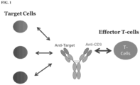
- the present invention provides methods for analyzing preparations of multispecific antibodies having an antigen binding fragment that binds a T cell receptor complex subunit, such as a CD3 subunit, and an antigen binding fragment that binds a target antigen.
- the invention provides methods for detecting a TDB in a composition, quantitating the amount of TDB in a composition, determining the potency and/or specificity of a TDB, or determining if a population of cells expresses a target antigen.
- Compositions and kits are also contemplated.
- T cell dependent bispecific antibodies are bispecific antibodies designed to bind a target antigen expressed on a target cell and a T cell receptor (TCR) complex subunit (e.g., CD3 subunit, such as CD3 ⁇ ) expressed on a T cell. Binding of the bispecific antibody to the extracellular domains of both the target antigen and the TCR complex subunit (TCS) results in T cell recruitment to target cells, leading to T cell activation and target cell depletion. Certain combinations of target antigen-specific and TCS-specific (such as CD3-specific) antigen binding fragments will be more effective than others for specifically activating T cells in the presence of target cells. Weakly activating TDBs will have little therapeutic benefit.
- TCR T cell receptor
- Non-specific activation of T cells in the presence of off-target cells could lead to undesirable release of inflammatory cytokines. It is therefore desirable to assay the degree and specificity of T cell activation mediated by various TDBs in order to support the development of safe and efficacious clinical drug candidates.
- T cell activation assays using Jurkat T-cells stably expressing luciferase reporter driven by IL2 promoter or NFAT-RE are disclosed by Zhi-Jie Cheng Jey et al. (Development of a Robust Reporter-based T cell Activation Assay for Therapeutic Biologics in Immunotherapy ).
- Pete Stecha et al. (Abstract 5439: Development of a robust reporter-based T-cell activation assay for bispecific therapeutic antibodies in immunotherapy ) report the development of a reporter-based T cell activation assay using two Jurkat cell lines stably expressing luciferase reporter driven by IL2 promoter or NFAT-response element. Signosis offered NFAT Luciferase Reporter Stable Cell Line Jurkat T Cells for research use (catalog number SL-0032). Promega provides instructions for use of a NFAT T cell activation bioassay.
- the invention provides methods for detecting T cell activation mediated by a T cell dependent bispecific antibody (TDB), wherein the TDB comprises a target antigen-binding fragment and a T cell receptor complex subunit (TCS)-binding fragment, and various uses thereof.
- TDB T cell dependent bispecific antibody
- TCS T cell receptor complex subunit
- a method of detecting a TDB in a composition comprising contacting the composition with a population of cells comprising a) T cells comprising nucleic acid encoding a reporter operably linked to a promoter and/or enhancer responsive to T cell activation; and b) target cells expressing the target antigen, wherein expression of the reporter indicates the presence of the TDB in the composition.
- the reporter is a luciferase, a fluorescent protein, an alkaline phosphatase, a beta lactamase, or a beta galactosidase.
- the luciferase is a firefly luciferase, a Renilla luciferase, or a nanoluciferase.
- the promoter and/or enhancer responsive to T cell activation is an an NE ⁇ B promoter.
- the promoter and/or enhancer responsive to T cell activation comprises T cell activation responsive elements from any one or more of NFAT, AP-1, NE ⁇ B, FOXO, STAT3, STATS and IRF.
- the T cells in the population of cells are CD4 + T cells or CD8 + T cells. In some embodiments, the T cells in the population of cells are Jurkat T cells or CTLL-2 T cells. In some embodiments, the TCS-binding fragment is a CD3-binding fragment. In some embodiments, the CD3-binding fragment is a CD3 ⁇ -binding fragment. In some embodiments, the target antigen is expressed on the surface of the target cells.
- the population of cells is contacted with a composition comprising the TDB at a concentration range of any of about 0.01 ng/mL to about 5000 ng/mL, about 0.05 ng/mL to about 5000 ng/mL, about 0.1 ng/mL to about 5000 ng/mL, about 0.5 ng/mL to about 5000 ng/mL, about 1 ng/mL to about 5000 ng/mL, about 5 ng/mL to about 5000 ng/mL, about 10 ng/mL to about 5000 ng/mL, about 0.01 ng/mL to about 4000 ng/mL, about 0.01 ng/mL to about 3000 ng/mL, about 0.01 ng/mL to about 2000 ng/mL, about 0.01 ng/mL to about 1000 ng/mL, about 0.01 ng/mL to about 500 ng/m
- the reporter is detected after more than about any of 1 hr, 2 hr, 3 hr, 4 hr, 5 hr, 6 hr, 7 hr, 8 hr, 9 hr, 10 hr, 12 hr, 16 hr, 20 hr, or 24 hr after contacting the cells with the composition.
- a method of quantifying the amount of a TDB in a composition comprising contacting the composition with a population of cells comprising a) T cells comprising nucleic acid encoding a reporter operably linked to a promoter and/or enhancer responsive to T cell activation; and b) target cells expressing the target antigen, and correlating the expression of the reporter as a function of antibody concentration with a standard curve generated by contacting the population of T cells and target cells with different concentrations of a purified reference TDB.
- the reporter is a luciferase, a fluorescent protein, an alkaline phosphatase, a beta lactamase, or a beta galactosidase.
- the luciferase is a firefly luciferase, a Renilla luciferase, or a nanoluciferase.
- the promoter and/or enhancer responsive to T cell activation is an NF ⁇ B promoter.
- the promoter and/or enhancer responsive to T cell activation comprises T cell activation responsive elements from any one or more of NFAT, AP-1, NF ⁇ B, FOXO, STAT3, STATS and IRF.
- the T cells in the population of cells are CD4 + T cells or CD8 + T cells. In some embodiments, the T cells in the population of cells are Jurkat T cells or CTLL-2 T cells. In some embodiments, the TCS-binding fragment is a CD3-binding fragment. In some embodiments, the CD3-binding fragment is a CD3 ⁇ -binding fragment. In some embodiments, the target antigen is expressed on the surface of the target cells.
- the target antigen is HER2 receptor and the target cell is a BT-474 cell
- the target antigen is HER2 receptor and the target cell is a SKBR3 cell
- the target antigen is CD20 and the target cell is a Wil2-S cell
- the target antigen is CD79b and the target cell is a BJAB cell.
- the ratio of T cells to target cells in the population of cells is about 1:1, about 1:2, about 1:3, about 1:4, about 1:5, about 1:6, about 1:7, about 1:8, about 1:9, or about 1:10.
- the ratio of T cells to target cells in the population of cells is about 1:4.
- the population of cells ranges from about 1 ⁇ 10 3 to about 1 ⁇ 10 6 .
- the population of cells is about 1 ⁇ 10 4 to about 5 ⁇ 10 4 .
- the standard curve is generated by contacting the population of cells with the purified reference TDB at a plurality of concentrations ranging from about 0.01 ng/mL to about 5000 ng/mL.
- the reporter is detected after more than about any of 1 hr, 2 hr, 3 hr, 4 hr, 5 hr, 6 hr, 7 hr, 8 hr, 9 hr, 10 hr, 12 hr, 16 hr, 20 hr, or 24 hr after contacting the cells with the composition.
- the reporter is detected between any of about 1 hr and about 24 hr, about 1 hr and about 12 hr, about 1 hr and about 8 hr, about 1 hr and about 6 hr, about 1 hr and about 4 hr, about 1 hr and about 2 hr, about 4 hr and about 24 hr, about 4 hr and about 12 hr, about 4 hr and about 8 hr, about 8 hr and about 24 hr, about 8 hr and about 12 hr, about 16 hr and about 24 hr, about 16 hr and about 20 hr, or about 20 hr and about 24 hr after contacting the cells with the composition.
- a method of determining the specificity of T cell activation mediated by a TDB comprising a) contacting a composition comprising the TDB with a population of cells comprising i) T cells comprising nucleic acid encoding a reporter operably linked to a promoter and/or enhancer responsive to T cell activation; and ii) test cells that do not express the target antigen; and b) contacting a composition comprising the TDB with a population of cells comprising i) T cells comprising nucleic acid encoding a reporter operably linked to a promoter and/or enhancer responsive to T cell activation; and ii) target cells that express the target antigen, and comparing expression of the reporter in the presence of the test cell in part a) with expression of the reporter in the presence of target cells in part b), wherein the
- the T cells in the population of cells are CD4 + T cells or CD8 + T cells. In some embodiments, the T cells in the population of cells are Jurkat T cells or CTLL-2 T cells. In some embodiments, the TCS-binding fragment is a CD3-binding fragment. In some embodiments, the CD3-binding fragment is a CD3 ⁇ -binding fragment. In some embodiments, the target antigen is expressed on the surface of the target cells.
- the target antigen is CD4, CD8, CD18, CD19, CD11a, CD11b, CD20, CD22, CD34, CD40, CD79 ⁇ (CD79a), CD79 ⁇ (CD79b), EGF receptor, HER2 receptor, HER3 receptor, HER4 receptor, FcRH5, CLL1, LFA-1, Mac1, p150, 95, VLA-4, ICAM-1, VCAM, ⁇ v/ ⁇ 3 integrin, VEGF, flk2/flt3 receptor; obesity (OB) receptor; mpl receptor; CTLA-4; protein C, BR3, c-met, tissue factor, ⁇ 7, Tenb2, STEAP, or transmembrane tumor-associated antigens (TAA).
- TAA tumor-associated antigens
- the target antigen is HER2 receptor and the target cell is a BT-474 cell
- the target antigen is HER2 receptor and the target cell is a SKBR3 cell
- the target antigen is CD20 and the target cell is a Wil2-S cell
- the target antigen is CD79b and the target cell is a BJAB cell.
- the ratio of T cells to test cells in the population of cells of step a) and/or the ratio of T cells to target cells in the population of cells of step b) is about 1:1, about 1:2, about 1:3, about 1:4, about 1:5, about 1:6, about 1:7, about 1:8, about 1:9 or about 1:10.
- the population of T cells and test cells of step a) and the population of T cells and target cells of step b) are contacted with a composition comprising the TDB at a concentration range of any of about 0.01 ng/mL to about 5000 ng/mL, about 0.05 ng/mL to about 5000 ng/mL, about 0.1 ng/mL to about 5000 ng/mL, about 0.5 ng/mL to about 5000 ng/mL, about 1 ng/mL to about 5000 ng/mL, about 5 ng/mL to about 5000 ng/mL, about 10 ng/mL to about 5000 ng/mL, about 0.01 ng/mL to about 4000 ng/mL, about 0.01 ng/mL to about 3000 ng/mL, about 0.01 ng/mL to about 2000 ng/mL, about 0. 0.01 ng/mL to about 0.01 ng/mL to about 2000 ng/mL, about 0.
- the reporter is detected after more than about any of 1 hr, 2 hr, 3 hr, 4 hr, 5 hr, 6 hr, 7 hr, 8 hr, 9 hr, 10 hr, 12 hr, 16 hr, 20 hr, or 24 hr after contacting the cells with the composition.
- the reporter is detected between any of about 1 hr and about 24 hr, about 1 hr and about 12 hr, about 1 hr and about 8 hr, about 1 hr and about 6 hr, about 1 hr and about 4 hr, about 1 hr and about 2 hr, about 4 hr and about 24 hr, about 4 hr and about 12 hr, about 4 hr and about 8 hr, about 8 hr and about 24 hr, about 8 hr and about 12 hr, about 16 hr and about 24 hr, about 16 hr and about 20 hr, or about 20 hr and about 24 hr after contacting the cells with the composition.
- kits for the detection of a TDB in a composition comprising a bispecific antibody comprising a target antigen-binding fragment and a TCS-binding fragment, wherein the kit comprises an engineered T cell comprising a reporter operably linked to a promoter and/or enhancer that is responsive to T cell activation.
- the kit further comprises a reference TDB assay standard (a purified TDB of known concentration), and/or a TDB control.
- the kit further comprises a composition comprising target cells expressing the target antigen.
- the reporter is a luciferase, a fluorescent protein, an alkaline phosphatase, a beta lactamase, or a beta galactosidase.
- the luciferase is a firefly luciferase, a Renilla luciferase, or a nanoluciferase.
- the promoter and/or enhancer responsive to T cell activation is an NF ⁇ B promoter.
- the promoter and/or enhancer responsive to T cell activation comprises T cell activation responsive elements from any one or more of NFAT, AP-1, NF ⁇ B, FOXO, STAT3, STATS and IRF.
- the engineered T cells are CD4 + T cells or CD8 + T cells. In some embodiments, the engineered T cells are Jurkat T cells or CTLL-2 T cells. In some embodiments, the TCS-binding fragment is a CD3-binding fragment. In some embodiments, the CD3-binding fragment is a CD3 ⁇ -binding fragment. In some embodiments, the target antigen is expressed on the surface of the target cells.
- the target antigen is CD4, CD8, CD 18, CD19, CD11a, CD11b, CD20, CD22, CD34, CD40, CD79 ⁇ (CD79a), CD79 ⁇ (CD79b), EGF receptor, HER2 receptor, HER3 receptor, HER4 receptor, FcRH5, CLL1, LFA-1, Mac1, p150, 95, VLA-4, ICAM-1, VCAM, ⁇ v/ ⁇ 3 integrin, VEGF, flk2/flt3 receptor; obesity (OB) receptor; mpl receptor; CTLA-4; protein C, BR3, c-met, tissue factor, ⁇ 7, Tenb2, STEAP, or transmembrane tumor-associated antigens (TAA).
- TAA tumor-associated antigens
- the target antigen is HER2 receptor and the target cell is a BT-474 cell
- the target antigen is HER2 receptor and the target cell is a SKBR3 cell
- the target antigen is CD20 and the target cell is a Wil2-S cell
- the target antigen is CD79b and the target cell is a BJAB cell.
- the kit is used in any of the methods described above.
- a method of determining if a population of test cells expresses a target antigen comprising a) contacting the population of test cells with a population of T cells, wherein the T cells comprise nucleic acid encoding a reporter operably linked to a promoter and/or enhancer that is responsive to T cell activation; and b) contacting the population of T cells and test cells with the TDB, wherein the TDB comprises a target antigen-binding fragment and a TCS-binding fragment (such as a CD3-binding fragment), wherein expression of the reporter indicates the presence of the target antigen expressed by the test cell.
- the reporter is a luciferase, a fluorescent protein, an alkaline phosphatase, a beta lactamase, or a beta galactosidase.
- the luciferase is a firefly luciferase, a Renilla luciferase, or a nanoluciferase.
- the promoter and/or enhancer responsive to T cell activation is an NF ⁇ B promoter.
- the promoter and/or enhancer responsive to T cell activation comprises T cell activation responsive elements from any one or more of NFAT, AP-1, NFxB, FOXO, STAT3, STATS and IRF.
- the population of T cells is CD4 + T cells or CD8 + T cells. In some embodiments, the population of T cells is Jurkat T cells or CTLL-2 T cells. In some embodiments, the TCS-binding fragment is a CD3-binding fragment. In some embodiments, the CD3-binding fragment is a CD3 ⁇ -binding fragment. In some embodiments, the target antigen is expressed on the surface of the target cells.
- the target antigen is CD4, CD8, CD18, CD19, CD11a, CD11b, CD20, CD22, CD34, CD40, CD79 ⁇ (CD79a), CD79 ⁇ (CD79b), EGF receptor, HER2 receptor, HER3 receptor, HER4 receptor, FcRH5, CLL1, LFA-1, Mac1, p150, 95, VLA-4, ICAM-1, VCAM, ⁇ v/ ⁇ 3 integrin, VEGF, flk2/flt3 receptor; obesity (OB) receptor; mpl receptor; CTLA-4; protein C, BR3, c-met, tissue factor, ⁇ 7, Tenb2, STEAP, or transmembrane tumor-associated antigens (TAA).
- TAA tumor-associated antigens
- the population of test cells are is a population of tumor cells, immune cells or vascular cells. In some embodiments, the population of test cells does not comprise T cells. In some embodiments, the ratio of T cells to test cells is about 1:1, about 1:2, about 1:3, about 1:4, about 1:5, about 1:6, about 1:7, about 1:8, about 1:9 or about 1:10. In some embodiments, the ratio of T cells to test cells is about 1:4. In some embodiments, the population of test cells and T cells comprises from about 1 ⁇ 10 3 to about 1 ⁇ 10 6 cells. In some embodiments, the population of test cells and T cells comprises from about 1 ⁇ 10 4 to about 5 ⁇ 10 4 cells.
- the population of test cells and T cells is contacted with a composition comprising the TDB at a concentration range of any of about 0.01 ng/mL to about 5000 ng/mL, about 0.05 ng/mL to about 5000 ng/mL, about 0.1 ng/mL to about 5000 ng/mL, about 0.5 ng/mL to about 5000 ng/mL, about 1 ng/mL to about 5000 ng/mL, about 5 ng/mL to about 5000 ng/mL, about 10 ng/mL to about 5000 ng/mL, about 0.01 ng/mL to about 4000 ng/mL, about 0.01 ng/mL to about 3000 ng/mL, about 0.01 ng/mL to about 2000 ng/mL, about 0.01 ng/mL to about 1000 ng/mL, about 0.01
- the reporter is detected after more than about any of 1 hr, 2 hr, 3 hr, 4 hr, 5 hr, 6 hr, 7 hr, 8 hr, 9 hr, 10 hr, 12 hr, 16 hr, 20 hr, or 24 hr after contacting the cells with the composition.
- the reporter is detected between any of about 1 hr and about 24 hr, about 1 hr and about 12 hr, about 1 hr and about 8 hr, about 1 hr and about 6 hr, about 1 hr and about 4 hr, about 1 hr and about 2 hr, about 4 hr and about 24 hr, about 4 hr and about 12 hr, about 4 hr and about 8 hr, about 8 hr and about 24 hr, about 8 hr and about 12 hr, about 16 hr and about 24 hr, about 16 hr and about 20 hr, or about 20 hr and about 24 hr after contacting the cells with the composition.
- the invention provides methods for detecting T cell activation mediated by a T cell dependent bispecific antibody (TDB) and/or determining the potency of a TDB, wherein the TDB comprises an antigen binding fragment that binds to a target antigen and an antigen binding fragment that binds to a T cell receptor complex subunit (TCS), such as a CD3 subunit, e.g., CD3 ⁇ , expressed on a T cell, and various uses thereof, including, inter alia, detecting a TDB in a composition, quantitating the amount of TDB in a composition, determining the specificity of a TDB, and determining if a population of cells expresses a target antigen.
- TDS T cell receptor complex subunit
- kits for detecting T cell activation mediated by a TDB and/or determining the potency of a TDB wherein the kit comprises an engineered T cell comprising a reporter operably linked to a promoter and/or enhancer responsive to T cell activation, and optionally includes the TDB, a reference TDB, a control TDB, and/or target cells.
- polypeptide or "protein” are used interchangeably herein to refer to polymers of amino acids of any length.
- the polymer may be linear or branched, it may comprise modified amino acids, and it may be interrupted by non-amino acids.
- the terms also encompass an amino acid polymer that has been modified naturally or by intervention; for example, disulfide bond formation, glycosylation, lipidation, acetylation, phosphorylation, or any other manipulation or modification, such as conjugation with a labeling component or toxin.
- polypeptides containing one or more analogs of an amino acid including, for example, unnatural amino acids, etc.
- the terms "polypeptide” and "protein” as used herein specifically encompass antibodies.
- Polypeptide e.g., antibody or immunoadhesin
- Purity is a relative term and does not necessarily mean absolute purity.
- antagonist is used in the broadest sense, and includes any molecule that partially or fully blocks, inhibits, or neutralizes a biological activity of a native polypeptide.
- agonist is used in the broadest sense and includes any molecule that mimics a biological activity of a native polypeptide. Suitable agonist or antagonist molecules specifically include agonist or antagonist antibodies or antibody fragments, fragments or amino acid sequence variants of native polypeptides, etc.
- Methods for identifying agonists or antagonists of a polypeptide may comprise contacting a polypeptide with a candidate agonist or antagonist molecule and measuring a detectable change in one or more biological activities normally associated with the polypeptide.
- a polypeptide "which binds" an antigen of interest e.g. a tumor-associated polypeptide antigen target
- an antigen of interest e.g. a tumor-associated polypeptide antigen target
- the extent of binding of the polypeptide to a "non-target" polypeptide will be less than about 10% of the binding of the polypeptide to its particular target polypeptide as determined by fluorescence activated cell sorting (FACS) analysis or radioimmunoprecipitation (RIA).
- the term "specific binding” or “specifically binds to” or is “specific for” a particular polypeptide or an epitope on a particular polypeptide target means binding that is measurably different from a non-specific interaction.
- Specific binding can be measured, for example, by determining binding of a molecule compared to binding of a control molecule, which generally is a molecule of similar structure that does not have binding activity.
- specific binding can be determined by competition with a control molecule that is similar to the target, for example, an excess of non-labeled target. In this case, specific binding is indicated if the binding of the labeled target to a probe is competitively inhibited by excess unlabeled target.
- antibody herein is used in the broadest sense and specifically covers monoclonal antibodies, polyclonal antibodies, multispecific antibodies (e.g. bispecific antibodies including TDB) formed from at least two intact antibodies, and antibody fragments so long as they exhibit the desired biological activity.
- multispecific antibodies e.g. bispecific antibodies including TDB
- antibody fragments so long as they exhibit the desired biological activity.
- immunoglobulin immunoglobulin
- Antibodies are naturally occurring immunoglobulin molecules which have varying structures, all based upon the immunoglobulin fold.
- IgG antibodies have two "heavy” chains and two "light” chains that are disulphide-bonded to form a functional antibody.
- Each heavy and light chain itself comprises a “constant” (C) and a “variable” (V) region.
- the V regions determine the antigen binding specificity of the antibody, whilst the C regions provide structural support and function in non-antigen-specific interactions with immune effectors.
- the antigen binding specificity of an antibody or antigen-binding fragment of an antibody is the ability of an antibody to specifically bind to a particular antigen.
- the antigen binding specificity of an antibody is determined by the structural characteristics of the V region.
- the variability is not evenly distributed across the 110-amino acid span of the variable domains.
- the V regions consist of relatively invariant stretches called framework regions (FRs) of 15-30 amino acids separated by shorter regions of extreme variability called “hypervariable regions” (HVRs) that are each 9-12 amino acids long.
- FRs framework regions
- HVRs hypervariable regions
- the variable domains of native heavy and light chains each comprise four FRs, largely adopting a ⁇ -sheet configuration, connected by three hypervariable regions, which form loops connecting, and in some cases forming part of, the ⁇ -sheet structure.
- the hypervariable regions in each chain are held together in close proximity by the FRs and, with the hypervariable regions from the other chain, contribute to the formation of the antigen-binding site of antibodies ( see Kabat et al., Sequences of Proteins of Immunological Interest, 5th Ed. Public Health Service, National Institutes of Health, Bethesda, Md. (1991 )).
- the constant domains are not involved directly in binding an antibody to an antigen, but exhibit various effector functions, such as participation of the antibody in antibody dependent cellular cytotoxicity (ADCC).
- Each V region typically comprises three HVRs, e.g . complementarity determining regions ("CDRs", each of which contains a "hypervariable loop"), and four framework regions.
- An antibody binding site the minimal structural unit required to bind with substantial affinity to a particular desired antigen, will therefore typically include the three CDRs, and at least three, preferably four, framework regions interspersed there between to hold and present the CDRs in the appropriate conformation.
- Classical four chain antibodies have antigen binding sites which are defined by V H and V L domains in cooperation. Certain antibodies, such as camel and shark antibodies, lack light chains and rely on binding sites formed by heavy chains only. Single domain engineered immunoglobulins can be prepared in which the binding sites are formed by heavy chains or light chains alone, in absence of cooperation between V H and V L .
- variable refers to the fact that certain portions of the variable domains differ extensively in sequence among antibodies and are used in the binding and specificity of each particular antibody for its particular antigen. However, the variability is not evenly distributed throughout the variable domains of antibodies. It is concentrated in three segments called hypervariable regions both in the light chain and the heavy chain variable domains. The more highly conserved portions of variable domains are called the framework regions (FRs).
- the variable domains of native heavy and light chains each comprise four FRs, largely adopting a ⁇ -sheet configuration, connected by three hypervariable regions, which form loops connecting, and in some cases forming part of, the ⁇ -sheet structure.
- the hypervariable regions in each chain are held together in close proximity by the FRs and, with the hypervariable regions from the other chain, contribute to the formation of the antigen-binding site of antibodies ( see Kabat et al., Sequences of Proteins of Immunological Interest, 5th Ed. Public Health Service, National Institutes of Health, Bethesda, MD. (1991 )).
- the constant domains are not involved directly in binding an antibody to an antigen, but exhibit various effector functions, such as participation of the antibody in antibody dependent cellular cytotoxicity (ADCC).
- hypervariable region when used herein refers to the amino acid residues of an antibody that are responsible for antigen binding.
- the hypervariable region may comprise amino acid residues from a "complementarity determining region” or "CDR" (e.g., around about residues 24-34 (L1), 50-56 (L2) and 89-97 (L3) in the V L , and around about 31-35B (H1), 50-65 (H2) and 95-102 (H3) in the V H ( Kabat et al., Sequences of Proteins of Immunological Interest, 5th Ed. Public Health Service, National Institutes of Health, Bethesda, Md.
- CDR complementarity determining region
- Framework or "FR” residues are those variable domain residues other than the hypervariable region residues as herein defined.
- T cell dependent bispecific antibodies or “TDB” are bispecific antibodies designed to bind a target antigen expressed on a cell, and to bind to T cells, such as by binding to a T cell receptor complex subunit (e.g ., CD3 ⁇ ) expressed on a T cell.
- T cell receptor complex subunit e.g ., CD3 ⁇
- Papain digestion of antibodies produces two identical antigen-binding fragments, called “Fab” fragments, each with a single antigen-binding site, and a residual "Fc” fragment, whose name reflects its ability to crystallize readily. Pepsin treatment yields an F(ab') 2 fragment that has two antigen-binding sites and is still capable of cross-linking antigen.
- Fv is the minimum antibody fragment that contains a complete antigen-recognition and antigen-binding site. This region consists of a dimer of one heavy chain and one light chain variable domain in tight, non-covalent association. It is in this configuration that the three hypervariable regions of each variable domain interact to define an antigen-binding site on the surface of the V H -V L dimer. Collectively, the six hypervariable regions confer antigen-binding specificity to the antibody. However, even a single variable domain (or half of an Fv comprising only three hypervariable regions specific for an antigen) has the ability to recognize and bind antigen, although at a lower affinity than the entire binding site.
- the Fab fragment also contains the constant domain of the light chain and the first constant domain (CH1) of the heavy chain.
- Fab' fragments differ from Fab fragments by the addition of a few residues at the carboxy terminus of the heavy chain CH1 domain including one or more cysteines from the antibody hinge region.
- Fab'-SH is the designation herein for Fab' in which the cysteine residue(s) of the constant domains bear at least one free thiol group.
- F(ab') 2 antibody fragments originally were produced as pairs of Fab' fragments that have hinge cysteines between them. Other chemical couplings of antibody fragments are also known.
- the "light chains" of antibodies (immunoglobulins) from any vertebrate species can be assigned to one of two clearly distinct types, called kappa ( ⁇ ) and lambda ( ⁇ ), based on the amino acid sequences of their constant domains.
- antibodies can be assigned to different classes. There are five major classes of intact antibodies: IgA, IgD, IgE, IgG, and IgM, and several of these may be further divided into subclasses (isotypes), e.g. , IgG1, IgG2, IgG3, IgG4, IgA, and IgA2.
- the heavy chain constant domains that correspond to the different classes of antibodies are called ⁇ , ⁇ , ⁇ , ⁇ , and ⁇ , respectively.
- the subunit structures and three-dimensional configurations of different classes of immunoglobulins are well known.
- Single-chain Fv or “scFv” antibody fragments comprise the V H and V L domains of antibody, wherein these domains are present in a single polypeptide chain.
- the Fv polypeptide further comprises a polypeptide linker between the V H and V L domains that enables the scFv to form the desired structure for antigen binding.
- diabodies refers to small antibody fragments with two antigen-binding sites, which fragments comprise a heavy chain variable domain (V H ) connected to a light chain variable domain (V L ) in the same polypeptide chain (V H - V L ).
- V H heavy chain variable domain
- V L light chain variable domain
- the domains are forced to pair with the complementary domains of another chain and create two antigen-binding sites.
- Diabodies are described more fully in, for example, EP 404,097 ; WO 93/11161 ; and Hollinger et al., Proc. Natl. Acad. Sci. USA, 90:6444-6448 (1993 ).
- multispecific antibody is used in the broadest sense and specifically covers an antibody that has polyepitopic specificity.
- Such multispecific antibodies include, but are not limited to, an antibody comprising a heavy chain variable domain (V H ) and a light chain variable domain (V L ), where the V H V L unit has polyepitopic specificity, antibodies having two or more V L and V H domains with each V H V L unit binding to a different epitope, antibodies having two or more single variable domains with each single variable domain binding to a different epitope, full length antibodies, antibody fragments such as Fab, Fv, dsFv, scFv, diabodies, bispecific diabodies, triabodies, tri-functional antibodies, antibody fragments that have been linked covalently or non-covalently.
- Polyepitopic specificity refers to the ability to specifically bind to two or more different epitopes on the same or different target(s).
- “Monospecific” refers to the ability to bind only one epitope.
- the multispecific antibody is an IgG antibody that binds to each epitope with an affinity of 5 ⁇ M to 0.001 pM, 3 ⁇ M to 0.001 pM, 1 ⁇ M to 0.001 pM, 0.5 ⁇ M to 0.001 pM, or 0.1 ⁇ M to 0.001 pM.
- single domain antibodies or “single variable domain (SVD) antibodies” generally refers to antibodies in which a single variable domain (VH or VL) can confer antigen binding. In other words, the single variable domain does not need to interact with another variable domain in order to recognize the target antigen.
- single domain antibodies include those derived from camelids (lamas and camels) and cartilaginous fish ( e.g., nurse sharks) and those derived from recombinant methods from humans and mouse antibodies ( Nature (1989) 341:544-546 ; Dev Comp Immunol (2006) 30:43-56 ; Trend Biochem Sci (2001) 26:230-235 ; Trends Biotechnol (2003):21:484-490 ; WO 2005/035572 ; WO 03/035694 ; Febs Lett (1994) 339:285-290 ; WO00/29004 ; WO 02/051870 ).
- the term "monoclonal antibody” as used herein refers to an antibody obtained from a population of substantially homogeneous antibodies, i.e ., the individual antibodies comprising the population are identical and/or bind the same epitope, except for possible variants that may arise during production of the monoclonal antibody, such variants generally being present in minor amounts.
- each monoclonal antibody is directed against a single determinant on the antigen.
- the monoclonal antibodies are advantageous in that they are uncontaminated by other immunoglobulins.
- the modifier "monoclonal” indicates the character of the antibody as being obtained from a substantially homogeneous population of antibodies, and is not to be construed as requiring production of the antibody by any particular method.
- the monoclonal antibodies to be used in accordance with the methods provided herein may be made by the hybridoma method first described by Kohler et al., Nature 256:495 (1975 ), or may be made by recombinant DNA methods ( see, e.g., U.S. Patent No. 4,816,567 ).
- the "monoclonal antibodies” may also be isolated from phage antibody libraries using the techniques described in Clackson et al., Nature 352:624-628 (1991 ) and Marks et al., J. Mol. Biol. 222:581-597 (1991 ), for example.
- Chimeric antibodies of interest herein include "primatized" antibodies comprising variable domain antigen-binding sequences derived from a non-human primate (e.g. Old World Monkey, such as baboon, rhesus or cynomolgus monkey) and human constant region sequences ( US Pat No. 5,693,780 ).
- a non-human primate e.g. Old World Monkey, such as baboon, rhesus or cynomolgus monkey
- human constant region sequences US Pat No. 5,693,780
- Humanized forms of non-human (e.g ., murine) antibodies are chimeric antibodies that contain minimal sequence derived from non-human immunoglobulin.
- humanized antibodies are human immunoglobulins (recipient antibody) in which residues from a hypervariable region of the recipient are replaced by residues from a hypervariable region of a non-human species (donor antibody) such as mouse, rat, rabbit or nonhuman primate having the desired specificity, affinity, and capacity.
- donor antibody such as mouse, rat, rabbit or nonhuman primate having the desired specificity, affinity, and capacity.
- framework region (FR) residues of the human immunoglobulin are replaced by corresponding non-human residues.
- humanized antibodies may comprise residues that are not found in the recipient antibody or in the donor antibody. These modifications are made to further refine antibody performance.
- the humanized antibody will comprise substantially all of at least one, and typically two, variable domains, in which all or substantially all of the hypervariable loops correspond to those of a non-human immunoglobulin and all or substantially all of the FRs are those of a human immunoglobulin sequence, except for FR substitution(s) as noted above.
- the humanized antibody optionally also will comprise at least a portion of an immunoglobulin constant region, typically that of a human immunoglobulin. For further details, see Jones et al., Nature 321:522-525 (1986 ); Riechmann et al., Nature 332:323-329 (1988 ); and Presta, Curr. Op. Struct. Biol. 2:593-596 (1992 ).
- an "intact antibody” is one comprising heavy and light variable domains as well as an Fc region.
- the constant domains may be native sequence constant domains (e.g. human native sequence constant domains) or amino acid sequence variant thereof.
- the intact antibody has one or more effector functions.
- “Native antibodies” are usually heterotetrameric glycoproteins of about 150,000 daltons, composed of two identical light (L) chains and two identical heavy (H) chains. Each light chain is linked to a heavy chain by one covalent disulfide bond, while the number of disulfide linkages varies among the heavy chains of different immunoglobulin isotypes. Each heavy and light chain also has regularly spaced intrachain disulfide bridges. Each heavy chain has at one end a variable domain (V H ) followed by a number of constant domains.
- V H variable domain
- Each light chain has a variable domain at one end (V L ) and a constant domain at its other end; the constant domain of the light chain is aligned with the first constant domain of the heavy chain, and the light chain variable domain is aligned with the variable domain of the heavy chain. Particular amino acid residues are believed to form an interface between the light chain and heavy chain variable domains.
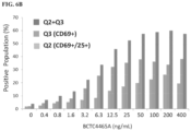
- TDB bi-specifics are capable of activating both CD4 + and CD8 + T cell lineages, provided the right target-expressing cells are present.
- the population of cells is contacted with a composition comprising the TDB at a concentration range of any of about 0.01 ng/mL to about 5000 ng/mL, about 0.05 ng/mL to about 5000 ng/mL, about 0.1 ng/mL to about 5000 ng/mL, about 0.5 ng/mL to about 5000 ng/mL, about 1 ng/mL to about 5000 ng/mL, about 5 ng/mL to about 5000 ng/mL, about 10 ng/mL to about 5000 ng/mL, about 0.01 ng/mL to about 4000 ng/mL, about 0.01 ng/mL to about 3000 ng/mL, about 0.01 ng/mL to about 2000 ng/mL, about 0.01 ng/mL to about 1000 ng/mL, about 0.01 ng/mL to about 500 ng/mL, about 0.01 ng/mL to about 100 ng/mL
- the reporter is detected after more than about any of 1 hr, 2 hr, 3 hr, 4 hr, 5 hr, 6 hr, 7 hr, 8 hr, 9 hr, 10 hr, 12 hr, 16 hr, 20 hr, or 24 hr after contacting the cells with the composition.
- the invention provides methods for quantifying the amount of a TDB in a composition, wherein the TDB comprises a target antigen-binding fragment and a TCS-binding fragment (such as a CD3-binding fragment), the method comprising contacting the composition with a population of cells comprising a) T cells comprising nucleic acid encoding a reporter operably linked to a promoter and/or enhancer responsive to T cell activation; and b) target cells expressing the target antigen, and correlating the expression of the reporter as a function of antibody concentration with a standard curve generated by contacting the population of T cells and target cells with different concentrations of a purified reference TDB.
- a target antigen-binding fragment such as a CD3-binding fragment
- the T cells in the population of cells are CD4 + T cells or CD8 + T cells. In some embodiments, the T cells in the population of cells are Jurkat T cells or CTLL-2 T cells. In some embodiments, the TCS-binding fragment is a CD3-binding fragment. In some embodiments, the CD3-binding fragment is a CD3 ⁇ -binding fragment. In some embodiments, the target antigen is expressed on the surface of the target cells.
- the reporter is detected after more than about any of 1 hr, 2 hr, 3 hr, 4 hr, 5 hr, 6 hr, 7 hr, 8 hr, 9 hr, 10 hr, 12 hr, 16 hr, 20 hr, or 24 hr after contacting the cells with the composition.
- the invention provides methods for determining the potency of T cell activation mediated by a TDB, wherein the TDB comprises a target antigen-binding fragment and a TCS-binding fragment (such as a CD3-binding fragment), the method comprising contacting a composition comprising the TDB with a population of cells comprising a) T cells comprising nucleic acid encoding a reporter operably linked to a promoter and/or enhancer responsive to T cell activation; and b) target cells expressing the target antigen, and correlating the expression of the reporter as a function of antibody concentration with a standard curve generated by contacting the population of cells with different concentrations of a reference TDB, thereby obtaining a relative measure of the potency of T cell activated mediated by the TDB.
- a composition comprising the TDB with a population of cells comprising a) T cells comprising nucleic acid encoding a reporter operably linked to a promoter and/or enhancer responsive to T cell activation; and b) target
- the target antigen is CD4, CD8, CD18, CD19, CD11a, CD11b, CD20, CD22, CD34, CD40, CD79 ⁇ (CD79a), CD79 ⁇ (CD79b), EGF receptor, HER2 receptor, HER3 receptor, HER4 receptor, FcRH5, CLL1, LFA-1, Mac1, p150, 95, VLA-4, ICAM-1, VCAM, ⁇ v/ ⁇ 3 integrin, VEGF, flk2/flt3 receptor; obesity (OB) receptor; mpl receptor; CTLA-4; protein C, BR3, c-met, tissue factor, ⁇ 7, Tenb2, STEAP, or transmembrane tumor-associated antigens (TAA).
- TAA tumor-associated antigens
- the target antigen is HER2 receptor and the target cell is a BT-474 cell
- the target antigen is HER2 receptor and the target cell is a SKBR3 cell
- the target antigen is CD20 and the target cell is a Wil2-S cell
- the target antigen is CD79b and the target cell is a BJAB cell.
- the ratio of T cells to target cells in the population of cells is about 1:1, about 1:2, about 1:3, about 1:4, about 1:5, about 1:6, about 1:7, about 1:8, about 1:9, or about 1:10.
- the ratio of T cells to target cells in the population of cells is about 1:4.
- the population of cells ranges from about 1 ⁇ 10 3 to about 1 ⁇ 10 6 .
- the population of cells is about 1 ⁇ 10 4 to about 5 ⁇ 10 4 .
- the standard curve is generated by contacting the population of cells with the purified reference TDB at a plurality of concentrations ranging from about 0.01 ng/mL to about 5000 ng/mL.
- the plurality of concentrations of purified reference TDB include about any one of 0.01 ng/ml, 0.1 ng/ml, 1 ng/ml, 10 ng/ml, 100ng/mL, 150 ng/mL, 200 ng/mL, 250 ng/mL, 500 ng/mL, 750 ng/mL, 1 ⁇ g/mL, 2.5 ⁇ g/mL, 5 ⁇ g/mL, 10 ⁇ g/mL, 25 ⁇ g/mL, 50 ⁇ g/mL, 100 ⁇ g/mL, 250 ⁇ g/mL, or 500 ⁇ g/mL.
- the plurality of concentrations of reference TDB is about three, four, five, six, seven, eight, nine, ten or more
- the reporter is detected after more than about any of 1 hr, 2 hr, 3 hr, 4 hr, 5 hr, 6 hr, 7 hr, 8 hr, 9 hr, 10 hr, 12 hr, 16 hr, 20 hr, or 24 hr after contacting the cells with the composition.
- the reporter is detected between any of about 1 hr and about 24 hr, about 1 hr and about 12 hr, about 1 hr and about 8 hr, about 1 hr and about 6 hr, about 1 hr and about 4 hr, about 1 hr and about 2 hr, about 4 hr and about 24 hr, about 4 hr and about 12 hr, about 4 hr and about 8 hr, about 8 hr and about 24 hr, about 8 hr and about 12 hr, about 16 hr and about 24 hr, about 16 hr and about 20 hr, or about 20 hr and about 24 hr after contacting the cells with the composition.
- the T cells in the population of cells are CD4 + T cells or CD8 + T cells. In some embodiments, the T cells in the population of cells are Jurkat T cells or CTLL-2 T cells. In some embodiments, the TCS-binding fragment is a CD3-binding fragment. In some embodiments, the CD3-binding fragment is a CD3 ⁇ -binding fragment. In some embodiments, the target antigen is expressed on the surface of the target cells.
- the ratio of T cells to test cells in the population of cells of step a) and/or the ratio of T cells to target cells in the population of cells or step b) is about 1:4. In some embodiments, the population of cells of steps a) and/or b) ranges from about 1 ⁇ 10 3 to about 1 ⁇ 10 6 . In some embodiments, the population of cells of steps a) and/or b) ranges from about 1 ⁇ 10 4 to about 5 ⁇ 10 4 .
- the population of T cells and test cells of step a) and the population of T cells and target cells of step b) are contacted with a composition comprising the TDB at a concentration range of any of about 0.01 ng/mL to about 5000 ng/mL, about 0.05 ng/mL to about 5000 ng/mL, about 0.1 ng/mL to about 5000 ng/mL, about 0.5 ng/mL to about 5000 ng/mL, about 1 ng/mL to about 5000 ng/mL, about 5 ng/mL to about 5000 ng/mL, about 10 ng/mL to about 5000 ng/mL, about 0.01 ng/mL to about 4000 ng/mL, about 0.01 ng/mL to about 3000 ng/mL, about 0.01 ng/mL to about 2000 ng/mL, about 0.01 ng/mL to about 1000 ng/mL, about 0.01 ng/mL to about
- the invention provides methods for determining if a population of test cells expresses a target antigen, the method comprising a) contacting the population of test cells with a population of T cells, wherein the T cells comprise nucleic acid encoding a reporter operably linked to a promoter and/or enhancer that is responsive to T cell activation; and b) contacting the population of T cells and test cells with the TDB, wherein the TDB comprises a target antigen-binding fragment and a TCS-binding fragment (such as a CD3-binding fragment), wherein expression of the reporter indicates the presence of the target antigen expressed by the test cell.
- the population of T cells is a population of CD4 + T cells or CD8 + T cells. In some embodiments, the population of T cells is a population of Jurkat T cells or CTLL-2 T cells. In some embodiments, the TCS-binding fragment is a CD3-binding fragment. In some embodiments, the CD3-binding fragment is a CD3 ⁇ -binding fragment. In some embodiments, the target antigen is expressed on the surface of the target cells.
- the target antigen is CD4, CD8, CD18, CD19, CD11a, CD11b, CD20, CD22, CD34, CD40, CD79 ⁇ (CD79a), CD79 ⁇ (CD79b), EGF receptor, HER2 receptor, HER3 receptor, HER4 receptor, FcRH5, CLL1, LFA-1, Mac1, p150, 95, VLA-4, ICAM-1, VCAM, ⁇ v/ ⁇ 3 integrin, VEGF, flk2/flt3 receptor; obesity (OB) receptor; mpl receptor; CTLA-4; protein C, BR3, c-met, tissue factor, ⁇ 7, Tenb2, STEAP, or transmembrane tumor-associated antigens (TAA).
- TAA tumor-associated antigens
- the population of test cells is a population of tumor cells, immune cells or vascular cells. In some embodiments, the population of test cells does not comprise T cells. In some embodiments, the ratio of T cells to test cells is about 1:1, about 1:2, about 1:3, about 1:4, about 1:5, about 1:6, about 1:7, about 1:8, about 1:9 or about 1:10. In some embodiments, the ratio of T cells to test cells is about 1:4. In some embodiments, the population of test cells and T cells comprises from about 1 ⁇ 10 3 to about 1 ⁇ 10 6 cells. In some embodiments, the population of test cells and T cells comprises from about 1 ⁇ 10 4 to about 5 ⁇ 10 4 cells.
- the population of test cells and T cells is contacted with a composition comprising the TDB at a concentration range of any of about 0.01 ng/mL to about 5000 ng/mL, about 0.05 ng/mL to about 5000 ng/mL, about 0.1 ng/mL to about 5000 ng/mL, about 0.5 ng/mL to about 5000 ng/mL, about 1 ng/mL to about 5000 ng/mL, about 5 ng/mL to about 5000 ng/mL, about 10 ng/mL to about 5000 ng/mL, about 0.01 ng/mL to about 4000 ng/mL, about 0.01 ng/mL to about 3000 ng/mL, about 0.01 ng/mL to about 2000 ng/mL, about 0.01 ng/mL to about 1000 ng/mL, about 0.01 ng/mL to about 500 ng/mL, about 0.01 ng/mL to about 100
- the reporter is detected after more than about any of 1 hr, 2 hr, 3 hr, 4 hr, 5 hr, 6 hr, 7 hr, 8 hr, 9 hr, 10 hr, 12 hr, 16 hr, 20 hr, or 24 hr after contacting the cells with the composition.
- the reporter is detected between any of about 1 hr and about 24 hr, about 1 hr and about 12 hr, about 1 hr and about 8 hr, about 1 hr and about 6 hr, about 1 hr and about 4 hr, about 1 hr and about 2 hr, about 4 hr and about 24 hr, about 4 hr and about 12 hr, about 4 hr and about 8 hr, about 8 hr and about 24 hr, about 8 hr and about 12 hr, about 16 hr and about 24 hr, about 16 hr and about 20 hr, or about 20 hr and about 24 hr after contacting the cells with the composition.
- the following is an exemplary but non-limiting method of developing a cell-based assay to detect TDB-mediated T cell activity.
- Lentivirus is used to generate the stable reporter T cell lines used to evaluate the potency of the TDB bi-specific antibody.
- Lentiviral vectors are constructed that express the reporter gene firefly luciferase, Renilla luciferase, or Nanoluciferase under the control of a minimal TK promoter regulated by DNA recognition elements for NFAT (Nuclear Factor of Activated T cells), AP-1 (Fos/Jun), NFAT/AP1, NE ⁇ B, FOXO, STAT3/5, or IRF.
- the lentiviral expression cassettes used for the generation of the stable reporter cell lines may be third generation self-inactivating bi-cistronic vectors that express various antibiotic selection markers under the control of constitutive promoters/enhancers (EF1alpha or SV40) to enable the generation of stable cell lines.
- the reporter lentiviral vectors used are modified from the pCDH.MCS.EF1a.Puro commercially available vector (SBI biosciences; Cat No. CD510B-1).
- Promoter modifications include the removal of the CMV minimal promoter and substitution with various enhancer elements (NFAT, NF ⁇ B, etc.), addition of a minimal core RNA polymerase promoter (TATA box) from pRK5.CMV.Luciferase ( Osaka, G et al., 1996 J Pharm Sci. 1996, 85:612-618 ), and substitution of different selection cassettes from internal DNAs (Neomycin resistance gene from pRK5.tk.neo, Hygromycin resistance gene from pRK5.tk.hygro; and the blasticidin resistance gene from pRK5.tk.blastocidin).
- TATA box minimal core RNA polymerase promoter
- Firefly Luciferase from pRK5.CMV.Luciferase (Osaka, 1996) is cloned into the HindIII-NotI site of the modified lentiviral parent vector.
- Other luminescent proteins including Renilla Luciferase and NanoLuciferase may also be subcloned into the HindIII-NotI site.
- Lentiviral packaging constructs (pCMV.HIVdelta, pCMC.VSV-G, and pCMV.Rev) used to generate viral stocks from transient transfection of 293s (293 suspension adapted cell line) cells may be obtained (pCMV.VSV-G) or generated (pCMV.HIVdelta, pCMV.REV).
- HIV strain MN Nakamura, GR et al., 1993, J. Virol. 67(10):6179-6191 ) may be used to generate the pCMV.HIVdelta packaging vector and contains an internal EcoRI partial digest deletion to inactivate by deletion the HIV viral envelope and modifications to the 5' and 3'LTRs for safety purposes.
- HIV Rev is cloned from pCMV.HIVdelta transfected 293s cell RNA by RT-PCR and introduced into the ClaI-Xho site of pRK5.tk.neo.
- VSV-G pseudotype the lentiviral reporters (substituting VSV-G for HIV env) enables the infection of any cell type.
- Lentiviral expression plasmids and packaging constructs are amplified in Stbl2 competent cells (Life Technologies, Cat. No. 10268-019) and DNA purified using Qiagen Maxi Prep kit (Cat. No. 12662). All DNA constructs are confirmed by DNA sequencing.
- Reporter gene assay cell line development Jurkat CD4 + T cell line (DSMZ, Cat. No. ACC 282) and CTLL-2 CD8 + T cell line (Life Technologies, Cat. No. K1653) are used to evaluate the feasibility of a reporter gene assay to monitor the activation of T cells by the TDB.
- Lentiviral vectors are constructed that express the reporter gene firefly luciferase, Renilla luciferase, or Nanoluciferase under the control of a minimal TK promoter regulated by DNA recognition elements for NFAT (Nuclear Factor of Activated T cells), AP-1 (Fos/Jun), NFAT/AP1, NF ⁇ B,FOXO, STAT3,5, and IRF.
- Reporter gene viral stocks are generated by transient transfection of 293s cells and pseudotyped with VSV-G, concentrated, and titered using standard methods ( Naldini, L., et al., 1996 Science, 272:263-267 ).
- the Jurkat CTLL-2 cells are infected with the lentiviral reporter viral stock at an MOI of 10 by spinoculation and after 3 days infected cells are selected for antibiotic resistance. After 2 weeks, stable pools are generated and evaluated for the response to purified TDB.
- a qPCR method that evaluates copy number and integration is used to demonstrate that all stable pools are stably infected with the reporter constructs.
- limiting dilution of Jurkat/NF ⁇ B-luciferase and Jurkat/NFAT-Luciferase are set up to enable single cell cloning and generation of single stable reporter cell lines.
- T cell activation assay To quantitate the potency of TDB-mediated T cell activation, the amount of luciferase activity observed for a plurality of dilutions of a TDB test sample incubated with a population of Jurkat/NF ⁇ B-fireflyLuciferase effector cells and target cells is compared to the luciferase activity observed for a reference TDB. The relative potency of the test TDB samples is determined from the standard curve generated by using the reference TDB.
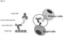
- non cell-based assays to detect simultaneous TDB binding of a target antigen and a TCS are provided, wherein one antigen binding fragment of the TDB binds the target antigen and the other antigen binding fragment binds the TCS.
- simultaneous binding of the TDB to a T cell receptor (TCR) complex subunit such as a CD3 subunit, e.g ., CD3 ⁇
- TCR T cell receptor
- a target antigen expressed on the surface of a target cell results in TCR clustering, leading to T cell activation and the cytotoxic depletion of the target cell.
- the invention provides methods of detecting simultaneous binding of a TDB to a target antigen and a TCS (such as a CD3 subunit), wherein one antigen binding fragment of the TDB binds a first epitope on the target antigen and the other antigen binding fragment binds a second epitope on the TCS, the method comprising performing an ELISA-based bridging binding assay using immobilized target antigen, or a fragment thereof comprising the first epitope, and a conjugate of biotin and the TCS, or a fragment thereof comprising the second epitope .
- the first epitope is localized to an extracellular portion of the target antigen and/or the second epitope is localized to an extracellular portion of the TCS.
- methods of detecting simultaneous binding of a TDB to a target antigen and a TCS are provided, wherein one antigen binding fragment of the TDB binds a first epitope on the target antigen and the other antigen binding fragment binds a second epitope on the TCS, the method comprising a) immobilizing the target antigen, or a fragment thereof comprising the first epitope, to a solid phase; b) incubating the TDB with the target antigen, or fragment thereof comprising the first epitope, immobilized to the solid phase; c) incubating the TDB with a conjugate of a reporter molecule and the TCS, or a fragment thereof comprising the second epitope (biotin-TCS conjugate); d) optionally incubating the reporter-TCS conjugate with an accessory molecule needed to detect the reporter molecule; e) removing molecules unbound to the solid phase (such as by washing); and f) detecting the
- methods of detecting simultaneous binding of a TDB to a target antigen and a TCS are provided, wherein one antigen binding fragment of the TDB binds a first epitope on the target antigen and the other antigen binding fragment binds a second epitope on the TCS, the method comprising a) immobilizing the target antigen, or a fragment thereof comprising the first epitope, to a solid phase; b) incubating the TDB with the target antigen, or fragment thereof comprising the first epitope, immobilized to the solid phase; c) incubating the TDB with a conjugate of biotin and the TCS, or a fragment thereof comprising the second epitope (biotin-TCS conjugate); d) incubating the biotin-TCS conjugate with a streptavidin-HRP conjugate; e) removing molecules unbound to the solid phase (such as by washing); and f)
- Washing steps may be included between the incubation steps to remove molecules unbound to the solid phase.
- the incubation steps are independently carried out for about any of 1 hr, 2 hr, 3 hr, 4 hr, 5 hr, 6 hr, 7 hr, 8 hr, 9 hr, 10 hr, 12 hr, 16 hr, 20 hr, 24 hr, or more, including any ranges between these values.
- the TCS is a CD3 subunit.
- the CD3 subunit is CD3 ⁇ .
- the target antigen is expressed on the surface of a cell.
- the target antigen is CD4, CD8, CD18, CD19, CD11a, CD11b, CD20, CD22, CD34, CD40, CD79 ⁇ (CD79a), CD79 ⁇ (CD79b), EGF receptor, HER2 receptor, HER3 receptor, HER4 receptor, FcRH5, CLL1, LFA-1, Mac1, p150, 95, VLA-4, ICAM-1, VCAM, ⁇ v/ ⁇ 3 integrin, VEGF, flk2/flt3 receptor; obesity (OB) receptor; mpl receptor; CTLA-4; protein C, BR3, c-met, tissue factor, ⁇ 7, Tenb2, STEAP, or transmembrane tumor-associated antigens (TAA).
- TAA tumor-associated antigens
- the first epitope is localized to an extracellular portion of the target antigen and/or the second epitope is localized to an extracellular portion of the TCS.
- the TDB included in an incubation step is at a concentration range of any of about 0.01 ng/mL to about 100 ⁇ g/mL, about 0.05 ng/mL to about 100 ⁇ g/mL, about 0.1 ng/mL to about 100 ⁇ g/mL, about 0.5 ng/mL to about 100 ⁇ g/mL, about 1 ng/mL to about 100 ⁇ g/mL, about 5 ng/mL to about 100 ⁇ g/mL, about 10 ng/mL to about 100 ⁇ g/mL, about 0.01 ng/mL to about 50 ⁇ g/mL, about 0.01 ng/mL to about 10 ⁇ g/mL, about 0.01 ng/mL to about 1 ⁇ g/mL, about 0.01 ng/mL to about 100 ng/mL, about 0.01 ng/mL to about 50 ng/mL, about 0.01 ng/mL to about 10 ng/mL, about 0.01 ng/m
- the invention provides methods of quantifying simultaneous binding of a TDB to a target antigen and a TCS (such as a CD3 subunit), wherein one antigen binding fragment of the TDB binds a first epitope on the target antigen and the other antigen binding fragment binds a second epitope on the TCS, the method comprising a) immobilizing the target antigen, or a fragment thereof comprising the first epitope, to a solid phase; b) incubating the TDB with the target antigen, or fragment thereof comprising the first epitope, immobilized to the solid phase; c) incubating the TDB with a conjugate of a reporter and the TCS, or a fragment thereof comprising the second epitope (e.g., biotin-TCS conjugate); d) optionally incubating the reporter-TCS conjugate with an accessory reporter molecule; e) removing molecules unbound to the solid phase (such as by washing); f) detecting reporter bound to the solid phase
- the invention provides methods of quantifying simultaneous binding of a TDB to a target antigen and a TCS (such as a CD3 subunit), wherein one antigen binding fragment of the TDB binds a first epitope on the target antigen and the other antigen binding fragment binds a second epitope on the TCS, the method comprising a) immobilizing the target antigen, or a fragment thereof comprising the first epitope, to a solid phase; b) incubating the TDB with the target antigen, or fragment thereof comprising the first epitope, immobilized to the solid phase; c) incubating the TDB with a conjugate of biotin and the TCS, or a fragment thereof comprising the second epitope (biotin-TCS conjugate); d) incubating the biotin-TCS conjugate with a streptavidin-HRP conjugate; e) removing molecules unbound to the solid phase (such as by washing); f) detecting HRP bound to
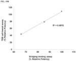
- comparing the signal intensity comprises generating a dose-response curve for each of the TDB and the reference TDB, and determining the ratio between the EC 50 values derived from the curves.
- Washing steps may be included between the incubation steps to remove molecules unbound to the solid phase. In some of these apsects, the incubation steps are independently carried out for about any of 1 hr, 2 hr, 3 hr, 4 hr, 5 hr, 6 hr, 7 hr, 8 hr, 9 hr, 10 hr, 12 hr, 16 hr, 20 hr, 24 hr, or more, including any ranges between these values.
- the TCS is a CD3 subunit.
- the CD3 subunit is CD3 ⁇ .
- the target antigen is expressed on the surface of a cell.
- the target antigen is CD4, CD8, CD18, CD19, CD11a, CD11b, CD20, CD22, CD34, CD40, CD79 ⁇ (CD79a), CD79 ⁇ (CD79b), EGF receptor, HER2 receptor, HER3 receptor, HER4 receptor, FcRH5, CLL1, LFA-1, Mac1, p150, 95, VLA-4, ICAM-1, VCAM, ⁇ v/ ⁇ 3 integrin, VEGF, flk2/flt3 receptor; obesity (OB) receptor; mpl receptor; CTLA-4; protein C, BR3, c-met, tissue factor, ⁇ 7, Tenb2, STEAP, or transmembrane tumor-associated antigens (TAA).
- TAA tumor-associated antigens
- the first epitope is localized to an extracellular portion of the target antigen and/or the second epitope is localized to an extracellular portion of the TCS.
- the TDB included in an incubation step is at a concentration range of any of about 0.01 ng/mL to about 100 ⁇ g/mL, about 0.05 ng/mL to about 100 ⁇ g/mL, about 0.1 ng/mL to about 100 ⁇ g/mL, about 0.5 ng/mL to about 100 ⁇ g/mL, about 1 ng/mL to about 100 ⁇ g/mL, about 5 ng/mL to about 100 ⁇ g/mL, about 10 ng/mL to about 100 ⁇ g/mL, about 0.01 ng/mL to about 50 ⁇ g/mL, about 0.01 ng/mL to about 10 ⁇ g/mL, about 0.01 ng/mL to about 1 ⁇ g/mL, about 0.01 ng/mL to about 100 ng/mL, about 0.01 ng/mL to about 50 ng/mL, about 0.01 ng/mL to about 10 ng//mL, about 0.01 ng/
- the standard curve from the reference TDB is generated by incubating the reference TDB at a plurality of concentrations ranging from about any one of 0.01 ng/mL to 100 ⁇ g/mL.
- the plurality of concentrations of reference TDB include about any one of 0.01 ng/ml, 0.1 ng/ml, 1 ng/ml, 10 ng/ml, 100ng/mL, 250 ng/mL, 500 ng/mL, 1 ⁇ g/mL, 2.5 ⁇ g/mL, 5 ⁇ g/mL, 10 ⁇ g/mL, 25 ⁇ g/mL, 50 ⁇ g/mL, 100 ⁇ g/mL, 250 ⁇ g/mL, or 500 ⁇ g/mL.
- the plurality of concentrations of reference TDB is about three, four, five, six, seven, eight, nine, ten or more than ten concentrations
- a kit or article of manufacture for use in various methods involving a TDB comprising a target antigen-binding fragment and a TCS-binding fragment (such as a CD3-binding fragment), comprising a container which holds a composition comprising engineered T cells comprising nucleic acid encoding a reporter operably linked to a promoter and/or enhancers that are responsive to T cell activation as described herein, and optionally provides instructions for its use.
- the kit further comprises a container which holds a reference TDB assay standard (a purified TDB of known concentration), and/or a container which holds a TDB control.
- the kit further comprises a container which holds a composition comprising target cells expressing the target antigen.
- the reporter is a luciferase, a fluorescent protein, an alkaline phosphatase, a beta lactamase, or a beta galactosidase.
- the luciferase is a firefly luciferase, a Renilla luciferase, or a nanoluciferase.
- the promoter and/or enhancer responsive to T cell activation is NF ⁇ B promoter.
- the promoter and/or enhancer responsive to T cell activation comprises T cell activation responsive elements from any one or more of NFAT, AP-1, NF ⁇ B, FOXO, STAT3, STATS and IRF.
- the engineered T cells are CD4 + T cells or CD8 + T cells.
- the engineered T cells are Jurkat T cells or CTLL-2 T cells.
- the TCS-binding fragment is a CD3-binding fragment.
- the CD3-binding fragment is a CD3 ⁇ -binding fragment.
- the target antigen is expressed on the surface of the target cells.
- the target antigen is CD4, CD8, CD18, CD19, CD11a, CD11b, CD20, CD22, CD34, CD40, CD79 ⁇ (CD79a), CD79 ⁇ (CD79b), EGF receptor, HER2 receptor, HER3 receptor, HER4 receptor, FcRH5, CLL1, LFA-1, Mac1, p150, 95, VLA-4, ICAM-1, VCAM, ⁇ v/ ⁇ 3 integrin, VEGF, flk2/flt3 receptor; obesity (OB) receptor; mpl receptor; CTLA-4; protein C, BR3, c-met, tissue factor, ⁇ 7, Tenb2, STEAP, or transmembrane tumor-associated antigens (TAA).
- TAA tumor-associated antigens
- the target antigen is HER2 receptor and the target cell is a BT-474 cell
- the target antigen is HER2 receptor and the target cell is a SKBR3 cell
- the target antigen is CD20 and the target cell is a Wil2-S cell
- the target antigen is CD79b and the target cell is a BJAB cell.
- the containers hold the formulations and the labels on, or associated with, the containers may indicate directions for use.
- the article of manufacture may further include other materials desirable from a commercial and user standpoint, including other buffers, diluents, cultureware, reagents for detecting reporter molecules, and package inserts with instructions for use.
- a kit or article of manufacture comprising a container which holds a composition comprising a TCS (such as a CD3 subunit), or a fragment thereof, conjugated with biotin, and optionally provides instructions for its use.
- a TCS such as a CD3 subunit
- the CD3 subunit is CD3 ⁇ .
- the kit further provides a target antigen, or a fragment thereof.
- the target antigen is expressed on the surface of a cell.
- the target antigen is CD4, CD8, CD18, CD19, CD11a, CD11b, CD20, CD22, CD34, CD40, CD79 ⁇ (CD79a), CD79 ⁇ (CD79b), EGF receptor, HER2 receptor, HER3 receptor, HER4 receptor, FcRH5, CLL1, LFA-1, Mac1, p150, 95, VLA-4, ICAM-1, VCAM, ⁇ v/ ⁇ 3 integrin, VEGF, flk2/flt3 receptor; obesity (OB) receptor; mpl receptor; CTLA-4; protein C, BR3, c-met, tissue factor, ⁇ 7, Tenb2, STEAP, or transmembrane tumor-associated antigens (TAA).
- TAA tumor-associated antigens
- the kit further provides a reference TDB assay standard (a purified TDB of known concentration), and/or a TDB control.
- the containers hold the formulations and the labels on, or associated with, the containers may indicate directions for use.
- the article of manufacture may further include other materials desirable from a commercial and user standpoint, including other buffers, diluents, cultureware, reagents for detecting reporter molecules, and package inserts with instructions for use.
- cell paste is thawed in the presence of sodium acetate (pH 3.5), EDTA, and phenylmethylsulfonylfluoride (PMSF) over about 30 min.
- PMSF phenylmethylsulfonylfluoride
- Cell debris can be removed by centrifugation.
- supernatants from such expression systems are generally first concentrated using a commercially available polypeptide concentration filter, for example, an Amicon or Millipore Pellicon ultrafiltration unit.
- a protease inhibitor such as PMSF may be included in any of the foregoing steps to inhibit proteolysis and antibiotics may be included to prevent the growth of adventitious contaminants.
- the polypeptide in the composition comprising the polypeptide and one or more contaminants has been purified or partially purified prior to analysis by the methods of the invention.
- the polypeptide of the methods is in an eluent from an affinity chromatography, a cation exchange chromatography, an anion exchange chromatography, a mixed mode chromatography and a hydrophobic interaction chromatography.
- the polypeptide is in an eluent from a Protein A chromatography.
- the polypeptide for use in any of the methods of analyzing polypeptides and formulations comprising the polypeptides by the methods described herein is an antibody.
- the polypeptide is a T cell-dependent bispecific (TDB) antibody.
- CD proteins and their ligands such as, but not limited to: (i) CD3, CD4, CD8, CD19, CD11a, CD20, CD22, CD34, CD40, CD79 ⁇ (CD79a), and CD79 ⁇ (CD79b); (ii) members of the ErbB receptor family such as the EGF receptor, HER2, HER3 or HER4 receptor; (iii) cell adhesion molecules such as LFA-1, Mac1, p150,95, VLA-4, ICAM-1, VCAM and ⁇ v/ ⁇ 3 integrin, including either alpha or beta subunits thereof (e.g., anti-CD11a, anti-CD18 or anti-CD11b antibodies); (iv) growth factors such as VEGF; IgE; blood group antigens; flk2/flt3 receptor; obesity (OB) receptor; mpl receptor; CTLA-4; protein C, BR3, c-met, tissue factor, ⁇ 7 etc; (v) cell surface and transmembran
- Patent No. 7,521,541 and (vi) other targets such as FcRH5, LyPD1, TenB2 and STEAP.
- the antibody is an anti-CD20/anti-CD3 antibody.
- Exemplary bispecific antibodies are provided in Table 1. Table 1.
- exemplary antibodies include those selected from, and without limitation, anti-estrogen receptor antibody, anti-progesterone receptor antibody, anti-p53 antibody, anti-HER-2/neu antibody, anti-EGFR antibody, anti-cathepsin D antibody, anti-Bcl-2 antibody, anti-E-cadherin antibody, anti-CA125 antibody, anti-CA15-3 antibody, anti-CA19-9 antibody, anti-c-erbB-2 antibody, anti-P-glycoprotein antibody, anti-CEA antibody, anti-retinoblastoma protein antibody, anti-ras oncoprotein antibody, anti-Lewis X antibody, anti-Ki-67 antibody, anti-PCNA antibody, anti-CD3 antibody, anti-CD4 antibody, anti-CD5 antibody, anti-CD7 antibody, anti-CD8 antibody, anti-CD9/p24 antibody, anti-CD10 antibody, anti-CD11a antibody, anti-CD11c antibody, anti-CD13 antibody, anti-CD14 antibody, anti-CD15 antibody, anti-CD19 antibody, anti-CD
- the antibodies are monoclonal antibodies.
- Monoclonal antibodies are obtained from a population of substantially homogeneous antibodies, i.e., the individual antibodies comprising the population are identical and/or bind the same epitope except for possible variants that arise during production of the monoclonal antibody, such variants generally being present in minor amounts.
- the modifier "monoclonal" indicates the character of the antibody as not being a mixture of discrete or polyclonal antibodies.
- the monoclonal antibodies may be made using the hybridoma method first described by Kohler et al., Nature 256:495 (1975 ), or may be made by recombinant DNA methods ( U.S. Patent No. 4,816,567 ).
- a mouse or other appropriate host animal such as a hamster
- lymphocytes that produce or are capable of producing antibodies that will specifically bind to the polypeptide used for immunization.
- lymphocytes may be immunized in vitro. Lymphocytes then are fused with myeloma cells using a suitable fusing agent, such as polyethylene glycol, to form a hybridoma cell ( Goding, Monoclonal Antibodies: Principles and Practice, pp. 59-103 (Academic Press, 1986 )).
- the hybridoma cells thus prepared are seeded and grown in a suitable culture medium that preferably contains one or more substances that inhibit the growth or survival of the unfused, parental myeloma cells.
- a suitable culture medium that preferably contains one or more substances that inhibit the growth or survival of the unfused, parental myeloma cells.
- the culture medium for the hybridomas typically will include hypoxanthine, aminopterin, and thymidine (HAT medium), which substances prevent the growth of HGPRT-deficient cells.
- the myeloma cells are those that fuse efficiently, support stable high-level production of antibody by the selected antibody-producing cells, and are sensitive to a medium such as HAT medium.
- the myeloma cell lines are murine myeloma lines, such as those derived from MOPC-21 and MPC-11 mouse tumors available from the Salk Institute Cell Distribution Center, San Diego, California USA, and SP-2 or X63-Ag8-653 cells available from the American Type Culture Collection, Rockville, Maryland USA.
- Human myeloma and mouse-human heteromyeloma cell lines also have been described for the production of human monoclonal antibodies ( Kozbor, J. Immunol. 133:3001 (1984 ); Brodeur et al., Monoclonal Antibody Production Techniques and Applications pp. 51-63 (Marcel Dekker, Inc., New York, 1987 )).
- Culture medium in which hybridoma cells are growing is assayed for production of monoclonal antibodies directed against the antigen.
- the binding specificity of monoclonal antibodies produced by hybridoma cells is determined by immunoprecipitation or by an in vitro binding assay, such as radioimmunoassay (RIA) or enzyme-linked immunoabsorbent assay (ELISA).
- RIA radioimmunoassay
- ELISA enzyme-linked immunoabsorbent assay
- the binding affinity of the monoclonal antibody can, for example, be determined by the Scatchard analysis of Munson et al., Anal. Biochem. 107:220 (1980 ).
- DNA encoding the monoclonal antibodies is readily isolated and sequenced using conventional procedures (e.g. , by using oligonucleotide probes that are capable of binding specifically to genes encoding the heavy and light chains of murine antibodies).
- the hybridoma cells serve as a source of such DNA.
- the DNA may be placed into expression vectors, which are then transfected into host cells such as E. coli cells, simian COS cells, Chinese Hamster Ovary (CHO) cells, or myeloma cells that do not otherwise produce immunoglobulin polypeptide, to obtain the synthesis of monoclonal antibodies in the recombinant host cells.
- antibodies or antibody fragments can be isolated from antibody phage libraries generated using the techniques described in McCafferty et al., Nature 348:552-554 (1990 ). Clackson et al., Nature 352:624-628 (1991 ) and Marks et al., J. Mol. Biol. 222:581-597 (1991 ) describe the isolation of murine and human antibodies, respectively, using phage libraries.
- the DNA also may be modified, for example, by substituting the coding sequence for human heavy- and light chain constant domains in place of the homologous murine sequences ( U.S. Patent No. 4,816,567 ; Morrison et al., Proc. Natl Acad. Sci. USA 81:6851 (1984 )), or by covalently joining to the immunoglobulin coding sequence all or part of the coding sequence for a non-immunoglobulin polypeptide.
- non-immunoglobulin polypeptides are substituted for the constant domains of an antibody, or they are substituted for the variable domains of one antigen-combining site of an antibody to create a chimeric bivalent antibody comprising one antigen-combining site having specificity for an antigen and another antigen-combining site having specificity for a different antigen.
- the antibody is IgA, IgD, IgE, IgG, or IgM. In some embodiments, the antibody is an IgG monoclonal antibody.
- the antibody is a humanized antibody. Methods for humanizing non-human antibodies have been described in the art.
- a humanized antibody has one or more amino acid residues introduced into it from a source that is non-human. These non-human amino acid residues are often referred to as "import" residues, which are typically taken from an "import” variable domain.
- Humanization can be essentially performed following the method of Winter and co-workers ( Jones et al., Nature 321:522-525 (1986 ); Riechmann et al., Nature 332:323-327 (1988 ); Verhoeyen et al., Science 239:1534-1536 (1988 )), by substituting hypervariable region sequences for the corresponding sequences of a human antibody.
- such "humanized" antibodies are chimeric antibodies ( U.S. Patent No. 4,816,567 ) wherein substantially less than an intact human variable domain has been substituted by the corresponding sequence from a non-human species.
- humanized antibodies are typically human antibodies in which some hypervariable region residues and possibly some FR residues are substituted by residues from analogous sites in rodent antibodies.
- variable domains both light and heavy
- sequence of the variable domain of a rodent antibody is screened against the entire library of known human variable-domain sequences.
- the human sequence that is closest to that of the rodent is then accepted as the human framework region (FR) for the humanized antibody ( Sims et al., J. Immunol. 151:2296 (1993 ); Chothia et al., J. Mol. Biol. 196:901 (1987 )).
- Another method uses a particular framework region derived from the consensus sequence of all human antibodies of a particular subgroup of light or heavy chain variable regions.
- the same framework may be used for several different humanized antibodies ( Carter et al., Proc. Natl. Acad. Sci. USA 89:4285 (1992 ); Presta et al., J. Immunol. 151:2623 (1993 )).
- humanized antibodies are prepared by a process of analysis of the parental sequences and various conceptual humanized products using three-dimensional models of the parental and humanized sequences.
- Three-dimensional immunoglobulin models are commonly available and are familiar to those skilled in the art.
- Computer programs are available that illustrate and display probable three-dimensional conformational structures of selected candidate immunoglobulin sequences. Inspection of these displays permits analysis of the likely role of the residues in the functioning of the candidate immunoglobulin sequence, i.e., the analysis of residues that influence the ability of the candidate immunoglobulin to bind its antigen.
- FR residues can be selected and combined from the recipient and import sequences so that the desired antibody characteristic, such as increased affinity for the target antigen(s), is achieved.
- the hypervariable region residues are directly and most substantially involved in influencing antigen binding.
- the antibody is a human antibody.
- human antibodies can be generated.
- transgenic animals e.g., mice
- J H antibody heavy chain joining region
- phage display technology McCafferty et al., Nature 348:552-553 (1990 )
- V domain gene repertoires from unimmunized donors.
- antibody V domain genes are cloned in-frame into either a major or minor coat polypeptide gene of a filamentous bacteriophage, such as M13 or fd, and displayed as functional antibody fragments on the surface of the phage particle. Because the filamentous particle contains a single-stranded DNA copy of the phage genome, selections based on the functional properties of the antibody also result in selection of the gene encoding the antibody exhibiting those properties.
- the phage mimics some of the properties of the B cell.
- Phage display can be performed in a variety of formats; for their review see, e.g., Johnson, Kevin S. and Chiswell, David J., Current Opinion in Structural Biology 3:564-571 (1993 ).
- V-gene segments can be used for phage display. Clackson et al., Nature 352:624-628 (1991 ) isolated a diverse array of anti-oxazolone antibodies from a small random combinatorial library of V genes derived from the spleens of immunized mice.
- a repertoire of V genes from unimmunized human donors can be constructed and antibodies to a diverse array of antigens (including self-antigens) can be isolated essentially following the techniques described by Marks et al., J. Mol. Biol. 222:581-597 (1991 ), or Griffith et al., EMBO J. 12:725-734 (1993 ). See also, US Patent Nos. 5,565,332 and 5,573,905 .
- Human antibodies may also be generated by in vitro activated B cells ( see US Patents 5,567,610 and 5,229,275 ).
- the antibody is an antibody fragment.
- Various techniques have been developed for the production of antibody fragments. Traditionally, these fragments were derived via proteolytic digestion of intact antibodies (see, e.g., Morimoto et al., Journal of Biochemical and Biophysical Methods 24:107-117 (1992 ) and Brennan et al., Science 229:81 (1985 )). However, these fragments can now be produced directly by recombinant host cells. For example, the antibody fragments can be isolated from the antibody phage libraries discussed above. Alternatively, Fab'-SH fragments can be directly recovered from E.
- fragments of the antibodies described herein are provided.
- the antibody fragment is an antigen binding fragment.
- the antigen binding fragment is selected from the group consisting of a Fab fragment, a Fab' fragment, a F(ab') 2 fragment, a scFv, a Fv, and a diabody.
- the fragments comprise a heavy chain variable domain (V H ) connected to a light chain variable domain (V L ) by a linker that is too short to allow pairing between the two domains on the same chain. Accordingly, the V H and V L domains of one fragment are forced to pair with the complementary V L and V H domains of another fragment, thereby forming two antigen-binding sites.
- V H and V L domains of one fragment are forced to pair with the complementary V L and V H domains of another fragment, thereby forming two antigen-binding sites.
- sFv single-chain Fv
- Oligonucleotides that define the desired termini of the nucleic acid fragment are employed at the 5' and 3' primers in the PCR.
- variant polypeptides share at least one biological and/or immunological activity with the native polypeptide disclosed herein.
- Amino acid sequence variants of the polypeptide are prepared by introducing appropriate nucleotide changes into the antibody nucleic acid, or by peptide synthesis. Such modifications include, for example, deletions from, and/or insertions into and/or substitutions of, residues within the amino acid sequences of the polypeptide. Any combination of deletion, insertion, and substitution is made to arrive at the final construct, provided that the final construct possesses the desired characteristics.
- the amino acid changes also may alter post-translational processes of the polypeptide (e.g., antibody), such as changing the number or position of glycosylation sites.
- Guidance in determining which amino acid residue may be inserted, substituted or deleted without adversely affecting the desired activity may be found by comparing the sequence of the polypeptide with that of homologous known polypeptide molecules and minimizing the number of amino acid sequence changes made in regions of high homology.
- a useful method for identification of certain residues or regions of the polypeptide (e.g., antibody) that are preferred locations for mutagenesis is called "alanine scanning mutagenesis" as described by Cunningham and Wells, Science 244:1081-1085 (1989 ).
- a residue or group of target residues are identified (e.g., charged residues such as Arg, Asp, His, Lys, and Glu) and replaced by a neutral or negatively charged amino acid (most preferably Alanine or Polyalanine) to affect the interaction of the amino acids with antigen.
- Those amino acid locations demonstrating functional sensitivity to the substitutions then are refined by introducing further or other variants at, or for, the sites of substitution.
- the site for introducing an amino acid sequence variation is predetermined, the nature of the mutation per se need not be predetermined. For example, to analyze the performance of a mutation at a given site, ala scanning or random mutagenesis is conducted at the target codon or region and the expressed antibody variants are screened for the desired activity.
- variants are an amino acid substitution variant. These variants have at least one amino acid residue in the antibody molecule replaced by a different residue.
- the sites of greatest interest for substitutional mutagenesis include the hypervariable regions, but FR alterations are also contemplated. Conservative substitutions are shown in the Table 2 below under the heading of "exemplary substitutions.” If such substitutions result in a change in biological activity, then more substantial changes, denominated “substitutions" in the Table 2, or as further described below in reference to amino acid classes, may be introduced and the products screened. Table 2.
- the phage-displayed variants are then screened for their biological activity (e.g., binding affinity) as herein disclosed.
- alanine scanning mutagenesis can be performed to identify hypervariable region residues contributing significantly to antigen binding.
- O-linked glycosylation refers to the attachment of one of the sugars N-aceylgalactosamine, galactose, or xylose to a hydroxyamino acid, most commonly serine or threonine, although 5-hydroxyproline or 5-hydroxylysine may also be used.
- Removal of carbohydrate moieties present on the polypeptide may be accomplished chemically or enzymatically or by mutational substitution of codons encoding for amino acid residues that serve as targets for glycosylation. Enzymatic cleavage of carbohydrate moieties on polypeptides can be achieved by the use of a variety of endo- and exo-glycosidases.
- a chimeric molecule comprises a fusion of the polypeptide with a tag polypeptide which provides an epitope to which an anti-tag antibody can selectively bind.
- the epitope tag is generally placed at the amino- or carboxyl-terminus of the polypeptide. The presence of such epitope-tagged forms of the polypeptide can be detected using an antibody against the tag polypeptide. Also, provision of the epitope tag enables the polypeptide to be readily purified by affinity purification using an anti-tag antibody or another type of affinity matrix that binds to the epitope tag.
- the chimeric molecule may comprise a fusion of the polypeptide with an immunoglobulin or a particular region of an immunoglobulin.
- a bivalent form of the chimeric molecule is referred to as an "immunoadhesin.”
- the immunoglobulin constant domain sequence in the immunoadhesin may be obtained from any immunoglobulin, such as IgG-1, IgG-2, IgG-3, or IgG-4 subtypes, IgA (including IgA-1 and IgA-2), IgE, IgD or IgM.
- immunoglobulin such as IgG-1, IgG-2, IgG-3, or IgG-4 subtypes, IgA (including IgA-1 and IgA-2), IgE, IgD or IgM.
- the polypeptide for use in polypeptide formulations may be conjugated to a cytotoxic agent such as a chemotherapeutic agent, a growth inhibitory agent, a toxin (e.g., an enzymatically active toxin of bacterial, fungal, plant, or animal origin, or fragments thereof), or a radioactive isotope (i.e., a radioconjugate).
- a cytotoxic agent such as a chemotherapeutic agent, a growth inhibitory agent, a toxin (e.g., an enzymatically active toxin of bacterial, fungal, plant, or animal origin, or fragments thereof), or a radioactive isotope (i.e., a radioconjugate).
- radionuclides are available for the production of radioconjugated polypeptides. Examples include 212 Bi, 131 I, 131 In, 90 Y, and 186 Re. Conjugates of the polypeptide and cytotoxic agent are made using a variety of bifunctional protein-coupling agents such as N-succinimidyl-3-(2-pyridyldithiol) propionate (SPDP), iminothiolane (IT), bifunctional derivatives of imidoesters (such as dimethyl adipimidate HCL), active esters (such as disuccinimidyl suberate), aldehydes (such as glutareldehyde), bis-azido compounds (such as bis(p-azidobenzoyl) hexanediamine), bis-diazonium derivatives (such as bis-(p-diazoniumbenzoyl)-ethylenediamine), diisocyanates (such as tolyene 2,6-diiso
- a ricin immunotoxin can be prepared as described in Vitetta et al., Science 238: 1098 (1987 ).
- Carbon-14-labeled 1-isothiocyanatobenzyl-3-methyldiethylene triaminepentaacetic acid (MX-DTPA) is an exemplary chelating agent for conjugation of radionucleotide to the polypeptide.
- Conjugates of a polypeptide and one or more small molecule toxins such as a calicheamicin, maytansinoids, a trichothene, and CC1065, and the derivatives of these toxins that have toxin activity, are also contemplated herein.
- Maytansinoids are mitototic inhibitors which act by inhibiting tubulin polymerization. Maytansine was first isolated from the east African shrub Maytenus serrata. Subsequently, it was discovered that certain microbes also produce maytansinoids, such as maytansinol and C-3 maytansinol esters. Synthetic maytansinol and derivatives and analogues thereof are also contemplated. There are many linking groups known in the art for making polypeptide-maytansinoid conjugates, including, for example, those disclosed in U.S. Pat. No. 5,208,020 .
- the linking groups include disufide groups, thioether groups, acid labile groups, photolabile groups, peptidase labile groups, or esterase labile groups, as disclosed in the above-identified patents, disulfide and thioether groups being preferred.
- the linker may be attached to the maytansinoid molecule at various positions, depending on the type of the link.
- an ester linkage may be formed by reaction with a hydroxyl group using conventional coupling techniques. The reaction may occur at the C-3 position having a hydroxyl group, the C-14 position modified with hyrdoxymethyl, the C-15 position modified with a hydroxyl group, and the C-20 position having a hydroxyl group.
- the linkage is formed at the C-3 position of maytansinol or a maytansinol analogue.
- antitumor agents that can be conjugated to the polypeptides described herein include BCNU, streptozoicin, vincristine and 5-fluorouracil, the family of agents known collectively LL-E33288 complex, as well as esperamicins.
- the polypeptide may be a conjugate between a polypeptide and a compound with nucleolytic activity (e.g., a ribonuclease or a DNA endonuclease such as a deoxyribonuclease; DNase).
- a compound with nucleolytic activity e.g., a ribonuclease or a DNA endonuclease such as a deoxyribonuclease; DNase.
- the polypeptide may be conjugated to a prodrug-activating enzyme which converts a prodrug (e.g., a peptidyl chemotherapeutic agent) to an active anti-cancer drug.
- a prodrug e.g., a peptidyl chemotherapeutic agent
- the enzyme component of the immunoconjugate includes any enzyme capable of acting on a prodrug in such a way so as to convert it into its more active, cytotoxic form.
- Enzymes that are useful include, but are not limited to, alkaline phosphatase useful for converting phosphate-containing prodrugs into free drugs; arylsulfatase useful for converting sulfate-containing prodrugs into free drugs; cytosine deaminase useful for converting non-toxic 5-fluorocytosine into the anti-cancer drug, 5-fluorouracil; proteases, such as serratia protease, thermolysin, subtilisin, carboxypeptidases and cathepsins (such as cathepsins B and L), that are useful for converting peptide-containing prodrugs into free drugs; D-alanylcarboxypeptidases, useful for converting prodrugs that contain D-amino acid substituents; carbohydrate-cleaving enzymes such as ⁇ -galactosidase and neuraminidase useful for converting glycosylated prodrugs into free drugs; ⁇ -lactamase useful
- polypeptide comprises linking the polypeptide to one of a variety of nonproteinaceous polymers, e.g ., polyethylene glycol, polypropylene glycol, polyoxyalkylenes, or copolymers of polyethylene glycol and polypropylene glycol.
- the polypeptide also may be entrapped in microcapsules prepared, for example, by coacervation techniques or by interfacial polymerization (for example, hydroxymethylcellulose or gelatin-microcapsules and poly-(methylmethacylate) microcapsules, respectively), in colloidal drug delivery systems (for example, liposomes, albumin microspheres, microemulsions, nano-particles and nanocapsules), or in macroemulsions.
- colloidal drug delivery systems for example, liposomes, albumin microspheres, microemulsions, nano-particles and nanocapsules
- polypeptides used in the methods of analysis described herein may be obtained using methods well-known in the art, including the recombination methods. The following sections provide guidance regarding these methods.
- Polynucleotide or “nucleic acid,” as used interchangeably herein, refer to polymers of nucleotides of any length, and include DNA and RNA.
- the polynucleotide may encode an entire immunoglobulin molecule chain, such as a light chain or a heavy chain.
- a complete heavy chain includes not only a heavy chain variable region (V H ) but also a heavy chain constant region (C H ), which typically will comprise three constant domains: C H 1, C H 2 and C H 3; and a "hinge" region. In some situations, the presence of a constant region is desirable.
- the polynucleotide encodes one or more immunoglobulin molecule chains of a TDB.
- polypeptides which may be encoded by the polynucleotide include antigen-binding antibody fragments such as single domain antibodies (“dAbs"), Fv, scFv, Fab' and F(ab') 2 and "minibodies.”
- dAbs single domain antibodies
- minibodies are (typically) bivalent antibody fragments from which the C H 1 and C K or C L domain has been excised.
- minibodies are smaller than conventional antibodies they should achieve better tissue penetration in clinical/diagnostic use, but being bivalent they should retain higher binding affinity than monovalent antibody fragments, such as dAbs. Accordingly, unless the context dictates otherwise, the term "antibody” as used herein encompasses not only whole antibody molecules but also antigen-binding antibody fragments of the type discussed above.
- each framework region present in the encoded polypeptide will comprise at least one amino acid substitution relative to the corresponding human acceptor framework.
- the framework regions may comprise, in total, three, four, five, six, seven, eight, nine, ten, eleven, twelve, thirteen, fourteen, or fifteen amino acid substitutions relative to the acceptor framework regions.
- T cell activation assay has been developed to determine the potency and specificity of a T Cell Dependent Bispecific (TDB) antibody for activating T cells in the presence of target cells. See FIG. 2 for an exemplary schematic representation.
- TDBs are bivalent and bispecific, with one arm specific for a TCR complex subunit and the other specific for a target antigen, cross-linking of TCRs leading to T cell activation can only occur when both the target cell and the T cell are bound by the TDB.
- TCR mediated cross-linking by anti-CD3-containing TDBs activates T cell signal transduction cascades leading to the phosphorylation and nuclear localization of transcription factors, including NFAT and NF ⁇ B, resulting in the transcriptional induction of target genes such as cytokines or cell killing agents such as Fas, Granzyme B and Perforins ( Brown, WM, 2006, Curr Opin Investig Drugs 7:381-388 ; Ferran, C et al., 1993 Exp Nephrol 1:83-89 ; Shannon, MF et al., 1995, J. Leukoc. Biol.
- Reporter genes such as firefly luciferase, under the transcriptional control of AP1, NFAT, or NF ⁇ B, have been used to monitor TCR activation of signaling pathways and T cell activation ( Shannon, MF et al., 1995, J. Leukoc. Biol. 57:767-773 ; Shapiro, 1998).
- TDBs can activate T cells in vitro
- Jurkat T cells DSMZ, ACC 282
- recombinant TCR-responsive reporter gene lentiviral stocks AP1-Luciferase, NFAT-Luciferase, or NFxB-Luciferase
- stable pools of reporter T cells were isolated.
- Jurkat/AP1Luciferase, Jurkat/NFATLuciferase, and Jurkat/NF ⁇ BLuciferase stable pools showed a dose-dependent induction of luciferase upon stimulation with purified Anti-CD3 homodimer.
- Luminescence responses luciferase reporter gene activity
- FIG. 3A Luminescence responses (luciferase reporter gene activity) were plotted, with the highest response observed from the Jurkat/NF ⁇ Bluciferase stable pool.
- FIG. 3A Luminescence responses (luciferase reporter gene activity) were plotted, with the highest response observed from the Jurkat/NF ⁇ Bluciferase stable pool.
- Jurkat/NF ⁇ BLuciferase stable clones isolated by limiting dilution were screened for their response to 10 ⁇ g/mL of purified Anti-CD3 homodimer.
- Jurkat T cell NF ⁇ BLuciferase pools demonstrated the highest response to Anti-CD3 homodimer compared to other TCR-res
- the T cell activation responses observed for the engineered Jurkat/NF ⁇ BLuciferase clone 2 reporter gene cell line is comparable to that observed using human T cells isolated from donor Peripheral Blood Mononuclear Cells (PBMCs) using other measures of T cell activation, indicating that the use of a reporter gene to monitor T cell activation response is comparable (Table 3).
- the Jurkat/NF ⁇ Bluciferase clone 2 cell line Jurkat-NFxBLuc
- Anti-CD20/CD3 or Anti-HER2/CD3 or Anti-CD79b/CD3 assay standard, control, and test samples were prepared and 50 ⁇ L was added to 96 well assay plates.
- Target cells Wil2-S, BT-474 or SKBR3, and BJAB cells for ⁇ CD20/CD3, allER2/CD3, and ⁇ CD79b/CD3, respectively
- JurkatNFkB reporter cells were then prepared, using either ready-to-use (R-to-U) frozen cells or cultured cells following assessment that frozen cells are comparable to fresh cultured cells.
- Equal volumes of 4.0 ⁇ 10 5 cells/mL of target cell diluent and 1.6 ⁇ 10 6 cells/mL JurkatNFkB cell diluent were combined to prepare a cell mixture with a target effector (T:E) cell ratio of 1:4.
- T:E target effector
- 50 ⁇ L of the mixed target and JurkatNFkBLuc cells was added to the each TDB dilution in the assay plate.
- the same T:E ratio was used for allER2/CD3 and ⁇ CD79b/CD3 cell based assays as well for the assays including the reference and control TDBs.
- Example 2 The assays described in Example 2 were used to measure the potency of and anti-FcRH5/anti-CD3 TBD.
- dilutions of anti-FcRH5/CD3 assay standard, control, and test samples were prepared and 50 ⁇ L was added to 96 well assay plates.
- Target cells FcRH5-expressing EJM cells
- JurkatNFkB reporter cells were then prepared, using either ready-to-use (R-to-U) frozen cells or cultured cells following assessment that frozen cells are comparable to fresh cultured cells.
Landscapes
- Health & Medical Sciences (AREA)
- Chemical & Material Sciences (AREA)
- Immunology (AREA)
- Life Sciences & Earth Sciences (AREA)
- Organic Chemistry (AREA)
- Engineering & Computer Science (AREA)
- Molecular Biology (AREA)
- General Health & Medical Sciences (AREA)
- Biochemistry (AREA)
- Genetics & Genomics (AREA)
- Medicinal Chemistry (AREA)
- Proteomics, Peptides & Aminoacids (AREA)
- Biophysics (AREA)
- Biomedical Technology (AREA)
- Cell Biology (AREA)
- Urology & Nephrology (AREA)
- Hematology (AREA)
- Bioinformatics & Cheminformatics (AREA)
- Wood Science & Technology (AREA)
- Zoology (AREA)
- Microbiology (AREA)
- Biotechnology (AREA)
- Analytical Chemistry (AREA)
- Physics & Mathematics (AREA)
- General Engineering & Computer Science (AREA)
- General Physics & Mathematics (AREA)
- Pathology (AREA)
- Food Science & Technology (AREA)
- Toxicology (AREA)
- Tropical Medicine & Parasitology (AREA)
- Oncology (AREA)
- Measuring Or Testing Involving Enzymes Or Micro-Organisms (AREA)
- Micro-Organisms Or Cultivation Processes Thereof (AREA)
- Hospice & Palliative Care (AREA)
- Peptides Or Proteins (AREA)
Applications Claiming Priority (2)
| Application Number | Priority Date | Filing Date | Title |
|---|---|---|---|
| US201662286862P | 2016-01-25 | 2016-01-25 | |
| PCT/US2017/014974 WO2017132279A1 (en) | 2016-01-25 | 2017-01-25 | Methods for assaying t-cell dependent bispecific antibodies |
Publications (3)
| Publication Number | Publication Date |
|---|---|
| EP3408671A1 EP3408671A1 (en) | 2018-12-05 |
| EP3408671B1 true EP3408671B1 (en) | 2023-11-01 |
| EP3408671C0 EP3408671C0 (en) | 2023-11-01 |
Family
ID=57966203
Family Applications (1)
| Application Number | Title | Priority Date | Filing Date |
|---|---|---|---|
| EP17703606.8A Active EP3408671B1 (en) | 2016-01-25 | 2017-01-25 | Methods for assaying t-cell dependent bispecific antibodies |
Country Status (5)
| Country | Link |
|---|---|
| US (2) | US11513127B2 (enExample) |
| EP (1) | EP3408671B1 (enExample) |
| JP (3) | JP7438662B2 (enExample) |
| CN (2) | CN109073635A (enExample) |
| WO (1) | WO2017132279A1 (enExample) |
Families Citing this family (26)
| Publication number | Priority date | Publication date | Assignee | Title |
|---|---|---|---|---|
| IL263466B2 (en) | 2013-12-17 | 2023-10-01 | Genentech Inc | Anti-CD3 antibodies and methods of using them |
| MA40579A (fr) | 2014-09-12 | 2016-03-17 | Genentech Inc | Anticorps anti-cll-1 et immunoconjugués |
| HRP20201779T1 (hr) | 2015-05-28 | 2021-01-22 | Genentech, Inc. | Stanični test za otkrivanje anti-cd3 homodimera |
| DK3310814T5 (da) | 2015-06-16 | 2024-10-07 | Hoffmann La Roche | Humaniserede og affinitetsmodnede antistoffer mod FcRH5 og fremgangsmåder til anvendelse |
| US10501545B2 (en) | 2015-06-16 | 2019-12-10 | Genentech, Inc. | Anti-CLL-1 antibodies and methods of use |
| JP7784795B2 (ja) | 2016-11-15 | 2025-12-12 | ジェネンテック, インコーポレイテッド | 抗cd20/抗cd3二重特異性抗体による処置のための投与 |
| BR112020013910A2 (pt) * | 2018-01-08 | 2020-12-01 | H. Lee Moffitt Cancer Center And Research Institute Inc. | composições e métodos para alvejamento de cânceres que expressam cd99 |
| CN111699200B (zh) | 2018-01-15 | 2023-05-26 | 南京传奇生物科技有限公司 | 针对pd-1的单域抗体和其变体 |
| IL325995A (en) | 2018-02-08 | 2026-03-01 | Genentech Inc | Bispecific antigen binding molecules and methods of use |
| CN108918892B (zh) * | 2018-08-01 | 2021-04-27 | 百奥泰生物制药股份有限公司 | 一种测定抗vegf抗体活性的方法及其应用 |
| CN110872356B (zh) * | 2018-09-03 | 2023-06-13 | 广西慧宝源健康产业有限公司 | 双特异性抗体及其使用方法 |
| EP3850013A4 (en) | 2018-09-10 | 2022-10-05 | Nanjing Legend Biotech Co., Ltd. | SINGLE DOMAIN ANTIBODIES AGAINST CLL1 AND USES THEREOF |
| TW202028244A (zh) * | 2018-10-09 | 2020-08-01 | 美商建南德克公司 | 用於確定突觸形成之方法及系統 |
| CN109402168A (zh) * | 2018-10-30 | 2019-03-01 | 深圳新诺微环生物科技有限公司 | 微环dna表达连接her2阳性细胞与效应细胞的桥接分子及其应用 |
| CN112798786B (zh) * | 2019-11-14 | 2022-04-29 | 深圳先进技术研究院 | 一种基于荧光素酶互补的生物传感器及其制备方法和应用 |
| WO2021119505A1 (en) | 2019-12-13 | 2021-06-17 | Genentech, Inc. | Anti-ly6g6d antibodies and methods of use |
| AU2021239929A1 (en) * | 2020-03-16 | 2022-10-13 | Crispr Therapeutics Ag | T-cell bispecific binding proteins |
| WO2022067005A1 (en) * | 2020-09-27 | 2022-03-31 | Genentech, Inc. | High throughput multiparametric immune cell engager screening assay |
| AU2021374594B2 (en) | 2020-11-04 | 2026-03-05 | Genentech, Inc. | Dosing for treatment with anti-cd20/anti-cd3 bispecific antibodies and anti-cd79b antibody drug conjugates |
| JP7716473B2 (ja) | 2020-11-04 | 2025-07-31 | ジェネンテック, インコーポレイテッド | 抗cd20/抗cd3二重特異性抗体の皮下投薬 |
| TWI874719B (zh) | 2020-11-04 | 2025-03-01 | 美商建南德克公司 | 用抗cd20/抗cd3雙特異性抗體進行治療之給藥 |
| CN112458149B (zh) * | 2020-12-03 | 2021-10-08 | 北京东方百泰生物科技股份有限公司 | 一种以cd3为靶点的抗体药物对t细胞激活程度的检测方法 |
| EP4337330A1 (en) | 2021-05-14 | 2024-03-20 | Genentech, Inc. | Methods for treatment of cd20-positive proliferative disorder with mosunetuzumab and polatuzumab vedotin |
| WO2023250485A1 (en) | 2022-06-23 | 2023-12-28 | LAVA Therapeutics N.V. | Assay for t cell dependent multispecific compounds |
| EP4573125A1 (en) * | 2022-08-18 | 2025-06-25 | Immunocore Limited | Multi-domain binding molecules |
| CN120077068A (zh) * | 2022-08-18 | 2025-05-30 | 英美偌科有限公司 | 多结构域结合分子 |
Family Cites Families (51)
| Publication number | Priority date | Publication date | Assignee | Title |
|---|---|---|---|---|
| FR2413974A1 (fr) | 1978-01-06 | 1979-08-03 | David Bernard | Sechoir pour feuilles imprimees par serigraphie |
| US4816567A (en) | 1983-04-08 | 1989-03-28 | Genentech, Inc. | Recombinant immunoglobin preparations |
| US4676980A (en) | 1985-09-23 | 1987-06-30 | The United States Of America As Represented By The Secretary Of The Department Of Health And Human Services | Target specific cross-linked heteroantibodies |
| US5567610A (en) | 1986-09-04 | 1996-10-22 | Bioinvent International Ab | Method of producing human monoclonal antibodies and kit therefor |
| IL85035A0 (en) | 1987-01-08 | 1988-06-30 | Int Genetic Eng | Polynucleotide molecule,a chimeric antibody with specificity for human b cell surface antigen,a process for the preparation and methods utilizing the same |
| DE3850542T2 (de) | 1987-09-23 | 1994-11-24 | Bristol Myers Squibb Co | Antikörper-Heterokonjugate zur Töting von HIV-infizierten Zellen. |
| GB8823869D0 (en) | 1988-10-12 | 1988-11-16 | Medical Res Council | Production of antibodies |
| US5175384A (en) | 1988-12-05 | 1992-12-29 | Genpharm International | Transgenic mice depleted in mature t-cells and methods for making transgenic mice |
| DE3920358A1 (de) | 1989-06-22 | 1991-01-17 | Behringwerke Ag | Bispezifische und oligospezifische, mono- und oligovalente antikoerperkonstrukte, ihre herstellung und verwendung |
| ES2096590T3 (es) | 1989-06-29 | 1997-03-16 | Medarex Inc | Reactivos biespecificos para la terapia del sida. |
| US5208020A (en) | 1989-10-25 | 1993-05-04 | Immunogen Inc. | Cytotoxic agents comprising maytansinoids and their therapeutic use |
| US5229275A (en) | 1990-04-26 | 1993-07-20 | Akzo N.V. | In-vitro method for producing antigen-specific human monoclonal antibodies |
| WO1992000373A1 (en) | 1990-06-29 | 1992-01-09 | Biosource Genetics Corporation | Melanin production by transformed microorganisms |
| US5571894A (en) | 1991-02-05 | 1996-11-05 | Ciba-Geigy Corporation | Recombinant antibodies specific for a growth factor receptor |
| WO1992020373A1 (en) | 1991-05-14 | 1992-11-26 | Repligen Corporation | Heteroconjugate antibodies for treatment of hiv infection |
| EP1400536A1 (en) | 1991-06-14 | 2004-03-24 | Genentech Inc. | Method for making humanized antibodies |
| MX9204374A (es) | 1991-07-25 | 1993-03-01 | Idec Pharma Corp | Anticuerpo recombinante y metodo para su produccion. |
| US7018809B1 (en) | 1991-09-19 | 2006-03-28 | Genentech, Inc. | Expression of functional antibody fragments |
| US5565332A (en) | 1991-09-23 | 1996-10-15 | Medical Research Council | Production of chimeric antibodies - a combinatorial approach |
| FI941572A7 (fi) | 1991-10-07 | 1994-05-27 | Oncologix Inc | Anti-erbB-2-monoklonaalisten vasta-aineiden yhdistelmä ja käyttömenete lmä |
| WO1993008829A1 (en) | 1991-11-04 | 1993-05-13 | The Regents Of The University Of California | Compositions that mediate killing of hiv-infected cells |
| ATE207080T1 (de) | 1991-11-25 | 2001-11-15 | Enzon Inc | Multivalente antigen-bindende proteine |
| CA2372813A1 (en) | 1992-02-06 | 1993-08-19 | L.L. Houston | Biosynthetic binding protein for cancer marker |
| WO1993016177A1 (en) | 1992-02-11 | 1993-08-19 | Cell Genesys, Inc. | Homogenotization of gene-targeting events |
| US5573905A (en) | 1992-03-30 | 1996-11-12 | The Scripps Research Institute | Encoded combinatorial chemical libraries |
| CA2140280A1 (en) | 1992-08-17 | 1994-03-03 | Avi J. Ashkenazi | Bispecific immunoadhesins |
| DK0752248T3 (da) | 1992-11-13 | 2000-11-13 | Idec Pharma Corp | Terapeutisk anvendelse af kimæriske og radioaktivt mærkede antistoffer mod humant B-lymfocytbegrænset differentieringsantig |
| US5534615A (en) | 1994-04-25 | 1996-07-09 | Genentech, Inc. | Cardiac hypertrophy factor and uses therefor |
| US5789199A (en) | 1994-11-03 | 1998-08-04 | Genentech, Inc. | Process for bacterial production of polypeptides |
| US5731168A (en) | 1995-03-01 | 1998-03-24 | Genentech, Inc. | Method for making heteromultimeric polypeptides |
| US5840523A (en) | 1995-03-01 | 1998-11-24 | Genetech, Inc. | Methods and compositions for secretion of heterologous polypeptides |
| US5641870A (en) | 1995-04-20 | 1997-06-24 | Genentech, Inc. | Low pH hydrophobic interaction chromatography for antibody purification |
| US5712374A (en) | 1995-06-07 | 1998-01-27 | American Cyanamid Company | Method for the preparation of substantiallly monomeric calicheamicin derivative/carrier conjugates |
| AU2814897A (en) * | 1996-04-25 | 1997-11-12 | T Cell Sciences, Inc. | Method of isolating regulators of t cell activation |
| IL127127A0 (en) | 1998-11-18 | 1999-09-22 | Peptor Ltd | Small functional units of antibody heavy chain variable regions |
| US8372954B2 (en) | 2000-12-22 | 2013-02-12 | National Research Council Of Canada | Phage display libraries of human VH fragments |
| JP2005289809A (ja) | 2001-10-24 | 2005-10-20 | Vlaams Interuniversitair Inst Voor Biotechnologie Vzw (Vib Vzw) | 突然変異重鎖抗体 |
| EP1565200A4 (en) | 2002-11-27 | 2009-06-24 | Agensys Inc | NUCLEIC ACID 24P4C12 AND CORRESPONDING PROTEIN FOR THE TREATMENT AND DETECTION OF CANCER |
| DE602004017726D1 (de) | 2003-06-30 | 2008-12-24 | Domantis Ltd | Pegylierte Single-domain-antikörper (dAb) |
| AU2005285347A1 (en) | 2004-08-19 | 2006-03-23 | Genentech, Inc. | Polypeptide variants with altered effector function |
| DK1791565T3 (en) | 2004-09-23 | 2016-08-01 | Genentech Inc | Cysteingensplejsede antibodies and conjugates |
| WO2010129304A2 (en) * | 2009-04-27 | 2010-11-11 | Oncomed Pharmaceuticals, Inc. | Method for making heteromultimeric molecules |
| WO2013026839A1 (en) * | 2011-08-23 | 2013-02-28 | Roche Glycart Ag | Bispecific antibodies specific for t-cell activating antigens and a tumor antigen and methods of use |
| JOP20200236A1 (ar) | 2012-09-21 | 2017-06-16 | Regeneron Pharma | الأجسام المضادة لمضاد cd3 وجزيئات ربط الأنتيجين ثنائية التحديد التي تربط cd3 وcd20 واستخداماتها |
| EP2769989A1 (en) * | 2013-02-21 | 2014-08-27 | Universitätsklinikum Freiburg | Recombinant bispecific antibody that binds to the CD133 antigen on tumor cells and to the human CD3 T cell receptor |
| IL311390A (en) | 2013-03-15 | 2024-05-01 | Memorial Sloan Kettering Cancer Center | Preparations and methods for immunotherapy |
| TWI681969B (zh) | 2014-01-23 | 2020-01-11 | 美商再生元醫藥公司 | 針對pd-1的人類抗體 |
| ES2905615T3 (es) * | 2014-11-20 | 2022-04-11 | Promega Corp | Sistemas y métodos para evaluar moduladores de puntos de control inmunitario |
| HRP20201779T1 (hr) | 2015-05-28 | 2021-01-22 | Genentech, Inc. | Stanični test za otkrivanje anti-cd3 homodimera |
| TWI756187B (zh) * | 2015-10-09 | 2022-03-01 | 美商再生元醫藥公司 | 抗lag3抗體及其用途 |
| WO2018108759A1 (en) * | 2016-12-13 | 2018-06-21 | F. Hoffmann-La Roche Ag | Method to determine the presence of a target antigen in a tumor sample |
-
2017
- 2017-01-25 CN CN201780015452.9A patent/CN109073635A/zh active Pending
- 2017-01-25 US US16/072,486 patent/US11513127B2/en active Active
- 2017-01-25 EP EP17703606.8A patent/EP3408671B1/en active Active
- 2017-01-25 WO PCT/US2017/014974 patent/WO2017132279A1/en not_active Ceased
- 2017-01-25 JP JP2018538107A patent/JP7438662B2/ja active Active
- 2017-01-25 CN CN202511302206.4A patent/CN121364307A/zh active Pending
-
2021
- 2021-09-24 JP JP2021155968A patent/JP2022023062A/ja not_active Withdrawn
-
2022
- 2022-10-14 US US18/046,865 patent/US20230280355A1/en active Pending
-
2023
- 2023-06-14 JP JP2023097744A patent/JP2023138948A/ja active Pending
Also Published As
| Publication number | Publication date |
|---|---|
| JP2022023062A (ja) | 2022-02-07 |
| EP3408671A1 (en) | 2018-12-05 |
| US11513127B2 (en) | 2022-11-29 |
| JP2023138948A (ja) | 2023-10-03 |
| US20200182882A1 (en) | 2020-06-11 |
| US20230280355A1 (en) | 2023-09-07 |
| CN121364307A (zh) | 2026-01-20 |
| WO2017132279A1 (en) | 2017-08-03 |
| EP3408671C0 (en) | 2023-11-01 |
| JP7438662B2 (ja) | 2024-02-27 |
| CN109073635A (zh) | 2018-12-21 |
| JP2019504321A (ja) | 2019-02-14 |
| WO2017132279A8 (en) | 2018-06-28 |
Similar Documents
| Publication | Publication Date | Title |
|---|---|---|
| US20230280355A1 (en) | Methods for assaying t-cell dependent bispecific antibodies | |
| US20240280586A1 (en) | CELL-BASED ASSAY FOR DETECTING ANTl-CD3 HOMODIMERS | |
| US10712322B2 (en) | Ionic strength-mediated pH gradient ion exchange chromatography | |
| US20220137067A1 (en) | Antibody potency assay | |
| JP2019530857A (ja) | 非イオン性サーファクタント及びポリペプチドを含む組成物中の非イオン性サーファクタントを定量するためのクロマトグラフィー法 | |
| HK40130187A (zh) | 用於测定t细胞依赖性双特异性抗体的方法 | |
| HK40002064A (en) | Methods for assaying t-cell dependent bispecific antibodies | |
| HK1250045B (zh) | 用於檢測抗cd3同二聚體的基於細胞的測定 | |
| BR112017025051B1 (pt) | Métodos para detecção e para quantificar a quantidade de homodímeros anti-cd3 em uma composição compreendendo um anticorpo biespecífico dependente de células t (tdb) e kit para a detecção de homodímeros anti-cd3 em uma composição |
Legal Events
| Date | Code | Title | Description |
|---|---|---|---|
| STAA | Information on the status of an ep patent application or granted ep patent |
Free format text: STATUS: UNKNOWN |
|
| STAA | Information on the status of an ep patent application or granted ep patent |
Free format text: STATUS: THE INTERNATIONAL PUBLICATION HAS BEEN MADE |
|
| PUAI | Public reference made under article 153(3) epc to a published international application that has entered the european phase |
Free format text: ORIGINAL CODE: 0009012 |
|
| STAA | Information on the status of an ep patent application or granted ep patent |
Free format text: STATUS: REQUEST FOR EXAMINATION WAS MADE |
|
| 17P | Request for examination filed |
Effective date: 20180827 |
|
| AK | Designated contracting states |
Kind code of ref document: A1 Designated state(s): AL AT BE BG CH CY CZ DE DK EE ES FI FR GB GR HR HU IE IS IT LI LT LU LV MC MK MT NL NO PL PT RO RS SE SI SK SM TR |
|
| AX | Request for extension of the european patent |
Extension state: BA ME |
|
| DAV | Request for validation of the european patent (deleted) | ||
| DAX | Request for extension of the european patent (deleted) | ||
| STAA | Information on the status of an ep patent application or granted ep patent |
Free format text: STATUS: EXAMINATION IS IN PROGRESS |
|
| 17Q | First examination report despatched |
Effective date: 20191129 |
|
| GRAP | Despatch of communication of intention to grant a patent |
Free format text: ORIGINAL CODE: EPIDOSNIGR1 |
|
| STAA | Information on the status of an ep patent application or granted ep patent |
Free format text: STATUS: GRANT OF PATENT IS INTENDED |
|
| INTG | Intention to grant announced |
Effective date: 20230605 |
|
| GRAS | Grant fee paid |
Free format text: ORIGINAL CODE: EPIDOSNIGR3 |
|
| GRAA | (expected) grant |
Free format text: ORIGINAL CODE: 0009210 |
|
| STAA | Information on the status of an ep patent application or granted ep patent |
Free format text: STATUS: THE PATENT HAS BEEN GRANTED |
|
| AK | Designated contracting states |
Kind code of ref document: B1 Designated state(s): AL AT BE BG CH CY CZ DE DK EE ES FI FR GB GR HR HU IE IS IT LI LT LU LV MC MK MT NL NO PL PT RO RS SE SI SK SM TR |
|
| REG | Reference to a national code |
Ref country code: GB Ref legal event code: FG4D |
|
| REG | Reference to a national code |
Ref country code: CH Ref legal event code: EP |
|
| REG | Reference to a national code |
Ref country code: IE Ref legal event code: FG4D |
|
| REG | Reference to a national code |
Ref country code: DE Ref legal event code: R096 Ref document number: 602017075944 Country of ref document: DE |
|
| U01 | Request for unitary effect filed |
Effective date: 20231101 |
|
| U07 | Unitary effect registered |
Designated state(s): AT BE BG DE DK EE FI FR IT LT LU LV MT NL PT SE SI Effective date: 20231106 |
|
| U20 | Renewal fee for the european patent with unitary effect paid |
Year of fee payment: 8 Effective date: 20231219 |
|
| PG25 | Lapsed in a contracting state [announced via postgrant information from national office to epo] |
Ref country code: GR Free format text: LAPSE BECAUSE OF FAILURE TO SUBMIT A TRANSLATION OF THE DESCRIPTION OR TO PAY THE FEE WITHIN THE PRESCRIBED TIME-LIMIT Effective date: 20240202 |
|
| PG25 | Lapsed in a contracting state [announced via postgrant information from national office to epo] |
Ref country code: IS Free format text: LAPSE BECAUSE OF FAILURE TO SUBMIT A TRANSLATION OF THE DESCRIPTION OR TO PAY THE FEE WITHIN THE PRESCRIBED TIME-LIMIT Effective date: 20240301 |
|
| PG25 | Lapsed in a contracting state [announced via postgrant information from national office to epo] |
Ref country code: ES Free format text: LAPSE BECAUSE OF FAILURE TO SUBMIT A TRANSLATION OF THE DESCRIPTION OR TO PAY THE FEE WITHIN THE PRESCRIBED TIME-LIMIT Effective date: 20231101 |
|
| PG25 | Lapsed in a contracting state [announced via postgrant information from national office to epo] |
Ref country code: IS Free format text: LAPSE BECAUSE OF FAILURE TO SUBMIT A TRANSLATION OF THE DESCRIPTION OR TO PAY THE FEE WITHIN THE PRESCRIBED TIME-LIMIT Effective date: 20240301 Ref country code: GR Free format text: LAPSE BECAUSE OF FAILURE TO SUBMIT A TRANSLATION OF THE DESCRIPTION OR TO PAY THE FEE WITHIN THE PRESCRIBED TIME-LIMIT Effective date: 20240202 Ref country code: ES Free format text: LAPSE BECAUSE OF FAILURE TO SUBMIT A TRANSLATION OF THE DESCRIPTION OR TO PAY THE FEE WITHIN THE PRESCRIBED TIME-LIMIT Effective date: 20231101 |
|
| PG25 | Lapsed in a contracting state [announced via postgrant information from national office to epo] |
Ref country code: RS Free format text: LAPSE BECAUSE OF FAILURE TO SUBMIT A TRANSLATION OF THE DESCRIPTION OR TO PAY THE FEE WITHIN THE PRESCRIBED TIME-LIMIT Effective date: 20231101 Ref country code: PL Free format text: LAPSE BECAUSE OF FAILURE TO SUBMIT A TRANSLATION OF THE DESCRIPTION OR TO PAY THE FEE WITHIN THE PRESCRIBED TIME-LIMIT Effective date: 20231101 Ref country code: NO Free format text: LAPSE BECAUSE OF FAILURE TO SUBMIT A TRANSLATION OF THE DESCRIPTION OR TO PAY THE FEE WITHIN THE PRESCRIBED TIME-LIMIT Effective date: 20240201 Ref country code: HR Free format text: LAPSE BECAUSE OF FAILURE TO SUBMIT A TRANSLATION OF THE DESCRIPTION OR TO PAY THE FEE WITHIN THE PRESCRIBED TIME-LIMIT Effective date: 20231101 |
|
| PG25 | Lapsed in a contracting state [announced via postgrant information from national office to epo] |
Ref country code: CZ Free format text: LAPSE BECAUSE OF FAILURE TO SUBMIT A TRANSLATION OF THE DESCRIPTION OR TO PAY THE FEE WITHIN THE PRESCRIBED TIME-LIMIT Effective date: 20231101 |
|
| PG25 | Lapsed in a contracting state [announced via postgrant information from national office to epo] |
Ref country code: SK Free format text: LAPSE BECAUSE OF FAILURE TO SUBMIT A TRANSLATION OF THE DESCRIPTION OR TO PAY THE FEE WITHIN THE PRESCRIBED TIME-LIMIT Effective date: 20231101 |
|
| PG25 | Lapsed in a contracting state [announced via postgrant information from national office to epo] |
Ref country code: SM Free format text: LAPSE BECAUSE OF FAILURE TO SUBMIT A TRANSLATION OF THE DESCRIPTION OR TO PAY THE FEE WITHIN THE PRESCRIBED TIME-LIMIT Effective date: 20231101 Ref country code: SK Free format text: LAPSE BECAUSE OF FAILURE TO SUBMIT A TRANSLATION OF THE DESCRIPTION OR TO PAY THE FEE WITHIN THE PRESCRIBED TIME-LIMIT Effective date: 20231101 Ref country code: RO Free format text: LAPSE BECAUSE OF FAILURE TO SUBMIT A TRANSLATION OF THE DESCRIPTION OR TO PAY THE FEE WITHIN THE PRESCRIBED TIME-LIMIT Effective date: 20231101 Ref country code: CZ Free format text: LAPSE BECAUSE OF FAILURE TO SUBMIT A TRANSLATION OF THE DESCRIPTION OR TO PAY THE FEE WITHIN THE PRESCRIBED TIME-LIMIT Effective date: 20231101 |
|
| REG | Reference to a national code |
Ref country code: DE Ref legal event code: R097 Ref document number: 602017075944 Country of ref document: DE |
|
| PG25 | Lapsed in a contracting state [announced via postgrant information from national office to epo] |
Ref country code: MC Free format text: LAPSE BECAUSE OF FAILURE TO SUBMIT A TRANSLATION OF THE DESCRIPTION OR TO PAY THE FEE WITHIN THE PRESCRIBED TIME-LIMIT Effective date: 20231101 |
|
| PG25 | Lapsed in a contracting state [announced via postgrant information from national office to epo] |
Ref country code: MC Free format text: LAPSE BECAUSE OF FAILURE TO SUBMIT A TRANSLATION OF THE DESCRIPTION OR TO PAY THE FEE WITHIN THE PRESCRIBED TIME-LIMIT Effective date: 20231101 |
|
| PLBE | No opposition filed within time limit |
Free format text: ORIGINAL CODE: 0009261 |
|
| STAA | Information on the status of an ep patent application or granted ep patent |
Free format text: STATUS: NO OPPOSITION FILED WITHIN TIME LIMIT |
|
| 26N | No opposition filed |
Effective date: 20240802 |
|
| U20 | Renewal fee for the european patent with unitary effect paid |
Year of fee payment: 9 Effective date: 20241218 |
|
| PG25 | Lapsed in a contracting state [announced via postgrant information from national office to epo] |
Ref country code: IE Free format text: LAPSE BECAUSE OF NON-PAYMENT OF DUE FEES Effective date: 20240125 |
|
| PG25 | Lapsed in a contracting state [announced via postgrant information from national office to epo] |
Ref country code: IE Free format text: LAPSE BECAUSE OF NON-PAYMENT OF DUE FEES Effective date: 20240125 |
|
| PG25 | Lapsed in a contracting state [announced via postgrant information from national office to epo] |
Ref country code: CY Free format text: LAPSE BECAUSE OF FAILURE TO SUBMIT A TRANSLATION OF THE DESCRIPTION OR TO PAY THE FEE WITHIN THE PRESCRIBED TIME-LIMIT; INVALID AB INITIO Effective date: 20170125 |
|
| PG25 | Lapsed in a contracting state [announced via postgrant information from national office to epo] |
Ref country code: HU Free format text: LAPSE BECAUSE OF FAILURE TO SUBMIT A TRANSLATION OF THE DESCRIPTION OR TO PAY THE FEE WITHIN THE PRESCRIBED TIME-LIMIT; INVALID AB INITIO Effective date: 20170125 |
|
| PG25 | Lapsed in a contracting state [announced via postgrant information from national office to epo] |
Ref country code: TR Free format text: LAPSE BECAUSE OF FAILURE TO SUBMIT A TRANSLATION OF THE DESCRIPTION OR TO PAY THE FEE WITHIN THE PRESCRIBED TIME-LIMIT Effective date: 20231101 |
|
| PGFP | Annual fee paid to national office [announced via postgrant information from national office to epo] |
Ref country code: GB Payment date: 20251220 Year of fee payment: 10 |
|
| U20 | Renewal fee for the european patent with unitary effect paid |
Year of fee payment: 10 Effective date: 20251217 |
|
| REG | Reference to a national code |
Ref country code: CH Ref legal event code: U11 Free format text: ST27 STATUS EVENT CODE: U-0-0-U10-U11 (AS PROVIDED BY THE NATIONAL OFFICE) Effective date: 20260201 |
|
| PGFP | Annual fee paid to national office [announced via postgrant information from national office to epo] |
Ref country code: CH Payment date: 20260201 Year of fee payment: 10 |