EP3383911B1 - Antibodies and molecules that immunospecifically bind to btn1a1 and the therapeutic uses thereof - Google Patents
Antibodies and molecules that immunospecifically bind to btn1a1 and the therapeutic uses thereof Download PDFInfo
- Publication number
- EP3383911B1 EP3383911B1 EP16820047.5A EP16820047A EP3383911B1 EP 3383911 B1 EP3383911 B1 EP 3383911B1 EP 16820047 A EP16820047 A EP 16820047A EP 3383911 B1 EP3383911 B1 EP 3383911B1
- Authority
- EP
- European Patent Office
- Prior art keywords
- seq
- amino acid
- btn1a1
- acid sequence
- antigen binding
- Prior art date
- Legal status (The legal status is an assumption and is not a legal conclusion. Google has not performed a legal analysis and makes no representation as to the accuracy of the status listed.)
- Active
Links
- 0 CC(C(*N)C(C)(C)C(C(C)(C)C)=C)*(C)C(*(C)C)=C Chemical compound CC(C(*N)C(C)(C)C(C(C)(C)C)=C)*(C)C(*(C)C)=C 0.000 description 2
- BQHDZWJTDVDOOP-SREVYHEPSA-N CCCCC/C=C\CN Chemical compound CCCCC/C=C\CN BQHDZWJTDVDOOP-SREVYHEPSA-N 0.000 description 1
Images


Classifications
-
- C—CHEMISTRY; METALLURGY
- C07—ORGANIC CHEMISTRY
- C07K—PEPTIDES
- C07K16/00—Immunoglobulins [IGs], e.g. monoclonal or polyclonal antibodies
- C07K16/18—Immunoglobulins [IGs], e.g. monoclonal or polyclonal antibodies against material from animals or humans
- C07K16/28—Immunoglobulins [IGs], e.g. monoclonal or polyclonal antibodies against material from animals or humans against receptors, cell surface antigens or cell surface determinants
- C07K16/2803—Immunoglobulins [IGs], e.g. monoclonal or polyclonal antibodies against material from animals or humans against receptors, cell surface antigens or cell surface determinants against the immunoglobulin superfamily
-
- A—HUMAN NECESSITIES
- A61—MEDICAL OR VETERINARY SCIENCE; HYGIENE
- A61P—SPECIFIC THERAPEUTIC ACTIVITY OF CHEMICAL COMPOUNDS OR MEDICINAL PREPARATIONS
- A61P1/00—Drugs for disorders of the alimentary tract or the digestive system
-
- A—HUMAN NECESSITIES
- A61—MEDICAL OR VETERINARY SCIENCE; HYGIENE
- A61P—SPECIFIC THERAPEUTIC ACTIVITY OF CHEMICAL COMPOUNDS OR MEDICINAL PREPARATIONS
- A61P1/00—Drugs for disorders of the alimentary tract or the digestive system
- A61P1/16—Drugs for disorders of the alimentary tract or the digestive system for liver or gallbladder disorders, e.g. hepatoprotective agents, cholagogues, litholytics
-
- A—HUMAN NECESSITIES
- A61—MEDICAL OR VETERINARY SCIENCE; HYGIENE
- A61P—SPECIFIC THERAPEUTIC ACTIVITY OF CHEMICAL COMPOUNDS OR MEDICINAL PREPARATIONS
- A61P1/00—Drugs for disorders of the alimentary tract or the digestive system
- A61P1/18—Drugs for disorders of the alimentary tract or the digestive system for pancreatic disorders, e.g. pancreatic enzymes
-
- A—HUMAN NECESSITIES
- A61—MEDICAL OR VETERINARY SCIENCE; HYGIENE
- A61P—SPECIFIC THERAPEUTIC ACTIVITY OF CHEMICAL COMPOUNDS OR MEDICINAL PREPARATIONS
- A61P11/00—Drugs for disorders of the respiratory system
-
- A—HUMAN NECESSITIES
- A61—MEDICAL OR VETERINARY SCIENCE; HYGIENE
- A61P—SPECIFIC THERAPEUTIC ACTIVITY OF CHEMICAL COMPOUNDS OR MEDICINAL PREPARATIONS
- A61P15/00—Drugs for genital or sexual disorders; Contraceptives
-
- A—HUMAN NECESSITIES
- A61—MEDICAL OR VETERINARY SCIENCE; HYGIENE
- A61P—SPECIFIC THERAPEUTIC ACTIVITY OF CHEMICAL COMPOUNDS OR MEDICINAL PREPARATIONS
- A61P35/00—Antineoplastic agents
-
- A—HUMAN NECESSITIES
- A61—MEDICAL OR VETERINARY SCIENCE; HYGIENE
- A61P—SPECIFIC THERAPEUTIC ACTIVITY OF CHEMICAL COMPOUNDS OR MEDICINAL PREPARATIONS
- A61P43/00—Drugs for specific purposes, not provided for in groups A61P1/00-A61P41/00
-
- A—HUMAN NECESSITIES
- A61—MEDICAL OR VETERINARY SCIENCE; HYGIENE
- A61K—PREPARATIONS FOR MEDICAL, DENTAL OR TOILETRY PURPOSES
- A61K39/00—Medicinal preparations containing antigens or antibodies
- A61K2039/505—Medicinal preparations containing antigens or antibodies comprising antibodies
-
- A—HUMAN NECESSITIES
- A61—MEDICAL OR VETERINARY SCIENCE; HYGIENE
- A61K—PREPARATIONS FOR MEDICAL, DENTAL OR TOILETRY PURPOSES
- A61K39/00—Medicinal preparations containing antigens or antibodies
- A61K39/395—Antibodies; Immunoglobulins; Immune serum, e.g. antilymphocytic serum
-
- C—CHEMISTRY; METALLURGY
- C07—ORGANIC CHEMISTRY
- C07K—PEPTIDES
- C07K2317/00—Immunoglobulins specific features
- C07K2317/20—Immunoglobulins specific features characterized by taxonomic origin
- C07K2317/24—Immunoglobulins specific features characterized by taxonomic origin containing regions, domains or residues from different species, e.g. chimeric, humanized or veneered
-
- C—CHEMISTRY; METALLURGY
- C07—ORGANIC CHEMISTRY
- C07K—PEPTIDES
- C07K2317/00—Immunoglobulins specific features
- C07K2317/30—Immunoglobulins specific features characterized by aspects of specificity or valency
- C07K2317/34—Identification of a linear epitope shorter than 20 amino acid residues or of a conformational epitope defined by amino acid residues
-
- C—CHEMISTRY; METALLURGY
- C07—ORGANIC CHEMISTRY
- C07K—PEPTIDES
- C07K2317/00—Immunoglobulins specific features
- C07K2317/40—Immunoglobulins specific features characterized by post-translational modification
- C07K2317/41—Glycosylation, sialylation, or fucosylation
-
- C—CHEMISTRY; METALLURGY
- C07—ORGANIC CHEMISTRY
- C07K—PEPTIDES
- C07K2317/00—Immunoglobulins specific features
- C07K2317/50—Immunoglobulins specific features characterized by immunoglobulin fragments
- C07K2317/56—Immunoglobulins specific features characterized by immunoglobulin fragments variable (Fv) region, i.e. VH and/or VL
- C07K2317/565—Complementarity determining region [CDR]
-
- C—CHEMISTRY; METALLURGY
- C07—ORGANIC CHEMISTRY
- C07K—PEPTIDES
- C07K2317/00—Immunoglobulins specific features
- C07K2317/70—Immunoglobulins specific features characterized by effect upon binding to a cell or to an antigen
- C07K2317/73—Inducing cell death, e.g. apoptosis, necrosis or inhibition of cell proliferation
-
- C—CHEMISTRY; METALLURGY
- C07—ORGANIC CHEMISTRY
- C07K—PEPTIDES
- C07K2317/00—Immunoglobulins specific features
- C07K2317/70—Immunoglobulins specific features characterized by effect upon binding to a cell or to an antigen
- C07K2317/76—Antagonist effect on antigen, e.g. neutralization or inhibition of binding
-
- C—CHEMISTRY; METALLURGY
- C07—ORGANIC CHEMISTRY
- C07K—PEPTIDES
- C07K2317/00—Immunoglobulins specific features
- C07K2317/70—Immunoglobulins specific features characterized by effect upon binding to a cell or to an antigen
- C07K2317/77—Internalization into the cell
-
- C—CHEMISTRY; METALLURGY
- C07—ORGANIC CHEMISTRY
- C07K—PEPTIDES
- C07K2317/00—Immunoglobulins specific features
- C07K2317/90—Immunoglobulins specific features characterized by (pharmaco)kinetic aspects or by stability of the immunoglobulin
- C07K2317/92—Affinity (KD), association rate (Ka), dissociation rate (Kd) or EC50 value
Definitions
- the present invention relates in general to the field of cancer immunology and molecular biology.
- the immune system of humans and other mammals protects them against infections and diseases.
- a number of stimulatory and inhibitory ligands and receptors provide a tight control system to maximize immune response against infection while limiting self-immunity.
- therapeutics that modulate immune response such as anti-PDl or anti-PDLl antibodies, were found to be effective in some cancer treatments.
- development of new therapeutics that safely and effectively treat diseases by modulating the immune system remain an urgent need, especially for metastatic cancers.
- the compositions and methods described herein meet these needs and provide other related advantages.
- molecules having an antigen binding fragment that immunospecifically binds to BTN1A are provided herein.
- the molecules are anti-BTN1A1 antibodies.
- the molecules have an antigen binding fragment that immunospecifically binds to glycosylated BTN1A1.
- the antigen binding fragments immunospecifically bind to BTN1A1 glycosylated at positions N55, N215, and/or N449.
- the antigen binding fragments immunospecifically bind to BTN1A1 glycosylated at position N55.
- the antigen binding fragments immunospecifically bind to BTN1A1 glycosylated at position N215.
- the antigen binding fragments immunospecifically bind to BTN1A1 glycosylated at position N449.
- the antigen binding fragments immunospecifically bind to one or more glycosylation motifs.
- the antigen binding fragments immunospecifically bind to BTN1A1 glycosylated at positions N55 and N215. In some aspects, the antigen binding fragments immunospecifically bind to BTN1A1 glycosylated at positions N215 and N449. In some aspects, the antigen binding fragments immunospecifically bind to BTN1A1 glycosylated at positions N55 and N449. In some aspects, the antigen binding fragments immunospecifically bind to BTN1A1 glycosylated at positions N55, N215 and N449.
- the molecules have an antigen binding fragment that immunospecifically binds to glycosylated BTN1A1, wherein the antigen binding fragment preferentially binds to glycosylated BTN1A1 over non-glycosylated BTN1A1.
- the antigen binding fragments preferentially bind to BTN1A1 glycosylated at positions N55, N215, and/or N449 over non-glycosylated BTN1A1.
- the antigen binding fragments preferentially bind to BTN1A1 glycosylated at position N55 over non-glycosylated BTN1A1.
- the antigen binding fragments preferentially bind to BTN1A1 glycosylated at position N215 over non-glycosylated BTN1A1. In some aspects, the antigen binding fragments preferentially bind to BTN1A1 glycosylated at position N449 over non-glycosylated BTN1A1. In some aspects, the antigen binding fragments preferentially bind to one or more glycosylation motifs. In some aspects, the antigen binding fragments preferentially bind to BTN1A1 glycosylated at positions N55 and N215 over non-glycosylated BTN1A1.
- the antigen binding fragments preferentially bind to BTN1A1 glycosylated at positions N215 and N449 over non-glycosylated BTN1A1. In some aspects, the antigen binding fragments preferentially bind to BTN1A1 glycosylated at positions N55 and N449 over non-glycosylated BTN1A1. In some aspects, the antigen binding fragments preferentially bind to BTN1A1 glycosylated at positions N55, N215 and N449 over non-glycosylated BTN1A1.
- the antigen binding fragment binds to glycosylated BTN1A1 with Kd less than half of the Kd exhibited relative to unglycosylated BTN1A1. In some embodiments, the antigen binding fragment binds to glycosylated BTN1A1 with Kd at least 10 times less than the Kd exhibited relative to unglycosylated BTN1A1.
- the antigen binding fragment binds to glycosylated BTN1A1 with a fluorescence intensity (MFI) that is at least twice as high as the MFI as exhibited relative to unglycosylated BTN1A1. In some embodiments, the antigen binding fragment binds to glycosylated BTN1A1 with an MFI that is at least five times as high as the MFI as exhibited relative to unglycosylated BTN1A1.
- MFI fluorescence intensity
- the antigen binding fragments immunospecifically mask BTN1A1 glycosylation at positions N55, N215, and/or N449. In some aspects, the antigen binding fragments immunospecifically mask BTN1A1 glycosylation at position N55. In some aspects, the antigen binding fragments immunospecifically mask BTN1A1 glycosylation at position N215. In some aspects, the antigen binding fragments immunospecifically mask BTN1A1 glycosylation at position N449. In some aspects, the antigen binding fragments immunospecifically mask one or more glycosylation motifs of BTN1A1. In some aspects, the antigen binding fragments immunospecifically mask BTN1A1 glycosylation at positions N55 and N215.
- the antigen binding fragments immunospecifically mask BTN1A1 glycosylation at positions N215 and N449. In some aspects, the antigen binding fragments immunospecifically mask BTN1A1 glycosylation at positions N55 and N449. In some aspects, the antigen binding fragments immunospecifically mask BTN1A1 glycosylation at positions N55, N215 and N449.
- molecules having an antigen binding fragment that immunospecifically binds to BTN1A1 and comprises the VH or VL domain of the murine monoclonal antibody STC810 as depicted in Table 2.
- the molecules can have an antigen binding fragment that comprises both the VH and VL domain of the murine monoclonal antibody STC810, as depicted in Table 2.
- the molecules can have an antigen binding fragment that comprises one or more VH CDRs having the amino acid sequence of any one of the VH CDRs of the murine monoclonal antibody STC810, as depicted in Table 2.
- the molecules can have antigen binding fragment that comprises one or more VL CDRs having the amino acid sequence of any one of the VL CDRs of the murine monoclonal antibody STC810, as depicted in Table 2.
- the molecules can have antigen binding fragment that comprises at least one VH CDR and at least one VL CDR of the murine monoclonal antibody STC810, as depicted in Table 2.
- the molecules provided herein have an antigen binding fragment comprising: (a) a heavy chain variable (V H ) region comprising: (1) a V H CDR1 having an amino acid sequence selected from the group consisting of SEQ ID NOS: 7, 10, 13 and 16; (2) a V H CDR2 having an amino acid sequence selected from the group consisting of SEQ ID NOS: 8, 11, 14 and 17; and (3) a V H CDR3 having an amino acid sequence selected from the group consisting of SEQ ID NOS: 9, 12, 15 and 18; or (b) a light chain variable (V L ) region comprising: (1) a V L CDR1 having an amino acid sequence selected from the group consisting of SEQ ID NOS: 19, 22, 25 and 28; (2) a V L CDR2 having an amino acid sequence selected from the group consisting of SEQ ID NOS: 20, 23, 26 and 29; and (3) a V L CDR3 having an amino acid sequence selected from the group consisting of SEQ ID NOS: 21, 24, 27 and 30.
- V H heavy chain variable
- isolated nucleic acid molecules encoding a VH chain, VL chain, VH domain, VL domain, VH CDR1, VH CDR2, VH CDR3, VL CDR1, VL CDR2, and/or VL CDR3 of anti-BTN1A1 antibodies described herein. Further provided are vectors and host cells comprising these nucleic acid molecules.
- molecules provided herein have an antigen binding fragment that competitively blocks ( e.g., in a dose-dependent manner) a BTN1A1 epitope described herein.
- the BTN1A1 epitope can be an epitope of STC810 as described herein.
- the molecules can have an antigen binding fragment that immunospecifically binds to an epitope of BTN1A1 as described herein.
- the BTN1A1 epitope can be an epitope of STC810 as described herein.
- the BTN1A1 epitope has at least five consecutive amino acids of an amino acid sequence of SEQ ID NOS: 31-41.
- the molecules having an antigen binding fragment that immunospecifically binds to BTN1A1 are anti-BTN1A1 antibodies, including anti-glycosylated BTN1A1 antibodies.
- the antibodies can be monoclonal antibodies.
- the antibodies can be humanized antibodies.
- the antibodies can be human antibodies.
- the antibodies can be IgG, IgM, or IgA.
- the molecule having an antigen binding fragment that immunospecifically binds to BTN1A1 is a Fab', a F(ab')2, a F(ab')3, a monovalent scFv, a bivalent scFv, or a single domain antibody.
- the molecules having an antigen binding fragment that immunospecifically binds to BTN1A1 are recombinantly produced.
- the molecule is conjugated to an imaging agent, a chemotherapeutic agent, a toxin or a radionuclide.
- compositions that comprises a molecule having an antigen binding fragment that immunospecifically binds to BTN1A1, as well as a pharmaceutically acceptable carrier.
- kits that include a molecule having an antigen binding fragment that immunospecifically binds to BTN1A1, as well as an ancillary agent.
- ADC antibody-drug conjugates
- ADC antibody-drug conjugates
- the compound can be an imaging agent, a therapeutic agent, a toxin or a radionuclide as described herein.
- the compound can be conjugated with anti-BTN1A1 antibody.
- the conjugate can be any conjugate as described herein, such as an ADC.
- the cell can be a cancer cell.
- the cell can also be a population of cells that include both cancer cells and normal cells.
- Modulating an immune response can include (a) increasing T cell activation; (b) increasing T cell proliferation; and/or (c) increasing cytokine production.
- the cells can be cancer cells.
- a molecule having an antigen binding fragment that immunospecifically binds to BTN1A1 as described herein is an anti-BTN1A1 antibody.
- the molecule is an anti-glycosylated BTN1A1 antibodies.
- the treatment can activate an immune response, or promote the activation and proliferation of T cells in the subject.
- the molecule binds to cancer cells and induces an immune response resulting in destruction of the cancer cells.
- the destruction of cancer cells is mediated by ADCC activity of the molecules.
- the destruction of cancer cells is mediated by CDC activity of the molecule.
- the subject has a metastatic cancer.
- the cancer can be a hematological cancer or a solid tumor.
- the cancer is a hematological cancer selected from the group consisting of leukemia, lymphoma, and myeloma.
- the cancer is a solid tumor selected from the group consisting of breast cancer, lung cancer, thymic cancer, thyroid cancer, head & neck cancer, prostate cancer, esophageal cancer, tracheal cancer, brain cancer, liver cancer, bladder cancer, kidney cancer, stomach cancer, pancreatic cancer, ovarian cancer, uterine cancer, cervical cancer, testicular cancer, colon cancer, rectal cancer and skin cancer.
- the skin cancer can be either melanomatous or non-melanomatous skin cancers.
- the methods include systematic administration to a subject of the molecules having an antigen binding fragment that immunospecifically binds BTN1A1 as described herein.
- the molecule is administered intravenously, intradermally, intratumorally, intramuscularly, intraperitoneally, subcutaneously or locally.
- the methods include administering a second anticancer therapy to the subject, which can be a surgical therapy, chemotherapy, biological targeted therapy, small molecular targeted therapy, radiation therapy, cryotherapy, hormonal therapy, immunotherapy or cytokine therapy.
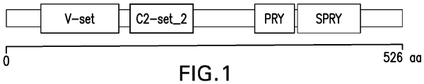
- BTN1A1 Butyrophilin, subfamily 1, member A1
- BTN1A1 is a type I membrane glycoprotein and a major component of milk fat globule membrane, and has structural similarities to the B7 family.
- BTN1A1 is known as a major protein regulating the formation of fat droplets in the milk. ( Ogg et al. PNAS, 101(27):10084-10089 (2004 )).
- BTN1A1 is expressed in immune cells, including T cells. Treatment with recombinant BTN1A1 was found to inhibit T cell activation and protect animal models of EAE. ( Stefferl et al., J. Immunol. 165(5):2859-65 (2000 )).
- BTN1A1 is also specifically and highly expressed in cancer cells.
- the BTN1A1 in cancer cells are also glycosylated.
- the expression of BTN1A1 can be used to aid cancer diagnosis as well as to evaluate the efficacy of a cancer treatment.
- an antibody refers to one antibody or more than one antibodies.
- BTN1A1 refers to BTN1A1 from any vertebrate source, including mammals such as primates (e.g ., humans, cynomolgus monkey (cyno)), dogs, and rodents ( e.g., mice and rats).
- BTN1A1 also includes various BTN1A1 isoforms, related BTN1A1 polypeptides, including SNP variants thereof, as well as different modified forms of BTN1A1, including but not limited to phosphorylated BTN1A1, glycosylated BTN1A1, and ubiquitinated BTN1A1.
- glycosylated BTN1A1 include BTN1A1 with N55, N215, and/or N449 glycosylation.
- the term “antibody” refers to a polypeptide product of B cells within the immunoglobulin (or "Ig") class of polypeptides that is able to bind to a specific molecular antigen and is composed of two identical pairs of polypeptide chains, wherein each pair has one heavy chain (about 50-70 kDa) and one light chain (about 25 kDa) and each amino-terminal portion of each chain includes a variable region of about 100 to about 130 or more amino acids and each carboxy-terminal portion of each chain includes a constant region (See Borrebaeck (ed.) (1995) Antibody Engineering, Second Edition, Oxford University Press .; Kuby (1997) Immunology, Third Edition, W.H.
- the specific molecular antigen includes the target BTN1A1, which can be a BTN1A1 polypeptide, BTN1A1 fragment or BTN1A1 epitope.
- Antibodies provided herein include, but are not limited to, monoclonal antibodies, synthetic antibodies, recombinantly produced antibodies, bi-specific antibodies, multispecific antibodies, human antibodies, humanized antibodies, camelized antibodies, chimeric antibodies, intrabodies, anti-idiotypic (anti-Id) antibodies.
- the term "monoclonal antibody” refers to an antibody that is the product of a single cell clone or hybridoma or a population of cells derived from a single cell.
- a monoclonal antibody also is intended to refer to an antibody produced by recombinant methods from heavy and light chain encoding immunoglobulin genes to produce a single molecular immunoglobulin species.
- Amino acid sequences for antibodies within a monoclonal antibody preparation are substantially homogeneous and the binding activity of antibodies within such a preparation exhibit substantially the same antigen binding activity.
- polyclonal antibodies are obtained from different B cells within a population, which are a combination of immunoglobulin molecules that bind a specific antigen.
- Each immunoglobulin of the polyclonal antibodies can bind a different epitope of the same antigen.
- Methods for producing both monoclonal antibodies and polyclonal antibodies are well known in the art ( Harlow and Lane., Antibodies: A Laboratory Manual, Cold Spring Harbor Laboratory Press (1989 ) and Borrebaeck (ed.), Antibody Engineering: A Practical Guide, W.H. Freeman and Co., Publishers, New York, pp. 103-120 (1991 )).
- human antibody refers to an antibody that has a human variable region and/or a human constant region or a portion thereof corresponding to human germline immunoglobulin sequences.
- human germline immunoglobulin sequences are described by Kabat et al. (1991) Sequences of Proteins of Immunological Interest, Fifth Edition, U.S. Department of Health and Human Services, NIH Publication No. 91-3242 .
- a human antibody can include an antibody that binds to BTN1A1 and is encoded by a nucleic acid sequence that is a naturally occurring somatic variant of the human germline immunoglobulin nucleic acid sequence.
- chimeric antibody refers to an antibody that a portion of the heavy and/or light chain is identical with or homologous to corresponding sequences in antibodies derived from a particular species or belonging to a particular antibody class or subclass, while the remainder of the chain(s) is identical with or homologous to corresponding sequences in antibodies derived from another species or belonging to another antibody class or subclass, as well as fragments of such antibodies, so long as they exhibit the desired biological activity ( see U.S. Patent No. 4,816,567 ; and Morrison et al., Proc. Natl. Acad. Sci. USA, 81:6851-6855 (1984 )).
- humanized antibody refers to chimeric antibodies that include human immunoglobulins (e.g ., recipient antibody) in which the native Complementarity Determining Region ("CDR") residues are replaced by residues from the corresponding CDR of a nonhuman species (e.g ., donor antibody) such as mouse, rat, rabbit or nonhuman primate having the desired specificity, affinity, and capacity.
- CDR Complementarity Determining Region
- a nonhuman species e.g ., donor antibody
- humanized antibodies can have residues that are not found in the recipient antibody or in the donor antibody. These modifications are made to further refine antibody performance.
- a humanized antibody heavy or light chain can have substantially all of at least one or more variable regions, in which all or substantially all of the CDRs correspond to those of a nonhuman immunoglobulin and all or substantially all of the FRs are those of a human immunoglobulin sequence.
- the humanized antibody can have at least a portion of an immunoglobulin constant region (Fc), typically that of a human immunoglobulin.
- Fc immunoglobulin constant region
- recombinant antibody refers to an antibody that is prepared, expressed, created or isolated by recombinant means.
- Recombinant antibodies can be antibodies expressed using a recombinant expression vector transfected into a host cell, antibodies isolated from a recombinant, combinatorial antibody library, antibodies isolated from an animal ( e.g., a mouse or cow) that is transgenic and/or transchromosomal for human immunoglobulin genes ( see, e.g., Taylor, L. D. et al., Nucl. Acids Res.
- Such recombinant antibodies can have variable and constant regions, including those derived from human germline immunoglobulin sequences ( see Kabat, E. A. et al. (1991) Sequences of Proteins of Immunological Interest, Fifth Edition, U.S. Department of Health and Human Services, NIH Publication No. 91-3242 ).
- the recombinant antibodies can also be subjected to in vitro mutagenesis (or, when an animal transgenic for human Ig sequences is used, in vivo somatic mutagenesis) and thus the amino acid sequences of the VH and VL regions of the recombinant antibodies can be sequences that, while derived from and related to human germline VH and VL sequences, do not naturally exist within the human antibody germline repertoire in vivo.
- a neutralizing antibody refers to an antibody that blocks the binding the BTN1A1 with its natural ligands and inhibits the signaling pathways mediated by BTN1A1 and/or its other physiological activities.
- the IC50 of a neutralizing antibody refers to the concentration of the antibody that is required to neutralize 50% of BTN1A1 in a neutralization assay.
- the IC50 of the neutralizing antibody can range between 0.01 - 10 ⁇ g/ml in the neutralization assay.
- antigen binding fragment refers to a portion of an antibody which includes the amino acid residues that immunospecifically bind to an antigen and confer on the antibody its specificity and affinity for the antigen.
- An antigen binding fragment can be referred to as a functional fragment of an antibody.
- An antigen binding fragment can be monovalent, bivalent, or multivalent.
- Molecules having an antigen binding fragment include, for example, an Fd, Fv, Fab, F(ab'), F(ab) 2 , F(ab') 2 , single chain Fv (scFv), diabody, triabody, tetrabody, minibody, or a single domain antibody.
- a scFv can be monovalent scFv or bivalent scFv.
- Other molecules having an antigen binding fragment can include, for example, heavy or light chain polypeptides, variable region polypeptides or CDR polypeptides or portions thereof so long as such antigen binding fragments retain binding activity.
- antigen binding fragments can be found described in, for example, Harlow and Lane, Antibodies: A Laboratory Manual, Cold Spring Harbor Laboratory, New York (1989 ); Myers (ed.), Molec. Biology and Biotechnology: A Comprehensive Desk Reference, New York: VCH Publisher, Inc. ; Huston et al., Cell Biophysics, 22:189-224 (1993 ); Plückthun and Skerra, Meth. Enzymol., 178:497-515 (1989 ) and in Day, E.D., Advanced Immunochemistry, Second Ed., Wiley-Liss, Inc., New York, NY (1990 ).
- An antigen binding fragment can be a polypeptide having an amino acid sequence of at least 5 contiguous amino acid residues, at least 10 contiguous amino acid residues, at least 15 contiguous amino acid residues, at least 20 contiguous amino acid residues, at least 25 contiguous amino acid residues, at least 40 contiguous amino acid residues, at least 50 contiguous amino acid residues, at least 60 contiguous amino residues, at least 70 contiguous amino acid residues, at least 80 contiguous amino acid residues, at least 90 contiguous amino acid residues, at least 100 contiguous amino acid residues, at least 125 contiguous amino acid residues, at least 150 contiguous amino acid residues, at least 175 contiguous amino acid residues, at least 200 contiguous amino acid residues, or at least 250 contiguous amino acid residues.
- the heavy chain of an antibody refers to a polypeptide chain of about 50-70 kDa, wherein the amino-terminal portion includes a variable region of about 120 to 130 or more amino acids and a carboxy-terminal portion that includes a constant region.
- the constant region can be one of five distinct types, referred to as alpha ( ⁇ ), delta ( ⁇ ), epsilon ( ⁇ ), gamma ( ⁇ ) and mu ( ⁇ ), based on the amino acid sequence of the heavy chain constant region.
- the distinct heavy chains differ in size: ⁇ , ⁇ and ⁇ contain approximately 450 amino acids, while ⁇ and ⁇ contain approximately 550 amino acids.
- heavy chains When combined with a light chain, these distinct types of heavy chains give rise to five well known classes of antibodies, IgA, IgD, IgE, IgG and IgM, respectively, including four subclasses of IgG, namely IgG1, IgG2, IgG3 and IgG4.
- a heavy chain can be a human heavy chain.
- the light chain of an antibody refers to a polypeptide chain of about 25 kDa, wherein the amino-terminal portion includes a variable region of about 100 to about 110 or more amino acids and a carboxy-terminal portion that includes a constant region.
- the approximate length of a light chain is 211 to 217 amino acids.
- Light chain amino acid sequences are well known in the art.
- a light chain can be a human light chain.
- variable domain or variable region of an antibody refers to a portion of the light or heavy chains of an antibody that is generally located at the amino-terminal of the light or heavy chain and has a length of about 120 to 130 amino acids in the heavy chain and about 100 to 110 amino acids in the light chain, and are used in the binding and specificity of each particular antibody for its particular antigen.
- the variable domains differ extensively in sequence between different antibodies. The variability in sequence is concentrated in the CDRs while the less variable portions in the variable domain are referred to as framework regions (FR).
- FR framework regions
- the CDRs of the light and heavy chains are primarily responsible for the interaction of the antibody with antigen. Numbering of amino acid positions used herein is according to the EU Index, as in Kabat et al. (1991) Sequences of proteins of immunological interest. (U.S. Department of Health and Human Services, Washington, D.C.) 5th ed .
- a variable region can be a human variable region.
- a CDR refers to one of three hypervariable regions (HI, H2 or H3) within the non-framework region of the immunoglobulin (Ig or antibody) VH ⁇ -sheet framework, or one of three hypervariable regions (L1, L2 or L3) within the non-framework region of the antibody VL ⁇ -sheet framework. Accordingly, CDRs are variable region sequences interspersed within the framework region sequences. CDR regions are well known to those skilled in the art and have been defined by, for example, Kabat as the regions of most hypervariability within the antibody variable (V) domains ( Kabat et al., J. Biol. Chem. 252:6609-6616 (1977 ); Kabat, Adv. Prot. Chem.
- CDR region sequences also have been defined structurally by Chothia as those residues that are not part of the conserved ⁇ -sheet framework, and thus are able to adapt different conformations ( Chothia and Lesk, J. Mol. Biol. 196:901-917 (1987 )). Both terminologies are well recognized in the art.
- the positions of CDRs within a canonical antibody variable domain have been determined by comparison of numerous structures ( Al-Lazikani et al., J. Mol. Biol. 273:927-948 (1997 ); Morea et al., Methods 20:267-279 (2000 )).
- CDRs defined according to standard designations are set forth in the Table 1 below.
- Table 1 CDR Definitions Exemplary (Kabat + Chothia) IMGT Kabat AbM Chothia Contact V H CDR1 26-35 27-38 31-35 26-35 26-32 30-35 V H CDR2 50-65 56-65 50-65 50-58 53-55 47-58 V H CDR3 95-102 105-117 95-102 95-102 96-101 93-101 V L CDR1 24-34 27-38 24-34 24-34 26-32 30-36 V L CDR2 50-56 56-65 50-56 50-56 50-52 46-55 V L CDR3 89-97 105-117 89-97 89-97 91-96 89-96
- One or more CDRs also can be incorporated into a molecule either covalently or noncovalently to make it an immunoadhesin.
- An immunoadhesin can incorporate the CDR(s) as part of a larger polypeptide chain, can covalently link the CDR(s) to another polypeptide chain, or can incorporate the CDR(s) noncovalently.
- the CDRs permit the immunoadhesin to bind to a particular antigen of interest.
- FR residues refer to those variable domain residues flanking the CDRs. FR residues are present, e.g., in chimeric, humanized, human, domain antibodies, diabodies, linear antibodies, and bispecific antibodies. FR residues are those variable domain residues other than the hypervariable region residues herein defined.
- the term “isolated” as used in reference to an antibody means the antibody is substantially free of cellular material or other contaminating proteins from the cell or tissue source and/or other contaminant components from which the antibody is derived, or substantially free of chemical precursors or other chemicals when chemically synthesized.
- the language “substantially free of cellular material” includes preparations of an antibody in which the antibody is separated from cellular components of the cells from which it is isolated or recombinantly produced.
- an antibody that is substantially free of cellular material includes preparations of antibody having less than about 30%, 20%, 10%, or 5% (by dry weight) of heterologous protein (also referred to herein as a "contaminating protein").
- the antibody when the antibody is recombinantly produced, it is substantially free of culture medium, e.g ., culture medium represents less than about 20%, 10%, or 5% of the volume of the protein preparation.
- culture medium represents less than about 20%, 10%, or 5% of the volume of the protein preparation.
- the antibody when the antibody is produced by chemical synthesis, it is substantially free of chemical precursors or other chemicals, e.g ., it is separated from chemical precursors or other chemicals which are involved in the synthesis of the protein. Accordingly such preparations of the antibody have less than about 30%, 20%, 10%, 5% (by dry weight) of chemical precursors or compounds other than the antibody of interest.
- Contaminant components can also include, but are not limited to, materials that would interfere with therapeutic uses for the antibody, and may include enzymes, hormones, and other proteinaceous or nonproteinaceous solutes.
- the antibody will be purified (1) to greater than 95% by weight of antibody as determined by the Lowry method ( Lowry et al. J. Bio. Chem. 193: 265-275, 1951 ), such as 99% by weight, (2) to a degree sufficient to obtain at least 15 residues of N-terminal or internal amino acid sequence by use of a spinning cup sequenator, or (3) to homogeneity by SDS-PAGE under reducing or nonreducing conditions using Coomassie blue or, preferably, silver stain.
- Isolated antibody includes the antibody in situ within recombinant cells since at least one component of the antibody's natural environment will not be present. Ordinarily, however, isolated antibody will be prepared by at least one purification step. In a specific embodiment, antibodies provided herein are isolated
- nucleic acid molecule As used herein, and unless otherwise specified, the term “polynucleotide,” “nucleotide,” nucleic acid” “nucleic acid molecule” and other similar terms are used interchangeable and include DNA, RNA, mRNA and the like.
- nucleic acid molecule As used herein, and unless otherwise specified, the term "isolated" as used in reference to a nucleic acid molecule means the nucleic acid molecule is one which is separated from other nucleic acid molecules which are present in the natural source of the nucleic acid molecule. Moreover, an "isolated" nucleic acid molecule, such as a cDNA molecule, can be substantially free of other cellular material, or culture medium when produced by recombinant techniques, or substantially free of chemical precursors or other chemicals when chemically synthesized. In a specific embodiment, a nucleic acid molecule(s) encoding an antibody provided herein is isolated or purified.
- binding refers to an interaction between molecules. Interactions can be, for example, non-covalent interactions including hydrogen bonds, ionic bonds, hydrophobic interactions, and/or van der Waals interactions.
- the strength of the total non-covalent interactions between an antibody and a single epitope of a target molecule, such as BTN1A1 is the affinity of the antibody for that epitope.
- Binding affinity generally refers to the strength of the sum total of noncovalent interactions between a single binding site of a molecule (e.g., a binding protein such as an antibody) and its binding partner (e.g ., an antigen).
- the affinity of a binding molecule X, such as an antibody, for its binding partner Y, such as the antibody's cognate antigen can generally be represented by the dissociation constant (K D ).
- K D dissociation constant
- Low-affinity antibodies generally bind antigen slowly and tend to dissociate readily, whereas high-affinity antibodies generally bind antigen faster and tend to remain bound longer.
- the "K D " or "K D value" can be measured by assays known in the art, for example by a binding assay.
- the K D can be measured in a radiolabeled antigen binding assay (RIA), for example, performed with the Fab version of an antibody of interest and its antigen ( Chen, et al., (1999) J. Mol. Biol. 293:865-881 ).
- the K D or K D value can also be measured by using surface plasmon resonance assays by Biacore, using, for example, a BIAcoreTM-2000 or a BIAcoreTM-3000 BIAcore, Inc., Piscataway, NJ), or by biolayer interferometry using, for example, the OctetQK384 system (ForteBio, Menlo Park, CA).
- a molecule is said to be able to "immunospecifically bind" a second molecule if such binding exhibits the specificity and affinity of an antibody to its cognate antigen.
- An antibody immunospecifically binds to a target region or conformation ("epitope") of an antigen if such binding involves the antigen recognition site of the antibody.
- An antibody that immunospecifically binds to a particular antigen can bind to other antigens with lower affinity if the other antigen has some sequence or conformational similarity that is recognized by the antigen recognition site as determined by, e.g ., immunoassays, BIACORE® assays, or other assays known in the art.
- Antibodies in general do not bind to a totally unrelated antigen. Some antibodies (and their antigen binding fragments) does not cross-react with other antigens. Antibodies can also bind to other molecules in a way that is not immunospecific, such as to FcR receptors, by virtue of binding domains in other regions/domains of the antibody that do not involve the antigen recognition site, such as the Fc region.
- an antibody or antigen binding fragment that immunospecifically binds to an antigen or an epitope of an antigen that includes a glycosylation site can bind to the antigen or the epitope in both glycosylated form or unglycosylated form.
- the antibody or antigen binding fragment preferentially binds to the glycosylated antigen or epitope over the unglycosylated antigen or epitope. The preferential binding can be determined by binding affinity.
- an antibody or antigen binding fragment that preferentially binds to glycosylated BTN1A1 over unglycosylated BTN1A1 can bind to glycosylated BTN1A1 with a Kd less than the Kd exhibited relative to unglycosylated BTN1A1.
- the antibody or antigen binding fragment binds to glycosylated BTN1A1 with Kd less than half of the Kd exhibited relative to unglycosylated BTN1A1.
- the antibody or antigen binding fragment binds to glycosylated BTN1A1 with Kd at least 10 times less than the Kd exhibited relative to unglycosylated BTN1A1.
- the antibody or antigen binding fragment binds to glycosylated BTN1A1 with Kd that is about 75%, about 50%, about 25%, about 10%, about 5%, about 2.5%, or about 1% of the Kd exhibited relative to unglycosylated BTN1A1.
- the preferential binding can also be determined by binding assays and be indicated by, for example, fluorescence intensity ("MFI") .
- MFI fluorescence intensity
- an antibody or antigen binding fragment that preferentially binds to the glycosylated BTN1A1 can bind to glycosylated BTN1A1 with an MFI that is higher than the MFI as exhibited relative to unglycosylated BTN1A1.
- the antibody or antigen binding fragment binds to glycosylated BTN1A1 with an MFI that is at least twice as high as the MFI as exhibited relative to unglycosylated BTN1A1.
- antibody or the antigen binding fragment binds to glycosylated BTN1A1 with an MFI that is at least three times as high as the MFI as exhibited relative to unglycosylated BTN1A1. In some embodiments, antibody or the antigen binding fragment binds to glycosylated BTN1A1 with an MFI that is at least five times, at least ten times, at least fifteen times, or at least twenty times as high as the MFI as exhibited relative to unglycosylated BTN1A1.
- a molecule is said to "immunospecifically mask" glycosylation of an antigen or epitope, or a specified glycosylation site thereof, refers to its ability to either (1) block the glycosylation site of an unglycosylated antigen or epitope so that the antigen or epitope cannot be glycosylated, or (2) bind to the glycosylated antigen or epitope or at the specified glycosylation site of the glycosylated antigen or epitope and prevent the physiological effect of the glycosylation, such as the downstream signaling mediated by the glycosylation.
- an antibody or antigen binding fragment that immunospecifically masks BTN1A1 glycosylation refers to the antibody or antigen binding fragment that (1) either blocks the glycosylation site of an unglycosylated BTN1A1 and prevents its glycosylation or (2) binds to glycosylated BTN1A1 and prevents the physiological effects of the glycosylation, such as the immunosuppressive effect mediated by the glycosylation.
- an antibody or antigen binding fragment that immunospecifically masks BTN1A1 glycosylation at N55 and N215 refers to the antibody or antigen binding fragment that either (1) blocks N55 and N215 of an unglycosylated BTN1A1 and prevents the glycosylation of N55 and N215 or (2) binds to BTN1A1 glycosylated at N55 and N215 and prevent the physiological effect of the glycosylation, such as the immunosuppressive effect mediated by the glycosylation.
- carrier refers to a diluent, adjuvant (e.g ., Freund's adjuvant (complete or incomplete)), excipient, stabilizers or vehicle with which a therapeutic agent is administered.
- adjuvant e.g ., Freund's adjuvant (complete or incomplete)
- excipient e.g ., Freund's adjuvant (complete or incomplete)
- stabilizers or vehicle with which a therapeutic agent is administered.
- a “pharmaceutically acceptable carrier” is a carrier that is nontoxic to the cell or mammal being exposed thereto at the dosages and concentrations employed, which can be sterile liquids, such as water and oils, including those of petroleum, animal, vegetable or synthetic origin, such as peanut oil, soybean oil, mineral oil, sesame oil and the like.
- vector refers to a substance that is used to introduce a nucleic acid molecule into a host cell.
- Vectors applicable for use include, for example, expression vectors, plasmids, phage vectors, viral vectors, episomes and artificial chromosomes, which can include selection sequences or markers operable for stable integration into a host cell's chromosome.
- the vectors can include one or more selectable marker genes and appropriate expression control sequences. Selectable marker genes that can be included, for example, provide resistance to antibiotics or toxins, complement auxotrophic deficiencies, or supply critical nutrients not in the culture media.
- Expression control sequences can include constitutive and inducible promoters, transcription enhancers, transcription terminators, and the like which are well known in the art.
- both nucleic acid molecules can be inserted, for example, into a single expression vector or in separate expression vectors.
- the encoding nucleic acids can be operationally linked to one common expression control sequence or linked to different expression control sequences, such as one inducible promoter and one constitutive promoter.
- the introduction of nucleic acid molecules into a host cell can be confirmed using methods well known in the art.
- nucleic acid analysis such as Northern blots or polymerase chain reaction (PCR) amplification of mRNA, or immunoblotting for expression of gene products, or other suitable analytical methods to test the expression of an introduced nucleic acid sequence or its corresponding gene product.
- PCR polymerase chain reaction
- suitable analytical methods to test the expression of an introduced nucleic acid sequence or its corresponding gene product.
- the nucleic acid molecule is expressed in a sufficient amount to produce the desired product (e.g. an anti-BTN1A1 antibody provided herein), and it is further understood that expression levels can be optimized to obtain sufficient expression using methods well known in the art.
- the term "host cell” refers to the particular subject cell transfected with a nucleic acid molecule and the progeny or potential progeny of such a cell. Progeny of such a cell may not be identical to the parent cell transfected with the nucleic acid molecule due to mutations or environmental influences that may occur in succeeding generations or integration of the nucleic acid molecule into the host cell genome.
- the term “subject” refers to an animal that is the object of treatment, observation and/or experiment.
- Animal includes vertebrates and invertebrates, such as fish, shellfish, reptiles, birds, and, in particular, mammals.
- mammals includes, but not limited to, mice, rats, rabbits, guinea pigs, dogs, cats, sheep, goats, cows, horses, primates, such as monkeys, chimpanzees, apes, and humans.
- cancer refers to the physiological condition in mammals that is typically characterized by unregulated cell growth.
- examples of cancer include, but are not limited to, hematological cancers and solid tumors.
- the term “treat,” “treating,” “treatment,” when used in reference to a cancer patient refer to an action that reduces the severity of the cancer, or retards or slows the progression of the cancer, including (a) inhibiting the cancer growth, or arresting development of the cancer, and (b) causing regression of the cancer, or delaying or minimizing one or more symptoms associated with the presence of the cancer.
- the term "therapeutically effective amount” refers to the amount of an agent (e.g., an antibody described herein or any other agent described herein) that is sufficient to reduce and/or ameliorate the severity and/or duration of a given disease, disorder or condition, and/or a symptom related thereto.
- a therapeutically effective amount of an agent can be an amount necessary for (i) reduction or amelioration of the advancement or progression of a given disease, disorder, or condition, (ii) reduction or amelioration of the recurrence, development or onset of a given disease, disorder or conditions, and/or (iii) to improve or enhance the prophylactic or therapeutic effect of another therapy (e.g., a therapy other than the administration of an antibody provided herein).
- another therapy e.g., a therapy other than the administration of an antibody provided herein.
- a therapeutically effective amount of a substance/molecule/agent of the present disclosure can vary according to factors such as the disease state, age, sex, and weight of the individual, and the ability of the substance/molecule/agent, to elicit a desired response in the individual.
- a therapeutically effective amount encompasses an amount in which any toxic or detrimental effects of the substance/molecule/agent are outweighed by the therapeutically beneficial effects.
- administer refers to the act of injecting or otherwise physically delivering a substance as it exists outside the body into a patient, such as by mucosal, intradermal, intravenous, intramuscular delivery and/or any other method of physical delivery described herein or known in the art.
- administration of the substance typically occurs after the onset of disease, disorder or condition or symptoms thereof.
- administration of the substance typically occurs before the onset of the disease, disorder or condition or symptoms thereof.
- the antigen binding fragment that immunospecifically binds BTN1A1 binds to a fragment, or an epitope of BTN1A1.
- the BTN1A1 epitope can be a linear epitope.
- the BTN1A1 epitope can be a conformation epitope.
- the molecules provided herein that have an antigen binding fragment that immunospecifically binds to BTN1A1 inhibit the immune suppressive function of BTN1A1.
- N-glycosylation is a posttranslational modification that is initiated in the endoplasmic reticulum (ER) and subsequently processed in the Golgi ( Schwarz and Aebi, Curr. Opin. Struc. Bio., 21(5): 576-582 (2011 )).
- This type of modification is first catalyzed by a membrane-associated oligosaccharyl transferase (OST) complex that transfers a preformed glycan composed of oligosaccharides to an asparagine (Asn) side-chain acceptor located within the NXT motif (-Asn-X-Ser/Thr-) ( Cheung and Reithmeier, Methods, 41: 451-459 (2007 ); Helenius and Aebi, Science, 291(551.2):2364-9 (2001 )).
- OST membrane-associated oligosaccharyl transferase
- the addition or removal of saccharides from the preformed glycan is mediated by a group of glycotransferases and glycosidases, respectively, which tightly regulate the N-glycosylation cascade in a cell- and location-dependent manner.
- the molecules have an antigen binding fragment that selectively binds to one or more glycosylation motifs of BTN1A1.
- the antigen binding fragment immunospecifically binds to a glycopeptide having a glycosylation motif and the adjacent peptide.
- the antigen binding fragment immunospecifically binds to a peptide sequence that is located near one or more of the glycosylation motifs in three dimensions.
- the antigen binding fragment binds to glycosylated BTN1A1 with Kd less than at least 30%, 40%, 50%, 60%, 70%, 80%, or 90% of the Kd exhibited relative to unglycosylated BTN1A1. In certain embodiments, the antigen binding fragment binds to glycosylated BTN1A1 with Kd less than 50% of the Kd exhibited relative to unglycosylated BTN1A1.
- the antigen binding fragment binds to glycosylated BTN1A1 with Kd that is less than 1%, 2%, 3%, 4%, 5%, 6%, 7%, 8%, 9%, 10%, 15%, 20%, 30%, 40%, 50% of the Kd exhibited relative to unglycosylated BTN1A1.
- the antigen binding fragment binds to glycosylated BTN1A1 with Kd at least 10 times less than the Kd exhibited relative to unglycosylated BTN1A1.
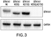
- the specific glycosylation sites of a particular BTN1A1 isoform or variant can vary from amino acids at position 55, 215, or 449 of that particular BTN1A1 isoform or variant.
- a person of ordinary skill in the art would be able to determine the glycosylation sites of any particular BTN1A1 isoform or variant that correspond to N55, N215, and N449 of the human BTN1A1 exemplified above based on sequence alignment and other common knowledge in the art.
- provided herein are also molecules having an antigen binding fragment that immunospecifically binds to a glycosylated form of a BTN1A1 isoform or variant relative to the unglycosylated BTN1A1 isoform or variant.
- the glycosylated sites of a BTN1A1 isoform or variant can be the corresponding sites of N55, N215, and N449 of human BTN1A1 sequence as provided above.
- the molecules have an antigen binding fragment that immunospecifically binds to glycosylated BTN1A1.
- the antigen binding fragment immunospecifically binds to BTN1A1 glycosylated at positions N55, N215, and/or N449.
- the antigen binding fragment immunospecifically binds to BTN1A1 glycosylated at position N55.
- the antigen binding fragment immunospecifically binds to BTN1A1 glycosylated at position N215.
- the antigen binding fragment immunospecifically binds to BTN1A1 glycosylated at position N449.
- the antigen binding fragment immunospecifically binds to one or more glycosylation motifs.
- the antigen binding fragment immunospecifically binds to BTN1A1 glycosylated at positions N55 and N215. In some aspects, the antigen binding fragments immunospecifically binds to BTN1A1 glycosylated at positions N215 and N449. In some aspects, the antigen binding fragments immunospecifically binds to BTN1A1 glycosylated at positions N55 and N449. In some aspects, the antigen binding fragments immunospecifically binds to BTN1A1 glycosylated at positions N55, N215 and N449.
- the molecules have an antigen binding fragment that immunospecifically binds to glycosylated BTN1A1, wherein the antigen binding fragment preferentially binds glycosylated BTN1A1 over non-glycosylated BTN1A1.
- the antigen binding fragments preferentially bind to BTN1A1 glycosylated at positions N55, N215, and/or N449 over non-glycosylated BTN1A1.
- the antigen binding fragments preferentially bind to BTN1A1 glycosylated at position N55 over non-glycosylated BTN1A1.
- the antigen binding fragments preferentially bind to BTN1A1 glycosylated at position N215 over non-glycosylated BTN1A1. In some aspects, the antigen binding fragments preferentially bind to BTN1A1 glycosylated at position N449 over non-glycosylated BTN1A1. In some aspects, the antigen binding fragments preferentially bind to one or more glycosylation motifs. In some aspects, the antigen binding fragments preferentially binds to BTN1A1 glycosylated at positions N55 and N215 over non-glycosylated BTN1A1.
- the antigen binding fragments preferentially bind to BTN1A1 glycosylated at positions N215 and N449 over non-glycosylated BTN1A1. In some aspects, the antigen binding fragments preferentially bind to BTN1A1 glycosylated at positions N55 and N449 over non-glycosylated BTN1A1. In some aspects, the antigen binding fragments preferentially binds to BTN1A1 glycosylated at positions N55, N215 and N449 over non-glycosylated BTN1A1.
- the preferential binding can be determined by binding affinity.
- an antibody or antigen binding fragment that preferentially binds to the glycosylated BTN1A1 can bind to glycosylated BTN1A1 with a Kd less than the Kd exhibited relative to unglycosylated BTN1A1.
- the antibody or antigen binding fragment binds to glycosylated BTN1A1 with Kd less than half of the Kd exhibited relative to unglycosylated BTN1A1.
- the antibody or antigen binding fragment binds to glycosylated BTN1A1 with Kd at least 10 times less than the Kd exhibited relative to unglycosylated BTN1A1.
- the antibody or antigen binding fragment binds to glycosylated BTN1A1 with Kd that is about 75% of the Kd exhibited relative to unglycosylated BTN1A1. In some embodiments, the antibody or antigen binding fragment binds to glycosylated BTN1A1 with Kd that is about 50% of the Kd exhibited relative to unglycosylated BTN1A1. In some embodiments, the antibody or antigen binding fragment binds to glycosylated BTN1A1 with Kd that is about 25% of the Kd exhibited relative to unglycosylated BTN1A1.
- the antibody or antigen binding fragment binds to glycosylated BTN1A1 with Kd that is about 10% of the Kd exhibited relative to unglycosylated BTN1A1. In some embodiments, the antibody or antigen binding fragment binds to glycosylated BTN1A1 with Kd that is about 5% of the Kd exhibited relative to unglycosylated BTN1A1. In some embodiments, the antibody or antigen binding fragment binds to glycosylated BTN1A1 with Kd that is bout 2.5% of the Kd exhibited relative to unglycosylated BTN1A1. In some embodiments, the antibody or antigen binding fragment binds to glycosylated BTN1A1 with Kd that is bout 1% of the Kd exhibited relative to unglycosylated BTN1A1.
- the preferential binding can also be determined by in a binding assay as indicated by, for example, fluorescence intensitity ("MFI").
- MFI fluorescence intensitity
- an antibody or antigen binding fragment that preferentially binds to the glycosylated BTN1A1 can bind to glycosylated BTN1A1 with an MFI that is higher than the MFI as exhibited relative to unglycosylated BTN1A1.
- the antibody or antigen binding fragment binds to glycosylated BTN1A1 with an MFI that is at least twice as high as the MFI as exhibited relative to unglycosylated BTN1A1.
- antibody or the antigen binding fragment binds to glycosylated BTN1A1 with an MFI that is at least three times as high as the MFI as exhibited relative to unglycosylated BTN1A1. In some embodiments, antibody or the antigen binding fragment binds to glycosylated BTN1A1 with an MFI that is at least five times as high as the MFI as exhibited relative to unglycosylated BTN1A1. In some embodiments, antibody or the antigen binding fragment binds to glycosylated BTN1A1 with an MFI that is at least ten times as high as the MFI as exhibited relative to unglycosylated BTN1A1.
- antibody or the antigen binding fragment binds to glycosylated BTN1A1 with an MFI that is at least fifteen times as high as the MFI as exhibited relative to unglycosylated BTN1A1. In some embodiments, antibody or the antigen binding fragment binds to glycosylated BTN1A1 with an MFI that is at least twenty times as high as the MFI as exhibited relative to unglycosylated BTN1A1.
- the antigen binding fragments immunospecifically mask BTN1A1 glycosylation at positions N55, N215, and/or N449. In some aspects, the antigen binding fragments immunospecifically mask BTN1A1 glycosylation at position N55. In some aspects, the antigen binding fragments immunospecifically mask BTN1A1 glycosylation at position N215. In some aspects, the antigen binding fragments immunospecifically mask BTN1A1 glycosylation at position N449. In some aspects, the antigen binding fragments immunospecifically mask one or more glycosylation motifs of BTN1A1. In some aspects, the antigen binding fragments immunospecifically mask BTN1A1 glycosylation at positions N55 and N215.
- the antigen binding fragments immunospecifically mask BTN1A1 glycosylation at positions N215 and N449. In some aspects, the antigen binding fragments immunospecifically mask BTN1A1 glycosylation at positions N55 and N449. In some aspects, the antigen binding fragments immunospecifically mask BTN1A1 glycosylation at positions N55, N215 and N449.
- the anti-BTN1A1 antibody or anti-glycosylated BTN1A1 antibody can be an IgG, IgM, IgA, IgD, or IgE.
- the anti-BTN1A1 antibody or anti-glycosylated BTN1A1 antibody can also be a chimeric antibody, an affinity matured antibody, a humanized antibody, or a human antibody.
- the anti-BTN1A1 antibody or anti-glycosylated BTN1A1 antibody can also be a camelized antibody, an intrabody, an anti-idiotypic (anti-Id) antibody.
- the anti-BTN1A1 antibody or anti-glycosylated BTN1A1 antibody can be a polyclonal antibody or monoclonal antibody.
- Antibodies can be produced from any animal source, including birds and mammals.
- the antibodies are ovine, murine (e.g ., mouse and rat), rabbit, goat, guinea pig, camel, horse, or chicken.
- newer technology permits the development of and screening for human antibodies from human combinatorial antibody libraries.
- bacteriophage antibody expression technology allows specific antibodies to be produced in the absence of animal immunization, as described in U.S. Patent No. 6,946,546 , which is hereby incorporated by reference in its entirety. These techniques are further described in Marks (1992); Stemmer (1994); Gram et al. (1992); Barbas et al. (1994); and Schier et al. (1996); which are hereby incorporated by reference in their entireties.
- the molecules having an antigen binding fragment that immunospecifically binds BTN1A1 or specifically glycosylated BTN1A1, including the anti-BTN1A1 antibodies or anti-glycosylated BTN1A1 antibodies can also be produced by any method known in the art useful for the production of polypeptides, e.g., in vitro synthesis, recombinant DNA production, and the like.
- the humanized antibodies can be produced by recombinant DNA technology.
- the antibodies described herein can also be produced using recombinant immunoglobulin expression technology.
- the recombinant production of immunoglobulin molecules, including humanized antibodies are described in U.S. Pat. No. 4,816,397 (Boss et al .), U.S. Pat.
- the anti-BTN1A1 antibody or anti-glycosylated BTN1A1 antibody is a human antibody.
- Human antibodies can be made by a variety of methods known in the art including phage display methods described above using antibody libraries derived from human immunoglobulin sequences ( see U.S. Pat. Nos. 4,444,887 and 4,716,111 ; and International Publication Nos. WO 98/46645 , WO 98/50433 , WO 98/24893 , WO 98/16654 , WO 96/34096 , WO 96/33735 , and WO 91/10741 ).
- Human antibodies can be produced using transgenic mice which are incapable of expressing functional endogenous immunoglobulins, but which can express human immunoglobulin genes.
- the human heavy and light chain immunoglobulin gene complexes can be introduced randomly or by homologous recombination into mouse embryonic stem cells.
- the human variable region, constant region, and diversity region can be introduced into mouse embryonic stem cells in addition to the human heavy and light chain genes.
- the mouse heavy and light chain immunoglobulin genes can be rendered non-functional separately or simultaneously with the introduction of human immunoglobulin loci by homologous recombination. In particular, homozygous deletion of the JH region prevents endogenous antibody production.
- the modified embryonic stem cells are expanded and microinjected into blastocysts to produce chimeric mice.
- the chimeric mice are then bred to produce homozygous offspring which express human antibodies.
- the transgenic mice are immunized using conventional methodologies with a selected antigen, e.g., all or a portion of a BTN1A1 polypeptide, or a glycosylated BTN1A1 polypeptide.
- Monoclonal antibodies directed against the antigen can be obtained from the immunized, transgenic mice using conventional hybridoma technology ( see, e.g., U.S. Pat. No. 5,916,771 ).
- the human immunoglobulin transgenes harbored by the transgenic mice rearrange during B cell differentiation, and subsequently undergo class switching and somatic mutation.
- therapeutically useful IgG, IgA, IgM and IgE antibodies can be produced.
- Lonberg and Huszar (1995, Int. Rev. Immunol. 13:65-93 , which is incorporated herein by reference in its entirety).
- the anti-BTN1A1 antibody or anti-glycosylated BTN1A1 antibody is a chimeric antibody, for example, an antibody having antigen binding sequences from a non-human donor grafted to a heterologous non-human, human or humanized sequence (e.g ., framework and/or constant domain sequences).
- the non-human donor is a rat.
- an antigen binding sequence is synthetic, e.g ., obtained by mutagenesis ( e.g., phage display screening of a human phage library, etc. ) .
- a chimeric antibody can have murine V regions and human C regions.
- the murine light chain V region is fused to a human kappa light chain.
- the murine heavy chain V region is fused to a human IgG1 C region.
- Chimeric antibodies comprising one or more CDRs from a non-human species and framework regions from a human immunoglobulin molecule can be produced using a variety of techniques known in the art including, for example, CDR-grafting ( EP 239,400 ; International Publication No. WO 91/09967 ; and U.S. Pat. Nos. 5,225,539 , 5,530,101 , and 5,585,089 ), veneering or resurfacing ( EP 592,106 ; EP 519,596 ; Padlan, 1991, Molecular Immunology 28(4/5):489-498 ; Studnicka et al., 1994, Protein Engineering 7:805 ; and Roguska et al., 1994, Proc. Natl. Acad. Sci. USA 91:969 ), and chain shuffling ( U.S. Pat. No. 5,565,332 ); all of which are hereby incorporated by references in their entireties.
- An exemplary process for the production of the recombinant chimeric anti-BTN1A1 antibodies can include the following: a) constructing, by conventional molecular biology methods, an expression vector that encodes and expresses an antibody heavy chain in which the CDRs and variable region of the murine anti-BTN1A1 (or anti- glycosylated BTN1A1) monoclonal antibody are fused to an Fc region derived from a human immunoglobulin, thereby producing a vector for the expression of a chimeric antibody heavy chain; b) constructing, by conventional molecular biology methods, an expression vector that encodes and expresses an antibody light chain of the murine anti- BTN1A1 (or anti-glycosylated BTN1A1) monoclonal antibody, thereby producing a vector for the expression of chimeric antibody light chain; c) transferring the expression vectors to a host cell by conventional molecular biology methods to produce a transfected host cell for the expression of chimeric antibodies; and d)
- An exemplary process for the production of the recombinant humanized anti-BTN1A1 antibodies can include the following: a) constructing, by conventional molecular biology methods, an expression vector that encodes and expresses an antibody heavy chain in which the CDRs and a minimal portion of the variable region framework that are required to retain donor antibody binding specificity are derived from a non-human immunoglobulin, such as the murine anti-BTN1A1 (or anti-glycosylated BTN1A1) monoclonal antibody, and the remainder of the antibody is derived from a human immunoglobulin, thereby producing a vector for the expression of a humanized antibody heavy chain; b) constructing, by conventional molecular biology methods, an expression vector that encodes and expresses an antibody light chain in which the CDRs and a minimal portion of the variable region framework that are required to retain donor antibody binding specificity are derived from a non-human immunoglobulin, such as the murine anti-BTN1A1 (or anti-glycosylated
- host cells can be co-transfected with such expression vectors, which can contain different selectable markers but, with the exception of the heavy and light chain coding sequences, are preferably identical.
- This procedure provides for equal expression of heavy and light chain polypeptides.
- a single vector may be used which encodes both heavy and light chain polypeptides.
- the coding sequences for the heavy and light chains can comprise cDNA or genomic DNA or both.
- the host cell used to express the recombinant antibody can be either a bacterial cell such as Escherichia coli, or more preferably a eukaryotic cell (e.g., a Chinese hamster ovary (CHO) cell or a HEK-293 cell).
- the choice of expression vector is dependent upon the choice of host cell, and can be selected so as to have the desired expression and regulatory characteristics in the selected host cell.
- Other cell lines that can be used include, but are not limited to, CHO-K1, NSO, and PER.C6 (Crucell, Leiden, Netherlands).
- codon usage can by optimized when host cell is selected to account for species specific codon usage bias and enhance protein expression.
- the DNA encoding the antibodies can incorporate codons used preferentially by Cricetulus griseus (from where Chinese Hamster ovaries cells are derived. Methods of codon optimization may be employed to facilitate improved expression by a desired host cell ( see, e.g., Wohlgemuth, I. et al., Philos.
- the anti-BTN1A1 antibodies or anti-glycosylated BTN1A1 antibodies can be monoclonal antibodies. In some embodiments, the anti-BTN1A1 antibodies or anti-glycosylated BTN1A1 antibodies can be polyclonal antibodies. Animals can be inoculated with an antigen, such as a BTN1A1 polypeptide or glycosylated BTN1A1 polypeptide in order to produce antibodies specific for a BTN1A1 polypeptide or a glycosylated BTN1A1 polypeptide. Frequently an antigen is bound or conjugated to another molecule to enhance the immune response.
- an antigen such as a BTN1A1 polypeptide or glycosylated BTN1A1 polypeptide
- a conjugate can be any peptide, polypeptide, protein, or non-proteinaceous substance bound to an antigen that is used to elicit an immune response in an animal.
- Antibodies produced in an animal in response to antigen inoculation have a variety of non-identical molecules (polyclonal antibodies) made from a variety of individual antibody producing B lymphocytes. Given the correct conditions for polyclonal antibody production in an animal, most of the antibodies in the animal's serum recognize the collective epitopes on the antigenic compound to which the animal has been immunized.
- MAbs monoclonal antibodies
- rodents such as mice and rats are used in generating monoclonal antibodies.
- rabbit, sheep, or frog cells are used in generating monoclonal antibodies.
- rats The use of rats is well known and can provide certain advantages.
- Mice e.g ., BALB/c mice
- BALB/c mice are routinely used and generally give a high percentage of stable fusions.
- Hybridoma technology involves the fusion of a single B lymphocyte from a mouse previously immunized with a BTN1A1 polypeptide or glycosylated BTN1A1 polypeptide with an immortal myeloma cell (usually mouse myeloma).
- This technology provides a method to propagate a single antibody-producing cell for an indefinite number of generations, such that unlimited quantities of structurally identical antibodies having the same antigen or epitope specificity (monoclonal antibodies) can be produced.
- the antibody is an immunoglobulin single variable domain derived from a camelid antibody, preferably from a heavy chain camelid antibody, devoid of light chains, which are known as V H H domain sequences or NanobodiesTM.
- a NanobodyTM (Nb) is the smallest functional fragment or single variable domain (V H H) of a naturally occurring single-chain antibody and is known to the person skilled in the art. They are derived from heavy chain only antibodies seen in camelids ( Hamers-Casterman et al., Nature, 363(6428):446-8 (1993 ); Desmyter et al., Nat StructBiol., 3(9):803-11. (1996 )).
- Nbs In the family of "camelids," immunoglobulins devoid of light polypeptide chains are found. "Camelids” comprise old world camelids ( Camelus bactrianus and Camelus dromedarius ) and new world camelids (for example, Lama paccos, Lama glama, Lama guanicoe and Lama vicugna ).
- the single variable domain heavy chain antibody is herein designated as a NanobodyTM or a V H H antibody.
- the small size and unique biophysical properties of Nbs excel conventional antibody fragments for the recognition of uncommon or hidden epitopes and for binding into cavities or active sites of protein targets. Further, Nbs can be designed as multi-specific and multivalent antibodies, attached to reporter molecules, or humanzied. Nbs are stable, survive the gastro-intestinal system and can easily be manufactured.
- bispecific antibodies Unifying two antigen binding sites of different specificity into a single construct, bispecific antibodies have the ability to bring together two discreet antigens with extraordinar specificity and therefore have great potential as therapeutic agents.
- Bispecific antibodies can be made by fusing two hybridomas, each capable of producing a different immunoglobulin.
- Bispecific antibodies can also be produced by joining two scFv antibody fragments while omitting the Fc portion present in full immunoglobulins.
- Each scFv unit in such constructs can be made up of one variable domain from each of the heavy (VH) and light (VL) antibody chains, joined with one another via a synthetic polypeptide linker, the latter often being genetically engineered so as to be minimally immunogenic while remaining maximally resistant to proteolysis.
- Respective scFv units can be joined by a number of techniques including incorporation of a short (usually less than 10 amino acids) polypeptide spacer bridging the two scFv units, thereby creating a bispecific single chain antibody.
- the resulting bispecific single chain antibody is therefore a species containing two VH/VL pairs of different specificity on a single polypeptide chain, wherein the VH and VL domains in a respective scFv unit are separated by a polypeptide linker long enough to allow intramolecular association between these two domains, and wherein the thusly formed scFv units are contiguously tethered to one another through a polypeptide spacer kept short enough to prevent unwanted association between, for example, the VH domain of one scFv unit and the VL of the other scFv unit.
- Examples of molecules having an antigen binding fragment that immunospecifically binds to BTN1A1 or glycosylated BTN1A1, include, without limitation: (i) the Fab fragment, consisting of VL, VH, CL, and CH1 domains; (ii) the "Fd” fragment consisting of the VH and CH1 domains; (iii) the "Fv” fragment consisting of the VL and VH domains of a single antibody; (iv) the "dAb” fragment, which consists of a VH domain; (v) isolated CDR regions; (vi) F(ab')2 fragments, a bivalent fragment comprising two linked Fab fragments; (vii) single chain Fv molecules ("scFv”), wherein a VH domain and a VL domain are linked by a peptide linker that allows the two domains to associate to form a binding domain; (viii) bi-specific single chain Fv dimers ( see U.S.
- Patent No. 5,091,513 and (ix) diabodies, multivalent, or multispecific fragments constructed by gene fusion ( U.S. Patent Appln. Publn. No. 20050214860 ).
- Fv, scFv, or diabody molecules may be stabilized by the incorporation of disulfide bridges linking the VH and VL domains.
- Minibodies having a scFv joined to a CH3 domain can also be made ( Hu et al., Cancer Res., 56(13):3055-61(1996 )).
- Antibody-like binding peptidomimetics are also contemplated in embodiments.
- Murali et al., Cell Mol. Biol., 49 (2):209-216 (2003 ) describe "antibody like binding peptidomimetics" (ABiPs), which are peptides that act as pared-down antibodies and have certain advantages of longer serum half-life as well as less cumbersome synthesis methods, which is hereby incorporated by reference in its entirety.
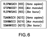
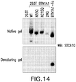
- STC810 also referred to as STC838
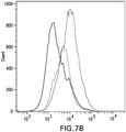
- KD between STC810 and hBTN1A1-Fc was determined to be 1.81 nM by Biacore, and 2.12 nM by Octet).
- treatment of the monoclonal anti-BTN1A1 antibody e.g ., STC810
- enhanced T-cell dependent apoptosis of cancer cells inhibited proliferation of cancer cells, and also resulted in glycosylation dependent internalization of BTN1A1 to lysosomes.
- the epitopes of STC810 are also provided herein. Accordingly, provided herein are also anti-BTN1A1 antibodies with specific sequence features, anti-BTN1A1 antibodies that immunospecifically bind to specific epitopes, as well as the uses thereof in cancer treatment.
- the anti-BTN1A1 antibody provided herein comprises a VH domain, VL domain, VH CDR1, VH CDR2, VH CDR3, VL CDR1, VL CDR2, and/or VL CDR3 of monoclonal antibody STC810 described herein, or a humanized variant thereof.
- the anti-BTN1A1 antibody can further comprise a VH FR1, VH FR2, VH FR3, VH FR4, VL FR1, VL FR2, VL FR3, and/or VL FR4 of a human germline immunoglobulin amino acid sequence or a variant thereof.
- the anti-BTN1A1 antibody comprises less than six CDRs.
- the antibody comprises or consists of one, two, three, four, or five CDRs selected from the group consisting of VH CDR1, VH CDR2, VH CDR3, VL CDR1, VL CDR2, and/or VL CDR3.
- the antibody comprises or consists of one, two, three, four, or five CDRs selected from the group consisting of VH CDR1, VH CDR2, VH CDR3, VL CDR1, VL CDR2, and/or VL CDR3 of the monoclonal antibody STC810 described herein, or a humanized variant thereof.
- the antibody further comprises a VH FR1, VH FR2, VH FR3, VH FR4, VL FR1, VL FR2, VL FR3, and/or VL FR4 of a human germline immunoglobulin amino acid sequence or a variant thereof.
- the antibody is a humanized antibody, a monoclonal antibody, a recombinant antibody, an antigen binding fragment or any combination thereof.
- the antibody is a humanized monoclonal antibody, or antigen binding fragment thereof.
- antibodies including humanized antibodies, (i) that competitively block ( e.g., in a dose-dependent manner) an anti-BTN1A1 antibody provided herein from binding to a BTN1A1 polypeptide (e.g., a cell surface-expressed or soluble BTN1A1), a BTN1A1 fragment, or a BTN1A1 epitope and/or (ii) that bind to a BTN1A1 epitope that is bound by an anti-BTN1A1 antibody (e.g ., humanized anti-BTN1A1 antibodies) provided herein.
- a BTN1A1 polypeptide e.g., a cell surface-expressed or soluble BTN1A1
- BTN1A1 fragment e.g., a BTN1A1 fragment
- BTN1A1 epitope e.g., humanized anti-BTN1A1 antibodies
- the antibody competitively blocks ( e.g., in a dose-dependent manner) monoclonal antibody STC810 described herein or a humanized variant thereof from binding to a BTN1A1 polypeptide (e.g ., a cell surface-expressed or soluble BTN1A1), a BTN1A1 fragment, or a BTN1A1 epitope.
- a BTN1A1 polypeptide e.g ., a cell surface-expressed or soluble BTN1A1
- the antibody binds to a BTN1A1 epitope that is bound ( e.g ., recognized) by monoclonal antibody BTN1A1 described herein or a humanized variant thereof (e.g . humanized anti-BTN1A1 antibodies).
- Table 2a Sequences of heavy chain variable (VH) region and light chain variable (VL) region of mouse monoclonal anti-human BTN1A1 Antibody STC810 DNA sequence Protein sequence heavy chain Kappa Light chain
- Table 2b CDR Sequences of mouse monoclonal anti-human BTN1A1 Antibody STC810 Region definition CDR1 CDR2 CDR3 Heavy chain Chothia (SEQ ID NO: 7) GYTFTHY (SEQ ID NO: 8) YPSNGG AbM (SEQ ID NO: 10) GYTFTHYNMD (SEQ ID NO: 11) YIYPSNGGTG Kabat (SEQ ID NO: 13) HYNMD Contact (SEQ ID NO: 16) THYNMD (SEQ ID NO: 17) WIGYIYPSNGGTG Kappa light chain Chothia (SEQ ID NO: 19) SASQDISNYLN (SEQ ID NO: 20) YTSSLHS (SEQ ID NO: 21) QQSSKLPFT Ab
- the molecules provided herein have an antigen binding fragment that has (a) a heavy chain variable (VH) region comprising: (1) a VH CDR1 having an amino acid sequence of SEQ ID NOS: 7, 10, 13, or 16; (2) a VH CDR2 having an amino acid sequence SEQ ID NOS: 8, 11, 14 or 17; and (3) a VH CDR3 having an amino acid sequence of SEQ ID NOS: 9, 12, 15 or 18; and/or (b) a light chain variable (VL) region comprising: (1) a VL CDR1 having an amino acid sequence of SEQ ID NOS: 19, 22, 25 or 28; (2) a VL CDR2 having an amino acid sequence of SEQ ID NOS: 20, 23, 26 or 29; and (3) a VL CDR3 having an amino acid sequence of SEQ ID NOS: 21, 24, 27 or 30.
- VH heavy chain variable
- antibodies having having (a) a heavy chain variable (VH) region comprising: (1) a VH CDR1 having an amino acid sequence of SEQ ID NOS: 7, 10, 13, or 16; (2) a VH CDR2 having an amino acid sequence SEQ ID NOS: 8, 11, 14 or 17; and (3) a VH CDR3 having an amino acid sequence of SEQ ID NOS: 9, 12, 15 or 18; and/or (b) a light chain variable (VL) region comprising: (1) a VL CDR1 having an amino acid sequence of SEQ ID NOS: 19, 22, 25 or 28; (2) a VL CDR2 having an amino acid sequence of SEQ ID NOS: 20, 23, 26 or 29; and (3) a VL CDR3 having an amino acid sequence of SEQ ID NOS: 21, 24, 27 or 30.
- the antibody can be a monoclonal antibody.
- the antibody can be a humanized antibody.
- the molecules provided herein have an antigen binding fragment that has a heavy chain variable (VH) region comprising: (1) a VH CDR1 having an amino acid sequence of SEQ ID NOS: 7, 10, 13 or 16; (2) a VH CDR2 having an amino acid sequence of SEQ ID NOS: 8, 11, 14 or 17; and/or (3) a VH CDR3 having an amino acid sequence of SEQ ID NOS: 9, 12, 15 or 18.
- the heavy chain variable (VH) region comprises (1) a VH CDR1 having an amino acid sequence of SEQ ID NOS: 7, 10, 13 or 16; and (2) a VH CDR2 having an amino acid sequence of SEQ ID NOS: 8, 11, 14 or 17.
- the heavy chain variable (VH) region comprises (1) a VH CDR1 having an amino acid sequence of SEQ ID NOS: 7, 10, 13 or 16; and (3) a VH CDR3 having an amino acid sequence of SEQ ID NOS: 9, 12, 15 or 18.
- the molecules provided herein have an antigen binding fragment that has a heavy chain variable (VH) region comprising: (1) a VH CDR1 having an amino acid sequence of SEQ ID NOS: 7, 10, 13 or 16; and (3) a VH CDR3 having an amino acid sequence of SEQ ID NOS: 9, 12, 15 or 18.
- the molecules provided herein have an antigen binding fragment that has a heavy chain variable (VH) region comprising a VH CDR1 having an amino acid sequence of SEQ ID NOS: 7, 10, 13 or 16.
- VH CDR1 can have an amino acid sequence of SEQ ID NO: 7.
- the VH CDR1 can have an amino acid sequence of SEQ ID NO: 10.
- the VH CDR1 can have an amino acid sequence of SEQ ID NO: 13.
- the VH CDR1 can have an amino acid sequence of SEQ ID NO: 16.
- the molecules provided herein have an antigen binding fragment that has a heavy chain variable (VH) region comprising a VH CDR2 having an amino acid sequence of SEQ ID NOS: 8, 11, 14 or 17.
- the VH CDR2 can have an amino acid sequence of SEQ ID NO: 8.
- the VH CDR2 can have an amino acid sequence of SEQ ID NO: 11.
- the VH CDR2 can have an amino acid sequence of SEQ ID NO: 14.
- the VH CDR2 can have an amino acid sequence of SEQ ID NO: 17.
- the molecules provided herein have an antigen binding fragment that has a heavy chain variable (VH) region comprising a VH CDR3 having an amino acid sequence of SEQ ID NOS: 9, 12, 15 or 18.
- the VH CDR3 can have an amino acid sequence of SEQ ID NO: 9.
- the VH CDR3 can have an amino acid sequence of SEQ ID NO: 12.
- the VH CDR3 can have an amino acid sequence of SEQ ID NO: 15.
- the VH CDR3 can have an amino acid sequence of SEQ ID NO: 20.
- the molecules provided herein have an antigen binding fragment that has a heavy chain variable (VH) region comprising: (1) a VH CDR1 having an amino acid sequence of SEQ ID NO: 7; (2) a VH CDR2 having an amino acid sequence of SEQ ID NO: 8; and/or (3) a VH CDR3 having an amino acid sequence of SEQ ID NO: 9.
- VH heavy chain variable
- the molecules provided herein have an antigen binding fragment that has a heavy chain variable (VH) region comprising: (1) a VH CDR1 having an amino acid sequence of SEQ ID NO: 10; (2) a VH CDR2 having an amino acid sequence of SEQ ID NO: 11; and/or (3) a VH CDR3 having an amino acid sequence of SEQ ID NO: 12.
- VH heavy chain variable
- the molecules provided herein have an antigen binding fragment that has a heavy chain variable (VH) region comprising: (1) a VH CDR1 having an amino acid sequence of SEQ ID NO: 13; (2) a VH CDR2 having an amino acid sequence of SEQ ID NO: 14; and/or (3) a VH CDR3 having an amino acid sequence of SEQ ID NO: 15.
- VH heavy chain variable
- the molecules provided herein have an antigen binding fragment that has a heavy chain variable (VH) region comprising: (1) a VH CDR1 having an amino acid sequence of SEQ ID NO: 16; (2) a VH CDR2 having an amino acid sequence of SEQ ID NO: 17; and/or (3) a VH CDR3 having an amino acid sequence of SEQ ID NO: 18.
- VH heavy chain variable
- the molecules provided herein have an antigen binding fragment that has a heavy chain variable (VH) region that has the amino acid sequence of SEQ ID NO: 3.
- VH heavy chain variable
- the molecule can be an antibody.
- the antibody can be a monoclonal antibody.
- the antibody can be a humanized antibody.
- the molecules provided herein have an antigen binding fragment that has a light chain variable (VL) region comprising: (1) a VL CDR1 having an amino acid sequence of SEQ ID NOS: 19, 22, 25 or 28; (2) a VL CDR2 having an amino acid sequence of SEQ ID NOS: 20, 23, 26 or 29; and (3) a VL CDR3 having an amino acid sequence of SEQ ID NOS: 21, 24, 27 or 30.
- VL light chain variable
- the molecules provided herein have an antigen binding fragment that has a light chain variable (VL) region comprising: (1) a VL CDR1 having an amino acid sequence of SEQ ID NOS: 19, 22, 25 or 28; and (2) a VL CDR2 having an amino acid sequence of SEQ ID NOS: 20, 23, 26 or 29.
- the molecules provided herein have an antigen binding fragment that has a light chain variable (VL) region comprising: (1) a VL CDR1 having an amino acid sequence of SEQ ID NOS: 19, 22, 25 or 28; and (3) a VL CDR3 having an amino acid sequence of SEQ ID NOS: 21, 24, 27 or 30.
- the molecules provided herein have an antigen binding fragment that has a light chain variable (VL) region comprising: (2) a VL CDR2 having an amino acid sequence of SEQ ID NOS: 20, 23, 26 or 29; and (3) a VL CDR3 having an amino acid sequence of SEQ ID NOS: 21, 24, 27 or 30.
- VL light chain variable
- the molecules provided herein have an antigen binding fragment that has a light chain variable (VL) region comprising a VL CDR1 having an amino acid sequence of SEQ ID NOS: 19, 22, 25 or 28.
- VL CDR1 can have an amino acid sequence of SEQ ID NO: 19.
- the VL CDR1 can have an amino acid sequence of SEQ ID NO: 22.
- the VL CDR1 can have an amino acid sequence of SEQ ID NO: 25.
- the VL CDR1 can have an amino acid sequence of SEQ ID NO: 28.
- the molecules provided herein have an antigen binding fragment that has a light chain variable (VL) region comprising a VL CDR2 having an amino acid sequence of SEQ ID NOS: 20, 23, 26 or 29.
- VL CDR2 can have an amino acid sequence of SEQ ID NO:23.
- the VL CDR2 can have an amino acid sequence of SEQ ID NO:26.
- the VL CDR2 can have an amino acid sequence of SEQ ID NO:29.
- the molecules provided herein have an antigen binding fragment that has a light chain variable (VL) region comprising a VL CDR3 having an amino acid sequence of SEQ ID NOS: 21, 24, 27 or 30.
- VL CDR3 can have an amino acid sequence of SEQ ID NO: 21.
- the VL CDR3 can have an amino acid sequence of SEQ ID NO: 24.
- the VL CDR3 can have an amino acid sequence of SEQ ID NO: 27.
- the VL CDR3 can have an amino acid sequence of SEQ ID NO:30.
- the molecules provided herein have an antigen binding fragment that has a light chain variable (VL) region that has (1) a VL CDR1 having an amino acid sequence of SEQ ID NO: 19; (2) a VL CDR2 having an amino acid sequence of SEQ ID NO: 20; and/or (3) a VL CDR3 having an amino acid sequence of SEQ ID NO: 21.
- VL light chain variable
- the molecules provided herein have an antigen binding fragment that has a light chain variable (VL) region that has (1) a VL CDR1 having an amino acid sequence of SEQ ID NO: 22; (2) a VL CDR2 having an amino acid sequence of SEQ ID NO: 23; and/or (3) a VL CDR3 having an amino acid sequence of SEQ ID NO: 24.
- VL light chain variable
- the molecules provided herein have an antigen binding fragment that has a light chain variable (VL) region that has (1) a VL CDR1 having an amino acid sequence of SEQ ID NO: 25; (2) a VL CDR2 having an amino acid sequence of SEQ ID NO: 26; and/or (3) a VL CDR3 having an amino acid sequence of SEQ ID NO: 27.
- VL light chain variable
- the molecules provided herein have an antigen binding fragment that has a light chain variable (VL) region that has (1) a VL CDR1 having an amino acid sequence of SEQ ID NO: 28; (2) a VL CDR2 having an amino acid sequence of SEQ ID NO: 29; and/or (3) a VL CDR3 having an amino acid sequence of SEQ ID NO: 30.
- VL light chain variable
- the molecules provided herein have an antigen binding fragment that has a light chain variable (VL) region that has the amino acid sequence of SEQ ID NO: 5.
- VL light chain variable
- the molecule can be an antibody.
- the antibody can be a monoclonal antibody.
- the antibody can be a humanized antibody.
- the molecules provided herein have an antigen binding fragment that has (a) a heavy chain variable (VH) region comprising: (1) a VH CDR1 having an amino acid sequence of SEQ ID NOS: 7, 10, 13 or 16; (2) a VH CDR2 having an amino acid sequence of SEQ ID NOS: 8, 11, 14 or 17; and/or (3) a VH CDR3 having an amino acid sequence of SEQ ID NOS: 9, 12, 15 or 18; and (b) a light chain variable (VL) region comprising: (1) a VL CDR1 having an amino acid sequence of SEQ ID NOS: 19, 22, 25 or 28; (2) a VL CDR2 having an amino acid sequence of SEQ ID NOS: 20, 23, 26 or 29; and/or (3) a VL CDR3 having an amino acid sequence of SEQ ID NOS: 21, 24, 27 or 30.
- the molecule can be an antibody.
- the antibody can be a monoclonal antibody.
- the antibody can be a humanized antibody.
- the molecules provided herein have an antigen binding fragment that has (a) a heavy chain variable (VH) region comprising: (1) a VH CDR1 having an amino acid sequence of SEQ ID NO 7; (2) a VH CDR2 having an amino acid sequence of SEQ ID NO 8; and/or (3) a VH CDR3 having an amino acid sequence of SEQ ID NO 9; and (b) a light chain variable (VL) region comprising: (1) a VL CDR1 having an amino acid sequence of SEQ ID NO: 19; (2) a VL CDR2 having an amino acid sequence of SEQ ID NO: 20; and/or (3) a VL CDR3 having an amino acid sequence of SEQ ID NO: 21.
- the molecule can be an antibody.
- the antibody can be a monoclonal antibody.
- the antibody can be a humanized antibody.
- the molecules provided herein have an antigen binding fragment that has (a) a heavy chain variable (VH) region comprising: (1) a VH CDR1 having an amino acid sequence of SEQ ID NO 10; (2) a VH CDR2 having an amino acid sequence of SEQ ID NO 11; and/or (3) a VH CDR3 having an amino acid sequence of SEQ ID NO 12; and (b) a light chain variable (VL) region comprising: (1) a VL CDR1 having an amino acid sequence of SEQ ID NO: 22; (2) a VL CDR2 having an amino acid sequence of SEQ ID NO: 23; and/or (3) a VL CDR3 having an amino acid sequence of SEQ ID NO: 24.
- the molecule can be an antibody.
- the antibody can be a monoclonal antibody.
- the antibody can be a humanized antibody.
- the molecules provided herein have an antigen binding fragment that has (a) a heavy chain variable (VH) region comprising: (1) a VH CDR1 having an amino acid sequence of SEQ ID NO 13; (2) a VH CDR2 having an amino acid sequence of SEQ ID NO 14; and/or (3) a VH CDR3 having an amino acid sequence of SEQ ID NO 15; and (b) a light chain variable (VL) region comprising: (1) a VL CDR1 having an amino acid sequence of SEQ ID NO: 25; (2) a VL CDR2 having an amino acid sequence of SEQ ID NO: 26; and/or (3) a VL CDR3 having an amino acid sequence of SEQ ID NO: 27.
- the molecule can be an antibody.
- the antibody can be a monoclonal antibody.
- the antibody can be a humanized antibody.
- the molecules provided herein have an antigen binding fragment that has (a) a heavy chain variable (VH) region comprising: (1) a VH CDR1 having an amino acid sequence of SEQ ID NO 16; (2) a VH CDR2 having an amino acid sequence of SEQ ID NO 17; and (3) a VH CDR3 having an amino acid sequence of SEQ ID NO 18; and (b) a light chain variable (VL) region comprising: (1) a VL CDR1 having an amino acid sequence of SEQ ID NO: 28; (2) a VL CDR2 having an amino acid sequence of SEQ ID NO: 29; and (3) a VL CDR3 having an amino acid sequence of SEQ ID NO: 30.
- the molecule can be an antibody.
- the antibody can be a monoclonal antibody.
- the antibody can be a humanized antibody.
- the molecules provided herein have an antigen binding fragment that has a VH region that has the amino acid sequence of SEQ ID NO: 3 and the VL region that has the the amino acid sequence of SEQ ID NO: 5.
- the molecule can be an antibody.
- the antibody can be a monoclonal antibody.
- the antibody can be a humanized antibody.
- the molecules provided herein is the mouse monoclonal antibody designated as STC810, or a humanized antibody version there of.
- a humanized STC810 antibody can have the VH region, the VL region, or both the VH and VL region of STC810 as described herein.
- a humanized STC810 antibody can also have six CDR regions (VH CDR1, VH CDR2, VH CDR3, VL CDR1, VL CDR2 and VL CDR3) of STC810 as described herein.
- the humanized STC810 antibody can also have less than the six CDR regions of STC810.
- the humanized STC810 antibody can also have one, two, three, four, or five CDR regions (VH CDR1, VH CDR2, VH CDR3, VL CDR1, VL CDR2 and VL CDR3) of STC810.
- the derivatives include less than 25 amino acid substitutions, less than 20 amino acid substitutions, less than 15 amino acid substitutions, less than 10 amino acid substitutions, less than 5 amino acid substitutions, less than 4 amino acid substitutions, less than 3 amino acid substitutions, or less than 2 amino acid substitutions relative to the original molecule.
- the derivatives have conservative amino acid substitutions are made at one or more predicted non-essential amino acid residues.
- a “conservative amino acid substitution” is one in which the amino acid residue is replaced with an amino acid residue having a side chain with a similar charge.
- Families of amino acid residues having side chains with similar charges have been defined in the art. These families include amino acids with basic side chains (e.g., lysine, arginine, histidine), acidic side chains ( e.g., aspartic acid, glutamic acid), uncharged polar side chains (e.g ., glycine, asparagine, glutamine, serine, threonine, tyrosine, cysteine), nonpolar side chains ( e.g ., alanine, valine, leucine, isoleucine, proline, phenylalanine, methionine, tryptophan), beta-branched side chains ( e.g ., threonine, valine, isoleucine) and aromatic side chains ( e.g ., tyrosine, phenyla
- mutations can be introduced randomly along all or part of the coding sequence, such as by saturation mutagenesis, and the resultant mutants can be screened for biological activity to identify mutants that retain activity. Following mutagenesis, the encoded protein can be expressed and the activity of the protein can be determined.
- the molecules provided herein having an antigen binding fragment that immunospecifically binds to BTN1A1 or glycosylated BTN1A1 can have an amino acid sequence that is at least 35%, at least 40%, at least 45%, at least 50%, at least 55%, at least 60%, at least 65%, at least 70%, at least 75%, at least 80%, at least 85%, at least 90%, at least 95%, or at least 99% identical to the amino acid sequence of the murine monoclonal antibody STC810, or an antigen-binding fragment thereof, such as a VH domain or VL domain.
- the molecules provided herein can have an amino acid sequence that is at least 35%, at least 40%, at least 45%, at least 50%, at least 55%, at least 60%, at least 65%, at least 70%, at least 75%, at least 80%, at least 85%, at least 90%, at least 95%, or at least 99% identical to an amino acid sequence depicted in SEQ ID NOS: 3 or 5.
- the molecules provided herein can have a VH CDR and/or a VL CDR amino acid sequence that is at least 35%, at least 40%, at least 45%, at least 50%, at least 55%, at least 60%, at least 65%, at least 70%, at least 75%, at least 80%, at least 85%, at least 90%, at least 95%, or at least 99% identical to a VH CDR amino acid sequence and/or a VL CDR amino acid sequence depicted in Table 2 above.
- the molecules provided herein can have an amino acid sequence of a VH domain and/or an amino acid sequence a VL domain encoded by a nucleotide sequence that hybridizes to the complement of a nucleotide sequence encoding any one of the VH and/or VL domains depicted in Table 2 under stringent conditions (e.g ., hybridization to filter-bound DNA in 6x sodium chloride/sodium citrate (SSC) at about 45° C followed by one or more washes in 0.2xSSC/0.1% SDS at about 50-65° C) under highly stringent conditions (e.g ., hybridization to filter-bound nucleic acid in 6xSSC at about 45° C followed by one or more washes in 0.1xSSC/0.2% SDS at about 68° C), or under other stringent hybridization conditions which are known to those of skill in the art (see, for example, Ausubel, F.M. et al., eds., 1989,
- the molecules provided herein can have an amino acid sequence of a VH CDR or an amino acid sequence of a VL CDR encoded by a nucleotide sequence that hybridizes to the complement of a nucleotide sequence encoding any one of the VH CDRs and/or VL CDRs depicted in Table 2 under stringent conditions (e.g.
- nucleic acid that encode an amino acid sequence f a VH CDR or an amino acid sequence of a VL CDR depicted in Table 2, or that hybridizes to the complement of a nucleic acid sequence encoding any one of the VH CDRs and/or VL CDRs depicted in Table 2 under stringent conditions (e.g.
- SSC sodium chloride/sodium citrate
- nucleic acid that encode an amino acid sequence of a VH domain and/or an amino acid sequence a VL domain depicted in Table 2, or that hybridizes to the complement of a nucleotide sequence encoding any one of the VH and/or VL domains depicted in Table 2 under stringent conditions (e.g ., hybridization to filter-bound DNA in 6x sodium chloride/sodium citrate (SSC) at about 45° C followed by one or more washes in 0.2xSSC/0.1% SDS at about 50-65° C) under highly stringent conditions (e.g.
- stringent conditions e.g ., hybridization to filter-bound DNA in 6x sodium chloride/sodium citrate (SSC) at about 45° C followed by one or more washes in 0.2xSSC/0.1% SDS at about 50-65° C
- highly stringent conditions e.g.
- the isolated nucleic acid can have a sequence of SEQ ID NO: 4 or that hybridizes to the complement of a nucleotide sequence of SEQ ID NO: 4 under stringent conditions (e.g ., hybridization to filter-bound DNA in 6x sodium chloride/sodium citrate (SSC) at about 45° C followed by one or more washes in 0.2xSSC/0.1% SDS at about 50-65° C) under highly stringent conditions (e.g. , hybridization to filter-bound nucleic acid in 6xSSC at about 45° C followed by one or more washes in 0.1xSSC/0.2% SDS at about 68° C), or under other stringent hybridization conditions which are known to those of skill in the art.
- stringent conditions e.g ., hybridization to filter-bound DNA in 6x sodium chloride/sodium citrate (SSC) at about 45° C followed by one or more washes in 0.2xSSC/0.1% SDS at about 50-65° C
- the isolated nucleic acid can have a sequence of SEQ ID NO: 6 or that hybridizes to the complement of a nucleotide sequence of SEQ ID NO: 6 under stringent conditions (e.g ., hybridization to filter-bound DNA in 6x sodium chloride/sodium citrate (SSC) at about 45° C followed by one or more washes in 0.2xSSC/0.1% SDS at about 50-65° C) under highly stringent conditions (e.g.
- stringent conditions e.g ., hybridization to filter-bound DNA in 6x sodium chloride/sodium citrate (SSC) at about 45° C followed by one or more washes in 0.2xSSC/0.1% SDS at about 50-65° C
- highly stringent conditions e.g.
- the molecules provided herein can be chemically modified, e.g., by the covalent attachment of any type of molecule to the antibody.
- the antibody derivatives include antibodies that have been chemically modified, e.g ., by glycosylation, acetylation, pegylation, phosphorylation, amidation, derivatization by known protecting/blocking groups, proteolytic cleavage, linkage to a cellular ligand or other protein, etc. Any of numerous chemical modifications may be carried out by known techniques, including, but not limited to specific chemical cleavage, acetylation, formulation, metabolic synthesis of tunicamycin, etc. Additionally, the antibody may contain one or more non-classical amino acids.
- the molecules provided herein can have a framework region known to those of skill in the art (e.g., a human or non-human fragment).
- the framework region can, for example, be naturally occurring or consensus framework regions.
- the framework region of an antibody provided herein is human (see, e.g., Chothia et al., 1998, J. Mol. Biol. 278:457-479 for a listing of human framework regions, which is incorporated by reference herein in its entirety). See also Kabat et al. (1991) Sequences of Proteins of Immunological Interest (U.S. Department of Health and Human Services, Washington, D.C.) 5th ed .
- FIG. 12 shows a synthesized epitope of BTN1A1(ECD)-Fc antigen for STC810: LELRWFRKKVSPA (SEQ ID NO:34) - EEGLFTVAASVIIRDTSAKNV (SEQ ID NO:35)
- Table 4 summarizes the cross-linked peptides of BTN1A1-His and STC810, which represent BTN1A1 epitopes of STC810 (SEQ ID NOS: 36-39).
- FIG. 13 shows a synthesized epitope of BT ⁇ N1A1(ECD)-His antigen for STC810.
- GRATLVQDGIAKGRV (SEQ ID NO:40) - EEGLFTVAASVIIRDTSAKNV (SEQ ID NO:41) Table 3: Cross-linked peptides of BTN1A1-Fc with STC810 analyzed by nLC-orbitrap MS/MS.
- the molecule is an molecules having an antigen binding fragment that competitively blocks ( e.g., in a dose-dependent manner) a BTN1A1 epitope described herein.
- the molecules provided herein have an antigen binding fragment that immunospecifically binds to an epitope of BTN1A1 as described herein.
- the molecules provided herein have an antigen binding fragment that immunospecifically binds to an BTN1A1 epitope of STC810.
- the molecule can be an antibody.
- the antibody can be a monoclonal antibody.
- the antibody can be a humanized antibody.
- anti-BTN1A1 antibodies that competitively block ( e.g., in a dose-dependent manner) a BTN1A1 epitope described herein.
- anti-BTN1A1 antibodies that competitively block ( e.g., in a dose-dependent manner) a BTN1A1 epitope of STC810 as described herein.
- the anti-BTN1A1 antibodies provided herein immunospecifically bind to an epitope of BTN1A1 as described herein.
- the anti-BTN1A1 antibodies provided herein immunospecifically bind to an BTN1A1 epitope of STC810.
- the molecules provided herein have an antigen binding fragment that immunospecifically binds to an epitope of BTN1A1, wherein the BTN1A1 epitope has at least five consecutive amino acids of an amino acid sequence of SEQ ID NOS: 31-41.
- the epitope of BTN1A1 can have at least six, at least seven, at least eight, at least nine, at least ten, at least eleven, at least twelve, at least thirteen, at least fourteen, or at least fifteen, consecutive amino acids of an amino acid sequence of SEQ ID NOS: 31-41.
- the epitope of BTN1A1 can have at least six consecutive amino acids of an amino acid sequence of SEQ ID NOS: 31-41.
- the epitope of BTN1A1 can have at least seven consecutive amino acids of an amino acid sequence of SEQ ID NOS: 31-41.
- the epitope of BTN1A1 can have at least eight consecutive amino acids of an amino acid sequence of SEQ ID NOS: 31-41.
- the epitope of BTN1A1 can have at least nine consecutive amino acids of an amino acid sequence of SEQ ID NOS: 31-41.
- the epitope of BTN1A1 can have at least ten consecutive amino acids of an amino acid sequence of SEQ ID NOS: 31-41.
- the epitope of BTN1A1 can have at least eleven consecutive amino acids of an amino acid sequence of SEQ ID NOS: 31-41.
- the epitope of BTN1A1 can have at least twelve consecutive amino acids of an amino acid sequence of SEQ ID NOS: 31-41.
- the epitope of BTN1A1 can have at least thirteen consecutive amino acids of an amino acid sequence of SEQ ID NOS: 31-41.
- the epitope of BTN1A1 can have at least fourteen consecutive amino acids of an amino acid sequence of SEQ ID NOS: 31-41.
- the epitope of BTN1A1 can have at least fifteen consecutive amino acids of an amino acid sequence of SEQ ID NOS: 31-41.
- the molecule can be an antibody.
- the antibody can be a monoclonal antibody.
- the antibody can be a humanized antibody.
- the molecules provided herein have an antigen binding fragment that immunospecifically binds to an epitope of BTN1A1, wherein the BTN1A1 epitope has an amino acid sequence of SEQ ID NOS: 31, 32, 33, 34, 35, 36, 37, 38, 39, 40 or 41.
- the epitope of BTN1A1 can have an amino acid sequence of SEQ ID NO: 31.
- the epitope of BTN1A1 can have an amino acid sequence of SEQ ID NO: 32.
- the epitope of BTN1A1 can have an amino acid sequence of SEQ ID NO: 33.
- the epitope of BTN1A1 can have an amino acid sequence of SEQ ID NO: 34.
- the epitope of BTN1A1 can have an amino acid sequence of SEQ ID NO: 35.
- the epitope of BTN1A1 can have an amino acid sequence of SEQ ID NO: 36.
- the epitope of BTN1A1 can have an amino acid sequence of SEQ ID NO: 37.
- the epitope of BTN1A1 can have an amino acid sequence of SEQ ID NO: 38.
- the epitope of BTN1A1 can have an amino acid sequence of SEQ ID NO: 39.
- the epitope of BTN1A1 can have an amino acid sequence of SEQ ID NO: 40.
- the epitope of BTN1A1 can have an amino acid sequence of SEQ ID
- the molecules provided herein have a high affinity for BTN1A1, glycosylated BTN1A1 or a polypeptide, or polypeptide fragment or epitope thereof.
- the molecules provided herein can be anti-BTN1A1 antibodies that have a higher affinity for a BTN1A1 antibody than known antibodies (e.g ., commercially available monoclonal antibodies discussed elsewhere herein).
- the molecules provided herein can be anti-BTN1A1 antibodies can have a 2- to 10-fold (or more) higher affinity for a BTN1A1 antigen than a known anti-BTN1A1 antibody as assessed by techniques described herein or known to one of skill in the art (e.g., a BIAcore assay).
- the affinity of the antibodies are, in one embodiment, assessed by a BIAcore assay.
- molecules provided herein can have an antigen binding fragment that binds to BTN1A1, glycosylated BTN1A1 or a polypeptide, or polypeptide fragment or epitope thereof with a dissociation constant (Kd) of no more than 1 ⁇ M, no more than 100 nM, no more than 10 nM, no more than 1 nM, or no more than 0.1 nM.
- Kd dissociation constant
- molecules provided herein can be anti-BTN1A1 antibodies having a Kd of no more than 500 nM.
- molecules provided herein can be anti-BTN1A1 antibodies having a Kd of no more than 200 nM.
- molecules provided herein can be anti-BTN1A1 antibodies having a Kd of no more than 100 nM. In some embodiments, the nti-BTN1A1 antibodies having a Kd of no more than 50 nM. In some embodiments, molecules provided herein can be anti-BTN1A1 antibodies having a Kd of no more than 20 nM. In some embodiments, molecules provided herein can be anti-BTN1A1 antibodies having a Kd of no more than 10 nM. In some embodiments, molecules provided herein can be anti-BTN1A1 antibodies having a Kd of no more than 5 nM.
- molecules provided herein can be anti-BTN1A1 antibodies having a Kd of no more than 2 nM. In some embodiments, molecules provided herein can be anti-BTN1A1 antibodies having a Kd of no more than 1 nM. In some embodiments, molecules provided herein can be anti-BTN1A1 antibodies having a Kd of no more than 0.5 nM. In some embodiments, molecules provided herein can be anti-BTN1A1 antibodies having a Kd of no more than 0.1 nM.
- molecules provided herein can block or neutralize the activities of BTN1A1.
- the molecule can be a neutralizing antibody.
- the neutralizing antibody can block the binding the BTN1A1 with its natural ligands and inhibit the signaling pathways mediated by BTN1A1 and/or its other physiological activities.
- the IC50 of a neutralizing antibody can range between 0.01 - 10 ⁇ g/ml in the neutralization assay.
- the IC50 of a neutralizing antibody can be no more than 10 ⁇ g/ml.
- the IC50 of a neutralizing antibody can be no more than 8 ⁇ g/ml.
- the IC50 of a neutralizing antibody can be no more than 6 ⁇ g/ml.
- the IC50 of a neutralizing antibody can be no more than 4 ⁇ g/ml.
- the IC50 of a neutralizing antibody can be no more than 2 ⁇ g/ml.
- the IC50 of a neutralizing antibody can be no more than 1 ⁇ g/ml.
- the IC50 of a neutralizing antibody can be no more than 0.8 ⁇ g/ml.
- the IC50 of a neutralizing antibody can be no more than 0.6 ⁇ g/ml.
- the IC50 of a neutralizing antibody can be no more than 0.4 ⁇ g/ml.
- the IC50 of a neutralizing antibody can be no more than 0.2 ⁇ g/ml.
- the IC50 of a neutralizing antibody can be no more than 0.1 ⁇ g/ml.
- the IC50 of a neutralizing antibody can be no more than 0.08 ⁇ g/ml.
- the IC50 of a neutralizing antibody can be no more than 0.06 ⁇ g/ml.
- the IC50 of a neutralizing antibody can be no more than 0.04 ⁇ g/ml.
- the IC50 of a neutralizing antibody can be no more than 0.02 ⁇ g/ml.
- the IC50 of a neutralizing antibody can be no more than 0.01 ⁇ g/ml.
- the molecules provided herein having an antigen binding fragment that immunospecifically binds to BTN1A1 or glycosylated BTN1A1 can be anti-BTN1A1 antibodies.
- Antibodies provided herein include, but are not limited to, synthetic antibodies, monoclonal antibodies, recombinantly produced antibodies, multispecific antibodies (including bi-specific antibodies), human antibodies, humanized antibodies, camelized antibodies, chimeric antibodies, intrabodies, anti-idiotypic (anti-Id) antibodies, and functional fragments of any of the above.
- functional fragments include single-chain Fvs (scFv) (e.g., including monospecific, bispecific, etc.
- Fab fragments F(ab') fragments, F(ab) 2 fragments, F(ab') 2 fragments, disulfide-linked Fvs (sdFv), Fd fragments, Fv fragments, diabody, triabody, tetrabody and minibody.
- molecules provided herein include immunoglobulin molecules and immunologically active portions of immunoglobulin molecules, e.g., molecules that contain an antigen binding fragment that immunospecifically binds to BTN1A1 or glycosylated BTN1A1.
- the immunoglobulin molecules provided herein can be of any type (e.g., IgG, IgE, IgM, IgD, IgA and IgY), class (e.g., IgG1, IgG2, IgG3, IgG4, IgAl and IgA2) or subclass of immunoglobulin molecule.
- the molecules provided herein can be monospecific, bispecific, trispecific antibodies or antibodies of greater multispecificity.
- Multispecific antibodies may be specific for different epitopes of a BTN1A1 as described here, or can be specific for both a BTN1A1 polypeptide as well as for a heterologous epitope, such as a heterologous polypeptide or solid support material.
- the antibodies provided herein are monospecific for a given epitope of a BTN1A1 polypeptide and do not bind to other epitopes.
- binding properties of any of the above molecules having an antigen binding fragment that immunospecifically binds to BTN1A1 or glycosylated BTN1A1 can be further improved by screening for variants that exhibit desired properties. For example, such improvement can be done using various phage display methods known in the art.
- phage display methods functional antibody domains are displayed on the surface of phage particles which carry the polynucleotide sequences encoding them.
- phage can be utilized to display antigen binding fragments, such as Fab and Fv or disulfide-bond stabilized Fv, expressed from a repertoire or combinatorial antibody library (e.g., human or murine).
- Phage expressing an antigen binding fragment that binds the antigen of interest can be selected or identified with antigen, e.g., using labeled antigen or antigen bound or captured to a solid surface or bead. Phage used in these methods are typically filamentous phage, including fd and M13. The antigen binding fragments are expressed as a recombinantly fused protein to either the phage gene III or gene VIII protein. Examples of phage display methods that can be used to make the antibodies or other molecules having an antigen binding fragment as described herein include those disclosed in Brinkman et al., J Immunol Methods, 182:41-50 (1995 ); Ames et al., J. Immunol.
- the antibody coding regions from the phage can be isolated and used to generate whole antibodies, including humanized antibodies, or any other desired fragments, and expressed in any desired host, including mammalian cells, insect cells, plant cells, yeast, and bacteria, e.g., as described in detail below.
- techniques to recombinantly produce Fab, Fab' and F(ab') 2 fragments can also be employed using methods known in the art such as those disclosed in PCT Publication WO 92/22324 ; Mullinax, R. L. et al., BioTechniques, 12(6):864-869 (1992 ); and Sawai et al., Am. J. Reprod. Immunol.
- Phage display technology can be used to increase the affinity of an anti-BTN1A1 antibody of or anti-glycosylated BTN1A1 antibodies, or other molecules having an antigen binding fragment that immunospecifically binds BTN1A1 or glycosylated BTN1A1 as described herein.
- This technique can be used in obtaining high affinity antibodies that could be used in the combinatorial methods described herein.
- This technology referred to as affinity maturation, employs mutagenesis or CDR walking and re-selection using such receptors or ligands (or their extracellular domains) or an antigenic fragment thereof to identify antibodies that bind with higher affinity to the antigen when compared with the initial or parental antibody ( See, e.g., Glaser, S. M.
- Libraries can be constructed consisting of a pool of variant clones each of which differs by a single amino acid alteration in a single CDR and which contain variants representing each possible amino acid substitution for each CDR residue.
- Mutants with increased binding affinity for the antigen can be screened by contacting the immobilized mutants with labeled antigen. Any screening method known in the art can be used to identify mutant antibodies with increased avidity to the antigen (e.g., ELISA) ( see, e.g., Wu, H. et al., Proc.
- Random mutagenesis can be used in concert with methods of phage display to identify improved CDRs and/or variable regions.
- Phage display technology can alternatively be used to increase (or decrease) CDR affinity by directed mutagenesis (e.g., affinity maturation or "CDR-walking").
- This technique uses the target antigen or an antigenic fragment thereof to identify antibodies having CDRs that bind with higher (or lower) affinity to the antigen when compared with the initial or parental antibody ( see, e.g., Glaser, S. M. et al., J. Immunol. 149:3903-3913(1992 )).
- Such amino acid substitutions or additions can introduce naturally occurring (i.e., DNA-encoded) or non-naturally occurring amino acid residues.
- Such amino acids can be glycosylated (e.g., have altered mannose, 2-N-acetylglucosamine, galactose, fucose, glucose, sialic acid, 5-N-acetylneuraminic acid, 5-glycolneuraminic acid, etc. content), acetylated, pegylated, phosphorylated, amidated, derivatized by known protecting/blocking groups, proteolytic cleavage, linked to a cellular ligand or other protein, etc.
- the altered carbohydrate modifications modulate one or more of the following: solubilization of the antibody, facilitation of subcellular transport and secretion of the antibody, promotion of antibody assembly, conformational integrity, and antibody-mediated effector function.
- the altered carbohydrate modifications enhance antibody mediated effector function relative to the antibody lacking the carbohydrate modification.
- Carbohydrate modifications that lead to altered antibody mediated effector function are well known in the art (for example, see Shields, R. L. et al., J. Biol. Chem. 277(30): 26733-26740 (2002 ); Davies J. et al.
- a humanized antibody is a derivative antibody.
- Such a humanized antibody includes amino acid residue substitutions, deletions or additions in one or more non-human CDRs.
- the humanized antibody derivative can have substantially the same binding, better binding, or worse binding when compared to a non-derivative humanized antibody.
- one, two, three, four, or five amino acid residues of the CDR have been mutated, such as substituted, deleted or added.
- a derivative molecule or a derivative antibody possesses a similar or identical function as the parental molecule or antibody.
- a derivative molecule or a derivative antibody exhibits an altered activity relative to the parent molecule or parental antibody.
- a derivative antibody (or fragment thereof) can bind to its epitope more tightly or be more resistant to proteolysis than the parental antibody.
- Substitutions, additions or deletions in the derivatized antibodies can be in the Fc region of the antibody and can thereby serve to modify the binding affinity of the antibody to one or more FcyR.
- Methods for modifying antibodies with modified binding to one or more FcyR are known in the art, see, e.g., PCT Publication Nos. WO 04/029207 , WO 04/029092 , WO 04/028564 , WO 99/58572 , WO 99/51642 , WO 98/23289 , WO 89/07142 , WO 88/07089 , and U.S. Pat. Nos.
- the antibodies or other molecules can have altered affinity for an activating FcyR, e.g., FcyRIIIA. Preferably such modifications also have an altered Fc-mediated effector function. Modifications that affect Fc-mediated effector function are well known in the art (see U.S. Pat. No. 6,194,551 , and WO 00/42072 ).
- the modification of the Fc region results in an antibody with an altered antibody-mediated effector function, an altered binding to other Fc receptors (e.g., Fc activation receptors), an altered antibody-dependent cell-mediated cytotoxicity (ADCC) activity, an altered C1q binding activity, an altered complement-dependent cytotoxicity activity (CDC), a phagocytic activity, or any combination thereof.
- Fc receptors e.g., Fc activation receptors
- ADCC antibody-dependent cell-mediated cytotoxicity
- CDC complement-dependent cytotoxicity activity
- phagocytic activity e.g., phagocytic activity, or any combination thereof.
- ADCC is a cell-mediated reaction in which antigen-nonspecific cytotoxic cells that express FcRs (e.g., natural killer (NK) cells, neutrophils, and macrophages) recognize antibody bound to the surface of a target cell and subsequently cause lysis of ( i.e., "kill") the target cell.
- the primary mediator cells are NK cells.
- NK cells express FcyRIII only, with FcyRIIIA being an activating receptor and FcR ⁇ IIIB an inhibiting one; monocytes express FcyRI, FcyRII and Fc ⁇ RIII ( Ravetch et al. (1991) Annu. Rev. Immunol., 9:457-92 ).
- ADCC activity can be expressed as a concentration of antibody or Fc fusion protein at which the lysis of target cells is half-maximal.
- the concentration of an antibody or Fc fusion protein of the invention, at which the lysis level is the same as the half-maximal lysis level by the wild-type control is at least 2-, 3-, 5-, 10-, 20-, 50-, 100-fold lower than the concentration of the wild-type control itself.
- the antibody or Fc fusion protein of the invention can exhibit a higher maximal target cell lysis as compared to the wild-type control.
- the maximal target cell lysis of an antibody or Fc fusion protein can be 10%, 15%, 20%, 25% or more higher than that of the wild-type control.
- the molecules and antibodies as described herein can be modified to have enhanced potency.
- the molecules and antibodies are modified with respect to effector function, e.g., so as to enhance ADCC and/or complement dependent cytotoxicity (CDC).
- these therapeutic molecules or antibodies have enhanced interaction with killer cells bearing Fc receptors. Enhancement of effector functions, such as ADCC, can be achieved by various means, including introducing one or more amino acid substitutions in an Fc region. Also, cysteine residue(s) can be introduced in the Fc region, allowing interchain disulfide bond formation in this region.
- the homodimeric antibody can also have improved internalization capability and/or increased CDC and ADCC. Caron et al., J.
- Homodimeric antibodies with enhanced anti-cancer activity can also be prepared using heterobifunctional cross-linkers.
- Wolff et al. Cancer Research, 53:2560-65 (1993 ).
- an antibody or molecule can be engineered which has dual Fc regions and can thereby have enhanced CDC and ADCC capabilities.
- Stevenson et al. Anti-Cancer Drug Design 3:219-30 (1989 ).
- the glycosylation pattern of the Fc region can also be engineered.
- a number of antibody glycosylation forms have been reported as having a positive impact on effector function, including ADCC.
- engineering of the carbohydrate component of the Fc region, particularly reducing core fucosylation can also have enhanced therapeutic potency.
- Antibodies or molecules described herein with select glycoforms can be produced by a number of means, including the use of glycosylation pathway inhibitors, mutant cell lines that have absent or reduced activity of particular enzymes in the glycosylation pathway, engineered cells with gene expression in the glycosylation pathway either enhanced or knocked out, and in vitro remodeling with glycosidases and glycosyltransferases.
- Methods to modify the glycosylation of Fc region and enhance the therapeutic potency of antibodies or other molecules having an antigen binding fragment are known in the art.
- Derivative molecules or antibodies can also have altered half-lives (e.g., serum half-lives) of parental molecules or antibodies in a mammal, preferably a human.
- altered half-lives e.g., serum half-lives
- such alteration results in a half-life of greater than 15 days, preferably greater than 20 days, greater than 25 days, greater than 30 days, greater than 35 days, greater than 40 days, greater than 45 days, greater than 2 months, greater than 3 months, greater than 4 months, or greater than 5 months.
- the increased half-lives of humanized antibodies or other molecules in a mammal results in a higher serum titer of said antibodies or other molecules in the mammal, and thus, reduces the frequency of the administration of said antibodies or other molecules and/or reduces the concentration of said antibodies or other molecules to be administered.
- Molecules or antibodies having increased in vivo half-lives can be generated by techniques known to those of skill in the art. For example, molecules or antibodies with increased in vivo half-lives can be generated by modifying ( e.g., substituting, deleting or adding) amino acid residues identified as involved in the interaction between the Fc domain and the FcRn receptor.
- humanized antibodies as described herein can be engineered to increase biological half-lives (see, e.g. U.S. Pat. No. 6,277,375 ).
- humanized antibodies as described herein can be engineered in the Fc-hinge domain to have increased in vivo or serum half-lives.
- Molecules or antibodies as described herein with increased in vivo half-lives can be generated by attaching to said antibodies or antibody fragments polymer molecules such as high molecular weight polyethyleneglycol (PEG).
- PEG polymer molecules
- PEG can be attached to the molecules or antibodies with or without a multifunctional linker either through site-specific conjugation of the PEG to the N- or C-terminus of said molecules or antibodies or via epsilon-amino groups present on lysine residues. Linear or branched polymer derivatization that results in minimal loss of biological activity can be used.
- the degree of conjugation can be closely monitored by SDS-PAGE and mass spectrometry to ensure proper conjugation of PEG molecules to the antibodies.
- Unreacted PEG can be separated from antibody-PEG conjugates by, e.g., size exclusion or ion-exchange chromatography.
- the molecules or antibodies as described herein can also be modified by the methods and coupling agents described by Davis et al. ( See U.S. Pat. No. 4,179,337 ) in order to provide compositions that can be injected into the mammalian circulatory system with substantially no immunogenic response. Removal of the Fc portion can reduce the likelihood that the antibody fragment elicits an undesirable immunological response and, thus, antibodies without Fc can be used for prophylactic or therapeutic treatments.
- antibodies can also be constructed so as to be chimeric, partially or fully human, so as to reduce or eliminate the adverse immunological consequences resulting from administering to an animal an antibody that has been produced in, or has sequences from, other species.
- molecules having an antigen binding fragment that immunospecifically binds to BTN1A1 or glycosylated BTN1A1, including anti-BTN1A1 antibodies and anti-glycosylated BTN1A1 antibodies are expressed as a fusion protein with other proteins or chemically conjugated to another moiety.
- the molecule is a fusion protein having an Fc portion, wherein the Fc portion can be varied by isotype or subclass, can be a chimeric or hybrid, and/or can be modified, for example to improve effector functions, control of half-life, tissue accessibility, augment biophysical characteristics such as stability, and improve efficiency of production (and less costly).
- Fc portion can be varied by isotype or subclass
- Many modifications useful in construction of disclosed fusion proteins and methods for making them are known in the art, see for example Mueller, J. P. et al., Mol. Immun. 34(6):441-452 (1997 ), Swann, P. G., Curr. Opin. Immun. 20:493-499 (2008 ), and Presta, L. G., Curr. Opin. Immun. 20:460-470 (2008
- the Fc region is the native IgG1, IgG2, or IgG4 Fc region.
- the Fc region is a hybrid, for example a chimeric having of IgG2/IgG4 Fc constant regions. Modications to the Fc region include, but are not limited to, IgG4 modified to prevent binding to Fc gamma receptors and complement, IgG1 modified to improve binding to one or more Fc gamma receptors, IgG1 modified to minimize effector function (amino acid changes), IgG1 with altered/no glycan (typically by changing expression host), and IgG1 with altered pH-dependent binding to FcRn.
- the Fc region can include the entire hinge region, or less than the entire hinge region.
- Another embodiment includes IgG2-4 hybrids and IgG4 mutants that have reduce binding to FcR which increase their half-life.
- Representative IG2-4 hybrids and IgG4 mutants are described in Angal et al., Molec. Immunol. 30(1):105-108 (1993 ); Mueller et al., Mol. Immun. 34(6):441-452 (1997 ); and U.S. Pat. No. 6,982,323 ; all of which are hereby incorporated by references in their entireties.
- the IgG1 and/or IgG2 domain is deleted for example, Angal et al. describe IgG1 and IgG2 having serine 241 replaced with a proline.
- the molecules are polypeptides having at least 10, at least 20, at least 30, at least 40, at least 50, at least 60, at least 70, at least 80, at least 90 or at least 100 amino acids.
- molecules that have an antigen binding fragment that immunospecifically binds to BTN1A1 or glycosylated BTN1A1, which link to or covalently bind or form into a complex with at least one moiety can be, but is not limited to, one that increases the efficacy of molecules as diagnostic or therapeutic agents.
- the moiety can be image agents, toxins, therapeutic enzymes, antibiotics, radio-labeled nucleotides and the like.
- Molecules provided herein can include a therapeutic moiety (or one or more therapeutic moieties).
- Molecules provided herein can be an antibody conjugated or recombinantly fused to a therapeutic moiety, such as a cytotoxin, e.g., a cytostatic or cytocidal agent, a therapeutic agent or a radioactive metal ion, e.g., alpha-emitters.
- a cytotoxin or cytotoxic agent includes any agent that is detrimental to cells.
- Therapeutic moieties include, but are not limited to, antimetabolites (e.g., methotrexate, 6-mercaptopurine, 6-thioguanine, cytarabine, 5-fluorouracil decarbazine); alkylating agents (e.g., mechlorethamine, thioepa chlorambucil, melphalan, carmustine (BCNU) and lomustine (CCNU), cyclothosphamide, busulfan, dibromomannitol, streptozotocin, mitomycin C, and cisdichlorodiamine platinum (II) (DDP), and cisplatin); anthracyclines (e.g., daunorubicin (formerly daunomycin) and doxorubicin); antibiotics ( e.g., d actinomycin (formerly actinomycin), bleomycin, mithramycin, and anthramycin (AMC)); Auristatin molecules ( e
- hormones e.g., glucocorticoids, progestins, androgens, and estrogens
- DNA-repair enzyme inhibitors e.g., etoposide or topotecan
- kinase inhibitors e.g., compound ST1571, imatinib mesylate ( Kantarjian et al., Clin Cancer Res.
- cytotoxic agents e.g., paclitaxel, cytochalasin B, gramicidin D, ethidium bromide, emetine, mitomycin, etoposide, tenoposide, vincristine, vinblastine, colchicin, doxorubicin, daunorubicin, dihydroxy anthracin dione, mitoxantrone, mithramycin, actinomycin D, 1-dehydrotestosterone, glucorticoids, procaine, tetracaine, lidocaine, propranolol, and puromycin and analogs or homologs thereof and those compounds disclosed in U.S. Patent Nos.
- Patent Nos. 6,277,832 , 5,998,596 , 5,885,834 , 5,734,033 , and 5,618,709 adenosine deaminase inhibitors (e.g., Fludarabine phosphate and 2-Chlorodeoxyadenosine); ibritumomab tiuxetan (Zevalin®); tositumomab (Bexxar®)) and pharmaceutically acceptable salts, solvates, clathrates, and prodrugs thereof.
- adenosine deaminase inhibitors e.g., Fludarabine phosphate and 2-Chlorodeoxyadenosine
- ibritumomab tiuxetan Zevalin®
- tositumomab Bexxar®
- molecules provided herein be antibodies conjugated or recombinantly fused to a therapeutic moiety or drug moiety that modifies a given biological response.
- Therapeutic moieties or drug moieties are not to be construed as limited to classical chemical therapeutic agents.
- the drug moiety may be a protein, peptide, or polypeptide possessing a desired biological activity.
- Such proteins may include, for example, a toxin such as abrin, ricin A, pseudomonas exotoxin, cholera toxin, or diphtheria toxin; a protein such as tumor necrosis factor, ⁇ -interferon, ⁇ -interferon, nerve growth factor, platelet derived growth factor, tissue plasminogen activator, an apoptotic agent, e.g., TNF- ⁇ , TNF- ⁇ , AIM I (see, International Publication No. WO 97/33899 ), AIM II (see, International Publication No. WO 97/34911 ), Fas Ligand ( Takahashi et al., 1994, J.
- a toxin such as abrin, ricin A, pseudomonas exotoxin, cholera toxin, or diphtheria toxin
- a protein such as tumor necrosis factor, ⁇ -interferon, ⁇ -interferon, nerve growth factor, platelet
- an anti-angiogenic agent e.g., angiostatin, endostatin or a component of the coagulation pathway (e.g., tissue factor); or, a biological response modifier such as, for example, a lymphokine (e.g., interferon gamma, interleukin-1 ("IL-1"), interleukin-2 ("IL-2"), interleukin-5 (“IL-5"), interleukin-6 (“IL-6”), interleukin-7 (“IL-7”), interleukin 9 (“IL-9”), interleukin-10 ("IL-10”), interleukin-12 (“IL-12”), interleukin-15 (“IL-15”), interleukin-23 (“IL-23”), granulocyte macrophage colony stimulating factor (“GM-CSF”), and granulocyte colony stimulating factor (“G-CSF”)), or a growth factor (e.g., interferon gamma, interleukin-1 ("IL-1"), interleukin-2 (“IL-2”), interleukin-5 (“
- an antibody provided herein can be conjugated to therapeutic moieties such as a radioactive metal ion, such as alpha-emitters such as 213 Bi or macrocyclic chelators useful for conjugating radiometal ions, including but not limited to, 131 In, 131 LU, 131 Y, 131 Ho, 131 Sm, to polypeptides.
- the macrocyclic chelator is 1,4,7,10-tetraazacyclododecane-N,N',N",N"'-tetraacetic acid (DOTA) which can be attached to the antibody via a linker molecule.
- linker molecules are commonly known in the art and described in Denardo et al., 1998, Clin Cancer Res.
- the therapeutic moiety or drug conjugated or recombinantly fused to an antibody provided herein that immunospecifically binds to BTN1A1 should be chosen to achieve the desired prophylactic or therapeutic effect(s).
- the antibody is a modified antibody.
- a clinician or other medical personnel should consider the following when deciding on which therapeutic moiety or drug to conjugate or recombinantly fuse to an antibody provided herein: the nature of the disease, the severity of the disease, and the condition of the subject.
- the moiety can be enzymes, hormones, cell surface receptors, toxins (such as abrin, ricin A, pseudomonas exotoxin ( i.e., PE-40), diphtheria toxin, ricin, gelonin, or pokeweed antiviral protein), proteins (such as tumor necrosis factor, interferon ( e.g., ⁇ -interferon, ⁇ -interferon), nerve growth factor, platelet derived growth factor, tissue plasminogen activator, or an apoptotic agent ( e.g., tumor necrosis factor- ⁇ , tumor necrosis factor- ⁇ )), biological response modifiers (such as, for example, a lymphokine (e.g., interleukin-1 ("IL-1"), interleukin-2 (“IL-2”), interleukin-6 (“IL-6”)), granulocyte macrophage colony stimulating factor (“GM-CSF”), granulocyte colony stimulating factor (“G-CSF”), or macrophage colony
- IL-1
- molecules as described herein can be conjugated to a marker, such as a peptide, to facilitate purification.
- the marker is a hexa-histidine peptide (SEQ ID NO: 55), the hemagglutinin "HA” tag, which corresponds to an epitope derived from the influenza hemagglutinin protein ( Wilson, I. A. et al., Cell, 37:767-778 (1984 )), or the "flag” tag ( Knappik, A. et al., Biotechniques 17(4):754-761 (1994 )).
- the moiety can be an image agent that can be detected in an assay.
- image agent can be enzymes, prosthetic groups, radiolabels, nonradioactive paramagnetic metal ions, haptens, fluorescent labels, phosphorescent molecules, chemiluminescent molecules, chromophores, luminescent molecules, bioluminescent molecules, photoaffinity molecules, colored particles or ligands, such as biotin.
- the enzymes include, but not limited to, horseradish peroxidase, alkaline phosphatase, beta-galactosidase, or acetylcholinesterase;
- the prosthetic group complexes include, but not limited to, streptavidin/biotin and avidin/biotin;
- the fluorescent materials include, but not limited to, umbelliferone, fluorescein, fluorescein isothiocyanate, rhodamine, dichlorotriazinylamine fluorescein, dansyl chloride or phycoerythrin;
- the luminescent material such as, but not limited to, luminol;
- the bioluminescent materials include, but not limited to, luciferase, luciferin, and aequorin;
- the radioactive material include, but not limited to, bismuth ( 213 Bi), carbon ( 14 C), chromium ( 51 Cr), cobalt ( 57 Co), fluorine ( 18 F),
- the image agent can be conjugated to the molecule having an antigen binding fragment either directly, or indirectly through an intermediate (such as, for example, a linker known in the art) using techniques known in the art. See, for example, U.S. Pat. No. 4,741,900 for metal ions which can be conjugated to antibodies and other molecules as described herein for use as diagnostics.
- Some conjugation methods involve the use of a metal chelate complex employing, for example, an organic chelating agent such a diethylenetriaminepentaacetic acid anhydride (DTPA); ethylenetriaminetetraacetic acid; N-chloro-p-toluenesulfonamide; and/or tetrachloro-3-6 ⁇ -diphenylglycouril-3 attached to the antibody.
- organic chelating agent such as diethylenetriaminepentaacetic acid anhydride (DTPA); ethylenetriaminetetraacetic acid; N-chloro-p-toluenesulfonamide; and/or tetrachloro-3-6 ⁇ -diphenylglycouril-3 attached to the antibody.
- Monoclonal antibodies can also be reacted with an enzyme in the presence of a coupling agent such as glutaraldehyde or periodate.
- Conjugates with fluorescein markers can be prepared in the presence of these coupling agents or by
- the molecules as described herein can be conjugated to a second antibody to form an antibody heteroconjugate as described by Segal in U.S. Pat. No. 4,676,980 .
- Such heteroconjugate antibodies can additionally bind to haptens (e.g., fluorescein), or to cellular markers (e.g., 4-1-BB, B7-H4, CD4, CD8, CD14, CD25, CD27, CD40, CD68, CD163, CTLA4, GITR, LAG-3, OX40, TIM3, TIM4, TLR2, LIGHT, ICOS, B7-H3, B7-H7, B7-H7CR, CD70, CD47) or to cytokines (e.g., IL-7, IL-15, IL-12, IL-4 TGF-beta, IL-10, IL-17, IFN ⁇ , Flt3, BLys) or chemokines (e.g., CCL21).
- haptens e.g., fluoresc
- the molecules as described herein can be attached to solid supports, which can be useful for immunoassays or purification of the target antigen or of other molecules that are capable of binding to target antigen that has been immobilized to the support via binding to an antibody or antigen binding fragment as described herein.
- solid supports include, but are not limited to, glass, cellulose, polyacrylamide, nylon, polystyrene, polyvinyl chloride or polypropylene.
- nucleic acid molecules DNA or RNA
- vector molecules such as plasmids
- the nucleic acids can be single-stranded, double-stranded, and can contain both single-stranded and double-stranded portions.
- ADCs Antibody-Drug Conjugates
- STC810 or a humanized variant thereof as the antibody are also provided herein.
- antibody-drug conjugates including an antibody-drug conjugate of the following formulas (Ia) and (Ib): or a pharmaceutically acceptable salt thereof; wherein:
- R is selected from the group consisting of W, (L 1 ) a , (L 2 ) b , (L 3 ) c , Z, W-(L 1 ) a -(L 2 ) b -(L 3 ) c , (L 1 ) a -(L 2 ) b -(L 3 ) c -Z, and W-(L 1 ) a -(L 2 ) b -(L 3 ) c -Z, as defined herein.
- R is selected from the group consisting of W, (L 1 ) a , (L 2 ) b , (L 3 ) c , and W-(L 1 ) a -(L 2 ) b -(L 3 ) c .
- R is selected from the group consisting of Z, (L 1 ) a- (L 2 ) b -(L 3 ) c -Z, and W-(L 1 ) a -(L 2 ) b -(L 3 ) c -Z.
- R is a detectable probe.
- R is a fluorophore, chromophore, radiolabel, enzyme, ligand, antibody or antibody fragment.
- R is a ligand (e.g., a ligand specific for a receptor on a tumor cell, such as a prostate specific membrane antigen, or a virally infected cell, such as an HIV infected cell).
- R is bonded to the rest of the linker molecule via an amide, an N-(C 1-6 alkyl)amide, a carbamate, an N-(C 1-6 alkyl)carbamate, an amine, an N-(C 1-6 alkyl)amine, an ether, a thioether, an urea, an N-(C 1-6 alkyl)urea, or an N,N-di(C 1-6 alkyl)urea bond.
- each L 1 , L 2 and L 3 is independently selected from the group consisting of -NHC(O)-, - C(O)NH-, -(CH 2 CH 2 O) p , -(CH 2 CH 2 O) p CH 2 CH 2 -, -CH 2 CH 2 -(CH 2 CH 2 O) p -, -OCH(CH 2 O-) 2 , - (AA) r -, unsubstituted phenylenyl, and phenylenyl substituted by 1 or 2 substituents selected from the group consisting of halo, CF 3 -, CF 3 O-, CH 3 O-, -C(O)OH, -C(O)OC 1-3 alkyl, - C(O)CH 3 , -CN, -NH-, -NH 2 , -O-, -OH, -NH
- one or more of the L 1 , L 2 and L 3 is -(AA) r -, wherein -(AA) r - is ValCit ( e.g., the first amino acid is Valine, the second amino acid is Citrulline, and r is 1).
- one or more of the L 1 , L 2 and L 3 is -(AA) r -, wherein -(AA) r - is ValAla (e.g., the first amino acid is Valine, the second amino acid is Alanine, and r is 1).
- one or more of the L 1 , L 2 and L 3 is phenylenyl substituted by -C(O)OH and - NH 2 . In certain embodiments, one or more of the L 1 , L 2 and L 3 is phenylenyl substituted by - C(O)O- and -NH-. In certain embodiments, one or more of the L 1 , L 2 and L 3 is phenylenyl substituted by -OC(O)- and -NH-. In certain embodiments, one or more of the L 1 , L 2 and L 3 is phenylenyl substituted by -O- and -NH-.
- one or more of the L 1 , L 2 and L 3 is para aminobenzyl (PAB), which is optionally substituted with -C(O)O-, - OC(O)- or -O-.
- L 1 is -(CH 2 ) q -
- L 2 is absent
- L 3 is absent
- the CTX is bonded to (L 1 ) a -(L 2 ) b -(L 3 ) c via an amide bond.
- L 1 is - (CH 2 ) q -
- L 2 is -(OCH 2 CH 2 ) p -
- L 3 is absent
- the CTX is bonded to (L 1 ) a -(L 2 ) b -(L 3 ) c via an amide bond.
- L 1 is -(CH 2 CH 2 O) p -
- L 2 is -(CH 2 ) q -
- L 3 is absent, and the CTX is bonded to (L 1 ) a -(L 2 ) b -(L 3 ) c via an amide bond.
- each L 1 is independently selected from the group consisting of -(CH 2 CH 2 O) p CH 2 CH 2 - and -CH 2 CH 2 -(CH 2 CH 2 O) p -, L 2 is absent, L 3 is absent, and the CTX is bonded to (L 1 ) a -(L 2 ) b -(L 3 ) c via an amide bond.
- each L 1 is independently selected from the group consisting of -(CH 2 ) q -, -(CH 2 CH 2 O) p , -(CH 2 CH 2 O) p CH 2 CH 2 -, -CH 2 CH 2 -(CH 2 CH 2 O) p -, and - C(O)-, L 2 is Val-Cit, L 3 is PAB, and the CTX is bonded to (L 1 ) a -(L 2 ) b -(L 3 ) c via an amide bond.
- each L 1 is independently selected from the group consisting of -(CH 2 ) q -, -(CH 2 CH 2 O) p , -(CH 2 CH 2 O) p CH 2 CH 2 -, -CH 2 CH 2 -(CH 2 CH 2 O) p -, and -C(O)-, L 2 is Val-Cit, L 3 is PAB, and the CTX is bonded to (L 1 ) a -(L 2 ) b -(L 3 ) c via an amide bond.
- each L 1 is independently selected from the group consisting of -(CH 2 ) q -, -(CH 2 CH 2 O) p , -(CH 2 CH 2 O) p CH 2 CH 2 -, -CH 2 CH 2 -(CH 2 CH 2 O) p -, and -C(O)-, L 2 is Val-Ala, L 3 is PAB, and the CTX is bonded to (L 1 ) a -(L 2 ) b -(L 3 ) c via an amide bond.
- CTX is selected from a from the group consisting of a tubulin stabilizer, a tubulin destabilizer, a DNA alkylator, a DNA minor groove binder, a DNA intercalator, a topoisomerase I inhibitor, a topoisomerase II inhibitor, a gyrase inhibitor, a protein synthesis inhibitor, a proteosome inhibitor, and an anti-metabolite.
- the CTX is a chemotherapeutic agent.
- chemotherapeutic agents as disclosed, for example, in Chu, E., DeVite, V. T., 2012, Physicians' Cancer Chemotherapy Drug Manual 2012 (Jones & Bartlett Learning Oncology ), and similar documents.
- the CTX may be any FDA-approved chemotherapeutic agent. In certain embodiments, the CTX may be any FDA-approved chemotherapeutic agent available for cancer treatment.
- the CTX is selected from the group consisting of an alkylating agents, an anthracyclines, a cytoskeletal disruptors (taxanes), an epothilones, an histone deacetylase Inhibitor (HDAC), an inhibitor of Topoisomerase I, an Inhibitor of Topoisomerase II, a kinase inhibitor, a monoclonal antibodies, a nucleotide analog, a peptide antibiotic, a platinum-based agent, a retinoids, a Vinca alkaloid or a derivative thereof, and radioisotope.
- an alkylating agents an anthracyclines, a cytoskeletal disruptors (taxanes), an epothilones, an histone deacetylase Inhibitor (HDAC), an inhibitor of Topoisomerase I, an Inhibitor of Topoisomerase II, a kinase inhibitor, a monoclonal antibodies, a nucleot
- the CTX is selected from the group consising of Actinomycin, all-trans retinoic acid, Azacitidine, Azathioprine, Bleomycin, Bortezomib, Carboplatin, Capecitabine, Cisplatin, Chlorambucil, Cyclophosphamide, Cytarabine, Daunorubicin, Docetaxel, Doxifluridine, Doxorubicin, Epirubicin, Epothilone, Etoposide, Fluorouracil, Gemcitabine, Hydroxyurea, Idarubicin, Imatinib, Irinotecan, Mechlorethamine, Mercaptopurine, Methotrexate, Mitoxantrone, Oxaliplatin, Paclitaxel, Pemetrexed, Teniposide, Tioguanine, Topotecan, Valrubicin, Vinblastine, Vincristine, Vindesine, and Vinorelbine.
- the CTX is selected from the group consisting of a tubulin stabilizer, a tubulin destabilizer, a DNA alkylator, a DNA minor groove binder, a DNA intercalator, a topoisomerase I inhibitor, a topoisomerase II inhibitor, a gyrase inhibitor, a protein synthesis inhibitor, a proteosome inhibitor, and an anti-metabolite.
- the CTX is selected from the group consisting of Actinomycin D, Amonafide, an auristatin, benzophenone, benzothiazole, a calicheamicin, Camptothecin, CC-1065 (NSC 298223), Cemadotin, Colchicine, Combretastatin A4, Dolastatin, Doxorubicin, Elinafide, Emtansine (DM1), Etoposide, KF-12347 (Leinamycin), a maytansinoid, Methotrexate, Mitoxantrone, Nocodazole, Proteosome Inhibitor 1 (PSI 1), Roridin A, T-2 Toxin (trichothecene analog), Taxol, a tubulysin, Velcade®, and Vincristine.
- the CTX is an auristatin, a calicheamicin, a maytansinoid, or a tubulysin
- the CTX is monomethylauristatin E (MMAE), monomethylauristatin F (MMAF), a pyrrolobenzodiazepine (PDB), calicheamicin y, mertansine, or tubulysin T2.
- MMAE monomethylauristatin E
- MMAF monomethylauristatin F
- PDB pyrrolobenzodiazepine
- the CTX is a PDB.
- the CTX is tubulysin T2.
- the CTX is tubulysin T3, or tubulysin T4, the structures for which are provided below:
- the conjugated or fusion proteins provided herein can include any anti-BTN1A1 antibody or antigen binding fragments described herein.
- a conjugated or fusion protein provided herein comprises the VH or VL domain of the murine monoclonal antibody STC810, as depicted in Table 2.
- a conjugated or fusion protein provided herein comprises both the VH and VL domain of the murine monoclonal antibody STC810, as depicted in Table 2.
- a conjugated or fusion protein provided herein comprises one or more VH CDRs having the amino acid sequence of any one of the VH CDRs of the murine monoclonal antibody STC810, as depicted in Table 2.
- a conjugated or fusion protein comprises one or more VL CDRs having the amino acid sequence of any one of the VL CDRs of the murine monoclonal antibody STC810, as depicted in Table 2.
- a conjugated or fusion protein provided herein comprises at least one VH CDR and at least one VL CDR of the murine monoclonal antibody STC810, as depicted in Table 2.
- a conjugated or fusion protein provided can include an antigen binding fragment that competitively blocks ( e.g., in a dose-dependent manner) a BTN1A1 epitope described herein.
- the BTN1A1 epitope can be an epitope of STC810 as described herein.
- a conjugated or fusion protein provided can include an antigen binding fragment that immunospecifically binds to an epitope of BTN1A1 as described herein.
- the BTN1A1 epitope can be an epitope of STC810 as described herein.
- the BTN1A1 epitope has at least five consecutive amino acids of an amino acid sequence of SEQ ID NOS: 31-41.
- compositions having molecules that have an antigen binding fragment that immunospecifically binds to BTN1A1 include glycosylated BTN1A1.
- the compositions have anti-BTN1A1 antibodies (including anti-glycosylated BTN1A1 antibodies).
- the antigen binding fragment immunospecifically binds to BTN1A1 glycosylated at positions N55, N215, and/or N449.
- the antigen binding fragment immunospecifically binds to BTN1A1 glycosylated at position N55.
- the antigen binding fragment immunospecifically binds to BTN1A1 glycosylated at position N215.
- the antigen binding fragment immunospecifically binds to BTN1A1 glycosylated at position N449. In some aspects, the antigen binding fragment immunospecifically binds to one or more glycosylation motifs. In some aspects, the antigen binding fragment immunospecifically binds to BTN1A1 glycosylated at positions N55 and N215. In some aspects, the antigen binding fragments immunospecifically binds to BTN1A1 glycosylated at positions N215 and N449. In some aspects, the antigen binding fragments immunospecifically binds to BTN1A1 glycosylated at positions N55 and N449. In some aspects, the antigen binding fragments immunospecifically binds to BTN1A1 glycosylated at positions N55, N215 and N449.
- the antigen binding fragments preferentially bind to BTN1A1 glycosylated at positions N55, N215, and/or N449 over non-glycosylated BTN1A1.
- the antigen binding fragments preferentially bind to BTN1A1 glycosylated at position N55 over non-glycosylated BTN1A1.
- the antigen binding fragments preferentially bind to BTN1A1 glycosylated at position N215 over non-glycosylated BTN1A1. In some aspects, the antigen binding fragments preferentially bind to BTN1A1 glycosylated at position N449 over non-glycosylated BTN1A1. In some aspects, the antigen binding fragments preferentially bind to one or more glycosylation motifs. In some aspects, the antigen binding fragments preferentially binds to BTN1A1 glycosylated at positions N55 and N215 over non-glycosylated BTN1A1.
- the antigen binding fragments preferentially bind to BTN1A1 glycosylated at positions N215 and N449 over non-glycosylated BTN1A1. In some aspects, the antigen binding fragments preferentially bind to BTN1A1 glycosylated at positions N55 and N449 over non-glycosylated BTN1A1. In some aspects, the antigen binding fragments preferentially binds to BTN1A1 glycosylated at positions N55, N215 and N449 over non-glycosylated BTN1A1.
- the antigen binding fragment binds to glycosylated BTN1A1 with Kd less than half of the Kd exhibited relative to unglycosylated BTN1A1. In some embodiments, the antigen binding fragment binds to glycosylated BTN1A1 with Kd at least 10 times less than the Kd exhibited relative to unglycosylated BTN1A1.
- the antigen binding fragment binds to glycosylated BTN1A1 with an MFI that is at least twice as high as the MFI as exhibited relative to unglycosylated BTN1A1. In some embodiments, the antigen binding fragment binds to glycosylated BTN1A1 with an MFI that is at least five times as high as the MFI as exhibited relative to unglycosylated BTN1A1.
- the antigen binding fragments immunospecifically mask BTN1A1 glycosylation at position N55.
- the antigen binding fragments immunospecifically mask BTN1A1 glycosylation at position N215.
- the antigen binding fragments immunospecifically mask BTN1A1 glycosylation at position N449.
- the antigen binding fragments immunospecifically mask one or more glycosylation motifs of BTN1A1.
- the antigen binding fragments immunospecifically mask BTN1A1 glycosylation at positions N55 and N215. In some aspects, the antigen binding fragments immunospecifically mask BTN1A1 glycosylation at positions N215 and N449. In some aspects, the antigen binding fragments immunospecifically mask BTN1A1 glycosylation at positions N55 and N449. In some aspects, the antigen binding fragments immunospecifically mask BTN1A1 glycosylation at positions N55, N215 and N449.
- the compositions can have a molecule having antigen binding fragment that comprises the VH or VL domain of the murine monoclonal antibody STC810, as depicted in Table 2. In one embodiment, the compositions can have a molecule having antigen binding fragment that comprises both the VH and VL domain of the murine monoclonal antibody STC810, as depicted in Table 2. In another embodiment, the compositions can have a molecule having antigen binding fragment that comprises one or more VH CDRs having the amino acid sequence of any one of the VH CDRs of the murine monoclonal antibody STC810, as depicted in Table 2.
- compositions can have a molecule having antigen binding fragment that comprises one or more VL CDRs having the amino acid sequence of any one of the VL CDRs of the murine monoclonal antibody STC810, as depicted in Table 2.
- compositions can have a molecule having antigen binding fragment that comprises at least one VH CDR and at least one VL CDR of the murine monoclonal antibody STC810, as depicted in Table 2.
- the compositions can have a molecule having antigen binding fragment that competitively blocks ( e.g., in a dose-dependent manner) a BTN1A1 epitope described herein.
- the BTN1A1 epitope can be an epitope of STC810 as described herein.
- the compositions can have a molecule having antigen binding fragment that immunospecifically binds to an epitope of BTN1A1 as described herein.
- the BTN1A1 epitope can be an epitope of STC810 as described herein.
- the BTN1A1 epitope has at least five consecutive amino acids of an amino acid sequence of SEQ ID NOS: 31-41.
- the composition can have at least 0.1% by weight the antibodies or other molecules as described herein. In some embodiments, the composition can have at least 0.5%, 1%, 2%, 3%, 4%, 5%, 6%, 7% 8%, 9%, 10%, 15%, 20%, 25%, 30%, 35%, 40%, 45%, 50%, 55%, 60%, 65%, 70%, 75%, 80%, 85%, 90%, or more by weight of the anti-BTN1A1 antibodies or other molecules having an antigen binding fragment that immunospecifically binds to BTN1A1.
- the anti-BTN1A1 antibodies or other molecules having an antigen binding fragment that immunospecifically binds to BTN1A1 can constitute between about 2% to about 75% of the weight of the composition, between about 25% to about 60%, between about 30% to about 50%, or any range therein.
- the composition can be a pharmaceutical composition having anti-BTN1A1 antibodies or other molecules having an antigen binding fragment that immunospecifically binds to BTN1A1 as the active ingredient as well as a pharmaceutically acceptable carrier.
- the pharmaceutical composition can further include one or more additional active ingredient.
- a pharmaceutically acceptable carrier can be a carrier approved by a regulatory agency of the Federal or a state government, or listed in the U.S. Pharmacopeia, European Pharmacopeia or other generally recognized Pharmacopeia for use in animals, and more particularly in humans.
- compositions having the antibodies or other molecules as described herein as active ingredient are known to those of skill in the art in light of the present disclosure, as exemplified by Remington's Pharmaceutical Sciences, 18th Ed., 1990 , incorporated herein by reference. Moreover, for animal (including human) administration, it is understood that preparations should meet sterility, pyrogenicity, general safety, and purity standards as required by FDA Office of Biological Standards.
- the pharmaceutically acceptable carriers include liquid, semi-solid, i.e., pastes, or solid carriers.
- carriers or diluents include fats, oils, water, saline solutions, lipids, liposomes, resins, binders, fillers, and the like, or combinations thereof.
- the pharmaceutically acceptable carrier can include aqueous solvents (e.g., water, alcoholic/aqueous solutions, ethanol, saline solutions, parenteral vehicles, such as sodium chloride, Ringer's dextrose, etc.
- non-aqueous solvents e.g., propylene glycol, polyethylene glycol, vegetable oil, and injectable organic esters, such as ethyloleate
- dispersion media coatings (e.g., lecithin), surfactants, antioxidants, preservatives (e.g., antibacterial or antifungal agents, anti-oxidants, chelating agents, inert gases, parabens ( e.g., methylparabens, propylparabens), chlorobutanol, phenol, sorbic acid, thimerosal), isotonic agents ( e.g., sugars, sodium chloride), absorption delaying agents ( e.g., aluminum monostearate, gelatin), salts, drugs, drug stabilizers ( e.g., buffers, amino acids, such as glycine and lysine, carbohydrates, such as dextrose, mannose, galactose, fructose, lactos
- composition can be combined with the carrier in any convenient and practical manner, i.e., by solution, suspension, emulsification, admixture, encapsulation, absorption, grinding, and the like. Such procedures are routine for those skilled in the art.
- a pharmaceutically acceptable carrier can be an aqueous pH buffered solution.
- buffers such as phosphate, citrate, and other organic acids; antioxidants including ascorbic acid; low molecular weight (( e.g., less than about 10 amino acid residues) polypeptide; proteins, such as serum albumin, gelatin, or immunoglobulins; hydrophilic polymers such as polyvinylpyrrolidone; amino acids such as glycine, glutamine, asparagine, arginine or lysine; monosaccharides, disaccharides, and other carbohydrates including glucose, mannose, or dextrins; chelating agents such as EDTA; sugar alcohols such as mannitol or sorbitol; salt-forming counterions such as sodium; and/or nonionic surfactants such as TWEENTM, polyethylene glycol (PEG), and PLURONICSTM.
- buffers such as phosphate, citrate, and other organic acids
- antioxidants including ascorbic acid
- pharmaceutically acceptable carriers can be sterile liquids, such as water and oils, including those of petroleum, animal, vegetable or synthetic origin, such as peanut oil, soybean oil, mineral oil, sesame oil and the like.
- Water can be a carrier, particularly when the pharmaceutical composition is administered intravenously.
- Saline solutions and aqueous dextrose and glycerol solutions can also be employed as liquid carriers, particularly for injectable solutions.
- Suitable pharmaceutical excipients include starch, glucose, lactose, sucrose, gelatin, malt, rice, flour, chalk, silica gel, sodium stearate, glycerol monostearate, talc, sodium chloride, dried skim milk, glycerol, propylene, glycol, water, ethanol, polysorbate-80 and the like.
- the composition can also contain minor amounts of wetting or emulsifying agents, or pH buffering agents. These compositions can take the form of solutions, suspensions, emulsion, tablets, pills, capsules, powders, sustained-release formulations and the like.
- compositions can be formulated for administration intravenously, intradermally, transdermally, intrathecally, intraarterially, intraperitoneally, intranasally, intravaginally, intrarectally, intramuscularly, subcutaneously, mucosally, orally, topically, locally, by inhalation (e.g., aerosol inhalation), by injection, by infusion, by continuous infusion, by localized perfusion bathing target cells directly, via a catheter, via a lavage, in lipid compositions (e.g., liposomes), or by other methods or any combination of the forgoing as would be known to one of ordinary skill in the art (see, for example, Remington's Pharmaceutical Sciences, 18th Ed., 1990 , incorporated herein by reference).
- such compositions can be prepared as either liquid
- the anti-BTN1A1 antibodies or other molecules having an antigen binding fragment that immunospecifically binds to BTN1A1 can be formulated into a composition in a free base, neutral, or salt form.
- Pharmaceutically acceptable salts include the acid addition salts, e.g., those formed with the free amino groups of a proteinaceous composition, or which are formed with inorganic acids, such as, for example, hydrochloric or phosphoric acids, or such organic acids as acetic, oxalic, tartaric, or mandelic acid.
- Salts formed with the free carboxyl groups can also be derived from inorganic bases, such as, for example, sodium, potassium, ammonium, calcium, or ferric hydroxides; or such organic bases as isopropylamine, trimethylamine, 2-ethylamino ethanol, histidine, or procaine.
- inorganic bases such as, for example, sodium, potassium, ammonium, calcium, or ferric hydroxides
- organic bases such as isopropylamine, trimethylamine, 2-ethylamino ethanol, histidine, or procaine.
- a lipid can broadly include a class of substances that are characteristically insoluble in water and extractable with an organic solvent. Examples include compounds that contain long-chain aliphatic hydrocarbons and their derivatives.
- a lipid can be naturally occurring or synthetic ( i.e., designed or produced by man).
- a lipid can be a biological substance.
- Biological lipids are well known in the art, and include for example, neutral fats, phospholipids, phosphoglycerides, steroids, terpenes, lysolipids, glycosphingolipids, glycolipids, sulphatides, lipids with ether- and ester-linked fatty acids, polymerizable lipids, and combinations thereof. Compounds other than those specifically described herein that are understood by one of skill in the art as lipids can also be used.
- antibodies can be dispersed in a solution containing a lipid, dissolved with a lipid, emulsified with a lipid, mixed with a lipid, combined with a lipid, covalently bonded to a lipid, contained as a suspension in a lipid, contained or complexed with a micelle or liposome, or otherwise associated with a lipid or lipid structure by any means known to those of ordinary skill in the art.
- the dispersion may or may not result in the formation of liposomes.
- compositions are supplied either separately or mixed together in unit dosage form, for example, as a dry lyophilized powder or water free concentrate in a hermetically sealed container such as an ampoule or sachette indicating the quantity of active agent.
- a hermetically sealed container such as an ampoule or sachette indicating the quantity of active agent.
- the composition is to be administered by infusion, it can be dispensed with an infusion bottle containing sterile pharmaceutical grade water or saline.
- an ampoule of sterile water for injection or saline can be provided so that the ingredients may be mixed prior to administration.
- each therapeutically useful composition can be prepared in such a way that a suitable dosage will be obtained in any given unit dose of the compound.
- Factors such as solubility, bioavailability, biological half-life, route of administration, product shelf life, as well as other pharmacological considerations, can be contemplated by one skilled in the art of preparing such pharmaceutical formulations, and as such, a variety of dosages and treatment regimens may be desirable.
- a unit dose or dosage refers to physically discrete units suitable for use in a subject, each unit containing a predetermined quantity of the pharmaceutical composition calculated to produce the desired responses discussed above in association with its administration, i.e., the appropriate route and treatment regimen.
- the quantity to be administered both according to number of treatments and unit dose, depends on the effect desired.
- the actual dosage amount of a composition of the present embodiments administered to a patient or subject can be determined by physical and physiological factors, such as body weight, the age, health, and sex of the subject, the type of disease being treated, the extent of disease penetration, previous or concurrent therapeutic interventions, idiopathy of the patient, the route of administration, and the potency, stability, and toxicity of the particular therapeutic substance.
- a dose can have from about 1 microgram/kg/body weight, about 5 microgram/kg/body weight, about 10 microgram/kg/body weight, about 50 microgram/kg/body weight, about 100 microgram/kg/body weight, about 200 microgram/kg/body weight, about 350 microgram/kg/body weight, about 500 microgram/kg/body weight, about 1 milligram/kg/body weight, about 5 milligram/kg/body weight, about 10 milligram/kg/body weight, about 50 milligram/kg/body weight, about 100 milligram/kg/body weight, about 200 milligram/kg/body weight, about 350 milligram/kg/body weight, about 500 milligram/kg/body weight, to about 1000 milligram/kg/body weight or more per administration, and any range derivable therein.
- a range of about 5 milligram/kg/body weight to about 100 milligram/kg/body weight, about 5 microgram/kg/body weight to about 500 milligram/kg/body weight, etc. can be administered, based on the numbers described above.
- the practitioner responsible for administration will, in any event, determine the concentration of active ingredient(s) in a composition and appropriate dose(s) for the individual subject.
- compositions described herein are not limited by the particular nature of the therapeutic preparation.
- such compositions can be provided in formulations together with physiologically tolerable liquid, gel, or solid carriers, diluents, and excipients.
- These therapeutic preparations can be administered to mammals for veterinary use, such as with domestic animals, and clinical use in humans in a manner similar to other therapeutic agents.
- the dosage required for therapeutic efficacy varies according to the type of use and mode of administration, as well as the particularized requirements of individual subjects.
- the actual dosage amount of a composition administered to an animal patient can be determined by physical and physiological factors, such as body weight, severity of condition, the type of disease being treated, previous or concurrent therapeutic interventions, idiopathy of the patient, and on the route of administration.
- the number of administrations of a preferred dosage and/or an effective amount can vary according to the response of the subject.
- the practitioner responsible for administration will, in any event, determine the concentration of active ingredient(s) in a composition and appropriate dose(s) for the individual subject.
- BTN1A1 is specifically and highly expressed in cancer cells.
- provided herein are therapeutic uses of molecules having an antigen binding fragment that immunospecifically binds to BTN1A1 or glycosylated BTN1A1 in cancer treatments.
- these molecules bind to BTN1A1-expressing cancer cells and induce an immune response resulting in destruction these cancer cells.
- the molecues provided herein, including anti-BTN1A1 antibodies can enhance T-cell dependent apoptisis of cancer cells, inhibit proliferation of cancer cells.
- the molecules provided herein having an antigen binding fragment that immunospecifically binds to BTN1A1, including anti-BTN1A1 antibodies (e.g. STC810 or its humanized variant) can cause the internalization of BTN1A1 into lysosomes.
- methods of using molecules provided herein to deliver a compound to a cell expressing BTN1A1 by contacting the cell with molecules provided herein conjugated with the compound can be an imaging agent, a therapeutic agent, a toxin or a radionuclide as described herein.
- the compound can be conjugated with anti-BTN1A1 antibody.
- the conjugate can be any conjugate as described herein, such as an ADC.
- the cell can be a cancer cell.
- the cell can also be a population of cells that include both cancer cells and normal cells. Because cancer cells specifically and highly express BTN1A1, the molecules described herein can be used to achieve specific drug delivery to cancer cells but not normal cells.
- the molecules provided herein having an antigen binding fragment that immunospecifically binds to BTN1A1, including anti-BTN1A1 antibodies (e.g. STC810 or its humanized variant) can modulating an immune response in a subject.
- the molecules provided herein can promote T cell activation.
- the molecules provided herein can promote T cell proliferation.
- the molecules provided herein can increase cytokine production.
- the molecules provided herein can also enhance T-cell dependent apoptosis of a cell expressing BTN1A1 or inhibit the proliferation of cells expressing BTN1A1.
- kits for modulating an immune response in a subject by administering an effective amount of the molecules described herein that have an antigen binding fragment that immunospecifically binds to BTN1A1, including anti-BTN1A1 antibodies (e.g. STC810 or its humanized variant).
- Modulating an immune response can include (a) increasing T cell activation; (b) increasing T cell proliferation; and/or (c) increasing cytokine production.
- the cells can be cancer cells.
- these molecules can be used to treat cancer by inhibiting the suppressive activity of BTN1A1 in T cell activation or proliferation. Accordingly, provided herein are uses of these molecules in up-modulating the immune system of a subject by inhibiting or blocking the BTN1A1 signaling. In some embodiments, provided herein are uses of these molecules to block BTN1A1 from binding T cells.
- these molecules result in the destruction of cancer cells through ADCC or CDC mechanism.
- these molecules are engineered to have enhanced ADCC activity.
- these molecules are engineered to have enhanced CDC activity.
- these molecules can be engineered to have enhanced interaction with killer cells bearing Fc receptors. Methods to produce such engineered molecules, including engineered antibodies or Fc-fusion proteins, are described herein and also known in the art.
- the expression level of BTN1A1 in the subject is higher than a reference level.
- the reference level can be the average or medium expression level of BTN1A1 in a population of healthy individuals.
- the reference level can also be determined by statistic analysis of the expression level of a sample population.
- the molecules have an antigen binding fragment that immunospecifically binds to glycosylated BTN1A1.
- the antigen binding fragment immunospecifically binds to BTN1A1 glycosylated at positions N55, N215, and/or N449.
- the antigen binding fragment immunospecifically binds to BTN1A1 glycosylated at position N55.
- the antigen binding fragment immunospecifically binds to BTN1A1 glycosylated at position N215. In some aspects, the antigen binding fragment immunospecifically binds to BTN1A1 glycosylated at position N449. In some aspects, the antigen binding fragment immunospecifically binds to one or more glycosylation motifs. In some aspects, the antigen binding fragment immunospecifically binds to BTN1A1 glycosylated at positions N55 and N215. In some aspects, the antigen binding fragments immunospecifically binds to BTN1A1 glycosylated at positions N215 and N449.
- the antigen binding fragments immunospecifically binds to BTN1A1 glycosylated at positions N55 and N449. In some aspects, the antigen binding fragments immunospecifically binds to BTN1A1 glycosylated at positions N55, N215 and N449.
- provided herein are therapeutic uses of molecules having an antigen binding fragment that preferentially binds to glycosylated BTN1A1 over non-glycosylated BTN1A1.
- the antigen binding fragments preferentially bind to BTN1A1 glycosylated at positions N55, N215, and/or N449 over non-glycosylated BTN1A1.
- the antigen binding fragments preferentially bind to BTN1A1 glycosylated at position N55 over non-glycosylated BTN1A1.
- the antigen binding fragments preferentially bind to BTN1A1 glycosylated at position N215 over non-glycosylated BTN1A1.
- the antigen binding fragments preferentially bind to BTN1A1 glycosylated at position N449 over non-glycosylated BTN1A1. In some aspects, the antigen binding fragments preferentially bind to one or more glycosylation motifs. In some aspects, the antigen binding fragments preferentially binds to BTN1A1 glycosylated at positions N55 and N215 over non-glycosylated BTN1A1. In some aspects, the antigen binding fragments preferentially bind to BTN1A1 glycosylated at positions N215 and N449 over non-glycosylated BTN1A1.
- the antigen binding fragments preferentially bind to BTN1A1 glycosylated at positions N55 and N449 over non-glycosylated BTN1A1. In some aspects, the antigen binding fragments preferentially binds to BTN1A1 glycosylated at positions N55, N215 and N449 over non-glycosylated BTN1A1.
- the antigen binding fragment binds to glycosylated BTN1A1 with Kd less than half of the Kd exhibited relative to unglycosylated BTN1A1. In some embodiments, the antigen binding fragment binds to glycosylated BTN1A1 with Kd at least 10 times less than the Kd exhibited relative to unglycosylated BTN1A1.
- the antigen binding fragment binds to glycosylated BTN1A1 with an MFI that is at least twice as high as the MFI as exhibited relative to unglycosylated BTN1A1. In some embodiments, the antigen binding fragment binds to glycosylated BTN1A1 with an MFI that is at least five times as high as the MFI as exhibited relative to unglycosylated BTN1A1.
- provided herein are therapeutic uses of molecules having an antigen binding fragment that immunospecifically masks BTN1A1 glycosylation at positions N55, N215, and/or N449.
- the antigen binding fragments immunospecifically mask BTN1A1 glycosylation at position N55.
- the antigen binding fragments immunospecifically mask BTN1A1 glycosylation at position N215.
- the antigen binding fragments immunospecifically mask BTN1A1 glycosylation at position N449.
- the antigen binding fragments immunospecifically mask one or more glycosylation motifs of BTN1A1.
- the antigen binding fragments immunospecifically mask BTN1A1 glycosylation at positions N55 and N215.
- the antigen binding fragments immunospecifically mask BTN1A1 glycosylation at positions N215 and N449. In some aspects, the antigen binding fragments immunospecifically mask BTN1A1 glycosylation at positions N55 and N449. In some aspects, the antigen binding fragments immunospecifically mask BTN1A1 glycosylation at positions N55, N215 and N449.
- provided herein are therapeutic uses of molecules having antigen binding fragment that comprises the VH or VL domain of the murine monoclonal antibody STC810, as depicted in Table 2.
- the molecules can have an antigen binding fragment that comprises both the VH and VL domain of the murine monoclonal antibody STC810, as depicted in Table 2.
- provided herein are therapeutic uses of molecules having antigen binding fragment that comprises one or more VH CDRs having the amino acid sequence of any one of the VH CDRs of the murine monoclonal antibody STC810, as depicted in Table 2.
- the molecules can have antigen binding fragment that comprises one or more VL CDRs having the amino acid sequence of any one of the VL CDRs of the murine monoclonal antibody STC810, as depicted in Table 2.
- the molecules can have antigen binding fragment that comprises at least one VH CDR and at least one VL CDR of the murine monoclonal antibody STC810, as depicted in Table 2.
- provided herein are therapeutic uses of molecules having antigen binding fragment that competitively blocks ( e.g., in a dose-dependent manner) a BTN1A1 epitope described herein.
- the BTN1A1 epitope can be an epitope of STC810 as described herein.
- provided herein are therapeutic uses of molecules having antigen binding fragment that immunospecifically binds to an epitope of BTN1A1 as described herein.
- the BTN1A1 epitope can be an epitope of STC810 as described herein.
- the BTN1A1 epitope has at least five consecutive amino acids of an amino acid sequence of SEQ ID NOS: 31-41.
- provided herein are uses of the antibodies or other molecules to mediate increased production of cytokines, such as IFN- ⁇ .
- cytokines such as IFN- ⁇ .
- uses of the antibodies and other molecules in mediating increased T cell activity or proliferation are used.
- provided herein are uses of the antibodies or other molecules as described herein to mediate both increased T cell activity and increased T cell proliferation.
- a cancer refers to a neoplasm or tumor resulting from abnormal uncontrolled growth of cells.
- a cancer can be a primary cancer or a metastatic cancer.
- a cancer in a subject by administering a molecule having an antigen binding fragment that immunospecifically binds to BTN1A1 or glycosylated BTN1A1.
- Cancers for which the treatment methods can be useful include any malignant cell type, such as those found in a solid tumor or a hematological cancer.
- Exemplary solid tumors include, but are not limited to, a tumor of an organ selected from the group consisting of pancreas, colon, cecum, esophagus, stomach, brain, head, neck, thyroid, thymus, ovary, kidney, larynx, sarcoma, lung, bladder, melanoma, prostate, and breast.
- Exemplary hematological cancers include, but not limited to, tumors of the bone marrow, T or B cell malignancies, leukemias, lymphomas, blastomas, myelomas, and the like.
- cancers that can be treated using the methods provided herein include, but are not limited to, carcinoma, lymphoma, blastoma, sarcoma, leukemia, squamous cell cancer, lung cancer (including small-cell lung cancer, non-small cell lung cancer, adenocarcinoma of the lung, and squamous carcinoma of the lung, mesothelioma), cancer of the peritoneum, hepatocellular cancer, gastric or stomach cancer (including gastrointestinal cancer and gastrointestinal stromal cancer), esophageal cancer, pancreatic cancer, glioblastoma, cervical cancer, ovarian cancer, liver cancer, bladder cancer, breast cancer, colon cancer, colorectal cancer, endometrial or uterine carcinoma, salivary gland carcinoma, kidney or renal cancer, prostate cancer, vulvar cancer, thyroid cancer, various types of head and neck cancer, melanoma, superficial spreading melanoma, lentigo malignant melanoma, acral lentiginous
- the cancer can also be of any of the following histological types: neoplasm, malignant; carcinoma; carcinoma, undifferentiated; giant and spindle cell carcinoma; small cell carcinoma; papillary carcinoma; squamous cell carcinoma; lymphoepithelial carcinoma; basal cell carcinoma; pilomatrix carcinoma; transitional cell carcinoma; papillary transitional cell carcinoma; adenocarcinoma; gastrinoma, malignant; cholangiocarcinoma; hepatocellular carcinoma; combined hepatocellular carcinoma and cholangiocarcinoma; trabecular adenocarcinoma; adenoid cystic carcinoma; adenocarcinoma in adenomatous polyp; adenocarcinoma, familial polyposis coli; solid carcinoma; carcinoid tumor, malignant; branchiolo-alveolar adenocarcinoma; papillary adenocarcinoma; chromophobe carcinoma; acidophil carcinoma; oxyphilic
- provided herein are methods to treat a cancer in a subject by administering the molecule described herein having an antigen binding fragment that immunospecifically binds to BTN1A1 or glycosylated BTN1A1, wherein the cancer is lung cancer, prostate cancer, pancreas cancer, ovarian cancer, liver cancer, head & neck cancer, breast cancer, or stomach cancer.
- provided herein are methods to treat a cancer in a subject by administering the molecule described herein having an antigen binding fragment that immunospecifically binds to BTN1A1 or glycosylated BTN1A1, wherein the cancer can be lung cancer.
- the lung cancer can be non-small cell lung cancer (NSCLC).
- the lung cancer can be small cell lung cancer (SCLC).
- the NSCLC can be squamous NSCLC.
- the molecules used for treating lung cancer can be any molecues described herein having an antigen binding fragment that immunospecifically binds to BTN1A1 or glycosylated BTN1A1.
- the antigen binding fragment preferentially binds to glycosylated BTN1A1 over non-glycosylated BTN1A1.
- the antigen binding fragments immunospecifically mask BTN1A1 glycosylation at positions N55, N215, N449, or any combination thereof.
- the molecules used for treating lung cancer is STC810.
- the molecules used for treating NSCLC is STC810.
- the molecules used for treating squamous NSCLC is STC810.
- provided herein are methods to treat a cancer in a subject by administering the molecule described herein having an antigen binding fragment that immunospecifically binds to BTN1A1 or glycosylated BTN1A1, wherein the cancer can be prostate cancer.
- the molecules used for treating prostate cancer can be any molecues described herein having an antigen binding fragment that immunospecifically binds to BTN1A1 or glycosylated BTN1A1.
- the antigen binding fragment preferentially binds to glycosylated BTN1A1 over non-glycosylated BTN1A1.
- the antigen binding fragments immunospecifically mask BTN1A1 glycosylation at positions N55, N215, N449, or any combination thereof.
- the molecules used for treating prostate cancer is STC810.
- provided herein are methods to treat a cancer in a subject by administering the molecule described herein having an antigen binding fragment that immunospecifically binds to BTN1A1 or glycosylated BTN1A1, wherein the cancer can be pancreas cancer.
- the molecules used for treating pancreas cancer can be any molecues described herein having an antigen binding fragment that immunospecifically binds to BTN1A1 or glycosylated BTN1A1.
- the antigen binding fragment preferentially binds to glycosylated BTN1A1 over non-glycosylated BTN1A1.
- the antigen binding fragments immunospecifically mask BTN1A1 glycosylation at positions N55, N215, N449, or any combination thereof.
- the molecules used for treating pancreas cancer is STC810.
- provided herein are methods to treat a cancer in a subject by administering the molecule described herein having an antigen binding fragment that immunospecifically binds to BTN1A1 or glycosylated BTN1A1, wherein the cancer can be ovarian cancer.
- the molecules used for treating ovarian cancer can be any molecues described herein having an antigen binding fragment that immunospecifically binds to BTN1A1 or glycosylated BTN1A1.
- the antigen binding fragment preferentially binds to glycosylated BTN1A1 over non-glycosylated BTN1A1.
- the antigen binding fragments immunospecifically mask BTN1A1 glycosylation at positions N55, N215, N449, or any combination thereof.
- the molecules used for treating ovarian cancer is STC810.
- provided herein are methods to treat a cancer in a subject by administering the molecule described herein having an antigen binding fragment that immunospecifically binds to BTN1A1 or glycosylated BTN1A1, wherein the cancer can be liver cancer.
- the molecules used for treating liver cancer can be any molecues described herein having an antigen binding fragment that immunospecifically binds to BTN1A1 or glycosylated BTN1A1.
- the antigen binding fragment preferentially binds to glycosylated BTN1A1 over non-glycosylated BTN1A1.
- the antigen binding fragments immunospecifically mask BTN1A1 glycosylation at positions N55, N215, N449, or any combination thereof.
- the molecules used for treating liver cancer is STC810.
- provided herein are methods to treat a cancer in a subject by administering the molecule described herein having an antigen binding fragment that immunospecifically binds to BTN1A1 or glycosylated BTN1A1, wherein the cancer can be head & neck cancer.
- the molecules used for treating head & neck cancer can be any molecues described herein having an antigen binding fragment that immunospecifically binds to BTN1A1 or glycosylated BTN1A1.
- the antigen binding fragment preferentially binds to glycosylated BTN1A1 over non-glycosylated BTN1A1.
- the antigen binding fragments immunospecifically mask BTN1A1 glycosylation at positions N55, N215, N449, or any combination thereof.
- the molecules used for treating head & neck cancer is STC810.
- provided herein are methods to treat a cancer in a subject by administering the molecule described herein having an antigen binding fragment that immunospecifically binds to BTN1A1 or glycosylated BTN1A1, wherein the cancer can be breast cancer.
- the molecules used for treating breast cancer can be any molecues described herein having an antigen binding fragment that immunospecifically binds to BTN1A1 or glycosylated BTN1A1.
- the antigen binding fragment preferentially binds to glycosylated BTN1A1 over non-glycosylated BTN1A1.
- the antigen binding fragments immunospecifically mask BTN1A1 glycosylation at positions N55, N215, N449, or any combination thereof.
- the molecules used for treating breast cancer is STC810.
- kits for treating a cancer in a subject by administering the molecule described herein having an antigen binding fragment that immunospecifically binds to BTN1A1 or glycosylated BTN1A1, wherein the cancer can be stomach cancer.
- the molecules used for treating stomach cancer can be any molecues described herein having an antigen binding fragment that immunospecifically binds to BTN1A1 or glycosylated BTN1A1.
- the antigen binding fragment preferentially binds to glycosylated BTN1A1 over non-glycosylated BTN1A1.
- the antigen binding fragments immunospecifically mask BTN1A1 glycosylation at positions N55, N215, N449, or any combination thereof.
- the molecules used for treating stomach cancer is STC810.
- the molecules used for treating cancer can be any molecues described herein having an antigen binding fragment that immunospecifically binds to BTN1A1 or glycosylated BTN1A1.
- the antigen binding fragment preferentially bind glycosylated BTN1A1 over non-glycosylated BTN1A1.
- the antigen binding fragment binds to glycosylated BTN1A1 with Kd less than half of the Kd exhibited relative to unglycosylated BTN1A1.
- the antigen binding fragment binds to glycosylated BTN1A1 with Kd at least 10 times less than the Kd exhibited relative to unglycosylated BTN1A1.
- the antigen binding fragment binds to glycosylated BTN1A1 with an MFI that is at least twice as high as the MFI as exhibited relative to unglycosylated BTN1A1. In some embodiments, the antigen binding fragment binds to glycosylated BTN1A1 with an MFI that is at least five times as high as the MFI as exhibited relative to unglycosylated BTN1A1.
- the antigen binding fragments immunospecifically mask BTN1A1 glycosylation at positions N55, N215, N449, or any combination thereof.
- the molecule useful for cancer treatment is STC810.
- the anti-BTN1A1 antibodies or other molecules having an antigen binding fragment that immunospecifically binds to BTN1A1 or glycosylated BTN1A1 as an antitumor agent by administering a therapeutically effective amount of the antibodies or molecules provided herein to a patient in need thereof.
- the patient is a cancer patient.
- Various delivery systems are also known and can be used to administer the anti-BTN1A1 antibodies or other molecules having an antigen binding fragment that immunospecifically binds to BTN1A1 or glycosylated BTN1A1, or related pharmaceutical compositions, such as encapsulation in liposomes, microparticles, microcapsules, recombinant cells capable of expressing the antibody or fusion protein, receptor-mediated endocytosis (see, e.g., Wu and Wu, 1987, J. Biol. Chem. 262:4429-4432 ), construction of a nucleic acid as part of a retroviral or other vector, etc.
- the methods of administration as provided herein include, but are not limited to, injection, as by parenteral administration (e.g., intradermal, intramuscular, intraperitoneal, intravenous and subcutaneous), epidural, and mucosal (e.g., intranasal and oral routes).
- parenteral administration e.g., intradermal, intramuscular, intraperitoneal, intravenous and subcutaneous
- epidural e.g., intranasal and oral routes
- mucosal e.g., intranasal and oral routes.
- the antibodies, other molecules, or pharmaceutical compositions provided herein are administered intramuscularly, intravenously, subcutaneously, intravenously, intraperitoneally, orally, intramuscularly, subcutaneously, intracavity, transdermally, or dermally.
- compositions can be administered by any convenient route, for example, by infusion or bolus injection, by absorption through epithelial or mucocutaneous linings ( e.g., oral mucosa, rectal and intestinal mucosa, etc. ) and can be administered together with other biologically active agents. Administration can be systemic or local.
- pulmonary administration can also be employed, e.g., by use of an inhaler or nebulizer, and formulation with an aerosolizing agent. See, e.g., U.S. Pat. Nos.
- the antibodies, other molecules, or pharmaceutical compositions provided herein are administered locally to the area in need of treatment, which can be achieved by, for example, local infusion, by injection, or by means of an implant, said implant being of a porous, non-porous, or gelatinous material, including membranes, such as sialastic membranes, or fibers.
- care is taken to use materials to which the antibodies or other molecules do not absorb.
- the humanized or chimeric antibodies provided herein are formulated in liposomes for targeted delivery.
- Liposomes are vesicles comprised of concentrically ordered phopsholipid bilayers which encapsulate an aqueous phase. Liposomes typically have various types of lipids, phospholipids, and/or surfactants. The components of liposomes are arranged in a bilayer configuration, similar to the lipid arrangement of biological membranes. Liposomes can be useful delivery vehicles due, in part, to their biocompatibility, low immunogenicity, and low toxicity. Methods for preparation of liposomes are known in the art and are provided herein, see, e.g., Epstein et al., 1985, Proc.
- liposomes used in the methods provided herein are not rapidly cleared from circulation, i.e., are not taken up into the mononuclear phagocyte system (MPS).
- MPS mononuclear phagocyte system
- sterically stabilized liposomes which are prepared using common methods known to one skilled in the art. Sterically stabilized liposomes can contain lipid components with bulky and highly flexible hydrophilic moieties, which reduces the unwanted reaction of liposomes with serum proteins, reduces oposonization with serum components and reduces recognition by MPS.
- Sterically stabilized liposomes can be prepared using polyethylene glycol.
- liposomes that are adapted for specific organ targeting, see, e.g., U.S. Pat. No. 4,544,545 , or specific cell targeting, see, e.g., U.S. Patent Application Publication No. 2005/0074403 , which are hereby incorporated by reference in their entireties.
- Particularly useful liposomes for use in the compositions and methods provided herein can be generated by reverse phase evaporation method with a lipid composition comprising phosphatidylcholine, cholesterol, and PEG derivatized phosphatidylethanolamine (PEG-PE). Liposomes can be extruded through filters of defined pore size to yield liposomes with the desired diameter.
- a molecule having an antigen binding fragment e.g., F(ab')
- F(ab') can be conjugated to the liposomes using previously described methods, see, e.g., Martin et al., 1982, J. Biol. Chem. 257: 286-288 , which is hereby incorporated by reference in its entirety.
- Immunoliposomes refer to a liposomal composition, wherein an antibody or a fragment thereof is linked, covalently or non-covalently to the liposomal surface.
- the chemistry of linking an antibody to the liposomal surface is known in the art, see, e.g., U.S. Pat. No. 6,787,153 ; Allen et al., 1995, Stealth Liposomes, Boca Rotan: CRC Press, 233-44 ; Hansen et al., 1995, Biochim. Biophys. Acta, 1239: 133-144 , which are hereby incorporated by reference in their entireties.
- immunoliposomes for use in the methods and compositions provided herein are further sterically stabilized.
- the humanized antibodies as described herein are linked covalently or non-covalently to a hydrophobic anchor, which is stably rooted in the lipid bilayer of the liposome.
- hydrophobic anchors include, but are not limited to, phospholipids, e.g., phosoatidylethanolamine (PE), phospahtidylinositol (PI).
- PE phosoatidylethanolamine
- PI phospahtidylinositol
- a functional group on an antibody molecule can react with an active group on a liposome associated hydrophobic anchor, e.g., an amino group of a lysine side chain on an antibody may be coupled to liposome associated N-glutaryl-phosphatidylethanolamine activated with water-soluble carbodiimide; or a thiol group of a reduced antibody can be coupled to liposomes via thiol reactive anchors, such as pyridylthiopropionylphosphatidylethanolamine.
- the immunoliposomal formulations having the anti-BTN1A1 antibodies or other molecules having an antigen binding fragment that immunospecifically binds to BTN1A1 or glycosylated BTN1A1 can be particularly effective as therapeutic agents, since they deliver the active ingredient to the cytoplasm of the target cell, i.e., the cell comprising the receptor to which the antibody binds.
- the immunoliposomes can have an increased half-life in blood, specifically target cells, and can be internalized into the cytoplasm of the target cells thereby avoiding loss of the therapeutic agent or degradation by the endolysosomal pathway.
- the immunoliposomal compositions provided herein can have one or more vesicle forming lipids, an antibody or other molecule of the invention or a fragment or derivative thereof, and, optionally, a hydrophilic polymer.
- a vesicle forming lipid can be a lipid with two hydrocarbon chains, such as acyl chains and a polar head group.
- Examples of vesicle forming lipids include phospholipids, e.g., phosphatidylcholine, phosphatidylethanolamine, phosphatidic acid, phosphatidylinositol, sphingomyelin, and glycolipids, e.g., cerebrosides, gangliosides.
- the immunoliposomal compositions further include a hydrophilic polymer, e.g., polyethylene glycol, and ganglioside GM1, which increases the serum half-life of the liposome.
- a hydrophilic polymer e.g., polyethylene glycol
- ganglioside GM1 e.g., ganglioside GM1
- Methods of conjugating hydrophilic polymers to liposomes are well known in the art and encompassed within the description.
- Additional exemplary immunoliposomes and methods of preparing them can be find in, e.g., U.S. Patent Application Publication No. 2003/0044407 ; PCT International Publication No.
- a unit dose refers to physically discrete units suitable as unitary dosage for the subject, each unit containing a predetermined quantity of active material calculated to produce the desired therapeutic effect in association with the required diluent, i.e., carrier, or vehicle.
- the antibodies, molecules, or compositions are administered in a manner compatible with the dosage formulation, and in a therapeutically effective amount.
- the quantity to be administered depends on the subject to be treated, capacity of the subject's system to utilize the active ingredient, and degree of therapeutic effect desired. Precise amounts of active ingredient required to be administered depend on the judgment of the practitioner and are peculiar to each individual subject.
- suitable dosage ranges for systemic application are disclosed herein and depend on the route of administration. Suitable regimes for initial and booster administration are also contemplated and typically include by an initial administration followed by repeated doses at one or more hour intervals by a subsequent injection or other administration. Exemplary multiple administrations are described herein and are useful to maintain continuously high serum and tissue levels of polypeptide or antibody. Alternatively, continuous intravenous infusion sufficient to maintain concentrations in the blood in the ranges specified for in vivo therapies are contemplated.
- a therapeutically effective amount is a predetermined amount calculated to achieve the desired effect.
- the dosage will vary with age of, condition of, sex of, and extent of the disease in the patient and can be determined by one of skill in the art. The dosage can be adjusted by the individual physician in the event of any complication.
- the antibodies, molecules, or pharmaceutical compositions provided herein are packaged in a hermetically sealed container, such as an ampoule or sachette.
- the antibodies, molecules, or pharmaceutical compositions provided herein are supplied as a dry sterilized lyophilized powder or water free concentrate in a hermetically sealed container and can be reconstituted, e.g., with water or saline to the appropriate concentration for administration to a subject.
- the antibodies, molecules, or pharmaceutical compositions provided herein are supplied as a dry sterile lyophilized powder in a hermetically sealed container at a unit dosage of at least 5 mg, more preferably at least 10 mg, at least 15 mg, at least 25 mg, at least 35 mg, at least 45 mg, at least 50 mg, or at least 75 mg.
- the lyophilized antibodies, molecules, or pharmaceutical compositions provided herein should be stored at between 2 and 8° C in their original container and should be administered within 12 hours, preferably within 6 hours, within 5 hours, within 3 hours, or within 1 hour after being reconstituted.
- the antibodies, molecules, or pharmaceutical compositions provided herein are supplied in liquid form in a hermetically sealed container indicating the quantity and concentration of the antibodies, molecules, or pharmaceutical compositions.
- the liquid form of the antibodies, molecules, or pharmaceutical compositions provided herein are supplied in a hermetically sealed container at least 1 mg/ml, more preferably at least 2.5 mg/ml, at least 5 mg/ml, at least 8 mg/ml, at least 10 mg/ml, at least 15 mg/ml, at least 25 mg/ml, at least 50 mg/ml, at least 100 mg/ml, at least 150 mg/ml, at least 200 mg/ml.
- the precise dose to be employed in the formulation will also depend on the route of administration, and the seriousness of the condition, and should be decided according to the judgment of the practitioner and each patient's circumstances. Effective doses can be extrapolated from dose-response curves derived from in vitro or animal model test systems.
- the dosage administered to a patient is typically 0.01 mg/kg to 100 mg/kg of the patient's body weight.
- the dosage administered to a patient is between 0.01 mg/kg and 20 mg/kg, 0.01 mg/kg and 10 mg/kg, 0.01 mg/kg and 5 mg/kg, 0.01 and 2 mg/kg, 0.01 and 1 mg/kg, 0.01 mg/kg and 0.75 mg/kg, 0.01 mg/kg and 0.5 mg/kg, 0.01 mg/kg to 0.25 mg/kg, 0.01 to 0.15 mg/kg, 0.01 to 0.10 mg/kg, 0.01 to 0.05 mg/kg, or 0.01 to 0.025 mg/kg of the patient's body weight.
- the dosage administered to a patient can be 0.2 mg/kg, 0.3 mg/kg, 1 mg/kg, 3 mg/kg, 6 mg/kg or 10 mg/kg.
- a dose as low as 0.01 mg/kg is predicted to show appreciable pharmacodynamic effects. Dose levels of 0.10-1 mg/kg are predicted to be most appropriate. Higher doses ( e.g., 1-30 mg/kg) can also be expected to be active.
- human antibodies have a longer half-life within the human body than antibodies from other species due to the immune response to the foreign polypeptides. Thus, lower dosages of human antibodies and less frequent administration can be practiced. Further, the dosage and frequency of administration of antibodies or other molecules provided herein can be reduced by enhancing uptake and tissue penetration of the antibodies by modifications such as, for example, lipidation.
- compositions can be delivered in a controlled release or sustained release system.
- Any technique known to one of skill in the art can be used to produce sustained release formulations having one or more antibodies, molecules, or pharmaceutical compositions provided herein. See, e.g., U.S. Pat. No. 4,526,938 ; PCT publication WO 91/05548 ; PCT publication WO 96/20698 ; Ning et al., Radiotherapy & Oncology 39:179-189 (1996 ), Song et al., PDA Journal ofPharmaceutical Science & Technology 50:372-397 (1995 ); Cleek et al., Pro. Int'l. Symp. Control. Rel. Bioact. Mater.
- a pump can be used in a controlled release system (see Langer, supra; Sefton, 1987, CRC Crit. Ref Biomed. Eng. 14:20 ; Buchwald et al., 1980, Surgery 88:507 ; and Saudek et al., 1989, N. Engl. J. Med. 321:574 ).
- polymeric materials can be used to achieve controlled release of antibodies (see e.g., Medical Applications of Controlled Release, Langer and Wise (eds.), CRC Pres., Boca Raton, Fla. (1974 ); Controlled Drug Bioavailability, Drug Product Design and Performance, Smolen and Ball (eds.), Wiley, New York (1984 ); Ranger and Peppas, 1983, J., Macromol. Sci. Rev. Macromol. Chem. 23:61 ; see also Levy et al., 1985, Science 228:190 ; During et al., 1989, Ann. Neurol. 25:351 ; Howard et al., 1989, J. Neurosurg. 7 1:105 ); U.S.
- polymers that can be used in sustained release formulations include, but are not limited to, poly(-hydroxy ethyl methacrylate), poly(methyl methacrylate), poly(acrylic acid), poly(ethylene-co-vinyl acetate), poly(methacrylic acid), polyglycolides (PLG), polyanhydrides, poly(N-vinyl pyrrolidone), poly(vinyl alcohol), polyacrylamide, poly(ethylene glycol), polylactides (PLA), poly(lactide-co-glycolides) (PLGA), and polyorthoesters.
- a controlled release system can be placed in proximity of the therapeutic target (e.g., the lungs), thus requiring only a fraction of the systemic dose (see, e.g., Goodson, in Medical Applications of Controlled Release, supra, vol. 2, pp. 115-138 (1984 )).
- polymeric compositions useful as controlled release implants are used according to Dunn et al. ( see U.S. Pat. No. 5,945,155 ), which is hereby incorporated by references in its entirety. Based upon the therapeutic effect of the in situ controlled release of the bioactive material from the polymer system, the implantation can generally occur anywhere within the body of the patient in need of therapeutic treatment.
- a non-polymeric sustained delivery system is used, whereby a non-polymeric implant in the body of the subject is used as a drug delivery system.
- the organic solvent of the implant Upon implantation in the body, the organic solvent of the implant will dissipate, disperse, or leach from the composition into surrounding tissue fluid, and the non-polymeric material will gradually coagulate or precipitate to form a solid, microporous matrix ( see U.S. Pat. No. 5,888,533 ).
- Controlled release systems are also discussed in the review by Langer (1990, Science 249:1527-1533 ). Any technique known to one of skill in the art can be used to produce sustained release formulations comprising one or more therapeutic agents provided herein. See, e.g., U.S. Pat.
- composition has nucleic acids encoding antibodies or other molecules as provided herein, wherein the nucleic acid can be administered in vivo to promote expression of its encoded antibody or other molecule, by constructing it as part of an appropriate nucleic acid expression vector and administering it so that it becomes intracellular, e.g., by use of a retroviral vector ( see U.S. Pat. No.
- a nucleic acid can be introduced intracellularly and incorporated within host cell DNA for expression by homologous recombination.
- Treatment of a subject with a therapeutically effective amount of antibodies, other molecules or pharmaceutical composition provided herein can include a single treatment or a series of treatments. It is contemplated that the antibodies, molecules, or pharmaceutical compositions provided herein can be administered systemically or locally to treat disease, such as to inhibit tumor cell growth or to kill cancer cells in cancer patients with locally advanced or metastatic cancers. They can be administered intravenously, intrathecally, and/or intraperitoneally. They can be administered alone or in combination with antiproliferative drugs. In one embodiment, they are administered to reduce the cancer load in the patient prior to surgery or other procedures. Alternatively, they can be administered after surgery to ensure that any remaining cancer (e.g., cancer that the surgery failed to eliminate) does not survive. In some embodiments, they can be administered after the regression of primary cancer to prevent metastasis.
- diseases such as to inhibit tumor cell growth or to kill cancer cells in cancer patients with locally advanced or metastatic cancers. They can be administered intravenously, intrathecally, and/or intraperitoneally. They can be
- compositions and methods that include administration of the anti-BTN1A1 antibodies (including anti-glycosylated BTN1A1 antibodies) or other molecules having an antigen binding fragment that immunospecifically binds to BTN1A1 or glycosylated BTN1A1 to a subject in need thereof, in combination with a second therapy.
- the subject is a cancer patient and the second therapy is an anti-cancer or anti-hyperproliferative therapy.
- compositions and methods that include administration of the antibodies or other molecules provided herein, when used in combination with another anti-cancer or anti-hyperproliferative therapy can enhance the therapeutic potency of the other anti-cancer or anti-hyperproliferative therapy. Accordingly, methods and compositions described herein can be provided in combination with a second therapy to achieve the desired effect, such as killing of a cancer cell, inhibition of cellular hyperproliferation, and/or inhibition of cancer metastasis.
- the second therapy has a direct cytotoxic effect, such as a chemotherapy, a targeted therapy, a cryotherapy, a hyperthermia therapy, a photodynamic therapy, a high intensity focused ultrasound (HIFU) therapy, a radiotherapy, or a surgical therapy.
- the targeted therapy can be a biological targeted therapy or a small molecule targeted therapy.
- the second therapy does not have a direct cytotoxic effect.
- the second therapy may be an agent that upregulates the immune system without having a direct cytotoxic effect.
- kits for treating diseases and conditions that include administration of the anti-BTN1A1 antibodies or other molecules having an antigen binding fragment that immunospecifically binds to BTN1A1 or glycosylated BTN1A1 to a subject in need thereof, in combination with a second or additional therapy.
- the antibodies, other molecules, or pharmaceutical compositions provided herein can be administered before, during, after, or in various combinations relative to the second anti-cancer therapy.
- the administrations can be in intervals ranging from concurrently to minutes to days to weeks.
- the antibodies or other molecules described herein are provided to a patient separately from a second anti-cancer agent, one would generally ensure that a significant period of time did not expire between the time of each delivery, such that the two compounds would still be able to exert an advantageously combined effect on the patient.
- the time period for treatment can be extended significantly where several days (2, 3, 4, 5, 6, or 7) to several weeks (1, 2, 3, 4, 5, 6, 7, or 8) lapse between respective administrations.
- a course of treatment will last 1-90 days or more (this such range includes intervening days). It is contemplated that one agent can be given on any day of day 1 to day 90 (this such range includes intervening days) or any combination thereof, and another agent is given on any day of day 1 to day 90 (this such range includes intervening days) or any combination thereof. Within a single day (24-hour period), the patient can be given one or multiple administrations of the agent(s). Moreover, after a course of treatment, it is contemplated that there is a period of time at which no anti-cancer treatment is administered.
- This time period can last 1-7 days, and/or 1-5 weeks, and/or 1-12 months or more (this such range includes intervening days), depending on the condition of the patient, such as their prognosis, strength, health, etc.
- the treatment cycles can be repeated as necessary.
- a wide variety of chemotherapeutic agents can be used in accordance with the present embodiments as the second therapy.
- a chemotherapeutic can be a compound or composition that is administered in the treatment of cancer. These agents or drugs can be categorized by their mode of activity within a cell, for example, whether and at what stage they affect the cell cycle. Alternatively, an agent can be characterized based on its ability to directly cross-link DNA, to intercalate into DNA, or to induce chromosomal and mitotic aberrations by affecting nucleic acid synthesis.
- chemotherapeutic agents include alkylating agents, such as thiotepa and cyclosphosphamide; alkyl sulfonates, such as busulfan, improsulfan, and piposulfan; aziridines, such as benzodopa, carboquone, meturedopa, and uredopa; ethylenimines and methylamelamines, including altretamine, triethylenemelamine, trietylenephosphoramide, triethiylenethiophosphoramide, and trimethylolomelamine; acetogenins (especially bullatacin and bullatacinone); a camptothecin (including the synthetic analogue topotecan); bryostatin; callystatin; CC-1065 (including its adozelesin, carzelesin and bizelesin synthetic analogues); cryptophycins (particularly cryptophycin 1 and cryptophycin 8); dolastatin;
- Radiotherapy include using ⁇ -rays, X-rays, and/or the directed delivery of radioisotopes to tumor cells.
- Other forms of DNA damaging factors are also contemplated, such as microwaves, proton beam irradiation ( U.S. Patent Nos. 5,760,395 and 4,870,287 ; all of which are hereby incorporated by references in their entireties), and UV-irradiation. It is most likely that all of these factors affect a broad range of damage on DNA, on the precursors of DNA, on the replication and repair of DNA, and on the assembly and maintenance of chromosomes.
- Tumor microenvironment is intrinsically inhibitory due to the presence of myeloid-derived suppressor cells and regulatory T cells that infiltrate the tumor and function to suppress immune responses.
- the expression of certain inhibitory molecules on T cells and antigen presenting cells (APCs) can limit effective immune responses. Radiation mediates anti-tumor effects through the induction of tumor cell apoptosis, senescence, autophagy, and in some situations, can stimulate more effective immune responses.
- Radiation can be a means to place tumor cells under a stressed condition so that the tumor cells can activate mechanisms to survive the stress. Molecules activated under such stressed conditions can be served as targets for therapies used in combination of radiation. BTN1A1 was identified as a potential target that overexpresses under such conditions.
- the molecules as described herein that have an antigen binding fragment that immunospecifically binds BTN1A1 or glycosylated BTN1A1 can stimulate local and systemic immune response.
- a therapeutically effective amount of the antibodies, other molecules, or pharmaceutical compositions as described herein are administered before, at the same time with, or after a radiotherapy to achieve a synergistic effect.
- a therapeutically effective amount of the antibodies, other molecules, or pharmaceutical compositions described herein are administered that effectively sensitizes a tumor in a host to irradiation.
- Irradiation can be ionizing radiation and in particular gamma radiation.
- the gamma radiation is emitted by linear accelerators or by radionuclides.
- the irradiation of the tumor by radionuclides can be external or internal.
- the administration of the antibodies, other molecules, or pharmaceutical compositions described herein commences up to one month, in particular up to 10 days or a week, before the irradiation of the tumor. Additionally, irradiation of the tumor is fractionated the administration of the antibodies, other molecules, or pharmaceutical compositions described herein is maintained in the interval between the first and the last irradiation session.
- Irradiation can also be X-ray radiation, gamma ray radiation, or charged particle radiation (proton beam, carbon beam, helium beam) (or “radiation” in general).
- Dosage ranges for radiation range from daily doses of 50 to 600 roentgens for some interval periods of time (2 or more days to several weeks), to single doses of 800 to 6000 roentgens. Radiation can be administered once daily, twice daily, three times daily, or four times daily.
- Dosage ranges for radioisotopes vary widely, and depend on the half-life of the isotope, the strength and type of radiation emitted, and the uptake by the neoplastic cells.
- Targeted cancer therapies are drugs or other substances that block the growth and spread of cancer by interfering with specific molecules (“molecular targets") that are involved in the growth, progression, and spread of cancer.
- Targeted cancer therapies are also referred to as "molecularly targeted drugs,” “molecularly targeted therapies,” “precision medicines,” or similar names. Differing from standard chemotherapy, targeted therapies act on specific molecular targets that are associated with cancer, whereas standard chemotherapies usually act on all rapidly dividing normal and cancerous cells.
- Targeted therapies include both small molecules targeted therapies and biologic targeted therapies, such as monoclonal antibodies.
- Small-molecule compounds are typically developed for targets that are located inside the cell because such agents are able to enter cells relatively easily.
- Biologic targeted therapies such as monoclonal antibodies are commonly used for targets that are outside cells or on the cell surface.
- a number of different targeted therapies have been approved for use in cancer treatment. These therapies include hormone therapies, signal transduction inhibitors, gene expression modulator, apoptosis inducer, angiogenesis inhibitor, immunotherapies, and toxin delivery molecules.
- Hormone therapies slow or stop the growth of hormone-sensitive tumors, which require certain hormones to grow. Hormone therapies act by preventing the body from producing the hormones or by interfering with the action of the hormones. Hormone therapies have been approved for both breast cancer and prostate cancer.
- Signal transduction inhibitors block the activities of molecules that participate in signal transduction, the process by which a cell responds to signals from its environment. During this process, once a cell has received a specific signal, the signal is relayed within the cell through a series of biochemical reactions that ultimately produce the appropriate response(s). In some cancers, the malignant cells are stimulated to divide continuously without being prompted to do so by external growth factors. Signal transduction inhibitors interfere with this inappropriate signaling.
- Gene expression modulators modify the function of proteins that play a role in controlling gene expression.
- Apoptosis inducers cause cancer cells to undergo a process of controlled cell death called apoptosis.
- Apoptosis is one method the body uses to get rid of unneeded or abnormal cells, but cancer cells have strategies to avoid apoptosis.
- Apoptosis inducers can get around these strategies to cause the death of cancer cells.
- Angiogenesis inhibitors block the growth of new blood vessels to tumors (a process called tumor angiogenesis).
- a blood supply is necessary for tumors to grow beyond a certain size because blood provides the oxygen and nutrients that tumors need for continued growth.
- Treatments that interfere with angiogenesis can block tumor growth.
- Some targeted therapies that inhibit angiogenesis interfere with the action of vascular endothelial growth factor (VEGF), a substance that stimulates new blood vessel formation.
- VEGF vascular endothelial growth factor
- Other angiogenesis inhibitors target other molecules that stimulate new blood vessel growth.
- Immunotherapies trigger the immune system to destroy cancer cells.
- Some immunotherapies are monoclonal antibodies that recognize specific molecules on the surface of cancer cells. Binding of the monoclonal antibody to the target molecule results in the immune destruction of cells that express that target molecule.
- Other monoclonal antibodies bind to certain immune cells to help these cells better kill cancer cells.
- Monoclonal antibodies that deliver toxic molecules can cause the death of cancer cells specifically. Once the antibody has bound to its target cell, the toxic molecule that is linked to the antibody-such as a radioactive substance or a poisonous chemical-is taken up by the cell, ultimately killing that cell.
- the toxin will not affect cells that lack the target for the antibody- i.e ., the vast majority of cells in the body.
- Cancer vaccines and gene therapy are also considered targeted therapies because they interfere with the growth of specific cancer cells.
- immunotherapeutics can be used in combination or in conjunction with methods of the embodiments.
- immunotherapeutics generally rely on the use of immune effector cells and molecules to target and destroy cancer cells.
- Rituximab (RITUXAN®) is such an example.
- Checkpoint inhibitors such as, for example, ipilumimab, are another such example.
- the immune effector can be, for example, an antibody specific for some marker on the surface of a tumor cell. The antibody alone can serve as an effector of therapy or it can recruit other cells to actually affect cell killing.
- the antibody also can be conjugated to a drug or toxin (e.g., chemotherapeutic, radionuclide, ricin A chain, cholera toxin, pertussis toxin) and serve merely as a targeting agent.
- a drug or toxin e.g., chemotherapeutic, radionuclide, ricin A chain, cholera toxin, pertussis toxin
- the effector can be a lymphocyte carrying a surface molecule that interacts, either directly or indirectly, with a tumor cell target.
- Various effector cells include cytotoxic T cells and NK cells.
- the tumor cell bear some marker that is amenable to targeting, i.e., is not present on the majority of other cells.
- Common tumor markers include CD20, carcinoembryonic antigen, tyrosinase (p97), gp68, TAG-72, HMFG, Sialyl Lewis Antigen, MucA, MucB, PLAP, laminin receptor, erb B, and p155.
- An alternative aspect of immunotherapy is to combine anticancer effects with immune stimulatory effects.
- Immune stimulating molecules also exist including: cytokines, such as IL-2, IL-4, IL-12, GM-CSF, gamma-IFN, chemokines, such as MIP-1, MCP-1, IL-8, and growth factors, such as FLT3 ligand.
- cytokines such as IL-2, IL-4, IL-12, GM-CSF, gamma-IFN
- chemokines such as MIP-1, MCP-1, IL-8
- growth factors such as FLT3 ligand.
- immunotherapies currently under investigation or in use are immune adjuvants, e.g., Mycobacterium bovis, Plasmodium falciparum, dinitrochlorobenzene, and aromatic compounds ( U.S. Patent Nos. 5,801,005 and 5,739,169 ; Hui and Hashimoto, Infect Immun., 66(11):5329-36(1998 ); Christodoulides et al., Microbiology, 66(11):5329-36(1998 )); cytokine therapy, e.g., interferons ⁇ , ⁇ , and ⁇ , IL-1, GM-CSF, and TNF ( Bukowski et al., Clin Cancer Res., 4(10):2337-47 (1998 ); Davidson et al., J Immunother., 21(5):389-98(1998 ); Hellstrand et al., Acta Oncol.
- immune adjuvants e.g., Mycobacterium bovis, Pla
- one or more anti-cancer therapies can be employed with the therapies described herein that involve the use the molecules having an antigen binding fragment that immunospecifically binds to BTN1A1 or glycosylated BTN1A1.
- Curative surgery includes resection in which all or part of cancerous tissue is physically removed, excised, and/or destroyed and may be used in conjunction with other therapies, such as the treatment of the present embodiments, chemotherapy, radiotherapy, hormonal therapy, gene therapy, immunotherapy, and/or alternative therapies.
- Tumor resection refers to physical removal of at least part of a tumor.
- treatment by surgery includes laser surgery, cryosurgery, electrosurgery, and microscopically-controlled surgery (Mohs' surgery).
- a cavity may be formed in the body.
- Treatment can be accomplished by perfusion, direct injection, or local application of the area with an additional anti-cancer therapy. Such treatment can be repeated, for example, every 1, 2, 3, 4, 5, 6, or 7 days, or every 1, 2, 3, 4, and 5 weeks or every 1, 2, 3, 4, 5, 6, 7, 8, 9, 10, 11, or 12 months. These treatments can be of varying dosages as well.
- cancer therapies known in the art can be used in combination or in conjunction with methods and compositions provided herein, including but not limited to a cryotherapy, a hyperthermia therapy, a photodynamic therapy, and a high intensity focused ultrasound (HIFU) therapy.
- a cryotherapy a hyperthermia therapy
- a photodynamic therapy a high intensity focused ultrasound (HIFU) therapy.
- HIFU high intensity focused ultrasound
- Cryotherapy is the use of extreme cold produced by liquid nitrogen (or argon gas) to destroy abnormal tissue.
- Cryosurgery is used to treat external tumors, such as those on the skin.
- liquid nitrogen is applied directly to the cancer cells with a cotton swab or spraying device.
- Cryosurgery can also be used to treat tumors inside the body (internal tumors and tumors in the bone).
- liquid nitrogen or argon gas is circulated through a hollow instrument called a cryoprobe, which is placed in contact with the tumor. The probes can be put into the tumor during surgery or through the skin (percutaneously).
- the frozen tissue thaws and is either naturally absorbed by the body (for internal tumors), or it dissolves and forms a scab (for external tumors).
- a hyperthermia therapy (also called thermal therapy or thermotherapy) is a type of cancer treatment in which body tissue is exposed to high temperatures (up to 113°F). There are several methods of hyperthermia, including local, regional, and whole-body hyperthermia.
- heat is applied to a small area, such as a tumor, using various techniques that deliver energy to heat the tumor.
- Different types of energy can be used to apply heat, including microwave, radiofrequency, and ultrasound.
- microwave, radiofrequency, and ultrasound can be used to apply heat.
- ultrasound can be used to apply heat.
- approaches to local hyperthermia including external approaches, intraluminal or endocavitary methods, and interstitial techniques.
- various approaches can be used to heat large areas of tissue, such as a body cavity, organ, or limb, including deep tissue approaches, regional perfusion techniques, and continuous hyperthermic peritoneal perfusion (CHPP).
- CHPP hyperthermic peritoneal perfusion
- Whole-body hyperthermia can be used to treat metastatic cancer that has spread throughout the body, which can be accomplished by several techniques that raise the body temperature to 107-108°F, including the use of thermal chambers (similar to large incubators) or hot water blankets.
- a photodynamic therapy is a treatment that uses a drug, called a photosensitizer or photosensitizing agent, and a particular type of light. When photosensitizers are exposed to a specific wavelength of light, they produce a form of oxygen that kills nearby cells.
- a photosensitizing agent is injected into the bloodstream. The agent is absorbed by cells all over the body but stays in cancer cells longer than it does in normal cells. Approximately 24 to 72 hours after injection, when most of the agent has left normal cells but remains in cancer cells, the tumor is exposed to light. The photosensitizer in the tumor absorbs the light and produces an active form of oxygen that destroys nearby cancer cells.
- the light used for PDT can come from a laser or other sources.
- Laser light can be directed through fiber optic cables (thin fibers that transmit light) to deliver light to areas inside the body.
- Other light sources include light-emitting diodes (LEDs), which can be used for surface tumors, such as skin cancer.
- Extracorporeal photopheresis (ECP) is a type of PDT in which a machine is used to collect the patient's blood cells, treat them outside the body with a photosensitizing agent, expose them to light, and then return them to the patient.
- a high intensity focused ultrasound therapy (or HIFU) is a type of cancer treatment. Doctors give the HIFU treatment using a machine that gives off high frequency sound waves that deliver a strong beam to a specific part of a cancer and kill the cancer cells.
- agents can be used in combination with certain aspects of the present embodiments to improve the therapeutic efficacy of treatment.
- additional agents include agents that affect the upregulation of cell surface receptors and GAP junctions, cytostatic and differentiation agents, inhibitors of cell adhesion, agents that increase the sensitivity of the hyperproliferative cells to apoptotic inducers, or other biological agents. Increases in intercellular signaling by elevating the number of GAP junctions can increase the anti-hyperproliferative effects on the neighboring hyperproliferative cell population.
- cytostatic or differentiation agents can be used in combination with certain aspects of the present embodiments to improve the anti-hyperproliferative efficacy of the treatments.
- Inhibitors of cell adhesion are contemplated to improve the efficacy of the present embodiments.
- cell adhesion inhibitors are focal adhesion kinase (FAKs) inhibitors and Lovastatin. It is further contemplated that other agents that increase the sensitivity of a hyperproliferative cell to apoptosis, such as the antibody c225, can be used in combination with certain aspects of the present embodiments to improve the treatment efficacy.
- BTN1A1 is highly and specifically expressed in cancer cells.
- Provided herein are also methods for detecting expression of BTN1A1 in a sample from a subject using molecules described herein that have an antigen binding fragment that immunospecifically binds to BTN1A1. Accordingly, provided herein are also uses of the molecules described herein as a cancer diagnostic.
- methods to detect BTN1A1 in a sample from a subject by contacting the sample with molecules described herein to form a complex between the molecule and BTN1A1, and detecting the complex in the sample.
- provided herein are methods to provide or aid cancer diagnosis of a subject, comprising contacting a sample from the subject with molecules described herein to form a complex between the molecule and BTN1A1, detecting the complex, and diagnosing the subject as likely having cancer if the complex is detected in the sample.
- the methods include detecting the presence of glycosylated BTN1A1 in the sample using an molecules described herein having an antigen binding fragment that immunospecifically binds to glycosylated BTN1A1.
- the molecules have an antigen binding fragment that immunospecifically binds to glycosylated BTN1A1In some aspects, the molecules have an antigen binding fragment that immunospecifically binds to BTN1A1 glycosylated at positions N55, N215, and/or N449. In some aspects, the antigen binding fragment immunospecifically binds to BTN1A1 glycosylated at position N55. In some aspects, the antigen binding fragment immunospecifically binds to BTN1A1 glycosylated at position N215. In some aspects, the antigen binding fragment immunospecifically binds to BTN1A1 glycosylated at position N449.
- the antigen binding fragment immunospecifically binds to one or more glycosylation motifs. In some aspects, the antigen binding fragment immunospecifically binds to BTN1A1 glycosylated at positions N55 and N215. In some aspects, the antigen binding fragments immunospecifically binds to BTN1A1 glycosylated at positions N215 and N449. In some aspects, the antigen binding fragments immunospecifically binds to BTN1A1 glycosylated at positions N55 and N449. In some aspects, the antigen binding fragments immunospecifically binds to BTN1A1 glycosylated at positions N55, N215 and N449.
- the molecules are anti-BTN1A1 antibodies. In some embodiments, the molecules are anti-glycosylated BTN1A1 antibodies.
- provided herein are also methods for detecting expression of BTN1A1 in a sample from a subject using molecules described herein that have an antigen binding fragment that comprises the VH or VL domain of the murine monoclonal antibody STC810, as depicted in Table 2.
- the molecules can have an antigen binding fragment that comprises both the VH and VL domain of the murine monoclonal antibody STC810, as depicted in Table 2.
- provided herein are methods for detecting expression of BTN1A1 in a sample from a subject using molecules described herein that have an antigen binding fragment that comprises one or more VH CDRs having the amino acid sequence of any one of the VH CDRs of the murine monoclonal antibody STC810, as depicted in Table 2.
- the molecules can have antigen binding fragment that comprises one or more VL CDRs having the amino acid sequence of any one of the VL CDRs of the murine monoclonal antibody STC810, as depicted in Table 2.
- the molecules can have antigen binding fragment that comprises at least one VH CDR and at least one VL CDR of the murine monoclonal antibody STC810, as depicted in Table 2.
- provided herein are methods for detecting expression of BTN1A1 in a sample from a subject using molecules described herein that have an antigen binding fragment that competitively blocks ( e.g., in a dose-dependent manner) a BTN1A1 epitope described herein.
- the BTN1A1 epitope can be an epitope of STC810 as described herein.
- provided herein are methods for detecting expression of BTN1A1 in a sample from a subject using molecules described herein that have an antigen binding fragment that immunospecifically binds to an epitope of BTN1A1 as described herein.
- the BTN1A1 epitope can be an epitope of STC810 as described herein.
- the BTN1A1 epitope has at least five consecutive amino acids of an amino acid sequence of SEQ ID NOS: 31-41.
- detecting BTN1A1 in a sample includes measuring the expression level of BTN1A1 in the sample using molecules described herein. In other embodiments, detecting BTN1A1 further includes comparing the expression level of BTN1A1 in the sample from the subject to a reference level. In some embodiments, the methods include measuring the expression level of the BTN1A1 in a sample using the molecules described herein, comparing the expression level of the BTN1A1 in the sample with a reference level, and diagnosing the subject as likely having cancer if the expression level of BTN1A1 in the sample is higher than the reference level.
- measuring the BTN1A1 level includes measuring the level of glycosylated BTN1A1 using molecules having an antigen binding fragment that immunospecifically binds to glycosylated BTN1A1, such as anti-glycosylated BTN1A1 antibodies.
- measuring the level of glycosylated BTN1A1 in a sample further includes comparing the level of glycosylated BTN1A1 in the sample with a reference level, and diagnosing the subject as likely having cancer if the level of glycosylated BTN1A1 in the sample is higher than the reference level.
- the reference level can be the expression level of BTN1A1 in a sample from a healthy individual. In some embodiments, the reference level can be the average or medium expression level of BTN1A1 in samples from a population of healthy individuals.
- the reference level can also be a cutoff value determined by statistic analysis of the expression levels of BTN1A1 from samples of a population. Statistic methods that can be used to determine such cutoff value are well known in the art. For example, Receiver Operator Characteristic (ROC) analysis can be utilized to determine the reference expression ratio. A review of the ROC analysis can be found in Soreide, J Clin Pathol, 10:1136 (2008 ), which is herby incorporated by reference in its entirety.
- ROC Receiver Operator Characteristic
- the subject can be a healthy subject undergoing a routine medical checkup.
- the healthy subject is at risk of having cancer, as determined by the presence of certain risk factors that are well known in the art.
- risk factors include, without limitation, a genetic predisposition, a personal disease history, a familial disease history, a lifestyle factor, an environmental factor, a diagnostic indicator, and the like.
- the subject is asymptomatic.
- An asymptomatic subject further includes a cancer patient who display mild early diagnostic indicators of cancer, but is otherwise symptom or complaint free.
- the subject has cancer.
- the subject is suspected of having cancer.
- the subject has a genetic predisposition for developing cancer or a family history of cancer.
- the subject is exposed to certain lifestyle factors promoting the development of cancer or the subject shows clinical disease manifestations of cancer.
- the subject is a patient who is receiving a clinical workup to diagnose cancer or to assess the risk of developing cancer.
- the cancer can be a metastatic cancer.
- the cancer can be a hematological cancer or a solid tumor.
- the cancer is a hematological cancer selected from the group consisting of leukemia, lymphoma, and myeloma.
- the cancer is a solid tumor selected from the group consisting of breast cancer, lung cancer, thymic cancer, thyroid cancer, head & neck cancer, prostate cancer, esophageal cancer, tracheal cancer, brain cancer, liver cancer, bladder cancer, kidney cancer, stomach cancer, pancreatic cancer, ovarian cancer, uterine cancer, cervical cancer, testicular cancer, colon cancer, rectal cancer or skin cancer, both melanomatous and non-melanomatous skin cancers.
- the cancer can also be any other type of cancer as described herein.
- the subject is treatment naive. In some embodiments, the subject is undergoing treatments for cancer (e.g. , chemotherapy). In some embodiments, the subject is in remission. In some embodiments, the remission is drug-induced. In some embodiments, the remission is drug-free.
- the methods of detecting BTN1A1 or glycosylated BTN1A1 include obtaining a sample from a subject.
- the subject can be a human.
- the subject can be a cancer patient.
- the sample can be a whole blood sample, a bone marrow sample, a partially purified blood sample, PBMCs, tissue biopsy, circulating tumor cells, circulating elements such as protein complexes or exosomes.
- the sample is a blood sample.
- the sample is tissue biopsy.
- the methods provided herein include detecting BTN1A1 in a sample using a variety of immunohistochemistry (IHC) approaches or other immunoassay methods using molecules described herein, including anti-BTN1A1 antibodies and anti-glycosyalted BTN1A1 antibodies.
- IHC immunohistochemistry
- Immunohistochemistry techniques utilize an antibody to probe and visualize cellular antigens in situ, generally by chromogenic or fluorescent methods.
- antibodies or antisera preferably polyclonal antisera, and most preferably monoclonal antibodies specific for BTN1A1 can be used.
- the antibodies can be detected by direct labeling of the antibodies themselves, for example, with radioactive labels, fluorescent labels, hapten labels such as, biotin, or an enzyme such as horse radish peroxidase or alkaline phosphatase.
- unlabeled primary antibody is used in conjunction with a labeled secondary antibody, comprising antisera, polyclonal antisera or a monoclonal antibody specific for the primary antibody.
- Immunohistochemistry protocols and kits are well known in the art and are commercially available. Automated systems for slide preparation and IHC processing are available commercially. The Ventana® BenchMark XT system is an example of such an automated system.
- Standard immunological and immunoassay procedures can be found in Basic and Clinical Immunology (Stites & Terr eds., 7th ed. 1991 ). Moreover, the immunoassays can be performed in any of several configurations, which are reviewed extensively in Enzyme Immunoassay (Maggio, ed., 1980 ); and Harlow & Lane, supra. For a review of the general immunoassays, see also Methods in Cell Biology: Antibodies in Cell Biology, volume 37 (Asai, ed. 1993 ); Basic and Clinical Immunology (Stites & Ten, eds., 7th ed. 1991 ).
- Commonly used assays to detect BTN1A1 or glycosylated BTN1A1 include an enzyme-linked immunosorbent assay (ELISA), a fluorescent immunosorbent assay (FIA), a chemiluminescent immunosorbent assay (CLIA), a radioimmunoassay (RIA), an enzyme multiplied immunoassay (EMI), a solid phase radioimmunoassay (SPROA), a fluorescence polarization (FP) assay, a fluorescence resonance energy transfer (FRET) assay, a time-resolved fluorescence resonance energy transfer (TR-FRET) assay and a surface plasmon resonance (SPR) assay.
- ELISA enzyme-linked immunosorbent assay
- FIA fluorescent immunosorbent assay
- CLIA chemiluminescent immunosorbent assay
- RIA radioimmunoassay
- EMI enzyme multiplied immunoassay
- SPROA solid phase radioimmunoas
- the ELISA is a sandwich ELISA. In some embodiments, the ELISA is a direct ELISA. In some embodiments, the ELISA includes the initial step of immobilizing the molecules described herein on a solid support (e.g., on the wall of a microtiter plate well or of a cuvette).
- a solid support e.g., on the wall of a microtiter plate well or of a cuvette.
- the assays to detect BTN1A1 or glycosylated BTN1A1 include noncompetitive assays, e.g ., sandwich assays, and competitive assays.
- an assay such as an ELISA assay can be used.
- ELISA assays are known in the art, e.g., for assaying a wide variety of tissues and samples, including blood, plasma, serum or bone marrow.
- an unlabelled anti-BTN1A1 antibody is immobilized on a solid substrate, and the sample to be tested brought into contact with the bound antibody. After a suitable period of incubation, for a period of time sufficient to allow formation of an antibody-antigen complex, a second anti-BTN1A1 antibody, labeled with a reporter molecule capable of producing a detectable signal is then added and incubated, allowing time sufficient for the formation of another complex of antibody-antigen-labeled antibody. Any unreacted material is washed away, and the presence of the antigen is determined by observation of a signal produced by the reporter molecule. The results may either be qualitative by simple observation of the visible signal, or can be quantitated by comparing with a control sample containing standard amounts of the antigen.
- a first anti-BTN1A1 antibody is either covalently or passively bound to a solid surface.
- the solid surface can be glass or a polymer, the most commonly used polymers being cellulose, polyacrylamide, nylon, polystyrene, polyvinyl chloride, or polypropylene.
- the solid supports can be in the form of tubes, beads, discs of microplates, or any other surface suitable for conducting an immunoassay.
- the binding processes are well-known in the art and generally consist of cross-linking covalently binding or physically adsorbing, the polymer-antibody complex is washed in preparation for the test sample. An aliquot of the sample to be tested is then added to the solid phase complex and incubated for a period of time sufficient (e.g. 2-40 minutes or overnight if more convenient) and under suitable conditions (e.g., from room temperature to 40° C. such as between 25° C. and 32° C. inclusive) to allow binding of any subunit present in the antibody. Following the incubation period, the antibody subunit solid phase is washed and dried and incubated with a second antibody specific for a portion of the antigen. The second anti-BTN1A1 antibody is linked to a reporter molecule which is used to indicate the binding of the second antibody to the molecular marker.
- flow cytometry can be used to detect the level of BTN1A1 or glycosylated BTN1A1 in a sample.
- the flow cytometer detects and reports the intensity of the fluorichrome-tagged antibody, which indicates the level of BTN1A1 or glycosylated BTN1A1.
- Non-fluorescent cytoplasmic proteins can also be observed by staining permeablized cells.
- the stain can either be a fluorescence compound able to bind to certain molecules, or a fluorichrome-tagged antibody to bind the molecule of choice.
- an enzyme is conjugated to the second antibody, generally by means of glutaraldehyde or periodate.
- glutaraldehyde or periodate As will be readily recognized, however, a wide variety of different conjugation techniques exist, which are readily available to the skilled artisan. Commonly used enzymes include horseradish peroxidase, glucose oxidase, beta-galactosidase, and alkaline phosphatase, and other are discussed herein.
- the substrates to be used with the specific enzymes are generally chosen for the production, upon hydrolysis by the corresponding enzyme, of a detectable color change. Examples of suitable enzymes include alkaline phosphatase and peroxidase.
- fluorogenic substrates which yield a fluorescent product rather than the chromogenic substrates noted above.
- the enzyme-labeled antibody is added to the first antibody-molecular marker complex, allowed to bind, and then the excess reagent is washed away. A solution containing the appropriate substrate is then added to the complex of antibody-antigen-antibody. The substrate will react with the enzyme linked to the second antibody, giving a qualitative visual signal, which can be further quantitated, usually spectrophotometrically, to give an indication of the amount of BTN1A1 or glycosylated BTN1A1 present in the sample.
- fluorescent compounds such as fluorescein and rhodamine
- fluorescein and rhodamine can be chemically coupled to antibodies without altering their binding capacity.
- the fluorochrome-labeled antibody When activated by illumination with light of a particular wavelength, the fluorochrome-labeled antibody adsorbs the light energy, inducing a state to excitability in the molecule, followed by emission of the light at a characteristic color visually detectable with a light microscope.
- the fluorescent labeled antibody is allowed to bind to the first antibody-molecular marker complex. After washing off the unbound reagent, the remaining tertiary complex is then exposed to the light of the appropriate wavelength, the fluorescence observed indicates the presence of BTN1A1 or glycosylated BTN1A1.
- Immunofluorescence and EIA techniques are both well established in the art and are discussed herein.
- the expression level of BTN1A1 in a subject can correlate with cancer development.
- An increase in BTN1A1 level can indicate cancer progression, and a decrease in BTN1A1 level can indicate cancer regression.
- provided herein are also methods to evaluate the efficacy of a particular cancer treatment in a subject by monitoring the BTN1A1 level in samples of the subject over a course of the treatment using molecules described herein having an antigen binding fragment that immunospecifically binds to BTN1A1.
- the methods include detecting the expression levels of BTN1A1.
- the methods include detecting the levels of glycosylated BTN1A1.
- the molecules have an antigen binding fragment that immunospecifically binds to glycosylated BTN1A1In some aspects, the molecules have an antigen binding fragment that immunospecifically binds to BTN1A1 glycosylated at positions N55, N215, and/or N449. In some aspects, the antigen binding fragment immunospecifically binds to BTN1A1 glycosylated at position N55. In some aspects, the antigen binding fragment immunospecifically binds to BTN1A1 glycosylated at position N215. In some aspects, the antigen binding fragment immunospecifically binds to BTN1A1 glycosylated at position N449.
- the antigen binding fragment immunospecifically binds to one or more glycosylation motifs. In some aspects, the antigen binding fragment immunospecifically binds to BTN1A1 glycosylated at positions N55 and N215. In some embodiments, the molecules are anti-BTN1A1 antibodies. In some embodiments, the molecules are anti-glycosylated BTN1A1 antibodies.
- provided herein are also methods to evaluate the efficacy of a particular cancer treatment in a subject by monitoring the BTN1A1 level in samples of the subject over a course of the treatment using molecules described herein having an antigen binding fragment that immunospecifically binds to BTN1A1.
- the molecules can have an antigen binding fragment that comprises the VH or VL domain of the murine monoclonal antibody STC810, as depicted in Table 2.
- the molecules can have an antigen binding fragment that comprises both the VH and VL domain of the murine monoclonal antibody STC810, as depicted in Table 2.
- the molecules can have an antigen binding fragment that comprises one or more VH CDRs having the amino acid sequence of any one of the VH CDRs of the murine monoclonal antibody STC810, as depicted in Table 2.
- the molecules can have antigen binding fragment that comprises one or more VL CDRs having the amino acid sequence of any one of the VL CDRs of the murine monoclonal antibody STC810, as depicted in Table 2.
- the molecules can have antigen binding fragment that comprises at least one VH CDR and at least one VL CDR of the murine monoclonal antibody STC810, as depicted in Table 2.
- provided herein are also methods to evaluate the efficacy of a particular cancer treatment in a subject by monitoring the BTN1A1 level in samples of the subject over a course of the treatment using molecules described herein having an antigen binding fragment.
- the molecules can have an antigen binding fragment that competitively blocks ( e.g., in a dose-dependent manner) a BTN1A1 epitope described herein.
- the BTN1A1 epitope can be an epitope of STC810 as described herein.
- the molecules can have an antigen binding fragment that immunospecifically binds to an epitope of BTN1A1 as described herein.
- the BTN1A1 epitope can be an epitope of STC810 as described herein.
- the BTN1A1 epitope has at least five consecutive amino acids of an amino acid sequence of SEQ ID NOS: 31-41.
- kits for evaluating the efficacy of a particular cancer treatment in a patient including: a) contacting two or more samples obtained from the patient at a first and at least one subsequent time point throughout the course of the treatment with a molecule described herein; b) measuring the levels of BTN1A1 in the two or more samples, and c) comparing the levels of BTN1A1 in the two or more samples, where a decreased level of BTN1A1 in a sample obtained at a subsequent time point relative to the level of BTN1A1 in the sample obtained at the first time point indicate that the cancer treatment is efficacious.
- the molecule can be an anti-BTN1A1 antibody.
- the BTN1A1 level can be the level of glycosylated BTN1A1.
- the molecule can also be an anti-glycosylated BTN1A1 antibody.
- the methods include contacting two or more samples obtained from the patient at a first and at least one subsequent time point throughout the course of the treatment with a molecule described herein to form complexes between the molecule and BTN1A1 in the samples and measuring the levels of BTN1A1 in the two or more samples by measuring the complexes in the sample.
- the levels of BTN1A1 or glycosylated BTN1A1 from two or more samples are measured in one assay. In other embodiments, the levels the levels of BTN1A1 or glycosylated BTN1A1 from two or more samples are measured in multiple assays. In some embodiments, the level of BTN1A1 or glycosylated BTN1A1 is measured the same day as the sample is obtained from the subject. In some embodiments, the level of BTN1A1 or glycosylated BTN1A1 is measured without storage of the sample obtained from the subject.
- the sample from a cancer patient can be a whole blood sample, a bone marrow sample, a partially purified blood sample, PBMCs, tissue biopsy, circulating tumor cells, circulating elements such as protein complexes or exosomes.
- the sample is a blood sample.
- the sample is tissue biopsy.
- any methods of determining the expression level of a protein in a sample as described herein or otherwise known in the art can be used to determine the level of BTN1A1 in a sample from a cancer patient.
- the methods include an immunoassay.
- the immunoassay can be an immunohistochemistry approach, including using molecules described herein to probe and visualize BTN1A1.
- the immunoassay can include FIA, CLIA, RIA, EMI, SPROA, FP assay, FRET assay, TR-FRET assay or SPR assay.
- the cancer treatment or cancer therapy can be any therapy described herein or otherwise known in the art, including but not limited to: a surgical therapy, chemotherapy, biological targeted therapy, small molecular targeted therapy, radiation therapy, cryotherapy, hormonal therapy, immunotherapy and cytokine therapy.
- the cancer treatment include a FDA-approved cancer treatment, including an experimental cancer treatment in clinical development.
- the cancer treatment includes treatments with a combination of two or more drugs, or two or more types of therapies.
- the cancer treatment includes administering an anti-BTN1A1 antibody to the cancer patient.
- one or more samples were obtained at the beginning of the course of the cancer treatment and one or more samples were obtained at later time points throughout the course of the treatment.
- the subsequent time points are 2 or more, 3 or more, 4 or more, 5 or more, 6 or more, 7 or more, 8 or more, 9 or more, 10 or more, 15 or more, 20 or more, 25 or more or 30 or more time points.
- the method further includes adjusting the treatment if the treatment is determined to be not efficacious. Adjusting the treatment can include, for example, adjusting the dose of a drug treatment, increasing the frequency of a drug treatment, treating with a different drug or combination of drugs, or ending the treatment.
- the method further includes repeating a treatment if the treatment is determined to be efficacious.
- the level of BTN1A1 or glycosylated BTN1A1 in the samples obtained at the first time point is decreased by more than 10%, more than 20%, more than 30%, more than 40%, more than 50%, more than 60%, more than 70%, more than 80%, more than 90%, more than 95%, or more than 99% in a subsequence time point.
- the methods include detecting BTN1A1 in a sample from a cancer patient by contacting the sample with a molecule described herein to form a complex between the molecule and BTN1A1, and predicting that the subject will likely be responsive to a cancer treatment if the complex is detected.
- the methods include detecting the presence of glycosylated BTN1A1 in the sample using molecules having antigen binding fragment that immunospecifically binds to glycosylated BTN1A1.
- the molecules have an antigen binding fragment that immunospecifically binds to glycosylated BTN1A1. In some aspects, the molecules have an antigen binding fragment that immunospecifically binds to BTN1A1 glycosylated at positions N55, N215, and/or N449. In some aspects, the antigen binding fragment immunospecifically binds to BTN1A1 glycosylated at position N55. In some aspects, the antigen binding fragment immunospecifically binds to BTN1A1 glycosylated at position N215. In some aspects, the antigen binding fragment immunospecifically binds to BTN1A1 glycosylated at position N449.
- the antigen binding fragment immunospecifically binds to one or more glycosylation motifs. In some aspects, the antigen binding fragment immunospecifically binds to BTN1A1 glycosylated at positions N55 and N215. In some aspects, the antigen binding fragments immunospecifically binds to BTN1A1 glycosylated at positions N215 and N449. In some aspects, the antigen binding fragments immunospecifically binds to BTN1A1 glycosylated at positions N55 and N449. In some aspects, the antigen binding fragments immunospecifically binds to BTN1A1 glycosylated at positions N55, N215 and N449.
- the molecules are anti-BTN1A1 antibodies. In some embodiments, the molecules are anti-glycosylated BTN1A1 antibodies.
- the molecules provided herein that can be used for patient selection can have an antigen binding fragment that comprises the VH or VL domain of the murine monoclonal antibody STC810, as depicted in Table 2. In one embodiment, the molecules can have an antigen binding fragment that comprises both the VH and VL domain of the murine monoclonal antibody STC810, as depicted in Table 2. In another embodiment, the molecules can have an antigen binding fragment that comprises one or more VH CDRs having the amino acid sequence of any one of the VH CDRs of the murine monoclonal antibody STC810, as depicted in Table 2.
- the molecules can have antigen binding fragment that comprises one or more VL CDRs having the amino acid sequence of any one of the VL CDRs of the murine monoclonal antibody STC810, as depicted in Table 2.
- the molecules can have antigen binding fragment that comprises at least one VH CDR and at least one VL CDR of the murine monoclonal antibody STC810, as depicted in Table 2.
- the molecules provided herein that can be used for patient selection can have an antigen binding fragment that competitively blocks ( e.g., in a dose-dependent manner) a BTN1A1 epitope described herein.
- the BTN1A1 epitope can be an epitope of STC810 as described herein.
- the molecules can have an antigen binding fragment that immunospecifically binds to an epitope of BTN1A1 as described herein.
- the BTN1A1 epitope can be an epitope of STC810 as described herein.
- the BTN1A1 epitope has at least five consecutive amino acids of an amino acid sequence of SEQ ID NOS: 31-41.
- detecting BTN1A1 in a sample includes measuring the expression level of BTN1A1 in the sample using molecules described herein. In some embodiments, detecting BTN1A1 further includes comparing the expression level of BTN1A1 in the sample from the subject to a reference level. In some embodiments, the methods include measuring the expression level of the BTN1A1 in a sample using an anti-BTN1A1 antibody, comparing the expression level the BTN1A1 in the sample with a reference level, and predicting that the subject will likely be responsive to a cancer treatment if the expression level of BTN1A1 in the sample is higher than the reference level.
- measuring the BTN1A1 level includes measuring the level of glycosylated BTN1A1 using an anti-glycosylated BTN1A1 antibody. In some embodiments, measuring the level of glycosylated BTN1A1 in a sample further includes comparing the level of glycosylated BTN1A1 in the sample with a reference level, and predicting that the subject will likely be responsive to a cancer treatment if the level of glycosylated BTN1A1 in the sample is higher than the reference level.
- the sample from a cancer patient can be a whole blood sample, a bone marrow sample, a partially purified blood sample, PBMCs, tissue biopsy, circulating tumor cells, circulating elements such as protein complexes or exosomes.
- the sample is a blood sample.
- the cancer treatment or cancer therapy can be any therapy described herein or otherwise known in the art, including but not limited to: a surgical therapy, chemotherapy, biological targeted therapy, small molecular targeted therapy, radiation therapy, cryotherapy, hormonal therapy, immunotherapy and cytokine therapy.
- the cancer treatment include a FDA-approved cancer treatment, including an experimental cancer treatment in clinical development.
- the cancer treatment includes treatments with a combination of two or more drugs, or two or more types of therapies.
- the cancer treatment includes administering an anti-BTN1A1 antibody to the cancer patient.
- kits containing a molecule described herein and one or more ancillary agents are provided herein.
- a kit for preparing and/or administering a therapy provided herein can have one or more sealed vials containing any of the pharmaceutical compositions described herein.
- the kit can include, for example, a molecule having an antigen binding fragment that immunospecifically binds to BTN1A1 or glycosylated BTN1A1, as well as reagents to prepare, formulate, and/or administer the molecule or perform one or more steps of the methods disclosed herein.
- the antigen binding fragment immunospecifically binds to glycosylated BTN1A1. In some aspects, the antigen binding fragment immunospecifically binds to BTN1A1 glycosylated at positions N55, N215, and/or N449. In some aspects, the antigen binding fragment immunospecifically binds to BTN1A1 glycosylated at position N55. In some aspects, the antigen binding fragment immunospecifically binds to BTN1A1 glycosylated at position N215. In some aspects, the antigen binding fragment immunospecifically binds to BTN1A1 glycosylated at position N449. In some aspects, the antigen binding fragment immunospecifically binds to one or more glycosylation motifs. In some aspects, the antigen binding fragment immunospecifically binds to BTN1A1 glycosylated at positions N55 and N215.
- the molecule is an anti-BTN1A1 antibody.
- the anti-BTN1A1 antibody is anti-glycosylated BTN1A1 antibody.
- the anti-BTN1A1 antibody is humanized antibody or human antibody.
- kits provided herein can include molecules having an antigen binding fragment that comprises the VH or VL domain of the murine monoclonal antibody STC810, as depicted in Table 2. In one embodiment, kits provided herein can include molecules having an antigen binding fragment that comprises both the VH and VL domain of the murine monoclonal antibody STC810, as depicted in Table 2. In another embodiment, kits provided herein can include molecules having an antigen binding fragment that comprises one or more VH CDRs having the amino acid sequence of any one of the VH CDRs of the murine monoclonal antibody STC810, as depicted in Table 2.
- the molecules can have antigen binding fragment that comprises one or more VL CDRs having the amino acid sequence of any one of the VL CDRs of the murine monoclonal antibody STC810, as depicted in Table 2.
- the molecules can have antigen binding fragment that comprises at least one VH CDR and at least one VL CDR of the murine monoclonal antibody STC810, as depicted in Table 2.
- kits provided herein can include molecules having an antigen binding fragment that competitively blocks ( e.g., in a dose-dependent manner) a BTN1A1 epitope described herein.
- the BTN1A1 epitope can be an epitope of STC810 as described herein.
- kits provided herein can include molecules having an antigen binding fragment that immunospecifically binds to an epitope of BTN1A1 as described herein.
- the BTN1A1 epitope can be an epitope of STC810 as described herein.
- the BTN1A1 epitope has at least five consecutive amino acids of an amino acid sequence of SEQ ID NOS: 31-41.
- the kit further includes a second anticancer agent.
- the second anticancer agent can be a chemotherapeutic agent, a immunotherapeutic agent, a hormonal therapeutic agent, or a cytokine.
- kits that can be used as a companion diagnostic for cancer.
- the kits can be used to provide or aid cancer diagnosis.
- the kits can be used to evaluate the efficacy of a cancer treatment.
- the kits can be used to predict the responsiveness of a patient to a cancer treatment.
- the kits can be used to select patients for a particular cancer treatment.
- the kit can include, for example, reagents for detecting BTN1A1 in a sample.
- the reagent can be a molecule having an antigen binding fragment that immunospecifically binds to BTN1A1 or glycosylated BTN1A1.
- the molecules have an antigen binding fragment that immunospecifically binds to glycosylated BTN1A1
- the molecules have an antigen binding fragment that immunospecifically binds to BTN1A1 glycosylated at positions N55, N215, and/or N449.
- the antigen binding fragment immunospecifically binds to BTN1A1 glycosylated at position N55.
- the antigen binding fragment immunospecifically binds to BTN1A1 glycosylated at position N215.
- the antigen binding fragment immunospecifically binds to BTN1A1 glycosylated at position N449. In some aspects, the antigen binding fragment immunospecifically binds to one or more glycosylation motifs. In some aspects, the antigen binding fragment immunospecifically binds to BTN1A1 glycosylated at positions N55 and N215. In some embodiments, the molecules are anti-BTN1A1 antibodies. In some embodiments, the molecules are anti-glycosylated BTN1A1 antibodies.
- the cancer therapies can be any therapy described herein or otherwise known in the art, including but not limited to: a surgical therapy, chemotherapy, biological targeted therapy, small molecular targeted therapy, radiation therapy, cryotherapy, hormonal therapy, immunotherapy and cytokine therapy.
- the cancer therapy includes administering to a cancer patient molecules described herein that have an antigen binding fragment that immunospecifically binds to BTN1A1, such as anti-BTN1A1 antibodies, including anti-glycosylated BTN1A1 antibodies.
- the ancillary reagent for the diagnostic kit can be a secondary antibody, a detection reagent, an immobilization buffer, a blocking buffer, a washing buffer, a detection buffer, or any combination thereof.
- Secondary antibodies can include, for example, an anti-human IgA antibody, an anti-human IgD antibody, an anti-human IgE antibody, an anti-human IgG antibody, or an anti-human IgM antibody.
- the secondary antibodies are anti-bovine antibodies.
- Secondary detection antibodies can be monoclonal or polyclonal antibodies. Secondary antibodies can be derived from any mammalian organism, including mice, rats, hamsters, goats, camels, chicken, rabbit, and others.
- Secondary antibodies can be conjugated to enzymes (e.g., horseradish peroxidase (HRP), alkaline phosphatase (AP), luciferase, and the like) or dyes (e.g., colorimetric dyes, fluorescent dyes, fluorescence resonance energy transfer (FRET)-dyes, time-resolved (TR)-FRET dyes, and the like).
- enzymes e.g., horseradish peroxidase (HRP), alkaline phosphatase (AP), luciferase, and the like
- dyes e.g., colorimetric dyes, fluorescent dyes, fluorescence resonance energy transfer (FRET)-dyes, time-resolved (TR)-FRET dyes, and the like.
- FRET fluorescence resonance energy transfer
- TR time-resolved
- the detection reagent contains a fluorescent detection reagent or a luminescent detection reagent. In some other aspects, the luminescent detection reagent contains luminol or luciferin.
- washing buffers such as tris(hydroxymethyl)aminomethane (Tris)-based buffers (e.g., Tris-buffered saline, TBS) or phosphate buffers (e.g., phosphate-buffered saline, PBS).
- Tris tris(hydroxymethyl)aminomethane
- phosphate buffers e.g., phosphate-buffered saline, PBS
- Washing buffers can include detergents, such as ionic or non-ionic detergents.
- the washing buffer is a PBS buffer (e.g., about pH 7.4) including Tween®20 (e.g., about 0.05% Tween®20).
- Dilution buffers can include a carrier protein (e.g., bovine serum albumin, BSA) and a detergent (e.g., Tween®20).
- BSA bovine serum albumin
- Tween®20 a detergent
- the dilution buffer is PBS (e.g., about pH 7.4) including BSA (e.g., about 1% BSA) and Tween®20 (e.g., about 0.05% Tween®20).
- the detection reagent is a colorimetric detection reagent, a fluorescent detection reagent, or a chemiluminescent detection reagent.
- the colorimetric detection reagent includes PNPP (p-nitrophenyl phosphate), ABTS (2,2'-azino-bis(3-ethylbenzothiazoline-6-sulphonic acid)) or OPD (o-phenylenediamine).
- the fluorescent detection reagent includes QuantaBluTM or QuantaRedTM (Thermo Scientific, Waltham, MA).
- the luminescent detection reagent includes luminol or luciferin.
- the detection reagent includes a trigger (e.g., H2O2) and a tracer (e.g., isoluminol-conjugate).
- the detection buffer is a citrate-phosphate buffer (e.g., about pH 4.2).
- Stop solutions can include, for example, low-pH buffers (e.g., glycine-buffer, pH 2.0), chaotrophic agents (e.g., guanidinium chloride, sodium-dodecylsulfate (SDS)) or reducing agents (e.g., dithiothreitol, mecaptoethanol), or the like.
- low-pH buffers e.g., glycine-buffer, pH 2.0
- chaotrophic agents e.g., guanidinium chloride, sodium-dodecylsulfate (SDS)
- SDS sodium-dodecylsulfate
- reducing agents e.g., dithiothreitol, mecaptoethanol
- kits provided herein include a cleaning reagent for an automated assay system.
- An automated assay system can include systems by any manufacturer.
- the automated assay systems include, for example, the BIO-FLASHTM, the BEST 2000TM, the DS2TM, the ELx50 WASHER, the ELx800 WASHER, the ELx800 READER, and the Autoblot S20TM .
- a cleaning reagent can include any cleaning reagent known in the art.
- the cleaning reagent is the cleaning reagent recommended by the manufacturer of the automated assay system.
- kits can also include a suitable container means, which is a container that does not react with components of the kit, such as an eppendorf tube, an assay plate, a syringe, a bottle, or a tube.
- a suitable container means which is a container that does not react with components of the kit, such as an eppendorf tube, an assay plate, a syringe, a bottle, or a tube.
- the container can be made from sterilizable materials, such as plastic or glass.
- kits further include a solid support.
- the solid support can include any support known in the art on which a protein of this disclosure can be immobilized.
- solid the solid substrates are microtiter well plates, slides (e.g., glass slides), chips (e.g., protein chips, biosensor chips, such as Biacore chips), microfluidic cartridges, cuvettes, beads (e.g., magnetic beads) or resins.
- kits provided herein include instruction for using the subunits of the kit for detecting BTN1A1 or glycosylated BTN1A1 in the sample from the subj ect.
- kits provided herein can be tailored to specific assay technologies.
- the kit is an ELISA kit, Dot Blot kit, chemiluminescence immunoassay (CIA) kit or multiplex kit.
- the ELSA kit can include a washing buffer, a sample diluents, a secondary antibody-enzyme conjugate, a detection reagent and a stop solution.
- the Dot Blot kit includes a washing buffer, a sample diluents, a secondary antibody-enzyme conjugate, a detection reagent, and a stop solution.
- the CIA kit includes a washing buffer, a sample diluent, a tracer (e.g., isoluminol-conjugate) and a trigger (e.g., H2O2).
- the multiplex kit includes a washing buffer, a sample diluents and a secondary antibody-enzyme conjugate.
- the kit of the present invention has a packaging that includes a label indicating the kit is used for diagnosis, prognosis or monitoring of a cancer. In some embodiments, the kit is used as companion diagnostics for cancer treatments. In some other embodiments, the packaging has a label indicates that the kit is used with a cancer drug. In some embodiments, the kit is used to select a patient for a specific cancer treatment.
- the packaging of the kit includes an FDA-approved label.
- FDA approved labels can include notification of an FDA-approved use and instructions.
- the kit is labeled for Research Use Only (RUO) or for Investigational Use Only (IUO).
- the kit is labeled for In Vitro Diagnostic Use (IVD).
- the kit is labeled in accordance with Title 21, Code of Federal Regulations, Section 809, Subpart B (21 CFR 89, Subpart B).
- Radiotherapy can place tumor cells under a stressed condition such that the tumor cells can activate mechanisms to survive the stress, and the molecules activated under such conditions can serve as a target for either independent therapy or combination therapy with radiation.
- BTN1A1 was identified as a target that overexpresses under such conditions.
- Naive T cells were isolated from a non-tumor bearing mouse and placed into a 96 well plate. The naive T cells were engineered to contain a knocked-down particular gene of interest by infecting T cells using lentivirus vectors that contain a shRNA of interest. The knock-down of a particular candidate gene was done one well at a time.
- the shRNA treated T cells were incubated with the suppressor cells in the presence of antigen or anti-CD3 + anti-CD28, using two sets of suppressor cells: (1) suppressor cells isolated from an irradiated animal; and (2) suppressor cells isolated from a unirradiated animal. Then T-cell proliferation was assessed in individual wells by monitoring 3[H]-thymidine incorporation using the procedures substantially similar to those described in Dolcetti et al., Current Protocols in Immunlogy, 14.17.1-14.17.25 (2010 ), which is hereby incorporated by reference in its entirety.
- T cells isolated from irradiated vs. unirradiated animals were compared in the same in vitro suppression assay. Proliferation was suppressed in T cells treated with non-target control shRNAs whereas inactivation of target genes that negatively regulate (inhibit) the immune response resulted in an enhanced response (reduced suppression). Significantly better T cell proliferation (i.e., reduced T cell suppression) was observed in samples that contained knock-down of BTN1A1, supporting that BTN1A1 is involved in inhibition of T cell responses, when combined with suppressor cells isolated from an irradiated animal.
- BTN1A1 was identified as targets for cancer therapy, in particular, as a target whose inhibition can activate patient's own immune system by releasing the immunesuppressive effect by the stressed cancer cells. Furthermore, inhibition of BTN1A1 is expected to sensitive a tumor to additional therapies such as radiotherapy.
- the N-glycosylation is a post-translational modification first catalyzed by a membrane-associated oligosaccharyl transferase (OST) complex that transfers a preformed glycan composed of oligosaccharides to an asparagine (Asn) side-chain acceptor located within the NXT motif (-Asn-X-Ser/Thr-) (Cheung and Reithmeier, 2007; Helenius and Aebi, 2001).
- OST membrane-associated oligosaccharyl transferase
- N-linked glycosylation sites Nx[ST] pattern predicting software (http://www.hiv.lanl.gov/content/sequence/GLYCOSITE/glycosite.html).
- Three potential glycosylation sites were identified by the software, which were N55, N215, and/or N449. As shown in FIG. 5 , N55 and N215 are in extracellular domain of BTN1A1, and N449 is in the intracellular domain.
- glycosylation sites To pinpoint the glycosylation sites, a sequence alignment of the BTN1A1 amino acid sequences from different species was performed to search for evolutionarily conserved NXT motifs, a consensus N-glycosylation recognition sequence. As shown in FIG. 6 , high degree of homology in the glycosylation sites of the extracellular domains of BTN1A1 was observed. As such, the glycosylations sites are evolutionarily conserved across species.
- the anti-BTN1A1 antibody described herein can be used to study the glycosylation pattern of BTN1A1.
- the tryptic peptides of a purified human BTN1A1 is analyzed by nano LC-MS/MS. Glycopeptides carrying complex type N-glycans can be identified for N-glycosylation sites.
- a panel of monoclonal antibodies are produced against a recombinant BTN1A1 polypeptide using standard techniques (e.g ., by injecting polypeptide comprising BTN1A1 epitopes as immunogens in rats ( Aurrand-Lions et al., Immunity, 5(5):391-405(1996 )).
- the BTN1A1 polypeptide can be the full length human BTN1A1, or a fragment thereof having a BTN1A1 epitope.
- human BTN1A1 polypeptides coupled to 100 ⁇ g KLH carrier protein (keyhole limpet hemocyanin, Pierce) and mixed with adjuvant S6322 (Sigma), are used to immunize female Wister rats.
- a humanized derivative of the mouse monoclonal antibody As indicated above, for certain purposes, including for example, use in the in vivo treatment of human disease, it is preferred to employ a humanized derivative of the mouse monoclonal antibody.
- the framework sequences of the mouse monoclonal antibodies are first aligned with framework sequences of a set of "Acceptor” human antibodies in order to identify differences in the framework sequences. Humanization are accomplished by substituting non-matching framework residues between the Parental and the Acceptor. Substitutions at potentially important positions such as those in the Vernier zone, the VH/VL inter-chain interface or CDR canonical class determining positions were analyzed for prospective back mutations ( see, Foote, J. et al., J. Molec. Biol. 224:487-499 (1992 )).
- the conserveed Domain Database (Marchler-Bauer, et al. (2011) Nucleic Acids Res. 39:D225-D229 ) can be used to determine the domain content of each amino-acid chain and the approximate boundaries of each domain. Variable domain boundaries can be exactly determined along with the boundaries of the CDRs according to several commonly used definitions ( Kabat, E. A. et al. (1991) "Sequences of Proteins of Immunological Interest," Fifth Edition. NIH Publication No. 91-3242 ; Chothia, C. et al., J. Mol. Biol. 196:901-917 (1987 ); Honegger, A. et al., J. Molec. Biol. 309(3):657-670 (2001 ))
- the optimal Acceptor framework selection is based on the overall Parental antibodies sequence identity to the Acceptor across the framework of both chains; however the positions that compose the VH/VL inter-chain interface are of particular interest. Additionally, the CDR-loops lengths and CDR positions responsible for the discrete set of canonical structures that has been defined for 5 of the CDRs ( Chothia, C. et al., J. Mol. Biol. 196:901-917 (1987 ); Martin, A. C. et al., J. Molec. Biol 263:800-815 (1996 ); Al-Laziniki, B. et al., J. Molec. Biol. 273:927-948(1997 )) are compared to the germlines, in order to determine which germline frameworks have both the same interface residues and are known to support similar CDR-loop conformations.
- the closest matching entries are identified.
- the choice of the preferred human germline is based on the ordered criteria: (1) Sequence identity across the framework; (2) Identical or compatible inter-chain interface residues; (3) Support loops with the Parental CDRs canonical conformations; (4) The combination of heavy and light germlines are found in expressed antibodies; and (5) Presence of N-glycosylation sites that have to be removed.
- a structural model of Fv-region of the humanized antibody is generated.
- Candidate structural template fragments for the FR and CDRs as well as the full Fv are scored, ranked and selected from an antibody database based on their sequence identity to the target, as well as qualitative crystallographic measures of the template structure such as the resolution, in Angstroms ( ⁇ ).
- the models are inspected and the side chains of the positions which differed between the target and template are optimized using a side chain optimization algorithm and energy minimized.
- a suite of visualization and computational tools are used to assess the CDRs conformational variability, local packing and surface analysis to select one or more preferred models.
- a structural model of the Parental antibody is constructed and inspected for imperfections such as poor atomic packing, strain in bond lengths, bond angles or dihedral angles. These imperfections may indicate potential issues with the structural stability of the antibody.
- the modeling protocol seeks to minimize such imperfections.
- the initial structural model of the Humanized Fv contains all safe substitutions (i.e., substitutions that should not affect binding affinity or stability) and cautious substitutions (i.e., the position substitution is made but the position may be important for binding affinity). Substitutions at positions that are considered to be associated with a risk a decreased binding affinity or reduced stability are not altered.
- the template search and selection is performed separately to the Parental template search in order to create a good stand-alone model rather than a closely matching variant model of the Parental. As the assessment of potential substitutions is performed the model is updated to reflect the preferred substitutions and the effect of back mutations.
- Naive murine T cells were either mock stimulated (left) or stimulated with anti CD3 (5 ug/ml) and anti CD28 (5ug/ml) for 2 days and subjected to flow cytometric analysis. As shown in both FIG. 7A and FIG. 7B , high induction of cell surface BTN1A1 in the CD3/CD28 stimulated cells was observed compared to the mock treated cells, demonstrating that the activation of T cells as stimulated by CD3/CD28 can result in the increased expression of BTN1A1.
- Extracellular BTN1A1 in B16-Ova cells was detected by staining with antibody only control or FITC-BTN1A1 antibody, and BTN1A1 expression level was examined using flow cytometry. As shown in FIG. 8 , bone marrow cells induced the expression of extracellular BTN1A1 in B16-ova melanoma cells.
- Antibody-producing hybridomas against BTN1A1 were obtained by the fusion of SP2/0 murine myeloma cells with spleen cells isolated from BTN1A1-immunized BALB/c mice according to standardized protocol. Before fusion, sera from mice were validated for binding to immunogen using FACS. A total of 68 molonclonal antibody-producing hybridomas (mAb) were generated.
- the isotypes of the monoclonal antibodies were determined by ELISA and provided in Table 5 below. Isotypes of MAbs in hybridoma culture supernatants were determined according to the ELISA technique (Sigma-Aldrich ISO2 SIGMA Mouse Monoclonal Antibody Isotyping Reagents). Table 5.
- the glyco-specificity of monoclonal anti-BTN1A1 antibodies was characterized by dot blot analysis. Each anti-BTN1A1 mAb (0.5 ⁇ g/well loaded) was tested for binding to glycosylated BTN1A1 (PNGaseF "-”) or deglycosylated BTN1A1 (PNGaseF “+”). Non-specific antibody controls ("IgG,” 0.25 ⁇ g/well loaded) was also included in the assay. As shown in FIGS.
- both glycosylated BTN1A1 protein and non-glycosylated BTN1A1 were coated on the solid phase and tested for mAbs and antigens binding affinity.
- All 13 tested mAbs STC703, STC709, STC710, STC713, STC715, STC717, STC725, STC738, STC810, STC819, STC820, STC822, and STC838) showed a higher affinity with glycosylated BTN1A1 protein compared to non-glycosylated BTN1A1 protein (PNGase F treated protein), as indicated by a higher band intensity.
- the glyco-specificity of monoclonal anti-BTN1A1 antibodies was also characterized by FACS analysis. 293T cells overexpressing BTN1A1 WT (fully glycosylated) and 2NQ (fully unglycosylated) were incubated with primary antibodies against BTN1A1, and further washed with secondary antibodies conjugated with FITC. After washing, fluorescence intensity (MFI) was measured to assess relative binding of antibodies to membrane bound glycosylated or unglycosylated BTN1A1. Antibodies that exhibited significantly higher MFI on glycosylated BTN1A1 over unglycosylated BTN1A1 were identified as glyco-specific antibodies.
- MFI fluorescence intensity
- STC810 (same antibody as STC838) exhibited an at least 5 fold higher MFI on glycosylated BTN1A1 over unglycosylated BTN1A1.
- Eight monoclonal anti-BTN1A1 antibodies (STC702, STC810, STC819, STC820, STC821, STC822, STC838 and STC839) were further tested in FACS ( FIG. 10 ) with their ability to bind BTN1A1 and glycosylation specificity verified.
- FIG. 11 provides sensorgrams showing real-time binding of soluble BTN1A1-Fc protein (2 - 64 nM with 2-fold dilution; FIG.
- FIG. 11A or soluble BTN1A1-His protein (2 - 64 nM with 4-fold dilution; FIG. 11B ) to STC810 immobilized on a Protein A-CM5 chip. Flow cells without any immobilized protein were used as the controls for non-specific binding and were subtracted from the presented data.
- the K D value of STC810 was also determined in an octet assay.
- antibody ligand was loaded to the sensor via 20 nM solution.
- Baseline was established in PBS containing 1 mg/ml bovine serum albumin (assay buffer), the association step was performed by submerging the sensors in a single concentration of analyte in assay buffer. Dissociation was performed and monitored in fresh assay buffer. All experiments were performed with sensor shaking at 1,000 rpm. ForteBio's data analysis software was used to fit the data to a 1:1 binding model to extract an association rate and dissociation rate.
- the K D value was calculated using the ratio kd/ka.
- antigen BTN1A1-Fc (10 nM) was preincubated with the second antibody (10 nM) for 1 h at room temperature.
- Control antibody (20 nM) was loaded onto AMC sensors (ForteBio) and remaining Fc-binding sites on the sensor were blocked with a whole mouse IgG antibody (Jackson ImmunoResearch).
- the sensors were exposed to preinculated antigen-second antibody mixture.
- Raw data was processed using ForteBio's Data Analysis Software 7.0 and the antibody pairs were assessed for competitive binding. Additional binding by the second antibody indicates an unoccupied epitope (non-competitor), while no binding indicates epitope blocking (competitor).
- K D of STC810 Determined by Biacore and Octet Antibody Antigen K D (M) kd (1/Ms) ka (1/s) Measured by STC810 hBTN1A1-His 1.49E-08 2.69E+05 4.02E-03 Biacore STC810 hBTN1A1-Fc 1.81E-09 9.89E+04 1.80E-04 Biacore STC810 hBTN1A1-Fc 2.12E-09 1.45E+05 3.09E-04 Octet
- amino acid residues of BTN1A1 (ECD)-Fc that were cross-linked to STC810, including R41, K42, K43, T185 and K188.
- Table 4 summarizes the cross-linked peptides of BTN1A1-His and STC810 analyzed by nLC-orbitrap MS/MS. After protease digestion of the antibody/antigen cross-linked complex with deuterated d0 d12, the nLC-orbitrap MS/MS analysis allowed to detect three cross-linked peptides between BTN1A1(ECD)-His antigen and STC810 antibody. These cross-linked peptides have been detected with both Xquest and Stavrox software.
- amino acid residues of BTN1A1 (ECD)-His that were cross-linked to STC810, including R68, K78, T175, S179 and T185.
- FIG. 14 The expression of the wild-type BTN1A1 and mutant BTN1A1 as detected by STC810 is provided in FIG. 14 .
- FIG. 14 upper panel
- the expression of BTN1A1 N55Q mutant and mutant N215Q detectable by STC810 was reduced compared to BTN1A1, and expression of BTN1A1 2NQ mutant was further significantly reduced.
- BTN1A1 WT wild-type BTN1A1
- BTN1A1-2NQ mutant BTN1A1
- BTN1A1 in human prostate cancer samples was examined and confirmed by both immunohistochemical (IHC) staining and OPAL staining.
- IHC staining formalin-fixed paraffin-embedded sections of prostate tissues from cancer patient were subjected to immunostaining of BTN1A1 using STC810, and visualized by 3,3'-diaminobenzidine (DAB) with hematoxylin counterstain. Positive staining by STC810, but not mouse IgG, was observed as shown in FIG.
- the BTN1A1 expression in prostate tumor samples was also detected by OPAL staining using STC810. Stained together with BTN1A1 were also antigens CD8 and cytokeratin as indicated in FIG. 17 .
- STC810 The function of anti-BTN1A1 antibody STC810 was analyzed with apoptosis assay and cell proliferation assay. As shown in FIGS.18A-B , STC810 resulted in apoptosis in hBTN1A1 overexpressing prostate cancer cells (PC3 cells) treated with activated T cells.
- FIG. 18A shows apoptotic cells that were stained with green caspase 3/7 fluorescent PC3 cells.
- FIG. 18B shows the calculation of relative apoptosis of PC3 cells at 120 h post treatment with antibody. As shown, the STC810 significantly increased the T cell dependent apoptosis of PC3 cells in a dose dependent manner. The relative apoptosis of the PC3 cells increased when treated with 5 ⁇ g/ml STC810; and at least doubled when cells were treated with 50 ⁇ g/ml STC810.
- STC810 also inhibited cell proliferation of in hBTN1A1 overexpressing prostate cancer cells (PC3 cells) treated with activated T cells.
- FIG. 18C shows proliferation of prostate cancer cell as monitored by confluency of PC3 cells on the plate.
- FIG. 18D shows the calculation of relative proliferation of PC3 cells at 120 h post treatment with antibody.
- the STC810 significantly inhibited the proliferation of PC3 cells in a dose dependent manner. The rate of proliferation of the PC3 cells was reduced by about 50% when the cells were treated with 50 ⁇ g/ml STC810.
- duolink usually includes the following steps: 1. Incubate with a target primary antibodies; 2. Add PLA probes "PLUS” and “MINUS”; 3. hybridize connector oligos; 4. Ligation to form a complete DNA circle; 5. Rolling circle amplification; and 6. Add fluorescent probes to reveal interaction.
- the two targets of interest are marked using primary antibodies raised in different species; and special secondary antibodies tagged with two halves of a DNA circle are used. If the two protein targets are in close proximity, reaction with DNA ligase will complete the DNA circle. DNA polymerase reaction then creates a long strand, to which fluorescently-labeled probes can hybridize, which achieves both specificity and substantial amplification of signal, and allows detection of single protein-protein interactions can be detected.
- the Duolink technology was used to detect the binding between BTN1A1 and the lysosozomal marker LAMP1, which indicates the internalization of BTN1A1 to lysozome.
- HEK293T cells stably overexpressing WT or 2NQ BTN1A1 were plated onto Poly-L-Lysine-coated coverslips and allowed to adhere overnight at 37°C 5% CO 2 .
- Cells were treated 1h with 10 ⁇ g/mL mIgG1 or STC810, fixed for 20' with 10% NBF, then stained with mouse STC810at 5 ⁇ g/mL and rabbit anti-LAMP1 (ab24170, Abcam, Cambridge, UK) at 1 ⁇ g/mL and Duolink secondary PLA antibodies (DUO92001, Sigma, St. Louis, MO, US) and green detection according to the manufacturer's specifications. Coverslips were mounted using Vectashield with DAPI (Vector, Burlingame, CA, US) and imaged on a Nikon A1 confocal microscope.
- DAPI Vectashield with DAPI
- antibody STC810 internalized BTN1A1 (wild type, glycosylated version), but not BTN1A1 2NQ (N55Q and N215Q).
- BTN1A1 WT wild type, glycosylated version
- BTN1A1 2NQ N55Q and N215Q
- cells stably overexpressing BTN1A1 WT but not BTN1A1 2NQ reflected an increase in the number of green fluorescent spots per nucleus, indicating an increase in the colocalization between BTN1A1 and LAMP1.
- LAMP1 is a lysosozomal marker, and green spots were localized within the cytosolic compartment of the cell, indicating that BTN1A1 was actively internalized to the lysosomal compartment after interaction with STC810.
- Lysosomal localization of 2NQ BTN1A1 did not increase after STC810 treatment, demonstrating that the specificity of STC810 to the glycosylated moieties conserved on WT BTN1A1.
- the detected binding was quantified, with the statistic analysis provided below in Table 7. The qualified results are provided in FIG. 20A .
- BTN1A1 WT and BTN1A1 2NQ in HEK293T cells were detected and confirmed by FACS analysis ( FIG. 21 ), and the expression of both BTN1A1 WT and BTN1A1 2NQ demonstrated that the specific internalization of BTN1A1 WT, but not BTN1A1 2NQ to lysosomes by STC810 was not due to the lack of expression of BTN1A1 2NQ.
- the molecule provided herein having an antigen binding fragment that immunospecifically binds to BTN1A1 can be conjugated to an imaging agent, a therapeutic agent, a toxin or a radionuclide.
- the therapeutic agent is a chemotherapeutic agent.
- the therapeutic agent is a cytotoxin.
- the molecule provided herein can be conjugated to an imaging agent.
- compositions having molecules provided herein that have an antigen binding fragment that immunospecifically binds to BTN1A1, as well as a pharmaceutically acceptable carrier are provided herein.
- compositions having molecules provided herein that have an antigen binding fragment that immunospecifically binds to BTN1A1, as well as an ancillary agent are provided herein.
- isolated nucleic acids encoding the VH region or VL region of molecules provided herein that have an antigen binding fragment that immunospecifically binds to BTN1A1.
- the molecule can be STC810.
- the isolated nucleic acid can have a sequence of SEQ ID NO: 4.
- the isolated nucleic acid can have a sequence of SEQ ID NO: 6.
- vectors having the nucleic acid molecules described herein.
- host cells having the vector described herein.
- a compound to a cell expressing BTN1A1 comprising contacting said cell with the molecules described herein that have an antigen binding fragment that immunospecifically binds to BTN1A1, wherein the molecule is conjugated with the compound.
- the cell can be a cancer cell.
- the compound can be an imaging agent, a therapeutic agent, a toxin or a radionuclide.
- the modulating can include: (a) increasing T cell activation; (b) increasing T cell proliferation; or (c) increasing cytokine production.
- Provided herein are also methods of enhancing T-cell dependent apoptosis of a cell expressing BTN1A1 comprising contacting the cell with an effective amount of the molecules described herein that have an antigen binding fragment that immunospecifically binds to BTN1A1.
- the cancer is a hematological cancer or a solid tumor.
- the cancer can be a solid tumor such as a breast cancer, lung cancer, thyroid cancer, thymus cancer, head & neck cancer, prostate cancer, esophageal cancer, tracheal cancer, brain cancer, liver cancer, bladder cancer, kidney cancer, stomach cancer, pancreatic cancer, ovarian cancer, uterine cancer, cervical cancer, testicular cancer, colon cancer, rectal cancer or skin cancer.
- the cancer can be a hematological cancer such as leukemia, lymphoma, or myeloma.
- the molecule is administered systemically.
- the molecule can be administered intravenously, intradermally, intratumorally, intramuscularly, intraperitoneally, subcutaneously or locally.
- the method can further comprise administering at least a second anticancer therapy to the subject.
- the second anticancer therapy can be a surgical therapy, chemotherapy, radiation therapy, cryotherapy, hyperthermal therapy, high intensity focused ultrasound therapy, hormonal therapy, immunotherapy or cytokine therapy.
- the second anticancer therapy is radiation therapy.
- detecting BTN1A1 in a sample from a subject comprising contacting the sample with the molecules provided herein to form a complex between the molecule and BTN1A1, and detecting the complex in the sample, wherein the molecues have an antigen binding fragment that immunospecifically binds to BTN1A1.
- the method can further comprise diagnosing the subject as likely having cancer if said complex is detected.
- the method can further comprise predicting that the subject will likely be responsive to a cancer treatment if said complex is detected.
- the method can further comprise comparing the expression level of BTN1A1 in the sample from the subject to a reference level and diagnosing the subject as likely having cancer if the expression level of BTN1A1 in the sample is higher than the reference level.
- the reference level can be the expression level of BTN1A1 in a sample from a healthy individual.
- the sample can be such as a whole blood sample, a bone marrow sample, a partially purified blood sample, PBMCs, tissue biopsy, circulating tumor cells, circulating protein complexes, or circulating exosomes.
- the complex can be detected by an assay such as an enzyme-linked immunosorbent assay (ELISA), a fluorescent immunosorbent assay (FIA), a chemiluminescent immunosorbent assay (CLIA), a radioimmunoassay (RIA), an enzyme multiplied immunoassay, a solid phase radioimmunoassay (SPROA), a fluorescence polarization (FP) assay, a fluorescence resonance energy transfer (FRET) assay, a time-resolved fluorescence resonance energy transfer (TR-FRET) assay, a surface plasmon resonance (SPR) assay or an immunohistochemistry (IHC) approach.
- an enzyme-linked immunosorbent assay ELISA
- FIA fluorescent immunosorbent assay
- CLIA chemiluminescent immunosorbent assay
- RIA radioimmunoassay
- SPROA solid phase radioimmunoassay
- FP fluorescence polarization
- FRET flu
- kits for evaluating the efficacy of a particular cancer treatment in a patient comprising: a) contacting two or more samples obtained from the patient at a first and at least one subsequent time point throughout the course of the treatment, with the molecules described herein having an antigen binding fragment that immunospecifically binds to BTN1A1; b) measuring the levels of BTN1A1 in the two or more samples, and c) comparing the levels of BTN1A1 in the two or more samples, where a decreased level of BTN1A1 in a sample obtained at a subsequent time point relative to the level of BTN1A1 in the sample obtained at the first time point indicate that the cancer treatment is efficacious.
- antibody-drug conjugates of the following formulas (Ia) or (Ib): or a pharmaceutically acceptable salt thereof; wherein:
- A can be an anti-BTN1A1 antibody.
- the CTX can be such as a tubulin stabilizer, a tubulin destabilizer, a DNA alkylator, a DNA minor groove binder, a DNA intercalator, a topoisomerase I inhibitor, a topoisomerase II inhibitor, a gyrase inhibitor, a protein synthesis inhibitor, a proteosome inhibitor, or an anti-metabolite.
- the CTX can be such as Actinomycin-D, Amonafide, an auristatin, benzophenone, benzothiazole, a calicheamicin, Camptothecin, CC-1065 (NSC 298223), Cemadotin, Colchicine, Combretastatin A4, Dolastatin, Doxorubicin, Elinafide, Emtansine (DM1), Etoposide, KF-12347 (Leinamycin), a maytansinoid, Methotrexate, Mitoxantrone, Nocodazole, Proteosome Inhibitor 1 (PSI 1), Roridin A, T-2 Toxin (trichothecene analog), Taxol, a tubulysin, Velcade®, or Vincristin.
- Actinomycin-D Actinomycin-D
- Amonafide an auristatin
- benzophenone benzothiazole
- a calicheamicin Camp
- the CTX can be an auristatin, a calicheamicin, a maytansinoid, or a tubulysin.
- the CTX can be monomethylauristatin E, monomethylauristatin F, calicheamicin ⁇ , mertansine, a pyrrolobenzodiazepine, tubulysin T2, tubulysin T3, or tubulysin T4.
Landscapes
- Health & Medical Sciences (AREA)
- Chemical & Material Sciences (AREA)
- Organic Chemistry (AREA)
- Life Sciences & Earth Sciences (AREA)
- Medicinal Chemistry (AREA)
- General Health & Medical Sciences (AREA)
- Immunology (AREA)
- Nuclear Medicine, Radiotherapy & Molecular Imaging (AREA)
- General Chemical & Material Sciences (AREA)
- Pharmacology & Pharmacy (AREA)
- Animal Behavior & Ethology (AREA)
- Public Health (AREA)
- Veterinary Medicine (AREA)
- Chemical Kinetics & Catalysis (AREA)
- Engineering & Computer Science (AREA)
- Bioinformatics & Cheminformatics (AREA)
- Biochemistry (AREA)
- Molecular Biology (AREA)
- Proteomics, Peptides & Aminoacids (AREA)
- Genetics & Genomics (AREA)
- Biophysics (AREA)
- Pulmonology (AREA)
- Gastroenterology & Hepatology (AREA)
- Endocrinology (AREA)
- Reproductive Health (AREA)
- Peptides Or Proteins (AREA)
- Medicines Containing Antibodies Or Antigens For Use As Internal Diagnostic Agents (AREA)
- Preparation Of Compounds By Using Micro-Organisms (AREA)
- Micro-Organisms Or Cultivation Processes Thereof (AREA)
Priority Applications (1)
| Application Number | Priority Date | Filing Date | Title |
|---|---|---|---|
| EP21152131.5A EP3909983A1 (en) | 2015-12-02 | 2016-12-01 | Antibodies and molecules that immunospecifically bind to btn1a1 and the therapeutic uses thereof |
Applications Claiming Priority (2)
| Application Number | Priority Date | Filing Date | Title |
|---|---|---|---|
| US201562262309P | 2015-12-02 | 2015-12-02 | |
| PCT/US2016/064436 WO2017096051A1 (en) | 2015-12-02 | 2016-12-01 | Antibodies and molecules that immunospecifically bind to btn1a1 and the therapeutic uses thereof |
Related Child Applications (1)
| Application Number | Title | Priority Date | Filing Date |
|---|---|---|---|
| EP21152131.5A Division EP3909983A1 (en) | 2015-12-02 | 2016-12-01 | Antibodies and molecules that immunospecifically bind to btn1a1 and the therapeutic uses thereof |
Publications (2)
| Publication Number | Publication Date |
|---|---|
| EP3383911A1 EP3383911A1 (en) | 2018-10-10 |
| EP3383911B1 true EP3383911B1 (en) | 2021-01-20 |
Family
ID=57681727
Family Applications (2)
| Application Number | Title | Priority Date | Filing Date |
|---|---|---|---|
| EP16820047.5A Active EP3383911B1 (en) | 2015-12-02 | 2016-12-01 | Antibodies and molecules that immunospecifically bind to btn1a1 and the therapeutic uses thereof |
| EP21152131.5A Pending EP3909983A1 (en) | 2015-12-02 | 2016-12-01 | Antibodies and molecules that immunospecifically bind to btn1a1 and the therapeutic uses thereof |
Family Applications After (1)
| Application Number | Title | Priority Date | Filing Date |
|---|---|---|---|
| EP21152131.5A Pending EP3909983A1 (en) | 2015-12-02 | 2016-12-01 | Antibodies and molecules that immunospecifically bind to btn1a1 and the therapeutic uses thereof |
Country Status (10)
Families Citing this family (9)
| Publication number | Priority date | Publication date | Assignee | Title |
|---|---|---|---|---|
| US11933786B2 (en) | 2015-03-30 | 2024-03-19 | Stcube, Inc. | Antibodies specific to glycosylated PD-L1 and methods of use thereof |
| ES2861449T3 (es) * | 2015-12-02 | 2021-10-06 | Stcube & Co Inc | Anticuerpos y moléculas que se unen inmunoespecíficamente a BTN1A1 y los usos terapéuticos de los mismos |
| CN109071636A (zh) | 2016-03-29 | 2018-12-21 | 斯特库比股份有限公司 | 用于选择特异性结合糖基化免疫检查点蛋白的抗体的方法 |
| KR20200015602A (ko) * | 2017-05-31 | 2020-02-12 | 주식회사 에스티큐브앤컴퍼니 | Btn1a1에 면역특이적으로 결합하는 항체 및 분자 및 이의 치료적 용도 |
| EP3630834A1 (en) * | 2017-05-31 | 2020-04-08 | STCube & Co., Inc. | Methods of treating cancer using antibodies and molecules that immunospecifically bind to btn1a1 |
| WO2018226671A1 (en) * | 2017-06-06 | 2018-12-13 | Stcube & Co., Inc. | Methods of treating cancer using antibodies and molecules that bind to btn1a1 or btn1a1-ligands |
| US11806330B2 (en) * | 2018-03-27 | 2023-11-07 | Sintef Tto As | PACA and cabazitaxel for anti-cancer treatment |
| US11098093B2 (en) | 2019-01-07 | 2021-08-24 | Shattuck Labs, Inc. | Heterodimeric proteins for modulating gamma delta T cells |
| WO2020146393A1 (en) | 2019-01-07 | 2020-07-16 | Shattuck Labs. Inc. | Heterodimeric proteins for modulating gamma delta t cells |
Family Cites Families (223)
| Publication number | Priority date | Publication date | Assignee | Title |
|---|---|---|---|---|
| US598520A (en) | 1898-02-08 | Marker s record for laundrymen | ||
| NL154598B (nl) | 1970-11-10 | 1977-09-15 | Organon Nv | Werkwijze voor het aantonen en bepalen van laagmoleculire verbindingen en van eiwitten die deze verbindingen specifiek kunnen binden, alsmede testverpakking. |
| US3817837A (en) | 1971-05-14 | 1974-06-18 | Syva Corp | Enzyme amplification assay |
| US4018653A (en) | 1971-10-29 | 1977-04-19 | U.S. Packaging Corporation | Instrument for the detection of Neisseria gonorrhoeae without culture |
| US4179337A (en) | 1973-07-20 | 1979-12-18 | Davis Frank F | Non-immunogenic polypeptides |
| US3939350A (en) | 1974-04-29 | 1976-02-17 | Board Of Trustees Of The Leland Stanford Junior University | Fluorescent immunoassay employing total reflection for activation |
| US3996345A (en) | 1974-08-12 | 1976-12-07 | Syva Company | Fluorescence quenching with immunological pairs in immunoassays |
| US4016043A (en) | 1975-09-04 | 1977-04-05 | Akzona Incorporated | Enzymatic immunological method for the determination of antigens and antibodies |
| US4196265A (en) | 1977-06-15 | 1980-04-01 | The Wistar Institute | Method of producing antibodies |
| US4277437A (en) | 1978-04-05 | 1981-07-07 | Syva Company | Kit for carrying out chemically induced fluorescence immunoassay |
| US4275149A (en) | 1978-11-24 | 1981-06-23 | Syva Company | Macromolecular environment control in specific receptor assays |
| US4444887A (en) | 1979-12-10 | 1984-04-24 | Sloan-Kettering Institute | Process for making human antibody producing B-lymphocytes |
| US4366241A (en) | 1980-08-07 | 1982-12-28 | Syva Company | Concentrating zone method in heterogeneous immunoassays |
| US4485045A (en) | 1981-07-06 | 1984-11-27 | Research Corporation | Synthetic phosphatidyl cholines useful in forming liposomes |
| US4957939A (en) | 1981-07-24 | 1990-09-18 | Schering Aktiengesellschaft | Sterile pharmaceutical compositions of gadolinium chelates useful enhancing NMR imaging |
| US4867973A (en) | 1984-08-31 | 1989-09-19 | Cytogen Corporation | Antibody-therapeutic agent conjugates |
| EP0092918B1 (en) | 1982-04-22 | 1988-10-19 | Imperial Chemical Industries Plc | Continuous release formulations |
| US4472509A (en) | 1982-06-07 | 1984-09-18 | Gansow Otto A | Metal chelate conjugated monoclonal antibodies |
| US4606855A (en) | 1982-07-26 | 1986-08-19 | Mex Research Associates C/O Leon Reimer | Monoclonal antibody to digoxin |
| US4716111A (en) | 1982-08-11 | 1987-12-29 | Trustees Of Boston University | Process for producing human antibodies |
| US4424279A (en) | 1982-08-12 | 1984-01-03 | Quidel | Rapid plunger immunoassay method and apparatus |
| US4469797A (en) | 1982-09-23 | 1984-09-04 | Miles Laboratories, Inc. | Digoxigenin immunogens, antibodies, labeled conjugates, and related derivatives |
| US4741900A (en) | 1982-11-16 | 1988-05-03 | Cytogen Corporation | Antibody-metal ion complexes |
| GB8308235D0 (en) | 1983-03-25 | 1983-05-05 | Celltech Ltd | Polypeptides |
| US4816567A (en) | 1983-04-08 | 1989-03-28 | Genentech, Inc. | Recombinant immunoglobin preparations |
| US4544545A (en) | 1983-06-20 | 1985-10-01 | Trustees University Of Massachusetts | Liposomes containing modified cholesterol for organ targeting |
| DE3330160A1 (de) | 1983-08-20 | 1985-03-07 | Boehringer Ingelheim KG, 6507 Ingelheim | Monoklonaler antikoerper mit hoher affinitaet zum digoxin |
| DE3342870A1 (de) | 1983-11-26 | 1985-06-05 | Boehringer Mannheim Gmbh, 6800 Mannheim | Digitalis-antikoerper, verfahren zu ihrer herstellung und ihre verwendung zur therapie von digitalis-intoxikationen |
| US5807715A (en) | 1984-08-27 | 1998-09-15 | The Board Of Trustees Of The Leland Stanford Junior University | Methods and transformed mammalian lymphocyte cells for producing functional antigen-binding protein including chimeric immunoglobulin |
| US5128326A (en) | 1984-12-06 | 1992-07-07 | Biomatrix, Inc. | Drug delivery systems based on hyaluronans derivatives thereof and their salts and methods of producing same |
| US4980286A (en) | 1985-07-05 | 1990-12-25 | Whitehead Institute For Biomedical Research | In vivo introduction and expression of foreign genetic material in epithelial cells |
| US4767720A (en) | 1985-08-29 | 1988-08-30 | Hsc Research Development Corporation | Antidigoxin antibodies |
| US4676980A (en) | 1985-09-23 | 1987-06-30 | The United States Of America As Represented By The Secretary Of The Department Of Health And Human Services | Target specific cross-linked heteroantibodies |
| US4938948A (en) | 1985-10-07 | 1990-07-03 | Cetus Corporation | Method for imaging breast tumors using labeled monoclonal anti-human breast cancer antibodies |
| US5225539A (en) | 1986-03-27 | 1993-07-06 | Medical Research Council | Recombinant altered antibodies and methods of making altered antibodies |
| GB8607679D0 (en) | 1986-03-27 | 1986-04-30 | Winter G P | Recombinant dna product |
| US4946778A (en) | 1987-09-21 | 1990-08-07 | Genex Corporation | Single polypeptide chain binding molecules |
| AU600575B2 (en) | 1987-03-18 | 1990-08-16 | Sb2, Inc. | Altered antibodies |
| US5091513A (en) | 1987-05-21 | 1992-02-25 | Creative Biomolecules, Inc. | Biosynthetic antibody binding sites |
| US5258498A (en) | 1987-05-21 | 1993-11-02 | Creative Biomolecules, Inc. | Polypeptide linkers for production of biosynthetic proteins |
| US4880078A (en) | 1987-06-29 | 1989-11-14 | Honda Giken Kogyo Kabushiki Kaisha | Exhaust muffler |
| US5824311A (en) | 1987-11-30 | 1998-10-20 | Trustees Of The University Of Pennsylvania | Treatment of tumors with monoclonal antibodies against oncogene antigens |
| JP3040121B2 (ja) | 1988-01-12 | 2000-05-08 | ジェネンテク,インコーポレイテッド | 増殖因子レセプターの機能を阻害することにより腫瘍細胞を処置する方法 |
| JP3095168B2 (ja) | 1988-02-05 | 2000-10-03 | エル. モリソン,シェリー | ドメイン‐変性不変部を有する抗体 |
| US4870287A (en) | 1988-03-03 | 1989-09-26 | Loma Linda University Medical Center | Multi-station proton beam therapy system |
| US5223409A (en) | 1988-09-02 | 1993-06-29 | Protein Engineering Corp. | Directed evolution of novel binding proteins |
| EP1997891A1 (en) | 1988-09-02 | 2008-12-03 | Dyax Corporation | Generation and selection of recombinant varied binding proteins |
| US5734033A (en) | 1988-12-22 | 1998-03-31 | The Trustees Of The University Of Pennsylvania | Antisense oligonucleotides inhibiting human bcl-2 gene expression |
| US5530101A (en) | 1988-12-28 | 1996-06-25 | Protein Design Labs, Inc. | Humanized immunoglobulins |
| US5413923A (en) | 1989-07-25 | 1995-05-09 | Cell Genesys, Inc. | Homologous recombination for universal donor cells and chimeric mammalian hosts |
| AU6430190A (en) | 1989-10-10 | 1991-05-16 | Pitman-Moore, Inc. | Sustained release composition for macromolecular proteins |
| US5013556A (en) | 1989-10-20 | 1991-05-07 | Liposome Technology, Inc. | Liposomes with enhanced circulation time |
| EP0550436A1 (en) | 1989-11-06 | 1993-07-14 | Alkermes Controlled Therapeutics, Inc. | Protein microspheres and methods of using them |
| GB8928874D0 (en) | 1989-12-21 | 1990-02-28 | Celltech Ltd | Humanised antibodies |
| US5780225A (en) | 1990-01-12 | 1998-07-14 | Stratagene | Method for generating libaries of antibody genes comprising amplification of diverse antibody DNAs and methods for using these libraries for the production of diverse antigen combining molecules |
| WO1991010737A1 (en) | 1990-01-11 | 1991-07-25 | Molecular Affinities Corporation | Production of antibodies using gene libraries |
| ATE356869T1 (de) | 1990-01-12 | 2007-04-15 | Amgen Fremont Inc | Bildung von xenogenen antikörpern |
| JPH049249A (ja) | 1990-04-27 | 1992-01-14 | Kusuda:Kk | 塗型剤吹き付け機 |
| US5427908A (en) | 1990-05-01 | 1995-06-27 | Affymax Technologies N.V. | Recombinant library screening methods |
| GB9015198D0 (en) | 1990-07-10 | 1990-08-29 | Brien Caroline J O | Binding substance |
| CA2090126C (en) | 1990-08-02 | 2002-10-22 | John W. Schrader | Methods for the production of proteins with a desired function |
| US5814318A (en) | 1990-08-29 | 1998-09-29 | Genpharm International Inc. | Transgenic non-human animals for producing heterologous antibodies |
| US5633425A (en) | 1990-08-29 | 1997-05-27 | Genpharm International, Inc. | Transgenic non-human animals capable of producing heterologous antibodies |
| DK0546073T3 (da) | 1990-08-29 | 1998-02-02 | Genpharm Int | Frembringelse og anvendelse af transgene, ikke-humane dyr, der er i stand til at danne heterologe antistoffer |
| US5545806A (en) | 1990-08-29 | 1996-08-13 | Genpharm International, Inc. | Ransgenic non-human animals for producing heterologous antibodies |
| US5625126A (en) | 1990-08-29 | 1997-04-29 | Genpharm International, Inc. | Transgenic non-human animals for producing heterologous antibodies |
| US5661016A (en) | 1990-08-29 | 1997-08-26 | Genpharm International Inc. | Transgenic non-human animals capable of producing heterologous antibodies of various isotypes |
| US5164296A (en) | 1990-08-31 | 1992-11-17 | University Of Maryland At Baltimore | Assay methods involving ouabain |
| US5698426A (en) | 1990-09-28 | 1997-12-16 | Ixsys, Incorporated | Surface expression libraries of heteromeric receptors |
| CA2095633C (en) | 1990-12-03 | 2003-02-04 | Lisa J. Garrard | Enrichment method for variant proteins with altered binding properties |
| US5656434A (en) | 1990-12-28 | 1997-08-12 | Suntory Limited | Monoclonal antibody against cardiac glycoside and utilization thereof |
| US5658727A (en) | 1991-04-10 | 1997-08-19 | The Scripps Research Institute | Heterodimeric receptor libraries using phagemids |
| AU666852B2 (en) | 1991-05-01 | 1996-02-29 | Henry M. Jackson Foundation For The Advancement Of Military Medicine | A method for treating infectious respiratory diseases |
| US5871907A (en) | 1991-05-15 | 1999-02-16 | Medical Research Council | Methods for producing members of specific binding pairs |
| US5858657A (en) | 1992-05-15 | 1999-01-12 | Medical Research Council | Methods for producing members of specific binding pairs |
| EP0519596B1 (en) | 1991-05-17 | 2005-02-23 | Merck & Co. Inc. | A method for reducing the immunogenicity of antibody variable domains |
| WO1994004679A1 (en) | 1991-06-14 | 1994-03-03 | Genentech, Inc. | Method for making humanized antibodies |
| CA2110799A1 (en) | 1991-06-14 | 1992-12-23 | Arnold H. Horwitz | Microbially-produced antibody fragments and their conjugates |
| ATE255131T1 (de) | 1991-06-14 | 2003-12-15 | Genentech Inc | Humanisierter heregulin antikörper |
| US6800738B1 (en) | 1991-06-14 | 2004-10-05 | Genentech, Inc. | Method for making humanized antibodies |
| US6787153B1 (en) | 1991-06-28 | 2004-09-07 | Mitsubishi Chemical Corporation | Human monoclonal antibody specifically binding to surface antigen of cancer cell membrane |
| ES2136092T3 (es) | 1991-09-23 | 1999-11-16 | Medical Res Council | Procedimientos para la produccion de anticuerpos humanizados. |
| DE69233782D1 (de) | 1991-12-02 | 2010-05-20 | Medical Res Council | Herstellung von Autoantikörpern auf Phagenoberflächen ausgehend von Antikörpersegmentbibliotheken |
| US6271242B1 (en) | 1992-02-10 | 2001-08-07 | Bristol-Myers Squibb Co. | Method for treating cancer using a tyrosine protein kinase inhibitor |
| US5912015A (en) | 1992-03-12 | 1999-06-15 | Alkermes Controlled Therapeutics, Inc. | Modulated release from biocompatible polymers |
| US5733743A (en) | 1992-03-24 | 1998-03-31 | Cambridge Antibody Technology Limited | Methods for producing members of specific binding pairs |
| US5639641A (en) | 1992-09-09 | 1997-06-17 | Immunogen Inc. | Resurfacing of rodent antibodies |
| CH686365A5 (de) | 1992-10-06 | 1996-03-15 | Werner Hofliger | Mobilkran. |
| US5770376A (en) | 1992-12-02 | 1998-06-23 | Biomedical Sciences Research Laboratories, Inc. | Method of diagnosing and treating myocardial infarction and hypertension |
| US5934272A (en) | 1993-01-29 | 1999-08-10 | Aradigm Corporation | Device and method of creating aerosolized mist of respiratory drug |
| US5846945A (en) | 1993-02-16 | 1998-12-08 | Onyx Pharmaceuticals, Inc. | Cytopathic viruses for therapy and prophylaxis of neoplasia |
| US5801005A (en) | 1993-03-17 | 1998-09-01 | University Of Washington | Immune reactivity to HER-2/neu protein for diagnosis of malignancies in which the HER-2/neu oncogene is associated |
| JPH09500125A (ja) | 1993-07-15 | 1997-01-07 | キャンサー リサーチ キャンペーン テクノロジー リミテッド | タンパク質チロシンキナーゼインヒビターのプロドラッグ |
| US5420253A (en) | 1993-09-09 | 1995-05-30 | Willmar Poultry Company, Inc. | Method for purifying egg yolk immunoglobulins |
| GB9325182D0 (en) | 1993-12-08 | 1994-02-09 | T Cell Sciences Inc | Humanized antibodies or binding proteins thereof specific for t cell subpopulations exhibiting select beta chain variable regions |
| WO1995015982A2 (en) | 1993-12-08 | 1995-06-15 | Genzyme Corporation | Process for generating specific antibodies |
| US5925376C1 (en) | 1994-01-10 | 2001-03-20 | Madalene C Y Heng | Method for treating psoriasis using selected phosphorylase kinase inhibitor and additional compounds |
| US5618709A (en) | 1994-01-14 | 1997-04-08 | University Of Pennsylvania | Antisense oligonucleotides specific for STK-1 and method for inhibiting expression of the STK-1 protein |
| AU1736495A (en) | 1994-01-31 | 1995-08-15 | Trustees Of Boston University | Polyclonal antibody libraries |
| US5861499A (en) | 1994-02-10 | 1999-01-19 | Imclone Systems Incorporated | Nucleic acid molecules encoding the variable or hypervariable region of a monoclonal antibody that binds to an extracellular domain |
| US5516637A (en) | 1994-06-10 | 1996-05-14 | Dade International Inc. | Method involving display of protein binding pairs on the surface of bacterial pili and bacteriophage |
| US5911995A (en) | 1994-08-19 | 1999-06-15 | Regents Of The University Of Minnesota | EGF-genistein conjugates for the treatment of cancer |
| US5587459A (en) | 1994-08-19 | 1996-12-24 | Regents Of The University Of Minnesota | Immunoconjugates comprising tyrosine kinase inhibitors |
| GB9506466D0 (en) | 1994-08-26 | 1995-05-17 | Prolifix Ltd | Cell cycle regulated repressor and dna element |
| US6962686B2 (en) | 1994-10-12 | 2005-11-08 | California Institute Of Technology | Cell-specific gene delivery vehicles |
| US6214388B1 (en) | 1994-11-09 | 2001-04-10 | The Regents Of The University Of California | Immunoliposomes that optimize internalization into target cells |
| WO1996020698A2 (en) | 1995-01-05 | 1996-07-11 | The Board Of Regents Acting For And On Behalf Of The University Of Michigan | Surface-modified nanoparticles and method of making and using same |
| GB9501567D0 (en) | 1995-01-26 | 1995-03-15 | Pharmacia Spa | Hydrosoluble 3-arylidene-2-oxindole derivatives as tyrosine kinase inhibitors |
| US5998596A (en) | 1995-04-04 | 1999-12-07 | The United States Of America As Represented By The Department Of Health And Human Services | Inhibition of protein kinase activity by aptameric action of oligonucleotides |
| US6019968A (en) | 1995-04-14 | 2000-02-01 | Inhale Therapeutic Systems, Inc. | Dispersible antibody compositions and methods for their preparation and use |
| ES2304786T3 (es) | 1995-04-27 | 2008-10-16 | Amgen Fremont Inc. | Anticuerpos anti-il-8 humanos, derivados a partir de xenoratones inmunizados. |
| AU2466895A (en) | 1995-04-28 | 1996-11-18 | Abgenix, Inc. | Human antibodies derived from immunized xenomice |
| EP0841068B1 (en) | 1995-06-01 | 2006-07-12 | Kishimoto, Tadamitsu | Leukemic cell growth inhibitor containing antisense oligonucleotide derivative against wilms' tumor gene (wt1) |
| GB9515975D0 (en) | 1995-08-04 | 1995-10-04 | Zeneca Ltd | Chemical compounds |
| WO1997007788A2 (en) | 1995-08-31 | 1997-03-06 | Alkermes Controlled Therapeutics, Inc. | Composition for sustained release of an agent |
| US5863904A (en) | 1995-09-26 | 1999-01-26 | The University Of Michigan | Methods for treating cancers and restenosis with P21 |
| US5736152A (en) | 1995-10-27 | 1998-04-07 | Atrix Laboratories, Inc. | Non-polymeric sustained release delivery system |
| US6127366A (en) | 1995-11-22 | 2000-10-03 | Merck & Co., Inc. | Inhibitors of farnesyl-protein transferase |
| WO1997021701A1 (en) | 1995-12-08 | 1997-06-19 | Janssen Pharmaceutica N.V. | Farnesyl protein transferase inhibiting (imidazol-5-yl)methyl-2-quinolinone derivatives |
| US5958769A (en) | 1996-01-18 | 1999-09-28 | Fred Hutchinson Cancer Research Center | Compositions and methods for mediating cell cycle progression |
| JP2978435B2 (ja) | 1996-01-24 | 1999-11-15 | チッソ株式会社 | アクリロキシプロピルシランの製造方法 |
| EP1011669A4 (en) | 1996-01-30 | 2001-09-12 | Merck & Co Inc | FARNESYL PROTEIN TRANSFERASE INHIBITORS |
| AU703203B2 (en) | 1996-01-30 | 1999-03-18 | Merck & Co., Inc. | Inhibitors of farnesyl-protein transferase |
| US5942328A (en) | 1996-02-29 | 1999-08-24 | International Business Machines Corporation | Low dielectric constant amorphous fluorinated carbon and method of preparation |
| ES2395214T3 (es) | 1996-03-04 | 2013-02-11 | The Penn State Research Foundation | Materiales y métodos para aumentar la internalización celular |
| AU5711196A (en) | 1996-03-14 | 1997-10-01 | Human Genome Sciences, Inc. | Apoptosis inducing molecule i |
| CN1107072C (zh) | 1996-03-22 | 2003-04-30 | 人类基因组科学公司 | 编程性细胞死亡诱导分子ⅱ |
| US5883105A (en) | 1996-04-03 | 1999-03-16 | Merck & Co., Inc. | Inhibitors of farnesyl-protein transferase |
| US6063930A (en) | 1996-04-03 | 2000-05-16 | Merck & Co., Inc. | Substituted imidazole compounds useful as farnesyl-protein transferase inhibitors |
| US5891889A (en) | 1996-04-03 | 1999-04-06 | Merck & Co., Inc. | Inhibitors of farnesyl-protein transferase |
| US6080870A (en) | 1996-04-03 | 2000-06-27 | Merck & Co., Inc. | Biaryl substituted imidazole compounds useful as farnesyl-protein transferase inhibitors |
| US5760395A (en) | 1996-04-18 | 1998-06-02 | Universities Research Assoc., Inc. | Method and apparatus for laser-controlled proton beam radiology |
| US6300501B1 (en) | 1996-05-22 | 2001-10-09 | Warner-Lambert Company | Histidine-(N-benzyl glycinamide) inhibitors of protein farnesyl transferase |
| US5874064A (en) | 1996-05-24 | 1999-02-23 | Massachusetts Institute Of Technology | Aerodynamically light particles for pulmonary drug delivery |
| US5985309A (en) | 1996-05-24 | 1999-11-16 | Massachusetts Institute Of Technology | Preparation of particles for inhalation |
| US5855913A (en) | 1997-01-16 | 1999-01-05 | Massachusetts Instite Of Technology | Particles incorporating surfactants for pulmonary drug delivery |
| US5739169A (en) | 1996-05-31 | 1998-04-14 | Procept, Incorporated | Aromatic compounds for inhibiting immune response |
| US5648239A (en) | 1996-06-21 | 1997-07-15 | Incyte Pharmaceuticals, Inc. | Human camp-dependent protein kinase inhibitor homolog |
| AU2710597A (en) | 1996-06-27 | 1998-01-14 | Pfizer Inc. | Derivatives of 2-(2-oxo-ethylidene)-imidazolidin-4-one and their use as farnesyl protein transferase inhibitors |
| US6709659B1 (en) | 1996-08-02 | 2004-03-23 | Zymogenetics, Inc. | Antibodies that bind testis-specific insulin homolog polypeptides |
| PL331561A1 (en) | 1996-08-12 | 1999-07-19 | Yoshitomi Pharmaceutical | Pharmaceutic composition containing an inhibitor of rho kinase |
| US6406867B1 (en) | 1996-08-16 | 2002-06-18 | Human Genome Sciences, Inc. | Antibody to human endokine alpha and methods of use |
| US6030982A (en) | 1996-09-13 | 2000-02-29 | Schering Corporationm | Compounds useful for inhibition of farnesyl protein transferase |
| US5945429A (en) | 1996-09-13 | 1999-08-31 | Schering Corporation | Compounds useful for inhibition of farnesyl protein transferase |
| US6040305A (en) | 1996-09-13 | 2000-03-21 | Schering Corporation | Compounds useful for inhibition of farnesyl protein transferase |
| US5885834A (en) | 1996-09-30 | 1999-03-23 | Epstein; Paul M. | Antisense oligodeoxynucleotide against phosphodiesterase |
| US5916771A (en) | 1996-10-11 | 1999-06-29 | Abgenix, Inc. | Production of a multimeric protein by cell fusion method |
| WO1998023289A1 (en) | 1996-11-27 | 1998-06-04 | The General Hospital Corporation | MODULATION OF IgG BINDING TO FcRn |
| PT942968E (pt) | 1996-12-03 | 2008-03-27 | Amgen Fremont Inc | Anticorpos totalmente humanos que se ligam ao egfr |
| US5939439A (en) | 1996-12-30 | 1999-08-17 | Merck & Co., Inc. | Inhibitors of farnesyl-protein transferase |
| US6013662A (en) | 1996-12-30 | 2000-01-11 | Rhone-Poulenc Rorer S.A. | Farnesyl transferase inhibitors, their preparation, the pharmaceutical compositions which contain them and their use in the preparation of medicaments |
| US6093737A (en) | 1996-12-30 | 2000-07-25 | Merck & Co., Inc. | Inhibitors of farnesyl-protein transferase |
| ATE287257T1 (de) | 1997-01-16 | 2005-02-15 | Massachusetts Inst Technology | Zubereitung von partikelhaltigen arzneimitteln zur inhalation |
| US6414145B1 (en) | 1997-01-29 | 2002-07-02 | Zeneca Limited | Imidazolyl compounds as inhibitors of farnesyl-protein tranferase |
| ZA981080B (en) | 1997-02-11 | 1998-08-12 | Warner Lambert Co | Bicyclic inhibitors of protein farnesyl transferase |
| US6277375B1 (en) | 1997-03-03 | 2001-08-21 | Board Of Regents, The University Of Texas System | Immunoglobulin-like domains with increased half-lives |
| TW591030B (en) | 1997-03-10 | 2004-06-11 | Janssen Pharmaceutica Nv | Farnesyl transferase inhibiting 1,8-annelated quinolinone derivatives substituted with N- or C-linked imidazoles |
| US6709873B1 (en) | 1997-04-09 | 2004-03-23 | Isodiagnostika Inc. | Method for production of antibodies to specific sites of rapamycin |
| HU221818B1 (hu) | 1997-04-14 | 2003-01-28 | Micromet Ag | Új eljárás humán antigén elleni receptorok előállítására, és azok alkalmazása |
| US6235883B1 (en) | 1997-05-05 | 2001-05-22 | Abgenix, Inc. | Human monoclonal antibodies to epidermal growth factor receptor |
| US6225322B1 (en) | 1997-06-17 | 2001-05-01 | Schering Corporation | Compounds useful for inhibition of farnesyl protein transferase |
| US6239140B1 (en) | 1997-06-17 | 2001-05-29 | Schering Corporation | Compounds useful for inhibition of farnesyl protein transferase |
| US6159984A (en) | 1997-06-17 | 2000-12-12 | Schering Corporation | Farnesyl protein transferase inhibitors |
| US6051582A (en) | 1997-06-17 | 2000-04-18 | Schering Corporation | Compounds useful for inhibition of farnesyl protein transferase |
| US6228865B1 (en) | 1997-06-17 | 2001-05-08 | Schering Corporation | Compounds useful for inhibition of farnesyl protein transferase |
| US6211193B1 (en) | 1997-06-17 | 2001-04-03 | Schering Corporation | Compounds useful for inhibition of farnesyl protein transferase |
| DE69802422T3 (de) | 1997-08-15 | 2005-12-29 | Cephalon, Inc. | Kombination von tyrosinkinaseinhibitoren und chemischer kastration zur behandlung von prostatakrebs |
| US6861572B1 (en) | 1997-11-14 | 2005-03-01 | Origen Therapeutics, Inc. | Production of proteins in eggs |
| US5989463A (en) | 1997-09-24 | 1999-11-23 | Alkermes Controlled Therapeutics, Inc. | Methods for fabricating polymer-based controlled release devices |
| US6103723A (en) | 1997-10-17 | 2000-08-15 | Merck & Co., Inc. | Inhibitors of farnesyl-protein transferase |
| DE69801590T2 (de) | 1997-10-22 | 2002-07-11 | Astrazeneca Ab, Soedertaelje | Imidazolderivate und ihre verwendung als farnesylproteintransferase inhibitoren |
| JP2001520222A (ja) | 1997-10-22 | 2001-10-30 | ゼネカ・リミテッド | イミダゾール誘導体およびファルネシルタンパク質トランスフェラーゼインヒビターとしてのそれらの使用 |
| SE512663C2 (sv) | 1997-10-23 | 2000-04-17 | Biogram Ab | Inkapslingsförfarande för aktiv substans i en bionedbrytbar polymer |
| JP4441112B2 (ja) | 1997-11-03 | 2010-03-31 | ヒューマン ジノーム サイエンシーズ, インコーポレイテッド | 新脈管形成および腫瘍増殖のインヒビターであるvegi |
| US6124465A (en) | 1997-11-25 | 2000-09-26 | Rhone-Poulenc S.A. | Farnesyl transferase inhibitors, their preparation, the pharmaceutical compositions which contain them and their use in the preparation of medicaments |
| IL135473A0 (en) | 1997-11-28 | 2001-05-20 | Lg Chemical Ltd | Imidazole derivatives having an inhibitory activity for farnesyl transferase and process for preparation thereof |
| US5843597A (en) | 1997-12-01 | 1998-12-01 | Eveready Battery Company, Inc. | Ribbed gasket for miniature galvanic cell |
| US6913745B1 (en) | 1997-12-02 | 2005-07-05 | Neuralab Limited | Passive immunization of Alzheimer's disease |
| US6054466A (en) | 1997-12-04 | 2000-04-25 | Merck & Co., Inc. | Inhibitors of farnesyl-protein transferase |
| US6242196B1 (en) | 1997-12-11 | 2001-06-05 | Dana-Farber Cancer Institute | Methods and pharmaceutical compositions for inhibiting tumor cell growth |
| US6335156B1 (en) | 1997-12-18 | 2002-01-01 | The Johns Hopkins University School Of Medicine | 14-3-3σ arrests the cell cycle |
| US6982323B1 (en) | 1997-12-23 | 2006-01-03 | Alexion Pharmaceuticals, Inc. | Chimeric proteins for diagnosis and treatment of diabetes |
| CA2320233C (en) | 1998-02-02 | 2004-07-27 | Lg Chemical Ltd. | Farnesyl transferase inhibitors having a piperidine structure and process for preparation thereof |
| US6194551B1 (en) | 1998-04-02 | 2001-02-27 | Genentech, Inc. | Polypeptide variants |
| ES2292236T3 (es) | 1998-04-02 | 2008-03-01 | Genentech, Inc. | Variantes de anticuerpos y sus fragmentos. |
| NZ507165A (en) | 1998-04-27 | 2003-06-30 | Warner Lambert Co | Functionalized alkyl and alkenyl side chain derivatives of glycinamides as farnesyl transferase inhibitors |
| GB9809951D0 (en) | 1998-05-08 | 1998-07-08 | Univ Cambridge Tech | Binding molecules |
| ES2198922T3 (es) | 1998-06-24 | 2004-02-01 | Advanced Inhalation Research, Inc. | Particulas porosas grandes emitadas por un inhalador. |
| HRP20000904A2 (en) | 1998-07-06 | 2001-12-31 | Janssen Pharmaceutica Nv | Farnesyl protein transferase inhibitors for treating arthropathies |
| US6034053A (en) | 1998-07-13 | 2000-03-07 | Wayne Hughes Institute | EGF-isoflavone conjugates for the prevention of restenosis |
| US6311415B1 (en) | 1998-09-14 | 2001-11-06 | Lind Shoe Company | Bowling shoe with replaceable tip |
| US6432673B1 (en) | 1998-12-07 | 2002-08-13 | Zymogenetics, Inc. | Growth factor homolog ZVEGF3 |
| US6372747B1 (en) | 1998-12-18 | 2002-04-16 | Schering Corporation | Farnesyl protein transferase inhibitors |
| US6362188B1 (en) | 1998-12-18 | 2002-03-26 | Schering Corporation | Farnesyl protein transferase inhibitors |
| FR2787327B1 (fr) | 1998-12-21 | 2003-01-17 | Aventis Pharma Sa | Compositions contenant des inhibiteurs de farnesyle transferase |
| US6432959B1 (en) | 1998-12-23 | 2002-08-13 | Schering Corporation | Inhibitors of farnesyl-protein transferase |
| CA2369895C (en) | 1999-01-11 | 2010-12-21 | Princeton University | High affinity inhibitors for target validation and uses thereof |
| NZ539776A (en) | 1999-01-15 | 2006-12-22 | Genentech Inc | Polypeptide variants with altered effector function |
| US20020064528A1 (en) | 2000-01-28 | 2002-05-30 | Zhenping Zhu | Antibodies specific to KDR and uses thereof |
| US6399633B1 (en) | 1999-02-01 | 2002-06-04 | Aventis Pharmaceuticals Inc. | Use of 4-H-1-benzopryan-4-one derivatives as inhibitors of smooth muscle cell proliferation |
| US7780882B2 (en) | 1999-02-22 | 2010-08-24 | Georgetown University | Simplified and improved method for preparing an antibody or an antibody fragment targeted immunoliposome for systemic administration of a therapeutic or diagnostic agent |
| US6245759B1 (en) | 1999-03-11 | 2001-06-12 | Merck & Co., Inc. | Tyrosine kinase inhibitors |
| US6143766A (en) | 1999-04-16 | 2000-11-07 | Warner-Lambert Company | Benzopyranone and quinolone inhibitors of ras farnesyl transferase |
| US6458935B1 (en) | 1999-06-23 | 2002-10-01 | Merck & Co., Inc. | Radiolabeled farnesyl-protein transferase inhibitors |
| US6537785B1 (en) | 1999-09-14 | 2003-03-25 | Genzyme Glycobiology Research Institute, Inc. | Methods of treating lysosomal storage diseases |
| US6403581B1 (en) | 2000-01-19 | 2002-06-11 | American Cyanamid Company | Method of inhibition of farnesyl-protein transferase using substituted benz (cd) indol-2-imine and-amine derivatives |
| US6946546B2 (en) | 2000-03-06 | 2005-09-20 | Cambridge Antibody Technology Limited | Human antibodies against eotaxin |
| US6849259B2 (en) | 2000-06-16 | 2005-02-01 | Symphogen A/S | Polyclonal antibody composition for treating allergy |
| US6753407B2 (en) | 2000-08-15 | 2004-06-22 | North Carolina State University | Antimicrobial peptides isolated from fish |
| US7138262B1 (en) | 2000-08-18 | 2006-11-21 | Shire Human Genetic Therapies, Inc. | High mannose proteins and methods of making high mannose proteins |
| US8178098B2 (en) | 2001-04-03 | 2012-05-15 | National Jewish Health | Method to inhibit airway hyperresponsiveness using aerosolized T cell receptor antibodies |
| US6891024B2 (en) | 2001-05-24 | 2005-05-10 | The Curators Of The University Of Missouri | Monoclonal antibodies to Sarcocystis neurona and uses therefor |
| US20030124652A1 (en) | 2001-12-21 | 2003-07-03 | Novazyme Pharmaceuticals, Inc. | Methods of producing high mannose glycoproteins in complex carbohydrate deficient cells |
| AU2003265294B8 (en) | 2002-07-19 | 2009-06-25 | Beth Israel Deaconess Medical Center | Methods of diagnosing and treating pre-eclampsia or eclampsia |
| FR2844513B1 (fr) | 2002-09-13 | 2007-08-03 | Lab Francais Du Fractionnement | Anticorps pour adcc et induisant la production de cytokines. |
| FR2844455B1 (fr) | 2002-09-13 | 2007-12-14 | Lab Francais Du Fractionnement | Traitement des pathologies echappant a la reponse immune par des anticorps optimises |
| DK2345671T3 (en) | 2002-09-27 | 2016-02-15 | Xencor Inc | Optimized Fc variants and methods for their formation |
| JP2008283947A (ja) * | 2007-04-19 | 2008-11-27 | Tokyo Medical & Dental Univ | 食道癌の判別方法 |
| GB0717101D0 (en) * | 2007-09-03 | 2007-10-10 | Cambridge Entpr Ltd | Tumour marker |
| WO2011156520A2 (en) * | 2010-06-09 | 2011-12-15 | Green Pacific Biologicals, Inc. | Compositions and methods for increasing oil production and secretion |
| US8725423B2 (en) * | 2011-12-27 | 2014-05-13 | Florida State University Research Foundation | Replication timing profiles for leukemia and other cancers |
| MX377091B (es) * | 2013-08-01 | 2025-03-07 | Ludwig Inst For Cancer Res Ltd | Proteina anti-garp y sus usos. |
| ES2861449T3 (es) * | 2015-12-02 | 2021-10-06 | Stcube & Co Inc | Anticuerpos y moléculas que se unen inmunoespecíficamente a BTN1A1 y los usos terapéuticos de los mismos |
-
2016
- 2016-12-01 ES ES16820047T patent/ES2861449T3/es active Active
- 2016-12-01 JP JP2018528346A patent/JP7090545B2/ja active Active
- 2016-12-01 CN CN201680080817.1A patent/CN109415437B/zh active Active
- 2016-12-01 CA CA3006769A patent/CA3006769A1/en active Pending
- 2016-12-01 EP EP16820047.5A patent/EP3383911B1/en active Active
- 2016-12-01 EP EP21152131.5A patent/EP3909983A1/en active Pending
- 2016-12-01 WO PCT/US2016/064436 patent/WO2017096051A1/en active Application Filing
- 2016-12-01 CN CN202210050534.XA patent/CN114470194A/zh active Pending
- 2016-12-01 KR KR1020257014103A patent/KR20250065433A/ko active Pending
- 2016-12-01 US US15/781,071 patent/US10875920B2/en active Active
- 2016-12-01 KR KR1020187018350A patent/KR102803158B1/ko active Active
- 2016-12-01 AU AU2016365318A patent/AU2016365318B2/en active Active
- 2016-12-01 MX MX2018006613A patent/MX2018006613A/es unknown
-
2018
- 2018-05-30 MX MX2022015197A patent/MX2022015197A/es unknown
-
2020
- 2020-11-24 US US17/103,308 patent/US11970534B2/en active Active
-
2021
- 2021-12-06 JP JP2021197473A patent/JP2022033910A/ja active Pending
-
2024
- 2024-04-02 AU AU2024202102A patent/AU2024202102A1/en active Pending
- 2024-04-03 US US18/625,891 patent/US20240343797A1/en active Pending
-
2025
- 2025-03-10 JP JP2025037630A patent/JP2025106258A/ja active Pending
Non-Patent Citations (1)
| Title |
|---|
| SMITH ISOBEL A ET AL: "BTN1A1, the Mammary Gland Butyrophilin, and BTN2A2 Are Both Inhibitors of T Cell Activation", THE JOURNAL OF IMMUNOLOGY, THE AMERICAN ASSOCIATION OF IMMUNOLOGISTS, INC, US, vol. 184, no. 7, 1 April 2010 (2010-04-01), pages 3514 - 3525, XP002777598, ISSN: 0022-1767 * |
Also Published As
| Publication number | Publication date |
|---|---|
| ES2861449T3 (es) | 2021-10-06 |
| WO2017096051A1 (en) | 2017-06-08 |
| EP3383911A1 (en) | 2018-10-10 |
| US20240343797A1 (en) | 2024-10-17 |
| AU2016365318B2 (en) | 2024-04-18 |
| CN109415437A (zh) | 2019-03-01 |
| US20210147537A1 (en) | 2021-05-20 |
| JP2025106258A (ja) | 2025-07-15 |
| KR20180086246A (ko) | 2018-07-30 |
| MX2018006613A (es) | 2019-01-30 |
| EP3909983A1 (en) | 2021-11-17 |
| JP2019505477A (ja) | 2019-02-28 |
| JP7090545B2 (ja) | 2022-06-24 |
| US20180355035A1 (en) | 2018-12-13 |
| CN109415437B (zh) | 2022-02-01 |
| AU2024202102A1 (en) | 2024-05-02 |
| KR20250065433A (ko) | 2025-05-12 |
| CN114470194A (zh) | 2022-05-13 |
| JP2022033910A (ja) | 2022-03-02 |
| MX2022015197A (es) | 2023-03-14 |
| US10875920B2 (en) | 2020-12-29 |
| US11970534B2 (en) | 2024-04-30 |
| KR102803158B1 (ko) | 2025-05-08 |
| CA3006769A1 (en) | 2017-06-08 |
| AU2016365318A1 (en) | 2018-06-21 |
Similar Documents
| Publication | Publication Date | Title |
|---|---|---|
| US20250059276A1 (en) | Antibodies and molecules that immunospecifically bind to btn1a1 and the therapeutic uses thereof | |
| US11970534B2 (en) | Antibodies and molecules that immunospecifically bind to BTN1A1 and the therapeutic uses thereof | |
| US11253590B2 (en) | Antibodies specific to glycosylated BTLA (B- and T- lymphocyte attenuator) | |
| US20230279112A1 (en) | Methods of treating cancer using antibodies and molecules that bind to btn1a1 or btn1a1-ligands | |
| US20250129159A1 (en) | Methods of treating cancer using antibodies and molecules that immunospecifically bind to btn1a1 | |
| WO2017096026A1 (en) | Antibodies specific to glycosylated pd-1 and methods of use thereof |
Legal Events
| Date | Code | Title | Description |
|---|---|---|---|
| STAA | Information on the status of an ep patent application or granted ep patent |
Free format text: STATUS: UNKNOWN |
|
| STAA | Information on the status of an ep patent application or granted ep patent |
Free format text: STATUS: THE INTERNATIONAL PUBLICATION HAS BEEN MADE |
|
| PUAI | Public reference made under article 153(3) epc to a published international application that has entered the european phase |
Free format text: ORIGINAL CODE: 0009012 |
|
| STAA | Information on the status of an ep patent application or granted ep patent |
Free format text: STATUS: REQUEST FOR EXAMINATION WAS MADE |
|
| 17P | Request for examination filed |
Effective date: 20180530 |
|
| AK | Designated contracting states |
Kind code of ref document: A1 Designated state(s): AL AT BE BG CH CY CZ DE DK EE ES FI FR GB GR HR HU IE IS IT LI LT LU LV MC MK MT NL NO PL PT RO RS SE SI SK SM TR |
|
| AX | Request for extension of the european patent |
Extension state: BA ME |
|
| DAV | Request for validation of the european patent (deleted) | ||
| DAX | Request for extension of the european patent (deleted) | ||
| STAA | Information on the status of an ep patent application or granted ep patent |
Free format text: STATUS: EXAMINATION IS IN PROGRESS |
|
| 17Q | First examination report despatched |
Effective date: 20190712 |
|
| REG | Reference to a national code |
Ref country code: DE Ref legal event code: R079 Ref document number: 602016051793 Country of ref document: DE Free format text: PREVIOUS MAIN CLASS: C07K0016280000 Ipc: A61P0001000000 |
|
| GRAP | Despatch of communication of intention to grant a patent |
Free format text: ORIGINAL CODE: EPIDOSNIGR1 |
|
| STAA | Information on the status of an ep patent application or granted ep patent |
Free format text: STATUS: GRANT OF PATENT IS INTENDED |
|
| RIC1 | Information provided on ipc code assigned before grant |
Ipc: A61P 15/00 20060101ALI20200401BHEP Ipc: A61P 1/16 20060101ALI20200401BHEP Ipc: A61P 35/00 20060101ALI20200401BHEP Ipc: A61P 1/18 20060101ALI20200401BHEP Ipc: A61P 1/00 20060101AFI20200401BHEP Ipc: A61P 43/00 20060101ALI20200401BHEP Ipc: C07K 16/28 20060101ALI20200401BHEP Ipc: A61P 11/00 20060101ALI20200401BHEP |
|
| INTG | Intention to grant announced |
Effective date: 20200428 |
|
| GRAS | Grant fee paid |
Free format text: ORIGINAL CODE: EPIDOSNIGR3 |
|
| GRAJ | Information related to disapproval of communication of intention to grant by the applicant or resumption of examination proceedings by the epo deleted |
Free format text: ORIGINAL CODE: EPIDOSDIGR1 |
|
| GRAL | Information related to payment of fee for publishing/printing deleted |
Free format text: ORIGINAL CODE: EPIDOSDIGR3 |
|
| STAA | Information on the status of an ep patent application or granted ep patent |
Free format text: STATUS: EXAMINATION IS IN PROGRESS |
|
| INTC | Intention to grant announced (deleted) | ||
| GRAR | Information related to intention to grant a patent recorded |
Free format text: ORIGINAL CODE: EPIDOSNIGR71 |
|
| STAA | Information on the status of an ep patent application or granted ep patent |
Free format text: STATUS: GRANT OF PATENT IS INTENDED |
|
| GRAA | (expected) grant |
Free format text: ORIGINAL CODE: 0009210 |
|
| STAA | Information on the status of an ep patent application or granted ep patent |
Free format text: STATUS: THE PATENT HAS BEEN GRANTED |
|
| INTG | Intention to grant announced |
Effective date: 20201207 |
|
| AK | Designated contracting states |
Kind code of ref document: B1 Designated state(s): AL AT BE BG CH CY CZ DE DK EE ES FI FR GB GR HR HU IE IS IT LI LT LU LV MC MK MT NL NO PL PT RO RS SE SI SK SM TR |
|
| REG | Reference to a national code |
Ref country code: GB Ref legal event code: FG4D |
|
| REG | Reference to a national code |
Ref country code: CH Ref legal event code: EP |
|
| REG | Reference to a national code |
Ref country code: DE Ref legal event code: R096 Ref document number: 602016051793 Country of ref document: DE |
|
| REG | Reference to a national code |
Ref country code: AT Ref legal event code: REF Ref document number: 1355856 Country of ref document: AT Kind code of ref document: T Effective date: 20210215 |
|
| REG | Reference to a national code |
Ref country code: IE Ref legal event code: FG4D |
|
| RAP4 | Party data changed (patent owner data changed or rights of a patent transferred) |
Owner name: BOARD OF REGENTS, THE UNIVERSITY OF TEXAS SYSTEM Owner name: STCUBE & CO. INC. |
|
| REG | Reference to a national code |
Ref country code: NL Ref legal event code: MP Effective date: 20210120 |
|
| REG | Reference to a national code |
Ref country code: LT Ref legal event code: MG9D |
|
| REG | Reference to a national code |
Ref country code: AT Ref legal event code: MK05 Ref document number: 1355856 Country of ref document: AT Kind code of ref document: T Effective date: 20210120 |
|
| PG25 | Lapsed in a contracting state [announced via postgrant information from national office to epo] |
Ref country code: GR Free format text: LAPSE BECAUSE OF FAILURE TO SUBMIT A TRANSLATION OF THE DESCRIPTION OR TO PAY THE FEE WITHIN THE PRESCRIBED TIME-LIMIT Effective date: 20210421 Ref country code: FI Free format text: LAPSE BECAUSE OF FAILURE TO SUBMIT A TRANSLATION OF THE DESCRIPTION OR TO PAY THE FEE WITHIN THE PRESCRIBED TIME-LIMIT Effective date: 20210120 Ref country code: HR Free format text: LAPSE BECAUSE OF FAILURE TO SUBMIT A TRANSLATION OF THE DESCRIPTION OR TO PAY THE FEE WITHIN THE PRESCRIBED TIME-LIMIT Effective date: 20210120 Ref country code: NO Free format text: LAPSE BECAUSE OF FAILURE TO SUBMIT A TRANSLATION OF THE DESCRIPTION OR TO PAY THE FEE WITHIN THE PRESCRIBED TIME-LIMIT Effective date: 20210420 Ref country code: PT Free format text: LAPSE BECAUSE OF FAILURE TO SUBMIT A TRANSLATION OF THE DESCRIPTION OR TO PAY THE FEE WITHIN THE PRESCRIBED TIME-LIMIT Effective date: 20210520 Ref country code: BG Free format text: LAPSE BECAUSE OF FAILURE TO SUBMIT A TRANSLATION OF THE DESCRIPTION OR TO PAY THE FEE WITHIN THE PRESCRIBED TIME-LIMIT Effective date: 20210420 Ref country code: LT Free format text: LAPSE BECAUSE OF FAILURE TO SUBMIT A TRANSLATION OF THE DESCRIPTION OR TO PAY THE FEE WITHIN THE PRESCRIBED TIME-LIMIT Effective date: 20210120 |
|
| PG25 | Lapsed in a contracting state [announced via postgrant information from national office to epo] |
Ref country code: SE Free format text: LAPSE BECAUSE OF FAILURE TO SUBMIT A TRANSLATION OF THE DESCRIPTION OR TO PAY THE FEE WITHIN THE PRESCRIBED TIME-LIMIT Effective date: 20210120 Ref country code: PL Free format text: LAPSE BECAUSE OF FAILURE TO SUBMIT A TRANSLATION OF THE DESCRIPTION OR TO PAY THE FEE WITHIN THE PRESCRIBED TIME-LIMIT Effective date: 20210120 Ref country code: RS Free format text: LAPSE BECAUSE OF FAILURE TO SUBMIT A TRANSLATION OF THE DESCRIPTION OR TO PAY THE FEE WITHIN THE PRESCRIBED TIME-LIMIT Effective date: 20210120 Ref country code: LV Free format text: LAPSE BECAUSE OF FAILURE TO SUBMIT A TRANSLATION OF THE DESCRIPTION OR TO PAY THE FEE WITHIN THE PRESCRIBED TIME-LIMIT Effective date: 20210120 Ref country code: AT Free format text: LAPSE BECAUSE OF FAILURE TO SUBMIT A TRANSLATION OF THE DESCRIPTION OR TO PAY THE FEE WITHIN THE PRESCRIBED TIME-LIMIT Effective date: 20210120 |
|
| PG25 | Lapsed in a contracting state [announced via postgrant information from national office to epo] |
Ref country code: IS Free format text: LAPSE BECAUSE OF FAILURE TO SUBMIT A TRANSLATION OF THE DESCRIPTION OR TO PAY THE FEE WITHIN THE PRESCRIBED TIME-LIMIT Effective date: 20210520 |
|
| REG | Reference to a national code |
Ref country code: ES Ref legal event code: FG2A Ref document number: 2861449 Country of ref document: ES Kind code of ref document: T3 Effective date: 20211006 |
|
| REG | Reference to a national code |
Ref country code: DE Ref legal event code: R097 Ref document number: 602016051793 Country of ref document: DE |
|
| PG25 | Lapsed in a contracting state [announced via postgrant information from national office to epo] |
Ref country code: SM Free format text: LAPSE BECAUSE OF FAILURE TO SUBMIT A TRANSLATION OF THE DESCRIPTION OR TO PAY THE FEE WITHIN THE PRESCRIBED TIME-LIMIT Effective date: 20210120 Ref country code: CZ Free format text: LAPSE BECAUSE OF FAILURE TO SUBMIT A TRANSLATION OF THE DESCRIPTION OR TO PAY THE FEE WITHIN THE PRESCRIBED TIME-LIMIT Effective date: 20210120 Ref country code: EE Free format text: LAPSE BECAUSE OF FAILURE TO SUBMIT A TRANSLATION OF THE DESCRIPTION OR TO PAY THE FEE WITHIN THE PRESCRIBED TIME-LIMIT Effective date: 20210120 |
|
| PLBE | No opposition filed within time limit |
Free format text: ORIGINAL CODE: 0009261 |
|
| STAA | Information on the status of an ep patent application or granted ep patent |
Free format text: STATUS: NO OPPOSITION FILED WITHIN TIME LIMIT |
|
| PG25 | Lapsed in a contracting state [announced via postgrant information from national office to epo] |
Ref country code: SK Free format text: LAPSE BECAUSE OF FAILURE TO SUBMIT A TRANSLATION OF THE DESCRIPTION OR TO PAY THE FEE WITHIN THE PRESCRIBED TIME-LIMIT Effective date: 20210120 Ref country code: RO Free format text: LAPSE BECAUSE OF FAILURE TO SUBMIT A TRANSLATION OF THE DESCRIPTION OR TO PAY THE FEE WITHIN THE PRESCRIBED TIME-LIMIT Effective date: 20210120 Ref country code: DK Free format text: LAPSE BECAUSE OF FAILURE TO SUBMIT A TRANSLATION OF THE DESCRIPTION OR TO PAY THE FEE WITHIN THE PRESCRIBED TIME-LIMIT Effective date: 20210120 |
|
| 26N | No opposition filed |
Effective date: 20211021 |
|
| PG25 | Lapsed in a contracting state [announced via postgrant information from national office to epo] |
Ref country code: AL Free format text: LAPSE BECAUSE OF FAILURE TO SUBMIT A TRANSLATION OF THE DESCRIPTION OR TO PAY THE FEE WITHIN THE PRESCRIBED TIME-LIMIT Effective date: 20210120 |
|
| PG25 | Lapsed in a contracting state [announced via postgrant information from national office to epo] |
Ref country code: SI Free format text: LAPSE BECAUSE OF FAILURE TO SUBMIT A TRANSLATION OF THE DESCRIPTION OR TO PAY THE FEE WITHIN THE PRESCRIBED TIME-LIMIT Effective date: 20210120 |
|
| PG25 | Lapsed in a contracting state [announced via postgrant information from national office to epo] |
Ref country code: IS Free format text: LAPSE BECAUSE OF FAILURE TO SUBMIT A TRANSLATION OF THE DESCRIPTION OR TO PAY THE FEE WITHIN THE PRESCRIBED TIME-LIMIT Effective date: 20210520 |
|
| PG25 | Lapsed in a contracting state [announced via postgrant information from national office to epo] |
Ref country code: MC Free format text: LAPSE BECAUSE OF FAILURE TO SUBMIT A TRANSLATION OF THE DESCRIPTION OR TO PAY THE FEE WITHIN THE PRESCRIBED TIME-LIMIT Effective date: 20210120 |
|
| REG | Reference to a national code |
Ref country code: CH Ref legal event code: PL |
|
| REG | Reference to a national code |
Ref country code: BE Ref legal event code: MM Effective date: 20211231 |
|
| PG25 | Lapsed in a contracting state [announced via postgrant information from national office to epo] |
Ref country code: LU Free format text: LAPSE BECAUSE OF NON-PAYMENT OF DUE FEES Effective date: 20211201 Ref country code: IE Free format text: LAPSE BECAUSE OF NON-PAYMENT OF DUE FEES Effective date: 20211201 |
|
| PG25 | Lapsed in a contracting state [announced via postgrant information from national office to epo] |
Ref country code: BE Free format text: LAPSE BECAUSE OF NON-PAYMENT OF DUE FEES Effective date: 20211231 |
|
| PG25 | Lapsed in a contracting state [announced via postgrant information from national office to epo] |
Ref country code: LI Free format text: LAPSE BECAUSE OF NON-PAYMENT OF DUE FEES Effective date: 20211231 Ref country code: CH Free format text: LAPSE BECAUSE OF NON-PAYMENT OF DUE FEES Effective date: 20211231 |
|
| PG25 | Lapsed in a contracting state [announced via postgrant information from national office to epo] |
Ref country code: HU Free format text: LAPSE BECAUSE OF FAILURE TO SUBMIT A TRANSLATION OF THE DESCRIPTION OR TO PAY THE FEE WITHIN THE PRESCRIBED TIME-LIMIT; INVALID AB INITIO Effective date: 20161201 |
|
| PG25 | Lapsed in a contracting state [announced via postgrant information from national office to epo] |
Ref country code: NL Free format text: LAPSE BECAUSE OF NON-PAYMENT OF DUE FEES Effective date: 20210120 Ref country code: CY Free format text: LAPSE BECAUSE OF FAILURE TO SUBMIT A TRANSLATION OF THE DESCRIPTION OR TO PAY THE FEE WITHIN THE PRESCRIBED TIME-LIMIT Effective date: 20210120 |
|
| PG25 | Lapsed in a contracting state [announced via postgrant information from national office to epo] |
Ref country code: MK Free format text: LAPSE BECAUSE OF FAILURE TO SUBMIT A TRANSLATION OF THE DESCRIPTION OR TO PAY THE FEE WITHIN THE PRESCRIBED TIME-LIMIT Effective date: 20210120 |
|
| PG25 | Lapsed in a contracting state [announced via postgrant information from national office to epo] |
Ref country code: MT Free format text: LAPSE BECAUSE OF FAILURE TO SUBMIT A TRANSLATION OF THE DESCRIPTION OR TO PAY THE FEE WITHIN THE PRESCRIBED TIME-LIMIT Effective date: 20210120 |
|
| PGFP | Annual fee paid to national office [announced via postgrant information from national office to epo] |
Ref country code: DE Payment date: 20241105 Year of fee payment: 9 |
|
| PGFP | Annual fee paid to national office [announced via postgrant information from national office to epo] |
Ref country code: GB Payment date: 20241107 Year of fee payment: 9 |
|
| PGFP | Annual fee paid to national office [announced via postgrant information from national office to epo] |
Ref country code: FR Payment date: 20241111 Year of fee payment: 9 |
|
| PGFP | Annual fee paid to national office [announced via postgrant information from national office to epo] |
Ref country code: IT Payment date: 20241112 Year of fee payment: 9 |
|
| PGFP | Annual fee paid to national office [announced via postgrant information from national office to epo] |
Ref country code: ES Payment date: 20250120 Year of fee payment: 9 |