EP3368668B1 - Procédé et appareil pour encoder des informations de position spatiale cellulaire - Google Patents
Procédé et appareil pour encoder des informations de position spatiale cellulaire Download PDFInfo
- Publication number
- EP3368668B1 EP3368668B1 EP16860845.3A EP16860845A EP3368668B1 EP 3368668 B1 EP3368668 B1 EP 3368668B1 EP 16860845 A EP16860845 A EP 16860845A EP 3368668 B1 EP3368668 B1 EP 3368668B1
- Authority
- EP
- European Patent Office
- Prior art keywords
- spatial
- microsamples
- specimen
- cells
- dna
- Prior art date
- Legal status (The legal status is an assumption and is not a legal conclusion. Google has not performed a legal analysis and makes no representation as to the accuracy of the status listed.)
- Active
Links
- 238000000034 method Methods 0.000 title claims description 91
- 230000001413 cellular effect Effects 0.000 title description 24
- 239000003153 chemical reaction reagent Substances 0.000 claims description 91
- 239000012528 membrane Substances 0.000 claims description 90
- 150000007523 nucleic acids Chemical class 0.000 claims description 84
- 102000039446 nucleic acids Human genes 0.000 claims description 82
- 108020004707 nucleic acids Proteins 0.000 claims description 82
- 238000012546 transfer Methods 0.000 claims description 81
- 238000006243 chemical reaction Methods 0.000 claims description 69
- 238000002360 preparation method Methods 0.000 claims description 51
- 239000002299 complementary DNA Substances 0.000 claims description 37
- 108020004999 messenger RNA Proteins 0.000 claims description 32
- 230000005298 paramagnetic effect Effects 0.000 claims description 29
- 230000003321 amplification Effects 0.000 claims description 27
- 238000003199 nucleic acid amplification method Methods 0.000 claims description 27
- 239000000203 mixture Substances 0.000 claims description 25
- 239000000839 emulsion Substances 0.000 claims description 24
- 239000003550 marker Substances 0.000 claims description 21
- 239000007788 liquid Substances 0.000 claims description 18
- 238000010494 dissociation reaction Methods 0.000 claims description 14
- 230000005593 dissociations Effects 0.000 claims description 14
- 238000003384 imaging method Methods 0.000 claims description 14
- 239000000376 reactant Substances 0.000 claims description 11
- 238000000605 extraction Methods 0.000 claims description 10
- 239000002245 particle Substances 0.000 claims description 8
- 238000005842 biochemical reaction Methods 0.000 claims description 7
- 238000010839 reverse transcription Methods 0.000 claims description 7
- 238000004891 communication Methods 0.000 claims description 2
- 210000004027 cell Anatomy 0.000 description 347
- 239000011324 bead Substances 0.000 description 143
- 108020004414 DNA Proteins 0.000 description 106
- 238000012732 spatial analysis Methods 0.000 description 70
- 238000004458 analytical method Methods 0.000 description 67
- 210000001519 tissue Anatomy 0.000 description 63
- 210000003128 head Anatomy 0.000 description 61
- 238000012163 sequencing technique Methods 0.000 description 47
- 108091034117 Oligonucleotide Proteins 0.000 description 46
- 239000000243 solution Substances 0.000 description 45
- 239000000523 sample Substances 0.000 description 42
- 238000010804 cDNA synthesis Methods 0.000 description 37
- 108020004635 Complementary DNA Proteins 0.000 description 36
- 238000007481 next generation sequencing Methods 0.000 description 36
- 102000053602 DNA Human genes 0.000 description 33
- 239000000047 product Substances 0.000 description 31
- 108091032973 (ribonucleotides)n+m Proteins 0.000 description 28
- 230000006870 function Effects 0.000 description 23
- 239000000463 material Substances 0.000 description 23
- 108090000623 proteins and genes Proteins 0.000 description 23
- 229920000936 Agarose Polymers 0.000 description 21
- 102000004190 Enzymes Human genes 0.000 description 20
- 108090000790 Enzymes Proteins 0.000 description 20
- 229940088598 enzyme Drugs 0.000 description 20
- 230000014509 gene expression Effects 0.000 description 20
- 239000012530 fluid Substances 0.000 description 19
- 239000010410 layer Substances 0.000 description 19
- 102000004169 proteins and genes Human genes 0.000 description 17
- 238000001712 DNA sequencing Methods 0.000 description 16
- 108091028043 Nucleic acid sequence Proteins 0.000 description 16
- 230000003287 optical effect Effects 0.000 description 16
- 239000000872 buffer Substances 0.000 description 15
- 230000009089 cytolysis Effects 0.000 description 15
- 238000004949 mass spectrometry Methods 0.000 description 15
- 230000008569 process Effects 0.000 description 14
- JLCPHMBAVCMARE-UHFFFAOYSA-N [3-[[3-[[3-[[3-[[3-[[3-[[3-[[3-[[3-[[3-[[3-[[5-(2-amino-6-oxo-1H-purin-9-yl)-3-[[3-[[3-[[3-[[3-[[3-[[5-(2-amino-6-oxo-1H-purin-9-yl)-3-[[5-(2-amino-6-oxo-1H-purin-9-yl)-3-hydroxyoxolan-2-yl]methoxy-hydroxyphosphoryl]oxyoxolan-2-yl]methoxy-hydroxyphosphoryl]oxy-5-(5-methyl-2,4-dioxopyrimidin-1-yl)oxolan-2-yl]methoxy-hydroxyphosphoryl]oxy-5-(6-aminopurin-9-yl)oxolan-2-yl]methoxy-hydroxyphosphoryl]oxy-5-(6-aminopurin-9-yl)oxolan-2-yl]methoxy-hydroxyphosphoryl]oxy-5-(6-aminopurin-9-yl)oxolan-2-yl]methoxy-hydroxyphosphoryl]oxy-5-(6-aminopurin-9-yl)oxolan-2-yl]methoxy-hydroxyphosphoryl]oxyoxolan-2-yl]methoxy-hydroxyphosphoryl]oxy-5-(5-methyl-2,4-dioxopyrimidin-1-yl)oxolan-2-yl]methoxy-hydroxyphosphoryl]oxy-5-(4-amino-2-oxopyrimidin-1-yl)oxolan-2-yl]methoxy-hydroxyphosphoryl]oxy-5-(5-methyl-2,4-dioxopyrimidin-1-yl)oxolan-2-yl]methoxy-hydroxyphosphoryl]oxy-5-(5-methyl-2,4-dioxopyrimidin-1-yl)oxolan-2-yl]methoxy-hydroxyphosphoryl]oxy-5-(6-aminopurin-9-yl)oxolan-2-yl]methoxy-hydroxyphosphoryl]oxy-5-(6-aminopurin-9-yl)oxolan-2-yl]methoxy-hydroxyphosphoryl]oxy-5-(4-amino-2-oxopyrimidin-1-yl)oxolan-2-yl]methoxy-hydroxyphosphoryl]oxy-5-(4-amino-2-oxopyrimidin-1-yl)oxolan-2-yl]methoxy-hydroxyphosphoryl]oxy-5-(4-amino-2-oxopyrimidin-1-yl)oxolan-2-yl]methoxy-hydroxyphosphoryl]oxy-5-(6-aminopurin-9-yl)oxolan-2-yl]methoxy-hydroxyphosphoryl]oxy-5-(4-amino-2-oxopyrimidin-1-yl)oxolan-2-yl]methyl [5-(6-aminopurin-9-yl)-2-(hydroxymethyl)oxolan-3-yl] hydrogen phosphate Polymers Cc1cn(C2CC(OP(O)(=O)OCC3OC(CC3OP(O)(=O)OCC3OC(CC3O)n3cnc4c3nc(N)[nH]c4=O)n3cnc4c3nc(N)[nH]c4=O)C(COP(O)(=O)OC3CC(OC3COP(O)(=O)OC3CC(OC3COP(O)(=O)OC3CC(OC3COP(O)(=O)OC3CC(OC3COP(O)(=O)OC3CC(OC3COP(O)(=O)OC3CC(OC3COP(O)(=O)OC3CC(OC3COP(O)(=O)OC3CC(OC3COP(O)(=O)OC3CC(OC3COP(O)(=O)OC3CC(OC3COP(O)(=O)OC3CC(OC3COP(O)(=O)OC3CC(OC3COP(O)(=O)OC3CC(OC3COP(O)(=O)OC3CC(OC3COP(O)(=O)OC3CC(OC3COP(O)(=O)OC3CC(OC3COP(O)(=O)OC3CC(OC3CO)n3cnc4c(N)ncnc34)n3ccc(N)nc3=O)n3cnc4c(N)ncnc34)n3ccc(N)nc3=O)n3ccc(N)nc3=O)n3ccc(N)nc3=O)n3cnc4c(N)ncnc34)n3cnc4c(N)ncnc34)n3cc(C)c(=O)[nH]c3=O)n3cc(C)c(=O)[nH]c3=O)n3ccc(N)nc3=O)n3cc(C)c(=O)[nH]c3=O)n3cnc4c3nc(N)[nH]c4=O)n3cnc4c(N)ncnc34)n3cnc4c(N)ncnc34)n3cnc4c(N)ncnc34)n3cnc4c(N)ncnc34)O2)c(=O)[nH]c1=O JLCPHMBAVCMARE-UHFFFAOYSA-N 0.000 description 13
- 239000003921 oil Substances 0.000 description 13
- 238000005070 sampling Methods 0.000 description 13
- -1 DNA modifications Proteins 0.000 description 12
- 230000015572 biosynthetic process Effects 0.000 description 12
- 239000000126 substance Substances 0.000 description 12
- 238000003786 synthesis reaction Methods 0.000 description 12
- 102100034343 Integrase Human genes 0.000 description 11
- 108010092799 RNA-directed DNA polymerase Proteins 0.000 description 11
- 238000001069 Raman spectroscopy Methods 0.000 description 11
- 238000013459 approach Methods 0.000 description 10
- 239000012634 fragment Substances 0.000 description 10
- 239000011541 reaction mixture Substances 0.000 description 10
- 102000012410 DNA Ligases Human genes 0.000 description 9
- 108010061982 DNA Ligases Proteins 0.000 description 9
- 238000003559 RNA-seq method Methods 0.000 description 9
- 238000013461 design Methods 0.000 description 9
- 238000007885 magnetic separation Methods 0.000 description 9
- 238000003908 quality control method Methods 0.000 description 9
- 230000000295 complement effect Effects 0.000 description 8
- 238000005516 engineering process Methods 0.000 description 8
- 239000007850 fluorescent dye Substances 0.000 description 8
- 239000002773 nucleotide Substances 0.000 description 8
- 125000003729 nucleotide group Chemical class 0.000 description 8
- 238000012545 processing Methods 0.000 description 8
- 238000003753 real-time PCR Methods 0.000 description 8
- 108010014303 DNA-directed DNA polymerase Proteins 0.000 description 7
- 102000016928 DNA-directed DNA polymerase Human genes 0.000 description 7
- 210000004369 blood Anatomy 0.000 description 7
- 239000008280 blood Substances 0.000 description 7
- 238000012512 characterization method Methods 0.000 description 7
- 239000003795 chemical substances by application Substances 0.000 description 7
- 230000000694 effects Effects 0.000 description 7
- 239000000835 fiber Substances 0.000 description 7
- 238000011065 in-situ storage Methods 0.000 description 7
- 150000002632 lipids Chemical class 0.000 description 7
- XLYOFNOQVPJJNP-UHFFFAOYSA-N water Substances O XLYOFNOQVPJJNP-UHFFFAOYSA-N 0.000 description 7
- LFQSCWFLJHTTHZ-UHFFFAOYSA-N Ethanol Chemical compound CCO LFQSCWFLJHTTHZ-UHFFFAOYSA-N 0.000 description 6
- 239000012472 biological sample Substances 0.000 description 6
- 238000001514 detection method Methods 0.000 description 6
- 239000007789 gas Substances 0.000 description 6
- 239000000499 gel Substances 0.000 description 6
- 238000009396 hybridization Methods 0.000 description 6
- 238000005259 measurement Methods 0.000 description 6
- 230000007246 mechanism Effects 0.000 description 6
- 238000002705 metabolomic analysis Methods 0.000 description 6
- 102000040430 polynucleotide Human genes 0.000 description 6
- 108091033319 polynucleotide Proteins 0.000 description 6
- 239000002157 polynucleotide Substances 0.000 description 6
- 238000001556 precipitation Methods 0.000 description 6
- 239000007787 solid Substances 0.000 description 6
- 238000011144 upstream manufacturing Methods 0.000 description 6
- 241000196324 Embryophyta Species 0.000 description 5
- 102000010834 Extracellular Matrix Proteins Human genes 0.000 description 5
- 108010037362 Extracellular Matrix Proteins Proteins 0.000 description 5
- 239000004365 Protease Substances 0.000 description 5
- DBMJMQXJHONAFJ-UHFFFAOYSA-M Sodium laurylsulphate Chemical compound [Na+].CCCCCCCCCCCCOS([O-])(=O)=O DBMJMQXJHONAFJ-UHFFFAOYSA-M 0.000 description 5
- 229920004890 Triton X-100 Polymers 0.000 description 5
- 239000013504 Triton X-100 Substances 0.000 description 5
- 150000001720 carbohydrates Chemical class 0.000 description 5
- 235000014633 carbohydrates Nutrition 0.000 description 5
- 230000008859 change Effects 0.000 description 5
- 238000004140 cleaning Methods 0.000 description 5
- 210000002744 extracellular matrix Anatomy 0.000 description 5
- 238000010438 heat treatment Methods 0.000 description 5
- 239000002480 mineral oil Substances 0.000 description 5
- 235000010446 mineral oil Nutrition 0.000 description 5
- 210000000056 organ Anatomy 0.000 description 5
- 239000002699 waste material Substances 0.000 description 5
- YBJHBAHKTGYVGT-ZKWXMUAHSA-N (+)-Biotin Chemical compound N1C(=O)N[C@@H]2[C@H](CCCCC(=O)O)SC[C@@H]21 YBJHBAHKTGYVGT-ZKWXMUAHSA-N 0.000 description 4
- 108090000145 Bacillolysin Proteins 0.000 description 4
- 241000282414 Homo sapiens Species 0.000 description 4
- 241001465754 Metazoa Species 0.000 description 4
- 102000035092 Neutral proteases Human genes 0.000 description 4
- 108091005507 Neutral proteases Proteins 0.000 description 4
- FAPWRFPIFSIZLT-UHFFFAOYSA-M Sodium chloride Chemical compound [Na+].[Cl-] FAPWRFPIFSIZLT-UHFFFAOYSA-M 0.000 description 4
- 241000700605 Viruses Species 0.000 description 4
- 238000003491 array Methods 0.000 description 4
- 230000008901 benefit Effects 0.000 description 4
- 150000001875 compounds Chemical class 0.000 description 4
- 238000009826 distribution Methods 0.000 description 4
- 238000012165 high-throughput sequencing Methods 0.000 description 4
- 239000000017 hydrogel Substances 0.000 description 4
- 238000004519 manufacturing process Methods 0.000 description 4
- 239000011159 matrix material Substances 0.000 description 4
- 230000002503 metabolic effect Effects 0.000 description 4
- 239000002207 metabolite Substances 0.000 description 4
- 230000001431 metabolomic effect Effects 0.000 description 4
- 238000002493 microarray Methods 0.000 description 4
- 239000012071 phase Substances 0.000 description 4
- 238000005498 polishing Methods 0.000 description 4
- 229920000435 poly(dimethylsiloxane) Polymers 0.000 description 4
- SCVFZCLFOSHCOH-UHFFFAOYSA-M potassium acetate Chemical compound [K+].CC([O-])=O SCVFZCLFOSHCOH-UHFFFAOYSA-M 0.000 description 4
- 108090000765 processed proteins & peptides Proteins 0.000 description 4
- 238000000746 purification Methods 0.000 description 4
- 108091008146 restriction endonucleases Proteins 0.000 description 4
- 239000000758 substrate Substances 0.000 description 4
- 241000894006 Bacteria Species 0.000 description 3
- 108060005980 Collagenase Proteins 0.000 description 3
- 102000029816 Collagenase Human genes 0.000 description 3
- 241000124008 Mammalia Species 0.000 description 3
- 206010028980 Neoplasm Diseases 0.000 description 3
- 229910019142 PO4 Inorganic materials 0.000 description 3
- 102000035195 Peptidases Human genes 0.000 description 3
- 108091005804 Peptidases Proteins 0.000 description 3
- 239000008346 aqueous phase Substances 0.000 description 3
- 238000001574 biopsy Methods 0.000 description 3
- 238000005251 capillar electrophoresis Methods 0.000 description 3
- 238000003776 cleavage reaction Methods 0.000 description 3
- 238000009792 diffusion process Methods 0.000 description 3
- LOKCTEFSRHRXRJ-UHFFFAOYSA-I dipotassium trisodium dihydrogen phosphate hydrogen phosphate dichloride Chemical compound P(=O)(O)(O)[O-].[K+].P(=O)(O)([O-])[O-].[Na+].[Na+].[Cl-].[K+].[Cl-].[Na+] LOKCTEFSRHRXRJ-UHFFFAOYSA-I 0.000 description 3
- 238000011143 downstream manufacturing Methods 0.000 description 3
- 238000002001 electrophysiology Methods 0.000 description 3
- 230000007831 electrophysiology Effects 0.000 description 3
- 238000000684 flow cytometry Methods 0.000 description 3
- 238000007672 fourth generation sequencing Methods 0.000 description 3
- 238000005286 illumination Methods 0.000 description 3
- 230000010354 integration Effects 0.000 description 3
- 230000003993 interaction Effects 0.000 description 3
- 230000005732 intercellular adhesion Effects 0.000 description 3
- 238000007169 ligase reaction Methods 0.000 description 3
- 230000005291 magnetic effect Effects 0.000 description 3
- 238000002844 melting Methods 0.000 description 3
- 230000008018 melting Effects 0.000 description 3
- RVZRBWKZFJCCIB-UHFFFAOYSA-N perfluorotributylamine Chemical compound FC(F)(F)C(F)(F)C(F)(F)C(F)(F)N(C(F)(F)C(F)(F)C(F)(F)C(F)(F)F)C(F)(F)C(F)(F)C(F)(F)C(F)(F)F RVZRBWKZFJCCIB-UHFFFAOYSA-N 0.000 description 3
- 239000002953 phosphate buffered saline Substances 0.000 description 3
- 229920001184 polypeptide Polymers 0.000 description 3
- 238000011176 pooling Methods 0.000 description 3
- 239000011148 porous material Substances 0.000 description 3
- 102000004196 processed proteins & peptides Human genes 0.000 description 3
- 235000019419 proteases Nutrition 0.000 description 3
- 238000011160 research Methods 0.000 description 3
- 230000002441 reversible effect Effects 0.000 description 3
- 230000007017 scission Effects 0.000 description 3
- 210000000582 semen Anatomy 0.000 description 3
- VYPSYNLAJGMNEJ-UHFFFAOYSA-N silicon dioxide Inorganic materials O=[Si]=O VYPSYNLAJGMNEJ-UHFFFAOYSA-N 0.000 description 3
- 235000002639 sodium chloride Nutrition 0.000 description 3
- 239000002904 solvent Substances 0.000 description 3
- 238000012360 testing method Methods 0.000 description 3
- 239000001993 wax Substances 0.000 description 3
- KBPLFHHGFOOTCA-UHFFFAOYSA-N 1-Octanol Chemical compound CCCCCCCCO KBPLFHHGFOOTCA-UHFFFAOYSA-N 0.000 description 2
- 229920001817 Agar Polymers 0.000 description 2
- HEDRZPFGACZZDS-UHFFFAOYSA-N Chloroform Chemical compound ClC(Cl)Cl HEDRZPFGACZZDS-UHFFFAOYSA-N 0.000 description 2
- 108090000317 Chymotrypsin Proteins 0.000 description 2
- KCXVZYZYPLLWCC-UHFFFAOYSA-N EDTA Chemical compound OC(=O)CN(CC(O)=O)CCN(CC(O)=O)CC(O)=O KCXVZYZYPLLWCC-UHFFFAOYSA-N 0.000 description 2
- 108700024394 Exon Proteins 0.000 description 2
- 241000233866 Fungi Species 0.000 description 2
- 108010003272 Hyaluronate lyase Proteins 0.000 description 2
- 102000001974 Hyaluronidases Human genes 0.000 description 2
- 108091092195 Intron Proteins 0.000 description 2
- KFZMGEQAYNKOFK-UHFFFAOYSA-N Isopropanol Chemical compound CC(C)O KFZMGEQAYNKOFK-UHFFFAOYSA-N 0.000 description 2
- 238000007397 LAMP assay Methods 0.000 description 2
- 108091092878 Microsatellite Proteins 0.000 description 2
- LRHPLDYGYMQRHN-UHFFFAOYSA-N N-Butanol Chemical compound CCCCO LRHPLDYGYMQRHN-UHFFFAOYSA-N 0.000 description 2
- 239000000020 Nitrocellulose Substances 0.000 description 2
- 238000012408 PCR amplification Methods 0.000 description 2
- 108010067372 Pancreatic elastase Proteins 0.000 description 2
- 102000016387 Pancreatic elastase Human genes 0.000 description 2
- 108090000526 Papain Proteins 0.000 description 2
- 239000002202 Polyethylene glycol Substances 0.000 description 2
- WCUXLLCKKVVCTQ-UHFFFAOYSA-M Potassium chloride Chemical compound [Cl-].[K+] WCUXLLCKKVVCTQ-UHFFFAOYSA-M 0.000 description 2
- 108010059712 Pronase Proteins 0.000 description 2
- 101710086015 RNA ligase Proteins 0.000 description 2
- 108090000631 Trypsin Proteins 0.000 description 2
- 102000004142 Trypsin Human genes 0.000 description 2
- FJWGYAHXMCUOOM-QHOUIDNNSA-N [(2s,3r,4s,5r,6r)-2-[(2r,3r,4s,5r,6s)-4,5-dinitrooxy-2-(nitrooxymethyl)-6-[(2r,3r,4s,5r,6s)-4,5,6-trinitrooxy-2-(nitrooxymethyl)oxan-3-yl]oxyoxan-3-yl]oxy-3,5-dinitrooxy-6-(nitrooxymethyl)oxan-4-yl] nitrate Chemical compound O([C@@H]1O[C@@H]([C@H]([C@H](O[N+]([O-])=O)[C@H]1O[N+]([O-])=O)O[C@H]1[C@@H]([C@@H](O[N+]([O-])=O)[C@H](O[N+]([O-])=O)[C@@H](CO[N+]([O-])=O)O1)O[N+]([O-])=O)CO[N+](=O)[O-])[C@@H]1[C@@H](CO[N+]([O-])=O)O[C@@H](O[N+]([O-])=O)[C@H](O[N+]([O-])=O)[C@H]1O[N+]([O-])=O FJWGYAHXMCUOOM-QHOUIDNNSA-N 0.000 description 2
- 239000007801 affinity label Substances 0.000 description 2
- 239000008272 agar Substances 0.000 description 2
- 239000012491 analyte Substances 0.000 description 2
- 239000007864 aqueous solution Substances 0.000 description 2
- 230000027455 binding Effects 0.000 description 2
- 229960000074 biopharmaceutical Drugs 0.000 description 2
- 229960002685 biotin Drugs 0.000 description 2
- 235000020958 biotin Nutrition 0.000 description 2
- 239000011616 biotin Substances 0.000 description 2
- PXTQQOLKZBLYDY-UHFFFAOYSA-N bis(2-ethylhexyl) carbonate Chemical compound CCCCC(CC)COC(=O)OCC(CC)CCCC PXTQQOLKZBLYDY-UHFFFAOYSA-N 0.000 description 2
- 201000011510 cancer Diseases 0.000 description 2
- 238000012513 carbohydrate characterization Methods 0.000 description 2
- 108010071626 caseinase Proteins 0.000 description 2
- 239000001913 cellulose Substances 0.000 description 2
- 229920002678 cellulose Polymers 0.000 description 2
- 229960002376 chymotrypsin Drugs 0.000 description 2
- 108090001092 clostripain Proteins 0.000 description 2
- 239000011248 coating agent Substances 0.000 description 2
- 238000000576 coating method Methods 0.000 description 2
- 238000010276 construction Methods 0.000 description 2
- 238000007405 data analysis Methods 0.000 description 2
- 230000001419 dependent effect Effects 0.000 description 2
- 239000003599 detergent Substances 0.000 description 2
- 239000004205 dimethyl polysiloxane Substances 0.000 description 2
- 235000013870 dimethyl polysiloxane Nutrition 0.000 description 2
- 201000010099 disease Diseases 0.000 description 2
- 208000037265 diseases, disorders, signs and symptoms Diseases 0.000 description 2
- 108010007093 dispase Proteins 0.000 description 2
- 238000004090 dissolution Methods 0.000 description 2
- 238000012377 drug delivery Methods 0.000 description 2
- 238000001962 electrophoresis Methods 0.000 description 2
- 238000004945 emulsification Methods 0.000 description 2
- 238000009472 formulation Methods 0.000 description 2
- 238000013467 fragmentation Methods 0.000 description 2
- 238000006062 fragmentation reaction Methods 0.000 description 2
- 230000002068 genetic effect Effects 0.000 description 2
- 238000012268 genome sequencing Methods 0.000 description 2
- 238000011331 genomic analysis Methods 0.000 description 2
- 230000036541 health Effects 0.000 description 2
- ZSIAUFGUXNUGDI-UHFFFAOYSA-N hexan-1-ol Chemical compound CCCCCCO ZSIAUFGUXNUGDI-UHFFFAOYSA-N 0.000 description 2
- 229960002773 hyaluronidase Drugs 0.000 description 2
- 229910017053 inorganic salt Inorganic materials 0.000 description 2
- ZXEKIIBDNHEJCQ-UHFFFAOYSA-N isobutanol Chemical compound CC(C)CO ZXEKIIBDNHEJCQ-UHFFFAOYSA-N 0.000 description 2
- 238000000370 laser capture micro-dissection Methods 0.000 description 2
- KWGKDLIKAYFUFQ-UHFFFAOYSA-M lithium chloride Chemical compound [Li+].[Cl-] KWGKDLIKAYFUFQ-UHFFFAOYSA-M 0.000 description 2
- 238000011068 loading method Methods 0.000 description 2
- 230000033001 locomotion Effects 0.000 description 2
- 238000013507 mapping Methods 0.000 description 2
- 230000004060 metabolic process Effects 0.000 description 2
- 239000003595 mist Substances 0.000 description 2
- 238000002156 mixing Methods 0.000 description 2
- 230000035772 mutation Effects 0.000 description 2
- 229920001220 nitrocellulos Polymers 0.000 description 2
- 239000012457 nonaqueous media Substances 0.000 description 2
- 235000019834 papain Nutrition 0.000 description 2
- 229940055729 papain Drugs 0.000 description 2
- 229920001223 polyethylene glycol Polymers 0.000 description 2
- 229920001343 polytetrafluoroethylene Polymers 0.000 description 2
- 239000004810 polytetrafluoroethylene Substances 0.000 description 2
- 229920002981 polyvinylidene fluoride Polymers 0.000 description 2
- 235000011056 potassium acetate Nutrition 0.000 description 2
- BWHMMNNQKKPAPP-UHFFFAOYSA-L potassium carbonate Chemical compound [K+].[K+].[O-]C([O-])=O BWHMMNNQKKPAPP-UHFFFAOYSA-L 0.000 description 2
- 238000007639 printing Methods 0.000 description 2
- 230000004044 response Effects 0.000 description 2
- 238000005096 rolling process Methods 0.000 description 2
- 238000005464 sample preparation method Methods 0.000 description 2
- 238000007480 sanger sequencing Methods 0.000 description 2
- 235000012239 silicon dioxide Nutrition 0.000 description 2
- 150000003384 small molecules Chemical class 0.000 description 2
- 239000011780 sodium chloride Substances 0.000 description 2
- 238000005382 thermal cycling Methods 0.000 description 2
- 239000012588 trypsin Substances 0.000 description 2
- 229960001322 trypsin Drugs 0.000 description 2
- VLEIUWBSEKKKFX-UHFFFAOYSA-N 2-amino-2-(hydroxymethyl)propane-1,3-diol;2-[2-[bis(carboxymethyl)amino]ethyl-(carboxymethyl)amino]acetic acid Chemical compound OCC(N)(CO)CO.OC(=O)CN(CC(O)=O)CCN(CC(O)=O)CC(O)=O VLEIUWBSEKKKFX-UHFFFAOYSA-N 0.000 description 1
- MWBWWFOAEOYUST-UHFFFAOYSA-N 2-aminopurine Chemical compound NC1=NC=C2N=CNC2=N1 MWBWWFOAEOYUST-UHFFFAOYSA-N 0.000 description 1
- 108700028369 Alleles Proteins 0.000 description 1
- USFZMSVCRYTOJT-UHFFFAOYSA-N Ammonium acetate Chemical compound N.CC(O)=O USFZMSVCRYTOJT-UHFFFAOYSA-N 0.000 description 1
- 239000005695 Ammonium acetate Substances 0.000 description 1
- 241000203069 Archaea Species 0.000 description 1
- 241000283690 Bos taurus Species 0.000 description 1
- 108020004998 Chloroplast DNA Proteins 0.000 description 1
- 239000004971 Cross linker Substances 0.000 description 1
- 230000004544 DNA amplification Effects 0.000 description 1
- 238000000018 DNA microarray Methods 0.000 description 1
- 230000008836 DNA modification Effects 0.000 description 1
- 230000004568 DNA-binding Effects 0.000 description 1
- RTZKZFJDLAIYFH-UHFFFAOYSA-N Diethyl ether Chemical class CCOCC RTZKZFJDLAIYFH-UHFFFAOYSA-N 0.000 description 1
- 108010067770 Endopeptidase K Proteins 0.000 description 1
- 239000004593 Epoxy Substances 0.000 description 1
- 241000283073 Equus caballus Species 0.000 description 1
- IAYPIBMASNFSPL-UHFFFAOYSA-N Ethylene oxide Chemical compound C1CO1 IAYPIBMASNFSPL-UHFFFAOYSA-N 0.000 description 1
- 241000206602 Eukaryota Species 0.000 description 1
- 206010056740 Genital discharge Diseases 0.000 description 1
- 102000003960 Ligases Human genes 0.000 description 1
- 108090000364 Ligases Proteins 0.000 description 1
- NPPQSCRMBWNHMW-UHFFFAOYSA-N Meprobamate Chemical compound NC(=O)OCC(C)(CCC)COC(N)=O NPPQSCRMBWNHMW-UHFFFAOYSA-N 0.000 description 1
- 108060004795 Methyltransferase Proteins 0.000 description 1
- 108700011259 MicroRNAs Proteins 0.000 description 1
- 108020005196 Mitochondrial DNA Proteins 0.000 description 1
- 241001529936 Murinae Species 0.000 description 1
- 239000004677 Nylon Substances 0.000 description 1
- 241000283973 Oryctolagus cuniculus Species 0.000 description 1
- 102100026466 POU domain, class 2, transcription factor 3 Human genes 0.000 description 1
- 101710084413 POU domain, class 2, transcription factor 3 Proteins 0.000 description 1
- 102100026456 POU domain, class 3, transcription factor 3 Human genes 0.000 description 1
- 101710133393 POU domain, class 3, transcription factor 3 Proteins 0.000 description 1
- 241000009328 Perro Species 0.000 description 1
- 208000002151 Pleural effusion Diseases 0.000 description 1
- 239000004642 Polyimide Substances 0.000 description 1
- 206010036790 Productive cough Diseases 0.000 description 1
- 108010026552 Proteome Proteins 0.000 description 1
- 230000026279 RNA modification Effects 0.000 description 1
- 241000283984 Rodentia Species 0.000 description 1
- VMHLLURERBWHNL-UHFFFAOYSA-M Sodium acetate Chemical compound [Na+].CC([O-])=O VMHLLURERBWHNL-UHFFFAOYSA-M 0.000 description 1
- 108010090804 Streptavidin Proteins 0.000 description 1
- 241000282898 Sus scrofa Species 0.000 description 1
- 108091046869 Telomeric non-coding RNA Proteins 0.000 description 1
- 108020004566 Transfer RNA Proteins 0.000 description 1
- 239000007983 Tris buffer Substances 0.000 description 1
- 238000010521 absorption reaction Methods 0.000 description 1
- QPMSXSBEVQLBIL-CZRHPSIPSA-N ac1mix0p Chemical compound C1=CC=C2N(C[C@H](C)CN(C)C)C3=CC(OC)=CC=C3SC2=C1.O([C@H]1[C@]2(OC)C=CC34C[C@@H]2[C@](C)(O)CCC)C2=C5[C@]41CCN(C)[C@@H]3CC5=CC=C2O QPMSXSBEVQLBIL-CZRHPSIPSA-N 0.000 description 1
- 230000009471 action Effects 0.000 description 1
- 239000000654 additive Substances 0.000 description 1
- 230000000996 additive effect Effects 0.000 description 1
- 239000000443 aerosol Substances 0.000 description 1
- 235000019257 ammonium acetate Nutrition 0.000 description 1
- 229940043376 ammonium acetate Drugs 0.000 description 1
- BFNBIHQBYMNNAN-UHFFFAOYSA-N ammonium sulfate Chemical compound N.N.OS(O)(=O)=O BFNBIHQBYMNNAN-UHFFFAOYSA-N 0.000 description 1
- 229910052921 ammonium sulfate Inorganic materials 0.000 description 1
- 235000011130 ammonium sulphate Nutrition 0.000 description 1
- 239000005557 antagonist Substances 0.000 description 1
- 239000003242 anti bacterial agent Substances 0.000 description 1
- 230000001093 anti-cancer Effects 0.000 description 1
- 230000003466 anti-cipated effect Effects 0.000 description 1
- 229940088710 antibiotic agent Drugs 0.000 description 1
- 239000000427 antigen Substances 0.000 description 1
- 102000036639 antigens Human genes 0.000 description 1
- 108091007433 antigens Proteins 0.000 description 1
- 239000003443 antiviral agent Substances 0.000 description 1
- 229940121357 antivirals Drugs 0.000 description 1
- 210000001742 aqueous humor Anatomy 0.000 description 1
- 210000003567 ascitic fluid Anatomy 0.000 description 1
- 238000003556 assay Methods 0.000 description 1
- 238000011948 assay development Methods 0.000 description 1
- 230000001580 bacterial effect Effects 0.000 description 1
- 230000004888 barrier function Effects 0.000 description 1
- 238000012742 biochemical analysis Methods 0.000 description 1
- 238000002306 biochemical method Methods 0.000 description 1
- 239000012620 biological material Substances 0.000 description 1
- 230000033228 biological regulation Effects 0.000 description 1
- 239000010836 blood and blood product Substances 0.000 description 1
- 210000001772 blood platelet Anatomy 0.000 description 1
- 229940125691 blood product Drugs 0.000 description 1
- 210000001185 bone marrow Anatomy 0.000 description 1
- 230000005587 bubbling Effects 0.000 description 1
- 239000013590 bulk material Substances 0.000 description 1
- 230000036952 cancer formation Effects 0.000 description 1
- 238000004113 cell culture Methods 0.000 description 1
- 230000003915 cell function Effects 0.000 description 1
- 239000008004 cell lysis buffer Substances 0.000 description 1
- 239000006285 cell suspension Substances 0.000 description 1
- 230000036755 cellular response Effects 0.000 description 1
- 238000005119 centrifugation Methods 0.000 description 1
- 210000001175 cerebrospinal fluid Anatomy 0.000 description 1
- 239000013043 chemical agent Substances 0.000 description 1
- 239000007795 chemical reaction product Substances 0.000 description 1
- 238000010367 cloning Methods 0.000 description 1
- 238000007906 compression Methods 0.000 description 1
- 230000006835 compression Effects 0.000 description 1
- 239000012141 concentrate Substances 0.000 description 1
- 239000000470 constituent Substances 0.000 description 1
- 238000011109 contamination Methods 0.000 description 1
- 238000001816 cooling Methods 0.000 description 1
- 230000002596 correlated effect Effects 0.000 description 1
- 238000012864 cross contamination Methods 0.000 description 1
- 238000004132 cross linking Methods 0.000 description 1
- DMSZORWOGDLWGN-UHFFFAOYSA-N ctk1a3526 Chemical compound NP(N)(N)=O DMSZORWOGDLWGN-UHFFFAOYSA-N 0.000 description 1
- 230000003247 decreasing effect Effects 0.000 description 1
- 230000001066 destructive effect Effects 0.000 description 1
- 230000029087 digestion Effects 0.000 description 1
- 238000010790 dilution Methods 0.000 description 1
- 239000012895 dilution Substances 0.000 description 1
- 239000006185 dispersion Substances 0.000 description 1
- 239000010459 dolomite Substances 0.000 description 1
- 229910000514 dolomite Inorganic materials 0.000 description 1
- 238000011304 droplet digital PCR Methods 0.000 description 1
- 239000003814 drug Substances 0.000 description 1
- 230000005518 electrochemistry Effects 0.000 description 1
- 238000010828 elution Methods 0.000 description 1
- 238000005538 encapsulation Methods 0.000 description 1
- 230000007613 environmental effect Effects 0.000 description 1
- 238000006911 enzymatic reaction Methods 0.000 description 1
- 238000001952 enzyme assay Methods 0.000 description 1
- 210000003743 erythrocyte Anatomy 0.000 description 1
- 230000001815 facial effect Effects 0.000 description 1
- 210000003608 fece Anatomy 0.000 description 1
- 238000000799 fluorescence microscopy Methods 0.000 description 1
- 239000003897 fog Substances 0.000 description 1
- 230000006650 fundamental cellular process Effects 0.000 description 1
- 230000002538 fungal effect Effects 0.000 description 1
- 239000003349 gelling agent Substances 0.000 description 1
- 238000011223 gene expression profiling Methods 0.000 description 1
- 238000012252 genetic analysis Methods 0.000 description 1
- 230000007614 genetic variation Effects 0.000 description 1
- 238000003205 genotyping method Methods 0.000 description 1
- 239000011521 glass Substances 0.000 description 1
- 239000003365 glass fiber Substances 0.000 description 1
- 230000005484 gravity Effects 0.000 description 1
- 230000012010 growth Effects 0.000 description 1
- 239000005556 hormone Substances 0.000 description 1
- 229940088597 hormone Drugs 0.000 description 1
- 210000005260 human cell Anatomy 0.000 description 1
- 230000002209 hydrophobic effect Effects 0.000 description 1
- 238000007901 in situ hybridization Methods 0.000 description 1
- 238000000338 in vitro Methods 0.000 description 1
- 238000012606 in vitro cell culture Methods 0.000 description 1
- 238000010348 incorporation Methods 0.000 description 1
- 238000011534 incubation Methods 0.000 description 1
- 239000003112 inhibitor Substances 0.000 description 1
- 238000002347 injection Methods 0.000 description 1
- 239000007924 injection Substances 0.000 description 1
- 150000002500 ions Chemical class 0.000 description 1
- 238000002372 labelling Methods 0.000 description 1
- 229910052747 lanthanoid Inorganic materials 0.000 description 1
- 150000002602 lanthanoids Chemical class 0.000 description 1
- 238000001499 laser induced fluorescence spectroscopy Methods 0.000 description 1
- 210000000265 leukocyte Anatomy 0.000 description 1
- XIXADJRWDQXREU-UHFFFAOYSA-M lithium acetate Chemical compound [Li+].CC([O-])=O XIXADJRWDQXREU-UHFFFAOYSA-M 0.000 description 1
- 210000002751 lymph Anatomy 0.000 description 1
- 230000001926 lymphatic effect Effects 0.000 description 1
- 230000002934 lysing effect Effects 0.000 description 1
- 229920002521 macromolecule Polymers 0.000 description 1
- UEGPKNKPLBYCNK-UHFFFAOYSA-L magnesium acetate Chemical compound [Mg+2].CC([O-])=O.CC([O-])=O UEGPKNKPLBYCNK-UHFFFAOYSA-L 0.000 description 1
- 239000011654 magnesium acetate Substances 0.000 description 1
- 235000011285 magnesium acetate Nutrition 0.000 description 1
- 229940069446 magnesium acetate Drugs 0.000 description 1
- 239000006249 magnetic particle Substances 0.000 description 1
- 230000003211 malignant effect Effects 0.000 description 1
- 230000013011 mating Effects 0.000 description 1
- 229910052751 metal Inorganic materials 0.000 description 1
- 239000002184 metal Substances 0.000 description 1
- 150000002739 metals Chemical class 0.000 description 1
- MYWUZJCMWCOHBA-VIFPVBQESA-N methamphetamine Chemical compound CN[C@@H](C)CC1=CC=CC=C1 MYWUZJCMWCOHBA-VIFPVBQESA-N 0.000 description 1
- 239000002679 microRNA Substances 0.000 description 1
- 230000000813 microbial effect Effects 0.000 description 1
- 244000005700 microbiome Species 0.000 description 1
- 239000011859 microparticle Substances 0.000 description 1
- 238000012544 monitoring process Methods 0.000 description 1
- 210000003097 mucus Anatomy 0.000 description 1
- 239000002086 nanomaterial Substances 0.000 description 1
- 239000002105 nanoparticle Substances 0.000 description 1
- 229920001778 nylon Polymers 0.000 description 1
- CXQXSVUQTKDNFP-UHFFFAOYSA-N octamethyltrisiloxane Chemical compound C[Si](C)(C)O[Si](C)(C)O[Si](C)(C)C CXQXSVUQTKDNFP-UHFFFAOYSA-N 0.000 description 1
- 229920002113 octoxynol Polymers 0.000 description 1
- 239000013307 optical fiber Substances 0.000 description 1
- 230000008520 organization Effects 0.000 description 1
- 230000020477 pH reduction Effects 0.000 description 1
- 239000012188 paraffin wax Substances 0.000 description 1
- 230000007170 pathology Effects 0.000 description 1
- 230000002572 peristaltic effect Effects 0.000 description 1
- 238000000053 physical method Methods 0.000 description 1
- 230000035479 physiological effects, processes and functions Effects 0.000 description 1
- 210000002381 plasma Anatomy 0.000 description 1
- 238000004987 plasma desorption mass spectroscopy Methods 0.000 description 1
- 239000013612 plasmid Substances 0.000 description 1
- 239000004033 plastic Substances 0.000 description 1
- 229920003023 plastic Polymers 0.000 description 1
- 239000004417 polycarbonate Substances 0.000 description 1
- 229920000515 polycarbonate Polymers 0.000 description 1
- 229910021420 polycrystalline silicon Inorganic materials 0.000 description 1
- 229920001721 polyimide Polymers 0.000 description 1
- 102000054765 polymorphisms of proteins Human genes 0.000 description 1
- 235000010482 polyoxyethylene sorbitan monooleate Nutrition 0.000 description 1
- 229920005591 polysilicon Polymers 0.000 description 1
- 229920001296 polysiloxane Polymers 0.000 description 1
- 229920000053 polysorbate 80 Polymers 0.000 description 1
- 229910000027 potassium carbonate Inorganic materials 0.000 description 1
- 235000011181 potassium carbonates Nutrition 0.000 description 1
- 239000001103 potassium chloride Substances 0.000 description 1
- 235000011164 potassium chloride Nutrition 0.000 description 1
- 238000011165 process development Methods 0.000 description 1
- 230000004952 protein activity Effects 0.000 description 1
- 238000000575 proteomic method Methods 0.000 description 1
- 238000005086 pumping Methods 0.000 description 1
- 238000012175 pyrosequencing Methods 0.000 description 1
- 238000011002 quantification Methods 0.000 description 1
- 239000002096 quantum dot Substances 0.000 description 1
- 239000010453 quartz Substances 0.000 description 1
- 239000011535 reaction buffer Substances 0.000 description 1
- 230000008439 repair process Effects 0.000 description 1
- 238000012340 reverse transcriptase PCR Methods 0.000 description 1
- 238000012552 review Methods 0.000 description 1
- 108020004418 ribosomal RNA Proteins 0.000 description 1
- 210000003296 saliva Anatomy 0.000 description 1
- 150000003839 salts Chemical class 0.000 description 1
- 238000013515 script Methods 0.000 description 1
- 239000004065 semiconductor Substances 0.000 description 1
- 238000000926 separation method Methods 0.000 description 1
- 238000007841 sequencing by ligation Methods 0.000 description 1
- 210000004911 serous fluid Anatomy 0.000 description 1
- 210000002966 serum Anatomy 0.000 description 1
- 229910001285 shape-memory alloy Inorganic materials 0.000 description 1
- 239000000377 silicon dioxide Substances 0.000 description 1
- 239000002356 single layer Substances 0.000 description 1
- 238000004513 sizing Methods 0.000 description 1
- 210000003491 skin Anatomy 0.000 description 1
- 239000001632 sodium acetate Substances 0.000 description 1
- 235000017281 sodium acetate Nutrition 0.000 description 1
- FQENQNTWSFEDLI-UHFFFAOYSA-J sodium diphosphate Chemical compound [Na+].[Na+].[Na+].[Na+].[O-]P([O-])(=O)OP([O-])([O-])=O FQENQNTWSFEDLI-UHFFFAOYSA-J 0.000 description 1
- 229940048086 sodium pyrophosphate Drugs 0.000 description 1
- 239000007921 spray Substances 0.000 description 1
- 210000003802 sputum Anatomy 0.000 description 1
- 208000024794 sputum Diseases 0.000 description 1
- 239000010935 stainless steel Substances 0.000 description 1
- 229910001220 stainless steel Inorganic materials 0.000 description 1
- 238000010561 standard procedure Methods 0.000 description 1
- 210000001179 synovial fluid Anatomy 0.000 description 1
- 210000001138 tear Anatomy 0.000 description 1
- 235000019818 tetrasodium diphosphate Nutrition 0.000 description 1
- 239000001577 tetrasodium phosphonato phosphate Substances 0.000 description 1
- 229940124597 therapeutic agent Drugs 0.000 description 1
- 229940126585 therapeutic drug Drugs 0.000 description 1
- 238000009210 therapy by ultrasound Methods 0.000 description 1
- 238000013518 transcription Methods 0.000 description 1
- 230000035897 transcription Effects 0.000 description 1
- 230000001131 transforming effect Effects 0.000 description 1
- 230000001960 triggered effect Effects 0.000 description 1
- LENZDBCJOHFCAS-UHFFFAOYSA-N tris Chemical compound OCC(N)(CO)CO LENZDBCJOHFCAS-UHFFFAOYSA-N 0.000 description 1
- PIEPQKCYPFFYMG-UHFFFAOYSA-N tris acetate Chemical compound CC(O)=O.OCC(N)(CO)CO PIEPQKCYPFFYMG-UHFFFAOYSA-N 0.000 description 1
- 210000002700 urine Anatomy 0.000 description 1
- 210000004127 vitreous body Anatomy 0.000 description 1
- DGVVWUTYPXICAM-UHFFFAOYSA-N β‐Mercaptoethanol Chemical compound OCCS DGVVWUTYPXICAM-UHFFFAOYSA-N 0.000 description 1
Images


Classifications
-
- C—CHEMISTRY; METALLURGY
- C12—BIOCHEMISTRY; BEER; SPIRITS; WINE; VINEGAR; MICROBIOLOGY; ENZYMOLOGY; MUTATION OR GENETIC ENGINEERING
- C12N—MICROORGANISMS OR ENZYMES; COMPOSITIONS THEREOF; PROPAGATING, PRESERVING, OR MAINTAINING MICROORGANISMS; MUTATION OR GENETIC ENGINEERING; CULTURE MEDIA
- C12N15/00—Mutation or genetic engineering; DNA or RNA concerning genetic engineering, vectors, e.g. plasmids, or their isolation, preparation or purification; Use of hosts therefor
- C12N15/09—Recombinant DNA-technology
- C12N15/10—Processes for the isolation, preparation or purification of DNA or RNA
- C12N15/1003—Extracting or separating nucleic acids from biological samples, e.g. pure separation or isolation methods; Conditions, buffers or apparatuses therefor
- C12N15/1017—Extracting or separating nucleic acids from biological samples, e.g. pure separation or isolation methods; Conditions, buffers or apparatuses therefor by filtration, e.g. using filters, frits, membranes
-
- C—CHEMISTRY; METALLURGY
- C12—BIOCHEMISTRY; BEER; SPIRITS; WINE; VINEGAR; MICROBIOLOGY; ENZYMOLOGY; MUTATION OR GENETIC ENGINEERING
- C12N—MICROORGANISMS OR ENZYMES; COMPOSITIONS THEREOF; PROPAGATING, PRESERVING, OR MAINTAINING MICROORGANISMS; MUTATION OR GENETIC ENGINEERING; CULTURE MEDIA
- C12N15/00—Mutation or genetic engineering; DNA or RNA concerning genetic engineering, vectors, e.g. plasmids, or their isolation, preparation or purification; Use of hosts therefor
- C12N15/09—Recombinant DNA-technology
- C12N15/10—Processes for the isolation, preparation or purification of DNA or RNA
- C12N15/1034—Isolating an individual clone by screening libraries
- C12N15/1093—General methods of preparing gene libraries, not provided for in other subgroups
-
- C—CHEMISTRY; METALLURGY
- C12—BIOCHEMISTRY; BEER; SPIRITS; WINE; VINEGAR; MICROBIOLOGY; ENZYMOLOGY; MUTATION OR GENETIC ENGINEERING
- C12Q—MEASURING OR TESTING PROCESSES INVOLVING ENZYMES, NUCLEIC ACIDS OR MICROORGANISMS; COMPOSITIONS OR TEST PAPERS THEREFOR; PROCESSES OF PREPARING SUCH COMPOSITIONS; CONDITION-RESPONSIVE CONTROL IN MICROBIOLOGICAL OR ENZYMOLOGICAL PROCESSES
- C12Q1/00—Measuring or testing processes involving enzymes, nucleic acids or microorganisms; Compositions therefor; Processes of preparing such compositions
- C12Q1/68—Measuring or testing processes involving enzymes, nucleic acids or microorganisms; Compositions therefor; Processes of preparing such compositions involving nucleic acids
- C12Q1/6806—Preparing nucleic acids for analysis, e.g. for polymerase chain reaction [PCR] assay
Definitions
- the networks may be in tissues, organs, multicellular organisms, symbionts, biofilms, surfaces, environments, or anywhere cells interact.
- NGS Next Generation Sequencing
- Single cell NGS RNA sequencing Saliba A.E., et. al., Nucleic Acids Res. 2014;42(14):8845-60 .
- In situ sequencing Ke R et.
- NGS Next Next Generation Sequencing
- the invention provides a spatial preparation system as defined in claim 1.
- the invention also provides a method as defined in claim 10. Further features of the invention are defined in the dependent claims.
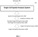
- Disclosed herein is a Single Cell Spatial Analysis System TM 100 that encodes the information of the spatial position 130 where a microsample 125 was collected from tissue or from a specimen 301 for single cell sequencing, proteomics, cellular analysis, metabolomics and other analysis modalities.
- the Single Cell Spatial Analysis System 100 will give researchers, clinicians, forensic scientists, and many other applications and disciplines, a fundamental new capability to understand how single cells function, combining two and three dimensional spatial information with gene expression, and/or sequencing information, cellular response to chemical/environmental challenges, protein expression, and other analysis methods.
- the Spatial Subsystem (also referred to herein as the "Spatial Preparation Subsystem” or “Spatial Preparation System” or “Spatial Preparation Module” ) 200, which collects microsamples 125, is an automated approach to collecting and generating single cells or groups of cells from microsamples 125 from matrices such as tissue without painstaking, lower throughput, or labor intensive manipulations, e.g ., Laser Capture Microdissection( Datta S. et. al. Histol Histopathol. 2015 Apr 20:11622 .), manual pipette collection ( Morris J. et. al. 2011. J. Vis. Exp. (50), e2634 ,) ( Kajiyama T. et.
- the Single Cell Spatial Analysis System can encode the position of where the microsample 125 was located in the tissue or specimen 301 by different methods for different analyses.
- the Single Cell Spatial Analysis System 100 can use primer sets with nucleic acid barcodes for spatial position attached to beads or other surfaces such as flow cells to encode the spatial position 130 into DNA.
- isotopes or other markers can be added to the microsamples 125 to encode the position while for enzyme activity assays fluorescent, Raman, optical, or other markers can be added to encode the spatial position 130.
- isotopes, fluorescent, or other markers can be added.
- This approach of encoding into the microsample 125 where it originated in three dimensions from the specimen 301 is a fundamentally new approach to prepare samples from single cells and to understand the genetics, gene and protein regulation, metabolism of individual cells, how they function in a 3D tissue or biofilm structure, and what cell types, including rare cells, are present and where among other scientific, clinical, and applied information.
- the Single Cell Spatial Analysis System 100 described can input raw, unprocessed samples, or other primary or secondary samples, and for genomic analysis produce either cDNA or prepared DNA libraries ready for DNA sequencing.
- the Single Cell Spatial Analysis System 100 for genomic assays has additional advantages over existing technology. Automated preparation of samples for NGS typically starts with purified bulk DNA or RNA and only part of the workflow is integrated, such as library preparation with manual steps of QC and post-library preparation amplification if needed. Automated sample preparation has not yet been integrated from raw samples, such as blood, tissue samples, fine needle aspirates, and other samples, to libraries ready for sequencing and remains largely manual. No automated NGS library preparation systems have yet been commercialized for single cells or for researchers and clinicians to routinely sequence cells from tissue. No system currently exists that collects single cells with positional information for nucleic acid analysis, or performs high throughput single cell RNA-Seq, or processes raw samples to libraries.
- a system that can integrate one or more of the overall steps to take samples from specimens (i.e ., tissue, biofilms, and other multi-dimensional matrices with cells or viruses) and prepare single cells, groups of cells, or cells and viruses (collectively or individually referred to as cells) to produce samples with information encoded about the cell's spatial positions in the original specimen.
- the cell's spatial position in the original specimen can be encoded in a marker, e.g., a spatial barcode, which is added to the specimen, a subregion, a microsample, or other part of a specimen, in a manner that encodes the single cell's position into the cell or components of the cell.
- the spatial position can be encoded by physical position of the sample as it is readout from a flow cell for NGS.
- microsamples from a subregion of a specimen are collected in physical order, such as a raster pattern or by rows or columns, and the microsamples placed in a known order into a fluidic stream or fixed wells or onto a surface.
- the term "microsample” is used to mean the smallest portion of the specimen that is collected as an individual sample that will be encoded with a single spatial marker or barcode and is the smallest unit sampled from the specimen by the system; microsamples may contain single cells to groups of cells.
- Single cells can be produced from the microsamples in microdrops, e.g., nanodroplets or boluses generated with a paramagnetic bead which has an attached oligonucleotide with a unique spatial DNA barcode, a type of spatial barcode, for the microsample.
- the beads with known unique spatial DNA barcodes are added in known order to the microsamples, which thereby will encode the order of the microsamples and cells in the microsamples and produce spatially encoded single cells. Additional barcodes may be unique for each cell or molecule or have quality control or other information.
- Nucleic acid is released from the single cell inside the nanodroplet or bolus and enzymology used to attach the oligonucleotide containing the spatial barcode to the cellular nucleic acid: this encodes the nucleic acid sequence to analyzed with the spatial DNA barcode on the oligonucleotide. After library preparation and DNA sequencing, the sequence of the spatial barcode is determined along with DNA or RNA sequence information. The spatial barcode, which was added in known order to microsamples that were also ordered in known order, can decode where in the specimen the microsample originated.
- Two and three dimensional spatial relationships of the microsamples and cells can then be determined and interpreted with the DNA or RNA sequence information to develop spatial information of what cells were present in the specimen, where the cells were located, the cell's DNA sequence and/or RNA expression, and other information.
- Different embodiments of the Single Cell Spatial Analysis System can encode the spatial information for decoding by analytical methods comprised of DNA sequencing, DNA microarrays, RNA sequencing, mass spectrometry, Raman spectroscopy, electrophysiology, flow cytometry, and many other analytical methods well known to one skilled in the art including multidimensional analysis.
- analytical methods comprised of DNA sequencing, DNA microarrays, RNA sequencing, mass spectrometry, Raman spectroscopy, electrophysiology, flow cytometry, and many other analytical methods well known to one skilled in the art including multidimensional analysis.
- the spatial position of where the cells originated from the specimen or tissue is encoded into the DNA sequence information, in one embodiment, by utilizing paramagnetic beads with a spatial barcode in an attached oligonucleotide that is unique for each cell/bead combination.
- the spatial barcode may be incorporated into the cDNA product by reverse transcriptase or RNA ligase if RNA is being analyzed, or into DNA by DNA polymerase or DNA ligase if DNA is being analyzed.
- the RNA from the specimen is processed into cDNA and then into sequencing libraries for (N)NGS analysis, while preserving the information of the spatial organization of the cells within the original specimen by attachment of a spatial encoding nucleic acid barcode.
- DNA is processed to a ready to sequence library; both RNA and DNA may be analyzed from a single cell, or nucleic acid and other properties, such as metabolites or proteomics, can be analyzed.
- next generation sequencing (NGS) or next-next generation sequencing (NNGS) refers to high-throughput sequencing, such as massively parallel sequencing, (e.g., simultaneously (or in rapid succession) sequencing any of at least 100,000, 1 million, 10 million, 100 million, or 1 billion polynucleotide molecules).
- Sequencing methods may include, but are not limited to: high-throughput sequencing, pyrosequencing, sequencing-by-synthesis, single-molecule sequencing, nanopore sequencing, semiconductor sequencing, sequencing-by-ligation, sequencing-by-hybridization, RNA-Seq (Illumina), Digital Gene Expression (Helicos), Next generation sequencing, Single Molecule Sequencing by Synthesis (SMSS) (Helicos), massively-parallel sequencing, Clonal Single Molecule Array (Solexa), shotgun sequencing, Maxam-Gilbert or Sanger sequencing, primer walking, sequencing using PacBio, SOLiD, Ion Torrent, Genius (GenapSys) or Nanopore (e.g., Oxford Nanopore) platforms and any other sequencing methods known in the art.
- SMSS Single Molecule Sequencing by Synthesis
- Solexa Solexa
- shotgun sequencing Maxam-Gilbert or Sanger sequencing
- primer walking sequencing using PacBio, SOLiD, Ion
- the Single Cell Spatial Analysis System described can have multiple subsystems and modules that perform processing or analysis.
- the Spatial Preparation Subsystem TM that inputs tissue and/or other specimens and outputs microsamples of single cell(s) encoded with cellular location information which may be DNA barcodes or other encoding methods.
- the first module, the Spatial Sampler TM module collects samples from defined areas of specimen(s) to provide single cells through a fluidic system in the order of their original spatial orientation to the second module, the Spatial Encoder TM module.
- cell imaging solutions such as cell specific antibodies, stains, or other reagents
- an optical or other imaging device scans the tissue in the Spatial Sampler module.
- the optical cellular image information can be used to decide which part or subregions of the specimen should be analyzed or to gather information to correlate with the downstream analysis.
- solutions comprised of antagonists, chemicals, biologicals, therapeutic drugs, or other compounds can be added to the tissue as needed before sampling and analysis.
- the Spatial Sampler module can then apply solution(s) to disassociate tissue or other specimens; as needed, the progression of dissociation can be monitored optically or by other means in the Spatial Sampler module.
- the Spatial Sampler module can apply solutions that encode the spatial position, comprised of combinations of isotopes or fluors, or marker molecules not present in the specimen, that are later decoded to identify where in the specimen the microsample originated.
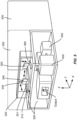
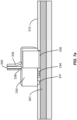
- a transfer device such as a multifunctional head, can then collect a layer from the specimen onto a surface such as a cell impermeable transfer membrane with vacuum applied.
- the transfer device can be moved to an input device for a fluidic system which has one or more fluidic channels, wells, or surfaces where microsamples from a subregion of the surface are transferred in physical positional order into the fluidic channel(s), well, or surface.
- fluidic channels or microchannels are used, the microsamples are entrained into a fluidic flow in known order that can be tracked to the physical position of the microsample in the original specimen.
- the channels may be capillaries, microchannels, micropipettes, or of other forms. Microsamples may be transferred one at a time or multiple samples transferred in parallel.
- the input device directly samples the specimen.
- the spatial position of where the cells originated from the tissue is encoded into the DNA sequence information in one embodiment by utilizing paramagnetic beads with an attached oligonucleotide with a spatial barcode unique for each cell/bead combination.
- the spatial barcode can be incorporated into the cDNA product by reverse transcriptase if RNA is being analyzed or into DNA from a primer by DNA polymerase or DNA ligase if DNA is being analyzed.
- the primer is attached to a solid surface such as a paramagnetic bead, microparticles, fibers, channel, wall, flowcell, membranes, test tubes, pipette tips or microwells or other surface.
- bead is used to encompass any solid surface, porous surface, or other implementations without limitation including flat surfaces.
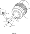
- the Spatial Encoder module integrates delivery of spatially barcoded beads entrained in known order with microdrops, e.g., nanofluidic droplet or bolus generation and sample preparation of DNA, RNA, or cDNA.
- the spatially encoded beads have DNA or other barcodes that identify which microsample is being analyzed and, by knowing the order the microsamples were taken and subsequent handling, the physical position of the microsample in the specimen is encoded.
- the spatially barcoded beads are added using a microfluidic nozzle to generate microdrops (nanodroplets or in others boluses are generated from the microsample in an immiscible fluid with preferably one spatially barcoded bead per droplet or bolus and only one cell.
- the nanodroplets or boluses may perform cellular lysis, mRNA binding, and cDNA reaction workflows. In others, cellular lysis, DNA binding, and DNA amplification reactions may be performed.
- a third module the Spatial Librarian Subsystem (also referred to as a "Spatial Librarian System” or “Spatial Librarian Module”) prepares an (N)NGS library for nucleic acid analysis from the microdrops (nanodroplets or boluses).
- the nanodroplets or boluses are processed separately or in the preferred embodiment the nanodroplets or boluses are pooled after spatial encoding to process many single cells and microsamples simultaneously.
- the Spatial Librarian module performs the necessary biochemistry to add adapters as needed, prepare an (N)NGS sequencing library, amplify as necessary, and perform quality control of the library.
- the analytical function such as DNA sequencing, is incorporated to create a sample-to-answer system.
- a system comprising: (i) a biological specimen; and (ii) added to each of a plurality of different microsamples from the biological specimen, a marker comprising spatial information that encodes the original spatial position of the microsample within the biological specimen.
- the biological specimen comprises human tissue, animal tissue, or plant tissue, a biopsy, a cellular conglomerate, an organ fragment, an organism, whole blood, bone marrow, biome, a biofilm, a fine needle aspirate or any other solid, semi-solid, gelatinous, or frozen three dimensional or two dimensional matrix of biological nature.
- the microsamples comprise a single cell or a plurality of cells.
- the marker comprises a polynucleotide.
- the nucleic acid is bound to a membrane, chip surface, bead, surface, flow cell, or particle or is indirectly bound via an adapter molecule e.g., a complementary nucleic acid or a chemical crosslinker.
- the marker comprises a peptide, antibody, protein, small molecule, isotope such as lanthanide, Raman marker, mass tag, fluorescent or chemiluminescent probe.
- the microsamples are dissociated from the biological specimen.
- the microsamples are entrained in microdrops in a fluidic stream.
- the microsamples are supported by at least one substrate, e.g., a membrane.
- a further example not forming subject matter claimed herein includes a device for the analysis of a biological sample, the device comprising: a sample module configured to extract microsamples from a biological specimen; and a recipient module configured to receive the microsample biological specimen from the sample module for analysis.
- the recipient module performs a downstream analysis selected from nucleic acid sequencing, next generation sequencing, next next generation sequencing, proteomic, genomic, gene expression, gene mapping, carbohydrate characterization and profiling, lipid characterization and profiling, flow cytometry, imaging, microarray, metabolic profiling, functional, or mass spectrometry or combinations thereof.
- a further example not forming subject matter claimed herein includes a device comprising: an element selected from a membrane, filter, surface, capillary, microchannel, device, and microfabricated chip; and means to bring the element into direct contact or close proximity to a biological specimen for the purpose of labeling or extracting a plurality of microsamples in an order based on their original spatial position within the biological specimen.
- a further example not forming subject matter claimed herein includes a system comprising: a stage for supporting a biological specimen; a device comprising an array of markers comprised in beads, surfaces, flat or microfabricated structures; means for transferring the array of markers into or onto the biological specimen at predetermined spatial positions.
- the method of the invention comprises: adding, to each of a plurality of different microsamples from a biological specimen, a marker comprising spatial information that encodes the original spatial position of the microsample within the biological specimen.
- the method further comprises dissociating the microsamples from the biological specimen.
- the method comprises adding the markers to the microsamples before dissociating the microsamples from the biological specimen.
- the method comprises adding the markers to the microsamples after dissociating the microsamples from the biological specimen.
- each microsample comprises a single cell.
- each microsample comprises a plurality of cells.
- dissociating the microsamples comprises extracting the microsamples in a raster pattern across the biological specimen. In another embodiment the microsamples are dissociated in a 3-D pattern. In another embodiment dissociating comprises contacting the biological specimen with a membrane, applying vacuum to the membrane to hold a layer comprising the microsamples; and removing the microsamples held by the membrane from the biological specimen. In another embodiment the method comprises removing a second layer of the microsamples from the biological specimen after a first layer is removed. In another embodiment the method further comprises moving the dissociated microsamples into a fluidic stream.
- the method comprises the microsamples are moved into the fluidic stream in an order correlated with their original spatial position in the biological specimen.
- the method comprises microsamples are incorporated into microdrops (e.g., nanodroplets or boluses) in the fluidic stream.
- the method comprises the microdrops contain one or more beads.
- the method comprises the beads are paramagnetic.
- the method comprises the beads are functionalized with oligonucleotides comprising the spatial information in the form of a nucleotide barcode.
- the method comprises the nucleotide barcode is unique for each cell or group of cells in the microsample.
- the method comprises the oligonucleotide comprises barcodes for cellular, molecular, or quality control purposes.
- the method comprises the nucleic acid of the sample component including but not limited to groups of cells or single cells is enzymatically combined with the oligonucleotide of the bead.
- the method comprises the nucleic acid is subjected to library preparation and nucleic acid sequencing.
- the method comprises the oligonucleotide further comprises a poly T tail, and the method comprises capturing mRNA molecules from the microsamples having a poly T tail; and reverse transcribing the mRNA molecules to produce cDNA molecules comprising the barcode where the barcode provides the spatial information.
- the method comprises the oligonucleotide further comprises a capture sequence complementary to a target sequence, and the method comprises capturing DNA molecules from the microsample having the target sequence; and extending the oligonucleotide to produce a nucleic acid molecule having a copy of the target sequence and comprising the barcode, wherein the barcode provides the spatial information.
- the method comprises dissociating comprises contacting the biological sample with a cell dissociation solution comprising at least one protease that digests extracellular matrix.
- the method comprises the at least one protease is selected from collagenases, elastase, trypsin, papain, hyaluronidase, chymotrypsin, neutral protease, clostripain, caseinase, neutral protease (Dispase ® ), DNAse, protease XIV.
- the method comprises the cell dissociation solution is in the form of a fluid, mist, fog, or aerosol applied to the biological sample.
- the method further comprises decoding the spatial information in the microsamples to determine the original spatial position of each microsamples.
- a further example not forming subject matter claimed herein includes a method comprising: providing a biological specimen; collecting microsamples from each of a plurality of different spatial positions in the biological specimen; attaching to nucleic acids in each microsample a marker comprising a nucleic acid barcode comprising spatial information that encodes the original spatial position of the microsample within the biological specimen, thereby producing spatial encoded nucleic acids; sequencing the spatial encoded nucleic acids; and based on the spatial information attached to each spatial encoded nucleic acids, determining the original spatial location of the nucleic acid in the biological specimen.
- the method comprises sequencing spatial encoding nucleic acids combined from a plurality of different microsamples in a single high throughput sequencing run.
- a further example not forming subject matter claimed herein includes a system having a graphical user interface that presents, based on spatial information obtained from microsamples of a biological specimen, a graphical representation of the biological specimen including original spatial position of a plurality of polynucleotides or polypeptides in the biological specimen.
- a spatial preparation system configured to entrain in a fluidic stream a plurality of cell-containing microsamples from a biological specimen, wherein the microsamples are contained in spatially separated microdrops in a fluidic stream and positioned in an order based on their original spatial position within the biological specimen
- the system comprises: a) a spatial sampler subsystem configured to extract a plurality of microsamples comprising cells from different original spatial positions in a biological specimen; and b) a spatial encoder subsystem comprising one or more spatial encoder microchannels, each having an inlet and an outlet; wherein the spatial sampler subsystem delivers the microsamples to the spatial encoder microchannel inlets in a predetermined order based on their original spatial position in the biological specimen, and the spatial encoder subsystem incorporates the microsamples into spatially separated microdrops in a fluidic stream.
- the spatial sampler subsystem comprises: (1) a specimen holder, and (2) a multifunctional head comprising a transfer head comprising one or more extraction channels, wherein the extraction channels communicate with a liquid source and, optionally, a gas source, each under positive and/or negative pressure, and wherein the one or more extraction channels comprise ends covered with one or more air permeable, cell impermeable transfer membranes, and wherein, the multifunctional head is mounted on a three axis stage to position the multifunctional head to extract, by contact adhesion or by vacuum, the microsamples from the specimen holder onto the one or more transfer membranes; and (ii) the spatial encoder subsystem comprises: (1) a microdroplet generator comprising a source of immiscible liquid in communication with each spatial encoder microchannel at a junction, wherein mixture of the immiscible liquid with the fluidic stream at the junction forms spatially separated microdrops comprising the microsamples; and (2) optionally, a microsample encoder
- the multifunctional head further comprises a dispense head configured to dispense liquids, e.g., imaging reagents or dissociation solution, onto the biological specimen.
- the transfer head comprises a plurality of extraction channels where in the extraction channels are arrayed in a two dimensional array (e.g., a line) or a three-dimensional array (e.g., a plane).
- the spatial encoder subsystem comprises a plurality of fluidic channels that merge into the encoder channel in which each has an inlet configured to receive the microsamples from an extraction channels.
- the transfer membranes have attached thereto a plurality of capture elements, each capture element comprising a particle, which is optionally paramagnetic, having attached thereto one or more antibodies that bind into cells in the biological specimen, and nucleic acid markers comprising positional barcodes comprising spatial information where the spatial information calling to the position of the particle on the multifunctional head.
- nucleic acid markers further comprise cell markers identifying the cell to which particle binds, and/or molecular barcodes that differently label different nucleic acid molecules and a single cell.
- a spatial analysis system comprising: a) a spatial preparation subsystem as disclosed herein, and b) a spatial librarian subsystem configured to perform a series of biochemical reactions on an emulsion comprising microdrops produced by the spatial preparation subsystem, wherein the spatial librarian subsystem comprises: a) a reaction device comprising an inlet configured to receive microdrops from the spatial preparation subsystem, at least one reaction chamber, and an outlet; b) a reagent rail communicating with the reaction device through a microchannel and comprising reagent sufficient to perform at least one of biochemical reaction on analytes in the microdrops; and c) one or more pumps configured to move the reagents from the reagent rail through the microchannel to the reaction chamber of the reaction device.
- the spatial librarian subsystem further comprises: c) a temperature controller configured to control temperature in the reaction chamber. In another embodiment the spatial librarian subsystem further comprising: c) a magnet configured to reversibly immobilize paramagnetic particles contained in the reaction chamber.
- the biochemical reactions comprise at least: (i) reverse transcription of messenger RNA into cDNA; and (ii) amplification of cDNA. In another embodiment the biochemical reactions comprise at least: (i) primer extension of a primer hybridized to a DNA template to create an extension product; and (ii) amplification of the extension product.
- a method comprising entraining in a fluidic stream a plurality of microsamples from a biological specimen, wherein the microsamples are contained in spatially separated microdrops in the fluidic stream and positioned in an order based on their original spatial position within the biological specimen.
- the method comprises: a) providing a biological specimen; b) collecting microsamples from each of a plurality of different spatial positions in the biological specimen; c) introducing the microsamples in a predetermined order into a fluidic stream in a fluidic channel; d) dividing the fluidic stream into microdrops by introducing boluses of immiscible liquid into the fluidic channel, whereby the microsamples are incorporated into microdrops that are spatially separated from each other in the fluidic stream.
- the method further comprises: (i) introducing into the fluidic stream a plurality of different spatial markers encoding spatial information, wherein the different spatial markers are incorporated into different microdrops in the fluidic stream, thereby encoding each microdrop with spatial information.
- the analytes comprise nucleic acids
- the method further comprises: (e) combining microdrops in a container in the form of an emulsion; (f) generating spatially tagged nucleic acids by tagging nucleic acid analytes with the nucleic acid barcodes; (g) breaking the emulsion; (h) amplifying the tagged nucleic acids.
- the analytes comprise polyadenylated mRNA and the nucleic acid markers further comprise polyT tail
- generating spatially tagged nucleic acids comprises: hybridizing the polyT tail to polyadenylated mRNA nucleic acid markers to the mRNA molecules barcodes and reverse transcribing the polyadenylated messenger RNA to produce that spatially tagged cDNA molecules; performing second strand synthesis on the spatially tagged cDNA molecules to produce tagged double stranded cDNA molecules.
- the analytes comprise DNA molecules and the nucleic acid markers further comprise a nucleotide sequence complementary to a target sequence, and generating spatially tagged nucleic acids comprises:hybridizing the complementary nucleotide sequence to a target sequence in the nucleic acid molecules and extending the nucleic acid markers to produce a double-stranded DNA molecule.
- the method further comprises: applying imaging reagent to the biological sample; imaging the biological sample to which the imaging reagent has been applied; based on the imaging selecting features of interest at predetermined spatial positions in the biological sample; and extracting the microsamples including the selected features of interest.
- the method further comprises, based on spatial information encoded in the microsamples, determining the initial spatial position of the selected features in the biological specimen.
- NGS information, mass spectrometry and other modern high-throughput analysis systems have revolutionized life and medical sciences.
- these and other high-throughput analysis systems fail to retain spatial information about where in the specimen the sample or microsample originated. It is anticipated that single cell spatial information, or spatial information from groups of cells, of genomic, proteomic including protein expression, carbohydrate, lipid, and metabolism of individual cells will provide fundamental scientific knowledge and revolutionize new research and clinical capacities.
- the terms “comprises,” “comprising,” “includes,” “including,” “has,” “having” or any other variation thereof, are intended to cover a non-exclusive inclusion.
- a process, method, article, or apparatus that comprises a list of features is not necessarily limited only to those features but may include other features not expressly listed or inherent to such process, method, article, or apparatus.
- “or” refers to an inclusive-or and not to an exclusive-or. For example, a condition A or B is satisfied by any one of the following: A is true (or present) and B is false (or not present), A is false (or not present) and B is true (or present), and both A and B are true (or present).
- Specimen refers to an in vitro cell, cell culture, virus, bacterial cell, fungal cell, plant cell, bodily sample, or tissue sample that contains genetic material.
- the genetic material of the specimen comprises RNA.
- the genetic material of the specimen is DNA, or both RNA and DNA.
- the genetic material is modified.
- a tissue specimen includes a cell isolated from a subject.
- a subject includes any organism from which a specimen can be isolated. Non-limiting examples of organisms include prokaryotes, eukaryotes or archaebacteria, including bacteria, fungi, animals, plants, or protists.
- the animal can be a mammal or a non-mammal.
- the mammal can be, for example, a rabbit, dog, pig, cow, horse, human, or a rodent such as a mouse or rat.
- the tissue specimen is a human tissue sample.
- the tissue specimen can be, for example, a blood sample.
- the blood sample can be whole blood or a blood product (e.g., red blood cells, white blood cells, platelets, plasma, serum).
- the specimen in other non-limiting embodiments, can be saliva, a cheek, throat, or nasal swab, a fine needle aspirate, a tissue print, cerebral spinal fluid, mucus, lymph, feces, urine, skin, spinal fluid, peritoneal fluid, lymphatic fluid, aqueous or vitreous humor, synovial fluid, tears, semen, seminal fluid, vaginal fluids, pulmonary effusion, serosal fluid, organs, bronchio-alveolar lavage, tumors, frozen cells, or constituents or components of in vitro cell cultures.
- the tissue specimen is a solid tissue sample or a frozen tissue sample.
- the specimen comprises a virus, bacteria, or fungus.
- the specimen can be an ex vivo tissue or sample or a specimen obtained by laser capture microdissection.
- the specimen can be a fixed specimen, including as set forth by U.S. Published Patent Application No. 2003/0170617 filed Jan. 28, 2003 .
- biomolecules including one or more polynucleotides or polypeptides are spatially encoded.
- the polynucleotide can include a single-stranded or double-stranded polynucleotide.
- the polypeptide can include an enzyme, antigen, hormone or antibody.
- the one or more biomolecules can include RNA, mRNA, cDNA, DNA, genomic DNA, microRNA, long noncoding RNA, ribosomal RNA, transfer RNA, chloroplast DNA, mitochondrial DNA, or other nucleic acids.
- the Single Cell Spatial Analysis System 100 accepts specimens 301 and processes one or more microsamples 125 to encode the physical location of the microsample 125 within the specimen 301 to produce spatially encoded single cells 1000 in microdrops, e.g., nanodroplets or boluses, by adding a marker such as a DNA barcode that encodes for the location of the microsample 125.
- a marker such as a DNA barcode that encodes for the location of the microsample 125.
- Known markers are added in known order to ordered microsamples 125 to encode the spatial position.
- Analysis method 1100 such as DNA sequencing, mass spectrometry, or other analytical methods, generates single cell analytical data 1600, such as DNA sequence or proteomics, and also decodes the encoded spatial information to produce decoded single cell spatial information 1500.
- the downstream processes can include without limitation, nucleic acid sequencing, targeted resequencing, genotyping analysis, mutation analysis, copy number variation assessment, allele frequency assessment, plasmid construction, cloning, and the like.
- the decoded single cell spatial information 1500 identifies the spatial position 130 where microsample 125 originated in specimen 301.
- the single cell analytical data 1600 is analyzed by single cell spatial analysis 2000 software to produce single cell spatial information 3000 which has the analytical data and associated physical position information of the microsample 125.
- the Single Cell Spatial Analysis System 100 is in some embodiments an automated sample preparation system that collects cells from areas of tissue, biofilms, and other matrices containing biological material and generates spatially-encoded samples for analysis of single cells 1000 and groups of cells from the same subregion.
- the Single Cell Spatial Analysis System 100 can have pushbutton operation for either specialists or non-specialists to generate samples with spatial information encoded from specimens for medical, health, life science research, and other applications.
- the Single Cell Spatial Analysis System 100 in one embodiment has a Spatial Subsystem 200 that inputs specimens and outputs samples for spatially encoded single cells 1000.
- the Spatial Preparation Subsystem 200 can accommodate many different types of specimens, comprised of fresh and snap-frozen tissue in the form of microtome slices (cryo, laser or vibrating); bulk material obtained by surgical excision, biopsies, fine needle aspirates; samples from surfaces, and other matrices. The specimens are positioned onto the sampling stage.
- Subregions 150 will then be addressed automatically by a multifunctional head 330 to transfer subregions 150 of microsamples 125 in 2-D spatial order to generate single cells and process them into cDNA or DNA with a spatial nucleic acid barcode, or other spatial markers added, or other analytes, e.g., proteins, metabolites, enzymes, with isotopes or other spatial markers added.
- the next physical layer of the specimen 301 subregion of interest can then be collected and analyzed to generate a second 2-D spatial patterns of DNA sequence, RNA expression, protein expression, protein activity, RNA activity, lipid composition and abundance, carbohydrate composition and abundance, and metabolites as well as any other biological components.
- the third physical layer can then be collected and analyzed.
- the physical layers can then be oriented to produce a three dimensional pattern.
- the basic elements of the Single Cell Spatial Analysis System 100 can be configured in multiple ways depending on the specimen(s) and analytes to be analyzed. In the following examples, a few of the numerous configurations are described in detail but in no way is the invention limited to these configurations as will be obvious to one skilled in the art. Nonetheless in many configurations, there are many common elements, in particular adding a barcode or other exogenous material to the microsample 125, or to single cells or groups of single cells from the microsample 125 to encode where physically from the specimen the microsample 125 originated.
- the barcode can be decoded and the physical position of the microsample 125 in the 3-D structure of the specimen used to understand the composition, function, cell type, activity, genetics, physiology, interconnections, and other attributes of the specimen with single cell spatial information 3000.
- the Single Cell Spatial Analysis System 100 has two subsystems, the Spatial Preparation Subsystem 200 and the Spatial Librarian Subsystem 500.
- the Spatial Preparation Subsystem 200 can in turn be composed of two modules, the Spatial Sampler module 300 and the Spatial Encoder 400, as shown in Figure 4 .
- Spatial Sampler module 300 (i) releases cells from tissue, (ii) collects, and (iii) transfers single cells or groups of cells with defined spatial relationships in a known order into a single fluidic or microfluidic process stream, or onto a surface such as a flow cell, or into a two-dimensional array of wells, or other ordered or unordered processes.
- the output of the Spatial Sampler 300 is single cells in a microfluidic flow in a known order.
- Spatial Encoder module 400 adds spatial barcodes to the sample and outputs the single cells with spatial barcodes.
- Figure 4 shows Spatial Encoder module 400 configured for DNA sequencing.
- specimen 301 is placed in a specimen holder 310 which is inserted into the Spatial Sampler module 300 by loading mechanism 305.
- Specimen holder 310 may be temperature controlled.
- the loading mechanism can have a mechanical slide, stage, pneumatic actuator, or other mechanism, that accepts specimen holder 310 and moves it into the Single Cell Analysis System 100, either automated or manually, into a fixed position in the Spatial Sampler module 300 or other mechanism.
- Spatial Sampler module 300 can have a three axis stage 320 that moves a multifunctional head in the x, y and z directions 330 to collect microsamples 125 in a spatially defined manner from specimen 301 at capture position 345 and dispenses the collected microsamples 125 at dispense position 350 into input device 360.
- the fluidics of the Single Cell Spatial Analysis System 100 are incorporated onto cartridge(s) 4000.
- the valves for the subsystems are microvalves, which in some embodiments are created in microchips with microchannels. Microvalves are well known to those skilled in the art. Microvalves can be actuated by, for example, mechanical force, pneumatic pressure, electrostatic force, piezoelectric force, or thermal expansion force. They may be have internal or external actuators. Pneumatic valves include, for example, diaphragm valves that employ a flexible membrane of the pneumatic pressure or vacuum to close or open a fluid channel.
- Electrostatic valves may include, for example, a polysilicon membrane or a polyimide cantilever that is operable to cover a hole formed in a substrate.
- Piezoelectric valves may include external (or internal) piezoelectric disks that expand against a valve actuator.
- Thermal expansion valves may include a sealed pressure chamber bounded by a diaphragm. Heating the chamber causes the diaphragm to expand against a valve seat.
- Microvalves can comprise mechanical (thermopneumatic, pneumatic, and shape memory alloy), non-mechanical (hydrogel, sol-gel, paraffin, and ice), and external (modular built-in, pneumatic, and non-pneumatic) microvalves (as described in C. Zhang, D. Xing, and Y. Li., Micropumps, microvalves, and micromixers within PCR microfluidic chips: Advances and trends. Biotechnology Advances. Volume 25, Issue 5, September-October 2007, Pages 483-514 ; D ⁇ az-González M., C. Fernández-Sánchez, and A. Baldi A. Multiple actuation microvalves in wax microfluidics. Lab Chip.
- cartridges 4000 are used with functionality from a group of reagents, valves or microvalves, microchannels, syringe pumps, optical devices, integrated electronics for control of cartridge 4000 functions, and lot tracking.
- microchips are used as parts of instrument or cartridges 4000.
- the cartridges 4000 in some embodiments hold kits to perform the chemistries including all needed reagents, including spatial barcodes attached to beads, emulsion oil, breaking solution, stains, library preparation chemistry, and other consumables.
- the multifunctional head 330 can have multiple functions integrated into a single head as shown in Figure 6 or multiple heads that are moved independently can be used.
- the multifunctional head can have an optics head 331 that in a preferred embodiment has illumination fiber 332 surrounded by multiple collection fibers 333.
- the illumination and collection fibers can have lenses to focus the illumination beam(s), such as a laser, arc lamp, UV lamp, or other light source, and collect reflected, fluorescent, Raman, reflected, or other light for delivery to an optics analysis device via optical fiber bundle 338. Many other optical configurations may be used or no optical capability may be used.
- Multifunctional head 330 can have a dispense head 334 with one or more dispensers 335 that can apply one or more solutions to specimen 301.
- the dispense head 334 may use piezoelectric pumps, inkjet technology, pressure driven flow through a nozzle, or other methods to dispense liquids, gels, or gases from fluidics and pneumatic bundle 339 to specimen 301 or subregions 150 of specimen 301.
- Multifunctional head 330 can have a transfer head for extracting the microsamples from a biological specimen and transferring them, e.g., into a fluidic stream.
- the transfer head can comprise a transfer membrane 336 to transfer a layer of the specimen.
- a transfer membrane 336 may be made of many different materials with characteristics that prevent cells passing through it but in preferred embodiments can be permeable to air or liquids, have low adhesion for collected cells, and can be changed as needed.
- the transfer membrane may be selected from many materials, including semipermeable membranes; filter papers comprising cellulose and cellulose derivatives, glass fiber filters, polytetrafluoroethylene (PTFE) filters, quartz fiber filters and others; blotting membranes comprising membranes made of nitrocellulose such as ProTran, polyvinylidene difluoride (PVDF) such as Immobilon ® or Hybond, nylon, cellulose nitrate, polyestersulfone, track etched polycarbonate, PVC, and others with pore sizes smaller than the cells of interest.
- semipermeable membranes comprising cellulose and cellulose derivatives, glass fiber filters, polytetrafluoroethylene (PTFE) filters, quartz fiber filters and others
- blotting membranes comprising membranes made of nitrocellulose such as ProTran, polyvinylidene difluoride (PVDF) such as Immobilon ® or Hybond, nylon, cellulose nitrate, polyestersulfone, track etched polycarbonate, PVC, and others
- the multifunctional head 330 may be able to apply vacuum, pressure, or fluids to the whole membrane through fluidic and pneumatic bundle 339 either to the complete surface of the membrane at the bottom of the multifunctional head or to portions of the membrane.
- portions of the membrane are individually addressable subregions 337 to enable subregions of the specimen 301 to be collected or dispensed individually.
- the transfer membrane 336 can be changed through swapping out a disposable fixed membrane head, or by advancing a roll of membrane to have unused membrane, or other mechanisms. In some embodiments, the transfer membrane may be reused multiple times and not changeable.
- the transfer membrane 336 is simply cleaned by separate cleaning modules that operate in direct contact or non-contact mode utilizing various cleaning mechanisms including, but not limited to, mechanical brushes and chemical agents such as ethanol, detergents, water, buffers, and other chemicals.
- various cleaning mechanisms including, but not limited to, mechanical brushes and chemical agents such as ethanol, detergents, water, buffers, and other chemicals.
- transfer membrane 336 is used but is not intended to limit the composition or material which can be a filter, membrane, surface, film, or other material.
- the Single Cell Spatial Analysis System Spatial Sampler module 300 can function as follows in one embodiment.
- the multifunctional head 330 is moved to the x, y directions to capture position 345.
- the optics head 331 scan the specimen by the multifunctional head 330 moving to the appropriate z height and being moved in the x-y axis to scan specimen 301.
- solutions can be dispensed onto specimen 301 or portions of the specimen by dispense head 334.
- the solutions can be imaging reagents, such as fluorescently directly conjugated antibodies or secondary antibodies, stains, fluorescent probes and dyes; imaging nanomaterials (including quantum dots and other nanoparticles), or other contrast or straining reagents; or reagents to stimulate or treat specimen 301 such as anticancer compounds, antibiotics, antivirals, energy sources, or other liquids, gels, or gases for the same or other functions.
- the information gathered by the optics or other sensors is used to decide which subregions 150 of the specimen are to be sampled or to decide the next step, such as adding another reagent, scanning at a different wavelength, or another action.
- the transfer membrane 336 portion of the multifunctional head 330 may be used to aspirate excess reagent or all unbound reagents.
- the dispense head 334 can use a dispenser 335 to apply a dissociation solution, such as Liberase TM DH Research Grade Roche or equivalent products or custom formulations, in form of a spray, mist, or liquid to a region of the top most layer of the tissue to dissociate intercellular adhesion between cells and free the cells from the extracellular matrix.
- a dissociation solution comprises at least one collegenase or protease that digests extracellular matrix (F. E Dwulet and M.E. Smith, "Enzyme composition for tissue dissociation," United States Patent 5,952,215, September 14, 1999 ).
- the dissociation solutions to dissociate the tissue can be comprised of collagenases (e.g.
- the dissociation solution can be applied in one embodiment such that only the uppermost layers of cells are dissociated or deeper layer are dissociated depending on the amount and concentration of the dissociation solution, time of contact, temperature, and other parameters.
- samples are collected from the specimen by moving multifunctional head 330 in the x and y axes to the appropriate subregion 150 of specimen 301 and then multifunctional head 330 is lowered in the z direction to contact specimen 301 in specimen holder 310 with the transfer membrane 336.
- Cells from the top layer of the dissociated tissue are gently picked up by vacuum, contact, absorption, and other methods to air-permeable, cell-impermeable, transfer membrane 336 located at the bottom of the multifunctional head 330 for sample transfer; this can be similar to vacuum assisted blotting when vacuum is used.
- the transfer membrane 336 limits the amount of transfer to preferably less than 20 um or less than 100 ⁇ m of tissue thickness and minimizes contamination by replacing the surface of the transfer membrane 336 with fresh membrane material between sequential sampling steps or cleaning.
- an array of individually addressable vacuum/pressure or fluidic lines behind the membrane can be used to selectively apply vacuum to individually addressable subregions 337 of the membrane.
- the multifunctional head 330 is then raised in the z direction, moved in the x and y direction as needed to dispense position 350, and lowered to contact the input device 360.
- Figure 7b shows multifunctional head 330 about to contact input device 360.
- Input device 360 may contact all of the transferred specimen 302 or a subregion 150 of the transfer membrane 336.
- the input device 360 is an array of microfluidic channels. It will be apparent to one skilled in the art that the fluidics and microfluidics may be implemented in different forms including capillaries, molded microchannels, embossed microchannels, etched microchannels, and many other formats in many materials including plastics, glass, silicon dioxide, metals, and others.
- Figure 7b shows a closeup of two ends of the input microchannels 382 held in connectors 316 about to contact the material transferred from specimen 301.
- Figure 8a shows a bottom view of multifunctional head 330 that has transferred parts of specimen 302 onto transfer membrane 336 after contact by input device 360.
- Subregion 150 is outlined as the portion of the transfer membrane 336 that will be transferred into input device 360 in this example.
- Figure 8a shows 12 microsamples 125 from the spatial positions 130 (shown as black circles) have been transferred onto transfer membrane 336 and then into input microchannels 382.
- the transfer into input microchannels 382 may be any number of microsamples 125 at a time.
- a 2 x 6 array of input microchannels 382 produces the pattern of microsamples 125 from spatial positions 130 shown in Figure 8a in one input step.
- the second input step can produce a pattern of 24 microsamples 125 from spatial positions 130 that have been transferred as illustrated in Figure 8b .
- Two more inputs steps would complete the input of 48 microsamples from subregion 150.
- the whole surface of transfer membrane 336 is used to transfer cells each time.
- the first pass of sampling microsamples 125 from subregion 150 of transferred specimen 302 may be followed by a second pass to transfer of cells from a second layer deep of transferred specimen 302 into input device 360.
- a different subregion 150 of transferred specimen 302 may be input in the second transfer.
- the multifunctional head 330 can be moved to capture position 345 and another sample from specimen 301 collected from the same or different region of specimen 301 by transfer membrane 336; in some instances, additional optic information is gathered, the specimen treated with reagents by dispensers 335, or other activities may be performed before or after sampling specimen 301.
- Input device 360 can adopt many different configurations.
- Figure 8 shows the microsamples transferred to input device 360 that are circular in shape, like a capillary or other round microchannel would produce.
- the inside diameter (ID) can be matched to the size of the cells or material to be sampled.
- Capillaries could be used with, for example, 5 ⁇ m ID, or 20 ⁇ m ID, or 100 ⁇ m or 100s of ⁇ ms to sample from different matrices.
- a 20 ⁇ m ID capillary can be used to sample mammalian tissue for single cells and input a microsample 125 from 30 ⁇ m deep for example; this microsample 125 could contain a single or multiple cells.
- a 100 ⁇ m ID capillary can be used to create microsamples 125 that could be 75 ⁇ m deep, for example, and might contain 10s or more cells.
- Other shapes than circular will enable sampling with different resultant patterns such as rectangular, triangular, and other shapes.
- the number of samples collected from transfer membrane 336 will depend on the number of input microchannels 382 in input device 360. In some embodiments it is a single input microchannel 382 and in other embodiments two or more microchannels that may be arranged in one or more rows and columns. In some embodiments input device 360 collects microsamples 125 from the complete surface of transfer membrane 336 simultaneously.
- the pattern of transfer of microsamples 125 from transferred specimen 302 is known and where in the subregion 150 input device 360 contacted transferred specimens 302 is known, therefore the spatial position 130 of where in the transferred specimen 302 each microsample 382 originated is known; therefore the Single Cell Spatial Analysis System 100 can keep track of the order of each microsample 125 until it is encoded with a marker or barcode to spatially encoded single cells 1000.
- the microsamples 125, cells, or groups of cells can then be pooled if desired.
- the barcode is read out to produce Decoded Single Cell Spatial Data 1500. Decoding the barcode then determines from which microsample 125 each single cell orginated since known barcodes were added to ordered microsamples 125 in known order. Since the spatial position 130 of each microsample 125 is known, the three dimensional or two dimensional position of the single cells and groups of cells in multiple microsamples is known.
- Single Cell Spatial Analysis 2000 software analyzes the spatial position information with the spatial position 130 of each microsample 125 with Single Cell Analytical Data 1600 to produce Single Cell Spatial Information 3000.
- Figure 9 shows one configuration of a single channel implementation of the input device 360.
- the input device 360 connects of input microchannel 382 through two three-way valves to one or more syringe pumps.
- the end of input microchannel 382 contacts the transfer membrane 336 through an optional filter 381 such as a 50 ⁇ m stainless steel mesh to break up any clumps of cells and remove debris.
- Syringe pump 365 pulls reagents from reagent rail 366 using three-way valve 364 and fluidic channel 367 from one of the reagents, 369, 371, 373, and 375 with selection controlled by valves 368, 370, 372, and 374.
- syringe pump 365 pulls buffer, e.g ., 100 nL of PBS buffer, from buffer reservoir 369 through fluidic channel 367 by opening buffer valve 368 and three-way valve 364; a sample loop can be placed in between syringe pump 365 and three-way valve 364 or other locations as needed.
- Three way valve 364 can be replaced with valves such as 3 way or more way valves.
- syringe pump 365 pushes the buffer through three-way valve 364 through defining microchannel 363, three-way valve 362, input microchannel 361, the end of input microchannel 382 and through filter 381 to contact the portion of the transferred specimen 302 on transfer membrane 336.
- Pressure or liquid can be supplied through fluidic and pneumatic bundle 339 to individually addressable subregions 337 of the areas that match input device 360 to help elute microsamples 125 off of the transfer membrane 336 and into input device 360 as needed.
- syringe pump 365 can pull the buffer and the transferred cells from transferred specimen 302, and moving it into input device 360, through filter 381, end of input microchannel 382, input microchannel 361, three-way valve 362, and into defining microchannel 363 which is flanked by three way valves 364 and 362 to create a microsample 125 in the fluidic stream.
- Three way valve 362 is then changed to connect defining microchannel 363 to connecting microchannel 376 and flow channel 380 and syringe pump 365 used to push the microsample 125 from defining microchannel 363 through three way valve 362 and connecting microchannel 376 and flow channel 380.
- Valve 379 may be closed to direct the flow in the direction of the arrow, towards the Sampler Encoder module 400 or other downstream processing.
- Flow movement in flow channel 380 can also be accomplished using flow syringe pump 390 accessing flow reagent rail 391 through flow three way valve 393 to push fluids or gases to move the bolus(es) entrained in flow channel 380.
- One or more optional bolus detector 396 can detect the edges of boluses using optical measurement, conductance, or other methods well known to one skilled in the art.
- the bolus detectors can be used to align the additional encoding beads downstream as needed.
- the length of the bolus containing the microsample 125 can be defined by the volume in defining microchannel 363 which is flanked by three way valves 364 and 362, and the length and cross-sectional area of defining microchannel 363.
- the volume of the bolus is 100 nL, but volumes can range from subnanoliters to 10s of microliters or larger.
- the bolus can be flanked by many materials including air, immiscible fluids (such as FluorInert ® ; mineral oil; silicone-based oil; fluorinated oils; Droplet Generation Oil (Biorad, #1863005); emulsion oil (Life Technologies, Part No.
- immiscible fluids such as FluorInert ® ; mineral oil; silicone-based oil; fluorinated oils; Droplet Generation Oil (Biorad, #1863005); emulsion oil (Life Technologies, Part No.
- Figure 10 shows an embodiment with the input device(s) 360 having two channels of input, through end of input microchannels 382 and 392.
- the two or more channels can be operated by two or more syringes 365 and 395 as shown in Figure 10 or the microchannels can be ganged together to operate from a single syringe in parallel.
- the two channels may have separate reagent rails 366 and 387 or a single reagent rail.
- Multiple channels of input may have a single or reagent rails, for example, a bank of 16 input devices 360 could have two reagent rails, each of which services eight input channels.
- RNA sequencing preparation in one reagent rail and DNA targeted sequencing reagents in a second reagent rail.
- DNA targeted sequencing reagents in a second reagent rail.
- one reagent such as 371 contain a dissociation solution to further loosen cellular attachments to extracellular matrix material.
- the dissociation solution or other solutions could be applied through the fluidics to a microregion of the sample on transfer membrane 336 before transfer of the microsample 125 or be used as the fluid to transport the microsamples to continue to dissociate any clumps.
- one reagent can hold an immiscible fluid such as FluorInert, mineral oil, Droplet Generation Oil (Biorad, #1863005) or other fluids.
- an immiscible fluid such as FluorInert, mineral oil, Droplet Generation Oil (Biorad, #1863005) or other fluids.
- a cleaning reagent to remove debris, disinfect, flush, and other functions can be used in reagent 375.
- the number of reagents may be as low as one and be as large as needed for the application without limitation.
- the reagents are in syringes operated by syringe pumps.
- the reagents are in pouches, tubes, wells, or other containers and are moved by a pump or micropump such as a piezopump, e.g. a Bartels mp6, Dolomite Piezoelectric Pump 3200138, or others.
- the reagents and other components of the Single Cell Spatial Analysis System 100 may be in cartridges 4000 that are readily changed by non-specialists.
- transfer membrane 336 When transfer membrane 336 is moved against the input device 360, if two or more end of input microchannels 382 and 392 are used, they may be directly adjacent or with known spacing. In a preferred embodiment the ends are adjacent. When the input microchannels are not adjacent, the transfer membrane 336 can be moved against the input device 360 for multiple samplings of the transfer membrane 336. For example, if the input device has microchannels of 20 ⁇ m ID and spacing between microchannels of 200 ⁇ m, then the multifunction head 330 could move ten times to pull in samples from the complete subregion 150 if it is linear. Similarly, multifunction head 330 could raster or use other patterns to insure that all of the transfer membrane 336 is sampled when desired.
- Microsamples 125 for multiple input microchannels may be processed into defining microchannels 363 and 394 in parallel or independently.
- three way valves 362 and 399 are changed to connect defining microchannels 363 and 394 to connecting microchannels 376 and 383 to flow channels 380 and output microchannel 386.
- Valve 379 may be closed to direct the flow in the direction of the arrow, leading to the Sampler Encoder module 400 or other downstream processing.
- the microsample 125 in defining microchannel 394 is pushed by syringe pump 395 into output microchannel 386 and then the upstream microsample 125 in defining microchannel 363 is pushed by syringe pump 365 into flow channel 380 to place the two samples in a known order, separated by the volumes and initial material in connecting microchannel 376 and flow channel 380.
- the microsamples 125 may be moved simultaneously.
- the multifunctional head or other device moves the transfer membrane 336 to a dispense position where released cells are pulled in the order of the original 2-D spatial position into end of input microchannels 382 and 392 on input device 360, driven initially by syringe pumps 365 and 395.
- the top row could be input column by column.
- Boluses of air, immiscible fluids, or other gases or liquids can be introduced to separate microsamples 125 as needed. Cells are aligned in order and output in a single fluidic or microfluidic stream into the Spatial Encoder module 400.
- a transfer device with a membrane with a single microchannel can be implemented using a single capillary, e.g ., 20 ⁇ m ID capillary (Polymicro Technologies), which in a preferred embodiment is epoxied in a f iber o ptic- l ike c apillary (FROLC) connector with a membrane attached to one end, held in a moveable fixture; the other end of the capillary is attached through a three way valve using a FROLC connector to defining microchannel 363 of 100 nL to reagent rail 366 and syringe pump 365 as a vacuum and pressure source.
- the single 20 ⁇ m ID capillary may be able to sample about 1 nL from specimen 301.
- the FROLC connectors shown in Figure 11 , are true zero dead volume connectors and can join two or more capillaries to one, and be used as multiway microvalves.
- U.S. Patent 6,551,839 Jovanovich, S., I. Blaga, and R. McIntosh. Integrated system with modular microfluidic components. US Patent 7,244,961.
- FROLCs can be made by using a fiberoptic FC-, or other fiberoptic connectors 311 with cleaved capillaries plugged with wax or other materials, and then inserted into face 312, epoxy filled, polished to a dome like a fiber optic connector, and the wax removed.
- a spring in the connector provides compression to seal capillaries for liquid flow at >9,600 psi. Rotation of the connector can change the orientation of, for example, three capillaries to rout sample streams into any of three output capillaries.
- FROLC connectors can also be made from other designs than fiberoptic connectors in a matter that allows leak-free connection between one or more capillaries or microchannels.
- a FROLC connector is shown in Figure 11a with three capillaries 313, 314, and 315, that are visible in Figure 11b .
- Membranes such as transfer membrane 336 or filters on input devices, such as a 20 -40 um range filters or other materials can be mounted to the FC stainless surface over the end of the capillary in the FROLC ( Figure 11c ).
- a collar to simplify mounting the membrane can be made with 3-D printing or other methods. Collection and dispensing requirements of different membranes can be evaluated for the ability to 'grab' the top layer from loosened tissue and to dispense into the input device 360.
- Membrane filters can be from commercial sources with different characteristic, such as material, coating, and pore size.
- the amount of vacuum to pickup and hold, pressure to dispense, efficiency in pickup and delivery, and the depth of tissue picked up can be optimized for different membranes and specimens; the part of the FC connector without the capillary can be used as a control region when evaluating transfer membrane 336 candidates.
- Membranes can be examined under brightfield and, after fluorescent cell stains, with fluorescence microscopy for testing of capture and dispensing. Selected samples can be assayed with real-time PCR to develop the process and the requirements for repeatedly collecting cells onto a surface and then dispensing using a single capillary, and provide first insight into repeated sampling to generate 3-D spatial information.
- a microsample 125 can be pulled into a single channel with a capillary (e.g., 75 ⁇ m ID) with FROLC connectors on both ends.
- a capillary e.g. 75 ⁇ m ID
- One end will contact the transfer membrane and might optionally have a filter or mesh on its outer surface to breakup cell clumps; the other end is a FROLC three way valve that is in turn attached to the defining microchannel 363 which can have FROLC at both ends with the FROLCS used as three way microvalves to connect at the upstream end to the input device end 382 and to a capillary as the connecting microchannel 376 and the other end of defining microchannel 363 through a FROLC three way valve to a syringe pump 365 and a reagent rail 366. Buffer, ⁇ 100 nL, will be delivered to the tip of the capillary and manually contacted to the transfer membrane area of the multifunctional head 330. Transfer protocols can be optimized for requirements for pulling samples into the input device, timing, and fluidic process.
- the defining microchannel 363 can readily be a volume such as 100 nL, or larger or smaller.
- a 100 nL is an approximately 7 cm long bolus in a 40 ⁇ m ID capillary or 2.5 cm long bolus in a 75 ⁇ m ID capillary-sufficient length to be defined between two FROLC or other microvalves. If needed, the 100 nL sample can have immiscible fluid added to create a larger volume to manipulate.
- the ability to input a sample and then rout into the microfluidic flow can be tested with fluorescent beads and then cell suspensions. Multiple samples of alternating types (i.e ., rat cells followed by murine cells) can be input and output to the fluidics stream to measure cross contamination rates.
- the connecting microchannel 376 can be used as the output line for the Spatial Sample module (microchannels 378 and 380, and valve 379 are not used).
- microchannels 378 and 380, and valve 379 are not used.
- the sample transfer portion of the multifunctional head 330 simplifies to a transfer membrane 336 with vacuum control. If dispensing requires pressure sequentially applied to individual subregions of the membrane, then to achieve high throughput the transfer device can be an array device that input collects and dispenses multiple samples at a time into a 'mirrored' input format, i.e ., if the transfer device used 8x12 channels, the input device 360 would use 8x12 channels.
- Figure 12 shows 12 capillaries 317 arrayed in a linear FROLC connector 316.
- the transfer region of the multifunctional head will have a membrane in front of the 12 capillaries to collect samples, each capillary individually addressable to deliver pressure to dispense the 12 microsamples 125 if needed.
- the input device 360 can be another linear array of 12 capillaries 317 in inear FROLC connector 316, covered with a 30 ⁇ m filter, connected to a ganged syringe pump an array of 12 fluidic circuits.
- the transfer region of the multifunctional head does not have a membrane in front of the 12 capillaries to collect samples.
- the Single Cell Spatial Analysis System Spatial Encoder module 400 inputs the ordered microsamples 125 which may contain single cells or groups of cells from the Spatial Sampler module 300 using the Spatial Sampler output such as output microchannel 386 as an input to the Spatial Encoder module 400.
- the spatial encoder subsystem can place the microsamples in a sequential ordered arrangement in a fluidic stream. This sequential arrangement is referred to as a train, and microsamples in a train are said to be entrained.
- nucleic acid encoding adds beads with known barcodes to correlate with the original spatial information into boluses, creates microdrops, (nanodroplets in some implementations, or boluses in other implementations), preferably with one or less cells per nanodroplet.
- a bolus is typically elongate in shape, while a nandroplet is typically spherical.
- a bolus typically has a volume of at least 3 microliters.
- a nanodrop typically has a volume of no more than 3 microliters, e.g., about 1.5 microliters.
- the Spatial Encoder module 400 outputs single cells with spatial barcodes in nanodroplets or boluses to the Spatial Librarian Subsystem 500.
- One embodiment of spatial barcoding is to use beads with oligonucleotides with spatial barcodes 680 is illustrated in Figure 13a .
- the beads with oligonucleotides with spatial barcodes 680 can be paramagnetic beads, agarose beads, or others, and have surface chemistry optimized for the nucleic acid capture and subsequent chemistries.
- Oligonucleotides with spatial barcodes 601 can be generated by synthesis using standard commercially available phosphoramide or other technology.
- the oligonucleotide has a cleavable linker 602, attached to an amplification primer 604 with fluorescent label 603, a sequencing primer 605, barcode region 606, and capture region 610.
- the barcode region is comprised of a spatial barcode 607, cellular barcode 608, and molecular barcode 609.
- the spatial barcode 607 can be 5 nucleotides long to provide 1,024 barcodes for spatial resolution.
- Cellular barcodes 608 can be 6 nucleotides, or other lengths, and synthesized by split-pool synthesis, and molecular barcode 609 can be 8 nucleotides synthesized by degenerate synthesis; the cellular barcodes 608 only need to identify cells from within a single microsample 125 since each microsample is encoded with a spatial barcode.
- each spatial barcode 607 is unique for each set of microsamples 125 that are analyzed together.
- spatial barcode 607 can be shared between microsamples 125 and then resolved bioinformatically using cellular barcode 608 to sort and cluster by cells to resolve spatial barcode ambiguities.
- Spatial, cellular, and molecular barcodes can be of different lengths or in different orders, or dispersed among other elements of the oligonucleotide with spatial barcode 50 without limitation.
- spatial barcoding is to attach oligonucleotides with spatial barcodes 601 to surfaces 660 as illustrated in Figure 13b , or flowcells 670.
- the surface 660 then can be used in similar ways to the beads. It will be obvious to one skilled in the art that surface 660 could be assembled into a flow cell.
- the oligonucleotides can be initially attached to commercially available paramagnetic beads 630 by covalent crosslinking and may include a cleavable linker bond ( Ju. J. et. al. United States Patent 9,133,511. September 15, 2015 .), ( Knapp D.C. et. al. Bioconjug Chem. 2010; 21(6):1043-55 .), (https://www.clickchemistrytools.com/products/click_chernistry_toolbox), ( Olejnik J. et. al. Nucleic Acids Res. 1996;24(2):361-6 .).
- Fluorescent probes can be attached to the oligonucleotide distal from the bead and cleavable bond or alternatively fluorescent nucleic acid base analogs can be used such as 2-Aminopurine ( Wilhelmsson, Quarterley Reviews of Biophysics, 43, 2, 2010, 159-183 ).
- the cleavage of labeled oligonucleotides can be used for assay development since the oligonucleotide can be analyzed by fragment sizing on CE with the fluorescent tag to give the distribution of sizes to assess library quality.
- the Spatial Encoder module 400 must deliver a set of beads with a unique, known, spatial barcode 607 to each microsample 125.
- the Spatial Encoding fluidic delivery can use a spatial barcode reagent rail 401 to access beads each with a single spatial barcode, for example 403, 405, 407, and 409, controlled by spatial barcode reagent rail valves 402, 404, 406, and 408 respectively, to deliver reagents through by spatial barcode fluidic channel 410 to spatial barcode syringe pump 412. It is within the scope that the spatially barcoded beads will scale to 1,024 or greater number of spatial barcodes per run for the Single Cell Spatial Analysis System.
- Spatial barcode three way valve 411 and spatial barcode reagent rail 401 can be implemented with many other variations, including having spatial barcode reagent rail valves 402, 404, 406, and 408 each being multiway valves, such as 8-way valves that in turn access 8 sets of spatial barcode reagents, or additional valves be added to the reagent rail.
- the multiway valves can be FROLC or other microvalves, molded microfluidic valves, valves on microchips, or other embodiments.
- An alternative approach is an ordered series of boluses of beads with different spatial codes that are merged with the microsample 125 boluses in known order.
- reagent rail syringe pump 412 and spatial barcode reagent rail 401 can select a reagent of singly spatially barcoded beads, such as beads all with a single spatial barcode 403, and delivers a bolus of beads 403 through spatial barcode connecting channel 413 to spatial encoder junction 414 to merge with the microsample 125 in spatial encoder microfluidic device 420.
- Optical, conductance, or other sensors can be incorporated as needed to detect the microsample 125 in the bolus and coordinate the addition of the spatially barcoded beads to the bolus.
- the bolus then passes through spatial encoder microchannel 417 to nozzle 429 where an immiscible fluid such as FluorInert, Droplet Generation Oil (Biorad, #1863005), or other solutions can be added by nanodroplet generation syringe pumps 425 and 415 to the bolus to produce nanodroplets, preferably 1.5 nL, and sent down spatial encoder output microchannel 430 as output from the Spatial Encoder.
- an immiscible fluid such as FluorInert, Droplet Generation Oil (Biorad, #1863005), or other solutions can be added by nanodroplet generation syringe pumps 425 and 415 to the bolus to produce nanodroplets, preferably 1.5 nL, and sent down spatial encoder output microchannel 430 as output from the Spatial Encoder.
- Nanodroplet generation syringe pumps 415 and 425 can also be combined into one syringe pump that has two microchannels 416 and 426 that split in two from nanodroplet generation syringe pump output to join the microsample 125 with barcoded bead bolus from either side to produce nanodroplets, eliminating the need for a second nanodroplet generation syringe pump.
- Nozzle designs and circuits are described by ( Macosko E.Z. et. al. Cell. 2015;161(5):1202-14 .) ( Klein A. M. et. al. Cell. 2015;161(5):1187-201 .) ( Geng T. et. al. Anal Chem. 2014;86(1):703-12 ).
- the microsample 125 bolus with the added spatially barcoded beads are processed as a bolus without the formation of nanodroplets.
- the bolus may be preferably less than 5 nL, or 10 nL, or 25 nL, or 100 nL, or 250 nL,and 10,000 microsamples 125 may be less than 2.5 mL.
- single channel fluidics are used.
- a bolus of , for example, 100 nL of beads with one spatial code are added in junction 414 to the, for example,100 nL of microsample 125 in output microchannel 386 and lysis and/or reaction mixtures, such as lysis/reverse transcriptase mix, added separately through spatial encoder reagent syringe pump 418 and reagent connecting microchannel 419.
- Monodispersed nanodroplets from single cells with spatially coded beads with lysis and/or reaction mixtures e.g., lysis/RT mix for RNA-Seq, lysis/restriction mix for DNA sequencing, are then be produced using a nozzle 429 ( Macosko E.Z. et. al. Cell. 2015;161(5):1202-14 .) ( Klein A. M. et. al. Cell. 2015;161 (5):1187-201 .) ( Geng T. et. al. Anal Chem. 2014;86(1):703-12 .) and output through spatial encoder output microchannel 430.
- the geometry and flow rates can be altered to adjust size and flow rates to produce a Poisson distribution of single cells with each nanodroplet preferably having a spatially barcoded bead.
- the bolus from the Spatial Sampler module 300 is physically separated by structures, volumes, or surfaces, for example, by placing the bolus into a microtiter or smaller well or tube.
- the Spatial Sampler module 300 output can be used to be physically dispersed onto the surface of a material comprised of agar, membranes, arrays of beads, microscope slides, flow cells, and others.
- the physical dispersion can be by moving the surface under a capillary or other flow, by printing with a microarray pen, by piezospraying, electrowetting, microfluidics, or other methods.
- the physically separated microsample 125 bolus can be dispersed such that, for example on the surface of agar, all cells are far enough apart to be processed as single cells.
- spatial encoder reagent syringe pump 418 adds a low melting temperature agarose to encapsule the nanodroplet with heated liquified agarose ( Geng T. et. al. Anal Chem. 2014;86(1):703-12 .) during the formation of nanodroplets.
- heated liquified agarose Geng T. et. al. Anal Chem. 2014;86(1):703-12 .
- the agarose can be used as a barrier permeable to low molecular weight components, such as reaction components, but not to high molecular weight components such as nucleic acids when it is cooled.
- the use of agarose to encapsulate the reactions enables multiple sequential reactions or manipulations in a row to be performed in the nanodroplet.
- the Spatial Librarian Subsystem 500 receives the output of the Spatial Preparation Subsystem 200 and can process the microsamples 125 including single cells and groups of cells from a single spatial position 130 of the specimen with a spatial barcode added.
- the Spatial Librarian Subsystem 500 can perform enzymatic reactions, chemical reactions, and purifications, quality control, and other functions.
- the output from the Spatial Encoder module 400 is input into the Spatial Librarian Subsystem 500 to reaction device 520.
- the microdrops e.g., nanodroplets or boluses
- the microdrops are pooled in reaction chamber 523 and reagents added from spatial librarian reagent rail 501 accessing spatial librarian reagents 503, 505, 507, and 509 through spatial librarian valves 502, 504, 506, and 508 respectively to allow access through spatial librarian reagent rail connecting microchannel 510 with spatial librarian syringe pump 512 and spatial librarian three-way valve 511.
- Reagents can be added to reaction chamber 523 to mix with pooled microdroplets.
- reaction device 520 is a flowthrough system. Waste can be flowed into waste line 526 as needed. Temperature can be controlled including thermal cycling by temperature control device 521 which can include sensors and heaters well known to one skilled in the art including resistive heating, infrared heating, flowing air or water, and other methods.
- Optional optic device 522 can monitor reactions such as quantitative polymerase chain reaction (qPCR), reverse transcriptase-PCR, Raman spectroscopy, or other optical measurements including fiberoptic delivery and collection of light from lasers, filters, imaging devices such as CCD and CMOS, and other optical methods.
- Quality control device 525 can assess the quality of the processing integrated devices such as capillary electrophoresis with laser induced fluorescence to determine the amount and the size distribution of fragments in the library, mass spectrometry, Raman, electrochemistry, or other devices.
- the spatial librarian can break or create emulsions by accessing reagents on spatial librarian reagent rail 501.
- many standard methods can be used including adding isobutanol ("Idiot-proof emulsion PCR", Lab Times, 1-2011, p50); isopropanol and detergent buffer (10 mM Tris pH 7.5, 1 mM EDTA pH 8.0, 100 mM NaCl, 1% (v/v) Triton X-100, 1% (w/v) SDS); water-saturated diethyl ether; salt including sodium pyrophosphate; or demulsifiers such as Dow Corning ® DM4 Demulsifier or others can be added.
- an organic extraction solvent selected from the group consisting of: butanol, octanol, hexanol and chloroform
- a breaking solution includes sodium dodecyl sulfate (SDS) or phosphate buffered saline (PBS), further including an inorganic salt in the aqueous phase prior to forming the breaking solution wherein the inorganic salt is selected from the group consisting of potassium chloride, potassium acetate, sodium chloride, sodium acetate, lithium chloride, lithium acetate, Na2S04, potassium carbonate, ammonium sulfate, and ammonium acetate ( Jeffrey Sabina, Ilya Zlatkovsky, Rachel Kasinskas, "Methods and kits for breaking emulsions" WO 2012138926 A1, Published Oct 11, 2012 ) or.
- SDS sodium dodecyl sulfate
- PBS phosphate buffered saline
- the solution can be allowed to separate by gravity and then the bottom phase collected with spatial librarian syringe 512 with connection line 513 positioned in the bottom of reaction chamber 523.
- the top phase can be discarded through waste line 526.
- a centrifuge device is included in reaction device 520 to separate emulsion phases, with a pipetting device to withdraw the aqueous phase from the bottom of the centrifuge tube.
- the appropriate emulsion oil is selected by spatial librarian reagent rail 501 and added to the reaction chamber 523 with a vigorous back and forth motion of the solution in the reaction chamber 523.
- a moveable magnet device 524 can position a magnet near reaction chamber 523 to collect paramagnetic beads to the surface of the reaction chamber 523 or retract a magnet to release paramagnetic beads as required.
- a magnetic separation He J. et. al. J Pharm Biomed Anal. 2014; 101:84-101 .
- a magnetic separation can be used to change the buffer in a reaction, to remove residual traces of emulsion oil or breaking solution, to remove an enzyme, to concentrate a product, or to capture nucleic acid or other components onto beads for ease of handling.
- the surface chemistries of the paramagnetic beads and conditions to precipitate, wash, and elute nucleic acids and other biomolecules onto surfaces is well understood, ( Boom, W. R. et. al.
- the beads and attached biomolecules can be separated from solution without the additive of a precipitation solution such as a crowding agent, e.g ., polyethylene glycol, ethanol, etc. If the biomolecules are not attached to paramagnetic beads, reagent rail 501 can add the appropriate precipitation solution(s) to reaction chamber 523 to force the biomolecule of interest onto the surface of the paramagnetic bead.
- a precipitation solution such as a crowding agent, e.g ., polyethylene glycol, ethanol, etc.
- a magnetic separation can be performed using moveable magnet device 524 to position a magnet near reaction chamber 523 to collect the paramagnetic beads to a surface of reaction chamber 523 and the reaction mix pumped to waste line 526.
- a wash solution such as 80% ethanol, is added by reagent rail 501 and the beads released by retracting the magnet.
- the wash solution can include Triton X-100 (Octylphenol ethylene oxide condensate).
- the wash solution can include about 0.01% to about 5% Triton X-100.
- the wash solution can include about 0.05% to about 1% Triton-X.
- the wash solution can include Tris-HCI or Tris-EDTA.
- the beads can be agitated by pumping, magnetic stirrers, bubbling air, or other methods.
- the beads are recaptured by using moveable magnet device 524 to position a magnet near reaction chamber 523 to collect the paramagnetic beads to a surface of the reaction chamber 523 and the wash solution pumped to waste line 526. This can be repeated with the same or different wash solutions as needed to purify the reaction products, to remove unincorporated reactants, to remove emulsion oil or breaking solution, or other reasons.
- the biomolecule of interest can be associated with the bead or with the solution phase as desired.
- reaction chamber 523 can be heated to activate or inactivate enzymes.
- other devices and functionality can be added to reaction device 520 including a centrifuge device or a pipetting device.
- the nanodroplet or bolus generation was performed with a reversible gelling agent such as ultra low gelling agarose as taught by Geng T. et. al. Anal Chem. 2014;86(1):703-12 .
- a reversible gelling agent such as ultra low gelling agarose as taught by Geng T. et. al. Anal Chem. 2014;86(1):703-12 .
- low melting agarose is added to the microsample before nozzle 429 and nanodroplets are produced with at times a single cell and a single barcoded bead with the cells compartmentalized within nanoliter agarose droplets.
- the nanoliter agarose droplets are transformed into microgels by cooling.
- the agarose microgels allow small molecules to diffuse into and out of the nanodroplets but retains macromolecules such as DNA. This enables lysis and removal of PCR inhibitors and PCR amplification (as taught by Geng T. and R.A. Mathies Forensic Sci
- the utility of the nanoliter agarose droplets can be further extended in the instant disclosure to couple multiple reactions in a row.
- the microgels can be harvested and reagents infused.
- the substrates and enzymes can be changed between reactions, such as a DNA library construction; see "Library Preparation for Double-Stranded DNA example.”
- spatial analysis of polyadenylated mRNA from single cells and tissue can be performed in nanoliter agarose droplets by lysing cells, capturing mRNA, reverse transcriptase, followed by second strand synthesis.
- precipitation agents are diffused into the nanoliter agarose droplets to force precipitation for separations onto the bead inside the nanoliter agarose droplets if the biomolecule of interest is not attached to the bead. Unbound materials can diffuse out.
- markers comprised of internal markers, standards, nucleic acids, enzymes, chemicals such as isotopes, mass tags, or ratios or mixtures of enzymes, fluorescent markers, Raman markers, optical markers, carbohydrates, lipids, other biomolecules, or chemicals, are added to the specimen in known order by dispensers 335 on the multifunctional head 330 or alternatively by direct transfer from prefabricated arrays 900 of markers 901 on a support carrier, backing surface or membrane (see Figure 17 ) attached to and maneuvered by the multifunctional head 330. The markers are later analyzed to decode the spatial position in the specimen of the microsample 125.
- the decoding can be an orthogonal method to the analytic method for the analyte of interest, i.e ., a DNA marker could be used to decode spatial information while the sample analysis might be by mass spectrometry.
- the markers are attached to antibodies or other bioaffinity agents such as to exterior cell surface epitopes.
- the markers are attached to motifs, compounds, or structures that are transported, electroporated, or otherwise enter into the cells. The markers can be used to identify the spatial position of the microsample 125 and cell for many different types of analyses comprising metabolic characterization and profiling, proteomics, genomics, gene expression, carbohydrate characterization and profiling, lipid characterization and profiling, and combinations of analyses.
- the markers depicted by letters A to P are each unique and arranged in patches 901 to form a two dimensional array 900.
- These markers can be physisorbed as mono- or multilayers onto the surface of a membrane or support material which is connected to the multifunctional head 330.
- the markers can be arrayed in form of beads or contained in micro/nanowell architectures, or immersed in hydrogels or other surfaces 660 or structures.
- the array is brought into direct contact with the specimen 301 resulting in the transfer of marker reagents onto the topmost layer of the specimen thereby encoding the spatial order information directly onto the cells.
- the patch-to-cell registry shown in Figure 17 is an idealized version. It is more likely that in certain cases multiple adjacent markers will be transferred to the same cell or one marker will be transferred to multiple adjacent cells which can be used to further define boundaries between patches.
- a dispenser(s) 335 or array 900 on the multifunctional head 330 can apply different solutions of markers to each microsample 125 on the specimen.
- markers For example, mass isotopes or ratios of mass isotopes can be added, and, after microsample 125 is processed in the Spatial Sampler 300 to produce single cells, the isotopes can be determined by mass spectrometry to determine where in the sample the cell or cells originated.
- the markers are added as barcodes in the Spatial Encoder module 400.
- nucleic acids or other markers can be added to the specimen in known order by dispensers 335 or an array 900 on the multifunctional head 330.
- the nucleic acids can be electroporated into the specimen by addition of suitable electrodes and buffer, applied to the surface, or designed to be uptaken by the cells.
- Nucleic acid sequences can be added that are not found in the organisms being studied and their signature used to decode the spatial position of microsample 125.
- an affinity label such as biotin
- the mating affinity label such as streptavidin
- One embodiment uses a physical array of paramagnetic beads that are pre-arrayed on a Collection multifunctional head 330 with positional information encoded in the primers attached to the beads with oligonucleotides with spatial barcodes 680. This approach greatly simplifies engineering approaches for the Single Cell Spatial Analysis System 100.
- the Spatial Sampler module 300 uses the two-dimensional stage 320 with a multifunctional head 330. Fluidic dispensers 335 first applies solution(s) to tissue to dissolve intercellular adhesion.
- the transfer membrane 336 is replaced with a collection head that has an array 900 of paramagnetic beads 630 each with a single spatial barcode and in a known order.
- the beads can be attached by cleavable linker 620 to backing surface 621, alternatively held by magnets located behind backing surface 621 or arrayed and attached by other methods.
- each bead can have two types of biologicals attached: antibodies to binding cell surface markers 651, 652, and 653 which can all be the same or different and each individual bead has a DNA oligonucleotide with a unique spatial barcode 640, 641, and 642, and optional barcodes for cellular identity and molecular identity, and other elements comprised of sequencing primer, poly T sequence when capturing polyadenylated mRNA, targeted capture sequence, or other elements.
- Each bead has a different spatial barcode marker and is placed in a known order upon backing surface 621. Decoding the spatial barcode in turn allows the sequenced DNA or RNA to be traced back to its original location in the specimen.
- the collection head with backing surface 621 of the multifunctional head 330 is pressed against the top layer of the tissue and cells bound to the beads by antibodies 651, 652, and 653.
- the multifunctional head 330 is moved to the cell input port of the Spatial Encoder module 300 and the linkers cleaved (or the magnet removed) to deposit all of the beads into the input port simultaneously or in groups, with gas pressure applied if needed.
- Individual portions do not need to be dispensed because the spatial barcode on the beads, which are attached to the cells through the antibody, can be used to decode the position of the microsample 125 after amplification.
- This embodiment obviates the need for spatial barcode reagent rail 401 to add spatially barcoded beads to the microsamples 125.
- this approach requires a more complicated manufacturing process to generate the arrays of spatially encoded beads.
- the Single Cell Spatial Analysis System 100 encodes the physical location of microsamples 125 within the specimen 301 to produce spatially encoded single cells 1000.
- analysis method 1100 such as DNA sequencing, mass spectrometry, Raman spectroscopy, or other analytical methods
- single cell analytical data 1600 is produced of analytes of interest, such as DNA sequence of target sequences, and at the same time the encoded spatial information from each sequence can be decoded to produce decoded single cell spatial information 1500 which identifies the spatial position 130 where microsample 125 originated in specimen 301.
- the decoded single cell spatial information 1500 can be, for example, 5 nucleotides or ratios of isotopes, or other decoding of markers.
- the single cell analytical data 1600 is analyzed by single cell spatial analysis 2000 software to produce single cell spatial information 3000 which has the analytical data and associated physical position information of the microsample 125.
- scripts can sort reads by the spatial barcode 607 and reconstruct, for example, the DNA sequence or expression patterns from individual cells from the specimen by position in three dimensions. Types of cells, activity, gene expression, mutation, networks of genes, and expression patterns can be mapped to two or three dimensional spatial coordinates. Information from optical or other measured properties can be combined and represented visually with two and three dimensional plots of the origin of the microsample and the activity measured, genomic, proteomic, metabolomics, systems biology, etc.
- the data analysis can build upon existing analytic platforms.
- downstream (N)NGS workflow can use existing (N)NGS sequencing analysis and bioinformatics.
- the quality of the data is assessed and, optionally, the reads trimmed and those with poor quality filtered out.
- High quality reads are aligned to the reference genome or transcriptome using one of the many available high-throughput sequencing mapping tools. Alignments are assembled into full-length transcripts or contigs based on a reference genome. For gene expression, the aligned reads are subsequently passed to quantification tools to obtain a measure of expression.
- several differential analyses can be executed to identify differentially expressed genes and transcripts.
- the spatial barcode 607 information in either the same analysis or an orthogonal analysis are used to decode the origin of the same and provide input into the creation of single cell spatial information 3000, which combines single cell analytical data with the spatial data.
- Single Cell Spatial Analysis System 100 Many applications are enabled by the Single Cell Spatial Analysis System 100.
- the capability of analyzing the identity, genetic sequence, gene expression, proteomic and metabolomic profiles of specimens and how groups of cells function in three dimensional matrices can be applied to forensics, molecular diagnostics, pathology, cell biology, cell discovery, histology and many other applied areas to understanding of how tissue functions.
- Spatial analysis of tissue at the single cell level will provide extraordinary insight and understanding, tissue composition of different cell types, structure, and how cells function in a microenvironment.
- Animal, plant, and microbial communities, such as biomes and films, will be important applications and how biomes change at interfaces of different organs and microenvironments.
- the Single Cell Spatial Analysis System 100 can be applied to molecular diagnostics where spatial analysis of biopsies can provide information about the state of the tissue, subregions 150 of activity, such as cancerous growths, and sources of qualified material for treatment; many raw and processed samples can be addressed including blood, forensic samples, tissue samples, fine needle aspirates and many others.
- spatial analysis of biopsies can provide information about the state of the tissue, subregions 150 of activity, such as cancerous growths, and sources of qualified material for treatment; many raw and processed samples can be addressed including blood, forensic samples, tissue samples, fine needle aspirates and many others.
- the production of single cells with spatial barcodes is discussed. It will be clear to one skilled in the art that ensemble measurements of groups of cells with the same spatial barcodes, such as for a microsample 125, is within the scope of the present disclosure.
- modules examples are discussed in the following sections. Many other permutations, workflows, modules, devices, and combinations are possible. The names of the modules and subsystems are not limiting and functionality can be distributed differently between modules.
- FIG. 19 A preferred workflow for mRNA to encode spatial position of the microsample 125 into DNA is shown in Figure 19 .
- the workflow begins with sampling from tissue or other specimens 301 to collect microsamples 125, i.e ., cells and groups of cells, in known physical order, one layer at a time; the layer can be a single layer of cells or multiple layers of cells.
- the microsample 125 is then input into a fluidic flow system in known order in the Spatial Sampler module 300.
- the microsample 125 and beads pass through nozzle 429 which then generates nanodroplets or small boluses each with preferably a single bead and one or less cells.
- the Spatial Librarian Subsystem 500 the cells are lyzed, and the released mRNA captured onto the barcoded oligonucleotide on the bead 680.
- a reverse transcription reaction is performed either in the bolus or after pooling nanodroplets or boluses.
- the reverse transcription reaction uses the oligonucleotide as a primer and synthesizes complementary DNA (cDNA) to the mRNA sequence attached to one strand of the spatial encoded oligonucleotide, thereby covalently linking the cDNA to the DNA of the oligonucleotide. Since the oligonucleotide contains a spatial barcode, the cDNA now has a spatial barcode which can be read out by DNA sequencing after library preparation.
- cDNA complementary DNA
- the microsample 125 can be output into a well such as a microtiter plate or test tube, or into a flow cell, or other ordered substrate in known order from the Spatial Sampler module 300.
- a spatially encoded bead or set of beads all with the same spatial barcode is added to the microsample 125 or to single cells or groups of cells from the microsample 125.
- the spatial encoding can be on nucleic acid attached to the surface of a flowcell, or on a hydrogel, or on the interior of a capillary or microchannel, or other surfaces or matrices; these alternatives are all within the scope of the present disclosure when the term bead is used.
- a preferred embodiment for spatial analysis of mRNA is described in more detail.
- single channel fluidics are used. Referring to Figure 20 , a bolus of 100 nL of spatially barcoded oligonucleotide-functionalized beads 680 with a poly T sequence as the capture region 610 with a single spatial barcode is added in junction 414 to 100 nL of microsample 125 from spatial sampler output microchannel 386 and lysis/reverse transcriptase mix added separately through spatial encoder reagent syringe pump 418 and reagent connecting microchannel 419; the lysis conditions and reverse transcriptase described by ( Fekete R. A. and A. Nguyen. United States Patent 8,288,106. October 16, 2012 ).
- Monodisperse nanodroplets from single cells with spatially coded beads with lysis/RT mix are produced using a nozzle 429 ( Macosko E.Z. et. al. Cell. 2015;161(5):1202-14 .) ( Klein A. M. et. al. Cell. 2015;161(5):1187-201 .) ( Geng T. et. al. Anal Chem. 2014;86(1):703-12 ) and output through spatial encoder output microchannel 430.
- the nanodroplets or boluses with a distribution of beads and single cells can be input in the Spatial Librarian Subsystem 500.
- Cells are lysed by heating or other methods and polyadenylated mRNA 681 captured onto the oligonucleotide to form captured mRNA structure 682.
- the oligonucleotides 601 are barcoded for spatial barcode information 607 as well as cellular barcodes 608 and molecule barcodes 609.
- Amplification primer 604 and sequencing primer 605 may be included on the oligonucleotide, or may be added in downstream library preparation methods as needed.
- the amplification primers can be for T7 polymerase for amplified RNA production ( Van Gelder R.N. et. al. Proc Natl Acad Sci USA. 1990; 87(5):1663-7 .), PCR, rolling circle transcription-based amplification, rapid amplification of cDNA ends, continuous flow amplification, and other amplification methods.
- RNA or cDNA can be performed using methods comprised of chemical, biochemical, and physical methods.
- the reverse transcription reactions can be assayed by qPCR using reagents added from spatial librarian reagent rail 501 to reaction chamber 523 with thermal cycling from temperature control device 521 with optic device 522 monitoring the qPCR reaction.
- cDNA produced by cleaving the fluorescently tagged cDNA can be analyzed by QC device 525 with capillary electrophoresis.
- External polyadenylated transcripts 250-2,000 nt in length at a 10 6 range of concentrations can be added from spatial librarian reagent rail 501 to assess the dynamic range and range of detection with NGS analysis (ERCC RNA Spike in Controls, 4456740, Life Technology).
- Alternative preferred embodiments include performing an RNA ligase reaction to covalently join the mRNA to one strand of the double stranded oligonucleotide after lysis and capture of the mRNA onto spatially barcoded oligonucleotide-functionalized beads 680 with a poly T sequence as the capture region 610, or ligating RNA to a single stranded RNA or DNA attached to the bead.
- the emulsion is broken at this stage in a demulsification step.
- the emulsion can be broken, for example by heating the mixture, acidification, centrifugation and ultrasonic treatment.
- the solidified nanoliter agarose droplets can be separated from the oil with a filter, e.g ., a 40 ⁇ m or other pore size filter, incorporated into reaction chamber 523 and washed with water or solvents from spatial librarian reagent rail 501.
- the agarose can be melted when required to release the bead with spatial information encoded.
- the demulsification can be performed in the reaction chamber 523 or in some embodiments externally using chemistry and process flows as known to the skilled artisan ( Kasinskas, R. et. al. WO 2012138926 A1 ) ( Xu, M. et. al. J. Gen. Eng. Biotech. 10, 239-245, 2012 ) including adding solvents, aqueous solutions, non-aqueous solutions, boluses of aqueous solutions interspersed with non-aqueous solutions, while using magnetic separations, filters, membranes, charge, or other properties, to retain the spatially encoded beads while the emulsification agents are removed. Multiple solutions can be added in appropriate order.
- nanoliter droplets or boluses are broken and then new nanodroplets or boluses are created for a second time preferably with a single bead in each nanoliter droplet or bolus in an emulsification agent.
- the reverse transcriptase can be heat inactivated and/or depending on the downstream chemistries, the cDNA bound to the bead can be purified by magnetic separation, when paramagnetic beads are used, to remove inactive reverse transcriptase and change buffers and purify double stranded cDNA attached to bead 684.
- Second strand synthesis of DNA can be performed by adding second strand synthesis reaction mix and DNA polymerase or enzyme mix in appropriate volumes to reaction chamber 523 from spatial librarian reagent rail 501 and incubating the reaction to produce double stranded cDNA attached to bead 684.
- NEBNext Second Strand Synthesis Reaction Buffer and NEBNext Second Strand Synthesis Enzyme Mix NEB#E6111S may be used with incubation at 16° C for 2.5 hr.
- a magnetic separation can be used to purify the double stranded DNA and remove reactants and enzymes.
- Double stranded cDNA attached to bead 684 can be processed in downstream library preparation for double stranded DNA, as per the section 'Library Preparation for Double-Stranded DNA example' and other methods of preparing NGS and (N)NGS sequencing libraries either in the Spatial Librarian subsystem 500 or externally.
- the cleavable linker can be cleaved if desired to produce double stranded cDNA released from the bead 685 from double stranded cDNA attached to bead 684 or to produce cDNA attached to bead 682.
- the double-stranded DNA released from the bead 685 is ready for quality control and may include a fluorescent or other tag.
- Double-stranded DNA can be prepared into a library ready for amplification and (N)NGS sequencing.
- the double stranded DNA can be attached to a paramagnetic bead through an oligonucleotide or double stranded DNA in solution can be used with addition of paramagnetic beads.
- Double-stranded DNA attached to a bead can be end-polished in reaction chamber 523 by addition of reaction mix and enzymes, for example, the NEBNext ® End Repair Module (NEB E 6050S) reagents, from reaction rail 501 to generate end-polished DNA product 810, an end-polished, blunt-ended double-stranded DNA having 5'-phosphates and 3'-hydroxyls; other kits such as Agilent PCR polishing kit 200409 and other enzymology can perform the same function. Following end polishing, a magnetic separation is performed in reaction chamber 523 to remove reactants and enzymes from end-polished DNA product 810.
- reaction mix and enzymes for example, the NEBNext ® End Repair Module (NEB E 6050S) reagents
- A-tailing is used to generate fragments ready to ligate with a primer with a complementary T overhang and to prevent concatamer formation during ligation.
- A-tailing can be performed using commercially available kits such as the NEBNext ® dA-Tailing Module (NEB E6053S) with enzyme and master mix added from the spatial librarian reagent rail 501 to reaction chamber 523 containing end-polished DNA product 810 and incubating the reaction to produce blunt-ended double-stranded DNA having 5'-phosphates with an A residue overhang on the 3' end, A-tailing DNA product 815.
- a magnetic separation is performed in reaction chamber 523 to remove reactants and enzymes from A-tailing DNA product 815.
- a double stranded second primer 611 with a complementary T overhang can be ligated by DNA ligase onto the 3' end of A-tailing DNA product 815 which in some embodiments is attached to a bead or surface 660.
- DNA ligase, DNA ligase reaction mix, and second primer 611 are added from spatial librarian reagent rail 501 to reaction chamber 523 and incubating the reaction.
- DNA ligation can be performed using commercially available kits or reactions, e.g. NEBNext ® Quick Ligation Module, NEB E6056S. Following DNA ligation, a magnetic separation is performed in reaction chamber 523 to remove reactants and enzymes.
- the product is now a double stranded DNA product 820 that has incorporated on one end the oligonucleotide 601, which can still be attached to bead 630, and the other end has incorporated second sequencing primer 611.
- Oligonucleotide 601 can have a cleavable linker 602, attached to an amplification primer 604 with fluorescent label 603, a sequencing primer 605, barcode region 606, and capture region 610 and other functionality such as affinity tags, e.g ., biotin and others as described in Uhlén, M. BioTechniques. 2008. 44:649-654 .
- the barcode region is comprised of a spatial barcode 607, cellular barcode 608, and molecular barcode 609.
- Second primer 611 can contain functionality comprising a second sequencing primer 612, second barcode region 613, second amplification primer 614, and additionality functionality such as affinity tags.
- the double stranded DNA product 820 is attached to beads and then released by cleavage of cleavable linker 602 and eluted with water, buffer, or other liquids while magnets hold the paramagnetic beads to produce double stranded DNA product in solution 830.
- the cleaved product can be analyzed either on the device using quality control device 525 or collected and analyzed off of the Single Cell Spatial Analysis System 100. The analysis results can be used to assess the quality and quantity of double stranded DNA product 830.
- second primer 611 is attached to a bead attached to second primer 650 which may have similar or different properties such as different magnetic moment than bead 630.
- bead attached to second primer 650 may have a different size, magnetic moment, surface coating, affinity tags, or other property(s) that allows bead attached to second primer 650 to be manipulated differently than bead or surface 630.
- the difference in properties can be manipulated to purify double stranded DNA product 820 which is attached to bead 650 through the second primer.
- second primer 611 is attached to a surface 660.
- the surface can be a flow cell 670.
- the product of the A-tailing reaction, the blunt-ended double-stranded DNA having 5'-phosphates and an A residue overhang on the 3' end, A-tailing DNA product 815 is cleaved off of the bead 630 to produce freed A-tailed DNA product 816 and introduced to a surface 660 or flow cell 670 which has second primer 611 with a T overhang bound to the surface 660 or to flow cell 670.
- DNA ligase and DNA ligase reaction mix are added to produce a double stranded DNA product 840 that has incorporated on one end the oligonucleotide 601 except the cleavable linker and the other end has incorporated sequencing primer 611 attached to surface 660 or flow cell 670.
- Oligonucleotide 601 can be comprised of amplification primer 604, fluorescent label 603, a sequencing primer 605, barcode region 606, and capture region 610 and other functionality such as affinity tags.
- the barcode region is comprised of a spatial barcode 607, cellular barcode 608, and molecular barcode 609.
- the Single Cell Spatial Analysis System can spatially encode cellular location in specimen 301 for whole genome DNA sequencing applications by addition of spatial barcodes 607.
- single cells and groups of cells from subregions 150 of a specimen 301 can be input as an individual microsample 125; the analysis can be using whole or partial genome sequencing.
- the Spatial Sampler module 200 collects microsamples 125 from specimen 301 as described, or other embodiments.
- the Spatial Encoder module 400 adds beads with oligonucleotides with spatial barcodes 680 or other surfaces with spatial barcoding in known order as nanodroplets or boluses are produced in Spatial Encoder module 400.
- spatial barcodes are added directly to the specimen as described in the "Spatial Encoding By Adding Internal Markers or Standards Directly to the Specimen Description".
- the Spatial Library module 500 performs the chemistry on the nanodroplets or boluses from the microsamples 125.
- the microdrops can be formed with cell lysis buffer such as 0.5% sodium dodecyl sulfate (SDS), 0.1 mg/mL proteinase K, 100 mM EDTA, and 10 mM Tris-HCI. After lysis, if low melting point agarose is used, the temperature can be decreased to gel the agarose and the gel droplets can be repeated washed.
- cell lysis buffer such as 0.5% sodium dodecyl sulfate (SDS), 0.1 mg/mL proteinase K, 100 mM EDTA, and 10 mM Tris-HCI.
- a restriction digest can then be performed by mixing a restriction digest buffer, for example, 100 mM potassium acetate, 25 mM Tris-acetate, pH 7.6, 10 mM magnesium acetate, 10 ⁇ g/ml BSA, 0.5 mM ⁇ -mercaptoethanol, containing a blunt end restriction enzyme or a restriction enzyme that generates an overhang with the beads.
- a restriction digest buffer for example, 100 mM potassium acetate, 25 mM Tris-acetate, pH 7.6, 10 mM magnesium acetate, 10 ⁇ g/ml BSA, 0.5 mM ⁇ -mercaptoethanol, containing a blunt end restriction enzyme or a restriction enzyme that generates an overhang with the beads.
- emulsion oil can be added and the temperature can be raised to 37° C; the digestion of the genomic DNA will occur to produce blunt ended fragments or fragments with overhangs.
- the oligonucleotide has a cleavable linker 602, attached to an amplification primer 604 with fluorescent label 603, a sequencing primer 605, barcode region 606, and capture region 610.
- the capture region 610 is blunted ended while in other embodiments the capture region 610 is single-stranded to capture specific or non-specific DNA sequences.
- the barcode region is comprised of a spatial barcode 607, cellular barcode 608, and molecular barcode 609. In some embodiments, the molecular barcode 609 is not used.
- restriction enzymes are used, the oligonucleotide on the bead can be designed to not contain the relevant restriction site.
- the temperature can be lowered to gel the agarose, and after an optional rinse step(s), either DNA polymerase with appropriate reaction mixture or DNA ligase with appropriate reaction mixture is added.
- DNA polymerase with appropriate reaction mixture
- DNA ligase with appropriate reaction mixture
- emulsion oil is added and the temperature raised, for example to 37 C. DNA captured using overhangs or hybridization is replicated with a DNA polymerase to incorporate the spatial barcode 607 into the DNA strand in the microdroplet.
- DNA ligase incorporates a spatial barcode 607 on a blunted ended bead with spatial barcode 680 with the ligated DNA strand in the nanodroplet, bolus, or well.
- the microdroplets or microsamples 125 in wells can be pooled for the remaining library preparation.
- the length of the capture region and hybridization conditions can be adjusted to tune the specificity of capture.
- related sequences might be desired to be captured while in others increased specificity of capture may be desired: either can be accommodated including a plurality of capture sequences comprised of sequences that interrogate different signatures, networks, diseases including cancer, microbes, genetic traits, introns, exons, and groups of sequences without limitation.
- DNA or RNA can be fragmented in the Spatial Library module by methods well known to one skilled in the art, using chemical, biochemical, or physical fragmentation when required.
- the Spatial Librarian Subsystem 500 adds restriction enzymes to double stranded whole genome DNA to create restriction fragments with specific sequences. The restriction fragments can have an overhang that can be captured or ligated to its complementary sequence on the capture region 610 on the oligonucleotide.
- the Spatial Librarian Subsystem 500 adds polishing reagents to fragments DNA to produce blunt ended fragments.
- the Single Cell Spatial Analysis System 100 can spatially encode where in the specimen 301 the cell was located to prepare libraries for targeted DNA sequencing.
- the overall workflow for one embodiment is shown in Figure 23 .
- Beads with oligonucleotides with spatial barcodes 680 or other surfaces 660 with spatial barcoding are used with capture region 610 designed to be complementary to the DNA sequence to be captured.
- an overhang such as a restriction site can be captured or ligated to its complementary sequence.
- the sequences to be captured can be generated by using a restriction digest mixture to form the microdroplets and then performing the digest in the microdroplets and hybridization.
- the microdroplets are formed with fragmentase such as NEBNext ® dsDNA Fragmentase and the DNA is fragmented.
- the length of the capture region and hybridization conditions can be adjusted to tune the specificity of capture. In some cases, related sequences might be desired to be captured while in others increased specificity of capture may be desired: either can be accommodated.
- a plurality of capture sequences comprised of sequences that interrogate different signatures, networks, diseases including cancer, microbes, genetic traits, introns, exons, and groups of sequences without limitation are within the instant disclosure.
- the targeted DNA captured is replicated with a DNA polymerase, e.g., phi29, Taq, or others, to incorporate spatial barcode 607 into the captured targeted DNA strand in the nanodroplet, bolus, or well.
- the captured DNA with the spatial barcode 607 on beads can be pooled and processed into (N)NGS libraries either in the Single Cell Spatial Analysis System or externally, with amplification when required, comprised of PCR, rolling circle amplification (RCA), Loop mediated isothermal amplification (LAMP), Helicase-dependent amplification (HDA), Nicking Enzyme Amplification Reaction (NEAR) and other methods.
- double stranded DNA with spatial encoding attached to a bead or surface is used as the input in the Spatial Library Subsystem 500.
- transposons e.g. , Nextera enzyme, reaction mix, and water are added from reagent rail 501. The reaction is incubated for example at 55 C for 5 min.
- a bead purification is performed to remove reactants and purify the double stranded product with transposon inserted 840.
- precipitation agents are not added since the desired material is attached to a bead or surface; the inclusion or not of precipitation reagents is not limiting.
- Reagent rail 501 is used to add Nuclease-Free Water, Nextera Adaptor 2 (or other barcoded adapters), Nextera PCR Enzyme, PCR Buffer, Nextera Primer Cocktail. Nine cycles of PCR can be performed.
- a bead purification is performed to remove reactants and purify the double stranded DNA product 850 before elution into buffer or water.
- the double stranded DNA product 850 library is now ready to QC and bridge amplification on the flow cell.
- the double stranded DNA with a spatial barcode is attached to a flow cell 670 or surface 660.
- same chemistry as shown in Figure 24 could be applied except the products are attached to the flow cell 670 and flow cell 670 is washed rather than the beads.
- Single cell spatial analysis 1000 and its integration into a Single Cell Spatial Analysis System 100 can be applied to systems biology.
- Systems biology connotes the integration of two or more data streams from specimen 301, e.g. , DNA sequencing with RNA sequencing; DNA and RNA sequencing with mass spectrometry for proteins, DNA sequencing with metabolomics; and other combinations without limitation.
- the benefits of single cell spatial analysis 2000 include all the benefits of systems biology extended with the benefits of understanding the identity, location, activity, and interplay of single cells in a matrix such as tissue, organs, organisms, biofilms, molecular diagnostics samples, and in the environment.
- the paramagnetic beads can be a mixture of, for example, two types of beads with oligonucleotides with spatial barcodes 680 with a capture region 610 with one type of bead having poly T sequences and the other type sequences for targeted DNA capture.
- the beads can be synthesized by split pool, and mixed in the appropriate ratios.
- the cells are lysed and the DNA and RNA bound by hybridization.
- Reverse transcription is performed to convert the bound mRNA into cDNA.
- Second strand synthesis can convert the cDNA to double stranded DNA and at the same time incorporate the target DNA captured to the oligonucleotide attached to the bead.
- the beads can then be pooled, the emulsion broken, and libraries created.
- the molecular barcodes and string of Ts or As will identify RNA products for quantitation of gene expression.
- An alternative configuration of single cell spatial analysis 2000 is to analyze nucleic acids simultanteously with other cellular components comprised of proteins, lipid, carbohydrates, metabolites, etc.
- the spatial encoding can be any method including DNA analysis or by adding internal markers or standards directly to specimen 301.
- markers e.g., internal markers, standards, nucleic acids, chemicals etc.
- dispensers 335 on the multifunctional head 330.
- single cells from a microsample 125 with spatial barcoding are prepared as described and nanodroplets with a single cell and single bead with a single spatial barcode are produced.
- the sample can be processed by two analysis modalities.
- the material unbound to the paramagnetic bead can be analyzed by a orthogonal method such as an mass spectrometry to determine proteomics and the double stranded cDNA attached to bead 684 can be used for mRNA analysis with nucleic acid decoding.
- a orthogonal method such as an mass spectrometry to determine proteomics
- the double stranded cDNA attached to bead 684 can be used for mRNA analysis with nucleic acid decoding.
- One or both sets of markers can be analyzed to decode the spatial position in the specimen of the microsample 125.
- the decoding can be an orthogonal method to the analytic method for the analyte of interest, i.e ., a DNA marker could be used to decode spatial information while the sample analysis might be by mass spectrometry.
- the markers are attached to antibodies such as to exterior cell surface epitopes.
- the markers are attached to motifs, compounds, or structures that are transported, electroporated, or otherwise enter into cells. The markers can be used to identify the spatial position 130 of the microsample 125 for many different types of analyzes comprising metabolic characterisation and profiling, proteomics, genomics, gene expression, carbohydrate characterisation and profiling, lipid characterisation and profiling, and combinations of analyzes.
- the spatial encoding of single cells or groups of cells is only encoded with one modality.
- single cell spatial analysis 1000 of specimen 301 can determine through nucleic acid sequencing the spatial information needed to identify the microsample 125 and individual cells.
- the nucleic acid sequencing provides the spatial information which can then be used even when the same sample is analyzed by a different modality, such as mass spectrometry, to provide spatial information of where the microsample 125 was in specimen 301 since the order of the barcoded microsample 125 can be tracked. In these embodiments therefore only one barcode is required for systems biology or other applications.
- specimens 301 can be recovered from crime scenes by tape lifts, swabs, and other collection devices from a diverse set of sample types, such as blood, semen, sputum, etc., on a diverse set of surfaces with possible mixtures of cells from different contributors.
- Specimen 301 can be input into the Single Cell Spatial Analysis System 100 and the pattern of cells from the two-dimensional or three-dimensional specimen analyzed by methods comprised of short tandem repeats (STRs), such as the Combined DNA Index System (CODIS) or other STR panels, for single nucleotide polymorphisms (SNPs), gene sequencing, protein profiling, and other methods.
- STRs short tandem repeats
- CODIS Combined DNA Index System
- SNPs single nucleotide polymorphisms
- Specimen 301 can be directly contacted by a portable Single Cell Spatial Analysis System 100 or Spatial Sampling Module 300.
- Single cell analysis can identify the contributors to a mixture, regardless of the number of contributors.
- spatial analysis retains information of the pattern of the cells which may contain important evidence about the crime and how it was reflected in the crime scene.
- the patterns revealed by single cell spatial analysis 1000 may show layers, edges of contact, spatter, or other information. SNP analysis with single cell spatial analysis 1000 will allow facial reconstruction of victim, suspects, and contributors ( Claes P. et. al. PLoS Genet. 2014; 10(3):e1004224 .) as the field continues to advance.
- a portable Single Cell Spatial Analysis System 100 will enable direct sampling and single cell spatial analysis 1000 at a non-laboratory location.
- multifunctional head 330 directly contacts specimen 301 when the specimen is a surface, material, or other two or three dimension matrix.
- the multifunctional head 330 enables the application of stain to the sample, for example, to identify regions with human cells at a crime scene, regions with malignant cells, live/dead, or other attributes.
- the appropriate subregions can be transferred by transfer membrane 336 into the Single Cell Spatial Analysis System 100 for preparation of samples for single cell spatial analysis 1000.
- a portable Single Cell Spatial Analysis System 100 will enable single cell spatial analysis 1000 while the system is physically moved on its way to an analysis system.
- the spatial encoded sample might be produced at the non-laboratory location and transported to an analysis system in a cartridge 4000.
- a sample-to-answer portable system can be produced by combining the Single Cell Spatial Analysis System 100 with a miniaturized detection system such as Oxford nanopore sequencing, a portable mass spectrometer, Raman, capillary electrophoresis, real-time PCR, or other miniaturized detection system.
- the Single Cell Spatial Analysis System 100 or a portable version can be integrated with upstream specimen processing and downstream sample preparation and analysis processing in a preferred embodiment.
- upstream specimen processing that is processing before transferring any of specimen 301
- the Single Cell Spatial Analysis System 100 in one embodiment adds reagents to the specimen and optionally measures attributes with optics head 331 such as fluorescence, colorimetric, Raman, Surface Enhanced Raman, or other optical properties. This upstream information can inform whether subregions 150 are of interest in the specimen and/or the workflow to be performed.
- multifunctional head 330 has an additional sensor such that can perform electrophysiology measurements before destructive transfer of microregions 150 of specimen 301.
- the Single Cell Spatial Analysis System 100 collects raw samples with a device that produces specimen 301 from tissue or other matrices, for example, sectioning tissue, contact transferring, or by dissolution to create microsamples 125.
- downstream analytical subsystems are incorporated such as a DNA sequencer, mass spectrometer, real time PCR, single molecule, capillary array electrophoresis, DNA or protein microarray, or other analytical systems.
- a sample-to-answer system embodiment can be produced by combining a Single Cell Spatial Analysis System 100 with the Spatial Librarian subsystem 500 with a miniaturized detection system such as nanopore sequencing, capillary array electrophoresis, or real time PCR system.
Claims (15)
- Système de préparation spatiale configuré pour entraîner dans un flux fluidique une pluralité de micro-échantillons contenant des cellules à partir d'un échantillon biologique, dans lequel les micro-échantillons sont contenus dans des microgouttelettes séparées spatialement dans un flux fluidique et positionnés dans un ordre basé sur leur position spatiale d'origine dans l'échantillon biologique, le système comprenant :a) un sous-système d'échantillonnage spatial configuré pour extraire une pluralité de micro-échantillons comprenant des cellules de différentes positions spatiales originales dans un échantillon biologique(i) le sous-système d'échantillonnage spatial comprenant :(1) un support d'échantillon, et(2) une tête multifonctionnelle comprenant une tête de transfert comprenant un ou plusieurs canaux d'extraction, dans laquelle les canaux d'extraction communiquent avec une source liquide et, éventuellement, une source de gaz, chacune sous pression positive et / ou négative, et un ou plusieurs canaux d'extraction comprenant des extrémités recouvertes d'une ou de plusieurs membranes de transfert perméables à l'air, imperméables aux cellules, et la tête multifonctionnelle étant montée sur un étage à trois axes pour positionner la tête multifonctionnelle afin d'extraire, par adhérence de contact ou par vide, les micro-échantillons du porte-échantillon sur une ou plusieurs membranes de transfert ; etb) un sous-système de codeurs spatiaux comprenant un ou plusieurs microcanaux de codeurs spatiaux, chacun ayant une entrée et une sortie ;
(ii) le sous-système de codeurs spatiaux comprenant :(1) un générateur de microgouttelettes comprenant une source de liquide non miscible en communication avec chaque microcanal de codeur spatial à une jonction, dans lequel le mélange du liquide non miscible avec le flux fluidique à la jonction forme des microgouttelettes séparées spatialement comprenant les micro-échantillons ; et(2) éventuellement, un ensemble de codeurs de micro-échantillons comprenant une pluralité de réservoirs, chacun comprenant un marqueur spatial différent et chacun communiquant avec le microcanal du codeur spatial, et, éventuellement, des réservoirs comprenant un réactif suffisant pour attacher les étiquettes aux analytes dans les micro-échantillons, dans lesquels différents marqueurs spatiaux sont incorporés avec des micro-échantillons dans différentes microgouttelettes ;
le sous-système d'échantillonneur spatial fournissant les micro-échantillons aux entrées du microcanal du codeur spatial dans un ordre prédéterminé en fonction de leur position spatiale d'origine dans l'échantillon biologique, et le sous-système de codeur spatial incorporant les micro-échantillons dans des microgouttelettes séparées spatialement dans un flux fluidique. - Système de la revendication 1, dans lequel la tête multifonctionnelle comprend en outre une tête de distribution configurée pour distribuer des liquides, p. ex.des réactifs d'imagerie ou une solution de dissociation, sur l'échantillon biologique.
- Système de la revendication 1 ou 2, dans lequel la tête de transfert comprend une pluralité de canaux adressables individuellement où, dans les canaux adressables individuellement, sont disposés selon une configuration bidimensionnelle (par exemple, une ligne) ou une configuration tridimensionnelle (p. ex., un plan).
- Système de l'une quelconque des revendications précédentes, dans lequel le sous-système de codeur spatial comprend une pluralité de canaux fluidiques qui fusionnent dans le canal de codeur dans lequel chacun a une entrée configurée pour recevoir les micro-échantillons d'un canal adressable individuellement.
- Système de l'une quelconque des revendications 1 à 4, dans lequel les membranes de transfert ont attaché sur elles une pluralité d'éléments de capture, chaque élément de capture comprenant une particule, qui est éventuellement paramagnétique, ayant attaché sur elle un ou plusieurs anticorps qui se lient aux cellules de l'échantillon biologique, et des marqueurs d'acides nucléiques comprenant des codes-barres de position comprenant des informations spatiales où les informations spatiales correspondent à la position de la particule sur la tête multifonctionnelle.
- Système de l'une quelconque des revendications 1 à 5, comprenant en outre:
c) un sous-système bibliothécaire spatial configuré pour effectuer une série de réactions biochimiques sur une émulsion comprenant des microgouttelettes produites par le sous-système de préparation spatiale, le sous-système bibliothécaire spatial comprenant :i) un dispositif de réaction comprenant une entrée configurée pour recevoir des microgouttellettes du sous-système de préparation spatiale, au moins une chambre de réaction et une sortie ;ii) un rail de réactif communiquant avec le dispositif de réaction par un microcanal et comprenant un réactif suffisant pour effectuer au moins une réaction biochimique sur les analytes dans les microgouttelettes ; etiii) une ou plusieurs pompes configurées pour déplacer les réactifs du rail de réactif dans le microcanal jusqu'à la chambre de réaction du dispositif de réaction. - Système de la revendication 6, dans lequel le sous-système bibliothécaire spatial comprend en outre :
d) un contrôleur de température configuré pour contrôler la température dans la chambre de réaction. - Système de la revendication 6 ou 7, dans lequel le sous-système bibliothécaire spatial comprend en outre :
d) un aimant configuré pour immobiliser de manière réversible les particules paramagnétiques contenues dans la chambre de réaction. - Système de l'une quelconque des revendications 6 à 8, dans lequel les réactions biochimiques comprennent au moins:A(i) la transcription inverse de l'ARN messager en ADNc ; et(ii) l'amplification de l'ADNc ; et/ouB(i) l'extension d'amorce d'une amorce hybridée à une matrice d'ADN pour créer un produit d'extension ; et(ii) l'amplification du produit d'extension.
- Procédé comprenant l'entraînement dans un flux fluidique d'une pluralité de micro-échantillons à partir d'un échantillon biologique, dans lequel les micro-échantillons sont contenus dans des microgouttelettes séparées spatialement dans le flux fluidique et positionnées dans un ordre basé sur leur position spatiale d'origine dans l'échantillon biologique, le procédé comprenant :a) la fourniture d'un échantillon biologique ;b) le recueil des micro-échantillons de chacune d'une pluralité de positions spatiales différentes dans l'échantillon biologique ;c) l'introduction des micro-échantillons dans un ordre prédéterminé dans un flux fluidique dans un canal fluidique ;d) la division du flux fluidique en microgouttelettes en introduisant des bolus de liquide non miscible dans le canal fluidique, de sorte que les micro-échantillons sont incorporés dans des microgouttelettes qui sont séparés spatialement les unes des autres dans le flux fluidique ; ete) l'introduction dans le flux fluidique d'une pluralité de codes-barres d'acide nucléique différents, les différents codes-barres d'acide nucléique étant incorporés dans différentes microgouttelettes dans le flux fluidique dans un ordre prédéterminé de sorte que chaque code-barres code chaque microgouttelette avec des informations indiquant la position spatiale originale du micro-échantillon dans la microgouttelette.
- Procédé de la revendication 10, dans lequel les micro-échantillons comprennent des analytes, les analytes comprenant des acides nucléiques, le procédé comprenant en outre:(e) la combinaison des microgouttelettes dans un récipient sous la forme d'une émulsion ;(f) la production d'acides nucléiques étiquetés spatialement en étiquetant les analytes d'acides nucléiques avec les codes-barres d'acide nucléique ;(g) la rupture de l'émulsion ;(h) l'amplification des acides nucléiques étiquetés.
- Procédé de la revendication 10 ou 11, comprenant en outre, sur la base des informations spatiales codées dans les micro-échantillons, la détermination de la position spatiale initiale des caractéristiques sélectionnées dans l'échantillon biologique.
- Procédé de l'une quelconque des revendications 10 à 12, dans lequel l'échantillon comprend un échantillon de tissu.
- Procédé de l'une quelconque des revendications 10 à 13, dans lequel les micro-échantillons sont recueillis par adhérence de contact ou par vide.
- Procédé de l'une quelconque des revendications 10 à 13, dans lequel les positions spatiales sont bidimensionnelles ou tridimensionnelles.
Applications Claiming Priority (2)
| Application Number | Priority Date | Filing Date | Title |
|---|---|---|---|
| US201562247368P | 2015-10-28 | 2015-10-28 | |
| PCT/US2016/059232 WO2017075293A1 (fr) | 2015-10-28 | 2016-10-27 | Procédé et appareil pour encoder des informations de position spatiale cellulaire |
Publications (4)
| Publication Number | Publication Date |
|---|---|
| EP3368668A1 EP3368668A1 (fr) | 2018-09-05 |
| EP3368668A4 EP3368668A4 (fr) | 2019-03-06 |
| EP3368668C0 EP3368668C0 (fr) | 2023-11-29 |
| EP3368668B1 true EP3368668B1 (fr) | 2023-11-29 |
Family
ID=58631224
Family Applications (1)
| Application Number | Title | Priority Date | Filing Date |
|---|---|---|---|
| EP16860845.3A Active EP3368668B1 (fr) | 2015-10-28 | 2016-10-27 | Procédé et appareil pour encoder des informations de position spatiale cellulaire |
Country Status (3)
| Country | Link |
|---|---|
| US (3) | US11111487B2 (fr) |
| EP (1) | EP3368668B1 (fr) |
| WO (1) | WO2017075293A1 (fr) |
Families Citing this family (52)
| Publication number | Priority date | Publication date | Assignee | Title |
|---|---|---|---|---|
| JP6828007B2 (ja) | 2015-04-10 | 2021-02-10 | スペーシャル トランスクリプトミクス アクチボラグ | 生物学的試料の空間識別されるマルチプレックスな核酸分析 |
| US11111487B2 (en) | 2015-10-28 | 2021-09-07 | Silicon Valley Scientific, Inc. | Method and apparatus for encoding cellular spatial position information |
| EP3548603A4 (fr) | 2016-11-29 | 2020-10-28 | S2 Genomics, Inc. | Procédé et appareil pour traiter des échantillons de tissu |
| WO2019084165A1 (fr) * | 2017-10-27 | 2019-05-02 | 10X Genomics, Inc. | Procédés et systèmes de préparation et d'analyse d'échantillons |
| CN107602874B (zh) * | 2017-11-01 | 2020-09-11 | 中国药科大学 | 一种基于微流控技术合成金属有机骨架材料的方法 |
| EP3765596A4 (fr) * | 2018-03-12 | 2022-04-06 | Silicon Valley Scientific, Inc. | Procédé et appareil de traitement de tissu et d'autres échantillons codant des informations de position spatiale cellulaire |
| EP3788146A4 (fr) * | 2018-05-02 | 2022-06-01 | The General Hospital Corporation | Évaluation d'abondance de macromolécules spatiales à haute résolution |
| WO2019226631A1 (fr) * | 2018-05-21 | 2019-11-28 | The Regents Of The University Of California | Mappage de cellules individuelles et analyses de transcriptome |
| CN112997061A (zh) | 2018-06-01 | 2021-06-18 | 艾斯图基因组学公司 | 用于处理组织样品的方法和装置 |
| US11519033B2 (en) | 2018-08-28 | 2022-12-06 | 10X Genomics, Inc. | Method for transposase-mediated spatial tagging and analyzing genomic DNA in a biological sample |
| CN109008835B (zh) * | 2018-10-12 | 2020-09-25 | 河北建材职业技术学院 | 一种多功能车床废料收集装置 |
| WO2020123311A2 (fr) | 2018-12-10 | 2020-06-18 | 10X Genomics, Inc. | Résolution de réseaux spatiaux à l'aide d'une déconvolution |
| US11926867B2 (en) | 2019-01-06 | 2024-03-12 | 10X Genomics, Inc. | Generating capture probes for spatial analysis |
| US11649485B2 (en) | 2019-01-06 | 2023-05-16 | 10X Genomics, Inc. | Generating capture probes for spatial analysis |
| WO2020243579A1 (fr) * | 2019-05-30 | 2020-12-03 | 10X Genomics, Inc. | Procédés de détection de l'hétérogénéité spatiale d'un échantillon biologique |
| EP4055185A1 (fr) | 2019-11-08 | 2022-09-14 | 10X Genomics, Inc. | Agents de capture d'analytes marqués spatialement pour le multiplexage d'analytes |
| EP4025711A2 (fr) | 2019-11-08 | 2022-07-13 | 10X Genomics, Inc. | Amélioration de la spécificité de la liaison d'un analyte |
| US20230295606A1 (en) * | 2019-12-06 | 2023-09-21 | Arc Bio, Llc | Ligation free methods of nucleic acid library preparation |
| CN114885610A (zh) | 2019-12-23 | 2022-08-09 | 10X基因组学有限公司 | 使用rna模板化连接进行空间分析的方法 |
| CN111088250B (zh) * | 2019-12-25 | 2022-03-08 | 中国科学院苏州生物医学工程技术研究所 | 一种mRNA捕获序列、捕获载体的合成方法、高通量单细胞测序文库制备方法 |
| US11702693B2 (en) | 2020-01-21 | 2023-07-18 | 10X Genomics, Inc. | Methods for printing cells and generating arrays of barcoded cells |
| US11732299B2 (en) | 2020-01-21 | 2023-08-22 | 10X Genomics, Inc. | Spatial assays with perturbed cells |
| US11821035B1 (en) | 2020-01-29 | 2023-11-21 | 10X Genomics, Inc. | Compositions and methods of making gene expression libraries |
| US11898205B2 (en) | 2020-02-03 | 2024-02-13 | 10X Genomics, Inc. | Increasing capture efficiency of spatial assays |
| US11732300B2 (en) | 2020-02-05 | 2023-08-22 | 10X Genomics, Inc. | Increasing efficiency of spatial analysis in a biological sample |
| US11835462B2 (en) | 2020-02-11 | 2023-12-05 | 10X Genomics, Inc. | Methods and compositions for partitioning a biological sample |
| WO2021161893A1 (fr) * | 2020-02-14 | 2021-08-19 | ソニーグループ株式会社 | Procédé d'analyse, système d'analyse et surface pour analyse |
| US11891654B2 (en) | 2020-02-24 | 2024-02-06 | 10X Genomics, Inc. | Methods of making gene expression libraries |
| US11926863B1 (en) | 2020-02-27 | 2024-03-12 | 10X Genomics, Inc. | Solid state single cell method for analyzing fixed biological cells |
| CN111304077B (zh) * | 2020-03-03 | 2023-10-24 | 广东工业大学 | 一种用于核酸提取的集成微流控芯片及其提取方法 |
| US11768175B1 (en) | 2020-03-04 | 2023-09-26 | 10X Genomics, Inc. | Electrophoretic methods for spatial analysis |
| US11739327B2 (en) | 2020-04-11 | 2023-08-29 | International Business Machines Corporation | Positional delivery and encoding by oligonucleotides of biological cells for single cell sequencing (POS SEQ) |
| CN115916999A (zh) | 2020-04-22 | 2023-04-04 | 10X基因组学有限公司 | 用于使用靶向rna耗竭进行空间分析的方法 |
| IL274811B (en) * | 2020-05-20 | 2021-05-31 | Yeda Res & Dev | Spatial information is developed for the analysis of individual cells |
| AU2021275906A1 (en) | 2020-05-22 | 2022-12-22 | 10X Genomics, Inc. | Spatial analysis to detect sequence variants |
| EP4153775A1 (fr) | 2020-05-22 | 2023-03-29 | 10X Genomics, Inc. | Mesure spatio-temporelle simultanée de l'expression génique et de l'activité cellulaire |
| WO2021242834A1 (fr) | 2020-05-26 | 2021-12-02 | 10X Genomics, Inc. | Procédé de réinitialisation d'un réseau |
| CN116249785A (zh) | 2020-06-02 | 2023-06-09 | 10X基因组学有限公司 | 用于抗原-受体的空间转录组学 |
| AU2021283174A1 (en) | 2020-06-02 | 2023-01-05 | 10X Genomics, Inc. | Nucleic acid library methods |
| WO2021252499A1 (fr) | 2020-06-08 | 2021-12-16 | 10X Genomics, Inc. | Méthodes de détermination de marge chirurgicale et méthodes d'utilisation associées |
| WO2021252591A1 (fr) | 2020-06-10 | 2021-12-16 | 10X Genomics, Inc. | Procédés de détermination d'un emplacement d'un analyte dans un échantillon biologique |
| CN116034166A (zh) | 2020-06-25 | 2023-04-28 | 10X基因组学有限公司 | Dna甲基化的空间分析 |
| US11761038B1 (en) | 2020-07-06 | 2023-09-19 | 10X Genomics, Inc. | Methods for identifying a location of an RNA in a biological sample |
| CN111647549B (zh) * | 2020-07-28 | 2023-08-11 | 内蒙古自治区农牧业科学院 | 一种动植物中单细胞的分离方法 |
| US11200446B1 (en) | 2020-08-31 | 2021-12-14 | Element Biosciences, Inc. | Single-pass primary analysis |
| US11926822B1 (en) | 2020-09-23 | 2024-03-12 | 10X Genomics, Inc. | Three-dimensional spatial analysis |
| US11827935B1 (en) | 2020-11-19 | 2023-11-28 | 10X Genomics, Inc. | Methods for spatial analysis using rolling circle amplification and detection probes |
| AU2021409136A1 (en) | 2020-12-21 | 2023-06-29 | 10X Genomics, Inc. | Methods, compositions, and systems for capturing probes and/or barcodes |
| EP4301870A1 (fr) | 2021-03-18 | 2024-01-10 | 10X Genomics, Inc. | Capture multiplex de gène et expression de protéines à partir d'un échantillon biologique |
| EP4196605A1 (fr) | 2021-09-01 | 2023-06-21 | 10X Genomics, Inc. | Procédés, compositions et kits pour bloquer une sonde de capture sur un réseau spatial |
| WO2023168018A2 (fr) * | 2022-03-03 | 2023-09-07 | Silicon Valley Scientific, Inc. | Procédé et appareil de traitement de tissu et d'autres échantillons codant des informations de position spatiale cellulaire avec codage combinatoire |
| CN115105928B (zh) * | 2022-07-05 | 2023-12-26 | 南京大学 | 一种促进co2吸收传质速率的脱碳装置及方法 |
Family Cites Families (32)
| Publication number | Priority date | Publication date | Assignee | Title |
|---|---|---|---|---|
| US5234809A (en) | 1989-03-23 | 1993-08-10 | Akzo N.V. | Process for isolating nucleic acid |
| GB9411572D0 (en) | 1994-06-09 | 1994-08-03 | Amersham Int Plc | Magnetic bead precipitation method |
| US5705628A (en) | 1994-09-20 | 1998-01-06 | Whitehead Institute For Biomedical Research | DNA purification and isolation using magnetic particles |
| US5830741A (en) | 1996-12-06 | 1998-11-03 | Boehringer Mannheim Corporation | Composition for tissue dissociation containing collagenase I and II from clostridium histolyticum and a neutral protease |
| US6190616B1 (en) | 1997-09-11 | 2001-02-20 | Molecular Dynamics, Inc. | Capillary valve, connector, and router |
| US6534262B1 (en) | 1998-05-14 | 2003-03-18 | Whitehead Institute For Biomedical Research | Solid phase technique for selectively isolating nucleic acids |
| US7613571B2 (en) * | 2000-07-28 | 2009-11-03 | Doyle Michael D | Method and system for the multidimensional morphological reconstruction of genome expression activity |
| US6664079B2 (en) | 2000-10-06 | 2003-12-16 | The Trustees Of Columbia University In The City Of New York | Massive parallel method for decoding DNA and RNA |
| US6601468B2 (en) | 2000-10-24 | 2003-08-05 | Innovative Robotic Solutions | Drive system for multiple axis robot arm |
| US20030170617A1 (en) | 2002-01-28 | 2003-09-11 | Pasloske Brittan L. | Crude biological derivatives competent for nucleic acid detection |
| WO2003087410A1 (fr) * | 2002-04-11 | 2003-10-23 | Sequenom, Inc. | Techniques et dispositifs permettant de realiser des reactions chimiques sur un support solide |
| AU2003204160B2 (en) | 2002-05-13 | 2010-05-13 | Corning Incorporated | Self-aliquoting sample storage plate system |
| US7244961B2 (en) | 2002-08-02 | 2007-07-17 | Silicon Valley Scientific | Integrated system with modular microfluidic components |
| US7402229B2 (en) * | 2004-03-31 | 2008-07-22 | Intel Corporation | Fabrication and use of semipermeable membranes and gels for the control of electrolysis in a microfluidic device |
| US8703492B2 (en) * | 2007-04-06 | 2014-04-22 | Qiagen Gaithersburg, Inc. | Open platform hybrid manual-automated sample processing system |
| US7964350B1 (en) | 2007-05-18 | 2011-06-21 | Applied Biosystems, Llc | Sample preparation for in situ nucleic acid analysis |
| US9156010B2 (en) * | 2008-09-23 | 2015-10-13 | Bio-Rad Laboratories, Inc. | Droplet-based assay system |
| US8536322B2 (en) | 2009-10-19 | 2013-09-17 | Zhiqiang Han | Method for nucleic acid isolation by solid phase reversible binding of nucleic acids |
| DK2556171T3 (en) | 2010-04-05 | 2015-12-14 | Prognosys Biosciences Inc | Spatially CODED BIOLOGICAL ASSAYS |
| US20130109024A1 (en) * | 2010-05-28 | 2013-05-02 | Sridharan Rajagopalan | Obtaining analytes from a tissue specimen |
| WO2012138926A1 (fr) | 2011-04-08 | 2012-10-11 | Life Technologies Corporation | Procédés et trousses pour casser des émulsions |
| GB201106254D0 (en) * | 2011-04-13 | 2011-05-25 | Frisen Jonas | Method and product |
| US9103754B2 (en) | 2011-08-01 | 2015-08-11 | Denovo Sciences, Inc. | Cell capture system and method of use |
| US9388465B2 (en) * | 2013-02-08 | 2016-07-12 | 10X Genomics, Inc. | Polynucleotide barcode generation |
| WO2014135232A1 (fr) * | 2013-03-04 | 2014-09-12 | Tecan Trading Ag | Manipulation de la taille de gouttelettes de liquide en microfluidique numérique |
| DK3013983T3 (da) | 2013-06-25 | 2023-03-06 | Prognosys Biosciences Inc | Spatialt kodede biologiske assays ved brug af en mikrofluidisk anordning |
| US9834814B2 (en) | 2013-11-22 | 2017-12-05 | Agilent Technologies, Inc. | Spatial molecular barcoding of in situ nucleic acids |
| EP3262192B1 (fr) | 2015-02-27 | 2020-09-16 | Becton, Dickinson and Company | Codage à barres moléculaire à adressage spatial |
| US11111487B2 (en) | 2015-10-28 | 2021-09-07 | Silicon Valley Scientific, Inc. | Method and apparatus for encoding cellular spatial position information |
| US20170136458A1 (en) | 2015-11-18 | 2017-05-18 | Wafergen, Inc. | Systems and methods for pooling samples from multi-well devices |
| EP3548603A4 (fr) | 2016-11-29 | 2020-10-28 | S2 Genomics, Inc. | Procédé et appareil pour traiter des échantillons de tissu |
| EP3765596A4 (fr) | 2018-03-12 | 2022-04-06 | Silicon Valley Scientific, Inc. | Procédé et appareil de traitement de tissu et d'autres échantillons codant des informations de position spatiale cellulaire |
-
2016
- 2016-10-27 US US15/771,607 patent/US11111487B2/en active Active
- 2016-10-27 WO PCT/US2016/059232 patent/WO2017075293A1/fr active Application Filing
- 2016-10-27 EP EP16860845.3A patent/EP3368668B1/fr active Active
-
2021
- 2021-07-21 US US17/381,796 patent/US11667907B2/en active Active
-
2023
- 2023-05-22 US US18/200,569 patent/US20240026338A1/en active Pending
Also Published As
| Publication number | Publication date |
|---|---|
| EP3368668A1 (fr) | 2018-09-05 |
| WO2017075293A1 (fr) | 2017-05-04 |
| EP3368668A4 (fr) | 2019-03-06 |
| EP3368668C0 (fr) | 2023-11-29 |
| US20220033802A1 (en) | 2022-02-03 |
| US11111487B2 (en) | 2021-09-07 |
| US11667907B2 (en) | 2023-06-06 |
| US20240026338A1 (en) | 2024-01-25 |
| US20180305681A1 (en) | 2018-10-25 |
Similar Documents
| Publication | Publication Date | Title |
|---|---|---|
| US11667907B2 (en) | Method and apparatus for encoding cellular spatial position information | |
| US20210046482A1 (en) | Method and apparatus for processing tissue and other samples encoding cellular spatial position information | |
| US11441976B2 (en) | Method and apparatus for processing tissue samples | |
| US20210379555A1 (en) | Systems and methods for barcoding nucleic acids | |
| Zilionis et al. | Single-cell barcoding and sequencing using droplet microfluidics | |
| EP3134536B1 (fr) | Systèmes et procédés permettant de marquer des acides nucléiques avec un code à barres | |
| CN101918590B (zh) | 核酸测序 | |
| Haselgrübler et al. | High-throughput, multiparameter analysis of single cells | |
| WO2021011895A1 (fr) | Procédés et dispositifs systèmes de codage à barres de cellules uniques | |
| Cao et al. | Microfluidics-based single cell analysis: from transcriptomics to spatiotemporal multi-omics | |
| US20240026446A1 (en) | Systems and methods for spatial screening of analytes | |
| EP4177353A1 (fr) | Système et procédé d'analyse de bioparticules | |
| WO2023168018A2 (fr) | Procédé et appareil de traitement de tissu et d'autres échantillons codant des informations de position spatiale cellulaire avec codage combinatoire | |
| WO2023037334A1 (fr) | Système et procédé de profilage phénotypique de cellule unique et d'encapsulation de gouttelettes de l'ordre du nanolitre déterministe et ensembles de consortiums de gouttelettes déterministes | |
| Cao | Microfluidic Engineering for Ultrasensitive Molecular Analysis of cells |
Legal Events
| Date | Code | Title | Description |
|---|---|---|---|
| STAA | Information on the status of an ep patent application or granted ep patent |
Free format text: STATUS: THE INTERNATIONAL PUBLICATION HAS BEEN MADE |
|
| PUAI | Public reference made under article 153(3) epc to a published international application that has entered the european phase |
Free format text: ORIGINAL CODE: 0009012 |
|
| STAA | Information on the status of an ep patent application or granted ep patent |
Free format text: STATUS: REQUEST FOR EXAMINATION WAS MADE |
|
| 17P | Request for examination filed |
Effective date: 20180525 |
|
| AK | Designated contracting states |
Kind code of ref document: A1 Designated state(s): AL AT BE BG CH CY CZ DE DK EE ES FI FR GB GR HR HU IE IS IT LI LT LU LV MC MK MT NL NO PL PT RO RS SE SI SK SM TR |
|
| AX | Request for extension of the european patent |
Extension state: BA ME |
|
| DAV | Request for validation of the european patent (deleted) | ||
| DAX | Request for extension of the european patent (deleted) | ||
| A4 | Supplementary search report drawn up and despatched |
Effective date: 20190204 |
|
| RIC1 | Information provided on ipc code assigned before grant |
Ipc: C12Q 1/68 20180101ALI20190129BHEP Ipc: C12N 15/10 20060101AFI20190129BHEP |
|
| STAA | Information on the status of an ep patent application or granted ep patent |
Free format text: STATUS: EXAMINATION IS IN PROGRESS |
|
| 17Q | First examination report despatched |
Effective date: 20200904 |
|
| STAA | Information on the status of an ep patent application or granted ep patent |
Free format text: STATUS: EXAMINATION IS IN PROGRESS |
|
| STAA | Information on the status of an ep patent application or granted ep patent |
Free format text: STATUS: EXAMINATION IS IN PROGRESS |
|
| GRAP | Despatch of communication of intention to grant a patent |
Free format text: ORIGINAL CODE: EPIDOSNIGR1 |
|
| STAA | Information on the status of an ep patent application or granted ep patent |
Free format text: STATUS: GRANT OF PATENT IS INTENDED |
|
| INTG | Intention to grant announced |
Effective date: 20220223 |
|
| GRAJ | Information related to disapproval of communication of intention to grant by the applicant or resumption of examination proceedings by the epo deleted |
Free format text: ORIGINAL CODE: EPIDOSDIGR1 |
|
| STAA | Information on the status of an ep patent application or granted ep patent |
Free format text: STATUS: EXAMINATION IS IN PROGRESS |
|
| INTC | Intention to grant announced (deleted) | ||
| GRAP | Despatch of communication of intention to grant a patent |
Free format text: ORIGINAL CODE: EPIDOSNIGR1 |
|
| STAA | Information on the status of an ep patent application or granted ep patent |
Free format text: STATUS: GRANT OF PATENT IS INTENDED |
|
| INTG | Intention to grant announced |
Effective date: 20221130 |
|
| GRAJ | Information related to disapproval of communication of intention to grant by the applicant or resumption of examination proceedings by the epo deleted |
Free format text: ORIGINAL CODE: EPIDOSDIGR1 |
|
| STAA | Information on the status of an ep patent application or granted ep patent |
Free format text: STATUS: EXAMINATION IS IN PROGRESS |
|
| INTC | Intention to grant announced (deleted) | ||
| GRAP | Despatch of communication of intention to grant a patent |
Free format text: ORIGINAL CODE: EPIDOSNIGR1 |
|
| STAA | Information on the status of an ep patent application or granted ep patent |
Free format text: STATUS: GRANT OF PATENT IS INTENDED |
|
| INTG | Intention to grant announced |
Effective date: 20230516 |
|
| GRAS | Grant fee paid |
Free format text: ORIGINAL CODE: EPIDOSNIGR3 |
|
| GRAA | (expected) grant |
Free format text: ORIGINAL CODE: 0009210 |
|
| STAA | Information on the status of an ep patent application or granted ep patent |
Free format text: STATUS: THE PATENT HAS BEEN GRANTED |
|
| AK | Designated contracting states |
Kind code of ref document: B1 Designated state(s): AL AT BE BG CH CY CZ DE DK EE ES FI FR GB GR HR HU IE IS IT LI LT LU LV MC MK MT NL NO PL PT RO RS SE SI SK SM TR |
|
| REG | Reference to a national code |
Ref country code: GB Ref legal event code: FG4D |
|
| REG | Reference to a national code |
Ref country code: CH Ref legal event code: EP |
|
| REG | Reference to a national code |
Ref country code: DE Ref legal event code: R096 Ref document number: 602016084453 Country of ref document: DE |
|
| REG | Reference to a national code |
Ref country code: IE Ref legal event code: FG4D |
|
| U01 | Request for unitary effect filed |
Effective date: 20231205 |
|
| U07 | Unitary effect registered |
Designated state(s): AT BE BG DE DK EE FI FR IT LT LU LV MT NL PT SE SI Effective date: 20231212 |
|
| PG25 | Lapsed in a contracting state [announced via postgrant information from national office to epo] |
Ref country code: GR Free format text: LAPSE BECAUSE OF FAILURE TO SUBMIT A TRANSLATION OF THE DESCRIPTION OR TO PAY THE FEE WITHIN THE PRESCRIBED TIME-LIMIT Effective date: 20240301 |
|
| PG25 | Lapsed in a contracting state [announced via postgrant information from national office to epo] |
Ref country code: IS Free format text: LAPSE BECAUSE OF FAILURE TO SUBMIT A TRANSLATION OF THE DESCRIPTION OR TO PAY THE FEE WITHIN THE PRESCRIBED TIME-LIMIT Effective date: 20240329 |