EP3338800A1 - Therapeutisches mittel für soliden malignen tumor - Google Patents
Therapeutisches mittel für soliden malignen tumor Download PDFInfo
- Publication number
- EP3338800A1 EP3338800A1 EP18153989.1A EP18153989A EP3338800A1 EP 3338800 A1 EP3338800 A1 EP 3338800A1 EP 18153989 A EP18153989 A EP 18153989A EP 3338800 A1 EP3338800 A1 EP 3338800A1
- Authority
- EP
- European Patent Office
- Prior art keywords
- antibody
- cancer
- day
- cells
- tumor
- Prior art date
- Legal status (The legal status is an assumption and is not a legal conclusion. Google has not performed a legal analysis and makes no representation as to the accuracy of the status listed.)
- Ceased
Links
Images


Classifications
-
- A—HUMAN NECESSITIES
- A61—MEDICAL OR VETERINARY SCIENCE; HYGIENE
- A61K—PREPARATIONS FOR MEDICAL, DENTAL OR TOILETRY PURPOSES
- A61K39/00—Medicinal preparations containing antigens or antibodies
- A61K39/395—Antibodies; Immunoglobulins; Immune serum, e.g. antilymphocytic serum
- A61K39/39533—Antibodies; Immunoglobulins; Immune serum, e.g. antilymphocytic serum against materials from animals
- A61K39/39558—Antibodies; Immunoglobulins; Immune serum, e.g. antilymphocytic serum against materials from animals against tumor tissues, cells, antigens
-
- A—HUMAN NECESSITIES
- A61—MEDICAL OR VETERINARY SCIENCE; HYGIENE
- A61K—PREPARATIONS FOR MEDICAL, DENTAL OR TOILETRY PURPOSES
- A61K31/00—Medicinal preparations containing organic active ingredients
- A61K31/33—Heterocyclic compounds
- A61K31/395—Heterocyclic compounds having nitrogen as a ring hetero atom, e.g. guanethidine or rifamycins
- A61K31/495—Heterocyclic compounds having nitrogen as a ring hetero atom, e.g. guanethidine or rifamycins having six-membered rings with two or more nitrogen atoms as the only ring heteroatoms, e.g. piperazine or tetrazines
- A61K31/505—Pyrimidines; Hydrogenated pyrimidines, e.g. trimethoprim
- A61K31/513—Pyrimidines; Hydrogenated pyrimidines, e.g. trimethoprim having oxo groups directly attached to the heterocyclic ring, e.g. cytosine
-
- A—HUMAN NECESSITIES
- A61—MEDICAL OR VETERINARY SCIENCE; HYGIENE
- A61K—PREPARATIONS FOR MEDICAL, DENTAL OR TOILETRY PURPOSES
- A61K31/00—Medicinal preparations containing organic active ingredients
- A61K31/66—Phosphorus compounds
- A61K31/675—Phosphorus compounds having nitrogen as a ring hetero atom, e.g. pyridoxal phosphate
-
- A—HUMAN NECESSITIES
- A61—MEDICAL OR VETERINARY SCIENCE; HYGIENE
- A61K—PREPARATIONS FOR MEDICAL, DENTAL OR TOILETRY PURPOSES
- A61K31/00—Medicinal preparations containing organic active ingredients
- A61K31/70—Carbohydrates; Sugars; Derivatives thereof
- A61K31/7042—Compounds having saccharide radicals and heterocyclic rings
- A61K31/7052—Compounds having saccharide radicals and heterocyclic rings having nitrogen as a ring hetero atom, e.g. nucleosides, nucleotides
- A61K31/706—Compounds having saccharide radicals and heterocyclic rings having nitrogen as a ring hetero atom, e.g. nucleosides, nucleotides containing six-membered rings with nitrogen as a ring hetero atom
- A61K31/7064—Compounds having saccharide radicals and heterocyclic rings having nitrogen as a ring hetero atom, e.g. nucleosides, nucleotides containing six-membered rings with nitrogen as a ring hetero atom containing condensed or non-condensed pyrimidines
- A61K31/7068—Compounds having saccharide radicals and heterocyclic rings having nitrogen as a ring hetero atom, e.g. nucleosides, nucleotides containing six-membered rings with nitrogen as a ring hetero atom containing condensed or non-condensed pyrimidines having oxo groups directly attached to the pyrimidine ring, e.g. cytidine, cytidylic acid
-
- A—HUMAN NECESSITIES
- A61—MEDICAL OR VETERINARY SCIENCE; HYGIENE
- A61K—PREPARATIONS FOR MEDICAL, DENTAL OR TOILETRY PURPOSES
- A61K39/00—Medicinal preparations containing antigens or antibodies
- A61K39/395—Antibodies; Immunoglobulins; Immune serum, e.g. antilymphocytic serum
- A61K39/39533—Antibodies; Immunoglobulins; Immune serum, e.g. antilymphocytic serum against materials from animals
- A61K39/3955—Antibodies; Immunoglobulins; Immune serum, e.g. antilymphocytic serum against materials from animals against proteinaceous materials, e.g. enzymes, hormones, lymphokines
-
- A—HUMAN NECESSITIES
- A61—MEDICAL OR VETERINARY SCIENCE; HYGIENE
- A61K—PREPARATIONS FOR MEDICAL, DENTAL OR TOILETRY PURPOSES
- A61K45/00—Medicinal preparations containing active ingredients not provided for in groups A61K31/00 - A61K41/00
- A61K45/06—Mixtures of active ingredients without chemical characterisation, e.g. antiphlogistics and cardiaca
-
- A—HUMAN NECESSITIES
- A61—MEDICAL OR VETERINARY SCIENCE; HYGIENE
- A61P—SPECIFIC THERAPEUTIC ACTIVITY OF CHEMICAL COMPOUNDS OR MEDICINAL PREPARATIONS
- A61P35/00—Antineoplastic agents
-
- C—CHEMISTRY; METALLURGY
- C07—ORGANIC CHEMISTRY
- C07K—PEPTIDES
- C07K16/00—Immunoglobulins [IGs], e.g. monoclonal or polyclonal antibodies
- C07K16/18—Immunoglobulins [IGs], e.g. monoclonal or polyclonal antibodies against material from animals or humans
- C07K16/28—Immunoglobulins [IGs], e.g. monoclonal or polyclonal antibodies against material from animals or humans against receptors, cell surface antigens or cell surface determinants
- C07K16/2803—Immunoglobulins [IGs], e.g. monoclonal or polyclonal antibodies against material from animals or humans against receptors, cell surface antigens or cell surface determinants against the immunoglobulin superfamily
- C07K16/2812—Immunoglobulins [IGs], e.g. monoclonal or polyclonal antibodies against material from animals or humans against receptors, cell surface antigens or cell surface determinants against the immunoglobulin superfamily against CD4
-
- C—CHEMISTRY; METALLURGY
- C07—ORGANIC CHEMISTRY
- C07K—PEPTIDES
- C07K16/00—Immunoglobulins [IGs], e.g. monoclonal or polyclonal antibodies
- C07K16/18—Immunoglobulins [IGs], e.g. monoclonal or polyclonal antibodies against material from animals or humans
- C07K16/28—Immunoglobulins [IGs], e.g. monoclonal or polyclonal antibodies against material from animals or humans against receptors, cell surface antigens or cell surface determinants
- C07K16/2803—Immunoglobulins [IGs], e.g. monoclonal or polyclonal antibodies against material from animals or humans against receptors, cell surface antigens or cell surface determinants against the immunoglobulin superfamily
- C07K16/2818—Immunoglobulins [IGs], e.g. monoclonal or polyclonal antibodies against material from animals or humans against receptors, cell surface antigens or cell surface determinants against the immunoglobulin superfamily against CD28 or CD152
-
- C—CHEMISTRY; METALLURGY
- C07—ORGANIC CHEMISTRY
- C07K—PEPTIDES
- C07K16/00—Immunoglobulins [IGs], e.g. monoclonal or polyclonal antibodies
- C07K16/18—Immunoglobulins [IGs], e.g. monoclonal or polyclonal antibodies against material from animals or humans
- C07K16/28—Immunoglobulins [IGs], e.g. monoclonal or polyclonal antibodies against material from animals or humans against receptors, cell surface antigens or cell surface determinants
- C07K16/2803—Immunoglobulins [IGs], e.g. monoclonal or polyclonal antibodies against material from animals or humans against receptors, cell surface antigens or cell surface determinants against the immunoglobulin superfamily
- C07K16/2827—Immunoglobulins [IGs], e.g. monoclonal or polyclonal antibodies against material from animals or humans against receptors, cell surface antigens or cell surface determinants against the immunoglobulin superfamily against B7 molecules, e.g. CD80, CD86
-
- C—CHEMISTRY; METALLURGY
- C07—ORGANIC CHEMISTRY
- C07K—PEPTIDES
- C07K16/00—Immunoglobulins [IGs], e.g. monoclonal or polyclonal antibodies
- C07K16/18—Immunoglobulins [IGs], e.g. monoclonal or polyclonal antibodies against material from animals or humans
- C07K16/28—Immunoglobulins [IGs], e.g. monoclonal or polyclonal antibodies against material from animals or humans against receptors, cell surface antigens or cell surface determinants
- C07K16/2878—Immunoglobulins [IGs], e.g. monoclonal or polyclonal antibodies against material from animals or humans against receptors, cell surface antigens or cell surface determinants against the NGF-receptor/TNF-receptor superfamily, e.g. CD27, CD30, CD40, CD95
-
- A—HUMAN NECESSITIES
- A61—MEDICAL OR VETERINARY SCIENCE; HYGIENE
- A61K—PREPARATIONS FOR MEDICAL, DENTAL OR TOILETRY PURPOSES
- A61K39/00—Medicinal preparations containing antigens or antibodies
- A61K2039/505—Medicinal preparations containing antigens or antibodies comprising antibodies
-
- A—HUMAN NECESSITIES
- A61—MEDICAL OR VETERINARY SCIENCE; HYGIENE
- A61K—PREPARATIONS FOR MEDICAL, DENTAL OR TOILETRY PURPOSES
- A61K39/00—Medicinal preparations containing antigens or antibodies
- A61K2039/505—Medicinal preparations containing antigens or antibodies comprising antibodies
- A61K2039/507—Comprising a combination of two or more separate antibodies
-
- A—HUMAN NECESSITIES
- A61—MEDICAL OR VETERINARY SCIENCE; HYGIENE
- A61K—PREPARATIONS FOR MEDICAL, DENTAL OR TOILETRY PURPOSES
- A61K39/00—Medicinal preparations containing antigens or antibodies
- A61K2039/545—Medicinal preparations containing antigens or antibodies characterised by the dose, timing or administration schedule
-
- C—CHEMISTRY; METALLURGY
- C07—ORGANIC CHEMISTRY
- C07K—PEPTIDES
- C07K2317/00—Immunoglobulins specific features
- C07K2317/20—Immunoglobulins specific features characterized by taxonomic origin
- C07K2317/24—Immunoglobulins specific features characterized by taxonomic origin containing regions, domains or residues from different species, e.g. chimeric, humanized or veneered
-
- C—CHEMISTRY; METALLURGY
- C07—ORGANIC CHEMISTRY
- C07K—PEPTIDES
- C07K2317/00—Immunoglobulins specific features
- C07K2317/70—Immunoglobulins specific features characterized by effect upon binding to a cell or to an antigen
- C07K2317/73—Inducing cell death, e.g. apoptosis, necrosis or inhibition of cell proliferation
- C07K2317/732—Antibody-dependent cellular cytotoxicity [ADCC]
-
- C—CHEMISTRY; METALLURGY
- C07—ORGANIC CHEMISTRY
- C07K—PEPTIDES
- C07K2317/00—Immunoglobulins specific features
- C07K2317/70—Immunoglobulins specific features characterized by effect upon binding to a cell or to an antigen
- C07K2317/73—Inducing cell death, e.g. apoptosis, necrosis or inhibition of cell proliferation
- C07K2317/734—Complement-dependent cytotoxicity [CDC]
-
- C—CHEMISTRY; METALLURGY
- C07—ORGANIC CHEMISTRY
- C07K—PEPTIDES
- C07K2317/00—Immunoglobulins specific features
- C07K2317/70—Immunoglobulins specific features characterized by effect upon binding to a cell or to an antigen
- C07K2317/75—Agonist effect on antigen
-
- C—CHEMISTRY; METALLURGY
- C07—ORGANIC CHEMISTRY
- C07K—PEPTIDES
- C07K2317/00—Immunoglobulins specific features
- C07K2317/70—Immunoglobulins specific features characterized by effect upon binding to a cell or to an antigen
- C07K2317/76—Antagonist effect on antigen, e.g. neutralization or inhibition of binding
-
- C—CHEMISTRY; METALLURGY
- C07—ORGANIC CHEMISTRY
- C07K—PEPTIDES
- C07K2317/00—Immunoglobulins specific features
- C07K2317/90—Immunoglobulins specific features characterized by (pharmaco)kinetic aspects or by stability of the immunoglobulin
- C07K2317/92—Affinity (KD), association rate (Ka), dissociation rate (Kd) or EC50 value
Definitions
- the present invention relates to a therapeutic agent for solid cancer.
- Non-patent Document 1 a combination therapy with an anti-CTLA-4 antibody and an anti-PD-1 antibody resulted in an increased effectiveness without causing serious combined side effects. Since both CTLA-4 and PD-1 molecules are understood as molecules that act at the immune checkpoints, antibodies against these molecules are recognized as immune checkpoint inhibitors (Non-patent Document 2).
- Antibody drugs against this molecule have remarkable effects, and they have been found to be even effective for about 30% of patients with solid cancer (for example, lung cancer, colon cancer) (Non-patent Document 4). However, such antibodies have not necessarily been effective for all cancer patients when used alone.
- Non-patent Document 5 combined use of an anti-CD4 antibody with an anti-CD137 (also called as 4-1BB) antibody was proposed using a mouse model (Non-patent Document 5), and, subsequently, combined use of an anti-PD-1 antibody with an anti-CTLA-4 antibody was reported to be very effective according to clinical test data (Non-patent Document 1).
- the CD137 molecule has an agonistic activity against immune-checkpoint.
- the PD-1 molecule and the CTLA-4 molecule have immune-checkpoint inhibitory activities. Taking these into account, it is understood that a clinical advantage can be obtained by combined use of drugs that have immune-checkpoint activities but regulate different signaling systems.
- Non-patent Document 6 Although there is a report suggesting that an anti-CD4 antibody alone has a tumor regression effect (Non-patent Document 6), this report is merely based on a study of a mouse model using a particular cancer whose immunogenicity was enhanced by artificial processing of a cancer cell line. Thus, this report does not demonstrate an effect on spontaneous solid cancer.
- Mouse anti-CD4 antibodies (for example, the GK1.5 clone) used in mouse models are known to have very high complement-dependent cytotoxicity (CDC activity).
- CDC activity complement-dependent cytotoxicity
- a human-type or humanized anti-CD4 antibody having such a very high cytotoxic effect against CD4-positive (CD4 + ) cells is not yet developed so far to the stage of clinical application (Non-patent Document 7). All humanized anti-CD4 antibodies that have proceeded to clinical tests in the past were those to be used for blood cancers that may contain CD4-expressing tumor cells, such as malignant lymphoma, and no anti-CD4 antibody drug has
- An object of the present invention is to provide novel therapeutic and prophylactic means effective for human solid cancers.
- anti-CD4 antibodies For clinical application of anti-CD4 antibodies to human, human-type or humanized anti-CD4 antibodies given a strong, antibody-dependent or complement-dependent cytotoxicity by certain means have to be taken into consideration. Since such expected anti-CD4 antibodies have a property that allows removal of all CD4 + cells, they are capable of not only removing regulatory T-lymphocytes, but also widely removing CD4 + immune-system cells infiltrating into cancer tissues. Thus, such antibodies could be expected to have a wide range of combination effects.
- the present inventors succeeded in establishment of a human-type or humanized anti-CD4 antibodies capable of widely removing CD4 + cells and having a high cytotoxic activity, and intensively carried out a model-mouse-level study aiming at development of means which shows a wide range of effects by combination of antibodies based on the established antibodies, thereby accomplishing the present invention.
- the present invention provides a therapeutic agent for solid cancer, comprising as an effective ingredient an anti-CD4 antibody having a high cytotoxic activity, or an anti-CD4 antibody or antigen-binding fragment thereof which antibody or fragment comprises a cytotoxic component bound thereto, wherein said anti-CD4 antibody is a human-type chimeric antibody, humanized antibody or human antibody against human CD4.
- the present invention also provides a recurrence-suppressing agent for solid cancer, comprising as an effective ingredient an anti-CD4 antibody having a high cytotoxic activity, or an anti-CD4 antibody or antigen-binding fragment thereof which antibody or fragment comprises a cytotoxic component bound thereto, wherein said anti-CD4 antibody is a human-type chimeric antibody, humanized antibody or human antibody against human CD4.
- the present invention further provides a metastasis-suppressing agent for solid cancer, comprising as an effective ingredient an anti-CD4 antibody having a high cytotoxic activity, or an anti-CD4 antibody or antigen-binding fragment thereof which antibody or fragment comprises a cytotoxic component bound thereto, wherein said anti-CD4 antibody is a human-type chimeric antibody, humanized antibody or human antibody against human CD4.
- the present invention further provides an agent for enhancing activity of, promoting proliferation of, and/or promoting differentiation of CD8 + T cells specific to tumor antigen expressed by solid cancer in a solid cancer patient, and/or recruiting the CD8 + T cells specific to said tumor antigen to the tumor site in a solid cancer patient, said agent comprising as an effective ingredient an anti-CD4 antibody having a high cytotoxic activity, or an anti-CD4 antibody or antigen-binding fragment thereof which antibody or fragment comprises a cytotoxic component bound thereto, wherein said anti-CD4 antibody is a human-type chimeric antibody, humanized antibody or human antibody against human CD4.
- the present invention further provides a method of treating solid cancer, comprising administering an effective amount of an anti-CD4 antibody having a high cytotoxic activity, or an anti-CD4 antibody or antigen-binding fragment thereof which antibody or fragment comprises a cytotoxic component bound thereto, to a patient in need of treating solid cancer, wherein said anti-CD4 antibody is a human-type chimeric antibody, humanized antibody or human antibody against human CD4.
- the present invention further provides a method of suppressing recurrence of solid cancer, comprising administering an effective amount of an anti-CD4 antibody having a high cytotoxic activity, or an anti-CD4 antibody or antigen-binding fragment thereof which antibody or fragment comprises a cytotoxic component bound thereto, to a patient in need of suppressing recurrence of solid cancer, wherein said anti-CD4 antibody is a human-type chimeric antibody, humanized antibody or human antibody against human CD4.
- the present invention further provides a method of suppressing metastasis of solid cancer, comprising administering an effective amount of an anti-CD4 antibody having a high cytotoxic activity, or an anti-CD4 antibody or antigen-binding fragment thereof which antibody or fragment comprises a cytotoxic component bound thereto, to a patient in need of suppressing metastasis of solid cancer, wherein said anti-CD4 antibody is a human-type chimeric antibody, humanized antibody or human antibody against human CD4.
- the present invention further provides a method of enhancing activity of, promoting proliferation of, and/or promoting differentiation of CD8 + T cells specific to tumor antigen expressed by solid cancer in a solid cancer patient, and/or recruiting the CD8 + T cells specific to said tumor antigen to the tumor site in a solid cancer patient, said method comprising administering an effective amount of an anti-CD4 antibody having a high cytotoxic activity, or an anti-CD4 antibody or antigen-binding fragment thereof which antibody or fragment comprises a cytotoxic component bound thereto, to said patient, wherein said anti-CD4 antibody is a human-type chimeric antibody, humanized antibody or human antibody against human CD4.
- the present invention makes it possible to not only treat solid cancers, but also suppress metastasis and recurrence of solid cancers, by using a human-type or humanized anti-CD4 antibody etc. which can exert a sufficient cytotoxic activity against CD4 + cells in a human body.
- the therapeutic effect of the cytotoxic anti-CD4 antibody against blood cancer is exerted through killing of CD4-expressing tumor cells per se.
- the therapeutic agent for solid cancer according to the present invention the immunocompromised environment is canceled by removal of CD4 + cells involving immunosuppression, which leads to enhancement of destruction of cancer cells by CD8 + CTL (T cells), thereby achieving its therapeutic effect.
- the CTL activity of CD8 + T cells can be further enhanced to obtain a synergistic effect.
- a cytotoxic anti-CD4 antibody in combination with an anticancer drug as a small molecule chemotherapy the progression of cancer is weakened by growth inhibition or death of cancer cells, and thus a synergistic effect can also be obtained.
- Cancers to which the present invention is applied are solid cancers including various cancers except blood cancers (malignant lymphoma, leukemia, multiple myeloma).
- Typical specific examples include epithelial solid cancers such as lung cancer, breast cancer, gastric cancer, liver cancer, colon cancer, tongue cancer, thyroid cancer, renal cancer, prostate cancer, uterine cancer, cervical cancer, ovarian cancer.
- cancers are not limited as long as they are solid cancers, and the examples also include other solid cancers not belonging to epithelial solid cancers, such as melanoma and glioma.
- the solid cancer include, but not limited to, at least one epithelial solid cancer selected from the group consisting of colon cancer, lung cancer, pancreatic cancer, renal cancer, and breast cancer, or at least one epithelial solid cancer selected from the group consisting of colon cancer, lung cancer, pancreatic cancer, and renal cancer, or at least one epithelial solid cancer selected from the group consisting of colon cancer, lung cancer, and breast cancer, or at least one solid cancer selected from melanoma and glioma.
- treatment of solid cancer includes both suppression of cancer growth and prolongation of life of cancer patients.
- treatment of solid cancer also includes treatment of primary cancer.
- the therapeutic agent of the present invention may be applied to a patient who developed primary cancer which is different from the primary cancer the patient had first developed, for the purpose of treatment of the second or subsequent primary cancers.
- solid cancer is typically spontaneous solid cancer.
- the spontaneous solid cancer is solid cancer composed of spontaneously occurring cancer cells.
- the solid cancer is cancer occurring in cells that are not immune cells.
- the agent of the present invention can be preferably used for treatment of solid cancer stage I to IV.
- tumor around the 5th day after the transplantation corresponds to human cancer at stage I
- tumor on the 9th day after B16F10 transplantation and tumor on the 12th day after Colon26 or LLC transplantation correspond to human cancer at stage IV.
- the effective ingredient of the therapeutic agent of the present invention is any of the followings. Both of them may be used in combination.
- the effective ingredients (1) and (2) may be hereinafter collectively referred to as "anti-CD4 component”.
- the anti-CD4 antibody is typically an antibody against human CD4, and is a human-type antibody, a humanized antibody (prepared by transplanting the CDR region of a non-human-derived antibody to the corresponding region of a human antibody), or a human antibody (the same antibody as an antibody produced in the body of human, which is prepared using a non-human animal or a human cell line).
- the cytotoxic activity antibodies have includes the antibody-dependent cell-mediated cytotoxicity activity (ADCC activity) and the complement-dependent cytotoxicity activity (CDC activity).
- ADCC activity antibody-dependent cell-mediated cytotoxicity activity
- CDC activity complement-dependent cytotoxicity activity
- the anti-CD4 antibody may have any of the ADCC activity and the CDC activity. It is necessary to use an antibody having a high cytotoxic activity that can exert a sufficiently high ability to kill CD4 + cells.
- high cytotoxic activity in the context of the ADCC activity means that an antibody has a higher ADCC activity than the known anti-CD4 antibody 6G5 or CE9.1 that is known to have an ADCC activity, when the ADCC activity against CD4-expressing cells is measured by a known measurement method.
- the term means that an antibody has a stronger CDC activity than the known anti-CD4 antibody OKT4 that is known to have a CDC activity, when the CDC activity against CD4-expressing cells is measured in an experimental system using the same complements by a known measurement method.
- the level of the ADCC activity of anti-CD4 antibody can also be evaluated by, as described in the Examples below, mixing human peripheral blood mononuclear cells with the anti-CD4 antibody, allowing the reaction to proceed at 37°C for several hours, performing flow cytometry analysis to measure the ratio of CD3 + cells to CD8 + cells in the reaction solution, and then comparing the obtained measurement value with a measurement value obtained using an anti-CD4 antibody having no ADCC activity or a known anti-CD4 antibody described above.
- an anti-CD4 antibody having a high cytotoxic activity has an ADCC activity that is 10 times or more, more preferably 100 times or more higher than the ADCC activity of the known anti-CD4 antibody 6G5 and/or CE9.1, or has a CDC activity that is 10 times or more, more preferably 100 times or more higher than the CDC activity of the known anti-CD4 antibody OKT4.
- the term "10 times or more” means, for example, that the minimum antibody concentration at which a given antibody exhibits a cytotoxic activity against a certain amount of cells is one-tenth or less of that of the above-described known antibody.
- the affinity of the anti-CD4 antibody to CD4 the antibody binding activity K D may be about 1x10 -9 M or less.
- An anti-CD4 antibody having a high cytotoxic activity can be prepared, for example, from a monoclonal anti-CD4 antibody prepared by a known method or from an already established known anti-CD4 antibody, by increasing the cytotoxicity of the antibody by a method known in the art.
- an anti-CD4 antibody that specifically recognizes CD4 expressed on the cell surface and has a strong cytotoxicity is known, such an antibody may be used as an effective ingredient of the agent of the present invention.
- WO 2010/074266 discloses an anti-CD4 antibody having a higher ADCC activity than conventional anti-CD4 antibodies.
- an anti-CD4 monoclonal antibody can be obtained by immunizing an animal (except human) with a CD4 protein or an appropriate fragment thereof (the extracellular region, e.g., a region from the N-terminus to the 394th amino acid of CD4), collecting antibody-producing cells such as spleen cells or lymphocytes from the immunized animal, fusing the antibody-producing cells with myeloma cells to prepare hybridomas, screening a hybridoma which produces an antibody that binds to CD4 protein, growing the hybridoma, and then collecting an anti-CD4 antibody from the culture supernatant.
- a CD4 protein or an appropriate fragment thereof the extracellular region, e.g., a region from the N-terminus to the 394th amino acid of CD4
- CD4 The gene sequence, amino acid sequence, spatial structure, and the like of CD4 have been deposited in public databases under the accession numbers of, for example, M12807 in GenBank of NCBI.
- the CD4 protein or an appropriate fragment thereof to be used as an immunogen can be easily prepared based on such sequence information according to well-known genetic engineering methods.
- an anti-CD4 human antibody can be prepared by using CDR sequence fragments that ensure CD4 recognition prepared by cassette modification method.
- antibody molecules with enhanced ADCC activity can be obtained by expressing the gene encoding a recombinant antibody in Fut-8 knockout animal cells ( Yamane-Ohnuki N, et al., Establishment of FUT8 knockout Chinese hamster ovary cells: an ideal host cell line for producing completely defucosylated antibodies with enhanced antibody-dependent cellular cytotoxicity, Biotechnol Bioeng 2004; 87: 614-622 ).
- a method in which fucose substrate donation is blocked is also known, but this method removes all fucose including core fucose, and hence is not specific to core fucose.
- the POTELLIGENT registered trademark
- Another example of the method for increasing the ADCC activity is a method in which sugar chains present in the Fc region of the antibody is converted.
- addition of core fucose is avoided by introduction of GlcNAc in the antenna-type branched sugar chain region by GnT-III gene manipulation ( M. Schuster et al., Improved effector functions of a therapeutic monoclonal Lewis Y-specific antibody by glycoform engineering, Cancer Res 2005; 65: 7934-7941 ).
- An anti-CD4 antibody having enhanced ADCC activity prepared by such a method may also be used.
- a known example of the method for enhancing the CDC activity is the COMPLEGENT (registered trademark) technology, wherein a part of isotype IgG1 is combined with the sequence of isotype IgG3 to increase the CDC activity ( Natsume A, In M, Takamura H, et al. Engineered antibodies of IgG1/IgG3 mixed isotype with enhanced cytotoxic activities, Cancer Res. 2008; 68: 3863-3872 ).
- Another known example is the AccretaMab (registered trademark) technology, wherein the POTELLIGENT (registered trademark) technology and the COMPLEGENT (registered trademark) technology described above are employed in combination to strongly increase the cytotoxic activity of an antibody ( Natsume A, et al., Improving effector functions of antibodies for cancer treatment: Enhancing ADCC and CDC, Drug Des Devel Ther. 2009; 3: 7-16 ).
- An anti-CD4 antibody wherein both ADCC activity and CDC activity are increased by such a method may also be used.
- an anti-CD4 antibody to which a cytotoxic component is bound In cases where an anti-CD4 antibody to which a cytotoxic component is bound is used, the antibody does not need to have a high cytotoxic activity, because CD4 + cells are injured by the cytotoxic component.
- An antibody fragment retaining the binding capacity to CD4 (antigen-binding fragment), comprising a cytotoxic component bound thereto may also be used as an effective ingredient of the agent of the present invention.
- the cytotoxic component means a substance having an activity to destroy living cells, and includes biological toxic substances, chemical substances, and radioactive substances.
- the antigen-binding fragment may be any antibody fragment as long as it retains the binding capacity (antigen-antibody reactivity) to the corresponding antigen of its original antibody.
- Specific examples of the antigen-binding fragment include, but are not limited to, Fab, F(ab') 2 , and scFv.
- Fab and F(ab') 2 can be obtained, as is well known, by treatment of a monoclonal antibody with a protease such as papain or pepsin. Methods for preparing scFv (single chain fragment of variable region) are also well known.
- scFv can be obtained by extracting mRNA from a hybridoma prepared as described above, preparing single-stranded cDNA, performing PCR using primers specific to the immunoglobulin H chain and L chain to amplify the immunoglobulin H-chain gene and L-chain gene, linking these using a linker, giving an appropriate restriction enzyme site(s) to the resulting product, introducing the product into a plasmid vector, transforming E. coli with the resulting vector to allow expression of scFv, and then recovering the expressed scFv from E . coli.
- the immunocompromised environment in a solid cancer tissue can be canceled, and thus the CTL function of CD8 + T cells can be enhanced to effectively remove cancer cells. It has been confirmed that removal of CD4 + cells from a cancer-bearing living body results in proliferation of CD8 + T cells specific to an antigen of the cancer (tumor antigen). It is thought that an anti-tumor effect of the anti-CD4 component is produced by tumor antigen-specific CD8 + T cells whose proliferation, differentiation and/or activity has/have been promoted or enhanced in the immune tissues or tumor tissues, and/or which has been recruited to the tumor site, by administration of anti-CD4 component.
- a sufficient tumor regression effect is obtained by using the anti-CD4 component alone. It is preferred to use the anti-CD4 component in combination with immune checkpoint inhibitors, various substances having an action to stimulate cellular immunity, immune cell therapy and/or the like, because a still higher anticancer effect (cancer growth inhibitory effect and/or life-prolonging effect) can be obtained.
- Immune checkpoint molecule includes both receptors and ligands that function as an immune checkpoint.
- Immune checkpoints are the immune escape mechanism to prevent the immune system from attacking its own body.
- Immune checkpoint receptors are present on T cells, and interact with ligands expressed on antigen-presenting cells. T cells recognize an antigen presented on the MHC molecule and are activated to generate an immune reaction, whereas the activation of T cells is controlled by an interaction between immune checkpoint receptor and ligand that occurs in parallel.
- Immune checkpoint receptors include co-stimulatory receptors and inhibitory receptors, and the T cell activation and the immune reaction are controlled by a balance between both receptors.
- Cancer cells express a ligand for an inhibitory immune checkpoint receptor, and escape from attack of cytotoxic T cells utilizing the receptor. Therefore, administration of an antagonist against the inhibitory receptor can prevent cancer cells from utilizing the immune checkpoint mechanism, thereby facilitating killing of cancer cells by CD8 + T cells.
- administration of an agonist against a co-stimulatory immune checkpoint receptor can enhance the immune reaction, by which killing of cancer cells by CD8 + T cells can also be facilitated.
- at least any of one or more of such antagonists and one or more of such agonists can be preferably used in combination with the anti-CD4 component.
- antagonist includes various substances that interfere with receptor activation induced by binding between receptor and ligand. Examples thereof include substances that interfere with the binding between receptor and ligand by binding to the receptor, and substances that interfere with the binding between receptor and ligand by binding to the ligand.
- an antagonist against an inhibitory immune checkpoint molecule may be an antagonistic antibody that binds to an inhibitory immune checkpoint molecule (inhibitory receptor or its ligand), a soluble polypeptide that is designed based on an inhibitory immune checkpoint ligand and does not activate the receptor, or a vector capable of expressing the polypeptide, or the like.
- Examples of the inhibitory immune checkpoint molecule include receptors such as PD-1, CTLA-4, LAG-3, TIM-3, and BTLA, and ligands such as PD-L1 (ligand for PD-1), PD-L2 (ligand for PD-1), CD80 (ligand for CTLA-4), CD86 (ligand for CTLA-4), GAL9 (ligand for TIM-3), and HVEM (ligand for BTLA).
- Methods of producing an antibody, and methods of producing a polypeptide by chemical synthesis or genetic engineering procedure are well-known conventional methods in the art, and a skilled person can prepare an antagonist against an inhibitory immune checkpoint molecule as described above by conventional methods.
- An agonist against a co-stimulatory immune checkpoint molecule may be an agonistic antibody that binds to a co-stimulatory immune checkpoint receptor, a soluble polypeptide that is designed based on a co-stimulatory immune checkpoint ligand and has an effect to activate the receptor, or a vector capable of expressing the polypeptide, or the like.
- the co-stimulatory immune checkpoint molecule include receptors such as CD137, OX40, and GITR, and ligands such as CD137L (ligand for CD137), OX40L (ligand for OX40), and TNFSF18 (ligand for GITR).
- anti-CD4 component in cases where the anti-CD4 component is used in combination with an antibody, preferred specific examples of the above-described antagonistic antibody include an anti-PD-1 antibody, anti-CTLA-4 antibody, anti-LAG-3 antibody, anti-TIM-3 antibody, and an anti-BTLA antibody, which antibodies bind to a receptor to inhibit binding of a ligand to the receptor, and preferred specific examples of the above-described agonistic antibody include an anti-CD137 antibody, anti-OX40 antibody, and an anti-GITR antibody, which antibodies bind to a receptor to stimulate a downstream signaling pathway.
- the antibody also include an anti-PD-L1 antibody, anti-PD-L2 antibody, anti-CD80 antibody, anti-CD86 antibody, anti-GAL9 antibody, and an anti-HVEM antibody, which antibodies bind to a ligand for an inhibitory immune checkpoint receptor to inhibit binding of the ligand to the receptor.
- the number of the antibody against an immune checkpoint molecule (anti-immune checkpoint antibody) used in combination with the anti-CD4 component is not restricted.
- One anti-immune checkpoint antibody may be used, or two anti-immune checkpoint antibodies may be used, or three or more anti-immune checkpoint antibodies may be used, in combination with the anti-CD4 component.
- a preferred antibody that can be preferably used together with the anti-CD4 component may be at least one selected from the group consisting of an antagonistic anti-PD-1 antibody, antagonistic anti-CTLA-4 antibody, antagonistic anti-LAG-3 antibody, antagonistic anti-TIM-3 antibody, antagonistic anti-BTLA antibody, anti-PD-L1 antibody, anti-PD-L2 antibody, agonistic anti-CD137 antibody, agonistic anti-OX40 antibody, and an agonistic anti-GITR antibody; more preferably, at least one selected from the group consisting of an antagonistic anti-PD-1 antibody, antagonistic anti-CTLA-4 antibody, anti-PD-L1 antibody, anti-PD-L2 antibody, agonistic anti-CD137 antibody, and an agonistic anti-OX40 antibody, or at least one selected from the group consisting of an antagonistic anti-LAG-3 antibody, antagonistic anti-TIM-3 antibody, antagonistic anti-BTLA antibody, and an agonistic anti-GITR antibody.
- Especially preferred examples include at least one selected from the group consisting of an antagonistic anti-PD-1 antibody, an anti-PD-L1 antibody, and an anti-PD-L2 antibody.
- a very remarkable anticancer effect can be obtained just by using the anti-CD4 component in combination with at least one selected from an antagonistic anti-PD-1 antibody, an anti-PD-L1 antibody and an anti-PD-L2 antibody, and a still higher therapeutic effect can be obtained by further combining therewith one or more of other immune checkpoint antagonists or agonists or the like (preferred examples include an agonistic anti-CD137 antibody, an agonistic anti-OX40 antibody, an antagonistic anti-CTLA-4 antibody and the like).
- the antibody used in combination with the anti-CD4 component also include an antagonistic anti-CTLA-4 antibody.
- An antagonistic anti-CTLA-4 antibody only may be used in combination with the anti-CD4 component, or one or more of other immune checkpoint antagonists or agonists or the like (preferred examples include an antagonistic anti-PD-1 antibody, an anti-PD-L1 antibody, an anti-PD-L2 antibody, an agonistic anti-CD137 antibody, an agonistic anti-OX40 antibody and the like) may be further combined with the above, by which a still higher therapeutic effect can be obtained
- the antibody used in combination with the anti-CD4 component still further include an antagonistic anti-CD137 antibody.
- An agonistic anti-CD137 antibody only may be used in combination with the anti-CD4 component, or one or more of other immune checkpoint antagonists or agonists or the like (preferred examples include an antagonistic anti-PD-1 antibody, an anti-PD-L1 antibody, an anti-PD-L2 antibody, an antagonistic anti-CTLA-4 antibody and the like) may be further combined with the above, by which a still higher therapeutic effect can be obtained.
- Antibodies against some of immune checkpoints have already been developed, and such known antibodies can also be used.
- Specific examples of the preferred combination of antibodies include a combination of three components: the anti-CD4 component, an antagonistic anti-PD-1 antibody and an antagonistic anti-CTLA-4 antibody; and a combination of three components: the anti-CD4 component, an anti-PD-L1 antibody and an antagonistic anti-CTLA-4 antibody, but a combination that can be used in the present invention is not limited thereto.
- Examples of other substances that can be used in combination with the anti-CD4 component include substances having an action to stimulate cellular immunity or activate NK (natural killer) cells, such as IFN- ⁇ / ⁇ , IL-12, GM-CSF, and various chemokines (e.g. CCL10, CCL5, RANTES, MIP-1). Combined use of these substances with the anti-CD4 component can further facilitate destruction of cancer cells by the immune system.
- substances having an action to stimulate cellular immunity or activate NK (natural killer) cells such as IFN- ⁇ / ⁇ , IL-12, GM-CSF, and various chemokines (e.g. CCL10, CCL5, RANTES, MIP-1).
- Immune cell therapy is a therapeutic method to attack cancer cells using autologous immune cells. Immune cells are taken out of blood or cancer tissue collected or removed from a cancer patient, and cultured in vitro to proliferate and activate them. The immune cells are then recovered and administered to the same patient to attack cancer cells in the patient body. Immune cell therapy that can be used in combination with the anti-CD4 component is not limited, and any of known cell therapies conventionally used to treat cancer may be used.
- Examples of the immune cell therapy include, but are not limited to, TIL therapy in which lymphocytes present in a tumor tissue (tumor-infiltrating lymphocytes) are isolated, proliferated and then administered; LAK therapy in which lymphocytes mainly containing NK cells are collected from a patient, proliferated and then administered; CTL therapy in which lymphocytes are stimulated using lymphocytes and cancer cells collected from a patient to proliferate CTLs specific to cancer cells of the patient, and then the CTLs are administered; and T cell (chimeric antigen receptor; CAR-T) transfer therapy in which T cells produced by genetic modification are transferred.
- TIL therapy in which lymphocytes present in a tumor tissue (tumor-infiltrating lymphocytes) are isolated, proliferated and then administered
- LAK therapy in which lymphocytes mainly containing NK cells are collected from a patient, proliferated and then administered
- CTL therapy in which lymphocytes are stimulated using lymphocytes and cancer cells collected from a patient to proliferate CTL
- a small molecule anticancer agent is an anticancer agent comprising a low-molecular compound as an effective ingredient.
- the terms "low-molecular compound” and “small molecule drug” in the medical field refer to a chemical substance having a molecular weight of about 1000 to 1500 or below (about several hundreds in general), and a pharmaceutical comprising such a chemical substance as an effective ingredient, which is different from an antibody medicine and nucleic acid medicine. These terms have the same meanings as the above when used in the present invention. Small molecule anticancer agents that can be used in combination with the anti-CD4 component are not restricted.
- kinase inhibitors for example, inhibitors of various tyrosine kinases such as EGFR, Her2, ALK, MET, JAK and the like, including Gefitinib, Erlotinib and Tivantinib; BRAF kinase inhibitors including Vemurafenib and Dabrafenib; and MEK inhibitors including Trametinib.
- tyrosine kinases such as EGFR, Her2, ALK, MET, JAK and the like, including Gefitinib, Erlotinib and Tivantinib
- BRAF kinase inhibitors including Vemurafenib and Dabrafenib
- MEK inhibitors including Trametinib.
- platinum-based drugs such as cisplatin, carboplatin and oxaliplatin
- pyrimidine-based agents such as fluorouracil and gemcitabine
- camptothecin-based agents such as irinotecan and topotecan
- epipodophyllotoxin-based agents such as etoposide
- vinca alkaloid-based agents such as vinblastine, vincristine, vindesine and vinorelbine
- anthracyclines DNA synthesis inhibition
- DNA synthesis inhibition such as doxorubicin, epirubicin and pirarubicin
- taxanes apoptosis inducer
- alkylating agents DNA synthesis inhibition
- combined use of certain effective ingredients or drugs, or the term “used in combination” means that a plurality of effective ingredients are administered concurrently, sequentially, or separately, to a patient.
- a plurality of effective ingredients to be used in combination may be provided as separate formulations. In cases where they are administered concurrently, the plurality of effective ingredients may be contained in a single formulation.
- the administration route of the anti-CD4 component may be oral or parenteral, and parenteral administration such as intramuscular administration, subcutaneous administration, intravenous administration, or intraarterial administration is preferred.
- the anti-CD4 component may be administered locally to solid cancer tissue or the vicinity of solid cancer tissue, or may be administered to a regional lymph node in the vicinity of solid cancer, and systemic administration is preferred.
- the above-described administration routes are also applied to other substances used in combination with the anti-CD4 component.
- the anti-CD4 component may be administered at any dose as long as it is effective for therapy of solid cancer to be treated.
- the effective dose is appropriately selected depending on tumor size, symptoms, age and body weight of the patient, and the like.
- the dose of the anti-CD4 component may be, but not limited to, about 0.001 mg/kg to 1000 mg/kg, e.g., about 0.01 mg/kg to 100 mg/kg, in terms of the weight of the effective ingredient per day per 1 kg body weight of the patient.
- the above-described dose may be given to a patient once or dividedly in a few or several times in a day.
- the anti-CD4 component may be administered once, or daily for a few or several days, or may be administered multiple times every few or several days, every few or several weeks, or every few or several months.
- the dose of the antagonist against the immune checkpoint molecule is also appropriately selected depending on tumor size, symptoms and the like. Usulally, a desirable effect is obtained by increasing the total dose and the frequency of administration of the antagonist more than those of the anti-CD4 component.
- the antibody may be given to a patient at a dose of 1/5 to 5 times the dose of anti-CD4 component per single administration, and at a frequency of 3 to 10 times or more the frequency of administration of anti-CD4 component. Administration of the antagonist may be continued long-term.
- the administration of antagonist can be started before, at the same time as, or after the administration of anti-CD4 component.
- the dose etc. of the agonist may be the same as those of the antagonist.
- anti-CD4 component may be used in the same manner as when they are used alone in cancer therapy. It is also possible to reduce the dose, the frequency of administration, the dosing period, etc. of drugs, since an increased effect is obtained thanks to combined use with anti-CD4 component.
- the anti-CD4 component and other substances that may be used in combination therewith can be formulated by appropriately mixing with additives such as pharmaceutically acceptable carriers, diluents, and/or excipients that are suitable for the administration route employed.
- additives such as pharmaceutically acceptable carriers, diluents, and/or excipients that are suitable for the administration route employed.
- examples of the formulation include oral preparations such as tablets, capsules, granules, powders and syrups; and parenteral preparations such as inhalants, injection solutions, suppositories and solutions.
- Formulation methods and additives that may be used are well-known in the field of formulation of pharmaceuticals, and any of the methods and additives may be used.
- anti-CD4 component By administration of anti-CD4 component, not only a therapeutic effect on solid cancer but also an inhibitory effect on metastasis and recurrence of solid cancer can be obtained. It is confirmed in the Examples described below that mice whose solid tumor has been completely regressed by combined use of an anti-CD4 antibody having a high cytotoxic activity and anti-PD-1 antibody show suppression of growth of the same kind of solid tumor cells transplanted thereto again. This result indicates that the agent according to the present invention has a preventive effect on recurrence and metastasis of solid cancer.
- the therapeutic agent according to the present invention can also be used as a metastasis-suppressing agent and a recurrence-suppressing agent for solid cancer.
- the agent according to the present invention can also be used as an agent for enhancing activity of, promoting proliferation of, and/or promoting differentiation of CD8 + T cells specific to an antigen (tumor antigen) expressed by solid cancer in a solid cancer patient, and/or recruiting the CD8 + T cells specific to said tumor antigen to the tumor site in a solid cancer patient.
- an anti-human CD4 humanized antibody IT1208 having enhanced ADCC activity (wherein HV2 and LV0 described in WO 2010/074266 are contained as the variable region; subtype, IgG1) was prepared.
- the antibody binding activity as measured using Biacore T100 was K D (nM) ⁇ 0.009, which indicates high binding activity.
- ADCC activity assay kit sold by Promega. After gently mixing 12,500 PBMCs derived from a healthy individual, anti-CD4mAb (IT1208), and 75,000 ADCC Bioassay Effector cells contained in the Promega kit, the cells were cultured in a CO 2 incubator at 37°C for 6 hours. The luminescent reagent Bio-Glo reagent was added to the culture, and culturing was then continued at room temperature for 20 minutes, followed by measuring chemiluminescence using a luminescence plate reader.
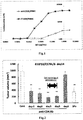
- IT1208 showed ADCC activity at 1 nM or more, and the activity then increased concentration-dependently to reach the maximum value at 50 nM.
- concentration at which the ADCC activity began to be found was 10 nM or more, and the concentration at which the maximum value was achieved was 1 ⁇ M or more.
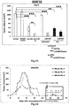
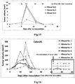
- Example 2-1 Action of Anti-CD4 Antibody against Mouse Solid Cancer Model - Study on Timing of Administration
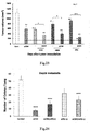
- Fig. 2 The results from the C57BL/6 mice transplanted with the melanoma cell line B16F10 are shown in Fig. 2 ; the results from the BALB/c mice transplanted with the colon cancer cell line Colon 26 are shown in Fig. 3 ; and the results from the C57BL/6 mice transplanted with the lung cancer cell line LLC are shown in Fig. 4 . From these results, it was found that the optimal timing of the single administration of the anti-CD4 antibody is Day 0 to Day 12, especially Day 3 to Day 5, after the tumor transplantation.
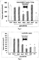
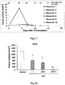
- Fig. 6 shows the tumor volume on Day 15 in each group. While significant (Dunnett; significance level, p ⁇ 0.05) tumor-growth inhibitory effects could be observed in the 50 ⁇ g-administration group and the 200 ⁇ g-administration group, in which CD4+ T cells were almost completely eliminated, no difference from the negative control group could be confirmed in the 3.125 ⁇ g-administration group and the 12.5 ⁇ g-administration group, in which CD4+ T cells were remaining.
- Fig. 8 shows the tumor volume on Day 18 in each group. Although 10.14% (corresponding to about 41% relative to the ratio in the negative control group) of CD4+ T cells were remaining in the 3.125 ⁇ g-administration group, a significantly higher tumor-growth inhibitory effect than that in the negative control group could be observed (Dunnett; significance level, p ⁇ 0.05). Similarly, the 12.5 ⁇ g-administration group and the 50 ⁇ g-administration group showed tendencies to inhibit the tumor growth.
- Anti-PD-1 alone group An anti-PD-1 antibody (0.2 mg; J43, antagonistic antibody, manufactured by BioXcell) is intraperitoneally administered daily for five days from Day 4 to Day 8.
- Anti-CD4 + anti-PD-1 combination group An anti-CD4 antibody (0.2 mg) is intraperitoneally administered in a single dose on Day 5, and an anti-PD-1 antibody (0.2 mg) is intraperitoneally administered daily for five days from Day 4 to Day 8.
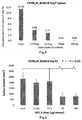
- Fig. 9 shows the tumor volume in each group of C57BL/6 mice transplanted with the B16F10 cell line.
- the tumor volume was calculated (short diameter ⁇ short diameter ⁇ long diameter ⁇ ⁇ / 6) from the solid tumor diameter measured on Day 18.
- the anti-CD4 antibody significantly inhibited the growth of the solid tumor of B16 melanoma to 1/5 (21 %) relative to that in the control group (Dunnett; significance level, p ⁇ 0.01).
- the anti-PD-1 antibody significantly inhibited the growth of the tumor to about half (55%) relative to that in the control group (Dunnett; significance level, p ⁇ 0.01).
- the total dose of the anti-CD4 antibody was 1/5 of that of the anti-PD-1 antibody
- the antitumor action of the anti-CD4 antibody was significantly stronger than that of the anti-PD-1 antibody (Dunnett; significance level, p ⁇ 0.01).
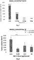
- Fig. 10 shows the tumor volume in each group of BALBIc mice transplanted with the Colon26 cell line.
- the tumor volume was calculated (short diameter ⁇ short diameter ⁇ long diameter ⁇ ⁇ / 6) from the solid tumor diameter measured on Day 29.
- the anti-CD4 antibody significantly inhibited the growth of the solid tumor of Colon 26 colon cancer to 1/4 (22%) relative to that in the control group (Dunnett; significance level, p ⁇ 0.01).
- the anti-PD-1 antibody hardly inhibited the growth, showing no significant difference (to 91% relative to the growth in the control group; Dunnett, NS; t-test, NS).
- the total dose of the anti-CD4 antibody was 1/5 of that of the anti-PD-1 antibody, the antitumor action of the anti-CD4 antibody was significantly stronger than that of the anti-PD-1 antibody (Dunnett; significance level, p ⁇ 0.01).
- Anti-CD4 alone group An anti-CD4 antibody (0.2 mg; GK1.5) is intraperitoneally administered in a single dose on Day 5.
- Anti-PD-1 alone group An anti-PD-1 antibody (0.2 mg; J43, antagonistic antibody, manufactured by BioXcell) is intraperitoneally administered daily for five days from Day 4 to Day 8.
- Anti-CD4 + anti-PD-1 (once/five times) combination group An anti-CD4 antibody (0.2 mg) is intraperitoneally administered in a single dose on Day 5, and an anti-PD-1 antibody (0.2 mg) is intraperitoneally administered daily for five days from Day 4 to Day 8.
- Anti-CD4 alone twice administration group An anti-CD4 antibody (0.2 mg; GK1.5) is intraperitoneally administered twice on Day 5 and Day 9.
- Anti-PD-1 alone ten times group An anti-PD-1 antibody (0.2 mg; J43, manufactured by BioXcell) is intraperitoneally administered daily for five days from Day 4 to Day 8 and again daily for five days from Day 14 to Day 18, ten times in total.
- Anti-CD4 + anti-PD-1 (twice/ten times) combination group An anti-CD4 antibody (0.2 mg) is intraperitoneally administered twice on Day 5 and Day 9, and an anti-PD-1 antibody (0.2 mg) is intraperitoneally administered daily for five days and again daily for five days from Day 14 to Day 18, ten times in total.
- Fig. 11 shows the tumor volume in each group of C57BL/6 mice transplanted with the B16F10 cell line.
- the tumor volume was calculated (short diameter ⁇ short diameter ⁇ long diameter ⁇ ⁇ /6) from the solid tumor diameter measured on Day 18.
- the anti-CD4 antibody significantly inhibited the growth of the solid tumor of B16 melanoma to 1/4 (27%) relative to that in the control group (Dunnett; significance level, p ⁇ 0.01).
- the anti-PD-1 antibody inhibited the growth to 24% relative to that in the control group.
- the total dose of the anti-CD4 antibody was 1/5 of that of the anti-PD-1 antibody
- the antitumor action of the anti-CD4 antibody was significantly stronger than that of the anti-PD-1 antibody (Dunnett; significance level, p ⁇ 0.01).
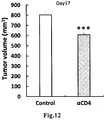
- Anti-CD4 antibody administration group An anti-CD4 antibody (0.2 mg; GK1.5) is intraperitoneally administered twice on Day 5 and Day 9.
- Fig. 12 shows the tumor volume in each group of BALB/c mice transplanted with the 4T1 cell line.
- the tumor volume was calculated (short diameter ⁇ short diameter ⁇ long diameter ⁇ ⁇ / 6) from the solid tumor diameter measured on Day 17.
- the anti-CD4 antibody significantly inhibited the growth of the 4T1 solid tumor to 76% relative to that in the control group (t-test; significance level, p ⁇ 0.001). This result suggested that the anti-CD4 antibody also inhibits the tumor growth of the breast cancer cell line 4T1.
- Anti-CD4 alone twice administration group An anti-CD4 antibody (0.2 mg; GK1.5) is intraperitoneally administered twice on Day 5 and Day 9.
- Anti-CD137 alone group An anti-CD137 antibody (0.1 mg; 3H3, antagonist antibody, Juntendo University) is intraperitoneally administered daily for five days from Day 5 to Day 9.
- 5-FU alone group 5-FU is administered at a dose of 50 mg/kg twice a week for two weeks (Day 5, Day 9, Day 12, and Day 16).
- Anti-CD4 + anti-CD137 combination group An anti-CD4 antibody (0.2 mg) is intraperitoneally administered in a single dose on Day 5, and an anti-CD 137 antibody (0.1 mg) is intraperitoneally administered daily for five days from Day 5 to Day 9.
- Anti-CD4 + 5-FU combination group An anti-CD4 antibody (0.2 mg) is intraperitoneally administered in a single dose on Day 5, and 5-FU is administered at a dose of 50 mg/kg twice a week for two weeks.
- Fig. 13 shows the tumor volume in each group of C57BL/6 mice transplanted with the B16F10 cell line.
- the tumor volume was calculated (short diameter ⁇ short diameter ⁇ long diameter ⁇ ⁇ / 6) from the solid tumor diameter measured on Day 15.
- the anti-CD4 antibody significantly inhibited the growth of the solid tumor of B16 melanoma by half or more of that in the control group (to 43% relative to the growth in the control group) (Dunnett; significance level, p ⁇ 0.01).
- the effect of the single administration of the anti-CD4 antibody was equivalent to the effect of the four times administration of 5-FU.
- the growth inhibitory effect observed in the cases of twice administration of the anti-CD4 antibody was not less than twice as much as that in the cases of single administration of the antibody. It is expected that complete regression of cancer may be possible by three times administration of the anti-CD4 antibody.
- the anti-CD137 antibody hardly inhibited the growth, showing no significant difference (to 95% relative to the growth in the control group; Dunnett, NS; t-test, NS).
- the total dose of the anti-CD4 antibody was 1/2.5 of that of the anti-CD137 antibody, the antitumor action of the anti-CD4 antibody was significantly stronger than that of the anti-CD 137 antibody (Dunnett; significance level, p ⁇ 0.01).
- the chemotherapeutic agent 5-FU which has already been clinically widely used, significantly inhibited the growth of the solid tumor of B16 melanoma to 1/3 (36%) relative to that in the control group (Dunnett; significance level, p ⁇ 0.01).
- the tumor growth was more strongly inhibited (to 21 % relative to the growth in the control group) in the anti-CD4 + 5-FU combination group as compared to the growth in the groups in which they were individually used, although the difference was not significant.
- a synergistic effect by the combination was suggested.
- Anti-CD4 alone group An anti-CD4 antibody (0.2 mg; GK1.5) is intraperitoneally administered in a single dose on Day 5.
- Anti-PD-1 alone group An anti-PD-1 antibody (0.2 mg; J43, antagonistic antibody, manufactured by BioXcell) is intraperitoneally administered daily for five days from Day 4 to Day 8.
- Anti-CD4 + anti-PD-1 combination group An anti-CD4 antibody (0.2 mg) is intraperitoneally administered in a single dose on Day 5, and an anti-PD-1 antibody (0.2 mg) is intraperitoneally administered daily for five days from Day 4 to Day 8.
- the solid tumor diameter in each mouse was measured, and the tumor volume was calculated (short diameter ⁇ short diameter ⁇ long diameter ⁇ ⁇ / 6).
- the combined use of the anti-CD4 antibody and the anti-PD-1 antibody inhibited the tumor growth in all mice on Day 12 and later.
- the average tumor volume in these mice became 13% relative to that in the control group.
- the evaluation result for each mouse was as follows. On Day 18 and later, four out of eight mice showed reduction in their tumor volumes, and, on Day 36, three out of the eight mice showed complete regression of their tumors ( Fig. 14 ).
- Fig. 14 and Fig. 15 Five days after the re-inoculation (Day 51), two out of the three mice showed traces of tumors, and, seven days after the re-inoculation (Day 53), tumor masses were found in all three mice. However, according to comparison of their average tumor volume on Day 53 with that of the mice observed 7 days after the initial inoculation (Day 7), the average on Day 53 was 1/7 relative to the average on Day 7 in the control group, and 1/4 relative to the average on Day 7 in the anti-CD4 antibody + anti-PD-1 antibody combination group.
- the tumors whose growth was observed after the re-inoculation showed reduction in their volumes 11 days after the re-inoculation (Day 57) in all mice, and no tumors were found any more in all the mice 14 days after the re-inoculation (Day 64), indicating complete rejection of the tumors.
- Anti-CD4 alone group An anti-CD4 antibody (0.2 mg; GK1.5) is intraperitoneally administered in a single dose on Day 5.
- An anti-PD-L1 antibody (0.2 mg; 10F.9G2, manufactured by BioXcell) is intraperitoneally administered on Day 4, Day 8, Day 14, and Day 18, four times in total.
- Anti-CD4 + anti-PD-L1 combination group An anti-CD4 antibody (0.2 mg) is intraperitoneally administered in a single dose on Day 5, and an anti-PD-L1 antibody (0.2 mg) is intraperitoneally administered on Day 4, Day 8, Day 14, and Day 18, four times in total.
- the solid tumor diameter in each mouse was measured, and the tumor volume was calculated (short diameter ⁇ short diameter ⁇ long diameter ⁇ ⁇ / 6).
- the combined use of the anti-CD4 antibody and the anti-PD-L1 antibody inhibited the tumor growth in all mice on Day 10 and later.
- the average tumor volume in these mice became 2.5% relative to that in the control group.
- the evaluation result for each mouse was as follows. On Day 18 and later, nine out of ten mice showed reduction in their tumor volumes, and, on Day 28, six out of the ten mice showed complete regression of their tumors ( Fig. 16 ).
- mice which showed the complete regression on Day 28 were further subjected to subcutaneous inoculation, on their ventral side, of Colon 26 cells in a number five times higher (1 ⁇ 10 6 cells/mouse) than when the cells were initially inoculated.
- Fig. 16 and Fig. 17 The results are shown in Fig. 16 and Fig. 17 .
- Day 46 Four days after the re-inoculation (Day 46), four out of the six mice showed traces of tumors. However, according to comparison of their average tumor volume on Day 46 with that of the mice observed 7 days after the initial inoculation (Day 7), the average on Day 46 was 1/20 relative to the average on Day 7 in the anti-CD4 antibody + anti-PD-L1 antibody combination group.
- the tumors whose growth was observed after the re-inoculation showed reduction in their volumes 7 days after the re-inoculation (Day 49) in all mice, and no tumors were found any more in all the mice 10 days after the re-inoculation (Day 52), indicating complete rejection of the tumors.
- Anti-CD4 alone group An anti-CD4 antibody (0.2 mg; GK1.5) is intraperitoneally administered in a single dose on Day 5.
- Anti-PD-1 alone group An anti-PD-1 antibody (0.2 mg; J43, antagonistic antibody, manufactured by BioXcell) is intraperitoneally administered daily for five days from Day 4 to Day 8.
- Anti-PD-L1 alone group An anti-PD-1 antibody (0.2 mg; J43, antagonistic antibody, manufactured by BioXcell) is intraperitoneally administered daily for five days from Day 4 to Day 8.
- Anti-CD4 + anti-PD-1 combination group An anti-CD4 antibody (0.2 mg) is intraperitoneally administered in a single dose on Day 5, and an anti-PD-1 antibody (0.2 mg) is intraperitoneally administered daily for five days from Day 4 to Day 8.
- Anti-CD4 + anti-PD-L1 combination group An anti-CD4 antibody (0.2 mg) is intraperitoneally administered in a single dose on Day 5, and an anti-PD-1 antibody (0.2 mg) is intraperitoneally administered daily for five days from Day 4 to Day 8.
- the solid tumor diameter in each mouse was measured, and the tumor volume was calculated (short diameter ⁇ short diameter ⁇ long diameter ⁇ ⁇ / 6).
- the anti-CD4 antibody and the anti-PD-1 antibody were used in combination, inhibition of the tumor growth began in all mice on Day 14 and later.
- the average tumor volume in these mice became 39% relative to that in the control group ( Fig. 18 ).
- the combined use of the anti-CD4 antibody and the anti-PD-L antibody inhibited the tumor growth in all mice on Day 14 and later.
- the average tumor volume in these mice became 1% relative to that in the control group ( Fig. 18 ).
- the evaluation result for each mouse was as follows. In the anti-CD4 + anti-PD-1 combination group, four out of eight mice showed complete regression of their tumors on Day 32. Similarly, in the anti-CD4 + anti-PD-L combination group, seven out of eight mice showed reduction in their tumor volumes, and complete regression of the tumors was achieved on Day 26.
- mice with complete regression were divided into two groups each composed of five individuals, and, 17 days after the complete regression of the tumor (Day 49), the mice were further subjected to subcutaneous inoculation, on their ventral side, of Colon 26 cells or 4T1 cells in a number five times higher (1 ⁇ 10 6 cells/mouse) than when the cells were initially inoculated. The tumor growth was then investigated.
- Colon 26 (2 ⁇ 10 5 cells/mouse) or 4T1 (2 ⁇ 10 5 cells/mouse) prepared at the same time was subcutaneously transplanted into the right abdomen of normal BALB/c mice (male, 7 weeks old), and the tumor growth was investigated thereafter. The results are shown in Fig. 19 and Fig. 20 .
- Negative control group No antibody is administered.
- Anti-CD4 alone group An anti-CD4 antibody (0.2 mg; GK1.5) is intraperitoneally administered twice on Day 5 and Day 9.
- Anti-PD-L1, anti-PD-L2, anti-OX40, or anti-CTLA-4 alone group An anti-PD-L antibody (10F.9G2, manufactured by BioXcell), anti-PD-L2 antibody (TY25, manufactured by BioXcell), anti-OX40 antibody (OX-86, agonist antibody; manufactured by BioXcell), or anti-CTLA-4 antibody (9D9, antagonist antibody; manufactured by BioXcell) is intraperitoneally administered at a dose of 0.2 mg on Day 4, Day 8, Day 14, and Day 18, four times in total.
- Anti-CD4 + anti-PD-L1, anti-PD-L2, anti-OX40, or anti-CTLA-4 combination group An anti-CD4 antibody (0.2 mg) is intraperitoneally administered twice on Day 5 and Day 8, and an anti-PD-L1, anti-PD-L2, anti-OX40, or anti-CTLA-4 antibody is administered at a dose of 0.2 mg on Day 4, Day 8, Day 14, and Day 18, four times in total.
- Fig. 21 shows the tumor volume in each group of C57BL/6 mice transplanted with the B16F10 cell line.
- the tumor volume was calculated (short diameter ⁇ short diameter ⁇ long diameter ⁇ ⁇ / 6) from the solid tumor diameter measured on Day 16.
- the anti-CD4 antibody significantly inhibited the growth of the solid tumor of B16 melanoma to about 1/3 relative to that in the control group (Dunnett; significance level, p ⁇ 0.01).
- the anti-CD4 antibody has a better inhibitory effect than those of the other anti-immune checkpoint antibodies.
- the anti-PD-L antibody, anti-PD-L2 antibody, anti-OX40 antibody, and anti-CTLA-4 antibody were individually administered, significantly stronger inhibition of the growth could be observed relative to the growth in the control group (Dunnett; significance level, p ⁇ 0.01), although the inhibition was weaker than that by the anti-CD4 antibody.
- the anti-CD4 antibody was used in combination with the anti-PD-L antibody, anti-PD-L2 antibody, anti-OX40 antibody, or anti-CTLA-4 antibody, the growth of the B16 melanoma solid tumor was more strongly inhibited than in the groups in which the antibodies were individually administered without administration of the anti-CD4 antibody.
- the averages in the immune checkpoint antibody alone groups were significantly different from the averages in the anti-CD4 combination groups (Dunnett; significance level, p ⁇ 0.05 or p ⁇ 0.01). Thus, synergistic effects by the combinations became apparent.
- the average tumor volume in the anti-CD4 + anti-PD-L1 combination group was significantly different from that in the anti-CD4 alone group (significance level, 5%; Dunnett).
- a remarkable synergistic effect by the combined use of the anti-CD4 antibody and the anti-PD-L1 antibody was shown.
- Negative control group No antibody is administered.
- Anti-CD4 alone group An anti-CD4 antibody (0.2 mg; GK1.5) is intraperitoneally administered twice on Day 5 and Day 9.
- Anti-BTLA, anti-GITR, anti-LAG-3, or anti-TIM-3 alone group An anti-BTLA antibody (6A6, manufactured by BioXcell), anti-GITR antibody (DTA-1, agonist antibody; manufactured by BioXcell), anti-LAG-3 antibody (C9B7W, manufactured by BioXcell), or anti-TIM-3 antibody (RMT3-23, manufactured by BioXcell) is intraperitoneally administered at a dose of 0.2 mg on Day4, Day8, Day14, and Day18, four times in total.
- Anti-CD4 + anti-BTLA, anti-GITR, anti-LAG-3, or anti-TIM-3 combination group An anti-CD4 antibody (0.2 mg) is intraperitoneally administered twice on Day 5 and Day 8, and an anti-BTLA antibody, anti-GITR antibody, anti-LAG-3 antibody, or an anti-TIM-3 antibody is intraperitoneally administered at a dose of 0.2 mg on Day4, Day8, Day14, and Day18, four times in total.
- Fig. 22 shows the tumor volume in each group of C57BL/6 mice transplanted with the B16F10 cell line.
- the tumor volume was calculated (short diameter ⁇ short diameter ⁇ long diameter ⁇ ⁇ / 6) from the solid tumor diameter measured on Day 15.
- the anti-CD4 antibody significantly inhibited the growth of the solid tumor of B16 melanoma to about half relative to that in the control group (Dunnett; significance level, p ⁇ 0.01).
- the anti-CD4 antibody has a better inhibitory effect than the other anti-immune checkpoint antibodies when used alone.
- Negative control group No drug is administered.
- Anti-CD4 alone group An anti-CD4 antibody (0.2 mg; GK1.5) is intraperitoneally administered twice on Day 5 and Day 9.
- Anti-CD4 + OXA combination group An anti-CD4 antibody (0.2 mg) is intraperitoneally administered twice on Day 5 and Day 9, and OXA is administered at a dose of 10 mg/kg twice a week for two weeks.
- Gemcitabine (GEM, Gemzar Injection; Eli Lilly Japan) alone group Gemcitabine is administered at a dose of 20 mg/kg twice a week for two weeks (Day 5, Day 9, Day 12, and Day 16).
- Anti-CD4 + GEM combination group An anti-CD4 antibody (0.2 mg) is intraperitoneally administered twice on Day 5 and Day 9, and GEM is administered at a dose of 20 mg/kg twice a week for two weeks.
- Cyclophosphamide (CPA, Endoxan Injection; Shionogi & Co., Ltd.) alone group Cyclophosphamide is administered at a dose of 10 mg/kg twice a week for two weeks (Day 5, Day 9, Day 12, and Day 16).
- Anti-CD4 + CPA combination group An anti-CD4 antibody (0.2 mg) is intraperitoneally administered twice on Day 5 and Day 9, and OXA is administered at a dose of 10 mg/kg twice a week for two weeks.
- Fig. 23 shows the tumor volume in each group of C57BL/6 mice transplanted with the B16F10 cell line.
- the tumor volume was calculated (short diameter ⁇ short diameter ⁇ long diameter ⁇ ⁇ / 6) from the solid tumor diameter measured on Day 15.
- the anti-CD4 antibody significantly inhibited the growth of the solid tumor of B16 melanoma by half or more of that in the control group (to 47% relative to the growth in the control group) (Dunnett; significance level, p ⁇ 0.01).
- the growth was inhibited to the same degree as in the anti-CD4 antibody alone group (to 46% relative to the growth in the control group) (Dunnett; significance level, p ⁇ 0.01).
- weight loss was observed in mice receiving OXA at this dose, indicating occurrence of a side effect.
- the anti-CD4 antibody When the anti-CD4 antibody was used in combination with OXA, GEM, or CPA, the growth of the solid tumor of B16 melanoma was more strongly inhibited than when the anti-CD4 antibody was used alone. In particular, the average in the CPA combination group was significantly different from the average in the anti-CD4 alone group (Dunnett; significance level, p ⁇ 0.05).
- Negative control group No antibody is administered.
- Anti-CD4 alone group An anti-CD4 antibody (0.2 mg; GK1.5) is intraperitoneally administered twice on Day 5 and Day 9.
- An anti-PD-L1 antibody 0.2 mg; 10F.9G2, manufactured by BioXcell is intraperitoneally administered on Day4, Day8, Day 14, and Day18, four times in total.
- Anti-CD4 + anti-PD-1 combination group An anti-CD4 antibody (0.2 mg; GK1.5) is intraperitoneally administered twice on Day 5 and Day 9, and an anti-PD-1 antibody (0.2 mg; J43, manufactured by BioXcell) is intraperitoneally administered on Day4, Day8, Day14, and Day18, four times in total.
- Anti-CD4 + anti-PD-L1 combination group An anti-CD4 antibody (0.2 mg; GK1.5) is intraperitoneally administered twice on Day 5 and Day 9, and an anti-PD-L1 antibody (0.2 mg; 10F.9G2, manufactured by BioXcell) is intraperitoneally administered on Day4, Day8, Day14, and Day18, four times in total.
- Fig. 24 shows the number of metastasis to the lungs in each group of BALB/c mice transplanted with the 4T1 cell line. The mice were dissected and their lungs were excised on Day 28 to count the metastasis.
- the anti-CD4 antibody significantly reduced the number of metastasis of 4T1 to the lungs to 15% relative to that in the control group (Dunnett; significance level, p ⁇ 0.001).
- the anti-CD4 + anti-PD-1 combination group significantly reduced the number of metastasis to 32% relative to that in the control group (Dunnett; significance level, p ⁇ 0.001).
- the anti-CD4 + anti-PD-L1 combination group significantly reduced the number of metastasis to 44% relative to that in the control group (Dunnett; significance level, p ⁇ 0.001), showing an enhanced antimetastatic effect by the combined use.
- Example 9 Study on Antigen Specificity of CD8 + T Cells That Increase in Tumor-Draining Lymph Nodes after Administration of Anti-CD4 Antibody Alone
- the mouse melanoma cell line B16F10(5 ⁇ 1O 5 cells/mouse) was subcutaneously inoculated into the right side of the back of C57BL/6 mice (congenic markers CD45.1 - CD45.2 + CD90.1 - CD90.2 + ), and 0.2 mg of the anti-CD4 antibody (GK1.5) was intraperitoneally administered in a single dose on Day 5.
- B16F10(+) anti-CD4 antibody(+) group Besides the B16F10(+) anti-CD4 antibody(+) group, a B16F10(-) anti-CD4 antibody(-) group, a B16F10(-) anti-CD4 antibody(+) group, and a B16F11(+) anti-CD4 antibody(-) group were provided as control groups.
- cell suspensions were prepared from the tumor (tumor), tumor-draining brachial lymph node (dLN), non-draining brachial lymph node (ndLN), peripheral blood (blood), and spleen (spleen).
- the cells were stained with fluorescence-labeled antibodies specific to the surface antigens, and the number of donor CD8+ T cells transferred and the frequency of cell division were analyzed by flow cytometry.
- Pmel-1 CD8 + T cells in the B16F11(+) group are capable of recognizing the specific antigen.
- OT-1 and polyclonal CD8 + T cells are not capable of recognizing the specific antigen in any of the groups. That is, if the administration of the anti-CD4 antibody promotes the growth response not only for Pmel-1 CD8 + T cells in the B16F10(+) group, but also for OT-1 and polyclonal CD8 + T cells in any of the groups, the growth response can be understood as an antigen-nonspecific growth response. On the other hand, if the growth response is promoted only for Pmel-1 CD8 + T cells in the B16F11(+) group, the growth response can be understood as a tumor antigen-specific growth response.
- Fig. 25A shows the experimental protocol.
- Fig. 25B shows an example of flow cytometric analysis of the tumor-draining lymph node cells.
- the IVS-CD45 - CD8 + fraction was developed according to the expression of CD90.1/FSC to identify CD90.1+ Pmel-1 cells.
- the CD90.1- fraction was developed according to the expression of CD45.1/CD45.2 to separately identify CD45.1- CD45.2 + host CD8 + T cells, CD45.1 + CD45.2 + Polyclonal CD8 + T cells, and CD45.1 + CD45.2 + OT-1 CD8 + T cells.
- Fig. 25C shows an example of the results of analysis of the frequency of cell division of lymph node Pmel-1, OT-1, and Polyclonal CD8 + T cells identified by the development shown in Fig. 25B , which analysis was carried out using the fluorescence intensity of CFSE as an index. Since the fluorescence signal of CFSE decreases as the cell division proceeds, the frequency of cell division can be identified based on the fluorescence intensity of CFSE.
- brachial lymph node of the B16F10(-) anti-CD4 antibody(-) group and the B16F10(-) anti-CD4 antibody(+) group were analyzed.
- Fig. 25D shows the percentage of cells with cell division frequency of 0 to 1, 2 to 4, and 5 to 8 in tumor-draining lymph node Pmel-1 CD8 + T cells from the B16F11(+) anti-CD4 antibody(-) group or the B16F10(+) anti-CD4 antibody(+) group.
- the percentage of cells that had undergone less frequent cell division i.e. 0-1 and 2-4 times of cell division
- Fig. 25E shows the numbers of Pmel-1 CD8+ T cells with cell division frequency of 5 to 8 in peripheral blood (blood), tumor-draining lymph node (dLN), non-draining lymph node (ndLN), spleen (spleen), and tumor (tumor) in the B16F11(+) anti-CD4 antibody(-) group or the B16F11(+) anti-CD4 antibody(+) group.
- the CD8 + T cells that increase in the tumor-draining brachial lymph node when the anti-CD4 antibody is administered alone are tumor antigen-specific, and that such an increase is not a nonspecific increase (homeostatic proliferation) in T cells, which is often found under conditions where lymphocytes are decreased.
- the following clauses detail embodiments of the invention.
Landscapes
- Health & Medical Sciences (AREA)
- Chemical & Material Sciences (AREA)
- Immunology (AREA)
- Life Sciences & Earth Sciences (AREA)
- Medicinal Chemistry (AREA)
- General Health & Medical Sciences (AREA)
- Organic Chemistry (AREA)
- Pharmacology & Pharmacy (AREA)
- Public Health (AREA)
- Veterinary Medicine (AREA)
- Animal Behavior & Ethology (AREA)
- Molecular Biology (AREA)
- Epidemiology (AREA)
- Genetics & Genomics (AREA)
- Biophysics (AREA)
- Biochemistry (AREA)
- Proteomics, Peptides & Aminoacids (AREA)
- Engineering & Computer Science (AREA)
- Bioinformatics & Cheminformatics (AREA)
- Microbiology (AREA)
- Mycology (AREA)
- Endocrinology (AREA)
- Chemical Kinetics & Catalysis (AREA)
- General Chemical & Material Sciences (AREA)
- Nuclear Medicine, Radiotherapy & Molecular Imaging (AREA)
- Biomedical Technology (AREA)
- Oncology (AREA)
- Medicines Containing Antibodies Or Antigens For Use As Internal Diagnostic Agents (AREA)
- Medicines That Contain Protein Lipid Enzymes And Other Medicines (AREA)
- Peptides Or Proteins (AREA)
Applications Claiming Priority (4)
| Application Number | Priority Date | Filing Date | Title |
|---|---|---|---|
| JP2014032241 | 2014-02-21 | ||
| JP2014120300 | 2014-06-11 | ||
| JP2014178953 | 2014-09-03 | ||
| EP15751324.3A EP3132802B1 (de) | 2014-02-21 | 2015-02-09 | Therapeutisches mittel für soliden malignen tumor |
Related Parent Applications (2)
| Application Number | Title | Priority Date | Filing Date |
|---|---|---|---|
| EP15751324.3A Division-Into EP3132802B1 (de) | 2014-02-21 | 2015-02-09 | Therapeutisches mittel für soliden malignen tumor |
| EP15751324.3A Division EP3132802B1 (de) | 2014-02-21 | 2015-02-09 | Therapeutisches mittel für soliden malignen tumor |
Publications (1)
| Publication Number | Publication Date |
|---|---|
| EP3338800A1 true EP3338800A1 (de) | 2018-06-27 |
Family
ID=53878157
Family Applications (3)
| Application Number | Title | Priority Date | Filing Date |
|---|---|---|---|
| EP18153989.1A Ceased EP3338800A1 (de) | 2014-02-21 | 2015-02-09 | Therapeutisches mittel für soliden malignen tumor |
| EP18153990.9A Withdrawn EP3338801A1 (de) | 2014-02-21 | 2015-02-09 | Therapeutisches mittel für soliden malignen tumor |
| EP15751324.3A Active EP3132802B1 (de) | 2014-02-21 | 2015-02-09 | Therapeutisches mittel für soliden malignen tumor |
Family Applications After (2)
| Application Number | Title | Priority Date | Filing Date |
|---|---|---|---|
| EP18153990.9A Withdrawn EP3338801A1 (de) | 2014-02-21 | 2015-02-09 | Therapeutisches mittel für soliden malignen tumor |
| EP15751324.3A Active EP3132802B1 (de) | 2014-02-21 | 2015-02-09 | Therapeutisches mittel für soliden malignen tumor |
Country Status (5)
| Country | Link |
|---|---|
| US (2) | US20170165364A1 (de) |
| EP (3) | EP3338800A1 (de) |
| JP (3) | JP6029160B2 (de) |
| ES (1) | ES2808728T3 (de) |
| WO (1) | WO2015125652A1 (de) |
Cited By (1)
| Publication number | Priority date | Publication date | Assignee | Title |
|---|---|---|---|---|
| WO2020057486A1 (zh) * | 2018-09-17 | 2020-03-26 | 中国科学院动物研究所 | 经修饰的t细胞、其制备方法及用途 |
Families Citing this family (8)
| Publication number | Priority date | Publication date | Assignee | Title |
|---|---|---|---|---|
| JP6971239B2 (ja) * | 2015-10-05 | 2021-11-24 | キャリセラ バイオサイエンシーズ, インコーポレイテッド | グルタミナーゼ阻害剤とイムノオンコロジー薬剤との併用療法 |
| AU2017284499A1 (en) * | 2016-06-17 | 2019-01-31 | Osaka University | Intratumoral vein formation promoter |
| WO2018047917A1 (ja) * | 2016-09-09 | 2018-03-15 | 国立大学法人 東京大学 | Hmgタンパク質と抗cd4抗体又は免疫チェックポイント制御剤との組み合わせによる相乗的抗腫瘍効果 |
| WO2018071500A1 (en) | 2016-10-11 | 2018-04-19 | Agenus Inc. | Anti-lag-3 antibodies and methods of use thereof |
| PT3630112T (pt) | 2017-06-02 | 2024-04-23 | Bayer Healthcare Llc | Combinação de regorafenib e nivolumab para tratamento de cancro |
| WO2019172358A1 (ja) | 2018-03-08 | 2019-09-12 | 国立大学法人 東京大学 | Hmgn部分ペプチド及びこれを用いたがん療法 |
| GB201811291D0 (en) * | 2018-07-10 | 2018-08-29 | Univ Dublin City | A method of predicting response to treatment in cancer patients |
| JPWO2021025140A1 (de) * | 2019-08-08 | 2021-02-11 |
Citations (6)
| Publication number | Priority date | Publication date | Assignee | Title |
|---|---|---|---|---|
| WO2004078928A2 (en) * | 2003-02-28 | 2004-09-16 | The Johns Hopkins University | T cell regulation |
| WO2006121168A1 (en) * | 2005-05-09 | 2006-11-16 | Ono Pharmaceutical Co., Ltd. | Human monoclonal antibodies to programmed death 1(pd-1) and methods for treating cancer using anti-pd-1 antibodies alone or in combination with other immunotherapeutics |
| WO2010074266A1 (ja) | 2008-12-26 | 2010-07-01 | 協和発酵キリン株式会社 | 抗cd4抗体 |
| WO2011109789A2 (en) * | 2010-03-05 | 2011-09-09 | The Johns Hopkins University | Compositions and methods for targeted immunomodulatory antibodies and fusion proteins |
| WO2015120198A1 (en) * | 2014-02-05 | 2015-08-13 | Cedars-Sinai Medical Center | Methods and compositions for treating cancer and infectious diseases |
| EP3156072A1 (de) * | 2014-06-11 | 2017-04-19 | IDAC Theranostics, Inc. | Verfahren zur reduzierung der nebenwirkungen von immunprüfpunkt-kontrollmittel |
Family Cites Families (8)
| Publication number | Priority date | Publication date | Assignee | Title |
|---|---|---|---|---|
| ES2311558T3 (es) * | 2000-10-20 | 2009-02-16 | Genetics Institute, Inc. | Uso de inhibidores de la il-13 para el tratamiento de tumores. |
| EP1460088A1 (de) * | 2003-03-21 | 2004-09-22 | Biotest AG | Humanisierter Antikörper gegen CD4 mit immunsuppressiven Merkmalen |
| KR20100135257A (ko) * | 2008-03-13 | 2010-12-24 | 바이오테스트 아게 | 질병 치료제 |
| US9156912B2 (en) * | 2008-12-12 | 2015-10-13 | The University Of Tokyo | Immunological reconstitution promoter or prophylactic agent for infections each of which maintains graft-versus-tumor effect |
| CA3019531A1 (en) * | 2011-04-19 | 2012-10-26 | Pfizer Inc. | Combinations of anti-4-1bb antibodies and adcc-inducing antibodies for the treatment of cancer |
| JP6120848B2 (ja) * | 2011-08-15 | 2017-04-26 | メディミューン,エルエルシー | 抗b7−h4抗体およびその使用 |
| AR090263A1 (es) * | 2012-03-08 | 2014-10-29 | Hoffmann La Roche | Terapia combinada de anticuerpos contra el csf-1r humano y las utilizaciones de la misma |
| US20160108122A1 (en) * | 2013-05-23 | 2016-04-21 | Idac Theranostics, Inc. | Therapeutic or prophylactic agent for immunodeficiency virus infection |
-
2015
- 2015-02-09 WO PCT/JP2015/053569 patent/WO2015125652A1/ja active Application Filing
- 2015-02-09 ES ES15751324T patent/ES2808728T3/es active Active
- 2015-02-09 EP EP18153989.1A patent/EP3338800A1/de not_active Ceased
- 2015-02-09 JP JP2016504046A patent/JP6029160B2/ja active Active
- 2015-02-09 US US15/120,427 patent/US20170165364A1/en not_active Abandoned
- 2015-02-09 EP EP18153990.9A patent/EP3338801A1/de not_active Withdrawn
- 2015-02-09 EP EP15751324.3A patent/EP3132802B1/de active Active
-
2016
- 2016-08-15 JP JP2016159348A patent/JP6578604B2/ja active Active
-
2018
- 2018-12-27 US US16/234,292 patent/US20190125867A1/en not_active Abandoned
-
2019
- 2019-02-15 JP JP2019025193A patent/JP7041439B2/ja active Active
Patent Citations (7)
| Publication number | Priority date | Publication date | Assignee | Title |
|---|---|---|---|---|
| WO2004078928A2 (en) * | 2003-02-28 | 2004-09-16 | The Johns Hopkins University | T cell regulation |
| WO2006121168A1 (en) * | 2005-05-09 | 2006-11-16 | Ono Pharmaceutical Co., Ltd. | Human monoclonal antibodies to programmed death 1(pd-1) and methods for treating cancer using anti-pd-1 antibodies alone or in combination with other immunotherapeutics |
| WO2010074266A1 (ja) | 2008-12-26 | 2010-07-01 | 協和発酵キリン株式会社 | 抗cd4抗体 |
| US20100310573A1 (en) * | 2008-12-26 | 2010-12-09 | Kyowa Hakko Kirin Co., Ltd. | Anti-cd4 antibody |
| WO2011109789A2 (en) * | 2010-03-05 | 2011-09-09 | The Johns Hopkins University | Compositions and methods for targeted immunomodulatory antibodies and fusion proteins |
| WO2015120198A1 (en) * | 2014-02-05 | 2015-08-13 | Cedars-Sinai Medical Center | Methods and compositions for treating cancer and infectious diseases |
| EP3156072A1 (de) * | 2014-06-11 | 2017-04-19 | IDAC Theranostics, Inc. | Verfahren zur reduzierung der nebenwirkungen von immunprüfpunkt-kontrollmittel |
Non-Patent Citations (27)
| Title |
|---|
| B. K. CHOI ET AL: "Mechanisms Involved in Synergistic Anticancer Immunity of Anti-4-1BB and Anti-CD4 Therapy", CANCER RESEARCH, vol. 67, no. 18, 15 September 2007 (2007-09-15), US, pages 8891 - 8899, XP055221272, ISSN: 0008-5472, DOI: 10.1158/0008-5472.CAN-07-1056 * |
| CHERN SIANG LEE ET AL: "Novel antibodies targeting immune regulatory checkpoints for cancer therapy", BRITISH JOURNAL OF CLINICAL PHARMACOLOGY, vol. 76, no. 2, 23 July 2013 (2013-07-23), pages 233 - 247, XP055151740, ISSN: 0306-5251, DOI: 10.1111/bcp.12164 * |
| CHOI BK ET AL., CANCER RESEARCH, vol. 67, 2007, pages 8891 - 9 |
| HAMID O ET AL., THE NEW ENGLAND JOURNAL OF MEDICINE, vol. 369, 2013, pages 134 - 44 |
| HUANG CHING-TAI ET AL: "Role of LAG-3 in regulatory T cells", IMMUN, CELL PRESS, US, vol. 21, no. 4, 1 October 2004 (2004-10-01), pages 503 - 513, XP009138514, ISSN: 1074-7613, [retrieved on 20041019], DOI: 10.1016/J.IMMUNI.2004.08.010 * |
| J. MITSUI ET AL: "Two Distinct Mechanisms of Augmented Antitumor Activity by Modulation of Immunostimulatory/Inhibitory Signals", CLINICAL CANCER RESEARCH, vol. 16, no. 10, 15 May 2010 (2010-05-15), pages 2781 - 2791, XP055100557, ISSN: 1078-0432, DOI: 10.1158/1078-0432.CCR-09-3243 * |
| JEDD D. WOLCHOK ET AL: "Nivolumab plus Ipilimumab in Advanced Melanoma", NEW ENGLAND JOURNAL OF MEDICINE, vol. 369, no. 2, 11 July 2013 (2013-07-11), pages 122 - 133, XP055182024, ISSN: 0028-4793, DOI: 10.1056/NEJMoa1302369 * |
| JOSEPH F. GROSSO ET AL: "LAG-3 regulates CD8+ T cell accumulation and effector function in murine self- and tumor-tolerance systems", JOURNAL OF CLINICAL INVESTIGATION, vol. 117, no. 11, 11 October 2007 (2007-10-11), US, pages 3383 - 3392, XP055463609, ISSN: 0021-9738, DOI: 10.1172/JCI31184 * |
| K. TAKEDA ET AL: "Combination Therapy of Established Tumors by Antibodies Targeting Immune Activating and Suppressing Molecules", THE JOURNAL OF IMMUNOLOGY, vol. 184, no. 10, 16 April 2010 (2010-04-16), pages 5493 - 5501, XP055123621, ISSN: 0022-1767, DOI: 10.4049/jimmunol.0903033 * |
| KIM YH ET AL., BLOOD, vol. 109, 2007, pages 4655 - 62 |
| KOCAK E ET AL: "Combination therapy with anti-CTL antigen-4 and anti-4-1BB antibodies enhances cancer immunity and reduces autoimmunity", CANCER RESEARCH, AACR - AMERICAN ASSOCIATION FOR CANCER RESEARCH, US, vol. 66, no. 14, 15 July 2006 (2006-07-15), pages 7276 - 7284, XP002488612, ISSN: 0008-5472, DOI: 10.1158/0008-5472.CAN-05-2128 * |
| M. SCHUSTER ET AL.: "Improved effector functions of a therapeutic monoclonal Lewis Y-specific antibody by glycoform engineering", CANCER RES, vol. 65, 2005, pages 7934 - 7941, XP008060462 |
| NATSUME A ET AL.: "Improving effector functions of antibodies for cancer treatment: Enhancing ADCC and CDC", DRUG DES DEVEL THER, vol. 3, 2009, pages 7 - 16, XP002727883, DOI: doi:10.2147/DDDT.S4378 |
| NATSUME A; IN M; TAKAMURA H ET AL.: "Engineered antibodies of IgGl/IgG3 mixed isotype with enhanced cytotoxic activities", CANCER RES., vol. 68, 2008, pages 3863 - 3872 |
| PARDOLL DM., NATURE REVIEWS CANCER, vol. 12, 2012, pages 252 - 64 |
| PRIETO PA ET AL., CLINICAL CANCER RESEARCH : AN OFFICIAL JOURNAL OF THE AMERICAN ASSOCIATION FOR CANCER RESEARCH, vol. 18, 2012, pages 2039 - 47 |
| ROSENBLATT JACALYN ET AL: "PD-1 blockade by CT-011, anti-PD-1 antibody, enhances ex vivo T-cell responses to autologous dendritic cell/myeloma fusion vaccine", JOURNAL OF IMMUNOTHERAPY, LIPPINCOTT WILLIAMS & WILKINS, US, vol. 34, no. 5, 1 June 2011 (2011-06-01), pages 409 - 418, XP008175988, ISSN: 1537-4513, DOI: 10.1097/CJI.0B013E31821CA6CE * |
| S. UEHA ET AL: "Robust Antitumor Effects of Combined Anti-CD4-Depleting Antibody and Anti-PD-1/PD-L1 Immune Checkpoint Antibody Treatment in Mice", CANCER IMMUNOLOGY RESEARCH, vol. 3, no. 6, 20 February 2015 (2015-02-20), US, pages 631 - 640, XP055422635, ISSN: 2326-6066, DOI: 10.1158/2326-6066.CIR-14-0190 * |
| S.-R. WOO ET AL: "Immune Inhibitory Molecules LAG-3 and PD-1 Synergistically Regulate T-cell Function to Promote Tumoral Immune Escape", CANCER RESEARCH, vol. 72, no. 4, 15 February 2012 (2012-02-15), US, pages 917 - 927, XP055431013, ISSN: 0008-5472, DOI: 10.1158/0008-5472.CAN-11-1620 * |
| SE ALTERS ET AL: "Mechanisms of anti-CD4-mediated depletion and immunotherapy. A study using a set of chimeric anti-CD4 antibodies", THE JOURNAL OF IMMUNOLOGY, 15 June 1990 (1990-06-15), UNITED STATES, pages 4587 - 4592, XP055422661, Retrieved from the Internet <URL:http://www.jimmunol.org/content/jimmunol/144/12/4587.full.pdf> [retrieved on 20171109] * |
| SUTMULLER R P M ET AL: "Synergism of Cytotoxic T Lymphocyte-Associated Antigen 4 Blockade and Depletion of Cd25+ Regulatory T Cells in Antitumor Therapy Reveals Alternative Pathways for Suppression of Autoreactive Cytotoxic T Lymphocyte Responses", THE JOURNAL OF EXPERIMENTAL MEDICINE, ROCKEFELLER UNIVERSITY PRESS, US, vol. 194, no. 6, 17 September 2001 (2001-09-17), pages 823 - 832, XP008040742, ISSN: 0022-1007, DOI: 10.1084/JEM.194.6.823 * |
| WOLCHOK JD ET AL., THE NEW ENGLAND JOURNAL OF MEDICINE, vol. 369, 2013, pages 122 - 33 |
| YAMANE-OHNUKI N ET AL.: "Establishment of FUT8 knockout Chinese hamster ovary cells: an ideal host cell line for producing completely defucosylated antibodies with enhanced antibody-dependent cellular cytotoxicity", BIOTECHNOL BIOENG, vol. 87, 2004, pages 614 - 622, XP002984450, DOI: doi:10.1002/bit.20151 |
| YAMANE-OHNUKI N; SATOH M: "Production of therapeutic antibodies with controlled fucosylation", MABS, vol. 1, 2009, pages 230 - 236, XP002731447, DOI: doi:10.4161/mabs.1.3.8328 |
| YOUN H KIM ET AL: "Clinical efficacy of zanolimumab (HuMax-CD4): two phase 2 studies in refractory cutaneous T-cell lymphoma", BLOOD, 20 February 2007 (2007-02-20), pages 4655 - 4662, XP055430340, Retrieved from the Internet <URL:http://www.bloodjournal.org/content/109/11/4655.full.pdf> [retrieved on 20171129], DOI: 10.1182/blood- * |
| YU P ET AL., THE JOURNAL OF EXPERIMENTAL MEDICINE, vol. 201, 2005, pages 779 - 91 |
| ZHIQIANG GUO ET AL: "PD-1 Blockade and OX40 Triggering Synergistically Protects against Tumor Growth in a Murine Model of Ovarian Cancer", PLOS ONE, vol. 9, no. 2, 27 February 2014 (2014-02-27), pages e89350, XP055132840, ISSN: 1932-6203, DOI: 10.1371/journal.pone.0089350 * |
Cited By (1)
| Publication number | Priority date | Publication date | Assignee | Title |
|---|---|---|---|---|
| WO2020057486A1 (zh) * | 2018-09-17 | 2020-03-26 | 中国科学院动物研究所 | 经修饰的t细胞、其制备方法及用途 |
Also Published As
| Publication number | Publication date |
|---|---|
| ES2808728T3 (es) | 2021-03-01 |
| WO2015125652A1 (ja) | 2015-08-27 |
| US20190125867A1 (en) | 2019-05-02 |
| JP2017014254A (ja) | 2017-01-19 |
| JP7041439B2 (ja) | 2022-03-24 |
| JPWO2015125652A1 (ja) | 2017-03-30 |
| EP3132802A4 (de) | 2018-04-18 |
| EP3132802A1 (de) | 2017-02-22 |
| JP6578604B2 (ja) | 2019-09-25 |
| JP2019070056A (ja) | 2019-05-09 |
| US20170165364A1 (en) | 2017-06-15 |
| EP3338801A1 (de) | 2018-06-27 |
| JP6029160B2 (ja) | 2016-11-24 |
| EP3132802B1 (de) | 2020-06-17 |
Similar Documents
| Publication | Publication Date | Title |
|---|---|---|
| EP3132802B1 (de) | Therapeutisches mittel für soliden malignen tumor | |
| US20210324028A1 (en) | IL-15 Variants and Uses Thereof | |
| US11168137B2 (en) | Method for reducing side effects of immune checkpoint control agent | |
| JP2023016811A (ja) | 腫瘍壊死因子受容体スーパーファミリー(tnfrsf)アゴニストによる腫瘍浸潤リンパ球(til)の拡大培養及びtilとtnfrsfアゴニストとの治療的併用 | |
| TW201722985A (zh) | Cd80胞外域多肽及其用於癌症治療 | |
| EP3752600A1 (de) | Expansion von tumorinfiltrierenden lymphozyten (tils) mit adenosin-a2a-rezeptor-antagonisten und therapeutische kombinationen von tils und adenosin-a2a-rezeptor-antagonisten | |
| TW202031273A (zh) | 抗pd-1抗體難治療性之非小細胞肺癌(nsclc)病患的治療 | |
| IL303761A (en) | Cancer treatment using natural killer cells and an antibody for CD20 | |
| CN117603360A (zh) | 用于治疗癌症的多特异性抗体 | |
| US10746726B2 (en) | Method for assessing therapeutic effect of anti-cancer agent having anti-CD4 antibody as active ingredient | |
| IL303762A (en) | Natural killer cells are expanded and stimulated | |
| TW202216194A (zh) | 包含抗cd137抗體之組合療法 |
Legal Events
| Date | Code | Title | Description |
|---|---|---|---|
| PUAI | Public reference made under article 153(3) epc to a published international application that has entered the european phase |
Free format text: ORIGINAL CODE: 0009012 |
|
| STAA | Information on the status of an ep patent application or granted ep patent |
Free format text: STATUS: THE APPLICATION HAS BEEN PUBLISHED |
|
| AC | Divisional application: reference to earlier application |
Ref document number: 3132802 Country of ref document: EP Kind code of ref document: P |
|
| AK | Designated contracting states |
Kind code of ref document: A1 Designated state(s): AL AT BE BG CH CY CZ DE DK EE ES FI FR GB GR HR HU IE IS IT LI LT LU LV MC MK MT NL NO PL PT RO RS SE SI SK SM TR |
|
| STAA | Information on the status of an ep patent application or granted ep patent |
Free format text: STATUS: REQUEST FOR EXAMINATION WAS MADE |
|
| 17P | Request for examination filed |
Effective date: 20181220 |
|
| RBV | Designated contracting states (corrected) |
Designated state(s): AL AT BE BG CH CY CZ DE DK EE ES FI FR GB GR HR HU IE IS IT LI LT LU LV MC MK MT NL NO PL PT RO RS SE SI SK SM TR |
|
| STAA | Information on the status of an ep patent application or granted ep patent |
Free format text: STATUS: EXAMINATION IS IN PROGRESS |
|
| 17Q | First examination report despatched |
Effective date: 20190328 |
|
| RAP1 | Party data changed (applicant data changed or rights of an application transferred) |
Owner name: THE UNIVERSITY OF TOKYO Owner name: IDAC THERANOSTICS, INC. |
|
| RIN1 | Information on inventor provided before grant (corrected) |
Inventor name: YOKOCHI, SHOJI Inventor name: ITO, SATORU Inventor name: ISHIWATA, YOSHIRO Inventor name: MATSUSHIMA, KOUJI Inventor name: UEHA, SATOSHI |
|
| STAA | Information on the status of an ep patent application or granted ep patent |
Free format text: STATUS: EXAMINATION IS IN PROGRESS |
|
| STAA | Information on the status of an ep patent application or granted ep patent |
Free format text: STATUS: THE APPLICATION HAS BEEN REFUSED |
|
| 18R | Application refused |
Effective date: 20220131 |