EP3307781B1 - Anticorps, composés et écrans permettant l'identification et le traitement la cachexie ou pré-cachexie - Google Patents
Anticorps, composés et écrans permettant l'identification et le traitement la cachexie ou pré-cachexie Download PDFInfo
- Publication number
- EP3307781B1 EP3307781B1 EP16733251.9A EP16733251A EP3307781B1 EP 3307781 B1 EP3307781 B1 EP 3307781B1 EP 16733251 A EP16733251 A EP 16733251A EP 3307781 B1 EP3307781 B1 EP 3307781B1
- Authority
- EP
- European Patent Office
- Prior art keywords
- cachexia
- rage
- antibody
- alkyl
- alkylene
- Prior art date
- Legal status (The legal status is an assumption and is not a legal conclusion. Google has not performed a legal analysis and makes no representation as to the accuracy of the status listed.)
- Active
Links
- 206010006895 Cachexia Diseases 0.000 title claims description 384
- 150000001875 compounds Chemical class 0.000 title description 74
- 210000004027 cell Anatomy 0.000 claims description 166
- 238000000034 method Methods 0.000 claims description 148
- 206010028980 Neoplasm Diseases 0.000 claims description 134
- 239000003446 ligand Substances 0.000 claims description 127
- 230000027455 binding Effects 0.000 claims description 109
- 201000011510 cancer Diseases 0.000 claims description 95
- 108010052500 Calgranulin A Proteins 0.000 claims description 87
- 102100032442 Protein S100-A8 Human genes 0.000 claims description 86
- 102000005962 receptors Human genes 0.000 claims description 73
- 108020003175 receptors Proteins 0.000 claims description 73
- 108090000765 processed proteins & peptides Proteins 0.000 claims description 72
- 239000000090 biomarker Substances 0.000 claims description 61
- 238000011282 treatment Methods 0.000 claims description 59
- 241000282414 Homo sapiens Species 0.000 claims description 54
- 102000004196 processed proteins & peptides Human genes 0.000 claims description 43
- 230000002401 inhibitory effect Effects 0.000 claims description 37
- 229920001184 polypeptide Polymers 0.000 claims description 35
- 210000002381 plasma Anatomy 0.000 claims description 33
- 210000000107 myocyte Anatomy 0.000 claims description 32
- 108010084498 Myosin Heavy Chains Proteins 0.000 claims description 31
- 102000005604 Myosin Heavy Chains Human genes 0.000 claims description 31
- 102000001109 Leukocyte L1 Antigen Complex Human genes 0.000 claims description 30
- 108010069316 Leukocyte L1 Antigen Complex Proteins 0.000 claims description 30
- 238000000338 in vitro Methods 0.000 claims description 30
- 230000001965 increasing effect Effects 0.000 claims description 30
- 101000958741 Homo sapiens Myosin-6 Proteins 0.000 claims description 26
- 102100038319 Myosin-6 Human genes 0.000 claims description 26
- 210000001789 adipocyte Anatomy 0.000 claims description 26
- 239000000427 antigen Substances 0.000 claims description 24
- 108091007433 antigens Proteins 0.000 claims description 24
- 102000036639 antigens Human genes 0.000 claims description 24
- 101000958753 Homo sapiens Myosin-2 Proteins 0.000 claims description 19
- 102100038303 Myosin-2 Human genes 0.000 claims description 19
- 101000588972 Homo sapiens Myosin-1 Proteins 0.000 claims description 18
- 101001030243 Homo sapiens Myosin-7 Proteins 0.000 claims description 18
- 102100032975 Myosin-1 Human genes 0.000 claims description 18
- 102100038934 Myosin-7 Human genes 0.000 claims description 18
- 210000003205 muscle Anatomy 0.000 claims description 18
- 230000000747 cardiac effect Effects 0.000 claims description 17
- 238000002560 therapeutic procedure Methods 0.000 claims description 17
- 101000958751 Homo sapiens Myosin-3 Proteins 0.000 claims description 16
- 102100038317 Myosin-3 Human genes 0.000 claims description 16
- 210000001072 colon Anatomy 0.000 claims description 14
- 238000011161 development Methods 0.000 claims description 12
- 206010003694 Atrophy Diseases 0.000 claims description 11
- 230000037444 atrophy Effects 0.000 claims description 11
- 210000004185 liver Anatomy 0.000 claims description 11
- 210000004413 cardiac myocyte Anatomy 0.000 claims description 9
- 210000002966 serum Anatomy 0.000 claims description 9
- 108010005256 S100 Calcium Binding Protein A7 Proteins 0.000 claims description 8
- 210000000496 pancreas Anatomy 0.000 claims description 8
- 210000003491 skin Anatomy 0.000 claims description 8
- 201000009030 Carcinoma Diseases 0.000 claims description 7
- 206010039491 Sarcoma Diseases 0.000 claims description 7
- 210000000481 breast Anatomy 0.000 claims description 7
- 210000000038 chest Anatomy 0.000 claims description 7
- 210000004696 endometrium Anatomy 0.000 claims description 7
- 210000003238 esophagus Anatomy 0.000 claims description 7
- 210000000232 gallbladder Anatomy 0.000 claims description 7
- 210000001035 gastrointestinal tract Anatomy 0.000 claims description 7
- 210000004072 lung Anatomy 0.000 claims description 7
- 210000000214 mouth Anatomy 0.000 claims description 7
- 210000002346 musculoskeletal system Anatomy 0.000 claims description 7
- 210000001672 ovary Anatomy 0.000 claims description 7
- 210000002784 stomach Anatomy 0.000 claims description 7
- 210000004291 uterus Anatomy 0.000 claims description 7
- 108010021625 Immunoglobulin Fragments Proteins 0.000 claims description 6
- 102000008394 Immunoglobulin Fragments Human genes 0.000 claims description 6
- 102100030840 AT-rich interactive domain-containing protein 4B Human genes 0.000 claims description 5
- 102100035886 Adenine DNA glycosylase Human genes 0.000 claims description 5
- 101000792935 Homo sapiens AT-rich interactive domain-containing protein 4B Proteins 0.000 claims description 5
- 241001465754 Metazoa Species 0.000 claims description 5
- 230000004130 lipolysis Effects 0.000 claims description 5
- 101001061840 Homo sapiens Advanced glycosylation end product-specific receptor Proteins 0.000 claims description 4
- 102000056986 human AGER Human genes 0.000 claims description 4
- 102100032446 Protein S100-A7 Human genes 0.000 claims 6
- 101000852815 Homo sapiens Insulin receptor Proteins 0.000 claims 4
- 102100036721 Insulin receptor Human genes 0.000 claims 4
- 102100032420 Protein S100-A9 Human genes 0.000 claims 4
- 101000958744 Homo sapiens Myosin-7B Proteins 0.000 claims 2
- 102100038322 Myosin-7B Human genes 0.000 claims 2
- 210000002307 prostate Anatomy 0.000 claims 1
- 102000005622 Receptor for Advanced Glycation End Products Human genes 0.000 description 325
- 108010045108 Receptor for Advanced Glycation End Products Proteins 0.000 description 325
- 108090000623 proteins and genes Proteins 0.000 description 123
- 102000004169 proteins and genes Human genes 0.000 description 110
- 235000018102 proteins Nutrition 0.000 description 104
- 125000004169 (C1-C6) alkyl group Chemical group 0.000 description 93
- 239000012634 fragment Substances 0.000 description 78
- -1 S100A9 Proteins 0.000 description 61
- 230000001612 cachectic effect Effects 0.000 description 58
- 230000014509 gene expression Effects 0.000 description 54
- 125000002877 alkyl aryl group Chemical group 0.000 description 52
- 125000003118 aryl group Chemical group 0.000 description 52
- 239000003795 chemical substances by application Substances 0.000 description 45
- 230000000694 effects Effects 0.000 description 45
- 125000000217 alkyl group Chemical group 0.000 description 44
- 239000013598 vector Substances 0.000 description 43
- 102100037907 High mobility group protein B1 Human genes 0.000 description 42
- 239000003814 drug Substances 0.000 description 40
- 239000000203 mixture Substances 0.000 description 40
- 101001025337 Homo sapiens High mobility group protein B1 Proteins 0.000 description 38
- 230000001939 inductive effect Effects 0.000 description 38
- 150000003384 small molecules Chemical class 0.000 description 38
- 208000037265 diseases, disorders, signs and symptoms Diseases 0.000 description 37
- 150000007523 nucleic acids Chemical class 0.000 description 37
- 108010001127 Insulin Receptor Proteins 0.000 description 31
- 102000003746 Insulin Receptor Human genes 0.000 description 31
- 102000039446 nucleic acids Human genes 0.000 description 31
- 108020004707 nucleic acids Proteins 0.000 description 31
- 241000702421 Dependoparvovirus Species 0.000 description 30
- 229940079593 drug Drugs 0.000 description 27
- 229910052739 hydrogen Inorganic materials 0.000 description 27
- 239000001257 hydrogen Substances 0.000 description 27
- 230000003993 interaction Effects 0.000 description 27
- 101000693049 Homo sapiens Protein S100-A14 Proteins 0.000 description 26
- 239000000523 sample Substances 0.000 description 26
- 201000010099 disease Diseases 0.000 description 25
- 229910052736 halogen Inorganic materials 0.000 description 25
- 239000002245 particle Substances 0.000 description 25
- FAPWRFPIFSIZLT-UHFFFAOYSA-M Sodium chloride Chemical compound [Na+].[Cl-] FAPWRFPIFSIZLT-UHFFFAOYSA-M 0.000 description 24
- 210000001519 tissue Anatomy 0.000 description 23
- 235000001014 amino acid Nutrition 0.000 description 22
- 125000001424 substituent group Chemical group 0.000 description 22
- RAHZWNYVWXNFOC-UHFFFAOYSA-N Sulphur dioxide Chemical compound O=S=O RAHZWNYVWXNFOC-UHFFFAOYSA-N 0.000 description 21
- 229940024606 amino acid Drugs 0.000 description 21
- 238000003491 array Methods 0.000 description 21
- 150000003839 salts Chemical class 0.000 description 21
- 230000003827 upregulation Effects 0.000 description 21
- 102100026298 Protein S100-A14 Human genes 0.000 description 20
- 150000001413 amino acids Chemical class 0.000 description 20
- 150000002431 hydrogen Chemical class 0.000 description 20
- 108060003951 Immunoglobulin Proteins 0.000 description 19
- 102000018358 immunoglobulin Human genes 0.000 description 19
- 102000013674 S-100 Human genes 0.000 description 18
- 108700021018 S100 Proteins 0.000 description 18
- 238000003556 assay Methods 0.000 description 18
- 230000000903 blocking effect Effects 0.000 description 18
- 150000005693 branched-chain amino acids Chemical class 0.000 description 18
- 125000001072 heteroaryl group Chemical group 0.000 description 18
- 238000001727 in vivo Methods 0.000 description 18
- 230000001225 therapeutic effect Effects 0.000 description 18
- 108020004414 DNA Proteins 0.000 description 17
- 125000002947 alkylene group Chemical group 0.000 description 17
- 230000004075 alteration Effects 0.000 description 17
- 125000000623 heterocyclic group Chemical group 0.000 description 16
- 150000002632 lipids Chemical class 0.000 description 16
- 229910052757 nitrogen Inorganic materials 0.000 description 16
- 125000000753 cycloalkyl group Chemical group 0.000 description 15
- 230000007423 decrease Effects 0.000 description 15
- 150000002367 halogens Chemical class 0.000 description 15
- 239000013612 plasmid Substances 0.000 description 15
- 102000004232 Mitogen-Activated Protein Kinase Kinases Human genes 0.000 description 14
- 108090000744 Mitogen-Activated Protein Kinase Kinases Proteins 0.000 description 14
- 239000003636 conditioned culture medium Substances 0.000 description 14
- 239000003550 marker Substances 0.000 description 14
- 210000002027 skeletal muscle Anatomy 0.000 description 14
- 238000012360 testing method Methods 0.000 description 14
- 208000016261 weight loss Diseases 0.000 description 14
- 125000006376 (C3-C10) cycloalkyl group Chemical group 0.000 description 13
- 208000024827 Alzheimer disease Diseases 0.000 description 13
- 101000685712 Homo sapiens Protein S100-A1 Proteins 0.000 description 13
- 102100023097 Protein S100-A1 Human genes 0.000 description 13
- 230000005764 inhibitory process Effects 0.000 description 13
- 102000040430 polynucleotide Human genes 0.000 description 13
- 108091033319 polynucleotide Proteins 0.000 description 13
- 239000002157 polynucleotide Substances 0.000 description 13
- 230000009870 specific binding Effects 0.000 description 13
- 230000002103 transcriptional effect Effects 0.000 description 13
- 230000004580 weight loss Effects 0.000 description 13
- 101000821881 Homo sapiens Protein S100-P Proteins 0.000 description 12
- 102000003505 Myosin Human genes 0.000 description 12
- 108060008487 Myosin Proteins 0.000 description 12
- 102100021494 Protein S100-P Human genes 0.000 description 12
- 108010085149 S100 Calcium-Binding Protein A4 Proteins 0.000 description 12
- 102000007460 S100 Calcium-Binding Protein A4 Human genes 0.000 description 12
- 125000003275 alpha amino acid group Chemical group 0.000 description 12
- 239000005557 antagonist Substances 0.000 description 12
- 230000008859 change Effects 0.000 description 12
- 239000002207 metabolite Substances 0.000 description 12
- 210000004165 myocardium Anatomy 0.000 description 12
- 230000035945 sensitivity Effects 0.000 description 12
- 239000011780 sodium chloride Substances 0.000 description 12
- 239000013603 viral vector Substances 0.000 description 12
- 229940127089 cytotoxic agent Drugs 0.000 description 11
- 230000006870 function Effects 0.000 description 11
- 230000004044 response Effects 0.000 description 11
- 230000011664 signaling Effects 0.000 description 11
- 239000001509 sodium citrate Substances 0.000 description 11
- HRXKRNGNAMMEHJ-UHFFFAOYSA-K trisodium citrate Chemical compound [Na+].[Na+].[Na+].[O-]C(=O)CC(O)(CC([O-])=O)C([O-])=O HRXKRNGNAMMEHJ-UHFFFAOYSA-K 0.000 description 11
- 229940038773 trisodium citrate Drugs 0.000 description 11
- ZHNUHDYFZUAESO-UHFFFAOYSA-N Formamide Chemical compound NC=O ZHNUHDYFZUAESO-UHFFFAOYSA-N 0.000 description 10
- 101000685726 Homo sapiens Protein S100-A2 Proteins 0.000 description 10
- 241000713666 Lentivirus Species 0.000 description 10
- 102100024193 Mitogen-activated protein kinase 1 Human genes 0.000 description 10
- 102100023089 Protein S100-A2 Human genes 0.000 description 10
- RJURFGZVJUQBHK-UHFFFAOYSA-N actinomycin D Natural products CC1OC(=O)C(C(C)C)N(C)C(=O)CN(C)C(=O)C2CCCN2C(=O)C(C(C)C)NC(=O)C1NC(=O)C1=C(N)C(=O)C(C)=C2OC(C(C)=CC=C3C(=O)NC4C(=O)NC(C(N5CCCC5C(=O)N(C)CC(=O)N(C)C(C(C)C)C(=O)OC4C)=O)C(C)C)=C3N=C21 RJURFGZVJUQBHK-UHFFFAOYSA-N 0.000 description 10
- 239000003153 chemical reaction reagent Substances 0.000 description 10
- 208000035475 disorder Diseases 0.000 description 10
- 238000012216 screening Methods 0.000 description 10
- 230000008685 targeting Effects 0.000 description 10
- 102000004127 Cytokines Human genes 0.000 description 9
- 108090000695 Cytokines Proteins 0.000 description 9
- 241000699666 Mus <mouse, genus> Species 0.000 description 9
- DBMJMQXJHONAFJ-UHFFFAOYSA-M Sodium laurylsulphate Chemical compound [Na+].CCCCCCCCCCCCOS([O-])(=O)=O DBMJMQXJHONAFJ-UHFFFAOYSA-M 0.000 description 9
- 239000011324 bead Substances 0.000 description 9
- 125000004367 cycloalkylaryl group Chemical group 0.000 description 9
- 238000001514 detection method Methods 0.000 description 9
- 230000018109 developmental process Effects 0.000 description 9
- 238000003745 diagnosis Methods 0.000 description 9
- 238000005516 engineering process Methods 0.000 description 9
- 238000009396 hybridization Methods 0.000 description 9
- 238000004519 manufacturing process Methods 0.000 description 9
- 125000004433 nitrogen atom Chemical group N* 0.000 description 9
- 239000002773 nucleotide Substances 0.000 description 9
- 125000003729 nucleotide group Chemical group 0.000 description 9
- 230000002265 prevention Effects 0.000 description 9
- IJGRMHOSHXDMSA-UHFFFAOYSA-N Atomic nitrogen Chemical compound N#N IJGRMHOSHXDMSA-UHFFFAOYSA-N 0.000 description 8
- DHMQDGOQFOQNFH-UHFFFAOYSA-N Glycine Chemical compound NCC(O)=O DHMQDGOQFOQNFH-UHFFFAOYSA-N 0.000 description 8
- 108091028043 Nucleic acid sequence Proteins 0.000 description 8
- 241000700605 Viruses Species 0.000 description 8
- 239000002253 acid Substances 0.000 description 8
- 125000004450 alkenylene group Chemical group 0.000 description 8
- 125000004171 alkoxy aryl group Chemical group 0.000 description 8
- 230000008901 benefit Effects 0.000 description 8
- 230000001413 cellular effect Effects 0.000 description 8
- 125000005215 cycloalkylheteroaryl group Chemical group 0.000 description 8
- 230000006378 damage Effects 0.000 description 8
- 238000006471 dimerization reaction Methods 0.000 description 8
- 238000000099 in vitro assay Methods 0.000 description 8
- 229910052760 oxygen Inorganic materials 0.000 description 8
- 125000001997 phenyl group Chemical group [H]C1=C([H])C([H])=C(*)C([H])=C1[H] 0.000 description 8
- 230000008569 process Effects 0.000 description 8
- 238000003498 protein array Methods 0.000 description 8
- 108091032973 (ribonucleotides)n+m Proteins 0.000 description 7
- 101001014223 Homo sapiens MAPK/MAK/MRK overlapping kinase Proteins 0.000 description 7
- 101000727775 Homo sapiens Protein S100-A7A Proteins 0.000 description 7
- 102000004877 Insulin Human genes 0.000 description 7
- 108090001061 Insulin Proteins 0.000 description 7
- 241000124008 Mammalia Species 0.000 description 7
- 102100030102 Protein S100-A7A Human genes 0.000 description 7
- 240000004808 Saccharomyces cerevisiae Species 0.000 description 7
- 125000003545 alkoxy group Chemical group 0.000 description 7
- 230000001580 bacterial effect Effects 0.000 description 7
- 238000001574 biopsy Methods 0.000 description 7
- 230000037396 body weight Effects 0.000 description 7
- 230000000295 complement effect Effects 0.000 description 7
- 239000000539 dimer Substances 0.000 description 7
- 238000010494 dissociation reaction Methods 0.000 description 7
- 230000005593 dissociations Effects 0.000 description 7
- 230000001747 exhibiting effect Effects 0.000 description 7
- 210000003494 hepatocyte Anatomy 0.000 description 7
- 102000049409 human MOK Human genes 0.000 description 7
- 230000002503 metabolic effect Effects 0.000 description 7
- 230000004048 modification Effects 0.000 description 7
- 238000012986 modification Methods 0.000 description 7
- 238000012544 monitoring process Methods 0.000 description 7
- 230000002441 reversible effect Effects 0.000 description 7
- 238000006467 substitution reaction Methods 0.000 description 7
- 208000024891 symptom Diseases 0.000 description 7
- 241000701161 unidentified adenovirus Species 0.000 description 7
- 238000005406 washing Methods 0.000 description 7
- 108010092160 Dactinomycin Proteins 0.000 description 6
- AOJJSUZBOXZQNB-TZSSRYMLSA-N Doxorubicin Chemical compound O([C@H]1C[C@@](O)(CC=2C(O)=C3C(=O)C=4C=CC=C(C=4C(=O)C3=C(O)C=21)OC)C(=O)CO)[C@H]1C[C@H](N)[C@H](O)[C@H](C)O1 AOJJSUZBOXZQNB-TZSSRYMLSA-N 0.000 description 6
- 241000282412 Homo Species 0.000 description 6
- 125000003412 L-alanyl group Chemical group [H]N([H])[C@@](C([H])([H])[H])(C(=O)[*])[H] 0.000 description 6
- 150000007513 acids Chemical class 0.000 description 6
- 238000004458 analytical method Methods 0.000 description 6
- 239000002246 antineoplastic agent Substances 0.000 description 6
- 238000013459 approach Methods 0.000 description 6
- 125000004429 atom Chemical group 0.000 description 6
- 210000001124 body fluid Anatomy 0.000 description 6
- 210000004556 brain Anatomy 0.000 description 6
- 238000002512 chemotherapy Methods 0.000 description 6
- 125000004093 cyano group Chemical group *C#N 0.000 description 6
- 231100000599 cytotoxic agent Toxicity 0.000 description 6
- 238000009472 formulation Methods 0.000 description 6
- 230000002068 genetic effect Effects 0.000 description 6
- 125000005842 heteroatom Chemical group 0.000 description 6
- 238000013537 high throughput screening Methods 0.000 description 6
- 230000002209 hydrophobic effect Effects 0.000 description 6
- 238000013507 mapping Methods 0.000 description 6
- 239000000463 material Substances 0.000 description 6
- 239000002829 mitogen activated protein kinase inhibitor Substances 0.000 description 6
- 230000037361 pathway Effects 0.000 description 6
- 230000010076 replication Effects 0.000 description 6
- 238000011160 research Methods 0.000 description 6
- 239000002904 solvent Substances 0.000 description 6
- 239000000126 substance Substances 0.000 description 6
- 229940124597 therapeutic agent Drugs 0.000 description 6
- WYWHKKSPHMUBEB-UHFFFAOYSA-N tioguanine Chemical compound N1C(N)=NC(=S)C2=C1N=CN2 WYWHKKSPHMUBEB-UHFFFAOYSA-N 0.000 description 6
- 230000003612 virological effect Effects 0.000 description 6
- MTCFGRXMJLQNBG-REOHCLBHSA-N (2S)-2-Amino-3-hydroxypropansäure Chemical compound OC[C@H](N)C(O)=O MTCFGRXMJLQNBG-REOHCLBHSA-N 0.000 description 5
- 125000004191 (C1-C6) alkoxy group Chemical group 0.000 description 5
- 108091033409 CRISPR Proteins 0.000 description 5
- 239000012824 ERK inhibitor Substances 0.000 description 5
- GHASVSINZRGABV-UHFFFAOYSA-N Fluorouracil Chemical compound FC1=CNC(=O)NC1=O GHASVSINZRGABV-UHFFFAOYSA-N 0.000 description 5
- PEDCQBHIVMGVHV-UHFFFAOYSA-N Glycerine Chemical compound OCC(O)CO PEDCQBHIVMGVHV-UHFFFAOYSA-N 0.000 description 5
- 101710168537 High mobility group protein B1 Proteins 0.000 description 5
- 108020004511 Recombinant DNA Proteins 0.000 description 5
- RJURFGZVJUQBHK-IIXSONLDSA-N actinomycin D Chemical compound C[C@H]1OC(=O)[C@H](C(C)C)N(C)C(=O)CN(C)C(=O)[C@@H]2CCCN2C(=O)[C@@H](C(C)C)NC(=O)[C@H]1NC(=O)C1=C(N)C(=O)C(C)=C2OC(C(C)=CC=C3C(=O)N[C@@H]4C(=O)N[C@@H](C(N5CCC[C@H]5C(=O)N(C)CC(=O)N(C)[C@@H](C(C)C)C(=O)O[C@@H]4C)=O)C(C)C)=C3N=C21 RJURFGZVJUQBHK-IIXSONLDSA-N 0.000 description 5
- 230000004913 activation Effects 0.000 description 5
- 239000002671 adjuvant Substances 0.000 description 5
- 125000004419 alkynylene group Chemical group 0.000 description 5
- 125000000732 arylene group Chemical group 0.000 description 5
- 125000003178 carboxy group Chemical group [H]OC(*)=O 0.000 description 5
- 239000002299 complementary DNA Substances 0.000 description 5
- 238000010168 coupling process Methods 0.000 description 5
- 239000013078 crystal Substances 0.000 description 5
- 125000004122 cyclic group Chemical group 0.000 description 5
- 125000002993 cycloalkylene group Chemical group 0.000 description 5
- 239000002254 cytotoxic agent Substances 0.000 description 5
- 229960000640 dactinomycin Drugs 0.000 description 5
- 230000003247 decreasing effect Effects 0.000 description 5
- 238000002474 experimental method Methods 0.000 description 5
- 239000013604 expression vector Substances 0.000 description 5
- 229960002949 fluorouracil Drugs 0.000 description 5
- 229960005277 gemcitabine Drugs 0.000 description 5
- SDUQYLNIPVEERB-QPPQHZFASA-N gemcitabine Chemical compound O=C1N=C(N)C=CN1[C@H]1C(F)(F)[C@H](O)[C@@H](CO)O1 SDUQYLNIPVEERB-QPPQHZFASA-N 0.000 description 5
- 239000011521 glass Substances 0.000 description 5
- 230000036541 health Effects 0.000 description 5
- 125000005549 heteroarylene group Chemical group 0.000 description 5
- 125000001165 hydrophobic group Chemical group 0.000 description 5
- 230000002757 inflammatory effect Effects 0.000 description 5
- 239000003112 inhibitor Substances 0.000 description 5
- 239000012528 membrane Substances 0.000 description 5
- KKZJGLLVHKMTCM-UHFFFAOYSA-N mitoxantrone Chemical compound O=C1C2=C(O)C=CC(O)=C2C(=O)C2=C1C(NCCNCCO)=CC=C2NCCNCCO KKZJGLLVHKMTCM-UHFFFAOYSA-N 0.000 description 5
- XDFKWGIBQMHSOH-UHFFFAOYSA-N n-benzyl-4-chloro-n-cyclohexylbenzamide Chemical compound C1=CC(Cl)=CC=C1C(=O)N(C1CCCCC1)CC1=CC=CC=C1 XDFKWGIBQMHSOH-UHFFFAOYSA-N 0.000 description 5
- 230000000144 pharmacologic effect Effects 0.000 description 5
- 239000000047 product Substances 0.000 description 5
- 230000001105 regulatory effect Effects 0.000 description 5
- 208000001076 sarcopenia Diseases 0.000 description 5
- 239000000243 solution Substances 0.000 description 5
- 239000000758 substrate Substances 0.000 description 5
- 229910052717 sulfur Inorganic materials 0.000 description 5
- 238000013518 transcription Methods 0.000 description 5
- 230000035897 transcription Effects 0.000 description 5
- YBJHBAHKTGYVGT-ZKWXMUAHSA-N (+)-Biotin Chemical compound N1C(=O)N[C@@H]2[C@H](CCCCC(=O)O)SC[C@@H]21 YBJHBAHKTGYVGT-ZKWXMUAHSA-N 0.000 description 4
- KJNNWYBAOPXVJY-UHFFFAOYSA-N 3-[4-[2-butyl-1-[4-(4-chlorophenoxy)phenyl]imidazol-4-yl]phenoxy]-n,n-diethylpropan-1-amine Chemical compound CCCCC1=NC(C=2C=CC(OCCCN(CC)CC)=CC=2)=CN1C(C=C1)=CC=C1OC1=CC=C(Cl)C=C1 KJNNWYBAOPXVJY-UHFFFAOYSA-N 0.000 description 4
- 108010005094 Advanced Glycation End Products Proteins 0.000 description 4
- 102100027211 Albumin Human genes 0.000 description 4
- 108010088751 Albumins Proteins 0.000 description 4
- 108091023037 Aptamer Proteins 0.000 description 4
- 102100028239 Basal cell adhesion molecule Human genes 0.000 description 4
- 101710172654 Basal cell adhesion molecule Proteins 0.000 description 4
- 125000000882 C2-C6 alkenyl group Chemical group 0.000 description 4
- 125000004649 C2-C8 alkynyl group Chemical group 0.000 description 4
- 238000010356 CRISPR-Cas9 genome editing Methods 0.000 description 4
- 206010007558 Cardiac failure chronic Diseases 0.000 description 4
- 206010008111 Cerebral haemorrhage Diseases 0.000 description 4
- RTZKZFJDLAIYFH-UHFFFAOYSA-N Diethyl ether Chemical compound CCOCC RTZKZFJDLAIYFH-UHFFFAOYSA-N 0.000 description 4
- 102000004190 Enzymes Human genes 0.000 description 4
- 108090000790 Enzymes Proteins 0.000 description 4
- 206010019280 Heart failures Diseases 0.000 description 4
- UFHFLCQGNIYNRP-UHFFFAOYSA-N Hydrogen Chemical compound [H][H] UFHFLCQGNIYNRP-UHFFFAOYSA-N 0.000 description 4
- 102000007330 LDL Lipoproteins Human genes 0.000 description 4
- 108010007622 LDL Lipoproteins Proteins 0.000 description 4
- 241000699670 Mus sp. Species 0.000 description 4
- NWIBSHFKIJFRCO-WUDYKRTCSA-N Mytomycin Chemical compound C1N2C(C(C(C)=C(N)C3=O)=O)=C3[C@@H](COC(N)=O)[C@@]2(OC)[C@@H]2[C@H]1N2 NWIBSHFKIJFRCO-WUDYKRTCSA-N 0.000 description 4
- 102000058242 S100A12 Human genes 0.000 description 4
- 108700016890 S100A12 Proteins 0.000 description 4
- 101150097337 S100A12 gene Proteins 0.000 description 4
- 208000027418 Wounds and injury Diseases 0.000 description 4
- 238000011861 anti-inflammatory therapy Methods 0.000 description 4
- 238000002869 basic local alignment search tool Methods 0.000 description 4
- 125000004432 carbon atom Chemical group C* 0.000 description 4
- 239000013068 control sample Substances 0.000 description 4
- 230000009260 cross reactivity Effects 0.000 description 4
- 230000001419 dependent effect Effects 0.000 description 4
- 230000002708 enhancing effect Effects 0.000 description 4
- 230000007613 environmental effect Effects 0.000 description 4
- 239000012530 fluid Substances 0.000 description 4
- 108020001507 fusion proteins Proteins 0.000 description 4
- 102000037865 fusion proteins Human genes 0.000 description 4
- 239000000499 gel Substances 0.000 description 4
- 230000006698 induction Effects 0.000 description 4
- 208000014674 injury Diseases 0.000 description 4
- NOESYZHRGYRDHS-UHFFFAOYSA-N insulin Chemical compound N1C(=O)C(NC(=O)C(CCC(N)=O)NC(=O)C(CCC(O)=O)NC(=O)C(C(C)C)NC(=O)C(NC(=O)CN)C(C)CC)CSSCC(C(NC(CO)C(=O)NC(CC(C)C)C(=O)NC(CC=2C=CC(O)=CC=2)C(=O)NC(CCC(N)=O)C(=O)NC(CC(C)C)C(=O)NC(CCC(O)=O)C(=O)NC(CC(N)=O)C(=O)NC(CC=2C=CC(O)=CC=2)C(=O)NC(CSSCC(NC(=O)C(C(C)C)NC(=O)C(CC(C)C)NC(=O)C(CC=2C=CC(O)=CC=2)NC(=O)C(CC(C)C)NC(=O)C(C)NC(=O)C(CCC(O)=O)NC(=O)C(C(C)C)NC(=O)C(CC(C)C)NC(=O)C(CC=2NC=NC=2)NC(=O)C(CO)NC(=O)CNC2=O)C(=O)NCC(=O)NC(CCC(O)=O)C(=O)NC(CCCNC(N)=N)C(=O)NCC(=O)NC(CC=3C=CC=CC=3)C(=O)NC(CC=3C=CC=CC=3)C(=O)NC(CC=3C=CC(O)=CC=3)C(=O)NC(C(C)O)C(=O)N3C(CCC3)C(=O)NC(CCCCN)C(=O)NC(C)C(O)=O)C(=O)NC(CC(N)=O)C(O)=O)=O)NC(=O)C(C(C)CC)NC(=O)C(CO)NC(=O)C(C(C)O)NC(=O)C1CSSCC2NC(=O)C(CC(C)C)NC(=O)C(NC(=O)C(CCC(N)=O)NC(=O)C(CC(N)=O)NC(=O)C(NC(=O)C(N)CC=1C=CC=CC=1)C(C)C)CC1=CN=CN1 NOESYZHRGYRDHS-UHFFFAOYSA-N 0.000 description 4
- 208000020658 intracerebral hemorrhage Diseases 0.000 description 4
- HPJKCIUCZWXJDR-UHFFFAOYSA-N letrozole Chemical compound C1=CC(C#N)=CC=C1C(N1N=CN=C1)C1=CC=C(C#N)C=C1 HPJKCIUCZWXJDR-UHFFFAOYSA-N 0.000 description 4
- 238000004949 mass spectrometry Methods 0.000 description 4
- 230000001404 mediated effect Effects 0.000 description 4
- GLVAUDGFNGKCSF-UHFFFAOYSA-N mercaptopurine Chemical compound S=C1NC=NC2=C1NC=N2 GLVAUDGFNGKCSF-UHFFFAOYSA-N 0.000 description 4
- 238000002705 metabolomic analysis Methods 0.000 description 4
- 230000001431 metabolomic effect Effects 0.000 description 4
- 229960001156 mitoxantrone Drugs 0.000 description 4
- 238000010369 molecular cloning Methods 0.000 description 4
- 210000000663 muscle cell Anatomy 0.000 description 4
- 230000035772 mutation Effects 0.000 description 4
- 235000016709 nutrition Nutrition 0.000 description 4
- 238000004806 packaging method and process Methods 0.000 description 4
- 125000005010 perfluoroalkyl group Chemical group 0.000 description 4
- 238000002823 phage display Methods 0.000 description 4
- 230000003285 pharmacodynamic effect Effects 0.000 description 4
- 230000036470 plasma concentration Effects 0.000 description 4
- 238000002360 preparation method Methods 0.000 description 4
- 229940002612 prodrug Drugs 0.000 description 4
- 239000000651 prodrug Substances 0.000 description 4
- RXWNCPJZOCPEPQ-NVWDDTSBSA-N puromycin Chemical compound C1=CC(OC)=CC=C1C[C@H](N)C(=O)N[C@H]1[C@@H](O)[C@H](N2C3=NC=NC(=C3N=C2)N(C)C)O[C@@H]1CO RXWNCPJZOCPEPQ-NVWDDTSBSA-N 0.000 description 4
- 230000002829 reductive effect Effects 0.000 description 4
- QFJCIRLUMZQUOT-HPLJOQBZSA-N sirolimus Chemical compound C1C[C@@H](O)[C@H](OC)C[C@@H]1C[C@@H](C)[C@H]1OC(=O)[C@@H]2CCCCN2C(=O)C(=O)[C@](O)(O2)[C@H](C)CC[C@H]2C[C@H](OC)/C(C)=C/C=C/C=C/[C@@H](C)C[C@@H](C)C(=O)[C@H](OC)[C@H](O)/C(C)=C/[C@@H](C)C(=O)C1 QFJCIRLUMZQUOT-HPLJOQBZSA-N 0.000 description 4
- 230000004083 survival effect Effects 0.000 description 4
- RCINICONZNJXQF-MZXODVADSA-N taxol Chemical compound O([C@@H]1[C@@]2(C[C@@H](C(C)=C(C2(C)C)[C@H](C([C@]2(C)[C@@H](O)C[C@H]3OC[C@]3([C@H]21)OC(C)=O)=O)OC(=O)C)OC(=O)[C@H](O)[C@@H](NC(=O)C=1C=CC=CC=1)C=1C=CC=CC=1)O)C(=O)C1=CC=CC=C1 RCINICONZNJXQF-MZXODVADSA-N 0.000 description 4
- 239000003053 toxin Substances 0.000 description 4
- 231100000765 toxin Toxicity 0.000 description 4
- 108700012359 toxins Proteins 0.000 description 4
- 238000001890 transfection Methods 0.000 description 4
- 238000010200 validation analysis Methods 0.000 description 4
- 125000000391 vinyl group Chemical group [H]C([*])=C([H])[H] 0.000 description 4
- 108010090849 Amyloid beta-Peptides Proteins 0.000 description 3
- 102000013455 Amyloid beta-Peptides Human genes 0.000 description 3
- 239000004475 Arginine Substances 0.000 description 3
- 102100023995 Beta-nerve growth factor Human genes 0.000 description 3
- 108010006654 Bleomycin Proteins 0.000 description 3
- 238000010354 CRISPR gene editing Methods 0.000 description 3
- 206010007559 Cardiac failure congestive Diseases 0.000 description 3
- 108010067225 Cell Adhesion Molecules Proteins 0.000 description 3
- 102000016289 Cell Adhesion Molecules Human genes 0.000 description 3
- 208000006545 Chronic Obstructive Pulmonary Disease Diseases 0.000 description 3
- CMSMOCZEIVJLDB-UHFFFAOYSA-N Cyclophosphamide Chemical compound ClCCN(CCCl)P1(=O)NCCCO1 CMSMOCZEIVJLDB-UHFFFAOYSA-N 0.000 description 3
- 108010002156 Depsipeptides Proteins 0.000 description 3
- 102100037985 Dickkopf-related protein 3 Human genes 0.000 description 3
- 101710099550 Dickkopf-related protein 3 Proteins 0.000 description 3
- 239000006144 Dulbecco’s modified Eagle's medium Substances 0.000 description 3
- 102000001301 EGF receptor Human genes 0.000 description 3
- 108060006698 EGF receptor Proteins 0.000 description 3
- 241000713730 Equine infectious anemia virus Species 0.000 description 3
- LFQSCWFLJHTTHZ-UHFFFAOYSA-N Ethanol Chemical compound CCO LFQSCWFLJHTTHZ-UHFFFAOYSA-N 0.000 description 3
- 102000003969 Fibroblast growth factor 4 Human genes 0.000 description 3
- 108090000381 Fibroblast growth factor 4 Proteins 0.000 description 3
- WHUUTDBJXJRKMK-UHFFFAOYSA-N Glutamic acid Natural products OC(=O)C(N)CCC(O)=O WHUUTDBJXJRKMK-UHFFFAOYSA-N 0.000 description 3
- 239000004471 Glycine Substances 0.000 description 3
- 241000238631 Hexapoda Species 0.000 description 3
- 101710185235 High mobility group protein 1 Proteins 0.000 description 3
- 101000653788 Homo sapiens Protein S100-A11 Proteins 0.000 description 3
- 101000597779 Homo sapiens Tumor necrosis factor ligand superfamily member 18 Proteins 0.000 description 3
- 241000725303 Human immunodeficiency virus Species 0.000 description 3
- 102000009786 Immunoglobulin Constant Regions Human genes 0.000 description 3
- 108010009817 Immunoglobulin Constant Regions Proteins 0.000 description 3
- 108010067060 Immunoglobulin Variable Region Proteins 0.000 description 3
- 102000017727 Immunoglobulin Variable Region Human genes 0.000 description 3
- 102000004889 Interleukin-6 Human genes 0.000 description 3
- 108090001005 Interleukin-6 Proteins 0.000 description 3
- QNAYBMKLOCPYGJ-REOHCLBHSA-N L-alanine Chemical compound C[C@H](N)C(O)=O QNAYBMKLOCPYGJ-REOHCLBHSA-N 0.000 description 3
- 125000000570 L-alpha-aspartyl group Chemical group [H]OC(=O)C([H])([H])[C@]([H])(N([H])[H])C(*)=O 0.000 description 3
- DCXYFEDJOCDNAF-REOHCLBHSA-N L-asparagine Chemical compound OC(=O)[C@@H](N)CC(N)=O DCXYFEDJOCDNAF-REOHCLBHSA-N 0.000 description 3
- WHUUTDBJXJRKMK-VKHMYHEASA-N L-glutamic acid Chemical compound OC(=O)[C@@H](N)CCC(O)=O WHUUTDBJXJRKMK-VKHMYHEASA-N 0.000 description 3
- AGPKZVBTJJNPAG-WHFBIAKZSA-N L-isoleucine Chemical compound CC[C@H](C)[C@H](N)C(O)=O AGPKZVBTJJNPAG-WHFBIAKZSA-N 0.000 description 3
- FBOZXECLQNJBKD-ZDUSSCGKSA-N L-methotrexate Chemical compound C=1N=C2N=C(N)N=C(N)C2=NC=1CN(C)C1=CC=C(C(=O)N[C@@H](CCC(O)=O)C(O)=O)C=C1 FBOZXECLQNJBKD-ZDUSSCGKSA-N 0.000 description 3
- COLNVLDHVKWLRT-QMMMGPOBSA-N L-phenylalanine Chemical compound OC(=O)[C@@H](N)CC1=CC=CC=C1 COLNVLDHVKWLRT-QMMMGPOBSA-N 0.000 description 3
- OUYCCCASQSFEME-QMMMGPOBSA-N L-tyrosine Chemical compound OC(=O)[C@@H](N)CC1=CC=C(O)C=C1 OUYCCCASQSFEME-QMMMGPOBSA-N 0.000 description 3
- KDXKERNSBIXSRK-UHFFFAOYSA-N Lysine Natural products NCCCCC(N)C(O)=O KDXKERNSBIXSRK-UHFFFAOYSA-N 0.000 description 3
- 239000004472 Lysine Substances 0.000 description 3
- 206010028289 Muscle atrophy Diseases 0.000 description 3
- 108010025020 Nerve Growth Factor Proteins 0.000 description 3
- 239000002033 PVDF binder Substances 0.000 description 3
- 229930012538 Paclitaxel Natural products 0.000 description 3
- 206010061902 Pancreatic neoplasm Diseases 0.000 description 3
- 102100035194 Placenta growth factor Human genes 0.000 description 3
- 241000288906 Primates Species 0.000 description 3
- 102100029811 Protein S100-A11 Human genes 0.000 description 3
- MTCFGRXMJLQNBG-UHFFFAOYSA-N Serine Natural products OCC(N)C(O)=O MTCFGRXMJLQNBG-UHFFFAOYSA-N 0.000 description 3
- 108010003723 Single-Domain Antibodies Proteins 0.000 description 3
- FOCVUCIESVLUNU-UHFFFAOYSA-N Thiotepa Chemical compound C1CN1P(N1CC1)(=S)N1CC1 FOCVUCIESVLUNU-UHFFFAOYSA-N 0.000 description 3
- 102100035283 Tumor necrosis factor ligand superfamily member 18 Human genes 0.000 description 3
- 102100040115 Tumor necrosis factor receptor superfamily member 10C Human genes 0.000 description 3
- 101710178279 Tumor necrosis factor receptor superfamily member 10C Proteins 0.000 description 3
- 108010053099 Vascular Endothelial Growth Factor Receptor-2 Proteins 0.000 description 3
- 102100033177 Vascular endothelial growth factor receptor 2 Human genes 0.000 description 3
- 230000009471 action Effects 0.000 description 3
- 230000002411 adverse Effects 0.000 description 3
- 235000004279 alanine Nutrition 0.000 description 3
- 125000003342 alkenyl group Chemical group 0.000 description 3
- 229940100198 alkylating agent Drugs 0.000 description 3
- 239000002168 alkylating agent Substances 0.000 description 3
- 125000005275 alkylenearyl group Chemical group 0.000 description 3
- 125000000304 alkynyl group Chemical group 0.000 description 3
- 239000012491 analyte Substances 0.000 description 3
- ODKSFYDXXFIFQN-UHFFFAOYSA-N arginine Natural products OC(=O)C(N)CCCNC(N)=N ODKSFYDXXFIFQN-UHFFFAOYSA-N 0.000 description 3
- 210000003719 b-lymphocyte Anatomy 0.000 description 3
- 239000012472 biological sample Substances 0.000 description 3
- OYVAGSVQBOHSSS-UAPAGMARSA-O bleomycin A2 Chemical compound N([C@H](C(=O)N[C@H](C)[C@@H](O)[C@H](C)C(=O)N[C@@H]([C@H](O)C)C(=O)NCCC=1SC=C(N=1)C=1SC=C(N=1)C(=O)NCCC[S+](C)C)[C@@H](O[C@H]1[C@H]([C@@H](O)[C@H](O)[C@H](CO)O1)O[C@@H]1[C@H]([C@@H](OC(N)=O)[C@H](O)[C@@H](CO)O1)O)C=1N=CNC=1)C(=O)C1=NC([C@H](CC(N)=O)NC[C@H](N)C(N)=O)=NC(N)=C1C OYVAGSVQBOHSSS-UAPAGMARSA-O 0.000 description 3
- 210000004369 blood Anatomy 0.000 description 3
- 239000008280 blood Substances 0.000 description 3
- 229960004562 carboplatin Drugs 0.000 description 3
- YAYRGNWWLMLWJE-UHFFFAOYSA-L carboplatin Chemical compound O=C1O[Pt](N)(N)OC(=O)C11CCC1 YAYRGNWWLMLWJE-UHFFFAOYSA-L 0.000 description 3
- 108010046713 cemadotin Proteins 0.000 description 3
- 239000012707 chemical precursor Substances 0.000 description 3
- DQLATGHUWYMOKM-UHFFFAOYSA-L cisplatin Chemical compound N[Pt](N)(Cl)Cl DQLATGHUWYMOKM-UHFFFAOYSA-L 0.000 description 3
- 229960004316 cisplatin Drugs 0.000 description 3
- 230000001143 conditioned effect Effects 0.000 description 3
- 230000002596 correlated effect Effects 0.000 description 3
- 230000000875 corresponding effect Effects 0.000 description 3
- 230000008878 coupling Effects 0.000 description 3
- 238000005859 coupling reaction Methods 0.000 description 3
- STQGQHZAVUOBTE-VGBVRHCVSA-N daunorubicin Chemical compound O([C@H]1C[C@@](O)(CC=2C(O)=C3C(=O)C=4C=CC=C(C=4C(=O)C3=C(O)C=21)OC)C(C)=O)[C@H]1C[C@H](N)[C@H](O)[C@H](C)O1 STQGQHZAVUOBTE-VGBVRHCVSA-N 0.000 description 3
- 230000034994 death Effects 0.000 description 3
- 231100000517 death Toxicity 0.000 description 3
- 230000002950 deficient Effects 0.000 description 3
- 230000002255 enzymatic effect Effects 0.000 description 3
- VJJPUSNTGOMMGY-MRVIYFEKSA-N etoposide Chemical compound COC1=C(O)C(OC)=CC([C@@H]2C3=CC=4OCOC=4C=C3[C@@H](O[C@H]3[C@@H]([C@@H](O)[C@@H]4O[C@H](C)OC[C@H]4O3)O)[C@@H]3[C@@H]2C(OC3)=O)=C1 VJJPUSNTGOMMGY-MRVIYFEKSA-N 0.000 description 3
- 229960005420 etoposide Drugs 0.000 description 3
- 238000010195 expression analysis Methods 0.000 description 3
- 239000000284 extract Substances 0.000 description 3
- 239000007850 fluorescent dye Substances 0.000 description 3
- 230000004547 gene signature Effects 0.000 description 3
- 238000001415 gene therapy Methods 0.000 description 3
- 238000010362 genome editing Methods 0.000 description 3
- 239000001963 growth medium Substances 0.000 description 3
- 102000056529 human esRAGE Human genes 0.000 description 3
- 108700022231 human esRAGE Proteins 0.000 description 3
- 125000002887 hydroxy group Chemical group [H]O* 0.000 description 3
- 238000003018 immunoassay Methods 0.000 description 3
- 229940072221 immunoglobulins Drugs 0.000 description 3
- 230000003308 immunostimulating effect Effects 0.000 description 3
- 230000004054 inflammatory process Effects 0.000 description 3
- 238000001990 intravenous administration Methods 0.000 description 3
- 208000015486 malignant pancreatic neoplasm Diseases 0.000 description 3
- 230000007246 mechanism Effects 0.000 description 3
- 201000001441 melanoma Diseases 0.000 description 3
- 229960001428 mercaptopurine Drugs 0.000 description 3
- 229960000485 methotrexate Drugs 0.000 description 3
- 238000002493 microarray Methods 0.000 description 3
- 238000003032 molecular docking Methods 0.000 description 3
- 201000000585 muscular atrophy Diseases 0.000 description 3
- 210000001087 myotubule Anatomy 0.000 description 3
- 229940053128 nerve growth factor Drugs 0.000 description 3
- 108091008104 nucleic acid aptamers Proteins 0.000 description 3
- 229960001592 paclitaxel Drugs 0.000 description 3
- 201000002528 pancreatic cancer Diseases 0.000 description 3
- 208000008443 pancreatic carcinoma Diseases 0.000 description 3
- 239000008194 pharmaceutical composition Substances 0.000 description 3
- 239000000546 pharmaceutical excipient Substances 0.000 description 3
- COLNVLDHVKWLRT-UHFFFAOYSA-N phenylalanine Natural products OC(=O)C(N)CC1=CC=CC=C1 COLNVLDHVKWLRT-UHFFFAOYSA-N 0.000 description 3
- 125000003386 piperidinyl group Chemical group 0.000 description 3
- 108010017843 platelet-derived growth factor A Proteins 0.000 description 3
- BASFCYQUMIYNBI-UHFFFAOYSA-N platinum Chemical compound [Pt] BASFCYQUMIYNBI-UHFFFAOYSA-N 0.000 description 3
- 229920002981 polyvinylidene fluoride Polymers 0.000 description 3
- 125000002924 primary amino group Chemical group [H]N([H])* 0.000 description 3
- 238000003259 recombinant expression Methods 0.000 description 3
- 238000002702 ribosome display Methods 0.000 description 3
- 210000003296 saliva Anatomy 0.000 description 3
- 230000019491 signal transduction Effects 0.000 description 3
- 239000000725 suspension Substances 0.000 description 3
- 238000011287 therapeutic dose Methods 0.000 description 3
- 229960003087 tioguanine Drugs 0.000 description 3
- 238000012546 transfer Methods 0.000 description 3
- 238000013519 translation Methods 0.000 description 3
- 229910052721 tungsten Inorganic materials 0.000 description 3
- OUYCCCASQSFEME-UHFFFAOYSA-N tyrosine Natural products OC(=O)C(N)CC1=CC=C(O)C=C1 OUYCCCASQSFEME-UHFFFAOYSA-N 0.000 description 3
- 241000701447 unidentified baculovirus Species 0.000 description 3
- 241001430294 unidentified retrovirus Species 0.000 description 3
- 210000002700 urine Anatomy 0.000 description 3
- 229960004528 vincristine Drugs 0.000 description 3
- OGWKCGZFUXNPDA-XQKSVPLYSA-N vincristine Chemical compound C([N@]1C[C@@H](C[C@]2(C(=O)OC)C=3C(=CC4=C([C@]56[C@H]([C@@]([C@H](OC(C)=O)[C@]7(CC)C=CCN([C@H]67)CC5)(O)C(=O)OC)N4C=O)C=3)OC)C[C@@](C1)(O)CC)CC1=C2NC2=CC=CC=C12 OGWKCGZFUXNPDA-XQKSVPLYSA-N 0.000 description 3
- OGWKCGZFUXNPDA-UHFFFAOYSA-N vincristine Natural products C1C(CC)(O)CC(CC2(C(=O)OC)C=3C(=CC4=C(C56C(C(C(OC(C)=O)C7(CC)C=CCN(C67)CC5)(O)C(=O)OC)N4C=O)C=3)OC)CN1CCC1=C2NC2=CC=CC=C12 OGWKCGZFUXNPDA-UHFFFAOYSA-N 0.000 description 3
- 229910052727 yttrium Inorganic materials 0.000 description 3
- GCTFTMWXZFLTRR-GFCCVEGCSA-N (2r)-2-amino-n-[3-(difluoromethoxy)-4-(1,3-oxazol-5-yl)phenyl]-4-methylpentanamide Chemical compound FC(F)OC1=CC(NC(=O)[C@H](N)CC(C)C)=CC=C1C1=CN=CO1 GCTFTMWXZFLTRR-GFCCVEGCSA-N 0.000 description 2
- RRUYWEMUWIRRNB-LURJTMIESA-N (2s)-6-amino-2-[carboxy(methyl)amino]hexanoic acid Chemical compound OC(=O)N(C)[C@H](C(O)=O)CCCCN RRUYWEMUWIRRNB-LURJTMIESA-N 0.000 description 2
- XSAKVDNHFRWJKS-IIZANFQQSA-N (2s)-n-benzyl-1-[(2s)-1-[(2s)-2-[[(2s)-2-[[(2s)-2-(dimethylamino)-3-methylbutanoyl]amino]-3-methylbutanoyl]-methylamino]-3-methylbutanoyl]pyrrolidine-2-carbonyl]pyrrolidine-2-carboxamide Chemical compound CC(C)[C@H](N(C)C)C(=O)N[C@@H](C(C)C)C(=O)N(C)[C@@H](C(C)C)C(=O)N1CCC[C@H]1C(=O)N1[C@H](C(=O)NCC=2C=CC=CC=2)CCC1 XSAKVDNHFRWJKS-IIZANFQQSA-N 0.000 description 2
- 125000000008 (C1-C10) alkyl group Chemical group 0.000 description 2
- 125000003161 (C1-C6) alkylene group Chemical group 0.000 description 2
- IAKHMKGGTNLKSZ-INIZCTEOSA-N (S)-colchicine Chemical compound C1([C@@H](NC(C)=O)CC2)=CC(=O)C(OC)=CC=C1C1=C2C=C(OC)C(OC)=C1OC IAKHMKGGTNLKSZ-INIZCTEOSA-N 0.000 description 2
- 125000001781 1,3,4-oxadiazolyl group Chemical group 0.000 description 2
- ROZCIVXTLACYNY-UHFFFAOYSA-N 2,3,4,5,6-pentafluoro-n-(3-fluoro-4-methoxyphenyl)benzenesulfonamide Chemical compound C1=C(F)C(OC)=CC=C1NS(=O)(=O)C1=C(F)C(F)=C(F)C(F)=C1F ROZCIVXTLACYNY-UHFFFAOYSA-N 0.000 description 2
- LRYZPFWEZHSTHD-HEFFAWAOSA-O 2-[[(e,2s,3r)-2-formamido-3-hydroxyoctadec-4-enoxy]-hydroxyphosphoryl]oxyethyl-trimethylazanium Chemical class CCCCCCCCCCCCC\C=C\[C@@H](O)[C@@H](NC=O)COP(O)(=O)OCC[N+](C)(C)C LRYZPFWEZHSTHD-HEFFAWAOSA-O 0.000 description 2
- WRMNZCZEMHIOCP-UHFFFAOYSA-N 2-phenylethanol Chemical compound OCCC1=CC=CC=C1 WRMNZCZEMHIOCP-UHFFFAOYSA-N 0.000 description 2
- ZNAYQMLUMSHALT-UHFFFAOYSA-N 4,6-bis(4-chlorophenyl)-n-[2-[2-(diethylamino)ethoxy]phenyl]pyrimidin-2-amine Chemical compound CCN(CC)CCOC1=CC=CC=C1NC1=NC(C=2C=CC(Cl)=CC=2)=CC(C=2C=CC(Cl)=CC=2)=N1 ZNAYQMLUMSHALT-UHFFFAOYSA-N 0.000 description 2
- VVIAGPKUTFNRDU-UHFFFAOYSA-N 6S-folinic acid Natural products C1NC=2NC(N)=NC(=O)C=2N(C=O)C1CNC1=CC=C(C(=O)NC(CCC(O)=O)C(O)=O)C=C1 VVIAGPKUTFNRDU-UHFFFAOYSA-N 0.000 description 2
- STQGQHZAVUOBTE-UHFFFAOYSA-N 7-Cyan-hept-2t-en-4,6-diinsaeure Natural products C1=2C(O)=C3C(=O)C=4C(OC)=CC=CC=4C(=O)C3=C(O)C=2CC(O)(C(C)=O)CC1OC1CC(N)C(O)C(C)O1 STQGQHZAVUOBTE-UHFFFAOYSA-N 0.000 description 2
- 208000030507 AIDS Diseases 0.000 description 2
- 241001655883 Adeno-associated virus - 1 Species 0.000 description 2
- 241000702423 Adeno-associated virus - 2 Species 0.000 description 2
- 241001634120 Adeno-associated virus - 5 Species 0.000 description 2
- 206010052747 Adenocarcinoma pancreas Diseases 0.000 description 2
- BFYIZQONLCFLEV-DAELLWKTSA-N Aromasine Chemical compound O=C1C=C[C@]2(C)[C@H]3CC[C@](C)(C(CC4)=O)[C@@H]4[C@@H]3CC(=C)C2=C1 BFYIZQONLCFLEV-DAELLWKTSA-N 0.000 description 2
- 206010003445 Ascites Diseases 0.000 description 2
- 201000001320 Atherosclerosis Diseases 0.000 description 2
- NOWKCMXCCJGMRR-UHFFFAOYSA-N Aziridine Chemical compound C1CN1 NOWKCMXCCJGMRR-UHFFFAOYSA-N 0.000 description 2
- 241000894006 Bacteria Species 0.000 description 2
- 241000283690 Bos taurus Species 0.000 description 2
- 108091003079 Bovine Serum Albumin Proteins 0.000 description 2
- 102100021936 C-C motif chemokine 27 Human genes 0.000 description 2
- OBMZMSLWNNWEJA-XNCRXQDQSA-N C1=CC=2C(C[C@@H]3NC(=O)[C@@H](NC(=O)[C@H](NC(=O)N(CC#CCN(CCCC[C@H](NC(=O)[C@@H](CC4=CC=CC=C4)NC3=O)C(=O)N)CC=C)NC(=O)[C@@H](N)C)CC3=CNC4=C3C=CC=C4)C)=CNC=2C=C1 Chemical compound C1=CC=2C(C[C@@H]3NC(=O)[C@@H](NC(=O)[C@H](NC(=O)N(CC#CCN(CCCC[C@H](NC(=O)[C@@H](CC4=CC=CC=C4)NC3=O)C(=O)N)CC=C)NC(=O)[C@@H](N)C)CC3=CNC4=C3C=CC=C4)C)=CNC=2C=C1 OBMZMSLWNNWEJA-XNCRXQDQSA-N 0.000 description 2
- 102000005701 Calcium-Binding Proteins Human genes 0.000 description 2
- 108010045403 Calcium-Binding Proteins Proteins 0.000 description 2
- GAGWJHPBXLXJQN-UORFTKCHSA-N Capecitabine Chemical compound C1=C(F)C(NC(=O)OCCCCC)=NC(=O)N1[C@H]1[C@H](O)[C@H](O)[C@@H](C)O1 GAGWJHPBXLXJQN-UORFTKCHSA-N 0.000 description 2
- 208000024172 Cardiovascular disease Diseases 0.000 description 2
- 102000014914 Carrier Proteins Human genes 0.000 description 2
- 229930188224 Cryptophycin Natural products 0.000 description 2
- 102000053602 DNA Human genes 0.000 description 2
- 238000002965 ELISA Methods 0.000 description 2
- 241000196324 Embryophyta Species 0.000 description 2
- 241000283073 Equus caballus Species 0.000 description 2
- 206010015150 Erythema Diseases 0.000 description 2
- 241000588724 Escherichia coli Species 0.000 description 2
- 108091006020 Fc-tagged proteins Proteins 0.000 description 2
- 108010067306 Fibronectins Proteins 0.000 description 2
- 102000016970 Follistatin Human genes 0.000 description 2
- 108010014612 Follistatin Proteins 0.000 description 2
- VWUXBMIQPBEWFH-WCCTWKNTSA-N Fulvestrant Chemical compound OC1=CC=C2[C@H]3CC[C@](C)([C@H](CC4)O)[C@@H]4[C@@H]3[C@H](CCCCCCCCCS(=O)CCCC(F)(F)C(F)(F)F)CC2=C1 VWUXBMIQPBEWFH-WCCTWKNTSA-N 0.000 description 2
- 108010010803 Gelatin Proteins 0.000 description 2
- 208000031886 HIV Infections Diseases 0.000 description 2
- 208000037357 HIV infectious disease Diseases 0.000 description 2
- 102000000849 HMGB Proteins Human genes 0.000 description 2
- 108010001860 HMGB Proteins Proteins 0.000 description 2
- 101000897494 Homo sapiens C-C motif chemokine 27 Proteins 0.000 description 2
- 101000693054 Homo sapiens Protein S100-A13 Proteins 0.000 description 2
- 101000685719 Homo sapiens Protein S100-A5 Proteins 0.000 description 2
- XDXDZDZNSLXDNA-TZNDIEGXSA-N Idarubicin Chemical compound C1[C@H](N)[C@H](O)[C@H](C)O[C@H]1O[C@@H]1C2=C(O)C(C(=O)C3=CC=CC=C3C3=O)=C3C(O)=C2C[C@@](O)(C(C)=O)C1 XDXDZDZNSLXDNA-TZNDIEGXSA-N 0.000 description 2
- XDXDZDZNSLXDNA-UHFFFAOYSA-N Idarubicin Natural products C1C(N)C(O)C(C)OC1OC1C2=C(O)C(C(=O)C3=CC=CC=C3C3=O)=C3C(O)=C2CC(O)(C(C)=O)C1 XDXDZDZNSLXDNA-UHFFFAOYSA-N 0.000 description 2
- 108010058683 Immobilized Proteins Proteins 0.000 description 2
- 108010054477 Immunoglobulin Fab Fragments Proteins 0.000 description 2
- 102000001706 Immunoglobulin Fab Fragments Human genes 0.000 description 2
- 206010061218 Inflammation Diseases 0.000 description 2
- 206010022489 Insulin Resistance Diseases 0.000 description 2
- 102000004375 Insulin-like growth factor-binding protein 1 Human genes 0.000 description 2
- 108090000957 Insulin-like growth factor-binding protein 1 Proteins 0.000 description 2
- ODKSFYDXXFIFQN-BYPYZUCNSA-P L-argininium(2+) Chemical compound NC(=[NH2+])NCCC[C@H]([NH3+])C(O)=O ODKSFYDXXFIFQN-BYPYZUCNSA-P 0.000 description 2
- HNDVDQJCIGZPNO-YFKPBYRVSA-N L-histidine Chemical compound OC(=O)[C@@H](N)CC1=CN=CN1 HNDVDQJCIGZPNO-YFKPBYRVSA-N 0.000 description 2
- 125000003440 L-leucyl group Chemical group O=C([*])[C@](N([H])[H])([H])C([H])([H])C(C([H])([H])[H])([H])C([H])([H])[H] 0.000 description 2
- KDXKERNSBIXSRK-YFKPBYRVSA-N L-lysine Chemical compound NCCCC[C@H](N)C(O)=O KDXKERNSBIXSRK-YFKPBYRVSA-N 0.000 description 2
- SNDPXSYFESPGGJ-UHFFFAOYSA-N L-norVal-OH Natural products CCCC(N)C(O)=O SNDPXSYFESPGGJ-UHFFFAOYSA-N 0.000 description 2
- 125000002842 L-seryl group Chemical group O=C([*])[C@](N([H])[H])([H])C([H])([H])O[H] 0.000 description 2
- AYFVYJQAPQTCCC-GBXIJSLDSA-N L-threonine Chemical compound C[C@@H](O)[C@H](N)C(O)=O AYFVYJQAPQTCCC-GBXIJSLDSA-N 0.000 description 2
- QIVBCDIJIAJPQS-VIFPVBQESA-N L-tryptophane Chemical compound C1=CC=C2C(C[C@H](N)C(O)=O)=CNC2=C1 QIVBCDIJIAJPQS-VIFPVBQESA-N 0.000 description 2
- 239000005551 L01XE03 - Erlotinib Substances 0.000 description 2
- 239000002147 L01XE04 - Sunitinib Substances 0.000 description 2
- 208000019693 Lung disease Diseases 0.000 description 2
- 102000018697 Membrane Proteins Human genes 0.000 description 2
- 108010052285 Membrane Proteins Proteins 0.000 description 2
- 241001529936 Murinae Species 0.000 description 2
- ZDZOTLJHXYCWBA-VCVYQWHSSA-N N-debenzoyl-N-(tert-butoxycarbonyl)-10-deacetyltaxol Chemical compound O([C@H]1[C@H]2[C@@](C([C@H](O)C3=C(C)[C@@H](OC(=O)[C@H](O)[C@@H](NC(=O)OC(C)(C)C)C=4C=CC=CC=4)C[C@]1(O)C3(C)C)=O)(C)[C@@H](O)C[C@H]1OC[C@]12OC(=O)C)C(=O)C1=CC=CC=C1 ZDZOTLJHXYCWBA-VCVYQWHSSA-N 0.000 description 2
- 108091007491 NSP3 Papain-like protease domains Proteins 0.000 description 2
- 108091034117 Oligonucleotide Proteins 0.000 description 2
- 239000012124 Opti-MEM Substances 0.000 description 2
- 241000283973 Oryctolagus cuniculus Species 0.000 description 2
- 101710176384 Peptide 1 Proteins 0.000 description 2
- ISWSIDIOOBJBQZ-UHFFFAOYSA-N Phenol Chemical compound OC1=CC=CC=C1 ISWSIDIOOBJBQZ-UHFFFAOYSA-N 0.000 description 2
- 108010082093 Placenta Growth Factor Proteins 0.000 description 2
- 102100040990 Platelet-derived growth factor subunit B Human genes 0.000 description 2
- ZTHYODDOHIVTJV-UHFFFAOYSA-N Propyl gallate Chemical compound CCCOC(=O)C1=CC(O)=C(O)C(O)=C1 ZTHYODDOHIVTJV-UHFFFAOYSA-N 0.000 description 2
- 102100025670 Protein S100-A13 Human genes 0.000 description 2
- 102100023088 Protein S100-A5 Human genes 0.000 description 2
- 108010026552 Proteome Proteins 0.000 description 2
- 108010019674 Proto-Oncogene Proteins c-sis Proteins 0.000 description 2
- 241000700159 Rattus Species 0.000 description 2
- 101100193650 Rattus norvegicus Ager gene Proteins 0.000 description 2
- 108010008281 Recombinant Fusion Proteins Proteins 0.000 description 2
- 102000007056 Recombinant Fusion Proteins Human genes 0.000 description 2
- OWPCHSCAPHNHAV-UHFFFAOYSA-N Rhizoxin Natural products C1C(O)C2(C)OC2C=CC(C)C(OC(=O)C2)CC2CC2OC2C(=O)OC1C(C)C(OC)C(C)=CC=CC(C)=CC1=COC(C)=N1 OWPCHSCAPHNHAV-UHFFFAOYSA-N 0.000 description 2
- 241000283984 Rodentia Species 0.000 description 2
- 229910020008 S(O) Inorganic materials 0.000 description 2
- 108010005260 S100 Calcium Binding Protein A6 Proteins 0.000 description 2
- 102000005881 S100 Calcium Binding Protein A6 Human genes 0.000 description 2
- 102000005871 S100 Calcium Binding Protein A7 Human genes 0.000 description 2
- 229910006069 SO3H Inorganic materials 0.000 description 2
- 102000054727 Serum Amyloid A Human genes 0.000 description 2
- 108700028909 Serum Amyloid A Proteins 0.000 description 2
- XUIMIQQOPSSXEZ-UHFFFAOYSA-N Silicon Chemical compound [Si] XUIMIQQOPSSXEZ-UHFFFAOYSA-N 0.000 description 2
- NINIDFKCEFEMDL-UHFFFAOYSA-N Sulfur Chemical group [S] NINIDFKCEFEMDL-UHFFFAOYSA-N 0.000 description 2
- NKANXQFJJICGDU-QPLCGJKRSA-N Tamoxifen Chemical compound C=1C=CC=CC=1C(/CC)=C(C=1C=CC(OCCN(C)C)=CC=1)/C1=CC=CC=C1 NKANXQFJJICGDU-QPLCGJKRSA-N 0.000 description 2
- 229940123237 Taxane Drugs 0.000 description 2
- 102000005876 Tissue Inhibitor of Metalloproteinases Human genes 0.000 description 2
- 108010005246 Tissue Inhibitor of Metalloproteinases Proteins 0.000 description 2
- GWEVSGVZZGPLCZ-UHFFFAOYSA-N Titan oxide Chemical compound O=[Ti]=O GWEVSGVZZGPLCZ-UHFFFAOYSA-N 0.000 description 2
- QIVBCDIJIAJPQS-UHFFFAOYSA-N Tryptophan Natural products C1=CC=C2C(CC(N)C(O)=O)=CNC2=C1 QIVBCDIJIAJPQS-UHFFFAOYSA-N 0.000 description 2
- 102000004243 Tubulin Human genes 0.000 description 2
- 108090000704 Tubulin Proteins 0.000 description 2
- 108060008682 Tumor Necrosis Factor Proteins 0.000 description 2
- 102000000852 Tumor Necrosis Factor-alpha Human genes 0.000 description 2
- 102100040247 Tumor necrosis factor Human genes 0.000 description 2
- 102100031988 Tumor necrosis factor ligand superfamily member 6 Human genes 0.000 description 2
- KZSNJWFQEVHDMF-UHFFFAOYSA-N Valine Natural products CC(C)C(N)C(O)=O KZSNJWFQEVHDMF-UHFFFAOYSA-N 0.000 description 2
- JXLYSJRDGCGARV-WWYNWVTFSA-N Vinblastine Natural products O=C(O[C@H]1[C@](O)(C(=O)OC)[C@@H]2N(C)c3c(cc(c(OC)c3)[C@]3(C(=O)OC)c4[nH]c5c(c4CCN4C[C@](O)(CC)C[C@H](C3)C4)cccc5)[C@@]32[C@H]2[C@@]1(CC)C=CCN2CC3)C JXLYSJRDGCGARV-WWYNWVTFSA-N 0.000 description 2
- 229940122803 Vinca alkaloid Drugs 0.000 description 2
- JLCPHMBAVCMARE-UHFFFAOYSA-N [3-[[3-[[3-[[3-[[3-[[3-[[3-[[3-[[3-[[3-[[3-[[5-(2-amino-6-oxo-1H-purin-9-yl)-3-[[3-[[3-[[3-[[3-[[3-[[5-(2-amino-6-oxo-1H-purin-9-yl)-3-[[5-(2-amino-6-oxo-1H-purin-9-yl)-3-hydroxyoxolan-2-yl]methoxy-hydroxyphosphoryl]oxyoxolan-2-yl]methoxy-hydroxyphosphoryl]oxy-5-(5-methyl-2,4-dioxopyrimidin-1-yl)oxolan-2-yl]methoxy-hydroxyphosphoryl]oxy-5-(6-aminopurin-9-yl)oxolan-2-yl]methoxy-hydroxyphosphoryl]oxy-5-(6-aminopurin-9-yl)oxolan-2-yl]methoxy-hydroxyphosphoryl]oxy-5-(6-aminopurin-9-yl)oxolan-2-yl]methoxy-hydroxyphosphoryl]oxy-5-(6-aminopurin-9-yl)oxolan-2-yl]methoxy-hydroxyphosphoryl]oxyoxolan-2-yl]methoxy-hydroxyphosphoryl]oxy-5-(5-methyl-2,4-dioxopyrimidin-1-yl)oxolan-2-yl]methoxy-hydroxyphosphoryl]oxy-5-(4-amino-2-oxopyrimidin-1-yl)oxolan-2-yl]methoxy-hydroxyphosphoryl]oxy-5-(5-methyl-2,4-dioxopyrimidin-1-yl)oxolan-2-yl]methoxy-hydroxyphosphoryl]oxy-5-(5-methyl-2,4-dioxopyrimidin-1-yl)oxolan-2-yl]methoxy-hydroxyphosphoryl]oxy-5-(6-aminopurin-9-yl)oxolan-2-yl]methoxy-hydroxyphosphoryl]oxy-5-(6-aminopurin-9-yl)oxolan-2-yl]methoxy-hydroxyphosphoryl]oxy-5-(4-amino-2-oxopyrimidin-1-yl)oxolan-2-yl]methoxy-hydroxyphosphoryl]oxy-5-(4-amino-2-oxopyrimidin-1-yl)oxolan-2-yl]methoxy-hydroxyphosphoryl]oxy-5-(4-amino-2-oxopyrimidin-1-yl)oxolan-2-yl]methoxy-hydroxyphosphoryl]oxy-5-(6-aminopurin-9-yl)oxolan-2-yl]methoxy-hydroxyphosphoryl]oxy-5-(4-amino-2-oxopyrimidin-1-yl)oxolan-2-yl]methyl [5-(6-aminopurin-9-yl)-2-(hydroxymethyl)oxolan-3-yl] hydrogen phosphate Polymers Cc1cn(C2CC(OP(O)(=O)OCC3OC(CC3OP(O)(=O)OCC3OC(CC3O)n3cnc4c3nc(N)[nH]c4=O)n3cnc4c3nc(N)[nH]c4=O)C(COP(O)(=O)OC3CC(OC3COP(O)(=O)OC3CC(OC3COP(O)(=O)OC3CC(OC3COP(O)(=O)OC3CC(OC3COP(O)(=O)OC3CC(OC3COP(O)(=O)OC3CC(OC3COP(O)(=O)OC3CC(OC3COP(O)(=O)OC3CC(OC3COP(O)(=O)OC3CC(OC3COP(O)(=O)OC3CC(OC3COP(O)(=O)OC3CC(OC3COP(O)(=O)OC3CC(OC3COP(O)(=O)OC3CC(OC3COP(O)(=O)OC3CC(OC3COP(O)(=O)OC3CC(OC3COP(O)(=O)OC3CC(OC3COP(O)(=O)OC3CC(OC3CO)n3cnc4c(N)ncnc34)n3ccc(N)nc3=O)n3cnc4c(N)ncnc34)n3ccc(N)nc3=O)n3ccc(N)nc3=O)n3ccc(N)nc3=O)n3cnc4c(N)ncnc34)n3cnc4c(N)ncnc34)n3cc(C)c(=O)[nH]c3=O)n3cc(C)c(=O)[nH]c3=O)n3ccc(N)nc3=O)n3cc(C)c(=O)[nH]c3=O)n3cnc4c3nc(N)[nH]c4=O)n3cnc4c(N)ncnc34)n3cnc4c(N)ncnc34)n3cnc4c(N)ncnc34)n3cnc4c(N)ncnc34)O2)c(=O)[nH]c1=O JLCPHMBAVCMARE-UHFFFAOYSA-N 0.000 description 2
- 229930183665 actinomycin Natural products 0.000 description 2
- 229940009456 adriamycin Drugs 0.000 description 2
- 150000001299 aldehydes Chemical class 0.000 description 2
- 150000008052 alkyl sulfonates Chemical class 0.000 description 2
- 230000003281 allosteric effect Effects 0.000 description 2
- 125000000746 allylic group Chemical group 0.000 description 2
- ROBVIMPUHSLWNV-UHFFFAOYSA-N aminoglutethimide Chemical compound C=1C=C(N)C=CC=1C1(CC)CCC(=O)NC1=O ROBVIMPUHSLWNV-UHFFFAOYSA-N 0.000 description 2
- 229960003437 aminoglutethimide Drugs 0.000 description 2
- XCPGHVQEEXUHNC-UHFFFAOYSA-N amsacrine Chemical compound COC1=CC(NS(C)(=O)=O)=CC=C1NC1=C(C=CC=C2)C2=NC2=CC=CC=C12 XCPGHVQEEXUHNC-UHFFFAOYSA-N 0.000 description 2
- 229960001220 amsacrine Drugs 0.000 description 2
- YBBLVLTVTVSKRW-UHFFFAOYSA-N anastrozole Chemical compound N#CC(C)(C)C1=CC(C(C)(C#N)C)=CC(CN2N=CN=C2)=C1 YBBLVLTVTVSKRW-UHFFFAOYSA-N 0.000 description 2
- 229940045799 anthracyclines and related substance Drugs 0.000 description 2
- 239000003242 anti bacterial agent Substances 0.000 description 2
- 230000000340 anti-metabolite Effects 0.000 description 2
- 229940088710 antibiotic agent Drugs 0.000 description 2
- 210000000628 antibody-producing cell Anatomy 0.000 description 2
- 238000011319 anticancer therapy Methods 0.000 description 2
- 229940100197 antimetabolite Drugs 0.000 description 2
- 239000002256 antimetabolite Substances 0.000 description 2
- 238000004630 atomic force microscopy Methods 0.000 description 2
- QVGXLLKOCUKJST-UHFFFAOYSA-N atomic oxygen Chemical group [O] QVGXLLKOCUKJST-UHFFFAOYSA-N 0.000 description 2
- 150000003851 azoles Chemical class 0.000 description 2
- 239000011230 binding agent Substances 0.000 description 2
- 108091008324 binding proteins Proteins 0.000 description 2
- 230000004071 biological effect Effects 0.000 description 2
- 229960002685 biotin Drugs 0.000 description 2
- 235000020958 biotin Nutrition 0.000 description 2
- 239000011616 biotin Substances 0.000 description 2
- 229960001561 bleomycin Drugs 0.000 description 2
- 230000008499 blood brain barrier function Effects 0.000 description 2
- GXJABQQUPOEUTA-RDJZCZTQSA-N bortezomib Chemical compound C([C@@H](C(=O)N[C@@H](CC(C)C)B(O)O)NC(=O)C=1N=CC=NC=1)C1=CC=CC=C1 GXJABQQUPOEUTA-RDJZCZTQSA-N 0.000 description 2
- 229910052799 carbon Inorganic materials 0.000 description 2
- 230000015556 catabolic process Effects 0.000 description 2
- OSASVXMJTNOKOY-UHFFFAOYSA-N chlorobutanol Chemical compound CC(C)(O)C(Cl)(Cl)Cl OSASVXMJTNOKOY-UHFFFAOYSA-N 0.000 description 2
- 150000001840 cholesterol esters Chemical class 0.000 description 2
- 238000003776 cleavage reaction Methods 0.000 description 2
- 239000003086 colorant Substances 0.000 description 2
- 229940125900 compound 59 Drugs 0.000 description 2
- 238000002591 computed tomography Methods 0.000 description 2
- 208000035250 cutaneous malignant susceptibility to 1 melanoma Diseases 0.000 description 2
- 229960004397 cyclophosphamide Drugs 0.000 description 2
- 238000006731 degradation reaction Methods 0.000 description 2
- 238000012217 deletion Methods 0.000 description 2
- 230000037430 deletion Effects 0.000 description 2
- 230000004069 differentiation Effects 0.000 description 2
- 239000003085 diluting agent Substances 0.000 description 2
- 239000003534 dna topoisomerase inhibitor Substances 0.000 description 2
- AMRJKAQTDDKMCE-UHFFFAOYSA-N dolastatin Chemical compound CC(C)C(N(C)C)C(=O)NC(C(C)C)C(=O)N(C)C(C(C)C)C(OC)CC(=O)N1CCCC1C(OC)C(C)C(=O)NC(C=1SC=CN=1)CC1=CC=CC=C1 AMRJKAQTDDKMCE-UHFFFAOYSA-N 0.000 description 2
- 229930188854 dolastatin Natural products 0.000 description 2
- 238000009510 drug design Methods 0.000 description 2
- 229960005501 duocarmycin Drugs 0.000 description 2
- VQNATVDKACXKTF-XELLLNAOSA-N duocarmycin Chemical compound COC1=C(OC)C(OC)=C2NC(C(=O)N3C4=CC(=O)C5=C([C@@]64C[C@@H]6C3)C=C(N5)C(=O)OC)=CC2=C1 VQNATVDKACXKTF-XELLLNAOSA-N 0.000 description 2
- 229930184221 duocarmycin Natural products 0.000 description 2
- 239000000975 dye Substances 0.000 description 2
- 230000004064 dysfunction Effects 0.000 description 2
- XOPYFXBZMVTEJF-PDACKIITSA-N eleutherobin Chemical compound C(/[C@H]1[C@H](C(=CC[C@@H]1C(C)C)C)C[C@@H]([C@@]1(C)O[C@@]2(C=C1)OC)OC(=O)\C=C\C=1N=CN(C)C=1)=C2\CO[C@@H]1OC[C@@H](O)[C@@H](O)[C@@H]1OC(C)=O XOPYFXBZMVTEJF-PDACKIITSA-N 0.000 description 2
- XOPYFXBZMVTEJF-UHFFFAOYSA-N eleutherobin Natural products C1=CC2(OC)OC1(C)C(OC(=O)C=CC=1N=CN(C)C=1)CC(C(=CCC1C(C)C)C)C1C=C2COC1OCC(O)C(O)C1OC(C)=O XOPYFXBZMVTEJF-UHFFFAOYSA-N 0.000 description 2
- 229930013356 epothilone Natural products 0.000 description 2
- AAKJLRGGTJKAMG-UHFFFAOYSA-N erlotinib Chemical compound C=12C=C(OCCOC)C(OCCOC)=CC2=NC=NC=1NC1=CC=CC(C#C)=C1 AAKJLRGGTJKAMG-UHFFFAOYSA-N 0.000 description 2
- CBOQJANXLMLOSS-UHFFFAOYSA-N ethyl vanillin Chemical compound CCOC1=CC(C=O)=CC=C1O CBOQJANXLMLOSS-UHFFFAOYSA-N 0.000 description 2
- 229940087476 femara Drugs 0.000 description 2
- 239000012091 fetal bovine serum Substances 0.000 description 2
- 108700014844 flt3 ligand Proteins 0.000 description 2
- OVBPIULPVIDEAO-LBPRGKRZSA-N folic acid Chemical compound C=1N=C2NC(N)=NC(=O)C2=NC=1CNC1=CC=C(C(=O)N[C@@H](CCC(O)=O)C(O)=O)C=C1 OVBPIULPVIDEAO-LBPRGKRZSA-N 0.000 description 2
- VVIAGPKUTFNRDU-ABLWVSNPSA-N folinic acid Chemical compound C1NC=2NC(N)=NC(=O)C=2N(C=O)C1CNC1=CC=C(C(=O)N[C@@H](CCC(O)=O)C(O)=O)C=C1 VVIAGPKUTFNRDU-ABLWVSNPSA-N 0.000 description 2
- 235000008191 folinic acid Nutrition 0.000 description 2
- 239000011672 folinic acid Substances 0.000 description 2
- CHPZKNULDCNCBW-UHFFFAOYSA-N gallium nitrate Chemical compound [Ga+3].[O-][N+]([O-])=O.[O-][N+]([O-])=O.[O-][N+]([O-])=O CHPZKNULDCNCBW-UHFFFAOYSA-N 0.000 description 2
- 230000002496 gastric effect Effects 0.000 description 2
- XGALLCVXEZPNRQ-UHFFFAOYSA-N gefitinib Chemical compound C=12C=C(OCCCN3CCOCC3)C(OC)=CC2=NC=NC=1NC1=CC=C(F)C(Cl)=C1 XGALLCVXEZPNRQ-UHFFFAOYSA-N 0.000 description 2
- 239000008273 gelatin Substances 0.000 description 2
- 229920000159 gelatin Polymers 0.000 description 2
- 235000019322 gelatine Nutrition 0.000 description 2
- 235000011852 gelatine desserts Nutrition 0.000 description 2
- 235000011187 glycerol Nutrition 0.000 description 2
- 230000012010 growth Effects 0.000 description 2
- 208000019622 heart disease Diseases 0.000 description 2
- 238000004128 high performance liquid chromatography Methods 0.000 description 2
- HNDVDQJCIGZPNO-UHFFFAOYSA-N histidine Natural products OC(=O)C(N)CC1=CN=CN1 HNDVDQJCIGZPNO-UHFFFAOYSA-N 0.000 description 2
- 229940088597 hormone Drugs 0.000 description 2
- 239000005556 hormone Substances 0.000 description 2
- 210000005260 human cell Anatomy 0.000 description 2
- 208000033519 human immunodeficiency virus infectious disease Diseases 0.000 description 2
- JYGXADMDTFJGBT-VWUMJDOOSA-N hydrocortisone Chemical compound O=C1CC[C@]2(C)[C@H]3[C@@H](O)C[C@](C)([C@@](CC4)(O)C(=O)CO)[C@@H]4[C@@H]3CCC2=C1 JYGXADMDTFJGBT-VWUMJDOOSA-N 0.000 description 2
- 229960000908 idarubicin Drugs 0.000 description 2
- 229960001101 ifosfamide Drugs 0.000 description 2
- HOMGKSMUEGBAAB-UHFFFAOYSA-N ifosfamide Chemical compound ClCCNP1(=O)OCCCN1CCCl HOMGKSMUEGBAAB-UHFFFAOYSA-N 0.000 description 2
- 238000003384 imaging method Methods 0.000 description 2
- YLMAHDNUQAMNNX-UHFFFAOYSA-N imatinib methanesulfonate Chemical compound CS(O)(=O)=O.C1CN(C)CCN1CC1=CC=C(C(=O)NC=2C=C(NC=3N=C(C=CN=3)C=3C=NC=CC=3)C(C)=CC=2)C=C1 YLMAHDNUQAMNNX-UHFFFAOYSA-N 0.000 description 2
- 229940127121 immunoconjugate Drugs 0.000 description 2
- 238000001114 immunoprecipitation Methods 0.000 description 2
- 230000002779 inactivation Effects 0.000 description 2
- 239000000411 inducer Substances 0.000 description 2
- 239000004615 ingredient Substances 0.000 description 2
- 229910052500 inorganic mineral Inorganic materials 0.000 description 2
- 229940125396 insulin Drugs 0.000 description 2
- 230000003834 intracellular effect Effects 0.000 description 2
- 238000011835 investigation Methods 0.000 description 2
- 150000002500 ions Chemical class 0.000 description 2
- 229960004768 irinotecan Drugs 0.000 description 2
- UWKQSNNFCGGAFS-XIFFEERXSA-N irinotecan Chemical compound C1=C2C(CC)=C3CN(C(C4=C([C@@](C(=O)OC4)(O)CC)C=4)=O)C=4C3=NC2=CC=C1OC(=O)N(CC1)CCC1N1CCCCC1 UWKQSNNFCGGAFS-XIFFEERXSA-N 0.000 description 2
- 238000002955 isolation Methods 0.000 description 2
- 238000002372 labelling Methods 0.000 description 2
- BCFGMOOMADDAQU-UHFFFAOYSA-N lapatinib Chemical compound O1C(CNCCS(=O)(=O)C)=CC=C1C1=CC=C(N=CN=C2NC=3C=C(Cl)C(OCC=4C=C(F)C=CC=4)=CC=3)C2=C1 BCFGMOOMADDAQU-UHFFFAOYSA-N 0.000 description 2
- 229960003881 letrozole Drugs 0.000 description 2
- 229960001691 leucovorin Drugs 0.000 description 2
- 108010077695 lipolysis-stimulated receptor Proteins 0.000 description 2
- 230000014759 maintenance of location Effects 0.000 description 2
- 210000004962 mammalian cell Anatomy 0.000 description 2
- 229960001924 melphalan Drugs 0.000 description 2
- SGDBTWWWUNNDEQ-LBPRGKRZSA-N melphalan Chemical compound OC(=O)[C@@H](N)CC1=CC=C(N(CCCl)CCCl)C=C1 SGDBTWWWUNNDEQ-LBPRGKRZSA-N 0.000 description 2
- 108020004999 messenger RNA Proteins 0.000 description 2
- 230000004060 metabolic process Effects 0.000 description 2
- 238000007884 metabolite profiling Methods 0.000 description 2
- 239000011325 microbead Substances 0.000 description 2
- 230000003278 mimic effect Effects 0.000 description 2
- 239000011707 mineral Substances 0.000 description 2
- 229960004857 mitomycin Drugs 0.000 description 2
- 230000000394 mitotic effect Effects 0.000 description 2
- 238000001964 muscle biopsy Methods 0.000 description 2
- 208000010125 myocardial infarction Diseases 0.000 description 2
- LBWFXVZLPYTWQI-IPOVEDGCSA-N n-[2-(diethylamino)ethyl]-5-[(z)-(5-fluoro-2-oxo-1h-indol-3-ylidene)methyl]-2,4-dimethyl-1h-pyrrole-3-carboxamide;(2s)-2-hydroxybutanedioic acid Chemical compound OC(=O)[C@@H](O)CC(O)=O.CCN(CC)CCNC(=O)C1=C(C)NC(\C=C/2C3=CC(F)=CC=C3NC\2=O)=C1C LBWFXVZLPYTWQI-IPOVEDGCSA-N 0.000 description 2
- 125000004123 n-propyl group Chemical group [H]C([H])([H])C([H])([H])C([H])([H])* 0.000 description 2
- QZGIWPZCWHMVQL-UIYAJPBUSA-N neocarzinostatin chromophore Chemical compound O1[C@H](C)[C@H](O)[C@H](O)[C@@H](NC)[C@H]1O[C@@H]1C/2=C/C#C[C@H]3O[C@@]3([C@@H]3OC(=O)OC3)C#CC\2=C[C@H]1OC(=O)C1=C(O)C=CC2=C(C)C=C(OC)C=C12 QZGIWPZCWHMVQL-UIYAJPBUSA-N 0.000 description 2
- 210000004126 nerve fiber Anatomy 0.000 description 2
- 210000002569 neuron Anatomy 0.000 description 2
- 230000003472 neutralizing effect Effects 0.000 description 2
- 230000035764 nutrition Effects 0.000 description 2
- 238000002515 oligonucleotide synthesis Methods 0.000 description 2
- 229960001756 oxaliplatin Drugs 0.000 description 2
- DWAFYCQODLXJNR-BNTLRKBRSA-L oxaliplatin Chemical compound O1C(=O)C(=O)O[Pt]11N[C@@H]2CCCC[C@H]2N1 DWAFYCQODLXJNR-BNTLRKBRSA-L 0.000 description 2
- 239000001301 oxygen Substances 0.000 description 2
- 201000002094 pancreatic adenocarcinoma Diseases 0.000 description 2
- 230000008506 pathogenesis Effects 0.000 description 2
- 230000001575 pathological effect Effects 0.000 description 2
- 230000007170 pathology Effects 0.000 description 2
- 150000003904 phospholipids Chemical class 0.000 description 2
- 210000004910 pleural fluid Anatomy 0.000 description 2
- 238000002264 polyacrylamide gel electrophoresis Methods 0.000 description 2
- 229920000642 polymer Polymers 0.000 description 2
- 238000004393 prognosis Methods 0.000 description 2
- 230000000750 progressive effect Effects 0.000 description 2
- 230000000770 proinflammatory effect Effects 0.000 description 2
- 230000004853 protein function Effects 0.000 description 2
- 230000002685 pulmonary effect Effects 0.000 description 2
- 229950010131 puromycin Drugs 0.000 description 2
- 239000002096 quantum dot Substances 0.000 description 2
- 230000005855 radiation Effects 0.000 description 2
- 229960004622 raloxifene Drugs 0.000 description 2
- GZUITABIAKMVPG-UHFFFAOYSA-N raloxifene Chemical compound C1=CC(O)=CC=C1C1=C(C(=O)C=2C=CC(OCCN3CCCCC3)=CC=2)C2=CC=C(O)C=C2S1 GZUITABIAKMVPG-UHFFFAOYSA-N 0.000 description 2
- ZAHRKKWIAAJSAO-UHFFFAOYSA-N rapamycin Natural products COCC(O)C(=C/C(C)C(=O)CC(OC(=O)C1CCCCN1C(=O)C(=O)C2(O)OC(CC(OC)C(=CC=CC=CC(C)CC(C)C(=O)C)C)CCC2C)C(C)CC3CCC(O)C(C3)OC)C ZAHRKKWIAAJSAO-UHFFFAOYSA-N 0.000 description 2
- OWPCHSCAPHNHAV-LMONGJCWSA-N rhizoxin Chemical compound C/C([C@H](OC)[C@@H](C)[C@@H]1C[C@H](O)[C@]2(C)O[C@@H]2/C=C/[C@@H](C)[C@]2([H])OC(=O)C[C@@](C2)(C[C@@H]2O[C@H]2C(=O)O1)[H])=C\C=C\C(\C)=C\C1=COC(C)=N1 OWPCHSCAPHNHAV-LMONGJCWSA-N 0.000 description 2
- 229930182947 sarcodictyin Natural products 0.000 description 2
- 230000007017 scission Effects 0.000 description 2
- 239000004054 semiconductor nanocrystal Substances 0.000 description 2
- 238000000926 separation method Methods 0.000 description 2
- 229910052710 silicon Inorganic materials 0.000 description 2
- 239000010703 silicon Substances 0.000 description 2
- 229960002930 sirolimus Drugs 0.000 description 2
- 210000002363 skeletal muscle cell Anatomy 0.000 description 2
- 239000000344 soap Substances 0.000 description 2
- 239000007790 solid phase Substances 0.000 description 2
- 239000012453 solvate Substances 0.000 description 2
- 241000894007 species Species 0.000 description 2
- ICXJVZHDZFXYQC-UHFFFAOYSA-N spongistatin 1 Natural products OC1C(O2)(O)CC(O)C(C)C2CCCC=CC(O2)CC(O)CC2(O2)CC(OC)CC2CC(=O)C(C)C(OC(C)=O)C(C)C(=C)CC(O2)CC(C)(O)CC2(O2)CC(OC(C)=O)CC2CC(=O)OC2C(O)C(CC(=C)CC(O)C=CC(Cl)=C)OC1C2C ICXJVZHDZFXYQC-UHFFFAOYSA-N 0.000 description 2
- 238000010186 staining Methods 0.000 description 2
- PVYJZLYGTZKPJE-UHFFFAOYSA-N streptonigrin Chemical compound C=1C=C2C(=O)C(OC)=C(N)C(=O)C2=NC=1C(C=1N)=NC(C(O)=O)=C(C)C=1C1=CC=C(OC)C(OC)=C1O PVYJZLYGTZKPJE-UHFFFAOYSA-N 0.000 description 2
- 239000011593 sulfur Chemical group 0.000 description 2
- 239000006228 supernatant Substances 0.000 description 2
- 238000002198 surface plasmon resonance spectroscopy Methods 0.000 description 2
- 239000000375 suspending agent Substances 0.000 description 2
- 229940034785 sutent Drugs 0.000 description 2
- 230000002194 synthesizing effect Effects 0.000 description 2
- 125000003831 tetrazolyl group Chemical group 0.000 description 2
- 229960001196 thiotepa Drugs 0.000 description 2
- 229940044693 topoisomerase inhibitor Drugs 0.000 description 2
- 235000021476 total parenteral nutrition Nutrition 0.000 description 2
- 230000009466 transformation Effects 0.000 description 2
- 150000003626 triacylglycerols Chemical class 0.000 description 2
- 210000004881 tumor cell Anatomy 0.000 description 2
- 208000001072 type 2 diabetes mellitus Diseases 0.000 description 2
- 239000003981 vehicle Substances 0.000 description 2
- 230000035899 viability Effects 0.000 description 2
- 229960003048 vinblastine Drugs 0.000 description 2
- JXLYSJRDGCGARV-XQKSVPLYSA-N vincaleukoblastine Chemical compound C([C@@H](C[C@]1(C(=O)OC)C=2C(=CC3=C([C@]45[C@H]([C@@]([C@H](OC(C)=O)[C@]6(CC)C=CCN([C@H]56)CC4)(O)C(=O)OC)N3C)C=2)OC)C[C@@](C2)(O)CC)N2CCC2=C1NC1=CC=CC=C21 JXLYSJRDGCGARV-XQKSVPLYSA-N 0.000 description 2
- NMDYYWFGPIMTKO-HBVLKOHWSA-N vinflunine Chemical compound C([C@@](C1=C(C2=CC=CC=C2N1)C1)(C2=C(OC)C=C3N(C)[C@@H]4[C@@]5(C3=C2)CCN2CC=C[C@]([C@@H]52)([C@H]([C@]4(O)C(=O)OC)OC(C)=O)CC)C(=O)OC)[C@H]2C[C@@H](C(C)(F)F)CN1C2 NMDYYWFGPIMTKO-HBVLKOHWSA-N 0.000 description 2
- 229960000922 vinflunine Drugs 0.000 description 2
- GBABOYUKABKIAF-IELIFDKJSA-N vinorelbine Chemical compound C1N(CC=2C3=CC=CC=C3NC=22)CC(CC)=C[C@H]1C[C@]2(C(=O)OC)C1=CC([C@]23[C@H]([C@@]([C@H](OC(C)=O)[C@]4(CC)C=CCN([C@H]34)CC2)(O)C(=O)OC)N2C)=C2C=C1OC GBABOYUKABKIAF-IELIFDKJSA-N 0.000 description 2
- 229960002066 vinorelbine Drugs 0.000 description 2
- AADVCYNFEREWOS-UHFFFAOYSA-N (+)-DDM Natural products C=CC=CC(C)C(OC(N)=O)C(C)C(O)C(C)CC(C)=CC(C)C(O)C(C)C=CC(O)CC1OC(=O)C(C)C(O)C1C AADVCYNFEREWOS-UHFFFAOYSA-N 0.000 description 1
- NNJPGOLRFBJNIW-HNNXBMFYSA-N (-)-demecolcine Chemical compound C1=C(OC)C(=O)C=C2[C@@H](NC)CCC3=CC(OC)=C(OC)C(OC)=C3C2=C1 NNJPGOLRFBJNIW-HNNXBMFYSA-N 0.000 description 1
- SFLSHLFXELFNJZ-QMMMGPOBSA-N (-)-norepinephrine Chemical compound NC[C@H](O)C1=CC=C(O)C(O)=C1 SFLSHLFXELFNJZ-QMMMGPOBSA-N 0.000 description 1
- WDQLRUYAYXDIFW-RWKIJVEZSA-N (2r,3r,4s,5r,6r)-4-[(2s,3r,4s,5r,6r)-3,5-dihydroxy-4-[(2r,3r,4s,5s,6r)-3,4,5-trihydroxy-6-(hydroxymethyl)oxan-2-yl]oxy-6-[[(2r,3r,4s,5s,6r)-3,4,5-trihydroxy-6-(hydroxymethyl)oxan-2-yl]oxymethyl]oxan-2-yl]oxy-6-(hydroxymethyl)oxane-2,3,5-triol Chemical compound O[C@@H]1[C@@H](CO)O[C@@H](O)[C@H](O)[C@H]1O[C@H]1[C@H](O)[C@@H](O[C@H]2[C@@H]([C@@H](O)[C@H](O)[C@@H](CO)O2)O)[C@H](O)[C@@H](CO[C@H]2[C@@H]([C@@H](O)[C@H](O)[C@@H](CO)O2)O)O1 WDQLRUYAYXDIFW-RWKIJVEZSA-N 0.000 description 1
- YXTKHLHCVFUPPT-YYFJYKOTSA-N (2s)-2-[[4-[(2-amino-5-formyl-4-oxo-1,6,7,8-tetrahydropteridin-6-yl)methylamino]benzoyl]amino]pentanedioic acid;(1r,2r)-1,2-dimethanidylcyclohexane;5-fluoro-1h-pyrimidine-2,4-dione;oxalic acid;platinum(2+) Chemical compound [Pt+2].OC(=O)C(O)=O.[CH2-][C@@H]1CCCC[C@H]1[CH2-].FC1=CNC(=O)NC1=O.C1NC=2NC(N)=NC(=O)C=2N(C=O)C1CNC1=CC=C(C(=O)N[C@@H](CCC(O)=O)C(O)=O)C=C1 YXTKHLHCVFUPPT-YYFJYKOTSA-N 0.000 description 1
- FLWWDYNPWOSLEO-HQVZTVAUSA-N (2s)-2-[[4-[1-(2-amino-4-oxo-1h-pteridin-6-yl)ethyl-methylamino]benzoyl]amino]pentanedioic acid Chemical compound C=1N=C2NC(N)=NC(=O)C2=NC=1C(C)N(C)C1=CC=C(C(=O)N[C@@H](CCC(O)=O)C(O)=O)C=C1 FLWWDYNPWOSLEO-HQVZTVAUSA-N 0.000 description 1
- OCUSNPIJIZCRSZ-ZTZWCFDHSA-N (2s)-2-amino-3-methylbutanoic acid;(2s)-2-amino-4-methylpentanoic acid;(2s,3s)-2-amino-3-methylpentanoic acid Chemical compound CC(C)[C@H](N)C(O)=O.CC[C@H](C)[C@H](N)C(O)=O.CC(C)C[C@H](N)C(O)=O OCUSNPIJIZCRSZ-ZTZWCFDHSA-N 0.000 description 1
- URLVCROWVOSNPT-XOTOMLERSA-N (2s)-4-[(13r)-13-hydroxy-13-[(2r,5r)-5-[(2r,5r)-5-[(1r)-1-hydroxyundecyl]oxolan-2-yl]oxolan-2-yl]tridecyl]-2-methyl-2h-furan-5-one Chemical compound O1[C@@H]([C@H](O)CCCCCCCCCC)CC[C@@H]1[C@@H]1O[C@@H]([C@H](O)CCCCCCCCCCCCC=2C(O[C@@H](C)C=2)=O)CC1 URLVCROWVOSNPT-XOTOMLERSA-N 0.000 description 1
- LOGFVTREOLYCPF-KXNHARMFSA-N (2s,3r)-2-[[(2r)-1-[(2s)-2,6-diaminohexanoyl]pyrrolidine-2-carbonyl]amino]-3-hydroxybutanoic acid Chemical compound C[C@@H](O)[C@@H](C(O)=O)NC(=O)[C@H]1CCCN1C(=O)[C@@H](N)CCCCN LOGFVTREOLYCPF-KXNHARMFSA-N 0.000 description 1
- KJTPWUVVLPCPJD-AUWJEWJLSA-N (2z)-7-amino-2-[(4-hydroxy-3,5-dimethylphenyl)methylidene]-5,6-dimethoxy-3h-inden-1-one Chemical compound O=C1C=2C(N)=C(OC)C(OC)=CC=2C\C1=C\C1=CC(C)=C(O)C(C)=C1 KJTPWUVVLPCPJD-AUWJEWJLSA-N 0.000 description 1
- CGMTUJFWROPELF-YPAAEMCBSA-N (3E,5S)-5-[(2S)-butan-2-yl]-3-(1-hydroxyethylidene)pyrrolidine-2,4-dione Chemical compound CC[C@H](C)[C@@H]1NC(=O)\C(=C(/C)O)C1=O CGMTUJFWROPELF-YPAAEMCBSA-N 0.000 description 1
- XRBSKUSTLXISAB-XVVDYKMHSA-N (5r,6r,7r,8r)-8-hydroxy-7-(hydroxymethyl)-5-(3,4,5-trimethoxyphenyl)-5,6,7,8-tetrahydrobenzo[f][1,3]benzodioxole-6-carboxylic acid Chemical compound COC1=C(OC)C(OC)=CC([C@@H]2C3=CC=4OCOC=4C=C3[C@H](O)[C@@H](CO)[C@@H]2C(O)=O)=C1 XRBSKUSTLXISAB-XVVDYKMHSA-N 0.000 description 1
- XRBSKUSTLXISAB-UHFFFAOYSA-N (7R,7'R,8R,8'R)-form-Podophyllic acid Natural products COC1=C(OC)C(OC)=CC(C2C3=CC=4OCOC=4C=C3C(O)C(CO)C2C(O)=O)=C1 XRBSKUSTLXISAB-UHFFFAOYSA-N 0.000 description 1
- AESVUZLWRXEGEX-DKCAWCKPSA-N (7S,9R)-7-[(2S,4R,5R,6R)-4-amino-5-hydroxy-6-methyloxan-2-yl]oxy-6,9,11-trihydroxy-9-(2-hydroxyacetyl)-4-methoxy-8,10-dihydro-7H-tetracene-5,12-dione iron(3+) Chemical compound [Fe+3].COc1cccc2C(=O)c3c(O)c4C[C@@](O)(C[C@H](O[C@@H]5C[C@@H](N)[C@@H](O)[C@@H](C)O5)c4c(O)c3C(=O)c12)C(=O)CO AESVUZLWRXEGEX-DKCAWCKPSA-N 0.000 description 1
- MWWSFMDVAYGXBV-MYPASOLCSA-N (7r,9s)-7-[(2r,4s,5s,6s)-4-amino-5-hydroxy-6-methyloxan-2-yl]oxy-6,9,11-trihydroxy-9-(2-hydroxyacetyl)-4-methoxy-8,10-dihydro-7h-tetracene-5,12-dione;hydrochloride Chemical compound Cl.O([C@@H]1C[C@@](O)(CC=2C(O)=C3C(=O)C=4C=CC=C(C=4C(=O)C3=C(O)C=21)OC)C(=O)CO)[C@H]1C[C@H](N)[C@H](O)[C@H](C)O1 MWWSFMDVAYGXBV-MYPASOLCSA-N 0.000 description 1
- JXVAMODRWBNUSF-KZQKBALLSA-N (7s,9r,10r)-7-[(2r,4s,5s,6s)-5-[[(2s,4as,5as,7s,9s,9ar,10ar)-2,9-dimethyl-3-oxo-4,4a,5a,6,7,9,9a,10a-octahydrodipyrano[4,2-a:4',3'-e][1,4]dioxin-7-yl]oxy]-4-(dimethylamino)-6-methyloxan-2-yl]oxy-10-[(2s,4s,5s,6s)-4-(dimethylamino)-5-hydroxy-6-methyloxan-2 Chemical compound O([C@@H]1C2=C(O)C=3C(=O)C4=CC=CC(O)=C4C(=O)C=3C(O)=C2[C@@H](O[C@@H]2O[C@@H](C)[C@@H](O[C@@H]3O[C@@H](C)[C@H]4O[C@@H]5O[C@@H](C)C(=O)C[C@@H]5O[C@H]4C3)[C@H](C2)N(C)C)C[C@]1(O)CC)[C@H]1C[C@H](N(C)C)[C@H](O)[C@H](C)O1 JXVAMODRWBNUSF-KZQKBALLSA-N 0.000 description 1
- INAUWOVKEZHHDM-PEDBPRJASA-N (7s,9s)-6,9,11-trihydroxy-9-(2-hydroxyacetyl)-7-[(2r,4s,5s,6s)-5-hydroxy-6-methyl-4-morpholin-4-yloxan-2-yl]oxy-4-methoxy-8,10-dihydro-7h-tetracene-5,12-dione;hydrochloride Chemical compound Cl.N1([C@H]2C[C@@H](O[C@@H](C)[C@H]2O)O[C@H]2C[C@@](O)(CC=3C(O)=C4C(=O)C=5C=CC=C(C=5C(=O)C4=C(O)C=32)OC)C(=O)CO)CCOCC1 INAUWOVKEZHHDM-PEDBPRJASA-N 0.000 description 1
- RCFNNLSZHVHCEK-IMHLAKCZSA-N (7s,9s)-7-(4-amino-6-methyloxan-2-yl)oxy-6,9,11-trihydroxy-9-(2-hydroxyacetyl)-4-methoxy-8,10-dihydro-7h-tetracene-5,12-dione;hydrochloride Chemical compound [Cl-].O([C@H]1C[C@@](O)(CC=2C(O)=C3C(=O)C=4C=CC=C(C=4C(=O)C3=C(O)C=21)OC)C(=O)CO)C1CC([NH3+])CC(C)O1 RCFNNLSZHVHCEK-IMHLAKCZSA-N 0.000 description 1
- NOPNWHSMQOXAEI-PUCKCBAPSA-N (7s,9s)-7-[(2r,4s,5s,6s)-4-(2,3-dihydropyrrol-1-yl)-5-hydroxy-6-methyloxan-2-yl]oxy-6,9,11-trihydroxy-9-(2-hydroxyacetyl)-4-methoxy-8,10-dihydro-7h-tetracene-5,12-dione Chemical compound N1([C@H]2C[C@@H](O[C@@H](C)[C@H]2O)O[C@H]2C[C@@](O)(CC=3C(O)=C4C(=O)C=5C=CC=C(C=5C(=O)C4=C(O)C=32)OC)C(=O)CO)CCC=C1 NOPNWHSMQOXAEI-PUCKCBAPSA-N 0.000 description 1
- FPVKHBSQESCIEP-UHFFFAOYSA-N (8S)-3-(2-deoxy-beta-D-erythro-pentofuranosyl)-3,6,7,8-tetrahydroimidazo[4,5-d][1,3]diazepin-8-ol Natural products C1C(O)C(CO)OC1N1C(NC=NCC2O)=C2N=C1 FPVKHBSQESCIEP-UHFFFAOYSA-N 0.000 description 1
- IEXUMDBQLIVNHZ-YOUGDJEHSA-N (8s,11r,13r,14s,17s)-11-[4-(dimethylamino)phenyl]-17-hydroxy-17-(3-hydroxypropyl)-13-methyl-1,2,6,7,8,11,12,14,15,16-decahydrocyclopenta[a]phenanthren-3-one Chemical compound C1=CC(N(C)C)=CC=C1[C@@H]1C2=C3CCC(=O)C=C3CC[C@H]2[C@H](CC[C@]2(O)CCCO)[C@@]2(C)C1 IEXUMDBQLIVNHZ-YOUGDJEHSA-N 0.000 description 1
- 125000006590 (C2-C6) alkenylene group Chemical group 0.000 description 1
- 125000006591 (C2-C6) alkynylene group Chemical group 0.000 description 1
- FDKXTQMXEQVLRF-ZHACJKMWSA-N (E)-dacarbazine Chemical compound CN(C)\N=N\c1[nH]cnc1C(N)=O FDKXTQMXEQVLRF-ZHACJKMWSA-N 0.000 description 1
- UCTWMZQNUQWSLP-VIFPVBQESA-N (R)-adrenaline Chemical compound CNC[C@H](O)C1=CC=C(O)C(O)=C1 UCTWMZQNUQWSLP-VIFPVBQESA-N 0.000 description 1
- 229930182837 (R)-adrenaline Natural products 0.000 description 1
- LKJPYSCBVHEWIU-KRWDZBQOSA-N (R)-bicalutamide Chemical compound C([C@@](O)(C)C(=O)NC=1C=C(C(C#N)=CC=1)C(F)(F)F)S(=O)(=O)C1=CC=C(F)C=C1 LKJPYSCBVHEWIU-KRWDZBQOSA-N 0.000 description 1
- AGNGYMCLFWQVGX-AGFFZDDWSA-N (e)-1-[(2s)-2-amino-2-carboxyethoxy]-2-diazonioethenolate Chemical compound OC(=O)[C@@H](N)CO\C([O-])=C\[N+]#N AGNGYMCLFWQVGX-AGFFZDDWSA-N 0.000 description 1
- 102000040650 (ribonucleotides)n+m Human genes 0.000 description 1
- FONKWHRXTPJODV-DNQXCXABSA-N 1,3-bis[2-[(8s)-8-(chloromethyl)-4-hydroxy-1-methyl-7,8-dihydro-3h-pyrrolo[3,2-e]indole-6-carbonyl]-1h-indol-5-yl]urea Chemical compound C1([C@H](CCl)CN2C(=O)C=3NC4=CC=C(C=C4C=3)NC(=O)NC=3C=C4C=C(NC4=CC=3)C(=O)N3C4=CC(O)=C5NC=C(C5=C4[C@H](CCl)C3)C)=C2C=C(O)C2=C1C(C)=CN2 FONKWHRXTPJODV-DNQXCXABSA-N 0.000 description 1
- IGERFAHWSHDDHX-UHFFFAOYSA-N 1,3-dioxanyl Chemical group [CH]1OCCCO1 IGERFAHWSHDDHX-UHFFFAOYSA-N 0.000 description 1
- MXHRCPNRJAMMIM-SHYZEUOFSA-N 2'-deoxyuridine Chemical class C1[C@H](O)[C@@H](CO)O[C@H]1N1C(=O)NC(=O)C=C1 MXHRCPNRJAMMIM-SHYZEUOFSA-N 0.000 description 1
- BTOTXLJHDSNXMW-POYBYMJQSA-N 2,3-dideoxyuridine Chemical compound O1[C@H](CO)CC[C@@H]1N1C(=O)NC(=O)C=C1 BTOTXLJHDSNXMW-POYBYMJQSA-N 0.000 description 1
- CHHHXKFHOYLYRE-UHFFFAOYSA-M 2,4-Hexadienoic acid, potassium salt (1:1), (2E,4E)- Chemical compound [K+].CC=CC=CC([O-])=O CHHHXKFHOYLYRE-UHFFFAOYSA-M 0.000 description 1
- BOMZMNZEXMAQQW-UHFFFAOYSA-N 2,5,11-trimethyl-6h-pyrido[4,3-b]carbazol-2-ium-9-ol;acetate Chemical compound CC([O-])=O.C[N+]1=CC=C2C(C)=C(NC=3C4=CC(O)=CC=3)C4=C(C)C2=C1 BOMZMNZEXMAQQW-UHFFFAOYSA-N 0.000 description 1
- FUOOLUPWFVMBKG-UHFFFAOYSA-N 2-Aminoisobutyric acid Chemical compound CC(C)(N)C(O)=O FUOOLUPWFVMBKG-UHFFFAOYSA-N 0.000 description 1
- QCXJFISCRQIYID-IAEPZHFASA-N 2-amino-1-n-[(3s,6s,7r,10s,16s)-3-[(2s)-butan-2-yl]-7,11,14-trimethyl-2,5,9,12,15-pentaoxo-10-propan-2-yl-8-oxa-1,4,11,14-tetrazabicyclo[14.3.0]nonadecan-6-yl]-4,6-dimethyl-3-oxo-9-n-[(3s,6s,7r,10s,16s)-7,11,14-trimethyl-2,5,9,12,15-pentaoxo-3,10-di(propa Chemical compound C[C@H]1OC(=O)[C@H](C(C)C)N(C)C(=O)CN(C)C(=O)[C@@H]2CCCN2C(=O)[C@H](C(C)C)NC(=O)[C@H]1NC(=O)C1=C(N=C2C(C(=O)N[C@@H]3C(=O)N[C@H](C(N4CCC[C@H]4C(=O)N(C)CC(=O)N(C)[C@@H](C(C)C)C(=O)O[C@@H]3C)=O)[C@@H](C)CC)=C(N)C(=O)C(C)=C2O2)C2=C(C)C=C1 QCXJFISCRQIYID-IAEPZHFASA-N 0.000 description 1
- 150000005006 2-aminopyrimidines Chemical class 0.000 description 1
- VNBAOSVONFJBKP-UHFFFAOYSA-N 2-chloro-n,n-bis(2-chloroethyl)propan-1-amine;hydrochloride Chemical compound Cl.CC(Cl)CN(CCCl)CCCl VNBAOSVONFJBKP-UHFFFAOYSA-N 0.000 description 1
- YIMDLWDNDGKDTJ-QLKYHASDSA-N 3'-deamino-3'-(3-cyanomorpholin-4-yl)doxorubicin Chemical compound N1([C@H]2C[C@@H](O[C@@H](C)[C@H]2O)O[C@H]2C[C@@](O)(CC=3C(O)=C4C(=O)C=5C=CC=C(C=5C(=O)C4=C(O)C=32)OC)C(=O)CO)CCOCC1C#N YIMDLWDNDGKDTJ-QLKYHASDSA-N 0.000 description 1
- NDMPLJNOPCLANR-UHFFFAOYSA-N 3,4-dihydroxy-15-(4-hydroxy-18-methoxycarbonyl-5,18-seco-ibogamin-18-yl)-16-methoxy-1-methyl-6,7-didehydro-aspidospermidine-3-carboxylic acid methyl ester Natural products C1C(CC)(O)CC(CC2(C(=O)OC)C=3C(=CC4=C(C56C(C(C(O)C7(CC)C=CCN(C67)CC5)(O)C(=O)OC)N4C)C=3)OC)CN1CCC1=C2NC2=CC=CC=C12 NDMPLJNOPCLANR-UHFFFAOYSA-N 0.000 description 1
- PWMYMKOUNYTVQN-UHFFFAOYSA-N 3-(8,8-diethyl-2-aza-8-germaspiro[4.5]decan-2-yl)-n,n-dimethylpropan-1-amine Chemical compound C1C[Ge](CC)(CC)CCC11CN(CCCN(C)C)CC1 PWMYMKOUNYTVQN-UHFFFAOYSA-N 0.000 description 1
- AOJJSUZBOXZQNB-VTZDEGQISA-N 4'-epidoxorubicin Chemical compound O([C@H]1C[C@@](O)(CC=2C(O)=C3C(=O)C=4C=CC=C(C=4C(=O)C3=C(O)C=21)OC)C(=O)CO)[C@H]1C[C@H](N)[C@@H](O)[C@H](C)O1 AOJJSUZBOXZQNB-VTZDEGQISA-N 0.000 description 1
- YFDRYCDHDMZGBD-UHFFFAOYSA-N 4,6-bis(4-chlorophenyl)-n-[3-[2-(diethylamino)ethoxy]phenyl]pyrimidin-2-amine Chemical compound CCN(CC)CCOC1=CC=CC(NC=2N=C(C=C(N=2)C=2C=CC(Cl)=CC=2)C=2C=CC(Cl)=CC=2)=C1 YFDRYCDHDMZGBD-UHFFFAOYSA-N 0.000 description 1
- CLPFFLWZZBQMAO-UHFFFAOYSA-N 4-(5,6,7,8-tetrahydroimidazo[1,5-a]pyridin-5-yl)benzonitrile Chemical compound C1=CC(C#N)=CC=C1C1N2C=NC=C2CCC1 CLPFFLWZZBQMAO-UHFFFAOYSA-N 0.000 description 1
- QUHGSDZVAPFNLV-UHFFFAOYSA-N 4-[(5-acetamidofuran-2-carbonyl)amino]-n-[3-(dimethylamino)propyl]-1-propylpyrrole-2-carboxamide Chemical compound C1=C(C(=O)NCCCN(C)C)N(CCC)C=C1NC(=O)C1=CC=C(NC(C)=O)O1 QUHGSDZVAPFNLV-UHFFFAOYSA-N 0.000 description 1
- DODQJNMQWMSYGS-QPLCGJKRSA-N 4-[(z)-1-[4-[2-(dimethylamino)ethoxy]phenyl]-1-phenylbut-1-en-2-yl]phenol Chemical compound C=1C=C(O)C=CC=1C(/CC)=C(C=1C=CC(OCCN(C)C)=CC=1)/C1=CC=CC=C1 DODQJNMQWMSYGS-QPLCGJKRSA-N 0.000 description 1
- KXHQXABLTUZEMM-UHFFFAOYSA-N 4-[4-(2-cyclohexylethoxy)phenyl]-n-[3-[2-(diethylamino)ethoxy]phenyl]-6-methylpyrimidin-2-amine Chemical compound CCN(CC)CCOC1=CC=CC(NC=2N=C(C=C(C)N=2)C=2C=CC(OCCC3CCCCC3)=CC=2)=C1 KXHQXABLTUZEMM-UHFFFAOYSA-N 0.000 description 1
- TVZGACDUOSZQKY-LBPRGKRZSA-N 4-aminofolic acid Chemical compound C1=NC2=NC(N)=NC(N)=C2N=C1CNC1=CC=C(C(=O)N[C@@H](CCC(O)=O)C(O)=O)C=C1 TVZGACDUOSZQKY-LBPRGKRZSA-N 0.000 description 1
- WXNZTHHGJRFXKQ-UHFFFAOYSA-N 4-chlorophenol Chemical compound OC1=CC=C(Cl)C=C1 WXNZTHHGJRFXKQ-UHFFFAOYSA-N 0.000 description 1
- IDPUKCWIGUEADI-UHFFFAOYSA-N 5-[bis(2-chloroethyl)amino]uracil Chemical compound ClCCN(CCCl)C1=CNC(=O)NC1=O IDPUKCWIGUEADI-UHFFFAOYSA-N 0.000 description 1
- NMUSYJAQQFHJEW-KVTDHHQDSA-N 5-azacytidine Chemical compound O=C1N=C(N)N=CN1[C@H]1[C@H](O)[C@H](O)[C@@H](CO)O1 NMUSYJAQQFHJEW-KVTDHHQDSA-N 0.000 description 1
- WYXSYVWAUAUWLD-SHUUEZRQSA-N 6-azauridine Chemical compound O[C@@H]1[C@H](O)[C@@H](CO)O[C@H]1N1C(=O)NC(=O)C=N1 WYXSYVWAUAUWLD-SHUUEZRQSA-N 0.000 description 1
- YCWQAMGASJSUIP-YFKPBYRVSA-N 6-diazo-5-oxo-L-norleucine Chemical compound OC(=O)[C@@H](N)CCC(=O)C=[N+]=[N-] YCWQAMGASJSUIP-YFKPBYRVSA-N 0.000 description 1
- 229960005538 6-diazo-5-oxo-L-norleucine Drugs 0.000 description 1
- FJHBVJOVLFPMQE-QFIPXVFZSA-N 7-Ethyl-10-Hydroxy-Camptothecin Chemical compound C1=C(O)C=C2C(CC)=C(CN3C(C4=C([C@@](C(=O)OC4)(O)CC)C=C33)=O)C3=NC2=C1 FJHBVJOVLFPMQE-QFIPXVFZSA-N 0.000 description 1
- ZGXJTSGNIOSYLO-UHFFFAOYSA-N 88755TAZ87 Chemical compound NCC(=O)CCC(O)=O ZGXJTSGNIOSYLO-UHFFFAOYSA-N 0.000 description 1
- HDZZVAMISRMYHH-UHFFFAOYSA-N 9beta-Ribofuranosyl-7-deazaadenin Natural products C1=CC=2C(N)=NC=NC=2N1C1OC(CO)C(O)C1O HDZZVAMISRMYHH-UHFFFAOYSA-N 0.000 description 1
- 208000004998 Abdominal Pain Diseases 0.000 description 1
- 108010085238 Actins Proteins 0.000 description 1
- 102000007469 Actins Human genes 0.000 description 1
- 241000580270 Adeno-associated virus - 4 Species 0.000 description 1
- 241001164825 Adeno-associated virus - 8 Species 0.000 description 1
- 229920001817 Agar Polymers 0.000 description 1
- 108010012934 Albumin-Bound Paclitaxel Proteins 0.000 description 1
- PQSUYGKTWSAVDQ-ZVIOFETBSA-N Aldosterone Chemical compound C([C@@]1([C@@H](C(=O)CO)CC[C@H]1[C@@H]1CC2)C=O)[C@H](O)[C@@H]1[C@]1(C)C2=CC(=O)CC1 PQSUYGKTWSAVDQ-ZVIOFETBSA-N 0.000 description 1
- PQSUYGKTWSAVDQ-UHFFFAOYSA-N Aldosterone Natural products C1CC2C3CCC(C(=O)CO)C3(C=O)CC(O)C2C2(C)C1=CC(=O)CC2 PQSUYGKTWSAVDQ-UHFFFAOYSA-N 0.000 description 1
- 102000002260 Alkaline Phosphatase Human genes 0.000 description 1
- 108020004774 Alkaline Phosphatase Proteins 0.000 description 1
- GUBGYTABKSRVRQ-XLOQQCSPSA-N Alpha-Lactose Chemical compound O[C@@H]1[C@@H](O)[C@@H](O)[C@@H](CO)O[C@H]1O[C@@H]1[C@@H](CO)O[C@H](O)[C@H](O)[C@H]1O GUBGYTABKSRVRQ-XLOQQCSPSA-N 0.000 description 1
- CEIZFXOZIQNICU-UHFFFAOYSA-N Alternaria alternata Crofton-weed toxin Natural products CCC(C)C1NC(=O)C(C(C)=O)=C1O CEIZFXOZIQNICU-UHFFFAOYSA-N 0.000 description 1
- 208000037259 Amyloid Plaque Diseases 0.000 description 1
- 101800001718 Amyloid-beta protein Proteins 0.000 description 1
- 102000004550 Angiostatic Proteins Human genes 0.000 description 1
- 108010017551 Angiostatic Proteins Proteins 0.000 description 1
- 102000012936 Angiostatins Human genes 0.000 description 1
- 108010079709 Angiostatins Proteins 0.000 description 1
- 102000014654 Aromatase Human genes 0.000 description 1
- 108010078554 Aromatase Proteins 0.000 description 1
- 229940122815 Aromatase inhibitor Drugs 0.000 description 1
- DCXYFEDJOCDNAF-UHFFFAOYSA-N Asparagine Natural products OC(=O)C(N)CC(N)=O DCXYFEDJOCDNAF-UHFFFAOYSA-N 0.000 description 1
- 241000972773 Aulopiformes Species 0.000 description 1
- VGGGPCQERPFHOB-MCIONIFRSA-N Bestatin Chemical compound CC(C)C[C@H](C(O)=O)NC(=O)[C@@H](O)[C@H](N)CC1=CC=CC=C1 VGGGPCQERPFHOB-MCIONIFRSA-N 0.000 description 1
- 102100026189 Beta-galactosidase Human genes 0.000 description 1
- 229940122361 Bisphosphonate Drugs 0.000 description 1
- 206010048962 Brain oedema Diseases 0.000 description 1
- MBABCNBNDNGODA-LTGLSHGVSA-N Bullatacin Natural products O=C1C(C[C@H](O)CCCCCCCCCC[C@@H](O)[C@@H]2O[C@@H]([C@@H]3O[C@H]([C@@H](O)CCCCCCCCCC)CC3)CC2)=C[C@H](C)O1 MBABCNBNDNGODA-LTGLSHGVSA-N 0.000 description 1
- KGGVWMAPBXIMEM-ZRTAFWODSA-N Bullatacinone Chemical compound O1[C@@H]([C@@H](O)CCCCCCCCCC)CC[C@@H]1[C@@H]1O[C@@H]([C@H](O)CCCCCCCCCC[C@H]2OC(=O)[C@H](CC(C)=O)C2)CC1 KGGVWMAPBXIMEM-ZRTAFWODSA-N 0.000 description 1
- KGGVWMAPBXIMEM-JQFCFGFHSA-N Bullatacinone Natural products O=C(C[C@H]1C(=O)O[C@H](CCCCCCCCCC[C@H](O)[C@@H]2O[C@@H]([C@@H]3O[C@@H]([C@@H](O)CCCCCCCCCC)CC3)CC2)C1)C KGGVWMAPBXIMEM-JQFCFGFHSA-N 0.000 description 1
- COVZYZSDYWQREU-UHFFFAOYSA-N Busulfan Chemical compound CS(=O)(=O)OCCCCOS(C)(=O)=O COVZYZSDYWQREU-UHFFFAOYSA-N 0.000 description 1
- 102100021942 C-C motif chemokine 28 Human genes 0.000 description 1
- 102100032367 C-C motif chemokine 5 Human genes 0.000 description 1
- 125000006374 C2-C10 alkenyl group Chemical group 0.000 description 1
- 125000006375 C2-C10 alkynyl group Chemical group 0.000 description 1
- 108010059108 CD18 Antigens Proteins 0.000 description 1
- 238000010453 CRISPR/Cas method Methods 0.000 description 1
- 108091011896 CSF1 Proteins 0.000 description 1
- 101100289995 Caenorhabditis elegans mac-1 gene Proteins 0.000 description 1
- 102000018803 Calgranulin A Human genes 0.000 description 1
- KLWPJMFMVPTNCC-UHFFFAOYSA-N Camptothecin Natural products CCC1(O)C(=O)OCC2=C1C=C3C4Nc5ccccc5C=C4CN3C2=O KLWPJMFMVPTNCC-UHFFFAOYSA-N 0.000 description 1
- 241000282465 Canis Species 0.000 description 1
- GAGWJHPBXLXJQN-UHFFFAOYSA-N Capecitabine Natural products C1=C(F)C(NC(=O)OCCCCC)=NC(=O)N1C1C(O)C(O)C(C)O1 GAGWJHPBXLXJQN-UHFFFAOYSA-N 0.000 description 1
- SHHKQEUPHAENFK-UHFFFAOYSA-N Carboquone Chemical compound O=C1C(C)=C(N2CC2)C(=O)C(C(COC(N)=O)OC)=C1N1CC1 SHHKQEUPHAENFK-UHFFFAOYSA-N 0.000 description 1
- AOCCBINRVIKJHY-UHFFFAOYSA-N Carmofur Chemical compound CCCCCCNC(=O)N1C=C(F)C(=O)NC1=O AOCCBINRVIKJHY-UHFFFAOYSA-N 0.000 description 1
- DLGOEMSEDOSKAD-UHFFFAOYSA-N Carmustine Chemical compound ClCCNC(=O)N(N=O)CCCl DLGOEMSEDOSKAD-UHFFFAOYSA-N 0.000 description 1
- DQFBYFPFKXHELB-UHFFFAOYSA-N Chalcone Natural products C=1C=CC=CC=1C(=O)C=CC1=CC=CC=C1 DQFBYFPFKXHELB-UHFFFAOYSA-N 0.000 description 1
- 108010012236 Chemokines Proteins 0.000 description 1
- 102000019034 Chemokines Human genes 0.000 description 1
- JWBOIMRXGHLCPP-UHFFFAOYSA-N Chloditan Chemical compound C=1C=CC=C(Cl)C=1C(C(Cl)Cl)C1=CC=C(Cl)C=C1 JWBOIMRXGHLCPP-UHFFFAOYSA-N 0.000 description 1
- XCDXSSFOJZZGQC-UHFFFAOYSA-N Chlornaphazine Chemical compound C1=CC=CC2=CC(N(CCCl)CCCl)=CC=C21 XCDXSSFOJZZGQC-UHFFFAOYSA-N 0.000 description 1
- MKQWTWSXVILIKJ-LXGUWJNJSA-N Chlorozotocin Chemical compound OC[C@@H](O)[C@@H](O)[C@H](O)[C@H](C=O)NC(=O)N(N=O)CCCl MKQWTWSXVILIKJ-LXGUWJNJSA-N 0.000 description 1
- 208000017667 Chronic Disease Diseases 0.000 description 1
- RNZAEKUSZHXFMR-UHFFFAOYSA-N Cl.Cl.Cl.S1C2=CC(O)=CC=C2C2=NN(CCN(CC)CC)C3=C2C1=C(NCCN)C=C3 Chemical compound Cl.Cl.Cl.S1C2=CC(O)=CC=C2C2=NN(CCN(CC)CC)C3=C2C1=C(NCCN)C=C3 RNZAEKUSZHXFMR-UHFFFAOYSA-N 0.000 description 1
- 108091026890 Coding region Proteins 0.000 description 1
- 108010047041 Complementarity Determining Regions Proteins 0.000 description 1
- 108010002947 Connectin Proteins 0.000 description 1
- LUEYTMPPCOCKBX-UHFFFAOYSA-N Curacin A Natural products C=CCC(OC)CCC(C)=CC=CCCC=CC1CSC(C2C(C2)C)=N1 LUEYTMPPCOCKBX-UHFFFAOYSA-N 0.000 description 1
- LUEYTMPPCOCKBX-KWYHTCOPSA-N Curacin A Chemical compound C=CC[C@H](OC)CC\C(C)=C\C=C\CC\C=C/[C@@H]1CSC([C@H]2[C@H](C2)C)=N1 LUEYTMPPCOCKBX-KWYHTCOPSA-N 0.000 description 1
- XDTMQSROBMDMFD-UHFFFAOYSA-N Cyclohexane Chemical compound C1CCCCC1 XDTMQSROBMDMFD-UHFFFAOYSA-N 0.000 description 1
- UHDGCWIWMRVCDJ-CCXZUQQUSA-N Cytarabine Chemical compound O=C1N=C(N)C=CN1[C@H]1[C@@H](O)[C@H](O)[C@@H](CO)O1 UHDGCWIWMRVCDJ-CCXZUQQUSA-N 0.000 description 1
- 101710112752 Cytotoxin Proteins 0.000 description 1
- 230000004544 DNA amplification Effects 0.000 description 1
- 239000012625 DNA intercalator Substances 0.000 description 1
- 238000000018 DNA microarray Methods 0.000 description 1
- 229940122029 DNA synthesis inhibitor Drugs 0.000 description 1
- WEAHRLBPCANXCN-UHFFFAOYSA-N Daunomycin Natural products CCC1(O)CC(OC2CC(N)C(O)C(C)O2)c3cc4C(=O)c5c(OC)cccc5C(=O)c4c(O)c3C1 WEAHRLBPCANXCN-UHFFFAOYSA-N 0.000 description 1
- NNJPGOLRFBJNIW-UHFFFAOYSA-N Demecolcine Natural products C1=C(OC)C(=O)C=C2C(NC)CCC3=CC(OC)=C(OC)C(OC)=C3C2=C1 NNJPGOLRFBJNIW-UHFFFAOYSA-N 0.000 description 1
- 229920002307 Dextran Polymers 0.000 description 1
- AUGQEEXBDZWUJY-ZLJUKNTDSA-N Diacetoxyscirpenol Chemical compound C([C@]12[C@]3(C)[C@H](OC(C)=O)[C@@H](O)[C@H]1O[C@@H]1C=C(C)CC[C@@]13COC(=O)C)O2 AUGQEEXBDZWUJY-ZLJUKNTDSA-N 0.000 description 1
- AUGQEEXBDZWUJY-UHFFFAOYSA-N Diacetoxyscirpenol Natural products CC(=O)OCC12CCC(C)=CC1OC1C(O)C(OC(C)=O)C2(C)C11CO1 AUGQEEXBDZWUJY-UHFFFAOYSA-N 0.000 description 1
- AADVCYNFEREWOS-OBRABYBLSA-N Discodermolide Chemical compound C=C\C=C/[C@H](C)[C@H](OC(N)=O)[C@@H](C)[C@H](O)[C@@H](C)C\C(C)=C/[C@H](C)[C@@H](O)[C@@H](C)\C=C/[C@@H](O)C[C@@H]1OC(=O)[C@H](C)[C@@H](O)[C@H]1C AADVCYNFEREWOS-OBRABYBLSA-N 0.000 description 1
- 206010061818 Disease progression Diseases 0.000 description 1
- OFDNQWIFNXBECV-UHFFFAOYSA-N Dolastatin 10 Natural products CC(C)C(N(C)C)C(=O)NC(C(C)C)C(=O)N(C)C(C(C)CC)C(OC)CC(=O)N1CCCC1C(OC)C(C)C(=O)NC(C=1SC=CN=1)CC1=CC=CC=C1 OFDNQWIFNXBECV-UHFFFAOYSA-N 0.000 description 1
- LQKSHSFQQRCAFW-UHFFFAOYSA-N Dolastatin 15 Natural products COC1=CC(=O)N(C(=O)C(OC(=O)C2N(CCC2)C(=O)C2N(CCC2)C(=O)C(C(C)C)N(C)C(=O)C(NC(=O)C(C(C)C)N(C)C)C(C)C)C(C)C)C1CC1=CC=CC=C1 LQKSHSFQQRCAFW-UHFFFAOYSA-N 0.000 description 1
- ZQZFYGIXNQKOAV-OCEACIFDSA-N Droloxifene Chemical compound C=1C=CC=CC=1C(/CC)=C(C=1C=C(O)C=CC=1)\C1=CC=C(OCCN(C)C)C=C1 ZQZFYGIXNQKOAV-OCEACIFDSA-N 0.000 description 1
- 229930193152 Dynemicin Natural products 0.000 description 1
- 102100025682 Dystroglycan 1 Human genes 0.000 description 1
- 238000012286 ELISA Assay Methods 0.000 description 1
- 239000001692 EU approved anti-caking agent Substances 0.000 description 1
- 102100027723 Endogenous retrovirus group K member 6 Rec protein Human genes 0.000 description 1
- SAMRUMKYXPVKPA-VFKOLLTISA-N Enocitabine Chemical compound O=C1N=C(NC(=O)CCCCCCCCCCCCCCCCCCCCC)C=CN1[C@H]1[C@@H](O)[C@H](O)[C@@H](CO)O1 SAMRUMKYXPVKPA-VFKOLLTISA-N 0.000 description 1
- 101710121417 Envelope glycoprotein Proteins 0.000 description 1
- 101800003838 Epidermal growth factor Proteins 0.000 description 1
- HTIJFSOGRVMCQR-UHFFFAOYSA-N Epirubicin Natural products COc1cccc2C(=O)c3c(O)c4CC(O)(CC(OC5CC(N)C(=O)C(C)O5)c4c(O)c3C(=O)c12)C(=O)CO HTIJFSOGRVMCQR-UHFFFAOYSA-N 0.000 description 1
- OBMLHUPNRURLOK-XGRAFVIBSA-N Epitiostanol Chemical compound C1[C@@H]2S[C@@H]2C[C@]2(C)[C@H]3CC[C@](C)([C@H](CC4)O)[C@@H]4[C@@H]3CC[C@H]21 OBMLHUPNRURLOK-XGRAFVIBSA-N 0.000 description 1
- 229930189413 Esperamicin Natural products 0.000 description 1
- JOYRKODLDBILNP-UHFFFAOYSA-N Ethyl urethane Chemical compound CCOC(N)=O JOYRKODLDBILNP-UHFFFAOYSA-N 0.000 description 1
- 241000206602 Eukaryota Species 0.000 description 1
- 206010015866 Extravasation Diseases 0.000 description 1
- 108010008177 Fd immunoglobulins Proteins 0.000 description 1
- 241000282324 Felis Species 0.000 description 1
- 102100037362 Fibronectin Human genes 0.000 description 1
- 102000016359 Fibronectins Human genes 0.000 description 1
- 108010058643 Fungal Proteins Proteins 0.000 description 1
- 108091092584 GDNA Proteins 0.000 description 1
- BLCLNMBMMGCOAS-URPVMXJPSA-N Goserelin Chemical compound C([C@@H](C(=O)N[C@H](COC(C)(C)C)C(=O)N[C@@H](CC(C)C)C(=O)N[C@@H](CCCN=C(N)N)C(=O)N1[C@@H](CCC1)C(=O)NNC(N)=O)NC(=O)[C@H](CO)NC(=O)[C@H](CC=1C2=CC=CC=C2NC=1)NC(=O)[C@H](CC=1NC=NC=1)NC(=O)[C@H]1NC(=O)CC1)C1=CC=C(O)C=C1 BLCLNMBMMGCOAS-URPVMXJPSA-N 0.000 description 1
- 108010069236 Goserelin Proteins 0.000 description 1
- 102100039619 Granulocyte colony-stimulating factor Human genes 0.000 description 1
- 108020005004 Guide RNA Proteins 0.000 description 1
- 108700010013 HMGB1 Proteins 0.000 description 1
- 102000055207 HMGB1 Human genes 0.000 description 1
- ZBLLGPUWGCOJNG-UHFFFAOYSA-N Halichondrin B Natural products CC1CC2(CC(C)C3OC4(CC5OC6C(CC5O4)OC7CC8OC9CCC%10OC(CC(C(C9)C8=C)C%11%12CC%13OC%14C(OC%15CCC(CC(=O)OC7C6C)OC%15C%14O%11)C%13O%12)CC%10=C)CC3O2)OC%16OC(CC1%16)C(O)CC(O)CO ZBLLGPUWGCOJNG-UHFFFAOYSA-N 0.000 description 1
- 108090001102 Hammerhead ribozyme Proteins 0.000 description 1
- 102100031573 Hematopoietic progenitor cell antigen CD34 Human genes 0.000 description 1
- 102000018802 High Mobility Group Proteins Human genes 0.000 description 1
- 101710176246 High mobility group protein Proteins 0.000 description 1
- 108010093488 His-His-His-His-His-His Proteins 0.000 description 1
- 101001029057 Homo sapiens ATP-binding cassette sub-family C member 11 Proteins 0.000 description 1
- 101000897477 Homo sapiens C-C motif chemokine 28 Proteins 0.000 description 1
- 101000797762 Homo sapiens C-C motif chemokine 5 Proteins 0.000 description 1
- 101000855983 Homo sapiens Dystroglycan 1 Proteins 0.000 description 1
- 101000746367 Homo sapiens Granulocyte colony-stimulating factor Proteins 0.000 description 1
- 101000777663 Homo sapiens Hematopoietic progenitor cell antigen CD34 Proteins 0.000 description 1
- 101001046686 Homo sapiens Integrin alpha-M Proteins 0.000 description 1
- 101000935040 Homo sapiens Integrin beta-2 Proteins 0.000 description 1
- 101000979629 Homo sapiens Nucleoside diphosphate kinase A Proteins 0.000 description 1
- 101001001487 Homo sapiens Phosphatidylinositol-glycan biosynthesis class F protein Proteins 0.000 description 1
- 101000595923 Homo sapiens Placenta growth factor Proteins 0.000 description 1
- 101000685725 Homo sapiens Protein S100-A3 Proteins 0.000 description 1
- 101000869689 Homo sapiens Protein S100-A7 Proteins 0.000 description 1
- 101100476483 Homo sapiens S100A9 gene Proteins 0.000 description 1
- 101000638161 Homo sapiens Tumor necrosis factor ligand superfamily member 6 Proteins 0.000 description 1
- 101000807561 Homo sapiens Tyrosine-protein kinase receptor UFO Proteins 0.000 description 1
- 108010070875 Human Immunodeficiency Virus tat Gene Products Proteins 0.000 description 1
- 108091006905 Human Serum Albumin Proteins 0.000 description 1
- 102000008100 Human Serum Albumin Human genes 0.000 description 1
- VEXZGXHMUGYJMC-UHFFFAOYSA-N Hydrochloric acid Chemical compound Cl VEXZGXHMUGYJMC-UHFFFAOYSA-N 0.000 description 1
- CPELXLSAUQHCOX-UHFFFAOYSA-N Hydrogen bromide Chemical compound Br CPELXLSAUQHCOX-UHFFFAOYSA-N 0.000 description 1
- VSNHCAURESNICA-UHFFFAOYSA-N Hydroxyurea Chemical compound NC(=O)NO VSNHCAURESNICA-UHFFFAOYSA-N 0.000 description 1
- 206010021143 Hypoxia Diseases 0.000 description 1
- MPBVHIBUJCELCL-UHFFFAOYSA-N Ibandronate Chemical compound CCCCCN(C)CCC(O)(P(O)(O)=O)P(O)(O)=O MPBVHIBUJCELCL-UHFFFAOYSA-N 0.000 description 1
- 102000004883 Insulin-like growth factor-binding protein 6 Human genes 0.000 description 1
- 108090001014 Insulin-like growth factor-binding protein 6 Proteins 0.000 description 1
- 102100022338 Integrin alpha-M Human genes 0.000 description 1
- 102100025390 Integrin beta-2 Human genes 0.000 description 1
- 102000014150 Interferons Human genes 0.000 description 1
- 108010050904 Interferons Proteins 0.000 description 1
- 102000003777 Interleukin-1 beta Human genes 0.000 description 1
- 108090000193 Interleukin-1 beta Proteins 0.000 description 1
- 102000015696 Interleukins Human genes 0.000 description 1
- 108010063738 Interleukins Proteins 0.000 description 1
- 229910021577 Iron(II) chloride Inorganic materials 0.000 description 1
- WVWWZNXKZNACRW-UHFFFAOYSA-N Isohomohalichondrin B Natural products O1C2C(C)CC3(OC4CC(O)C(CC(=O)CCO)OC4C(C)C3)OC2CC1(OC1CC2OC3CC4C(=C)C(C)CC(O4)CCC4C(=C)CC(O4)CC4)CC1OC2C(C)C3OC(=O)CC(O1)CCC2C1C(O1)C3OC5CC14OC5C3O2 WVWWZNXKZNACRW-UHFFFAOYSA-N 0.000 description 1
- 101150008942 J gene Proteins 0.000 description 1
- 240000007049 Juglans regia Species 0.000 description 1
- 235000009496 Juglans regia Nutrition 0.000 description 1
- SNDPXSYFESPGGJ-BYPYZUCNSA-N L-2-aminopentanoic acid Chemical compound CCC[C@H](N)C(O)=O SNDPXSYFESPGGJ-BYPYZUCNSA-N 0.000 description 1
- CKLJMWTZIZZHCS-REOHCLBHSA-N L-aspartic acid Chemical compound OC(=O)[C@@H](N)CC(O)=O CKLJMWTZIZZHCS-REOHCLBHSA-N 0.000 description 1
- ZDXPYRJPNDTMRX-VKHMYHEASA-N L-glutamine Chemical compound OC(=O)[C@@H](N)CCC(N)=O ZDXPYRJPNDTMRX-VKHMYHEASA-N 0.000 description 1
- ROHFNLRQFUQHCH-YFKPBYRVSA-N L-leucine Chemical compound CC(C)C[C@H](N)C(O)=O ROHFNLRQFUQHCH-YFKPBYRVSA-N 0.000 description 1
- 125000001176 L-lysyl group Chemical group [H]N([H])[C@]([H])(C(=O)[*])C([H])([H])C([H])([H])C([H])([H])C(N([H])[H])([H])[H] 0.000 description 1
- 125000000769 L-threonyl group Chemical group [H]N([H])[C@]([H])(C(=O)[*])[C@](O[H])(C([H])([H])[H])[H] 0.000 description 1
- KZSNJWFQEVHDMF-BYPYZUCNSA-N L-valine Chemical compound CC(C)[C@H](N)C(O)=O KZSNJWFQEVHDMF-BYPYZUCNSA-N 0.000 description 1
- 125000003580 L-valyl group Chemical group [H]N([H])[C@]([H])(C(=O)[*])C(C([H])([H])[H])(C([H])([H])[H])[H] 0.000 description 1
- 239000005517 L01XE01 - Imatinib Substances 0.000 description 1
- 239000005411 L01XE02 - Gefitinib Substances 0.000 description 1
- 239000002136 L01XE07 - Lapatinib Substances 0.000 description 1
- JLERVPBPJHKRBJ-UHFFFAOYSA-N LY 117018 Chemical compound C1=CC(O)=CC=C1C1=C(C(=O)C=2C=CC(OCCN3CCCC3)=CC=2)C2=CC=C(O)C=C2S1 JLERVPBPJHKRBJ-UHFFFAOYSA-N 0.000 description 1
- GUBGYTABKSRVRQ-QKKXKWKRSA-N Lactose Natural products OC[C@H]1O[C@@H](O[C@H]2[C@H](O)[C@@H](O)C(O)O[C@@H]2CO)[C@H](O)[C@@H](O)[C@H]1O GUBGYTABKSRVRQ-QKKXKWKRSA-N 0.000 description 1
- 241000283953 Lagomorpha Species 0.000 description 1
- 229920001491 Lentinan Polymers 0.000 description 1
- ROHFNLRQFUQHCH-UHFFFAOYSA-N Leucine Natural products CC(C)CC(N)C(O)=O ROHFNLRQFUQHCH-UHFFFAOYSA-N 0.000 description 1
- 108010000817 Leuprolide Proteins 0.000 description 1
- 229940123917 Lipid kinase inhibitor Drugs 0.000 description 1
- 102000019298 Lipocalin Human genes 0.000 description 1
- 108050006654 Lipocalin Proteins 0.000 description 1
- 239000012097 Lipofectamine 2000 Substances 0.000 description 1
- 102100032010 Lipolysis-stimulated lipoprotein receptor Human genes 0.000 description 1
- 101710158772 Lipolysis-stimulated lipoprotein receptor Proteins 0.000 description 1
- GQYIWUVLTXOXAJ-UHFFFAOYSA-N Lomustine Chemical compound ClCCN(N=O)C(=O)NC1CCCCC1 GQYIWUVLTXOXAJ-UHFFFAOYSA-N 0.000 description 1
- 108060001084 Luciferase Proteins 0.000 description 1
- 206010058467 Lung neoplasm malignant Diseases 0.000 description 1
- 229940124647 MEK inhibitor Drugs 0.000 description 1
- 102100028123 Macrophage colony-stimulating factor 1 Human genes 0.000 description 1
- VJRAUFKOOPNFIQ-UHFFFAOYSA-N Marcellomycin Natural products C12=C(O)C=3C(=O)C4=C(O)C=CC(O)=C4C(=O)C=3C=C2C(C(=O)OC)C(CC)(O)CC1OC(OC1C)CC(N(C)C)C1OC(OC1C)CC(O)C1OC1CC(O)C(O)C(C)O1 VJRAUFKOOPNFIQ-UHFFFAOYSA-N 0.000 description 1
- 229930126263 Maytansine Natural products 0.000 description 1
- IVDYZAAPOLNZKG-KWHRADDSSA-N Mepitiostane Chemical compound O([C@@H]1[C@]2(CC[C@@H]3[C@@]4(C)C[C@H]5S[C@H]5C[C@@H]4CC[C@H]3[C@@H]2CC1)C)C1(OC)CCCC1 IVDYZAAPOLNZKG-KWHRADDSSA-N 0.000 description 1
- 206010027476 Metastases Diseases 0.000 description 1
- 229920000168 Microcrystalline cellulose Polymers 0.000 description 1
- 102000029749 Microtubule Human genes 0.000 description 1
- 108091022875 Microtubule Proteins 0.000 description 1
- VFKZTMPDYBFSTM-KVTDHHQDSA-N Mitobronitol Chemical compound BrC[C@@H](O)[C@@H](O)[C@H](O)[C@H](O)CBr VFKZTMPDYBFSTM-KVTDHHQDSA-N 0.000 description 1
- 229930192392 Mitomycin Natural products 0.000 description 1
- 206010061296 Motor dysfunction Diseases 0.000 description 1
- 101100351033 Mus musculus Pax7 gene Proteins 0.000 description 1
- 102100032970 Myogenin Human genes 0.000 description 1
- 108010056785 Myogenin Proteins 0.000 description 1
- 102000016349 Myosin Light Chains Human genes 0.000 description 1
- 108010067385 Myosin Light Chains Proteins 0.000 description 1
- OVBPIULPVIDEAO-UHFFFAOYSA-N N-Pteroyl-L-glutaminsaeure Natural products C=1N=C2NC(N)=NC(=O)C2=NC=1CNC1=CC=C(C(=O)NC(CCC(O)=O)C(O)=O)C=C1 OVBPIULPVIDEAO-UHFFFAOYSA-N 0.000 description 1
- LVDRREOUMKACNJ-BKMJKUGQSA-N N-[(2R,3S)-2-(4-chlorophenyl)-1-(1,4-dimethyl-2-oxoquinolin-7-yl)-6-oxopiperidin-3-yl]-2-methylpropane-1-sulfonamide Chemical compound CC(C)CS(=O)(=O)N[C@H]1CCC(=O)N([C@@H]1c1ccc(Cl)cc1)c1ccc2c(C)cc(=O)n(C)c2c1 LVDRREOUMKACNJ-BKMJKUGQSA-N 0.000 description 1
- URCVCIZFVQDVPM-UHFFFAOYSA-N N-[2-(4-hydroxyanilino)-3-pyridinyl]-4-methoxybenzenesulfonamide Chemical compound C1=CC(OC)=CC=C1S(=O)(=O)NC1=CC=CN=C1NC1=CC=C(O)C=C1 URCVCIZFVQDVPM-UHFFFAOYSA-N 0.000 description 1
- 108010057466 NF-kappa B Proteins 0.000 description 1
- 102000003945 NF-kappa B Human genes 0.000 description 1
- 102100034434 Nebulin Human genes 0.000 description 1
- 239000000020 Nitrocellulose Substances 0.000 description 1
- SYNHCENRCUAUNM-UHFFFAOYSA-N Nitrogen mustard N-oxide hydrochloride Chemical compound Cl.ClCC[N+]([O-])(C)CCCl SYNHCENRCUAUNM-UHFFFAOYSA-N 0.000 description 1
- KGTDRFCXGRULNK-UHFFFAOYSA-N Nogalamycin Natural products COC1C(OC)(C)C(OC)C(C)OC1OC1C2=C(O)C(C(=O)C3=C(O)C=C4C5(C)OC(C(C(C5O)N(C)C)O)OC4=C3C3=O)=C3C=C2C(C(=O)OC)C(C)(O)C1 KGTDRFCXGRULNK-UHFFFAOYSA-N 0.000 description 1
- 102000007399 Nuclear hormone receptor Human genes 0.000 description 1
- 108020005497 Nuclear hormone receptor Proteins 0.000 description 1
- 108091005461 Nucleic proteins Proteins 0.000 description 1
- 102100023252 Nucleoside diphosphate kinase A Human genes 0.000 description 1
- ZMUANTVDGHVAHS-UHFFFAOYSA-N OBOB(O)C1=CC=CC=C1 Chemical compound OBOB(O)C1=CC=CC=C1 ZMUANTVDGHVAHS-UHFFFAOYSA-N 0.000 description 1
- 208000011623 Obstructive Lung disease Diseases 0.000 description 1
- 208000022873 Ocular disease Diseases 0.000 description 1
- 229930187135 Olivomycin Natural products 0.000 description 1
- 108700026244 Open Reading Frames Proteins 0.000 description 1
- 229910019142 PO4 Inorganic materials 0.000 description 1
- 102100035593 POU domain, class 2, transcription factor 1 Human genes 0.000 description 1
- 101710084414 POU domain, class 2, transcription factor 1 Proteins 0.000 description 1
- 208000002193 Pain Diseases 0.000 description 1
- VREZDOWOLGNDPW-ALTGWBOUSA-N Pancratistatin Chemical compound C1=C2[C@H]3[C@@H](O)[C@H](O)[C@@H](O)[C@@H](O)[C@@H]3NC(=O)C2=C(O)C2=C1OCO2 VREZDOWOLGNDPW-ALTGWBOUSA-N 0.000 description 1
- VREZDOWOLGNDPW-MYVCAWNPSA-N Pancratistatin Natural products O=C1N[C@H]2[C@H](O)[C@H](O)[C@H](O)[C@H](O)[C@@H]2c2c1c(O)c1OCOc1c2 VREZDOWOLGNDPW-MYVCAWNPSA-N 0.000 description 1
- 208000018737 Parkinson disease Diseases 0.000 description 1
- 235000019483 Peanut oil Nutrition 0.000 description 1
- 108010057150 Peplomycin Proteins 0.000 description 1
- 108010004729 Phycoerythrin Proteins 0.000 description 1
- 241001495084 Phylo Species 0.000 description 1
- 241000255972 Pieris <butterfly> Species 0.000 description 1
- KMSKQZKKOZQFFG-HSUXVGOQSA-N Pirarubicin Chemical compound O([C@H]1[C@@H](N)C[C@@H](O[C@H]1C)O[C@H]1C[C@@](O)(CC=2C(O)=C3C(=O)C=4C=CC=C(C=4C(=O)C3=C(O)C=21)OC)C(=O)CO)[C@H]1CCCCO1 KMSKQZKKOZQFFG-HSUXVGOQSA-N 0.000 description 1
- XIHGDBYGUWEHCV-FSEPSNHWSA-N Pironetin Chemical compound CC[C@@H]1C=CC(=O)O[C@@H]1C[C@@H](O)[C@H](C)[C@H](OC)[C@@H](C)C\C=C\C XIHGDBYGUWEHCV-FSEPSNHWSA-N 0.000 description 1
- 108010022233 Plasminogen Activator Inhibitor 1 Proteins 0.000 description 1
- 102100039418 Plasminogen activator inhibitor 1 Human genes 0.000 description 1
- 108010039918 Polylysine Proteins 0.000 description 1
- 208000006994 Precancerous Conditions Diseases 0.000 description 1
- HFVNWDWLWUCIHC-GUPDPFMOSA-N Prednimustine Chemical compound O=C([C@@]1(O)CC[C@H]2[C@H]3[C@@H]([C@]4(C=CC(=O)C=C4CC3)C)[C@@H](O)C[C@@]21C)COC(=O)CCCC1=CC=C(N(CCCl)CCCl)C=C1 HFVNWDWLWUCIHC-GUPDPFMOSA-N 0.000 description 1
- 102100033237 Pro-epidermal growth factor Human genes 0.000 description 1
- XBDQKXXYIPTUBI-UHFFFAOYSA-N Propionic acid Chemical class CCC(O)=O XBDQKXXYIPTUBI-UHFFFAOYSA-N 0.000 description 1
- 102000001253 Protein Kinase Human genes 0.000 description 1
- 102100023090 Protein S100-A3 Human genes 0.000 description 1
- 101710149951 Protein Tat Proteins 0.000 description 1
- 102000004022 Protein-Tyrosine Kinases Human genes 0.000 description 1
- 108090000412 Protein-Tyrosine Kinases Proteins 0.000 description 1
- 101000942681 Rattus norvegicus Clusterin Proteins 0.000 description 1
- 102100028255 Renin Human genes 0.000 description 1
- 108090000783 Renin Proteins 0.000 description 1
- 208000004756 Respiratory Insufficiency Diseases 0.000 description 1
- 108091028664 Ribonucleotide Proteins 0.000 description 1
- NSFWWJIQIKBZMJ-YKNYLIOZSA-N Roridin A Chemical compound C([C@]12[C@]3(C)[C@H]4C[C@H]1O[C@@H]1C=C(C)CC[C@@]13COC(=O)[C@@H](O)[C@H](C)CCO[C@H](\C=C\C=C/C(=O)O4)[C@H](O)C)O2 NSFWWJIQIKBZMJ-YKNYLIOZSA-N 0.000 description 1
- BLRPTPMANUNPDV-UHFFFAOYSA-N Silane Chemical compound [SiH4] BLRPTPMANUNPDV-UHFFFAOYSA-N 0.000 description 1
- 229920000519 Sizofiran Polymers 0.000 description 1
- 108020004459 Small interfering RNA Proteins 0.000 description 1
- 101001073211 Solanum lycopersicum Suberization-associated anionic peroxidase 2 Proteins 0.000 description 1
- 108010090804 Streptavidin Proteins 0.000 description 1
- 108091027544 Subgenomic mRNA Proteins 0.000 description 1
- 229930006000 Sucrose Natural products 0.000 description 1
- CZMRCDWAGMRECN-UGDNZRGBSA-N Sucrose Chemical compound O[C@H]1[C@H](O)[C@@H](CO)O[C@@]1(CO)O[C@@H]1[C@H](O)[C@@H](O)[C@H](O)[C@@H](CO)O1 CZMRCDWAGMRECN-UGDNZRGBSA-N 0.000 description 1
- QAOWNCQODCNURD-UHFFFAOYSA-L Sulfate Chemical compound [O-]S([O-])(=O)=O QAOWNCQODCNURD-UHFFFAOYSA-L 0.000 description 1
- 206010042674 Swelling Diseases 0.000 description 1
- 229940100514 Syk tyrosine kinase inhibitor Drugs 0.000 description 1
- 108050009621 Synapsin Proteins 0.000 description 1
- 102000001435 Synapsin Human genes 0.000 description 1
- 108091008874 T cell receptors Proteins 0.000 description 1
- 102000016266 T-Cell Antigen Receptors Human genes 0.000 description 1
- CGMTUJFWROPELF-UHFFFAOYSA-N Tenuazonic acid Natural products CCC(C)C1NC(=O)C(=C(C)/O)C1=O CGMTUJFWROPELF-UHFFFAOYSA-N 0.000 description 1
- AYFVYJQAPQTCCC-UHFFFAOYSA-N Threonine Natural products CC(O)C(N)C(O)=O AYFVYJQAPQTCCC-UHFFFAOYSA-N 0.000 description 1
- 239000004473 Threonine Substances 0.000 description 1
- 102000036693 Thrombopoietin Human genes 0.000 description 1
- 108010041111 Thrombopoietin Proteins 0.000 description 1
- 102100026260 Titin Human genes 0.000 description 1
- IWEQQRMGNVVKQW-OQKDUQJOSA-N Toremifene citrate Chemical compound OC(=O)CC(O)(C(O)=O)CC(O)=O.C1=CC(OCCN(C)C)=CC=C1C(\C=1C=CC=CC=1)=C(\CCCl)C1=CC=CC=C1 IWEQQRMGNVVKQW-OQKDUQJOSA-N 0.000 description 1
- 241000283907 Tragelaphus oryx Species 0.000 description 1
- 101000980463 Treponema pallidum (strain Nichols) Chaperonin GroEL Proteins 0.000 description 1
- SHGAZHPCJJPHSC-NWVFGJFESA-N Tretinoin Chemical compound OC(=O)/C=C(\C)/C=C/C=C(C)C=CC1=C(C)CCCC1(C)C SHGAZHPCJJPHSC-NWVFGJFESA-N 0.000 description 1
- UMILHIMHKXVDGH-UHFFFAOYSA-N Triethylene glycol diglycidyl ether Chemical compound C1OC1COCCOCCOCCOCC1CO1 UMILHIMHKXVDGH-UHFFFAOYSA-N 0.000 description 1
- 108010030743 Tropomyosin Proteins 0.000 description 1
- 102000005937 Tropomyosin Human genes 0.000 description 1
- 102000004903 Troponin Human genes 0.000 description 1
- 108090001027 Troponin Proteins 0.000 description 1
- 108060008683 Tumor Necrosis Factor Receptor Proteins 0.000 description 1
- 108050002568 Tumor necrosis factor ligand superfamily member 6 Proteins 0.000 description 1
- 102100037236 Tyrosine-protein kinase receptor UFO Human genes 0.000 description 1
- 241000700618 Vaccinia virus Species 0.000 description 1
- 102000005789 Vascular Endothelial Growth Factors Human genes 0.000 description 1
- 108010019530 Vascular Endothelial Growth Factors Proteins 0.000 description 1
- 208000010399 Wasting Syndrome Diseases 0.000 description 1
- SPJCRMJCFSJKDE-ZWBUGVOYSA-N [(3s,8s,9s,10r,13r,14s,17r)-10,13-dimethyl-17-[(2r)-6-methylheptan-2-yl]-2,3,4,7,8,9,11,12,14,15,16,17-dodecahydro-1h-cyclopenta[a]phenanthren-3-yl] 2-[4-[bis(2-chloroethyl)amino]phenyl]acetate Chemical compound O([C@@H]1CC2=CC[C@H]3[C@@H]4CC[C@@H]([C@]4(CC[C@@H]3[C@@]2(C)CC1)C)[C@H](C)CCCC(C)C)C(=O)CC1=CC=C(N(CCCl)CCCl)C=C1 SPJCRMJCFSJKDE-ZWBUGVOYSA-N 0.000 description 1
- IFJUINDAXYAPTO-UUBSBJJBSA-N [(8r,9s,13s,14s,17s)-17-[2-[4-[4-[bis(2-chloroethyl)amino]phenyl]butanoyloxy]acetyl]oxy-13-methyl-6,7,8,9,11,12,14,15,16,17-decahydrocyclopenta[a]phenanthren-3-yl] benzoate Chemical compound C([C@@H]1[C@@H](C2=CC=3)CC[C@]4([C@H]1CC[C@@H]4OC(=O)COC(=O)CCCC=1C=CC(=CC=1)N(CCCl)CCCl)C)CC2=CC=3OC(=O)C1=CC=CC=C1 IFJUINDAXYAPTO-UUBSBJJBSA-N 0.000 description 1
- IHGLINDYFMDHJG-UHFFFAOYSA-N [2-(4-methoxyphenyl)-3,4-dihydronaphthalen-1-yl]-[4-(2-pyrrolidin-1-ylethoxy)phenyl]methanone Chemical compound C1=CC(OC)=CC=C1C(CCC1=CC=CC=C11)=C1C(=O)C(C=C1)=CC=C1OCCN1CCCC1 IHGLINDYFMDHJG-UHFFFAOYSA-N 0.000 description 1
- XZSRRNFBEIOBDA-CFNBKWCHSA-N [2-[(2s,4s)-4-[(2r,4s,5s,6s)-4-amino-5-hydroxy-6-methyloxan-2-yl]oxy-2,5,12-trihydroxy-7-methoxy-6,11-dioxo-3,4-dihydro-1h-tetracen-2-yl]-2-oxoethyl] 2,2-diethoxyacetate Chemical compound O([C@H]1C[C@](CC2=C(O)C=3C(=O)C4=CC=CC(OC)=C4C(=O)C=3C(O)=C21)(O)C(=O)COC(=O)C(OCC)OCC)[C@H]1C[C@H](N)[C@H](O)[C@H](C)O1 XZSRRNFBEIOBDA-CFNBKWCHSA-N 0.000 description 1
- FSXLOWIFSZNIMV-UHFFFAOYSA-N [2-methoxy-5-[(2,3,4,5,6-pentafluorophenyl)sulfonylamino]phenyl]urea Chemical compound C1=C(NC(N)=O)C(OC)=CC=C1NS(=O)(=O)C1=C(F)C(F)=C(F)C(F)=C1F FSXLOWIFSZNIMV-UHFFFAOYSA-N 0.000 description 1
- 230000002159 abnormal effect Effects 0.000 description 1
- 230000005856 abnormality Effects 0.000 description 1
- 229940028652 abraxane Drugs 0.000 description 1
- ZOZKYEHVNDEUCO-XUTVFYLZSA-N aceglatone Chemical compound O1C(=O)[C@H](OC(C)=O)[C@@H]2OC(=O)[C@@H](OC(=O)C)[C@@H]21 ZOZKYEHVNDEUCO-XUTVFYLZSA-N 0.000 description 1
- 229950002684 aceglatone Drugs 0.000 description 1
- QUHYUSAHBDACNG-UHFFFAOYSA-N acerogenin 3 Natural products C1=CC(O)=CC=C1CCCCC(=O)CCC1=CC=C(O)C=C1 QUHYUSAHBDACNG-UHFFFAOYSA-N 0.000 description 1
- QTBSBXVTEAMEQO-UHFFFAOYSA-N acetic acid Substances CC(O)=O QTBSBXVTEAMEQO-UHFFFAOYSA-N 0.000 description 1
- 150000001242 acetic acid derivatives Chemical class 0.000 description 1
- DPXJVFZANSGRMM-UHFFFAOYSA-N acetic acid;2,3,4,5,6-pentahydroxyhexanal;sodium Chemical compound [Na].CC(O)=O.OCC(O)C(O)C(O)C(O)C=O DPXJVFZANSGRMM-UHFFFAOYSA-N 0.000 description 1
- 230000002378 acidificating effect Effects 0.000 description 1
- 150000001251 acridines Chemical class 0.000 description 1
- 230000037328 acute stress Effects 0.000 description 1
- 230000006978 adaptation Effects 0.000 description 1
- 210000000577 adipose tissue Anatomy 0.000 description 1
- 210000004100 adrenal gland Anatomy 0.000 description 1
- 208000037842 advanced-stage tumor Diseases 0.000 description 1
- 239000008272 agar Substances 0.000 description 1
- 235000010419 agar Nutrition 0.000 description 1
- 206010064930 age-related macular degeneration Diseases 0.000 description 1
- 229960002478 aldosterone Drugs 0.000 description 1
- 125000001931 aliphatic group Chemical group 0.000 description 1
- 229930013930 alkaloid Natural products 0.000 description 1
- 125000003282 alkyl amino group Chemical group 0.000 description 1
- 229940045714 alkyl sulfonate alkylating agent Drugs 0.000 description 1
- 125000005218 alkyleneheteroaryl group Chemical group 0.000 description 1
- 229960000473 altretamine Drugs 0.000 description 1
- 229940037003 alum Drugs 0.000 description 1
- WNROFYMDJYEPJX-UHFFFAOYSA-K aluminium hydroxide Chemical compound [OH-].[OH-].[OH-].[Al+3] WNROFYMDJYEPJX-UHFFFAOYSA-K 0.000 description 1
- ILRRQNADMUWWFW-UHFFFAOYSA-K aluminium phosphate Chemical compound O1[Al]2OP1(=O)O2 ILRRQNADMUWWFW-UHFFFAOYSA-K 0.000 description 1
- 230000007792 alzheimer disease pathology Effects 0.000 description 1
- 125000000539 amino acid group Chemical group 0.000 description 1
- 125000003277 amino group Chemical group 0.000 description 1
- 229960002749 aminolevulinic acid Drugs 0.000 description 1
- 229960003896 aminopterin Drugs 0.000 description 1
- 230000003321 amplification Effects 0.000 description 1
- 108010064539 amyloid beta-protein (1-42) Proteins 0.000 description 1
- 239000003263 anabolic agent Substances 0.000 description 1
- 230000001195 anabolic effect Effects 0.000 description 1
- 229940070021 anabolic steroids Drugs 0.000 description 1
- 229960002932 anastrozole Drugs 0.000 description 1
- BBDAGFIXKZCXAH-CCXZUQQUSA-N ancitabine Chemical compound N=C1C=CN2[C@@H]3O[C@H](CO)[C@@H](O)[C@@H]3OC2=N1 BBDAGFIXKZCXAH-CCXZUQQUSA-N 0.000 description 1
- 229950000242 ancitabine Drugs 0.000 description 1
- 239000003098 androgen Substances 0.000 description 1
- 229940030486 androgens Drugs 0.000 description 1
- 208000007502 anemia Diseases 0.000 description 1
- 239000004037 angiogenesis inhibitor Substances 0.000 description 1
- 239000003587 angiostatic protein Substances 0.000 description 1
- 210000004102 animal cell Anatomy 0.000 description 1
- 238000010171 animal model Methods 0.000 description 1
- 125000000129 anionic group Chemical group 0.000 description 1
- 230000002280 anti-androgenic effect Effects 0.000 description 1
- 230000003269 anti-cachectic effect Effects 0.000 description 1
- 229940124650 anti-cancer therapies Drugs 0.000 description 1
- 230000003160 anti-catabolic effect Effects 0.000 description 1
- 229940046836 anti-estrogen Drugs 0.000 description 1
- 230000001833 anti-estrogenic effect Effects 0.000 description 1
- 230000003110 anti-inflammatory effect Effects 0.000 description 1
- 230000001147 anti-toxic effect Effects 0.000 description 1
- 239000000051 antiandrogen Substances 0.000 description 1
- 230000000890 antigenic effect Effects 0.000 description 1
- 239000013059 antihormonal agent Substances 0.000 description 1
- 229940045686 antimetabolites antineoplastic purine analogs Drugs 0.000 description 1
- 229940045687 antimetabolites folic acid analogs Drugs 0.000 description 1
- 229940045688 antineoplastic antimetabolites pyrimidine analogues Drugs 0.000 description 1
- 229940045985 antineoplastic platinum compound Drugs 0.000 description 1
- 239000003963 antioxidant agent Substances 0.000 description 1
- 235000006708 antioxidants Nutrition 0.000 description 1
- 239000002948 appetite stimulant Substances 0.000 description 1
- 229940029995 appetite stimulants Drugs 0.000 description 1
- 150000008209 arabinosides Chemical class 0.000 description 1
- DCPBQSFZQHFSMR-QMMMGPOBSA-N argpyrimidine Chemical compound CC1=NC(NCCC[C@H](N)C(O)=O)=NC(C)=C1O DCPBQSFZQHFSMR-QMMMGPOBSA-N 0.000 description 1
- 229940078010 arimidex Drugs 0.000 description 1
- 229940087620 aromasin Drugs 0.000 description 1
- 239000003886 aromatase inhibitor Substances 0.000 description 1
- 125000004350 aryl cycloalkyl group Chemical group 0.000 description 1
- 235000009582 asparagine Nutrition 0.000 description 1
- 229960001230 asparagine Drugs 0.000 description 1
- 235000003704 aspartic acid Nutrition 0.000 description 1
- FZCSTZYAHCUGEM-UHFFFAOYSA-N aspergillomarasmine B Natural products OC(=O)CNC(C(O)=O)CNC(C(O)=O)CC(O)=O FZCSTZYAHCUGEM-UHFFFAOYSA-N 0.000 description 1
- 108010044540 auristatin Proteins 0.000 description 1
- 230000004900 autophagic degradation Effects 0.000 description 1
- 229960002756 azacitidine Drugs 0.000 description 1
- 229950011321 azaserine Drugs 0.000 description 1
- 229950003536 azeliragon Drugs 0.000 description 1
- 238000002819 bacterial display Methods 0.000 description 1
- 125000003785 benzimidazolyl group Chemical class N1=C(NC2=C1C=CC=C2)* 0.000 description 1
- 150000001558 benzoic acid derivatives Chemical class 0.000 description 1
- 108010005774 beta-Galactosidase Proteins 0.000 description 1
- OQFSQFPPLPISGP-UHFFFAOYSA-N beta-carboxyaspartic acid Natural products OC(=O)C(N)C(C(O)=O)C(O)=O OQFSQFPPLPISGP-UHFFFAOYSA-N 0.000 description 1
- 229960000997 bicalutamide Drugs 0.000 description 1
- 230000003851 biochemical process Effects 0.000 description 1
- 230000031018 biological processes and functions Effects 0.000 description 1
- 230000005540 biological transmission Effects 0.000 description 1
- 230000015572 biosynthetic process Effects 0.000 description 1
- 229950008548 bisantrene Drugs 0.000 description 1
- 150000004663 bisphosphonates Chemical class 0.000 description 1
- 229950006844 bizelesin Drugs 0.000 description 1
- 210000004204 blood vessel Anatomy 0.000 description 1
- 210000001218 blood-brain barrier Anatomy 0.000 description 1
- UBJAHGAUPNGZFF-XOVTVWCYSA-N bms-184476 Chemical compound O([C@H]1[C@@H]2[C@]3(OC(C)=O)CO[C@@H]3C[C@@H]([C@]2(C(=O)[C@H](OC(C)=O)C2=C(C)[C@@H](OC(=O)[C@H](O)[C@@H](NC(=O)C=3C=CC=CC=3)C=3C=CC=CC=3)C[C@]1(O)C2(C)C)C)OCSC)C(=O)C1=CC=CC=C1 UBJAHGAUPNGZFF-XOVTVWCYSA-N 0.000 description 1
- 239000010839 body fluid Substances 0.000 description 1
- 210000000988 bone and bone Anatomy 0.000 description 1
- 229960001467 bortezomib Drugs 0.000 description 1
- 210000004958 brain cell Anatomy 0.000 description 1
- 208000006752 brain edema Diseases 0.000 description 1
- 208000029028 brain injury Diseases 0.000 description 1
- RXOVOXFAAGIKDQ-UHFFFAOYSA-N brostallicin Chemical compound C1=C(C(=O)NCCN=C(N)N)N(C)C=C1NC(=O)C1=CC(NC(=O)C=2N(C=C(NC(=O)C=3N(C=C(NC(=O)C(Br)=C)C=3)C)C=2)C)=CN1C RXOVOXFAAGIKDQ-UHFFFAOYSA-N 0.000 description 1
- 229950004271 brostallicin Drugs 0.000 description 1
- 229960005520 bryostatin Drugs 0.000 description 1
- MJQUEDHRCUIRLF-TVIXENOKSA-N bryostatin 1 Chemical compound C([C@@H]1CC(/[C@@H]([C@@](C(C)(C)/C=C/2)(O)O1)OC(=O)/C=C/C=C/CCC)=C\C(=O)OC)[C@H]([C@@H](C)O)OC(=O)C[C@H](O)C[C@@H](O1)C[C@H](OC(C)=O)C(C)(C)[C@]1(O)C[C@@H]1C\C(=C\C(=O)OC)C[C@H]\2O1 MJQUEDHRCUIRLF-TVIXENOKSA-N 0.000 description 1
- MUIWQCKLQMOUAT-AKUNNTHJSA-N bryostatin 20 Natural products COC(=O)C=C1C[C@@]2(C)C[C@]3(O)O[C@](C)(C[C@@H](O)CC(=O)O[C@](C)(C[C@@]4(C)O[C@](O)(CC5=CC(=O)O[C@]45C)C(C)(C)C=C[C@@](C)(C1)O2)[C@@H](C)O)C[C@H](OC(=O)C(C)(C)C)C3(C)C MUIWQCKLQMOUAT-AKUNNTHJSA-N 0.000 description 1
- 239000000872 buffer Substances 0.000 description 1
- MBABCNBNDNGODA-LUVUIASKSA-N bullatacin Chemical compound O1[C@@H]([C@@H](O)CCCCCCCCCC)CC[C@@H]1[C@@H]1O[C@@H]([C@H](O)CCCCCCCCCC[C@@H](O)CC=2C(O[C@@H](C)C=2)=O)CC1 MBABCNBNDNGODA-LUVUIASKSA-N 0.000 description 1
- 229960002092 busulfan Drugs 0.000 description 1
- 210000004899 c-terminal region Anatomy 0.000 description 1
- BMQGVNUXMIRLCK-OAGWZNDDSA-N cabazitaxel Chemical compound O([C@H]1[C@@H]2[C@]3(OC(C)=O)CO[C@@H]3C[C@@H]([C@]2(C(=O)[C@H](OC)C2=C(C)[C@@H](OC(=O)[C@H](O)[C@@H](NC(=O)OC(C)(C)C)C=3C=CC=CC=3)C[C@]1(O)C2(C)C)C)OC)C(=O)C1=CC=CC=C1 BMQGVNUXMIRLCK-OAGWZNDDSA-N 0.000 description 1
- 108700002839 cactinomycin Proteins 0.000 description 1
- 229950009908 cactinomycin Drugs 0.000 description 1
- 239000001506 calcium phosphate Substances 0.000 description 1
- 229910000389 calcium phosphate Inorganic materials 0.000 description 1
- 235000011010 calcium phosphates Nutrition 0.000 description 1
- 229950009823 calusterone Drugs 0.000 description 1
- IVFYLRMMHVYGJH-PVPPCFLZSA-N calusterone Chemical compound C1C[C@]2(C)[C@](O)(C)CC[C@H]2[C@@H]2[C@@H](C)CC3=CC(=O)CC[C@]3(C)[C@H]21 IVFYLRMMHVYGJH-PVPPCFLZSA-N 0.000 description 1
- 229940127093 camptothecin Drugs 0.000 description 1
- VSJKWCGYPAHWDS-FQEVSTJZSA-N camptothecin Chemical compound C1=CC=C2C=C(CN3C4=CC5=C(C3=O)COC(=O)[C@]5(O)CC)C4=NC2=C1 VSJKWCGYPAHWDS-FQEVSTJZSA-N 0.000 description 1
- 230000005773 cancer-related death Effects 0.000 description 1
- 229960004117 capecitabine Drugs 0.000 description 1
- 210000000234 capsid Anatomy 0.000 description 1
- 125000003917 carbamoyl group Chemical group [H]N([H])C(*)=O 0.000 description 1
- 229960002115 carboquone Drugs 0.000 description 1
- 229920003123 carboxymethyl cellulose sodium Polymers 0.000 description 1
- 229940063834 carboxymethylcellulose sodium Drugs 0.000 description 1
- 229930188550 carminomycin Natural products 0.000 description 1
- XREUEWVEMYWFFA-CSKJXFQVSA-N carminomycin Chemical compound C1[C@H](N)[C@H](O)[C@H](C)O[C@H]1O[C@@H]1C2=C(O)C(C(=O)C3=C(O)C=CC=C3C3=O)=C3C(O)=C2C[C@@](O)(C(C)=O)C1 XREUEWVEMYWFFA-CSKJXFQVSA-N 0.000 description 1
- XREUEWVEMYWFFA-UHFFFAOYSA-N carminomycin I Natural products C1C(N)C(O)C(C)OC1OC1C2=C(O)C(C(=O)C3=C(O)C=CC=C3C3=O)=C3C(O)=C2CC(O)(C(C)=O)C1 XREUEWVEMYWFFA-UHFFFAOYSA-N 0.000 description 1
- 229960003261 carmofur Drugs 0.000 description 1
- 229960005243 carmustine Drugs 0.000 description 1
- 229950001725 carubicin Drugs 0.000 description 1
- 108010047060 carzinophilin Proteins 0.000 description 1
- 125000002091 cationic group Chemical group 0.000 description 1
- 238000004113 cell culture Methods 0.000 description 1
- 230000024245 cell differentiation Effects 0.000 description 1
- 239000013592 cell lysate Substances 0.000 description 1
- 230000004663 cell proliferation Effects 0.000 description 1
- 230000010001 cellular homeostasis Effects 0.000 description 1
- 230000004637 cellular stress Effects 0.000 description 1
- 229950009017 cemadotin Drugs 0.000 description 1
- 238000005119 centrifugation Methods 0.000 description 1
- 150000001789 chalcones Chemical class 0.000 description 1
- 235000005513 chalcones Nutrition 0.000 description 1
- 239000002738 chelating agent Substances 0.000 description 1
- HZCWPKGYTCJSEB-UHFFFAOYSA-N chembl118841 Chemical compound C12=CC(OC)=CC=C2NC2=C([N+]([O-])=O)C=CC3=C2C1=NN3CCCN(C)C HZCWPKGYTCJSEB-UHFFFAOYSA-N 0.000 description 1
- 238000009614 chemical analysis method Methods 0.000 description 1
- 125000003636 chemical group Chemical group 0.000 description 1
- 238000001311 chemical methods and process Methods 0.000 description 1
- 238000006243 chemical reaction Methods 0.000 description 1
- 230000000973 chemotherapeutic effect Effects 0.000 description 1
- 229960004630 chlorambucil Drugs 0.000 description 1
- JCKYGMPEJWAADB-UHFFFAOYSA-N chlorambucil Chemical compound OC(=O)CCCC1=CC=C(N(CCCl)CCCl)C=C1 JCKYGMPEJWAADB-UHFFFAOYSA-N 0.000 description 1
- 229950008249 chlornaphazine Drugs 0.000 description 1
- 229960004926 chlorobutanol Drugs 0.000 description 1
- 229960001480 chlorozotocin Drugs 0.000 description 1
- 230000037326 chronic stress Effects 0.000 description 1
- ACSIXWWBWUQEHA-UHFFFAOYSA-N clodronic acid Chemical compound OP(O)(=O)C(Cl)(Cl)P(O)(O)=O ACSIXWWBWUQEHA-UHFFFAOYSA-N 0.000 description 1
- 229960002286 clodronic acid Drugs 0.000 description 1
- 238000010367 cloning Methods 0.000 description 1
- 238000012411 cloning technique Methods 0.000 description 1
- 239000011248 coating agent Substances 0.000 description 1
- 229960001338 colchicine Drugs 0.000 description 1
- 201000010897 colon adenocarcinoma Diseases 0.000 description 1
- 208000029742 colonic neoplasm Diseases 0.000 description 1
- 238000004440 column chromatography Methods 0.000 description 1
- 229960005527 combretastatin A-4 phosphate Drugs 0.000 description 1
- WDOGQTQEKVLZIJ-WAYWQWQTSA-N combretastatin a-4 phosphate Chemical compound C1=C(OP(O)(O)=O)C(OC)=CC=C1\C=C/C1=CC(OC)=C(OC)C(OC)=C1 WDOGQTQEKVLZIJ-WAYWQWQTSA-N 0.000 description 1
- 238000012875 competitive assay Methods 0.000 description 1
- 230000021615 conjugation Effects 0.000 description 1
- 238000010276 construction Methods 0.000 description 1
- 238000002508 contact lithography Methods 0.000 description 1
- 238000007796 conventional method Methods 0.000 description 1
- 208000029078 coronary artery disease Diseases 0.000 description 1
- 230000000139 costimulatory effect Effects 0.000 description 1
- SBRXTSOCZITGQG-UHFFFAOYSA-N crisnatol Chemical compound C1=CC=C2C(CNC(CO)(CO)C)=CC3=C(C=CC=C4)C4=CC=C3C2=C1 SBRXTSOCZITGQG-UHFFFAOYSA-N 0.000 description 1
- 229950007258 crisnatol Drugs 0.000 description 1
- 238000004132 cross linking Methods 0.000 description 1
- PSNOPSMXOBPNNV-VVCTWANISA-N cryptophycin 1 Chemical compound C1=C(Cl)C(OC)=CC=C1C[C@@H]1C(=O)NC[C@@H](C)C(=O)O[C@@H](CC(C)C)C(=O)O[C@H]([C@H](C)[C@@H]2[C@H](O2)C=2C=CC=CC=2)C/C=C/C(=O)N1 PSNOPSMXOBPNNV-VVCTWANISA-N 0.000 description 1
- 108010089438 cryptophycin 1 Proteins 0.000 description 1
- PSNOPSMXOBPNNV-UHFFFAOYSA-N cryptophycin-327 Natural products C1=C(Cl)C(OC)=CC=C1CC1C(=O)NCC(C)C(=O)OC(CC(C)C)C(=O)OC(C(C)C2C(O2)C=2C=CC=CC=2)CC=CC(=O)N1 PSNOPSMXOBPNNV-UHFFFAOYSA-N 0.000 description 1
- 238000012258 culturing Methods 0.000 description 1
- 125000000392 cycloalkenyl group Chemical group 0.000 description 1
- 125000000113 cyclohexyl group Chemical group [H]C1([H])C([H])([H])C([H])([H])C([H])(*)C([H])([H])C1([H])[H] 0.000 description 1
- 229960000684 cytarabine Drugs 0.000 description 1
- 230000001086 cytosolic effect Effects 0.000 description 1
- 231100000433 cytotoxic Toxicity 0.000 description 1
- 230000001472 cytotoxic effect Effects 0.000 description 1
- 230000003013 cytotoxicity Effects 0.000 description 1
- 231100000135 cytotoxicity Toxicity 0.000 description 1
- 239000002619 cytotoxin Substances 0.000 description 1
- 229960003901 dacarbazine Drugs 0.000 description 1
- 229960000975 daunorubicin Drugs 0.000 description 1
- 230000001934 delay Effects 0.000 description 1
- 230000002939 deleterious effect Effects 0.000 description 1
- 229940124447 delivery agent Drugs 0.000 description 1
- 238000002716 delivery method Methods 0.000 description 1
- 229960005052 demecolcine Drugs 0.000 description 1
- CFCUWKMKBJTWLW-UHFFFAOYSA-N deoliosyl-3C-alpha-L-digitoxosyl-MTM Natural products CC=1C(O)=C2C(O)=C3C(=O)C(OC4OC(C)C(O)C(OC5OC(C)C(O)C(OC6OC(C)C(O)C(C)(O)C6)C5)C4)C(C(OC)C(=O)C(O)C(C)O)CC3=CC2=CC=1OC(OC(C)C1O)CC1OC1CC(O)C(O)C(C)O1 CFCUWKMKBJTWLW-UHFFFAOYSA-N 0.000 description 1
- 239000005547 deoxyribonucleotide Substances 0.000 description 1
- 125000002637 deoxyribonucleotide group Chemical group 0.000 description 1
- 238000001212 derivatisation Methods 0.000 description 1
- URLVCROWVOSNPT-QTTMQESMSA-N desacetyluvaricin Natural products O=C1C(CCCCCCCCCCCC[C@@H](O)[C@H]2O[C@@H]([C@@H]3O[C@@H]([C@@H](O)CCCCCCCCCC)CC3)CC2)=C[C@H](C)O1 URLVCROWVOSNPT-QTTMQESMSA-N 0.000 description 1
- 238000013461 design Methods 0.000 description 1
- 229930184531 desoxyepothilone Natural products 0.000 description 1
- 239000003599 detergent Substances 0.000 description 1
- 229950003913 detorubicin Drugs 0.000 description 1
- 238000000502 dialysis Methods 0.000 description 1
- WVYXNIXAMZOZFK-UHFFFAOYSA-N diaziquone Chemical compound O=C1C(NC(=O)OCC)=C(N2CC2)C(=O)C(NC(=O)OCC)=C1N1CC1 WVYXNIXAMZOZFK-UHFFFAOYSA-N 0.000 description 1
- 229950002389 diaziquone Drugs 0.000 description 1
- 235000005911 diet Nutrition 0.000 description 1
- 230000037213 diet Effects 0.000 description 1
- 235000021196 dietary intervention Nutrition 0.000 description 1
- 238000002050 diffraction method Methods 0.000 description 1
- 230000029087 digestion Effects 0.000 description 1
- 125000005050 dihydrooxazolyl group Chemical group O1C(NC=C1)* 0.000 description 1
- LOKCTEFSRHRXRJ-UHFFFAOYSA-I dipotassium trisodium dihydrogen phosphate hydrogen phosphate dichloride Chemical compound P(=O)(O)(O)[O-].[K+].P(=O)(O)([O-])[O-].[Na+].[Na+].[Cl-].[K+].[Cl-].[Na+] LOKCTEFSRHRXRJ-UHFFFAOYSA-I 0.000 description 1
- 230000005750 disease progression Effects 0.000 description 1
- 238000009826 distribution Methods 0.000 description 1
- VSJKWCGYPAHWDS-UHFFFAOYSA-N dl-camptothecin Natural products C1=CC=C2C=C(CN3C4=CC5=C(C3=O)COC(=O)C5(O)CC)C4=NC2=C1 VSJKWCGYPAHWDS-UHFFFAOYSA-N 0.000 description 1
- 229960003668 docetaxel Drugs 0.000 description 1
- OFDNQWIFNXBECV-VFSYNPLYSA-N dolastatin 10 Chemical compound CC(C)[C@H](N(C)C)C(=O)N[C@@H](C(C)C)C(=O)N(C)[C@@H]([C@@H](C)CC)[C@H](OC)CC(=O)N1CCC[C@H]1[C@H](OC)[C@@H](C)C(=O)N[C@H](C=1SC=CN=1)CC1=CC=CC=C1 OFDNQWIFNXBECV-VFSYNPLYSA-N 0.000 description 1
- 108010045524 dolastatin 10 Proteins 0.000 description 1
- 239000002552 dosage form Substances 0.000 description 1
- 231100000673 dose–response relationship Toxicity 0.000 description 1
- ZWAOHEXOSAUJHY-ZIYNGMLESA-N doxifluridine Chemical compound O[C@@H]1[C@H](O)[C@@H](C)O[C@H]1N1C(=O)NC(=O)C(F)=C1 ZWAOHEXOSAUJHY-ZIYNGMLESA-N 0.000 description 1
- 229950005454 doxifluridine Drugs 0.000 description 1
- 229960004679 doxorubicin Drugs 0.000 description 1
- 229950004203 droloxifene Drugs 0.000 description 1
- NOTIQUSPUUHHEH-UXOVVSIBSA-N dromostanolone propionate Chemical compound C([C@@H]1CC2)C(=O)[C@H](C)C[C@]1(C)[C@@H]1[C@@H]2[C@@H]2CC[C@H](OC(=O)CC)[C@@]2(C)CC1 NOTIQUSPUUHHEH-UXOVVSIBSA-N 0.000 description 1
- 229950004683 drostanolone propionate Drugs 0.000 description 1
- 229940000406 drug candidate Drugs 0.000 description 1
- 239000003937 drug carrier Substances 0.000 description 1
- 239000000890 drug combination Substances 0.000 description 1
- 230000000857 drug effect Effects 0.000 description 1
- 238000007877 drug screening Methods 0.000 description 1
- 238000007878 drug screening assay Methods 0.000 description 1
- 239000003596 drug target Substances 0.000 description 1
- 230000008406 drug-drug interaction Effects 0.000 description 1
- FSIRXIHZBIXHKT-MHTVFEQDSA-N edatrexate Chemical compound C=1N=C2N=C(N)N=C(N)C2=NC=1CC(CC)C1=CC=C(C(=O)N[C@@H](CCC(O)=O)C(O)=O)C=C1 FSIRXIHZBIXHKT-MHTVFEQDSA-N 0.000 description 1
- 229950006700 edatrexate Drugs 0.000 description 1
- 230000002526 effect on cardiovascular system Effects 0.000 description 1
- 230000002900 effect on cell Effects 0.000 description 1
- 239000012636 effector Substances 0.000 description 1
- 238000001493 electron microscopy Methods 0.000 description 1
- 238000001962 electrophoresis Methods 0.000 description 1
- 229950000549 elliptinium acetate Drugs 0.000 description 1
- 229940120655 eloxatin Drugs 0.000 description 1
- 239000003995 emulsifying agent Substances 0.000 description 1
- JOZGNYDSEBIJDH-UHFFFAOYSA-N eniluracil Chemical compound O=C1NC=C(C#C)C(=O)N1 JOZGNYDSEBIJDH-UHFFFAOYSA-N 0.000 description 1
- 229950010213 eniluracil Drugs 0.000 description 1
- 229950011487 enocitabine Drugs 0.000 description 1
- 239000002532 enzyme inhibitor Substances 0.000 description 1
- 229940125532 enzyme inhibitor Drugs 0.000 description 1
- 229940116977 epidermal growth factor Drugs 0.000 description 1
- 229960005139 epinephrine Drugs 0.000 description 1
- 229960001904 epirubicin Drugs 0.000 description 1
- 229950002973 epitiostanol Drugs 0.000 description 1
- HESCAJZNRMSMJG-KKQRBIROSA-N epothilone A Chemical class C/C([C@@H]1C[C@@H]2O[C@@H]2CCC[C@@H]([C@@H]([C@@H](C)C(=O)C(C)(C)[C@@H](O)CC(=O)O1)O)C)=C\C1=CSC(C)=N1 HESCAJZNRMSMJG-KKQRBIROSA-N 0.000 description 1
- QXRSDHAAWVKZLJ-PVYNADRNSA-N epothilone B Chemical compound C/C([C@@H]1C[C@@H]2O[C@]2(C)CCC[C@@H]([C@@H]([C@@H](C)C(=O)C(C)(C)[C@@H](O)CC(=O)O1)O)C)=C\C1=CSC(C)=N1 QXRSDHAAWVKZLJ-PVYNADRNSA-N 0.000 description 1
- 150000003883 epothilone derivatives Chemical class 0.000 description 1
- 229960001433 erlotinib Drugs 0.000 description 1
- 231100000321 erythema Toxicity 0.000 description 1
- 229950002017 esorubicin Drugs 0.000 description 1
- ITSGNOIFAJAQHJ-BMFNZSJVSA-N esorubicin Chemical compound O([C@H]1C[C@@](O)(CC=2C(O)=C3C(=O)C=4C=CC=C(C=4C(=O)C3=C(O)C=21)OC)C(=O)CO)[C@H]1C[C@H](N)C[C@H](C)O1 ITSGNOIFAJAQHJ-BMFNZSJVSA-N 0.000 description 1
- LJQQFQHBKUKHIS-WJHRIEJJSA-N esperamicin Chemical compound O1CC(NC(C)C)C(OC)CC1OC1C(O)C(NOC2OC(C)C(SC)C(O)C2)C(C)OC1OC1C(\C2=C/CSSSC)=C(NC(=O)OC)C(=O)C(OC3OC(C)C(O)C(OC(=O)C=4C(=CC(OC)=C(OC)C=4)NC(=O)C(=C)OC)C3)C2(O)C#C\C=C/C#C1 LJQQFQHBKUKHIS-WJHRIEJJSA-N 0.000 description 1
- 229960001842 estramustine Drugs 0.000 description 1
- FRPJXPJMRWBBIH-RBRWEJTLSA-N estramustine Chemical compound ClCCN(CCCl)C(=O)OC1=CC=C2[C@H]3CC[C@](C)([C@H](CC4)O)[C@@H]4[C@@H]3CCC2=C1 FRPJXPJMRWBBIH-RBRWEJTLSA-N 0.000 description 1
- 229940011871 estrogen Drugs 0.000 description 1
- 239000000262 estrogen Substances 0.000 description 1
- 239000000328 estrogen antagonist Substances 0.000 description 1
- BEFDCLMNVWHSGT-UHFFFAOYSA-N ethenylcyclopentane Chemical compound C=CC1CCCC1 BEFDCLMNVWHSGT-UHFFFAOYSA-N 0.000 description 1
- QSRLNKCNOLVZIR-KRWDZBQOSA-N ethyl (2s)-2-[[2-[4-[bis(2-chloroethyl)amino]phenyl]acetyl]amino]-4-methylsulfanylbutanoate Chemical compound CCOC(=O)[C@H](CCSC)NC(=O)CC1=CC=C(N(CCCl)CCCl)C=C1 QSRLNKCNOLVZIR-KRWDZBQOSA-N 0.000 description 1
- 125000001495 ethyl group Chemical group [H]C([H])([H])C([H])([H])* 0.000 description 1
- 229940073505 ethyl vanillin Drugs 0.000 description 1
- 229960005237 etoglucid Drugs 0.000 description 1
- 230000007717 exclusion Effects 0.000 description 1
- 230000029142 excretion Effects 0.000 description 1
- 229960000255 exemestane Drugs 0.000 description 1
- 238000000605 extraction Methods 0.000 description 1
- 230000036251 extravasation Effects 0.000 description 1
- 229950011548 fadrozole Drugs 0.000 description 1
- 229940043168 fareston Drugs 0.000 description 1
- 229940087861 faslodex Drugs 0.000 description 1
- 235000013861 fat-free Nutrition 0.000 description 1
- 239000000945 filler Substances 0.000 description 1
- 238000000684 flow cytometry Methods 0.000 description 1
- 229960000961 floxuridine Drugs 0.000 description 1
- ODKNJVUHOIMIIZ-RRKCRQDMSA-N floxuridine Chemical compound C1[C@H](O)[C@@H](CO)O[C@H]1N1C(=O)NC(=O)C(F)=C1 ODKNJVUHOIMIIZ-RRKCRQDMSA-N 0.000 description 1
- 229960000390 fludarabine Drugs 0.000 description 1
- GIUYCYHIANZCFB-FJFJXFQQSA-N fludarabine phosphate Chemical compound C1=NC=2C(N)=NC(F)=NC=2N1[C@@H]1O[C@H](COP(O)(O)=O)[C@@H](O)[C@@H]1O GIUYCYHIANZCFB-FJFJXFQQSA-N 0.000 description 1
- 238000001917 fluorescence detection Methods 0.000 description 1
- 238000001215 fluorescent labelling Methods 0.000 description 1
- 108091006047 fluorescent proteins Proteins 0.000 description 1
- 102000034287 fluorescent proteins Human genes 0.000 description 1
- 229960002074 flutamide Drugs 0.000 description 1
- MKXKFYHWDHIYRV-UHFFFAOYSA-N flutamide Chemical compound CC(C)C(=O)NC1=CC=C([N+]([O-])=O)C(C(F)(F)F)=C1 MKXKFYHWDHIYRV-UHFFFAOYSA-N 0.000 description 1
- 235000019152 folic acid Nutrition 0.000 description 1
- 239000011724 folic acid Substances 0.000 description 1
- 229960000304 folic acid Drugs 0.000 description 1
- 150000002224 folic acids Chemical class 0.000 description 1
- 235000013305 food Nutrition 0.000 description 1
- 229960004783 fotemustine Drugs 0.000 description 1
- YAKWPXVTIGTRJH-UHFFFAOYSA-N fotemustine Chemical compound CCOP(=O)(OCC)C(C)NC(=O)N(CCCl)N=O YAKWPXVTIGTRJH-UHFFFAOYSA-N 0.000 description 1
- 229960002258 fulvestrant Drugs 0.000 description 1
- 238000002825 functional assay Methods 0.000 description 1
- 230000002538 fungal effect Effects 0.000 description 1
- 230000005021 gait Effects 0.000 description 1
- 229940044658 gallium nitrate Drugs 0.000 description 1
- 229960002584 gefitinib Drugs 0.000 description 1
- 238000001502 gel electrophoresis Methods 0.000 description 1
- 229940020967 gemzar Drugs 0.000 description 1
- 102000034356 gene-regulatory proteins Human genes 0.000 description 1
- 108091006104 gene-regulatory proteins Proteins 0.000 description 1
- 229940080856 gleevec Drugs 0.000 description 1
- 235000013922 glutamic acid Nutrition 0.000 description 1
- 239000004220 glutamic acid Substances 0.000 description 1
- ZDXPYRJPNDTMRX-UHFFFAOYSA-N glutamine Natural products OC(=O)C(N)CCC(N)=O ZDXPYRJPNDTMRX-UHFFFAOYSA-N 0.000 description 1
- 235000004554 glutamine Nutrition 0.000 description 1
- 230000036252 glycation Effects 0.000 description 1
- 229930182470 glycoside Natural products 0.000 description 1
- 125000003147 glycosyl group Chemical group 0.000 description 1
- 230000013595 glycosylation Effects 0.000 description 1
- 238000006206 glycosylation reaction Methods 0.000 description 1
- PCHJSUWPFVWCPO-UHFFFAOYSA-N gold Chemical compound [Au] PCHJSUWPFVWCPO-UHFFFAOYSA-N 0.000 description 1
- 229910052737 gold Inorganic materials 0.000 description 1
- 239000010931 gold Substances 0.000 description 1
- 229960002913 goserelin Drugs 0.000 description 1
- 239000003102 growth factor Substances 0.000 description 1
- FXNFULJVOQMBCW-VZBLNRDYSA-N halichondrin b Chemical compound O([C@@H]1[C@@H](C)[C@@H]2O[C@@H]3C[C@@]4(O[C@H]5[C@@H](C)C[C@@]6(C[C@@H]([C@@H]7O[C@@H](C[C@@H]7O6)[C@@H](O)C[C@@H](O)CO)C)O[C@H]5C4)O[C@@H]3C[C@@H]2O[C@H]1C[C@@H]1C(=C)[C@H](C)C[C@@H](O1)CC[C@H]1C(=C)C[C@@H](O1)CC1)C(=O)C[C@H](O2)CC[C@H]3[C@H]2[C@H](O2)[C@@H]4O[C@@H]5C[C@@]21O[C@@H]5[C@@H]4O3 FXNFULJVOQMBCW-VZBLNRDYSA-N 0.000 description 1
- 210000005003 heart tissue Anatomy 0.000 description 1
- 125000005349 heteroarylcycloalkyl group Chemical group 0.000 description 1
- UUVWYPNAQBNQJQ-UHFFFAOYSA-N hexamethylmelamine Chemical compound CN(C)C1=NC(N(C)C)=NC(N(C)C)=N1 UUVWYPNAQBNQJQ-UHFFFAOYSA-N 0.000 description 1
- 102000050219 human ABCC11 Human genes 0.000 description 1
- 102000053637 human HMGB1 Human genes 0.000 description 1
- 102000051448 human S100A7 Human genes 0.000 description 1
- 238000011577 humanized mouse model Methods 0.000 description 1
- 239000003906 humectant Substances 0.000 description 1
- 235000003642 hunger Nutrition 0.000 description 1
- 210000004408 hybridoma Anatomy 0.000 description 1
- 229960000890 hydrocortisone Drugs 0.000 description 1
- 239000000017 hydrogel Substances 0.000 description 1
- 229960001330 hydroxycarbamide Drugs 0.000 description 1
- 230000007954 hypoxia Effects 0.000 description 1
- 229940015872 ibandronate Drugs 0.000 description 1
- 229960003685 imatinib mesylate Drugs 0.000 description 1
- 230000003100 immobilizing effect Effects 0.000 description 1
- 210000000987 immune system Anatomy 0.000 description 1
- 238000002649 immunization Methods 0.000 description 1
- 230000003053 immunization Effects 0.000 description 1
- 238000003364 immunohistochemistry Methods 0.000 description 1
- 230000001771 impaired effect Effects 0.000 description 1
- DBIGHPPNXATHOF-UHFFFAOYSA-N improsulfan Chemical compound CS(=O)(=O)OCCCNCCCOS(C)(=O)=O DBIGHPPNXATHOF-UHFFFAOYSA-N 0.000 description 1
- 229950008097 improsulfan Drugs 0.000 description 1
- 230000006872 improvement Effects 0.000 description 1
- 239000012535 impurity Substances 0.000 description 1
- 238000010874 in vitro model Methods 0.000 description 1
- 238000011065 in-situ storage Methods 0.000 description 1
- 238000010348 incorporation Methods 0.000 description 1
- 208000015181 infectious disease Diseases 0.000 description 1
- 208000027866 inflammatory disease Diseases 0.000 description 1
- 230000004941 influx Effects 0.000 description 1
- 238000001802 infusion Methods 0.000 description 1
- 108091008042 inhibitory receptors Proteins 0.000 description 1
- 238000002347 injection Methods 0.000 description 1
- 239000007924 injection Substances 0.000 description 1
- 238000007641 inkjet printing Methods 0.000 description 1
- 238000003780 insertion Methods 0.000 description 1
- 230000037431 insertion Effects 0.000 description 1
- 239000000138 intercalating agent Substances 0.000 description 1
- 230000002452 interceptive effect Effects 0.000 description 1
- 229940079322 interferon Drugs 0.000 description 1
- 229940100601 interleukin-6 Drugs 0.000 description 1
- 230000000968 intestinal effect Effects 0.000 description 1
- 238000007918 intramuscular administration Methods 0.000 description 1
- 238000010255 intramuscular injection Methods 0.000 description 1
- 239000007927 intramuscular injection Substances 0.000 description 1
- 229940084651 iressa Drugs 0.000 description 1
- NMCUIPGRVMDVDB-UHFFFAOYSA-L iron dichloride Chemical compound Cl[Fe]Cl NMCUIPGRVMDVDB-UHFFFAOYSA-L 0.000 description 1
- 230000002427 irreversible effect Effects 0.000 description 1
- WVWWZNXKZNACRW-MRQXMKSQSA-N isohomohalichondrin b Chemical compound O([C@@H]1[C@@H](C)[C@@H]2O[C@@H]3C[C@]4(C[C@@H]5O[C@@]6(O[C@H]7C[C@@H](O)[C@@H](CC(=O)CCO)O[C@H]7[C@@H](C)C6)C[C@@H]([C@@H]5O4)C)O[C@@H]3C[C@@H]2O[C@H]1C[C@@H]1C(=C)[C@H](C)C[C@@H](O1)CC[C@H]1C(=C)C[C@@H](O1)CC1)C(=O)C[C@H](O2)CC[C@H]3[C@H]2[C@H](O2)[C@@H]4O[C@@H]5C[C@@]21O[C@@H]5[C@@H]4O3 WVWWZNXKZNACRW-MRQXMKSQSA-N 0.000 description 1
- 229960000310 isoleucine Drugs 0.000 description 1
- AGPKZVBTJJNPAG-UHFFFAOYSA-N isoleucine Natural products CCC(C)C(N)C(O)=O AGPKZVBTJJNPAG-UHFFFAOYSA-N 0.000 description 1
- 125000000842 isoxazolyl group Chemical group 0.000 description 1
- 229960002014 ixabepilone Drugs 0.000 description 1
- FABUFPQFXZVHFB-CFWQTKTJSA-N ixabepilone Chemical compound C/C([C@@H]1C[C@@H]2O[C@]2(C)CCC[C@@H]([C@@H]([C@H](C)C(=O)C(C)(C)[C@H](O)CC(=O)N1)O)C)=C\C1=CSC(C)=N1 FABUFPQFXZVHFB-CFWQTKTJSA-N 0.000 description 1
- 229940043355 kinase inhibitor Drugs 0.000 description 1
- 238000011898 label-free detection Methods 0.000 description 1
- 239000008101 lactose Substances 0.000 description 1
- 229960004891 lapatinib Drugs 0.000 description 1
- SEFGUGYLLVNFIJ-QDRLFVHASA-N larotaxel dihydrate Chemical compound O.O.O([C@@H]1[C@@]2(C[C@@H](C(C)=C(C2(C)C)[C@H](C([C@@]23[C@H]1[C@@]1(CO[C@@H]1C[C@@H]2C3)OC(C)=O)=O)OC(=O)C)OC(=O)[C@H](O)[C@@H](NC(=O)OC(C)(C)C)C=1C=CC=CC=1)O)C(=O)C1=CC=CC=C1 SEFGUGYLLVNFIJ-QDRLFVHASA-N 0.000 description 1
- 229940115286 lentinan Drugs 0.000 description 1
- GFIJNRVAKGFPGQ-LIJARHBVSA-N leuprolide Chemical compound CCNC(=O)[C@@H]1CCCN1C(=O)[C@H](CCCNC(N)=N)NC(=O)[C@H](CC(C)C)NC(=O)[C@@H](CC(C)C)NC(=O)[C@@H](NC(=O)[C@H](CO)NC(=O)[C@H](CC=1C2=CC=CC=C2NC=1)NC(=O)[C@H](CC=1N=CNC=1)NC(=O)[C@H]1NC(=O)CC1)CC1=CC=C(O)C=C1 GFIJNRVAKGFPGQ-LIJARHBVSA-N 0.000 description 1
- 229960004338 leuprorelin Drugs 0.000 description 1
- 108700009084 lexitropsin Proteins 0.000 description 1
- 238000000670 ligand binding assay Methods 0.000 description 1
- 108020001756 ligand binding domains Proteins 0.000 description 1
- 230000000670 limiting effect Effects 0.000 description 1
- 125000005647 linker group Chemical group 0.000 description 1
- 239000007788 liquid Substances 0.000 description 1
- 229960002247 lomustine Drugs 0.000 description 1
- DHMTURDWPRKSOA-RUZDIDTESA-N lonafarnib Chemical compound C1CN(C(=O)N)CCC1CC(=O)N1CCC([C@@H]2C3=C(Br)C=C(Cl)C=C3CCC3=CC(Br)=CN=C32)CC1 DHMTURDWPRKSOA-RUZDIDTESA-N 0.000 description 1
- YROQEQPFUCPDCP-UHFFFAOYSA-N losoxantrone Chemical compound OCCNCCN1N=C2C3=CC=CC(O)=C3C(=O)C3=C2C1=CC=C3NCCNCCO YROQEQPFUCPDCP-UHFFFAOYSA-N 0.000 description 1
- 229950008745 losoxantrone Drugs 0.000 description 1
- 201000005202 lung cancer Diseases 0.000 description 1
- 208000020816 lung neoplasm Diseases 0.000 description 1
- 210000002540 macrophage Anatomy 0.000 description 1
- 208000002780 macular degeneration Diseases 0.000 description 1
- 238000012423 maintenance Methods 0.000 description 1
- 230000036210 malignancy Effects 0.000 description 1
- 150000002690 malonic acid derivatives Chemical class 0.000 description 1
- 210000005171 mammalian brain Anatomy 0.000 description 1
- MQXVYODZCMMZEM-ZYUZMQFOSA-N mannomustine Chemical compound ClCCNC[C@@H](O)[C@@H](O)[C@H](O)[C@H](O)CNCCCl MQXVYODZCMMZEM-ZYUZMQFOSA-N 0.000 description 1
- 229950008612 mannomustine Drugs 0.000 description 1
- 239000011159 matrix material Substances 0.000 description 1
- WKPWGQKGSOKKOO-RSFHAFMBSA-N maytansine Chemical compound CO[C@@H]([C@@]1(O)C[C@](OC(=O)N1)([C@H]([C@@H]1O[C@@]1(C)[C@@H](OC(=O)[C@H](C)N(C)C(C)=O)CC(=O)N1C)C)[H])\C=C\C=C(C)\CC2=CC(OC)=C(Cl)C1=C2 WKPWGQKGSOKKOO-RSFHAFMBSA-N 0.000 description 1
- 229960004961 mechlorethamine Drugs 0.000 description 1
- HAWPXGHAZFHHAD-UHFFFAOYSA-N mechlorethamine Chemical compound ClCCN(C)CCCl HAWPXGHAZFHHAD-UHFFFAOYSA-N 0.000 description 1
- 238000002483 medication Methods 0.000 description 1
- 239000002609 medium Substances 0.000 description 1
- RQZAXGRLVPAYTJ-GQFGMJRRSA-N megestrol acetate Chemical compound C1=C(C)C2=CC(=O)CC[C@]2(C)[C@@H]2[C@@H]1[C@@H]1CC[C@@](C(C)=O)(OC(=O)C)[C@@]1(C)CC2 RQZAXGRLVPAYTJ-GQFGMJRRSA-N 0.000 description 1
- 229960004296 megestrol acetate Drugs 0.000 description 1
- 229950009246 mepitiostane Drugs 0.000 description 1
- 229910052751 metal Inorganic materials 0.000 description 1
- 239000002184 metal Substances 0.000 description 1
- 230000009401 metastasis Effects 0.000 description 1
- VJRAUFKOOPNFIQ-TVEKBUMESA-N methyl (1r,2r,4s)-4-[(2r,4s,5s,6s)-5-[(2s,4s,5s,6s)-5-[(2s,4s,5s,6s)-4,5-dihydroxy-6-methyloxan-2-yl]oxy-4-hydroxy-6-methyloxan-2-yl]oxy-4-(dimethylamino)-6-methyloxan-2-yl]oxy-2-ethyl-2,5,7,10-tetrahydroxy-6,11-dioxo-3,4-dihydro-1h-tetracene-1-carboxylat Chemical compound O([C@H]1[C@@H](O)C[C@@H](O[C@H]1C)O[C@H]1[C@H](C[C@@H](O[C@H]1C)O[C@H]1C[C@]([C@@H](C2=CC=3C(=O)C4=C(O)C=CC(O)=C4C(=O)C=3C(O)=C21)C(=O)OC)(O)CC)N(C)C)[C@H]1C[C@H](O)[C@H](O)[C@H](C)O1 VJRAUFKOOPNFIQ-TVEKBUMESA-N 0.000 description 1
- HPNSFSBZBAHARI-UHFFFAOYSA-N micophenolic acid Natural products OC1=C(CC=C(C)CCC(O)=O)C(OC)=C(C)C2=C1C(=O)OC2 HPNSFSBZBAHARI-UHFFFAOYSA-N 0.000 description 1
- 239000008108 microcrystalline cellulose Substances 0.000 description 1
- 229940016286 microcrystalline cellulose Drugs 0.000 description 1
- 235000019813 microcrystalline cellulose Nutrition 0.000 description 1
- 239000004005 microsphere Substances 0.000 description 1
- 210000004688 microtubule Anatomy 0.000 description 1
- CFCUWKMKBJTWLW-BKHRDMLASA-N mithramycin Chemical compound O([C@@H]1C[C@@H](O[C@H](C)[C@H]1O)OC=1C=C2C=C3C[C@H]([C@@H](C(=O)C3=C(O)C2=C(O)C=1C)O[C@@H]1O[C@H](C)[C@@H](O)[C@H](O[C@@H]2O[C@H](C)[C@H](O)[C@H](O[C@@H]3O[C@H](C)[C@@H](O)[C@@](C)(O)C3)C2)C1)[C@H](OC)C(=O)[C@@H](O)[C@@H](C)O)[C@H]1C[C@@H](O)[C@H](O)[C@@H](C)O1 CFCUWKMKBJTWLW-BKHRDMLASA-N 0.000 description 1
- 229960005485 mitobronitol Drugs 0.000 description 1
- 229960003539 mitoguazone Drugs 0.000 description 1
- MXWHMTNPTTVWDM-NXOFHUPFSA-N mitoguazone Chemical compound NC(N)=N\N=C(/C)\C=N\N=C(N)N MXWHMTNPTTVWDM-NXOFHUPFSA-N 0.000 description 1
- VFKZTMPDYBFSTM-GUCUJZIJSA-N mitolactol Chemical compound BrC[C@H](O)[C@@H](O)[C@@H](O)[C@H](O)CBr VFKZTMPDYBFSTM-GUCUJZIJSA-N 0.000 description 1
- 229950010913 mitolactol Drugs 0.000 description 1
- 229960000350 mitotane Drugs 0.000 description 1
- 238000002156 mixing Methods 0.000 description 1
- 230000037230 mobility Effects 0.000 description 1
- 239000003607 modifier Substances 0.000 description 1
- 230000009149 molecular binding Effects 0.000 description 1
- 238000001823 molecular biology technique Methods 0.000 description 1
- 230000004001 molecular interaction Effects 0.000 description 1
- 239000000178 monomer Substances 0.000 description 1
- 238000010172 mouse model Methods 0.000 description 1
- 230000020763 muscle atrophy Effects 0.000 description 1
- 230000029712 muscle cell homeostasis Effects 0.000 description 1
- 230000004220 muscle function Effects 0.000 description 1
- 229960000951 mycophenolic acid Drugs 0.000 description 1
- HPNSFSBZBAHARI-RUDMXATFSA-N mycophenolic acid Chemical compound OC1=C(C\C=C(/C)CCC(O)=O)C(OC)=C(C)C2=C1C(=O)OC2 HPNSFSBZBAHARI-RUDMXATFSA-N 0.000 description 1
- 210000003098 myoblast Anatomy 0.000 description 1
- NJSMWLQOCQIOPE-OCHFTUDZSA-N n-[(e)-[10-[(e)-(4,5-dihydro-1h-imidazol-2-ylhydrazinylidene)methyl]anthracen-9-yl]methylideneamino]-4,5-dihydro-1h-imidazol-2-amine Chemical compound N1CCN=C1N\N=C\C(C1=CC=CC=C11)=C(C=CC=C2)C2=C1\C=N\NC1=NCCN1 NJSMWLQOCQIOPE-OCHFTUDZSA-N 0.000 description 1
- XBGNERSKEKDZDS-UHFFFAOYSA-N n-[2-(dimethylamino)ethyl]acridine-4-carboxamide Chemical compound C1=CC=C2N=C3C(C(=O)NCCN(C)C)=CC=CC3=CC2=C1 XBGNERSKEKDZDS-UHFFFAOYSA-N 0.000 description 1
- XSAKVDNHFRWJKS-UHFFFAOYSA-N n-benzyl-1-[1-[2-[[2-[[2-(dimethylamino)-3-methylbutanoyl]amino]-3-methylbutanoyl]-methylamino]-3-methylbutanoyl]pyrrolidine-2-carbonyl]pyrrolidine-2-carboxamide Chemical compound CC(C)C(N(C)C)C(=O)NC(C(C)C)C(=O)N(C)C(C(C)C)C(=O)N1CCCC1C(=O)N1C(C(=O)NCC=2C=CC=CC=2)CCC1 XSAKVDNHFRWJKS-UHFFFAOYSA-N 0.000 description 1
- 125000004108 n-butyl group Chemical group [H]C([H])([H])C([H])([H])C([H])([H])C([H])([H])* 0.000 description 1
- 239000002105 nanoparticle Substances 0.000 description 1
- 229940086322 navelbine Drugs 0.000 description 1
- 108010054130 nebulin Proteins 0.000 description 1
- 208000015122 neurodegenerative disease Diseases 0.000 description 1
- 229960002653 nilutamide Drugs 0.000 description 1
- XWXYUMMDTVBTOU-UHFFFAOYSA-N nilutamide Chemical compound O=C1C(C)(C)NC(=O)N1C1=CC=C([N+]([O-])=O)C(C(F)(F)F)=C1 XWXYUMMDTVBTOU-UHFFFAOYSA-N 0.000 description 1
- 229960001420 nimustine Drugs 0.000 description 1
- VFEDRRNHLBGPNN-UHFFFAOYSA-N nimustine Chemical compound CC1=NC=C(CNC(=O)N(CCCl)N=O)C(N)=N1 VFEDRRNHLBGPNN-UHFFFAOYSA-N 0.000 description 1
- 229920001220 nitrocellulos Polymers 0.000 description 1
- 229950009266 nogalamycin Drugs 0.000 description 1
- KGTDRFCXGRULNK-JYOBTZKQSA-N nogalamycin Chemical compound CO[C@@H]1[C@@](OC)(C)[C@@H](OC)[C@H](C)O[C@H]1O[C@@H]1C2=C(O)C(C(=O)C3=C(O)C=C4[C@@]5(C)O[C@H]([C@H]([C@@H]([C@H]5O)N(C)C)O)OC4=C3C3=O)=C3C=C2[C@@H](C(=O)OC)[C@@](C)(O)C1 KGTDRFCXGRULNK-JYOBTZKQSA-N 0.000 description 1
- 230000009871 nonspecific binding Effects 0.000 description 1
- 229960002748 norepinephrine Drugs 0.000 description 1
- SFLSHLFXELFNJZ-UHFFFAOYSA-N norepinephrine Natural products NCC(O)C1=CC=C(O)C(O)=C1 SFLSHLFXELFNJZ-UHFFFAOYSA-N 0.000 description 1
- 238000003199 nucleic acid amplification method Methods 0.000 description 1
- 239000003921 oil Substances 0.000 description 1
- 235000019198 oils Nutrition 0.000 description 1
- 238000006384 oligomerization reaction Methods 0.000 description 1
- CZDBNBLGZNWKMC-MWQNXGTOSA-N olivomycin Chemical class O([C@@H]1C[C@@H](O[C@H](C)[C@@H]1O)OC=1C=C2C=C3C[C@H]([C@@H](C(=O)C3=C(O)C2=C(O)C=1)O[C@H]1O[C@@H](C)[C@H](O)[C@@H](OC2O[C@@H](C)[C@H](O)[C@@H](O)C2)C1)[C@H](OC)C(=O)[C@@H](O)[C@@H](C)O)[C@H]1C[C@H](O)[C@H](OC)[C@H](C)O1 CZDBNBLGZNWKMC-MWQNXGTOSA-N 0.000 description 1
- 229950011093 onapristone Drugs 0.000 description 1
- 238000011275 oncology therapy Methods 0.000 description 1
- 210000000056 organ Anatomy 0.000 description 1
- 230000004768 organ dysfunction Effects 0.000 description 1
- 150000007524 organic acids Chemical class 0.000 description 1
- 235000005985 organic acids Nutrition 0.000 description 1
- 239000003960 organic solvent Substances 0.000 description 1
- 238000012261 overproduction Methods 0.000 description 1
- 125000002971 oxazolyl group Chemical group 0.000 description 1
- BPUBBGLMJRNUCC-UHFFFAOYSA-N oxygen(2-);tantalum(5+) Chemical compound [O-2].[O-2].[O-2].[O-2].[O-2].[Ta+5].[Ta+5] BPUBBGLMJRNUCC-UHFFFAOYSA-N 0.000 description 1
- 239000006179 pH buffering agent Substances 0.000 description 1
- VREZDOWOLGNDPW-UHFFFAOYSA-N pancratistatine Natural products C1=C2C3C(O)C(O)C(O)C(O)C3NC(=O)C2=C(O)C2=C1OCO2 VREZDOWOLGNDPW-UHFFFAOYSA-N 0.000 description 1
- 229940090668 parachlorophenol Drugs 0.000 description 1
- 230000001717 pathogenic effect Effects 0.000 description 1
- 230000001991 pathophysiological effect Effects 0.000 description 1
- 230000007310 pathophysiology Effects 0.000 description 1
- 239000000312 peanut oil Substances 0.000 description 1
- 239000001814 pectin Substances 0.000 description 1
- 235000010987 pectin Nutrition 0.000 description 1
- 229920001277 pectin Polymers 0.000 description 1
- 239000008188 pellet Substances 0.000 description 1
- 229960002340 pentostatin Drugs 0.000 description 1
- FPVKHBSQESCIEP-JQCXWYLXSA-N pentostatin Chemical compound C1[C@H](O)[C@@H](CO)O[C@H]1N1C(N=CNC[C@H]2O)=C2N=C1 FPVKHBSQESCIEP-JQCXWYLXSA-N 0.000 description 1
- QIMGFXOHTOXMQP-GFAGFCTOSA-N peplomycin Chemical compound N([C@H](C(=O)N[C@H](C)[C@@H](O)[C@H](C)C(=O)N[C@@H]([C@H](O)C)C(=O)NCCC=1SC=C(N=1)C=1SC=C(N=1)C(=O)NCCCN[C@@H](C)C=1C=CC=CC=1)[C@@H](O[C@H]1[C@H]([C@@H](O)[C@H](O)[C@H](CO)O1)O[C@@H]1[C@H]([C@@H](OC(N)=O)[C@H](O)[C@@H](CO)O1)O)C=1NC=NC=1)C(=O)C1=NC([C@H](CC(N)=O)NC[C@H](N)C(N)=O)=NC(N)=C1C QIMGFXOHTOXMQP-GFAGFCTOSA-N 0.000 description 1
- 229950003180 peplomycin Drugs 0.000 description 1
- 230000002093 peripheral effect Effects 0.000 description 1
- 230000035699 permeability Effects 0.000 description 1
- 229940067107 phenylethyl alcohol Drugs 0.000 description 1
- NBIIXXVUZAFLBC-UHFFFAOYSA-K phosphate Chemical compound [O-]P([O-])([O-])=O NBIIXXVUZAFLBC-UHFFFAOYSA-K 0.000 description 1
- 239000010452 phosphate Substances 0.000 description 1
- 239000002953 phosphate buffered saline Substances 0.000 description 1
- 230000026731 phosphorylation Effects 0.000 description 1
- 238000006366 phosphorylation reaction Methods 0.000 description 1
- 238000000206 photolithography Methods 0.000 description 1
- 150000004885 piperazines Chemical class 0.000 description 1
- NJBFOOCLYDNZJN-UHFFFAOYSA-N pipobroman Chemical compound BrCCC(=O)N1CCN(C(=O)CCBr)CC1 NJBFOOCLYDNZJN-UHFFFAOYSA-N 0.000 description 1
- 229960000952 pipobroman Drugs 0.000 description 1
- NUKCGLDCWQXYOQ-UHFFFAOYSA-N piposulfan Chemical compound CS(=O)(=O)OCCC(=O)N1CCN(C(=O)CCOS(C)(=O)=O)CC1 NUKCGLDCWQXYOQ-UHFFFAOYSA-N 0.000 description 1
- 229950001100 piposulfan Drugs 0.000 description 1
- 229960001221 pirarubicin Drugs 0.000 description 1
- XIHGDBYGUWEHCV-UHFFFAOYSA-N pironetin Natural products CCC1C=CC(=O)OC1CC(O)C(C)C(OC)C(C)CC=CC XIHGDBYGUWEHCV-UHFFFAOYSA-N 0.000 description 1
- 229960004403 pixantrone Drugs 0.000 description 1
- PEZPMAYDXJQYRV-UHFFFAOYSA-N pixantrone Chemical compound O=C1C2=CN=CC=C2C(=O)C2=C1C(NCCN)=CC=C2NCCN PEZPMAYDXJQYRV-UHFFFAOYSA-N 0.000 description 1
- 229910052697 platinum Inorganic materials 0.000 description 1
- 150000003057 platinum Chemical class 0.000 description 1
- 150000003058 platinum compounds Chemical class 0.000 description 1
- 230000004983 pleiotropic effect Effects 0.000 description 1
- 229960003171 plicamycin Drugs 0.000 description 1
- 229920002401 polyacrylamide Polymers 0.000 description 1
- 229920000656 polylysine Polymers 0.000 description 1
- 238000003752 polymerase chain reaction Methods 0.000 description 1
- 239000000244 polyoxyethylene sorbitan monooleate Substances 0.000 description 1
- 235000010482 polyoxyethylene sorbitan monooleate Nutrition 0.000 description 1
- 229940068968 polysorbate 80 Drugs 0.000 description 1
- 229920000053 polysorbate 80 Polymers 0.000 description 1
- 238000010837 poor prognosis Methods 0.000 description 1
- 230000029279 positive regulation of transcription, DNA-dependent Effects 0.000 description 1
- 239000004302 potassium sorbate Substances 0.000 description 1
- 235000010241 potassium sorbate Nutrition 0.000 description 1
- 229940069338 potassium sorbate Drugs 0.000 description 1
- 230000003389 potentiating effect Effects 0.000 description 1
- 229960004694 prednimustine Drugs 0.000 description 1
- 239000003755 preservative agent Substances 0.000 description 1
- CPTBDICYNRMXFX-UHFFFAOYSA-N procarbazine Chemical compound CNNCC1=CC=C(C(=O)NC(C)C)C=C1 CPTBDICYNRMXFX-UHFFFAOYSA-N 0.000 description 1
- 229960000624 procarbazine Drugs 0.000 description 1
- 238000012545 processing Methods 0.000 description 1
- 230000035755 proliferation Effects 0.000 description 1
- 239000000473 propyl gallate Substances 0.000 description 1
- 229940075579 propyl gallate Drugs 0.000 description 1
- 235000010388 propyl gallate Nutrition 0.000 description 1
- 230000004952 protein activity Effects 0.000 description 1
- 238000002818 protein evolution Methods 0.000 description 1
- 229940121649 protein inhibitor Drugs 0.000 description 1
- 239000012268 protein inhibitor Substances 0.000 description 1
- 230000006916 protein interaction Effects 0.000 description 1
- 239000003909 protein kinase inhibitor Substances 0.000 description 1
- 230000009145 protein modification Effects 0.000 description 1
- 230000004850 protein–protein interaction Effects 0.000 description 1
- 230000017854 proteolysis Effects 0.000 description 1
- WOLQREOUPKZMEX-UHFFFAOYSA-N pteroyltriglutamic acid Chemical compound C=1N=C2NC(N)=NC(=O)C2=NC=1CNC1=CC=C(C(=O)NC(CCC(=O)NC(CCC(=O)NC(CCC(O)=O)C(O)=O)C(O)=O)C(O)=O)C=C1 WOLQREOUPKZMEX-UHFFFAOYSA-N 0.000 description 1
- 238000003751 purification from natural source Methods 0.000 description 1
- 150000003212 purines Chemical class 0.000 description 1
- 125000003226 pyrazolyl group Chemical group 0.000 description 1
- 125000002098 pyridazinyl group Chemical group 0.000 description 1
- 125000004076 pyridyl group Chemical group 0.000 description 1
- 150000003230 pyrimidines Chemical class 0.000 description 1
- 238000011002 quantification Methods 0.000 description 1
- 150000003254 radicals Chemical class 0.000 description 1
- 230000002285 radioactive effect Effects 0.000 description 1
- 239000012857 radioactive material Substances 0.000 description 1
- 230000003439 radiotherapeutic effect Effects 0.000 description 1
- 238000001959 radiotherapy Methods 0.000 description 1
- 102000005912 ran GTP Binding Protein Human genes 0.000 description 1
- 229940099538 rapamune Drugs 0.000 description 1
- 238000011552 rat model Methods 0.000 description 1
- BMKDZUISNHGIBY-UHFFFAOYSA-N razoxane Chemical compound C1C(=O)NC(=O)CN1C(C)CN1CC(=O)NC(=O)C1 BMKDZUISNHGIBY-UHFFFAOYSA-N 0.000 description 1
- 229960000460 razoxane Drugs 0.000 description 1
- 230000008707 rearrangement Effects 0.000 description 1
- 239000003087 receptor blocking agent Substances 0.000 description 1
- 238000010188 recombinant method Methods 0.000 description 1
- 230000009467 reduction Effects 0.000 description 1
- 230000003362 replicative effect Effects 0.000 description 1
- 238000001209 resonance light scattering Methods 0.000 description 1
- 230000000241 respiratory effect Effects 0.000 description 1
- 201000004193 respiratory failure Diseases 0.000 description 1
- 108091008146 restriction endonucleases Proteins 0.000 description 1
- 230000000717 retained effect Effects 0.000 description 1
- 229930002330 retinoic acid Natural products 0.000 description 1
- 108010056030 retronectin Proteins 0.000 description 1
- 230000001177 retroviral effect Effects 0.000 description 1
- 238000012552 review Methods 0.000 description 1
- 239000002336 ribonucleotide Substances 0.000 description 1
- 125000002652 ribonucleotide group Chemical group 0.000 description 1
- 210000003705 ribosome Anatomy 0.000 description 1
- 229950004892 rodorubicin Drugs 0.000 description 1
- 238000005096 rolling process Methods 0.000 description 1
- MBABCNBNDNGODA-WPZDJQSSSA-N rolliniastatin 1 Natural products O1[C@@H]([C@@H](O)CCCCCCCCCC)CC[C@H]1[C@H]1O[C@@H]([C@H](O)CCCCCCCCCC[C@@H](O)CC=2C(O[C@@H](C)C=2)=O)CC1 MBABCNBNDNGODA-WPZDJQSSSA-N 0.000 description 1
- IMUQLZLGWJSVMV-UOBFQKKOSA-N roridin A Natural products CC(O)C1OCCC(C)C(O)C(=O)OCC2CC(=CC3OC4CC(OC(=O)C=C/C=C/1)C(C)(C23)C45CO5)C IMUQLZLGWJSVMV-UOBFQKKOSA-N 0.000 description 1
- VHXNKPBCCMUMSW-FQEVSTJZSA-N rubitecan Chemical compound C1=CC([N+]([O-])=O)=C2C=C(CN3C4=CC5=C(C3=O)COC(=O)[C@]5(O)CC)C4=NC2=C1 VHXNKPBCCMUMSW-FQEVSTJZSA-N 0.000 description 1
- HBROZNQEVUILML-UHFFFAOYSA-N salicylhydroxamic acid Chemical compound ONC(=O)C1=CC=CC=C1O HBROZNQEVUILML-UHFFFAOYSA-N 0.000 description 1
- 235000019515 salmon Nutrition 0.000 description 1
- 239000012266 salt solution Substances 0.000 description 1
- 238000013391 scatchard analysis Methods 0.000 description 1
- 230000003248 secreting effect Effects 0.000 description 1
- 229940095743 selective estrogen receptor modulator Drugs 0.000 description 1
- 239000000333 selective estrogen receptor modulator Substances 0.000 description 1
- 239000004065 semiconductor Substances 0.000 description 1
- 238000002864 sequence alignment Methods 0.000 description 1
- 238000013207 serial dilution Methods 0.000 description 1
- 239000008159 sesame oil Substances 0.000 description 1
- 235000011803 sesame oil Nutrition 0.000 description 1
- 229910000077 silane Inorganic materials 0.000 description 1
- 229950001403 sizofiran Drugs 0.000 description 1
- 229940126586 small molecule drug Drugs 0.000 description 1
- 210000001057 smooth muscle myoblast Anatomy 0.000 description 1
- IVDHYUQIDRJSTI-UHFFFAOYSA-N sorafenib tosylate Chemical compound [H+].CC1=CC=C(S([O-])(=O)=O)C=C1.C1=NC(C(=O)NC)=CC(OC=2C=CC(NC(=O)NC=3C=C(C(Cl)=CC=3)C(F)(F)F)=CC=2)=C1 IVDHYUQIDRJSTI-UHFFFAOYSA-N 0.000 description 1
- 239000004334 sorbic acid Substances 0.000 description 1
- 235000010199 sorbic acid Nutrition 0.000 description 1
- 229940075582 sorbic acid Drugs 0.000 description 1
- 238000001179 sorption measurement Methods 0.000 description 1
- 229950006315 spirogermanium Drugs 0.000 description 1
- HAOCRCFHEPRQOY-JKTUOYIXSA-N spongistatin-1 Chemical compound C([C@@H]1C[C@@H](C[C@@]2(C[C@@H](O)C[C@@H](C2)\C=C/CCC[C@@H]2[C@H](C)[C@@H](O)C[C@](O2)(O)[C@H]2O)O1)OC)C(=O)[C@@H](C)[C@@H](OC(C)=O)[C@H](C)C(=C)C[C@H](O1)C[C@](C)(O)C[C@@]1(O1)C[C@@H](OC(C)=O)C[C@@H]1CC(=O)O[C@H]1[C@H](O)[C@@H](CC(=C)C(C)[C@H](O)\C=C\C(Cl)=C)O[C@@H]2[C@@H]1C HAOCRCFHEPRQOY-JKTUOYIXSA-N 0.000 description 1
- 239000003381 stabilizer Substances 0.000 description 1
- 230000037351 starvation Effects 0.000 description 1
- 210000000130 stem cell Anatomy 0.000 description 1
- 230000000638 stimulation Effects 0.000 description 1
- 229960001052 streptozocin Drugs 0.000 description 1
- ZSJLQEPLLKMAKR-GKHCUFPYSA-N streptozocin Chemical compound O=NN(C)C(=O)N[C@H]1[C@@H](O)O[C@H](CO)[C@@H](O)[C@@H]1O ZSJLQEPLLKMAKR-GKHCUFPYSA-N 0.000 description 1
- 239000005720 sucrose Substances 0.000 description 1
- 235000000346 sugar Nutrition 0.000 description 1
- 150000008163 sugars Chemical class 0.000 description 1
- 229940044609 sulfur dioxide Drugs 0.000 description 1
- 235000010269 sulphur dioxide Nutrition 0.000 description 1
- 230000009469 supplementation Effects 0.000 description 1
- 239000004094 surface-active agent Substances 0.000 description 1
- 238000001356 surgical procedure Methods 0.000 description 1
- 230000008961 swelling Effects 0.000 description 1
- 208000011580 syndromic disease Diseases 0.000 description 1
- 230000002195 synergetic effect Effects 0.000 description 1
- 238000003786 synthesis reaction Methods 0.000 description 1
- 238000010189 synthetic method Methods 0.000 description 1
- 230000009885 systemic effect Effects 0.000 description 1
- 229960001603 tamoxifen Drugs 0.000 description 1
- PBCFLUZVCVVTBY-UHFFFAOYSA-N tantalum pentoxide Inorganic materials O=[Ta](=O)O[Ta](=O)=O PBCFLUZVCVVTBY-UHFFFAOYSA-N 0.000 description 1
- 229940120982 tarceva Drugs 0.000 description 1
- DKPFODGZWDEEBT-QFIAKTPHSA-N taxane Chemical class C([C@]1(C)CCC[C@@H](C)[C@H]1C1)C[C@H]2[C@H](C)CC[C@@H]1C2(C)C DKPFODGZWDEEBT-QFIAKTPHSA-N 0.000 description 1
- RCINICONZNJXQF-XAZOAEDWSA-N taxol® Chemical compound O([C@@H]1[C@@]2(CC(C(C)=C(C2(C)C)[C@H](C([C@]2(C)[C@@H](O)C[C@H]3OC[C@]3(C21)OC(C)=O)=O)OC(=O)C)OC(=O)[C@H](O)[C@@H](NC(=O)C=1C=CC=CC=1)C=1C=CC=CC=1)O)C(=O)C1=CC=CC=C1 RCINICONZNJXQF-XAZOAEDWSA-N 0.000 description 1
- 229940063683 taxotere Drugs 0.000 description 1
- XQTLDIFVVHJORV-UHFFFAOYSA-N tecnazene Chemical compound [O-][N+](=O)C1=C(Cl)C(Cl)=CC(Cl)=C1Cl XQTLDIFVVHJORV-UHFFFAOYSA-N 0.000 description 1
- NRUKOCRGYNPUPR-QBPJDGROSA-N teniposide Chemical compound COC1=C(O)C(OC)=CC([C@@H]2C3=CC=4OCOC=4C=C3[C@@H](O[C@H]3[C@@H]([C@@H](O)[C@@H]4O[C@@H](OC[C@H]4O3)C=3SC=CC=3)O)[C@@H]3[C@@H]2C(OC3)=O)=C1 NRUKOCRGYNPUPR-QBPJDGROSA-N 0.000 description 1
- 229960001278 teniposide Drugs 0.000 description 1
- 125000003142 tertiary amide group Chemical group 0.000 description 1
- 150000003511 tertiary amides Chemical class 0.000 description 1
- 229960005353 testolactone Drugs 0.000 description 1
- BPEWUONYVDABNZ-DZBHQSCQSA-N testolactone Chemical compound O=C1C=C[C@]2(C)[C@H]3CC[C@](C)(OC(=O)CC4)[C@@H]4[C@@H]3CCC2=C1 BPEWUONYVDABNZ-DZBHQSCQSA-N 0.000 description 1
- 125000001412 tetrahydropyranyl group Chemical group 0.000 description 1
- 229940126622 therapeutic monoclonal antibody Drugs 0.000 description 1
- 238000011285 therapeutic regimen Methods 0.000 description 1
- 238000009482 thermal adhesion granulation Methods 0.000 description 1
- 150000007979 thiazole derivatives Chemical class 0.000 description 1
- 210000000115 thoracic cavity Anatomy 0.000 description 1
- YFTWHEBLORWGNI-UHFFFAOYSA-N tiamiprine Chemical compound CN1C=NC([N+]([O-])=O)=C1SC1=NC(N)=NC2=C1NC=N2 YFTWHEBLORWGNI-UHFFFAOYSA-N 0.000 description 1
- 229950011457 tiamiprine Drugs 0.000 description 1
- 229950002376 tirapazamine Drugs 0.000 description 1
- QVMPZNRFXAKISM-UHFFFAOYSA-N tirapazamine Chemical compound C1=CC=C2[N+]([O-])=NC(=N)N(O)C2=C1 QVMPZNRFXAKISM-UHFFFAOYSA-N 0.000 description 1
- 239000004408 titanium dioxide Substances 0.000 description 1
- 229960000303 topotecan Drugs 0.000 description 1
- UCFGDBYHRUNTLO-QHCPKHFHSA-N topotecan Chemical compound C1=C(O)C(CN(C)C)=C2C=C(CN3C4=CC5=C(C3=O)COC(=O)[C@]5(O)CC)C4=NC2=C1 UCFGDBYHRUNTLO-QHCPKHFHSA-N 0.000 description 1
- 229960005026 toremifene Drugs 0.000 description 1
- XFCLJVABOIYOMF-QPLCGJKRSA-N toremifene Chemical compound C1=CC(OCCN(C)C)=CC=C1C(\C=1C=CC=CC=1)=C(\CCCl)C1=CC=CC=C1 XFCLJVABOIYOMF-QPLCGJKRSA-N 0.000 description 1
- 230000001988 toxicity Effects 0.000 description 1
- 231100000419 toxicity Toxicity 0.000 description 1
- 238000002723 toxicity assay Methods 0.000 description 1
- LIRYPHYGHXZJBZ-UHFFFAOYSA-N trametinib Chemical compound CC(=O)NC1=CC=CC(N2C(N(C3CC3)C(=O)C3=C(NC=4C(=CC(I)=CC=4)F)N(C)C(=O)C(C)=C32)=O)=C1 LIRYPHYGHXZJBZ-UHFFFAOYSA-N 0.000 description 1
- 229960004066 trametinib Drugs 0.000 description 1
- 102000035160 transmembrane proteins Human genes 0.000 description 1
- 108091005703 transmembrane proteins Proteins 0.000 description 1
- 238000011269 treatment regimen Methods 0.000 description 1
- 229950001353 tretamine Drugs 0.000 description 1
- IUCJMVBFZDHPDX-UHFFFAOYSA-N tretamine Chemical compound C1CN1C1=NC(N2CC2)=NC(N2CC2)=N1 IUCJMVBFZDHPDX-UHFFFAOYSA-N 0.000 description 1
- 229960001727 tretinoin Drugs 0.000 description 1
- 150000004654 triazenes Chemical class 0.000 description 1
- 229960004560 triaziquone Drugs 0.000 description 1
- PXSOHRWMIRDKMP-UHFFFAOYSA-N triaziquone Chemical compound O=C1C(N2CC2)=C(N2CC2)C(=O)C=C1N1CC1 PXSOHRWMIRDKMP-UHFFFAOYSA-N 0.000 description 1
- QORWJWZARLRLPR-UHFFFAOYSA-H tricalcium bis(phosphate) Chemical compound [Ca+2].[Ca+2].[Ca+2].[O-]P([O-])([O-])=O.[O-]P([O-])([O-])=O QORWJWZARLRLPR-UHFFFAOYSA-H 0.000 description 1
- 229930013292 trichothecene Natural products 0.000 description 1
- 150000003327 trichothecene derivatives Chemical class 0.000 description 1
- KVJXBPDAXMEYOA-CXANFOAXSA-N trilostane Chemical compound OC1=C(C#N)C[C@]2(C)[C@H]3CC[C@](C)([C@H](CC4)O)[C@@H]4[C@@H]3CC[C@@]32O[C@@H]31 KVJXBPDAXMEYOA-CXANFOAXSA-N 0.000 description 1
- 229960001670 trilostane Drugs 0.000 description 1
- NOYPYLRCIDNJJB-UHFFFAOYSA-N trimetrexate Chemical compound COC1=C(OC)C(OC)=CC(NCC=2C(=C3C(N)=NC(N)=NC3=CC=2)C)=C1 NOYPYLRCIDNJJB-UHFFFAOYSA-N 0.000 description 1
- 229960001099 trimetrexate Drugs 0.000 description 1
- 229950000212 trioxifene Drugs 0.000 description 1
- 229960000875 trofosfamide Drugs 0.000 description 1
- UMKFEPPTGMDVMI-UHFFFAOYSA-N trofosfamide Chemical compound ClCCN(CCCl)P1(=O)OCCCN1CCCl UMKFEPPTGMDVMI-UHFFFAOYSA-N 0.000 description 1
- 229950010147 troxacitabine Drugs 0.000 description 1
- RXRGZNYSEHTMHC-BQBZGAKWSA-N troxacitabine Chemical compound O=C1N=C(N)C=CN1[C@H]1O[C@@H](CO)OC1 RXRGZNYSEHTMHC-BQBZGAKWSA-N 0.000 description 1
- HDZZVAMISRMYHH-LITAXDCLSA-N tubercidin Chemical compound C1=CC=2C(N)=NC=NC=2N1[C@@H]1O[C@@H](CO)[C@H](O)[C@H]1O HDZZVAMISRMYHH-LITAXDCLSA-N 0.000 description 1
- 230000004614 tumor growth Effects 0.000 description 1
- 102000003298 tumor necrosis factor receptor Human genes 0.000 description 1
- 238000000539 two dimensional gel electrophoresis Methods 0.000 description 1
- 229950009811 ubenimex Drugs 0.000 description 1
- 241001529453 unidentified herpesvirus Species 0.000 description 1
- 229960001055 uracil mustard Drugs 0.000 description 1
- VBEQCZHXXJYVRD-GACYYNSASA-N uroanthelone Chemical compound C([C@@H](C(=O)N[C@H](C(=O)N[C@@H](CS)C(=O)N[C@@H](CC(N)=O)C(=O)N[C@@H](CS)C(=O)N[C@H](C(=O)N[C@@H]([C@@H](C)CC)C(=O)NCC(=O)N[C@@H](CC=1C=CC(O)=CC=1)C(=O)N[C@@H](CO)C(=O)NCC(=O)N[C@@H](CC(O)=O)C(=O)N[C@@H](CCCNC(N)=N)C(=O)N[C@@H](CS)C(=O)N[C@@H](CCC(N)=O)C(=O)N[C@@H]([C@@H](C)O)C(=O)N[C@@H](CCCNC(N)=N)C(=O)N[C@@H](CC(O)=O)C(=O)N[C@@H](CC(C)C)C(=O)N[C@@H](CCCNC(N)=N)C(=O)N[C@@H](CC=1C2=CC=CC=C2NC=1)C(=O)N[C@@H](CC=1C2=CC=CC=C2NC=1)C(=O)N[C@@H](CCC(O)=O)C(=O)N[C@@H](CC(C)C)C(=O)N[C@@H](CCCNC(N)=N)C(O)=O)C(C)C)[C@@H](C)O)NC(=O)[C@H](CO)NC(=O)[C@H](CC(O)=O)NC(=O)[C@H](CC(C)C)NC(=O)[C@H](CO)NC(=O)[C@H](CCC(O)=O)NC(=O)[C@@H](NC(=O)[C@H](CC=1NC=NC=1)NC(=O)[C@H](CCSC)NC(=O)[C@H](CS)NC(=O)[C@@H](NC(=O)CNC(=O)CNC(=O)[C@H](CC(N)=O)NC(=O)[C@H](CC(C)C)NC(=O)[C@H](CS)NC(=O)[C@H](CC=1C=CC(O)=CC=1)NC(=O)CNC(=O)[C@H](CC(O)=O)NC(=O)[C@H](CC=1C=CC(O)=CC=1)NC(=O)[C@H](CO)NC(=O)[C@H](CO)NC(=O)[C@H]1N(CCC1)C(=O)[C@H](CS)NC(=O)CNC(=O)[C@H]1N(CCC1)C(=O)[C@H](CC=1C=CC(O)=CC=1)NC(=O)[C@H](CO)NC(=O)[C@@H](N)CC(N)=O)C(C)C)[C@@H](C)CC)C1=CC=C(O)C=C1 VBEQCZHXXJYVRD-GACYYNSASA-N 0.000 description 1
- XGOYIMQSIKSOBS-UHFFFAOYSA-N vadimezan Chemical compound C1=CC=C2C(=O)C3=CC=C(C)C(C)=C3OC2=C1CC(O)=O XGOYIMQSIKSOBS-UHFFFAOYSA-N 0.000 description 1
- 239000004474 valine Substances 0.000 description 1
- 229910052720 vanadium Inorganic materials 0.000 description 1
- 230000004869 vascular alteration Effects 0.000 description 1
- 208000011733 vascular alteration Diseases 0.000 description 1
- 230000006439 vascular pathology Effects 0.000 description 1
- 239000004066 vascular targeting agent Substances 0.000 description 1
- LLDWLPRYLVPDTG-UHFFFAOYSA-N vatalanib succinate Chemical compound OC(=O)CCC(O)=O.C1=CC(Cl)=CC=C1NC(C1=CC=CC=C11)=NN=C1CC1=CC=NC=C1 LLDWLPRYLVPDTG-UHFFFAOYSA-N 0.000 description 1
- 229940099039 velcade Drugs 0.000 description 1
- 230000002861 ventricular Effects 0.000 description 1
- 229960004355 vindesine Drugs 0.000 description 1
- UGGWPQSBPIFKDZ-KOTLKJBCSA-N vindesine Chemical compound C([C@@H](C[C@]1(C(=O)OC)C=2C(=CC3=C([C@]45[C@H]([C@@]([C@H](O)[C@]6(CC)C=CCN([C@H]56)CC4)(O)C(N)=O)N3C)C=2)OC)C[C@@](C2)(O)CC)N2CCC2=C1N=C1[C]2C=CC=C1 UGGWPQSBPIFKDZ-KOTLKJBCSA-N 0.000 description 1
- CILBMBUYJCWATM-PYGJLNRPSA-N vinorelbine ditartrate Chemical compound OC(=O)[C@H](O)[C@@H](O)C(O)=O.OC(=O)[C@H](O)[C@@H](O)C(O)=O.C1N(CC=2C3=CC=CC=C3NC=22)CC(CC)=C[C@H]1C[C@]2(C(=O)OC)C1=CC([C@]23[C@H]([C@@]([C@H](OC(C)=O)[C@]4(CC)C=CCN([C@H]34)CC2)(O)C(=O)OC)N2C)=C2C=C1OC CILBMBUYJCWATM-PYGJLNRPSA-N 0.000 description 1
- 230000000007 visual effect Effects 0.000 description 1
- 229960001771 vorozole Drugs 0.000 description 1
- XLMPPFTZALNBFS-INIZCTEOSA-N vorozole Chemical compound C1([C@@H](C2=CC=C3N=NN(C3=C2)C)N2N=CN=C2)=CC=C(Cl)C=C1 XLMPPFTZALNBFS-INIZCTEOSA-N 0.000 description 1
- 239000007762 w/o emulsion Substances 0.000 description 1
- 235000020234 walnut Nutrition 0.000 description 1
- XLYOFNOQVPJJNP-UHFFFAOYSA-N water Substances O XLYOFNOQVPJJNP-UHFFFAOYSA-N 0.000 description 1
- 238000009736 wetting Methods 0.000 description 1
- 239000000080 wetting agent Substances 0.000 description 1
- 238000002424 x-ray crystallography Methods 0.000 description 1
- JNELGWHKGNBSMD-UHFFFAOYSA-N xanthone Chemical compound C1=CC=C2C(=O)C3=CC=CC=C3OC2=C1 JNELGWHKGNBSMD-UHFFFAOYSA-N 0.000 description 1
- 229940053867 xeloda Drugs 0.000 description 1
- 238000001086 yeast two-hybrid system Methods 0.000 description 1
- UGBMEXLBFDAOGL-INIZCTEOSA-N zd6126 Chemical compound C1C[C@H](NC(C)=O)C2=CC(OP(O)(O)=O)=CC=C2C2=C1C=C(OC)C(OC)=C2OC UGBMEXLBFDAOGL-INIZCTEOSA-N 0.000 description 1
- 230000004572 zinc-binding Effects 0.000 description 1
- 229950009268 zinostatin Drugs 0.000 description 1
- 229960000641 zorubicin Drugs 0.000 description 1
- FBTUMDXHSRTGRV-ALTNURHMSA-N zorubicin Chemical compound O([C@H]1C[C@@](O)(CC=2C(O)=C3C(=O)C=4C=CC=C(C=4C(=O)C3=C(O)C=21)OC)C(\C)=N\NC(=O)C=1C=CC=CC=1)[C@H]1C[C@H](N)[C@H](O)[C@H](C)O1 FBTUMDXHSRTGRV-ALTNURHMSA-N 0.000 description 1
Images


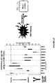

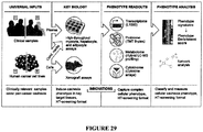


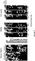
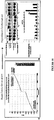


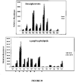

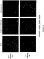


Classifications
-
- C—CHEMISTRY; METALLURGY
- C07—ORGANIC CHEMISTRY
- C07K—PEPTIDES
- C07K16/00—Immunoglobulins [IGs], e.g. monoclonal or polyclonal antibodies
- C07K16/18—Immunoglobulins [IGs], e.g. monoclonal or polyclonal antibodies against material from animals or humans
- C07K16/28—Immunoglobulins [IGs], e.g. monoclonal or polyclonal antibodies against material from animals or humans against receptors, cell surface antigens or cell surface determinants
- C07K16/2803—Immunoglobulins [IGs], e.g. monoclonal or polyclonal antibodies against material from animals or humans against receptors, cell surface antigens or cell surface determinants against the immunoglobulin superfamily
-
- A—HUMAN NECESSITIES
- A61—MEDICAL OR VETERINARY SCIENCE; HYGIENE
- A61K—PREPARATIONS FOR MEDICAL, DENTAL OR TOILETRY PURPOSES
- A61K31/00—Medicinal preparations containing organic active ingredients
- A61K31/16—Amides, e.g. hydroxamic acids
- A61K31/165—Amides, e.g. hydroxamic acids having aromatic rings, e.g. colchicine, atenolol, progabide
- A61K31/166—Amides, e.g. hydroxamic acids having aromatic rings, e.g. colchicine, atenolol, progabide having the carbon of a carboxamide group directly attached to the aromatic ring, e.g. procainamide, procarbazine, metoclopramide, labetalol
-
- A—HUMAN NECESSITIES
- A61—MEDICAL OR VETERINARY SCIENCE; HYGIENE
- A61K—PREPARATIONS FOR MEDICAL, DENTAL OR TOILETRY PURPOSES
- A61K31/00—Medicinal preparations containing organic active ingredients
- A61K31/33—Heterocyclic compounds
- A61K31/395—Heterocyclic compounds having nitrogen as a ring hetero atom, e.g. guanethidine or rifamycins
- A61K31/41—Heterocyclic compounds having nitrogen as a ring hetero atom, e.g. guanethidine or rifamycins having five-membered rings with two or more ring hetero atoms, at least one of which being nitrogen, e.g. tetrazole
- A61K31/4164—1,3-Diazoles
-
- A—HUMAN NECESSITIES
- A61—MEDICAL OR VETERINARY SCIENCE; HYGIENE
- A61K—PREPARATIONS FOR MEDICAL, DENTAL OR TOILETRY PURPOSES
- A61K31/00—Medicinal preparations containing organic active ingredients
- A61K31/33—Heterocyclic compounds
- A61K31/395—Heterocyclic compounds having nitrogen as a ring hetero atom, e.g. guanethidine or rifamycins
- A61K31/41—Heterocyclic compounds having nitrogen as a ring hetero atom, e.g. guanethidine or rifamycins having five-membered rings with two or more ring hetero atoms, at least one of which being nitrogen, e.g. tetrazole
- A61K31/425—Thiazoles
- A61K31/426—1,3-Thiazoles
-
- A—HUMAN NECESSITIES
- A61—MEDICAL OR VETERINARY SCIENCE; HYGIENE
- A61P—SPECIFIC THERAPEUTIC ACTIVITY OF CHEMICAL COMPOUNDS OR MEDICINAL PREPARATIONS
- A61P43/00—Drugs for specific purposes, not provided for in groups A61P1/00-A61P41/00
-
- G—PHYSICS
- G01—MEASURING; TESTING
- G01N—INVESTIGATING OR ANALYSING MATERIALS BY DETERMINING THEIR CHEMICAL OR PHYSICAL PROPERTIES
- G01N33/00—Investigating or analysing materials by specific methods not covered by groups G01N1/00 - G01N31/00
- G01N33/48—Biological material, e.g. blood, urine; Haemocytometers
- G01N33/50—Chemical analysis of biological material, e.g. blood, urine; Testing involving biospecific ligand binding methods; Immunological testing
- G01N33/5005—Chemical analysis of biological material, e.g. blood, urine; Testing involving biospecific ligand binding methods; Immunological testing involving human or animal cells
- G01N33/5008—Chemical analysis of biological material, e.g. blood, urine; Testing involving biospecific ligand binding methods; Immunological testing involving human or animal cells for testing or evaluating the effect of chemical or biological compounds, e.g. drugs, cosmetics
- G01N33/502—Chemical analysis of biological material, e.g. blood, urine; Testing involving biospecific ligand binding methods; Immunological testing involving human or animal cells for testing or evaluating the effect of chemical or biological compounds, e.g. drugs, cosmetics for testing non-proliferative effects
- G01N33/5023—Chemical analysis of biological material, e.g. blood, urine; Testing involving biospecific ligand binding methods; Immunological testing involving human or animal cells for testing or evaluating the effect of chemical or biological compounds, e.g. drugs, cosmetics for testing non-proliferative effects on expression patterns
-
- G—PHYSICS
- G01—MEASURING; TESTING
- G01N—INVESTIGATING OR ANALYSING MATERIALS BY DETERMINING THEIR CHEMICAL OR PHYSICAL PROPERTIES
- G01N33/00—Investigating or analysing materials by specific methods not covered by groups G01N1/00 - G01N31/00
- G01N33/48—Biological material, e.g. blood, urine; Haemocytometers
- G01N33/50—Chemical analysis of biological material, e.g. blood, urine; Testing involving biospecific ligand binding methods; Immunological testing
- G01N33/68—Chemical analysis of biological material, e.g. blood, urine; Testing involving biospecific ligand binding methods; Immunological testing involving proteins, peptides or amino acids
- G01N33/6893—Chemical analysis of biological material, e.g. blood, urine; Testing involving biospecific ligand binding methods; Immunological testing involving proteins, peptides or amino acids related to diseases not provided for elsewhere
-
- A—HUMAN NECESSITIES
- A61—MEDICAL OR VETERINARY SCIENCE; HYGIENE
- A61K—PREPARATIONS FOR MEDICAL, DENTAL OR TOILETRY PURPOSES
- A61K38/00—Medicinal preparations containing peptides
- A61K38/16—Peptides having more than 20 amino acids; Gastrins; Somatostatins; Melanotropins; Derivatives thereof
-
- C—CHEMISTRY; METALLURGY
- C07—ORGANIC CHEMISTRY
- C07K—PEPTIDES
- C07K2317/00—Immunoglobulins specific features
- C07K2317/30—Immunoglobulins specific features characterized by aspects of specificity or valency
- C07K2317/33—Crossreactivity, e.g. for species or epitope, or lack of said crossreactivity
-
- C—CHEMISTRY; METALLURGY
- C07—ORGANIC CHEMISTRY
- C07K—PEPTIDES
- C07K2317/00—Immunoglobulins specific features
- C07K2317/30—Immunoglobulins specific features characterized by aspects of specificity or valency
- C07K2317/34—Identification of a linear epitope shorter than 20 amino acid residues or of a conformational epitope defined by amino acid residues
-
- C—CHEMISTRY; METALLURGY
- C07—ORGANIC CHEMISTRY
- C07K—PEPTIDES
- C07K2317/00—Immunoglobulins specific features
- C07K2317/70—Immunoglobulins specific features characterized by effect upon binding to a cell or to an antigen
- C07K2317/76—Antagonist effect on antigen, e.g. neutralization or inhibition of binding
-
- G—PHYSICS
- G01—MEASURING; TESTING
- G01N—INVESTIGATING OR ANALYSING MATERIALS BY DETERMINING THEIR CHEMICAL OR PHYSICAL PROPERTIES
- G01N2800/00—Detection or diagnosis of diseases
- G01N2800/04—Endocrine or metabolic disorders
- G01N2800/042—Disorders of carbohydrate metabolism, e.g. diabetes, glucose metabolism
-
- G—PHYSICS
- G01—MEASURING; TESTING
- G01N—INVESTIGATING OR ANALYSING MATERIALS BY DETERMINING THEIR CHEMICAL OR PHYSICAL PROPERTIES
- G01N2800/00—Detection or diagnosis of diseases
- G01N2800/10—Musculoskeletal or connective tissue disorders
-
- G—PHYSICS
- G01—MEASURING; TESTING
- G01N—INVESTIGATING OR ANALYSING MATERIALS BY DETERMINING THEIR CHEMICAL OR PHYSICAL PROPERTIES
- G01N2800/00—Detection or diagnosis of diseases
- G01N2800/12—Pulmonary diseases
- G01N2800/122—Chronic or obstructive airway disorders, e.g. asthma COPD
-
- G—PHYSICS
- G01—MEASURING; TESTING
- G01N—INVESTIGATING OR ANALYSING MATERIALS BY DETERMINING THEIR CHEMICAL OR PHYSICAL PROPERTIES
- G01N2800/00—Detection or diagnosis of diseases
- G01N2800/32—Cardiovascular disorders
-
- G—PHYSICS
- G01—MEASURING; TESTING
- G01N—INVESTIGATING OR ANALYSING MATERIALS BY DETERMINING THEIR CHEMICAL OR PHYSICAL PROPERTIES
- G01N2800/00—Detection or diagnosis of diseases
- G01N2800/52—Predicting or monitoring the response to treatment, e.g. for selection of therapy based on assay results in personalised medicine; Prognosis
Definitions
- the invention relates to cachexia, including compositions and methods for detecting individuals who are or who are likely to suffer from cachexia, and compositions and methods and uses of compositions for treating or alleviating cachexia.
- Cachexia is a wasting syndrome associated with chronic diseases. Cachexia is defined as weight loss exceeding 5% within the previous 3-12 months, combined with fatigue, loss of skeletal muscle, and biochemical abnormalities (e.g., anemia or insulin resistance).
- Cancer-induced cachexia (CIC) is experienced by up to 80% of patients with advanced stage cancer, particularly those with gastrointestinal, pancreatic, thoracic and head and neck malignancies. CIC has been implicated in up to 25% of cancer-related deaths.
- interventions such as total parenteral nutrition (complete daily intravenous nutrition), anti-inflammatory medications, and anabolic stimulation, a patient with cancer-induced cachexia will continue to lose weight, often becoming so frail that they are unable to receive anti-cancer therapies. This distinguishes CIC from other forms of cachexia, which may respond to nutrition supplementation coupled with anti-inflammatory therapy.
- RAGE The receptor for advanced glycation end products
- DAMPs Damage-Associated Molecular Pattern molecules
- Anti-RAGE monoclonal antibodies are known in the art and are developed to treat conditions known to involve the RAGE receptor, and compete for binding with a range of RAGE ligands including HMGB1. These anti-RAGE monoclonal antibodies are not the subject of the invention and teach away from the instant invention.
- RAGE advanced glycation end products
- Applicants have now demonstrated the role of particular RAGE ligands in pre-cachexia and/or cachexia, and disclose herein therapeutic engineered biologic compositions for advantageously and more specifically targeting RAGE in a subject at risk of developing pre-cachexia and/or cachexia or a subject suffering from pre-cachexia and/or cachexia.
- the invention provides therapeutic, engineered protein/peptide compositions comprising e.g., anti-RAGE antibodies, to target a RAGE receptor (including soluble forms thereof and endogenous secretory RAGE), directly and/or via differential competition with one or more pre-cachexia and/or cachexia-associated RAGE ligands or markers.
- pre-cachexia is characterized by an alteration in biomarker or specific component levels in cells or body tissue like RAGE ligands (e.g. S100A7, S100A8 and S100A9), Myosin Heavy Chain (MYH) (e.g.
- any of skeletal or cardiac muscle MYHs like myosin, heavy chain 3, skeletal muscle, embryonic (MYH3); myosin, heavy chain 6, cardiac muscle, alpha (MYH6); myosin, heavy chain 2, skeletal muscle, adult (MYH1, MYH2); myosin, heavy chain 7, cardiac muscle, beta (MYH7); myosin, heavy chain 7B, cardiac muscle, beta (MYH7B) etc), Soluble Insulin Receptor (INSR) and Branched Chain Amino Acids (BCAA).
- An alteration in the levels of the biomarkers may be an increase in level or a decrease in level of the biomarkers as compared to a baseline reference value level (e.g.
- Loss of lipid content in cells and cell atrophy are also changes that are observed in a pre-cachexia or cachexia state. Loss of lipid content may be associated with increased levels of lipid metabolites like sphingomyelins, lysophospholipids, di-acyl-glycerides, triacyl glycerides, cholesterol esters, and/ or phospholipids.
- increase in the levels of one or more of S100A7, S100A8, S100A9, MYH1, MYH2, MYH3, MYH6, MYH7, MYH7B and INSR are correlated to pre-cachexia and cachexia.
- the levels of BCAA are increased in pre-cachexia but are decreased in advanced or late stage cachexia.
- Applicants have also determined that increase in the plasma levels of one of more of S100A4, S100A6, S100A11, S100A12, S100P and HMGB1 are anti-correlated to pre-cachexia and cachexia.
- the increase in levels of said biomarkers are determined in fluid obtained from a subject or patient, wherein the fluid may be plasma, serum, urine, saliva, ascites or pleural fluid. In a more preferred embodiment, the levels of said biomarkers are determined in plasma or serum.
- the invention provides an anti-RAGE antibody or antibodies specific for a RAGE receptor, which antibody competes for binding to the RAGE receptor with a RAGE ligand of the S100 family, e.g ., S100A7, S100A8, S100A9 and/or Calprotectin, and preferably may not compete for binding to the RAGE receptor with the RAGE ligands HMGB1 or S100B.
- a RAGE ligand of the S100 family e.g ., S100A7, S100A8, S100A9 and/or Calprotectin
- the invention provides an anti-RAGE antibody mixture/cocktail comprising one or more antibodies which compete for ligand binding to the RAGE receptor, wherein each antibody in said mixture/cocktail competes for binding of one or more specific S100 family member/ligand (e.g ., S100A7, S100A8, S100A9 and/or Calprotectin) to the RAGE receptor, and preferably may not compete for binding to the RAGE receptor with the RAGE ligands HMGB1 or S100B.
- S100 family member/ligand e.g ., S100A7, S100A8, S100A9 and/or Calprotectin
- the invention provides an antibody specific for a RAGE receptor, which antibody competes for binding to the RAGE receptor with a RAGE ligand of the S100 family, wherein said competition is via a moiety/epitope (linear and/or conformational) which is bound by one or more specific S100 family members/ligands that induce pre-cachexia and/or cachexia, but is not shared by S100 RAGE ligands which do not induce cachexia or pre-cachexia.
- a moiety/epitope linear and/or conformational
- the antibody binds to an epitope on the RAGE receptor selected from an epitope, which is bound by S100A7, S100A8, S100A9 and/or Calprotectin, and preferably may not bind to an epitope, which is bound by the RAGE ligand HMGB1.
- the antibody binds to a RAGE epitope bound by S100A7, S100A8, S100A9 and/or Calprotectin by mimicking the linear epitope defined by any one of residues 41-93 in S100A7; residues 36-89 in S100A8; or residues 45-98 in S100A9.
- the antibody binds to a RAGE epitope bound by S100A7, S100A8, S100A9 and/or Calprotectin by mimicking the conformational epitope represented by helix 2A formed by residues 41-53 in S100A7, residues 43-49 in S100A8 or residues 47-55 in S100A9.
- the antibody does not mimic the epitope defined by residues 43-78 on S100B.
- the antibody binds to the V region of the RAGE receptor, formed by residues 23-116 in human RAGE, mimicking the interaction between the RAGE V-region and helix A2 in S100A7, S100A8 or S100A9.
- the antibody preferably may not bind to the V region of the RAGE receptor, formed by residues 23-116 in human RAGE, in a manner which mimics the interaction between RAGE and S100B residues 43-78.
- the invention provides an antibody specific for a RAGE ligand selected from S100A7, S100A8, S100A9 and/or Calprotectin, and preferably may not bind to the RAGE ligands HMGB1 or S 100B.
- the antibody according to this aspect of the invention mimics the RAGE epitope, which is targeted by S100A7, S100A8, S100A9 and/or Calprotectin.
- the antibody binds to a linear epitope, which is selected from the linear epitope defined by residues 41-93 in S100A7; residues 36-89 in S100A8; or residues 45-98 in S100A9.
- the antibody preferably may not bind to the S100B epitope defined by residues 43-78 on S100B.
- the antibody binds to the conformational epitope represented by helix A2 formed by residues 41-53 in S100A7, residues 43-49 in S100A8, residues 47-55 in S100A9 or residues 40-53 in S100A15.
- the antibody preferably may not bind to the conformational epitopes formed on HMGB1 or S100B.
- any of the aforementioned antibody/antibodies may be selected from, but not limited to, an IgG, IgA, or an antigen binding antibody fragment selected from an antibody single variable domain polypeptide, dAb, FAb, F(ab')2, an scFv, an Fv, a V HH domain (such as a Nanobody® or other camelized immunoglobulin domain) or a disulfide-bonded Fv.
- any of the above antibody types or fragments thereof may be prepared from one or more of a mammalian species selected from, but not limited to mouse, rat, rabbit, human. But of course, such antibodies should be humanized for use in humans.
- any of the above antibody types or fragments thereof may be provided as heterocinjugates, bispecific, single-chain, chimeric or humanized molecules having affinity for one or more specific S100 family members/ligands that induce pre-cachexia and/or cachexia.
- any of the hereinmentioned antibody/antibodies binds to a RAGE receptor polypeptide or an S100A7, A8, A9 or A15 polypeptide, with a dissociation coefficient of 100nM or less, 75nM or less, 50nM or less, 25nM or less, such as 10nM or less, 5nM or less, 1nM or less, or in embodiments 500pM or less, 100pM or less, 50pM or less or 25pM or less.
- any of the hereinmentioned antibody/antibodies may be provided as polyclonal and/or monoclonal antibodies, including any mixture thereof.
- antibody includes polyclonal antisera and antibody cocktails as well as monoclonal antibodies.
- Antibodies may be monospecific, with narrow or broad specificity; or multispecific, such as bispecific, such that they possess two distinct epitope specificities in a single antibody molecule. Cocktails of antibodies may be targeted at two or more specific epitopes.
- Polyclonal antisera can be raised in a conventional manner against one or more antigens, and may include IgG, IgM and other antibody classes. In certain embodiments, antisera may be modified to comprise only IgG or only IgM.
- Antibody cocktails may be prepared by admixture of one or more monoclonal antibodies.
- an antibody cocktail contains two, three, four or more monoclonal antibodies each of which binds to or mimics S100A7, S100A8 and/or S100A9, so binds to S100A15, as set out herein, but preferably may not bind to or mimic HMGB1 or S100B.
- the foregoing antigens may be targeted by multispecific antibodies and/ore polyclonal antisera.
- the antibody is monoclonal and binds a unique structural motif shared by S100A7/ A8/ A9/ A15 or a bi or multi-valent antibody that binds to any combination of S100A7/ A8/ A9/ A15.
- the antibody or antibodies of the invention are formulated for intravenous (iv) or intramuscular (im) administration.
- Antibodies administered iv should extravasate from the circulation in order to enter the interstitial tissue space and bind to their cognate target. It is an advantage of the present invention that the anti-cachexia antibodies do not need to be targeted to solid tumors, which are resistant to extravasation as they exert a higher tissue fluid pressure which retards convective transport of antibodies from blood vessels into tissue.
- the antibody in one embodiment, is an antibody fragment such as a scFv, dAb or V HH antibody.
- Small antibody fragments are extravasated much more readily into tissue, and for this reason can perform better than IgG or other larger antibodies. However, smaller fragments are also cleared faster from the circulation. A compromise must be struck between tissue accessibility and clearance. For example, see Wang et al., Clinical pharmacology & Therapeutics, 84:5, 2008, 548-558 .
- Several antibody conjugates have been described which have extended half-life using a variety of strategies, for example through conjugation to albumin (such as human serum albumin). See Kontermann et al., BioDrugs April 2009, Volume 23, Issue 2, pp 93-109 .
- the invention provides a vector system comprising nucleic acid molecule(s) encoding and expressing therapeutic, engineered protein/peptide compositions comprising e.g., antibodies, to target a RAGE receptor directly and/or via differential competition with one or more pre-cachexia and/or cachexia-associated RAGE ligands or markers.
- the invention provides a viral vector system (e.g ., AAV, retroviral, lentiviral) comprising nucleic acid molecule(s) encoding and expressing therapeutic, engineered protein/peptide compositions comprising e.g., antibodies, to target a RAGE receptor directly and/or via differential competition with one or more pre-cachexia and/or cachexia-associated RAGE ligands or markers.
- a viral vector system e.g ., AAV, retroviral, lentiviral
- nucleic acid molecule(s) encoding and expressing therapeutic, engineered protein/peptide compositions comprising e.g., antibodies
- the invention provides a method for viral delivery comprising administering to a subject at risk of developing pre-cachexia and/or cachexia or a subject suffering from pre-cachexia and/or cachexia, a viral vector system comprising nucleic acid molecule(s) encoding and expressing therapeutic, engineered protein/peptide compositions comprising e.g., antibodies, to target a RAGE receptor directly and/or via differential competition with one or more pre-cachexia and/or cachexia-associated RAGE ligands or markers.
- the viral vector system is an AAV system or a lentivral system.
- Therapetuic compositions may also be delievered uing a modified RNA system ( Zangi et al., Nature Biotechnology 31, 898-907 (2013 )).
- the invention features a method of inhibiting the loss of myosin heavy chain in a myocyte, the method involving contacting the myocyte with an effective amount of one or more agents that is an agent that inhibits RAGE activity, that is any one or more of an anti-RAGE antibody, a RAGE blocking peptide or small molecule that inhibits binding of RAGE to ligands, as defined in the foregoing aspects of the invention.
- the invention features a method of inhibiting lipolysis in an adipocyte, the method involving contacting the adipocyte with an effective amount of one or more agents that is an agent that inhibits RAGE activity, that is any one or more of an anti-RAGE antibody, a RAGE blocking peptide or small molecule that inhibits binding of RAGE to ligands, as defined in the foregoing aspects of the invention.
- the invention features a method of inhibiting atrophy in a cell, the method involving contacting the adipocyte, myocyte, or cardiomyocyte with an effective amount of one or more agents that is an agent that inhibits RAGE activity, that is an anti-RAGE antibody, as defined in the foregoing aspects of the invention.
- the disclosure features a method of inhibiting atrophy in a cell, the method involving contacting the adipocyte, myocyte, or cardiomyocyte with an effective amount of one or more agents that is an agent that inhibits RAGE activity, that is any one or more of an anti-RAGE antibody, a RAGE blocking peptide or small molecule that inhibits binding of RAGE to ligands, as defined in the foregoing aspects of the invention.
- the myocyte, adipocyte, cardiomyocyte or hepatocyte cell is in vitro or in vivo.
- the myocyte, adipocyte, or hepatocyte cell is present in a subject identified as having at least one cancer.
- the cancer includes but is not limited to one or more carcinomas and sarcomas and cancers of the skin, pancreas, stomach, colon, thorax, liver, gallbladder, musculoskeletal system, breast, lung, ovary, uterus, endometrium, prostrate, colon, skin, mouth, salivary, esophagus, head and neck, plus other tumors of the gastrointestinal tract.
- the invention features a method of treating pre-cachexia, the method involving administering to the subject an effective amount of one or more agents that is an agent that inhibits RAGE activity, that is any one or more of an anti-RAGE antibody, a RAGE blocking peptide or small molecule that inhibits binding of RAGE to ligands, as defined in the foregoing aspects of the invention.
- the invention features a method of inhibiting the progression of precachexia to cachexia in a subject, the method involving administering to the subject an effective amount of one or more agents that is an agent that inhibits RAGE activity, that is an anti-RAGE antibody, as defined in the foregoing aspects of the invention.
- the disclosure features a method of inhibiting the progression of precachexia to cachexia in a subject, the method involving administering to the subject an effective amount of one or more agents that is an agent that inhibits RAGE activity, that is any one or more of, a RAGE blocking peptide or small molecule that inhibits binding of RAGE to ligands, as defined in the foregoing aspects of the invention.
- the invention features a method of treating or preventing undesirable muscle or fat loss in a cancer patient, the method involving administering to the subject an effective amount of one or more agents that is an agent that inhibits RAGE activity, that is anti-RAGE antibody, as defined in the foregoing aspects of the invention.
- the disclosure features a method of treating or preventing undesirable muscle or fat loss in a cancer patient, the method involving administering to the subject an effective amount of one or more agents that is an agent that inhibits RAGE activity, that is any one or more of a RAGE blocking peptide or small molecule that inhibits binding of RAGE to ligands, as defined in the foregoing aspects of the invention.
- the subject is pre-selected as having a molecular signature indicative of pre-cachexia or cachexia by detecting an alteration in at least three markers that is any one or more of S100A2 or S100A4; S100A8 or S100A9; and S100A7; detecting an alteration in at least four markers that is any one or more of: S100A2 or S100A4; S100A8 or S100A9; S100A7; and S100A14; and/or measuring the level of at least five markers that is any one or more of: S100A2 or S100A4; S100A8 or S100A9; S100A7; S100A14; and S100P, thereby pre-selecting the patient as having a molecular signature indicative of pre-cachexia or cachexia.
- markers are not causative of cachexia, many markers are coordinately expressed and hence provide diagnostic potential even if not specifically causative of cachexia.
- the disclosure features a method of enhancing cancer sensitivity to chemotherapy, the method involving administering gemcitabine in combination with an anti-RAGE therapy that is any one or more of an anti-RAGE antibody, a RAGE blocking peptide, an antibody targeted to a RAGE ligand such as S100 A7/ A8/ A9 or A15, or a small molecule that inhibits binding of RAGE to ligands, as described in the preceding aspects of the invention, thereby enhancing cancer sensitivity to chemotherapy. Also included are antibodies specific to the exposed V-domain of RAGE, as explained herein. Gemcitabine is a cytotoxic chemotherapeutic agent, which is used most often in combination with other agents (such as platinum compounds or combinations of drugs such as FOLFOX).
- an anti-RAGE therapy that is any one or more of an anti-RAGE antibody, a RAGE blocking peptide, an antibody targeted to a RAGE ligand such as S100 A7/ A8/ A9 or A15, or a small molecule that inhibits binding of
- the cancer is that is any one or more of one or more carcinoma or sarcoma, including but not limited to cancers of the skin, pancreas, stomach, colon, thorax, liver, gallbladder, musculoskeletal system, breast, lung, ovary, uterus, endometrium, prostrate, colon, skin, mouth, salivary, esophagus, head and neck, plus other tumors of the gastrointestinal tract.
- carcinoma or sarcoma including but not limited to cancers of the skin, pancreas, stomach, colon, thorax, liver, gallbladder, musculoskeletal system, breast, lung, ovary, uterus, endometrium, prostrate, colon, skin, mouth, salivary, esophagus, head and neck, plus other tumors of the gastrointestinal tract.
- anti-RAGE therapy can be delivered in combination with chemotherapy, which can achieve synergistic and/or curative results.
- Chemotherapeutic agents can be drugs or cytotoxic agents that inhibit or prevens the function of cells and/or causes destruction of cells.
- the drugs or cytotoxic agents may be targeted, or systemically administered.
- Examples of cytotoxic agents include radioactive isotopes, chemotherapeutic agents, and toxins such as small molecule toxins or enzymatically active toxins of bacterial, fungal, plant or animal origin, including synthetic analogues and derivatives thereof.
- the cytotoxic agent may be selected from the group consisting of an auristatin, a DNA minor groove binding agent, a DNA minor groove alkylating agent, an enediyne, a lexitropsin, a duocarmycin, a taxane, a puromycin, a dolastatin, a maytansinoid and a vinca alkaloid or a combination of two or more thereof.
- the drug is a chemotherapeutic agent selected from the group consisting of a topoisomerase inhibitor, an alkylating agent (eg . nitrogen mustards; ethylenimes; alkylsulfonates; triazenes; piperazines; and nitrosureas), an antimetabolite (eg mercaptopurine, thioguanine, 5-fluorouracil), an antibiotics ( eg . anthracyclines, dactinomycin, bleomycin, adriamycin, mithramycin. dactinomycin) a mitotic disrupter ( eg .
- a topoisomerase inhibitor eg . nitrogen mustards; ethylenimes; alkylsulfonates; triazenes; piperazines; and nitrosureas
- an antimetabolite eg mercaptopurine, thioguanine, 5-fluorouracil
- an antibiotics eg
- plant alkaloids - such as vincristine and/or microtubule antagonists - such as paclitaxel
- a DNA intercalating agent eg carboplatin and/or cisplatin
- a DNA synthesis inhibitor eg. DNA-RNA transcription regulator
- an enzyme inhibitor eg. carboplatin and/or cisplatin
- a DNA synthesis inhibitor eg. DNA-RNA transcription regulator
- an enzyme inhibitor eg. agene regulator, a hormone response modifier
- a hypoxia-selective cytotoxin eg. tirapazamine
- an epidermal growth factor inhibitor eg. xanthenone 5,6-dimethylxanthenone-4-acetic acid
- a radiation-activated prodrug eg . nitroarylmethyl quaternary (NMQ) salts
- bioreductive drug eg. nitroarylmethyl quaternary (NMQ) salts
- the chemotherapeutic agent may selected from the group consisting of Erlotinib (TARCEVA®), Bortezomib (VELCADE®), Fulvestrant (FASLODEX®), Sutent (SU11248), Letrozole (FEMARA®), Imatinib mesylate (GLEEVEC®), PTK787/ZK 222584, Oxaliplatin (Eloxatin®.), 5-FU (5-fluorouracil), Leucovorin, Rapamycin (Sirolimus, RAPAMUNE®.), Lapatinib (GSK572016), Lonafarnib (SCH 66336), Sorafenib (BAY43-9006), and Gefitinib (IRESSA®.), AG1478, AG1571 (SU 5271; Sugen) or a combination of two or more thereof.
- TARCEVA® Erlotinib
- VELCADE® Bortezomib
- FASLODEX® Fulvestrant
- the chemotherapeutic agent may be an alkylating agent - such as thiotepa, CYTOXAN® and/or cyclosphosphamide; an alkyl sulfonate - such as busulfan, improsulfan and/or piposulfan; an aziridine - such as benzodopa, carboquone, meturedopa and/or uredopa; ethylenimines and/or methylamelamines - such as altretamine, triethylenemelamine, triethylenepbosphoramide, triethylenethiophosphoramide and/or trimethylomelamine; acetogenin - such as bullatacin and/or bullatacinone; camptothecin; bryostatin; callystatin; cryptophycins; dolastatin; duocarmycin; eleutherobin; pancratistatin; sarcodictyin; spongistatin; nitrogen mustards
- doxorubicin - such as morpholino-doxorubicin, cyanomorpholino-doxorubicin, 2-pyrrolino-doxorubicin and/or deoxydoxorubicin, epirubicin, esorubicin, idarubicin, marcellomycin, mitomycins - such as mitomycin C, mycophenolic acid, nogalamycin, olivomycins, peplomycin, potfiromycin, puromycin, quelamycin, rodorubicin, streptonigrin, streptozocin, tubercidin, ubenimex, zinostatin, zorubicin; anti-metabolites - such as methotrexate and 5-fluorouracil (5-FU); folic acid analogues - such as denopterin, methotrexate, pteropterin, trimetrexate; purine analogues - such as fludarabine, 6-mercaptopurine, thiamiprine,
- paclitaxel paclitaxel, abraxane, and/or TAXOTERE®, doxetaxel; chloranbucil; GEMZAR® gemcitabine; 6-thioguanine; mercaptopurine; methotrexate; platinum analogues - such as cisplatin and carboplatin; vinblastine; platinum; etoposide; ifosfamide; mitoxantrone; vincristine; NAVELBINE®, vinorelbine; novantrone; teniposide; edatrexate; daunomycin; aminopterin; xeloda; ibandronate; topoisomerase inhibitor RFS 2000; difluoromethylomithine (DMFO); retinoids - such as retinoic acid; capecitabine; and pharmaceutically acceptable salts, acids, derivatives or combinations of two or more of any of the above.
- platinum analogues - such as cis
- the drug may be a tubulin disruptor including but are not limited to: taxanes - such as paclitaxel and docetaxel, vinca alkaloids, discodermolide, epothilones A and B, desoxyepothilone, cryptophycins, curacin A, combretastatin A-4-phosphate, BMS 247550, BMS 184476, BMS 188791; LEP, RPR 109881A, EPO 906, TXD 258, ZD 6126, vinflunine, LU 103793, dolastatin 10, E7010, T138067 and T900607, colchicine, phenstatin, chalcones, indanocine, T138067, oncocidin, vincristine, vinblastine, vinorelbine, vinflunine, halichondrin B, isohomohalichondrin B, ER-86526, pironetin, spongistatin 1, spiket P, cryptophyc
- the drug may be a DNA intercalator including but are not limited to: acridines, actinomycins, anthracyclines, benzothiopyranoindazoles, pixantrone, crisnatol, brostallicin, CI-958, doxorubicin (adriamycin), actinomycin D, daunorubicin (daunomycin), bleomycin, idarubicin, mitoxantrone, cyclophosphamide, melphalan, mitomycin C, bizelesin, etoposide, mitoxantrone, SN-38, carboplatin, cis-platin, actinomycin D, amsacrine, DACA, pyrazoloacridine, irinotecan and topotecan and pharmaceutically acceptable salts, acids, derivatives or combinations of two or more of any of the above.
- acridines adriamycin
- the drug may be an anti-hormonal agent that acts to regulate or inhibit hormone action on tumours - such as anti-estrogens and selective estrogen receptor modulators, including, but not limited to, tamoxifen, raloxifene, droloxifene, 4-hydroxytamoxifen, trioxifene, keoxifene, LY117018, onapristone, and/or fareston toremifene and pharmaceutically acceptable salts, acids, derivatives or combinations of two or more of any of the above.
- an anti-hormonal agent that acts to regulate or inhibit hormone action on tumours -
- selective estrogen receptor modulators including, but not limited to, tamoxifen, raloxifene, droloxifene, 4-hydroxytamoxifen, trioxifene, keoxifene, LY117018, onapristone, and/or fareston toremifene and pharmaceutically acceptable salts, acids, derivatives or combinations of two or more of any
- the drug may be an aromatase inhibitor that inhibits the enzyme aromatase, which regulates estrogen production in the adrenal glands - such as, for example, 4(5)-imidazoles, aminoglutethimide, megestrol acetate, AROMASIN®. exemestane, formestanie, fadrozole, RIVISOR®. vorozole, FEMARA®. letrozole, and ARIMIDEX® and/or anastrozole and pharmaceutically acceptable salts, acids, derivatives or combinations of two or more of any of the above.
- aromatase inhibitor that inhibits the enzyme aromatase, which regulates estrogen production in the adrenal glands -
- 4(5)-imidazoles aminoglutethimide
- megestrol acetate megestrol acetate
- AROMASIN® exemestane
- formestanie formestanie
- fadrozole RIVISOR®
- vorozole FEMARA®. letroz
- the drug may be an anti-androgen - such as flutamide, nilutamide, bicalutamide, leuprolide, goserelin and/or troxacitabine and pharmaceutically acceptable salts, acids, derivatives or combinations of two or more of any of the above.
- an anti-androgen - such as flutamide, nilutamide, bicalutamide, leuprolide, goserelin and/or troxacitabine and pharmaceutically acceptable salts, acids, derivatives or combinations of two or more of any of the above.
- the drug may be a protein kinase inhibitor, a lipid kinase inhibitor or an anti-angiogenic agent.
- the drug could also be a cytokine (e.g., an interleukin, a member of the TNF superfamily, or an interferon).
- cytokine e.g., an interleukin, a member of the TNF superfamily, or an interferon.
- the drug is a maytansinoid, in particular DM1, or a tubulin disruptor.
- pre-cachexia is currently and clearly defined as a pathological state necessitating intervention (e.g., International Consensus Definition Classification 2011-present Lancet Onc, 2011, Vol. 12(5), 489-495 ). Accordingly, pre-cachexia encompasses the early pathophysiologic state of normal tissue wasting or atrophy which does not yet meet the clinical criteria for cachexia. For example, a subject may be pre-cachectic when their weight is stable or when their weight loss is about 1%, 2% or 3% of their body mass.
- At least a subset of patients with pre-cachexia may be characterized by an increase in one or more S100 RAGE ligands and/or a decrease in soluble RAGE relative to a reference level without unintended weight loss of at least 5% or more of body weight.
- pre-cachexia to be characterized by an alteration in biomarker or specific component levels in cells or body tissue like RAGE ligands (e.g. S100A7, S100A8 and S100A9), Myosin Heavy Chain (MYH) (e.g.
- the levels of biomarkers may be assayed in the plasma or other fluids (which include but are not limited to serum, urine, saliva, ascites, pleural fluid etc) or cells via biopsy samples- fresh or fixed.
- elevated mRNA levels of specific RAGE ligands or INSR have been assessed (most likely as cleavage products) is another diagnostic approach.
- An alteration in the levels of the biomarkers may be an increase in level or a decrease in level of the biomarkers as compared to a baseline reference value level (e.g. a reference value of the biomarker level in a healthy or control subject or individual). Loss of lipid content in cells and cell atrophy are also changes that are observed in a pre-cachexia or cachexia state.
- Loss of lipid content may be associated with increased levels of lipid metabolites like sphingomyelins, lysophospholipids, di-acyl-glycerides, triacyl glycerides, cholesterol esters, and/ or phospholipids.
- increase in the plasma levels of one or more of S100A7, MYH3, MYH6, INSR and BCAA are correlated to pre-cachexia and cachexia.
- alteration in the levels of biomarkers MYH1, MYH2, MYH7, MYH7B, MYH6, and MYH3 are related to skeletal or cardiac muscle cachexia.
- Applicants have also determined that increase in the plasma levels of one of more of S100A4, S100A6, S100A11, S100A12, S100P and HMGB1 are anti-correlated to pre-cachexia and cachexia.
- cachexia is a condition often but not exclusively associated with cancer, which is manifested in weight loss, of at least 5% or more of body weight.
- a universal definition of cachexia is provided in Fearon et al., Lancet Oncol 2011; 12: 489-95 .
- cachexia refers to the progressive loss of lean body mass (particularly of muscle mass) that typically is associated with gross body weight loss that is at least 5, 6, 7, 8, 9, 10% or more.
- Muscle and adipose tissue loss, indicative of cachexia may be detected by a computed tomography (CT) scan ( Martin et al., J Clin Oncol 31:1539-1547 (2013 )), though this method has not been validated as sufficient to formally diagnose the condition as there are numerous conditions that result in similar findings by imaging studies alone. Currently, there is no molecular biomarker(s) for this condition, as the pathogenesis of cancer-induced cachexia remains to be elucidated.
- CT computed tomography
- cancer-induced cachexia is meant cachexia associated with the presence of a cancer or tumor.
- disease-induced cachexia cachexia associated with the presence of a disease that is not due to the presence of at least one cancer or tumor.
- aspects of the invention may relate to tissue or muscle cachexia which may not only be associated with disease states but also with injury and acute or chronic stress.
- at least a subset of patients with disease-induced cachexia may be characterized by an increase in one or more S100 RAGE ligands and/or a decrease in soluble RAGE relative to a reference level.
- Cardiac cachexia is a form of cachexia that has long been recognized as a serious complication of heart disease which includes but is not limited to chronic heart failure (CHF), myocardial infarction, congestive heart failure etc. Cardiac cachexia is associated with poor prognosis, independently of functional disease severity, age, measures of exercise capacity, and left ventricular ejection fraction. Patients with cardiac cachexia suffer from generalised loss of lean tissue, fat tissue, as well as bone tissue. Cachectic CHF patients are weaker and fatigue earlier.
- An aspect of the invention encompasses treating cardiac cachexia with a medicament comprising an antibody to S100A7, S100A8 and/or S100A9.
- cardiac pre-cachexia is associated with an increased level of the biomarkers MYH6, MYH7 and MYH7B.
- COPD cachexia refers to the disproportionate loss of fat free mass (FFM) in chornic obstructive pulmonary disease (COPD), which is a lung disease characterized by irreversible chronic airflow limitation with or without alveolar wall destruction. Weight loss and particularly loss of FFM were shown to adversely affect respiratory and peripheral muscle function and exercise capacity as described in Broekhuizen et al., Pulmonary cachexia, systemic inflammatory profile, and the interleukin 1 ⁇ -511 single nucleotide polymorpbism1,2,3, Am J Clin Nutr. 2005 Nov;82(5): 1059-64 and Remels et al., The mechanisms of cachexia underlying muscle dysfunction in COPD, J Appl Physiol (1985).
- An aspect of the invention encompasses treating COPD cachexia with a medicament comprising an antibody to S100A7, S100A8 S100A9 and/or Calprotectin.
- cardiac pre-cachexia is associated with an increased level of the biomarkers MYH1, MYH2 and MYH3.
- cellular differentiation or “differentiation” is the process by which a less specialized cell becomes a more specialized cell type.
- High-throughput screening refers to a process that uses a combination of modern robotics, data processing and control software, liquid handling devices, and/or sensitive detectors, to efficiently process a large amount of (e.g., thousands, hundreds of thousands, or millions of) samples in biochemical, genetic or pharmacological experiments, either in parallel or in sequence, within a reasonably short period of time (e.g., days).
- the process is amenable to automation, such as robotic simultaneous handling of 96 samples, 384 samples, 1536 samples or more.
- a typical HTS robot tests up to 100,000 to a few hundred thousand compounds per day.
- the samples are often in small volumes, such as no more than 1 mL, 500 ⁇ l, 200 ⁇ l, 100 ⁇ l, 50 ⁇ l or less.
- high-throughput screening does not include handling large quantities of radioactive materials, slow and complicated operator-dependent screening steps, and/or prohibitively expensive reagent costs, etc.
- a therapeutic that "prevents" a disorder or condition refers to a compound that, in a statistical sample, reduces the occurrence of the disorder or condition in the treated sample relative to an untreated control sample, or delays the onset or reduces the severity of one or more symptoms of the disorder or condition relative to the untreated control sample.
- a "subject” means a human or animal (in the case of an animal, more typically a mammal, and can be, but is not limited to, a non-human animal or mammal).
- the subject is a human.
- a "subject" mammal can include, but is not limited to, a human or non-human mammal, such as a primate, bovine, equine, canine, ovine, feline, or rodent; and, it is understood that an adult human is typically about 70 kg, and a mouse is about 20g, and that dosing from a mouse or other non-human mammal can be adjusted to a 70 kg human by a skilled person without undue experimentation.
- treating is art-recognized and includes administration to the host or patient or subject of one or more of the subject compositions, e.g., to diminish, ameliorate, or stabilize the existing unwanted condition or side effects thereof.
- treatment is for the patient or subject in need thereof.
- agent is meant a peptide, nucleic acid molecule, or small compound.
- ameliorate decrease, suppress, attenuate, diminish, arrest, or stabilize the development or progression of a disease.
- alteration is meant a change (increase or decrease) in the expression levels or activity of a gene or polypeptide as detected by standard art known methods such as those described herein.
- an alteration includes a 10% change in expression levels, preferably a 25% change, more preferably more than a 30% change, a 35% change, a 40% change, and most preferably a 50% or greater change in expression levels.
- the upregulation or increase in biomarker levels is at least greater than a 30% increase over baseline or normal population reference standards.
- Detect refers to identifying the presence, absence or amount of the analyte to be detected.
- disease is meant any condition or disorder that damages or interferes with the normal function of a cell, tissue, or organ.
- the disease is pre-cachexia, cachexia, or refractory cachexia.
- an effective amount is meant the amount of a required to ameliorate the symptoms of a disease relative to an untreated patient.
- the effective amount of active compound(s) used to practice the present invention for therapeutic treatment of a disease varies depending upon the manner of administration, the age, body weight, and general health of the subject. Ultimately, the attending physician or veterinarian will decide the appropriate amount and dosage regimen. Such amount is referred to as an "effective" amount.
- the invention provides a number of targets that are useful for the development of highly specific drugs to treat or a disorder characterized by the methods delineated herein.
- the methods of the invention provide a facile means to identify therapies that are safe for use in subjects.
- the methods of the invention provide a route for analyzing virtually any number of compounds for effects on a disease described herein with high-volume throughput, high sensitivity, and low complexity.
- fragment is meant a portion of a polypeptide or nucleic acid molecule. This portion contains, preferably, at least 10%, 20%, 30%, 40%, 50%, 60%, 70%, 80%, or 90% of the entire length of the reference nucleic acid molecule or polypeptide.
- a fragment may contain 10, 20, 30, 40, 50, 60, 70, 80, 90, or 100, 200, 300, 400, 500, 600, 700, 800, 900, or 1000 nucleotides or amino acids.
- isolated refers to material that is free to varying degrees from components which normally accompany it as found in its native state.
- Isolate denotes a degree of separation from original source or surroundings.
- Purify denotes a degree of separation that is higher than isolation.
- a “purified” or “biologically pure” protein is sufficiently free of other materials such that any impurities do not materially affect the biological properties of the protein or cause other adverse consequences. That is, a nucleic acid or peptide of this invention is purified if it is substantially free of cellular material, viral material, or culture medium when produced by recombinant DNA techniques, or chemical precursors or other chemicals when chemically synthesized.
- Purity and homogeneity are typically determined using analytical chemistry techniques, for example, polyacrylamide gel electrophoresis or high performance liquid chromatography.
- the term "purified" can denote that a nucleic acid or protein gives rise to essentially one band in an electrophoretic gel.
- modifications for example, phosphorylation or glycosylation, different modifications may give rise to different isolated proteins, which can be separately purified.
- isolated polynucleotide is meant a nucleic acid (e.g., a DNA) that is free of the genes which, in the naturally-occurring genome of the organism from which the nucleic acid molecule of the invention is derived, flank the gene.
- the term therefore includes, for example, a recombinant DNA that is incorporated into a vector; into an autonomously replicating plasmid or virus; or into the genomic DNA of a prokaryote or eukaryote; or that exists as a separate molecule (for example, a cDNA or a genomic or cDNA fragment produced by PCR or restriction endonuclease digestion) independent of other sequences.
- the term includes an RNA molecule that is transcribed from a DNA molecule, as well as a recombinant DNA that is part of a hybrid gene encoding additional polypeptide sequence.
- polynucleotide refers to a polymer of deoxyribonucleotide or ribonucleotide that exists as a single-stranded or double-stranded form.
- the polynucleotide includes RNA genome sequences, DNA (gDNA and cDNA), and RNA sequences transcribed therefrom, and includes analogues of natural polynucleotides, unless specifically mentioned.
- the polynucleotide also includes nucleotide sequences encoding the amino acid sequences of the heavy and light chain variable regions of an antibody disclosed herein, and nucleotide sequences complementary thereto.
- the complementary sequences include completely complementary sequences and substantially complementary sequences.
- substantially complementary sequences are sequences that may be hybridized with nucleotide sequences encoding the amino acid sequences of the heavy or light chain variable regions of an antibody disclosed herein under stringent conditions known in the art. Stringent conditions mean, for example, hybridization to DNA in 6 ⁇ SSC at about 45°C, followed by one or more washes in 0.2 ⁇ SSC/0.1% SDS at about 50°C - 65°C.
- an “isolated polypeptide” is meant a polypeptide of the invention that has been separated from components that naturally accompany it.
- the polypeptide is isolated when it is at least 60%, by weight, free from the proteins and naturally-occurring organic molecules with which it is naturally associated.
- the preparation is at least 75%, more preferably at least 90%, and most preferably at least 99%, by weight, a polypeptide of the invention.
- An isolated polypeptide of the invention may be obtained, for example, by extraction from a natural source, by expression of a recombinant nucleic acid encoding such a polypeptide; or by chemically synthesizing the protein. Purity can be measured by any appropriate method, for example, column chromatography, polyacrylamide gel electrophoresis, or by HPLC analysis.
- Biomarker biological marker
- Biomarkers are often measured _and evaluated (e.g. whether their levels are increased or decreased or remain unchanged) to examine normal biological processes, pathogenic processes, or pharmacologic responses to a therapeutic intervention.
- an alteration in body mass, lean body mass, metabolism, or a metabolite, as well as RAGE ligands e.g. S100A7, S100A8 and S100A9, Myosin Heavy Chain (MYH) (e.g.
- myosin, heavy chain 3, skeletal muscle, embryonic (MYH3) or myosin, heavy chain 6, cardiac muscle, alpha (MYH6), myosin, heavy chain 2, skeletal muscle, adult (MYH1 and MYH2) etc), Soluble Insulin Receptor (INSR) and Branched Chain Amino Acids (BCAA) are markers or biomarkers (e.g., clinical indicator) of disease state (e.g., pre-cachexia or cachexia).
- Soluble Insulin Receptor is meant the soluble form of the insulin receptor (IR), a tetrameric transmembrane protein located on the surface of target cells, that is present in plamsa, e.g. human plasma.
- BCAA Branched Chain Amino Acid
- Branched Chain Amino Acid is meant an amino acid having aliphatic side-chains with a branch (a central carbon atom bound to three or more carbon atoms).
- proteinogenic amino acids there are three BCAAs which include but are not imited to leucine, isoleucine and valine.
- Non-proteinogenic BCAAs include but are not limited to norvaline and 2-aminoisobutyric acid.
- Validation of the elevation of circulating BCAA in human plasma as a early event in human pancreatic adenocarcinoma development is further described in Mayers et al., Elevation of circulating branched-chain amino acids is an early event in human pancreatic adenocarcinoma development, Nat Med. 2014 Oct;20(10): 1193-8 .
- levels of BCAA are initially found to increase in pre-cachexia but then the levels decrease in late stage cachexia, as further confirmed by Mayers et al.
- metabolic profile is meant alterations in the level or activity of one or more amino acid, small molecule cellular metabolite, polypeptide or lipid metabolites.
- obtaining as in “obtaining an agent” includes synthesizing, purchasing, or otherwise acquiring the agent.
- reduces is meant a negative alteration of at least 10%, 25%, 50%, 75%, or 100%.
- reference is meant a standard or control condition.
- a “reference sequence” is a defined sequence used as a basis for sequence comparison.
- a reference sequence may be a subset of or the entirety of a specified sequence; for example, a segment of a full-length cDNA or gene sequence, or the complete cDNA or gene sequence.
- the length of the reference polypeptide sequence will generally be at least about 16 amino acids, preferably at least about 20 amino acids, more preferably at least about 25 amino acids, and even more preferably about 35 amino acids, about 50 amino acids, or about 100 amino acids.
- the length of the reference nucleic acid sequence will generally be at least about 50 nucleotides, preferably at least about 60 nucleotides, more preferably at least about 75 nucleotides, and even more preferably about 100 nucleotides or about 300 nucleotides or any integer thereabout or therebetween.
- proteins including antibodies of the invention may associate with a specified region through various interactions to form ligand-receptor complexes. These interactions include but are not limited to electrostatic forces, such as hydrogen-bonding and Van der Waal forces, dipole-dipole interactions, hydrophobic interactions, pi-pi stacking, and so on. Other associations which describe more specific types of interactions include covalent bonds, electronic and conformational rearrangements, steric interactions, and so on.
- the term "associate” generally relates to any type of force which connects an antibody to a specified region.
- the term “interacts” generally relates to a more specific and stronger connection of an antibody to a specified region.
- sterically blocks is a specific type of association which describes an antibody interacting with a specific region and preventing other ligands from associating with that region through steric interactions.
- binds or “specifically binds” as used throughout this application may be interpreted to relate to the terms “associates”, “interacts” or “sterically blocks” as required.
- telomere binding By “specifically binds” is meant a compound or antibody that recognizes and binds a polypeptide of the invention, but which does not substantially recognize and bind other molecules in a sample, for example, a biological sample, which naturally includes a polypeptide of the invention.
- Nucleic acid molecules useful in the methods of the invention include any nucleic acid molecule that encodes a polypeptide of the invention or a fragment thereof. Such nucleic acid molecules need not be 100% identical with an endogenous nucleic acid sequence, but will typically exhibit substantial identity. Polynucleotides having "substantial identity" to an endogenous sequence are typically capable of hybridizing with at least one strand of a double-stranded nucleic acid molecule. Nucleic acid molecules useful in the methods of the invention include any nucleic acid molecule that encodes a polypeptide of the invention or a fragment thereof. Such nucleic acid molecules need not be 100% identical with an endogenous nucleic acid sequence, but will typically exhibit substantial identity.
- Polynucleotides having "substantial identity" to an endogenous sequence are typically capable of hybridizing with at least one strand of a double-stranded nucleic acid molecule.
- hybridize is meant pair to form a double-stranded molecule between complementary polynucleotide sequences (e.g., a gene described herein), or portions thereof, under various conditions of stringency.
- complementary polynucleotide sequences e.g., a gene described herein
- stringent salt concentration will ordinarily be less than about 750 mM NaCl and 75 mM trisodium citrate, preferably less than about 500 mM NaCl and 50 mM trisodium citrate, and more preferably less than about 250 mM NaCl and 25 mM trisodium citrate.
- Low stringency hybridization can be obtained in the absence of organic solvent, e.g., formamide, while high stringency hybridization can be obtained in the presence of at least about 35% formamide, and more preferably at least about 50% formamide.
- Stringent temperature conditions will ordinarily include temperatures of at least about 30° C, more preferably of at least about 37° C, and most preferably of at least about 42° C.
- Varying additional parameters, such as hybridization time, the concentration of detergent, e.g., sodium dodecyl sulfate (SDS), and the inclusion or exclusion of carrier DNA, are well known to those skilled in the art.
- concentration of detergent e.g., sodium dodecyl sulfate (SDS)
- SDS sodium dodecyl sulfate
- Various levels of stringency are accomplished by combining these various conditions as needed.
- hybridization will occur at 30° C in 750 mM NaCl, 75 mM trisodium citrate, and 1% SDS.
- hybridization will occur at 37° C in 500 mM NaCl, 50 mM trisodium citrate, 1% SDS, 35% formamide, and 100 .mu.g/ml denatured salmon sperm DNA (ssDNA).
- hybridization will occur at 42° C in 250 mM NaCl, 25 mM trisodium citrate, 1% SDS, 50% formamide, and 200 ⁇ g/ml ssDNA. Useful variations on these conditions will be readily apparent to those skilled in the art.
- wash stringency conditions can be defined by salt concentration and by temperature. As above, wash stringency can be increased by decreasing salt concentration or by increasing temperature.
- stringent salt concentration for the wash steps will preferably be less than about 30 mM NaCl and 3 mM trisodium citrate, and most preferably less than about 15 mM NaCl and 1.5 mM trisodium citrate.
- Stringent temperature conditions for the wash steps will ordinarily include a temperature of at least about 25 °C, more preferably of at least about 42° C, and even more preferably of at least about 68 °C.
- wash steps will occur at 25° C in 30 mM NaCl, 3 mM trisodium citrate, and 0.1% SDS. In a more preferred embodiment, wash steps will occur at 42 °C in 15 mM NaCl, 1.5 mM trisodium citrate, and 0.1% SDS. In a more preferred embodiment, wash steps will occur at 68° C in 15 mM NaCl, 1.5 mM trisodium citrate, and 0.1% SDS. Additional variations on these conditions will be readily apparent to those skilled in the art. Hybridization techniques are well known to those skilled in the art and are described, for example, in Benton and Davis (Science 196:180, 1977 ); Grunstein and Hogness (Proc. Natl.
- substantially identical is meant a polypeptide or nucleic acid molecule exhibiting at least 50% identity to a reference amino acid sequence (for example, any one of the amino acid sequences described herein) or nucleic acid sequence (for example, any one of the nucleic acid sequences described herein).
- a reference amino acid sequence for example, any one of the amino acid sequences described herein
- nucleic acid sequence for example, any one of the nucleic acid sequences described herein.
- such a sequence is at least 60%, more preferably 80% or 85%, and more preferably 90%, 95% or even 99% identical at the amino acid level or nucleic acid to the sequence used for comparison.
- Sequence identity is typically measured using sequence analysis software (for example, Sequence Analysis Software Package of the Genetics Computer Group, University of Wisconsin Biotechnology Center, 1710 University Avenue, Madison, Wis. 53705, BLAST, BESTFIT, GAP, or PILEUP/PRETTYBOX programs). Such software matches identical or similar sequences by assigning degrees of homology to various substitutions, deletions, and/or other modifications. Conservative substitutions typically include substitutions within the following groups: glycine, alanine; valine, isoleucine, leucine; aspartic acid, glutamic acid, asparagine, glutamine; serine, threonine; lysine, arginine; and phenylalanine, tyrosine. In an exemplary approach to determining the degree of identity, a BLAST program may be used, with a probability score between e -3 and e -100 indicating a closely related sequence.
- sequence analysis software for example, Sequence Analysis Software Package of the Genetics Computer Group, University of Wisconsin Bio
- Ranges provided herein are understood to be shorthand for all of the values within the range.
- a range of 1 to 50 is understood to include any number, combination of numbers, or sub-range from the group consisting 1, 2, 3, 4, 5, 6, 7, 8, 9, 10, 11, 12, 13, 14, 15, 16, 17, 18, 19, 20, 21, 22, 23, 24, 25, 26, 27, 28, 29, 30, 31, 32, 33, 34, 35, 36, 37, 38, 39, 40, 41, 42, 43, 44, 45, 46, 47, 48, 49, or 50.
- treat refers to reducing or ameliorating a disorder and/or symptoms associated therewith. It will be appreciated that, although not precluded, treating a disorder or condition does not require that the disorder, condition or symptoms associated therewith be completely eliminated.
- the term "about” is understood as within a range of normal tolerance in the art, for example within 2 standard deviations of the mean. About can be understood as within 10%, 9%, 8%, 7%, 6%, 5%, 4%, 3%, 2%, 1%, 0.5%, 0.1%, 0.05%, or 0.01% of the stated value. Unless otherwise clear from context, all numerical values provided herein are modified by the term about.
- compositions or methods provided herein can be combined with one or more of any of the other compositions and methods provided herein.
- S100A7, S100A8, and S100A9 are differentially secreted by cachexia-inducing cancer cells compared to cachexia non-inducing matched cancer cells (see Figure 37 ).
- RAGE gene expression is up-regulated in response to plasma from cachectic cancer patients but not to the plasma from non-cachectic cancer patients (see Figure 31 ).
- HMGB1 and S100B do not induce precachexia and/or cachexia and that S100A7 and S100A8/A9 dimers directly bind to RAGE in direct immunoprecipitation experiments, which was not previously known.
- Applicants' novel findings open the door for effectively targeting RAGE in the context of precachexia and cachexia.
- the invention disclosed herein provides therapeutic, engineered protein/peptide compositions comprising e.g., anti-RAGE antibodies to target a RAGE receptor (including soluble forms thereof) directly and/or via differential competition with one or more pre-cachexia and/or cachexia-associated RAGE ligands or markers (e.g., a RAGE ligand of the S100 family chosen from S100A7, S100A8, and S100A9).
- a RAGE ligand of the S100 family chosen from S100A7, S100A8, and S100A9
- Applicants' gene signature of cachexia generated by treating primary human skeletal muscle with plasma from case-matched controlled cachectic vs non-cachectic cancer patients is highly specific (99.9 percentile) for the gene expression state analyzed from clinical muscle biopsies from cachectic vs non-cachectic cancer patients.
- This cachexia gene signature was also used successfully as the screen for drugs that could block and reverse cachexia through MEK/ERK inhibition (see Figure 31 ). It was demonstrated that ERK activation is downstream of RAGE activation.
- Applicants have determined that transcription of S100A7, S100A8, and S100A9 is highly coordinately regulated in primary cancers from patient biopsy samples (see a TCGA heat map known in the art, see also Figure 27 ). It was discovered that specific inhibition of S100A7, S100A8/A9 by neutralizing Abs, but not neutralizing Abs against other S100 proteins inhibited the disease phenotype (see Figure 6 ). Applicants demonstrated that specifically only recombinant S100A7 or S100A8/A9 (Calprotectin) are capable of inducing the cachexia phenotype while other active recombinant S100 and HMGB1 proteins cannot (see Figure 27 ).
- S100 proteins in mouse and man from evolution to function and pathology (including an update of the nomenclature)).
- the functional diversity of S100 proteins is extremely borad, partly due to their ability to adapt to various targets, (see e.g., Permyakov S.E. et al., Mol. BioSyst. 2011 Jul;7(7):2164-80 .
- Intrinsic disorder in S100 proteins The crystal structures of several S100 proteins are known. (see e.g., Brodersen D. E. et al.
- Anti-RAGE antibodies are known; see, e.g., WO2008137552 , WO2007109747 , WO2010019656 , WO2009136382 , WO 2011053707 , WO2011042548 , US8,420,083 .
- Anti-RAGE monoclonal antibodies are known; see, e.g., US 8,420,083 , WO2011042548 and WO2008137552 .
- these antibodies and monoclonal antibodies compete for binding with a range of RAGE ligands including HMGB1-contrary to and teaching away from, the instant invention.
- WO2008137552 specifically involves antibodies that inhibit binding between RAGE and HMGB1.
- HMGB1 in not cachexogenic in muscle and therefore HMGB1 is NOT a relevant target RAGE ligand for treating, preventing, alleviating or addressing pre-cachexia and/or cachexia.
- the anti-RAGE antibodies and monoclonal antibodies of the prior art are not useful in the practice of the instant invention; fail to teach or suggest the present invention; and moreover, expressly teach away from the instant invention.
- the present invention provides antibody(ies) specific for a RAGE receptor, which antibody(ies) competes for binding to the RAGE receptor with RAGE ligand(s) S100A7, S100A8, S100A9 and/or Calprotectin, but preferably may not compete for binding to the RAGE receptor with RAGE ligand HMGB1; for example, antibody(ies) that binds to epitope(s) on the RAGE receptor selected from epitope(s) which is / are bound by S100A7, S100A8, S100A9 or Calprotectin, but preferably may not bind to an epitope which is bound by the RAGE ligand HMGB1.
- the prior art teaches and directs towards antibodies that compete for binding to the RAGE receptor with RAGE ligand HMBG1, and hence, teach away from the instant invention; and the prior art does not teach or suggest antibody(ies) competes for binding to the RAGE receptor with RAGE ligand(s) S100A7, S100A8, S100A9 and/or Calprotectin, but preferably may not compete for binding to the RAGE receptor with RAGE ligand HMGB 1, and hence does not teach or suggest the instant invention.
- the invention involves an antibody specific for a RAGE receptor, which antibody competes for binding to the RAGE receptor with a RAGE ligand selected from S100A7, S100A8, S100A9 and/or Calprotectin, but preferably may not compete for binding to the RAGE receptor with RAGE ligand HMGB1.
- a RAGE ligand selected from S100A7, S100A8, S100A9 and/or Calprotectin, but preferably may not compete for binding to the RAGE receptor with RAGE ligand HMGB1.
- S100A7 and S100A8/A9 dimers directly bind to RAGE in direct immunoprecipitation experiments.
- the antibody can be is selected from an IgG, IgA, or an antigen binding antibody fragment selected from an antibody single variable domain polypeptide, dAb, FAb, F(ab') 2 , an scFv, an Fv, or a disulfide-bonded Fv.
- the antibody can binds to a RAGE receptor polypeptide with a dissociation coefficient of 1 ⁇ m or less.
- the antibody can bind to a RAGE receptor polypeptide with a dissociation coefficient of 1nm or less.
- the antibody can bind to an epitope on the RAGE receptor selected from an epitope, which is bound by S100A7, S100A8, S100A9 or Calprotectin, but preferably may not bind to an epitope which is bound by the RAGE ligand HMGB1.
- the antibody can bind to a RAGE epitope bound by S100A7, S100A8 or S100A9 and/or calprotectin by mimicking the linear epitope defined by any one of residues 41-93 on S100A7, residues 36-89 on S100A8, or residues 45-98 on S100A9 (see Figure 6 ); or by mimicking the conformational epitope represented by helix A2 formed by residues 41-53 in S100A7, residues 43-49 in S100 A8 or residues 47-55 in S100A9; or which does not bind to the epitope defined by residues 43-78 in (S100B).
- the antibody can bind to residues 41-93 in (S100A7), residues 36-89 in (S100A8) or residues 45-98 in (S100A9), S100A15 or Calprotectin.
- the antibody binds to a linear epitope which is selected from the linear epitope defined by residues 41-93 in (S100A7), residues 36-89 in (S100A8), residues 45-98 in (S100A9) or residues 43-78 in (S100B), and does not bind to the S100B epitope defined by residues 43-78 in (S100B), or which binds to the conformational epitope formed by residues 41-53 on S100A7, residues 43-49 on S100A8, residues 47-55 on S100A9 or residues 40-53 on S100A15 and does not bind to the conformational epitope formed by residues 43-78 on S100B.
- an “epitope” can be a linear epitope, comprising a sequence of contiguous amino acids, or conformational, formed by the three-dimensional juxtaposition of amino acids and/or glycosyl groups in the tertiary structure of a protein.
- An epitope can comprise or consist of as few as at least 6 amino acids that are unique to a polypeptide or protein sequence.
- An epitope can be longer; for example, an epitope can comprise, consist essentially of or consist of at least 6 or at least 8, or at least 10, or at least 12, or at least 15 or at least 20 amino acids.
- S100 is, in embodiments of the invention, human S100.
- HMBG1 is, in embodiments of the invention, human HMGB1. Sequences of human S100 proteins, as well as HMBG1, can be located in the UniProt database under accession numbers P31151, P05109, P06702, P33763, P06703, P29034, P25815, P23297, P04271, Q99584 and P80511.
- the invention also encompasses sequences which are homologous the the database sequences recited above.
- the sequences according to the invention are at least 95% homologous the sequences referred to above. In other embodiments, they are at least 96%, 97%, 98%, 99% or 100% homologous. Homology can be assessed using any suitable sequence alignment technique. Optimal alignment may be detemined with the use of any suitable algorithm for aligning sequences, non-limiting examples of which include the Smith-Waterman algorithm, the Needleman-Tunsch algorithm, algorithms based on the Burrows-Wheeler Transfom (e.g.
- the invention comprises a method of treating pre-cachexia and providing treatment to patients in need thereof, the method comprising administering to the subject an effective amount of one or more agents that is an anti-RAGE antibody, a RAGE blocking peptide or small molecule that inhibits binding of RAGE to ligands.
- the current invention includes antibodies to the RAGE exposed V-domain for the prevention and or treatment of pre-cachexia.
- various combinations of 1) antibodies to the RAGE exposed V-domain, 2) antibodies to the S100A7, A8, A9, A14, A1, and 3) antibodies to the INSR/MYH are possible and effective for the prevention and or treatment of pre-cachexia.
- Different patients may have different combinations of these diagnostic and pharmacodynamics biomarkers.
- Aspects of the invention encompass identifying patients as pre-cachetic or cachectic based on these different combinations and providing them with subsequent treatment.
- the diagnostioc biomarkers include S100A7/8/9 (which may be considered the "driver” biomarkers), S100A1 and A14 (which may be considered as passenger or highly correlated biomarkers) and the pharmacodynamics biomarkers include MYH1/2/3/6/7s in addition to metabolomic profile of BCAA/ lipid metabolites. Any one of the above antibodies may be also combined with antibodies to MYH1, MYH2, MYH7 and MYH7B for the prevention and/or treatment of pre-cachexia related to skeletal or cardiac muscle cachexia.. Of these, S100A7 is important, as is S100A8 and S00A9.
- Antibodies to S100A7 can be combined with Antibodies to S100A8 and/or S100A9.
- S100A14 and S100A1 are of interest. And hence antibodies against S100A7 with antibiodies against S100A1 and/or S100A14, alone or in combination with antibodies against S100A8 and/or S100A9.
- the Soluble Insulin Receptor is increased across the board. From the foregoing, increased levels of S100A7, especially with increased levels of S100A8 and/or S100A9 may be important markers for observing pre-cachexia. Increased levels of INSR and/or MYH6 may be important markers too.
- Pre-cachexia patients may be observed though a combination of increased levels of S100A7, INSR, and MYH6 or S100A7, S100A8, INSR, and MYH6 or S100A7, S100A8, S100A9, INSR, and MYH6. With these can be increased levels of S100A1 and/or S100A14. Accordingly, pre-cachexia may be defined by upregulation of S100A7, S100A8, S100A9, S100A14 and INSR and optionally also MYH6, alone or in further combination with upregulation of S100A1 and/or S100A14.
- the invention thus envisions S100A7 and INSR, and combinations thereof, for diagnosis of pre-cachexia.
- the invention also envisions S100A7 and S100A8/A9 dimers directly bind to RAGE. Patients may be assayed for biomarker levels, wherein the biomarkers may be different combinations of S100A7/8/9, S100A1 and A14, and MYH1/2/3/6/7s further to the metabolomic profile of BCAA/ lipid metabolites. Diagnosis includes diagnosis in the context of in vitro assays.
- the invention also envisions S100A7, INSR, MYH6, MYH7, MYH7B for diagnosis of cardiac pre-cachexia.
- the invention further comprehends upregulation of at least two of S100A7, S100A8, S100A9, MYH1, MYH2, MYH3, MYH6, MYH7, MYH7B and/or INSR, alone or in further combination with S100A1 and/or S100A14 for diagnosis of pre-cachexia.
- Diagnosis includes diagnosis in the context of in vitro assays.
- the antibodies or small molecules of the invention may be used for the prevention and or treatment of cardiac pre-cachexia, wherein cardiac pre-cachexia is defined by upregulation of S100A7, INSR, and cardio specific MYH6 in plasma.
- the antibodies or small molecules of the invention may be used for the prevention and or treatment of cardiac pre-cachexia, wherein cardiac pre-cachexia is defined by upregulation of S100A7, INSR, and cardio specific MYH6.
- the antibodies or small molecules of the invention may be used for the prevention and or treatment of pre-cachexia, wherein pre-cachexia is defined by upregulation of at least two of S100A7, S100A8, S100A9, MYH1, MYH2, MYH3, MYH6, MYH7, MYH7B and/or INSR.
- the antibodies or small moleucles of the invention may be used for the prevention and or treatment of pre-cachexia, wherein pre-cachexia is defined by upregulation of S100A7, S100A8, S100A9, S100A14, and/or INSR.
- the antibodies or small moleucles of the invention may be used for the prevention and or treatment of pre-cachexia, wherein pre-cachexia is defined by upregulation of at least two of S100A7, S100A8, S100A9, MYH1, MYH2, MYH3, MYH6, MYH7, MYH7B and/or INSR and optionally also S100A14 and/or S100A1.
- a practioner in applying any aspects of the invention may consider an increase in the level of at least two biomarkers in conjunction with evidence of pre-cachexia (unexpected weight loss %, MYH release or degradation, lipolysis or increased BCAA levels).
- the markers for determining or identifying pre-cachexia discussed herein e.g., upregulation of at least two of S100A7, S100A8, S100A9, MYH1, MYH2, MYH3, MYH6, MYH7, MYH7B and/or INSR, alone or in further combination with upregulation of S100A1 and/or S100A14, can also be combined with other diagnostic means for determining or identifying whether a patient may have a cardiac or heart condition; and, the treatments herein as to treating or inhibiting the progression of pre-cachexia can be used in combination with treatments for cardiac or heart conditions, especially in view of cardiac cachexia.
- markers for determining or identifying pre-cachexia discussed herein e.g., upregulation of at least two of S100A7, S100A8, S100A9, MYH1, MYH2, MYH3, MYH6, MYH7, MYH7B and/or INSR, alone or in further combination with upregulation of S100A1 and/or S100A14, can also be combined with other diagnostic means for determining or identifying whether a patient may have a cancer; and, the treatments herein as to treating or inhibiting the progression of pre-cachexia can be used in combination with treatments for cancer, especially in view of cancer cachexia.
- markers for determining or identifying pre-cachexia discussed herein e.g., upregulation of at least two of S100A7, S100A8, S100A9, MYH1, MYH2, MYH3, MYH6, MYH7, MYH7B and/or INSR, alone or in further combination with upregulation of S100A1 and/or S100A14, can also be combined with other diagnostic means for determining or identifying whether a patient may have COPD; and, the treatments herein as to treating or inhibiting the progression of pre-cachexia can be used in combination with treatments for COPD, especially in view of cancer cachexia.
- the patient may be further screened to determine whether the patient is prone or susceptible to or has a yet undiagnosed cardiac or heart condition, cancer, COPD or other condition associated with cachexia; and thus, the markers herein- upregulation of at least two of S100A7, S100A8, S100A9, MYH1, MYH2, MYH3, MYH6, MYH7, MYH7B and/or INSR, alone or in further combination with upregulation of S100A1 and/or S100A14 observed in a patient-may be sign of an individual prone or
- the invention comprehends a method of inhibiting the progression of pre-cachexia to cachexia in a subject, the method comprising administering to the subject an effective amount of one or more agents that is an anti-RAGE antibody, a RAGE blocking peptide or small molecule that inhibits binding of RAGE to ligands.
- the invention comprehends a method of treating or preventing undesirable muscle or fat loss in a cancer patient in need of treatment thereof, the method comprising administering to the subject an effective amount of one or more agents that is an anti-RAGE antibody, a RAGE blocking peptide or small molecule that inhibits binding of RAGE to ligands.
- the method can involve wherein: the subject is pre-selected as having a molecular signature indicative of pre-cachexia or cachexia by detecting an alteration in at least three markers selected from the group consisting of S100A2 or S100A4; S100A8 or S100A9; and S100A7; detecting an alteration in at least four markers selected from the group consisting of: S100A2 or S100A4; S100A8 or S100A9; S100A7; and S100A14; and/or measuring the level of at least five markers selected from the group consisting of : S100A2 or S100A4; S100A8 or S100A9; S100A7; S100A14; and S100P, thereby pre-selecting the patient as having a molecular signature indicative of pre-cachexia or cachexia.
- the anti-RAGE antibody can be an antibody which competes for binding to the RAGE receptor with a RAGE ligand selected from S100A7, S100A8, S100A9 and Calprotectin, but preferably may not compete for binding to the RAGE receptor with RAGE ligand HMGB1.
- the anti-RAGE antibody is an antibody as herein disclosed.
- the invention also comprehends a method of inhibiting the loss of myosin heavy chain in a myocyte, the method comprising contacting the myocyte with an effective amount of one or more agents that is an anti-RAGE antibody, a RAGE blocking peptide or small molecule that inhibits binding of RAGE to ligands.
- the invention also comprehends a method of inhibiting lipolysis in an adipocyte, the method comprising contacting the adipocyte with an effective amount of one or more agents that is an anti-RAGE antibody, a RAGE blocking peptide or small molecule that inhibits binding of RAGE to ligands.
- the invention also involves a method of inhibiting atrophy in a cell, the method comprising contacting the adipocyte, myocyte, or cardiomyocyte with an effective amount of one or more agents that is an anti-RAGE antibody, a RAGE blocking peptide or small molecule that inhibits binding of RAGE to ligands.
- the myocyte, adipocyte, or cell is in vitro or in vivo.
- the myocyte, adipocyte, or cell can be present in a subject identified as having at least one cancer.
- the cancer includes but is not limited to one or more carcinomas and sarcomas and cancers of the skin, pancreas, stomach, colon, thorax, liver, gallbladder, musculoskeletal system, breast, lung, ovary, uterus, endometrium, prostrate, colon, skin, mouth, salivary, esophagus, head and neck, plus other tumors of the gastrointestinal tract.
- the anti-RAGE antibody can be an antibody, which competes for binding to the RAGE receptor with a RAGE ligand selected from S100A7, S100A8, S100A9 and Calprotectin, but preferably may not compete for binding to the RAGE receptor with RAGE ligand HMGB1 or A100B.
- the anti-RAGE antibody is an antibody as herein disclosed.
- the disclosure additionally involves a method of enhancing cancer sensitivity to chemotherapy, the method comprising administering gemcitabine in combination with an anti-RAGE therapy selected from the group consisting of an anti-RAGE antibody, a RAGE blocking peptide, or a small molecule that inhibits binding of RAGE to ligands, thereby enhancing cancer sensitivity to chemotherapy.
- an anti-RAGE therapy selected from the group consisting of an anti-RAGE antibody, a RAGE blocking peptide, or a small molecule that inhibits binding of RAGE to ligands, thereby enhancing cancer sensitivity to chemotherapy.
- the cancer can be selected from the group consisting of one or more carcinoma or sarcoma, including but not limited to cancers of the skin, pancreas, stomach, colon, thorax, liver, gallbladder, musculoskeletal system, breast, lung, ovary, uterus, endometrium, prostrate, colon, skin, mouth, salivary, esophagus, head and neck, plus other tumors of the gastrointestinal tract.
- carcinoma or sarcoma including but not limited to cancers of the skin, pancreas, stomach, colon, thorax, liver, gallbladder, musculoskeletal system, breast, lung, ovary, uterus, endometrium, prostrate, colon, skin, mouth, salivary, esophagus, head and neck, plus other tumors of the gastrointestinal tract.
- the invention also comprehends a non-naturally-occurring or engineered B-cell that expresses an antibody of the invention.
- the invention also comprehends a vector expressing an antibody or binding fragment thereof of the invention.
- the anti-RAGE antibody can be administered by administering a vector that contains nucleic acid molecule(s) encoding and expresses the antibody or a binding fragment thereof.
- the disclosure comprehends a method of identifying pre-cachexia in a subject wherein an epitope surface binds to a compound of Formula (I):
- the method identifies pre-cachexia according to any one of claims X in a subject wherein an epitope surface binds to a compound of Formula (I):
- a method for treating treating pre-cachexia in a subject in need thereof with a therapeutically effective amount of at least one compound according to the Formula I which associates with an epitope on a RAGE receptor or a pharmaceutically acceptable salt thereof.
- the method further provides a compound according to Formula I wherein the compound associates with a V domain of the RAGE receptor.
- the method provides a compound according to Formula I wherein the V domain is exposed on V-V dimerization of the RAGE receptor.
- the method further provides a compound wherein the compound associates with amino resides 44-52 corresponding to the sequence numbering of human RAGE receptor.
- the method provides a compound wherein the compound associates with amino resides KPPQRLEWK of human RAGE receptor.
- the present invention provides antibodies that bind specifically to RAGE, including soluble RAGE and endogenous secretory RAGE, and competitively inhibit binding of RAGE to one or more specific RAGE ligands, in particular RAGE ligands of the S100 family.
- Antibodies can be expressed in various ways, as herein discussed. Phage display and yeast systems, as well as other vector systems, e.g., vectors for in vivo expression, are amongst the ways to produce antibodies.
- the invention includes anti-RAGE antibodies that bind specifically to RAGE-expressing cells in vitro and in vivo , and antibodies that bind to human RAGE with a dissociation constant (Kd) in the range of from at least about 1 x 10 -7 M to about 1 x 10 -10 M. Also Included are anti-RAGE antibodies of the invention that bind specifically to the V domain of human RAGE, and anti-RAGE antibodies that block the binding of RAGE to an S100 RAGE ligand.
- Kd dissociation constant
- anti-RAGE antibodies of the invention that bind specifically to the V domain of human RAGE.
- the invention defines particularly useful antibodies which exhibit specific binding to amino acids of the V domain which are unexpectedly, exposed on V-V dimerization (exposed V-domain in V-V dimer).
- antibodies which exhibit specific binding to the linear epitope of amino acids 44 - 52 of RAGE (Cyno RAGE: KPPQQLEWK; HuRAGE: KPPQRLEWK; MuRAGE KPPQQLEWK; RatRAGE: KPTQKLEWK).
- antibodies which exhibit specific binding to the conformational epitope of amino acids 44 - 52 of RAGE (Cyno RAGE: KPPQQLEWK; HuRAGE: KPPQRLEWK; MuRAGE KPPQQLEWK; RatRAGE: KPTQKLEWK).
- Such antibodies provide the unexpected result of specific inhibition of ligand binding to the V domain..
- Such specific inhibition results in treatment of pre-cachexia and/or cachexia.
- the V-V domain dimers of RAGE block aa#23-43 leaving only aa#44-52 solvent exposed for binding to the antibodies and small molecules of the invention.
- an antibody that binds specifically to RAGE and blocks the binding of RAGE to RAGE ligands such as S100A7, S100A8, S100A9 and/or Calprotectin.
- cachexia in vitro assays are any in vitro assays which reflect inducible cachetic conditions in vitro (e.g. cachexia-inducing media, cachetic patient plasma; S100A7, S100A8, S100A9).
- Exemplary cell types for such in vitro assays are hepatocytes, apidocytes, or muscle cells.
- Preferred are primary skeletal muscle cells, e.g. HsKM cells.
- a preferred marker for in vitro cachetic condition is the loss of myosin heavy chain; a preferred marker for demonstrating a return to baseline.
- Cachetic conditions may be defined as conditions reflective of changes observed in cells in vitro in response to cachexia inducing factors (e.g., the cachectic gene expression signature, loss of myosin heavy chain, loss of lipid content, and cell atrophy). For example levels which are no more than 75%, no more than 70%, no more than 65% of control values may be considered as an indicator of a cachetic condition in vitro. Preferred is no more than 70%. Most preferred is no more than 65%. Preferred control is loss of myosin heavy chain.
- a return to baseline is reflective of changes observed in cells in vitro in response to cachexia inducing factors which are then reversed to conditions which are comparable to conditions of non-induced controls (e.g., the cachectic gene expression signature, loss of myosin heavy chain, loss of lipid content, and cell atrophy).
- a return to baseline levels include values of at least 75%, at least 80%, at least 90%, at least 95% and 100% as compared to the control (i.e. not cachetic induced). Preferred is at least 90%, more preferred is at least 95% return to baseline. In terms of upregulation or increase in biomarker levels, more than a 30% increase over baseline or normal population reference standard may indicate a pre-cachextic or cachextic state.
- primary skeletal muscle cells may be cultured in vitro. These cells may be exposed to cachectic inducing conditions (e.g. S100A7). Control cells are those which are not exposed to such conditions. Cachetic conditions may be reached when there is a 75% decrease / loss of the myosin heavy chain as compared to the control baseline. Once applying an antibody of the invention, the 75% decrease / loss of the myosin heavy chain as compared to the control baseline may be reversed and the myosin heavy chain levels return to levels of at least 90% of the original baseline levels.
- cachectic inducing conditions e.g. S100A7
- Control cells are those which are not exposed to such conditions. Cachetic conditions may be reached when there is a 75% decrease / loss of the myosin heavy chain as compared to the control baseline.
- an antibody of the invention is that which binds to the RAGE V domain, in particular the RAGE exposed V domain when there is V-V dimerization.
- These criteria may be combined to further define the antibodies of the invention (e.g. binding of the antibody to the RAGE V domain while at the same time demonstrating a return of myosin heavy chain levels to 90% of the baseline).
- small molecules of the invention i.e. application of the small molecule and a return of the myosin heavy chain values to baseline; small molecules that bind to the RAGE V domain, in particular the RAGE exposed V domain when there is V-V dimerization; the combination thereof).
- the antibodies of the invention are further specifically defined as exhibiting specific binding to the RAGE V domain while also exhibiting a return to baseline levels in an in vitro cachetic assay.
- the antibodies exhibit specific binding to the RAGE V domain while also exhibiting a return of loss of myosin heavy chain to baseline levels in an in vitro assay using S100A7 to induce cachetic conditions.
- the antibodies of the invention are further specifically defined as exhibiting specific binding to the RAGE exposed V domain in V-V dimer while also exhibiting a return to baseline levels in an in vitro cachetic assay.
- the antibodies exhibit specific binding to the RAGE exposed V domain in V-V dimer while also exhibiting a return of loss of myosin heavy chain to baseline levels in an in vitro assay using S100A7 or credentialed cachexia-inducing human cancer cell line conditioned media (based on cachectic gene expression signature or spefic loss of MYH in primary human in vitro muscle to induce cachetic conditions.
- the anti-RAGE antibodies of the invention include an anti-RAGE antibody as described above, which binds to RAGE or a RAGE ligand and has the features set forth in the Summary section above or otherwise in this description, or a RAGE-binding fragment which is selected from the group consisting of a chimeric antibody, a humanized antibody, a single chain antibody, a tetrameric antibody, a tetravalent antibody, a multispecific antibody, a domain-specific antibody, a domain-deleted antibody, a fusion protein, an Fab fragment, an Fab' fragment, an F(ab')2 fragment, an Fv fragment, an ScFv fragment, an Fd fragment, a single domain antibody, a dAb fragment, a V HH domain such as a NanobodyTM (see Mulydermans et al., Annual Review of Biochemistry (2013) Vol.
- an Fc fusion protein i.e., an antigen binding domain fused to an immunoglobulin constant region.
- Fc fusion protein i.e., an antigen binding domain fused to an immunoglobulin constant region.
- Antibodies of the invention that specifically bind to RAGE also include variants of any of the antibodies described herein, which may be readily prepared using known molecular biology and cloning techniques.
- Antibodies of the invention may also comprise a label attached thereto and able to be detected, (e.g. the label can be a radioisotope, fluorescent compound, enzyme or enzyme co-factor).
- the label can be a radioisotope, fluorescent compound, enzyme or enzyme co-factor.
- Proteins or polypeptides referred to herein as "recombinant” are proteins or polypeptides produced by the expression of recombinant nucleic acids.
- antibody is used interchangeably with the term “immunoglobulin” herein, and includes intact antibodies, fragments of antibodies, e.g., Fab, F(ab')2 fragments, and intact antibodies and fragments that have been mutated either in their constant and/or variable region (e.g., mutations to produce chimeric, partially humanized, or fully humanized antibodies, as well as to produce antibodies with a desired trait, e.g., enhanced IL 13 binding and/or reduced FcR binding).
- fragment refers to a part or portion of an antibody or antibody chain comprising fewer amino acid residues than an intact or complete antibody or antibody chain.
- Fragments can be obtained via chemical or enzymatic treatment of an intact or complete antibody or antibody chain. Fragments can also be obtained by recombinant means. Exemplary fragments include Fab, Fab', F(ab')2, Fabc, Fd, dAb, V HH and scFv and/or Fv fragments.
- the antibody specifically binding to RAGE, or the antigen binding fragments thereof may include variants of amino acid sequences disclosed herein within a range retaining the ability to specifically recognize RAGE.
- the amino acid sequences of the antibody may be mutated.
- mutations include deletion, insertion, and/or substitution of amino acid sequence residues of the antibody.
- An amino acid mutation is made based on the relative similarity of the amino acid side chain substituents, for example, with respect to hydrophobic properties, hydrophilic properties, charges, or sizes.
- arginine, lysine, and histidine are each a positively charged residue; alanine, glycine, and serine have a similar size; and phenylalanine, tryptophan, and tyrosine have a similar shape. Therefore, based on the considerations described above, arginine, lysine, and histidine may be biological functional equivalents; alanine, glycine, and serine may be biological functional equivalents; and phenylalanine, tryptophan, and tyrosine may be biological functional equivalents.
- Amino acid substitution in a protein in which the activity of the molecule is not completely changed is well known in the art.
- Typical substitutions include Ala/Ser, Val/Ile, Asp/Glu, Thr/Ser, Ala/Gly, Ala/Thr, Ser/Asn, Ala/Val, Ser/Gly, Thy/Phe, Ala/Pro, Lys/Arg, Asp/Asn, Leu/Ile, Leu/Val, Ala/Glu, and Asp/Gly substitutions.
- an antibody specifically binding to RAGE or the antigen-binding fragments thereof may also include sequences substantially identical to sequences disclosed herein.
- a substantially identical amino acid sequence may be a sequence with at least 60% homology, at least 70% homology, at least 80% homology, at least 90%, at least 95% homology or 100% homology to a sequence disclosed herein, when the amino acid sequences are aligned to correspond to each other as much as possible.
- the aligned amino acid sequences are analyzed using an algorithm known in the art. Alignment methods for sequence comparison are well known to one of ordinary skill in the art. For example, a sequence analysis program available on the Internet at the NCBI Basic Local Alignment Search Tool (BLAST) home page, such as blastp, blastx, tblastn, or tblastx, may be used.
- BLAST Basic Local Alignment Search Tool
- a preparation of antibody protein having less than about 50% of non-antibody protein (also referred to herein as a "contaminating protein"), or of chemical precursors, is considered to be “substantially free.” 40%, 30%, 20%, 10% and more preferably 5% (by dry weight), of non-antibody protein, or of chemical precursors is considered to be substantially free.
- the antibody protein or biologically active portion thereof is recombinantly produced, it is also preferably substantially free of culture medium, i.e., culture medium represents less than about 30%, preferably less than about 20%, more preferably less than about 10%, and most preferably less than about 5% of the volume or mass of the protein preparation.
- antigen-binding fragment refers to a polypeptide fragment of an immunoglobulin or antibody that binds antigen or competes with intact antibody (i.e., with the intact antibody from which they were derived) for antigen binding (i.e., specific binding).
- these antibodies or fragments thereof are included in the scope of the invention, provided that the antibody or fragment binds specifically to RAGE, and neutralizes or inhibits one or more RAGE-associated activities (e.g., inhibits binding of RAGE binding ligands to RAGE).
- antibody encompass any Ig class or any Ig subclass (e.g. the IgG1, IgG2, IgG3, and IgG4 subclassess of IgG) obtained from any source (e.g., humans and non-human primates, and in rodents, lagomorphs, caprines, bovines, equines, ovines, etc.).
- IgG1, IgG2, IgG3, and IgG4 subclassess of IgG obtained from any source (e.g., humans and non-human primates, and in rodents, lagomorphs, caprines, bovines, equines, ovines, etc.).
- Ig class or "immunoglobulin class", as used herein, refers to the five classes of immunoglobulin that have been identified in humans and higher mammals, IgG, IgM, IgA, IgD, and IgE.
- Ig subclass refers to the two subclasses of IgM (H and L), three subclasses of IgA (IgA1, IgA2, and secretory IgA), and four subclasses of IgG (IgG1, IgG2, IgG3, and IgG4) that have been identified in humans and higher mammals.
- the antibodies can exist in monomeric or polymeric form; for example, IgM antibodies exist in pentameric form, and IgA antibodies exist in monomeric, dimeric or multimeric form.
- IgG subclass refers to the four subclasses of immunoglobulin class IgG - IgG1, IgG2, IgG3, and IgG4 that have been identified in humans and higher mammals by the heavy chains of the immunoglobulins, VI - ⁇ 4, respectively.
- single-chain immunoglobulin or “single-chain antibody” (used interchangeably herein) refers to a protein having a two-polypeptide chain structure consisting of a heavy and a light chain, said chains being stabilized, for example, by interchain peptide linkers, which has the ability to specifically bind antigen.
- domain refers to a globular region of a heavy or light chain polypeptide comprising peptide loops (e.g., comprising 3 to 4 peptide loops) stabilized, for example, by ⁇ pleated sheet and/or intrachain disulfide bond. Domains are further referred to herein as “constant” or “variable”, based on the relative lack of sequence variation within the domains of various class members in the case of a “constant” domain, or the significant variation within the domains of various class members in the case of a “variable” domain.
- Antibody or polypeptide "domains" are often referred to interchangeably in the art as antibody or polypeptide "regions”.
- the “constant” domains of an antibody light chain are referred to interchangeably as “light chain constant regions”, “light chain constant domains”, “CL” regions or “CL” domains.
- the “constant” domains of an antibody heavy chain are referred to interchangeably as “heavy chain constant regions”, “heavy chain constant domains”, “CH” regions or “CH” domains).
- the “variable” domains of an antibody light chain are referred to interchangeably as “light chain variable regions”, “light chain variable domains", “VL” regions or “VL” domains).
- the “variable” domains of an antibody heavy chain are referred to interchangeably as “heavy chain constant regions”, “heavy chain constant domains", "VH” regions or “VH” domains).
- region can also refer to a part or portion of an antibody chain or antibody chain domain (e.g., a part or portion of a heavy or light chain or a part or portion of a constant or variable domain, as defined herein), as well as more discrete parts or portions of said chains or domains.
- light and heavy chains or light and heavy chain variable domains include "complementarity determining regions" or "CDRs" interspersed among "framework regions” or "FRs", as defined herein.
- formation refers to the tertiary structure of a protein or polypeptide (e.g., an antibody, antibody chain, domain or region thereof).
- light (or heavy) chain conformation refers to the tertiary structure of a light (or heavy) chain variable region
- antibody conformation or “antibody fragment conformation” refers to the tertiary structure of an antibody or fragment thereof.
- Specific binding of an antibody means that the antibody exhibits appreciable affinity for a particular antigen or epitope and, generally, does not exhibit significant crossreactivity.
- anti-RAGE antibody refers to an antibody that binds specifically to a RAGE.
- Appreciable binding includes binding with an affinity of at least 25 ⁇ M.
- Antibodies with affinities greater than 1 x 10 7 M -1 or a dissociation coefficient of 1 ⁇ m or less or a dissociation coefficient of 1nm or less typically bind with correspondingly greater specificity.
- antibodies of the invention bind to RAGE with a range of affinities, for example, 100nM or less, 75nM or less, 50nM or less, 25nM or less, for example 10nM or less, 5nM or less, 1nM or less, or in embodiments 500pM or less, 100pM or less, 50pM or less or 25pM or less.
- An antibody that "does not exhibit significant crossreactivity" is one that will not appreciably bind to an entity other than its target (e.g., a different epitope or a different molecule).
- an antibody that specifically binds to RAGE will appreciably bind RAGE but will not significantly react with non-RAGE proteins or peptides.
- An antibody specific for a particular epitope will, for example, not significantly crossreact with remote epitopes on the same protein or peptide.
- Specific binding can be determined according to any art-recognized means for determining such binding. Preferably, specific binding is determined according to Scatchard analysis and/or competitive binding assays.
- affinity refers to the strength of the binding of a single antigen-combining site with an antigenic determinant. Affinity depends on the closeness of stereochemical fit between antibody combining sites and antigen determinants, on the size of the area of contact between them, on the distribution of charged and hydrophobic groups, etc. Antibody affinity can be measured by equilibrium dialysis or by the kinetic BIACORETM method. The dissociation constant, Kd, and the association constant, Ka, are quantitative measures of affinity.
- the term "monoclonal antibody” refers to an antibody derived from a clonal population of antibody-producing cells (e.g., B lymphocytes or B cells) which is homogeneous in structure and antigen specificity.
- the term “polyclonal antibody” refers to a plurality of antibodies originating from different clonal populations of antibody-producing cells which are heterogeneous in their structure and epitope specificity but which recognize a common antigen.
- Monoclonal and polyclonal antibodies may exist within bodily fluids, as crude preparations, or may be purified, as described herein.
- binding portion of an antibody includes one or more complete domains, e.g., a pair of complete domains, as well as fragments of an antibody that retain the ability to specifically bind to RAGE. It has been shown that the binding function of an antibody can be performed by fragments of a full-length antibody. Binding fragments are produced by recombinant DNA techniques, or by enzymatic or chemical cleavage of intact immunoglobulins. Binding fragments include Fab, Fab', F(ab')2, Fabc, Fd, dAb, Fv, single chains, single-chain antibodies, e.g., scFv, and single domain antibodies.
- Humanized forms of non-human (e.g., murine) antibodies are chimeric antibodies that contain minimal sequence derived from non-human immunoglobulin.
- humanized antibodies are human immunoglobulins (recipient antibody) in which residues from a hypervariable region of the recipient are replaced by residues from a hypervariable region of a non-human species (donor antibody) such as mouse, rat, rabbit or nonhuman primate having the desired specificity, affinity, and capacity.
- donor antibody such as mouse, rat, rabbit or nonhuman primate having the desired specificity, affinity, and capacity.
- FR residues of the human immunoglobulin are replaced by corresponding non-human residues.
- humanized antibodies may comprise residues that are not found in the recipient antibody or in the donor antibody. These modifications are made to further refine antibody performance.
- the humanized antibody will comprise substantially all of at least one, and typically two, variable domains, in which all or substantially all of the hypervariable regions correspond to those of a non-human immunoglobulin and all or substantially all of the FR regions are those of a human immunoglobulin sequence.
- the humanized antibody optionally also will comprise at least a portion of an immunoglobulin constant region (Fc), typically that of a human immunoglobulin.
- Dose or dosage levels for antibodies in suitable and/or preferred pharmaceutical formulations can be determined in view of the present disclosure and general knowledge of formulation technology, depending upon the intended route of administration, delivery format, and desired dosage. Typically, a physician will determine the actual dosage which will be most suitable for an individual subject.
- the specific dose level and frequency of dosage for any particular patient or subject in need of treatment thereof may be varied and will depend upon a variety of factors including the activity of the specific compound employed, the metabolic stability and length of action of that compound, the age, body weight, general health, sex, diet, mode and time of administration, rate of excretion, drug combination, the severity of the particular condition, and the individual undergoing therapy.
- the antibody compound or composition may be administered at a dose of between 2 mg/kg and 0.1 mg/kg, depending on its activity.
- the anti-RAGE antibody or fragment thereof may be administered as a fixed dose, independent of a dose per subject weight ratio, or at an appropriate dose in mg/kg body weight with an approximate maximum of 200 mg/kg.
- the antibody or fragment thereof may be administered to a 70 kg individual in one or more separate, simultaneous or sequential doses of 14,000 mg or less of antibody or fragment thereof, 13,000 mg or less of antibody or fragment thereof, 12,000 mg or less of antibody or fragment thereof, 11,000 mg or less of antibody or fragment thereof, 10,000 mg or less of antibody or fragment thereof, 9000 mg or less of antibody or fragment thereof, 8000 mg or less of antibody or fragment thereof, 7000 mg or less of antibody or fragment thereof, 6000 mg or less of antibody or fragment thereof, 5000 mg or less of antibody or fragment thereof, 4500 mg or less of antibody or fragment thereof, 4000 mg or less of antibody or fragment thereof, 3700 mg or less of antibody or fragment thereof, 3500 mg or less of antibody or fragment thereof, mg or less of antibody or fragment thereof, 3200 mg or less of antibody or fragment thereof, 3000 mg or
- the antibody or fragment is administered in one or more doses of at least 20 mg of antibody or fragment thereof. Since the total protein concentration in human plasma is 70,000 mg/l, and standard blood transfusion or plasma or albumin infusions routinely deliver tens or even hundreds of grams of protein intravenously, administration of the maximal doses of anti-RAGE antibody are safe and acceptable.
- the anti-RAGE antibody compound or composition may be administered as a fixed dose, independent of a dose per subject weight ratio.
- the anti-RAGE antibody compound or composition may be administered in one or more separate, simultaneous or sequential parenteral doses of 100 mg or less, of 50 mg or less, 25 mg or less, or 10 mg or less.
- anti-RAGE antibody compound or composition may be administered in a dose per subject weight ratio as easily determined by one of skill in the art.
- the component(s) may be formulated into a pharmaceutical composition, such as by mixing with one or more of a suitable carrier, diluent or excipient, by using techniques that are known in the art.
- vector refers to a circular double stranded DNA loop into which additional DNA segments can be inserted, such as by standard molecular cloning techniques.
- viral vector wherein virally-derived DNA or RNA sequences are present in the vector for packaging into a virus (e.g. retroviruses, replication defective retroviruses, adenoviruses, replication defective adenoviruses, and adeno-associated viruses).
- Viral vectors also include polynucleotides carried by a virus for transfection into a host cell. Certain vectors are capable of autonomous replication in a host cell into which they are introduced (e.g.
- bacterial vectors having a bacterial origin of replication and episomal mammalian vectors.
- Other vectors e.g., non-episomal mammalian vectors
- certain vectors are capable of directing the expression of genes to which they are operatively-linked. Such vectors are referred to herein as "expression vectors.”
- Common expression vectors of utility in recombinant DNA techniques are often in the form of plasmids.
- Recombinant expression vectors can comprise a nucleic acid of the invention in a form suitable for expression of the nucleic acid in a host cell, which means that the recombinant expression vectors include one or more regulatory elements, which may be selected on the basis of the host cells to be used for expression, that is operatively-linked to the nucleic acid sequence to be expressed.
- "operably linked" is intended to mean that the nucleotide sequence of interest is linked to the regulatory element(s) in a manner that allows for expression of the nucleotide sequence (e.g. in an in vitro transcription/translation system or in a host cell when the vector is introduced into the host cell).
- delivery is in the form of a vector which may be a viral vector, such as a lenti- or baculo- or preferably adeno-viral/adeno-associated viral vectors, but other means of delivery are known (such as yeast systems, microvesicles, gene guns/means of attaching vectors to gold nanoparticles) and are provided.
- a vector may mean not only a viral or yeast system (for instance, where the nucleic acids of interest may be operably linked to and under the control of (in terms of expression, such as to ultimately provide a processed RNA) a promoter), but also direct delivery of nucleic acids into a host cell.
- the vector may be a viral vector and this is advantageously an AAV
- other viral vectors as herein discussed can be employed, such as lentivirus.
- baculoviruses may be used for expression in insect cells. These insect cells may, in turn be useful for producing large quantities of further vectors, such as AAV or lentivirus vectors adapted for delivery of the present invention.
- a method of delivering the present compositions comprising nucleic acids encoding anti-RAGE antibodies of the invention comprising delivering to a cell mRNA encoding said anti-RAGE antibodies.
- AAV and lentiviral vectors are preferred.
- delivering a non-naturally occurring or engineered composition comprising an AAV or lentivirus vector system comprising one or more AAV or lentivirus vectors operably encoding a composition for expression thereof.
- vectored immunprophylaxis Use of viral vectors for antibody-based protection (commonly referred to as "vectored immunprophylaxis") is known in the art.
- Balzas et al. discuss antibody-based protection against HIV infection by vectored immunoprophylaxis (see e.g., Balazs A. B. et al. Nature 2011 Nov 30;481(7379):81-4 .
- Antibody-based protection against HIV infection by vectored immunoprophylaxis Balazs A.B. et al. Nat. Med. 2014 Mar;20(3):296-300 .
- Vectored immunoprophylaxis protects humanized mice from mucosal HIV transmission.).
- the invention in some embodiments comprehends a method of preparing the AAV of the invention comprising transfecting plasmid(s) containing or consisting essentially of nucleic acid molecule(s) coding for the AAV into AAV-infected cells, and supplying AAV rep and/or cap obligatory for replication and packaging of the AAV.
- the AAV rep and/or cap obligatory for replication and packaging of the AAV are supplied by transfecting the cells with helper plasmid(s) or helper virus(es).
- the helper virus is a poxvirus, adenovirus, herpesvirus or baculovirus.
- the poxvirus is a vaccinia virus.
- the cells are mammalian cells. And in some embodiments the cells are insect cells and the helper virus is baculovirus. In other embodiments, the virus is a lentivirus.
- the AAV can be AAV1, AAV2, AAV5 or any combination thereof.
- AAV8 is useful for delivery to the liver.
- the above promoters and vectors are preferred individually.
- the route of administration, formulation and dose can be as in US Patent No. 8,454,972 and as in clinical trials involving AAV.
- the route of administration, formulation and dose can be as in US Patent No.
- the route of administration, formulation and dose can be as in US Patent No 5,846,946 and as in clinical studies involving plasmids.
- Doses may be based on or extrapolated to an average 70 kg individual, and can be adjusted for patients, subjects, mammals of different weight and species. Frequency of administration is within the ambit of the medical or veterinary practitioner (e.g., physician, veterinarian), depending on usual factors including the age, sex, general health, other conditions of the patient or subject and the particular condition or symptoms being addressed.
- the viral vectors can be injected into the tissue of interest.
- the expression of anti-RAGE antibodies can be driven by a cell-type specific promoter.
- liver-specific expression might use the Albumin promoter and neuron-specific expression might use the Synapsin I promoter.
- the viral vector is delivered to the tissue of interest by, for example, an intramuscular injection, while other times the viral delivery is via intravenous, transdermal, intranasal, oral, mucosal, or other delivery methods. Such delivery may be either via a single dose, or multiple doses.
- the actual dosage to be delivered herein may vary greatly depending upon a variety of factors, such as the vector chose, the target cell, organism, or tissue, the general condition of the subject to be treated, the degree of transformation/modification sought, the administration route, the administration mode, the type of transformation/modification sought, etc.
- Such a dosage may further contain, for example, a carrier (water, saline, ethanol, glycerol, lactose, sucrose, calcium phosphate, gelatin, dextran, agar, pectin, peanut oil, sesame oil, etc.), a diluent, a pharmaceutically-acceptable carrier (e.g., phosphate-buffered saline), a pharmaceutically-acceptable excipient, an adjuvant to enhance antigenicity, an immunostimulatory compound or molecule, and/or other compounds known in the art.
- a carrier water, saline, ethanol, glycerol, lactose, sucrose, calcium phosphate, gelatin, dextran, agar, pectin, peanut oil, sesame oil, etc.
- a pharmaceutically-acceptable carrier e.g., phosphate-buffered saline
- a pharmaceutically-acceptable excipient e.g., an adju
- the adjuvant herein may contain a suspension of minerals (alum, aluminum hydroxide, aluminum phosphate) on which antigen is adsorbed; or water-in-oil emulsion in which antigen solution is emulsified in oil (MF-59, Freund's incomplete adjuvant), sometimes with the inclusion of killed mycobacteria (Freund's complete adjuvant) to further enhance antigenicity (inhibits degradation of antigen and/or causes influx of macrophages).
- Adjuvants also include immunostimulatory molecules, such as cytokines, costimulatory molecules, and for example, immunostimulatory DNA or RNA molecules, such as CpG oligonucleotides. Such a dosage formulation is readily ascertainable by one skilled in the art.
- the dosage may further contain one or more pharmaceutically acceptable salts such as, for example, a mineral acid salt such as a hydrochloride, a hydrobromide, a phosphate, a sulfate, etc.; and the salts of organic acids such as acetates, propionates, malonates, benzoates, etc.
- auxiliary substances such as wetting or emulsifying agents, pH buffering substances, gels or gelling materials, flavorings, colorants, microspheres, polymers, suspension agents, etc. may also be present herein.
- one or more other conventional pharmaceutical ingredients such as preservatives, humectants, suspending agents, surfactants, antioxidants, anticaking agents, fillers, chelating agents, coating agents, chemical stabilizers, etc. may also be present, especially if the dosage form is a reconstitutable form.
- suitable exemplary ingredients include microcrystalline cellulose, carboxymethylcellulose sodium, polysorbate 80, phenylethyl alcohol, chlorobutanol, potassium sorbate, sorbic acid, sulfur dioxide, propyl gallate, the parabens, ethyl vanillin, glycerin, phenol, parachlorophenol, gelatin, albumin and a combination thereof.
- the delivery is via an adenovirus, which may be at a single booster dose containing at least 1 x 10 5 particles (also referred to as particle units, pu) of adenoviral vector.
- the dose preferably is at least about 1 x 10 6 particles (for example, about 1 x 10⁶-1 x 10 12 particles), more preferably at least about 1 x 10 7 particles, more preferably at least about 1 x 10 8 particles (e.g., about 1 x 10 8 -1 x 10 11 particles or about 1 x 10 8 -1 x 10 12 particles), and most preferably at least about 1 x 10 9 particles (e.g., about 1 ⁇ x 109-1 x 10 10 particles or about 1 x 10 9 -1 x 10 12 particles), or even at least about 1 x 10 10 particles (e.g., about 1 x 10 10 -1 x 10 12 particles) of the adenoviral vector.
- 1 x 10 6 particles for example, about 1 x 10⁶-1 x 10 12 particles
- 1 x 10 7 particles more preferably at least about 1 x 10 8 particles (e.g., about 1 x 10 8 -1 x 10 11 particles or about 1 x 10 8 -1 x 10 12 particles
- the dose comprises no more than about 1 x 10 14 particles, preferably no more than about 1 x 10 13 particles, even more preferably no more than about 1 x 10 12 particles, even more preferably no more than about 1 x 10 11 particles, and most preferably no more than about 1 x 10 10 particles (e.g., no more than about 1 x 10 9 articles).
- the dose may contain a single dose of adenoviral vector with, for example, about 1 x 10 6 particle units (pu), about 2 x 10 6 pu, about 4 x 10 6 pu, about 1 x 10 7 pu, about 2 x 10 7 pu, about 4 x 10 7 pu, about 1 x 10 8 pu, about 2 x 10 8 pu, about 4 x 10 8 pu, about 1 x 10 9 pu, about 2 x 10 9 pu, about 4 x 10 9 pu, about 1 x 10 10 pu, about 2 x 10 10 pu, about 4 x 10 10 pu, about 1 x 10 11 pu, about 2 x 10 11 pu, about 4 x 10 11 pu, about 1 x 10 12 pu, about 2 x 10 12 pu, or about 4 x 10 12 pu of adenoviral vector.
- adenoviral vector with, for example, about 1 x 10 6 particle units (pu), about 2 x 10 6 pu, about 4 x 10 6 pu, about 1 x 10 7 pu, about 2 x 10 7 pu
- the adenoviral vectors in U.S. Patent No. 8,454,972 B2 to Nabel, et. al., granted on June 4, 2013 , and the dosages at col 29, lines 36-58 thereof.
- the adenovirus is delivered via multiple doses.
- the delivery is via an AAV.
- a therapeutically effective dosage for in vivo delivery of the AAV to a human is believed to be in the range of from about 20 to about 50 ml of saline solution containing from about 1 x 10 10 to about 1 x 10 10 functional AAV/ml solution. The dosage may be adjusted to balance the therapeutic benefit against any side effects.
- the AAV dose is generally in the range of concentrations of from about 1 x 10 5 to 1 x 10 50 genomes AAV, from about 1 x 10 8 to 1 x 10 20 genomes AAV, from about 1 x 10 10 to about 1 x 10 16 genomes, or about 1 x 10 11 to about 1 x 10 16 genomes AAV.
- a human dosage may be about 1 x 10 13 genomes AAV. Such concentrations may be delivered in from about 0.001 ml to about 100 ml, about 0.05 to about 50 ml, or about 10 to about 25 ml of a carrier solution. Other effective dosages can be readily established by one of ordinary skill in the art through routine trials establishing dose response curves. See, for example, U.S. Patent No. 8,404,658 B2 to Hajjar, et al., granted on March 26, 2013 , at col. 27, lines 45-60.
- the delivery is via a plasmid.
- the dosage should be a sufficient amount of plasmid to elicit a response.
- suitable quantities of plasmid DNA in plasmid compositions can be from about 0.1 to about 2 mg, or from about 1 ⁇ g to about 10 ⁇ g.
- the doses herein are based on an average 70 kg individual.
- the frequency of administration is within the ambit of the medical or veterinary practitioner (e.g., physician, veterinarian), or scientist skilled in the art.
- Lentiviruses are complex retroviruses that have the ability to infect and express their genes in both mitotic and post-mitotic cells.
- the most commonly known lentivirus is the human immunodeficiency virus (HIV), which uses the envelope glycoproteins of other viruses to target a broad range of cell types.
- HIV human immunodeficiency virus
- lentiviral transfer plasmid pCasES10
- pMD2.G VSV-g pseudotype
- psPAX2 gag/pol/rev/tat
- Transfection was done in 4mL OptiMEM with a cationic lipid delivery agent (50 ⁇ L Lipofectamine 2000 and 100 ⁇ l Plus reagent). After 6 hours, the media was changed to antibiotic-free DMEM with 10% fetal bovine serum.
- Lentivirus may be purified as follows. Viral supernatants were harvested after 48 hours.
- minimal non-primate lentiviral vectors based on the equine infectious anemia virus are also contemplated, especially for ocular gene therapy (see, e.g., Balagaan, J Gene Med 2006; 8: 275 - 285, Published online 21 November 2005 in Wiley InterScience (www.interscience.wiley.com). DOI: 10.1002/jgm.845).
- EIAV equine infectious anemia virus
- RetinoStat® an equine infectious anemia virus-based lentiviral gene therapy vector that expresses angiostatic proteins endostain and angiostatin that is delivered via a subretinal injection for the treatment of the web form of age-related macular degeneration is also contemplated (see, e.g., Binley et al., HUMAN GENE THERAPY 23:980-991 (September 2012 )) may be modified for expressing antibody(ies) of the present invention.
- self-inactivating lentiviral vectors with an siRNA targeting a common exon shared by HIV tat/rev, a nucleolar-localizing TAR decoy, and an anti-CCR5-specific hammerhead ribozyme may be used/and or adapted for expressing antibody(ies) of the present invention.
- a minimum of 2.5 ⁇ 106 CD34+ cells per kilogram patient weight may be collected and prestimulated for 16 to 20 hours in X-VIVO 15 medium (Lonza) containing 2mML-glutamine, stem cell factor (100 ng/ml), Flt-3 ligand (Flt-3L) (100 ng/ml), and thrombopoietin (10 ng/ml) (CellGenix) at a density of 2 ⁇ 106 cells/ml.
- Prestimulated cells may be transduced with lentiviral at a multiplicity of infection of 5 for 16 to 24 hours in 75-cm2 tissue culture flasks coated with fibronectin (25 mg/cm2) (RetroNectin,Takara Bio Inc.).
- Lentiviral vectors have been disclosed as in the treatment for Parkinson's Disease, see, e.g., US Patent Publication No. 20120295960 and US Patent Nos. 7303910 and 7351585 , and for delivery of anti- ⁇ antibodies for the treatment of Alzheimers disease ( Fukuchi et al., Neurobiol Dis. 2006 September ; 23(3): 502-511 ).
- Lentiviral vectors have also been disclosed for the treatment of ocular diseases, see e.g., US Patent Publication Nos. 20060281180 , 20090007284 , US20110117189 ; US20090017543 ; US20070054961 , US20100317109 .
- Lentiviral vectors have also been disclosed for delivery to the brain, see, e.g., US Patent Publication Nos. US20110293571 ; US20110293571 , US20040013648 , US20070025970 , US20090111106 and US Patent No. US7259015 .
- These vectors can be adapted to express the antibody(ies) of the invention, and can be administered in analogous amounts, albeit advantageously iv or im or to the tumor mass rather than to the brain or eye (unless, of course, the tumor is in the brain or eye).
- the invention provides compositions and methods for identifying patients with pre-cachexia and/or cachexia; a culture system that reproduces the cachectic process in cells in vitro; and methods of using this system to identify markers associated with pre-cachexia and/or cachexia, as well as for the discovery of therapeutic agents useful for disrupting (slowing, reducing, reversing, or preventing) the progression of pre-cachexia to cachexia.
- the invention is based, at least in part, on the discovery of a molecular signature associated with pre-cachexia and/or cachexia. Accordingly, the invention provides a panel of markers useful for identifying patients that are pre-cachectic.
- This panel of markers or biomarkers includes but is not limited to S100A7, S100A8, S100A9, skeletal or cardiac muscle MYH, INSR and/or BCAA.
- the panel include the cardio specific MYH, MYH6, MYH7, MYH7B. This provides for the identification and treatment of subjects before they develop the progressive weight loss and muscle atrophy that define cachexia, and which has proven refractory to all attempted therapeutic interventions.
- the invention further provides markers useful for characterizing cachexia.
- Cachexia associated deaths include death by respiratory failure, cardiac failure, and metabolic derangement.
- the devastating effects of cachexia are admirantly illustrated in a case study of a sixty-one year old patient with pancreatic cancer.
- the patient presented with worsening abdominal pain.
- a small (3 x 3 x 2cm) tumor was observed in the head of the pancreas.
- the patient was diagnosed with stage III non-resectable pancreatic cancer. She also had cachexia, having lost 35 lbs (17%) of her body weight.
- the patient was treated with 5'-FU / Leucovorin / Irinotecan / Oxaliplatin. Treatment successfully reduced the size of the patient's tumor, as well as levels of CA-19.9 a serum marker associated with pancreatic cancer. Despite this positive response, the patient continued to lose weight, ultimately becoming so frail that anti-cancer therapy had to be discontinued.
- cachexia shares certain phenotypic similarities with food deprivation, in fact cachexia is distinct from starvation. Even where patients with cachexia are provided with total parenteral nutrition, weight loss, including loss of lean body mass, continues. These losses have proven refractory to all therapeutic interventions, except for a complete removal of the cancer, which remains elusive for the vast majority of cancer patients.
- the present invention provides an in vitro system that models pre-cachexia and/or cachexia and facilitates the identification of agents that drive cachexia vs. factors that merely contribute to the disease process.
- the present invention provides a culture system, comprising a human target cell and a cachexia-inducing factor.
- the target cell is selected from a myocyte, an adipocyte, and a hepatocyte.
- the target cell is a myocyte.
- the cachexia-inducing factor is provided in (i) human plasma from a cachexia patient or (ii) cachexia-inducing conditioned media, such as human cancer cell conditioned media.
- the media is conditioned by a human cancer cell line selected from one or more of the following: AsPC-1, A375, CAPAN-1, CAPAN-2, C32, G361, HCT-15, HPAF-II, JHU012, JHU022, LS180, LX1, MKN1, PC9, HCC827 and PANC-1.
- a cachexia-inducing cell line is the human melanoma cell line A375 (ATCC® CRL1619TM); the intestinal human colon adenocarcinoma cell line LS180 (ATCC® CL-187TM); the squamous cel lung cancer cell line LX1 ( Cell vol. 29, 161-169 (1982 ); the malignant melanoma human cell line G361 (ATCC® CRL-1424TM), or the human malignant melanoma cell line C32 (ATCC® CRL-1585TM).
- the media is conditioned by a human cancer cell isolated from a patient.
- the culture is in a single or a multi-well plate format.
- the invention provides a multi-well plate suitable for use in a high-throughput screening system, the plate having a plurality of wells comprising the culture system described therein.
- the surface of the multi-well plate may be the surface of a culture well or glass slide, or any other suitable surface.
- the multi-well plate contains 384 wells. Cultures can be contained in a multi-well plate having a 96-, 384-, 1536- or more than 1536-well format.
- the invention further provides a method for producing the model of the present invention, in a single or multi-well plate format.
- the present invention provides methods for characterizing an agent for the ability to induce pre-cachexia or cachexia in a target cell.
- the method generally involves exposing a target cell to a test agent, and characterizing the effect of the agent on the target cell relative to: (i) a control target cell not exposed to the test agent; or (ii) a transcriptional profile of a non-cachectic cell and/or a transcriptional profile of a cachectic cell, wherein the cell type of the target cell and the cachectic and/or non-cachectic cell is the same.
- the present invention provides a method for predicting the effect of a test agent on a target cell of a patient in vivo, comprising culturing a target cell obtained from a patient in the system of the invention, exposing it to the test agent, and assaying for a pharmacological effect of the test agent on the target cell relative to: (i) a control target cell not treated with the test agent; or (ii) a transcriptional profile of a non-cachectic cell and/or a transcriptional profile of a cachectic cell, wherein the cell type of the target cell and the cachectic and/or non-cachectic cell is the same.
- the target cell is isolated from a patient with cancer.
- Cancer is understood in the context it is used in the literature, e.g., Martin, L. et al., J Clin Oncol 31(12): 1539-1547 (2013 ).
- the effect is selected from proliferation, viability, and differentiation, or combinations thereof.
- the effect is detected by assessing a change in gene expression profile between the target cell and the control target cell of step (i); or the cachectic or non-cachectic cell of step (ii).
- the target cell is selected from a myocyte, an adipocyte, and a hepatocyte. In a preferred embodiment, the target cell is a myocyte.
- the cachectic culture system can be used to screen for test agents (such as solvents, small molecule drugs, peptides, and polynucleotides) or environmental conditions (such as culture conditions or manipulation) that affect the characteristics of cells.
- test agents such as solvents, small molecule drugs, peptides, and polynucleotides
- environmental conditions such as culture conditions or manipulation
- Two or more agents can be tested in combination (by exposing to the cells either simultaneously or sequentially), to detect possible drug-drug interactions and/or rescue effects (e.g., by testing a toxin and a potential anti-toxin).
- Agent(s) and environmental condition(s) can be tested in combination (by treating the cells with a drug either simultaneously or sequentially relative to an environmental condition), to detect possible agent-environment interaction effects.
- the assay to determine the characteristics of cells is selected in a manner appropriate to the cell type and agent and/or environmental factor being studied as disclosed in WO 2002/04113 .
- changes in cell morphology may be assayed by standard light, or electron microscopy.
- the effects of treatments or compounds potentially affecting the expression of cell surface proteins may be assayed by exposing the cells to either fluorescently labeled ligands of the proteins or antibodies to the proteins and then measuring the fluorescent emissions associated with each cell on the plate.
- the effects of treatments or compounds which potentially alter the pH or levels of various ions within cells may be assayed using various dyes which change in color at determined pH values or in the presence of particular ions.
- the use of such dyes is well known in the art.
- a genetic marker such as the ⁇ -galactosidase, alkaline phosphatase, or luciferase genes
- the effects of treatments or compounds may be assessed by assays for expression of that marker.
- the marker may be chosen so as to cause spectrophotometrically assayable changes associated with its expression.
- Particular screening applications of this invention relate to the testing of pharmaceutical compounds in drug research.
- the reader is referred generally to the standard textbook In vitro Methods in Pharmaceutical Research, Academic Press, 1997 , and U.S. Pat. No. 5,030,015 .
- the culture of the invention is used to grow and differentiate a cachectic target cell to play the role of test cells for standard drug screening and toxicity assays.
- Assessment of the activity of candidate pharmaceutical compounds generally involves combining the target cell (e.g., a myocyte, an adipocyte, a cardiomyocyte or a hepatocyte) with the candidate compound, determining any change in the morphology, marker phenotype, or metabolic activity of the cells that is attributable to the candidate compound (compared with untreated cells or cells treated with an inert compound, such as vehicle), and then correlating the effect of the candidate compound with the observed change.
- the screening may be done because the candidate compound is designed to have a pharmacological effect on the target cell, or because a candidate compound may have unintended side effects on the target cell.
- libraries can be screened without any predetermined expectations in hopes of identifying compounds with desired effects.
- Cytotoxicity can be determined in the first instance by the effect on cell viability and morphology. In certain embodiments, toxicity may be assessed by observation of vital staining techniques, ELISA assays, immunohistochemistry, and the like or by analyzing the cellular content of the culture, e.g., by total cell counts, and differential cell counts or by metabolic markers such as MTT and XTT.
- Additional further uses of the culture of the invention include, but are not limited to, its use in research e.g., to elucidate cachectic mechanisms leading to the identification of novel targets for cachectic therapies, and to generate genotype-specific cells for disease modeling, including the generation of new therapies customized to different genotypes. Such customization can reduce adverse drug effects and help identify therapies appropriate to the patient's genotype.
- the invention provides methods for identifying subjects as pre-cachectic. Such methods are particularly advantageous given that treatment of pre-cachexia could slow or even reverse the progression of pre-cachexia to cachexia. Moreover, early identification of pre-cachectic patients could ensure that subjects are sufficiently strong to benefit from therapies delineated herein.
- the invention provides for a transcriptional biomarker profile of a cachectic phenotype (e.g., cachexia, pre-cachexia) suitable for use in diagnosing and/or monitoring a patient with cachexia or pre-cachexia.
- a method for diagnosing a patient with cachexia or pre-cachexia comprising (i) obtaining a target cell from the patient; (ii) determining the gene expression profile of that cell; and (iii) comparing the transcriptional profile of that target cell and the transcriptional profile of a cachectic cell, wherein the cell type of the target cell and the cachectic cell is the same.
- the method of diagnosing a patient with pre-cachexia and providing treatment to the patient in need thereof comprises (i) obtaining plasma from the patient, (ii) determining the levels of a panel of biomarkers selected from the group consisting of S100A7, S100A8, S100A9, INSR, skeletal or cardiac muscle MYH and BCAA, (iii) comparing the levels of said panel of biomarkers to those obtained from a control or healthy subject, and (iv) if the level of the panel of biomarkers are increased then an effective amount of the antibodies or small molecules of the invention are provided to the patient.
- the invention provides for a transcriptional biomarker profile of a cachectic phenotype suitable for use in diagnosing and/or monitoring a patient with cachexia or pre-cachexia. Accordingly, the present invention provides a method for diagnosing a patient with cachexia or pre-cachexia, comprising (i) obtaining a target cell from the patient; (ii) determining the gene expression profile of that cell; and (iii) comparing the transcriptional profile of that target cell and the transcriptional profile of a cachectic cell, wherein the cell type of the target cell and the cachectic cell is the same.
- the present invention further provides a method for monitoring a patient with cachexia or pre-cachexia, comprising (i) obtaining a target cell from the patient; (ii) determining the gene expression profile of that cell; and (iii) comparing the transcriptional profile of that target cell and the transcriptional profile of a cachectic cell, wherein the cell type of the target cell and the cachectic cell is the same.
- Monitoring the cachectic transcriptional profile of a patient is useful, for example, to determine the patient's pharmacological response to a drug or disease progression.
- the invention further provides protein-mediated cachexia-inducing factors, present in media conditioned by certain human cancer cell lines, that induce cellular consequences recapitulated by cachectic-patient-derived plasma samples.
- RAGE-based biomarkers were identified herein as associated with the cachexia phenotype. Soluble RAGE (a negative regulator of RAGE signaling that acts as a sink for RAGE ligands) was found to be elevated in non-cachexia-inducing conditioned media in comparison to cachexia-inducing conditioned media. In contrast, levels of certain RAGE ligands, such as S100 proteins, were found to be elevated in cachexia-inducing conditioned media in comparison to non-cachexia-inducing conditioned media.
- HMGB1 does not induce the cachectic phenotype in any of Applicants' studies, nor is it elevated in cachectic patient plasma samples compared to non-cachectic cancer patient controls (is elevated in cachexia-inducing conditioned media), nor do inhibitory anti-HMGB 1 Abs inhibit the cachectic phenotype.
- the present invention also provides a method for diagnosing a patient with cachexia, comprising (i) obtaining plasma from the patient; and (ii) detecting the presence or level of a cachexia-inducing factor(s) selected from the following Table.
- the present invention also provides a method for monitoring a patient with cachexia, comprising (i) obtaining plasma from the patient; and (ii) detecting the presence or level of a cachexia-inducing factor(s) selected from the following Table.
- At least one or more of IGFBP-1, CCL27, AXL, CSF1, ICAM2, PIGF, TMP2, FGF4, KDR and CSF3 are increased in cachexia.
- a marker of cachexia or pre-cachexia is any one or more of three markers selected from S100A2 or S100A4, S100A8 or S100A9, and S100A7; four markers selected from S100A2 or S100A4, S100A8 or S100A9, S100A7 and S100A14; five markers selected from S100A2 or S100A4, S100A8 or S100A9, S100A7, S100A14, and S100P, which are increased in pre-cachexia or cachexia.
- a marker of cachexia or pre-cachexia is Basal cell adhesion molecule (BCAM), Buchang-tang (BCT), Chemokine ligand (CCL)5, CCL28, Dickkopf-related protein 3 (DKK3), Epidermal Growth Factor Receptor (EGFR), FASLG (Fas ligand), Fibroblast growth factor 4 (FGF4), Follistatin-related peptide 1, intercellular adhesion molecule (ICAM2), High Mobility Group (HMG1), Insulin Growth Factor-2 (IGF-2), Insulin Growth Factor Binding Protein-2 (IGFBP-2), IGFBP-6, interleukin-6 (IL6, Kinase insert domain receptor (KDR), lipolysis-stimulated receptor (LSR), NM (NME/NM23 Nucleoside Diphosphate Kinase 1), Nerve Growth Factor (NGF), Platelet Derived Growth Factor-A (PDGFA), PDGFB, PlGF (placenta growth factor), ty
- the method further involves measuring an increase in an S100 family member selected from HMGB1, S100P, S100A2, S100A3, S100A4, S100A5, S100A7, S100A7A, S100A8, S100A9, S100A11, S100A12, S100A13, S100A14, and S100A15.
- the patient has cachexia associated with a cancer.
- the patient has cachexia associated with a cancer, e.g., an epithelial-derived cancer or a mesenchemal-derived cancer, such as carcinomas and sarcomas and cancers of the skin, pancreas, stomach, colon, thorax, liver, gallbladder, musculoskeletal system, breast, lung, ovary, uterus, endometrium, prostrate, colon, skin, mouth, salivary, esophagus, head and neck, plus other tumors of the gastrointestinal tract.
- a cancer e.g., an epithelial-derived cancer or a mesenchemal-derived cancer, such as carcinomas and sarcomas and cancers of the skin, pancreas, stomach, colon, thorax, liver, gallbladder, musculoskeletal system, breast, lung, ovary, uterus, endometrium, prostrate, colon, skin, mouth, salivary, e
- the patient has age-related weight loss or has age-related sarcopenia.
- age-related sarcopenia a patient may present with muscle wasting without evidence of unintended weight loss.
- at least a subset of patients with age-related weight loss or has age-related sarcopenia may be characterized by an increase in one or more S100 RAGE ligands and/or a decrease in soluble RAGE relative to a reference level.
- the patient has a disease-related cachexia that is not associated with cancer, but that like cancer-induced cachexia fails to respond to treatment with nutritional support and anti-inflammatory therapy.
- disease-related cachexia may, for example, be associated with AIDS, chronic obstructive lung or pulmonary disease (COPD), or heart disease (e.g. congestive heart failure, myocardial infarction or chronic heart failure) and, like cancer-induced cachexia are characterized by an increase in one or more S100 RAGE ligands and/or a decrease in soluble RAGE relative to a reference level.
- COPD chronic obstructive lung or pulmonary disease
- heart disease e.g. congestive heart failure, myocardial infarction or chronic heart failure
- the target cell is selected from a myocyte, an adipocyte, and a hepatocyte. In a preferred embodiment, the target cell is a myocyte.
- the presence or absence of the herein disclosed marker(s) is measured in a tissue (e.g., biopsy) or bodily fluid from a pre-cachectic or cachectic subject.
- Bodily fluids used to evaluate the presence or absence of the herein disclosed markers include without limitation blood, serum, plasma, urine, and or saliva.
- levels of biomarker are measured in the blood or biopsy before and after treatment in a subject.
- Biopsy refers to the removal of a sample of tissue for purposes of diagnosis.
- a biopsy is from a muscle, fat, a cancer or tumor, including a sample of tissue from an abnormal area or an entire tumor.
- the disclosed methods involve comparing the presence or levels of the disclosed markers in a sample from a subject identified as having cancer or a pre-cancerous condition to the levels of the same markers in a reference (e.g., levels present in a corresponding sample from a healthy control).
- a reference includes a concurrently run control, or a standard created by assaying one or more non-cancer cells and collecting the marker data.
- the control sample is optionally a standard that is created and used continuously.
- the standard includes, for example, the average level of a biomarker in a sample from a non-cancer control group.
- the method comprises acquiring a biological sample, such as tissue or bodily fluid, from the subject after administering the agent to the subject.
- a biological sample such as tissue or bodily fluid
- the tissue or bodily fluid is collected from the subject 1 to 60 minutes, hours, days, or weeks after administering the agent to the subject.
- the method further comprises detecting levels of one or more biomarkers delineated herein (e.g., S100 family proteins).
- a decrease in level(s) of one or more biomarkers is evidence of treatment efficacy.
- a decline in said increase or time is evidence of decreasing efficacy.
- biological samples be systematically acquired over time to monitor changes in marker levels.
- the invention provides panels for characterizing cachexia comprising the aforementioned markers.
- the panel is present on a protein array.
- Protein arrays are solid-phase ligand binding assay systems using immobilized proteins on surfaces which include glass, membranes, microtiter wells, mass spectrometer plates, and beads or other particles.
- the assays are highly parallel (multiplexed) and often miniaturized (microarrays, protein chips). Their advantages include being rapid and automatable, capable of high sensitivity, economical on reagents, and giving an abundance of data for a single experiment. Bioinformatics support is important; the data handling demands sophisticated software and data comparison analysis. However, the software is adapted from that used for DNA arrays, as can much of the hardware and detection systems.
- capture array in which ligand-binding reagents, which are usually antibodies, but can also be alternative protein scaffolds, peptides or nucleic acid aptamers, are used to detect target molecules in mixtures such as plasma or tissue extracts.
- ligand-binding reagents which are usually antibodies, but can also be alternative protein scaffolds, peptides or nucleic acid aptamers, are used to detect target molecules in mixtures such as plasma or tissue extracts.
- diagnostics capture arrays are used to carry out multiple immunoassays in parallel, both testing for several analytes in individual sera for example and testing many serum samples simultaneously.
- proteomics capture arrays are used to quantitate and compare the levels of proteins in different samples in health and disease, i.e. protein expression profiling.
- Proteins other than specific ligand binders are used in the array format for in vitro functional interaction screens such as protein-protein, protein-DNA, protein-drug, receptor-ligand, enzyme-substrate, etc.
- the capture reagents themselves are selected and screened against many proteins, optionally in a multiplex array format against multiple protein targets.
- sources of proteins include cell-based expression systems for recombinant proteins, purification from natural sources, production in vitro by cell-free translation systems, and synthetic methods for peptides. Many of these methods are automated for high throughput production.
- capture arrays and protein function analysis it is important that proteins be correctly folded and functional; this is not always the case, e.g., where recombinant proteins are extracted from bacteria under denaturing conditions. Nevertheless, arrays of denatured proteins are useful in screening antibodies for cross-reactivity, and selecting ligand binding proteins.
- Protein arrays have been designed as a miniaturization of familiar immunoassay methods such as ELISA and dot blotting, often utilizing fluorescent readout, and facilitated by robotics and high throughput detection systems to enable multiple assays to be carried out in parallel.
- Physical supports include glass slides, silicon, microwells, nitrocellulose or PVDF membranes, and magnetic and other microbeads.
- CD centrifugation devices based on developments in microfluidics (Gyros, Monmouth Junction, N.J.) and specialized chip designs, such as engineered microchannels in a plate (e.g., The Living Chip.TM., Biotrove, Woburn, Mass.) and tiny 3D posts on a silicon surface (Zyomyx, Hayward Calif.).
- Particles in suspension are also used as the basis of arrays, providing they are coded for identification; systems include color coding for microbeads (Luminex, Austin, Tex.; Bio-Rad Laboratories), semiconductor nanocrystals (e.g., QDOTS.TM., Quantum Dot, Hayward, Calif.), barcoding for beads (ULTRAPLEX.TM. beads, SmartBead Technologies Ltd, Babraham, Cambridge, UK) and multimetal microrods (e.g., NANOBARCODES.TM. particles, Nanoplex Technologies, Mountain View, Calif.). Beads are optionally assembled into planar arrays on semiconductor chips (LEAPS.TM. technology, BioArray Solutions, Warren, N.J.).
- Immobilization of proteins involves both the coupling reagent and the nature of the surface being coupled to.
- a good protein array support surface is chemically stable before and after the coupling procedures, allows good spot morphology, displays minimal nonspecific binding, does not contribute a background in detection systems, and is compatible with different detection systems.
- the immobilization method used are reproducible, applicable to proteins of different properties (size, hydrophilic, hydrophobic), amenable to high throughput and automation, and compatible with retention of fully functional protein activity.
- Orientation of the surface-bound protein is recognized as an important factor in presenting it to ligand or substrate in an active state; for capture arrays the most efficient binding results are obtained with orientated capture reagents, which generally require site-specific labeling of the protein.
- Biotin may be conjugated to a poly-lysine backbone immobilized on a surface such as titanium dioxide (Zyomyx, Inc., Hayward, Calif.) or tantalum pentoxide (Zeptosens, Witterswil, Switzerland).
- Array fabrication methods include robotic contact printing, ink-jetting, piezoelectric spotting and photolithography.
- a number of commercial arrayers are available [e.g. Packard Biosciences, Affymetrix Inc. and Genetix] as well as manual equipment [e.g., V & P Scientific].
- Bacterial colonies are optionally robotically gridded onto PVDF membranes for induction of protein expression in situ.
- Nanoarrays At the limit of spot size and density are nanoarrays, with spots on the nanometer spatial scale, enabling thousands of reactions to be performed on a single chip less than 1 mm square.
- BioForce Nanosciences Inc. and Nanolink Inc. for example, have developed commercially available nanoarrays.
- Fluorescence labeling and detection methods are widely used. The same instrumentation as used for reading DNA microarrays is applicable to protein arrays.
- capture e.g., antibody
- fluorescently labeled proteins from two different cell states, in which cell lysates are directly conjugated with different fluorophores (e.g. Cy-3, Cy-5) and mixed, such that the color acts as a readout for changes in target abundance.
- Fluorescent readout sensitivity is amplified 10-100 fold by tyramide signal amplification (TSA) (PerkinElmer Lifesciences).
- TSA tyramide signal amplification
- Planar waveguide technology Zeptosens
- Capture arrays form the basis of diagnostic chips and arrays for expression profiling. They employ high affinity capture reagents, such as conventional antibodies, single domains, engineered scaffolds, peptides or nucleic acid aptamers, to bind and detect specific target ligands in high throughput manner.
- high affinity capture reagents such as conventional antibodies, single domains, engineered scaffolds, peptides or nucleic acid aptamers, to bind and detect specific target ligands in high throughput manner.
- Antibody arrays have the required properties of specificity and acceptable background, and some are available commercially (BD Biosciences, San Jose, Calif.; Clontech, Mountain View, Calif.; BioRad; Sigma, St. Louis, Mo.). Antibodies for capture arrays are made either by conventional immunization (polyclonal sera and hybridomas), or as recombinant fragments, usually expressed in E. coli, after selection from phage or ribosome display libraries (Cambridge Antibody Technology, Cambridge, UK; BioInvent, Lund, Sweden; Affitech, Walnut Creek, Calif.; Biosite, San Diego, Calif.). In addition to the conventional antibodies, Fab and scFv fragments, single V-domains from camelids or engineered human equivalents (Domantis, Waltham, Mass.) are optionally useful in arrays.
- scaffold refers to ligand-binding domains of proteins, which are engineered into multiple variants capable of binding diverse target molecules with antibody-like properties of specificity and affinity.
- the variants are produced in a genetic library format and selected against individual targets by phage, bacterial or ribosome display.
- ligand-binding scaffolds or frameworks include Affibodies based on S. aureus protein A (Affibody, Bromma, Sweden), Trinectins based on fibronectins (Phylos, Lexington, Mass.) and Anticalins based on the lipocalin structure (Pieris Proteolab, Freising-Weihenstephan, Germany). These are used on capture arrays in a similar fashion to antibodies and have advantages of robustness and ease of production.
- Nonprotein capture molecules notably the single-stranded nucleic acid aptamers which bind protein ligands with high specificity and affinity, are also used in arrays (SomaLogic, Boulder, Colo.).
- Aptamers are selected from libraries of oligonucleotides by the Selex.TM. procedure (SomaLogic, Boulder, Colo.) and their interaction with protein is enhanced by covalent attachment, through incorporation of brominated deoxyuridine and UV-activated crosslinking (photoaptamers). Photocrosslinking to ligand reduces the crossreactivity of aptamers due to the specific steric requirements.
- Aptamers have the advantages of ease of production by automated oligonucleotide synthesis and the stability and robustness of DNA; on photoaptamer arrays, universal fluorescent protein stains are used to detect binding.
- Protein analytes binding to antibody arrays are detected directly or indirectly, for example, via a secondary antibody. Direct labeling is used for comparison of different samples with different colors. Where pairs of antibodies directed at the same protein ligand are available, sandwich immunoassays provide high specificity and sensitivity and are therefore the method of choice for low abundance proteins such as cytokines; they also give the possibility of detection of protein modifications. Label-free detection methods, including mass spectrometry, surface plasmon resonance and atomic force microscopy, avoid alteration of ligand. What is required from any method is optimal sensitivity and specificity, with low background to give high signal to noise. Since analyte concentrations cover a wide range, sensitivity has to be tailored appropriately.
- Proteins of interest are frequently those in low concentration in body fluids and extracts, requiring detection in the pictogram (pg) range or lower, such as cytokines or the low expression products in cells.
- An alternative to an array of capture molecules is one made through molecular imprinting technology, in which peptides (e.g., from the C-terminal regions of proteins) are used as templates to generate structurally complementary, sequence-specific cavities in a polymerizable matrix; the cavities can then specifically capture (denatured) proteins that have the appropriate primary amino acid sequence (ProteinPrint.TM., Aspira Biosystems, Burlingame, Calif.).
- ProteinChip.RTM. array (Ciphergen, Fremont, Calif.), in which solid phase chromatographic surfaces bind proteins with similar characteristics of charge or hydrophobicity from mixtures such as plasma or tumor extracts, and SELDI-TOF mass spectrometry is used to detection the retained proteins.
- protein arrays are in vitro alternatives to the cell-based yeast two-hybrid system and are useful where the latter is deficient, such as interactions involving secreted proteins or proteins with disulphide bridges.
- High-throughput analysis of biochemical activities on arrays has been described for yeast protein kinases and for various functions (protein-protein and protein-lipid interactions) of the yeast proteome, where a large proportion of all yeast open-reading frames was expressed and immobilized on a microarray.
- Large-scale proteome chips are also useful in identification of functional interactions, drug screening, etc. (Proteometrix, Branford, Conn.).
- a protein array is used to screen phage or ribosome display libraries, in order to select specific binding partners, including antibodies, synthetic scaffolds, peptides and aptamers.
- specific binding partners including antibodies, synthetic scaffolds, peptides and aptamers.
- library against library screening is carried out. Screening of drug candidates in combinatorial chemical libraries against an array of protein targets identified from genome projects is another application of the approach.
- Multiplexed bead assays use a series of spectrally discrete particles that are used to capture and quantitate soluble analytes. The analyte is then measured by detection of a fluorescence-based emission and flow cytometric analysis. Multiplexed bead assays generate data that is comparable to ELISA based assays, but in a multiplexed or simultaneous fashion. Concentration of unknowns is calculated for the cytometric bead array as with any sandwich format assay, i.e., through the use of known standards and by plotting unknowns against a standard curve. Further, multiplexed bead assays allow quantification of soluble analytes in samples never previously considered due to sample volume limitations. In addition to the quantitative data, powerful visual images are generated revealing unique profiles or signatures that provide the user with additional information at a glance.
- the level of expression of a biomarker(s) when the level of expression of a biomarker(s) is assessed, the level is compared with the level of expression of the biomarker(s) in a reference standard.
- reference standard is meant the level of expression of a particular biomarker(s) from a sample or subject lacking a cancer, at a selected stage of cancer, or in the absence of a particular variable such as a therapeutic agent.
- the reference standard comprises a known amount of biomarker. Such a known amount correlates with an average level of subjects lacking a cancer, at a selected stage of cancer, or in the absence of a particular variable such as a therapeutic agent.
- a reference standard also includes the expression level of one or more biomarkers from one or more selected samples or subjects as described herein.
- a reference standard includes an assessment of the expression level of one or more biomarkers in a sample from a subject that does not have a cancer, is at a selected stage of progression of a cancer, or has not received treatment for a cancer.
- Another exemplary reference standard includes an assessment of the expression level of one or more biomarkers in samples taken from multiple subjects that do not have a cancer, are at a selected stage of progression of a cancer, or have not received treatment for a cancer.
- the control sample or subject is optionally the same sample or subject to be tested before or after treatment with a therapeutic agent or is a selected sample or subject in the absence of the therapeutic agent.
- a reference standard is an average expression level calculated from a number of subjects without a particular cancer.
- a reference standard also includes a known control level or value known in the art. In one aspect of the methods disclosed herein, it is desirable to age-match a reference standard with the subject diagnosed with a cancer.
- each sample is separately subjected to 2D gel electrophoresis.
- each sample is differently labeled and both samples are loaded onto the same 2D gel. See, e.g., Unlu et al. Electrophoresis, 1997; 18:2071-2077 , which is cited herein for at least its teachings of methods to assess and compare levels of protein expression.
- the same protein or group of proteins in each sample is identified by the relative position within the pattern of proteins resolved by 2D electrophoresis.
- the expression levels of one or more proteins in a first sample is then compared to the expression level of the same protein(s) in the second sample, thereby allowing the identification of a protein or group of proteins that is expressed differently between the two samples (e.g., a biomarker).
- This comparison is made for subjects before and after they are suspected of having a cancer, before and after they begin a therapeutic regimen, and over the course of that regimen.
- the expression level of one or more proteins is in a single sample as a percentage of total expressed proteins. This assessed level of expression is compared to a preexisting reference standard, thereby allowing for the identification of proteins that are differentially expressed in the sample relative to the reference standard.
- Screens for in vivo validation of the RAGE pathway in cachexia may be conducted by either targeting RAGE receptors or RAGE ligands, as illustrated in Figures 41 and 42 .
- the aspects of the CRISPR system may be used to knock out the RAGE receptor using AAV-Cas9 system (e.g. AAV-SaCas9) or knock out a RAGE ligand by expressing a CRISPR-Cas system with a guide RNA targeting a genomic locus comprising the coding sequence of a particular RAGE ligand.
- screening may be via High-throughput means or in vivo animal model systems in which expression, epigentic state and loss of function metrics are utilized.
- a GeCKO library may be utilized in the screen.
- the disclosure provides a small molecule which associates with the RAGE receptor as a means of identifying and treating pre-cachexia, cardiovascular cachexia, insulin resistance, and skeletal muscle wasting.
- Previous reports have disclosed molecules which treat mild Alzheimer's disease (AD).
- AD Alzheimer's disease
- Herein is provided a summary of molecules which have been identified for uses in treating Alzheimer's disease.
- AD Alzheimer's Disease
- a ⁇ amyloid beta
- RAGE Receptor for Advanced Glycation Endproducts
- FPS-ZMI is a tertiary amide with a hydrophobic cyclohexane, an electron-rich aromatic group, and a deactivated aromatic group
- RAGE comprises a heterogeneous group of compounds formed by the Maillard chemical process, which refers to a non-enzymatic glycation of free amino groups of proteins, lipids, and nucleic acids, mostly, by reducing sugars and reactive aldehydes.
- Maillard chemical process refers to a non-enzymatic glycation of free amino groups of proteins, lipids, and nucleic acids, mostly, by reducing sugars and reactive aldehydes.
- elevated serum AGEs are observed in patients with coronary artery disease and thereby associated with aortic stiffness. AGEs may also induce local inflammation and intracellular ROS which all together lead to vascular alterations and the expression of a variety of atherosclerosis-related genes, including VEGF and RAGE.
- AGE modification of low-density lipoprotein (LDL) results in reduced LDL plasma clearance contributing significantly to increased LDL in vivo, and potentially to atherosclerosis.
- LDL low-density lipoprotein
- compounds, and molecules which are used as a therapeuctic in cardiovascular diseases which bind to the RAGE receptor are envisioned as identifying and treating pre-cachexia ( Nedic et al Free Radical Research, August 2013; 47(Suppl. 1): 28-38 .)
- S100 proteins are calcium-binding proteins that represent a subfamily of EF-hand proteins. Penumutchu and co-workers found additional significance of the V domain in RAGE homodimerization and signal transduction. They were able to characterize the molecular interactions between S100P and the V domain of RAGE and further identify the role of crucial residues at the interface of both proteins. ( Penumutchu SR, Chou R-H, Yu C (2014) Structural Insights into Calcium-Bound S100P and the V Domain of the RAGE Complex. PLoS ONE 9(8): e103947.doi:10.1371/journal.pone.0103947 )
- the small molecule RAGE antagonists comprise benzimidazole compounds of Formula II. wherein for compounds of Formula (II)
- the small molecule RAGE antagonist comprises carboxamide compounds of Formula (III): wherein for compound of Formula (III)
- the present disclosure also comprises compounds of formula (IV) as a small molecule RAGE antagonists capable of reversing Alzheimer's Disease: wherein for compounds of Formula (IV):
- Additional compounds related to RAGE antagonists which may be used in the methods of the present disclosure include phenylheteroaryl compounds such as those provided for in US 9045461 B2 to Gohimukkla et al. ,. These phenylheteroaryl compounds include compounds of the formula: wherein
- phenylheteroaryl compounds such as those provided for in US 9,045,461 B2 to Gohimukkla et al. ,. These phenylheteroaryl compounds include compounds of the formula:
- compounds related to RAGE antagonists which may be used in the methods of the present disclosure include substituted imidazole derivative compounds such as those provided for in US 8,580,833 B2 to Jones et al.
- additional compounds related to RAGE antagonists which may used in the methods of the present invention include, but are not limited to the following compounds and their derivatives:
- the present invention provides a method of treating and/or preventing cachexia or pre-cachexia in a patient, comprising administering to a patient in need thereof an effective dose of a compound that inhibits the RAGE signaling pathway, or a pharmaceutically acceptable salt or prodrug thereof.
- a compound that inhibits the RAGE signaling pathway or a pharmaceutically acceptable salt or prodrug thereof.
- activation of the Receptor for Advanced Glycation End-products was associated with the cachectic phenotype induced by multiple cancer types.
- the compound inhibits the Receptor for Advanced Glycation Endproducts (RAGE).
- RAGE is a member of the immunoglobulin supergene family of molecules and was identified herein to be transcriptionally upregulated in muscle cells exposed to a cachexia-inducing factor(s).
- the extracellular (N-terminal) domain of RAGE includes three immunoglobulin-type regions: one V (variable) type domain followed by two C-type (constant) domains ( Neeper et al., J Biol Chem 267:14998-15004 (1992 ); and Schmidt et al., Circ (Suppl) 96#194 (1997 )).
- the N-terminal, extracellular domain can be isolated by proteolysis of RAGE or by molecular biological approaches to generate soluble RAGE (sRAGE) comprising the V and C domains.
- Soluble RAGE (a negative regulator of RAGE signaling that acts as a sink for RAGE ligands) was also identified herein to be elevated in non-cachexia-inducing conditioned media in comparison to cachexia-inducing conditioned media. Additionally, treatment with a blocking peptide or antibody to RAGE has been shown to inhibit and reverse the cachectic induced loss of myosin heavy chain protein in human muscle cells in vitro.
- RAGE blocking peptides are known in the art (see, for example, Arumugam et al., Clin Cancer Res. 2012; 18(16): 10.1158/1078-0432.CCR-12-0221 ), and are commercially available (e.g., RAP; a 10 amino acid sequence from S100P (R&D Systems)).
- RAP a 10 amino acid sequence from S100P (R&D Systems)
- antibody(ies) or blocking peptide(s) specific for RAGE compete(s) for binding to RAGE with RAGE ligand(s) S100A7, S100A8, S100A9 and/or Calprotectin, and preferably may not compete for binding to RAGE with RAGE ligand HMGB1.
- RAGE binds to multiple functionally and structurally diverse ligands, such as proteins having ⁇ -sheet fibrils characteristic of amyloid deposits and pro-inflammatory mediators, and includes amyloid beta (A ⁇ ), serum amyloid A (SAA), Advanced Glycation End products (AGEs), S100 (a proinflammatory member of the Calgranulin 65 family), carboxymethyl lysine (CML), Mac-1, ⁇ 2-integrin, CD11b/CD18 and high-mobility group protein 1 (HMG1), which is also known as high-mobility group protein box-1 (HMGB1) or amphoterin ( Bucciarelli et al., Cell Mol Life Sci 59:1117-1128 (2002 ); Chavakis et al., Microbes Infect 6:1219-1225 (2004 ); Kokkola et al., Scand J Immunol 61:1-9 (2005 ); Schmidt et al., J Clin Invest 108:949-955 (2001 ); Rocken et al., Am
- the invention also provides methods for inhibiting RAGE by alternative means, such as with the use antibodies.
- the use of of small organic molecules, soluble receptor fragments, fusion proteins, antibodies or peptides is also described. Mention is made of WO2011/042548 , WO2007/109747 , WO2009/136382 , WO2011053707 , WO2007/109749 , WO2008/137552 , WO2004/016229 , US2010/0226915A1 and U.S. Patent Nos. 7,981,424 , 7,485,697 and 8,420,083 , which are cited for the compounds and methods for inhibiting RAGE disclosed therein.
- antibody(ies) specific for RAGE compete(s) for binding to RAGE with RAGE ligand(s) S100A7, S100A8, S100A9 and/or Calprotectin, and preferably may not compete for binding to RAGE with RAGE ligand HMGB1.
- small organic molecule(s), soluble receptor fragment(s), fusion protein(s), or peptide(s) specific for RAGE compete(s) for binding to RAGE with RAGE ligand(s) S100A7, S100A8, S100A9 and/or Calprotectin, and preferably may not compete for binding to RAGE with RAGE ligand HMGB1
- the invention provides therapeutic, engineered protein/peptide compositions comprising e.g. , anti-RAGE antibodies, to target a RAGE receptor (including soluble forms thereof and endogenous secretory RAGE), directly and/or via differential competition with one or more pre-cachexia and/or cachexia-associated RAGE ligands or markers.
- the invention provides an anti-RAGE antibody or antibodies specific for a RAGE receptor, which antibody competes for binding to the RAGE receptor with a RAGE ligand of the S100 family, e.g., S100A7, S100A8, S100A9 and/or Calprotectin, and preferably may not compete for binding to the RAGE receptor with the RAGE ligand HMGB 1.
- the invention provides an anti-RAGE antibody mixture/cocktail.
- an anti-RAGE antibody mixture/cocktail comprising one or more antibodies which compete for ligand binding to the RAGE receptor, wherein each antibody in said mixture/cocktail competes for binding of one or more specific S100 family member/ligand (e.g. , S100A7, S100A8, S100A9 and/or Calprotectin) to the RAGE receptor, and preferably may not compete for binding to the RAGE receptor with the RAGE ligand HMGB1.
- S100 family member/ligand e.g. , S100A7, S100A8, S100A9 and/or Calprotectin
- the invention provides an antibody specific for a RAGE receptor, which antibody competes for binding to the RAGE receptor with a RAGE ligand of the S100 family, wherein said competition is via a moiety/epitope (linear and/or conformational) which is bound by one or more specific S100 family members/ligands that induce pre-cachexia and/or cachexia.
- the antibody binds to an epitope on the RAGE receptor selected from an epitope, which is bound by S100A7, S100A8, S100A9 and/or Calprotectin, and preferably may not bind to an epitope which is bound by the RAGE ligand HMGB1.
- Suitable antibodies may also comprise a label attached thereto, such as a detectable label (e.g., a radioisotope, fluorescent compound, enzyme or enzyme co-factor).
- Suitable antibodies include whole antibodies and fragments thereof including chimeric antibodies, humanized antibodies, single chain antibodies, tetrameric antibodies, tetravalent antibodies, heteroconjugate antibodies, bispecific antibodies, multispecific antibodies, domain-specific antibodies, domain-deleted antibodies, diabodies, antibody conjugates (e.g., with an Fc domain (e.g., an antigen binding domain fused to an immunoglobulin constant region), PEG, an immunoglobulin domain, etc.), Fab fragments, Fab' fragments, F(ab') 2 fragments, Fv fragments, ScFv fragments, Fd fragments, single domain antibodies, and dAb fragments, and Fc fusion protein.
- an Fc domain e.g., an antigen binding domain fused to an immunoglobulin constant region
- PEG an immunoglobulin
- the compound inhibits MEK/ERK signaling either directly or downstream of MEK/ERK.
- the invention provides methods for the treatment and/or prevention of cachexia or pre-cachexia by administering a low dose(s) of MEK/ERK inhibitors, e.g., as compared to an effective dose of that inhibitor for the treatment of cancer.
- an effective dose of the compound for treating or preventing cachexia or pre-cachexia is at most half the effective therapeutic dose of that compound for the treatment of cancer.
- an effective dose of the compound is half the effective therapeutic dose for the treatment of cancer.
- an effective dose of the compound is less than half the effective therapeutic dose for the treatment of cancer.
- a dose of a MEK/ERK inhibitor that effectively treats and/or prevents cachexia or pre-cachexia may not be an effective dose for treating cancer.
- an effective dose of MEK/ERK inhibitors to treat and/or prevent cachexia results in MEK/ERK pathway activity that is at least 50%, 40%, 30% or 20% of healthy baseline.
- an effective amount of a MEK/ERK inhibitor to treat and/or prevent cachexia results in MEK/ERK pathway activity that is at least 50%, 40%, 30% or 20% of healthy baseline.
- MEK/ERK and/or RAGE signaling is inhibited in a myocyte or an adipocyte.
- the myocyte is a mature or immature myocyte.
- the myocyte is a skeletal myocyte or a cardiac myocyte.
- the myocyte is a skeletal myocyte or a cardiac myocyte.
- an effective dose of the compound increases myofibrillar protein content and does not inhibit cellular proliferation of the myocyte.
- the myofibrillar protein is selected from myosin, actin, tropomyosin, myosin heavy chain, myosin light chain, troponin, titin, and nebulin.
- the myofibrillar protein is raised to at least 75%, at least 80%, at least 90%, at least 95%, or 100% of standard levels.
- the myofibrillar protein is a myosin heavy chain protein.
- the patient has cachexia or pre-cachexia associated with a cancer.
- the patient has cachexia or pre-cachexia associated with a cancer.
- the cancer is an epithelial-derived cancer or a mesenchemal-derived cancer.
- the cancer is selected from one or more of a carcinoma, a sarcoma, and cancers of the skin, pancreas, stomach, colon, thorax, liver, gallbladder, musculoskeletal system, breast, lung, ovary, uterus, endometrium, prostrate, colon, skin, mouth, salivary, esophagus, head and neck, plus other tumors of the gastrointestinal tract.
- the patient has age-related weight loss or has age-related sarcopenia.
- age-related sarcopenia unintended weight loss is not necessarily present.
- such conditions are characterized by an increase in one or more S100 RAGE ligands and/or a decrease in soluble RAGE relative to a reference level.
- the patient has disease-associated cachexia that is not associated with cancer, but that like cancer-induced cachexia fails to respond to treatment with nutritional support and anti-inflammatory therapy.
- disease-associated cachexia may, for example, be associated with AIDS, chronic obstructive lung disease, or congestive heart failure and, like cancer-induced cachexia are characterized by an increase in one or more S100 RAGE ligands and/or a decrease in soluble RAGE relative to a reference level.
- Example 1 Differential Cachexia induction by S100 and HMGB1 marker proteins
- Figures 1-3 illustrate the effect of various proteins of the S100 family, as well as HMGB1, as cachexia inducers when conmpared with cachexia-inducing media.
- Primary human myoblast were plated and differentiated into terminal myotubes as previously described.
- Recombinant ligands were prepared at the same dose curve in Hanks Balance Salt Solution (buffer) and applied 1:2 v:v to fresh maintenance media each day for 3 days.
- Cells were assessed for myosin HC content by in-cell Western staining as previously described.
- S100 A7, A8 and A9 are shown to be inducers of the cachectic phenotype, whilst S100B, HMGB1 and other S100 family members are not.
- Example 3 Inhibitory Antibodies block cachexia induction by conditioned media
- Antibodes tested were as follows:
- Antibodies 5 and 7 are effective in inhibiting cachexia.
- L1000 highthroughput, low-cost gene expression profiling method
- the method involves LMA, using locus-specific probes engineered to contain unique molecular barcodes, universal biotinylated primers, and 5.6-micron optically-addressed polystyrene microspheres coupled to capture probes complementary to the barcode sequences.
- the hybridization events are detected using a two-laser flow cytometer, whereby one laser detects the bead color (denoting transcript identity), and the other laser detects the phycoerythrin channel (denoting transcript abundance).
- the plex limit for this approach (set by the number of available bead colors) is 500, whereby the 500 distinct bead colors is discerned using an instrument with the required capabilities and excellent performance characteristics.
- a computational strategy was developed that allows for two transcripts to be detected using a single bead color (with subsequent computational deconvolution). The resulting assay is a 1,000. Theead color (with subsequent cooped that allo-well plate at very modest cost.
- cachectic patient-derived plasma samples induced a reproducible and characteristic gene expression signature in primary skeletal muscle cells in comparison to non-cachectic patient plasma. Consistent with this result and with the physiology of cachexia, skeletal myocytes and adipocytes atrophied in size, and myocytes lost expression of myosin heavy chain protein, as has been previously reported in cancer cachexia ( Acharyya, S. et al., J. Clin Invest 1 14(3): 370-378 (2004 ). Human-cancer-cell conditioned media recapitulated the L1000 cachexia signature seen in patient plasma in in vitro human muscle cells.
- the L1000 cachexia signature was seen in mouse skeletal muscle tissue harvested from cachectic mice and analyzed by Affymetrix gene expression arrays. This analysis showed that the Affymetrix cachexia signature in mouse skeletal muscle tissue correlated with the L1000 cachexia signature of patient human muscles cells, demonstrating that the cachectic signatures correlated across species and across platforms.
- Applicants have assessed a preclinical model for cachexia and conducted epitope mapping of inhibitory RAGE antibodies.
- a control cachectic condtion is maintained by providing cachexia inducing conditioning media to cells (e.g. conditioning media comprising S100A7, see Figure 37 ) .
- a baseline condition is maintained by evaluating cells which are not exposed to such a conditioning media. Cachetic conditions may be reached when there is a 75% decrease / loss of the myosin heavy chain as compared to the control baseline.
- the 75% decrease / loss of the myosin heavy chain as compared to the control baseline may be reversed and the myosin heavy chain levels return to levels of at least 90% of the original baseline levels.
- the RAGE antibodies were found to have different cachexia-inhibiting activities.
- Applicants also defined diagnostic and pharmacodynamic biomarkers in cachexia by conducting a deep proteomic analyses of case controlled patient plasma. Applicants found that pre-clinical cachexia or the state of pre-cachexia is characterized by an increased level of biomarkers S100A7, S100A8, S100A9, MYH1, MYH2, MYH3, MYH6, MYH7, MYH7B, BCAA and INSR in the plasma. In a particular aspect the level of cardio specific MYH, MYH6, MYH7 and/or MYH7B is increased and this is indicative of cardiac pre-cachexia.
- V-V dimerization is primarily driven by hydrophobic interactions observed in the structure available in PDB database (ID: 4LP4).
- the area represented in red faces a cellular membrane, and is inaccessible for antibody binding.
- the amino acids represented in green are exposed to the solvent and fully accessible for binding.
- Residues K44, P45, P46, R48, E50, and K52 form a region of approximately 31 ⁇ in length that is exposed to solvent in the hRAGE dimer.
- FTMap program was applied.( Kozakov, D., Grove, L.E., Hall, D.R., Bohnuud, T., Mottarella, S.E., Luo, L., Xia, B., Beglov, D., and Vajda, S. (2015).
- FTMap algorithm computationally samples positions of small organic solvents as probes to detect enegetically favorable locations and clustres the conformations.
- a site with the largest number of probes is considered to be the primary binding hot spot.
- Applicants demonstrate a surface representation of hRAGE with a cluster of molecular solvents (yellow) with the highest number of members (24, with the second one consisting of 15 members) which indicates that the position of the binding site is located between residues 45-49, 66-67, and 78-82 in V domain of hRAGE.
- V domain of hRAGE from the X-ray structure 4P2Y was used. Residues 21-22 in the N-terminal part of the X-ray structure constituted an expression tag. Therefore, these residues together with additional two residues 23-24 projecting from otherwise globular V domain were removed. As a result, V domain of hRAGE consisting of residues 25-117 was analyzed with the FTMap program. FTMap algorithm computationally samples positions of small organic solvents as probes to detect enegetically favorable locations and clustres the conformations. A site with the largest number of probes is considered to be the primary binding hot spot.
- Example 10 Docking of small molecule ligands that are known to bind to hRAGE
- Tables 2 through 3 show the binding of small molecule ligands to the V domain of hRAGE from the computational docking.
- Table 2 Docking results for the FPS-ZM1 compound are summarized. Model Binding affinity [kcal/mol] RMSD (lower bound) [ ⁇ ] RMSD (upper bound) [ ⁇ ] 1 -6.2 0.0 0.0 2 -6.1 2.5 5.5 3 -5.7 2.9 5.9 4 -5.7 0.9 3.7 5 -5.5 3.1 6.4 6 -5.5 13.7 15.7 7 -5.4 13.5 15.3 8 -5.4 3.4 5.9 9 -5.4 13.6 15.5
- Table 3 Docking results for the Azeliragon compound are summarized Model Binding affinity [kcal/mol] RMSD (lower bound) [ ⁇ ] RMSD (upper bound) [ ⁇ ] 1 -5.6 0.0 0.0 2 4.9 7.0 3 -5.6 4.7 6.8 4 -5.6 1.2 2.0 5 -5.5 5.5 12.4 6 -5.5 4.3 7.0 7 -5.4 6.2
- sequence-validated open reading frame (ORF) constructs for each of up to 10 candidate genes are transduced into two non-cachexia-inducing cell lines (AsPC-1 and JHU012) and incorporated into the pLX_TRC206 vector with an in-frame V5 epitope tag and the blasticidin selection marker.
- Western blot analysis of the V5 epitope tag confirms expression in blasticidin-resistant cells.
- Five Balb/c nude mice are injected for each transduced cell line and its parental control.
- Mouse tumor volumes and measures of the cachexia phenotype are collected as described above, in order to determine which ORFs were sufficient to induce cachexia.
- expression is knocked down using at least two validated and shRNA constructs, each internally controlled by preserving the 7 nucleotide seed sequence of each shRNA, while mutating the remaining sequence.
- shRNAs are mediated by their seed sequence.
- Such off-target effects could be ruled out by using shRNA-specific seed controls.
- Stably transduced clones from cachexia-inducing cell lines (MKN1, G361 and TOV21G) are generated and assessed by qPCR, Western blots of targeted protein, and assessment of resulting conditioned media on in vitro myocytes.
- the transduced cells are then injected subcutaneously into the hind limb of 5 female Balb/c nude mice (5e 6 cells per clone per animal). Tumor size, animal weight, and plasma, muscle, fat, and liver tissues are harvested under standard approved protocols over 8 weeks or a maximal tumor size of 1 cm 3 . Assessment of weight change is calculated by tumor-free carcass weight/ median weight of sham-injected negative controls. Cachexia is defined as weight loss of ⁇ -5%.
- CRISPR/Cas9-mediated somatic cell knock-out of candidate cachexia-inducing genes could be used rather than RNAi ( Martin, L. et al., J Clin Oncol 31(12): 1539-1547 (2013 )).
- Example 12 Competition, depletion and reconstitution of RAGE ligands using a recombinant soluble RAGE
- RAGE ligands were depleted by treatment with 10 ⁇ g/ml rsRAGE and reconstituted by treatment with 1x, 0.5x, 0.25x of eluent (PBS). Together these results further indicate that RAGE is a suitable therapeutic target for the treatment of cachexia.
Landscapes
- Health & Medical Sciences (AREA)
- Life Sciences & Earth Sciences (AREA)
- Chemical & Material Sciences (AREA)
- Immunology (AREA)
- Engineering & Computer Science (AREA)
- General Health & Medical Sciences (AREA)
- Medicinal Chemistry (AREA)
- Molecular Biology (AREA)
- Biomedical Technology (AREA)
- Public Health (AREA)
- Pharmacology & Pharmacy (AREA)
- Veterinary Medicine (AREA)
- Animal Behavior & Ethology (AREA)
- Organic Chemistry (AREA)
- Hematology (AREA)
- Urology & Nephrology (AREA)
- Biochemistry (AREA)
- Epidemiology (AREA)
- Proteomics, Peptides & Aminoacids (AREA)
- Bioinformatics & Cheminformatics (AREA)
- Microbiology (AREA)
- Analytical Chemistry (AREA)
- General Physics & Mathematics (AREA)
- Pathology (AREA)
- Physics & Mathematics (AREA)
- Food Science & Technology (AREA)
- Cell Biology (AREA)
- Biotechnology (AREA)
- Genetics & Genomics (AREA)
- Biophysics (AREA)
- Chemical Kinetics & Catalysis (AREA)
- General Chemical & Material Sciences (AREA)
- Nuclear Medicine, Radiotherapy & Molecular Imaging (AREA)
- Toxicology (AREA)
- Tropical Medicine & Parasitology (AREA)
- Medicines Containing Antibodies Or Antigens For Use As Internal Diagnostic Agents (AREA)
- Medicines That Contain Protein Lipid Enzymes And Other Medicines (AREA)
- Peptides Or Proteins (AREA)
Claims (10)
- Anticorps pour son utilisation dans un procédé sélectionné dans le groupe constitué du traitement de la pré-cachexie chez un sujet, de l'inhibition de la progression de la pré-cachexie en cachexie chez un sujet, du traitement ou de l'inhibition du développement de la cachexie ou de la pré-cachexie chez un sujet, et du traitement ou de la prévention de la perte musculaire ou graisseuse indésirable chez un patient cancéreux,
dans lequel l'anticorps est administré au sujet ou au patient cancéreux en ayant besoin en une quantité efficace,
dans lequel ledit anticorps s'associe à un épitope d'un récepteur RAGE, qui empêche l'épitope d'interagir avec un ligand de RAGE sélectionné parmi S100A7, S100A8, S100A9 et/ou la calprotectine, et dans lequel l'épitope comprend :- les résidus d'acides aminés 44 à 52 correspondant à la numérotation de séquence du récepteur RAGE humain, ou- KPPQRLEWK. - Anticorps pour son utilisation selon la revendication 1, dans laquelle le procédé est destiné à inhiber la perte de la chaîne lourde de myosine dans un myocyte, la lipolyse dans un adipocyte, ou l'atrophie dans une cellule sélectionnée parmi un adipocyte, un myocyte, ou un cardiomyocyte,
dans laquelle le procédé comprend la mise en contact du myocyte, de l'adipocyte ou de la cellule avec une quantité efficace de l'anticorps. - Anticorps pour son utilisation selon la revendication 2, dans laquelle le myocyte, l'adipocyte, ou la cellule est présent chez un sujet identifié comme ayant au moins un cancer.
- Anticorps pour son utilisation selon l'une quelconque des revendications 1 ou 3, dans laquelle le cancer est sélectionné parmi des carcinomes et des sarcomes et des cancers de la peau, du pancréas, de l'estomac, du côlon, du thorax, du foie, de la vessie, du système musculosquelettique, du sein, du poumon, de l'ovaire, de l'utérus, de l'endomètre, de la prostate, du côlon, de la bouche, des glandes salivaires, de l'œsophage, de la tête et du cou et/ou d'autres tumeurs du tractus gastro-intestinal.
- Anticorps pour son utilisation selon la revendication 1, dans laquelle le procédé est destiné au traitement ou à l'inhibition du développement de la cachexie ou de la pré-cachexie, dans lequel la cachexie ou la pré-cachexie est la cachexie cardiaque ou la pré-cachexie cardiaque.
- Anticorps pour son utilisation selon la revendication 1, dans laquelle le procédé est destiné au traitement ou à l'inhibition du développement de la cachexie ou de la pré-cachexie ;
dans lequel le sujet en ayant besoin est identifié par des niveaux d'au moins trois biomarqueurs étant accrus par rapport à un étalon de référence normal, et dans lequel les au moins trois biomarqueurs sont sélectionnés dans le groupe constitué de S100A7, S100A8, S100A9, MYH, INSR et BCAA ;
facultativement dans lequel lesdits niveaux de biomarqueurs sont analysés à partir d'un échantillon obtenu du sujet ; ou
facultativement dans lequel les niveaux de biomarqueurs S100A7, MYH et INSR sont accrus ; ou
facultativement dans lequel le MYH est MYH1, MYH2, MYH3, MYH6, MYH7, MYH7B ; ou facultativement dans lequel l'échantillon est du plasma ou du sérum sanguin. - Anticorps pour son utilisation selon la revendication 5, dans laquelle le sujet en ayant besoin est identifié par des niveaux d'au moins trois desdits biomarqueurs étant accrus par rapport à un étalon de référence normal, et dans lequel les au moins trois biomarqueurs sont sélectionnés dans le groupe constitué de S100A7, MYH6, MYH7, MYH7B, INSR et BCAA ;
facultativement dans lequel lesdits niveaux de biomarqueurs sont analysés à partir d'un échantillon obtenu du sujet ; ou
facultativement dans lequel les niveaux de biomarqueurs S100A7, S100A8, S100A9, MYH, INSR et BCAA sont accrus ; ou
facultativement dans lequel l'échantillon est du plasma ou du sérum sanguin. - Procédé d'inhibition de la perte de la chaîne lourde de myosine dans un myocyte, d'inhibition de la lipolyse dans un adipocyte, ou d'inhibition de l'atrophie dans une cellule sélectionnée parmi un adipocyte, un myocyte, ou un cardiomyocyte, le procédé comprenant la mise en contact du myocyte, de l'adipocyte ou de la cellule avec une quantité efficace d'un anticorps,
dans lequel l'anticorps s'associe à un épitope d'un récepteur RAGE, qui empêche l'interaction de l'épitope avec un ligand de RAGE sélectionné parmi S100A7, S100A8, S100A9 et/ou la calprotectine, dans lequel l'épitope comprend :- les résidus d'acides aminés 44 à 52 correspondant à la numérotation de séquence du récepteur RAGE humain, ou- KPPQRLEWK ; etdans lequel le procédé n'est pas un procédé de traitement du corps humain ou animal par thérapie. - Procédé selon la revendication 8, dans lequel le myocyte, l'adipocyte, ou la cellule est in vitro.
- Anticorps pour son utilisation selon l'une quelconque des revendications 1 à 7 ; ou le procédé selon la revendication 8 ou 9, dans lequel l'anticorps est sélectionné parmi une IgG, une IgA, ou un fragment d'anticorps se liant à l'antigène sélectionné parmi un polypeptide d'anticorps à domaine variable unique, dAb, Fab, F(ab')2, un scFv, un Fv, ou un Fv à liaison disulfure.
Priority Applications (1)
| Application Number | Priority Date | Filing Date | Title |
|---|---|---|---|
| EP20196326.1A EP3822291A1 (fr) | 2015-06-10 | 2016-06-10 | Anticorps, composés et écrans permettant l'identification et le traitement la cachexie ou pré-cachexie |
Applications Claiming Priority (5)
| Application Number | Priority Date | Filing Date | Title |
|---|---|---|---|
| US201562173901P | 2015-06-10 | 2015-06-10 | |
| US201562173908P | 2015-06-10 | 2015-06-10 | |
| US201562182140P | 2015-06-19 | 2015-06-19 | |
| US201562193516P | 2015-07-16 | 2015-07-16 | |
| PCT/US2016/037082 WO2016201368A1 (fr) | 2015-06-10 | 2016-06-10 | Anticorps, composés et écrans permettant l'identification et le traitement la cachexie ou pré-cachexie |
Related Child Applications (2)
| Application Number | Title | Priority Date | Filing Date |
|---|---|---|---|
| EP20196326.1A Division EP3822291A1 (fr) | 2015-06-10 | 2016-06-10 | Anticorps, composés et écrans permettant l'identification et le traitement la cachexie ou pré-cachexie |
| EP20196326.1A Division-Into EP3822291A1 (fr) | 2015-06-10 | 2016-06-10 | Anticorps, composés et écrans permettant l'identification et le traitement la cachexie ou pré-cachexie |
Publications (3)
| Publication Number | Publication Date |
|---|---|
| EP3307781A1 EP3307781A1 (fr) | 2018-04-18 |
| EP3307781B1 true EP3307781B1 (fr) | 2020-10-28 |
| EP3307781B8 EP3307781B8 (fr) | 2020-12-02 |
Family
ID=56289587
Family Applications (2)
| Application Number | Title | Priority Date | Filing Date |
|---|---|---|---|
| EP16733251.9A Active EP3307781B8 (fr) | 2015-06-10 | 2016-06-10 | Anticorps, composés et écrans permettant l'identification et le traitement la cachexie ou pré-cachexie |
| EP20196326.1A Withdrawn EP3822291A1 (fr) | 2015-06-10 | 2016-06-10 | Anticorps, composés et écrans permettant l'identification et le traitement la cachexie ou pré-cachexie |
Family Applications After (1)
| Application Number | Title | Priority Date | Filing Date |
|---|---|---|---|
| EP20196326.1A Withdrawn EP3822291A1 (fr) | 2015-06-10 | 2016-06-10 | Anticorps, composés et écrans permettant l'identification et le traitement la cachexie ou pré-cachexie |
Country Status (3)
| Country | Link |
|---|---|
| US (1) | US20180355033A1 (fr) |
| EP (2) | EP3307781B8 (fr) |
| WO (1) | WO2016201368A1 (fr) |
Families Citing this family (3)
| Publication number | Priority date | Publication date | Assignee | Title |
|---|---|---|---|---|
| WO2015085097A1 (fr) | 2013-12-05 | 2015-06-11 | The Broad Institute, Inc. | Compositions et méthodes pour identifier et traiter la cachexie ou la pré-cachexie |
| US11111296B2 (en) | 2015-12-14 | 2021-09-07 | The Broad Institute, Inc. | Compositions and methods for treating cardiac dysfunction |
| WO2019190823A1 (fr) | 2018-03-28 | 2019-10-03 | Vtv Therapeutics Llc | Sels pharmaceutiquement acceptables de [3-(4- {2-butyl-1-[4-(4-chlorophénoxy)-phényl]-1h-imidazol-4-yl} -phénoxy)-propyl]-diéthyl-amine |
Family Cites Families (36)
| Publication number | Priority date | Publication date | Assignee | Title |
|---|---|---|---|---|
| CA190101A (fr) | 1918-10-22 | 1919-05-06 | Harry L. Davis | Crampon |
| GB2197915B (en) | 1986-11-19 | 1990-11-14 | Rolls Royce Plc | Improvements in or relating to fluid bearings |
| US5846946A (en) | 1996-06-14 | 1998-12-08 | Pasteur Merieux Serums Et Vaccins | Compositions and methods for administering Borrelia DNA |
| GB9907461D0 (en) | 1999-03-31 | 1999-05-26 | King S College London | Neurite regeneration |
| GB9720465D0 (en) | 1997-09-25 | 1997-11-26 | Oxford Biomedica Ltd | Dual-virus vectors |
| US6613801B2 (en) | 2000-05-30 | 2003-09-02 | Transtech Pharma, Inc. | Method for the synthesis of compounds of formula I and their uses thereof |
| US20050026811A1 (en) | 2003-05-20 | 2005-02-03 | Mjalli Adnan M. M. | Rage antagonists as agents to reverse amyloidosis and diseases associated therewith |
| AU2001283492A1 (en) | 2000-07-11 | 2002-01-21 | The Johns Hopkins University School Of Medicine | Methods of patterning protein and cell adhesivity |
| GB0024550D0 (fr) | 2000-10-06 | 2000-11-22 | Oxford Biomedica Ltd | |
| AU2002245591B2 (en) | 2001-03-05 | 2007-05-17 | Transtech Pharma, Inc. | Carboxamide derivatives as therapeutic agents |
| EP1387680A4 (fr) | 2001-03-05 | 2010-01-13 | Transtech Pharma Inc | Agents th rapeutiques base de d riv s benzimidazole |
| JP3837494B2 (ja) | 2001-03-19 | 2006-10-25 | 国立大学法人金沢大学 | 可溶型rageタンパク質 |
| AU2002353231B2 (en) | 2001-12-21 | 2008-10-16 | Oxford Biomedica (Uk) Limited | Method for producing a transgenic organism using a lentiviral expression vector such as EIAV |
| EP1482931B1 (fr) | 2002-03-05 | 2011-10-19 | TransTech Pharma, Inc. | Derives de l'azole et derives bicycliques fusionnes de l'azole, agents therapeutiques |
| WO2004016229A2 (fr) | 2002-08-16 | 2004-02-26 | Wyeth | Compositions et methodes de traitement de troubles associes au recepteur rage |
| GB0220467D0 (en) | 2002-09-03 | 2002-10-09 | Oxford Biomedica Ltd | Composition |
| GB0325379D0 (en) | 2003-10-30 | 2003-12-03 | Oxford Biomedica Ltd | Vectors |
| CA2573702C (fr) | 2004-07-16 | 2013-10-15 | The Government Of The United States Of America As Represented By The Sec Retary Of The Department Of Health And Human Services | Constructions vaccinales et combinaisons de vaccins concues pour ameliorer l'etendue de la reaction immunitaire a diverses souches et variantes du vih |
| US9291621B2 (en) | 2005-01-18 | 2016-03-22 | AbbVie Deutschland GmbH & Co. KG | AGER-peptides and use thereof |
| GB0526211D0 (en) | 2005-12-22 | 2006-02-01 | Oxford Biomedica Ltd | Viral vectors |
| JP2009524680A (ja) | 2006-01-26 | 2009-07-02 | ザ ユニバーシティ オブ ロチェスター | 血液脳関門におけるアミロイドβペプチド/RAGE相互作用の阻害 |
| MX2008011933A (es) | 2006-03-21 | 2008-12-18 | Wyeth Corp | Metodos y composiciones para antagonismo de rage. |
| KR20090008459A (ko) | 2006-05-05 | 2009-01-21 | 트랜스테크 파르마, 인크. | Rage 융합 단백질, 제제 및 이의 사용 방법 |
| WO2008137552A2 (fr) | 2007-05-02 | 2008-11-13 | Medimmune, Llc | Anticorps anti-rage et procédés d'utilisation de ceux-ci |
| WO2009033743A1 (fr) * | 2007-09-13 | 2009-03-19 | University Of Zurich Prorektorat Forschung | Anticorps monoclonal anti-bêta-amyloide (abêta) et ses utilisations |
| NZ587060A (en) | 2007-12-31 | 2012-09-28 | Nanocor Therapeutics Inc | Rna interference for the treatment of heart failure |
| MX2010012142A (es) | 2008-05-09 | 2011-04-05 | Abbott Gmbh & Co Kg | Anticuerpos para receptor de productos finales de glucacion avanzada (rage) y usos de los mismos. |
| US20110117189A1 (en) | 2008-07-08 | 2011-05-19 | S.I.F.I. Societa' Industria Farmaceutica Italiana S.P.A. | Ophthalmic compositions for treating pathologies of the posterior segment of the eye |
| WO2010019656A1 (fr) | 2008-08-12 | 2010-02-18 | Wyeth | Anticorps anti-rage humanisé |
| WO2011041198A1 (fr) | 2009-09-30 | 2011-04-07 | Transtech Pharma, Inc. | Dérivés d'imidazole substitués pour traiter la maladie d'alzheimer |
| BR112012007821A2 (pt) | 2009-10-09 | 2017-05-30 | Sanofi Sa | polipeptídeos para ligação ao "receptor de produtos finais de glicação avançada" bem como composições e métodos envolvendo os mesmos |
| TW201121568A (en) | 2009-10-31 | 2011-07-01 | Abbott Lab | Antibodies to receptor for advanced glycation end products (RAGE) and uses thereof |
| ES2550667T3 (es) | 2010-02-18 | 2015-11-11 | Vtv Therapeutics Llc | Derivados de fenilheteroarilo y métodos de uso de los mismos |
| US20110293571A1 (en) | 2010-05-28 | 2011-12-01 | Oxford Biomedica (Uk) Ltd. | Method for vector delivery |
| US20120295960A1 (en) | 2011-05-20 | 2012-11-22 | Oxford Biomedica (Uk) Ltd. | Treatment regimen for parkinson's disease |
| WO2015085097A1 (fr) * | 2013-12-05 | 2015-06-11 | The Broad Institute, Inc. | Compositions et méthodes pour identifier et traiter la cachexie ou la pré-cachexie |
-
2016
- 2016-06-10 US US15/735,558 patent/US20180355033A1/en not_active Abandoned
- 2016-06-10 WO PCT/US2016/037082 patent/WO2016201368A1/fr active Application Filing
- 2016-06-10 EP EP16733251.9A patent/EP3307781B8/fr active Active
- 2016-06-10 EP EP20196326.1A patent/EP3822291A1/fr not_active Withdrawn
Non-Patent Citations (1)
| Title |
|---|
| None * |
Also Published As
| Publication number | Publication date |
|---|---|
| EP3822291A1 (fr) | 2021-05-19 |
| WO2016201368A1 (fr) | 2016-12-15 |
| US20180355033A1 (en) | 2018-12-13 |
| EP3307781B8 (fr) | 2020-12-02 |
| EP3307781A1 (fr) | 2018-04-18 |
Similar Documents
| Publication | Publication Date | Title |
|---|---|---|
| US20170240632A1 (en) | Compositions and methods for identifying and treating cachexia or pre-cachexia | |
| EP3077823B1 (fr) | Compositions et méthodes pour identifier et traiter la cachexie ou la pré-cachexie | |
| US20230375555A1 (en) | Markers selectively deregulated in tumor-infiltrating regulatory t cells | |
| JP7197556B2 (ja) | がん治療のための染色体不安定性および下流サイトゾルdnaシグナル伝達をターゲティングする方法 | |
| EP3307781B1 (fr) | Anticorps, composés et écrans permettant l'identification et le traitement la cachexie ou pré-cachexie | |
| US20190111114A1 (en) | Methods for identifying and treating hemoglobinopathies | |
| WO2016201319A1 (fr) | Anticorps, composés et écrans d'identification et de traitement de cachexie ou pré-cachexie | |
| US10828377B2 (en) | Method for determining presence or absence of suffering from malignant lymphoma or leukemia, and agent for treatment and/or prevention of leukemia | |
| WO2016148115A1 (fr) | Procédé pour prévoir la sensibilité à un inhibiteur de la phosphatidylsérine synthase 1 | |
| EP3828269B1 (fr) | Composition pour la prévention ou le traitement de l'épilepsie réfractaire comprenant un inhibiteur mtor | |
| EP3884948A1 (fr) | Inhibiteurs de cathepsine et leurs utilisations | |
| WO2019031637A1 (fr) | Gènes marqueurs du cancer pour le cancer non mutationnel p53, et procédé de dépistage d'agent thérapeutique | |
| US20190055565A1 (en) | Prevention, diagnosis and treatment of cancer overexpressing gpr160 | |
| WO2021100869A1 (fr) | Procédé de prédiction de la sensibilité de cellules cancéreuses à un inhibiteur d'hélicase | |
| US20190105340A1 (en) | Methods and compositions for targeting vascular mimicry | |
| JP2018529642A (ja) | Casrを発現する癌(例えば神経芽腫)のための免疫療法 | |
| WO2023154895A1 (fr) | Utilisation d'une charge mutationnelle tumorale en tant que biomarqueur prédictif pour un inhibiteur de point de contrôle immunitaire par rapport à l'efficacité de chimiothérapie dans le traitement du cancer | |
| US20090246196A1 (en) | Combined targeted therapy for the treatment of proliferative disease | |
| KR20150047658A (ko) | 돌연변이 p53 단백질 발현 또는 활성 억제제를 유효성분으로 함유하는 암전이 예방 및 억제용 약학적 조성물 |
Legal Events
| Date | Code | Title | Description |
|---|---|---|---|
| STAA | Information on the status of an ep patent application or granted ep patent |
Free format text: STATUS: THE INTERNATIONAL PUBLICATION HAS BEEN MADE |
|
| PUAI | Public reference made under article 153(3) epc to a published international application that has entered the european phase |
Free format text: ORIGINAL CODE: 0009012 |
|
| STAA | Information on the status of an ep patent application or granted ep patent |
Free format text: STATUS: REQUEST FOR EXAMINATION WAS MADE |
|
| 17P | Request for examination filed |
Effective date: 20180110 |
|
| AK | Designated contracting states |
Kind code of ref document: A1 Designated state(s): AL AT BE BG CH CY CZ DE DK EE ES FI FR GB GR HR HU IE IS IT LI LT LU LV MC MK MT NL NO PL PT RO RS SE SI SK SM TR |
|
| AX | Request for extension of the european patent |
Extension state: BA ME |
|
| DAV | Request for validation of the european patent (deleted) | ||
| DAX | Request for extension of the european patent (deleted) | ||
| STAA | Information on the status of an ep patent application or granted ep patent |
Free format text: STATUS: EXAMINATION IS IN PROGRESS |
|
| 17Q | First examination report despatched |
Effective date: 20181205 |
|
| RAP1 | Party data changed (applicant data changed or rights of an application transferred) |
Owner name: THE BROAD INSTITUTE, INC. Owner name: DANA-FARBER CANCER INSTITUTE INC. ET AL. |
|
| REG | Reference to a national code |
Ref country code: DE Ref legal event code: R079 Ref document number: 602016046690 Country of ref document: DE Free format text: PREVIOUS MAIN CLASS: C07K0016280000 Ipc: A61K0031166000 |
|
| RIC1 | Information provided on ipc code assigned before grant |
Ipc: A61P 43/00 20060101ALI20200312BHEP Ipc: G01N 33/68 20060101ALI20200312BHEP Ipc: A61K 31/426 20060101ALI20200312BHEP Ipc: A61K 31/166 20060101AFI20200312BHEP Ipc: C07K 16/28 20060101ALI20200312BHEP |
|
| GRAP | Despatch of communication of intention to grant a patent |
Free format text: ORIGINAL CODE: EPIDOSNIGR1 |
|
| STAA | Information on the status of an ep patent application or granted ep patent |
Free format text: STATUS: GRANT OF PATENT IS INTENDED |
|
| INTG | Intention to grant announced |
Effective date: 20200515 |
|
| GRAS | Grant fee paid |
Free format text: ORIGINAL CODE: EPIDOSNIGR3 |
|
| GRAA | (expected) grant |
Free format text: ORIGINAL CODE: 0009210 |
|
| STAA | Information on the status of an ep patent application or granted ep patent |
Free format text: STATUS: THE PATENT HAS BEEN GRANTED |
|
| GRAT | Correction requested after decision to grant or after decision to maintain patent in amended form |
Free format text: ORIGINAL CODE: EPIDOSNCDEC |
|
| AK | Designated contracting states |
Kind code of ref document: B1 Designated state(s): AL AT BE BG CH CY CZ DE DK EE ES FI FR GB GR HR HU IE IS IT LI LT LU LV MC MK MT NL NO PL PT RO RS SE SI SK SM TR |
|
| REG | Reference to a national code |
Ref country code: GB Ref legal event code: FG4D |
|
| REG | Reference to a national code |
Ref country code: DE Ref legal event code: R081 Ref document number: 602016046690 Country of ref document: DE Owner name: THE BROAD INSTITUTE, INC., CAMBRIDGE, US Free format text: FORMER OWNERS: DANA-FARBER CANCER INSTITUTE INC. ET AL., BOSTON,, MA, US; THE BROAD INSTITUTE, INC., CAMBRIDGE, MASS., US Ref country code: DE Ref legal event code: R081 Ref document number: 602016046690 Country of ref document: DE Owner name: DANA-FARBER CANCER INSTITUTE, INC., BOSTON, US Free format text: FORMER OWNERS: DANA-FARBER CANCER INSTITUTE INC. ET AL., BOSTON,, MA, US; THE BROAD INSTITUTE, INC., CAMBRIDGE, MASS., US |
|
| REG | Reference to a national code |
Ref country code: CH Ref legal event code: EP |
|
| RAP2 | Party data changed (patent owner data changed or rights of a patent transferred) |
Owner name: DANA-FARBER CANCER INSTITUTE, INC. Owner name: THE BROAD INSTITUTE, INC. |
|
| REG | Reference to a national code |
Ref country code: DE Ref legal event code: R096 Ref document number: 602016046690 Country of ref document: DE |
|
| REG | Reference to a national code |
Ref country code: CH Ref legal event code: PK Free format text: BERICHTIGUNG B8 |
|
| REG | Reference to a national code |
Ref country code: AT Ref legal event code: REF Ref document number: 1327498 Country of ref document: AT Kind code of ref document: T Effective date: 20201115 |
|
| REG | Reference to a national code |
Ref country code: IE Ref legal event code: FG4D |
|
| REG | Reference to a national code |
Ref country code: AT Ref legal event code: MK05 Ref document number: 1327498 Country of ref document: AT Kind code of ref document: T Effective date: 20201028 |
|
| REG | Reference to a national code |
Ref country code: NL Ref legal event code: MP Effective date: 20201028 |
|
| PG25 | Lapsed in a contracting state [announced via postgrant information from national office to epo] |
Ref country code: GR Free format text: LAPSE BECAUSE OF FAILURE TO SUBMIT A TRANSLATION OF THE DESCRIPTION OR TO PAY THE FEE WITHIN THE PRESCRIBED TIME-LIMIT Effective date: 20210129 Ref country code: FI Free format text: LAPSE BECAUSE OF FAILURE TO SUBMIT A TRANSLATION OF THE DESCRIPTION OR TO PAY THE FEE WITHIN THE PRESCRIBED TIME-LIMIT Effective date: 20201028 Ref country code: NO Free format text: LAPSE BECAUSE OF FAILURE TO SUBMIT A TRANSLATION OF THE DESCRIPTION OR TO PAY THE FEE WITHIN THE PRESCRIBED TIME-LIMIT Effective date: 20210128 Ref country code: PT Free format text: LAPSE BECAUSE OF FAILURE TO SUBMIT A TRANSLATION OF THE DESCRIPTION OR TO PAY THE FEE WITHIN THE PRESCRIBED TIME-LIMIT Effective date: 20210301 Ref country code: RS Free format text: LAPSE BECAUSE OF FAILURE TO SUBMIT A TRANSLATION OF THE DESCRIPTION OR TO PAY THE FEE WITHIN THE PRESCRIBED TIME-LIMIT Effective date: 20201028 |
|
| REG | Reference to a national code |
Ref country code: LT Ref legal event code: MG4D |
|
| PG25 | Lapsed in a contracting state [announced via postgrant information from national office to epo] |
Ref country code: SE Free format text: LAPSE BECAUSE OF FAILURE TO SUBMIT A TRANSLATION OF THE DESCRIPTION OR TO PAY THE FEE WITHIN THE PRESCRIBED TIME-LIMIT Effective date: 20201028 Ref country code: PL Free format text: LAPSE BECAUSE OF FAILURE TO SUBMIT A TRANSLATION OF THE DESCRIPTION OR TO PAY THE FEE WITHIN THE PRESCRIBED TIME-LIMIT Effective date: 20201028 Ref country code: IS Free format text: LAPSE BECAUSE OF FAILURE TO SUBMIT A TRANSLATION OF THE DESCRIPTION OR TO PAY THE FEE WITHIN THE PRESCRIBED TIME-LIMIT Effective date: 20210228 Ref country code: LV Free format text: LAPSE BECAUSE OF FAILURE TO SUBMIT A TRANSLATION OF THE DESCRIPTION OR TO PAY THE FEE WITHIN THE PRESCRIBED TIME-LIMIT Effective date: 20201028 Ref country code: BG Free format text: LAPSE BECAUSE OF FAILURE TO SUBMIT A TRANSLATION OF THE DESCRIPTION OR TO PAY THE FEE WITHIN THE PRESCRIBED TIME-LIMIT Effective date: 20210128 Ref country code: AT Free format text: LAPSE BECAUSE OF FAILURE TO SUBMIT A TRANSLATION OF THE DESCRIPTION OR TO PAY THE FEE WITHIN THE PRESCRIBED TIME-LIMIT Effective date: 20201028 Ref country code: ES Free format text: LAPSE BECAUSE OF FAILURE TO SUBMIT A TRANSLATION OF THE DESCRIPTION OR TO PAY THE FEE WITHIN THE PRESCRIBED TIME-LIMIT Effective date: 20201028 |
|
| PG25 | Lapsed in a contracting state [announced via postgrant information from national office to epo] |
Ref country code: HR Free format text: LAPSE BECAUSE OF FAILURE TO SUBMIT A TRANSLATION OF THE DESCRIPTION OR TO PAY THE FEE WITHIN THE PRESCRIBED TIME-LIMIT Effective date: 20201028 Ref country code: NL Free format text: LAPSE BECAUSE OF FAILURE TO SUBMIT A TRANSLATION OF THE DESCRIPTION OR TO PAY THE FEE WITHIN THE PRESCRIBED TIME-LIMIT Effective date: 20201028 |
|
| REG | Reference to a national code |
Ref country code: DE Ref legal event code: R097 Ref document number: 602016046690 Country of ref document: DE |
|
| PG25 | Lapsed in a contracting state [announced via postgrant information from national office to epo] |
Ref country code: RO Free format text: LAPSE BECAUSE OF FAILURE TO SUBMIT A TRANSLATION OF THE DESCRIPTION OR TO PAY THE FEE WITHIN THE PRESCRIBED TIME-LIMIT Effective date: 20201028 Ref country code: SK Free format text: LAPSE BECAUSE OF FAILURE TO SUBMIT A TRANSLATION OF THE DESCRIPTION OR TO PAY THE FEE WITHIN THE PRESCRIBED TIME-LIMIT Effective date: 20201028 Ref country code: CZ Free format text: LAPSE BECAUSE OF FAILURE TO SUBMIT A TRANSLATION OF THE DESCRIPTION OR TO PAY THE FEE WITHIN THE PRESCRIBED TIME-LIMIT Effective date: 20201028 Ref country code: EE Free format text: LAPSE BECAUSE OF FAILURE TO SUBMIT A TRANSLATION OF THE DESCRIPTION OR TO PAY THE FEE WITHIN THE PRESCRIBED TIME-LIMIT Effective date: 20201028 Ref country code: SM Free format text: LAPSE BECAUSE OF FAILURE TO SUBMIT A TRANSLATION OF THE DESCRIPTION OR TO PAY THE FEE WITHIN THE PRESCRIBED TIME-LIMIT Effective date: 20201028 Ref country code: LT Free format text: LAPSE BECAUSE OF FAILURE TO SUBMIT A TRANSLATION OF THE DESCRIPTION OR TO PAY THE FEE WITHIN THE PRESCRIBED TIME-LIMIT Effective date: 20201028 |
|
| PG25 | Lapsed in a contracting state [announced via postgrant information from national office to epo] |
Ref country code: DK Free format text: LAPSE BECAUSE OF FAILURE TO SUBMIT A TRANSLATION OF THE DESCRIPTION OR TO PAY THE FEE WITHIN THE PRESCRIBED TIME-LIMIT Effective date: 20201028 |
|
| PLBE | No opposition filed within time limit |
Free format text: ORIGINAL CODE: 0009261 |
|
| STAA | Information on the status of an ep patent application or granted ep patent |
Free format text: STATUS: NO OPPOSITION FILED WITHIN TIME LIMIT |
|
| 26N | No opposition filed |
Effective date: 20210729 |
|
| PG25 | Lapsed in a contracting state [announced via postgrant information from national office to epo] |
Ref country code: IT Free format text: LAPSE BECAUSE OF FAILURE TO SUBMIT A TRANSLATION OF THE DESCRIPTION OR TO PAY THE FEE WITHIN THE PRESCRIBED TIME-LIMIT Effective date: 20201028 Ref country code: AL Free format text: LAPSE BECAUSE OF FAILURE TO SUBMIT A TRANSLATION OF THE DESCRIPTION OR TO PAY THE FEE WITHIN THE PRESCRIBED TIME-LIMIT Effective date: 20201028 |
|
| PG25 | Lapsed in a contracting state [announced via postgrant information from national office to epo] |
Ref country code: SI Free format text: LAPSE BECAUSE OF FAILURE TO SUBMIT A TRANSLATION OF THE DESCRIPTION OR TO PAY THE FEE WITHIN THE PRESCRIBED TIME-LIMIT Effective date: 20201028 |
|
| PG25 | Lapsed in a contracting state [announced via postgrant information from national office to epo] |
Ref country code: MC Free format text: LAPSE BECAUSE OF FAILURE TO SUBMIT A TRANSLATION OF THE DESCRIPTION OR TO PAY THE FEE WITHIN THE PRESCRIBED TIME-LIMIT Effective date: 20201028 |
|
| REG | Reference to a national code |
Ref country code: CH Ref legal event code: PL |
|
| REG | Reference to a national code |
Ref country code: BE Ref legal event code: MM Effective date: 20210630 |
|
| PG25 | Lapsed in a contracting state [announced via postgrant information from national office to epo] |
Ref country code: LU Free format text: LAPSE BECAUSE OF NON-PAYMENT OF DUE FEES Effective date: 20210610 |
|
| PG25 | Lapsed in a contracting state [announced via postgrant information from national office to epo] |
Ref country code: LI Free format text: LAPSE BECAUSE OF NON-PAYMENT OF DUE FEES Effective date: 20210630 Ref country code: IE Free format text: LAPSE BECAUSE OF NON-PAYMENT OF DUE FEES Effective date: 20210610 Ref country code: CH Free format text: LAPSE BECAUSE OF NON-PAYMENT OF DUE FEES Effective date: 20210630 |
|
| PG25 | Lapsed in a contracting state [announced via postgrant information from national office to epo] |
Ref country code: IS Free format text: LAPSE BECAUSE OF FAILURE TO SUBMIT A TRANSLATION OF THE DESCRIPTION OR TO PAY THE FEE WITHIN THE PRESCRIBED TIME-LIMIT Effective date: 20210228 |
|
| PG25 | Lapsed in a contracting state [announced via postgrant information from national office to epo] |
Ref country code: BE Free format text: LAPSE BECAUSE OF NON-PAYMENT OF DUE FEES Effective date: 20210630 |
|
| PG25 | Lapsed in a contracting state [announced via postgrant information from national office to epo] |
Ref country code: CY Free format text: LAPSE BECAUSE OF FAILURE TO SUBMIT A TRANSLATION OF THE DESCRIPTION OR TO PAY THE FEE WITHIN THE PRESCRIBED TIME-LIMIT Effective date: 20201028 |
|
| PG25 | Lapsed in a contracting state [announced via postgrant information from national office to epo] |
Ref country code: HU Free format text: LAPSE BECAUSE OF FAILURE TO SUBMIT A TRANSLATION OF THE DESCRIPTION OR TO PAY THE FEE WITHIN THE PRESCRIBED TIME-LIMIT; INVALID AB INITIO Effective date: 20160610 |
|
| PGFP | Annual fee paid to national office [announced via postgrant information from national office to epo] |
Ref country code: FR Payment date: 20230626 Year of fee payment: 8 Ref country code: DE Payment date: 20230626 Year of fee payment: 8 |
|
| PGFP | Annual fee paid to national office [announced via postgrant information from national office to epo] |
Ref country code: GB Payment date: 20230627 Year of fee payment: 8 |
|
| PG25 | Lapsed in a contracting state [announced via postgrant information from national office to epo] |
Ref country code: MK Free format text: LAPSE BECAUSE OF FAILURE TO SUBMIT A TRANSLATION OF THE DESCRIPTION OR TO PAY THE FEE WITHIN THE PRESCRIBED TIME-LIMIT Effective date: 20201028 |