EP3292414B1 - Astrocyte traumatome and neurotrauma biomarkers - Google Patents
Astrocyte traumatome and neurotrauma biomarkers Download PDFInfo
- Publication number
- EP3292414B1 EP3292414B1 EP16790108.1A EP16790108A EP3292414B1 EP 3292414 B1 EP3292414 B1 EP 3292414B1 EP 16790108 A EP16790108 A EP 16790108A EP 3292414 B1 EP3292414 B1 EP 3292414B1
- Authority
- EP
- European Patent Office
- Prior art keywords
- tbi
- injury
- aldoc
- kda
- csf
- Prior art date
- Legal status (The legal status is an assumption and is not a legal conclusion. Google has not performed a legal analysis and makes no representation as to the accuracy of the status listed.)
- Active
Links
Images


Classifications
-
- G—PHYSICS
- G01—MEASURING; TESTING
- G01N—INVESTIGATING OR ANALYSING MATERIALS BY DETERMINING THEIR CHEMICAL OR PHYSICAL PROPERTIES
- G01N33/00—Investigating or analysing materials by specific methods not covered by groups G01N1/00 - G01N31/00
- G01N33/48—Biological material, e.g. blood, urine; Haemocytometers
- G01N33/50—Chemical analysis of biological material, e.g. blood, urine; Testing involving biospecific ligand binding methods; Immunological testing
- G01N33/68—Chemical analysis of biological material, e.g. blood, urine; Testing involving biospecific ligand binding methods; Immunological testing involving proteins, peptides or amino acids
- G01N33/6893—Chemical analysis of biological material, e.g. blood, urine; Testing involving biospecific ligand binding methods; Immunological testing involving proteins, peptides or amino acids related to diseases not provided for elsewhere
- G01N33/6896—Neurological disorders, e.g. Alzheimer's disease
-
- G—PHYSICS
- G01—MEASURING; TESTING
- G01N—INVESTIGATING OR ANALYSING MATERIALS BY DETERMINING THEIR CHEMICAL OR PHYSICAL PROPERTIES
- G01N33/00—Investigating or analysing materials by specific methods not covered by groups G01N1/00 - G01N31/00
- G01N33/48—Biological material, e.g. blood, urine; Haemocytometers
- G01N33/50—Chemical analysis of biological material, e.g. blood, urine; Testing involving biospecific ligand binding methods; Immunological testing
- G01N33/53—Immunoassay; Biospecific binding assay; Materials therefor
- G01N33/577—Immunoassay; Biospecific binding assay; Materials therefor involving monoclonal antibodies binding reaction mechanisms characterised by the use of monoclonal antibodies; monoclonal antibodies per se are classified with their corresponding antigens
-
- G—PHYSICS
- G01—MEASURING; TESTING
- G01N—INVESTIGATING OR ANALYSING MATERIALS BY DETERMINING THEIR CHEMICAL OR PHYSICAL PROPERTIES
- G01N33/00—Investigating or analysing materials by specific methods not covered by groups G01N1/00 - G01N31/00
- G01N33/48—Biological material, e.g. blood, urine; Haemocytometers
- G01N33/50—Chemical analysis of biological material, e.g. blood, urine; Testing involving biospecific ligand binding methods; Immunological testing
- G01N33/68—Chemical analysis of biological material, e.g. blood, urine; Testing involving biospecific ligand binding methods; Immunological testing involving proteins, peptides or amino acids
- G01N33/6803—General methods of protein analysis not limited to specific proteins or families of proteins
- G01N33/6848—Methods of protein analysis involving mass spectrometry
-
- C—CHEMISTRY; METALLURGY
- C12—BIOCHEMISTRY; BEER; SPIRITS; WINE; VINEGAR; MICROBIOLOGY; ENZYMOLOGY; MUTATION OR GENETIC ENGINEERING
- C12Y—ENZYMES
- C12Y201/00—Transferases transferring one-carbon groups (2.1)
- C12Y201/01—Methyltransferases (2.1.1)
- C12Y201/01259—[Fructose-bisphosphate aldolase]-lysine N-methyltransferase (2.1.1.259)
-
- G—PHYSICS
- G01—MEASURING; TESTING
- G01N—INVESTIGATING OR ANALYSING MATERIALS BY DETERMINING THEIR CHEMICAL OR PHYSICAL PROPERTIES
- G01N2800/00—Detection or diagnosis of diseases
- G01N2800/28—Neurological disorders
- G01N2800/2871—Cerebrovascular disorders, e.g. stroke, cerebral infarct, cerebral haemorrhage, transient ischemic event
-
- G—PHYSICS
- G01—MEASURING; TESTING
- G01N—INVESTIGATING OR ANALYSING MATERIALS BY DETERMINING THEIR CHEMICAL OR PHYSICAL PROPERTIES
- G01N2800/00—Detection or diagnosis of diseases
- G01N2800/40—Disorders due to exposure to physical agents, e.g. heat disorders, motion sickness, radiation injuries, altitude sickness, decompression illness
-
- G—PHYSICS
- G01—MEASURING; TESTING
- G01N—INVESTIGATING OR ANALYSING MATERIALS BY DETERMINING THEIR CHEMICAL OR PHYSICAL PROPERTIES
- G01N2800/00—Detection or diagnosis of diseases
- G01N2800/52—Predicting or monitoring the response to treatment, e.g. for selection of therapy based on assay results in personalised medicine; Prognosis
Definitions
- the present invention relates to compositions of matter, including antibodies, probes, kits and related materials, and their use for detection, early prediction of severity and outcome, monitoring of progression and of treatment of neurotrauma, including traumatic brain injury (TBI), mild TBI (concussion) and traumatic spinal cord injury (SCI) and their distinction from chronic neurodegenerative diseases.
- TBI traumatic brain injury
- SCI traumatic spinal cord injury
- Diagnosis and monitoring of TBI victims is critical for assessing severity of brain disturbance and assessing the risk level accurately to respond with the appropriate preventative care.
- timely surgical intervention could be life-saving.
- mild TBI patients the identification of concussion patients at risk for developing chronic pain and cognitive or psychological deficits will help to provide treatment options, guidance in coping strategies and prevent exposing the recovering brain to a second impact.
- Current severity evaluations rely mainly on depth and duration of coma using the Glasgow Coma Scale, which varies daily with the patient's progressive injury course and is subject to medications that may be needed to maintain a coma (Iankova, 2006).
- Mild TBI is evaluated by time of unconsciousness, cognitive or psychological and pain symptoms that are subjective and may be motivationally influenced.
- Neuroimaging tools especially advanced modalities, are difficult to be repeatedly administered for intensive care patients and have diverse readout values that lack standardization, are not everywhere available, and are of limited use for mild TBI and pediatric patients.
- Measuring blood levels of surrogate chemical biomarkers can provide a simpler, objective and more easily standardized tool as a diagnostic starting point to classify risk and needs for TBI patients.
- Neurotrauma biomarkers should be acutely released from traumatized brain cells, be brain and mechanical trauma specific, readily pass the blood-brain barrier and show no or consistent low levels in healthy subjects.
- the invention meets these needs and others, by providing a method for detection or monitoring status of traumatic brain injury (TBI) across the entire severity spectrum including diagnosis of mild TBI or/and determining mild TBI patients at risk of complications, in a surviving subject.
- the method comprises: contacting a specimen of bodily fluid obtained from the subject with reagents for assaying for a marker of TBI selected from aldolase C (ALDOC), or a trauma-specific break down product (BDP) of ALDOC, wherein the BDP of ALDOC is selected from the group consisting of a 38 kDa fragment, a 35 kDa fragment, a 30 kDa fragment, and a 23 kDa fragment; measuring the amount of marker present in the specimen as compared to a control sample; and determining the presence of TBI when an elevated amount of marker is present in the specimen compared to the control sample.
- ALDOC aldolase C
- BDP trauma-specific break down product
- the method further comprises contacting a specimen of bodily fluid obtained from the subject with reagents for assaying brain lipid binding protein (BLBP/ FABP7), or a trauma-specific break down product (BDP) of BLBP/FABP7.
- the method further comprises measuring the amount of marker present in the specimen as compared to a control sample, and determining the presence of TBI when an elevated amount of marker is present in the specimen compared to the control sample.
- the method further comprises measuring the amount of glutamine synthetase (GS), astrocytic phosphoprotein PEA-15 (PEA15), ⁇ B-crystallin (CRYAB/HSP27), a trauma-specific proteolytic cleavage product of GS, PEA15, or CRYAB, or any combination of two or more thereof.
- the method further comprises measuring the amount of a 20-30 kDa BDP of glial fibrillary acid protein (GFAP).
- GFAP glial fibrillary acid protein
- Traum-specific proteolytic cleavage product of ALDOC include a 38 kDa major fragment, or a 35 kDa fragment, a 30 kDa fragment, and a 25 kDa fragment.
- An example of the trauma-specific proteolytic cleavage product for BLBP/FABP7 is a 3 kDa breakdown product.
- Examples of trauma-specific proteolytic cleavage product of GS include a 37+35 kDa doublet, a 32 kDa fragment, a 23 kDa fragment, a 20 kDa fragment, and 18 kDa fragment.
- Examples of the trauma-specific proteolytic cleavage product of PEA15 include a 12+13 kDa doublet and a 8 kDa fragment.
- Examples of the trauma-specific proteolytic cleavage product of ⁇ B-crystallin is selected from the group consisting of a 18+19 kDa doublet, a 17 kDa fragment, a 15+14 kDa doublet and a 8 kDa fragment.
- the method further comprises measuring the amount of a blood specific protein in a cerebrospinal fluid (CSF) sample obtained from the subject. The detection and monitoring of such markers can be used to determine the status of intraventricular brain bleeding post-injury.
- the blood specific protein is apolipoprotein B (APOB).
- the method further comprises measuring the amount of prostaglandin synthase (PTGDS) in a cerebrospinal fluid (CSF) sample obtained from the subject.
- PTGDS also known as beta trace protein, is abundant in control, non-TBI CSF positively correlated with a healthy CSF composition. The presence of TBI is determined when the amount of PTGDS is reduced, and rises with recovery.
- the detection and monitoring of such markers as an elevated blood-specific protein or a reduced CSF protein after TBI can therefore be used to determine the status of recovery to control, or normal, levels after injury.
- no additional markers are assayed beyond those recited herein.
- only markers recited herein are assayed.
- additional markers known to those skilled in the art are assayed in combination with markers recited herein.
- only a subset of possible markers is assayed.
- the method can comprise assaying for 2, 3, 4, 5, 6, 7, 8, 9, 10, 12, 15, or 20 markers. In one particular disclosure, no more than 4 markers are assayed.
- the reagents for use in the method of the invention can comprise antibodies or other molecules that specifically bind the marker of TBI.
- the measuring comprises immunoassay.
- immunoassays include western blotting, immunofluorescence, immunoluminescence, radioimmunoassay, and enzyme linked immunosorbent assay (ELISA).
- the reagents comprise protein-sequence and -fragment-specific peptides. Such reagents are useful for methods in which the measuring comprises targeted quantitative mass spectrometry.
- the measuring comprises quantitative signal detection of endogenous (in the sample) proteo-typic peptides that are compared to added ('spiked in') labeled (e.g., heavy isotope labeled) known amounts of the same proteo-specific peptides (internal standards) using multiple or parallel reaction monitoring mass spectrometry.
- control sample is a pre-injury sample obtained from the subject.
- control sample is representative of normal, healthy subjects, such as an average value obtained from a control cohort of healthy subjects.
- Representative examples of a specimen of bodily fluid for use in the invention include, but are not limited to, plasma, serum, cerebrospinal fluid (CSF), nasal fluid, cerumen, urine, saliva, lacrimal tears, and brain microdialysate.
- CSF cerebrospinal fluid
- the disclosure additionally provides a kit comprising agents that specifically bind a set of biomarkers for use in the method of any one of the preceding embodiment of the invention.
- the biomarkers comprise aldolase C (ALDOC) and, optionally, brain lipid binding protein (BLBP).
- ALDOC aldolase C
- BLBP brain lipid binding protein
- the agents are optionally polynucleotides or antibodies, and optionally labeled with a detectable marker.
- the kit optionally further consists of at least one container for housing the agents and/or instructions for use of the agents for determining status of traumatic brain injury in a test sample.
- the kit for use comprises agents that specifically bind astrocytic phosphoprotein PEA-15 (PEA15) and/or a 20-30 kDalton fragment of glial fibrillary acid protein (GFAP-BDP).
- the antibodies are monoclonal antibodies.
- the set of biomarkers consists of up to 3, 4, 5, 6, 7, 8, 9, or 10 biomarkers.
- the disclosure further provides a method of determining the expression of the biomarkers ALDOC and BLBP in a sample of serum obtained from a subject.
- the method comprises contacting the serum sample with a kit of the invention and measuring the binding of the agents to the biomarkers.
- the method comprises contacting the serum sample with a kit here disclosed and measuring the binding of the agents to the biomarkers, and comparing the binding to a control sample. TBI is then determined to be present if the binding of the agents to ALDOC and BLBP is increased in the serum sample from the subject relative to the control sample.
- the invention further provides a method of detecting TBI in a subject. In one embodiment, the method comprises assaying a specimen of bodily fluid from the subject for an elevated amount of ALDOC and BLBP compared to a control sample. An elevated amount of ALDOC and/or BLBP is indicative of TBI.
- the assaying is performed within 24 hours of a suspected injury.
- the assay can be performed up to one week post-injury.
- the assaying is performed within 1-3 hours, or as early as within 15-30 minutes, of a suspected injury.
- the subject is an infant or child, including, for example, a subject suspected of having experienced shaken baby syndrome. Suitable for this use is a biomarker expressed in the early developing brain, such as ALDOC or BLBP.
- the subject is elderly, and the method is used to distinguish between TBI and chronic neurodegenerative disease, by measuring a ratio of ALDOC to its breakdown product.
- the disclosure additionally provides a method of predicting outcome of TBI and/or recovery of ambulation after SCI in a subject.
- the method can comprise assaying a specimen of bodily fluid from the subject for an elevated amount of PEA15 or small BDPs of GFAP compared to a control sample or to a sample of a TBI survivor, wherein an elevated amount of PEA15 or small BDPs of GFAP is predicative of mortality.
- a method of treating TBI in a subject comprises assaying a sample, obtained from the subject at multiple time points after injury (a longitudinal sample series) for a marker of TBI as described herein; and treating the patient for TBI if the assay indicates presence of TBI.
- This method can be used to monitor the status of the patient over time, and determine drug treatment efficacy, or whether an interventional treatment of the TBI patient would be indicated.
- each of the methods described herein can be performed with any one of the markers due to their very early postinjury release and prolonged detection window as well as variable biofluid clearance kinetic: ALDOC, BLBP, GS, PEA-15, CRYAB, a BDP of any of the foregoing; alone or in combination with one or more additional markers.
- the markers ALDOC and BLBP, as well as PEA15 and CRYAB, are released from wounded, that is transiently compromised human brain cells and can therefore be used to track a concussion-relevant pathophysiological process, which is the brain's vulnerable state after injury.
- This association of these markers to a potentially reversible injury state provides patho-mechanistic information that can aid in making the diagnosis of mild TBI more sensitive, and can be valuable for pharmacokinetic monitoring of TBI patients beyond and in addition to tracking cell death released markers that reflect tissue loss.
- the invention provides several new TBI biomarkers that were initially tested on CSF, plasma and serum from TBI patients and controls.
- New neurotrauma markers are defined by their release mechanisms to associate with cell wounding and/or cell death of human brain astroglia in a trauma model. Data presented herein demonstrate that select biomarkers show highly interesting kinetics and stability in body fluids. Immunological detectability, sensitivity and specificity is shown and suitable monoclonal antibodies have been selected. The timing of appearance of markers in CSF and serum during the first hours and days after TBI are presented in the accompanying Examples. The results show that markers described herein and detectable in patient serum or plasma can be used to identify moderate and severe TBI, as well as mild TBI, and patterns indicative of fatal TBI. The markers are summarized in Table 1.
- major refers to the most frequently and consistently observed breakdown product, for example the 38 kDa BDP of ALDOC is the major BDP of ALDOC.
- acute refers to an early time post-injury time, typical the biofluid sample was collected on injury day for it to be considered acute. For example, 15-30min after injury in trauma models, 1-2 hours post-injury in mild TBI patients, 3 hours to 24 hours post-injury in moderate and severe TBI patients.
- a "significant difference” means a difference that can be detected in a manner that is considered reliable by one skilled in the art, such as a statistically significant difference, or a difference that is of sufficient magnitude that, under the circumstances, can be detected with a reasonable level of reliability.
- log-transformed data followed Gaussian distribution, and were used for statistical analyses by an independent statistician.
- significant changes are typically manifold, even by orders of magnitude. In one example, increase or decrease between TBI and controls that range between 80 fold to 13,000 fold are observed and found to be significant.
- changes across different post-injury days between 6 to 32 fold are considered significant.
- changes between survivors and non-survivors of TBI are between 4 fold and 1,400 fold are observed and found to be significant.
- an increase of two-fold relative to a reference or control sample is considered significant.
- control or "control sample” refers to a sample that is representative of either normal levels, or obtained from a subject known to be healthy.
- Astroglial injury markers Name ID, molecular weight Breakdown products (BDPs) with size Release mechanism, marker class Biomarker properties Top tier markers Glial fibrillary acidic protein GFAP, 50 kDa Lower GFAP BDPs 29, 25, (23) kDa 19 + 20 doublet, sequence defined by mass spectrometry of traumatized astrocytes, TBI patient CSF and serum Cell death marker, strong correlation to human astroglial death, not cell wounding. Small fragments are calpain and caspases activity dependent. Delayed presence in TBI blood Fast clearance from biofluids (CSF, blood).
- BDPs molecular weight Breakdown products
- Brain lipid binding protein BLBP, 15 kDa Cell leak and cell death marker FABP7 BLBP BDP 3 kDa Moderate correlations to both in traumatized human astrocytes.
- Fatty acid binding protein 7 Fast release & presence in TBI blood Short-lived in biofluids (CSF, blood) Brain enriched; Suitable for TBI progression monitoring; BLBP/GFAP ratio differentiates TBI severity. Moderate correlation with SCI severity.
- Second tier markers Astrocytic phosphoprotein 15 PEA15, 15 kDa Cell leak marker, strong correlation with human astrocyte cell wounding.
- CRYAB differentiates trauma severity Standards, indicators for CSF samples Apolipoprotein B APOB, 120-130 kDa Bleeding indicator N/A Secreted into blood from intestine & liver; absent from healthy CSF Associates with bleeding in TBI CSF.
- Prostaglandin (D2) Synthase ⁇ trace protein PTGDS, 22 kDa Healthy CSF standard N/A Most abundant CSF protein. Secreted enzyme; Associates post-TBI with survival
- the invention provides a method for detection or monitoring status of traumatic brain injury (TBI), mild TBI, in a surviving subject.
- TBI traumatic brain injury
- the method can be used to determine the presence, progression, prediction, and discrimination of severity of TBI in a surviving subject.
- the method comprises contacting a specimen of bodily fluid obtained from the subject with reagents for assaying for a marker of TBI selected from aldolase C (ALDOC), or a trauma-specific break down product (BDP) of ALDOC.
- ALDOC aldolase C
- BDP trauma-specific break down product
- the method comprises: contacting a specimen of bodily fluid obtained from the subject with reagents for assaying for a marker of TBI selected from aldolase C (ALDOC), or a trauma-specific break down product (BDP) of ALDOC, wherein the BDP of ALDOC is selected from the group consisting of a 38 kDa fragment, a 35 kDa fragment, a 30 kDa fragment, and a 23 kDa fragment; measuring the amount of marker present in the specimen as compared to a control sample; and determining the presence of TBI when an elevated amount of marker is present in the specimen compared to the control sample.
- a marker of TBI selected from aldolase C (ALDOC), or a trauma-specific break down product (BDP) of ALDOC
- BDP trauma-specific break down product
- the method further comprises measuring the amount of marker present in the specimen as compared to a control sample, and determining the presence of TBI when an elevated amount of marker is present in the specimen compared to the control sample.
- the method further comprises measuring BLBP and/or a BDP thereof.
- the method further comprises measuring the amount of glutamine synthetase (GS), astrocytic phosphoprotein PEA-15 (PEA15), ⁇ B-crystallin (CRYAB/HSP27), a trauma-specific proteolytic cleavage product of GS, PEA15, or CRYAB, or any combination of two or more thereof.
- the method further comprises measuring the amount of glial fibrillary acid protein (GFAP), or of a 20-30 kDa BDP of GFAP.
- GFAP glial fibrillary acid protein
- the monitoring of elevation of BLBP and/or PEA15 on subsequent days post-TBI informs on secondary adverse events post-injury.
- the detection of elevated levels of ALDOC, BLBP, GS and PEA15 can be used to calculate Factor A
- levels of GFAP, S100beta and APOB can be used to calculate Factor B, based on marker loadings to each factor.
- Factor A and Factor B combined can be used to partition patients by severity.
- Factor A and B thresholds provide boundaries between TBI survivors, non-survivors and controls.
- a patient assessment within a clinical trial or study can be more robust by using a kit that provides multiple biomarker readings and performing factor analysis.
- the method further comprises calculating a ratio between amounts of BLBP, an example of a cell leak marker, and GFAP, a cell death marker.
- the amounts can be measured using optical densities. Ratios between amounts of BLBP and GFAP in the trauma model range from 0.6-1.2 correspond to mild/moderate trauma, while ratios between 0.1-0.4 correspond to severe trauma. This reflects the finding in the human culture trauma model that, in severe trauma, there is proportionally more GFAP found than after mild trauma. BLBP/GFAP ratios in moderate TBI patients range between 0.4-0.3, whereas the range in severe TBI patients is between 0.01 and 0.05, again expressing a proportional larger GFAP to BLBP amount in severe versus moderate TBI patients.
- the method for detecting and monitoring status of TBI in a subject can be used to identify a subject at risk for complications after mild TBI or concussion. This identification is made by using acute presence, such as using samples obtained within 1-2 hours and up to 17 hours post-injury of BLBP and/or PEA15, in addition to ALDOC in serum samples. ALDOC elevation alone can identify a concussion, and injury day elevation of ALDOC and BLBP and/or PEA15 is associated with risk for complications.
- Representative examples of the trauma-specific proteolytic cleavage product of ALDOC include a major 38 kDa fragment that was found most consistently, a 35 kDa fragment, a 30 kDa fragment, and a 23 kDa fragment.
- Examples of the trauma-specific proteolytic cleavage product of GS include a 37+35 kDa doublet, a 32 kDa fragment, a 23 kDa fragment, a 20 kDa fragment, and 18 kDa fragment.
- Examples of the trauma-specific proteolytic cleavage product of PEA15 include a 12+13 kDa doublet and a 8 kDa fragment.
- Examples of the trauma-specific proteolytic cleavage product of ⁇ B-crystallin is selected from the group consisting of a 18+19 kDa doublet, a 17 kDa fragment, a 15+14 kDa doublet and a 8 kDa fragment.
- the method of detecting and/or monitoring TBI comprises determining the ratio of 40 kDa ALDOC to 38 kDa ALDOC levels in the specimen obtained from the subject.
- a ratio larger than 1, ranging from 3.6 - 8.6, is indicative of TBI (acute and subacute, over post-injury days 1-5), as full size ALDOC is much more abundant than the 38 kDa ALDOC BDP.
- a ratio smaller than 1, ranging 0.4-0.6 is indicative of Alzheimer's disease, as the 38 kDa ALDOC BDP was similar or more abundant than the full size ALDOC optical signal density because a chronic degenerative condition allows for long-term partial degradation and accumulation of the fragment than an acute injury condition.
- the method further comprises measuring the amount of a blood specific protein in a cerebrospinal fluid (CSF) sample obtained from the subject. The detection and monitoring of such markers can be used to determine the status of intraventricular brain bleeding post-injury.
- the blood specific protein is apolipoprotein B (APOB).
- the method further comprises measuring the amount of prostaglandin synthase (PTGDS) in a cerebrospinal fluid (CSF) sample obtained from the subject.
- PTGDS also known as beta trace protein, is positively correlated with a healthy CSF composition. The presence of TBI is determined when the amount of PTGDS is reduced, and rises with recovery.
- the detection and monitoring of such markers can therefore be used to determine the status of recovery to healthy levels after injury.
- recovery of acute trauma-reduced PTGDS levels is monitored over subsequent post-injury days and is predictive of survival, while sustained reduced levels of PTGDS predict mortality.
- no additional markers are assayed beyond those recited herein.
- only markers recited herein are assayed.
- additional markers known to those skilled in the art are assayed in combination with markers recited herein.
- only a subset of possible markers is assayed.
- the method can comprise assaying for 2, 3, 4, 5, 6, 7, 8, 9, 10, 12, 15, or 20 markers. In one particular disclosure, no more than 4 markers are assayed.
- the reagents for use in the method of the invention can comprise antibodies or other molecules that specifically bind the marker of TBI.
- the measuring comprises immunoassay.
- immunoassays include western blotting, immunofluorescence, immunoluminescence, radioimmunoassay, and ELISA.
- ALDOC isoform specific antibodies are available as monoclonal antibodies clones 4A9, 1A1, 5C9 and E9 from EnCor Biotechnology Inc. (Gainesville, Florida).
- BLBP specific monoclonal antibodies are also available from EnCor Biotech Inc.
- the reagents comprise protein-sequence and -fragment-specific peptides. Such reagents are useful for methods in which the measuring comprises targeted quantitative mass spectrometry.
- the measuring comprises quantitative signal detection of endogenous (in the sample) proteo-typic peptides that are compared to added ('spiked in') labeled (e.g., heavy isotope labeled) known amounts of the same proteo-specific peptides (internal standards) using multiple or parallel reaction monitoring mass spectrometry.
- control sample is a pre-injury sample obtained from the subject.
- control sample is representative of normal, healthy subjects, such as an average value obtained from a control cohort of healthy subjects.
- Representative examples of a specimen of bodily fluid for use in the invention include, but are not limited to, plasma, serum, cerebrospinal fluid (CSF), nasal fluid, cerumen, urine, saliva, lacrimal tears, and brain microdialysate.
- CSF cerebrospinal fluid
- the invention further provides a method of determining the presence of the biomarkers ALDOC and BLBP in a sample of serum or plasma obtained from a subject.
- the method comprises contacting the serum or plasma sample with a kit of the present disclosure and measuring the binding of the agents to the biomarkers.
- the method comprises contacting the serum/plasma sample with a kit here disclosed and measuring the binding of the agents to the biomarkers, and comparing the binding to a control sample. TBI is then determined to be present if the binding of the agents to ALDOC and BLBP is increased in the serum sample from the subject relative to the control sample.
- amounts and concentration ranges for ALDOC and BLBP are given in Figure 14 and Example 13, and biomarker comparisons are given with concentrations in Figure 13 and Example12.
- the invention further provides a method of detecting TBI, in a subject.
- the method comprises assaying a specimen of bodily fluid from the subject for an elevated amount of ALDOC and BLBP compared to a control sample. An elevated amount of ALDOC and/or BLBP is indicative of TBI.
- the assaying is performed within 24 hours of a suspected injury. In some disclosures, the assaying is performed within 1-3 hours, or as early as within 15-30 minutes, of a suspected injury. In one disclosure, the assaying is performed up to 7 days after a suspected injury.
- the subject is an infant or child, including, for example, a subject suspected of having experienced shaken baby syndrome. The method allows for injury severity to be assessed, and outcome predicted in a subject, acutely after a spinal cord injury.
- the disclosure additionally provides a method of predicting outcome of TBI in a subject.
- the method comprises assaying a specimen of bodily fluid from the subject for an elevated amount of PEA15 and/or 20-30 kDa (small) GFAP fragments compared to a control sample, wherein an elevated amount of PEA15 and/or small (i.e. lower) GFAP fragments is predicative of mortality.
- a method of treating TBI in a subject comprises assaying a sample obtained from the subject for a marker of TBI as described herein; and treating the patient for TBI if the assay indicates presence of TBI.
- the disclosure further provides a method of monitoring for treatment guidance in a subject being treated for TBI.
- the method comprises assaying a sample obtained from the subject for a marker of TBI as described herein; and initiating a treatment of the patient for TBI if the assay indicates concerning deterioration of the patients status during the days post-injury, i.e. showing secondary elevated levels of any of the markers described herein.
- the methods of the invention additionally provide pharmacokinetic (theragnostic) applications, that is use in monitoring drug or other patient treatment for early evaluation of treatment effects and to monitor TBI progression post-injury.
- the patients' assessment can include any one of the markers: ALDOC, BLBP, GS, PEA-15, CRYAB, a BDP of any of the foregoing; alone or in combination with one or more additional markers.
- Some embodiments contemplated by the present disclosure include use of a combination of TBI markers, including aldolase C (ALDOC), glutamine synthetase (GS), astrocytic phosphoprotein PEA-15 (PEA15), ⁇ B-crystallin (CRYAB), or brain lipid binding protein (BLBP/ FABP7), a trauma-specific proteolytic cleavage product of ALDOC, GS, PEA15, CRYAB, or BLBP/FABP7, or any combination of two or more thereof.
- ALDOC aldolase C
- GS glutamine synthetase
- PEA15 astrocytic phosphoprotein PEA-15
- CRYAB ⁇ B-crystallin
- BLBP/ FABP7 brain lipid binding protein
- a trauma-specific proteolytic cleavage product of ALDOC, GS, PEA15, CRYAB, or BLBP/FABP7 or any combination of two or more thereof.
- embodiments include those in which the marker of TBI is GS and aldolase C, the marker of TBI is GS and PEA15, the marker of TBI is GS and ⁇ B-crystallin, the marker of TBI is GS and BLBP, the marker of TBI is aldolase C and PEA15, the marker of TBI is aldolase C and ⁇ B-crystallin, the marker of TBI is aldolase C and BLBP, the marker of TBI is PEA15 and ⁇ B-crystallin, the marker of TBI is PEA15 and BLBP, the marker of TBI is ⁇ B-crystallin and BLBP, the marker of TBI is GS, aldolase C, and PEA15, the marker of TBI is GS, BLBP, and PEA15, the marker of TBI is GS, ⁇ B-crystallin, and PEA15, the marker of TBI is GS, ⁇ B-crystallin, and PEA15,
- kits comprising agents that specifically bind a set of biomarkers for use in the method of any one of the preceding embodiment of the invention.
- the biomarkers comprise aldolase C (ALDOC) and optionally brain lipid binding protein (BLBP).
- ALDOC aldolase C
- BLBP brain lipid binding protein
- the agents are typically polynucleotides or antibodies, and optionally labeled with a detectable marker.
- the kit optionally further consists of at least one container for housing the agents and/or instructions for use of the agents for determining status of traumatic brain injury in a test sample.
- the kit further comprises agents that specifically bind astrocytic phosphoprotein PEA-15 (PEA15) and/or a 20-30 kDalton fragment of glial fibrillary acid protein (GFAP-BDP).
- the antibodies are monoclonal antibodies.
- the set of biomarkers consists of up to 3, 4, 5, 6, 7, 8, 9, or 10 biomarkers.
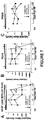
- Example 1 Human neocortical astrocyte cell fates after mechanical trauma
- This Example shows population scores of human astrocytes 30 minutes and 48 hours after abrupt pressure-pulse traumatic stretching using different severities ( Figure 1 ; PSI ranges from 2.6-4.0 for milder injury and 4.4-5.3 for severe injury).
- Human astrocytes were isolated from 16-18 week donated fetal neocortical brain specimen, purified and then differentiated on deformable membranes (Wanner, 2012). Cell membrane wounding / compromise, mechanoporation (Barbee, 2005), was determined using Propidium iodide (PI) uptake in living cultures accompanied by nuclear shape assessment (middle picture, stained nuclei, Hoechst stained after fixation, with little pink dots, nucleoli that are PI-stained).
- PI Propidium iodide
- Example 2 Mechanically traumatized human astrocytes show prolonged endurance in a compromised state
- FIG. 3 shows immunoblots of concentrated, denatured conditioned media (Levine et al., 2016; Sondej et al., 2011) from traumatized astrocytes show different release profiles of glial fibrillary acidic proteins (GFAP) and its known and newly-identified breakdown products (BDPs) versus aldolase C (ALDOC), brain lipid binding protein (BLPB), astrocytic phosphoprotein (PEA15) and ⁇ crystallin (CRYAB).
- GFAP glial fibrillary acidic proteins
- BDPs aldolase C
- ADOC aldolase C
- BLPB brain lipid binding protein
- PEA15 astrocytic phosphoprotein
- CRYAB ⁇ crystallin
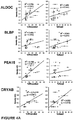
- Example 3 Neurotrauma biomarkers associated with cell fates of human traumatized astrocytes
- FIG 4A Shown in Figure 4A are biplots of trauma-released astroglial marker levels (see Figure 3 ) for ALDOC, BLBP, PEA15 and CRYAB over the percent membrane permeable cells (% wounded, red data, left) as well as correlated to percent of dead human traumatized astrocytes using PI-dye-update assay and nuclear morphology (see Figures 1 +2). Regression lines (R2-value) and p-values indicate correlation significance.
- ALDOC, PEA15 and CRYAB show best correlation with human astroglial cell wounding.
- CRYAB, BLBP and ALDOC also correlated with extend of cell death after trauma.
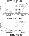
- Figure 4B shows biplots of GFAP trauma-release, which show correlation of GFAP with cell death inflicted by mechanical trauma and weak / no correlation with cell wounding. Plots are separated by grouped GFAP fragment sizes (upper bands: 50-37 kD, lower bands: 25-19 kD).
- TBI CSF proteome was compiled by bottom-up mass spectrometry using CSF samples from 19 severe TBI patients and compared with the CSF proteome from 9 control subjects (Crl). This TBI CSF proteome (483 proteins) overlapped with 252 proteins from the control CSF proteome (402 proteins), leaving 231 unique TBI CSF proteins. Sixty percent of the TBI CSF proteins were also present in the published plasma proteomes (Omenn et al., 2005; Schenk et al., 2008) and, if abundant, may not be suitable as TBI biomarkers.
- Additional 3 proteins were astrocyte enriched, "traumatome” proteins present in TBI CSF and also in control CSF that were aldolase C (ALDOC), clusterin (CLUS) and apolipoprotein E (APOE, lower field of 3). Clusterin and APOE are secreted by astrocytes and their levels decreased in fluid after trauma (arrow). Additional 5 trauma-released proteins that were highly astrocyte enriched were not listed in the shotgun mass spectrometry-based TBI CSF proteome list.
- ezrin Ezr
- F-box only protein 2 FBX2
- GS Glutamine synthetase
- PEA15 astrocytic phosphoprotein 15
- BLBP brain-lipid binding protein
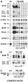
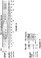
- FIG. 6A shows immunoblots of GFAP (50 kDa with BDPs 37, 25, 20 and 18 kDa) and S100 ⁇ and ALDOC (40 kDa), GS (45 kDa), BLBP (15 kDa) and PEA15 (15 kDa) of a longitudinal set of 30 ⁇ l CSF samples from injury day (i) and subsequent 5 days post-injury (i+1 to i+5) of a 54 year old male severe TBI patient (1a.-1f.) alongside with 30 ⁇ l control CSF (Crl).
- GFAP 50 kDa with BDPs 37, 25, 20 and 18 kDa
- ALDOC 40 kDa
- GS 45 kDa
- BLBP 15 kDa
- PEA15 15 kDa
- BLBP and PEA15 showed strongest signals on injury day and second rise on 3rd post-injury day, that associated with this patient's secondary ischemic episode.
- Bleeding indicator APOB 130 and 250 kDa
- CSF marker PTGDS 22 kDa
- CSF marker PTGDS 22 kDa
- Figure 6B shows six CSF samples (30 ⁇ l/lane) from four TBI patients (2.-5.), illustrating variable signal intensities of GFAP and "upper" BDPs between 50 and 37kDa and "lower/new" GFAP BDPs including 25/23 kDa doublet, 20 kDa and 18 kDa small BDPs on injury day (patients 2, 3, 4a) and one day post-injury (4b, 5) alongside control CSF from a healthy 43 year old male. Individual bands within upper GFAP BDPs have similar intensities to each other, while relative abundance between lower GFAP BDPs was distinctly different across patients.
- FIG. 6C shows CSF immunoblots (30 ⁇ l/lane), demonstrating full size ALDOC (40 kDa) in five TBI patients (6.-10.) and 38 kDa ALDOC BDP of variable intensity on four days post-injury in three TBI patients (8-10).
- a healthy Control subject showed no ALDOC.
- Example 7 Assessing the spectrum of TBI using factors of grouped astroglial markers
- This Example demonstrates the grouping of astroglial trauma markers to create factors useful in the assessment of TBI across the spectrum of injury.
- multivariate analysis of variance that employed an unsupervised learning algorithm based on Spearman correlation coefficients was used (factor analysis, Fabrigar and Wegener, 2012; Tucker, 1997).
- This approach is new to the neurotrauma biomarker field, and reveals underlying neurotrauma conditions by grouping markers here based on their TBI CSF signals.
- Fig. 8F Classification tree analysis
- This mathematical multivariate unsupervised learning approach combines the markers of this panel with each marker given a weight (loading) that is derived from correlation coefficients and expresses how much variance in the patient cohort is captured by this marker's contribution.
- Weight derived from correlation coefficients and expresses how much variance in the patient cohort is captured by this marker's contribution.
- Overall high loading of the listed markers (0.8-0.95) documents robust categorization into the two factors.
- Factor A reflects markers associated with cell death, hemorrhage and tissue loss.
- Factor B reflects markers associated with cell leak, wounding and tissue compromise (see Figure 8A ).
- the resulting values document a unique position of each patient based on its biomarker panel readings within a given cohort. This approach reliably covers the majority of patient variation observed in this TBI cohort.
- Factor analysis provides a simplified framework for capturing a large patient heterogeneity observed in TBI using only few biomarker readings, (compared to more complex principal component analysis that used many different entries).
- these biomarker panel readings from a test kit can be entered into a growing database that can provide valuable monitoring information of individual TBI patients compared to other patients. This tool can simplify patient assessment and strengthen the robustness of ongoing patient evaluation using biofluid signature of tissue compromise and tissue demise.
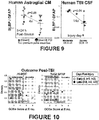
- Example 8 Differentiating trauma severity in human traumatized astrocytes and TBI patients
- This Example demonstrates that the ratio of BLBP to GFAP levels in a subject's specimen can be used to differentiate trauma severity in human traumatized astrocytes and TBI patients.
- Significant differences in fluid level ratios of 'cell leak' marker BLBP over 'cell death' marker GFAP are shown in vitro ( Fig. 9 , CM, conditioned medium, left) and in TBI patients (CSF, right panel of Fig. 9 ).
- Human astrocytes from 6 donors were traumatized with different severities using indicated PSI pressure-pulses.
- Moderate TBI patients are defined by post-resuscitation Glasgow coma scale (GCS) > 8 while severe TBI patients had GCS ⁇ 8.
- GCS Glasgow coma scale
- Data presented in Figure 10 show ALDOC and GFAP levels plotted against outcome of severe TBI patients, assessed using extended Glasgow outcome score (GOSe) at 6 months post-TBI.
- Data from 12 (11) TBI patients show ALDOC elevation early (white, i, i+1, +2) and later post-injury (black, i+3, +4 and +5 post-injury days) in patients with unfavorable outcome, while GFAP levels did not as well maintain elevated levels on later post-injury days.
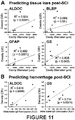
- FIG. 11 Acute UCLA astroglial marker release correlated with histopathological severity measures, tissue loss and hemorrhage, after swine spinal cord injury.
- A) Acute (15-30 min post-injury) CSF signal densities of UCLA astroglial injury markers ALDOC, BLBP and glutamine synthetase (GS) as well as GFAP are plotted against rostro-caudal cavity diameters in the Yucatan swine spinal cord one week after spinal cord contusion injury using an established injury weight drop model (Lee et al., 2013).
- Figure 12 shows the correlation of acutely elevated ALDOC and GFAP with outcome after swine spinal cord injury.
- Recovery of walking associates with acute CSF levels of ALDOC and GFAP after swine spinal cord injury.
- Plotted are acute post-injury CSF levels (15-30 min post-injury) of ALDOC (black dots) and GFAP (white dots) that predicts recovery of walking one week post-injury using the porcine thoracic injury behavioral scale (PTIBS), an established test for recovery of ambulation in the swine indicating the level of walking recovery after spinal cord injury in the Yucatan swine (Lee et al., 2013).
- PTIBS porcine thoracic injury behavioral scale
- Example 11 Quantitative mass spectrometry of astroglial TBI markers
- Example 12 Quantitative immunoassay of ALDOC and BLBP in TBI CSF and blood
- This Example provides a quantitative antibody-based evaluation of ALDOC and BLBP levels in CSF and blood of TBI specimens using standard curves. Box plots in Figure 14 show median and interquartile concentration ranges of ALDOC (left) and BLBP (right) and their amounts in CSF and blood (serum and plasma). Inserts show dose-response using known amounts of isoform-specific recombinant proteins ALDOC and BLBP using two concentration ranges and immunoblot detection conditions (see Table 7).
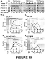
- Example 13 Detection of astroglial biomarkers in blood samples of severe TBI patients
- FIG. 15A shows longitudinal plasma samples from 3 different severe TBI patients alongside one control plasma sample (Crl). Abundant proteins are removed using immunoaffinity albumin and immunoglobulin depletion columns (Sigma, ProteoPrep). New GFAP breakdown product of 25 kDa was never detected on injury day (i) but appeared on subsequent post-injury days. ALDOC was consistently present at all time points in all 3 patients. Short-lived markers PEA15 and BLBP were robustly present on injury day and showed different temporal profiles over subsequent days post-injury in each patient.
- FIG. 15B Plotted in Figure 15B are scaled densitometry signals for ALDOC (B1), BLBP (B2), GFAP/25 kDa BDP (B3) and PEA15 (B4) from 26 serum and 24 plasma samples derived from 22 severe TBI patients compared with up to 11 control blood samples (Crl). Longitudinal same patient data are connected by gray lines. ALDOC was significantly elevated on injury day and every post-injury day, while GFAP was significantly elevated beginning on the first post-injury day. BLBP and PEA15 had significant elevation on injury day versus control levels.
- Example 14 Extended detection window of ALDOC versus GFAP
- Example 15 BLBP breakdown product
- Example 16 Acute circulatory appearance of TBI markers
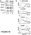
- Figure 18 presents evidence for acute circulatory appearance of new astroglial injury markers due to direct passage across damaged blood-brain barrier.
- Panel A of Figure 18 shows immunoblots for GFAP 25 kDa BDP along with ALDOC, BLBP and PEA15 in CSF and serum of the same severe TBI patient acutely after TBI (3 hours post-injury), as well as on the first post-injury day. While GFAP appeared first in the CSF and with a day delay in serum, ALDOC was present in both biofluids at both time points. BLBP and PEA15 were first present in serum and appeared with delay in CSF.
- GFAP is not localized in astroglial endings, and, as shown in Figure 3 , the 25 kDa GFAP BDP takes time to be generated, suggesting a delayed release during cell death leading to accumulation first in the CSF, and subsequent appearance in serum.
- Advantage of a direct passage of astroglial endfeet via open blood-brain barrier lays in enabling very acute post-injury blood-testing.
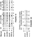
- Example 17 Early detection of astroglial injury markers in serum of mild TBI patients
- This Example demonstrates robust and early detection of top tier astroglial injury markers in serum of mild TBI patients.
- Shown in Figure 19 are 10 serum samples from 7 mild TBI (mTBI) patients early after injury alongside of one control serum (Crl). Samples were probed for GFAP, ALDOC, BLBP and PEA15. Specific GFAP signals were faint and limited to4 mTBI patients with one patient showing GFAP/25 kDa BDP by 31 hours post-injury (patient # II). ALDOC 38 kDa BDP was consistently and strongly elevated in all mTBI patients versus control. BLBP and PEA15 showed variable intensity and were present in 5 mTBI patients. ALDOC, BLBP and PEA15 were detected already one hour post-injury.
- CT computed tomography
- Figure 20 shows acute and robust detection of ALDOC in pediatric TBI patient serum samples.
- Injury day serum samples from 5 infants (1-4 months of age) suffering TBI are shown next to a control serum from a child aged 22 months.
- Robust ALDOC signals are detected in all infant TBI versus control cases while only two infants showed weak GFAP signal (34 kDa BDP, not previously described, cases # I, III).
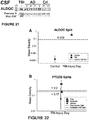
- Example 19 ALDOC as a CSF marker for Alzheimer's disease
- the TBI patient had mainly full size ALDOC and little 38 kDa fragment on day 4 post-injury, while the chronic degenerative AD samples showed equal presence of full size and 38 kDa ALDOC BDP.
- the data suggest that acute and chronic brain injury can be distinguished on the basis of ALDOC /ALDOC BDP ratio.
- the table shows average ratios of full size ALDOC (40 kDa) over its breakdown product (38 kDa) in CSF of Alzheimer's Disease (AD) patients and moderate to severe TBI patients.
- Different ALDOC antibodies to different epitopes of the protein resulted in varying emphasis of the 40 versus 38 kDa band signal intensities.
- On the left data of all ALDOC antibodies were combined averaging 20 AD samples and 25 TBI samples.
- the average AD ratio was 6 fold smaller than the average TBI ratio that was significant by two-tailed T-test.
- On the right 10 AD CSF samples and 5 TBI patient's samples were analyzed using same antibody (E9) for ALDOC detection. There was again a significant 20 x difference between TBI, showing more full size signal and AD showing more BDP ALDOC signal.
- FIG. 21 Data examples are shown in Figure 21 . This provides a distinction between TBI and chronic neurodegenerative disease. Both data selections result in significantly different ratios based on the increased abundance of the ALDOC major 38 kDa BDP signifying the chronic neurodegenerative condition. Acute TBI can easily be distinguished by a higher abundance of full size 40 kDa ALDOC acutely and up to 5 days post-TBI.
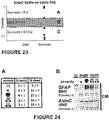
- Multivariate Classification Tree Analysis (Breiman, 1984) was used to determine the markers that most accurately split the subject cohort into Control and Surviving or Non-surviving TBI patients on injury, analyzed by immunoblotting of 30 ⁇ l CSF sample each. Upon obtaining 100% accuracy with just two markers, AldoC and PTGDS (Table 3), the analysis was repeated allowing only those markers known to be detectable in the blood for future noninvasive assay (Table 4). Markers used for the data presented in Table 3 were Aldo C and PTGDS, while the markers considered, but not used, were: GFAP, PEA15, GFAP lower, S100B, BLBP, GS, APOB, PTGDS, and AldoC 38kD.
- Table 3 thus summarizes the detection of TBI and survival outcome prediction using all markers in the classification tree analysis.
- Figure 22 provides a partitioning illustration of the Table 3 thresholds.
- Table 4 shows the detection of TBI and survival outcome prediction using the new blood-compatible glial markers.
- Figure 23 provides a partitioning illustration of the Table 4 thresholds.
- ICP intracranial pressure
- Glial injury markers BLBP and PEA15 are correlated with intraparenchymal lesions, including brain tissue contusion, intracranial hemorrhage and diffuse axonal injury.
- Example 22 Increased levels of glial trauma-release markers after repeated mild injury in the human trauma culture model
- Figure 24 shows results obtained using a model for repeat mild injury.
- Human astrocytes received a single ( ⁇ ) or double ( ⁇ - ⁇ ) mild pressure pulse 30 mins apart (30') or one day apart (1D).
- Cell populations of acute leaky and delayed dead were not much changed by the repeated trauma ( Fig. 24A ).
- conditioned medium (CM) fluid levels of trauma-release markers GFAP and AldoC were elevated after shortly-followed repeated mild injury versus a single mild stretch, and were only slightly elevated when the two insults were one day apart. AldoC levels nearly reached those of a single severe insult ( ⁇ ) after shortly-followed mild stretches, indicating its sensitivity to repeated injury.
- Example 23 Calpain and caspase activation generated GFAP upper and lower breakdown products after trauma
- Shown in Figure 25 are two film exposures of GFAP, upper and lower breakdown products 48 hour post-injury in conditioned medium samples using the DAKO anti-GFAP polyclonal antibody. Severely stretched and unstretched cultures received either no drug, calpain inhibitor PD150606 (100 ⁇ M, "Cali") or pan-caspase inhibitor Z-VAD FMK (8.8 ⁇ M, "Casi”). Trauma-released GFAP upper and lower BDPs were reduced by both calpain and caspase inhibition, suggesting trauma induced activation of both enzymes degrading GFAP after injury. Part of this enzymatic breakdown occurs in cells, part extracellular as suggested by similar analyses of cell lysate fractions of the same experiments. Minor release of lower GFAP BDPs in unstretched cultures was due to small numbers of non-traumatic cell death.
- Example 24 Antibodies and proteins used for western blotting of astroglial injury markers
- Mab 1A1 C-terminal peptide, does not detect ALDOC BDPs, no signal in blood
- Mab E9 Recombinant whole ALDOC, detects ALDOC BDPs, signal in human blood (serum, plasma)
- Mab 4A9 N-terminal peptide sequence: MPHSYPALSAEQKKELS (SEQ ID NO: 1), signal in human and pig blood)
- Mab 5C9 N-terminus, signal in human blood (serum & plasma). Standard curves using pure recombinant ALDOC (EnCor) See Fig.
- Biofluid concentrations of the TBI injury biomarker proteins were measured by targeted multiple-reaction-monitoring (MRM) mass spectrometry.
- Biofluid samples were first digested using endoproteinase trypsin, cleaving all proteins into their respective tryptic peptides. Protein specific peptide signals were used as a surrogate measure for their respective proteins.
- MRM-MS peptide signals are measured by what are known as precursor ⁇ to product ion transitions as shown in Table 7 below (e.g. 554.821 (2+) --> 924.514 (1+, y8)). Selection of specific precursor ions of interest allows for increased sensitivity.
- MRM By measuring signal from specific product ions from selected precursors, MRM allows for a high degree of analyte specificity.
- defined amounts of stable isotope-labeled standard (SIS) peptides containing either a heavy lysine [K(Label:13C(6)15N(2))] or heavy arginine [R(Label:13C(6)15N(4))] are spiked into biofluid samples.
- These heavy standard peptides are chemically identical to their endogenous (light) counterparts but display a mass shift of +8 and +10 Da (K and R respectively) for differentiation from endogenous biofluid peptides.
Landscapes
- Health & Medical Sciences (AREA)
- Life Sciences & Earth Sciences (AREA)
- Engineering & Computer Science (AREA)
- Biomedical Technology (AREA)
- Molecular Biology (AREA)
- Immunology (AREA)
- Chemical & Material Sciences (AREA)
- Urology & Nephrology (AREA)
- Hematology (AREA)
- Physics & Mathematics (AREA)
- Pathology (AREA)
- Biochemistry (AREA)
- Cell Biology (AREA)
- Biotechnology (AREA)
- Microbiology (AREA)
- General Physics & Mathematics (AREA)
- General Health & Medical Sciences (AREA)
- Food Science & Technology (AREA)
- Medicinal Chemistry (AREA)
- Analytical Chemistry (AREA)
- Proteomics, Peptides & Aminoacids (AREA)
- Neurosurgery (AREA)
- Neurology (AREA)
- Bioinformatics & Computational Biology (AREA)
- Bioinformatics & Cheminformatics (AREA)
- Spectroscopy & Molecular Physics (AREA)
- Biophysics (AREA)
- Chemical Kinetics & Catalysis (AREA)
- Investigating Or Analysing Biological Materials (AREA)
- Other Investigation Or Analysis Of Materials By Electrical Means (AREA)
- Peptides Or Proteins (AREA)
- Preparation Of Compounds By Using Micro-Organisms (AREA)
Applications Claiming Priority (2)
| Application Number | Priority Date | Filing Date | Title |
|---|---|---|---|
| US201562157389P | 2015-05-05 | 2015-05-05 | |
| PCT/US2016/031043 WO2016179426A1 (en) | 2015-05-05 | 2016-05-05 | Astrocyte traumatome and neurotrauma biomarkers |
Publications (3)
| Publication Number | Publication Date |
|---|---|
| EP3292414A1 EP3292414A1 (en) | 2018-03-14 |
| EP3292414A4 EP3292414A4 (en) | 2018-09-26 |
| EP3292414B1 true EP3292414B1 (en) | 2021-04-07 |
Family
ID=57218432
Family Applications (1)
| Application Number | Title | Priority Date | Filing Date |
|---|---|---|---|
| EP16790108.1A Active EP3292414B1 (en) | 2015-05-05 | 2016-05-05 | Astrocyte traumatome and neurotrauma biomarkers |
Country Status (7)
Families Citing this family (4)
| Publication number | Priority date | Publication date | Assignee | Title |
|---|---|---|---|---|
| EP3292414B1 (en) * | 2015-05-05 | 2021-04-07 | The Regents of the University of California | Astrocyte traumatome and neurotrauma biomarkers |
| US11709168B2 (en) * | 2017-05-23 | 2023-07-25 | Brainbox Solutions, Inc. | Biomarker levels and neuroimaging for detecting, monitoring and treating brain injury or trauma |
| US11988676B2 (en) * | 2017-11-16 | 2024-05-21 | Brainbox Solutions, Inc. | Protein biomarker indicators of neurological injury and/or disease and methods of use thereof |
| WO2019133717A1 (en) | 2017-12-29 | 2019-07-04 | Abbott Laboratories | Novel biomarkers and methods for diagnosing and evaluating traumatic brain injury |
Family Cites Families (11)
| Publication number | Priority date | Publication date | Assignee | Title |
|---|---|---|---|---|
| US8492107B2 (en) * | 2004-04-15 | 2013-07-23 | University Of Florida Research Foundation, Inc. | Neural proteins as biomarkers for nervous system injury and other neural disorders |
| GB0414798D0 (en) * | 2004-07-01 | 2004-08-04 | Paradigm Therapeutics Ltd | Receptor |
| WO2007136617A2 (en) * | 2006-05-18 | 2007-11-29 | Walter Reed Army Institute Of Research (Wrair) | Endothelial-monocyte activating polypeptide ii, a biomarker for use in diagnosis of brain injury |
| CN101983337A (zh) * | 2008-02-04 | 2011-03-02 | 班扬生物标记公司 | 用于诊断或治疗脑损伤的方法 |
| EP2443461A4 (en) | 2009-06-19 | 2012-12-26 | Banyan Biomarkers Inc | BIOMARKER ASSAY FOR NERVES |
| KR101211280B1 (ko) * | 2010-03-26 | 2012-12-11 | 가톨릭대학교 산학협력단 | 신규한 뇌손상 질환 진단용 마커 및 이의 용도 |
| WO2011147981A2 (en) | 2010-05-28 | 2011-12-01 | Mind-Nrg Sa | Neuregulin isoforms, neuregulin polypeptides and uses thereof |
| WO2012051519A2 (en) | 2010-10-14 | 2012-04-19 | The Johns Hopkins University | Biomarkers of brain injury |
| CN106461645A (zh) | 2014-04-07 | 2017-02-22 | 铁马诊断公司 | 外伤性脑损伤和神经退行性生物标记物、方法和系统 |
| AU2015322029A1 (en) | 2014-09-25 | 2017-04-27 | Oxeia Biopharmaceuticals, Inc. | Methods of treating traumatic brain injury |
| EP3292414B1 (en) * | 2015-05-05 | 2021-04-07 | The Regents of the University of California | Astrocyte traumatome and neurotrauma biomarkers |
-
2016
- 2016-05-05 EP EP16790108.1A patent/EP3292414B1/en active Active
- 2016-05-05 ES ES16790108T patent/ES2873509T3/es active Active
- 2016-05-05 US US15/570,982 patent/US10557859B2/en active Active
- 2016-05-05 WO PCT/US2016/031043 patent/WO2016179426A1/en active Application Filing
- 2016-05-05 JP JP2017557192A patent/JP6839854B2/ja active Active
- 2016-05-05 CN CN201680032940.6A patent/CN107864671A/zh active Pending
-
2017
- 2017-10-31 IL IL255351A patent/IL255351A0/en unknown
-
2019
- 2019-12-16 US US16/716,095 patent/US11249094B2/en active Active
-
2022
- 2022-01-06 US US17/647,289 patent/US12339289B2/en active Active
Non-Patent Citations (1)
| Title |
|---|
| None * |
Also Published As
| Publication number | Publication date |
|---|---|
| US20220299531A1 (en) | 2022-09-22 |
| US20180364259A1 (en) | 2018-12-20 |
| IL255351A0 (en) | 2017-12-31 |
| WO2016179426A1 (en) | 2016-11-10 |
| ES2873509T3 (es) | 2021-11-03 |
| US11249094B2 (en) | 2022-02-15 |
| US12339289B2 (en) | 2025-06-24 |
| EP3292414A4 (en) | 2018-09-26 |
| JP6839854B2 (ja) | 2021-03-17 |
| JP2018523092A (ja) | 2018-08-16 |
| EP3292414A1 (en) | 2018-03-14 |
| US20200278359A1 (en) | 2020-09-03 |
| CN107864671A (zh) | 2018-03-30 |
| US10557859B2 (en) | 2020-02-11 |
Similar Documents
| Publication | Publication Date | Title |
|---|---|---|
| US12339289B2 (en) | Astrocyte traumatome and neurotrauma biomarkers | |
| US20230314453A1 (en) | Biomarker levels and neuroimaging for detecting, monitoring and treating brain injury or trauma | |
| US11307208B2 (en) | Biomarkers for cognitive impairment and methods for detecting cognitive impairment using such biomarkers | |
| JP4674317B2 (ja) | 疾病の判定および監視 | |
| Jahn et al. | Peptide fingerprinting of Alzheimer's disease in cerebrospinal fluid: identification and prospective evaluation of new synaptic biomarkers | |
| DK2444814T3 (en) | Biomarker for mental disorders, including cognitive disorders, and method of using the biomarker for detecting mental disorders, including cognitive disorders | |
| US9977036B2 (en) | Diagnostic markers for multiple sclerosis | |
| WO2010005077A1 (ja) | パーキンソン病の疾患関連たんぱく質およびその使用 | |
| WO2019012667A1 (ja) | 認知機能障害疾患のバイオマーカー及び該バイオマーカーを用いる認知機能障害疾患の検出方法 | |
| Alawieh et al. | Neuro‑proteomics and neuro‑systems biology in the quest of TBI biomarker discovery | |
| WO2014071359A1 (en) | Detection of neurological diseases via measurement of neuromelanin in recirculating phagocytes |
Legal Events
| Date | Code | Title | Description |
|---|---|---|---|
| STAA | Information on the status of an ep patent application or granted ep patent |
Free format text: STATUS: THE INTERNATIONAL PUBLICATION HAS BEEN MADE |
|
| PUAI | Public reference made under article 153(3) epc to a published international application that has entered the european phase |
Free format text: ORIGINAL CODE: 0009012 |
|
| STAA | Information on the status of an ep patent application or granted ep patent |
Free format text: STATUS: REQUEST FOR EXAMINATION WAS MADE |
|
| 17P | Request for examination filed |
Effective date: 20171129 |
|
| AK | Designated contracting states |
Kind code of ref document: A1 Designated state(s): AL AT BE BG CH CY CZ DE DK EE ES FI FR GB GR HR HU IE IS IT LI LT LU LV MC MK MT NL NO PL PT RO RS SE SI SK SM TR |
|
| AX | Request for extension of the european patent |
Extension state: BA ME |
|
| DAV | Request for validation of the european patent (deleted) | ||
| DAX | Request for extension of the european patent (deleted) | ||
| A4 | Supplementary search report drawn up and despatched |
Effective date: 20180828 |
|
| RIC1 | Information provided on ipc code assigned before grant |
Ipc: G01N 33/53 20060101ALI20180822BHEP Ipc: G01N 33/68 20060101AFI20180822BHEP Ipc: G01N 27/64 20060101ALI20180822BHEP Ipc: G01N 33/577 20060101ALI20180822BHEP |
|
| STAA | Information on the status of an ep patent application or granted ep patent |
Free format text: STATUS: EXAMINATION IS IN PROGRESS |
|
| 17Q | First examination report despatched |
Effective date: 20190514 |
|
| GRAP | Despatch of communication of intention to grant a patent |
Free format text: ORIGINAL CODE: EPIDOSNIGR1 |
|
| STAA | Information on the status of an ep patent application or granted ep patent |
Free format text: STATUS: GRANT OF PATENT IS INTENDED |
|
| INTG | Intention to grant announced |
Effective date: 20201026 |
|
| RAP1 | Party data changed (applicant data changed or rights of an application transferred) |
Owner name: THE REGENTS OF THE UNIVERSITY OF CALIFORNIA |
|
| RIN1 | Information on inventor provided before grant (corrected) |
Inventor name: LOO, JOSEPH A. Inventor name: WANNER, INA-BEATE |
|
| GRAS | Grant fee paid |
Free format text: ORIGINAL CODE: EPIDOSNIGR3 |
|
| GRAA | (expected) grant |
Free format text: ORIGINAL CODE: 0009210 |
|
| STAA | Information on the status of an ep patent application or granted ep patent |
Free format text: STATUS: THE PATENT HAS BEEN GRANTED |
|
| AK | Designated contracting states |
Kind code of ref document: B1 Designated state(s): AL AT BE BG CH CY CZ DE DK EE ES FI FR GB GR HR HU IE IS IT LI LT LU LV MC MK MT NL NO PL PT RO RS SE SI SK SM TR |
|
| REG | Reference to a national code |
Ref country code: GB Ref legal event code: FG4D |
|
| REG | Reference to a national code |
Ref country code: CH Ref legal event code: EP Ref country code: AT Ref legal event code: REF Ref document number: 1380346 Country of ref document: AT Kind code of ref document: T Effective date: 20210415 |
|
| REG | Reference to a national code |
Ref country code: DE Ref legal event code: R096 Ref document number: 602016055716 Country of ref document: DE |
|
| REG | Reference to a national code |
Ref country code: IE Ref legal event code: FG4D |
|
| REG | Reference to a national code |
Ref country code: NL Ref legal event code: FP |
|
| REG | Reference to a national code |
Ref country code: LT Ref legal event code: MG9D |
|
| REG | Reference to a national code |
Ref country code: AT Ref legal event code: MK05 Ref document number: 1380346 Country of ref document: AT Kind code of ref document: T Effective date: 20210407 |
|
| PG25 | Lapsed in a contracting state [announced via postgrant information from national office to epo] |
Ref country code: LT Free format text: LAPSE BECAUSE OF FAILURE TO SUBMIT A TRANSLATION OF THE DESCRIPTION OR TO PAY THE FEE WITHIN THE PRESCRIBED TIME-LIMIT Effective date: 20210407 Ref country code: FI Free format text: LAPSE BECAUSE OF FAILURE TO SUBMIT A TRANSLATION OF THE DESCRIPTION OR TO PAY THE FEE WITHIN THE PRESCRIBED TIME-LIMIT Effective date: 20210407 Ref country code: BG Free format text: LAPSE BECAUSE OF FAILURE TO SUBMIT A TRANSLATION OF THE DESCRIPTION OR TO PAY THE FEE WITHIN THE PRESCRIBED TIME-LIMIT Effective date: 20210707 Ref country code: AT Free format text: LAPSE BECAUSE OF FAILURE TO SUBMIT A TRANSLATION OF THE DESCRIPTION OR TO PAY THE FEE WITHIN THE PRESCRIBED TIME-LIMIT Effective date: 20210407 Ref country code: HR Free format text: LAPSE BECAUSE OF FAILURE TO SUBMIT A TRANSLATION OF THE DESCRIPTION OR TO PAY THE FEE WITHIN THE PRESCRIBED TIME-LIMIT Effective date: 20210407 |
|
| REG | Reference to a national code |
Ref country code: ES Ref legal event code: FG2A Ref document number: 2873509 Country of ref document: ES Kind code of ref document: T3 Effective date: 20211103 |
|
| PG25 | Lapsed in a contracting state [announced via postgrant information from national office to epo] |
Ref country code: GR Free format text: LAPSE BECAUSE OF FAILURE TO SUBMIT A TRANSLATION OF THE DESCRIPTION OR TO PAY THE FEE WITHIN THE PRESCRIBED TIME-LIMIT Effective date: 20210708 Ref country code: LV Free format text: LAPSE BECAUSE OF FAILURE TO SUBMIT A TRANSLATION OF THE DESCRIPTION OR TO PAY THE FEE WITHIN THE PRESCRIBED TIME-LIMIT Effective date: 20210407 Ref country code: IS Free format text: LAPSE BECAUSE OF FAILURE TO SUBMIT A TRANSLATION OF THE DESCRIPTION OR TO PAY THE FEE WITHIN THE PRESCRIBED TIME-LIMIT Effective date: 20210807 Ref country code: SE Free format text: LAPSE BECAUSE OF FAILURE TO SUBMIT A TRANSLATION OF THE DESCRIPTION OR TO PAY THE FEE WITHIN THE PRESCRIBED TIME-LIMIT Effective date: 20210407 Ref country code: RS Free format text: LAPSE BECAUSE OF FAILURE TO SUBMIT A TRANSLATION OF THE DESCRIPTION OR TO PAY THE FEE WITHIN THE PRESCRIBED TIME-LIMIT Effective date: 20210407 Ref country code: NO Free format text: LAPSE BECAUSE OF FAILURE TO SUBMIT A TRANSLATION OF THE DESCRIPTION OR TO PAY THE FEE WITHIN THE PRESCRIBED TIME-LIMIT Effective date: 20210707 Ref country code: PT Free format text: LAPSE BECAUSE OF FAILURE TO SUBMIT A TRANSLATION OF THE DESCRIPTION OR TO PAY THE FEE WITHIN THE PRESCRIBED TIME-LIMIT Effective date: 20210809 Ref country code: PL Free format text: LAPSE BECAUSE OF FAILURE TO SUBMIT A TRANSLATION OF THE DESCRIPTION OR TO PAY THE FEE WITHIN THE PRESCRIBED TIME-LIMIT Effective date: 20210407 |
|
| REG | Reference to a national code |
Ref country code: CH Ref legal event code: PL |
|
| REG | Reference to a national code |
Ref country code: DE Ref legal event code: R097 Ref document number: 602016055716 Country of ref document: DE |
|
| PG25 | Lapsed in a contracting state [announced via postgrant information from national office to epo] |
Ref country code: SK Free format text: LAPSE BECAUSE OF FAILURE TO SUBMIT A TRANSLATION OF THE DESCRIPTION OR TO PAY THE FEE WITHIN THE PRESCRIBED TIME-LIMIT Effective date: 20210407 Ref country code: SM Free format text: LAPSE BECAUSE OF FAILURE TO SUBMIT A TRANSLATION OF THE DESCRIPTION OR TO PAY THE FEE WITHIN THE PRESCRIBED TIME-LIMIT Effective date: 20210407 Ref country code: CZ Free format text: LAPSE BECAUSE OF FAILURE TO SUBMIT A TRANSLATION OF THE DESCRIPTION OR TO PAY THE FEE WITHIN THE PRESCRIBED TIME-LIMIT Effective date: 20210407 Ref country code: EE Free format text: LAPSE BECAUSE OF FAILURE TO SUBMIT A TRANSLATION OF THE DESCRIPTION OR TO PAY THE FEE WITHIN THE PRESCRIBED TIME-LIMIT Effective date: 20210407 Ref country code: DK Free format text: LAPSE BECAUSE OF FAILURE TO SUBMIT A TRANSLATION OF THE DESCRIPTION OR TO PAY THE FEE WITHIN THE PRESCRIBED TIME-LIMIT Effective date: 20210407 Ref country code: CH Free format text: LAPSE BECAUSE OF NON-PAYMENT OF DUE FEES Effective date: 20210531 Ref country code: MC Free format text: LAPSE BECAUSE OF FAILURE TO SUBMIT A TRANSLATION OF THE DESCRIPTION OR TO PAY THE FEE WITHIN THE PRESCRIBED TIME-LIMIT Effective date: 20210407 Ref country code: LI Free format text: LAPSE BECAUSE OF NON-PAYMENT OF DUE FEES Effective date: 20210531 Ref country code: LU Free format text: LAPSE BECAUSE OF NON-PAYMENT OF DUE FEES Effective date: 20210505 Ref country code: RO Free format text: LAPSE BECAUSE OF FAILURE TO SUBMIT A TRANSLATION OF THE DESCRIPTION OR TO PAY THE FEE WITHIN THE PRESCRIBED TIME-LIMIT Effective date: 20210407 |
|
| REG | Reference to a national code |
Ref country code: BE Ref legal event code: MM Effective date: 20210531 |
|
| PLBE | No opposition filed within time limit |
Free format text: ORIGINAL CODE: 0009261 |
|
| STAA | Information on the status of an ep patent application or granted ep patent |
Free format text: STATUS: NO OPPOSITION FILED WITHIN TIME LIMIT |
|
| 26N | No opposition filed |
Effective date: 20220110 |
|
| PG25 | Lapsed in a contracting state [announced via postgrant information from national office to epo] |
Ref country code: IE Free format text: LAPSE BECAUSE OF NON-PAYMENT OF DUE FEES Effective date: 20210505 |
|
| PG25 | Lapsed in a contracting state [announced via postgrant information from national office to epo] |
Ref country code: IS Free format text: LAPSE BECAUSE OF FAILURE TO SUBMIT A TRANSLATION OF THE DESCRIPTION OR TO PAY THE FEE WITHIN THE PRESCRIBED TIME-LIMIT Effective date: 20210807 Ref country code: AL Free format text: LAPSE BECAUSE OF FAILURE TO SUBMIT A TRANSLATION OF THE DESCRIPTION OR TO PAY THE FEE WITHIN THE PRESCRIBED TIME-LIMIT Effective date: 20210407 |
|
| PG25 | Lapsed in a contracting state [announced via postgrant information from national office to epo] |
Ref country code: BE Free format text: LAPSE BECAUSE OF NON-PAYMENT OF DUE FEES Effective date: 20210531 |
|
| PG25 | Lapsed in a contracting state [announced via postgrant information from national office to epo] |
Ref country code: HU Free format text: LAPSE BECAUSE OF FAILURE TO SUBMIT A TRANSLATION OF THE DESCRIPTION OR TO PAY THE FEE WITHIN THE PRESCRIBED TIME-LIMIT; INVALID AB INITIO Effective date: 20160505 |
|
| P01 | Opt-out of the competence of the unified patent court (upc) registered |
Effective date: 20230511 |
|
| PG25 | Lapsed in a contracting state [announced via postgrant information from national office to epo] |
Ref country code: CY Free format text: LAPSE BECAUSE OF FAILURE TO SUBMIT A TRANSLATION OF THE DESCRIPTION OR TO PAY THE FEE WITHIN THE PRESCRIBED TIME-LIMIT Effective date: 20210407 |
|
| PG25 | Lapsed in a contracting state [announced via postgrant information from national office to epo] |
Ref country code: MK Free format text: LAPSE BECAUSE OF FAILURE TO SUBMIT A TRANSLATION OF THE DESCRIPTION OR TO PAY THE FEE WITHIN THE PRESCRIBED TIME-LIMIT Effective date: 20210407 |
|
| PG25 | Lapsed in a contracting state [announced via postgrant information from national office to epo] |
Ref country code: TR Free format text: LAPSE BECAUSE OF FAILURE TO SUBMIT A TRANSLATION OF THE DESCRIPTION OR TO PAY THE FEE WITHIN THE PRESCRIBED TIME-LIMIT Effective date: 20210407 |
|
| PG25 | Lapsed in a contracting state [announced via postgrant information from national office to epo] |
Ref country code: MT Free format text: LAPSE BECAUSE OF FAILURE TO SUBMIT A TRANSLATION OF THE DESCRIPTION OR TO PAY THE FEE WITHIN THE PRESCRIBED TIME-LIMIT Effective date: 20210407 |
|
| PGFP | Annual fee paid to national office [announced via postgrant information from national office to epo] |
Ref country code: NL Payment date: 20250526 Year of fee payment: 10 |
|
| PGFP | Annual fee paid to national office [announced via postgrant information from national office to epo] |
Ref country code: DE Payment date: 20250529 Year of fee payment: 10 |
|
| PGFP | Annual fee paid to national office [announced via postgrant information from national office to epo] |
Ref country code: GB Payment date: 20250527 Year of fee payment: 10 Ref country code: ES Payment date: 20250602 Year of fee payment: 10 |
|
| PGFP | Annual fee paid to national office [announced via postgrant information from national office to epo] |
Ref country code: IT Payment date: 20250521 Year of fee payment: 10 |
|
| PGFP | Annual fee paid to national office [announced via postgrant information from national office to epo] |
Ref country code: FR Payment date: 20250526 Year of fee payment: 10 |