EP3292414B1 - Astrocyte traumatome and neurotrauma biomarkers - Google Patents
Astrocyte traumatome and neurotrauma biomarkers Download PDFInfo
- Publication number
- EP3292414B1 EP3292414B1 EP16790108.1A EP16790108A EP3292414B1 EP 3292414 B1 EP3292414 B1 EP 3292414B1 EP 16790108 A EP16790108 A EP 16790108A EP 3292414 B1 EP3292414 B1 EP 3292414B1
- Authority
- EP
- European Patent Office
- Prior art keywords
- tbi
- injury
- aldoc
- kda
- csf
- Prior art date
- Legal status (The legal status is an assumption and is not a legal conclusion. Google has not performed a legal analysis and makes no representation as to the accuracy of the status listed.)
- Active
Links
- 239000000090 biomarker Substances 0.000 title claims description 54
- 210000001130 astrocyte Anatomy 0.000 title description 37
- 208000030886 Traumatic Brain injury Diseases 0.000 claims description 258
- 230000009529 traumatic brain injury Effects 0.000 claims description 257
- 208000014674 injury Diseases 0.000 claims description 197
- 102000001390 Fructose-Bisphosphate Aldolase Human genes 0.000 claims description 156
- 108010068561 Fructose-Bisphosphate Aldolase Proteins 0.000 claims description 156
- 208000027418 Wounds and injury Diseases 0.000 claims description 136
- 210000001175 cerebrospinal fluid Anatomy 0.000 claims description 100
- 102000002140 Fatty Acid-Binding Protein 7 Human genes 0.000 claims description 99
- 108010001387 Fatty Acid-Binding Protein 7 Proteins 0.000 claims description 99
- 102000053171 Glial Fibrillary Acidic Human genes 0.000 claims description 97
- 108700005000 Glial Fibrillary Acidic Proteins 0.000 claims description 95
- 239000003550 marker Substances 0.000 claims description 81
- 238000000034 method Methods 0.000 claims description 81
- 102100034691 Astrocytic phosphoprotein PEA-15 Human genes 0.000 claims description 76
- 101000734668 Homo sapiens Astrocytic phosphoprotein PEA-15 Proteins 0.000 claims description 70
- 230000006378 damage Effects 0.000 claims description 70
- 239000012634 fragment Substances 0.000 claims description 60
- 230000008733 trauma Effects 0.000 claims description 58
- 102000005396 glutamine synthetase Human genes 0.000 claims description 48
- 108020002326 glutamine synthetase Proteins 0.000 claims description 48
- 210000002966 serum Anatomy 0.000 claims description 41
- 239000000523 sample Substances 0.000 claims description 40
- 102000004169 proteins and genes Human genes 0.000 claims description 35
- 108090000623 proteins and genes Proteins 0.000 claims description 35
- 210000004369 blood Anatomy 0.000 claims description 33
- 239000008280 blood Substances 0.000 claims description 33
- 238000001514 detection method Methods 0.000 claims description 28
- 102000004005 Prostaglandin-endoperoxide synthases Human genes 0.000 claims description 26
- 108090000459 Prostaglandin-endoperoxide synthases Proteins 0.000 claims description 26
- 239000013068 control sample Substances 0.000 claims description 26
- 238000012544 monitoring process Methods 0.000 claims description 24
- 108090000765 processed proteins & peptides Proteins 0.000 claims description 23
- 102100040743 Alpha-crystallin B chain Human genes 0.000 claims description 22
- 101000891982 Homo sapiens Alpha-crystallin B chain Proteins 0.000 claims description 22
- 210000004556 brain Anatomy 0.000 claims description 22
- 239000003795 chemical substances by application Substances 0.000 claims description 22
- 210000002381 plasma Anatomy 0.000 claims description 20
- 101710095342 Apolipoprotein B Proteins 0.000 claims description 19
- 102100040202 Apolipoprotein B-100 Human genes 0.000 claims description 18
- 108010051585 alpha-Crystallin B Chain Proteins 0.000 claims description 17
- 102000013640 alpha-Crystallin B Chain Human genes 0.000 claims description 17
- 230000006337 proteolytic cleavage Effects 0.000 claims description 16
- 210000001124 body fluid Anatomy 0.000 claims description 13
- 239000003153 chemical reaction reagent Substances 0.000 claims description 13
- 102000004196 processed proteins & peptides Human genes 0.000 claims description 13
- 101001027674 Homo sapiens Fatty acid-binding protein, brain Proteins 0.000 claims description 12
- 102100037733 Fatty acid-binding protein, brain Human genes 0.000 claims description 11
- 238000004949 mass spectrometry Methods 0.000 claims description 10
- 238000012360 testing method Methods 0.000 claims description 10
- 239000012530 fluid Substances 0.000 claims description 9
- 101710148554 Astrocytic phosphoprotein PEA-15 Proteins 0.000 claims description 7
- 238000003018 immunoassay Methods 0.000 claims description 7
- 238000010833 quantitative mass spectrometry Methods 0.000 claims description 7
- 230000002829 reductive effect Effects 0.000 claims description 7
- 102100039165 Heat shock protein beta-1 Human genes 0.000 claims description 6
- 238000001262 western blot Methods 0.000 claims description 5
- 238000002965 ELISA Methods 0.000 claims description 4
- 108010045100 HSP27 Heat-Shock Proteins Proteins 0.000 claims description 4
- 210000002939 cerumen Anatomy 0.000 claims description 3
- 238000006243 chemical reaction Methods 0.000 claims description 3
- 102000040430 polynucleotide Human genes 0.000 claims description 3
- 108091033319 polynucleotide Proteins 0.000 claims description 3
- 239000002157 polynucleotide Substances 0.000 claims description 3
- 210000003296 saliva Anatomy 0.000 claims description 3
- 210000002700 urine Anatomy 0.000 claims description 3
- 241000282414 Homo sapiens Species 0.000 description 44
- 230000001154 acute effect Effects 0.000 description 27
- 210000004027 cell Anatomy 0.000 description 26
- 230000030833 cell death Effects 0.000 description 23
- 208000024827 Alzheimer disease Diseases 0.000 description 19
- 238000003119 immunoblot Methods 0.000 description 19
- 208000020431 spinal cord injury Diseases 0.000 description 18
- 230000015556 catabolic process Effects 0.000 description 17
- 238000002591 computed tomography Methods 0.000 description 14
- 208000032843 Hemorrhage Diseases 0.000 description 13
- 238000011084 recovery Methods 0.000 description 13
- 206010010254 Concussion Diseases 0.000 description 12
- 210000001519 tissue Anatomy 0.000 description 12
- 241000282898 Sus scrofa Species 0.000 description 11
- 230000009514 concussion Effects 0.000 description 11
- 241000283973 Oryctolagus cuniculus Species 0.000 description 10
- 238000004458 analytical method Methods 0.000 description 10
- 230000000740 bleeding effect Effects 0.000 description 10
- 230000001684 chronic effect Effects 0.000 description 9
- 238000002552 multiple reaction monitoring Methods 0.000 description 9
- 230000027455 binding Effects 0.000 description 8
- 239000003636 conditioned culture medium Substances 0.000 description 8
- 230000002596 correlated effect Effects 0.000 description 8
- 238000003556 assay Methods 0.000 description 7
- 208000029028 brain injury Diseases 0.000 description 7
- 238000003745 diagnosis Methods 0.000 description 7
- 238000011156 evaluation Methods 0.000 description 7
- 238000000638 solvent extraction Methods 0.000 description 7
- 230000004083 survival effect Effects 0.000 description 7
- 102000003712 Complement factor B Human genes 0.000 description 6
- 108090000056 Complement factor B Proteins 0.000 description 6
- 241001465754 Metazoa Species 0.000 description 6
- 108010026552 Proteome Proteins 0.000 description 6
- 238000013459 approach Methods 0.000 description 6
- 230000003902 lesion Effects 0.000 description 6
- 238000011068 loading method Methods 0.000 description 6
- 238000001228 spectrum Methods 0.000 description 6
- 210000000278 spinal cord Anatomy 0.000 description 6
- 102000011727 Caspases Human genes 0.000 description 5
- 108010076667 Caspases Proteins 0.000 description 5
- 101100269568 Danio rerio aldocb gene Proteins 0.000 description 5
- 102100021487 Protein S100-B Human genes 0.000 description 5
- 101150057384 aldoc gene Proteins 0.000 description 5
- 230000003111 delayed effect Effects 0.000 description 5
- 229940079593 drug Drugs 0.000 description 5
- 239000003814 drug Substances 0.000 description 5
- 230000002518 glial effect Effects 0.000 description 5
- 230000000626 neurodegenerative effect Effects 0.000 description 5
- 230000003287 optical effect Effects 0.000 description 5
- 230000002035 prolonged effect Effects 0.000 description 5
- XJMOSONTPMZWPB-UHFFFAOYSA-M propidium iodide Chemical compound [I-].[I-].C12=CC(N)=CC=C2C2=CC=C(N)C=C2[N+](CCC[N+](C)(CC)CC)=C1C1=CC=CC=C1 XJMOSONTPMZWPB-UHFFFAOYSA-M 0.000 description 5
- 102000007590 Calpain Human genes 0.000 description 4
- 108010032088 Calpain Proteins 0.000 description 4
- 206010010071 Coma Diseases 0.000 description 4
- 230000004913 activation Effects 0.000 description 4
- 150000001413 amino acids Chemical class 0.000 description 4
- 230000008901 benefit Effects 0.000 description 4
- 230000008499 blood brain barrier function Effects 0.000 description 4
- 210000001218 blood-brain barrier Anatomy 0.000 description 4
- 230000001010 compromised effect Effects 0.000 description 4
- 230000003247 decreasing effect Effects 0.000 description 4
- BDAGIHXWWSANSR-UHFFFAOYSA-N methanoic acid Natural products OC=O BDAGIHXWWSANSR-UHFFFAOYSA-N 0.000 description 4
- 230000009525 mild injury Effects 0.000 description 4
- 210000004940 nucleus Anatomy 0.000 description 4
- 230000035945 sensitivity Effects 0.000 description 4
- 230000000472 traumatic effect Effects 0.000 description 4
- WEVYAHXRMPXWCK-UHFFFAOYSA-N Acetonitrile Chemical compound CC#N WEVYAHXRMPXWCK-UHFFFAOYSA-N 0.000 description 3
- 102100029470 Apolipoprotein E Human genes 0.000 description 3
- 102000003780 Clusterin Human genes 0.000 description 3
- 108090000197 Clusterin Proteins 0.000 description 3
- 102000007982 Phosphoproteins Human genes 0.000 description 3
- 108010089430 Phosphoproteins Proteins 0.000 description 3
- 102000048176 Prostaglandin-D synthases Human genes 0.000 description 3
- 108030003866 Prostaglandin-D synthases Proteins 0.000 description 3
- 102000007056 Recombinant Fusion Proteins Human genes 0.000 description 3
- 108010008281 Recombinant Fusion Proteins Proteins 0.000 description 3
- 108010023918 S100 Calcium Binding Protein beta Subunit Proteins 0.000 description 3
- 230000003140 astrocytic effect Effects 0.000 description 3
- 238000000326 densiometry Methods 0.000 description 3
- 238000000556 factor analysis Methods 0.000 description 3
- 102000045058 human FABP7 Human genes 0.000 description 3
- 238000003384 imaging method Methods 0.000 description 3
- 230000001900 immune effect Effects 0.000 description 3
- 150000002500 ions Chemical class 0.000 description 3
- 239000012528 membrane Substances 0.000 description 3
- 239000000203 mixture Substances 0.000 description 3
- 230000001423 neocortical effect Effects 0.000 description 3
- 230000004770 neurodegeneration Effects 0.000 description 3
- 208000015122 neurodegenerative disease Diseases 0.000 description 3
- 239000002243 precursor Substances 0.000 description 3
- 208000024891 symptom Diseases 0.000 description 3
- 230000007704 transition Effects 0.000 description 3
- OSWFIVFLDKOXQC-UHFFFAOYSA-N 4-(3-methoxyphenyl)aniline Chemical compound COC1=CC=CC(C=2C=CC(N)=CC=2)=C1 OSWFIVFLDKOXQC-UHFFFAOYSA-N 0.000 description 2
- FWMNVWWHGCHHJJ-SKKKGAJSSA-N 4-amino-1-[(2r)-6-amino-2-[[(2r)-2-[[(2r)-2-[[(2r)-2-amino-3-phenylpropanoyl]amino]-3-phenylpropanoyl]amino]-4-methylpentanoyl]amino]hexanoyl]piperidine-4-carboxylic acid Chemical compound C([C@H](C(=O)N[C@H](CC(C)C)C(=O)N[C@H](CCCCN)C(=O)N1CCC(N)(CC1)C(O)=O)NC(=O)[C@H](N)CC=1C=CC=CC=1)C1=CC=CC=C1 FWMNVWWHGCHHJJ-SKKKGAJSSA-N 0.000 description 2
- 101150037123 APOE gene Proteins 0.000 description 2
- 206010008513 Child maltreatment syndrome Diseases 0.000 description 2
- 208000000532 Chronic Brain Injury Diseases 0.000 description 2
- 208000034656 Contusions Diseases 0.000 description 2
- 102000004190 Enzymes Human genes 0.000 description 2
- 108090000790 Enzymes Proteins 0.000 description 2
- 102100022116 F-box only protein 2 Human genes 0.000 description 2
- 101710199775 F-box only protein 2 Proteins 0.000 description 2
- 101710098548 Fatty acid-binding protein, brain Proteins 0.000 description 2
- 102100037738 Fatty acid-binding protein, heart Human genes 0.000 description 2
- 101710100504 Heat shock protein beta-1 Proteins 0.000 description 2
- 101000821885 Homo sapiens Protein S100-B Proteins 0.000 description 2
- 108060003951 Immunoglobulin Proteins 0.000 description 2
- 208000002193 Pain Diseases 0.000 description 2
- 102000001708 Protein Isoforms Human genes 0.000 description 2
- 108010029485 Protein Isoforms Proteins 0.000 description 2
- 208000002108 Shaken Baby Syndrome Diseases 0.000 description 2
- 102000004142 Trypsin Human genes 0.000 description 2
- 108090000631 Trypsin Proteins 0.000 description 2
- 238000009825 accumulation Methods 0.000 description 2
- 230000002411 adverse Effects 0.000 description 2
- 102000007362 alpha-Crystallins Human genes 0.000 description 2
- 108010007908 alpha-Crystallins Proteins 0.000 description 2
- 238000000540 analysis of variance Methods 0.000 description 2
- 210000004958 brain cell Anatomy 0.000 description 2
- 230000006931 brain damage Effects 0.000 description 2
- 230000001149 cognitive effect Effects 0.000 description 2
- 230000009519 contusion Effects 0.000 description 2
- 230000034994 death Effects 0.000 description 2
- 230000007423 decrease Effects 0.000 description 2
- 230000003412 degenerative effect Effects 0.000 description 2
- 230000001419 dependent effect Effects 0.000 description 2
- 230000006866 deterioration Effects 0.000 description 2
- 230000000694 effects Effects 0.000 description 2
- 235000019253 formic acid Nutrition 0.000 description 2
- 238000010166 immunofluorescence Methods 0.000 description 2
- 102000018358 immunoglobulin Human genes 0.000 description 2
- 238000007914 intraventricular administration Methods 0.000 description 2
- 230000002045 lasting effect Effects 0.000 description 2
- 230000007246 mechanism Effects 0.000 description 2
- 150000003180 prostaglandins Chemical class 0.000 description 2
- 238000003127 radioimmunoassay Methods 0.000 description 2
- 238000012502 risk assessment Methods 0.000 description 2
- 230000002123 temporal effect Effects 0.000 description 2
- 239000012588 trypsin Substances 0.000 description 2
- 210000004885 white matter Anatomy 0.000 description 2
- UXHQLGLGLZKHTC-CUNXSJBXSA-N 4-[(3s,3ar)-3-cyclopentyl-7-(4-hydroxypiperidine-1-carbonyl)-3,3a,4,5-tetrahydropyrazolo[3,4-f]quinolin-2-yl]-2-chlorobenzonitrile Chemical compound C1CC(O)CCN1C(=O)C1=CC=C(C=2[C@@H]([C@H](C3CCCC3)N(N=2)C=2C=C(Cl)C(C#N)=CC=2)CC2)C2=N1 UXHQLGLGLZKHTC-CUNXSJBXSA-N 0.000 description 1
- 102100027211 Albumin Human genes 0.000 description 1
- 108010088751 Albumins Proteins 0.000 description 1
- 101710095339 Apolipoprotein E Proteins 0.000 description 1
- 239000004475 Arginine Substances 0.000 description 1
- 238000012935 Averaging Methods 0.000 description 1
- 102000004506 Blood Proteins Human genes 0.000 description 1
- 108010017384 Blood Proteins Proteins 0.000 description 1
- 241000283690 Bos taurus Species 0.000 description 1
- 101100061478 Bos taurus CRYAB gene Proteins 0.000 description 1
- 101000888414 Bos taurus Glial fibrillary acidic protein Proteins 0.000 description 1
- 208000014644 Brain disease Diseases 0.000 description 1
- 101800001415 Bri23 peptide Proteins 0.000 description 1
- 101800000655 C-terminal peptide Proteins 0.000 description 1
- 102400000107 C-terminal peptide Human genes 0.000 description 1
- 229940121926 Calpain inhibitor Drugs 0.000 description 1
- 102100035037 Calpastatin Human genes 0.000 description 1
- 102000003952 Caspase 3 Human genes 0.000 description 1
- 108090000397 Caspase 3 Proteins 0.000 description 1
- 108010077544 Chromatin Proteins 0.000 description 1
- 208000000094 Chronic Pain Diseases 0.000 description 1
- 229920002307 Dextran Polymers 0.000 description 1
- 208000000202 Diffuse Axonal Injury Diseases 0.000 description 1
- 206010015769 Extradural haematoma Diseases 0.000 description 1
- 102100020903 Ezrin Human genes 0.000 description 1
- 108010062715 Fatty Acid Binding Protein 3 Proteins 0.000 description 1
- 101710136552 Fatty acid-binding protein, heart Proteins 0.000 description 1
- 241000287828 Gallus gallus Species 0.000 description 1
- 101710193519 Glial fibrillary acidic protein Proteins 0.000 description 1
- 206010018985 Haemorrhage intracranial Diseases 0.000 description 1
- 206010019196 Head injury Diseases 0.000 description 1
- 101100269577 Homo sapiens ALDOC gene Proteins 0.000 description 1
- 101000873851 Homo sapiens N(G),N(G)-dimethylarginine dimethylaminohydrolase 1 Proteins 0.000 description 1
- 101001135402 Homo sapiens Prostaglandin-H2 D-isomerase Proteins 0.000 description 1
- 208000008574 Intracranial Hemorrhages Diseases 0.000 description 1
- 239000004472 Lysine Substances 0.000 description 1
- KDXKERNSBIXSRK-UHFFFAOYSA-N Lysine Natural products NCCCCC(N)C(O)=O KDXKERNSBIXSRK-UHFFFAOYSA-N 0.000 description 1
- 101100191318 Mus musculus Prdx5 gene Proteins 0.000 description 1
- 102100035854 N(G),N(G)-dimethylarginine dimethylaminohydrolase 1 Human genes 0.000 description 1
- 206010030113 Oedema Diseases 0.000 description 1
- 102000007514 Peroxiredoxin VI Human genes 0.000 description 1
- 108010085824 Peroxiredoxin VI Proteins 0.000 description 1
- 101150002096 Prdx6 gene Proteins 0.000 description 1
- 101710180958 Putative aminoacrylate hydrolase RutD Proteins 0.000 description 1
- 208000031074 Reinjury Diseases 0.000 description 1
- 208000020339 Spinal injury Diseases 0.000 description 1
- 208000032851 Subarachnoid Hemorrhage Diseases 0.000 description 1
- 208000002667 Subdural Hematoma Diseases 0.000 description 1
- YCUVUDODLRLVIC-UHFFFAOYSA-N Sudan black B Chemical compound C1=CC(=C23)NC(C)(C)NC2=CC=CC3=C1N=NC(C1=CC=CC=C11)=CC=C1N=NC1=CC=CC=C1 YCUVUDODLRLVIC-UHFFFAOYSA-N 0.000 description 1
- 208000029224 Thoracic injury Diseases 0.000 description 1
- 208000003443 Unconsciousness Diseases 0.000 description 1
- 206010052428 Wound Diseases 0.000 description 1
- 230000009692 acute damage Effects 0.000 description 1
- 239000012491 analyte Substances 0.000 description 1
- 238000010171 animal model Methods 0.000 description 1
- 230000006907 apoptotic process Effects 0.000 description 1
- ODKSFYDXXFIFQN-UHFFFAOYSA-N arginine Natural products OC(=O)C(N)CCCNC(N)=N ODKSFYDXXFIFQN-UHFFFAOYSA-N 0.000 description 1
- YDGMGEXADBMOMJ-UHFFFAOYSA-N asymmetrical dimethylarginine Natural products CN(C)C(N)=NCCCC(N)C(O)=O YDGMGEXADBMOMJ-UHFFFAOYSA-N 0.000 description 1
- 230000003542 behavioural effect Effects 0.000 description 1
- 239000000091 biomarker candidate Substances 0.000 description 1
- 230000036765 blood level Effects 0.000 description 1
- 210000004204 blood vessel Anatomy 0.000 description 1
- 239000010839 body fluid Substances 0.000 description 1
- 231100000874 brain damage Toxicity 0.000 description 1
- 210000005013 brain tissue Anatomy 0.000 description 1
- 210000004899 c-terminal region Anatomy 0.000 description 1
- 108010079785 calpain inhibitors Proteins 0.000 description 1
- 108010044208 calpastatin Proteins 0.000 description 1
- 239000013592 cell lysate Substances 0.000 description 1
- 210000000170 cell membrane Anatomy 0.000 description 1
- 230000005754 cellular signaling Effects 0.000 description 1
- 210000003483 chromatin Anatomy 0.000 description 1
- 238000003776 cleavage reaction Methods 0.000 description 1
- 238000004624 confocal microscopy Methods 0.000 description 1
- 230000010485 coping Effects 0.000 description 1
- 230000006735 deficit Effects 0.000 description 1
- 238000006731 degradation reaction Methods 0.000 description 1
- 230000000593 degrading effect Effects 0.000 description 1
- 238000011161 development Methods 0.000 description 1
- 230000018109 developmental process Effects 0.000 description 1
- 238000002405 diagnostic procedure Methods 0.000 description 1
- 230000004069 differentiation Effects 0.000 description 1
- 230000009521 diffuse axonal injury Effects 0.000 description 1
- 231100000673 dose–response relationship Toxicity 0.000 description 1
- 238000010828 elution Methods 0.000 description 1
- 230000002255 enzymatic effect Effects 0.000 description 1
- 238000002474 experimental method Methods 0.000 description 1
- 108010055671 ezrin Proteins 0.000 description 1
- 108091022862 fatty acid binding Proteins 0.000 description 1
- 230000001605 fetal effect Effects 0.000 description 1
- 239000000835 fiber Substances 0.000 description 1
- 210000005046 glial fibrillary acidic protein Anatomy 0.000 description 1
- 230000036541 health Effects 0.000 description 1
- 102000056002 human PTGDS Human genes 0.000 description 1
- 238000000338 in vitro Methods 0.000 description 1
- 230000005764 inhibitory process Effects 0.000 description 1
- 230000000977 initiatory effect Effects 0.000 description 1
- 210000000936 intestine Anatomy 0.000 description 1
- 238000007917 intracranial administration Methods 0.000 description 1
- 201000009941 intracranial hypertension Diseases 0.000 description 1
- 230000005865 ionizing radiation Effects 0.000 description 1
- 230000000302 ischemic effect Effects 0.000 description 1
- 230000000366 juvenile effect Effects 0.000 description 1
- 210000004185 liver Anatomy 0.000 description 1
- 230000007774 longterm Effects 0.000 description 1
- 210000003141 lower extremity Anatomy 0.000 description 1
- 239000000463 material Substances 0.000 description 1
- 239000011159 matrix material Substances 0.000 description 1
- 238000002483 medication Methods 0.000 description 1
- 230000009456 molecular mechanism Effects 0.000 description 1
- 230000000877 morphologic effect Effects 0.000 description 1
- 238000000491 multivariate analysis Methods 0.000 description 1
- 238000002610 neuroimaging Methods 0.000 description 1
- 229940127255 pan-caspase inhibitor Drugs 0.000 description 1
- 230000036961 partial effect Effects 0.000 description 1
- 238000005192 partition Methods 0.000 description 1
- 230000035778 pathophysiological process Effects 0.000 description 1
- 230000002085 persistent effect Effects 0.000 description 1
- 238000000513 principal component analysis Methods 0.000 description 1
- 230000008569 process Effects 0.000 description 1
- 230000000750 progressive effect Effects 0.000 description 1
- 230000002797 proteolythic effect Effects 0.000 description 1
- 238000000575 proteomic method Methods 0.000 description 1
- 230000002488 pyknotic effect Effects 0.000 description 1
- 239000013074 reference sample Substances 0.000 description 1
- 230000004044 response Effects 0.000 description 1
- 238000004366 reverse phase liquid chromatography Methods 0.000 description 1
- 230000002441 reversible effect Effects 0.000 description 1
- 238000012552 review Methods 0.000 description 1
- PYWVYCXTNDRMGF-UHFFFAOYSA-N rhodamine B Chemical compound [Cl-].C=12C=CC(=[N+](CC)CC)C=C2OC2=CC(N(CC)CC)=CC=C2C=1C1=CC=CC=C1C(O)=O PYWVYCXTNDRMGF-UHFFFAOYSA-N 0.000 description 1
- 230000007017 scission Effects 0.000 description 1
- 230000009528 severe injury Effects 0.000 description 1
- 208000037974 severe injury Diseases 0.000 description 1
- 230000035939 shock Effects 0.000 description 1
- 238000009987 spinning Methods 0.000 description 1
- 238000007619 statistical method Methods 0.000 description 1
- 239000000126 substance Substances 0.000 description 1
- 239000000758 substrate Substances 0.000 description 1
- 230000001502 supplementing effect Effects 0.000 description 1
- 238000011477 surgical intervention Methods 0.000 description 1
- 230000002459 sustained effect Effects 0.000 description 1
- 230000026683 transduction Effects 0.000 description 1
- 238000010361 transduction Methods 0.000 description 1
- 230000008736 traumatic injury Effects 0.000 description 1
- 108010064245 urinary gonadotropin fragment Proteins 0.000 description 1
- XLYOFNOQVPJJNP-UHFFFAOYSA-N water Substances O XLYOFNOQVPJJNP-UHFFFAOYSA-N 0.000 description 1
Images


Classifications
-
- G—PHYSICS
- G01—MEASURING; TESTING
- G01N—INVESTIGATING OR ANALYSING MATERIALS BY DETERMINING THEIR CHEMICAL OR PHYSICAL PROPERTIES
- G01N33/00—Investigating or analysing materials by specific methods not covered by groups G01N1/00 - G01N31/00
- G01N33/48—Biological material, e.g. blood, urine; Haemocytometers
- G01N33/50—Chemical analysis of biological material, e.g. blood, urine; Testing involving biospecific ligand binding methods; Immunological testing
- G01N33/68—Chemical analysis of biological material, e.g. blood, urine; Testing involving biospecific ligand binding methods; Immunological testing involving proteins, peptides or amino acids
- G01N33/6893—Chemical analysis of biological material, e.g. blood, urine; Testing involving biospecific ligand binding methods; Immunological testing involving proteins, peptides or amino acids related to diseases not provided for elsewhere
- G01N33/6896—Neurological disorders, e.g. Alzheimer's disease
-
- G—PHYSICS
- G01—MEASURING; TESTING
- G01N—INVESTIGATING OR ANALYSING MATERIALS BY DETERMINING THEIR CHEMICAL OR PHYSICAL PROPERTIES
- G01N33/00—Investigating or analysing materials by specific methods not covered by groups G01N1/00 - G01N31/00
- G01N33/48—Biological material, e.g. blood, urine; Haemocytometers
- G01N33/50—Chemical analysis of biological material, e.g. blood, urine; Testing involving biospecific ligand binding methods; Immunological testing
- G01N33/53—Immunoassay; Biospecific binding assay; Materials therefor
- G01N33/577—Immunoassay; Biospecific binding assay; Materials therefor involving monoclonal antibodies binding reaction mechanisms characterised by the use of monoclonal antibodies; monoclonal antibodies per se are classified with their corresponding antigens
-
- G—PHYSICS
- G01—MEASURING; TESTING
- G01N—INVESTIGATING OR ANALYSING MATERIALS BY DETERMINING THEIR CHEMICAL OR PHYSICAL PROPERTIES
- G01N33/00—Investigating or analysing materials by specific methods not covered by groups G01N1/00 - G01N31/00
- G01N33/48—Biological material, e.g. blood, urine; Haemocytometers
- G01N33/50—Chemical analysis of biological material, e.g. blood, urine; Testing involving biospecific ligand binding methods; Immunological testing
- G01N33/68—Chemical analysis of biological material, e.g. blood, urine; Testing involving biospecific ligand binding methods; Immunological testing involving proteins, peptides or amino acids
- G01N33/6803—General methods of protein analysis not limited to specific proteins or families of proteins
- G01N33/6848—Methods of protein analysis involving mass spectrometry
-
- C—CHEMISTRY; METALLURGY
- C12—BIOCHEMISTRY; BEER; SPIRITS; WINE; VINEGAR; MICROBIOLOGY; ENZYMOLOGY; MUTATION OR GENETIC ENGINEERING
- C12Y—ENZYMES
- C12Y201/00—Transferases transferring one-carbon groups (2.1)
- C12Y201/01—Methyltransferases (2.1.1)
- C12Y201/01259—[Fructose-bisphosphate aldolase]-lysine N-methyltransferase (2.1.1.259)
-
- G—PHYSICS
- G01—MEASURING; TESTING
- G01N—INVESTIGATING OR ANALYSING MATERIALS BY DETERMINING THEIR CHEMICAL OR PHYSICAL PROPERTIES
- G01N2800/00—Detection or diagnosis of diseases
- G01N2800/28—Neurological disorders
- G01N2800/2871—Cerebrovascular disorders, e.g. stroke, cerebral infarct, cerebral haemorrhage, transient ischemic event
-
- G—PHYSICS
- G01—MEASURING; TESTING
- G01N—INVESTIGATING OR ANALYSING MATERIALS BY DETERMINING THEIR CHEMICAL OR PHYSICAL PROPERTIES
- G01N2800/00—Detection or diagnosis of diseases
- G01N2800/40—Disorders due to exposure to physical agents, e.g. heat disorders, motion sickness, radiation injuries, altitude sickness, decompression illness
-
- G—PHYSICS
- G01—MEASURING; TESTING
- G01N—INVESTIGATING OR ANALYSING MATERIALS BY DETERMINING THEIR CHEMICAL OR PHYSICAL PROPERTIES
- G01N2800/00—Detection or diagnosis of diseases
- G01N2800/52—Predicting or monitoring the response to treatment, e.g. for selection of therapy based on assay results in personalised medicine; Prognosis
Description
- This application claims benefit of United States provisional patent application number
62/157,389, filed May 5, 2015 - The content of the ASCII text file of the sequence listing named "UCLA217_SL" which is 3 kb in size was created on May 5, 2016, and electronically submitted via EFS-Web.
- The present invention relates to compositions of matter, including antibodies, probes, kits and related materials, and their use for detection, early prediction of severity and outcome, monitoring of progression and of treatment of neurotrauma, including traumatic brain injury (TBI), mild TBI (concussion) and traumatic spinal cord injury (SCI) and their distinction from chronic neurodegenerative diseases.
- Each year, 1.7 million individuals sustain a TBI in the US. Between 1.6 and 3.8 million often unreported concussions occur annually, making TBI a silent epidemic of great significance. An additional one million patients are evaluated for spinal injuries every year in the US emergency departments, with 2-3 percent thereof suffering spinal cord injury. Life-saving treatment decisions for neurotrauma patients require rapid diagnosis and repeated accurate risk assessment due to evolving injury progression, as a brain trauma victim's condition typically changes each day after a TBI. Assessing moderate and severe traumatic brain and spinal cord injured patients is critical for safe urgent care, monitoring injury evolution to be ready for responding to secondary adverse events and for predicting outcome that is an early evaluation of the recovery potential for neurotrauma patients. Neurotrauma patient assessment is challenged by a broad heterogeneity in severity among patients. Identifying individual concussion victims at risk of complications, these are mild TBI patients with persistent symptoms or positive CAT scan finding, is a priority for urgent care responders as well as sports and military arena operations. Infants, children and juvenile brain injuries comprise the leading cause of death and disability in children worldwide, yet the diagnosis is challenging because signs and symptoms of TBI are absent or overlap with common childhood illnesses. Since the developing brain is more sensitive to the ionizing radiation of CAT scanning, it is imperative to reduce unnecessary CT scans by providing objective biomarker testing. In addition to the youth, the elderly are a common target group for TBI due to falls, making it desirable to distinguish TBI biomarker signals from chronic neurodegenerative marker profiles.
- Diagnosis and monitoring of TBI victims is critical for assessing severity of brain disturbance and assessing the risk level accurately to respond with the appropriate preventative care. For severe TBI patients, timely surgical intervention could be life-saving. For mild TBI patients, the identification of concussion patients at risk for developing chronic pain and cognitive or psychological deficits will help to provide treatment options, guidance in coping strategies and prevent exposing the recovering brain to a second impact. Current severity evaluations rely mainly on depth and duration of coma using the Glasgow Coma Scale, which varies daily with the patient's progressive injury course and is subject to medications that may be needed to maintain a coma (Iankova, 2006). Mild TBI is evaluated by time of unconsciousness, cognitive or psychological and pain symptoms that are subjective and may be motivationally influenced. Neuroimaging tools, especially advanced modalities, are difficult to be repeatedly administered for intensive care patients and have diverse readout values that lack standardization, are not everywhere available, and are of limited use for mild TBI and pediatric patients.
- Measuring blood levels of surrogate chemical biomarkers can provide a simpler, objective and more easily standardized tool as a diagnostic starting point to classify risk and needs for TBI patients. Neurotrauma biomarkers should be acutely released from traumatized brain cells, be brain and mechanical trauma specific, readily pass the blood-brain barrier and show no or consistent low levels in healthy subjects.
- Osuna et al. (Forensic Sci Int. 1992, 52(2):193-8) studied postmortem biochemical indices in relation to morphological studies in the identification of antemortem brain insult. Pelsers et al. (Clin Chem. 2004 50(9):1568-75) investigated the tissue distribution of brain- and heart-type fatty acid-binding proteins (B-FABP and H-FABP) in segments of the human brain and the potential of either protein to serve as plasma marker for diagnosis of brain injury. Hulscher et al. (Journal of Neurotrauma. 2014 31(4):4-11, letter to the editor) discusses the diagnostic value of brain-fatty acid binding protein in TBI. Feala et al. (J Neurotrauma. 2013 30(13):1101-16) reviews available systems biology strategies, databases, and tools in describing opportunities for applying current methodology to existing TBI data sets to identify new biomarker candidates and gain insights about the underlying molecular mechanisms of TBI response.
US2014045713 relates to biomarkers useful in diagnosing brain injuries. - Currently, there is no sensitive, objective, standardized diagnostic test in clinical use for concussion patients, who are the majority of TBI patients, nor for pediatric patients with suspected TBI. These are both target populations particularly in need of objective risk assessment to prevent repeated hits putting the vulnerable brain at risk for suffering lasting brain damage. Intensive care unit head trauma patients are another target group who can benefit from repeated noninvasive blood sample analysis for monitoring, instead of, or supplementing time and cost consuming imaging because trauma progression is known for secondary deterioration on consecutive post-injury days that might require informed intervention. In addition, there remains a need for repeated biofluid sample analysis of brain injury biomarkers to determine short-term post-acute severity assessment and to determine efficacy of drug or other treatment paradigms administered to TBI patients.
- The invention meets these needs and others, by providing a method for detection or monitoring status of traumatic brain injury (TBI) across the entire severity spectrum including diagnosis of mild TBI or/and determining mild TBI patients at risk of complications, in a surviving subject. The method comprises: contacting a specimen of bodily fluid obtained from the subject with reagents for assaying for a marker of TBI selected from aldolase C (ALDOC), or a trauma-specific break down product (BDP) of ALDOC, wherein the BDP of ALDOC is selected from the group consisting of a 38 kDa fragment, a 35 kDa fragment, a 30 kDa fragment, and a 23 kDa fragment; measuring the amount of marker present in the specimen as compared to a control sample; and determining the presence of TBI when an elevated amount of marker is present in the specimen compared to the control sample. In one embodiment, the method further comprises contacting a specimen of bodily fluid obtained from the subject with reagents for assaying brain lipid binding protein (BLBP/ FABP7), or a trauma-specific break down product (BDP) of BLBP/FABP7. Optionally, the method further comprises measuring the amount of marker present in the specimen as compared to a control sample, and determining the presence of TBI when an elevated amount of marker is present in the specimen compared to the control sample. Optionally, the method further comprises measuring the amount of glutamine synthetase (GS), astrocytic phosphoprotein PEA-15 (PEA15), αB-crystallin (CRYAB/HSP27), a trauma-specific proteolytic cleavage product of GS, PEA15, or CRYAB, or any combination of two or more thereof. In one embodiment, the method further comprises measuring the amount of a 20-30 kDa BDP of glial fibrillary acid protein (GFAP).
- Representative examples of the trauma-specific proteolytic cleavage product of ALDOC include a 38 kDa major fragment, or a 35 kDa fragment, a 30 kDa fragment, and a 25 kDa fragment. An example of the trauma-specific proteolytic cleavage product for BLBP/FABP7 is a 3 kDa breakdown product. Examples of trauma-specific proteolytic cleavage product of GS include a 37+35 kDa doublet, a 32 kDa fragment, a 23 kDa fragment, a 20 kDa fragment, and 18 kDa fragment. Examples of the trauma-specific proteolytic cleavage product of PEA15 include a 12+13 kDa doublet and a 8 kDa fragment. Examples of the trauma-specific proteolytic cleavage product of αB-crystallin is selected from the group consisting of a 18+19 kDa doublet, a 17 kDa fragment, a 15+14 kDa doublet and a 8 kDa fragment.
- In one embodiment, the method further comprises measuring the amount of a blood specific protein in a cerebrospinal fluid (CSF) sample obtained from the subject. The detection and monitoring of such markers can be used to determine the status of intraventricular brain bleeding post-injury. In one embodiment, the blood specific protein is apolipoprotein B (APOB). In another embodiment, the method further comprises measuring the amount of prostaglandin synthase (PTGDS) in a cerebrospinal fluid (CSF) sample obtained from the subject. PTGDS, also known as beta trace protein, is abundant in control, non-TBI CSF positively correlated with a healthy CSF composition. The presence of TBI is determined when the amount of PTGDS is reduced, and rises with recovery. The detection and monitoring of such markers as an elevated blood-specific protein or a reduced CSF protein after TBI can therefore be used to determine the status of recovery to control, or normal, levels after injury.
- In some embodiments of the method, no additional markers are assayed beyond those recited herein. In other disclosures, only markers recited herein are assayed. In some disclosures, additional markers known to those skilled in the art are assayed in combination with markers recited herein. In other disclosures, only a subset of possible markers is assayed. For example, the method can comprise assaying for 2, 3, 4, 5, 6, 7, 8, 9, 10, 12, 15, or 20 markers. In one particular disclosure, no more than 4 markers are assayed.
- The reagents for use in the method of the invention can comprise antibodies or other molecules that specifically bind the marker of TBI. In one embodiment, the measuring comprises immunoassay. Examples of immunoassays include western blotting, immunofluorescence, immunoluminescence, radioimmunoassay, and enzyme linked immunosorbent assay (ELISA).
- In another embodiment, the reagents comprise protein-sequence and -fragment-specific peptides. Such reagents are useful for methods in which the measuring comprises targeted quantitative mass spectrometry. In one embodiment, the measuring comprises quantitative signal detection of endogenous (in the sample) proteo-typic peptides that are compared to added ('spiked in') labeled (e.g., heavy isotope labeled) known amounts of the same proteo-specific peptides (internal standards) using multiple or parallel reaction monitoring mass spectrometry.
- In one embodiment, the control sample is a pre-injury sample obtained from the subject. In another embodiment, the control sample is representative of normal, healthy subjects, such as an average value obtained from a control cohort of healthy subjects.
- Representative examples of a specimen of bodily fluid for use in the invention include, but are not limited to, plasma, serum, cerebrospinal fluid (CSF), nasal fluid, cerumen, urine, saliva, lacrimal tears, and brain microdialysate.
- The disclosure additionally provides a kit comprising agents that specifically bind a set of biomarkers for use in the method of any one of the preceding embodiment of the invention. In one embodiment, the biomarkers comprise aldolase C (ALDOC) and, optionally, brain lipid binding protein (BLBP). The agents are optionally polynucleotides or antibodies, and optionally labeled with a detectable marker. The kit optionally further consists of at least one container for housing the agents and/or instructions for use of the agents for determining status of traumatic brain injury in a test sample. In some embodiments, the kit for use comprises agents that specifically bind astrocytic phosphoprotein PEA-15 (PEA15) and/or a 20-30 kDalton fragment of glial fibrillary acid protein (GFAP-BDP). In one embodiment, the antibodies are monoclonal antibodies. In one disclosure, the set of biomarkers consists of up to 3, 4, 5, 6, 7, 8, 9, or 10 biomarkers.
- The disclosure further provides a method of determining the expression of the biomarkers ALDOC and BLBP in a sample of serum obtained from a subject. In one disclosure, the method comprises contacting the serum sample with a kit of the invention and measuring the binding of the agents to the biomarkers.
- Also provided is a method of determining the status of traumatic brain injury in a sample of serum obtained from a subject. In one disclosure, the method comprises contacting the serum sample with a kit here disclosed and measuring the binding of the agents to the biomarkers, and comparing the binding to a control sample. TBI is then determined to be present if the binding of the agents to ALDOC and BLBP is increased in the serum sample from the subject relative to the control sample. The invention further provides a method of detecting TBI in a subject. In one embodiment, the method comprises assaying a specimen of bodily fluid from the subject for an elevated amount of ALDOC and BLBP compared to a control sample. An elevated amount of ALDOC and/or BLBP is indicative of TBI. In one embodiment, the assaying is performed within 24 hours of a suspected injury. The assay can be performed up to one week post-injury. In some disclosures, the assaying is performed within 1-3 hours, or as early as within 15-30 minutes, of a suspected injury. In some embodiments, the subject is an infant or child, including, for example, a subject suspected of having experienced shaken baby syndrome. Suitable for this use is a biomarker expressed in the early developing brain, such as ALDOC or BLBP. In another disclosure, the subject is elderly, and the method is used to distinguish between TBI and chronic neurodegenerative disease, by measuring a ratio of ALDOC to its breakdown product.
- The disclosure additionally provides a method of predicting outcome of TBI and/or recovery of ambulation after SCI in a subject. The method can comprise assaying a specimen of bodily fluid from the subject for an elevated amount of PEA15 or small BDPs of GFAP compared to a control sample or to a sample of a TBI survivor, wherein an elevated amount of PEA15 or small BDPs of GFAP is predicative of mortality. Also disclosed is a method of treating TBI in a subject. In one disclosure, the method comprises assaying a sample, obtained from the subject at multiple time points after injury (a longitudinal sample series) for a marker of TBI as described herein; and treating the patient for TBI if the assay indicates presence of TBI. This method can be used to monitor the status of the patient over time, and determine drug treatment efficacy, or whether an interventional treatment of the TBI patient would be indicated. Those skilled in the art will appreciate that each of the methods described herein can be performed with any one of the markers due to their very early postinjury release and prolonged detection window as well as variable biofluid clearance kinetic: ALDOC, BLBP, GS, PEA-15, CRYAB, a BDP of any of the foregoing; alone or in combination with one or more additional markers.
- The markers ALDOC and BLBP, as well as PEA15 and CRYAB, are released from wounded, that is transiently compromised human brain cells and can therefore be used to track a concussion-relevant pathophysiological process, which is the brain's vulnerable state after injury. This association of these markers to a potentially reversible injury state provides patho-mechanistic information that can aid in making the diagnosis of mild TBI more sensitive, and can be valuable for pharmacokinetic monitoring of TBI patients beyond and in addition to tracking cell death released markers that reflect tissue loss.
-
-
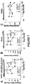
Figure 1 : Human neocortical astrocyte cell fates after mechanical trauma illustrated with bar graphs and photomicrographs of live-stained cells depicting nuclei of viable, leaky and dead cells. -
Figure 2 : Mechanically traumatized human astrocytes show prolonged endurance in a compromised state after wounding versus mouse astrocytes, as shown with a bar graph, a series of photomicrographs from time-lapse videos of live cells imaged on a temperature and gas controlled stage via confocal microscopy, and a schematic illustration of the protocol. -
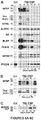
Figure 3 : Mechanical trauma of human astrocytes causes significant release of astroglial markers into surrounding fluids, as illustrated with immunoblots (A), and bar graphs showing amounts of marker at various time points after injury (B-G). -
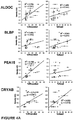
Figure 4 : Biplots showing that neurotrauma biomarkers are associated with cell fates of human traumatized astrocytes. -
Figure 5 : Schematic illustration of astrocyte injury biomarker selection strategy. -
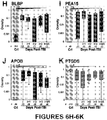
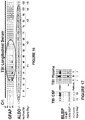
Figures 6A-6K : Immunoblots (6A-6C) and scatter-plots plotted jointly with box and whisker plots (6D-6K) with interquartile ranges (90th and 10th percentiles), median (line) and geometric mean (dashed line) showing logarithmic scaled optical densities measured from immunoblot signals using scaled densitometry (seeFigure 3 ) in CSF of 20-25 TBI patients on injury day and subsequent 5 post-injury days and 8-11 Controls (n: subjects numbers per day), showing that astroglial injury markers are elevated in CSF of TBI patients versus controls in a retrospective observational cohort on injury day and consecutive 5 days post-injury. -
Figure 7 : TBI patient outcome correlation of biomarker CSF amounts. -
Figure 8 : Assessing the spectrum of TBI using factors of grouped astroglial injury markers. -
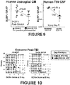
Figure 9 : Cell wounding over cell death - ratio of BLBP over GFAP differentiates trauma severity in human traumatized astrocytes and TBI patients. -
Figure 10 : Correlation between ALDOC levels and TBI patient outcome. -
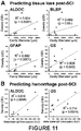
Figure 11 : Acute astroglial marker release correlated with histopathological severity measures, tissue loss and hemorrhage, after swine spinal cord injury. -
Figure 12 : Outcome correlation of acutely elevated ALDOC and GFAP after swine spinal cord injury. -
Figure 13 : Quantitative mass spectrometry, multiple reaction monitoring, documents concentration of astroglial TBI markers and allows marker amount comparison independent of antibodies. -
Figure 14 : Quantitative antibody-based evaluation of ALDOC and BLBP amounts and concentration ranges in TBI CSF and blood using known amounts of pure proteins. -
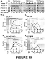
Figure 15 : Blood-compatible astroglial biomarkers in blood samples of severe TBI patients, as shown by immunoblots (A) and graphs of amount measured over time after injury (B). -
Figure 16 : Immunoblots of longitudinal severe TBI serum sample show extended detection window of ALDOC versus GFAP. -
Figure 17 : Immunoblots showing BLBP breakdown product in CSF and plasma after TBI. -
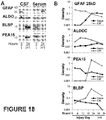
Figure 18 : Evidence for acute circulatory appearance of astroglial injury markers due to direct passage across damaged blood-brain barrier, as shown by immunoblots (A) and graphs of measured amounts over time after injury. -
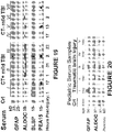
Figure 19 : Immunoblot data showing top tier astroglial injury markers and PEA15 are detected robustly and early in serum of mild TBI patients after concussions with or without complication (CT+: with complication - positive CAT scan; CT-: without complication, no CAT scan finding). -
Figure 20 : Immunoblot data showing acute and robust detection of serum ALDOC in pediatric TBI, infants. -
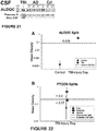
Figure 21 : Immunoblot data showing that full size ALDOC is present in greater amounts than the 38 kDa BDP in acute TBI, whereas the two sizes of ALDOC are present in different ratios (given in Table 2) in the chronic neurodegenerative condition of Alzheimer's disease. -
Figure 22 : Partitioning illustration of Table 3 thresholds. -
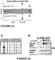
Figure 23 : Partitioning illustration of Table 4 thresholds. -
Figure 24 : Increased levels of glial trauma-release markers after repeated mild injury in the human trauma culture model, as indicated by percent astrocytes with acute membrane wounding and delayed cell death (A), and Conditioned medium (CM) levels of GFAP and AldoC after stretching (B). -
Figure 25 : Two film exposures of GFAP, showing that calpain and caspase activation generated GFAP upper and lower breakdown products after trauma. - The invention provides several new TBI biomarkers that were initially tested on CSF, plasma and serum from TBI patients and controls. New neurotrauma markers are defined by their release mechanisms to associate with cell wounding and/or cell death of human brain astroglia in a trauma model. Data presented herein demonstrate that select biomarkers show highly interesting kinetics and stability in body fluids. Immunological detectability, sensitivity and specificity is shown and suitable monoclonal antibodies have been selected. The timing of appearance of markers in CSF and serum during the first hours and days after TBI are presented in the accompanying Examples. The results show that markers described herein and detectable in patient serum or plasma can be used to identify moderate and severe TBI, as well as mild TBI, and patterns indicative of fatal TBI. The markers are summarized in Table 1.
- All scientific and technical terms used in this application have meanings commonly used in the art unless otherwise specified. As used in this application, the following words or phrases have the meanings specified.
- As used herein, "major", as in "major BDP", refers to the most frequently and consistently observed breakdown product, for example the 38 kDa BDP of ALDOC is the major BDP of ALDOC.
- As used herein, "acute" refers to an early time post-injury time, typical the biofluid sample was collected on injury day for it to be considered acute. For example, 15-30min after injury in trauma models, 1-2 hours post-injury in mild TBI patients, 3 hours to 24 hours post-injury in moderate and severe TBI patients.
- As used herein, complicated mild TBI is used for concussion patients with positive computed tomography, CT / CAT scan finding, or more broadly with lasting symptomology, based on Buki et al., 2015.
- As used herein, a "significant difference" means a difference that can be detected in a manner that is considered reliable by one skilled in the art, such as a statistically significant difference, or a difference that is of sufficient magnitude that, under the circumstances, can be detected with a reasonable level of reliability. In the Examples provided, herein, log-transformed data followed Gaussian distribution, and were used for statistical analyses by an independent statistician. One can use repeated measures analysis of variance with non-constant variance, mixed model (Crowder and Hand, 1990). As data are linear when log-transformed, significant changes are typically manifold, even by orders of magnitude. In one example, increase or decrease between TBI and controls that range between 80 fold to 13,000 fold are observed and found to be significant. In another example, changes across different post-injury days between 6 to 32 fold are considered significant. In yet another example, changes between survivors and non-survivors of TBI are between 4 fold and 1,400 fold are observed and found to be significant. In yet another example, an increase of two-fold relative to a reference or control sample is considered significant.
- As used herein, "control" or "control sample" refers to a sample that is representative of either normal levels, or obtained from a subject known to be healthy.
- As used herein, "a" or "an" means at least one, unless clearly indicated otherwise.
Table 1: Astroglial injury markers Name ID, molecular weight Breakdown products (BDPs) with size Release mechanism, marker class Biomarker properties Top tier markers Glial fibrillary acidic protein GFAP, 50 kDa Lower GFAP BDPs 29, 25, (23) kDa 19 + 20 doublet, sequence defined by mass spectrometry of traumatized astrocytes, TBI patient CSF and serumCell death marker, strong correlation to human astroglial death, not cell wounding. Small fragments are calpain and caspases activity dependent. Delayed presence in TBI blood Fast clearance from biofluids (CSF, blood). Brain specific and abundant Associates with TBI and SCI outcome and predicts SCI severity. Fructose-1,6-bisphosphate aldolase C ALDOC, 40 kDa Cell leak marker, strong correlation to ALDOC BDPs 38 + 37 kDa doublethuman astroglial cell wounding, moderate correlation to cell death. 35 + 30 kDa 25 kDa Fast release & presence in TBI blood Long-lived in biofluids (CSF, blood) Has higher levels than GFAP in CSF and blood on later post-injury days. Highly brain enriched and abundant. Strong predictive association with SCI severity and outcome; trend to relate with TBI outcome. ALDOC BDPs present in AD patients. More robust than GFAP in infants with TBI and mild TBI patients. Brain lipid binding protein BLBP, 15 kDa Cell leak and cell death marker FABP7 BLBP BDP 3 kDa Moderate correlations to both in traumatized human astrocytes. Fatty acid binding protein 7Fast release & presence in TBI blood Short-lived in biofluids (CSF, blood) Brain enriched; Suitable for TBI progression monitoring; BLBP/GFAP ratio differentiates TBI severity. Moderate correlation with SCI severity. Second tier markers Astrocytic phosphoprotein 15 PEA15, 15 kDa Cell leak marker, strong correlation with human astrocyte cell wounding. PEA15 BDPs 13 + 12 kDa doublet 8 kDaFast release & presence in TBI blood Short-lived in biofluids (CSF, blood) Suitable for TBI progression monitoring Trend to relate with TBI mortality Glutamine synthetase GS, 45 kDa GS BDPs 37, 35, 32 kDa More stable in biofluids as BLBP and PEA15, but less stable than ALDOC Predictive of SCI severity α Crystallin, B chain = Heath shock protein 27CRYAB, 21 kDa HSP27 CRYAB BDPs 19+18, 17 kDa triplet Cell leak marker, strong correlation with human astroglial cell wounding, moderate correlation with cell death. 15 + 14 kDa doublet 8 kDaShort-lived in biofluids (CSF, blood) CRYAB differentiates trauma severity Standards, indicators for CSF samples Apolipoprotein B APOB, 120-130 kDa Bleeding indicator N/A Secreted into blood from intestine & liver; absent from healthy CSF Associates with bleeding in TBI CSF. Prostaglandin (D2) Synthase =β trace protein PTGDS, 22 kDa Healthy CSF standard N/A Most abundant CSF protein. Secreted enzyme; Associates post-TBI with survival - The invention provides a method for detection or monitoring status of traumatic brain injury (TBI), mild TBI, in a surviving subject. The method can be used to determine the presence, progression, prediction, and discrimination of severity of TBI in a surviving subject. In one embodiment, the method comprises contacting a specimen of bodily fluid obtained from the subject with reagents for assaying for a marker of TBI selected from aldolase C (ALDOC), or a trauma-specific break down product (BDP) of ALDOC. The method comprises: contacting a specimen of bodily fluid obtained from the subject with reagents for assaying for a marker of TBI selected from aldolase C (ALDOC), or a trauma-specific break down product (BDP) of ALDOC, wherein the BDP of ALDOC is selected from the group consisting of a 38 kDa fragment, a 35 kDa fragment, a 30 kDa fragment, and a 23 kDa fragment; measuring the amount of marker present in the specimen as compared to a control sample; and determining the presence of TBI when an elevated amount of marker is present in the specimen compared to the control sample. The method further comprises measuring the amount of marker present in the specimen as compared to a control sample, and determining the presence of TBI when an elevated amount of marker is present in the specimen compared to the control sample. In one embodiment, the method further comprises measuring BLBP and/or a BDP thereof. Optionally, the method further comprises measuring the amount of glutamine synthetase (GS), astrocytic phosphoprotein PEA-15 (PEA15), αB-crystallin (CRYAB/HSP27), a trauma-specific proteolytic cleavage product of GS, PEA15, or CRYAB, or any combination of two or more thereof. In one embodiment, the method further comprises measuring the amount of glial fibrillary acid protein (GFAP), or of a 20-30 kDa BDP of GFAP.
- The monitoring of elevation of BLBP and/or PEA15 on subsequent days post-TBI informs on secondary adverse events post-injury. For example, the detection of elevated levels of ALDOC, BLBP, GS and PEA15 can be used to calculate Factor A, and levels of GFAP, S100beta and APOB can be used to calculate Factor B, based on marker loadings to each factor. Factor A and Factor B combined can be used to partition patients by severity. Factor A and B thresholds provide boundaries between TBI survivors, non-survivors and controls. A patient assessment within a clinical trial or study can be more robust by using a kit that provides multiple biomarker readings and performing factor analysis. This provides a simplified approach to track individual patients within a highly variable cohort, as opposed to requiring very large cohort sizes that may not be financially and otherwise feasible. Each clinical trial or study cohort biomarker panel data can be entered into a database, standardized and each patient is assessed based on tissue demise/bleeding versus tissue compromise/wounding Factors. Using Factors representative of these two classes makes the assessment more robust, as one zero reading will not prevent the entire factor analyses from providing a relative status output for any given patient. Boundaries pin out the severity spectrum of each cohort within which each patient will have a unique status at a given time post-injury
- In one disclosure, the method further comprises calculating a ratio between amounts of BLBP, an example of a cell leak marker, and GFAP, a cell death marker. The amounts can be measured using optical densities. Ratios between amounts of BLBP and GFAP in the trauma model range from 0.6-1.2 correspond to mild/moderate trauma, while ratios between 0.1-0.4 correspond to severe trauma. This reflects the finding in the human culture trauma model that, in severe trauma, there is proportionally more GFAP found than after mild trauma. BLBP/GFAP ratios in moderate TBI patients range between 0.4-0.3, whereas the range in severe TBI patients is between 0.01 and 0.05, again expressing a proportional larger GFAP to BLBP amount in severe versus moderate TBI patients. As such, using this ratio provides a more robust patient severity classification. By including two markers, significance is reached, whereas one marker alone would require a much larger cohort size. Use of marker combinations can thereby help in assessing TBI status and monitoring TBI progression, by reducing minimum required patient enrollment sizes when used as an evaluation tool in clinical studies or trials.
- The method for detecting and monitoring status of TBI in a subject can be used to identify a subject at risk for complications after mild TBI or concussion. This identification is made by using acute presence, such as using samples obtained within 1-2 hours and up to 17 hours post-injury of BLBP and/or PEA15, in addition to ALDOC in serum samples. ALDOC elevation alone can identify a concussion, and injury day elevation of ALDOC and BLBP and/or PEA15 is associated with risk for complications.
- Representative examples of the trauma-specific proteolytic cleavage product of ALDOC include a major 38 kDa fragment that was found most consistently, a 35 kDa fragment, a 30 kDa fragment, and a 23 kDa fragment. Examples of the trauma-specific proteolytic cleavage product of GS include a 37+35 kDa doublet, a 32 kDa fragment, a 23 kDa fragment, a 20 kDa fragment, and 18 kDa fragment. Examples of the trauma-specific proteolytic cleavage product of PEA15 include a 12+13 kDa doublet and a 8 kDa fragment. Examples of the trauma-specific proteolytic cleavage product of αB-crystallin is selected from the group consisting of a 18+19 kDa doublet, a 17 kDa fragment, a 15+14 kDa doublet and a 8 kDa fragment.
- The ratio of amount of ALDOC full size (40 kDa) to amount of ALDOC cleavage product (38 kDa) is indicative for time post-injury, as well as distinction of acute versus subacute versus chronic brain injury or neurodegenerative brain disease, including Alzheimer's disease (AD). Thus, in one embodiment, the method of detecting and/or monitoring TBI comprises determining the ratio of 40 kDa ALDOC to 38 kDa ALDOC levels in the specimen obtained from the subject. A ratio larger than 1, ranging from 3.6 - 8.6, is indicative of TBI (acute and subacute, over post-injury days 1-5), as full size ALDOC is much more abundant than the 38 kDa ALDOC BDP. A ratio smaller than 1, ranging 0.4-0.6 is indicative of Alzheimer's disease, as the 38 kDa ALDOC BDP was similar or more abundant than the full size ALDOC optical signal density because a chronic degenerative condition allows for long-term partial degradation and accumulation of the fragment than an acute injury condition.
- In one embodiment, the method further comprises measuring the amount of a blood specific protein in a cerebrospinal fluid (CSF) sample obtained from the subject. The detection and monitoring of such markers can be used to determine the status of intraventricular brain bleeding post-injury. In one embodiment, the blood specific protein is apolipoprotein B (APOB). In another embodiment, the method further comprises measuring the amount of prostaglandin synthase (PTGDS) in a cerebrospinal fluid (CSF) sample obtained from the subject. PTGDS, also known as beta trace protein, is positively correlated with a healthy CSF composition. The presence of TBI is determined when the amount of PTGDS is reduced, and rises with recovery. The detection and monitoring of such markers can therefore be used to determine the status of recovery to healthy levels after injury. In some disclosures, recovery of acute trauma-reduced PTGDS levels is monitored over subsequent post-injury days and is predictive of survival, while sustained reduced levels of PTGDS predict mortality.
- In some embodiments of the method, no additional markers are assayed beyond those recited herein. In other disclosures, only markers recited herein are assayed. In some disclosures, additional markers known to those skilled in the art are assayed in combination with markers recited herein. In other disclosures, only a subset of possible markers is assayed. For example, the method can comprise assaying for 2, 3, 4, 5, 6, 7, 8, 9, 10, 12, 15, or 20 markers. In one particular disclosure, no more than 4 markers are assayed.
- The reagents for use in the method of the invention can comprise antibodies or other molecules that specifically bind the marker of TBI. In one embodiment, the measuring comprises immunoassay. Examples of immunoassays include western blotting, immunofluorescence, immunoluminescence, radioimmunoassay, and ELISA. ALDOC isoform specific antibodies are available as monoclonal antibodies clones 4A9, 1A1, 5C9 and E9 from EnCor Biotechnology Inc. (Gainesville, Florida). BLBP specific monoclonal antibodies are also available from EnCor Biotech Inc.
- In another embodiment, the reagents comprise protein-sequence and -fragment-specific peptides. Such reagents are useful for methods in which the measuring comprises targeted quantitative mass spectrometry. In one embodiment, the measuring comprises quantitative signal detection of endogenous (in the sample) proteo-typic peptides that are compared to added ('spiked in') labeled (e.g., heavy isotope labeled) known amounts of the same proteo-specific peptides (internal standards) using multiple or parallel reaction monitoring mass spectrometry.
- In one embodiment, the control sample is a pre-injury sample obtained from the subject. In another embodiment, the control sample is representative of normal, healthy subjects, such as an average value obtained from a control cohort of healthy subjects.
- Representative examples of a specimen of bodily fluid for use in the invention include, but are not limited to, plasma, serum, cerebrospinal fluid (CSF), nasal fluid, cerumen, urine, saliva, lacrimal tears, and brain microdialysate.
- The invention further provides a method of determining the presence of the biomarkers ALDOC and BLBP in a sample of serum or plasma obtained from a subject. In one embodiment, the method comprises contacting the serum or plasma sample with a kit of the present disclosure and measuring the binding of the agents to the biomarkers.
- Also provided is a method of determining the status of traumatic brain injury in a sample of serum or plasma obtained from a subject. In one embodiment, the method comprises contacting the serum/plasma sample with a kit here disclosed and measuring the binding of the agents to the biomarkers, and comparing the binding to a control sample. TBI is then determined to be present if the binding of the agents to ALDOC and BLBP is increased in the serum sample from the subject relative to the control sample. For moderate to severe TBI patients, amounts and concentration ranges for ALDOC and BLBP are given in
Figure 14 and Example 13, and biomarker comparisons are given with concentrations inFigure 13 and Example12. The invention further provides a method of detecting TBI, in a subject. In particular, it can be used to detect mild TBI, where diagnosis is not otherwise obvious. In one embodiment, the method comprises assaying a specimen of bodily fluid from the subject for an elevated amount of ALDOC and BLBP compared to a control sample. An elevated amount of ALDOC and/or BLBP is indicative of TBI. In one embodiment, the assaying is performed within 24 hours of a suspected injury. In some disclosures, the assaying is performed within 1-3 hours, or as early as within 15-30 minutes, of a suspected injury. In one disclosure, the assaying is performed up to 7 days after a suspected injury. In some embodiments, the subject is an infant or child, including, for example, a subject suspected of having experienced shaken baby syndrome. The method allows for injury severity to be assessed, and outcome predicted in a subject, acutely after a spinal cord injury. - The disclosure additionally provides a method of predicting outcome of TBI in a subject. In one disclosure, the method comprises assaying a specimen of bodily fluid from the subject for an elevated amount of PEA15 and/or 20-30 kDa (small) GFAP fragments compared to a control sample, wherein an elevated amount of PEA15 and/or small (i.e. lower) GFAP fragments is predicative of mortality. Also disclosed is a method of treating TBI in a subject. In one disclosure, the method comprises assaying a sample obtained from the subject for a marker of TBI as described herein; and treating the patient for TBI if the assay indicates presence of TBI. The disclosure further provides a method of monitoring for treatment guidance in a subject being treated for TBI. In one disclosure, the method comprises assaying a sample obtained from the subject for a marker of TBI as described herein; and initiating a treatment of the patient for TBI if the assay indicates concerning deterioration of the patients status during the days post-injury, i.e. showing secondary elevated levels of any of the markers described herein. The methods of the invention additionally provide pharmacokinetic (theragnostic) applications, that is use in monitoring drug or other patient treatment for early evaluation of treatment effects and to monitor TBI progression post-injury. Those skilled in the art will appreciate that, given the different release and clearance kinetics of the markers described herein, the benefit of using multiple markers described herein as a panel. Thus the patients' assessment can include any one of the markers: ALDOC, BLBP, GS, PEA-15, CRYAB, a BDP of any of the foregoing; alone or in combination with one or more additional markers.
- Some embodiments contemplated by the present disclosure include use of a combination of TBI markers, including aldolase C (ALDOC), glutamine synthetase (GS), astrocytic phosphoprotein PEA-15 (PEA15), αB-crystallin (CRYAB), or brain lipid binding protein (BLBP/ FABP7), a trauma-specific proteolytic cleavage product of ALDOC, GS, PEA15, CRYAB, or BLBP/FABP7, or any combination of two or more thereof. For example, embodiments include those in which the marker of TBI is GS and aldolase C, the marker of TBI is GS and PEA15, the marker of TBI is GS and αB-crystallin, the marker of TBI is GS and BLBP, the marker of TBI is aldolase C and PEA15, the marker of TBI is aldolase C and αB-crystallin, the marker of TBI is aldolase C and BLBP, the marker of TBI is PEA15 and αB-crystallin, the marker of TBI is PEA15 and BLBP, the marker of TBI is αB-crystallin and BLBP, the marker of TBI is GS, aldolase C, and PEA15, the marker of TBI is GS, BLBP, and PEA15, the marker of TBI is GS, αB-crystallin, and PEA15, the marker of TBI is GS, αB-crystallin, and BLBP, the marker of TBI is GS, αB-crystallin, and aldolase C, the marker of TBI is GS, BLBP, and aldolase C, the marker of TBI is aldolase C, PEA15, and αB-crystallin, the marker of TBI is aldolase C, PEA15, and BLBP, the marker of TBI is aldolase C, αB-crystallin, and BLBP, and the marker of TBI is PEA15, αB-crystallin, and BLBP. In the above examples, the TBI may be the recited protein, a breakdown product thereof, or both.
- Provided herein is a kit comprising agents that specifically bind a set of biomarkers for use in the method of any one of the preceding embodiment of the invention.. In one embodiment, the biomarkers comprise aldolase C (ALDOC) and optionally brain lipid binding protein (BLBP). The agents are typically polynucleotides or antibodies, and optionally labeled with a detectable marker. The kit optionally further consists of at least one container for housing the agents and/or instructions for use of the agents for determining status of traumatic brain injury in a test sample. In some embodiments, the kit further comprises agents that specifically bind astrocytic phosphoprotein PEA-15 (PEA15) and/or a 20-30 kDalton fragment of glial fibrillary acid protein (GFAP-BDP). In one embodiment, the antibodies are monoclonal antibodies. In one disclosure, the set of biomarkers consists of up to 3, 4, 5, 6, 7, 8, 9, or 10 biomarkers.
- The following examples are presented to illustrate the present invention and to assist one of ordinary skill in making and using the same. The examples are not intended in any way to otherwise limit the scope of the invention.
- This Example shows population scores of
human astrocytes 30 minutes and 48 hours after abrupt pressure-pulse traumatic stretching using different severities (Figure 1 ; PSI ranges from 2.6-4.0 for milder injury and 4.4-5.3 for severe injury). Human astrocytes were isolated from 16-18 week donated fetal neocortical brain specimen, purified and then differentiated on deformable membranes (Wanner, 2012). Cell membrane wounding / compromise, mechanoporation (Barbee, 2005), was determined using Propidium iodide (PI) uptake in living cultures accompanied by nuclear shape assessment (middle picture, stained nuclei, Hoechst stained after fixation, with little pink dots, nucleoli that are PI-stained). Cell death was determined by PI uptake accompanied by condensed nuclei (pyknotic nuclei with compacted chromatin and bright Hoechst and PI stains). Significant percent of cell integrity compromise is shown 30min post-injury (left graph) while significant cell death was present two days post-injury (bars on the right). - This Example demonstrates that mechanically traumatized human astrocytes show prolonged endurance in a compromised state after wounding versus mouse astrocytes (
Figure 2 ). Dye uptake (0.05mM 10 kDa dextran rhodamine) during stretch as well as cell death by apoptosis (caspase activation using 10µM caspase 3 substrate) was monitored using time-lapse imaging on a temperature and humidity controlled spinning disc confocal microscope (Levine et al., 2016). Human traumatized wounded and resealed human astroglia show prolonged endurance after integrity compromise. - As shown in
Figure 3 , mechanical trauma of human astrocytes causes significant release of astroglial marker into surrounding fluids.Figure 3A shows immunoblots of concentrated, denatured conditioned media (Levine et al., 2016; Sondej et al., 2011) from traumatized astrocytes show different release profiles of glial fibrillary acidic proteins (GFAP) and its known and newly-identified breakdown products (BDPs) versus aldolase C (ALDOC), brain lipid binding protein (BLPB), astrocytic phosphoprotein (PEA15) and α crystallin (CRYAB). In order to capture the full range of signal intensity, signals from multiple exposures of immunoblots were densitometrically measured (optical densities) and scaled to match a single exposure. Scaled densities of signals from indicated number of donors are plotted on log-spaced axis in the graphs shown inFigure 3B-G . Known upper GFAP BDPs (between 37-50kDa) showed increased release with time (triangles) and severity (circle at 5 hours post-injury). New, lower GFAP BDPs were significantly elevated only one and two days post-injury, associating them with cell death (seeFigure 1 ). ALDOC, BLBP and PEA15 were already significantly elevated 30min post-injury over two orders of magnitude and such elevated levels remained in fluids at all post-injury times measured. CRYAB levels showed severity distinction at 5 hours and two days post-injury. - Shown in
Figure 4A are biplots of trauma-released astroglial marker levels (seeFigure 3 ) for ALDOC, BLBP, PEA15 and CRYAB over the percent membrane permeable cells (% wounded, red data, left) as well as correlated to percent of dead human traumatized astrocytes using PI-dye-update assay and nuclear morphology (seeFigures 1 +2). Regression lines (R2-value) and p-values indicate correlation significance. ALDOC, PEA15 and CRYAB show best correlation with human astroglial cell wounding. CRYAB, BLBP and ALDOC also correlated with extend of cell death after trauma. -
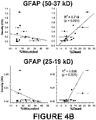
Figure 4B shows biplots of GFAP trauma-release, which show correlation of GFAP with cell death inflicted by mechanical trauma and weak / no correlation with cell wounding. Plots are separated by grouped GFAP fragment sizes (upper bands: 50-37 kD, lower bands: 25-19 kD). - Candidate astrocyte injury biomarkers were selected by the following strategy (
Figure 5 ). A TBI CSF proteome was compiled by bottom-up mass spectrometry using CSF samples from 19 severe TBI patients and compared with the CSF proteome from 9 control subjects (Crl). This TBI CSF proteome (483 proteins) overlapped with 252 proteins from the control CSF proteome (402 proteins), leaving 231 unique TBI CSF proteins. Sixty percent of the TBI CSF proteins were also present in the published plasma proteomes (Omenn et al., 2005; Schenk et al., 2008) and, if abundant, may not be suitable as TBI biomarkers. To select candidate brain injury markers, we determined proteins released from traumatized astrocytes using a proteomic approach in asimple trauma model 79 differentially released proteins were identified after pressure-pulse stretching mouse astrocytes (Levine et al., 2015; Sondej et al., 2011). - From this "traumatome" 48 proteins (62%), were present in TBI CSF. Selecting for highly astrocyte-enriched proteins (>/= 5 fold enrichment over other cell types, Cahoy et al., 2008) yielded a small candidate pool of 11 injury biomarker proteins (black outlined enclosed 3 fields). These included 3 proteins GFAP and peroxiredoxin 6 (Prdx6, both present in plasma) and N,N-dimethyl arginine dimethyl aminohydrolase (DDAH1, center field of 3). Additional 3 proteins were astrocyte enriched, "traumatome" proteins present in TBI CSF and also in control CSF that were aldolase C (ALDOC), clusterin (CLUS) and apolipoprotein E (APOE, lower field of 3). Clusterin and APOE are secreted by astrocytes and their levels decreased in fluid after trauma (arrow). Additional 5 trauma-released proteins that were highly astrocyte enriched were not listed in the shotgun mass spectrometry-based TBI CSF proteome list. These 5 were ezrin (Ezr), F-box only protein 2 (FBX2), Glutamine synthetase (GS), astrocytic phosphoprotein 15 (PEA15) and brain-lipid binding protein (BLBP, upper field of 5, dashed outline). GFAP, ALDOC, GS, BLBP, PEA15 and CRYAB were included in subsequent immunological and mass spectrometry testing.
- This Example demonstrates that astroglial injury markers are elevated in CSF of TBI patients versus controls in a retrospective observational cohort on injury day and for five consecutive days post-injury.
Figure 6A shows immunoblots of GFAP (50 kDa withBDPs -
Figure 6B shows six CSF samples (30 µl/lane) from four TBI patients (2.-5.), illustrating variable signal intensities of GFAP and "upper" BDPs between 50 and 37kDa and "lower/new" GFAP BDPs including 25/23 kDa doublet, 20 kDa and 18 kDa small BDPs on injury day (patients -
Figure 6C shows CSF immunoblots (30µl/lane), demonstrating full size ALDOC (40 kDa) in five TBI patients (6.-10.) and 38 kDa ALDOC BDP of variable intensity on four days post-injury in three TBI patients (8-10). A healthy Control subject showed no ALDOC. - Scatter-plots (
Figures 6D-K , same day and patient replicates averaged) are plotted jointly with box and whisker plots with interquartile ranges (90th and 10th percentiles), median (line) and geometric mean (dashed line) showing logarithmic scaled optical densities measured from immunoblot signals using scaled densitometry (seeFigure 3 ) in CSF of 20-25 TBI patients on injury day and subsequent 5 post-injury days and 8-11 Controls (n: subjects numbers per day). (Fig. 6D ) Upper GFAP signals (50-37 kDa) were elevated on all TBI days versus Crl (P <0.06) and declined over time as indicated (P <0.05, repeated measures ANOVA, see Methods). (Fig. 6E ) Lower GFAP BDPs (25-18 kD) were elevated in TBI versus Crl (P < 0.03) and declined between first post-injury day later post-injury days (P < 0.05). (Fig. 6F ) ALDOC (P < 0.004) and (Fig. 6G ) GS (P < 0.001) were elevated on each day in TBI CSF versus Crls without decline. (Fig. 6H ) BLBP (P< 0.03) and (Fig. 6I ) PEA15 (P< 0.004) had elevated levels in TBI versus Crls on indicated days with larger signal ranges. Serum protein APOB (Fig. 6J ) was elevated in TBI versus Crl CSF (P <0.005), whereas CSF standard PTGDS was stronger in Crl versus TBI (P< 0.004) with levels acutely depleted to various extents followed by outcome dependent recovery (seeFigure 7 ). - This Example demonstrates that CSF levels of new, cell death associated lower GFAP breakdown products were two orders of magnitude more elevated in non-survivors versus survivors of TBI (
Figure 7 ). PEA15 levels were over 2-3 orders of magnitude elevated in non-survivors versus survivors after TBI, with p-value on the third post-injury day p=0.07. Levels of health marker PTGDS, which decreased acutely post-injury, showed significant recovery by the third post-injury day in survivors of TBI while levels remained decreased in non-survivors. - This Example demonstrates the grouping of astroglial trauma markers to create factors useful in the assessment of TBI across the spectrum of injury. To combine the diverse data encompassed by the marker panel, multivariate analysis of variance that employed an unsupervised learning algorithm based on Spearman correlation coefficients was used (factor analysis, Fabrigar and Wegener, 2012; Tucker, 1997). This approach is new to the neurotrauma biomarker field, and reveals underlying neurotrauma conditions by grouping markers here based on their TBI CSF signals. Known astroglial marker S100β, cell death markers GFAP with known and new BDPs, and bleeding indicator APOB were grouped into Factor A (
Fig. 8A , gray, with combined loading using Cronbach's coefficient α = 0.921). New 'cell leak', cell wounding markers ALDOC with BDP, BLBP, GS and PEA15, were grouped into Factor B (α=0.879). As shown inFig. 8B , Factor A's temporal profile significantly decreased over days post-injury, while Factor B's trajectory did not. Factor A data showed significant difference between survivor and non-survivors on several post-injury days (Fig. 8C ). Factor B data did not show survival differences (Fig. 8D ). Standardized marker density readings from 12 TBI patients with signal present for all markers were converted into the Factors A and B using the loadings shown inFig. 8A , and then plotted against each other (Fig. 8E ). Heterogeneity among the TBI spectrum is reflected in the spread of the combined panel data in this biplot. Classification tree analysis (Fig. 8F ) determined boundaries that partitioned controls, survivors and non-survivors of TBI according to given Factor thresholds (Breiman, 1984). Patient partitioning thresholds using the biomarker levels grouped into factors are illustrated by lines inFig. 8E . - This mathematical multivariate unsupervised learning approach combines the markers of this panel with each marker given a weight (loading) that is derived from correlation coefficients and expresses how much variance in the patient cohort is captured by this marker's contribution. Overall high loading of the listed markers (0.8-0.95) documents robust categorization into the two factors. Factor A reflects markers associated with cell death, hemorrhage and tissue loss. Factor B reflects markers associated with cell leak, wounding and tissue compromise (see
Figure 8A ). The resulting values document a unique position of each patient based on its biomarker panel readings within a given cohort. This approach reliably covers the majority of patient variation observed in this TBI cohort. Factor analysis provides a simplified framework for capturing a large patient heterogeneity observed in TBI using only few biomarker readings, (compared to more complex principal component analysis that used many different entries). In a clinical trial, these biomarker panel readings from a test kit can be entered into a growing database that can provide valuable monitoring information of individual TBI patients compared to other patients. This tool can simplify patient assessment and strengthen the robustness of ongoing patient evaluation using biofluid signature of tissue compromise and tissue demise. - This Example demonstrates that the ratio of BLBP to GFAP levels in a subject's specimen can be used to differentiate trauma severity in human traumatized astrocytes and TBI patients. Significant differences in fluid level ratios of 'cell leak' marker BLBP over 'cell death' marker GFAP are shown in vitro (
Fig. 9 , CM, conditioned medium, left) and in TBI patients (CSF, right panel ofFig. 9 ). Human astrocytes from 6 donors were traumatized with different severities using indicated PSI pressure-pulses. Moderate versus severe TBI patients are distinguished by 14 fold different BLBP/GFAP ratio in CSF (n=9). Moderate TBI patients are defined by post-resuscitation Glasgow coma scale (GCS) > 8 while severe TBI patients had GCS<8. - Data presented in
Figure 10 show ALDOC and GFAP levels plotted against outcome of severe TBI patients, assessed using extended Glasgow outcome score (GOSe) at 6 months post-TBI. Data from 12 (11) TBI patients show ALDOC elevation early (white, i, i+1, +2) and later post-injury (black, i+3, +4 and +5 post-injury days) in patients with unfavorable outcome, while GFAP levels did not as well maintain elevated levels on later post-injury days. - This Example demonstrates the correlation between astroglial trauma markers and severity of spinal cord injury in a swine animal model.
Figure 11 : Acute UCLA astroglial marker release correlated with histopathological severity measures, tissue loss and hemorrhage, after swine spinal cord injury. A) Acute (15-30 min post-injury) CSF signal densities of UCLA astroglial injury markers ALDOC, BLBP and glutamine synthetase (GS) as well as GFAP are plotted against rostro-caudal cavity diameters in the Yucatan swine spinal cord one week after spinal cord contusion injury using an established injury weight drop model (Lee et al., 2013). Spearman correlations indicate acute elevation of these astroglial CSF marker levels associated significantly with increasing tissue loss. Histopathology: Rostro-caudal extension of the cavity was measures in Sudan black stained horizontal spinal cord sections (p-values and R2 values given, n=10 animals). B) Acute (15-30min post-injury) CSF signal densities of ALDOC and GS significantly associated with white matter interstitial tissue bleeding one week post-injury that was measured using swine immunoglobulin (IgG) fluorescence in injured horizontal spinal cord sections and normalized by parallel stained uninjured horizontal spinal cord sections. Such normalized white matter fluorescence signals of rostro-caudal serial images were plotted for each animal (n=8 animals) and areas under the curves (AUC) were determined over distances with above uninjured signal levels. -
Figure 12 shows the correlation of acutely elevated ALDOC and GFAP with outcome after swine spinal cord injury. Recovery of walking associates with acute CSF levels of ALDOC and GFAP after swine spinal cord injury. Plotted are acute post-injury CSF levels (15-30 min post-injury) of ALDOC (black dots) and GFAP (white dots) that predicts recovery of walking one week post-injury using the porcine thoracic injury behavioral scale (PTIBS), an established test for recovery of ambulation in the swine indicating the level of walking recovery after spinal cord injury in the Yucatan swine (Lee et al., 2013). A significant inverse correlation existed with higher ALDOC (R2 -0.88) and GFAP (R2 -0.89) levels in poorly ambulating animals, who were dragging their hind limbs and crawled at best, and low acute ALDOC and GFAP levels in animals who walked by one week post-injury. Preliminary biomarker thresholds are indicated by background color, showing feasibility of very early partitioning of recovering animals. - This Example demonstrates that quantitative mass spectrometry confirms the increased levels of astroglial TBI markers following injury, providing objective marker amount comparisons that is limited when using immunological methods as these are not standardized. Multiple reaction monitoring, an antibody independent, simultaneous and quantitative mass spectrometry approach, was used for the first time in the neurotrauma biomarker field to compare abundance of known and new astroglial markers in CSF of TBI patients. Marker-specific peptides are measured in parallel with defined amounts of added, isotope-labeled peptides (see Table 6).
Figure 13A shows that ALDOC and GFAP had the highest concentrations in TBI CSF on injury day, and both differed significantly from levels of other markers shown. In addition, BLBP levels were significantly higher than GS levels on injury day. As shown inFigure 13B , by 3 days post-TBI, ALDOC levels significantly out-performed GFAP concentrations, differing by an order of magnitude. - This Example provides a quantitative antibody-based evaluation of ALDOC and BLBP levels in CSF and blood of TBI specimens using standard curves. Box plots in
Figure 14 show median and interquartile concentration ranges of ALDOC (left) and BLBP (right) and their amounts in CSF and blood (serum and plasma). Inserts show dose-response using known amounts of isoform-specific recombinant proteins ALDOC and BLBP using two concentration ranges and immunoblot detection conditions (see Table 7). - This Example demonstrates that the astroglial trauma markers are compatible with blood testing, using samples obtained from patients having severe TBI.
Figure 15A shows longitudinal plasma samples from 3 different severe TBI patients alongside one control plasma sample (Crl). Abundant proteins are removed using immunoaffinity albumin and immunoglobulin depletion columns (Sigma, ProteoPrep). New GFAP breakdown product of 25 kDa was never detected on injury day (i) but appeared on subsequent post-injury days. ALDOC was consistently present at all time points in all 3 patients. Short-lived markers PEA15 and BLBP were robustly present on injury day and showed different temporal profiles over subsequent days post-injury in each patient. Plotted inFigure 15B are scaled densitometry signals for ALDOC (B1), BLBP (B2), GFAP/25 kDa BDP (B3) and PEA15 (B4) from 26 serum and 24 plasma samples derived from 22 severe TBI patients compared with up to 11 control blood samples (Crl). Longitudinal same patient data are connected by gray lines. ALDOC was significantly elevated on injury day and every post-injury day, while GFAP was significantly elevated beginning on the first post-injury day. BLBP and PEA15 had significant elevation on injury day versus control levels. - This Example demonstrates, using a longitudinal severe TBI serum sample, the extended detection window of ALDOC versus GFAP. ALDOC was detected 19 hours prior to first detection of
GFAP 25 kDa BDP and ALDOC signals were present over two days beyond the last specific GFAP signal, a 37 kDa known GFAP BDP (Figure 16 ). - In addition to the full size, 15 kDa BLBP a 3 kDa BLBP-specific fragment was detected using 2 antibodies in TBI CSF and plasma on injury day and various post-injury days. Results are shown in
Figure 17 . -
Figure 18 presents evidence for acute circulatory appearance of new astroglial injury markers due to direct passage across damaged blood-brain barrier. Panel A ofFigure 18 shows immunoblots forGFAP 25 kDa BDP along with ALDOC, BLBP and PEA15 in CSF and serum of the same severe TBI patient acutely after TBI (3 hours post-injury), as well as on the first post-injury day. While GFAP appeared first in the CSF and with a day delay in serum, ALDOC was present in both biofluids at both time points. BLBP and PEA15 were first present in serum and appeared with delay in CSF. Traces on log-spaced axis of all 4 astroglial markers document the switch forGFAP 25 kDa BDP from CSF into serum, steadier presence of biofluid-stable ALDOC, and the presence of short-lived BLBP and PEA15 in serum prior to their appearance and delayed elevation in CSF (Figure 18B ). - These observations suggest the direct passage of ALDOC, BLBP and PEA15 from the injury site into the circulation. All three markers are localized in astroglial processes, with fine endings known to entirely wrap capillaries and blood vessels (Mathiisen et al., 2010). Traumatic injury, even mild TBI, causes rupture of perivascular astroglial fibers, allowing these markers a direct passage into the blood (Barzo et al., 1996; Hicks et al., 1993; Korn et al., 2005). GFAP is not localized in astroglial endings, and, as shown in
Figure 3 , the 25 kDa GFAP BDP takes time to be generated, suggesting a delayed release during cell death leading to accumulation first in the CSF, and subsequent appearance in serum. Advantage of a direct passage of astroglial endfeet via open blood-brain barrier lays in enabling very acute post-injury blood-testing. - This Example demonstrates robust and early detection of top tier astroglial injury markers in serum of mild TBI patients. Shown in
Figure 19 are 10 serum samples from 7 mild TBI (mTBI) patients early after injury alongside of one control serum (Crl). Samples were probed for GFAP, ALDOC, BLBP and PEA15. Specific GFAP signals were faint and limited to4 mTBI patients with one patient showing GFAP/25 kDa BDP by 31 hours post-injury (patient # II).ALDOC 38 kDa BDP was consistently and strongly elevated in all mTBI patients versus control. BLBP and PEA15 showed variable intensity and were present in 5 mTBI patients. ALDOC, BLBP and PEA15 were detected already one hour post-injury. Concussion patients received computed tomography (CT) scans and those with positive findings of a lesion/bleed were classified as CT+, potentially complicated mTBI patients, and those without visible wounds were CT-negative, or uncomplicated (Buki et al., 2015). Robust presence of ALDOC and differential signals for BLBP and PEA15 are suited to augment risk identification among concussion patients. -
Figure 20 shows acute and robust detection of ALDOC in pediatric TBI patient serum samples. Injury day serum samples from 5 infants (1-4 months of age) suffering TBI are shown next to a control serum from a child aged 22 months. Robust ALDOC signals are detected in all infant TBI versus control cases while only two infants showed weak GFAP signal (34 kDa BDP, not previously described, cases # I, III). - This Example demonstrates that CSF samples from patients afflicted with the chronic neurodegenerative condition of Alzheimer's disease exhibit equally distinct levels of full size (40 kDa) and 38 kDa BDP ALDOC. In contrast, ALDOC detected in CSF of an acute TBI patient shows a distinct preponderance of full size (40 kDa) versus 38 kDa BDP. Shown in
Figure 21 are 7 CSF samples from 5 Alzheimer's disease patients (AD, stage 0.5, cases #II and III;stage 1, cases # IV and V; based on Fagan et al., Science Transl. Med. 2014) compared to one severe TBI patient (case # I) and two age-matched controls (cases # VI and VII) and protein stain for loading (Ponceau S). The TBI patient had mainly full size ALDOC and little 38 kDa fragment onday 4 post-injury, while the chronic degenerative AD samples showed equal presence of full size and 38 kDa ALDOC BDP. The data suggest that acute and chronic brain injury can be distinguished on the basis of ALDOC /ALDOC BDP ratio.Table 2: Distinction between TBI and Alzheimer's disease, using ratio between full size ALDOC and its 38 kDa proteolytic fragment in patients' CSF ALDOC 40kD/38kD All antibodies used ALDOC 40kD/38kD Same antibody (E9) used AD 0.61 ± 0.25 0.42 ± 0.14 TBI 3.63 ± 3.26 8.56 ± 0.85 AD v TBI p = 0.0001 p = 0.00002 - The table shows average ratios of full size ALDOC (40 kDa) over its breakdown product (38 kDa) in CSF of Alzheimer's Disease (AD) patients and moderate to severe TBI patients. Different ALDOC antibodies to different epitopes of the protein resulted in varying emphasis of the 40 versus 38 kDa band signal intensities. On the left data of all ALDOC antibodies were combined averaging 20 AD samples and 25 TBI samples. The average AD ratio was 6 fold smaller than the average TBI ratio that was significant by two-tailed T-test. On the right 10 AD CSF samples and 5 TBI patient's samples were analyzed using same antibody (E9) for ALDOC detection. There was again a significant 20 x difference between TBI, showing more full size signal and AD showing more BDP ALDOC signal. Data examples are shown in
Figure 21 . This provides a distinction between TBI and chronic neurodegenerative disease. Both data selections result in significantly different ratios based on the increased abundance of the ALDOC major 38 kDa BDP signifying the chronic neurodegenerative condition. Acute TBI can easily be distinguished by a higher abundance offull size 40 kDa ALDOC acutely and up to 5 days post-TBI. - Multivariate Classification Tree Analysis (Breiman, 1984) was used to determine the markers that most accurately split the subject cohort into Control and Surviving or Non-surviving TBI patients on injury, analyzed by immunoblotting of 30 µl CSF sample each. Upon obtaining 100% accuracy with just two markers, AldoC and PTGDS (Table 3), the analysis was repeated allowing only those markers known to be detectable in the blood for future noninvasive assay (Table 4). Markers used for the data presented in Table 3 were Aldo C and PTGDS, while the markers considered, but not used, were: GFAP, PEA15, GFAP lower, S100B, BLBP, GS, APOB, PTGDS, and AldoC 38kD. Table 3 thus summarizes the detection of TBI and survival outcome prediction using all markers in the classification tree analysis. This analysis selected ALDOC and PTGDS as the best partitioning markers. Among all inspected markers, those were utilized by the mathematical unsupervised learning approach. Groups, n=21, all Controls; and all injury day TBI of 30 TBI samples. The indicated 7ng/30µl CSF equals a concentration of 233 ng/ml CSF using immunoblotting. Accuracy was excellent, as grouping and predicted
outcome match 100% correctly.Figure 22 provides a partitioning illustration of the Table 3 thresholds.Table 3: Detection of TBI and survival outcome prediction Group n Aldo C [OD] (ng/30ul CSF) PTGDS [OD] Prediction A 8 >= 0.078 (7ng) < 0.174 or TBI survivor (severe) C >= 0.078 (7ng) >= 0.202 TBI survivor (moderate) B 2 >= 0.078 (7ng) 0.174 to 0.202 TBI non-survivor D 11 < 0.078 (7ng) > 0.202 Control (healthy subject) - Table 4 shows the detection of TBI and survival outcome prediction using the new blood-compatible glial markers. Markers used for Table 4 (1): Aldo C total. Markers considered but not used were: GFAP lower, BLBP,
PEA 15,Aldo C 38 kD breakdown product (BDP). The markers NOT considered (omitted) were: GFAP total, S100B, GS, APOB, and PTGDS. Groups were: n=21 all Control; and injury day TBI patients of a total of 30 in entire cohort. Accuracy was 95%; the unweighted probability correct was 20/21, or 0.952. There was a 96% equal priority probability correct = 0.96 = (1.0 + 1.0 + 0.875).Figure 23 provides a partitioning illustration of the Table 4 thresholds.Table 4: Detection of TBI and survival outcome prediction using TBI markers Group n Aldo C total [OD] Prediction A 4 0.078 to 0.22 TBI survivor B 3 0.22 to 0.4 TBI non-survivor C 3 >= 0.4 TBI survivor D 11 < 0.078 Control (healthy subject) - Classification matrix for data in Table 4:
Predicted-control Predicted-died Predicted-Survivor total True - control 11 0 0 11 True - died 0 2 0 2 True- survivor 0 1 7 8 - Receiver Operating Characteristic (applicable with higher n):
Group Area N N correct Prop correct Control 1.0000 11 11 1.0 Died 0.9737 2 2 1.0 survived 0.9904 8 7 0.875 - Secondary TBI progression can cause elevated intracranial pressure (ICP) which is often associated with secondary injury. Observed is a significant correlation between levels of cell death marker GFAP lower BDPs (19, 20, 25 kDa doublet) and ICP. Bleeding marker APOB is significantly correlated with extra+intraparenchymal lesion volume and midline shift. Both these CT findings are associated with brain bleeding, including epidural hematoma, subdural hematoma, subarachnoid hemorrhage and intraparenchymal lesions. Glial injury markers BLBP and PEA15, as well as cell death glial marker GFAP lower BDPs, are correlated with intraparenchymal lesions, including brain tissue contusion, intracranial hemorrhage and diffuse axonal injury.
Table 5: Spearman Correlations between marker signal density and CAT scan data ICP, Intracranial pressure Midline Shift (edema indication) Extra+lntra Parenchymal Lesion Volume Intraparenchymal Lesion Volume New TBI marker panel Correlation p-value Correlation p-value Correlation p-value Correlation p-value Lower GFAP 0.0005 BDPs 0.834 9 0.121 0.733 0.676 0.136 0.775 0.333 ALDOC -0.165 0.614 -0.0872 0.776 -0.126 0.72 -0.4 0.517 BLBP 0.204 0.559 -0.194 0.58 0.116 0.803 0.6 0.417 PEA15 0.21 0.55 0.373 0.321 0.667 0.233 0.738 0.333 APOB -0.258 0.446 0.684 0.0361 0.986 0.0028 0.667 0.233 -
Figure 24 shows results obtained using a model for repeat mild injury. Human astrocytes received a single (●) or double (●-●)mild pressure pulse 30 mins apart (30') or one day apart (1D). Cell populations of acute leaky and delayed dead were not much changed by the repeated trauma (Fig. 24A ). Yet, conditioned medium (CM) fluid levels of trauma-release markers GFAP and AldoC were elevated after shortly-followed repeated mild injury versus a single mild stretch, and were only slightly elevated when the two insults were one day apart. AldoC levels nearly reached those of a single severe insult (●) after shortly-followed mild stretches, indicating its sensitivity to repeated injury. - Shown in
Figure 25 are two film exposures of GFAP, upper andlower breakdown products 48 hour post-injury in conditioned medium samples using the DAKO anti-GFAP polyclonal antibody. Severely stretched and unstretched cultures received either no drug, calpain inhibitor PD150606 (100µM, "Cali") or pan-caspase inhibitor Z-VAD FMK (8.8µM, "Casi"). Trauma-released GFAP upper and lower BDPs were reduced by both calpain and caspase inhibition, suggesting trauma induced activation of both enzymes degrading GFAP after injury. Part of this enzymatic breakdown occurs in cells, part extracellular as suggested by similar analyses of cell lysate fractions of the same experiments. Minor release of lower GFAP BDPs in unstretched cultures was due to small numbers of non-traumatic cell death. -
Table 6: Antibodies and proteins used for western blotting of markers Name Antibodies, recombinant proteins Epitope Comments GFAP Rabbit polyclonal anti-GFAP (DAKO, Z0334) Whole cow GFAP Polyclonal Abs recognize full size Chicken polyclonal anti GFAP (ThermoFisher Scientific, PA1-10004) Whole bovine GFAP GFAP, upper & lower BDP set ALDOC Rabbit affinity purified polyclonal anti-ALDOC (Genetex, GTX102284) Recombinant ALDOC fragment amino acids 10-163 (P09972) Rabbit Serum 88, 89 (EnCor, Biotech)Recombinant whole ALDOC, BDPs Several monoclonal ALDOC antibodies (EnCor): IgG1 mab 1A1 (MCA-1A1), IgG1 mab E9 (MCA-E9), IgG1 mab 4A9 (MCA-4A9), IgG1 mab 5C9 (still under development) Standard: His-tagged human ALDOC recombinant protein (EnCor Biotech. Inc.) Mab 1A1: C-terminal peptide, does not detect ALDOC BDPs, no signal in blood; Mab E9: Recombinant whole ALDOC, detects ALDOC BDPs, signal in human blood (serum, plasma); Mab 4A9: N-terminal peptide sequence: MPHSYPALSAEQKKELS (SEQ ID NO: 1), signal in human and pig blood); Mab 5C9: N-terminus, signal in human blood (serum & plasma). Standard curves using pure recombinant ALDOC (EnCor) See Fig. 14 GS Rabbit IgG fraction polyclonal anti GS (Sigma, G2781) GS peptide amino acids 357-373, BDPs are seen, more sensitive Mouse mab IgG2A to GS clone 6 (BD Transduction, 610517) Full size GS, less sensitive no BDPs PEA15 Rabbit polyclonal affinity purified anti PEA15 (Cell Signaling) Human PEA15 peptide surrounding Leu60 Standard: Recombinant PEA15 (EnCor) BLBP Affinity purified rabbit polyclonal GST-tagged recombinant full size Standard curves = FABP7 = B-FABP anti -FABP7 (Millipore) human FABP7, brain specific (Millipore) using pure recombinant BLBP (EnCor) see Fig. 14 Affinity purified rabbit polyclonal anti-FABP7 clone C-terminal human FABP7 peptide amino acids 104-132, brain specific (Abgent) RB22973(Abgent) Mouse monoclonal IgG2b anti-FABP7 (Hycult, HM2299) Standard: His-tagged recombinant BLBP protein (EnCor Biotech.Inc.) Peptide derived from human B-FABP sequence (Hycult) CRYAB Mouse monoclonal IgG1 anti-CRYAB (Enzo, 1B61-3G4) Whole bovine CRYAB, recognizes full size and BDPs = HSP 27Rabbit affinity purified polyclonal anti-CRYAB (EMD Millipore, ABN185) N-terminus APOB Rabbit affinity purified polyclonal IgG anti-APOB (PTGlab, 20578-1-AP) Unspecified APOB peptide APOB 120-130 kDa observed band, full size 516 kDa PTGDS Rabbit affinity purified IgG anti-PTGDS (USBiological, P9053-24D) Synthetic human PTGDS peptide amino acids 120-190 - Biofluid concentrations of the TBI injury biomarker proteins were measured by targeted multiple-reaction-monitoring (MRM) mass spectrometry. Biofluid samples were first digested using endoproteinase trypsin, cleaving all proteins into their respective tryptic peptides. Protein specific peptide signals were used as a surrogate measure for their respective proteins. In MRM-MS, peptide signals are measured by what are known as precursor → to product ion transitions as shown in Table 7 below (e.g. 554.821 (2+) --> 924.514 (1+, y8)). Selection of specific precursor ions of interest allows for increased sensitivity. By measuring signal from specific product ions from selected precursors, MRM allows for a high degree of analyte specificity. For quantitation, defined amounts of stable isotope-labeled standard (SIS) peptides containing either a heavy lysine [K(Label:13C(6)15N(2))] or heavy arginine [R(Label:13C(6)15N(4))] are spiked into biofluid samples. These heavy standard peptides are chemically identical to their endogenous (light) counterparts but display a mass shift of +8 and +10 Da (K and R respectively) for differentiation from endogenous biofluid peptides. Comparison of the peak area ratios between the light and heavy MRM transitions allows for absolute quantitation of biomarker concentrations. For our assay, trypsin digested biofluids were first separated by reversed phase liquid chromatography using a 0.1% formic acid in water and 0.1% formic acid in acetonitrile elution system to further reduce sample complexity and improve signal sensitivity.
Table 7: Multiple Reaction Monitoring mass spectrometry peptide TBI biomarkers Name Peptide Sequence Measured MRM Transition GFAP ALAAELNQLR(Heavy) 554.821 (2+) --> 924.514 (1+, y8) all within core fragment SEQ ID NO: 2 554.821 (2+) --> 853.477 (1+, y7) 554.821 (2+) --> 782.439 (1+, y6) ALAAELNQLR(Light) 549.816 (2+) --> 914.505 (1+, y8) SEQ ID NO: 2 549.816 (2+) --> 843.468 (1+, y8) 549.816 (2+) --> 722.431 (1+, y8) LADVYQAELR (Heavy) 594.758 (2+) --> 1003.508 (1+, y8) SEQ ID NO: 3 594.758 (2+) --> 789.413 (1+, y6) 594.758 (2+) --> 626.350 (1+, y5) LADVYQAELR (Light) 589.314 (2+) --> 993.500 (1+, y8) SEQ ID NO: 3 589.314 (2+) --> 779.405 (1+, y6) 589.314 (2+) --> 616.341 (1+, y5) ALDOC TPSALAILENANVLAR (Heavy) 831.974 (2+) --> 1193.688 (1+ y11) SEQ ID NO: 4 831.974 (2+) --> 1122.651 (1+ y10) 831.974 (2+) --> 1009.566 (1+ y9) TPSALAILENANVLAR (Light) 826.970 (2+) --> 1183.679 (1+, y11) SEQ ID NO: 4 826.970 (2+) --> 1112.642 (1+, y10) 826.970 (2+) --> 999.558 (1+, y9) LSQIGVENTEENR (Heavy) 749.872 (2+) --> 1170.563 (1+, y10) SEQ ID NO: 5 749.872 (2+) --> 1057.478 (1+, y9) 749.872 (2+) --> 901.389 (1+, y7) LSQIGVENTEENR (Light) 744.868 (2+) --> 1160.554 (1+, y10) SEQ ID NO: 5 744.868 (2+) --> 1047.470 (1+, y9) 744.868 (2+) --> 891.380 (1+, y7) GS DIVEAHYR (Heavy) 506.758 (2+) --> 784.398 (1+, y6) SEQ ID NO: 6 506.758 (2+) --> 685.329 (1+, y5) 506.758 (2+) --> 556.287 (1+, y4) DIVEAHYR (Light) 501.753 (2+) --> 774.389 (1+, y6) SEQ ID NO: 6 501.753 (2+) --> 675.321 (1+, y5) 501.753 (2+) --> 546.278 (1+, y4) PEA15 DNLSYIEHIFEISR (Heavy) 582.629 (3+) --> 661.354 (1+, y5) SEQ ID NO: 7 582.629 (3+) --> 702.363 (2+, y11) 582.629 (3+) --> 658.847 (2+, y10) DNLSYIEHIFEISR (Light) 579.293 (3+) --> 651.346 (1+, y5) SEQ ID NO: 7 579.293 (3+) --> 697.359 (2+, y11) 579.293 (3+) --> 653.843 (2+, y10) BLBP = FABP7 ALGVGFATR (Heavy) 451.260 (2+) --> 717.392 (1+, y7) SEQ ID NO: 8 451.260 (2+) --> 660.370 (1+, y6) 451.260 (2+) --> 561.302 (1+, y5) ALGVGFATR (Light) 446.256 (2+) --> 707.384 (1+, y7) SEQ ID NO: 8 446.256 (2+) --> 650.362 (1+, y6) 446.256 (2+) --> 551.294 (1+, y5) CRYAB = HSP 27 HFSPEELK (Heavy) 497.758 (2+) --> 857.450 (1+, y7) SEQ ID NO: 9 497.758 (2+) --> 710.381 (1+, y6) 497.758 (2+) --> 623.349 (1+, y5) HFSPEELK (Light) 493.751 (2+) --> 849.435 (1+, y7) SEQ ID NO: 9 493.751 (2+) --> 702.367 (1+, y6) 493.751 (2+) --> 615.335 (1+, y5) APOB SPAFTDLHLR (Heavy) 389.545 (3+) --> 764.429 (1+, y6) SEQ ID NO: 10 389.545 (3+) --> 663.381 (1+, y5) 389.545 (3+) --> 491.771 (2+, y8) SPAFTDLHLR (Light) 386.208 (3+) --> 754.421 (1+ y6) SEQ ID NO: 10 386.208 (3+) --> 653.373 (1+ y5) 386.208 (3+) --> 486.767 (2+ y8) PTGDS APEAQVSVQPNFQQDK 897.449 (2+) --> 1297.663 (1+, y11) (Heavy) SEQ ID NO: 11 897.449 (2+) --> 1198.594 (1+, y10) 897.449 (2+) --> 884.435 (1+, y7) APEAQVSVQPNFQQDK 893.442 (2+) --> 1289.648 (1+, y11) (Light) SEQ ID NO: 11 893.442 (2+) --> 1190.580 (1+, y10) 893.442 (2+) --> 876.421 (1+, y7) -
- Barbee, K.A. 2005. Mechanical cell injury. Ann N YAcad Sci. 1066:67-84.
- Barzo, P., et al. 1996. Magnetic resonance imaging-monitored acute blood-brain barrier changes in experimental traumatic brain injury. Journal of neurosurgery. 85:1113-1121.
- Breiman, L. 1984. Classification and regression trees. Wadsworth International Group, Belmont, Calif. x, 358 p. pp.
- Buki, A., et al. 2015. Minor and repetitive head injury. Advances and technical standards in neurosurgery. 42:147-192.
- Cahoy, J.D., et al. A transcriptome database for astrocytes, neurons, and oligodendrocytes: a new resource for understanding brain development and function. The Journal of neuroscience : the official journal of the Society for Neuroscience. 28:264-278.
- Crowder, M.J., and D.J. Hand. 1990. Analysis of repeated measures. In Monographs on statistics and applied probability. Chapman and Hall, London ; New York. 1-59.
- Fabrigar, L.R., and D.T. Wegener. 2012. Exploratory factor analysis. Oxford University Press, Oxford ; New York. viii, 159 p. pp.
- Hicks, R.R., et al. 1993. Mild experimental brain injury in the rat induces cognitive deficits associated with regional neuronal loss in the hippocampus. Journal of neurotrauma. 10:405-414.
- Korn, A., et al. 2005. Focal cortical dysfunction and blood-brain barrier disruption in patients with Postconcussion syndrome. J Clin Neurophysiol. 22:1-9.
- Lee, J.H., et al. 2013. A novel porcine model of traumatic thoracic spinal cord injury. Journal of neurotrauma. 30:142-159.
- Levine, J., et al. 2016. Traumatically injured astrocytes release a proteomic signature modulated by STAT3-dependent cell survival. Glia. 64:668-694.
- Levine, J., et al. 2015. Traumatically injured astrocytes release a proteomic signature modulated by STAT3 dependent cell survival. Glia. online.
- Mathiisen, T.M., et al. 2010. The perivascular astroglial sheath provides a complete covering of the brain microvessels: an electron microscopic 3D reconstruction. Glia. 58:1094-1103.
- Omenn, G.S., et al. 2005. Overview of the HUPO Plasma Proteome Project: results from the pilot phase with 35 collaborating laboratories and multiple analytical groups, generating a core dataset of 3020 proteins and a publicly-available database. Proteomics. 5:3226-3245.
- Schenk, S., et al. 2008. A high confidence, manually validated human blood plasma protein reference set. BMC Med Genomics. 1:41.
- Sondej, M., et al. 2011. Sample preparation of primary astrocyte cellular and released proteins for 2-D gel electrophoresis and protein identification by mass spectrometry. In Sample preparation in biological mass spectrometry. Vol. 39. A. Ivanov and A. Lazarev, editors. Springer, Dordrecht. 829-849.
- Tucker, L.M., RC. 1997. Exploratory Factor Analysis. University of Illinois; Ohio State University.
- Wanner, I.B. 2012. An in vitro trauma model to study rodent and human astrocyte reactivity. Methods in molecular biology. 814:189-219.
- Those skilled in the art will appreciate that the conceptions and specific embodiments disclosed in the foregoing description may be readily utilized as a basis for modifying or designing other embodiments for carrying out the same purposes of the present invention. Those skilled in the art will also appreciate that such equivalent embodiments do not depart from the scope of the invention as set forth in the appended claims.
-
- <110> The Regents of the University of California
- <120> ASTROCYTE TRAUMATOME AND NEUROTRAUMA BIOMARKERS
- <130> UCLA217WOU1
- <150>
62/157,389
<151> 2015-05-05 - <160> 11
- <170> PatentIn version 3.5
- <210> 1
<211> 17
<212> PRT
<213> Artificial Sequence - <220>
<223> Mab 4A9: N-terminal peptide sequence - <400> 1

- <210> 2
<211> 10
<212> PRT
<213> Homo sapiens - <400> 2

- <210> 3
<211> 10
<212> PRT
<213> Homo sapiens - <400> 3

- <210> 4
<211> 16
<212> PRT
<213> Homo sapiens - <400> 4


- <210> 5
<211> 13
<212> PRT
<213> Homo sapiens - <400> 5

- <210> 6
<211> 8
<212> PRT
<213> Homo sapiens - <400> 6

- <210> 7
<211> 14
<212> PRT
<213> Homo sapiens - <400> 7

- <210> 8
<211> 9
<212> PRT
<213> Homo sapiens - <400> 8

- <210> 9
<211> 8
<212> PRT
<213> Homo sapiens - <400> 9

- <210> 10
<211> 10
<212> PRT
<213> Homo sapiens - <400> 10

- <210> 11
<211> 16
<212> PRT
<213> Homo sapiens - <400> 11

Claims (20)
- A method for detection or monitoring status of traumatic brain injury (TBI) in a surviving subject, the method comprising:(a) contacting a specimen of bodily fluid obtained from the subject with reagents for assaying for a marker of TBI selected from aldolase C (ALDOC), or a trauma-specific break down product (BDP) of ALDOC, wherein the BDP of ALDOC is selected from the group consisting of a 38 kDa fragment, a 35 kDa fragment, a 30 kDa fragment, and a 23 kDa fragment;(b) measuring the amount of marker present in the specimen as compared to a control sample; and(c) determining the presence of TBI when an elevated amount of marker is present in the specimen compared to the control sample.
- The method of claim 1, further comprising measuring the amount of brain lipid binding protein (BLBP/ FABP7) or a BDP thereof.
- The method of claim 1 or 2, further comprising measuring the amount of glutamine synthetase (GS), astrocytic phosphoprotein PEA-15 (PEA15), αB-crystallin (CRYAB/HSP27), a trauma-specific proteolytic cleavage product of GS, PEA15, or CRYAB, or any combination of two or more thereof.
- The method of claim 3, wherein the trauma-specific proteolytic cleavage product of GS is selected from the group consisting of a 37+35 kDa doublet, a 32 kDa fragment, a 23 kDa fragment, a 20 kDa fragment, and 18 kDa fragment; or
wherein the trauma-specific proteolytic cleavage product of PEA15 is selected from the group consisting of a 12+13 kDa doublet and an 8 kDa fragment; or
wherein the trauma-specific proteolytic cleavage product of αB-crystallin is selected from the group consisting of an 18+19 kDa doublet, a 17 kDa fragment, a 15+14 kDa doublet and a 8 kDa fragment. - The method of claim 1 or 2, further comprising measuring the amount of a blood specific protein in a cerebrospinal fluid (CSF) sample obtained from the subject;
wherein, optionally, the blood specific protein is apolipoprotein B (APOB). - The method of claim 1 or 2, further comprising measuring the amount of prostaglandin synthase (PTGDS) in a cerebrospinal fluid (CSF) sample obtained from the subject, and the presence of TBI is determined when the amount of PTGDS is reduced; or further comprising measuring the amount of a 20-30 kDa BDP of glial fibrillary acid protein (GFAP).
- The method of claim 1 or 2, wherein the reagents of step (a) comprise antibodies that specifically bind the marker of TBI, and the measuring comprises immunoassay;
wherein, optionally, the immunoassay comprises western blotting, or ELISA. - The method of claim 1 or 2, wherein the control sample is a pre-injury sample obtained from the subject; or
wherein the control sample is an average value obtained from a control cohort of healthy subjects. - The method of any of the preceding claims in which no additional markers are assayed.
- The method of any of the preceding claims in which the specimen of bodily fluid comprises plasma, serum, cerebrospinal fluid (CSF), nasal fluid, cerumen, urine, saliva, lacrimal tears, or brain microdialysate.
- The method of claim 1 or 2, wherein the reagents comprise protein-sequence and - fragment-specific peptides, and wherein the measuring comprises targeted quantitative mass spectrometry.
- The method of claim 1 or 2, wherein the measuring comprises multiple or parallel reaction monitoring mass spectrometry.
- The method of claim 1 or 2, further comprising measuring the amount of one or more additional biomarkers selected from PTGDS and APOB in a CSF sample obtained from the subject.
- The method of claim 1 for detecting TBI in a subject, wherein the assaying is performed within 24 hours of a suspected injury.
- The method of claim 1 for detecting TBI in a subject, wherein the subject is an infant or child.
- The method of any one of claims 1 to 15, wherein the TBI is mild TBI.
- Use of a kit comprising agents that specifically bind a set of biomarkers in the method of any one of claims 1 to 16, wherein the set of biomarkers comprise:(a) aldolase C (ALDOC) or a trauma-specific break down product (BDP) of ALDOC, wherein the BDP of ALDOC is selected from the group consisting of a 38 kDa fragment, a 35 kDa fragment, a 30 kDa fragment, and a 23 kDa fragment; and, optionally,(b) brain lipid binding protein (BLBP) or a BDP thereof;wherein the agents are optionally polynucleotides or antibodies, the agents optionally labeled with a detectable marker, and wherein the kit optionally further consists of at least one container for housing the agents and/or instructions for use of the agents for determining status of traumatic brain injury in a test sample.
- The use of the kit of claim 17, further comprising agents that specifically bind:(c) astrocytic phosphoprotein PEA-15 (PEA15); and/or(d) a 20-30 kDalton fragment of glial fibrillary acid protein (GFAP-BDP).
- The use of the kit of claim 17, wherein the antibodies are monoclonal antibodies.
- The use of the kit of claim 17, wherein the set of biomarkers further comprises PEA-15.
Applications Claiming Priority (2)
| Application Number | Priority Date | Filing Date | Title |
|---|---|---|---|
| US201562157389P | 2015-05-05 | 2015-05-05 | |
| PCT/US2016/031043 WO2016179426A1 (en) | 2015-05-05 | 2016-05-05 | Astrocyte traumatome and neurotrauma biomarkers |
Publications (3)
| Publication Number | Publication Date |
|---|---|
| EP3292414A1 EP3292414A1 (en) | 2018-03-14 |
| EP3292414A4 EP3292414A4 (en) | 2018-09-26 |
| EP3292414B1 true EP3292414B1 (en) | 2021-04-07 |
Family
ID=57218432
Family Applications (1)
| Application Number | Title | Priority Date | Filing Date |
|---|---|---|---|
| EP16790108.1A Active EP3292414B1 (en) | 2015-05-05 | 2016-05-05 | Astrocyte traumatome and neurotrauma biomarkers |
Country Status (7)
| Country | Link |
|---|---|
| US (3) | US10557859B2 (en) |
| EP (1) | EP3292414B1 (en) |
| JP (1) | JP6839854B2 (en) |
| CN (1) | CN107864671A (en) |
| ES (1) | ES2873509T3 (en) |
| IL (1) | IL255351A0 (en) |
| WO (1) | WO2016179426A1 (en) |
Families Citing this family (3)
| Publication number | Priority date | Publication date | Assignee | Title |
|---|---|---|---|---|
| EP3292414B1 (en) * | 2015-05-05 | 2021-04-07 | The Regents of the University of California | Astrocyte traumatome and neurotrauma biomarkers |
| WO2018217792A1 (en) * | 2017-05-23 | 2018-11-29 | Immunarray USA, Inc. | Biomarker levels and neuroimaging for detecting, monitoring and treating brain injury or trauma |
| US20200355701A1 (en) * | 2017-11-16 | 2020-11-12 | Brainbox Solutions, Inc. | Protein biomarker indicators of neurological injury and/or disease and methods of use thereof |
Family Cites Families (8)
| Publication number | Priority date | Publication date | Assignee | Title |
|---|---|---|---|---|
| US8492107B2 (en) * | 2004-04-15 | 2013-07-23 | University Of Florida Research Foundation, Inc. | Neural proteins as biomarkers for nervous system injury and other neural disorders |
| GB0414798D0 (en) * | 2004-07-01 | 2004-08-04 | Paradigm Therapeutics Ltd | Receptor |
| WO2007136617A2 (en) * | 2006-05-18 | 2007-11-29 | Walter Reed Army Institute Of Research (Wrair) | Endothelial-monocyte activating polypeptide ii, a biomarker for use in diagnosis of brain injury |
| AU2009212463A1 (en) * | 2008-02-04 | 2009-08-13 | Banyan Biomarkers, Inc. | Process to diagnose or treat brain injury |
| KR101211280B1 (en) * | 2010-03-26 | 2012-12-11 | 가톨릭대학교 산학협력단 | Novel markers for diagnosing brain disease caused by brain injury and use thereof |
| CA2800766A1 (en) | 2010-05-28 | 2011-12-01 | Mind-Nrg Sa | Neuregulin isoforms, neuregulin polypeptides and uses thereof |
| JP6071886B2 (en) | 2010-10-14 | 2017-02-01 | ザ・ジョンズ・ホプキンス・ユニバーシティ | Brain injury biomarkers |
| EP3292414B1 (en) * | 2015-05-05 | 2021-04-07 | The Regents of the University of California | Astrocyte traumatome and neurotrauma biomarkers |
-
2016
- 2016-05-05 EP EP16790108.1A patent/EP3292414B1/en active Active
- 2016-05-05 CN CN201680032940.6A patent/CN107864671A/en active Pending
- 2016-05-05 JP JP2017557192A patent/JP6839854B2/en active Active
- 2016-05-05 US US15/570,982 patent/US10557859B2/en active Active
- 2016-05-05 ES ES16790108T patent/ES2873509T3/en active Active
- 2016-05-05 WO PCT/US2016/031043 patent/WO2016179426A1/en active Application Filing
-
2017
- 2017-10-31 IL IL255351A patent/IL255351A0/en unknown
-
2019
- 2019-12-16 US US16/716,095 patent/US11249094B2/en active Active
-
2022
- 2022-01-06 US US17/647,289 patent/US20220299531A1/en active Pending
Non-Patent Citations (1)
| Title |
|---|
| None * |
Also Published As
| Publication number | Publication date |
|---|---|
| US20180364259A1 (en) | 2018-12-20 |
| WO2016179426A1 (en) | 2016-11-10 |
| US20200278359A1 (en) | 2020-09-03 |
| EP3292414A4 (en) | 2018-09-26 |
| CN107864671A (en) | 2018-03-30 |
| US10557859B2 (en) | 2020-02-11 |
| EP3292414A1 (en) | 2018-03-14 |
| IL255351A0 (en) | 2017-12-31 |
| JP6839854B2 (en) | 2021-03-17 |
| US20220299531A1 (en) | 2022-09-22 |
| US11249094B2 (en) | 2022-02-15 |
| JP2018523092A (en) | 2018-08-16 |
| ES2873509T3 (en) | 2021-11-03 |
Similar Documents
| Publication | Publication Date | Title |
|---|---|---|
| US11307208B2 (en) | Biomarkers for cognitive impairment and methods for detecting cognitive impairment using such biomarkers | |
| US20220299531A1 (en) | Astrocyte traumatome and neurotrauma biomarkers | |
| JP4674317B2 (en) | Disease determination and monitoring | |
| Jahn et al. | Peptide fingerprinting of Alzheimer's disease in cerebrospinal fluid: identification and prospective evaluation of new synaptic biomarkers | |
| Shaw et al. | Hyperphosphorylated neurofilament NF-H is a serum biomarker of axonal injury | |
| US20230314453A1 (en) | Biomarker levels and neuroimaging for detecting, monitoring and treating brain injury or trauma | |
| DK2444814T3 (en) | Biomarker for mental disorders, including cognitive disorders, and method of using the biomarker for detecting mental disorders, including cognitive disorders | |
| US9977036B2 (en) | Diagnostic markers for multiple sclerosis | |
| EP3002588B1 (en) | Use of a biomarker for diagnosing schizophrenia | |
| WO2010005077A1 (en) | Disease-related protein for parkinson’s disease, and use thereof | |
| Alawieh et al. | Neuro-proteomics and neuro-systems biology in the quest of TBI biomarker discovery | |
| KR102145438B1 (en) | A composition for predicting a risk of neurodegenerative diseases and a method for predicting neurodegenerative diseases using the same | |
| WO2014071359A1 (en) | Detection of neurological diseases via measurement of neuromelanin in recirculating phagocytes |
Legal Events
| Date | Code | Title | Description |
|---|---|---|---|
| STAA | Information on the status of an ep patent application or granted ep patent |
Free format text: STATUS: THE INTERNATIONAL PUBLICATION HAS BEEN MADE |
|
| PUAI | Public reference made under article 153(3) epc to a published international application that has entered the european phase |
Free format text: ORIGINAL CODE: 0009012 |
|
| STAA | Information on the status of an ep patent application or granted ep patent |
Free format text: STATUS: REQUEST FOR EXAMINATION WAS MADE |
|
| 17P | Request for examination filed |
Effective date: 20171129 |
|
| AK | Designated contracting states |
Kind code of ref document: A1 Designated state(s): AL AT BE BG CH CY CZ DE DK EE ES FI FR GB GR HR HU IE IS IT LI LT LU LV MC MK MT NL NO PL PT RO RS SE SI SK SM TR |
|
| AX | Request for extension of the european patent |
Extension state: BA ME |
|
| DAV | Request for validation of the european patent (deleted) | ||
| DAX | Request for extension of the european patent (deleted) | ||
| A4 | Supplementary search report drawn up and despatched |
Effective date: 20180828 |
|
| RIC1 | Information provided on ipc code assigned before grant |
Ipc: G01N 33/53 20060101ALI20180822BHEP Ipc: G01N 33/68 20060101AFI20180822BHEP Ipc: G01N 27/64 20060101ALI20180822BHEP Ipc: G01N 33/577 20060101ALI20180822BHEP |
|
| STAA | Information on the status of an ep patent application or granted ep patent |
Free format text: STATUS: EXAMINATION IS IN PROGRESS |
|
| 17Q | First examination report despatched |
Effective date: 20190514 |
|
| GRAP | Despatch of communication of intention to grant a patent |
Free format text: ORIGINAL CODE: EPIDOSNIGR1 |
|
| STAA | Information on the status of an ep patent application or granted ep patent |
Free format text: STATUS: GRANT OF PATENT IS INTENDED |
|
| INTG | Intention to grant announced |
Effective date: 20201026 |
|
| RAP1 | Party data changed (applicant data changed or rights of an application transferred) |
Owner name: THE REGENTS OF THE UNIVERSITY OF CALIFORNIA |
|
| RIN1 | Information on inventor provided before grant (corrected) |
Inventor name: LOO, JOSEPH A. Inventor name: WANNER, INA-BEATE |
|
| GRAS | Grant fee paid |
Free format text: ORIGINAL CODE: EPIDOSNIGR3 |
|
| GRAA | (expected) grant |
Free format text: ORIGINAL CODE: 0009210 |
|
| STAA | Information on the status of an ep patent application or granted ep patent |
Free format text: STATUS: THE PATENT HAS BEEN GRANTED |
|
| AK | Designated contracting states |
Kind code of ref document: B1 Designated state(s): AL AT BE BG CH CY CZ DE DK EE ES FI FR GB GR HR HU IE IS IT LI LT LU LV MC MK MT NL NO PL PT RO RS SE SI SK SM TR |
|
| REG | Reference to a national code |
Ref country code: GB Ref legal event code: FG4D |
|
| REG | Reference to a national code |
Ref country code: CH Ref legal event code: EP Ref country code: AT Ref legal event code: REF Ref document number: 1380346 Country of ref document: AT Kind code of ref document: T Effective date: 20210415 |
|
| REG | Reference to a national code |
Ref country code: DE Ref legal event code: R096 Ref document number: 602016055716 Country of ref document: DE |
|
| REG | Reference to a national code |
Ref country code: IE Ref legal event code: FG4D |
|
| REG | Reference to a national code |
Ref country code: NL Ref legal event code: FP |
|
| REG | Reference to a national code |
Ref country code: LT Ref legal event code: MG9D |
|
| REG | Reference to a national code |
Ref country code: AT Ref legal event code: MK05 Ref document number: 1380346 Country of ref document: AT Kind code of ref document: T Effective date: 20210407 |
|
| PG25 | Lapsed in a contracting state [announced via postgrant information from national office to epo] |
Ref country code: LT Free format text: LAPSE BECAUSE OF FAILURE TO SUBMIT A TRANSLATION OF THE DESCRIPTION OR TO PAY THE FEE WITHIN THE PRESCRIBED TIME-LIMIT Effective date: 20210407 Ref country code: FI Free format text: LAPSE BECAUSE OF FAILURE TO SUBMIT A TRANSLATION OF THE DESCRIPTION OR TO PAY THE FEE WITHIN THE PRESCRIBED TIME-LIMIT Effective date: 20210407 Ref country code: BG Free format text: LAPSE BECAUSE OF FAILURE TO SUBMIT A TRANSLATION OF THE DESCRIPTION OR TO PAY THE FEE WITHIN THE PRESCRIBED TIME-LIMIT Effective date: 20210707 Ref country code: AT Free format text: LAPSE BECAUSE OF FAILURE TO SUBMIT A TRANSLATION OF THE DESCRIPTION OR TO PAY THE FEE WITHIN THE PRESCRIBED TIME-LIMIT Effective date: 20210407 Ref country code: HR Free format text: LAPSE BECAUSE OF FAILURE TO SUBMIT A TRANSLATION OF THE DESCRIPTION OR TO PAY THE FEE WITHIN THE PRESCRIBED TIME-LIMIT Effective date: 20210407 |
|
| REG | Reference to a national code |
Ref country code: ES Ref legal event code: FG2A Ref document number: 2873509 Country of ref document: ES Kind code of ref document: T3 Effective date: 20211103 |
|
| PG25 | Lapsed in a contracting state [announced via postgrant information from national office to epo] |
Ref country code: GR Free format text: LAPSE BECAUSE OF FAILURE TO SUBMIT A TRANSLATION OF THE DESCRIPTION OR TO PAY THE FEE WITHIN THE PRESCRIBED TIME-LIMIT Effective date: 20210708 Ref country code: LV Free format text: LAPSE BECAUSE OF FAILURE TO SUBMIT A TRANSLATION OF THE DESCRIPTION OR TO PAY THE FEE WITHIN THE PRESCRIBED TIME-LIMIT Effective date: 20210407 Ref country code: IS Free format text: LAPSE BECAUSE OF FAILURE TO SUBMIT A TRANSLATION OF THE DESCRIPTION OR TO PAY THE FEE WITHIN THE PRESCRIBED TIME-LIMIT Effective date: 20210807 Ref country code: SE Free format text: LAPSE BECAUSE OF FAILURE TO SUBMIT A TRANSLATION OF THE DESCRIPTION OR TO PAY THE FEE WITHIN THE PRESCRIBED TIME-LIMIT Effective date: 20210407 Ref country code: RS Free format text: LAPSE BECAUSE OF FAILURE TO SUBMIT A TRANSLATION OF THE DESCRIPTION OR TO PAY THE FEE WITHIN THE PRESCRIBED TIME-LIMIT Effective date: 20210407 Ref country code: NO Free format text: LAPSE BECAUSE OF FAILURE TO SUBMIT A TRANSLATION OF THE DESCRIPTION OR TO PAY THE FEE WITHIN THE PRESCRIBED TIME-LIMIT Effective date: 20210707 Ref country code: PT Free format text: LAPSE BECAUSE OF FAILURE TO SUBMIT A TRANSLATION OF THE DESCRIPTION OR TO PAY THE FEE WITHIN THE PRESCRIBED TIME-LIMIT Effective date: 20210809 Ref country code: PL Free format text: LAPSE BECAUSE OF FAILURE TO SUBMIT A TRANSLATION OF THE DESCRIPTION OR TO PAY THE FEE WITHIN THE PRESCRIBED TIME-LIMIT Effective date: 20210407 |
|
| REG | Reference to a national code |
Ref country code: CH Ref legal event code: PL |
|
| REG | Reference to a national code |
Ref country code: DE Ref legal event code: R097 Ref document number: 602016055716 Country of ref document: DE |
|
| PG25 | Lapsed in a contracting state [announced via postgrant information from national office to epo] |
Ref country code: SK Free format text: LAPSE BECAUSE OF FAILURE TO SUBMIT A TRANSLATION OF THE DESCRIPTION OR TO PAY THE FEE WITHIN THE PRESCRIBED TIME-LIMIT Effective date: 20210407 Ref country code: SM Free format text: LAPSE BECAUSE OF FAILURE TO SUBMIT A TRANSLATION OF THE DESCRIPTION OR TO PAY THE FEE WITHIN THE PRESCRIBED TIME-LIMIT Effective date: 20210407 Ref country code: CZ Free format text: LAPSE BECAUSE OF FAILURE TO SUBMIT A TRANSLATION OF THE DESCRIPTION OR TO PAY THE FEE WITHIN THE PRESCRIBED TIME-LIMIT Effective date: 20210407 Ref country code: EE Free format text: LAPSE BECAUSE OF FAILURE TO SUBMIT A TRANSLATION OF THE DESCRIPTION OR TO PAY THE FEE WITHIN THE PRESCRIBED TIME-LIMIT Effective date: 20210407 Ref country code: DK Free format text: LAPSE BECAUSE OF FAILURE TO SUBMIT A TRANSLATION OF THE DESCRIPTION OR TO PAY THE FEE WITHIN THE PRESCRIBED TIME-LIMIT Effective date: 20210407 Ref country code: CH Free format text: LAPSE BECAUSE OF NON-PAYMENT OF DUE FEES Effective date: 20210531 Ref country code: MC Free format text: LAPSE BECAUSE OF FAILURE TO SUBMIT A TRANSLATION OF THE DESCRIPTION OR TO PAY THE FEE WITHIN THE PRESCRIBED TIME-LIMIT Effective date: 20210407 Ref country code: LI Free format text: LAPSE BECAUSE OF NON-PAYMENT OF DUE FEES Effective date: 20210531 Ref country code: LU Free format text: LAPSE BECAUSE OF NON-PAYMENT OF DUE FEES Effective date: 20210505 Ref country code: RO Free format text: LAPSE BECAUSE OF FAILURE TO SUBMIT A TRANSLATION OF THE DESCRIPTION OR TO PAY THE FEE WITHIN THE PRESCRIBED TIME-LIMIT Effective date: 20210407 |
|
| REG | Reference to a national code |
Ref country code: BE Ref legal event code: MM Effective date: 20210531 |
|
| PLBE | No opposition filed within time limit |
Free format text: ORIGINAL CODE: 0009261 |
|
| STAA | Information on the status of an ep patent application or granted ep patent |
Free format text: STATUS: NO OPPOSITION FILED WITHIN TIME LIMIT |
|
| 26N | No opposition filed |
Effective date: 20220110 |
|
| PG25 | Lapsed in a contracting state [announced via postgrant information from national office to epo] |
Ref country code: IE Free format text: LAPSE BECAUSE OF NON-PAYMENT OF DUE FEES Effective date: 20210505 |
|
| PG25 | Lapsed in a contracting state [announced via postgrant information from national office to epo] |
Ref country code: IS Free format text: LAPSE BECAUSE OF FAILURE TO SUBMIT A TRANSLATION OF THE DESCRIPTION OR TO PAY THE FEE WITHIN THE PRESCRIBED TIME-LIMIT Effective date: 20210807 Ref country code: AL Free format text: LAPSE BECAUSE OF FAILURE TO SUBMIT A TRANSLATION OF THE DESCRIPTION OR TO PAY THE FEE WITHIN THE PRESCRIBED TIME-LIMIT Effective date: 20210407 |
|
| PG25 | Lapsed in a contracting state [announced via postgrant information from national office to epo] |
Ref country code: BE Free format text: LAPSE BECAUSE OF NON-PAYMENT OF DUE FEES Effective date: 20210531 |
|
| PG25 | Lapsed in a contracting state [announced via postgrant information from national office to epo] |
Ref country code: HU Free format text: LAPSE BECAUSE OF FAILURE TO SUBMIT A TRANSLATION OF THE DESCRIPTION OR TO PAY THE FEE WITHIN THE PRESCRIBED TIME-LIMIT; INVALID AB INITIO Effective date: 20160505 |
|
| P01 | Opt-out of the competence of the unified patent court (upc) registered |
Effective date: 20230511 |
|
| PG25 | Lapsed in a contracting state [announced via postgrant information from national office to epo] |
Ref country code: CY Free format text: LAPSE BECAUSE OF FAILURE TO SUBMIT A TRANSLATION OF THE DESCRIPTION OR TO PAY THE FEE WITHIN THE PRESCRIBED TIME-LIMIT Effective date: 20210407 |
|
| PGFP | Annual fee paid to national office [announced via postgrant information from national office to epo] |
Ref country code: NL Payment date: 20230526 Year of fee payment: 8 Ref country code: IT Payment date: 20230519 Year of fee payment: 8 Ref country code: FR Payment date: 20230525 Year of fee payment: 8 Ref country code: ES Payment date: 20230601 Year of fee payment: 8 Ref country code: DE Payment date: 20230530 Year of fee payment: 8 |
|
| PGFP | Annual fee paid to national office [announced via postgrant information from national office to epo] |
Ref country code: GB Payment date: 20230529 Year of fee payment: 8 |
|
| PG25 | Lapsed in a contracting state [announced via postgrant information from national office to epo] |
Ref country code: MK Free format text: LAPSE BECAUSE OF FAILURE TO SUBMIT A TRANSLATION OF THE DESCRIPTION OR TO PAY THE FEE WITHIN THE PRESCRIBED TIME-LIMIT Effective date: 20210407 |