EP3134513B1 - Method of producing skin-derived precursor cells - Google Patents
Method of producing skin-derived precursor cells Download PDFInfo
- Publication number
- EP3134513B1 EP3134513B1 EP15721045.1A EP15721045A EP3134513B1 EP 3134513 B1 EP3134513 B1 EP 3134513B1 EP 15721045 A EP15721045 A EP 15721045A EP 3134513 B1 EP3134513 B1 EP 3134513B1
- Authority
- EP
- European Patent Office
- Prior art keywords
- cells
- skps
- differentiation
- derived
- stem cells
- Prior art date
- Legal status (The legal status is an assumption and is not a legal conclusion. Google has not performed a legal analysis and makes no representation as to the accuracy of the status listed.)
- Not-in-force
Links
- 238000000034 method Methods 0.000 title claims description 80
- 239000002243 precursor Substances 0.000 title claims description 13
- 210000004027 cell Anatomy 0.000 claims description 342
- 230000004069 differentiation Effects 0.000 claims description 119
- 239000002609 medium Substances 0.000 claims description 75
- 108050003627 Wnt Proteins 0.000 claims description 61
- 102000013814 Wnt Human genes 0.000 claims description 61
- 230000011664 signaling Effects 0.000 claims description 61
- 210000001778 pluripotent stem cell Anatomy 0.000 claims description 57
- 230000001939 inductive effect Effects 0.000 claims description 54
- 239000000556 agonist Substances 0.000 claims description 52
- 238000012258 culturing Methods 0.000 claims description 45
- 210000000130 stem cell Anatomy 0.000 claims description 35
- 210000000933 neural crest Anatomy 0.000 claims description 32
- 102000003974 Fibroblast growth factor 2 Human genes 0.000 claims description 16
- 108090000379 Fibroblast growth factor 2 Proteins 0.000 claims description 16
- 101800003838 Epidermal growth factor Proteins 0.000 claims description 14
- 229940116977 epidermal growth factor Drugs 0.000 claims description 14
- VBEQCZHXXJYVRD-GACYYNSASA-N uroanthelone Chemical compound C([C@@H](C(=O)N[C@H](C(=O)N[C@@H](CS)C(=O)N[C@@H](CC(N)=O)C(=O)N[C@@H](CS)C(=O)N[C@H](C(=O)N[C@@H]([C@@H](C)CC)C(=O)NCC(=O)N[C@@H](CC=1C=CC(O)=CC=1)C(=O)N[C@@H](CO)C(=O)NCC(=O)N[C@@H](CC(O)=O)C(=O)N[C@@H](CCCNC(N)=N)C(=O)N[C@@H](CS)C(=O)N[C@@H](CCC(N)=O)C(=O)N[C@@H]([C@@H](C)O)C(=O)N[C@@H](CCCNC(N)=N)C(=O)N[C@@H](CC(O)=O)C(=O)N[C@@H](CC(C)C)C(=O)N[C@@H](CCCNC(N)=N)C(=O)N[C@@H](CC=1C2=CC=CC=C2NC=1)C(=O)N[C@@H](CC=1C2=CC=CC=C2NC=1)C(=O)N[C@@H](CCC(O)=O)C(=O)N[C@@H](CC(C)C)C(=O)N[C@@H](CCCNC(N)=N)C(O)=O)C(C)C)[C@@H](C)O)NC(=O)[C@H](CO)NC(=O)[C@H](CC(O)=O)NC(=O)[C@H](CC(C)C)NC(=O)[C@H](CO)NC(=O)[C@H](CCC(O)=O)NC(=O)[C@@H](NC(=O)[C@H](CC=1NC=NC=1)NC(=O)[C@H](CCSC)NC(=O)[C@H](CS)NC(=O)[C@@H](NC(=O)CNC(=O)CNC(=O)[C@H](CC(N)=O)NC(=O)[C@H](CC(C)C)NC(=O)[C@H](CS)NC(=O)[C@H](CC=1C=CC(O)=CC=1)NC(=O)CNC(=O)[C@H](CC(O)=O)NC(=O)[C@H](CC=1C=CC(O)=CC=1)NC(=O)[C@H](CO)NC(=O)[C@H](CO)NC(=O)[C@H]1N(CCC1)C(=O)[C@H](CS)NC(=O)CNC(=O)[C@H]1N(CCC1)C(=O)[C@H](CC=1C=CC(O)=CC=1)NC(=O)[C@H](CO)NC(=O)[C@@H](N)CC(N)=O)C(C)C)[C@@H](C)CC)C1=CC=C(O)C=C1 VBEQCZHXXJYVRD-GACYYNSASA-N 0.000 claims description 14
- 239000012583 B-27 Supplement Substances 0.000 claims description 13
- 108060000903 Beta-catenin Proteins 0.000 claims description 13
- 102000015735 Beta-catenin Human genes 0.000 claims description 13
- 108091023040 Transcription factor Proteins 0.000 claims description 12
- 102000040945 Transcription factor Human genes 0.000 claims description 12
- 230000009471 action Effects 0.000 claims description 12
- 230000030648 nucleus localization Effects 0.000 claims description 12
- 239000007640 basal medium Substances 0.000 claims description 7
- 235000003170 nutritional factors Nutrition 0.000 claims description 7
- 210000004263 induced pluripotent stem cell Anatomy 0.000 claims description 2
- 102000009024 Epidermal Growth Factor Human genes 0.000 claims 2
- 230000006698 induction Effects 0.000 description 49
- 238000001000 micrograph Methods 0.000 description 45
- 108090000623 proteins and genes Proteins 0.000 description 40
- 230000014509 gene expression Effects 0.000 description 36
- 238000012360 testing method Methods 0.000 description 36
- 238000005516 engineering process Methods 0.000 description 31
- 230000002500 effect on skin Effects 0.000 description 30
- 210000001789 adipocyte Anatomy 0.000 description 26
- 210000004409 osteocyte Anatomy 0.000 description 26
- 108020004414 DNA Proteins 0.000 description 24
- 108091034117 Oligonucleotide Proteins 0.000 description 24
- 239000002773 nucleotide Substances 0.000 description 24
- 125000003729 nucleotide group Chemical group 0.000 description 24
- UCSJYZPVAKXKNQ-HZYVHMACSA-N streptomycin Chemical compound CN[C@H]1[C@H](O)[C@@H](O)[C@H](CO)O[C@H]1O[C@@H]1[C@](C=O)(O)[C@H](C)O[C@H]1O[C@@H]1[C@@H](NC(N)=N)[C@H](O)[C@@H](NC(N)=N)[C@H](O)[C@H]1O UCSJYZPVAKXKNQ-HZYVHMACSA-N 0.000 description 24
- 239000001963 growth medium Substances 0.000 description 23
- 238000010186 staining Methods 0.000 description 21
- 108010088225 Nestin Proteins 0.000 description 16
- 238000002073 fluorescence micrograph Methods 0.000 description 16
- 238000005138 cryopreservation Methods 0.000 description 15
- 239000007758 minimum essential medium Substances 0.000 description 15
- 210000004498 neuroglial cell Anatomy 0.000 description 15
- 210000001339 epidermal cell Anatomy 0.000 description 13
- 102000008730 Nestin Human genes 0.000 description 12
- 229930182555 Penicillin Natural products 0.000 description 12
- JGSARLDLIJGVTE-MBNYWOFBSA-N Penicillin G Chemical compound N([C@H]1[C@H]2SC([C@@H](N2C1=O)C(O)=O)(C)C)C(=O)CC1=CC=CC=C1 JGSARLDLIJGVTE-MBNYWOFBSA-N 0.000 description 12
- 102100033237 Pro-epidermal growth factor Human genes 0.000 description 12
- 210000002950 fibroblast Anatomy 0.000 description 12
- 238000003125 immunofluorescent labeling Methods 0.000 description 12
- 229940049954 penicillin Drugs 0.000 description 12
- 102000004169 proteins and genes Human genes 0.000 description 12
- 229960005322 streptomycin Drugs 0.000 description 12
- 102000016359 Fibronectins Human genes 0.000 description 11
- 108010067306 Fibronectins Proteins 0.000 description 11
- 230000006870 function Effects 0.000 description 11
- 210000005055 nestin Anatomy 0.000 description 11
- 102000002260 Alkaline Phosphatase Human genes 0.000 description 10
- 108020004774 Alkaline Phosphatase Proteins 0.000 description 10
- 210000002569 neuron Anatomy 0.000 description 10
- 210000000329 smooth muscle myocyte Anatomy 0.000 description 10
- 239000003550 marker Substances 0.000 description 9
- 210000002966 serum Anatomy 0.000 description 9
- 210000001519 tissue Anatomy 0.000 description 9
- NPGIHFRTRXVWOY-UHFFFAOYSA-N Oil red O Chemical compound Cc1ccc(C)c(c1)N=Nc1cc(C)c(cc1C)N=Nc1c(O)ccc2ccccc12 NPGIHFRTRXVWOY-UHFFFAOYSA-N 0.000 description 8
- 210000004116 schwann cell Anatomy 0.000 description 8
- 238000010257 thawing Methods 0.000 description 8
- 241000283707 Capra Species 0.000 description 6
- 241000237858 Gastropoda Species 0.000 description 6
- 101150047834 SNAI2 gene Proteins 0.000 description 6
- 101150106167 SOX9 gene Proteins 0.000 description 6
- 102100032250 Trichohyalin Human genes 0.000 description 6
- 108010083176 Twist-Related Protein 2 Proteins 0.000 description 6
- -1 aminopyrimidine compound Chemical class 0.000 description 6
- 210000003780 hair follicle Anatomy 0.000 description 6
- 230000008929 regeneration Effects 0.000 description 6
- 238000011069 regeneration method Methods 0.000 description 6
- 108010031667 trichohyalin Proteins 0.000 description 6
- FWBHETKCLVMNFS-UHFFFAOYSA-N 4',6-Diamino-2-phenylindol Chemical compound C1=CC(C(=N)N)=CC=C1C1=CC2=CC=C(C(N)=N)C=C2N1 FWBHETKCLVMNFS-UHFFFAOYSA-N 0.000 description 5
- 108010049955 Bone Morphogenetic Protein 4 Proteins 0.000 description 5
- 102000004190 Enzymes Human genes 0.000 description 5
- 108090000790 Enzymes Proteins 0.000 description 5
- 101710126211 POU domain, class 5, transcription factor 1 Proteins 0.000 description 5
- 101150109862 WNT-5A gene Proteins 0.000 description 5
- 108700020483 Wnt-5a Proteins 0.000 description 5
- 238000006243 chemical reaction Methods 0.000 description 5
- 238000012790 confirmation Methods 0.000 description 5
- 210000000056 organ Anatomy 0.000 description 5
- 239000000126 substance Substances 0.000 description 5
- DGVVWUTYPXICAM-UHFFFAOYSA-N β‐Mercaptoethanol Chemical compound OCCS DGVVWUTYPXICAM-UHFFFAOYSA-N 0.000 description 5
- DWHVZCLBMTZRQM-UHFFFAOYSA-N 2H-pyrazolo[4,3-b]quinoxalin-3-amine Chemical compound C1=CC=CC2=NC3=C(N)NN=C3N=C21 DWHVZCLBMTZRQM-UHFFFAOYSA-N 0.000 description 4
- JCSGFHVFHSKIJH-UHFFFAOYSA-N 3-(2,4-dichlorophenyl)-4-(1-methyl-3-indolyl)pyrrole-2,5-dione Chemical compound C12=CC=CC=C2N(C)C=C1C(C(NC1=O)=O)=C1C1=CC=C(Cl)C=C1Cl JCSGFHVFHSKIJH-UHFFFAOYSA-N 0.000 description 4
- VPVLEBIVXZSOMQ-UHFFFAOYSA-N 3-[[6-(3-aminophenyl)-7H-pyrrolo[2,3-d]pyrimidin-4-yl]oxy]phenol Chemical compound NC1=CC=CC(C=2NC3=NC=NC(OC=4C=C(O)C=CC=4)=C3C=2)=C1 VPVLEBIVXZSOMQ-UHFFFAOYSA-N 0.000 description 4
- 239000006144 Dulbecco’s modified Eagle's medium Substances 0.000 description 4
- NWIBSHFKIJFRCO-WUDYKRTCSA-N Mytomycin Chemical compound C1N2C(C(C(C)=C(N)C3=O)=O)=C3[C@@H](COC(N)=O)[C@@]2(OC)[C@@H]2[C@H]1N2 NWIBSHFKIJFRCO-WUDYKRTCSA-N 0.000 description 4
- PQCXVIPXISBFPN-UHFFFAOYSA-N SB 415286 Chemical compound C1=C(Cl)C(O)=CC=C1NC1=C(C=2C(=CC=CC=2)[N+]([O-])=O)C(=O)NC1=O PQCXVIPXISBFPN-UHFFFAOYSA-N 0.000 description 4
- 150000001875 compounds Chemical class 0.000 description 4
- 238000010586 diagram Methods 0.000 description 4
- 238000010494 dissociation reaction Methods 0.000 description 4
- 230000005593 dissociations Effects 0.000 description 4
- 239000003814 drug Substances 0.000 description 4
- 238000001962 electrophoresis Methods 0.000 description 4
- 238000000684 flow cytometry Methods 0.000 description 4
- 238000004519 manufacturing process Methods 0.000 description 4
- 230000001172 regenerating effect Effects 0.000 description 4
- 210000003491 skin Anatomy 0.000 description 4
- 239000000243 solution Substances 0.000 description 4
- 239000012103 Alexa Fluor 488 Substances 0.000 description 3
- 108091003079 Bovine Serum Albumin Proteins 0.000 description 3
- 229930040373 Paraformaldehyde Natural products 0.000 description 3
- 230000003115 biocidal effect Effects 0.000 description 3
- 230000000903 blocking effect Effects 0.000 description 3
- 239000000872 buffer Substances 0.000 description 3
- 210000004748 cultured cell Anatomy 0.000 description 3
- 239000012091 fetal bovine serum Substances 0.000 description 3
- 108020004445 glyceraldehyde-3-phosphate dehydrogenase Proteins 0.000 description 3
- 230000003993 interaction Effects 0.000 description 3
- 150000002632 lipids Chemical class 0.000 description 3
- 229920002866 paraformaldehyde Polymers 0.000 description 3
- 230000037361 pathway Effects 0.000 description 3
- 238000001356 surgical procedure Methods 0.000 description 3
- CRDNMYFJWFXOCH-YPKPFQOOSA-N (3z)-3-(3-oxo-1h-indol-2-ylidene)-1h-indol-2-one Chemical compound N/1C2=CC=CC=C2C(=O)C\1=C1/C2=CC=CC=C2NC1=O CRDNMYFJWFXOCH-YPKPFQOOSA-N 0.000 description 2
- IGAZHQIYONOHQN-UHFFFAOYSA-N Alexa Fluor 555 Chemical compound C=12C=CC(=N)C(S(O)(=O)=O)=C2OC2=C(S(O)(=O)=O)C(N)=CC=C2C=1C1=CC=C(C(O)=O)C=C1C(O)=O IGAZHQIYONOHQN-UHFFFAOYSA-N 0.000 description 2
- IJGRMHOSHXDMSA-UHFFFAOYSA-N Atomic nitrogen Chemical compound N#N IJGRMHOSHXDMSA-UHFFFAOYSA-N 0.000 description 2
- 101150112014 Gapdh gene Proteins 0.000 description 2
- 102100031181 Glyceraldehyde-3-phosphate dehydrogenase Human genes 0.000 description 2
- DHCLVCXQIBBOPH-UHFFFAOYSA-N Glycerol 2-phosphate Chemical compound OCC(CO)OP(O)(O)=O DHCLVCXQIBBOPH-UHFFFAOYSA-N 0.000 description 2
- 108700021430 Kruppel-Like Factor 4 Proteins 0.000 description 2
- 101710135898 Myc proto-oncogene protein Proteins 0.000 description 2
- 239000012580 N-2 Supplement Substances 0.000 description 2
- 101150012532 NANOG gene Proteins 0.000 description 2
- 101100247004 Rattus norvegicus Qsox1 gene Proteins 0.000 description 2
- 101150086694 SLC22A3 gene Proteins 0.000 description 2
- 101710150448 Transcriptional regulator Myc Proteins 0.000 description 2
- 108010076089 accutase Proteins 0.000 description 2
- 239000004480 active ingredient Substances 0.000 description 2
- 210000004102 animal cell Anatomy 0.000 description 2
- 239000003242 anti bacterial agent Substances 0.000 description 2
- 239000011324 bead Substances 0.000 description 2
- 230000015572 biosynthetic process Effects 0.000 description 2
- 210000004556 brain Anatomy 0.000 description 2
- 238000004113 cell culture Methods 0.000 description 2
- 230000010261 cell growth Effects 0.000 description 2
- 239000002299 complementary DNA Substances 0.000 description 2
- 238000012136 culture method Methods 0.000 description 2
- 210000000805 cytoplasm Anatomy 0.000 description 2
- 230000007423 decrease Effects 0.000 description 2
- 238000011161 development Methods 0.000 description 2
- 230000018109 developmental process Effects 0.000 description 2
- UREBDLICKHMUKA-CXSFZGCWSA-N dexamethasone Chemical compound C1CC2=CC(=O)C=C[C@]2(C)[C@]2(F)[C@@H]1[C@@H]1C[C@@H](C)[C@@](C(=O)CO)(O)[C@@]1(C)C[C@@H]2O UREBDLICKHMUKA-CXSFZGCWSA-N 0.000 description 2
- 229960003957 dexamethasone Drugs 0.000 description 2
- 238000010790 dilution Methods 0.000 description 2
- 239000012895 dilution Substances 0.000 description 2
- 210000002919 epithelial cell Anatomy 0.000 description 2
- 230000003203 everyday effect Effects 0.000 description 2
- 230000001605 fetal effect Effects 0.000 description 2
- 238000012757 fluorescence staining Methods 0.000 description 2
- NOESYZHRGYRDHS-UHFFFAOYSA-N insulin Chemical compound N1C(=O)C(NC(=O)C(CCC(N)=O)NC(=O)C(CCC(O)=O)NC(=O)C(C(C)C)NC(=O)C(NC(=O)CN)C(C)CC)CSSCC(C(NC(CO)C(=O)NC(CC(C)C)C(=O)NC(CC=2C=CC(O)=CC=2)C(=O)NC(CCC(N)=O)C(=O)NC(CC(C)C)C(=O)NC(CCC(O)=O)C(=O)NC(CC(N)=O)C(=O)NC(CC=2C=CC(O)=CC=2)C(=O)NC(CSSCC(NC(=O)C(C(C)C)NC(=O)C(CC(C)C)NC(=O)C(CC=2C=CC(O)=CC=2)NC(=O)C(CC(C)C)NC(=O)C(C)NC(=O)C(CCC(O)=O)NC(=O)C(C(C)C)NC(=O)C(CC(C)C)NC(=O)C(CC=2NC=NC=2)NC(=O)C(CO)NC(=O)CNC2=O)C(=O)NCC(=O)NC(CCC(O)=O)C(=O)NC(CCCNC(N)=N)C(=O)NCC(=O)NC(CC=3C=CC=CC=3)C(=O)NC(CC=3C=CC=CC=3)C(=O)NC(CC=3C=CC(O)=CC=3)C(=O)NC(C(C)O)C(=O)N3C(CCC3)C(=O)NC(CCCCN)C(=O)NC(C)C(O)=O)C(=O)NC(CC(N)=O)C(O)=O)=O)NC(=O)C(C(C)CC)NC(=O)C(CO)NC(=O)C(C(C)O)NC(=O)C1CSSCC2NC(=O)C(CC(C)C)NC(=O)C(NC(=O)C(CCC(N)=O)NC(=O)C(CC(N)=O)NC(=O)C(NC(=O)C(N)CC=1C=CC=CC=1)C(C)C)CC1=CN=CN1 NOESYZHRGYRDHS-UHFFFAOYSA-N 0.000 description 2
- 229930027917 kanamycin Natural products 0.000 description 2
- 229960000318 kanamycin Drugs 0.000 description 2
- SBUJHOSQTJFQJX-NOAMYHISSA-N kanamycin Chemical compound O[C@@H]1[C@@H](O)[C@H](O)[C@@H](CN)O[C@@H]1O[C@H]1[C@H](O)[C@@H](O[C@@H]2[C@@H]([C@@H](N)[C@H](O)[C@@H](CO)O2)O)[C@H](N)C[C@@H]1N SBUJHOSQTJFQJX-NOAMYHISSA-N 0.000 description 2
- 229930182823 kanamycin A Natural products 0.000 description 2
- 210000003716 mesoderm Anatomy 0.000 description 2
- 229960004857 mitomycin Drugs 0.000 description 2
- 239000000523 sample Substances 0.000 description 2
- 210000001082 somatic cell Anatomy 0.000 description 2
- 238000007447 staining method Methods 0.000 description 2
- 239000000725 suspension Substances 0.000 description 2
- RLTPJVKHGBFGQA-UHFFFAOYSA-N thiadiazolidine Chemical compound C1CSNN1 RLTPJVKHGBFGQA-UHFFFAOYSA-N 0.000 description 2
- 238000005406 washing Methods 0.000 description 2
- 101150084750 1 gene Proteins 0.000 description 1
- RUJZRLFQRAMGNM-UHFFFAOYSA-N 2,4-dibenzyl-3-sulfanylidene-1,2,4-thiadiazolidin-5-one Chemical compound S=C1N(CC=2C=CC=CC=2)C(=O)SN1CC1=CC=CC=C1 RUJZRLFQRAMGNM-UHFFFAOYSA-N 0.000 description 1
- OSBLTNPMIGYQGY-UHFFFAOYSA-N 2-amino-2-(hydroxymethyl)propane-1,3-diol;2-[2-[bis(carboxymethyl)amino]ethyl-(carboxymethyl)amino]acetic acid;boric acid Chemical compound OB(O)O.OCC(N)(CO)CO.OC(=O)CN(CC(O)=O)CCN(CC(O)=O)CC(O)=O OSBLTNPMIGYQGY-UHFFFAOYSA-N 0.000 description 1
- UHHSUGRHGZBBCL-UHFFFAOYSA-N 2-chloro-1-(3,3-dibromo-2h-thiophen-5-yl)ethanone Chemical compound ClCC(=O)C1=CC(Br)(Br)CS1 UHHSUGRHGZBBCL-UHFFFAOYSA-N 0.000 description 1
- APIXJSLKIYYUKG-UHFFFAOYSA-N 3 Isobutyl 1 methylxanthine Chemical compound O=C1N(C)C(=O)N(CC(C)C)C2=C1N=CN2 APIXJSLKIYYUKG-UHFFFAOYSA-N 0.000 description 1
- FWMNVWWHGCHHJJ-SKKKGAJSSA-N 4-amino-1-[(2r)-6-amino-2-[[(2r)-2-[[(2r)-2-[[(2r)-2-amino-3-phenylpropanoyl]amino]-3-phenylpropanoyl]amino]-4-methylpentanoyl]amino]hexanoyl]piperidine-4-carboxylic acid Chemical compound C([C@H](C(=O)N[C@H](CC(C)C)C(=O)N[C@H](CCCCN)C(=O)N1CCC(N)(CC1)C(O)=O)NC(=O)[C@H](N)CC=1C=CC=CC=1)C1=CC=CC=C1 FWMNVWWHGCHHJJ-SKKKGAJSSA-N 0.000 description 1
- UZOVYGYOLBIAJR-UHFFFAOYSA-N 4-isocyanato-4'-methyldiphenylmethane Chemical compound C1=CC(C)=CC=C1CC1=CC=C(N=C=O)C=C1 UZOVYGYOLBIAJR-UHFFFAOYSA-N 0.000 description 1
- 102000007469 Actins Human genes 0.000 description 1
- 108010085238 Actins Proteins 0.000 description 1
- 108010088751 Albumins Proteins 0.000 description 1
- 102000009027 Albumins Human genes 0.000 description 1
- 102100024505 Bone morphogenetic protein 4 Human genes 0.000 description 1
- 201000009030 Carcinoma Diseases 0.000 description 1
- 102000008186 Collagen Human genes 0.000 description 1
- 108010035532 Collagen Proteins 0.000 description 1
- CRDNMYFJWFXOCH-BUHFOSPRSA-N Couroupitine B Natural products N\1C2=CC=CC=C2C(=O)C/1=C1/C2=CC=CC=C2NC1=O CRDNMYFJWFXOCH-BUHFOSPRSA-N 0.000 description 1
- KCXVZYZYPLLWCC-UHFFFAOYSA-N EDTA Chemical compound OC(=O)CN(CC(O)=O)CCN(CC(O)=O)CC(O)=O KCXVZYZYPLLWCC-UHFFFAOYSA-N 0.000 description 1
- 241000283074 Equus asinus Species 0.000 description 1
- 108010010803 Gelatin Proteins 0.000 description 1
- 108010051975 Glycogen Synthase Kinase 3 beta Proteins 0.000 description 1
- 102000004877 Insulin Human genes 0.000 description 1
- 108090001061 Insulin Proteins 0.000 description 1
- 239000007760 Iscove's Modified Dulbecco's Medium Substances 0.000 description 1
- MIJPAVRNWPDMOR-ZAFYKAAXSA-N L-ascorbic acid 2-phosphate Chemical compound OC[C@H](O)[C@H]1OC(=O)C(OP(O)(O)=O)=C1O MIJPAVRNWPDMOR-ZAFYKAAXSA-N 0.000 description 1
- ZDXPYRJPNDTMRX-VKHMYHEASA-N L-glutamine Chemical compound OC(=O)[C@@H](N)CCC(N)=O ZDXPYRJPNDTMRX-VKHMYHEASA-N 0.000 description 1
- 229930182816 L-glutamine Natural products 0.000 description 1
- 238000000636 Northern blotting Methods 0.000 description 1
- 108091028043 Nucleic acid sequence Proteins 0.000 description 1
- 241000283973 Oryctolagus cuniculus Species 0.000 description 1
- 102100035423 POU domain, class 5, transcription factor 1 Human genes 0.000 description 1
- 238000011530 RNeasy Mini Kit Methods 0.000 description 1
- 239000012979 RPMI medium Substances 0.000 description 1
- 239000006146 Roswell Park Memorial Institute medium Substances 0.000 description 1
- BUGBHKTXTAQXES-UHFFFAOYSA-N Selenium Chemical compound [Se] BUGBHKTXTAQXES-UHFFFAOYSA-N 0.000 description 1
- 239000008051 TBE buffer Substances 0.000 description 1
- JDSJDASOXWCHPN-UHFFFAOYSA-N TDZD-8 Chemical compound O=C1N(C)SC(=O)N1CC1=CC=CC=C1 JDSJDASOXWCHPN-UHFFFAOYSA-N 0.000 description 1
- FZWLAAWBMGSTSO-UHFFFAOYSA-N Thiazole Chemical compound C1=CSC=N1 FZWLAAWBMGSTSO-UHFFFAOYSA-N 0.000 description 1
- 102000004338 Transferrin Human genes 0.000 description 1
- 108090000901 Transferrin Proteins 0.000 description 1
- 102000004142 Trypsin Human genes 0.000 description 1
- 108090000631 Trypsin Proteins 0.000 description 1
- 102100031720 Twist-related protein 2 Human genes 0.000 description 1
- 241000251539 Vertebrata <Metazoa> Species 0.000 description 1
- 230000031712 Wnt receptor signaling pathway, planar cell polarity pathway Effects 0.000 description 1
- 102000043366 Wnt-5a Human genes 0.000 description 1
- 102000044880 Wnt3A Human genes 0.000 description 1
- 108700013515 Wnt3A Proteins 0.000 description 1
- HCHKCACWOHOZIP-UHFFFAOYSA-N Zinc Chemical compound [Zn] HCHKCACWOHOZIP-UHFFFAOYSA-N 0.000 description 1
- HUDSYNWJCPDHLL-CJLVFECKSA-N [(E)-[2-(6-bromo-2-hydroxy-1H-indol-3-yl)indol-3-ylidene]amino] acetate Chemical compound CC(=O)O\N=C1\C(=Nc2ccccc12)c1c(O)[nH]c2cc(Br)ccc12 HUDSYNWJCPDHLL-CJLVFECKSA-N 0.000 description 1
- 230000003213 activating effect Effects 0.000 description 1
- 239000011543 agarose gel Substances 0.000 description 1
- NMPVEAUIHMEAQP-UHFFFAOYSA-N alpha-bromo-acetaldehyde Natural products BrCC=O NMPVEAUIHMEAQP-UHFFFAOYSA-N 0.000 description 1
- 150000001413 amino acids Chemical class 0.000 description 1
- 230000000692 anti-sense effect Effects 0.000 description 1
- 210000001130 astrocyte Anatomy 0.000 description 1
- 230000008901 benefit Effects 0.000 description 1
- 239000012888 bovine serum Substances 0.000 description 1
- 239000004202 carbamide Substances 0.000 description 1
- 230000024245 cell differentiation Effects 0.000 description 1
- 230000003915 cell function Effects 0.000 description 1
- 230000008859 change Effects 0.000 description 1
- SAQUSDSPQYQNBG-UHFFFAOYSA-N chembl355496 Chemical compound N1C2=CC=CC=C2C(N=O)=C1C1=C(O)NC2=CC(Br)=CC=C21 SAQUSDSPQYQNBG-UHFFFAOYSA-N 0.000 description 1
- 239000003795 chemical substances by application Substances 0.000 description 1
- 229920001436 collagen Polymers 0.000 description 1
- 235000014113 dietary fatty acids Nutrition 0.000 description 1
- 201000010099 disease Diseases 0.000 description 1
- 208000037265 diseases, disorders, signs and symptoms Diseases 0.000 description 1
- KAKKHKRHCKCAGH-UHFFFAOYSA-L disodium;(4-nitrophenyl) phosphate;hexahydrate Chemical compound O.O.O.O.O.O.[Na+].[Na+].[O-][N+](=O)C1=CC=C(OP([O-])([O-])=O)C=C1 KAKKHKRHCKCAGH-UHFFFAOYSA-L 0.000 description 1
- 210000001671 embryonic stem cell Anatomy 0.000 description 1
- 230000002708 enhancing effect Effects 0.000 description 1
- 238000002474 experimental method Methods 0.000 description 1
- 239000000194 fatty acid Substances 0.000 description 1
- 229930195729 fatty acid Natural products 0.000 description 1
- 150000004665 fatty acids Chemical class 0.000 description 1
- 238000007710 freezing Methods 0.000 description 1
- 230000008014 freezing Effects 0.000 description 1
- 239000008273 gelatin Substances 0.000 description 1
- 229920000159 gelatin Polymers 0.000 description 1
- 235000019322 gelatine Nutrition 0.000 description 1
- 235000011852 gelatine desserts Nutrition 0.000 description 1
- 210000004602 germ cell Anatomy 0.000 description 1
- 210000004209 hair Anatomy 0.000 description 1
- 230000003661 hair follicle regeneration Effects 0.000 description 1
- 230000036541 health Effects 0.000 description 1
- 230000001976 improved effect Effects 0.000 description 1
- 239000004615 ingredient Substances 0.000 description 1
- 239000003112 inhibitor Substances 0.000 description 1
- 229940125396 insulin Drugs 0.000 description 1
- 230000003834 intracellular effect Effects 0.000 description 1
- CRDNMYFJWFXOCH-UHFFFAOYSA-N isoindigotin Natural products N1C2=CC=CC=C2C(=O)C1=C1C2=CC=CC=C2NC1=O CRDNMYFJWFXOCH-UHFFFAOYSA-N 0.000 description 1
- 230000003780 keratinization Effects 0.000 description 1
- 239000007788 liquid Substances 0.000 description 1
- 238000013160 medical therapy Methods 0.000 description 1
- 210000000274 microglia Anatomy 0.000 description 1
- 239000000203 mixture Substances 0.000 description 1
- 210000003205 muscle Anatomy 0.000 description 1
- YAEMHJKFIIIULI-UHFFFAOYSA-N n-(4-methoxybenzyl)-n'-(5-nitro-1,3-thiazol-2-yl)urea Chemical compound C1=CC(OC)=CC=C1CNC(=O)NC1=NC=C([N+]([O-])=O)S1 YAEMHJKFIIIULI-UHFFFAOYSA-N 0.000 description 1
- 210000001982 neural crest cell Anatomy 0.000 description 1
- 210000000276 neural tube Anatomy 0.000 description 1
- 229910052757 nitrogen Inorganic materials 0.000 description 1
- 102000045246 noggin Human genes 0.000 description 1
- 108700007229 noggin Proteins 0.000 description 1
- 210000004248 oligodendroglia Anatomy 0.000 description 1
- 230000005305 organ development Effects 0.000 description 1
- NRNCYVBFPDDJNE-UHFFFAOYSA-N pemoline Chemical compound O1C(N)=NC(=O)C1C1=CC=CC=C1 NRNCYVBFPDDJNE-UHFFFAOYSA-N 0.000 description 1
- 230000002093 peripheral effect Effects 0.000 description 1
- 125000001997 phenyl group Chemical group [H]C1=C([H])C([H])=C(*)C([H])=C1[H] 0.000 description 1
- 229920000729 poly(L-lysine) polymer Polymers 0.000 description 1
- 239000013641 positive control Substances 0.000 description 1
- 238000002360 preparation method Methods 0.000 description 1
- 230000001737 promoting effect Effects 0.000 description 1
- 239000011541 reaction mixture Substances 0.000 description 1
- 230000009467 reduction Effects 0.000 description 1
- 238000011160 research Methods 0.000 description 1
- 239000011369 resultant mixture Substances 0.000 description 1
- 238000010839 reverse transcription Methods 0.000 description 1
- 150000003839 salts Chemical class 0.000 description 1
- 239000012723 sample buffer Substances 0.000 description 1
- 239000011669 selenium Substances 0.000 description 1
- 229910052711 selenium Inorganic materials 0.000 description 1
- 239000012679 serum free medium Substances 0.000 description 1
- 210000001057 smooth muscle myoblast Anatomy 0.000 description 1
- 238000004114 suspension culture Methods 0.000 description 1
- 238000002560 therapeutic procedure Methods 0.000 description 1
- 125000001544 thienyl group Chemical group 0.000 description 1
- 230000017423 tissue regeneration Effects 0.000 description 1
- 239000011573 trace mineral Substances 0.000 description 1
- 235000013619 trace mineral Nutrition 0.000 description 1
- 239000012581 transferrin Substances 0.000 description 1
- 239000012588 trypsin Substances 0.000 description 1
- 241001430294 unidentified retrovirus Species 0.000 description 1
- 239000011782 vitamin Substances 0.000 description 1
- 229940088594 vitamin Drugs 0.000 description 1
- 229930003231 vitamin Natural products 0.000 description 1
- 235000013343 vitamin Nutrition 0.000 description 1
- 150000003722 vitamin derivatives Chemical class 0.000 description 1
- 239000011534 wash buffer Substances 0.000 description 1
- 239000011701 zinc Substances 0.000 description 1
- 229910052725 zinc Inorganic materials 0.000 description 1
Images


Classifications
-
- C—CHEMISTRY; METALLURGY
- C12—BIOCHEMISTRY; BEER; SPIRITS; WINE; VINEGAR; MICROBIOLOGY; ENZYMOLOGY; MUTATION OR GENETIC ENGINEERING
- C12N—MICROORGANISMS OR ENZYMES; COMPOSITIONS THEREOF; PROPAGATING, PRESERVING, OR MAINTAINING MICROORGANISMS; MUTATION OR GENETIC ENGINEERING; CULTURE MEDIA
- C12N5/00—Undifferentiated human, animal or plant cells, e.g. cell lines; Tissues; Cultivation or maintenance thereof; Culture media therefor
- C12N5/06—Animal cells or tissues; Human cells or tissues
- C12N5/0602—Vertebrate cells
- C12N5/0625—Epidermal cells, skin cells; Cells of the oral mucosa
-
- A—HUMAN NECESSITIES
- A61—MEDICAL OR VETERINARY SCIENCE; HYGIENE
- A61K—PREPARATIONS FOR MEDICAL, DENTAL OR TOILETRY PURPOSES
- A61K35/00—Medicinal preparations containing materials or reaction products thereof with undetermined constitution
- A61K35/12—Materials from mammals; Compositions comprising non-specified tissues or cells; Compositions comprising non-embryonic stem cells; Genetically modified cells
- A61K35/36—Skin; Hair; Nails; Sebaceous glands; Cerumen; Epidermis; Epithelial cells; Keratinocytes; Langerhans cells; Ectodermal cells
-
- C—CHEMISTRY; METALLURGY
- C12—BIOCHEMISTRY; BEER; SPIRITS; WINE; VINEGAR; MICROBIOLOGY; ENZYMOLOGY; MUTATION OR GENETIC ENGINEERING
- C12N—MICROORGANISMS OR ENZYMES; COMPOSITIONS THEREOF; PROPAGATING, PRESERVING, OR MAINTAINING MICROORGANISMS; MUTATION OR GENETIC ENGINEERING; CULTURE MEDIA
- C12N2501/00—Active agents used in cell culture processes, e.g. differentation
- C12N2501/10—Growth factors
- C12N2501/11—Epidermal growth factor [EGF]
-
- C—CHEMISTRY; METALLURGY
- C12—BIOCHEMISTRY; BEER; SPIRITS; WINE; VINEGAR; MICROBIOLOGY; ENZYMOLOGY; MUTATION OR GENETIC ENGINEERING
- C12N—MICROORGANISMS OR ENZYMES; COMPOSITIONS THEREOF; PROPAGATING, PRESERVING, OR MAINTAINING MICROORGANISMS; MUTATION OR GENETIC ENGINEERING; CULTURE MEDIA
- C12N2501/00—Active agents used in cell culture processes, e.g. differentation
- C12N2501/10—Growth factors
- C12N2501/115—Basic fibroblast growth factor (bFGF, FGF-2)
-
- C—CHEMISTRY; METALLURGY
- C12—BIOCHEMISTRY; BEER; SPIRITS; WINE; VINEGAR; MICROBIOLOGY; ENZYMOLOGY; MUTATION OR GENETIC ENGINEERING
- C12N—MICROORGANISMS OR ENZYMES; COMPOSITIONS THEREOF; PROPAGATING, PRESERVING, OR MAINTAINING MICROORGANISMS; MUTATION OR GENETIC ENGINEERING; CULTURE MEDIA
- C12N2501/00—Active agents used in cell culture processes, e.g. differentation
- C12N2501/40—Regulators of development
- C12N2501/415—Wnt; Frizzeled
-
- C—CHEMISTRY; METALLURGY
- C12—BIOCHEMISTRY; BEER; SPIRITS; WINE; VINEGAR; MICROBIOLOGY; ENZYMOLOGY; MUTATION OR GENETIC ENGINEERING
- C12N—MICROORGANISMS OR ENZYMES; COMPOSITIONS THEREOF; PROPAGATING, PRESERVING, OR MAINTAINING MICROORGANISMS; MUTATION OR GENETIC ENGINEERING; CULTURE MEDIA
- C12N2506/00—Differentiation of animal cells from one lineage to another; Differentiation of pluripotent cells
- C12N2506/45—Differentiation of animal cells from one lineage to another; Differentiation of pluripotent cells from artificially induced pluripotent stem cells
Definitions
- This invention relates to a method of producing skin-derived precursor cells.
- Regenerative medicine has become a focus of attention as a medical therapy enabling regeneration of cells, tissues or organs damaged by diseases, accidents or other causes, and restoration of their lost functions.
- various therapies are being tried using artificially cultured cells or tissues for regeneration of cells, tissues and organs lost by surgical treatment, accidents or the like.
- hair follicle regeneration technologies are highly significant in terms of enhancing quality of life (QOL) from the aspect of appearance (the social side), health aspects or the like.
- SKPs Skin-derived precursor cells
- SKPs are cells present in dermal papilla that are capable of differentiating into neurons, glial cells (neuroglia cells), smooth muscle cells, adipocytes, osteocytes, dermal fibroblasts, dermal papilla cells or the like.
- SKPs are cells that fulfill an important function in maintaining a dermal environment, tissue repair, hair follicle formation or the like (see Non-Patent Literatures 1 and 2).
- Non-Patent Literature 1 a method of collecting and culturing SKPs as floating human or non-human animal cell aggregates
- a method of producing SKPs from cells subjected to adhesion culture from human or non-human animal cells for example, see Non-Patent Literature 3
- the present invention relates to a method of producing SKPs, comprising culturing human-derived pluripotent stem cells in a differentiation-inducing medium containing an agonist of Wnt signaling to differentiate the above-described pluripotent stem cells into SKPs, wherein a basal medium of the differentiation-inducing medium is a D-MEM/Ham's F12 medium, wherein the differentiation-inducing medium further comprises B-27 supplement and at least one nutritional factor selected from the group consisting of epidermal growth factor and basic fibroblast growth factor, and wherein Wnt signaling is a series of actions to enhance nuclear localization of beta-catenin to exhibit a function as a transcription factor.
- the present invention relates to the use of a differentiation-inducing medium for differentiating human-derived pluripotent stem cells into SKPs, containing an agonist of Wnt signaling as a differentiation-inducing promoter, wherein Wnt signaling is a series of actions to enhance nuclear localization of beta-catenin to exhibit a function as a transcription factor.
- the present invention relates to the use of an agonist of Wnt signaling as a differentiation-inducing promoter for differentiating human-derived pluripotent stem cells into SKPs, containing an agonist of Wnt signaling as an active ingredient, wherein Wnt signaling is a series of actions to enhance nuclear localization of beta-catenin to exhibit a function as a transcription factor.
- Non-Patent Literatures 1 and 3 enable production of SKPs that can differentiate into neurons, glial cells, smooth muscle cells, adipocytes, osteocytes, dermal papilla cells or the like. However, the methods described in Non-Patent Literatures 1 and 3 are still far from sufficient in terms of SKPs production efficiency.
- the present invention is contemplated for providing a method of efficiently producing SKPs capable of differentiating into neurons, glial cells, smooth muscle cells, adipocytes, osteocytes, dermal papilla cells or the like.
- the present invention is contemplated for providing a differentiation-inducing medium and a differentiation-inducing promoter that can be preferably used in the above-described method.
- SKPs can be efficiently produced by culturing human-derived pluripotent stem cells in a differentiation-inducing medium containing an agonist of Wnt signaling.
- the present invention was completed based on this finding.
- SKPs of the present invention it is possible to efficiently produce SKPs capable of differentiating into neurons, glial cells, smooth muscle cells, adipocytes, osteocytes, dermal papilla cells or the like.
- differentiation-inducing medium and the differentiation-inducing promoter of the present invention can be used in the above-described method.
- the pluripotent stem cells are cultured using a differentiation-inducing medium containing an agonist of Wnt signaling.
- differentiation induction of the pluripotent stem cells to SKPs is performed.
- Differentiation efficiency of the pluripotent stem cells is improved by culturing pluripotent stem cells in the differentiation-inducing medium containing an agonist of Wnt signaling, and thus SKPs can be efficiently produced.
- Skin-derived precursor cells herein means undifferentiated cells having self-renewal potential, and cells having differentiation potential to neurons, glial cells (for example, microglia, astrocytes, oligodendrocytes, ependimocytes, Schwann cells, satellite cells), smooth muscle cells, adipocytes, osteocytes, dermal fibroblasts, dermal papilla cells or the like.
- glial cells for example, microglia, astrocytes, oligodendrocytes, ependimocytes, Schwann cells, satellite cells
- smooth muscle cells for example, adipocytes, osteocytes, dermal fibroblasts, dermal papilla cells or the like.
- pluripotent stem cells herein means undifferentiated cells having pluripotency allowing differentiation to various tissues that form an adult, and the self-renewal potential.
- the pluripotent stem cells used in the present invention can be appropriately selected.
- Specific examples of the pluripotent stem cells include embryonic stem cells (hereinafter, also referred to as "ES cells”), embryonic carcinoma cells (hereinafter, also referred to as “EC cells”), embryonic germ cells (hereinafter, also referred to as "EG cells”) and iPS cells. These cells may be produced according to an ordinary method. Alternatively, commercially available cells may be used.
- the ES cells or the iPS cells are preferred, and the iPS cells are further preferred.
- human-derived pluripotent stem cells that can be preferably used in the present invention include human iPS cells established by various methods, such as human iPS cells produced by introducing, into somatic cells such as cutaneous cells, a factor required for maintaining or inducing an undifferentiated state of Oct3/4 genes, Klf4 genes, c-Myc genes and Sox2 genes, and human iPS cells produced by treating somatic cells such as cutaneous cells with a specific compound.
- a method of culturing the pluripotent stem cells is described.
- the pluripotent stem cells are cultured using a differentiation-inducing medium containing an agonist of Wnt signaling.
- Wnt signaling here is a series of actions to enhance nuclear localization of beta-catenin to exhibit a function as a transcription factor.
- the Wnt signaling herein includes a series of flows in which a protein called Wnt3A secreted from certain cells, for example, as caused by interaction between the cells, further acts on different cells, and beta-catenin in the cells causes nuclear localization to act as the transcription factor.
- the series of flows causes a first phenomenon of integrating an organ, taking epithelial-mesenchymal interaction as an example.
- the Wnt signaling is known to control various kinds of cell functions, such as growth or differentiation of cells, organogenesis, and cytotropism during early development by activating three pathways, a beta-catenin pathway, a PCP pathway and a Ca 2+ pathway.
- a commercially available agonist of Wnt signaling may be used.
- an agonist of Wnt signaling produced according to an ordinary method may be used.
- Specific examples of the agonist of Wnt signaling include an aminopyrimidine compound (e.g. CHIR99021 (trade name)), a bis-indolo(indirubin) compound (hereinafter, also referred to as "BIO") (e.g. (2'Z,3'E)-6-bromoindirubin-3'-oxime), an acetoxime compound of BIO (hereinafter, also referred to as "BIO-acetoxime”) (e.g.
- (2'Z,3'E)-6-bromoindirubin-3'-acetoxime a thiadiazolidine (TDZD) compound (e.g. 4-benzyl-2-methyl-1,2,4-thiadiazolidine-3,5-dione), an oxothiadiazolidine-3-thione compound (e.g. 2,4-dibenzyl-5-oxothiadiazolidine-3-thione), a thienyl alpha-chloromethyl ketone compound (e.g. 2-chloro-1-(4,4-dibromothiophene-2-yl)-ethanone), a phenyl alpha bromomethyl ketone compound (e.g.
- TTZD thiadiazolidine
- an oxothiadiazolidine-3-thione compound e.g. 2,4-dibenzyl-5-oxothiadiazolidine-3-thi
- the agonist of Wnt signaling used in the present invention includes preferably at least one kind selected from CHIR99021, BIO, NSC693868 (trade name), SB216763 (trade name), SB415286 (trade name) and TWS119 (trade name), further preferably CHIR99021.
- a content of the agonist of Wnt signaling contained in the differentiation-inducing medium can be appropriately set up according to culture conditions, kinds of pluripotent stem cells to be used, kinds of agonists of Wnt signalings to be used, or the like, within the range in which the Wnt signaling is activated and cell growth is not broken down.
- a concentration of the agonist of Wnt signaling in the culture medium is preferably 0.5 micromolar or more, further preferably 2 micromolars or more, and preferably 5 micromolars or less, and further preferably 4 micromolars or less.
- a concentration range of the agonist of Wnt signaling is preferably 0.5 micromolar to 5 micromolars, and further preferably 2 micromolars to 4 micromolars. Moreover, a concentration of the agonist of Wnt signaling is particularly preferably adjusted to 3 micromolars.
- the differentiation-inducing medium used for culture of the pluripotent stem cells can be prepared by adding a predetermined amount of the agonist of Wnt signaling into a culture medium ordinarily used for culturing the stem cells.
- a basal medium of the differentiation-inducing medium can be appropriately selected from the culture media ordinarily used for culturing the stem cells.
- Specific examples include a MEM medium (Minimum Essential Medium), a BME medium (Basal Medium Eagle), an IMDM medium (Iscove's Modified Dulbecco's Medium), a D-MEM medium (Dulbecco's Modified Eagle's Medium), a Ham's medium, a RPMI medium (Roswell Park Memorial Institute medium), a Fischer's medium, and a mixed medium thereof.
- a D-MEM/Ham's F12 medium hereinafter, also referred to simply as "D-MEM/F12" is preferred.
- the differentiation-inducing medium used in the present invention may include a serum-containing medium, a serum-free medium or a serum replacement-containing medium.
- serum replacement that can be used in the present invention include albumin, transferrin, fatty acid, a collagen precursor, a trace element (e.g. zinc, selenium), a nutritional factor (EGF (epidermal growth factor)), bFGF (basic fibroblast growth factor), B-27 supplement, an N2 supplement, a knockout serum replacement and 2-mercaptoethanol.
- the differentiation-inducing medium used in the present invention preferably includes a culture medium containing B-27 supplement, and at least one kind of nutritional factor selected from the group consisting of EGF and bFGF, and further preferably a culture medium containing B27 supplement, EGF and bFGF.
- an ingredient ordinarily used for the culture medium of the stem cells such as feeder cells, a vitamin, a buffer, inorganic salts, an antibiotic (e.g. penicillin, kanamycin, streptomycin) or the like may be contained in the differentiation-inducing medium.
- an antibiotic e.g. penicillin, kanamycin, streptomycin
- the differentiation induction from the pluripotent stem cells to SKPs is executed by culturing the cells at a culture temperature suitable for culture of the pluripotent stem cells to be used and for a period sufficient for achieving the differentiation induction to SKPs.
- the cells are preferably cultured for 1 to 20 days.
- the differentiation induction may be performed directly from the pluripotent stem cells to SKPs.
- the pluripotent stem cells may be preliminarily differentiated to neural crest stem cells or mesoderm (preferably neural crest stem cells), and differentiated neural crest stem cells or mesoderm (preferably neural crest stem cells) may be differentiated to SKPs.
- SKPs can be further efficiently produced by differentiating the pluripotent stem cells to SKPs through the neural crest stem cells or the like. Culture conditions upon differentiating the cells from the neural crest stem cells to SKPs can be appropriately set up.
- the cells can be efficiently differentiated to SKPs by culturing the neural crest stem cells in the differentiation-inducing medium containing an agonist of Wnt signaling preferably for 3 to 5 days, further preferably 4 days.
- neural crest stem cells means pluripotent stem cells having self-renewal potential and pluripotency, moving from a dorsal side into the body of neural tube in genesis of a vertebrate to contribute to formation of various tissues.
- differentiation induction from the pluripotent stem cells to the neural crest stem cells and differentiation to the neural crest stem cells can be confirmed according to an ordinary method.
- the SKPs differentiated by using the above-described differentiation-inducing medium can be preferably passage-cultured once or twice or more. SKPs can be obtained as a cell population with high purity by carrying out the passage culture.
- a method and a frequency of the passage culture of the above-described pluripotent stem cells and SKPs can be appropriately selected from ordinary passage methods according to kinds of cells, a culturing method or the like.
- passage is performed by dilution culture after dissociation of the cells by an enzyme or the like.
- suspension cultured cells the passage is performed by dilution culture.
- the passage can be performed by adhesion culture or suspension culture, and the passage is preferably performed under conditions of the adhesion culture.
- the cells differentiated to SKPs can be produced at a ratio in a level needing neither detachment nor collection.
- the cells differentiated to SKPs may be detached and collected by an ordinary method.
- Specific examples of the method of detaching and collecting the cells differentiated to SKPs include a method using a cell sorter and a method using magnetic beads.
- the differentiation induction from the pluripotent stem cells or the neural crest stem cells to SKPs can be confirmed by evaluating presence or absence of expression of a protein for developing a function as these cells or genes encoding the protein (hereinafter, also referred to simply as "marker"), or a cell form by observation through a microscope, or the like.
- a protein for developing a function as these cells or genes encoding the protein hereinafter, also referred to simply as "marker”
- the expression of the protein can be confirmed by a method using an antigen-antibody reaction.
- the expression of the genes can be confirmed by a method using a Northern blot procedure, a reverse transcription-polymeraze chain reaction (RT-PCR), or the like.
- genes that are expressed in iPS cells before differentiation, and the expression decreases by differentiation to other cells include Oct-4 genes and Nanog genes.
- the differentiation of the iPS cells to SKPs can be confirmed by confirming a decrease in an amount of expression of these genes.
- specific examples of factors reported to be expressed in SKPs include Nestin genes, Snail genes, Slug genes, Dermo-1 genes, Sox9 genes, BMP-4 genes, Wnt-5a genes and Versican genes.
- the differentiation of the iPS cells to SKPs can also be confirmed by confirming expression of these genes.
- specific examples of factors reported to be expressed in both neural crest-derived and mesenchymal system-derived dermal papilla cells include CD133 genes.
- a specific protein as a marker protein, nestin, alpha-SMA, fibronectin or the like can be used.
- nestin as a marker protein
- alpha-SMA alpha-SMA
- fibronectin or the like
- the agonist of Wnt signaling exhibits an action of promoting the differentiation induction of the pluripotent stem cells (preferably the neural crest cells) to SKPs to improve differentiation efficiency of the pluripotent stem cells.
- the present invention also provides a differentiation-inducing medium containing the agonist of Wnt signaling as a differentiation-inducing promoter for differentiating the pluripotent stem cells into SKPs.
- the present invention also provides a differentiation-inducing promoter for differentiating the pluripotent stem cells into SKPs containing, as an active ingredient, an agonist of Wnt signaling.
- Nontherapeutic herein means a concept without containing medical practice, namely, without containing procedure practice to a human body by treatment.
- a content of the agonist of Wnt signaling in the above-described differentiation-inducing medium and differentiation-inducing promoter can be appropriately set up according to use forms thereof, such as conditions of culture of the pluripotent stem cells.
- SKPs can be efficiently produced. Then, SKPs obtained by the method of producing SKPs of the present invention are subjected to the differentiation induction to target cells such as neurons, glial cells, smooth muscle cells, adipocytes, osteocytes, dermal fibroblasts or dermal papilla cells, thereby allowing efficient production of these cells.
- target cells such as neurons, glial cells, smooth muscle cells, adipocytes, osteocytes, dermal fibroblasts or dermal papilla cells.
- a culture method, a composition of the culture medium, a differentiation induction method or a passage-culture method upon allowing differentiation induction of SKPs to target cells can be appropriately set up according to an ordinary method.
- the differentiation to target cells can also be confirmed according to an ordinary method.
- the differentiation to the adipocytes can be confirmed by staining an intracellular lipid by an Oil Red O staining method, and confirming presence or absence of staining.
- the differentiation to the osteocytes can be confirmed by staining the cells by an alkaline phosphatase staining method, and confirming presence or absence of staining.
- SKPs When SKPs are differentiated to the dermal papilla cells, potency allowing induction of hair follicle-like keratinization in epithelial cells by an interaction with the epithelial cells, more specifically, a function as the dermal papilla cells can be confirmed by evaluating presence or absence of expression of the marker protein such as trichohyalin by an immunofluorescence staining method.
- the differentiation to the Schwann cells can be confirmed by immunofluorescence staining the cells using an anti-S100-beta antibody, and confirming presence or absence of staining.
- the cells further differentiated from SKPs can be separated and collected depending on a kind of each cell according to an ordinary method.
- the target cells differentiated from SKPs such as the neurons, the glial cells, the smooth muscle cells, the adipocytes, the osteocytes, the dermal fibroblasts and the dermal papilla cells, as obtained by the present invention, can be preferably used for regeneration of cells, tissues or organs lost by surgical treatment, casualties or the like, regeneration of hair follicles, or the like.
- also disclosed by the present invention includes a method of producing cells described below, a differentiation-inducing medium, a differentiation-inducing promoter, use described below, and a method described below.
- Test Example 1 Passage-culture of iPS cells
- human-derived iPS cells (trade name: Clone 201B7, passage number: 24, purchased from iPS Academia Japan, Inc.) were used.
- the above-described iPS cells were obtained by introducing 4 kinds of genes ( Oct3/4 genes, Sox2 genes, Klf4 genes, c-Myc genes) into human dermal fibroblasts using a retrovirus vector.
- SNL76/7 cells mouse embryonic fibroblast lines, manufactured by CELL BIOLABS, Inc.
- the SNL76/7 cells were cultured in a D-MEM medium (catalog number: 11965-092, manufactured by Life Technologies Corporation) containing 7% by mass of fetal bovine serum (catalog number: SH30070.03E, manufactured by HyClone Laboratories, Inc.) and penicillin/streptomycin (catalog number: 15140-122, 50U, 50 microgram/mL, manufactured by Life Technologies Corporation). Then, confluent cells were treated with Mitomycin-C (trade name, concentration: 0.012 mg/mL, manufactured by Kyowa Hakko Kirin Co., Ltd.) for 2 hours, and detached by 0.25% trypsin/ethylenediaminetetraacetic acid.
- Mitomycin-C trade name, concentration: 0.012 mg/mL, manufactured by Kyowa Hakko Kirin Co., Ltd.
- a culture medium for human iPS cells As a culture medium for human iPS cells (hES medium), a D-MEM/F12 medium (D6421, manufactured by Sigma-Aldrich Corporation) containing a serum replacement (catalog number: 10828-028, 20% by mass, manufactured by Life Technologies Corporation), L-glutamine (catalog number: 25030-081, 2 mM, manufactured by Life Technologies Corporation), nonessential amino acid (catalog number: M7145, 0.1 mM, manufactured by Sigma-Aldrich Corporation), 2-mercaptoethanol (catalog number: 21985-023, 0.1 mM, manufactured by Life Technologies Corporation), penicillin/streptomycin (catalog number: 15140-122, 50U, 50 microgram/mL, manufactured by Life Technologies Corporation) and bFGF (catalog number: 064-04541, 4 ng/mL, manufactured by Wako Pure Chemical Industries, Ltd.) was prepared. Human iPS cells were cultured using this culture medium, at 37 degrees
- iPS cells 80 to 90% confluent iPS cells were treated with a dissociation enzyme (catalog number: RCHETP002, manufactured by ReproCELL Inc.), and iPS cell colonies were detached.
- the detached colonies were broken in a suitable size by pipetting, and the above-described SNL feeder cells treated with Mitomycin-C were seeded in a preliminary arranged culture, and the iPS cells were cultured at 37 degrees in an incubator having 5% CO 2 . Medium replacement was carried out every day.
- Test Example 2 Induction of differentiation from human iPS cell-derived neural crest stem cells to SKPs
- the iPS cells subjected to the passage culture in Test Example 1 were cultured in a culture medium prepared by adding noggin (catalog number: 6057-NG-100/CF, 500 ng/mL, manufactured by R&D Systems, Inc.) and/or SB431542 (catalog number: 1614, 10 micromolars, manufactured by TOCRIS Bioscience) to a hES medium (-) bFGF for 5 days to 2 weeks to induce differentiation from human iPS cells to neural crest stem cells.
- noggin catalog number: 6057-NG-100/CF, 500 ng/mL, manufactured by R&D Systems, Inc.
- SB431542 catalog number: 1614, 10 micromolars, manufactured by TOCRIS Bioscience
- the above-described iPS cell-derived neural crest stem cells were cultured in a D-MEM/F12 medium (catalog number: 10565-018, manufactured by Life Technologies Corporation) containing B-27 supplement (catalog number: 17504-044, 2% by mass, manufactured by Life Technologies Corporation), EGF (catalog number: 336-EG-200, 20 ng/mL, manufactured by R&D Systems, Inc.), bFGF (catalog number: 064-04541, 40 ng/mL, manufactured by Wako Pure Chemical Industries, Ltd.), penicillin/streptomycin (catalog number: 15140-122, 50U, 50 microgram/mL, manufactured by Life Technologies Corporation) and 0 micromolar to 5 micromolars of CHIR99021 (catalog number: 13122, manufactured by Cayman Chemical Company).
- Culture was carried out for 3 to 5 days to induce differentiation from human iPS cell-derived neural crest stem cells to SKPs, and cells differentiated to SKPs were subjected to the passage culture using a culture cell dissociation enzyme (trade name: Accutase, catalog number: 561527, manufactured by BD Biosciences, Inc.), and culture was further carried out in a D-MEM/F12 medium containing B27 supplement (2% by mass), EGF (20 ng/mL), bFGF (40 ng/mL) and penicillin/streptomycin (50U, 50 microgram/mL).
- a culture cell dissociation enzyme trade name: Accutase, catalog number: 561527, manufactured by BD Biosciences, Inc.
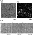
- Fig. 1 shows microphotographs showing an aspect of differentiation to SKPs from human iPS cells before being differentiated to SKPs.
- Fig 1(A) shows a microphotograph of human iPS cells
- Fig. 1(B) shows a microphotograph of human iPS cell-derived neural crest stem cells
- Fig. 1(C) shows a microphotograph of SKPs obtained by culturing human iPS cell-derived neural crest stem cells in a differentiation-inducing medium containing an agonist of Wnt signaling (CHIR99021) in an amount of 3 micromolars
- Fig. 1 shows microphotographs showing an aspect of differentiation to SKPs from human iPS cells before being differentiated to SKPs.
- Fig. 1(A) shows a microphotograph of human iPS cells
- Fig. 1(B) shows a microphotograph of human iPS cell-derived neural crest stem cells
- Fig. 1(C) shows a
- 1(D) shows a microphotograph of cells produced by passage-culturing SKPs obtained by culturing human iPS-derived neural crest stem cells in a differentiation-inducing medium containing an agonist of Wnt signaling (CHIR99021) in an amount of 3 micromolars (magnification: 40 times for all).
- Fig. 1(A) the human iPS cells grew in a colony form, abundance of nuclei was high and cytoplasm was small.
- Fig. 1(D) human iPS cell-derived SKPs after the passage-culturing were not in the colony form, were cultured in a state of individual cells, and existence of clear cytoplasm was recognized. Accordingly, it was confirmed that human iPS cell-derived SKPs were grown by carrying out the passage culture of the cells differentiated to SKPs by the above-described method.
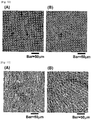
- Fig. 2 shows a microphotograph of SKPs differentiated from human iPS cell-derived neural crest stem cells when differentiation was induced by culturing under different concentrations of an agonist of Wnt signaling (CHIR99021).
- Fig. 2(A) shows a microphotograph of cells produced in the case of culturing in a culture medium to which no CHIR99021 (0 micromolar) was added
- Fig. 2(B) shows a microphotograph of cells produced in the case of culturing in a culture medium to which CHIR99021 was added at a concentration of 0.1 micromolar
- Fig. 2 shows a microphotograph of SKPs differentiated from human iPS cell-derived neural crest stem cells when differentiation was induced by culturing under different concentrations of an agonist of Wnt signaling (CHIR99021).
- Fig. 2(A) shows a microphotograph of cells produced in the case of culturing in a culture medium to which no CHIR99021
- FIG. 2(C) shows a microphotograph of cells produced in the case of culturing in a culture medium to which CHIR99021 was added at a concentration of 0.5 micromolar
- Fig. 2(D) shows a microphotograph of cells produced in the case of culturing in a culture medium to which CHIR99021 was added at a concentration of 3 micromolars
- Fig. 2(E) shows a microphotograph of cells produced in the case of culturing in a culture medium to which CHIR99021 was added at a concentration of 5 micromolars.
- microphotographs of the cells before passage culture are designated "P0”
- microphotographs of the cells after passage culture are designated "P1".
- FIG. 2 an aspect was observed in which a larger amount of the cells differentiated to SKPs was migrated in a peripheral area of the colony when CHIR99021 was added in a concentration of 0.5 micromolar to 5 micromolars even before the passage culture.
- SKPs can be efficiently produced in a large amount by culturing the pluripotent stem cells in the differentiation-inducing medium containing the agonist of Wnt signaling.
- iPS iPS cell-derived neural crest stem cells
- iPS-SKPs-P0 the iPS cell-derived SKPs before passage culture
- iPS-SKPs-P1 the iPS cell-derived SKPs after the passage culture
- PCR was carried out using the above-described primers in 50 microliters of system.
- An enzyme used here was KOD-Plus-Ver. 2 (catalog number: KOD-211, manufactured by TOYOBO Co., Ltd.), and the PCR was carried out under a reaction protocol of applying 94 degrees, 2 min, and applying 25 to 35 cycles in which (98 degrees, 10 seconds; 63 degrees, 30 seconds; 68 degrees, 30 seconds) was taken as one cycle.
- human fetal brain-derived RNA (catalog number: 636526, manufactured by Clontech Laboratories, Inc.) was used as a positive control of the PCR, and the PCR was carried out in a similar manner.
- Fig. 3(A) and (B) show the results of electrophoresis.
- Fig. 3(A) shows a gene expression change of a gene important for maintaining an undifferentiated state of human iPS cells
- Fig. 3(B) shows gene expression specific to SKPs.
- Fig. 3(A) in association with the differentiation induction to SKPs, reduction was caused in expression of a marker to be expressed in undifferentiated cells that are not differentiated to SKPs. Further, as shown in Fig. 3(B) , expression of genes specific to SKPs was detected by the differentiation induction to SKPs. Accordingly, it was confirmed that the cells subjected to the differentiation induction by the above-described method were SKPs.
- the resultant cells were treated with the primary antibodies (at room temperature, 2 hours) and the secondary antibodies (at room temperature, 1 hour) as shown in Table 2 above, nuclei were stained using 4',6-diamidino-2-phenylindole (DAPI, catalog number: FK045, manufactured by DOJINDO Laboratories), and then embedded.
- DAPI 4',6-diamidino-2-phenylindole
- FK045 4',6-diamidino-2-phenylindole
- Fig. 4 shows the results.
- Fig. 4(A) is a fluorescence microphotograph showing expression of nestin
- Fig. 4(B) is a fluorescence microphotograph showing expression of fibronectin
- Fig. 4(C) is a fluorescence microphotograph showing expression of alpha-SMA (magnification: 400 times).
- Human iPS cell-derived SKPs after the passage culture obtained in Test Example 2 described above were washed with D-PBS(-), and then the resultant cells were detached using a culture cell dissociation enzyme (trade name: Accutase, catalog number: 561527, manufactured by BD Biosciences, Inc.).
- the detached cells were fixed at room temperature for 20 minutes using 1 times 10 6 cells/100 microliters of a sample buffer (trade name: BD Cytofix Buffer, catalog number:554655, manufactured by BD Biosciences Inc.).
- the fixed cells were washed with a cell permeation washing solution (trade name: BD Phosflow Perm/Wash buffer I, catalog number: 557885, manufactured by BD Biosciences Inc.), and then the resultant cells were treated with the identical buffer at room temperature for 10 minutes. Then, an anti-nestin antibody (catalog number: 561231, manufactured by BD Pharmingen, Inc.), an anti-fibronectin antibody (catalog number: 563100, manufactured by BD Pharmingen, Inc.) and an isotype control (catalog number: 347202, catalog number: 557782, manufactured by BD Biosciences, Inc.) were added at a ratio of 1:20 to allow reaction at room temperature for 30 minutes.
- a cell permeation washing solution trade name: BD Phosflow Perm/Wash buffer I, catalog number: 557885, manufactured by BD Biosciences Inc.
- the resultant cells were washed with the cell permeation washing solution twice, and dispersed with 500 microliters of PBS, and flow cytometric analysis of the suspension was conducted using BD FACSVerse (manufactured by BD Biosciences, Inc.).
- Fig. 5(A) shows a diagram obtained by determining cell population of human iPS cell-derived SKPs from SSC and FSC of individual cells, and selecting the cell population (a group of living cells) to be analyzed.
- Fig. 5 (B) shows the results of staining the cell population selected in Fig. 5(A) using an anti-fibronectin antibody and an anti-nestin antibody.
- a vertical axis shows expression intensity (fluorescence intensity obtained by fluorescence staining) of nestin
- a horizontal axis shows expression intensity (fluorescence intensity obtained by fluorescence staining) of fibronectin.
- Test Example 6 Induction to adipocytes from human iPS cell-derived SKPs
- Human iPS cell-derived SKPs after the passage culture obtained in Test Example 2 described above were seeded at 3 times 10 5 cells/35 mm dish. After culture for 24 hours, the resultant cells were cultured for 2 weeks in an MEM medium (catalog number: 42360-032, manufactured by Life Technologies Corporation) containing 3-isobutyl-1-methylxanthine (catalog number: 17018, 0.45 nM, manufactured by Sigma-Aldrich Corporation), insulin (catalog number: 13536, 2.07 micromolars, manufactured by Sigma-Aldrich Corporation), dexamethasone (catalog number: D4902, 100 nM, manufactured by Sigma-Aldrich Corporation), rabbit serum (catalog number: R4505, 15%, manufactured by Sigma-Aldrich Corporation) and penicillin/streptomycin (catalog number: 15140-122, 100U, 100 microgram/mL, manufactured by Life Technologies Corporation).
- MEM medium catalog number: 42360-032, manufactured by Life Technologies Corporation
- Oil Red O staining of the resultant cells was performed using Oil Red O staining Kit (catalog number: 0843, manufactured by ScienCell Research Laboratories) according to an attached protocol.
- Fig. 6(A) shows the results (magnification: 200 times).
- a lipid was stained red to allow confirmation of achievement of differentiation of human iPS cell-derived SKPs to adipocytes. Accordingly, it was confirmed that the cells obtained in Test Example 2 described above were SKPs that can be differentiated to the adipocytes.
- Test Example 7 Induction to osteocytes from human iPS cell-derived SKPs
- Human iPS cell-derived SKPs after the passage culture obtained in Test Example 2 described above were seeded at 3 times 10 cells/35 mm dish. After culture for 24 hours, the resultant cells were cultured for 2 weeks in an MEM medium (catalog number: 42360-032, manufactured by Life Technologies Corporation) containing dexamethasone (catalog number: D4902, 100 nM, manufactured by Sigma-Aldrich Corporation), beta-glycerophosphate (catalog number: G9422, 10 mM, manufactured by Sigma-Aldrich Corporation), L-Ascorbic acid-2-phosphate (catalog number: A8960, 50 micromolars, manufactured by Sigma-Aldrich Corporation), fetal bovine serum (catalog number: SH30070.03, 10 mass%, manufactured by Hyclone) and penicillin/streptomycin (catalog number: 15140-122, 100 U, 100 microgram/mL, manufactured by Life Technologies Corporation).
- MEM medium catalog number: 42360-0
- alkaline phosphatase staining of the resultant cells was performed using Blue Alkaline Phosphatase substrate Kit (catalog number: SK5300, manufactured by Vector laboratories) according to an attached protocol.
- Fig. 6(B) shows the results (magnification: 200 times).
- alkaline phosphatase-positive cells were recognized to allow confirmation of achievement of differentiation of human iPS cell-derived SKPs to osteocytes. Accordingly, it was confirmed that the cells obtained in Test Example 2 described above were SKPs that can be differentiated to the osteocytes.
- Test Example 8 Study of hair follicle induction potency of human iPS cell-derived SKPs by spheroid culture
- NHEK normal human epidermal cells
- EpiLife trade name, manufactured by Life Technologies Corporation
- normal human dermal papilla cells manufactured by Cell Applications, Inc.
- TOYOBO Co., Ltd. commercially available normal human epidermal cells
- epidermal cells, fibroblasts, dermal papilla cells and human iPS cell-derived SKPs were mixed by 4 times 10 4 cells for each, and the resultant mixture was cultured using an AmnioMAX C-100 medium (trade name, manufactured by Life Technologies Corporation) by means of a 96-well nonadhesion round bottom plate (manufactured by CellSeed Inc.).
- AmnioMAX C-100 medium trade name, manufactured by Life Technologies Corporation
- a 96-well nonadhesion round bottom plate manufactured by CellSeed Inc.
- the resultant spheroid was embedded with a frozen tissue embedding agent (trade name: OCT Compound, manufactured by Sakura Finetek Co., Ltd.), and a frozen section having a thickness of 6 micrometers was produced.
- the sections were fixed with 4% paraformaldehyde for 15 minutes, the fixed sections were washed with D-PBS(-), and subjected to blocking using 10% goat serum (catalog number: 426041, manufactured by NICHIREI Corporation) at room temperature for 1 hour.
- the resultant sections were treated with an anti-trichohyalin antibody (catalog number: sc-80607, manufactured by Santa Cruz Biotechnology, Inc.) at room temperature for 2 hours, and the resultant sections were washed with D-PBS(-), and then the resultant sections were treated with a secondary antibody (Alexa Fluor 488 goat anti-mouse IgG (H+L), catalog number: A 11029, manufactured by Life Technologies Corporation) at room temperature for 1 hour.
- an anti-trichohyalin antibody catalog number: sc-80607, manufactured by Santa Cruz Biotechnology, Inc.
- Fig. 7 shows the results.
- Fig. 7(A) shows a fluorescence microphotograph obtained by immunofluorescence staining, with an anti-trichohyalin antibody, a cell mass obtained by carrying out spheroid culture of only epidermal cells.
- Fig. 7(B) shows a fluorescence microphotograph obtained by immunofluorescence staining, with an anti-trichohyalin antibody, a cell mass obtained by mixing epidermal cells and fibroblasts, and carrying out spheroid culturing.
- Fig. 7(A) shows a fluorescence microphotograph obtained by immunofluorescence staining, with an anti-trichohyalin antibody, a cell mass obtained by mixing epidermal cells and fibroblasts, and carrying out spheroid culturing.
- FIG. 7(C) shows a fluorescence microphotograph obtained by immunofluorescence staining, with an anti-trichohyalin antibody, a cell mass obtained by mixing epidermal cells and dermal papilla cells, and carrying out spheroid culturing.
- Fig. 7(D) shows a fluorescence microphotograph obtained by immunofluorescence staining, with an anti-trichohyalin antibody, a cell mass obtained by mixing epidermal cells and human iPS cell-derived SKPs, and carrying out spheroid culturing. Arrowheads in the figures indicate expression of trichohyalin.
- Test Example 9 Induction to glial cells from human iPS cell-derived SKPs
- SKPs after the passage culture obtained in Test Example 2 described above were seeded, using a culture medium for SKPs culture (DMEM/F12 medium (catalog number: 10565-018, manufactured by Life Technologies Corporation) containing 2% B-27 supplement (catalog number: 17504-044, manufactured by Life Technologies Corporation), 20 ng/mL EGF (catalog number: 336-EG-200, manufactured R&D Systems, Inc.), 40 ng/mL bFGF (catalog number: 064-04541, manufactured by Wako Pure Chemical Industries, Ltd.), 50U penicillin and 50 microgram/mL streptomycin (catalog number: 15140-122, manufactured by Life Technologies Corporation), in a culture dish pre-coated with Laminin diluted by 25 times (catalog number: P4707, manufactured by Sigma-Aldrich Corporation) and 0.1 mg/mL Poly-L-lysine (catalog number: L4544, manufactured by Sigma-Aldrich Corporation), at 4.8 times 10 4 cells
- DMEM:F12 medium (catalog number: 10565-018, manufactured by Life Technologies Corporation) containing 5 micromolars Forskoline (catalog number: F3917, manufactured by Sigma-Aldrich Corporation), 50 ng/mL Heregulin-1-beta (catalog number: 100-03, manufactured by PeproTech, Inc.), 2% N2 supplement (catalog number: 17502-048, manufactured by Life Technologies Corporation) and 1% bovine serum (catalog number: SH30070.03E , manufactured by HyClone Laboratories, Inc.). Medium replacement was carried out once every two to three days.
- the resultant cells were washed with D-PBS(-), and fixed with 4% paraformaldehyde for 15 minutes.
- the fixed cells were washed with D-PBS(-), and then treated with a PBS solution of 0.5% TritonX-100 for 5 minutes, the resultant cells were washed again with D-PBS(-), and subjected to blocking using 10% goat serum (catalog number: 426041, manufactured by NICHIREI Corporation) at room temperature for 1 hour.
- the resultant cells were treated with a primary antibody (anti-S100-beta antibody, catalog number: S2532, manufactured by Sigma-Aldrich Corporation, at room temperature, 2 hours) and a secondary antibody (Alexa Fluor 488 goat anti-mouse IgG(H+L), catalog number: A11029, manufactured by Life Technologies Corporation, at room temperature, 1 hour).
- a primary antibody anti-S100-beta antibody, catalog number: S2532, manufactured by Sigma-Aldrich Corporation, at room temperature, 2 hours
- a secondary antibody Alexa Fluor 488 goat anti-mouse IgG(H+L), catalog number: A11029, manufactured by Life Technologies Corporation, at room temperature, 1 hour.
- nuclei were stained using DAPI (catalog number: FK045, manufactured by DOJINDO Laboratories), and then embedded, and expression of a marker protein (S100-beta) specific to Schwann cells was observed under a fluorescence microscope.
- DAPI catalog number: FK
- Fig. 8 shows the results.
- Fig. 8(A) shows a microphotograph of cells produced by additionally subjecting SKPs induced from human iPS cells to 3 weeks of differentiation to Schwann cells
- Fig. 8(B) shows a fluorescence microphotograph of cells produced by staining the cells in Fig. 8(A) using an anti-S100-beta antibody.
- S100-beta-positive cells were recognized to allow confirmation of achievement of differentiation of the human iPS cell-derived SKPs to the Schwann cells. Accordingly, it was confirmed that the cells obtained in Test Example 2 described above were SKPs that can be differentiated to the glial cells, such as Schwann cells.
- SKPs that can be differentiated to the neurons, the glial cells, the smooth muscle cells, the adipocytes, the osteocytes, the dermal papilla cells or the like can be efficiently produced in a large amount.
- Test Example 10 Cryopreservation of human iPS cell-derived SKPs obtained according to the present invention
- Preserved cells were thawed, and the thawed cells were cultured in a culture medium for human iPS cell-derived SKPs (DMEM/F12 medium (catalog number: 10565-018, manufactured by Life Technologies Corporation) containing 2% B27 supplement (catalog number: 17504-044, manufactured by Life Technologies Corporation), 20 ng/mL EGF (catalog number: 336-EG-200, manufactured R&D Systems, Inc.), 40 ng/mL bFGF (catalog number: 064-04541, manufactured by Wako Pure Chemical Industries, Ltd.), 50U penicillin and 50 microgram/mL streptomycin (catalog number: 15140-122, manufactured by Life Technologies Corporation).
- DMEM/F12 medium catalog number: 10565-018, manufactured by Life Technologies Corporation
- B27 supplement catalog number: 17504-044, manufactured by Life Technologies Corporation
- 20 ng/mL EGF catalog number: 336-EG-200, manufactured R&D Systems, Inc.
- Fig. 9 shows microphotographs of human iPS cell-derived SKPs before cryopreservation and after thawing of the SKPs.
- Fig. 9(A) shows a microphotograph of SKPs before cryopreservation;
- Fig. 9(B) shows a microphotograph of cells produced by cryopreserving some of the SKPs shown in Fig. 9(A) , and then thawing, and culturing the resultant cells for 1 day;
- Fig. 9(C) shows a microphotograph of cells produced by further carrying out propagation/culturing of the cells shown in Fig. 9(B) for 3 days.
- the human iPS cell-derived SKPs grew even if the cells were subjected to freezing and thawing, and culture can be continued. Accordingly, it was shown that the human iPS cell-derived SKPs obtained in the present invention allow the cryopreservation and the passage culture.
- Test Example 11 Induction to adipocytes from post-freeze-thaw iPS cell-derived SKPs
- Fig. 10 shows the results.
- Fig. 10(A) shows a microphotograph of adipocytes stained by Oil Red O staining of cells produced by subjecting human iPS cell-derived SKPs before the cryopreservation to 2 weeks of differentiation induction.
- Fig. 10(B) shows a microphotograph of adipocytes stained by Oil Red O staining of cells produced by subjecting human iPS cell-derived SKPs after the cyopreservation-thawing to 2 weeks of differentiation induction.
- a lipid was stained red also in the human iPS cell-derived SKPs after being frozen, thawed, grown and cultured to allow confirmation of achievement of differentiation of the human iPS cell-derived SKPs to the adipocytes. Accordingly, it was shown that the cells obtained in Test Example 2 described above kept differentiation induction potency to the adipocytes even after the cells were subjected to passage and the cryopreservation.
- Test Example 12 Induction to osteocytes from post-freeze-thaw iPS cell-derived SKPs
- Fig. 11 shows the results.
- Fig. 11(A) shows a microphotograph of osteocytes stained by alkaline phosphatase staining of cells produced by subjecting human iPS cell-derived SKPs before the cryopreservation to 2 weeks of differentiation induction.
- Fig. 11(A) shows a microphotograph of osteocytes stained by alkaline phosphatase staining of cells produced by subjecting human iPS cell-derived SKPs before the cryopreservation to 2 weeks of differentiation induction.
- 11(B) shows a microphotograph of osteocytes stained by alkaline phosphatase staining of cells produced by culturing human iPS cell-derived SKPs after the cyopreservation-thawing and then subjecting the resultant SKPs to two weeks of differentiation induction.
Landscapes
- Health & Medical Sciences (AREA)
- Engineering & Computer Science (AREA)
- Life Sciences & Earth Sciences (AREA)
- Biomedical Technology (AREA)
- Zoology (AREA)
- Biotechnology (AREA)
- Chemical & Material Sciences (AREA)
- Organic Chemistry (AREA)
- Bioinformatics & Cheminformatics (AREA)
- Genetics & Genomics (AREA)
- Wood Science & Technology (AREA)
- Cell Biology (AREA)
- General Health & Medical Sciences (AREA)
- Dermatology (AREA)
- Microbiology (AREA)
- Biochemistry (AREA)
- General Engineering & Computer Science (AREA)
- Immunology (AREA)
- Developmental Biology & Embryology (AREA)
- Virology (AREA)
- Medicinal Chemistry (AREA)
- Pharmacology & Pharmacy (AREA)
- Epidemiology (AREA)
- Animal Behavior & Ethology (AREA)
- Public Health (AREA)
- Veterinary Medicine (AREA)
- Micro-Organisms Or Cultivation Processes Thereof (AREA)
Applications Claiming Priority (3)
| Application Number | Priority Date | Filing Date | Title |
|---|---|---|---|
| JP2014087247 | 2014-04-21 | ||
| JP2014260434A JP6304818B2 (ja) | 2014-04-21 | 2014-12-24 | 皮膚由来多能性前駆細胞の作製方法 |
| PCT/JP2015/002156 WO2015162908A1 (en) | 2014-04-21 | 2015-04-20 | Method of producing skin-derived precursor cells |
Publications (2)
| Publication Number | Publication Date |
|---|---|
| EP3134513A1 EP3134513A1 (en) | 2017-03-01 |
| EP3134513B1 true EP3134513B1 (en) | 2019-03-06 |
Family
ID=53055077
Family Applications (1)
| Application Number | Title | Priority Date | Filing Date |
|---|---|---|---|
| EP15721045.1A Not-in-force EP3134513B1 (en) | 2014-04-21 | 2015-04-20 | Method of producing skin-derived precursor cells |
Country Status (6)
Families Citing this family (9)
| Publication number | Priority date | Publication date | Assignee | Title |
|---|---|---|---|---|
| JP6941052B2 (ja) * | 2014-07-09 | 2021-09-29 | サンフォード−バーナム メディカル リサーチ インスティテュート | 発毛を調節するための方法および組成物 |
| WO2017136834A1 (en) | 2016-02-05 | 2017-08-10 | Memorial Sloan-Ketering Cancer Center | Methods of differentiating stem cell-derived ectodermal lineage precursors |
| KR102122039B1 (ko) | 2017-07-19 | 2020-06-11 | 서울대학교산학협력단 | 인간 유래 만능줄기세포에서 모유두전구세포로의 분화방법 및 이의 용도 |
| WO2019017691A2 (ko) * | 2017-07-19 | 2019-01-24 | 서울대학교 산학협력단 | 인간 유래 만능줄기세포에서 모유두전구세포로의 분화방법 및 이의 용도 |
| AU2018327341A1 (en) * | 2017-09-07 | 2020-04-23 | Memorial Sloan-Kettering Cancer Center | Methods of differentiating stem cell-derived ectodermal lineage precursors |
| KR20200091885A (ko) | 2017-11-30 | 2020-07-31 | 고쿠리츠 다이가쿠 호진 교토 다이가쿠 | 세포의 배양 방법 |
| JP7702229B2 (ja) * | 2018-11-19 | 2025-07-03 | 株式会社 資生堂 | 毛包を有するヒト皮膚組織を再構成するための組成物、ヒト皮膚組織モデル動物およびその製造方法 |
| WO2021025027A1 (ja) | 2019-08-06 | 2021-02-11 | 花王株式会社 | 皮膚由来多能性前駆細胞の作製方法 |
| CN114591894B (zh) * | 2022-02-28 | 2024-01-09 | 中国人民解放军总医院 | 一种皮肤多能前体干细胞的制备方法及其应用 |
Family Cites Families (11)
| Publication number | Priority date | Publication date | Assignee | Title |
|---|---|---|---|---|
| JP4652645B2 (ja) * | 1999-07-12 | 2011-03-16 | ゲーリング、ウォルター | Notch経路を用いた組織型または器官型の操作 |
| US7504257B2 (en) * | 2000-03-14 | 2009-03-17 | Es Cell International Pte Ltd. | Embryonic stem cells and neural progenitor cells derived therefrom |
| WO2005071063A1 (en) * | 2004-01-27 | 2005-08-04 | The Hospital For Sick Children | Methods of making and using skin-derived stem cells |
| WO2007016485A2 (en) * | 2005-07-29 | 2007-02-08 | Athersys, Inc. | Use of a gsk-3 inhibitor to maintain potency of cultured cells |
| KR20080075494A (ko) * | 2005-09-21 | 2008-08-18 | 다스크 테크날러지, 엘엘씨 | 장기 및 조직 기능성을 위한 방법 및 조성물 |
| US20080213892A1 (en) * | 2007-03-02 | 2008-09-04 | The Board Of Trustees Of The Leland Stanford Junior University | Self-renewal of neural stem cells is promoted by wnt proteins |
| EP2042592A1 (en) * | 2007-09-28 | 2009-04-01 | IMBA-Institut für Molekulare Biotechnologie GmbH | Methods for modulating the proliferation and differentiation potential of stem cells and progenitor cells |
| EP2318520B1 (en) * | 2008-07-25 | 2017-10-11 | The University Of Georgia Research Foundation, Inc. | Compositions for mesoderm derived isl1+ multipotent cells (imps), epicardial progenitor cells (epcs) and multipotent cxcr4+cd56+ cells (c56cs) and methods of use |
| WO2012091978A2 (en) * | 2010-12-31 | 2012-07-05 | University Of Georgia Research Foundation, Inc. | Differentiation of human pluripotent stem cells to multipotent neural crest cells |
| WO2013051722A1 (ja) * | 2011-10-06 | 2013-04-11 | 学校法人 慶應義塾 | 角膜内皮細胞の製造方法 |
| CN103230623A (zh) * | 2013-05-10 | 2013-08-07 | 南通大学 | 一种体外构建组织工程化神经的方法 |
-
2014
- 2014-12-24 JP JP2014260434A patent/JP6304818B2/ja not_active Expired - Fee Related
-
2015
- 2015-04-20 EP EP15721045.1A patent/EP3134513B1/en not_active Not-in-force
- 2015-04-20 US US15/305,315 patent/US20170044495A1/en not_active Abandoned
- 2015-04-20 CN CN201580025674.XA patent/CN106459906B/zh not_active Expired - Fee Related
- 2015-04-20 WO PCT/JP2015/002156 patent/WO2015162908A1/en active Application Filing
- 2015-04-20 KR KR1020167032087A patent/KR102316007B1/ko not_active Expired - Fee Related
-
2022
- 2022-05-26 US US17/825,366 patent/US20220348868A1/en not_active Abandoned
Non-Patent Citations (3)
| Title |
|---|
| ANONYMOUS: "KEGG PATHWAY: Wnt signaling pathway - Homo sapiens (human)", 19 May 2017 (2017-05-19), XP055397739, Retrieved from the Internet <URL:http://www.genome.jp/kegg-bin/show_pathway?hsa04310> [retrieved on 20170810] * |
| CHRISTOF NIEHRS: "The complex world of WNT receptor signalling", NAT REV MOL CELL BIOL., vol. 13, no. 12, 1 December 2012 (2012-12-01), pages 767 - 779, XP055397737, ISSN: 1471-0072, DOI: 10.1038/nrm3470 * |
| MICHAEL KAHN: "Can we safely target the WNT pathway?", NATURE REVIEWS. DRUG DISCOVERY, vol. 13, no. 7, 1 July 2014 (2014-07-01), GB, pages 513 - 532, XP055292587, ISSN: 1474-1776, DOI: 10.1038/nrd4233 * |
Also Published As
| Publication number | Publication date |
|---|---|
| EP3134513A1 (en) | 2017-03-01 |
| CN106459906B (zh) | 2021-10-29 |
| JP6304818B2 (ja) | 2018-04-04 |
| WO2015162908A1 (en) | 2015-10-29 |
| KR102316007B1 (ko) | 2021-10-21 |
| KR20160145170A (ko) | 2016-12-19 |
| US20170044495A1 (en) | 2017-02-16 |
| US20220348868A1 (en) | 2022-11-03 |
| JP2015213495A (ja) | 2015-12-03 |
| CN106459906A (zh) | 2017-02-22 |
Similar Documents
| Publication | Publication Date | Title |
|---|---|---|
| US20220348868A1 (en) | Method of producing skin-derived precursor cells | |
| US11155782B2 (en) | Method for preparing pluripotent stem cells | |
| US8703411B2 (en) | Cryopreservation of umbilical cord tissue for cord tissue-derived stem cells | |
| JP2019141074A (ja) | 軟骨細胞系統細胞及び/又は軟骨様組織を作製するための方法及び組成物 | |
| EP3770250A1 (en) | Differentiation promoter for pluripotent stem cells | |
| CA2998096C (en) | Method for producing renal progenitor cells | |
| UA129013C2 (uk) | Спосіб культивування клітин | |
| Sugiyama-Nakagiri et al. | Induction of skin-derived precursor cells from human induced pluripotent stem cells | |
| TWI438275B (zh) | 促進幹細胞分化為胰島素製造細胞的方法 | |
| KR20230026952A (ko) | 전분화능 줄기세포 유래 상피 오가노이드의 제조방법 | |
| US10612002B2 (en) | Human pluripotent stem cell derived endocardial endothelium | |
| EP3950933A1 (en) | Cell population including pluripotent stem cells and production method thereof | |
| US20200095551A1 (en) | Methods, systems, factors, and media for reduction of cellular stress and reactive oxygen species | |
| JP7016185B2 (ja) | 幹細胞由来涙腺組織の作製方法 | |
| WO2025091318A1 (en) | Improved production of induced midbrain dopaminergic progenitor cells from pluripotent stem cells | |
| HK40013193A (en) | Method for preparing pluripotent stem cells | |
| JP2017055721A (ja) | 表皮幹細胞を分離するためのマーカー及び三次元培養表皮の製造方法 | |
| Baio | Microgravity Exerts an Age-Dependent Effect on Cardiovascular Progenitor Cell Development | |
| Dauleh | Characterising mesothelial cell cultures derived from the murine omentum | |
| HK1222200B (en) | Method for preparing pluripotent stem cells | |
| Henning | Identification and characterisation of a novel, multi-potent, skeletal muscle-derived stem cell with broad developmental plasticity | |
| Chen | K15 & Id3 expression in intact and regenerating adult vibrissae rodent hair follicles |
Legal Events
| Date | Code | Title | Description |
|---|---|---|---|
| STAA | Information on the status of an ep patent application or granted ep patent |
Free format text: STATUS: THE INTERNATIONAL PUBLICATION HAS BEEN MADE |
|
| PUAI | Public reference made under article 153(3) epc to a published international application that has entered the european phase |
Free format text: ORIGINAL CODE: 0009012 |
|
| STAA | Information on the status of an ep patent application or granted ep patent |
Free format text: STATUS: REQUEST FOR EXAMINATION WAS MADE |
|
| 17P | Request for examination filed |
Effective date: 20161121 |
|
| AK | Designated contracting states |
Kind code of ref document: A1 Designated state(s): AL AT BE BG CH CY CZ DE DK EE ES FI FR GB GR HR HU IE IS IT LI LT LU LV MC MK MT NL NO PL PT RO RS SE SI SK SM TR |
|
| AX | Request for extension of the european patent |
Extension state: BA ME |
|
| DAV | Request for validation of the european patent (deleted) | ||
| DAX | Request for extension of the european patent (deleted) | ||
| STAA | Information on the status of an ep patent application or granted ep patent |
Free format text: STATUS: EXAMINATION IS IN PROGRESS |
|
| 17Q | First examination report despatched |
Effective date: 20170818 |
|
| GRAP | Despatch of communication of intention to grant a patent |
Free format text: ORIGINAL CODE: EPIDOSNIGR1 |
|
| STAA | Information on the status of an ep patent application or granted ep patent |
Free format text: STATUS: GRANT OF PATENT IS INTENDED |
|
| INTG | Intention to grant announced |
Effective date: 20180920 |
|
| GRAS | Grant fee paid |
Free format text: ORIGINAL CODE: EPIDOSNIGR3 |
|
| GRAA | (expected) grant |
Free format text: ORIGINAL CODE: 0009210 |
|
| STAA | Information on the status of an ep patent application or granted ep patent |
Free format text: STATUS: THE PATENT HAS BEEN GRANTED |
|
| AK | Designated contracting states |
Kind code of ref document: B1 Designated state(s): AL AT BE BG CH CY CZ DE DK EE ES FI FR GB GR HR HU IE IS IT LI LT LU LV MC MK MT NL NO PL PT RO RS SE SI SK SM TR |
|
| REG | Reference to a national code |
Ref country code: GB Ref legal event code: FG4D |
|
| REG | Reference to a national code |
Ref country code: CH Ref legal event code: EP Ref country code: AT Ref legal event code: REF Ref document number: 1104567 Country of ref document: AT Kind code of ref document: T Effective date: 20190315 |
|
| REG | Reference to a national code |
Ref country code: DE Ref legal event code: R096 Ref document number: 602015025806 Country of ref document: DE |
|
| REG | Reference to a national code |
Ref country code: IE Ref legal event code: FG4D |
|
| REG | Reference to a national code |
Ref country code: NL Ref legal event code: MP Effective date: 20190306 |
|
| REG | Reference to a national code |
Ref country code: LT Ref legal event code: MG4D |
|
| PG25 | Lapsed in a contracting state [announced via postgrant information from national office to epo] |
Ref country code: FI Free format text: LAPSE BECAUSE OF FAILURE TO SUBMIT A TRANSLATION OF THE DESCRIPTION OR TO PAY THE FEE WITHIN THE PRESCRIBED TIME-LIMIT Effective date: 20190306 Ref country code: LT Free format text: LAPSE BECAUSE OF FAILURE TO SUBMIT A TRANSLATION OF THE DESCRIPTION OR TO PAY THE FEE WITHIN THE PRESCRIBED TIME-LIMIT Effective date: 20190306 Ref country code: NO Free format text: LAPSE BECAUSE OF FAILURE TO SUBMIT A TRANSLATION OF THE DESCRIPTION OR TO PAY THE FEE WITHIN THE PRESCRIBED TIME-LIMIT Effective date: 20190606 Ref country code: SE Free format text: LAPSE BECAUSE OF FAILURE TO SUBMIT A TRANSLATION OF THE DESCRIPTION OR TO PAY THE FEE WITHIN THE PRESCRIBED TIME-LIMIT Effective date: 20190306 |
|
| PG25 | Lapsed in a contracting state [announced via postgrant information from national office to epo] |
Ref country code: NL Free format text: LAPSE BECAUSE OF FAILURE TO SUBMIT A TRANSLATION OF THE DESCRIPTION OR TO PAY THE FEE WITHIN THE PRESCRIBED TIME-LIMIT Effective date: 20190306 Ref country code: GR Free format text: LAPSE BECAUSE OF FAILURE TO SUBMIT A TRANSLATION OF THE DESCRIPTION OR TO PAY THE FEE WITHIN THE PRESCRIBED TIME-LIMIT Effective date: 20190607 Ref country code: BG Free format text: LAPSE BECAUSE OF FAILURE TO SUBMIT A TRANSLATION OF THE DESCRIPTION OR TO PAY THE FEE WITHIN THE PRESCRIBED TIME-LIMIT Effective date: 20190606 Ref country code: RS Free format text: LAPSE BECAUSE OF FAILURE TO SUBMIT A TRANSLATION OF THE DESCRIPTION OR TO PAY THE FEE WITHIN THE PRESCRIBED TIME-LIMIT Effective date: 20190306 Ref country code: HR Free format text: LAPSE BECAUSE OF FAILURE TO SUBMIT A TRANSLATION OF THE DESCRIPTION OR TO PAY THE FEE WITHIN THE PRESCRIBED TIME-LIMIT Effective date: 20190306 Ref country code: LV Free format text: LAPSE BECAUSE OF FAILURE TO SUBMIT A TRANSLATION OF THE DESCRIPTION OR TO PAY THE FEE WITHIN THE PRESCRIBED TIME-LIMIT Effective date: 20190306 |
|
| REG | Reference to a national code |
Ref country code: AT Ref legal event code: MK05 Ref document number: 1104567 Country of ref document: AT Kind code of ref document: T Effective date: 20190306 |
|
| PG25 | Lapsed in a contracting state [announced via postgrant information from national office to epo] |
Ref country code: AL Free format text: LAPSE BECAUSE OF FAILURE TO SUBMIT A TRANSLATION OF THE DESCRIPTION OR TO PAY THE FEE WITHIN THE PRESCRIBED TIME-LIMIT Effective date: 20190306 Ref country code: PT Free format text: LAPSE BECAUSE OF FAILURE TO SUBMIT A TRANSLATION OF THE DESCRIPTION OR TO PAY THE FEE WITHIN THE PRESCRIBED TIME-LIMIT Effective date: 20190706 Ref country code: EE Free format text: LAPSE BECAUSE OF FAILURE TO SUBMIT A TRANSLATION OF THE DESCRIPTION OR TO PAY THE FEE WITHIN THE PRESCRIBED TIME-LIMIT Effective date: 20190306 Ref country code: RO Free format text: LAPSE BECAUSE OF FAILURE TO SUBMIT A TRANSLATION OF THE DESCRIPTION OR TO PAY THE FEE WITHIN THE PRESCRIBED TIME-LIMIT Effective date: 20190306 Ref country code: IT Free format text: LAPSE BECAUSE OF FAILURE TO SUBMIT A TRANSLATION OF THE DESCRIPTION OR TO PAY THE FEE WITHIN THE PRESCRIBED TIME-LIMIT Effective date: 20190306 Ref country code: CZ Free format text: LAPSE BECAUSE OF FAILURE TO SUBMIT A TRANSLATION OF THE DESCRIPTION OR TO PAY THE FEE WITHIN THE PRESCRIBED TIME-LIMIT Effective date: 20190306 Ref country code: SK Free format text: LAPSE BECAUSE OF FAILURE TO SUBMIT A TRANSLATION OF THE DESCRIPTION OR TO PAY THE FEE WITHIN THE PRESCRIBED TIME-LIMIT Effective date: 20190306 Ref country code: ES Free format text: LAPSE BECAUSE OF FAILURE TO SUBMIT A TRANSLATION OF THE DESCRIPTION OR TO PAY THE FEE WITHIN THE PRESCRIBED TIME-LIMIT Effective date: 20190306 |
|
| PG25 | Lapsed in a contracting state [announced via postgrant information from national office to epo] |
Ref country code: SM Free format text: LAPSE BECAUSE OF FAILURE TO SUBMIT A TRANSLATION OF THE DESCRIPTION OR TO PAY THE FEE WITHIN THE PRESCRIBED TIME-LIMIT Effective date: 20190306 Ref country code: PL Free format text: LAPSE BECAUSE OF FAILURE TO SUBMIT A TRANSLATION OF THE DESCRIPTION OR TO PAY THE FEE WITHIN THE PRESCRIBED TIME-LIMIT Effective date: 20190306 |
|
| REG | Reference to a national code |
Ref country code: CH Ref legal event code: PL |
|
| REG | Reference to a national code |
Ref country code: DE Ref legal event code: R097 Ref document number: 602015025806 Country of ref document: DE |
|
| REG | Reference to a national code |
Ref country code: BE Ref legal event code: MM Effective date: 20190430 |
|
| PG25 | Lapsed in a contracting state [announced via postgrant information from national office to epo] |
Ref country code: IS Free format text: LAPSE BECAUSE OF FAILURE TO SUBMIT A TRANSLATION OF THE DESCRIPTION OR TO PAY THE FEE WITHIN THE PRESCRIBED TIME-LIMIT Effective date: 20190706 Ref country code: AT Free format text: LAPSE BECAUSE OF FAILURE TO SUBMIT A TRANSLATION OF THE DESCRIPTION OR TO PAY THE FEE WITHIN THE PRESCRIBED TIME-LIMIT Effective date: 20190306 Ref country code: LU Free format text: LAPSE BECAUSE OF NON-PAYMENT OF DUE FEES Effective date: 20190420 |
|
| PLBE | No opposition filed within time limit |
Free format text: ORIGINAL CODE: 0009261 |
|
| STAA | Information on the status of an ep patent application or granted ep patent |
Free format text: STATUS: NO OPPOSITION FILED WITHIN TIME LIMIT |
|
| PG25 | Lapsed in a contracting state [announced via postgrant information from national office to epo] |
Ref country code: DK Free format text: LAPSE BECAUSE OF FAILURE TO SUBMIT A TRANSLATION OF THE DESCRIPTION OR TO PAY THE FEE WITHIN THE PRESCRIBED TIME-LIMIT Effective date: 20190306 Ref country code: LI Free format text: LAPSE BECAUSE OF NON-PAYMENT OF DUE FEES Effective date: 20190430 Ref country code: CH Free format text: LAPSE BECAUSE OF NON-PAYMENT OF DUE FEES Effective date: 20190430 Ref country code: MC Free format text: LAPSE BECAUSE OF FAILURE TO SUBMIT A TRANSLATION OF THE DESCRIPTION OR TO PAY THE FEE WITHIN THE PRESCRIBED TIME-LIMIT Effective date: 20190306 |
|
| 26N | No opposition filed |
Effective date: 20191209 |
|
| PG25 | Lapsed in a contracting state [announced via postgrant information from national office to epo] |
Ref country code: BE Free format text: LAPSE BECAUSE OF NON-PAYMENT OF DUE FEES Effective date: 20190430 Ref country code: SI Free format text: LAPSE BECAUSE OF FAILURE TO SUBMIT A TRANSLATION OF THE DESCRIPTION OR TO PAY THE FEE WITHIN THE PRESCRIBED TIME-LIMIT Effective date: 20190306 |
|
| PG25 | Lapsed in a contracting state [announced via postgrant information from national office to epo] |
Ref country code: TR Free format text: LAPSE BECAUSE OF FAILURE TO SUBMIT A TRANSLATION OF THE DESCRIPTION OR TO PAY THE FEE WITHIN THE PRESCRIBED TIME-LIMIT Effective date: 20190306 |
|
| PG25 | Lapsed in a contracting state [announced via postgrant information from national office to epo] |
Ref country code: IE Free format text: LAPSE BECAUSE OF NON-PAYMENT OF DUE FEES Effective date: 20190420 |
|
| PG25 | Lapsed in a contracting state [announced via postgrant information from national office to epo] |
Ref country code: CY Free format text: LAPSE BECAUSE OF FAILURE TO SUBMIT A TRANSLATION OF THE DESCRIPTION OR TO PAY THE FEE WITHIN THE PRESCRIBED TIME-LIMIT Effective date: 20190306 |
|
| PG25 | Lapsed in a contracting state [announced via postgrant information from national office to epo] |
Ref country code: MT Free format text: LAPSE BECAUSE OF FAILURE TO SUBMIT A TRANSLATION OF THE DESCRIPTION OR TO PAY THE FEE WITHIN THE PRESCRIBED TIME-LIMIT Effective date: 20190306 Ref country code: HU Free format text: LAPSE BECAUSE OF FAILURE TO SUBMIT A TRANSLATION OF THE DESCRIPTION OR TO PAY THE FEE WITHIN THE PRESCRIBED TIME-LIMIT; INVALID AB INITIO Effective date: 20150420 |
|
| PG25 | Lapsed in a contracting state [announced via postgrant information from national office to epo] |
Ref country code: MK Free format text: LAPSE BECAUSE OF FAILURE TO SUBMIT A TRANSLATION OF THE DESCRIPTION OR TO PAY THE FEE WITHIN THE PRESCRIBED TIME-LIMIT Effective date: 20190306 |
|
| PGFP | Annual fee paid to national office [announced via postgrant information from national office to epo] |
Ref country code: FR Payment date: 20230309 Year of fee payment: 9 |
|
| PGFP | Annual fee paid to national office [announced via postgrant information from national office to epo] |
Ref country code: GB Payment date: 20230302 Year of fee payment: 9 |
|
| P01 | Opt-out of the competence of the unified patent court (upc) registered |
Effective date: 20230522 |
|
| PGFP | Annual fee paid to national office [announced via postgrant information from national office to epo] |
Ref country code: DE Payment date: 20230228 Year of fee payment: 9 |
|
| REG | Reference to a national code |
Ref country code: DE Ref legal event code: R119 Ref document number: 602015025806 Country of ref document: DE |
|
| GBPC | Gb: european patent ceased through non-payment of renewal fee |
Effective date: 20240420 |
|
| PG25 | Lapsed in a contracting state [announced via postgrant information from national office to epo] |
Ref country code: DE Free format text: LAPSE BECAUSE OF NON-PAYMENT OF DUE FEES Effective date: 20241105 |
|
| PG25 | Lapsed in a contracting state [announced via postgrant information from national office to epo] |
Ref country code: GB Free format text: LAPSE BECAUSE OF NON-PAYMENT OF DUE FEES Effective date: 20240420 |
|
| PG25 | Lapsed in a contracting state [announced via postgrant information from national office to epo] |
Ref country code: FR Free format text: LAPSE BECAUSE OF NON-PAYMENT OF DUE FEES Effective date: 20240430 |
|
| PG25 | Lapsed in a contracting state [announced via postgrant information from national office to epo] |
Ref country code: GB Free format text: LAPSE BECAUSE OF NON-PAYMENT OF DUE FEES Effective date: 20240420 Ref country code: FR Free format text: LAPSE BECAUSE OF NON-PAYMENT OF DUE FEES Effective date: 20240430 Ref country code: DE Free format text: LAPSE BECAUSE OF NON-PAYMENT OF DUE FEES Effective date: 20241105 |