EP3125963B1 - Tissue filler compositions and methods of use - Google Patents
Tissue filler compositions and methods of use Download PDFInfo
- Publication number
- EP3125963B1 EP3125963B1 EP15772726.4A EP15772726A EP3125963B1 EP 3125963 B1 EP3125963 B1 EP 3125963B1 EP 15772726 A EP15772726 A EP 15772726A EP 3125963 B1 EP3125963 B1 EP 3125963B1
- Authority
- EP
- European Patent Office
- Prior art keywords
- composition
- tissue filler
- tissue
- light
- collagen
- Prior art date
- Legal status (The legal status is an assumption and is not a legal conclusion. Google has not performed a legal analysis and makes no representation as to the accuracy of the status listed.)
- Active
Links
- 239000000203 mixture Substances 0.000 title claims description 263
- 239000000945 filler Substances 0.000 title claims description 182
- 238000000034 method Methods 0.000 title description 88
- 210000001519 tissue Anatomy 0.000 claims description 168
- 108010035532 Collagen Proteins 0.000 claims description 102
- 102000008186 Collagen Human genes 0.000 claims description 101
- 229920001436 collagen Polymers 0.000 claims description 98
- 229920002674 hyaluronan Polymers 0.000 claims description 70
- KIUKXJAPPMFGSW-DNGZLQJQSA-N (2S,3S,4S,5R,6R)-6-[(2S,3R,4R,5S,6R)-3-Acetamido-2-[(2S,3S,4R,5R,6R)-6-[(2R,3R,4R,5S,6R)-3-acetamido-2,5-dihydroxy-6-(hydroxymethyl)oxan-4-yl]oxy-2-carboxy-4,5-dihydroxyoxan-3-yl]oxy-5-hydroxy-6-(hydroxymethyl)oxan-4-yl]oxy-3,4,5-trihydroxyoxane-2-carboxylic acid Chemical compound CC(=O)N[C@H]1[C@H](O)O[C@H](CO)[C@@H](O)[C@@H]1O[C@H]1[C@H](O)[C@@H](O)[C@H](O[C@H]2[C@@H]([C@@H](O[C@H]3[C@@H]([C@@H](O)[C@H](O)[C@H](O3)C(O)=O)O)[C@H](O)[C@@H](CO)O2)NC(C)=O)[C@@H](C(O)=O)O1 KIUKXJAPPMFGSW-DNGZLQJQSA-N 0.000 claims description 68
- 229960003160 hyaluronic acid Drugs 0.000 claims description 67
- 210000004872 soft tissue Anatomy 0.000 claims description 52
- 230000015572 biosynthetic process Effects 0.000 claims description 42
- 238000003786 synthesis reaction Methods 0.000 claims description 36
- 239000002245 particle Substances 0.000 claims description 32
- 238000005286 illumination Methods 0.000 claims description 30
- 239000002537 cosmetic Substances 0.000 claims description 21
- 238000002347 injection Methods 0.000 claims description 14
- 239000007924 injection Substances 0.000 claims description 14
- 230000037390 scarring Effects 0.000 claims description 12
- 238000002513 implantation Methods 0.000 claims description 8
- 230000004936 stimulating effect Effects 0.000 claims description 8
- 230000002401 inhibitory effect Effects 0.000 claims description 7
- 230000008093 supporting effect Effects 0.000 claims description 3
- 239000013028 medium composition Substances 0.000 claims 1
- 210000003491 skin Anatomy 0.000 description 83
- 208000027418 Wounds and injury Diseases 0.000 description 46
- 206010052428 Wound Diseases 0.000 description 44
- SEACYXSIPDVVMV-UHFFFAOYSA-L eosin Y Chemical compound [Na+].[Na+].[O-]C(=O)C1=CC=CC=C1C1=C2C=C(Br)C(=O)C(Br)=C2OC2=C(Br)C([O-])=C(Br)C=C21 SEACYXSIPDVVMV-UHFFFAOYSA-L 0.000 description 42
- 239000000499 gel Substances 0.000 description 32
- 230000002500 effect on skin Effects 0.000 description 30
- GNBHRKFJIUUOQI-UHFFFAOYSA-N fluorescein Chemical compound O1C(=O)C2=CC=CC=C2C21C1=CC=C(O)C=C1OC1=CC(O)=CC=C21 GNBHRKFJIUUOQI-UHFFFAOYSA-N 0.000 description 27
- 229960002143 fluorescein Drugs 0.000 description 27
- 210000004207 dermis Anatomy 0.000 description 23
- 239000000463 material Substances 0.000 description 23
- -1 oxygen radicals Chemical class 0.000 description 23
- 231100000241 scar Toxicity 0.000 description 23
- 210000004027 cell Anatomy 0.000 description 20
- 229920000642 polymer Polymers 0.000 description 19
- 238000000862 absorption spectrum Methods 0.000 description 17
- 230000000694 effects Effects 0.000 description 17
- 230000003716 rejuvenation Effects 0.000 description 17
- 238000012546 transfer Methods 0.000 description 16
- YQGOJNYOYNNSMM-UHFFFAOYSA-N eosin Chemical compound [Na+].OC(=O)C1=CC=CC=C1C1=C2C=C(Br)C(=O)C(Br)=C2OC2=C(Br)C(O)=C(Br)C=C21 YQGOJNYOYNNSMM-UHFFFAOYSA-N 0.000 description 15
- 230000014509 gene expression Effects 0.000 description 14
- 241000283690 Bos taurus Species 0.000 description 13
- 238000000295 emission spectrum Methods 0.000 description 13
- 230000000699 topical effect Effects 0.000 description 13
- 230000001737 promoting effect Effects 0.000 description 12
- 238000001228 spectrum Methods 0.000 description 12
- 230000029663 wound healing Effects 0.000 description 12
- 241001465754 Metazoa Species 0.000 description 11
- 208000004210 Pressure Ulcer Diseases 0.000 description 11
- 208000025865 Ulcer Diseases 0.000 description 11
- 239000003795 chemical substances by application Substances 0.000 description 11
- 238000002834 transmittance Methods 0.000 description 11
- 230000037303 wrinkles Effects 0.000 description 11
- 102000012422 Collagen Type I Human genes 0.000 description 10
- 108010022452 Collagen Type I Proteins 0.000 description 10
- 238000004132 cross linking Methods 0.000 description 10
- 210000002950 fibroblast Anatomy 0.000 description 10
- 208000032544 Cicatrix Diseases 0.000 description 9
- 239000000017 hydrogel Substances 0.000 description 9
- 239000004005 microsphere Substances 0.000 description 9
- 229920001432 poly(L-lactide) Polymers 0.000 description 9
- 230000037387 scars Effects 0.000 description 9
- 102000010834 Extracellular Matrix Proteins Human genes 0.000 description 8
- 108010037362 Extracellular Matrix Proteins Proteins 0.000 description 8
- 229920002683 Glycosaminoglycan Polymers 0.000 description 8
- 229920001954 Restylane Polymers 0.000 description 8
- 230000003213 activating effect Effects 0.000 description 8
- 239000000975 dye Substances 0.000 description 8
- IINNWAYUJNWZRM-UHFFFAOYSA-L erythrosin B Chemical compound [Na+].[Na+].[O-]C(=O)C1=CC=CC=C1C1=C2C=C(I)C(=O)C(I)=C2OC2=C(I)C([O-])=C(I)C=C21 IINNWAYUJNWZRM-UHFFFAOYSA-L 0.000 description 8
- 210000002744 extracellular matrix Anatomy 0.000 description 8
- 150000004676 glycans Chemical class 0.000 description 8
- 108020004999 messenger RNA Proteins 0.000 description 8
- CXKWCBBOMKCUKX-UHFFFAOYSA-M methylene blue Chemical compound [Cl-].C1=CC(N(C)C)=CC2=[S+]C3=CC(N(C)C)=CC=C3N=C21 CXKWCBBOMKCUKX-UHFFFAOYSA-M 0.000 description 8
- 229920001282 polysaccharide Polymers 0.000 description 8
- 239000005017 polysaccharide Substances 0.000 description 8
- 230000001603 reducing effect Effects 0.000 description 8
- 238000002560 therapeutic procedure Methods 0.000 description 8
- 230000015556 catabolic process Effects 0.000 description 7
- 229940096422 collagen type i Drugs 0.000 description 7
- 238000006731 degradation reaction Methods 0.000 description 7
- 210000002615 epidermis Anatomy 0.000 description 7
- 230000008569 process Effects 0.000 description 7
- 231100000397 ulcer Toxicity 0.000 description 7
- 238000010521 absorption reaction Methods 0.000 description 6
- 230000003416 augmentation Effects 0.000 description 6
- MSWZFWKMSRAUBD-UHFFFAOYSA-N beta-D-galactosamine Natural products NC1C(O)OC(CO)C(O)C1O MSWZFWKMSRAUBD-UHFFFAOYSA-N 0.000 description 6
- ATNHDLDRLWWWCB-AENOIHSZSA-M chlorophyll a Chemical compound C1([C@@H](C(=O)OC)C(=O)C2=C3C)=C2N2C3=CC(C(CC)=C3C)=[N+]4C3=CC3=C(C=C)C(C)=C5N3[Mg-2]42[N+]2=C1[C@@H](CCC(=O)OC\C=C(/C)CCC[C@H](C)CCC[C@H](C)CCCC(C)C)[C@H](C)C2=C5 ATNHDLDRLWWWCB-AENOIHSZSA-M 0.000 description 6
- 230000001684 chronic effect Effects 0.000 description 6
- 230000006378 damage Effects 0.000 description 6
- ZBQZBWKNGDEDOA-UHFFFAOYSA-N eosin B Chemical compound O1C(=O)C2=CC=CC=C2C21C1=CC([N+]([O-])=O)=C(O)C(Br)=C1OC1=C2C=C([N+]([O-])=O)C(O)=C1Br ZBQZBWKNGDEDOA-UHFFFAOYSA-N 0.000 description 6
- 239000012530 fluid Substances 0.000 description 6
- 208000014674 injury Diseases 0.000 description 6
- 229960000907 methylthioninium chloride Drugs 0.000 description 6
- 229920003229 poly(methyl methacrylate) Polymers 0.000 description 6
- 239000004926 polymethyl methacrylate Substances 0.000 description 6
- 108090000765 processed proteins & peptides Proteins 0.000 description 6
- 102000004196 processed proteins & peptides Human genes 0.000 description 6
- 230000003595 spectral effect Effects 0.000 description 6
- OYPRJOBELJOOCE-UHFFFAOYSA-N Calcium Chemical compound [Ca] OYPRJOBELJOOCE-UHFFFAOYSA-N 0.000 description 5
- 108010069502 Collagen Type III Proteins 0.000 description 5
- 108010014258 Elastin Proteins 0.000 description 5
- 102000016942 Elastin Human genes 0.000 description 5
- HLUCICHZHWJHLL-UHFFFAOYSA-N Haematein Natural products C12=CC=C(O)C(O)=C2OCC2(O)C1=C1C=C(O)C(=O)C=C1C2 HLUCICHZHWJHLL-UHFFFAOYSA-N 0.000 description 5
- WZUVPPKBWHMQCE-UHFFFAOYSA-N Haematoxylin Natural products C12=CC(O)=C(O)C=C2CC2(O)C1C1=CC=C(O)C(O)=C1OC2 WZUVPPKBWHMQCE-UHFFFAOYSA-N 0.000 description 5
- RZUBARUFLYGOGC-MTHOTQAESA-L acid fuchsin Chemical compound [Na+].[Na+].[O-]S(=O)(=O)C1=C(N)C(C)=CC(C(=C\2C=C(C(=[NH2+])C=C/2)S([O-])(=O)=O)\C=2C=C(C(N)=CC=2)S([O-])(=O)=O)=C1 RZUBARUFLYGOGC-MTHOTQAESA-L 0.000 description 5
- 230000032683 aging Effects 0.000 description 5
- 238000006065 biodegradation reaction Methods 0.000 description 5
- 239000011575 calcium Substances 0.000 description 5
- 229910052791 calcium Inorganic materials 0.000 description 5
- FFUMCSDSJNSMQH-HEXQVDJKSA-K chromoxane cyanin R Chemical compound [Na+].[Na+].[Na+].C1=C(C([O-])=O)C(=O)C(C)=C\C1=C(C=1C(=CC=CC=1)S([O-])(=O)=O)\C1=CC(C)=C(O)C(C([O-])=O)=C1 FFUMCSDSJNSMQH-HEXQVDJKSA-K 0.000 description 5
- 239000000501 collagen implant Substances 0.000 description 5
- ZXJXZNDDNMQXFV-UHFFFAOYSA-M crystal violet Chemical compound [Cl-].C1=CC(N(C)C)=CC=C1[C+](C=1C=CC(=CC=1)N(C)C)C1=CC=C(N(C)C)C=C1 ZXJXZNDDNMQXFV-UHFFFAOYSA-M 0.000 description 5
- 206010012601 diabetes mellitus Diseases 0.000 description 5
- 229920002549 elastin Polymers 0.000 description 5
- 239000000835 fiber Substances 0.000 description 5
- 238000011049 filling Methods 0.000 description 5
- 210000002683 foot Anatomy 0.000 description 5
- 230000035876 healing Effects 0.000 description 5
- 210000002510 keratinocyte Anatomy 0.000 description 5
- 239000011159 matrix material Substances 0.000 description 5
- SHXOKQKTZJXHHR-UHFFFAOYSA-N n,n-diethyl-5-iminobenzo[a]phenoxazin-9-amine;hydrochloride Chemical compound [Cl-].C1=CC=C2C3=NC4=CC=C(N(CC)CC)C=C4OC3=CC(=[NH2+])C2=C1 SHXOKQKTZJXHHR-UHFFFAOYSA-N 0.000 description 5
- 229910052760 oxygen Inorganic materials 0.000 description 5
- 239000001301 oxygen Substances 0.000 description 5
- GVKCHTBDSMQENH-UHFFFAOYSA-L phloxine B Chemical compound [Na+].[Na+].[O-]C(=O)C1=C(Cl)C(Cl)=C(Cl)C(Cl)=C1C1=C2C=C(Br)C(=O)C(Br)=C2OC2=C(Br)C([O-])=C(Br)C=C21 GVKCHTBDSMQENH-UHFFFAOYSA-L 0.000 description 5
- INCIMLINXXICKS-UHFFFAOYSA-M pyronin Y Chemical compound [Cl-].C1=CC(=[N+](C)C)C=C2OC3=CC(N(C)C)=CC=C3C=C21 INCIMLINXXICKS-UHFFFAOYSA-M 0.000 description 5
- 230000036573 scar formation Effects 0.000 description 5
- 238000007493 shaping process Methods 0.000 description 5
- 230000002195 synergetic effect Effects 0.000 description 5
- 230000001225 therapeutic effect Effects 0.000 description 5
- JWYVGKFDLWWQJX-UHFFFAOYSA-N 1-ethenylazepan-2-one Chemical compound C=CN1CCCCCC1=O JWYVGKFDLWWQJX-UHFFFAOYSA-N 0.000 description 4
- 206010065687 Bone loss Diseases 0.000 description 4
- 102000001187 Collagen Type III Human genes 0.000 description 4
- 206010011985 Decubitus ulcer Diseases 0.000 description 4
- MYMOFIZGZYHOMD-UHFFFAOYSA-N Dioxygen Chemical compound O=O MYMOFIZGZYHOMD-UHFFFAOYSA-N 0.000 description 4
- WHNWPMSKXPGLAX-UHFFFAOYSA-N N-Vinyl-2-pyrrolidone Chemical compound C=CN1CCCC1=O WHNWPMSKXPGLAX-UHFFFAOYSA-N 0.000 description 4
- 229920003171 Poly (ethylene oxide) Polymers 0.000 description 4
- VYPSYNLAJGMNEJ-UHFFFAOYSA-N Silicium dioxide Chemical compound O=[Si]=O VYPSYNLAJGMNEJ-UHFFFAOYSA-N 0.000 description 4
- GSEJCLTVZPLZKY-UHFFFAOYSA-N Triethanolamine Chemical compound OCCN(CCO)CCO GSEJCLTVZPLZKY-UHFFFAOYSA-N 0.000 description 4
- YJVBLROMQZEFPA-UHFFFAOYSA-L acid red 26 Chemical compound [Na+].[Na+].CC1=CC(C)=CC=C1N=NC1=C(O)C(S([O-])(=O)=O)=CC2=CC(S([O-])(=O)=O)=CC=C12 YJVBLROMQZEFPA-UHFFFAOYSA-L 0.000 description 4
- HFVAFDPGUJEFBQ-UHFFFAOYSA-M alizarin red S Chemical compound [Na+].O=C1C2=CC=CC=C2C(=O)C2=C1C=C(S([O-])(=O)=O)C(O)=C2O HFVAFDPGUJEFBQ-UHFFFAOYSA-M 0.000 description 4
- QVGXLLKOCUKJST-UHFFFAOYSA-N atomic oxygen Chemical compound [O] QVGXLLKOCUKJST-UHFFFAOYSA-N 0.000 description 4
- 235000012733 azorubine Nutrition 0.000 description 4
- 210000002469 basement membrane Anatomy 0.000 description 4
- 210000004204 blood vessel Anatomy 0.000 description 4
- 210000000988 bone and bone Anatomy 0.000 description 4
- DBZJJPROPLPMSN-UHFFFAOYSA-N bromoeosin Chemical compound O1C(=O)C2=CC=CC=C2C21C1=CC(Br)=C(O)C(Br)=C1OC1=C(Br)C(O)=C(Br)C=C21 DBZJJPROPLPMSN-UHFFFAOYSA-N 0.000 description 4
- 235000012730 carminic acid Nutrition 0.000 description 4
- 230000037319 collagen production Effects 0.000 description 4
- 150000001875 compounds Chemical class 0.000 description 4
- 210000002808 connective tissue Anatomy 0.000 description 4
- UKZQEOHHLOYJLY-UHFFFAOYSA-M ethyl eosin Chemical compound [K+].CCOC(=O)C1=CC=CC=C1C1=C2C=C(Br)C(=O)C(Br)=C2OC2=C(Br)C([O-])=C(Br)C=C21 UKZQEOHHLOYJLY-UHFFFAOYSA-M 0.000 description 4
- 210000003195 fascia Anatomy 0.000 description 4
- 239000011521 glass Substances 0.000 description 4
- 230000001965 increasing effect Effects 0.000 description 4
- FDZZZRQASAIRJF-UHFFFAOYSA-M malachite green Chemical compound [Cl-].C1=CC(N(C)C)=CC=C1C(C=1C=CC=CC=1)=C1C=CC(=[N+](C)C)C=C1 FDZZZRQASAIRJF-UHFFFAOYSA-M 0.000 description 4
- CTIQLGJVGNGFEW-UHFFFAOYSA-L naphthol yellow S Chemical compound [Na+].[Na+].C1=C(S([O-])(=O)=O)C=C2C([O-])=C([N+]([O-])=O)C=C([N+]([O-])=O)C2=C1 CTIQLGJVGNGFEW-UHFFFAOYSA-L 0.000 description 4
- HSXUHWZMNJHFRV-QIKYXUGXSA-L orange G Chemical compound [Na+].[Na+].OC1=CC=C2C=C(S([O-])(=O)=O)C=C(S([O-])(=O)=O)C2=C1\N=N\C1=CC=CC=C1 HSXUHWZMNJHFRV-QIKYXUGXSA-L 0.000 description 4
- 208000033808 peripheral neuropathy Diseases 0.000 description 4
- NBIIXXVUZAFLBC-UHFFFAOYSA-K phosphate Chemical compound [O-]P([O-])([O-])=O NBIIXXVUZAFLBC-UHFFFAOYSA-K 0.000 description 4
- 235000019238 ponceau 6R Nutrition 0.000 description 4
- 230000002035 prolonged effect Effects 0.000 description 4
- BBNQQADTFFCFGB-UHFFFAOYSA-N purpurin Chemical compound C1=CC=C2C(=O)C3=C(O)C(O)=CC(O)=C3C(=O)C2=C1 BBNQQADTFFCFGB-UHFFFAOYSA-N 0.000 description 4
- 239000003642 reactive oxygen metabolite Substances 0.000 description 4
- 235000012756 tartrazine Nutrition 0.000 description 4
- 239000004149 tartrazine Substances 0.000 description 4
- 230000036269 ulceration Effects 0.000 description 4
- XLYOFNOQVPJJNP-UHFFFAOYSA-N water Substances O XLYOFNOQVPJJNP-UHFFFAOYSA-N 0.000 description 4
- MSWZFWKMSRAUBD-IVMDWMLBSA-N 2-amino-2-deoxy-D-glucopyranose Chemical compound N[C@H]1C(O)O[C@H](CO)[C@@H](O)[C@@H]1O MSWZFWKMSRAUBD-IVMDWMLBSA-N 0.000 description 3
- AXDJCCTWPBKUKL-UHFFFAOYSA-N 4-[(4-aminophenyl)-(4-imino-3-methylcyclohexa-2,5-dien-1-ylidene)methyl]aniline;hydron;chloride Chemical compound Cl.C1=CC(=N)C(C)=CC1=C(C=1C=CC(N)=CC=1)C1=CC=C(N)C=C1 AXDJCCTWPBKUKL-UHFFFAOYSA-N 0.000 description 3
- RGCKGOZRHPZPFP-UHFFFAOYSA-N Alizarin Natural products C1=CC=C2C(=O)C3=C(O)C(O)=CC=C3C(=O)C2=C1 RGCKGOZRHPZPFP-UHFFFAOYSA-N 0.000 description 3
- AOMZHDJXSYHPKS-DROYEMJCSA-L Amido Black 10B Chemical compound [Na+].[Na+].[O-]S(=O)(=O)C1=CC2=CC(S([O-])(=O)=O)=C(\N=N\C=3C=CC=CC=3)C(O)=C2C(N)=C1\N=N\C1=CC=C(N(=O)=O)C=C1 AOMZHDJXSYHPKS-DROYEMJCSA-L 0.000 description 3
- MCZVRBLCRZWFJH-UHFFFAOYSA-N Bismark brown Y Chemical compound Cl.Cl.NC1=CC(N)=CC=C1N=NC1=CC=CC(N=NC=2C(=CC(N)=CC=2)N)=C1 MCZVRBLCRZWFJH-UHFFFAOYSA-N 0.000 description 3
- 229920002134 Carboxymethyl cellulose Polymers 0.000 description 3
- 102000004510 Collagen Type VII Human genes 0.000 description 3
- 108010017377 Collagen Type VII Proteins 0.000 description 3
- ZWYHVBGOBINPHN-AVRYKWKFSA-L Congo corinth Chemical compound [Na+].[Na+].Nc1c(cc(c2ccccc12)S([O-])(=O)=O)\N=N\c1ccc(cc1)-c1ccc(cc1)\N=N\c1cc(c2ccccc2c1[O-])S(O)(=O)=O ZWYHVBGOBINPHN-AVRYKWKFSA-L 0.000 description 3
- 229920002307 Dextran Polymers 0.000 description 3
- 206010013786 Dry skin Diseases 0.000 description 3
- 206010014970 Ephelides Diseases 0.000 description 3
- 206010063560 Excessive granulation tissue Diseases 0.000 description 3
- IAJILQKETJEXLJ-UHFFFAOYSA-N Galacturonsaeure Natural products O=CC(O)C(O)C(O)C(O)C(O)=O IAJILQKETJEXLJ-UHFFFAOYSA-N 0.000 description 3
- 108010085895 Laminin Proteins 0.000 description 3
- 208000003351 Melanosis Diseases 0.000 description 3
- 229910019142 PO4 Inorganic materials 0.000 description 3
- 208000003251 Pruritus Diseases 0.000 description 3
- 206010037867 Rash macular Diseases 0.000 description 3
- 208000002847 Surgical Wound Diseases 0.000 description 3
- GLNADSQYFUSGOU-GPTZEZBUSA-J Trypan blue Chemical compound [Na+].[Na+].[Na+].[Na+].C1=C(S([O-])(=O)=O)C=C2C=C(S([O-])(=O)=O)C(/N=N/C3=CC=C(C=C3C)C=3C=C(C(=CC=3)\N=N\C=3C(=CC4=CC(=CC(N)=C4C=3O)S([O-])(=O)=O)S([O-])(=O)=O)C)=C(O)C2=C1N GLNADSQYFUSGOU-GPTZEZBUSA-J 0.000 description 3
- DGOBMKYRQHEFGQ-UHFFFAOYSA-L acid green 5 Chemical compound [Na+].[Na+].C=1C=C(C(=C2C=CC(C=C2)=[N+](CC)CC=2C=C(C=CC=2)S([O-])(=O)=O)C=2C=CC(=CC=2)S([O-])(=O)=O)C=CC=1N(CC)CC1=CC=CC(S([O-])(=O)=O)=C1 DGOBMKYRQHEFGQ-UHFFFAOYSA-L 0.000 description 3
- FUGCXLNGEHFIOA-UHFFFAOYSA-L acid red 44 Chemical compound [Na+].[Na+].C1=CC=C2C(N=NC3=C4C(=CC(=CC4=CC=C3O)S([O-])(=O)=O)S([O-])(=O)=O)=CC=CC2=C1 FUGCXLNGEHFIOA-UHFFFAOYSA-L 0.000 description 3
- NIXOWILDQLNWCW-UHFFFAOYSA-N acrylic acid group Chemical group C(C=C)(=O)O NIXOWILDQLNWCW-UHFFFAOYSA-N 0.000 description 3
- 150000001412 amines Chemical group 0.000 description 3
- MMRNCQMFQXTUGO-UHFFFAOYSA-N anthracene blue SWR Chemical compound OC1=CC(O)=C2C(=O)C3=C(O)C(O)=CC(O)=C3C(=O)C2=C1O MMRNCQMFQXTUGO-UHFFFAOYSA-N 0.000 description 3
- 229930002875 chlorophyll Natural products 0.000 description 3
- 235000019804 chlorophyll Nutrition 0.000 description 3
- 230000008602 contraction Effects 0.000 description 3
- 230000001419 dependent effect Effects 0.000 description 3
- 230000037336 dry skin Effects 0.000 description 3
- 238000005516 engineering process Methods 0.000 description 3
- QGAYMQGSQUXCQO-UHFFFAOYSA-L eosin b Chemical compound [Na+].[Na+].O1C(=O)C2=CC=CC=C2C21C1=CC([N+]([O-])=O)=C([O-])C(Br)=C1OC1=C2C=C([N+]([O-])=O)C([O-])=C1Br QGAYMQGSQUXCQO-UHFFFAOYSA-L 0.000 description 3
- 210000002919 epithelial cell Anatomy 0.000 description 3
- 235000012732 erythrosine Nutrition 0.000 description 3
- 239000004174 erythrosine Substances 0.000 description 3
- 229940011411 erythrosine Drugs 0.000 description 3
- 230000001815 facial effect Effects 0.000 description 3
- 230000002349 favourable effect Effects 0.000 description 3
- 229960002442 glucosamine Drugs 0.000 description 3
- 210000001126 granulation tissue Anatomy 0.000 description 3
- 239000003102 growth factor Substances 0.000 description 3
- 230000001976 improved effect Effects 0.000 description 3
- 108010028309 kalinin Proteins 0.000 description 3
- CXORMDKZEUMQHX-UHFFFAOYSA-N kermesic acid Chemical compound O=C1C2=C(O)C(O)=CC(O)=C2C(=O)C2=C1C=C(O)C(C(O)=O)=C2C CXORMDKZEUMQHX-UHFFFAOYSA-N 0.000 description 3
- 230000003902 lesion Effects 0.000 description 3
- NYGZLYXAPMMJTE-UHFFFAOYSA-M metanil yellow Chemical compound [Na+].[O-]S(=O)(=O)C1=CC=CC(N=NC=2C=CC(NC=3C=CC=CC=3)=CC=2)=C1 NYGZLYXAPMMJTE-UHFFFAOYSA-M 0.000 description 3
- MCPLVIGCWWTHFH-UHFFFAOYSA-L methyl blue Chemical compound [Na+].[Na+].C1=CC(S(=O)(=O)[O-])=CC=C1NC1=CC=C(C(=C2C=CC(C=C2)=[NH+]C=2C=CC(=CC=2)S([O-])(=O)=O)C=2C=CC(NC=3C=CC(=CC=3)S([O-])(=O)=O)=CC=2)C=C1 MCPLVIGCWWTHFH-UHFFFAOYSA-L 0.000 description 3
- 201000001119 neuropathy Diseases 0.000 description 3
- 230000007823 neuropathy Effects 0.000 description 3
- PGSADBUBUOPOJS-UHFFFAOYSA-N neutral red Chemical compound Cl.C1=C(C)C(N)=CC2=NC3=CC(N(C)C)=CC=C3N=C21 PGSADBUBUOPOJS-UHFFFAOYSA-N 0.000 description 3
- IFSXZLJQEKGQAF-UHFFFAOYSA-M nuclear fast red Chemical compound [Na+].O=C1C2=CC=CC=C2C(=O)C2=C1C(O)=C(S([O-])(=O)=O)C(O)=C2N IFSXZLJQEKGQAF-UHFFFAOYSA-M 0.000 description 3
- 239000010452 phosphate Substances 0.000 description 3
- 229920000570 polyether Polymers 0.000 description 3
- 229920001343 polytetrafluoroethylene Polymers 0.000 description 3
- 239000004810 polytetrafluoroethylene Substances 0.000 description 3
- 239000011148 porous material Substances 0.000 description 3
- 238000002360 preparation method Methods 0.000 description 3
- 230000008929 regeneration Effects 0.000 description 3
- 238000011069 regeneration method Methods 0.000 description 3
- AZJPTIGZZTZIDR-UHFFFAOYSA-L rose bengal Chemical compound [K+].[K+].[O-]C(=O)C1=C(Cl)C(Cl)=C(Cl)C(Cl)=C1C1=C2C=C(I)C(=O)C(I)=C2OC2=C(I)C([O-])=C(I)C=C21 AZJPTIGZZTZIDR-UHFFFAOYSA-L 0.000 description 3
- 238000007665 sagging Methods 0.000 description 3
- 230000009759 skin aging Effects 0.000 description 3
- 230000000638 stimulation Effects 0.000 description 3
- UJMBCXLDXJUMFB-GLCFPVLVSA-K tartrazine Chemical compound [Na+].[Na+].[Na+].[O-]C(=O)C1=NN(C=2C=CC(=CC=2)S([O-])(=O)=O)C(=O)C1\N=N\C1=CC=C(S([O-])(=O)=O)C=C1 UJMBCXLDXJUMFB-GLCFPVLVSA-K 0.000 description 3
- 208000009056 telangiectasis Diseases 0.000 description 3
- JADVWWSKYZXRGX-UHFFFAOYSA-M thioflavine T Chemical compound [Cl-].C1=CC(N(C)C)=CC=C1C1=[N+](C)C2=CC=C(C)C=C2S1 JADVWWSKYZXRGX-UHFFFAOYSA-M 0.000 description 3
- 230000017423 tissue regeneration Effects 0.000 description 3
- 230000002792 vascular Effects 0.000 description 3
- XOSXWYQMOYSSKB-LDKJGXKFSA-L water blue Chemical compound CC1=CC(/C(\C(C=C2)=CC=C2NC(C=C2)=CC=C2S([O-])(=O)=O)=C(\C=C2)/C=C/C\2=N\C(C=C2)=CC=C2S([O-])(=O)=O)=CC(S(O)(=O)=O)=C1N.[Na+].[Na+] XOSXWYQMOYSSKB-LDKJGXKFSA-L 0.000 description 3
- IAJILQKETJEXLJ-KLVWXMOXSA-N (2s,3r,4r,5r)-2,3,4,5-tetrahydroxy-6-oxohexanoic acid Chemical compound O=C[C@H](O)[C@H](O)[C@@H](O)[C@H](O)C(O)=O IAJILQKETJEXLJ-KLVWXMOXSA-N 0.000 description 2
- PVPBBTJXIKFICP-UHFFFAOYSA-N (7-aminophenothiazin-3-ylidene)azanium;chloride Chemical compound [Cl-].C1=CC(=[NH2+])C=C2SC3=CC(N)=CC=C3N=C21 PVPBBTJXIKFICP-UHFFFAOYSA-N 0.000 description 2
- 229920002818 (Hydroxyethyl)methacrylate Polymers 0.000 description 2
- FFRBMBIXVSCUFS-UHFFFAOYSA-N 2,4-dinitro-1-naphthol Chemical compound C1=CC=C2C(O)=C([N+]([O-])=O)C=C([N+]([O-])=O)C2=C1 FFRBMBIXVSCUFS-UHFFFAOYSA-N 0.000 description 2
- KKAJSJJFBSOMGS-UHFFFAOYSA-N 3,6-diamino-10-methylacridinium chloride Chemical compound [Cl-].C1=C(N)C=C2[N+](C)=C(C=C(N)C=C3)C3=CC2=C1 KKAJSJJFBSOMGS-UHFFFAOYSA-N 0.000 description 2
- REPMZEQSQQAHJR-UHFFFAOYSA-N 7-(diethylamino)-3,4-dioxo-10H-phenoxazine-1-carboxamide hydrochloride Chemical compound [Cl-].OC(=[NH2+])C1=CC(=O)C(=O)C2=C1NC1=CC=C(N(CC)CC)C=C1O2 REPMZEQSQQAHJR-UHFFFAOYSA-N 0.000 description 2
- AQSOTOUQTVJNMY-UHFFFAOYSA-N 7-(dimethylamino)-4-hydroxy-3-oxophenoxazin-10-ium-1-carboxylic acid;chloride Chemical compound [Cl-].OC(=O)C1=CC(=O)C(O)=C2OC3=CC(N(C)C)=CC=C3[NH+]=C21 AQSOTOUQTVJNMY-UHFFFAOYSA-N 0.000 description 2
- QFIIYGZAUXVPSZ-UHFFFAOYSA-N 8-(2,4-dihydroxy-6-methylanilino)-2-(2,4-dihydroxy-6-methylphenyl)imino-7-hydroxy-1,9-dimethyldibenzofuran-3-one Chemical compound CC1=CC(=CC(=C1NC2=C(C3=C(C=C2O)OC4=CC(=O)C(=NC5=C(C=C(C=C5C)O)O)C(=C43)C)C)O)O QFIIYGZAUXVPSZ-UHFFFAOYSA-N 0.000 description 2
- XKRFYHLGVUSROY-UHFFFAOYSA-N Argon Chemical compound [Ar] XKRFYHLGVUSROY-UHFFFAOYSA-N 0.000 description 2
- CIWBSHSKHKDKBQ-JLAZNSOCSA-N Ascorbic acid Chemical compound OC[C@H](O)[C@H]1OC(=O)C(O)=C1O CIWBSHSKHKDKBQ-JLAZNSOCSA-N 0.000 description 2
- IJGRMHOSHXDMSA-UHFFFAOYSA-N Atomic nitrogen Chemical compound N#N IJGRMHOSHXDMSA-UHFFFAOYSA-N 0.000 description 2
- 241000271566 Aves Species 0.000 description 2
- VVAVKBBTPWYADW-UHFFFAOYSA-L Biebrich scarlet Chemical compound [Na+].[Na+].OC1=CC=C2C=CC=CC2=C1N=NC(C(=C1)S([O-])(=O)=O)=CC=C1N=NC1=CC=C(S([O-])(=O)=O)C=C1 VVAVKBBTPWYADW-UHFFFAOYSA-L 0.000 description 2
- UJKPHYRXOLRVJJ-MLSVHJFASA-N CC(O)C1=C(C)/C2=C/C3=N/C(=C\C4=C(CCC(O)=O)C(C)=C(N4)/C=C4\N=C(\C=C\1/N\2)C(C)=C4C(C)O)/C(CCC(O)=O)=C3C Chemical compound CC(O)C1=C(C)/C2=C/C3=N/C(=C\C4=C(CCC(O)=O)C(C)=C(N4)/C=C4\N=C(\C=C\1/N\2)C(C)=C4C(C)O)/C(CCC(O)=O)=C3C UJKPHYRXOLRVJJ-MLSVHJFASA-N 0.000 description 2
- JUQPZRLQQYSMEQ-UHFFFAOYSA-N CI Basic red 9 Chemical compound [Cl-].C1=CC(N)=CC=C1C(C=1C=CC(N)=CC=1)=C1C=CC(=[NH2+])C=C1 JUQPZRLQQYSMEQ-UHFFFAOYSA-N 0.000 description 2
- 208000034656 Contusions Diseases 0.000 description 2
- 208000008960 Diabetic foot Diseases 0.000 description 2
- 208000035874 Excoriation Diseases 0.000 description 2
- RZSYLLSAWYUBPE-UHFFFAOYSA-L Fast green FCF Chemical compound [Na+].[Na+].C=1C=C(C(=C2C=CC(C=C2)=[N+](CC)CC=2C=C(C=CC=2)S([O-])(=O)=O)C=2C(=CC(O)=CC=2)S([O-])(=O)=O)C=CC=1N(CC)CC1=CC=CC(S([O-])(=O)=O)=C1 RZSYLLSAWYUBPE-UHFFFAOYSA-L 0.000 description 2
- 102000009123 Fibrin Human genes 0.000 description 2
- 108010073385 Fibrin Proteins 0.000 description 2
- BWGVNKXGVNDBDI-UHFFFAOYSA-N Fibrin monomer Chemical compound CNC(=O)CNC(=O)CN BWGVNKXGVNDBDI-UHFFFAOYSA-N 0.000 description 2
- 206010018852 Haematoma Diseases 0.000 description 2
- WOBHKFSMXKNTIM-UHFFFAOYSA-N Hydroxyethyl methacrylate Chemical compound CC(=C)C(=O)OCCO WOBHKFSMXKNTIM-UHFFFAOYSA-N 0.000 description 2
- 206010020751 Hypersensitivity Diseases 0.000 description 2
- XQFRJNBWHJMXHO-RRKCRQDMSA-N IDUR Chemical compound C1[C@H](O)[C@@H](CO)O[C@H]1N1C(=O)NC(=O)C(I)=C1 XQFRJNBWHJMXHO-RRKCRQDMSA-N 0.000 description 2
- 208000034693 Laceration Diseases 0.000 description 2
- NNJVILVZKWQKPM-UHFFFAOYSA-N Lidocaine Chemical compound CCN(CC)CC(=O)NC1=C(C)C=CC=C1C NNJVILVZKWQKPM-UHFFFAOYSA-N 0.000 description 2
- LUWJPTVQOMUZLW-UHFFFAOYSA-N Luxol fast blue MBS Chemical compound [Cu++].Cc1ccccc1N\C(N)=N\c1ccccc1C.Cc1ccccc1N\C(N)=N\c1ccccc1C.OS(=O)(=O)c1cccc2c3nc(nc4nc([n-]c5[n-]c(nc6nc(n3)c3ccccc63)c3c(cccc53)S(O)(=O)=O)c3ccccc43)c12 LUWJPTVQOMUZLW-UHFFFAOYSA-N 0.000 description 2
- NPGIHFRTRXVWOY-UHFFFAOYSA-N Oil red O Chemical compound Cc1ccc(C)c(c1)N=Nc1cc(C)c(cc1C)N=Nc1c(O)ccc2ccccc12 NPGIHFRTRXVWOY-UHFFFAOYSA-N 0.000 description 2
- 208000009344 Penetrating Wounds Diseases 0.000 description 2
- 239000002202 Polyethylene glycol Substances 0.000 description 2
- 238000011529 RT qPCR Methods 0.000 description 2
- YIQKLZYTHXTDDT-UHFFFAOYSA-H Sirius red F3B Chemical compound C1=CC(=CC=C1N=NC2=CC(=C(C=C2)N=NC3=C(C=C4C=C(C=CC4=C3[O-])NC(=O)NC5=CC6=CC(=C(C(=C6C=C5)[O-])N=NC7=C(C=C(C=C7)N=NC8=CC=C(C=C8)S(=O)(=O)[O-])S(=O)(=O)[O-])S(=O)(=O)O)S(=O)(=O)O)S(=O)(=O)[O-])S(=O)(=O)[O-].[Na+].[Na+].[Na+].[Na+].[Na+].[Na+] YIQKLZYTHXTDDT-UHFFFAOYSA-H 0.000 description 2
- 208000031439 Striae Distensae Diseases 0.000 description 2
- FHNINJWBTRXEBC-UHFFFAOYSA-N Sudan III Chemical compound OC1=CC=C2C=CC=CC2=C1N=NC(C=C1)=CC=C1N=NC1=CC=CC=C1 FHNINJWBTRXEBC-UHFFFAOYSA-N 0.000 description 2
- KNNFENIIZCXFDO-UHFFFAOYSA-N [7-(dimethylamino)-3,4-dioxo-10H-phenoxazine-1-carbonyl]azanium chloride Chemical compound [Cl-].OC(=[NH2+])C1=CC(=O)C(=O)C2=C1NC1=CC=C(N(C)C)C=C1O2 KNNFENIIZCXFDO-UHFFFAOYSA-N 0.000 description 2
- 230000002159 abnormal effect Effects 0.000 description 2
- 238000005299 abrasion Methods 0.000 description 2
- 238000002835 absorbance Methods 0.000 description 2
- GXEAXHYQKZAJGB-UHFFFAOYSA-L acid red 29 Chemical compound [Na+].[Na+].OC1=C2C(O)=CC(S([O-])(=O)=O)=CC2=CC(S([O-])(=O)=O)=C1N=NC1=CC=CC=C1 GXEAXHYQKZAJGB-UHFFFAOYSA-L 0.000 description 2
- 230000004913 activation Effects 0.000 description 2
- 230000001154 acute effect Effects 0.000 description 2
- PBTFWNIEMRWXLI-UHFFFAOYSA-L alcian yellow Chemical compound [Cl-].[Cl-].CN(C)C(=[N+](C)C)SCC1=C(C)C=C2SC(C3=CC=C(C=C3)N=NC3=CC=C(C=C3)C3=NC=4C=C(C(=CC=4S3)C)CSC(N(C)C)=[N+](C)C)=NC2=C1 PBTFWNIEMRWXLI-UHFFFAOYSA-L 0.000 description 2
- MACGOVWEZWQBMW-UHFFFAOYSA-L alizarin cyanin BBS Chemical compound [Na+].[Na+].O=C1C2=C(O)C(O)=C(S([O-])(=O)=O)C(O)=C2C(=O)C2=C1C(O)=C(S([O-])(=O)=O)C(O)=C2O MACGOVWEZWQBMW-UHFFFAOYSA-L 0.000 description 2
- WLDHEUZGFKACJH-UHFFFAOYSA-K amaranth Chemical compound [Na+].[Na+].[Na+].C12=CC=C(S([O-])(=O)=O)C=C2C=C(S([O-])(=O)=O)C(O)=C1N=NC1=CC=C(S([O-])(=O)=O)C2=CC=CC=C12 WLDHEUZGFKACJH-UHFFFAOYSA-K 0.000 description 2
- 238000002266 amputation Methods 0.000 description 2
- KSCQDDRPFHTIRL-UHFFFAOYSA-N auramine O Chemical compound [H+].[Cl-].C1=CC(N(C)C)=CC=C1C(=N)C1=CC=C(N(C)C)C=C1 KSCQDDRPFHTIRL-UHFFFAOYSA-N 0.000 description 2
- LUERODMRBLNCFK-UHFFFAOYSA-M azocarmine G Chemical compound [Na+].C1=CC(S(=O)(=O)[O-])=CC=C1NC(C1=CC(=CC=C1C1=NC2=CC=CC=C22)S([O-])(=O)=O)=CC1=[N+]2C1=CC=CC=C1 LUERODMRBLNCFK-UHFFFAOYSA-M 0.000 description 2
- BHPNXACHQYJJJS-UHFFFAOYSA-N bacteriochlorin Chemical compound N1C(C=C2N=C(C=C3NC(=C4)C=C3)CC2)=CC=C1C=C1CCC4=N1 BHPNXACHQYJJJS-UHFFFAOYSA-N 0.000 description 2
- 230000009286 beneficial effect Effects 0.000 description 2
- 239000000560 biocompatible material Substances 0.000 description 2
- 210000001217 buttock Anatomy 0.000 description 2
- 229910052799 carbon Inorganic materials 0.000 description 2
- 125000003178 carboxy group Chemical group [H]OC(*)=O 0.000 description 2
- 239000004106 carminic acid Substances 0.000 description 2
- DGQLVPJVXFOQEV-JNVSTXMASA-N carminic acid Chemical compound OC1=C2C(=O)C=3C(C)=C(C(O)=O)C(O)=CC=3C(=O)C2=C(O)C(O)=C1[C@@H]1O[C@H](CO)[C@@H](O)[C@H](O)[C@H]1O DGQLVPJVXFOQEV-JNVSTXMASA-N 0.000 description 2
- 210000003850 cellular structure Anatomy 0.000 description 2
- SURLGNKAQXKNSP-DBLYXWCISA-N chlorin Chemical compound C\1=C/2\N/C(=C\C3=N/C(=C\C=4NC(/C=C\5/C=CC/1=N/5)=CC=4)/C=C3)/CC\2 SURLGNKAQXKNSP-DBLYXWCISA-N 0.000 description 2
- XWOVYFGIWQEHHR-UHFFFAOYSA-K chrome violet CG Chemical compound [Na+].[Na+].[Na+].C1=C(C([O-])=O)C(O)=CC=C1C(C=1C=C(C(O)=CC=1)C([O-])=O)=C1C=C(C([O-])=O)C(=O)C=C1 XWOVYFGIWQEHHR-UHFFFAOYSA-K 0.000 description 2
- 230000009519 contusion Effects 0.000 description 2
- 239000003431 cross linking reagent Substances 0.000 description 2
- 201000010251 cutis laxa Diseases 0.000 description 2
- 230000008021 deposition Effects 0.000 description 2
- 230000000994 depressogenic effect Effects 0.000 description 2
- 238000010586 diagram Methods 0.000 description 2
- OOYIOIOOWUGAHD-UHFFFAOYSA-L disodium;2',4',5',7'-tetrabromo-4,5,6,7-tetrachloro-3-oxospiro[2-benzofuran-1,9'-xanthene]-3',6'-diolate Chemical compound [Na+].[Na+].O1C(=O)C(C(=C(Cl)C(Cl)=C2Cl)Cl)=C2C21C1=CC(Br)=C([O-])C(Br)=C1OC1=C(Br)C([O-])=C(Br)C=C21 OOYIOIOOWUGAHD-UHFFFAOYSA-L 0.000 description 2
- AFOSIXZFDONLBT-UHFFFAOYSA-N divinyl sulfone Chemical compound C=CS(=O)(=O)C=C AFOSIXZFDONLBT-UHFFFAOYSA-N 0.000 description 2
- 239000003814 drug Substances 0.000 description 2
- SUPCQIBBMFXVTL-UHFFFAOYSA-N ethyl 2-methylprop-2-enoate Chemical compound CCOC(=O)C(C)=C SUPCQIBBMFXVTL-UHFFFAOYSA-N 0.000 description 2
- JVICFMRAVNKDOE-UHFFFAOYSA-M ethyl violet Chemical compound [Cl-].C1=CC(N(CC)CC)=CC=C1C(C=1C=CC(=CC=1)N(CC)CC)=C1C=CC(=[N+](CC)CC)C=C1 JVICFMRAVNKDOE-UHFFFAOYSA-M 0.000 description 2
- 230000005284 excitation Effects 0.000 description 2
- 235000019240 fast green FCF Nutrition 0.000 description 2
- FPVGTPBMTFTMRT-NSKUCRDLSA-L fast yellow Chemical compound [Na+].[Na+].C1=C(S([O-])(=O)=O)C(N)=CC=C1\N=N\C1=CC=C(S([O-])(=O)=O)C=C1 FPVGTPBMTFTMRT-NSKUCRDLSA-L 0.000 description 2
- 229950003499 fibrin Drugs 0.000 description 2
- 230000006870 function Effects 0.000 description 2
- 125000000524 functional group Chemical group 0.000 description 2
- PHLYOKFVXIVOJC-UHFFFAOYSA-N gallein Chemical compound O1C(=O)C2=CC=CC=C2C21C1=CC=C(O)C(O)=C1OC1=C(O)C(O)=CC=C21 PHLYOKFVXIVOJC-UHFFFAOYSA-N 0.000 description 2
- 108010020199 glutaraldehyde-cross-linked collagen Proteins 0.000 description 2
- 230000033687 granuloma formation Effects 0.000 description 2
- 230000005283 ground state Effects 0.000 description 2
- 229910052736 halogen Inorganic materials 0.000 description 2
- 150000002367 halogens Chemical class 0.000 description 2
- 229960003569 hematoporphyrin Drugs 0.000 description 2
- 150000002402 hexoses Chemical class 0.000 description 2
- KIUKXJAPPMFGSW-MNSSHETKSA-N hyaluronan Chemical compound CC(=O)N[C@H]1[C@H](O)O[C@H](CO)[C@@H](O)C1O[C@H]1[C@H](O)[C@@H](O)[C@H](O[C@H]2[C@@H](C(O[C@H]3[C@@H]([C@@H](O)[C@H](O)[C@H](O3)C(O)=O)O)[C@H](O)[C@@H](CO)O2)NC(C)=O)[C@@H](C(O)=O)O1 KIUKXJAPPMFGSW-MNSSHETKSA-N 0.000 description 2
- 229940099552 hyaluronan Drugs 0.000 description 2
- 125000002887 hydroxy group Chemical group [H]O* 0.000 description 2
- 230000001969 hypertrophic effect Effects 0.000 description 2
- 229960004716 idoxuridine Drugs 0.000 description 2
- 238000001727 in vivo Methods 0.000 description 2
- 230000002757 inflammatory effect Effects 0.000 description 2
- 230000001678 irradiating effect Effects 0.000 description 2
- FWBFDXIBOYYUPH-DBLYXWCISA-N isobacteriochlorin Chemical compound C1C\C2=C\C3=N\C(\C=C3)=C/C3=CC=C(N3)\C=C3\CCC(\C=C1/N2)=N3 FWBFDXIBOYYUPH-DBLYXWCISA-N 0.000 description 2
- 210000002414 leg Anatomy 0.000 description 2
- 229960004194 lidocaine Drugs 0.000 description 2
- 210000003041 ligament Anatomy 0.000 description 2
- 230000033001 locomotion Effects 0.000 description 2
- 238000004519 manufacturing process Methods 0.000 description 2
- 238000005259 measurement Methods 0.000 description 2
- 210000004379 membrane Anatomy 0.000 description 2
- 239000012528 membrane Substances 0.000 description 2
- SQFDQLBYJKFDDO-UHFFFAOYSA-K merbromin Chemical compound [Na+].[Na+].C=12C=C(Br)C(=O)C=C2OC=2C([Hg]O)=C([O-])C(Br)=CC=2C=1C1=CC=CC=C1C([O-])=O SQFDQLBYJKFDDO-UHFFFAOYSA-K 0.000 description 2
- DWCZIOOZPIDHAB-UHFFFAOYSA-L methyl green Chemical compound [Cl-].[Cl-].C1=CC(N(C)C)=CC=C1C(C=1C=CC(=CC=1)[N+](C)(C)C)=C1C=CC(=[N+](C)C)C=C1 DWCZIOOZPIDHAB-UHFFFAOYSA-L 0.000 description 2
- 239000000178 monomer Substances 0.000 description 2
- 210000003205 muscle Anatomy 0.000 description 2
- 210000005036 nerve Anatomy 0.000 description 2
- IPSIPYMEZZPCPY-UHFFFAOYSA-N new fuchsin Chemical compound [Cl-].C1=CC(=[NH2+])C(C)=CC1=C(C=1C=C(C)C(N)=CC=1)C1=CC=C(N)C(C)=C1 IPSIPYMEZZPCPY-UHFFFAOYSA-N 0.000 description 2
- NTPMRTUYLKDNSS-UHFFFAOYSA-N night blue Chemical compound [Cl-].C1=CC(N(CC)CC)=CC=C1C(C=1C2=CC=CC=C2C(NC=2C=CC=CC=2)=CC=1)=C1C=CC(=[N+](CC)CC)C=C1 NTPMRTUYLKDNSS-UHFFFAOYSA-N 0.000 description 2
- 230000037311 normal skin Effects 0.000 description 2
- 235000015097 nutrients Nutrition 0.000 description 2
- 210000004279 orbit Anatomy 0.000 description 2
- 230000036961 partial effect Effects 0.000 description 2
- XYJRXVWERLGGKC-UHFFFAOYSA-D pentacalcium;hydroxide;triphosphate Chemical compound [OH-].[Ca+2].[Ca+2].[Ca+2].[Ca+2].[Ca+2].[O-]P([O-])([O-])=O.[O-]P([O-])([O-])=O.[O-]P([O-])([O-])=O XYJRXVWERLGGKC-UHFFFAOYSA-D 0.000 description 2
- 201000001245 periodontitis Diseases 0.000 description 2
- 229950000688 phenothiazine Drugs 0.000 description 2
- ZYIBVBKZZZDFOY-UHFFFAOYSA-N phloxine O Chemical compound O1C(=O)C(C(=C(Cl)C(Cl)=C2Cl)Cl)=C2C21C1=CC(Br)=C(O)C(Br)=C1OC1=C(Br)C(O)=C(Br)C=C21 ZYIBVBKZZZDFOY-UHFFFAOYSA-N 0.000 description 2
- 239000003504 photosensitizing agent Substances 0.000 description 2
- 229920001223 polyethylene glycol Polymers 0.000 description 2
- 238000006116 polymerization reaction Methods 0.000 description 2
- 229920001184 polypeptide Polymers 0.000 description 2
- VCRBUDCZLSQJPZ-UHFFFAOYSA-N porphyrinogen Chemical compound C1C(N2)=CC=C2CC(N2)=CC=C2CC(N2)=CC=C2CC2=CC=C1N2 VCRBUDCZLSQJPZ-UHFFFAOYSA-N 0.000 description 2
- 150000004032 porphyrins Chemical class 0.000 description 2
- 108090000623 proteins and genes Proteins 0.000 description 2
- 102000004169 proteins and genes Human genes 0.000 description 2
- DASFNRASQHZIIW-XOTKKQSBSA-M protochlorophyll a Chemical compound [Mg+2].N1=C2C3=C([N-]4)C(CCC(=O)OC\C=C(/C)CCCC(C)CCCC(C)CCCC(C)C)=C(C)C4=CC(C(=C4C=C)C)=NC4=CC(C(C)=C4CC)=NC4=CC1=C(C)C2=C([O-])C3C(=O)OC DASFNRASQHZIIW-XOTKKQSBSA-M 0.000 description 2
- 238000006862 quantum yield reaction Methods 0.000 description 2
- 230000009467 reduction Effects 0.000 description 2
- 230000002829 reductive effect Effects 0.000 description 2
- 238000002165 resonance energy transfer Methods 0.000 description 2
- 230000004044 response Effects 0.000 description 2
- PYWVYCXTNDRMGF-UHFFFAOYSA-N rhodamine B Chemical compound [Cl-].C=12C=CC(=[N+](CC)CC)C=C2OC2=CC(N(CC)CC)=CC=C2C=1C1=CC=CC=C1C(O)=O PYWVYCXTNDRMGF-UHFFFAOYSA-N 0.000 description 2
- 229930187593 rose bengal Natural products 0.000 description 2
- 229940081623 rose bengal Drugs 0.000 description 2
- STRXNPAVPKGJQR-UHFFFAOYSA-N rose bengal A Natural products O1C(=O)C(C(=CC=C2Cl)Cl)=C2C21C1=CC(I)=C(O)C(I)=C1OC1=C(I)C(O)=C(I)C=C21 STRXNPAVPKGJQR-UHFFFAOYSA-N 0.000 description 2
- OARRHUQTFTUEOS-UHFFFAOYSA-N safranin Chemical compound [Cl-].C=12C=C(N)C(C)=CC2=NC2=CC(C)=C(N)C=C2[N+]=1C1=CC=CC=C1 OARRHUQTFTUEOS-UHFFFAOYSA-N 0.000 description 2
- 150000003839 salts Chemical class 0.000 description 2
- RCTGMCJBQGBLKT-PAMTUDGESA-N scarlet red Chemical compound CC1=CC=CC=C1\N=N\C(C=C1C)=CC=C1\N=N\C1=C(O)C=CC2=CC=CC=C12 RCTGMCJBQGBLKT-PAMTUDGESA-N 0.000 description 2
- 239000000377 silicon dioxide Substances 0.000 description 2
- 235000012239 silicon dioxide Nutrition 0.000 description 2
- 230000035882 stress Effects 0.000 description 2
- 238000007920 subcutaneous administration Methods 0.000 description 2
- 210000004243 sweat Anatomy 0.000 description 2
- 208000024891 symptom Diseases 0.000 description 2
- 239000011885 synergistic combination Substances 0.000 description 2
- 210000001258 synovial membrane Anatomy 0.000 description 2
- 229960000943 tartrazine Drugs 0.000 description 2
- 210000002435 tendon Anatomy 0.000 description 2
- 230000000451 tissue damage Effects 0.000 description 2
- 231100000827 tissue damage Toxicity 0.000 description 2
- HNONEKILPDHFOL-UHFFFAOYSA-M tolonium chloride Chemical compound [Cl-].C1=C(C)C(N)=CC2=[S+]C3=CC(N(C)C)=CC=C3N=C21 HNONEKILPDHFOL-UHFFFAOYSA-M 0.000 description 2
- 230000007704 transition Effects 0.000 description 2
- 230000008733 trauma Effects 0.000 description 2
- SWGJCIMEBVHMTA-UHFFFAOYSA-K trisodium;6-oxido-4-sulfo-5-[(4-sulfonatonaphthalen-1-yl)diazenyl]naphthalene-2-sulfonate Chemical compound [Na+].[Na+].[Na+].C1=CC=C2C(N=NC3=C4C(=CC(=CC4=CC=C3O)S([O-])(=O)=O)S([O-])(=O)=O)=CC=C(S([O-])(=O)=O)C2=C1 SWGJCIMEBVHMTA-UHFFFAOYSA-K 0.000 description 2
- AODQPPLFAXTBJS-UHFFFAOYSA-M victoria blue 4R Chemical compound [Cl-].C1=CC(N(C)C)=CC=C1C(C=1C=CC(=CC=1)N(C)C)=C(C=C1)C2=CC=CC=C2C1=[N+](C)C1=CC=CC=C1 AODQPPLFAXTBJS-UHFFFAOYSA-M 0.000 description 2
- LLWJPGAKXJBKKA-UHFFFAOYSA-N victoria blue B Chemical compound [Cl-].C1=CC(N(C)C)=CC=C1C(C=1C=CC(=CC=1)N(C)C)=C(C=C1)C2=CC=CC=C2C1=[NH+]C1=CC=CC=C1 LLWJPGAKXJBKKA-UHFFFAOYSA-N 0.000 description 2
- 238000001429 visible spectrum Methods 0.000 description 2
- WZUVPPKBWHMQCE-XJKSGUPXSA-N (+)-haematoxylin Chemical compound C12=CC(O)=C(O)C=C2C[C@]2(O)[C@H]1C1=CC=C(O)C(O)=C1OC2 WZUVPPKBWHMQCE-XJKSGUPXSA-N 0.000 description 1
- QBZIEGUIYWGBMY-FUZXWUMZSA-N (5Z)-5-hydroxyimino-6-oxonaphthalene-2-sulfonic acid iron Chemical compound [Fe].O\N=C1/C(=O)C=Cc2cc(ccc12)S(O)(=O)=O.O\N=C1/C(=O)C=Cc2cc(ccc12)S(O)(=O)=O.O\N=C1/C(=O)C=Cc2cc(ccc12)S(O)(=O)=O QBZIEGUIYWGBMY-FUZXWUMZSA-N 0.000 description 1
- IOOQHEFLQLMYPZ-GNQFORKWSA-M (7R,8Z)-bacteriochlorophyll b Chemical compound O=C([C@@H](C1=C2N3[Mg]N45)C(=O)OC)C2=C(C)\C3=C\C(\C(\[C@H]/2C)=C/C)=N\C\2=C/C4=C(C(C)=O)C(C)=C5\C=C/2[C@@H](C)[C@H](CCC(=O)OC\C=C(/C)CCC[C@H](C)CCC[C@H](C)CCCC(C)C)C1=N\2 IOOQHEFLQLMYPZ-GNQFORKWSA-M 0.000 description 1
- 125000004804 1-methylmethylene group Chemical group [H]C([H])([H])C([H])([*:2])[*:1] 0.000 description 1
- YNBZQSXWRWAXMV-UHFFFAOYSA-N 2',7'-dibromo-3',6'-dihydroxyspiro[2-benzofuran-3,9'-xanthene]-1-one Chemical compound O1C(=O)C2=CC=CC=C2C21C1=CC(Br)=C(O)C=C1OC1=C2C=C(Br)C(O)=C1 YNBZQSXWRWAXMV-UHFFFAOYSA-N 0.000 description 1
- VFNKZQNIXUFLBC-UHFFFAOYSA-N 2',7'-dichlorofluorescein Chemical compound O1C(=O)C2=CC=CC=C2C21C1=CC(Cl)=C(O)C=C1OC1=C2C=C(Cl)C(O)=C1 VFNKZQNIXUFLBC-UHFFFAOYSA-N 0.000 description 1
- SHKUUQIDMUMQQK-UHFFFAOYSA-N 2-[4-(oxiran-2-ylmethoxy)butoxymethyl]oxirane Chemical compound C1OC1COCCCCOCC1CO1 SHKUUQIDMUMQQK-UHFFFAOYSA-N 0.000 description 1
- CEQFOVLGLXCDCX-UHFFFAOYSA-N 2-[[4-(dimethylamino)phenyl]diazenyl]benzoic acid Chemical compound C1=CC(N(C)C)=CC=C1N=NC1=CC=CC=C1C(O)=O CEQFOVLGLXCDCX-UHFFFAOYSA-N 0.000 description 1
- MSWZFWKMSRAUBD-GASJEMHNSA-N 2-amino-2-deoxy-D-galactopyranose Chemical compound N[C@H]1C(O)O[C@H](CO)[C@H](O)[C@@H]1O MSWZFWKMSRAUBD-GASJEMHNSA-N 0.000 description 1
- OALHHIHQOFIMEF-UHFFFAOYSA-N 3',6'-dihydroxy-2',4',5',7'-tetraiodo-3h-spiro[2-benzofuran-1,9'-xanthene]-3-one Chemical compound O1C(=O)C2=CC=CC=C2C21C1=CC(I)=C(O)C(I)=C1OC1=C(I)C(O)=C(I)C=C21 OALHHIHQOFIMEF-UHFFFAOYSA-N 0.000 description 1
- YUOSDRMYPOJFCP-UHFFFAOYSA-N 3',6'-dihydroxy-2',7'-diiodospiro[2-benzofuran-3,9'-xanthene]-1-one Chemical compound O1C(=O)C2=CC=CC=C2C21C1=CC(I)=C(O)C=C1OC1=C2C=C(I)C(O)=C1 YUOSDRMYPOJFCP-UHFFFAOYSA-N 0.000 description 1
- DSVUBXQDJGJGIC-UHFFFAOYSA-N 3',6'-dihydroxy-4',5'-diiodospiro[2-benzofuran-3,9'-xanthene]-1-one Chemical compound O1C(=O)C2=CC=CC=C2C21C1=CC=C(O)C(I)=C1OC1=C(I)C(O)=CC=C21 DSVUBXQDJGJGIC-UHFFFAOYSA-N 0.000 description 1
- ZDTNHRWWURISAA-UHFFFAOYSA-N 4',5'-dibromo-3',6'-dihydroxyspiro[2-benzofuran-3,9'-xanthene]-1-one Chemical compound O1C(=O)C2=CC=CC=C2C21C1=CC=C(O)C(Br)=C1OC1=C(Br)C(O)=CC=C21 ZDTNHRWWURISAA-UHFFFAOYSA-N 0.000 description 1
- WLHLYHWMNUCWEV-UHFFFAOYSA-N 4',5'-dichloro-3',6'-dihydroxyspiro[2-benzofuran-3,9'-xanthene]-1-one Chemical compound O1C(=O)C2=CC=CC=C2C21C1=CC=C(O)C(Cl)=C1OC1=C(Cl)C(O)=CC=C21 WLHLYHWMNUCWEV-UHFFFAOYSA-N 0.000 description 1
- BDBMLMBYCXNVMC-UHFFFAOYSA-O 4-[(2e)-2-[(2e,4e,6z)-7-[1,1-dimethyl-3-(4-sulfobutyl)benzo[e]indol-3-ium-2-yl]hepta-2,4,6-trienylidene]-1,1-dimethylbenzo[e]indol-3-yl]butane-1-sulfonic acid Chemical compound OS(=O)(=O)CCCCN1C2=CC=C3C=CC=CC3=C2C(C)(C)C1=CC=CC=CC=CC1=[N+](CCCCS(O)(=O)=O)C2=CC=C(C=CC=C3)C3=C2C1(C)C BDBMLMBYCXNVMC-UHFFFAOYSA-O 0.000 description 1
- SQDAZGGFXASXDW-UHFFFAOYSA-N 5-bromo-2-(trifluoromethoxy)pyridine Chemical compound FC(F)(F)OC1=CC=C(Br)C=N1 SQDAZGGFXASXDW-UHFFFAOYSA-N 0.000 description 1
- MSLKMRUEVOYOOZ-VBYMZDBQSA-L 519-62-0 Chemical compound [Mg+2].[N-]1C2=C(C=3[C@H]([C@H](C)C(=CC=4C(=C(C=C)C(=C5)N=4)C)N=3)CCC(=O)OC\C=C(/C)CCC[C@H](C)CCC[C@H](C)CCCC(C)C)[C@@H](C(=O)OC)C([O-])=C2C(C)=C1C=C1C(CC)=C(C=O)C5=N1 MSLKMRUEVOYOOZ-VBYMZDBQSA-L 0.000 description 1
- VDBJCDWTNCKRTF-UHFFFAOYSA-N 6'-hydroxyspiro[2-benzofuran-3,9'-9ah-xanthene]-1,3'-dione Chemical compound O1C(=O)C2=CC=CC=C2C21C1C=CC(=O)C=C1OC1=CC(O)=CC=C21 VDBJCDWTNCKRTF-UHFFFAOYSA-N 0.000 description 1
- FHVDTGUDJYJELY-UHFFFAOYSA-N 6-{[2-carboxy-4,5-dihydroxy-6-(phosphanyloxy)oxan-3-yl]oxy}-4,5-dihydroxy-3-phosphanyloxane-2-carboxylic acid Chemical compound O1C(C(O)=O)C(P)C(O)C(O)C1OC1C(C(O)=O)OC(OP)C(O)C1O FHVDTGUDJYJELY-UHFFFAOYSA-N 0.000 description 1
- ALJHHTHBYJROOG-UHFFFAOYSA-N 7-(dimethylamino)phenothiazin-3-one Chemical compound C1=CC(=O)C=C2SC3=CC(N(C)C)=CC=C3N=C21 ALJHHTHBYJROOG-UHFFFAOYSA-N 0.000 description 1
- NGZUCVGMNQGGNA-UHFFFAOYSA-N 7-[5-(2-acetamidoethyl)-2-hydroxyphenyl]-3,5,6,8-tetrahydroxy-9,10-dioxoanthracene-1,2-dicarboxylic acid 7-[5-(2-amino-2-carboxyethyl)-2-hydroxyphenyl]-3,5,6,8-tetrahydroxy-9,10-dioxoanthracene-1,2-dicarboxylic acid 3,5,6,8-tetrahydroxy-7-[2-hydroxy-5-(2-hydroxyethyl)phenyl]-9,10-dioxoanthracene-1,2-dicarboxylic acid 3,6,8-trihydroxy-1-methyl-9,10-dioxoanthracene-2-carboxylic acid Chemical compound Cc1c(C(O)=O)c(O)cc2C(=O)c3cc(O)cc(O)c3C(=O)c12.OCCc1ccc(O)c(c1)-c1c(O)c(O)c2C(=O)c3cc(O)c(C(O)=O)c(C(O)=O)c3C(=O)c2c1O.CC(=O)NCCc1ccc(O)c(c1)-c1c(O)c(O)c2C(=O)c3cc(O)c(C(O)=O)c(C(O)=O)c3C(=O)c2c1O.NC(Cc1ccc(O)c(c1)-c1c(O)c(O)c2C(=O)c3cc(O)c(C(O)=O)c(C(O)=O)c3C(=O)c2c1O)C(O)=O NGZUCVGMNQGGNA-UHFFFAOYSA-N 0.000 description 1
- RHAXKFFKGZJUOE-UHFFFAOYSA-N 7-acetyl-6-ethyl-3,5,8-trihydroxy-9,10-dioxoanthracene-1,2-dicarboxylic acid Chemical compound O=C1C2=CC(O)=C(C(O)=O)C(C(O)=O)=C2C(=O)C2=C1C(O)=C(CC)C(C(C)=O)=C2O RHAXKFFKGZJUOE-UHFFFAOYSA-N 0.000 description 1
- DDGMDTGNGDOUPX-UHFFFAOYSA-N 7-methyliminophenothiazin-3-amine;hydrochloride Chemical compound [Cl-].C1=C(N)C=C2SC3=CC(=[NH+]C)C=CC3=NC2=C1 DDGMDTGNGDOUPX-UHFFFAOYSA-N 0.000 description 1
- CKLBXIYTBHXJEH-UHFFFAOYSA-J 75881-23-1 Chemical compound [Cl-].[Cl-].[Cl-].[Cl-].[Cu+2].[N-]1C(N=C2C3=CC=C(CSC(N(C)C)=[N+](C)C)C=C3C(N=C3C4=CC=C(CSC(N(C)C)=[N+](C)C)C=C4C(=N4)[N-]3)=N2)=C(C=C(CSC(N(C)C)=[N+](C)C)C=C2)C2=C1N=C1C2=CC(CSC(N(C)C)=[N+](C)C)=CC=C2C4=N1 CKLBXIYTBHXJEH-UHFFFAOYSA-J 0.000 description 1
- CEZCCHQBSQPRMU-LLIZZRELSA-L Allura red AC Chemical compound [Na+].[Na+].COC1=CC(S([O-])(=O)=O)=C(C)C=C1\N=N\C1=C(O)C=CC2=CC(S([O-])(=O)=O)=CC=C12 CEZCCHQBSQPRMU-LLIZZRELSA-L 0.000 description 1
- 240000001592 Amaranthus caudatus Species 0.000 description 1
- 235000009328 Amaranthus caudatus Nutrition 0.000 description 1
- 229920001823 Aquamid® Polymers 0.000 description 1
- 108010008457 Artecoll Proteins 0.000 description 1
- 108010044811 Autologen Proteins 0.000 description 1
- 241000894006 Bacteria Species 0.000 description 1
- 108010070075 Bacteriochlorophyll A Proteins 0.000 description 1
- 101001014210 Bos taurus Morphogenetic neuropeptide Proteins 0.000 description 1
- COXVTLYNGOIATD-HVMBLDELSA-N CC1=C(C=CC(=C1)C1=CC(C)=C(C=C1)\N=N\C1=C(O)C2=C(N)C(=CC(=C2C=C1)S(O)(=O)=O)S(O)(=O)=O)\N=N\C1=CC=C2C(=CC(=C(N)C2=C1O)S(O)(=O)=O)S(O)(=O)=O Chemical compound CC1=C(C=CC(=C1)C1=CC(C)=C(C=C1)\N=N\C1=C(O)C2=C(N)C(=CC(=C2C=C1)S(O)(=O)=O)S(O)(=O)=O)\N=N\C1=CC=C2C(=CC(=C(N)C2=C1O)S(O)(=O)=O)S(O)(=O)=O COXVTLYNGOIATD-HVMBLDELSA-N 0.000 description 1
- YSVBPNGJESBVRM-ZPZFBZIMSA-L Carmoisine Chemical compound [Na+].[Na+].C1=CC=C2C(/N=N/C3=C(C4=CC=CC=C4C(=C3)S([O-])(=O)=O)O)=CC=C(S([O-])(=O)=O)C2=C1 YSVBPNGJESBVRM-ZPZFBZIMSA-L 0.000 description 1
- 229920002101 Chitin Polymers 0.000 description 1
- 229920001661 Chitosan Polymers 0.000 description 1
- GHXZTYHSJHQHIJ-UHFFFAOYSA-N Chlorhexidine Chemical compound C=1C=C(Cl)C=CC=1NC(N)=NC(N)=NCCCCCCN=C(N)N=C(N)NC1=CC=C(Cl)C=C1 GHXZTYHSJHQHIJ-UHFFFAOYSA-N 0.000 description 1
- VEXZGXHMUGYJMC-UHFFFAOYSA-M Chloride anion Chemical compound [Cl-] VEXZGXHMUGYJMC-UHFFFAOYSA-M 0.000 description 1
- 229920001287 Chondroitin sulfate Polymers 0.000 description 1
- 102000004266 Collagen Type IV Human genes 0.000 description 1
- 108010042086 Collagen Type IV Proteins 0.000 description 1
- IQFVPQOLBLOTPF-UHFFFAOYSA-L Congo Red Chemical compound [Na+].[Na+].C1=CC=CC2=C(N)C(N=NC3=CC=C(C=C3)C3=CC=C(C=C3)N=NC3=C(C4=CC=CC=C4C(=C3)S([O-])(=O)=O)N)=CC(S([O-])(=O)=O)=C21 IQFVPQOLBLOTPF-UHFFFAOYSA-L 0.000 description 1
- SEBIKDIMAPSUBY-ARYZWOCPSA-N Crocin Chemical compound C([C@H]1O[C@H]([C@@H]([C@@H](O)[C@@H]1O)O)OC(=O)C(C)=CC=CC(C)=C\C=C\C=C(/C)\C=C\C=C(C)C(=O)O[C@H]1[C@@H]([C@@H](O)[C@H](O)[C@@H](CO[C@H]2[C@@H]([C@@H](O)[C@H](O)[C@@H](CO)O2)O)O1)O)O[C@@H]1O[C@H](CO)[C@@H](O)[C@H](O)[C@H]1O SEBIKDIMAPSUBY-ARYZWOCPSA-N 0.000 description 1
- SEBIKDIMAPSUBY-JAUCNNNOSA-N Crocin Natural products CC(=C/C=C/C=C(C)/C=C/C=C(C)/C(=O)OC1OC(COC2OC(CO)C(O)C(O)C2O)C(O)C(O)C1O)C=CC=C(/C)C(=O)OC3OC(COC4OC(CO)C(O)C(O)C4O)C(O)C(O)C3O SEBIKDIMAPSUBY-JAUCNNNOSA-N 0.000 description 1
- 235000015655 Crocus sativus Nutrition 0.000 description 1
- 244000124209 Crocus sativus Species 0.000 description 1
- 108010085626 Cymetra Proteins 0.000 description 1
- ZZZCUOFIHGPKAK-UHFFFAOYSA-N D-erythro-ascorbic acid Natural products OCC1OC(=O)C(O)=C1O ZZZCUOFIHGPKAK-UHFFFAOYSA-N 0.000 description 1
- 244000115658 Dahlia pinnata Species 0.000 description 1
- 235000012040 Dahlia pinnata Nutrition 0.000 description 1
- 229920000045 Dermatan sulfate Polymers 0.000 description 1
- 206010056340 Diabetic ulcer Diseases 0.000 description 1
- 239000004214 Fast Green FCF Substances 0.000 description 1
- SXRSQZLOMIGNAQ-UHFFFAOYSA-N Glutaraldehyde Chemical compound O=CCCCC=O SXRSQZLOMIGNAQ-UHFFFAOYSA-N 0.000 description 1
- 108090000288 Glycoproteins Proteins 0.000 description 1
- 102000003886 Glycoproteins Human genes 0.000 description 1
- 206010018691 Granuloma Diseases 0.000 description 1
- OKNJKIKBMQYONP-ZTFPKQFBSA-N Hoffman's violet Chemical compound CCNC(C=C1)=CC=C1/C(\C(C=C1)=CC(C)=C1NCC)=C(\C=C1)/C=C/C\1=N/CC.Cl OKNJKIKBMQYONP-ZTFPKQFBSA-N 0.000 description 1
- 101150098499 III gene Proteins 0.000 description 1
- 208000020060 Increased inflammatory response Diseases 0.000 description 1
- 206010061218 Inflammation Diseases 0.000 description 1
- 108060004056 Integrin alpha Chain Proteins 0.000 description 1
- 229920000288 Keratan sulfate Polymers 0.000 description 1
- 241001446187 Kermes Species 0.000 description 1
- JVTAAEKCZFNVCJ-REOHCLBHSA-N L-lactic acid Chemical compound C[C@H](O)C(O)=O JVTAAEKCZFNVCJ-REOHCLBHSA-N 0.000 description 1
- 229930192967 Laccaic acid Natural products 0.000 description 1
- WWKGVZASJYXZKN-UHFFFAOYSA-N Methyl violet 2B Chemical compound [Cl-].C1=CC(N(C)C)=CC=C1C(C=1C=CC(N)=CC=1)=C1C=CC(=[N+](C)C)C=C1 WWKGVZASJYXZKN-UHFFFAOYSA-N 0.000 description 1
- 206010028980 Neoplasm Diseases 0.000 description 1
- 239000004218 Orcein Substances 0.000 description 1
- 206010033372 Pain and discomfort Diseases 0.000 description 1
- 230000010748 Photoabsorption Effects 0.000 description 1
- 206010034972 Photosensitivity reaction Diseases 0.000 description 1
- 229920002845 Poly(methacrylic acid) Polymers 0.000 description 1
- 108010039918 Polylysine Proteins 0.000 description 1
- 239000004237 Ponceau 6R Substances 0.000 description 1
- 241000245063 Primula Species 0.000 description 1
- 235000000497 Primula Nutrition 0.000 description 1
- 102000001708 Protein Isoforms Human genes 0.000 description 1
- 108010029485 Protein Isoforms Proteins 0.000 description 1
- 238000002123 RNA extraction Methods 0.000 description 1
- 206010039580 Scar Diseases 0.000 description 1
- 206010040030 Sensory loss Diseases 0.000 description 1
- 229920001800 Shellac Polymers 0.000 description 1
- 206010040943 Skin Ulcer Diseases 0.000 description 1
- 206010040925 Skin striae Diseases 0.000 description 1
- FAPWRFPIFSIZLT-UHFFFAOYSA-M Sodium chloride Chemical compound [Na+].[Cl-] FAPWRFPIFSIZLT-UHFFFAOYSA-M 0.000 description 1
- 229920002385 Sodium hyaluronate Polymers 0.000 description 1
- 229920002125 Sokalan® Polymers 0.000 description 1
- 229920002472 Starch Polymers 0.000 description 1
- YCUVUDODLRLVIC-UHFFFAOYSA-N Sudan black B Chemical compound C1=CC(=C23)NC(C)(C)NC2=CC=CC3=C1N=NC(C1=CC=CC=C11)=CC=C1N=NC1=CC=CC=C1 YCUVUDODLRLVIC-UHFFFAOYSA-N 0.000 description 1
- QAOWNCQODCNURD-UHFFFAOYSA-L Sulfate Chemical compound [O-]S([O-])(=O)=O QAOWNCQODCNURD-UHFFFAOYSA-L 0.000 description 1
- 108010076830 Thionins Proteins 0.000 description 1
- 229920001492 Tholin Polymers 0.000 description 1
- 208000003443 Unconsciousness Diseases 0.000 description 1
- 229930003268 Vitamin C Natural products 0.000 description 1
- GMCVSEFMPYGLOB-UHFFFAOYSA-N [6-amino-2,4,5,7-tetrabromo-9-(2-methoxycarbonylphenyl)xanthen-3-ylidene]azanium;chloride Chemical compound [Cl-].COC(=O)C1=CC=CC=C1C1=C2C=C(Br)C(=[NH2+])C(Br)=C2OC2=C(Br)C(N)=C(Br)C=C21 GMCVSEFMPYGLOB-UHFFFAOYSA-N 0.000 description 1
- JRMSLDWZFJZLAS-UHFFFAOYSA-M [7-(dimethylamino)-1,9-dimethylphenothiazin-3-ylidene]-dimethylazanium;chloride Chemical compound [Cl-].CC1=CC(N(C)C)=CC2=[S+]C3=CC(N(C)C)=CC(C)=C3N=C21 JRMSLDWZFJZLAS-UHFFFAOYSA-M 0.000 description 1
- ZXGIHDNEIWPDFW-UHFFFAOYSA-M acid red 4 Chemical compound [Na+].COC1=CC=CC=C1N=NC1=CC(S([O-])(=O)=O)=C(C=CC=C2)C2=C1O ZXGIHDNEIWPDFW-UHFFFAOYSA-M 0.000 description 1
- DPKHZNPWBDQZCN-UHFFFAOYSA-N acridine orange free base Chemical compound C1=CC(N(C)C)=CC2=NC3=CC(N(C)C)=CC=C3C=C21 DPKHZNPWBDQZCN-UHFFFAOYSA-N 0.000 description 1
- 229940023020 acriflavine Drugs 0.000 description 1
- IAJILQKETJEXLJ-QTBDOELSSA-N aldehydo-D-glucuronic acid Chemical compound O=C[C@H](O)[C@@H](O)[C@H](O)[C@H](O)C(O)=O IAJILQKETJEXLJ-QTBDOELSSA-N 0.000 description 1
- 229940072056 alginate Drugs 0.000 description 1
- 229920000615 alginic acid Polymers 0.000 description 1
- 235000010443 alginic acid Nutrition 0.000 description 1
- 208000030961 allergic reaction Diseases 0.000 description 1
- 235000012741 allura red AC Nutrition 0.000 description 1
- 239000004191 allura red AC Substances 0.000 description 1
- WQZGKKKJIJFFOK-PHYPRBDBSA-N alpha-D-galactose Chemical compound OC[C@H]1O[C@H](O)[C@H](O)[C@@H](O)[C@H]1O WQZGKKKJIJFFOK-PHYPRBDBSA-N 0.000 description 1
- AIPNSHNRCQOTRI-UHFFFAOYSA-N aluminon Chemical compound [NH4+].[NH4+].[NH4+].C1=C(C([O-])=O)C(O)=CC=C1C(C=1C=C(C(O)=CC=1)C([O-])=O)=C1C=C(C([O-])=O)C(=O)C=C1 AIPNSHNRCQOTRI-UHFFFAOYSA-N 0.000 description 1
- 235000012735 amaranth Nutrition 0.000 description 1
- 239000004178 amaranth Substances 0.000 description 1
- 230000033115 angiogenesis Effects 0.000 description 1
- 230000002421 anti-septic effect Effects 0.000 description 1
- 229910052586 apatite Inorganic materials 0.000 description 1
- 238000013459 approach Methods 0.000 description 1
- 229910052786 argon Inorganic materials 0.000 description 1
- 210000000617 arm Anatomy 0.000 description 1
- 230000003190 augmentative effect Effects 0.000 description 1
- 239000000987 azo dye Substances 0.000 description 1
- QZKHGYGBYOUFGK-UHFFFAOYSA-L azocarmine B Chemical compound [Na+].[Na+].[O-]S(=O)(=O)C1=CC(S(=O)(=O)[O-])=CC=C1NC(C1=CC(=CC=C1C1=NC2=CC=CC=C22)S([O-])(=O)=O)=CC1=[N+]2C1=CC=CC=C1 QZKHGYGBYOUFGK-UHFFFAOYSA-L 0.000 description 1
- 239000004176 azorubin Substances 0.000 description 1
- TVWOWDDBXAFQDG-DQRAZIAOSA-N azorubine Chemical compound C1=CC=C2C(\N=N/C3=C(C4=CC=CC=C4C(=C3)S(O)(=O)=O)O)=CC=C(S(O)(=O)=O)C2=C1 TVWOWDDBXAFQDG-DQRAZIAOSA-N 0.000 description 1
- PGWTYMLATMNCCZ-UHFFFAOYSA-M azure A Chemical compound [Cl-].C1=CC(N)=CC2=[S+]C3=CC(N(C)C)=CC=C3N=C21 PGWTYMLATMNCCZ-UHFFFAOYSA-M 0.000 description 1
- KFZNPGQYVZZSNV-UHFFFAOYSA-M azure B Chemical compound [Cl-].C1=CC(N(C)C)=CC2=[S+]C3=CC(NC)=CC=C3N=C21 KFZNPGQYVZZSNV-UHFFFAOYSA-M 0.000 description 1
- DSJXIQQMORJERS-AGGZHOMASA-M bacteriochlorophyll a Chemical compound C1([C@@H](C(=O)OC)C(=O)C2=C3C)=C2N2C3=CC([C@H](CC)[C@H]3C)=[N+]4C3=CC3=C(C(C)=O)C(C)=C5N3[Mg-2]42[N+]2=C1[C@@H](CCC(=O)OC\C=C(/C)CCC[C@H](C)CCC[C@H](C)CCCC(C)C)[C@H](C)C2=C5 DSJXIQQMORJERS-AGGZHOMASA-M 0.000 description 1
- 108010010589 bacteriochlorophyll b Proteins 0.000 description 1
- 108010010609 bacteriochlorophyll c Proteins 0.000 description 1
- 108010010601 bacteriochlorophyll d Proteins 0.000 description 1
- 229940052223 basic fuchsin Drugs 0.000 description 1
- 239000011324 bead Substances 0.000 description 1
- 230000008901 benefit Effects 0.000 description 1
- DZBUGLKDJFMEHC-UHFFFAOYSA-N benzoquinolinylidene Natural products C1=CC=CC2=CC3=CC=CC=C3N=C21 DZBUGLKDJFMEHC-UHFFFAOYSA-N 0.000 description 1
- VVAVKBBTPWYADW-RVTJCSDESA-L biebrich scarlet Chemical compound [Na+].[Na+].OC1=CC=C2C=CC=CC2=C1\N=N\C(C(=C1)S([O-])(=O)=O)=CC=C1\N=N\C1=CC=C(S([O-])(=O)=O)C=C1 VVAVKBBTPWYADW-RVTJCSDESA-L 0.000 description 1
- 239000000227 bioadhesive Substances 0.000 description 1
- 230000003115 biocidal effect Effects 0.000 description 1
- 229920000249 biocompatible polymer Polymers 0.000 description 1
- 210000004369 blood Anatomy 0.000 description 1
- 239000008280 blood Substances 0.000 description 1
- 230000036770 blood supply Effects 0.000 description 1
- 239000001045 blue dye Substances 0.000 description 1
- 210000000481 breast Anatomy 0.000 description 1
- 238000010804 cDNA synthesis Methods 0.000 description 1
- 159000000007 calcium salts Chemical class 0.000 description 1
- 201000011510 cancer Diseases 0.000 description 1
- 239000001768 carboxy methyl cellulose Substances 0.000 description 1
- 235000010948 carboxy methyl cellulose Nutrition 0.000 description 1
- 239000008112 carboxymethyl-cellulose Substances 0.000 description 1
- 229940114118 carminic acid Drugs 0.000 description 1
- 229940031019 carmoisine Drugs 0.000 description 1
- 235000021466 carotenoid Nutrition 0.000 description 1
- 150000001747 carotenoids Chemical class 0.000 description 1
- 239000003054 catalyst Substances 0.000 description 1
- 230000005754 cellular signaling Effects 0.000 description 1
- 229920002678 cellulose Polymers 0.000 description 1
- 239000001913 cellulose Substances 0.000 description 1
- HNNSUZPWERIYIL-UHFFFAOYSA-N chembl1730100 Chemical compound O1CC2(O)CC3=CC(O)=C(O)C=C3C2=C2C1=C(O)C(=O)C=C2 HNNSUZPWERIYIL-UHFFFAOYSA-N 0.000 description 1
- VYXSBFYARXAAKO-WTKGSRSZSA-N chembl402140 Chemical compound Cl.C1=2C=C(C)C(NCC)=CC=2OC2=C\C(=N/CC)C(C)=CC2=C1C1=CC=CC=C1C(=O)OCC VYXSBFYARXAAKO-WTKGSRSZSA-N 0.000 description 1
- 238000006243 chemical reaction Methods 0.000 description 1
- 229960003260 chlorhexidine Drugs 0.000 description 1
- 229930002868 chlorophyll a Natural products 0.000 description 1
- 229930002869 chlorophyll b Natural products 0.000 description 1
- 229940099898 chlorophyllin Drugs 0.000 description 1
- 235000019805 chlorophyllin Nutrition 0.000 description 1
- 229940059329 chondroitin sulfate Drugs 0.000 description 1
- 229940080423 cochineal Drugs 0.000 description 1
- 238000004040 coloring Methods 0.000 description 1
- 230000000295 complement effect Effects 0.000 description 1
- IQFVPQOLBLOTPF-HKXUKFGYSA-L congo red Chemical compound [Na+].[Na+].C1=CC=CC2=C(N)C(/N=N/C3=CC=C(C=C3)C3=CC=C(C=C3)/N=N/C3=C(C4=CC=CC=C4C(=C3)S([O-])(=O)=O)N)=CC(S([O-])(=O)=O)=C21 IQFVPQOLBLOTPF-HKXUKFGYSA-L 0.000 description 1
- 210000000555 contractile cell Anatomy 0.000 description 1
- 208000006111 contracture Diseases 0.000 description 1
- 229920001577 copolymer Polymers 0.000 description 1
- SEBIKDIMAPSUBY-RTJKDTQDSA-N crocin-1 Chemical compound C([C@H]1O[C@H]([C@@H]([C@@H](O)[C@@H]1O)O)OC(=O)C(/C)=C/C=C/C(/C)=C/C=C/C=C(\C)/C=C/C=C(\C)C(=O)O[C@H]1[C@@H]([C@@H](O)[C@H](O)[C@@H](CO[C@H]2[C@@H]([C@@H](O)[C@H](O)[C@@H](CO)O2)O)O1)O)O[C@@H]1O[C@H](CO)[C@@H](O)[C@H](O)[C@H]1O SEBIKDIMAPSUBY-RTJKDTQDSA-N 0.000 description 1
- 229920006037 cross link polymer Polymers 0.000 description 1
- 239000013078 crystal Substances 0.000 description 1
- 230000007547 defect Effects 0.000 description 1
- 230000003111 delayed effect Effects 0.000 description 1
- AVJBPWGFOQAPRH-FWMKGIEWSA-L dermatan sulfate Chemical compound CC(=O)N[C@H]1[C@H](O)O[C@H](CO)[C@H](OS([O-])(=O)=O)[C@@H]1O[C@H]1[C@H](O)[C@@H](O)[C@H](O)[C@H](C([O-])=O)O1 AVJBPWGFOQAPRH-FWMKGIEWSA-L 0.000 description 1
- 229940051593 dermatan sulfate Drugs 0.000 description 1
- 238000001514 detection method Methods 0.000 description 1
- 238000003745 diagnosis Methods 0.000 description 1
- VADJQOXWNSPOQA-UHFFFAOYSA-L dichlorozinc;3-n,3-n,6-n,6-n-tetramethylacridine-3,6-diamine;hydrochloride Chemical compound Cl.[Cl-].[Cl-].[Zn+2].C1=CC(N(C)C)=CC2=NC3=CC(N(C)C)=CC=C3C=C21 VADJQOXWNSPOQA-UHFFFAOYSA-L 0.000 description 1
- 230000003292 diminished effect Effects 0.000 description 1
- 230000003467 diminishing effect Effects 0.000 description 1
- XVLXYDXJEKLXHN-UHFFFAOYSA-M dioc6 Chemical compound [I-].O1C2=CC=CC=C2[N+](CCCCCC)=C1C=CC=C1N(CCCCCC)C2=CC=CC=C2O1 XVLXYDXJEKLXHN-UHFFFAOYSA-M 0.000 description 1
- 229910001882 dioxygen Inorganic materials 0.000 description 1
- 125000000600 disaccharide group Chemical group 0.000 description 1
- 201000010099 disease Diseases 0.000 description 1
- 208000037265 diseases, disorders, signs and symptoms Diseases 0.000 description 1
- AOMZHDJXSYHPKS-UHFFFAOYSA-L disodium 4-amino-5-hydroxy-3-[(4-nitrophenyl)diazenyl]-6-phenyldiazenylnaphthalene-2,7-disulfonate Chemical compound [Na+].[Na+].[O-]S(=O)(=O)C1=CC2=CC(S([O-])(=O)=O)=C(N=NC=3C=CC=CC=3)C(O)=C2C(N)=C1N=NC1=CC=C([N+]([O-])=O)C=C1 AOMZHDJXSYHPKS-UHFFFAOYSA-L 0.000 description 1
- YSVBPNGJESBVRM-UHFFFAOYSA-L disodium;4-[(1-oxido-4-sulfonaphthalen-2-yl)diazenyl]naphthalene-1-sulfonate Chemical compound [Na+].[Na+].C1=CC=C2C(N=NC3=C(C4=CC=CC=C4C(=C3)S([O-])(=O)=O)O)=CC=C(S([O-])(=O)=O)C2=C1 YSVBPNGJESBVRM-UHFFFAOYSA-L 0.000 description 1
- XOSXWYQMOYSSKB-UHFFFAOYSA-M disodium;4-[4-[(4-amino-3-methyl-5-sulfophenyl)-[4-(4-sulfonatophenyl)azaniumylidenecyclohexa-2,5-dien-1-ylidene]methyl]anilino]benzenesulfonate Chemical compound [Na+].[Na+].OS(=O)(=O)C1=C(N)C(C)=CC(C(=C2C=CC(C=C2)=[NH+]C=2C=CC(=CC=2)S([O-])(=O)=O)C=2C=CC(NC=3C=CC(=CC=3)S([O-])(=O)=O)=CC=2)=C1 XOSXWYQMOYSSKB-UHFFFAOYSA-M 0.000 description 1
- MCPLVIGCWWTHFH-UHFFFAOYSA-M disodium;4-[4-[[4-(4-sulfoanilino)phenyl]-[4-(4-sulfonatophenyl)azaniumylidenecyclohexa-2,5-dien-1-ylidene]methyl]anilino]benzenesulfonate Chemical compound [Na+].[Na+].C1=CC(S(=O)(=O)O)=CC=C1NC1=CC=C(C(=C2C=CC(C=C2)=[NH+]C=2C=CC(=CC=2)S([O-])(=O)=O)C=2C=CC(NC=3C=CC(=CC=3)S([O-])(=O)=O)=CC=2)C=C1 MCPLVIGCWWTHFH-UHFFFAOYSA-M 0.000 description 1
- 210000005069 ears Anatomy 0.000 description 1
- 230000003028 elevating effect Effects 0.000 description 1
- 229960001483 eosin Drugs 0.000 description 1
- IDAQSADEMXDTKN-UHFFFAOYSA-L ethyl green Chemical compound [Cl-].[Br-].C1=CC([N+](C)(C)CC)=CC=C1C(C=1C=CC(=CC=1)N(C)C)=C1C=CC(=[N+](C)C)C=C1 IDAQSADEMXDTKN-UHFFFAOYSA-L 0.000 description 1
- 229960003699 evans blue Drugs 0.000 description 1
- 230000005281 excited state Effects 0.000 description 1
- 229920000295 expanded polytetrafluoroethylene Polymers 0.000 description 1
- 238000002474 experimental method Methods 0.000 description 1
- 230000008921 facial expression Effects 0.000 description 1
- 210000001097 facial muscle Anatomy 0.000 description 1
- QMMMCTXNYMSXLI-UHFFFAOYSA-N fast blue B Chemical compound C1=C([N+]#N)C(OC)=CC(C=2C=C(OC)C([N+]#N)=CC=2)=C1 QMMMCTXNYMSXLI-UHFFFAOYSA-N 0.000 description 1
- GPPKNJIWDULNQH-UHFFFAOYSA-J fast blue salt B Chemical compound [Cl-].[Cl-].[Cl-].[Cl-].[Zn+2].C1=C([N+]#N)C(OC)=CC(C=2C=C(OC)C([N+]#N)=CC=2)=C1 GPPKNJIWDULNQH-UHFFFAOYSA-J 0.000 description 1
- AXKAZKNOUOFMLN-UHFFFAOYSA-M fast red B Chemical compound COC1=CC([N+]([O-])=O)=CC=C1[N+]#N.C1=CC=C2C(S(=O)(=O)O)=CC=CC2=C1S([O-])(=O)=O AXKAZKNOUOFMLN-UHFFFAOYSA-M 0.000 description 1
- 235000019233 fast yellow AB Nutrition 0.000 description 1
- 238000000855 fermentation Methods 0.000 description 1
- 230000004151 fermentation Effects 0.000 description 1
- 230000003328 fibroblastic effect Effects 0.000 description 1
- 230000009791 fibrotic reaction Effects 0.000 description 1
- 230000009969 flowable effect Effects 0.000 description 1
- 239000007850 fluorescent dye Substances 0.000 description 1
- 235000013305 food Nutrition 0.000 description 1
- 239000012634 fragment Substances 0.000 description 1
- 229930182830 galactose Natural products 0.000 description 1
- 210000004392 genitalia Anatomy 0.000 description 1
- 229960001235 gentian violet Drugs 0.000 description 1
- 229940097043 glucuronic acid Drugs 0.000 description 1
- 108010056686 glycosylated collagen Proteins 0.000 description 1
- 239000008187 granular material Substances 0.000 description 1
- 238000009499 grossing Methods 0.000 description 1
- 230000012010 growth Effects 0.000 description 1
- 210000004247 hand Anatomy 0.000 description 1
- 210000000301 hemidesmosome Anatomy 0.000 description 1
- 210000005260 human cell Anatomy 0.000 description 1
- 230000036571 hydration Effects 0.000 description 1
- 238000006703 hydration reaction Methods 0.000 description 1
- 230000002209 hydrophobic effect Effects 0.000 description 1
- 239000001866 hydroxypropyl methyl cellulose Substances 0.000 description 1
- 229920003088 hydroxypropyl methyl cellulose Polymers 0.000 description 1
- 235000010979 hydroxypropyl methyl cellulose Nutrition 0.000 description 1
- UFVKGYZPFZQRLF-UHFFFAOYSA-N hydroxypropyl methyl cellulose Chemical compound OC1C(O)C(OC)OC(CO)C1OC1C(O)C(O)C(OC2C(C(O)C(OC3C(C(O)C(O)C(CO)O3)O)C(CO)O2)O)C(CO)O1 UFVKGYZPFZQRLF-UHFFFAOYSA-N 0.000 description 1
- 108010021426 hylan gel Proteins 0.000 description 1
- IAJILQKETJEXLJ-LECHCGJUSA-N iduronic acid Chemical compound O=C[C@@H](O)[C@H](O)[C@@H](O)[C@H](O)C(O)=O IAJILQKETJEXLJ-LECHCGJUSA-N 0.000 description 1
- 230000028993 immune response Effects 0.000 description 1
- 230000006872 improvement Effects 0.000 description 1
- 238000000338 in vitro Methods 0.000 description 1
- 238000011065 in-situ storage Methods 0.000 description 1
- 238000011534 incubation Methods 0.000 description 1
- 229960004657 indocyanine green Drugs 0.000 description 1
- 230000006698 induction Effects 0.000 description 1
- 230000004054 inflammatory process Effects 0.000 description 1
- 239000004615 ingredient Substances 0.000 description 1
- 230000005764 inhibitory process Effects 0.000 description 1
- 102000017777 integrin alpha chain Human genes 0.000 description 1
- 230000003993 interaction Effects 0.000 description 1
- JORABGDXCIBAFL-UHFFFAOYSA-M iodonitrotetrazolium chloride Chemical compound [Cl-].C1=CC([N+](=O)[O-])=CC=C1N1[N+](C=2C=CC(I)=CC=2)=NC(C=2C=CC=CC=2)=N1 JORABGDXCIBAFL-UHFFFAOYSA-M 0.000 description 1
- 208000028867 ischemia Diseases 0.000 description 1
- WFKAJVHLWXSISD-UHFFFAOYSA-N isobutyramide Chemical compound CC(C)C(N)=O WFKAJVHLWXSISD-UHFFFAOYSA-N 0.000 description 1
- KXCLCNHUUKTANI-RBIYJLQWSA-N keratan Chemical compound CC(=O)N[C@@H]1[C@@H](O)C[C@@H](COS(O)(=O)=O)O[C@H]1O[C@@H]1[C@@H](O)[C@H](O[C@@H]2[C@H](O[C@@H](O[C@H]3[C@H]([C@@H](COS(O)(=O)=O)O[C@@H](O)[C@@H]3O)O)[C@H](NC(C)=O)[C@H]2O)COS(O)(=O)=O)O[C@H](COS(O)(=O)=O)[C@@H]1O KXCLCNHUUKTANI-RBIYJLQWSA-N 0.000 description 1
- 108010057670 laminin 1 Proteins 0.000 description 1
- 230000002045 lasting effect Effects 0.000 description 1
- 210000000265 leukocyte Anatomy 0.000 description 1
- 230000031700 light absorption Effects 0.000 description 1
- 239000007788 liquid Substances 0.000 description 1
- 238000011068 loading method Methods 0.000 description 1
- 210000002540 macrophage Anatomy 0.000 description 1
- 159000000003 magnesium salts Chemical class 0.000 description 1
- 229940107698 malachite green Drugs 0.000 description 1
- 230000001404 mediated effect Effects 0.000 description 1
- 229920002529 medical grade silicone Polymers 0.000 description 1
- 238000013160 medical therapy Methods 0.000 description 1
- 229960002782 merbromin Drugs 0.000 description 1
- 229940008716 mercurochrome Drugs 0.000 description 1
- 230000004060 metabolic process Effects 0.000 description 1
- 229940051142 metanil yellow Drugs 0.000 description 1
- QDBPSMVYZMGGGG-UHFFFAOYSA-N methyl 2-(3-amino-4,5-dibromo-6-iminoxanthen-9-yl)benzoate Chemical compound COC(=O)C1=CC=CC=C1C1=C2C=CC(=N)C(Br)=C2OC2=C(Br)C(N)=CC=C21 QDBPSMVYZMGGGG-UHFFFAOYSA-N 0.000 description 1
- 150000004702 methyl esters Chemical class 0.000 description 1
- CEQFOVLGLXCDCX-WUKNDPDISA-N methyl red Chemical compound C1=CC(N(C)C)=CC=C1\N=N\C1=CC=CC=C1C(O)=O CEQFOVLGLXCDCX-WUKNDPDISA-N 0.000 description 1
- 239000011325 microbead Substances 0.000 description 1
- 230000005012 migration Effects 0.000 description 1
- 238000013508 migration Methods 0.000 description 1
- 210000001616 monocyte Anatomy 0.000 description 1
- 210000000865 mononuclear phagocyte system Anatomy 0.000 description 1
- 230000007659 motor function Effects 0.000 description 1
- 210000004877 mucosa Anatomy 0.000 description 1
- 210000000651 myofibroblast Anatomy 0.000 description 1
- JMXROTHPANUTOJ-UHFFFAOYSA-H naphthol green b Chemical compound [Na+].[Na+].[Na+].[Fe+3].C1=C(S([O-])(=O)=O)C=CC2=C(N=O)C([O-])=CC=C21.C1=C(S([O-])(=O)=O)C=CC2=C(N=O)C([O-])=CC=C21.C1=C(S([O-])(=O)=O)C=CC2=C(N=O)C([O-])=CC=C21 JMXROTHPANUTOJ-UHFFFAOYSA-H 0.000 description 1
- 210000003739 neck Anatomy 0.000 description 1
- 230000017074 necrotic cell death Effects 0.000 description 1
- 230000001338 necrotic effect Effects 0.000 description 1
- 230000009707 neogenesis Effects 0.000 description 1
- 210000004498 neuroglial cell Anatomy 0.000 description 1
- 230000002981 neuropathic effect Effects 0.000 description 1
- VOFUROIFQGPCGE-UHFFFAOYSA-N nile red Chemical compound C1=CC=C2C3=NC4=CC=C(N(CC)CC)C=C4OC3=CC(=O)C2=C1 VOFUROIFQGPCGE-UHFFFAOYSA-N 0.000 description 1
- FSVCQIDHPKZJSO-UHFFFAOYSA-L nitro blue tetrazolium dichloride Chemical compound [Cl-].[Cl-].COC1=CC(C=2C=C(OC)C(=CC=2)[N+]=2N(N=C(N=2)C=2C=CC=CC=2)C=2C=CC(=CC=2)[N+]([O-])=O)=CC=C1[N+]1=NC(C=2C=CC=CC=2)=NN1C1=CC=C([N+]([O-])=O)C=C1 FSVCQIDHPKZJSO-UHFFFAOYSA-L 0.000 description 1
- JPXMTWWFLBLUCD-UHFFFAOYSA-N nitro blue tetrazolium(2+) Chemical compound COC1=CC(C=2C=C(OC)C(=CC=2)[N+]=2N(N=C(N=2)C=2C=CC=CC=2)C=2C=CC(=CC=2)[N+]([O-])=O)=CC=C1[N+]1=NC(C=2C=CC=CC=2)=NN1C1=CC=C([N+]([O-])=O)C=C1 JPXMTWWFLBLUCD-UHFFFAOYSA-N 0.000 description 1
- 229910052757 nitrogen Inorganic materials 0.000 description 1
- 230000024121 nodulation Effects 0.000 description 1
- 231100000252 nontoxic Toxicity 0.000 description 1
- 230000003000 nontoxic effect Effects 0.000 description 1
- 239000003921 oil Substances 0.000 description 1
- 230000003287 optical effect Effects 0.000 description 1
- 239000013307 optical fiber Substances 0.000 description 1
- 235000019248 orcein Nutrition 0.000 description 1
- 210000000056 organ Anatomy 0.000 description 1
- 239000006174 pH buffer Substances 0.000 description 1
- 230000003119 painkilling effect Effects 0.000 description 1
- 206010033675 panniculitis Diseases 0.000 description 1
- WOTPFVNWMLFMFW-ISLYRVAYSA-N para red Chemical compound OC1=CC=C2C=CC=CC2=C1\N=N\C1=CC=C(N(=O)=O)C=C1 WOTPFVNWMLFMFW-ISLYRVAYSA-N 0.000 description 1
- 230000035515 penetration Effects 0.000 description 1
- VSIIXMUUUJUKCM-UHFFFAOYSA-D pentacalcium;fluoride;triphosphate Chemical compound [F-].[Ca+2].[Ca+2].[Ca+2].[Ca+2].[Ca+2].[O-]P([O-])([O-])=O.[O-]P([O-])([O-])=O.[O-]P([O-])([O-])=O VSIIXMUUUJUKCM-UHFFFAOYSA-D 0.000 description 1
- 210000004261 periodontium Anatomy 0.000 description 1
- 150000002978 peroxides Chemical class 0.000 description 1
- 239000008194 pharmaceutical composition Substances 0.000 description 1
- NTGBUUXKGAZMSE-UHFFFAOYSA-N phenyl n-[4-[4-(4-methoxyphenyl)piperazin-1-yl]phenyl]carbamate Chemical compound C1=CC(OC)=CC=C1N1CCN(C=2C=CC(NC(=O)OC=3C=CC=CC=3)=CC=2)CC1 NTGBUUXKGAZMSE-UHFFFAOYSA-N 0.000 description 1
- 230000002186 photoactivation Effects 0.000 description 1
- 238000000016 photochemical curing Methods 0.000 description 1
- 230000001443 photoexcitation Effects 0.000 description 1
- 230000036211 photosensitivity Effects 0.000 description 1
- 230000001766 physiological effect Effects 0.000 description 1
- OXNIZHLAWKMVMX-UHFFFAOYSA-N picric acid Chemical compound OC1=C([N+]([O-])=O)C=C([N+]([O-])=O)C=C1[N+]([O-])=O OXNIZHLAWKMVMX-UHFFFAOYSA-N 0.000 description 1
- 239000000049 pigment Substances 0.000 description 1
- 229920000083 poly(allylamine) Polymers 0.000 description 1
- 229920002401 polyacrylamide Polymers 0.000 description 1
- 239000004584 polyacrylic acid Substances 0.000 description 1
- 229920000768 polyamine Polymers 0.000 description 1
- 229920000656 polylysine Polymers 0.000 description 1
- 229920005862 polyol Polymers 0.000 description 1
- 150000003077 polyols Chemical class 0.000 description 1
- 229920001296 polysiloxane Polymers 0.000 description 1
- 229920002451 polyvinyl alcohol Polymers 0.000 description 1
- 235000012731 ponceau 4R Nutrition 0.000 description 1
- 239000004175 ponceau 4R Substances 0.000 description 1
- 239000013641 positive control Substances 0.000 description 1
- 230000008092 positive effect Effects 0.000 description 1
- 159000000001 potassium salts Chemical class 0.000 description 1
- WYOHGPUPVHHUGO-UHFFFAOYSA-K potassium;oxygen(2-);titanium(4+);phosphate Chemical compound [O-2].[K+].[Ti+4].[O-]P([O-])([O-])=O WYOHGPUPVHHUGO-UHFFFAOYSA-K 0.000 description 1
- 230000003334 potential effect Effects 0.000 description 1
- 230000035752 proliferative phase Effects 0.000 description 1
- CXZRDVVUVDYSCQ-UHFFFAOYSA-M pyronin B Chemical compound [Cl-].C1=CC(=[N+](CC)CC)C=C2OC3=CC(N(CC)CC)=CC=C3C=C21 CXZRDVVUVDYSCQ-UHFFFAOYSA-M 0.000 description 1
- 238000010791 quenching Methods 0.000 description 1
- 230000000171 quenching effect Effects 0.000 description 1
- 230000009257 reactivity Effects 0.000 description 1
- 238000007634 remodeling Methods 0.000 description 1
- 238000009877 rendering Methods 0.000 description 1
- 230000000979 retarding effect Effects 0.000 description 1
- 230000002441 reversible effect Effects 0.000 description 1
- MYFATKRONKHHQL-UHFFFAOYSA-N rhodamine 123 Chemical compound [Cl-].COC(=O)C1=CC=CC=C1C1=C2C=CC(=[NH2+])C=C2OC2=CC(N)=CC=C21 MYFATKRONKHHQL-UHFFFAOYSA-N 0.000 description 1
- 229940043267 rhodamine b Drugs 0.000 description 1
- 239000001022 rhodamine dye Substances 0.000 description 1
- 229960003138 rose bengal sodium Drugs 0.000 description 1
- 235000013974 saffron Nutrition 0.000 description 1
- 239000004248 saffron Substances 0.000 description 1
- SOUHUMACVWVDME-UHFFFAOYSA-N safranin O Chemical compound [Cl-].C12=CC(N)=CC=C2N=C2C=CC(N)=CC2=[N+]1C1=CC=CC=C1 SOUHUMACVWVDME-UHFFFAOYSA-N 0.000 description 1
- 229960005369 scarlet red Drugs 0.000 description 1
- ZLGIYFNHBLSMPS-ATJNOEHPSA-N shellac Chemical compound OCCCCCC(O)C(O)CCCCCCCC(O)=O.C1C23[C@H](C(O)=O)CCC2[C@](C)(CO)[C@@H]1C(C(O)=O)=C[C@@H]3O ZLGIYFNHBLSMPS-ATJNOEHPSA-N 0.000 description 1
- 239000004208 shellac Substances 0.000 description 1
- 229940113147 shellac Drugs 0.000 description 1
- 235000013874 shellac Nutrition 0.000 description 1
- 210000004927 skin cell Anatomy 0.000 description 1
- 230000037380 skin damage Effects 0.000 description 1
- 231100000019 skin ulcer Toxicity 0.000 description 1
- 239000011780 sodium chloride Substances 0.000 description 1
- 229940010747 sodium hyaluronate Drugs 0.000 description 1
- 159000000000 sodium salts Chemical class 0.000 description 1
- YWIVKILSMZOHHF-QJZPQSOGSA-N sodium;(2s,3s,4s,5r,6r)-6-[(2s,3r,4r,5s,6r)-3-acetamido-2-[(2s,3s,4r,5r,6r)-6-[(2r,3r,4r,5s,6r)-3-acetamido-2,5-dihydroxy-6-(hydroxymethyl)oxan-4-yl]oxy-2-carboxy-4,5-dihydroxyoxan-3-yl]oxy-5-hydroxy-6-(hydroxymethyl)oxan-4-yl]oxy-3,4,5-trihydroxyoxane-2- Chemical compound [Na+].CC(=O)N[C@H]1[C@H](O)O[C@H](CO)[C@@H](O)[C@@H]1O[C@H]1[C@H](O)[C@@H](O)[C@H](O[C@H]2[C@@H]([C@@H](O[C@H]3[C@@H]([C@@H](O)[C@H](O)[C@H](O3)C(O)=O)O)[C@H](O)[C@@H](CO)O2)NC(C)=O)[C@@H](C(O)=O)O1 YWIVKILSMZOHHF-QJZPQSOGSA-N 0.000 description 1
- KVMUSGMZFRRCAS-UHFFFAOYSA-N sodium;5-oxo-1-(4-sulfophenyl)-4-[(4-sulfophenyl)diazenyl]-4h-pyrazole-3-carboxylic acid Chemical compound [Na+].OC(=O)C1=NN(C=2C=CC(=CC=2)S(O)(=O)=O)C(=O)C1N=NC1=CC=C(S(O)(=O)=O)C=C1 KVMUSGMZFRRCAS-UHFFFAOYSA-N 0.000 description 1
- 239000007787 solid Substances 0.000 description 1
- 239000000243 solution Substances 0.000 description 1
- 229940033816 solvent red 27 Drugs 0.000 description 1
- 239000008107 starch Substances 0.000 description 1
- 235000019698 starch Nutrition 0.000 description 1
- 210000004304 subcutaneous tissue Anatomy 0.000 description 1
- YCUVUDODLRLVIC-VPHDGDOJSA-N sudan black b Chemical compound C1=CC(=C23)NC(C)(C)NC2=CC=CC3=C1\N=N\C(C1=CC=CC=C11)=CC=C1\N=N\C1=CC=CC=C1 YCUVUDODLRLVIC-VPHDGDOJSA-N 0.000 description 1
- 229940099373 sudan iii Drugs 0.000 description 1
- 229910021653 sulphate ion Inorganic materials 0.000 description 1
- 238000001356 surgical procedure Methods 0.000 description 1
- 239000000725 suspension Substances 0.000 description 1
- 230000002459 sustained effect Effects 0.000 description 1
- 229920002994 synthetic fiber Polymers 0.000 description 1
- 229920001059 synthetic polymer Polymers 0.000 description 1
- 230000009885 systemic effect Effects 0.000 description 1
- 229950003937 tolonium Drugs 0.000 description 1
- 230000001131 transforming effect Effects 0.000 description 1
- 102000035160 transmembrane proteins Human genes 0.000 description 1
- 108091005703 transmembrane proteins Proteins 0.000 description 1
- 201000002282 venous insufficiency Diseases 0.000 description 1
- 239000008154 viscoelastic solution Substances 0.000 description 1
- 239000011782 vitamin Substances 0.000 description 1
- 229930003231 vitamin Natural products 0.000 description 1
- 235000013343 vitamin Nutrition 0.000 description 1
- 229940088594 vitamin Drugs 0.000 description 1
- 235000019154 vitamin C Nutrition 0.000 description 1
- 239000011718 vitamin C Substances 0.000 description 1
- 150000003722 vitamin derivatives Chemical class 0.000 description 1
- XOSXWYQMOYSSKB-UHFFFAOYSA-L water blue Chemical compound [Na+].[Na+].[O-]S(=O)(=O)C1=C(N)C(C)=CC(C(=C2C=CC(C=C2)=NC=2C=CC(=CC=2)S([O-])(=O)=O)C=2C=CC(NC=3C=CC(=CC=3)S(O)(=O)=O)=CC=2)=C1 XOSXWYQMOYSSKB-UHFFFAOYSA-L 0.000 description 1
- 239000001018 xanthene dye Substances 0.000 description 1
- 150000003732 xanthenes Chemical class 0.000 description 1
Images
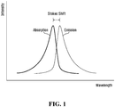
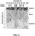


Classifications
-
- A—HUMAN NECESSITIES
- A61—MEDICAL OR VETERINARY SCIENCE; HYGIENE
- A61L—METHODS OR APPARATUS FOR STERILISING MATERIALS OR OBJECTS IN GENERAL; DISINFECTION, STERILISATION OR DEODORISATION OF AIR; CHEMICAL ASPECTS OF BANDAGES, DRESSINGS, ABSORBENT PADS OR SURGICAL ARTICLES; MATERIALS FOR BANDAGES, DRESSINGS, ABSORBENT PADS OR SURGICAL ARTICLES
- A61L27/00—Materials for grafts or prostheses or for coating grafts or prostheses
- A61L27/50—Materials characterised by their function or physical properties, e.g. injectable or lubricating compositions, shape-memory materials, surface modified materials
- A61L27/52—Hydrogels or hydrocolloids
-
- A—HUMAN NECESSITIES
- A61—MEDICAL OR VETERINARY SCIENCE; HYGIENE
- A61L—METHODS OR APPARATUS FOR STERILISING MATERIALS OR OBJECTS IN GENERAL; DISINFECTION, STERILISATION OR DEODORISATION OF AIR; CHEMICAL ASPECTS OF BANDAGES, DRESSINGS, ABSORBENT PADS OR SURGICAL ARTICLES; MATERIALS FOR BANDAGES, DRESSINGS, ABSORBENT PADS OR SURGICAL ARTICLES
- A61L27/00—Materials for grafts or prostheses or for coating grafts or prostheses
- A61L27/50—Materials characterised by their function or physical properties, e.g. injectable or lubricating compositions, shape-memory materials, surface modified materials
-
- A—HUMAN NECESSITIES
- A61—MEDICAL OR VETERINARY SCIENCE; HYGIENE
- A61K—PREPARATIONS FOR MEDICAL, DENTAL OR TOILETRY PURPOSES
- A61K41/00—Medicinal preparations obtained by treating materials with wave energy or particle radiation ; Therapies using these preparations
-
- A—HUMAN NECESSITIES
- A61—MEDICAL OR VETERINARY SCIENCE; HYGIENE
- A61K—PREPARATIONS FOR MEDICAL, DENTAL OR TOILETRY PURPOSES
- A61K8/00—Cosmetics or similar toiletry preparations
- A61K8/18—Cosmetics or similar toiletry preparations characterised by the composition
- A61K8/30—Cosmetics or similar toiletry preparations characterised by the composition containing organic compounds
- A61K8/49—Cosmetics or similar toiletry preparations characterised by the composition containing organic compounds containing heterocyclic compounds
- A61K8/4973—Cosmetics or similar toiletry preparations characterised by the composition containing organic compounds containing heterocyclic compounds with oxygen as the only hetero atom
- A61K8/498—Cosmetics or similar toiletry preparations characterised by the composition containing organic compounds containing heterocyclic compounds with oxygen as the only hetero atom having 6-membered rings or their condensed derivatives, e.g. coumarin
-
- A—HUMAN NECESSITIES
- A61—MEDICAL OR VETERINARY SCIENCE; HYGIENE
- A61K—PREPARATIONS FOR MEDICAL, DENTAL OR TOILETRY PURPOSES
- A61K8/00—Cosmetics or similar toiletry preparations
- A61K8/18—Cosmetics or similar toiletry preparations characterised by the composition
- A61K8/72—Cosmetics or similar toiletry preparations characterised by the composition containing organic macromolecular compounds
- A61K8/73—Polysaccharides
- A61K8/735—Mucopolysaccharides, e.g. hyaluronic acid; Derivatives thereof
-
- A—HUMAN NECESSITIES
- A61—MEDICAL OR VETERINARY SCIENCE; HYGIENE
- A61L—METHODS OR APPARATUS FOR STERILISING MATERIALS OR OBJECTS IN GENERAL; DISINFECTION, STERILISATION OR DEODORISATION OF AIR; CHEMICAL ASPECTS OF BANDAGES, DRESSINGS, ABSORBENT PADS OR SURGICAL ARTICLES; MATERIALS FOR BANDAGES, DRESSINGS, ABSORBENT PADS OR SURGICAL ARTICLES
- A61L27/00—Materials for grafts or prostheses or for coating grafts or prostheses
- A61L27/14—Macromolecular materials
- A61L27/20—Polysaccharides
-
- A—HUMAN NECESSITIES
- A61—MEDICAL OR VETERINARY SCIENCE; HYGIENE
- A61L—METHODS OR APPARATUS FOR STERILISING MATERIALS OR OBJECTS IN GENERAL; DISINFECTION, STERILISATION OR DEODORISATION OF AIR; CHEMICAL ASPECTS OF BANDAGES, DRESSINGS, ABSORBENT PADS OR SURGICAL ARTICLES; MATERIALS FOR BANDAGES, DRESSINGS, ABSORBENT PADS OR SURGICAL ARTICLES
- A61L27/00—Materials for grafts or prostheses or for coating grafts or prostheses
- A61L27/50—Materials characterised by their function or physical properties, e.g. injectable or lubricating compositions, shape-memory materials, surface modified materials
- A61L27/54—Biologically active materials, e.g. therapeutic substances
-
- A—HUMAN NECESSITIES
- A61—MEDICAL OR VETERINARY SCIENCE; HYGIENE
- A61L—METHODS OR APPARATUS FOR STERILISING MATERIALS OR OBJECTS IN GENERAL; DISINFECTION, STERILISATION OR DEODORISATION OF AIR; CHEMICAL ASPECTS OF BANDAGES, DRESSINGS, ABSORBENT PADS OR SURGICAL ARTICLES; MATERIALS FOR BANDAGES, DRESSINGS, ABSORBENT PADS OR SURGICAL ARTICLES
- A61L27/00—Materials for grafts or prostheses or for coating grafts or prostheses
- A61L27/50—Materials characterised by their function or physical properties, e.g. injectable or lubricating compositions, shape-memory materials, surface modified materials
- A61L27/58—Materials at least partially resorbable by the body
-
- A—HUMAN NECESSITIES
- A61—MEDICAL OR VETERINARY SCIENCE; HYGIENE
- A61N—ELECTROTHERAPY; MAGNETOTHERAPY; RADIATION THERAPY; ULTRASOUND THERAPY
- A61N5/00—Radiation therapy
- A61N5/06—Radiation therapy using light
- A61N5/0613—Apparatus adapted for a specific treatment
- A61N5/0616—Skin treatment other than tanning
-
- A—HUMAN NECESSITIES
- A61—MEDICAL OR VETERINARY SCIENCE; HYGIENE
- A61N—ELECTROTHERAPY; MAGNETOTHERAPY; RADIATION THERAPY; ULTRASOUND THERAPY
- A61N5/00—Radiation therapy
- A61N5/06—Radiation therapy using light
- A61N5/0613—Apparatus adapted for a specific treatment
- A61N5/062—Photodynamic therapy, i.e. excitation of an agent
-
- A—HUMAN NECESSITIES
- A61—MEDICAL OR VETERINARY SCIENCE; HYGIENE
- A61P—SPECIFIC THERAPEUTIC ACTIVITY OF CHEMICAL COMPOUNDS OR MEDICINAL PREPARATIONS
- A61P17/00—Drugs for dermatological disorders
-
- A—HUMAN NECESSITIES
- A61—MEDICAL OR VETERINARY SCIENCE; HYGIENE
- A61P—SPECIFIC THERAPEUTIC ACTIVITY OF CHEMICAL COMPOUNDS OR MEDICINAL PREPARATIONS
- A61P17/00—Drugs for dermatological disorders
- A61P17/02—Drugs for dermatological disorders for treating wounds, ulcers, burns, scars, keloids, or the like
-
- A—HUMAN NECESSITIES
- A61—MEDICAL OR VETERINARY SCIENCE; HYGIENE
- A61Q—SPECIFIC USE OF COSMETICS OR SIMILAR TOILETRY PREPARATIONS
- A61Q19/00—Preparations for care of the skin
- A61Q19/08—Anti-ageing preparations
-
- A—HUMAN NECESSITIES
- A61—MEDICAL OR VETERINARY SCIENCE; HYGIENE
- A61B—DIAGNOSIS; SURGERY; IDENTIFICATION
- A61B18/00—Surgical instruments, devices or methods for transferring non-mechanical forms of energy to or from the body
- A61B2018/00315—Surgical instruments, devices or methods for transferring non-mechanical forms of energy to or from the body for treatment of particular body parts
- A61B2018/00452—Skin
- A61B2018/0047—Upper parts of the skin, e.g. skin peeling or treatment of wrinkles
-
- A—HUMAN NECESSITIES
- A61—MEDICAL OR VETERINARY SCIENCE; HYGIENE
- A61K—PREPARATIONS FOR MEDICAL, DENTAL OR TOILETRY PURPOSES
- A61K2800/00—Properties of cosmetic compositions or active ingredients thereof or formulation aids used therein and process related aspects
- A61K2800/80—Process related aspects concerning the preparation of the cosmetic composition or the storage or application thereof
- A61K2800/81—Preparation or application process involves irradiation
-
- A—HUMAN NECESSITIES
- A61—MEDICAL OR VETERINARY SCIENCE; HYGIENE
- A61K—PREPARATIONS FOR MEDICAL, DENTAL OR TOILETRY PURPOSES
- A61K2800/00—Properties of cosmetic compositions or active ingredients thereof or formulation aids used therein and process related aspects
- A61K2800/80—Process related aspects concerning the preparation of the cosmetic composition or the storage or application thereof
- A61K2800/88—Two- or multipart kits
- A61K2800/884—Sequential application
-
- A—HUMAN NECESSITIES
- A61—MEDICAL OR VETERINARY SCIENCE; HYGIENE
- A61K—PREPARATIONS FOR MEDICAL, DENTAL OR TOILETRY PURPOSES
- A61K2800/00—Properties of cosmetic compositions or active ingredients thereof or formulation aids used therein and process related aspects
- A61K2800/80—Process related aspects concerning the preparation of the cosmetic composition or the storage or application thereof
- A61K2800/91—Injection
-
- A—HUMAN NECESSITIES
- A61—MEDICAL OR VETERINARY SCIENCE; HYGIENE
- A61L—METHODS OR APPARATUS FOR STERILISING MATERIALS OR OBJECTS IN GENERAL; DISINFECTION, STERILISATION OR DEODORISATION OF AIR; CHEMICAL ASPECTS OF BANDAGES, DRESSINGS, ABSORBENT PADS OR SURGICAL ARTICLES; MATERIALS FOR BANDAGES, DRESSINGS, ABSORBENT PADS OR SURGICAL ARTICLES
- A61L2300/00—Biologically active materials used in bandages, wound dressings, absorbent pads or medical devices
- A61L2300/40—Biologically active materials used in bandages, wound dressings, absorbent pads or medical devices characterised by a specific therapeutic activity or mode of action
- A61L2300/442—Colorants, dyes
-
- A—HUMAN NECESSITIES
- A61—MEDICAL OR VETERINARY SCIENCE; HYGIENE
- A61L—METHODS OR APPARATUS FOR STERILISING MATERIALS OR OBJECTS IN GENERAL; DISINFECTION, STERILISATION OR DEODORISATION OF AIR; CHEMICAL ASPECTS OF BANDAGES, DRESSINGS, ABSORBENT PADS OR SURGICAL ARTICLES; MATERIALS FOR BANDAGES, DRESSINGS, ABSORBENT PADS OR SURGICAL ARTICLES
- A61L2400/00—Materials characterised by their function or physical properties
- A61L2400/06—Flowable or injectable implant compositions
-
- A—HUMAN NECESSITIES
- A61—MEDICAL OR VETERINARY SCIENCE; HYGIENE
- A61L—METHODS OR APPARATUS FOR STERILISING MATERIALS OR OBJECTS IN GENERAL; DISINFECTION, STERILISATION OR DEODORISATION OF AIR; CHEMICAL ASPECTS OF BANDAGES, DRESSINGS, ABSORBENT PADS OR SURGICAL ARTICLES; MATERIALS FOR BANDAGES, DRESSINGS, ABSORBENT PADS OR SURGICAL ARTICLES
- A61L2430/00—Materials or treatment for tissue regeneration
- A61L2430/34—Materials or treatment for tissue regeneration for soft tissue reconstruction
Definitions
- the present disclosure generally relates to tissue filler compositions and methods of use. Specifically, but not exclusively, the present disclosure relates to tissue filler compositions which can be injected or implanted into soft tissues.
- Tissue fillers are used in the medical and cosmetic fields for tissue augmentation, regeneration or re-shaping.
- tissue fillers are known as dermal fillers and are injectable materials which can improve the appearance of skin by filling of wrinkles and folds, replacing lost tissue, or by facial contouring such as re-shaping the cheeks, chin or jaw.
- Tissue fillers are also used for treating scars by temporarily elevating a depressed scar
- CA2695386 A1 discloses cosmetic fillers activated by visible light and containing modified reactive groups that facilitate polymerization, attachment and crosslinking of the polymeric material on exposure to light.
- the filler can be in semisolid or gel form (e.g. a hyaluronic acid hydrogel) and can be injected under the skin.
- the pregel, monomer solution can comprise a photoinitiator, such as Eosin Y.
- the invention relates to a tissue filler composition as defined in claim 1.
- the tissue filler composition can be used for stimulating collagen synthesis.
- the tissue filler composition can be used for cosmetic enhancement of soft tissue.
- the tissue filler composition can be used for cosmetically inhibiting or treating scarring.
- light emitted by the fluorophore can induce or stimulate collagen synthesis in the soft tissue around the composition where there otherwise would be no collagen synthesis or where the collagen synthesis would be minimal.
- the collagen synthesis induced by the fluorophore may be in addition to any collagen synthesis inherently induced by the tissue filler medium by mechanical means (e.g. when the tissue filler medium comprises hyaluronic acid) or by a foreign body response (e.g. when the tissue filler medium is a nonbiodegradable material).
- the surrounding soft tissues are illuminated with a broader spectrum of light than if illuminated from outside the dermis or from a single light source. Also, the surrounding soft tissues may be illuminated with shorter wavelengths of light than those able to reach those soft tissues transdermally. In this way, a more efficient and wavelength specific illumination of the soft tissues, such as dermal/epidermal/subdermal layers can be achieved which may lead to a therapeutic or cosmetic effect.
- the area to be treated is soft tissue.
- the area to be treated may be on any part of the body such as on a face, neck, ears, breasts, buttocks, arms, armpits, hands, genitalia, legs or feet of a human subject.
- the area to be treated is in or around a scar.
- the scar may be a post-surgical scar.
- the scar can be an old or a fresh scar.
- the area to be treated is in or around a wound.
- the wound may be an acute or a chronic wound, including burns.
- the area to be treated includes stretch marks.
- the area to be treated may be in or around a stretch mark.
- the administering of the composition is by injection.
- the injection can be performed using a needle.
- the needle can have a gauge of 27G to 40G, typically 30G or 32G.
- the administering by injection can be a continuous injection, known as linear threading, or serial punctures to deposit microdroplets.
- the administering of the composition is by implantation.
- the composition is a cohesive gel.
- the composition may be a hydrated gel.
- the composition may be transparent or translucent.
- the tissue filler medium is a cohesive gel.
- the tissue filler medium may be a hydrated gel.
- the tissue filler medium may be transparent or translucent.
- the tissue filler medium retains the fluorophore within the composition during administering of the composition, and at least during a portion of the illumination.
- the tissue filler medium/composition may allow the fluorophore to leach from the composition/tissue filler medium after administering to the tissue.
- the tissue filler medium is biodegradable.
- the tissue filler medium may comprise any biodegradable and biocompatible material such as hyaluronic acid (HA), collagen, poly-L-lactic acid. Complete biodegradation may occur in about 3-18 months.
- the fluorophore may or may not affect the biodegradation rate of the tissue filler medium.
- the tissue filler medium comprises cross-linked hyaluronic acid (cross-linked HA).
- the cross-linked hyaluronic acid may be about 0.1% to about 2%, about 2% to about 30%, about 2% to about 25%, about 2% to about 20%, about 2% to about 15%, about 2% to about 10%, about 2% to about 5%, 4% to about 30%, about 4% to about 25%, about 4% to about 20%, about 4% to about 15%, about 4% to about 10%, about 4% to about 5%, of the composition.
- the cross-linked hyaluronic acid is in particulate form. The particles may be hydrated. The particles may be cohesive.
- the fluorophore is within the cross-linked hyaluronic acid particle.
- the composition further comprises an injectable medium supporting the particles.
- the injectable medium may be a fluid.
- the injectable medium may be less viscous and/or less cohesive than the particles.
- the particles may be cohesive and the injectable medium non-cohesive.
- the fluorophore may be in the injectable medium or the particles, or both.
- the injectable medium comprises hyaluronic acid which is non-cross linked or relatively less cross-linked than the cross-linked hyaluronic acid particles.
- the composition further comprises light reflecting particles.
- the light reflecting particles may be a glass or silicon dioxide.
- the fluorophore is a hydrophilic chromophore.
- the fluorophore may be water soluble.
- the fluorophore is not in a liposomal form in the composition.
- the fluorophore can be activated by light having a wavelength in the visible range.
- the fluorophore does not absorb light in the UV range of the electromagnetic spectrum.
- the fluorophore when activated, can emit light having a wavelength in the visible range. The emitted light can be within one or more of the violet, blue, green, yellow, orange or red portions of the electromagnetic spectrum. Alternatively, the fluorophore can emit in the infrared range.
- the fluorophore can be photobleached after illumination of the area.
- the fluorophore may be photobleached after 5, 10, 15, 20, 25, 30, 35, 40, 45, 50, 55, 60 or 65 minutes of illumination.
- the illumination may be continuous or pulsed.
- the illumination may comprise re-illumination of the composition or the area after a few hours or days.
- photosensitivity is reduced or avoided.
- the fluorophore is not a photosensitizing agent requiring to be metabolized.
- the fluorophore is not porphyrin, porphyrinogen, hematoporphyrin, pheophorbide, chlorin, bacteriochlorin, iso-bacteriochlorin and dihydro- and tetrahydro-tetrapyrroles.
- the fluorophore does not bind to native cells or cellular structures.
- the area is illuminated at the same time as or immediately after intradermal or subdermal administration. In other words, there is no incubation period between administering of the composition and illumination.
- the illumination is for a time sufficient to activate the fluorophore. In certain embodiments, the illumination is for a time sufficient to photobleach the fluorophore. In certain embodiments, the illumination is for 1-30 seconds, 15-45 seconds, 30-60 seconds, 0.75-1.5 minutes, 1-2 minutes, 1.5-2.5 minutes, 2-3 minutes, 2.5-3.5 minutes, 3-4 minutes, 3.5-4.5 minutes, 4-5 minutes, 5-10 minutes, 10-15 minutes, 15-20 minutes, 20-25 minutes, or 25-30 minutes.
- the treatment time may range up to about 90 minutes, about 80 minutes, about 70 minutes, about 60 minutes, about 50 minutes, about 40 minutes or about 30 minutes.
- the composition is illuminated by a light source positioned external to the dermis. In other words, illumination is transdermally.
- intradermally or subdermally administration provides a track of HA-stimulated cells from the external surface to the dermis, thereby creating a light duct from the light source to the intradermal or subdermal in order to activate the composition.
- the present disclosure relates to a method for skin rejuvenation comprising: intradermally or subdermally administering a composition to an area to be treated, wherein the composition comprises a tissue filler medium and a fluorophore; topically applying a biophotonic composition to the skin above the area to be treated; and illuminating the topical biophotonic composition with light having a wavelength which can be absorbed by a chromophore in the topical biophotonic composition to cause the chromophore to emit light; wherein the fluorophore can absorb the emitted light from the topical biophotonic composition, wherein the method stimulates collagen synthesis in the area to cosmetically rejuvenate the skin.
- the composition is injectable and can be injected through a 23-40 gauge needle. In certain embodiments, the composition is injected under a wrinkle or a fold or a scar to provide an immediate lift to the defect. Illumination of the composition may then stimulate collagen synthesis around the injected composition to provide further skin rejuvenation.
- tissue filler composition comprising: a tissue filler medium; and a fluorophore, wherein the at least one fluorophore is a hydrophilic fluorophore; wherein the composition is suitable for injection or implantation into a human; and wherein the tissue filler composition is not photo-polymerizable.
- the composition is a cohesive gel.
- the composition may be a hydrated gel.
- the composition may be transparent or translucent.
- the tissue filler medium retains the fluorophore within the composition during administering of the composition, and at least during a portion of the illumination.
- the fluorophore may leach from the tissue filler medium after administering to the tissue.
- the tissue filler medium is biodegradable.
- the tissue filler medium may comprise any biodegradable and biocompatible material such as hyaluronic acid, collagen, poly-L-lactic acid. Complete biodegradation may occur in about 3-18 months.
- the fluorophore may or may not affect the biodegradation rate of the tissue filler medium.
- the tissue filler medium comprises cross-linked hyaluronic acid.
- the cross-linked hyaluronic acid may be about 0.1 to about 2%, about 2 to about 30%, about 2 to about 25%, about 2 to about 20%, about 2 to about 15%, about 2 to about 10%, about 2 to about 5%, 4 to about 30%, about 4 to about 25%, about 4 to about 20%, about 4 to about 15%, about 4 to about 10%, about 4 to about 5%, of the composition.
- the cross-linked hyaluronic acid is in particulate form. The particles may be hydrated. The particles may be cohesive.
- the fluorophore is within the cross-linked hyaluronic acid particle.
- the composition further comprises an injectable medium supporting the particles.
- the injectable medium may be a fluid.
- the injectable medium may be less viscous and/or less cohesive than the particles.
- the particles may be cohesive and the injectable medium non-cohesive.
- the fluorophore may be in the injectable medium or the particles, or both.
- the injectable medium comprises hyaluronic acid which is non-cross linked or relatively less cross-linked than the cross-linked hyaluronic acid particles.
- the composition further comprises light reflecting particles.
- the light reflecting particles may be a glass or silicon dioxide.
- the fluorophore is a hydrophilic chromophore.
- the fluorophore may be water soluble. In certain embodiments, the fluorophore is not in a liposomal form in the composition.
- the fluorophore can be activated by light having a wavelength in the visible range. In certain embodiments, the fluorophore does not absorb light in the UV range of the electromagnetic spectrum. In certain embodiments, when activated, the fluorophore can emit light having a wavelength in the visible range. The emitted light can be within one or more of the violet, blue, green, yellow, orange or red portions of the electromagnetic spectrum. Alternatively, the fluorophore can emit in the infrared range.
- the fluorophore can be photobleached after illumination of the area.
- the fluorophore may be photobleached after 5, 10, 15, 20, 25, 30, 35, 40, 45, 50, 55, 60 or 65 minutes of illumination.
- the fluorophore may be photobleached after 1-30 seconds, 15-45 seconds, 30-60 seconds, 0.75-1.5 minutes, 1-2 minutes, 1.5-2.5 minutes, 2-3 minutes, 2.5-3.5 minutes, 3-4 minutes, 3.5-4.5 minutes, 4-5 minutes, 5-10 minutes, 10-15 minutes, 15-20 minutes, 20-25 minutes, or 25-30 minutes.
- the treatment time may range up to about 90 minutes, about 80 minutes, about 70 minutes, about 60 minutes, about 50 minutes, about 40 minutes or about 30 minutes.
- the illumination may be continuous or pulsed.
- the illumination may comprise re-illumination of the composition or the area after a few hours or days.
- the fluorophore is not a photosensitizing agent requiring to be metabolized.
- the fluorophore is not porphyrin, porphyrinogen, hematoporphyrin, pheophorbide, chlorin, bacteriochlorin, iso-bacteriochlorin and dihydro- and tetrahydro-tetrapyrroles.
- the fluorophore does not bind to native cells, cancer cells or other cellular structures.
- the composition does not include glucosamine. In certain embodiments, not claimed, where the composition comprises hyaluronic acid, the composition does not include glucosamine. In certain embodiments, the composition does not include a source of oxygen such as peroxide. In certain embodiments, not claimed, the composition does not include hydrophobic fluorophores. The composition is not photopolymerizable. In certain embodiments, not claimed, the composition does not include a monomer. In certain embodiments, not claimed, the composition does not include hyaluronic acid with functionalized chains.
- the composition does not include one or more (e.g., 1, 2 or 3) of triethanolamine (TEA), N-vinyl-2-pyrrolidone (NVP), or N-vinyl caprolactam (NVC). In certain embodiments, the composition does not include any of triethanolamine (TEA), N-vinyl-2-pyrrolidone (NVP), or N-vinyl caprolactam (NVC).
- TAA triethanolamine
- NVP N-vinyl-2-pyrrolidone
- NVC N-vinyl caprolactam
- the composition is a sterile composition.
- the composition can be sterilized by heat and/or pressure, such as using an autoclave.
- the composition can be sterilized by gamma irradiation.
- the subdermally or intradermally placed composition can provide an immediate 'lifting' which may be due in part to its cohesive or viscoelastic properties. This is beneficial in smoothing of lines and folds on skin to provide an appearance of skin rejuvenation.
- the 'lifting' effect of the subdermally or intradermally placed composition diminishes over time as the tissue filler medium degrades, is compressed and/or is dispersed. Therefore, by means of certain embodiments, not claimed, of the present disclosure, a prolonged skin rejuvenation effect can be obtained by providing an immediate lifting effect to the skin which gradually diminishes with time, and at the same time synthesis of new collagen at or around the intradermal or subdermal composition which increases over time. As the tissue filler degradation and the collagen synthesis dovetail into one another, a substantially continuous skin rejuvenation effect may be achieved.
- the new collagen on or around the intracorporeal composition substantially matches the extracellular matrix in terms of collagen type, collagen mechanical properties and collagen fibrillar orientation. This is unlike existing degradable tissue filler media which either induce collagen synthesis but do not provide a lifting effect (e.g. fillers based on poly-L-lactic acid), or which provide a lifting effect but which do not induce significant collagen synthesis (e.g. hyaluronic acid based dermal fillers), or which provide a lifting effect and induce collagen synthesis having different properties to the extracellular matrix (e.g. permanent dermal fillers).
- a lifting effect e.g. fillers based on poly-L-lactic acid
- hyaluronic acid based dermal fillers e.g. hyaluronic acid based dermal fillers
- collagen synthesis having different properties to the extracellular matrix e.g. permanent dermal fillers.
- the present disclosure relates to the composition as described herein, for stimulating collagen synthesis within soft tissues. From another aspect, there is provided use of a composition as described herein, for cosmetic enhancement of soft tissues. From another aspect, there is provided use of a composition as described herein, for inhibiting or treating scarring. From another aspect, there is provided use of a composition as described herein, for skin rejuvenation. From another aspect, there is provided use of a composition as described herein, together with a topical biophotonic composition for skin rejuvenation.
- the present disclosure relates to the use of a composition comprising a tissue filler medium and a fluorophore in a method for stimulating collagen synthesis, wherein the method comprises a step of administration of the composition to an area to be treated within a soft tissue, and a step illumination of the area with light having a wavelength which can be absorbed by the fluorophore; and wherein the method stimulates collagen synthesis in the area.
- the present disclosure relates to the use of a composition comprising a tissue filler medium and a fluorophore in a method for cosmetic enhancement of soft tissue, wherein the method comprises a step of administration of the composition to an area to be treated within a soft tissue, and a step illumination of the area with light having a wavelength which can be absorbed by the fluorophore; and wherein the method cosmetically enhances the soft tissue.
- the present disclosure relates to the use of a composition comprising a tissue filler medium and a fluorophore in a method for inhibiting or treating scarring, wherein the method comprises a step of administration of the composition to an area to be treated within a soft tissue, and a step illumination of the area with light having a wavelength which can be absorbed by the fluorophore; and wherein the method cosmetically inhibits or treats scarring.
- the present disclosure relates a tissue filler composition
- a tissue filler composition comprising more than one tissue filler medium, wherein the composition is suitable for injection or implantation into a human.
- the present disclosure provides compositions which can be used as tissue fillers and which are biophotonic.
- the tissue filler compositions of the present disclosure include a photoactive exogenous chromophore together with a tissue filler medium, and may be injectable or implantable or deliverable to soft tissues in any other way.
- the present disclosure also provides methods useful for stimulating collagen synthesis; cosmetic enhancement of soft tissue such as soft tissue augmentation, rejuvenation or re-shaping; inhibition or treatment of scars; or promoting wound healing, using such tissue filler compositions.
- the term "about” in the context of a given value or range refers to a value or range that is within 20%, preferably within 10%, and more preferably within 5% of the given value or range.
- Biophotonic means the generation, manipulation, detection and application of photons in a biologically relevant context. In other words, biophotonic compositions exert their physiological effects primarily due to the generation and manipulation of photons. Biophotonic compositions may also generate reactive oxygen species.
- Biophotonic composition is a composition as described herein that may be activated by light to produce photons and/or reactive oxygen species for biologically relevant applications.
- chromophore means a chemical compound, when contacted by light irradiation, is capable of absorbing the light. The chromophore readily undergoes photoexcitation and can then transfer its energy to other molecules and/or emit it as light.
- the chromophore may be a synthetic chromophore or a naturally occurring chromophore.
- Fluorophore as used herein means a chromophore which can emit light upon light excitation e.g. by fluorescence, phosphorescence or any other means.
- tissue filler when used herein means a material that is suitable for, or generally used for, tissue augmentation, regeneration or re-shaping such as those suitable for use or used in the dermis area such as dermal fillers, or those suitable for use or used in any other soft tissue for example as a scaffold or a delivery material.
- Tissue fillers include biodegradable and non-biodegradable materials, as well as natural and synthetic materials, including hydrogels.
- Tissue fillers can be administered by any means such as by injection or implantation. Tissue fillers can be administered to any soft tissue site such as subcutaneous, hypodermic and/or intradermal.
- Dermat filler when used herein means a material which can be used in the dermis area, such as below the epidermis and/or above or below the hypodermis, such as for tissue augmentation, regeneration or re-shaping. Dermal fillers can be delivered by hypodermic and/or intradermal injection, or by any other means such as implantation.
- soft tissue refers to tissues that connect, support, or surround other structures and organs of the body, not being bone. Soft tissue includes tendons, ligaments, fascia, skin, fibrous tissues, fat, and synovial membranes (which are connective tissue), and muscles, nerves and blood vessels (which are not connective tissue). It is sometimes defined by what it is not. Soft tissue has been defined as “nonepithelial, extraskeletal mesenchyme exclusive of the reticuloendothelial system and glia”.
- “Injectable” when used herein means a flowable material which may be drawn through or pushed through a needle by a syringe for injection into a soft tissue.
- Soft tissue includes tendons, ligaments, fascia, skin, dermis, fibrous tissues, fat, synovial membranes, muscles, nerves and blood vessels.
- “Inhibiting or treating scarring” as used herein means preventing or minimizing scar formation, or reducing an existing scar.
- the scarring can be a result of any wound such as a burn or a surgical incision.
- Photobleaching means the photochemical destruction of a chromophore.
- light or “actinic light” is intended to mean light energy emitted from a specific light source (e.g., lamp, LED, laser) and capable of being absorbed by matter (e.g. the chromophore or photoactivator defined above).
- a specific light source e.g., lamp, LED, laser
- matter e.g. the chromophore or photoactivator defined above.
- the light has a peak wavelength within the visible range of the electromagnetic spectrum, e.g. 360 nm to 760 nm.
- Wound means an injury to any tissue, including for example, acute, subacute, delayed or difficult to heal wounds, and chronic wounds. Examples of wounds may include both open and closed wounds. Wounds include, for example, amputations, burns, incisions, excisions, lesions, lacerations, abrasions, puncture or penetrating wounds, surgical wounds, contusions, hematomas, crushing injuries, ulcers (such as for example pressure, venous, arterial or diabetic), wounds caused by periodontitis (inflammation of the periodontium).
- Skin rejuvenation means the reducing, diminishing, retarding or reversing of one or more signs of skin aging or skin damage. Skin rejuvenation also means generally improving the cosmetic appearance of skin or cosmetic enhancement of skin. For instance, it includes increasing luminosity of the skin; reducing pore size; reducing fine lines or wrinkles; improving thin and transparent skin; improving firmness; improving plumpness; augmenting tissue loss or sagging skin due to loss of underlying fat or bone; improving dry skin (which might itch); reducing or reversing freckles, age spots, spider veins, or a blotchy complexion; improving rough and leathery skin; or improving fine wrinkles that disappear when stretched. According to the present disclosure, one or more of the above conditions may be improved or one or more of the above signs of aging may be reduced, diminished, retarded or even reversed by certain embodiments of the compositions, methods and uses of the present disclosure.
- the present disclosure provides biophotonic compositions which can be injected or implanted within soft tissue as tissue fillers.
- the present disclosure provides intracorporeal biophotonic compositions.
- Biophotonic compositions are compositions that are, in a broad sense, activated by light (e.g., photons) of specific wavelength. These compositions contain at least one exogenous chromophore which can be activated by light having an appropriate wavelength causing the chromophore to emit light. The activating and/or emitted light may have a therapeutic effect on its own.
- the activated chromophore and/or the light may also lead to the photochemical activation of other agents contained in the composition or at a treatment site.
- an oxygen-releasing agent such an agent may be activated leading to the formation of oxygen radicals, such as singlet oxygen.
- the oxygen-releasing agent may be an internal source of oxygen such as blood.
- the present disclosure provides biophotonic compositions comprising at least a first chromophore and a tissue filler medium.
- a chromophore absorbs a photon of a certain wavelength, it becomes excited. This is an unstable condition and the molecule tries to return to the ground state, giving away the excess energy.
- fluorescence When some chromophores, it is favorable to emit the excess energy as light when transforming back to the ground state. This process is called fluorescence and these chromophores are also known as "fluorophores”.
- the peak wavelength of the emitted fluorescence is shifted towards longer wavelengths compared to the absorption wavelengths due to loss of energy in the conversion process. This is called the Stokes' shift and is illustrated in Figure 1 . In the proper environment (e.g., in a biophotonic composition) much of this energy is transferred to other components of the composition or to the treatment site directly.
- fluorescent light emitted by photoactivated chromophores may have therapeutic properties due to its femto-, pico- or nano-second emission properties which may be recognized by biological cells and tissues, leading to favorable biomodulation. Furthermore, the emitted fluorescent light has a longer wavelength and hence a deeper penetration into the tissue than the activating light ( Figure 2 ). Irradiating intradermal, subdermal or other soft tissue with such a broad range of wavelengths, including in some embodiments the activating light which passes through the composition, may have different and complementary effects on the cells and tissues.
- the tissues surrounding the biophotonic composition will be illuminated with wavelengths of light which would not normally reach those soft tissue sites if illuminated transdermally due to the short wavelengths and the depth from the skin. In certain embodiments, this may lead to a therapeutic or cosmetic benefit to the soft tissues surrounding the biophotonic composition in use, such as stimulation of collagen production. This may be in addition to any physical filling and lifting effects of a typical tissue filler, as well as in addition to any collagen production due induced by locally applied stresses from the tissue filler. Furthermore, the collagen which is produced may more closely match that of the extracellular collagen matrix of the area being treated compared to fibrous collagen formation through foreign body response seen with some existing tissue fillers. That is the type of collagen, the mechanical properties of the collagen, and collagen fibrillar direction will more closely match that of native collagen.
- the biophotonic compositions of the present disclosure may be capable of triggering changes in cell signalling as so to produce an increase in collagen deposition through fibroblast stimulation and thereby modifying the extracellular matrix locally.
- the biophotonic compositions of the present disclosure may be substantially transparent/translucent and/or have high light transmittance in order to permit light dissipation into and through the composition. In this way, the area of tissue around the composition can be treated both with the fluorescent light emitted by the composition and the light irradiating the composition to activate it.
- the % transmittance of the biophotonic composition can be measured in the range of wavelengths from 250 nm to 800 nm using, for example, a Perkin-Elmer Lambda 9500 series UV-visible spectrophotometer. In some embodiments, transmittance within the visible range is measured and averaged. In some other embodiments, transmittance of the biophotonic material is measured with the chromophore omitted.
- transmittance of the compositions disclosed herein is measured at 460 nm.
- the thickness of each sample can be measured with calipers prior to loading in the spectrophotometer.
- transmittance measurements are usually normalized to 1 cm.
- the biophotonic composition has a transmittance that exceeds about 15%, about 20%, about 30%, about 40%, about 50%, about 55%, about 60%, about 65%, about 70%, about 75%, about 80%, or about 85% within the visible range. In some embodiments, the transmittance exceeds about 70%, about 86%, about 87%, about 88%, about 89%, about 90%, about 91%, about 92%, about 93%, about 94%, about 95%, about 96%, about 97%, about 98% or about 99% within the visible range of the electromagnetic spectrum (e.g. 400-700nm).
- the transmittance exceeds about 70%, about 86%, about 87%, about 88%, about 89%, about 90%, about 91%, about 92%, about 93%, about 94%, about 95%, about 96%, about 97%, about 98% or about 99% within the visible range of the electromagnetic spectrum (e.g. 400-700nm).
- biophotonic compositions of the present disclosure are injectable or implantable, or deliverable intracorporeally to a soft tissue site by any other means.
- compositions may be described based on the components making up the composition. Additionally or alternatively, the compositions of the present disclosure have functional and structural properties and these properties may also be used to define and describe the compositions. Individual components of the composition of the present disclosure are detailed as below.
- compositions of the present disclosure comprise one or more chromophores, which can be considered exogenous, e.g., are not naturally present in skin or tissue.
- Suitable chromophores can be fluorophores or fluorochromes such as fluorescent dyes (or stains), although other dye groups or dyes (biological and histological dyes, food colorings, carotenoids, naturally occurring fluorescent and other dyes) can also be used.
- Suitable photoactivators can be those that are Generally Regarded As Safe (GRAS). Photoactivators which are not well tolerated by the skin or other tissues can be included in the biophotonic composition in an encapsulated, or chemically modified form.
- the biophotonic composition of the present disclosure comprises a first chromophore which undergoes partial or complete photobleaching upon application of light.
- photobleaching is meant a photochemical destruction of the chromophore which can generally be visualized as a loss of color.
- the first chromophore absorbs at a wavelength in the range of the visible spectrum, such as at a wavelength of about 380-800 nm, about 380-700, about 400-700 or about 380-600 nm. In these embodiments, the first chromophore is not activated by UV light.
- the first chromophore absorbs at a wavelength of about 200-800 nm, about 200-700 nm, about 200-600 nm or about 200-500 nm. In one embodiment, the first chromophore absorbs at a wavelength of about 200-600 nm. In some embodiments, the first chromophore absorbs light at a wavelength of about 200-300 nm, about 250-350 nm, about 300-400 nm, about 350-450 nm, about 400-500 nm, about 400-600 nm, about 450-650 nm, about 600-700 nm, about 650-750 nm or about 700-800 nm.
- a particular chromophore's absorption and/or emission wavelength (or spectrum) corresponds to the wavelengths (or spectrum) measured in a biophotonic composition of the present disclosure.
- the biophotonic compositions disclosed herein may include at least one additional chromophore.
- Combining chromophores may increase photo-absorption by the combined dye molecules and enhance absorption and photo-biomodulation selectivity. This creates multiple possibilities of generating new photosensitive, and/or selective chromophore mixtures.
- resonance energy transfer is a photophysical process through which an excited 'donor' chromophore (also referred to herein as first chromophore) transfers its excitation energy to an 'acceptor' chromophore (also referred to herein as second chromophore).
- first chromophore also referred to herein as first chromophore
- second chromophore also referred to herein as second chromophore.
- the efficiency and directedness of resonance energy transfer depends on the spectral features of donor and acceptor chromophores. In particular, the flow of energy between chromophores is dependent on a spectral overlap reflecting the relative positioning and shapes of the absorption and emission spectra.
- the emission spectrum of the donor chromophore must overlap with the absorption spectrum of the acceptor chromophore ( Figure 3 ).
- Energy transfer manifests itself through decrease or quenching of the donor emission and a reduction of excited state lifetime accompanied also by an increase in acceptor emission intensity.
- Figure 4 is a Jablonski diagram that illustrates the coupled transitions involved between a donor emission and acceptor absorbance. To enhance the energy transfer efficiency, the donor chromophore should have good abilities to absorb photons and emit photons.
- the biophotonic composition of the present disclosure further comprises a second chromophore.
- the first chromophore has an emission spectrum that overlaps at least about 80%, at least about 50%, at least about 40%, at least about 30%, at least about 20%, or at least about 10% with an absorption spectrum of the second chromophore.
- the first chromophore has an emission spectrum that overlaps at least about 20% with an absorption spectrum of the second chromophore.
- the first chromophore has an emission spectrum that overlaps at least about 1-10%, at least about 5-15%, at least about 10-20%, at least about 15-25%, at least about 20-30%, at least about 25-35%, at least about 30-40%, at least about 35-45%, at least about 50-60%, at least about 55-65% or at least about 60-70% with an absorption spectrum of the second chromophore.
- % spectral overlap means the % overlap of a donor chromophore's emission wavelength range with an acceptor chromophore's absorption wavelength range, measured at spectral full width quarter maximum (FWQM).
- Figure 2 shows the normalized absorption and emission spectra of donor and acceptor chromophores.
- the spectral FWQM of the acceptor chromophore's absorption spectrum is from about 60 nm (515 nm to about 575 nm).
- the overlap of the donor chromophore's spectrum with the absorption spectrum of the acceptor chromophore is about 40 nm (from 515 nm to about 555 nm).
- the second chromophore absorbs at a wavelength in the range of the visible spectrum. In certain embodiments, the second chromophore has an absorption wavelength that is relatively longer than that of the first chromophore within the range of about 50-250, about 25-150 or about 10-100 nm.
- the application of light to the compositions of the present disclosure can result in a cascade of energy transfer between the chromophores.
- a cascade of energy transfer provides emission of photons from inside the soft tissue that can travel deeper than if the photons were travelling transdermally.
- such a cascade of energy transfer is not accompanied by concomitant generation of heat.
- the cascade of energy transfer does not result in tissue damage.
- the biophotonic composition comprises a first and a second chromophore
- the first chromophore is present in an amount of about 0.001-40% per weight of the composition
- the second chromophore is present in an amount of about 0.001-40% per weight of the composition.
- the total weight per weight of chromophore or combination of chromophores may be in the amount of about 0.001-40.001% per weight of the composition.
- the first chromophore is present in an amount of about 0.001-0.01%, about 0.001-0.05%, about 0.005-0.01%, about 0.01-1%, about 0.01-2%, about 0.05-1%, about 0.05-2%, about 1-5%, about 2.5-7.5%, about 5-10%, about 7.5-12.5%, about 10-15%, about 12.5-17.5%, about 15-20%, about 17.5-22.5%, about 20-25%, about 22.5-27.5%, about 25-30%, about 27.5-32.5%, about 30-35%, about 32.5-37.5%, or about 35-40% per weight of the composition.
- the second chromophore is present in an amount of about 0.001-1%, about 0.001-2%, about 0.001-0.01%, about 0.01-0.1%, about 0.1-1.0%, about 1-2%, about 1-5%, about 2.5-7.5%, about 5-10%, about 7.5-12.5%, about 10-15%, about 12.5-17.5%, about 15-20%, about 17.5-22.5%, about 20-25%, about 22.5-27.5%, about 25-30%, about 27.5-32.5%, about 30-35%, 3 about 2.5-37.5%, or about 35-40% per weight of the composition.
- the total weight % per weight of chromophore or combination of chromophores may be in the amount of about 0.001-1%, about 0.01-1%, about 0.01-2%, about 0.05-2%, about 0.5-1%, about 0.5-2%, about 1-5%, about 2.5-7.5%, about 5-10%, about 7.5-12.5%, about 10-15%, about 12.5-17.5%, about 15-20%, about 17.5-22.5%, about 20-25%, about 22.5-27.5%, about 25-30%, about 27.5-32.5%, about 30-35%, about 32.5-37.5%, or about 35-40.05% per weight of the composition.
- the chromophore or chromophores are selected such that their emitted fluorescent light, on photoactivation, is within one or more of the blue, green, yellow, orange, red and infrared portions of the electromagnetic spectrum, for example having a peak wavelength within the range of about 450 nm to about 500 nm, 490 nm to about 800 nm, or about 470 nm to about 700 nm.
- the emitted fluorescent light has a peak power density of between 0.005 to about 10 mW/cm 2 , or about 0.5 to about 5 mW/cm 2 .
- Chlorophyll dyes include but are not limited to chlorophyll a; chlorophyll b; oil soluble chlorophyll; bacteriochlorophyll a; bacteriochlorophyll b; bacteriochlorophyll c; bacteriochlorophyll d; protochlorophyll; protochlorophyll a; amphiphilic chlorophyll derivative 1; and amphiphilic chlorophyll derivative 2.
- Xanthene derivatives - Exemplary xanthene dyes include but are not limited to Eosin B (4',5'-dibromo,2',7'-dinitr- o-fluorescein, dianion); eosin Y; eosin Y (2',4',5',7'-tetrabromo-fluorescein, dianion); eosin (2',4',5',7'-tetrabromo-fluorescein, dianion); eosin (2',4',5',7'-tetrabromo-fluorescein, dianion) methyl ester; eosin (2',4',5',7'-tetrabromo-fluorescein, monoanion) p-isopropylbenzyl ester; eosin derivative (2',7'-dibromo-fluorescein, diani
- Methylene blue dyes - Exemplary methylene blue derivatives include but are not limited to 1-methyl methylene blue; 1,9-dimethyl methylene blue; methylene blue; methylene blue (16 ⁇ M); methylene blue (14 ⁇ M); methylene violet; bromomethylene violet; 4-iodomethylene violet; 1,9-dimethyl-3-dimethyl-amino-7-dietbyl-a- mino-phenothiazine; and 1,9-dimethyl-3-diethylamino-7-dibutyl-amino-phenot- hiazine.
- Azo dyes - Exemplary azo (or diazo-) dyes include but are not limited to methyl violet, neutral red, para red (pigment red 1), amaranth (Azorubine S), Carmoisine (azorubine, food red 3, acid red 14), allura red AC (FD&C 40), tartrazine (FD&C Yellow 5), orange G (acid orange 10), Ponceau 4R (food red 7), methyl red (acid red 2), and murexide-ammonium purpurate.
- the one or more chromophores of the biophotonic composition disclosed herein can be independently selected from any of Acid black 1, Acid blue 22, Acid blue 93, Acid fuchsin, Acid green, Acid green 1, Acid green 5, Acid magenta, Acid orange 10, Acid red 26, Acid red 29, Acid red 44, Acid red 51, Acid red 66, Acid red 87, Acid red 91, Acid red 92, Acid red 94, Acid red 101, Acid red 103, Acid roseine, Acid rubin, Acid violet 19, Acid yellow 1, Acid yellow 9, Acid yellow 23, Acid yellow 24, Acid yellow 36, Acid yellow 73, Acid yellow S, Acridine orange, Acriflavine, Alcian blue, Alcian yellow, Alcohol soluble eosin, Alizarin, Alizarin blue 2RC, Alizarin carmine, Alizarin cyanin BBS, Alizarol cyanin R, Alizarin red S, Alizarin purpurin, Aluminon, Amido black 10B, Amidoschwarz, Aniline blue WS, Anthrac
- Phycoerythrincyanin PEC
- Phthalocyanines Picric acid, Ponceau 2R, Ponceau 6R, Ponceau B, Ponceau de Xylidine, Ponceau S, Primula, Purpurin, Pyronin B, Pyronin G, Pyronin Y, Rhodamine B, Rosanilin, Rose bengal, Saffron, Safranin O, Scarlet R, Scarlet red, Scharlach R, Shellac, Sirius red F3B, Solochrome cyanin R, Soluble blue, Solvent black 3, Solvent blue 38, Solvent red 23, Solvent red 24, Solvent red 27, Solvent red 45, Solvent yellow 94, Spirit soluble eosin, Sudan III, Sudan IV, Sudan black B, Sulfur yellow S, Swiss blue, Tartrazine, Thioflavine S, Thioflavine T, Thionin, Toluidine blue, Toluyline red, Tropaeolin G, Trypaflavine, Trypan blue, Uran
- the composition of the present disclosure includes any of the chromophores listed above, or a combination thereof, so as to provide a biophotonic impact at the treatment site. This is a distinct application of these agents and differs from the use of chromophores as simple stains or as a catalyst for photo-polymerization. In certain embodiments, the composition does not include compounds which can be polymerized or cross-linked through activation of the chromophore.
- a synergistic effect of the chromophore combinations means that the biophotonic effect is greater than the sum of their individual effects.
- this may translate to increased reactivity of the biophotonic material, faster or improved treatment time.
- the treatment conditions need not be altered to achieve the same or better treatment results, such as time of exposure to light, power of light source used, and wavelength of light used.
- use of synergistic combinations of chromophores may allow the same or better treatment without necessitating a longer time of exposure to a light source, a higher power light source or a light source with different wavelengths.
- the material includes Eosin Y as a first chromophore and any one or more of Rose Bengal, Fluorescein, Erythrosine, Phloxine B, chlorophyllin as a second chromophore. It is believed that these combinations have a synergistic effect as they can transfer energy to one another when activated due in part to overlaps or close proximity of their absorption and emission spectra. This transferred energy is then emitted as fluorescence or leads to production of reactive oxygen species. This absorbed and re-emitted light is thought to be transmitted throughout the composition, and also to be transmitted into the site of treatment.
- the material includes the following synergistic combinations: Eosin Y and Fluorescein; Fluorescein and Rose Bengal; Erythrosine in combination with Eosin Y, Rose Bengal or Fluorescein; Phloxine B in combination with one or more of Eosin Y, Rose Bengal, Fluorescein and Erythrosine.
- synergistic chromophore combinations are also possible.
- chromophores which cannot normally be activated by an activating light can be activated through energy transfer from chromophores which are activated by the activating light.
- an activating light such as a blue light from an LED
- Rose Bengal can generate a high yield of singlet oxygen when activated in the presence of molecular oxygen; however it has a low quantum yield in terms of emitted fluorescent light.
- Rose Bengal has a peak absorption around 540 nm and so can be activated by green light.
- Eosin Y has a high quantum yield and can be activated by blue light.
- the blue light photoactivates Eosin Y which transfers some of its energy to Rose Bengal as well as emitting some energy as fluorescence.
- the present disclosure provides biophotonic compositions that comprise at least a first chromophore and a tissue filler medium.
- the tissue filler medium may be a dermal filler.
- the tissue filler medium is characterized by its source.
- the source can be natural, biologic or synthetic.
- Biologic tissue filler media can be those that are derived from a living organism.
- the tissue filler medium can be characterized by the body's ability to clear a product without external intervention (e.g., biodegradable vs. non-biodegradable).
- the tissue filler media and/or the biophotonic composition is generally biocompatible.
- the tissue filler media and/or the biophotonic composition is generally non-toxic.
- the tissue filler medium may comprise a polymer.
- the polymer may be selected from the group of polymers consisting of proteins, peptides, polypeptides, polylysine, collagens, pro-collagens, elastins, and laminins.
- the polymer may be selected from the group of polymers consisting of synthetic polymers with hydroxyl, amine, and carboxyl functional groups: poly(vinyl alcohol), polyethylene glycol, polyvinlyl amine, polyallylamine, deacetylated polyacrylamide, polyacrylic acid, and polymethacrylic acid.
- the polymer may be selected from the group of polymers consisting of dendritic or branched polymers, including dendritic polyols and dendritic polyamines.
- the polymer may be selected from the group of polymers consisting of solid surface with hydroxyl, amine, and carboxyl functional groups.
- the polymer may be a polysaccharide, for example, selected from the group of polysaccharides including starch and its derivatives; dextran and its derivatives, cellulose and its derivatives; chitin and chitosan and alginate and its derivatives.
- the tissue filler medium is a cross-linked biocompatible polysaccharide gel.
- the polymer is a glycosaminoglycan.
- the tissue filler medium can further comprise two or more different glycosaminoglycan polymers.
- glycosaminoglycan is synonymous with “GAG” and "mucopolysaccharide” and refers to long unbranched polysaccharides consisting of a repeating disaccharide units.
- the repeating unit consists of a hexose (six-carbon sugar) or a hexuronic acid, linked to a hexosamine (six-carbon sugar containing nitrogen) and pharmaceutically acceptable salts thereof.
- glycosaminoglycans include chondroitin sulfate, dermatan sulfate, keratan sulfate, and hyaluronan.
- an acceptable salt of a glycosaminoglycans includes sodium salts, potassium salts, magnesium salts, calcium salts, and combinations thereof.
- Glycosaminoglycan and their resulting polymers useful in the compositions and methods disclosed herein are described in, e.g., Piron and Tholin, Polysaccharide Crosslinking, Hydrogel Preparation, Resulting Polysaccharides(s) and Hydrogel(s), uses Thereof, U.S. Patent Publication 2003/0148995 ; Lebreton, Cross-Linking of Low and High Molecular Weight Polysaccharides Preparation of Injectable Monophase Hydrogels; Lebreton, Viscoelastic Solutions Containing Sodium Hyaluronate and Hydroxypropyl Methyl Cellulose, Preparation and Uses, U.S.
- Patent Publication 2008/0089918 Lebreton, Hyaluronic Acid-Based Gels Including Lidocaine, U.S. Patent Publication 2010/0028438 ; and Polysaccharides and Hydrogels thus Obtained, U.S. Patent Publication 2006/0194758 ; and Di Napoli, Composition and Method for Intradermal Soft Tissue Augmentation, International Patent Publication WO 2004/073759 .
- GAGs useful in the methods disclosed herein are commercially available, such as, e.g., hyaluronan-based dermal fillers JUVEDERMTM, JUVEDERMTM 30, JUVEDERMTM Ultra, JUVEDERMTM Ultra Plus, JUVEDERMTM Ultra XC, JUVEDERMTM Ultra Plus XC, JUVEDERM VOLUMATM XC, and JUVEDERM VOLUMATM (Allergan Inc., Irvine, Calif.).
- biologic, biodegradable tissue filler media are those that include materials derived from organism, human, and/or animal tissues and/or products.
- examples of such media include the following: hyaluronic acid (HA), (such as the following: avian HA, bovine HA, and non-animal stabilized HA ("NASHA”) (e.g., RESTYLANE®, Captique® and Juvederm® injectable dermal fillers)), collagen (such as collagen I, collagen II, collagen III, cross-linked and/or non-cross-linked, bovine, porcine, human, and autologous collagen).
- HA hyaluronic acid
- NASHA non-animal stabilized HA
- collagen such as collagen I, collagen II, collagen III, cross-linked and/or non-cross-linked, bovine, porcine, human, and autologous collagen.
- collagen based fillers include ZYPLAST® (collagen derived from bovine tissue), ZYDERM® I (collagen derived from bovine tissue), ZYDERM® II, (collagen derived from bovine tissue), EVOLENCETM (porcine derived collagen), and FIBRELTM (porcine derived collagen).
- Collagen-based tissue fillers are generally animal derived and have been associated with a higher occurrence of allergic reactions than non-animal based fillers.
- NASHA tissue fillers for example, are bacteria derived and have a lower incidence of allergic reaction than the collagen-based fillers. Many NASHA tissue fillers provide an immediate filling effect but can also induce minimal collagen formation due to local mechanical deformation of fibroblasts at the soft tissue site.
- the filler media is self-replicating, and can include living cells (such as collagen-producing cells or fibroblasts).
- the composition comprises a tissue filler medium that is biologic and biodegradable.
- Synthetic, biodegradable, tissue filler media include RADIANCETM and RADIESSETM (calcium and phosphate based microspheres), LARESSE® (carboxymethyl cellulose and polyethylene oxide), SCULPTRA® (microspheres of poly-L-lactic acid), other polyacids, polyethers and polymers.
- the calcium and phosphate based microspheres comprise calcium hydroxylapatite particles suspended in a water-based gel that acts as a scaffold for new collagen growth. The particles degrade over time into calcium and phosphate ions which can be removed by normal metabolic processes. Degradation typically takes up to 18 months or even longer. Fillers based on poly-L-lactic acid are considered as "stimulatory" as they are thought to stimulate collagen production over time. Therefore, an immediate filling effect is not seen. Also, they have been associated with granuloma and nodule formation.
- Synthetic, non-biodegradable, tissue filler media include compounds that are not readily broken down in the body.
- Synthetic, non-biodegradable, tissue filler media can include a biologic component (and vice versa). In some embodiments, at least a portion of the product cannot be significantly broken down by various body processes.
- non-biodegradable filler media examples include the following: ARTEFILTM and ARTECOLTM (polymethylmethacrylate (PMMA) microspheres suspended in bovine collagen carrier), SILSKIN® (liquid medical grade silicone), DERMALIVETM and DERMADEEPTM (stabilized hyaluronic acid plus acrylic hydrogel hydroxyethylmethacrylate (HEMA) and ethylmethacrylate (EMA) co-polymer particles) and various other polymers, polyacids, and polyethers.
- the carrier has rapid biodegradation. Some of these nondegradable fillers will stimulate a fibroblastic deposition of collagen around the nondegradable material as part of the body's immune response.
- the newly formed collagen may not be closely matched in type, fibrillar alignment and/or mechanical properties to the body's native collagen matrix. Also, complications may arise due to the nondegradable nature of the material and may be longer lasting and more difficult to treat than with degradable fillers. Furthermore, permanent fillers have also been associated with severe fibrotic reactions leading in some cases to scarring.
- any one, or combination, of ingredients of the above fillers can be combined with the other fillers (or alternative fillers) in various embodiments and for particular results.
- the tissue filler media is selected from the following: collagen, fat, human or animal derived collagen, bovine collagen, type I collagen, type II collagen, type III collagen, 3.5% bovine dermal collagen cross-linked by glutaraldehyde to form a latticework, natural human collagen, autologous collagen, polymethylmethacrylate microspheres (optionally suspended in bovine collagen), suspension of collagen fibers prepared from the subject's tissue, human tissue collagen matrix derived from cadaveric dermis, polyacids and polyethers (e.g., carboxymethyl cellulose (CMC) and polyethylene oxide), acellular human cadaveric dermis that has been freeze-dried, micronized acellular human cadaveric dermis that has been freeze-dried, cultured autologous fibroblasts, hyaluronic acid, non-animal-stabilized hyaluronic acid derivative, microspheres of calcium hydroxyl apatite suspended in an aqueous gel carrier, dextran
- injectable filler media include the following: AQUAMID® (comprising water and cross-linked polymers), ARTEFIL® (PMMA microspheres suspended in bovine collagen), LARESSE® Dermal Filler (synthetic, biocompatible polymers, non-HA gel comprising absorbable medical polymers), ARTECOLL® (PMMA microspheres suspended in bovine collagen), BELOTERO®, BIO-ALCAMIDTM (synthetic reticulate polymer (polyalkyl-imide), CAPTIQUETM (non-animal hyaluronic acid), COSMODERMTM (human collagen skin filler), COMOPLASTTM, CYMETRA®, autologen, DERMALOGEN®, FASCIANTM (fascia), fascia, fat, HylaformTM (avian hyaluronic acid), JUVEDERM®, JUVEDERM VOLBELLA, JUVEDERM VOLUMA, JUVADERM ESTHELIS, JUVADERM FOR
- tissue fillers include: Juvederm® VOLIFT® from Allergan, Emervel® from Galderm, Elevess® from Anika Therapeutics, Regenovue® from Neogenesis.
- additional agents may be combined with the tissue filler medium.
- the agent combined with the polymer may comprise an anaesthetizing agent or a painkilling agent (e.g. lidocaine), or a vitamin (e.g. vitamin C).
- the additional agent may comprise a biologically active component such as growth factors, peptides and cells.
- the tissue filler medium is substantially stable at room temperature for, e.g., about 3 months, about 6 months, about 9 months, about 12 months, about 15 months, about 18 months, about 21 months, about 24 months, about 27 months, about 30 months, about 33 months, or about 36 months.
- a dermal filler composition is substantially stable at room temperature for, e.g., at least 3 months, at least 6 months, at least 9 months, at least 12 months, at least 15 months, at least 18 months, at least 21 months, at least 24 months, at least 27 months, at least 30 months, at least 33 months, or at least 36 months.
- a tissue filler composition is substantially stable at room temperature for, e.g., about 3 months to about 12 months, about 3 months to about 18 months, about 3 months to about 24 months, about 3 months to about 30 months, about 3 months to about 36 months, about 6 months to about 12 months, about 6 months to about 18 months, about 6 months to about 24 months, about 6 months to about 30 months, about 6 months to about 36 months, about 9 months to about 12 months, about 9 months to about 18 months, about 9 months to about 24 months, about 9 months to about 30 months, about 9 months to about 36 months, about 12 months to about 18 months, about 12 months to about 24 months, about 12 months to about 30 months, about 12 months to about 36 months, about 18 months to about 24 months, about 18 months to about 30 months, or about 18 months to about 36 months.
- the tissue filler medium is degradable and degrades in vivo in about 1 month, 2 months, 3 months, about 3 months to about 12 months, about 3 months to about 18 months, about 3 months to about 24 months, about 6 months to about 12 months, about 6 months to about 18 months, about 9 months to about 12 months, about 9 months to about 18 months, about 12 months to about 18 months.
- the tissue filler medium comprises cross-linked hyaluronic acid.
- the hyaluronic acid may be non-animal derived such as through a streptococcal fermentation process which is then stabilized by cross-linking.
- Such bacterially derived hyaluronic acids have a shorter chain length and molecular weight than animal based hyaluronic acid (about 1-3 megadaltons compared with 4-6 megadaltons).
- Cross-linking of hyaluronic acid can be achieved using cross-linking agents such as 1, 4-butanediol diglycidyl ether (BDDE) (e.g. Restylane, Juvederm and Boletero dermal fillers), divinyl sulphone (DVS) (e.g. Hylaform, Captique and Prevelle dermal fillers), 2, 7, 8-diepoxyoctane (DEO), a polyethylene glycol based crosslinking agent as described in U.S. Patent Application Publication No: 2014/0039062 and biscarbodiimide in the presence of a pH buffer, as described in U.S. Patent No: 8,124,1 20 .
- BDDE 1, 4-butanediol diglycidyl ether
- DVS divinyl sulphone
- DEO 2, 7, 8-diepoxyoctane
- Cross-linking renders the hyaluronic acid slower to degrade.
- the type and extent of cross-linking will determine the rate of degradation, viscoelastic properties as well as the stability of the tissue filler medium.
- a possible limitation on the extent of cross-linking may be a decrease in biocompatibility (e.g. increased inflammatory response and granuloma formation in vivo ) and a high viscosity rendering the filler non-injectable.
- the tissue filler medium may comprise a relatively insoluble cross-linked hyaluronic acid component within a soluble fluid component.
- the cross-linked hyaluronic acid component may comprise cohesive particles.
- the soluble fluid component may comprise free hyaluronic acid (non cross-linked) or any other fluid component which can facilitate the delivery of the tissue filler composition through fine bore needles.
- the concentration of hyaluronic acid can vary from about 4.5 to about 40 mg/mL, about 4.5 to about 35 mg/mL, about 4.5 to about 30 mg/mL, about 4.5 to about 25 mg/mL, about 4.5 to about 20 mg/mL, about 4.5 to about 15 mg/mL, about 4.5 to about 10 mg/mL.
- the concentration of hyaluronic acid can be about 10 mg/mL, about 12 mg/mL, about 14 mg/mL, about 16 mg/mL, about 18 mg/mL, about 20 mg/mL, about 22 mg/mL, or about 24 mg/mL.
- the non cross-linked hyaluronic acid component may comprise from about 30% to about 100% of the total hyaluronic acid, or about 40-100%, about 40-99%, about 40-98%, about 40-95%, about 40-90%, about 40-85%, about 40-80%, about 40-75%, about 40-70%, about 40-65%, about 40-60%, about 40-55%, about 40-50%, about 40-45%, about 50-100%, about 50-99%, about 50-98%, about 50-95%, about 50-90%, about 50-85%, about 50-80%, about 50-75%, about 50-70%, about 50-65%, about 50-60%, about 50-55%, about 60-100%, about 60-99%, about 60-98%, about 60-95%, about 60-90%, about 60-85%, about 60-80%, about 60-75%, about 60-70% of the total hyaluronic acid content in the composition.
- the cross-linked hyaluronic acid component may comprise from about 1% to about 20% of the total hyaluronic acid content in the composition.
- the cross-linked hyaluronic acid component comprises from about 1% to about 15%, about 1% to about 14%, about 1% to about 13%, about 1% to about 12%, about 1% to about 11%, about 1% to about 10%, about 1% to about 9%, about 1% to about 8%, about 1% to about 7%, about 1% to about 6%, about 1% to about 5%, about 1% to about 4%, about 1% to about 3%, about 1% to about 2%.
- the cross-linked hyaluronic acid component comprises from about 1% to about 12%, about 3% to about 10%, or about 4% to about 10%.
- the tissue filler medium comprises cross-linked hyaluronic acid particles within a fluid component, the particles may be the same or different sizes.
- the particles may be sized to enable them to pass through a fine needle bore such as a 27-40G needle.
- the particle sizes may range from 100-1000 microns, or about 200-1000 microns, about 250-1000 microns, about 300-1000 microns, about 350-1000 microns, about 400-1000 microns about 450 -1000 microns, about 500-1000 microns, about 550-1000 microns, about 600-1000 microns, about 650-1000 microns, about 700-1000 microns, about 200-900 microns, about 250-900 microns, about 300-900 microns, about 350-900 microns, about 400-900 microns about 450 - 900 microns, about 500-900 microns, about 550-900 microns, about 600-900 microns, about 650-900 microns, about 700-900 microns, 200-800 microns, about 250-800 microns, about 300-800 microns, about 350-800 microns, about 400-800 microns about 450 -900 microns, about 500-900 microns, about
- Cohesiveness of the tissue filler composition may be a desired property in applications where it is desired to mechanically lift soft tissue around the implantation or injection site.
- the elastic modulus G' is often used to characterize the firmness of a gel, and represents the material's ability to resist deformation. In hyaluronic gels, the degree of cross-linking and hyaluronic acid concentration affects the modulus of the composition. Increasing hyaluronic acid concentration and cross-linking will increase the filler modulus.
- the elastic modulus, G', of the tissue filler media ranges from 100 - 800 Pa, about 100-700 Pa, about 100-600 Pa, about 100-500Pa, about 200-600 Pa, or about 200-500 Pa.
- the tissue filler medium is hydrated.
- the extent of hydration is a factor in determining how much the medium will swell once administered to soft tissue.
- the biophotonic compositions of the present disclosure have numerous uses. Without being bound by theory, the biophotonic compositions of the present disclosure may be used for skin rejuvenation.
- the biophotonic compositions of the present disclosure may promote wound healing or tissue repair.
- the biophotonic compositions of the present disclosure may provide cosmetic enhancement of soft tissue.
- the biophotonic compositions of the present disclosure may inhibit or treat scarring.
- the biophotonic compositions of the present disclosure may stimulate collagen synthesis. This collagen synthesis may be useful for tissue repair, skin rejuvenation, or cosmetic enhancement of soft tissue. Therefore, it is an objective of the present disclosure to provide a method for providing biophotonic therapy to a wound, where the method promotes wound healing.
- the present disclosure provides a method for providing a biophotonic therapy to a wound, the method comprising: administering a composition to an area to be treated within a wound, wherein the composition comprises a tissue filler medium and a fluorophore; illuminating the area with light having a wavelength which can be absorbed by the fluorophore; wherein the method stimulates wound healing in the area.
- the present disclosure provides a method for promoting skin rejuvenation.
- the present disclosure provides a method for providing skin rejuvenation, the method comprising: administering a composition to an area to be treated within a soft tissue, wherein the composition comprises a tissue filler medium and a fluorophore; illuminating the area with light having a wavelength which can be absorbed by the fluorophore; wherein the method stimulates collagen synthesis in the area.
- the tissue filler medium may provide an immediate filling effect and the composition may induce collagen synthesis.
- the present disclosure provides a method for cosmetic enhancement of soft tissue.
- the present disclosure provides a method for cosmetic enhancement of soft tissue comprising: intradermally or subdermally administering a composition to an area to be treated, wherein the composition comprises a tissue filler medium and a fluorophore; illuminating the area with light having a wavelength which can be absorbed by the fluorophore; wherein the method stimulates collagen synthesis in the area to cosmetically enhance the soft tissue.
- the present disclosure provides a method for inhibiting or treating scarring.
- the present disclosure provides a method for inhibiting or treating scarring, the method comprising: administering a composition to an area to be treated within or around a scar or a wound, wherein the composition comprises a tissue filler medium and a fluorophore; illuminating the area with light having a wavelength which can be absorbed by the fluorophore; wherein the method stimulates collagen synthesis in the area to prevent or reduce scar formation.
- the biophotonic composition can be administered by injection or implantation (e.g., using a syringe and needle, etc.) into or underneath soft tissue at a treatment site (e.g., subcutaneous administration, intradermal administration, subdermal administration).
- a treatment site e.g., subcutaneous administration, intradermal administration, subdermal administration.
- a skilled person can select an appropriate needle bore size according to the soft tissue to be treated. In intradermal applications, needle gauges of 27G to 40 G can be used, typically 30 or 32G. The higher the gauge size, the finer the needle bore.
- the biophotonic compositions of the disclosure can also be injected or implanted superficially, such as, for example, within the papillary layer of the dermis, or can be injected or implanted within the reticular layer of the dermis.
- the biophotonic composition can be administered intradermally or subdermally into the dermis in a continuous fashion (e.g. linear threading) or using pin pricks to form discrete pockets of the biophotonic composition intradermally or subdermally (e.g. serial puncture technique).
- the biophotonic composition can be illuminated at the same time as administering the composition to the soft tissue site, or after administration.
- about 1 mL of the biophotonic composition is administered intradermally or subdermally. In certain embodiments, less than about 1 mL of the biophotonic composition is administered intradermally or subdermally. In certain embodiments, about 0.1-0.2, about 0.2-0.3, about 0.3-0.4, about 0.4-0.5, about 0.5-0.6, about 0.6-0.7, about 0.7-0.8, about 0.8-0.9, about 0.9-1.0mL of the biophotonic composition is administered intradermally or subdermally.
- the biophotonic compositions suitable for use in the methods of the present disclosure may be selected from any of the embodiments of the biophotonic compositions described above.
- the biophotonic compositions useful in the method of the present disclosure may comprise a first chromophore that undergoes at least partial photobleaching upon application of light.
- the first chromophore may absorb at a wavelength of about 200-800 nm, about 200-700 nm, about 200-600 nm or about 200-500 nm.
- the first chromophore absorbs at a wavelength of about 200-600 nm.
- the first chromophore absorbs light at a wavelength of about 200-300 nm, about 250-350 nm, about 300-400 nm, about 350-450 nm, about 400-500 nm, about 450-650 nm, about 600-700 nm, about 650-750 nm or about 700-800 nm.
- suitable biophotonic compositions for the methods of the present disclosure may further comprise at least one additional chromophore (e.g., a second chromophore).
- the absorption spectrum of the second chromophore overlaps at least about 80%, at least about 50%, at least about 40%, at least about 30%, or at least about 20% with the emission spectrum of the first chromophore.
- the first chromophore has an emission spectrum that overlaps at least about 1-10%, at least about 5-15%, at least about 10-20%, at least about 15-25%, at least about 20-30%, at least about 25-35%, at least about 30-40%, at least about 35-45%, at least about 50-60%, at least about 55-65% or at least about 60-70% with an absorption spectrum of the second chromophore.
- Illumination of the biophotonic composition with light may cause a transfer of energy from the first chromophore to the second chromophore. Subsequently, the second chromophore may emit energy as fluorescence and/or generate reactive oxygen species.
- energy transfer caused by the application of light is not accompanied by concomitant generation of heat, or does not result in tissue damage.
- the biophotonic composition may be illuminated transdermally (from outside the body) or from within the soft tissue (e.g. using an optical fibre or a light duct).
- the biophotonic composition may itself form a light duct from the skin surface to the composition. To form such a light duct, a trail of the composition is formed from the skin puncture point to the composition within the soft tissue.
- the biophotonic composition may be illuminated at the same time as or immediately following administration of the composition to the intracorporeal area.
- any source of actinic light can be used. Any type of halogen, LED or plasma arc lamp or laser may be suitable.
- the primary characteristic of suitable sources of actinic light will be that they emit light in a wavelength (or wavelengths) appropriate for activating the one or more photoactivators present in the composition.
- an argon laser is used.
- a potassium-titanyl phosphate (KTP) laser e.g. a GreenLightTM laser
- sunlight may be used.
- a LED photocuring device is the source of the actinic light.
- the source of the actinic light is a source of light having a wavelength between about 200 to 800 nm.
- the source of the actinic light is a source of visible light having a wavelength between about 400 and 600 nm or about 400 to 700 nm. In yet another embodiment, the source of the actinic light is blue light. In yet another embodiment, the source of the actinic light is red light. In yet another embodiment, the source of the actinic light is green light. Furthermore, the source of actinic light should have a suitable power density. Suitable power density for non-collimated light sources (LED, halogen or plasma lamps) are in the range from about 1 mW/cm 2 to about 200 mW/cm 2 . Suitable power density for laser light sources are in the range from about 0.5 mW/cm 2 to about 0.8 mW/cm 2 .
- the light has an energy at the subject's skin, wound or mucosa surface of between about 1 mW/cm 2 and about 500 mW/cm 2 , 1-300 mW/cm 2 , or 1-200 mW/cm 2 , wherein the energy applied depends at least on the condition being treated, the wavelength of the light, the distance of the subject's skin from the light source, and the thickness of the biophotonic composition.
- the light at the subject's skin is between about 1-40 mW/cm 2 , or 20-60 mW/cm 2 , or 40-80 mW/cm 2 , or 60-100 mW/cm 2 , or 80-120 mW/cm 2 , or 100-140 mW/cm 2 , or 120-160 mW/cm 2 , or 140-180 mW/cm 2 , or 160-200 mW/cm 2 , or 110-240 mW/cm 2 , or 110-150 mW/cm 2 , or 190-240 mW/cm 2 .
- a biophotonic topical composition may be and additional source of illumination or the only source of illumination.
- the biophotonic topical composition may comprise a fluorophore such that when illuminated with an activating light (e.g. from an LED or laser light source) it will emit light having a longer wavelength (Stoke's shift).
- the light from the activating light and/or the biophotonic topical composition may activate the fluorophore within the tissue filler composition. In this way, the tissues surrounding the tissue filler composition are illuminated with a broad bandwidth of light of different intensities.
- the topical biophotonic compositions may be as described in any one of WO 2010/051636 , WO 2010/051641 and WO 2013/155620 .
- the chromophore(s) in the composition can be photoexcited by ambient light including from the sun and overhead lighting.
- the chromophore(s) can be photoactivated by light in the visible range of the electromagnetic spectrum.
- the light can be emitted by any light source such as sunlight, light bulb, an LED device, electronic display screens such as on a television, computer, telephone, mobile device, flashlights on mobile devices.
- any source of light can be used.
- Ambient light can include overhead lighting such as LED bulbs, fluorescent bulbs etc. and indirect sunlight.
- the duration of the exposure to actinic light required will be dependent on depth beneath the skin surface of the biophotonic composition; the thickness, density and components of the intervening tissue; the type of intervening tissue, the concentration of chromophore within the tissue filler composition, the power density, wavelength and bandwidth of the light source, the thickness of the biophotonic composition, and the treatment distance from the light source.
- the illumination of the treated area by fluorescence may take place within seconds or even fragments of seconds, but a prolonged exposure period is beneficial to exploit the synergistic effects of the absorbed, reflected and reemitted light on the composition of the present disclosure and its interaction with the tissue being treated.
- the time of exposure to actinic light of the tissue in which the biophotonic composition has been administered is a period between 1 minute and 5 minutes. In another embodiment, the time of exposure to actinic light of the tissue in which the biophotonic composition has been administered is a period between 1 minute and 5 minutes. In some other embodiments, the biophotonic composition is illuminated for a period between 1 minute and 3 minutes. In certain embodiments, light is applied for a period of 1-30 seconds, 15-45 seconds, 30-60 seconds, 0.75-1.5 minutes, 1-2 minutes, 1.5-2.5 minutes, 2-3 minutes, 2.5-3.5 minutes, 3-4 minutes, 3.5-4.5 minutes, 4-5 minutes, 5-10 minutes, 10-15 minutes, 15-20 minutes, 20-25 minutes, or 20-30 minutes.
- the treatment time may range up to about 90 minutes, about 80 minutes, about 70 minutes, about 60 minutes, about 50 minutes, about 40 minutes or about 30 minutes. It will be appreciated that the treatment time can be adjusted in order to maintain a dosage by adjusting the rate of fluence delivered to a treatment area.
- the delivered fluence may be about 4 to about 60 J/cm 2 , about 10 to about 60 J/cm 2 , about 10 to about 50 J/cm 2 , about 10 to about 40 J/cm 2 , about 10 to about 30 J/cm 2 , about 20 to about 40 J/cm 2 , about 15 J/cm 2 to 25 J/cm 2 , or about 10 to about 20 J/cm 2 .
- the source of actinic light is in continuous motion over the treated area for the appropriate time of exposure.
- multiple applications of the biophotonic composition and actinic light are performed.
- the biophotonic composition or the tissue is exposed to actinic light at least two, three, four, five or six times.
- a fresh application of the topical biophotonic composition is applied before exposure to actinic light, or a fresh biophotonic composition administered to the soft tissues.
- the method may be repeated as necessary.
- the method may be repeated close to or after full degradation of the tissue filler medium.
- the tissue filler medium degradation time depends on the type of filler medium and its inherent degradation properties such as viscosity, the quantity of filler in the soft tissues, its placement within the soft tissues, and the depth of the tissue.
- the period of repeating the method according to certain embodiments of the present disclosure can range from about 2 months up to about 24 months.
- the fluorophore is not degraded after illumination, in which case the composition can be re-illuminated until photobleaching.
- the method may further comprise massaging the area once the composition has been administered.
- the epidermis and the dermis are the first and second layers of skin, respectively.
- the dermis contains the structural elements of the skin, the connective tissue. There are various types of connective tissue with different functions. Elastin fibers give the skin its elasticity, and collagen gives the skin its strength.
- the junction between the dermis and the epidermis is an important structure.
- the dermal-epidermal junction interlocks forming finger-like epidermal ridges.
- the cells of the epidermis receive their nutrients from the blood vessels in the dermis.
- the epidermal ridges increase the surface area of the epidermis that is exposed to these blood vessels and the needed nutrients.
- Collagen is a major component of the skin's extracellular matrix, providing a structural framework. During the aging process, the decrease of collagen synthesis and insolubilization of collagen fibers contribute to a thinning of the dermis and loss of the skin's biomechanical properties.
- the physiological changes to the skin result in noticeable aging symptoms often referred to as chronological-, intrinsic- and photo-ageing.
- the skin becomes drier, roughness and scaling increase, the appearance becomes duller, and most obviously fine lines and wrinkles appear.
- Other symptoms or signs of skin aging include, but are not limited to, thinning and transparent skin, loss of underlying fat (leading to hollowed cheeks and eye sockets as well as noticeable loss of firmness on the hands and neck), bone loss (such that bones shrink away from the skin due to bone loss, which causes sagging skin), dry skin (which might itch), inability to sweat sufficiently to cool the skin, unwanted facial hair, freckles, age spots, spider veins, rough and leathery skin, fine wrinkles that disappear when stretched, loose skin, a blotchy complexion.
- the dermal-epidermal junction is a basement membrane that separates the keratinocytes in the epidermis from the extracellular matrix, which lies below in the dermis.
- This membrane consists of two layers: the basal lamina in contact with the keratinocytes, and the underlying reticular lamina in contact with the extracellular matrix.
- the basal lamina is rich in collagen type IV and laminin, molecules that play a role in providing a structural network and bioadhesive properties for cell attachment.
- Laminin is a glycoprotein that only exists in basement membranes. It is composed of three polypeptide chains (alpha, beta and gamma) arranged in the shape of an asymmetric cross and held together by disulfide bonds. The three chains exist as different subtypes which result in twelve different isoforms for laminin, including Laminin-1 and Laminin-5.
- the dermis is anchored to hemidesmosomes, specific junction points located on the keratinocytes, which consist of ⁇ -integrins and other proteins, at the basal membrane keratinocytes by type VII collagen fibrils.
- Laminins, and particularly Laminin-5 constitute the real anchor point between hemidesmosomal transmembrane proteins in basal keratinocytes and type VII collagen.
- Laminin-5 synthesis and type VII collagen expression have been proven to decrease in aged skin. This causes a loss of contact between dermis and epidermis, and results in the skin losing elasticity and becoming saggy.
- expression wrinkles Another type of wrinkles, generally referred to as expression wrinkles, require loss of resilience, particularly in the dermis, because of which the skin is no longer able to resume its original state when facial muscles which produce facial expressions exert stress on the skin, resulting in expression wrinkles.
- compositions and methods of the present disclosure promote skin rejuvenation.
- the compositions and methods of the present disclosure promote collagen synthesis.
- the compositions and methods of the present disclosure may reduce, diminish, retard or even reverse one or more signs of skin aging including, but not limited to, appearance of fine lines or wrinkles, thin and transparent skin, loss of underlying fat (leading to hollowed cheeks and eye sockets as well as noticeable loss of firmness on the hands and neck), bone loss (such that bones shrink away from the skin due to bone loss, which causes sagging skin), dry skin (which might itch), inability to sweat sufficiently to cool the skin, unwanted facial hair, freckles, age spots, spider veins, rough and leathery skin, fine wrinkles that disappear when stretched, loose skin, or a blotchy complexion.
- compositions and methods of the present disclosure may induce a reduction in pore size, enhance sculpturing of skin subsections, and/or enhance skin translucence. Furthermore, the compositions and methods of the present disclosure may enhance the cosmetic appearance of skin such as by improving luminosity and texture, reducing pore size, and tightening the skin.
- the biophotonic materials and methods of the present disclosure may be used to treat wounds, promote wound healing, promote tissue repair and/or prevent or reduce cosmesis including improvement of motor function (e.g. movement of joints).
- Wounds that may be treated by the biophotonic materials and methods of the present disclosure include, for example, injuries to the skin and subcutaneous tissue initiated in different ways (e.g., pressure ulcers from extended bed rest, wounds induced by trauma or surgery, burns, ulcers linked to diabetes or venous insufficiency, wounds induced by conditions such as periodontitis) and with varying characteristics.
- the present disclosure provides biophotonic materials and methods for treating and/or promoting the healing of, for example, burns, incisions, excisions, lesions, lacerations, abrasions, puncture or penetrating wounds, surgical wounds, contusions, hematomas, crushing injuries, amputations, sores and ulcers.
- Biophotonic compositions and methods of the present disclosure may be used to treat and/or promote the healing of chronic cutaneous ulcers or wounds, which are wounds that have failed to proceed through an orderly and timely series of events to produce a durable structural, functional, and cosmetic closure.
- chronic wounds can be classified into three categories based on their etiology: pressure ulcers, neuropathic (diabetic foot) ulcers and vascular (venous or arterial) ulcers.
- the present disclosure provides biophotonic compositions and methods for treating and/or promoting healing of a diabetic ulcer.
- Diabetic patients are prone to foot and other ulcerations due to both neurologic and vascular complications.
- Peripheral neuropathy can cause altered or complete loss of sensation in the foot and/or leg.
- Diabetic patients with advanced neuropathy lose all ability for sharp-dull discrimination. Any cuts or trauma to the foot may go completely unnoticed for days or weeks in a patient with neuropathy.
- a patient with advanced neuropathy loses the ability to sense a sustained pressure insult, as a result, tissue ischemia and necrosis may occur leading to for example, plantar ulcerations.
- Microvascular disease is one of the significant complications for diabetics which may also lead to ulcerations.
- biophotonic materials and methods of treating a chronic wound are provided here in, where the chronic wound is characterized by diabetic foot ulcers and/or ulcerations due to neurologic and/or vascular complications of diabetes.
- the present disclosure provides biophotonic compositions and methods for treating and/or promoting healing of a pressure ulcer.
- Pressure ulcers include bed sores, decubitus ulcers and ischial tuberosity ulcers and can cause considerable pain and discomfort to a patient.
- a pressure ulcer can occur as a result of a prolonged pressure applied to the skin.
- pressure can be exerted on the skin of a patient due to the weight or mass of an individual.
- a pressure ulcer can develop when blood supply to an area of the skin is obstructed or cut off for more than two or three hours. The affected skin area can turn red, become painful and necrotic. If untreated, the skin can break open and become infected.
- a pressure ulcer is therefore a skin ulcer that occurs in an area of the skin that is under pressure from e.g. lying in bed, sitting in a wheelchair, and/or wearing a cast for a prolonged period of time.
- Pressure ulcers can occur when a person is bedridden, unconscious, unable to sense pain, or immobile.
- Pressure ulcers often occur in boney prominences of the body such as the buttocks area (on the sacrum or iliac crest), or on the heels of foot.
- Additional types of wounds that can be treated by the biophotonic materials and methods of the present disclosure include those disclosed by US Patent Application Publication No. 20090220450 .
- inflammatory phase which typically occurs from the moment a wound occurs until the first two to five days, platelets aggregate to deposit granules, promoting the deposit of fibrin and stimulating the release of growth factors.
- Leukocytes migrate to the wound site and begin to digest and transport debris away from the wound.
- monocytes are also converted to macrophages, which release growth factors for stimulating angiogenesis and the production of fibroblasts.
- Fibroblasts which are key cell types in this phase, proliferate and synthesize collagen to fill the wound and provide a strong matrix on which epithelial cells grow. As fibroblasts produce collagen, vascularization extends from nearby vessels, resulting in granulation tissue. Granulation tissue typically grows from the base of the wound. Epithelialization involves the migration of epithelial cells from the wound surfaces to seal the wound. Epithelial cells are driven by the need to contact cells of like type and are guided by a network of fibrin strands that function as a grid over which these cells migrate. Contractile cells called myofibroblasts appear in wounds, and aid in wound closure. These cells exhibit collagen synthesis and contractility, and are common in granulating wounds.
- the final phase of wound healing which can take place from three weeks up to several years, collagen in the scar undergoes repeated degradation and re-synthesis. During this phase, the tensile strength of the newly formed skin increases.
- Scarring is a consequence of the healing process in most adult animal and human tissues.
- Scar tissue is not identical to the tissue which it replaces, as it is usually of inferior functional quality.
- the types of scars include, but are not limited to, atrophic, hypertrophic and keloidal scars, as well as scar contractures.
- Atrophic scars are flat and depressed below the surrounding skin as a valley or hole.
- Hypertrophic scars are elevated scars that remain within the boundaries of the original lesion, and often contain excessive collagen arranged in an abnormal pattern.
- Keloidal scars are elevated scars that spread beyond the margins of the original wound and invade the surrounding normal skin in a way that is site specific, and often contain whorls of collagen arranged in an abnormal fashion.
- normal skin consists of collagen fibers arranged in a basket-weave pattern, which contributes to both the strength and elasticity of the dermis.
- an approach is needed that not only stimulates collagen production, but also does so in a way that reduces scar formation.
- the biophotonic compositions and methods of the present disclosure promote wound healing by promoting the formation of substantially uniform epithelialization; promoting collagen synthesis; promoting controlled contraction; and/or by reducing the formation of scar tissue.
- the biophotonic compositions and methods of the present disclosure may promote wound healing by promoting the formation of substantially uniform epithelialization.
- the biophotonic compositions and methods of the present disclosure promote collagen synthesis.
- the biophotonic compositions and methods of the present disclosure promote controlled contraction.
- the biophotonic compositions and methods of the present disclosure promote wound healing, for example, by reducing the formation of scar tissue.
- the biophotonic composition can be used during or following wound closure to optimize scar revision.
- the biophotonic composition may be administered at regular intervals such as once a week, or at an interval deemed appropriate by the physician. Alternatively, once administered, the biophotonic composition may be light activated at regular intervals until exhaustion of the chromophore.
- Adjunct therapies which may be topical or systemic such as antibiotic treatment may also be used.
- Negative pressure assisted wound closure can also be used to assist wound closure and/or to remove the composition.
- the frequency of administration of the biophotonic compositions as defined herein may depend on the type and/or the quantity of the filler that is initially administered. Fillers can be re-administered as early as a few months and as late as 2 years depending on their thickness, viscosity and elasticity. As such, the question of administrations per unit time may depend on the type of filler, the placement, the depth of tissue-site, and the quantity administered.
- compositions according to the present disclosure comprising a fluorophore and dermal filler were prepared.
- dermal filler and fluorophore were used:
- compositions 7) and 8) were prepared as follows:
- Dermal filler-mediated effect on collagen type I and III gene expression level was assessed in Dermal Human Fibroblasts (DHF). Dermal filler gels were evaluated in vitro to assess their potential effect on collagen synthesis.
- Collagen is an essential component of the skin extracellular matrix (ECM) and an increase of collagen synthesis is favorable in skin rejuvenation.
- JUVEDERM VOLUMA (VB), JUVADERM VOLBELLA (V15), RESTYLANE® (RL), ESTHELIS (EB) and a mixture of JUVADERM VOLBELLA (V15), RESTYLANE® (RL) and ESTHELIS (EB) (Gel A) were used as dermal filler gels in the study.
- the effect of dermal fillers on collagen synthesis was tested in dermal human fibroblasts (DHF). Collagen type I and type III mRNA were quantified by qRT-PCR. DHF were cultured on glass bottom dish.
- the dermal filler gels were applied on the other side of the glass dish (2 mm thick) and were illuminated for 5 min (300sec) at 5 cm distance using blue visible light (KLOX THERATM lamp).
- the tested dermal filler gels contained Eosin Y only or the combination of both, Eosin Y and Fluorescein (0,011% of each).
- Dermal filler gels without chromophore were used as controls.
- DHF cells were also treated with light alone without the gel to assess the effect of the blue light illumination on collagen expression pattern. Sixteen hours post-illumination, cells were collected for RNA extraction and cDNA synthesis was performed. Collagen mRNA level under different conditions was assessed by qRT-PCR. TGF ⁇ 1 (5ng/ml) was used to trigger collagen genes expression and served as a positive control in all experiments. Results are presented as fold mRNA expression level compared to untreated control.
- Table 1 Collagen type I and III mRNA expression in DHF 16 hours post-treatment compared to untreated control.
- a mixture of tissue filler medium comprising EB, RL and V15 stimulated collagen type I mRNA expression in DHF cells to a higher level than the tissue filler medium applied individually as well as in comparison to light alone. Stimulation of collagen type I and collagen type III mRNA expression in DHF cells was increased when chromophores were added to Gel A, when compared to Gel A in absence of chromophores.
- the technique is performed under sterile conditions.
- the skin in the upper lip area is cleansed with chlorhexidine 0.5% (or similar antiseptic).
- a slow dermal or subdermal injection with the HA filler is undertaken. Care is taken not to overfill each area.
- the area is gently massaged to achieve the desired result.
- the chromophore gel is applied and the area is illuminated using a LED light for a period of 5 minutes per region of the upper lip. Following the treatment with the light, the gel is removed with a saline wipe.
- a composition according to the present disclosure comprising a fluorophore and a dermal filler comprising hyaluronic acid may be injected intradermally under a fold on a patient's face.
- the composition may be illuminated with light overlapping the absorption spectra of the fluorophore to photoactivate the fluorophore in situ.
- the fluorophore may emit fluorescent light to the surrounding soft tissues.
- a composition according to the present disclosure comprising a fluorophore and a dermal filler comprising hyaluronic acid may be injected intradermally under a fold on a patient's face.
- a thin layer of a topical biophotonic composition may be placed over the fold, immediately above the injected composition.
- the topical composition may be illuminated with light overlapping the absorption spectra of a fluorophore in the topical composition to photoactivate the fluorophore and may cause it to emit light having emission spectra which may overlap the absorption spectra of the fluorophore within the injected composition of the present disclosure.
- the fluorophore may emit fluorescent light to the surrounding soft tissues.
Description
- The present disclosure generally relates to tissue filler compositions and methods of use. Specifically, but not exclusively, the present disclosure relates to tissue filler compositions which can be injected or implanted into soft tissues.
- Tissue fillers are used in the medical and cosmetic fields for tissue augmentation, regeneration or re-shaping. In the cosmetic field, tissue fillers are known as dermal fillers and are injectable materials which can improve the appearance of skin by filling of wrinkles and folds, replacing lost tissue, or by facial contouring such as re-shaping the cheeks, chin or jaw. Tissue fillers are also used for treating scars by temporarily elevating a depressed scar
-
CA2695386 A1 discloses cosmetic fillers activated by visible light and containing modified reactive groups that facilitate polymerization, attachment and crosslinking of the polymeric material on exposure to light. The filler can be in semisolid or gel form (e.g. a hyaluronic acid hydrogel) and can be injected under the skin. The pregel, monomer solution can comprise a photoinitiator, such as Eosin Y. - It is an object of the present disclosure to provide new and improved tissue filler compositions and methods useful in medical and cosmetic fields.
- The invention relates to a tissue filler composition as defined in claim 1.
- The tissue filler composition can be used for stimulating collagen synthesis. The tissue filler composition can be used for cosmetic enhancement of soft tissue. The tissue filler composition can be used for cosmetically inhibiting or treating scarring.
- The scope of the invention is defined by the claims. Any references in the description to methods of treatment refer to the compounds, pharmaceutical compositions and medicaments of the present invention for use in a method for treatment of the human (or animal) body by therapy (or for diagnosis).
- By means of certain embodiments of the present disclosure, light emitted by the fluorophore can induce or stimulate collagen synthesis in the soft tissue around the composition where there otherwise would be no collagen synthesis or where the collagen synthesis would be minimal. For example, the collagen synthesis induced by the fluorophore may be in addition to any collagen synthesis inherently induced by the tissue filler medium by mechanical means (e.g. when the tissue filler medium comprises hyaluronic acid) or by a foreign body response (e.g. when the tissue filler medium is a nonbiodegradable material). When the fluorophore is activated to emit fluorescent or phosphorescent light, the surrounding soft tissues are illuminated with a broader spectrum of light than if illuminated from outside the dermis or from a single light source. Also, the surrounding soft tissues may be illuminated with shorter wavelengths of light than those able to reach those soft tissues transdermally. In this way, a more efficient and wavelength specific illumination of the soft tissues, such as dermal/epidermal/subdermal layers can be achieved which may lead to a therapeutic or cosmetic effect.
- In certain embodiments, the area to be treated is soft tissue. The area to be treated may be on any part of the body such as on a face, neck, ears, breasts, buttocks, arms, armpits, hands, genitalia, legs or feet of a human subject. In certain embodiments, the area to be treated is in or around a scar. The scar may be a post-surgical scar. The scar can be an old or a fresh scar. In certain embodiments, the area to be treated is in or around a wound. The wound may be an acute or a chronic wound, including burns. In certain embodiments, the area to be treated includes stretch marks. The area to be treated may be in or around a stretch mark.
- In certain embodiments, the administering of the composition is by injection. The injection can be performed using a needle. The needle can have a gauge of 27G to 40G, typically 30G or 32G. The administering by injection can be a continuous injection, known as linear threading, or serial punctures to deposit microdroplets. In certain other embodiments, the administering of the composition is by implantation.
- In certain embodiments, the composition is a cohesive gel. The composition may be a hydrated gel. The composition may be transparent or translucent. In certain embodiments, the tissue filler medium is a cohesive gel. The tissue filler medium may be a hydrated gel. The tissue filler medium may be transparent or translucent.
- In certain embodiments, the tissue filler medium retains the fluorophore within the composition during administering of the composition, and at least during a portion of the illumination. Alternatively, in certain embodiments, the tissue filler medium/composition may allow the fluorophore to leach from the composition/tissue filler medium after administering to the tissue.
- In certain embodiments, the tissue filler medium is biodegradable. The tissue filler medium may comprise any biodegradable and biocompatible material such as hyaluronic acid (HA), collagen, poly-L-lactic acid. Complete biodegradation may occur in about 3-18 months. The fluorophore may or may not affect the biodegradation rate of the tissue filler medium.
- In certain embodiments, the tissue filler medium comprises cross-linked hyaluronic acid (cross-linked HA). The cross-linked hyaluronic acid may be about 0.1% to about 2%, about 2% to about 30%, about 2% to about 25%, about 2% to about 20%, about 2% to about 15%, about 2% to about 10%, about 2% to about 5%, 4% to about 30%, about 4% to about 25%, about 4% to about 20%, about 4% to about 15%, about 4% to about 10%, about 4% to about 5%, of the composition. In certain embodiments, the cross-linked hyaluronic acid is in particulate form. The particles may be hydrated. The particles may be cohesive. In certain embodiments, the fluorophore is within the cross-linked hyaluronic acid particle.
- In certain embodiments, the composition further comprises an injectable medium supporting the particles. The injectable medium may be a fluid. The injectable medium may be less viscous and/or less cohesive than the particles. For example, the particles may be cohesive and the injectable medium non-cohesive. The fluorophore may be in the injectable medium or the particles, or both. In certain embodiments, the injectable medium comprises hyaluronic acid which is non-cross linked or relatively less cross-linked than the cross-linked hyaluronic acid particles.
- In certain embodiments, the composition further comprises light reflecting particles. The light reflecting particles may be a glass or silicon dioxide.
- The fluorophore is a hydrophilic chromophore. The fluorophore may be water soluble. In certain embodiments, the fluorophore is not in a liposomal form in the composition. In certain embodiments, the fluorophore can be activated by light having a wavelength in the visible range. In certain embodiments, the fluorophore does not absorb light in the UV range of the electromagnetic spectrum. In certain embodiments, when activated, the fluorophore can emit light having a wavelength in the visible range. The emitted light can be within one or more of the violet, blue, green, yellow, orange or red portions of the electromagnetic spectrum. Alternatively, the fluorophore can emit in the infrared range. In certain embodiments, the fluorophore can be photobleached after illumination of the area. In certain embodiments, the fluorophore may be photobleached after 5, 10, 15, 20, 25, 30, 35, 40, 45, 50, 55, 60 or 65 minutes of illumination. The illumination may be continuous or pulsed. The illumination may comprise re-illumination of the composition or the area after a few hours or days. In certain embodiments, by means of the photobleaching, photosensitivity is reduced or avoided. In certain embodiments, the fluorophore is not a photosensitizing agent requiring to be metabolized. For example, in certain embodiments, the fluorophore is not porphyrin, porphyrinogen, hematoporphyrin, pheophorbide, chlorin, bacteriochlorin, iso-bacteriochlorin and dihydro- and tetrahydro-tetrapyrroles. In certain embodiments, the fluorophore does not bind to native cells or cellular structures. In certain embodiments, the area is illuminated at the same time as or immediately after intradermal or subdermal administration. In other words, there is no incubation period between administering of the composition and illumination.
- In certain embodiments, the illumination is for a time sufficient to activate the fluorophore. In certain embodiments, the illumination is for a time sufficient to photobleach the fluorophore. In certain embodiments, the illumination is for 1-30 seconds, 15-45 seconds, 30-60 seconds, 0.75-1.5 minutes, 1-2 minutes, 1.5-2.5 minutes, 2-3 minutes, 2.5-3.5 minutes, 3-4 minutes, 3.5-4.5 minutes, 4-5 minutes, 5-10 minutes, 10-15 minutes, 15-20 minutes, 20-25 minutes, or 25-30 minutes. The treatment time may range up to about 90 minutes, about 80 minutes, about 70 minutes, about 60 minutes, about 50 minutes, about 40 minutes or about 30 minutes. In certain embodiments, the composition is illuminated by a light source positioned external to the dermis. In other words, illumination is transdermally.
- In certain embodiments, intradermally or subdermally administration provides a track of HA-stimulated cells from the external surface to the dermis, thereby creating a light duct from the light source to the intradermal or subdermal in order to activate the composition.
- The present disclosure relates to a method for skin rejuvenation comprising: intradermally or subdermally administering a composition to an area to be treated, wherein the composition comprises a tissue filler medium and a fluorophore; topically applying a biophotonic composition to the skin above the area to be treated; and illuminating the topical biophotonic composition with light having a wavelength which can be absorbed by a chromophore in the topical biophotonic composition to cause the chromophore to emit light; wherein the fluorophore can absorb the emitted light from the topical biophotonic composition, wherein the method stimulates collagen synthesis in the area to cosmetically rejuvenate the skin. In certain embodiments, the composition is injectable and can be injected through a 23-40 gauge needle. In certain embodiments, the composition is injected under a wrinkle or a fold or a scar to provide an immediate lift to the defect. Illumination of the composition may then stimulate collagen synthesis around the injected composition to provide further skin rejuvenation.
- From another aspect there is provided a tissue filler composition comprising: a tissue filler medium; and a fluorophore, wherein the at least one fluorophore is a hydrophilic fluorophore; wherein the composition is suitable for injection or implantation into a human; and wherein the tissue filler composition is not photo-polymerizable.
- In certain embodiments, the composition is a cohesive gel. The composition may be a hydrated gel. The composition may be transparent or translucent.
- In certain embodiments, the tissue filler medium retains the fluorophore within the composition during administering of the composition, and at least during a portion of the illumination. Alternatively, the fluorophore may leach from the tissue filler medium after administering to the tissue.
- In certain embodiments, the tissue filler medium is biodegradable. The tissue filler medium may comprise any biodegradable and biocompatible material such as hyaluronic acid, collagen, poly-L-lactic acid. Complete biodegradation may occur in about 3-18 months. The fluorophore may or may not affect the biodegradation rate of the tissue filler medium.
- In certain embodiments, the tissue filler medium comprises cross-linked hyaluronic acid. The cross-linked hyaluronic acid may be about 0.1 to about 2%, about 2 to about 30%, about 2 to about 25%, about 2 to about 20%, about 2 to about 15%, about 2 to about 10%, about 2 to about 5%, 4 to about 30%, about 4 to about 25%, about 4 to about 20%, about 4 to about 15%, about 4 to about 10%, about 4 to about 5%, of the composition. In certain embodiments, the cross-linked hyaluronic acid is in particulate form. The particles may be hydrated. The particles may be cohesive. In certain embodiments, the fluorophore is within the cross-linked hyaluronic acid particle.
- In certain embodiments, the composition further comprises an injectable medium supporting the particles. The injectable medium may be a fluid. The injectable medium may be less viscous and/or less cohesive than the particles. For example, the particles may be cohesive and the injectable medium non-cohesive. The fluorophore may be in the injectable medium or the particles, or both. In certain embodiments, the injectable medium comprises hyaluronic acid which is non-cross linked or relatively less cross-linked than the cross-linked hyaluronic acid particles.
- In certain embodiments, the composition further comprises light reflecting particles. The light reflecting particles may be a glass or silicon dioxide.
- The fluorophore is a hydrophilic chromophore. The fluorophore may be water soluble. In certain embodiments, the fluorophore is not in a liposomal form in the composition.
- In certain embodiments, the fluorophore can be activated by light having a wavelength in the visible range. In certain embodiments, the fluorophore does not absorb light in the UV range of the electromagnetic spectrum. In certain embodiments, when activated, the fluorophore can emit light having a wavelength in the visible range. The emitted light can be within one or more of the violet, blue, green, yellow, orange or red portions of the electromagnetic spectrum. Alternatively, the fluorophore can emit in the infrared range.
- The fluorophore can be photobleached after illumination of the area. The fluorophore may be photobleached after 5, 10, 15, 20, 25, 30, 35, 40, 45, 50, 55, 60 or 65 minutes of illumination. The fluorophore may be photobleached after 1-30 seconds, 15-45 seconds, 30-60 seconds, 0.75-1.5 minutes, 1-2 minutes, 1.5-2.5 minutes, 2-3 minutes, 2.5-3.5 minutes, 3-4 minutes, 3.5-4.5 minutes, 4-5 minutes, 5-10 minutes, 10-15 minutes, 15-20 minutes, 20-25 minutes, or 25-30 minutes. The treatment time may range up to about 90 minutes, about 80 minutes, about 70 minutes, about 60 minutes, about 50 minutes, about 40 minutes or about 30 minutes. The illumination may be continuous or pulsed. The illumination may comprise re-illumination of the composition or the area after a few hours or days.
- In certain embodiments, not claimed, the fluorophore is not a photosensitizing agent requiring to be metabolized. For example, in certain embodiments, not claimed, the fluorophore is not porphyrin, porphyrinogen, hematoporphyrin, pheophorbide, chlorin, bacteriochlorin, iso-bacteriochlorin and dihydro- and tetrahydro-tetrapyrroles. In certain embodiments, the fluorophore does not bind to native cells, cancer cells or other cellular structures.
- In certain embodiments, not claimed, the composition does not include glucosamine. In certain embodiments, not claimed, where the composition comprises hyaluronic acid, the composition does not include glucosamine. In certain embodiments, the composition does not include a source of oxygen such as peroxide. In certain embodiments, not claimed, the composition does not include hydrophobic fluorophores. The composition is not photopolymerizable. In certain embodiments, not claimed, the composition does not include a monomer. In certain embodiments, not claimed, the composition does not include hyaluronic acid with functionalized chains. In certain embodiments of any of the foregoing or following, the composition does not include one or more (e.g., 1, 2 or 3) of triethanolamine (TEA), N-vinyl-2-pyrrolidone (NVP), or N-vinyl caprolactam (NVC). In certain embodiments, the composition does not include any of triethanolamine (TEA), N-vinyl-2-pyrrolidone (NVP), or N-vinyl caprolactam (NVC).
- In certain embodiments, not claimed, of any of the foregoing or following, the composition is a sterile composition. In certain embodiments, not claimed, the composition can be sterilized by heat and/or pressure, such as using an autoclave. In certain embodiments, not claimed, the composition can be sterilized by gamma irradiation. In certain embodiments of any of the above aspects, the subdermally or intradermally placed composition can provide an immediate 'lifting' which may be due in part to its cohesive or viscoelastic properties. This is beneficial in smoothing of lines and folds on skin to provide an appearance of skin rejuvenation. In certain embodiments, not claimed, the 'lifting' effect of the subdermally or intradermally placed composition diminishes over time as the tissue filler medium degrades, is compressed and/or is dispersed. Therefore, by means of certain embodiments, not claimed, of the present disclosure, a prolonged skin rejuvenation effect can be obtained by providing an immediate lifting effect to the skin which gradually diminishes with time, and at the same time synthesis of new collagen at or around the intradermal or subdermal composition which increases over time. As the tissue filler degradation and the collagen synthesis dovetail into one another, a substantially continuous skin rejuvenation effect may be achieved.
- In certain embodiments, not claimed, the new collagen on or around the intracorporeal composition substantially matches the extracellular matrix in terms of collagen type, collagen mechanical properties and collagen fibrillar orientation. This is unlike existing degradable tissue filler media which either induce collagen synthesis but do not provide a lifting effect (e.g. fillers based on poly-L-lactic acid), or which provide a lifting effect but which do not induce significant collagen synthesis (e.g. hyaluronic acid based dermal fillers), or which provide a lifting effect and induce collagen synthesis having different properties to the extracellular matrix (e.g. permanent dermal fillers).
- According to various aspects, the present disclosure relates to the composition as described herein, for stimulating collagen synthesis within soft tissues. From another aspect, there is provided use of a composition as described herein, for cosmetic enhancement of soft tissues. From another aspect, there is provided use of a composition as described herein, for inhibiting or treating scarring. From another aspect, there is provided use of a composition as described herein, for skin rejuvenation. From another aspect, there is provided use of a composition as described herein, together with a topical biophotonic composition for skin rejuvenation.
- According to various aspects, the present disclosure relates to the use of a composition comprising a tissue filler medium and a fluorophore in a method for stimulating collagen synthesis, wherein the method comprises a step of administration of the composition to an area to be treated within a soft tissue, and a step illumination of the area with light having a wavelength which can be absorbed by the fluorophore; and wherein the method stimulates collagen synthesis in the area.
- According to various aspects, the present disclosure relates to the use of a composition comprising a tissue filler medium and a fluorophore in a method for cosmetic enhancement of soft tissue, wherein the method comprises a step of administration of the composition to an area to be treated within a soft tissue, and a step illumination of the area with light having a wavelength which can be absorbed by the fluorophore; and wherein the method cosmetically enhances the soft tissue.
- According to various aspects, the present disclosure relates to the use of a composition comprising a tissue filler medium and a fluorophore in a method for inhibiting or treating scarring, wherein the method comprises a step of administration of the composition to an area to be treated within a soft tissue, and a step illumination of the area with light having a wavelength which can be absorbed by the fluorophore; and wherein the method cosmetically inhibits or treats scarring.
- According to various aspects, the present disclosure relates a tissue filler composition comprising more than one tissue filler medium, wherein the composition is suitable for injection or implantation into a human.
-
-
Figure 1 is a graph illustrating the Stokes' shift. -
Figure 2 is a picture depicting the absorption of light in the various layers of the skin (Samson et al. Evidence Report/Technology Assessment 2004, 111, pages 1-97). -
Figure 3 is a graph illustrating the absorption and emission spectra of donor and acceptor chromophores. The spectral overlap between the absorption spectrum of the acceptor chromophore and the emission spectrum of the donor chromophore is also shown. -
Figure 4 is a schematic representation of a Jablonski diagram that illustrates the coupled transitions involved between a donor emission and acceptor absorbance. - The present disclosure provides compositions which can be used as tissue fillers and which are biophotonic. The tissue filler compositions of the present disclosure include a photoactive exogenous chromophore together with a tissue filler medium, and may be injectable or implantable or deliverable to soft tissues in any other way. The present disclosure also provides methods useful for stimulating collagen synthesis; cosmetic enhancement of soft tissue such as soft tissue augmentation, rejuvenation or re-shaping; inhibition or treatment of scars; or promoting wound healing, using such tissue filler compositions.
- Before continuing to describe the present disclosure in further detail, it is to be understood that this disclosure is not limited to specific compositions or process steps, as such may vary. It must be noted that, as used in this specification and the appended claims, the singular form "a", "an" and "the" include plural referents unless the context clearly dictates otherwise.
- As used herein, the term "about" in the context of a given value or range refers to a value or range that is within 20%, preferably within 10%, and more preferably within 5% of the given value or range.
- It is convenient to point out here that "and/or" where used herein is to be taken as specific disclosure of each of the two specified features or components with or without the other. For example "A and/or B" is to be taken as specific disclosure of each of (i) A, (ii) B and (iii) A and B, just as if each is set out individually herein.
- "Biophotonic" means the generation, manipulation, detection and application of photons in a biologically relevant context. In other words, biophotonic compositions exert their physiological effects primarily due to the generation and manipulation of photons. Biophotonic compositions may also generate reactive oxygen species. "Biophotonic composition" is a composition as described herein that may be activated by light to produce photons and/or reactive oxygen species for biologically relevant applications.
- Terms "chromophore", "photoactivating agent" and "photoactivator" are used herein interchangeably. A chromophore means a chemical compound, when contacted by light irradiation, is capable of absorbing the light. The chromophore readily undergoes photoexcitation and can then transfer its energy to other molecules and/or emit it as light. The chromophore may be a synthetic chromophore or a naturally occurring chromophore.
- "Fluorophore" as used herein means a chromophore which can emit light upon light excitation e.g. by fluorescence, phosphorescence or any other means.
- "Tissue filler" when used herein means a material that is suitable for, or generally used for, tissue augmentation, regeneration or re-shaping such as those suitable for use or used in the dermis area such as dermal fillers, or those suitable for use or used in any other soft tissue for example as a scaffold or a delivery material. Tissue fillers include biodegradable and non-biodegradable materials, as well as natural and synthetic materials, including hydrogels. Tissue fillers can be administered by any means such as by injection or implantation. Tissue fillers can be administered to any soft tissue site such as subcutaneous, hypodermic and/or intradermal. "Dermal filler" when used herein means a material which can be used in the dermis area, such as below the epidermis and/or above or below the hypodermis, such as for tissue augmentation, regeneration or re-shaping. Dermal fillers can be delivered by hypodermic and/or intradermal injection, or by any other means such as implantation.
- As used herein, the expression "soft tissue" refers to tissues that connect, support, or surround other structures and organs of the body, not being bone. Soft tissue includes tendons, ligaments, fascia, skin, fibrous tissues, fat, and synovial membranes (which are connective tissue), and muscles, nerves and blood vessels (which are not connective tissue). It is sometimes defined by what it is not. Soft tissue has been defined as "nonepithelial, extraskeletal mesenchyme exclusive of the reticuloendothelial system and glia".
- "Injectable" when used herein means a flowable material which may be drawn through or pushed through a needle by a syringe for injection into a soft tissue. Soft tissue includes tendons, ligaments, fascia, skin, dermis, fibrous tissues, fat, synovial membranes, muscles, nerves and blood vessels.
- "Inhibiting or treating scarring" as used herein means preventing or minimizing scar formation, or reducing an existing scar. The scarring can be a result of any wound such as a burn or a surgical incision.
- "Photobleaching" means the photochemical destruction of a chromophore.
- The term "light" or "actinic light" is intended to mean light energy emitted from a specific light source (e.g., lamp, LED, laser) and capable of being absorbed by matter (e.g. the chromophore or photoactivator defined above). In a preferred embodiment, the light has a peak wavelength within the visible range of the electromagnetic spectrum, e.g. 360 nm to 760 nm.
- "Wound" means an injury to any tissue, including for example, acute, subacute, delayed or difficult to heal wounds, and chronic wounds. Examples of wounds may include both open and closed wounds. Wounds include, for example, amputations, burns, incisions, excisions, lesions, lacerations, abrasions, puncture or penetrating wounds, surgical wounds, contusions, hematomas, crushing injuries, ulcers (such as for example pressure, venous, arterial or diabetic), wounds caused by periodontitis (inflammation of the periodontium).
- "Skin rejuvenation" means the reducing, diminishing, retarding or reversing of one or more signs of skin aging or skin damage. Skin rejuvenation also means generally improving the cosmetic appearance of skin or cosmetic enhancement of skin. For instance, it includes increasing luminosity of the skin; reducing pore size; reducing fine lines or wrinkles; improving thin and transparent skin; improving firmness; improving plumpness; augmenting tissue loss or sagging skin due to loss of underlying fat or bone; improving dry skin (which might itch); reducing or reversing freckles, age spots, spider veins, or a blotchy complexion; improving rough and leathery skin; or improving fine wrinkles that disappear when stretched. According to the present disclosure, one or more of the above conditions may be improved or one or more of the above signs of aging may be reduced, diminished, retarded or even reversed by certain embodiments of the compositions, methods and uses of the present disclosure.
- In some embodiments, the present disclosure provides biophotonic compositions which can be injected or implanted within soft tissue as tissue fillers. The present disclosure provides intracorporeal biophotonic compositions. Biophotonic compositions are compositions that are, in a broad sense, activated by light (e.g., photons) of specific wavelength. These compositions contain at least one exogenous chromophore which can be activated by light having an appropriate wavelength causing the chromophore to emit light. The activating and/or emitted light may have a therapeutic effect on its own. The activated chromophore and/or the light may also lead to the photochemical activation of other agents contained in the composition or at a treatment site. For example, in the presence of an oxygen-releasing agent, such an agent may be activated leading to the formation of oxygen radicals, such as singlet oxygen. The oxygen-releasing agent may be an internal source of oxygen such as blood.
- In some aspects, the present disclosure provides biophotonic compositions comprising at least a first chromophore and a tissue filler medium. When a chromophore absorbs a photon of a certain wavelength, it becomes excited. This is an unstable condition and the molecule tries to return to the ground state, giving away the excess energy. For some chromophores, it is favorable to emit the excess energy as light when transforming back to the ground state. This process is called fluorescence and these chromophores are also known as "fluorophores". The peak wavelength of the emitted fluorescence is shifted towards longer wavelengths compared to the absorption wavelengths due to loss of energy in the conversion process. This is called the Stokes' shift and is illustrated in
Figure 1 . In the proper environment (e.g., in a biophotonic composition) much of this energy is transferred to other components of the composition or to the treatment site directly. - Without being bound to theory, it is thought that fluorescent light emitted by photoactivated chromophores may have therapeutic properties due to its femto-, pico- or nano-second emission properties which may be recognized by biological cells and tissues, leading to favorable biomodulation. Furthermore, the emitted fluorescent light has a longer wavelength and hence a deeper penetration into the tissue than the activating light (
Figure 2 ). Irradiating intradermal, subdermal or other soft tissue with such a broad range of wavelengths, including in some embodiments the activating light which passes through the composition, may have different and complementary effects on the cells and tissues. - In certain embodiments, the tissues surrounding the biophotonic composition will be illuminated with wavelengths of light which would not normally reach those soft tissue sites if illuminated transdermally due to the short wavelengths and the depth from the skin. In certain embodiments, this may lead to a therapeutic or cosmetic benefit to the soft tissues surrounding the biophotonic composition in use, such as stimulation of collagen production. This may be in addition to any physical filling and lifting effects of a typical tissue filler, as well as in addition to any collagen production due induced by locally applied stresses from the tissue filler. Furthermore, the collagen which is produced may more closely match that of the extracellular collagen matrix of the area being treated compared to fibrous collagen formation through foreign body response seen with some existing tissue fillers. That is the type of collagen, the mechanical properties of the collagen, and collagen fibrillar direction will more closely match that of native collagen.
- In some instances, the biophotonic compositions of the present disclosure may be capable of triggering changes in cell signalling as so to produce an increase in collagen deposition through fibroblast stimulation and thereby modifying the extracellular matrix locally.
- The biophotonic compositions of the present disclosure may be substantially transparent/translucent and/or have high light transmittance in order to permit light dissipation into and through the composition. In this way, the area of tissue around the composition can be treated both with the fluorescent light emitted by the composition and the light irradiating the composition to activate it. The % transmittance of the biophotonic composition can be measured in the range of wavelengths from 250 nm to 800 nm using, for example, a Perkin-Elmer Lambda 9500 series UV-visible spectrophotometer. In some embodiments, transmittance within the visible range is measured and averaged. In some other embodiments, transmittance of the biophotonic material is measured with the chromophore omitted. In some embodiments, transmittance of the compositions disclosed herein is measured at 460 nm. As transmittance is dependent upon thickness, the thickness of each sample can be measured with calipers prior to loading in the spectrophotometer. Transmittance values can be normalized to a thickness of 100 µm (or any thickness) according to the following formula:

- The biophotonic compositions of the present disclosure are injectable or implantable, or deliverable intracorporeally to a soft tissue site by any other means.
- These compositions may be described based on the components making up the composition. Additionally or alternatively, the compositions of the present disclosure have functional and structural properties and these properties may also be used to define and describe the compositions. Individual components of the composition of the present disclosure are detailed as below.
- The compositions of the present disclosure comprise one or more chromophores, which can be considered exogenous, e.g., are not naturally present in skin or tissue. Suitable chromophores can be fluorophores or fluorochromes such as fluorescent dyes (or stains), although other dye groups or dyes (biological and histological dyes, food colorings, carotenoids, naturally occurring fluorescent and other dyes) can also be used. Suitable photoactivators can be those that are Generally Regarded As Safe (GRAS). Photoactivators which are not well tolerated by the skin or other tissues can be included in the biophotonic composition in an encapsulated, or chemically modified form.
- In certain embodiments, the biophotonic composition of the present disclosure comprises a first chromophore which undergoes partial or complete photobleaching upon application of light. By photobleaching is meant a photochemical destruction of the chromophore which can generally be visualized as a loss of color. In some embodiments, the first chromophore absorbs at a wavelength in the range of the visible spectrum, such as at a wavelength of about 380-800 nm, about 380-700, about 400-700 or about 380-600 nm. In these embodiments, the first chromophore is not activated by UV light. In other embodiments, the first chromophore absorbs at a wavelength of about 200-800 nm, about 200-700 nm, about 200-600 nm or about 200-500 nm. In one embodiment, the first chromophore absorbs at a wavelength of about 200-600 nm. In some embodiments, the first chromophore absorbs light at a wavelength of about 200-300 nm, about 250-350 nm, about 300-400 nm, about 350-450 nm, about 400-500 nm, about 400-600 nm, about 450-650 nm, about 600-700 nm, about 650-750 nm or about 700-800 nm.
- It will be appreciated by those skilled in the art that optical properties of a particular chromophore may vary depending on the chromophore's surrounding medium. Therefore, as used herein, a particular chromophore's absorption and/or emission wavelength (or spectrum) corresponds to the wavelengths (or spectrum) measured in a biophotonic composition of the present disclosure.
- The biophotonic compositions disclosed herein may include at least one additional chromophore. Combining chromophores may increase photo-absorption by the combined dye molecules and enhance absorption and photo-biomodulation selectivity. This creates multiple possibilities of generating new photosensitive, and/or selective chromophore mixtures.
- When such multi-chromophore compositions are illuminated with light, energy transfer can occur between the chromophores. This process, known as resonance energy transfer, is a photophysical process through which an excited 'donor' chromophore (also referred to herein as first chromophore) transfers its excitation energy to an 'acceptor' chromophore (also referred to herein as second chromophore). The efficiency and directedness of resonance energy transfer depends on the spectral features of donor and acceptor chromophores. In particular, the flow of energy between chromophores is dependent on a spectral overlap reflecting the relative positioning and shapes of the absorption and emission spectra. For energy transfer to occur, the emission spectrum of the donor chromophore must overlap with the absorption spectrum of the acceptor chromophore (
Figure 3 ). Energy transfer manifests itself through decrease or quenching of the donor emission and a reduction of excited state lifetime accompanied also by an increase in acceptor emission intensity.Figure 4 is a Jablonski diagram that illustrates the coupled transitions involved between a donor emission and acceptor absorbance. To enhance the energy transfer efficiency, the donor chromophore should have good abilities to absorb photons and emit photons. Furthermore, it is thought that the more overlap there is between the donor chromophores' emission spectra and the acceptor chromophore's absorption spectra, the better a donor chromophore can transfer energy to the acceptor chromophore. - In certain embodiments, the biophotonic composition of the present disclosure further comprises a second chromophore. In some embodiments, the first chromophore has an emission spectrum that overlaps at least about 80%, at least about 50%, at least about 40%, at least about 30%, at least about 20%, or at least about 10% with an absorption spectrum of the second chromophore. In one embodiment, the first chromophore has an emission spectrum that overlaps at least about 20% with an absorption spectrum of the second chromophore. In some embodiments, the first chromophore has an emission spectrum that overlaps at least about 1-10%, at least about 5-15%, at least about 10-20%, at least about 15-25%, at least about 20-30%, at least about 25-35%, at least about 30-40%, at least about 35-45%, at least about 50-60%, at least about 55-65% or at least about 60-70% with an absorption spectrum of the second chromophore.
- % spectral overlap, as used herein, means the % overlap of a donor chromophore's emission wavelength range with an acceptor chromophore's absorption wavelength range, measured at spectral full width quarter maximum (FWQM). For example,
Figure 2 shows the normalized absorption and emission spectra of donor and acceptor chromophores. The spectral FWQM of the acceptor chromophore's absorption spectrum is from about 60 nm (515 nm to about 575 nm). The overlap of the donor chromophore's spectrum with the absorption spectrum of the acceptor chromophore is about 40 nm (from 515 nm to about 555 nm). Thus, the % overlap can be calculated as 40nm / 60nm x 100 = 66.6%. - In some embodiments, the second chromophore absorbs at a wavelength in the range of the visible spectrum. In certain embodiments, the second chromophore has an absorption wavelength that is relatively longer than that of the first chromophore within the range of about 50-250, about 25-150 or about 10-100 nm.
- As discussed above, the application of light to the compositions of the present disclosure can result in a cascade of energy transfer between the chromophores. In certain embodiments, such a cascade of energy transfer provides emission of photons from inside the soft tissue that can travel deeper than if the photons were travelling transdermally. In some embodiments, such a cascade of energy transfer is not accompanied by concomitant generation of heat. In some other embodiments, the cascade of energy transfer does not result in tissue damage.
- Optionally, when the biophotonic composition comprises a first and a second chromophore, the first chromophore is present in an amount of about 0.001-40% per weight of the composition, and the second chromophore is present in an amount of about 0.001-40% per weight of the composition. In certain embodiments, the total weight per weight of chromophore or combination of chromophores may be in the amount of about 0.001-40.001% per weight of the composition. In certain embodiments, the first chromophore is present in an amount of about 0.001-0.01%, about 0.001-0.05%, about 0.005-0.01%, about 0.01-1%, about 0.01-2%, about 0.05-1%, about 0.05-2%, about 1-5%, about 2.5-7.5%, about 5-10%, about 7.5-12.5%, about 10-15%, about 12.5-17.5%, about 15-20%, about 17.5-22.5%, about 20-25%, about 22.5-27.5%, about 25-30%, about 27.5-32.5%, about 30-35%, about 32.5-37.5%, or about 35-40% per weight of the composition. In certain embodiments, the second chromophore is present in an amount of about 0.001-1%, about 0.001-2%, about 0.001-0.01%, about 0.01-0.1%, about 0.1-1.0%, about 1-2%, about 1-5%, about 2.5-7.5%, about 5-10%, about 7.5-12.5%, about 10-15%, about 12.5-17.5%, about 15-20%, about 17.5-22.5%, about 20-25%, about 22.5-27.5%, about 25-30%, about 27.5-32.5%, about 30-35%, 3 about 2.5-37.5%, or about 35-40% per weight of the composition. In certain embodiments, the total weight % per weight of chromophore or combination of chromophores may be in the amount of about 0.001-1%, about 0.01-1%, about 0.01-2%, about 0.05-2%, about 0.5-1%, about 0.5-2%, about 1-5%, about 2.5-7.5%, about 5-10%, about 7.5-12.5%, about 10-15%, about 12.5-17.5%, about 15-20%, about 17.5-22.5%, about 20-25%, about 22.5-27.5%, about 25-30%, about 27.5-32.5%, about 30-35%, about 32.5-37.5%, or about 35-40.05% per weight of the composition.
- In some embodiments, the chromophore or chromophores are selected such that their emitted fluorescent light, on photoactivation, is within one or more of the blue, green, yellow, orange, red and infrared portions of the electromagnetic spectrum, for example having a peak wavelength within the range of about 450 nm to about 500 nm, 490 nm to about 800 nm, or about 470 nm to about 700 nm. In certain embodiments, the emitted fluorescent light has a peak power density of between 0.005 to about 10 mW/cm2, or about 0.5 to about 5 mW/cm2.
- Suitable chromophores that may be used in the biophotonic compositions of the present disclosure include, but are not limited to the following:
Chlorophyll dyes - Exemplary chlorophyll dyes include but are not limited to chlorophyll a; chlorophyll b; oil soluble chlorophyll; bacteriochlorophyll a; bacteriochlorophyll b; bacteriochlorophyll c; bacteriochlorophyll d; protochlorophyll; protochlorophyll a; amphiphilic chlorophyll derivative 1; and amphiphilic chlorophyll derivative 2. - Xanthene derivatives - Exemplary xanthene dyes include but are not limited to Eosin B (4',5'-dibromo,2',7'-dinitr- o-fluorescein, dianion); eosin Y; eosin Y (2',4',5',7'-tetrabromo-fluorescein, dianion); eosin (2',4',5',7'-tetrabromo-fluorescein, dianion); eosin (2',4',5',7'-tetrabromo-fluorescein, dianion) methyl ester; eosin (2',4',5',7'-tetrabromo-fluorescein, monoanion) p-isopropylbenzyl ester; eosin derivative (2',7'-dibromo-fluorescein, dianion); eosin derivative (4',5'-dibromo-fluorescein, dianion); eosin derivative (2',7'-dichloro-fluorescein, dianion); eosin derivative (4',5'-dichloro-fluorescein, dianion); eosin derivative (2',7'-diiodo-fluorescein, dianion); eosin derivative (4',5'-diiodo-fluorescein, dianion); eosin derivative (tribromo-fluorescein, dianion); eosin derivative (2',4',5',7'-tetrachlor- o-fluorescein, dianion); eosin; eosin dicetylpyridinium chloride ion pair; erythrosin B (2',4',5',7'-tetraiodo-fluorescein, dianion); erythrosin; erythrosin dianion; erythiosin B; fluorescein; fluorescein dianion; phloxin B (2',4',5',7'-tetrabromo-3,4,5,6-tetrachloro-fluorescein, dianion); phloxin B (tetrachloro-tetrabromo-fluorescein); phloxine B; rose bengal (3,4,5,6-tetrachloro-2',4',5',7'-tetraiodofluorescein, dianion); pyronin G, pyronin J, pyronin Y; Rhodamine dyes such as rhodamines include 4,5-dibromo-rhodamine methyl ester; 4,5-dibromo-rhodamine n-butyl ester; rhodamine 101 methyl ester; rhodamine 123; rhodamine 6G; rhodamine 6G hexyl ester; tetrabromo-rhodamine 123; and tetramethyl-rhodamine ethyl ester.
- Methylene blue dyes - Exemplary methylene blue derivatives include but are not limited to 1-methyl methylene blue; 1,9-dimethyl methylene blue; methylene blue; methylene blue (16 µM); methylene blue (14 µM); methylene violet; bromomethylene violet; 4-iodomethylene violet; 1,9-dimethyl-3-dimethyl-amino-7-dietbyl-a- mino-phenothiazine; and 1,9-dimethyl-3-diethylamino-7-dibutyl-amino-phenot- hiazine.
- Azo dyes - Exemplary azo (or diazo-) dyes include but are not limited to methyl violet, neutral red, para red (pigment red 1), amaranth (Azorubine S), Carmoisine (azorubine, food red 3, acid red 14), allura red AC (FD&C 40), tartrazine (FD&C Yellow 5), orange G (acid orange 10), Ponceau 4R (food red 7), methyl red (acid red 2), and murexide-ammonium purpurate.
- In some aspects of the disclosure, the one or more chromophores of the biophotonic composition disclosed herein can be independently selected from any of Acid black 1, Acid blue 22, Acid blue 93, Acid fuchsin, Acid green, Acid green 1, Acid green 5, Acid magenta, Acid orange 10, Acid red 26, Acid red 29, Acid red 44, Acid red 51, Acid red 66, Acid red 87, Acid red 91, Acid red 92, Acid red 94, Acid red 101, Acid red 103, Acid roseine, Acid rubin, Acid violet 19, Acid yellow 1, Acid yellow 9, Acid yellow 23, Acid yellow 24, Acid yellow 36, Acid yellow 73, Acid yellow S, Acridine orange, Acriflavine, Alcian blue, Alcian yellow, Alcohol soluble eosin, Alizarin, Alizarin blue 2RC, Alizarin carmine, Alizarin cyanin BBS, Alizarol cyanin R, Alizarin red S, Alizarin purpurin, Aluminon, Amido black 10B, Amidoschwarz, Aniline blue WS, Anthracene blue SWR, Auramine O, Azocannine B, Azocarmine G, Azoic diazo 5, Azoic diazo 48, Azure A, Azure B, Azure C, Basic blue 8, Basic blue 9, Basic blue 12, Basic blue 15, Basic blue 17, Basic blue 20, Basic blue 26, Basic brown 1, Basic fuchsin, Basic green 4, Basic orange 14, Basic red 2 (Saffranin O), Basic red 5, Basic red 9, Basic violet 2, Basic violet 3, Basic violet 4, Basic violet 10, Basic violet 14, Basic yellow 1, Basic yellow 2, Biebrich scarlet, Bismarck brown Y, Brilliant crystal scarlet 6R, Calcium red, Carmine, Carminic acid (acid red 4), Celestine blue B, China blue, Cochineal, Coelestine blue, Chrome violet CG, Chromotrope 2R, Chromoxane cyanin R, Congo corinth, Congo red, Cotton blue, Cotton red, Croceine scarlet, Crocin, Crystal ponceau 6R, Crystal violet, Dahlia, Diamond green B, DiOC6, Direct blue 14, Direct blue 58, Direct red, Direct red 10, Direct red 28, Direct red 80, Direct yellow 7, Eosin B, Eosin Bluish, Eosin, Eosin Y, Eosin yellowish, Eosinol, Erie garnet B, Eriochrome cyanin R, Erythrosin B, Ethyl eosin, Ethyl green, Ethyl violet, Evans blue, Fast blue B, Fast green FCF, Fast red B, Fast yellow, Fluorescein, Food green 3, Gallein, Gallamine blue, Gallocyanin, Gentian violet, Haematein, Haematine, Haematoxylin, Helio fast rubin BBL, Helvetia blue, Hematein, Hematine, Hematoxylin, Hoffman's violet, Imperial red, Indocyanin green, Ingrain blue, Ingrain blue 1, Ingrain yellow 1, INT, Kermes, Kermesic acid, Kernechtrot, Lac, Laccaic acid, Lauth's violet, Light green, Lissamine green SF, Luxol fast blue, Magenta 0, Magenta I, Magenta II, Magenta III, Malachite green, Manchester brown, Martius yellow, Merbromin, Mercurochrome, Metanil yellow, Methylene azure A, Methylene azure B, Methylene azure C, Methylene blue, Methyl blue, Methyl green, Methyl violet, Methyl violet 2B, Methyl violet 10B, Mordant blue 3, Mordant blue 10, Mordant blue 14, Mordant blue 23, Mordant blue 32, Mordant blue 45, Mordant red 3, Mordant red 11, Mordant violet 25, Mordant violet 39 Naphthol blue black, Naphthol green B, Naphthol yellow S, Natural black 1, Natural red, Natural red 3, Natural red 4, Natural red 8, Natural red 16, Natural red 25, Natural red 28, Natural yellow 6, NBT, Neutral red, New fuchsin, Niagara blue 3B, Night blue, Nile blue, Nile blue A, Nile blue oxazone, Nile blue sulphate, Nile red, Nitro BT, Nitro blue tetrazolium, Nuclear fast red, Oil red O, Orange G, Orcein, Pararosanilin, Phloxine B, phycobilins, Phycocyanins, Phycoerythrins. Phycoerythrincyanin (PEC), Phthalocyanines, Picric acid, Ponceau 2R, Ponceau 6R, Ponceau B, Ponceau de Xylidine, Ponceau S, Primula, Purpurin, Pyronin B, Pyronin G, Pyronin Y, Rhodamine B, Rosanilin, Rose bengal, Saffron, Safranin O, Scarlet R, Scarlet red, Scharlach R, Shellac, Sirius red F3B, Solochrome cyanin R, Soluble blue, Solvent black 3, Solvent blue 38, Solvent red 23, Solvent red 24, Solvent red 27, Solvent red 45, Solvent yellow 94, Spirit soluble eosin, Sudan III, Sudan IV, Sudan black B, Sulfur yellow S, Swiss blue, Tartrazine, Thioflavine S, Thioflavine T, Thionin, Toluidine blue, Toluyline red, Tropaeolin G, Trypaflavine, Trypan blue, Uranin, Victoria blue 4R, Victoria blue B, Victoria green B, Water blue I, Water soluble eosin, Xylidine ponceau, or Yellowish eosin.
- In certain embodiments, the composition of the present disclosure includes any of the chromophores listed above, or a combination thereof, so as to provide a biophotonic impact at the treatment site. This is a distinct application of these agents and differs from the use of chromophores as simple stains or as a catalyst for photo-polymerization. In certain embodiments, the composition does not include compounds which can be polymerized or cross-linked through activation of the chromophore.
- Without being bound to any particular theory, a synergistic effect of the chromophore combinations means that the biophotonic effect is greater than the sum of their individual effects. Advantageously, this may translate to increased reactivity of the biophotonic material, faster or improved treatment time. Also, the treatment conditions need not be altered to achieve the same or better treatment results, such as time of exposure to light, power of light source used, and wavelength of light used. In other words, use of synergistic combinations of chromophores may allow the same or better treatment without necessitating a longer time of exposure to a light source, a higher power light source or a light source with different wavelengths.
- In some embodiments, the material includes Eosin Y as a first chromophore and any one or more of Rose Bengal, Fluorescein, Erythrosine, Phloxine B, chlorophyllin as a second chromophore. It is believed that these combinations have a synergistic effect as they can transfer energy to one another when activated due in part to overlaps or close proximity of their absorption and emission spectra. This transferred energy is then emitted as fluorescence or leads to production of reactive oxygen species. This absorbed and re-emitted light is thought to be transmitted throughout the composition, and also to be transmitted into the site of treatment.
- In further embodiments, the material includes the following synergistic combinations: Eosin Y and Fluorescein; Fluorescein and Rose Bengal; Erythrosine in combination with Eosin Y, Rose Bengal or Fluorescein; Phloxine B in combination with one or more of Eosin Y, Rose Bengal, Fluorescein and Erythrosine. Other synergistic chromophore combinations are also possible.
- By means of synergistic effects of the chromophore combinations in the material, chromophores which cannot normally be activated by an activating light (such as a blue light from an LED), can be activated through energy transfer from chromophores which are activated by the activating light. In this way, the different properties of photoactivated chromophores can be harnessed and tailored according to the cosmetic or the medical therapy required.
- For example, Rose Bengal can generate a high yield of singlet oxygen when activated in the presence of molecular oxygen; however it has a low quantum yield in terms of emitted fluorescent light. Rose Bengal has a peak absorption around 540 nm and so can be activated by green light. Eosin Y has a high quantum yield and can be activated by blue light. By combining Rose Bengal with Eosin Y, one obtains a composition which can emit therapeutic fluorescent light and generate singlet oxygen when activated by blue light. In this case, the blue light photoactivates Eosin Y which transfers some of its energy to Rose Bengal as well as emitting some energy as fluorescence.
- The present disclosure provides biophotonic compositions that comprise at least a first chromophore and a tissue filler medium. The tissue filler medium may be a dermal filler. In some embodiments, the tissue filler medium is characterized by its source. In some embodiments, the source can be natural, biologic or synthetic. Biologic tissue filler media can be those that are derived from a living organism. In some embodiments, the tissue filler medium can be characterized by the body's ability to clear a product without external intervention (e.g., biodegradable vs. non-biodegradable). The tissue filler media and/or the biophotonic composition is generally biocompatible. The tissue filler media and/or the biophotonic composition is generally non-toxic. The tissue filler medium may comprise a polymer. The polymer may be selected from the group of polymers consisting of proteins, peptides, polypeptides, polylysine, collagens, pro-collagens, elastins, and laminins. The polymer may be selected from the group of polymers consisting of synthetic polymers with hydroxyl, amine, and carboxyl functional groups: poly(vinyl alcohol), polyethylene glycol, polyvinlyl amine, polyallylamine, deacetylated polyacrylamide, polyacrylic acid, and polymethacrylic acid. The polymer may be selected from the group of polymers consisting of dendritic or branched polymers, including dendritic polyols and dendritic polyamines. The polymer may be selected from the group of polymers consisting of solid surface with hydroxyl, amine, and carboxyl functional groups. The polymer may be a polysaccharide, for example, selected from the group of polysaccharides including starch and its derivatives; dextran and its derivatives, cellulose and its derivatives; chitin and chitosan and alginate and its derivatives. In some embodiments, the tissue filler medium is a cross-linked biocompatible polysaccharide gel.
- In some embodiments, the polymer is a glycosaminoglycan. The tissue filler medium can further comprise two or more different glycosaminoglycan polymers. As used herein, the term "glycosaminoglycan" is synonymous with "GAG" and "mucopolysaccharide" and refers to long unbranched polysaccharides consisting of a repeating disaccharide units. The repeating unit consists of a hexose (six-carbon sugar) or a hexuronic acid, linked to a hexosamine (six-carbon sugar containing nitrogen) and pharmaceutically acceptable salts thereof. Members of the GAG family vary in the type of hexosamine, hexose or hexuronic acid unit they contain, such as, e.g., glucuronic acid, iduronic acid, galactose, galactosamine, glucosamine) and may also vary in the geometry of the glycosidic linkage. Non-limiting examples of glycosaminoglycans include chondroitin sulfate, dermatan sulfate, keratan sulfate, and hyaluronan. Non-limiting examples of an acceptable salt of a glycosaminoglycans includes sodium salts, potassium salts, magnesium salts, calcium salts, and combinations thereof. Glycosaminoglycan and their resulting polymers useful in the compositions and methods disclosed herein are described in, e.g., Piron and Tholin, Polysaccharide Crosslinking, Hydrogel Preparation, Resulting Polysaccharides(s) and Hydrogel(s), uses Thereof,
U.S. Patent Publication 2003/0148995 ; Lebreton, Cross-Linking of Low and High Molecular Weight Polysaccharides Preparation of Injectable Monophase Hydrogels; Lebreton, Viscoelastic Solutions Containing Sodium Hyaluronate and Hydroxypropyl Methyl Cellulose, Preparation and Uses,U.S. Patent Publication 2008/0089918 ; Lebreton, Hyaluronic Acid-Based Gels Including Lidocaine,U.S. Patent Publication 2010/0028438 ; and Polysaccharides and Hydrogels thus Obtained,U.S. Patent Publication 2006/0194758 ; and Di Napoli, Composition and Method for Intradermal Soft Tissue Augmentation, International Patent PublicationWO 2004/073759 . GAGs useful in the methods disclosed herein are commercially available, such as, e.g., hyaluronan-based dermal fillers JUVEDERM™, JUVEDERM™ 30, JUVEDERM™ Ultra, JUVEDERM™ Ultra Plus, JUVEDERM™ Ultra XC, JUVEDERM™ Ultra Plus XC, JUVEDERM VOLUMA™ XC, and JUVEDERM VOLUMA™ (Allergan Inc., Irvine, Calif.). - Examples of biologic, biodegradable tissue filler media are those that include materials derived from organism, human, and/or animal tissues and/or products. Examples of such media include the following: hyaluronic acid (HA), (such as the following: avian HA, bovine HA, and non-animal stabilized HA ("NASHA") (e.g., RESTYLANE®, Captique® and Juvederm® injectable dermal fillers)), collagen (such as collagen I, collagen II, collagen III, cross-linked and/or non-cross-linked, bovine, porcine, human, and autologous collagen). Additional examples of collagen based fillers include ZYPLAST® (collagen derived from bovine tissue), ZYDERM® I (collagen derived from bovine tissue), ZYDERM® II, (collagen derived from bovine tissue), EVOLENCE™ (porcine derived collagen), and FIBREL™ (porcine derived collagen). Collagen-based tissue fillers are generally animal derived and have been associated with a higher occurrence of allergic reactions than non-animal based fillers. NASHA tissue fillers, for example, are bacteria derived and have a lower incidence of allergic reaction than the collagen-based fillers. Many NASHA tissue fillers provide an immediate filling effect but can also induce minimal collagen formation due to local mechanical deformation of fibroblasts at the soft tissue site. As can be appreciated by one of skill in the art, in some embodiments, the filler media is self-replicating, and can include living cells (such as collagen-producing cells or fibroblasts). Thus, in some embodiments the composition comprises a tissue filler medium that is biologic and biodegradable.
- Synthetic, biodegradable, tissue filler media include RADIANCE™ and RADIESSE™ (calcium and phosphate based microspheres), LARESSE® (carboxymethyl cellulose and polyethylene oxide), SCULPTRA® (microspheres of poly-L-lactic acid), other polyacids, polyethers and polymers. The calcium and phosphate based microspheres comprise calcium hydroxylapatite particles suspended in a water-based gel that acts as a scaffold for new collagen growth. The particles degrade over time into calcium and phosphate ions which can be removed by normal metabolic processes. Degradation typically takes up to 18 months or even longer. Fillers based on poly-L-lactic acid are considered as "stimulatory" as they are thought to stimulate collagen production over time. Therefore, an immediate filling effect is not seen. Also, they have been associated with granuloma and nodule formation.
- Synthetic, non-biodegradable, tissue filler media include compounds that are not readily broken down in the body. Synthetic, non-biodegradable, tissue filler media can include a biologic component (and vice versa). In some embodiments, at least a portion of the product cannot be significantly broken down by various body processes. Examples of synthetic non-biodegradable filler media include the following: ARTEFIL™ and ARTECOL™ (polymethylmethacrylate (PMMA) microspheres suspended in bovine collagen carrier), SILSKIN® (liquid medical grade silicone), DERMALIVE™ and DERMADEEP™ (stabilized hyaluronic acid plus acrylic hydrogel hydroxyethylmethacrylate (HEMA) and ethylmethacrylate (EMA) co-polymer particles) and various other polymers, polyacids, and polyethers. In some embodiments, the carrier has rapid biodegradation. Some of these nondegradable fillers will stimulate a fibroblastic deposition of collagen around the nondegradable material as part of the body's immune response. In some cases, the newly formed collagen may not be closely matched in type, fibrillar alignment and/or mechanical properties to the body's native collagen matrix. Also, complications may arise due to the nondegradable nature of the material and may be longer lasting and more difficult to treat than with degradable fillers. Furthermore, permanent fillers have also been associated with severe fibrotic reactions leading in some cases to scarring.
- As can be appreciated by one of skill in the art, in some embodiments, any one, or combination, of ingredients of the above fillers can be combined with the other fillers (or alternative fillers) in various embodiments and for particular results.
- In some embodiments, the tissue filler media is selected from the following: collagen, fat, human or animal derived collagen, bovine collagen, type I collagen, type II collagen, type III collagen, 3.5% bovine dermal collagen cross-linked by glutaraldehyde to form a latticework, natural human collagen, autologous collagen, polymethylmethacrylate microspheres (optionally suspended in bovine collagen), suspension of collagen fibers prepared from the subject's tissue, human tissue collagen matrix derived from cadaveric dermis, polyacids and polyethers (e.g., carboxymethyl cellulose (CMC) and polyethylene oxide), acellular human cadaveric dermis that has been freeze-dried, micronized acellular human cadaveric dermis that has been freeze-dried, cultured autologous fibroblasts, hyaluronic acid, non-animal-stabilized hyaluronic acid derivative, microspheres of calcium hydroxyl apatite suspended in an aqueous gel carrier, dextran beads suspended in hylan gel of nonanimal origin (e.g., 40- to 60-µm in diameter), solubilized elastin peptides with bovine collagen, silicone, solubilized elastin peptides with bovine collagen, poly-L-lactic acid, polytetrafluoroethylene (PTFE), glycosylated collagen, PMMA, bone-forming calcium apatite, cultured human cells, expanded PTFE (e-PTFE), SOFTFORM® of ePTFE, and some combination thereof. Further examples of injectable filler media include the following: AQUAMID® (comprising water and cross-linked polymers), ARTEFIL® (PMMA microspheres suspended in bovine collagen), LARESSE® Dermal Filler (synthetic, biocompatible polymers, non-HA gel comprising absorbable medical polymers), ARTECOLL® (PMMA microspheres suspended in bovine collagen), BELOTERO®, BIO-ALCAMID™ (synthetic reticulate polymer (polyalkyl-imide), CAPTIQUE™ (non-animal hyaluronic acid), COSMODERM™ (human collagen skin filler), COMOPLAST™, CYMETRA®, autologen, DERMALOGEN®, FASCIAN™ (fascia), fascia, fat, Hylaform™ (avian hyaluronic acid), JUVEDERM®, JUVEDERM VOLBELLA, JUVEDERM VOLUMA, JUVADERM ESTHELIS, JUVADERM FORTELIS, RESTYLANE®, PERLANE® (biosynthesized, non-animal hyaluronic acid), RADIESSE™ (microspheres based on calcium and phosphate), SCULPTRA® (poly-L-lactic acid (PLLA)), collagen, hyaluronic acid, ZYDERM®, ZYPLAST® (collagen derived from bovine tissue), DERMALIVE®, (hyaluronic acid and acrylic hydrogel particles), DERMADEEP® (hyaluronic acid and acrylic hydrogel particles), HYDRAFILL®, ISOLAGEN® (cultured autologous human fibroblasts), LARESSE® (carboxymethylcellulose (CMC) and polyethylene oxide (PEO) filler), PURAGEN™ (filler comprising double cross-linked hyaluron molecules), REVIDERM® INTRA (filler comprising flexible dextran micro-beads suspended in super-coiled, stabilized hyaluronic acid), SCULPTRA™ (Formerly NEW-FILL™, filler from poly-L-lactic acid), Teosyal, SURGIDERM® (hyaluronic acid filler involving 3D hyaluronic acid matrix technology), OUTLINE®, ANIKA®, Cosmetic tissue augmentation (CTA, from Anika), and combinations thereof.
- Other examples of tissue fillers include: Juvederm® VOLIFT® from Allergan, Emervel® from Galderm, Elevess® from Anika Therapeutics, Regenovue® from Neogenesis.
- In some embodiments, additional agents may be combined with the tissue filler medium. The agent combined with the polymer may comprise an anaesthetizing agent or a painkilling agent (e.g. lidocaine), or a vitamin (e.g. vitamin C). The additional agent may comprise a biologically active component such as growth factors, peptides and cells.
- In some embodiments, the tissue filler medium is substantially stable at room temperature for, e.g., about 3 months, about 6 months, about 9 months, about 12 months, about 15 months, about 18 months, about 21 months, about 24 months, about 27 months, about 30 months, about 33 months, or about 36 months. In other aspects of this embodiment, a dermal filler composition is substantially stable at room temperature for, e.g., at least 3 months, at least 6 months, at least 9 months, at least 12 months, at least 15 months, at least 18 months, at least 21 months, at least 24 months, at least 27 months, at least 30 months, at least 33 months, or at least 36 months. In other aspects of this embodiment, a tissue filler composition is substantially stable at room temperature for, e.g., about 3 months to about 12 months, about 3 months to about 18 months, about 3 months to about 24 months, about 3 months to about 30 months, about 3 months to about 36 months, about 6 months to about 12 months, about 6 months to about 18 months, about 6 months to about 24 months, about 6 months to about 30 months, about 6 months to about 36 months, about 9 months to about 12 months, about 9 months to about 18 months, about 9 months to about 24 months, about 9 months to about 30 months, about 9 months to about 36 months, about 12 months to about 18 months, about 12 months to about 24 months, about 12 months to about 30 months, about 12 months to about 36 months, about 18 months to about 24 months, about 18 months to about 30 months, or about 18 months to about 36 months.
- In some embodiments, the tissue filler medium is degradable and degrades in vivo in about 1 month, 2 months, 3 months, about 3 months to about 12 months, about 3 months to about 18 months, about 3 months to about 24 months, about 6 months to about 12 months, about 6 months to about 18 months, about 9 months to about 12 months, about 9 months to about 18 months, about 12 months to about 18 months.
- In certain embodiments, the tissue filler medium comprises cross-linked hyaluronic acid. The hyaluronic acid may be non-animal derived such as through a streptococcal fermentation process which is then stabilized by cross-linking. Such bacterially derived hyaluronic acids have a shorter chain length and molecular weight than animal based hyaluronic acid (about 1-3 megadaltons compared with 4-6 megadaltons).
- Cross-linking of hyaluronic acid can be achieved using cross-linking agents such as 1, 4-butanediol diglycidyl ether (BDDE) (e.g. Restylane, Juvederm and Boletero dermal fillers), divinyl sulphone (DVS) (e.g. Hylaform, Captique and Prevelle dermal fillers), 2, 7, 8-diepoxyoctane (DEO), a polyethylene glycol based crosslinking agent as described in
U.S. Patent Application Publication No: 2014/0039062 and biscarbodiimide in the presence of a pH buffer, as described inU.S. Patent No: 8,124,1 20 . Cross-linking renders the hyaluronic acid slower to degrade. The type and extent of cross-linking will determine the rate of degradation, viscoelastic properties as well as the stability of the tissue filler medium. However, a possible limitation on the extent of cross-linking may be a decrease in biocompatibility (e.g. increased inflammatory response and granuloma formation in vivo) and a high viscosity rendering the filler non-injectable. - The tissue filler medium may comprise a relatively insoluble cross-linked hyaluronic acid component within a soluble fluid component. The cross-linked hyaluronic acid component may comprise cohesive particles. The soluble fluid component may comprise free hyaluronic acid (non cross-linked) or any other fluid component which can facilitate the delivery of the tissue filler composition through fine bore needles. The concentration of hyaluronic acid (including both cross-linked and non cross-linked hyaluronic acid components) can vary from about 4.5 to about 40 mg/mL, about 4.5 to about 35 mg/mL, about 4.5 to about 30 mg/mL, about 4.5 to about 25 mg/mL, about 4.5 to about 20 mg/mL, about 4.5 to about 15 mg/mL, about 4.5 to about 10 mg/mL. The concentration of hyaluronic acid (including both cross-linked and non cross-linked hyaluronic acid components) can be about 10 mg/mL, about 12 mg/mL, about 14 mg/mL, about 16 mg/mL, about 18 mg/mL, about 20 mg/mL, about 22 mg/mL, or about 24 mg/mL. The non cross-linked hyaluronic acid component may comprise from about 30% to about 100% of the total hyaluronic acid, or about 40-100%, about 40-99%, about 40-98%, about 40-95%, about 40-90%, about 40-85%, about 40-80%, about 40-75%, about 40-70%, about 40-65%, about 40-60%, about 40-55%, about 40-50%, about 40-45%, about 50-100%, about 50-99%, about 50-98%, about 50-95%, about 50-90%, about 50-85%, about 50-80%, about 50-75%, about 50-70%, about 50-65%, about 50-60%, about 50-55%, about 60-100%, about 60-99%, about 60-98%, about 60-95%, about 60-90%, about 60-85%, about 60-80%, about 60-75%, about 60-70% of the total hyaluronic acid content in the composition. The cross-linked hyaluronic acid component may comprise from about 1% to about 20% of the total hyaluronic acid content in the composition. In certain embodiments, the cross-linked hyaluronic acid component comprises from about 1% to about 15%, about 1% to about 14%, about 1% to about 13%, about 1% to about 12%, about 1% to about 11%, about 1% to about 10%, about 1% to about 9%, about 1% to about 8%, about 1% to about 7%, about 1% to about 6%, about 1% to about 5%, about 1% to about 4%, about 1% to about 3%, about 1% to about 2%. In certain embodiments, the cross-linked hyaluronic acid component comprises from about 1% to about 12%, about 3% to about 10%, or about 4% to about 10%.
- In certain embodiments, the tissue filler medium comprises cross-linked hyaluronic acid particles within a fluid component, the particles may be the same or different sizes. The particles may be sized to enable them to pass through a fine needle bore such as a 27-40G needle. The particle sizes may range from 100-1000 microns, or about 200-1000 microns, about 250-1000 microns, about 300-1000 microns, about 350-1000 microns, about 400-1000 microns about 450 -1000 microns, about 500-1000 microns, about 550-1000 microns, about 600-1000 microns, about 650-1000 microns, about 700-1000 microns, about 200-900 microns, about 250-900 microns, about 300-900 microns, about 350-900 microns, about 400-900 microns about 450 - 900 microns, about 500-900 microns, about 550-900 microns, about 600-900 microns, about 650-900 microns, about 700-900 microns, 200-800 microns, about 250-800 microns, about 300-800 microns, about 350-800 microns, about 400-800 microns about 450 -800 microns, about 500-800 microns, about 550-800 microns, about 600-800 microns, about 650-800 microns, or about 700-800 microns.
- Cohesiveness of the tissue filler composition may be a desired property in applications where it is desired to mechanically lift soft tissue around the implantation or injection site. The elastic modulus G' is often used to characterize the firmness of a gel, and represents the material's ability to resist deformation. In hyaluronic gels, the degree of cross-linking and hyaluronic acid concentration affects the modulus of the composition. Increasing hyaluronic acid concentration and cross-linking will increase the filler modulus. In certain embodiments, the elastic modulus, G', of the tissue filler media ranges from 100 - 800 Pa, about 100-700 Pa, about 100-600 Pa, about 100-500Pa, about 200-600 Pa, or about 200-500 Pa.
- In certain embodiments, the tissue filler medium is hydrated. The extent of hydration is a factor in determining how much the medium will swell once administered to soft tissue.
- The biophotonic compositions of the present disclosure have numerous uses. Without being bound by theory, the biophotonic compositions of the present disclosure may be used for skin rejuvenation. The biophotonic compositions of the present disclosure may promote wound healing or tissue repair. The biophotonic compositions of the present disclosure may provide cosmetic enhancement of soft tissue. The biophotonic compositions of the present disclosure may inhibit or treat scarring. The biophotonic compositions of the present disclosure may stimulate collagen synthesis. This collagen synthesis may be useful for tissue repair, skin rejuvenation, or cosmetic enhancement of soft tissue. Therefore, it is an objective of the present disclosure to provide a method for providing biophotonic therapy to a wound, where the method promotes wound healing. It is an objective of the present disclosure to provide a method for providing biophotonic therapy to a wound, where the method promotes collagen synthesis. It is an objective of the present disclosure to provide a method for providing biophotonic therapy to a wound, where the method prevents scar formation. It is also an objective of the present disclosure to provide a method for providing biophotonic therapy to skin tissue, wherein the method is used for promoting skin rejuvenation. It is also an objective of the present disclosure to provide a method for providing biophotonic therapy to skin tissue, wherein the method is used for promoting collagen synthesis.
- In certain embodiments, the present disclosure provides a method for providing a biophotonic therapy to a wound, the method comprising: administering a composition to an area to be treated within a wound, wherein the composition comprises a tissue filler medium and a fluorophore; illuminating the area with light having a wavelength which can be absorbed by the fluorophore; wherein the method stimulates wound healing in the area.
- In yet another aspect, the present disclosure provides a method for promoting skin rejuvenation. In certain embodiments, the present disclosure provides a method for providing skin rejuvenation, the method comprising: administering a composition to an area to be treated within a soft tissue, wherein the composition comprises a tissue filler medium and a fluorophore; illuminating the area with light having a wavelength which can be absorbed by the fluorophore; wherein the method stimulates collagen synthesis in the area. The tissue filler medium may provide an immediate filling effect and the composition may induce collagen synthesis.
- In yet another aspect, the present disclosure provides a method for cosmetic enhancement of soft tissue. In certain embodiments, the present disclosure provides a method for cosmetic enhancement of soft tissue comprising: intradermally or subdermally administering a composition to an area to be treated, wherein the composition comprises a tissue filler medium and a fluorophore; illuminating the area with light having a wavelength which can be absorbed by the fluorophore; wherein the method stimulates collagen synthesis in the area to cosmetically enhance the soft tissue.
- In yet another aspect, the present disclosure provides a method for inhibiting or treating scarring. In certain embodiments, the present disclosure provides a method for inhibiting or treating scarring, the method comprising: administering a composition to an area to be treated within or around a scar or a wound, wherein the composition comprises a tissue filler medium and a fluorophore; illuminating the area with light having a wavelength which can be absorbed by the fluorophore; wherein the method stimulates collagen synthesis in the area to prevent or reduce scar formation.
- In the methods disclosed herein, the biophotonic composition can be administered by injection or implantation (e.g., using a syringe and needle, etc.) into or underneath soft tissue at a treatment site (e.g., subcutaneous administration, intradermal administration, subdermal administration). A skilled person can select an appropriate needle bore size according to the soft tissue to be treated. In intradermal applications, needle gauges of 27G to 40 G can be used, typically 30 or 32G. The higher the gauge size, the finer the needle bore. The biophotonic compositions of the disclosure can also be injected or implanted superficially, such as, for example, within the papillary layer of the dermis, or can be injected or implanted within the reticular layer of the dermis.
- The biophotonic composition can be administered intradermally or subdermally into the dermis in a continuous fashion (e.g. linear threading) or using pin pricks to form discrete pockets of the biophotonic composition intradermally or subdermally (e.g. serial puncture technique). The biophotonic composition can be illuminated at the same time as administering the composition to the soft tissue site, or after administration.
- In certain embodiments, about 1 mL of the biophotonic composition is administered intradermally or subdermally. In certain embodiments, less than about 1 mL of the biophotonic composition is administered intradermally or subdermally. In certain embodiments, about 0.1-0.2, about 0.2-0.3, about 0.3-0.4, about 0.4-0.5, about 0.5-0.6, about 0.6-0.7, about 0.7-0.8, about 0.8-0.9, about 0.9-1.0mL of the biophotonic composition is administered intradermally or subdermally.
- The biophotonic compositions suitable for use in the methods of the present disclosure may be selected from any of the embodiments of the biophotonic compositions described above. For instance, the biophotonic compositions useful in the method of the present disclosure may comprise a first chromophore that undergoes at least partial photobleaching upon application of light. The first chromophore may absorb at a wavelength of about 200-800 nm, about 200-700 nm, about 200-600 nm or about 200-500 nm. In one embodiment, the first chromophore absorbs at a wavelength of about 200-600 nm. In some embodiments, the first chromophore absorbs light at a wavelength of about 200-300 nm, about 250-350 nm, about 300-400 nm, about 350-450 nm, about 400-500 nm, about 450-650 nm, about 600-700 nm, about 650-750 nm or about 700-800 nm. In other examples, suitable biophotonic compositions for the methods of the present disclosure may further comprise at least one additional chromophore (e.g., a second chromophore). The absorption spectrum of the second chromophore overlaps at least about 80%, at least about 50%, at least about 40%, at least about 30%, or at least about 20% with the emission spectrum of the first chromophore. In some embodiments, the first chromophore has an emission spectrum that overlaps at least about 1-10%, at least about 5-15%, at least about 10-20%, at least about 15-25%, at least about 20-30%, at least about 25-35%, at least about 30-40%, at least about 35-45%, at least about 50-60%, at least about 55-65% or at least about 60-70% with an absorption spectrum of the second chromophore.
- Illumination of the biophotonic composition with light may cause a transfer of energy from the first chromophore to the second chromophore. Subsequently, the second chromophore may emit energy as fluorescence and/or generate reactive oxygen species. In certain embodiments of the methods of the present disclosure, energy transfer caused by the application of light is not accompanied by concomitant generation of heat, or does not result in tissue damage.
- In the methods of the present disclosure, the biophotonic composition may be illuminated transdermally (from outside the body) or from within the soft tissue (e.g. using an optical fibre or a light duct). In certain embodiments, the biophotonic composition may itself form a light duct from the skin surface to the composition. To form such a light duct, a trail of the composition is formed from the skin puncture point to the composition within the soft tissue. In the methods of the present disclosure, the biophotonic composition may be illuminated at the same time as or immediately following administration of the composition to the intracorporeal area.
- In the methods of the present disclosure, any source of actinic light can be used. Any type of halogen, LED or plasma arc lamp or laser may be suitable. The primary characteristic of suitable sources of actinic light will be that they emit light in a wavelength (or wavelengths) appropriate for activating the one or more photoactivators present in the composition. In one embodiment, an argon laser is used. In another embodiment, a potassium-titanyl phosphate (KTP) laser (e.g. a GreenLight™ laser) is used. In another embodiment, sunlight may be used. In yet another embodiment, a LED photocuring device is the source of the actinic light. In yet another embodiment, the source of the actinic light is a source of light having a wavelength between about 200 to 800 nm. In another embodiment, the source of the actinic light is a source of visible light having a wavelength between about 400 and 600 nm or about 400 to 700 nm. In yet another embodiment, the source of the actinic light is blue light. In yet another embodiment, the source of the actinic light is red light. In yet another embodiment, the source of the actinic light is green light. Furthermore, the source of actinic light should have a suitable power density. Suitable power density for non-collimated light sources (LED, halogen or plasma lamps) are in the range from about 1 mW/cm2 to about 200 mW/cm2. Suitable power density for laser light sources are in the range from about 0.5 mW/cm2 to about 0.8 mW/cm2.
- In some embodiments of the methods of the present disclosure, the light has an energy at the subject's skin, wound or mucosa surface of between about 1 mW/cm2 and about 500 mW/cm2, 1-300 mW/cm2, or 1-200 mW/cm2, wherein the energy applied depends at least on the condition being treated, the wavelength of the light, the distance of the subject's skin from the light source, and the thickness of the biophotonic composition. In certain embodiments, the light at the subject's skin is between about 1-40 mW/cm2, or 20-60 mW/cm2, or 40-80 mW/cm2, or 60-100 mW/cm2, or 80-120 mW/cm2, or 100-140 mW/cm2, or 120-160 mW/cm2, or 140-180 mW/cm2, or 160-200 mW/cm2, or 110-240 mW/cm2, or 110-150 mW/cm2, or 190-240 mW/cm2.
- In some embodiments of the methods of the present disclosure, a biophotonic topical composition may be and additional source of illumination or the only source of illumination. In these embodiments, the biophotonic topical composition may comprise a fluorophore such that when illuminated with an activating light (e.g. from an LED or laser light source) it will emit light having a longer wavelength (Stoke's shift). The light from the activating light and/or the biophotonic topical composition may activate the fluorophore within the tissue filler composition. In this way, the tissues surrounding the tissue filler composition are illuminated with a broad bandwidth of light of different intensities. The topical biophotonic compositions may be as described in any one of
WO 2010/051636 ,WO 2010/051641 andWO 2013/155620 . - In certain embodiments, the chromophore(s) in the composition can be photoexcited by ambient light including from the sun and overhead lighting. In certain embodiments, the chromophore(s) can be photoactivated by light in the visible range of the electromagnetic spectrum. The light can be emitted by any light source such as sunlight, light bulb, an LED device, electronic display screens such as on a television, computer, telephone, mobile device, flashlights on mobile devices. In the methods of the present disclosure, any source of light can be used. For example, a combination of ambient light and direct sunlight or direct artificial light may be used. Ambient light can include overhead lighting such as LED bulbs, fluorescent bulbs etc. and indirect sunlight.
- The duration of the exposure to actinic light required will be dependent on depth beneath the skin surface of the biophotonic composition; the thickness, density and components of the intervening tissue; the type of intervening tissue, the concentration of chromophore within the tissue filler composition, the power density, wavelength and bandwidth of the light source, the thickness of the biophotonic composition, and the treatment distance from the light source. The illumination of the treated area by fluorescence may take place within seconds or even fragments of seconds, but a prolonged exposure period is beneficial to exploit the synergistic effects of the absorbed, reflected and reemitted light on the composition of the present disclosure and its interaction with the tissue being treated.
- In one embodiment, the time of exposure to actinic light of the tissue in which the biophotonic composition has been administered is a period between 1 minute and 5 minutes. In another embodiment, the time of exposure to actinic light of the tissue in which the biophotonic composition has been administered is a period between 1 minute and 5 minutes. In some other embodiments, the biophotonic composition is illuminated for a period between 1 minute and 3 minutes. In certain embodiments, light is applied for a period of 1-30 seconds, 15-45 seconds, 30-60 seconds, 0.75-1.5 minutes, 1-2 minutes, 1.5-2.5 minutes, 2-3 minutes, 2.5-3.5 minutes, 3-4 minutes, 3.5-4.5 minutes, 4-5 minutes, 5-10 minutes, 10-15 minutes, 15-20 minutes, 20-25 minutes, or 20-30 minutes. The treatment time may range up to about 90 minutes, about 80 minutes, about 70 minutes, about 60 minutes, about 50 minutes, about 40 minutes or about 30 minutes. It will be appreciated that the treatment time can be adjusted in order to maintain a dosage by adjusting the rate of fluence delivered to a treatment area. For example, the delivered fluence may be about 4 to about 60 J/cm2, about 10 to about 60 J/cm2, about 10 to about 50 J/cm2, about 10 to about 40 J/cm2, about 10 to about 30 J/cm2, about 20 to about 40 J/cm2, about 15 J/cm2 to 25 J/cm2, or about 10 to about 20 J/cm2.
- In yet another embodiment, the source of actinic light is in continuous motion over the treated area for the appropriate time of exposure. In yet another embodiment, multiple applications of the biophotonic composition and actinic light are performed. In some embodiments, the biophotonic composition or the tissue is exposed to actinic light at least two, three, four, five or six times. In some embodiments, a fresh application of the topical biophotonic composition is applied before exposure to actinic light, or a fresh biophotonic composition administered to the soft tissues.
- The method may be repeated as necessary. For example, in certain embodiments where the tissue filler medium is a biodegradable material, the method may be repeated close to or after full degradation of the tissue filler medium. The tissue filler medium degradation time depends on the type of filler medium and its inherent degradation properties such as viscosity, the quantity of filler in the soft tissues, its placement within the soft tissues, and the depth of the tissue. The period of repeating the method according to certain embodiments of the present disclosure can range from about 2 months up to about 24 months. In certain embodiments, the fluorophore is not degraded after illumination, in which case the composition can be re-illuminated until photobleaching.
- The method may further comprise massaging the area once the composition has been administered.
- The epidermis and the dermis are the first and second layers of skin, respectively. The dermis contains the structural elements of the skin, the connective tissue. There are various types of connective tissue with different functions. Elastin fibers give the skin its elasticity, and collagen gives the skin its strength.
- The junction between the dermis and the epidermis is an important structure. The dermal-epidermal junction interlocks forming finger-like epidermal ridges. The cells of the epidermis receive their nutrients from the blood vessels in the dermis. The epidermal ridges increase the surface area of the epidermis that is exposed to these blood vessels and the needed nutrients.
- Aging of skin comes with significant physiological changes to the skin. The generation of new skin cells slows down, and the epidermal ridges of the dermal-epidermal junction flatten out. While the number of elastin fibers increases, their structure and coherence decrease. Also the amount of collagen and the thickness of the dermis decrease with the ageing of the skin.
- Collagen is a major component of the skin's extracellular matrix, providing a structural framework. During the aging process, the decrease of collagen synthesis and insolubilization of collagen fibers contribute to a thinning of the dermis and loss of the skin's biomechanical properties.
- The physiological changes to the skin result in noticeable aging symptoms often referred to as chronological-, intrinsic- and photo-ageing. The skin becomes drier, roughness and scaling increase, the appearance becomes duller, and most obviously fine lines and wrinkles appear.
- Other symptoms or signs of skin aging include, but are not limited to, thinning and transparent skin, loss of underlying fat (leading to hollowed cheeks and eye sockets as well as noticeable loss of firmness on the hands and neck), bone loss (such that bones shrink away from the skin due to bone loss, which causes sagging skin), dry skin (which might itch), inability to sweat sufficiently to cool the skin, unwanted facial hair, freckles, age spots, spider veins, rough and leathery skin, fine wrinkles that disappear when stretched, loose skin, a blotchy complexion.
- The dermal-epidermal junction is a basement membrane that separates the keratinocytes in the epidermis from the extracellular matrix, which lies below in the dermis. This membrane consists of two layers: the basal lamina in contact with the keratinocytes, and the underlying reticular lamina in contact with the extracellular matrix. The basal lamina is rich in collagen type IV and laminin, molecules that play a role in providing a structural network and bioadhesive properties for cell attachment.
- Laminin is a glycoprotein that only exists in basement membranes. It is composed of three polypeptide chains (alpha, beta and gamma) arranged in the shape of an asymmetric cross and held together by disulfide bonds. The three chains exist as different subtypes which result in twelve different isoforms for laminin, including Laminin-1 and Laminin-5.
- The dermis is anchored to hemidesmosomes, specific junction points located on the keratinocytes, which consist of α-integrins and other proteins, at the basal membrane keratinocytes by type VII collagen fibrils. Laminins, and particularly Laminin-5, constitute the real anchor point between hemidesmosomal transmembrane proteins in basal keratinocytes and type VII collagen.
- Laminin-5 synthesis and type VII collagen expression have been proven to decrease in aged skin. This causes a loss of contact between dermis and epidermis, and results in the skin losing elasticity and becoming saggy.
- Another type of wrinkles, generally referred to as expression wrinkles, require loss of resilience, particularly in the dermis, because of which the skin is no longer able to resume its original state when facial muscles which produce facial expressions exert stress on the skin, resulting in expression wrinkles.
- The compositions and methods of the present disclosure promote skin rejuvenation. In certain embodiments, the compositions and methods of the present disclosure promote collagen synthesis. In certain other embodiments, the compositions and methods of the present disclosure may reduce, diminish, retard or even reverse one or more signs of skin aging including, but not limited to, appearance of fine lines or wrinkles, thin and transparent skin, loss of underlying fat (leading to hollowed cheeks and eye sockets as well as noticeable loss of firmness on the hands and neck), bone loss (such that bones shrink away from the skin due to bone loss, which causes sagging skin), dry skin (which might itch), inability to sweat sufficiently to cool the skin, unwanted facial hair, freckles, age spots, spider veins, rough and leathery skin, fine wrinkles that disappear when stretched, loose skin, or a blotchy complexion. In certain embodiments, the compositions and methods of the present disclosure may induce a reduction in pore size, enhance sculpturing of skin subsections, and/or enhance skin translucence. Furthermore, the compositions and methods of the present disclosure may enhance the cosmetic appearance of skin such as by improving luminosity and texture, reducing pore size, and tightening the skin.
- The biophotonic materials and methods of the present disclosure may be used to treat wounds, promote wound healing, promote tissue repair and/or prevent or reduce cosmesis including improvement of motor function (e.g. movement of joints). Wounds that may be treated by the biophotonic materials and methods of the present disclosure include, for example, injuries to the skin and subcutaneous tissue initiated in different ways (e.g., pressure ulcers from extended bed rest, wounds induced by trauma or surgery, burns, ulcers linked to diabetes or venous insufficiency, wounds induced by conditions such as periodontitis) and with varying characteristics. In certain embodiments, the present disclosure provides biophotonic materials and methods for treating and/or promoting the healing of, for example, burns, incisions, excisions, lesions, lacerations, abrasions, puncture or penetrating wounds, surgical wounds, contusions, hematomas, crushing injuries, amputations, sores and ulcers.
- Biophotonic compositions and methods of the present disclosure may be used to treat and/or promote the healing of chronic cutaneous ulcers or wounds, which are wounds that have failed to proceed through an orderly and timely series of events to produce a durable structural, functional, and cosmetic closure. The vast majority of chronic wounds can be classified into three categories based on their etiology: pressure ulcers, neuropathic (diabetic foot) ulcers and vascular (venous or arterial) ulcers.
- For example, the present disclosure provides biophotonic compositions and methods for treating and/or promoting healing of a diabetic ulcer. Diabetic patients are prone to foot and other ulcerations due to both neurologic and vascular complications. Peripheral neuropathy can cause altered or complete loss of sensation in the foot and/or leg. Diabetic patients with advanced neuropathy lose all ability for sharp-dull discrimination. Any cuts or trauma to the foot may go completely unnoticed for days or weeks in a patient with neuropathy. A patient with advanced neuropathy loses the ability to sense a sustained pressure insult, as a result, tissue ischemia and necrosis may occur leading to for example, plantar ulcerations. Microvascular disease is one of the significant complications for diabetics which may also lead to ulcerations. In certain embodiments, biophotonic materials and methods of treating a chronic wound are provided here in, where the chronic wound is characterized by diabetic foot ulcers and/or ulcerations due to neurologic and/or vascular complications of diabetes.
- In other examples, the present disclosure provides biophotonic compositions and methods for treating and/or promoting healing of a pressure ulcer. Pressure ulcers include bed sores, decubitus ulcers and ischial tuberosity ulcers and can cause considerable pain and discomfort to a patient. A pressure ulcer can occur as a result of a prolonged pressure applied to the skin. Thus, pressure can be exerted on the skin of a patient due to the weight or mass of an individual. A pressure ulcer can develop when blood supply to an area of the skin is obstructed or cut off for more than two or three hours. The affected skin area can turn red, become painful and necrotic. If untreated, the skin can break open and become infected. A pressure ulcer is therefore a skin ulcer that occurs in an area of the skin that is under pressure from e.g. lying in bed, sitting in a wheelchair, and/or wearing a cast for a prolonged period of time. Pressure ulcers can occur when a person is bedridden, unconscious, unable to sense pain, or immobile. Pressure ulcers often occur in boney prominences of the body such as the buttocks area (on the sacrum or iliac crest), or on the heels of foot.
- Additional types of wounds that can be treated by the biophotonic materials and methods of the present disclosure include those disclosed by
US Patent Application Publication No. 20090220450 . - There are three distinct phases in the wound healing process. First, in the inflammatory phase, which typically occurs from the moment a wound occurs until the first two to five days, platelets aggregate to deposit granules, promoting the deposit of fibrin and stimulating the release of growth factors. Leukocytes migrate to the wound site and begin to digest and transport debris away from the wound. During this inflammatory phase, monocytes are also converted to macrophages, which release growth factors for stimulating angiogenesis and the production of fibroblasts.
- Second, in the proliferative phase, which typically occurs from two days to three weeks, granulation tissue forms, and epithelialization and contraction begin. Fibroblasts, which are key cell types in this phase, proliferate and synthesize collagen to fill the wound and provide a strong matrix on which epithelial cells grow. As fibroblasts produce collagen, vascularization extends from nearby vessels, resulting in granulation tissue. Granulation tissue typically grows from the base of the wound. Epithelialization involves the migration of epithelial cells from the wound surfaces to seal the wound. Epithelial cells are driven by the need to contact cells of like type and are guided by a network of fibrin strands that function as a grid over which these cells migrate. Contractile cells called myofibroblasts appear in wounds, and aid in wound closure. These cells exhibit collagen synthesis and contractility, and are common in granulating wounds.
- Third, in the remodeling phase, the final phase of wound healing which can take place from three weeks up to several years, collagen in the scar undergoes repeated degradation and re-synthesis. During this phase, the tensile strength of the newly formed skin increases.
- However, as the rate of wound healing increases, there is often an associated increase in scar formation. Scarring is a consequence of the healing process in most adult animal and human tissues. Scar tissue is not identical to the tissue which it replaces, as it is usually of inferior functional quality. The types of scars include, but are not limited to, atrophic, hypertrophic and keloidal scars, as well as scar contractures. Atrophic scars are flat and depressed below the surrounding skin as a valley or hole. Hypertrophic scars are elevated scars that remain within the boundaries of the original lesion, and often contain excessive collagen arranged in an abnormal pattern. Keloidal scars are elevated scars that spread beyond the margins of the original wound and invade the surrounding normal skin in a way that is site specific, and often contain whorls of collagen arranged in an abnormal fashion.
- In contrast, normal skin consists of collagen fibers arranged in a basket-weave pattern, which contributes to both the strength and elasticity of the dermis. Thus, to achieve a smoother wound healing process, an approach is needed that not only stimulates collagen production, but also does so in a way that reduces scar formation.
- The biophotonic compositions and methods of the present disclosure promote wound healing by promoting the formation of substantially uniform epithelialization; promoting collagen synthesis; promoting controlled contraction; and/or by reducing the formation of scar tissue. In certain embodiments, the biophotonic compositions and methods of the present disclosure may promote wound healing by promoting the formation of substantially uniform epithelialization. In some embodiments, the biophotonic compositions and methods of the present disclosure promote collagen synthesis. In some other embodiments, the biophotonic compositions and methods of the present disclosure promote controlled contraction. In certain embodiments, the biophotonic compositions and methods of the present disclosure promote wound healing, for example, by reducing the formation of scar tissue. In certain embodiments, the biophotonic composition can be used during or following wound closure to optimize scar revision.
- The biophotonic composition may be administered at regular intervals such as once a week, or at an interval deemed appropriate by the physician. Alternatively, once administered, the biophotonic composition may be light activated at regular intervals until exhaustion of the chromophore.
- Adjunct therapies which may be topical or systemic such as antibiotic treatment may also be used. Negative pressure assisted wound closure can also be used to assist wound closure and/or to remove the composition.
- In some instances, the frequency of administration of the biophotonic compositions as defined herein may depend on the type and/or the quantity of the filler that is initially administered. Fillers can be re-administered as early as a few months and as late as 2 years depending on their thickness, viscosity and elasticity. As such, the question of administrations per unit time may depend on the type of filler, the placement, the depth of tissue-site, and the quantity administered.
- The examples below are given so as to illustrate the practice of this invention. They are not intended to limit or define the entire scope of this invention.
- Compositions according to the present disclosure comprising a fluorophore and dermal filler were prepared. The following combinations of dermal filler and fluorophore were used:
- 1) JUVEDERM VOLBELLA + Fluorescein + Eosin Y;
- 2) JUVEDERM VOLBELLA + Eosin Y + Eosin B;
- 3) JUVEDERM VOLBELLA + Eosin Y;
- 4) JUVEDERM VOLUMA + Fluorescein + Eosin Y;
- 5) JUVEDERM VOLUMA + Eosin Y;
- 6) JUVEDERM VOLUMA + Eosin Y + Eosin B;
- 7) ESTHELIS + Eosin Y + Fluorescein;
- 8) FORTELIS + Eosin Y + Fluorescein;
- 9) PERLANE® + Fluorescein + Eosin Y;
- 10) PERLANE® + Eosin Y + Eosin B;
- 11) PERLANE® + Eosin Y;
- 12) RESTYLANE® + Fluorescein + Eosin Y;
- 13) RESTYLANE® + Eosin Y + Eosin B; and
- 14) RESTYLANE® + Eosin Y.
- For example, compositions 7) and 8) were prepared as follows:
- 7) 0.75g FORTELIS as mixed with 9.38 µl Eosin Y (9.6 mg/ml) and 9.38 µl Fluorescein (9.6 mg/ml) so that the final concentration of Eosin Y is 0.012% and the final concentration of fluorescein is 0.012%.
- 8) 0.75g ESTHELIS was mixed with 9.38 µl Eosin Y (9.6 mg/ml) and 9.38 µl Fluorescein (9.6 mg/ml) so that the final concentration of Eosin Y is 0.012% and the final concentration of fluorescein is 0.012%.
- Dermal filler-mediated effect on collagen type I and III gene expression level was assessed in Dermal Human Fibroblasts (DHF). Dermal filler gels were evaluated in vitro to assess their potential effect on collagen synthesis. Collagen is an essential component of the skin extracellular matrix (ECM) and an increase of collagen synthesis is favorable in skin rejuvenation.
- JUVEDERM VOLUMA (VB), JUVADERM VOLBELLA (V15), RESTYLANE® (RL), ESTHELIS (EB) and a mixture of JUVADERM VOLBELLA (V15), RESTYLANE® (RL) and ESTHELIS (EB) (Gel A) were used as dermal filler gels in the study. The effect of dermal fillers on collagen synthesis was tested in dermal human fibroblasts (DHF). Collagen type I and type III mRNA were quantified by qRT-PCR. DHF were cultured on glass bottom dish. The dermal filler gels were applied on the other side of the glass dish (2 mm thick) and were illuminated for 5 min (300sec) at 5 cm distance using blue visible light (KLOX THERA™ lamp). The tested dermal filler gels contained Eosin Y only or the combination of both, Eosin Y and Fluorescein (0,011% of each). Dermal filler gels without chromophore were used as controls.
- DHF cells were also treated with light alone without the gel to assess the effect of the blue light illumination on collagen expression pattern. Sixteen hours post-illumination, cells were collected for RNA extraction and cDNA synthesis was performed. Collagen mRNA level under different conditions was assessed by qRT-PCR. TGF β1 (5ng/ml) was used to trigger collagen genes expression and served as a positive control in all experiments. Results are presented as fold mRNA expression level compared to untreated control.
- The data is summarized in the Table 1.
Table 1: Collagen type I and III mRNA expression in DHF 16 hours post-treatment compared to untreated control. Dermal filler gels Type I collagen mRNA Type III collagen mRNA Light (KLOX Thera™ lamp) 1.62 1.55 VB + no chromophore 0.46 1.1 VB + Eosin Y 2.48 1.16 V15 + no chromophore 0.47 1.58 V15 + Eosin Y 1.7 1.2 V15 + Eosin Y + Fluorescein 0.74 1.7 RL + no chromophore 1.17 0.75 RL + Eosin Y 1 0.8 RL + Eosin Y + Fluorescein 1.6 0.98 EB + no chromophore 1.29 1,46 EB + Eosin Y + Fluorescein 1.13 1.64 Gel A + no chromophore 2.3 1.57 Gel A + Eosin Y + Fluorescein 2.9 2.33 - Gel A + Eosin Y + Fluorescein showed a positive effect on collagen type I and type III gene expression (up to 2.9 and 2.33 fold increase, respectively) as compared to untreated control. Interestingly, slight induction of the collagen type I and type III genes expression was also observed for Gel A no chromophore tested sample (2.3 and 1.57 respectively), which could be attributed to the higher ability of Gel A to transmit blue light which exerts its effect on DHF cells leading to collagen mRNA expression.
- A mixture of tissue filler medium comprising EB, RL and V15 (Gel A) stimulated collagen type I mRNA expression in DHF cells to a higher level than the tissue filler medium applied individually as well as in comparison to light alone. Stimulation of collagen type I and collagen type III mRNA expression in DHF cells was increased when chromophores were added to Gel A, when compared to Gel A in absence of chromophores.
- The technique is performed under sterile conditions. The skin in the upper lip area is cleansed with chlorhexidine 0.5% (or similar antiseptic). At that time, a slow dermal or subdermal injection with the HA filler is undertaken. Care is taken not to overfill each area. Following completion of the injection, the area is gently massaged to achieve the desired result. The chromophore gel is applied and the area is illuminated using a LED light for a period of 5 minutes per region of the upper lip. Following the treatment with the light, the gel is removed with a saline wipe.
- A composition according to the present disclosure comprising a fluorophore and a dermal filler comprising hyaluronic acid may be injected intradermally under a fold on a patient's face. The composition may be illuminated with light overlapping the absorption spectra of the fluorophore to photoactivate the fluorophore in situ. The fluorophore may emit fluorescent light to the surrounding soft tissues.
- A composition according to the present disclosure comprising a fluorophore and a dermal filler comprising hyaluronic acid may be injected intradermally under a fold on a patient's face. A thin layer of a topical biophotonic composition may be placed over the fold, immediately above the injected composition. The topical composition may be illuminated with light overlapping the absorption spectra of a fluorophore in the topical composition to photoactivate the fluorophore and may cause it to emit light having emission spectra which may overlap the absorption spectra of the fluorophore within the injected composition of the present disclosure. The fluorophore may emit fluorescent light to the surrounding soft tissues.
Claims (14)
- A tissue filler composition comprising:a tissue filler medium; andat least one fluorophore, wherein the at least one fluorophore is a hydrophilic fluorophore;wherein the tissue filler composition is suitable for injection or implantation into a human; and
wherein the tissue filler composition is not photo-polymerizable. - The tissue filler composition of claim 1, wherein the tissue filler composition is a cohesive gel or a hydrated gel.
- The tissue filler composition of claim 1 or 2, wherein the tissue filler medium retains the fluorophore within the tissue filler composition during administration of the tissue filler composition, and at least during a portion of illumination of the tissue filler composition.
- The tissue filler composition of claim 1, 2 or 3, wherein the tissue filler medium is biodegradable.
- The tissue filler composition of any one of the preceding claims, wherein the tissue filler medium comprises cross-linked hyaluronic acid.
- The tissue filler composition of claim 5, wherein the cross-linked hyaluronic acid is in particulate form.
- The tissue filler composition of any one of the preceding claims, wherein the tissue filler composition further comprises an injectable medium supporting the particles.
- The tissue filler composition of any one of the preceding claims, wherein the injectable medium comprises hyaluronic acid which is relatively less cross-linked than the hyaluronic acid in particulate form.
- The tissue filler composition of any one of the preceding claims, wherein the composition further comprises light reflecting particles.
- The tissue filler composition of any one of the preceding claims, wherein the fluorophore is not in a liposomal form in the tissue filler composition.
- The tissue filler composition of any one of claims 1 to 10 for stimulating collagen synthesis.
- The tissue filler composition of any one of claims 1 to 10 for cosmetic enhancement of soft tissue.
- The tissue filler medium composition of any one of claims 1 to 10 for cosmetically inhibiting or treating scarring.
- The tissue filler composition of any one of claims 1 to 10, wherein the composition comprises more than one tissue filler medium.
Applications Claiming Priority (2)
| Application Number | Priority Date | Filing Date | Title |
|---|---|---|---|
| US201461973659P | 2014-04-01 | 2014-04-01 | |
| PCT/CA2015/050261 WO2015149177A1 (en) | 2014-04-01 | 2015-04-01 | Tissue filler compositions and methods of use |
Publications (3)
| Publication Number | Publication Date |
|---|---|
| EP3125963A1 EP3125963A1 (en) | 2017-02-08 |
| EP3125963A4 EP3125963A4 (en) | 2017-11-29 |
| EP3125963B1 true EP3125963B1 (en) | 2019-11-20 |
Family
ID=54239176
Family Applications (1)
| Application Number | Title | Priority Date | Filing Date |
|---|---|---|---|
| EP15772726.4A Active EP3125963B1 (en) | 2014-04-01 | 2015-04-01 | Tissue filler compositions and methods of use |
Country Status (12)
| Country | Link |
|---|---|
| US (2) | US10207029B2 (en) |
| EP (1) | EP3125963B1 (en) |
| JP (1) | JP2017509433A (en) |
| KR (1) | KR20160140716A (en) |
| CN (1) | CN106413766A (en) |
| AR (1) | AR099941A1 (en) |
| AU (1) | AU2015240385B2 (en) |
| CA (1) | CA2944201A1 (en) |
| IL (1) | IL248160A0 (en) |
| MX (1) | MX2016012933A (en) |
| RU (1) | RU2016142722A (en) |
| WO (1) | WO2015149177A1 (en) |
Families Citing this family (9)
| Publication number | Priority date | Publication date | Assignee | Title |
|---|---|---|---|---|
| EP3515496A4 (en) * | 2016-09-23 | 2020-05-20 | Klox Technologies Inc. | Biophotonic compositions and methods for reducing scarring |
| US20200147214A1 (en) * | 2017-07-19 | 2020-05-14 | Klox Technologies Inc. | Tissue filler compositions for photo-biomodulation and methods for achieving same |
| KR102410132B1 (en) * | 2018-02-26 | 2022-06-17 | 주식회사 뉴온 | Hyraluronic acid dermal filler composition for tissue restoration |
| US11241374B2 (en) | 2018-06-28 | 2022-02-08 | Johnson & Johnson Consumer Inc. | Compositions and methods for treating skin conditions using light and glucosamine hydrochloride |
| IT201900006929A1 (en) * | 2019-05-16 | 2020-11-16 | Giuliani Spa | KIT FOR AESTHETIC SKIN TREATMENT |
| US20210046163A1 (en) * | 2019-08-12 | 2021-02-18 | Bioregentech, Inc. | Therapeutic systems, devices, and compositions with wound healing and tissue regenerative properties, uses thereof, and corresponding methods |
| US11845248B2 (en) | 2020-02-14 | 2023-12-19 | Donaldson Company, Inc. | Expanded polytetrafluoroethylene composite |
| WO2023039362A1 (en) * | 2021-09-08 | 2023-03-16 | Lions VisionGift | Preserving fluorophore function |
| CN114601920A (en) * | 2022-03-23 | 2022-06-10 | 宁波御坊堂生物科技有限公司 | Oral liposome composition for improving bone joint health and preparation method thereof |
Family Cites Families (207)
| Publication number | Priority date | Publication date | Assignee | Title |
|---|---|---|---|---|
| US2877221A (en) | 1954-03-17 | 1959-03-10 | Pfizer & Co C | 11, 14-peroxides of substituted ergostadiene compounds |
| US3309274A (en) | 1962-07-23 | 1967-03-14 | Brilliant Herbert | Use of fluorescent dyes in dental diagnostic methods |
| US3293127A (en) | 1964-07-22 | 1966-12-20 | Gold Crest Chemical Corp Inc | Arterial embalming fluid and method for embalming therewith |
| DE1617744A1 (en) | 1964-07-24 | 1970-09-10 | Peter Strong & Co Inc | Cleaning agents for dentures |
| NL152290B (en) | 1965-09-08 | 1977-02-15 | American Cyanamid Co | PROCESS FOR PREPARING CHEMILUMINESCENT MIXTURES. |
| US3372125A (en) | 1965-11-15 | 1968-03-05 | Peter Strong & Company Inc | Denture cleanser |
| US3595798A (en) | 1967-12-18 | 1971-07-27 | Lever Brothers Ltd | Cleansing compositions |
| US3671450A (en) | 1969-09-22 | 1972-06-20 | American Cyanamid Co | Chemiluminescent compositions |
| US3728446A (en) | 1971-01-11 | 1973-04-17 | Colgate Palmolive Co | Speckled dentifrice gel |
| FR2463613A1 (en) | 1979-08-20 | 1981-02-27 | Thorel Jean Noel | NEW COMPOSITIONS FOR THE REVELATION OF THE DENTAL PLATE |
| DE2935450A1 (en) | 1979-09-01 | 1981-03-19 | Hermann Dr.Med.Dent. 4044 Kaarst Gertenbach | Tooth cleansing compsn. contg. dye - pref. encapsulated in gelatin to indicate presence of tooth deposit e.g. plaque |
| US4320140A (en) | 1980-10-09 | 1982-03-16 | Sterling Drug Inc. | Synergistic insecticidal compositions |
| US4647578A (en) | 1983-12-02 | 1987-03-03 | Sterling Drug Inc. | Phototoxic insecticidal compositions and method of use thereof |
| US4574097A (en) | 1984-08-10 | 1986-03-04 | Isopedix Corporation | Reinforced thixotropic gel composition |
| JPH01279838A (en) | 1988-04-30 | 1989-11-10 | Kiyuukiyuu Yakuhin Kogyo Kk | Lysozyme chloride-containing plaster for gingivitis and pyorrhea |
| US4891211A (en) | 1988-06-29 | 1990-01-02 | Church & Dwight Co., Inc. | Stable hydrogen peroxide-releasing dentifice |
| EP0356868A3 (en) | 1988-09-01 | 1991-03-20 | Dentsply International, Inc. | A method of treating a tooth with adhesive dental cavity basing composition |
| US4923726A (en) | 1988-12-05 | 1990-05-08 | Somar Corporation | Lightsafe masking film |
| NL8900165A (en) | 1989-01-24 | 1990-08-16 | Matthijs Cornelis Evers | WOUND POWDER. |
| CA2028136A1 (en) | 1989-02-28 | 1990-09-29 | Marc N. Benhuri | Method and composition for treatment of periodontal disease |
| JPH0383927A (en) | 1989-08-25 | 1991-04-09 | Sunstar Inc | Periodontal tissue regeneration promoter |
| US4992256A (en) | 1989-09-27 | 1991-02-12 | Colgate-Palmolive Company | Plaque disclosing compositions |
| DE4007428A1 (en) | 1990-03-09 | 1991-09-12 | Hoechst Ag | Photopolymerisable mixt. sensitive to near UV and visible light |
| US5292362A (en) | 1990-07-27 | 1994-03-08 | The Trustees Of Columbia University In The City Of New York | Tissue bonding and sealing composition and method of using the same |
| US5611793A (en) | 1992-04-30 | 1997-03-18 | Institute Of Dental Surgery | Laser treatment |
| US5800373A (en) | 1995-03-23 | 1998-09-01 | Focal, Inc. | Initiator priming for improved adherence of gels to substrates |
| US5749968A (en) | 1993-03-01 | 1998-05-12 | Focal, Inc. | Device for priming for improved adherence of gels to substrates |
| US5516227A (en) | 1994-07-07 | 1996-05-14 | Iit Research Institute | Spherodized fluorescent beads for improved roadway pavement marker visibility |
| US5705357A (en) | 1994-08-29 | 1998-01-06 | Johnson & Johnson Clinical Diagnostics, Inc. | Chemiluminescent reagent and assay using a substituted acetanilide for light generation |
| GB9500116D0 (en) | 1995-01-05 | 1995-03-01 | Ciba Geigy Ag | Pharmaceutical compositions |
| AU709527B2 (en) | 1995-03-23 | 1999-09-02 | Board Of Regents, The University Of Texas System | Redox and photoinitiator systems for priming for improved adherence of gels to substrates |
| US5658148A (en) | 1995-04-26 | 1997-08-19 | Ceramoptec Industries, Inc. | Dental laser brushing or cleaning device |
| JPH092925A (en) | 1995-06-16 | 1997-01-07 | Shiseido Co Ltd | Two agent-mixing type cosmetic |
| US5713738A (en) | 1995-12-12 | 1998-02-03 | Britesmile, Inc. | Method for whitening teeth |
| US5885557A (en) | 1996-02-08 | 1999-03-23 | Estee Lauder Inc. | Compositions useful in the phototherapeutic treatment of proliferative skin disorders |
| US6541460B2 (en) | 1996-08-07 | 2003-04-01 | George D. Petito | Method for use of hyaluronic acid in wound management |
| WO1998010738A1 (en) | 1996-09-16 | 1998-03-19 | The Procter & Gamble Company | Antimicrobial oral care compositions |
| US5913884A (en) | 1996-09-19 | 1999-06-22 | The General Hospital Corporation | Inhibition of fibrosis by photodynamic therapy |
| US7353829B1 (en) | 1996-10-30 | 2008-04-08 | Provectus Devicetech, Inc. | Methods and apparatus for multi-photon photo-activation of therapeutic agents |
| US7390668B2 (en) | 1996-10-30 | 2008-06-24 | Provectus Pharmatech, Inc. | Intracorporeal medicaments for photodynamic treatment of disease |
| WO1998023219A1 (en) | 1996-11-27 | 1998-06-04 | Sibner Jeffrey A | Dental bleaching composition and method |
| US8182473B2 (en) | 1999-01-08 | 2012-05-22 | Palomar Medical Technologies | Cooling system for a photocosmetic device |
| FR2756741B1 (en) | 1996-12-05 | 1999-01-08 | Cird Galderma | USE OF A CHROMOPHORE IN A COMPOSITION INTENDED TO BE APPLIED TO THE SKIN BEFORE LASER TREATMENT |
| JPH10182390A (en) | 1996-12-25 | 1998-07-07 | Lion Corp | Composition for oral cavity |
| US5785527A (en) | 1997-01-10 | 1998-07-28 | Ultradent Products, Inc. | Stable light or heat activated dental bleaching compositions |
| US5858332A (en) | 1997-01-10 | 1999-01-12 | Ultradent Products, Inc. | Dental bleaching compositions with high concentrations of hydrogen peroxide |
| US6391283B1 (en) | 1997-01-10 | 2002-05-21 | Ultradent Products, Inc. | Methods and apparatus for activating dental compositions |
| KR100548976B1 (en) | 1997-01-30 | 2006-02-03 | 시바 스페셜티 케미칼스 홀딩 인크. | Non-volatile phenylglyoxalic esters |
| US6305936B1 (en) | 1997-02-19 | 2001-10-23 | Ultradent Products, Inc. | Polymerizable isolation barriers with reduced polymerization strength and methods for forming and using such barriers |
| US5922331A (en) | 1997-03-26 | 1999-07-13 | Chanel, Inc. | Skin cream composition |
| JPH10330235A (en) | 1997-06-02 | 1998-12-15 | Lion Corp | Agent for washing artificial tooth |
| US6108850A (en) | 1997-06-03 | 2000-08-29 | Mclaughlin; Gerald | Accelerated method and instrumentation for whitening teeth |
| US8974363B2 (en) | 1997-12-11 | 2015-03-10 | Provectus Pharmatech, Inc. | Topical medicaments and methods for photodynamic treatment of disease |
| US6161544A (en) | 1998-01-28 | 2000-12-19 | Keratoform, Inc. | Methods for accelerated orthokeratology |
| US6162055A (en) | 1998-02-13 | 2000-12-19 | Britesmile, Inc. | Light activated tooth whitening composition and method of using same |
| US20030198605A1 (en) | 1998-02-13 | 2003-10-23 | Montgomery R. Eric | Light-activated tooth whitening composition and method of using same |
| AU743641C (en) | 1998-02-13 | 2002-08-08 | Discus Dental, Llc | Light-activated tooth whitening composition and method of using same |
| US6149895A (en) | 1998-02-17 | 2000-11-21 | Kreativ, Inc | Dental bleaching compositions, kits & methods |
| AU3371799A (en) | 1998-03-30 | 1999-10-18 | Fibermark, Inc. | Light-activated antimicrobial polymeric materials |
| US6036493A (en) | 1998-07-23 | 2000-03-14 | Ad Dent Inc. | Dental bleaching system and method |
| JP4199333B2 (en) | 1998-08-07 | 2008-12-17 | 宇部マテリアルズ株式会社 | Dentifrice composition |
| US6676655B2 (en) | 1998-11-30 | 2004-01-13 | Light Bioscience L.L.C. | Low intensity light therapy for the manipulation of fibroblast, and fibroblast-derived mammalian cells and collagen |
| US6663659B2 (en) | 2000-01-13 | 2003-12-16 | Mcdaniel David H. | Method and apparatus for the photomodulation of living cells |
| US6887260B1 (en) | 1998-11-30 | 2005-05-03 | Light Bioscience, Llc | Method and apparatus for acne treatment |
| US6030222A (en) | 1998-12-01 | 2000-02-29 | Tarver; Jeanna G. | Dye compositions and methods for whitening teeth using same |
| US6183773B1 (en) | 1999-01-04 | 2001-02-06 | The General Hospital Corporation | Targeting of sebaceous follicles as a treatment of sebaceous gland disorders |
| US6420455B1 (en) | 1999-06-18 | 2002-07-16 | 3M Innovative Properties Company | Antimicrobial composition containing photosensitizers articles, and methods of use |
| IT1306679B1 (en) | 1999-06-29 | 2001-10-02 | Fidia Advanced Biopolymers Srl | USE OF HYALURONIC ACID DERIVATIVES FOR THE PREPARATION OF PHARMACEUTICAL AND BIOMATERIAL COMPOSITIONS FOR THE PREVENTION OF |
| US6365134B1 (en) | 1999-07-07 | 2002-04-02 | Scientific Pharmaceuticals, Inc. | Process and composition for high efficacy teeth whitening |
| NL1012696C2 (en) | 1999-07-23 | 2001-01-24 | Berg Electronics Mfg | Plug or socket for use in a power connector. |
| JP5689573B2 (en) | 1999-08-13 | 2015-03-25 | プロヴェクタス ファーマテック,インク. | Improved topical drugs and methods for photodynamic therapy of disease |
| EP1235543B1 (en) | 1999-12-08 | 2004-11-10 | The Procter & Gamble Company | Calculus control denture adhesive compositions |
| US6475498B1 (en) | 1999-12-08 | 2002-11-05 | The Procter & Gamble Company | Method to inhibit tartar and stain using denture adhesive compositions |
| US6905672B2 (en) | 1999-12-08 | 2005-06-14 | The Procter & Gamble Company | Compositions and methods to inhibit tartar and microbes using denture adhesive compositions with colorants |
| ES2356983T3 (en) | 2000-02-11 | 2011-04-15 | The General Hospital Corporation | PHOTOCHEMICAL TISSULAR UNION. |
| GB2370992B (en) | 2000-03-23 | 2002-11-20 | Photo Therapeutics Ltd | Therapeutic light source and method |
| US6267976B1 (en) | 2000-04-14 | 2001-07-31 | Gojo Industries, Inc. | Skin cleanser with photosensitive dye |
| US6800671B1 (en) | 2000-04-21 | 2004-10-05 | Britesmile, Inc. | Low peak exotherm curable compositions |
| FR2811996B1 (en) | 2000-07-19 | 2003-08-08 | Corneal Ind | CROSS-LINKING OF POLYSACCHARIDE (S), PREPARATION OF HYDROGEL (S); POLYSACCHARIDE (S) AND HYDROGEL (S) OBTAINED, THEIR USES |
| WO2002011539A1 (en) | 2000-08-08 | 2002-02-14 | Cytopharm, Inc. | Methods and compositions for treating skin ulcers by topical photodynamic therapy |
| AU2000272625B2 (en) | 2000-09-14 | 2006-06-29 | High Tech Laser | Method and composition for dental bleaching |
| DE10056114A1 (en) | 2000-11-04 | 2002-05-29 | Wolfgang Malodobry | Scar-free tattoo removal |
| AR031658A1 (en) | 2000-12-13 | 2003-09-24 | Aventis Cropscience Gmbh | PROCEDURE FOR THE PREPARATION OF 4-HALOALQUILNICOTINONITRILOS |
| US6485709B2 (en) | 2001-01-23 | 2002-11-26 | Addent Inc. | Dental bleaching gel composition, activator system and method for activating a dental bleaching gel |
| JP2002233612A (en) | 2001-02-07 | 2002-08-20 | Daito Giken:Kk | Gable table |
| JP2002293747A (en) | 2001-03-29 | 2002-10-09 | Jinen:Kk | Skin preparation for external use |
| EP1390084B1 (en) | 2001-05-01 | 2011-03-23 | A.V. Topchiev Institute of Petrochemical Synthesis | Two-phase, water-absorbent bioadhesive composition |
| ITMI20011315A1 (en) | 2001-06-21 | 2002-12-21 | Herbariorum Medicaminum Offici | COMPOSITION FOR ANTI-PLATE TOOTHPASTE PASTE WITH PLATE DETECTOR |
| US20030009158A1 (en) | 2001-07-09 | 2003-01-09 | Perricone Nicholas V. | Skin treatments using blue and violet light |
| US6558653B2 (en) | 2001-09-19 | 2003-05-06 | Scot N. Andersen | Methods for treating periodontal disease |
| US20040147984A1 (en) | 2001-11-29 | 2004-07-29 | Palomar Medical Technologies, Inc. | Methods and apparatus for delivering low power optical treatments |
| US7223270B2 (en) | 2001-11-29 | 2007-05-29 | Altshuler Gregory B | Light emitting toothbrush for oral phototherapy |
| JP2005527493A (en) | 2002-01-23 | 2005-09-15 | ライト サイエンシーズ コーポレイション | System and method for photodynamic therapy |
| US7081128B2 (en) | 2002-03-04 | 2006-07-25 | Hart Barry M | Phototherapy device and method of use |
| AU2003222006A1 (en) * | 2002-03-15 | 2003-09-29 | Robert A Ganz | A device and method for treatment of external surfaces of a body utilizing a light-emitting container |
| AU2003223613A1 (en) | 2002-04-16 | 2003-11-03 | Lumerx, Inc | Chemiluminescent light source using visible light for biotherapy |
| US6949240B2 (en) | 2002-05-23 | 2005-09-27 | The Procter & Gamble Company | Tooth whitening products |
| US7201766B2 (en) | 2002-07-03 | 2007-04-10 | Life Support Technologies, Inc. | Methods and apparatus for light therapy |
| JP2004107231A (en) | 2002-09-17 | 2004-04-08 | Showa Yakuhin Kako Kk | Oral composition |
| US20050098766A1 (en) | 2002-09-19 | 2005-05-12 | Watson David L.Jr. | Chemiluminescent processes and systems |
| GB0222091D0 (en) | 2002-09-24 | 2002-10-30 | Boots Co Plc | Dental compositions and methods |
| US20080091250A1 (en) | 2002-09-26 | 2008-04-17 | Lumiport, Llc | Light therapy desk lamp |
| NZ539713A (en) | 2002-10-11 | 2008-07-31 | Novocell Inc | Implantation of encapsulated biological materials for treating diseases |
| AU2003296475A1 (en) | 2002-12-10 | 2004-06-30 | University Of Florida | Phototherapy bandage |
| WO2004073759A1 (en) | 2003-02-19 | 2004-09-02 | Aventis Pharmaceuticals Holdings Inc. | Composition and method for intradermal soft tissue augmentation |
| US7157502B2 (en) | 2003-02-19 | 2007-01-02 | Pulpdent Corporation | Polymerizable dental barrier material |
| WO2004081222A2 (en) | 2003-03-14 | 2004-09-23 | Sol-Gel Technologies Ltd. | Agent-encapsulating micro- and nanoparticles, methods for preparation of same and products containing same |
| US20040191330A1 (en) | 2003-03-31 | 2004-09-30 | Keefe Candace R. | Daily skin care regimen |
| FR2861734B1 (en) | 2003-04-10 | 2006-04-14 | Corneal Ind | CROSSLINKING OF LOW AND HIGH MOLECULAR MASS POLYSACCHARIDES; PREPARATION OF INJECTABLE SINGLE PHASE HYDROGELS; POLYSACCHARIDES AND HYDROGELS OBTAINED |
| JP2003339875A (en) | 2003-06-16 | 2003-12-02 | Hirokatsu Kimura | Tatoo eliminating device |
| EP1638491A4 (en) | 2003-06-16 | 2008-05-28 | Univ Loma Linda Med | Deployable multifunctional hemostatic agent |
| GB0314210D0 (en) | 2003-06-18 | 2003-07-23 | Unilever Plc | Laundry treatment compositions |
| KR100960687B1 (en) | 2003-06-24 | 2010-06-01 | 엘지디스플레이 주식회사 | An etchant to etching a double layer with Cuor Cu-alloy |
| CA2533406C (en) | 2003-07-31 | 2013-01-22 | Sol-Gel Technologies Ltd. | Microcapsules loaded with active ingredients and a method for their preparation |
| US7148066B2 (en) | 2003-08-01 | 2006-12-12 | Glotell Products, Inc. | Dye solutions for use in methods to detect the prior evaporation of anhydrous ammonia and the production of illict drugs |
| US8795693B2 (en) | 2003-08-04 | 2014-08-05 | Foamix Ltd. | Compositions with modulating agents |
| US20050042712A1 (en) | 2003-08-22 | 2005-02-24 | Advanced Medical Optics, Inc. | Methods, compositions and instruments to predict antimicrobial or preservative activity |
| US20050049228A1 (en) | 2003-09-02 | 2005-03-03 | Ceramoptec Industries Inc. | Antimicrobial photodynamic therapy compound and method of use |
| US20050059731A1 (en) | 2003-09-16 | 2005-03-17 | Ceramoptec Industries, Inc. | Erythrosin-based antimicrobial photodynamic therapy compound and its use |
| KR20070017292A (en) | 2003-09-16 | 2007-02-09 | 세람옵텍 인더스트리스, 인크. | Erythrosin-based antimicrobial photodynamic therapy compound and its use |
| CA2540621A1 (en) | 2003-11-19 | 2005-06-09 | Barnes-Jewish Hospital | Enhanced drug delivery |
| US20050124722A1 (en) | 2003-12-03 | 2005-06-09 | Arthur Samuel D. | Branched highly-functional monomers exhibiting low polymerization shrinkage |
| US20050124721A1 (en) | 2003-12-03 | 2005-06-09 | Arthur Samuel D. | Bulky monomers leading to resins exhibiting low polymerization shrinkage |
| US8124120B2 (en) | 2003-12-22 | 2012-02-28 | Anika Therapeutics, Inc. | Crosslinked hyaluronic acid compositions for tissue augmentation |
| CA2457214A1 (en) | 2004-02-06 | 2005-08-06 | Qlt Inc. | Photodynamic therapy for the treatment of acne |
| US20070172439A1 (en) * | 2004-02-13 | 2007-07-26 | Shigeaki Tamura | Method of chemiluminescence-utilizing makeup and beautification, luminant for skin irradiation beautification and makeup/beautification equipment |
| US7264471B2 (en) | 2004-05-05 | 2007-09-04 | Ultradent Products, Inc. | Methods and kits for bleaching teeth while protecting adjacent gingival tissue |
| US20070244195A1 (en) | 2004-05-18 | 2007-10-18 | Burkhart Craig N | Treatment methods with peroxides and tertiary amines |
| JP4496375B2 (en) | 2004-05-21 | 2010-07-07 | 国立大学法人鳥取大学 | Drugs for the treatment or treatment of wounds |
| US20090130030A1 (en) | 2004-06-10 | 2009-05-21 | Ribi Hans O | Plural Activating Optical Change Toothpastes, Stimuli and Elements |
| US20050281890A1 (en) | 2004-06-18 | 2005-12-22 | San Chandan K | Methods and compositions for wound healing |
| EP1784154A1 (en) | 2004-07-08 | 2007-05-16 | 3M Innovative Properties Company | Dental methods, compositions, and kits including acid-sensitive dyes |
| GB0420888D0 (en) | 2004-09-20 | 2004-10-20 | Photopharmica Ltd | Compounds and uses |
| CA2486475A1 (en) | 2004-11-02 | 2006-05-02 | John Kennedy | Method of treating microorganisms in the oral cavity |
| RU2007121775A (en) | 2004-11-09 | 2008-12-20 | Дискес Дентл Импрешнз | TWO COMPONENT COMPOSITION FOR TEETH WHITENING |
| FR2878444B1 (en) | 2004-11-30 | 2008-04-25 | Corneal Ind Soc Par Actions Si | VISCOELASTIC SOLUTIONS COMPRISING SODIUM HYALURONATE AND HYDROXYPROPYLMETHYLCELLULOSE, PREPARATION AND USES |
| JP2008526693A (en) | 2005-01-03 | 2008-07-24 | ノボザイムス バイオポリマー アクティーゼルスカブ | Hyaluronic acid fraction with moisturizing and anti-wrinkle properties |
| US9358193B2 (en) | 2005-02-15 | 2016-06-07 | Martin S. Giniger | Whitening compositions and methods involving nitrogen oxide radicals |
| EP1853598B1 (en) | 2005-03-01 | 2012-11-21 | Life Technologies Corporation | Chemical probe compounds that become fluorescent upon reduction, and methods for their use |
| US20060217690A1 (en) | 2005-03-22 | 2006-09-28 | Bastin Norman J | Method for treating various dermatological and muscular conditions using electromagnetic radiation |
| US20060246020A1 (en) | 2005-04-29 | 2006-11-02 | Cole Curtis A | Topical composition detection |
| EP1733762A1 (en) | 2005-05-25 | 2006-12-20 | 3M Espe AG | Dental composition for detection of carious tissue, detection method |
| WO2006135344A1 (en) | 2005-06-13 | 2006-12-21 | National University Of Singapore | A photosensitising composition and uses thereof |
| WO2006138542A2 (en) * | 2005-06-15 | 2006-12-28 | Surmodics, Inc. | Macromer composition including light activated initiator |
| US20070021807A1 (en) | 2005-07-20 | 2007-01-25 | Eastman Kodak Company | Device for optically stimulating collagen formation in tissue |
| WO2007021840A2 (en) | 2005-08-10 | 2007-02-22 | Dentsply International Inc. | Methods for preparing chair-side dental crowns |
| EP1933941A2 (en) | 2005-08-25 | 2008-06-25 | Philip R. Houle | Treatment systems for delivery of sensitizer solutions |
| WO2007043973A1 (en) * | 2005-10-13 | 2007-04-19 | Dso National Laboratories | Method of enhancing a fluorescent signal |
| US20070092469A1 (en) | 2005-10-26 | 2007-04-26 | Eric Jacobs | Topically applied Glucosamine Sulfate and all its related, precursor, and derivative compounds significantly increases the skin's natural produciton of hyaluronic acid for the rejuvenation of healthier younger-looking skin; while PhosphatidylCholine is required to replace its deficiency caused by topical Dimethylaminoethanol (DMAE) |
| EP1779891A1 (en) | 2005-10-28 | 2007-05-02 | Abdula Kurkayev | Method of activating a photosensitizer |
| BRPI0618475B1 (en) | 2005-11-09 | 2016-10-11 | Klox Technologies Inc | composition, tooth whitening method and kit |
| US8999933B2 (en) | 2006-01-18 | 2015-04-07 | Biolitec Pharma Marketing Ltd | Photodynamic cosmetic procedure and healing method |
| BRPI0710440A2 (en) | 2006-01-23 | 2011-08-16 | Procter & Gamble | enzyme containing and photobleaching compositions |
| US20100028407A1 (en) | 2006-04-27 | 2010-02-04 | University Of Louisville Research Foundation, Inc. | Layered bio-adhesive compositions and uses thereof |
| EP2026756B1 (en) | 2006-05-22 | 2020-06-17 | Syncera, Inc. | Resorbable polymer compositions for use in medicine, dentistry, and surgery |
| US20070286824A1 (en) | 2006-06-07 | 2007-12-13 | Thomas Elliot Rabe | Bleed-resistant colored microparticles and skin care compositions comprising them |
| US8454991B2 (en) | 2006-07-24 | 2013-06-04 | Quest Pharmatech Inc. | Method and device for photodynamic therapy |
| EP2068622A4 (en) | 2006-07-28 | 2012-10-10 | Ceramoptec Gmbh | Method and mixture for in-vivo photochemical cross-linking of collagen |
| WO2008052081A2 (en) | 2006-10-24 | 2008-05-02 | California Institute Of Technology | Photochemical therapy to affect mechanical and/or chemical properties of body tissue |
| US20080108681A1 (en) | 2006-10-27 | 2008-05-08 | Access Business Group International Llc | Use of allantoin as a pro-collagen synthesis agent in cosmetic compositions |
| US20100266989A1 (en) | 2006-11-09 | 2010-10-21 | Klox Technologies Inc. | Teeth whitening compositions and methods |
| US20080113037A1 (en) | 2006-11-10 | 2008-05-15 | Green Barbara A | Topical Compositions Comprising Polyhydroxy Acids and/or Lactones for Improved Cutaneous Effects of Oxidative Therapeutic Drugs |
| KR20150072458A (en) | 2006-11-15 | 2015-06-29 | 코다 테라퓨틱스, 인크. | Improved methods and compositions for wound healing |
| EP2117575A4 (en) | 2007-01-03 | 2013-06-05 | Burnham Inst Medical Research | Methods and compositions related to clot binding compounds |
| US20090162423A1 (en) | 2007-12-20 | 2009-06-25 | Ceramoptec Industries, Inc. | Formulations for cosmetic and wound care treatments with photosensitizes as fluorescent markers |
| KR20100004934A (en) | 2007-02-09 | 2010-01-13 | 클록스 테크놀로지스 인크. | Photopolymerization material for gums isolation |
| US20140039062A1 (en) | 2007-07-30 | 2014-02-06 | Allergan, Inc | Injectable device and method for sculpting, augmenting or correcting facial features such as the chin |
| KR20100043081A (en) * | 2007-08-01 | 2010-04-27 | 더 존스 홉킨스 유니버시티 | Photoinitiated tissue filler |
| US9079022B2 (en) | 2007-09-27 | 2015-07-14 | Led Intellectual Properties, Llc | LED based phototherapy device for photo-rejuvenation of cells |
| FR2924021B1 (en) | 2007-11-27 | 2010-08-13 | Du Vernet Michele Eymard | COMPOSITION FOR THE TREATMENT OF SKIN BY PHOTODYNAMIC THERAPY |
| JP4740934B2 (en) | 2007-12-07 | 2011-08-03 | シャープ株式会社 | Lighting device |
| WO2009089345A2 (en) | 2008-01-08 | 2009-07-16 | Blazetrak, Llc | Review managment system for audition portfolios |
| AU2009204189B2 (en) | 2008-01-08 | 2014-01-16 | Quick-Med Technologies, Inc. | Disinfectant alcohol-soluble quaternary ammonium polymers |
| US20090238778A1 (en) | 2008-03-19 | 2009-09-24 | Mordas Carolyn J | Tooth whitening compositions, delivery systems and methods |
| US20090269121A1 (en) | 2008-04-23 | 2009-10-29 | Unicep Packaging, Inc. | Dispensing and applicator devices |
| US8357795B2 (en) | 2008-08-04 | 2013-01-22 | Allergan, Inc. | Hyaluronic acid-based gels including lidocaine |
| CA2739412A1 (en) | 2008-10-01 | 2010-04-08 | Action Bandage, L.L.C. | Wound care device |
| DK2352488T3 (en) * | 2008-11-07 | 2017-03-27 | Klox Tech Inc | OXIDATIVE PHOTO-ACTIVATED SKIN REFRESHING COMPOSITION INCLUDING HYALURONIC ACID, GLUCOSAMINE, OR ALLANTOIN |
| GB0823265D0 (en) | 2008-12-20 | 2009-01-28 | Convatec Technologies Inc | Antimicrobial Composition |
| US20100227799A1 (en) | 2009-03-09 | 2010-09-09 | Medtronic Vascular, Inc. | Simultaneous photodynamic therapy and photo induced polymerization |
| KR101118586B1 (en) | 2009-04-15 | 2012-02-27 | 포항공과대학교 산학협력단 | Pharmaceutical compositions comprising polymer capsule |
| EP2443462A1 (en) | 2009-06-16 | 2012-04-25 | Martin Jan Peter Eversdijk | Methods for detecting the presence or absence of blood |
| SI2453922T1 (en) | 2009-07-17 | 2018-04-30 | Klox Technologies Inc. | Antibacterial oral composition |
| JP2011020965A (en) * | 2009-07-17 | 2011-02-03 | Maruzen Pharmaceut Co Ltd | Cosmetic kit, and method of makeup |
| FR2948584B1 (en) | 2009-07-30 | 2012-08-03 | Prod Dentaires Pierre Rolland | APPLICATION TIP |
| CA2779092A1 (en) | 2009-10-27 | 2011-05-19 | Klox Technologies Inc. | Device for personal use in phototherapy |
| US20110171310A1 (en) | 2010-01-13 | 2011-07-14 | Allergan Industrie, Sas | Hydrogel compositions comprising vasoconstricting and anti-hemorrhagic agents for dermatological use |
| GB201012217D0 (en) | 2010-07-21 | 2010-09-08 | Republic Polytechnic | Compounds for photodynamic therapy |
| GB201020236D0 (en) | 2010-11-30 | 2011-01-12 | Convatec Technologies Inc | A composition for detecting biofilms on viable tissues |
| EP2675524B1 (en) | 2011-02-14 | 2017-05-10 | Merck Patent GmbH | Device and method for treatment of cells and cell tissue |
| CN103501739B (en) | 2011-03-03 | 2018-06-19 | 尼欧迪纳生物科学公司 | For the device of skin-tightening |
| CN102133208A (en) | 2011-03-17 | 2011-07-27 | 北京化工大学 | Photosensitive microcapsule used for photodynamic therapy and preparation method thereof |
| US20120244491A1 (en) | 2011-03-22 | 2012-09-27 | Remigio Piergallini | Device and method for teeth brightening |
| US20130116411A1 (en) * | 2011-09-06 | 2013-05-09 | Allergan, Inc. | Methods of making hyaluronic acid/collagen compositions |
| US20130281913A1 (en) | 2012-04-20 | 2013-10-24 | Klox Technologies Inc. | Biophotonic compositions and methods for providing biophotonic treatment |
| US11116841B2 (en) | 2012-04-20 | 2021-09-14 | Klox Technologies Inc. | Biophotonic compositions, kits and methods |
| KR20150023294A (en) * | 2012-05-30 | 2015-03-05 | 클록스 테크놀로지스 인크. | Compositions and Methods for Biophotonic Bone Reconstruction |
| CA2884349C (en) | 2012-09-14 | 2019-03-26 | Valeant Pharmaceuticals International, Inc. | Compositions and methods for teeth whitening |
| WO2014040177A1 (en) | 2012-09-14 | 2014-03-20 | Klox Technologies Inc. | Cosmetic biophotonic compositions |
| CA2883717A1 (en) | 2012-09-14 | 2014-03-20 | Klox Technologies Inc. | Chromophore combinations for biophotonic uses |
| EP2895194A4 (en) * | 2012-09-14 | 2016-03-09 | Klox Technologies Inc | Chromophore combinations for biophotonic uses |
| US20140303547A1 (en) | 2013-03-01 | 2014-10-09 | Klox Technologies Inc. | Phototherapeutic device, method and use |
| US20140276354A1 (en) | 2013-03-14 | 2014-09-18 | Klox Technologies Inc. | Biophotonic materials and uses thereof |
| EP3016686A4 (en) | 2013-07-03 | 2017-01-25 | Klox Technologies Inc. | Biophotonic compositions comprising a chromophore and a gelling agent for treating wounds |
| AR100864A1 (en) | 2014-06-09 | 2016-11-09 | Klox Tech Inc | THERMOENDURECIBLE BIOPHOTONIC COMPOSITIONS AND THEIR USES |
| BR112017008849B1 (en) | 2014-10-31 | 2022-05-24 | Klox Technologies Inc | Light-curable fiber, light-curable fabric and manufactured article |
| EP3463467A4 (en) | 2016-05-23 | 2020-01-01 | Orphaderm Limited | Biophotonic compositions comprising a fungal-derived chromophore |
-
2015
- 2015-04-01 RU RU2016142722A patent/RU2016142722A/en unknown
- 2015-04-01 JP JP2016560449A patent/JP2017509433A/en active Pending
- 2015-04-01 WO PCT/CA2015/050261 patent/WO2015149177A1/en active Application Filing
- 2015-04-01 MX MX2016012933A patent/MX2016012933A/en unknown
- 2015-04-01 AR ARP150100995A patent/AR099941A1/en unknown
- 2015-04-01 CN CN201580022838.3A patent/CN106413766A/en active Pending
- 2015-04-01 CA CA2944201A patent/CA2944201A1/en not_active Abandoned
- 2015-04-01 EP EP15772726.4A patent/EP3125963B1/en active Active
- 2015-04-01 AU AU2015240385A patent/AU2015240385B2/en not_active Expired - Fee Related
- 2015-04-01 US US15/301,189 patent/US10207029B2/en active Active
- 2015-04-01 KR KR1020167028095A patent/KR20160140716A/en unknown
-
2016
- 2016-09-29 IL IL248160A patent/IL248160A0/en unknown
-
2018
- 2018-12-20 US US16/226,818 patent/US10772990B2/en active Active
Non-Patent Citations (1)
| Title |
|---|
| None * |
Also Published As
| Publication number | Publication date |
|---|---|
| AR099941A1 (en) | 2016-08-31 |
| AU2015240385B2 (en) | 2019-02-28 |
| CN106413766A (en) | 2017-02-15 |
| IL248160A0 (en) | 2016-11-30 |
| MX2016012933A (en) | 2016-12-07 |
| EP3125963A1 (en) | 2017-02-08 |
| RU2016142722A (en) | 2018-05-04 |
| US20170014549A1 (en) | 2017-01-19 |
| US10772990B2 (en) | 2020-09-15 |
| AU2015240385A1 (en) | 2016-10-06 |
| KR20160140716A (en) | 2016-12-07 |
| WO2015149177A1 (en) | 2015-10-08 |
| US20190117844A1 (en) | 2019-04-25 |
| JP2017509433A (en) | 2017-04-06 |
| CA2944201A1 (en) | 2015-10-08 |
| EP3125963A4 (en) | 2017-11-29 |
| US10207029B2 (en) | 2019-02-19 |
Similar Documents
| Publication | Publication Date | Title |
|---|---|---|
| US10772990B2 (en) | Tissue filler compositions and methods of use | |
| JP7097924B2 (en) | Bio-light compositions, kits and methods | |
| US11324823B2 (en) | Biophotonic materials and uses thereof | |
| JP6980717B2 (en) | A bio-light composition for treating wounds, including a chromophore and a gelling agent. | |
| JP5642388B2 (en) | Hyaluronic acid gel graft mixture for injection | |
| Requena et al. | Adverse reactions to injectable soft tissue fillers | |
| EP3329938B1 (en) | Chromophore combinations for biophotonic uses | |
| KR102052775B1 (en) | Dermal filler compositions | |
| AU2010303414B2 (en) | Methods and compositions for skin regeneration | |
| US20200147214A1 (en) | Tissue filler compositions for photo-biomodulation and methods for achieving same | |
| CA2958273A1 (en) | Emissive polymeric matrices | |
| JP2013540147A (en) | Composition Comprising Filler Products And Compounds Of The Tetracycline Family Used At Less Than Antibacterial Dosing |
Legal Events
| Date | Code | Title | Description |
|---|---|---|---|
| STAA | Information on the status of an ep patent application or granted ep patent |
Free format text: STATUS: THE INTERNATIONAL PUBLICATION HAS BEEN MADE |
|
| PUAI | Public reference made under article 153(3) epc to a published international application that has entered the european phase |
Free format text: ORIGINAL CODE: 0009012 |
|
| STAA | Information on the status of an ep patent application or granted ep patent |
Free format text: STATUS: REQUEST FOR EXAMINATION WAS MADE |
|
| 17P | Request for examination filed |
Effective date: 20161018 |
|
| AK | Designated contracting states |
Kind code of ref document: A1 Designated state(s): AL AT BE BG CH CY CZ DE DK EE ES FI FR GB GR HR HU IE IS IT LI LT LU LV MC MK MT NL NO PL PT RO RS SE SI SK SM TR |
|
| AX | Request for extension of the european patent |
Extension state: BA ME |
|
| DAV | Request for validation of the european patent (deleted) | ||
| DAX | Request for extension of the european patent (deleted) | ||
| A4 | Supplementary search report drawn up and despatched |
Effective date: 20171103 |
|
| RIC1 | Information provided on ipc code assigned before grant |
Ipc: A61N 5/06 20060101ALI20171026BHEP Ipc: A61L 27/48 20060101ALI20171026BHEP Ipc: A61P 17/02 20060101ALI20171026BHEP Ipc: A61K 41/00 20060101ALI20171026BHEP Ipc: A61F 2/02 20060101ALI20171026BHEP Ipc: A61L 27/40 20060101AFI20171026BHEP Ipc: A61Q 19/00 20060101ALI20171026BHEP |
|
| REG | Reference to a national code |
Ref country code: HK Ref legal event code: DE Ref document number: 1233974 Country of ref document: HK |
|
| GRAP | Despatch of communication of intention to grant a patent |
Free format text: ORIGINAL CODE: EPIDOSNIGR1 |
|
| STAA | Information on the status of an ep patent application or granted ep patent |
Free format text: STATUS: GRANT OF PATENT IS INTENDED |
|
| INTG | Intention to grant announced |
Effective date: 20190528 |
|
| GRAS | Grant fee paid |
Free format text: ORIGINAL CODE: EPIDOSNIGR3 |
|
| GRAA | (expected) grant |
Free format text: ORIGINAL CODE: 0009210 |
|
| STAA | Information on the status of an ep patent application or granted ep patent |
Free format text: STATUS: THE PATENT HAS BEEN GRANTED |
|
| AK | Designated contracting states |
Kind code of ref document: B1 Designated state(s): AL AT BE BG CH CY CZ DE DK EE ES FI FR GB GR HR HU IE IS IT LI LT LU LV MC MK MT NL NO PL PT RO RS SE SI SK SM TR |
|
| REG | Reference to a national code |
Ref country code: GB Ref legal event code: FG4D |
|
| REG | Reference to a national code |
Ref country code: CH Ref legal event code: EP |
|
| REG | Reference to a national code |
Ref country code: IE Ref legal event code: FG4D |
|
| REG | Reference to a national code |
Ref country code: DE Ref legal event code: R096 Ref document number: 602015042118 Country of ref document: DE |
|
| REG | Reference to a national code |
Ref country code: AT Ref legal event code: REF Ref document number: 1203453 Country of ref document: AT Kind code of ref document: T Effective date: 20191215 |
|
| REG | Reference to a national code |
Ref country code: NL Ref legal event code: MP Effective date: 20191120 |
|
| REG | Reference to a national code |
Ref country code: LT Ref legal event code: MG4D |
|
| PG25 | Lapsed in a contracting state [announced via postgrant information from national office to epo] |
Ref country code: LT Free format text: LAPSE BECAUSE OF FAILURE TO SUBMIT A TRANSLATION OF THE DESCRIPTION OR TO PAY THE FEE WITHIN THE PRESCRIBED TIME-LIMIT Effective date: 20191120 Ref country code: NO Free format text: LAPSE BECAUSE OF FAILURE TO SUBMIT A TRANSLATION OF THE DESCRIPTION OR TO PAY THE FEE WITHIN THE PRESCRIBED TIME-LIMIT Effective date: 20200220 Ref country code: NL Free format text: LAPSE BECAUSE OF FAILURE TO SUBMIT A TRANSLATION OF THE DESCRIPTION OR TO PAY THE FEE WITHIN THE PRESCRIBED TIME-LIMIT Effective date: 20191120 Ref country code: SE Free format text: LAPSE BECAUSE OF FAILURE TO SUBMIT A TRANSLATION OF THE DESCRIPTION OR TO PAY THE FEE WITHIN THE PRESCRIBED TIME-LIMIT Effective date: 20191120 Ref country code: LV Free format text: LAPSE BECAUSE OF FAILURE TO SUBMIT A TRANSLATION OF THE DESCRIPTION OR TO PAY THE FEE WITHIN THE PRESCRIBED TIME-LIMIT Effective date: 20191120 Ref country code: GR Free format text: LAPSE BECAUSE OF FAILURE TO SUBMIT A TRANSLATION OF THE DESCRIPTION OR TO PAY THE FEE WITHIN THE PRESCRIBED TIME-LIMIT Effective date: 20200221 Ref country code: FI Free format text: LAPSE BECAUSE OF FAILURE TO SUBMIT A TRANSLATION OF THE DESCRIPTION OR TO PAY THE FEE WITHIN THE PRESCRIBED TIME-LIMIT Effective date: 20191120 Ref country code: BG Free format text: LAPSE BECAUSE OF FAILURE TO SUBMIT A TRANSLATION OF THE DESCRIPTION OR TO PAY THE FEE WITHIN THE PRESCRIBED TIME-LIMIT Effective date: 20200220 |
|
| PG25 | Lapsed in a contracting state [announced via postgrant information from national office to epo] |
Ref country code: RS Free format text: LAPSE BECAUSE OF FAILURE TO SUBMIT A TRANSLATION OF THE DESCRIPTION OR TO PAY THE FEE WITHIN THE PRESCRIBED TIME-LIMIT Effective date: 20191120 Ref country code: IS Free format text: LAPSE BECAUSE OF FAILURE TO SUBMIT A TRANSLATION OF THE DESCRIPTION OR TO PAY THE FEE WITHIN THE PRESCRIBED TIME-LIMIT Effective date: 20200320 Ref country code: HR Free format text: LAPSE BECAUSE OF FAILURE TO SUBMIT A TRANSLATION OF THE DESCRIPTION OR TO PAY THE FEE WITHIN THE PRESCRIBED TIME-LIMIT Effective date: 20191120 |
|
| PG25 | Lapsed in a contracting state [announced via postgrant information from national office to epo] |
Ref country code: AL Free format text: LAPSE BECAUSE OF FAILURE TO SUBMIT A TRANSLATION OF THE DESCRIPTION OR TO PAY THE FEE WITHIN THE PRESCRIBED TIME-LIMIT Effective date: 20191120 |
|
| PG25 | Lapsed in a contracting state [announced via postgrant information from national office to epo] |
Ref country code: DK Free format text: LAPSE BECAUSE OF FAILURE TO SUBMIT A TRANSLATION OF THE DESCRIPTION OR TO PAY THE FEE WITHIN THE PRESCRIBED TIME-LIMIT Effective date: 20191120 Ref country code: EE Free format text: LAPSE BECAUSE OF FAILURE TO SUBMIT A TRANSLATION OF THE DESCRIPTION OR TO PAY THE FEE WITHIN THE PRESCRIBED TIME-LIMIT Effective date: 20191120 Ref country code: PT Free format text: LAPSE BECAUSE OF FAILURE TO SUBMIT A TRANSLATION OF THE DESCRIPTION OR TO PAY THE FEE WITHIN THE PRESCRIBED TIME-LIMIT Effective date: 20200412 Ref country code: RO Free format text: LAPSE BECAUSE OF FAILURE TO SUBMIT A TRANSLATION OF THE DESCRIPTION OR TO PAY THE FEE WITHIN THE PRESCRIBED TIME-LIMIT Effective date: 20191120 Ref country code: ES Free format text: LAPSE BECAUSE OF FAILURE TO SUBMIT A TRANSLATION OF THE DESCRIPTION OR TO PAY THE FEE WITHIN THE PRESCRIBED TIME-LIMIT Effective date: 20191120 Ref country code: CZ Free format text: LAPSE BECAUSE OF FAILURE TO SUBMIT A TRANSLATION OF THE DESCRIPTION OR TO PAY THE FEE WITHIN THE PRESCRIBED TIME-LIMIT Effective date: 20191120 |
|
| REG | Reference to a national code |
Ref country code: AT Ref legal event code: MK05 Ref document number: 1203453 Country of ref document: AT Kind code of ref document: T Effective date: 20191120 |
|
| REG | Reference to a national code |
Ref country code: DE Ref legal event code: R097 Ref document number: 602015042118 Country of ref document: DE |
|
| PG25 | Lapsed in a contracting state [announced via postgrant information from national office to epo] |
Ref country code: SK Free format text: LAPSE BECAUSE OF FAILURE TO SUBMIT A TRANSLATION OF THE DESCRIPTION OR TO PAY THE FEE WITHIN THE PRESCRIBED TIME-LIMIT Effective date: 20191120 Ref country code: SM Free format text: LAPSE BECAUSE OF FAILURE TO SUBMIT A TRANSLATION OF THE DESCRIPTION OR TO PAY THE FEE WITHIN THE PRESCRIBED TIME-LIMIT Effective date: 20191120 |
|
| PLBE | No opposition filed within time limit |
Free format text: ORIGINAL CODE: 0009261 |
|
| STAA | Information on the status of an ep patent application or granted ep patent |
Free format text: STATUS: NO OPPOSITION FILED WITHIN TIME LIMIT |
|
| 26N | No opposition filed |
Effective date: 20200821 |
|
| PG25 | Lapsed in a contracting state [announced via postgrant information from national office to epo] |
Ref country code: AT Free format text: LAPSE BECAUSE OF FAILURE TO SUBMIT A TRANSLATION OF THE DESCRIPTION OR TO PAY THE FEE WITHIN THE PRESCRIBED TIME-LIMIT Effective date: 20191120 Ref country code: PL Free format text: LAPSE BECAUSE OF FAILURE TO SUBMIT A TRANSLATION OF THE DESCRIPTION OR TO PAY THE FEE WITHIN THE PRESCRIBED TIME-LIMIT Effective date: 20191120 Ref country code: MC Free format text: LAPSE BECAUSE OF FAILURE TO SUBMIT A TRANSLATION OF THE DESCRIPTION OR TO PAY THE FEE WITHIN THE PRESCRIBED TIME-LIMIT Effective date: 20191120 Ref country code: SI Free format text: LAPSE BECAUSE OF FAILURE TO SUBMIT A TRANSLATION OF THE DESCRIPTION OR TO PAY THE FEE WITHIN THE PRESCRIBED TIME-LIMIT Effective date: 20191120 |
|
| REG | Reference to a national code |
Ref country code: CH Ref legal event code: PL |
|
| PG25 | Lapsed in a contracting state [announced via postgrant information from national office to epo] |
Ref country code: IT Free format text: LAPSE BECAUSE OF FAILURE TO SUBMIT A TRANSLATION OF THE DESCRIPTION OR TO PAY THE FEE WITHIN THE PRESCRIBED TIME-LIMIT Effective date: 20191120 Ref country code: CH Free format text: LAPSE BECAUSE OF NON-PAYMENT OF DUE FEES Effective date: 20200430 Ref country code: LU Free format text: LAPSE BECAUSE OF NON-PAYMENT OF DUE FEES Effective date: 20200401 Ref country code: LI Free format text: LAPSE BECAUSE OF NON-PAYMENT OF DUE FEES Effective date: 20200430 |
|
| REG | Reference to a national code |
Ref country code: BE Ref legal event code: MM Effective date: 20200430 |
|
| PG25 | Lapsed in a contracting state [announced via postgrant information from national office to epo] |
Ref country code: BE Free format text: LAPSE BECAUSE OF NON-PAYMENT OF DUE FEES Effective date: 20200430 |
|
| PG25 | Lapsed in a contracting state [announced via postgrant information from national office to epo] |
Ref country code: IE Free format text: LAPSE BECAUSE OF NON-PAYMENT OF DUE FEES Effective date: 20200401 |
|
| PG25 | Lapsed in a contracting state [announced via postgrant information from national office to epo] |
Ref country code: TR Free format text: LAPSE BECAUSE OF FAILURE TO SUBMIT A TRANSLATION OF THE DESCRIPTION OR TO PAY THE FEE WITHIN THE PRESCRIBED TIME-LIMIT Effective date: 20191120 Ref country code: MT Free format text: LAPSE BECAUSE OF FAILURE TO SUBMIT A TRANSLATION OF THE DESCRIPTION OR TO PAY THE FEE WITHIN THE PRESCRIBED TIME-LIMIT Effective date: 20191120 Ref country code: CY Free format text: LAPSE BECAUSE OF FAILURE TO SUBMIT A TRANSLATION OF THE DESCRIPTION OR TO PAY THE FEE WITHIN THE PRESCRIBED TIME-LIMIT Effective date: 20191120 |
|
| PG25 | Lapsed in a contracting state [announced via postgrant information from national office to epo] |
Ref country code: MK Free format text: LAPSE BECAUSE OF FAILURE TO SUBMIT A TRANSLATION OF THE DESCRIPTION OR TO PAY THE FEE WITHIN THE PRESCRIBED TIME-LIMIT Effective date: 20191120 |
|
| PGFP | Annual fee paid to national office [announced via postgrant information from national office to epo] |
Ref country code: GB Payment date: 20220426 Year of fee payment: 8 Ref country code: FR Payment date: 20220421 Year of fee payment: 8 Ref country code: DE Payment date: 20220419 Year of fee payment: 8 |
|
| P01 | Opt-out of the competence of the unified patent court (upc) registered |
Effective date: 20230512 |
|
| REG | Reference to a national code |
Ref country code: HK Ref legal event code: WD Ref document number: 1233974 Country of ref document: HK |
|
| REG | Reference to a national code |
Ref country code: DE Ref legal event code: R119 Ref document number: 602015042118 Country of ref document: DE |
|
| GBPC | Gb: european patent ceased through non-payment of renewal fee |
Effective date: 20230401 |
|
| PG25 | Lapsed in a contracting state [announced via postgrant information from national office to epo] |
Ref country code: GB Free format text: LAPSE BECAUSE OF NON-PAYMENT OF DUE FEES Effective date: 20230401 |
|
| PG25 | Lapsed in a contracting state [announced via postgrant information from national office to epo] |
Ref country code: FR Free format text: LAPSE BECAUSE OF NON-PAYMENT OF DUE FEES Effective date: 20230430 Ref country code: DE Free format text: LAPSE BECAUSE OF NON-PAYMENT OF DUE FEES Effective date: 20231103 Ref country code: GB Free format text: LAPSE BECAUSE OF NON-PAYMENT OF DUE FEES Effective date: 20230401 |