EP3076166A2 - Procédé de détermination de température - Google Patents
Procédé de détermination de température Download PDFInfo
- Publication number
- EP3076166A2 EP3076166A2 EP16163066.0A EP16163066A EP3076166A2 EP 3076166 A2 EP3076166 A2 EP 3076166A2 EP 16163066 A EP16163066 A EP 16163066A EP 3076166 A2 EP3076166 A2 EP 3076166A2
- Authority
- EP
- European Patent Office
- Prior art keywords
- peptide
- temperature
- sample
- fluorescence
- hsa
- Prior art date
- Legal status (The legal status is an assumption and is not a legal conclusion. Google has not performed a legal analysis and makes no representation as to the accuracy of the status listed.)
- Granted
Links
- 238000000034 method Methods 0.000 title claims abstract description 63
- 108090000765 processed proteins & peptides Proteins 0.000 claims abstract description 209
- 239000000126 substance Substances 0.000 claims abstract description 107
- 238000002372 labelling Methods 0.000 claims abstract description 68
- 239000000203 mixture Substances 0.000 claims abstract description 66
- 230000003287 optical effect Effects 0.000 claims abstract description 56
- 102000009027 Albumins Human genes 0.000 claims abstract description 49
- 108010088751 Albumins Proteins 0.000 claims abstract description 49
- 238000010438 heat treatment Methods 0.000 claims abstract description 42
- 239000003153 chemical reaction reagent Substances 0.000 claims abstract description 21
- 238000001514 detection method Methods 0.000 claims abstract description 19
- 238000002156 mixing Methods 0.000 claims abstract description 16
- 238000010494 dissociation reaction Methods 0.000 claims abstract description 13
- 230000005593 dissociations Effects 0.000 claims abstract description 13
- 239000006228 supernatant Substances 0.000 claims description 52
- 238000005192 partition Methods 0.000 claims description 25
- 239000002244 precipitate Substances 0.000 claims description 19
- 125000000539 amino acid group Chemical group 0.000 claims description 18
- 210000002966 serum Anatomy 0.000 claims description 15
- DHMQDGOQFOQNFH-UHFFFAOYSA-N Glycine Chemical group NCC(O)=O DHMQDGOQFOQNFH-UHFFFAOYSA-N 0.000 claims description 13
- CKLJMWTZIZZHCS-REOHCLBHSA-N aspartic acid group Chemical group N[C@@H](CC(=O)O)C(=O)O CKLJMWTZIZZHCS-REOHCLBHSA-N 0.000 claims description 10
- SJQRQOKXQKVJGJ-UHFFFAOYSA-N 5-(2-aminoethylamino)naphthalene-1-sulfonic acid Chemical group C1=CC=C2C(NCCN)=CC=CC2=C1S(O)(=O)=O SJQRQOKXQKVJGJ-UHFFFAOYSA-N 0.000 claims description 9
- WHUUTDBJXJRKMK-UHFFFAOYSA-N Glutamic acid Chemical group OC(=O)C(N)CCC(O)=O WHUUTDBJXJRKMK-UHFFFAOYSA-N 0.000 claims description 9
- 210000004369 blood Anatomy 0.000 claims description 8
- 239000008280 blood Substances 0.000 claims description 8
- 239000004471 Glycine Chemical group 0.000 claims description 6
- WCKQPPQRFNHPRJ-UHFFFAOYSA-N 4-[[4-(dimethylamino)phenyl]diazenyl]benzoic acid Chemical group C1=CC(N(C)C)=CC=C1N=NC1=CC=C(C(O)=O)C=C1 WCKQPPQRFNHPRJ-UHFFFAOYSA-N 0.000 claims description 5
- DCXYFEDJOCDNAF-REOHCLBHSA-N L-asparagine Chemical group OC(=O)[C@@H](N)CC(N)=O DCXYFEDJOCDNAF-REOHCLBHSA-N 0.000 claims description 5
- ROHFNLRQFUQHCH-YFKPBYRVSA-N L-leucine Chemical group CC(C)C[C@H](N)C(O)=O ROHFNLRQFUQHCH-YFKPBYRVSA-N 0.000 claims description 5
- 125000003588 lysine group Chemical group [H]N([H])C([H])([H])C([H])([H])C([H])([H])C([H])([H])C([H])(N([H])[H])C(*)=O 0.000 claims description 5
- MTCFGRXMJLQNBG-REOHCLBHSA-N (2S)-2-Amino-3-hydroxypropansäure Chemical group OC[C@H](N)C(O)=O MTCFGRXMJLQNBG-REOHCLBHSA-N 0.000 claims description 4
- UFBJCMHMOXMLKC-UHFFFAOYSA-N 2,4-dinitrophenol Chemical compound OC1=CC=C([N+]([O-])=O)C=C1[N+]([O-])=O UFBJCMHMOXMLKC-UHFFFAOYSA-N 0.000 claims description 4
- ZEKAXIFHLIITGV-UHFFFAOYSA-N 7-methoxycoumarin-4-acetic acid Chemical compound OC(=O)CC1=CC(=O)OC2=CC(OC)=CC=C21 ZEKAXIFHLIITGV-UHFFFAOYSA-N 0.000 claims description 4
- DCXYFEDJOCDNAF-UHFFFAOYSA-N Asparagine Chemical group OC(=O)C(N)CC(N)=O DCXYFEDJOCDNAF-UHFFFAOYSA-N 0.000 claims description 4
- ONIBWKKTOPOVIA-BYPYZUCNSA-N L-Proline Chemical group OC(=O)[C@@H]1CCCN1 ONIBWKKTOPOVIA-BYPYZUCNSA-N 0.000 claims description 4
- QNAYBMKLOCPYGJ-REOHCLBHSA-N L-alanine Chemical group C[C@H](N)C(O)=O QNAYBMKLOCPYGJ-REOHCLBHSA-N 0.000 claims description 4
- WHUUTDBJXJRKMK-VKHMYHEASA-N L-glutamic acid Chemical group OC(=O)[C@@H](N)CCC(O)=O WHUUTDBJXJRKMK-VKHMYHEASA-N 0.000 claims description 4
- ROHFNLRQFUQHCH-UHFFFAOYSA-N Leucine Chemical group CC(C)CC(N)C(O)=O ROHFNLRQFUQHCH-UHFFFAOYSA-N 0.000 claims description 4
- ONIBWKKTOPOVIA-UHFFFAOYSA-N Proline Chemical group OC(=O)C1CCCN1 ONIBWKKTOPOVIA-UHFFFAOYSA-N 0.000 claims description 4
- MTCFGRXMJLQNBG-UHFFFAOYSA-N Serine Chemical group OCC(N)C(O)=O MTCFGRXMJLQNBG-UHFFFAOYSA-N 0.000 claims description 4
- 235000004279 alanine Nutrition 0.000 claims description 4
- 150000001413 amino acids Chemical group 0.000 claims description 4
- 235000009582 asparagine Nutrition 0.000 claims description 4
- 229960001230 asparagine Drugs 0.000 claims description 4
- 235000003704 aspartic acid Nutrition 0.000 claims description 4
- OQFSQFPPLPISGP-UHFFFAOYSA-N beta-carboxyaspartic acid Natural products OC(=O)C(N)C(C(O)=O)C(O)=O OQFSQFPPLPISGP-UHFFFAOYSA-N 0.000 claims description 4
- 235000013922 glutamic acid Nutrition 0.000 claims description 4
- 239000004220 glutamic acid Chemical group 0.000 claims description 4
- 235000004400 serine Nutrition 0.000 claims description 4
- 210000002381 plasma Anatomy 0.000 claims description 3
- 239000000523 sample Substances 0.000 description 218
- 238000010335 hydrothermal treatment Methods 0.000 description 69
- 239000012634 fragment Substances 0.000 description 52
- IDLFZVILOHSSID-OVLDLUHVSA-N corticotropin Chemical compound C([C@@H](C(=O)N[C@@H](CO)C(=O)N[C@@H](CCSC)C(=O)N[C@@H](CCC(O)=O)C(=O)N[C@@H](CC=1NC=NC=1)C(=O)N[C@@H](CC=1C=CC=CC=1)C(=O)N[C@@H](CCCNC(N)=N)C(=O)N[C@@H](CC=1C2=CC=CC=C2NC=1)C(=O)NCC(=O)N[C@@H](CCCCN)C(=O)N1[C@@H](CCC1)C(=O)N[C@@H](C(C)C)C(=O)NCC(=O)N[C@@H](CCCCN)C(=O)N[C@@H](CCCCN)C(=O)N[C@@H](CCCNC(N)=N)C(=O)N[C@@H](CCCNC(N)=N)C(=O)N1[C@@H](CCC1)C(=O)N[C@@H](C(C)C)C(=O)N[C@@H](CCCCN)C(=O)N[C@@H](C(C)C)C(=O)N[C@@H](CC=1C=CC(O)=CC=1)C(=O)N1[C@@H](CCC1)C(=O)N[C@@H](CC(N)=O)C(=O)NCC(=O)N[C@@H](C)C(=O)N[C@@H](CCC(O)=O)C(=O)N[C@@H](CC(O)=O)C(=O)N[C@@H](CCC(O)=O)C(=O)N[C@@H](CO)C(=O)N[C@@H](C)C(=O)N[C@@H](CCC(O)=O)C(=O)N[C@@H](C)C(=O)N[C@@H](CC=1C=CC=CC=1)C(=O)N1[C@@H](CCC1)C(=O)N[C@@H](CC(C)C)C(=O)N[C@@H](CCC(O)=O)C(=O)N[C@@H](CC=1C=CC=CC=1)C(O)=O)NC(=O)[C@@H](N)CO)C1=CC=C(O)C=C1 IDLFZVILOHSSID-OVLDLUHVSA-N 0.000 description 37
- 239000000275 Adrenocorticotropic Hormone Substances 0.000 description 33
- 101800000414 Corticotropin Proteins 0.000 description 33
- 238000002415 sodium dodecyl sulfate polyacrylamide gel electrophoresis Methods 0.000 description 32
- 238000003776 cleavage reaction Methods 0.000 description 31
- 102000004196 processed proteins & peptides Human genes 0.000 description 31
- 230000007017 scission Effects 0.000 description 31
- 239000000047 product Substances 0.000 description 28
- 239000000499 gel Substances 0.000 description 24
- 238000001962 electrophoresis Methods 0.000 description 23
- 239000012071 phase Substances 0.000 description 22
- 239000000243 solution Substances 0.000 description 22
- 238000012360 testing method Methods 0.000 description 21
- 108010091893 Cosyntropin Proteins 0.000 description 19
- ZOEFCCMDUURGSE-SQKVDDBVSA-N cosyntropin Chemical compound C([C@@H](C(=O)N[C@@H](CO)C(=O)N[C@@H](CCSC)C(=O)N[C@@H](CCC(O)=O)C(=O)N[C@@H](CC=1NC=NC=1)C(=O)N[C@@H](CC=1C=CC=CC=1)C(=O)N[C@@H](CCCNC(N)=N)C(=O)N[C@@H](CC=1C2=CC=CC=C2NC=1)C(=O)NCC(=O)N[C@@H](CCCCN)C(=O)N1[C@@H](CCC1)C(=O)N[C@@H](C(C)C)C(=O)NCC(=O)N[C@@H](CCCCN)C(=O)N[C@@H](CCCCN)C(=O)N[C@@H](CCCNC(N)=N)C(=O)N[C@@H](CCCNC(N)=N)C(=O)N1[C@@H](CCC1)C(=O)N[C@@H](C(C)C)C(=O)N[C@@H](CCCCN)C(=O)N[C@@H](C(C)C)C(=O)N[C@@H](CC=1C=CC(O)=CC=1)C(=O)N1[C@@H](CCC1)C(O)=O)NC(=O)[C@@H](N)CO)C1=CC=C(O)C=C1 ZOEFCCMDUURGSE-SQKVDDBVSA-N 0.000 description 19
- LOKCTEFSRHRXRJ-UHFFFAOYSA-I dipotassium trisodium dihydrogen phosphate hydrogen phosphate dichloride Chemical compound P(=O)(O)(O)[O-].[K+].P(=O)(O)([O-])[O-].[Na+].[Na+].[Cl-].[K+].[Cl-].[Na+] LOKCTEFSRHRXRJ-UHFFFAOYSA-I 0.000 description 15
- 238000005259 measurement Methods 0.000 description 15
- 239000002953 phosphate buffered saline Substances 0.000 description 15
- 230000005284 excitation Effects 0.000 description 13
- WEVYAHXRMPXWCK-UHFFFAOYSA-N Acetonitrile Chemical compound CC#N WEVYAHXRMPXWCK-UHFFFAOYSA-N 0.000 description 12
- 239000000872 buffer Substances 0.000 description 12
- 238000002073 fluorescence micrograph Methods 0.000 description 12
- 102400000739 Corticotropin Human genes 0.000 description 11
- 229960000258 corticotropin Drugs 0.000 description 11
- 238000002360 preparation method Methods 0.000 description 11
- DTQVDTLACAAQTR-UHFFFAOYSA-N Trifluoroacetic acid Chemical compound OC(=O)C(F)(F)F DTQVDTLACAAQTR-UHFFFAOYSA-N 0.000 description 8
- 230000000052 comparative effect Effects 0.000 description 8
- 239000013068 control sample Substances 0.000 description 8
- 238000002189 fluorescence spectrum Methods 0.000 description 8
- 108010074605 gamma-Globulins Proteins 0.000 description 8
- BQCADISMDOOEFD-UHFFFAOYSA-N Silver Chemical compound [Ag] BQCADISMDOOEFD-UHFFFAOYSA-N 0.000 description 7
- 238000002866 fluorescence resonance energy transfer Methods 0.000 description 7
- XDJYMJULXQKGMM-UHFFFAOYSA-N polymyxin E1 Natural products CCC(C)CCCCC(=O)NC(CCN)C(=O)NC(C(C)O)C(=O)NC(CCN)C(=O)NC1CCNC(=O)C(C(C)O)NC(=O)C(CCN)NC(=O)C(CCN)NC(=O)C(CC(C)C)NC(=O)C(CC(C)C)NC(=O)C(CCN)NC1=O XDJYMJULXQKGMM-UHFFFAOYSA-N 0.000 description 7
- KNIWPHSUTGNZST-UHFFFAOYSA-N polymyxin E2 Natural products CC(C)CCCCC(=O)NC(CCN)C(=O)NC(C(C)O)C(=O)NC(CCN)C(=O)NC1CCNC(=O)C(C(C)O)NC(=O)C(CCN)NC(=O)C(CCN)NC(=O)C(CC(C)C)NC(=O)C(CC(C)C)NC(=O)C(CCN)NC1=O KNIWPHSUTGNZST-UHFFFAOYSA-N 0.000 description 7
- 229910052709 silver Inorganic materials 0.000 description 7
- 239000004332 silver Substances 0.000 description 7
- 102400000667 Brain natriuretic peptide 32 Human genes 0.000 description 6
- 101800000407 Brain natriuretic peptide 32 Proteins 0.000 description 6
- 101800002247 Brain natriuretic peptide 45 Proteins 0.000 description 6
- XEEYBQQBJWHFJM-UHFFFAOYSA-N Iron Chemical compound [Fe] XEEYBQQBJWHFJM-UHFFFAOYSA-N 0.000 description 6
- 239000012472 biological sample Substances 0.000 description 6
- 238000012546 transfer Methods 0.000 description 6
- QKNYBSVHEMOAJP-UHFFFAOYSA-N 2-amino-2-(hydroxymethyl)propane-1,3-diol;hydron;chloride Chemical compound Cl.OCC(N)(CO)CO QKNYBSVHEMOAJP-UHFFFAOYSA-N 0.000 description 5
- VOUAQYXWVJDEQY-QENPJCQMSA-N 33017-11-7 Chemical compound OC(=O)CC[C@H](N)C(=O)N[C@@H](C)C(=O)N[C@@H](CCC(O)=O)C(=O)N[C@@H](CC(O)=O)C(=O)N[C@@H](CC(C)C)C(=O)N[C@@H](CCC(N)=O)C(=O)N[C@@H](C(C)C)C(=O)NCC(=O)N[C@@H](CCC(N)=O)C(=O)N[C@@H](C(C)C)C(=O)N[C@@H](CCC(O)=O)C(=O)N[C@@H](CC(C)C)C(=O)NCC(=O)NCC(=O)NCC(=O)N1CCC[C@H]1C(=O)NCC(=O)N[C@@H](C)C(=O)NCC(=O)N[C@@H](CO)C(=O)N[C@@H](CC(C)C)C(=O)N[C@@H](CCC(N)=O)C(=O)N1[C@H](C(=O)N[C@@H](CC(C)C)C(=O)N[C@@H](C)C(=O)N[C@@H](CC(C)C)C(=O)N[C@@H](CCC(O)=O)C(=O)NCC(=O)N[C@@H](CO)C(=O)N[C@@H](CC(C)C)C(=O)N[C@@H](CCC(N)=O)C(O)=O)CCC1 VOUAQYXWVJDEQY-QENPJCQMSA-N 0.000 description 5
- 102400000967 Bradykinin Human genes 0.000 description 5
- 101800004538 Bradykinin Proteins 0.000 description 5
- 108010075254 C-Peptide Proteins 0.000 description 5
- 108010078777 Colistin Proteins 0.000 description 5
- QXZGBUJJYSLZLT-UHFFFAOYSA-N H-Arg-Pro-Pro-Gly-Phe-Ser-Pro-Phe-Arg-OH Natural products NC(N)=NCCCC(N)C(=O)N1CCCC1C(=O)N1C(C(=O)NCC(=O)NC(CC=2C=CC=CC=2)C(=O)NC(CO)C(=O)N2C(CCC2)C(=O)NC(CC=2C=CC=CC=2)C(=O)NC(CCCN=C(N)N)C(O)=O)CCC1 QXZGBUJJYSLZLT-UHFFFAOYSA-N 0.000 description 5
- 101000609413 Homo sapiens Inter-alpha-trypsin inhibitor heavy chain H4 Proteins 0.000 description 5
- 102000008100 Human Serum Albumin Human genes 0.000 description 5
- 108091006905 Human Serum Albumin Proteins 0.000 description 5
- 102100039457 Inter-alpha-trypsin inhibitor heavy chain H4 Human genes 0.000 description 5
- QXZGBUJJYSLZLT-FDISYFBBSA-N bradykinin Chemical compound NC(=N)NCCC[C@H](N)C(=O)N1CCC[C@H]1C(=O)N1[C@H](C(=O)NCC(=O)N[C@@H](CC=2C=CC=CC=2)C(=O)N[C@@H](CO)C(=O)N2[C@@H](CCC2)C(=O)N[C@@H](CC=2C=CC=CC=2)C(=O)N[C@@H](CCCNC(N)=N)C(O)=O)CCC1 QXZGBUJJYSLZLT-FDISYFBBSA-N 0.000 description 5
- 229960003346 colistin Drugs 0.000 description 5
- 239000011521 glass Substances 0.000 description 5
- 229960002449 glycine Drugs 0.000 description 5
- 238000001294 liquid chromatography-tandem mass spectrometry Methods 0.000 description 5
- 239000003550 marker Substances 0.000 description 5
- 239000012046 mixed solvent Substances 0.000 description 5
- JORAUNFTUVJTNG-BSTBCYLQSA-N n-[(2s)-4-amino-1-[[(2s,3r)-1-[[(2s)-4-amino-1-oxo-1-[[(3s,6s,9s,12s,15r,18s,21s)-6,9,18-tris(2-aminoethyl)-3-[(1r)-1-hydroxyethyl]-12,15-bis(2-methylpropyl)-2,5,8,11,14,17,20-heptaoxo-1,4,7,10,13,16,19-heptazacyclotricos-21-yl]amino]butan-2-yl]amino]-3-h Chemical compound CC(C)CCCCC(=O)N[C@@H](CCN)C(=O)N[C@H]([C@@H](C)O)CN[C@@H](CCN)C(=O)N[C@H]1CCNC(=O)[C@H]([C@@H](C)O)NC(=O)[C@H](CCN)NC(=O)[C@H](CCN)NC(=O)[C@H](CC(C)C)NC(=O)[C@@H](CC(C)C)NC(=O)[C@H](CCN)NC1=O.CCC(C)CCCCC(=O)N[C@@H](CCN)C(=O)N[C@H]([C@@H](C)O)CN[C@@H](CCN)C(=O)N[C@H]1CCNC(=O)[C@H]([C@@H](C)O)NC(=O)[C@H](CCN)NC(=O)[C@H](CCN)NC(=O)[C@H](CC(C)C)NC(=O)[C@@H](CC(C)C)NC(=O)[C@H](CCN)NC1=O JORAUNFTUVJTNG-BSTBCYLQSA-N 0.000 description 5
- 235000018102 proteins Nutrition 0.000 description 5
- 102000004169 proteins and genes Human genes 0.000 description 5
- 108090000623 proteins and genes Proteins 0.000 description 5
- 239000012134 supernatant fraction Substances 0.000 description 5
- ABZLKHKQJHEPAX-UHFFFAOYSA-N tetramethylrhodamine Chemical compound C=12C=CC(N(C)C)=CC2=[O+]C2=CC(N(C)C)=CC=C2C=1C1=CC=CC=C1C([O-])=O ABZLKHKQJHEPAX-UHFFFAOYSA-N 0.000 description 5
- QWTLUPDHBKBULE-UHFFFAOYSA-N 2-[[2-[[2-[[2-[[2-[[2-[[2-[[2-[[2-[(2-aminoacetyl)amino]acetyl]amino]acetyl]amino]acetyl]amino]acetyl]amino]acetyl]amino]acetyl]amino]acetyl]amino]acetyl]amino]acetic acid Chemical compound NCC(=O)NCC(=O)NCC(=O)NCC(=O)NCC(=O)NCC(=O)NCC(=O)NCC(=O)NCC(=O)NCC(O)=O QWTLUPDHBKBULE-UHFFFAOYSA-N 0.000 description 4
- 102400000242 Dynorphin A(1-17) Human genes 0.000 description 4
- 108010065372 Dynorphins Proteins 0.000 description 4
- 108010025198 decaglycine Proteins 0.000 description 4
- 238000004895 liquid chromatography mass spectrometry Methods 0.000 description 4
- BDAGIHXWWSANSR-UHFFFAOYSA-N methanoic acid Natural products OC=O BDAGIHXWWSANSR-UHFFFAOYSA-N 0.000 description 4
- XLYOFNOQVPJJNP-UHFFFAOYSA-N water Substances O XLYOFNOQVPJJNP-UHFFFAOYSA-N 0.000 description 4
- UXVMQQNJUSDDNG-UHFFFAOYSA-L Calcium chloride Chemical compound [Cl-].[Cl-].[Ca+2] UXVMQQNJUSDDNG-UHFFFAOYSA-L 0.000 description 3
- 108010071289 Factor XIII Proteins 0.000 description 3
- PEDCQBHIVMGVHV-UHFFFAOYSA-N Glycerine Chemical compound OCC(O)CO PEDCQBHIVMGVHV-UHFFFAOYSA-N 0.000 description 3
- KDXKERNSBIXSRK-UHFFFAOYSA-N Lysine Natural products NCCCCC(N)C(O)=O KDXKERNSBIXSRK-UHFFFAOYSA-N 0.000 description 3
- 239000002671 adjuvant Substances 0.000 description 3
- 238000004458 analytical method Methods 0.000 description 3
- 238000000225 bioluminescence resonance energy transfer Methods 0.000 description 3
- 239000001110 calcium chloride Substances 0.000 description 3
- 229910001628 calcium chloride Inorganic materials 0.000 description 3
- -1 carboxy rhodamine Chemical compound 0.000 description 3
- JMNJYGMAUMANNW-FIXZTSJVSA-N dynorphin a Chemical compound C([C@@H](C(=O)N[C@@H](CC(C)C)C(=O)N[C@@H](CCCNC(N)=N)C(=O)N[C@@H](CCCNC(N)=N)C(=O)N[C@@H]([C@@H](C)CC)C(=O)N[C@@H](CCCNC(N)=N)C(=O)N1[C@@H](CCC1)C(=O)N[C@@H](CCCCN)C(=O)N[C@@H](CC(C)C)C(=O)N[C@@H](CCCCN)C(=O)N[C@@H](CC=1C2=CC=CC=C2NC=1)C(=O)N[C@@H](CC(O)=O)C(=O)N[C@@H](CC(N)=O)C(=O)N[C@@H](CCC(N)=O)C(O)=O)NC(=O)CNC(=O)CNC(=O)[C@@H](N)CC=1C=CC(O)=CC=1)C1=CC=CC=C1 JMNJYGMAUMANNW-FIXZTSJVSA-N 0.000 description 3
- 238000005516 engineering process Methods 0.000 description 3
- 238000011156 evaluation Methods 0.000 description 3
- 229910052742 iron Inorganic materials 0.000 description 3
- 238000010791 quenching Methods 0.000 description 3
- 239000013076 target substance Substances 0.000 description 3
- IVLXQGJVBGMLRR-UHFFFAOYSA-N 2-aminoacetic acid;hydron;chloride Chemical compound Cl.NCC(O)=O IVLXQGJVBGMLRR-UHFFFAOYSA-N 0.000 description 2
- OSWFIVFLDKOXQC-UHFFFAOYSA-N 4-(3-methoxyphenyl)aniline Chemical compound COC1=CC=CC(C=2C=CC(N)=CC=2)=C1 OSWFIVFLDKOXQC-UHFFFAOYSA-N 0.000 description 2
- 238000002965 ELISA Methods 0.000 description 2
- 108010049003 Fibrinogen Proteins 0.000 description 2
- 102000008946 Fibrinogen Human genes 0.000 description 2
- 102000051325 Glucagon Human genes 0.000 description 2
- 108060003199 Glucagon Proteins 0.000 description 2
- 108010077861 Kininogens Proteins 0.000 description 2
- 102000010631 Kininogens Human genes 0.000 description 2
- 108010064539 amyloid beta-protein (1-42) Proteins 0.000 description 2
- FEWOUVRMGWFWIH-ILZZQXMPSA-N amyloid-beta polypeptide 40 Chemical compound C([C@@H](C(=O)N[C@@H](C)C(=O)N[C@@H](CCC(O)=O)C(=O)N[C@@H](CC(O)=O)C(=O)N[C@H](C(=O)NCC(=O)N[C@@H](CO)C(=O)N[C@@H](CC(N)=O)C(=O)N[C@@H](CCCCN)C(=O)NCC(=O)N[C@@H](C)C(=O)N[C@H](C(=O)N[C@@H]([C@@H](C)CC)C(=O)NCC(=O)N[C@@H](CC(C)C)C(=O)N[C@@H](CCSC)C(=O)N[C@@H](C(C)C)C(=O)NCC(=O)NCC(=O)N[C@@H](C(C)C)C(=O)N[C@@H](C(C)C)C(O)=O)[C@@H](C)CC)C(C)C)NC(=O)[C@H](CC=1C=CC=CC=1)NC(=O)[C@@H](NC(=O)[C@H](CC(C)C)NC(=O)[C@H](CCCCN)NC(=O)[C@H](CCC(N)=O)NC(=O)[C@H](CC=1N=CNC=1)NC(=O)[C@H](CC=1N=CNC=1)NC(=O)[C@@H](NC(=O)[C@H](CCC(O)=O)NC(=O)[C@H](CC=1C=CC(O)=CC=1)NC(=O)CNC(=O)[C@H](CO)NC(=O)[C@H](CC(O)=O)NC(=O)[C@H](CC=1N=CNC=1)NC(=O)[C@H](CCCNC(N)=N)NC(=O)[C@H](CC=1C=CC=CC=1)NC(=O)[C@H](CCC(O)=O)NC(=O)[C@H](C)NC(=O)[C@@H](N)CC(O)=O)C(C)C)C(C)C)C1=CC=CC=C1 FEWOUVRMGWFWIH-ILZZQXMPSA-N 0.000 description 2
- 239000000090 biomarker Substances 0.000 description 2
- 230000015572 biosynthetic process Effects 0.000 description 2
- 210000004899 c-terminal region Anatomy 0.000 description 2
- 238000005119 centrifugation Methods 0.000 description 2
- XDJYMJULXQKGMM-RVYUQJQSSA-N colistin A Chemical compound CC[C@@H](C)CCCCC(=O)N[C@@H](CCN)C(=O)N[C@@H]([C@@H](C)O)C(=O)N[C@@H](CCN)C(=O)N[C@H]1CCNC(=O)[C@H]([C@@H](C)O)NC(=O)[C@H](CCN)NC(=O)[C@H](CCN)NC(=O)[C@H](CC(C)C)NC(=O)[C@@H](CC(C)C)NC(=O)[C@H](CCN)NC1=O XDJYMJULXQKGMM-RVYUQJQSSA-N 0.000 description 2
- KNIWPHSUTGNZST-SSWRVQTPSA-N colistin B Chemical compound CC(C)CCCCC(=O)N[C@@H](CCN)C(=O)N[C@@H]([C@@H](C)O)C(=O)N[C@@H](CCN)C(=O)N[C@H]1CCNC(=O)[C@H]([C@@H](C)O)NC(=O)[C@H](CCN)NC(=O)[C@H](CCN)NC(=O)[C@H](CC(C)C)NC(=O)[C@@H](CC(C)C)NC(=O)[C@H](CCN)NC1=O KNIWPHSUTGNZST-SSWRVQTPSA-N 0.000 description 2
- 238000010790 dilution Methods 0.000 description 2
- 239000012895 dilution Substances 0.000 description 2
- 239000012156 elution solvent Substances 0.000 description 2
- 229940012952 fibrinogen Drugs 0.000 description 2
- 235000019253 formic acid Nutrition 0.000 description 2
- 229960001269 glycine hydrochloride Drugs 0.000 description 2
- 239000007788 liquid Substances 0.000 description 2
- 239000008213 purified water Substances 0.000 description 2
- HEMHJVSKTPXQMS-UHFFFAOYSA-M sodium hydroxide Inorganic materials [OH-].[Na+] HEMHJVSKTPXQMS-UHFFFAOYSA-M 0.000 description 2
- 239000002904 solvent Substances 0.000 description 2
- 241000894007 species Species 0.000 description 2
- 238000003786 synthesis reaction Methods 0.000 description 2
- 238000000108 ultra-filtration Methods 0.000 description 2
- 238000009281 ultraviolet germicidal irradiation Methods 0.000 description 2
- QCPFFGGFHNZBEP-UHFFFAOYSA-N 4,5,6,7-tetrachloro-3',6'-dihydroxyspiro[2-benzofuran-3,9'-xanthene]-1-one Chemical compound O1C(=O)C(C(=C(Cl)C(Cl)=C2Cl)Cl)=C2C21C1=CC=C(O)C=C1OC1=CC(O)=CC=C21 QCPFFGGFHNZBEP-UHFFFAOYSA-N 0.000 description 1
- UDGUGZTYGWUUSG-UHFFFAOYSA-N 4-[4-[[2,5-dimethoxy-4-[(4-nitrophenyl)diazenyl]phenyl]diazenyl]-n-methylanilino]butanoic acid Chemical compound COC=1C=C(N=NC=2C=CC(=CC=2)N(C)CCCC(O)=O)C(OC)=CC=1N=NC1=CC=C([N+]([O-])=O)C=C1 UDGUGZTYGWUUSG-UHFFFAOYSA-N 0.000 description 1
- FWMNVWWHGCHHJJ-SKKKGAJSSA-N 4-amino-1-[(2r)-6-amino-2-[[(2r)-2-[[(2r)-2-[[(2r)-2-amino-3-phenylpropanoyl]amino]-3-phenylpropanoyl]amino]-4-methylpentanoyl]amino]hexanoyl]piperidine-4-carboxylic acid Chemical compound C([C@H](C(=O)N[C@H](CC(C)C)C(=O)N[C@H](CCCCN)C(=O)N1CCC(N)(CC1)C(O)=O)NC(=O)[C@H](N)CC=1C=CC=CC=1)C1=CC=CC=C1 FWMNVWWHGCHHJJ-SKKKGAJSSA-N 0.000 description 1
- ZCVAGTPWBAZXAL-UHFFFAOYSA-N 4-nitro-2,1,3-benzoxadiazole Chemical compound [O-][N+](=O)C1=CC=CC2=NON=C12 ZCVAGTPWBAZXAL-UHFFFAOYSA-N 0.000 description 1
- BZTDTCNHAFUJOG-UHFFFAOYSA-N 6-carboxyfluorescein Chemical compound C12=CC=C(O)C=C2OC2=CC(O)=CC=C2C11OC(=O)C2=CC=C(C(=O)O)C=C21 BZTDTCNHAFUJOG-UHFFFAOYSA-N 0.000 description 1
- 102400001318 Adrenomedullin Human genes 0.000 description 1
- 101800004616 Adrenomedullin Proteins 0.000 description 1
- 108010090849 Amyloid beta-Peptides Proteins 0.000 description 1
- 102000013455 Amyloid beta-Peptides Human genes 0.000 description 1
- ROHFNLRQFUQHCH-RXMQYKEDSA-N D-leucine Chemical compound CC(C)C[C@@H](N)C(O)=O ROHFNLRQFUQHCH-RXMQYKEDSA-N 0.000 description 1
- 229930182819 D-leucine Natural products 0.000 description 1
- KCXVZYZYPLLWCC-UHFFFAOYSA-N EDTA Chemical compound OC(=O)CN(CC(O)=O)CCN(CC(O)=O)CC(O)=O KCXVZYZYPLLWCC-UHFFFAOYSA-N 0.000 description 1
- 102000002045 Endothelin Human genes 0.000 description 1
- 108050009340 Endothelin Proteins 0.000 description 1
- 108010043121 Green Fluorescent Proteins Proteins 0.000 description 1
- 102000004144 Green Fluorescent Proteins Human genes 0.000 description 1
- 108060001084 Luciferase Proteins 0.000 description 1
- 239000005089 Luciferase Substances 0.000 description 1
- 108010047357 Luminescent Proteins Proteins 0.000 description 1
- 102000006830 Luminescent Proteins Human genes 0.000 description 1
- 102400000050 Oxytocin Human genes 0.000 description 1
- 101800000989 Oxytocin Proteins 0.000 description 1
- XNOPRXBHLZRZKH-UHFFFAOYSA-N Oxytocin Natural products N1C(=O)C(N)CSSCC(C(=O)N2C(CCC2)C(=O)NC(CC(C)C)C(=O)NCC(N)=O)NC(=O)C(CC(N)=O)NC(=O)C(CCC(N)=O)NC(=O)C(C(C)CC)NC(=O)C1CC1=CC=C(O)C=C1 XNOPRXBHLZRZKH-UHFFFAOYSA-N 0.000 description 1
- 108010050808 Procollagen Proteins 0.000 description 1
- ULQISTXYYBZJSJ-UHFFFAOYSA-N R-12-HOA Natural products CCCCCCC(O)CCCCCCCCCCC(O)=O ULQISTXYYBZJSJ-UHFFFAOYSA-N 0.000 description 1
- 101100006979 Rattus norvegicus C4 gene Proteins 0.000 description 1
- 239000011542 SDS running buffer Substances 0.000 description 1
- GXBMIBRIOWHPDT-UHFFFAOYSA-N Vasopressin Natural products N1C(=O)C(CC=2C=C(O)C=CC=2)NC(=O)C(N)CSSCC(C(=O)N2C(CCC2)C(=O)NC(CCCN=C(N)N)C(=O)NCC(N)=O)NC(=O)C(CC(N)=O)NC(=O)C(CCC(N)=O)NC(=O)C1CC1=CC=CC=C1 GXBMIBRIOWHPDT-UHFFFAOYSA-N 0.000 description 1
- 108010004977 Vasopressins Proteins 0.000 description 1
- 102000002852 Vasopressins Human genes 0.000 description 1
- ULCUCJFASIJEOE-NPECTJMMSA-N adrenomedullin Chemical compound C([C@@H](C(=O)N[C@@H](CCC(N)=O)C(=O)NCC(=O)N[C@@H]([C@@H](C)CC)C(=O)N[C@@H](CCCNC(N)=N)C(=O)N[C@@H](CO)C(=O)N[C@@H](CC=1C=CC=CC=1)C(=O)NCC(=O)N[C@@H]1C(N[C@@H](CCCNC(N)=N)C(=O)N[C@@H](CC=2C=CC=CC=2)C(=O)NCC(=O)N[C@H](C(=O)N[C@@H](CSSC1)C(=O)N[C@@H]([C@@H](C)O)C(=O)N[C@@H](C(C)C)C(=O)N[C@@H](CCC(N)=O)C(=O)N[C@@H](CCCCN)C(=O)N[C@@H](CC(C)C)C(=O)N[C@@H](C)C(=O)N[C@@H](CC=1NC=NC=1)C(=O)N[C@@H](CCC(N)=O)C(=O)N[C@@H]([C@@H](C)CC)C(=O)N[C@@H](CC=1C=CC(O)=CC=1)C(=O)N[C@@H](CCC(N)=O)C(=O)N[C@@H](CC=1C=CC=CC=1)C(=O)N[C@@H]([C@@H](C)O)C(=O)N[C@@H](CC(O)=O)C(=O)N[C@@H](CCCCN)C(=O)N[C@@H](CC(O)=O)C(=O)N[C@@H](CCCCN)C(=O)N[C@@H](CC(O)=O)C(=O)N[C@@H](CC(N)=O)C(=O)N[C@@H](C(C)C)C(=O)N[C@@H](C)C(=O)N1[C@@H](CCC1)C(=O)N[C@@H](CCCNC(N)=N)C(=O)N[C@@H](CO)C(=O)N[C@@H](CCCCN)C(=O)N[C@@H]([C@@H](C)CC)C(=O)N[C@@H](CO)C(=O)N1[C@@H](CCC1)C(=O)N[C@@H](CCC(N)=O)C(=O)NCC(=O)N[C@@H](CC=1C=CC(O)=CC=1)C(N)=O)[C@@H](C)O)=O)NC(=O)[C@H](CC(N)=O)NC(=O)[C@H](CC(N)=O)NC(=O)[C@H](CCSC)NC(=O)[C@H](CO)NC(=O)[C@H](CCC(N)=O)NC(=O)[C@H](CCCNC(N)=N)NC(=O)[C@@H](N)CC=1C=CC(O)=CC=1)C1=CC=CC=C1 ULCUCJFASIJEOE-NPECTJMMSA-N 0.000 description 1
- 108010041395 alpha-Endorphin Proteins 0.000 description 1
- 235000001014 amino acid Nutrition 0.000 description 1
- 229940024606 amino acid Drugs 0.000 description 1
- 108010064397 amyloid beta-protein (1-40) Proteins 0.000 description 1
- 125000000637 arginyl group Chemical group N[C@@H](CCCNC(N)=N)C(=O)* 0.000 description 1
- KBZOIRJILGZLEJ-LGYYRGKSSA-N argipressin Chemical compound C([C@H]1C(=O)N[C@@H](CCC(N)=O)C(=O)N[C@@H](CC(N)=O)C(=O)N[C@@H](CSSC[C@@H](C(N[C@@H](CC=2C=CC(O)=CC=2)C(=O)N1)=O)N)C(=O)N1[C@@H](CCC1)C(=O)N[C@@H](CCCN=C(N)N)C(=O)NCC(N)=O)C1=CC=CC=C1 KBZOIRJILGZLEJ-LGYYRGKSSA-N 0.000 description 1
- 125000000613 asparagine group Chemical group N[C@@H](CC(N)=O)C(=O)* 0.000 description 1
- 230000001746 atrial effect Effects 0.000 description 1
- FOYVTVSSAMSORJ-UHFFFAOYSA-N atto 655 Chemical compound OC(=O)CCCN1C(C)(C)CC(CS([O-])(=O)=O)C2=C1C=C1OC3=CC4=[N+](CC)CCCC4=CC3=NC1=C2 FOYVTVSSAMSORJ-UHFFFAOYSA-N 0.000 description 1
- 210000001124 body fluid Anatomy 0.000 description 1
- 239000010839 body fluid Substances 0.000 description 1
- 125000000151 cysteine group Chemical group N[C@@H](CS)C(=O)* 0.000 description 1
- 238000007405 data analysis Methods 0.000 description 1
- 238000010908 decantation Methods 0.000 description 1
- 230000003247 decreasing effect Effects 0.000 description 1
- 102000038379 digestive enzymes Human genes 0.000 description 1
- 108091007734 digestive enzymes Proteins 0.000 description 1
- 201000010099 disease Diseases 0.000 description 1
- 208000037265 diseases, disorders, signs and symptoms Diseases 0.000 description 1
- 239000002934 diuretic Substances 0.000 description 1
- ZUBDGKVDJUIMQQ-UBFCDGJISA-N endothelin-1 Chemical compound C([C@@H](C(=O)N[C@@H](CC(C)C)C(=O)N[C@@H](CC(O)=O)C(=O)N[C@@H]([C@@H](C)CC)C(=O)N[C@@H]([C@@H](C)CC)C(=O)N[C@@H](CC=1C2=CC=CC=C2NC=1)C(O)=O)NC(=O)[C@H]1NC(=O)[C@H](CC=2C=CC=CC=2)NC(=O)[C@@H](CC=2C=CC(O)=CC=2)NC(=O)[C@H](C(C)C)NC(=O)[C@H]2CSSC[C@@H](C(N[C@H](CO)C(=O)N[C@@H](CO)C(=O)N[C@H](CC(C)C)C(=O)N[C@@H](CCSC)C(=O)N[C@H](CC(O)=O)C(=O)N[C@@H](CCCCN)C(=O)N[C@@H](CCC(O)=O)C(=O)N2)=O)NC(=O)[C@@H](CO)NC(=O)[C@H](N)CSSC1)C1=CNC=N1 ZUBDGKVDJUIMQQ-UBFCDGJISA-N 0.000 description 1
- 238000000605 extraction Methods 0.000 description 1
- 229940012444 factor xiii Drugs 0.000 description 1
- 239000000706 filtrate Substances 0.000 description 1
- MHMNJMPURVTYEJ-UHFFFAOYSA-N fluorescein-5-isothiocyanate Chemical compound O1C(=O)C2=CC(N=C=S)=CC=C2C21C1=CC=C(O)C=C1OC1=CC(O)=CC=C21 MHMNJMPURVTYEJ-UHFFFAOYSA-N 0.000 description 1
- 239000007850 fluorescent dye Substances 0.000 description 1
- 125000000291 glutamic acid group Chemical group N[C@@H](CCC(O)=O)C(=O)* 0.000 description 1
- 125000000404 glutamine group Chemical group N[C@@H](CCC(N)=O)C(=O)* 0.000 description 1
- 125000003630 glycyl group Chemical group [H]N([H])C([H])([H])C(*)=O 0.000 description 1
- 125000000487 histidyl group Chemical group [H]N([H])C(C(=O)O*)C([H])([H])C1=C([H])N([H])C([H])=N1 0.000 description 1
- 238000003384 imaging method Methods 0.000 description 1
- 238000000752 ionisation method Methods 0.000 description 1
- 125000000741 isoleucyl group Chemical group [H]N([H])C(C(C([H])([H])[H])C([H])([H])C([H])([H])[H])C(=O)O* 0.000 description 1
- 125000001909 leucine group Chemical group [H]N(*)C(C(*)=O)C([H])([H])C(C([H])([H])[H])C([H])([H])[H] 0.000 description 1
- 238000004811 liquid chromatography Methods 0.000 description 1
- 210000004880 lymph fluid Anatomy 0.000 description 1
- 238000004949 mass spectrometry Methods 0.000 description 1
- 239000011159 matrix material Substances 0.000 description 1
- 238000012986 modification Methods 0.000 description 1
- 230000004048 modification Effects 0.000 description 1
- 238000010844 nanoflow liquid chromatography Methods 0.000 description 1
- 239000002105 nanoparticle Substances 0.000 description 1
- 230000001452 natriuretic effect Effects 0.000 description 1
- HPNRHPKXQZSDFX-OAQDCNSJSA-N nesiritide Chemical compound C([C@H]1C(=O)NCC(=O)N[C@@H](CCCNC(N)=N)C(=O)N[C@@H](CCCCN)C(=O)N[C@@H](CCSC)C(=O)N[C@@H](CC(O)=O)C(=O)N[C@@H](CCCNC(N)=N)C(=O)N[C@H](C(N[C@@H](CO)C(=O)N[C@@H](CO)C(=O)N[C@@H](CO)C(=O)N[C@@H](CO)C(=O)NCC(=O)N[C@@H](CC(C)C)C(=O)NCC(=O)N[C@@H](CSSC[C@@H](C(=O)N1)NC(=O)CNC(=O)[C@H](CO)NC(=O)CNC(=O)[C@H](CCC(N)=O)NC(=O)[C@@H](NC(=O)[C@H](CCSC)NC(=O)[C@H](CCCCN)NC(=O)[C@H]1N(CCC1)C(=O)[C@@H](N)CO)C(C)C)C(=O)N[C@@H](CCCCN)C(=O)N[C@@H](C(C)C)C(=O)N[C@@H](CC(C)C)C(=O)N[C@@H](CCCNC(N)=N)C(=O)N[C@@H](CCCNC(N)=N)C(=O)N[C@@H](CC=1N=CNC=1)C(O)=O)=O)[C@@H](C)CC)C1=CC=CC=C1 HPNRHPKXQZSDFX-OAQDCNSJSA-N 0.000 description 1
- 230000007935 neutral effect Effects 0.000 description 1
- 238000002414 normal-phase solid-phase extraction Methods 0.000 description 1
- 230000003647 oxidation Effects 0.000 description 1
- 238000007254 oxidation reaction Methods 0.000 description 1
- XNOPRXBHLZRZKH-DSZYJQQASA-N oxytocin Chemical compound C([C@H]1C(=O)N[C@H](C(N[C@@H](CCC(N)=O)C(=O)N[C@@H](CC(N)=O)C(=O)N[C@@H](CSSC[C@H](N)C(=O)N1)C(=O)N1[C@@H](CCC1)C(=O)N[C@@H](CC(C)C)C(=O)NCC(N)=O)=O)[C@@H](C)CC)C1=CC=C(O)C=C1 XNOPRXBHLZRZKH-DSZYJQQASA-N 0.000 description 1
- 229960001723 oxytocin Drugs 0.000 description 1
- 238000005191 phase separation Methods 0.000 description 1
- COLNVLDHVKWLRT-QMMMGPOBSA-N phenylalanine group Chemical group N[C@@H](CC1=CC=CC=C1)C(=O)O COLNVLDHVKWLRT-QMMMGPOBSA-N 0.000 description 1
- 229920001184 polypeptide Polymers 0.000 description 1
- 239000003755 preservative agent Substances 0.000 description 1
- 238000011160 research Methods 0.000 description 1
- 238000002165 resonance energy transfer Methods 0.000 description 1
- PYWVYCXTNDRMGF-UHFFFAOYSA-N rhodamine B Chemical compound [Cl-].C=12C=CC(=[N+](CC)CC)C=C2OC2=CC(N(CC)CC)=CC=C2C=1C1=CC=CC=C1C(O)=O PYWVYCXTNDRMGF-UHFFFAOYSA-N 0.000 description 1
- 239000012146 running buffer Substances 0.000 description 1
- 125000003607 serino group Chemical group [H]N([H])[C@]([H])(C(=O)[*])C(O[H])([H])[H] 0.000 description 1
- AIDBEARHLBRLMO-UHFFFAOYSA-M sodium;dodecyl sulfate;2-morpholin-4-ylethanesulfonic acid Chemical compound [Na+].OS(=O)(=O)CCN1CCOCC1.CCCCCCCCCCCCOS([O-])(=O)=O AIDBEARHLBRLMO-UHFFFAOYSA-M 0.000 description 1
- 238000000638 solvent extraction Methods 0.000 description 1
- 238000004611 spectroscopical analysis Methods 0.000 description 1
- 238000001228 spectrum Methods 0.000 description 1
- 238000001931 thermography Methods 0.000 description 1
- 125000000341 threoninyl group Chemical group [H]OC([H])(C([H])([H])[H])C([H])(N([H])[H])C(*)=O 0.000 description 1
- 125000001493 tyrosinyl group Chemical group [H]OC1=C([H])C([H])=C(C([H])=C1[H])C([H])([H])C([H])(N([H])[H])C(*)=O 0.000 description 1
- 238000004704 ultra performance liquid chromatography Methods 0.000 description 1
- 210000002700 urine Anatomy 0.000 description 1
- 125000002987 valine group Chemical group [H]N([H])C([H])(C(*)=O)C([H])(C([H])([H])[H])C([H])([H])[H] 0.000 description 1
- 229960003726 vasopressin Drugs 0.000 description 1
- NXSIJWJXMWBCBX-NWKQFZAZSA-N α-endorphin Chemical compound C([C@@H](C(=O)N[C@@H](CCSC)C(=O)N[C@@H]([C@@H](C)O)C(=O)N[C@@H](CO)C(=O)N[C@@H](CCC(O)=O)C(=O)N[C@@H](CCCCN)C(=O)N[C@@H](CO)C(=O)N[C@@H](CCC(N)=O)C(=O)N[C@@H]([C@@H](C)O)C(=O)N1[C@@H](CCC1)C(=O)N[C@@H](CC(C)C)C(=O)N[C@@H](C(C)C)C(=O)N[C@@H]([C@@H](C)O)C(O)=O)NC(=O)CNC(=O)CNC(=O)[C@@H](N)CC=1C=CC(O)=CC=1)C1=CC=CC=C1 NXSIJWJXMWBCBX-NWKQFZAZSA-N 0.000 description 1
Images


Classifications
-
- G—PHYSICS
- G01—MEASURING; TESTING
- G01N—INVESTIGATING OR ANALYSING MATERIALS BY DETERMINING THEIR CHEMICAL OR PHYSICAL PROPERTIES
- G01N33/00—Investigating or analysing materials by specific methods not covered by groups G01N1/00 - G01N31/00
- G01N33/48—Biological material, e.g. blood, urine; Haemocytometers
- G01N33/50—Chemical analysis of biological material, e.g. blood, urine; Testing involving biospecific ligand binding methods; Immunological testing
- G01N33/53—Immunoassay; Biospecific binding assay; Materials therefor
- G01N33/536—Immunoassay; Biospecific binding assay; Materials therefor with immune complex formed in liquid phase
- G01N33/542—Immunoassay; Biospecific binding assay; Materials therefor with immune complex formed in liquid phase with steric inhibition or signal modification, e.g. fluorescent quenching
-
- G—PHYSICS
- G01—MEASURING; TESTING
- G01N—INVESTIGATING OR ANALYSING MATERIALS BY DETERMINING THEIR CHEMICAL OR PHYSICAL PROPERTIES
- G01N33/00—Investigating or analysing materials by specific methods not covered by groups G01N1/00 - G01N31/00
- G01N33/48—Biological material, e.g. blood, urine; Haemocytometers
- G01N33/50—Chemical analysis of biological material, e.g. blood, urine; Testing involving biospecific ligand binding methods; Immunological testing
- G01N33/68—Chemical analysis of biological material, e.g. blood, urine; Testing involving biospecific ligand binding methods; Immunological testing involving proteins, peptides or amino acids
-
- G—PHYSICS
- G01—MEASURING; TESTING
- G01N—INVESTIGATING OR ANALYSING MATERIALS BY DETERMINING THEIR CHEMICAL OR PHYSICAL PROPERTIES
- G01N1/00—Sampling; Preparing specimens for investigation
- G01N1/28—Preparing specimens for investigation including physical details of (bio-)chemical methods covered elsewhere, e.g. G01N33/50, C12Q
- G01N1/44—Sample treatment involving radiation, e.g. heat
-
- G—PHYSICS
- G01—MEASURING; TESTING
- G01N—INVESTIGATING OR ANALYSING MATERIALS BY DETERMINING THEIR CHEMICAL OR PHYSICAL PROPERTIES
- G01N21/00—Investigating or analysing materials by the use of optical means, i.e. using sub-millimetre waves, infrared, visible or ultraviolet light
- G01N21/62—Systems in which the material investigated is excited whereby it emits light or causes a change in wavelength of the incident light
- G01N21/63—Systems in which the material investigated is excited whereby it emits light or causes a change in wavelength of the incident light optically excited
- G01N21/64—Fluorescence; Phosphorescence
- G01N21/6428—Measuring fluorescence of fluorescent products of reactions or of fluorochrome labelled reactive substances, e.g. measuring quenching effects, using measuring "optrodes"
-
- G—PHYSICS
- G01—MEASURING; TESTING
- G01N—INVESTIGATING OR ANALYSING MATERIALS BY DETERMINING THEIR CHEMICAL OR PHYSICAL PROPERTIES
- G01N21/00—Investigating or analysing materials by the use of optical means, i.e. using sub-millimetre waves, infrared, visible or ultraviolet light
- G01N21/75—Systems in which material is subjected to a chemical reaction, the progress or the result of the reaction being investigated
- G01N21/76—Chemiluminescence; Bioluminescence
- G01N21/763—Bioluminescence
-
- G—PHYSICS
- G01—MEASURING; TESTING
- G01N—INVESTIGATING OR ANALYSING MATERIALS BY DETERMINING THEIR CHEMICAL OR PHYSICAL PROPERTIES
- G01N30/00—Investigating or analysing materials by separation into components using adsorption, absorption or similar phenomena or using ion-exchange, e.g. chromatography or field flow fractionation
- G01N30/02—Column chromatography
-
- G—PHYSICS
- G01—MEASURING; TESTING
- G01N—INVESTIGATING OR ANALYSING MATERIALS BY DETERMINING THEIR CHEMICAL OR PHYSICAL PROPERTIES
- G01N21/00—Investigating or analysing materials by the use of optical means, i.e. using sub-millimetre waves, infrared, visible or ultraviolet light
- G01N21/62—Systems in which the material investigated is excited whereby it emits light or causes a change in wavelength of the incident light
- G01N21/63—Systems in which the material investigated is excited whereby it emits light or causes a change in wavelength of the incident light optically excited
- G01N21/64—Fluorescence; Phosphorescence
- G01N2021/6417—Spectrofluorimetric devices
-
- G—PHYSICS
- G01—MEASURING; TESTING
- G01N—INVESTIGATING OR ANALYSING MATERIALS BY DETERMINING THEIR CHEMICAL OR PHYSICAL PROPERTIES
- G01N21/00—Investigating or analysing materials by the use of optical means, i.e. using sub-millimetre waves, infrared, visible or ultraviolet light
- G01N21/62—Systems in which the material investigated is excited whereby it emits light or causes a change in wavelength of the incident light
- G01N21/63—Systems in which the material investigated is excited whereby it emits light or causes a change in wavelength of the incident light optically excited
- G01N21/64—Fluorescence; Phosphorescence
- G01N21/6428—Measuring fluorescence of fluorescent products of reactions or of fluorochrome labelled reactive substances, e.g. measuring quenching effects, using measuring "optrodes"
- G01N2021/6432—Quenching
-
- G—PHYSICS
- G01—MEASURING; TESTING
- G01N—INVESTIGATING OR ANALYSING MATERIALS BY DETERMINING THEIR CHEMICAL OR PHYSICAL PROPERTIES
- G01N21/00—Investigating or analysing materials by the use of optical means, i.e. using sub-millimetre waves, infrared, visible or ultraviolet light
- G01N21/62—Systems in which the material investigated is excited whereby it emits light or causes a change in wavelength of the incident light
- G01N21/63—Systems in which the material investigated is excited whereby it emits light or causes a change in wavelength of the incident light optically excited
- G01N21/64—Fluorescence; Phosphorescence
- G01N21/6428—Measuring fluorescence of fluorescent products of reactions or of fluorochrome labelled reactive substances, e.g. measuring quenching effects, using measuring "optrodes"
- G01N2021/6439—Measuring fluorescence of fluorescent products of reactions or of fluorochrome labelled reactive substances, e.g. measuring quenching effects, using measuring "optrodes" with indicators, stains, dyes, tags, labels, marks
- G01N2021/6441—Measuring fluorescence of fluorescent products of reactions or of fluorochrome labelled reactive substances, e.g. measuring quenching effects, using measuring "optrodes" with indicators, stains, dyes, tags, labels, marks with two or more labels
-
- G—PHYSICS
- G01—MEASURING; TESTING
- G01N—INVESTIGATING OR ANALYSING MATERIALS BY DETERMINING THEIR CHEMICAL OR PHYSICAL PROPERTIES
- G01N30/00—Investigating or analysing materials by separation into components using adsorption, absorption or similar phenomena or using ion-exchange, e.g. chromatography or field flow fractionation
- G01N30/02—Column chromatography
- G01N2030/022—Column chromatography characterised by the kind of separation mechanism
- G01N2030/027—Liquid chromatography
-
- G—PHYSICS
- G01—MEASURING; TESTING
- G01N—INVESTIGATING OR ANALYSING MATERIALS BY DETERMINING THEIR CHEMICAL OR PHYSICAL PROPERTIES
- G01N2333/00—Assays involving biological materials from specific organisms or of a specific nature
- G01N2333/435—Assays involving biological materials from specific organisms or of a specific nature from animals; from humans
- G01N2333/76—Assays involving albumins other than in routine use for blocking surfaces or for anchoring haptens during immunisation
Definitions
- the present invention relates to a method of determining a temperature and use of a temperature determination peptide.
- a target peptide included in an albumin-containing sample forms a complex with albumin in some cases.
- a method of liberating the peptide from albumin for example, the method described in US 2012/027740 is suggested.
- a sample containing a complex of a peptide and albumin is heated at a predetermined temperature to allow albumin to self-associate, thereby liberating the peptide from albumin.
- the present invention provides a method of determining temperatures and a reagent for determining temperatures that can simply determine whether the temperature of a sample during heating treatment has reached a predetermined temperature, and a method of detecting a target peptide using the method and the reagent.
- One aspect of the present invention is a method comprising following steps: mixing an albumin-containing sample with a peptide reagent comprising a peptide for temperature determination; heating the mixture; detecting an optical change of the mixture; and determining whether the temperature of the mixture has reached a predetermined temperature based on the detection result of the optical change.
- the dissociation constant (K d ) of binding of the peptide for temperature determination to albumin is 500 ⁇ M or more.
- the peptide for temperature determination comprises a first labeling substance and a second labeling substance.
- the peptide for temperature determination may comprise at least one amino acid residue selected from the group consisting of aspartic acid, glutamic acid, asparagine, serine, glycine, leucine, alanine, proline, and lysine residues.
- the sample may be a whole blood, serum, or plasma.
- the peptide for temperature determination may comprise any one amino acid sequence of SEQ ID NOs: 1 to 32.
- the optical change may be a change in wavelength of an optical signal due to the peptide for temperature determination.
- the change in wavelength of the optical signal may be fluorescence generation.
- the first labeling substance may be a fluorescent substance
- the second labeling substance may be a quencher that quenches fluorescence from the fluorescent substance
- the first labeling substance may be 5-((2-aminoethyl)amino)naphthalene-1-sulfonic acid or 7-methoxycoumarin-4-acetic acid
- the second labeling substance is 4-[4-(dimethylamino)phenylazo]benzoic acid or 2,4-dinitrophenol.
- the predetermined temperature may be set between 120°C and 250°C.
- following steps may further comprise: collecting a supernatant of the mixture and detecting a target peptide in the supernatant.
- the method is a method of detecting a target peptide.
- these steps are executed when it is determined that the temperature of the mixture has reached the predetermined temperature in the determining step.
- the invention further provides methods for detecting a target peptide in a sample comprising determining the temperature of the mixture as described herein above and thereafter, when it is determined that the temperature of the mixture has reached the predetermined temperature, carrying out the detection of the target peptide.
- the collecting step and the target peptide detecting step may not be executed when it is determined that the temperature of the mixture has not reached the predetermined temperature in the determining step.
- the fluorescence of the supernatant of the mixture may be detected in the optical change detecting step, the fluorescence of the supernatant may be generated from the labeling substances comprised in the peptide for temperature determination.
- the intensity of the fluorescence of the supernatant is higher than or equal to a predetermined threshold in the determining step, it may be determined that the temperature of the mixture has reached the predetermined temperature.
- the intensity of the fluorescence of the supernatant is less than the predetermined threshold in the determining step, it may be determined that the temperature of the mixture has not reached the predetermined temperature.
- a peptide reagent comprising a peptide for phase partition determination may be further added, in addition to the sample and the peptide reagent comprising the peptide for temperature determination in the mixing step.
- the peptide for phase partition determination comprises a fluorescent substance.
- the dissociation constant (K d ) of binding of the peptide for phase partition determination to albumin is less than 500 ⁇ M.
- the precipitate that generates fluorescence based on the fluorescent substance of the peptide for phase partition determination is detected in the mixture in the determining step. It may be determined that the albumin forms a self-aggregate and the target peptide is present in the supernatant when the intensity of the fluorescence from the precipitate is higher than or equal to a predetermined threshold.
- Another aspect of the present invention comprises a use of a peptide for temperature determination, for determining whether the temperature of a heated sample has reached a predetermined temperature.
- the dissociation constant (K d ) of binding of the peptide for temperature determination to albumin is 500 ⁇ M or more.
- the peptide for temperature determination comprises a first labeling substance and a second labeling substance.
- a method of determining a temperature is provided that can simply determine whether the temperature of a sample during heating treatment has reached a predetermined temperature.
- the method of determining a temperature is a method of determining a temperature that determines whether the temperature of a sample has reached a predetermined temperature.
- This method comprises the steps of: mixing an albumin-containing sample with a peptide reagent comprising a peptide for temperature determination, thereby forming a mixture (hereinafter, also referred to as a "mixing step”); heating the mixture (hereinafter, also referred to as a "heating step”); detecting an optical change of the mixture (hereinafter, also referred to as an "optical change detecting step”); and determining whether the temperature of the mixture has reached the predetermined temperature based on the detection result of the optical change (hereinafter, also referred to as a "determining step”).
- the dissociation constant (K d ) of binding of the peptide for temperature determination to albumin is 500 ⁇ M or more
- the peptide for temperature determination includes a first labeling substance and a second labeling substance
- an albumin-containing sample is used.
- the sample include biological samples.
- the biological samples include body fluids such as whole blood and lymph fluid; excrements such as urine; and samples obtained by pretreatment of these biological samples, but are not limited thereto.
- the samples obtained by pretreatment of these biological samples include plasma and serum.
- the predetermined temperature is a temperature that allows albumin to self-associate and causes an optical change based on the cleavage of the peptide for temperature determination.
- This temperature is preferably 120°C or more, more preferably 140°C or more.
- the upper limit of the predetermined temperature is required to be a temperature that enables detection of optical changes based on the cleavage of the peptide for temperature determination.
- the upper limit of the predetermined temperature is preferably 260°C.
- the heating conditions should be conditions where the heat-treated target peptide or a fragment of the target peptide showing the presence of the target peptide can be specifically detected. This temperature is set according to the kind of the target peptide.
- the peptide for temperature determination is a peptide whose dissociation constant for albumin is 500 ⁇ M or more. Accordingly, the peptide for temperature determination is hardly bound to or associated with albumin in a sample.
- the peptide for temperature determination includes a linker peptide described below, and first and second labeling substances.
- the linker peptide comprises the first labeling substance and the second labeling substance.
- dissociation constant (K d ) used herein means a dissociation constant under the following conditions: normal pressure; 25°C; a pH of 7.5; and an electrical conductivity (hereinafter also referred to as "conductivity") of 10 mS/cm.
- a higher dissociation constant of the peptide for temperature determination for albumin means that the peptide for temperature determination has a low affinity for albumin.
- linker peptide used herein means a peptide of 2 to 130 amino acid residues.
- the linker peptide may be a synthetic or natural peptide. There is no particular limitation as to the isoelectric point of the linker peptide.
- linker peptide examples include Adrenocorticotropic hormone (ACTH) (1-39) [ACTH (1-39)] (SEQ ID NO: 16), ACTH (1-41) (SEQ ID NO: 17), (Glu) 10 (SEQ ID NO: 18), Dynorphin A (SEQ ID NO: 19), (Gly) 10 (SEQ ID NO: 20), (Arg) 10 (SEQ ID NO: 21), Cp6 (SEQ ID NO: 22), C4a (SEQ ID NO: 23), Brain natriuretic peptide (BNP, SEQ ID NO: 24), Bradykinin (SEQ ID NO: 25), HSA 237-249 fragment (SEQ ID NO: 26), C3f (SEQ ID NO: 27), ITIH4 (SEQ ID NO: 28), HSA 397-413 fragment (SEQ ID NO: 29), HSA 599-609 fragment (SEQ ID NO: 30), C-peptide (SEQ ID NO: 31), and colistin (SEQ ID NO: 32), but are not limited thereto.
- the sites of addition of the first labeling substance and the second labeling substance are required to be positions located such that the first labeling substance and the second labeling substance are separated from each other by the cleavage of the linker peptide described below.
- the first labeling substance is bound to the N terminus of the linker peptide and the second labeling substance is bound to the C-terminus thereof such that the first labeling substance is reliably separated from the second labeling substance by the cleavage.
- the first labeling substance and the second labeling substance produce a detectable optical change by the cleavage of the peptide for temperature determination.
- the optical change is preferably a change in wavelength of an optical signal due to the peptide for temperature determination.
- Examples of the change in wavelength of the optical signal include a change in wavelength of light and fluorescence generation, but are not limited thereto.
- fluorescence generation used herein means that fluorescence with an intensity of higher than or equal to a predetermined threshold is generated after heat treatment. In the case where fluorescence is not generated before heat treatment or in the case where the fluorescence intensity is less than the threshold, the "fluorescence generation" can be detected as the optical change.
- the first labeling substance and the second labeling substance may be appropriately selected from a labeling substance utilizing fluorescent resonance energy transfer (FRET) and a labeling substance utilizing bioluminescence resonance energy transfer (BRET).
- FRET fluorescent resonance energy transfer
- BRET bioluminescence resonance energy transfer
- FRET is a phenomenon in which energy is directly transferred between two adjacent fluorescent substances by electronic resonance.
- FRET the energy of the excitation light absorbed by the donor is transferred to an acceptor i.e., the fluorescent substance.
- acceptor i.e., the fluorescent substance.
- fluorescence is emitted from the acceptor.
- BRET is a phenomenon in which energy used to emit light from a bioluminescent substance is transferred to a fluorescent substance disposed near a luminescent substance and fluorescence is generated.
- Examples of the first labeling substance include a fluorescent substance serving as an energy donor and a bioluminescent substance serving as an energy donor, but are not limited thereto.
- Examples of the second labeling substance include a quencher of fluorescence resulting from the fluorescent substance used as the first labeling substance (a fluorescent substance serving as an acceptor which receives energy from the fluorescent substance serving as a donor) and a fluorescent substance serving as an acceptor which receives energy from the bioluminescent substance used as the first labeling substance, but are not limited thereto.
- fluorescent substance serving as an energy donor examples include 5-((2-aminoethyl)amino)naphthalene-1-sulfonic acid, 7-methoxycoumarin-4-acetic acid, fluorescein 5(6) isothiocyanate, 6-carboxyfluorescein, tetrachlorofluorescein, Rhodamine green (trademark), tetramethyl rhodamine, carboxy rhodamine, Cy5 (trademark), Cyanin5 (trademark), ATTO 655 (trademark), STELLATM-488 (trademark), STELLATM-600 (trademark), and fluorescent nanoparticles, but are not limited thereto.
- the fluorescent substance serving as an energy donor is preferably 5-((2-aminoethyl)amino)naphthalene-1-sulfonic acid and 7-methoxycoumarin-4-acetic acid in view of ease of detection of fluorescence.
- the bioluminescent substance serving as an energy donor include photoproteins such as luciferase, but are not limited thereto.
- the quencher include 4-[4-(dimethylamino)phenylazo]benzoic acid, 2,4-dinitrophenol, tetramethyl rhodamine, BHQ-1 (trademark), BHQ-2 (trademark), 7-nitrobenzofurazan, and Cy5Q (trademark), but are not limited thereto.
- the quencher is preferably 4-[4-(dimethylamino)phenylazo]benzoic acid and 2,4-dinitrophenol in view of ease of detection of fluorescence.
- Examples of the fluorescent substance serving as an acceptor which receives energy from the bioluminescent substance include green fluorescent proteins, but are not limited thereto. A combination of the first labeling substance and the second labeling substance can be appropriately determined.
- the distance between the first labeling substance and the second labeling substance on the linker peptide should be sufficiently close.
- the distance may be a distance that allows the energy transfer to occur.
- the energy transfer does not occur. This results in an optical change.
- the distance between the first labeling substance and the second labeling substance on the linker peptide is required to be a distance that allows the quencher to quench fluorescence resulting from the fluorescent substance.
- the upper limit of the distance between the first labeling substance and the second labeling substance is preferably 1111 residues, more preferably 40 residues.
- the distance between the first labeling substance and the second labeling substance is between 3 and 45 residues.
- the quencher can quench the fluorescence.
- the first and second labeling substances are positioned N- and C-terminally, more particularly of peptides having a length between 3 and 45 residues.
- the cleavage of the peptide for temperature determination is assumed to be due to cleavage of the peptide bond.
- Aspartic acid, glutamic acid, asparagine, serine, glycine, leucine, alanine, proline, and lysine residues are considered to be easily cleaved at the N-terminal or C-terminal side by heat treatment. Therefore, the peptide for temperature determination preferably includes at least one amino acid residue of the above residues, more preferably includes the aspartic acid residue. More particularly, the peptides has a length of between 3 and 45 residues comprising one or more of the above residues at the N-terminal or C-terminal end.
- the peptide for temperature determination to be used may be dissolved in an adjuvant such as a buffer.
- the albumin-containing sample is first mixed with the peptide for temperature determination in the mixing step.
- the amount of the peptide for temperature determination to a sample can be appropriately determined according to the amount of protein or peptide included in the sample, the kind of the sample or the like.
- the heating step the mixture obtained in the mixing step is heated.
- the heating temperature is the same as the predetermined temperature.
- the heating time is required to be a time sufficient to heat the mixture to the predetermined temperature. Usually, the heating time is, for example, 5 minutes or more.
- Examples of a method of heating the mixture include a heating method by microwave irradiation and a heating method by external heat conduction, but are not limited thereto.
- Examples of a device used to heat the mixture include a hydrothermal reactor and a microwave applicator, but are not limited thereto.
- albumin molecules When being heated at the predetermined temperature, albumin molecules associate with one another to form an insoluble precipitate.
- the peptide for temperature determination is hardly bound to albumin in the sample.
- the peptide for temperature determination is not entrapped in an insoluble precipitate of albumin when being heated. Accordingly, when the temperature of the mixture reaches the predetermined temperature in the heating step, the peptide for temperature determination is not contained in the insoluble precipitate of albumin, but is present in the liquid component (supernatant).
- the peptide for temperature determination is considered to be cleaved in such a manner that when the temperature of the mixture reaches the predetermined temperature, the peptide bond is cut. Therefore, it is assumed that the distance between the first labeling substance and the second labeling substance is larger than before the heating step and an optical change is likely to occur.
- the optical change of the mixture is detected in the optical change detecting step.
- the optical state before heating may or may not be detected when detecting the optical change.
- the optical state before heating is known in advance because it is an optical state of the reagent itself.
- the optical state after heating is detected and compared with the optical state before heating so that the optical change can be detected.
- the peptide for temperature determination is cleaved.
- the distance between the first labeling substance and the second labeling substance becomes longer than that before performing the heating step and the optical change occurs.
- the first labeling substance and the second labeling substance are, for example, a combination of a fluorescent substance used for FRET and a quencher
- the cleavage (cutting of the peptide bond) of the peptide for temperature determination allows the distance between the fluorescent substance of the first labeling substance and the quencher of the second labeling substance to be far enough to cause no FRET.
- the quencher cannot quench the fluorescence from the fluorescent substance and fluorescence is generated.
- Examples of an optical change detection unit include a spectrophotometer, a fluorophotometer, and a combination of an electrophoretic device and an imaging analyzer, but are not particularly limited thereto.
- the detection unit can be appropriately selected according to the kind of optical change of a detection target substance.
- the determining step it is determined whether the temperature of the mixture has reached the predetermined temperature based on the detection result of the optical change obtained in the optical change detecting step.
- the temperature of the mixture can be determined to be not lower than the predetermined temperature.
- the intensity of the detected optical signal is less than the predetermined threshold, the temperature of the mixture can be determined to be lower than the predetermined temperature.
- predetermined threshold used herein means a value that can most accurately classify the intensity of the optical signal before heating and the intensity of the optical signal resulting from the cleavage of the peptide for temperature determination by heating.
- the determination can be made based on the intensity of the fluorescence of the supernatant in the determining step.
- the intensity of the fluorescence of the supernatant is higher than or equal to the predetermined threshold, the temperature of the mixture can be determined to be not lower than the predetermined temperature.
- the intensity of the fluorescence of the supernatant is less than the predetermined threshold, the temperature of the mixture can be determined to be lower than the predetermined temperature.
- thermometer When a container containing a sample needs to be sealed and heated, it is difficult to bring a measurement target substance into contact with a thermometer and measure the temperature thereof. In this case, it is difficult to measure the temperature of the sample outside without a non-contact thermometer such as thermography.
- the use of the peptide for temperature determination of the embodiment enables the temperature of the sample in the container to be simply determined without the thermometer which is brought into contact with the measurement target substance or the non-contact thermometer. Therefore, the method of determining temperature according to the embodiment is suitable for determining the temperature of the sample when the container containing the sample is sealed and heated.
- the method of detecting a target peptide according to the embodiment includes the steps of mixing a sample containing a target peptide and albumin with a peptide for temperature determination to give a mixture (mixing step), heating the mixture (heating step), detecting an optical change of the mixture (optical change detecting step), and determining whether the temperature of the mixture has reached a predetermined temperature based on the detection result of the optical change (determining step).
- the peptide for temperature determination is as described above.
- a step of collecting a supernatant of the mixture (hereinafter, also referred to as a “collecting step") and a step of detecting the target peptide in the supernatant (hereinafter, also referred to as a “target peptide detecting step”) are executed.
- the mixing step, heating step, optical change detecting step, and determining step in the detection method according to the embodiment are the same as those in the above method of determining temperature.
- the target peptide of the detection method according to the embodiment is preferably different from the linker peptide of the peptide for temperature determination.
- Examples of the target peptide include peptides such as biomarkers in a biological sample, but are not limited thereto.
- the biomarkers include ACTH, BNP, atrial natriuretic polypeptide (ANP), bradykinin, ⁇ -endorphin, C3f, ITIH4, C-peptide, ⁇ -amyloid, endothelin, adrenomedullin, vasopressin, oxytocin, intact type I procollagen-N-propeptide (Intact PINP), and procollagen III peptide (P-III-P), but are not limited thereto.
- the collecting step and the target peptide detecting step are executed.
- the collecting step and the target peptide detecting step are not executed.
- the supernatant of the mixture can be collected by centrifugation, ultrafiltration, decantation or the like.
- Examples of the method of detecting a target peptide in the target peptide detecting step include methods using an antibody to the target peptide, such as enzyme-linked immunosorbent assay (ELISA), liquid chromatography mass spectrometry (LC/MS, LC/MS/MS), a method using a reflectometric interference spectroscopy sensor, and reflection intensity measurement, but are not limited thereto.
- ELISA enzyme-linked immunosorbent assay
- LC/MS liquid chromatography mass spectrometry
- LC/MS/MS/MS liquid chromatography mass spectrometry
- reflection intensity measurement but are not limited thereto.
- a peptide reagent comprising a peptide for phase partition determination having a fluorescent substance may be further mixed, in addition to the sample and the peptide for temperature determination.
- phase partition used herein means partitioning into an insoluble precipitate layer and a solution layer (a supernatant).
- the insoluble precipitate contains a protein or peptide binding to albumin.
- the component not binding to albumin is present in the solution layer.
- the peptide for temperature determination is not bound to albumin, and thus is present in the solution layer.
- the peptide for phase partition determination is bound to albumin as described below, and thus is present in the insoluble precipitate layer.
- the peptide for phase partition determination is a peptide having a K d of less than 500 ⁇ M when binding to albumin. Therefore, the peptide for phase partition determination is likely to bind to albumin in a sample. Consequently, when the temperature of the mixture reaches the predetermined temperature in the heating step, the peptide for phase partition determination is contained in the insoluble precipitate of albumin. In this case, the use of the peptide for phase partition determination enables the phase separation between the insoluble precipitate of albumin formed by heat treatment and the supernatant fraction to be simply determined.
- An exemplary fluorescent substance is the same fluorescent substance as that used for the peptide for temperature determination.
- the fluorescent substance of the peptide for phase partition determination is a different substance from the labeling substance of the peptide for temperature determination from the viewpoint of easy detection of the fluorescence resulting from the fluorescent substance in the peptide for phase partition determination.
- the fluorescence resulting from the fluorescent substance in the peptide for phase partition determination contained in the insoluble precipitate of albumin is detected, in addition to the optical change resulting from the peptide for temperature determination.
- a precipitate that generates fluorescence based on the fluorescent label of the peptide for phase partition determination is detected in the mixture.
- the intensity of the fluorescence from the precipitate is higher than or equal to a predetermined threshold, it can be determined that the phase partition is appropriately performed.
- the reagent for determining temperature includes the peptide for temperature determination.
- This reagent may be a reagent prepared from the peptide for temperature determination alone or may be a reagent prepared by dissolving the peptide for temperature determination in a solvent such as water.
- the reagent for determining temperature may further contain an adjuvant for keeping the peptide stable. Examples of the adjuvant include buffers and preservatives, but are not limited thereto.
- the reagent of the embodiment may further include the peptide for phase partition determination.
- the reagent is preferably in the form of a reagent kit in which the peptide for temperature determination and the peptide for phase partition determination are placed in different containers.
- amino acid residues may be represented by three letters as shown in Table 1.
- Table 1 Amino acid residue Notation Alanine residue Ala Arginine residue Arg Asparagine residue Asn Aspartic acid residue Asp Cysteine residue Cys Glutamine residue Gln Glutamic acid residue Glu Glycine residue Gly Histidine residue His Isoleucine residue Ile Leucine residue Leu Lysine residue Lys Methionine residue Met Phenylalanine residue Phe Proline residue Pro Serine residue Ser Threonine residue Thr Tryptophan residue Trp Tyrosine residue Tyr Valine residue Val
- the resulting supernatant was subjected to ultrafiltration using an ultrafilter [product name: AmiconUltra-0.5 10kDa, manufactured by Merck Millipore].
- the resulting filtrate was desalted using a solid phase extraction column [product name: RP-1, manufactured by GL Sciences Inc.].
- the desalted solution was placed in a centrifugal evaporator to dry out.
- the resulting dried product was dissolved in 100 ⁇ L of a mixed solvent of acetonitrile, trifluoroacetic acid (TFA), and water [purified water containing 50% by volume of acetonitrile and 0.1% by volume of TFA].
- TFA trifluoroacetic acid
- Human ⁇ -globulin [manufactured by Wako Pure Chemical Industries, Ltd.] and calcium chloride were dissolved at final concentrations of 20 mg/mL and 0.1 M, respectively, in a 0.1 M Tris-HCl buffer (pH 7.0). Thus, a human ⁇ -globulin-containing sample was obtained.
- Human serum albumin (hereinafter, also referred to as "HSA") [manufactured by Sigma-Aldrich Co. LLC.] and calcium chloride were dissolved at final concentrations of 40 mg/mL and 0.1 M, respectively, in a 0.1 M Tris-HCl buffer (pH 7.0). Thus, an HSA-containing sample was obtained.
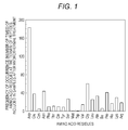
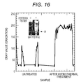
- Fig. 2 shows the analyzed results of the frequency of occurrence of amino acid residues at the termini of the peptides included in the human ⁇ -globulin-containing sample after hydrothermal treatment.
- Fig. 3 shows the analyzed results of the frequency of occurrence of amino acid residues at the termini of the peptides included in the human serum albumin-containing sample after hydrothermal treatment.
- Fig. 4 shows the analyzed results of the frequency of occurrence of amino acid residues at the termini of the peptides formed by hydrothermal treatment of each of serum, human ⁇ -globulin, and human serum albumin.
- Figs. 1 to 4 indicate that positions which are easily cleaved by hydrothermal treatment are present in the proteins and peptides in the serum.
- the positions easily cleaved were preferably the positions of the aspartic acid, glutamic acid, asparagine, serine, glycine, leucine, alanine, proline, and lysine residues, more preferably the position of the aspartic acid residue.
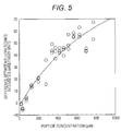
- HSA (manufactured by Sigma-Aldrich Co. LLC.) was dissolved at a final concentration of 1 ⁇ M in phosphate buffered saline to obtain a solution A.
- the HSA 397-413 fragment (SEQ ID NO: 29) consisting of amino acids at positions 397 to 413 of HSA, the tetramethyl rhodamine (hereinafter, also referred to as "TMR")-labeled HSA 397-413 fragment, and HSA were dissolved at final concentrations of 833 ⁇ M, 7 ⁇ M, and 1 ⁇ M, respectively, in phosphate buffered saline to obtain a solution B.
- the HSA 397-412 fragment and the TMR-labeled HSA 397-412 fragment were dissolved at final concentrations of 833 ⁇ M and 7 ⁇ M, respectively, in phosphate buffered saline to obtain a solution C.
- the phosphate buffered saline and the solution C were mixed at various mixing ratios to prepare various mixtures having different concentrations of HSA 397-412 fragment.
- fluorescence intensity B fluorescence intensity with an excitation wavelength of 540 nm and a fluorescence wavelength of 580 nm
- Equation (I): Difference between fluorescence intensities fluorescence intensity A ⁇ fluorescence intensity B
- a difference between the fluorescence intensities was determined in accordance with the above equation.
- Data points of the peptide concentration in each of the mixtures and the difference between the fluorescence intensities when using each of the mixtures were plotted on a two-dimensional coordinate, wherein the x-axis shows the peptide concentration and the y-axis shows the difference between the fluorescence intensities.
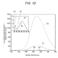
- Fig. 5 shows the analyzed results of the relationship of the peptide concentration with the difference between the fluorescence intensities.
- the K d of the HSA 397-412 fragment for HSA is 1000 ⁇ M or more. Therefore, it was found that the HSA 397-412 fragment has a low affinity for HSA. This result suggests that the HSA 397-412 fragment is hardly bound to HSA.
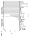
- the ACTH (1-24) with a K d of 200 ⁇ M for HSA and the ACTH (1-39) with a K d of 520 ⁇ M for HSA were selected from the peptides shown in Fig. 6 .
- the TMR-labeled ACTH (1-24) and HSA were mixed at final concentrations of 5 ⁇ M and 470 ⁇ M, respectively, with a 0.1 M Tris-HCl solution to obtain a TMR-labeled ACTH (1-24)-containing sample.
- the TMR-labeled ACTH (1-39) and HSA were mixed at final concentrations of 8 ⁇ M and 600 ⁇ M, respectively, with a 0.1 M Tris-HCl solution to obtain a TMR-labeled ACTH (1-39)-containing sample.
- the hydrothermal treatment was performed in the same manner as in Reference example 1 except that the TMR-labeled ACTH (1-24)-containing sample or the TMR-labeled ACTH (1-39)-containing sample was used in place of the serum-containing sample, the human ⁇ -globulin-containing sample or the HSA-containing sample in Reference example 1. Thereafter, a supernatant was collected from the sample after hydrothermal treatment.
- an additional buffer for SDS-PAGE [product name: 10xLoading Buffer, manufactured by TAKARA BIO INC.] was mixed with 20 ⁇ L of an aqueous glycerol solution (60% by volume) to form a buffer for sample. Then, 1.5 ⁇ L of the TMR-labeled ACTH (1-24)-containing sample was added to 3 ⁇ L of the resulting buffer for sample to obtain a test sample.
- a test sample was produced in the same manner as described above except that the TMR-labeled ACTH (1-39)-containing sample or a supernatant after hydrothermal treatment of each of the samples was used in place of the TMR-labeled ACTH (1-24)-containing sample in the above process.
- Example 2 (2) Each of the test samples obtained in Example 2 (2) was subjected to SDS-PAGE at a voltage of 200 V using an electrophoretic device [product name: Vertical mini electrophoresis system, manufactured by Invitrogen], an electrophoresis gel [product name: NuPAGE 4-12% Bis-TrisGels, 1.0 mm, 10 wells, manufactured by Invitrogen], and a running buffer [a diluted solution prepared by 20-fold dilution of product name: NuPAGE MES SDS running buffer (20x), manufactured by Invitrogen].
- the resulting electrophoresis gel was subjected to analysis with a fluorescence imager [product name: Pharos FX Molecular Imager, manufactured by Bio-Rad Laboratories, Inc.].
- the fluorescence image of the electrophoresis gel was captured in High Sample Intensity mode at a wavelength for TAMRA (excitation wavelength: 532 nm).
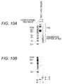
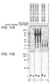
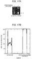
- Figs. 7A and 7B respectively show the results of SDS-PAGE of the untreated and hydrothermally-treated TMR-labeled ACTH (1-24) and the results of SDS-PAGE of the untreated and hydrothermally-treated TMR-labeled ACTH (1-39) in Example 2.
- Lane M represents the electrophoretic pattern of a molecular weight marker
- Lane 1 represents the electrophoretic pattern of the untreated TMR-labeled ACTH (1-24)-containing sample in the presence of HSA
- Lane 2 represents the electrophoretic pattern of the supernatant fraction of the hydrothermally-treated TMR-labeled ACTH (1-24)-containing sample in the presence of HSA.
- Fig. 7A Lane M represents the electrophoretic pattern of a molecular weight marker
- Lane 1 represents the electrophoretic pattern of the untreated TMR-labeled ACTH (1-24)-containing sample in the presence of HSA
- Lane 2 represents the electrophoretic pattern of the supernatant fraction of the hydrothermally-treated TMR-
- Lane 1 represents the untreated TMR-labeled ACTH (1-39)-containing sample in the presence of HSA
- Lane 2 represents the supernatant fraction of the hydrothermally-treated TMR-labeled ACTH (1-39)-containing sample in the presence of HSA
- Lane 3 represents the untreated TMR-labeled ACTH (1-39)-containing sample in the absence of HSA.
- Fig. 7A The results shown in Fig. 7A indicate that a band derived from the ACTH (1-24) was observed in the electrophoretic pattern of the untreated TMR-labeled ACTH (1-24)-containing sample in the presence of HSA (Lane 1). On the other hand, the band derived from the ACTH (1-24) was not observed in the electrophoretic pattern of the supernatant of the hydrothermally-treated TMR-labeled ACTH (1-24) in the presence of HSA (Lane 2). From this result, the ACTH (1-24) after hydrothermal treatment was expected to be bound to a precipitate of albumin in an insoluble fraction.
- the band derived from the ACTH (1-39) was observed in the electrophoretic pattern of the supernatant of the hydrothermally-treated TMR-labeled ACTH (1-39)-containing sample (Lane 2) in the presence of HSA. This result indicates that when the heating temperature reaches the predetermined temperature by heating the ACTH (1-39)-containing sample, the ACTH (1-39) is not bound to the precipitate of albumin in an insoluble fraction, and is present in the supernatant fraction.
- the ACTH (1-39) is suitable as a linker portion of the peptide for temperature determination to determine whether the temperature of an albumin-containing sample is the predetermined temperature.
- the peptide with a K d of 500 ⁇ M or more for albumin is suitable as a linker peptide of the peptide for temperature determination to determine whether the temperature of an albumin-containing sample is the predetermined temperature.
- Fig. 8 shows the results of the peptides in Fig. 6 classified into peptides suitable and unsuitable as linker peptides.
- the TMR-labeled HSA 237-249 fragment, the TMR-labeled HSA 397-413 fragment or the TMR-labeled HSA 599-609 fragment was dissolved at a final concentration of 5 ⁇ M in 1 mL of phosphate buffered saline.
- a sample A containing the TMR-labeled HSA 237-249 fragment
- a sample B containing the TMR-labeled HSA 397-413 fragment
- a sample C containing the TMR-labeled HSA 599-609 fragment
- 0.7 mL of the sample A was put in a screw cap bottle. Then, the uncovered screw cap bottle containing the sample A was placed in a heat transfer container made of iron. The hydrothermal treatment was performed in such a manner that the iron heat transfer container was heated in a thermostat bath at 200°C for 40 minutes, and the iron heat transfer container was incubated until the temperature of the thermostat bath decreased to room temperature. The resulting solution was designated as a sample A after hydrothermal treatment. The hydrothermal treatment was performed in the same manner as described above except that the sample B or the sample C was used in place of the sample A in the above process.
- a test sample was produced in the same manner as in Example 2 (2) except that each of the samples A to C or each of the hydrothermally-treated samples was used in place of the TMR-labeled ACTH (1-24)-containing sample, the TMR-labeled ACTH (1-39)-containing sample or the supernatant after hydrothermal treatment of each of the samples in Example 2 (2).
- the fluorescence image of the electrophoresis gel after SDS-PAGE was captured in the same manner as in Example 2 (3) except that the test sample obtained in Example 3 (2) was used in place of each of the test samples obtained in Example 2 (2).
- Fig. 9 shows the results of SDS-PAGE performed in Example 3.
- Lane M represents the electrophoretic pattern of a molecular weight marker
- Lane 1 represents the electrophoretic pattern of a sample A containing the untreated TMR-labeled HSA 237-249 fragment
- Lane 2 represents the electrophoretic pattern of a sample B containing the untreated TMR-labeled HSA 397-413 fragment
- Lane 3 represents the electrophoretic pattern of a sample C containing the untreated TMR-labeled HSA 599-609 fragment
- Lane 4 represents the electrophoretic pattern of a sample A containing the hydrothermally-treated TMR-labeled HSA 237-249 fragment
- Lane 5 represents the electrophoretic pattern of a sample B containing the hydrothermally-treated TMR-labeled HSA 397-413 fragment
- Lane 6 represents the electrophoretic pattern of a sample C containing the hydrothermally-treated TMR-labeled HSA 599-609 fragment.
- the TMR-labeled SA21 and HSA were dissolved at final concentrations of 8 ⁇ M and 300 ⁇ M, respectively, in 1 mL of phosphate buffered saline to obtain a TMR-labeled SA21-containing sample.
- the hydrothermal treatment was performed in the same manner as in Example 3 (1) except that the TMR-labeled SA21-containing sample was used in place of the samples A to C in Example 3 (1).
- the hydrothermally-treated TMR-labeled SA21-containing sample was separated into two phases: an insoluble fraction with red color (resulted from TMR) at the lower portion of the solution and a nearly clear and colorless supernatant. From this result, most of the TMR-labeled SA21 was expected to be present in the insoluble fraction.
- a test sample was produced in the same manner as in Example 2 (2) except that the untreated TMR-labeled SA21-containing sample or the supernatant of the hydrothermally-treated TMR-labeled SA21-containing sample was used in place of the TMR-labeled ACTH (1-24)-containing sample, the TMR-labeled ACTH (1-39)-containing sample or the supernatant after hydrothermal treatment of each of the samples in Example 2 (2).
- Example 3 SDS-PAGE was performed in the same manner as in Example 3 (3) except that the test sample obtained in Comparative example 1 (2) was used in place of each of the test samples obtained in Example 2 (2).
- the resulting electrophoresis gel was subjected to analysis with a fluorescence imager [product name: Pharos FX Molecular Imager, manufactured by Bio-Rad Laboratories, Inc.].
- the fluorescence image of the electrophoresis gel was captured in High Sample Intensity mode at a wavelength for TAMRA (excitation wavelength: 532 nm).
- the resulting electrophoresis gel was silver-stained and a silver-stained image was captured.
- Fig. 10A shows a silver-stained image of the electrophoresis gel after SDS-PAGE in Comparative example 1
- Fig. 10B shows a fluorescence image of the electrophoresis gel after SDS-PAGE in Comparative example 1.
- Lane M represents the electrophoretic pattern of a molecular weight marker
- Lane 1 represents the electrophoretic pattern of the untreated TMR-labeled SA1-containing sample
- Lane 2 represents the electrophoretic pattern of the supernatant of the hydrothermally-treated TMR-labeled SA21-containing sample.
- "A" represents a band derived from the untreated SA21 and "B" represents an aggregate of SA21.
- the TMR-labeled HSA 397-413 fragment was dissolved at a final concentration of 5 ⁇ M in 1 mL of phosphate buffered saline to obtain a sample D.
- the TMR-labeled HSA 599-609 fragment was dissolved at a final concentration of 5 ⁇ M in 1 mL of phosphate buffered saline to obtain a sample E.
- the TMR-labeled HSA 397-413 fragment and HSA were dissolved at final concentrations of 5 ⁇ M and 600 ⁇ M, respectively, in 1 mL of phosphate buffered saline to obtain a sample F.
- the TMR-labeled HSA 599-609 fragment and HSA were dissolved at final concentrations of 5 ⁇ M and 600 ⁇ M, respectively, in 1 mL of phosphate buffered saline to obtain a sample G.
- HSA was dissolved at a final concentration of 600 ⁇ M in 1 mL of phosphate buffered saline to obtain a control sample.
- the hydrothermal treatment was performed in the same manner as in Example 3 (1) except that each of the samples D to G or the control sample was used in place of the samples A to C in Example 3 (1). The supernatant of each of the hydrothermally-treated samples was collected.
- a test sample was produced in the same manner as in Example 2 (2) except that each of the samples D to G, the control sample or the supernatant of each of the hydrothermally-treated samples was used in place of each of the test samples obtained in Example 2 (2).
- the silver-stained and fluorescence images of the electrophoresis gel were captured in the same manner as in Comparative Example 1 (3) except that the test sample obtained in Example 4 (2) was used in place of each of the test samples obtained in Comparative Example 1 (2).
- Fig. 11A shows a silver-stained image of the electrophoresis gel after SDS-PAGE in Example 4
- Fig. 11B shows a fluorescence image of the electrophoresis gel after SDS-PAGE in Example 4.
- Lane M represents the electrophoretic pattern of a molecular weight marker
- Lane 1 represents the electrophoretic pattern of the untreated sample D
- Lane 2 represents the electrophoretic pattern of the untreated sample E
- Lane 3 represents the electrophoretic pattern of the untreated sample F
- Lane 4 represents the electrophoretic pattern of the untreated sample G
- Lane 5 represents the electrophoretic pattern of the untreated control sample
- Lane 6 represents the electrophoretic pattern of the supernatant of the hydrothermally-treated sample F
- Lane 7 represents the electrophoretic pattern of the supernatant of the hydrothermally-treated sample F
- Lane 8 represents the electrophoretic pattern of the supernatant of the hydrothermally-treated control sample.
- DABCYL-HSA 397-413 fragment-EDANS in which both the termini of the HSA 397-413 fragment (N- and C-termini) were labeled was dissolved at a final concentration of 20 ⁇ M in 6 mL of 100 mM glycine-hydrochloride buffer (pH 2.5) to obtain a sample H.
- the DABCYL-HSA 397-413 fragment-EDANS was designed such that the fluorescence from EDANS in a non-cleaved state was quenched by DABCYL and the intensity of the fluorescence from EDANS was increased by cleavage of the EDANS.
- Example 5 1.5 mL of the sample H obtained in Example 5 (1) was placed in a glass container for hydrothermal treatment. Then, the glass container for hydrothermal treatment containing the sample H was put in a microwave synthesis reactor [product name: MultiSYNTH, manufactured by Milestone General K.K.] and the hydrothermal treatment was performed at 80°C, 120°C or 160°C under the conditions shown in Table 4.
- a microwave synthesis reactor product name: MultiSYNTH, manufactured by Milestone General K.K.
- the untreated sample H obtained in Example 5 (1) and the hydrothermally-treated sample H obtained in Example 5 (2) were subjected to measurement with a spectrophotofluorometer [product name: F-7000, manufactured by Hitachi High-Technologies Corporation] and the fluorescence spectrum with an excitation wavelength of 335 nm and a fluorescence wavelength of 360 to 600 nm was measured.
- a spectrophotofluorometer product name: F-7000, manufactured by Hitachi High-Technologies Corporation
- Fig. 12 shows the results of the measurement of the fluorescence spectrum with an excitation wavelength of 335 nm and a fluorescence wavelength of 360 to 600 nm regarding the untreated sample and each of the hydrothermally-treated samples in Example 5.
- (A) represents a fluorescence spectrum of the untreated sample H
- (B) represents a fluorescence spectrum of the sample H hydrothermally treated at 80°C
- (C) represents a fluorescence spectrum of the sample H hydrothermally treated at 160°C
- (D) represents a fluorescence spectrum of the sample H hydrothermally treated at 160°C.
- a test sample was produced in the same manner as in Example 3 (1) except that the untreated sample H obtained in Example 5 (1) or the hydrothermally-treated sample H obtained in Example 5 (2) was used in place of each of the samples A to C or each of the hydrothermally-treated samples in Example 3 (1).
- Example 2 (3) SDS-PAGE was performed in the same manner as in Example 2 (3) except that the test sample obtained in Example 5 (4) was used in place of each of the test samples obtained in Example 2 (2).
- the gel after electrophoresis was irradiated with UV light using a transilluminator (model: NM-15 95-0219-03, manufactured by UVP), and a photograph of the gel was taken with a camera (COOLPIX 4500, manufactured by Nikon Corporation) equipped with an orange filter.
- COOLPIX 4500 manufactured by Nikon Corporation
- Fig. 14 shows fluorescence images of electrophoresis gels after SDS-PAGE regarding the untreated DABCYL-HSA 397-413 fragment-EDANS and the product after hydrothermal treatment of DABCYL-HSA 397-413 fragment-EDANS in Example 5.
- Lane 1 represents the electrophoretic pattern of the untreated DABCYL-HSA 397-413 fragment-EDANS
- Lane 2 represents the electrophoretic pattern of the product after hydrothermal treatment of DABCYL-HSA 397-413 fragment-EDANS.
- Example 5 (1) A sample I was produced in the same manner as in Example 5 (1) except that a 50 ⁇ M pH 11 CAPS-sodium hydroxide buffer was used in place of a 100 mM glycine-hydrochloride buffer (pH 2.5) in Example 5 (1).
- the hydrothermal treatment at 160°C was performed in the same manner as in Example 5 (2) except that the sample I obtained in Example 6 (1) was used in place of the sample H obtained in Example 5 (1).
- Example 6 (1) While the container containing the untreated sample I obtained in Example 6 (1) and the container containing the hydrothermally-treated sample I obtained in Example 6 (2) were irradiated with UV light using a transilluminator (model: NM-15 95-0219-03, manufactured by UVP), a photograph of the gel was taken with a camera (COOLPIX 4500, manufactured by Nikon Corporation) equipped with an orange filter. Thus, the fluorescence image with the excitation light removed was visualized.
- the resulting image and image processing software [product name: Image J, provided by National Institute of Health] were used to quantify the intensity of the fluorescence resulting from the cleavage of DABCYL-HSA 397-413 fragment-EDANS as a gray value.
- Fig. 15 shows the results obtained by quantifying the intensity of the fluorescence resulting from the cleavage of DABCYL-HSA 397-413 fragment-EDANS as a gray value, regarding the untreated sample and each of the hydrothermally-treated samples in Example 6.
- the fluorescence intensity at the position indicated by an arrow A in the photograph in Fig. 15 is quantified as a gray value.
- MCA-HSA 397-413 fragment-DNP in which both the termini of the HSA 397-413 fragment (N- and C-termini) were labeled was dissolved at a final concentration of 20 ⁇ M in 6 mL of phosphate buffered saline to obtain a sample J.
- the MCA-HSA 397-413 fragment-DNP was designed such that the fluorescence from DNP in a non-cleaved state was quenched by MCA and the intensity of the fluorescence from DNP was increased by cleavage of the DNP.
- the hydrothermal treatment at 160°C was performed in the same manner as in Example 5 (2) except that the sample J obtained in Example 7 (1) was used in place of the sample H obtained in Example 5 (1).
- the intensity of the fluorescence resulting from the cleavage of MCA-HSA 397-413 fragment-DNP was quantified as a gray value in the same manner as in Example 6 (3) except that the container containing the untreated sample J obtained in Example 7 (1) and the container containing the hydrothermally-treated sample J obtained in Example 7 (2) were used in place of the container containing the untreated sample I and the container containing the hydrothermally-treated sample I in Example 6 (3).
- Fig. 16 shows the results obtained by quantifying the intensity of the fluorescence resulting from the cleavage of MCA-HSA 397-413 fragment-DNP as a gray value in Example 7.
- the fluorescence intensity at the position indicated by an arrow A in the photograph in Fig. 16 is quantified as a gray value.
- DABCYL-HSA 397-413 fragment-EDANS and HSA were dissolved at final concentrations of 20 ⁇ M and 300 ⁇ M, respectively, in 1.5 mL of phosphate buffered saline to obtain a sample K.
- the hydrothermal treatment at 160°C was performed in the same manner as in Example 5 (2) except that the sample K obtained in Example 8 (1) was used in place of the sample H obtained in Example 5 (1).
- the intensity of the fluorescence resulting from the cleavage of DABCYL-HSA 397-413 fragment-EDANS and HSA was quantified as a gray value in the same manner as in Example 6 (3) except that the container containing the untreated sample K obtained in Example 8 (1) and the container containing the hydrothermally-treated sample K obtained in Example 8 (2) were used in place of the container containing the untreated sample I and the container containing the hydrothermally-treated sample I in Example 6 (3).
- Fig. 17A shows the results obtained by observing a container containing the untreated sample and the container containing the hydrothermally-treated sample in Example 8, and Fig. 17B shows the results obtained by quantifying the intensity of the fluorescence resulting from the cleavage of DABCYL-HSA 397-413 fragment-EDANS as a gray value in Example 8.
- the fluorescence intensity at the position indicated by an arrow A in the photograph in Fig. 17A is quantified as a gray value.
- DABCYL-HSA 397-413 fragment-EDANS, whole blood [product name: EDTA Whole blood (20 year-old female), manufactured by PROMEDDX, LLC], and glycine were prepared to produce a 3 mL solution having final concentrations of 60 ⁇ M, 5-fold dilution, and 1 M, respectively to obtain a sample L.
- the hydrothermal treatment at 160°C was performed in the same manner as in Example 5 (2) except that the sample L obtained in Example 9 (1) was used in place of the sample H obtained in Example 5 (1).
- the untreated sample L and the hydrothermally-treated sample L were subjected to centrifugation at 15000xg for 3 minutes.
- the container containing the resulting supernatant was photographed under visible light. While the container containing the resulting supernatant was irradiated with UV light using a transilluminator (model: NM-15 95-0219-03, manufactured by UVP), a photograph of the container was taken with a camera (COOLPIX 4500, manufactured by Nikon Corporation) through an orange filter.
- the image photographed under UV irradiation and image processing software [product name: Image J, provided by National Institute of Health] were used to quantify the intensity of the fluorescence resulting from the cleavage of DABCYL-HSA 397-413 fragment-EDANS as a gray value.
- Fig. 18A shows the results of the container containing the untreated sample and the container containing the hydrothermally-treated sample which were observed under visible light in Example 9
- Fig. 18B shows the results of the container containing the untreated sample and the container containing the hydrothermally-treated sample which were observed under ultraviolet irradiation in Example 9
- Fig. 18C shows the results obtained by quantifying the intensity of the fluorescence resulting from the cleavage of DABCYL-HSA 397-413 fragment-EDANS as a gray value in Example 9.
- the fluorescence intensity at the position indicated by an arrow A in the photograph in the graph is quantified as a gray value.
- DABCYL-HSA 397-413 fragment-EDANS and TMR-SA21 were dissolved at final concentrations of 200 ⁇ M and 20 ⁇ M, respectively, in 0.1-fold phosphate buffered saline. Then, 750 ⁇ L of the resulting solution and 300 ⁇ L of serum from a healthy woman (22 years old) [product name: Normal Serum Pool, manufactured by PROMEDDX, LLC] were mixed. To 1050 ⁇ L of the resulting serum-containing solution, 375 ⁇ L of 2 M glycine solution and 75 ⁇ L of phosphate buffered saline were added to obtain a sample M.
- the hydrothermal treatment at 160°C was performed in the same manner as in Example 5 (2) except that the sample M obtained in Example 9 (1) was used in place of the sample H obtained in Example 5 (1).
- the container containing the untreated sample M and the container containing the hydrothermally-treated sample M were photographed under visible light. As a result, it was confirmed that an insoluble fraction was attached to the wall surface of the container. It was confirmed that the insoluble fraction exhibited a red color derived from TMR.
- the data image of only the red channel was obtained from the resulting image using image processing software [product name: Photoshop, manufactured by Adobe Systems Software Ireland Ltd.].
- the data image of only the resulting red channel and image processing software [product name: Image J, provided by National Institute of Health] were used to quantify the intensity of the fluorescence resulting from the cleavage ofDABCYL-HSA 397-413 fragment-EDANS as a gray value.
- Fig. 19A shows the results of the container containing the untreated sample and the container containing the hydrothermally-treated sample which were observed under visible light in Example 10, and Fig. 19B shows the results obtained by quantifying the intensity of the fluorescence resulting from the cleavage of DABCYL-HSA 397-413 fragment-EDANS as a gray value in Example 10.
- (a) represents the hydrothermally-treated sample and the dashed line (b) represents the untreated sample.
- the fluorescence intensities of the boxed portions in the photograph in Fig. 19A were quantified as a gray value.
- Fig. 20A shows the results of the container containing the untreated sample and the container containing the hydrothermally-treated sample which were observed under ultraviolet irradiation in Example 10, and Fig. 20B shows the results obtained by quantifying the intensity of the fluorescence resulting from the cleavage of DABCYL-HSA 397-413 fragment-EDANS as a gray value in Example 10.
- the fluorescence intensity at the position indicated by an arrow A in the photograph in Fig. 20A is quantified as a gray value.
Landscapes
- Health & Medical Sciences (AREA)
- Life Sciences & Earth Sciences (AREA)
- Immunology (AREA)
- Chemical & Material Sciences (AREA)
- Physics & Mathematics (AREA)
- Pathology (AREA)
- General Physics & Mathematics (AREA)
- General Health & Medical Sciences (AREA)
- Biochemistry (AREA)
- Analytical Chemistry (AREA)
- Engineering & Computer Science (AREA)
- Hematology (AREA)
- Biomedical Technology (AREA)
- Urology & Nephrology (AREA)
- Molecular Biology (AREA)
- Chemical Kinetics & Catalysis (AREA)
- Nuclear Medicine, Radiotherapy & Molecular Imaging (AREA)
- Microbiology (AREA)
- Biotechnology (AREA)
- Cell Biology (AREA)
- Food Science & Technology (AREA)
- Medicinal Chemistry (AREA)
- Optics & Photonics (AREA)
- Plasma & Fusion (AREA)
- Proteomics, Peptides & Aminoacids (AREA)
- Investigating Or Analysing Biological Materials (AREA)
- Peptides Or Proteins (AREA)
- Investigating Or Analysing Materials By The Use Of Chemical Reactions (AREA)
- Investigating, Analyzing Materials By Fluorescence Or Luminescence (AREA)
- Investigating Or Analyzing Materials Using Thermal Means (AREA)
Applications Claiming Priority (1)
| Application Number | Priority Date | Filing Date | Title |
|---|---|---|---|
| JP2015070865A JP6473366B2 (ja) | 2015-03-31 | 2015-03-31 | 温度判定方法、標的ペプチドの検出方法および温度判定試薬 |
Publications (3)
| Publication Number | Publication Date |
|---|---|
| EP3076166A2 true EP3076166A2 (fr) | 2016-10-05 |
| EP3076166A3 EP3076166A3 (fr) | 2016-12-28 |
| EP3076166B1 EP3076166B1 (fr) | 2020-05-06 |
Family
ID=55646414
Family Applications (1)
| Application Number | Title | Priority Date | Filing Date |
|---|---|---|---|
| EP16163066.0A Not-in-force EP3076166B1 (fr) | 2015-03-31 | 2016-03-31 | Procédé de détermination de température |
Country Status (4)
| Country | Link |
|---|---|
| US (1) | US9995740B2 (fr) |
| EP (1) | EP3076166B1 (fr) |
| JP (1) | JP6473366B2 (fr) |
| CN (1) | CN106018821B (fr) |
Families Citing this family (2)
| Publication number | Priority date | Publication date | Assignee | Title |
|---|---|---|---|---|
| CN111426667A (zh) * | 2020-04-23 | 2020-07-17 | 天津科技大学 | 一种基于量子点-核酸适配体-氧化石墨烯建立的对β-乳球蛋白检测的荧光方法 |
| JP7496504B2 (ja) * | 2020-05-21 | 2024-06-07 | パナソニックIpマネジメント株式会社 | 温度測定システム |
Citations (1)
| Publication number | Priority date | Publication date | Assignee | Title |
|---|---|---|---|---|
| US20120027740A1 (en) | 2009-02-05 | 2012-02-02 | Pierre Philippart | Method and means for producing tissues and tissues obtained |
Family Cites Families (11)
| Publication number | Priority date | Publication date | Assignee | Title |
|---|---|---|---|---|
| US6803188B1 (en) | 1996-01-31 | 2004-10-12 | The Regents Of The University Of California | Tandem fluorescent protein constructs |
| CA2450641A1 (fr) * | 2001-06-14 | 2002-12-27 | Anadys Pharmaceuticals, Inc. | Procedes de criblage de ligands de molecules cibles |
| WO2007142208A1 (fr) * | 2006-06-05 | 2007-12-13 | Kyoto University | Protéine thermosensible, polynucléotide, plasmide et cellule exprimant cette protéine thermosensible et méthode d'utilisation de cette protéine thermosensible |
| JP2009236906A (ja) * | 2008-03-05 | 2009-10-15 | Keio Gijuku | 蛍光性温度及び/又はpH応答性ポリマー及び蛍光共鳴エネルギー移動を用いた蛍光発生成分を有する物質等の測定方法 |
| JP5750216B2 (ja) * | 2009-04-17 | 2015-07-15 | 株式会社カネカ | 新規リンカーを用いた抗菌剤および診断薬 |
| FR2950697B1 (fr) * | 2009-09-25 | 2011-12-09 | Biomerieux Sa | Procede de detection de molecules par spectrometrie de masse |
| JP5636331B2 (ja) | 2011-04-28 | 2014-12-03 | シスメックス株式会社 | ペプチドの遊離方法および回収方法 |
| JP5817838B2 (ja) * | 2011-11-02 | 2015-11-18 | ウシオ電機株式会社 | 蛍光標識抗体可変領域含有ポリペプチド複合体を用いた蛍光免疫測定方法 |
| JP2013113614A (ja) * | 2011-11-25 | 2013-06-10 | Nagoya Univ | 蛍光温度計測方法及び蛍光温度計測システム |
| CN104350383B (zh) * | 2012-06-01 | 2017-03-08 | 赫普泰雅治疗有限公司 | 分析 |
| US9829386B2 (en) * | 2013-06-08 | 2017-11-28 | Brigham Young University | Apparatus and method for determining denaturation thermograms of blood plasma or serum |
-
2015
- 2015-03-31 JP JP2015070865A patent/JP6473366B2/ja not_active Expired - Fee Related
-
2016
- 2016-03-24 US US15/079,139 patent/US9995740B2/en active Active
- 2016-03-31 EP EP16163066.0A patent/EP3076166B1/fr not_active Not-in-force
- 2016-03-31 CN CN201610195950.3A patent/CN106018821B/zh not_active Expired - Fee Related
Patent Citations (1)
| Publication number | Priority date | Publication date | Assignee | Title |
|---|---|---|---|---|
| US20120027740A1 (en) | 2009-02-05 | 2012-02-02 | Pierre Philippart | Method and means for producing tissues and tissues obtained |
Also Published As
| Publication number | Publication date |
|---|---|
| JP2016191587A (ja) | 2016-11-10 |
| EP3076166A3 (fr) | 2016-12-28 |
| CN106018821A (zh) | 2016-10-12 |
| CN106018821B (zh) | 2018-05-22 |
| US9995740B2 (en) | 2018-06-12 |
| JP6473366B2 (ja) | 2019-02-20 |
| US20160290999A1 (en) | 2016-10-06 |
| EP3076166B1 (fr) | 2020-05-06 |
Similar Documents
| Publication | Publication Date | Title |
|---|---|---|
| CN101600959B (zh) | 肽和蛋白的定量方法 | |
| Navajas et al. | Serum exosome isolation by size-exclusion chromatography for the discovery and validation of preeclampsia-associated biomarkers | |
| US8470603B2 (en) | Methods of evaluating diethylamide in glatiramer acetate | |
| AU2015258962B2 (en) | Compositions and methods for purification and detection of HDL and ApoA1 | |
| Choi et al. | Complexin induces a conformational change at the membrane-proximal C-terminal end of the SNARE complex | |
| Mao et al. | Comparison of the influence of pH on the selectivity of free and immobilized trypsin for β-lactoglobulin hydrolysis | |
| ES2617918T3 (es) | Biomarcador para trastornos mentales, incluyendo trastornos cognitivos, y método de utilización de dicho biomarcador para la detección de trastornos mentales, incluyendo los trastornos cognitivos | |
| EP3076166B1 (fr) | Procédé de détermination de température | |
| KR102386584B1 (ko) | Adamts13 효소 활성을 결정하기 위한 방법 및 시스템 | |
| Kabatas et al. | A contamination-insensitive probe for imaging specific biomolecules by secondary ion mass spectrometry | |
| Beck et al. | Robust workflow for iTRAQ-based peptide and protein quantification | |
| Turtoi et al. | Isotope coded protein label quantification of serum proteins—Comparison with the label-free LC–MS and validation using the MRM approach | |
| Saito et al. | Establishment and application of a high-quality comparative analysis strategy for the discovery and small-scale validation of low-abundance biomarker peptides in serum based on an optimized novel peptide extraction method | |
| Williams et al. | The dimerization of half-molecule fragments of transferrin | |
| EP2902407B1 (fr) | Procédé de récupération de peptide | |
| JP6742235B2 (ja) | 質量分析を用いたタンパク質の検出方法 | |
| Rochat et al. | Robust and sensitive peptidomics workflow for plasma based on specific extraction, lipid removal, capillary LC setup and multinozzle ESI emitter | |
| KR20220052959A (ko) | 알레르겐 검출용 마커 및 이를 사용한 알레르겐 검출 방법 | |
| JP4724816B2 (ja) | タンパク質の測定方法 | |
| Su et al. | Time-dependent nature in peptic hydrolysis of native bovine hemoglobin | |
| CA2817851A1 (fr) | Complexe de proteine/peptide lie a l'albumine en tant que biomarqueur pour une maladie | |
| Abounorinejad | The Effect of Solvent on Silicon and Germanium Rhodamines and Synthesis of the AQP4ex C-Terminus Extension Peptide | |
| KR101527766B1 (ko) | Pyp 단백질을 이용하는 목적 단백질의 분석 방법 및 이에 이용되는 pyp 변이체 | |
| Ernoult et al. | OFFGEL-Isoelectric Focusing Fractionation for the analysis of complex proteome | |
| Táborský et al. | Extrinsic fluorescence of peptides: Labelling of humanin derivatives for CE-LIF |
Legal Events
| Date | Code | Title | Description |
|---|---|---|---|
| PUAI | Public reference made under article 153(3) epc to a published international application that has entered the european phase |
Free format text: ORIGINAL CODE: 0009012 |
|
| AK | Designated contracting states |
Kind code of ref document: A2 Designated state(s): AL AT BE BG CH CY CZ DE DK EE ES FI FR GB GR HR HU IE IS IT LI LT LU LV MC MK MT NL NO PL PT RO RS SE SI SK SM TR |
|
| AX | Request for extension of the european patent |
Extension state: BA ME |
|
| PUAL | Search report despatched |
Free format text: ORIGINAL CODE: 0009013 |
|
| AK | Designated contracting states |
Kind code of ref document: A3 Designated state(s): AL AT BE BG CH CY CZ DE DK EE ES FI FR GB GR HR HU IE IS IT LI LT LU LV MC MK MT NL NO PL PT RO RS SE SI SK SM TR |
|
| AX | Request for extension of the european patent |
Extension state: BA ME |
|
| RIC1 | Information provided on ipc code assigned before grant |
Ipc: G01N 21/64 20060101ALI20161122BHEP Ipc: G01N 25/48 20060101AFI20161122BHEP Ipc: G01N 33/542 20060101ALI20161122BHEP Ipc: G01N 1/44 20060101ALI20161122BHEP |
|
| STAA | Information on the status of an ep patent application or granted ep patent |
Free format text: STATUS: REQUEST FOR EXAMINATION WAS MADE |
|
| 17P | Request for examination filed |
Effective date: 20170616 |
|
| RBV | Designated contracting states (corrected) |
Designated state(s): AL AT BE BG CH CY CZ DE DK EE ES FI FR GB GR HR HU IE IS IT LI LT LU LV MC MK MT NL NO PL PT RO RS SE SI SK SM TR |
|
| STAA | Information on the status of an ep patent application or granted ep patent |
Free format text: STATUS: EXAMINATION IS IN PROGRESS |
|
| 17Q | First examination report despatched |
Effective date: 20180604 |
|
| REG | Reference to a national code |
Ref country code: DE Ref legal event code: R079 Ref document number: 602016035510 Country of ref document: DE Free format text: PREVIOUS MAIN CLASS: G01N0025480000 Ipc: G01N0021640000 |
|
| GRAP | Despatch of communication of intention to grant a patent |
Free format text: ORIGINAL CODE: EPIDOSNIGR1 |
|
| STAA | Information on the status of an ep patent application or granted ep patent |
Free format text: STATUS: GRANT OF PATENT IS INTENDED |
|
| RIC1 | Information provided on ipc code assigned before grant |
Ipc: G01N 21/76 20060101ALI20190904BHEP Ipc: G01N 33/542 20060101ALI20190904BHEP Ipc: G01N 1/44 20060101ALI20190904BHEP Ipc: G01N 21/64 20060101AFI20190904BHEP Ipc: G01N 25/48 20060101ALI20190904BHEP |
|
| INTG | Intention to grant announced |
Effective date: 20190924 |
|
| GRAS | Grant fee paid |
Free format text: ORIGINAL CODE: EPIDOSNIGR3 |
|
| GRAJ | Information related to disapproval of communication of intention to grant by the applicant or resumption of examination proceedings by the epo deleted |
Free format text: ORIGINAL CODE: EPIDOSDIGR1 |
|
| GRAL | Information related to payment of fee for publishing/printing deleted |
Free format text: ORIGINAL CODE: EPIDOSDIGR3 |
|
| STAA | Information on the status of an ep patent application or granted ep patent |
Free format text: STATUS: EXAMINATION IS IN PROGRESS |
|
| GRAR | Information related to intention to grant a patent recorded |
Free format text: ORIGINAL CODE: EPIDOSNIGR71 |
|
| STAA | Information on the status of an ep patent application or granted ep patent |
Free format text: STATUS: GRANT OF PATENT IS INTENDED |
|
| INTC | Intention to grant announced (deleted) | ||
| INTG | Intention to grant announced |
Effective date: 20200213 |
|
| GRAA | (expected) grant |
Free format text: ORIGINAL CODE: 0009210 |
|
| STAA | Information on the status of an ep patent application or granted ep patent |
Free format text: STATUS: THE PATENT HAS BEEN GRANTED |
|
| AK | Designated contracting states |
Kind code of ref document: B1 Designated state(s): AL AT BE BG CH CY CZ DE DK EE ES FI FR GB GR HR HU IE IS IT LI LT LU LV MC MK MT NL NO PL PT RO RS SE SI SK SM TR |
|
| REG | Reference to a national code |
Ref country code: GB Ref legal event code: FG4D |
|
| REG | Reference to a national code |
Ref country code: CH Ref legal event code: EP Ref country code: AT Ref legal event code: REF Ref document number: 1267581 Country of ref document: AT Kind code of ref document: T Effective date: 20200515 |
|
| REG | Reference to a national code |
Ref country code: IE Ref legal event code: FG4D |
|
| REG | Reference to a national code |
Ref country code: DE Ref legal event code: R096 Ref document number: 602016035510 Country of ref document: DE |
|
| REG | Reference to a national code |
Ref country code: LT Ref legal event code: MG4D |
|
| REG | Reference to a national code |
Ref country code: NL Ref legal event code: MP Effective date: 20200506 |
|
| PG25 | Lapsed in a contracting state [announced via postgrant information from national office to epo] |
Ref country code: GR Free format text: LAPSE BECAUSE OF FAILURE TO SUBMIT A TRANSLATION OF THE DESCRIPTION OR TO PAY THE FEE WITHIN THE PRESCRIBED TIME-LIMIT Effective date: 20200807 Ref country code: FI Free format text: LAPSE BECAUSE OF FAILURE TO SUBMIT A TRANSLATION OF THE DESCRIPTION OR TO PAY THE FEE WITHIN THE PRESCRIBED TIME-LIMIT Effective date: 20200506 Ref country code: LT Free format text: LAPSE BECAUSE OF FAILURE TO SUBMIT A TRANSLATION OF THE DESCRIPTION OR TO PAY THE FEE WITHIN THE PRESCRIBED TIME-LIMIT Effective date: 20200506 Ref country code: IS Free format text: LAPSE BECAUSE OF FAILURE TO SUBMIT A TRANSLATION OF THE DESCRIPTION OR TO PAY THE FEE WITHIN THE PRESCRIBED TIME-LIMIT Effective date: 20200906 Ref country code: SE Free format text: LAPSE BECAUSE OF FAILURE TO SUBMIT A TRANSLATION OF THE DESCRIPTION OR TO PAY THE FEE WITHIN THE PRESCRIBED TIME-LIMIT Effective date: 20200506 Ref country code: NO Free format text: LAPSE BECAUSE OF FAILURE TO SUBMIT A TRANSLATION OF THE DESCRIPTION OR TO PAY THE FEE WITHIN THE PRESCRIBED TIME-LIMIT Effective date: 20200806 Ref country code: PT Free format text: LAPSE BECAUSE OF FAILURE TO SUBMIT A TRANSLATION OF THE DESCRIPTION OR TO PAY THE FEE WITHIN THE PRESCRIBED TIME-LIMIT Effective date: 20200907 |
|
| PG25 | Lapsed in a contracting state [announced via postgrant information from national office to epo] |
Ref country code: HR Free format text: LAPSE BECAUSE OF FAILURE TO SUBMIT A TRANSLATION OF THE DESCRIPTION OR TO PAY THE FEE WITHIN THE PRESCRIBED TIME-LIMIT Effective date: 20200506 Ref country code: RS Free format text: LAPSE BECAUSE OF FAILURE TO SUBMIT A TRANSLATION OF THE DESCRIPTION OR TO PAY THE FEE WITHIN THE PRESCRIBED TIME-LIMIT Effective date: 20200506 Ref country code: BG Free format text: LAPSE BECAUSE OF FAILURE TO SUBMIT A TRANSLATION OF THE DESCRIPTION OR TO PAY THE FEE WITHIN THE PRESCRIBED TIME-LIMIT Effective date: 20200806 Ref country code: LV Free format text: LAPSE BECAUSE OF FAILURE TO SUBMIT A TRANSLATION OF THE DESCRIPTION OR TO PAY THE FEE WITHIN THE PRESCRIBED TIME-LIMIT Effective date: 20200506 |
|
| REG | Reference to a national code |
Ref country code: AT Ref legal event code: MK05 Ref document number: 1267581 Country of ref document: AT Kind code of ref document: T Effective date: 20200506 |
|
| PG25 | Lapsed in a contracting state [announced via postgrant information from national office to epo] |
Ref country code: NL Free format text: LAPSE BECAUSE OF FAILURE TO SUBMIT A TRANSLATION OF THE DESCRIPTION OR TO PAY THE FEE WITHIN THE PRESCRIBED TIME-LIMIT Effective date: 20200506 Ref country code: AL Free format text: LAPSE BECAUSE OF FAILURE TO SUBMIT A TRANSLATION OF THE DESCRIPTION OR TO PAY THE FEE WITHIN THE PRESCRIBED TIME-LIMIT Effective date: 20200506 |
|
| PG25 | Lapsed in a contracting state [announced via postgrant information from national office to epo] |
Ref country code: ES Free format text: LAPSE BECAUSE OF FAILURE TO SUBMIT A TRANSLATION OF THE DESCRIPTION OR TO PAY THE FEE WITHIN THE PRESCRIBED TIME-LIMIT Effective date: 20200506 Ref country code: DK Free format text: LAPSE BECAUSE OF FAILURE TO SUBMIT A TRANSLATION OF THE DESCRIPTION OR TO PAY THE FEE WITHIN THE PRESCRIBED TIME-LIMIT Effective date: 20200506 Ref country code: EE Free format text: LAPSE BECAUSE OF FAILURE TO SUBMIT A TRANSLATION OF THE DESCRIPTION OR TO PAY THE FEE WITHIN THE PRESCRIBED TIME-LIMIT Effective date: 20200506 Ref country code: SM Free format text: LAPSE BECAUSE OF FAILURE TO SUBMIT A TRANSLATION OF THE DESCRIPTION OR TO PAY THE FEE WITHIN THE PRESCRIBED TIME-LIMIT Effective date: 20200506 Ref country code: IT Free format text: LAPSE BECAUSE OF FAILURE TO SUBMIT A TRANSLATION OF THE DESCRIPTION OR TO PAY THE FEE WITHIN THE PRESCRIBED TIME-LIMIT Effective date: 20200506 Ref country code: AT Free format text: LAPSE BECAUSE OF FAILURE TO SUBMIT A TRANSLATION OF THE DESCRIPTION OR TO PAY THE FEE WITHIN THE PRESCRIBED TIME-LIMIT Effective date: 20200506 Ref country code: CZ Free format text: LAPSE BECAUSE OF FAILURE TO SUBMIT A TRANSLATION OF THE DESCRIPTION OR TO PAY THE FEE WITHIN THE PRESCRIBED TIME-LIMIT Effective date: 20200506 Ref country code: RO Free format text: LAPSE BECAUSE OF FAILURE TO SUBMIT A TRANSLATION OF THE DESCRIPTION OR TO PAY THE FEE WITHIN THE PRESCRIBED TIME-LIMIT Effective date: 20200506 |
|
| REG | Reference to a national code |
Ref country code: DE Ref legal event code: R097 Ref document number: 602016035510 Country of ref document: DE |
|
| PG25 | Lapsed in a contracting state [announced via postgrant information from national office to epo] |
Ref country code: PL Free format text: LAPSE BECAUSE OF FAILURE TO SUBMIT A TRANSLATION OF THE DESCRIPTION OR TO PAY THE FEE WITHIN THE PRESCRIBED TIME-LIMIT Effective date: 20200506 Ref country code: SK Free format text: LAPSE BECAUSE OF FAILURE TO SUBMIT A TRANSLATION OF THE DESCRIPTION OR TO PAY THE FEE WITHIN THE PRESCRIBED TIME-LIMIT Effective date: 20200506 |
|
| PLBE | No opposition filed within time limit |
Free format text: ORIGINAL CODE: 0009261 |
|
| STAA | Information on the status of an ep patent application or granted ep patent |
Free format text: STATUS: NO OPPOSITION FILED WITHIN TIME LIMIT |
|
| 26N | No opposition filed |
Effective date: 20210209 |
|
| PG25 | Lapsed in a contracting state [announced via postgrant information from national office to epo] |
Ref country code: SI Free format text: LAPSE BECAUSE OF FAILURE TO SUBMIT A TRANSLATION OF THE DESCRIPTION OR TO PAY THE FEE WITHIN THE PRESCRIBED TIME-LIMIT Effective date: 20200506 |
|
| PG25 | Lapsed in a contracting state [announced via postgrant information from national office to epo] |
Ref country code: MC Free format text: LAPSE BECAUSE OF FAILURE TO SUBMIT A TRANSLATION OF THE DESCRIPTION OR TO PAY THE FEE WITHIN THE PRESCRIBED TIME-LIMIT Effective date: 20200506 |
|
| REG | Reference to a national code |
Ref country code: CH Ref legal event code: PL |
|
| REG | Reference to a national code |
Ref country code: BE Ref legal event code: MM Effective date: 20210331 |
|
| PG25 | Lapsed in a contracting state [announced via postgrant information from national office to epo] |
Ref country code: IE Free format text: LAPSE BECAUSE OF NON-PAYMENT OF DUE FEES Effective date: 20210331 Ref country code: CH Free format text: LAPSE BECAUSE OF NON-PAYMENT OF DUE FEES Effective date: 20210331 Ref country code: LU Free format text: LAPSE BECAUSE OF NON-PAYMENT OF DUE FEES Effective date: 20210331 Ref country code: LI Free format text: LAPSE BECAUSE OF NON-PAYMENT OF DUE FEES Effective date: 20210331 |
|
| PGFP | Annual fee paid to national office [announced via postgrant information from national office to epo] |
Ref country code: GB Payment date: 20220210 Year of fee payment: 7 Ref country code: DE Payment date: 20220203 Year of fee payment: 7 |
|
| PGFP | Annual fee paid to national office [announced via postgrant information from national office to epo] |
Ref country code: FR Payment date: 20220209 Year of fee payment: 7 |
|
| PG25 | Lapsed in a contracting state [announced via postgrant information from national office to epo] |
Ref country code: BE Free format text: LAPSE BECAUSE OF NON-PAYMENT OF DUE FEES Effective date: 20210331 |
|
| PG25 | Lapsed in a contracting state [announced via postgrant information from national office to epo] |
Ref country code: HU Free format text: LAPSE BECAUSE OF FAILURE TO SUBMIT A TRANSLATION OF THE DESCRIPTION OR TO PAY THE FEE WITHIN THE PRESCRIBED TIME-LIMIT; INVALID AB INITIO Effective date: 20160331 |
|
| PG25 | Lapsed in a contracting state [announced via postgrant information from national office to epo] |
Ref country code: CY Free format text: LAPSE BECAUSE OF FAILURE TO SUBMIT A TRANSLATION OF THE DESCRIPTION OR TO PAY THE FEE WITHIN THE PRESCRIBED TIME-LIMIT Effective date: 20200506 |
|
| REG | Reference to a national code |
Ref country code: DE Ref legal event code: R119 Ref document number: 602016035510 Country of ref document: DE |
|
| GBPC | Gb: european patent ceased through non-payment of renewal fee |
Effective date: 20230331 |
|
| PG25 | Lapsed in a contracting state [announced via postgrant information from national office to epo] |
Ref country code: GB Free format text: LAPSE BECAUSE OF NON-PAYMENT OF DUE FEES Effective date: 20230331 |
|
| PG25 | Lapsed in a contracting state [announced via postgrant information from national office to epo] |
Ref country code: GB Free format text: LAPSE BECAUSE OF NON-PAYMENT OF DUE FEES Effective date: 20230331 Ref country code: FR Free format text: LAPSE BECAUSE OF NON-PAYMENT OF DUE FEES Effective date: 20230331 Ref country code: DE Free format text: LAPSE BECAUSE OF NON-PAYMENT OF DUE FEES Effective date: 20231003 |
|
| PG25 | Lapsed in a contracting state [announced via postgrant information from national office to epo] |
Ref country code: MK Free format text: LAPSE BECAUSE OF FAILURE TO SUBMIT A TRANSLATION OF THE DESCRIPTION OR TO PAY THE FEE WITHIN THE PRESCRIBED TIME-LIMIT Effective date: 20200506 |
|
| PG25 | Lapsed in a contracting state [announced via postgrant information from national office to epo] |
Ref country code: TR Free format text: LAPSE BECAUSE OF FAILURE TO SUBMIT A TRANSLATION OF THE DESCRIPTION OR TO PAY THE FEE WITHIN THE PRESCRIBED TIME-LIMIT Effective date: 20200506 |
|
| PG25 | Lapsed in a contracting state [announced via postgrant information from national office to epo] |
Ref country code: MT Free format text: LAPSE BECAUSE OF FAILURE TO SUBMIT A TRANSLATION OF THE DESCRIPTION OR TO PAY THE FEE WITHIN THE PRESCRIBED TIME-LIMIT Effective date: 20200506 |